EP3303386B1 - Anti-tau antibodies and methods of use - Google Patents
Anti-tau antibodies and methods of use Download PDFInfo
- Publication number
- EP3303386B1 EP3303386B1 EP16728559.2A EP16728559A EP3303386B1 EP 3303386 B1 EP3303386 B1 EP 3303386B1 EP 16728559 A EP16728559 A EP 16728559A EP 3303386 B1 EP3303386 B1 EP 3303386B1
- Authority
- EP
- European Patent Office
- Prior art keywords
- antibody
- tau
- hvr
- hu37d3
- antibodies
- Prior art date
- Legal status (The legal status is an assumption and is not a legal conclusion. Google has not performed a legal analysis and makes no representation as to the accuracy of the status listed.)
- Active
Links
Images


Classifications
-
- A—HUMAN NECESSITIES
- A61—MEDICAL OR VETERINARY SCIENCE; HYGIENE
- A61K—PREPARATIONS FOR MEDICAL, DENTAL OR TOILETRY PURPOSES
- A61K39/00—Medicinal preparations containing antigens or antibodies
- A61K39/395—Antibodies; Immunoglobulins; Immune serum, e.g. antilymphocytic serum
- A61K39/39533—Antibodies; Immunoglobulins; Immune serum, e.g. antilymphocytic serum against materials from animals
- A61K39/3955—Antibodies; Immunoglobulins; Immune serum, e.g. antilymphocytic serum against materials from animals against proteinaceous materials, e.g. enzymes, hormones, lymphokines
-
- A—HUMAN NECESSITIES
- A61—MEDICAL OR VETERINARY SCIENCE; HYGIENE
- A61K—PREPARATIONS FOR MEDICAL, DENTAL OR TOILETRY PURPOSES
- A61K45/00—Medicinal preparations containing active ingredients not provided for in groups A61K31/00 - A61K41/00
- A61K45/06—Mixtures of active ingredients without chemical characterisation, e.g. antiphlogistics and cardiaca
-
- A—HUMAN NECESSITIES
- A61—MEDICAL OR VETERINARY SCIENCE; HYGIENE
- A61P—SPECIFIC THERAPEUTIC ACTIVITY OF CHEMICAL COMPOUNDS OR MEDICINAL PREPARATIONS
- A61P21/00—Drugs for disorders of the muscular or neuromuscular system
-
- A—HUMAN NECESSITIES
- A61—MEDICAL OR VETERINARY SCIENCE; HYGIENE
- A61P—SPECIFIC THERAPEUTIC ACTIVITY OF CHEMICAL COMPOUNDS OR MEDICINAL PREPARATIONS
- A61P21/00—Drugs for disorders of the muscular or neuromuscular system
- A61P21/02—Muscle relaxants, e.g. for tetanus or cramps
-
- A—HUMAN NECESSITIES
- A61—MEDICAL OR VETERINARY SCIENCE; HYGIENE
- A61P—SPECIFIC THERAPEUTIC ACTIVITY OF CHEMICAL COMPOUNDS OR MEDICINAL PREPARATIONS
- A61P25/00—Drugs for disorders of the nervous system
-
- A—HUMAN NECESSITIES
- A61—MEDICAL OR VETERINARY SCIENCE; HYGIENE
- A61P—SPECIFIC THERAPEUTIC ACTIVITY OF CHEMICAL COMPOUNDS OR MEDICINAL PREPARATIONS
- A61P25/00—Drugs for disorders of the nervous system
- A61P25/28—Drugs for disorders of the nervous system for treating neurodegenerative disorders of the central nervous system, e.g. nootropic agents, cognition enhancers, drugs for treating Alzheimer's disease or other forms of dementia
-
- A—HUMAN NECESSITIES
- A61—MEDICAL OR VETERINARY SCIENCE; HYGIENE
- A61P—SPECIFIC THERAPEUTIC ACTIVITY OF CHEMICAL COMPOUNDS OR MEDICINAL PREPARATIONS
- A61P43/00—Drugs for specific purposes, not provided for in groups A61P1/00-A61P41/00
-
- C—CHEMISTRY; METALLURGY
- C07—ORGANIC CHEMISTRY
- C07K—PEPTIDES
- C07K16/00—Immunoglobulins [IGs], e.g. monoclonal or polyclonal antibodies
- C07K16/18—Immunoglobulins [IGs], e.g. monoclonal or polyclonal antibodies against material from animals or humans
-
- G—PHYSICS
- G01—MEASURING; TESTING
- G01N—INVESTIGATING OR ANALYSING MATERIALS BY DETERMINING THEIR CHEMICAL OR PHYSICAL PROPERTIES
- G01N33/00—Investigating or analysing materials by specific methods not covered by groups G01N1/00 - G01N31/00
- G01N33/48—Biological material, e.g. blood, urine; Haemocytometers
- G01N33/50—Chemical analysis of biological material, e.g. blood, urine; Testing involving biospecific ligand binding methods; Immunological testing
- G01N33/68—Chemical analysis of biological material, e.g. blood, urine; Testing involving biospecific ligand binding methods; Immunological testing involving proteins, peptides or amino acids
- G01N33/6893—Chemical analysis of biological material, e.g. blood, urine; Testing involving biospecific ligand binding methods; Immunological testing involving proteins, peptides or amino acids related to diseases not provided for elsewhere
- G01N33/6896—Neurological disorders, e.g. Alzheimer's disease
-
- A—HUMAN NECESSITIES
- A61—MEDICAL OR VETERINARY SCIENCE; HYGIENE
- A61K—PREPARATIONS FOR MEDICAL, DENTAL OR TOILETRY PURPOSES
- A61K39/00—Medicinal preparations containing antigens or antibodies
- A61K2039/505—Medicinal preparations containing antigens or antibodies comprising antibodies
-
- A—HUMAN NECESSITIES
- A61—MEDICAL OR VETERINARY SCIENCE; HYGIENE
- A61K—PREPARATIONS FOR MEDICAL, DENTAL OR TOILETRY PURPOSES
- A61K39/00—Medicinal preparations containing antigens or antibodies
- A61K2039/505—Medicinal preparations containing antigens or antibodies comprising antibodies
- A61K2039/507—Comprising a combination of two or more separate antibodies
-
- C—CHEMISTRY; METALLURGY
- C07—ORGANIC CHEMISTRY
- C07K—PEPTIDES
- C07K2317/00—Immunoglobulins specific features
- C07K2317/20—Immunoglobulins specific features characterized by taxonomic origin
- C07K2317/24—Immunoglobulins specific features characterized by taxonomic origin containing regions, domains or residues from different species, e.g. chimeric, humanized or veneered
-
- C—CHEMISTRY; METALLURGY
- C07—ORGANIC CHEMISTRY
- C07K—PEPTIDES
- C07K2317/00—Immunoglobulins specific features
- C07K2317/30—Immunoglobulins specific features characterized by aspects of specificity or valency
- C07K2317/33—Crossreactivity, e.g. for species or epitope, or lack of said crossreactivity
-
- C—CHEMISTRY; METALLURGY
- C07—ORGANIC CHEMISTRY
- C07K—PEPTIDES
- C07K2317/00—Immunoglobulins specific features
- C07K2317/30—Immunoglobulins specific features characterized by aspects of specificity or valency
- C07K2317/34—Identification of a linear epitope shorter than 20 amino acid residues or of a conformational epitope defined by amino acid residues
-
- C—CHEMISTRY; METALLURGY
- C07—ORGANIC CHEMISTRY
- C07K—PEPTIDES
- C07K2317/00—Immunoglobulins specific features
- C07K2317/50—Immunoglobulins specific features characterized by immunoglobulin fragments
- C07K2317/56—Immunoglobulins specific features characterized by immunoglobulin fragments variable (Fv) region, i.e. VH and/or VL
- C07K2317/565—Complementarity determining region [CDR]
-
- C—CHEMISTRY; METALLURGY
- C07—ORGANIC CHEMISTRY
- C07K—PEPTIDES
- C07K2317/00—Immunoglobulins specific features
- C07K2317/90—Immunoglobulins specific features characterized by (pharmaco)kinetic aspects or by stability of the immunoglobulin
- C07K2317/92—Affinity (KD), association rate (Ka), dissociation rate (Kd) or EC50 value
-
- G—PHYSICS
- G01—MEASURING; TESTING
- G01N—INVESTIGATING OR ANALYSING MATERIALS BY DETERMINING THEIR CHEMICAL OR PHYSICAL PROPERTIES
- G01N2333/00—Assays involving biological materials from specific organisms or of a specific nature
- G01N2333/435—Assays involving biological materials from specific organisms or of a specific nature from animals; from humans
- G01N2333/46—Assays involving biological materials from specific organisms or of a specific nature from animals; from humans from vertebrates
- G01N2333/47—Assays involving proteins of known structure or function as defined in the subgroups
- G01N2333/4701—Details
- G01N2333/4709—Amyloid plaque core protein
-
- G—PHYSICS
- G01—MEASURING; TESTING
- G01N—INVESTIGATING OR ANALYSING MATERIALS BY DETERMINING THEIR CHEMICAL OR PHYSICAL PROPERTIES
- G01N2800/00—Detection or diagnosis of diseases
- G01N2800/28—Neurological disorders
- G01N2800/2814—Dementia; Cognitive disorders
- G01N2800/2821—Alzheimer
Definitions
- the present invention relates to anti-Tau antibodies and uses of the same.
- NTs Neurofibrillary tangles and neuropil threads (NTs) are the major neuropathological hallmarks of Alzheimer's Disease (AD). NTs are composed of the microtubule-associated Tau protein that has undergone post-translational modifications including phosphorylation, and develop by aggregation of hyperphosphorylated Tau conformers. AD shares this pathology with many neurodegenerative tauopathies, in particularly with certain types of frontotemporal dementia (FTD). Tau protein appears to be a major player in the cognitive demise in AD and related neurodegenerative tauopathies.
- FDD frontotemporal dementia
- Therapeutic approaches that target Tau protein are scarce and comprise mainly inhibitors of the kinases that are thought to increase the phosphorylation of Tau to pathological levels, and compounds that block the cytoplasmic aggregation of hyper-phosphorylated Tau protein. These approaches suffer various draw-backs of specificity and efficacy. There is a need for additional therapeutic agents that target the pathological protein conformers that are known or presumed to cause neurodegenerative disorders.
- WO2013/007839 A1 describes antibodies for the detection of aggregated Tau.
- WO2014/200921 A1 describes methods of treating a tauopathy, involving administering an anti-Tau antibody.
- WO2014/059442 A2 describes antibodies that specifically recognise oligomeric forms of Tau.
- WO2014/150877 A2 describes anti-Tau antibodies, such as antibodies that bind to a phosphorylated epitope on human Tau protein with high specificity and/or affinity.
- the invention relates to an isolated monoclonal antibody that binds to human Tau, wherein the antibody binds to monomeric Tau, oligomeric Tau, non-phosphorylated Tau, and phosphorylated Tau, wherein the antibody comprises: HVR-H1 comprising the amino acid sequence of SEQ ID NO: 342; HVR-H2 comprising the amino acid sequence of SEQ ID NO: 343; HVR-H3 comprising the amino acid sequence of SEQ ID NO: 344; HVR-L1 comprising the amino acid sequence of SEQ ID NO: 345; HVR-L2 comprising the amino acid sequence of SEQ ID NO: 346; and HVR-L3 comprising the amino acid sequence of SEQ ID NO: 347.
- HVR-H1 comprising the amino acid sequence of SEQ ID NO: 342
- HVR-H2 comprising the amino acid sequence of SEQ ID NO: 343
- HVR-H3 comprising the amino acid sequence of SEQ ID NO: 344
- HVR-L1 comprising the amino acid
- the antibody comprises:
- the antibody comprises a heavy chain variable region comprising the amino acid sequence of SEQ ID NO: 340 and a light chain variable region comprising the amino acid sequence of SEQ ID NO: 341.
- the antibody comprises a heavy chain comprising the amino acid sequence of SEQ ID NO: 348 or SEQ ID NO: 602 and a light chain comprising the amino acid sequence of SEQ ID NO: 349.
- an isolated antibody that binds to human Tau comprises a heavy chain comprising the amino acid sequence of SEQ ID NO: 348 or SEQ ID NO: 602 and a light chain comprising the amino acid sequence of SEQ ID NO: 349.
- an isolated antibody that binds to human Tau is provided, wherein the antibody comprises a heavy chain comprising the amino acid sequence of SEQ ID NO: 348 and a light chain comprising the amino acid sequence of SEQ ID NO: 349.
- an isolated antibody that binds to human Tau is provided, wherein the antibody comprises a heavy chain comprising the amino acid sequence of SEQ ID NO: 602 and a light chain comprising the amino acid sequence of SEQ ID NO: 349.
- an isolated antibody that binds to human Tau comprises a heavy chain consisting of the amino acid sequence of SEQ ID NO: 348 or SEQ ID NO: 602 and a light chain consisting of the amino acid sequence of SEQ ID NO: 349.
- an isolated antibody that binds to human Tau is provided, wherein the antibody comprises a heavy chain consisting of the amino acid sequence of SEQ ID NO: 348 and a light chain consisting of the amino acid sequence of SEQ ID NO: 349.
- an isolated antibody that binds to human Tau is provided, wherein the antibody comprises a heavy chain consisting of the amino acid sequence of SEQ ID NO: 602 and a light chain consisting of the amino acid sequence of SEQ ID NO: 349.
- the antibody may be an IgG1 or an IgG4 antibody. In any of the embodiments described herein, the antibody may be an IgG4 antibody. In some such embodiments, the antibody comprises M252Y, S254T, and T256E mutations. In any of the embodiments described herein, the antibody may comprise an S228P mutation. In any of the embodiments described herein, the antibody may comprise S228P, M252Y, S254T, and T256E mutations. In any of the embodiments described herein, the antibody may be an IgG4 antibody comprising S228P, M252Y, S254T, and T256E mutations. In some embodiments, the antibody is an antibody fragment.
- the antibody may be an IgG4 antibody comprising S228P, M252Y, S254T, and T256E mutations, and lacking the C-terminal lysine (des-K) of the heavy chain constant region.
- the C-terminal lysine of the heavy chain constant region may be removed, for example, during purification of the antibody or by recombinant engineering of the nucleic acid encoding the antibody such that the C-terminal lysine is not encoded.
- the antibody binds each of monomeric Tau, phosphorylated Tau, non-phosphorylated Tau, and oligomeric Tau with a K D of less than 100 nM, less than 75 nM, or less than 50 nM. In some embodiments, the antibody binds cynomolgus monkey Tau (SEQ ID NO: 4).
- an isolated nucleic acid is provided, wherein the isolated nucleic acid encodes an antibody of the present invention.
- a host cell is provided, wherein the host cell comprises an isolated nucleic acid that encodes an antibody of the invention.
- a method of producing an antibody comprising culturing the host cell under conditions suitable for producing the antibody.
- the antibody of the present invention may be present as part of an immunoconjugate comprising the isolated antibody and a therapeutic agent.
- the antibody of the present invention may be present as part of a labeled antibody, comprising the antibody and a detectable label.
- a pharmaceutical composition comprising the isolated antibody of the present invention and a pharmaceutically acceptable carrier.
- the isolated antibody described herein may be used as a medicament.
- the isolated antibody of the present invention is provided for use in treating a tauopathy in an individual.
- the tauopathy is a neurodegenerative tauopathy.
- the tauopathy is selected from Alzheimer's Disease, amyotrophic lateral sclerosis, Parkinson's disease, Creutzfeldt-Jacob disease, Dementia pugilistica, Down Syndrome, Gerstmann-St Hurssler-Scheinker disease, inclusion-body myositis, prion protein cerebral amyloid angiopathy, traumatic brain injury, amyotrophic lateral sclerosis/parkinsonism-dementia complex of Guam, Non-Guamanian motor neuron disease with neurofibrillary tangles, argyrophilic grain dementia, corticobasal degeneration, diffuse neurofibrillary tangles with calcification, frontotemporal dementia, frontotemporal dementia with parkinsonism linked to
- an isolated antibody of the present invention is provided for use in retaining or increasing cognitive memory capacity or slowing memory loss in an individual.
- a method of detecting neurofibrillary tangles, neuropil threads, or dystrophic neuritis comprising contacting a sample with an antibody according to the invention.
- the sample is a brain sample, a cerebrospinal fluid sample, or a blood sample.
- a use may comprise administering the antibody in combination with at least one additional therapy.
- additional therapies include neurological drugs, corticosteroids, antibiotics, antiviral agents, and other therapeutic agents.
- additional therapies include neurological drugs, corticosteroids, antibiotics, antiviral agents, and other therapeutic agents.
- additional therapeutic agents include, but are not limited to, other anti-Tau antibodies, Tau inhibitors, antibodies against amyloid-beta, beta-amyloid aggregation inhibitors, antibodies against beta-secretase 1 ("BACE1"), and inhibitors of beta-secretase 1.
- acceptor human framework for the purposes herein is a framework comprising the amino acid sequence of a light chain variable domain (VL) framework or a heavy chain variable domain (VH) framework derived from a human immunoglobulin framework or a human consensus framework, as defined below.
- An acceptor human framework "derived from” a human immunoglobulin framework or a human consensus framework may comprise the same amino acid sequence thereof, or it may contain amino acid sequence changes. In some embodiments, the number of amino acid changes are 10 or less, 9 or less, 8 or less, 7 or less, 6 or less, 5 or less, 4 or less, 3 or less, or 2 or less.
- the VL acceptor human framework is identical in sequence to the VL human immunoglobulin framework sequence or human consensus framework sequence.
- Binding affinity refers to intrinsic binding affinity which reflects a 1:1 interaction between members of a binding pair (e.g., antibody and antigen).
- the affinity of a molecule X for its partner Y can generally be represented by the dissociation constant (K D ). Affinity can be measured by common methods known in the art, including those described herein. Specific illustrative and exemplary embodiments for measuring binding affinity are described in the following.
- an “affinity matured” antibody refers to an antibody with one or more alterations in one or more hypervariable regions (HVRs), compared to a parent antibody which does not possess such alterations, such alterations resulting in an improvement in the affinity of the antibody for antigen.
- HVRs hypervariable regions
- anti-Tau antibody and "an antibody that binds to Tau” refer to an antibody that is capable of binding Tau with sufficient affinity such that the antibody is useful as a diagnostic and/or therapeutic agent in targeting Tau.
- the extent of binding of an anti-Tau antibody to an unrelated, non-Tau protein is less than about 10% of the binding of the antibody to Tau as measured, e.g. , by a radioimmunoassay (RIA).
- RIA radioimmunoassay
- an antibody that binds to Tau has a dissociation constant (K D ) of ⁇ 1 ⁇ M, ⁇ 100 nM, ⁇ 10 nM, ⁇ 1 nM, ⁇ 0.1 nM, ⁇ 0.01 nM, or ⁇ 0.001 nM ( e.g. 10 -8 M or less, e.g. from 10 -8 M to 10 -13 M, e.g., from 10 -9 M to 10 -13 M).
- K D dissociation constant
- an anti-Tau antibody binds to an epitope of Tau that is conserved among Tau from different species.
- anti-Tau antibody and "antibody that binds to Tau,” as used herein, refers to an antibody that binds monomeric Tau, oligomeric Tau, and/or phosphorylated Tau, unless specifically indicated otherwise.
- the anti-Tau antibody binds to monomeric Tau, oligomeric Tau, non-phosphorylated Tau, and phosphorylated Tau with comparable affinities, such as with affinities that differ by no more than 50-fold from one another.
- an antibody that binds monomeric Tau, oligomeric Tau, non-phosphorylated Tau, and phosphorylated Tau is referred to as a "pan-Tau antibody.”
- antibody herein is used in the broadest sense and encompasses various antibody structures, including but not limited to monoclonal antibodies, polyclonal antibodies, multispecific antibodies (e.g., bispecific antibodies), and antibody fragments so long as they exhibit the desired antigen-binding activity.
- antibody fragment refers to a molecule other than an intact antibody that comprises a portion of an intact antibody that binds the antigen to which the intact antibody binds.
- antibody fragments include but are not limited to Fv, Fab, Fab', Fab'-SH, F(ab') 2 ; diabodies; linear antibodies; single-chain antibody molecules ( e.g. scFv); and multispecific antibodies formed from antibody fragments.
- an "antibody that binds to the same epitope” as a reference antibody refers to an antibody that blocks binding of the reference antibody to its antigen in a competition assay by 50% or more, and conversely, the reference antibody blocks binding of the antibody to its antigen in a competition assay by 50% or more.
- An exemplary competition assay is provided herein.
- chimeric antibody refers to an antibody in which a portion of the heavy and/or light chain is derived from a particular source or species, while the remainder of the heavy and/or light chain is derived from a different source or species.
- the "class" of an antibody refers to the type of constant domain or constant region possessed by its heavy chain.
- the heavy chain constant domains that correspond to the different classes of immunoglobulins are called ⁇ , ⁇ , ⁇ , ⁇ , and ⁇ , respectively.
- cytotoxic agent refers to a substance that inhibits or prevents a cellular function and/or causes cell death or destruction.
- Cytotoxic agents include, but are not limited to, radioactive isotopes (e.g., At 211 , I 131 , I 125 , Y 90 , Re 186 , Re 188 , Sm 153 , Bi 212 , P 32 , Pb 212 and radioactive isotopes of Lu); chemotherapeutic agents or drugs (e.g., methotrexate, adriamicin, vinca alkaloids (vincristine, vinblastine, etoposide), doxorubicin, melphalan, mitomycin C, chlorambucil, daunorubicin or other intercalating agents); growth inhibitory agents; enzymes and fragments thereof such as nucleolytic enzymes; antibiotics; toxins such as small molecule toxins or enzymatically active toxins of bacterial,
- Antibody effector functions refer to those biological activities attributable to the Fc region of an antibody, which vary with the antibody isotype. Examples of antibody effector functions include: C1q binding and complement dependent cytotoxicity (CDC); Fc receptor binding; antibody-dependent cell-mediated cytotoxicity (ADCC); phagocytosis; down regulation of cell surface receptors ( e.g. B cell receptor); and B cell activation.
- an "effective amount" of an agent refers to an amount effective, at dosages and for periods of time necessary, to achieve the desired therapeutic or prophylactic result.
- Fc region herein is used to define a C-terminal region of an immunoglobulin heavy chain that contains at least a portion of the constant region.
- the term includes native sequence Fc regions and variant Fc regions.
- a human IgG heavy chain Fc region extends from Cys226, or from Pro230, to the carboxyl-terminus of the heavy chain.
- the C-terminal lysine (Lys447) of the Fc region may or may not be present.
- numbering of amino acid residues in the Fc region or constant region is according to the EU numbering system, also called the EU index, as described in Kabat et al., Sequences of Proteins of Immunological Interest, 5th Ed. Public Health Service, National Institutes of Health, Bethesda, MD, 1991 .
- FR Framework or "FR” refers to variable domain residues other than hypervariable region (HVR) residues.
- the FR of a variable domain generally consists of four FR domains: FR1, FR2, FR3, and FR4. Accordingly, the HVR and FR sequences generally appear in the following sequence in VH (or VL): FR1-H1(L1)-FR2-H2(L2)-FR3-H3(L3)-FR4.
- full length antibody “intact antibody,” and “whole antibody” are used herein interchangeably to refer to an antibody having a structure substantially similar to a native antibody structure or having heavy chains that contain an Fc region as defined herein.
- host cell refers to cells into which exogenous nucleic acid has been introduced, including the progeny of such cells.
- Host cells include “transformants” and “transformed cells,” which include the primary transformed cell and progeny derived therefrom without regard to the number of passages. Progeny may not be completely identical in nucleic acid content to a parent cell, but may contain mutations. Mutant progeny that have the same function or biological activity as screened or selected for in the originally transformed cell are included herein.
- a "human antibody” is one which possesses an amino acid sequence which corresponds to that of an antibody produced by a human or a human cell or derived from a non-human source that utilizes human antibody repertoires or other human antibody-encoding sequences. This definition of a human antibody specifically excludes a humanized antibody comprising non-human antigen-binding residues.
- variable region refers to the domain of an antibody heavy or light chain that is involved in binding the antibody to antigen.
- the variable domains of the heavy chain and light chain (VH and VL, respectively) of a native antibody generally have similar structures, with each domain comprising four conserved framework regions (FRs) and three hypervariable regions (HVRs).
- FRs conserved framework regions
- HVRs hypervariable regions
- antibodies that bind a particular antigen may be isolated using a VH or VL domain from an antibody that binds the antigen to screen a library of complementary VL or VH domains, respectively. See, e.g., Portolano et al., J. Immunol. 150:880-887 (1993 ); Clarkson et al., Nature 352:624-628 (1991 ).
- a "human consensus framework” is a framework which represents the most commonly occurring amino acid residues in a selection of human immunoglobulin VL or VH framework sequences.
- the selection of human immunoglobulin VL or VH sequences is from a subgroup of variable domain sequences.
- the subgroup of sequences is a subgroup as in Kabat et al., Sequences of Proteins of Immunological Interest, Fifth Edition, NIH Publication 91-3242, Bethesda MD (1991), vols. 1-3 .
- the subgroup is subgroup kappa I as in Kabat et al., supra.
- the subgroup is subgroup III as in Kabat et al., supra.
- a “humanized” antibody refers to a chimeric antibody comprising amino acid residues from non-human HVRs and amino acid residues from human FRs.
- a humanized antibody will comprise substantially all of at least one, and typically two, variable domains, in which all or substantially all of the HVRs (e.g., CDRs) correspond to those of a non-human antibody, and all or substantially all of the FRs correspond to those of a human antibody.
- a humanized antibody optionally may comprise at least a portion of an antibody constant region derived from a human antibody.
- a "humanized form" of an antibody, e.g. , a non-human antibody refers to an antibody that has undergone humanization.
- hypervariable region refers to each of the regions of an antibody variable domain which are hypervariable in sequence ("complementarity determining regions” or “CDRs") and/or form structurally defined loops ("hypervariable loops") and/or contain the antigen-contacting residues ("antigen contacts").
- CDRs complementarity determining regions
- hypervariable loops form structurally defined loops
- antigen contacts Generally, antibodies comprise six HVRs: three in the VH (H1, H2, H3), and three in the VL (L1, L2, L3).
- Exemplary HVRs herein include:
- HVR residues and other residues in the variable domain are numbered herein according to Kabat et al., supra.
- an “immunoconjugate” is an antibody conjugated to one or more heterologous molecule(s), including but not limited to a cytotoxic agent.
- mammals include, but are not limited to, domesticated animals (e.g., cows, sheep, cats, dogs, and horses), primates (e.g., humans and non-human primates such as monkeys), rabbits, and rodents (e.g., mice and rats).
- domesticated animals e.g., cows, sheep, cats, dogs, and horses
- primates e.g., humans and non-human primates such as monkeys
- rabbits e.g., mice and rats
- rodents e.g., mice and rats.
- the individual or subject is a human.
- an “isolated” antibody is one which has been separated from a component of its natural environment.
- an antibody is purified to greater than 95% or 99% purity as determined by, for example, electrophoretic (e.g., SDS-PAGE, isoelectric focusing (IEF), capillary electrophoresis) or chromatographic (e.g., ion exchange or reverse phase HPLC).
- electrophoretic e.g., SDS-PAGE, isoelectric focusing (IEF), capillary electrophoresis
- chromatographic e.g., ion exchange or reverse phase HPLC.
- nucleic acid refers to a nucleic acid molecule that has been separated from a component of its natural environment.
- An isolated nucleic acid includes a nucleic acid molecule contained in cells that ordinarily contain the nucleic acid molecule, but the nucleic acid molecule is present extrachromosomally or at a chromosomal location that is different from its natural chromosomal location.
- isolated nucleic acid encoding an anti-Tau antibody refers to one or more nucleic acid molecules encoding antibody heavy and light chains (or fragments thereof), including such nucleic acid molecule(s) in a single vector or separate vectors, and such nucleic acid molecule(s) present at one or more locations in a host cell.
- monoclonal antibody refers to an antibody obtained from a population of substantially homogeneous antibodies, i.e., the individual antibodies comprising the population are identical and/or bind the same epitope, except for possible variant antibodies, e.g. , containing naturally occurring mutations or arising during production of a monoclonal antibody preparation, such variants generally being present in minor amounts.
- polyclonal antibody preparations typically include different antibodies directed against different determinants (epitopes)
- each monoclonal antibody of a monoclonal antibody preparation is directed against a single determinant on an antigen.
- the modifier "monoclonal” indicates the character of the antibody as being obtained from a substantially homogeneous population of antibodies, and is not to be construed as requiring production of the antibody by any particular method.
- the monoclonal antibodies to be used in accordance with the present invention may be made by a variety of techniques, including but not limited to the hybridoma method, recombinant DNA methods, phage-display methods, and methods utilizing transgenic animals containing all or part of the human immunoglobulin loci, such methods and other exemplary methods for making monoclonal antibodies being described herein.
- naked antibody refers to an antibody that is not conjugated to a heterologous moiety (e.g., a cytotoxic moiety) or radiolabel.
- the naked antibody may be present in a pharmaceutical formulation.
- Native antibodies refer to naturally occurring immunoglobulin molecules with varying structures.
- native IgG antibodies are heterotetrameric glycoproteins of about 150,000 daltons, composed of two identical light chains and two identical heavy chains that are disulfide-bonded. From N- to C-terminus, each heavy chain has a variable region (VH), also called a variable heavy domain or a heavy chain variable domain, followed by three constant domains (CH1, CH2, and CH3).
- VH variable region
- VL variable region
- the light chain of an antibody may be assigned to one of two types, called kappa ( ⁇ ) and lambda ( ⁇ ), based on the amino acid sequence of its constant domain.
- package insert is used to refer to instructions customarily included in commercial packages of therapeutic products, that contain information about the indications, usage, dosage, administration, combination therapy, contraindications and/or warnings concerning the use of such therapeutic products.
- Percent (%) amino acid sequence identity with respect to a reference polypeptide sequence is defined as the percentage of amino acid residues in a candidate sequence that are identical with the amino acid residues in the reference polypeptide sequence, after aligning the sequences and introducing gaps, if necessary, to achieve the maximum percent sequence identity, and not considering any conservative substitutions as part of the sequence identity. Alignment for purposes of determining percent amino acid sequence identity can be achieved in various ways that are within the skill in the art, for instance, using publicly available computer software such as BLAST, BLAST-2, ALIGN or Megalign (DNASTAR) software. Those skilled in the art can determine appropriate parameters for aligning sequences, including any algorithms needed to achieve maximal alignment over the full length of the sequences being compared.
- % amino acid sequence identity values are generated using the sequence comparison computer program ALIGN-2.
- the ALIGN-2 sequence comparison computer program was authored by Genentech, Inc., and the source code has been filed with user documentation in the U.S. Copyright Office, Washington D.C., 20559, where it is registered under U.S. Copyright Registration No. TXU510087.
- the ALIGN-2 program is publicly available from Genentech, Inc., South San Francisco, California, or may be compiled from the source code.
- the ALIGN-2 program should be compiled for use on a UNIX operating system, including digital UNIX V4.0D. All sequence comparison parameters are set by the ALIGN-2 program and do not vary.
- the % amino acid sequence identity of a given amino acid sequence A to, with, or against a given amino acid sequence B is calculated as follows: 100 times the fraction X/Y where X is the number of amino acid residues scored as identical matches by the sequence alignment program ALIGN-2 in that program's alignment of A and B, and where Y is the total number of amino acid residues in B.
- pharmaceutical formulation refers to a preparation which is in such form as to permit the biological activity of an active ingredient contained therein to be effective, and which contains no additional components which are unacceptably toxic to a subject to which the formulation would be administered.
- a “pharmaceutically acceptable carrier” refers to an ingredient in a pharmaceutical formulation, other than an active ingredient, which is nontoxic to a subject.
- a pharmaceutically acceptable carrier includes, but is not limited to, a buffer, excipient, stabilizer, or preservative.
- Tau refers to any native Tau protein from any vertebrate source, including mammals such as primates ( e.g., humans) and rodents ( e.g., mice and rats), unless otherwise indicated.
- the term encompasses "full-length,” unprocessed Tau as well as any form of Tau that results from processing in the cell.
- the term also encompasses naturally occurring variants of Tau, e.g., splice variants or allelic variants.
- pTau refers to Tau in which a serine, a threonine or a tyrosine residue is phosphorylated by a protein kinase by the addition of a covalently bound phosphate group.
- pTau is phosphorylated on a serine or on a threonine residue.
- pTau is phosphorylated on Serine at position 409 and/or Serine at position 404.
- pTau is phosphorylated on Serine at position 409.
- soluble Tau or "soluble Tau protein,” as used herein, refer to proteins consisting of both completely solubilized Tau protein/peptide monomers or of Tau-like peptides/proteins, or of modified or truncated Tau peptides/proteins or of other derivates of Tau peptides/proteins monomers, and of Tau protein oligomers.
- Soluble Tau excludes particularly neurofibrillary tangles (NFT).
- insoluble Tau refers to multiple aggregated monomers of Tau peptides or proteins, or of Tau-like peptides/proteins, or of modified or truncated Tau peptides/proteins or of other derivates of Tau peptides/proteins forming oligomeric or polymeric structures which are insoluble both in vitro in aqueous medium and in vivo in the mammalian or human body more particularly in the brain, but particularly to multiple aggregated monomers of Tau or of modified or truncated Tau peptides/proteins or of derivatives thereof, which are insoluble in the mammalian or human body more particularly in the brain, respectively.
- “Insoluble Tau” particularly includes neurofibrillary tangles (NFT).
- Tau monomer refers to completely solubilized Tau proteins without aggregated complexes in aqueous medium.
- aggregated Tau refers to multiple aggregated monomers of Tau peptides or proteins, or of Tau-like peptides/proteins, or of modified or truncated Tau peptides/proteins or of other derivates of Tau peptides/proteins forming oligomeric or polymeric structures which are insoluble or soluble both in vitro in aqueous medium and in vivo in the mammalian or human body more particularly in the brain, but particularly to multiple aggregated monomers of Tau or of modified or truncated Tau peptides/proteins or of derivatives thereof, which are insoluble or soluble in the mammalian or human body more particularly in the brain, respectively.
- PHF neurofibrillary tangles of Alzheimer's Disease (AD) and neuropil threads. PHF may also be seen in some but not all dystrophic neurites associated with neuritic plaques.
- the major component of PHF is a hyperphosphorylated form of microtubule-associated protein tau. PHF may be partially composed of disulfide-linked antiparallel hyper-phosphorylated Tau proteins.
- PHF Tau may be truncated of its C-terminal 20 amino acid residues.
- the mechanisms underlying PHF formation are uncertain but hyper- phosphorylation of Tau may disengage it from microtubules, increasing the soluble pool of Tau from which PHF can be formed inside neurons.
- treatment refers to clinical intervention in an attempt to alter the natural course of the individual being treated, and can be performed either for prophylaxis or during the course of clinical pathology. Desirable effects of treatment include, but are not limited to, preventing occurrence or recurrence of disease, alleviation of symptoms, diminishment of any direct or indirect pathological consequences of the disease, preventing metastasis, decreasing the rate of disease progression, amelioration or palliation of the disease state, and remission or improved prognosis.
- antibodies of the invention are used to delay development of a disease or to slow the progression of a disease.
- earsly Alzheimer's Disease or "early AD” as used herein (e.g., a "patient diagnosed with early AD” or a “patient suffering from early AD”) includes patients with mild cognitive impairement, such as a memory deficit, due to AD and patients having AD biomarkers, for example amyloid positive patients.
- micro Alzheimer's Disease or "mild AD” as used herein (e.g., a “patient diagnosed with mild AD”) refers to a stage of AD characterized by an MMSE score of 20 to 26.
- Mild to moderate Alzheimer's Disease or “mild to moderate AD” as used herein encompasses both mild and moderate AD, and is characterized by an MMSE score of 18 to 26.
- moderate Alzheimer's Disease or “moderate AD” as used herein (e.g., a “patient diagnosed with moderate AD”) refers to a stage of AD characterized by an MMSE score of 18 to 19.
- MMSE refers to the Mini Mental State Examination, which provides a score between 1 and 30. See Folstein, et al., 1975, J. Psychiatr. Res. 12:189-98 . Scores of 26 and lower are generally considered to be indicative of a deficit. The lower the numerical score on the MMSE, the greater the tested patient's deficit or impairment relative to another individual with a lower score. An increase in MMSE score may be indicative of improvement in the patient's condition, whereas a decrease in MMSE score may denote worsening in the patient's condition.
- vector refers to a nucleic acid molecule capable of propagating another nucleic acid to which it is linked.
- the term includes the vector as a self-replicating nucleic acid structure as well as the vector incorporated into the genome of a host cell into which it has been introduced.
- Certain vectors are capable of directing the expression of nucleic acids to which they are operatively linked. Such vectors are referred to herein as "expression vectors.”
- Antibodies that bind Tau are provided, as defined by the claims.
- An antibody of the invention binds to an epitope within Tau amino acids 2 to 24 and binds monomeric Tau, oligomeric Tau, non-phosphorylated Tau, and phosphorylated Tau.
- an antibody binds an epitope of human Tau having, or consisting of, the sequence AEPRQEFEVMEDHAGTYGLGDRK (SEQ ID NO: 2).
- an antibody binds an epitope of cynomolgus monkey Tau having, or consisting of, the sequence AEPRQEFDVMEDHAGTYGLGDRK (SEQ ID NO: 4).
- an antibody binds an epitope of human Tau having, or consisting of, the sequence AEPRQEFEVMEDHAGTYGLGDRK (SEQ ID NO: 2) and an epitope of cynomolgus monkey Tau having, or consisting of, the sequence AEPRQEFDVMEDHAGTYGLGDRK (SEQ ID NO: 4).
- Antibodies of the invention are useful, e.g., for the diagnosis or treatment of neurodegenerative diseases.
- the anti-Tau antibody of the present invention comprises (a) HVR-H1 comprising the amino acid sequence of SEQ ID NO: 342; (b) HVR-H2 comprising the amino acid sequence of SEQ ID NO: 343; (c) HVR-H3 comprising the amino acid sequence of SEQ ID NO: 344; (d) HVR-L1 comprising the amino acid sequence of SEQ ID NO: 345; (e) HVR-L2 comprising the amino acid sequence of SEQ ID NO: 346; and (f) HVR-L3 comprising the amino acid sequence of SEQ ID NO: 347.
- an anti-Tau antibody may be humanized.
- an anti-Tau antibody comprises HVRs as above, and further comprises an acceptor human framework, e.g. a human immunoglobulin framework or a human consensus framework.
- the anti-Tau antibody comprises a heavy chain variable domain (VH) sequence having at least 95%, 96%, 97%, 98%, 99%, or 100% sequence identity to the amino acid sequence of SEQ ID NO: 340.
- VH heavy chain variable domain
- a VH sequence having at least 95%, 96%, 97%, 98%, or 99% identity contains substitutions ( e.g., conservative substitutions), insertions, or deletions relative to the reference sequence, but an anti-Tau antibody comprising that sequence retains the ability to bind to Tau.
- a total of 1 to 10 amino acids have been substituted, inserted and/or deleted in SEQ ID NO: 340.
- the anti-Tau antibody comprises the VH sequence in SEQ ID NO: 340, including post-translational modifications of that sequence.
- the anti-Tau antibody comprises a light chain variable domain (VL) sequence having at least 95%, 96%, 97%, 98%, 99%, or 100% sequence identity to the amino acid sequence of SEQ ID NO: 341.
- VL sequence having at least 95%, 96%, 97%, 98%, or 99% identity contains substitutions (e.g., conservative substitutions), insertions, or deletions relative to the reference sequence, but an anti-Tau antibody comprising that sequence retains the ability to bind to Tau.
- a total of 1 to 10 amino acids have been substituted, inserted and/or deleted in SEQ ID NO: 341.
- the anti-Tau antibody comprises the VL sequence in SEQ ID NO: 341, including post-translational modifications of that sequence.
- an anti-Tau antibody comprising a VH as in any of the embodiments provided above, and a VL as in any of the embodiments provided above.
- an anti-Tau antibody comprising a heavy chain comprising the amino acid sequence of SEQ ID NO: 348 or SEQ ID NO: 602 and a light chain comprising the amino acid sequence of SEQ ID NO: 349.
- an anti-Tau antibody is provided, wherein the antibody comprises a heavy chain consisting of the amino acid sequence of SEQ ID NO: 348 or SEQ ID NO: 602 and a light chain consisting of the amino acid sequence of SEQ ID NO: 349.
- an anti-Tau antibody is a monoclonal antibody, including a chimeric, humanized or human antibody.
- an anti-Tau antibody is an antibody fragment, e.g. , a Fv, Fab, Fab', scFv, diabody, or F(ab') 2 fragment.
- the antibody is a full length antibody, e.g., an intact IgG1 or IgG4 antibody or other antibody class or isotype as defined herein.
- an anti-Tau antibody may incorporate any of the features, singly or in combination, as described in Sections 1-7 below:
- an antibody provided herein has a dissociation constant (K D ) of ⁇ 1 ⁇ M, ⁇ 100 nM, ⁇ 10 nM, ⁇ 1 nM, ⁇ 0.1 nM, ⁇ 0.01 nM, or ⁇ 0.001 nM (e.g. 10 -8 M or less, e.g. from 10 -8 M to 10 -13 M, e.g., from 10 -9 M to 10 -13 M).
- K D dissociation constant
- K D is measured by a radiolabeled antigen binding assay (RIA).
- RIA radiolabeled antigen binding assay
- an RIA is performed with the Fab version of an antibody of interest and its antigen.
- solution binding affinity of Fabs for antigen is measured by equilibrating Fab with a minimal concentration of ( 125 I)-labeled antigen in the presence of a titration series of unlabeled antigen, then capturing bound antigen with an anti-Fab antibody-coated plate (see, e.g., Chen et al., J. Mol. Biol. 293:865-881(1999 )).
- MICROTITER ® multi-well plates (Thermo Scientific) are coated overnight with 5 ⁇ g/ml of a capturing anti-Fab antibody (Cappel Labs) in 50 mM sodium carbonate (pH 9.6), and subsequently blocked with 2% (w/v) bovine serum albumin in PBS for two to five hours at room temperature (approximately 23°C).
- a non-adsorbent plate (Nunc #269620)
- 100 pM or 26 pM [ 125 I]-antigen are mixed with serial dilutions of a Fab of interest (e.g., consistent with assessment of the anti-VEGF antibody, Fab-12, in Presta et al., Cancer Res.
- the Fab of interest is then incubated overnight; however, the incubation may continue for a longer period ( e.g., about 65 hours) to ensure that equilibrium is reached. Thereafter, the mixtures are transferred to the capture plate for incubation at room temperature ( e.g., for one hour). The solution is then removed and the plate washed eight times with 0.1% polysorbate 20 (TWEEN-20 ® ) in PBS. When the plates have dried, 150 ⁇ l/well of scintillant (MICROSCINT-20 TM ; Packard) is added, and the plates are counted on a TOPCOUNT TM gamma counter (Packard) for ten minutes. Concentrations of each Fab that give less than or equal to 20% of maximal binding are chosen for use in competitive binding assays.
- K D is measured using a BIACORE ® surface plasmon resonance assay.
- a BIACORE ® surface plasmon resonance assay For example, an assay using a BIACORE ® -2000 or a BIACORE ® -3000 (BIAcore, Inc., Piscataway, NJ) is performed at 25°C with immobilized antigen CM5 chips at ⁇ 10 resonance units (RU).
- CM5, BIACORE, Inc. are activated with N -ethyl- N '-(3-dimethylaminopropyl)-carbodiimide hydrochloride (EDC) and N -hydroxysuccinimide (NHS) according to the supplier's instructions.
- EDC N -ethyl- N '-(3-dimethylaminopropyl)-carbodiimide hydrochloride
- NHS N -hydroxysuccinimide
- Antigen is diluted with 10 mM sodium acetate, pH 4.8, to 5 ⁇ g/ml ( ⁇ 0.2 ⁇ M) before injection at a flow rate of 5 ⁇ l/minute to achieve approximately 10 resonance units (RU) of coupled protein. Following the injection of antigen, 1 M ethanolamine is injected to block unreacted groups. For kinetics measurements, two-fold serial dilutions of Fab (0.78 nM to 500 nM) are injected in PBS with 0.05% polysorbate 20 (TWEEN-20 TM ) surfactant (PBST) at 25°C at a flow rate of approximately 25 ⁇ l/min.
- TWEEN-20 TM polysorbate 20
- PBST surfactant
- association rates (k on ) and dissociation rates (k off ) are calculated using a simple one-to-one Langmuir binding model (BIACORE ® Evaluation Software version 3.2) by simultaneously fitting the association and dissociation sensorgrams.
- the equilibrium dissociation constant (K D ) is calculated as the ratio k off /k on. See, e.g., Chen et al., J. Mol. Biol. 293:865-881 (1999 ).
- an antibody provided herein is an antibody fragment.
- Antibody fragments include, but are not limited to, Fab, Fab', Fab'-SH, F(ab') 2 , Fv, and scFv fragments, and other fragments described below.
- Fab fragment antigen
- Fab' fragment antigen binding domain
- Fab'-SH fragment antigen binding domain antigen binding domain antigen binding domain antigen binding domain antigen binding domain antigen binding domains
- Fv fragment antigen binding domain antigen binding
- scFv fragments see, e.g., Pluckthün, in The Pharmacology of Monoclonal Antibodies, vol. 113, Rosenburg and Moore eds., (Springer-Verlag, New York), pp. 269-315 (1994 ); see also WO 93/16185 ; and U.S.
- Patent Nos. 5,571,894 and 5,587,458 For discussion of Fab and F(ab') 2 fragments comprising salvage receptor binding epitope residues and having increased in vivo half-life, see U.S. Patent No. 5,869,046 .
- Diabodies are antibody fragments with two antigen-binding sites that may be bivalent or bispecific. See, for example, EP 404,097 ; WO 1993/01161 ; Hudson et al., Nat. Med. 9:129-134 (2003 ); and Hollinger et al., Proc. Natl. Acad. Sci. USA 90: 6444-6448 (1993 ). Triabodies and tetrabodies are also described in Hudson et al., Nat. Med. 9:129-134 (2003 ).
- Single-domain antibodies are antibody fragments comprising all or a portion of the heavy chain variable domain or all or a portion of the light chain variable domain of an antibody.
- a single-domain antibody is a human single-domain antibody (Domantis, Inc., Waltham, MA; see, e.g., U.S. Patent No. 6,248,516 B1 ).
- Antibody fragments can be made by various techniques, including but not limited to proteolytic digestion of an intact antibody as well as production by recombinant host cells (e.g. E. coli or phage), as described herein.
- recombinant host cells e.g. E. coli or phage
- an antibody provided herein is a chimeric antibody.
- Certain chimeric antibodies are described, e.g. , in U.S. Patent No. 4,816,567 ; and Morrison et al., Proc. Natl. Acad. Sci. USA, 81:6851-6855 (1984 )).
- a chimeric antibody comprises a non-human variable region (e.g., a variable region derived from a mouse, rat, hamster, rabbit, or non-human primate, such as a monkey) and a human constant region.
- a chimeric antibody is a "class switched" antibody in which the class or subclass has been changed from that of the parent antibody. Chimeric antibodies include antigen-binding fragments thereof.
- a chimeric antibody is a humanized antibody.
- a non-human antibody is humanized to reduce immunogenicity to humans, while retaining the specificity and affinity of the parental non-human antibody.
- a humanized antibody comprises one or more variable domains in which HVRs, e.g. , CDRs, (or portions thereof) are derived from a non-human antibody, and FRs (or portions thereof) are derived from human antibody sequences.
- HVRs e.g. , CDRs, (or portions thereof) are derived from a non-human antibody
- FRs or portions thereof
- a humanized antibody optionally will also comprise at least a portion of a human constant region.
- some FR residues in a humanized antibody are substituted with corresponding residues from a non-human antibody (e.g., the antibody from which the HVR residues are derived), e.g., to restore or improve antibody specificity or affinity.
- a non-human antibody e.g., the antibody from which the HVR residues are derived
- Human framework regions that may be used for humanization include but are not limited to: framework regions selected using the "best-fit" method (see, e.g. , Sims et al. J. Immunol. 151:2296 (1993 )); framework regions derived from the consensus sequence of human antibodies of a particular subgroup of light or heavy chain variable regions (see, e.g. , Carter et al. Proc. Natl. Acad. Sci. USA, 89:4285 (1992 ); and Presta et al. J. Immunol., 151:2623 (1993 )); human mature (somatically mutated) framework regions or human germline framework regions (see, e.g., Almagro and Fransson, Front. Biosci.
- an antibody provided herein is a human antibody.
- Human antibodies can be produced using various techniques known in the art. Human antibodies are described generally in van Dijk and van de Winkel, Curr. Opin. Pharmacol. 5: 368-74 (2001 ) and Lonberg, Curr. Opin. Immunol. 20:450-459 (2008 ).
- Human antibodies may be prepared by administering an immunogen to a transgenic animal that has been modified to produce intact human antibodies or intact antibodies with human variable regions in response to antigenic challenge.
- Such animals typically contain all or a portion of the human immunoglobulin loci, which replace the endogenous immunoglobulin loci, or which are present extrachromosomally or integrated randomly into the animal's chromosomes. In such transgenic mice, the endogenous immunoglobulin loci have generally been inactivated.
- Human antibodies can also be made by hybridoma-based methods. Human myeloma and mouse-human heteromyeloma cell lines for the production of human monoclonal antibodies have been described. (See, e.g., Kozbor J. Immunol., 133: 3001 (1984 ); Brodeur et al., Monoclonal Antibody Production Techniques and Applications, pp. 51-63 (Marcel Dekker, Inc., New York, 1987 ); and Boerner et al., J. Immunol., 147: 86 (1991 ).) Human antibodies generated via human B-cell hybridoma technology are also described in Li et al., Proc. Natl. Acad. Sci.
- Human antibodies may also be generated by isolating Fv clone variable domain sequences selected from human-derived phage display libraries. Such variable domain sequences may then be combined with a desired human constant domain. Techniques for selecting human antibodies from antibody libraries are described below.
- Antibodies of the invention may be isolated by screening combinatorial libraries for antibodies with the desired activity or activities. For example, a variety of methods are known in the art for generating phage display libraries and screening such libraries for antibodies possessing the desired binding characteristics. Such methods are reviewed, e.g. , in Hoogenboom et al. in Methods in Molecular Biology 178:1-37 (O'Brien et al., ed., Human Press, Totowa, NJ, 2001 ) and further described, e.g., in the McCafferty et al., Nature 348:552-554 ; Clackson et al., Nature 352: 624-628 (1991 ); Marks et al., J. Mol. Biol.
- repertoires of VH and VL genes are separately cloned by polymerase chain reaction (PCR) and recombined randomly in phage libraries, which can then be screened for antigen-binding phage as described in Winter et al., Ann. Rev. Immunol., 12: 433-455 (1994 ).
- Phage typically display antibody fragments, either as single-chain Fv (scFv) fragments or as Fab fragments.
- scFv single-chain Fv
- Libraries from immunized sources provide high-affinity antibodies to the immunogen without the requirement of constructing hybridomas.
- naive repertoire can be cloned (e.g., from human) to provide a single source of antibodies to a wide range of non-self and also self antigens without any immunization as described by Griffiths et al., EMBO J, 12: 725-734 (1993 ).
- naive libraries can also be made synthetically by cloning unrearranged V-gene segments from stem cells, and using PCR primers containing random sequence to encode the highly variable CDR3 regions and to accomplish rearrangement in vitro, as described by Hoogenboom and Winter, J. Mol. Biol., 227: 381-388 (1992 ).
- Patent publications describing human antibody phage libraries include, for example: US Patent No.
- Antibodies or antibody fragments isolated from human antibody libraries are considered human antibodies or human antibody fragments herein.
- an antibody provided herein is a multispecific antibody, e.g. a bispecific antibody.
- Multispecific antibodies are monoclonal antibodies that have binding specificities for at least two different sites.
- one of the binding specificities is for Tau and the other is for any other antigen.
- one of the binding specificities is for Tau and the other is for amyloid beta.
- bispecific antibodies may bind to two different epitopes of Tau.
- Bispecific antibodies may also be used to localize cytotoxic agents to cells which express Tau.
- Bispecific antibodies can be prepared as full length antibodies or antibody fragments.
- Multispecific antibodies include, but are not limited to, recombinant co-expression of two immunoglobulin heavy chain-light chain pairs having different specificities (see Milstein and Cuello, Nature 305: 537 (1983 )), WO 93/08829 , and Traunecker et al., EMBO J. 10: 3655 (1991 )), and "knob-in-hole” engineering (see, e.g., U.S. Patent No. 5,731,168 ). Multi-specific antibodies may also be made by engineering electrostatic steering effects for making antibody Fc-heterodimeric molecules ( WO 2009/089004A1 ); crosslinking two or more antibodies or fragments (see, e.g., US Patent No.
- the antibody or fragment herein also includes a "Dual Acting FAb” or “DAF” comprising an antigen binding site that binds to Tau as well as another, different antigen (see, US 2008/0069820 , for example).
- a “Dual Acting FAb” or “DAF” comprising an antigen binding site that binds to Tau as well as another, different antigen (see, US 2008/0069820 , for example).
- amino acid sequence variants of the antibodies provided herein are contemplated.
- Amino acid sequence variants of an antibody may be prepared by introducing appropriate modifications into the nucleotide sequence encoding the antibody, or by peptide synthesis. Such modifications include, for example, deletions from, and/or insertions into and/or substitutions of residues within the amino acid sequences of the antibody. Any combination of deletion, insertion, and substitution can be made to arrive at the final construct, provided that the final construct possesses the desired characteristics, e.g ., antigen-binding.
- antibody variants having one or more amino acid substitutions are provided.
- Sites of interest for substitutional mutagenesis include the FRs.
- Conservative substitutions are shown in Table 1 under the heading of "preferred substitutions.” More substantial changes are provided in Table 1 under the heading of "exemplary substitutions,” and as further described below in reference to amino acid side chain classes.
- Amino acid substitutions may be introduced into an antibody of interest and the products screened for a desired activity, e.g. , retained/improved antigen binding, decreased immunogenicity, or improved ADCC or CDC.
- Amino acids may be grouped according to common side-chain properties:
- Non-conservative substitutions will entail exchanging a member of one of these classes for another class.
- a useful method for identification of residues or regions of an antibody that may be targeted for mutagenesis is called "alanine scanning mutagenesis" as described by Cunningham and Wells (1989) Science, 244:1081-1085 .
- a residue or group of target residues e.g., charged residues such as arg, asp, his, lys, and glu
- a neutral or negatively charged amino acid e.g., alanine or polyalanine
- Further substitutions may be introduced at the amino acid locations demonstrating functional sensitivity to the initial substitutions.
- a crystal structure of an antigen-antibody complex to identify contact points between the antibody and antigen. Such contact residues and neighboring residues may be targeted or eliminated as candidates for substitution.
- Variants may be screened to determine whether they contain the desired properties.
- Amino acid sequence insertions include amino- and/or carboxyl-terminal fusions ranging in length from one residue to polypeptides containing a hundred or more residues, as well as intrasequence insertions of single or multiple amino acid residues.
- terminal insertions include an antibody with an N-terminal methionyl residue.
- Other insertional variants of the antibody molecule include the fusion to the N- or C-terminus of the antibody to an enzyme (e.g. for ADEPT) or a polypeptide which increases the serum half-life of the antibody.
- an antibody provided herein is altered to increase or decrease the extent to which the antibody is glycosylated.
- Addition or deletion of glycosylation sites to an antibody may be conveniently accomplished by altering the amino acid sequence such that one or more glycosylation sites is created or removed.
- the carbohydrate attached thereto may be altered.
- Native antibodies produced by mammalian cells typically comprise a branched, biantennary oligosaccharide that is generally attached by an N-linkage to Asn297 of the CH2 domain of the Fc region. See, e.g., Wright et al. TIBTECH 15:26-32 (1997 ).
- the oligosaccharide may include various carbohydrates, e.g.
- oligosaccharide in an antibody of the invention may be made in order to create antibody variants with certain improved properties.
- antibody variants having a carbohydrate structure that lacks fucose attached (directly or indirectly) to an Fc region.
- the amount of fucose in such antibody may be from 1% to 80%, from 1% to 65%, from 5% to 65% or from 20% to 40%.
- the amount of fucose is determined by calculating the average amount of fucose within the sugar chain at Asn297, relative to the sum of all glycostructures attached to Asn 297 (e. g. complex, hybrid and high mannose structures) as measured by MALDI-TOF mass spectrometry, as described in WO 2008/077546 , for example.
- Asn297 refers to the asparagine residue located at about position 297 in the Fc region (Eu numbering of Fc region residues); however, Asn297 may also be located about ⁇ 3 amino acids upstream or downstream of position 297, i.e., between positions 294 and 300, due to minor sequence variations in antibodies. Such fucosylation variants may have improved ADCC function. See, e.g., US Patent Publication Nos. US 2003/0157108 (Presta, L. ); US 2004/0093621 (Kyowa Hakko Kogyo Co., Ltd) .
- Examples of publications related to "defucosylated” or “fucose-deficient” antibody variants include: US 2003/0157108 ; WO 2000/61739 ; WO 2001/29246 ; US 2003/0115614 ; US 2002/0164328 ; US 2004/0093621 ; US 2004/0132140 ; US 2004/0110704 ; US 2004/0110282 ; US 2004/0109865 ; WO 2003/085119 ; WO 2003/084570 ; WO 2005/035586 ; WO 2005/035778 ; WO2005/053742 ; WO2002/031140 ; Okazaki et al. J. Mol. Biol.
- Examples of cell lines capable of producing defucosylated antibodies include Lec13 CHO cells deficient in protein fucosylation ( Ripka et al. Arch. Biochem. Biophys. 249:533-545 (1986 ); US Pat Appl No US 2003/0157108 A1, Presta, L ; and WO 2004/056312 A1, Adams et al. , especially at Example 11), and knockout cell lines, such as alpha-1,6-fucosyltransferase gene, FUT8, knockout CHO cells (see, e.g., Yamane-Ohnuki et al. Biotech. Bioeng. 87: 614 (2004 ); Kanda, Y. et al., Biotechnol. Bioeng., 94(4):680-688 (2006 ); and WO2003/085107 ).
- Antibodies variants are further provided with bisected oligosaccharides, e.g., in which a biantennary oligosaccharide attached to the Fc region of the antibody is bisected by GlcNAc. Such antibody variants may have reduced fucosylation and/or improved ADCC function. Examples of such antibody variants are described, e.g. , in WO 2003/011878 (Jean-Mairet et al. ); US Patent No. 6,602,684 (Umana et al. ); and US 2005/0123546 (Umana et al. ) . Antibody variants with at least one galactose residue in the oligosaccharide attached to the Fc region are also provided.
- Such antibody variants may have improved CDC function.
- Such antibody variants are described, e.g. , in WO 1997/30087 (Patel et al. ); WO 1998/58964 (Raju, S. ); and WO 1999/22764 (Raju, S. ).
- one or more amino acid modifications may be introduced into the Fc region of an antibody provided herein, thereby generating an Fc region variant.
- the Fc region variant may comprise a human Fc region sequence (e.g., a human IgG1, IgG2, IgG3 or IgG4 Fc region) comprising an amino acid modification (e.g. a substitution) at one or more amino acid positions.
- the invention contemplates an antibody variant that possesses some but not all effector functions, which make it a desirable candidate for applications in which the half life of the antibody in vivo is important yet certain effector functions (such as complement and ADCC) are unnecessary or deleterious.
- In vitro and/or in vivo cytotoxicity assays can be conducted to confirm the reduction/depletion of CDC and/or ADCC activities.
- Fc receptor (FcR) binding assays can be conducted to ensure that the antibody lacks FcyR binding (hence likely lacking ADCC activity), but retains FcRn binding ability.
- NK cells express FcyRIII only, whereas monocytes express FcyRI, Fc ⁇ RII and FcyRIII.
- FcR expression on hematopoietic cells is summarized in Table 3 on page 464 of Ravetch and Kinet, Annu. Rev. Immunol. 9:457-492 (1991 ).
- Non-limiting examples of in vitro assays to assess ADCC activity of a molecule of interest is described in U.S. Patent No. 5,500,362 (see, e.g. Hellstrom, I. et al. Proc. Nat'l Acad. Sci. USA 83:7059-7063 (1986 )) and Hellstrom, I et al., Proc.
- non-radioactive assays methods may be employed (see, for example, ACTI TM non-radioactive cytotoxicity assay for flow cytometry (CellTechnology, Inc. Mountain View, CA; and CytoTox 96 ® non-radioactive cytotoxicity assay (Promega, Madison, WI).
- Useful effector cells for such assays include peripheral blood mononuclear cells (PBMC) and Natural Killer (NK) cells.
- ADCC activity of the molecule of interest may be assessed in vivo, e.g., in a animal model such as that disclosed in Clynes et al. Proc. Nat'l Acad. Sci. USA 95:652-656 (1998 ).
- C1q binding assays may also be carried out to confirm that the antibody is unable to bind C1q and hence lacks CDC activity. See, e.g., C1q and C3c binding ELISA in WO 2006/029879 and WO 2005/100402 .
- a CDC assay may be performed (see, for example, Gazzano-Santoro et al., J. Immunol.
- FcRn binding and in vivo clearance/half life determinations can also be performed using methods known in the art (see, e.g., Petkova, S.B. et al., Int'l. Immunol. 18(12):1759-1769 (2006 )).
- Antibodies with reduced effector function include those with substitution of one or more of Fc region residues 238, 265, 269, 270, 297, 327 and 329 ( U.S. Patent No. 6,737,056 ).
- Fc mutants include Fc mutants with substitutions at two or more of amino acid positions 265, 269, 270, 297 and 327, including the so-called "DANA" Fc mutant with substitution of residues 265 and 297 to alanine ( US Patent No. 7,332,581 ).
- an antibody variant comprises an Fc region with one or more amino acid substitutions which improve ADCC, e.g. , substitutions at positions 298, 333, and/or 334 of the Fc region (EU numbering of residues).
- alterations are made in the Fc region that result in altered (i.e., either improved or diminished) C1q binding and/or Complement Dependent Cytotoxicity (CDC), e.g., as described in US Patent No. 6,194,551 , WO 99/51642 , and Idusogie et al. J. Immunol. 164: 4178-4184 (2000 ).
- CDC Complement Dependent Cytotoxicity
- Antibodies with increased half lives and improved binding to the neonatal Fc receptor (FcRn), which is responsible for the transfer of maternal IgGs to the fetus are described in US2005/0014934A1 (Hinton et al. ). Those antibodies comprise an Fc region with one or more substitutions therein which improve binding of the Fc region to FcRn.
- Such Fc variants include those with substitutions at one or more of Fc region residues: 238, 256, 265, 272, 286, 303, 305, 307, 311, 312, 317, 340, 356, 360, 362, 376, 378, 380, 382, 413, 424 or 434, e.g. , substitution of Fc region residue 434 ( US Patent No. 7,371,826 ).
- cysteine engineered antibodies e.g. , "thioMAbs”
- one or more residues of an antibody are substituted with cysteine residues.
- the substituted residues occur at accessible sites of the antibody.
- reactive thiol groups are thereby positioned at accessible sites of the antibody and may be used to conjugate the antibody to other moieties, such as drug moieties or linker-drug moieties, to create an immunoconjugate, as described further herein.
- any one or more of the following residues may be substituted with cysteine: V205 (Kabat numbering) of the light chain; A118 (EU numbering) of the heavy chain; and S400 (EU numbering) of the heavy chain Fc region.
- Cysteine engineered antibodies may be generated as described, e.g. , in U.S. Patent No. 7,521,541 .
- an antibody provided herein may be further modified to contain additional nonproteinaceous moieties that are known in the art and readily available.
- the moieties suitable for derivatization of the antibody include but are not limited to water soluble polymers.
- water soluble polymers include, but are not limited to, polyethylene glycol (PEG), copolymers of ethylene glycol/propylene glycol, carboxymethylcellulose, dextran, polyvinyl alcohol, polyvinyl pyrrolidone, poly-1, 3-dioxolane, poly-1,3,6-trioxane, ethylene/maleic anhydride copolymer, polyaminoacids (either homopolymers or random copolymers), and dextran or poly(n-vinyl pyrrolidone)polyethylene glycol, propropylene glycol homopolymers, prolypropylene oxide/ethylene oxide co-polymers, polyoxyethylated polyols ( e.g., g
- Polyethylene glycol propionaldehyde may have advantages in manufacturing due to its stability in water.
- the polymer may be of any molecular weight, and may be branched or unbranched.
- the number of polymers attached to the antibody may vary, and if more than one polymer are attached, they can be the same or different molecules. In general, the number and/or type of polymers used for derivatization can be determined based on considerations including, but not limited to, the particular properties or functions of the antibody to be improved, whether the antibody derivative will be used in a therapy under defined conditions, etc.
- conjugates of an antibody and nonproteinaceous moiety that may be selectively heated by exposure to radiation are provided.
- the nonproteinaceous moiety is a carbon nanotube ( Kam et al., Proc. Natl. Acad. Sci. USA 102: 11600-11605 (2005 )).
- the radiation may be of any wavelength, and includes, but is not limited to, wavelengths that do not harm ordinary cells, but which heat the nonproteinaceous moiety to a temperature at which cells proximal to the antibody-nonproteinaceous moiety are killed.
- Antibodies may be produced using recombinant methods and compositions, e.g., as described in U.S. Patent No. 4,816,567 .
- isolated nucleic acid encoding an anti-Tau antibody described herein is provided.
- Such nucleic acid may encode an amino acid sequence comprising the VL and/or an amino acid sequence comprising the VH of the antibody ( e.g., the light and/or heavy chains of the antibody).
- one or more vectors e.g., expression vectors
- a host cell comprising such nucleic acid is provided.
- a host cell comprises ( e.g., has been transformed with): (1) a vector comprising a nucleic acid that encodes an amino acid sequence comprising the VL of the antibody and an amino acid sequence comprising the VH of the antibody, or (2) a first vector comprising a nucleic acid that encodes an amino acid sequence comprising the VL of the antibody and a second vector comprising a nucleic acid that encodes an amino acid sequence comprising the VH of the antibody.
- the host cell is eukaryotic, e.g. a Chinese Hamster Ovary (CHO) cell or lymphoid cell (e.g., Y0, NS0, Sp20 cell).
- a method of making an anti-Tau antibody comprises culturing a host cell comprising a nucleic acid encoding the antibody, as provided above, under conditions suitable for expression of the antibody, and optionally recovering the antibody from the host cell (or host cell culture medium).
- nucleic acid encoding an antibody is isolated and inserted into one or more vectors for further cloning and/or expression in a host cell.
- nucleic acid may be readily isolated and sequenced using conventional procedures (e.g., by using oligonucleotide probes that are capable of binding specifically to genes encoding the heavy and light chains of the antibody).
- Suitable host cells for cloning or expression of antibody-encoding vectors include prokaryotic or eukaryotic cells described herein.
- antibodies may be produced in bacteria, in particular when glycosylation and Fc effector function are not needed.
- U.S. Patent Nos. 5,648,237 , 5,789,199 , and 5,840,523 See also Charlton, Methods in Molecular Biology, Vol. 248 (B.K.C. Lo, ed., Humana Press, Totowa, NJ, 2003), pp. 245-254 , describing expression of antibody fragments in E. coli.
- the antibody may be isolated from the bacterial cell paste in a soluble fraction and can be further purified.
- eukaryotic microbes such as filamentous fungi or yeast are suitable cloning or expression hosts for antibody-encoding vectors, including fungi and yeast strains whose glycosylation pathways have been "humanized,” resulting in the production of an antibody with a partially or fully human glycosylation pattern. See Gerngross, Nat. Biotech. 22:1409-1414 (2004 ), and Li et al., Nat. Biotech. 24:210-215 (2006 ).
- Suitable host cells for the expression of glycosylated antibody are also derived from multicellular organisms (invertebrates and vertebrates). Examples of invertebrate cells include plant and insect cells. Numerous baculoviral strains have been identified which may be used in conjunction with insect cells, particularly for transfection of Spodoptera frugiperda cells.
- Plant cell cultures can also be utilized as hosts. See, e.g., US Patent Nos. 5,959,177 , 6,040,498 , 6,420,548 , 7,125,978 , and 6,417,429 (describing PLANTIBODIES TM technology for producing antibodies in transgenic plants).
- Vertebrate cells may also be used as hosts.
- mammalian cell lines that are adapted to grow in suspension may be useful.
- Other examples of useful mammalian host cell lines are monkey kidney CV1 line transformed by SV40 (COS-7); human embryonic kidney line (293 or 293 cells as described, e.g., in Graham et al., J. Gen Virol. 36:59 (1977 )); baby hamster kidney cells (BHK); mouse sertoli cells (TM4 cells as described, e.g., in Mather, Biol. Reprod.
- monkey kidney cells (CV1); African green monkey kidney cells (VERO-76); human cervical carcinoma cells (HELA); canine kidney cells (MDCK; buffalo rat liver cells (BRL 3A); human lung cells (W138); human liver cells (Hep G2); mouse mammary tumor (MMT 060562); TRI cells, as described, e.g., in Mather et al., Annals N.Y. Acad. Sci. 383:44-68 (1982 ); MRC 5 cells; and FS4 cells.
- Other useful mammalian host cell lines include Chinese hamster ovary (CHO) cells, including DHFR - CHO cells ( Urlaub et al., Proc. Natl. Acad. Sci.
- Anti-Tau antibodies provided herein may be identified, screened for, or characterized for their physical/chemical properties and/or biological activities by various assays known in the art.
- an antibody of the invention is tested for its antigen binding activity, e.g ., by known methods such as ELISA, Western blot, etc.
- competition assays may be used to identify an antibody that competes with an antibody described herein for binding to Tau.
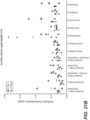
- a competing antibody binds to the same epitope (e.g., a linear or a conformational epitope) that is bound by 94B2-C1, 125B11-H3, 37D3-H9, or hu37D3-H9.v28.A4.
- epitope e.g., a linear or a conformational epitope
- 94B2-C1, 125B11-H3, 37D3-H9, or hu37D3-H9.v28.A4 Detailed exemplary methods for mapping an epitope to which an antibody binds are provided in Morris (1996) "Epitope Mapping Protocols," in Methods in Molecular Biology vol. 66 (Humana Press, Totowa, NJ ).
- immobilized Tau (such as monomeric Tau) is incubated in a solution comprising a first labeled antibody that binds to Tau (e.g., any antibody described herein, such as hu37D3-H9.v28.A4) and a second unlabeled antibody that is being tested for its ability to compete with the first antibody for binding to Tau.
- the second antibody may be present in a hybridoma supernatant.
- immobilized Tau is incubated in a solution comprising the first labeled antibody but not the second unlabeled antibody. After incubation under conditions permissive for binding of the first antibody to Tau, excess unbound antibody is removed, and the amount of label associated with immobilized Tau is measured.
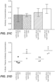
- assays are provided for identifying anti-Tau (e.g., pan-Tau) antibodies thereof having biological activity.
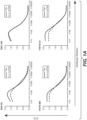
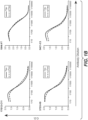
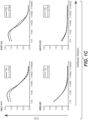
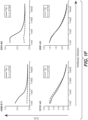
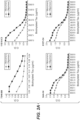
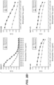
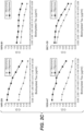
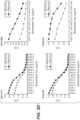
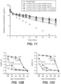
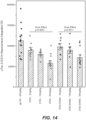
- Biological activity may include, e.g. , binding of such antibodies to multiple forms of Tau (e.g., monomeric Tau, oligomeric Tau, non-phosphorylated Tau, and phosphorylated Tau) and reducing the level of Tau protein (e.g., total Tau, total soluble Tau, soluble non-phosphorylated Tau, soluble phosphorylated Tau, total insoluble Tau, insoluble non-phosphorylated Tau, insoluble phosphorylated Tau, hyperphosphorylated Tau, or paired helical filaments containing hyperphosphorylated Tau, in the brain, e.g., in the brain cortex and/or hippocampus).
- Antibodies having such biological activity in vivo and/or in vitro are also provided.
- an antibody of the invention is tested for such biological activity.
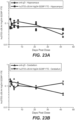
- an animal model of tauopathy such as a Tau transgenic mice (e.g., P301L)
- Tau transgenic mice e.g., P301L
- P301L Tau transgenic mice
- an animal model of tauopathy such as a Tau transgenic mice (e.g., P301L)
- a Tau transgenic mice e.g., P301L
- experimental techniques known in the art can be used to assess whether such treatment reduces the level of Tau protein (e.g., total Tau, total soluble Tau, soluble phosphorylated Tau, soluble non-phosphorylated Tau, total insoluble Tau, insoluble phosphorylated Tau, insoluble non-phosphorylated Tau, hyperphosphorylated Tau, or paired helical filaments containing hyperphosphorylated Tau) in the mouse brain ( e.g., in the brain cortex and/or hippocampus).
- Tau protein e.g., total Tau, total soluble Tau, soluble phosphorylated Tau, soluble non-phosphorylated Tau, total insoluble Tau, insoluble phosphorylated Tau, insoluble non-phosphorylated Tau, hyperphosphorylated Tau, or paired helical filaments containing hyperphosphorylated Tau
- the mouse brain e.g., in the brain
- the antibodies of the present invention may be present in immunoconjugates comprising the anti-Tau antibody conjugated to one or more other therapeutic agents or radioactive isotopes.
- An immunoconjugate may comprise an antibody as described herein conjugated to a radioactive atom to form a radioconjugate.
- a variety of radioactive isotopes are available for the production of radioconjugates. Examples include At 211 , I 131 , I 125 , Y 90 , Re 186 , Re 188 , Sm 153 , Bi 212 , P 32 , Pb 212 and radioactive isotopes of Lu.
- the radioconjugate When used for detection, it may comprise a radioactive atom for scintigraphic studies, for example tc99m or I123, or a spin label for nuclear magnetic resonance (NMR) imaging (also known as magnetic resonance imaging, mri), such as iodine-123 again, iodine-131, indium-111, fluorine-19, carbon-13, nitrogen-15, oxygen-17, gadolinium, manganese or iron.
- NMR nuclear magnetic resonance
- Conjugates of an antibody may be made using a variety of bifunctional protein coupling agents such as N-succinimidyl-3-(2-pyridyldithio) propionate (SPDP), succinimidyl-4-(N-maleimidomethyl) cyclohexane-1-carboxylate (SMCC), iminothiolane (IT), bifunctional derivatives of imidoesters (such as dimethyl adipimidate HCl), active esters (such as disuccinimidyl suberate), aldehydes (such as glutaraldehyde), bis-azido compounds (such as bis (p-azidobenzoyl) hexanediamine), bis-diazonium derivatives (such as bis-(p-diazoniumbenzoyl)-ethylenediamine), diisocyanates (such as toluene 2,6-diisocyanate), and bis-active fluorine compounds (such as 1,5-diflu
- a ricin immunotoxin can be prepared as described in Vitetta et al., Science 238:1098 (1987 ).
- Carbon-14-labeled 1-isothiocyanatobenzyl-3-methyldiethylene triaminepentaacetic acid (MX-DTPA) is an exemplary chelating agent for conjugation of radionucleotide to the antibody. See WO94/11026 .
- the linker may be a "cleavable linker" facilitating release of a cytotoxic drug in the cell.
- an acid-labile linker, peptidase-sensitive linker, photolabile linker, dimethyl linker or disulfide-containing linker Chari et al., Cancer Res. 52: 127-131 (1992 ); U.S. Patent No. 5,208,020 ) may be used.
- the immunuoconjugates or ADCs herein expressly contemplate, but are not limited to such conjugates prepared with cross-linker reagents including, but not limited to, BMPS, EMCS, GMBS, HBVS, LC-SMCC, MBS, MPBH, SBAP, SIA, SIAB, SMCC, SMPB, SMPH, sulfo-EMCS, sulfo-GMBS, sulfo-KMUS, sulfo-MBS, sulfo-SIAB, sulfo-SMCC, and sulfo-SMPB, and SVSB (succinimidyl-(4-vinylsulfone)benzoate) which are commercially available ( e.g., from Pierce Biotechnology, Inc., Rockford, IL., U.S.A).
- cross-linker reagents including, but not limited to, BMPS, EMCS, GMBS, HBVS,
- a biological sample comprises a cell or tissue, such as cerebrospinal fluid, a cell or tissue of the brain (e.g., brain cortex or hippocampus), or blood.
- a biological sample is cerebrospinal fluid.
- the anti-Tau antibody may be useful in a method of diagnosis or detection or in a method of detecting the presence of Tau in a biological sample.
- the method may comprise contacting the biological sample with an anti-Tau antibody as described herein under conditions permissive for binding of the anti-Tau antibody to Tau, and detecting whether a complex is formed between the anti-Tau antibody and Tau.
- Such method may be an in vitro or in vivo method.
- the complex formed between the anti-Tau antibody and Tau in a test biological sample can be compared to the complex formed in a control biological sample (e.g., a biological sample from a healthy subject or subjects).
- the amount of the complex formed between the anti-Tau antibody and Tau in a test biological sample can also be quantified and compared to the amount of the complex formed in a control biological sample (e.g., a biological sample from a healthy subject or subjects) or to the average amount of the complex known to be formed in healthy subjects.
- a control biological sample e.g., a biological sample from a healthy subject or subjects
- An anti-Tau antibody may be used to select subjects eligible for therapy with an anti-Tau antibody, e.g. where Tau is a biomarker for selection of patients.
- an anti-Tau (e.g., pan-Tau) antibody is used to detect whether the subject has a Tau protein disease or disorder, or whether the subject is at high risk (or predisposed to) a Tau protein disease or disorder.
- diseases or disorders that may be diagnosed using an antibody of the invention include Tau protein associated diseases or disorders, and diseases or disorders caused by or associated with the formation of neurofibrillary tangles or neuropil threads.
- diseases or disorders that may be diagnosed using an antibody of the invention include Tau protein associated diseases or disorders that are manifested in an impairment or loss of cognitive functions including reasoning, situational judgement, memory capacity, learning, and/or special navigation.
- diseases or disorders that may be diagnosed using an antibody of the invention include tauopathies such as neurodegenerative tauopathies.
- Exemplary diseases or disorders that may be diagnosed using an antibody of the invention include, but are not limited to, Alzheimer's Disease, Creutzfeldt-Jacob disease, Dementia pugilistica, Down Syndrome, Gerstmann-St Hurssler-Scheinker disease, inclusion-body myositis, prion protein cerebral amyloid angiopathy, traumatic brain injury, amyotrophic lateral sclerosis/parkinsonism-dementia complex of Guam, Non-Guamanian motor neuron disease with neurofibrillary tangles, argyrophilic grain dementia, corticobasal degeneration, diffuse neurofibrillary tangles with calcification, frontotemporal dementia, frontotemporal dementia with parkinsonism linked to chromosome 17, Hallervorden-Spatz disease, multiple system atrophy, Niemann-Pick disease type C, Pallido-ponto-nigral degeneration, Pick's disease, progressive subcortical gliosis, progressive supran
- Labels include, but are not limited to, labels or moieties that are detected directly (such as fluorescent, chromophoric, electron-dense, chemiluminescent, and radioactive labels), as well as moieties, such as enzymes or ligands, that are detected indirectly, e.g. , through an enzymatic reaction or molecular interaction.
- Exemplary labels include, but are not limited to, the radioisotopes 32 P, 14 C, 125 I, 3 H, and 131 I, fluorophores such as rare earth chelates or fluorescein and its derivatives, rhodamine and its derivatives, dansyl, umbelliferone, luceriferases, e.g. , firefly luciferase and bacterial luciferase ( U.S. Patent No.
- luciferin 2,3-dihydrophthalazinediones
- HRP horseradish peroxidase
- alkaline phosphatase alkaline phosphatase
- ⁇ -galactosidase alkaline phosphatase
- glucoamylase glucoamylase
- lysozyme saccharide oxidases
- glucose oxidase glucose oxidase, galactose oxidase, and glucose-6-phosphate dehydrogenase
- heterocyclic oxidases such as uricase and xanthine oxidase
- an enzyme that employs hydrogen peroxide to oxidize a dye precursor such as HRP, lactoperoxidase, or microperoxidase, biotin/avidin, spin labels, bacteriophage labels, stable free radicals, and the like.
- compositions of an anti-Tau antibody as described herein are prepared by mixing such antibody having the desired degree of purity with one or more optional pharmaceutically acceptable carriers, diluents, and/or excipients ( Remington's Pharmaceutical Sciences 16th edition, Osol, A. Ed. (1980 )), in the form of lyophilized formulations or aqueous solutions.
- Pharmaceutically acceptable carriers, diluents, and excipients are generally nontoxic to recipients at the dosages and concentrations employed, and include, but are not limited to: sterile water, buffers such as phosphate, citrate, and other organic acids; antioxidants including ascorbic acid and methionine; preservatives (such as octadecyldimethylbenzyl ammonium chloride; hexamethonium chloride; benzalkonium chloride; benzethonium chloride; phenol, butyl or benzyl alcohol; alkyl parabens such as methyl or propyl paraben; catechol; resorcinol; cyclohexanol; 3-pentanol; and m-cresol); low molecular weight (less than about 10 residues) polypeptides; proteins, such as serum albumin, gelatin, or immunoglobulins; hydrophilic polymers such as polyvinylpyrrolidone; amino acids such as
- sHASEGP soluble neutral-active hyaluronidase glycoproteins
- rHuPH20 HYLENEX ® , Baxter International, Inc.
- Certain exemplary sHASEGPs and methods of use, including rHuPH20, are described in US Patent Publication Nos. 2005/0260186 and 2006/0104968 .
- a sHASEGP is combined with one or more additional glycosaminoglycanases such as chondroitinases.
- Exemplary lyophilized antibody formulations are described in US Patent No. 6,267,958 .
- Aqueous antibody formulations include those described in US Patent No. 6,171,586 and WO2006/044908 , the latter formulations including a histidine-acetate buffer.
- the formulation herein may also contain more than one active ingredients as necessary for the particular indication being treated, preferably those with complementary activities that do not adversely affect each other.
- active ingredients are suitably present in combination in amounts that are effective for the purpose intended.
- Active ingredients may be entrapped in microcapsules prepared, for example, by coacervation techniques or by interfacial polymerization, for example, hydroxymethylcellulose or gelatin-microcapsules and poly-(methylmethacylate) microcapsules, respectively, in colloidal drug delivery systems (for example, liposomes, albumin microspheres, microemulsions, nano-particles and nanocapsules) or in macroemulsions.
- colloidal drug delivery systems for example, liposomes, albumin microspheres, microemulsions, nano-particles and nanocapsules
- Sustained-release preparations may be prepared. Suitable examples of sustained-release preparations include semipermeable matrices of solid hydrophobic polymers containing the antibody, which matrices are in the form of shaped articles, e.g. films, or microcapsules.
- the formulations to be used for in vivo administration are generally sterile. Sterility may be readily accomplished, e.g ., by filtration through sterile filtration membranes.
- anti-Tau antibodies Any of the anti-Tau antibodies provided herein may be used in therapeutic methods.
- the anti-Tau antibody of the present invention may be used as a medicament.
- the anti-Tau antibody for use in treating a tauopathy such as a neurodegenerative tauopathy
- Exemplary Tau protein associated diseases or disorders that can be treated that can be treated with anti-tau antibodies include, without limitation, Alzheimer's Disease, amyotrophic lateral sclerosis, Parkinson's disease, Creutzfeldt-Jacob disease, Dementia pugilistica, Down Syndrome, Gerstmann-St syndromessler-Scheinker disease, inclusion-body myositis, prion protein cerebral amyloid angiopathy, traumatic brain injury, amyotrophic lateral sclerosis/parkinsonism-dementia complex of Guam, Non-Guamanian motor neuron disease with neurofibrillary tangles, argyrophilic grain dementia, corticobasal degeneration, diffuse neurofibrillary tangles with calcification, frontotemporal dementia, frontotemporal dementia
- the anti-Tau antibody for use in treating Alzheimer's Disease is provided herein.
- the anti-Tau antibody for use in treating moderate AD, mild to moderate AD, mild AD, early AD, or prodromal AD is provided herein.
- Tau protein associated diseases or disorders that can be treated with the anti-Tau antibody include diseases or disorders that are manifested in an impairment or loss of a cognitive function such as reasoning, situational judgement, memory capacity, learning, and/or special navigation.
- the treatment may further comprise administering to the individual an effective amount of at least one additional therapeutic agent, e.g., as described below.
- the antibody of the invention is used to treat an individual having an MMSE score of between 20 and 30, between 20 and 26, between 24 and 30, between 21and 26, between 22 and 26, between 22 and 28, between 23 and 26, between 24 and 26, or between 25 and 26.
- the patient has an MMSE score between 22 and 26.
- an MMSE score between two numbers includes the numbers at each end of the range.
- an MMSE score between 22 and 26 includes MMSE scores of 22 and 26.
- the antibodies of the invention are used to treat an individual who is 'tau positive,' e.g., a patient having brain tau deposits that are typical of Tau protein associated disorders, e.g., a patient having a positive Tau PET scan.
- An anti-Tau antibody may find use in reducing the levels of Tau protein (e.g., total Tau, total soluble Tau, soluble phosphorylated Tau, total insoluble Tau, insoluble phosphorylated Tau, hyperphosphorylated Tau, or paired helical filaments containing hyperphosphorylated Tau) in an individual. For example such reduction can occur in the brain (e.g., in the brain cortex and/or hippocampus).
- An anti-Tau antibody may find use in reducing the levels of phosphorylated Tau.
- An anti-Tau antibody may find use in reducing the levels of insoluble Tau (e.g., insoluble phosphorylated Tau).
- An anti-Tau antibody may find use in reducing the levels of hyperphosphorylated Tau.
- An anti-Tau antibody may find use in reducing the levels of paired helical filaments (e.g., paired helical filaments containing hyperphosphorylated Tau) in a brain tissue (e.g., in the brain cortex and/or hippocampus).
- paired helical filaments e.g., paired helical filaments containing hyperphosphorylated Tau
- a brain tissue e.g., in the brain cortex and/or hippocampus.
- An anti-Tau antibody may find use in a method of reducing the levels of Tau protein (e.g., total Tau, total soluble Tau, soluble phosphorylated Tau, total insoluble Tau, insoluble phosphorylated Tau, hyperphosphorylated Tau, or paired helical filaments containing hyperphosphorylated Tau) in the brain (e.g., in the brain cortex and/or hippocampus) in an individual comprising administering to the individual an effective amount of the anti-Tau antibody to reduce the levels of Tau protein.
- An "individual" according to any of the above embodiments is a mammal, preferably a human.
- the anti-Tau antibody or a medicament comprising the anti-Tau antibody may be used for alleviating one or more symptoms of a Tau protein associated disease or disorder (such as any of the diseases or disorders described herein, for example, AD.
- the anti-Tau antibody or a medicament comprising the anti-Tau antibody may be used for reducing the number of symptoms or the severity of one or more symptoms of a Tau protein associated disease or disorder (such as any of the diseases or disorders described herein, for example, AD.
- the symptom of a Tau protein associated disease or disorder may be an impairment in cognition.
- the symptom of a Tau protein associated disease or disorder may be an impairment in learning and/or memory.
- the symptom of a Tau protein associated disease or disorder may be a long-term memory loss.
- the symptom of a Tau protein associated disease or disorder may be dementia.
- the symptom of a Tau protein associated disease or disorder may be confusion, irritability, aggression, mood swings, or a language impairment.
- the symptom of a Tau protein associated disease or disorder may be an impairment or loss of one or more cognitive functions such as reasoning, situational judgment, memory capacity, and/or learning.
- the invention provides the anti-Tau antibody of the invention or a medicament comprising the anti-Tau antibody for retaining or increasing cognitive memory capacity or for slowing down memory loss associated with a Tau protein associated disease or disorder (such as any of the diseases or disorders described herein, for example, AD).
- the methods provided herein comprise administration of an amount (e.g., therapeutically effective amount) of an anti-Tau antibody to an individual (e.g., who displays one or more symptoms of memory loss or a decrease of memory capacity).
- the anti-Tau antibody or a medicament comprising the anti-Tau antibody may be useful for decreasing the rate of progression of a Tau protein associated disease or disorder (such as any of the diseases or disorders described herein, for example, AD).
- the anti-Tau antibody or a medicament comprising the anti-Tau antibody may be useful for preventing the development of a Tau protein associated disease or disorder (such as any of the diseases or disorders described herein, for example, AD).
- the anti-Tau antibody or a medicament comprising the anti-Tau antibody may be useful for delaying the development of Tau protein associated disease or disorder (such as any of the diseases or disorders described herein, for example, AD).
- the methods provided herein comprise administration of an amount (e.g., therapeutically effective amount) of an anti-Tau antibody to an individual (e.g., who displays one or more symptoms of a Tau protein associated disease or disorder).
- the invention provides pharmaceutical formulations comprising any of the anti-Tau antibodies provided herein, e.g., for use in any of the above therapeutic methods.
- a pharmaceutical formulation comprises any of the anti-Tau antibodies provided herein and a pharmaceutically acceptable carrier.
- a pharmaceutical formulation comprises any of the anti-Tau antibodies provided herein and at least one additional therapeutic agent, e.g., as described below.
- Antibodies of the invention can be used either alone or in combination with other agents in a therapy.
- an antibody of the invention may be co-administered with at least one additional therapeutic agent.
- composition according to the invention may be administered in combination with other compositions comprising an additional therapeutic agent, such as a biologically active substance or compound such as, for example, a known compound used in the medication of tauopathies and/or of amyloidoses, a group of diseases and disorders associated with amyloid or amyloid-like protein such as the amyloid ⁇ protein involved in Alzheimer's Disease.
- an additional therapeutic agent such as a biologically active substance or compound such as, for example, a known compound used in the medication of tauopathies and/or of amyloidoses, a group of diseases and disorders associated with amyloid or amyloid-like protein such as the amyloid ⁇ protein involved in Alzheimer's Disease.
- the other biologically active compound may include neuron-transmission enhancers, psychotherapeutic drugs, acetylcholine esterase inhibitors, calcium-channel blockers, biogenic amines, benzodiazepine tranquillizers, acetylcholine synthesis, storage or release enhancers, acetylcholine postsynaptic receptor agonists, monoamine oxidase-A or -B inhibitors, N-methyl-D-aspartate glutamate receptor antagonists, non-steroidal anti-inflammatory drugs, antioxidants, serotonergic receptor antagonists, or other therapeutic agents.
- neuron-transmission enhancers may include neuron-transmission enhancers, psychotherapeutic drugs, acetylcholine esterase inhibitors, calcium-channel blockers, biogenic amines, benzodiazepine tranquillizers, acetylcholine synthesis, storage or release enhancers, acetylcholine postsynaptic receptor agonists, monoamine oxidase-A or -
- the biologically active agent or compound may comprise at least one compound selected from compounds against oxidative stress, anti-apoptotic compounds, metal chelators, inhibitors of DNA repair such as pirenzepine and metabolites, 3- amino-1-propanesulfonic acid (3APS), 1,3-propanedisulfonate (1,3PDS), secretase activators, beta- and gamma-secretase inhibitors, tau proteins, anti-Tau antibodies (including, but not limited to, antibodies disclosed in WO2012049570 , WO2014028777 , WO2014165271 , WO2014100600 , WO2015200806 , US8980270 , and US8980271 ), neurotransmitter, beta-sheet breakers, antiinflammatory molecules, "atypical antipsychotics” such as, for example clozapine, ziprasidone, risperidone, aripiprazole or olanzapine or cholinesterase inhibitors (ChEIs)
- an antibody of the invention may be administered in combination with a neurological drug.
- neurological drugs include, but are not limited to, an antibody or other binding molecule (including, but not limited to a small molecule, a peptide, an aptamer, or other protein binder) that specifically binds to a target selected from: beta secretase, presenilin, amyloid precursor protein or portions thereof, amyloid beta peptide or oligomers or fibrils thereof, death receptor 6 (DR6), receptor for advanced glycation endproducts (RAGE), parkin, and huntingtin; an NMDA receptor antagonist ( i.e., memantine), a monoamine depletor ( i.e., tetrabenazine); an ergoloid mesylate; an anticholinergic antiparkinsonism agent (i.e., procyclidine, diphenhydramine, trihexylphenidyl, benztropine, biperiden and trihexyphenid
- corticosteroid includes, but is not limited to, fluticasone (including fluticasone propionate (FP)), beclometasone, budesonide, ciclesonide, mometasone, flunisolide, betamethasone and triamcinolone.
- fluticasone including fluticasone propionate (FP)
- beclometasone a corticosteroid that is suitable for delivery by inhalation.
- Exemplary inhalable corticosteroids are fluticasone, beclomethasone dipropionate, budenoside, mometasone furoate, ciclesonide, flunisolide, and triamcinolone acetonide.
- one or more anti-amyloid beta (anti-Abeta) antibodies may be administered with an anti-Tau antibody provided herein.
- anti-Abeta antibodies include crenezumab, solanezumab, bapineuzumab, aducanumab, gantenerumab, and BAN-2401 (Biogen, Eisai Co., Ltd.).
- one or more beta-amyloid aggregation inhibitors may be administered with an anti-Tau antibody provided herein.
- Nonlimiting exemplary beta-amyloid aggregation inhibitors include ELND-005 (also referred to as AZD-103 or scyllo-inositol), tramiprosate, and PTI-80 (Exebryl-1 ® ; ProteoTech).
- one or more BACE inhibitors may be administered with an anti-Tau antibody provided herein.
- Non-limiting examples of such BACE inhibitors include E-2609 (Biogen, Eisai Co., Ltd.), AZD3293 (also known as LY3314814; AstraZeneca, Eli Lilly & Co.), MK-8931 (verubecestat), and JNJ-54861911 (Janssen, Shionogi Pharma).
- one or more Tau inhibitors may be administered with an anti-Tau antibody provided herein.
- Non-limiting examples of such Tau inhibitors include methylthioninium, LMTX (also known as leuco-methylthioninium or Trx-0237; TauRx Therapeutics Ltd.), Rember TM (methylene blue or methylthioninium chloride [MTC]; Trx-0014; TauRx Therapeutics Ltd), PBT2 (Prana Biotechnology), and PTI-51-CH3 (TauPro TM ; ProteoTech).
- one or more other anti-Tau antibodies may be administered with an anti-Tau antibody provided herein.
- Non-limiting examples of such other anti-Tau antibodies include BMS-986168 (Bristol-Myers Squibb) and C2N-8E12 (AbbVie, C2N Diagnostics, LLC).
- a general misfolding inhibitor such as NPT088 (NeuroPhage Pharmaceuticals) may be administered with an anti-Tau antibody provided herein.
- the composition according to the invention may comprise niacin or memantine together with a chimeric antibody or a humanized antibody according to the invention including antibodies, particularly monoclonal antibodies and active fragments thereof, and, optionally, a pharmaceutically acceptable carrier and/or a diluent and/or an excipient.
- compositions are provided that comprise "atypical antipsychotics" such as, for example clozapine, ziprasidone, risperidone, aripiprazole or olanzapine for the treatment of positive and negative psychotic symptoms including hallucinations, delusions, thought disorders (manifested by marked incoherence, derailment, tangentiality), and playful or disorganized behavior, as well as anhedonia, flattened affect, apathy, and social withdrawal, together with the chimeric antibody or the humanized antibody according to the invention or active fragments thereof, and, optionally, a pharmaceutically acceptable carrier and/or a diluent and/or an excipient.
- "atypical antipsychotics” such as, for example clozapine, ziprasidone, risperidone, aripiprazole or olanzapine for the treatment of positive and negative psychotic symptoms including hallucinations, delusions, thought disorders (manifested by marked incoherence,
- compositions in addition to chimeric antibody or humanized antibody according to the invention are those disclosed, for example, in WO 2004/058258 (see especially pages 16 and 17) including therapeutic drug targets (page 36-39), alkanesulfonic acids and alkanolsulfuric acid (pages 39-51), cholinesterase inhibitors (pages 51-56), NMDA receptor antagonists (pages 56-58), estrogens (pages 58-59), non-steroidal anti-inflammatory drugs (pages 60-61), antioxidants (pages 61-62), peroxisome proliferators-activated receptors (PPAR) agonists (pages 63-67), cholesterol-lowering agents (pages 68-75); amyloid inhibitors (pages 75-77), amyloid formation inhibitors (pages 77-78), metal chelators (pages 78-79), anti-psychotics and anti-depressants (pages 80-82), nutritional supplements (pages 83-89) and compounds increasing
- Such combination therapies noted above encompass combined administration (where two or more therapeutic agents are included in the same or separate formulations), and separate administration, in which case, administration of the antibody of the invention can occur prior to, simultaneously, and/or following, administration of the additional therapeutic agent or agents.
- administration of the anti-Tau antibody and administration of an additional therapeutic agent occur within about one month, or within about one, two or three weeks, or within about one, two, three, four, five, or six days, of each other.
- An antibody of the invention can be administered by any suitable means, including parenteral, intrapulmonary, and intranasal, and, if desired for local treatment, intralesional administration.
- Parenteral infusions include intramuscular, intravenous, intraarterial, intraperitoneal, or subcutaneous administration. Dosing can be by any suitable route, e.g. by injections, such as intravenous or subcutaneous injections, depending in part on whether the administration is brief or chronic.
- Various dosing schedules including but not limited to single or multiple administrations over various time-points, bolus administration, and pulse infusion are contemplated herein.
- Antibodies of the invention would be formulated, dosed, and administered in a fashion consistent with good medical practice. Factors for consideration in this context include the particular disorder being treated, the particular mammal being treated, the clinical condition of the individual patient, the cause of the disorder, the site of delivery of the agent, the method of administration, the scheduling of administration, and other factors known to medical practitioners.
- the antibody need not be, but is optionally formulated with one or more agents currently used to prevent or treat the disorder in question. The effective amount of such other agents depends on the amount of antibody present in the formulation, the type of disorder or treatment, and other factors discussed above. These are generally used in the same dosages and with administration routes as described herein, or about from 1 to 99% of the dosages described herein, or in any dosage and by any route that is empirically/clinically determined to be appropriate.
- an antibody of the invention when used alone or in combination with one or more other additional therapeutic agents, will depend on the type of disease to be treated, the type of antibody, the severity and course of the disease, whether the antibody is administered for preventive or therapeutic purposes, previous therapy, the patient's clinical history and response to the antibody, and the discretion of the attending physician.
- the antibody is suitably administered to the patient at one time or over a series of treatments.
- about 1 ⁇ g/kg to 15 mg/kg (e.g. 0.1mg/kg-10mg/kg) of antibody can be an initial candidate dosage for administration to the patient, whether, for example, by one or more separate administrations, or by continuous infusion.
- One typical daily dosage might range from about 1 ⁇ g/kg to 100 mg/kg or more, depending on the factors mentioned above. For repeated administrations over several days or longer, depending on the condition, the treatment would generally be sustained until a desired suppression of disease symptoms occurs.
- One exemplary dosage of the antibody would be in the range from about 0.05 mg/kg to about 10 mg/kg.
- one or more doses of about 0.5 mg/kg, 2.0 mg/kg, 4.0 mg/kg or 10 mg/kg (or any combination thereof) may be administered to the patient.
- Such doses may be administered intermittently, e.g. every week or every three weeks ( e.g. such that the patient receives from about two to about twenty, or e.g. about six doses of the antibody).
- An initial higher loading dose, followed by one or more lower doses may be administered.
- other dosage regimens may be useful. The progress of this therapy is easily monitored by conventional techniques and assays.
- the article of manufacture comprises a container and a label or package insert on or associated with the container.
- Suitable containers include, for example, bottles, vials, syringes, IV solution bags, etc.
- the containers may be formed from a variety of materials such as glass or plastic.
- the container holds a composition which is by itself or combined with another composition effective for treating, preventing and/or diagnosing the condition and may have a sterile access port (for example the container may be an intravenous solution bag or a vial having a stopper pierceable by a hypodermic injection needle).
- At least one active agent in the composition is an antibody of the invention.
- the label or package insert indicates that the composition is used for treating the condition of choice.
- the article of manufacture may comprise (a) a first container with a composition contained therein, wherein the composition comprises an antibody of the invention; and (b) a second container with a composition contained therein, wherein the composition comprises a further cytotoxic or otherwise therapeutic agent.
- the article of manufacture may further comprise a package insert indicating that the compositions can be used to treat a particular condition.
- the article of manufacture may further comprise a second (or third) container comprising a pharmaceutically-acceptable buffer, such as bacteriostatic water for injection (BWFI), phosphate-buffered saline, Ringer's solution and dextrose solution. It may further include other materials desirable from a commercial and user standpoint, including other buffers, diluents, filters, needles, and syringes.
- BWFI bacteriostatic water for injection
- phosphate-buffered saline such as bacteriostatic water for injection (
- any of the above articles of manufacture may include an immunoconjugate as described herein in place of or in addition to an anti-Tau antibody.
- the recombinant human Tau construct 2N4R isoform (amino acids 2-441), was fused to a N-terminal His-tag to facilitate purification and characterization. See, e.g., Figure 15 .
- the fusion construct was cloned into the pET52b vector (Novagen) and expressed in E . coli. Cells were harvested and lysed under denaturing condition using 7M guanidinium chloride overnight at 4°C with stirring. Cell debris was pelleted at 40,000 rpm for 1 hour.
- the recombinant, His-tagged protein was isolated by nickel affinity chromatography (Ni Sepharose excel affinity resin, GE Healthcare Life Sciences) followed by size-exclusion chromatography (Superdex 200 resin, GE Healthcare Life Sciences) under denaturing condition. Guanidinium chloride was removed by dialyzing the recovered protein into 20mM MES, 50mM NaCl, and 1mM TCEP at pH 6.8. The His-tag was subsequently removed using TEV protease, followed by final purification using cation exchange chromatography (Mono S column, GE Healthcare Life Sciences) to remove the cleaved His-tag. The purification buffer contained 0.1% Triton x-114 (v/v) to remove endotoxin.
- Purified protein was exchanged into PBS with 1mM TCEP. The purity and monomeric state were analyzed by SDS-PAGE and SEC-MALLS. Identity was confirmed by mass spectrometry. Protein concentration was determined by UV absorption at 280 nm. The final product was free of endotoxin ( ⁇ 0.5 EU/mg), as determined by Kinetic Limulus Amebocyte Lysate (LAL) assay.
- LAL Kinetic Limulus Amebocyte Lysate
- Phosphorylated Tau was generated using the Tau 2-441 construct prepared using the method described above.
- the protein construct was phosphorylated using 0.5 ⁇ M PKA kinase (Life Technologies), which phosphorylates serine 409, among other residues.
- the reaction mixture was incubated with 1mM ATP, 5mM MgCl 2 , at room temperature for 72 hours. Phosphorylation was confirmed by mass spectrometry. Size-exclusion chromatography (Superdex 75, GE Healthcare Life Sciences) was used to remove the kinase. The purity, monomeric state, and endotoxin level of the phosphorylated protein preparation were analyzed substantially as described above.
- Oligomeric Tau was generated using the monomeric Tau 2-441 construct.
- the monomeric protein was first exchanged into 20mM N,N-Bis(2-hydroxyethyl)-2-aminoethanesulfonic acid (BES), 25mM NaCl, pH 7.4, followed by oligomeriztion using 75 ⁇ M arachidonic acid (Cayman Chemicals) and 18 kDa Heparin (Sigma Aldrich), at equimolar concentration with protein at 37°C for 3 days.
- Oligomerization was confirmed by thioflavin T fluorescence assay, dynamic light scattering (DLS), and analytical size-exclusion chromatography.
- Oligomeric Tau is in some instances referred to as "oligoTau.”
- mice Female C57BL/6JOlaHsd (C57BL/6) and BALB/c OlaHsd (Balb/c) wild-type mice (Harlan, USA) were received at 9 weeks of age. Tau knock-out mice (B6.129-Mapttm1Hnd/J; The Jackson Laboratory, USA) were received at 6 and 9 weeks of age. Vaccinations started at 12 to 15 weeks of age. Mice were vaccinated with oligomerized human Tau.
- the oligoTau was mixed with one of two adjuvants used in this study, Ribi Adjuvant System (Ribi; Sigma-Aldrich, Switzerland) at 50% v/v, or a combination of CpG single-stranded synthetic DNA oligodeoxynucleotides (CpG; Microsynth, Switzerland) and aluminium hydroxide (Al; Brenntag, Switzerland).
- Ribi Adjuvant System Ribi; Sigma-Aldrich, Switzerland
- Al aluminium hydroxide
- Ribi is 2% squalene oil-in-water emulsion containing monophosphoryl lipid A (isolated from Salmonella minnesota ) and synthetic trehalose dicorynomycolate (isolated from the cord factor of the Tubercle bacillus ) in squalene oil, 0.2% Tween-80, and water.
- mice were vaccinated by subcutaneous injection (s.c.), except groups D and G, which received a combination of intraperitoneal (i.p.) and hock administrations.
- Mice in group D were administered 50 ⁇ g of oligoTau i.p. and 10 ⁇ g of oligoTau as hock injection.
- Mice in group G were administered 8 ⁇ g of oligoTau i.p. and 2 ⁇ g of oligoTau as hock injection. See Table 2.
- mice were injected on days 0, 14, 35, and 56. Mice used for myeloma fusion (Nanotools, Germany) were additionally vaccinated with three daily booster injections of oligoTau (50 ⁇ g per i.p. injection) without adjuvant added. Table 2. Mice and vaccination protocols Study group Mouse strain Total oligoTau dose ( ⁇ g/injection) Adjuvant Vaccination route A C57BL/6 50 CpG/AI S. C.
- mice were bled and sacrificed one day following the last of three booster injections, and splenocytes were fused with myeloma cells to generate antibody producing hybridomas.
- mice were divided into three groups, for a total of 10 fusions (2 fusions in one group, four fusions in the second group, and four fusions in the third group), generating 299 hybridomas.
- Viable hybridomas were grown using serum-containing selection media, and the best hybridomas were then selected for subcloning, using ELISA assays for full-length human Tau and oligoTau binding as described below. Following limiting dilution, the final hybridomas were then grown in serum-free medium and media was collected from stable colonies for antibody screening and selection.
- Serum-free supernatants were harvested from stable hybridomas. The supernatants containing antibodies of interest were then screened by ELISA assays to characterize antibody properties and select antibodies for further development.
- the ELISA assays were used to determine the following: binding to full-length human Tau (flTau; SignalChem, Canada), binding to hyperphosphorylated flTau (Genentech, USA), binding to oligomeric versus monomeric preparations of flTau, and binding to certain antibody Tau epitope(s). Briefly, 96-well MaxiSorp ELISA plates (Nunc, Denmark) were coated with one of the targets as shown in Table 3. Table 3. Targets used for the ELISA screening assays.
- Coating was done overnight in phosphate-buffered saline (PBS) at 4°C. Plates were washed thoroughly with 0.05% Tween-20/PBS and then blocked with 1% bovine serum albumin (BSA) in 0.05% Tween-20/PBS for 1 hr at 37°C. The antibody contained in the hybridoma supernatant was then added at the indicated dilutions, and incubated for 2 hrs at 37°C after which the plates were washed as described previously.
- PBS phosphate-buffered saline
- an AP-conjugated anti-mouse IgG secondary antibody (Jackson ImmunoResearch Laboratories, United Kingdom) was added at 1/6000 dilution in 0.05% Tween-20/PBS for 2 hr at 37°C. After the final wash, plates were incubated with p-nitrophenyl phosphate disodium hexahydrate (pNPP; Sigma-Aldrich, Switzerland) phosphatase substrate solution, and read at 405 nm using an ELISA plate reader (Tecan, Switzerland). Results are expressed as optical densities (O.D.).
- oligoTau and monoTau capture ELISAs antibodies contained in serum-free sterile hybridoma supernatants were immobilized on an anti-IgG coated plate at 500-fold dilution, followed by the incubation of oligoTau or monoTau, both with site-specific biotinylation via an AVI-tag.
- the target incubations started at 5 ⁇ g/mL and then were diluted 8- or 16-fold.
- Streptavidin-HRP and ABTS substrate was used for signal quantitation in a plate reader (Tecan, Switzerland). Results are expressed as O.D.
- Affinity of non-purified antibodies in serum-free hybridoma supernatants was estimated by surface plasmon resonance using a Biacore T-100 instrument (GE Healthcare, United Kingdom). Antibodies were immobilized onto an anti-IgG biosensor chip, and flTau (SignalChem, Canada) was used as the target analyte. Kinetic analysis was done using a 1:1 Langmuir fit model.
- panTau antibodies to Tau in human brain was tested in a Western-blot (WB) using brain lysates from three AD and two age-matched non-AD control donors (Tissue Solutions, United Kingdom). The lysates were processed to obtain a detergent-free soluble Tau fraction. Processed lysates were loaded onto 4-12% bis-tris gels (Novex, Life Technologies, Switzerland) and transferred onto Immobilon PVDF membranes and blotted with antibodies being tested with and an IRDye 800CW goat anti-mouse secondary antibody (Li-Cor, USA).
- Hybridoma cell lysates were supplied to Antitope (Antitope, United Kingdom) for variable region gene sequencing. Briefly, RT-PCR was performed using degenerate primer pools for murine signal sequences together with constant region primers for each of IgG variable heavy (VH), IgM VH, Ig kappa variable light (KVL) and Ig ⁇ VL chains. Heavy chain V-region mRNA was amplified using a set of six degenerate primer pools (HA to HF) specific for VH signal sequences together with either IgM or IgG-specific constant region primers.
- VH variable heavy
- KVL Ig kappa variable light
- Ig ⁇ VL chains Ig ⁇ VL chains.
- Heavy chain V-region mRNA was amplified using a set of six degenerate primer pools (HA to HF) specific for VH signal sequences together with either IgM or IgG-specific constant region primers.
- the light chain V-region mRNA was amplified using a set of eight signal sequence-specific degenerate primer pools, seven for the ⁇ cluster (KA to KG) and one for the ⁇ cluster (LA), together with either ⁇ or ⁇ constant region primers.
- the PCR products obtained from successful amplification were purified, cloned into a 'TA' cloning vector (pGEM-T Easy, Promega), transformed into E.coli and individual colonies sequenced.
- the nucleotide and amino acid sequences of the antibody VH and VL regions were determined with the sequences for 27 antibody hybridomas.
- Hybridomas that were generated from each of the three rounds of fusions a total of 299 hybridomas derived from ten fusions, were initially assayed for binding to flTau, with selected hybridomas additionally assayed for binding to pTau and oligomerized Tau.
- the aim was to select antibodies that bind equally well to Tau and to Tau modified post-translationally, such as phosphorylated or oligomeric Tau. For this, assays were run on hybridomas to select for the best panTau properties.
- the binding region was first determined using different Tau fragments and then a library of 15-mer overlapping Tau peptides spanning the full 441 amino acids (aa) sequence of the longest human Tau isoform.
- a group of antibodies binding to pre-determined regions of Tau were intentionally avoided with the aim to maximize binding to different post-translationally modified forms of Tau and to all the six different human Tau isoforms present in humans.
- the three fusion series resulted in the generation of 133 subcloned stable hybridomas that were screened for the best panTau properties.
- a combination of different screening assays was used to narrow down the number of antibody hybridomas having the preferred properties for a panTau antibody.
- 90 hybridomas were assayed with the results of 24 hybridomas shown in Figure 1A-F .
- As an initial screen had been performed using Tau fragments to avoid selecting antibodies binding to regions of Tau known to have high density of residues that are phosphorylated in Alzheimer's disease (AD) and other tauopathies, most antibodies tested bound to both flTau and pTau with similar binding properties as determined by this ELISA.
- a panTau antibody binds to both monomeric and oligomeric forms of Tau without a strong preference to one or the other.
- a capture ELISA was set up to determine if antibodies bound to both monomeric and oligomeric forms of flTau. An ELISA run in capture mode preserves the oligomer conformation of pre-oligomerized Tau and the monomeric state of monoTau better than when run as a direct ELISA with the targets immobilized onto an ELISA plate.
- Mapping the epitopes is important for selecting antibodies with good panTau properties, as antibodies that bind to regions with high density of potential pTau residues (Ser, Thr, and Tyr) can be avoided. Binding to all six isoforms of human Tau was also used as a selection criterion for a panTau antibody.
- the panTau epitopes of antibodies that had been initially selected were verified and determined with improved accuracy using a library of 49 peptides each having 15 amino acids (aa) spanning the full length of human Tau, with an overlap of 6 aa residues and an offset of 9 aa. The residue numbers are based on the longest isoform of human Tau (441 aa).
- Non-purified antibdies were used at high 1/10 dilution to verify binding versus no binding to all peptides. Screening of antibodies from 112 hybridomas previously selected by ELISA indicated binding to 20 different Tau epitopes (Table 4).
- AD Alzheimer's disease
- control samples were additionally run in a non-denaturing ELISA capture assay to verify binding to Tau in human brains.
- Samples lysates processed for soluble Tau from two AD and two non-AD age-matched control subjects were run at 8 dilutions testing three antibodies ( Figure 4A-C ).
- Antibody variable chain sequences were determined for 27 hybridomas (Antitope, United Kingdom). Protein sequences for certain heavy and light chain variable domains and hypervariable regions (HVRs) are shown in the Table of Sequences.
- Antibody heavy and light chains were constructed via gene synthesis and subcloning of the resulting DNA into murine IgG2a (heavy chain) and murine kappa (light chain) mammalian expression vectors. Antibodies were expressed in CHO or 293T cells by transient co-transfection of the heavy chain and light chain plasmids and were purified with affinity resin MabSelectSure (GE Healthcare Life Sciences). Purified recombinant antibodies were screened for binding to Tau monomer protein on a Biacore T200 surface plasmon resonance instrument using a mouse IgG capture kit and a Series S CM5 chip.
- Antibodies in mIgG2a format diluted in 10mM HEPES pH7.4, 150 mM NaCl, 0.05% Tween 20 (running buffer, HBSP) were captured for 30 or 45 seconds at a concentration of 1 ⁇ g/ml (antibodies 26C1, 94B2-C1, 52F6-F11.v1, 52F6-F11.v2, 11E10-B8, 55E7-F11, 125B11-H3, 123E9-A1, 30G1-B2, 66F5-A1, 89F4-A1, 93A8-D2 and 126F11-G11) or for 70 or 150 seconds at a concentration of 0.1 ⁇ g/ml (antibodies 19H6-F7, 3A4-H4, 54C1-H11 and 37D3-H9) using a flow rate of 10 ⁇ l/min.
- Binding of Tau monomer in HBSP was monitored at 25°C using a flow rate of 30 ⁇ l/min and concentrations of 16, 31, 63, 125, 125, 250 and 500 nM for antibodies 26C1 and 94B2; 16, 31, 63, 125, 125, 250, 500 and 1000 nM for antibodies 52F6-F11.v1 and 52F6-F11.v2; 6, 19, 56, 56, 167 and 500 nM for antibodies 11E10-B8, 55E7-F11 and 125B11-H3; 5, 16, 49, 148, 148, 444, 1333 and 4000 nM for antibodies 123E9-A1, 30G1-B2, 66F5-A1, 89F4-A1, 93A8-D2 and 126F11-G11; 0.4, 1.6, 6.3, 2.5, 100 and 400 nM for 19H6-F7; and 0.2, 0.8, 4, 4, 20 and 100 nM for 3A4-H4, 54C1-H
- Human monomer Tau protein was covalently coupled to a Biacore Series S CM5 chip using the Biacore Amine Coupling Kit (GE Life Sciences), resulting in immobilization to a level of approximately 128 RU.
- Direct binding of 37D3-H9 in both Fab and IgG formats was monitored using the single-cycle kinetics experimental format with five association periods of 300s each and antibody concentrations of 1, 2, 4, 8 and 16 nM (IgG) or 5, 10, 20, 40 and 80 nM (Fab).
- Dissociation was monitored for 7200 seconds (Fab) or for 14400 seconds (IgG). A value for the dissociation rate was calculated by fitting a 1:1 binding model to the data.
- Antibody 37D3-H9 was humanized by grafting the antibody CDRs and selected variable region framework residues onto human antibody consensus frameworks ( Dennis, M.S. (2010). CDR repair: A novel approach to antibody humanization. In Current Trends in Monoclonal Antibody Development and Manufacturing, S.J. Shire, W. Gombotz, K. Bechtold-Peters and J. Andya, eds. (Springer, New York), pp. 9-28 ). Grafting onto consensus VH3, V ⁇ 2 and V ⁇ 1 frameworks was assessed. The heavy chain graft included murine residue at position 49 (Kabat numbering system). The V ⁇ 2 graft included murine residues in framework positions 2 and 4.
- the V ⁇ 1 graft included murine residues in framework positions 2, 4 and 43.
- Humanized variants were constructed by gene synthesis and subcloning into human IgG1 or IgG4 and Kappa chain mammalian expression vectors. Antibodies were expressed by co-transfection of the heavy and light chain plasmids into CHO cells and purified with affinity resin MabSelect Sure. Humanized variants were screened for affinity to human Tau monomer using the Biacore human IgG capture kit, a Series S CM5 chip and a Biacore T200 instrument. Antibodies were diluted to 2 ⁇ g/ml and captured for 15 seconds at 10 ⁇ l/min.
- Antibody variants hu37D3-H9.v1, hu37D3-H9.v2, hu37D3-H9.v5 and hu37D3-H9.v6 were characterized further by surface plasmon resonance with additional antibody concentrations and longer association/dissociation times. These variants were analyzed with a broader range of human Tau monomer concentrations (1.2, 3.7, 11.1, 11.1, 33.3, 100 nM) and increased association (300 seconds) and dissociation (1200 seconds) periods. A 1:1 binding model was applied to the results (Table 8).
- Table 8 Detailed analysis of binding kinetics of selected variants to human Tau by surface plasmon resonance Antibody variant Light chain framework K D (nM) hu37D3-H9.v1 Kappa1 1.1 nM, 1.0 nM hu37D3-H9.v2 Kappa1 1.2 nM hu37D3-H9.v5 Kappa2 0.8 nM hu37D3-H9.v6 Kappa2 1.4 nM
- FcRn Fc Receptor-neonate binding domain mutations such as M252Y, S254T and T256E (YTE) have been described to increase FcRn binding and thus increase the half-life of antibodies. See U.S. Published Patent Application No. 2003/0190311 and Dall'Acqua et al., J. Biol. Chem. 281:23514-23524 (2006 ).
- Antibody 125B11-H3 was humanized onto VH3 and V ⁇ 1 consensus frameworks.
- the heavy chain graft included murine residues at position 78 (Kabat numbering system).
- the V ⁇ 1 graft included murine residues in framework positions 43 and 87.
- the light chain of 113F5-F7 was also humanized onto the V ⁇ 1 framework, with additional murine residues at framework positions 43 and 87.
- Humanized variant heavy chains (125B11) and light chains (125B11 and 113F5-F7) were co-transfected in multiple combinations and purified in 96-well format as described above.
- Humanized variants were then screened for affinity for human Tau monomer using the Biacore human IgG capture kit, a Series S CM5 chip and a Biacore T200 instrument. Antibodies were diluted to 2 ⁇ g/ml and captured for 15 seconds at 10 ⁇ l/min. Association and dissociation of 0, 100 and 500 nM human Tau monomer in HBSP was monitored for 180s and 300s respectively at a flow rate of 40 ⁇ l/min. A 1:1 binding model was applied to the results (Table 9).
- Table 9 Screening of 125B11-H3 and 113F5-F7 humanization variants by surface plasmon resonance Screening K D (nM) 125B11 heavy chain humanization variant HC1 HC2 HC3 HC4 HC5 HC6 125B11 light chain humanization variant LC1 16, 19 18 18 15 85 - LC2 20 20 19 14 -* NT LC3 21 23 20 15 - - LC4 23 22 20 17 >100 >100 113F5-F7 light chain humanization variant LC1 57 61 54 44 - - LC2 67 68 55 47 - - LC3 61 64 54 47 >100 - LC4 71 77 65 51 - - * Minimal binding to Tau monomer. NT, not tested.
- Variants hu125B11.v17 (HC3+LC1), hu125B11.v26 (HC4+LC2) and hu125B11.v28 (HC4+LC4) were selected for high-resolution kinetic analysis based on the affinity screen (Table 10).
- Antibody 94B2-C1 was humanized onto VH1 and V ⁇ 2 frameworks.
- the heavy chain graft also included murine residues at position 28, 29, 67, 69, 71, and 73 (Kabat numbering system).
- the V ⁇ 2 graft also included murine residues in framework positions 2, 36, and 46. Combinations of eight heavy chains and eight light chains were expressed, purified and screened by surface plasmon resonance (SPR) as described for 125B11 above.
- SPR surface plasmon resonance
- Antibody samples were thermally stressed to mimic stability over the shelf life of the product. Samples were buffer exchanged into 20mM Acetate buffer, pH 5.5, or phosphate buffer, pH 7.4, and diluted to a concentration of 1 mg/ml. One ml of sample was stressed at 40°C for 2 weeks and a second was stored at -70°C as a control. Both samples were then digested using trypsin to create peptides that could be analyzed using liquid chromatography (LC) - mass spectrometry (MS) analysis. For each peptide in the sample retention time, from the LC as well as high resolution accurate mass and peptide ion fragmentation information (amino acid sequence information) were acquired in the MS.
- LC liquid chromatography
- MS mass spectrometry
- the stressed and unstressed antibodies were analyzed for Tau binding by surface plasmon resonance (Biacore) using the GE Biacore human IgG capture kit and a Series S CM5 chip.
- the hIgG were diluted to 2 ⁇ g/ml in 10mM HEPES pH7.4, 150mM NaCl, 0.05% Tween 20 (running buffer, HBSP) and captured at a flow rate of 10 ⁇ l/min for 15 seconds (t0 sample) or 17 seconds (t2 sample).
- the hIgG were diluted to 2 ⁇ g/ml in 10mM HEPES pH7.4, 150mM NaCl, 0.05% Tween 20 (running buffer, HBSP) and captured at a flow rate of 10 ⁇ l/min for 22 seconds.
- Kinetic data was collected for human Tau monomer injected at concentrations of 0, 6.3, 12.5, 25, 25, 50, 100, 200, and 400 nM in HBSP, using a flow rate of 30 ⁇ l/min, a 300 second association phase and a 600 second dissociation phase. Between cycles the surface was regenerated using a 30 second injection of 3M Magnesium Chloride at 10 ⁇ l/min.
- a 1: 1 binding model was fitted to the data and affinities for hu37D3-H9.v5 and hu37D3-H9.v5.3 (also referred to herein as hu37D3-H9.v5 N28D) calculated using kinetic analysis.
- Parameters used for the 1:1 fitting included the Instrument default of local fitting for the "RI" parameter. The results are shown in Figure 7 and Table 13.
- Table 13 Changes in Normalized Rmax observed upon thermal stress of hu37D3-H9.v5 and upon mixing of hu37D3-H9.v5 with anticipated deamidation product hu37D3-H9.v5 N28D
- Sample K D (nM) Rmax (RU) Decrease in Normalized Rmax compared to Reference* Comments hu37D3-H9.v5 1.5 ⁇ 0.2 76.1 ⁇ 0.4 Reference Mean +/- Standard Deviation of four intra-experiment analyses hIgG1 hu37D3-H9.v5 N28D 160 81.0 4% hIgG1 hu37D3-H9.v5 & 2.0 46.4 45%
- Two antibodies mixed at a 1:1 ratio hu37D3-H9.v5 N28D hIgG1 hu37D3-H9.v5 1.5 68.8 3% Control for Stressed sample hIgG4.S228P, t 0 hu37D3-H9.v5
- variants were assessed by Biacore to compare their functional stability with or without a two-week 40°C thermal stress period.
- the variants included most single mutations of the N 28 G 29 N 30 T 31 motif, double mutants containing the G29A mutation, double mutations of Asn-28 and Tyr-32 that might functionally replace these to hydrogen-bonded residues, as well as all possible permutations of residues 2, 4, 33, and 93 as either the residues present in the original 37D3-H9 antibody or the corresponding germline residue variant.
- mutations were tested in the context of residue 1 being Asp or Glu, which does not impact affinity or stability of the Asn-28 residue.
- Antibodies were expressed by transient transfection of Expi293 cells in 96-well format and automated purification performed on a Tecan freedom EVO 200 liquid handling system with a 500 ⁇ L MCA96 head. Briefly, IgGs in 1 mL culuture were captured using tip columns that were custom packed with 20 ⁇ L MabSelect SuRe resin (Glygen Corp & GE Healthcare). After washing with 1X PBS pH 7.4, IgGs were eluted into 160 ⁇ L of 50 mM phosphoric acid pH 3 and neutralized with 12 ⁇ L of 20X PBS pH 11. MabSelect SuRe tip columns were stripped in 0.1 M NaOH and regenerated with 1X PBS pH 7.4 for consecutive use of up to 15 times.
- MabSelect SuRe tip columns were stripped in 0.1 M NaOH and regenerated with 1X PBS pH 7.4 for consecutive use of up to 15 times.
- Purified antibodies in 96-well format were normalized to 0.1 mg/ml using a Hamilton Star liquid handling robot.
- the "pre-stress” samples were kept at approximately 4°C and the "post-stress” samples were incubated at 40°C for two weeks in a PCR machine. Functional stability of the variants was compared by running surface plasmon resonance kinetics experiments with the "pre-stress" and "post-stress” antibody preps.
- the antibodies were assessed using a human antibody capture CM5 Series S chip generated using the GE Biacore human IgG capture kit and a Biacore T200 instrument.
- Antibodies diluted to 2 ⁇ g/ml were immobilized using a 15 second injection time and 10 ⁇ l/min flow rate.
- a Stability Index was also calculated, using the rationale that affinity-compromised antibodies (due for example to deamidation of key residues) are expected to contribute equally to the IgG capture level ("Ligand Level”) but to contribute less to the measured Tau binding, and that this would be reflected in the experimentally derived value for Rmax. To account for variations in the amount of each antibody captured, Rmax was normalized for the antibody capture level (as measured by "Ligand Level", Response Units immobilized during antibody capture).
- Table 14 Stability of the hu37D3-H9.v28.A4 variants in stress tests for deamidation Antibody Thermal Stress Conditions Increase in deamidation of light chain N 28 G 29 N 30 hu37D3-H9.v1 hIgG1 40°C in Acetate Buffer, pH 5.5 16.5% hu37D3-H9.v5 hIgG1 40°C in Acetate Buffer, pH 5.5 11% hu37D3-H9.v28.A4 hIgG1 40°C in Acetate Buffer, pH 5.5 N 28 : 2.8% N 30 : 0.2% 37°C in PBS pH 7.4 N 28 : 5.3% N 30 : ND hIgG4.S228P.YTE 40°C in Acetate Buffer, pH 5.5 N 28 : 0% N 30 : 0% 37°C in PBS pH 7.4 N 28 : 10.4% N 30 : 2.0%
- Affinity of selected antibodies was assessed at 25°C using a Biacore T200 instrument, the GE Biacore human IgG capture kit and a CM5 Series S chip.
- the hIgG were diluted to 0.25 ⁇ g/ml in 10mM HEPES pH7.4, 150mM NaCl, 0.05% Tween 20 (running buffer, HBSP) and captured at a flow rate of 10 ⁇ l/min for 150 seconds.
- Kinetic data was collected for Human Tau monomer injected at concentrations of 0, 0.4, 1.2, 3.7, 11, 11, 33 and 100 nM in HBSP, using a flow rate of 30 ⁇ l/min, a 300 second association phase and a 600 second dissociation phase.
- Affinity was assessed at 25°C using a Biacore T200 instrument, the GE Biacore human FAb capture kit and a CM5 Series S chip.
- the hIgG were diluted to 0.5 ⁇ g/ml in 10mM HEPES pH7.4, 150mM NaCl, 0.05% Tween 20 (running buffer, HBSP) and captured at a flow rate of 10 ⁇ l/min for 180 seconds.
- Kinetic data was collected for Human Tau monomer injected at concentrations of 0, 0.4, 1.2, 3.7, 11, 11, 33 and 100 nM in HBSP, using a flow rate of 30 ⁇ l/min, a 300 second association phase and a 600 second dissociation phase.
- Affinity was assessed at 25°C using a Biacore T200 instrument, the GE Biacore human IgG capture kit and a CM5 Series S chip.
- the hIgG were diluted to 2 ⁇ g/ml in 10mM HEPES pH7.4, 150mM NaCl, 0.05% Tween 20 (running buffer, HBSP) and captured at a flow rate of 10 ⁇ l/min for 15 seconds.
- Kinetic data was collected for Human Tau monomer injected at a minimum of five different non-zero concentrations between 1.2 and 100 nM, with one replicate concentration. Kinetics were assessed using a flow rate of 30 ⁇ l/min, a 300 second association phase and a 600 second dissociation phase.
- Humanized antibodies hu37D3.v28.A4 and hu37D3.v28.F1 also bind to phosphorylated Tau (pTau).
- mice were administered a single intravenous (IV) or intraperitoneal (IP) bolus injection at a dose of 10 mg/kg to conscious mice.
- IV intravenous
- IP intraperitoneal
- plasma samples were collected to determined anti-Tau antibody concentrations.
- the concentrations of the dosed antibody in mouse plasma were measured with a generic ELISA using a mouse anti-muIgG2a antibody coat, followed by adding plasma samples starting at a dilution of 1:100, and finished by adding a mouse anti-muIgG2a-biotin conjugate, and then streptavidin conjugated to horseradish peroxidase for detection.
- the assay had a standard curve range of 1.56-200 ng/mL and a limit of detection of 0.16 ⁇ g/mL. Results below this limit of detection were reported as less than reportable (LTR).
- Figure 10 shows the results of the pharmacokinetic analysis for anti-Tau 37D3-H9 mIgG2a.
- Anti-Tau 37D3-H9 mIgG2a had similar exposure and clearance in wild-type C57BL/6 mice as isotype control antibodies, with a clearance of 6.31 mL/day/kg.
- the assay had a standard curve range of 0.78-100 ng/mL and a limit of detection of 0.078 ⁇ g/mL.
- the concentrations were also measured with a specific ELISA using recombinant Tau as the coat, followed by adding plasma samples starting at a dilution of 1:10, and finished by adding goat anti-mIgG2a conjugated to horseradish peroxidase for detection.
- the assay had a standard curve range of 0.078-10 ng/mL and a limit of detection of 0.0008 ⁇ g/mL. Results below this limit of detection were reported as less than reportable (LTR).
- Anti-Tau 94B2 mIgG2a had similar exposure and clearance in wild-type C57BL/6 mice as an isotype control antibody when concentrations were analyzed using a generic assay, but lower exposure and faster clearance when concentrations were analyzed using a specific assay. See Figure 16 .
- the clearance determined by the generic assay was 4.06 mL/day/kg and that determined by the specific assay was 7.53 mL/day/kg.
- Anti-Tau 125B11-H3 mIgG2a had similar exposure and clearance in wild-type C57BL/6 mice as an isotype control antibody, regardless of which assay generated the concentrations. See Figure 17 .
- the clearance determined by the generic assay is 4.96 mL/day/kg and that determined by the specific assay is 4.90 mL/day/kg.
- Table 18 shows the pharmacokinetic parameters for anti-Tau antibodies 37D3-H9, 94B2-C1, and 125B11-H3 in mice.
- Table 18 Pharmacokinetic parameters for anti-Tau antibodies Administration Route Assay Cmax ( ⁇ g/mL) AUCinf ( ⁇ g/mL*day) CL or CL/F (mL/day/kg) 37D3-H9 IV Generic 185 1590 6.31 IP Generic 107 1680 6.76 94B2-C1 IP Generic 151 2460 4.06 IP Specific 141 1330 4.91 125B11-H3 IP Generic 127 2020 4.96 IP Specific 151 2040 4.90
- cynomolgus monkeys ( Macaca fascicularis ) were administered a single IV bolus injection at a dose of 1 mg/kg to conscious moneys. At various time points up to 49 days post-dose, plasma samples were collected to determined anti-Tau antibody concentrations.
- the assay had a standard curve range of 0.156-20 ng/mL and a limit of detection of 0.02 ⁇ g/mL. Results below this limit of detection were reported as less than reportable (LTR).
- Figure 11 shows the results of the pharmacokinetic analysis for hu37D3.v28.A4 hIgG4.S228P and hu37D3.v28.A4 hIgG4-S228P.YTE.
- each set of datapoints represents one animal and the lines represent the average for all animals in the antibody and assay group.
- Table 19 shows the pharmacokinetic parameters for hu37D3.v28.A4 hIgG4.S228P and hu37D3.v28.A4 hIgG4-S228P.YTE in cynomolgus monkeys.
- KPL TMB Microwell 2-Component Substrate
- Figure 12A shows binding of each of the indicated antibodies for the indicated peptides.
- Antibodies 37D3-H9 and 94B2-C1 both showed strong binding to fragment 10-24 in that experiment, and antibody 94B2-C1 also showed strong binding to fragment 1-15.
- Antibodies 19F8-C11 and 123E9-A1 showed strong binding to fragment 19-33, while antibody 89F4-A1 showed strong binding to fragments 28-42 and 37-51.
- Figure 12A shows binding of Antibodies 37D3-H9 mIgG2a and hu37D3-H9.v5 hIgG1 both showed strong binding to Tau fragments 2-24 and 2-34 and weaker binding to fragment 10-24.
- Dissociated primary hippocampal neurons were prepared from embryonic day 16-17 wild-type C57BL/6N mice. Cells were plated onto PDL/laminin-coated 8-well chamber slides (Biocoat, 354688 Corning) at 25,000 cells/well. Cells were plated and maintained in NbActiv4 (BrainBits) and half of the media was replaced twice a week. Recombinant tau and antibodies were applied to the culture at 18 cell divisions.
- Hippocampal-microglial co-cultures were prepared by replating microglia dissociated from 225 mm 2 culture flasks onto 18 DIV primary hippocampal neurons in 8-well slide chambers (12,500 microglia and 25,000 neurons per one well). Co-cultures were treated with recombinant tau and antibodies 4 hours after microglia plating.
- recombinant human oligomeric tau and antibodies 500 nM each at 1:1 ratio
- controls were pre-incubated in neuron culture medium (conditioned medium from 18DIV hippocampal culture: fresh NbActiv4 at 1:1) for 1 hour at 37°C before they were added to the cells.
- Cells were incubated with the tau-antibody mix or control in the media for 72 hours (hippocampal culture) or 48 hours (hippocampal-microglial co-culture). Cells were washed with PBS three times before fixation.
- recombinant human oligomeric tau and antibodies or controls were pre-incubated at 125 nM each (immunocytochemistry/imaging) or 250 nM each (cytokine assay) in low-glucose DMEM in the absence of serum for 1 hour at 37°C prior to the addition to the cells.
- immunocytochemistry/imaging cells were incubated with the tau-antibody mix or controls for 10 minutes and washed three times with PBS before fixation.
- cytokine assay cells were incubated with the tau-antibody mix or control for 24 hours and medium of each well was collected for cytokine assay.
- Confocal fluorescent imaging was performed with a LSM780 (Carl Zeiss, Inc.) using Zen 2010 software (Carl Zeiss, Inc.).
- LSM780 Carl Zeiss, Inc.
- Zen 2010 software Carl Zeiss, Inc.
- 5 z-stack images at 0.98 ⁇ m intervals were collected using Plan Apochromat 20x/0.8 M27 objective lens.
- MAP2 fragmentation assay a maximum intensity z projection was created for the image stack and analyzed using Metamorph (Molecular Devices, Sunnyvale, CA). A median filter and nearest neighbor deconvolution were used for noise reduction.
- Neurite and cell body lengths were analyzed using the neurite outgrowth module followed by morphological processing. Fragments less than 15 pixels (6.225 ⁇ m) were normalized to total signal length to obtain a measure of MAP2 fragmentation.
- Microglia were imaged with ⁇ -Plan Apochromat 100x/1.46 M27 objective. Quantification of recombinant tau uptake in the cells was performed with Image J (1.43u, 64-bit, National Institute of Health). ROIs of cell area were drawn manually using Iba-1 signal as a reference. Area and integrated intensity of tau immunoreactivity of ROI were measured to obtain tau immunoreactivity normalized to area. All analyses were performed blinded to experimental conditions.
- FIG. 13 The results of the experiment are shown in Figure 13 .
- Figure 13A antibodies with full effector function were not protective against Tau toxicity in the neuron-microglia co-cultures.
- Figure 13B shows images of neuron-microglia co-cultures contacted with oligomeric Tau and antibodies (bottom panels).
- Example 10 Dose-dependent reduction of Tau pathology in Tau Tg mice administered 37D3-H9 IgG2a or 37D3-H9 IgG2a DANG
- Tau P301L-Tg Transgenic mice expressing human Tau P301L under the Thy1 promoter (Tau P301L-Tg) were maintained on a C57BL/6N (Charles River) background.
- Tau P301L-Tg and wild type littermate mice were assigned to treatment groups and dosed once weekly intraperitoneally (i.p.) with either IgG2a-control (anti-gp120) at 30 mg/kg, anti-tau 37D3-H9 WT IgG2a at 3, 10 or 30 mg/kg, anti-tau 37D3-H9 DANG IgG2a at 3, 10 or 30 mg/kg.
- DANG refers to D265A/N297G mutations in IgG2a, which abrogate effector function.
- All antibody-dosing solutions were prepared in 10 mM histidine pH 5.8, 6% sucrose, 0.02% Tween 20 at a concentration of 10 mg/ml. Treatment started at 13 weeks of age. The mouse groups in the in vivo study were males and staggered into 3 cohorts. In addition, 3 TauP301L-Tg mice were harvested at age 3 months without undergoing any treatment in order to determine the baseline level of pathology at the time of treatment initiation.
- mice were anesthetized with 2.5% tribromoethanol (0.5ml per 25g body weight) and transcardially perfused with PBS. Brains were harvested and bisected. Right hemispheres were fixed in 4% paraformaldehyde overnight at 4°C then transferred to phosphate buffered saline prior to processing for immunohistochemistry. Left hemispheres were sub-dissected on ice then frozen at -80°C for biochemical analysis. Tail clips were taken from all mice to confirm genotypes.
- Hemibrains were multiply embedded into a gelatin matrix using MultiBrain ® blocks (NeuroScience Associates, Knoxville, TN) and sectioned coronally at 25 ⁇ m thickness. Within each block, the brain position was randomized relative to genotype and treatment. Free-floating sections of individual mouse hemibrains or of MultiBrain ® blocks were stained as previously described ( Le Pichon et al., 2013, PLoS One, 8(4): e62342 ), but with washes in PBS instead of Tris buffered saline and primary antibody incubations at 4°C instead of room temperature. Primary antibody was rabbit anti-pTau212/214 (generated in-house; 0.01 ⁇ g/ml). To avoid high background staining, in the case of mouse primary antibodies that were subtype specific, we used the corresponding subtype-specific secondary antibody (eg. Biotinylated anti-mouse IgG3, Bethyl A90-111B).
- MultiBrain ® blocks Neuroscience Associates, Knox
- the three variants differ from each other in the light chain variable region: hu37D3.v39 contains the mutation F33L, hu37D3.v40 contains the mutation G29T and hu37D3.v41 contains the mutation N30Q.
- Antibody samples were thermally stressed, as follows. Samples were buffer exchanged into 20mM histidine acetate, 240mM sucrose, pH 5.5 and diluted to a concentration of 1 mg/ml. One ml of sample was stressed at 40°C for 2 weeks and a second was stored at -70°C as a control. Both samples were then digested using trypsin to create peptides that could be analyzed using liquid chromatography (LC) - mass spectrometry (MS) analysis. For each peptide in the sample retention time, from the LC as well as high resolution accurate mass and peptide ion fragmentation information (amino acid sequence information) were acquired in the MS.
- LC liquid chromatography
- MS mass spectrometry
- Affinity of the humanized antibody variants was measured at 25°C using a Biacore T200 instrument, the GE Biacore human FAb capture kit, and a CM5 Series S chip.
- Antibodies were diluted to 1 ⁇ g/ml in HBSP (10mM HEPES pH7.4, 150mM NaCl, 0.05% Tween 20) and captured at a flow rate of 10 ⁇ l/min for 180 seconds.
- Kinetic data were collected for human Tau monomer injected at 1.2, 3.7, 11, 33 and 100 nM in HBSP using the Single Cycle Kinetics methodology and a flow rate of 30 ⁇ l/min. Each concentration of Tau monomer was injected for a period of 3 minutes and dissociation was monitored for ten minutes.
- Table 22 Affinities of hu37D3-H9.v1 variants for monomeric Tau K D (nM) k on (1/Ms) K off (1/s) hu37D3.v1 hIgG1 2.3 ⁇ 0.3 6 ⁇ 0.5 ⁇ 10 5 1 ⁇ 0.1 ⁇ 10 -3 hu37D3.v1 hIgG4 2.3 ⁇ 0.3 6 ⁇ 0.2 ⁇ 10 5 1 ⁇ 0.1 ⁇ 10 -3 hu37D3.v39 hIgG4.YTE 1.9 ⁇ 0.2 6 ⁇ 0.6 ⁇ 10 5 1 ⁇ 0.02 ⁇ 10 -3 hu37D3.v40 hIgG4.YTE 4.4 ⁇ 0.5 8 ⁇ 0.9 ⁇ 10 5 3 ⁇ 0.02 ⁇ 10 -3 hu37D3.v41 hIgG4.YTE 5.4 ⁇ 0.3 9 ⁇ 1.2 ⁇ 10 5 5 ⁇ 0.3 ⁇ 10 -3
- Example 12 Pharmacokinetics and pharmacodynamics of hu37D3.v28.A4 hIgG4-S228P and hu37D3.v28.A4 hIgG4-S228P.YTE in cynomolgus monkeys
- the animals were allowed to recover for 63-64 days before initiation of the second phase.
- Human IgG4 antibodies in cynomolgus monkey plasma, CSF, and brain homogenate were measured with an ELISA using a sheep anti-human IgG monkey adsorbed antibody coat, followed by adding plasma samples starting at a dilution of 1:100, CSF samples starting at a dilution of 1:20, or brain homogenate samples starting at a dilution of 1: 10, and finished by adding a goat anti-human IgG antibody conjugated to horseradish peroxidase monkey adsorbed for detection. Color was developed using 3,3',5,5'-tetramethylbenzidine and neutralized using 1M phosphoric acid. Samples were read at 450/620 nm.
- the assay has a standard curve range of 0.156-20 ng/mL and a limit of detection of 0.02 ug/mL for plasma, 0.003 ⁇ g/mL for CSF, and 0.002 ⁇ g/mL for brain homogenate. Results below this concentration were reported as less than reportable (LTR).
- Table 23 Mean ( ⁇ SD) plasma clearance and C max estimates following single IV bolus dose Antibody Plasma clearance (mL/day/kg) C max ( ⁇ g/mL) anti-gD hIgG4 1.67 ⁇ 0.415 1950 ⁇ 174 hu37D3.v28.A4 hIgG4.S228P 2.09 ⁇ 0.229 1970 ⁇ 144 hu37D3.v28.A4 hIgG4-S228P.YTE 1.12 ⁇ 0.233 1850 ⁇ 156
- Table 24 Mean ( ⁇ SD) CSF C max estimates following single IV bolus dose Antibody C max ( ⁇ g/mL) anti-gD hIgG4 1.39 ⁇ 0.751 hu37D3.v28.A4 hIgG4.S228P 0.910 ⁇ 0.552 hu37D3.v28.A4 hIgG4-S228P.YTE 2.51 ⁇ 1.93
- the brain concentration of the antibodies at 2 and 10 days post-injection was determined as follows. Brain tissue was weighed and then homogenized in 1% NP-40 in phosphate-buffered saline containing cOmplete TM , Mini, EDTA-free protease inhibitor cocktail tablets. The homogenized brain samples were then rotated at 4°C for 1 hour before spinning at 14,000 rpm for 20 minutes. The supernatant was isolated for brain antibody measurement by ELISA, as described above. The results of that experiment are shown in Figures 21A-D .
- the pharmacodynamics response in plasma was also determined.
- the concentration of total Tau in K 2 EDTA plasma was determined using an electrochemiluminescence (ECL) immunoassay (Roche Professional Diagnostics (RPD), Penzberg, Germany).
- ECL electrochemiluminescence
- the Elecsys ® immunoassay is validated for the quantification of total Tau in human CSF, and because of the similarity between human and cynomolgus monkey Tau, was considered acceptable for the measurement of cynomolgus monkey Tau in CSF and plasma.
- the assay captures and detects amino acids 159-224 of human and cynomolgus monkey Tau, a region present in all known isoforms, independent of phosphorylation state.
- the lower detection limit (LDL) of the assay is 1.01 pg/mL.
- the assay is tolerant to 15.0 mg/mL of hu37D3.v28.A4 hIgG4-S228
- Example 13 Pharmacokinetics and pharmacodynamics of hu37D3.v28.A4 hIgG4-S228P.YTE in cynomolgus monkey brain
- Antibody concentrations were determined substantially as described in Example 12.
- Figure 22A-B show the concentration of antibody in cynomolgus monkey brain at various time points post-dose, plotted in logarithmic (A) and linear (B) scale.
- Table 25 shows the brain concentration parameters.
- the hu37D3.v28.A4 hIgG4-S228P.YTE antibody showed increased brain concentration at the terminal timepoint, compared to anti-gD.
- Table 26 Mean hippocampus PK parameter estimates following single IV bolus dose Group Cmax ( ⁇ g/ml) AUCall (day* ⁇ g/ml) anti-gD hIgG4 0.159 3.95 hu37D3.v28.A4 hIgG4-S228P.YTE 0.087 2.87
- Table 27 Mean cerebellum PK parameter estimates following single IV bolus dose Group Cmax ( ⁇ g/ml) AUCall (day* ⁇ g/ml) anti-gD hIgG4 0.196 4.30 hu37D3.v28.A4 hIgG4-S228P.YTE 0.139 4.56
- Table 28 Mean frontal cortex PK parameter estimates following single IV bolus dose Group Cmax ( ⁇ g/ml) AUCall (day* ⁇ g/m
- FIGS. 23D (CSF) and 23E (plasma) and Tables 29 and 30 show the results of that analysis.
- Table 29 Mean CSF PK parameter estimates following single IV bolus dose Group Cmax ( ⁇ g/ml) AUCall (day* ⁇ g/ml) anti-gD hIgG4 1.270 18.400 hu37D3.v28.A4 hIgG4-S228P.YTE 3.980 21.100
- Table 30 Mean plasma PK parameter estimates following single IV bolus dose Group Cmax ( ⁇ g/ml) Tmax Day AUCall (day* ⁇ g/ml) Terminal (Day 43) ⁇ g/mL anti-gD hIgG4 0.175 ⁇ 0.02 2 4.26 ⁇ 0.35 36.3 ⁇ 14.1 hu37D3.v28.A4 hIgG4-S228P.YTE 0.12. ⁇ 0.03 3 3.88 ⁇ 0.89 89.4 ⁇ 42.3
- Plasma Tau was quantitated using the Elecsys ® immunoassay discussed in Example 12.
- Figure 24A-B The results of the pharmacodynamics analysis are shown in Figure 24A-B .
- Figure 24A shows the mean total plasma Tau concerntration, normalized to baseline.
- Figure 24B shows the total plasma Tau concerntration in individual monkeys in the study, normalized to baseline.
- administration of antibody hu37D3.v28.A4 hIgG4-S228P.YTE resulted in significantly increased plasma Tau levels. While not intending to be bound by any particular theory, these data suggest that hu37D3.v28.A4 hIgG4-S228P.YTE binds to Tau in the brain, and consequently Tau is cleared from the brain into the periphery.
Landscapes
- Health & Medical Sciences (AREA)
- Life Sciences & Earth Sciences (AREA)
- Chemical & Material Sciences (AREA)
- Engineering & Computer Science (AREA)
- Medicinal Chemistry (AREA)
- General Health & Medical Sciences (AREA)
- Organic Chemistry (AREA)
- Biomedical Technology (AREA)
- Animal Behavior & Ethology (AREA)
- Veterinary Medicine (AREA)
- Public Health (AREA)
- Pharmacology & Pharmacy (AREA)
- Bioinformatics & Cheminformatics (AREA)
- Neurology (AREA)
- Immunology (AREA)
- Nuclear Medicine, Radiotherapy & Molecular Imaging (AREA)
- General Chemical & Material Sciences (AREA)
- Chemical Kinetics & Catalysis (AREA)
- Molecular Biology (AREA)
- Neurosurgery (AREA)
- Biochemistry (AREA)
- Proteomics, Peptides & Aminoacids (AREA)
- Hematology (AREA)
- Urology & Nephrology (AREA)
- Microbiology (AREA)
- Orthopedic Medicine & Surgery (AREA)
- Physical Education & Sports Medicine (AREA)
- Epidemiology (AREA)
- Biophysics (AREA)
- Genetics & Genomics (AREA)
- Analytical Chemistry (AREA)
- General Physics & Mathematics (AREA)
- Pathology (AREA)
- Cell Biology (AREA)
- Biotechnology (AREA)
- Physics & Mathematics (AREA)
- Food Science & Technology (AREA)
- Pain & Pain Management (AREA)
- Psychiatry (AREA)
- Hospice & Palliative Care (AREA)
Priority Applications (2)
| Application Number | Priority Date | Filing Date | Title |
|---|---|---|---|
| SI201631870T SI3303386T1 (sl) | 2015-06-05 | 2016-06-02 | Anti-tau protitelesa in postopki uporabe |
| EP24192932.2A EP4465050A3 (en) | 2015-06-05 | 2016-06-02 | Anti-tau antibodies and methods of use |
Applications Claiming Priority (2)
| Application Number | Priority Date | Filing Date | Title |
|---|---|---|---|
| US201562171693P | 2015-06-05 | 2015-06-05 | |
| PCT/US2016/035409 WO2016196726A1 (en) | 2015-06-05 | 2016-06-02 | Anti-tau antibodies and methods of use |
Related Child Applications (1)
| Application Number | Title | Priority Date | Filing Date |
|---|---|---|---|
| EP24192932.2A Division EP4465050A3 (en) | 2015-06-05 | 2016-06-02 | Anti-tau antibodies and methods of use |
Publications (2)
| Publication Number | Publication Date |
|---|---|
| EP3303386A1 EP3303386A1 (en) | 2018-04-11 |
| EP3303386B1 true EP3303386B1 (en) | 2024-08-28 |
Family
ID=56118083
Family Applications (2)
| Application Number | Title | Priority Date | Filing Date |
|---|---|---|---|
| EP24192932.2A Pending EP4465050A3 (en) | 2015-06-05 | 2016-06-02 | Anti-tau antibodies and methods of use |
| EP16728559.2A Active EP3303386B1 (en) | 2015-06-05 | 2016-06-02 | Anti-tau antibodies and methods of use |
Family Applications Before (1)
| Application Number | Title | Priority Date | Filing Date |
|---|---|---|---|
| EP24192932.2A Pending EP4465050A3 (en) | 2015-06-05 | 2016-06-02 | Anti-tau antibodies and methods of use |
Country Status (29)
| Country | Link |
|---|---|
| US (6) | US10112990B2 (enExample) |
| EP (2) | EP4465050A3 (enExample) |
| JP (3) | JP6793134B2 (enExample) |
| KR (1) | KR102768863B1 (enExample) |
| CN (2) | CN107849124B (enExample) |
| AR (1) | AR104875A1 (enExample) |
| AU (2) | AU2016270858B2 (enExample) |
| CA (2) | CA3258972A1 (enExample) |
| CL (2) | CL2017003063A1 (enExample) |
| CO (1) | CO2018000046A2 (enExample) |
| CR (1) | CR20180013A (enExample) |
| DK (1) | DK3303386T3 (enExample) |
| ES (1) | ES2991587T3 (enExample) |
| FI (1) | FI3303386T3 (enExample) |
| IL (3) | IL314569A (enExample) |
| MA (1) | MA44955A (enExample) |
| MX (3) | MX392257B (enExample) |
| NZ (2) | NZ737796A (enExample) |
| PE (1) | PE20180317A1 (enExample) |
| PH (2) | PH12021551770A1 (enExample) |
| PL (1) | PL3303386T3 (enExample) |
| PT (1) | PT3303386T (enExample) |
| RU (1) | RU2732122C2 (enExample) |
| SG (1) | SG10201911349YA (enExample) |
| SI (1) | SI3303386T1 (enExample) |
| TW (4) | TWI730963B (enExample) |
| UA (1) | UA126272C2 (enExample) |
| WO (1) | WO2016196726A1 (enExample) |
| ZA (3) | ZA201707644B (enExample) |
Families Citing this family (38)
| Publication number | Priority date | Publication date | Assignee | Title |
|---|---|---|---|---|
| EP3689904A1 (en) | 2013-03-13 | 2020-08-05 | Prothena Biosciences Limited | Tau immunotherapy |
| DK3277321T3 (da) | 2015-04-01 | 2024-09-02 | Anaptysbio Inc | Antistoffer, der er rettet mod T-celle-immunglobulin og mucin-protein 3 (TIM-3) |
| EP4465050A3 (en) | 2015-06-05 | 2025-06-11 | Genentech, Inc. | Anti-tau antibodies and methods of use |
| EA036821B1 (ru) | 2015-07-06 | 2020-12-23 | Юсб Биофарма Срл | Тау-связывающие антитела |
| JP7481726B2 (ja) | 2016-05-02 | 2024-05-13 | プロセナ バイオサイエンシーズ リミテッド | タウ認識抗体 |
| WO2017191559A1 (en) | 2016-05-02 | 2017-11-09 | Prothena Biosciences Limited | Tau immunotherapy |
| MA46113A (fr) | 2016-11-01 | 2019-07-10 | Anaptysbio Inc | Anticorps dirigés contre l'immunoglobuline de lymphocyte t et la protéine 3 de mucine (tim-3) |
| CN110248959B (zh) | 2016-12-07 | 2023-06-30 | 基因泰克公司 | 抗tau抗体和使用方法 |
| EP3551220B1 (en) * | 2016-12-07 | 2025-01-29 | Genentech, Inc. | Anti-tau antibodies and methods of use |
| JP2020508054A (ja) * | 2017-02-17 | 2020-03-19 | デナリ セラピューティクス インコーポレイテッドDenali Therapeutics Inc. | 抗タウ抗体及びその使用方法 |
| KR20190133162A (ko) * | 2017-03-28 | 2019-12-02 | 제넨테크, 인크. | 신경퇴행성 질병의 치료 방법 |
| PE20200695A1 (es) | 2017-05-02 | 2020-06-16 | Prothena Biosciences Ltd | Anticuerpos que reconocen tau |
| JP2020530554A (ja) * | 2017-07-20 | 2020-10-22 | シートムエックス セラピューティクス,インコーポレイテッド | 活性化抗体の特性を定性的および/または定量的に分析する方法およびその使用 |
| US10829547B2 (en) | 2017-10-16 | 2020-11-10 | Eisai R&D Management Co., Ltd. | Anti-tau antibodies and uses thereof |
| AR113792A1 (es) | 2017-10-25 | 2020-06-10 | Janssen Pharmaceuticals Inc | Composiciones de péptidos tau fosforilados y sus usos |
| NL2020520B1 (en) * | 2018-03-02 | 2019-09-12 | Labo Bio Medical Invest B V | Multispecific binding molecules for the prevention, treatment and diagnosis of neurodegenerative disorders |
| JOP20200215A1 (ar) | 2018-03-05 | 2020-09-03 | Janssen Pharmaceutica Nv | الأجسام المضادة لـ PHF-Tau المضاد واستخداماتها |
| CA3093198A1 (en) * | 2018-03-05 | 2019-09-12 | Janssen Pharmaceutica Nv | Assays to detect neurodegeneration |
| WO2019173417A1 (en) * | 2018-03-08 | 2019-09-12 | Phanes Therapeutics, Inc. | Anti-tip-1 antibodies and uses thereof |
| CN113508127A (zh) | 2018-11-06 | 2021-10-15 | 阿尔萨泰克公司 | 神经变性疾病的基于细胞的基因疗法 |
| SG11202104549TA (en) * | 2018-11-08 | 2021-05-28 | Prothena Biosciences Ltd | Antibodies recognizing tau |
| WO2020153940A1 (en) * | 2019-01-22 | 2020-07-30 | Clemson University | Anti-elastin antibodies and methods of use |
| WO2020154150A1 (en) * | 2019-01-22 | 2020-07-30 | The United States Of America, As Represented By The Secretary, Department Of Health And Human Services | High affinity monoclonal antibodies targeting glypican-1 and methods of use |
| AU2020213565A1 (en) * | 2019-01-28 | 2021-08-05 | Tuojie Biotech (Shanghai) Co., Ltd. | Anti-CD79B antibody, antigen-binding fragment thereof, and pharmaceutical use thereof |
| CN113597431B (zh) * | 2019-02-08 | 2025-02-21 | 普罗塞纳生物科学有限公司 | 识别Tau的抗体 |
| BR112021014794A2 (pt) | 2019-02-08 | 2021-10-05 | Ac Immune S.A. | Método de administração segura de vacina de peptídeo de tau fosforilada |
| PE20212324A1 (es) | 2019-03-03 | 2021-12-14 | Prothena Biosciences Ltd | Anticuerpos que reconocen tau |
| EP3965817A4 (en) | 2019-04-24 | 2023-11-22 | Janssen Pharmaceuticals, Inc. | HETEROLOGICAL ADMINISTRATION OF TAU VACCINES |
| US20220218805A1 (en) * | 2019-05-27 | 2022-07-14 | The University Of British Columbia | Conformation-specific epitopes in tau, antibodies thereto and methods related thereof |
| JP7227895B2 (ja) * | 2019-12-23 | 2023-02-22 | 株式会社堀場製作所 | 相互作用検出方法、相互作用検出装置及びバイオチップ再生キット |
| US20230391855A1 (en) * | 2020-10-21 | 2023-12-07 | La Jolla Institute For Immunology | Chimeric anti-sars-cov2 nucleoprotein antibodies |
| CN113005096B (zh) * | 2021-01-19 | 2022-10-14 | 华中科技大学 | 分泌抗丝氨酸磷酸化tau蛋白单克隆抗体的杂交瘤细胞株 |
| IL310382A (en) * | 2021-08-27 | 2024-03-01 | Genentech Inc | Methods for treating tau pathologies |
| KR20240067082A (ko) * | 2021-09-09 | 2024-05-16 | 알즈패스, 인크. | 포스포-타우 항체 및 사용 방법 |
| TWI892069B (zh) * | 2022-01-12 | 2025-08-01 | 英商Gt創新公司 | 用於偵測Tau蛋白或其片段的方法與套組 |
| WO2023245063A2 (en) * | 2022-06-14 | 2023-12-21 | The Johns Hopkins University | Identification of tdp-43 cryptic exon-encoded neoepitopes as functional fluid biomarkers for alzheimer's disease and related dementia |
| CN120019071A (zh) | 2022-09-15 | 2025-05-16 | 沃雅戈治疗公司 | Tau结合化合物 |
| WO2025134068A1 (en) | 2023-12-22 | 2025-06-26 | Ac Immune Sa | Antibody drug conjugates targeting proteinopathies, and uses thereof |
Family Cites Families (133)
| Publication number | Priority date | Publication date | Assignee | Title |
|---|---|---|---|---|
| US4816567A (en) | 1983-04-08 | 1989-03-28 | Genentech, Inc. | Recombinant immunoglobin preparations |
| US4737456A (en) | 1985-05-09 | 1988-04-12 | Syntex (U.S.A.) Inc. | Reducing interference in ligand-receptor binding assays |
| US4676980A (en) | 1985-09-23 | 1987-06-30 | The United States Of America As Represented By The Secretary Of The Department Of Health And Human Services | Target specific cross-linked heteroantibodies |
| US6548640B1 (en) | 1986-03-27 | 2003-04-15 | Btg International Limited | Altered antibodies |
| US5811310A (en) * | 1986-09-30 | 1998-09-22 | Albert Einstein College Of Medicine Of Yeshiva Univ. | The Alz-50 monoclonal antibody and diagnostic assay for alzheimer's disease |
| IL85035A0 (en) | 1987-01-08 | 1988-06-30 | Int Genetic Eng | Polynucleotide molecule,a chimeric antibody with specificity for human b cell surface antigen,a process for the preparation and methods utilizing the same |
| EP0307434B2 (en) | 1987-03-18 | 1998-07-29 | Scotgen Biopharmaceuticals, Inc. | Altered antibodies |
| WO1990005144A1 (en) | 1988-11-11 | 1990-05-17 | Medical Research Council | Single domain ligands, receptors comprising said ligands, methods for their production, and use of said ligands and receptors |
| DE3920358A1 (de) | 1989-06-22 | 1991-01-17 | Behringwerke Ag | Bispezifische und oligospezifische, mono- und oligovalente antikoerperkonstrukte, ihre herstellung und verwendung |
| US5208020A (en) | 1989-10-25 | 1993-05-04 | Immunogen Inc. | Cytotoxic agents comprising maytansinoids and their therapeutic use |
| US5959177A (en) | 1989-10-27 | 1999-09-28 | The Scripps Research Institute | Transgenic plants expressing assembled secretory antibodies |
| US6075181A (en) | 1990-01-12 | 2000-06-13 | Abgenix, Inc. | Human antibodies derived from immunized xenomice |
| US6150584A (en) | 1990-01-12 | 2000-11-21 | Abgenix, Inc. | Human antibodies derived from immunized xenomice |
| AU7121191A (en) * | 1990-02-26 | 1991-10-10 | Albert Einstein College Of Medicine Of Yeshiva University | Diagnostic assay for alzheimer's disease |
| US5770429A (en) | 1990-08-29 | 1998-06-23 | Genpharm International, Inc. | Transgenic non-human animals capable of producing heterologous antibodies |
| WO1992009690A2 (en) | 1990-12-03 | 1992-06-11 | Genentech, Inc. | Enrichment method for variant proteins with altered binding properties |
| US5571894A (en) | 1991-02-05 | 1996-11-05 | Ciba-Geigy Corporation | Recombinant antibodies specific for a growth factor receptor |
| CA2103059C (en) | 1991-06-14 | 2005-03-22 | Paul J. Carter | Method for making humanized antibodies |
| GB9114948D0 (en) | 1991-07-11 | 1991-08-28 | Pfizer Ltd | Process for preparing sertraline intermediates |
| JP3951062B2 (ja) | 1991-09-19 | 2007-08-01 | ジェネンテック・インコーポレーテッド | 少なくとも遊離のチオールとして存在するシステインを有する抗体フラグメントの大腸菌での発現、2官能性F(ab’)2抗体の産生のための使用 |
| FI941572L (fi) | 1991-10-07 | 1994-05-27 | Oncologix Inc | Anti-erbB-2-monoklonaalisten vasta-aineiden yhdistelmä ja käyttömenetelmä |
| WO1993008829A1 (en) | 1991-11-04 | 1993-05-13 | The Regents Of The University Of California | Compositions that mediate killing of hiv-infected cells |
| EP1997894B1 (en) | 1992-02-06 | 2011-03-30 | Novartis Vaccines and Diagnostics, Inc. | Biosynthetic binding protein for cancer marker |
| PT752248E (pt) | 1992-11-13 | 2001-01-31 | Idec Pharma Corp | Aplicacao terapeutica de anticorpos quimericos e marcados radioactivamente contra antigenios de diferenciacao restrita de linfocitos b humanos para o tratamento do linfoma de celulas b |
| WO1994029351A2 (en) | 1993-06-16 | 1994-12-22 | Celltech Limited | Antibodies |
| US5789199A (en) | 1994-11-03 | 1998-08-04 | Genentech, Inc. | Process for bacterial production of polypeptides |
| US5840523A (en) | 1995-03-01 | 1998-11-24 | Genetech, Inc. | Methods and compositions for secretion of heterologous polypeptides |
| US5731168A (en) | 1995-03-01 | 1998-03-24 | Genentech, Inc. | Method for making heteromultimeric polypeptides |
| US5869046A (en) | 1995-04-14 | 1999-02-09 | Genentech, Inc. | Altered polypeptides with increased half-life |
| US6267958B1 (en) | 1995-07-27 | 2001-07-31 | Genentech, Inc. | Protein formulation |
| GB9603256D0 (en) | 1996-02-16 | 1996-04-17 | Wellcome Found | Antibodies |
| US6171586B1 (en) | 1997-06-13 | 2001-01-09 | Genentech, Inc. | Antibody formulation |
| JP2002506353A (ja) | 1997-06-24 | 2002-02-26 | ジェネンテック・インコーポレーテッド | ガラクトシル化糖タンパク質の方法及び組成物 |
| US6040498A (en) | 1998-08-11 | 2000-03-21 | North Caroline State University | Genetically engineered duckweed |
| WO1999022764A1 (en) | 1997-10-31 | 1999-05-14 | Genentech, Inc. | Methods and compositions comprising glycoprotein glycoforms |
| US6610833B1 (en) | 1997-11-24 | 2003-08-26 | The Institute For Human Genetics And Biochemistry | Monoclonal human natural antibodies |
| ATE531812T1 (de) | 1997-12-05 | 2011-11-15 | Scripps Research Inst | Humanisierung von nager-antikörpern |
| DK1068241T3 (da) | 1998-04-02 | 2008-02-04 | Genentech Inc | Antistofvarianter og fragmenter deraf |
| US6194551B1 (en) | 1998-04-02 | 2001-02-27 | Genentech, Inc. | Polypeptide variants |
| PT1071700E (pt) | 1998-04-20 | 2010-04-23 | Glycart Biotechnology Ag | Modificação por glicosilação de anticorpos para melhorar a citotoxicidade celular dependente de anticorpos |
| ES2694002T3 (es) | 1999-01-15 | 2018-12-17 | Genentech, Inc. | Polipéptido que comprende una región Fc de IgG1 humana variante |
| US6737056B1 (en) | 1999-01-15 | 2004-05-18 | Genentech, Inc. | Polypeptide variants with altered effector function |
| WO2000061739A1 (fr) | 1999-04-09 | 2000-10-19 | Kyowa Hakko Kogyo Co., Ltd. | Methode de regulation de l'activite d'une molecule immunologiquement fonctionnelle |
| US7125978B1 (en) | 1999-10-04 | 2006-10-24 | Medicago Inc. | Promoter for regulating expression of foreign genes |
| EP1222292B1 (en) | 1999-10-04 | 2005-08-31 | Medicago Inc. | Method for regulating transcription of foreign genes in the presence of nitrogen |
| US7504256B1 (en) | 1999-10-19 | 2009-03-17 | Kyowa Hakko Kogyo Co., Ltd. | Process for producing polypeptide |
| EP1240319A1 (en) | 1999-12-15 | 2002-09-18 | Genentech, Inc. | Shotgun scanning, a combinatorial method for mapping functional protein epitopes |
| PT2857516T (pt) | 2000-04-11 | 2017-08-28 | Genentech Inc | Anticorpos multivalentes e utilizações dos mesmos |
| US7064191B2 (en) | 2000-10-06 | 2006-06-20 | Kyowa Hakko Kogyo Co., Ltd. | Process for purifying antibody |
| MXPA03002974A (es) | 2000-10-06 | 2004-05-05 | Kyowa Hakko Kogyo Kk | Celulas que producen composiciones de anticuerpo. |
| US6946292B2 (en) | 2000-10-06 | 2005-09-20 | Kyowa Hakko Kogyo Co., Ltd. | Cells producing antibody compositions with increased antibody dependent cytotoxic activity |
| US6596541B2 (en) | 2000-10-31 | 2003-07-22 | Regeneron Pharmaceuticals, Inc. | Methods of modifying eukaryotic cells |
| KR100857943B1 (ko) | 2000-11-30 | 2008-09-09 | 메다렉스, 인코포레이티드 | 인간 항체의 제조를 위한 형질전환 트랜스염색체 설치류 |
| ES2649037T3 (es) * | 2000-12-12 | 2018-01-09 | Medimmune, Llc | Moléculas con semividas prolongadas, composiciones y usos de las mismas |
| AT500379B8 (de) | 2001-02-02 | 2009-08-15 | Axon Neuroscience | Tau-proteine |
| CA2838062C (en) | 2001-08-03 | 2015-12-22 | Roche Glycart Ag | Antibody glycosylation variants having increased antibody-dependent cellular cytotoxicity |
| NZ532526A (en) | 2001-10-25 | 2007-01-26 | Genentech Inc | Compositions comprising a glycoprotein having a Fc region |
| US20040093621A1 (en) | 2001-12-25 | 2004-05-13 | Kyowa Hakko Kogyo Co., Ltd | Antibody composition which specifically binds to CD20 |
| US20040259150A1 (en) | 2002-04-09 | 2004-12-23 | Kyowa Hakko Kogyo Co., Ltd. | Method of enhancing of binding activity of antibody composition to Fcgamma receptor IIIa |
| JPWO2003085118A1 (ja) | 2002-04-09 | 2005-08-11 | 協和醗酵工業株式会社 | 抗体組成物の製造方法 |
| JP4628679B2 (ja) | 2002-04-09 | 2011-02-09 | 協和発酵キリン株式会社 | Gdp−フコースの輸送に関与する蛋白質の活性が低下または欠失した細胞 |
| WO2003084569A1 (en) | 2002-04-09 | 2003-10-16 | Kyowa Hakko Kogyo Co., Ltd. | Drug containing antibody composition |
| MXPA04009924A (es) | 2002-04-09 | 2005-07-01 | Kyowa Hakko Kogyo Kk | Celulas de genoma modificado. |
| AU2003236019A1 (en) | 2002-04-09 | 2003-10-20 | Kyowa Hakko Kirin Co., Ltd. | Drug containing antibody composition appropriate for patient suffering from Fc Gamma RIIIa polymorphism |
| CA2488441C (en) | 2002-06-03 | 2015-01-27 | Genentech, Inc. | Synthetic antibody phage libraries |
| US7361740B2 (en) | 2002-10-15 | 2008-04-22 | Pdl Biopharma, Inc. | Alteration of FcRn binding affinities or serum half-lives of antibodies by mutagenesis |
| AR042485A1 (es) | 2002-12-16 | 2005-06-22 | Genentech Inc | Anticuerpo humanizado que se une al cd20 humano |
| CA2511606A1 (en) | 2002-12-24 | 2004-07-15 | Neurochem (International) Limited | Therapeutic formulations for the treatment of beta-amyloid related diseases |
| WO2004065416A2 (en) | 2003-01-16 | 2004-08-05 | Genentech, Inc. | Synthetic antibody phage libraries |
| US20060104968A1 (en) | 2003-03-05 | 2006-05-18 | Halozyme, Inc. | Soluble glycosaminoglycanases and methods of preparing and using soluble glycosaminogly ycanases |
| US7871607B2 (en) | 2003-03-05 | 2011-01-18 | Halozyme, Inc. | Soluble glycosaminoglycanases and methods of preparing and using soluble glycosaminoglycanases |
| AU2004279742A1 (en) | 2003-10-08 | 2005-04-21 | Kyowa Hakko Kirin Co., Ltd. | Fused protein composition |
| AU2004280065A1 (en) | 2003-10-09 | 2005-04-21 | Kyowa Hakko Kirin Co., Ltd. | Process for producing antibody composition by using RNA inhibiting the function of alpha1,6-fucosyltransferase |
| TR201809892T4 (tr) | 2003-11-05 | 2018-07-23 | Roche Glycart Ag | Fc reseptörüne bağlanma afinitesi ve artırılmış efektör fonksiyonu bulunan antijen bağlayan moleküller. |
| WO2005053742A1 (ja) | 2003-12-04 | 2005-06-16 | Kyowa Hakko Kogyo Co., Ltd. | 抗体組成物を含有する医薬 |
| WO2005097832A2 (en) | 2004-03-31 | 2005-10-20 | Genentech, Inc. | Humanized anti-tgf-beta antibodies |
| US7785903B2 (en) | 2004-04-09 | 2010-08-31 | Genentech, Inc. | Variable domain library and uses |
| CA2885854C (en) | 2004-04-13 | 2017-02-21 | F. Hoffmann-La Roche Ag | Anti-p-selectin antibodies |
| TWI309240B (en) | 2004-09-17 | 2009-05-01 | Hoffmann La Roche | Anti-ox40l antibodies |
| EP3088004B1 (en) | 2004-09-23 | 2018-03-28 | Genentech, Inc. | Cysteine engineered antibodies and conjugates |
| JO3000B1 (ar) | 2004-10-20 | 2016-09-05 | Genentech Inc | مركبات أجسام مضادة . |
| ES2577292T3 (es) | 2005-11-07 | 2016-07-14 | Genentech, Inc. | Polipéptidos de unión con secuencias hipervariables de VH/VL diversificadas y consenso |
| US20070237764A1 (en) | 2005-12-02 | 2007-10-11 | Genentech, Inc. | Binding polypeptides with restricted diversity sequences |
| EP2016101A2 (en) | 2006-05-09 | 2009-01-21 | Genentech, Inc. | Binding polypeptides with optimized scaffolds |
| PL2059533T3 (pl) | 2006-08-30 | 2013-04-30 | Genentech Inc | Przeciwciała wieloswoiste |
| GB0624500D0 (en) | 2006-12-07 | 2007-01-17 | Istituto Superiore Di Sanito | A novel passive vaccine for candida infections |
| US20080226635A1 (en) | 2006-12-22 | 2008-09-18 | Hans Koll | Antibodies against insulin-like growth factor I receptor and uses thereof |
| CN100592373C (zh) | 2007-05-25 | 2010-02-24 | 群康科技(深圳)有限公司 | 液晶显示面板驱动装置及其驱动方法 |
| AU2009204501B2 (en) | 2008-01-07 | 2015-02-12 | Amgen Inc. | Method for making antibody Fc-heterodimeric molecules using electrostatic steering effects |
| UA107571C2 (xx) | 2009-04-03 | 2015-01-26 | Фармацевтична композиція | |
| ES2641612T3 (es) * | 2009-04-17 | 2017-11-10 | Immunas Pharma, Inc. | Anticuerpos que se unen específicamente a oligómeros de beta A y uso de los mismos |
| US8609097B2 (en) | 2009-06-10 | 2013-12-17 | Hoffmann-La Roche Inc. | Use of an anti-Tau pS422 antibody for the treatment of brain diseases |
| CA2807282A1 (en) * | 2010-08-02 | 2012-02-09 | Regeneron Pharamaceuticals, Inc. | Mice that make binding proteins comprising vl domains |
| WO2012045882A2 (en) | 2010-10-07 | 2012-04-12 | Ac Immune S.A. | Pharmaceutical composition |
| CN103339146B (zh) | 2010-10-11 | 2017-09-01 | 比奥根国际神经科学公司 | 人抗tau抗体 |
| JP6147670B2 (ja) | 2010-12-22 | 2017-06-14 | テバ・ファーマシューティカルズ・オーストラリア・ピーティワイ・リミテッド | 改善された半減期を有する修飾された抗体 |
| US8703137B2 (en) | 2011-01-31 | 2014-04-22 | Intellect Neurosciences Inc. | Treatment of tauopathies |
| GB201112056D0 (en) | 2011-07-14 | 2011-08-31 | Univ Leuven Kath | Antibodies |
| UA115657C2 (uk) | 2011-09-19 | 2017-12-11 | Аксон Ньюросайєнс Сє | Виділене антитіло, яке зв'язується з одним або декількома епітопами тау, фармацевтична композиція, що його містить, спосіб лікування та спосіб діагностики хвороби альцгеймера на основі виділеного антитіла |
| MX354662B (es) | 2011-10-07 | 2018-03-14 | Ac Immune Sa | Anticuerpos fosfoespecificos que reconocen la tau. |
| EP3578567A1 (en) | 2011-12-20 | 2019-12-11 | Janssen Biotech, Inc. | Anti-phf-tau antibodies and their uses |
| KR102132041B1 (ko) * | 2012-04-05 | 2020-07-09 | 에이씨 이뮨 에스.에이. | 인간화된 타우 항체 |
| MX359555B (es) | 2012-07-03 | 2018-10-02 | Univ Washington | Anticuerpos para tau. |
| JP6290212B2 (ja) * | 2012-08-16 | 2018-03-07 | アイピエリアン,インコーポレイティド | タウオパチーの処置方法 |
| JP2015527369A (ja) | 2012-08-21 | 2015-09-17 | インスティテュート フォー モレキュラー メディシン, インコーポレイテッド | アミロイド、タウおよびα−シヌクレインの沈着に関連する疾患に関する組成物および方法 |
| US20140056901A1 (en) | 2012-08-21 | 2014-02-27 | The Institute For Molecular Medicine | Anti-tau antibodies and compositions for and methods of making and using in treatment, diagnosis and monitoring of tauopathies |
| EP2906596B1 (en) * | 2012-10-12 | 2023-12-27 | Arizona Board of Regents on behalf of Arizona State University | Antibody based reagents that specifically recognize toxic oligomeric forms of tau |
| EP3792278A3 (en) | 2012-12-21 | 2021-05-26 | Biogen MA Inc. | Human anti-tau antibodies |
| US9259211B2 (en) | 2012-12-24 | 2016-02-16 | Transmed7, Llc | Automated, selectable, soft tissue excision biopsy devices and methods |
| US8980270B2 (en) | 2013-01-18 | 2015-03-17 | Ipierian, Inc. | Methods of treating a tauopathy |
| EP3689904A1 (en) | 2013-03-13 | 2020-08-05 | Prothena Biosciences Limited | Tau immunotherapy |
| EP2970452A2 (en) * | 2013-03-15 | 2016-01-20 | AC Immune S.A. | Anti-tau antibodies and methods of use |
| BR112015030356A2 (pt) | 2013-06-10 | 2017-12-05 | Ipierian Inc | métodos de tratamento de uma taupatia |
| CN105899230B (zh) | 2013-11-27 | 2020-06-09 | 伊皮埃里安股份有限公司 | 治疗tau病变的方法 |
| MX2016007208A (es) | 2013-12-20 | 2016-07-21 | Hoffmann La Roche | Anticuerpos anti-tau(ps422) humanizados y metodos de uso. |
| US10400018B2 (en) | 2014-02-14 | 2019-09-03 | Ipierian, Inc. | Tau peptides, anti-tau antibodies, and methods of use thereof |
| TW202136296A (zh) | 2014-06-27 | 2021-10-01 | 美商C2N醫療診斷有限責任公司 | 人類化抗-tau抗體 |
| US10098973B2 (en) | 2014-10-10 | 2018-10-16 | National Research Council Of Canada | Anti-tau antibody and uses thereof |
| US10323082B2 (en) | 2014-10-14 | 2019-06-18 | The Research Foundation For Mental Hygiene, Inc. | Treatment of tauopathies by passive immunization targeting the N-terminal projection domain of tau |
| CA2874083C (en) | 2014-12-05 | 2024-01-02 | Universite Laval | Tdp-43-binding polypeptides useful for the treatment of neurodegenerative diseases |
| WO2016137950A1 (en) | 2015-02-24 | 2016-09-01 | Rpeptide, Llc | Anti-tau antibodies |
| TWI669314B (zh) | 2015-02-26 | 2019-08-21 | 美國禮來大藥廠 | 針對tau之抗體及其用途 |
| EP4465050A3 (en) | 2015-06-05 | 2025-06-11 | Genentech, Inc. | Anti-tau antibodies and methods of use |
| EP3307320A4 (en) | 2015-06-12 | 2019-03-06 | C2N Diagnostics LLC | Stable formulations of humanized anti-tau antibodies |
| MX2017015817A (es) | 2015-07-06 | 2018-04-10 | Ucb Biopharma Sprl | Anticuerpos de union a tau. |
| EA036821B1 (ru) | 2015-07-06 | 2020-12-23 | Юсб Биофарма Срл | Тау-связывающие антитела |
| JO3711B1 (ar) | 2015-07-13 | 2021-01-31 | H Lundbeck As | أجسام مضادة محددة لبروتين تاو وطرق استعمالها |
| EP3448875A4 (en) | 2016-04-29 | 2020-04-08 | Voyager Therapeutics, Inc. | Compositions for the treatment of disease |
| JP7481726B2 (ja) | 2016-05-02 | 2024-05-13 | プロセナ バイオサイエンシーズ リミテッド | タウ認識抗体 |
| MY197836A (en) | 2016-07-12 | 2023-07-20 | H Lundbeck As | Antibodies specific for hyperphosphorylated tau and methods of use thereof |
| CN110248959B (zh) | 2016-12-07 | 2023-06-30 | 基因泰克公司 | 抗tau抗体和使用方法 |
| EP3551220B1 (en) | 2016-12-07 | 2025-01-29 | Genentech, Inc. | Anti-tau antibodies and methods of use |
| KR20190133162A (ko) | 2017-03-28 | 2019-12-02 | 제넨테크, 인크. | 신경퇴행성 질병의 치료 방법 |
-
2016
- 2016-06-02 EP EP24192932.2A patent/EP4465050A3/en active Pending
- 2016-06-02 MA MA044955A patent/MA44955A/fr unknown
- 2016-06-02 DK DK16728559.2T patent/DK3303386T3/da active
- 2016-06-02 IL IL314569A patent/IL314569A/en unknown
- 2016-06-02 PE PE2017002517A patent/PE20180317A1/es unknown
- 2016-06-02 NZ NZ737796A patent/NZ737796A/en unknown
- 2016-06-02 SI SI201631870T patent/SI3303386T1/sl unknown
- 2016-06-02 CN CN201680033214.6A patent/CN107849124B/zh active Active
- 2016-06-02 PH PH1/2021/551770A patent/PH12021551770A1/en unknown
- 2016-06-02 SG SG10201911349YA patent/SG10201911349YA/en unknown
- 2016-06-02 US US15/171,143 patent/US10112990B2/en active Active
- 2016-06-02 TW TW105117467A patent/TWI730963B/zh active
- 2016-06-02 JP JP2017562681A patent/JP6793134B2/ja active Active
- 2016-06-02 ES ES16728559T patent/ES2991587T3/es active Active
- 2016-06-02 NZ NZ775762A patent/NZ775762A/en unknown
- 2016-06-02 IL IL300670A patent/IL300670B2/en unknown
- 2016-06-02 KR KR1020177037588A patent/KR102768863B1/ko active Active
- 2016-06-02 AU AU2016270858A patent/AU2016270858B2/en active Active
- 2016-06-02 TW TW112144454A patent/TW202428606A/zh unknown
- 2016-06-02 CR CR20180013A patent/CR20180013A/es unknown
- 2016-06-02 PL PL16728559.2T patent/PL3303386T3/pl unknown
- 2016-06-02 RU RU2017145540A patent/RU2732122C2/ru active
- 2016-06-02 FI FIEP16728559.2T patent/FI3303386T3/fi active
- 2016-06-02 MX MX2017015464A patent/MX392257B/es unknown
- 2016-06-02 PT PT167285592T patent/PT3303386T/pt unknown
- 2016-06-02 CA CA3258972A patent/CA3258972A1/en active Pending
- 2016-06-02 CA CA2986942A patent/CA2986942C/en active Active
- 2016-06-02 UA UAA201713029A patent/UA126272C2/uk unknown
- 2016-06-02 EP EP16728559.2A patent/EP3303386B1/en active Active
- 2016-06-02 TW TW111148795A patent/TWI827405B/zh active
- 2016-06-02 WO PCT/US2016/035409 patent/WO2016196726A1/en not_active Ceased
- 2016-06-02 AR ARP160101629A patent/AR104875A1/es unknown
- 2016-06-02 TW TW110121322A patent/TWI790642B/zh active
- 2016-06-02 CN CN202111063174.9A patent/CN114057872A/zh active Pending
-
2017
- 2017-11-09 IL IL255578A patent/IL255578B2/en unknown
- 2017-11-10 ZA ZA2017/07644A patent/ZA201707644B/en unknown
- 2017-11-28 PH PH12017502165A patent/PH12017502165A1/en unknown
- 2017-11-30 MX MX2022003273A patent/MX2022003273A/es unknown
- 2017-11-30 MX MX2022003272A patent/MX2022003272A/es unknown
- 2017-12-01 CL CL2017003063A patent/CL2017003063A1/es unknown
-
2018
- 2018-01-02 CO CONC2018/0000046A patent/CO2018000046A2/es unknown
- 2018-09-24 US US16/140,182 patent/US10336819B2/en active Active
- 2018-09-24 US US16/140,189 patent/US10329344B2/en active Active
-
2019
- 2019-05-07 US US16/405,359 patent/US10633436B2/en active Active
- 2019-10-25 ZA ZA2019/07054A patent/ZA201907054B/en unknown
- 2019-10-29 CL CL2019003114A patent/CL2019003114A1/es unknown
-
2020
- 2020-11-09 JP JP2020186282A patent/JP7403429B2/ja active Active
- 2020-12-03 US US17/110,969 patent/US11555065B2/en active Active
-
2021
- 2021-10-18 ZA ZA2021/07931A patent/ZA202107931B/en unknown
-
2022
- 2022-07-25 AU AU2022209213A patent/AU2022209213A1/en not_active Abandoned
- 2022-12-13 US US18/065,472 patent/US12486319B2/en active Active
-
2023
- 2023-08-18 JP JP2023133420A patent/JP2023179407A/ja active Pending
Also Published As
Similar Documents
| Publication | Publication Date | Title |
|---|---|---|
| US12486319B2 (en) | Anti-tau antibodies and methods of use | |
| JP7594574B2 (ja) | 抗tau抗体及び使用方法 | |
| US11352419B2 (en) | Anti-tau antibodies and methods of use |
Legal Events
| Date | Code | Title | Description |
|---|---|---|---|
| STAA | Information on the status of an ep patent application or granted ep patent |
Free format text: STATUS: THE INTERNATIONAL PUBLICATION HAS BEEN MADE |
|
| PUAI | Public reference made under article 153(3) epc to a published international application that has entered the european phase |
Free format text: ORIGINAL CODE: 0009012 |
|
| STAA | Information on the status of an ep patent application or granted ep patent |
Free format text: STATUS: REQUEST FOR EXAMINATION WAS MADE |
|
| 17P | Request for examination filed |
Effective date: 20171219 |
|
| AK | Designated contracting states |
Kind code of ref document: A1 Designated state(s): AL AT BE BG CH CY CZ DE DK EE ES FI FR GB GR HR HU IE IS IT LI LT LU LV MC MK MT NL NO PL PT RO RS SE SI SK SM TR |
|
| AX | Request for extension of the european patent |
Extension state: BA ME |
|
| STAA | Information on the status of an ep patent application or granted ep patent |
Free format text: STATUS: EXAMINATION IS IN PROGRESS |
|
| 17Q | First examination report despatched |
Effective date: 20191028 |
|
| GRAP | Despatch of communication of intention to grant a patent |
Free format text: ORIGINAL CODE: EPIDOSNIGR1 |
|
| STAA | Information on the status of an ep patent application or granted ep patent |
Free format text: STATUS: GRANT OF PATENT IS INTENDED |
|
| INTG | Intention to grant announced |
Effective date: 20210622 |
|
| GRAJ | Information related to disapproval of communication of intention to grant by the applicant or resumption of examination proceedings by the epo deleted |
Free format text: ORIGINAL CODE: EPIDOSDIGR1 |
|
| STAA | Information on the status of an ep patent application or granted ep patent |
Free format text: STATUS: EXAMINATION IS IN PROGRESS |
|
| INTC | Intention to grant announced (deleted) | ||
| GRAP | Despatch of communication of intention to grant a patent |
Free format text: ORIGINAL CODE: EPIDOSNIGR1 |
|
| STAA | Information on the status of an ep patent application or granted ep patent |
Free format text: STATUS: GRANT OF PATENT IS INTENDED |
|
| INTG | Intention to grant announced |
Effective date: 20230926 |
|
| GRAJ | Information related to disapproval of communication of intention to grant by the applicant or resumption of examination proceedings by the epo deleted |
Free format text: ORIGINAL CODE: EPIDOSDIGR1 |
|
| STAA | Information on the status of an ep patent application or granted ep patent |
Free format text: STATUS: EXAMINATION IS IN PROGRESS |
|
| INTC | Intention to grant announced (deleted) | ||
| GRAP | Despatch of communication of intention to grant a patent |
Free format text: ORIGINAL CODE: EPIDOSNIGR1 |
|
| STAA | Information on the status of an ep patent application or granted ep patent |
Free format text: STATUS: GRANT OF PATENT IS INTENDED |
|
| INTG | Intention to grant announced |
Effective date: 20240325 |
|
| GRAS | Grant fee paid |
Free format text: ORIGINAL CODE: EPIDOSNIGR3 |
|
| GRAA | (expected) grant |
Free format text: ORIGINAL CODE: 0009210 |
|
| STAA | Information on the status of an ep patent application or granted ep patent |
Free format text: STATUS: THE PATENT HAS BEEN GRANTED |
|
| AK | Designated contracting states |
Kind code of ref document: B1 Designated state(s): AL AT BE BG CH CY CZ DE DK EE ES FI FR GB GR HR HU IE IS IT LI LT LU LV MC MK MT NL NO PL PT RO RS SE SI SK SM TR |
|
| REG | Reference to a national code |
Ref country code: GB Ref legal event code: FG4D |
|
| REG | Reference to a national code |
Ref country code: CH Ref legal event code: EP |
|
| P01 | Opt-out of the competence of the unified patent court (upc) registered |
Free format text: CASE NUMBER: APP_45796/2024 Effective date: 20240807 |
|
| REG | Reference to a national code |
Ref country code: DE Ref legal event code: R096 Ref document number: 602016089107 Country of ref document: DE |
|
| REG | Reference to a national code |
Ref country code: IE Ref legal event code: FG4D |
|
| REG | Reference to a national code |
Ref country code: DK Ref legal event code: T3 Effective date: 20241022 |
|
| REG | Reference to a national code |
Ref country code: PT Ref legal event code: SC4A Ref document number: 3303386 Country of ref document: PT Date of ref document: 20241112 Kind code of ref document: T Free format text: AVAILABILITY OF NATIONAL TRANSLATION Effective date: 20241106 |
|
| REG | Reference to a national code |
Ref country code: FI Ref legal event code: FGE Ref country code: NL Ref legal event code: FP |
|
| REG | Reference to a national code |
Ref country code: SE Ref legal event code: TRGR |
|
| REG | Reference to a national code |
Ref country code: ES Ref legal event code: FG2A Ref document number: 2991587 Country of ref document: ES Kind code of ref document: T3 Effective date: 20241203 |
|
| REG | Reference to a national code |
Ref country code: LT Ref legal event code: MG9D |
|
| PG25 | Lapsed in a contracting state [announced via postgrant information from national office to epo] |
Ref country code: GR Free format text: LAPSE BECAUSE OF FAILURE TO SUBMIT A TRANSLATION OF THE DESCRIPTION OR TO PAY THE FEE WITHIN THE PRESCRIBED TIME-LIMIT Effective date: 20241129 |
|
| PG25 | Lapsed in a contracting state [announced via postgrant information from national office to epo] |
Ref country code: LV Free format text: LAPSE BECAUSE OF FAILURE TO SUBMIT A TRANSLATION OF THE DESCRIPTION OR TO PAY THE FEE WITHIN THE PRESCRIBED TIME-LIMIT Effective date: 20240828 |
|
| PG25 | Lapsed in a contracting state [announced via postgrant information from national office to epo] |
Ref country code: IS Free format text: LAPSE BECAUSE OF FAILURE TO SUBMIT A TRANSLATION OF THE DESCRIPTION OR TO PAY THE FEE WITHIN THE PRESCRIBED TIME-LIMIT Effective date: 20241228 |
|
| PG25 | Lapsed in a contracting state [announced via postgrant information from national office to epo] |
Ref country code: HR Free format text: LAPSE BECAUSE OF FAILURE TO SUBMIT A TRANSLATION OF THE DESCRIPTION OR TO PAY THE FEE WITHIN THE PRESCRIBED TIME-LIMIT Effective date: 20240828 |
|
| PG25 | Lapsed in a contracting state [announced via postgrant information from national office to epo] |
Ref country code: RS Free format text: LAPSE BECAUSE OF FAILURE TO SUBMIT A TRANSLATION OF THE DESCRIPTION OR TO PAY THE FEE WITHIN THE PRESCRIBED TIME-LIMIT Effective date: 20241128 |
|
| PG25 | Lapsed in a contracting state [announced via postgrant information from national office to epo] |
Ref country code: RS Free format text: LAPSE BECAUSE OF FAILURE TO SUBMIT A TRANSLATION OF THE DESCRIPTION OR TO PAY THE FEE WITHIN THE PRESCRIBED TIME-LIMIT Effective date: 20241128 Ref country code: LV Free format text: LAPSE BECAUSE OF FAILURE TO SUBMIT A TRANSLATION OF THE DESCRIPTION OR TO PAY THE FEE WITHIN THE PRESCRIBED TIME-LIMIT Effective date: 20240828 Ref country code: IS Free format text: LAPSE BECAUSE OF FAILURE TO SUBMIT A TRANSLATION OF THE DESCRIPTION OR TO PAY THE FEE WITHIN THE PRESCRIBED TIME-LIMIT Effective date: 20241228 Ref country code: HR Free format text: LAPSE BECAUSE OF FAILURE TO SUBMIT A TRANSLATION OF THE DESCRIPTION OR TO PAY THE FEE WITHIN THE PRESCRIBED TIME-LIMIT Effective date: 20240828 Ref country code: GR Free format text: LAPSE BECAUSE OF FAILURE TO SUBMIT A TRANSLATION OF THE DESCRIPTION OR TO PAY THE FEE WITHIN THE PRESCRIBED TIME-LIMIT Effective date: 20241129 |
|
| PG25 | Lapsed in a contracting state [announced via postgrant information from national office to epo] |
Ref country code: SM Free format text: LAPSE BECAUSE OF FAILURE TO SUBMIT A TRANSLATION OF THE DESCRIPTION OR TO PAY THE FEE WITHIN THE PRESCRIBED TIME-LIMIT Effective date: 20240828 |
|
| PG25 | Lapsed in a contracting state [announced via postgrant information from national office to epo] |
Ref country code: EE Free format text: LAPSE BECAUSE OF FAILURE TO SUBMIT A TRANSLATION OF THE DESCRIPTION OR TO PAY THE FEE WITHIN THE PRESCRIBED TIME-LIMIT Effective date: 20240828 |
|
| PG25 | Lapsed in a contracting state [announced via postgrant information from national office to epo] |
Ref country code: SK Free format text: LAPSE BECAUSE OF FAILURE TO SUBMIT A TRANSLATION OF THE DESCRIPTION OR TO PAY THE FEE WITHIN THE PRESCRIBED TIME-LIMIT Effective date: 20240828 |
|
| REG | Reference to a national code |
Ref country code: DE Ref legal event code: R097 Ref document number: 602016089107 Country of ref document: DE |
|
| VS25 | Lapsed in a validation state [announced via postgrant information from nat. office to epo] |
Ref country code: MD Free format text: LAPSE BECAUSE OF FAILURE TO SUBMIT A TRANSLATION OF THE DESCRIPTION OR TO PAY THE FEE WITHIN THE PRESCRIBED TIME-LIMIT Effective date: 20240828 |
|
| PGFP | Annual fee paid to national office [announced via postgrant information from national office to epo] |
Ref country code: NL Payment date: 20250520 Year of fee payment: 10 |
|
| PLBE | No opposition filed within time limit |
Free format text: ORIGINAL CODE: 0009261 |
|
| STAA | Information on the status of an ep patent application or granted ep patent |
Free format text: STATUS: NO OPPOSITION FILED WITHIN TIME LIMIT |
|
| PGFP | Annual fee paid to national office [announced via postgrant information from national office to epo] |
Ref country code: FI Payment date: 20250520 Year of fee payment: 10 |
|
| PGFP | Annual fee paid to national office [announced via postgrant information from national office to epo] |
Ref country code: PL Payment date: 20250526 Year of fee payment: 10 Ref country code: DE Payment date: 20250520 Year of fee payment: 10 |
|
| PGFP | Annual fee paid to national office [announced via postgrant information from national office to epo] |
Ref country code: GB Payment date: 20250520 Year of fee payment: 10 Ref country code: DK Payment date: 20250520 Year of fee payment: 10 |
|
| PGFP | Annual fee paid to national office [announced via postgrant information from national office to epo] |
Ref country code: NO Payment date: 20250522 Year of fee payment: 10 |
|
| PGFP | Annual fee paid to national office [announced via postgrant information from national office to epo] |
Ref country code: LU Payment date: 20250523 Year of fee payment: 10 Ref country code: BE Payment date: 20250520 Year of fee payment: 10 Ref country code: IT Payment date: 20250520 Year of fee payment: 10 |
|
| PGFP | Annual fee paid to national office [announced via postgrant information from national office to epo] |
Ref country code: PT Payment date: 20250521 Year of fee payment: 10 |
|
| PGFP | Annual fee paid to national office [announced via postgrant information from national office to epo] |
Ref country code: FR Payment date: 20250520 Year of fee payment: 10 |
|
| PGFP | Annual fee paid to national office [announced via postgrant information from national office to epo] |
Ref country code: MT Payment date: 20250530 Year of fee payment: 10 Ref country code: BG Payment date: 20250527 Year of fee payment: 10 |
|
| PGFP | Annual fee paid to national office [announced via postgrant information from national office to epo] |
Ref country code: RO Payment date: 20250602 Year of fee payment: 10 Ref country code: AT Payment date: 20250522 Year of fee payment: 10 |
|
| PGFP | Annual fee paid to national office [announced via postgrant information from national office to epo] |
Ref country code: TR Payment date: 20250526 Year of fee payment: 10 |
|
| PGFP | Annual fee paid to national office [announced via postgrant information from national office to epo] |
Ref country code: CZ Payment date: 20250528 Year of fee payment: 10 |
|
| PGFP | Annual fee paid to national office [announced via postgrant information from national office to epo] |
Ref country code: IE Payment date: 20250522 Year of fee payment: 10 |
|
| PGFP | Annual fee paid to national office [announced via postgrant information from national office to epo] |
Ref country code: SE Payment date: 20250520 Year of fee payment: 10 Ref country code: SI Payment date: 20250527 Year of fee payment: 10 |
|
| 26N | No opposition filed |
Effective date: 20250530 |
|
| PGFP | Annual fee paid to national office [announced via postgrant information from national office to epo] |
Ref country code: ES Payment date: 20250701 Year of fee payment: 10 |
|
| PGFP | Annual fee paid to national office [announced via postgrant information from national office to epo] |
Ref country code: CH Payment date: 20250701 Year of fee payment: 10 |