EP3271728B1 - Materials and methods for diagnosis and treatment of alzheimer's disease - Google Patents
Materials and methods for diagnosis and treatment of alzheimer's disease Download PDFInfo
- Publication number
- EP3271728B1 EP3271728B1 EP16714282.7A EP16714282A EP3271728B1 EP 3271728 B1 EP3271728 B1 EP 3271728B1 EP 16714282 A EP16714282 A EP 16714282A EP 3271728 B1 EP3271728 B1 EP 3271728B1
- Authority
- EP
- European Patent Office
- Prior art keywords
- protein
- seq
- subunit
- alpha
- replaced
- Prior art date
- Legal status (The legal status is an assumption and is not a legal conclusion. Google has not performed a legal analysis and makes no representation as to the accuracy of the status listed.)
- Active
Links
Images


Classifications
-
- G—PHYSICS
- G01—MEASURING; TESTING
- G01N—INVESTIGATING OR ANALYSING MATERIALS BY DETERMINING THEIR CHEMICAL OR PHYSICAL PROPERTIES
- G01N33/00—Investigating or analysing materials by specific methods not covered by groups G01N1/00 - G01N31/00
- G01N33/48—Biological material, e.g. blood, urine; Haemocytometers
- G01N33/50—Chemical analysis of biological material, e.g. blood, urine; Testing involving biospecific ligand binding methods; Immunological testing
- G01N33/68—Chemical analysis of biological material, e.g. blood, urine; Testing involving biospecific ligand binding methods; Immunological testing involving proteins, peptides or amino acids
- G01N33/6893—Chemical analysis of biological material, e.g. blood, urine; Testing involving biospecific ligand binding methods; Immunological testing involving proteins, peptides or amino acids related to diseases not provided for elsewhere
- G01N33/6896—Neurological disorders, e.g. Alzheimer's disease
-
- A—HUMAN NECESSITIES
- A61—MEDICAL OR VETERINARY SCIENCE; HYGIENE
- A61K—PREPARATIONS FOR MEDICAL, DENTAL OR TOILETRY PURPOSES
- A61K31/00—Medicinal preparations containing organic active ingredients
- A61K31/13—Amines
-
- C—CHEMISTRY; METALLURGY
- C07—ORGANIC CHEMISTRY
- C07C—ACYCLIC OR CARBOCYCLIC COMPOUNDS
- C07C49/00—Ketones; Ketenes; Dimeric ketenes; Ketonic chelates
- C07C49/587—Unsaturated compounds containing a keto groups being part of a ring
- C07C49/757—Unsaturated compounds containing a keto groups being part of a ring containing —CHO groups
-
- C—CHEMISTRY; METALLURGY
- C07—ORGANIC CHEMISTRY
- C07K—PEPTIDES
- C07K14/00—Peptides having more than 20 amino acids; Gastrins; Somatostatins; Melanotropins; Derivatives thereof
- C07K14/435—Peptides having more than 20 amino acids; Gastrins; Somatostatins; Melanotropins; Derivatives thereof from animals; from humans
- C07K14/575—Hormones
- C07K14/57581—Thymosin; Related peptides
-
- C—CHEMISTRY; METALLURGY
- C12—BIOCHEMISTRY; BEER; SPIRITS; WINE; VINEGAR; MICROBIOLOGY; ENZYMOLOGY; MUTATION OR GENETIC ENGINEERING
- C12N—MICROORGANISMS OR ENZYMES; COMPOSITIONS THEREOF; PROPAGATING, PRESERVING, OR MAINTAINING MICROORGANISMS; MUTATION OR GENETIC ENGINEERING; CULTURE MEDIA
- C12N9/00—Enzymes; Proenzymes; Compositions thereof; Processes for preparing, activating, inhibiting, separating or purifying enzymes
- C12N9/90—Isomerases (5.)
-
- C—CHEMISTRY; METALLURGY
- C12—BIOCHEMISTRY; BEER; SPIRITS; WINE; VINEGAR; MICROBIOLOGY; ENZYMOLOGY; MUTATION OR GENETIC ENGINEERING
- C12Y—ENZYMES
- C12Y504/00—Intramolecular transferases (5.4)
- C12Y504/02—Phosphotransferases (phosphomutases) (5.4.2)
- C12Y504/02002—Phosphoglucomutase (5.4.2.2)
-
- G—PHYSICS
- G01—MEASURING; TESTING
- G01N—INVESTIGATING OR ANALYSING MATERIALS BY DETERMINING THEIR CHEMICAL OR PHYSICAL PROPERTIES
- G01N33/00—Investigating or analysing materials by specific methods not covered by groups G01N1/00 - G01N31/00
- G01N33/48—Biological material, e.g. blood, urine; Haemocytometers
- G01N33/50—Chemical analysis of biological material, e.g. blood, urine; Testing involving biospecific ligand binding methods; Immunological testing
- G01N33/68—Chemical analysis of biological material, e.g. blood, urine; Testing involving biospecific ligand binding methods; Immunological testing involving proteins, peptides or amino acids
-
- G—PHYSICS
- G01—MEASURING; TESTING
- G01N—INVESTIGATING OR ANALYSING MATERIALS BY DETERMINING THEIR CHEMICAL OR PHYSICAL PROPERTIES
- G01N2800/00—Detection or diagnosis of diseases
- G01N2800/28—Neurological disorders
- G01N2800/2814—Dementia; Cognitive disorders
- G01N2800/2821—Alzheimer
Definitions
- the invention relates to biomarker panels and methods for diagnosing, staging and assessing the likelihood of developing a neurocognitive disorder.
- the invention concerns biomarker panels useful in methods for the diagnosis and treatment of Alzheimer's disease.
- AD Alzheimer's disease
- AD Alzheimer's disease
- AD can only be definitively diagnosed by brain biopsy or upon autopsy after a patient died, in clinical settings brain biopsy is rarely performed and diagnosis is still primarily made based on the history of the symptoms and depends on a battery of neurological, psychometric and biochemical tests, which include the measurement of biomarkers.
- CSF cerebrospinal fluid
- AD cerebrospinal fluid
- myloid ⁇ 1-42, total tau and phosphorylated tau are routinely used to diagnose AD. All three of these CSF biomarkers demonstrate high levels of sensitivity (falling within the 80-90% criteria specified by the National Institute of Neurological and Disorders and Stroke and the Alzheimer Disease and Related Disorders Work Group) but struggle to differentiate AD from other forms of dementia and neurological disorders.
- CSF amyloid ⁇ 1-42 levels are decreased in AD but are also reportedly lower in Lewy body dementia (LBD), fronto-temporal dementia (FTD), vascular dementia (VaD), amyotrophic lateral sclerosis (ALS) and Creutzfeldt-Jakob disease (CJD) ( Blennow K et al., Nat Rev Neurol. 2010,6:131-44 ).
- LBD Lewy body dementia
- FTD fronto-temporal dementia
- VaD vascular dementia
- ALS amyotrophic lateral sclerosis
- CJD Creutzfeldt-Jakob disease
- total tau levels are raised in AD but are also found elevated following stroke, traumatic brain injury, FTD, VaD and CJD1.
- biomarkers Two key features of an ideal biomarker are high specificity for disease versus non-disease and high sensitivity to distinguish between disease types.
- biomarker or biomarker panel would be the first indicator for starting treatment as early as possible, when degeneration is still limited, it would prove enormous valuable in screening the effectiveness of new therapies in clinical trial settings, particularly those trials that are focused on preventing the development of neuropathological changes. Such biomarker or biomarker panel would also be useful in the follow-up of the development of the disease.
- biomarkers that may perform with superior sensitivity and/or specificity in the early diagnosis, staging and prognostic monitoring of patients with Alzheimer's disease and other neurocognitive disorders.
- the present invention therefore, provides a method for detecting neuroinflammation resulting from microglia activation in an Alzheimer's disease (AD) subject, the method comprising:
- the biomarker panel comprises phosphoglucomutase 1 comprising or having the amino acid sequence of SEQ ID NO:1 or an isoform, variant or fragment thereof; in which:
- the biomarker panel comprises thymosin beta-4 comprising or having the amino acid sequence of SEQ ID NO:2 or a variant or fragment thereof; in which:
- the panel may further comprise:
- the panel may further comprise at least one, optionally two or more, biomarkers selected from any one of Tables 1, 2, 3, 4, 5, 6, 7, 8 or 9.
- the panel may further comprise at least one biomarker selected from the group of Dynactin subunit 1, Cofilin-1, Peroxiredoxin-1, MARCKS-related protein, Moesin, Actin, Protein TMSB4XP4, ApoE, Gelsolin, Secretogranin, Albumin and complement proteins.
- biomarker selected from the group of Dynactin subunit 1, Cofilin-1, Peroxiredoxin-1, MARCKS-related protein, Moesin, Actin, Protein TMSB4XP4, ApoE, Gelsolin, Secretogranin, Albumin and complement proteins.
- the subject may be a human subject, preferably a human subject previously diagnosed with mild cognitive impairment.
- the assaying step a) and/or the measuring step b) may comprise:
- the assaying in step a) and/or the measuring in step b) may comprise detecting one or more fragments of said biomarker in the biomarker panel.
- the present disclosure also describes a kit comprising reagents for assaying and/or measuring in a Cerebrospinal fluid sample biomarkers of a biomarker panel as specified herein.
- the reagents may comprise
- biomarker(s) includes all biologically relevant forms of the protein identified, including post-translational modifications.
- the biomarker can be present in a glycosylated, phosphorylated, multimeric, fragmented or precursor form.
- a biomarker fragment may be naturally occurring or, for example, enzymatically generated and still retaining the biologically active function of the full protein. Fragments will typically be at least about 10 amino acids, usually at least about 50 amino acids in length, and can be as long as 300 amino acids in length or longer.
- canonical sequence is used herein as to refer to the most prevalent sequence and/or the most similar sequence among orthologous species. In particular, unless otherwise specified, the canonical sequence refers herein to the human sequence.
- KEGG pathway refers to a collection of manually drawn pathway maps representing molecular interactions and reaction networks for metabolism, genetic information processing, environmental information processing, cellular processes, organismal systems, human diseases and drug development.
- KEGG pathways mapping is the process to map molecular datasets, especially large-scale datasets in genomics, transcriptomics, proteomics, and metabolomics, to the KEGG pathway maps for biological interpretation of higher-level systemic functions; (http://www.genome.jp/kegg/pathway.html).
- concentration or amount refers to the relative concentration or amount of biomarker in the sample, for example as determined by LC-MS/MS label free quantification approaches such as area under the curve and spectral counting.
- comparing means measuring the relative concentration or amount of a biomarker in a sample relative to other samples (for example protein concentrations or amounts stored in proprietary or public database).
- reference concentration or amount refers to, but it is not limited to, protein concentrations or amounts stored in proprietary or public databases.
- the "reference concentration or amount” may have been obtained from a large screening of patients, or by reference to a known or previously determined correlation between such a determination and clinical information in control patients.
- the reference values may be determined by comparison to the concentration or amount of the biomarkers in a control subject, for example a healthy person (i.e. without dementia) of similar age and gender as the subject.
- the reference values are values which can be found in literature such as the ApoE ⁇ 4 allele presence whereby the presence or absence of the mutations at position 112 and 158 represent the reference to be compared to, or like the levels of total tau (T-tau) >350 ng/L, phospho-tau (P-tau) >80 ng/L and A ⁇ 42 ⁇ 530 ng/L in the CSF ( Hansson O, et al., Lancet Neurol. 2006, 5:228-34 ).
- the reference values may have been obtained from the same subject at one or more time points which precede in time the test time point.
- Such earlier sample may be taken one week or more, one month or more, three months or more, most preferably six months or more before the date of the test time point.
- multiple earlier samples may be compared in a longitudinal manner and the slope of change in biomarker expression may be calculated as a correlate of cognitive decline.
- the reference value or reference level can be an absolute value; a relative value; a value that has an upper or a lower limit; a range of values; an average value; a median value, a mean value, or a value as compared to a particular control or baseline value.
- the level of a biomarker is increased when the level of said biomarker in a sample is higher than a reference value.
- the levels of a biomarker are considered to be higher than its reference value when it is at least 1.5%, at least 2%, at least 5%, at least 10%, at least 15%, at least 20%, at least 25%, at least 30%, at least 35%, at least 40%, at least 45%, at least 50%, at least 55%, at least 60%, at least 65%, at least 70%, at least 75%, at least 80%: at least 85%, at least 90%, at least 95%, at least 100%, at least 110%, at least 120%, at least 130%, at least 140%, at least 150% or more higher than the reference value.
- the level of a biomarker is decreased when the level of said biomarker in a sample is lower than a reference value.
- the levels of a biomarker are considered to be lower than its reference value when it is at least 5%, at least 10%, at least 15%, at least 20%, at least 25%, at least 30%, at least 35%, at least 40%, at least 45%, at least 50%, at least 55%, at least 60%, at least 65%, at least 70%, at least 75%, at least 80%: at least 85%, at least 90%, at least 95%, at least 100%, at least 110%, at least 120%, at least 130%, at least 140%, at least 150% or more lower than the reference value.
- control or as used herein "non AD control” or “non AD subject” refers to a tissue sample or a bodily fluid sample taken from a human or non-human subject diagnosed or presenting symptoms of a cognitive abnormality but defined, with respect to the existing biochemical tests, as non AD subjects.
- antibody includes polyclonal antiserum, monoclonal antibodies, fragments of antibodies such as single chain and Fab fragments, and genetically engineered antibodies.
- the antibodies may be chimeric or of a single species.
- SRM selected reaction monitoring
- MRM mass spectrometry assay whereby precursor ions of known mass-to-charge ratio representing known biomarkers are preferentially targeted for analysis by tandem mass spectrometry in an ion trap or triple quadrupole mass spectrometer. During the analysis the parent ion is fragmented and the number of daughter ions of a second predefined mass-to-charge ratio is counted. Typically, an equivalent precursor ion bearing a predefined number of stable isotope substitutions but otherwise chemically identical to the target ion is included in the method to act as a quantitative internal standard.
- isolated means throughout this specification, that the protein, antibody, polynucleotide or chemical molecule as the case may be, exists in a physical milieu distinct from that in which it may occur in nature.
- the term “subject” includes any human or non-human animal.
- non-human animal includes all vertebrates, e.g., mammals and non-mammals, such as non-human primates, rodents, sheep, dogs, cats, horses, cows, chickens, amphibians, reptiles, etc.
- treat includes therapeutic treatments, prophylactic treatments and applications in which one reduces the risk that a subject will develop a disorder or other risk factor. Treatment does not require the complete curing of a disorder and encompasses the reduction of the symptoms or underlying risk factors.
- diagnosis includes the provision of any information concerning the existence or presence, non-existence or absence or probability of the disorder in a patient. It further includes the provision of information concerning the type or classification of the disorder or of symptoms which are or may be experienced in connection with it. This may include, for example, diagnosis of the severity of the disorder. It encompasses prognosis of the medical course of the disorder, for example its duration, severity and the course of progression from mild cognitive impairment (MCI) to AD or other dementias.
- MCI mild cognitive impairment
- AD means identifying in a subject the stage of a neurocognitive disorder, in particular AD.
- AD is characterised by 3 stage or 7 stages, depending on the diagnostic framework used.
- the Global Dementia Scale is one such measure of global function. It is measured by assessment of severity including cognition and function against a standardised set of severity criteria.
- efficacy indicates the capacity for beneficial change of a given intervention (e.g. a drug, medical device, surgical procedure, etc.). If efficacy is established, that intervention is likely to be at least as good as other available interventions, to which it will have been compared.
- a given intervention e.g. a drug, medical device, surgical procedure, etc.
- CSF cerebrospinal fluid
- LBD Lewy body dementia
- FTD fronto-temporal dementia
- VaD vascular dementia
- ALS amyotrophic lateral sclerosis
- CJD Creutzfeldt-Jakob disease
- CNS central nervous system
- LPS lipopolysaccharide
- INF ⁇ interferon-gamma
- TMT ® Tandem Mass Tag ®
- TEAB Tetra-ethylamonium Bicarbonate
- SDS sodium dodecyl sulfate
- TCEP Tris(2-carboxyethyl)phosphine
- ACN Alcohol
- PGM1 phosphoglucomutase 1
- TMS4 Thymosin beta-4
- amyloid precursor protein and its proteolytic product amyloid ⁇ -peptide (A ⁇ )
- a ⁇ amyloid ⁇ -peptide
- plaques and tangles are most often associated with the disease, a third pathological feature "gliosis" or inflammation of the brain is also much more integral to AD patho-etiology than once appreciated.
- Microglia are a type of glial cell and are the resident macrophage-like cells of the central nervous system (CNS) ( Mandrekar S et al., CNS Neurol Disord Drug Targets. 2010, 9:156-67 ). Within the CNS, microglial cells perform a variety of different functions related to the immune response and the maintenance of cellular homeostasis. Microglia sense the environment around them, can be activated by various factors and referee neuroinflammatory and neuroprotective responses in the brain, responding to the formation of amyloid plaques in brains of AD patients.
- CNS central nervous system
- M1 activation state causes the release of pro-inflammatory cytokines such as TNF- ⁇ , IL-1 ⁇ and reactive oxygen/nitrogen species ROS/NOS ( Henkel J, Neuroimmune Pharmacol. 2009, 4:389-98 ). This ultimately may cause or increase neuronal damage.
- pro-inflammatory cytokines such as TNF- ⁇ , IL-1 ⁇ and reactive oxygen/nitrogen species ROS/NOS ( Henkel J, Neuroimmune Pharmacol. 2009, 4:389-98 ). This ultimately may cause or increase neuronal damage.
- microglial activation There appears to be different mechanisms of microglial activation, including in vivo intrinsic regulation, A ⁇ phagocytosis, and microglial A ⁇ receptor complex ( Mandrekar S et al., CNS Neurol Disord Drug Targets. 2010, 9:156-67 ).
- CSF cerebrospinal fluid
- the present invention relates to biomarkers and their use in methods for detecting neuroinflammation resulting from microglia activation in an Alzheimer's disease (AD) subject, or for diagnosing AD in a subject.
- AD Alzheimer's disease
- biomarker panels disclosed herein represent a peripheral signal that reflects events and pathways modulation in brain tissue that could not be tested by biopsy or other invasive tests. Because the biomarker panels reflect the neuroinflammation resulting from microglia activation in AD subject, they represent ideal measurable indicators of the disease at an early stage.
- the biomarker panel comprises phosphoglucomutase 1 comprising or having the amino acid sequence of SEQ ID NO:1 or thymosin beta-4 comprising or having the amino acid sequence of SEQ ID NO:2.
- Human phosphoglucomutase 1 (PMG1; P36871) is an isozyme of phosphoglucomutase (PGM) and belongs to the phosphohexose mutase family. There are several PGM isozymes, which are encoded by different genes and catalyse the transfer of phosphate between the 1 and 6 positions of glucose. Mutations in this gene cause glycogen storage disease type 14. Alternatively spliced transcript variants encoding different isoforms have been identified.
- TMSB4X Human thymosin beta-4 (TMSB4X; P62328) plays an important role in the organization of the cytoskeleton. It binds to and sequesters actin monomers (G-actin) and therefore inhibits actin polymerization.
- the biomarker panel may also comprise phosphoglucomutase 1 comprising or having the amino acid sequence of SEQ ID NO:1 and thymosin beta-4 comprising or having the amino acid sequence of SEQ ID NO:2.
- the biomarker panel may also comprise an isoform or a variant or a fragment of phosphoglucomutase 1 comprising or having the amino acid sequence of SEQ ID NO:1; and/or a variant or a fragment of thymosin beta-4 comprising or having the amino acid sequence of SEQ ID NO:2.
- Isoform is described herein as an alternative protein sequence with respect to the canonical sequence. Isoforms can be generated from the same gene by a single or by the combination of alternative promoter usage, alternative splicing, alternative initiation and ribosomal frameshifting.
- a variant is described herein as to include natural variants such as (naturally occurring) polymorphisms, variations between strains, isolates or cultivars, disease-associated mutations and RNA editing events.
- a variant is generally reported as the amino acid change with respect to the canonical sequence.
- Most naturally occurring polymorphisms also called single amino acid polymorphisms or SAPs are due to a single nucleotide change at the codon level.
- RNA editing events include conversion, insertion and deletion of nucleotides.
- a fragment is described herein as the result of proteolytic (enzymatic or else) cleavage of a protein. Fragments may be the results of natural proteolytic cleavage for example fragments generated during the activation of complement, the clotting cascade, or from enzymatic cleavage of matrix proteins. Alternatively, fragments may be generated in-vivo and/or in-vitro for example with proteases.
- the variant of phosphoglucomutase 1 comprises or has the amino acid sequence of SEQ ID NO: 1 and wherein
- Figure 1 shows the human sequence of human phosphoglucomutase 1 (SEQ ID NO:1); flagged by symbol ⁇ are those amino acids that are replaced with a different amino acid in isoforms of human phosphoglucomutase 1 as indicated above in a) to q). Amino acids flagged by a * are those amino acids which are replaced by modified amino acids in isoforms of human phosphoglucomutase 1 as indicated above in r) to y).
- the isoform of phosphoglucomutase 1 comprises or has the amino acid sequence of
- the fragment of phosphoglucomutase 1 comprises or has the amino acid sequence of any one of SEQ ID NO: 6, SEQ ID NO: 7, SEQ ID NO: 8, SEQ ID NO: 9, or SEQ ID NO: 10; or SEQ ID NO: 11; or SEQ ID NO: 12 ; or SEQ ID NO: 13.
- variant of thymosin beta-4 comprises or has the amino acid sequence of SEQ ID NO: 2 and
- Figure 2 shows the sequence human of thymosin beta-4 (SEQ ID NO:2); flagged by symbol ⁇ are those amino acids that are replaced with a different amino acid in isoforms of human thymosin beta-4 as indicated in a) to c) in the previous paragraph.
- Amino acids flagged by a * are those amino acids which are replaced by modified amino acids in isoforms of human thymosin beta-4 as indicated above in d) to f) in the previous paragraph.
- fragment of thymosin beta-4 comprises or has the amino acid sequence of any one of SEQ ID NO: 14, SEQ ID NO: 15, or SEQ ID NO: 16.
- biomarker panels disclosed herein have been identified as a set of upregulated proteins found in the CSF of AD patients with respect to individual who present mild cognitive impairment but are not diagnosed with AD.
- biomarkers are measured as a set of at least 2, preferably at least 3 or 4.
- the biomarker panel according to the invention may further comprise ubiquitin carboxy-terminal hydrolase L1 (UCH-L1; D6R956) which comprises or has an amino acid sequence of SEQ ID NO:4 or an isoform or a variant or a fragment thereof; and/or vitamin D binding protein (VBPD; D6RBJ7) which comprises or has an amino acid sequence of SEQ ID NO: 5 or an isoform or a variant or a fragment thereof.
- ubiquitin carboxy-terminal hydrolase L1 UCH-L1; D6R956
- VBPD vitamin D binding protein
- isoforms, variants or fragments of ubiquitin carboxy-terminal hydrolase L1 or vitamin D binding protein can be found in publically accessible databases such as Uniprot.
- the fragment of ubiquitin carboxy-terminal hydrolase L1 has or comprises the sequence as shown in SEQ ID NO: 17.
- the fragment of vitamin D binding protein has or comprises the sequence as shown in SEQ ID NO: 18.
- the biomarker panel comprises phosphoglucomutase 1 comprising or having the amino acid sequence of SEQ ID NO:1 or an isoform or a variant or a fragment thereof; and thymosin beta-4 comprising or having the amino acid sequence of SEQ ID NO:2 or a variant or a fragment thereof; and ubiquitin carboxy-terminal hydrolase L1 comprising or having the amino acid sequence of SEQ ID NO:4 or an isoform or a variant or a fragment thereof and/or vitamin D binding protein comprising or having the amino acid sequence of SEQ ID NO: 5 or an isoform or variant or a fragment thereof.
- the biomarker panel may further comprise one or more biomarkers selected from proteins involved in a KEGG pathway wherein the KEGG pathway is selected from the group of complement and coagulation cascade, or glycolysis/glycogenesis, or prion disease, or amino and nucleotide sugar metabolism, or antigen processing and presentation, or extracellular matrixreceptor interaction, or focal adhesion, or regulation of actin cytoskeleton or alanine/aspartate/glutamate metabolism.
- the biomarker panel may further comprise at least one, optionally two or more, biomarkers selected from any one of Tables 1, 2, 3, 4, 5, 6, 7, or 8 or fragments thereof.
- the biomarker panel comprises phosphoglucomutase 1 comprising or having the amino acid sequence of SEQ ID NO:1 or an isoform or a variant or a fragment thereof; and/or thymosin beta-4 comprising or having the amino acid sequence of SEQ ID NO:2 or a variant or a fragment thereof; and at least one, optionally two or more biomarkers selected from Table 1 or fragments thereof.
- Table 1 discloses proteins whose peptides were found to be regulated by at least 60% in the activated microglia cell line compared to resting cells and also at least 60% in the CSF of AD patients compared to CSF of non-AD subjects.
- Asterisk (*) indicates those proteins which were annotated as deleted as the entry has been removed from Uniprot (due to redundancy).
- the biomarker panel comprises phosphoglucomutase 1 comprising or having the amino acid sequence of SEQ ID NO:1 or an isoform or a variant or a fragment thereof; and/or thymosin beta-4 comprising or having the amino acid sequence of SEQ ID NO:2 or a variant or a fragment thereof; and at least one, or at least two or more, optionally at least three or all biomarkers selected from the group of Peroxiredoxin-1, MARCKS-related protein, Moesin and Actin or fragments thereof.
- the biomarker panel comprises phosphoglucomutase 1 comprising or having the amino acid sequence of SEQ ID NO:1 or an isoform or a variant or a fragment thereof; and/or thymosin beta-4 comprising or having the amino acid sequence of SEQ ID NO:2 or a variant or a fragment thereof; and at least one, or at least two or more, optionally at least three or all biomarkers selected from the group of Peroxiredoxin-1, MARCKS-related protein, Moesin, Actin, Protein TMSB4XP4, ApoE, Gelsolin, Secretogranin, Albumin and complement proteins or fragments thereof.
- the biomarker panel comprises phosphoglucomutase 1 comprising or having the amino acid sequence of SEQ ID NO:1 or an isoform or a variant or a fragment thereof; and/or thymosin beta-4 comprising or having the amino acid sequence of SEQ ID NO:2 or a variant or a fragment thereof; and at least one, optionally two or more biomarkers selected from Table 1 and/or Table 2 or fragments thereof.
- Table 2 discloses proteins whose peptides were found to be regulated by at least 60% in in the CSF of AD patients compared to CSF of non-AD subjects.
- Hash (#) indicates those proteins that were annotated as "merged with ###" and which have been researched in Uniprot.
- Asterisk (*) indicates those proteins which were annotated as deleted as the entry has been removed from Uniprot (due to redundancy).
- the biomarker panel comprises phosphoglucomutase 1 comprising or having the amino acid sequence of SEQ ID NO:1 or an isoform or a variant or a fragment thereof; and/or thymosin beta-4 comprising or having the amino acid sequence of SEQ ID NO:2 or a variant or a fragment thereof; and at least one, or at least two or more, optionally at least three or all biomarkers selected from the group of Apolipoprotein E, Secretogranin-1, Serine/threonine-protein phosphatase 2A 65 kDa regulatory subunit A alpha isoform, Cytoplasmic dynein 1 heavy chain 1, RuvB-like 1, cDNA FLJ54806, Alpha-1-acid glycoprotein 2, Ras-related protein Rab-13, Serum albumin and Pigment epithelium-derived factor.
- the biomarker panel may comprise phosphoglucomutase 1 comprising or having the amino acid sequence of SEQ ID NO:1 or an isoform or a variant or a fragment thereof; and/or thymosin beta-4 comprising or having the amino acid sequence of SEQ ID NO:2 or a variant or a fragment thereof; and at least one, optionally two or more biomarkers selected from Table 1 and/or Table 2 and/or Table 3 or fragments thereof.
- Table 3 discloses proteins whose peptides were found to be down-regulated by at least 60% in in the CSF of AD patients compared to CSF of non-AD subjects.
- Hash (#) indicates those proteins that were annotated as "merged with ###" and which have been researched in Uniprot.
- Asterisk (*) indicates those proteins which were annotated as deleted as the entry has been removed from Uniprot (due to redundancy).
- the biomarker panel comprises phosphoglucomutase 1 comprising or having the amino acid sequence of SEQ ID NO:1 or an isoform or a variant or a fragment thereof; and/or thymosin beta-4 comprising or having the amino acid sequence of SEQ ID NO:2 or a variant or a fragment thereof; and at least one, or at least two or more, optionally at least three or all biomarkers selected from the group of ubiquitin carboxy-terminal hydrolase L1, vitamin D binding protein Peroxiredoxin-1, MARCKS-related protein, Moesin, Actin, Protein TMSB4XP4, ApoE, Gelsolin, Secretogranin, Albumin and complement proteins, Apolipoprotein E, Secretogranin-1, Serine/threonine-protein phosphatase 2A 65 kDa regulatory subunit A alpha isoform, Cytoplasmic dynein 1 heavy chain 1, RuvB-like 1, cDNA FLJ54806, Alpha-1-acid glycoprotein 2,
- the biomarker panel may comprise phosphoglucomutase 1 comprising or having the amino acid sequence of SEQ ID NO:1 or an isoform or a variant or a fragment thereof; and/or thymosin beta-4 comprising orhaving the amino acid sequence of SEQ ID NO:2 or a variant or a fragment thereof; and at least one, optionally two or more biomarkers selected from Table 1 and/or Table 2 and/or Table 3 and/or Table 4 or fragments thereof.
- Table 4 discloses proteins whose peptides were found to be regulated by at least 60% in the CSF of AD patients compared to non-AD CSF when compared in a proteomics study without calibrator. Significance is not considered here. Same 6 CSF samples as in all calibrator studies.
- Column 1 annotated ID during data search.
- Middle column protein name/s given to matched sequence.
- Final column All Uniprot IDs that match to the peptide sequence detected at time of invention.
- the biomarker panel may comprise phosphoglucomutase 1 comprising or having the amino acid sequence of SEQ ID NO:1 or an isoform or a variant or a fragment thereof; and/or thymosin beta-4 comprising or having the amino acid sequence of SEQ ID NO:2 or a variant or a fragment thereof; and at least one, optionally two or more, biomarkers selected from Table 1 and/or Table 2 and/or Table 3 and/or Table 4 and/or Table 2 or fragments thereof.
- biomarkers disclosed herein may be upregulated in the CSF of AD patients versus control like dynactin subunit-1 or may be down-regulated in the CSF patients versus control like cofilin-1.
- the biomarker panel comprises phosphoglucomutase 1 comprising or having the amino acid sequence of SEQ ID NO:1 or an isoform or a variant or a fragment thereof; and/or thymosin beta-4 comprising or having the amino acid sequence of SEQ ID NO:2 or a variant or a fragment thereof; and cofilin-1 or a fragment thereof (e.g. a fragment having SEQ ID NO: 38) and/or dynactin subunit 1 or a fragment thereof (e.g. a fragment having SEQ ID NO: 37).
- the biomarker panel may also comprise at least one, or at least two or more, optionally at least three or all biomarkers selected from the group of ubiquitin carboxy-terminal hydrolase L1, vitamin D binding protein Peroxiredoxin-1, MARCKS-related protein, Moesin, Actin, Protein TMSB4XP4, ApoE, Gelsolin, Secretogranin, Albumin and complement proteins, Apolipoprotein E, Secretogranin-1, Serine/threonine-protein phosphatase 2A 65 kDa regulatory subunit A alpha isoform, Cytoplasmic dynein 1 heavy chain 1, RuvB-like 1, cDNA FLJ54806, Alpha-1-acid glycoprotein 2, Ras-related protein Rab-13, Serum albumin and Pigment epithelium-derived factor or fragments thereof.
- biomarkers selected from the group of ubiquitin carboxy-terminal hydrolase L1, vitamin D binding protein Peroxiredoxin-1, MARCKS-related protein, Moesin, Actin,
- the biomarker panel may comprise phospholucomutase 1 having or comprising SEQ ID NOs: 1, 6-13 and thymosin beta-4 having or comprising SEQ ID NOs: 2, 14-16 and optionally Vitamin D-binding protein having or comprising SEQ ID NOs: 5 and 18 and/or Ubiquitin carboxyl-terminal hydrolase isozyme L1 having or comprising SEQ ID NOs: 4 or 17, or fragments thereof, preferably the panel further comprises at least one or more of the following biomarkers, or fragments thereof, dynactin subunit 1 having or comprising SEQ ID NO: 37 or SEQ ID NO: 57, cofilin 1 having or comprising SEQ ID NO: 38 or SEQ ID NO:58), Apolipoprotein E having or comprising SEQ ID NO:19; SEQ ID NO:39 or amino acids 19 to 317 of SEQ ID NO:39, Secretogranin-1 having or comprising SEQ ID NO:20; SEQ ID NO:40; or amino acids
- biomarker panels described herein are useful for diagnosing, for staging and for assessing the likelihood of developing a neurocognitive disorder, in particular Alzheimer's disease.
- the use of the biomarker panels according to the present invention in any of such methods has considerable advantages. Firstly, these biomarker panels translate events and changes in pathways that occur in the brain into a peripheral signal, they allow replacing tissue testing with a peripheral fluid testing. This represents a great advantage especially as the tissue primarily affected in neurocognitive disorder is the brain tissue. Brain biopsies are not carried out and the only tissue analysis carried out is post-mortem.
- these biomarker panels have been developed as capable to translate early events and changes in pathways in the brain, such as neuroinflammatory events, which are believed to be the hallway of neurocognitive disorders characterized by microglia activation, such as Alzheimer's disease.
- these biomarker panels encompass biomarkers which are not those typically reported in the literature or currently used in the clinical setting, thus lending to clinician additional tools for identifying and distinguish, at an early stage, subjects who have AD and subjects who, despite presenting symptoms of neurocognitive impairment are not affect by the early signs of AD.
- kits comprising reagents for assaying and/or measuring in a sample biomarkers of a biomarker panel comprising phosphoglucomutase 1 which comprises or has the amino acid sequence of SEQ ID NO:1 or an isoform or a variant or a fragment thereof; and/or thymosin beta-4 which comprises or has the amino acid sequence of SEQ ID NO:2 or a variant or a fragment thereof.
- the biomarker panel further comprises at least one, alternatively two or more, biomarkers selected from any one of Tables 1, 2, 3, or 4 or fragments thereof.
- the reagents of the kits may comprise one or more binding agents which specifically bind to the biomarkers of the biomarker panels.
- the one or more binding agents are primary antibodies, wherein each primary antibody specifically binds to a different biomarker of the biomarker panel.
- the primary antibodies are antibodies against human phosphoglucomutase 1 which comprises or has the amino acid sequence of SEQ ID NO:1 or an isoform or a variant or a fragment thereof; and/or human thymosin beta-4 which comprises or has the amino acid sequence of SEQ ID NO:2 or a variant or a fragment thereof.
- the primary antibodies may be immobilised on an assay plate, beads, microspheres or particles.
- beads, microspheres or particles may be dyed, tagged or labelled.
- kits comprise primary antibodies against the biomarkers of the biomarker panel
- the kits may further comprise one or more secondary antibodies which specifically bind to said primary antibodies.
- the secondary antibodies may be labelled for example fluorescent labelled or tagged.
- kits may further comprise one or more detection reagents for detecting the presence of the tagged secondary antibodies.
- the sample is selected from the group of cerebrospinal fluid (CSF), blood, plasma, serum, saliva, urine, tissue (e.g. brain tissue) or combinations thereof.
- CSF cerebrospinal fluid
- blood plasma
- serum serum
- saliva saliva
- urine tissue
- tissue e.g. brain tissue
- kits allow the user to:
- kits may instruct the user to assay (as in step a)) and/or to measure (as in step b)) the sample by:
- kits may comprise reagents suitable for preparing brain tissue, optionally for preparing formalin-fixed paraffin-embedded brain tissue sections.
- the kit may additionally provide a reference which provides a quantitative measure by which determination of a concentration or amount of one or more biomarkers can be compared.
- the reference may indicate the amount or concentration of biomarkers which indicate the presence or staging or likelihood of developing a neurocognitive disorder in particular AD.
- the kit may also comprise printed instructions for performing the methods according to the present invention.
- the kit may be for performance of a mass spectrometry assay and may comprise a set of reference peptides (e.g. SRM peptides) in an assay compatible format wherein each peptide in the set is uniquely representative of each of the biomarkers provided in Tables 1, 2, 3, 4, 5, 6, 7, or 8.
- a set of reference peptides e.g. SRM peptides
- Preferably two or more of such unique peptides are used for each biomarker for which the kit is designed, and wherein each set of unique peptides are provided in known amounts which reflect the amount or concentration of such biomarker in a sample of a healthy subject.
- the kit may also provide protocols and reagents for the isolation and extraction of the biomarkers according to the invention from a sample, a purified preparation of a proteolytic enzyme such as trypsin and a detailed protocol of the method including details of the precursor mass and specific transitions to be monitored.
- the peptides may be synthetic peptides and may comprise one or more heavy isotopes of carbon, nitrogen, oxygen and/or hydrogen.
- kits may also comprise appropriate cells, vessels, growth media and buffers.
- biomarker panels described herein comprise both biomarkers where expression is modulated, i.e. quantitatively increased or decreased, and biomarkers which are exclusively present or absent, i.e. qualitatively expressed, in normal versus disease states.
- the degree to which expression differs in normal versus disease states need only be large enough to be visualised via standard characterisation techniques.
- the biomarker(s) in the biomarker panel may be detected using a binding agent, such as an antibody, specific to that biomarker, for example in an ELISA assay or Western blotting.
- a binding agent such as an antibody, specific to that biomarker, for example in an ELISA assay or Western blotting.
- Such antibodies may include, but are not limited to, polyclonal antibodies, monoclonal antibodies (mAbs), humanised or chimeric antibodies, single chain antibodies, Fab fragments, F(ab')2 fragments, fragments produced by a Fab expression library, anti-idiotypic (anti-ld) antibodies, and epitope-binding fragments of any of the above.
- various host animals may be immunised by injection with a protein, or a portion thereof.
- Such host animals may include, but are not limited to, rabbits, mice and rats.
- Various adjuvants may be used to increase the immunological response, depending on the host species, including active substances such as lysolecithin, Pluronic polyols, polyanions, peptides, oil emulsions, keyhole limpet hemocyamin, dinitrophenol, and potentially useful human adjuvant such as BCG bacille Calmette-Fuerin) and Corynebacterium parvum.
- Polyclonal antibodies are heterogeneous populations of antibody molecules derived from the sera of animals immunised with an antigen, such as target proteins, or an antigenic functional derivative thereof.
- an antigen such as target proteins, or an antigenic functional derivative thereof.
- host animals such as those described above, may be immunised by injection with differentially expressed or pathway protein supplemented with adjuvants as also described above.
- Monoclonal antibodies which are homogeneous populations of antibodies to a particular antigen, may be obtained by any technique, which provides for the production of antibody molecules by continuous cell lines in culture. These include, but are not limited to, the hybridoma technique of Kohler and Milstein (1975, Nature 256:495-7 ; and US Patent No: 4,376,110 ), the human ⁇ -cell hybridoma technique ( Kosbor, et al., 1983, Immunology Today 4:72 ; Cole, et al., 1983, Proc. Natl. Acad. Sci. USA 80:2026-30 ), and the EBV-hybridoma technique ( Cole, et al., 1985, Monoclonal Antibodies and Cancer Therapy, Alan R.
- Such antibodies may be of any immunoglobulin class including IgG, IgM, IgE, IgA, IgD and any subclass thereof.
- the hybridoma producing the mAb of this invention may be cultivated in vitro or in vivo. Production of high titers of mAbs in vivo makes this the presently preferred method of production.
- a chimeric antibody is a molecule in which different portions are derived from different animal species, such as those having a variable region derived from a murine mAb and a human immunoglobulin constant region.
- Single chain antibodies are formed by linking the heavy and light chain fragments of the Fv region via an amino acid bridge, resulting in a single chain polypeptide.
- Antibody fragments which recognise specific epitopes, may be generated by known techniques.
- fragments include, but are not limited to, the F(ab')2 fragments which can be produced by pepsin digestion of the antibody molecule and the Fab fragments which can be generated by reducing the disulfide bridges of the F(ab')2 fragments.
- Fab expression libraries may be constructed ( Huse, et al., 1989, Science 246:1275-81 ) to allow rapid and easy identification of monoclonal Fab fragments with the desired specificity.
- the sample may be immobilised on a solid support for analysis.
- An antibody sandwich technique may be employed in which binding agents, such as antibodies, specific for the individual biomarkers in the biomarker panel are immobilized on a solid support such as a planar surface or a microparticle bead and biomarkers in the panel are captured by the immobilised binding agents, such as immobilized antibodies.
- the captured biomarkers are then detected using a second binding agent, such as a secondary antibody, that may be directly labelled with a signal generating agent (enzyme, fluorescent tag, radiolabel etc.) or may be detected using further amplification (labelled secondary antibody, streptavidin/biotin systems with enzyme, fluorophore, radiolabel etc.).
- Other methods may include, but are not limited to, one-dimensional or two-dimensional (2D) gel electrophoresis of samples. Such methods are followed by transfer to a solid surface using techniques such as Western blotting and subsequent detection using antibodies specific for the AD biomarkers.
- autoantibodies to the biomarkers may be detected using the Western blotting approach described above using samples from a healthy subject, a patient or representative of AD, and then detecting the presence of auto-antibodies specific for the biomarker that are present in the sample, but not in healthy subjects.
- aptamers include nucleic acid aptamers and peptide aptamers.
- the biomarkers may be detected by, amongst others, silver staining of 2D gel electrophoresis or mass spectrometry techniques including LS/MS/MS, MALDI-TOF, SELDI-TOF and TMT-SRM.
- Chromatographic separations can be carried out by high performance liquid chromatography as described in literature, the chromatogram being obtained in the form of a plot of absorbance of light at 280 nm against time of separation. The material giving incompletely resolved peaks is then re-chromatographed and so on.
- Capillary electrophoresis is a technique described in many publications, for example in the literature "Total CE Solutions” supplied by Beckman with their P/ACE 5000 system. The technique depends on applying an electric potential across the sample contained in a small capillary tube.
- the tube has a charged surface, such as negatively charged silicate glass.
- Oppositely charged ions in this instance, positive ions
- the cathode In this electro-osmotic flow (EOF) of the sample, the positive ions move fastest, followed by uncharged material and negatively charged ions.
- proteins are separated essentially according to charge on them.
- Micro-channel networks function similarly to capillaries and can be formed by photoablation of a polymeric material.
- a UV laser is used to generate high energy light pulses that are fired in bursts onto polymers having suitable UV absorption characteristics, for example polyethylene terephthalate or polycarbonate.
- the incident photons break chemical bonds with a confined space, leading to a rise in internal pressure, mini-explosions and ejection of the ablated material, leaving behind voids which form micro-channels.
- the micro-channel material achieves a separation based on EOF, as for capillary electrophoresis. It is adaptable to micro-chip form, each chip having its own sample injector, separation column and electrochemical detector: see J.S.Rossier et al., 1999, Electrophoresis 20:727-31 .
- the ProteinChip system consists of aluminium chips to which protein samples can be selectively bound on the surface chemistry of the chip (e.g. anionic, cationic, hydrophobic, hydrophilic, etc.). Bound biomarkers are then co-crystallised with a molar excess of small energy-absorbing molecules. The chip is then analysed by short intense pulses of N2 320nm UV laser with protein separation and detection being by time of flight mass spectrometry. Spectral profiles of each group within an experiment are compared and any peaks of interest can be further analysed using techniques as described below to establish the identity of the biomarkers.
- Isotopic or isobaric Tandem Mass Tags ® (TMT ® Thermo Scientific, Rockford, USA) technology may also be used to detect biomarkers such as proteins of a biomarker panel described herein. Briefly, the proteins in the samples for comparison are optionally digested, labelled with a stable isotope tag and quantified by mass spectrometry. In this way, expression of equivalent proteins in the different samples can be compared directly by comparing the intensities of their respective isotopic peaks or of reporter ions released from the TMT ® reagents during fragmentation in a tandem mass spectrometry experiment.
- Detection of biomarkers of biomarker panels described herein may be preceded by a depletion step to remove the most abundant proteins from the sample.
- the large majority of the protein composition of serum/plasma consists of just a few proteins.
- albumin which is present at a concentration of 35-50 mg/ml, represents approximately 54% of the total protein content with IgG adding other 16%.
- proteins changing in response to disease for example as a result of tissue leakage, may circulate at 10ng/ml.
- a multiple affinity depletion column may be used to remove the most highly abundant proteins (e.g. the 2, 3, 4, 5, 6, 7, 8, 9 or 10 or more highly abundant proteins). This enables the detection of changes in lower abundance ranges because more starting material can be used and there is less interference from the highly abundant molecules.
- Such a depletion strategy can be applied before any detection method.
- biomarker panels described herein may be used to test agents for the ability to prevent or ameliorate neurocognitive disorders, such as AD, or one or more symptoms thereof.
- Such agents may be tested in human subjects in clinical trials. Any agent which restores the expression of the proteins in a biomarker panel described herein towards levels found in healthy individuals or towards its absence or presence as the case may be, is of potential use in treating a neurocognitive disorder, such as AD, i.e. reducing AD symptoms or slowing the progression of AD.
- a neurocognitive disorder such as AD, i.e. reducing AD symptoms or slowing the progression of AD.
- the amount or concentration of one or more biomarkers of a biomarker panel as described herein can be determined in the presence or absence of the agent being tested.
- the efficacy of the agent can be followed by comparing the expression data obtained to the corresponding known expression patterns in a normal state.
- Agents exhibiting efficacy are those which alter the presence, amount or concentration of the biomarkers in the biomarker panel to more closely resemble that of the normal state.
- the detection of the biomarkers in the biomarker panel in the neurocognitive disorder relative to their expression or presence in a normal state can also be used for monitoring the efficacy of potential agents for the treatment of a neurocognitive disorder, such as AD, during clinical trials.
- a neurocognitive disorder such as AD
- the level and/or presence of the biomarkers in the biomarker panel can be determined in cerebrospinal fluid (CSF) in the presence or absence of the agent being tested.
- CSF cerebrospinal fluid
- the efficacy of the agent can be followed by comparing the biomarker levels and/or presence of the biomarkers in the data obtained, to the corresponding known levels/presence for the cells and/or tissues and/or body fluids in a normal state.
- Agents exhibiting efficacy are those which alter the presence, amount or concentration of the biomarker panel of the cell and/or tissue sample and/or body fluid from a subject to more closely resemble that of the normal state or which stabilise the pattern i.e. prevent progression of the disease. Because the present biomarker panels translate events and changes in pathways that occur in the brain into a peripheral signal, they allow replacing tissue testing with a peripheral fluid testing. This represents a great advantage especially as the tissue primarily affected in neurocognitive disorder is the brain tissue. Brain biopsies are not carried out and the only tissue analysis carried out is post-mortem.
- any treatments that restore or partially restore the expression of biomarkers in a biomarker panel described herein to healthy levels should be considered as candidates for therapeutic intervention in neurocognitive disorders such as AD.
- Dosages of test agents may be determined by deriving dose-response curves.
- any treatments that can prevent the development of neurocognitive disorders such as AD or prevent progression to levels of more advanced AD should be considered as candidates for the AD therapeutic intervention.
- An agent may be selected if it prevents or slows the change over time in presence, concentration or amount of the biomarkers of the biomarker panels relative to controls.
- the agent is selected if it converts the amount or concentration of a biomarker of the biomarker panels towards that of a normal subject.
- the agent may be selected if it slows or stops the change of concentration or amount over time.
- agents which exhibit inhibitory activity may be used in accordance with the invention to prevent mild cognitive impairment or AD symptoms.
- Such molecules may include, but are not limited to, peptides (such as, for example, peptides representing soluble extracellular portions of target protein transmembrane receptors), phosphopeptides, small organic or inorganic molecules, or antibodies (including, for example, polyclonal, monoclonal, humanised, anti-idiotypic, chimeric or single chain antibodies, and Fab, F(ab')2 and Fab expression library fragments, and epitope-binding fragments thereof).
- BV2 cells samples - BV2 cells were seeded into 6 well plates at 80,000 cells per well and maintained at 37°C at 5% CO 2 .
- Cells were cultured for 24 hours in Dulbecco's modified eagle medium (Gibco ® , Life Technologies) with 10% fetal bovine serum (Gibco ® ) supplemented with 2mM L-glutamine, 100U/mL penicillin and 100mg/mL streptomycin, gentamycin and mycoplasma removal agent (AbD Serotec ® ).
- LPS lipopolysaccharide
- IFN ⁇ interferon-gamma
- the protein concentration of the cell lysate was estimated by Bradford assay. Samples were aliquoted to prevent freeze-thawing and stored at -80°C. The BV2 cells samples will be referred herein after as "calibrator” as they will be used to create calibration curves in the following experiments.
- Cerebrospinal fluid samples - CSF samples were from subjects who sought medical advice because of cognitive impairment. Subjects were designated as normal or AD according to CSF biomarker concentrations using cutoffs that are 90% sensitive and specific for AD): total tau (T-tau) >350 ng/L, phospho-tau (P-tau) >80 ng/L and A ⁇ 42 ⁇ 530 ng/L ( Hansson O, et al., Lancet Neurol. 2006, 5:228-34 . None of the biochemically normal subjects fulfilled these criteria.
- CSF T-tau, P-tau and A ⁇ 42 concentration were determined using INNOTEST enzyme-linked immunosorbent assays (Fujirebio, Ghent, Belgium) by board-certified laboratory technicians according to protocols approved by the Swedish Board for Accreditation and Conformity Assessment (SWEDAC). The study was approved by the regional ethics committee at the University of Gothenburg.
- TMT ® Tryptic Digest and Tandem Mass Tag ®labelling - Following solubilization and denaturation in 100mM TEAB buffer and 0.1% sodium dodecyl sulphate (SDS), the CSF samples and the BV2 cell-line (calibrator) were reduced with 1mM TCEP at 55 °C for 60 min and alkylated with 7.5mM iodoacetamide at room temperature for 60 min. Trypsin was used for digestion at an approximate 1:25 weight ratio of trypsin-to-total protein and incubated at 37 °C overnight ( ⁇ 18 h).
- SDS sodium dodecyl sulphate
- the peptides obtained were labeled with one of the TMT ® 10-plex reagents at 15mM and incubated for 60 min at room temperature. To quench the TMT ® reaction, 0.25% hydroxylamine was added to each sample and incubated for 15 min. The samples were then combined to generate an analytical sample comprising a 1:10 calibration curve of non-activated BV2 cells, a 1:10 calibration curve of activated BV2 cells and equal amount of six CSF samples and incubated for a further 15 min ( Figure 6 ). Each sample was desalted using RP18 columns (Waters, Manchester, UK) and excess reagents and SDS were removed by strong cation exchange (SCX) purification.
- SCX strong cation exchange
- LC-MS/MS - Quantitative analysis was performed in triplicate using an Orbitrap Fusion Tribrid ® mass spectrometer (Thermo Scientific ® ) in the positive mode with EASYnLC1000 system and a 50cm EASY-Spray column (Thermo Scientific ® ). The column temperature was maintained at 40°C, and the peptides were separated at a flow rate of 200 nL/min.
- Peptides were eluted from the column over a 300 min gradient, from 3% to 30% solvent B (acetonitrile with 0.1% formic acid) in 280 min, followed by an increase to 80% solvent B in 10mins, which was held for a further 10mins.
- Solvent A was water with 0.1% formic acid. Wash and blank LC-MS/MS runs preceded the analysis.
- ATop10 CID-HCD MS3 SPS method with a 2 second cycle time was utilized and the parameters were as follows: MS; Spray voltage - 2000V; ion transfer tube temperature - 275 °C; detector - Orbitrap; scan range (m/z) - 400-1400; resolution - 120000; AGC target - 5x10⁵.
- MS/MS (CID) Isolation mode - quadropole; collision energy - 35%; detector- Ion trap; AGC target - 4x10³.
- MS3 (HCD) detector - Orbitrap; collision energy - 55%; scan range - 100-1000 m/z; resolution power - 30000; AGC target - 6x10⁴.
- Bioinformatics - LC-MS/MS data was initially processed using SequestHT and Mascot within Proteome Discoverer 1.4, against the Uniprot human protein database (ftp://ftp.uniprot.org/pub/databases/uniprot/current_release/knowledgebase/proteomes/). After applying a 5% false discovery rate (FDR)) to filter the data in Proteome Discoverer, the number of peptide and protein identifications was determined.
- FDR 5% false discovery rate
- This experiment was designed to incorporate two 2-point calibration curves using the control (i.e. non-activated) BV2 cells and activated (IFN- ⁇ + LPS) BV2 cells, alongside six (6) CSF samples constituted of three (3) AD CSF samples and 3 non-AD CSF samples); 3 AD CSF samples were compared with three (3) non-AD control CSF samples using a two x 2-point calibration curve of control and activated BV2 cells to detect biomarkers of activation in a 3 vs 3 vs 2 vs 2 TMT ® 10-plex MS3 method.
- the experiment was designed so that the protein content from the BV2 cells dominated the total protein content in the sample.
- the two x 2-point calibration curve samples were spiked at a ratio of 1:10 and the 6 CSF samples were calculated to fall within this range.
- the total protein content that can be injected onto the MS column is 2 ⁇ g, therefore, the CSF samples' protein content per inject is limited to 121ng per channel per CSF sample.
- the TMT 10 plex labelled samples were mixed so as to generate a 1:10 calibration curve for the resting cells (channels 129 & 130e), a 1:10 calibration curve for the activated cells (channels 130 & 131) and equal levels of the 6 CSF samples wherein the total protein content from the cell digests is approximately double that for the CSF samples ( Figure 6 ).
- the mixed analytical sample was de-salted and cleaned (excess TMT ® labels removed) and split into 2. Half of the sample was frozen and the remainder was fractionated by high pH reverse phase fractionation into 8 separate fractions. These were analysed on the new generation Orbitrap Fusion using a gradient of 3 hours.
- the raw data files from all MS injections were searched using SequestHT and Mascot in Proteome Discoverer 1.4, against the Uniprot human protein database.
- the reporter ion signal intensities from the 6 CSF channels were normalised by sum-scale normalization, a mathematical approach to remove experimental bias.
- the process involves summing the intensity values for all analytes measured in a given sample and then calculating the median value across all the summed values. The median value is divided by each summed value to create a correction factor which is multiplied to the original intensity values to give the normalized sum scaled measurement ( Robinson MD, et al., Bioinformatics 2010, 26:139-40 ; De Livera AM, et al., Anal. Chem. 2012, 84, 24, 10768-76 ) and the variability across the CSF dataset was investigated using Principle Component Analysis (PCA) plots. It was found that the CSF samples separate by disease status at the peptide and protein level with the first principle component accounting for 79.9% of the total variation leading to group separation (data not shown).
- PCA Principle Component Analysis
- the ratio of the signal intensity in the calibrator channels was expected to be 1:10 for the non-activated (129 and 130e) and 1:10 in the activated cells (130 and 131).
- the observed intensities across the two non-activated cell channels and across the two activated cell channels was calculated for every single peptide. Peptides with a ratio of 1:10 ⁇ 15% were considered to be acceptable calibrators. Only 22 peptides were found to contain an acceptable calibrator using these parameters.
- the log2 ratio of the average signal intensities in the three AD CSF samples compared to the average signal in the three control CSF samples was determined.
- a p-value for this ratio was established following a 2-sample t-test across the two CSF groups for every single peptide and protein (providing a signal was present in all 6 of the CSF TMT ® channels).
- the log2 ratio of the two calibrator channels in the non-activated cells and the activated cells was also calculated to show which peptides are regulated with BV2 cell activation. The ratio was calculated for the low spike calibrator in activated and control cells, and also for the high spike calibrator.
- a total of 564 peptides were found to be up-regulated by at least 60% in the activated vs control BV2 cells (filtered for 60% increase in both the high and low calibrator ratio's). These were shown to map to 9 KEGG pathways, the most significant being the complement and coagulation cascades pathway, followed by the glycolysis/glycogenesis, the prion disease, the amino and nucleotide sugar metabolism, the antigen processing and presentation, the extracellular matrix-receptor interaction, the focal adhesion, the regulation of actin cytoskeleton and alanine/aspartate/glutamate metabolism pathways.
- This first data set processing included all the peptides detected, regardless of missing TMR ® reporter ion channels and allow the identification of peptides that are present in the CSF of AD patients that are absent in controls, or similarly, peptides which are present in activated BV2 cells that are not present in non-activated BV2 cells.
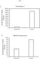
- MARCKS-related protein Figure 3B
- Moesin Figure 3C
- Actin Figure 3D
- Peptide NPLPSKETIEQEK (SEQ ID NO: 14) is found in both TMSB4XP4 protein and homologous protein Thymosin beta-4. The protein sequences of these two proteins are identical apart from the first 19 residues of TMSB4XP4 which are absent in Thymosin beta-4.
- Phosphoglucomutase 1 was not detected when the CSF samples were measured in the absence of the BV2 cell sample calibrator, whilst Thymosin beta-4 was detected in both settings.
- 5 were regulated ( ⁇ 60%; in bold in Table 7) but not significantly and only one was found to be significantly regulated with a P value ⁇ 0.05 (QFSANDK; SEQ ID NO:6).
- experiment B was designed as a 4-point calibration curve to be spiked into the total combined CSF samples at a ratio of 1:4:6:10 in order to ensure the calibrator proteins dominated the overall protein content of the TMT ® 10-plex sample. These are arbitrary values allowing to determine a suitable protein load from the CSF samples for analysis. Six individual CSF samples were combined at a 2:2:2:2:2 ratio, falling within the 4-point calibration curve.
- the true protein load per channel can be determined by dividing the maximum 2 ⁇ g by the total number of arbitrary values (x) needed, which in this case is 33 (4 calibrator channels plus 6 CSF channels). If 33x equals 2 ⁇ g, 1x is therefore 0.06 ⁇ g and this can then be used to determine the total amount of protein from each of the 10 channels contributing to the calibrator analytical sample. Just 0.12 ⁇ g of each CSF sample is required (2x), along with a 4-point calibrator comprised of 0.06 ⁇ g (1x), 0.24 ⁇ g (4x), 0.36 ⁇ g (6x) and 0.6 ⁇ g (10x). This results in a total of 0.72 ⁇ g of protein from all the CSF samples and 1.26 ⁇ g of protein from the BV2 cell line calibrator - the calibrator proteins are therefore 1.75 fold more prevalent than the total CSF protein load.
- the calibrator sample was analysed in triplicate on the Oribtrap Fusion Tribrid using an MS3 SPS method over a 300 minute gradient.
- the raw data files from all three MS injections were searched using SequestHT and Mascot in Proteome Discoverer 1.4, against the Uniprot human protein database. Results for the triplicate injections were merged resulting in one large data set for analysis.
- FDR 5% false discovery rate
- the reporter ion signal intensities from the six CSF channels were normalized by sum-scale normalization. Following normalization the variability across the CSF dataset was investigated using Principal Component Analysis (PCA). Group separation of AD and control CSF was seen at the peptide and protein level, forming the first principal component, and accounting for 44.6% of the total variation seen in the dataset. The second principal component explained 19.6% of the variability in the dataset, and this corresponded to the biological variation within the groups.
- PCA Principal Component Analysis
- the normalized dataset, filtered for the presence of an expected linear calibrator signal intensity was further filtered based on the significance following a 2-sample t-test across the two CSF groups for every single peptide and protein. Peptides with a p value ⁇ 0.05 were considered significant.
- the log2 ratio of the average AD CSF signal intensities (channels 128e, 128 and 129e) to control CSF signal intensities (channels 126, 127e and 127) was also normalized to the 129 TMT ® calibrator channel. This provided a means of identifying which of the peptide sequences common to the activated microglia cell line and CSF samples were differentially regulated between control and AD CSF.
- the benefit of TMT ® calibrator over the traditional proteomics approach can be seen.
- the maximum protein load on the 75 ⁇ m diameter analytical LC column per injection is 2ug.
- the 2 ⁇ g is split unequally between the 4-point calibrator and the six CSF samples (1:4:6:10 ratio for the calibrator, 2:2:2 for AD CSF and 2:2:2 for non-AD CSF).
- the six CSF samples could be labelled with TMT in a multiplex experiment, with 0.33 ⁇ g combined from each sample per inject.
- TMT ® calibrator proteins that are commonly observed in CSF studies including serum albumin, complement proteins (C3, C5 and C7), fibrinogen gamma chain and Ig gamma chain ( Boche D, et al., Neuropathol Appl Neurobiol. 2013, 39, 1, 3-18 ). These proteins are likely representative of disease related changes of low specificity rather than the cellular changes specific to AD. They are some of the most abundant proteins found in the CSF and are reported in the majority of CSF proteomic analysis studies ( Hühmer AF, et al., Disease Markers. 2006, 22, 1-2, 3-26 ).
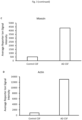
- peptides derived when the BV2 cell line is used to drive the experiment are cellular and may represent a cellular response specific to disease. These include peptides from dynactin ( Figure 5A ), cofilin ( Figure 5A ), alcohol dehydrogenase, filamin-A, myosin proteins and Ras-related proteins. These are cellular proteins regulated in AD that would not have been detected if the CSF samples had been analysed in the absence of the BV2 cell samples (calibrator samples).
- Peptide IKDALVR (SEQ ID NO: 37), corresponding to dynactin subunit 1( Figure 5A ), was the most upregulated peptide whist peptide MIYASSK (SEQ ID NO: 38), corresponding to Cofilin-1 ( Figure 5B ), was the most down regulated peptide in the CSF of AD patients when compared to non-AD controls.
- biomarkers are indicators of pathological processes and biological events. Their levels change as a result of disease and also in response to pharmacological intervention.
- the challenge is how to find these molecules in biofluids such as blood or CSF which contain a high protein dynamic range and hyper-abundant proteins such as albumin.
- TMT ® Tandem Mass Tag ®
- MS mass spectrometry
Landscapes
- Health & Medical Sciences (AREA)
- Life Sciences & Earth Sciences (AREA)
- Chemical & Material Sciences (AREA)
- Engineering & Computer Science (AREA)
- Organic Chemistry (AREA)
- General Health & Medical Sciences (AREA)
- Biomedical Technology (AREA)
- Molecular Biology (AREA)
- Medicinal Chemistry (AREA)
- Biochemistry (AREA)
- Genetics & Genomics (AREA)
- Zoology (AREA)
- Urology & Nephrology (AREA)
- Hematology (AREA)
- Immunology (AREA)
- Bioinformatics & Cheminformatics (AREA)
- Wood Science & Technology (AREA)
- Microbiology (AREA)
- Biotechnology (AREA)
- Proteomics, Peptides & Aminoacids (AREA)
- General Engineering & Computer Science (AREA)
- Analytical Chemistry (AREA)
- Pathology (AREA)
- General Physics & Mathematics (AREA)
- Cell Biology (AREA)
- Food Science & Technology (AREA)
- Physics & Mathematics (AREA)
- Public Health (AREA)
- Veterinary Medicine (AREA)
- Animal Behavior & Ethology (AREA)
- Epidemiology (AREA)
- Pharmacology & Pharmacy (AREA)
- Endocrinology (AREA)
- Toxicology (AREA)
- Gastroenterology & Hepatology (AREA)
- Biophysics (AREA)
- Neurology (AREA)
- Neurosurgery (AREA)
- Investigating Or Analysing Biological Materials (AREA)
- Peptides Or Proteins (AREA)
Applications Claiming Priority (2)
| Application Number | Priority Date | Filing Date | Title |
|---|---|---|---|
| GB201504432A GB201504432D0 (en) | 2015-03-17 | 2015-03-17 | Materials and methods for diagnosis and treatment of alzheimers disease |
| PCT/EP2016/055883 WO2016146783A1 (en) | 2015-03-17 | 2016-03-17 | Materials and methods for diagnosis and treatment of alzheimer's disease |
Publications (2)
| Publication Number | Publication Date |
|---|---|
| EP3271728A1 EP3271728A1 (en) | 2018-01-24 |
| EP3271728B1 true EP3271728B1 (en) | 2024-02-28 |
Family
ID=53016214
Family Applications (1)
| Application Number | Title | Priority Date | Filing Date |
|---|---|---|---|
| EP16714282.7A Active EP3271728B1 (en) | 2015-03-17 | 2016-03-17 | Materials and methods for diagnosis and treatment of alzheimer's disease |
Country Status (6)
| Country | Link |
|---|---|
| US (1) | US10718785B2 (enExample) |
| EP (1) | EP3271728B1 (enExample) |
| JP (2) | JP6954839B2 (enExample) |
| CA (1) | CA2979773A1 (enExample) |
| GB (1) | GB201504432D0 (enExample) |
| WO (1) | WO2016146783A1 (enExample) |
Families Citing this family (22)
| Publication number | Priority date | Publication date | Assignee | Title |
|---|---|---|---|---|
| GB201509134D0 (en) * | 2015-05-28 | 2015-07-15 | Electrophoretics Ltd | Biomolecules involved in Alzheimer's disease |
| CN111201030B (zh) | 2017-07-25 | 2024-11-01 | 真和制药有限公司 | 通过阻断tim-3和其配体的相互作用治疗癌症 |
| KR102034929B1 (ko) * | 2017-09-27 | 2019-10-22 | 한양대학교 산학협력단 | Nckap1 단백질 또는 상기 단백질을 암호화하는 유전자를 포함하는 신경계 퇴행성질환의 예방 또는 치료용 약학적 조성물 |
| EP3764102A4 (en) * | 2018-03-09 | 2021-12-15 | National University Corporation Tokyo Medical and Dental University | DETECTION OF ALZHEIMER'S DISEASE (MA), FRONTOTEMPORAL LOBAR DEGENERATION (FRONTOTEMPORAL LOBAR DEGENERATION), AMYOTROPHIC LATERAL SCLEROSIS (ALS), PARKINSON'S DISEASE (PD) AND INDICATED LEWY-BODY DEMENTIA (PARL) MARCKS PHOSPHORYLATION |
| KR102063610B1 (ko) * | 2018-05-23 | 2020-01-08 | 충북대학교 산학협력단 | 퇴행성 신경질환 진단용 신규한 바이오마커 및 이의 용도 |
| KR101992060B1 (ko) * | 2018-10-30 | 2019-06-21 | 아주대학교산학협력단 | 알츠하이머치매 진단 체액 바이오마커 후보 단백4종 |
| KR102232200B1 (ko) * | 2019-03-13 | 2021-03-25 | 아주대학교산학협력단 | 알츠하이머치매 진단 바이오마커 |
| US20220003787A1 (en) * | 2018-10-30 | 2022-01-06 | Ajou University Industry-Academic Cooperation Foundation | Biomarker proteins for diagnosing alzheimer's dementia and use thereof |
| CN109738653B (zh) * | 2019-01-11 | 2022-04-12 | 湖南诺琪生物科技有限公司 | 用于阿尔茨海默症的检测、诊断或风险预测的抗原蛋白组合以及包含其的试剂盒 |
| WO2020160156A2 (en) | 2019-01-30 | 2020-08-06 | Immutics, Inc. | Anti-gal3 antibodies and uses thereof |
| CN110261272B (zh) * | 2019-07-05 | 2020-08-18 | 西南交通大学 | 基于地理探测和pca对pm2.5浓度分布的关键影响因子筛选方法 |
| CN112336859A (zh) * | 2019-08-06 | 2021-02-09 | 上海绿谷制药有限公司 | 通过抑制t细胞摄取氨基酸治疗阿尔茨海默病的方法 |
| EP4051812A1 (en) * | 2019-10-28 | 2022-09-07 | AgenT | Biomarkers and uses thereof for diagnosing the silent phase of alzheimer's disease |
| KR102313455B1 (ko) * | 2020-02-27 | 2021-10-15 | 이화여자대학교 산학협력단 | 알츠하이머병 경도인지장애의 진단 또는 알츠하이머병 치매로의 진행 위험성 예측 방법 |
| EP4157338A4 (en) | 2020-05-26 | 2024-11-13 | TrueBinding, Inc. | METHOD FOR TREATING INFLAMMATORY DISEASES BY GALECTIN-3 BLOCKING |
| CN112695084A (zh) * | 2021-02-08 | 2021-04-23 | 山东大学第二医院 | 诊断阿尔茨海默病和阿尔茨海默病进展的生物标志物及应用 |
| GB202102399D0 (en) * | 2021-02-19 | 2021-04-07 | Univ Manchester | Methods of determining alzheimer's disease |
| WO2022192019A1 (en) * | 2021-03-08 | 2022-09-15 | The Children's Medical Center Corporation | Methods for diagnosis and treatment of alzheimer's disease |
| CA3226615A1 (en) * | 2021-07-23 | 2023-03-30 | Ian Pike | Blood-based diagnostic assays for alzheimer's disease |
| WO2023235871A2 (en) * | 2022-06-03 | 2023-12-07 | Seer, Inc. | Systems, compositions, and methods relating to neurodegenerative diseases |
| WO2024073067A2 (en) * | 2022-09-30 | 2024-04-04 | Mayo Foundation For Medical Education And Research | Assessing and treating tremor |
| CN121606708B (zh) * | 2026-02-03 | 2026-04-28 | 四川大学华西医院 | 一种用于治疗阿尔茨海默症的病理血脑屏障靶向纳米制剂及其制备方法 |
Family Cites Families (15)
| Publication number | Priority date | Publication date | Assignee | Title |
|---|---|---|---|---|
| US4376110A (en) | 1980-08-04 | 1983-03-08 | Hybritech, Incorporated | Immunometric assays using monoclonal antibodies |
| US4946778A (en) | 1987-09-21 | 1990-08-07 | Genex Corporation | Single polypeptide chain binding molecules |
| WO2004001865A1 (ja) | 2002-06-19 | 2003-12-31 | Kabushiki Kaisha Toshiba | 熱電素子とそれを用いた電子部品モジュールおよび携帯用電子機器 |
| WO2005020784A2 (en) | 2003-05-23 | 2005-03-10 | Mount Sinai School Of Medicine Of New York University | Surrogate cell gene expression signatures for evaluating the physical state of a subject |
| EP1658502A2 (en) * | 2003-08-20 | 2006-05-24 | Genova Ltd. | Secreted polypeptide species and use thereof |
| US7794948B2 (en) * | 2003-11-07 | 2010-09-14 | Vermilllion, Inc. | Biomarkers for alzheimer's disease |
| WO2006110621A2 (en) * | 2005-04-11 | 2006-10-19 | Cornell Research Foundation, Inc. | Multiplexed biomarkers for monitoring the alzheimer's disease state of a subject |
| US20120071337A1 (en) * | 2009-01-26 | 2012-03-22 | Electrophoretics Limited | Methods |
| US8865411B2 (en) | 2009-03-26 | 2014-10-21 | National Institutes Of Health (Nih) | Methods of identifying modulators of TDP-43 mediated cellular toxicity |
| WO2011005893A2 (en) * | 2009-07-07 | 2011-01-13 | Abbott Laboratories | Biomarkers and methods for detecting alzheimer's disease |
| EP2553466A4 (en) | 2010-04-01 | 2013-10-16 | Banyan Biomarkers Inc | MARKERS AND ASSAYS FOR DETECTING NEUROTOXICITY |
| GB2511221B (en) | 2011-12-09 | 2020-09-23 | Veracyte Inc | Methods and compositions for classification of samples |
| GB2511525A (en) * | 2013-03-05 | 2014-09-10 | Randox Teoranta | Methods and Compositions for the Diagnosis of Alzheimer's Disease |
| GB201310203D0 (en) * | 2013-06-07 | 2013-07-24 | Electrophoretics Ltd | Materials and methods relating to Alzheimer's disease |
| GB201322094D0 (en) * | 2013-12-13 | 2014-01-29 | Electrophoretics Ltd | Methods and compositions relating to alzheimers disease |
-
2015
- 2015-03-17 GB GB201504432A patent/GB201504432D0/en not_active Ceased
-
2016
- 2016-03-17 US US15/559,371 patent/US10718785B2/en active Active
- 2016-03-17 JP JP2017549301A patent/JP6954839B2/ja active Active
- 2016-03-17 EP EP16714282.7A patent/EP3271728B1/en active Active
- 2016-03-17 WO PCT/EP2016/055883 patent/WO2016146783A1/en not_active Ceased
- 2016-03-17 CA CA2979773A patent/CA2979773A1/en active Pending
-
2021
- 2021-06-22 JP JP2021102858A patent/JP2021177180A/ja active Pending
Also Published As
| Publication number | Publication date |
|---|---|
| GB201504432D0 (en) | 2015-04-29 |
| US10718785B2 (en) | 2020-07-21 |
| EP3271728A1 (en) | 2018-01-24 |
| JP2021177180A (ja) | 2021-11-11 |
| US20180067133A1 (en) | 2018-03-08 |
| JP6954839B2 (ja) | 2021-10-27 |
| WO2016146783A1 (en) | 2016-09-22 |
| CA2979773A1 (en) | 2016-09-22 |
| JP2018510343A (ja) | 2018-04-12 |
Similar Documents
| Publication | Publication Date | Title |
|---|---|---|
| EP3271728B1 (en) | Materials and methods for diagnosis and treatment of alzheimer's disease | |
| Merkel et al. | Proteomic study of human bronchoalveolar lavage fluids from smokers with chronic obstructive pulmonary disease by combining surface‐enhanced laser desorption/ionization‐mass spectrometry profiling with mass spectrometric protein identification | |
| Hu et al. | Human body fluid proteome analysis | |
| US20260091025A1 (en) | Biomolecules involved in alzheimer's disease | |
| Hu et al. | Large‐scale identification of proteins in human salivary proteome by liquid chromatography/mass spectrometry and two‐dimensional gel electrophoresis‐mass spectrometry | |
| Qian et al. | Comparative proteome analyses of human plasma following in vivo lipopolysaccharide administration using multidimensional separations coupled with tandem mass spectrometry | |
| EP2569446B1 (en) | Diagnostic markers for neuropsychiatric diseasen | |
| US20220397576A1 (en) | Apparatuses and methods for detection of pancreatic cancer | |
| CN110914689A (zh) | 肝癌高危人群的肝癌发病监视或诊断用生物标记物及其用途 | |
| WO2019004430A1 (ja) | 大腸がんを検出するためのバイオマーカー | |
| Zen et al. | A global proteomic study identifies distinct pathological features of IgG4‐related and primary sclerosing cholangitis | |
| US20230152333A1 (en) | Method of differentiating of a chronic kidney disease or glomerulopathy, method of monitoring a response to treatment of a chronic kidney disease or glomerulop athy in a subject and a method of treatment of a chronic kidney disease or glomerulopathy | |
| US12449426B2 (en) | Method of screening for a chronic kidney disease or glomerulopathy method of monitoring a response to treatment of a chronic kidney disease or glomerulopathy in a subject and a method of treatment of a chronic kidney disease or glomerulopathy | |
| WO2022203371A1 (ko) | 우울증 진단용 바이오마커 조성물 및 이의 용도 | |
| Wang et al. | HUPO BPP pilot study: a proteomics analysis of the mouse brain of different developmental stages | |
| EP1969376B1 (en) | An in-vitro method for screening accessible biological markers in pathologic tissues | |
| Zuberovic et al. | Proteome profiling of human cerebrospinal fluid: exploring the potential of capillary electrophoresis with surface modified capillaries for analysis of complex biological samples | |
| KR101117799B1 (ko) | 아토피성 피부질환 진단용 마커 | |
| EP3961218B1 (en) | Composition and kit for detecting testican-1 for identifying exposure to particulate matter in lung and method using the same | |
| US20240241139A1 (en) | Diagnosis of autism spectrum disorder by multiomics platform | |
| Cortes-Figueiredo et al. | From bedside to bench: methods in precision medicine | |
| KR20220121463A (ko) | 건선 환자에서 심혈관 동반질환을 선별하기 위한 바이오마커 또는 이의 용도 | |
| Rohan | Proteomic Prediction of Breast Cancer Risk: A Cohort Study | |
| Fogli et al. | Peptidomics and proteomics studies of transformed lymphocytes from patients mutated for the eukaryotic initiation factor 2B | |
| D'Siva et al. | Peptidomics for bioactive peptide analysis |
Legal Events
| Date | Code | Title | Description |
|---|---|---|---|
| STAA | Information on the status of an ep patent application or granted ep patent |
Free format text: STATUS: THE INTERNATIONAL PUBLICATION HAS BEEN MADE |
|
| PUAI | Public reference made under article 153(3) epc to a published international application that has entered the european phase |
Free format text: ORIGINAL CODE: 0009012 |
|
| STAA | Information on the status of an ep patent application or granted ep patent |
Free format text: STATUS: REQUEST FOR EXAMINATION WAS MADE |
|
| 17P | Request for examination filed |
Effective date: 20171012 |
|
| AK | Designated contracting states |
Kind code of ref document: A1 Designated state(s): AL AT BE BG CH CY CZ DE DK EE ES FI FR GB GR HR HU IE IS IT LI LT LU LV MC MK MT NL NO PL PT RO RS SE SI SK SM TR |
|
| AX | Request for extension of the european patent |
Extension state: BA ME |
|
| DAV | Request for validation of the european patent (deleted) | ||
| DAX | Request for extension of the european patent (deleted) | ||
| STAA | Information on the status of an ep patent application or granted ep patent |
Free format text: STATUS: EXAMINATION IS IN PROGRESS |
|
| 17Q | First examination report despatched |
Effective date: 20190404 |
|
| P01 | Opt-out of the competence of the unified patent court (upc) registered |
Effective date: 20230512 |
|
| GRAP | Despatch of communication of intention to grant a patent |
Free format text: ORIGINAL CODE: EPIDOSNIGR1 |
|
| STAA | Information on the status of an ep patent application or granted ep patent |
Free format text: STATUS: GRANT OF PATENT IS INTENDED |
|
| INTG | Intention to grant announced |
Effective date: 20230926 |
|
| GRAS | Grant fee paid |
Free format text: ORIGINAL CODE: EPIDOSNIGR3 |
|
| GRAA | (expected) grant |
Free format text: ORIGINAL CODE: 0009210 |
|
| STAA | Information on the status of an ep patent application or granted ep patent |
Free format text: STATUS: THE PATENT HAS BEEN GRANTED |
|
| AK | Designated contracting states |
Kind code of ref document: B1 Designated state(s): AL AT BE BG CH CY CZ DE DK EE ES FI FR GB GR HR HU IE IS IT LI LT LU LV MC MK MT NL NO PL PT RO RS SE SI SK SM TR |
|
| RAP3 | Party data changed (applicant data changed or rights of an application transferred) |
Owner name: ELECTROPHORETICS LIMITED |
|
| REG | Reference to a national code |
Ref country code: GB Ref legal event code: FG4D |
|
| RIN1 | Information on inventor provided before grant (corrected) |
Inventor name: WARD, MALCOLM Inventor name: RUSSELL, CLAIRE LOUISE Inventor name: PIKE, IAN HUGO |
|
| REG | Reference to a national code |
Ref country code: CH Ref legal event code: EP |
|
| REG | Reference to a national code |
Ref country code: DE Ref legal event code: R096 Ref document number: 602016085997 Country of ref document: DE |
|
| REG | Reference to a national code |
Ref country code: IE Ref legal event code: FG4D |
|
| REG | Reference to a national code |
Ref country code: LT Ref legal event code: MG9D |
|
| PG25 | Lapsed in a contracting state [announced via postgrant information from national office to epo] |
Ref country code: IS Free format text: LAPSE BECAUSE OF FAILURE TO SUBMIT A TRANSLATION OF THE DESCRIPTION OR TO PAY THE FEE WITHIN THE PRESCRIBED TIME-LIMIT Effective date: 20240628 |
|
| REG | Reference to a national code |
Ref country code: NL Ref legal event code: MP Effective date: 20240228 |
|
| PG25 | Lapsed in a contracting state [announced via postgrant information from national office to epo] |
Ref country code: LT Free format text: LAPSE BECAUSE OF FAILURE TO SUBMIT A TRANSLATION OF THE DESCRIPTION OR TO PAY THE FEE WITHIN THE PRESCRIBED TIME-LIMIT Effective date: 20240228 |
|
| PG25 | Lapsed in a contracting state [announced via postgrant information from national office to epo] |
Ref country code: GR Free format text: LAPSE BECAUSE OF FAILURE TO SUBMIT A TRANSLATION OF THE DESCRIPTION OR TO PAY THE FEE WITHIN THE PRESCRIBED TIME-LIMIT Effective date: 20240529 |
|
| PG25 | Lapsed in a contracting state [announced via postgrant information from national office to epo] |
Ref country code: RS Free format text: LAPSE BECAUSE OF FAILURE TO SUBMIT A TRANSLATION OF THE DESCRIPTION OR TO PAY THE FEE WITHIN THE PRESCRIBED TIME-LIMIT Effective date: 20240528 Ref country code: HR Free format text: LAPSE BECAUSE OF FAILURE TO SUBMIT A TRANSLATION OF THE DESCRIPTION OR TO PAY THE FEE WITHIN THE PRESCRIBED TIME-LIMIT Effective date: 20240228 Ref country code: NL Free format text: LAPSE BECAUSE OF FAILURE TO SUBMIT A TRANSLATION OF THE DESCRIPTION OR TO PAY THE FEE WITHIN THE PRESCRIBED TIME-LIMIT Effective date: 20240228 |
|
| PG25 | Lapsed in a contracting state [announced via postgrant information from national office to epo] |
Ref country code: ES Free format text: LAPSE BECAUSE OF FAILURE TO SUBMIT A TRANSLATION OF THE DESCRIPTION OR TO PAY THE FEE WITHIN THE PRESCRIBED TIME-LIMIT Effective date: 20240228 |
|
| PG25 | Lapsed in a contracting state [announced via postgrant information from national office to epo] |
Ref country code: RS Free format text: LAPSE BECAUSE OF FAILURE TO SUBMIT A TRANSLATION OF THE DESCRIPTION OR TO PAY THE FEE WITHIN THE PRESCRIBED TIME-LIMIT Effective date: 20240528 Ref country code: NO Free format text: LAPSE BECAUSE OF FAILURE TO SUBMIT A TRANSLATION OF THE DESCRIPTION OR TO PAY THE FEE WITHIN THE PRESCRIBED TIME-LIMIT Effective date: 20240528 Ref country code: NL Free format text: LAPSE BECAUSE OF FAILURE TO SUBMIT A TRANSLATION OF THE DESCRIPTION OR TO PAY THE FEE WITHIN THE PRESCRIBED TIME-LIMIT Effective date: 20240228 Ref country code: LT Free format text: LAPSE BECAUSE OF FAILURE TO SUBMIT A TRANSLATION OF THE DESCRIPTION OR TO PAY THE FEE WITHIN THE PRESCRIBED TIME-LIMIT Effective date: 20240228 Ref country code: IS Free format text: LAPSE BECAUSE OF FAILURE TO SUBMIT A TRANSLATION OF THE DESCRIPTION OR TO PAY THE FEE WITHIN THE PRESCRIBED TIME-LIMIT Effective date: 20240628 Ref country code: HR Free format text: LAPSE BECAUSE OF FAILURE TO SUBMIT A TRANSLATION OF THE DESCRIPTION OR TO PAY THE FEE WITHIN THE PRESCRIBED TIME-LIMIT Effective date: 20240228 Ref country code: GR Free format text: LAPSE BECAUSE OF FAILURE TO SUBMIT A TRANSLATION OF THE DESCRIPTION OR TO PAY THE FEE WITHIN THE PRESCRIBED TIME-LIMIT Effective date: 20240529 Ref country code: FI Free format text: LAPSE BECAUSE OF FAILURE TO SUBMIT A TRANSLATION OF THE DESCRIPTION OR TO PAY THE FEE WITHIN THE PRESCRIBED TIME-LIMIT Effective date: 20240228 Ref country code: ES Free format text: LAPSE BECAUSE OF FAILURE TO SUBMIT A TRANSLATION OF THE DESCRIPTION OR TO PAY THE FEE WITHIN THE PRESCRIBED TIME-LIMIT Effective date: 20240228 Ref country code: BG Free format text: LAPSE BECAUSE OF FAILURE TO SUBMIT A TRANSLATION OF THE DESCRIPTION OR TO PAY THE FEE WITHIN THE PRESCRIBED TIME-LIMIT Effective date: 20240228 |
|
| PG25 | Lapsed in a contracting state [announced via postgrant information from national office to epo] |
Ref country code: PT Free format text: LAPSE BECAUSE OF FAILURE TO SUBMIT A TRANSLATION OF THE DESCRIPTION OR TO PAY THE FEE WITHIN THE PRESCRIBED TIME-LIMIT Effective date: 20240628 Ref country code: PL Free format text: LAPSE BECAUSE OF FAILURE TO SUBMIT A TRANSLATION OF THE DESCRIPTION OR TO PAY THE FEE WITHIN THE PRESCRIBED TIME-LIMIT Effective date: 20240228 |
|
| REG | Reference to a national code |
Ref country code: AT Ref legal event code: MK05 Ref document number: 1661701 Country of ref document: AT Kind code of ref document: T Effective date: 20240228 |
|
| PG25 | Lapsed in a contracting state [announced via postgrant information from national office to epo] |
Ref country code: SE Free format text: LAPSE BECAUSE OF FAILURE TO SUBMIT A TRANSLATION OF THE DESCRIPTION OR TO PAY THE FEE WITHIN THE PRESCRIBED TIME-LIMIT Effective date: 20240228 Ref country code: PT Free format text: LAPSE BECAUSE OF FAILURE TO SUBMIT A TRANSLATION OF THE DESCRIPTION OR TO PAY THE FEE WITHIN THE PRESCRIBED TIME-LIMIT Effective date: 20240628 Ref country code: PL Free format text: LAPSE BECAUSE OF FAILURE TO SUBMIT A TRANSLATION OF THE DESCRIPTION OR TO PAY THE FEE WITHIN THE PRESCRIBED TIME-LIMIT Effective date: 20240228 Ref country code: LV Free format text: LAPSE BECAUSE OF FAILURE TO SUBMIT A TRANSLATION OF THE DESCRIPTION OR TO PAY THE FEE WITHIN THE PRESCRIBED TIME-LIMIT Effective date: 20240228 |
|
| PG25 | Lapsed in a contracting state [announced via postgrant information from national office to epo] |
Ref country code: DK Free format text: LAPSE BECAUSE OF FAILURE TO SUBMIT A TRANSLATION OF THE DESCRIPTION OR TO PAY THE FEE WITHIN THE PRESCRIBED TIME-LIMIT Effective date: 20240228 |
|
| PG25 | Lapsed in a contracting state [announced via postgrant information from national office to epo] |
Ref country code: SM Free format text: LAPSE BECAUSE OF FAILURE TO SUBMIT A TRANSLATION OF THE DESCRIPTION OR TO PAY THE FEE WITHIN THE PRESCRIBED TIME-LIMIT Effective date: 20240228 |
|
| PG25 | Lapsed in a contracting state [announced via postgrant information from national office to epo] |
Ref country code: EE Free format text: LAPSE BECAUSE OF FAILURE TO SUBMIT A TRANSLATION OF THE DESCRIPTION OR TO PAY THE FEE WITHIN THE PRESCRIBED TIME-LIMIT Effective date: 20240228 Ref country code: CZ Free format text: LAPSE BECAUSE OF FAILURE TO SUBMIT A TRANSLATION OF THE DESCRIPTION OR TO PAY THE FEE WITHIN THE PRESCRIBED TIME-LIMIT Effective date: 20240228 |
|
| PG25 | Lapsed in a contracting state [announced via postgrant information from national office to epo] |
Ref country code: AT Free format text: LAPSE BECAUSE OF FAILURE TO SUBMIT A TRANSLATION OF THE DESCRIPTION OR TO PAY THE FEE WITHIN THE PRESCRIBED TIME-LIMIT Effective date: 20240228 |
|
| PG25 | Lapsed in a contracting state [announced via postgrant information from national office to epo] |
Ref country code: SK Free format text: LAPSE BECAUSE OF FAILURE TO SUBMIT A TRANSLATION OF THE DESCRIPTION OR TO PAY THE FEE WITHIN THE PRESCRIBED TIME-LIMIT Effective date: 20240228 |
|
| PG25 | Lapsed in a contracting state [announced via postgrant information from national office to epo] |
Ref country code: SM Free format text: LAPSE BECAUSE OF FAILURE TO SUBMIT A TRANSLATION OF THE DESCRIPTION OR TO PAY THE FEE WITHIN THE PRESCRIBED TIME-LIMIT Effective date: 20240228 Ref country code: SK Free format text: LAPSE BECAUSE OF FAILURE TO SUBMIT A TRANSLATION OF THE DESCRIPTION OR TO PAY THE FEE WITHIN THE PRESCRIBED TIME-LIMIT Effective date: 20240228 Ref country code: RO Free format text: LAPSE BECAUSE OF FAILURE TO SUBMIT A TRANSLATION OF THE DESCRIPTION OR TO PAY THE FEE WITHIN THE PRESCRIBED TIME-LIMIT Effective date: 20240228 Ref country code: EE Free format text: LAPSE BECAUSE OF FAILURE TO SUBMIT A TRANSLATION OF THE DESCRIPTION OR TO PAY THE FEE WITHIN THE PRESCRIBED TIME-LIMIT Effective date: 20240228 Ref country code: DK Free format text: LAPSE BECAUSE OF FAILURE TO SUBMIT A TRANSLATION OF THE DESCRIPTION OR TO PAY THE FEE WITHIN THE PRESCRIBED TIME-LIMIT Effective date: 20240228 Ref country code: CZ Free format text: LAPSE BECAUSE OF FAILURE TO SUBMIT A TRANSLATION OF THE DESCRIPTION OR TO PAY THE FEE WITHIN THE PRESCRIBED TIME-LIMIT Effective date: 20240228 Ref country code: AT Free format text: LAPSE BECAUSE OF FAILURE TO SUBMIT A TRANSLATION OF THE DESCRIPTION OR TO PAY THE FEE WITHIN THE PRESCRIBED TIME-LIMIT Effective date: 20240228 |
|
| REG | Reference to a national code |
Ref country code: CH Ref legal event code: PL |
|
| PG25 | Lapsed in a contracting state [announced via postgrant information from national office to epo] |
Ref country code: LU Free format text: LAPSE BECAUSE OF NON-PAYMENT OF DUE FEES Effective date: 20240317 |
|
| PG25 | Lapsed in a contracting state [announced via postgrant information from national office to epo] |
Ref country code: MC Free format text: LAPSE BECAUSE OF FAILURE TO SUBMIT A TRANSLATION OF THE DESCRIPTION OR TO PAY THE FEE WITHIN THE PRESCRIBED TIME-LIMIT Effective date: 20240228 |
|
| PG25 | Lapsed in a contracting state [announced via postgrant information from national office to epo] |
Ref country code: MC Free format text: LAPSE BECAUSE OF FAILURE TO SUBMIT A TRANSLATION OF THE DESCRIPTION OR TO PAY THE FEE WITHIN THE PRESCRIBED TIME-LIMIT Effective date: 20240228 Ref country code: LU Free format text: LAPSE BECAUSE OF NON-PAYMENT OF DUE FEES Effective date: 20240317 |
|
| REG | Reference to a national code |
Ref country code: DE Ref legal event code: R097 Ref document number: 602016085997 Country of ref document: DE |
|
| PG25 | Lapsed in a contracting state [announced via postgrant information from national office to epo] |
Ref country code: IT Free format text: LAPSE BECAUSE OF FAILURE TO SUBMIT A TRANSLATION OF THE DESCRIPTION OR TO PAY THE FEE WITHIN THE PRESCRIBED TIME-LIMIT Effective date: 20240228 |
|
| REG | Reference to a national code |
Ref country code: BE Ref legal event code: MM Effective date: 20240331 |
|
| PG25 | Lapsed in a contracting state [announced via postgrant information from national office to epo] |
Ref country code: IT Free format text: LAPSE BECAUSE OF FAILURE TO SUBMIT A TRANSLATION OF THE DESCRIPTION OR TO PAY THE FEE WITHIN THE PRESCRIBED TIME-LIMIT Effective date: 20240228 |
|
| PLBE | No opposition filed within time limit |
Free format text: ORIGINAL CODE: 0009261 |
|
| STAA | Information on the status of an ep patent application or granted ep patent |
Free format text: STATUS: NO OPPOSITION FILED WITHIN TIME LIMIT |
|
| PG25 | Lapsed in a contracting state [announced via postgrant information from national office to epo] |
Ref country code: BE Free format text: LAPSE BECAUSE OF NON-PAYMENT OF DUE FEES Effective date: 20240331 |
|
| PG25 | Lapsed in a contracting state [announced via postgrant information from national office to epo] |
Ref country code: IE Free format text: LAPSE BECAUSE OF NON-PAYMENT OF DUE FEES Effective date: 20240317 |
|
| PG25 | Lapsed in a contracting state [announced via postgrant information from national office to epo] |
Ref country code: IE Free format text: LAPSE BECAUSE OF NON-PAYMENT OF DUE FEES Effective date: 20240317 Ref country code: BE Free format text: LAPSE BECAUSE OF NON-PAYMENT OF DUE FEES Effective date: 20240331 Ref country code: CH Free format text: LAPSE BECAUSE OF NON-PAYMENT OF DUE FEES Effective date: 20240331 |
|
| 26N | No opposition filed |
Effective date: 20241129 |
|
| PG25 | Lapsed in a contracting state [announced via postgrant information from national office to epo] |
Ref country code: SI Free format text: LAPSE BECAUSE OF FAILURE TO SUBMIT A TRANSLATION OF THE DESCRIPTION OR TO PAY THE FEE WITHIN THE PRESCRIBED TIME-LIMIT Effective date: 20240228 |
|
| PG25 | Lapsed in a contracting state [announced via postgrant information from national office to epo] |
Ref country code: CY Free format text: LAPSE BECAUSE OF FAILURE TO SUBMIT A TRANSLATION OF THE DESCRIPTION OR TO PAY THE FEE WITHIN THE PRESCRIBED TIME-LIMIT; INVALID AB INITIO Effective date: 20160317 |
|
| PG25 | Lapsed in a contracting state [announced via postgrant information from national office to epo] |
Ref country code: HU Free format text: LAPSE BECAUSE OF FAILURE TO SUBMIT A TRANSLATION OF THE DESCRIPTION OR TO PAY THE FEE WITHIN THE PRESCRIBED TIME-LIMIT; INVALID AB INITIO Effective date: 20160317 |
|
| PG25 | Lapsed in a contracting state [announced via postgrant information from national office to epo] |
Ref country code: TR Free format text: LAPSE BECAUSE OF FAILURE TO SUBMIT A TRANSLATION OF THE DESCRIPTION OR TO PAY THE FEE WITHIN THE PRESCRIBED TIME-LIMIT Effective date: 20240228 |
|
| PGFP | Annual fee paid to national office [announced via postgrant information from national office to epo] |
Ref country code: GB Payment date: 20260327 Year of fee payment: 11 |
|
| PGFP | Annual fee paid to national office [announced via postgrant information from national office to epo] |
Ref country code: DE Payment date: 20260327 Year of fee payment: 11 |
|
| PGFP | Annual fee paid to national office [announced via postgrant information from national office to epo] |
Ref country code: FR Payment date: 20260325 Year of fee payment: 11 |