EP3271728B1 - Materials and methods for diagnosis and treatment of alzheimer's disease - Google Patents
Materials and methods for diagnosis and treatment of alzheimer's disease Download PDFInfo
- Publication number
- EP3271728B1 EP3271728B1 EP16714282.7A EP16714282A EP3271728B1 EP 3271728 B1 EP3271728 B1 EP 3271728B1 EP 16714282 A EP16714282 A EP 16714282A EP 3271728 B1 EP3271728 B1 EP 3271728B1
- Authority
- EP
- European Patent Office
- Prior art keywords
- protein
- seq
- subunit
- alpha
- replaced
- Prior art date
- Legal status (The legal status is an assumption and is not a legal conclusion. Google has not performed a legal analysis and makes no representation as to the accuracy of the status listed.)
- Active
Links
- 208000024827 Alzheimer disease Diseases 0.000 title claims description 49
- 238000000034 method Methods 0.000 title claims description 18
- 238000011282 treatment Methods 0.000 title description 8
- 238000003745 diagnosis Methods 0.000 title description 6
- 239000000090 biomarker Substances 0.000 claims description 105
- 102000001708 Protein Isoforms Human genes 0.000 claims description 87
- 108010029485 Protein Isoforms Proteins 0.000 claims description 87
- 150000001413 amino acids Chemical group 0.000 claims description 75
- 239000012634 fragment Substances 0.000 claims description 58
- 108090000623 proteins and genes Proteins 0.000 claims description 56
- 102000004169 proteins and genes Human genes 0.000 claims description 53
- 235000018102 proteins Nutrition 0.000 claims description 52
- 210000001175 cerebrospinal fluid Anatomy 0.000 claims description 29
- KDXKERNSBIXSRK-UHFFFAOYSA-N Lysine Natural products NCCCCC(N)C(O)=O KDXKERNSBIXSRK-UHFFFAOYSA-N 0.000 claims description 27
- 239000004472 Lysine Substances 0.000 claims description 27
- 102100030999 Phosphoglucomutase-1 Human genes 0.000 claims description 26
- 101710105361 Phosphoglucomutase 1 Proteins 0.000 claims description 25
- UGPMCIBIHRSCBV-XNBOLLIBSA-N Thymosin beta 4 Chemical compound N([C@@H](CC(O)=O)C(=O)N[C@@H](CCSC)C(=O)N[C@@H](C)C(=O)N[C@@H](CCC(O)=O)C(=O)N[C@@H]([C@@H](C)CC)C(=O)N[C@@H](CCC(O)=O)C(=O)N[C@@H](CCCCN)C(=O)N[C@@H](CC=1C=CC=CC=1)C(=O)N[C@@H](CC(O)=O)C(=O)N[C@@H](CCCCN)C(=O)N[C@@H](CO)C(=O)N[C@@H](CCCCN)C(=O)N[C@@H](CC(C)C)C(=O)N[C@@H](CCCCN)C(=O)N[C@@H](CCCCN)C(=O)N[C@@H]([C@@H](C)O)C(=O)N[C@@H](CCC(O)=O)C(=O)N[C@@H]([C@@H](C)O)C(=O)N[C@@H](CCC(N)=O)C(=O)N[C@@H](CCC(O)=O)C(=O)N[C@@H](CCCCN)C(=O)N[C@@H](CC(N)=O)C(=O)N1[C@@H](CCC1)C(=O)N[C@@H](CC(C)C)C(=O)N1[C@@H](CCC1)C(=O)N[C@@H](CO)C(=O)N[C@@H](CCCCN)C(=O)N[C@@H](CCC(O)=O)C(=O)N[C@@H]([C@@H](C)O)C(=O)N[C@@H]([C@@H](C)CC)C(=O)N[C@@H](CCC(O)=O)C(=O)N[C@@H](CCC(N)=O)C(=O)N[C@@H](CCC(O)=O)C(=O)N[C@@H](CCCCN)C(=O)N[C@@H](CCC(N)=O)C(=O)N[C@@H](C)C(=O)NCC(=O)N[C@@H](CCC(O)=O)C(=O)N[C@@H](CO)C(O)=O)C(=O)[C@@H]1CCCN1C(=O)[C@H](CCCCN)NC(=O)[C@H](CC(O)=O)NC(=O)[C@H](CO)NC(C)=O UGPMCIBIHRSCBV-XNBOLLIBSA-N 0.000 claims description 24
- 229940024606 amino acid Drugs 0.000 claims description 24
- 235000001014 amino acid Nutrition 0.000 claims description 24
- 102100035000 Thymosin beta-4 Human genes 0.000 claims description 23
- 125000003588 lysine group Chemical group [H]N([H])C([H])([H])C([H])([H])C([H])([H])C([H])([H])C([H])(N([H])[H])C(*)=O 0.000 claims description 21
- 108010079996 thymosin beta(4) Proteins 0.000 claims description 21
- 239000004475 Arginine Substances 0.000 claims description 15
- DHMQDGOQFOQNFH-UHFFFAOYSA-N Glycine Natural products NCC(O)=O DHMQDGOQFOQNFH-UHFFFAOYSA-N 0.000 claims description 15
- 241000282414 Homo sapiens Species 0.000 claims description 15
- 102000007456 Peroxiredoxin Human genes 0.000 claims description 15
- ODKSFYDXXFIFQN-UHFFFAOYSA-N arginine Natural products OC(=O)C(N)CCCNC(N)=N ODKSFYDXXFIFQN-UHFFFAOYSA-N 0.000 claims description 15
- 108030002458 peroxiredoxin Proteins 0.000 claims description 15
- 239000004471 Glycine Substances 0.000 claims description 13
- QNAYBMKLOCPYGJ-REOHCLBHSA-N L-alanine Chemical compound C[C@H](N)C(O)=O QNAYBMKLOCPYGJ-REOHCLBHSA-N 0.000 claims description 13
- 229960003767 alanine Drugs 0.000 claims description 13
- 235000004279 alanine Nutrition 0.000 claims description 13
- DTERQYGMUDWYAZ-ZETCQYMHSA-N N(6)-acetyl-L-lysine Chemical compound CC(=O)NCCCC[C@H]([NH3+])C([O-])=O DTERQYGMUDWYAZ-ZETCQYMHSA-N 0.000 claims description 12
- AYFVYJQAPQTCCC-UHFFFAOYSA-N Threonine Natural products CC(O)C(N)C(O)=O AYFVYJQAPQTCCC-UHFFFAOYSA-N 0.000 claims description 12
- 239000004473 Threonine Substances 0.000 claims description 12
- 125000000341 threoninyl group Chemical group [H]OC([H])(C([H])([H])[H])C([H])(N([H])[H])C(*)=O 0.000 claims description 12
- OUYCCCASQSFEME-UHFFFAOYSA-N tyrosine Natural products OC(=O)C(N)CC1=CC=C(O)C=C1 OUYCCCASQSFEME-UHFFFAOYSA-N 0.000 claims description 12
- 102000007469 Actins Human genes 0.000 claims description 11
- 108010085238 Actins Proteins 0.000 claims description 11
- MTCFGRXMJLQNBG-UHFFFAOYSA-N Serine Natural products OCC(N)C(O)=O MTCFGRXMJLQNBG-UHFFFAOYSA-N 0.000 claims description 11
- 230000004913 activation Effects 0.000 claims description 10
- 102100032514 MARCKS-related protein Human genes 0.000 claims description 9
- 101710136868 MARCKS-related protein Proteins 0.000 claims description 9
- ONIBWKKTOPOVIA-UHFFFAOYSA-N Proline Natural products OC(=O)C1CCCN1 ONIBWKKTOPOVIA-UHFFFAOYSA-N 0.000 claims description 9
- 108010005656 Ubiquitin Thiolesterase Proteins 0.000 claims description 9
- 102000050760 Vitamin D-binding protein Human genes 0.000 claims description 9
- 101710179590 Vitamin D-binding protein Proteins 0.000 claims description 9
- 125000003630 glycyl group Chemical group [H]N([H])C([H])([H])C(*)=O 0.000 claims description 9
- 229930182817 methionine Natural products 0.000 claims description 9
- 210000000274 microglia Anatomy 0.000 claims description 9
- 125000003607 serino group Chemical group [H]N([H])[C@]([H])(C(=O)[*])C(O[H])([H])[H] 0.000 claims description 9
- 108090001064 Gelsolin Proteins 0.000 claims description 7
- ONIBWKKTOPOVIA-BYPYZUCNSA-N L-Proline Chemical compound OC(=O)[C@@H]1CCCN1 ONIBWKKTOPOVIA-BYPYZUCNSA-N 0.000 claims description 7
- 102100027869 Moesin Human genes 0.000 claims description 7
- CKLJMWTZIZZHCS-REOHCLBHSA-N aspartic acid group Chemical group N[C@@H](CC(=O)O)C(=O)O CKLJMWTZIZZHCS-REOHCLBHSA-N 0.000 claims description 7
- 108010071525 moesin Proteins 0.000 claims description 7
- WHUUTDBJXJRKMK-UHFFFAOYSA-N Glutamic acid Natural products OC(=O)C(N)CCC(O)=O WHUUTDBJXJRKMK-UHFFFAOYSA-N 0.000 claims description 6
- NTYJJOPFIAHURM-UHFFFAOYSA-N Histamine Chemical compound NCCC1=CN=CN1 NTYJJOPFIAHURM-UHFFFAOYSA-N 0.000 claims description 6
- FFEARJCKVFRZRR-BYPYZUCNSA-N L-methionine Chemical compound CSCC[C@H](N)C(O)=O FFEARJCKVFRZRR-BYPYZUCNSA-N 0.000 claims description 6
- OUYCCCASQSFEME-QMMMGPOBSA-N L-tyrosine Chemical compound OC(=O)[C@@H](N)CC1=CC=C(O)C=C1 OUYCCCASQSFEME-QMMMGPOBSA-N 0.000 claims description 6
- ROHFNLRQFUQHCH-UHFFFAOYSA-N Leucine Natural products CC(C)CC(N)C(O)=O ROHFNLRQFUQHCH-UHFFFAOYSA-N 0.000 claims description 6
- XUYPXLNMDZIRQH-LURJTMIESA-N N-acetyl-L-methionine Chemical compound CSCC[C@@H](C(O)=O)NC(C)=O XUYPXLNMDZIRQH-LURJTMIESA-N 0.000 claims description 6
- KZSNJWFQEVHDMF-UHFFFAOYSA-N Valine Natural products CC(C)C(N)C(O)=O KZSNJWFQEVHDMF-UHFFFAOYSA-N 0.000 claims description 6
- 235000003704 aspartic acid Nutrition 0.000 claims description 6
- OQFSQFPPLPISGP-UHFFFAOYSA-N beta-carboxyaspartic acid Natural products OC(=O)C(N)C(C(O)=O)C(O)=O OQFSQFPPLPISGP-UHFFFAOYSA-N 0.000 claims description 6
- 208000010877 cognitive disease Diseases 0.000 claims description 6
- 235000013922 glutamic acid Nutrition 0.000 claims description 6
- 239000004220 glutamic acid Substances 0.000 claims description 6
- 125000000291 glutamic acid group Chemical group N[C@@H](CCC(O)=O)C(=O)* 0.000 claims description 6
- 229960000310 isoleucine Drugs 0.000 claims description 6
- AGPKZVBTJJNPAG-UHFFFAOYSA-N isoleucine Natural products CCC(C)C(N)C(O)=O AGPKZVBTJJNPAG-UHFFFAOYSA-N 0.000 claims description 6
- 125000001909 leucine group Chemical group [H]N(*)C(C(*)=O)C([H])([H])C(C([H])([H])[H])C([H])([H])[H] 0.000 claims description 6
- 229940099459 n-acetylmethionine Drugs 0.000 claims description 6
- USRGIUJOYOXOQJ-GBXIJSLDSA-N phosphothreonine Chemical compound OP(=O)(O)O[C@H](C)[C@H](N)C(O)=O USRGIUJOYOXOQJ-GBXIJSLDSA-N 0.000 claims description 6
- 125000001493 tyrosinyl group Chemical group [H]OC1=C([H])C([H])=C(C([H])=C1[H])C([H])([H])C([H])(N([H])[H])C(*)=O 0.000 claims description 6
- 239000004474 valine Substances 0.000 claims description 6
- 108010025628 Apolipoproteins E Proteins 0.000 claims description 5
- 102000013918 Apolipoproteins E Human genes 0.000 claims description 5
- 108090000996 Cofilin 1 Proteins 0.000 claims description 5
- 208000027061 mild cognitive impairment Diseases 0.000 claims description 5
- 102000009027 Albumins Human genes 0.000 claims description 4
- 108010088751 Albumins Proteins 0.000 claims description 4
- 102000007345 Chromogranins Human genes 0.000 claims description 4
- 108010007718 Chromogranins Proteins 0.000 claims description 4
- 102000000989 Complement System Proteins Human genes 0.000 claims description 4
- 108010069112 Complement System Proteins Proteins 0.000 claims description 4
- 108010012830 Dynactin Complex Proteins 0.000 claims description 4
- 208000036110 Neuroinflammatory disease Diseases 0.000 claims description 4
- 239000011230 binding agent Substances 0.000 claims description 4
- 230000003959 neuroinflammation Effects 0.000 claims description 4
- ZAFOVBXOMIXMTH-ZETCQYMHSA-N (2s)-2-amino-6-(3-carboxypropanoylamino)hexanoic acid Chemical compound OC(=O)[C@@H](N)CCCCNC(=O)CCC(O)=O ZAFOVBXOMIXMTH-ZETCQYMHSA-N 0.000 claims description 3
- FWMNVWWHGCHHJJ-SKKKGAJSSA-N 4-amino-1-[(2r)-6-amino-2-[[(2r)-2-[[(2r)-2-[[(2r)-2-amino-3-phenylpropanoyl]amino]-3-phenylpropanoyl]amino]-4-methylpentanoyl]amino]hexanoyl]piperidine-4-carboxylic acid Chemical compound C([C@H](C(=O)N[C@H](CC(C)C)C(=O)N[C@H](CCCCN)C(=O)N1CCC(N)(CC1)C(O)=O)NC(=O)[C@H](N)CC=1C=CC=CC=1)C1=CC=CC=C1 FWMNVWWHGCHHJJ-SKKKGAJSSA-N 0.000 claims description 3
- DCXYFEDJOCDNAF-UHFFFAOYSA-N Asparagine Natural products OC(=O)C(N)CC(N)=O DCXYFEDJOCDNAF-UHFFFAOYSA-N 0.000 claims description 3
- 102000019205 Dynactin Complex Human genes 0.000 claims description 3
- AGPKZVBTJJNPAG-WHFBIAKZSA-N L-isoleucine Chemical compound CC[C@H](C)[C@H](N)C(O)=O AGPKZVBTJJNPAG-WHFBIAKZSA-N 0.000 claims description 3
- KZSNJWFQEVHDMF-BYPYZUCNSA-N L-valine Chemical compound CC(C)[C@H](N)C(O)=O KZSNJWFQEVHDMF-BYPYZUCNSA-N 0.000 claims description 3
- JJIHLJJYMXLCOY-BYPYZUCNSA-N N-acetyl-L-serine Chemical compound CC(=O)N[C@@H](CO)C(O)=O JJIHLJJYMXLCOY-BYPYZUCNSA-N 0.000 claims description 3
- BZQFBWGGLXLEPQ-UHFFFAOYSA-N O-phosphoryl-L-serine Natural products OC(=O)C(N)COP(O)(O)=O BZQFBWGGLXLEPQ-UHFFFAOYSA-N 0.000 claims description 3
- 125000000637 arginyl group Chemical group N[C@@H](CCCNC(N)=N)C(=O)* 0.000 claims description 3
- 229960001230 asparagine Drugs 0.000 claims description 3
- 235000009582 asparagine Nutrition 0.000 claims description 3
- 125000000613 asparagine group Chemical group N[C@@H](CC(N)=O)C(=O)* 0.000 claims description 3
- 235000018417 cysteine Nutrition 0.000 claims description 3
- XUJNEKJLAYXESH-UHFFFAOYSA-N cysteine Natural products SCC(N)C(O)=O XUJNEKJLAYXESH-UHFFFAOYSA-N 0.000 claims description 3
- 229950006137 dexfosfoserine Drugs 0.000 claims description 3
- ZDXPYRJPNDTMRX-UHFFFAOYSA-N glutamine Natural products OC(=O)C(N)CCC(N)=O ZDXPYRJPNDTMRX-UHFFFAOYSA-N 0.000 claims description 3
- 125000000404 glutamine group Chemical group N[C@@H](CCC(N)=O)C(=O)* 0.000 claims description 3
- 229960001340 histamine Drugs 0.000 claims description 3
- HNDVDQJCIGZPNO-UHFFFAOYSA-N histidine Natural products OC(=O)C(N)CC1=CN=CN1 HNDVDQJCIGZPNO-UHFFFAOYSA-N 0.000 claims description 3
- 125000000741 isoleucyl group Chemical group [H]N([H])C(C(C([H])([H])[H])C([H])([H])C([H])([H])[H])C(=O)O* 0.000 claims description 3
- 238000004949 mass spectrometry Methods 0.000 claims description 3
- 125000001360 methionine group Chemical group N[C@@H](CCSC)C(=O)* 0.000 claims description 3
- BZQFBWGGLXLEPQ-REOHCLBHSA-N phosphoserine Chemical compound OC(=O)[C@@H](N)COP(O)(O)=O BZQFBWGGLXLEPQ-REOHCLBHSA-N 0.000 claims description 3
- DCWXELXMIBXGTH-UHFFFAOYSA-N phosphotyrosine Chemical compound OC(=O)C(N)CC1=CC=C(OP(O)(O)=O)C=C1 DCWXELXMIBXGTH-UHFFFAOYSA-N 0.000 claims description 3
- 125000002987 valine group Chemical group [H]N([H])C([H])(C(*)=O)C([H])(C([H])([H])[H])C([H])([H])[H] 0.000 claims description 3
- 238000001502 gel electrophoresis Methods 0.000 claims description 2
- 238000002372 labelling Methods 0.000 claims description 2
- 102000004360 Cofilin 1 Human genes 0.000 claims 1
- 102000004878 Gelsolin Human genes 0.000 claims 1
- 102000005918 Ubiquitin Thiolesterase Human genes 0.000 claims 1
- 239000002299 complementary DNA Substances 0.000 description 85
- 102000003960 Ligases Human genes 0.000 description 22
- 108090000364 Ligases Proteins 0.000 description 22
- 208000037265 diseases, disorders, signs and symptoms Diseases 0.000 description 20
- 239000000523 sample Substances 0.000 description 17
- 102100029948 Tyrosine-protein phosphatase non-receptor type substrate 1 Human genes 0.000 description 16
- 102100034193 Aspartate aminotransferase, mitochondrial Human genes 0.000 description 15
- 108010044156 peptidyl-prolyl cis-trans isomerase b Proteins 0.000 description 15
- 102100027434 Luc7-like protein 3 Human genes 0.000 description 14
- 102000012288 Phosphopyruvate Hydratase Human genes 0.000 description 14
- 108010022181 Phosphopyruvate Hydratase Proteins 0.000 description 14
- 102100026126 Proline-tRNA ligase Human genes 0.000 description 14
- 102100021736 Galectin-1 Human genes 0.000 description 13
- 101001116520 Homo sapiens Myotubularin-related protein 11 Proteins 0.000 description 13
- 102100034580 AT-rich interactive domain-containing protein 1A Human genes 0.000 description 12
- 102100021407 ATP-dependent RNA helicase DDX18 Human genes 0.000 description 12
- 102100027353 Interferon-induced helicase C domain-containing protein 1 Human genes 0.000 description 12
- 102100026004 Lactoylglutathione lyase Human genes 0.000 description 12
- 108010050765 Lactoylglutathione lyase Proteins 0.000 description 12
- 102100023846 Peptidyl-prolyl cis-trans isomerase FKBP3 Human genes 0.000 description 12
- 101710092145 Peptidyl-prolyl cis-trans isomerase-like 1 Proteins 0.000 description 12
- 201000010099 disease Diseases 0.000 description 12
- 102100038222 60 kDa heat shock protein, mitochondrial Human genes 0.000 description 11
- 102100022974 AP-2 complex subunit alpha-2 Human genes 0.000 description 11
- 108010072135 Cell Adhesion Molecule-1 Proteins 0.000 description 11
- 102100024649 Cell adhesion molecule 1 Human genes 0.000 description 11
- 102100021600 Ephrin type-A receptor 10 Human genes 0.000 description 11
- 102100031968 Ephrin type-B receptor 2 Human genes 0.000 description 11
- 102100033425 GDNF family receptor alpha-2 Human genes 0.000 description 11
- 102100034535 Histone H3.1 Human genes 0.000 description 11
- 102100024193 Mitogen-activated protein kinase 1 Human genes 0.000 description 11
- 102100029250 RNA-binding protein 14 Human genes 0.000 description 11
- 101710183617 Tyrosine-protein phosphatase non-receptor type substrate 1 Proteins 0.000 description 11
- 108090000765 processed proteins & peptides Proteins 0.000 description 11
- 101710081913 AT-rich interactive domain-containing protein 1A Proteins 0.000 description 10
- 102100022460 Alpha-1-acid glycoprotein 2 Human genes 0.000 description 10
- 101710186699 Alpha-1-acid glycoprotein 2 Proteins 0.000 description 10
- 101710201058 Aspartate aminotransferase, mitochondrial Proteins 0.000 description 10
- 102100025227 Calcium/calmodulin-dependent protein kinase type II subunit gamma Human genes 0.000 description 10
- 102100024046 Cell adhesion molecule 3 Human genes 0.000 description 10
- 102100023593 Fibroblast growth factor receptor 1 Human genes 0.000 description 10
- 108010001517 Galectin 3 Proteins 0.000 description 10
- 102100023541 Glutathione S-transferase omega-1 Human genes 0.000 description 10
- 101001067844 Homo sapiens Histone H3.1 Proteins 0.000 description 10
- 102100029228 Insulin-like growth factor-binding protein 7 Human genes 0.000 description 10
- 102100038802 Peptidyl-prolyl cis-trans isomerase-like 1 Human genes 0.000 description 10
- 102100022239 Peroxiredoxin-6 Human genes 0.000 description 10
- 102100021555 RNA cytosine C(5)-methyltransferase NSUN2 Human genes 0.000 description 10
- 102100022012 Transcription intermediary factor 1-beta Human genes 0.000 description 10
- 102100021017 Ubiquitin carboxyl-terminal hydrolase 5 Human genes 0.000 description 10
- 108090000663 Annexin A1 Proteins 0.000 description 9
- 102100032412 Basigin Human genes 0.000 description 9
- 102100030802 Beta-2-glycoprotein 1 Human genes 0.000 description 9
- 102100038274 Charged multivesicular body protein 4b Human genes 0.000 description 9
- 102100035976 Exostosin-like 3 Human genes 0.000 description 9
- 108091008794 FGF receptors Proteins 0.000 description 9
- 101710145248 Glutathione S-transferase omega-1 Proteins 0.000 description 9
- 101001062098 Homo sapiens RNA-binding protein 14 Proteins 0.000 description 9
- 102100029438 Nitric oxide synthase, inducible Human genes 0.000 description 9
- 108010037490 Peptidyl-Prolyl Cis-Trans Isomerase NIMA-Interacting 4 Proteins 0.000 description 9
- 102100037385 Phosphoglycerate mutase 2 Human genes 0.000 description 9
- 102100038374 Pinin Human genes 0.000 description 9
- 102100025822 Pre-mRNA-processing factor 40 homolog A Human genes 0.000 description 9
- 102100032859 Protein AMBP Human genes 0.000 description 9
- 102100024180 Transmembrane emp24 domain-containing protein 10 Human genes 0.000 description 9
- 210000004556 brain Anatomy 0.000 description 9
- 230000001105 regulatory effect Effects 0.000 description 9
- 102100022463 Alpha-1-acid glycoprotein 1 Human genes 0.000 description 8
- 101710186701 Alpha-1-acid glycoprotein 1 Proteins 0.000 description 8
- 102100031500 Beta-1,4-glucuronyltransferase 1 Human genes 0.000 description 8
- 102100022548 Beta-hexosaminidase subunit alpha Human genes 0.000 description 8
- 102100032902 Chromobox protein homolog 3 Human genes 0.000 description 8
- 102100034560 Cytosol aminopeptidase Human genes 0.000 description 8
- 102100029905 DNA polymerase epsilon subunit 3 Human genes 0.000 description 8
- 102100024426 Dihydropyrimidinase-related protein 2 Human genes 0.000 description 8
- 102100020977 DnaJ homolog subfamily A member 1 Human genes 0.000 description 8
- 102100021598 Endoplasmic reticulum aminopeptidase 1 Human genes 0.000 description 8
- 108010001498 Galectin 1 Proteins 0.000 description 8
- 102100039558 Galectin-3 Human genes 0.000 description 8
- 102100028909 Heterogeneous nuclear ribonucleoprotein K Human genes 0.000 description 8
- 101000798441 Homo sapiens Basigin Proteins 0.000 description 8
- 101000605827 Homo sapiens Pinin Proteins 0.000 description 8
- 102100039062 Interleukin enhancer-binding factor 3 Human genes 0.000 description 8
- 102100022854 Keratin, type II cytoskeletal 2 epidermal Human genes 0.000 description 8
- 101710145498 Keratin, type II cytoskeletal 2 epidermal Proteins 0.000 description 8
- 102100029107 Long chain 3-hydroxyacyl-CoA dehydrogenase Human genes 0.000 description 8
- 102100038938 Myosin-9 Human genes 0.000 description 8
- 101710204108 Myosin-9 Proteins 0.000 description 8
- 102100034943 Peptidyl-prolyl cis-trans isomerase F, mitochondrial Human genes 0.000 description 8
- 102100027913 Peptidyl-prolyl cis-trans isomerase FKBP1A Human genes 0.000 description 8
- 101710147154 Peptidyl-prolyl cis-trans isomerase FKBP2 Proteins 0.000 description 8
- 102100028489 Phosphatidylethanolamine-binding protein 1 Human genes 0.000 description 8
- 102100030477 Plectin Human genes 0.000 description 8
- 102100036915 Proteasomal ubiquitin receptor ADRM1 Human genes 0.000 description 8
- 102100033190 Proteasome subunit beta type-4 Human genes 0.000 description 8
- 102100037089 Protein disulfide-isomerase A4 Human genes 0.000 description 8
- 102100034911 Pyruvate kinase PKM Human genes 0.000 description 8
- 108090000944 RNA Helicases Proteins 0.000 description 8
- 108090000873 Receptor Protein-Tyrosine Kinases Proteins 0.000 description 8
- 102100035348 Serine/threonine-protein phosphatase 2B catalytic subunit alpha isoform Human genes 0.000 description 8
- 108050003387 Stathmin Proteins 0.000 description 8
- 102100036049 T-complex protein 1 subunit gamma Human genes 0.000 description 8
- 101710136137 T-complex protein 1 subunit gamma Proteins 0.000 description 8
- 102100034769 Thioredoxin-dependent peroxide reductase, mitochondrial Human genes 0.000 description 8
- 102100039662 Xaa-Pro dipeptidase Human genes 0.000 description 8
- 210000003169 central nervous system Anatomy 0.000 description 8
- 208000035475 disorder Diseases 0.000 description 8
- 230000006870 function Effects 0.000 description 8
- 230000037361 pathway Effects 0.000 description 8
- 101710124217 AP-2 complex subunit alpha-2 Proteins 0.000 description 7
- 102100030088 ATP-dependent RNA helicase A Human genes 0.000 description 7
- 102100038910 Alpha-enolase Human genes 0.000 description 7
- 102100036608 Aspartate aminotransferase, cytoplasmic Human genes 0.000 description 7
- 102100035680 Cadherin EGF LAG seven-pass G-type receptor 2 Human genes 0.000 description 7
- 102000014914 Carrier Proteins Human genes 0.000 description 7
- 102100024646 Cell adhesion molecule 2 Human genes 0.000 description 7
- 101710197434 Cell adhesion molecule 2 Proteins 0.000 description 7
- 101710197433 Cell adhesion molecule 3 Proteins 0.000 description 7
- 102100032403 Charged multivesicular body protein 1b Human genes 0.000 description 7
- 102100036444 Clathrin interactor 1 Human genes 0.000 description 7
- 102100029269 Coatomer subunit alpha Human genes 0.000 description 7
- 102100024330 Collectin-12 Human genes 0.000 description 7
- 102100037579 D-3-phosphoglycerate dehydrogenase Human genes 0.000 description 7
- 102100031681 DnaJ homolog subfamily C member 3 Human genes 0.000 description 7
- 102100029906 Dolichol-phosphate mannosyltransferase subunit 3 Human genes 0.000 description 7
- 102100038912 E3 SUMO-protein ligase RanBP2 Human genes 0.000 description 7
- 102100021616 Ephrin type-A receptor 4 Human genes 0.000 description 7
- 102100021605 Ephrin type-A receptor 5 Human genes 0.000 description 7
- 102100033940 Ephrin-A3 Human genes 0.000 description 7
- 102000013366 Filamin Human genes 0.000 description 7
- 108060002900 Filamin Proteins 0.000 description 7
- 101710105158 GDNF family receptor alpha-2 Proteins 0.000 description 7
- 102100028953 Gelsolin Human genes 0.000 description 7
- 102100034051 Heat shock protein HSP 90-alpha Human genes 0.000 description 7
- 102100028896 Heterogeneous nuclear ribonucleoprotein Q Human genes 0.000 description 7
- 101710141313 Heterogeneous nuclear ribonucleoprotein Q Proteins 0.000 description 7
- 101000583553 Homo sapiens Phosphoglucomutase-1 Proteins 0.000 description 7
- 102100027037 Hsc70-interacting protein Human genes 0.000 description 7
- 102100037920 Insulin-like growth factor 2 mRNA-binding protein 3 Human genes 0.000 description 7
- 102100022905 Keratin, type II cytoskeletal 1 Human genes 0.000 description 7
- 102100035792 Kininogen-1 Human genes 0.000 description 7
- 101710111682 Peptidyl-prolyl cis-trans isomerase FKBP1A Proteins 0.000 description 7
- 102100026408 Peptidyl-prolyl cis-trans isomerase FKBP2 Human genes 0.000 description 7
- 101710185569 Peroxiredoxin-6 Proteins 0.000 description 7
- 102000004245 Proteasome Endopeptidase Complex Human genes 0.000 description 7
- 108090000708 Proteasome Endopeptidase Complex Proteins 0.000 description 7
- 102100036127 Proteasome subunit beta type-5 Human genes 0.000 description 7
- 102100029796 Protein S100-A10 Human genes 0.000 description 7
- 102100027171 Protein SET Human genes 0.000 description 7
- 101710106226 Protein disulfide-isomerase A4 Proteins 0.000 description 7
- 102100021423 Reactive oxygen species modulator 1 Human genes 0.000 description 7
- 102100030571 STE20-like serine/threonine-protein kinase Human genes 0.000 description 7
- 102100022760 Stress-70 protein, mitochondrial Human genes 0.000 description 7
- 102100025293 Syntaxin-binding protein 1 Human genes 0.000 description 7
- 101710177718 Transcription intermediary factor 1-beta Proteins 0.000 description 7
- 102100031986 Transmembrane emp24 domain-containing protein 4 Human genes 0.000 description 7
- 102100021125 Tyrosine-protein kinase ZAP-70 Human genes 0.000 description 7
- 101710159648 Uncharacterized protein Proteins 0.000 description 7
- 108091008324 binding proteins Proteins 0.000 description 7
- 230000001086 cytosolic effect Effects 0.000 description 7
- 108020004999 messenger RNA Proteins 0.000 description 7
- 230000002438 mitochondrial effect Effects 0.000 description 7
- 102000004196 processed proteins & peptides Human genes 0.000 description 7
- 108050001327 26S Proteasome regulatory subunit 6B Proteins 0.000 description 6
- 102100029511 26S proteasome regulatory subunit 6B Human genes 0.000 description 6
- 102100039377 28 kDa heat- and acid-stable phosphoprotein Human genes 0.000 description 6
- 102100027278 4-trimethylaminobutyraldehyde dehydrogenase Human genes 0.000 description 6
- 102100027271 40S ribosomal protein SA Human genes 0.000 description 6
- 108050007366 40S ribosomal protein SA Proteins 0.000 description 6
- 101710154868 60 kDa heat shock protein, mitochondrial Proteins 0.000 description 6
- 102100036630 60S ribosomal protein L7a Human genes 0.000 description 6
- 102100026396 ADP/ATP translocase 2 Human genes 0.000 description 6
- 102000004146 ATP citrate synthases Human genes 0.000 description 6
- 108090000662 ATP citrate synthases Proteins 0.000 description 6
- 102100034070 Actin-like protein 6B Human genes 0.000 description 6
- 101710165900 Actin-like protein 6B Proteins 0.000 description 6
- 102400000630 Acylation stimulating protein Human genes 0.000 description 6
- 101800000415 Acylation stimulating protein Proteins 0.000 description 6
- 108010053754 Aldehyde reductase Proteins 0.000 description 6
- 102100027265 Aldo-keto reductase family 1 member B1 Human genes 0.000 description 6
- 102000004411 Antithrombin III Human genes 0.000 description 6
- 108090000935 Antithrombin III Proteins 0.000 description 6
- 102100039998 Apolipoprotein C-II Human genes 0.000 description 6
- 102100035730 B-cell receptor-associated protein 31 Human genes 0.000 description 6
- 102100029963 Beta-galactoside alpha-2,6-sialyltransferase 2 Human genes 0.000 description 6
- 102100022549 Beta-hexosaminidase subunit beta Human genes 0.000 description 6
- 102100033087 Calcium/calmodulin-dependent protein kinase type 1B Human genes 0.000 description 6
- 108090000549 Calreticulin Proteins 0.000 description 6
- 102100028801 Calsyntenin-1 Human genes 0.000 description 6
- 102100024045 Cell adhesion molecule 4 Human genes 0.000 description 6
- 101710197438 Cell adhesion molecule 4 Proteins 0.000 description 6
- 102100034667 Chloride intracellular channel protein 1 Human genes 0.000 description 6
- 101710173111 Chromobox protein homolog 3 Proteins 0.000 description 6
- 102100022133 Complement C3 Human genes 0.000 description 6
- 206010012289 Dementia Diseases 0.000 description 6
- 102100035966 DnaJ homolog subfamily A member 2 Human genes 0.000 description 6
- 101710124654 Dolichol-phosphate mannosyltransferase subunit 3 Proteins 0.000 description 6
- 101710198453 E3 SUMO-protein ligase RanBP2 Proteins 0.000 description 6
- 102100021606 Ephrin type-A receptor 7 Human genes 0.000 description 6
- 102100021601 Ephrin type-A receptor 8 Human genes 0.000 description 6
- 108010043940 Ephrin-A3 Proteins 0.000 description 6
- 101710205516 Exostosin-like 2 Proteins 0.000 description 6
- 102100027300 Extracellular serine/threonine protein kinase FAM20C Human genes 0.000 description 6
- 102100025637 FACT complex subunit SPT16 Human genes 0.000 description 6
- 102100030421 Fatty acid-binding protein 5 Human genes 0.000 description 6
- 101710083187 Fatty acid-binding protein 5 Proteins 0.000 description 6
- 101710182386 Fibroblast growth factor receptor 1 Proteins 0.000 description 6
- 102100033842 General transcription factor IIF subunit 2 Human genes 0.000 description 6
- 102100021223 Glucosidase 2 subunit beta Human genes 0.000 description 6
- 108050000822 Glucosidase 2 subunit beta Proteins 0.000 description 6
- 102100036424 Glutaredoxin-3 Human genes 0.000 description 6
- 108010051724 Glycine-tRNA Ligase Proteins 0.000 description 6
- 102100031624 Heat shock protein 105 kDa Human genes 0.000 description 6
- 102100023999 Heterogeneous nuclear ribonucleoprotein R Human genes 0.000 description 6
- 101710141315 Heterogeneous nuclear ribonucleoprotein R Proteins 0.000 description 6
- 101000883938 Homo sapiens Charged multivesicular body protein 4b Proteins 0.000 description 6
- 101001033293 Homo sapiens Interleukin enhancer-binding factor 3 Proteins 0.000 description 6
- 101000574013 Homo sapiens Pre-mRNA-processing factor 40 homolog A Proteins 0.000 description 6
- 101001091538 Homo sapiens Pyruvate kinase PKM Proteins 0.000 description 6
- 101000658157 Homo sapiens Thymosin beta-4 Proteins 0.000 description 6
- 102100023350 Integral membrane protein 2B Human genes 0.000 description 6
- 101710180845 Integral membrane protein 2B Proteins 0.000 description 6
- 102100025655 Keratin, type II cytoskeletal 6B Human genes 0.000 description 6
- 102100034671 L-lactate dehydrogenase A chain Human genes 0.000 description 6
- 108010051910 Long-chain-3-hydroxyacyl-CoA dehydrogenase Proteins 0.000 description 6
- 108700015928 Mitogen-activated protein kinase 13 Proteins 0.000 description 6
- 108010040630 N-acetyllactosaminide beta-1,3-N-acetylglucosaminyltransferase Proteins 0.000 description 6
- 102100026781 N-alpha-acetyltransferase 15, NatA auxiliary subunit Human genes 0.000 description 6
- 102100026100 Nucleolar RNA helicase 2 Human genes 0.000 description 6
- 101710118021 Nucleolar RNA helicase 2 Proteins 0.000 description 6
- 102100040283 Peptidyl-prolyl cis-trans isomerase B Human genes 0.000 description 6
- 102100037827 Peptidyl-prolyl cis-trans isomerase D Human genes 0.000 description 6
- 108010054050 Plectin Proteins 0.000 description 6
- 102100038632 Presequence protease, mitochondrial Human genes 0.000 description 6
- 102100033344 Programmed cell death 6-interacting protein Human genes 0.000 description 6
- 102100030284 Prosaposin receptor GPR37 Human genes 0.000 description 6
- 101710189503 Proteasomal ubiquitin receptor ADRM1 Proteins 0.000 description 6
- 101710094502 Proteasome subunit beta type-5 Proteins 0.000 description 6
- 101710110950 Protein S100-A10 Proteins 0.000 description 6
- 102100034607 Protein arginine N-methyltransferase 5 Human genes 0.000 description 6
- 102100037061 Protein disulfide-isomerase A6 Human genes 0.000 description 6
- 101710173722 RNA cytosine C(5)-methyltransferase NSUN2 Proteins 0.000 description 6
- 102100028508 Receptor-type tyrosine-protein phosphatase zeta Human genes 0.000 description 6
- 102100039474 Ribonuclease H2 subunit B Human genes 0.000 description 6
- 102100020867 Secretogranin-1 Human genes 0.000 description 6
- 102100035835 Secretogranin-2 Human genes 0.000 description 6
- 102100036122 Serine/threonine-protein phosphatase 2A 65 kDa regulatory subunit A alpha isoform Human genes 0.000 description 6
- 101710116475 Serine/threonine-protein phosphatase 2A 65 kDa regulatory subunit A alpha isoform Proteins 0.000 description 6
- 102100027780 Splicing factor, proline- and glutamine-rich Human genes 0.000 description 6
- 102000005465 Stathmin Human genes 0.000 description 6
- 102100025292 Stress-induced-phosphoprotein 1 Human genes 0.000 description 6
- 102100026145 Transitional endoplasmic reticulum ATPase Human genes 0.000 description 6
- 101710188425 Transmembrane emp24 domain-containing protein 10 Proteins 0.000 description 6
- 102100040411 Tripeptidyl-peptidase 2 Human genes 0.000 description 6
- 102100021575 Tyrosine-protein kinase BAZ1B Human genes 0.000 description 6
- 102100029823 Tyrosine-protein kinase BTK Human genes 0.000 description 6
- 102100020779 UV excision repair protein RAD23 homolog B Human genes 0.000 description 6
- 101710204645 UV excision repair protein RAD23 homolog B Proteins 0.000 description 6
- 101710167640 Ubiquitin carboxyl-terminal hydrolase 5 Proteins 0.000 description 6
- 102100037587 Ubiquitin carboxyl-terminal hydrolase BAP1 Human genes 0.000 description 6
- 102100024861 Ubiquitin-conjugating enzyme E2 L3 Human genes 0.000 description 6
- 102100039465 V-type proton ATPase subunit E 1 Human genes 0.000 description 6
- 102100037090 V-type proton ATPase subunit S1 Human genes 0.000 description 6
- 239000000427 antigen Substances 0.000 description 6
- 102000036639 antigens Human genes 0.000 description 6
- 108091007433 antigens Proteins 0.000 description 6
- 108020004445 glyceraldehyde-3-phosphate dehydrogenase Proteins 0.000 description 6
- 102000056184 human PGM1 Human genes 0.000 description 6
- 108010039189 tripeptidyl-peptidase 2 Proteins 0.000 description 6
- 102100026210 1-phosphatidylinositol 4,5-bisphosphate phosphodiesterase gamma-2 Human genes 0.000 description 5
- 108050004188 1-phosphatidylinositol 4,5-bisphosphate phosphodiesterase gamma-2 Proteins 0.000 description 5
- 101710191812 14-3-3 protein gamma Proteins 0.000 description 5
- 101710183121 14-3-3 protein zeta/delta Proteins 0.000 description 5
- 102100025230 2-amino-3-ketobutyrate coenzyme A ligase, mitochondrial Human genes 0.000 description 5
- 108010022579 ATP dependent 26S protease Proteins 0.000 description 5
- 101710164022 ATP-dependent RNA helicase A Proteins 0.000 description 5
- 102100020980 Actin-related protein 3 Human genes 0.000 description 5
- 108090000104 Actin-related protein 3 Proteins 0.000 description 5
- 102100040277 Acyl-protein thioesterase 2 Human genes 0.000 description 5
- 101710132083 Acyl-protein thioesterase 2 Proteins 0.000 description 5
- 102100033816 Aldehyde dehydrogenase, mitochondrial Human genes 0.000 description 5
- 102100040006 Annexin A1 Human genes 0.000 description 5
- 108010087614 Apolipoprotein A-II Proteins 0.000 description 5
- 108010024284 Apolipoprotein C-II Proteins 0.000 description 5
- 101710200994 Aspartate aminotransferase, cytoplasmic Proteins 0.000 description 5
- 108090000461 Aurora Kinase A Proteins 0.000 description 5
- 108090000749 Aurora kinase B Proteins 0.000 description 5
- 108090000805 Aurora kinase C Proteins 0.000 description 5
- 101710180007 Beta-2-glycoprotein 1 Proteins 0.000 description 5
- 101710202536 Beta-hexosaminidase subunit alpha Proteins 0.000 description 5
- 102100026008 Breakpoint cluster region protein Human genes 0.000 description 5
- 102400001087 Bri23 peptide Human genes 0.000 description 5
- 101800001415 Bri23 peptide Proteins 0.000 description 5
- 108030005456 Calcium/calmodulin-dependent protein kinases Proteins 0.000 description 5
- 102100039901 Calcyclin-binding protein Human genes 0.000 description 5
- 102100024940 Cathepsin K Human genes 0.000 description 5
- 101710148711 Clathrin interactor 1 Proteins 0.000 description 5
- 101710100144 Coatomer subunit alpha Proteins 0.000 description 5
- 101710194650 Collectin-12 Proteins 0.000 description 5
- 102100034528 Core histone macro-H2A.1 Human genes 0.000 description 5
- 102100038607 Cullin-associated NEDD8-dissociated protein 1 Human genes 0.000 description 5
- 101710205077 Cullin-associated NEDD8-dissociated protein 1 Proteins 0.000 description 5
- 102000012192 Cystatin C Human genes 0.000 description 5
- 108010061642 Cystatin C Proteins 0.000 description 5
- 102100031635 Cytoplasmic dynein 1 heavy chain 1 Human genes 0.000 description 5
- 101710204897 Cytoplasmic dynein 1 heavy chain 1 Proteins 0.000 description 5
- 102100022825 Disintegrin and metalloproteinase domain-containing protein 22 Human genes 0.000 description 5
- 101710169908 DnaJ homolog subfamily A member 1 Proteins 0.000 description 5
- 102100038002 Dolichyl-diphosphooligosaccharide-protein glycosyltransferase subunit STT3A Human genes 0.000 description 5
- 102100030801 Elongation factor 1-alpha 1 Human genes 0.000 description 5
- 101710120810 Elongation factor 1-alpha 1 Proteins 0.000 description 5
- 101710168245 Endoplasmic reticulum aminopeptidase 1 Proteins 0.000 description 5
- 102100039328 Endoplasmin Human genes 0.000 description 5
- 101710116742 Ephrin type-A receptor 5 Proteins 0.000 description 5
- 101710116633 Ephrin type-A receptor 7 Proteins 0.000 description 5
- 101710114534 Ephrin type-B receptor 2 Proteins 0.000 description 5
- 102100023721 Ephrin-B2 Human genes 0.000 description 5
- 108010044090 Ephrin-B2 Proteins 0.000 description 5
- 102100021699 Eukaryotic translation initiation factor 3 subunit B Human genes 0.000 description 5
- 101710091271 Extracellular serine/threonine protein kinase FAM20C Proteins 0.000 description 5
- 101710104971 FACT complex subunit SPT16 Proteins 0.000 description 5
- 102100030431 Fatty acid-binding protein, adipocyte Human genes 0.000 description 5
- 201000011240 Frontotemporal dementia Diseases 0.000 description 5
- 108010015514 Glutamate-tRNA ligase Proteins 0.000 description 5
- 102100024977 Glutamine-tRNA ligase Human genes 0.000 description 5
- 101710171268 Glutaredoxin-3 Proteins 0.000 description 5
- 102100031181 Glyceraldehyde-3-phosphate dehydrogenase Human genes 0.000 description 5
- 102100036589 Glycine-tRNA ligase Human genes 0.000 description 5
- 102100033067 Growth factor receptor-bound protein 2 Human genes 0.000 description 5
- 108091009389 Growth factor receptor-bound protein 2 Proteins 0.000 description 5
- 101710127535 Heat shock protein 105 kDa Proteins 0.000 description 5
- 102100032510 Heat shock protein HSP 90-beta Human genes 0.000 description 5
- 102100027385 Hematopoietic lineage cell-specific protein Human genes 0.000 description 5
- 102100031000 Hepatoma-derived growth factor Human genes 0.000 description 5
- 102100024002 Heterogeneous nuclear ribonucleoprotein U Human genes 0.000 description 5
- 102100022653 Histone H1.5 Human genes 0.000 description 5
- 101000864175 Homo sapiens DNA polymerase epsilon subunit 3 Proteins 0.000 description 5
- 101001064462 Homo sapiens Ephrin type-B receptor 2 Proteins 0.000 description 5
- 101001042451 Homo sapiens Galectin-1 Proteins 0.000 description 5
- 101000911772 Homo sapiens Hsc70-interacting protein Proteins 0.000 description 5
- 101000863873 Homo sapiens Tyrosine-protein phosphatase non-receptor type substrate 1 Proteins 0.000 description 5
- 102100037845 Isocitrate dehydrogenase [NADP], mitochondrial Human genes 0.000 description 5
- 102100027665 Isopentenyl-diphosphate Delta-isomerase 1 Human genes 0.000 description 5
- 102100023970 Keratin, type I cytoskeletal 10 Human genes 0.000 description 5
- 102100040445 Keratin, type I cytoskeletal 14 Human genes 0.000 description 5
- 101710194922 Keratin, type II cytoskeletal 1 Proteins 0.000 description 5
- 102100022742 Lupus La protein Human genes 0.000 description 5
- 102100031347 Metallothionein-2 Human genes 0.000 description 5
- 108060004795 Methyltransferase Proteins 0.000 description 5
- 102100023845 Mitochondrial fission 1 protein Human genes 0.000 description 5
- 102100021970 Myc box-dependent-interacting protein 1 Human genes 0.000 description 5
- 108010062010 N-Acetylmuramoyl-L-alanine Amidase Proteins 0.000 description 5
- 102100030397 N-acetylmuramoyl-L-alanine amidase Human genes 0.000 description 5
- 208000027626 Neurocognitive disease Diseases 0.000 description 5
- 102100032132 Neuroendocrine convertase 1 Human genes 0.000 description 5
- 101710089543 Nitric oxide synthase, inducible Proteins 0.000 description 5
- 102000000473 Nogo Receptor 2 Human genes 0.000 description 5
- 108010041253 Nogo Receptor 2 Proteins 0.000 description 5
- 102100026742 Opioid-binding protein/cell adhesion molecule Human genes 0.000 description 5
- 102100039087 Peptidyl-alpha-hydroxyglycine alpha-amidating lyase Human genes 0.000 description 5
- 101710191011 Peptidyl-prolyl cis-trans isomerase F, mitochondrial Proteins 0.000 description 5
- 101710111747 Peptidyl-prolyl cis-trans isomerase FKBP12 Proteins 0.000 description 5
- 101710204191 Phosphatidylethanolamine-binding protein 1 Proteins 0.000 description 5
- 101710093483 Phosphoglycerate mutase 2 Proteins 0.000 description 5
- 102100035182 Plastin-2 Human genes 0.000 description 5
- 102100034960 Poly(rC)-binding protein 1 Human genes 0.000 description 5
- 102100026136 Probable ATP-dependent RNA helicase DDX23 Human genes 0.000 description 5
- 101710204180 Probable dolichol-phosphate mannosyltransferase subunit 3 Proteins 0.000 description 5
- 102100031566 Proteasome subunit beta type-1 Human genes 0.000 description 5
- 101710094499 Proteasome subunit beta type-4 Proteins 0.000 description 5
- 102100023075 Protein Niban 2 Human genes 0.000 description 5
- 101710084427 Protein arginine N-methyltransferase 5 Proteins 0.000 description 5
- 102100033856 Protein canopy homolog 3 Human genes 0.000 description 5
- 102100030706 Ras-related protein Rap-1A Human genes 0.000 description 5
- 102100028287 Regulator of nonsense transcripts 1 Human genes 0.000 description 5
- 101710157230 STE20-like serine/threonine-protein kinase Proteins 0.000 description 5
- 102100021996 Staphylococcal nuclease domain-containing protein 1 Human genes 0.000 description 5
- 101710111177 Stress-70 protein, mitochondrial Proteins 0.000 description 5
- 102100029538 Structural maintenance of chromosomes protein 1A Human genes 0.000 description 5
- 101710143390 Structural maintenance of chromosomes protein 1A Proteins 0.000 description 5
- 101710096016 Syntaxin-binding protein 1 Proteins 0.000 description 5
- 102100036407 Thioredoxin Human genes 0.000 description 5
- 101710093817 Transmembrane emp24 domain-containing protein 4 Proteins 0.000 description 5
- 101710195553 V-type proton ATPase subunit S1 Proteins 0.000 description 5
- 201000004810 Vascular dementia Diseases 0.000 description 5
- 102100028641 Vesicle-associated membrane protein-associated protein A Human genes 0.000 description 5
- 101710117541 Vesicle-associated membrane protein-associated protein A Proteins 0.000 description 5
- 102100037820 Voltage-dependent anion-selective channel protein 1 Human genes 0.000 description 5
- VREFGVBLTWBCJP-UHFFFAOYSA-N alprazolam Chemical compound C12=CC(Cl)=CC=C2N2C(C)=NN=C2CN=C1C1=CC=CC=C1 VREFGVBLTWBCJP-UHFFFAOYSA-N 0.000 description 5
- 230000001419 dependent effect Effects 0.000 description 5
- 150000002500 ions Chemical class 0.000 description 5
- 102100021408 14-3-3 protein beta/alpha Human genes 0.000 description 4
- 101710161661 14-3-3 protein beta/alpha Proteins 0.000 description 4
- 101710176122 28 kDa heat- and acid-stable phosphoprotein Proteins 0.000 description 4
- 102100022406 60S ribosomal protein L10a Human genes 0.000 description 4
- 101710155230 60S ribosomal protein L10a Proteins 0.000 description 4
- 102100040924 60S ribosomal protein L6 Human genes 0.000 description 4
- 101710187823 60S ribosomal protein L7a Proteins 0.000 description 4
- 102100040430 Active breakpoint cluster region-related protein Human genes 0.000 description 4
- 101710090794 Active breakpoint cluster region-related protein Proteins 0.000 description 4
- 108020002202 Adenosylhomocysteinase Proteins 0.000 description 4
- 102100040055 Amyloid beta precursor like protein 1 Human genes 0.000 description 4
- 101710168919 Amyloid beta precursor like protein 1 Proteins 0.000 description 4
- 102000004145 Annexin A1 Human genes 0.000 description 4
- 102100028118 Annexin A11 Human genes 0.000 description 4
- 108010056301 Apolipoprotein C-III Proteins 0.000 description 4
- 102100029470 Apolipoprotein E Human genes 0.000 description 4
- 101710095339 Apolipoprotein E Proteins 0.000 description 4
- 102100026291 Arf-GAP with SH3 domain, ANK repeat and PH domain-containing protein 2 Human genes 0.000 description 4
- 101710112065 Arf-GAP with SH3 domain, ANK repeat and PH domain-containing protein 2 Proteins 0.000 description 4
- 102100023927 Asparagine synthetase [glutamine-hydrolyzing] Human genes 0.000 description 4
- 101710113110 B-cell receptor-associated protein 31 Proteins 0.000 description 4
- 101710088655 Beta-1,4-glucuronyltransferase 1 Proteins 0.000 description 4
- 101710151528 Beta-hexosaminidase subunit beta Proteins 0.000 description 4
- 108010029692 Bisphosphoglycerate mutase Proteins 0.000 description 4
- 102400000626 C3-beta-c Human genes 0.000 description 4
- 101800000261 C3-beta-c Proteins 0.000 description 4
- 102100039866 CTP synthase 1 Human genes 0.000 description 4
- 101710167576 Calcium/calmodulin-dependent protein kinase type 1B Proteins 0.000 description 4
- 102100025232 Calcium/calmodulin-dependent protein kinase type II subunit beta Human genes 0.000 description 4
- 101710200046 Calcium/calmodulin-dependent protein kinase type II subunit gamma Proteins 0.000 description 4
- 102100021868 Calnexin Human genes 0.000 description 4
- 102000004082 Calreticulin Human genes 0.000 description 4
- 101710193358 Calsyntenin-1 Proteins 0.000 description 4
- 108010075016 Ceruloplasmin Proteins 0.000 description 4
- 102100023321 Ceruloplasmin Human genes 0.000 description 4
- 101800001982 Cholecystokinin Proteins 0.000 description 4
- 102100031186 Chromogranin-A Human genes 0.000 description 4
- 102100027466 Cofilin-1 Human genes 0.000 description 4
- 102100025890 Complement C1q tumor necrosis factor-related protein 3 Human genes 0.000 description 4
- 101710204075 Complement C1q tumor necrosis factor-related protein 3 Proteins 0.000 description 4
- 108050006400 Cyclin Proteins 0.000 description 4
- 101710171534 Cytochrome b-c1 complex subunit 1, mitochondrial Proteins 0.000 description 4
- 102100039924 Cytochrome b-c1 complex subunit 1, mitochondrial Human genes 0.000 description 4
- 102100028712 Cytosolic purine 5'-nucleotidase Human genes 0.000 description 4
- 102100028945 Developmentally-regulated GTP-binding protein 1 Human genes 0.000 description 4
- 101710091696 Developmentally-regulated GTP-binding protein 1 Proteins 0.000 description 4
- 102100037985 Dickkopf-related protein 3 Human genes 0.000 description 4
- 101710138842 DnaJ homolog subfamily C member 3 Proteins 0.000 description 4
- 102100032245 Dynein axonemal heavy chain 2 Human genes 0.000 description 4
- 102100032036 EH domain-containing protein 1 Human genes 0.000 description 4
- 102100031334 Elongation factor 2 Human genes 0.000 description 4
- 102100033238 Elongation factor Tu, mitochondrial Human genes 0.000 description 4
- 108700041152 Endoplasmic Reticulum Chaperone BiP Proteins 0.000 description 4
- 102100021451 Endoplasmic reticulum chaperone BiP Human genes 0.000 description 4
- 102100031853 Endoplasmic reticulum resident protein 44 Human genes 0.000 description 4
- 101710113885 Endoplasmic reticulum resident protein 44 Proteins 0.000 description 4
- 102100032450 Endothelial differentiation-related factor 1 Human genes 0.000 description 4
- 101710182961 Endothelial differentiation-related factor 1 Proteins 0.000 description 4
- 102100021822 Enoyl-CoA hydratase, mitochondrial Human genes 0.000 description 4
- 101710116734 Ephrin type-A receptor 4 Proteins 0.000 description 4
- 102100035549 Eukaryotic translation initiation factor 2 subunit 1 Human genes 0.000 description 4
- 101710151743 Eukaryotic translation initiation factor 2 subunit 1 Proteins 0.000 description 4
- 102100029782 Eukaryotic translation initiation factor 3 subunit I Human genes 0.000 description 4
- 101710109054 Eukaryotic translation initiation factor 3 subunit I Proteins 0.000 description 4
- 102100038085 Eukaryotic translation initiation factor 3 subunit L Human genes 0.000 description 4
- 101710109053 Eukaryotic translation initiation factor 3 subunit L Proteins 0.000 description 4
- 102100035977 Exostosin-like 2 Human genes 0.000 description 4
- 101710205521 Exostosin-like 3 Proteins 0.000 description 4
- 102100029956 F-actin-capping protein subunit beta Human genes 0.000 description 4
- 102100026339 F-box-like/WD repeat-containing protein TBL1X Human genes 0.000 description 4
- 102100036123 Far upstream element-binding protein 2 Human genes 0.000 description 4
- 101710133942 Far upstream element-binding protein 2 Proteins 0.000 description 4
- 102100026561 Filamin-A Human genes 0.000 description 4
- 101710091743 Filamin-A Proteins 0.000 description 4
- 108010091824 Focal Adhesion Kinase 1 Proteins 0.000 description 4
- 108010062427 GDP-mannose 4,6-dehydratase Proteins 0.000 description 4
- 102000000802 Galectin 3 Human genes 0.000 description 4
- 101710202044 General transcription factor IIF subunit 2 Proteins 0.000 description 4
- 102100034009 Glutamate dehydrogenase 1, mitochondrial Human genes 0.000 description 4
- 102100040408 Heat shock 70 kDa protein 1-like Human genes 0.000 description 4
- 102100026973 Heat shock protein 75 kDa, mitochondrial Human genes 0.000 description 4
- 101710147599 Heat shock protein HSP 90-alpha Proteins 0.000 description 4
- 101710163596 Heat shock protein HSP 90-beta Proteins 0.000 description 4
- 108091005902 Hemoglobin subunit alpha Proteins 0.000 description 4
- 102100027685 Hemoglobin subunit alpha Human genes 0.000 description 4
- 102100034000 Heterogeneous nuclear ribonucleoprotein F Human genes 0.000 description 4
- 108010084680 Heterogeneous-Nuclear Ribonucleoprotein K Proteins 0.000 description 4
- 108010085697 Heterogeneous-Nuclear Ribonucleoprotein U Proteins 0.000 description 4
- 102100029242 Hexokinase-2 Human genes 0.000 description 4
- 102000029746 Histidine-tRNA Ligase Human genes 0.000 description 4
- 108010024124 Histone Deacetylase 1 Proteins 0.000 description 4
- 102100039996 Histone deacetylase 1 Human genes 0.000 description 4
- 102100023357 Histone deacetylase complex subunit SAP30 Human genes 0.000 description 4
- 101710174174 Histone deacetylase complex subunit SAP30 Proteins 0.000 description 4
- 101000757394 Homo sapiens AP-2 complex subunit alpha-2 Proteins 0.000 description 4
- 101000715674 Homo sapiens Cadherin EGF LAG seven-pass G-type receptor 2 Proteins 0.000 description 4
- 101000943271 Homo sapiens Charged multivesicular body protein 1b Proteins 0.000 description 4
- 101000931210 Homo sapiens DnaJ homolog subfamily A member 2 Proteins 0.000 description 4
- 101000827746 Homo sapiens Fibroblast growth factor receptor 1 Proteins 0.000 description 4
- 101000838964 Homo sapiens Heterogeneous nuclear ribonucleoprotein K Proteins 0.000 description 4
- 101001082073 Homo sapiens Interferon-induced helicase C domain-containing protein 1 Proteins 0.000 description 4
- 101001124867 Homo sapiens Peroxiredoxin-1 Proteins 0.000 description 4
- 101001090065 Homo sapiens Peroxiredoxin-2 Proteins 0.000 description 4
- 101001009547 Homo sapiens Prosaposin receptor GPR37 Proteins 0.000 description 4
- 101000797623 Homo sapiens Protein AMBP Proteins 0.000 description 4
- 101001108656 Homo sapiens RNA cytosine C(5)-methyltransferase NSUN2 Proteins 0.000 description 4
- 101001106420 Homo sapiens Reactive oxygen species modulator 1 Proteins 0.000 description 4
- 101000971144 Homo sapiens Tyrosine-protein kinase BAZ1B Proteins 0.000 description 4
- 101000761737 Homo sapiens Ubiquitin-conjugating enzyme E2 L3 Proteins 0.000 description 4
- 102100028888 Hydroxymethylglutaryl-CoA synthase, cytoplasmic Human genes 0.000 description 4
- 102100029098 Hypoxanthine-guanine phosphoribosyltransferase Human genes 0.000 description 4
- 101710120772 Hypoxia up-regulated protein 1 Proteins 0.000 description 4
- 102100020755 Hypoxia up-regulated protein 1 Human genes 0.000 description 4
- 108010044240 IFIH1 Interferon-Induced Helicase Proteins 0.000 description 4
- 102100036188 Importin subunit alpha-3 Human genes 0.000 description 4
- 101710118224 Importin subunit alpha-3 Proteins 0.000 description 4
- 102100037963 Importin-7 Human genes 0.000 description 4
- 101710125784 Importin-7 Proteins 0.000 description 4
- 101710126182 Insulin-like growth factor 2 mRNA-binding protein 3 Proteins 0.000 description 4
- 101800001691 Inter-alpha-trypsin inhibitor light chain Proteins 0.000 description 4
- 101710085994 Interferon-induced helicase C domain-containing protein 1 Proteins 0.000 description 4
- 102100038096 Iron-sulfur cluster assembly enzyme ISCU, mitochondrial Human genes 0.000 description 4
- 101710102690 Isocitrate dehydrogenase [NADP] cytoplasmic Proteins 0.000 description 4
- 108010044467 Isoenzymes Proteins 0.000 description 4
- 101710183404 Keratin, type I cytoskeletal 10 Proteins 0.000 description 4
- 102100023422 Kinesin-1 heavy chain Human genes 0.000 description 4
- 101710174459 Kinesin-1 heavy chain Proteins 0.000 description 4
- 101710158663 La-related protein 4B Proteins 0.000 description 4
- 102100030946 La-related protein 4B Human genes 0.000 description 4
- 102100028206 Leucine-rich repeat-containing protein 59 Human genes 0.000 description 4
- 108010004098 Leucyl aminopeptidase Proteins 0.000 description 4
- 208000009829 Lewy Body Disease Diseases 0.000 description 4
- 201000002832 Lewy body dementia Diseases 0.000 description 4
- 102100031955 Lon protease homolog, mitochondrial Human genes 0.000 description 4
- 101710167388 Lon protease homolog, mitochondrial Proteins 0.000 description 4
- 102100034319 Long-chain-fatty-acid-CoA ligase 4 Human genes 0.000 description 4
- 102100037511 Metastasis-associated protein MTA2 Human genes 0.000 description 4
- 101710081788 Mitochondrial fission 1 protein Proteins 0.000 description 4
- 102100033590 Mitochondrial import receptor subunit TOM22 homolog Human genes 0.000 description 4
- 102100038990 Multiple epidermal growth factor-like domains protein 8 Human genes 0.000 description 4
- 101710101932 Multiple epidermal growth factor-like domains protein 8 Proteins 0.000 description 4
- 108010081778 N-acylneuraminate cytidylyltransferase Proteins 0.000 description 4
- 102100031349 N-acylneuraminate cytidylyltransferase Human genes 0.000 description 4
- 102100032457 NAD-dependent malic enzyme, mitochondrial Human genes 0.000 description 4
- 102100038436 Neuronal pentraxin-1 Human genes 0.000 description 4
- 101710155145 Neuronal pentraxin-1 Proteins 0.000 description 4
- 102100033615 Nucleoprotein TPR Human genes 0.000 description 4
- 101710145115 Nucleoprotein TPR Proteins 0.000 description 4
- 101710096745 Opioid-binding protein/cell adhesion molecule Proteins 0.000 description 4
- 102100026389 PHD finger-like domain-containing protein 5A Human genes 0.000 description 4
- 101710144015 PHD finger-like domain-containing protein 5A Proteins 0.000 description 4
- 108010077519 Peptide Elongation Factor 2 Proteins 0.000 description 4
- 102100034539 Peptidyl-prolyl cis-trans isomerase A Human genes 0.000 description 4
- 101710111213 Peptidyl-prolyl cis-trans isomerase D Proteins 0.000 description 4
- 102100029139 Peroxiredoxin-1 Human genes 0.000 description 4
- 102100034768 Peroxiredoxin-4 Human genes 0.000 description 4
- 101710185568 Peroxiredoxin-4 Proteins 0.000 description 4
- 102100024617 Phosphatidylethanolamine-binding protein 4 Human genes 0.000 description 4
- 108090000645 Phosphoenolpyruvate carboxykinase (GTP) Proteins 0.000 description 4
- 108010038555 Phosphoglycerate dehydrogenase Proteins 0.000 description 4
- 102100028251 Phosphoglycerate kinase 1 Human genes 0.000 description 4
- 102100037389 Phosphoglycerate mutase 1 Human genes 0.000 description 4
- 108050006040 Phosphoglycerate mutase 1 Proteins 0.000 description 4
- 102100034055 Plasminogen activator inhibitor 1 RNA-binding protein Human genes 0.000 description 4
- 101710089655 Poly(rC)-binding protein 1 Proteins 0.000 description 4
- 102100036366 ProSAAS Human genes 0.000 description 4
- 102100036691 Proliferating cell nuclear antigen Human genes 0.000 description 4
- 102100026899 Proliferation-associated protein 2G4 Human genes 0.000 description 4
- 102100040475 Prolyl 4-hydroxylase subunit alpha-3 Human genes 0.000 description 4
- 102000056251 Prolyl Oligopeptidases Human genes 0.000 description 4
- 101710186650 Proteasome subunit alpha type-3 Proteins 0.000 description 4
- 102100035908 Proteasome subunit alpha type-3 Human genes 0.000 description 4
- 101710186649 Proteasome subunit alpha type-5 Proteins 0.000 description 4
- 102100029270 Proteasome subunit alpha type-5 Human genes 0.000 description 4
- 101710094495 Proteasome subunit beta type-1 Proteins 0.000 description 4
- 102100040400 Proteasome subunit beta type-2 Human genes 0.000 description 4
- 101710094498 Proteasome subunit beta type-2 Proteins 0.000 description 4
- 102100033976 Protein RRP5 homolog Human genes 0.000 description 4
- 101710148582 Protein SET Proteins 0.000 description 4
- 102100027173 Protein SETSIP Human genes 0.000 description 4
- 101710106224 Protein disulfide-isomerase A3 Proteins 0.000 description 4
- 102100038672 Protein phosphatase 1G Human genes 0.000 description 4
- 102000013009 Pyruvate Kinase Human genes 0.000 description 4
- 108020005115 Pyruvate Kinase Proteins 0.000 description 4
- 102100034909 Pyruvate kinase PKLR Human genes 0.000 description 4
- 102100022122 Ras-related C3 botulinum toxin substrate 1 Human genes 0.000 description 4
- 102100022303 Ras-related protein Rab-13 Human genes 0.000 description 4
- 101710172454 Regulator of nonsense transcripts 1 Proteins 0.000 description 4
- 102000007460 S100 Calcium-Binding Protein A4 Human genes 0.000 description 4
- 108010085149 S100 Calcium-Binding Protein A4 Proteins 0.000 description 4
- 102100031028 SWI/SNF-related matrix-associated actin-dependent regulator of chromatin subfamily A member 5 Human genes 0.000 description 4
- 101710192385 Secretogranin-1 Proteins 0.000 description 4
- 102100021119 Serine protease HTRA1 Human genes 0.000 description 4
- 102000007562 Serum Albumin Human genes 0.000 description 4
- 108010071390 Serum Albumin Proteins 0.000 description 4
- 102100031713 Splicing factor 3A subunit 1 Human genes 0.000 description 4
- 102100031711 Splicing factor 3B subunit 1 Human genes 0.000 description 4
- 101710190353 Splicing factor 3B subunit 1 Proteins 0.000 description 4
- 102100021816 Splicing factor 3B subunit 3 Human genes 0.000 description 4
- 102100021815 Splicing factor 3B subunit 4 Human genes 0.000 description 4
- 101710140918 Stress-induced-phosphoprotein 1 Proteins 0.000 description 4
- 102100020868 Succinyl-CoA:3-ketoacid coenzyme A transferase 1, mitochondrial Human genes 0.000 description 4
- 101710145888 Succinyl-CoA:3-ketoacid coenzyme A transferase 1, mitochondrial Proteins 0.000 description 4
- 102100029886 T-complex protein 1 subunit epsilon Human genes 0.000 description 4
- 101710186197 T-complex protein 1 subunit epsilon Proteins 0.000 description 4
- 102100026311 T-complex protein 1 subunit theta Human genes 0.000 description 4
- 102100031148 Transcription factor YY2 Human genes 0.000 description 4
- 102100029290 Transthyretin Human genes 0.000 description 4
- 102000005924 Triose-Phosphate Isomerase Human genes 0.000 description 4
- 108700015934 Triose-phosphate isomerases Proteins 0.000 description 4
- 102100022651 Tyrosine-protein kinase ABL2 Human genes 0.000 description 4
- 101710098621 Tyrosine-protein kinase ABL2 Proteins 0.000 description 4
- 101710086214 Tyrosine-protein kinase BTK Proteins 0.000 description 4
- 102100020696 Ubiquitin-conjugating enzyme E2 K Human genes 0.000 description 4
- 102100038398 Vacuolar protein sorting-associated protein 26A Human genes 0.000 description 4
- 102100020822 Vacuolar protein sorting-associated protein 35 Human genes 0.000 description 4
- 102000013625 Valine-tRNA Ligase Human genes 0.000 description 4
- 108700015935 Valine-tRNA ligases Proteins 0.000 description 4
- 108050001627 Voltage-dependent anion-selective channel protein 1 Proteins 0.000 description 4
- 102100020914 Zinc finger and SCAN domain-containing protein 20 Human genes 0.000 description 4
- 102000015395 alpha 1-Antitrypsin Human genes 0.000 description 4
- 108010050122 alpha 1-Antitrypsin Proteins 0.000 description 4
- DZHSAHHDTRWUTF-SIQRNXPUSA-N amyloid-beta polypeptide 42 Chemical compound C([C@@H](C(=O)N[C@@H](C)C(=O)N[C@@H](CCC(O)=O)C(=O)N[C@@H](CC(O)=O)C(=O)N[C@H](C(=O)NCC(=O)N[C@@H](CO)C(=O)N[C@@H](CC(N)=O)C(=O)N[C@@H](CCCCN)C(=O)NCC(=O)N[C@@H](C)C(=O)N[C@H](C(=O)N[C@@H]([C@@H](C)CC)C(=O)NCC(=O)N[C@@H](CC(C)C)C(=O)N[C@@H](CCSC)C(=O)N[C@@H](C(C)C)C(=O)NCC(=O)NCC(=O)N[C@@H](C(C)C)C(=O)N[C@@H](C(C)C)C(=O)N[C@@H]([C@@H](C)CC)C(=O)N[C@@H](C)C(O)=O)[C@@H](C)CC)C(C)C)NC(=O)[C@H](CC=1C=CC=CC=1)NC(=O)[C@@H](NC(=O)[C@H](CC(C)C)NC(=O)[C@H](CCCCN)NC(=O)[C@H](CCC(N)=O)NC(=O)[C@H](CC=1N=CNC=1)NC(=O)[C@H](CC=1N=CNC=1)NC(=O)[C@@H](NC(=O)[C@H](CCC(O)=O)NC(=O)[C@H](CC=1C=CC(O)=CC=1)NC(=O)CNC(=O)[C@H](CO)NC(=O)[C@H](CC(O)=O)NC(=O)[C@H](CC=1N=CNC=1)NC(=O)[C@H](CCCNC(N)=N)NC(=O)[C@H](CC=1C=CC=CC=1)NC(=O)[C@H](CCC(O)=O)NC(=O)[C@H](C)NC(=O)[C@@H](N)CC(O)=O)C(C)C)C(C)C)C1=CC=CC=C1 DZHSAHHDTRWUTF-SIQRNXPUSA-N 0.000 description 4
- 206010002026 amyotrophic lateral sclerosis Diseases 0.000 description 4
- 229960005348 antithrombin iii Drugs 0.000 description 4
- 108010023562 beta 2-Glycoprotein I Proteins 0.000 description 4
- 210000004027 cell Anatomy 0.000 description 4
- 230000008859 change Effects 0.000 description 4
- 108010022822 collapsin response mediator protein-2 Proteins 0.000 description 4
- 108010022937 endoplasmin Proteins 0.000 description 4
- 108010081484 glutaminyl-peptide cyclotransferase Proteins 0.000 description 4
- 108010008598 insulin-like growth factor binding protein-related protein 1 Proteins 0.000 description 4
- 108010088694 phosphoserine aminotransferase Proteins 0.000 description 4
- 108010066823 proline dipeptidase Proteins 0.000 description 4
- 108010093322 s-formylglutathione hydrolase Proteins 0.000 description 4
- IZTQOLKUZKXIRV-YRVFCXMDSA-N sincalide Chemical compound C([C@@H](C(=O)N[C@@H](CCSC)C(=O)NCC(=O)N[C@@H](CC=1C2=CC=CC=C2NC=1)C(=O)N[C@@H](CCSC)C(=O)N[C@@H](CC(O)=O)C(=O)N[C@@H](CC=1C=CC=CC=1)C(N)=O)NC(=O)[C@@H](N)CC(O)=O)C1=CC=C(OS(O)(=O)=O)C=C1 IZTQOLKUZKXIRV-YRVFCXMDSA-N 0.000 description 4
- 208000024891 symptom Diseases 0.000 description 4
- 238000012360 testing method Methods 0.000 description 4
- 102100024341 10 kDa heat shock protein, mitochondrial Human genes 0.000 description 3
- 102100025007 14-3-3 protein epsilon Human genes 0.000 description 3
- 101710125124 14-3-3 protein epsilon Proteins 0.000 description 3
- 108050001340 26S Proteasome regulatory subunit 4 Proteins 0.000 description 3
- 102100032282 26S proteasome non-ATPase regulatory subunit 14 Human genes 0.000 description 3
- 102100033453 26S proteasome non-ATPase regulatory subunit 5 Human genes 0.000 description 3
- 101710091323 26S proteasome non-ATPase regulatory subunit 5 Proteins 0.000 description 3
- 102100033828 26S proteasome regulatory subunit 10B Human genes 0.000 description 3
- 108050001312 26S proteasome regulatory subunit 10B Proteins 0.000 description 3
- 102100022644 26S proteasome regulatory subunit 4 Human genes 0.000 description 3
- 101710096585 4-trimethylaminobutyraldehyde dehydrogenase Proteins 0.000 description 3
- 102100039980 40S ribosomal protein S18 Human genes 0.000 description 3
- 101710131771 40S ribosomal protein S18 Proteins 0.000 description 3
- 102100040540 60S ribosomal protein L3 Human genes 0.000 description 3
- 101710148588 ADP,ATP carrier protein 2 Proteins 0.000 description 3
- 102100028358 ADP-ribosylation factor-like protein 8A Human genes 0.000 description 3
- 101710128122 ADP-ribosylation factor-like protein 8A Proteins 0.000 description 3
- 101710102718 ADP/ATP translocase 2 Proteins 0.000 description 3
- 102100033938 AP-1 complex subunit gamma-1 Human genes 0.000 description 3
- 101710122371 AP-1 complex subunit gamma-1 Proteins 0.000 description 3
- 102100023388 ATP-dependent RNA helicase DHX15 Human genes 0.000 description 3
- 108090000963 Actin-related protein 2 Proteins 0.000 description 3
- 102100038820 Actin-related protein 2/3 complex subunit 1B Human genes 0.000 description 3
- 102000005234 Adenosylhomocysteinase Human genes 0.000 description 3
- 102100026448 Aldo-keto reductase family 1 member A1 Human genes 0.000 description 3
- 102100027165 Alpha-2-macroglobulin receptor-associated protein Human genes 0.000 description 3
- 101710165425 Alpha-enolase Proteins 0.000 description 3
- 102100036817 Ankyrin-3 Human genes 0.000 description 3
- 101710191051 Ankyrin-3 Proteins 0.000 description 3
- 102000000412 Annexin Human genes 0.000 description 3
- 108050008874 Annexin Proteins 0.000 description 3
- 108050005845 Annexin A11 Proteins 0.000 description 3
- 102100033715 Apolipoprotein A-I Human genes 0.000 description 3
- 108010059886 Apolipoprotein A-I Proteins 0.000 description 3
- 102100030942 Apolipoprotein A-II Human genes 0.000 description 3
- 102100037320 Apolipoprotein A-IV Human genes 0.000 description 3
- 102100040202 Apolipoprotein B-100 Human genes 0.000 description 3
- 108010008150 Apolipoprotein B-100 Proteins 0.000 description 3
- 102000030169 Apolipoprotein C-III Human genes 0.000 description 3
- 101000670963 Arabidopsis thaliana V-type proton ATPase subunit E1 Proteins 0.000 description 3
- 101000749362 Arabidopsis thaliana V-type proton ATPase subunit e1 Proteins 0.000 description 3
- 102100023245 Asparagine-tRNA ligase, cytoplasmic Human genes 0.000 description 3
- 101710160288 Asparagine-tRNA ligase, cytoplasmic Proteins 0.000 description 3
- 102000003924 Asparagine-tRNA ligases Human genes 0.000 description 3
- 108090000314 Asparagine-tRNA ligases Proteins 0.000 description 3
- 102100034691 Astrocytic phosphoprotein PEA-15 Human genes 0.000 description 3
- 101710148554 Astrocytic phosphoprotein PEA-15 Proteins 0.000 description 3
- 102400001088 BRI2, membrane form Human genes 0.000 description 3
- 101800000897 BRI2, membrane form Proteins 0.000 description 3
- 102100034321 Beta-centractin Human genes 0.000 description 3
- 101710136188 Beta-galactoside alpha-2,6-sialyltransferase 2 Proteins 0.000 description 3
- 102400000417 Big PEN-LEN Human genes 0.000 description 3
- 101800000032 Big PEN-LEN Proteins 0.000 description 3
- 102100028252 Brain acid soluble protein 1 Human genes 0.000 description 3
- 101710087660 Brain acid soluble protein 1 Proteins 0.000 description 3
- 102100032312 Brevican core protein Human genes 0.000 description 3
- 102100037084 C4b-binding protein alpha chain Human genes 0.000 description 3
- 102100033849 CCHC-type zinc finger nucleic acid binding protein Human genes 0.000 description 3
- 101710116319 CCHC-type zinc finger nucleic acid binding protein Proteins 0.000 description 3
- 102100021824 COP9 signalosome complex subunit 5 Human genes 0.000 description 3
- 101710154059 COP9 signalosome complex subunit 5 Proteins 0.000 description 3
- 101710155776 CTP synthase 1 Proteins 0.000 description 3
- 101710146247 Cadherin EGF LAG seven-pass G-type receptor 2 Proteins 0.000 description 3
- 101710148947 Calcium-binding protein 39 Proteins 0.000 description 3
- 102100022533 Calcium-binding protein 39 Human genes 0.000 description 3
- 102100033093 Calcium/calmodulin-dependent protein kinase type II subunit alpha Human genes 0.000 description 3
- 101710157286 Calcium/calmodulin-dependent protein kinase type II subunit alpha Proteins 0.000 description 3
- 101710128759 Calcium/calmodulin-dependent protein kinase type II subunit beta Proteins 0.000 description 3
- 101710194360 Calcyclin-binding protein Proteins 0.000 description 3
- 108010056891 Calnexin Proteins 0.000 description 3
- 102100029968 Calreticulin Human genes 0.000 description 3
- 102100028802 Calsyntenin-3 Human genes 0.000 description 3
- 101710193381 Calsyntenin-3 Proteins 0.000 description 3
- 108010061117 Cathepsin Z Proteins 0.000 description 3
- 102100026657 Cathepsin Z Human genes 0.000 description 3
- 101710162454 Charged multivesicular body protein 1b Proteins 0.000 description 3
- 101710163821 Charged multivesicular body protein 4b Proteins 0.000 description 3
- 108050001549 Chloride intracellular channel protein 1 Proteins 0.000 description 3
- 102100028758 Chondroitin sulfate proteoglycan 5 Human genes 0.000 description 3
- 101710173787 Chondroitin sulfate proteoglycan 5 Proteins 0.000 description 3
- 108010038447 Chromogranin A Proteins 0.000 description 3
- 102100026127 Clathrin heavy chain 1 Human genes 0.000 description 3
- 102100034467 Clathrin light chain A Human genes 0.000 description 3
- 101710093541 Clathrin light chain A Proteins 0.000 description 3
- 102100037529 Coagulation factor V Human genes 0.000 description 3
- 102100028289 Coatomer subunit delta Human genes 0.000 description 3
- 102100031506 Complement C5 Human genes 0.000 description 3
- 108010053085 Complement Factor H Proteins 0.000 description 3
- 102100035432 Complement factor H Human genes 0.000 description 3
- 108010087195 Contactin 1 Proteins 0.000 description 3
- 102100024326 Contactin-1 Human genes 0.000 description 3
- 101710184999 Core histone macro-H2A.1 Proteins 0.000 description 3
- 208000020406 Creutzfeldt Jacob disease Diseases 0.000 description 3
- 208000003407 Creutzfeldt-Jakob Syndrome Diseases 0.000 description 3
- 102100023044 Cytosolic acyl coenzyme A thioester hydrolase Human genes 0.000 description 3
- 101710184972 DNA polymerase epsilon subunit 3 Proteins 0.000 description 3
- 101710099550 Dickkopf-related protein 3 Proteins 0.000 description 3
- 108050002467 Dihydropyrimidinase-related protein 2 Proteins 0.000 description 3
- 101710121147 Disintegrin and metalloproteinase domain-containing protein 22 Proteins 0.000 description 3
- 102100031675 DnaJ homolog subfamily C member 5 Human genes 0.000 description 3
- 101710133440 Dolichyl-diphosphooligosaccharide-protein glycosyltransferase subunit STT3A Proteins 0.000 description 3
- 102100021074 Dynactin subunit 4 Human genes 0.000 description 3
- 101710120274 Elongation factor Tu, mitochondrial Proteins 0.000 description 3
- 102100031862 Endoplasmic reticulum-Golgi intermediate compartment protein 1 Human genes 0.000 description 3
- 101710153243 Endoplasmic reticulum-Golgi intermediate compartment protein 1 Proteins 0.000 description 3
- 101710180035 Enoyl-CoA hydratase, mitochondrial Proteins 0.000 description 3
- 101710109047 Eukaryotic translation initiation factor 3 subunit B Proteins 0.000 description 3
- 102100039466 Eukaryotic translation initiation factor 5B Human genes 0.000 description 3
- 102100026045 Exosome complex component RRP42 Human genes 0.000 description 3
- 102100029095 Exportin-1 Human genes 0.000 description 3
- 101710131321 F-actin-capping protein subunit beta Proteins 0.000 description 3
- 101710104425 FK506-binding protein 2 Proteins 0.000 description 3
- 102100036118 Far upstream element-binding protein 1 Human genes 0.000 description 3
- 101710133945 Far upstream element-binding protein 1 Proteins 0.000 description 3
- 101710118908 Fatty acid-binding protein, adipocyte Proteins 0.000 description 3
- 102100022277 Fructose-bisphosphate aldolase A Human genes 0.000 description 3
- 101710180993 Glutamate dehydrogenase 1, mitochondrial Proteins 0.000 description 3
- 102100025960 Glutaminase kidney isoform, mitochondrial Human genes 0.000 description 3
- 101710136238 Glutaminase kidney isoform, mitochondrial Proteins 0.000 description 3
- 108030004364 Glutamine-tRNA ligases Proteins 0.000 description 3
- 102100029846 Glutaminyl-peptide cyclotransferase Human genes 0.000 description 3
- 101710145255 Glutathione S-transferase omega-2 Proteins 0.000 description 3
- 101710173754 H/ACA ribonucleoprotein complex subunit 1 Proteins 0.000 description 3
- 102100021385 H/ACA ribonucleoprotein complex subunit 1 Human genes 0.000 description 3
- 102100030595 HLA class II histocompatibility antigen gamma chain Human genes 0.000 description 3
- 102100028765 Heat shock 70 kDa protein 4 Human genes 0.000 description 3
- 101710097535 Hematopoietic lineage cell-specific protein Proteins 0.000 description 3
- 102100027743 Heterogeneous nuclear ribonucleoprotein C-like 1 Human genes 0.000 description 3
- 101710198385 Hexokinase-2 Proteins 0.000 description 3
- 102100037907 High mobility group protein B1 Human genes 0.000 description 3
- 101710168537 High mobility group protein B1 Proteins 0.000 description 3
- 108010000487 High-Molecular-Weight Kininogen Proteins 0.000 description 3
- 101710177011 Histidine-tRNA ligase, cytoplasmic Proteins 0.000 description 3
- 102100037487 Histone H1.0 Human genes 0.000 description 3
- 101710192083 Histone H1.0 Proteins 0.000 description 3
- 102000003964 Histone deacetylase Human genes 0.000 description 3
- 108090000353 Histone deacetylase Proteins 0.000 description 3
- 101000910449 Homo sapiens Cell adhesion molecule 3 Proteins 0.000 description 3
- 101000946430 Homo sapiens Chloride intracellular channel protein 1 Proteins 0.000 description 3
- 101000931227 Homo sapiens DnaJ homolog subfamily A member 1 Proteins 0.000 description 3
- 101000845898 Homo sapiens DnaJ homolog subfamily C member 3 Proteins 0.000 description 3
- 101000898647 Homo sapiens Ephrin type-A receptor 4 Proteins 0.000 description 3
- 101000898676 Homo sapiens Ephrin type-A receptor 8 Proteins 0.000 description 3
- 101000875556 Homo sapiens Exostosin-like 3 Proteins 0.000 description 3
- 101001037977 Homo sapiens Heat shock 70 kDa protein 1-like Proteins 0.000 description 3
- 101001009443 Homo sapiens Histone H1.4 Proteins 0.000 description 3
- 101000899879 Homo sapiens Histone H1.5 Proteins 0.000 description 3
- 101000840577 Homo sapiens Insulin-like growth factor-binding protein 7 Proteins 0.000 description 3
- 101000972485 Homo sapiens Lupus La protein Proteins 0.000 description 3
- 101001128431 Homo sapiens Myeloid-derived growth factor Proteins 0.000 description 3
- 101000741967 Homo sapiens Presequence protease, mitochondrial Proteins 0.000 description 3
- 101000912686 Homo sapiens Probable ATP-dependent RNA helicase DDX23 Proteins 0.000 description 3
- 101000983170 Homo sapiens Proliferation-associated protein 2G4 Proteins 0.000 description 3
- 101000630284 Homo sapiens Proline-tRNA ligase Proteins 0.000 description 3
- 101000592466 Homo sapiens Proteasome subunit beta type-4 Proteins 0.000 description 3
- 101000836351 Homo sapiens Protein SET Proteins 0.000 description 3
- 101000836345 Homo sapiens Protein SETSIP Proteins 0.000 description 3
- 101000710817 Homo sapiens Protein canopy homolog 3 Proteins 0.000 description 3
- 101001098769 Homo sapiens Protein disulfide-isomerase A6 Proteins 0.000 description 3
- 101000620593 Homo sapiens Ras-related protein Rab-37 Proteins 0.000 description 3
- 101000695844 Homo sapiens Receptor-type tyrosine-protein phosphatase zeta Proteins 0.000 description 3
- 101000753286 Homo sapiens Transcription intermediary factor 1-beta Proteins 0.000 description 3
- 108010000775 Hydroxymethylglutaryl-CoA synthase Proteins 0.000 description 3
- 108010091358 Hypoxanthine Phosphoribosyltransferase Proteins 0.000 description 3
- 102100026103 IgGFc-binding protein Human genes 0.000 description 3
- 102100027050 Inorganic pyrophosphatase Human genes 0.000 description 3
- 101710119384 Iron-sulfur cluster assembly enzyme ISCU, mitochondrial Proteins 0.000 description 3
- 102100039905 Isocitrate dehydrogenase [NADP] cytoplasmic Human genes 0.000 description 3
- 102100021311 Isocitrate dehydrogenase [NAD] subunit beta, mitochondrial Human genes 0.000 description 3
- 101710197614 Isocitrate dehydrogenase [NAD] subunit beta, mitochondrial Proteins 0.000 description 3
- 102100040503 Junctophilin-2 Human genes 0.000 description 3
- 102000003855 L-lactate dehydrogenase Human genes 0.000 description 3
- 101710115390 L-lactate dehydrogenase A chain Proteins 0.000 description 3
- 108700023483 L-lactate dehydrogenases Proteins 0.000 description 3
- 102100022118 Leukotriene A-4 hydrolase Human genes 0.000 description 3
- 108010013563 Lipoprotein Lipase Proteins 0.000 description 3
- 102100022119 Lipoprotein lipase Human genes 0.000 description 3
- 101710159337 Long-chain-fatty-acid-CoA ligase 4 Proteins 0.000 description 3
- 108010076371 Lumican Proteins 0.000 description 3
- 102100032114 Lumican Human genes 0.000 description 3
- 102100040888 Malignant T-cell-amplified sequence 1 Human genes 0.000 description 3
- 102100021833 Mesencephalic astrocyte-derived neurotrophic factor Human genes 0.000 description 3
- 101710155665 Mesencephalic astrocyte-derived neurotrophic factor Proteins 0.000 description 3
- 101710094505 Metallothionein-2 Proteins 0.000 description 3
- 101710157488 Mitochondrial import receptor subunit TOM70 Proteins 0.000 description 3
- 102100028811 Mitochondrial import receptor subunit TOM70 Human genes 0.000 description 3
- 101710146921 Myc box-dependent-interacting protein 1 Proteins 0.000 description 3
- 102100035628 N-acetyllactosaminide beta-1,3-N-acetylglucosaminyltransferase 2 Human genes 0.000 description 3
- 101710148467 N-alpha-acetyltransferase 15, NatA auxiliary subunit Proteins 0.000 description 3
- 101710108550 NAD-dependent malic enzyme, mitochondrial Proteins 0.000 description 3
- 102100026779 Nascent polypeptide-associated complex subunit alpha, muscle-specific form Human genes 0.000 description 3
- 101710180545 Nascent polypeptide-associated complex subunit alpha, muscle-specific form Proteins 0.000 description 3
- 102100021310 Neurexin-3 Human genes 0.000 description 3
- 102100021510 Nuclear pore complex protein Nup160 Human genes 0.000 description 3
- 101710170670 Nuclear pore complex protein Nup160 Proteins 0.000 description 3
- 102100037052 Nucleolar protein 56 Human genes 0.000 description 3
- 101710127504 Nucleolar protein 56 Proteins 0.000 description 3
- 102100021010 Nucleolin Human genes 0.000 description 3
- 102100038760 POTE ankyrin domain family member F Human genes 0.000 description 3
- 102000010292 Peptide Elongation Factor 1 Human genes 0.000 description 3
- 108010077524 Peptide Elongation Factor 1 Proteins 0.000 description 3
- 101710116692 Peptidyl-prolyl cis-trans isomerase Proteins 0.000 description 3
- 101710147149 Peptidyl-prolyl cis-trans isomerase FKBP3 Proteins 0.000 description 3
- 108010012822 Peroxiredoxin III Proteins 0.000 description 3
- 102100034763 Peroxiredoxin-2 Human genes 0.000 description 3
- 102100034792 Phosphoenolpyruvate carboxykinase [GTP], mitochondrial Human genes 0.000 description 3
- 102000009569 Phosphoglucomutase Human genes 0.000 description 3
- 102100035846 Pigment epithelium-derived factor Human genes 0.000 description 3
- 101710106677 Plasminogen activator inhibitor 1 RNA-binding protein Proteins 0.000 description 3
- 101710081231 Plastin-2 Proteins 0.000 description 3
- 101710165431 Pre-mRNA-processing factor 40 homolog A Proteins 0.000 description 3
- 102100028729 Pre-mRNA-splicing factor ATP-dependent RNA helicase PRP16 Human genes 0.000 description 3
- 101710146337 Pre-mRNA-splicing factor ATP-dependent RNA helicase PRP16 Proteins 0.000 description 3
- 102100026531 Prelamin-A/C Human genes 0.000 description 3
- 101710162458 Presequence protease, mitochondrial Proteins 0.000 description 3
- 101710095367 ProSAAS Proteins 0.000 description 3
- 102100037434 Probable ATP-dependent RNA helicase DDX5 Human genes 0.000 description 3
- 101710170918 Probable glutathione transferase omega-2 Proteins 0.000 description 3
- 101710198445 Programmed cell death 6-interacting protein Proteins 0.000 description 3
- 108030004374 Proline-tRNA ligases Proteins 0.000 description 3
- 101710084320 Prolyl 4-hydroxylase subunit alpha-3 Proteins 0.000 description 3
- 101710178372 Prolyl endopeptidase Proteins 0.000 description 3
- 108090000544 Proprotein convertase 1 Proteins 0.000 description 3
- 102000006010 Protein Disulfide-Isomerase Human genes 0.000 description 3
- 102100039972 Protein RCC2 Human genes 0.000 description 3
- 101710106306 Protein disulfide-isomerase A6 Proteins 0.000 description 3
- 102100023468 Putative RNA-binding protein Luc7-like 1 Human genes 0.000 description 3
- 102100025288 Putative high mobility group protein B1-like 1 Human genes 0.000 description 3
- 102100025902 RNA-binding protein 3 Human genes 0.000 description 3
- 102100034419 Ras GTPase-activating-like protein IQGAP1 Human genes 0.000 description 3
- 101710114053 Ras-related protein Rab-13 Proteins 0.000 description 3
- 102100022294 Ras-related protein Rab-37 Human genes 0.000 description 3
- 101710116841 Ras-related protein Rap-1A Proteins 0.000 description 3
- 101710099073 Reactive oxygen species modulator 1 Proteins 0.000 description 3
- 102100038246 Retinol-binding protein 4 Human genes 0.000 description 3
- 101710137011 Retinol-binding protein 4 Proteins 0.000 description 3
- 101710133911 Ribonuclease H2 subunit B Proteins 0.000 description 3
- 101710111834 RuvB-like 1 Proteins 0.000 description 3
- 102100027160 RuvB-like 1 Human genes 0.000 description 3
- 102100034187 S-methyl-5'-thioadenosine phosphorylase Human genes 0.000 description 3
- 102100021995 SPARC-related modular calcium-binding protein 1 Human genes 0.000 description 3
- 101710202248 SPARC-related modular calcium-binding protein 1 Proteins 0.000 description 3
- 101800003628 Secretoneurin Proteins 0.000 description 3
- 102100027939 Serine/threonine-protein kinase PAK 2 Human genes 0.000 description 3
- 101710189648 Serine/threonine-protein phosphatase Proteins 0.000 description 3
- 101710123826 Serine/threonine-protein phosphatase 2B catalytic subunit alpha isoform Proteins 0.000 description 3
- 102100032016 Serum amyloid A-4 protein Human genes 0.000 description 3
- 102400000247 Short peptide from AAT Human genes 0.000 description 3
- 101800002400 Short peptide from AAT Proteins 0.000 description 3
- 102100030458 Sodium/potassium-transporting ATPase subunit alpha-1 Human genes 0.000 description 3
- 101710185807 Sodium/potassium-transporting ATPase subunit alpha-1 Proteins 0.000 description 3
- 102100038626 Sorting nexin-6 Human genes 0.000 description 3
- 108050006109 Sorting nexin-6 Proteins 0.000 description 3
- 101710190370 Splicing factor 3B subunit 3 Proteins 0.000 description 3
- 102100036428 Spondin-1 Human genes 0.000 description 3
- 101710092167 Spondin-1 Proteins 0.000 description 3
- 101710171257 Staphylococcal nuclease domain-containing protein 1 Proteins 0.000 description 3
- 102100021919 Syncoilin Human genes 0.000 description 3
- 101710128444 Syncoilin Proteins 0.000 description 3
- 101710114584 T-complex protein 1 subunit theta Proteins 0.000 description 3
- 102100026811 TIP41-like protein Human genes 0.000 description 3
- 108010029287 Threonine-tRNA ligase Proteins 0.000 description 3
- 102100028196 Threonine-tRNA ligase 2, cytoplasmic Human genes 0.000 description 3
- 101710200953 Transcription factor YY2 Proteins 0.000 description 3
- 101710132062 Transitional endoplasmic reticulum ATPase Proteins 0.000 description 3
- 102400000657 Truncated apolipoprotein A-I Human genes 0.000 description 3
- 102100036821 Tubulin beta-4B chain Human genes 0.000 description 3
- 101710191279 U1 small nuclear ribonucleoprotein 70 kDa Proteins 0.000 description 3
- 102100024121 U1 small nuclear ribonucleoprotein 70 kDa Human genes 0.000 description 3
- 102000044159 Ubiquitin Human genes 0.000 description 3
- 108090000848 Ubiquitin Proteins 0.000 description 3
- 102100025038 Ubiquitin carboxyl-terminal hydrolase isozyme L1 Human genes 0.000 description 3
- 101710192920 Ubiquitin-conjugating enzyme E2 K Proteins 0.000 description 3
- 102100032189 V-type proton ATPase subunit C 1 Human genes 0.000 description 3
- 101710192414 V-type proton ATPase subunit e 1 Proteins 0.000 description 3
- 101710187769 Vacuolar protein sorting-associated protein 26A Proteins 0.000 description 3
- 101710117947 Vacuolar protein sorting-associated protein 35 Proteins 0.000 description 3
- 102100035071 Vimentin Human genes 0.000 description 3
- 108010065472 Vimentin Proteins 0.000 description 3
- 102100037803 Voltage-dependent anion-selective channel protein 2 Human genes 0.000 description 3
- 108050001640 Voltage-dependent anion-selective channel protein 2 Proteins 0.000 description 3
- 102100036551 WD repeat-containing protein 1 Human genes 0.000 description 3
- 102100024657 Zinc finger protein 33B Human genes 0.000 description 3
- 108010064539 amyloid beta-protein (1-42) Proteins 0.000 description 3
- 108010073614 apolipoprotein A-IV Proteins 0.000 description 3
- 238000001574 biopsy Methods 0.000 description 3
- 102100037490 cAMP-dependent protein kinase type I-alpha regulatory subunit Human genes 0.000 description 3
- 239000003596 drug target Substances 0.000 description 3
- 102100030495 eIF-2-alpha kinase activator GCN1 Human genes 0.000 description 3
- 108700002148 exportin 1 Proteins 0.000 description 3
- 230000002068 genetic effect Effects 0.000 description 3
- 102000003642 glutaminyl-peptide cyclotransferase Human genes 0.000 description 3
- 108010074299 glycine acetyltransferase Proteins 0.000 description 3
- 108010052188 hepatoma-derived growth factor Proteins 0.000 description 3
- 108010072713 leukotriene A4 hydrolase Proteins 0.000 description 3
- 230000004060 metabolic process Effects 0.000 description 3
- 230000002025 microglial effect Effects 0.000 description 3
- 210000003205 muscle Anatomy 0.000 description 3
- 230000035772 mutation Effects 0.000 description 3
- 108010044762 nucleolin Proteins 0.000 description 3
- 239000002773 nucleotide Substances 0.000 description 3
- 125000003729 nucleotide group Chemical group 0.000 description 3
- 108010040793 peptidylamidoglycolate lyase Proteins 0.000 description 3
- 108091000115 phosphomannomutase Proteins 0.000 description 3
- 108090000102 pigment epithelium-derived factor Proteins 0.000 description 3
- 102000054765 polymorphisms of proteins Human genes 0.000 description 3
- 229920001184 polypeptide Polymers 0.000 description 3
- 102100032318 pre-rRNA 2'-O-ribose RNA methyltransferase FTSJ3 Human genes 0.000 description 3
- 239000002243 precursor Substances 0.000 description 3
- 108010017378 prolyl aminopeptidase Proteins 0.000 description 3
- 108020003519 protein disulfide isomerase Proteins 0.000 description 3
- 102000028528 s-formylglutathione hydrolase Human genes 0.000 description 3
- 230000035945 sensitivity Effects 0.000 description 3
- NHXLMOGPVYXJNR-ATOGVRKGSA-N somatostatin Chemical compound C([C@H]1C(=O)N[C@H](C(N[C@@H](CO)C(=O)N[C@@H](CSSC[C@@H](C(=O)N[C@@H](CCCCN)C(=O)N[C@@H](CC(N)=O)C(=O)N[C@@H](CC=2C=CC=CC=2)C(=O)N[C@@H](CC=2C=CC=CC=2)C(=O)N[C@@H](CC=2C3=CC=CC=C3NC=2)C(=O)N[C@@H](CCCCN)C(=O)N[C@H](C(=O)N1)[C@@H](C)O)NC(=O)CNC(=O)[C@H](C)N)C(O)=O)=O)[C@H](O)C)C1=CC=CC=C1 NHXLMOGPVYXJNR-ATOGVRKGSA-N 0.000 description 3
- 108030000160 tRNA (cytosine(34)-C(5))-methyltransferases Proteins 0.000 description 3
- 108060008226 thioredoxin Proteins 0.000 description 3
- 230000003827 upregulation Effects 0.000 description 3
- 210000005048 vimentin Anatomy 0.000 description 3
- CPPVONFJYMZZAP-AAMOZYCBSA-N (2s)-2-[[(2s)-1-[2-[[(2s)-2-[[(2s)-2-[[(2s)-2-[[(2s)-2-[[(2s,3s)-2-[[(2s)-2-[[(2s)-2-[[(2s)-2-acetamidopropanoyl]amino]propanoyl]amino]-3-carboxypropanoyl]amino]-3-methylpentanoyl]amino]-3-hydroxypropanoyl]amino]-5-amino-5-oxopentanoyl]amino]-3-(1h-indol- Chemical compound O=C([C@H](C)NC(=O)[C@H](CC=1C2=CC=CC=C2NC=1)NC(=O)[C@H](CCC(N)=O)NC(=O)[C@H](CO)NC(=O)[C@@H](NC(=O)[C@H](CC(O)=O)NC(=O)[C@H](C)NC(=O)[C@H](C)NC(C)=O)[C@@H](C)CC)NCC(=O)N1CCC[C@H]1C(=O)N[C@@H](CC(C)C)C(O)=O CPPVONFJYMZZAP-AAMOZYCBSA-N 0.000 description 2
- IVCOLVXXCVGZJW-CCPSMLFMSA-N (3S)-3-[[(2S)-2-[[(2S)-2-[[(2S)-2-[[(2S,3S)-2-[[(2S)-2-[[(2S)-2-[[(2S)-2-[[(2S)-1-[(2S)-2-[[(2S)-2-[[(2S)-2-[[(2S)-5-amino-2-[[(2S)-2-[[(2S)-4-amino-2-[[(2S)-6-amino-2-[[(2S,3S)-2-[[(2S)-2-[[(2S)-2-[[(2S)-2-[[(2S)-2-[[2-[[(2S)-2-[[(2S)-1-[(2S)-2-[[(2S)-6-amino-2-[[(2S)-2-[[(2S)-2-[[(2S)-5-amino-2-[[(2S)-5-amino-2-[[(2S,3S)-2-[[(2S)-2-amino-3-(4-hydroxyphenyl)propanoyl]amino]-3-methylpentanoyl]amino]-5-oxopentanoyl]amino]-5-oxopentanoyl]amino]propanoyl]amino]-5-carbamimidamidopentanoyl]amino]hexanoyl]amino]propanoyl]pyrrolidine-2-carbonyl]amino]-3-hydroxypropanoyl]amino]acetyl]amino]-5-carbamimidamidopentanoyl]amino]-3-methylbutanoyl]amino]-3-hydroxypropanoyl]amino]-4-methylsulfanylbutanoyl]amino]-3-methylpentanoyl]amino]hexanoyl]amino]-4-oxobutanoyl]amino]-4-methylpentanoyl]amino]-5-oxopentanoyl]amino]-3-hydroxypropanoyl]amino]-4-methylpentanoyl]amino]-3-carboxypropanoyl]pyrrolidine-2-carbonyl]amino]-3-hydroxypropanoyl]amino]-3-(1H-imidazol-5-yl)propanoyl]amino]-5-carbamimidamidopentanoyl]amino]-3-methylpentanoyl]amino]-3-hydroxypropanoyl]amino]-3-carboxypropanoyl]amino]-5-carbamimidamidopentanoyl]amino]-4-[[(2S)-1-[[(2S)-1-[[2-[[(2S)-1-[[(2S)-1-[[(2S)-1-[[(2S)-1-amino-1-oxo-3-phenylpropan-2-yl]amino]-3-carboxy-1-oxopropan-2-yl]amino]-4-methylsulfanyl-1-oxobutan-2-yl]amino]-3-(1H-indol-3-yl)-1-oxopropan-2-yl]amino]-2-oxoethyl]amino]-4-methylsulfanyl-1-oxobutan-2-yl]amino]-1-oxo-3-(4-sulfooxyphenyl)propan-2-yl]amino]-4-oxobutanoic acid Chemical compound CC[C@H](C)[C@H](NC(=O)[C@@H](N)Cc1ccc(O)cc1)C(=O)N[C@@H](CCC(N)=O)C(=O)N[C@@H](CCC(N)=O)C(=O)N[C@@H](C)C(=O)N[C@@H](CCCNC(N)=N)C(=O)N[C@@H](CCCCN)C(=O)N[C@@H](C)C(=O)N1CCC[C@H]1C(=O)N[C@@H](CO)C(=O)NCC(=O)N[C@@H](CCCNC(N)=N)C(=O)N[C@@H](C(C)C)C(=O)N[C@@H](CO)C(=O)N[C@@H](CCSC)C(=O)N[C@@H]([C@@H](C)CC)C(=O)N[C@@H](CCCCN)C(=O)N[C@@H](CC(N)=O)C(=O)N[C@@H](CC(C)C)C(=O)N[C@@H](CCC(N)=O)C(=O)N[C@@H](CO)C(=O)N[C@@H](CC(C)C)C(=O)N[C@@H](CC(O)=O)C(=O)N1CCC[C@H]1C(=O)N[C@@H](CO)C(=O)N[C@@H](Cc1cnc[nH]1)C(=O)N[C@@H](CCCNC(N)=N)C(=O)N[C@@H]([C@@H](C)CC)C(=O)N[C@@H](CO)C(=O)N[C@@H](CC(O)=O)C(=O)N[C@@H](CCCNC(N)=N)C(=O)N[C@@H](CC(O)=O)C(=O)N[C@@H](Cc1ccc(OS(O)(=O)=O)cc1)C(=O)N[C@@H](CCSC)C(=O)NCC(=O)N[C@@H](Cc1c[nH]c2ccccc12)C(=O)N[C@@H](CCSC)C(=O)N[C@@H](CC(O)=O)C(=O)N[C@@H](Cc1ccccc1)C(N)=O IVCOLVXXCVGZJW-CCPSMLFMSA-N 0.000 description 2
- 102100032386 1,5-anhydro-D-fructose reductase Human genes 0.000 description 2
- 102100027832 14-3-3 protein gamma Human genes 0.000 description 2
- 102400000343 14-3-3 protein gamma, N-terminally processed Human genes 0.000 description 2
- 101800001394 14-3-3 protein gamma, N-terminally processed Proteins 0.000 description 2
- 102100027831 14-3-3 protein theta Human genes 0.000 description 2
- 101710108995 14-3-3 protein theta Proteins 0.000 description 2
- 102100040685 14-3-3 protein zeta/delta Human genes 0.000 description 2
- 108010027312 26S proteasome non-ATPase regulatory subunit 13 Proteins 0.000 description 2
- 102100040962 26S proteasome non-ATPase regulatory subunit 13 Human genes 0.000 description 2
- 101710113895 26S proteasome non-ATPase regulatory subunit 14 Proteins 0.000 description 2
- 102100032301 26S proteasome non-ATPase regulatory subunit 3 Human genes 0.000 description 2
- 101710091327 26S proteasome non-ATPase regulatory subunit 3 Proteins 0.000 description 2
- 102100029103 3-ketoacyl-CoA thiolase Human genes 0.000 description 2
- 102100026357 40S ribosomal protein S13 Human genes 0.000 description 2
- 101710131790 40S ribosomal protein S13 Proteins 0.000 description 2
- 102100031571 40S ribosomal protein S16 Human genes 0.000 description 2
- 102100023415 40S ribosomal protein S20 Human genes 0.000 description 2
- 102100022600 40S ribosomal protein S3a Human genes 0.000 description 2
- 102100033731 40S ribosomal protein S9 Human genes 0.000 description 2
- 102100035916 60S ribosomal protein L11 Human genes 0.000 description 2
- 102100024442 60S ribosomal protein L13 Human genes 0.000 description 2
- 101710187793 60S ribosomal protein L13 Proteins 0.000 description 2
- 102100024406 60S ribosomal protein L15 Human genes 0.000 description 2
- 102100037965 60S ribosomal protein L21 Human genes 0.000 description 2
- 102100021308 60S ribosomal protein L23 Human genes 0.000 description 2
- 102100023247 60S ribosomal protein L23a Human genes 0.000 description 2
- 102100021671 60S ribosomal protein L29 Human genes 0.000 description 2
- 101710117444 60S ribosomal protein L3 Proteins 0.000 description 2
- 102100023777 60S ribosomal protein L31 Human genes 0.000 description 2
- 101710187890 60S ribosomal protein L31 Proteins 0.000 description 2
- 102100040131 60S ribosomal protein L37 Human genes 0.000 description 2
- 102100026926 60S ribosomal protein L4 Human genes 0.000 description 2
- 101710117434 60S ribosomal protein L6 Proteins 0.000 description 2
- 101710165307 ADP,ATP carrier protein 2, mitochondrial Proteins 0.000 description 2
- 102100023818 ADP-ribosylation factor 3 Human genes 0.000 description 2
- 101710139744 ADP-ribosylation factor 3 Proteins 0.000 description 2
- 101710102715 ADP/ATP translocase 3 Proteins 0.000 description 2
- 102100027573 ATP synthase subunit alpha, mitochondrial Human genes 0.000 description 2
- 101710170662 ATP synthase subunit alpha, mitochondrial Proteins 0.000 description 2
- 101710156115 ATP-dependent RNA helicase DDX18 Proteins 0.000 description 2
- 102100033391 ATP-dependent RNA helicase DDX3X Human genes 0.000 description 2
- 101710156069 ATP-dependent RNA helicase DDX3X Proteins 0.000 description 2
- WEVYAHXRMPXWCK-UHFFFAOYSA-N Acetonitrile Chemical compound CC#N WEVYAHXRMPXWCK-UHFFFAOYSA-N 0.000 description 2
- 108010003902 Acetyl-CoA C-acyltransferase Proteins 0.000 description 2
- 102000004373 Actin-related protein 2 Human genes 0.000 description 2
- 108050001125 Actin-related protein 2/3 complex subunit 1 Proteins 0.000 description 2
- 102000005991 Acylphosphatase Human genes 0.000 description 2
- 108700006311 Acylphosphatases Proteins 0.000 description 2
- 108010077324 Adenine Nucleotide Translocator 2 Proteins 0.000 description 2
- 102100032383 Adherens junction-associated protein 1 Human genes 0.000 description 2
- 102100036774 Afamin Human genes 0.000 description 2
- 108010029445 Agammaglobulinaemia Tyrosine Kinase Proteins 0.000 description 2
- 101710205573 Alanine aminotransferase Proteins 0.000 description 2
- 101710150756 Aldehyde dehydrogenase, mitochondrial Proteins 0.000 description 2
- 108700001448 Aldo-keto reductase family 1 member A1 Proteins 0.000 description 2
- 102100022524 Alpha-1-antichymotrypsin Human genes 0.000 description 2
- 102400001364 Alpha-1-microglobulin Human genes 0.000 description 2
- 102100033326 Alpha-1B-glycoprotein Human genes 0.000 description 2
- 101710104910 Alpha-1B-glycoprotein Proteins 0.000 description 2
- 102100028042 Alpha-2-HS-glycoprotein Human genes 0.000 description 2
- 108010065763 Aminobutyraldehyde dehydrogenase Proteins 0.000 description 2
- 208000037259 Amyloid Plaque Diseases 0.000 description 2
- 101710137189 Amyloid-beta A4 protein Proteins 0.000 description 2
- 102100022704 Amyloid-beta precursor protein Human genes 0.000 description 2
- 101710151993 Amyloid-beta precursor protein Proteins 0.000 description 2
- 102400000352 Apolipoprotein B-48 Human genes 0.000 description 2
- 101800001976 Apolipoprotein B-48 Proteins 0.000 description 2
- 108010076807 Apolipoprotein C-I Proteins 0.000 description 2
- 102000011772 Apolipoprotein C-I Human genes 0.000 description 2
- 101001061795 Arabidopsis thaliana Ras-related protein RABD2b Proteins 0.000 description 2
- 101000850570 Arabidopsis thaliana V-type proton ATPase subunit c1 Proteins 0.000 description 2
- 102100022278 Arachidonate 5-lipoxygenase-activating protein Human genes 0.000 description 2
- 101710187011 Arachidonate 5-lipoxygenase-activating protein Proteins 0.000 description 2
- 101710082690 Asparagine synthetase [glutamine-hydrolyzing] Proteins 0.000 description 2
- 101710092506 Aspartate aminotransferase Proteins 0.000 description 2
- 101710203251 Aspartate aminotransferase 1 Proteins 0.000 description 2
- 108030002081 Aspartate transaminases Proteins 0.000 description 2
- 101710192252 Aspartate/prephenate aminotransferase Proteins 0.000 description 2
- 108010032947 Ataxin-3 Proteins 0.000 description 2
- IJGRMHOSHXDMSA-UHFFFAOYSA-N Atomic nitrogen Chemical compound N#N IJGRMHOSHXDMSA-UHFFFAOYSA-N 0.000 description 2
- 102400001089 BRI2 intracellular domain Human genes 0.000 description 2
- 101800001041 BRI2 intracellular domain Proteins 0.000 description 2
- 102100037151 Barrier-to-autointegration factor Human genes 0.000 description 2
- 108010064528 Basigin Proteins 0.000 description 2
- 102100029648 Beta-arrestin-2 Human genes 0.000 description 2
- 101710098427 Beta-centractin Proteins 0.000 description 2
- 102100035388 Beta-enolase Human genes 0.000 description 2
- 101710161338 Beta-enolase Proteins 0.000 description 2
- 102400000418 Big LEN Human genes 0.000 description 2
- 101800000474 Big LEN Proteins 0.000 description 2
- 102400000421 Big SAAS Human genes 0.000 description 2
- 101800000295 Big SAAS Proteins 0.000 description 2
- 101800004538 Bradykinin Proteins 0.000 description 2
- 101710140080 Brevican core protein Proteins 0.000 description 2
- 101800001577 C3a anaphylatoxin Proteins 0.000 description 2
- 101710159767 C4b-binding protein alpha chain Proteins 0.000 description 2
- 102100038817 CDGSH iron-sulfur domain-containing protein 1 Human genes 0.000 description 2
- 101710177132 CDGSH iron-sulfur domain-containing protein 1 Proteins 0.000 description 2
- 102400001201 CTF1-alpha Human genes 0.000 description 2
- 101800001103 CTF1-alpha Proteins 0.000 description 2
- 102100025228 Calcium/calmodulin-dependent protein kinase type II subunit delta Human genes 0.000 description 2
- 102100040751 Casein kinase II subunit alpha Human genes 0.000 description 2
- 101710099573 Casein kinase II subunit alpha Proteins 0.000 description 2
- 108010053835 Catalase Proteins 0.000 description 2
- 108090000625 Cathepsin K Proteins 0.000 description 2
- 108090000613 Cathepsin S Proteins 0.000 description 2
- 102100035654 Cathepsin S Human genes 0.000 description 2
- 102100025051 Cell division control protein 42 homolog Human genes 0.000 description 2
- 102100035812 Cerebellin-4 Human genes 0.000 description 2
- 101710135520 Cerebellin-4 Proteins 0.000 description 2
- 108010058432 Chaperonin 60 Proteins 0.000 description 2
- 102100023508 Chloride intracellular channel protein 4 Human genes 0.000 description 2
- 101710185644 Chloride intracellular channel protein 4 Proteins 0.000 description 2
- 102100025841 Cholecystokinin Human genes 0.000 description 2
- 102400000885 Cholecystokinin-12 Human genes 0.000 description 2
- 101800001115 Cholecystokinin-12 Proteins 0.000 description 2
- 102400000886 Cholecystokinin-18 Human genes 0.000 description 2
- 101800001114 Cholecystokinin-18 Proteins 0.000 description 2
- 102400000882 Cholecystokinin-25 Human genes 0.000 description 2
- 101800001107 Cholecystokinin-25 Proteins 0.000 description 2
- 102400000883 Cholecystokinin-33 Human genes 0.000 description 2
- 101800001100 Cholecystokinin-33 Proteins 0.000 description 2
- 102400000880 Cholecystokinin-39 Human genes 0.000 description 2
- 101800001099 Cholecystokinin-39 Proteins 0.000 description 2
- 102400000879 Cholecystokinin-58 Human genes 0.000 description 2
- 101800004067 Cholecystokinin-58 Proteins 0.000 description 2
- 102400000881 Cholecystokinin-58 desnonopeptide Human genes 0.000 description 2
- 101800000101 Cholecystokinin-58 desnonopeptide Proteins 0.000 description 2
- 102400000887 Cholecystokinin-7 Human genes 0.000 description 2
- 101800001542 Cholecystokinin-7 Proteins 0.000 description 2
- 102400000888 Cholecystokinin-8 Human genes 0.000 description 2
- 101800005151 Cholecystokinin-8 Proteins 0.000 description 2
- 108010038439 Chromogranin B Proteins 0.000 description 2
- 102100032559 Clathrin light chain B Human genes 0.000 description 2
- 101710093540 Clathrin light chain B Proteins 0.000 description 2
- 102100030972 Coatomer subunit beta Human genes 0.000 description 2
- 101710186199 Coatomer subunit beta Proteins 0.000 description 2
- 102100022589 Coatomer subunit beta' Human genes 0.000 description 2
- 101710202359 Coatomer subunit delta Proteins 0.000 description 2
- 102100031046 Coiled-coil domain-containing protein 12 Human genes 0.000 description 2
- 102100033601 Collagen alpha-1(I) chain Human genes 0.000 description 2
- 102000055157 Complement C1 Inhibitor Human genes 0.000 description 2
- 108010028780 Complement C3 Proteins 0.000 description 2
- 102400000625 Complement C3 alpha chain Human genes 0.000 description 2
- 101710089593 Complement C3 alpha chain Proteins 0.000 description 2
- 102400000627 Complement C3 beta chain Human genes 0.000 description 2
- 101800000262 Complement C3 beta chain Proteins 0.000 description 2
- 102400000629 Complement C3b alpha' chain Human genes 0.000 description 2
- 101800001257 Complement C3b alpha' chain Proteins 0.000 description 2
- 102400000628 Complement C3c alpha' chain fragment 1 Human genes 0.000 description 2
- 101800001199 Complement C3c alpha' chain fragment 1 Proteins 0.000 description 2
- 102400000636 Complement C3c alpha' chain fragment 2 Human genes 0.000 description 2
- 101800001197 Complement C3c alpha' chain fragment 2 Proteins 0.000 description 2
- 108010078018 Complement C3d Proteins 0.000 description 2
- 102400000633 Complement C3d fragment Human genes 0.000 description 2
- 102400000635 Complement C3dg fragment Human genes 0.000 description 2
- 101800000421 Complement C3dg fragment Proteins 0.000 description 2
- 102400000632 Complement C3f fragment Human genes 0.000 description 2
- 101800001454 Complement C3f fragment Proteins 0.000 description 2
- 102400000634 Complement C3g fragment Human genes 0.000 description 2
- 101800001174 Complement C3g fragment Proteins 0.000 description 2
- 102100033777 Complement C4-B Human genes 0.000 description 2
- 108010077762 Complement C4b Proteins 0.000 description 2
- 108090000056 Complement factor B Proteins 0.000 description 2
- 102100040497 Contactin-associated protein-like 4 Human genes 0.000 description 2
- 101710168460 Contactin-associated protein-like 4 Proteins 0.000 description 2
- 102100032648 Copine-3 Human genes 0.000 description 2
- 108050007222 Coronin Proteins 0.000 description 2
- 108050006296 Coronin 1B Proteins 0.000 description 2
- 102100041025 Coronin-1B Human genes 0.000 description 2
- 208000010859 Creutzfeldt-Jakob disease Diseases 0.000 description 2
- 108010088874 Cullin 1 Proteins 0.000 description 2
- 102100039195 Cullin-1 Human genes 0.000 description 2
- 102100028908 Cullin-3 Human genes 0.000 description 2
- 101710094482 Cullin-3 Proteins 0.000 description 2
- 108010072220 Cyclophilin A Proteins 0.000 description 2
- 108010048028 Cyclophilin D Proteins 0.000 description 2
- 108010026759 Cytoplasmic Aspartate Aminotransferase Proteins 0.000 description 2
- 101710199286 Cytosol aminopeptidase Proteins 0.000 description 2
- 101710152190 Cytosolic acyl coenzyme A thioester hydrolase Proteins 0.000 description 2
- 101710127225 Cytosolic purine 5'-nucleotidase Proteins 0.000 description 2
- 101710151348 D-3-phosphoglycerate dehydrogenase Proteins 0.000 description 2
- 102100038826 DNA helicase MCM8 Human genes 0.000 description 2
- 101710132166 DNA helicase MCM8 Proteins 0.000 description 2
- 102000003844 DNA helicases Human genes 0.000 description 2
- 108090000133 DNA helicases Proteins 0.000 description 2
- 102100024607 DNA topoisomerase 1 Human genes 0.000 description 2
- 108090000082 Destrin Proteins 0.000 description 2
- 101710173648 DnaJ homolog subfamily B member 6 Proteins 0.000 description 2
- 101710138858 DnaJ homolog subfamily C member 5 Proteins 0.000 description 2
- 102100023266 Dual specificity mitogen-activated protein kinase kinase 2 Human genes 0.000 description 2
- 101710095413 Dynein axonemal heavy chain 2 Proteins 0.000 description 2
- 102100031418 EF-hand domain-containing protein D2 Human genes 0.000 description 2
- 101710146663 EF-hand domain-containing protein D2 Proteins 0.000 description 2
- 101710085392 EH domain-containing protein 1 Proteins 0.000 description 2
- 108700015856 ELAV-Like Protein 1 Proteins 0.000 description 2
- 102100034235 ELAV-like protein 1 Human genes 0.000 description 2
- 101710186952 Electron transfer flavoprotein subunit beta Proteins 0.000 description 2
- 102100027262 Electron transfer flavoprotein subunit beta Human genes 0.000 description 2
- 102100030209 Elongin-B Human genes 0.000 description 2
- 108010023922 Enoyl-CoA hydratase Proteins 0.000 description 2
- 108010055334 EphB2 Receptor Proteins 0.000 description 2
- 101710143005 Ephrin type-A receptor 10 Proteins 0.000 description 2
- 101710116632 Ephrin type-A receptor 8 Proteins 0.000 description 2
- 102100039950 Eukaryotic initiation factor 4A-I Human genes 0.000 description 2
- 101710145855 Eukaryotic initiation factor 4A-I Proteins 0.000 description 2
- 102100022462 Eukaryotic initiation factor 4A-II Human genes 0.000 description 2
- 101710149995 Eukaryotic initiation factor 4A-II Proteins 0.000 description 2
- 102100030667 Eukaryotic peptide chain release factor subunit 1 Human genes 0.000 description 2
- 101710175705 Eukaryotic peptide chain release factor subunit 1 Proteins 0.000 description 2
- 102100039408 Eukaryotic translation initiation factor 1A, X-chromosomal Human genes 0.000 description 2
- 101710107721 Eukaryotic translation initiation factor 1A, X-chromosomal Proteins 0.000 description 2
- 102100027327 Eukaryotic translation initiation factor 2 subunit 2 Human genes 0.000 description 2
- 101710151740 Eukaryotic translation initiation factor 2 subunit 2 Proteins 0.000 description 2
- 102100040015 Eukaryotic translation initiation factor 2 subunit 3 Human genes 0.000 description 2
- 101710164432 Eukaryotic translation initiation factor 2 subunit alpha Proteins 0.000 description 2
- 102100034295 Eukaryotic translation initiation factor 3 subunit A Human genes 0.000 description 2
- 101710109043 Eukaryotic translation initiation factor 3 subunit A Proteins 0.000 description 2
- 102100033132 Eukaryotic translation initiation factor 3 subunit E Human genes 0.000 description 2
- 101710109031 Eukaryotic translation initiation factor 3 subunit E Proteins 0.000 description 2
- 102100034226 Eukaryotic translation initiation factor 3 subunit J Human genes 0.000 description 2
- 101710109057 Eukaryotic translation initiation factor 3 subunit J Proteins 0.000 description 2
- 101710092084 Eukaryotic translation initiation factor 5B Proteins 0.000 description 2
- 101710120489 Exosome complex component RRP42 Proteins 0.000 description 2
- 102100029091 Exportin-2 Human genes 0.000 description 2
- 101710147878 Exportin-2 Proteins 0.000 description 2
- 101710165567 Extracellular signal-regulated kinase 1 Proteins 0.000 description 2
- 102100025971 F-actin-capping protein subunit alpha-2 Human genes 0.000 description 2
- 101710087975 F-actin-capping protein subunit alpha-2 Proteins 0.000 description 2
- 101710131597 F-box-like/WD repeat-containing protein TBL1X Proteins 0.000 description 2
- 101710103508 FK506-binding protein Proteins 0.000 description 2
- 101710104423 FK506-binding protein 3 Proteins 0.000 description 2
- 101710104333 FK506-binding protein 4 Proteins 0.000 description 2
- 101710104342 FK506-binding protein 5 Proteins 0.000 description 2
- 101710149710 FKBP-type 16 kDa peptidyl-prolyl cis-trans isomerase Proteins 0.000 description 2
- 101710121306 FKBP-type 22 kDa peptidyl-prolyl cis-trans isomerase Proteins 0.000 description 2
- 101710180800 FKBP-type peptidyl-prolyl cis-trans isomerase FkpA Proteins 0.000 description 2
- 102000030914 Fatty Acid-Binding Human genes 0.000 description 2
- 102100031752 Fibrinogen alpha chain Human genes 0.000 description 2
- 101710137044 Fibrinogen alpha chain Proteins 0.000 description 2
- 102100030456 Follistatin-related protein 4 Human genes 0.000 description 2
- 101710115770 Follistatin-related protein 4 Proteins 0.000 description 2
- 108010068561 Fructose-Bisphosphate Aldolase Proteins 0.000 description 2
- 101710123627 Fructose-bisphosphate aldolase A Proteins 0.000 description 2
- 102100023313 GDP-mannose 4,6 dehydratase Human genes 0.000 description 2
- 102000002312 GDPmannose 4,6-dehydratase Human genes 0.000 description 2
- 102100035309 GRIP and coiled-coil domain-containing protein 1 Human genes 0.000 description 2
- 101710176452 GRIP and coiled-coil domain-containing protein 1 Proteins 0.000 description 2
- 102100022887 GTP-binding nuclear protein Ran Human genes 0.000 description 2
- 102100039954 Gem-associated protein 5 Human genes 0.000 description 2
- 102100035172 Glucose-6-phosphate 1-dehydrogenase Human genes 0.000 description 2
- 102100031132 Glucose-6-phosphate isomerase Human genes 0.000 description 2
- 108010070600 Glucose-6-phosphate isomerase Proteins 0.000 description 2
- 108030002293 Glucuronosyl-galactosyl-proteoglycan 4-alpha-N-acetylglucosaminyltransferases Proteins 0.000 description 2
- 108010073324 Glutaminase Proteins 0.000 description 2
- 102000009127 Glutaminase Human genes 0.000 description 2
- 102100028603 Glutaryl-CoA dehydrogenase, mitochondrial Human genes 0.000 description 2
- 101710146244 Glutaryl-CoA dehydrogenase, mitochondrial Proteins 0.000 description 2
- 102000006587 Glutathione peroxidase Human genes 0.000 description 2
- 108700016172 Glutathione peroxidases Proteins 0.000 description 2
- 108010036164 Glutathione synthase Proteins 0.000 description 2
- 102100034294 Glutathione synthetase Human genes 0.000 description 2
- 102100024008 Glycerol-3-phosphate acyltransferase 1, mitochondrial Human genes 0.000 description 2
- 102100025506 Glycine cleavage system H protein, mitochondrial Human genes 0.000 description 2
- 101710157355 Glycine cleavage system H protein, mitochondrial Proteins 0.000 description 2
- 102100039262 Glycogen [starch] synthase, muscle Human genes 0.000 description 2
- 229930189936 Glyoxalase Natural products 0.000 description 2
- 101710092774 Glyoxalase 1 Proteins 0.000 description 2
- 102100025989 Glyoxalase domain-containing protein 4 Human genes 0.000 description 2
- 101710169064 Glyoxalase domain-containing protein 4 Proteins 0.000 description 2
- 102100034223 Golgi apparatus protein 1 Human genes 0.000 description 2
- 102100039215 Guanine nucleotide-binding protein G(t) subunit alpha-3 Human genes 0.000 description 2
- 101710085639 Guanine nucleotide-binding protein G(t) subunit alpha-3 Proteins 0.000 description 2
- 101710089250 Heat shock 70 kDa protein 5 Proteins 0.000 description 2
- 101710130649 Heat shock protein 75 kDa, mitochondrial Proteins 0.000 description 2
- 102400000564 Hematopoietic system regulatory peptide Human genes 0.000 description 2
- 102100021519 Hemoglobin subunit beta Human genes 0.000 description 2
- 108091005904 Hemoglobin subunit beta Proteins 0.000 description 2
- 102100034629 Hemopexin Human genes 0.000 description 2
- 102100023434 Heterogeneous nuclear ribonucleoprotein A0 Human genes 0.000 description 2
- 101710203706 Heterogeneous nuclear ribonucleoprotein A0 Proteins 0.000 description 2
- 102100035669 Heterogeneous nuclear ribonucleoprotein A3 Human genes 0.000 description 2
- 101710203740 Heterogeneous nuclear ribonucleoprotein A3 Proteins 0.000 description 2
- 101710141316 Heterogeneous nuclear ribonucleoprotein F Proteins 0.000 description 2
- 102100035616 Heterogeneous nuclear ribonucleoproteins A2/B1 Human genes 0.000 description 2
- 101710105974 Heterogeneous nuclear ribonucleoproteins A2/B1 Proteins 0.000 description 2
- 108010026751 High-Temperature Requirement A Serine Peptidase 1 Proteins 0.000 description 2
- 101800001941 Hippocampal cholinergic neurostimulating peptide Proteins 0.000 description 2
- 108700016229 Histidine-tRNA ligases Proteins 0.000 description 2
- 102100027369 Histone H1.4 Human genes 0.000 description 2
- 102100038806 Histone H2B type 3-B Human genes 0.000 description 2
- 102100023584 Histone-binding protein RBBP7 Human genes 0.000 description 2
- 101710181536 Histone-binding protein RBBP7 Proteins 0.000 description 2
- 108010033040 Histones Proteins 0.000 description 2
- 102000006947 Histones Human genes 0.000 description 2
- 102100028411 Homeobox protein Hox-B3 Human genes 0.000 description 2
- 101710160857 Homeobox protein Hox-B3 Proteins 0.000 description 2
- 101000883686 Homo sapiens 60 kDa heat shock protein, mitochondrial Proteins 0.000 description 2
- 101000853243 Homo sapiens 60S ribosomal protein L7a Proteins 0.000 description 2
- 101000924266 Homo sapiens AT-rich interactive domain-containing protein 1A Proteins 0.000 description 2
- 101000864670 Homo sapiens ATP-dependent RNA helicase A Proteins 0.000 description 2
- 101001041703 Homo sapiens ATP-dependent RNA helicase DDX18 Proteins 0.000 description 2
- 101000907886 Homo sapiens ATP-dependent RNA helicase DHX15 Proteins 0.000 description 2
- 101001060288 Homo sapiens Alpha-2-HS-glycoprotein Proteins 0.000 description 2
- 101000882335 Homo sapiens Alpha-enolase Proteins 0.000 description 2
- 101000874270 Homo sapiens B-cell receptor-associated protein 31 Proteins 0.000 description 2
- 101001045433 Homo sapiens Beta-hexosaminidase subunit beta Proteins 0.000 description 2
- 101000745250 Homo sapiens Calcyclin-binding protein Proteins 0.000 description 2
- 101000916423 Homo sapiens Calsyntenin-1 Proteins 0.000 description 2
- 101000761509 Homo sapiens Cathepsin K Proteins 0.000 description 2
- 101000910979 Homo sapiens Cathepsin Z Proteins 0.000 description 2
- 101000797578 Homo sapiens Chromobox protein homolog 3 Proteins 0.000 description 2
- 101000912851 Homo sapiens Clathrin heavy chain 1 Proteins 0.000 description 2
- 101000851951 Homo sapiens Clathrin interactor 1 Proteins 0.000 description 2
- 101000909528 Homo sapiens Collectin-12 Proteins 0.000 description 2
- 101000901154 Homo sapiens Complement C3 Proteins 0.000 description 2
- 101000756722 Homo sapiens Disintegrin and metalloproteinase domain-containing protein 22 Proteins 0.000 description 2
- 101001016199 Homo sapiens Dynein axonemal heavy chain 2 Proteins 0.000 description 2
- 101000896557 Homo sapiens Eukaryotic translation initiation factor 3 subunit B Proteins 0.000 description 2
- 101000835691 Homo sapiens F-box-like/WD repeat-containing protein TBL1X Proteins 0.000 description 2
- 101000608757 Homo sapiens Galectin-3 Proteins 0.000 description 2
- 101000640770 Homo sapiens General transcription factor IIF subunit 2 Proteins 0.000 description 2
- 101000585315 Homo sapiens Glutaminyl-peptide cyclotransferase Proteins 0.000 description 2
- 101001082627 Homo sapiens HLA class II histocompatibility antigen gamma chain Proteins 0.000 description 2
- 101001078692 Homo sapiens Heat shock 70 kDa protein 4 Proteins 0.000 description 2
- 101001016865 Homo sapiens Heat shock protein HSP 90-alpha Proteins 0.000 description 2
- 101001083798 Homo sapiens Hepatoma-derived growth factor Proteins 0.000 description 2
- 101001081151 Homo sapiens Heterogeneous nuclear ribonucleoprotein C-like 1 Proteins 0.000 description 2
- 101000913082 Homo sapiens IgGFc-binding protein Proteins 0.000 description 2
- 101001037140 Homo sapiens Immunoglobulin heavy variable 3-23 Proteins 0.000 description 2
- 101000599782 Homo sapiens Insulin-like growth factor 2 mRNA-binding protein 3 Proteins 0.000 description 2
- 101001091590 Homo sapiens Kininogen-1 Proteins 0.000 description 2
- 101001090713 Homo sapiens L-lactate dehydrogenase A chain Proteins 0.000 description 2
- 101500026028 Homo sapiens Lamin-A/C Proteins 0.000 description 2
- 101001028019 Homo sapiens Metastasis-associated protein MTA2 Proteins 0.000 description 2
- 101000801530 Homo sapiens Mitochondrial import receptor subunit TOM22 homolog Proteins 0.000 description 2
- 101000982000 Homo sapiens N-alpha-acetyltransferase 15, NatA auxiliary subunit Proteins 0.000 description 2
- 101001128284 Homo sapiens N-alpha-acetyltransferase 30 Proteins 0.000 description 2
- 101000636582 Homo sapiens N-alpha-acetyltransferase 50 Proteins 0.000 description 2
- 101001124991 Homo sapiens Nitric oxide synthase, inducible Proteins 0.000 description 2
- 101000741894 Homo sapiens POTE ankyrin domain family member F Proteins 0.000 description 2
- 101001091191 Homo sapiens Peptidyl-prolyl cis-trans isomerase F, mitochondrial Proteins 0.000 description 2
- 101000619708 Homo sapiens Peroxiredoxin-6 Proteins 0.000 description 2
- 101000987493 Homo sapiens Phosphatidylethanolamine-binding protein 1 Proteins 0.000 description 2
- 101001116307 Homo sapiens Phosphatidylethanolamine-binding protein 4 Proteins 0.000 description 2
- 101000579123 Homo sapiens Phosphoglycerate kinase 1 Proteins 0.000 description 2
- 101001126471 Homo sapiens Plectin Proteins 0.000 description 2
- 101000772905 Homo sapiens Polyubiquitin-B Proteins 0.000 description 2
- 101000952113 Homo sapiens Probable ATP-dependent RNA helicase DDX5 Proteins 0.000 description 2
- 101001134621 Homo sapiens Programmed cell death 6-interacting protein Proteins 0.000 description 2
- 101000928034 Homo sapiens Proteasomal ubiquitin receptor ADRM1 Proteins 0.000 description 2
- 101000979461 Homo sapiens Protein Niban 2 Proteins 0.000 description 2
- 101000668432 Homo sapiens Protein RCC2 Proteins 0.000 description 2
- 101000849321 Homo sapiens Protein RRP5 homolog Proteins 0.000 description 2
- 101000622041 Homo sapiens Putative RNA-binding protein Luc7-like 1 Proteins 0.000 description 2
- 101001005953 Homo sapiens Putative high mobility group protein B1-like 1 Proteins 0.000 description 2
- 101001091536 Homo sapiens Pyruvate kinase PKLR Proteins 0.000 description 2
- 101001110286 Homo sapiens Ras-related C3 botulinum toxin substrate 1 Proteins 0.000 description 2
- 101000584595 Homo sapiens Ras-related protein Rap-1A Proteins 0.000 description 2
- 101001103768 Homo sapiens Ribonuclease H2 subunit B Proteins 0.000 description 2
- 101000652133 Homo sapiens STE20-like serine/threonine-protein kinase Proteins 0.000 description 2
- 101000702544 Homo sapiens SWI/SNF-related matrix-associated actin-dependent regulator of chromatin subfamily A member 5 Proteins 0.000 description 2
- 101000637835 Homo sapiens Serum amyloid A-4 protein Proteins 0.000 description 2
- 101000707546 Homo sapiens Splicing factor 3A subunit 1 Proteins 0.000 description 2
- 101000616167 Homo sapiens Splicing factor 3B subunit 4 Proteins 0.000 description 2
- 101000651018 Homo sapiens Splicing factor, proline- and glutamine-rich Proteins 0.000 description 2
- 101000617805 Homo sapiens Staphylococcal nuclease domain-containing protein 1 Proteins 0.000 description 2
- 101000648153 Homo sapiens Stress-induced-phosphoprotein 1 Proteins 0.000 description 2
- 101000763890 Homo sapiens TIP41-like protein Proteins 0.000 description 2
- 101001090050 Homo sapiens Thioredoxin-dependent peroxide reductase, mitochondrial Proteins 0.000 description 2
- 101000831851 Homo sapiens Transmembrane emp24 domain-containing protein 10 Proteins 0.000 description 2
- 101000638194 Homo sapiens Transmembrane emp24 domain-containing protein 4 Proteins 0.000 description 2
- 101000587313 Homo sapiens Tyrosine-protein kinase Srms Proteins 0.000 description 2
- 101000784544 Homo sapiens Zinc finger and SCAN domain-containing protein 20 Proteins 0.000 description 2
- 101000760212 Homo sapiens Zinc finger protein 33B Proteins 0.000 description 2
- 101001026573 Homo sapiens cAMP-dependent protein kinase type I-alpha regulatory subunit Proteins 0.000 description 2
- 101710109065 Hsc70-interacting protein Proteins 0.000 description 2
- 102100040220 Immunoglobulin heavy variable 3-23 Human genes 0.000 description 2
- 206010061218 Inflammation Diseases 0.000 description 2
- 108010009595 Inorganic Pyrophosphatase Proteins 0.000 description 2
- 101710153276 Insulin-like growth factor-binding protein 7 Proteins 0.000 description 2
- 108010075869 Isocitrate Dehydrogenase Proteins 0.000 description 2
- 102000012011 Isocitrate Dehydrogenase Human genes 0.000 description 2
- 101710175291 Isocitrate dehydrogenase [NADP], mitochondrial Proteins 0.000 description 2
- 101710153681 Isopentenyl-diphosphate Delta-isomerase 1 Proteins 0.000 description 2
- FYSKZKQBTVLYEQ-FSLKYBNLSA-N Kallidin Chemical compound NCCCC[C@H](N)C(=O)N[C@@H](CCCN=C(N)N)C(=O)N1CCC[C@H]1C(=O)N1[C@H](C(=O)NCC(=O)N[C@@H](CC=2C=CC=CC=2)C(=O)N[C@@H](CO)C(=O)N2[C@@H](CCC2)C(=O)N[C@@H](CC=2C=CC=CC=2)C(=O)N[C@@H](CCCN=C(N)N)C(O)=O)CCC1 FYSKZKQBTVLYEQ-FSLKYBNLSA-N 0.000 description 2
- 101710183391 Keratin, type I cytoskeletal 14 Proteins 0.000 description 2
- 101710083640 Keratin, type II cytoskeletal 6B Proteins 0.000 description 2
- 108010066321 Keratin-14 Proteins 0.000 description 2
- WHUUTDBJXJRKMK-VKHMYHEASA-N L-glutamic acid Chemical compound OC(=O)[C@@H](N)CCC(O)=O WHUUTDBJXJRKMK-VKHMYHEASA-N 0.000 description 2
- 108010003718 LDL-Receptor Related Protein-Associated Protein Proteins 0.000 description 2
- 102400000018 Lamin-A/C Human genes 0.000 description 2
- 102100026517 Lamin-B1 Human genes 0.000 description 2
- 102400000420 Little SAAS Human genes 0.000 description 2
- 101710104030 Long-type peptidyl-prolyl cis-trans isomerase Proteins 0.000 description 2
- 102100032128 Lymphocyte antigen 6H Human genes 0.000 description 2
- 101710158218 Lymphocyte antigen 6H Proteins 0.000 description 2
- 102400000966 Lysyl-bradykinin Human genes 0.000 description 2
- 102100024573 Macrophage-capping protein Human genes 0.000 description 2
- 102100025818 Major prion protein Human genes 0.000 description 2
- 108010026217 Malate Dehydrogenase Proteins 0.000 description 2
- 101710120903 Malignant T-cell-amplified sequence 1 Proteins 0.000 description 2
- 241000124008 Mammalia Species 0.000 description 2
- 108010090665 Mannosyl-Glycoprotein Endo-beta-N-Acetylglucosaminidase Proteins 0.000 description 2
- 108050002932 Metastasis-associated protein MTA2 Proteins 0.000 description 2
- 102100031603 Metaxin-1 Human genes 0.000 description 2
- 101710101647 Metaxin-1 Proteins 0.000 description 2
- 102000019010 Methylmalonyl-CoA Mutase Human genes 0.000 description 2
- 108010051862 Methylmalonyl-CoA mutase Proteins 0.000 description 2
- 102100030979 Methylmalonyl-CoA mutase, mitochondrial Human genes 0.000 description 2
- 101710116624 Methylmalonyl-CoA mutase, mitochondrial Proteins 0.000 description 2
- 102100023333 Microtubule-associated proteins 1A/1B light chain 3 beta 2 Human genes 0.000 description 2
- 102100027666 Midasin Human genes 0.000 description 2
- 101710166824 Midasin Proteins 0.000 description 2
- 108010091751 Mitochondrial Aspartate Aminotransferase Proteins 0.000 description 2
- 102100032125 Mitochondrial import inner membrane translocase subunit Tim13 Human genes 0.000 description 2
- 101710129924 Mitochondrial import receptor subunit TOM22 homolog Proteins 0.000 description 2
- 101710166115 Mitogen-activated protein kinase 2 Proteins 0.000 description 2
- 102000043365 Myelin P2 Human genes 0.000 description 2
- 102100031789 Myeloid-derived growth factor Human genes 0.000 description 2
- 102100035854 N(G),N(G)-dimethylarginine dimethylaminohydrolase 1 Human genes 0.000 description 2
- 102100026778 N-alpha-acetyltransferase 20 Human genes 0.000 description 2
- 102100021850 Nardilysin Human genes 0.000 description 2
- 102100023616 Neural cell adhesion molecule L1-like protein Human genes 0.000 description 2
- 101710163900 Neural cell adhesion molecule L1-like protein Proteins 0.000 description 2
- 101710203763 Neurexin-3 Proteins 0.000 description 2
- 102100021344 Neurexin-3-beta Human genes 0.000 description 2
- 101710154380 Neurexin-3-beta Proteins 0.000 description 2
- 102400001105 Neurexin-3-beta, C-terminal fragment Human genes 0.000 description 2
- 101800000192 Neurexin-3-beta, C-terminal fragment Proteins 0.000 description 2
- 102100031837 Neuroblast differentiation-associated protein AHNAK Human genes 0.000 description 2
- 101710151472 Neuroendocrine convertase 1 Proteins 0.000 description 2
- 102400000333 Neuroendocrine regulatory peptide-1 Human genes 0.000 description 2
- 101800001681 Neuroendocrine regulatory peptide-1 Proteins 0.000 description 2
- 102400000339 Neuroendocrine regulatory peptide-2 Human genes 0.000 description 2
- 101800001682 Neuroendocrine regulatory peptide-2 Proteins 0.000 description 2
- 102100035107 Neurotrimin Human genes 0.000 description 2
- 102100031346 Non-histone chromosomal protein HMG-17 Human genes 0.000 description 2
- 101710188541 Non-histone chromosomal protein HMG-17 Proteins 0.000 description 2
- 108010010424 Nuclear Factor 90 Proteins Proteins 0.000 description 2
- 102100036965 Nuclear migration protein nudC Human genes 0.000 description 2
- 101710151927 Nuclear migration protein nudC Proteins 0.000 description 2
- 102000004884 Nucleobindin Human genes 0.000 description 2
- 108090001016 Nucleobindin Proteins 0.000 description 2
- 102000013901 Nucleoside diphosphate kinase Human genes 0.000 description 2
- 102100022396 Nucleosome assembly protein 1-like 4 Human genes 0.000 description 2
- 101710192965 Nucleosome assembly protein 1-like 4 Proteins 0.000 description 2
- 101710114693 Outer membrane protein MIP Proteins 0.000 description 2
- 102000004316 Oxidoreductases Human genes 0.000 description 2
- 108090000854 Oxidoreductases Proteins 0.000 description 2
- 102400001093 PAK-2p27 Human genes 0.000 description 2
- 102400001092 PAK-2p34 Human genes 0.000 description 2
- 102100024312 PEST proteolytic signal-containing nuclear protein Human genes 0.000 description 2
- 101710130510 PEST proteolytic signal-containing nuclear protein Proteins 0.000 description 2
- 108010047613 PTB-Associated Splicing Factor Proteins 0.000 description 2
- 101710111198 Peptidyl-prolyl cis-trans isomerase A Proteins 0.000 description 2
- 102100035580 Peptidyl-prolyl cis-trans isomerase A-like 4G Human genes 0.000 description 2
- 101710111764 Peptidyl-prolyl cis-trans isomerase FKBP10 Proteins 0.000 description 2
- 101710111749 Peptidyl-prolyl cis-trans isomerase FKBP11 Proteins 0.000 description 2
- 101710111757 Peptidyl-prolyl cis-trans isomerase FKBP14 Proteins 0.000 description 2
- 101710111689 Peptidyl-prolyl cis-trans isomerase FKBP1B Proteins 0.000 description 2
- 101710147152 Peptidyl-prolyl cis-trans isomerase FKBP4 Proteins 0.000 description 2
- 101710147150 Peptidyl-prolyl cis-trans isomerase FKBP5 Proteins 0.000 description 2
- 101710147138 Peptidyl-prolyl cis-trans isomerase FKBP7 Proteins 0.000 description 2
- 101710147137 Peptidyl-prolyl cis-trans isomerase FKBP8 Proteins 0.000 description 2
- 101710147136 Peptidyl-prolyl cis-trans isomerase FKBP9 Proteins 0.000 description 2
- 101710174853 Peptidyl-prolyl cis-trans isomerase Mip Proteins 0.000 description 2
- 101710200991 Peptidyl-prolyl cis-trans isomerase, rhodopsin-specific isozyme Proteins 0.000 description 2
- 101710092146 Peptidyl-prolyl cis-trans isomerase-like 2 Proteins 0.000 description 2
- 101710092148 Peptidyl-prolyl cis-trans isomerase-like 3 Proteins 0.000 description 2
- 101710092149 Peptidyl-prolyl cis-trans isomerase-like 4 Proteins 0.000 description 2
- 101710204066 Phosphatidylethanolamine-binding protein 4 Proteins 0.000 description 2
- 102000004138 Phosphoenolpyruvate carboxykinase (GTP) Human genes 0.000 description 2
- 101710139464 Phosphoglycerate kinase 1 Proteins 0.000 description 2
- 108010056751 Phospholipase C gamma Proteins 0.000 description 2
- 102000004422 Phospholipase C gamma Human genes 0.000 description 2
- 102100021768 Phosphoserine aminotransferase Human genes 0.000 description 2
- 108050007539 Plasma protease C1 inhibitor Proteins 0.000 description 2
- 102100038124 Plasminogen Human genes 0.000 description 2
- 108010051456 Plasminogen Proteins 0.000 description 2
- 102100030264 Pleckstrin Human genes 0.000 description 2
- 102100034381 Plexin-A2 Human genes 0.000 description 2
- 101710139641 Polyadenylate-binding protein 2 Proteins 0.000 description 2
- 102100039427 Polyadenylate-binding protein 2 Human genes 0.000 description 2
- 108010013381 Porins Proteins 0.000 description 2
- 108010071690 Prealbumin Proteins 0.000 description 2
- 101710205254 Prelamin-A/C Proteins 0.000 description 2
- 102400000658 Proapolipoprotein A-I Human genes 0.000 description 2
- 101800000571 Proapolipoprotein A-I Proteins 0.000 description 2
- 102400000793 Proapolipoprotein A-II Human genes 0.000 description 2
- 101710163216 Probable ATP-dependent RNA helicase DDX23 Proteins 0.000 description 2
- 101710101107 Probable aspartate aminotransferase Proteins 0.000 description 2
- 101710100249 Probable aspartate/prephenate aminotransferase Proteins 0.000 description 2
- 101710096715 Probable histidine-tRNA ligase, cytoplasmic Proteins 0.000 description 2
- 101710113444 Probable parvulin-type peptidyl-prolyl cis-trans isomerase Proteins 0.000 description 2
- 101710090737 Probable peptidyl-prolyl cis-trans isomerase Proteins 0.000 description 2
- 101710146427 Probable tyrosine-tRNA ligase, cytoplasmic Proteins 0.000 description 2
- 102000011195 Profilin Human genes 0.000 description 2
- 108050001408 Profilin Proteins 0.000 description 2
- 101710115782 Proline-tRNA ligase Proteins 0.000 description 2
- 101710098487 Prosaposin receptor GPR37 Proteins 0.000 description 2
- 102000000538 Protein D-Aspartate-L-Isoaspartate Methyltransferase Human genes 0.000 description 2
- 108010002119 Protein D-Aspartate-L-Isoaspartate Methyltransferase Proteins 0.000 description 2
- 102100026113 Protein DEK Human genes 0.000 description 2
- 101710173050 Protein DEK Proteins 0.000 description 2
- 101710089623 Protein Niban 2 Proteins 0.000 description 2
- 101710081801 Protein O-GlcNAcase Proteins 0.000 description 2
- 102100030122 Protein O-GlcNAcase Human genes 0.000 description 2
- 102100040170 Protein PBDC1 Human genes 0.000 description 2
- 101710201571 Protein PBDC1 Proteins 0.000 description 2
- 108010059000 Protein Phosphatase 1 Proteins 0.000 description 2
- 101710130160 Protein RRP5 homolog Proteins 0.000 description 2
- 102100023087 Protein S100-A4 Human genes 0.000 description 2
- 101710156965 Protein S100-A4 Proteins 0.000 description 2
- 101710170433 Protein canopy homolog 3 Proteins 0.000 description 2
- 102100037097 Protein disulfide-isomerase A3 Human genes 0.000 description 2
- 102100034433 Protein kinase C-binding protein NELL2 Human genes 0.000 description 2
- 101710105657 Protein phosphatase 1G Proteins 0.000 description 2
- 102100023366 Protein transport protein Sec23B Human genes 0.000 description 2
- 101710198443 Protein transport protein Sec23B Proteins 0.000 description 2
- 102000003894 Protein-L-isoaspartate(D-aspartate) O-methyltransferases Human genes 0.000 description 2
- 108090000249 Protein-L-isoaspartate(D-aspartate) O-methyltransferases Proteins 0.000 description 2
- 102100038240 Putative charged multivesicular body protein 4B-like protein CHMP4BP1 Human genes 0.000 description 2
- 102100023615 Putative neutrophil cytosol factor 1B Human genes 0.000 description 2
- 102100032116 Putative nucleoside diphosphate kinase Human genes 0.000 description 2
- 101710133309 Putative peptidyl-prolyl cis-trans isomerase Proteins 0.000 description 2
- 101710152724 Pyruvate kinase PKM Proteins 0.000 description 2
- 108010010469 Qa-SNARE Proteins Proteins 0.000 description 2
- 238000010357 RNA editing Methods 0.000 description 2
- 230000026279 RNA modification Effects 0.000 description 2
- 101710203307 RNA-binding protein 14 Proteins 0.000 description 2
- 102000003890 RNA-binding protein FUS Human genes 0.000 description 2
- 108090000292 RNA-binding protein FUS Proteins 0.000 description 2
- 101710145390 Ras GTPase-activating-like protein IQGAP1 Proteins 0.000 description 2
- 102100028191 Ras-related protein Rab-1A Human genes 0.000 description 2
- 101710118935 Receptor-type tyrosine-protein phosphatase zeta Proteins 0.000 description 2
- 108090000983 Ribosomal protein L15 Proteins 0.000 description 2
- 102100033643 Ribosomal protein S6 kinase alpha-3 Human genes 0.000 description 2
- 102100028750 Ribosome maturation protein SBDS Human genes 0.000 description 2
- 102100029991 S-formylglutathione hydrolase Human genes 0.000 description 2
- 108030005627 S-methyl-5'-thioadenosine phosphorylases Proteins 0.000 description 2
- 101710098264 SWI/SNF-related matrix-associated actin-dependent regulator of chromatin subfamily A member 5 Proteins 0.000 description 2
- 108010002533 Secretogranin II Proteins 0.000 description 2
- 102100035897 Secretogranin-3 Human genes 0.000 description 2
- 102000017500 Septin 2 Human genes 0.000 description 2
- 108050005721 Septin 2 Proteins 0.000 description 2
- 108010030161 Serine-tRNA ligase Proteins 0.000 description 2
- 102100040516 Serine-tRNA ligase, cytoplasmic Human genes 0.000 description 2
- 102100035934 Serine-threonine kinase receptor-associated protein Human genes 0.000 description 2
- 101710192171 Serine-threonine kinase receptor-associated protein Proteins 0.000 description 2
- 102100028900 Serine/threonine-protein kinase 10 Human genes 0.000 description 2
- 101710183959 Serine/threonine-protein kinase 10 Proteins 0.000 description 2
- 102100035712 Serrate RNA effector molecule homolog Human genes 0.000 description 2
- 101710160083 Serrate RNA effector molecule homolog Proteins 0.000 description 2
- 102100035476 Serum paraoxonase/arylesterase 1 Human genes 0.000 description 2
- 101710180981 Serum paraoxonase/arylesterase 1 Proteins 0.000 description 2
- 108010089417 Sex Hormone-Binding Globulin Proteins 0.000 description 2
- 102100030758 Sex hormone-binding globulin Human genes 0.000 description 2
- 101710124237 Short-type peptidyl-prolyl cis-trans isomerase Proteins 0.000 description 2
- 102100026940 Small ubiquitin-related modifier 1 Human genes 0.000 description 2
- DBMJMQXJHONAFJ-UHFFFAOYSA-M Sodium laurylsulphate Chemical compound [Na+].CCCCCCCCCCCCOS([O-])(=O)=O DBMJMQXJHONAFJ-UHFFFAOYSA-M 0.000 description 2
- 108010056088 Somatostatin Proteins 0.000 description 2
- 102100032829 Sorting nexin-3 Human genes 0.000 description 2
- 101710168341 Splicing factor 3A subunit 1 Proteins 0.000 description 2
- 101710190372 Splicing factor 3B subunit 4 Proteins 0.000 description 2
- 101710201479 Splicing factor, proline- and glutamine-rich Proteins 0.000 description 2
- 102100024237 Stathmin Human genes 0.000 description 2
- 101710153934 Succinate dehydrogenase [ubiquinone] flavoprotein subunit, mitochondrial Proteins 0.000 description 2
- 102100023155 Succinate dehydrogenase [ubiquinone] flavoprotein subunit, mitochondrial Human genes 0.000 description 2
- 102100032891 Superoxide dismutase [Mn], mitochondrial Human genes 0.000 description 2
- 102100030664 T-complex protein 1 subunit zeta Human genes 0.000 description 2
- 101710147017 T-complex protein 1 subunit zeta Proteins 0.000 description 2
- PZBFGYYEXUXCOF-UHFFFAOYSA-N TCEP Chemical compound OC(=O)CCP(CCC(O)=O)CCC(O)=O PZBFGYYEXUXCOF-UHFFFAOYSA-N 0.000 description 2
- 108090000920 TNF receptor-associated factor 1 Proteins 0.000 description 2
- 102100036977 Talin-1 Human genes 0.000 description 2
- 101710142287 Talin-1 Proteins 0.000 description 2
- 102100030269 Thioredoxin domain-containing protein 5 Human genes 0.000 description 2
- 101710150060 Thioredoxin-dependent peroxide reductase, mitochondrial Proteins 0.000 description 2
- 102100028601 Transaldolase Human genes 0.000 description 2
- 108020004530 Transaldolase Proteins 0.000 description 2
- 101710156229 Transcription initiation factor IIF subunit beta Proteins 0.000 description 2
- 102000004338 Transferrin Human genes 0.000 description 2
- 108090000901 Transferrin Proteins 0.000 description 2
- 102000014701 Transketolase Human genes 0.000 description 2
- 108010043652 Transketolase Proteins 0.000 description 2
- 102100031987 Transmembrane emp24 domain-containing protein 2 Human genes 0.000 description 2
- 101710093806 Transmembrane emp24 domain-containing protein 2 Proteins 0.000 description 2
- 102100035293 Tropomodulin-2 Human genes 0.000 description 2
- 108050000004 Tropomodulin-2 Proteins 0.000 description 2
- 101800001688 Truncated apolipoprotein A-I Proteins 0.000 description 2
- 102400000794 Truncated apolipoprotein A-II Human genes 0.000 description 2
- 101800000386 Truncated apolipoprotein A-II Proteins 0.000 description 2
- 102400000248 Truncated apolipoprotein C-I Human genes 0.000 description 2
- 101800000288 Truncated apolipoprotein C-I Proteins 0.000 description 2
- 102100024717 Tubulin beta chain Human genes 0.000 description 2
- 101710201428 Tubulin beta chain Proteins 0.000 description 2
- 101710161045 Tubulin beta-4B chain Proteins 0.000 description 2
- 108010047933 Tumor Necrosis Factor alpha-Induced Protein 3 Proteins 0.000 description 2
- 101710085687 Tyrosine-protein kinase BAZ1B Proteins 0.000 description 2
- 102100029654 Tyrosine-protein kinase Srms Human genes 0.000 description 2
- 101710153444 Tyrosine-protein kinase ZAP-70 Proteins 0.000 description 2
- 102000018378 Tyrosine-tRNA ligase Human genes 0.000 description 2
- 101710107268 Tyrosine-tRNA ligase, mitochondrial Proteins 0.000 description 2
- 102100036230 U5 small nuclear ribonucleoprotein 200 kDa helicase Human genes 0.000 description 2
- 101710119213 U5 small nuclear ribonucleoprotein 200 kDa helicase Proteins 0.000 description 2
- 102100023341 Ubiquitin-40S ribosomal protein S27a Human genes 0.000 description 2
- 101710101289 Ubiquitin-conjugating enzyme E2 L3 Proteins 0.000 description 2
- 102100037160 Ubiquitin-like modifier-activating enzyme 1 Human genes 0.000 description 2
- 102100029836 Unconventional myosin-XV Human genes 0.000 description 2
- 101710135185 Unconventional myosin-XV Proteins 0.000 description 2
- 102100039006 V-type proton ATPase subunit H Human genes 0.000 description 2
- 102400000194 Vasostatin-1 Human genes 0.000 description 2
- 102400000195 Vasostatin-2 Human genes 0.000 description 2
- 102100036627 WD repeat-containing protein 88 Human genes 0.000 description 2
- 101710171640 Xaa-Pro dipeptidase Proteins 0.000 description 2
- 101710082115 Zinc finger and SCAN domain-containing protein 20 Proteins 0.000 description 2
- 108010029777 actin interacting protein 1 Proteins 0.000 description 2
- 210000001642 activated microglia Anatomy 0.000 description 2
- 108010075843 alpha-2-HS-Glycoprotein Proteins 0.000 description 2
- 102000012005 alpha-2-HS-Glycoprotein Human genes 0.000 description 2
- 230000007792 alzheimer disease pathology Effects 0.000 description 2
- 238000004458 analytical method Methods 0.000 description 2
- 238000003556 assay Methods 0.000 description 2
- 108010032967 beta-Arrestin 2 Proteins 0.000 description 2
- WQZGKKKJIJFFOK-FPRJBGLDSA-N beta-D-galactose Chemical compound OC[C@H]1O[C@@H](O)[C@H](O)[C@@H](O)[C@H]1O WQZGKKKJIJFFOK-FPRJBGLDSA-N 0.000 description 2
- 108010005774 beta-Galactosidase Proteins 0.000 description 2
- 238000010876 biochemical test Methods 0.000 description 2
- 230000033228 biological regulation Effects 0.000 description 2
- QXZGBUJJYSLZLT-FDISYFBBSA-N bradykinin Chemical compound NC(=N)NCCC[C@H](N)C(=O)N1CCC[C@H]1C(=O)N1[C@H](C(=O)NCC(=O)N[C@@H](CC=2C=CC=CC=2)C(=O)N[C@@H](CO)C(=O)N2[C@@H](CCC2)C(=O)N[C@@H](CC=2C=CC=CC=2)C(=O)N[C@@H](CCCNC(N)=N)C(O)=O)CCC1 QXZGBUJJYSLZLT-FDISYFBBSA-N 0.000 description 2
- 108010051348 cdc42 GTP-Binding Protein Proteins 0.000 description 2
- NCEXYHBECQHGNR-UHFFFAOYSA-N chembl421 Chemical compound C1=C(O)C(C(=O)O)=CC(N=NC=2C=CC(=CC=2)S(=O)(=O)NC=2N=CC=CC=2)=C1 NCEXYHBECQHGNR-UHFFFAOYSA-N 0.000 description 2
- 238000006243 chemical reaction Methods 0.000 description 2
- 239000003153 chemical reaction reagent Substances 0.000 description 2
- 229940107137 cholecystokinin Drugs 0.000 description 2
- QFLBZJPOIZFFJQ-UHFFFAOYSA-N cholecystokinin 33 Chemical compound C=1NC2=CC=CC=C2C=1CC(C(=O)NC(CCSC)C(=O)NC(CC(O)=O)C(=O)NC(CC=1C=CC=CC=1)C(N)=O)NC(=O)CNC(=O)C(CCSC)NC(=O)C(C=1C=CC(OS(O)(=O)=O)=CC=1)NC(=O)C(CC(O)=O)NC(=O)C(CCCNC(N)=N)NC(=O)C(CC(O)=O)NC(=O)C(CO)NC(=O)C(C(C)CC)NC(=O)C(CCCNC(N)=N)NC(=O)C(NC(=O)C(CO)NC(=O)C1N(CCC1)C(=O)C(CC(O)=O)NC(=O)C(CC(C)C)NC(=O)C(CC(N)=O)NC(=O)C(CCC(N)=O)NC(=O)C(CC(C)C)NC(=O)C(CC(N)=O)NC(=O)C(CCCCN)NC(=O)C(NC(=O)C(NC(=O)C(CO)NC(=O)C(CCSC)NC(=O)C(CCCNC(N)=N)NC(=O)CNC(=O)C(CO)NC(=O)C1N(CCC1)C(=O)C(C)NC(=O)C(N)CCCCN)C(C)CC)C(C)C)CC1=CNC=N1 QFLBZJPOIZFFJQ-UHFFFAOYSA-N 0.000 description 2
- 238000003776 cleavage reaction Methods 0.000 description 2
- 230000000295 complement effect Effects 0.000 description 2
- 102000018123 coronin Human genes 0.000 description 2
- KRQUGYHEWIVMJV-UHFFFAOYSA-N coronin Natural products CCCCCCCCCCCCC=C/CCC1OC1CCC2OC2CCCCCCCCCCC3=CC(C)OC3=O KRQUGYHEWIVMJV-UHFFFAOYSA-N 0.000 description 2
- 108010073382 cysteine-rich fibroblast growth factor receptor Proteins 0.000 description 2
- OPTASPLRGRRNAP-UHFFFAOYSA-N cytosine Chemical compound NC=1C=CNC(=O)N=1 OPTASPLRGRRNAP-UHFFFAOYSA-N 0.000 description 2
- 210000004292 cytoskeleton Anatomy 0.000 description 2
- 230000003247 decreasing effect Effects 0.000 description 2
- 230000007850 degeneration Effects 0.000 description 2
- 238000011161 development Methods 0.000 description 2
- 230000018109 developmental process Effects 0.000 description 2
- 101710157028 eIF-2-alpha kinase activator GCN1 Proteins 0.000 description 2
- 102100039119 eIF5-mimic protein 1 Human genes 0.000 description 2
- 210000002472 endoplasmic reticulum Anatomy 0.000 description 2
- 239000002158 endotoxin Substances 0.000 description 2
- 230000002255 enzymatic effect Effects 0.000 description 2
- 238000002474 experimental method Methods 0.000 description 2
- 108091022862 fatty acid binding Proteins 0.000 description 2
- 108010017007 glucose-regulated proteins Proteins 0.000 description 2
- 229930195712 glutamate Natural products 0.000 description 2
- 108010051239 glutaminyl-tRNA synthetase Proteins 0.000 description 2
- 238000001727 in vivo Methods 0.000 description 2
- 230000001965 increasing effect Effects 0.000 description 2
- 230000004054 inflammatory process Effects 0.000 description 2
- 230000010365 information processing Effects 0.000 description 2
- 108010076401 isopeptidase Proteins 0.000 description 2
- 108010012212 junctophilin Proteins 0.000 description 2
- HSMPDPBYAYSOBC-UHFFFAOYSA-N khellin Chemical compound O1C(C)=CC(=O)C2=C1C(OC)=C1OC=CC1=C2OC HSMPDPBYAYSOBC-UHFFFAOYSA-N 0.000 description 2
- 108010052263 lamin B1 Proteins 0.000 description 2
- 229920006008 lipopolysaccharide Polymers 0.000 description 2
- 230000004770 neurodegeneration Effects 0.000 description 2
- 230000000926 neurological effect Effects 0.000 description 2
- 108010042350 neurotrimin Proteins 0.000 description 2
- 108010058266 p21-Activated Kinases Proteins 0.000 description 2
- 230000001575 pathological effect Effects 0.000 description 2
- 102000030592 phosphoserine aminotransferase Human genes 0.000 description 2
- 108010036962 polypeptide 3 90kDa ribosomal protein S6 kinase Proteins 0.000 description 2
- 102000007739 porin activity proteins Human genes 0.000 description 2
- 101710084771 pre-rRNA 2'-O-ribose RNA methyltransferase FTSJ3 Proteins 0.000 description 2
- 230000002797 proteolythic effect Effects 0.000 description 2
- HUDHMIUZDXZZRC-UHFFFAOYSA-N protogonyautoxin 3 Chemical compound N=C1N(O)C(COC(=O)NS(O)(=O)=O)C2NC(=N)NC22C(O)(O)C(OS(O)(=O)=O)CN21 HUDHMIUZDXZZRC-UHFFFAOYSA-N 0.000 description 2
- 102100037565 rRNA/tRNA 2'-O-methyltransferase fibrillarin-like protein 1 Human genes 0.000 description 2
- 101710203041 rRNA/tRNA 2'-O-methyltransferase fibrillarin-like protein 1 Proteins 0.000 description 2
- 108010092942 ribosomal protein S20 Proteins 0.000 description 2
- 230000007017 scission Effects 0.000 description 2
- 238000012216 screening Methods 0.000 description 2
- 108010007653 secretogranin III Proteins 0.000 description 2
- 229960002959 sincalide Drugs 0.000 description 2
- 102000015380 snRNP Core Proteins Human genes 0.000 description 2
- 108010039827 snRNP Core Proteins Proteins 0.000 description 2
- 229960000553 somatostatin Drugs 0.000 description 2
- 241000894007 species Species 0.000 description 2
- 238000002560 therapeutic procedure Methods 0.000 description 2
- 108091023025 thyroid hormone binding Proteins 0.000 description 2
- 102000028501 thyroid hormone-binding Human genes 0.000 description 2
- 238000012546 transfer Methods 0.000 description 2
- 108010059391 vasostatin I Proteins 0.000 description 2
- 108010077020 vasostatin II Proteins 0.000 description 2
- NBQSSMDPPLHNDC-GSOGOTFGSA-N (2s)-2-[[(2s)-2-[[(2s)-5-amino-2-[[(2s,3r)-2-[[(2s)-2-[[(2s)-1-[(2s)-2-[[(2s)-2-[[(2s)-2-[[(2s)-2-amino-4-methylpentanoyl]amino]-3-methylbutanoyl]amino]-3-methylbutanoyl]amino]-3-(4-hydroxyphenyl)propanoyl]pyrrolidine-2-carbonyl]amino]-3-(1h-indol-3-yl)pr Chemical compound C([C@H](NC(=O)[C@H](C(C)C)NC(=O)[C@H](C(C)C)NC(=O)[C@@H](N)CC(C)C)C(=O)N1[C@@H](CCC1)C(=O)N[C@@H](CC=1C2=CC=CC=C2NC=1)C(=O)N[C@@H]([C@@H](C)O)C(=O)N[C@@H](CCC(N)=O)C(=O)N[C@@H](CCCN=C(N)N)C(=O)N[C@@H](CC=1C=CC=CC=1)C(O)=O)C1=CC=C(O)C=C1 NBQSSMDPPLHNDC-GSOGOTFGSA-N 0.000 description 1
- MZOFCQQQCNRIBI-VMXHOPILSA-N (3s)-4-[[(2s)-1-[[(2s)-1-[[(1s)-1-carboxy-2-hydroxyethyl]amino]-4-methyl-1-oxopentan-2-yl]amino]-5-(diaminomethylideneamino)-1-oxopentan-2-yl]amino]-3-[[2-[[(2s)-2,6-diaminohexanoyl]amino]acetyl]amino]-4-oxobutanoic acid Chemical compound OC[C@@H](C(O)=O)NC(=O)[C@H](CC(C)C)NC(=O)[C@H](CCCN=C(N)N)NC(=O)[C@H](CC(O)=O)NC(=O)CNC(=O)[C@@H](N)CCCCN MZOFCQQQCNRIBI-VMXHOPILSA-N 0.000 description 1
- JWICNZAGYSIBAR-LEEGLKINSA-N (4s)-4-[[2-[[(2s)-2-[[(2s)-2-[[(2s)-2-aminopropanoyl]amino]-3-carboxypropanoyl]amino]-3-hydroxypropanoyl]amino]acetyl]amino]-5-[[2-[[(2s)-3-carboxy-1-[[(2s)-1-[[1-[[(2s)-1-[[(2s)-4-carboxy-1-[[2-[[2-[[2-[[(2s)-1-[[(1s)-1-carboxy-4-(diaminomethylideneamino Chemical compound NC(N)=NCCC[C@@H](C(O)=O)NC(=O)[C@H](C(C)C)NC(=O)CNC(=O)CNC(=O)CNC(=O)[C@H](CCC(O)=O)NC(=O)[C@H](C)NC(=O)C(CC(C)C)NC(=O)[C@@H](NC(=O)[C@H](CC(O)=O)NC(=O)CNC(=O)[C@H](CCC(O)=O)NC(=O)CNC(=O)[C@H](CO)NC(=O)[C@H](CC(O)=O)NC(=O)[C@H](C)N)CC1=CC=CC=C1 JWICNZAGYSIBAR-LEEGLKINSA-N 0.000 description 1
- 108030006342 1,5-anhydro-D-fructose reductases Proteins 0.000 description 1
- 102000049273 1-Cys peroxiredoxin Human genes 0.000 description 1
- 108050004163 1-phosphatidylinositol 4,5-bisphosphate phosphodiesterase delta-1 Proteins 0.000 description 1
- 108050004626 1-phosphatidylinositol 4,5-bisphosphate phosphodiesterase delta-3 Proteins 0.000 description 1
- 108050004186 1-phosphatidylinositol 4,5-bisphosphate phosphodiesterase delta-4 Proteins 0.000 description 1
- 108050004161 1-phosphatidylinositol 4,5-bisphosphate phosphodiesterase epsilon-1 Proteins 0.000 description 1
- 108050004167 1-phosphatidylinositol 4,5-bisphosphate phosphodiesterase eta-1 Proteins 0.000 description 1
- 108050004168 1-phosphatidylinositol 4,5-bisphosphate phosphodiesterase eta-2 Proteins 0.000 description 1
- 108050004159 1-phosphatidylinositol 4,5-bisphosphate phosphodiesterase zeta-1 Proteins 0.000 description 1
- 101710122378 10 kDa heat shock protein, mitochondrial Proteins 0.000 description 1
- 102100039583 116 kDa U5 small nuclear ribonucleoprotein component Human genes 0.000 description 1
- 101710151090 116 kDa U5 small nuclear ribonucleoprotein component Proteins 0.000 description 1
- 102400000245 14-3-3 protein beta/alpha, N-terminally processed Human genes 0.000 description 1
- 101800001249 14-3-3 protein beta/alpha, N-terminally processed Proteins 0.000 description 1
- 102100024682 14-3-3 protein eta Human genes 0.000 description 1
- 101710188276 14-3-3 protein eta Proteins 0.000 description 1
- 101800001779 2'-O-methyltransferase Proteins 0.000 description 1
- 101710103123 2,3-bisphosphoglycerate-dependent phosphoglycerate mutase 2 Proteins 0.000 description 1
- 101710187664 2-amino-3-ketobutyrate coenzyme A ligase, mitochondrial Proteins 0.000 description 1
- 108010030844 2-methylcitrate synthase Proteins 0.000 description 1
- 102000011114 26S Proteasome non-ATPase regulatory subunit 1 Human genes 0.000 description 1
- 108050001315 26S Proteasome non-ATPase regulatory subunit 1 Proteins 0.000 description 1
- 101710198708 26S protease regulatory subunit 6A Proteins 0.000 description 1
- 102100032303 26S proteasome non-ATPase regulatory subunit 2 Human genes 0.000 description 1
- 101710091328 26S proteasome non-ATPase regulatory subunit 2 Proteins 0.000 description 1
- 102100036657 26S proteasome non-ATPase regulatory subunit 7 Human genes 0.000 description 1
- 101710091322 26S proteasome non-ATPase regulatory subunit 7 Proteins 0.000 description 1
- 101710189844 26S proteasome regulatory subunit rpn11 Proteins 0.000 description 1
- 102100027824 3'(2'),5'-bisphosphate nucleotidase 1 Human genes 0.000 description 1
- 101710097446 3'(2'),5'-bisphosphate nucleotidase 1 Proteins 0.000 description 1
- 102000005460 3-oxoacid CoA-transferase Human genes 0.000 description 1
- 108020002872 3-oxoacid CoA-transferase Proteins 0.000 description 1
- 102100033747 39S ribosomal protein L15, mitochondrial Human genes 0.000 description 1
- 101710194686 39S ribosomal protein L15, mitochondrial Proteins 0.000 description 1
- 102100034036 39S ribosomal protein L22, mitochondrial Human genes 0.000 description 1
- 101710203625 39S ribosomal protein L22, mitochondrial Proteins 0.000 description 1
- XYDVHKCVOMGRSY-UHFFFAOYSA-N 4-(4-benzylphenyl)-1,3-thiazol-2-amine Chemical compound S1C(N)=NC(C=2C=CC(CC=3C=CC=CC=3)=CC=2)=C1 XYDVHKCVOMGRSY-UHFFFAOYSA-N 0.000 description 1
- 101710144198 40 kDa peptidyl-prolyl cis-trans isomerase Proteins 0.000 description 1
- 102100026744 40S ribosomal protein S10 Human genes 0.000 description 1
- 101710131777 40S ribosomal protein S10 Proteins 0.000 description 1
- 102100026726 40S ribosomal protein S11 Human genes 0.000 description 1
- 101710131778 40S ribosomal protein S11 Proteins 0.000 description 1
- 102100023912 40S ribosomal protein S12 Human genes 0.000 description 1
- 101710131789 40S ribosomal protein S12 Proteins 0.000 description 1
- 102100030693 40S ribosomal protein S14 Human genes 0.000 description 1
- 101710131787 40S ribosomal protein S14 Proteins 0.000 description 1
- 102100023216 40S ribosomal protein S15 Human genes 0.000 description 1
- 101710131788 40S ribosomal protein S15 Proteins 0.000 description 1
- 102100024113 40S ribosomal protein S15a Human genes 0.000 description 1
- 101710198769 40S ribosomal protein S15a Proteins 0.000 description 1
- 101710131774 40S ribosomal protein S16 Proteins 0.000 description 1
- 102100033051 40S ribosomal protein S19 Human genes 0.000 description 1
- 101710131772 40S ribosomal protein S19 Proteins 0.000 description 1
- 102100037563 40S ribosomal protein S2 Human genes 0.000 description 1
- 101710107640 40S ribosomal protein S2 Proteins 0.000 description 1
- 102100037710 40S ribosomal protein S21 Human genes 0.000 description 1
- 101710131800 40S ribosomal protein S21 Proteins 0.000 description 1
- 102100037513 40S ribosomal protein S23 Human genes 0.000 description 1
- 101710131793 40S ribosomal protein S23 Proteins 0.000 description 1
- 102100033449 40S ribosomal protein S24 Human genes 0.000 description 1
- 101710131794 40S ribosomal protein S24 Proteins 0.000 description 1
- 102100022721 40S ribosomal protein S25 Human genes 0.000 description 1
- 101710131810 40S ribosomal protein S25 Proteins 0.000 description 1
- 102100027337 40S ribosomal protein S26 Human genes 0.000 description 1
- 101710131792 40S ribosomal protein S26 Proteins 0.000 description 1
- 102100022681 40S ribosomal protein S27 Human genes 0.000 description 1
- 101710131797 40S ribosomal protein S27 Proteins 0.000 description 1
- 102400000559 40S ribosomal protein S27a Human genes 0.000 description 1
- 101800001570 40S ribosomal protein S27a Proteins 0.000 description 1
- 102100031928 40S ribosomal protein S29 Human genes 0.000 description 1
- 101710131796 40S ribosomal protein S29 Proteins 0.000 description 1
- 102100033409 40S ribosomal protein S3 Human genes 0.000 description 1
- 101710107637 40S ribosomal protein S3 Proteins 0.000 description 1
- 101710131943 40S ribosomal protein S3a Proteins 0.000 description 1
- 102100034088 40S ribosomal protein S4, X isoform Human genes 0.000 description 1
- 101710198924 40S ribosomal protein S4, X isoform Proteins 0.000 description 1
- 102100023779 40S ribosomal protein S5 Human genes 0.000 description 1
- 101710107635 40S ribosomal protein S5 Proteins 0.000 description 1
- 102100033714 40S ribosomal protein S6 Human genes 0.000 description 1
- 101710107638 40S ribosomal protein S6 Proteins 0.000 description 1
- 102100024088 40S ribosomal protein S7 Human genes 0.000 description 1
- 101710107649 40S ribosomal protein S7 Proteins 0.000 description 1
- 101710107647 40S ribosomal protein S9 Proteins 0.000 description 1
- 102100033400 4F2 cell-surface antigen heavy chain Human genes 0.000 description 1
- 108700004024 5'-Nucleotidase Proteins 0.000 description 1
- 108010034457 5'-methylthioadenosine phosphorylase Proteins 0.000 description 1
- TXUWMXQFNYDOEZ-UHFFFAOYSA-N 5-(1H-indol-3-ylmethyl)-3-methyl-2-sulfanylidene-4-imidazolidinone Chemical compound O=C1N(C)C(=S)NC1CC1=CNC2=CC=CC=C12 TXUWMXQFNYDOEZ-UHFFFAOYSA-N 0.000 description 1
- SQDAZGGFXASXDW-UHFFFAOYSA-N 5-bromo-2-(trifluoromethoxy)pyridine Chemical compound FC(F)(F)OC1=CC=C(Br)C=N1 SQDAZGGFXASXDW-UHFFFAOYSA-N 0.000 description 1
- 102100031126 6-phosphogluconolactonase Human genes 0.000 description 1
- 108010029731 6-phosphogluconolactonase Proteins 0.000 description 1
- 102100040881 60S acidic ribosomal protein P0 Human genes 0.000 description 1
- 108050002744 60S acidic ribosomal protein P0 Proteins 0.000 description 1
- 102100021546 60S ribosomal protein L10 Human genes 0.000 description 1
- 101710187296 60S ribosomal protein L10 Proteins 0.000 description 1
- 101710187292 60S ribosomal protein L11 Proteins 0.000 description 1
- 102100025643 60S ribosomal protein L12 Human genes 0.000 description 1
- 101710187298 60S ribosomal protein L12 Proteins 0.000 description 1
- 102100022289 60S ribosomal protein L13a Human genes 0.000 description 1
- 101710152719 60S ribosomal protein L13a Proteins 0.000 description 1
- 101710187806 60S ribosomal protein L17 Proteins 0.000 description 1
- 102100032411 60S ribosomal protein L18 Human genes 0.000 description 1
- 101710187807 60S ribosomal protein L18 Proteins 0.000 description 1
- 101710152821 60S ribosomal protein L18-A Proteins 0.000 description 1
- 102100021690 60S ribosomal protein L18a Human genes 0.000 description 1
- 101710187789 60S ribosomal protein L21 Proteins 0.000 description 1
- 102100037685 60S ribosomal protein L22 Human genes 0.000 description 1
- 101710187788 60S ribosomal protein L22 Proteins 0.000 description 1
- 102100038008 60S ribosomal protein L22-like 1 Human genes 0.000 description 1
- 108050002675 60S ribosomal protein L22-like 1 Proteins 0.000 description 1
- 101710187798 60S ribosomal protein L23 Proteins 0.000 description 1
- 101710154747 60S ribosomal protein L23a Proteins 0.000 description 1
- 101710091856 60S ribosomal protein L26-like 1 Proteins 0.000 description 1
- 102100028439 60S ribosomal protein L26-like 1 Human genes 0.000 description 1
- 102100021660 60S ribosomal protein L28 Human genes 0.000 description 1
- 101710187898 60S ribosomal protein L28 Proteins 0.000 description 1
- 101710187787 60S ribosomal protein L29 Proteins 0.000 description 1
- 102100040768 60S ribosomal protein L32 Human genes 0.000 description 1
- 101710187894 60S ribosomal protein L32 Proteins 0.000 description 1
- 102100040637 60S ribosomal protein L34 Human genes 0.000 description 1
- 101710187889 60S ribosomal protein L34 Proteins 0.000 description 1
- 102100022276 60S ribosomal protein L35a Human genes 0.000 description 1
- 101710155187 60S ribosomal protein L35a Proteins 0.000 description 1
- 102100022048 60S ribosomal protein L36 Human genes 0.000 description 1
- 101710187872 60S ribosomal protein L36 Proteins 0.000 description 1
- 101710187869 60S ribosomal protein L37 Proteins 0.000 description 1
- 102100030982 60S ribosomal protein L38 Human genes 0.000 description 1
- 101710187873 60S ribosomal protein L38 Proteins 0.000 description 1
- 101710117426 60S ribosomal protein L4 Proteins 0.000 description 1
- 102100026750 60S ribosomal protein L5 Human genes 0.000 description 1
- 101710117446 60S ribosomal protein L5 Proteins 0.000 description 1
- 102100035931 60S ribosomal protein L8 Human genes 0.000 description 1
- 101710117436 60S ribosomal protein L8 Proteins 0.000 description 1
- 102100041029 60S ribosomal protein L9 Human genes 0.000 description 1
- 101710117435 60S ribosomal protein L9 Proteins 0.000 description 1
- 102400001131 76 kDa lysosomal alpha-glucosidase Human genes 0.000 description 1
- 101800003672 76 kDa lysosomal alpha-glucosidase Proteins 0.000 description 1
- 210000002925 A-like Anatomy 0.000 description 1
- 101710148593 ADP,ATP carrier protein 3 Proteins 0.000 description 1
- 102100040164 ADP-ribosylation factor-binding protein GGA1 Human genes 0.000 description 1
- 101710090294 ADP-ribosylation factor-binding protein GGA1 Proteins 0.000 description 1
- 102100028357 ADP-ribosylation factor-like protein 8B Human genes 0.000 description 1
- 101710128140 ADP-ribosylation factor-like protein 8b Proteins 0.000 description 1
- 102400000722 ADP/ATP translocase 2, N-terminally processed Human genes 0.000 description 1
- 101800000363 ADP/ATP translocase 2, N-terminally processed Proteins 0.000 description 1
- 102100026397 ADP/ATP translocase 3 Human genes 0.000 description 1
- 102400000205 AL-11 Human genes 0.000 description 1
- 101800004106 AL-11 Proteins 0.000 description 1
- 101710087718 ALG-2 interacting protein X Proteins 0.000 description 1
- 102100034482 AP-1 complex subunit beta-1 Human genes 0.000 description 1
- 101710166029 AP-1 complex subunit beta-1 Proteins 0.000 description 1
- 102100033347 AP-2 complex subunit beta Human genes 0.000 description 1
- 101710114498 AP-2 complex subunit beta Proteins 0.000 description 1
- 101150063992 APOC2 gene Proteins 0.000 description 1
- 101710109520 ATP synthase F(0) complex subunit C1, mitochondrial Proteins 0.000 description 1
- 102100023568 ATP synthase F(0) complex subunit C1, mitochondrial Human genes 0.000 description 1
- 102100022654 ATP-binding cassette sub-family F member 2 Human genes 0.000 description 1
- 102100025514 ATP-dependent 6-phosphofructokinase, platelet type Human genes 0.000 description 1
- 101710125293 ATP-dependent 6-phosphofructokinase, platelet type Proteins 0.000 description 1
- 102100034402 ATP-dependent RNA helicase DDX39A Human genes 0.000 description 1
- 101710115849 ATP-dependent RNA helicase DDX39A Proteins 0.000 description 1
- 101710140082 ATP-dependent RNA helicase DHX15 Proteins 0.000 description 1
- 102100032787 ATPase family AAA domain-containing protein 3A Human genes 0.000 description 1
- 101710086606 ATPase family AAA domain-containing protein 3A Proteins 0.000 description 1
- HJDRXEQUFWLOGJ-AJNGGQMLSA-N Ac-Ser-Asp-Lys-Pro-OH Chemical compound CC(=O)N[C@@H](CO)C(=O)N[C@@H](CC(O)=O)C(=O)N[C@@H](CCCCN)C(=O)N1CCC[C@H]1C(O)=O HJDRXEQUFWLOGJ-AJNGGQMLSA-N 0.000 description 1
- 102100037768 Acetyl-CoA acetyltransferase, mitochondrial Human genes 0.000 description 1
- 101710101868 Acetyl-CoA acetyltransferase, mitochondrial Proteins 0.000 description 1
- 108020005296 Acid Ceramidase Proteins 0.000 description 1
- 102100024005 Acid ceramidase Human genes 0.000 description 1
- 102100027484 Acid sphingomyelinase-like phosphodiesterase 3b Human genes 0.000 description 1
- 101710150196 Acid sphingomyelinase-like phosphodiesterase 3b Proteins 0.000 description 1
- 102100026656 Actin, alpha skeletal muscle Human genes 0.000 description 1
- 101710090617 Actin, alpha skeletal muscle Proteins 0.000 description 1
- 102100022900 Actin, cytoplasmic 1 Human genes 0.000 description 1
- 101710119043 Actin, cytoplasmic 1 Proteins 0.000 description 1
- 102400000246 Actin, cytoplasmic 1, N-terminally processed Human genes 0.000 description 1
- 101800001674 Actin, cytoplasmic 1, N-terminally processed Proteins 0.000 description 1
- 102100030374 Actin, cytoplasmic 2 Human genes 0.000 description 1
- 101710119042 Actin, cytoplasmic 2 Proteins 0.000 description 1
- 102400000427 Actin, cytoplasmic 2, N-terminally processed Human genes 0.000 description 1
- 101800001743 Actin, cytoplasmic 2, N-terminally processed Proteins 0.000 description 1
- 102100032746 Actin-histidine N-methyltransferase Human genes 0.000 description 1
- 102100023989 Actin-related protein 2 Human genes 0.000 description 1
- 108050000119 Actin-related protein 2/3 complex subunit 1B Proteins 0.000 description 1
- 102000011200 Actin-related protein 2/3 complex subunit 5 Human genes 0.000 description 1
- 108050001405 Actin-related protein 2/3 complex subunit 5 Proteins 0.000 description 1
- 102400000069 Activation peptide Human genes 0.000 description 1
- 101800001401 Activation peptide Proteins 0.000 description 1
- 102100032382 Activator of 90 kDa heat shock protein ATPase homolog 1 Human genes 0.000 description 1
- 101710162400 Activator of 90 kDa heat shock protein ATPase homolog 1 Proteins 0.000 description 1
- 102100034534 Adenomatous polyposis coli protein 2 Human genes 0.000 description 1
- 102100030446 Adenosine 5'-monophosphoramidase HINT1 Human genes 0.000 description 1
- 101710163062 Adenosine 5'-monophosphoramidase HINT1 Proteins 0.000 description 1
- 102100020925 Adenosylhomocysteinase Human genes 0.000 description 1
- 101710137115 Adenylyl cyclase-associated protein 1 Proteins 0.000 description 1
- 102100027241 Adenylyl cyclase-associated protein 1 Human genes 0.000 description 1
- 101710121395 Adherens junction-associated protein 1 Proteins 0.000 description 1
- 102100032601 Adhesion G protein-coupled receptor B2 Human genes 0.000 description 1
- 101710096307 Adhesion G protein-coupled receptor B2 Proteins 0.000 description 1
- 102100023809 Adipocyte plasma membrane-associated protein Human genes 0.000 description 1
- 101710202569 Adipocyte plasma membrane-associated protein Proteins 0.000 description 1
- 102000003804 Adrenodoxin Human genes 0.000 description 1
- 108090000187 Adrenodoxin Proteins 0.000 description 1
- 101710149366 Afamin Proteins 0.000 description 1
- 102100028292 Aladin Human genes 0.000 description 1
- 101710065039 Aladin Proteins 0.000 description 1
- 102100037399 Alanine-tRNA ligase, cytoplasmic Human genes 0.000 description 1
- 101710087087 Alanine-tRNA ligase, cytoplasmic Proteins 0.000 description 1
- 108010021809 Alcohol dehydrogenase Proteins 0.000 description 1
- 108020002663 Aldehyde Dehydrogenase Proteins 0.000 description 1
- 102000005369 Aldehyde Dehydrogenase Human genes 0.000 description 1
- 102000016912 Aldehyde Reductase Human genes 0.000 description 1
- 102100040069 Aldehyde dehydrogenase 1A1 Human genes 0.000 description 1
- 102100039075 Aldehyde dehydrogenase family 1 member A3 Human genes 0.000 description 1
- 101710117279 Aldo-keto reductase family 1 member B1 Proteins 0.000 description 1
- 108700028369 Alleles Proteins 0.000 description 1
- 101800001761 Alpha-1-microglobulin Proteins 0.000 description 1
- 102400001257 Alpha-2-HS-glycoprotein chain A Human genes 0.000 description 1
- 101800000870 Alpha-2-HS-glycoprotein chain A Proteins 0.000 description 1
- 102400001256 Alpha-2-HS-glycoprotein chain B Human genes 0.000 description 1
- 101800000869 Alpha-2-HS-glycoprotein chain B Proteins 0.000 description 1
- 101710126837 Alpha-2-macroglobulin receptor-associated protein Proteins 0.000 description 1
- 102100034163 Alpha-actinin-1 Human genes 0.000 description 1
- 101710115082 Alpha-actinin-1 Proteins 0.000 description 1
- 102100032959 Alpha-actinin-4 Human genes 0.000 description 1
- 101710115256 Alpha-actinin-4 Proteins 0.000 description 1
- 102100022782 Alpha-soluble NSF attachment protein Human genes 0.000 description 1
- 101710114118 Alpha-soluble NSF attachment protein Proteins 0.000 description 1
- 102100032126 Aminopeptidase B Human genes 0.000 description 1
- 108010043324 Amyloid Precursor Protein Secretases Proteins 0.000 description 1
- 102000002659 Amyloid Precursor Protein Secretases Human genes 0.000 description 1
- 102100040038 Amyloid beta precursor like protein 2 Human genes 0.000 description 1
- 101710168921 Amyloid beta precursor like protein 2 Proteins 0.000 description 1
- 108010090849 Amyloid beta-Peptides Proteins 0.000 description 1
- 102000013455 Amyloid beta-Peptides Human genes 0.000 description 1
- 102400000068 Angiostatin Human genes 0.000 description 1
- 108010079709 Angiostatins Proteins 0.000 description 1
- 108090000668 Annexin A2 Proteins 0.000 description 1
- 102100034613 Annexin A2 Human genes 0.000 description 1
- 108090000669 Annexin A4 Proteins 0.000 description 1
- 102100034612 Annexin A4 Human genes 0.000 description 1
- 102400000338 Antimicrobial peptide VGF[554-577] Human genes 0.000 description 1
- 101800005054 Antimicrobial peptide VGF[554-577] Proteins 0.000 description 1
- 102100030970 Apolipoprotein C-III Human genes 0.000 description 1
- 102100040124 Apoptosis-inducing factor 1, mitochondrial Human genes 0.000 description 1
- 101710186947 Apoptosis-inducing factor 1, mitochondrial Proteins 0.000 description 1
- 108010062544 Apoptotic Protease-Activating Factor 1 Proteins 0.000 description 1
- 102100034524 Apoptotic protease-activating factor 1 Human genes 0.000 description 1
- 101100167345 Arabidopsis thaliana CJD1 gene Proteins 0.000 description 1
- 101000810330 Arabidopsis thaliana Eukaryotic translation initiation factor 3 subunit E Proteins 0.000 description 1
- 101100293180 Arabidopsis thaliana MYB28 gene Proteins 0.000 description 1
- 101000584912 Arabidopsis thaliana Ras-related protein RABB1c Proteins 0.000 description 1
- 101001132529 Arabidopsis thaliana Ras-related protein RABE1c Proteins 0.000 description 1
- 101001130274 Arabidopsis thaliana Ras-related protein RABF1 Proteins 0.000 description 1
- 101000787278 Arabidopsis thaliana Valine-tRNA ligase, chloroplastic/mitochondrial 2 Proteins 0.000 description 1
- 101000787296 Arabidopsis thaliana Valine-tRNA ligase, mitochondrial 1 Proteins 0.000 description 1
- 102100028820 Aspartate-tRNA ligase, cytoplasmic Human genes 0.000 description 1
- 101710156826 Aspartate-tRNA ligase, cytoplasmic Proteins 0.000 description 1
- 102100032306 Aurora kinase B Human genes 0.000 description 1
- 102100027961 BAG family molecular chaperone regulator 2 Human genes 0.000 description 1
- 101710089785 BAG family molecular chaperone regulator 2 Proteins 0.000 description 1
- 102400001086 BRI2C, soluble form Human genes 0.000 description 1
- 101800000732 BRI2C, soluble form Proteins 0.000 description 1
- 102100021692 BTB/POZ domain-containing protein 16 Human genes 0.000 description 1
- 101710095187 BTB/POZ domain-containing protein 16 Proteins 0.000 description 1
- 101710118343 Barrier-to-autointegration factor Proteins 0.000 description 1
- 102400000662 Barrier-to-autointegration factor, N-terminally processed Human genes 0.000 description 1
- 101800001737 Barrier-to-autointegration factor, N-terminally processed Proteins 0.000 description 1
- 102000015279 Basigin Human genes 0.000 description 1
- 102100027314 Beta-2-microglobulin Human genes 0.000 description 1
- 108030004753 Beta-Ala-His dipeptidases Proteins 0.000 description 1
- 102100031109 Beta-catenin-like protein 1 Human genes 0.000 description 1
- 101710164563 Beta-catenin-like protein 1 Proteins 0.000 description 1
- 102400001148 Beta-hexosaminidase subunit beta chain A Human genes 0.000 description 1
- 101800004994 Beta-hexosaminidase subunit beta chain A Proteins 0.000 description 1
- 102400001149 Beta-hexosaminidase subunit beta chain B Human genes 0.000 description 1
- 101800004993 Beta-hexosaminidase subunit beta chain B Proteins 0.000 description 1
- BVKZGUZCCUSVTD-UHFFFAOYSA-M Bicarbonate Chemical compound OC([O-])=O BVKZGUZCCUSVTD-UHFFFAOYSA-M 0.000 description 1
- 241000283690 Bos taurus Species 0.000 description 1
- 102400000967 Bradykinin Human genes 0.000 description 1
- 101710113502 Branchpoint-bridging protein Proteins 0.000 description 1
- 108010085074 Brevican Proteins 0.000 description 1
- 102100025374 Butyrophilin-like protein 9 Human genes 0.000 description 1
- 101710140588 Butyrophilin-like protein 9 Proteins 0.000 description 1
- 101710085462 C-myc promoter-binding protein Proteins 0.000 description 1
- 101800000655 C-terminal peptide Proteins 0.000 description 1
- 102100021390 C-terminal-binding protein 1 Human genes 0.000 description 1
- 101710178052 C-terminal-binding protein 1 Proteins 0.000 description 1
- 101800001654 C5a anaphylatoxin Proteins 0.000 description 1
- 102400000737 CART(1-39) Human genes 0.000 description 1
- 101800000572 CART(1-39) Proteins 0.000 description 1
- 102400000738 CART(42-89) Human genes 0.000 description 1
- 101800000211 CART(42-89) Proteins 0.000 description 1
- 102400000278 CCB peptide Human genes 0.000 description 1
- 101800001686 CCB peptide Proteins 0.000 description 1
- 102100024210 CD166 antigen Human genes 0.000 description 1
- 101710164718 CD166 antigen Proteins 0.000 description 1
- 108010070033 COP9 Signalosome Complex Proteins 0.000 description 1
- 102000005643 COP9 Signalosome Complex Human genes 0.000 description 1
- 102100031629 COP9 signalosome complex subunit 1 Human genes 0.000 description 1
- 108050003510 COP9 signalosome complex subunit 1 Proteins 0.000 description 1
- 102100027648 COP9 signalosome complex subunit 3 Human genes 0.000 description 1
- 101710153846 COP9 signalosome complex subunit 3 Proteins 0.000 description 1
- 102000010205 COP9 signalosome complex subunit 4 Human genes 0.000 description 1
- 108050001744 COP9 signalosome complex subunit 4 Proteins 0.000 description 1
- 108010003764 Calcium-Calmodulin-Dependent Protein Kinase Type 1 Proteins 0.000 description 1
- 101710093032 Calcium-binding protein 39-like Proteins 0.000 description 1
- 102100021629 Calcium-binding protein 39-like Human genes 0.000 description 1
- 101710205660 Calcium-transporting ATPase Proteins 0.000 description 1
- 101710167049 Calcium/calmodulin-dependent protein kinase type II subunit delta Proteins 0.000 description 1
- 102000000584 Calmodulin Human genes 0.000 description 1
- 108010041952 Calmodulin Proteins 0.000 description 1
- 102100021848 Calumenin Human genes 0.000 description 1
- 102300055976 Calumenin isoform 2 Human genes 0.000 description 1
- 241000282472 Canis lupus familiaris Species 0.000 description 1
- 102400000730 Canstatin Human genes 0.000 description 1
- 101800000626 Canstatin Proteins 0.000 description 1
- OKTJSMMVPCPJKN-UHFFFAOYSA-N Carbon Chemical compound [C] OKTJSMMVPCPJKN-UHFFFAOYSA-N 0.000 description 1
- 102100021973 Carbonyl reductase [NADPH] 1 Human genes 0.000 description 1
- 101710112960 Carbonyl reductase [NADPH] 1 Proteins 0.000 description 1
- 102100032378 Carboxypeptidase E Human genes 0.000 description 1
- 108010058255 Carboxypeptidase H Proteins 0.000 description 1
- 201000009030 Carcinoma Diseases 0.000 description 1
- 102100035882 Catalase Human genes 0.000 description 1
- 102000016938 Catalase Human genes 0.000 description 1
- 102100028906 Catenin delta-1 Human genes 0.000 description 1
- 101710177066 Cathepsin O Proteins 0.000 description 1
- 102000005600 Cathepsins Human genes 0.000 description 1
- 108010084457 Cathepsins Proteins 0.000 description 1
- 101000850966 Cavia porcellus Eosinophil granule major basic protein 1 Proteins 0.000 description 1
- 102100035811 Cerebellin-3 Human genes 0.000 description 1
- 101710135501 Cerebellin-3 Proteins 0.000 description 1
- 108010059013 Chaperonin 10 Proteins 0.000 description 1
- 101710104159 Chaperonin GroEL Proteins 0.000 description 1
- 101710108115 Chaperonin GroEL, chloroplastic Proteins 0.000 description 1
- 102100032401 Charged multivesicular body protein 2a Human genes 0.000 description 1
- 101710163880 Charged multivesicular body protein 2a Proteins 0.000 description 1
- 102100038279 Charged multivesicular body protein 2b Human genes 0.000 description 1
- 101710163882 Charged multivesicular body protein 2b Proteins 0.000 description 1
- 102100029172 Choline-phosphate cytidylyltransferase A Human genes 0.000 description 1
- 101710100763 Choline-phosphate cytidylyltransferase A Proteins 0.000 description 1
- 229920001287 Chondroitin sulfate Polymers 0.000 description 1
- 102100038214 Chromodomain-helicase-DNA-binding protein 4 Human genes 0.000 description 1
- 101710170308 Chromodomain-helicase-DNA-binding protein 4 Proteins 0.000 description 1
- 102100033684 Cilia- and flagella-associated protein 57 Human genes 0.000 description 1
- 101710146393 Cilia- and flagella-associated protein 57 Proteins 0.000 description 1
- 108010071536 Citrate (Si)-synthase Proteins 0.000 description 1
- 102000006732 Citrate synthase Human genes 0.000 description 1
- 101710123900 Clathrin heavy chain 1 Proteins 0.000 description 1
- 102100034665 Clathrin heavy chain 2 Human genes 0.000 description 1
- 101710123899 Clathrin heavy chain 2 Proteins 0.000 description 1
- 102100022256 Clustered mitochondria protein homolog Human genes 0.000 description 1
- 101710183231 Clustered mitochondria protein homolog Proteins 0.000 description 1
- 102400000878 Clusterin beta chain Human genes 0.000 description 1
- 101800001493 Clusterin beta chain Proteins 0.000 description 1
- 102100031552 Coactosin-like protein Human genes 0.000 description 1
- 101710105549 Coactosin-like protein Proteins 0.000 description 1
- 101710163924 Coagulation factor V Proteins 0.000 description 1
- 102400000314 Coagulation factor V heavy chain Human genes 0.000 description 1
- 101800000623 Coagulation factor V heavy chain Proteins 0.000 description 1
- 102400000315 Coagulation factor V light chain Human genes 0.000 description 1
- 101800000834 Coagulation factor V light chain Proteins 0.000 description 1
- 102100030563 Coagulation factor XI Human genes 0.000 description 1
- 206010053567 Coagulopathies Diseases 0.000 description 1
- 101710181675 Coatomer subunit beta' Proteins 0.000 description 1
- 102100024252 Coatomer subunit zeta-1 Human genes 0.000 description 1
- 101710083660 Coatomer subunit zeta-1 Proteins 0.000 description 1
- 101710162392 Cocaine- and amphetamine-regulated transcript protein Proteins 0.000 description 1
- 108020004705 Codon Proteins 0.000 description 1
- 101710149354 Coiled-coil domain-containing protein 12 Proteins 0.000 description 1
- 102100034946 Coiled-coil domain-containing protein 169 Human genes 0.000 description 1
- 101710155655 Coiled-coil domain-containing protein 169 Proteins 0.000 description 1
- 102100031634 Cold shock domain-containing protein E1 Human genes 0.000 description 1
- 101710124441 Cold shock domain-containing protein E1 Proteins 0.000 description 1
- 102000002734 Collagen Type VI Human genes 0.000 description 1
- 108010043741 Collagen Type VI Proteins 0.000 description 1
- 101710090667 Collagen alpha-1(I) chain Proteins 0.000 description 1
- 102100033781 Collagen alpha-2(IV) chain Human genes 0.000 description 1
- 101710179387 Collagen alpha-2(IV) chain Proteins 0.000 description 1
- 108010028773 Complement C5 Proteins 0.000 description 1
- 102400000137 Complement C5 alpha chain Human genes 0.000 description 1
- 101800001261 Complement C5 alpha chain Proteins 0.000 description 1
- 102400000139 Complement C5 alpha' chain Human genes 0.000 description 1
- 101800001346 Complement C5 alpha' chain Proteins 0.000 description 1
- 102400000138 Complement C5 beta chain Human genes 0.000 description 1
- 101800001713 Complement C5 beta chain Proteins 0.000 description 1
- 108010028777 Complement C8 Proteins 0.000 description 1
- 102000016916 Complement C8 Human genes 0.000 description 1
- 102000008928 Complement component C7 Human genes 0.000 description 1
- 108050000890 Complement component C7 Proteins 0.000 description 1
- 102100040494 Complement component C8 alpha chain Human genes 0.000 description 1
- 101710205371 Complement component C8 alpha chain Proteins 0.000 description 1
- 102000003712 Complement factor B Human genes 0.000 description 1
- 102100034622 Complement factor B Human genes 0.000 description 1
- 102100040132 Complement factor H-related protein 1 Human genes 0.000 description 1
- 101710101151 Complement factor H-related protein 1 Proteins 0.000 description 1
- 101710125100 Copine-3 Proteins 0.000 description 1
- 241000938605 Crocodylia Species 0.000 description 1
- 102000049244 Cullin-4B Human genes 0.000 description 1
- 108010052456 Cyclic AMP-Dependent Protein Kinase RIIalpha Subunit Proteins 0.000 description 1
- 101710173551 Cysteine proteinase 1, mitochondrial Proteins 0.000 description 1
- 108030002082 Cysteine transaminases Proteins 0.000 description 1
- 108010001237 Cytochrome P-450 CYP2D6 Proteins 0.000 description 1
- 102100021704 Cytochrome P450 2D6 Human genes 0.000 description 1
- 102100025621 Cytochrome b-245 heavy chain Human genes 0.000 description 1
- 101710186553 Cytochrome b-245 heavy chain Proteins 0.000 description 1
- 102100030497 Cytochrome c Human genes 0.000 description 1
- 108010075031 Cytochromes c Proteins 0.000 description 1
- 102000004127 Cytokines Human genes 0.000 description 1
- 108090000695 Cytokines Proteins 0.000 description 1
- 102100028523 Cytoplasmic dynein 1 intermediate chain 2 Human genes 0.000 description 1
- 101710127747 Cytoplasmic dynein 1 intermediate chain 2 Proteins 0.000 description 1
- 102100028624 Cytoskeleton-associated protein 5 Human genes 0.000 description 1
- 101710087041 Cytoskeleton-associated protein 5 Proteins 0.000 description 1
- 108010071840 Cytosol nonspecific dipeptidase Proteins 0.000 description 1
- 102100038281 Cytospin-A Human genes 0.000 description 1
- 101710195612 Cytospin-A Proteins 0.000 description 1
- 102100024810 DNA (cytosine-5)-methyltransferase 3B Human genes 0.000 description 1
- 108010061982 DNA Ligases Proteins 0.000 description 1
- 102000012410 DNA Ligases Human genes 0.000 description 1
- 108090000323 DNA Topoisomerases Proteins 0.000 description 1
- 102100029995 DNA ligase 1 Human genes 0.000 description 1
- 101710099951 DNA mismatch repair protein MSH5 Proteins 0.000 description 1
- 102100030960 DNA replication licensing factor MCM2 Human genes 0.000 description 1
- 101710119265 DNA topoisomerase 1 Proteins 0.000 description 1
- 101710119266 DNA topoisomerase 2 Proteins 0.000 description 1
- 108010014303 DNA-directed DNA polymerase Proteins 0.000 description 1
- 101001064457 Danio rerio Ephrin type-B receptor 3 Proteins 0.000 description 1
- 101710088194 Dehydrogenase Proteins 0.000 description 1
- 102100034690 Delta(14)-sterol reductase LBR Human genes 0.000 description 1
- 102100038194 Destrin Human genes 0.000 description 1
- 101001112318 Dictyostelium discoideum Nucleoside diphosphate kinase, cytosolic Proteins 0.000 description 1
- 101001112320 Dictyostelium discoideum Nucleoside diphosphate kinase, mitochondrial Proteins 0.000 description 1
- 101000787280 Dictyostelium discoideum Probable valine-tRNA ligase, mitochondrial Proteins 0.000 description 1
- 101001055993 Dictyostelium discoideum Putative exosome complex exonuclease RRP42 Proteins 0.000 description 1
- 102100023319 Dihydrolipoyl dehydrogenase, mitochondrial Human genes 0.000 description 1
- 101710144432 Dihydrolipoyl dehydrogenase, mitochondrial Proteins 0.000 description 1
- 102100034581 Dihydroorotase Human genes 0.000 description 1
- 108091000126 Dihydroorotase Proteins 0.000 description 1
- 108010028196 Dihydropteridine Reductase Proteins 0.000 description 1
- 102100022317 Dihydropteridine reductase Human genes 0.000 description 1
- 102000012122 Dihydropyrimidinase-related protein 1 Human genes 0.000 description 1
- 108050002656 Dihydropyrimidinase-related protein 1 Proteins 0.000 description 1
- 102100020750 Dipeptidyl peptidase 3 Human genes 0.000 description 1
- 206010061818 Disease progression Diseases 0.000 description 1
- 101800000620 Disintegrin-like Proteins 0.000 description 1
- 101710081888 Divergent protein kinase domain 1B Proteins 0.000 description 1
- 102100037849 Divergent protein kinase domain 1B Human genes 0.000 description 1
- 101710169909 DnaJ homolog subfamily A member 2 Proteins 0.000 description 1
- 101710155755 DnaJ protein homolog 2 Proteins 0.000 description 1
- 101710174943 Dolichol-phosphate mannose synthase subunit 3 Proteins 0.000 description 1
- 101710124628 Dolichol-phosphate mannosyltransferase subunit 1 Proteins 0.000 description 1
- 102100020746 Dolichol-phosphate mannosyltransferase subunit 1 Human genes 0.000 description 1
- 101710204254 Dolichyl-diphosphooligosaccharide-protein glycosyltransferase 48 kDa subunit Proteins 0.000 description 1
- 102100031477 Dolichyl-diphosphooligosaccharide-protein glycosyltransferase 48 kDa subunit Human genes 0.000 description 1
- 102100034583 Dolichyl-diphosphooligosaccharide-protein glycosyltransferase subunit 1 Human genes 0.000 description 1
- 101710103147 Dolichyl-diphosphooligosaccharide-protein glycosyltransferase subunit 1 Proteins 0.000 description 1
- 108010089072 Dolichyl-diphosphooligosaccharide-protein glycotransferase Proteins 0.000 description 1
- 102100037069 Doublecortin domain-containing protein 1 Human genes 0.000 description 1
- 101710198099 Doublecortin domain-containing protein 1 Proteins 0.000 description 1
- 241000255581 Drosophila <fruit fly, genus> Species 0.000 description 1
- 101001081251 Drosophila melanogaster Protein held out wings Proteins 0.000 description 1
- 101710146529 Dual specificity mitogen-activated protein kinase kinase 2 Proteins 0.000 description 1
- 101710195778 Dynactin subunit 4 Proteins 0.000 description 1
- 102100024827 Dynamin-1-like protein Human genes 0.000 description 1
- 108050003303 Dynamin-1-like proteins Proteins 0.000 description 1
- 101710165788 Dynein heavy chain, cytoplasmic Proteins 0.000 description 1
- 102100040565 Dynein light chain 1, cytoplasmic Human genes 0.000 description 1
- 101710096197 Dynein light chain 1, cytoplasmic Proteins 0.000 description 1
- 101710150629 Dynein light chain Tctex-type 1 Proteins 0.000 description 1
- 102100025682 Dystroglycan 1 Human genes 0.000 description 1
- 108010071885 Dystroglycans Proteins 0.000 description 1
- 102100021183 E3 ubiquitin-protein ligase RNF130 Human genes 0.000 description 1
- 101710162575 E3 ubiquitin-protein ligase RNF130 Proteins 0.000 description 1
- 102100023431 E3 ubiquitin-protein ligase TRIM21 Human genes 0.000 description 1
- 101710164941 E3 ubiquitin-protein ligase TRIM21 Proteins 0.000 description 1
- 102400000193 EA-92 Human genes 0.000 description 1
- 101800005029 EA-92 Proteins 0.000 description 1
- 108700035271 EC 1.1.1.2 Proteins 0.000 description 1
- 108700033402 EC 1.1.1.38 Proteins 0.000 description 1
- 108700033380 EC 1.1.1.41 Proteins 0.000 description 1
- 108700033381 EC 1.1.1.42 Proteins 0.000 description 1
- 108700033376 EC 1.1.1.49 Proteins 0.000 description 1
- 108010032045 EHK1 ligand Proteins 0.000 description 1
- 102100021659 ER membrane protein complex subunit 10 Human genes 0.000 description 1
- 101710174500 ER membrane protein complex subunit 10 Proteins 0.000 description 1
- 102400000210 ER-37 Human genes 0.000 description 1
- 101800005073 ER-37 Proteins 0.000 description 1
- 101150068276 ERT1 gene Proteins 0.000 description 1
- 102400000202 ES-43 Human genes 0.000 description 1
- 101800004560 ES-43 Proteins 0.000 description 1
- 102100035078 ETS-related transcription factor Elf-2 Human genes 0.000 description 1
- 101710131939 ETS-related transcription factor Elf-2 Proteins 0.000 description 1
- 102100021977 Ectonucleotide pyrophosphatase/phosphodiesterase family member 2 Human genes 0.000 description 1
- 108050004000 Ectonucleotide pyrophosphatase/phosphodiesterase family member 2 Proteins 0.000 description 1
- 102100030695 Electron transfer flavoprotein subunit alpha, mitochondrial Human genes 0.000 description 1
- 101710148013 Electron transfer flavoprotein subunit alpha, mitochondrial Proteins 0.000 description 1
- 101710108932 Electron transfer flavoprotein subunit beta, mitochondrial Proteins 0.000 description 1
- 102100037642 Elongation factor G, mitochondrial Human genes 0.000 description 1
- 101710138818 Elongation factor G, mitochondrial Proteins 0.000 description 1
- 108010003751 Elongin Proteins 0.000 description 1
- 101710191209 Elongin-B Proteins 0.000 description 1
- 102100037114 Elongin-C Human genes 0.000 description 1
- 108050009447 Elongin-C Proteins 0.000 description 1
- 102100027118 Engulfment and cell motility protein 1 Human genes 0.000 description 1
- 101710116017 Engulfment and cell motility protein 1 Proteins 0.000 description 1
- 102100030770 Enhancer of rudimentary homolog Human genes 0.000 description 1
- 101710184324 Enhancer of rudimentary homolog Proteins 0.000 description 1
- 101710184673 Enolase 1 Proteins 0.000 description 1
- 102000004190 Enzymes Human genes 0.000 description 1
- 108090000790 Enzymes Proteins 0.000 description 1
- 108010055182 EphA5 Receptor Proteins 0.000 description 1
- 108010055155 EphA8 Receptor Proteins 0.000 description 1
- 102000000075 EphB2 Receptor Human genes 0.000 description 1
- 101710116080 Epsin-4 Proteins 0.000 description 1
- 241000283086 Equidae Species 0.000 description 1
- 101710081757 Ergosterol biosynthetic protein 28 Proteins 0.000 description 1
- LFQSCWFLJHTTHZ-UHFFFAOYSA-N Ethanol Chemical compound CCO LFQSCWFLJHTTHZ-UHFFFAOYSA-N 0.000 description 1
- 102100036816 Eukaryotic peptide chain release factor GTP-binding subunit ERF3A Human genes 0.000 description 1
- 101710151739 Eukaryotic peptide chain release factor GTP-binding subunit ERF3A Proteins 0.000 description 1
- 101710092062 Eukaryotic translation initiation factor 1A Proteins 0.000 description 1
- 101710146270 Eukaryotic translation initiation factor 1A, Y-chromosomal Proteins 0.000 description 1
- 101710151744 Eukaryotic translation initiation factor 2 subunit 3 Proteins 0.000 description 1
- 101710089405 Eukaryotic translation initiation factor 2 subunit beta Proteins 0.000 description 1
- 102100029776 Eukaryotic translation initiation factor 3 subunit D Human genes 0.000 description 1
- 101710109042 Eukaryotic translation initiation factor 3 subunit D Proteins 0.000 description 1
- 102100023236 Eukaryotic translation initiation factor 3 subunit G Human genes 0.000 description 1
- 101710109055 Eukaryotic translation initiation factor 3 subunit G Proteins 0.000 description 1
- 102000050082 Eukaryotic translation initiation factor 3 subunit M Human genes 0.000 description 1
- 108700039175 Eukaryotic translation initiation factor 3 subunit M Proteins 0.000 description 1
- 102100029777 Eukaryotic translation initiation factor 3 subunit M Human genes 0.000 description 1
- 102000056062 Eukaryotic translation initiation factor 4 gamma 1 Human genes 0.000 description 1
- 108700036185 Eukaryotic translation initiation factor 4 gamma 1 Proteins 0.000 description 1
- 102100026761 Eukaryotic translation initiation factor 5A-1 Human genes 0.000 description 1
- 101710126270 Eukaryotic translation initiation factor 5A-1 Proteins 0.000 description 1
- 102100021002 Eukaryotic translation initiation factor 5A-2 Human genes 0.000 description 1
- 101710165576 Extracellular signal-regulated kinase 2 Proteins 0.000 description 1
- 102100020903 Ezrin Human genes 0.000 description 1
- 108010014172 Factor V Proteins 0.000 description 1
- 108010074864 Factor XI Proteins 0.000 description 1
- 108050003772 Fatty acid-binding protein 4 Proteins 0.000 description 1
- 102100022366 Fatty acyl-CoA reductase 1 Human genes 0.000 description 1
- 101710137200 Fatty acyl-CoA reductase 1 Proteins 0.000 description 1
- 241000282326 Felis catus Species 0.000 description 1
- 102100040612 Fermitin family homolog 3 Human genes 0.000 description 1
- 101710108800 Fermitin family homolog 3 Proteins 0.000 description 1
- 102000008857 Ferritin Human genes 0.000 description 1
- 108050000784 Ferritin Proteins 0.000 description 1
- 238000008416 Ferritin Methods 0.000 description 1
- 102100028313 Fibrinogen beta chain Human genes 0.000 description 1
- 101710170765 Fibrinogen beta chain Proteins 0.000 description 1
- 102100024783 Fibrinogen gamma chain Human genes 0.000 description 1
- 102400000525 Fibrinopeptide A Human genes 0.000 description 1
- 101800000974 Fibrinopeptide A Proteins 0.000 description 1
- 102000044168 Fibroblast Growth Factor Receptor Human genes 0.000 description 1
- 102100031812 Fibulin-1 Human genes 0.000 description 1
- 101710170731 Fibulin-1 Proteins 0.000 description 1
- 102100025413 Formyltetrahydrofolate synthetase Human genes 0.000 description 1
- 102000001390 Fructose-Bisphosphate Aldolase Human genes 0.000 description 1
- 102100027269 Fructose-bisphosphate aldolase C Human genes 0.000 description 1
- 101710123709 Fructose-bisphosphate aldolase C Proteins 0.000 description 1
- 102400000280 GAWK peptide Human genes 0.000 description 1
- 101800002918 GAWK peptide Proteins 0.000 description 1
- 102400000209 GR-44 Human genes 0.000 description 1
- 101800001946 GR-44 Proteins 0.000 description 1
- 101710084647 GTP-binding nuclear protein Ran Proteins 0.000 description 1
- 102100023745 GTP-binding protein 4 Human genes 0.000 description 1
- 101710102120 GTP-binding protein 4 Proteins 0.000 description 1
- 102400000208 GV-19 Human genes 0.000 description 1
- 101800004133 GV-19 Proteins 0.000 description 1
- 241000287828 Gallus gallus Species 0.000 description 1
- 102100030916 Gamma-soluble NSF attachment protein Human genes 0.000 description 1
- 101710137481 Gamma-soluble NSF attachment protein Proteins 0.000 description 1
- 101710116911 Gem-associated protein 5 Proteins 0.000 description 1
- 102000004038 Glia Maturation Factor Human genes 0.000 description 1
- 108090000495 Glia Maturation Factor Proteins 0.000 description 1
- 108090000722 Glial cell line-derived neurotrophic factor receptors Proteins 0.000 description 1
- 206010018341 Gliosis Diseases 0.000 description 1
- WQZGKKKJIJFFOK-GASJEMHNSA-N Glucose Natural products OC[C@H]1OC(O)[C@H](O)[C@@H](O)[C@@H]1O WQZGKKKJIJFFOK-GASJEMHNSA-N 0.000 description 1
- 101710178003 Glucose 1-dehydrogenase 1 Proteins 0.000 description 1
- 101710155861 Glucose-6-phosphate 1-dehydrogenase Proteins 0.000 description 1
- 108700023156 Glutamate dehydrogenases Proteins 0.000 description 1
- 108050005205 Glutaredoxin Proteins 0.000 description 1
- 102100036533 Glutathione S-transferase Mu 2 Human genes 0.000 description 1
- 101710104647 Glutathione S-transferase omega-like 2 Proteins 0.000 description 1
- 108090000038 Glutathione dehydrogenase (ascorbate) Proteins 0.000 description 1
- 108010070675 Glutathione transferase Proteins 0.000 description 1
- 101710137018 Glycerol-3-phosphate acyltransferase 1, mitochondrial Proteins 0.000 description 1
- 102100030395 Glycerol-3-phosphate dehydrogenase, mitochondrial Human genes 0.000 description 1
- 101710102249 Glycerol-3-phosphate dehydrogenase, mitochondrial Proteins 0.000 description 1
- 108010081520 Glycodelin Proteins 0.000 description 1
- 102000004240 Glycodelin Human genes 0.000 description 1
- 229920002527 Glycogen Polymers 0.000 description 1
- 101710162633 Glycogen [starch] synthase, muscle Proteins 0.000 description 1
- 102100034551 Glycolipid transfer protein Human genes 0.000 description 1
- 102000019220 Glycyl-tRNA synthetases Human genes 0.000 description 1
- 108060003393 Granulin Proteins 0.000 description 1
- 108060005986 Granzyme Proteins 0.000 description 1
- 102100030386 Granzyme A Human genes 0.000 description 1
- 108010012029 Guanine Deaminase Proteins 0.000 description 1
- 102000013587 Guanine deaminase Human genes 0.000 description 1
- 102100032191 Guanine nucleotide exchange factor VAV3 Human genes 0.000 description 1
- 101710089817 Guanine nucleotide exchange factor VAV3 Proteins 0.000 description 1
- 102100035354 Guanine nucleotide-binding protein G(I)/G(S)/G(T) subunit beta-1 Human genes 0.000 description 1
- 101710197496 Guanine nucleotide-binding protein G(I)/G(S)/G(T) subunit beta-1 Proteins 0.000 description 1
- 102100035341 Guanine nucleotide-binding protein G(I)/G(S)/G(T) subunit beta-2 Human genes 0.000 description 1
- 101710197495 Guanine nucleotide-binding protein G(I)/G(S)/G(T) subunit beta-2 Proteins 0.000 description 1
- 101710103106 Guanine nucleotide-binding protein subunit beta-2-like 1 Proteins 0.000 description 1
- QXZGBUJJYSLZLT-UHFFFAOYSA-N H-Arg-Pro-Pro-Gly-Phe-Ser-Pro-Phe-Arg-OH Natural products NC(N)=NCCCC(N)C(=O)N1CCCC1C(=O)N1C(C(=O)NCC(=O)NC(CC=2C=CC=CC=2)C(=O)NC(CO)C(=O)N2C(CCC2)C(=O)NC(CC=2C=CC=CC=2)C(=O)NC(CCCN=C(N)N)C(O)=O)CCC1 QXZGBUJJYSLZLT-UHFFFAOYSA-N 0.000 description 1
- 101710141180 H/ACA ribonucleoprotein complex subunit GAR1 Proteins 0.000 description 1
- 101710119866 HLA class II histocompatibility antigen gamma chain Proteins 0.000 description 1
- 102000014702 Haptoglobin Human genes 0.000 description 1
- 108050005077 Haptoglobin Proteins 0.000 description 1
- 102100027772 Haptoglobin-related protein Human genes 0.000 description 1
- 101710122541 Haptoglobin-related protein Proteins 0.000 description 1
- 101710091133 Heat shock 70 kDa protein 1-like Proteins 0.000 description 1
- 101710089244 Heat shock 70 kDa protein 4 Proteins 0.000 description 1
- 102100027421 Heat shock cognate 71 kDa protein Human genes 0.000 description 1
- 101710104933 Heat shock cognate 71 kDa protein Proteins 0.000 description 1
- 101710116987 Heat shock protein 60, mitochondrial Proteins 0.000 description 1
- 101800001783 Hematopoietic system regulatory peptide Proteins 0.000 description 1
- 102100031962 Heme-binding protein 1 Human genes 0.000 description 1
- 101710153909 Heme-binding protein 1 Proteins 0.000 description 1
- 102100039894 Hemoglobin subunit delta Human genes 0.000 description 1
- 108091005903 Hemoglobin subunit delta Proteins 0.000 description 1
- 102100038617 Hemoglobin subunit gamma-2 Human genes 0.000 description 1
- 101710195285 Hemoglobin subunit gamma-2 Proteins 0.000 description 1
- 108010054147 Hemoglobins Proteins 0.000 description 1
- 102000001554 Hemoglobins Human genes 0.000 description 1
- 108010026027 Hemopexin Proteins 0.000 description 1
- 108010014594 Heterogeneous Nuclear Ribonucleoprotein A1 Proteins 0.000 description 1
- 108091021225 Heterogeneous Nuclear Ribonucleoprotein D0 Proteins 0.000 description 1
- 101710156363 Heterogeneous nuclear ribonucleoprotein A/B Proteins 0.000 description 1
- 102100035617 Heterogeneous nuclear ribonucleoprotein A/B Human genes 0.000 description 1
- 102100035621 Heterogeneous nuclear ribonucleoprotein A1 Human genes 0.000 description 1
- 101710130089 Heterogeneous nuclear ribonucleoprotein C-like 1 Proteins 0.000 description 1
- 102100033985 Heterogeneous nuclear ribonucleoprotein D0 Human genes 0.000 description 1
- 101710141321 Heterogeneous nuclear ribonucleoprotein H Proteins 0.000 description 1
- 102100027738 Heterogeneous nuclear ribonucleoprotein H Human genes 0.000 description 1
- 102100028895 Heterogeneous nuclear ribonucleoprotein M Human genes 0.000 description 1
- 102100033994 Heterogeneous nuclear ribonucleoproteins C1/C2 Human genes 0.000 description 1
- 101710124314 Heterogeneous nuclear ribonucleoproteins C1/C2 Proteins 0.000 description 1
- 108010052492 Heterogeneous-Nuclear Ribonucleoprotein Group F-H Proteins 0.000 description 1
- 108010042923 Heterogeneous-Nuclear Ribonucleoprotein Group M Proteins 0.000 description 1
- 102000006479 Heterogeneous-Nuclear Ribonucleoproteins Human genes 0.000 description 1
- 108010019372 Heterogeneous-Nuclear Ribonucleoproteins Proteins 0.000 description 1
- 102000005548 Hexokinase Human genes 0.000 description 1
- 102100030338 Hexokinase-1 Human genes 0.000 description 1
- 101710198391 Hexokinase-1 Proteins 0.000 description 1
- 108700040460 Hexokinases Proteins 0.000 description 1
- 101710185235 High mobility group protein 1 Proteins 0.000 description 1
- 102100022130 High mobility group protein B3 Human genes 0.000 description 1
- 101710168572 High mobility group protein B3 Proteins 0.000 description 1
- 102100028999 High mobility group protein HMGI-C Human genes 0.000 description 1
- 101710091505 High mobility group protein HMGI-C Proteins 0.000 description 1
- 102000030789 Histidine Ammonia-Lyase Human genes 0.000 description 1
- 108700006308 Histidine ammonia-lyases Proteins 0.000 description 1
- 101710094174 Histidine triad nucleotide-binding protein 1 Proteins 0.000 description 1
- 102100031004 Histidine-tRNA ligase, cytoplasmic Human genes 0.000 description 1
- 102400001075 Histone H1.0, N-terminally processed Human genes 0.000 description 1
- 101800000170 Histone H1.0, N-terminally processed Proteins 0.000 description 1
- 101710192082 Histone H1.1 Proteins 0.000 description 1
- 101710192078 Histone H1.4 Proteins 0.000 description 1
- 101710192088 Histone H1.5 Proteins 0.000 description 1
- 102000017286 Histone H2A Human genes 0.000 description 1
- 108050005231 Histone H2A Proteins 0.000 description 1
- 101710090646 Histone H2A.Y Proteins 0.000 description 1
- 101710163832 Histone H2B type 3-B Proteins 0.000 description 1
- 101710195388 Histone H3.1 Proteins 0.000 description 1
- 101710195387 Histone H3.2 Proteins 0.000 description 1
- 108010016918 Histone-Lysine N-Methyltransferase Proteins 0.000 description 1
- 102100025190 Histone-binding protein RBBP4 Human genes 0.000 description 1
- 101710181537 Histone-binding protein RBBP4 Proteins 0.000 description 1
- 101000797917 Homo sapiens 1,5-anhydro-D-fructose reductase Proteins 0.000 description 1
- 101000980303 Homo sapiens 10 kDa heat shock protein, mitochondrial Proteins 0.000 description 1
- 101000590281 Homo sapiens 26S proteasome non-ATPase regulatory subunit 14 Proteins 0.000 description 1
- 101000836407 Homo sapiens 4-trimethylaminobutyraldehyde dehydrogenase Proteins 0.000 description 1
- 101000679249 Homo sapiens 40S ribosomal protein S3a Proteins 0.000 description 1
- 101001073740 Homo sapiens 60S ribosomal protein L11 Proteins 0.000 description 1
- 101001115494 Homo sapiens 60S ribosomal protein L23a Proteins 0.000 description 1
- 101000676246 Homo sapiens 60S ribosomal protein L29 Proteins 0.000 description 1
- 101000671735 Homo sapiens 60S ribosomal protein L37 Proteins 0.000 description 1
- 101000673524 Homo sapiens 60S ribosomal protein L6 Proteins 0.000 description 1
- 101000718417 Homo sapiens ADP/ATP translocase 2 Proteins 0.000 description 1
- 101000823289 Homo sapiens ATP-binding cassette sub-family F member 2 Proteins 0.000 description 1
- 101000654703 Homo sapiens Actin-histidine N-methyltransferase Proteins 0.000 description 1
- 101000797959 Homo sapiens Adherens junction-associated protein 1 Proteins 0.000 description 1
- 101000959046 Homo sapiens Aldehyde dehydrogenase family 1 member A3 Proteins 0.000 description 1
- 101000678026 Homo sapiens Alpha-1-antichymotrypsin Proteins 0.000 description 1
- 101500025164 Homo sapiens Alpha-1-microglobulin Proteins 0.000 description 1
- 101000768066 Homo sapiens Annexin A11 Proteins 0.000 description 1
- 101000975992 Homo sapiens Asparagine synthetase [glutamine-hydrolyzing] Proteins 0.000 description 1
- 101000740067 Homo sapiens Barrier-to-autointegration factor Proteins 0.000 description 1
- 101000863891 Homo sapiens Beta-galactoside alpha-2,6-sialyltransferase 2 Proteins 0.000 description 1
- 101000859448 Homo sapiens Beta/gamma crystallin domain-containing protein 1 Proteins 0.000 description 1
- 101000944254 Homo sapiens Calcium/calmodulin-dependent protein kinase type 1B Proteins 0.000 description 1
- 101001077352 Homo sapiens Calcium/calmodulin-dependent protein kinase type II subunit beta Proteins 0.000 description 1
- 101001077338 Homo sapiens Calcium/calmodulin-dependent protein kinase type II subunit delta Proteins 0.000 description 1
- 101001077334 Homo sapiens Calcium/calmodulin-dependent protein kinase type II subunit gamma Proteins 0.000 description 1
- 101000898052 Homo sapiens Calnexin Proteins 0.000 description 1
- 101000793651 Homo sapiens Calreticulin Proteins 0.000 description 1
- 101000898068 Homo sapiens Calumenin Proteins 0.000 description 1
- 101600130237 Homo sapiens Calumenin (isoform 2) Proteins 0.000 description 1
- 101001027836 Homo sapiens Coagulation factor V Proteins 0.000 description 1
- 101000899916 Homo sapiens Coatomer subunit beta' Proteins 0.000 description 1
- 101000860881 Homo sapiens Coatomer subunit delta Proteins 0.000 description 1
- 101000777336 Homo sapiens Coiled-coil domain-containing protein 12 Proteins 0.000 description 1
- 101000941598 Homo sapiens Complement C5 Proteins 0.000 description 1
- 101001067929 Homo sapiens Core histone macro-H2A.1 Proteins 0.000 description 1
- 101000916231 Homo sapiens Cullin-4B Proteins 0.000 description 1
- 101000915162 Homo sapiens Cytosolic purine 5'-nucleotidase Proteins 0.000 description 1
- 101000951342 Homo sapiens Dickkopf-related protein 3 Proteins 0.000 description 1
- 101001053503 Homo sapiens Dihydropyrimidinase-related protein 2 Proteins 0.000 description 1
- 101000931862 Homo sapiens Dipeptidyl peptidase 3 Proteins 0.000 description 1
- 101000864172 Homo sapiens Dolichol-phosphate mannosyltransferase subunit 3 Proteins 0.000 description 1
- 101000661592 Homo sapiens Dolichyl-diphosphooligosaccharide-protein glycosyltransferase subunit STT3A Proteins 0.000 description 1
- 101001041189 Homo sapiens Dynactin subunit 4 Proteins 0.000 description 1
- 101000921221 Homo sapiens EH domain-containing protein 1 Proteins 0.000 description 1
- 101000920078 Homo sapiens Elongation factor 1-alpha 1 Proteins 0.000 description 1
- 101000898750 Homo sapiens Endoplasmic reticulum aminopeptidase 1 Proteins 0.000 description 1
- 101000812663 Homo sapiens Endoplasmin Proteins 0.000 description 1
- 101000898673 Homo sapiens Ephrin type-A receptor 10 Proteins 0.000 description 1
- 101000898701 Homo sapiens Ephrin type-A receptor 5 Proteins 0.000 description 1
- 101000898708 Homo sapiens Ephrin type-A receptor 7 Proteins 0.000 description 1
- 101000925241 Homo sapiens Ephrin-A3 Proteins 0.000 description 1
- 101000959829 Homo sapiens Eukaryotic translation initiation factor 2 subunit 3 Proteins 0.000 description 1
- 101001012700 Homo sapiens Eukaryotic translation initiation factor 3 subunit M Proteins 0.000 description 1
- 101001002419 Homo sapiens Eukaryotic translation initiation factor 5A-2 Proteins 0.000 description 1
- 101001036496 Homo sapiens Eukaryotic translation initiation factor 5B Proteins 0.000 description 1
- 101001055992 Homo sapiens Exosome complex component RRP42 Proteins 0.000 description 1
- 101000937709 Homo sapiens Extracellular serine/threonine protein kinase FAM20C Proteins 0.000 description 1
- 101000793778 Homo sapiens F-actin-capping protein subunit beta Proteins 0.000 description 1
- 101000836111 Homo sapiens FACT complex subunit SPT16 Proteins 0.000 description 1
- 101000997967 Homo sapiens GDNF family receptor alpha-2 Proteins 0.000 description 1
- 101000620756 Homo sapiens GTP-binding nuclear protein Ran Proteins 0.000 description 1
- 101000904268 Homo sapiens Glycerol-3-phosphate acyltransferase 1, mitochondrial Proteins 0.000 description 1
- 101000866478 Homo sapiens Heat shock protein 105 kDa Proteins 0.000 description 1
- 101001016856 Homo sapiens Heat shock protein HSP 90-beta Proteins 0.000 description 1
- 101001067323 Homo sapiens Hemopexin Proteins 0.000 description 1
- 101001017544 Homo sapiens Heterogeneous nuclear ribonucleoprotein F Proteins 0.000 description 1
- 101000840551 Homo sapiens Hexokinase-2 Proteins 0.000 description 1
- 101001031390 Homo sapiens Histone H2B type 3-B Proteins 0.000 description 1
- 101001037191 Homo sapiens Hyaluronan synthase 1 Proteins 0.000 description 1
- 101000961149 Homo sapiens Immunoglobulin heavy constant gamma 4 Proteins 0.000 description 1
- 101000998953 Homo sapiens Immunoglobulin heavy variable 1-2 Proteins 0.000 description 1
- 101000840257 Homo sapiens Immunoglobulin kappa constant Proteins 0.000 description 1
- 101001138133 Homo sapiens Immunoglobulin kappa variable 1-5 Proteins 0.000 description 1
- 101000840270 Homo sapiens Immunoglobulin lambda constant 7 Proteins 0.000 description 1
- 101000878213 Homo sapiens Inactive peptidyl-prolyl cis-trans isomerase FKBP6 Proteins 0.000 description 1
- 101001032502 Homo sapiens Iron-sulfur cluster assembly enzyme ISCU, mitochondrial Proteins 0.000 description 1
- 101001046960 Homo sapiens Keratin, type II cytoskeletal 1 Proteins 0.000 description 1
- 101001056445 Homo sapiens Keratin, type II cytoskeletal 6B Proteins 0.000 description 1
- 101001006782 Homo sapiens Kinesin-associated protein 3 Proteins 0.000 description 1
- 101000579789 Homo sapiens Leucine-rich repeat-containing protein 59 Proteins 0.000 description 1
- 101500024583 Homo sapiens Little SAAS Proteins 0.000 description 1
- 101001115423 Homo sapiens MAGUK p55 subfamily member 4 Proteins 0.000 description 1
- 101001039753 Homo sapiens Malignant T-cell-amplified sequence 1 Proteins 0.000 description 1
- 101000624613 Homo sapiens Microtubule-associated proteins 1A/1B light chain 3 beta 2 Proteins 0.000 description 1
- 101000827338 Homo sapiens Mitochondrial fission 1 protein Proteins 0.000 description 1
- 101000637710 Homo sapiens Mitochondrial import inner membrane translocase subunit Tim13 Proteins 0.000 description 1
- 101000970561 Homo sapiens Myc box-dependent-interacting protein 1 Proteins 0.000 description 1
- 101000635955 Homo sapiens Myelin P2 protein Proteins 0.000 description 1
- 101000990986 Homo sapiens Myosin regulatory light chain 12A Proteins 0.000 description 1
- 101000588230 Homo sapiens N-alpha-acetyltransferase 10 Proteins 0.000 description 1
- 101000981987 Homo sapiens N-alpha-acetyltransferase 20 Proteins 0.000 description 1
- 101001108356 Homo sapiens Nardilysin Proteins 0.000 description 1
- 101000775053 Homo sapiens Neuroblast differentiation-associated protein AHNAK Proteins 0.000 description 1
- 101000982440 Homo sapiens Opioid-binding protein/cell adhesion molecule Proteins 0.000 description 1
- 101500026488 Homo sapiens PAK-2p34 Proteins 0.000 description 1
- 101000735594 Homo sapiens Peptidyl-prolyl cis-trans isomerase A-like 4G Proteins 0.000 description 1
- 101001060744 Homo sapiens Peptidyl-prolyl cis-trans isomerase FKBP1A Proteins 0.000 description 1
- 101000914053 Homo sapiens Peptidyl-prolyl cis-trans isomerase FKBP2 Proteins 0.000 description 1
- 101000596046 Homo sapiens Plastin-2 Proteins 0.000 description 1
- 101000582936 Homo sapiens Pleckstrin Proteins 0.000 description 1
- 101001067187 Homo sapiens Plexin-A2 Proteins 0.000 description 1
- 101000735354 Homo sapiens Poly(rC)-binding protein 1 Proteins 0.000 description 1
- 101001072091 Homo sapiens ProSAAS Proteins 0.000 description 1
- 101001041721 Homo sapiens Probable ATP-dependent RNA helicase DDX17 Proteins 0.000 description 1
- 101000735881 Homo sapiens Proteasome subunit beta type-5 Proteins 0.000 description 1
- 101000585728 Homo sapiens Protein O-GlcNAcase Proteins 0.000 description 1
- 101000924530 Homo sapiens Protein arginine N-methyltransferase 5 Proteins 0.000 description 1
- 101000710830 Homo sapiens Protein canopy homolog 4 Proteins 0.000 description 1
- 101001098802 Homo sapiens Protein disulfide-isomerase A3 Proteins 0.000 description 1
- 101000995264 Homo sapiens Protein kinase C-binding protein NELL2 Proteins 0.000 description 1
- 101000884062 Homo sapiens Putative charged multivesicular body protein 4B-like protein CHMP4BP1 Proteins 0.000 description 1
- 101001112198 Homo sapiens Putative neutrophil cytosol factor 1B Proteins 0.000 description 1
- 101001128731 Homo sapiens Putative nucleoside diphosphate kinase Proteins 0.000 description 1
- 101001076867 Homo sapiens RNA-binding protein 3 Proteins 0.000 description 1
- 101000620529 Homo sapiens Ras-related protein Rab-13 Proteins 0.000 description 1
- 101000756805 Homo sapiens Repulsive guidance molecule B Proteins 0.000 description 1
- 101000631899 Homo sapiens Ribosome maturation protein SBDS Proteins 0.000 description 1
- 101000693728 Homo sapiens S-acyl fatty acid synthase thioesterase, medium chain Proteins 0.000 description 1
- 101001010890 Homo sapiens S-formylglutathione hydrolase Proteins 0.000 description 1
- 101001041393 Homo sapiens Serine protease HTRA1 Proteins 0.000 description 1
- 101000585262 Homo sapiens Serine/threonine kinase-like domain-containing protein STKLD1 Proteins 0.000 description 1
- 101001026885 Homo sapiens Serine/threonine-protein kinase D3 Proteins 0.000 description 1
- 101000691459 Homo sapiens Serine/threonine-protein kinase N2 Proteins 0.000 description 1
- 101000628899 Homo sapiens Small ubiquitin-related modifier 1 Proteins 0.000 description 1
- 101000632994 Homo sapiens Somatostatin Proteins 0.000 description 1
- 101000708470 Homo sapiens Sorting nexin-3 Proteins 0.000 description 1
- 101000616172 Homo sapiens Splicing factor 3B subunit 3 Proteins 0.000 description 1
- 101000828537 Homo sapiens Synaptic functional regulator FMR1 Proteins 0.000 description 1
- 101000835696 Homo sapiens T-complex protein 1 subunit theta Proteins 0.000 description 1
- 101000852559 Homo sapiens Thioredoxin Proteins 0.000 description 1
- 101000773122 Homo sapiens Thioredoxin domain-containing protein 5 Proteins 0.000 description 1
- 101000891649 Homo sapiens Transcription elongation factor A protein-like 1 Proteins 0.000 description 1
- 101000940146 Homo sapiens Transcription factor YY2 Proteins 0.000 description 1
- 101000800502 Homo sapiens Transketolase-like protein 2 Proteins 0.000 description 1
- 101000772194 Homo sapiens Transthyretin Proteins 0.000 description 1
- 101500024697 Homo sapiens Truncated apolipoprotein A-I Proteins 0.000 description 1
- 101000644689 Homo sapiens Ubiquitin-conjugating enzyme E2 K Proteins 0.000 description 1
- 101000644684 Homo sapiens Ubiquitin-conjugating enzyme E2 N Proteins 0.000 description 1
- 101000807306 Homo sapiens Ubiquitin-like modifier-activating enzyme 1 Proteins 0.000 description 1
- 101000775709 Homo sapiens V-type proton ATPase subunit C 1 Proteins 0.000 description 1
- 101000807961 Homo sapiens V-type proton ATPase subunit H Proteins 0.000 description 1
- 101000807820 Homo sapiens V-type proton ATPase subunit S1 Proteins 0.000 description 1
- 101000743587 Homo sapiens Vacuolar protein sorting-associated protein 26A Proteins 0.000 description 1
- 101000854862 Homo sapiens Vacuolar protein sorting-associated protein 35 Proteins 0.000 description 1
- 101000805450 Homo sapiens Voltage-dependent anion-selective channel protein 1 Proteins 0.000 description 1
- 101000782180 Homo sapiens WD repeat-containing protein 1 Proteins 0.000 description 1
- 101000782043 Homo sapiens WD repeat-containing protein 88 Proteins 0.000 description 1
- 101000723920 Homo sapiens Zinc finger protein 40 Proteins 0.000 description 1
- 101001052394 Homo sapiens [F-actin]-monooxygenase MICAL1 Proteins 0.000 description 1
- 101000862627 Homo sapiens eIF-2-alpha kinase activator GCN1 Proteins 0.000 description 1
- 101000744322 Homo sapiens eIF5-mimic protein 1 Proteins 0.000 description 1
- 101000868892 Homo sapiens pre-rRNA 2'-O-ribose RNA methyltransferase FTSJ3 Proteins 0.000 description 1
- 102100028627 Hornerin Human genes 0.000 description 1
- 101710100096 Hornerin Proteins 0.000 description 1
- 102100040203 Hyaluronan synthase 1 Human genes 0.000 description 1
- 102000004157 Hydrolases Human genes 0.000 description 1
- 108090000604 Hydrolases Proteins 0.000 description 1
- 101710177512 Hydroxymethylglutaryl-CoA synthase, cytoplasmic Proteins 0.000 description 1
- 108010007666 IMP cyclohydrolase Proteins 0.000 description 1
- 101710158906 Ig alpha-1 chain C region Proteins 0.000 description 1
- 101710138871 Ig gamma-1 chain C region Proteins 0.000 description 1
- 101710089039 Ig gamma-2 chain C region Proteins 0.000 description 1
- 101710158469 Ig mu chain C region Proteins 0.000 description 1
- 101710147387 IgGFc-binding protein Proteins 0.000 description 1
- 102000001706 Immunoglobulin Fab Fragments Human genes 0.000 description 1
- 108010054477 Immunoglobulin Fab Fragments Proteins 0.000 description 1
- 102100026217 Immunoglobulin heavy constant alpha 1 Human genes 0.000 description 1
- 102100039345 Immunoglobulin heavy constant gamma 1 Human genes 0.000 description 1
- 102100039346 Immunoglobulin heavy constant gamma 2 Human genes 0.000 description 1
- 102100039347 Immunoglobulin heavy constant gamma 4 Human genes 0.000 description 1
- 102100039352 Immunoglobulin heavy constant mu Human genes 0.000 description 1
- 102100036887 Immunoglobulin heavy variable 1-2 Human genes 0.000 description 1
- 102100029572 Immunoglobulin kappa constant Human genes 0.000 description 1
- 102100020769 Immunoglobulin kappa variable 1-5 Human genes 0.000 description 1
- 102100029614 Immunoglobulin lambda constant 7 Human genes 0.000 description 1
- 102100025937 Immunoglobulin lambda variable 3-19 Human genes 0.000 description 1
- 101710194314 Immunoglobulin lambda variable 3-19 Proteins 0.000 description 1
- 102100025934 Immunoglobulin lambda variable 3-21 Human genes 0.000 description 1
- 101710194341 Immunoglobulin lambda variable 3-21 Proteins 0.000 description 1
- 102100036984 Inactive peptidyl-prolyl cis-trans isomerase FKBP6 Human genes 0.000 description 1
- 102100020796 Inosine 5'-monophosphate cyclohydrolase Human genes 0.000 description 1
- 102100025891 Inosine-5'-monophosphate dehydrogenase 2 Human genes 0.000 description 1
- 101710172331 Inosine-5'-monophosphate dehydrogenase 2 Proteins 0.000 description 1
- 102100035679 Inositol monophosphatase 1 Human genes 0.000 description 1
- 101710150697 Inositol monophosphatase 1 Proteins 0.000 description 1
- 102000012355 Integrin beta1 Human genes 0.000 description 1
- 108010022222 Integrin beta1 Proteins 0.000 description 1
- 102100023490 Inter-alpha-trypsin inhibitor heavy chain H1 Human genes 0.000 description 1
- 101710083916 Inter-alpha-trypsin inhibitor heavy chain H1 Proteins 0.000 description 1
- 102100039460 Inter-alpha-trypsin inhibitor heavy chain H3 Human genes 0.000 description 1
- 101710083925 Inter-alpha-trypsin inhibitor heavy chain H3 Proteins 0.000 description 1
- 102400001240 Inter-alpha-trypsin inhibitor light chain Human genes 0.000 description 1
- 108010074328 Interferon-gamma Proteins 0.000 description 1
- 102000008070 Interferon-gamma Human genes 0.000 description 1
- 102000013691 Interleukin-17 Human genes 0.000 description 1
- 108050003558 Interleukin-17 Proteins 0.000 description 1
- 102100036680 Interleukin-25 Human genes 0.000 description 1
- 102100035008 Isoamyl acetate-hydrolyzing esterase 1 homolog Human genes 0.000 description 1
- 101710168039 Isoamyl acetate-hydrolyzing esterase 1 homolog Proteins 0.000 description 1
- 101710177984 Isocitrate dehydrogenase [NADP] Proteins 0.000 description 1
- 102100021332 Isocitrate dehydrogenase [NAD] subunit alpha, mitochondrial Human genes 0.000 description 1
- 108010065958 Isopentenyl-diphosphate Delta-isomerase Proteins 0.000 description 1
- 102100025392 Isovaleryl-CoA dehydrogenase, mitochondrial Human genes 0.000 description 1
- 101710201965 Isovaleryl-CoA dehydrogenase, mitochondrial Proteins 0.000 description 1
- 101710114245 Junctophilin-2 Proteins 0.000 description 1
- 101710036322 KIAA0586 Proteins 0.000 description 1
- 102100023500 KICSTOR complex protein ITFG2 Human genes 0.000 description 1
- 101710160455 KICSTOR complex protein ITFG2 Proteins 0.000 description 1
- 108010003195 Kallidin Proteins 0.000 description 1
- 102100040487 Keratin, type I cytoskeletal 13 Human genes 0.000 description 1
- 101710183403 Keratin, type I cytoskeletal 13 Proteins 0.000 description 1
- 102100040443 Keratin, type I cytoskeletal 15 Human genes 0.000 description 1
- 101710183392 Keratin, type I cytoskeletal 15 Proteins 0.000 description 1
- 102100023129 Keratin, type I cytoskeletal 9 Human genes 0.000 description 1
- 101710160157 Keratin, type I cytoskeletal 9 Proteins 0.000 description 1
- 102100022925 Keratin, type II cytoskeletal 1b Human genes 0.000 description 1
- 101710083825 Keratin, type II cytoskeletal 1b Proteins 0.000 description 1
- 108010070514 Keratin-1 Proteins 0.000 description 1
- 108010065038 Keratin-10 Proteins 0.000 description 1
- 108010070557 Keratin-6 Proteins 0.000 description 1
- 102100020733 Kinesin-like protein KIF3C Human genes 0.000 description 1
- 101710134387 Kinesin-like protein KIF3C Proteins 0.000 description 1
- 101710111227 Kininogen-1 Proteins 0.000 description 1
- 102400000965 Kininogen-1 heavy chain Human genes 0.000 description 1
- 101800000201 Kininogen-1 heavy chain Proteins 0.000 description 1
- 102400000969 Kininogen-1 light chain Human genes 0.000 description 1
- 101800000979 Kininogen-1 light chain Proteins 0.000 description 1
- 108010068073 Kynurenine-oxoglutarate transaminase Proteins 0.000 description 1
- 125000002842 L-seryl group Chemical group O=C([*])[C@](N([H])[H])([H])C([H])([H])O[H] 0.000 description 1
- 102400000207 LF-19 Human genes 0.000 description 1
- 101800004974 LF-19 Proteins 0.000 description 1
- 102100035118 LIM and SH3 domain protein 1 Human genes 0.000 description 1
- 101710162163 LIM and SH3 domain protein 1 Proteins 0.000 description 1
- 102400001228 LVV-hemorphin-7 Human genes 0.000 description 1
- 101800005093 LVV-hemorphin-7 Proteins 0.000 description 1
- 102100025154 LYR motif-containing protein 4 Human genes 0.000 description 1
- 101710196456 LYR motif-containing protein 4 Proteins 0.000 description 1
- 101710205833 La protein homolog Proteins 0.000 description 1
- 102100020859 La-related protein 1 Human genes 0.000 description 1
- 101710198283 La-related protein 1 Proteins 0.000 description 1
- 102100020861 La-related protein 4 Human genes 0.000 description 1
- 101710198246 La-related protein 4 Proteins 0.000 description 1
- 108010088350 Lactate Dehydrogenase 5 Proteins 0.000 description 1
- 102000010445 Lactoferrin Human genes 0.000 description 1
- 108010063045 Lactoferrin Proteins 0.000 description 1
- 101800000516 Lamin-A/C Proteins 0.000 description 1
- 102100024038 Lamina-associated polypeptide 2, isoforms beta/gamma Human genes 0.000 description 1
- 101710189385 Lamina-associated polypeptide 2, isoforms beta/gamma Proteins 0.000 description 1
- 101000988090 Leishmania donovani Heat shock protein 83 Proteins 0.000 description 1
- 101710125058 Leucine aminopeptidase 2, chloroplastic Proteins 0.000 description 1
- 101710084201 Leucine-rich repeat-containing protein 59 Proteins 0.000 description 1
- 102100028263 Limbic system-associated membrane protein Human genes 0.000 description 1
- 102100021607 Lipopolysaccharide-induced tumor necrosis factor-alpha factor Human genes 0.000 description 1
- 101710198439 Lipopolysaccharide-induced tumor necrosis factor-alpha factor Proteins 0.000 description 1
- 102400000419 Little LEN Human genes 0.000 description 1
- 101800001822 Little LEN Proteins 0.000 description 1
- 101800001012 Little SAAS Proteins 0.000 description 1
- 101710082559 Long chain acyl-CoA synthetase 4 Proteins 0.000 description 1
- 108010051074 Long-chain-enoyl-CoA hydratase Proteins 0.000 description 1
- 108010011449 Long-chain-fatty-acid-CoA ligase Proteins 0.000 description 1
- 102400000968 Low molecular weight growth-promoting factor Human genes 0.000 description 1
- 101800003999 Low molecular weight growth-promoting factor Proteins 0.000 description 1
- 101710133040 Luc7-like protein 3 Proteins 0.000 description 1
- 101710088635 Lupus La protein homolog Proteins 0.000 description 1
- 108050006655 Lupus La proteins Proteins 0.000 description 1
- 102000004317 Lyases Human genes 0.000 description 1
- 108090000856 Lyases Proteins 0.000 description 1
- 102100022539 Lymphoid-specific helicase Human genes 0.000 description 1
- 101710106178 Lymphoid-specific helicase Proteins 0.000 description 1
- 108010092041 Lysine-tRNA Ligase Proteins 0.000 description 1
- 102100035529 Lysine-tRNA ligase Human genes 0.000 description 1
- 102100035133 Lysosome-associated membrane glycoprotein 1 Human genes 0.000 description 1
- 101710116782 Lysosome-associated membrane glycoprotein 1 Proteins 0.000 description 1
- 102100033468 Lysozyme C Human genes 0.000 description 1
- 108050000633 Lysozyme C Proteins 0.000 description 1
- 101800001166 Lysyl-bradykinin Proteins 0.000 description 1
- 108010068353 MAP Kinase Kinase 2 Proteins 0.000 description 1
- 102000043136 MAP kinase family Human genes 0.000 description 1
- 108091054455 MAP kinase family Proteins 0.000 description 1
- 108700035965 MEG3 Proteins 0.000 description 1
- 102100026626 MICOS complex subunit MIC19 Human genes 0.000 description 1
- 101710125131 MICOS complex subunit MIC19 Proteins 0.000 description 1
- 102100026639 MICOS complex subunit MIC60 Human genes 0.000 description 1
- 101710128942 MICOS complex subunit MIC60 Proteins 0.000 description 1
- 101710092499 Macrophage-capping protein Proteins 0.000 description 1
- 108050006096 Macrophage-capping proteins Proteins 0.000 description 1
- 102000013460 Malate Dehydrogenase Human genes 0.000 description 1
- 102100037750 Malectin Human genes 0.000 description 1
- 101710084935 Malectin Proteins 0.000 description 1
- 101710186853 Malignant T-cell-amplified sequence 1 homolog Proteins 0.000 description 1
- 102100032399 Membrane-associated progesterone receptor component 1 Human genes 0.000 description 1
- 101710178207 Membrane-associated progesterone receptor component 1 Proteins 0.000 description 1
- 108090000157 Metallothionein Proteins 0.000 description 1
- 101710196499 Metallothionein-2A Proteins 0.000 description 1
- 101710185207 Metaxin-1 homolog Proteins 0.000 description 1
- 102000008677 Methylthioribose-1-phosphate isomerases Human genes 0.000 description 1
- 108050000498 Methylthioribose-1-phosphate isomerases Proteins 0.000 description 1
- 102000016397 Methyltransferase Human genes 0.000 description 1
- 101710194325 Microtubule-associated proteins 1A/1B light chain 3 beta 2 Proteins 0.000 description 1
- 241000551546 Minerva Species 0.000 description 1
- 108010079782 Minichromosome Maintenance Complex Component 2 Proteins 0.000 description 1
- 108010009513 Mitochondrial Aldehyde Dehydrogenase Proteins 0.000 description 1
- 101710159536 Mitochondrial import inner membrane translocase subunit TIM13 Proteins 0.000 description 1
- 101710159560 Mitochondrial import inner membrane translocase subunit TIM23 Proteins 0.000 description 1
- 102100026255 Mitochondrial import inner membrane translocase subunit Tim23 Human genes 0.000 description 1
- 101710166076 Mitogen-activated protein kinase 5 Proteins 0.000 description 1
- 101710165259 Mitotic checkpoint protein BUB3 Proteins 0.000 description 1
- 102100021718 Mitotic checkpoint protein BUB3 Human genes 0.000 description 1
- 101710142100 Multiprotein-bridging factor 1 Proteins 0.000 description 1
- 101001127829 Mus musculus 60S ribosomal protein L6 Proteins 0.000 description 1
- 101001055987 Mus musculus Exosome complex exonuclease RRP42 Proteins 0.000 description 1
- 101001082074 Mus musculus Interferon-induced helicase C domain-containing protein 1 Proteins 0.000 description 1
- 101100020366 Mus musculus Krtap14 gene Proteins 0.000 description 1
- 101100296200 Mus musculus Pak3 gene Proteins 0.000 description 1
- 101000710816 Mus musculus Protein canopy homolog 3 Proteins 0.000 description 1
- 101000710829 Mus musculus Protein canopy homolog 4 Proteins 0.000 description 1
- 101000880099 Mus musculus Sushi-repeat-containing protein SRPX Proteins 0.000 description 1
- 101100545330 Mus musculus Znf598 gene Proteins 0.000 description 1
- 101710106402 MutS protein homolog 5 Proteins 0.000 description 1
- 102100021156 MutS protein homolog 5 Human genes 0.000 description 1
- 101710200598 Myelin P2 protein Proteins 0.000 description 1
- 101710089434 Myosin heavy chain, non-muscle Proteins 0.000 description 1
- 102100031829 Myosin light polypeptide 6 Human genes 0.000 description 1
- 101710101143 Myosin light polypeptide 6 Proteins 0.000 description 1
- 102100030329 Myosin regulatory light chain 12A Human genes 0.000 description 1
- 101710109786 Myosin regulatory light chain 12A Proteins 0.000 description 1
- 102100036640 Myosin-10 Human genes 0.000 description 1
- 101710115163 Myosin-10 Proteins 0.000 description 1
- 102100032972 Myosin-14 Human genes 0.000 description 1
- 101710115136 Myosin-14 Proteins 0.000 description 1
- 108050006009 N(G),N(G)-dimethylarginine dimethylaminohydrolase 1 Proteins 0.000 description 1
- YDGMGEXADBMOMJ-LURJTMIESA-N N(g)-dimethylarginine Chemical compound CN(C)C(\N)=N\CCC[C@H](N)C(O)=O YDGMGEXADBMOMJ-LURJTMIESA-N 0.000 description 1
- 108010056296 N-Terminal Acetyltransferases Proteins 0.000 description 1
- 108010023320 N-acetylglucosamine-6-sulfatase Proteins 0.000 description 1
- 102000056067 N-acetylglucosamine-6-sulfatases Human genes 0.000 description 1
- 101710148605 N-acetyllactosaminide beta-1,3-N-acetylglucosaminyltransferase 2 Proteins 0.000 description 1
- 102000002307 N-acylglucosamine 2-epimerase Human genes 0.000 description 1
- 108060005182 N-acylglucosamine 2-epimerase Proteins 0.000 description 1
- 101710097973 N-alpha-acetyltransferase 20 Proteins 0.000 description 1
- 102100031957 N-alpha-acetyltransferase 50 Human genes 0.000 description 1
- 101710097767 N-alpha-acetyltransferase 50 Proteins 0.000 description 1
- 108010045510 NADPH-Ferrihemoprotein Reductase Proteins 0.000 description 1
- 102100023897 NADPH-cytochrome P450 reductase Human genes 0.000 description 1
- 102100036777 NADPH:adrenodoxin oxidoreductase, mitochondrial Human genes 0.000 description 1
- 101710192343 NADPH:adrenodoxin oxidoreductase, mitochondrial Proteins 0.000 description 1
- 108010081372 NM23 Nucleoside Diphosphate Kinases Proteins 0.000 description 1
- 108090000970 Nardilysin Proteins 0.000 description 1
- 102100036942 Nck-associated protein 1-like Human genes 0.000 description 1
- 101710199621 Nck-associated protein 1-like Proteins 0.000 description 1
- 208000012902 Nervous system disease Diseases 0.000 description 1
- 102100027347 Neural cell adhesion molecule 1 Human genes 0.000 description 1
- 108050003738 Neural cell adhesion molecule 1 Proteins 0.000 description 1
- 102100030467 Neural cell adhesion molecule 2 Human genes 0.000 description 1
- 101710116808 Neural cell adhesion molecule 2 Proteins 0.000 description 1
- 102100021582 Neurexin-1-beta Human genes 0.000 description 1
- 102400001106 Neurexin-3-beta, soluble form Human genes 0.000 description 1
- 101800001390 Neurexin-3-beta, soluble form Proteins 0.000 description 1
- 101710154564 Neuroblast differentiation-associated protein AHNAK Proteins 0.000 description 1
- 102100035414 Neurofascin Human genes 0.000 description 1
- 101710189786 Neurofascin Proteins 0.000 description 1
- 208000025966 Neurological disease Diseases 0.000 description 1
- 102100021852 Neuronal cell adhesion molecule Human genes 0.000 description 1
- 101710130688 Neuronal cell adhesion molecule Proteins 0.000 description 1
- 102100021877 Neuronal pentraxin receptor Human genes 0.000 description 1
- 102100037897 Neuropeptide-like protein C4orf48 Human genes 0.000 description 1
- 101710199898 Neuropeptide-like protein C4orf48 Proteins 0.000 description 1
- 102100037571 Neurosecretory protein VGF Human genes 0.000 description 1
- 101710176810 Neurosecretory protein VGF Proteins 0.000 description 1
- 101000914065 Neurospora crassa (strain ATCC 24698 / 74-OR23-1A / CBS 708.71 / DSM 1257 / FGSC 987) FK506-binding protein 2 Proteins 0.000 description 1
- 102100036592 Neutral alpha-glucosidase AB Human genes 0.000 description 1
- 101710140639 Neutral alpha-glucosidase AB Proteins 0.000 description 1
- 108010021487 Nitric Oxide Synthase Proteins 0.000 description 1
- 108010076864 Nitric Oxide Synthase Type II Proteins 0.000 description 1
- 102100027966 Nodal modulator 3 Human genes 0.000 description 1
- 101710194266 Nodal modulator 3 Proteins 0.000 description 1
- 102100028102 Non-POU domain-containing octamer-binding protein Human genes 0.000 description 1
- 101710097259 Non-POU domain-containing octamer-binding protein Proteins 0.000 description 1
- 101710089471 Nuclear movement protein nudC Proteins 0.000 description 1
- 102100027585 Nuclear pore complex protein Nup93 Human genes 0.000 description 1
- 101710202983 Nuclear pore complex protein Nup93 Proteins 0.000 description 1
- 102100028418 Nuclear transport factor 2 Human genes 0.000 description 1
- 101710159639 Nuclear transport factor 2 Proteins 0.000 description 1
- 101710142023 Nucleolar GTP-binding protein 1 Proteins 0.000 description 1
- 102100034532 Nucleolar protein 58 Human genes 0.000 description 1
- 101710127506 Nucleolar protein 58 Proteins 0.000 description 1
- 102100038485 Nucleolar transcription factor 1 Human genes 0.000 description 1
- 101710110176 Nucleolar transcription factor 1 Proteins 0.000 description 1
- 102100025794 Nucleoporin p58/p45 Human genes 0.000 description 1
- 101710093091 Nucleoporin p58/p45 Proteins 0.000 description 1
- 102100023252 Nucleoside diphosphate kinase A Human genes 0.000 description 1
- 102100022389 Nucleosome assembly protein 1-like 1 Human genes 0.000 description 1
- 101710192961 Nucleosome assembly protein 1-like 1 Proteins 0.000 description 1
- 101710147982 Nucleosome assembly protein 1;1 Proteins 0.000 description 1
- 101710198966 NudC domain-containing protein 2 Proteins 0.000 description 1
- 102100022473 NudC domain-containing protein 2 Human genes 0.000 description 1
- 101000985657 Oceanobacillus iheyensis (strain DSM 14371 / CIP 107618 / JCM 11309 / KCTC 3954 / HTE831) Putative type II methyltransferase M.OihORF3336P Proteins 0.000 description 1
- 108010005298 Oligodendrocyte-Myelin Glycoprotein Proteins 0.000 description 1
- 102100026746 Oligodendrocyte-myelin glycoprotein Human genes 0.000 description 1
- 102100040558 Osteoclast-stimulating factor 1 Human genes 0.000 description 1
- 101710199707 Osteoclast-stimulating factor 1 Proteins 0.000 description 1
- 102000004264 Osteopontin Human genes 0.000 description 1
- 108010081689 Osteopontin Proteins 0.000 description 1
- 102100039867 Outer mitochondrial transmembrane helix translocase Human genes 0.000 description 1
- 101710171768 Outer mitochondrial transmembrane helix translocase Proteins 0.000 description 1
- 102100032163 Oxysterol-binding protein 1 Human genes 0.000 description 1
- 101150053185 P450 gene Proteins 0.000 description 1
- 101800000521 PAK-2p27 Proteins 0.000 description 1
- 101800000513 PAK-2p34 Proteins 0.000 description 1
- 102100035983 PILR alpha-associated neural protein Human genes 0.000 description 1
- 101710113912 PILR alpha-associated neural protein Proteins 0.000 description 1
- 229910019142 PO4 Inorganic materials 0.000 description 1
- 101710189331 POTE ankyrin domain family member F Proteins 0.000 description 1
- 102100037878 Pancreas transcription factor 1 subunit alpha Human genes 0.000 description 1
- 101800005322 Pancreastatin Proteins 0.000 description 1
- 102100037499 Parkinson disease protein 7 Human genes 0.000 description 1
- 101710097645 Parkinson disease protein 7 homolog Proteins 0.000 description 1
- 208000037273 Pathologic Processes Diseases 0.000 description 1
- 241001494479 Pecora Species 0.000 description 1
- 102000035195 Peptidases Human genes 0.000 description 1
- 108091005804 Peptidases Proteins 0.000 description 1
- 108010049977 Peptide Elongation Factor Tu Proteins 0.000 description 1
- 102000002508 Peptide Elongation Factors Human genes 0.000 description 1
- 108010068204 Peptide Elongation Factors Proteins 0.000 description 1
- 102400000798 Peptidyl-prolyl cis-trans isomerase A, N-terminally processed Human genes 0.000 description 1
- 101800000924 Peptidyl-prolyl cis-trans isomerase A, N-terminally processed Proteins 0.000 description 1
- 101710197366 Peptidyl-prolyl cis-trans isomerase A-like 4G Proteins 0.000 description 1
- 102100026114 Peptidyl-prolyl cis-trans isomerase NIMA-interacting 1 Human genes 0.000 description 1
- 101710196594 Peptidyl-prolyl cis-trans isomerase NIMA-interacting 1 Proteins 0.000 description 1
- 102100034421 Periodic tryptophan protein 2 homolog Human genes 0.000 description 1
- 101710170655 Periodic tryptophan protein 2 homolog Proteins 0.000 description 1
- 101710185562 Peroxiredoxin 2 Proteins 0.000 description 1
- 108010085824 Peroxiredoxin VI Proteins 0.000 description 1
- 206010057249 Phagocytosis Diseases 0.000 description 1
- 101710128683 Phosphate carrier protein, mitochondrial Proteins 0.000 description 1
- 102100037170 Phosphate carrier protein, mitochondrial Human genes 0.000 description 1
- 102100036062 Phosphatidylinositol transfer protein alpha isoform Human genes 0.000 description 1
- 101710116324 Phosphatidylinositol transfer protein alpha isoform Proteins 0.000 description 1
- 102100031014 Phosphatidylinositol-binding clathrin assembly protein Human genes 0.000 description 1
- 101710169596 Phosphatidylinositol-binding clathrin assembly protein Proteins 0.000 description 1
- 101710089895 Phosphoenolpyruvate carboxykinase [GTP], mitochondrial Proteins 0.000 description 1
- 102100040865 Phospholipase A2 group XV Human genes 0.000 description 1
- 101710127148 Phospholipase A2 group XV Proteins 0.000 description 1
- 101710177166 Phosphoprotein Proteins 0.000 description 1
- 102100022060 Phosphoribosyl pyrophosphate synthase-associated protein 2 Human genes 0.000 description 1
- 101710088934 Phosphoribosyl pyrophosphate synthase-associated protein 2 Proteins 0.000 description 1
- 102100027330 Phosphoribosylaminoimidazole carboxylase Human genes 0.000 description 1
- 108010045040 Phosphoribosylaminoimidazolecarboxamide formyltransferase Proteins 0.000 description 1
- 101800001016 Picornain 3C-like protease Proteins 0.000 description 1
- 101710173952 Pinin Proteins 0.000 description 1
- 102400000962 Plasma retinol-binding protein(1-176) Human genes 0.000 description 1
- 101800002452 Plasma retinol-binding protein(1-176) Proteins 0.000 description 1
- 102400000960 Plasma retinol-binding protein(1-179) Human genes 0.000 description 1
- 101800002513 Plasma retinol-binding protein(1-179) Proteins 0.000 description 1
- 102400000961 Plasma retinol-binding protein(1-181) Human genes 0.000 description 1
- 101800002165 Plasma retinol-binding protein(1-181) Proteins 0.000 description 1
- 102400000951 Plasma retinol-binding protein(1-182) Human genes 0.000 description 1
- 101800002219 Plasma retinol-binding protein(1-182) Proteins 0.000 description 1
- 102400000067 Plasmin heavy chain A Human genes 0.000 description 1
- 101800001186 Plasmin heavy chain A Proteins 0.000 description 1
- 102400000066 Plasmin heavy chain A, short form Human genes 0.000 description 1
- 101800001613 Plasmin heavy chain A, short form Proteins 0.000 description 1
- 102400000065 Plasmin light chain B Human genes 0.000 description 1
- 101800000048 Plasmin light chain B Proteins 0.000 description 1
- 108050003861 Plasminogen activator inhibitor 1 RNA-binding proteins Proteins 0.000 description 1
- 102100035220 Plastin-3 Human genes 0.000 description 1
- 101710081133 Plastin-3 Proteins 0.000 description 1
- 101710100258 Plexin-A2 Proteins 0.000 description 1
- 102100034961 Poly(rC)-binding protein 2 Human genes 0.000 description 1
- 101710089647 Poly(rC)-binding protein 2 Proteins 0.000 description 1
- 102100026090 Polyadenylate-binding protein 1 Human genes 0.000 description 1
- 101710139643 Polyadenylate-binding protein 1 Proteins 0.000 description 1
- 108010066816 Polypeptide N-acetylgalactosaminyltransferase Proteins 0.000 description 1
- 102100039685 Polypeptide N-acetylgalactosaminyltransferase 3 Human genes 0.000 description 1
- 102100033073 Polypyrimidine tract-binding protein 1 Human genes 0.000 description 1
- 101710132817 Polypyrimidine tract-binding protein 1 Proteins 0.000 description 1
- 102100030432 Polyubiquitin-B Human genes 0.000 description 1
- 102100021231 Pre-mRNA-processing-splicing factor 8 Human genes 0.000 description 1
- 101710107104 Pre-mRNA-processing-splicing factor 8 Proteins 0.000 description 1
- 102100031564 Pre-rRNA-processing protein TSR1 homolog Human genes 0.000 description 1
- 101710168903 Pre-rRNA-processing protein TSR1 homolog Proteins 0.000 description 1
- 102100038255 Prefoldin subunit 1 Human genes 0.000 description 1
- 101710137822 Prefoldin subunit 1 Proteins 0.000 description 1
- 102100024920 Prefoldin subunit 2 Human genes 0.000 description 1
- 108050003981 Prefoldin subunit 2 Proteins 0.000 description 1
- 102000016635 Prefoldin subunit 3 Human genes 0.000 description 1
- 108050006241 Prefoldin subunit 3 Proteins 0.000 description 1
- 102100032926 Prefoldin subunit 6 Human genes 0.000 description 1
- 101710137807 Prefoldin subunit 6 Proteins 0.000 description 1
- 102100034569 Pregnancy zone protein Human genes 0.000 description 1
- 101710195143 Pregnancy zone protein Proteins 0.000 description 1
- 102000029797 Prion Human genes 0.000 description 1
- 108091000054 Prion Proteins 0.000 description 1
- 208000024777 Prion disease Diseases 0.000 description 1
- 102100025897 Probable ATP-dependent RNA helicase DDX10 Human genes 0.000 description 1
- 101710163224 Probable ATP-dependent RNA helicase DDX10 Proteins 0.000 description 1
- 102100021409 Probable ATP-dependent RNA helicase DDX17 Human genes 0.000 description 1
- 101710136313 Probable ATP-dependent RNA helicase DDX5 Proteins 0.000 description 1
- 101710173811 Probable DNA helicase MCM8 Proteins 0.000 description 1
- 101710158309 Probable H/ACA ribonucleoprotein complex subunit 1 Proteins 0.000 description 1
- 101710104207 Probable NADPH:adrenodoxin oxidoreductase, mitochondrial Proteins 0.000 description 1
- 101710112373 Probable asparagine synthetase [glutamine-hydrolyzing] Proteins 0.000 description 1
- 101710177666 Probable electron transfer flavoprotein subunit beta Proteins 0.000 description 1
- 101710115747 Probable peptidyl-prolyl cis-trans isomerase A Proteins 0.000 description 1
- 101710192243 Probable threonine-tRNA ligase 2, cytoplasmic Proteins 0.000 description 1
- 102100026534 Procathepsin L Human genes 0.000 description 1
- 102400001065 Processed exostosin-like 2 Human genes 0.000 description 1
- 101800005033 Processed exostosin-like 2 Proteins 0.000 description 1
- 102400000464 Processed neural cell adhesion molecule L1-like protein Human genes 0.000 description 1
- 101800001071 Processed neural cell adhesion molecule L1-like protein Proteins 0.000 description 1
- 102100035199 Procollagen glycosyltransferase Human genes 0.000 description 1
- 101710102040 Procollagen glycosyltransferase Proteins 0.000 description 1
- 101710114879 Procollagen-lysine,2-oxoglutarate 5-dioxygenase 1 Proteins 0.000 description 1
- 102100035202 Procollagen-lysine,2-oxoglutarate 5-dioxygenase 1 Human genes 0.000 description 1
- 102100034807 Programmed cell death protein 5 Human genes 0.000 description 1
- 101710089364 Programmed cell death protein 5 Proteins 0.000 description 1
- 102100031156 Prohibitin-2 Human genes 0.000 description 1
- 101710098058 Prohibitin-2 Proteins 0.000 description 1
- 101710150181 Proliferation-associated protein 2G4 Proteins 0.000 description 1
- 101710136733 Proline-rich protein Proteins 0.000 description 1
- 108700015930 Prolyl Oligopeptidases Proteins 0.000 description 1
- 101710129873 Prolyl endopeptidase FAP Proteins 0.000 description 1
- 239000004365 Protease Substances 0.000 description 1
- 102100031300 Proteasome activator complex subunit 1 Human genes 0.000 description 1
- 101710103872 Proteasome activator complex subunit 1 Proteins 0.000 description 1
- 101710172204 Proteasome subunit alpha Proteins 0.000 description 1
- 101710186655 Proteasome subunit alpha type-1 Proteins 0.000 description 1
- 102100036042 Proteasome subunit alpha type-1 Human genes 0.000 description 1
- 101710186661 Proteasome subunit alpha type-4 Proteins 0.000 description 1
- 102100028813 Proteasome subunit alpha type-4 Human genes 0.000 description 1
- 101710186664 Proteasome subunit alpha type-7 Proteins 0.000 description 1
- 102100021201 Proteasome subunit alpha type-7 Human genes 0.000 description 1
- 102100033755 Proteasome subunit beta type-3 Human genes 0.000 description 1
- 101710094500 Proteasome subunit beta type-3 Proteins 0.000 description 1
- 108050003874 Protein AMBP Proteins 0.000 description 1
- 102100030160 Protein BUD31 homolog Human genes 0.000 description 1
- 101710205581 Protein BUD31 homolog Proteins 0.000 description 1
- 101710157324 Protein CDV3 homolog Proteins 0.000 description 1
- 102100024449 Protein CDV3 homolog Human genes 0.000 description 1
- 102100029130 Protein DDI1 homolog 2 Human genes 0.000 description 1
- 101710090996 Protein DDI1 homolog 2 Proteins 0.000 description 1
- 101710126838 Protein LZIC Proteins 0.000 description 1
- 102100021802 Protein LZIC Human genes 0.000 description 1
- 101710125473 Protein Mo25 Proteins 0.000 description 1
- 102100031569 Protein PHTF1 Human genes 0.000 description 1
- 101710166304 Protein PHTF1 Proteins 0.000 description 1
- 101710191281 Protein RCC2 Proteins 0.000 description 1
- 101710193499 Protein RER1 Proteins 0.000 description 1
- 102100030594 Protein RER1 Human genes 0.000 description 1
- 101710104392 Protein SETSIP Proteins 0.000 description 1
- 102100028545 Protein TALPID3 Human genes 0.000 description 1
- 102100022985 Protein arginine N-methyltransferase 1 Human genes 0.000 description 1
- 101710084434 Protein arginine N-methyltransferase 1 Proteins 0.000 description 1
- 102400001031 Protein arginine N-methyltransferase 5, N-terminally processed Human genes 0.000 description 1
- 101800004817 Protein arginine N-methyltransferase 5, N-terminally processed Proteins 0.000 description 1
- 101710193175 Protein clueless Proteins 0.000 description 1
- 102100040923 Protein flightless-1 homolog Human genes 0.000 description 1
- 101710203927 Protein flightless-1 homolog Proteins 0.000 description 1
- 101710097760 Protein kinase C-binding protein NELL2 Proteins 0.000 description 1
- 102100037516 Protein polybromo-1 Human genes 0.000 description 1
- 101710136946 Protein polybromo-1 Proteins 0.000 description 1
- 102100030888 Protein shisa-6 Human genes 0.000 description 1
- 101710205496 Protein shisa-6 Proteins 0.000 description 1
- 102100025445 Protein transport protein Sec61 subunit alpha isoform 2 Human genes 0.000 description 1
- 101710203461 Protein transport protein Sec61 subunit alpha isoform 2 Proteins 0.000 description 1
- 102100023347 Proto-oncogene tyrosine-protein kinase ROS Human genes 0.000 description 1
- 101800000718 Proxenin Proteins 0.000 description 1
- 102100026595 Pterin-4-alpha-carbinolamine dehydratase 2 Human genes 0.000 description 1
- 101710133529 Pterin-4-alpha-carbinolamine dehydratase 2 Proteins 0.000 description 1
- 102100032081 Purine-rich element-binding protein gamma Human genes 0.000 description 1
- 108050003228 Purine-rich element-binding protein gamma Proteins 0.000 description 1
- 102100033192 Puromycin-sensitive aminopeptidase Human genes 0.000 description 1
- 102100033803 Puromycin-sensitive aminopeptidase-like protein Human genes 0.000 description 1
- 101710118739 Puromycin-sensitive aminopeptidase-like protein Proteins 0.000 description 1
- 102100036064 Putative PIP5K1A and PSMD4-like protein Human genes 0.000 description 1
- 101710137360 Putative PIP5K1A and PSMD4-like protein Proteins 0.000 description 1
- 101710153879 Putative RNA-binding protein Luc7-like 1 Proteins 0.000 description 1
- 101710181986 Putative T-complex protein 1 subunit theta-like 1 Proteins 0.000 description 1
- 102100033123 Putative T-complex protein 1 subunit theta-like 1 Human genes 0.000 description 1
- 101710119710 Putative charged multivesicular body protein 4B-like protein CHMP4BP1 Proteins 0.000 description 1
- 101710145988 Putative high mobility group protein B1-like 1 Proteins 0.000 description 1
- 101710191720 Putative neutrophil cytosol factor 1B Proteins 0.000 description 1
- 101710205590 Putative nucleoside diphosphate kinase Proteins 0.000 description 1
- 102100036767 Putative small nuclear ribonucleoprotein G-like protein 15 Human genes 0.000 description 1
- 101710088191 Putative small nuclear ribonucleoprotein G-like protein 15 Proteins 0.000 description 1
- 101710204693 Pyruvate kinase 1 Proteins 0.000 description 1
- 101710111658 Pyruvate kinase PKLR Proteins 0.000 description 1
- 102100037011 RNA cytidine acetyltransferase Human genes 0.000 description 1
- 101710160924 RNA cytidine acetyltransferase Proteins 0.000 description 1
- 102100024939 RNA-binding motif protein, X chromosome Human genes 0.000 description 1
- 101710176041 RNA-binding motif protein, X chromosome Proteins 0.000 description 1
- 101710133263 RNA-binding protein 3 Proteins 0.000 description 1
- 102100037294 RNA-binding protein PNO1 Human genes 0.000 description 1
- 101710136896 RNA-binding protein PNO1 Proteins 0.000 description 1
- 102100025052 RNA-binding protein Raly Human genes 0.000 description 1
- 101710160077 RNA-binding protein Raly Proteins 0.000 description 1
- 102100023535 RRP12-like protein Human genes 0.000 description 1
- 101710186025 RRP12-like protein Proteins 0.000 description 1
- 102100022127 Radixin Human genes 0.000 description 1
- 102100039790 Ran-specific GTPase-activating protein Human genes 0.000 description 1
- 101710179353 Ran-specific GTPase-activating protein Proteins 0.000 description 1
- 102100034329 Rap1 GTPase-GDP dissociation stimulator 1 Human genes 0.000 description 1
- 101710135460 Rap1 GTPase-GDP dissociation stimulator 1 Proteins 0.000 description 1
- 102100040854 Ras GTPase-activating protein-binding protein 1 Human genes 0.000 description 1
- 108050003637 Ras GTPase-activating protein-binding protein 1 Proteins 0.000 description 1
- 101710091132 Ras-related C3 botulinum toxin substrate 1 Proteins 0.000 description 1
- 102100039103 Ras-related protein Rab-10 Human genes 0.000 description 1
- 101710113969 Ras-related protein Rab-10 Proteins 0.000 description 1
- 102100039106 Ras-related protein Rab-12 Human genes 0.000 description 1
- 101710114052 Ras-related protein Rab-12 Proteins 0.000 description 1
- 102100034485 Ras-related protein Rab-2A Human genes 0.000 description 1
- 102100029568 Ras-related protein Rab-35 Human genes 0.000 description 1
- 101710113676 Ras-related protein Rab-35 Proteins 0.000 description 1
- 101710113685 Ras-related protein Rab-37 Proteins 0.000 description 1
- 102100025138 Ras-related protein Rab-5C Human genes 0.000 description 1
- 102100030019 Ras-related protein Rab-7a Human genes 0.000 description 1
- 102100033480 Ras-related protein Rab-8A Human genes 0.000 description 1
- 102000016179 Ras-related protein Rab14 Human genes 0.000 description 1
- 108050004679 Ras-related protein Rab14 Proteins 0.000 description 1
- 101710113718 Ras-related protein Rab7A Proteins 0.000 description 1
- 102100031424 Ras-related protein Ral-A Human genes 0.000 description 1
- 101710118481 Ras-related protein Ral-a Proteins 0.000 description 1
- 102100030705 Ras-related protein Rap-1b Human genes 0.000 description 1
- 101710116844 Ras-related protein Rap-1b Proteins 0.000 description 1
- 102100035713 Ras-related protein Rap-1b-like protein Human genes 0.000 description 1
- 101710119616 Ras-related protein Rap-1b-like protein Proteins 0.000 description 1
- 101000801718 Rattus norvegicus Tropomyosin alpha-1 chain Proteins 0.000 description 1
- 102100025234 Receptor of activated protein C kinase 1 Human genes 0.000 description 1
- 101710089526 Receptor of activated protein C kinase 1 Proteins 0.000 description 1
- 102000043322 Reelin Human genes 0.000 description 1
- 108700038365 Reelin Proteins 0.000 description 1
- 102100033796 Regulation of nuclear pre-mRNA domain-containing protein 1B Human genes 0.000 description 1
- 101710203735 Regulation of nuclear pre-mRNA domain-containing protein 1B Proteins 0.000 description 1
- 102100022814 Repulsive guidance molecule B Human genes 0.000 description 1
- 102100033844 Retinal cone rhodopsin-sensitive cGMP 3',5'-cyclic phosphodiesterase subunit gamma Human genes 0.000 description 1
- 101710195150 Retinal cone rhodopsin-sensitive cGMP 3',5'-cyclic phosphodiesterase subunit gamma Proteins 0.000 description 1
- 102100025483 Retinoid-inducible serine carboxypeptidase Human genes 0.000 description 1
- 101710166016 Retinoid-inducible serine carboxypeptidase Proteins 0.000 description 1
- 102100037486 Reverse transcriptase/ribonuclease H Human genes 0.000 description 1
- 102100025642 Rho GDP-dissociation inhibitor 1 Human genes 0.000 description 1
- 102100025622 Rho GDP-dissociation inhibitor 2 Human genes 0.000 description 1
- 102100021433 Rho GTPase-activating protein 1 Human genes 0.000 description 1
- 101710116876 Rho GTPase-activating protein 1 Proteins 0.000 description 1
- 102100039832 Ribonuclease pancreatic Human genes 0.000 description 1
- 101710123428 Ribonuclease pancreatic Proteins 0.000 description 1
- 102100033913 Ribonucleoprotein PTB-binding 1 Human genes 0.000 description 1
- 101710122546 Ribonucleoprotein PTB-binding 1 Proteins 0.000 description 1
- 108090000894 Ribosomal Protein L3 Proteins 0.000 description 1
- 108090000878 Ribosomal protein S9 Proteins 0.000 description 1
- 101710105306 Ribosome maturation protein SBDS Proteins 0.000 description 1
- 102100027486 Ribosome production factor 2 homolog Human genes 0.000 description 1
- 101710123297 Ribosome production factor 2 homolog Proteins 0.000 description 1
- 102100023542 Ribosome-binding protein 1 Human genes 0.000 description 1
- 101710132192 Ribosome-binding protein 1 Proteins 0.000 description 1
- 241000283984 Rodentia Species 0.000 description 1
- 102100027092 RuvB-like 2 Human genes 0.000 description 1
- 101710111831 RuvB-like 2 Proteins 0.000 description 1
- 102100025541 S-acyl fatty acid synthase thioesterase, medium chain Human genes 0.000 description 1
- 108050007572 S-phase kinase-associated protein 1 Proteins 0.000 description 1
- 108010015695 S100 calcium binding protein A10 Proteins 0.000 description 1
- 101710184770 SAFB-like transcription modulator Proteins 0.000 description 1
- 102100032664 SAFB-like transcription modulator Human genes 0.000 description 1
- 102100023361 SAP domain-containing ribonucleoprotein Human genes 0.000 description 1
- 101710083248 SAP domain-containing ribonucleoprotein Proteins 0.000 description 1
- 102100024243 SH3 domain-binding glutamic acid-rich-like protein Human genes 0.000 description 1
- 101710135940 SH3 domain-binding glutamic acid-rich-like protein Proteins 0.000 description 1
- 102000005155 SKP1 Human genes 0.000 description 1
- 108091006313 SLC3A2 Proteins 0.000 description 1
- 108010049037 SMN Complex Proteins Proteins 0.000 description 1
- 102100027242 SNW domain-containing protein 1 Human genes 0.000 description 1
- 101710138291 SNW domain-containing protein 1 Proteins 0.000 description 1
- 102100031581 SPARC-like protein 1 Human genes 0.000 description 1
- 101710190365 SPARC-like protein 1 Proteins 0.000 description 1
- 102400001322 SS-18 Human genes 0.000 description 1
- 101800004956 SS-18 Proteins 0.000 description 1
- 102100026877 SUZ domain-containing protein 1 Human genes 0.000 description 1
- 101710183786 SUZ domain-containing protein 1 Proteins 0.000 description 1
- 102100024790 SWI/SNF complex subunit SMARCC2 Human genes 0.000 description 1
- 101710169052 SWI/SNF complex subunit SMARCC2 Proteins 0.000 description 1
- 101000642479 Saccharomyces cerevisiae (strain ATCC 204508 / S288c) 27S pre-rRNA (guanosine(2922)-2'-O)-methyltransferase Proteins 0.000 description 1
- 102400000827 Saposin-D Human genes 0.000 description 1
- 101800001700 Saposin-D Proteins 0.000 description 1
- 102100027732 Sarcoplasmic/endoplasmic reticulum calcium ATPase 2 Human genes 0.000 description 1
- 101710109123 Sarcoplasmic/endoplasmic reticulum calcium ATPase 2 Proteins 0.000 description 1
- 101000994877 Schizosaccharomyces pombe (strain 972 / ATCC 24843) Putative inorganic pyrophosphatase C3A12.02 Proteins 0.000 description 1
- 102100020874 Sec1 family domain-containing protein 1 Human genes 0.000 description 1
- 101710128856 Sec1 family domain-containing protein 1 Proteins 0.000 description 1
- 108050000810 Secretogranin-2 Proteins 0.000 description 1
- 102100023160 Seizure 6-like protein Human genes 0.000 description 1
- 101710125113 Seizure 6-like protein Proteins 0.000 description 1
- 102100037545 Semaphorin-7A Human genes 0.000 description 1
- 101710199489 Semaphorin-7A Proteins 0.000 description 1
- 102000017496 Septin 7 Human genes 0.000 description 1
- 108050005730 Septin 7 Proteins 0.000 description 1
- 102000012060 Septin 9 Human genes 0.000 description 1
- 108050002584 Septin 9 Proteins 0.000 description 1
- 102100034606 Serine hydroxymethyltransferase, mitochondrial Human genes 0.000 description 1
- 101710087362 Serine hydroxymethyltransferase, mitochondrial Proteins 0.000 description 1
- 101710160144 Serine protease HTRA1 Proteins 0.000 description 1
- 102100037044 Serine/arginine-rich splicing factor 1 Human genes 0.000 description 1
- 101710123510 Serine/arginine-rich splicing factor 1 Proteins 0.000 description 1
- 101710123508 Serine/arginine-rich splicing factor 3 Proteins 0.000 description 1
- 102100029665 Serine/arginine-rich splicing factor 3 Human genes 0.000 description 1
- 102100029287 Serine/arginine-rich splicing factor 7 Human genes 0.000 description 1
- 101710123512 Serine/arginine-rich splicing factor 7 Proteins 0.000 description 1
- 101710176798 Serine/threonine protein phosphatase 2A 55 kDa regulatory subunit B alpha isoform Proteins 0.000 description 1
- 101710202841 Serine/threonine-protein kinase 2 Proteins 0.000 description 1
- 101710183953 Serine/threonine-protein kinase 25 Proteins 0.000 description 1
- 102100037311 Serine/threonine-protein kinase D3 Human genes 0.000 description 1
- 102100026180 Serine/threonine-protein kinase N2 Human genes 0.000 description 1
- 101710091457 Serine/threonine-protein kinase Nek4 Proteins 0.000 description 1
- 101710148167 Serine/threonine-protein kinase PAK 2 Proteins 0.000 description 1
- 102100035728 Serine/threonine-protein phosphatase 2A 55 kDa regulatory subunit B alpha isoform Human genes 0.000 description 1
- 102100036782 Serine/threonine-protein phosphatase 2A activator Human genes 0.000 description 1
- 101710196539 Serine/threonine-protein phosphatase 2A activator Proteins 0.000 description 1
- 101710159636 Serine/threonine-protein phosphatase 2B catalytic subunit beta isoform Proteins 0.000 description 1
- 101710158561 Serine/threonine-protein phosphatase 2B catalytic subunit gamma isoform Proteins 0.000 description 1
- 102100025520 Serpin B8 Human genes 0.000 description 1
- 101710156147 Serpin B8 Proteins 0.000 description 1
- 108010045517 Serum Amyloid P-Component Proteins 0.000 description 1
- 101710201419 Serum amyloid A-4 protein Proteins 0.000 description 1
- 102100036202 Serum amyloid P-component Human genes 0.000 description 1
- 102100022833 Serum paraoxonase/lactonase 3 Human genes 0.000 description 1
- 101710112260 Serum paraoxonase/lactonase 3 Proteins 0.000 description 1
- 108030005950 Short-chain-enoyl-CoA hydratases Proteins 0.000 description 1
- 102100036268 Signal peptidase complex catalytic subunit SEC11A Human genes 0.000 description 1
- 108050001681 Signal peptidase complex catalytic subunit SEC11A Proteins 0.000 description 1
- 102100023776 Signal peptidase complex subunit 2 Human genes 0.000 description 1
- 101710164605 Signal peptidase complex subunit 2 Proteins 0.000 description 1
- 108050005900 Signal peptide peptidase-like 2a Proteins 0.000 description 1
- 102100031877 Signal recognition particle 54 kDa protein Human genes 0.000 description 1
- 101710187184 Signal recognition particle 54 kDa protein Proteins 0.000 description 1
- 102100032770 Signal-regulatory protein beta-1 isoform 3 Human genes 0.000 description 1
- 101710200943 Signal-regulatory protein beta-1 isoform 3 Proteins 0.000 description 1
- 101710111754 Sister chromatid cohesion protein PDS5 homolog A Proteins 0.000 description 1
- 102100039166 Sister chromatid cohesion protein PDS5 homolog A Human genes 0.000 description 1
- 102100025490 Slit homolog 1 protein Human genes 0.000 description 1
- 101710123186 Slit homolog 1 protein Proteins 0.000 description 1
- 101710081623 Small ubiquitin-related modifier 1 Proteins 0.000 description 1
- 101710188689 Small, acid-soluble spore protein 1 Proteins 0.000 description 1
- 101710188693 Small, acid-soluble spore protein 2 Proteins 0.000 description 1
- 101710166422 Small, acid-soluble spore protein A Proteins 0.000 description 1
- 101710166404 Small, acid-soluble spore protein C Proteins 0.000 description 1
- 101710174019 Small, acid-soluble spore protein C1 Proteins 0.000 description 1
- 101710174017 Small, acid-soluble spore protein C2 Proteins 0.000 description 1
- 101710174574 Small, acid-soluble spore protein gamma-type Proteins 0.000 description 1
- 102100036916 Sodium-coupled neutral amino acid transporter 1 Human genes 0.000 description 1
- 101710188208 Sodium-coupled neutral amino acid transporter 1 Proteins 0.000 description 1
- 102100021955 Sodium/potassium-transporting ATPase subunit alpha-2 Human genes 0.000 description 1
- 101710185813 Sodium/potassium-transporting ATPase subunit alpha-2 Proteins 0.000 description 1
- 102100021952 Sodium/potassium-transporting ATPase subunit alpha-3 Human genes 0.000 description 1
- 101710185809 Sodium/potassium-transporting ATPase subunit alpha-3 Proteins 0.000 description 1
- 102400001202 Soluble Alc-alpha Human genes 0.000 description 1
- 101800001516 Soluble Alc-alpha Proteins 0.000 description 1
- 102000005157 Somatostatin Human genes 0.000 description 1
- 102100029563 Somatostatin Human genes 0.000 description 1
- 102400000820 Somatostatin-14 Human genes 0.000 description 1
- 102400000821 Somatostatin-28 Human genes 0.000 description 1
- 101800004701 Somatostatin-28 Proteins 0.000 description 1
- 102100021941 Sorcin Human genes 0.000 description 1
- 101710089292 Sorcin Proteins 0.000 description 1
- 102100030992 Sorting nexin-1 Human genes 0.000 description 1
- 101710103909 Sorting nexin-1 Proteins 0.000 description 1
- 102100029600 Sorting nexin-12 Human genes 0.000 description 1
- 101710182506 Sorting nexin-12 Proteins 0.000 description 1
- 102100022378 Sorting nexin-2 Human genes 0.000 description 1
- 101710103884 Sorting nexin-2 Proteins 0.000 description 1
- 102100024807 Sorting nexin-27 Human genes 0.000 description 1
- 108050006328 Sorting nexin-27 Proteins 0.000 description 1
- 101710103902 Sorting nexin-3 Proteins 0.000 description 1
- 102400001227 Spinorphin Human genes 0.000 description 1
- 101800001235 Spinorphin Proteins 0.000 description 1
- 101710135748 Spliceosome-associated protein 49 Proteins 0.000 description 1
- 102100030056 Splicing factor 1 Human genes 0.000 description 1
- 101710153291 Splicing factor 1 Proteins 0.000 description 1
- 102100036374 Splicing factor 45 Human genes 0.000 description 1
- 101710129494 Splicing factor 45 Proteins 0.000 description 1
- 102100035040 Splicing factor U2AF 65 kDa subunit Human genes 0.000 description 1
- 101710186483 Splicing factor U2AF 65 kDa subunit Proteins 0.000 description 1
- 101000836452 Sporidiobolus salmonicolor Aldehyde reductase 1 Proteins 0.000 description 1
- 229920002472 Starch Polymers 0.000 description 1
- 208000006011 Stroke Diseases 0.000 description 1
- 108010012715 Superoxide dismutase Proteins 0.000 description 1
- 101710202572 Superoxide dismutase [Mn], mitochondrial Proteins 0.000 description 1
- 102100029338 Suppressor of SWI4 1 homolog Human genes 0.000 description 1
- 101710144764 Suppressor of SWI4 1 homolog Proteins 0.000 description 1
- 102100026318 Surfeit locus protein 6 Human genes 0.000 description 1
- 101710093346 Surfeit locus protein 6 Proteins 0.000 description 1
- 102100023532 Synaptic functional regulator FMR1 Human genes 0.000 description 1
- 102100023512 Synaptobrevin homolog YKT6 Human genes 0.000 description 1
- 101710159051 Synaptobrevin homolog ykt6 Proteins 0.000 description 1
- 102100031117 Syntaxin-12 Human genes 0.000 description 1
- 101710112782 Syntaxin-12 Proteins 0.000 description 1
- 102100024174 Syntaxin-7 Human genes 0.000 description 1
- 102100028808 Syntaxin-8 Human genes 0.000 description 1
- GKCWYHPGFSRBQV-UHFFFAOYSA-N T-Kinin Natural products CCC(C)C(N)C(=O)NC(CO)C(=O)NC(CCCN=C(N)N)C(=O)N1CCCC1C(=O)N1C(C(=O)NCC(=O)NC(CC=2C=CC=CC=2)C(=O)NC(CO)C(=O)N2C(CCC2)C(=O)NC(CC=2C=CC=CC=2)C(=O)NC(CCCN=C(N)N)C(O)=O)CCC1 GKCWYHPGFSRBQV-UHFFFAOYSA-N 0.000 description 1
- 102100038410 T-complex protein 1 subunit alpha Human genes 0.000 description 1
- 101710097837 T-complex protein 1 subunit alpha Proteins 0.000 description 1
- 102100028679 T-complex protein 1 subunit beta Human genes 0.000 description 1
- 101710085986 T-complex protein 1 subunit beta Proteins 0.000 description 1
- 102100029958 T-complex protein 1 subunit delta Human genes 0.000 description 1
- 101710179457 T-complex protein 1 subunit delta Proteins 0.000 description 1
- 102400000964 T-kinin Human genes 0.000 description 1
- 101800000937 T-kinin Proteins 0.000 description 1
- GKCWYHPGFSRBQV-WRBAKOPXSA-N T-kinin Chemical compound CC[C@H](C)[C@H](N)C(=O)N[C@@H](CO)C(=O)N[C@@H](CCCN=C(N)N)C(=O)N1CCC[C@H]1C(=O)N1[C@H](C(=O)NCC(=O)N[C@@H](CC=2C=CC=CC=2)C(=O)N[C@@H](CO)C(=O)N2[C@@H](CCC2)C(=O)N[C@@H](CC=2C=CC=CC=2)C(=O)N[C@@H](CCCN=C(N)N)C(O)=O)CCC1 GKCWYHPGFSRBQV-WRBAKOPXSA-N 0.000 description 1
- 108010060341 TER ATPase Proteins 0.000 description 1
- 101710139420 THO complex subunit 4 Proteins 0.000 description 1
- 102100036434 THO complex subunit 4 Human genes 0.000 description 1
- 101710128850 TIP41-like protein Proteins 0.000 description 1
- 108090000925 TNF receptor-associated factor 2 Proteins 0.000 description 1
- 101150048952 TPM-1 gene Proteins 0.000 description 1
- 102100034779 TRAF family member-associated NF-kappa-B activator Human genes 0.000 description 1
- 108010006877 Tacrolimus Binding Protein 1A Proteins 0.000 description 1
- 102100036980 Talin-2 Human genes 0.000 description 1
- 101710142305 Talin-2 Proteins 0.000 description 1
- 102100024554 Tetranectin Human genes 0.000 description 1
- 101710154464 Thioredoxin domain-containing protein 5 Proteins 0.000 description 1
- 101710154459 Thioredoxin domain-containing protein 6 Proteins 0.000 description 1
- 102100031373 Thioredoxin-like protein 1 Human genes 0.000 description 1
- 101710137845 Thioredoxin-like protein 1 Proteins 0.000 description 1
- 108010022394 Threonine synthase Proteins 0.000 description 1
- 102400000713 Thrombin light chain Human genes 0.000 description 1
- 101800001027 Thrombin light chain Proteins 0.000 description 1
- 108010046722 Thrombospondin 1 Proteins 0.000 description 1
- 102100036034 Thrombospondin-1 Human genes 0.000 description 1
- 102100033523 Thy-1 membrane glycoprotein Human genes 0.000 description 1
- 101710094130 Thy-1 membrane glycoprotein Proteins 0.000 description 1
- 108010078233 Thymalfasin Proteins 0.000 description 1
- 102400000800 Thymosin alpha-1 Human genes 0.000 description 1
- 101710202766 Thymosin beta-4 Proteins 0.000 description 1
- 102100040423 Transcobalamin-2 Human genes 0.000 description 1
- 101710124862 Transcobalamin-2 Proteins 0.000 description 1
- 108090000289 Transferred entry: 2.1.1.319, 2.1.1.320, 2.1.1.321 and 2.1.1.322 Proteins 0.000 description 1
- 108090000002 Transferred entry: 7.2.2.13 Proteins 0.000 description 1
- 102100032762 Transformation/transcription domain-associated protein Human genes 0.000 description 1
- 101710201034 Transformation/transcription domain-associated protein Proteins 0.000 description 1
- 102100027044 Transforming acidic coiled-coil-containing protein 2 Human genes 0.000 description 1
- 101710120231 Transforming acidic coiled-coil-containing protein 2 Proteins 0.000 description 1
- 102100022387 Transforming protein RhoA Human genes 0.000 description 1
- 101710124052 Transforming protein RhoA Proteins 0.000 description 1
- 102100033109 Transketolase-like protein 2 Human genes 0.000 description 1
- 102000008864 Translin Human genes 0.000 description 1
- 108050009189 Translin Proteins 0.000 description 1
- 102100032504 Transmembrane 9 superfamily member 3 Human genes 0.000 description 1
- 101710127245 Transmembrane 9 superfamily member 3 Proteins 0.000 description 1
- 102100032466 Transmembrane 9 superfamily member 4 Human genes 0.000 description 1
- 101710127244 Transmembrane 9 superfamily member 4 Proteins 0.000 description 1
- 102100027172 Transposon Hsmar1 transposase Human genes 0.000 description 1
- 108050000089 Transthyretin Proteins 0.000 description 1
- 208000030886 Traumatic Brain injury Diseases 0.000 description 1
- 108010039203 Tripeptidyl-Peptidase 1 Proteins 0.000 description 1
- 102100033632 Tropomyosin alpha-1 chain Human genes 0.000 description 1
- 101710128188 Tropomyosin alpha-1 chain Proteins 0.000 description 1
- 102100033080 Tropomyosin alpha-3 chain Human genes 0.000 description 1
- 101710091952 Tropomyosin alpha-3 chain Proteins 0.000 description 1
- 102100024944 Tropomyosin alpha-4 chain Human genes 0.000 description 1
- 101710193115 Tropomyosin alpha-4 chain Proteins 0.000 description 1
- 101800000045 Trypstatin Proteins 0.000 description 1
- 102000004243 Tubulin Human genes 0.000 description 1
- 108090000704 Tubulin Proteins 0.000 description 1
- 102100028985 Tubulin alpha-1C chain Human genes 0.000 description 1
- 101710106373 Tubulin alpha-1C chain Proteins 0.000 description 1
- 102100025239 Tubulin alpha-4A chain Human genes 0.000 description 1
- 101710117197 Tubulin alpha-4A chain Proteins 0.000 description 1
- 102100034802 Tubulin alpha-8 chain Human genes 0.000 description 1
- 101710190713 Tubulin alpha-8 chain Proteins 0.000 description 1
- 101710176279 Tubulin beta-2 chain Proteins 0.000 description 1
- 102100025225 Tubulin beta-2A chain Human genes 0.000 description 1
- 101710125725 Tubulin beta-2A chain Proteins 0.000 description 1
- 101710120473 Tubulin beta-5 chain Proteins 0.000 description 1
- 102100034856 Tubulin polyglutamylase TTLL5 Human genes 0.000 description 1
- 101710097987 Tubulin polyglutamylase TTLL5 Proteins 0.000 description 1
- 108060008682 Tumor Necrosis Factor Proteins 0.000 description 1
- 102000000852 Tumor Necrosis Factor-alpha Human genes 0.000 description 1
- 102100040418 Tumor protein D52 Human genes 0.000 description 1
- 101710190247 Tumor protein D52 Proteins 0.000 description 1
- 102100040879 Tumor susceptibility gene 101 protein Human genes 0.000 description 1
- 101710134332 Tumor susceptibility gene 101 protein Proteins 0.000 description 1
- 101710090843 Tyrosine-protein kinase Fgr Proteins 0.000 description 1
- 102100021657 Tyrosine-protein phosphatase non-receptor type 6 Human genes 0.000 description 1
- 101710128901 Tyrosine-protein phosphatase non-receptor type 6 Proteins 0.000 description 1
- 102100034298 Tyrosine-tRNA ligase, cytoplasmic Human genes 0.000 description 1
- 101710140931 Tyrosine-tRNA ligase, cytoplasmic Proteins 0.000 description 1
- 102400000012 Tyrosine-tRNA ligase, cytoplasmic, N-terminally processed Human genes 0.000 description 1
- 101800001124 Tyrosine-tRNA ligase, cytoplasmic, N-terminally processed Proteins 0.000 description 1
- 102100025336 Tyrosine-tRNA ligase, mitochondrial Human genes 0.000 description 1
- 108700024145 Tyrosine-tRNA ligases Proteins 0.000 description 1
- 102100031471 U5 small nuclear ribonucleoprotein 40 kDa protein Human genes 0.000 description 1
- 101710116600 U5 small nuclear ribonucleoprotein 40 kDa protein Proteins 0.000 description 1
- LFTYTUAZOPRMMI-CFRASDGPSA-N UDP-N-acetyl-alpha-D-glucosamine Chemical compound O1[C@H](CO)[C@@H](O)[C@H](O)[C@@H](NC(=O)C)[C@H]1OP(O)(=O)OP(O)(=O)OC[C@@H]1[C@@H](O)[C@@H](O)[C@H](N2C(NC(=O)C=C2)=O)O1 LFTYTUAZOPRMMI-CFRASDGPSA-N 0.000 description 1
- 102100037918 UDP-N-acetylhexosamine pyrophosphorylase-like protein 1 Human genes 0.000 description 1
- 101710204219 UDP-N-acetylhexosamine pyrophosphorylase-like protein 1 Proteins 0.000 description 1
- 101710100179 UMP-CMP kinase Proteins 0.000 description 1
- 101710119674 UMP-CMP kinase 2, mitochondrial Proteins 0.000 description 1
- LFTYTUAZOPRMMI-UHFFFAOYSA-N UNPD164450 Natural products O1C(CO)C(O)C(O)C(NC(=O)C)C1OP(O)(=O)OP(O)(=O)OCC1C(O)C(O)C(N2C(NC(=O)C=C2)=O)O1 LFTYTUAZOPRMMI-UHFFFAOYSA-N 0.000 description 1
- 102100038834 UTP-glucose-1-phosphate uridylyltransferase Human genes 0.000 description 1
- 108700023183 UTP-glucose-1-phosphate uridylyltransferases Proteins 0.000 description 1
- 102100020845 UV excision repair protein RAD23 homolog A Human genes 0.000 description 1
- 101710204576 UV excision repair protein RAD23 homolog A Proteins 0.000 description 1
- 102100039934 Ubiquilin-1 Human genes 0.000 description 1
- 101710173441 Ubiquilin-1 Proteins 0.000 description 1
- 101710186825 Ubiquitin carboxyl-terminal hydrolase isozyme L1 Proteins 0.000 description 1
- 102100040461 Ubiquitin thioesterase OTUB1 Human genes 0.000 description 1
- 108050001601 Ubiquitin thioesterase OTUB1 Proteins 0.000 description 1
- 101710087921 Ubiquitin-40S ribosomal protein S27a Proteins 0.000 description 1
- 108010091546 Ubiquitin-Activating Enzymes Proteins 0.000 description 1
- 102000018478 Ubiquitin-Activating Enzymes Human genes 0.000 description 1
- 102100029817 Ubiquitin-associated protein 2-like Human genes 0.000 description 1
- 101710092739 Ubiquitin-associated protein 2-like Proteins 0.000 description 1
- 102100020695 Ubiquitin-conjugating enzyme E2 N Human genes 0.000 description 1
- 101710191412 Ubiquitin-like modifier-activating enzyme 1 Proteins 0.000 description 1
- 102100035142 V-set and transmembrane domain-containing protein 2A Human genes 0.000 description 1
- 101710098339 V-set and transmembrane domain-containing protein 2A Proteins 0.000 description 1
- 102100035157 V-set and transmembrane domain-containing protein 2B Human genes 0.000 description 1
- 101710098338 V-set and transmembrane domain-containing protein 2B Proteins 0.000 description 1
- 101710147974 V-type proton ATPase catalytic subunit A Proteins 0.000 description 1
- 102100037466 V-type proton ATPase catalytic subunit A Human genes 0.000 description 1
- 102100033476 V-type proton ATPase subunit B, brain isoform Human genes 0.000 description 1
- 101710127339 V-type proton ATPase subunit B, brain isoform Proteins 0.000 description 1
- 101710117867 V-type proton ATPase subunit D Proteins 0.000 description 1
- 102100033478 V-type proton ATPase subunit D Human genes 0.000 description 1
- 101710117874 V-type proton ATPase subunit H Proteins 0.000 description 1
- 101710192354 V-type proton ATPase subunit c1 Proteins 0.000 description 1
- 102100028290 Vacuolar protein sorting-associated protein 29 Human genes 0.000 description 1
- 108050006319 Vacuolar protein sorting-associated protein 29 Proteins 0.000 description 1
- 102100025607 Valine-tRNA ligase Human genes 0.000 description 1
- 108010027273 Valosin Containing Protein Proteins 0.000 description 1
- 241000251539 Vertebrata <Metazoa> Species 0.000 description 1
- 102100031041 Very-long-chain (3R)-3-hydroxyacyl-CoA dehydratase 3 Human genes 0.000 description 1
- 101710141117 Very-long-chain (3R)-3-hydroxyacyl-CoA dehydratase 3 Proteins 0.000 description 1
- 102100032026 Vesicle-associated membrane protein-associated protein B/C Human genes 0.000 description 1
- 101710189833 Vesicle-associated membrane protein-associated protein B/C Proteins 0.000 description 1
- 102100025455 Vesicular integral-membrane protein VIP36 Human genes 0.000 description 1
- 101710144407 Vesicular integral-membrane protein VIP36 Proteins 0.000 description 1
- 102100037814 Vigilin Human genes 0.000 description 1
- 108090000384 Vinculin Proteins 0.000 description 1
- 102100023486 Vinculin Human genes 0.000 description 1
- 101900253716 Vinculin (isoform 2) Proteins 0.000 description 1
- 102300041335 Vinculin isoform 2 Human genes 0.000 description 1
- 102100027538 WAS/WASL-interacting protein family member 1 Human genes 0.000 description 1
- 101710148954 WAS/WASL-interacting protein family member 1 Proteins 0.000 description 1
- 101710083179 WD repeat-containing protein 1 Proteins 0.000 description 1
- 102100038964 WD repeat-containing protein 3 Human genes 0.000 description 1
- 101710083188 WD repeat-containing protein 3 Proteins 0.000 description 1
- 101710093190 WD repeat-containing protein 88 Proteins 0.000 description 1
- 102400000206 WE-14 Human genes 0.000 description 1
- 101800001414 WE-14 Proteins 0.000 description 1
- 102100038365 Xaa-Pro aminopeptidase 1 Human genes 0.000 description 1
- 101710081951 Xaa-Pro aminopeptidase 1 Proteins 0.000 description 1
- 108030005221 Xaa-Pro dipeptidases Proteins 0.000 description 1
- 102400000752 Xenin Human genes 0.000 description 1
- 101800001750 Xenin Proteins 0.000 description 1
- 108010046882 ZAP-70 Protein-Tyrosine Kinase Proteins 0.000 description 1
- 102000006076 ZNF598 Human genes 0.000 description 1
- 102100028483 Zinc finger matrin-type protein 2 Human genes 0.000 description 1
- 101710118096 Zinc finger matrin-type protein 2 Proteins 0.000 description 1
- 101710146942 Zinc finger protein 33B Proteins 0.000 description 1
- 102100036647 Zinc finger protein 512B Human genes 0.000 description 1
- 101710158264 Zinc finger protein 512B Proteins 0.000 description 1
- 102100021144 Zinc-alpha-2-glycoprotein Human genes 0.000 description 1
- 101710201241 Zinc-alpha-2-glycoprotein Proteins 0.000 description 1
- OPQRFPHLZZPCCH-PGMHBOJBSA-N [(z)-[5-chloro-1-[(2,5-dichlorophenyl)methyl]-2-oxoindol-3-ylidene]amino] acetate Chemical compound C12=CC=C(Cl)C=C2C(=N/OC(=O)C)/C(=O)N1CC1=CC(Cl)=CC=C1Cl OPQRFPHLZZPCCH-PGMHBOJBSA-N 0.000 description 1
- 102100024306 [F-actin]-monooxygenase MICAL1 Human genes 0.000 description 1
- 230000005856 abnormality Effects 0.000 description 1
- 239000012190 activator Substances 0.000 description 1
- 102000035181 adaptor proteins Human genes 0.000 description 1
- 108091005764 adaptor proteins Proteins 0.000 description 1
- 108010027140 adenomatous polyposis coli protein 2 Proteins 0.000 description 1
- 230000032683 aging Effects 0.000 description 1
- 102000020006 aldose 1-epimerase Human genes 0.000 description 1
- 108091022872 aldose 1-epimerase Proteins 0.000 description 1
- 108010091628 alpha 1-Antichymotrypsin Proteins 0.000 description 1
- 229940024142 alpha 1-antitrypsin Drugs 0.000 description 1
- 108010026331 alpha-Fetoproteins Proteins 0.000 description 1
- 102000013529 alpha-Fetoproteins Human genes 0.000 description 1
- 108010090535 alpha-albumin Proteins 0.000 description 1
- 108090000449 aminopeptidase B Proteins 0.000 description 1
- 108090000686 amphiphysin Proteins 0.000 description 1
- -1 antibody Proteins 0.000 description 1
- 230000030741 antigen processing and presentation Effects 0.000 description 1
- 238000013459 approach Methods 0.000 description 1
- 229940009098 aspartate Drugs 0.000 description 1
- FZCSTZYAHCUGEM-UHFFFAOYSA-N aspergillomarasmine B Natural products OC(=O)CNC(C(O)=O)CNC(C(O)=O)CC(O)=O FZCSTZYAHCUGEM-UHFFFAOYSA-N 0.000 description 1
- YDGMGEXADBMOMJ-UHFFFAOYSA-N asymmetrical dimethylarginine Natural products CN(C)C(N)=NCCCC(N)C(O)=O YDGMGEXADBMOMJ-UHFFFAOYSA-N 0.000 description 1
- QVGXLLKOCUKJST-UHFFFAOYSA-N atomic oxygen Chemical compound [O] QVGXLLKOCUKJST-UHFFFAOYSA-N 0.000 description 1
- 239000005441 aurora Substances 0.000 description 1
- 238000011888 autopsy Methods 0.000 description 1
- 108010027375 bacterioferritin Proteins 0.000 description 1
- 230000006399 behavior Effects 0.000 description 1
- 230000009286 beneficial effect Effects 0.000 description 1
- 108010081355 beta 2-Microglobulin Proteins 0.000 description 1
- 108010085377 beta-N-Acetylhexosaminidases Proteins 0.000 description 1
- 230000027455 binding Effects 0.000 description 1
- 239000013060 biological fluid Substances 0.000 description 1
- 230000015572 biosynthetic process Effects 0.000 description 1
- 210000001124 body fluid Anatomy 0.000 description 1
- 210000005013 brain tissue Anatomy 0.000 description 1
- JOCZJWVCEYOBNH-VPUKBDNTSA-N brevin Chemical compound O1C(C)C(O)C(OC)CC1OC1C(OC)CC(OC2C(CC(OC2C)OC2CC3=CCC4(O)C5(O)CCC(O)([C@@]5(C)C(O)C(OC(=O)C=5C=CC=CC=5)C4[C@@]3(C)CC2)C(C)=O)OC)OC1C JOCZJWVCEYOBNH-VPUKBDNTSA-N 0.000 description 1
- 101710093418 cAMP-dependent protein kinase type I-alpha regulatory subunit Proteins 0.000 description 1
- 101800001411 cAMP-dependent protein kinase type I-alpha regulatory subunit, N-terminally processed Proteins 0.000 description 1
- 102100021203 cAMP-dependent protein kinase type I-beta regulatory subunit Human genes 0.000 description 1
- 101710138347 cAMP-dependent protein kinase type I-beta regulatory subunit Proteins 0.000 description 1
- 102100021204 cAMP-dependent protein kinase type II-alpha regulatory subunit Human genes 0.000 description 1
- 108010079344 calcyclin-associated protein 50 Proteins 0.000 description 1
- 108010015596 cathepsin J Proteins 0.000 description 1
- 230000010001 cellular homeostasis Effects 0.000 description 1
- 230000033077 cellular process Effects 0.000 description 1
- 108010022011 centractin Proteins 0.000 description 1
- 235000013330 chicken meat Nutrition 0.000 description 1
- 229940059329 chondroitin sulfate Drugs 0.000 description 1
- 230000035602 clotting Effects 0.000 description 1
- 238000005345 coagulation Methods 0.000 description 1
- 230000015271 coagulation Effects 0.000 description 1
- 230000019771 cognition Effects 0.000 description 1
- 230000006999 cognitive decline Effects 0.000 description 1
- 230000001149 cognitive effect Effects 0.000 description 1
- 230000003920 cognitive function Effects 0.000 description 1
- 108010039581 copine III Proteins 0.000 description 1
- 108010048032 cyclophilin B Proteins 0.000 description 1
- 108010059354 cysteine string protein Proteins 0.000 description 1
- 229940104302 cytosine Drugs 0.000 description 1
- 238000012217 deletion Methods 0.000 description 1
- 230000037430 deletion Effects 0.000 description 1
- 108010031971 delta catenin Proteins 0.000 description 1
- 210000004443 dendritic cell Anatomy 0.000 description 1
- 238000013461 design Methods 0.000 description 1
- 102000004419 dihydrofolate reductase Human genes 0.000 description 1
- METQSPRSQINEEU-UHFFFAOYSA-N dihydrospirorenone Natural products CC12CCC(C3(CCC(=O)C=C3C3CC33)C)C3C1C1CC1C21CCC(=O)O1 METQSPRSQINEEU-UHFFFAOYSA-N 0.000 description 1
- 108010048054 dimethylargininase Proteins 0.000 description 1
- 230000005750 disease progression Effects 0.000 description 1
- 230000003828 downregulation Effects 0.000 description 1
- METQSPRSQINEEU-OLKMEILKSA-N drospirenone Chemical compound C([C@]12[C@H]3C[C@H]3C3C4[C@@H]([C@]5(CCC(=O)C=C5[C@@H]5C[C@@H]54)C)CC[C@@]31C)CC(=O)O2 METQSPRSQINEEU-OLKMEILKSA-N 0.000 description 1
- 229940079593 drug Drugs 0.000 description 1
- 239000003814 drug Substances 0.000 description 1
- 238000009509 drug development Methods 0.000 description 1
- 101710123724 eIF5-mimic protein 1 Proteins 0.000 description 1
- 238000013399 early diagnosis Methods 0.000 description 1
- 108010063267 endoplasmic reticulum glycoprotein p72 Proteins 0.000 description 1
- 108010005324 enkephalin degrading enzyme Proteins 0.000 description 1
- 230000007613 environmental effect Effects 0.000 description 1
- 229940088598 enzyme Drugs 0.000 description 1
- 108010036236 extracellular matrix receptor Proteins 0.000 description 1
- 108010055671 ezrin Proteins 0.000 description 1
- 108020002231 fibrillarin Proteins 0.000 description 1
- 102000005525 fibrillarin Human genes 0.000 description 1
- 108010048325 fibrinopeptides gamma Proteins 0.000 description 1
- 210000001650 focal adhesion Anatomy 0.000 description 1
- 230000007387 gliosis Effects 0.000 description 1
- 239000008103 glucose Substances 0.000 description 1
- 125000002791 glucosyl group Chemical group C1([C@H](O)[C@@H](O)[C@H](O)[C@H](O1)CO)* 0.000 description 1
- 108010032789 glutathione S-transferase Mu 2 Proteins 0.000 description 1
- 229940096919 glycogen Drugs 0.000 description 1
- 208000007345 glycogen storage disease Diseases 0.000 description 1
- 230000004121 glycogenesis Effects 0.000 description 1
- 230000034659 glycolysis Effects 0.000 description 1
- 108010031357 goralatide Proteins 0.000 description 1
- 108010013219 group XV phospholipase A2 Proteins 0.000 description 1
- 239000003102 growth factor Substances 0.000 description 1
- 108010092427 high density lipoprotein binding protein Proteins 0.000 description 1
- 230000028993 immune response Effects 0.000 description 1
- 230000001771 impaired effect Effects 0.000 description 1
- 238000000338 in vitro Methods 0.000 description 1
- 239000003112 inhibitor Substances 0.000 description 1
- 230000000977 initiatory effect Effects 0.000 description 1
- 238000003780 insertion Methods 0.000 description 1
- 230000037431 insertion Effects 0.000 description 1
- 102000006495 integrins Human genes 0.000 description 1
- 108010044426 integrins Proteins 0.000 description 1
- 230000003993 interaction Effects 0.000 description 1
- 229960003130 interferon gamma Drugs 0.000 description 1
- 108010028930 invariant chain Proteins 0.000 description 1
- 238000005040 ion trap Methods 0.000 description 1
- 230000002427 irreversible effect Effects 0.000 description 1
- 108010029918 isocitrate dehydrogenase (NADP+) Proteins 0.000 description 1
- ODBLHEXUDAPZAU-UHFFFAOYSA-N isocitric acid Chemical compound OC(=O)C(O)C(C(O)=O)CC(O)=O ODBLHEXUDAPZAU-UHFFFAOYSA-N 0.000 description 1
- 238000003368 label free method Methods 0.000 description 1
- 108010022838 lamin B receptor Proteins 0.000 description 1
- 108010059830 limbic system-associated membrane protein Proteins 0.000 description 1
- 108010053156 lipid transfer protein Proteins 0.000 description 1
- 238000001294 liquid chromatography-tandem mass spectrometry Methods 0.000 description 1
- 102100034098 mRNA turnover protein 4 homolog Human genes 0.000 description 1
- 101710112386 mRNA turnover protein 4 homolog Proteins 0.000 description 1
- 238000012423 maintenance Methods 0.000 description 1
- 238000013507 mapping Methods 0.000 description 1
- 238000005259 measurement Methods 0.000 description 1
- 230000007246 mechanism Effects 0.000 description 1
- 206010027175 memory impairment Diseases 0.000 description 1
- 238000002705 metabolomic analysis Methods 0.000 description 1
- 230000001431 metabolomic effect Effects 0.000 description 1
- 230000000673 metalloproteinaselike Effects 0.000 description 1
- 230000006724 microglial activation Effects 0.000 description 1
- 239000000203 mixture Substances 0.000 description 1
- 230000004001 molecular interaction Effects 0.000 description 1
- 238000012544 monitoring process Methods 0.000 description 1
- 239000000178 monomer Substances 0.000 description 1
- 108010072187 mortalin Proteins 0.000 description 1
- 230000001537 neural effect Effects 0.000 description 1
- 108010093331 neurexin IIIalpha Proteins 0.000 description 1
- 108010013952 neurexin IIIbeta Proteins 0.000 description 1
- 108010038347 neurexin Ibeta Proteins 0.000 description 1
- 208000015122 neurodegenerative disease Diseases 0.000 description 1
- 230000000626 neurodegenerative effect Effects 0.000 description 1
- 210000004498 neuroglial cell Anatomy 0.000 description 1
- 238000002610 neuroimaging Methods 0.000 description 1
- 230000001703 neuroimmune Effects 0.000 description 1
- 230000002314 neuroinflammatory effect Effects 0.000 description 1
- 230000003961 neuronal insult Effects 0.000 description 1
- 108010001839 neuronal pentraxin receptor Proteins 0.000 description 1
- 230000002981 neuropathic effect Effects 0.000 description 1
- 230000007121 neuropathological change Effects 0.000 description 1
- 230000000324 neuroprotective effect Effects 0.000 description 1
- 229910052757 nitrogen Inorganic materials 0.000 description 1
- 230000008520 organization Effects 0.000 description 1
- 229910052760 oxygen Inorganic materials 0.000 description 1
- 239000001301 oxygen Substances 0.000 description 1
- 108010040421 oxysterol binding protein Proteins 0.000 description 1
- RYZUEKXRBSXBRH-CTXORKPYSA-N pancreastatin Chemical compound C([C@H](NC(=O)[C@H](CCC(O)=O)NC(=O)[C@H](CCCNC(N)=N)NC(=O)[C@H](C)NC(=O)CNC(=O)[C@H](CCCCN)NC(=O)CNC(=O)[C@H](CCC(O)=O)NC(=O)[C@@H]1CCCN1C(=O)[C@@H]1CCCN1C(=O)[C@H](CCC(N)=O)NC(=O)[C@H](C)NC(=O)[C@H](CCC(O)=O)NC(=O)[C@H](CCC(O)=O)NC(=O)[C@H](C)NC(=O)CNC(=O)[C@H]([C@@H](C)O)NC(=O)[C@H](CCCCN)NC(=O)CNC(=O)[C@H](C)NC(=O)CNC(=O)[C@H](CC(O)=O)NC(=O)[C@@H](NC(=O)[C@H](C)NC(=O)[C@H]1N(CCC1)C(=O)[C@H](C)NC(=O)[C@H](CCC(N)=O)NC(=O)[C@H]1N(CCC1)C(=O)[C@H](CC=1C2=CC=CC=C2NC=1)NC(=O)CN)CCSC)C(=O)N[C@@H](CO)C(=O)N[C@@H](CCCNC(N)=N)C(=O)N[C@@H](CCC(N)=O)C(=O)N[C@@H](CCC(O)=O)C(=O)N[C@@H](CCC(O)=O)C(=O)N[C@@H](CCC(O)=O)C(=O)N[C@@H](CCC(O)=O)C(=O)N[C@@H](CCC(O)=O)C(=O)N[C@@H]([C@@H](C)O)C(=O)N[C@@H](C)C(=O)NCC(=O)N[C@@H](C)C(=O)N1[C@@H](CCC1)C(=O)N[C@@H](CCC(N)=O)C(=O)NCC(=O)N[C@@H](CC(C)C)C(=O)N[C@@H](CC=1C=CC=CC=1)C(=O)N[C@@H](CCCNC(N)=N)C(=O)NCC(N)=O)C1=CN=CN1 RYZUEKXRBSXBRH-CTXORKPYSA-N 0.000 description 1
- 230000008506 pathogenesis Effects 0.000 description 1
- 231100000915 pathological change Toxicity 0.000 description 1
- 230000036285 pathological change Effects 0.000 description 1
- 230000009054 pathological process Effects 0.000 description 1
- 230000007170 pathology Effects 0.000 description 1
- 229920001192 peptidylglycine Polymers 0.000 description 1
- 108010007262 peptidylglycine monooxygenase Proteins 0.000 description 1
- 230000002093 peripheral effect Effects 0.000 description 1
- 230000000858 peroxisomal effect Effects 0.000 description 1
- 230000008782 phagocytosis Effects 0.000 description 1
- NBIIXXVUZAFLBC-UHFFFAOYSA-K phosphate Chemical compound [O-]P([O-])([O-])=O NBIIXXVUZAFLBC-UHFFFAOYSA-K 0.000 description 1
- 239000010452 phosphate Substances 0.000 description 1
- 102000051624 phosphatidylethanolamine binding protein Human genes 0.000 description 1
- 102000042760 phosphohexose mutase family Human genes 0.000 description 1
- 108091082049 phosphohexose mutase family Proteins 0.000 description 1
- 108010035774 phosphoribosylaminoimidazole carboxylase Proteins 0.000 description 1
- 229940012982 picot Drugs 0.000 description 1
- 108010049148 plastin Proteins 0.000 description 1
- 108010026735 platelet protein P47 Proteins 0.000 description 1
- 238000006116 polymerization reaction Methods 0.000 description 1
- 102000040430 polynucleotide Human genes 0.000 description 1
- 108091033319 polynucleotide Proteins 0.000 description 1
- 239000002157 polynucleotide Substances 0.000 description 1
- 230000004481 post-translational protein modification Effects 0.000 description 1
- 230000002265 prevention Effects 0.000 description 1
- 125000002924 primary amino group Chemical group [H]N([H])* 0.000 description 1
- 108010028075 procathepsin L Proteins 0.000 description 1
- 230000008569 process Effects 0.000 description 1
- 238000012545 processing Methods 0.000 description 1
- 238000004393 prognosis Methods 0.000 description 1
- 230000000750 progressive effect Effects 0.000 description 1
- 102000016670 prohibitin Human genes 0.000 description 1
- 108010028138 prohibitin Proteins 0.000 description 1
- 230000000770 proinflammatory effect Effects 0.000 description 1
- 108010042589 prolyl T RNA synthetase Proteins 0.000 description 1
- 238000011321 prophylaxis Methods 0.000 description 1
- 108010024343 proteasome component C5 Proteins 0.000 description 1
- 230000006337 proteolytic cleavage Effects 0.000 description 1
- 108010006404 puromycin-insensitive leucyl-specific aminopeptidase Proteins 0.000 description 1
- 102100029526 rRNA 2'-O-methyltransferase fibrillarin Human genes 0.000 description 1
- 101710134316 rRNA 2'-O-methyltransferase fibrillarin Proteins 0.000 description 1
- 108010062302 rac1 GTP Binding Protein Proteins 0.000 description 1
- 108010048484 radixin Proteins 0.000 description 1
- 108010062219 ran-binding protein 2 Proteins 0.000 description 1
- 102000005962 receptors Human genes 0.000 description 1
- 108020003175 receptors Proteins 0.000 description 1
- 230000009467 reduction Effects 0.000 description 1
- 230000004044 response Effects 0.000 description 1
- 230000000284 resting effect Effects 0.000 description 1
- 108010084871 rho Guanine Nucleotide Dissociation Inhibitor alpha Proteins 0.000 description 1
- 108010065332 rho Guanine Nucleotide Dissociation Inhibitor beta Proteins 0.000 description 1
- 108020004418 ribosomal RNA Proteins 0.000 description 1
- 108090000327 ribosomal protein L21 Proteins 0.000 description 1
- 108090000893 ribosomal protein L4 Proteins 0.000 description 1
- 108010092955 ribosomal protein S16 Proteins 0.000 description 1
- 210000003705 ribosome Anatomy 0.000 description 1
- 101150094830 setd3 gene Proteins 0.000 description 1
- 238000002553 single reaction monitoring Methods 0.000 description 1
- 235000019333 sodium laurylsulphate Nutrition 0.000 description 1
- GGYTXJNZMFRSLX-DFTNLTQTSA-N somatostatin-28 Chemical compound N([C@@H](C)C(=O)N[C@@H](CCSC)C(=O)N[C@@H](C)C(=O)N1[C@@H](CCC1)C(=O)N[C@@H](CCCNC(N)=N)C(=O)N[C@@H](CCC(O)=O)C(=O)N[C@@H](CCCNC(N)=N)C(=O)N[C@@H](CCCCN)C(=O)N[C@@H](C)C(=O)NCC(=O)N[C@@H]1C(N[C@@H](CCCCN)C(=O)N[C@@H](CC(N)=O)C(=O)N[C@@H](CC=2C=CC=CC=2)C(=O)N[C@@H](CC=2C=CC=CC=2)C(=O)N[C@@H](CC=2C3=CC=CC=C3NC=2)C(=O)N[C@@H](CCCCN)C(=O)N[C@H](C(=O)N[C@@H](CC=2C=CC=CC=2)C(=O)N[C@H](C(=O)N[C@@H](CO)C(=O)N[C@@H](CSSC1)C(O)=O)[C@@H](C)O)[C@@H](C)O)=O)C(=O)[C@@H]1CCCN1C(=O)[C@H](CC(N)=O)NC(=O)[C@H](CO)NC(=O)[C@H](CC(N)=O)NC(=O)[C@H](C)NC(=O)[C@@H](N)CO GGYTXJNZMFRSLX-DFTNLTQTSA-N 0.000 description 1
- 230000003595 spectral effect Effects 0.000 description 1
- VEKPWANJVWWTMM-DYDSHOKNSA-N spinorphin Chemical compound C([C@H](NC(=O)[C@H](C(C)C)NC(=O)[C@H](C(C)C)NC(=O)[C@@H](N)CC(C)C)C(=O)N1[C@@H](CCC1)C(=O)N[C@@H](CC=1[C]2C=CC=CC2=NC=1)C(=O)N[C@@H]([C@@H](C)O)C(O)=O)C1=CC=C(O)C=C1 VEKPWANJVWWTMM-DYDSHOKNSA-N 0.000 description 1
- 235000019698 starch Nutrition 0.000 description 1
- 239000008107 starch Substances 0.000 description 1
- 210000002536 stromal cell Anatomy 0.000 description 1
- 239000000126 substance Substances 0.000 description 1
- 238000006467 substitution reaction Methods 0.000 description 1
- 239000000758 substrate Substances 0.000 description 1
- 238000001356 surgical procedure Methods 0.000 description 1
- 210000005051 syncoilin Anatomy 0.000 description 1
- 230000009885 systemic effect Effects 0.000 description 1
- 238000004885 tandem mass spectrometry Methods 0.000 description 1
- 108010029231 testilin Proteins 0.000 description 1
- 108010013645 tetranectin Proteins 0.000 description 1
- 230000001225 therapeutic effect Effects 0.000 description 1
- 229940094937 thioredoxin Drugs 0.000 description 1
- NZVYCXVTEHPMHE-ZSUJOUNUSA-N thymalfasin Chemical compound CC(=O)N[C@@H](CO)C(=O)N[C@@H](CC(O)=O)C(=O)N[C@@H](C)C(=O)N[C@@H](C)C(=O)N[C@@H](C(C)C)C(=O)N[C@@H](CC(O)=O)C(=O)N[C@@H]([C@@H](C)O)C(=O)N[C@@H](CO)C(=O)N[C@@H](CO)C(=O)N[C@@H](CCC(O)=O)C(=O)N[C@@H]([C@@H](C)CC)C(=O)N[C@@H]([C@@H](C)O)C(=O)N[C@@H]([C@@H](C)O)C(=O)N[C@@H](CCCCN)C(=O)N[C@@H](CC(O)=O)C(=O)N[C@@H](CC(C)C)C(=O)N[C@@H](CCCCN)C(=O)N[C@@H](CCC(O)=O)C(=O)N[C@@H](CCCCN)C(=O)N[C@@H](CCCCN)C(=O)N[C@@H](CCC(O)=O)C(=O)N[C@@H](C(C)C)C(=O)N[C@@H](C(C)C)C(=O)N[C@@H](CCC(O)=O)C(=O)N[C@@H](CCC(O)=O)C(=O)N[C@@H](C)C(=O)N[C@@H](CCC(O)=O)C(=O)N[C@@H](CC(N)=O)C(O)=O NZVYCXVTEHPMHE-ZSUJOUNUSA-N 0.000 description 1
- 229960004231 thymalfasin Drugs 0.000 description 1
- 210000001519 tissue Anatomy 0.000 description 1
- 238000013518 transcription Methods 0.000 description 1
- 230000035897 transcription Effects 0.000 description 1
- 230000005029 transcription elongation Effects 0.000 description 1
- 230000009529 traumatic brain injury Effects 0.000 description 1
- STCOOQWBFONSKY-UHFFFAOYSA-N tributyl phosphate Chemical compound CCCCOP(=O)(OCCCC)OCCCC STCOOQWBFONSKY-UHFFFAOYSA-N 0.000 description 1
- 102000012344 tuftelin Human genes 0.000 description 1
- 108010022248 tuftelin Proteins 0.000 description 1
- 108010086502 tumor-derived adhesion factor Proteins 0.000 description 1
- 108010068120 ubiquitin precursor Proteins 0.000 description 1
- IDHVLSACPFUBDY-QCDLPZBNSA-N xenin Chemical compound C([C@@H](C(=O)N1CCC[C@H]1C(=O)N[C@@H](CCCCN)C(=O)N[C@@H](CCCNC(N)=N)C(=O)N1CCC[C@H]1C(=O)N[C@@H](CC=1C2=CC=CC=C2NC=1)C(=O)N[C@@H]([C@@H](C)CC)C(=O)N[C@@H](CC(C)C)C(O)=O)NC(=O)[C@H](CC=1C=CC=CC=1)NC(=O)[C@H](CO)NC(=O)[C@H](CC(C)C)NC(=O)CNC(=O)[C@H](CCCCN)NC(=O)[C@@H](NC(=O)[C@H](CCCNC(N)=N)NC(=O)[C@H](C)NC(=O)[C@H](CO)NC(=O)[C@H](CCCCN)NC(=O)[C@@H](NC(=O)[C@H](CCC(O)=O)NC(=O)[C@H](CC=1C=CC=CC=1)NC(=O)[C@H](CCCCN)NC(=O)[C@@H](NC(=O)[C@H](CC(C)C)NC(=O)[C@@H](N)CCSC)[C@@H](C)O)[C@@H](C)O)C(C)C)C1=CN=CN1 IDHVLSACPFUBDY-QCDLPZBNSA-N 0.000 description 1
- 108010006643 xenin 25 Proteins 0.000 description 1
Images

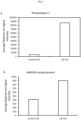
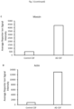


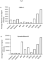
Classifications
-
- G—PHYSICS
- G01—MEASURING; TESTING
- G01N—INVESTIGATING OR ANALYSING MATERIALS BY DETERMINING THEIR CHEMICAL OR PHYSICAL PROPERTIES
- G01N33/00—Investigating or analysing materials by specific methods not covered by groups G01N1/00 - G01N31/00
- G01N33/48—Biological material, e.g. blood, urine; Haemocytometers
- G01N33/50—Chemical analysis of biological material, e.g. blood, urine; Testing involving biospecific ligand binding methods; Immunological testing
- G01N33/68—Chemical analysis of biological material, e.g. blood, urine; Testing involving biospecific ligand binding methods; Immunological testing involving proteins, peptides or amino acids
- G01N33/6893—Chemical analysis of biological material, e.g. blood, urine; Testing involving biospecific ligand binding methods; Immunological testing involving proteins, peptides or amino acids related to diseases not provided for elsewhere
- G01N33/6896—Neurological disorders, e.g. Alzheimer's disease
-
- A—HUMAN NECESSITIES
- A61—MEDICAL OR VETERINARY SCIENCE; HYGIENE
- A61K—PREPARATIONS FOR MEDICAL, DENTAL OR TOILETRY PURPOSES
- A61K31/00—Medicinal preparations containing organic active ingredients
- A61K31/13—Amines
-
- C—CHEMISTRY; METALLURGY
- C07—ORGANIC CHEMISTRY
- C07C—ACYCLIC OR CARBOCYCLIC COMPOUNDS
- C07C49/00—Ketones; Ketenes; Dimeric ketenes; Ketonic chelates
- C07C49/587—Unsaturated compounds containing a keto groups being part of a ring
- C07C49/757—Unsaturated compounds containing a keto groups being part of a ring containing —CHO groups
-
- C—CHEMISTRY; METALLURGY
- C07—ORGANIC CHEMISTRY
- C07K—PEPTIDES
- C07K14/00—Peptides having more than 20 amino acids; Gastrins; Somatostatins; Melanotropins; Derivatives thereof
- C07K14/435—Peptides having more than 20 amino acids; Gastrins; Somatostatins; Melanotropins; Derivatives thereof from animals; from humans
- C07K14/575—Hormones
- C07K14/57581—Thymosin; Related peptides
-
- C—CHEMISTRY; METALLURGY
- C12—BIOCHEMISTRY; BEER; SPIRITS; WINE; VINEGAR; MICROBIOLOGY; ENZYMOLOGY; MUTATION OR GENETIC ENGINEERING
- C12N—MICROORGANISMS OR ENZYMES; COMPOSITIONS THEREOF; PROPAGATING, PRESERVING, OR MAINTAINING MICROORGANISMS; MUTATION OR GENETIC ENGINEERING; CULTURE MEDIA
- C12N9/00—Enzymes; Proenzymes; Compositions thereof; Processes for preparing, activating, inhibiting, separating or purifying enzymes
- C12N9/90—Isomerases (5.)
-
- C—CHEMISTRY; METALLURGY
- C12—BIOCHEMISTRY; BEER; SPIRITS; WINE; VINEGAR; MICROBIOLOGY; ENZYMOLOGY; MUTATION OR GENETIC ENGINEERING
- C12Y—ENZYMES
- C12Y504/00—Intramolecular transferases (5.4)
- C12Y504/02—Phosphotransferases (phosphomutases) (5.4.2)
- C12Y504/02002—Phosphoglucomutase (5.4.2.2)
-
- G—PHYSICS
- G01—MEASURING; TESTING
- G01N—INVESTIGATING OR ANALYSING MATERIALS BY DETERMINING THEIR CHEMICAL OR PHYSICAL PROPERTIES
- G01N33/00—Investigating or analysing materials by specific methods not covered by groups G01N1/00 - G01N31/00
- G01N33/48—Biological material, e.g. blood, urine; Haemocytometers
- G01N33/50—Chemical analysis of biological material, e.g. blood, urine; Testing involving biospecific ligand binding methods; Immunological testing
- G01N33/68—Chemical analysis of biological material, e.g. blood, urine; Testing involving biospecific ligand binding methods; Immunological testing involving proteins, peptides or amino acids
-
- G—PHYSICS
- G01—MEASURING; TESTING
- G01N—INVESTIGATING OR ANALYSING MATERIALS BY DETERMINING THEIR CHEMICAL OR PHYSICAL PROPERTIES
- G01N2800/00—Detection or diagnosis of diseases
- G01N2800/28—Neurological disorders
- G01N2800/2814—Dementia; Cognitive disorders
- G01N2800/2821—Alzheimer
Definitions
- the invention relates to biomarker panels and methods for diagnosing, staging and assessing the likelihood of developing a neurocognitive disorder.
- the invention concerns biomarker panels useful in methods for the diagnosis and treatment of Alzheimer's disease.
- AD Alzheimer's disease
- AD Alzheimer's disease
- AD can only be definitively diagnosed by brain biopsy or upon autopsy after a patient died, in clinical settings brain biopsy is rarely performed and diagnosis is still primarily made based on the history of the symptoms and depends on a battery of neurological, psychometric and biochemical tests, which include the measurement of biomarkers.
- CSF cerebrospinal fluid
- AD cerebrospinal fluid
- myloid ⁇ 1-42, total tau and phosphorylated tau are routinely used to diagnose AD. All three of these CSF biomarkers demonstrate high levels of sensitivity (falling within the 80-90% criteria specified by the National Institute of Neurological and Disorders and Stroke and the Alzheimer Disease and Related Disorders Work Group) but struggle to differentiate AD from other forms of dementia and neurological disorders.
- CSF amyloid ⁇ 1-42 levels are decreased in AD but are also reportedly lower in Lewy body dementia (LBD), fronto-temporal dementia (FTD), vascular dementia (VaD), amyotrophic lateral sclerosis (ALS) and Creutzfeldt-Jakob disease (CJD) ( Blennow K et al., Nat Rev Neurol. 2010,6:131-44 ).
- LBD Lewy body dementia
- FTD fronto-temporal dementia
- VaD vascular dementia
- ALS amyotrophic lateral sclerosis
- CJD Creutzfeldt-Jakob disease
- total tau levels are raised in AD but are also found elevated following stroke, traumatic brain injury, FTD, VaD and CJD1.
- biomarkers Two key features of an ideal biomarker are high specificity for disease versus non-disease and high sensitivity to distinguish between disease types.
- biomarker or biomarker panel would be the first indicator for starting treatment as early as possible, when degeneration is still limited, it would prove enormous valuable in screening the effectiveness of new therapies in clinical trial settings, particularly those trials that are focused on preventing the development of neuropathological changes. Such biomarker or biomarker panel would also be useful in the follow-up of the development of the disease.
- biomarkers that may perform with superior sensitivity and/or specificity in the early diagnosis, staging and prognostic monitoring of patients with Alzheimer's disease and other neurocognitive disorders.
- the present invention therefore, provides a method for detecting neuroinflammation resulting from microglia activation in an Alzheimer's disease (AD) subject, the method comprising:
- the biomarker panel comprises phosphoglucomutase 1 comprising or having the amino acid sequence of SEQ ID NO:1 or an isoform, variant or fragment thereof; in which:
- the biomarker panel comprises thymosin beta-4 comprising or having the amino acid sequence of SEQ ID NO:2 or a variant or fragment thereof; in which:
- the panel may further comprise:
- the panel may further comprise at least one, optionally two or more, biomarkers selected from any one of Tables 1, 2, 3, 4, 5, 6, 7, 8 or 9.
- the panel may further comprise at least one biomarker selected from the group of Dynactin subunit 1, Cofilin-1, Peroxiredoxin-1, MARCKS-related protein, Moesin, Actin, Protein TMSB4XP4, ApoE, Gelsolin, Secretogranin, Albumin and complement proteins.
- biomarker selected from the group of Dynactin subunit 1, Cofilin-1, Peroxiredoxin-1, MARCKS-related protein, Moesin, Actin, Protein TMSB4XP4, ApoE, Gelsolin, Secretogranin, Albumin and complement proteins.
- the subject may be a human subject, preferably a human subject previously diagnosed with mild cognitive impairment.
- the assaying step a) and/or the measuring step b) may comprise:
- the assaying in step a) and/or the measuring in step b) may comprise detecting one or more fragments of said biomarker in the biomarker panel.
- the present disclosure also describes a kit comprising reagents for assaying and/or measuring in a Cerebrospinal fluid sample biomarkers of a biomarker panel as specified herein.
- the reagents may comprise
- biomarker(s) includes all biologically relevant forms of the protein identified, including post-translational modifications.
- the biomarker can be present in a glycosylated, phosphorylated, multimeric, fragmented or precursor form.
- a biomarker fragment may be naturally occurring or, for example, enzymatically generated and still retaining the biologically active function of the full protein. Fragments will typically be at least about 10 amino acids, usually at least about 50 amino acids in length, and can be as long as 300 amino acids in length or longer.
- canonical sequence is used herein as to refer to the most prevalent sequence and/or the most similar sequence among orthologous species. In particular, unless otherwise specified, the canonical sequence refers herein to the human sequence.
- KEGG pathway refers to a collection of manually drawn pathway maps representing molecular interactions and reaction networks for metabolism, genetic information processing, environmental information processing, cellular processes, organismal systems, human diseases and drug development.
- KEGG pathways mapping is the process to map molecular datasets, especially large-scale datasets in genomics, transcriptomics, proteomics, and metabolomics, to the KEGG pathway maps for biological interpretation of higher-level systemic functions; (http://www.genome.jp/kegg/pathway.html).
- concentration or amount refers to the relative concentration or amount of biomarker in the sample, for example as determined by LC-MS/MS label free quantification approaches such as area under the curve and spectral counting.
- comparing means measuring the relative concentration or amount of a biomarker in a sample relative to other samples (for example protein concentrations or amounts stored in proprietary or public database).
- reference concentration or amount refers to, but it is not limited to, protein concentrations or amounts stored in proprietary or public databases.
- the "reference concentration or amount” may have been obtained from a large screening of patients, or by reference to a known or previously determined correlation between such a determination and clinical information in control patients.
- the reference values may be determined by comparison to the concentration or amount of the biomarkers in a control subject, for example a healthy person (i.e. without dementia) of similar age and gender as the subject.
- the reference values are values which can be found in literature such as the ApoE ⁇ 4 allele presence whereby the presence or absence of the mutations at position 112 and 158 represent the reference to be compared to, or like the levels of total tau (T-tau) >350 ng/L, phospho-tau (P-tau) >80 ng/L and A ⁇ 42 ⁇ 530 ng/L in the CSF ( Hansson O, et al., Lancet Neurol. 2006, 5:228-34 ).
- the reference values may have been obtained from the same subject at one or more time points which precede in time the test time point.
- Such earlier sample may be taken one week or more, one month or more, three months or more, most preferably six months or more before the date of the test time point.
- multiple earlier samples may be compared in a longitudinal manner and the slope of change in biomarker expression may be calculated as a correlate of cognitive decline.
- the reference value or reference level can be an absolute value; a relative value; a value that has an upper or a lower limit; a range of values; an average value; a median value, a mean value, or a value as compared to a particular control or baseline value.
- the level of a biomarker is increased when the level of said biomarker in a sample is higher than a reference value.
- the levels of a biomarker are considered to be higher than its reference value when it is at least 1.5%, at least 2%, at least 5%, at least 10%, at least 15%, at least 20%, at least 25%, at least 30%, at least 35%, at least 40%, at least 45%, at least 50%, at least 55%, at least 60%, at least 65%, at least 70%, at least 75%, at least 80%: at least 85%, at least 90%, at least 95%, at least 100%, at least 110%, at least 120%, at least 130%, at least 140%, at least 150% or more higher than the reference value.
- the level of a biomarker is decreased when the level of said biomarker in a sample is lower than a reference value.
- the levels of a biomarker are considered to be lower than its reference value when it is at least 5%, at least 10%, at least 15%, at least 20%, at least 25%, at least 30%, at least 35%, at least 40%, at least 45%, at least 50%, at least 55%, at least 60%, at least 65%, at least 70%, at least 75%, at least 80%: at least 85%, at least 90%, at least 95%, at least 100%, at least 110%, at least 120%, at least 130%, at least 140%, at least 150% or more lower than the reference value.
- control or as used herein "non AD control” or “non AD subject” refers to a tissue sample or a bodily fluid sample taken from a human or non-human subject diagnosed or presenting symptoms of a cognitive abnormality but defined, with respect to the existing biochemical tests, as non AD subjects.
- antibody includes polyclonal antiserum, monoclonal antibodies, fragments of antibodies such as single chain and Fab fragments, and genetically engineered antibodies.
- the antibodies may be chimeric or of a single species.
- SRM selected reaction monitoring
- MRM mass spectrometry assay whereby precursor ions of known mass-to-charge ratio representing known biomarkers are preferentially targeted for analysis by tandem mass spectrometry in an ion trap or triple quadrupole mass spectrometer. During the analysis the parent ion is fragmented and the number of daughter ions of a second predefined mass-to-charge ratio is counted. Typically, an equivalent precursor ion bearing a predefined number of stable isotope substitutions but otherwise chemically identical to the target ion is included in the method to act as a quantitative internal standard.
- isolated means throughout this specification, that the protein, antibody, polynucleotide or chemical molecule as the case may be, exists in a physical milieu distinct from that in which it may occur in nature.
- the term “subject” includes any human or non-human animal.
- non-human animal includes all vertebrates, e.g., mammals and non-mammals, such as non-human primates, rodents, sheep, dogs, cats, horses, cows, chickens, amphibians, reptiles, etc.
- treat includes therapeutic treatments, prophylactic treatments and applications in which one reduces the risk that a subject will develop a disorder or other risk factor. Treatment does not require the complete curing of a disorder and encompasses the reduction of the symptoms or underlying risk factors.
- diagnosis includes the provision of any information concerning the existence or presence, non-existence or absence or probability of the disorder in a patient. It further includes the provision of information concerning the type or classification of the disorder or of symptoms which are or may be experienced in connection with it. This may include, for example, diagnosis of the severity of the disorder. It encompasses prognosis of the medical course of the disorder, for example its duration, severity and the course of progression from mild cognitive impairment (MCI) to AD or other dementias.
- MCI mild cognitive impairment
- AD means identifying in a subject the stage of a neurocognitive disorder, in particular AD.
- AD is characterised by 3 stage or 7 stages, depending on the diagnostic framework used.
- the Global Dementia Scale is one such measure of global function. It is measured by assessment of severity including cognition and function against a standardised set of severity criteria.
- efficacy indicates the capacity for beneficial change of a given intervention (e.g. a drug, medical device, surgical procedure, etc.). If efficacy is established, that intervention is likely to be at least as good as other available interventions, to which it will have been compared.
- a given intervention e.g. a drug, medical device, surgical procedure, etc.
- CSF cerebrospinal fluid
- LBD Lewy body dementia
- FTD fronto-temporal dementia
- VaD vascular dementia
- ALS amyotrophic lateral sclerosis
- CJD Creutzfeldt-Jakob disease
- CNS central nervous system
- LPS lipopolysaccharide
- INF ⁇ interferon-gamma
- TMT ® Tandem Mass Tag ®
- TEAB Tetra-ethylamonium Bicarbonate
- SDS sodium dodecyl sulfate
- TCEP Tris(2-carboxyethyl)phosphine
- ACN Alcohol
- PGM1 phosphoglucomutase 1
- TMS4 Thymosin beta-4
- amyloid precursor protein and its proteolytic product amyloid ⁇ -peptide (A ⁇ )
- a ⁇ amyloid ⁇ -peptide
- plaques and tangles are most often associated with the disease, a third pathological feature "gliosis" or inflammation of the brain is also much more integral to AD patho-etiology than once appreciated.
- Microglia are a type of glial cell and are the resident macrophage-like cells of the central nervous system (CNS) ( Mandrekar S et al., CNS Neurol Disord Drug Targets. 2010, 9:156-67 ). Within the CNS, microglial cells perform a variety of different functions related to the immune response and the maintenance of cellular homeostasis. Microglia sense the environment around them, can be activated by various factors and referee neuroinflammatory and neuroprotective responses in the brain, responding to the formation of amyloid plaques in brains of AD patients.
- CNS central nervous system
- M1 activation state causes the release of pro-inflammatory cytokines such as TNF- ⁇ , IL-1 ⁇ and reactive oxygen/nitrogen species ROS/NOS ( Henkel J, Neuroimmune Pharmacol. 2009, 4:389-98 ). This ultimately may cause or increase neuronal damage.
- pro-inflammatory cytokines such as TNF- ⁇ , IL-1 ⁇ and reactive oxygen/nitrogen species ROS/NOS ( Henkel J, Neuroimmune Pharmacol. 2009, 4:389-98 ). This ultimately may cause or increase neuronal damage.
- microglial activation There appears to be different mechanisms of microglial activation, including in vivo intrinsic regulation, A ⁇ phagocytosis, and microglial A ⁇ receptor complex ( Mandrekar S et al., CNS Neurol Disord Drug Targets. 2010, 9:156-67 ).
- CSF cerebrospinal fluid
- the present invention relates to biomarkers and their use in methods for detecting neuroinflammation resulting from microglia activation in an Alzheimer's disease (AD) subject, or for diagnosing AD in a subject.
- AD Alzheimer's disease
- biomarker panels disclosed herein represent a peripheral signal that reflects events and pathways modulation in brain tissue that could not be tested by biopsy or other invasive tests. Because the biomarker panels reflect the neuroinflammation resulting from microglia activation in AD subject, they represent ideal measurable indicators of the disease at an early stage.
- the biomarker panel comprises phosphoglucomutase 1 comprising or having the amino acid sequence of SEQ ID NO:1 or thymosin beta-4 comprising or having the amino acid sequence of SEQ ID NO:2.
- Human phosphoglucomutase 1 (PMG1; P36871) is an isozyme of phosphoglucomutase (PGM) and belongs to the phosphohexose mutase family. There are several PGM isozymes, which are encoded by different genes and catalyse the transfer of phosphate between the 1 and 6 positions of glucose. Mutations in this gene cause glycogen storage disease type 14. Alternatively spliced transcript variants encoding different isoforms have been identified.
- TMSB4X Human thymosin beta-4 (TMSB4X; P62328) plays an important role in the organization of the cytoskeleton. It binds to and sequesters actin monomers (G-actin) and therefore inhibits actin polymerization.
- the biomarker panel may also comprise phosphoglucomutase 1 comprising or having the amino acid sequence of SEQ ID NO:1 and thymosin beta-4 comprising or having the amino acid sequence of SEQ ID NO:2.
- the biomarker panel may also comprise an isoform or a variant or a fragment of phosphoglucomutase 1 comprising or having the amino acid sequence of SEQ ID NO:1; and/or a variant or a fragment of thymosin beta-4 comprising or having the amino acid sequence of SEQ ID NO:2.
- Isoform is described herein as an alternative protein sequence with respect to the canonical sequence. Isoforms can be generated from the same gene by a single or by the combination of alternative promoter usage, alternative splicing, alternative initiation and ribosomal frameshifting.
- a variant is described herein as to include natural variants such as (naturally occurring) polymorphisms, variations between strains, isolates or cultivars, disease-associated mutations and RNA editing events.
- a variant is generally reported as the amino acid change with respect to the canonical sequence.
- Most naturally occurring polymorphisms also called single amino acid polymorphisms or SAPs are due to a single nucleotide change at the codon level.
- RNA editing events include conversion, insertion and deletion of nucleotides.
- a fragment is described herein as the result of proteolytic (enzymatic or else) cleavage of a protein. Fragments may be the results of natural proteolytic cleavage for example fragments generated during the activation of complement, the clotting cascade, or from enzymatic cleavage of matrix proteins. Alternatively, fragments may be generated in-vivo and/or in-vitro for example with proteases.
- the variant of phosphoglucomutase 1 comprises or has the amino acid sequence of SEQ ID NO: 1 and wherein
- Figure 1 shows the human sequence of human phosphoglucomutase 1 (SEQ ID NO:1); flagged by symbol ⁇ are those amino acids that are replaced with a different amino acid in isoforms of human phosphoglucomutase 1 as indicated above in a) to q). Amino acids flagged by a * are those amino acids which are replaced by modified amino acids in isoforms of human phosphoglucomutase 1 as indicated above in r) to y).
- the isoform of phosphoglucomutase 1 comprises or has the amino acid sequence of
- the fragment of phosphoglucomutase 1 comprises or has the amino acid sequence of any one of SEQ ID NO: 6, SEQ ID NO: 7, SEQ ID NO: 8, SEQ ID NO: 9, or SEQ ID NO: 10; or SEQ ID NO: 11; or SEQ ID NO: 12 ; or SEQ ID NO: 13.
- variant of thymosin beta-4 comprises or has the amino acid sequence of SEQ ID NO: 2 and
- Figure 2 shows the sequence human of thymosin beta-4 (SEQ ID NO:2); flagged by symbol ⁇ are those amino acids that are replaced with a different amino acid in isoforms of human thymosin beta-4 as indicated in a) to c) in the previous paragraph.
- Amino acids flagged by a * are those amino acids which are replaced by modified amino acids in isoforms of human thymosin beta-4 as indicated above in d) to f) in the previous paragraph.
- fragment of thymosin beta-4 comprises or has the amino acid sequence of any one of SEQ ID NO: 14, SEQ ID NO: 15, or SEQ ID NO: 16.
- biomarker panels disclosed herein have been identified as a set of upregulated proteins found in the CSF of AD patients with respect to individual who present mild cognitive impairment but are not diagnosed with AD.
- biomarkers are measured as a set of at least 2, preferably at least 3 or 4.
- the biomarker panel according to the invention may further comprise ubiquitin carboxy-terminal hydrolase L1 (UCH-L1; D6R956) which comprises or has an amino acid sequence of SEQ ID NO:4 or an isoform or a variant or a fragment thereof; and/or vitamin D binding protein (VBPD; D6RBJ7) which comprises or has an amino acid sequence of SEQ ID NO: 5 or an isoform or a variant or a fragment thereof.
- ubiquitin carboxy-terminal hydrolase L1 UCH-L1; D6R956
- VBPD vitamin D binding protein
- isoforms, variants or fragments of ubiquitin carboxy-terminal hydrolase L1 or vitamin D binding protein can be found in publically accessible databases such as Uniprot.
- the fragment of ubiquitin carboxy-terminal hydrolase L1 has or comprises the sequence as shown in SEQ ID NO: 17.
- the fragment of vitamin D binding protein has or comprises the sequence as shown in SEQ ID NO: 18.
- the biomarker panel comprises phosphoglucomutase 1 comprising or having the amino acid sequence of SEQ ID NO:1 or an isoform or a variant or a fragment thereof; and thymosin beta-4 comprising or having the amino acid sequence of SEQ ID NO:2 or a variant or a fragment thereof; and ubiquitin carboxy-terminal hydrolase L1 comprising or having the amino acid sequence of SEQ ID NO:4 or an isoform or a variant or a fragment thereof and/or vitamin D binding protein comprising or having the amino acid sequence of SEQ ID NO: 5 or an isoform or variant or a fragment thereof.
- the biomarker panel may further comprise one or more biomarkers selected from proteins involved in a KEGG pathway wherein the KEGG pathway is selected from the group of complement and coagulation cascade, or glycolysis/glycogenesis, or prion disease, or amino and nucleotide sugar metabolism, or antigen processing and presentation, or extracellular matrixreceptor interaction, or focal adhesion, or regulation of actin cytoskeleton or alanine/aspartate/glutamate metabolism.
- the biomarker panel may further comprise at least one, optionally two or more, biomarkers selected from any one of Tables 1, 2, 3, 4, 5, 6, 7, or 8 or fragments thereof.
- the biomarker panel comprises phosphoglucomutase 1 comprising or having the amino acid sequence of SEQ ID NO:1 or an isoform or a variant or a fragment thereof; and/or thymosin beta-4 comprising or having the amino acid sequence of SEQ ID NO:2 or a variant or a fragment thereof; and at least one, optionally two or more biomarkers selected from Table 1 or fragments thereof.
- Table 1 discloses proteins whose peptides were found to be regulated by at least 60% in the activated microglia cell line compared to resting cells and also at least 60% in the CSF of AD patients compared to CSF of non-AD subjects.
- Asterisk (*) indicates those proteins which were annotated as deleted as the entry has been removed from Uniprot (due to redundancy).
- the biomarker panel comprises phosphoglucomutase 1 comprising or having the amino acid sequence of SEQ ID NO:1 or an isoform or a variant or a fragment thereof; and/or thymosin beta-4 comprising or having the amino acid sequence of SEQ ID NO:2 or a variant or a fragment thereof; and at least one, or at least two or more, optionally at least three or all biomarkers selected from the group of Peroxiredoxin-1, MARCKS-related protein, Moesin and Actin or fragments thereof.
- the biomarker panel comprises phosphoglucomutase 1 comprising or having the amino acid sequence of SEQ ID NO:1 or an isoform or a variant or a fragment thereof; and/or thymosin beta-4 comprising or having the amino acid sequence of SEQ ID NO:2 or a variant or a fragment thereof; and at least one, or at least two or more, optionally at least three or all biomarkers selected from the group of Peroxiredoxin-1, MARCKS-related protein, Moesin, Actin, Protein TMSB4XP4, ApoE, Gelsolin, Secretogranin, Albumin and complement proteins or fragments thereof.
- the biomarker panel comprises phosphoglucomutase 1 comprising or having the amino acid sequence of SEQ ID NO:1 or an isoform or a variant or a fragment thereof; and/or thymosin beta-4 comprising or having the amino acid sequence of SEQ ID NO:2 or a variant or a fragment thereof; and at least one, optionally two or more biomarkers selected from Table 1 and/or Table 2 or fragments thereof.
- Table 2 discloses proteins whose peptides were found to be regulated by at least 60% in in the CSF of AD patients compared to CSF of non-AD subjects.
- Hash (#) indicates those proteins that were annotated as "merged with ###" and which have been researched in Uniprot.
- Asterisk (*) indicates those proteins which were annotated as deleted as the entry has been removed from Uniprot (due to redundancy).
- the biomarker panel comprises phosphoglucomutase 1 comprising or having the amino acid sequence of SEQ ID NO:1 or an isoform or a variant or a fragment thereof; and/or thymosin beta-4 comprising or having the amino acid sequence of SEQ ID NO:2 or a variant or a fragment thereof; and at least one, or at least two or more, optionally at least three or all biomarkers selected from the group of Apolipoprotein E, Secretogranin-1, Serine/threonine-protein phosphatase 2A 65 kDa regulatory subunit A alpha isoform, Cytoplasmic dynein 1 heavy chain 1, RuvB-like 1, cDNA FLJ54806, Alpha-1-acid glycoprotein 2, Ras-related protein Rab-13, Serum albumin and Pigment epithelium-derived factor.
- the biomarker panel may comprise phosphoglucomutase 1 comprising or having the amino acid sequence of SEQ ID NO:1 or an isoform or a variant or a fragment thereof; and/or thymosin beta-4 comprising or having the amino acid sequence of SEQ ID NO:2 or a variant or a fragment thereof; and at least one, optionally two or more biomarkers selected from Table 1 and/or Table 2 and/or Table 3 or fragments thereof.
- Table 3 discloses proteins whose peptides were found to be down-regulated by at least 60% in in the CSF of AD patients compared to CSF of non-AD subjects.
- Hash (#) indicates those proteins that were annotated as "merged with ###" and which have been researched in Uniprot.
- Asterisk (*) indicates those proteins which were annotated as deleted as the entry has been removed from Uniprot (due to redundancy).
- the biomarker panel comprises phosphoglucomutase 1 comprising or having the amino acid sequence of SEQ ID NO:1 or an isoform or a variant or a fragment thereof; and/or thymosin beta-4 comprising or having the amino acid sequence of SEQ ID NO:2 or a variant or a fragment thereof; and at least one, or at least two or more, optionally at least three or all biomarkers selected from the group of ubiquitin carboxy-terminal hydrolase L1, vitamin D binding protein Peroxiredoxin-1, MARCKS-related protein, Moesin, Actin, Protein TMSB4XP4, ApoE, Gelsolin, Secretogranin, Albumin and complement proteins, Apolipoprotein E, Secretogranin-1, Serine/threonine-protein phosphatase 2A 65 kDa regulatory subunit A alpha isoform, Cytoplasmic dynein 1 heavy chain 1, RuvB-like 1, cDNA FLJ54806, Alpha-1-acid glycoprotein 2,
- the biomarker panel may comprise phosphoglucomutase 1 comprising or having the amino acid sequence of SEQ ID NO:1 or an isoform or a variant or a fragment thereof; and/or thymosin beta-4 comprising orhaving the amino acid sequence of SEQ ID NO:2 or a variant or a fragment thereof; and at least one, optionally two or more biomarkers selected from Table 1 and/or Table 2 and/or Table 3 and/or Table 4 or fragments thereof.
- Table 4 discloses proteins whose peptides were found to be regulated by at least 60% in the CSF of AD patients compared to non-AD CSF when compared in a proteomics study without calibrator. Significance is not considered here. Same 6 CSF samples as in all calibrator studies.
- Column 1 annotated ID during data search.
- Middle column protein name/s given to matched sequence.
- Final column All Uniprot IDs that match to the peptide sequence detected at time of invention.
- the biomarker panel may comprise phosphoglucomutase 1 comprising or having the amino acid sequence of SEQ ID NO:1 or an isoform or a variant or a fragment thereof; and/or thymosin beta-4 comprising or having the amino acid sequence of SEQ ID NO:2 or a variant or a fragment thereof; and at least one, optionally two or more, biomarkers selected from Table 1 and/or Table 2 and/or Table 3 and/or Table 4 and/or Table 2 or fragments thereof.
- biomarkers disclosed herein may be upregulated in the CSF of AD patients versus control like dynactin subunit-1 or may be down-regulated in the CSF patients versus control like cofilin-1.
- the biomarker panel comprises phosphoglucomutase 1 comprising or having the amino acid sequence of SEQ ID NO:1 or an isoform or a variant or a fragment thereof; and/or thymosin beta-4 comprising or having the amino acid sequence of SEQ ID NO:2 or a variant or a fragment thereof; and cofilin-1 or a fragment thereof (e.g. a fragment having SEQ ID NO: 38) and/or dynactin subunit 1 or a fragment thereof (e.g. a fragment having SEQ ID NO: 37).
- the biomarker panel may also comprise at least one, or at least two or more, optionally at least three or all biomarkers selected from the group of ubiquitin carboxy-terminal hydrolase L1, vitamin D binding protein Peroxiredoxin-1, MARCKS-related protein, Moesin, Actin, Protein TMSB4XP4, ApoE, Gelsolin, Secretogranin, Albumin and complement proteins, Apolipoprotein E, Secretogranin-1, Serine/threonine-protein phosphatase 2A 65 kDa regulatory subunit A alpha isoform, Cytoplasmic dynein 1 heavy chain 1, RuvB-like 1, cDNA FLJ54806, Alpha-1-acid glycoprotein 2, Ras-related protein Rab-13, Serum albumin and Pigment epithelium-derived factor or fragments thereof.
- biomarkers selected from the group of ubiquitin carboxy-terminal hydrolase L1, vitamin D binding protein Peroxiredoxin-1, MARCKS-related protein, Moesin, Actin,
- the biomarker panel may comprise phospholucomutase 1 having or comprising SEQ ID NOs: 1, 6-13 and thymosin beta-4 having or comprising SEQ ID NOs: 2, 14-16 and optionally Vitamin D-binding protein having or comprising SEQ ID NOs: 5 and 18 and/or Ubiquitin carboxyl-terminal hydrolase isozyme L1 having or comprising SEQ ID NOs: 4 or 17, or fragments thereof, preferably the panel further comprises at least one or more of the following biomarkers, or fragments thereof, dynactin subunit 1 having or comprising SEQ ID NO: 37 or SEQ ID NO: 57, cofilin 1 having or comprising SEQ ID NO: 38 or SEQ ID NO:58), Apolipoprotein E having or comprising SEQ ID NO:19; SEQ ID NO:39 or amino acids 19 to 317 of SEQ ID NO:39, Secretogranin-1 having or comprising SEQ ID NO:20; SEQ ID NO:40; or amino acids
- biomarker panels described herein are useful for diagnosing, for staging and for assessing the likelihood of developing a neurocognitive disorder, in particular Alzheimer's disease.
- the use of the biomarker panels according to the present invention in any of such methods has considerable advantages. Firstly, these biomarker panels translate events and changes in pathways that occur in the brain into a peripheral signal, they allow replacing tissue testing with a peripheral fluid testing. This represents a great advantage especially as the tissue primarily affected in neurocognitive disorder is the brain tissue. Brain biopsies are not carried out and the only tissue analysis carried out is post-mortem.
- these biomarker panels have been developed as capable to translate early events and changes in pathways in the brain, such as neuroinflammatory events, which are believed to be the hallway of neurocognitive disorders characterized by microglia activation, such as Alzheimer's disease.
- these biomarker panels encompass biomarkers which are not those typically reported in the literature or currently used in the clinical setting, thus lending to clinician additional tools for identifying and distinguish, at an early stage, subjects who have AD and subjects who, despite presenting symptoms of neurocognitive impairment are not affect by the early signs of AD.
- kits comprising reagents for assaying and/or measuring in a sample biomarkers of a biomarker panel comprising phosphoglucomutase 1 which comprises or has the amino acid sequence of SEQ ID NO:1 or an isoform or a variant or a fragment thereof; and/or thymosin beta-4 which comprises or has the amino acid sequence of SEQ ID NO:2 or a variant or a fragment thereof.
- the biomarker panel further comprises at least one, alternatively two or more, biomarkers selected from any one of Tables 1, 2, 3, or 4 or fragments thereof.
- the reagents of the kits may comprise one or more binding agents which specifically bind to the biomarkers of the biomarker panels.
- the one or more binding agents are primary antibodies, wherein each primary antibody specifically binds to a different biomarker of the biomarker panel.
- the primary antibodies are antibodies against human phosphoglucomutase 1 which comprises or has the amino acid sequence of SEQ ID NO:1 or an isoform or a variant or a fragment thereof; and/or human thymosin beta-4 which comprises or has the amino acid sequence of SEQ ID NO:2 or a variant or a fragment thereof.
- the primary antibodies may be immobilised on an assay plate, beads, microspheres or particles.
- beads, microspheres or particles may be dyed, tagged or labelled.
- kits comprise primary antibodies against the biomarkers of the biomarker panel
- the kits may further comprise one or more secondary antibodies which specifically bind to said primary antibodies.
- the secondary antibodies may be labelled for example fluorescent labelled or tagged.
- kits may further comprise one or more detection reagents for detecting the presence of the tagged secondary antibodies.
- the sample is selected from the group of cerebrospinal fluid (CSF), blood, plasma, serum, saliva, urine, tissue (e.g. brain tissue) or combinations thereof.
- CSF cerebrospinal fluid
- blood plasma
- serum serum
- saliva saliva
- urine tissue
- tissue e.g. brain tissue
- kits allow the user to:
- kits may instruct the user to assay (as in step a)) and/or to measure (as in step b)) the sample by:
- kits may comprise reagents suitable for preparing brain tissue, optionally for preparing formalin-fixed paraffin-embedded brain tissue sections.
- the kit may additionally provide a reference which provides a quantitative measure by which determination of a concentration or amount of one or more biomarkers can be compared.
- the reference may indicate the amount or concentration of biomarkers which indicate the presence or staging or likelihood of developing a neurocognitive disorder in particular AD.
- the kit may also comprise printed instructions for performing the methods according to the present invention.
- the kit may be for performance of a mass spectrometry assay and may comprise a set of reference peptides (e.g. SRM peptides) in an assay compatible format wherein each peptide in the set is uniquely representative of each of the biomarkers provided in Tables 1, 2, 3, 4, 5, 6, 7, or 8.
- a set of reference peptides e.g. SRM peptides
- Preferably two or more of such unique peptides are used for each biomarker for which the kit is designed, and wherein each set of unique peptides are provided in known amounts which reflect the amount or concentration of such biomarker in a sample of a healthy subject.
- the kit may also provide protocols and reagents for the isolation and extraction of the biomarkers according to the invention from a sample, a purified preparation of a proteolytic enzyme such as trypsin and a detailed protocol of the method including details of the precursor mass and specific transitions to be monitored.
- the peptides may be synthetic peptides and may comprise one or more heavy isotopes of carbon, nitrogen, oxygen and/or hydrogen.
- kits may also comprise appropriate cells, vessels, growth media and buffers.
- biomarker panels described herein comprise both biomarkers where expression is modulated, i.e. quantitatively increased or decreased, and biomarkers which are exclusively present or absent, i.e. qualitatively expressed, in normal versus disease states.
- the degree to which expression differs in normal versus disease states need only be large enough to be visualised via standard characterisation techniques.
- the biomarker(s) in the biomarker panel may be detected using a binding agent, such as an antibody, specific to that biomarker, for example in an ELISA assay or Western blotting.
- a binding agent such as an antibody, specific to that biomarker, for example in an ELISA assay or Western blotting.
- Such antibodies may include, but are not limited to, polyclonal antibodies, monoclonal antibodies (mAbs), humanised or chimeric antibodies, single chain antibodies, Fab fragments, F(ab')2 fragments, fragments produced by a Fab expression library, anti-idiotypic (anti-ld) antibodies, and epitope-binding fragments of any of the above.
- various host animals may be immunised by injection with a protein, or a portion thereof.
- Such host animals may include, but are not limited to, rabbits, mice and rats.
- Various adjuvants may be used to increase the immunological response, depending on the host species, including active substances such as lysolecithin, Pluronic polyols, polyanions, peptides, oil emulsions, keyhole limpet hemocyamin, dinitrophenol, and potentially useful human adjuvant such as BCG bacille Calmette-Fuerin) and Corynebacterium parvum.
- Polyclonal antibodies are heterogeneous populations of antibody molecules derived from the sera of animals immunised with an antigen, such as target proteins, or an antigenic functional derivative thereof.
- an antigen such as target proteins, or an antigenic functional derivative thereof.
- host animals such as those described above, may be immunised by injection with differentially expressed or pathway protein supplemented with adjuvants as also described above.
- Monoclonal antibodies which are homogeneous populations of antibodies to a particular antigen, may be obtained by any technique, which provides for the production of antibody molecules by continuous cell lines in culture. These include, but are not limited to, the hybridoma technique of Kohler and Milstein (1975, Nature 256:495-7 ; and US Patent No: 4,376,110 ), the human ⁇ -cell hybridoma technique ( Kosbor, et al., 1983, Immunology Today 4:72 ; Cole, et al., 1983, Proc. Natl. Acad. Sci. USA 80:2026-30 ), and the EBV-hybridoma technique ( Cole, et al., 1985, Monoclonal Antibodies and Cancer Therapy, Alan R.
- Such antibodies may be of any immunoglobulin class including IgG, IgM, IgE, IgA, IgD and any subclass thereof.
- the hybridoma producing the mAb of this invention may be cultivated in vitro or in vivo. Production of high titers of mAbs in vivo makes this the presently preferred method of production.
- a chimeric antibody is a molecule in which different portions are derived from different animal species, such as those having a variable region derived from a murine mAb and a human immunoglobulin constant region.
- Single chain antibodies are formed by linking the heavy and light chain fragments of the Fv region via an amino acid bridge, resulting in a single chain polypeptide.
- Antibody fragments which recognise specific epitopes, may be generated by known techniques.
- fragments include, but are not limited to, the F(ab')2 fragments which can be produced by pepsin digestion of the antibody molecule and the Fab fragments which can be generated by reducing the disulfide bridges of the F(ab')2 fragments.
- Fab expression libraries may be constructed ( Huse, et al., 1989, Science 246:1275-81 ) to allow rapid and easy identification of monoclonal Fab fragments with the desired specificity.
- the sample may be immobilised on a solid support for analysis.
- An antibody sandwich technique may be employed in which binding agents, such as antibodies, specific for the individual biomarkers in the biomarker panel are immobilized on a solid support such as a planar surface or a microparticle bead and biomarkers in the panel are captured by the immobilised binding agents, such as immobilized antibodies.
- the captured biomarkers are then detected using a second binding agent, such as a secondary antibody, that may be directly labelled with a signal generating agent (enzyme, fluorescent tag, radiolabel etc.) or may be detected using further amplification (labelled secondary antibody, streptavidin/biotin systems with enzyme, fluorophore, radiolabel etc.).
- Other methods may include, but are not limited to, one-dimensional or two-dimensional (2D) gel electrophoresis of samples. Such methods are followed by transfer to a solid surface using techniques such as Western blotting and subsequent detection using antibodies specific for the AD biomarkers.
- autoantibodies to the biomarkers may be detected using the Western blotting approach described above using samples from a healthy subject, a patient or representative of AD, and then detecting the presence of auto-antibodies specific for the biomarker that are present in the sample, but not in healthy subjects.
- aptamers include nucleic acid aptamers and peptide aptamers.
- the biomarkers may be detected by, amongst others, silver staining of 2D gel electrophoresis or mass spectrometry techniques including LS/MS/MS, MALDI-TOF, SELDI-TOF and TMT-SRM.
- Chromatographic separations can be carried out by high performance liquid chromatography as described in literature, the chromatogram being obtained in the form of a plot of absorbance of light at 280 nm against time of separation. The material giving incompletely resolved peaks is then re-chromatographed and so on.
- Capillary electrophoresis is a technique described in many publications, for example in the literature "Total CE Solutions” supplied by Beckman with their P/ACE 5000 system. The technique depends on applying an electric potential across the sample contained in a small capillary tube.
- the tube has a charged surface, such as negatively charged silicate glass.
- Oppositely charged ions in this instance, positive ions
- the cathode In this electro-osmotic flow (EOF) of the sample, the positive ions move fastest, followed by uncharged material and negatively charged ions.
- proteins are separated essentially according to charge on them.
- Micro-channel networks function similarly to capillaries and can be formed by photoablation of a polymeric material.
- a UV laser is used to generate high energy light pulses that are fired in bursts onto polymers having suitable UV absorption characteristics, for example polyethylene terephthalate or polycarbonate.
- the incident photons break chemical bonds with a confined space, leading to a rise in internal pressure, mini-explosions and ejection of the ablated material, leaving behind voids which form micro-channels.
- the micro-channel material achieves a separation based on EOF, as for capillary electrophoresis. It is adaptable to micro-chip form, each chip having its own sample injector, separation column and electrochemical detector: see J.S.Rossier et al., 1999, Electrophoresis 20:727-31 .
- the ProteinChip system consists of aluminium chips to which protein samples can be selectively bound on the surface chemistry of the chip (e.g. anionic, cationic, hydrophobic, hydrophilic, etc.). Bound biomarkers are then co-crystallised with a molar excess of small energy-absorbing molecules. The chip is then analysed by short intense pulses of N2 320nm UV laser with protein separation and detection being by time of flight mass spectrometry. Spectral profiles of each group within an experiment are compared and any peaks of interest can be further analysed using techniques as described below to establish the identity of the biomarkers.
- Isotopic or isobaric Tandem Mass Tags ® (TMT ® Thermo Scientific, Rockford, USA) technology may also be used to detect biomarkers such as proteins of a biomarker panel described herein. Briefly, the proteins in the samples for comparison are optionally digested, labelled with a stable isotope tag and quantified by mass spectrometry. In this way, expression of equivalent proteins in the different samples can be compared directly by comparing the intensities of their respective isotopic peaks or of reporter ions released from the TMT ® reagents during fragmentation in a tandem mass spectrometry experiment.
- Detection of biomarkers of biomarker panels described herein may be preceded by a depletion step to remove the most abundant proteins from the sample.
- the large majority of the protein composition of serum/plasma consists of just a few proteins.
- albumin which is present at a concentration of 35-50 mg/ml, represents approximately 54% of the total protein content with IgG adding other 16%.
- proteins changing in response to disease for example as a result of tissue leakage, may circulate at 10ng/ml.
- a multiple affinity depletion column may be used to remove the most highly abundant proteins (e.g. the 2, 3, 4, 5, 6, 7, 8, 9 or 10 or more highly abundant proteins). This enables the detection of changes in lower abundance ranges because more starting material can be used and there is less interference from the highly abundant molecules.
- Such a depletion strategy can be applied before any detection method.
- biomarker panels described herein may be used to test agents for the ability to prevent or ameliorate neurocognitive disorders, such as AD, or one or more symptoms thereof.
- Such agents may be tested in human subjects in clinical trials. Any agent which restores the expression of the proteins in a biomarker panel described herein towards levels found in healthy individuals or towards its absence or presence as the case may be, is of potential use in treating a neurocognitive disorder, such as AD, i.e. reducing AD symptoms or slowing the progression of AD.
- a neurocognitive disorder such as AD, i.e. reducing AD symptoms or slowing the progression of AD.
- the amount or concentration of one or more biomarkers of a biomarker panel as described herein can be determined in the presence or absence of the agent being tested.
- the efficacy of the agent can be followed by comparing the expression data obtained to the corresponding known expression patterns in a normal state.
- Agents exhibiting efficacy are those which alter the presence, amount or concentration of the biomarkers in the biomarker panel to more closely resemble that of the normal state.
- the detection of the biomarkers in the biomarker panel in the neurocognitive disorder relative to their expression or presence in a normal state can also be used for monitoring the efficacy of potential agents for the treatment of a neurocognitive disorder, such as AD, during clinical trials.
- a neurocognitive disorder such as AD
- the level and/or presence of the biomarkers in the biomarker panel can be determined in cerebrospinal fluid (CSF) in the presence or absence of the agent being tested.
- CSF cerebrospinal fluid
- the efficacy of the agent can be followed by comparing the biomarker levels and/or presence of the biomarkers in the data obtained, to the corresponding known levels/presence for the cells and/or tissues and/or body fluids in a normal state.
- Agents exhibiting efficacy are those which alter the presence, amount or concentration of the biomarker panel of the cell and/or tissue sample and/or body fluid from a subject to more closely resemble that of the normal state or which stabilise the pattern i.e. prevent progression of the disease. Because the present biomarker panels translate events and changes in pathways that occur in the brain into a peripheral signal, they allow replacing tissue testing with a peripheral fluid testing. This represents a great advantage especially as the tissue primarily affected in neurocognitive disorder is the brain tissue. Brain biopsies are not carried out and the only tissue analysis carried out is post-mortem.
- any treatments that restore or partially restore the expression of biomarkers in a biomarker panel described herein to healthy levels should be considered as candidates for therapeutic intervention in neurocognitive disorders such as AD.
- Dosages of test agents may be determined by deriving dose-response curves.
- any treatments that can prevent the development of neurocognitive disorders such as AD or prevent progression to levels of more advanced AD should be considered as candidates for the AD therapeutic intervention.
- An agent may be selected if it prevents or slows the change over time in presence, concentration or amount of the biomarkers of the biomarker panels relative to controls.
- the agent is selected if it converts the amount or concentration of a biomarker of the biomarker panels towards that of a normal subject.
- the agent may be selected if it slows or stops the change of concentration or amount over time.
- agents which exhibit inhibitory activity may be used in accordance with the invention to prevent mild cognitive impairment or AD symptoms.
- Such molecules may include, but are not limited to, peptides (such as, for example, peptides representing soluble extracellular portions of target protein transmembrane receptors), phosphopeptides, small organic or inorganic molecules, or antibodies (including, for example, polyclonal, monoclonal, humanised, anti-idiotypic, chimeric or single chain antibodies, and Fab, F(ab')2 and Fab expression library fragments, and epitope-binding fragments thereof).
- BV2 cells samples - BV2 cells were seeded into 6 well plates at 80,000 cells per well and maintained at 37°C at 5% CO 2 .
- Cells were cultured for 24 hours in Dulbecco's modified eagle medium (Gibco ® , Life Technologies) with 10% fetal bovine serum (Gibco ® ) supplemented with 2mM L-glutamine, 100U/mL penicillin and 100mg/mL streptomycin, gentamycin and mycoplasma removal agent (AbD Serotec ® ).
- LPS lipopolysaccharide
- IFN ⁇ interferon-gamma
- the protein concentration of the cell lysate was estimated by Bradford assay. Samples were aliquoted to prevent freeze-thawing and stored at -80°C. The BV2 cells samples will be referred herein after as "calibrator” as they will be used to create calibration curves in the following experiments.
- Cerebrospinal fluid samples - CSF samples were from subjects who sought medical advice because of cognitive impairment. Subjects were designated as normal or AD according to CSF biomarker concentrations using cutoffs that are 90% sensitive and specific for AD): total tau (T-tau) >350 ng/L, phospho-tau (P-tau) >80 ng/L and A ⁇ 42 ⁇ 530 ng/L ( Hansson O, et al., Lancet Neurol. 2006, 5:228-34 . None of the biochemically normal subjects fulfilled these criteria.
- CSF T-tau, P-tau and A ⁇ 42 concentration were determined using INNOTEST enzyme-linked immunosorbent assays (Fujirebio, Ghent, Belgium) by board-certified laboratory technicians according to protocols approved by the Swedish Board for Accreditation and Conformity Assessment (SWEDAC). The study was approved by the regional ethics committee at the University of Gothenburg.
- TMT ® Tryptic Digest and Tandem Mass Tag ®labelling - Following solubilization and denaturation in 100mM TEAB buffer and 0.1% sodium dodecyl sulphate (SDS), the CSF samples and the BV2 cell-line (calibrator) were reduced with 1mM TCEP at 55 °C for 60 min and alkylated with 7.5mM iodoacetamide at room temperature for 60 min. Trypsin was used for digestion at an approximate 1:25 weight ratio of trypsin-to-total protein and incubated at 37 °C overnight ( ⁇ 18 h).
- SDS sodium dodecyl sulphate
- the peptides obtained were labeled with one of the TMT ® 10-plex reagents at 15mM and incubated for 60 min at room temperature. To quench the TMT ® reaction, 0.25% hydroxylamine was added to each sample and incubated for 15 min. The samples were then combined to generate an analytical sample comprising a 1:10 calibration curve of non-activated BV2 cells, a 1:10 calibration curve of activated BV2 cells and equal amount of six CSF samples and incubated for a further 15 min ( Figure 6 ). Each sample was desalted using RP18 columns (Waters, Manchester, UK) and excess reagents and SDS were removed by strong cation exchange (SCX) purification.
- SCX strong cation exchange
- LC-MS/MS - Quantitative analysis was performed in triplicate using an Orbitrap Fusion Tribrid ® mass spectrometer (Thermo Scientific ® ) in the positive mode with EASYnLC1000 system and a 50cm EASY-Spray column (Thermo Scientific ® ). The column temperature was maintained at 40°C, and the peptides were separated at a flow rate of 200 nL/min.
- Peptides were eluted from the column over a 300 min gradient, from 3% to 30% solvent B (acetonitrile with 0.1% formic acid) in 280 min, followed by an increase to 80% solvent B in 10mins, which was held for a further 10mins.
- Solvent A was water with 0.1% formic acid. Wash and blank LC-MS/MS runs preceded the analysis.
- ATop10 CID-HCD MS3 SPS method with a 2 second cycle time was utilized and the parameters were as follows: MS; Spray voltage - 2000V; ion transfer tube temperature - 275 °C; detector - Orbitrap; scan range (m/z) - 400-1400; resolution - 120000; AGC target - 5x10⁵.
- MS/MS (CID) Isolation mode - quadropole; collision energy - 35%; detector- Ion trap; AGC target - 4x10³.
- MS3 (HCD) detector - Orbitrap; collision energy - 55%; scan range - 100-1000 m/z; resolution power - 30000; AGC target - 6x10⁴.
- Bioinformatics - LC-MS/MS data was initially processed using SequestHT and Mascot within Proteome Discoverer 1.4, against the Uniprot human protein database (ftp://ftp.uniprot.org/pub/databases/uniprot/current_release/knowledgebase/proteomes/). After applying a 5% false discovery rate (FDR)) to filter the data in Proteome Discoverer, the number of peptide and protein identifications was determined.
- FDR 5% false discovery rate
- This experiment was designed to incorporate two 2-point calibration curves using the control (i.e. non-activated) BV2 cells and activated (IFN- ⁇ + LPS) BV2 cells, alongside six (6) CSF samples constituted of three (3) AD CSF samples and 3 non-AD CSF samples); 3 AD CSF samples were compared with three (3) non-AD control CSF samples using a two x 2-point calibration curve of control and activated BV2 cells to detect biomarkers of activation in a 3 vs 3 vs 2 vs 2 TMT ® 10-plex MS3 method.
- the experiment was designed so that the protein content from the BV2 cells dominated the total protein content in the sample.
- the two x 2-point calibration curve samples were spiked at a ratio of 1:10 and the 6 CSF samples were calculated to fall within this range.
- the total protein content that can be injected onto the MS column is 2 ⁇ g, therefore, the CSF samples' protein content per inject is limited to 121ng per channel per CSF sample.
- the TMT 10 plex labelled samples were mixed so as to generate a 1:10 calibration curve for the resting cells (channels 129 & 130e), a 1:10 calibration curve for the activated cells (channels 130 & 131) and equal levels of the 6 CSF samples wherein the total protein content from the cell digests is approximately double that for the CSF samples ( Figure 6 ).
- the mixed analytical sample was de-salted and cleaned (excess TMT ® labels removed) and split into 2. Half of the sample was frozen and the remainder was fractionated by high pH reverse phase fractionation into 8 separate fractions. These were analysed on the new generation Orbitrap Fusion using a gradient of 3 hours.
- the raw data files from all MS injections were searched using SequestHT and Mascot in Proteome Discoverer 1.4, against the Uniprot human protein database.
- the reporter ion signal intensities from the 6 CSF channels were normalised by sum-scale normalization, a mathematical approach to remove experimental bias.
- the process involves summing the intensity values for all analytes measured in a given sample and then calculating the median value across all the summed values. The median value is divided by each summed value to create a correction factor which is multiplied to the original intensity values to give the normalized sum scaled measurement ( Robinson MD, et al., Bioinformatics 2010, 26:139-40 ; De Livera AM, et al., Anal. Chem. 2012, 84, 24, 10768-76 ) and the variability across the CSF dataset was investigated using Principle Component Analysis (PCA) plots. It was found that the CSF samples separate by disease status at the peptide and protein level with the first principle component accounting for 79.9% of the total variation leading to group separation (data not shown).
- PCA Principle Component Analysis
- the ratio of the signal intensity in the calibrator channels was expected to be 1:10 for the non-activated (129 and 130e) and 1:10 in the activated cells (130 and 131).
- the observed intensities across the two non-activated cell channels and across the two activated cell channels was calculated for every single peptide. Peptides with a ratio of 1:10 ⁇ 15% were considered to be acceptable calibrators. Only 22 peptides were found to contain an acceptable calibrator using these parameters.
- the log2 ratio of the average signal intensities in the three AD CSF samples compared to the average signal in the three control CSF samples was determined.
- a p-value for this ratio was established following a 2-sample t-test across the two CSF groups for every single peptide and protein (providing a signal was present in all 6 of the CSF TMT ® channels).
- the log2 ratio of the two calibrator channels in the non-activated cells and the activated cells was also calculated to show which peptides are regulated with BV2 cell activation. The ratio was calculated for the low spike calibrator in activated and control cells, and also for the high spike calibrator.
- a total of 564 peptides were found to be up-regulated by at least 60% in the activated vs control BV2 cells (filtered for 60% increase in both the high and low calibrator ratio's). These were shown to map to 9 KEGG pathways, the most significant being the complement and coagulation cascades pathway, followed by the glycolysis/glycogenesis, the prion disease, the amino and nucleotide sugar metabolism, the antigen processing and presentation, the extracellular matrix-receptor interaction, the focal adhesion, the regulation of actin cytoskeleton and alanine/aspartate/glutamate metabolism pathways.
- This first data set processing included all the peptides detected, regardless of missing TMR ® reporter ion channels and allow the identification of peptides that are present in the CSF of AD patients that are absent in controls, or similarly, peptides which are present in activated BV2 cells that are not present in non-activated BV2 cells.
- MARCKS-related protein Figure 3B
- Moesin Figure 3C
- Actin Figure 3D
- Peptide NPLPSKETIEQEK (SEQ ID NO: 14) is found in both TMSB4XP4 protein and homologous protein Thymosin beta-4. The protein sequences of these two proteins are identical apart from the first 19 residues of TMSB4XP4 which are absent in Thymosin beta-4.
- Phosphoglucomutase 1 was not detected when the CSF samples were measured in the absence of the BV2 cell sample calibrator, whilst Thymosin beta-4 was detected in both settings.
- 5 were regulated ( ⁇ 60%; in bold in Table 7) but not significantly and only one was found to be significantly regulated with a P value ⁇ 0.05 (QFSANDK; SEQ ID NO:6).
- experiment B was designed as a 4-point calibration curve to be spiked into the total combined CSF samples at a ratio of 1:4:6:10 in order to ensure the calibrator proteins dominated the overall protein content of the TMT ® 10-plex sample. These are arbitrary values allowing to determine a suitable protein load from the CSF samples for analysis. Six individual CSF samples were combined at a 2:2:2:2:2 ratio, falling within the 4-point calibration curve.
- the true protein load per channel can be determined by dividing the maximum 2 ⁇ g by the total number of arbitrary values (x) needed, which in this case is 33 (4 calibrator channels plus 6 CSF channels). If 33x equals 2 ⁇ g, 1x is therefore 0.06 ⁇ g and this can then be used to determine the total amount of protein from each of the 10 channels contributing to the calibrator analytical sample. Just 0.12 ⁇ g of each CSF sample is required (2x), along with a 4-point calibrator comprised of 0.06 ⁇ g (1x), 0.24 ⁇ g (4x), 0.36 ⁇ g (6x) and 0.6 ⁇ g (10x). This results in a total of 0.72 ⁇ g of protein from all the CSF samples and 1.26 ⁇ g of protein from the BV2 cell line calibrator - the calibrator proteins are therefore 1.75 fold more prevalent than the total CSF protein load.
- the calibrator sample was analysed in triplicate on the Oribtrap Fusion Tribrid using an MS3 SPS method over a 300 minute gradient.
- the raw data files from all three MS injections were searched using SequestHT and Mascot in Proteome Discoverer 1.4, against the Uniprot human protein database. Results for the triplicate injections were merged resulting in one large data set for analysis.
- FDR 5% false discovery rate
- the reporter ion signal intensities from the six CSF channels were normalized by sum-scale normalization. Following normalization the variability across the CSF dataset was investigated using Principal Component Analysis (PCA). Group separation of AD and control CSF was seen at the peptide and protein level, forming the first principal component, and accounting for 44.6% of the total variation seen in the dataset. The second principal component explained 19.6% of the variability in the dataset, and this corresponded to the biological variation within the groups.
- PCA Principal Component Analysis
- the normalized dataset, filtered for the presence of an expected linear calibrator signal intensity was further filtered based on the significance following a 2-sample t-test across the two CSF groups for every single peptide and protein. Peptides with a p value ⁇ 0.05 were considered significant.
- the log2 ratio of the average AD CSF signal intensities (channels 128e, 128 and 129e) to control CSF signal intensities (channels 126, 127e and 127) was also normalized to the 129 TMT ® calibrator channel. This provided a means of identifying which of the peptide sequences common to the activated microglia cell line and CSF samples were differentially regulated between control and AD CSF.
- the benefit of TMT ® calibrator over the traditional proteomics approach can be seen.
- the maximum protein load on the 75 ⁇ m diameter analytical LC column per injection is 2ug.
- the 2 ⁇ g is split unequally between the 4-point calibrator and the six CSF samples (1:4:6:10 ratio for the calibrator, 2:2:2 for AD CSF and 2:2:2 for non-AD CSF).
- the six CSF samples could be labelled with TMT in a multiplex experiment, with 0.33 ⁇ g combined from each sample per inject.
- TMT ® calibrator proteins that are commonly observed in CSF studies including serum albumin, complement proteins (C3, C5 and C7), fibrinogen gamma chain and Ig gamma chain ( Boche D, et al., Neuropathol Appl Neurobiol. 2013, 39, 1, 3-18 ). These proteins are likely representative of disease related changes of low specificity rather than the cellular changes specific to AD. They are some of the most abundant proteins found in the CSF and are reported in the majority of CSF proteomic analysis studies ( Hühmer AF, et al., Disease Markers. 2006, 22, 1-2, 3-26 ).
- peptides derived when the BV2 cell line is used to drive the experiment are cellular and may represent a cellular response specific to disease. These include peptides from dynactin ( Figure 5A ), cofilin ( Figure 5A ), alcohol dehydrogenase, filamin-A, myosin proteins and Ras-related proteins. These are cellular proteins regulated in AD that would not have been detected if the CSF samples had been analysed in the absence of the BV2 cell samples (calibrator samples).
- Peptide IKDALVR (SEQ ID NO: 37), corresponding to dynactin subunit 1( Figure 5A ), was the most upregulated peptide whist peptide MIYASSK (SEQ ID NO: 38), corresponding to Cofilin-1 ( Figure 5B ), was the most down regulated peptide in the CSF of AD patients when compared to non-AD controls.
- biomarkers are indicators of pathological processes and biological events. Their levels change as a result of disease and also in response to pharmacological intervention.
- the challenge is how to find these molecules in biofluids such as blood or CSF which contain a high protein dynamic range and hyper-abundant proteins such as albumin.
- TMT ® Tandem Mass Tag ®
- MS mass spectrometry
Description
- The invention relates to biomarker panels and methods for diagnosing, staging and assessing the likelihood of developing a neurocognitive disorder. In particular, the invention concerns biomarker panels useful in methods for the diagnosis and treatment of Alzheimer's disease.
- Alzheimer's disease (AD) is the most common type of dementia in aging adults with the number of people living with AD projected to increase dramatically over the next few decades, making the search for treatments and tools to diagnose and measure disease progression increasingly urgent.
- Approved treatments are few and of limited efficacy, serving mostly to slow or delay progression. Currently there is no cure.
- AD gives rise to an irreversible progressive loss of cognitive functions and of functional autonomy. While the time it takes for AD to develop will vary from person to person, advanced signs include severe memory impairment, confusion, language disturbances, impaired judgment, personality and behaviour changes. Patients with AD may become non-communicative and hostile. As the disease ends its course in profound dementia, patients are unable to care for themselves and often require institutionalization or professional care in the home setting. While some patients may live for many years after being diagnosed with AD, the average life expectancy after diagnosis is eight years.
- Although AD can only be definitively diagnosed by brain biopsy or upon autopsy after a patient died, in clinical settings brain biopsy is rarely performed and diagnosis is still primarily made based on the history of the symptoms and depends on a battery of neurological, psychometric and biochemical tests, which include the measurement of biomarkers.
- Nevertheless, these present methods are still not satisfactory in the diagnosing of AD and other neurocognitive disorders at the early stage of the disease when potential therapies are more likely to prevent or slow down neurodegeneration.
- As the brain is in direct contact with the cerebrospinal fluid (CSF), and pathological changes in the brain often result in altered biochemical composition of the CSF, this makes it an ideal source for biomarkers of neurocognitive disorders. In AD, currently three "core" CSF biomarkers (amyloid β1-42, total tau and phosphorylated tau) are routinely used to diagnose AD. All three of these CSF biomarkers demonstrate high levels of sensitivity (falling within the 80-90% criteria specified by the National Institute of Neurological and Disorders and Stroke and the Alzheimer Disease and Related Disorders Work Group) but struggle to differentiate AD from other forms of dementia and neurological disorders. For example, CSF amyloid β1-42 levels are decreased in AD but are also reportedly lower in Lewy body dementia (LBD), fronto-temporal dementia (FTD), vascular dementia (VaD), amyotrophic lateral sclerosis (ALS) and Creutzfeldt-Jakob disease (CJD) (Blennow K et al., Nat Rev Neurol. 2010,6:131-44). Similarly, total tau levels are raised in AD but are also found elevated following stroke, traumatic brain injury, FTD, VaD and CJD1.
- Two key features of an ideal biomarker are high specificity for disease versus non-disease and high sensitivity to distinguish between disease types. In addition, biomarkers that reflect the pathological process of AD and that are able to detect changes at a very early stage of the disease, before degeneration is observed by brain imaging and neuropathological tests, are very sought after.
- The ideal biomarker or biomarker panel would be the first indicator for starting treatment as early as possible, when degeneration is still limited, it would prove immensely valuable in screening the effectiveness of new therapies in clinical trial settings, particularly those trials that are focused on preventing the development of neuropathological changes. Such biomarker or biomarker panel would also be useful in the follow-up of the development of the disease.
- Hence, there remains a need for biomarkers that may perform with superior sensitivity and/or specificity in the early diagnosis, staging and prognostic monitoring of patients with Alzheimer's disease and other neurocognitive disorders.
- The present invention, therefore, provides a method for detecting neuroinflammation resulting from microglia activation in an Alzheimer's disease (AD) subject, the method comprising:
- a) assaying a cerebrospinal fluid (CSF) sample from said subject for the biomarkers of a biomarker panel;
- b) measuring in said sample the concentration or the amount of each of the biomarkers of said biomarker panel;
- c) determining said subject is neuroinflammed resulting from microglia activation, by comparing said concentration or amount of each of the biomarker of the biomarker panel in said sample with reference concentrations or amounts of said biomarkers.
- In one aspect, the biomarker panel comprises
phosphoglucomutase 1 comprising or having the amino acid sequence of SEQ ID NO:1 or an isoform, variant or fragment thereof; in which: - a
phosphoglucomutase 1 isoform comprises or has the amino acid sequence of SEQ ID NO:3 whereinmethionine 1 is N-acetylmethionine; or SEQ ID NO:1 amino acids 198 to 562, - a
phosphoglucomutase 1 variant comprises or has the amino acid sequence of SEQ ID NO: 1 in which: a) threonine at position 19 is replaced by alanine; or b) asparagine at position 38 is replaced by tyrosine; or c) glutamine at position 41 is replaced by arginine; or d) aspartic acid at position 62 is replaced by histamine; or e) lysine at position 68 is replaced by methionine; or f) isoleucine at position 88 is replaced by valine; or g) threonine at position 115 is replaced by alanine; or h) glycine at position 121 is replaced by arginine; or i) arginine at position 221 is replaced by cysteine; or j) aspartic acid at position 263 is replaced by glycine or tyrosine; or k) glycine at position 291 is replaced by arginine; or l) glycine atposition 330 is replaced by arginine; or m) glutamic acid at position 377 is replaced by lysine; or n) glutamic acid at position 388 is replaced by lysine; or o) tyrosine atposition 420 is replaced by histidine; or p) valine at position 501 is replaced by isoleucine; or q) leucine at position 516 is replaced by proline; or r) methionine atposition 1 is N-acetylmethionine; or s) lysine at position 16 is N6-acetyllysine; or t) serine at position 117 is phosphoserine; or u) lysine at position 349 is N6-acetyllysine; or v) tyrosine at position 353 is phosphotyrosine; or w) lysine at position 419 is N6-succinyllysine; or x) threonine at position 467 is phosphothreonine; or y) threonine at position 507 is phosphothreonine; and/or - a
phosphoglucomutase 1 fragment comprises or has the amino acid sequence of any one of SEQ ID NO: 6, SEQ ID NO: 7, SEQ ID NO: 8, SEQ ID NO: 9, or SEQ ID NO: 10; or SEQ ID NO: 11; or SEQ ID NO: 12 ; or SEQ ID NO: 13. - In a second aspect, the biomarker panel comprises thymosin beta-4 comprising or having the amino acid sequence of SEQ ID NO:2 or a variant or fragment thereof; in which:
- a thymosin beta-4 variant comprises or has the amino acid sequence of SEQ ID NO: 2 in which: a) lysine at position 12 is replaced by proline; or b) serine at position 16 replaced by alanine; or c) leucine at position 18 is replaced by alanine or proline; or d) serine at
position 2 is N-acetylserine; or e) lysine at position 26 is N6-acetyllysine; or f) lysine at position 9 is N6-acetyllysine; and/or - a thymosin beta-4 fragment comprises or has the amino acid sequence of any one of SEQ ID NO: 14, SEQ ID NO: 15, or SEQ ID NO: 16.
- The panel may further comprise:
- ubiquitin carboxy-terminal hydrolase L1 which comprises or has an amino acid sequence of SEQ ID NO:4 or an isoform, variant or fragment thereof, and/or
- vitamin D binding protein which comprises or has an amino acid sequence of SEQ ID NO: 5 or an isoform, variant or fragment thereof.
- The panel may further comprise at least one, optionally two or more, biomarkers selected from any one of Tables 1, 2, 3, 4, 5, 6, 7, 8 or 9.
- The panel may further comprise at least one biomarker selected from the group of Dynactin
subunit 1, Cofilin-1, Peroxiredoxin-1, MARCKS-related protein, Moesin, Actin, Protein TMSB4XP4, ApoE, Gelsolin, Secretogranin, Albumin and complement proteins. - The subject may be a human subject, preferably a human subject previously diagnosed with mild cognitive impairment.
- The assaying step a) and/or the measuring step b) may comprise:
- i) detecting in said sample by mass spectrometry each of said biomarkers of the biomarker panel, optionally by previously labelling said sample with one or more isobaric reactive mass labels; or
- ii) contacting said sample with one or more binding agents to each of said biomarkers of the biomarker panel; or
- iii) detecting in said sample autoantibodies specific to each of said biomarkers; or
- iv) detecting in said sample by 2D gel electrophoresis each of said biomarkers of the biomarker panel; or
- v) any combinations of i), ii), iii) or iv).
- The assaying in step a) and/or the measuring in step b) may comprise detecting one or more fragments of said biomarker in the biomarker panel.
- The present disclosure also describes a kit comprising reagents for assaying and/or measuring in a Cerebrospinal fluid sample biomarkers of a biomarker panel as specified herein.
- The reagents may comprise
- one or more binding agents which specifically bind to the biomarkers of the biomarker panels, preferably wherein said one or more binding agents are antibodies, or
- a set of reference peptides (e.g. SRM peptides) in an assay compatible format wherein each peptide in the set is uniquely representative of each of the biomarkers of the biomarker panel.
-
-
Figure 1 . Sequence ofhuman phosphoglucomutase 1. Amino acids flagged by symbol or a * indicate amino acids which are replaced by a different or a modified amino acid in an isoform or a variant ofhuman phosphoglucomutase 1, respectively. -
Figure 2 . Sequence of human thymosin beta-4. Amino acids outlined by a * indicates amino acids which are replaced by a different amino acid in a variant of human thymosin beta-4. -
Figure 3 . Up-regulation of peroxideroxin-1 (A), MARCKS-related protein (B), Meosin (C) and Actin (D) in AD CSF versus control CSF shown as function of the average signal intensity. -
Figure 4 . Up-regulation of Phosphoglucomutase-1 (A), Protein TMSB4XP4 (B), Ubiquitin carboxyl-terminal hydrolase isozyme L1 (C) and Vitamin-D binding protein (D) in AD CSF versus control CSF shown as function of the average signal intensity. -
Figure 5 . Up-regulation of Dynactin Subunit 1 (A) and down-regulation of Cofilin-1 (B) in AD CSF versus control CSF shown as function of the average signal intensity. -
Figure 6 . Sample design showing the TMT10-plex set up used in experiment A (left panel) and experiment B (right panel). - The term "biomarker(s)" includes all biologically relevant forms of the protein identified, including post-translational modifications. For example, the biomarker can be present in a glycosylated, phosphorylated, multimeric, fragmented or precursor form. A biomarker fragment may be naturally occurring or, for example, enzymatically generated and still retaining the biologically active function of the full protein. Fragments will typically be at least about 10 amino acids, usually at least about 50 amino acids in length, and can be as long as 300 amino acids in length or longer.
- The term "canonical sequence" is used herein as to refer to the most prevalent sequence and/or the most similar sequence among orthologous species. In particular, unless otherwise specified, the canonical sequence refers herein to the human sequence.
- The term "KEGG pathway" refers to a collection of manually drawn pathway maps representing molecular interactions and reaction networks for metabolism, genetic information processing, environmental information processing, cellular processes, organismal systems, human diseases and drug development. "KEGG pathways mapping" is the process to map molecular datasets, especially large-scale datasets in genomics, transcriptomics, proteomics, and metabolomics, to the KEGG pathway maps for biological interpretation of higher-level systemic functions; (http://www.genome.jp/kegg/pathway.html).
- The term "concentration or amount" refers to the relative concentration or amount of biomarker in the sample, for example as determined by LC-MS/MS label free quantification approaches such as area under the curve and spectral counting.
- The term "comparing" or "compare" or grammatical equivalents thereof, means measuring the relative concentration or amount of a biomarker in a sample relative to other samples (for example protein concentrations or amounts stored in proprietary or public database).
- The term "reference concentration or amount" or "reference value" refers to, but it is not limited to, protein concentrations or amounts stored in proprietary or public databases. The "reference concentration or amount" may have been obtained from a large screening of patients, or by reference to a known or previously determined correlation between such a determination and clinical information in control patients. For example, the reference values may be determined by comparison to the concentration or amount of the biomarkers in a control subject, for example a healthy person (i.e. without dementia) of similar age and gender as the subject. Alternatively, the reference values are values which can be found in literature such as the ApoE ε4 allele presence whereby the presence or absence of the mutations at position 112 and 158 represent the reference to be compared to, or like the levels of total tau (T-tau) >350 ng/L, phospho-tau (P-tau) >80 ng/L and Aβ42 <530 ng/L in the CSF (Hansson O, et al., Lancet Neurol. 2006, 5:228-34). In addition, the reference values may have been obtained from the same subject at one or more time points which precede in time the test time point. Such earlier sample may be taken one week or more, one month or more, three months or more, most preferably six months or more before the date of the test time point. In some embodiments, multiple earlier samples may be compared in a longitudinal manner and the slope of change in biomarker expression may be calculated as a correlate of cognitive decline. The reference value or reference level can be an absolute value; a relative value; a value that has an upper or a lower limit; a range of values; an average value; a median value, a mean value, or a value as compared to a particular control or baseline value.
- According to the present invention, the level of a biomarker is increased when the level of said biomarker in a sample is higher than a reference value. The levels of a biomarker are considered to be higher than its reference value when it is at least 1.5%, at least 2%, at least 5%, at least 10%, at least 15%, at least 20%, at least 25%, at least 30%, at least 35%, at least 40%, at least 45%, at least 50%, at least 55%, at least 60%, at least 65%, at least 70%, at least 75%, at least 80%: at least 85%, at least 90%, at least 95%, at least 100%, at least 110%, at least 120%, at least 130%, at least 140%, at least 150% or more higher than the reference value.
- Likewise, in the context of the present invention, the level of a biomarker is decreased when the level of said biomarker in a sample is lower than a reference value. The levels of a biomarker are considered to be lower than its reference value when it is at least 5%, at least 10%, at least 15%, at least 20%, at least 25%, at least 30%, at least 35%, at least 40%, at least 45%, at least 50%, at least 55%, at least 60%, at least 65%, at least 70%, at least 75%, at least 80%: at least 85%, at least 90%, at least 95%, at least 100%, at least 110%, at least 120%, at least 130%, at least 140%, at least 150% or more lower than the reference value.
- The term "control" or as used herein "non AD control" or "non AD subject" refers to a tissue sample or a bodily fluid sample taken from a human or non-human subject diagnosed or presenting symptoms of a cognitive abnormality but defined, with respect to the existing biochemical tests, as non AD subjects.
- The term "antibody" includes polyclonal antiserum, monoclonal antibodies, fragments of antibodies such as single chain and Fab fragments, and genetically engineered antibodies. The antibodies may be chimeric or of a single species.
- The terms "selected reaction monitoring", "SRM" and "MRM" means a mass spectrometry assay whereby precursor ions of known mass-to-charge ratio representing known biomarkers are preferentially targeted for analysis by tandem mass spectrometry in an ion trap or triple quadrupole mass spectrometer. During the analysis the parent ion is fragmented and the number of daughter ions of a second predefined mass-to-charge ratio is counted. Typically, an equivalent precursor ion bearing a predefined number of stable isotope substitutions but otherwise chemically identical to the target ion is included in the method to act as a quantitative internal standard.
- The term "isolated", or grammatical equivalents thereof, means throughout this specification, that the protein, antibody, polynucleotide or chemical molecule as the case may be, exists in a physical milieu distinct from that in which it may occur in nature.
- As used herein, the term "subject" includes any human or non-human animal. The term "non-human animal" includes all vertebrates, e.g., mammals and non-mammals, such as non-human primates, rodents, sheep, dogs, cats, horses, cows, chickens, amphibians, reptiles, etc.
- The term "treat", "treating", "treatment", "prevent", "preventing" or "prevention", or grammatical equivalents thereof, includes therapeutic treatments, prophylactic treatments and applications in which one reduces the risk that a subject will develop a disorder or other risk factor. Treatment does not require the complete curing of a disorder and encompasses the reduction of the symptoms or underlying risk factors.
- The term "diagnosis", or grammatical equivalents thereof, as used herein, includes the provision of any information concerning the existence or presence, non-existence or absence or probability of the disorder in a patient. It further includes the provision of information concerning the type or classification of the disorder or of symptoms which are or may be experienced in connection with it. This may include, for example, diagnosis of the severity of the disorder. It encompasses prognosis of the medical course of the disorder, for example its duration, severity and the course of progression from mild cognitive impairment (MCI) to AD or other dementias.
- The term "staging", or grammatical equivalents thereof, as used herein, means identifying in a subject the stage of a neurocognitive disorder, in particular AD. For example, AD is characterised by 3 stage or 7 stages, depending on the diagnostic framework used. The Global Dementia Scale is one such measure of global function. It is measured by assessment of severity including cognition and function against a standardised set of severity criteria.
- The term "efficacy" indicates the capacity for beneficial change of a given intervention (e.g. a drug, medical device, surgical procedure, etc.). If efficacy is established, that intervention is likely to be at least as good as other available interventions, to which it will have been compared. The term "efficacy" and "effectiveness" are used herein interchangeably.
- The term "comprising" indicates that the subject includes all the elements listed, but may, optionally, also include additional, unnamed elements (i.e. open).
- The term "and/or" where used herein is to be taken as specific disclosure of each of the two specified features or components with or without the other. For example "A and/or B" is to be taken as specific disclosure of each of (i) A, (ii) B and (iii) A and B, just as if each is set out individually herein.
- Unless the context dictates otherwise, the definitions of the features/terms set out above are not limited to any particular aspect or embodiment of the invention and apply equally to all aspects and embodiments which are described herein.
- CSF (cerebrospinal fluid); LBD (Lewy body dementia); FTD (fronto-temporal dementia); VaD (vascular dementia); ALS (amyotrophic lateral sclerosis); CJD (Creutzfeldt-Jakob disease); CNS (central nervous system); LPS (lipopolysaccharide); INFγ (interferon-gamma); TMT® (Tandem Mass Tag®); TEAB (Tetra-ethylamonium Bicarbonate), SDS (Sodium dodecyl sulfate); TCEP (Tris(2-carboxyethyl)phosphine); ACN (Acetonitril); PGM1 (phosphoglucomutase 1); TMS4 (Thymosin beta-4).
- The brains of patients with AD are characterized by amyloid plaques and neurofibrillary "tangles". Numerous lines of genetic, epidemiologic, and pathologic evidence point to the amyloid precursor protein and its proteolytic product, amyloid β-peptide (Aβ), as central players in AD etiology (Selkoe DJ, Physiol. Rev. 2001, 81:741-66). While plaques and tangles are most often associated with the disease, a third pathological feature "gliosis" or inflammation of the brain is also much more integral to AD patho-etiology than once appreciated.
- Early events in neurodegenerative pathology, including those typical of AD, involves activation of microglial cells (Mandrekar S et al., CNS Neurol Disord Drug Targets. 2010, 9:156-67). Much interest has been focused on the role of microglia in neurodegenerative diseases in recent times due to an increasing number of genetic and biomarker discovery programs and growing evidence suggesting that inflammation has a role in disease pathogenesis (Wisniewski HM, et al., Acta Neuropathol. 1992, 84:117-27).
- Microglia are a type of glial cell and are the resident macrophage-like cells of the central nervous system (CNS) (Mandrekar S et al., CNS Neurol Disord Drug Targets. 2010, 9:156-67). Within the CNS, microglial cells perform a variety of different functions related to the immune response and the maintenance of cellular homeostasis. Microglia sense the environment around them, can be activated by various factors and referee neuroinflammatory and neuroprotective responses in the brain, responding to the formation of amyloid plaques in brains of AD patients. This activation can take different forms, and the so called M1 activation state causes the release of pro-inflammatory cytokines such as TNF-α, IL-1β and reactive oxygen/nitrogen species ROS/NOS (Henkel J, Neuroimmune Pharmacol. 2009, 4:389-98). This ultimately may cause or increase neuronal damage.
- There appears to be different mechanisms of microglial activation, including in vivo intrinsic regulation, Aβ phagocytosis, and microglial Aβ receptor complex (Mandrekar S et al., CNS Neurol Disord Drug Targets. 2010, 9:156-67).
- The present inventors hypothesize that, during the early stage of AD pathology, activated microglia will secrete distinct proteins into the brain. Such proteins would then transfer into the cerebrospinal fluid (CSF) and other biological fluids and could be identified and validated as biomarkers for early AD pathology.
- The present invention relates to biomarkers and their use in methods for detecting neuroinflammation resulting from microglia activation in an Alzheimer's disease (AD) subject, or for diagnosing AD in a subject.
- The biomarker panels disclosed herein represent a peripheral signal that reflects events and pathways modulation in brain tissue that could not be tested by biopsy or other invasive tests. Because the biomarker panels reflect the neuroinflammation resulting from microglia activation in AD subject, they represent ideal measurable indicators of the disease at an early stage.
- The biomarker panel comprises
phosphoglucomutase 1 comprising or having the amino acid sequence of SEQ ID NO:1 or thymosin beta-4 comprising or having the amino acid sequence of SEQ ID NO:2. - Human phosphoglucomutase 1 (PMG1; P36871) is an isozyme of phosphoglucomutase (PGM) and belongs to the phosphohexose mutase family. There are several PGM isozymes, which are encoded by different genes and catalyse the transfer of phosphate between the 1 and 6 positions of glucose. Mutations in this gene cause glycogen storage disease type 14. Alternatively spliced transcript variants encoding different isoforms have been identified.
- Human thymosin beta-4 (TMSB4X; P62328) plays an important role in the organization of the cytoskeleton. It binds to and sequesters actin monomers (G-actin) and therefore inhibits actin polymerization.
- The biomarker panel may also comprise phosphoglucomutase 1 comprising or having the amino acid sequence of SEQ ID NO:1 and thymosin beta-4 comprising or having the amino acid sequence of SEQ ID NO:2.
- The biomarker panel may also comprise an isoform or a variant or a fragment of
phosphoglucomutase 1 comprising or having the amino acid sequence of SEQ ID NO:1; and/or a variant or a fragment of thymosin beta-4 comprising or having the amino acid sequence of SEQ ID NO:2. - An isoform is described herein as an alternative protein sequence with respect to the canonical sequence. Isoforms can be generated from the same gene by a single or by the combination of alternative promoter usage, alternative splicing, alternative initiation and ribosomal frameshifting.
- A variant is described herein as to include natural variants such as (naturally occurring) polymorphisms, variations between strains, isolates or cultivars, disease-associated mutations and RNA editing events. A variant is generally reported as the amino acid change with respect to the canonical sequence. Most naturally occurring polymorphisms (also called single amino acid polymorphisms or SAPs) are due to a single nucleotide change at the codon level. RNA editing events include conversion, insertion and deletion of nucleotides.
- A fragment is described herein as the result of proteolytic (enzymatic or else) cleavage of a protein. Fragments may be the results of natural proteolytic cleavage for example fragments generated during the activation of complement, the clotting cascade, or from enzymatic cleavage of matrix proteins. Alternatively, fragments may be generated in-vivo and/or in-vitro for example with proteases.
- In one embodiment, the variant of
phosphoglucomutase 1 comprises or has the amino acid sequence of SEQ ID NO: 1 and wherein - a) threonine at position 19 is replaced by alanine; or
- b) asparagine at position 38 is replaced by tyrosine; or
- c) glutamine at position 41 is replaced by arginine; or
- d) aspartic acid at position 62 is replaced by histamine; or
- e) lysine at position 68 is replaced by methionine; or
- f) isoleucine at position 88 is replaced by valine; or
- g) threonine at position 115 is replaced by alanine; or
- h) glycine at position 121 is replaced by arginine; or
- i) arginine at position 221 is replaced by cysteine; or
- j) aspartic acid at position 263 is replaced by glycine or tyrosine; or
- k) glycine at position 291 is replaced by arginine; or
- l) glycine at
position 330 is replaced by arginine; or - m) glutamic acid at position 377 is replaced by lysine; or
- n) glutamic acid at position 388 is replaced by lysine; or
- o) tyrosine at
position 420 is replaced by histidine; or - p) valine at position 501 is replaced by isoleucine; or
- q) leucine at position 516 is replaced by proline; or
- r) methionine at
position 1 is N-acetylmethionine; or - s) lysine at position 16 is N6-acetyllysine; or
- t) serine at position 117 is phosphoserine; or
- u) lysine at position 349 is N6-acetyllysine; or
- v) tyrosine at position 353 is phosphotyrosine; or
- w) lysine at position 419 is N6-succinyllysine; or
- x) threonine at position 467 is phosphothreonine; or
- y) threonine at position 507 is phosphothreonine.
-
Figure 1 shows the human sequence of human phosphoglucomutase 1 (SEQ ID NO:1); flagged by symbol □ are those amino acids that are replaced with a different amino acid in isoforms ofhuman phosphoglucomutase 1 as indicated above in a) to q). Amino acids flagged by a * are those amino acids which are replaced by modified amino acids in isoforms ofhuman phosphoglucomutase 1 as indicated above in r) to y). - In another embodiment the isoform of
phosphoglucomutase 1 comprises or has the amino acid sequence of - a) SEQ ID NO:3 (isoform 2) and wherein
methionine 1 is N-acetylmethionine; or - b) SEQ ID NO:1 from amino acids 198 to 562 (isoform 3).
- In another embodiment the fragment of
phosphoglucomutase 1 comprises or has the amino acid sequence of any one of SEQ ID NO: 6, SEQ ID NO: 7, SEQ ID NO: 8, SEQ ID NO: 9, or SEQ ID NO: 10; or SEQ ID NO: 11; or SEQ ID NO: 12 ; or SEQ ID NO: 13. - In another embodiment the variant of thymosin beta-4 comprises or has the amino acid sequence of SEQ ID NO: 2 and
- a) lysine at position 12 is replaced by proline; or
- b) serine at position 16 replaced by alanine; or
- c) leucine at position 18 is replaced by alanine or proline; or
- d) serine at
position 2 is N-acetylserine; or - e) lysine at position 26 is N6-acetyllysine; or
- f) lysine at position 9 is N6-acetyllysine.
-
Figure 2 shows the sequence human of thymosin beta-4 (SEQ ID NO:2); flagged by symbol □ are those amino acids that are replaced with a different amino acid in isoforms of human thymosin beta-4 as indicated in a) to c) in the previous paragraph. Amino acids flagged by a * are those amino acids which are replaced by modified amino acids in isoforms of human thymosin beta-4 as indicated above in d) to f) in the previous paragraph. - In another embodiment the fragment of thymosin beta-4 comprises or has the amino acid sequence of any one of SEQ ID NO: 14, SEQ ID NO: 15, or SEQ ID NO: 16.
- As shown later herein in the experimental section, the biomarker panels disclosed herein have been identified as a set of upregulated proteins found in the CSF of AD patients with respect to individual who present mild cognitive impairment but are not diagnosed with AD. In general clinical practice, biomarkers are measured as a set of at least 2, preferably at least 3 or 4.
- Therefore, the biomarker panel according to the invention may further comprise ubiquitin carboxy-terminal hydrolase L1 (UCH-L1; D6R956) which comprises or has an amino acid sequence of SEQ ID NO:4 or an isoform or a variant or a fragment thereof; and/or vitamin D binding protein (VBPD; D6RBJ7) which comprises or has an amino acid sequence of SEQ ID NO: 5 or an isoform or a variant or a fragment thereof.
- Examples of isoforms, variants or fragments of ubiquitin carboxy-terminal hydrolase L1 or vitamin D binding protein can be found in publically accessible databases such as Uniprot.
- In one embodiment, the fragment of ubiquitin carboxy-terminal hydrolase L1 has or comprises the sequence as shown in SEQ ID NO: 17. In another embodiment, the fragment of vitamin D binding protein has or comprises the sequence as shown in SEQ ID NO: 18.
- Hence, in one embodiment the biomarker panel comprises
phosphoglucomutase 1 comprising or having the amino acid sequence of SEQ ID NO:1 or an isoform or a variant or a fragment thereof; and thymosin beta-4 comprising or having the amino acid sequence of SEQ ID NO:2 or a variant or a fragment thereof; and ubiquitin carboxy-terminal hydrolase L1 comprising or having the amino acid sequence of SEQ ID NO:4 or an isoform or a variant or a fragment thereof and/or vitamin D binding protein comprising or having the amino acid sequence of SEQ ID NO: 5 or an isoform or variant or a fragment thereof. - The biomarker panel may further comprise one or more biomarkers selected from proteins involved in a KEGG pathway wherein the KEGG pathway is selected from the group of complement and coagulation cascade, or glycolysis/glycogenesis, or prion disease, or amino and nucleotide sugar metabolism, or antigen processing and presentation, or extracellular matrixreceptor interaction, or focal adhesion, or regulation of actin cytoskeleton or alanine/aspartate/glutamate metabolism.
- The biomarker panel may further comprise at least one, optionally two or more, biomarkers selected from any one of Tables 1, 2, 3, 4, 5, 6, 7, or 8 or fragments thereof.
- In one embodiment the biomarker panel comprises
phosphoglucomutase 1 comprising or having the amino acid sequence of SEQ ID NO:1 or an isoform or a variant or a fragment thereof; and/or thymosin beta-4 comprising or having the amino acid sequence of SEQ ID NO:2 or a variant or a fragment thereof; and at least one, optionally two or more biomarkers selected from Table 1 or fragments thereof.TABLE 1 Uniprot ID Protein Name All Uniprot matches K7EJ74 116 kDa U5 small nuclear ribonucleoprotein component K7EJ74;Q15029-3;K7EP67;Q15029;Q15029-2 B4DJF2 14-3-3 protein epsilon (cDNA FLJ51975, moderately similar to 14-3-3 protein epsilon (14-3-3E)) B4DJF2;13L3T1;P62258 E5RH77 40S ribosomal protein S14 E5RH77;HOYB22;P62263 M0R210 40S ribosomal protein S16 (Ribosomal protein S16, isoform CRA_a) M0R210;P62249 P63220 40S ribosomal protein S21 P63220;Q8WVC2 D6R9B6 40S ribosomal protein S3a (Ribosomal protein S3A, isoform CRA_e) D6R9B6;D6RAT0;D6RG13;E9PFI5;F5H4F9;H0 Y9Y4;P61247;D6RB09 B5MCP 9 40S ribosomal protein S7 B5MCP9;P62081 P10809 60 kDa heat shock protein, mitochondrial (60 kDa chaperonin) (Chaperonin 60) (CPN60) (Heat shock protein 60) (HSP-60) (Hsp60) (HuCHA60) (Mitochondrial matrix protein P1) (P60 lymphocyte protein) E7ESH4;E7EXB4;P10809 P30050 60S ribosomal protein L12 P30050;P30050-2 H0YKD 8 60S ribosomal protein L28 H0YKD8;H0YLP6;H0YMF4;P46779;P46779-2; P46779-3; P46779-4; P46779-5 C9K025 60S ribosomal protein L35a C9K025;F8WB72;F8WBS5;P18077 P11021 78 kDa glucose-regulated protein (GRP-78) (Endoplasmic reticulum lumenal Ca(2+)-binding protein grp78) (Heat shock 70 kDa protein 5) (Immunoglobulin heavy chain-binding protein) (BiP) P11021 A6NL76 Actin, alpha skeletal muscle A6NL76;B8ZZJ2;C9JTX5;C9JUM1;F6QUT6;F8 WB63;G5E9R0;I3L1U9;I3L3R2;J3KT65;K7EM3 8;P60709;P62736;P63261;P63267;P68032;P68 133;Q562R1;Q5T8M7;Q5T8M8;C9JFL5;C9JZR 7;E7EVS6 P01009 Alpha-1-antitrypsin (Alpha-1 protease inhibitor) (Alpha-1-antiproteinase) (Serpin A1) [Cleaved into: Short peptide from AAT (SPAAT)] P01009;P01009-2 P02765 Alpha-2-HS-glycoprotein (Alpha-2-Z-globulin) (Ba-alpha-2-glycoprotein) (Fetuin-A) [Cleaved into: Alpha-2-HS-glycoprotein chain A; Alpha-2-HS-glycoprotein chain B] P02765 F5GXS2 Alpha-actinin-4 F5GXS2;H0YJ11;H0YJW3;H7C144;H7C5W8;H 9KV75;O43707;O43707-2;O43707-3;P12814;P12814-2;P12814-3;P12814-4 P06733 Alpha-enolase (EC 4.2.1.11) (2-phospho-D-glycerate hydro-lyase) (C-myc promoterbinding protein) (Enolase 1) (MBP-1) (MPB-1) (Non-neural enolase) (NNE) (Phosphopyruvate hydratase) (Plasminogen-binding protein) P06733;P06733-2 J3KMX3 Alpha-fetoprotein J3KMX3;P02771 E9PEV0 Amyloid beta A4 protein* E9PEV0;P05067;P05067-11 ;P05067-2;P05067-3;P05067-4;P05067-5;P05067-6;P05067-7;P05067-8;P05067-9 E9PK76 Amyloid-like protein 2 E9PK76;E9PQS3;Q06481;Q06481-2;Q06481-3;Q06481-4;Q06481-5;Q06481-6;E9PSC7 D6RBE 9 Annexin D6RBE9;D6RBL5;P08758 A8MUN 2 Apolipoprotein B-100 A8MUN2;P04114 P00505 Aspartate aminotransferase, mitochondrial (mAspAT) (EC 2.6.1.1) (EC 2.6.1.7) (Fatty acid-binding protein) (FABP-1) (Glutamate oxaloacetate transaminase 2) (Kynurenine aminotransferase 4) (Kynurenine aminotransferase IV) (Kynurenineoxoglutarate transaminase 4) (Kynurenineoxoglutarate transaminase IV) (Plasma membrane-associated fatty acid-binding protein) (FABPpm) (Transaminase A) P00505-2;P00505 Q9NVP 1 ATP-dependent RNA helicase DDX18 (EC 3.6.4.13) (DEAD box protein 18) (Myc-regulated DEAD box protein) (MrDb) Q9NVP1 O75531 Barrier-to-autointegration factor (Breakpoint cluster region protein 1) [Cleaved into: Barrier-to-autointegration factor, N-terminally processed] O75531 F5H610 Beta-2-microglobulin form pl 5.3 F5H610;H0YLF3;P61769 P80723 Brain acid soluble protein 1 (22 kDa neuronal tissue-enriched acidic protein) (Neuronal axonal membrane protein NAP-22) P80723;P80723-2;U3KQP0 P04003 C4b-binding protein alpha chain (C4bp) (Proline-rich protein) (PRP) P04003 F8VPD4 CAD protein F8VPD4;H7C2E4;P27708 E7EMB 3 Calmodulin E7EMB3;E7ETZ0;H0Y7A7;P62158;G3V361;Q9 6HY3 P04040 Catalase (EC 1.11.1.6) P04040 B4DRT0 cDNA FLJ50184, highly similar to Histonebinding protein RBBP4 B4DRT0;E9PC52;H0YCT5;H0YDK2;H0YEU5;H 0YF10;Q09028;Q09028-2;Q09028-3;Q09028-4;Q16576;Q16576-2;Q5JP02 B4DY72 cDNA FLJ52360, highly similar to Heatshock protein 105 kDa B4DY72;Q92598-4;Q92598;Q92598-2;Q92598-3 B4E3Q1 cDNA FLJ61580, highly similar to Calsyntenin-1 B4E3Q1;O94985;O94985-2;Q5SR54 P62633 Cellular nucleic acid-binding protein (CNBP) (Zinc finger protein 9) P62633-7;P62633;P62633-2;P62633-3;P62633-4;P62633-5;P62633-6;P62633-8 Q9Y696 Chloride intracellular channel protein 4 (Intracellular chloride ion channel protein p64H1) Q9Y696 P12259 Coagulation factor V (Activated protein C cofactor) (Proaccelerin, labile factor) [Cleaved into: Coagulation factor V heavy chain; Coagulation factor V light chain] P12259 E9PGP 2 Coagulation factor XI E9PGP2;P03951;P03951-2;X6R3B1 J3KR35 Coiled-coil domain containing 12, isoform CRA_a (Coiled-coil domain-containing protein 12) J3KR35;Q8WUD4 P01031 Complement C5 (C3 and PZP-like alpha-2-macroglobulin domain-containing protein 4) [Cleaved into: Complement C5 beta chain; Complement C5 alpha chain; C5a anaphylatoxin; Complement C5 alpha' chain] P01031 P10643 Complement component C7 P10643 F5GX68 Cytosine-specific methyltransferase (EC 2.1.1.37) F5GX68; P26358; P26358-2; P26358-3 E5RIA2 Ectonucleotide pyrophosphatase/phosphodiesterase family member 2 E5RIA2;E7EUF1;Q13822;Q13822-2;Q13822-3 Q9H4M 9 EH domain-containing protein 1 (PAST homolog 1) (hPAST1) (Testilin) Q9H4M9;Q9NZN3 A6PW8 0 Elongation factor 1-alpha 1 P68104;Q05639;Q5JR01;Q5VTE0;A6PW80 K7EM90 Enolase (EC 4.2.1.11) K7EM90;P06733;P06733-2 P47813 Eukaryotic translation initiation factor 1A, X-chromosomal (eIF-1AX isoform) (Eukaryotic translation initiation factor 4C) (elF-4C) P47813;X6RAC9 C9JQN7 Eukaryotic translation initiation factor 3 subunit B C9JQN7;P55884;P55884-2 O60841 Eukaryotic translation initiation factor 5B (eIF-5B) (Translation initiation factor IF-2) O60841 Q15024 Exosome complex component RRP42 (Exosome component 7) (Ribosomal RNA-processing protein 42) (p8) Q15024 O14980 Exportin-1 (Exp1) (Chromosome region maintenance 1 protein homolog) O14980 B1AK85 F-actin-capping protein subunit beta (cDNA FLJ43095 fis, clone CORDB2000541, highly similar to F-actin capping protein subunit beta) B1AK85;B1AK87;B1AK88;F6Q0E3;F6USW4;P4 7756;P47756-2 P02671 Fibrinogen alpha chain [Cleaved into: Fibrinopeptide A; Fibrinogen alpha chain] P02671;P02671-2 C9JC84 Fibrinogen gamma chain C9JC84;C9JEU5;P02679;P02679-2 E9PHF0 Filamin-A E9PHF0;P21333;P21333-2;Q5HY54 F8WE9 8 Filamin-A F8WE98;O75369;O75369-2;O75369-3;O75369-4;O75369-5;O75369-6;O75369-8;O75369-9;P21333;P21333-2;Q14315;Q14315-2;Q5HY54 E7EN95 Filamin-B E7EN95;O75369;O75369-2;O75369-3;O75369-4;O75369-5;O75369-6;O75369-7;O75369-8;O75369-9 H3BPS8 Fructose-bisphosphate aldolase A H3BPS8;H3BQN4;H3BR04;H3BU78;J3KPS3;P 04075;P04075-2 H3BMQ 8 Fructose-bisphosphate aldolase A H3BMQ8;H3BQN4;H3BR04;H3BUH7;J3KPS3;P 04075 P17931 Galectin-3 (Gal-3) (35 kDa lectin) (Carbohydrate-binding protein 35) (CBP 35) (Galactose-specific lectin 3) (Galactosidebinding protein) (GALBP) (IgE-binding protein) (L-31) (Laminin-binding protein) (Lectin L-29) (Mac-2 antigen) P17931 P06396 Gelsolin (AGEL) (Actin-depolymerizing factor) (ADF) (Brevin) P06396 E7EUT5 Glyceraldehyde-3-phosphate dehydrogenase (EC 1.2.1.12) E7EUT5;P04406;P04406-2 B5MDF 5 GTP-binding nuclear protein Ran (RAN, member RAS oncogene family, isoform CRA c) B5MDF5;H0YFC6;J3KQE5;P62826 H0Y300 Haptoglobin H0Y300;H3BS21;J3QLC9;J3QR68;P00738;P00 739;P00739-2;P00738-2 G3V1N2 HCG1745306, isoform CRA_a (Hemoglobin subunit alpha) G3V1N2;P69905 A8K7Q2 Heat shock cognate 71 kDa protein (cDNA FLJ77848) A8K7Q2;E7EP94;E9PKE3;E9PNE6;E9PS65;P0 8107;P08107-2;P11142;P17066;P34931;P54652;V9GZ37;E9P M13 P08238 Heat shock protein HSP 90-beta (HSP 90) (Heat shock 84 kDa) (HSP 84) (HSP84) P08238 E9PEW 8 Hemoglobin subunit delta E9PEW8;E9PFT6;F8W6P5;P02042;P68871 F8VTQ5 Heterogeneous nuclear ribonucleoprotein A1 F8VTQ5;F8VYN5;F8VZ49;F8W6I7;P09651;P09 651-2;P09651-3;Q32P51 Q00839 Heterogeneous nuclear ribonucleoprotein U (hnRNP U) (Scaffold attachment factor A) (SAF-A) (p120) (pp120) Q00839;Q00839-2;Q5RI18 P22626 Heterogeneous nuclear ribonucleoproteins A2/B1 (hnRNP A2/B1) P22626;P22626-2 C9JOD1 Histone H2A C9J0D1;C9J386;P0C0S5;Q71UI9;Q71UI9-2;Q71UI9-3;Q71UI9-4 Q8N257 Histone H2B type 3-B (H2B type 12) Q8N257;Q96A08;Q99880 P01871 Ig mu chain C region P01871;P01871-2;P04220 Q9Y6R7 IgGFc-binding protein (Fcgamma-binding protein antigen) (FcgammaBP) Q9Y6R7 Q15181 Inorganic pyrophosphatase (EC 3.6.1.1) (Pyrophosphate phospho-hydrolase) (PPase) Q15181;Q5SQT6 Q16270 Insulin-like growth factor-binding protein 7 (IBP-7) (IGF-binding protein 7) (IGFBP-7) (IGFBP-rP1) (MAC25 protein) (PGI2-stimulating factor) (Prostacyclin-stimulating factor) (Tumor-derived adhesion factor) (TAF) Q16270;Q16270-2 F5H7E1 Inter-alpha-trypsin inhibitor heavy chain H1* F5H7E1;F8WAS2;P19827 E7ET33 Inter-alpha-trypsin inhibitor heavy chain H3 E7ET33;Q06033;Q06033-2 O75874 Isocitrate dehydrogenase [NADP] cytoplasmic (IDH) (EC 1.1.1.42) (Cytosolic NADP-isocitrate dehydrogenase) (IDP) (NADP(+)-specific ICDH) (Oxalosuccinate decarboxylase) O75874 Q9BR39 Junctophilin-2 (JP-2) (Junctophilin type 2) Q9BR39;Q9BR39-2 P13645 Keratin, type I cytoskeletal 10 (Cytokeratin-10) (CK-10) (Keratin-10) (K10) P13645;Q7Z3Y7 K7EMD 9 Keratin, type I cytoskeletal 13 K7EMD9;K7ERE3;P13645;P13646;P13646-2;P13646-3;Q2M2I5 P02533 Keratin, type I cytoskeletal 14 (Cytokeratin-14) (CK-14) (Keratin-14) (K14) P02533;P08779;P13645;Q7Z3Y7;Q7Z3Y8;Q7Z 3Y9;Q7Z3Z0 A8MT21 Keratin, type I cytoskeletal 15 A8MT21;B3KRA2;C9JM50;C9JTG5;F5GWP8;K 7EMS3;K7EPJ9;P02533;P08727;P08779;P1901 2;Q04695 K7EQQ 3 Keratin, type I cytoskeletal 9 K7EQQ3;P35527 P04264 Keratin, type II cytoskeletal 1 (67 kDa cytokeratin) (Cytokeratin-1) (CK-1) (Hair alpha protein) (Keratin-1) (K1) (Type-II keratin Kb1) P04264 F5GY66 Keratin, type II cytoskeletal 1b* F5GY66;Q7Z794 P35908 Keratin, type II cytoskeletal 2 epidermal (Cytokeratin-2e) (CK-2e) (Epithelial keratin-2e) (Keratin-2 epidermis) (Keratin-2e) (K2e) (Type-II keratin Kb2) P35908 P04259 Keratin, type II cytoskeletal 6B (Cytokeratin-6B) (CK-6B) (Keratin-6B) (K6B) (Type-II keratin Kb10) P04259;P04264 P01042 Kininogen-1 (Alpha-2-thiol proteinase inhibitor) (Fitzgerald factor) (High molecular weight kininogen) (HMWK) (Williams-Fitzgerald-Flaujeac factor) [Cleaved into: Kininogen-1 heavy chain; T-kinin (lle-Ser-Bradykinin); Bradykinin (Kallidin I); Lysylbradykinin (Kallidin II); Kininogen-1 light chain; Low molecular weight growthpromoting factor] P01042;P01042-2;P01042-3 E7EQB 2 Lactotransferrin E7EQB2;E7ER44;P02788;P02788-2 P49006 MARCKS-related protein (MARCKS-like protein 1) (Macrophage myristoylated alanine-rich C kinase substrate) (MacMARCKS) (MacMARCKS) P49006 Q9NU2 2 Midasin (MIDAS-containing protein) Q9NU22 P26038 Moesin (Membrane-organizing extension spike protein) P26038 C9JH43 Neuronal cell adhesion molecule C9JH43;C9JYY6;F8W775;Q92823;Q92823-2;Q92823-3;Q92823-4;Q92823-5;Q92823-6 O15240 Neurosecretory protein VGF [Cleaved into: Neuroendocrine regulatory peptide-1 (NERP-1); Neuroendocrine regulatory peptide-2 (NERP-2); Antimicrobial peptide VGF[554-577]] 015240 C9IZL7 Non-POU domain-containing octamerbinding protein C9IZL7;C9J4X2;Q15233;Q15233-2;Q8WXF1;Q8WXF1-2;C9JYS8;X6RDA4 P23515 Oligodendrocyte-myelin glycoprotein P23515 C9J5S7 Peptidyl-prolyl cis-trans isomerase (EC 5.2.1.8) C9J5S7;F8WE65;P62937 P62937 Peptidyl-prolyl cis-trans isomerase A (PPlase A) (EC 5.2.1.8) (Cyclophilin A) (Cyclosporin A-binding protein) (Rotamase A) [Cleaved into: Peptidyl-prolyl cis-trans isomerase A, N-terminally processed] P62937;Q567Q0 P23284 Peptidyl-prolyl cis-trans isomerase B (PPlase B) (EC 5.2.1.8) (CYP-S1) (Cyclophilin B) (Rotamase B) (S-cyclophilin) (SCYLP) P23284 Q9Y3C6 Peptidyl-prolyl cis-trans isomerase-like 1 (PPlase) (EC 5.2.1.8) (Rotamase PPIL1) Q9Y3C6 Q06830 Peroxiredoxin-1 (EC 1.11.1.15) (Natural killer cell-enhancing factor A) (NKEF-A) (Proliferation-associated gene protein) (PAG) (Thioredoxin peroxidase 2) (Thioredoxin-dependent peroxide reductase 2) Q06830 H7C3T4 Peroxiredoxin-4 H7C3T4;Q13162 B7Z7A9 Phosphoglycerate kinase 1 (EC:2.7.2.3) (Cell migration-inducing gene 10 protein) (Primer recognition protein 2) (prp 2)B7Z7A9;E7ERH5;P00558;P07205 P18669 Phosphoglycerate mutase 1 (EC 3.1.3.13) (EC 5.4.2.11) (EC 5.4.2.4) (BPG-dependent PGAM 1) (Phosphoglycerate mutase isozyme B) (PGAM-B) P18669;P36871;P36871-2;P36871-3 H0YGH 6Pregnancy zone protein* H0YGH6;P01023;P20742;P20742-2 P12004 Proliferating cell nuclear antigen (PCNA) (Cyclin) P12004 H0YKK6 Proteasome activator complex subunit 1H0YKK6;Q06323-3;Q06323;Q06323-2 F5H246 Protein NAMPTL F5H246;P43490;Q5SYT8 Q15437 Protein transport protein Sec23B (SEC23-related protein B) Q15437;Q5QPE1 A6NEC 2 Puromycin-sensitive aminopeptidase-like protein (EC 3.4.11.-) A6NEC2;A6NEC2-2;B7Z463;E5RJ24;I3L083;E9PLK3;F5GZY4;H0 YAQ6;P55786;E7EWZ2 H0Y825 Putative homeodomain transcription factor 1 H0Y825;Q5TCQ3;Q5TCQ5;Q9UMS5;Q9UMS5-2 A6NM4 3 Putative T-complex protein 1 subunit thetalike 1 A6NM43;Q96SF2 H3BQ34 Pyruvate kinase PKM (EC 2.7.1 .40) (Cytosolic thyroid hormone-binding protein) (CTHBP) (Opa-interacting protein 3) (OIP-3) (Pyruvate kinase 2/3) (Pyruvate kinase muscle isozyme) (Thyroid hormone-binding protein 1) (THBP1) (Tumor M2-PK) (p58) H3BQ34;H3BT25;H3BTJ2;H3BTN5;H3BUW1;P 14618;P14618-2;P14618-3 A8MSP 2 RAB37, member RAS oncogene family, isoform CRA_e (Ras-related protein Rab-37) (cDNA FLJ45130 fis, clone BRAWH3037428, highly similar to Homo sapiens RAB37, member RAS oncogene family (RAB37), transcript variant 3, mRNA) Q9H082;G3V196;G3V562;J3QSF4;P59190;P59 190-2;Q6MZX6;Q9NRW1-2;C9JB90;C9JU14;H0YGL6;H7BYW1;J3KR73;P 20340;P20340-2;P20340-4;Q14964;Q9NRW1 ;A8MSP2;A8MTC6;A8MZI4; B4DEK7;B7Z3L0;C9JFM7;Q86YS6-2;E9PI18;E9PJQ5;E9PMJ1;E9PNB9;E9PRF7;E 9PS06;F5GY21;F5H157;H0YDK7;H0YMN7;H0Y NE9;M0R0X1;M0R1E0;M0R257;O95716;P2033 6;P20337;P20338;P61006;P61018;P61018-2;P61026;P61106;P62820;Q15286;Q15286-2;Q15771;Q6IQ22;Q15771-2;Q6PIK3;Q86YS6;Q92928;Q92930;Q96AX2;Q 96AX2-2;Q96AX2-3;Q96AX2-4;Q96DA2;Q96E17;Q9H0U4;X6RFL8 P60602 Reactive oxygen species modulator 1 (ROS modulator 1) (Epididymis tissue protein Li 175) (Glyrichin) (Mitochondrial targeting GxxxG motif protein) (MTGM) (Protein MGR2 homolog) P60602 A8MT72 Reticulon B7Z4M1;F5H617;O95197;O95197-2;O95197-3;O95197-4;O95197-5;O95197-6;O95197-7;A8MT72;Q16799;Q16799-2;Q16799-3 P02753 Retinol-binding protein 4 (Plasma retinol-binding protein) (PRBP) (RBP) [Cleaved into: Plasma retinol-binding protein(1-182); Plasma retinol-binding protein(1-181); Plasma retinol-binding protein(1-179); Plasma retinol-binding protein(1-176)] P02753;Q5VY30 G3V357 Ribonuclease pancreatic G3V357;P07998 B3KQV 6 Serine/threonine-protein phosphatase 2A 65 kDa regulatory subunit A alpha isoform (cDNA FLJ33169 fis, clone ADRGL2000384, highly similar to Serine/threonine-protein phosphatase 2A 65 kDa regulatory subunit A alpha isoform) B3KQV6;E9PH38;F5H3X9;P30153 P35542 Serum amyloid A-4 protein (Constitutively expressed serum amyloid A protein) (C-SAA) P35542 J3KPM9 Signal transducer and activator of transcription J3KPM9;P42224;P42224-2 H3BQ21 Signal-regulatory protein beta-1 isoform 3 H3BQ21;H3BSK5;H3BU43;P78324;P78324-2;P78324-4;Q5TFQ8 Q9H2G 2 STE20-like serine/threonine-protein kinase (STE20-like kinase) (hSLK) (EC 2.7.11.1) (CTCL tumor antigen se20-9) (STE20-related serine/threonine-protein kinase) (STE20-related kinase) (Serine/threonineprotein kinase 2) Q9H2G2;Q9H2G2-2 P04179 Superoxide dismutase [Mn], mitochondrial (EC 1.15.1.1) P04179;P04179-2;P04179-4 P07996 Thrombospondin-1 P07996;P07996-2 P62328 Thymosin beta-4 (T beta-4) (Fx) [Cleaved into: Hematopoietic system regulatory peptide (Seraspenide)] P62328;Q5T4B6 Q13263 Transcription intermediary factor 1-beta (TIF1-beta) (E3 SUMO-protein ligase TRIM28) (EC 6.3.2.-) (KRAB-associated protein 1) (KAP-1) (KRAB-interacting protein 1) (KRIP-1) (Nuclear corepressor KAP-1) (RING finger protein 96) (Tripartite motifcontaining protein 28) Q13263;Q13263-2 P02766 Transthyretin (ATTR) (Prealbumin) (TBPA) P02766 P60174 Triosephosphate isomerase (TIM) (EC 5.3.1.1) (Triose-phosphate isomerase) P60174;P60174-1 Q08J23 tRNA (cytosine(34)-C(5))-methyltransferase (EC 2.1.1.203) (Myc-induced SUN domain-containing protein) (Misu) (NOL1/NOP2/Sun domain family member 2) (Substrate of AIM1/Aurora kinase B) (tRNA (cytosine-5-)-methyltransferase) (tRNA methyltransferase 4 homolog) (hTrm4) Q08J23;Q5SWD1 G5EA42 Tropomodulin 2 (Neuronal), isoform CRA_a (Tropomodulin-2) G5EA42;H0YMA2;Q9NZR1;Q9NZR1-2 F8W696 Truncated apolipoprotein A-I F8W696;P02647 C9J2C0 Tubulin alpha-8 chain F5H5D3;F8VQQ4;P68363;Q13748;Q13748-2;Q6PEY2;Q71U36;Q71U36-2;Q9BQE3;F8VVB9;P68366-2;C9J2C0;C9JDS9;P68366;Q9NY65;Q9NY65-2 P43403 Tyrosine-protein kinase ZAP-70 (EC 2.7.10.2) (70 kDa zeta-chain associated protein) (Syk-related tyrosine kinase) P43403;P43403-2;P43403-3;P43405;P43405-2;Q8NFD2 F5GY79 Tyrosine-protein phosphatase non-receptor type 6 F5GY79;F5H0N8;F5H5H9;P29350;P29350-2;P29350-3;P29350-4;F5H1Z8 D6R956 Ubiquitin carboxyl-terminal hydrolase isozyme L1 D6R956;D6R974;D6RE83;P09936 P62979 Ubiquitin-40S ribosomal protein S27a (Ubiquitin carboxyl extension protein 80) [Cleaved into: Ubiquitin; 40S ribosomal protein S27a] P62979 F5H4L7 Vacuolar protein sorting-associated protein 26A* F5H4L7;G3V1 N8;O75436;O75436-2;S4R3Q6 B0YJC4 Vimentin (Vimentin variant 3) B0YJC4;P08670;P17661 D6RBJ7 Vitamin D-binding protein D6RBJ7;D6RF20;D6RF35;P02774;P02774-3 B5MCX 6 V-set and transmembrane domain-containing protein 2A B5MCX6;F8W8J5;Q8TAG5;Q8TAG5-2;X6RBS5 H0YC04 V-type proton ATPase subunit B, brain isoform H0YC04;P21281 - Table 1 discloses proteins whose peptides were found to be regulated by at least 60% in the activated microglia cell line compared to resting cells and also at least 60% in the CSF of AD patients compared to CSF of non-AD subjects. Uniprot ID = annotated ID during data search; Protein names = protein name/s given to matched sequence; All Uniprot matches = All Uniprot IDs that match to the peptide sequence detected at the time of invention. Asterisk (*) indicates those proteins which were annotated as deleted as the entry has been removed from Uniprot (due to redundancy).
- In one embodiment the biomarker panel comprises
phosphoglucomutase 1 comprising or having the amino acid sequence of SEQ ID NO:1 or an isoform or a variant or a fragment thereof; and/or thymosin beta-4 comprising or having the amino acid sequence of SEQ ID NO:2 or a variant or a fragment thereof; and at least one, or at least two or more, optionally at least three or all biomarkers selected from the group of Peroxiredoxin-1, MARCKS-related protein, Moesin and Actin or fragments thereof. - In another embodiment the biomarker panel comprises
phosphoglucomutase 1 comprising or having the amino acid sequence of SEQ ID NO:1 or an isoform or a variant or a fragment thereof; and/or thymosin beta-4 comprising or having the amino acid sequence of SEQ ID NO:2 or a variant or a fragment thereof; and at least one, or at least two or more, optionally at least three or all biomarkers selected from the group of Peroxiredoxin-1, MARCKS-related protein, Moesin, Actin, Protein TMSB4XP4, ApoE, Gelsolin, Secretogranin, Albumin and complement proteins or fragments thereof. - In one embodiment the biomarker panel comprises
phosphoglucomutase 1 comprising or having the amino acid sequence of SEQ ID NO:1 or an isoform or a variant or a fragment thereof; and/or thymosin beta-4 comprising or having the amino acid sequence of SEQ ID NO:2 or a variant or a fragment thereof; and at least one, optionally two or more biomarkers selected from Table 1 and/or Table 2 or fragments thereof.TABLE 2 Uniprot ID Protein Name All Uniprot matches G3V1C1 1,5-anhydro-D-fructose reductase (Aldo-keto reductase family 1, member C-like 2, isoform CRA_c) G3V1C1;Q96JD6;Q96JD6-4;P14550 B8ZZ54 10 kDa heat shock protein, mitochondrial (Heat shock 10kDa protein 1 (Chaperonin 10), isoform CRA h) B8ZZ54;B8ZZL8;P61604 P31946 14-3-3 protein beta/alpha (Protein 1054) (Protein kinase C inhibitor protein 1) (KCIP-1) [Cleaved into: 14-3-3 protein beta/alpha, N-terminally processed] P31946;P31946-2;P61981;Q4VY19;Q4VY20 A2IDB1 14-3-3 protein eta A2IDB1;B4DJF2;E5RIR4;E7ESK7;E7EVZ2 ;E7EX29;E9PD24;E9PG15;F8WEB6;I3L0 W5;13 L3T 1; P27348; P31946; P31946-2;P31947;P31947-2;P61981;P62258;P63104;Q04917;Q4VY1 9;Q4VY20;E5RGE1 P61981 14-3-3 protein gamma (Protein kinase C inhibitor protein 1) (KCIP-1) [Cleaved into: 14-3-3 protein gamma, N-terminally processed] P61981 E9PG15 14-3-3 protein theta E9PG15;P27348 B0AZS6 14-3-3 protein zeta/delta (cDNA, FLJ79516, highly similar to 14-3-3 protein zeta/delta) B0AZS6;B7Z2E6;E7EX29;H0YB80;P6310 4;P63104-2 P16885 1-phosphatidylinositol 4,5-bisphosphate phosphodiesterase gamma-2 (EC 3.1.4.11) (Phosphoinositide phospholipase C-gamma-2) (Phospholipase C-IV) (PLC-IV) (Phospholipase C-gamma-2) (PLC-gamma-2) P16885 P62333 26S protease regulatory subunit 10B (26S proteasome AAA-ATPase subunit RPT4) (Proteasome 26S subunit ATPase 6) (Proteasome subunit p42) H0YJC0;H0YJS8;P62332 P62191 26S protease regulatory subunit 4 (P26s4) (26S proteasome AAA-ATPase subunit RPT2) (Proteasome 26S subunit ATPase 1) P62191-2;P62191 E9PKD5 26S protease regulatory subunit 6A E9PKD5;E9PM69;P17980;R4GNH3;E9PM D8;E9PN50 P43686 26S protease regulatory subunit 6B (26S proteasome AAA-ATPase subunit RPT3) (MB67-interacting protein) (MIP224) (Proteasome 26S subunitATPase 4) (Tat-binding protein 7) (TBP-7) P43686 B7Z5E2 26S protease regulatory subunit 7# B7Z5E2;P35998 J3KRP2 26S protease regulatory subunit 8 J3KRP2;J3QLH6;J3QQM1;J3QRR3;J3QS A9;P62195;P62195-2 C9J9M4 26S proteasome non-ATPase regulatory subunit 1 C9J9M4;H7BZR6;Q99460;Q99460-2 E9PL38 26S proteasome non-ATPase regulatory subunit 13 E9PL38;Q9UNM6;Q9UNM6-2 O00487 26S proteasome non-ATPase regulatory subunit 14 (EC 3.4.19.-) (26S proteasome regulatory subunit RPN11) (26S proteasome-associated PAD1 homolog 1) O00487 H7C1H2 26S proteasome non-ATPase regulatory subunit 2 Q13200-3;Q13200-2;H7C1H2;Q13200 B4DT72 26S proteasome non-ATPase regulatory subunit 3 (cD FLJ54148, highly similar to 26S proteasome non-ATPase regulatory subunit 3) B4DT72;H0YGV8;O43242 Q16401 26S proteasome non-ATPase regulatory subunit 5 (26S protease subunit S5 basic) (26S proteasome subunit S5B) Q16401;Q16401-2;Q4VXG9 Q13442 28 kDa heat- and acid-stable phosphoprotein (PDGF-associated protein) (PAP) (PDGFA-associated protein 1) (PAP1) Q13442 O75600 2-amino-3-ketobutyrate coenzyme A ligase, mitochondrial (AKB ligase) (EC 2.3.1.29) (Aminoacetone synthase) (Glycine acetyltransferase) O75600;O75600-2 A6NF51 3'(2'),5'-bisphosphate nucleotidase 1 A6NF51;O95861-3;O95861-4;O95861;O95861-2 E5RHF4 39S ribosomal protein L15, mitochondrial E5RHF4;Q9PO15 E7ESL0 39S ribosomal protein L22, mitochondrial E7ESL0;J3KQY1 ;Q9NWU5;Q9NWU5-2;Q9NWU5-3 B5MD38 3-ketoacyl-CoA thiolase P55084-2;B5MD38;F5GZQ3;P55084;C9JE81;C9JE Y0 C9JDE9 3-ketoacyl-CoA thiolase, peroxisomal C9JDE9;H7C131;P09110 F6U211 40S ribosomal protein S10 F6U211;P46783;S4R435 M0QZC5 40S ribosomal protein S11 M0QZC5;M0R1H6;P62280 P25398 40S ribosomal protein S12 P25398 P62277 40S ribosomal protein S13 P62277 K7EJ78 40S ribosomal protein S15 K7EJ78;K7ELC2;K7EM56;K7EQJ5;P6284 1;S4R456 I3L3P7 40S ribosomal protein S15a I3L3P7;P62244 P62269 40S ribosomal protein S18 (Ke-3) (Ke3) P62269 P39019 40S ribosomal protein S19 M0QXK4;M0QYF7;M0R140;M0R2L9;P390 18 E9PQD7 40S ribosomal protein S2 E9PQD7;H0YEN5;P15880 G3XAN0 40S ribosomal protein S20 (Ribosomal protein S20, isoform CRA a) G3XAN0;P60866;P60866-2 D6R9I7 40S ribosomal protein S23 D6R9I7;D6RD47;D6RDJ2;D6RIX0;P62266 E7ETK0 40S ribosomal protein S24 E7ETK0;P62847;P62847-2;P62847-3;P62847-4 P62851 40S ribosomal protein S25 P62851 P62854 40S ribosomal protein S26 P62854;Q5JNZ5 C9J1C5 40S ribosomal protein S27 C9J1C5;H0YMV8;P42677;Q71 UM5 P62273 40S ribosomal protein S29 P62273;P62273-2 E9PL09 40S ribosomal protein S3 E9PL09;E9PPU1;E9PSF4;H0YEU2;P2339 6;P23396-2 A6NH36 40S ribosomal protein S4, X isoform A6NH36;C9JEH7;P22090;P62701;Q8TD4 7 M0QZN2 40S ribosomal protein S5 M0QZN2;M0R0F0;M0R0R2; P46782 A2A3R5 40S ribosomal protein S6 A2A3R5;P62753 P46781 40S ribosomal protein S9 (Ribosomal protein S9, isoform CRA c) B5MCT8;C9JM19;P46780 A6NE09 40S ribosomal protein SA (37 kDa laminin receptor precursor) (37/67 kDa laminin receptor) (67 kDa laminin receptor) (Laminin receptor 1) (Laminin-binding protein precursor p40) A6NE09;P08865;C9J9K3 F5GZI0 4F2 cell-surface antigen heavy chain F5GZI0;F5GZS6;J3KPF3;P08195;P08195-2;P08195-3;P08195-4 P49189 4-trimethylaminobutyraldehyde dehydrogenase (TMABADH) (EC 1.2.1.47) (Aldehyde dehydrogenase E3 isozyme) (Aldehyde dehydrogenase family 9 member A1) (EC 1.2.1.3) (Gamma-aminobutyraldehyde dehydrogenase) (EC 1.2.1.19) (R-aminobutvraldehvde dehydrogenase) P49189 B7Z712 60 kDa heat shock protein, mitochondrial# B7Z712;C9JCQ4;C9JL19;C9JL25;E7ESH4 ;E7EXB4;P10809 F8VQY6 60S acidic ribosomal protein P0 F8VQY6;F8VRK7;F8VS58;F8VU65;F8VW S0;F8VZS0;G3V210;P05388;P05388-2;Q8NHW5 F8W7C6 60S ribosomal protein L10 A6QRI9;F8W7C6;H7C123;H7C2C5;P2763 5;Q96L21;X1WI27 P62906 60S ribosomal protein L10a (CSA-19) (Neural precursor cell expressed developmentally down-regulated protein 6) (NEDD-6) P62906 P62913 60S ribosomal protein L11 (CLL-associated antigen KW-12) P62913;P62913-2;Q5VVC8;Q5VVC9 P26373 60S ribosomal protein L13 (Breast basic conserved protein 1) J3KS98;P26373;P26373-1 M0QYS1 60S ribosomal protein L13a M0QYS1;P40429 J3KRB3 60S ribosomal protein L17 J3KRX5;J3QLC8;J3QQT2;J3QS96;P1862 1;P18621-2;P18621-3;J3KRB3 F8VYV2 60S ribosomal protein L18 Q07020-2;F8VYV2;G3V203;H0YHA7;J3QQ67;Q07 020 M0R0P7 60S ribosomal protein L18a M0R0P7;M0R117;M0R1A7;M0R3D6;Q025 43 G3V1B3 60S ribosomal protein L21 (Ribosomal protein L21, isoform CRA f) G3V1B3;M0R181;P46778 K7EJT5 60S ribosomal protein L22 K7EJT5;K7EKS7;K7ELC4;K7EMH1;K7EP 65;K7ERI7;P35268 C9JYQ9 60S ribosomal protein L22-like 1 C9JYQ9;H0Y8C2;Q6P5R6 B9ZVP7 60S ribosomal protein L23 B9ZVP7;C9JD32;J3KT29;P62829 A8MUS3 60S ribosomal protein L23a (Ribosomal protein L23a, isoform CRA a) A8MUS3;H7BY10;K7EJV9;K7EMA7;K7ER T8;P62750 E5RIT6 60S ribosomal protein L26-like 1 E5RIT6;J3QQQ9;J3QQV1;J3QRC4;J3QRI 7;P61254;Q9UNX3;J3KTJ8 F8WCR1 60S ribosomal protein L3 F8WCR1 ;H7C3M2;H7C422;P39023;B5MC W2;G5E9G0 G5E9G0 60S ribosomal protein L3 (Ribosomal protein L3, isoform CRA e) (Uncharacterized protein) G5E9G0;P39023;Q92901 B7Z4C8 60S ribosomal protein L31 (cDNA FLJ57527, highly similar to 60S ribosomal protein L31) B7Z4C8;B7Z4E3;B8ZZK4;C9JU56;H7C2W 9;P62899;P62899-2;P62899-3 D3YTB1 60S ribosomal protein L32 D3YTB1;F8W727;P62910 P49207 60S ribosomal protein L34 P49207 Q9Y3U8 60S ribosomal protein L36 Q9Y3U8 P61927 60S ribosomal protein L37 (G1.16) P61927 P63173 60S ribosomal protein L38 P63173 H3BM89 60S ribosomal protein L4 (Ribosomal protein L4, isoform CRA a) H3BM89;P36578 P46777 60S ribosomal protein L5 P46777 Q02878 60S ribosomal protein L6 (Neoplasm-related protein C140) (Tax-responsive enhancer element-binding protein 107) (TaxREB107) Q02878 P62424 60S ribosomal protein L7a (PLA-X polypeptide) (Surfeit locus protein 3) P62424;Q5T8U2;Q5T8U3 E9PKU4 60S ribosomal protein L8 E9PKZ0;G3V1A1;P62917;E9PKU4 D6RAN4 60S ribosomal protein L9 H0Y9R4;H0Y9V9;P32969;D6RAN4 M0R261 6-phosphogluconolactonase M0R261;O95336 P11021 78 kDa glucose-regulated protein (GRP-78) (Endoplasmic reticulum lumel Ca(2+)-binding protein grp78) (Heat shock 70 kDa protein 5) (Immunoglobulin heavy chain-binding protein) (BiP) P11021 H0YEL7 Acetyl-CoA acetyltransferase, mitochondrial H0YEL7;P24752 Q5T0Y8 Acid sphingomyelinase-like phosphodiesterase 3b Q5T0Y8;Q92485;Q92485-2 P60709 Actin, cytoplasmic 1 (Beta-actin) [Cleaved into: Actin, cytoplasmic 1, N-terminally processed] G5E9R0;I3L1U9;I3L3R2;J3KT65;K7EM38; P60709;P63261;E7EVS6;I3L4N7 I3L4N8 Actin, cytoplasmic 2 P60709;P63261;Q6S8J3;I3L4N8 I3L1U9 Actin, cytoplasmic 2, N-terminally processed I3L1U9;P60709;P63261;I3L4N8 O94805 Actin-like protein 6B (53 kDa BRG1-associated factor B) (Actin-related protein Baf53b) (ArpNalpha) (BRG1-associated factor 53B) (BAF53B) O94805;O96019;O96019-2 F5H6T1 Actin-related protein 2 F5H6T1;P61160;P61160-2 P61160 Actin-related protein 2 (Actin-like protein 2) P61160;P61160-2 O15143 Actin-related protein 2/3 complex subunit 1B (Arp2/3 complex 41 kDa subunit) (p41-ARC) C9J4Z7;C9J6C8;C9JBJ7;C9JEY1;C9JTT6; C9K057;O15142 B1ALC0 Actin-related protein 2/3 complex subunit 5 B1ALC0;O15511;O15511-2;Q9BPX5 B4DXW1 Actin-related protein 3 (cDNA FLJ51148, highly similar to Actin-like protein 3) (cDNA FLJ52434, highly similar to Actin-like protein 3) (cDNA, FLJ79112, highly similar to Actin-like protein 3) (cDNA, FLJ79295, highly similar to Actin-like protein 3) B4DXW1;F5H3P5;P61158 G3V3W9 Activator of 90 kDa heat shock protein ATPase homolog 1 O95433-2;G3V3W9;O95433 B7Z683 Active breakpoint cluster region-related protein (cDNA FLJ54747, highly similar to Active breakpoint cluster region-related protein) B7Z683;Q12979;Q12979-2;Q12979-4;I3L434;I3NI05;Q12979-3 G3V2U7 Acylphosphatase (EC 3.6.1.7) G3V2U7;P07311 O95372 Acyl-protein thioesterase 2 (APT-2) (EC 3.1.2.-) (Lysophospholipase II) (LPL-II) (LysoPLA II) O95372;Q5QPQ0 B5MDS5 Adenomatous polyposis coli protein 2 B5MDS5;095996;095996-2;095996-3 P23526 Adenosylhomocysteinase (AdoHcyase) (EC 3.3.1.1) (S-adenosyl-L-homocysteine hydrolase) P23526; P23526-2 E7ERF4 Adenylosuccite lyase E7ERF4;P30566;P30566-2 G3V232 Adenylosuccite synthetase isozyme 1 G3V232;G3V5D8;Q8N142;Q8N142-2 Q01518 Adenylyl cyclase-associated protein 1 (CAP 1) Q01518;Q01518-2 H0Y512 Adipocyte plasma membrane-associated protein HOY512;Q9HDC9;Q9HDC9-2 P05141 ADP/ATP translocase 2 (ADP,ATP carrier protein 2) (ADP,ATP carrier protein, fibroblast isoform) (Adenine nucleotide translocator 2) (ANT 2) (Solute carrier family 25 member 5) [Cleaved into: ADP/ATP translocase 2, N-terminally processed] P05141 I7HJJ0 ADP/ATP translocase 3 I7HJJ0;P05141;P12235;P12236;Q9HOC2; V9GYGO B7ZB63 ADP-ribosylation factor 3 (cDNA, FLJ79427, highly similar to ADP-ribosylation factor 3) B7ZB63;C9J1Z8;C9JPM4;F5HOC7;F5H1V 1;F5H423;F5H6T5;F8WDB3;P18085;P612 04;P84077;P84085 B0QYR5 ADP-ribosylation factor-binding protein GGA1 B0QYR5;B0QYR6;B0QYR9;B0QYS0;B0Q YS1 ;B0QYS3;B0QYS5;B7Z1E9;H3BMM6; H3BPI3;J3KSS7;J3QRP3;J3QRS8;Q6IC75 ;Q9NZ52;Q9NZ52-4;Q9UJY4;Q9UJY5;Q9UJY5-3;Q9UJY5-4;BOQYS2 Q96BM9 ADP-ribosylation factor-like protein 8A (ADP-ribosylation factor-like protein 10B) (Novel small G protein indispensable for equal chromosome segregation 2) Q96BM9;Q9NVJ2 B4DI85 ADP-ribosylation factor-like protein 8B# B4DI85;Q9NVJ2;Q96BM9 F8VUB6 Aladin F8VUB6;F8VZ44;H3BU82;Q9NRG9;Q9NR G9-2 H3BPK7 Alanine--tRNA ligase, cytoplasmic H3BPK7;P49588 P14550 Alcohol dehydrogenase [NADP(+)] (EC 1.1.1.2) (Aldehyde reductase) (Aldo-keto reductase family 1 member A1) P14550;Q5T621;V9GYG2;V9GYP9 H0YAG8 Alcohol dehydrogese class-3 HOYAG8;P11766 H0Y2X5 Aldehyde dehydrogenase 1 family, member A3, isoform CRA_b (Aldehyde dehydrogenase family 1 memberA3) H0Y2X5;H0YMG7;O94788;O94788-2;O94788-3;O94788-4;P00352;P05091;P05091-2;P30837;P47895 P05091 Aldehyde dehydrogenase, mitochondrial (EC 1 .2.1.3) (ALDH class 2) (ALDH-E2) (ALDHI) P05091;P05091-2;S4R3S4 B8ZZ75 Aldose 1-epimerase B8ZZ75;H7C1B5;Q96C23 E9PCX2 Aldose reductase E9PCX2;E9PEF9;P15121 P15121 Aldose reductase (AR) (EC 1.1.1.21) (Aldehyde reductase) (Aldo-keto reductase family 1 member B1) P15121 P02763 Alpha-1-acid glycoprotein 1 (AGP 1) (Orosomucoid-1) (OMD 1) P02763 P19652 Alpha-1-acid glycoprotein 2 (AGP 2) (Orosomucoid-2) (OMD 2) P19652 P30533 Alpha-2-macroglobulin receptor-associated protein (Alpha-2-MRAP) (Low density lipoprotein receptor-related protein-associated protein 1) (RAP) P30533 G3V2E8 Alpha-actinin-1 P35609-2;G3V2E8;G3V2W4;G3V2X9;G3V5M4;H9 KV75;P12814;P12814-2;P12814-3;P12814-4;P35609;G3V2N5 C9J196 Alpha-dystroglycan Q14118;C9J196;C9JQL4 M0R0I4 Alpha-soluble NSF attachment protein M0R0I4;MOR2M1;P54920 A6NKB8 Aminopeptidase B A6NKB8;C9JMZ3;Q9H4A4 B7Z4G8 Amyloid-like protein 1 (cDNA FLJ56046, highly similar to Amyloid-like protein 1 (APLP)(APLP-1)) B7Z4G8;F5GZ08;K7ELK0;K7EQJ4;P5169 3;P51693-2 P04083 AnnexinA1 (Annexin I) (Annexin-1) (Calpactin II) (Calpactin-2) (Chromobindin-9) (Lipocortin I) (Phospholipase A2 inhibitory protein) (p35) P04083 B4DDF9 Annexin A4# B4DDF9;P09525;Q6P452 P01008 Antithrombin-III (ATIII) (Serpin C1) P01008 C9J1 E7 AP-1 complex subunit beta-1 C9J1 E7;Q10567;Q10567-2;Q10567-3;Q10567-4 O94973 AP-2 complex subunit alpha-2 (100 kDa coated vesicle protein C) (Adaptor protein complexAP-2 subunit alpha-2) (Adaptor-related protein complex 2 subunit alpha-2) (Alpha-adaptin C) (Alpha2-adaptin) (Clathrin assembly protein complex 2 alpha-C large chain) (Huntingtin yeast partner J) (Huntingtin-interacting protein 9) (HIP-9) (Huntingtin-interacting protein J) (Plasma membrane adaptor HA2/AP2 adaptin alpha C subunit) O94973;O94973-2;O94973-3;O95782;O95782-2 K7EJT8 AP-2 complex subunit beta K7EJT8;P63010;P63010-2;P63010-3 P02649 Apolipoprotein E (Apo-E) E7ERP7;E9PEV4;H0Y7L5;P02648 E9PMA0 Apoptosis-inducing factor 1, mitochondrial E9PMA0;O95831;O95831-2;O95831-3;095831-5 C9JLV4 Apoptotic protease-activating factor 1 C9JLV4;O14727;O14727-2;O14727-3;O14727-4;O14727-5;Q53F19;Q53F19-2 P20292 Arachidote 5-lipoxygese-activating protein (FLAP) (MK-886-binding protein) P20292 B0YIW6 Archain 1, isoform CRA_a (Coatomer subunit delta) (Coatomer subunit delta variant 2) BOYIW6;P48444;P48444-2 043150 Arf-GAP with SH3 domain, ANK repeat and PH domain-containing protein 2 (Development and differentiation-enhancing factor 2) (Paxillinassociated protein with ARF GAP activity 3) (PAG3) (Pyk2 C-terminus-associated protein) (PAP) O43150;O43150-2 F5H3T8 Arginine--tR ligase, cytoplasmic F5H3T8;P54136;P54136-2 P05089 Argise-1 (EC 3.5.3.1) (Liver-type argise) (Type I argise) P05089;P05089-2;P05089-3 P08243 Asparagine synthetase [glutamine-hydrolyzing] (EC 6.3.5.4) (Cell cycle control protein TS11) (Glutamine-dependent asparagine synthetase) P08243;P08243-2;P08243-3 O43776 Asparagine--tRNA ligase, cytoplasmic (EC 6.1.1.22) (Asparaginyl-tRNA synthetase) (AsnRS) O43776 H7BZ35 Aspartate--tRNA ligase, cytoplasmic P14868;Q68CR9;H7BZ35 B1AKZ3 Astrocytic phosphoprotein PEA-15 (Phosphoprotein enriched in astrocytes 15, isoform CRA_b) (cD FLJ38560 fis, clone HCHON2003642, highly similar to Astrocytic phosphoprotein PEA-15) B1AKZ3;B1AKZ5;Q15121 D6R9H7 ATP synthase F(0) complex subunit C1, mitochondrial D6R9H7;E7EPU7;E7EQ97;I3L0Y5;P05496 ;P48201;Q06055;Q06055-2;Q06055-3 P25705 ATP synthase subunit alpha, mitochondrial K7EK77;K7ENJ4;P25705-3;K7ERX7;P25705;P25705-2;K7EJP0 B4DL14 ATP synthase subunit gamma B4DL14;P36542;P36542-2 H0Y2W2 ATPase familyAAA domain-containing protein 3A HOY2W2;J3QSF3;Q5T2N8;Q5T9A4;Q5T9 A4-2;Q5T9A4-3;Q9NVI7;Q9NVI7-2;Q9NVI7-3 G3V126 ATPase, H+ transporting, lysosomal 50/57kDa, V1 subunit H, isoform CRA_c (V-type proton ATPase subunit H) G3V126;Q9UI12;Q9UI12-2 B4E3P0 ATP-citrate synthase (cDNA FLJ55447, highly similar to ATP-citrate synthase (EC 2.3.3.8)) B4E3P0;P53396;P53396-2 B1APP6 ATP-dependent 6-phosphofructokinase, platelet type B1APP6;Q01813;Q01813-2 Q08211 ATP-dependent RNA helicase A (RHA) (EC 3.6.4.13) (DEAH box protein 9) (Leukophysin) (LKP) (Nuclear DNA helicase II) (NDH II) Q08211 B1Q2N1 ATP-dependent RNA helicase DDX39A (DEAD (Asp-Glu-Ala-Asp) box polypeptide 39 transcript variant) B1Q2N1;B4DIJ6;B4DP52;F6S4E6;F6TRA 5;F6UJC5;F6WLT2;F8VQ10;K7EPJ3;K7E QN7;O00148;O00148-2;Q13838;Q13838-2;Q5STU3 B4DLA0 ATP-dependent RNA helicase DDX3X (cDNA FLJ55031, highly similar to ATP-dependent RNA helicase DDX3X (EC 3.6.1.-)) B4DLA0;B4E3C4;O00571 B5MCE7 Basic leucine zipper and W2 domain-containing protein 2 (Basic leucine zipper and W2 domains 2, isoform CRA b) B5MCE7;B5MCH7;E7ETZ4;Q75MG1;Q9Y 6E2 P51572 B-cell receptor-associated protein 31 (BCR-associated protein 31) (Bap31) (6C6-AG tumorassociated antigen) (Protein CDM) (p28) P51572;P51572-2 J3KRP0 Beta-Ala-His dipeptidase J3KRP0;Q96KN2 P32121 Beta-arrestin-2 (Arrestin beta-2) P32121;P32121-2;P32121-3;P32121-4;P32121-5;Q68DZ5 A2A2P1 Beta-catenin-like protein 1 A2A2P1;Q8WYA6-4;Q8WYA6 P42025 Beta-centractin (Actin-related protein 1B) (ARP1B) P42025;P61163;R4GMT0 P13929 Beta-enolase (EC 4.2.1.11) (2-phospho-D-glycerate hydro-lyase) (Enolase 3) (Musclespecific enolase) (MSE) (Skeletal muscle enolase) P13929;P13929-2;P13929-3 B4DKE7 Beta-hexosaminidase subunit alpha# B4DKE7;E9PGL4;H3BP20;H3BS10;H3BT D4;H3BU85;P06865 P07686 Beta-hexosaminidase subunit beta (EC 3.2.1.52) (Beta-N-acetylhexosaminidase subunit beta) (Hexosaminidase subunit B) (Cervical cancer proto-oncogene 7 protein) (HCC-7) (N-acetylbeta-glucosaminidase subunit beta) [Cleaved into: Beta-hexosaminidase subunit beta chain B; Beta-hexosaminidase subunit beta chain A] P07686;Q5URX0 P07814 Bifunctional glutamate/proline--tRNA ligase (Bifunctional aminoacyl-tRNA synthetase) (Cell proliferation-inducing gene 32 protein) (Glutamatyl-prolyl-tRNA synthetase) [Includes: Glutamate--tRNA ligase (EC 6.1.1.17) (GlutamyltRNA synthetase) (G!uRS); Proline--tRNA ligase (EC 6.1.1.15) (Prolyl-tRNA synthetase)] P07814;V9GYZ6 H7C3X0 Bifunctional protein NCOAT O60502-4;H7C3X0;O60502;O60502-2;O6O5O2-3 Q96GW7 Brevican core protein (Brain-enriched hyaluronan-binding protein) (BEHAB) (Chondroitin sulfate proteoqlycan 7) Q5T3I7;Q96GW7;Q96GW7-1 F5H2F4 C-1-tetrahydrofolate synthase, cytoplasmic F5H2F4;P11586 Q9Y376 Calcium-binding protein 39 (MO25alpha) (Protein Mo25) Q9Y376 B7ZBJ4 Calcium-binding protein 39-like B7ZBJ4;Q5TAW7;Q9H9S4;Q9Y376 E7ERY9 Calcium-transporting ATPase (EC 3.6.3.8) E7ERY9;P20020;P20020-2;P20020-3;P20020-4;P20020-5;P20020-6 Q9HB71 Calcyclin-binding protein (CacyBP) (hCacyBP) (S100A6-binding protein) (Siah-interacting protein) Q9HB71;Q9HB71-3 D6RAQ8 Calnexin P27824-2;D6RAQ8;D6RAU8;D6RB85;D6RDP7;H0 Y9Q7;P27824 P27797 Calreticulin (CRP55) (Calregulin) (Endoplasmic reticulum resident protein 60) (ERp60) (HACBP) (grp60) K7EJB9;K7EL50;P27796 O94985 Calsyntenin-1 (Alcadein-alpha) (Alc-alpha) (Alzheimer-related cadherin-like protein) (Non-classical cadherin XB31alpha) [Cleaved into: Soluble Alc-alpha (SAIc-alpha); CTF1-alpha (C-terminal fragment 1-alpha)] O94985-2 O43852 Calumenin (Crocalbin) (IEF SSP 9302) O43852;O43852-10;O43852-15;O43852-2;O43852-3;O43852-4;O43852-6;043852-8 P10644 cAMP-dependent protein kinase type I-alpha regulatory subunit (Tissue-specific extinguisher 1) (TSE1) [Cleaved into: cAMP-dependent protein kinase type I-alpha regulatory subunit, N-terminally processed] P10644 H7BYW5 cAMP-dependent protein kinase type I-beta regulatory subunit H7BYW5;P10644;P31321 H7C4A9 cAMP-dependent protein kinase type II-alpha regulatory subunit H7C4A9;P13861;Q9BUB1 K7EID3 cAMP-dependent protein kise type I-alpha regulatory subunit K7EID3;K7EIE5;K7EJ40;K7EK41;K7ENR3 ;K7EPB2;K7ER48;P10644 B1AK87 Capping protein (Actin filament) muscle Z-line, beta, isoform CRA_a (F-actin-capping protein subunit beta) B1AK87;B1AK88;P47756;P47756-2 A8MTM1 Carbonyl reductase [NADPH] 1 A8MTM1;P16152-2;E9PQ63;P16152 C9JE88 Carboxypeptidase E C9JE88;D6R930;D6RF88;P16870;P16870 -2 H3BSA1 Casein kinase II subunit alpha H3BSA1;H3BV19;P19784 C9JZR2 Catenin delta-1 C9JZR2;E9PKY0;O60716;O60716-10;O60716-11;O60716-12;O60716-13;O60716-14;O60716-15;O60716-16;O60716-17;O60716-18;O60716-19;O60716-2;O60716-20;O60716-21;O60716-22;O60716-23;O60716-24;O60716-25;O60716-26;O60716-27;O60716-28;O60716-29;O60716-3;O6O716-3O;O6O716-31;O60716-32;O60716-4;O60716-5;O60716-6;O60716-7;O60716-8;O60716-9 P43235 Cathepsin K (EC 3.4.22.38) (Cathepsin O) (Cathepsin 02) (Cathepsin X) P43235;Q5QP40 Q9UBR2 Cathepsin Z (EC 3.4.18.1) (Cathepsin P) (Cathepsin X) Q9UBR2 F5GXJ9 CD166 antigen F5GXJ9;H7C543;Q13740;Q13740-2;Q13740-4 Q9NZ45 CDGSH iron-sulfur domain-containing protein 1 (MitoNEET) Q9NZ45 B3KRG5 cDNA FLJ34201 fis, clone FCBBF3019714, highly similar to HETEROGENEOUS NUCLEAR RIBONUCLEOPROTEIN G B3KRG5;H3BUY5;P38159;P38159-2;Q96E39 B7Z514 cDNA FLJ50601, highly similar to Glutathione synthetase (EC 6.3.2.3) B7Z514;P48637 B7Z3E2 cDNA FLJ50777, highly similar to Serine/threonine-protein phosphatase 6 (EC 3.1.3.16) (cDNA, FLJ79426, highly similar to Serine/threonine-protein phosphatase 6 (EC 3.1.3.16)) B7Z3E2;O00743;O00743-2;O00743-3 B4DXI8 cDNA FLJ51373, highly similar to 26S proteasome non-ATPase regulatory subunit 7 B4DX18;H3BNT7;H3BTM8;P51665 B4DPJ8 cDNA FLJ52344, highly similar to T-complex protein 1 subunit zeta (cDNA, FLJ79129, highly similar to T-complex protein 1 subunit zeta) B4DPJ8;P40227;P40227-2 B4DN60 cDNA FLJ52703, highly similar to Asparaginyl-tRNA synthetase, cytoplasmic (EC6.1.1.22) B4DN60;O43776 B4DTM7 cDNA FLJ53006, highly similar to Vinculin B4DTM7;P18206;P18206-2;P18206-3 B4DUC5 cDNA FLJ53202, highly similar to Exportin-2 B4DUC5;P55060;P55060-3;P55060-4 B4DT31 cDNA FLJ53425, highly similar to Far upstream element-binding protein 1 B4DT31;E9PEB5;M0R015;Q92945;Q96AE 4;Q96AE4-2;Q96I24;Q96I24-2 B7Z2F4 cDNA FLJ53873, highly similar to T-complex protein 1 subunit delta B7Z2F4;B7Z9L0;P50991;P50991-2 B4DX42 cDNA FLJ54064, highly similar to Splicing factor 1 B4DX42;Q15637;Q15637-2;Q15637-3;Q15637-4;Q15637-6;Q15637-7;Q15637-5 B4DTC3 cDNA FLJ54150, highly similar to Heterogeneous nuclear ribonucleoprotein D0 B4DTC3;F5H6R6;H0YA96;Q14103;Q1410 3-2;Q14103-3;Q14103-4;H0Y8G5 B4E2J1 cDNA FLJ54240, highly similar to ATPase family AAA domain-containing protein 1 B4E2J1;Q8NBU5 B4DYV7 cDNA FLJ54806, highly similar to Homo sapiens meningioma expressed antigen 5 (MGEA5), mRNA B4DYV7;O6O502-4;H7C3X0;O60502;O60502-2;O60502-3 B4DG22 cDNA FLJ56618, highly similar to Ribosomal protein S6 kinase alpha-3 (EC 2.7.11.1) B4DG22;B7Z3B5;B7ZB17;B7ZL90;E9PGT 3;E9PRI4;F2Z2J1;F5GYC4;P51812;Q1534 9;Q15349-2;Q15349-3;Q15418;Q15418-2;Q15418-3;Q5SVM6;Q9UK32 B1APJ0 cDNA FLJ57459, highly similar to Mitochondrial import inner membrane translocase subunit Tim23 B1APJ0;E7EP42;E9PB91;O14925;Q5SRD 1;S4R2X5 B7Z8M7 cDNA FLJ57768, highly similar to Ras-related protein Rab-1A B7Z8M7;E7END7;P62820;P62820-2 B4DDM6 cDNA FLJ57899, highly similar to Mitotic checkpoint protein BUB3 B4DDM6;J3QT28;O43684;O43684-2 B4DIZ2 cDNA FLJ57995, moderately similar to Ubiquitinconjugating enzyme E2-25 kDa (EC 6.3.2.19) B4DIZ2;D6RDM7;P61086;P61086-3 B7Z1Z6 cDNA FLJ58056, highly similar to Calcium/calmodulin-dependent protein kinase type II beta chain (EC 2.7.11.17) D6R938;E9PBG7;E9PF82;Q13557;Q1355 7-10;Q13557-11;Q13557-12;Q13557-3;Q13557-4;Q13557-5;Q13557-6;Q13557-8;Q13557-9;B7Z1Z6;D6RHX9;E7EQE4;E9PBE8;H7B XS4;Q13554;Q13554-2;Q13554-3;Q13554-4;Q13554-5;Q13554-6;Q13554-7;Q13554-8;Q13555;Q13555-10;Q13555-2;Q13555-3;Q13555-4;Q13555-5;Q13555-6;Q13555-7;Q13555-8;Q13555-9;Q5SWX3;Q8WU40;Q9UQM7;Q9UQM7-2 B4DLC0 cDNA FLJ58476, highly similar to Poly(rC)-binding protein 2 B4DLC0;B4DXP5;F8VRH0;F8VXH9;F8VZ X2;Q15366-7;H3BRU6;Q15366;Q15366-2;Q15366-3;Q15366-4;Q15366-5;Q15366-6 B7Z5N7 cDNA FLJ58612, highly similar to Sec1 family domain-containing protein 1 B7Z5N7;B7Z738;Q8WVM8;Q8WVM8-2;Q8WVM8-3 B4DJ96 cDNA FLJ59548, highly similar to Cisplatin resistance-associated overexpressed protein B4DJ96;D6RDI2;J3KPP4;O95232 B4DKM5 cDNA FLJ60120, highly similar to Voltagedependent anion-selective channel protein 2 (cDNA, FLJ78818, highly similar to Voltagedependent anion-selective channel protein 2) B4DKM5;P45880;P45880-1;P45880-2 B4DH61 cDNA FLJ60521 , highly similar to Protein kinaselike protein C9orf96 E9PKD5;E9PM69;P17980;R4GNH3;B4DH 61;E9PLG2;E9PMD8;E9PN50 A8K8G0 cDNA FLJ75113 A8K8G0;P51858;P51858-2;P51858-3 B7Z9J8 cDNA, FLJ78862, highly similar to Isocitrate dehydrogenase (cDNA, FLJ78950, highly similar to Isocitrate dehydrogenase) B7Z9J8;H0YL72;H0YMU3;P50213;P50213 -2 Q9BY67 Cell adhesion molecule 1 (Immunoglobulin superfamily member 4) (IgSF4) (Nectin-like protein 2) (NECL-2) (Spermatogenic immunoglobulin superfamily) (SgIgSF) (Synaptic cell adhesion molecule) (SynCAM) (Tumor suppressor in lung cancer 1) (TSLC-1) Q9BY67;Q9BY67-2;Q9BY67-3;Q9BY67-4;Q9BY67-5;X5DQS5 Q8NFZ8 Cell adhesion molecule 4 (Immunoglobulin superfamily member 4C) (IgSF4C) (Nectin-like protein 4) (NECL-4) (TSLC1-like protein 2) Q8NFZ8 E7ETU3 Cell division control protein 42 homolog E7ETU3;P60953 E7ETU3 Cell division control protein 42 homolog* E7ETU3;P60953;P60953-1 ;Q5JYX0 E9PFZ2 Ceruloplasmin E9PFZ2;H7C5R1;P00450 Q7LBR1 Charged multivesicular body protein 1b (CHMP1.5) (Chromatin-modifying protein 1b) (CHMP1b) (Vacuolar protein sorting-associated protein 46-2) (Vps46-2) (hVps46-2) Q7LBR1 Q9H444 Charged multivesicular body protein 4b (Chromatin-modifying protein 4b) (CHMP4b) (SNF7 homolog associated with Alix 1) (SNF7-2) (hSnf7-2) (Vacuolar protein sorting-associated protein 32-2) (Vps32-2) (hVps32-2) Q9H444 O00299 Chloride intracellular channel protein 1 (Chloride channel ABP) (Nuclear chloride ion channel 27) (NCC27) (Regulatory nuclear chloride ion channel protein) (hRNCC) 000299 C9J050 Choline-phosphate cytidylyltransferase A C9J050;C9JEJ2;H7BZN1;H7C1T3;P49585 ;C9J2E1 095196 Chondroitin sulfate proteoglycan 5 (Acidic leucine-rich EGF-like domain-containing brain protein) (Neuroglycan C) 095196;095196-2 B8ZZ43 Chromobox homolog 3 (HP1 gamma homolog, Drosophila), isoform CRA_b (Chromobox protein homolog 3) B8ZZ43;C9JMM0;Q13185;S4R2Y4 Q13185 Chromobox protein homolog 3 (HECH) (Heterochromatin protein 1 homolog gamma) (HP1 gamma) (Modifier 2 protein) Q13185;S4R2Y4 F5GWX5 Chromodomain-helicase-DNA-binding protein 4 F5GWX5;Q12873;Q12873-2;Q12873-3;Q14839;Q14839-2 G5E968 Chromogranin-A (CgA) (Pituitary secretory protein I) (SP-I) [Cleaved into: Vasostatin-1 (Vasostatin I); Vasostatin-2 (Vasostatin II); EA-92; ES-43; Pancreastatin; SS-18; WA-8; WE-14; LF-19; AL-11; GV-19; GR-44; ER-37] G5E968;P10645 B4DJV2 Citrate synthase B4DJV2;F8VPA1;F8VTT8;F8VX07;F8VX68 ;F8W1S4;F8W4S1 ;F8W642;H0YH82;O753 90;F8VPF9;F8VRP1;F8VZK9 Q00610 Clathrin heavy chain 1 (Clathrin heavy chain on chromosome 17) (CLH-17) J3KRF5;J3KS13;K7EJJ5;Q00610;Q00610-1 Q14677 Clathrin interactor 1 (Clathrin-interacting protein localized in the trans-Golgi region) (Clint) (Enthoprotin) (Epsin-4) (Epsin-related protein) (EpsinR) Q14677;Q14677-2;Q14677-3 F8WF69 Clathrin light chain A F8WF69;P09496;P09496-2;P09496-3;P09496-4 P09497 Clathrin light chain B (Lcb) P09497;P09497-2 I3L2B0 Clustered mitochondria protein homolog I3L2B0;K7EIG1;075153 E7ERK6 Clusterin beta chain E7ERK6;H0YC35;H0YLK8;P10909;P1090 9-2;P10909-3;P10909-4;P10909-5 Q14019 Coactosin-like protein Q14019 B4DZI8 Coatomer protein complex, subunit beta 2 (Beta prime), isoform CRA_b (Coatomer subunit beta') (cD FLJ56271, highly similar to Coatomer subunit beta) B4DZI8;P35606 P53621 Coatomer subunit alpha (Alpha-coat protein) (Alpha-COP) (HEP-COP) (HEPCOP) [Cleaved into: Xenin (Xenopsin-related peptide); Proxenin] P53621;P53621-2 B4DZI8 Coatomer subunit beta# B4DZI8;P35606 F8VVA7 Coatomer subunit zeta-1 P61923-3;F8VVA7;P61923-4;F8VYK5;F8W651;P61923;P61923-2 E9PK25 Cofilin-1 E9PK25;G3V1A4;P23528;E9PP50 A6NNP5 Coiled-coil domain-containing protein 169 A6NNP5;A6NNP5-3;A6NNP5-4;A6NNP5-5;A6NNP5-6;E9PBZ7;H7C1A8;Q9GZT8;Q9GZT8-2;Q9NX45-3 C9JRZ6 Coiled-coil-helix-coiled-coil-helix domain-containing protein 3, mitochondrial C9JRZ6;F8WAR4;Q9NX63 E9PLT0 Cold shock domain-containing protein E1 E9PLT0;O75534-4;O75534;O75534-2;O75534-3 P12109 Collagen alpha-1(VI) chain P12109 Q5KU26 Collectin-12 (Collectin placenta protein 1) (CL-P1) (hCL-P1) (Nurse cell scavenger receptor 2) (Scavenger receptor class A member 4) (Scavenger receptor with C-type lectin) Q5KU26 P01024 Complement C3 (C3 and PZP-like alpha-2-macroglobulin domain-containing protein 1) [Cleaved into: Complement C3 beta chain; C3-beta-c (C3bc); Complement C3 alpha chain; C3a anaphylatoxin; Acylation stimulating protein (ASP) (C3adesArg); Complement C3b alpha' chain; Complement C3c alpha' chain fragment 1; Complement C3dg fragment; Complement C3g fragment; Complement C3d fragment; Complement C3f fragment; Complement C3c alpha' chain fragment 2] M0QXZ3;P01023 F5GXS0 Complement C4-B F5GXS0;P0C0L4;P0C0L4-2;P0C0L5 F8WDX4 Complement factor H P08603;F8WDX4;P08603-2 Q12860 Contactin-1 (Glycoprotein gp135) (Neural cell surface protein F3) Q12860;Q12860-2 C9JFE4 COP9 signalosome complex subunit 1 (G protein pathway suppressor 1, isoform CRA_b) C9JFE4;J3KSA5;Q13098;Q13098-5;Q13098-6;Q13098-7 C9JLV5 COP9 signalosome complex subunit 3 H7C3P9;J3KTQ1;J3QKRO;Q9UNS2;Q9UN S2-2;C9JLV5 D6RAX7 COP9 signalosome complex subunit 4 D6RAX7;D6RFN0;Q9BT78;Q9BT78-2 E5RHH5 COP9 signalosome complex subunit 5 (SGN5) (Signalosome subunit 5) (EC 3.4.-.-) (Jun activation domain-binding protein 1) E5RHH5;Q92905 O75131 Copine-3 (Copine III) O75131 O75367 Core histone macro-H2A.1 (Histone macroH2A1) (mH2A1) (Histone H2A.y) (H2A/y) (Medulloblastoma antigen MU-MB-50.205) O75367;O75367-2;O75367-3 H3BRY3 Coronin H3BRY3;H3BTU6;P31146;H3BRJ1 Q9BR76 Coronin-1B (Coronin-2) Q9BR76 D6RAX2 C-terminal-binding protein 1 E7EPF8;E7ESU7;P56545;P56545-2;Q13363;Q13363-2;Q5SQP8;D6RAX2 P17812 CTP synthase 1 (EC 6.3.4.2) (CTP synthetase 1) (UTP--ammonia ligase 1) P17812-2;P17812 Q13616 Cullin-1 (CUL-1) Q13616 Q13618 Cullin-3 (CUL-3) Q13618;Q13618-2;Q13618-3 A6NE76 Cullin-4B A6NE76;K4DI93;Q13620;Q13620-1;Q13620-3 Q86VP6 Cullin-associated NEDD8-dissociated protein 1 (Cullin-associated and neddylation-dissociated protein 1) (TBP-interacting protein of 120 kDa A) (TBP-interacting protein 120A) (p120 CAND1) Q86VP6;Q86VP6-2 P01034 Cystatin-C (Cystatin-3) (Gamma-trace) (Neuroendocrine basic polypeptide) (Postgamma-globulin) P01034 F5GWD2 Cytochrome b-245 heavy chain F5GWD2;F5GWU5;P04839;Q9Y5S8;Q9Y5 S8-3 P31930 Cytochrome b-c1 complex subunit 1, mitochondrial (Complex III subunit 1) (Core protein I) (Ubiquinol-cytochrome-c reductase complex core protein 1) P31930 C9JFR7 Cytochrome c C9JFR7;P99999 E7ENE7 Cytochrome P450 2D6 E7ENE7;P10635;E9PDB2;G3XAL0;P4092 6 Q14204 Cytoplasmic dynein 1 heavy chain 1 (Cytoplasmic dynein heavy chain 1) (Dynein heavy chain, cytosolic) Q14204 E7EQL5 Cytoplasmic dynein 1 intermediate chain 2 Q13409-7;E7EQL5;F8W8S0;Q13409;Q13409-2;Q13409-3;Q13409-5;Q13409-6;E7EV09 H0YCF6 Cytoskeleton-associated protein 5 H0YEK7;Q14008;Q14008-2;Q14008-3;H0YCF6 P28838 Cytosol aminopeptidase (EC 3.4.11.1) (Leucine aminopeptidase 3) (LAP-3) (Leucyl aminopeptidase) (Peptidase S) (Proline aminopeptidase) (EC 3.4.11.5) (Prolyl aminopeptidase) P28838;P28838-2;H0Y9Q0 B4DUX0 Cytosolic acyl coenzyme A thioester hydrolase (cDNA FLJ60167, highly similar to Cytosolic acyl coenzyme A thioester hydrolase(EC 3.1.2.2)) B4DUX0;O00154;O00154-2;O00154-3;O00154-4;O00154-5;O00154-6;O00154-7;K7EKP8 B7Z382 Cytosolic purine 5'-nucleotidase B7Z382;P49902;Q5JUV6 P49902 Cytosolic purine 5'-nucleotidase (EC 3.1.3.5) (Cytosolic 5'-nucleotidase II) P49902-2;P49902;Q5JUV3;Q5JUV4 C9J8U1 Cytospin-A F8WAN1;P35241 ;P35241-4;P35241-5;Q69YQ0;Q69YQ0-2;C9J8U1 O43175 D-3-phosphoglycerate dehydrogenase (3-PGDH) (EC 1.1.1.95) O43175 F6RFD5 Destrin F6RFD5;P60981;P60981-2 Q9Y295 Developmentally-regulated GTP-binding protein 1 (DRG-1) (Neural precursor cell expressed developmentally down-regulated protein 3) (NEDD-3) Q9Y295 B4DDD2 Dihydrofolate reductase# B4DDD2;P00374 F2Z2E3 Dihydrolipoyl dehydrogenase, mitochondrial P09622-3;F2Z2E3;F8WDM5;P09622 B3KW71 Dihydropteridine reductase B3KW71;P09417 E9PD68 Dihydropyrimidinase-related protein 1 E9PD68;F5GWI3;H0YBT4;Q14194;Q1419 4-2;Q14195;Q14195-2;Q16555;Q16555-2 E5RFU4 Dihydropyrimidinase-related protein 2 E5RFU4;E9PD68;Q14194;Q14194-2;Q16555;Q16555-2 Q16555 Dihydropyrimidinase-related protein 2 (DRP-2) (Collapsin response mediator protein 2) (CRMP-2) (N2A3) (Unc-33-like phosphoprotein 2) (ULIP-2) Q16555;Q16555-2 E7EWB4 Dihydropyrimidise-related protein 5 E7EWB4;Q9BPU6 G3V180 Dipeptidyl peptidase 3 (HCG2016942, isoform CRA d) G3V180;G3V1D3;Q9NY33;Q9NY33-4 B4DTU4 DNA ligase (EC 6.5.1.1) B4DTU4;F5GZ28;MOROQ7;P18858;Q2TB1 2 Q9NRF9 DNA polymerase epsilon subunit 3 (EC 2.7.7.7) (Arsenic-transactivated protein) (AsTP) (Chromatin accessibility complex 17 kDa protein) (CHRAC-17) (HuCHRAC17) (DNA polymerase II subunit 3) (DNA polymerase epsilon subunit p17) Q9NRF9 H0Y8E6 DNA replication licensing factor MCM2 H0Y8E6;P49736 P11387 DNA topoisomerase 1 (EC 5.99.1.2) (DNA topoisomerase I) P11387 E9PCY5 DNA topoisomerase 2 (EC 5.99.1.3) E9PCY5;P11388;P11388-2;P11388-3;P11388-4;Q02880;Q02880-2 P31689 DnaJ homolog subfamily A member 1 (DnaJ protein homolog 2) (HSDJ) (Heat shock 40 kDa protein 4) (Heat shock protein J2) (HSJ-2) (Human DnaJ protein 2) (hDj-2) P31689;P31689-2 O60884 DnaJ homolog subfamily A member 2 (Cell cycle progression restoration gene 3 protein) (Dnj3) (Dj3) (HIRA-interacting protein 4) (Renal carcinoma antigen NY-REN-14) O60884 Q9H3Z4 DnaJ homolog subfamily C member 5 (Cysteine string protein) (CSP) Q9H3Z4;Q9H3Z4-2 H0Y368 Dolichol-phosphate mannosyltransferase subunit 1 H0Y368;O60762;Q5QPK2 Q9P2X0 Dolichol-phosphate mannosyltransferase subunit 3 (Dolichol-phosphate mannose synthase subunit 3) (DPM synthase subunit 3) (Dolichylphosphate beta-D-mannosyltransferase subunit 3) (Mannose-P-dolichol synthase subunit 3) (MPD synthase subunit 3) (Prostin-1) Q9P2X0;Q9P2X0-2 E7EWT1 Dolichyl-diphosphooligosaccharide--protein glycosyltransferase 48 kDa subunit E7EWT1;P39656;U3KQ84 F8WF32 Dolichyl-diphosphooligosaccharide--protein glycosyltransferase subunit 1 P04843;F8WF32;B7Z4L4 E9PI32 Dolichyl-diphosphooligosaccharide--protein glycosyltransferase subunit STT3A (Oligosaccharyl transferase subunit STT3A) (STT3-A) (EC 2.4.99.18) (B5) (Integral membrane protein 1) (Transmembrane protein TMC) E9PI32;E9PN73;P46977 G5E9C7 Dual-specificity mitogen-activated protein kinase kinase 2 (Mitogen-activated protein kinase kinase 2, isoform CRA_b) P36507;Q02750;Q02750-2;G5E9C7 Q9UJW0 Dynactin subunit 4 (Dyn4) (Dynactin subunit p62) E5RGG1;H9KVE0;Q9UJW0;Q9UJW0-2;Q9UJWO-2 G8JLD5 Dynamin-1-like protein O00429-7;G8JLD5;O00429-8;O00429;O00429-2;O00429-3;O00429-4;O00429-5;O00429-6 F8VRV5 Dynein light chain 1, cytoplasmic F8VRV5;F8VXL2;P63167 E5RI87 E3 ubiquitin-protein ligase RNF130 E5RI87;Q86XS8-2;Q86XS8;F5H1R4;F5H4A3;K7EM85;K7E S98;Q92619;Q92619-2 F5H012 E3 ubiquitin-protein ligase TRIM21 F5H012;P19474;P19474-2 Q96C 19 EF-hand domain-containing protein D2 (Swiprosin-1) Q96C19 B4DVB8 ELAV-like protein 1 (cD FLJ60076, highly similar to ELAV-like protein 1) B4DVB8;M0QZR9;M0R055;Q15717 H0YK49 Electron transfer flavoprotein subunit alpha, mitochondrial H0YK49;H0YNX6;P13804;P13804-2 P38117 Electron transfer flavoprotein subunit beta (Beta-ETF) M0QY67;P38117;P38117-1 P68104 Elongation factor 1-alpha 1 (EF-1-alpha-1) (Elongation factor Tu) (EF-Tu) (Eukaryotic elongation factor 1 A-1) (eEF1A-1) (Leukocyte receptor cluster member 7) P68104;Q05639;Q5VTE0 C9JZW3 Elongation factor 1-beta C9JZW3;P24534 B4DTG2 Elongation factor 1-gamma# B4DTG2;P26641 P13639 Elongation factor 2 (EF-2) P13639 C9IZ01 Elongation factor G, mitochondrial C9IZ01;Q96RP9;Q96RP9-2 P49411 Elongation factor Tu, mitochondrial (EF-Tu) (P43) P49411 Q9NZ08 Endoplasmic reticulum aminopeptidase 1 (EC 3.4.11.-) (ARTS-1) (Adipocyte-derived leucine aminopeptidase) (A-LAP) (Aminopeptidase PILS) (Puromycin-insensitive leucyl-specific aminopeptidase) (PILS-AP) (Type 1 tumor necrosis factor receptor shedding aminopeptidase regulator) Q9NZ08;Q9NZ08-2 Q9BS26 Endoplasmic reticulum resident protein 44 (ER protein 44) (ERp44) (Thioredoxin domain-containing protein 4) Q9BS26 Q969X5 Endoplasmic reticulum-Golgi intermediate compartment protein 1 (ER-Golgi intermediate compartment 32 kDa protein) (ERGIC-32) Q969X5;Q969X5-2 P14625 Endoplasmin (94 kDa glucose-regulated protein) (GRP-94) (Heat shock protein 90 kDa beta member 1) (Tumor rejection antigen 1) (gp96 homolog) P14625 O60869 Endothelial differentiation-related factor 1 (EDF-1) (Multiprotein-bridging factor 1) (MBF1) O60869;O60869-2;O60869-3 H7C0S6 Engulfment and cell motility protein 1 H7C0S6;Q92556;Q92556-3 G3V279 Enhancer of rudimentary homolog P84090;G3V279 P30084 Enoyl-CoA hydratase, mitochondrial (EC 4.2.1.17) (Enoyl-CoA hydratase 1) (Short-chain enoyl-CoA hydratase) (SCEH) P30084 B7Z6Q8 Ephrin type-A receptor 4# B7Z6Q8;E9PG71;P54764 M0R2A0 ER membrane protein complex subunit 10 M0R2A0;Q5UCC4;Q5UCC4-2 D6R9B1 ETS-related transcription factor Elf-2 D6R9B1 P60842 Eukaryotic initiation factor 4A-I (eIF-4A-I) (elF4A-I) (EC 3.6.4.13) (ATP-dependent RNA helicase elF4A-1) J3KSZ0;J3KT12;J3KTB5;J3KTN0;J3QS69; P60842;P60842-1 E7EQG2 Eukaryotic initiation factor 4A-II E7EQG2;J3KSZ0;J3KT12;J3KTN0;J3QL43 ;J3QS69;P38919;P60842;P60842-2;Q14240;Q14240-2;Q9NZE6;J3QL52 H3BR35 Eukaryotic peptide chain release factor GTP-binding subunit ERF3A H3BR35;P15170;P15170-2;P15170-3;Q8IYD1 B7Z7P8 Eukaryotic peptide chain release factor subunit 1 (cDNA FLJ56175, highly similar to Eukaryotic peptide chain release factor subunit1) B7Z7P8;I3L492;P62495;Q96CG1 P05198 Eukaryotic translation initiation factor 2 subunit 1 (Eukaryotic translation initiation factor 2 subunit alpha) (elF-2-alpha) (elF-2A) (elF-2alpha) P05198 P20042 Eukaryotic translation initiation factor 2 subunit 2 (Eukaryotic translation initiation factor 2 subunit beta) (elF-2-beta) P20042 P41091 Eukaryotic translation initiation factor 2 subunit 3 (Eukaryotic translation initiation factor 2 subunit gamma X) (elF-2-gamma X) (elF-2gX) P41091;Q2VIR3 F5H335 Eukaryotic translation initiation factor 3 subunit A (elF3a) (Eukaryotic translation initiation factor 3 subunit 10) (elF-3-theta) F5H335;Q14152 P55884 Eukaryotic translation initiation factor 3 subunit B (elF3b) (Eukaryotic translation initiation factor 3 subunit 9) (Prt1 homolog) (hPrt1) (elF-3-eta) (elF3 p110) (elF3 p116) P55884;P55884-2 B0QYA4 Eukaryotic translation initiation factor 3 subunit D B0QYA4;B0QYA5;O15371-2;O15371;O15371-3 E5RGA2 Eukaryotic translation initiation factor 3 subunit E (elF3e) (Eukaryotic translation initiation factor 3 subunit 6) (elF-3 p48) E5RGA2;E5RIT4;H0YBR5;P60228 K7ENA8 Eukaryotic translation initiation factor 3 subunit G K7ENA8;K7EP16;O75821 Q13347 Eukaryotic translation initiation factor 3 subunit I (elF3i) (Eukaryotic translation initiation factor 3 subunit 2) (TGF-beta receptor-interacting protein 1) (TRIP-1) (elF-3-beta) (elF3 p36) Q13347 O75822 Eukaryotic translation initiation factor 3 subunit J (elF3j) (Eukaryotic translation initiation factor 3 subunit 1) (elF-3-alpha) (elF3 p35) O75822-2;O75822-3;O75822 B0QY89 Eukaryotic translation initiation factor 3 subunit L (elF3I) (Eukaryotic translation initiation factor 3 subunit 6-interacting protein) (Eukaryotic translation initiation factor 3 subunit E-interacting protein) B0QY89;B0QY90;Q9Y262;Q9Y262-2 B4E2Q4 Eukaryotic translation initiation factor 3 subunit M (cDNA FLJ54904, highly similar to Homo sapiens dendritic cell protein (hfl-B5), mRNA) B4E2Q4;Q7L2H7;H0YCQ8 E7EUU4 Eukaryotic translation initiation factor 4 gamma 1 E7EUU4;E7EX73;E9PGM1;Q04637;Q046 37-3;Q04637-4;Q04637-5;Q04637-6;Q04637-7;Q04637-8;Q04637-9 I3L397 Eukaryotic translation initiation factor 5A-1 I3L504;P63241;P63241-2;I3L397 C9J4W5 Eukaryotic translation initiation factor 5A-2 C9J4W5;C9J7B5;F8WCJ1;I3L504;P63241; P63241-2;Q9GZV4;I3L397 O43909 Exostosin-like 3 (EC 2.4.1.223) (EXT-related protein 1) (Glucuronyl-galactosyl-proteoglycan 4-alpha-N-acetylglucosaminyltransferase) (Hereditary multiple exostoses gene isolog) (Multiple exostosis-like protein 3) (Putative tumor suppressor protein EXTL3) O43909;O95502 E7EQR4 Ezrin E7EQR4;P15311;P26038;P35241;P35241-4;P35241-5 Q9Y5B9 FACT complex subunit SPT16 (Chromatinspecific transcription elongation factor 140 kDa subunit) (FACT 140 kDa subunit) (FACTp140) (Facilitates chromatin transcription complex subunit SPT16) (hSPT16) Q9Y5B9 C9JUG7 F-actin-capping protein subunit alpha-2 (CapZ alpha-2) C9JUG7;F8W9N7;P47755;P47755-2 B4DT31 Far upstream element-binding protein 1 (cD FLJ53425, highly similar to Far upstream element-binding protein 1) Q96AE4;B4DT31;E9PEB5;M0QYG1;M0R0 C6;MOR251;MOR263;MOR3J3;Q92945;Q9 6AE4-2 M0QXW7 Far upstream element-binding protein 2 (FUSEbinding protein 2) (KH type-splicing regulatory protein) (KSRP) (p75) M0QXW7;M0QYH3;M0R0I5;Q92945 P15090 Fatty acid-binding protein, adipocyte (Adipocyte lipid-binding protein) (ALBP) (Adipocyte-type fatty acid-binding protein) (A-FABP) (AFABP) (Fatty acid-binding protein 4) P15090 E9PNW8 Fatty acyl-CoA reductase 1 E9PNW8;Q8VWX9 O60907 F-box-like/WD repeat-containing protein TBL1X (SMAP55) (Transducin beta-like protein 1X) (Transducin-beta-like protein 1, X-linked) O60907;O60907-2;Q9BQ87;Q9BZK7 H0YFT5 Fermitin family homolog 3 H0YFT5;Q86UX7;Q86UX7-2 B1AHL2 Fibulin-1 B1AHL2;P23142-3;F8W7M9;P23142;P23142-2;P23142-4 P21333 Filamin-A (FLN-A) (Actin-binding protein 280) (ABP-280) (Alpha-filamin) (Endothelial actin-binding protein) (Filamin-1) (Non-muscle filamin) F8WE98;O75369;O75369-2;O75369-3;O75369-4;O75369-5;O75369-6;O75369-8;O75369-9;P21333;P21333-2;Q14315;Q14315-2;Q5HY53 A8MQB8 Fragile X mental retardation protein 1 A8MQB8;G3V0J0;Q06787-9;G8JLE9;QO6787;QO6787-2;QO6787-3;Q06787-4;Q06787-5;Q06787-6;Q06787-7;Q06787-8;Q8IXW7 H3BQN4 Fructose-bisphosphate aldolase (EC 4.1.2.13) H3BQN4;H3BR68;J3KPS3;P04075;P0407 5-2 A8MVZ9 Fructose-bisphosphate aldolase C A8MVZ9;C9J8F3;H3BMQ8;H3BPS8;H3BQ N4;H3BR04;H3BU78;H3BUH7;J3KPS3;J3 KSV6;J3QKP5;K7EKH5;P04075;P04075-2;P09972 P09382 Galectin-1 (Gal-1) (14 kDa laminin-binding protein) (HLBP14) (14 kDa lectin) (Betagalactoside-binding lectin L-14-I) (Galaptin) (HBL) (HPL) (Lactose-binding lectin 1) (Lectin galactoside-binding soluble 1) (Putative MAPKactivating protein PM12) (S-Lac lectin 1) P09382 B7Z2X9 Gamma-enolase (cD FLJ50150, highly similar to Gamma-enolase (EC 4.2.1.11)) B7Z2X9;F5H0C8;F5H1C3;P09104 E9PEV0 Gamma-secretase C-termil fragment 59 E9PEV0;E9PG40;P05067;P05067-10;P05067-11;P05067-4;P05067-6;P05067-8 O60547 GDP-mannose 4,6 dehydratase (EC 4.2.1.47) (GDP-D-mannose dehydratase) (GMD) O60547;O60547-2 Q8TEQ6 Gem-associated protein 5 (Gemin5) Q8TEQ6 P13984 General transcription factor IIF subunit 2 (EC 3.6.4.12) (ATP-dependent helicase GTF2F2) (General transcription factor IIF 30 kDa subunit) (Transcription initiation factor IIF subunit beta) (TFIIF-beta) (Transcription initiation factor RAP30) P13984 G3V4P8 Glia maturation factor beta G3V4P8;M0QYG8;M0QYJ8;M0R0C1;M0R 1D2;O60234;P60983 E7EM57 Glucose-6-phosphate 1-dehydrogenase (EC 1.1.1.49) E7EM57;E9PD92;P11413;P11413-2;P11413-3;E7EUI8 K7EQ48 Glucose-6-phosphate isomerase (EC 5.3.1.9) K7EQ48;P06744;P06744-2 K7ELL7 Glucosidase 2 subunit beta K7ELL7;P14314;P14314-2 B3KV55 Glutamate dehydrogenase 1, mitochondrial# B3KV55;B4DGN5;P00367;P49448 P00367 Glutamate dehydrogenase 1, mitochondrial (GDH 1) (EC 1.4.1.3) P00367;P49448 O94925 Glutaminase kidney isoform, mitochondrial (GLS) (EC 3.5.1.2) (K-glutaminase) (L-glutamine amidohydrolase) O94925;O94925-3 P47897 Glutamine--tRNA ligase (EC 6.1.1.18) (Glutaminyl-tRNA synthetase) (GlnRS) P47897-2;P47897 O76003 Glutaredoxin-3 (PKC-interacting cousin of thioredoxin) (PICOT) (PKC-theta-interacting protein) (PKCq-interacting protein) (Thioredoxinlike protein 2) O76003 K7EJ20 Glutathione peroxidase K7EJ20;K7EKX7;K7ENB4;K7ERP4;P3696 9;P36969-2;R4GNE4 E9PHN6 Glutathione S-transferase Mu 2 E9PHN6;E9PHN7;E9PLF1;F6XZQ7;P2126 6;P28161;P28161-2;P46439;Q5T8Q9;Q5T8R1 P78417 Glutathione S-transferase omega-1 (GSTO-1) (EC 2.5.1.18) (Glutathione S-transferase omega 1-1) (GSTO 1-1) (Glutathione-dependent dehydroascorbate reductase) (EC 1.8.5.1) (Monomethylarsonic acid reductase) (MMA(V) reductase) (EC 1.20.4.2) (S-(Phenacyl)glutathione reductase) (SPG-R) P78417;P78417-2;P78417-3;Q5TA01;Q5TA02 E7EUT5 Glyceraldehyde-3-phosphate dehydrogenase (GAPDH) (EC 1 .2.1.12) (Peptidyl-cysteine S-nitrosylase GAPDH) (EC 2.6.99.-) E7EUT5;P04406;P04406-2 F5GYK7 Glycerol-3-phosphate dehydrogenase, mitochondrial P43304;P43304-2;F5GYK7 P23434 Glycine cleavage system H protein, mitochondrial (Lipoic acid-containing protein) P23434 P41250 Glycine--tRNA ligase (EC 6.1.1.14) (Diadenosine tetraphosphate synthetase) (AP-4-A synthetase) (Glycyl-tRNA synthetase) (GlyRS) H7C443;P41249 F2Z349 Glycodelin Q15431;P14618;F2Z349;H0Y4U4;H0Y6A4 ;H3BQ34;H3BTN5;P09466;P09466-2;P14618-2;P14618-3;Q504U3;Q5T6T6;Q5VXJ5 B7Z806 Glycogen [starch] synthase, muscle (cD FLJ53681, highly similar to Glycogen (starch) B7Z806;F5H1N8;P13807;P13807-2 synthase, muscle (EC 2.4.1.11)) F5H0U5 Glycolipid transfer protein F5H0U5;Q9NZD2 B7Z403 Glyoxalase domain-containing protein 4 (cDNA FLJ55095) B7Z403;I3L3Q4;Q9HC38;Q9HC38-2;Q9HC38-3 H3BM42 Golgi apparatus protein 1 (Golgi apparatus protein 1, isoform CRA_c) H3BM42;Q92896;Q92896-2;Q92896-3 Q96CN9 GRIP and coiled-coil domain-containing protein 1 (Golgi coiled-coil protein 1) Q96CN9 B4DJW4 Group XV phospholipase A2 (cDNA FLJ58003, highly similar to 1-O-acylceramide synthase (EC 2.3.1.-)) B4DJW4;H3BM47;H3BMU8;H3BPT3;Q8N CC3 J3KT38 Growth factor receptor-bound protein 2 P62993;J3KT38 P62993 Growth factor receptor-bound protein 2 (Adapter protein GRB2) (Protein Ash) (SH2/SH3 adapter GRB2) P62993;P62993-2 H0YDZ7 Guanine deaminase H0YDZ7;J3QSY3;Q5SZC3;Q5SZC6;Q9Y2 T3;Q9Y2T3-2;Q9Y2T3-3 H0YDG2 Guanine nucleotide exchange factor VAV3 H0YDG2;Q9UKW4;Q9UKW4-4 B1AKQ8 Guanine nucleotide-binding protein G(I)/G(S)/G(T) subunit beta-1 F6UT28;P62873;B1AKQ8;P62873-2 C9JIS1 Guanine nucleotide-binding protein G(I)/G(S)/G(T) subunit beta-2 C9JIS1;C9JXA5;C9JZN1;P62879 D6R909 Guanine nucleotide-binding protein subunit beta-2-like 1 D6R909;D6R9L0;D6RAC2;D6RAU2;D6RB DO;D6REE5;D6RFX4;D6RGK8;D6RHH4;E 9PD14;P63244;D6R9Z1 ;D6RFZ9 Q9NY12 H/ACA ribonucleoprotein complex subunit 1 (Nucleolar protein family A member 1) (snoRNP protein GAR1) Q9NY12;Q9NY12-2 A8MV53 HCG2033702, isoform CRA_a (Suppressor of SWI4 1 homolog) A8MV53;C9J3W3;Q9NQ55;Q9NQ55-2;Q9NQ55-3 H3BQZ7 HCG2044799 (Protein HNRNPUL2-BSCL2) H3BQZ7;Q1KMD3 P34931 Heat shock 70 kDa protein 1-like (Heat shock 70 kDa protein 1L) (Heat shock 70 kDa protein 1-Hom) (HSP70-Hom) P34931 P34932 Heat shock 70 kDa protein 4 (HSP70RY) (Heat shock 70-related protein APG-2) P34932 B4DY72 Heat shock protein 105 kDa (cD FLJ52360, highly similar to Heat-shock protein 105 kDa) B4DY72;B4DYH1;Q92598;Q92598-2;Q92598-3 I3L0K7 Heat shock protein 75 kDa, mitochondrial (TNF receptor-associated protein 1, isoform CRA_b) P07900;P07900-2;P08238;Q86U12;Q12931-2;I3L0K7;I3L239;Q12931;F5H897 P07900 Heat shock protein HSP 90-alpha (Heat shock 86 kDa) (HSP 86) (HSP86) (Lipopolysaccharideassociated protein 2) (LAP-2) (LPS-associated protein 2) (Rel carcinoma antigen NY-REN-38) G3V2J8;P07900;P07900-2;P08238;Q58FF6;Q58FF7;Q58FF8;Q5T9 W8;;Q86U13 E7EVW7 Hematopoietic lineage cell-specific protein (Hematopoietic cell-specific LYN substrate 1) (LckBP1) (p75) E7EVW7;P14317 F5GWX2 Heme-binding protein 1 F5GWX2;Q9NRV9 P69905 Hemoglobin subunit alpha (Alpha-globin) (Hemoglobin alpha chain) P69905 E9PBW4 Hemoglobin subunit gamma-2 E9PBW4;P02100;P69891;P69892 A8K8G0 Hepatoma-derived growth factor (cD FLJ75113) A8K8GO;P51858;P51858-2;P51858-3 P51858 Hepatoma-derived growth factor (HDGF) (High mobility group protein 1-like 2) (HMG-1L2) P51858 D6R9P3 Heterogeneous nuclear ribonucleoprotein A/B D6R9P3;D6RBZO;D6RD1 8;Q99729-2;Q99729-3;Q99729;Q99729-4 Q13151 Heterogeneous nuclear ribonucleoprotein A0 (hnRNP A0) Q13151 P51991 Heterogeneous nuclear ribonucleoprotein A3 (hnRNP A3) P51991;P51991-2 P52597 Heterogeneous nuclear ribonucleoprotein F (hnRNP F) (Nucleolin-like protein mcs94-1) [Cleaved into: Heterogeneous nuclear ribonucleoprotein F, N-termilly processed] P52597 D6R9T0 Heterogeneous nuclear ribonucleoprotein H D6RAM1;D6RBMO;D6RDU3;D6RFM3;D6R IH9;D6RIT2;D6RIU0;D6RJ04;E5RGH4;E5 RGV0;E7EQJ0;E9PCY7;G8JLB6;H0YAQ2; P31943;P55795;D6R9TO P61978 Heterogeneous nuclear ribonucleoprotein K (hnRNP K) (Transformation up-regulated nuclear protein) (TUNP) P61978;P61978-2;P61978-3;Q5T6W5;S4R359 M0QYQ7 Heterogeneous nuclear ribonucleoprotein M MOQZM1;P52272;P52272-2;M0QYQ7;M0R2T0;M0R019;M0R0N3 F6UXX1 Heterogeneous nuclear ribonucleoprotein Q F6UXX1 ;060506;060506-2;060506-3;O60506-4 O60506 Heterogeneous nuclear ribonucleoprotein Q (hnRNP Q) (Glycine- and tyrosine-rich RNA-binding protein) (GRY-RBP) (NS1-associated protein 1) (Synaptotagmin-binding, cytoplasmic RNA-interacting protein) O60506;O60506-2;O60506-3;O60506-4;060506-5 B4DT28 Heterogeneous nuclear ribonucleoprotein R (Heterogeneous nuclear ribonucleoprotein R, isoform CRA_a) (cDNA FLJ54544, highly similar to Heterogeneous nuclear ribonucleoprotein R) B4DT28;O43390;O43390-2;O43390-3;043390-4 B4DSU6 Heterogeneous nuclear ribonucleoproteins C1/C2 (cDNA FLJ52975, highly similar to Heterogeneous nuclear ribonucleoproteins C) B4DSU6;B4DY08;G3V251;G3V2Q1;G3V3 K6;G3V4C1;G3V4WO;G3V555;G3V575;G3 V576;G3V5X6;O60812;P07910;P07910-2;P07910-4 E7ENR4 Hexokinase-1 E7ENR4;P19367;P19367-2;P19367-3;P19367-4 E9PB90 Hexokinase-2 (EC 2.7.1.1) (Hexokinase type II) (HK II) (Muscle form hexokinase) E9PB90;P52789 P09429 High mobility group protein B1 (High mobility group protein 1) (HMG-1) P09429;Q5T7C4;Q5T7C6 E7EQU1 High mobility group protein B3 E7EQU1;E7ES08;E9PES6;O15347 F5H2A4 High mobility group protein HMGI-C F5H2A4;F5H2U8;P52926 F8VNX0 Histidine ammonia-lyase P42357;F8VNXO;F8WOV1 ;P42357-2;P42357-3 D6RC06 Histidine triad nucleotide-binding protein 1 D6RC06;D6RD60;D6RE99;D6REP8;P497 73 B3KWE1 Histidine--tR ligase, cytoplasmic (Histidyl-tR synthetase, isoform CRA_a) (cD FLJ42841 fis, clone BRCOC2003213, highly similar to Histidyl-tR synthetase (EC 6.1.1.21)) B3KWE1;B4DDD8;B4E1C5;C9JWK3;E7E TE2;P12081;P12081-2;P12081-3;P12081-4 B3KWE1 Histidine--tRNA ligase, cytoplasmic (HistidyltRNA synthetase, isoform CRA_a) (cDNA FLJ42841 fis, clone BRCOC2003213, highly similar to Histidyl-tRNAsynthetase (EC 6.1.1.21)) B3KWE1;B4DDD8;B4E1C5;P12081;P1208 1-2;P12081-3;P12081-4 B3KRS5 Histone deacetylase (EC 3.5.1.98) B3KRS5;E5RFI6;E5RFP9;E5RG37;E5RG V4;E5RH52;E5RHE7;E5RJ04;H3BM24;J3 KPW7;Q13547;Q5TEE2;Q92769 Q13547 Histone deacetylase 1 (HD1) (EC 3.5.1.98) Q13547;Q5TEE2 O75446 Histone deacetylase complex subunit SAP30 (30 kDa Sin3-associated polypeptide) (Sin3 corepressor complex subunit SAP30) (Sin3-associated polypeptide p30) O75446 P07305 Histone H1.0 (Histone H1') (Histone H1(0)) [Cleaved into: Histone H1.0, N-terminally processed] P07305 P10412 Histone H1.4 (Histone H1b) (Histone H1s-4) P10412;P16402;P16403;P22492;Q02539 P16401 Histone H1.5 (Histone H1a) (Histone H1b) (Histone H1s-3) P16401;Q9UPA5 B4DEB1 Histone H3 B4DEB1;K7EK07;K7EMV3;K7EP01;K7ES 00;P68431;P84243;Q16695;Q6NXT2;Q71 DI3 P68431 Histone H3.1 (Histone H3/a) (Histone H3/b) (Histone H3/c) (Histone H3/d) (Histone H3/f) (Histone H3/h) (Histone H3/i) (Histone H3/j) (Histone H3/k) (Histone H3/l) P68431;Q16695;Q5TEC6;Q71D13 P62805 Histone H4 P62805 E9PC52 Histone-binding protein RBBP7 E9PC52;Q16576;Q16576-2;Q5JP01 C9JAJ9 Histone-binding protein RBBP7 C9JAJ9;E9PC52;E9PIC4;E9PNS2;E9PNS 6;Q09028;Q09028-2;Q09028-3;Q16576;Q16576-2 Q6NXR6 Histone-lysine N-methyltransferase setd3 (SETD3 protein) Q6NXR6;Q86TU7;Q86TU7-2;Q86TU7-3 B7Z5N8 Homeobox protein Hox-B3 (cDNA FLJ51054, highly similar to Homeobox protein Hox-B3) B7Z5N8;B7ZAD0;F8VXG0;O43365;P1465 1;P31249 F8WAQ7 Hsc70-interacting protein (Hip) (Agingassociated protein 2) (Progesterone receptor-associated p48 protein) (Protein FAM10A1) (Putative tumor suppressor ST13) (Renal carcinoma antigen NY-REN-33) (Suppression of tumorigenicity 13 protein) F8WAQ7;P50502;Q8NFI4;F6VDH7;H7C3I 1 Q01581 Hydroxymethylglutaryl-CoA synthase, cytoplasmic (HMG-CoA synthase) (EC 2.3.3.10) (3-hydroxy-3-methylglutaryl coenzyme A synthase) Q01581 P00492 Hypoxanthine-guanine phosphoribosyltransferase (HGPRT) (HGPRTase) (EC 2.4.2.8) P00492 B7Z909 Hypoxia up-regulated protein 1# B7Z909;E9PJ21;E9PL22;Q9Y4L1 B1P7G3 ICSU (ISCU) B1P7G3;B3KQ30;B4DNC9;F5H5N2;Q9H1 K1;Q9H1K1-2 P01876 Ig alpha-1 chain C region P01876;P01877 P01857 Ig gamma-1 chain C region P01857 P01859 Ig gamma-2 chain C region P01859 P01765 Ig heavy chain V-III region TIL P01765;P01774;P01776;P01779 P01764 Ig heavy chain V-III region VH26 P01764 P01834 Ig kappa chain C region P01834 P01596 Ig kappa chain V-I region CAR P01596;P01598 P80748 Ig lambda chain V-III region LOI P80748 P01714 Ig lambda chain V-III region SH P01714 A0M8Q6 Ig lambda-7 chain C region A0M8Q6;B9A064;P0CG04 O00629 Importin subunit alpha-3 (Importin alpha Q1) (Qip1) (Karyopherin subunit alpha-4) O00629 O95373 Importin-7 (Imp7) (Ran-binding protein 7) (RanBP7) O95373 E7ETK5 Inosine-5'-monophosphate dehydrogenase 2 E7ETK5;H0Y4R1;P12268 E5RG13 Inositol monophosphatase 1 E5RG13;H0YBL1;P29218;P29218-3;E5RIP7 O00425 Insulin-like growth factor 2 mRNA-binding protein 3 (IGF2 mRNA-binding protein 3) (IMP-3) (IGF-II mRNA-binding protein 3) (KH domain-containing protein overexpressed in cancer) (hKOC) (VICKZ family member 3) O00425;Q9NZI8 Q9Y287 Integral membrane protein 2B (Immature BRI2) (imBRI2) (Protein E25B) (Transmembrane protein BRI) (Bri) [Cleaved into: BRI2, membrane form (Mature BRI2) (mBRI2); BRI2 intracellular domain (BRI2 ICD); BRI2C, soluble form; Bri23 peptide (Bri2-23) (ABri23) (C-terminal peptide) (P23 peptide)] Q9Y287-2;Q9Y287;U3KQL7 A8MYE6 Integrin beta A8MYE6;P05107 H7C4N8 Integrin beta-1 H7C4N8;P05556;P05556-2;P05556-3;P05556-4;P05556-5 H0YG72 Integrin-alpha FG-GAP repeat-containing protein 2 H0YG72 Q9BYX4 Interferon-induced helicase C domain-containing protein 1 (EC 3.6.4.13) (Clinically amyopathic dermatomyositis autoantigen 140 kDa) (CADM-140 autoantigen) (Helicase with 2 CARD domains) (Helicard) (Interferon-induced with helicase C domain protein 1) (Melanoma differentiation-associated protein 5) (MDA-5) (Murabutide down-regulated protein) (RIG-I-like receptor 2) (RLR-2) (RNA helicase-DEAD box protein 116) Q9BYX4 Q12906 Interleukin enhancer-binding factor 3 (Doublestranded R-binding protein 76) (DRBP76) (M-phase phosphoprotein 4) (MPP4) (Nuclear factor associated with dsR) (NFAR) (Nuclear factor of activated T-cells 90 kDa) (NF-AT-90) (Translatiol control protein 80) (TCP80) Q12906;Q12906-2;Q12906-3;Q12906-4;Q12906-5;Q12906-6;Q12906-7 Q9H1K1 Iron-sulfur cluster assembly enzyme ISCU, mitochondrial (NifU-like N-terminal domain-containing protein) (NifU-like protein) Q9H1K1;Q9H1K1-2 C9JE02 Isoamyl acetate-hydrolyzing esterase 1 homolog C9JE02;H7C5G1;Q2TAA2 O43837 Isocitrate dehydrogenase [NAD] subunit beta, mitochondrial (EC 1.1.1.41) (Isocitric dehydrogenase subunit beta) (NAD(+)-specific ICDH subunit beta) O43837;O43837-3;O43837-2 B4DFL2 Isocitrate dehydrogenase [NADP], mitochondrial# B4DFL2;B4DSZ6;H0YL11;P48735 H0YKD0 Isocitrate dehydrogese [D] subunit alpha, mitochondrial H0YKD0;H0YL72;H0YLI6;H0YM64;H0YNF 8;P50213 J3KR24 Isoleucine--tR ligase, cytoplasmic (Isoleucine-tR synthetase, isoform CRA d) J3KR24;P41252;Q5TCC4 Q13907 Isopentenyl-diphosphate Delta-isomerase 1 (EC 5.3.3.2) (Isopentenyl pyrophosphate isomerase 1) (IPP isomerase 1) (IPPI1) Q13907;Q13907-2 H0YLC3 Isovaleryl-CoA dehydrogenase, mitochondrial H0YLC3;P26440;P26440-2 P33176 Kinesin-1 heavy chain (Conventional kinesin heavy chain) (Ubiquitous kinesin heavy chain) (UKHC) P33176 Q04760 Lactoylglutathione lyase (EC 4.4.1.5) (Aldoketomutase) (Glyoxalase !) (Glx !) (Ketonealdehyde mutase) (Methylglyoxalase) (S-D-lactoylglutathione methylglyoxal lyase) Q04760;Q04760-2 A2T926 Lamina-associated polypeptide 2, isoforms beta/gamma# A2T926;G5E972;H0YJH7;P42166;P42167; P42167-2 C9JES9 Lamin-B receptor C9JES9;C9JXK0;Q14739 P20700 Lamin-B1 P20700 E5RH50 La-related protein 1 Q6PKG0;Q6PKG0-3;E5RH50 F8VNV1 La-related protein 4 F8VY40;F8W1Z5;Q71RC2;Q71RC2-2;Q71RC2-3;Q71RC2-4;Q71RC2-5;Q71RC2-6;Q71RC2-7;F8VNV1 Q96AG4 Leucine-rich repeat-containing protein 59 (Ribosome-binding protein p34) (p34) Q96AG4 B4DEH5 Leukotriene A-4 hydrolase (cDNA FLJ51712, moderately similar to Leukotriene A-4 hydrolase (EC 3.3.2.6)) B4DEH5;P09960;P09960-2;P09960-3;P09960-4 F6S2S5 LIM and SH3 domain protein 1 F6S2S5;K7ESD6;Q14847;Q14847-2;Q14847-3;C9J9W2 F5H5G1 Limbic system-associated membrane protein C9J5G3;F5H5G1;H3BLU2;Q13448 I3L1H3 Lipopolysaccharide-induced tumor necrosis factor-alpha factor I3L1H3;Q99732 P06858 Lipoprotein lipase (LPL) (EC 3.1.1.34) P06858 A8MW50 L-lactate dehydrogenase (EC 1.1.1.27) A8MW50;F5H155;F5H245;F5H5G7;G3XA P5;P00338;P00338-2;P00338-3;P00338-4;P00338-5;P07195;P07864;Q6ZMR3 P00338 L-lactate dehydrogenase A chain (LDH-A) (EC 1.1.1.27) (Cell proliferation-inducing gene 19 protein) (LDH muscle subunit) (LDH-M) (Renal carcinoma antigen NY-REN-59) P00338;P00338-3;P00338-4 K7EJE8 Lon protease homolog, mitochondrial (EC 3.4.21.-) (Lon protease-like protein) (Mitochondrial ATP-dependent protease Lon) (Serine protease 15) P36776-3;K7EJE8;K7EKE6;K7ER27;P36776;P367 76-2 O60488 Long-chain-fatty-acid--CoA ligase 4 (EC 6.2.1.3) (Long-chain acyl-CoA synthetase 4) (LACS 4) O60488;O60488-2 A8MYV2 LUC7-like (S. cerevisiae) (LUC7-like (S. cerevisiae), isoform CRA_f) (Putative RNA-binding protein Luc7-like 1) A8MYV2;B8ZZ10;Q96HJ9-2;Q9NQ29;Q9NQ29-2;Q9NQ29-3;Q9Y383;Q9Y383-2;Q9Y383-3 P05455 Lupus La protein (La autoantigen) (La ribonucleoprotein) (Sjoegren syndrome type B antigen) (SS-B) P05455 F8W6P5 LVV-hemorphin-7 F8W6P5;P68871 O94772 Lymphocyte antigen 6H (Ly-6H) O94772;O94772-2 B1ALG6 Lymphoid-specific helicase B1ALG6;Q9NRZ9;Q9NRZ9-2;Q9NRZ9-3;Q9NRZ9-4;Q9NRZ9-5;Q9NRZ9-6;Q9NRZ9-7;Q9NRZ9-8 Q9HD34 LYR motif-containing protein 4 Q9HD34 H3BPV7 Lysine--tRNA ligase H3BPV7;H3BVA8;J3KRL2;Q15046;Q1504 6-2 B4DWL3 Lysosome-associated membrane glycoprotein 1# B4DWL3;P11279 F8VV32 Lysozyme C F8W32;P61626 P40121 Macrophage-capping protein (Actin regulatory protein CAP-G) B8ZZL6;E7ENU9;P40121;P40121-1 A2A2V1 Major prion protein A2A2V1;P04156;P04156-2 G3XAL0 Malate dehydrogenase (EC 1.1.1.37) B9A041;C9JF79;P40925;P40925-2;P40925-3;C9JRL3 F5GX14 Malectin F5H1S8;H0YG07;Q14165 Q9ULC4 Malignant T-cell-amplified sequence 1 (MCT-1) (Multiple copies T-cell malignancies) Q9ULC4;Q9ULC4-2;Q9ULC4-3 B7Z1L3 Membrane-associated progesterone receptor component 1 O15173;015173-2;B7Z1L3;O00264 P55145 Mesencephalic astrocyte-derived neurotrophic factor (Arginine-rich protein) (Protein ARMET) P55145 P02795 Metallothionein-2 (MT-2) (Metallothionein-2A) (Metallothionein-II) (MT-II) P02795;P04732;P13640;P13640-2;P80297;Q8N339 O94776 Metastasis-associated protein MTA2 (Metastasis-associated 1-like 1) (MTA1-L1 protein) (p53 target protein in deacetylase complex) O94776 Q13505 Metaxin-1 (Mitochondrial outer membrane import complex protein 1) Q13505;Q13505-2;Q13505-3 A6NC17 Methionine--tR ligase, cytoplasmic A6NC17;P56192 P22033 Methylmalonyl-CoA mutase, mitochondrial (MCM) (EC 5.4.99.2) (Methylmalonyl-CoA isomerase) P22033 K7EM59 Methylthioribose-1-phosphate isomerase Q9BV20;K7EM59 A6NCE7 Microtubule-associated proteins 1A/1B light chain 3 beta 2 (Microtubule-associated proteins 1A/1B light chain 3B-like) A6NCE7;H3BTL1;Q9GZQ8 Q9Y3D6 Mitochondrial fission 1 protein (FIS1 homolog) (hFis1) (Tetratricopeptide repeat protein 11) (TPR repeat protein 11) Q9Y3D6 K7EIT2 Mitochondrial import inner membrane translocase subunit Tim13 (Translocase of inner mitochondrial membrane 13 homolog (Yeast), isoform CRA b) K7EIT2;Q9Y5L4 Q9NS69 Mitochondrial import receptor subunit TOM22 homolog (hTom22) (1C9-2) (Translocase of outer membrane 22 kDa subunit homolog) Q9NS69 O94826 Mitochondrial import receptor subunit TOM70 (Mitochondrial precursor proteins import receptor) (Translocase of outer membrane 70 kDa subunit) O94826 B9A067 Mitochondrial inner membrane protein B9A067;C9J406;H7C463;Q16891;Q16891-2;Q16891-3;Q16891-4 P28482 Mitogen-activated protein kinase 1 (MAP kinase 1) (MAPK 1) (EC 2.7.11.24) (ERT1) (Extracellular signal-regulated kinase 2) (ERK-2) (MAP kinase isoform p42) (p42-MAPK) (Mitogen-activated protein kinase 2) (MAP kinase 2) (MAPK 2) P28482;P28482-2 Q9UKD2 mRNA turnover protein 4 homolog Q9UKD2 H0YDS7 MutS protein homolog 5 H0YDS7 O00499 Myc box-dependent-interacting protein 1 (Amphiphysin II) (Amphiphysin-like protein) (Box-dependent myc-interacting protein 1) (Bridging integrator 1) O00499-10;O00499-11;O00499-2;O00499-4;O00499-6;O00499-7;O00499-8;O00499-9 P02689 Myelin P2 protein (Peripheral myelin protein 2) P02689;P05413;S4R371;S4R3A2 J3QRS3 Myosin regulatory light chain 12A (Myosin regulatory light chain MRCL3, isoform CRA_b) J3QRS3;O14950;P19105 F8W6L6 Myosin-10 F8W6L6; P35579; P35579-2;P35580;P35580-2;P35580-3;P35580-4;P35749;P35749-2;P35749-3;P35749-4;P35580-5 F2Z2U8 Myosin-14 F2Z2U8;G8JLL9;P35579;P35749;P35749-2;P35749-3;P35749-4;Q7Z406;Q7Z406-2;Q7Z406-4;Q7Z406-5;Q7Z406-6 P35579 Myosin-9 (Cellular myosin heavy chain, type A) (Myosin heavy chain 9) (Myosin heavy chain, non-muscle Ila) (Non-muscle myosin heavy chain A) (NMMHC-A) (Non-muscle myosin heavy chain IIa) (NMMHC II-a) (NMMHC-IIA) P35579;B1AH99;Q5BKV1 B4DGT0 N(G),N(G)-dimethylarginine dimethylaminohydrolase 1 (cDNA FLJ54083, highly similar to NG,NG-dimethylarginine dimethylaminohydrolase 1 (EC 3.5.3.18)) B4DGT0;B4DYP1;O94760;O94760-2 O43505 N-acetyllactosaminide beta-1,3-N-acetylglucosaminyltransferase (EC 2.4.1.149) (I-beta-1,3-N-acetylglucosaminyltransferase) (iGnT) (Poly-N-acetyllactosamine extension enzyme) (UDP-GlcNAc:betaGal beta-1,3-N-acetylqlucosaminyltransferase 1) O43505 Q9H0A0 N-acetyltransferase 10 (EC 2.3.1.-) Q9H0A0-2;Q9H0A0 A8MZB2 N-acetyltransferase 5 (ARD1 homolog, S. cerevisiae), isoform CRA_a (N-alpha-acetyltransferase 20) A8MZB2;P61599;P61599-2 A6NKZ2 N-acylglucosamine 2-epimerase A6NKZ2;P51606 Q8NFW8 N-acylneuraminate cytidylyltransferase (EC 2.7.7.43) (CMP-N-acetylneuraminic acid synthase) (CMP-NeuNAc synthase) Q8NFW8 P23368 NAD-dependent malic enzyme, mitochondrial (NAD-ME) (EC 1.1.1.38) (Malic enzyme 2) P23368;P23368-2 E7EMD0 NADPH--cytochrome P450 reductase E7EMD0;E7EPN3;E7EVY7;F5H468;H0Y4 R2;P16435 Q9BXJ9 N-alpha-acetyltransferase 15, NatA auxiliary subunit (Gastric cancer antigen Ga19) (N-terminal acetyltransferase) (NMDA receptorregulated protein 1) (Protein tubedown-1) (Tbdn100) Q9BXJ9;Q9BXJ9-4 B0AZT5 N-alpha-acetyltransferase 50 (cDNA FLJ51877, highly similar to Homo sapiens Mak3 homolog (MAK3), mRNA) (cDNA, FLJ79525, highly similar to Homo sapiens Mak3 homolog (MAK3), mRNA) BOAZT5;C9J5D1 ;E7EQ69;Q9GZZ1;Q9GZ Z1-2 B1AKJ5 Nardilysin (Nardilysin (N-arginine dibasic convertase), isoform CRA_d) B1AKJ5;F5H7V1;G3V1R5;O43847;O4384 7-2 E9PAV3 Nascent polypeptide-associated complex subunit alpha, muscle-specific form (Alpha-NAC, muscle-specific form) (skNAC) E9PAV3;E9PAV3-2;F8VNW4;F8VZ58;F8VZJ2;F8WOW4;F8 W1N5;H0YHX9;Q13765 F8W050 Nck-associated protein 1-like F8W050;P55160;P55160-2 H7BYX6 Neural cell adhesion molecule 1 H7BYX6;P13591;P13591-1;P13591-3;P13591-4;P13591-5;R4GMN9;S4R389;S4R3Z6 H9KV31 Neural cell adhesion molecule 2 H9KV31;015394 O00533 Neural cell adhesion molecule L1-like protein (Close homolog of L1) [Cleaved into: Processed neural cell adhesion molecule L1-like protein] C9J905;C9JW79;O00533;O00533-2;C9JH36 E7ERL8 Neurexin-1-beta E7ERL8;F5GYC7;F8WB18;Q9ULB1;Q9UL B1-2;Q9ULB1-3;H0YJL2;Q9Y4C0;Q9Y4C0-4 Q9Y4C0 Neurexin-3 (Neurexin III-alpha) (Neurexin-3-alpha) Q9Y4C0;Q9Y4C0-4 Q9HDB5 Neurexin-3-beta (Neurexin III-beta) [Cleaved into: Neurexin-3-beta, soluble form; Neurexin-3-beta, C-terminal fragment (NRXN3-CTF)] Q9HDB5;Q9HDB5-2;Q9HDB5-3;Q9HDB5-4;Q9Y4C0;Q9Y4C0-3;Q9Y4C0-4 Q09666 Neuroblast differentiation-associated protein AHNAK (Desmoyokin) Q09666 P29120 Neuroendocrine convertase 1 (NEC 1) (EC 3.4.21.93) (Prohormone convertase 1) (Proprotein convertase 1) (PC1) P29120;P29120-2 D6RBU5 Neurofascin D6RBU5;F8W791;F8W792;F8W8X7;F8W9 B6;FSWP,T1;H7BY57;J3QSX2;O94856;O9 4856-10;O94856-11;O94856-12;O94856-13;O94856-2;O94856-3;O94856-4;O94856-5;O94856-6;O94856-7;O94856-8;O94856-9;X6RKN2 O95502 Neuronal pentraxin receptor O95502 Q15818 Neuronal pentraxin-1 (NP1) (Neuronal pentraxin I) (NP-I) Q15818 Q5BLP8 Neuropeptide-like protein C4orf48 Q5BLP8;Q5BLP8-2;X6RBU3 B7Z1I4 Neurotrimin (cDNA FLJ57258, highly similar to Neurotrimin) B7Z1I4;B7Z1Z5;F8VTR5;Q9P121;Q9P121 -2;Q9P121-3;Q9P121-4 E9PKU7 Neutral alpha-glucosidase AB E9PKU7;F5H6X6;Q14697;Q14697-2 Q96TA1 Niban-like protein 1 (Meg-3) (Melanoma invasion by ERK) (MINERVA) (Protein FAM129B) Q96TA1;Q96TA1-2 P35228 Nitric oxide synthase, inducible (EC 1.14.13.39) (Hepatocyte NOS) (HEP-NOS) (Inducible NO synthase) (Inducible NOS) (iNOS) (NOS type II) (Peptidyl-cysteine S-nitrosylase NOS2) P35228;P35228-2 J3KN36 Nodal modulator 3 J3KN36;P69849;Q15155;Q5JPE7;Q5JPE7 -2 P05204 Non-histone chromosomal protein HMG-17 (High mobility group nucleosome-binding domain-containing protein 2) P05204 Q9Y266 Nuclear migration protein nudC (Nuclear distribution protein C homolog) Q9Y266 Q12769 Nuclear pore complex protein Nup160 (160 kDa nucleoporin) (Nucleoporin Nup160) Q12769 H3BPA9 Nuclear pore complex protein Nup93 H3BVG0;Q8N1F7;Q8N1F7-2;H3BPA9 H3BRV9 Nuclear transport factor 2 H3BRV9;P61970 Q02818 Nucleobindin-1 (CALNUC) H7BZI1;Q02818 B7Z7A3 Nucleolar GTP-binding protein 1# B7Z7A3;Q5T3R7;Q9BZE4 H0YDU4 Nucleolar protein 56 H0YDU4;O00567;Q5JXT2 H7BZ72 Nucleolar protein 58 H7BZ72;Q9Y2X3 Q9NR30 Nucleolar RNA helicase 2 (EC 3.6.4.13) (DEAD box protein 21) (Gu-alpha) (Nucleolar RNA helicase Gu) (Nucleolar RNA helicase II) (RH II/Gu) Q9NR30;Q9NR30-2 E9PKP7 Nucleolar transcription factor 1 E9PKP7;P17480;P17480-2 P19338 Nucleolin (Protein C23) P19338 H7BYF2 Nucleoporin p58/p45 H7BYF2;Q5JRG1;Q9BVL2;Q9BVL2-2;Q9BVL2-3 P12270 Nucleoprotein TPR (Megator) (NPC-associated intranuclear protein) (Translocated promoter region protein) P12270 E7ERL0 Nucleoside diphosphate kinase A E7ERL0;F6XY72;J3KPD9;O60361;P15531 ;P15531-2;P22392;P22392-2;Q32Q12;C9K028 B3KNT8 Nucleosome assembly protein 1-like 1 B3KNT8;B7Z9C2;F5H4R6;F8VRJ2;F8VV5 9;F8WB5;F8VY35;F8W020;F8W0J6;F8W 118;F8W543;H0YH88;H0YHC3;H0YIV4;P5 5209 C9JZI7 Nucleosome assembly protein 1-like 4 C9JZI7;H0YCI4;Q99733;Q99733-2 E5RFP0 NudC domain-containing protein 2 E5RFP0;Q8VWJ2 Q14982 Opioid-binding protein/cell adhesion molecule (OBCAM) (OPCML) (Opioid-binding cell adhesion molecule) (IgLON family member 1) Q14982-4;Q14982;Q14982-2;Q14982-3 Q92882 Osteoclast-stimulating factor 1 Q92882 D6R9C5 Osteopontin D6R9C5;P10451;P10451-2;P10451-3;P10451-4;P10451-5 B1AKJ6 Oxysterol-binding protein B1AKJ6;E9PJW9;J3KPA3;Q96SU4;Q96SU 4-2;Q96SU4-3;Q96SU4-4;Q96SU4-5;Q96SU4-6;Q96SU4-7 P19021 Peptidyl-glycine alpha-amidating monooxygese (PAM) [Includes: Peptidylglycine alphahydroxylating monooxygese (PHM) (EC 1.14.17.3); Peptidyl-alpha-hydroxyglycine alpha-amidating lyase (EC 4.3.2.5) (Peptidylamidoglycolate lyase) (PAL)] P19021;P19021-2;P19021-3;P19021-4;P19021-5;P19021-6 A2BFH1 Peptidyl-prolyl cis-trans isomerase A-like 4G (PPlase A-like 4G) (EC 5.2.1.8) (Peptidylprolyl cis-trans isomerase A-like 4) A2BFH1;C9J5S7;F5H284;F8WE65;P6293 7;Q9Y536 H0Y8J0 Peptidyl-prolyl cis-trans isomerase D (PPlase D) (EC 5.2.1.8) (40 kDa peptidyl-prolyl cis-trans isomerase) (Cyclophilin-40) (CYP-40) (Cyclophilin-related protein) (Rotamase D) Q08752;H0Y8J0 P30405 Peptidyl-prolyl cis-trans isomerase F, mitochondrial (PPlase F) (EC 5.2.1.8) (Cyclophilin D) (CyP-D) (CypD) (Cyclophilin F) (Mitochondrial cyclophilin) (CyP-M) (Rotamase F) P30405 P62942 Peptidyl-prolyl cis-trans isomerase FKBP1A (PPlase FKBP1A) (EC 5.2.1.8) (12 kDa FK506-binding protein) (12 kDa FKBP) (FKBP-12) (Calstabin-1) (FK506-binding protein 1A) (FKBP-1A) (Immunophilin FKBP12) (Rotamase) P62942 P26885 Peptidyl-prolyl cis-trans isomerase FKBP2 (PPlase FKBP2) (EC 5.2.1.8) (13 kDa FK506-binding protein) (13 kDa FKBP) (FKBP-13) (FK506-binding protein 2) (FKBP-2) (Immunophilin FKBP13) (Rotamase) P26885 G3V5F2 Peptidyl-prolyl cis-trans isomerase FKBP3 G3V5F2;Q00688 K7EN45 Peptidyl-prolyl cis-trans isomerase NIMAinteracting 1 K7EN45;Q13526 Q15269 Periodic tryptophan protein 2 homolog Q15269 P32119 Peroxiredoxin-2 (EC 1.11.1.15) (Natural killer cell-enhancing factor B) (NKEF-B) (PRP) (Thiolspecific antioxidant protein) (TSA) (Thioredoxin peroxidase 1) (Thioredoxin-dependent peroxide reductase 1) P32119;Q06830 H7C3T4 Peroxiredoxin-4 (EC 1.11.1.15) (Antioxidant enzyme AOE372) (AOE37-2) (Peroxiredoxin IV) (Prx-IV) (Thioredoxin peroxidase AO372) (Thioredoxin-dependent peroxide reductase A0372) H7C3T4;Q06830;Q13162 P30041 Peroxiredoxin-6 (EC 1.11.1.15) (1-Cys peroxiredoxin) (1-Cys PRX) (24 kDa protein) (Acidic calcium-independent phospholipase A2) (aiPLA2) (EC 3.1.1.-) (Antioxidant protein 2) (Liver 2D page spot 40) (Non-selenium glutathione peroxidase) (NSGPx) (EC 1.11.1.9) (Red blood cells page spot 12) P30041 Q8VWV12 PEST proteolytic signal-containing nuclear protein (PCNP) (PEST-containing nuclear protein) Q8WW12;Q8WW12-2 Q7RTV0 PHD finger-like domain-containing protein 5A (PHD finger-like domain protein 5A) (Splicing factor 3B-associated 14 kDa protein) (SF3b14b) Q7RTV0 F8VVM2 Phosphate carrier protein, mitochondrial F8VVM2;Q00325;Q00325-2 P30086 Phosphatidylethanolamine-binding protein 1 (PEBP-1) (HCNPpp) (Neuropolypeptide h3) (Prostatic-binding protein) (Raf kinase inhibitor protein) (RKIP) [Cleaved into: Hippocampal cholinergic neurostimulating peptide (HCNP)] P30086 Q96S96 Phosphatidylethanolamine-binding protein 4 (PEBP-4) (hPEBP4) (Protein cousin-of-RKIP 1) Q96S96 F5GWE5 Phosphatidylinositol transfer protein alpha isoform F5GWE5;I3L459;I3L4H1;Q00169 E9PJT1 Phosphatidylinositol-binding clathrin assembly protein E9PJT1;E9PK13;Q13492;Q13492-2;Q13492-3;Q13492-5 B4DW73 Phosphoenolpyruvate carboxykinase [GTP], mitochondrial (cDNA FLJ50710, highly similar to Phosphoenolpyruvate carboxykinase (GTP), mitochondrial (EC 4.1.1.32)) (cDNA, FLJ79105, highly similar to Phosphoenolpyruvate carboxykinase (GTP), mitochondrial (EC 4.1.1.32)) H0YM31;Q16822;Q16822-2;B4DW73;H0YML5 E7ERH5 Phosphoglycerate kinase 1 E7ERH5;P00558 P15259 Phosphoglycerate mutase 2 (EC 3.1.3.13) (EC 5.4.2.11) (EC 5.4.2.4) (BPG-dependent PGAM 2) (Muscle-specific phosphoglycerate mutase) (Phosphoglycerate mutase isozyme M) (PGAM-M) P15259;P18669;Q8N0Y7 C9JDH0 Phosphoribosyl pyrophosphate synthaseassociated protein 2 C9JDHO;C9JDU5;C9JJS3;C9KOK7;E7EPA 1;E7EW35;I3L164;I3L331;060256;060256 -3 E9PBS1 Phosphoribosylaminoimidazole carboxylase E9PBS1;P22234;P22234-2 F8WEF0 Phosphoribosylaminoimidazolecarboxamide formyltransferase C9JLKO;F5GWY2;P31939;P31939-1 Q9Y617 Phosphoserine aminotransferase (EC 2.6.1.52) (Phosphohydroxythreonine aminotransferase) (PSAT) Q9Y617;Q9Y617-2 I3L4N7 Pigment epithelium-derived factor I3L4N7;P36955 F5GZA6 PILR alpha-associated neural protein Q8IYJ0;Q8IYJ0-2;F5GZA6;F5HON9;F5H191 Q9H307 Pinin (140 kDa nuclear and cell adhesion-related phosphoprotein) (Desmosome-associated protein) (Domain-rich serine protein) (DRS protein) (DRSP) (Melanoma metastasis clone A protein) (Nuclear protein SDK3) (SR-like protein) Q9H307;Q9H307-2 B4E1F0 Plasma protease C1 inhibitor# B4E1F0;B4E1H2;E9PGN7;H9KV48;P0515 5 Q8NC51 Plasminogen activator inhibitor 1 RNA-binding protein (PAI1 RNA-binding protein 1) (PAI-RBP1) (SERPINE1 mRNA-binding protein 1) Q8NC51;Q8NC51-2;Q8NC51-3;Q8NC51-4 P13796 Plastin-2 (L-plastin) (LC64P) (Lymphocyte cytosolic protein 1) (LCP-1) P13796 B4DGB4 Plastin-3# B4DGB4;B4DI60;B7Z6M1;F8W8D8;P1379 7 P08567 Pleckstrin (Platelet 47 kDa protein) (p47) P08567 Q15149 Plectin (PCN) (PLTN) (Hemidesmosomal protein 1) (HD1) (Plectin-1) Q15149;Q15149-2;Q15149-3;Q15149-4;Q15149-5;Q15149-6;Q15149-7;Q15149-8;Q15149-9 Q15365 Poly(rC)-binding protein 1 (Alpha-CP1) (Heterogeneous nuclear ribonucleoprotein E1) (hnRNP E1) (Nucleic acid-binding protein SUB2.3) Q15365 E7EQV3 Polyadenylate-binding protein 1 E7EQV3;E7ERJ7;H0YAS6;H0YAS7;H0YB 75;H0YC10;P11940;P11940-2;Q9H361 B4DEH8 Polyadenylate-binding protein 2 (cDNA FLJ57714, highly similar to Poly(A)-binding protein 2) B4DEH8;G3V4T2;H0YJH9;Q86U42;Q86U 42-2;Q92843-2 C9J2C3 Polypeptide N-acetylgalactosaminyltransferase 3 C9J2C3;Q14435;Q14435-2;Q8NCL4;S4R3S5 K7EKJ7 Polypyrimidine tract-binding protein 1 K7EKJ7;K7ELW5;K7ES59;P26599;P26599 -2;P26599-3 A5A3E0 POTE ankyrin domain family member F (ANKRD26-like family C member 1B) (Chimeric POTE-actin protein) A5A3E0;I3L1U9;P0CG38;P0CG39;P60709 ;P62736;P63261;P63267;P63267-2;P68032;P68133;Q562R1;Q5T8M7;Q6S8 J3;Q9BYX7;I3L4N8;F5H6T1;P61160;P611 60-2;B4DXP9;F5H3I4;P61163;R4GMT0 E5RGS4 Prefoldin subunit 1 E5RGS4;O60925 Q9UHV9 Prefoldin subunit 2 Q9UHV9 F5H2A7 Prefoldin subunit 3 F5H2A7;P61758 A2AB88 Prefoldin subunit 6 A2AB88;O15212 P02545 Prelamin-A/C [Cleaved into: Lamin-A/C (70 kDa lamin) (Renal carcinoma antigen NY-REN-32)] H0YAB0;P02545;P02545-2;P02545-3;P02545-4;P02545-5;P02545-6;Q5TCI9 O75400 Pre-mRNA-processing factor 40 homolog A (Fas ligand-associated factor 1) (Formin-binding protein 11) (Formin-binding protein 3) (Huntingtin yeast partner A) (Huntingtin-interacting protein 10) (HIP-10) (Huntingtin-interacting protein A) (Renal carcinoma antigen NY-REN-6) O75400;O75400-2;O75400-3 I3L3Z8 Pre-mRNA-processing-splicing factor 8 I3L3Z8;Q6P2Q9 Q92620 Pre-mRNA-splicing factor ATP-dependent RNA helicase PRP16 (EC 3.6.4.13) (ATP-dependent RNA helicase DHX38) (DEAH box protein 38) Q92620 Q8IY81 pre-rRNA processing protein FTSJ3 (EC 2.1.1.-) (2'-O-ribose RNA methyltransferase SPB1 homolog) (Protein ftsJ homolog 3) (Putative rRNA methyltransferase 3) Q8IY81 Q5JRX3 Presequence protease, mitochondrial (hPreP) (EC 3.4.24.-) (Pitrilysin metalloproteinase 1) (Metalloprotease 1) (hMP1) Q5JRX3;Q5JRX3-2;Q5JRX3-3 C9JMU5 Probable ATP-dependent R helicase DDX17 C9JMU5;H3BLZ8;Q92841;Q92841-1;Q92841-2;Q92841-3 B4DLW8 Probable ATP-dependent R helicase DDX5 (cD FLJ59339, highly similar to Probable ATP-dependent R helicase DDX5 (EC 3.6.1.-)) B4DLW8;J3KRX8;J3KTA4;J3QKN9;J3QR0 2;J3QRQ7;P17844 Q9BUQ8 Probable ATP-dependent RNA helicase DDX23 (EC 3.6.4.13) (100 kDa U5 snRNP-specific protein) (DEAD box protein 23) (PRP28 homolog) (U5-100kD) Q9BUQ8 Q9UKR5 Probable ergosterol biosynthetic protein 28 Q9UKR5 A2RTX5 Probable threonine--tRNA ligase 2, cytoplasmic (EC 6.1.1.3) (Threonyl-tRNA synthetase) (ThrRS) (Threonyl-tRNA synthetase-like protein 2) A2RTX5;A2RTX5-2;E7ERI3;G3XAN9;P26639;P26639-2 B4DR87 Procollagen-lysine,2-oxoglutarate 5-dioxygenase 1# B4DR87;Q02809 C9JU11 Procollagen-lysine,2-oxoglutarate 5-dioxygenase 3 C9JU11;H7C2A8;O60568 K7EJ44 Profilin 1, isoform CRA_b (Profilin-1) K7EJ44;PO7737 Q8WUM4 Programmed cell death 6-interacting protein (PDCD6-interacting protein) (ALG-2-interacting protein 1) (ALG-2-interacting protein X) (Hp95) Q8WUM4;Q8WUM4-2;C9IZF9;F8WBR8;F8WEQ8 K7ESJ4 Programmed cell death protein 5 K7ESJ4;O14737;Q3HM38 C9JW96 Prohibitin C9JW96;C9JZ20;E7ESE2;E9PCW0;P3523 2 F5GWA7 Prohibitin-2 F5GWA7;F5GY37;J3KPX7;Q99623 F8VR77 Proliferation-associated protein 2G4 (Cell cycle protein p38-2G4 homolog) (hG4-1) (ErbB3-binding protein 1) F8VR77;Q9UQ80 P48147 Prolyl endopeptidase (PE) (EC 3.4.21.26) (Postproline cleaving enzyme) P48147 Q9UHG2 ProSAAS (Proprotein convertase subtilisin/kexin type 1 inhibitor) (Proprotein convertase 1 inhibitor) (pro-SAAS) [Cleaved into: KEP; Big SAAS (b-SAAS); Little SAAS (I-SAAS) (N-proSAAS); Big PEN-LEN (b-PEN-LEN) (SAAS CT(1-49)); PEN; Little LEN (I-LEN); Big LEN (b-LEN) (SAAS CT(25-40))] Q9UHG2 015354 Prosaposin receptor GPR37 (Endothelin B receptor-like protein 1) (ETBR-LP-1) (G-protein coupled receptor 37) (Parkin-associated endothelin receptor-like receptor) (PAELR) O15354 Q16186 Proteasomal ubiquitin receptor ADRM1 (110 kDa cell membrane glycoprotein) (Gp110) (Adhesionregulating molecule 1) (ARM-1) (Proteasome regulatory particle non-ATPase 13) (hRpn13) (Rpn13 homolog) Q16186 B4DEV8 Proteasome subunit alpha type (EC 3.4.25.1) B4DEV8;F5GX11 ;P25786;P25786-2 F5GX11 Proteasome subunit alpha type-1 F5GX11;P25786;P25786-2 G3V4X5 Proteasome subunit alpha type-3 (EC 3.4.25.1) (Macropain subunit C8) (Multicatalytic endopeptidase complex subunit C8) (Proteasome component C8) G3V4X5;P25788;P25788-2 H0YN18 Proteasome subunit alpha type-4 H0YN18;P25789;P25789-2 P28066 Proteasome subunit alpha type-5 (EC 3.4.25.1) (Macropain zeta chain) (Multicatalytic endopeptidase complex zeta chain) (Proteasome zeta chain) P28066;P28066-2 H0Y586 Proteasome subunit alpha type-7 H0Y586;O14818;O14818-2;Q8TAA3;Q8TAA3-2;Q8TAA3-5 P20618 Proteasome subunit beta type-1 (EC 3.4.25.1) (Macropain subunit C5) (Multicatalytic endopeptidase complex subunit C5) (Proteasome component C5) (Proteasome gamma chain) P20618 P49721 Proteasome subunit beta type-2 (EC 3.4.25.1) (Macropain subunit C7-I) (Multicatalytic endopeptidase complex subunit C7-I) (Proteasome component C7-I) P49721 J3KRR2 Proteasome subunit beta type-3 J3KRR2;J3KSM3;J3QKR3;P49720 P28070 Proteasome subunit beta type-4 (EC 3.4.25.1) (26 kDa prosomal protein) (HsBPROS26) (PROS-26) (Macropain beta chain) (Multicatalytic endopeptidase complex beta chain) (Proteasome beta chain) (Proteasome chain 3) (HsN3) P28070 P28074 Proteasome subunit beta type-5 (EC 3.4.25.1) (Macropain epsilon chain) (Multicatalytic endopeptidase complex epsilon chain) (Proteasome chain 6) (Proteasome epsilon chain) (Proteasome subunit MB1) (Proteasome subunit X) P28074;P28074-3 E9PIX6 Protein arginine N-methyltransferase 1 E9PIX6;E9PKG1;E9PQ98;H7C2I1;Q99873 ;Q99873-2;Q99873-3;Q99873-4 014744 Protein arginine N-methyltransferase 5 (EC 2.1.1.-) (72 kDa ICIn-binding protein) (Histonearginine N-methyltransferase PRMT5) (EC 2.1.1.125) (Jak-binding protein 1) (Shk1 kinasebinding protein 1 homolog) (SKB1 homolog) (SKB1 Hs) [Cleaved into: Protein arginine N-methyltransferase 5, N-terminally processed] 014744;014744-2 F8WCF6 Protein ARPC4-TTLL3 F8WCF6;F8WDD7;P59998;P59998-2;P59998-3 C9JNV2 Protein BUD31 homolog C9JNV2;P41223;P41223-2 Q9BT09 Protein canopy homolog 3 (CTG repeat protein 4a) (Expanded repeat-domain protein CAG/CTG 5) (Protein associated with TLR4) (Trinucleotide repeat-containing gene 5 protein) Q9BT09 Q9UKY7 Protein CDV3 homolog Q9UKY7;Q9UKY7-2 Q5TDH0 Protein DDI1 homolog 2 Q5TDH0;Q5TDH0-2;Q5TDH0-3 B4DFG0 Protein DEK (cD FLJ53031, highly similar to Protein DEK) B4DFG0;D6R9L5;P35659 G5EA52 Protein disulfide isomerase family A, member 3, G5EA52;P30101 isoform CRA_b (Protein disulfide-isomerase A3) I3L312 Protein disulfide-isomerase I3L312;I3L3P5;P07237 P13667 Protein disulfide-isomerase A4 (EC 5.3.4.1) (Endoplasmic reticulum resident protein 70) (ER protein 70) (ERp70) (Endoplasmic reticulum resident protein 72) (ER protein 72) (ERp-72) (ERp72) P13667 Q15084 Protein disulfide-isomerase A6 (EC 5.3.4.1) (Endoplasmic reticulum protein 5) (ER protein 5) (ERp5) (Protein disulfide isomerase P5) (Thioredoxin domain-containing protein 7) Q15084-5;Q15084-3;Q15084-4;Q15084;Q15084-2 K7ELW0 Protein DJ-1 Q99497;K7ELW0 Q5VUD6 Protein FAM69B Q5VUD6;Q5VUD6-2 Q13045 Protein flightless-1 homolog Q13045;Q13045-2;Q13045-3 F8VVB6 Protein kinase C-binding protein NELL2 F8WB6;Q99435;Q99435-2;Q99435-3;Q99435-4 F8VVB6 Protein kise C-binding protein NELL2 F8WB6;Q99435;Q99435-2;Q99435-3;Q99435-4 B4E2N0 Protein LZIC# B4E2N0;K7ES95;Q8WZA0 E9PL57 Protein NEDD8-MDP1 E9PL57;E9PS38;F8VSA6;H3BP08;H3BTT 7;Q15011;Q15011-2;Q15011-3;Q15011-4;Q15843;S4R3E9 F6XY72 Protein NME1-NME2 F6XY72;O60361;P22392;P22392-2;Q32Q12 Q9BVG4 Protein PBDC1 (Polysaccharide biosynthesis domain-containing protein 1) A6NDF3;P11532;E9PDN5;P11532-4;Q4G0X0;Q9BVG5 O15355 Protein phosphatase 1G (EC 3.1.3.16) (Protein phosphatase 1C) (Protein phosphatase 2C isoform gamma) (PP2C-gamma) (Protein phosphatase magnesium-dependent 1 gamma) O15355 E7EVG2 Protein polybromo-1 E7EVG2;H0Y5B5;Q86U86;Q86U86-2;Q86U86-3;Q86U86-4;Q86U86-5;Q86U86-6;Q86U86-7;Q86U86-8;Q86U86-9 Q9P258 Protein RCC2 (RCC1-like protein TD-60) (Telophase disk protein of 60 kDa) Q9P258 O15258 Protein RER1 O15258;Q5T091;Q5T092 H0Y3V9 Protein RPL36A-HNRNPH2 H0Y3V9;H0Y5B4;H7BY91;H7BZ11;J3KQN 4;P83881 Q14690 Protein RRP5 homolog (NF-kappa-B-binding protein) (NFBP) (Programmed cell death protein 11) Q14690 P60903 Protein S100-A10 (Calpactin I light chain) (Calpactin-1 light chain) (Cellular ligand of annexin II) (S100 calcium-binding protein A10) (p10 protein) (p11) P60903 P26447 Protein S100-A4 (Calvasculin) (Metastasin) (Placental calcium-binding protein) (Protein Mts1) (S100 calcium-binding protein A4) P26447 Q01105 Protein SET (HLA-DR-associated protein II) (inhibitor of granzyme A-activated Dse) (IGAAD) (PHAPII) (Phosphatase 2A inhibitor I2PP2A) (I-2PP2A) (Template-activating factor I) (TAF-I) Q01105;Q01105-2;Q01105-3;Q01105-4 P0DME0 Protein SETSIP (SET pseudogene protein 18) (SET similar protein) (Similar to SET translocation protein) Q01105;Q01105-2;Q01105-3;Q01105-4;P0DME0 A6NIF9 Protein transport protein Sec61 subunit alpha isoform 2 A6NIF9;A6NK38;B4DR61;C9JJV4;C9JXC6 ;F2Z2C7;F8W776;H7C069;H7C1Q9;P6161 9;Q8TC24;Q9H9S3;Q9H9S3-2;Q9H9S3-3 B7Z972 Protein-L-isoaspartate O-methyltransferase (EC 2.1.1.77) B7Z972;C9J0F2;H7BY58;P22061;P22061-2 H0Y6Z4 Protein-methionine sulfoxide oxidase MICAL1 H0Y6Z4;Q8TDZ2;Q8TDZ2-2;Q8TDZ2-4 H0YA52 Pterin-4-alpha-carbinolamine dehydratase 2 H0YA52;P61457;Q9H0N5 K7ENC1 Purine-rich element-binding protein gamma Q00577;Q96QR8;Q9H598;K7ENC1;Q9UJ V8;Q9UJV8-2 B7Z463 Puromycin-sensitive aminopeptidase# B7Z463;E9PLK3;P55786 B2RPK0 Putative high mobility group protein B1-like 1 (High mobility group protein B1 pseudogene 1) (Putative high mobility group protein 1-like 1) (HMG-1L1) B2RPK0;P09429;P23497;Q5T7C0;Q5T7C 4;Q5T7C6 A6NI72 Putative neutrophil cytosol factor 1B (NCF-1B) (Putative SH3 and PX domain-containing protein 1B) A6NI72;C9J155;P14598;P14598-2 A2A3N6 Putative PIP5K1A and PSMD4-like protein (PIP5K1A-PSMD4) A2A3N6;A6PVX3;P55036;P55036-2;Q5VWC4 O43143 Putative pre-mRNA-splicing factor ATP-dependent RNA helicase DHX15 (EC 3.6.4.13) (ATP-dependent RNA helicase #46) (DEAH box protein 15) O43143 P98179 Putative RNA-binding protein 3 (RNA-binding motif protein 3) (RNPL) P98179 A8MWD9 Putative small nuclear ribonucleoprotein G-like protein 15 A8MWD9;C9JVQ0;P62308 H3BQ34 Pyruvate kinase (EC 2.7.1.40) H3BQ34;H3BTN5;P14618;P14618-2;P14618-3 P30613 Pyruvate kinase PKLR (EC 2.7.1.40) (Pyruvate kinase 1) (Pyruvate kinase isozymes UR) (R-type/L-type pyruvate kinase) (Red cell/liver pyruvate kinase) P30613;P30613-2 E9PNV3 Radixin P35241 ;P35241-3;P35241-5;E9PNV3 C9JDM3 Ran-specific GTPase-activating protein C9JIC6;C9JJ34;C9JXG8;P43487;C9JDM3; C9JGV6;P43487-2 E5RH42 Ras GTPase-activating protein-binding protein 1 E5RH42;E5RI46;E5RIF8;E5RIZ6;E5RJU8; Q13283;Q5HYE9 H0YLE8 Ras GTPase-activating-like protein IQGAP1 H0YLE8;P46940 B1AH77 Ras-related C3 botulinum toxin substrate 1 (Cell migration-inducing gene 5 protein) (Ras-like protein TC25) (p21-Rac1) B1AH77;B1AH80;P15153;P63000;P63000-2;B1AH78 P61026 Ras-related protein Rab-10 P61026 Q6IQ22 Ras-related protein Rab-12 Q6IQ22 P51153 Ras-related protein Rab-13 (Cell growthinhibiting gene 4 protein) P51153;P61026 P61106 Ras-related protein Rab-14 P61106;X6RFL8 E9PKL7 Ras-related protein Rab-2A E9PKL7;H0YD31;P61019;P61019-2 F5H157 Ras-related protein Rab-35 F5H157;Q15286 F8VSF8 Ras-related protein Rab-5C F8WK3;K7ERI8;K7ERQ8;P51148;P51148 -2;F8VWU4;F8VSF8 C9IZZ0 Ras-related protein Rab-7a C9IZZ0;C9J4S4;C9J4V0;C9J592;C9J7D1; C9J8S3;P51149 B4DEK7 Ras-related protein Rab-8A# B4DEK7;B7Z8M7;E7END7;E7ETK2;E9PL D0;F5GY21;H0YL94;H0YLJ8;H0YMN7;H0 YNE9;P51153;P59190;P59190-2;P61006;P61026;P62820;P62820-2;P62820-3;Q92928;Q92930;Q9H0U4;F5H157;Q152 86; Q 15286-2 H7C3P7 Ras-related protein Ral-A H7C3P7;P11233 E7ESV4 Ras-related protein Rap-1b E7ESV4;F5GWU8;F5GX62;F5GYB5;F5GY H7;F5H004;F5H077;F5HOB7;F5H491;F5H 4H0;F5H500;F5H6R7;F5H7Y6;F8WBC0;P 61224;P61224-2;P61224-3;P61224-4;P62834 A6NIZ1 Ras-related protein Rap-1b-like protein A6NIZ1;E7ESV4;F5GWU8;F5GX62;F5GY B5;F5GYH7;F5H004;F5H0B7;F5H491;F5H 4H0;F5H500;F5H6R7;F5H7Y6;P61224;P6 1224-2;P61224-3;P62834;F5H077 Q96PK6 R-binding protein 14 (Paraspeckle protein 2) (PSP2) (R-binding motif protein 14) (RRMcontaining coactivator activator/modulator) (Syptotagmin-interacting protein) (SYTinteracting protein) Q96PK6 D6R9K7 R-binding protein 4 D6R9K7;E9PB51;E9PLB0;E9PM61;J3QR R5;Q9BQ04;Q9BWF3;Q9BWF3-2;Q9BWF3-3;Q9BWF3-4;U3KQD5 H3BPE7 R-binding protein FUS H3BPE7;P35637;P35637-2;K7EPT6;Q92804;Q92804-2 P23471 Receptor-type tyrosine-protein phosphatase zeta (R-PTP-zeta) (EC 3.1.3.48) (Protein-tyrosine phosphatase receptor type Z polypeptide 1) (Protein-tyrosine phosphatase receptor type Z polypeptide 2) (R-PTP-zeta-2) P23471-3;P23471;P23471-2 J3KQ66 Reelin J3KQ66;P78509;P78509-2;P78509-3 A2A2M0 Regulation of nuclear pre-mRNA domain-containing protein 1B A2A2M0;Q9NQG5 Q92900 Regulator of nonsense transcripts 1 (EC 3.6.4.-) (ATP-dependent helicase RENT1) (Nonsense mR reducing factor 1) (NORF1) (Up-frameshift suppressor 1 homolog) (hUpf1) Q92900;Q92900-2 Q86UN3 Reticulon-4 receptor-like 2 (Nogo receptor-like 3) (Nogo-66 receptor homolog 1) (Nogo-66 receptor-related protein 2) (NgR2) Q86UN3 B7Z6Z4 Retinal cone rhodopsin-sensitive cGMP 3',5'-cyclic phosphodiesterase subunit gamma (cDNA FLJ56329, highly similar to Myosin light polypeptide 6) B7Z6Z4;F8VPF3;F8W1 !5;F8W1 R7;G3V1V 0;G8JLA2;J3KND3;P14649;P60660;P6066 0-2 I3L2N6 Retinoid-inducible serine carboxypeptidase I3L2N6;I3L4Z3;Q9HB40;Q9HB40-2 J3KNF6 RGM domain family member B J3KNF6;Q6NW40 J3KTF8 Rho GDP-dissociation inhibitor 1 J3QQX2;P52565;J3KTF8 F5H2R5 Rho GDP-dissociation inhibitor 2 F5H2R5;F5H6Q0;H0YGX7;P52566;F5H3P 3 E9PNR6 Rho GTPase-activating protein 1 E9PNR6;H0YE29;Q07960 Q5TBB1 Ribonuclease H2 subunit B (RNase H2 subunit B) (Aicardi- Goutieres syndrome 2 protein) (AGS2) (Deleted in lymphocytic leukemia 8) (Ribonuclease HI subunit B)Q5TBB1-2;Q5TBB1 E9PAU2 Ribonucleoprotein PTB-binding 1 E9PAU2;Q8IY67-2 E7ENU7 Ribosomal protein L15 E7ENU7;E7EQV9;P61313 B1AXG1 Ribosomal protein S6 kinase alpha-3 B7Z3B5;D6R910;F2Z2J1;J3KRK0;J3QLK5 ;Q15349;Q15349-2;Q15349-3;Q5TI62;Q86UE8;Q86UE8-2;Q86UE8-3;Q9UKI8;Q9UKI8-2;Q9UKI8-3;Q9UKI8-4;Q9UKI8-5;B1AXG1;B4DG22;B7ZB17;B7ZL90;E7E RL6;E7EWQ5;E9PGT3;E9PRI4;F5GYC4;F 8WAQ9;J3QT34;O15021;O15021-1;015021-2;015021-3;O60307;P51812;Q15418;Q15418-2;Q15418-3;Q6P0Q8;Q6P0Q8-2;Q96GX5;Q96GX5-2;Q96GX5-3;Q9UK32;Q9Y2H9;V9GXZ1 Q9Y3A5 Ribosome maturation protein SBDS (Shwachman-Bodian-Diamond syndrome protein) Q9Y3A5 Q5VXN0 Ribosome production factor 2 homolog Q5VXN0;Q9H7B2 F8W7S5 Ribosome-binding protein 1 F8W7S5;Q9P2E9;Q9P2E9-2;Q9P2E9-3 Q96PK6 RNA-binding protein 14 (Paraspeckle protein 2) (PSP2) (RNA-binding motif protein 14) (RRM-containing coactivator activator/modulator) (Synaptotagmin-interacting protein) (SYT-interacting protein) Q96PK6;Q96PK6-2 H3BPE7 RNA-binding protein FUS H3BPE7;P35637;P35637-2 Q9NRX1 RNA-binding protein PNO1 Q9NRX1 Q5QPL9 RNA-binding protein Raly Q5QPL9;Q9UKM9;Q9UKM9-2;Q5QPM0 M0QXL5 rR 2'-O-methyltransferase fibrillarin M0QXL5;M0R0P1;M0R1H0;M0R299;M0R 2B0;M0R2Q4;M0R2U2;P22087 M0QXL5 rRNA 2'-O-methyltransferase fibrillarin M0QXL5;M0R0P1;M0R1H0;M0R299;M0R 2B0;M0R2Q4;M0R2U2;P22087 A6NHQ2 rRNA/tRNA 2'-O-methyltransferase fibrillarin-like protein 1 (EC 2.1.1.-) (Protein-glutamine methyltransferase) M0QXL5;M0R0P1;M0R299;M0R2Q4;P220 87;A6NHQ2;R4GMW7 Q5JTH9 RRP12-like protein Q5JTH9;Q5JTH9-2;Q5JTH9-3 E7ETR0 RuvB-like 1 E7ETR0;H7C4G5;H7C4I3;Q9Y265;Q9Y26 5-2 H7BXE3 SAFB-like transcription modulator H7BXE3;Q9NWH9 B1AVU8 Saposin-D B1AVU8;C9JIZ6;P07602;P07602-2;P07602-3 H7C5W9 Sarcoplasmic/endoplasmic reticulum calcium ATPase 2 H7C5W9;P16615;P16615-2;P16615-3;P16615-4;P16615-5;Q93084;Q93084-2;Q93084-3;Q93084-4;Q93084-5;Q93084-6;Q93084-7 E9PAV3 scent polypeptide-associated complex subunit alpha, muscle-specific form (Alpha-C, muscle-specific form) (skC) E9PAV3;F8VNW4;F8VZJ2;F8W0W4;F8W1 N5;H0YHX9;Q13765;Q13765-2 P05060 Secretogranin-1 (Chromogranin-B) (CgB) (Secretogranin I) (Sgl) [Cleaved into: GAWK peptide; CCB peptide] P05060 P13521 Secretogranin-2 (Chromogranin-C) (Secretogranin II) (Sgll) [Cleaved into: Secretoneurin (SN)] P13521 Q8WXD2 Secretogranin-3 (Secretogranin III) (Sglll) Q8WXD2 C9JQI2 Secretoneurin P13521;C9JQI2 B0QYH4 Seizure 6-like protein B0QYH4;B0QYH5;Q9BYH1;Q9BYH1-2;Q9BYH1-3;Q9BYH1-4;Q9BYH1-5;Q9BYH1-6;Q9BYH1-7 F5GYX3 Semaphorin-7A F5GYX3;O75326;O75326-2 B5MCX3 Septin-2 B5MCX3;H7C2Y0;Q15019;Q15019-2 E7EPK1 Septin-7 E7EPK1;E7ES33;G3V1Q4;Q16181;Q1618 1-2;Q5JXL7 K7EIE4 Septin-9 K7EIE4;K7EK18;K7EL40;K7ELJ9;K7EQD7 ;K7ER52;Q9UHD8;Q9UHD8-2;Q9UHD8-3;Q9UHD8-4;Q9UHD8-5;Q9UHD8-7;Q9UHD8-8;Q9UHD8-9 G3V241 Serine hydroxymethyltransferase, mitochondrial P34897;P34897-2;P34897-3;G3V241;G3V2E4;G3V2W0;G3V3Y8;G3V 4T0;G3V4W5;G3V4X0;G3V540;G3V5L0 Q92743 Serine protease HTRA1 (EC 3.4.21.-) (High-temperature requirement A serine peptidase 1) (L56) (Serine protease 11) Q92743 B4E241 Serine/arginine-rich splicing factor 3# B4E241;P84103 J3KSW7 Serine/arginine-rich-splicing factor 1 J3KSW7;J3KTL2;J3QQV5;Q07955;Q0795 5-2;Q07955-3 C9JAB2 Serine/arginine-rich-splicing factor 7 C9JAB2;Q16629;Q16629-2;Q16629-3;Q16629-4 O94804 Serine/threonine-protein kinase 10 (EC 2.7.11.1) (Lymphocyte-oriented kinase) O94804;Q9H2G2;Q9H2G2-2 Q13177 Serine/threonine-protein kise PAK 2 (EC 2.7.11.1) (Gamma-PAK) (PAK65) (S6/H4 kise) (p21-activated kise 2) (PAK-2) (p58) [Cleaved into: PAK-2p27 (p27); PAK-2p34 (p34) (C-t-PAK2)] Q13177 B1AKS5 Serine/threonine-protein kise PAK 3 Q13177;B1AKS5;O75914;O75914-2;O75914-3;O75914-4 E9PMD7 Serine/threonine-protein phosphatase (EC 3.1.3.16) E9PMD7;F8VYE8;F8W0W8;P36873;P368 73-2;P62136;P62136-2;P62136-3;P62140 E5RFR9 Serine/threonine-protein phosphatase 2A 55 kDa regulatory subunit B alpha isoform E5RFR9;P63151;P63151-2;Q9Y2T4;Q9Y2T4-2;Q9Y2T4-3;Q9Y2T4-4 B3KQV6 Serine/threonine-protein phosphatase 2A 65 kDa regulatory subunit A alpha isoform (cD FLJ33169 fis, clone ADRGL2000384, highly similar to Serine/threonine-protein phosphatase 2A 65 kDa regulatory subunit A alpha isoform) B3KQV6;C9J9C1 ;F5H3X9;J3KR29;P3015 3;P30154;P30154-2;P30154-3;P30154-4 A6PVN5 Serine/threonine-protein phosphatase 2A activator A6PVN5;A6PVN9;B4DZF8;H0Y562;H0Y6 E5;Q15257;Q15257-2;Q15257-3;Q15257-4 Q08209 Serine/threonine-protein phosphatase 2B catalytic subunit alpha isoform (EC 3.1.3.16) (CAM-PRP catalytic subunit) (Calmodulin-dependent calcineurin A subunit alpha isoform) Q08209-2;Q08209-3 B4DNJ6 Serine-threonine kinase receptor-associated protein (cDNA FLJ51909, highly similar to Serine-threonine kinase receptor-associatedprotein) B4DNJ6;Q9Y3F4 P49591 Serine--tR ligase, cytoplasmic (EC 6.1.1.11) (Seryl-tR synthetase) (SerRS) (Seryl-tR(Ser/Sec) synthetase) P49591;Q5T5C7 J3KN47 Serotransferrin C9JVG0;J3KN47;P02786 C9JTJ8 Serpin B8 C9JTJ8;C9JVA8;H7BXK7;P35237;P50452; P50452-2 Q9BXP5 Serrate RNA effector molecule homolog (Arsenite-resistance protein 2) H7C3A1 ;Q9BXP5;Q9BXP5-2;Q9BXP5-3;Q9BXP5-4;Q9BXP5-4 B7WNR0 Serum albumin B7WNR0;C9JKR2;H7C013;P02768;P0276 8-2 C9JZ99 Serum paraoxonase/lactonase 3 C9JZ99;F8WD41;Q15166;F5H4W9;P27169 P10768 S-formylglutathione hydrolase (FGH) (EC 3.1.2.12) (Esterase D) (Methylumbelliferyl-acetate deacetylase) (EC 3.1.1.56) P10768 O75368 SH3 domain-binding glutamic acid-rich-like protein O75368 H0YKT4 Signal peptidase complex catalytic subunit SEC11A P67812-4;P67812-3;H0YKT4;H0YNG3;P67812;P67812-2 E9PI68 Signal peptidase complex subunit 2 E9PI68;E9PL01;H0YE04;Q15005 G3V346 Signal recognition particle 54 kDa protein G3V3L9;G3V480;P61011;G3V346 H0Y9L6 Sister chromatid cohesion protein PDS5 homolog A H0Y9L6;Q29RF7 Q5T0V3 Slit homolog 1 protein Q5T0V3 K7EJB5 Small nuclear ribonucleoprotein Sm D2 P62316;P62316-2;K7EJB5 B3KVR1 Small nuclear ribonucleoprotein-associated protein N# B3KVR1;J3KRY3;J3QLE5;P14678;P14678 -2;P14678-3;P63162;S4R3P3 B4DUC8 S-methyl-5'-thioadenosine phosphorylase (EC 2.4.2.28) (5'-methylthioadenosine phosphorylase) B4DUC8;F2Z2F3;J3QSB7;Q13126;Q1312 6-2;Q13126-3;Q13126-4;Q13126-5;Q13126-6;Q13126-7 B8ZZ67 SMT3 suppressor of mif two 3 homolog 1 (Yeast), isoform CRA_b (Small ubiquitin-related modifier 1) B8ZZ67;B8ZZN6;P63165;P63165-2 G3V3A4 SNW domain-containing protein 1 G3V3A4;G3V4X8;Q13573 P05023 Sodium/potassium-transporting ATPase subunit alpha-1 (Na(+)/K(+)ATPase alpha-1 subunit) (EC 3.6.3.9) (Sodium pump subunit alpha-1) P05023;P05023-2;P05023-3;P05023-4 B1AKY9 Sodium/potassium-transporting ATPase subunit alpha-2 B1AKY9;M0R116;P05023;P05023-2;P05023-3;P05023-4;P13637;P13637-2;P13637-3;P20648;P50993 M0R116 Sodium/potassium-transporting ATPase subunit alpha-3 M0R116;P05023;P05023-3;P05023-4;P13637;P13637-2;P13637-3;Q5TC05 F8VX04 Sodium-coupled neutral amino acid transporter 1 F8VX04;Q9H2H9 P61278 Somatostatin (Growth hormone release-inhibiting factor) [Cleaved into: Somatostatin-28; Somatostatin-14] P61278 C9J0K6 Sorcin P30626-3;C9J0K6;P30626;P30626-2 B4DJS7 Sorting nexin 6, isoform CRA_e (cDNA FLJ58001, highly similar to Sorting nexin-6) B4DJS7;Q9UNH7;Q9UNH7-2 Q9UMY4 Sorting nexin-12 Q9UMY4;Q9UMY4-2 B4DEK4 Sorting nexin-2 B4DEK4;O60749;E9PS65;H7C5W5;P4121 9;P41219-2 Q96L92 Sorting nexin-27 Q96L92;Q96L92-3 O60493 Sorting nexin-3 (Protein SDP3) 060493;060493-3;060493-4 G3V2U1 Sorting nexin-6 G3V5X9;Q9UNH7;G3V2U1;G3V4Z5;H0YJ F8 E5RK62 SPARC E5RK62;F5GY03;P09486 C9JJR8 SPARC-like protein 1 C9JJR8;D6RA29;F5H331;F5H4Y3;Q14515 ;E9PC64 Q9H4F8 SPARC-related modular calcium-binding protein 1 (Secreted modular calcium-binding protein 1) (SMOC-1) Q9H4F8;Q9H4F8-2 E5RJR5 S-phase kinase-associated protein 1 E5RJR5;P63208 O75533 Splicing factor 3B subunit 1 (Pre-mRNA-splicing factor SF3b 155 kDa subunit) (SF3b155) (Spliceosome-associated protein 155) (SAP 155) H7C341;O75534 Q15393 Splicing factor 3B subunit 3 (Pre-mR-splicing factor SF3b 130 kDa subunit) (SF3b130) (STAF130) (Spliceosome-associated protein 130) (SAP 130) Q15393;Q15393-2 Q15427 Splicing factor 3B subunit 4 (Pre-mRNA-splicing factor SF3b 49 kDa subunit) (SF3b50) (Spliceosome-associated protein 49) (SAP 49) Q15427 H0Y6J6 Splicing factor 45 H0Y6J6;Q5W010;Q5W011;Q5W012;Q9612 5 K7ENG2 Splicing factor U2AF 65 kDa subunit K7ENG2;P26368;P26368-2 P23246 Splicing factor, proline- and glutamine-rich (100 kDa DNA-pairing protein) (hPOMp100) (DNA-binding p52/p100 complex, 100 kDa subunit) (Polypyrimidine tract-binding protein-associated-splicing factor) (PSF) (PTB-associated-splicing factor) P23246;P23246-2 Q9HCB6 Spondin-1 (F-spondin) (Vascular smooth muscle cell growth-promoting factor) Q9HCB6 Q7KZF4 Staphylococcal nuclease domain-containing protein 1 (100 kDa coactivator) (EBNA2 coactivator p100) (Tudor domain-containing protein 11) (p100 co-activator) H7C597;Q7KZF3 P16949 Stathmin (Leukemia-associated phosphoprotein p18) (Metablastin) (Oncoprotein 18) (Op18) (Phosphoprotein p19) (pp19) (Prosolin) (Protein Pr22) (pp17) P16949;P16949-2;A2A2D1 P38646 Stress-70 protein, mitochondrial (75 kDa glucose-regulated protein) (GRP-75) (Heat shock 70 kDa protein 9) (Mortalin) (MOT) (Peptide-binding protein 74) (PBP74) P38646 F5GXD8 Stress-induced-phosphoprotein 1 (STI1) (Hsc70/Hsp90-organizing protein) (Hop) (Renal carcinoma antigen NY-REN-11) (Transformation-sensitive protein IEF SSP 3521) F5GXD8;P31948-3;F5H783;P31948-2;P31948 Q14683 Structural maintenance of chromosomes protein 1A (SMC protein 1A) (SMC-1-alpha) (SMC-1A) (Sb1.8) Q14683 D6RFM5 Succinate dehydrogenase [ubiquinone] flavoprotein subunit, mitochondrial D6RFM5;P31040-2;P31040;P31040-3 H7C233 Succinyl-CoA ligase [ADP/GDP-forming] subunit alpha, mitochondrial H7C233;P53597 P55809 Succinyl-CoA:3-ketoacid coenzyme A transferase 1, mitochondrial (EC 2.8.3.5) (3-oxoacid CoA-transferase 1) (Somatic-type succinyl-CoA:3-oxoacid CoA-transferase) (SCOT-s) P55809 O75683 Surfeit locus protein 6 O75683 C9K0U0 SUZ domain-containing protein 1 C9K0U0;F8WEE8;Q7Z422;Q7Z422-2;Q7Z422-3;Q7Z422-4 F8VXC8 SWI/SNF complex subunit SMARCC2 F8VXC8;Q8TAQ2;Q8TAQ2-2;Q8TAQ2-3;Q92922 O60264 SWI/SNF-related matrix-associated actin-dependent regulator of chromatin subfamily A member 5 (SWI/SNF-related matrix-associated actin-dependent regulator of chromatin A5) (EC 3.6.4.-) (Sucrose nonfermenting protein 2 homolog) (hSNF2H) O60264;P28370;P28370-2 O15498 Synaptobrevin homolog YKT6 (EC 2.3.1.-) O15498 Q9H7C4 Syncoilin (Syncoilin intermediate filament 1) (Syncoilin-1) Q9H7C4 B1AJQ6 Syntaxin-12 B1AJQ6;Q86Y82 O15400 Syntaxin-7 O15400;O15400-2 I3L305 Syntaxin-8 I3L305;Q9UNK0 P61764 Syntaxin-binding protein 1 (MUNC18-1) (N-Sec1) (Protein unc-18 homolog 1) (Unc18-1) (Protein unc-18 homolog A) (Unc-18A) (p67) P61764;P61764-2 Q5TCU6 Talin-1 Q9Y490;Q5TCU6 H0YMT1 Talin-2 H0YMT1;Q5TCU6;Q9Y490;Q9Y4G6 E7EQR6 T-complex protein 1 subunit alpha E7EQR6;F5H282;P17987 F5GWF6 T-complex protein 1 subunit beta F5GWF6;F8VQ14;P78371;P78371-2 B7ZAR1 T-complex protein 1 subunit epsilon (cDNA, FLJ79275, highly similar to T-complex protein 1 subunit epsilon) P48643-2;B7ZAR1;E7ENZ3;E9PCA1;P48643 B4DUR8 T-complex protein 1 subunit gamma (cD FLJ57603, highly similar to T-complex protein 1 subunit gamma) (cD, FLJ78822, highly similar to T-complex protein 1 subunit gamma) (cD, FLJ79286, highly similar to T-complex protein 1 subunit gamma) E9PM09;E9PRC8;P49368;P49368-2;Q5SZW8;Q5SZX6;Q5SZX8 P50990 T-complex protein 1 subunit theta (TCP-1-theta) (CCT-theta) (Renal carcinoma antigen NY-REN-15) P50990-2;P50990-3;P50990 P10599 Thioredoxin (Trx) (ATL-derived factor) (ADF) (Surface-associated sulphydryl protein) (SASP) P10599;P10599-2 P30048 Thioredoxin-dependent peroxide reductase, mitochondrial (EC 1.11.1.15) (Antioxidant protein 1) (AOP-1) (HBC189) (Peroxiredoxin III) (Prx-III) (Peroxiredoxin-3) (Protein MER5 homolog) P30048-2;P30048 K7EKG2 Thioredoxin-like protein 1 K7EKG2;K7EPB7;O43396 E9PB61 THO complex subunit 4 E9PB61;Q86V81 D6R9F8 Threonine--tR ligase, cytoplasmic P26639;P26639-2;D6R9F8;D6RBR8;D6RCA5;D6RDJ6 C9JV37 Thrombin light chain C9JV37;E9PIT3;P00734 E9PIM6 Thy-1 membrane glycoprotein E9PNQ8;P04216;E9PIM6 B8ZZA1 Thymosin alpha-1 B8ZZA1;B8ZZQ6;B8ZZW7;H7C2N1;P0645 4;P06454-2 O75663 TIP41-like protein (Putative MAPK-activating protein PM10) (Type 2A-interacting protein) (TIP) O75663;O75663-2 Q08J23 tR (cytosine(34)-C(5))-methyltransferase (EC 2.1.1.203) (Myc-induced SUN domain-containing protein) (Misu) (NOL1/NOP2/Sun domain family member 2) (Substrate of AIM1/Aurora kise B) (tR (cytosine-5-)-methyltransferase) (tR methyltransferase 4 homolog) (hTrm4) Q08J23;Q08J23-2 C9J338 TRAF2 and NCK-interacting protein kise C9J338;E7EN19;E7ENQ1;E7ESS2;F5H5 M9;F5H865;G3XAA2;G5E948;H7C360;I3L 212;O95819;O95819-2;O95819-3;O95819-4;O95819-5;Q8N4C8;Q8N4C8-2;Q8N4C8-3;Q8N4C8-4;Q8N4C8-5;Q9UKE5;Q9UKE5-2;Q9UKE5-3;Q9UKE5-4;Q9UKE5-5;Q9UKE5-6;Q9UKE5-7;Q9UKE5-8 P37837 Transaldolase (EC 2.2.1.2) P37837 B5MBX2 Transcobalamin-2 B5MBX2;P20062;P20062-2;C9J6W9 B8ZZU8 Transcription elongation factor B (Sill), polypeptide 2 (18kDa, elongin B), isoform CRA_b (Transcription elongation factor B polypeptide 2) B8ZZU8;I3L0M9;Q15370;Q15370-2 E5RHG8 Transcription elongation factor B polypeptide 1 E5RHG8;Q15369;Q15369-2 O15391 Transcription factor YY2 (Yin and yang 2) (YY-2) (Zinc finger protein 631) 015391 ;P25490 F2Z2U4 Transformation/transcription domain-associated protein Q9BPX3;O75083;O75083-3;Q8WZ42;F2Z2U4;H0Y4W2;Q8WZ42-10;Q8WZ42-11;Q8WZ42-12;Q8WZ42-13;Q8WZ42-2;Q8WZ42-3;Q8WZ42-4;Q8WZ42-5;Q8WZ42-7;Q8WZ42-8;Q8WZ42-9;Q9Y4A5;Q9Y4A5-2 E7EMZ9 Transforming acidic coiled-coil-containing protein 2 O95359;E7EMZ9;E9PBC6;O95359-3 C9JNR4 Transforming protein RhoA C9JNR4;C9JX21;E9PN11;E9PQH6;P0813 4;P61586;Q5JR05;Q5JR07;Q5JR08 P55072 Transitional endoplasmic reticulumATPase (TER ATPase) (EC 3.6.4.6) (15S Mg(2+)-ATPase p97 subunit) (Valosin-containing protein) (VCP) P55072 B4E022 Transketolase (cDNA FLJ56274, highly similar to Transketolase (EC 2.2.1.1)) B4E022;P29401;P29401-2 Q92616 Translational activator GCN1 (HsGCN1) (GCN1-like protein 1) Q92616 E9PGT1 Translin E9PGT1;Q15631 Q5TB53 Transmembrane 9 superfamily member 3 Q9HD45;Q5TB53 Q92544 Transmembrane 9 superfamily member 4 Q92544 G3V2K7 Transmembrane emp24 domain-containing protein 10 (21 kDa transmembrane-trafficking protein) (S31III125) (S31I125) (Tmp-21-I) (Transmembrane protein Tmp21) (p23) (p24 family protein delta-1) (p24delta1) (p24delta) G3V2K7;P49755 E7EQ72 Transmembrane emp24 domain-containing protein 2 E7EQ72;Q15363 Q7Z7H5 Transmembrane emp24 domain-containing protein 4 (Endoplasmic reticulum stress-response protein 25) (ERS25) (GMP25iso) (Putative NF-kappa-B-activating protein 156) (p24 family protein alpha-3) (p24alpha3) Q7Z7H5;Q7Z7H5-2;Q7Z7H5-3;F8W7F7 P40939 Trifunctional enzyme subunit alpha, mitochondrial (78 kDa gastrin-binding protein) (TP-alpha) [Includes: Long-chain enoyl-CoA hydratase (EC 4.2.1.17); Long chain 3-hydroxyacyl-CoA dehydrogenase (EC 1.1.1.211)] H0YFD6;P40938 P29144 Tripeptidyl-peptidase 2 (TPP-2) (EC 3.4.14.10) (Tripeptidyl aminopeptidase) (Tripeptidyl-peptidase II) (TPP-II) P29144;Q5VZU9 B7Z596 Tropomyosin alpha-1 chain (cDNA FLJ55130, highly similar to Rattus norvegicus tropomyosin 1, alpha (Tpm1), transcript variant 8, mRNA) B7Z596;F5H7S3;H0YK20;H0YK48;H0YKX 5;H0YL42;H0YL52;H0YNC7;H7BYY1;J3K N67;K7EMU5;K7ENT6;K7EP68;K7EPV9;K 7ERG3;P06753;P06753-2;P06753-3;P06753-4;P06753-5;P07951;P07951-2;P07951-3;P09493;P09493-10;P09493-2;P09493-3;P09493-4;P09493-5;P09493-6;P09493-7;P09493-8;P09493-9;P67936;P67936-2;Q5TCU3;Q5TCU8;P06753-6;Q5VU59;Q5VU61;P06753-7;Q6ZN40;U3KQK2 J3KN67 Tropomyosin alpha-3 chain J3KN67;P06753-2;P06753-3;P06753-6;Q5VU59;Q5VU61 K7EPB9 Tropomyosin alpha-4 chain K7EPB9;P67936;P67936-2 F5H5D3 Tubulin alpha-1C chain F5H5D3;F8VQQ4;F8VRK0;F8VRZ4;F8VS 66;F8VWV9;F8VX09;P68363;Q71U36;Q71 U36-2;Q9BQE3;F8VVB9;P68363-2 C9JDS9 Tubulin alpha-4A chain P68366-2;C9JDS9;C9JEV8;C9JJQ8;C9JQ00;F5H5 D3;F8VQQ4;P68363;P68366;Q13748;Q13 748-2;Q71U36;Q71U36-2;Q9BQE3 P07437 Tubulin beta chain (Tubulin beta-5 chain) P07437;P68371;Q13885;Q5JP53;Q5ST81; Q9BVA1 Q13885 Tubulin beta-2A chain (Tubulin beta class Ila) Q13885 P68371 Tubulin beta-4B chain (Tubulin beta-2 chain) (Tubulin beta-2C chain) P68371 A6PVU8 Tuftelin H0Y5R1;Q13029;Q13029-2;Q13029-3;Q5THJ1;Q8WYA1;Q8WYA1-2;Q8WYA1-3;Q8WYA1-4;Q8WYA1-5;Q8WYA1-6;Q8WYA1-7;Q8WYA1-8;Q8WYA1-9;A6PVU8;F5H607;Q9NNX1 ;Q9NNX1-2;Q9NNX1-3 E5RFR7 Tumor protein D52 E5RFR7;F5H0B0;H0YC44;P55327;P5532 7-2;P55327-3;P55327-4;P55327-5;P55327-6;P55327-7;P55327-8 F5H442 Tumor susceptibility gene 101 protein F5H442;Q99816 Q86UY0 TXNDC5 protein (Thioredoxin domain-containing protein 5) Q86UY0;Q8NBS9;Q8NBS9-2 Q06187 Tyrosine-protein kinase BTK (EC 2.7.10.2) (Agammaglobulinemia tyrosine kinase) (ATK) (B-cell progenitor kinase) (BPK) (Bruton tyrosine kinase) Q06187;Q06187-2;Q5JY90 P78324 Tyrosine-protein phosphatase non-receptor type substrate 1 (SHP substrate 1) (SHPS-1) (Brain Ig-like molecule with tyrosine-based activation motifs) (Bit) (CD172 antigen-like family member A) (inhibitory receptor SHPS-1) (Macrophage fusion receptor) (MyD-1 antigen) (Signal-regulatory protein alpha-1) (Sirp-alpha-1) (Signal-regulatory protein alpha-2) (Sirp-alpha-2) (Signal-regulatory protein alpha-3) (Sirp-alpha-3) (p84) (CD antigen CD172a) P78324;P78324-2;P78324-4;Q5TFQ8 P54577 Tyrosine--tRNA ligase, cytoplasmic (EC 6.1.1.1) (Tyrosyl-tRNA synthetase) (TyrRS) [Cleaved into: Tyrosine--tRNA ligase, cytoplasmic, N-terminally processed] P54577 P08621 U1 small nuclear ribonucleoprotein 70 kDa (U1 snRNP 70 kDa) (U1-70K) (snRNP70) P08621;P08621-2;P08621-4 B4E0P5 U5 small nuclear ribonucleoprotein 200 kDa helicase (cDNA FLJ56901, highly similar to U5 small nuclear ribonucleoprotein 200 kDa helicase (EC 3.6.1.-)) B4E0P5;O75643 B4DQJ1 U5 small nuclear ribonucleoprotein 40 kDa protein# B4DQJ1;Q96DI7;Q9NSS8 H0YDS0 Ubiquilin-1 H0YDS0;Q9NRR5;Q9UMX0;Q9UMX0-2 B4DV12 Ubiquitin (cDNA FLJ51326, highly similar to Homo sapiens ubiquitin B (UBB), mRNA) B4DV12;F5GXK7;F5GYU3;F5GZ39;F5H04 1;F5H265;F5H2Z3;F5H388;J3QKN0;J3QL P7;J3QRK5;J3QS39;J3QSA3;J3QTR3;K7 EMA8;M0R1M6;M0R1V7;M0R2S1;P0CG4 7;P0CG48;P62979;P62987;Q96C32;F5H6 Q2;F5H747 P45974 Ubiquitin carboxyl-termil hydrolase 5 (EC 3.4.19.12) (Deubiquititing enzyme 5) (Isopeptidase T) (Ubiquitin thioesterase 5) (Ubiquitin-specific-processing protease 5) P45974;P45974-2 P45974 Ubiquitin carboxyl-terminal hydrolase 5 (EC 3.4.19.12) (Deubiquitinating enzyme 5) (Isopeptidase T) (Ubiquitin thioesterase 5) (Ubiquitin-specific-processing protease 5) P45974;P45974-2 F5GYJ8 Ubiquitin thioesterase OTUB1 F5GYJ8;F5GYN4;F5H6Q1;J3KR44;Q96F W1 F8W726 Ubiquitin-associated protein 2-like F8W726;Q14157;Q14157-1;Q14157-3;Q14157-4;Q14157-5;Q5VU77;Q5VU78;Q5VU79;Q5VU80;Q5 VU81 B4DIZ2 Ubiquitin-conjugating enzyme E2 K (cD FLJ57995, moderately similar to Ubiquitin-conjugating enzyme E2-25 kDa (EC 6.3.2.19)) B4DIZ2;D6RDM7;P61086;P61086-2 P68036 Ubiquitin-conjugating enzyme E2 L3 (EC 6.3.2.19) (L-UBC) (UbcH7) (Ubiquitin carrier protein L3) (Ubiquitin-conjugating enzyme E2-F1) (Ubiquitin-protein ligase L3) P68036;P68036-2;P68036-3 F8VSD4 Ubiquitin-conjugating enzyme E2 N F8VSD4;F8VV71;F8VZ29;P61088;Q5JXB2 P22314 Ubiquitin-like modifier-activating enzyme 1 (Protein A1S9) (Ubiquitin-activating enzyme E1) P22314;Q5JRR6;P22314-2 Q3KQV9 UDP-N-acetylhexosamine pyrophosphorylase-like protein 1 (EC 2.7.7.-) Q3KQV9 F8W810 Uncharacterized protein F8W810;H7BZU1;P41091;Q2VIR3;Q2VIR 3-2 H3BML4 Uncharacterized protein H3BML4;H3BRP9;H3BV43;H9KV29;O002 41;O00241-2;P78324;P78324-2 B4DUC5 Uncharacterized protein (cD FLJ53202, highly similar to Exportin-2) B4DUC5;P55060;P55060-3;P55060-4 B4DWJ2 Uncharacterized protein (cD FLJ54314, highly similar to Glutaminyl-tR synthetase (EC 6.1.1.18)) B4DWJ2;P47897 Q9UKN7 Unconventional myosin-XV (Unconventional myosin-15) Q9UKN7 Q969H8 UPF0556 protein C19orf10 (Interleukin-25) (IL-25) (Stromal cell-derived growth factor SF20) Q969H8 E7EUC7 UTP--glucose-1-phosphate uridylyltransferase C9JQU9;C9JUW1;C9JWG0;E7EUC7;F2Z3 H1;Q16851;Q16851-2;C9JNZ1;C9JTZ5;C9JVG2 K7ELW1 UV excision repair protein RAD23 homolog A K7ELW1 ;K7ENJ0;P54725;P54725-2;P54727;Q5W0S4;Q5W0S5;P54725-3 P54727 UV excision repair protein RAD23 homolog B (HR23B) (hHR23B) (XP-C repair-complementing complex 58 kDa protein) (p58) P54727;Q5W0S4;Q5W0S5 O75436 Vacuolar protein sorting-associated protein 26A (Vesicle protein sorting 26A) (hVPS26) O75436;O75436-2;S4R3Q6;S4R2Y3 F8VXU5 Vacuolar protein sorting-associated protein 29 F8VXU5;Q05DG7;Q9UBQ0;Q9UBQ0-2 Q96QK1 Vacuolar protein sorting-associated protein 35 (hVPS35) (Maternal-embryonic 3) (Vesicle protein sorting 35) Q96QK1 P26640 Valine--tRNA ligase (EC 6.1.1.9) (Protein G7a) (Valyl-tRNA synthetase) (VaIRS) P26640 H3BPZ1 Very-long-chain (3R)-3-hydroxyacyl-CoA dehydratase 3 H3BPZ1;H3BS72;Q9P035 Q9P0L0 Vesicle-associated membrane protein-associated protein A (VAMP-A) (VAMP-associated protein A) (VAP-A) (33 kDa VAMP-associated protein) (VAP-33) Q9P0L0;Q9P0L0-2 E5RK64 Vesicle-associated membrane protein-associated protein B/C E5RK64;O95292;O95292-2 D6RBV2 Vesicular integral-membrane protein VIP36 D6RBV2;D6RDX1;Q12907;D6RIU4 H0Y394 Vigilin H0Y394;Q00341;Q00341-2 P08670 Vimentin P08670 P18206 Vinculin (Metavinculin) (MV) P18206;P18206-2;Q5JQ13 P21796 Voltage-dependent anion-selective channel protein 1 (VDAC-1) (hVDAC1) (Outer mitochondrial membrane protein porin 1) (Plasmalemmal porin) (Porin 31 HL) (Porin 31 HM) P21796 A2A3S1 Voltage-dependent anion-selective channel protein 2 A2A3S1;P45880;P45880-1;P45880-2;Q5JSD1;Q5JSD2 A6NLU5 V-set and transmembrane domain-containing protein 2B A6NLU5 B7Z1R5 V-type proton ATPase catalytic subunit A# B7Z1 R5;C9JVW8;P38606;C9JA17 E7EV59 V-type proton ATPase subunit C 1 (V-ATPase subunit C 1) (Vacuolar proton pump subunit C 1) E7EV59;P21283;Q8NEY4;Q8NEY4-2 G3V2S6 V-type proton ATPase subunit D G3V2S6;G3V559;H0YJ55;H0YJS0;Q9Y5K 8 P36543 V-type proton ATPase subunit E 1 (V-ATPase subunit E 1) (V-ATPase 31 kDa subunit) (p31) (Vacuolar proton pump subunit E 1) P36543;P36543-3;Q96A05;C9J8H1;P36543-2 Q15904 V-type proton ATPase subunit S1 (V-ATPase subunit S1) (Protein XAP-3) (V-ATPase Ac45 subunit) (V-ATPase S1 accessory protein) (Vacuolar proton pump subunit S1) Q15904 O75083 WD repeat-containing protein 1 (Actin-interacting protein 1) (AIP1) (NORI-1) D6RD66;O75083;O75083-3 Q9UNX4 WD repeat-containing protein 3 Q9UNX4 Q96MR6 WD repeat-containing protein 65 Q96MR6 Q6ZMY6 WD repeat-containing protein 88 (PQQ repeat and WD repeat-containing protein) Q6ZMY6;Q6ZMY6-2 G8JLB2 Xaa-Pro aminopeptidase 1# G8JLB2;Q5T6H2;Q5T6H7;Q9NQW7;Q9N QW7-2;Q9NQW7-3 P12955 Xaa-Pro dipeptidase (X-Pro dipeptidase) (EC 3.4.13.9) (Imidodipeptidase) (Peptidase D) (Proline dipeptidase) (Prolidase) P12955;P12955-2;P12955-3 P17040 Zinc finger and SCAN domain-containing protein 20 (Zinc finger protein 31) (Zinc finger protein 360) (Zinc finger protein KOX29) P17040-4 Q96NC0 Zinc finger matrin-type protein 2 Q96NC0;R4GNG8 Q96KM6 Zinc finger protein 512B Q96KM6 H3BQQ2 Zinc finger protein 598 H3BQQ2;Q86UK7;Q86UK7-2;Q86UK7-3 - Table 2 discloses proteins whose peptides were found to be regulated by at least 60% in in the CSF of AD patients compared to CSF of non-AD subjects. Uniprot ID = annotated ID during data search; Protein names = protein name/s given to matched sequence; All Uniprot matches = All Uniprot IDs that match to the peptide sequence detected at the time of invention. Hash (#) indicates those proteins that were annotated as "merged with ###" and which have been researched in Uniprot. Asterisk (*) indicates those proteins which were annotated as deleted as the entry has been removed from Uniprot (due to redundancy).
- In one embodiment the biomarker panel comprises
phosphoglucomutase 1 comprising or having the amino acid sequence of SEQ ID NO:1 or an isoform or a variant or a fragment thereof; and/or thymosin beta-4 comprising or having the amino acid sequence of SEQ ID NO:2 or a variant or a fragment thereof; and at least one, or at least two or more, optionally at least three or all biomarkers selected from the group of Apolipoprotein E, Secretogranin-1, Serine/threonine-protein phosphatase 2A 65 kDa regulatory subunit A alpha isoform, Cytoplasmic dynein 1heavy chain 1, RuvB-like 1, cDNA FLJ54806, Alpha-1-acid glycoprotein 2, Ras-related protein Rab-13, Serum albumin and Pigment epithelium-derived factor. - The biomarker panel may comprise phosphoglucomutase 1 comprising or having the amino acid sequence of SEQ ID NO:1 or an isoform or a variant or a fragment thereof; and/or thymosin beta-4 comprising or having the amino acid sequence of SEQ ID NO:2 or a variant or a fragment thereof; and at least one, optionally two or more biomarkers selected from Table 1 and/or Table 2 and/or Table 3 or fragments thereof.
Table 3 Uniprot ID Protein Name All Uniprot matches P61981 14-3-3 protein gamma (Protein kinase C inhibitor protein 1) (KCIP-1) [Cleaved into: 14-3-3 protein gamma, N-terminally processed] P61981 E9PG1 5 14-3-3 protein theta E9PG15;P27348 E9PL38 26S proteasome non-ATPase regulatory subunit 13 E9PL38;H0YD73;J3KNQ3;Q 9UNM6;Q9UNM6-2 E9PS50 40S ribosomal protein S13 E9PS50;J3KMX5;P62277 P47914 60S ribosomal protein L29 (Cell surface heparin-binding protein HIP) P47914 P62424 60S ribosomal protein L7a (PLA-X polypeptide) (Surfeit locus protein 3) P62424;Q5T8U2 E7EMM 4 Acid ceramidase E7EMM4;E7ERV9;Q13510;Q 13510-2;Q13510-3 B7Z683 Active breakpoint cluster region-related protein (cDNA FLJ54747, highly similar to Active breakpoint cluster region-related protein) B7Z683;I3L0R7;Q12979;Q12 979-2;Q12979-4 P43652 Afamin (Alpha-albumin) (Alpha-Alb) P43652 P02763 Alpha-1-acid glycoprotein 1 (AGP 1) (Orosomucoid-1) (OMD 1) P02763 P19652 Alpha-1-acid glycoprotein 2 (AGP 2) (Orosomucoid-2) (OMD 2) P19652 G3V3A 0 Alpha-1-antichymotrypsin (Serpin peptidase inhibitor, clade A (Alpha-1 antiproteinase, antitrypsin), member 3, isoform CRA a) G3V3A0;P01011 P04217 Alpha-1B-glycoprotein (Alpha-1-B glycoprotein) P04217;P04217-2 P50995 Annexin A11 (56 kDa autoantigen) (Annexin XI) (Annexin-11) (Calcyclin-associated annexin 50) (CAP-50) P50995-2;P50995 P01008 Antithrombin-III (ATIII) (Serpin C1) P01008 B3KNW 1 AP-1 complex subunit gamma-1 (cDNA FLJ30560 fis, clone BRAWH2004217, highly similar to AP-1 complex subunit gamma-1) B3KNW1 ;B3KXW5;J3KQU9; O43747;O43747-2 P02647 Apolipoprotein A-I (Apo-Al) (ApoA-I) (Apolipoprotein A1) [Cleaved into: Proapolipoprotein A-I (ProapoA-I); Truncated apolipoprotein A-I (Apolipoprotein A-I(1-242))] P02647 P02652 Apolipoprotein A-II (Apo-AII) (ApoA-II) (Apolipoprotein A2) [Cleaved into: Proapolipoprotein A-II (ProapoA-II); Truncated apolipoprotein A-II (Apolipoprotein A-II(1-76))] P02652;V9GYC1 ;V9GYG9;V 9GYM3;V9GYS1 P06727 Apolipoprotein A-IV (Apo-AIV) (ApoA-IV) (Apolipoprotein A4) P06727 P04114 Apolipoprotein B-100 (Apo B-100) [Cleaved into: Apolipoprotein B-48 (Apo B-48)] P04114 B4DG1 6 Asparagine--tRNA ligase, cytoplasmic# B4DG16;K7EIU7;K7EJ19;K7 EPK2;K7EQ35;O43776 P25705 ATP synthase subunit alpha, mitochondrial P25705-3;P25705;P25705-2;K7EK77;P25705-3;K7EQH4;K7ERX7;P25705; P25705-2;K7EJP1 C9JHK 9 ATP-binding cassette sub-family F member 2 C9JHK9;C9JZV3;Q75MJ1;Q 9UG63 B4E3P0 ATP-citrate synthase (cD FLJ55447, highly similar to ATP-citrate synthase (EC 2.3.3.8)) B4E3P0;K7EIE7;P53396;P53 396-2 014497 AT-rich interactive domain-containing protein 1A (ARID domain-containing protein 1A) (B120) (BRG1-associated factor 250) (BAF250) (BRG1-associated factor 250a) (BAF250A) (Osa homolog 1) (hOSA1) (SWI-like protein) (SWI/SNF complex protein p270) (SWI/SNF-related, matrix-associated, actin-dependent regulator of chromatin subfamily F member 1) (hELD) O14497;O14497-2 A2A296 BAG family molecular chaperone regulator 2 A2A296;B4DXE2;O95816 P02749 Beta-2-glycoprotein 1 (APC inhibitor) (Activated protein C-binding protein) (Anticardiolipin cofactor) (Apolipoprotein H) (Apo-H) (Beta-2-glycoprotein I) (B2GPI) (Beta(2)GPI) J3QLI0;J3QRN2;P02748 P07814 Bifunctiol glutamate/proline--tR ligase (Bifunctiol aminoacyl-tR synthetase) (Cell proliferation-inducing gene 32 protein) (Glutamatyl-prolyl-tR synthetase) [Includes: Glutamate--tR ligase (EC 6.1.1.17) (Glutamyl-tR synthetase) (GluRS); Proline--tR ligase (EC 6.1.1.15) (Prolyl-tR synthetase)] P07814 A6NJ33 Butyrophilin-like protein 9* A6NJ33;B7Z4Y8;Q6UXG8;Q 6UXG8-2;Q6UXG8-3 D6R938 Calcium/calmodulin-dependent protein kinase (CaM kinase) II delta, isoform CRA_e (Calcium/calmodulin-dependent protein kinase type II subunit delta) D6R938;E9PBG7;E9PF82;H 0Y9J2;Q13557;Q13557-10;Q13557-11;Q13557-12;Q13557-3;Q13557-4;Q13557-5;Q13557-6;Q13557-8;Q13557-9 P27824 Calnexin (IP90) (Major histocompatibility complex class I antigen-binding protein p88) (p90) P27824-2;P27824 E7EU9 6 Casein kinase II subunit alpha E7EU96;P68400;P68400-2;Q8NEV1 B3KXW 5 cDNA FLJ46199 fis, clone TESTI4007965, highly similar to AP-1 complex subunit gamma-1 B3KXW5;J3KQU9;O43747;O 43747-2 B4DIZ2 cDNA FLJ57995, moderately similar to Ubiquitin-conjugating enzyme E2-25 kDa (EC 6.3.2.19) B4DIZ2;D6RDM7;P61086;P6 1086-3 P00450 Ceruloplasmin (EC 1.16.3.1) (Ferroxidase) H7C5R1;P00450 M0R1L 7 Charged multivesicular body protein 2a M0R1L7;M0R1T5;O43633 F5H5N 6 Clathrin heavy chain 2 F5H5N6;J3KR87;J3KS13;J3 KSQ2;P53675;P53675-2;Q00610;Q00610-2 P09496 Clathrin light chain A (Lca) P09496-2;P09496-5 E9PK25 Cofilin-1 P23528;Q9Y281;E9PK25;E9 PP50;E9PQB7;F6RFD5;G3V 1A4;P60981;P60981-2;Q9Y281-3 P02452 Collagen alpha-1(I) chain (Alpha-1 type I collagen) P02452 P08572 Collagen alpha-2(IV) chain [Cleaved into: Canstatin] P08572 P01024 Complement C3 (C3 and PZP-like alpha-2-macroglobulin domain-containing protein 1) [Cleaved into: Complement C3 beta chain; C3-beta-c (C3bc); Complement C3 alpha chain; C3a anaphylatoxin; Acylation stimulating protein (ASP) (C3adesArg); Complement C3b alpha' chain; Complement C3c alpha' chain fragment 1; Complement C3dg fragment; Complement C3g fragment; Complement C3d fragment; Complement C3f fragment; Complement C3c alpha' chain fragment 2] M0QYC8;P01024 F5GXS 0 Complement C4-B F5GXS0;P0C0L4;P0C0L4-2;P0C0L5 P07357 Complement component C8 alpha chain (Complement component 8 subunit alpha) P07357 P08603 Complement factor H (H factor 1) P08603 B1AKG 0 Complement factor H-related protein 1 B1AKG0;P080603;Q03591 H3BRY 3 Coronin H3BRY3;P31146 F8W87 2 Cyclin-dependent kise 10 F8W872;H3BT74;Q15131;Q 15131-2;Q15131-3;Q15131-4 O43175 D-3-phosphoglycerate dehydrogenase (3-PGDH) (EC 1.1.1.95) O43175;Q5SZU1 B7WPD 1 Doublecortin domain-containing protein 1* B7WPD1;M0R2J8 E7EMD 0 DPH--cytochrome P450 reductase E7EMD0;E7EPN3;E7EVY7;F 5H468;H0Y4R2;P16435 P49792 E3 SUMO-protein ligase RanBP2 (EC 6.3.2.-) (358 kDa nucleoporin) (Nuclear pore complex protein Nup358) (Nucleoporin Nup358) (Ran-binding protein 2) (RanBP2) (p270) P49792 E9PK01 Elongation factor 1-delta E9PK01 ;E9PQC9;E9PQZ1 ;E 9PRY8;H0YE58;H0YE72;P2 9692;P29692-2;P29692-3;P29692-4 P13639 Elongation factor 2 (EF-2) P13639 E7EQG 2 Eukaryotic initiation factor 4A-II E7EQG2;J3KSN7;J3KT12;J3 KTB5;J3QL43;J3QLN6;J3QR 64;P60842;P60842-2;Q14240;Q14240-2;J3KS25;J3QKZ9 P05198 Eukaryotic translation initiation factor 2 subunit 1 (Eukaryotic translation initiation factor 2 subunit alpha) (elF-2-alpha) (elF-2A) (elF-2alpha) P05198 Q8IXL6 Extracellular serine/threonine protein kinase FAM20C (EC 2.7.11.1) (Dentin matrix protein 4) (DMP-4) (Golgi-enriched fraction casein kinase) (GEF-CK) (Protein FAM20C) Q8IXL6 Q01469 Fatty acid-binding protein, epidermal (Epidermal-type fatty acid-binding protein) (E-FABP) (Fatty acid-binding protein 5) (Psoriasis-associated fatty acid-binding protein homolog) (PA-FABP) Q01469 G3V0F 2 Ferredoxin reductase, isoform CRA_c (DPH:adrenodoxin oxidoreductase, mitochondrial) G3V0F2;J3QQX3;P22570;P2 2570-2;P22570-3;P22570-4;P22570-5;P22570-6;P22570-7 E9PPQ 4 Ferritin E9PPQ4;E9PRK8;G3V192;G 3V1D1;P02794 D6REL 8 Fibrinogen beta chain D6REL8;P02675 J3KTJ6 Gamma-soluble NSF attachment protein J3KTJ6;Q99747 B7Z403 Glyoxalase domain-containing protein 4 (cD FLJ55095) B7Z403;Q9HC38;Q9HC38-2 A8MTJ 3 Guanine nucleotide-binding protein G(t) subunit alpha-3 (Gustducin alpha-3 chain) A8MTJ3;P04899-5;P04899-6;F5GZL8;P04899;P04899-2;P04899-3;P04899-4;P08754;P11488;P19087;P 38405-3;P63096;P63096-2 P00739 Haptoglobin-related protein P00739;P00739-2 Q92598 Heat shock protein 105 kDa (Antigen NY-CO-25) (Heat shock 110 kDa protein) Q92598-4;Q92598;Q92598-2;Q92598-3;R4GN69 I3L0K7 Heat shock protein 75 kDa, mitochondrial (TNF receptor-associated protein 1, isoform CRA_b) Q12931-2;I3L0K7;I3L239;Q12931 P68871 Hemoglobin subunit beta (Beta-globin) (Hemoglobin beta P68871 chain) [Cleaved into: LW-hemorphin-7; Spinorphin] P02790 Hemopexin (Beta-1B-glycoprotein) P02790 P61978 Heterogeneous nuclear ribonucleoprotein K (hnRNP K) (Transformation up-regulated nuclear protein) (TUNP) P61978;P61978-2;P61978-3;Q5T6W1;Q5T6W2;Q5T6W 5 B4DT28 Heterogeneous nuclear ribonucleoprotein R (Heterogeneous nuclear ribonucleoprotein R, isoform CRA_a) (cD FLJ54544, highly similar to Heterogeneous nuclear ribonucleoprotein R) B4DT28;O43390;O43390-2;O43390-3;S4R3J4 P01861 Ig gamma-4 chain C region P01861 P23083 Ig heavy chain V-I region V35 P23083 F8WAR 6 Kinesin-like protein KIF3C F8WAR6;F8WER6;O14782 Q04760 Lactoylglutathione lyase (EC 4.4.1.5) (Aldoketomutase) (Glyoxalase !) (Glx !) (Ketone-aldehyde mutase) (Methylglyoxalase) (S-D-lactoylglutathione methylglyoxal lyase) Q04760;Q04760-2 E9PBF 6 Lamin-B1 E9PBF6;P20700 Q92615 La-related protein 4B (La ribonucleoprotein domain family member 4B) (La ribonucleoprotein domain family member 5) (La-related protein 5) Q92615 P51884 Lumican (Keratan sulfate proteoglycan lumican) (KSPG lumican) P51884 F8W6P 5 LW-hemorphin-7 F8W6P5;P68871 Q7Z7M 0 Multiple epidermal growth factor-like domains protein 8 (Multiple EGF-like domains protein 8) (Epidermal growth factor-like protein 4) (EGF-like protein 4) Q7Z7M0;Q7Z7M0-2 B4DYH 8 N-acetylglucosamine-6-sulfatase# B4DYH8;F6S8M0;H0YFA9;H 7C3P4;P15586;F5H4C6 O43505 N-acetyllactosaminide beta-1,3-N-acetylglucosaminyltransferase (EC 2.4.1.149) (I-beta-1,3-N-acetylglucosaminyltransferase) (iGnT) (Poly-N-acetyllactosamine extension enzyme) (UDP-GIcNAc:betaGal beta-1,3-N-acetylglucosaminyltransferase 1) O43505 Q96PD 5 N-acetylmuramoyl-L-alanine amidase (EC 3.5.1.28) (Peptidoglycan recognition protein 2) (Peptidoglycan recognition protein long) (PGRP-L) Q96PD5;Q96PD5-2 O00567 Nucleolar protein 56 (Nucleolar protein 5A) O00567;Q5JXT2 H7BY1 6 Nucleolin P19338;H7BY16 A8MXH 2 Nucleosome assembly protein 1-like 4 A8MXH2;B3KNT8;B3KV44;B 7Z9C2;C9J6D1;C9JZI7;E9PJ J2;E9PKT8;E9PNJ7;E9PNW 0;E9PS34;F5H4R6;F5HFY4; F8VRJ2;F8VUX1;F8W59;F8 WB5;F8VXI6;F8VY35;F8W0 20;F8W0J6;F8W118;F8W54 3;H0YH88;H0YHC3;H0YIV4; P55209;Q99733 B4E1F0 Plasma protease C1 inhibitor# B4E1F0;B4E1H2;E9PGN7;P 05155 P00747 Plasminogen (EC 3.4.21.7) [Cleaved into: Plasmin heavy chain A; Activation peptide; Angiostatin; Plasmin heavy chain A, short form; Plasmin light chain B] P00747 Q15149 Plectin (PCN) (PLTN) (Hemidesmosomal protein 1) (HD1) (Plectin-1) Q15149;Q15149-2;Q15149-3;Q15149-4;Q15149-5;Q15149-6;Q15149-7;Q15149-8;Q15149-9 O75051 Plexin-A2 (Semaphorin receptor OCT) 075051 H0YAB 0 Prelamin-A/C H0YAB0;P02545;P02545-2;P02545-3;P02545-4;P02545-5;P02545-6;Q5TCI8 I3L1Q5 Pre-rRNA-processing protein TSR1 homolog I3L1Q5;Q2NL82 H0Y7L7 Presequence protease, mitochondrial H0Y7L7;Q5JRX3;Q5JRX3-2;Q5JRX3-3 E9PIF2 Probable ATP-dependent RNA helicase DDX10 E9PIF2;Q13206 B4DLW 8 Probable ATP-dependent RNA helicase DDX5# B4DLW8;C9JMU5;H3BLZ8;J 3KRZ1;J3KTA4;J3QRQ7;P17 844;Q92841;Q92841-1;Q92841-2;Q92841-3 Q7Z4N 8 Prolyl 4-hydroxylase subunit alpha-3 (4-PH alpha-3) (EC 1.14.11.2) (Procollagen-proline,2-oxoglutarate-4-dioxygenase subunit alpha-3) Q7Z4N8-3 P02760 Protein AMBP [Cleaved into: Alpha-1-microglobulin (Protein HC) (Alpha-1 microglycoprotein) (Complex-forming glycoprotein heterogeneous in charge); Inter-alpha-trypsin inhibitor light chain (ITI-LC) (Bikunin) (EDC1) (HI-30) (Uronic-acid-rich protein); Trypstatin] P02760;S4R471 K7ER7 4 Protein APOC4-APOC2 K7ER74;P02655;Q6P163 Q6ZSJ9 Protein shisa-6 homolog Q6ZSJ9;Q6ZSJ9-2;Q6ZSJ9-3 Q9BW 6-3 Protein TALPID3 Q9BW6-3 B7Z972 Protein-L-isoaspartate O-methyltransferase (EC 2.1.1.77) B7Z972;H7BY58;P22061;P2 2061-2 P59074 Putative charged multivesicular body protein 4B-like protein CHMP4BP1 (Charged multivesicular body protein 4B pseudogene 1) Q9H444;P59074 O60361 Putative nucleoside diphosphate kinase (NDK) (NDP kinase) (EC 2.7.4.6) O60361;P22392;P22392-2;Q32Q12 D6RHH 8 Rap1 GTPase-GDP dissociation stimulator 1 D6RHZ7;P52306;P52306-2;P52306-3;P52306-4;P52306-5;P52306-6;D6RHH8 P46940 Ras GTPase-activating-like protein IQGAP1 (p195) P46940 E7END 7 Ras-related protein Rab-1A E7END7;P62820 P62834 Ras-related protein Rap-1A (C21KG) (G-22K) (GTP-binding protein smg p21A) (Ras-related protein Krev-1) P62834 E7ENU 7 Ribosomal protein L15 E7ENU7;E7EQV9;E7EX53;P 61313;P61313-2 B3KQ5 9 RuvB-like 2# B3KQ59;M0R0Y3;Q9Y230 F8VZQ 9 SAP domain-containing ribonucleoprotein F8VZQ9;H0YHG0;P82979;Q 567R9 B5MCX 3 Septin-2 B5MCX3;H7C2Y0;Q15019;Q 15019-2;Q15019-3 J3KN47 Serotransferrin* J3KN47;P02787 B7WNR 0 Serum albumin B7WNR0;C9JKR2;D6RHD5; H0YA55;P02768;P02768-2 F5H4W 9 Serum paraoxonase/arylesterase 1* F5H4W9;P27169 F8WF4 2 Serum paraoxonase/arylesterase 1 F8WF42;P27169 B0FWH 5 Sex hormone binding globulin (Sex hormone-binding globulin) B0FWH5;B0FWH6;B0FWH7; B4DYU0;I3L0M1;I3L145;I3L1 C1;I3L1G4;I3L1J1;I3L2F1;I3 L2X4;I3L4B9;P04278;P0427 8-2;P04278-3;P04278-4;P04278-5 G3V2B 9 Short peptide from AAT G3V2B9;P01009;P01009-2;P01009-3 A6NKH 4 Sorting nexin-1* A6NKH4;H0YK42;Q13596;Q 13596-2;Q13596-3 Q15459 Splicing factor 3A subunit 1 (SF3a120) (Spliceosome-associated protein 114) (SAP 114) Q15459-2;Q15459 D6RFM 5 Succinate dehydrogenase [ubiquinone] flavoprotein subunit, mitochondrial D6RFM5;P31040-2;P31040;P31040-3 F5GXC 8 Succinyl-CoA ligase [ADP-forming] subunit beta, mitochondrial* F5GXC8;Q5T9Q5;Q9P2R7; Q9P2R7-2 Q5TCU 6 Talin-1 Q9Y490;Q5TCU6;Q9Y4G6 B7ZAR 1 T-complex protein 1 subunit epsilon (cDNA, FLJ79275, highly similar to T-complex protein 1 subunit epsilon) P48643-2;B7ZAR1;E7ENZ3;E9PCA1; P48643 B4DUR 8 T-complex protein 1 subunit gamma (cD FLJ57603, highly similar to T-complex protein 1 subunit gamma) (cD, FLJ78822, highly similar to T-complex protein 1 subunit gamma) (cD, FLJ79286, highly similar to T-complex protein 1 subunit gamma) B4DUR8;E9PRC8;P49368;Q 5SZX6;Q5SZX9 E9PHK 0 Tetranectin E9PHK0;P05452 E7EQ7 2 Transmembrane emp24 domain-containing protein 2 E7EQ72;F5GX39;Q15363 K7EJI9 Truncated apolipoprotein C-I K7EJI9;K7ELM9;K7EPF9;K7 ERI9;P02654;K7EKP1 G3V2J9 Tubulin polyglutamylase TTLL5 G3V2J9;Q6EMB2;Q6EMB2-3 Q9UIG0 Tyrosine-protein kinase BAZ1B (EC 2.7.10.2) (Bromodomain adjacent to zinc finger domain protein 1B) (Williams syndrome transcription factor) (Williams-Beuren syndrome chromosomal region 10 protein) (Williams-Beuren syndrome chromosomal region 9 protein) (hWALp2) Q9UIG0;Q9UIG0-2 B4E1Z4 Uncharacterized protein (cDNA FLJ55673, highly similar to Complement factor B (EC 3.4.21.47)) B4E1Z4;E7ETN3;H7C5H1;P 00751 H0Y426 Valine--tRNAligase H0Y426; P26640 C9J8H1 V-type proton ATPase subunit E 1 C9J8H1;P36543;P36543-2;P36543-3 E9PB87 WAS/WASL-interacting protein family member 1 E9PB87;O43516;O43516-2;043516-3 Q06732 Zinc finger protein 33B (Zinc finger protein 11B) (Zinc finger protein KOX2) Q32M84;Q32M84-2;Q06732 C9JEV0 Zinc-alpha-2-glycoprotein C9JEV0;P25311 - Table 3 discloses proteins whose peptides were found to be down-regulated by at least 60% in in the CSF of AD patients compared to CSF of non-AD subjects. Uniprot ID = annotated ID during data search; Protein names = protein name/s given to matched sequence; All Uniprot matches = All Uniprot IDs that match to the peptide sequence detected at the time of invention. Hash (#) indicates those proteins that were annotated as "merged with ###" and which have been researched in Uniprot. Asterisk (*) indicates those proteins which were annotated as deleted as the entry has been removed from Uniprot (due to redundancy).
- In one embodiment the biomarker panel comprises
phosphoglucomutase 1 comprising or having the amino acid sequence of SEQ ID NO:1 or an isoform or a variant or a fragment thereof; and/or thymosin beta-4 comprising or having the amino acid sequence of SEQ ID NO:2 or a variant or a fragment thereof; and at least one, or at least two or more, optionally at least three or all biomarkers selected from the group of ubiquitin carboxy-terminal hydrolase L1, vitamin D binding protein Peroxiredoxin-1, MARCKS-related protein, Moesin, Actin, Protein TMSB4XP4, ApoE, Gelsolin, Secretogranin, Albumin and complement proteins, Apolipoprotein E, Secretogranin-1, Serine/threonine-protein phosphatase 2A 65 kDa regulatory subunit A alpha isoform, Cytoplasmic dynein 1heavy chain 1, RuvB-like 1, cDNA FLJ54806, Alpha-1-acid glycoprotein 2, Ras-related protein Rab-13, Serum albumin and Pigment epithelium-derived factor or fragments thereof. - The biomarker panel may comprise phosphoglucomutase 1 comprising or having the amino acid sequence of SEQ ID NO:1 or an isoform or a variant or a fragment thereof; and/or thymosin beta-4 comprising orhaving the amino acid sequence of SEQ ID NO:2 or a variant or a fragment thereof; and at least one, optionally two or more biomarkers selected from Table 1 and/or Table 2 and/or Table 3 and/or Table 4 or fragments thereof.
Table 4 Uniprot ID Protein Name All Uniprot matches I3L0S5 76 kDa lysosomal alpha-glucosidase I3L0S5;I3L3L3;P10253 P42684 Abelson tyrosine-protein kinase 2 (EC 2.7.10.2) (Abelson murine leukemia viral oncogene homolog 2) (Abelson-related gene protein) (Tyrosine-protein kinase ARG) P42684;P42684-10;P42684-2;P42684-3;P42684-4;P42684-5;P42684-6;P42684-7;P42684-8 G5E951 ADAM metallopeptidase domain 22, isoform CRA_I (Disintegrin and metalloproteinase domain-containing protein 22) E7EPF1;F8WAD8;G5E951;Q9P0K1 ;Q9P0K1-2;Q9P0K1-3;Q9P0K1-4;Q9P0K1-5 Q9UKB5 Adherens junction-associated protein 1 (Membrane protein shrew-1) Q9UKB5 Q12955 Ankyrin-3 (ANK-3) (Ankyrin-G) Q12955;Q12955-4;Q12955-5 Q6P163 APOC2 protein (Apolipoprotein C-II) P02655;K7ER74;Q6P163 P02654 Apolipoprotein C-I (Apolipoprotein C1) [Cleaved into: Truncated apolipoprotein C-I] K7EJI9;K7ELM9;K7EPF9;K7ERI9;P 02654 P02655 Apolipoprotein C-II (Apo-CII) (ApoC-II) (Apolipoprotein C2) P02655;K7ER74 P02656 Apolipoprotein C-III (Apo-CIII) (ApoC-III) (Apolipoprotein C3) P02656;B0YIW2 P17174 Aspartate aminotransferase, cytoplasmic (cAspAT) (EC 2.6.1.1) (EC 2.6.1.3) (Cysteine aminotransferase, cytoplasmic) (Cysteine transaminase, cytoplasmic) (cCAT) (Glutamate oxaloacetate transaminase 1) (Transaminase A) P17174;B7Z7E9 P35613 Basigin (5F7) (Collagenase stimulatory factor) (Extracellular matrix metalloproteinase inducer) (EMMPRIN) (Leukocyte activation antigen M6) (OK blood group antigen) (Tumor cell-derived collagenase stimulatory factor) (TCSF) (CD antigen CD147) P35613;P35613-2;P35613-3;P35613-4 Q96JF0 Beta-galactoside alpha-2,6-sialyltransferase 2 (Alpha 2,6-ST 2) (EC 2.4.99.1) (CMP-N-acetylneuraminate-beta-galactosamide-alpha-2,6-sialyltransferase 2) (ST6Gal II) (ST6GaIII) (hST6Gal II) (Sialvltransferase 2) Q96JF0;Q96JF0-2 H3BP20 Beta-hexosaminidase (EC 3.2.1.52) E9PGL4;H3BP20;H3BS10;P06865 P06865 Beta-hexosaminidase subunit alpha (EC 3.2.1.52) (Beta-N-acetylhexosaminidase subunit alpha) (Hexosaminidase subunit A) (N-acetyl-beta-glucosaminidase subunit alpha) E9PGL4;H3BP20;H3BS10;P06865 A2A3C1 Brain-specific angiogenesis inhibitor 2 A2A3C1 ;A2A3C2;A2A3C3;A2A3C4; A2A3C6;E9PND1;060241;060241-2;060241-3;060241-4 Q32M84 BTB/POZ domain-containing protein 16 Q06732;Q32M84;Q32M84-2 Q9HCU4 Cadherin EGF LAG seven-pass G-type receptor 2 (Cadherin family member 10) (Epidermal growth factor-like protein 2) (EGF-like protein 2) (Flamingo homolog 3) (Multiple epidermal growth factor-like domains protein 3) (Multiple EGF-like domains protein 3) Q9HCU4 Q5SWX3 Calcium/calmodulin-dependent protein kinase (CaM kinase) II gamma, isoform CRA_n (Calcium/calmodulin-dependent protein kinase type II subunit gamma) Q9Y266;B7Z1Z6;D6R938;E9PBG7; E9PF82;H0Y6G2;H0Y9C2;H0Y9J2; H7C394;Q13217;Q13554;Q13554-2;Q13554-3;Q13554-4;Q13554-5;Q13554-6;Q13554-7;Q13554-8;Q13555;Q13555-10;Q13555-2;Q13555-3;Q13555-4;Q13555-5;Q13555-6;Q13555-7;Q13555-8;Q13555-9;Q13557;Q13557-10;Q13557-11;Q13557-12;Q13557-3;Q13557-4;Q13557-5;Q13557-6;Q13557-8;Q13557-9;Q5SWX3;Q8WU40;Q9UQM7;Q9 UQM7-2 Q6P2M8 Calcium/calmodulin-dependent protein kinase type 1B (EC 2.7.11.17) (CaM kinase I beta) (CaM kinase IB) (CaM-KI beta) (CaMKI-beta) (Pregnancy up-regulated non-ubiquitously-expressed CaM kinase) Q6P2M8;Q6P2M8-2;Q6P2M8-3;Q6P2M8-5;Q6P2M8-6 Q9UQM7 Calcium/calmodulin-dependent protein kinase type II subunit alpha (CaM kinase II subunit alpha) (CaMK-II subunit alpha) (EC 2.7.11.17) Q9Y266;B7Z1Z6;D6R938;E9PBG7; E9PF82;H0Y6G2;H0Y9C2;H0Y9J2; H7C394;Q13217;Q13554;Q13554-2;Q13554-3;Q13554-4;Q13554-5;Q13554-6;Q13554-7;Q13554-8;Q13555;Q13555-10;Q13555-2;Q13555-3;Q13555-4;Q13555-5;Q13555-6;Q13555-7;Q13555-8;Q13555-9;Q13557;Q13557-10;Q13557-11;Q13557-12;Q13557-3;Q13557-4;Q13557-5;Q13557-6;Q13557-8;Q13557-9;Q5SWX3;Q8WU40;Q9UQM7;Q9 UQM7-2 Q13554 Calcium/calmodulin-dependent protein kinase type II subunit beta (CaM kinase II subunit beta) (CaMK-II subunit beta) (EC 2.7.11.17) Q9Y266;B7Z1Z6;D6R938;E9PBG7; E9PF82;H0Y6G2;H0Y9C2;H0Y9J2; H7C394;Q13217;Q13554;Q13554-2;Q13554-3;Q13554-4;Q13554-5;Q13554-6;Q13554-7;Q13554-8;Q13555;Q13555-10;Q13555-2;Q13555-3;Q13555-4;Q13555-5;Q13555-6;Q13555-7;Q13555-8;Q13555-9;Q13557;Q13557-10;Q13557-11;Q13557-12;Q13557-3;Q13557-4;Q13557-5;Q13557-6;Q13557-8;Q13557-9;Q5SWX3;Q8WU40;Q9UQM7;Q9 UQM7-2 Q13555 Calcium/calmodulin-dependent protein kinase type II subunit gamma (CaM kinase II subunit gamma) (CaMK-II subunit gamma) (EC 2.7.11.17) Q9Y266;B7Z1Z6;D6R938;E9PBG7; E9PF82;H0Y6G2;H0Y9C2;H0Y9J2; H7C394;Q13217;Q13554;Q13554-2;Q13554-3;Q13554-4;Q13554-5;Q13554-6;Q13554-7;Q13554-8;Q13555;Q13555-10;Q13555-2;Q13555-3;Q13555-4;Q13555-5;Q13555-6;Q13555-7;Q13555-8;Q13555-9;Q13557;Q13557-10;Q13557-11;Q13557-12;Q13557-3;Q13557-4;Q13557-5;Q13557-6;Q13557-8;Q13557-9;Q5SWX3;Q8WU40;Q9UQM7;Q9 UQM7-2 Q9BQT9 Calsyntenin-3 (Alcadein-beta) (Alc-beta) Q9BQT9;Q9BQT9-2 Q5T8F0 Cathepsin L1 P07711;Q5T8F0 P25774 Cathepsin S (EC 3.4.22.27) P25774;P25774-2;U3KQE7 Q8N3J6 Cell adhesion molecule 2 (Immunoglobulin superfamily member 4D) (IgSF4D) (Nectin-like protein 3) (NECL-3) (Synaptic cell adhesion molecule 2) (SynCAM 2) Q8N3J6;Q8N3J6-2;Q8N3J6-3 Q8N126 Cell adhesion molecule 3 (Brain immunoglobulin receptor) (Immunoglobulin superfamily member 4B) (IgSF4B) (Nectin-like protein 1) (NECL-1) (Synaptic cell adhesion molecule 3) (SynCAM3) (TSLC1-like protein 1) (TSLL1) Q8N126;Q8N126-2;Q8N126-3 G3V2Y8 Cerebellin-3 (HCG40197) G3V2Y8;Q6UW01 Q9NTU7 Cerebellin-4 (Cerebellin-like glycoprotein 1) Q9NTU7 C9J0A7 Charged multivesicular body protein 2b C9J0A7 P06307 Cholecystokinin (CCK) [Cleaved into: Cholecystokinin-58 (CCK58); Cholecystokinin-58 desnonopeptide ((1-49)-CCK58); Cholecystokinin-39 (CCK39); Cholecystokinin-33 (CCK33); Cholecystokinin-25 (CCK25); Cholecystokinin-18 (CCK18); Cholecystokinin-12 (CCK12); Cholecystokinin-8 (CCK8); Cholecystokinin-7 (CCK7); Cholecvstokinin-5 (CCK5)] P06307 Q16568 Cocaine- and amphetamine-regulated transcript protein [Cleaved into: CART(1-39); CART(42-89)] Q16568 Q9BXJ4 Complement C1q tumor necrosis factor-related protein 3 (Collagenous repeat-containing sequence 26 kDa protein) (CORS26) (Secretory protein CORS26) E9PGA6;Q9BXJ4;Q9BXJ4-2;Q9BXJ4-3 Q9C0A0 Contactin-associated protein-like 4 (Cell recognition molecule Caspr4) E9PDN6;F5H107;Q9C0A0;Q9C0A0 -2 Q9UBP4 Dickkopf-related protein 3 (Dickkopf-3) (Dkk-3) (hDkk-3) F6SYF8;Q9UBP4 Q9P0K1 Disintegrin and metalloproteinase domain-containing protein 22 (ADAM 22) (Metalloproteinase-disintegrin ADAM22-3) (Metalloproteinase-like, disintegrin-like, and cvsteine-rich protein 2) E7EPF1;F8WAD8;G5E951;Q9P0K1 ;Q9P0K1-2;Q9P0K1-3;Q9P0K1-4;Q9P0K1-5 Q9UJA3 DNA helicase MCM8 (EC 3.6.4.12) (Minichromosome maintenance 8) Q9UJA3;Q9UJA3-2;Q9UJA3-3;Q9UJA3-4 Q13217 DnaJ homolog subfamily C member 3 (Endoplasmic reticulum DNA J domain-containing protein 6) (ER-resident protein ERdj6) (ERdj6) (interferon-induced, double-stranded RNA-activated protein kinase inhibitor) (Protein kinase inhibitor of 58 kDa) (Protein kinase inhibitor p58) Q9Y266;B7Z1Z6;D6R938;E9PBG7; E9PF82;H0Y6G2;H0Y9C2;H0Y9J2; H7C394;Q13217;Q13554;Q13554-2;Q13554-3;Q13554-4;Q13554-5;Q13554-6;Q13554-7;Q13554-8;Q13555;Q13555-10;Q13555-2;Q13555-3;Q13555-4;Q13555-5;Q13555-6;Q13555-7;Q13555-8;Q13555-9;Q13557;Q13557-10;Q13557-11;Q13557-12;Q13557-3;Q13557-4;Q13557-5;Q13557-6;Q13557-8;Q13557-9;Q5SWX3;Q8WU40;Q9UQM7;Q9 UQM7-2 Q9P225 Dynein heavy chain 2, axonemal (Axonemal beta dynein heavy chain 2) (Ciliary dynein heavy chain 2) (Dynein heavy chain domain-containing protein 3) P10645;Q9P225;Q9P225-2;Q9P225-3;060741 ;Q9P1Z3;Q9UL51 ;Q9Y3Q 4 J3KQG3 EPH receptor A10, isoform CRA_b (Ephrin type-A receptor 10) F8VP57;F8W9W0;J3KQG3;P29322 ;P29322-2;P54756;P54756-2;P54756-3;Q15375;Q15375-2;Q15375-3;Q15375-4;Q15375-5;Q5JZY3;Q5JZY3-2;Q5JZY3-3;B1AKC9;P29323;P29323-2;P29323-3;Q6NVW1 Q6NVW1 EPHB2 protein (Ephrin type-B receptor 2) F8VP57;F8W9W0;J3KQG3;P29322 ;P29322-2;P54756;P54756-2;P54756-3;Q15375;Q15375-2;Q15375-3;Q15375-4;Q15375-5;Q5JZY3;Q5JZY3-2;Q5JZY3-3;B1AKC9;P29323;P29323-2;P29323-3;Q6NVW1 Q5JZY3 Ephrin type-A receptor 10 (EC 2.7.10.1) F8VP57;F8W9W0;J3KQG3;P29322 ;P29322-2;P54756;P54756-2;P54756-3;Q15375;Q15375-2;Q15375-3;Q15375-4;Q15375-5;Q5JZY3;Q5JZY3-2;Q5JZY3-3;B1AKC9;P29323;P29323-2;P29323-3;Q6NVW1 P54764 Ephrin type-A receptor 4 (EC 2.7.10.1) (EPH-like kinase 8) (EK8) (hEK8) (Tyrosine-protein kinase TYRO1) (Tyrosine-protein kinase receptor SEK) B7Z6Q8;E9PG71;P54764 P54756 Ephrin type-A receptor 5 (EC 2.7.10.1) (Brain-specific kinase) (EPH homology kinase 1) (EHK-1) (EPH-like kinase 7) (EK7) (hEK7) F8VP57;F8W9W0;J3KQG3;P29322 ;P29322-2;P54756;P54756-2;P54756-3;Q15375;Q15375-2;Q15375-3;Q15375-4;Q15375-5;Q5JZY3;Q5JZY3-2;Q5JZY3-3;B1AKC9;P29323;P29323-2;P29323-3;Q6NVW1 Q15375 Ephrin type-A receptor 7 (EC 2.7.10.1) (EPH homology kinase 3) (EHK-3) (EPH-like kinase 11) (EK11) (hEK11) Q15375;Q15375-2;Q15375-4;Q15375-5 P29322 Ephrin type-A receptor 8 (EC 2.7.10.1) (EPH- and ELK-related kinase) (EPH-like kinase 3) (EK3) (hEK3) (Tyrosine-protein kinase receptor EEK) F8VP57;F8W9W0;J3KQG3;P29322 ;P29322-2;P54756;P54756-2;P54756-3;Q15375;Q15375-2;Q15375-3;Q15375-4;Q15375-5;Q5JZY3;Q5JZY3-2;Q5JZY3-3;B1AKC9;P29323;P29323-2;P29323-3;Q6NVW1 P29323 Ephrin type-B receptor2 (EC 2.7.10.1) (Developmentally-regulated Eph-related tyrosine kinase) (ELK-related tyrosine kinase) (EPH tyrosine kinase 3) (EPH-like kinase 5) (EK5) (hEK5) (Renal carcinoma antigen NY-REN-47) (Tyrosine-protein kinase TYRO5) (Tyrosine-protein kinase receptor EPH-3) F8VP57;F8W9W0;J3KQG3;P29322 ;P29322-2;P54756;P54756-2;P54756-3;Q15375;Q15375-2;Q15375-3;Q15375-4;Q15375-5;Q5JZY3;Q5JZY3-2;Q5JZY3-3;B1AKC9;P29323;P29323-2;P29323-3;Q6NVW1 P52797 Ephrin-A3 (EFL-2) (EHK1 ligand) (EHK1-L) (EPH-related receptor tyrosine kinase ligand 3) (LERK-3) B4DXG7;P52797 B4DXG7 Ephrin-A3 (Uncharacterized protein) (cDNA FLJ57652, highly similar to Ephrin-A3) B4DXG7;P52797 P52799 Ephrin-B2 (EPH-related receptor tyrosine kinase ligand 5) (LERK-5) (HTK ligand) (HTK-L) P52799 Q9UBQ6 Exostosin-like 2 (EC 2.4.1.223) (Alpha-1,4-N-acetylhexosaminyltransferase EXTL2) (Alpha-GalNAcT EXTL2) (EXT-related protein 2) (Glucuronyl-galactosyl-proteoglycan 4-alpha-N-acetylglucosaminyltransferase) [Cleaved into: Processed exostosin-like 2] C9JEG3;F5GZK1 ;Q9UBQ6 J3KNT4 Fibroblast growth factor receptor (EC 2.7.10.1) E7EU09;J3KNT4;P11362;P11362-10;P11362-11;P11362-12;P11362-13;P11362-14;P11362-16;P11362-18;P11362-19;P11362-2;P11362-20;P11362-21;P11362-3;P11362-4;P11362-5;P11362-6;P11362-7;P11362-8;P11362-9;E9PNM3;E9PKF2;E9PKV7;E9PN 14 P11362 Fibroblast growth factor receptor 1 (FGFR-1) (EC 2.7.10.1) (Basic fibroblast growth factor receptor 1) (BFGFR) (bFGF-R-1) (Fms-like tyrosine kinase 2) (FLT-2) (N-sam) (Proto-oncogene c-Fgr) (CD antigen CD331) E7EU09;J3KNT4;P11362;P11362-10;P11362-11;P11362-12;P11362-13;P11362-14;P11362-16;P11362-18;P11362-19;P11362-2;P11362-20;P11362-21;P11362-3;P11362-4;P11362-5;P11362-6;P11362-7;P11362-8;P11362-9;E9PNM3;E9PKF2;E9PKV7;E9PN 14 Q6MZW2 Follistatin-related protein 4 (Follistatin-like protein 4) Q6MZW2;Q6MZW2-3 000451 GDNF family receptor alpha-2 (GDNF receptor alpha-2) (GDNFR-alpha-2) (GFR-alpha-2) (GDNF receptor beta) (GDNFR-beta) (Neurturin receptor alpha) (NRTNR-alpha) (NTNR-alpha) (RET ligand 2) (TGF-beta-related neurotrophic factor receptor 2) E5RGR6;O00451;O00451-2;000451-3 P14314 Glucosidase 2 subunit beta (80K-H protein) (Glucosidase II subunit beta) (Protein kinase C substrate 60.1 kDa protein heavy chain) (PKCSH) K7ELL7;P14314;P14314-2 Q16769 Glutaminyl-peptide cyclotransferase (EC 2.3.2.5) (Glutaminyl cyclase) (QC) (sQC) (Glutaminyl-tRNA cyclotransferase) (Glutamyl cyclase) (EC) Q16769 B4DK85 Glutaryl-CoA dehydrogenase, mitochondrial (cDNA FLJ59559, highly similar to Glutaryl-CoA dehydrogenase, mitochondrial (EC 1.3.99.7)) B4DK85;K7ESA6;Q92947;Q92947-2;P13521 Q5VW52 Glycerol-3-phosphate acyltransferase 1, mitochondrial (Glycerol-3-phosphate acyltransferase, mitochondrial, isoform CRA_a) Q5VW52;Q9HCL2 O60812 Heterogeneous nuclear ribonucleoprotein C-like 1 (hnRNP C-like-1) (hnRNP core protein C-like 1) 060812 P04233 HLA class II histocompatibility antigen gamma chain (HLA-DR antigens-associated invariant chain) (la antigen-associated invariant chain) (ii) (p33) (CD antigen CD74) H0YBZ2;P04233 Q86YZ3 Hornerin Q86YZ3 Q9Y4L1 Hypoxia up-regulated protein 1 (150 kDa oxygen-regulated protein) (ORP-150) (170 kDa glucose-regulated protein) (GRP-170) E9PL22;Q9Y4L1 P22304 Iduronate 2-sulfatase (EC 3.1.6.13) (Alpha-L-iduronate sulfate sulfatase) (Idursulfase) [Cleaved into: Iduronate 2-sulfatase 42 kDa chain; Iduronate 2-sulfatase 14 kDa chain] P22304;P22304-2;P22304-3 P04433 Ig kappa chain V-III region VG P04433 P06316 Ig lambda chain V-I region BL2 P01702;P06316 P01702 Ig lambda chain V-I region NIG-64 P01702;P06316 P04220 Ig mu heavy chain disease protein (BOT) P04220 B9A064 Immunoglobulin lambda-like polypeptide 5 (G lambda-1) (Germline immunoglobulin lambda 1) B9A064;P0CG05 Q15111 Inactive phospholipase C-like protein 1 (PLC-L1) (Phospholipase C-deleted in lung carcinoma) (Phospholipase C-related but catalytically inactive protein) (PRIP) H3BUD4;Q15111;Q15111-2 Q9UMF0 Intercellular adhesion molecule 5 (ICAM-5) (Telencephalin) Q9UMF0 P01579 Interferon gamma (IFN-gamma) (Immune interferon) P01579;P14618;P14618-2 Q9NS87 Kinesin-like protein KIF15 (Kinesin-like protein 2) (hKLP2) (Kinesin-like protein 7) (Serologically defined breast cancer antigen NY-BR-62) C9JKA9;Q9NS87;Q9NS87-2;Q9NS87-3;Q9NS87-4 P24043 Laminin subunit alpha-2 (Laminin M chain) (Laminin-12 subunit alpha) (Laminin-2 subunit alpha) (Laminin-4 subunit alpha) (Merosin heavy chain) P24043 Q8N2S1 Latent-transforming growth factor beta-binding protein 4 (LTBP-4) E7EUU1;Q8N2S1-2;Q8N2S1;E7ENG9;F5GXC9;M0QZ X0;Q8N2S1-3 O94910 Latrophilin-1 (Calcium-independent alpha-latrotoxin receptor 1) (CIRL-1) (Lectomedin-2) O94910;O94910-2 P07195 L-lactate dehydrogenase B chain (LDH-B) (EC 1.1.1.27) (LDH heart subunit) (LDH-H) (Renal carcinoma antigen NY-REN-46) P07195 P10253 Lysosomal alpha-glucosidase (EC 3.2.1.20) (Acid maltase) (Aglucosidase alfa) [Cleaved into: 76 kDa lysosomal alpha-glucosidase; 70 kDa lysosomal alpha-glucosidase] I3L0S5;I3L3L3;P10253 E9PGC8 MAP1 light chain LC2 E9PGC8;J3KPX8;P78559;P78559-2 Q8TBP5 Membrane protein FAM174A (Hepatitis C virus NS5A-transactivated protein 6) (HCV NS5A-transactivated protein 6) (Transmembrane protein 157) Q8TBP5 P04731 Metallothionein-1A (MT-1A) (Metallothionein-IA) (MT-IA) H3BSF1;P02795;P04731 ;P13640;P 13640-2;P80294;P80297 H3BSF1 Metallothionein-1G H3BSF1;P02795;P04731;P13640;P 13640-2;P80294;P80297 P80294 Metallothionein-1H (MT-1H) (Metallothionein-0) (MT-0) (Metallothionein-IH) (MT-IH) H3BSF1;P02795;P04731 ;P13640;P 13640-2;P80294;P80297 P78559 Microtubule-associated protein 1A (MAP-1A) (Proliferation-related protein p80) [Cleaved into: MAP1A heavy chain; MAP1 light chain LC2] E9PGC8;J3KPX8;P78559;P78559-2 H0Y786 Nebulin P20929;F8WCL5;F8WCP0;H0Y786 ;J3QK84 Q9NQX5 Neural proliferation differentiation and control protein 1 (NPDC-1) Q5SPY9;Q9NQX5 O14594 Neurocan core protein (Chondroitin sulfate proteoglycan 3) 014594 Q9BYT8 Neurolysin, mitochondrial (EC 3.4.24.16) (Angiotensin-binding protein) (Microsomal endopeptidase) (MEP) (Mitochondrial oligopeptidase M) (Neurotensin endopeptidase) E9PCB6;H0YAK4;Q9BYT8 P17677 Neuromodulin (Axonal membrane protein GAP-43) (Growth-associated protein 43) (Neural phosphoprotein B-50) (pp46) P17677;P17677-2 P47972 Neuronal pentraxin-2 (NP2) (Neuronal pentraxin II) (NP-II) P47972 Q99574 Neuroserpin (Peptidase inhibitor 12) (PI-12) (Serpin I1) Q99574;C9JDY5;C9JQU9 P14543 Nidogen-1 (NID-1) (Entactin) P14543;P14543-2 Q7Z6G3 N-terminal EF-hand calcium-binding protein 2 (EF-hand calcium-binding protein 2) (Neuronal calcium-binding protein 2) (Synaptotagmin-interacting protein 2) (Stip-2) H3BPH6;H3BTW2;Q7Z6G3 Q5VST9 Obscurin (EC 2.7.11.1) (Obscurin-RhoGEF) (Obscurin-myosin light chain kinase) (Obscurin-MLCK) P01009;A6NGQ3;F8W8T3;H3BPX2 ;H3BQA7;H7BY31;Q5VST9;Q5VST 9-2;Q5VST9-3;Q5VST9-5;Q5VST9-6 Q96CV9 Optineurin (E3-14.7K-interacting protein) (FIP-2) (Huntingtin yeast partner L) (Huntingtin-interacting protein 7) (HIP-7) (Huntingtin-interacting protein L) (NEMO-related protein) (Optic neuropathy-inducing protein) (Transcription factor IIIA-interacting protein) (TFIIIA-IntP) P06727;Q96CV9;Q96CV9-2;Q96CV9-3 G5E956 Outer dense fiber of sperm tails 2-like, isoform CRA_a (Outer dense fiber protein 2-like) G5E956;H0YD68;Q9ULJ1;Q9ULJ1-2;Q9ULJ1-3;Q9ULJ1-4;Q9ULJ1-5;Q9ULJ1-6 H0YD68 Outer dense fiber protein 2-like G5E956;H0YD68;Q9ULJ1;Q9ULJ1-2;Q9ULJ1-3;Q9ULJ1-4;Q9ULJ1-5;Q9ULJ1-6 H3BUD4 Phosphoinositide phospholipase C (EC 3.1.4.11) H3BUD4;Q15111;Q15111-2 Q8IV08 Phospholipase D3 (PLD 3) (EC 3.1.4.4) (Choline phosphatase 3) (Hindlll K4L homolog) (Hu-K4) (Phosphatidylcholine-hydrolyzing phospholipase D3) E2QRG1;M0R1F7;M0R2W7;Q8IV0 8 Q9BTY2 Plasma alpha-L-fucosidase (EC 3.2.1.51) (Alpha-L-fucoside fucohydrolase 2) (Alpha-L-fucosidase 2) Q9BTY2 060741 Potassium/sodium hyperpolarization-activated cyclic nucleotide-gated channel 1 (Brain cyclic nucleotide-gated channel 1) (BCNG-1) P10645;Q9P225;Q9P225-2;Q9P225-3;060741 ;Q9P1Z3;Q9UL51 ;Q9Y3Q 4 Q9UL51 Potassium/sodium hyperpolarization-activated cyclic nucleotide-gated channel 2 (Brain cyclic nucleotide-gated channel 2) (BCNG-2) P10645;Q9P225;Q9P225-2;Q9P225-3;O60741;Q9P1Z3;Q9UL51;Q9Y3Q 4 Q9P1Z3 Potassium/sodium hyperpolarization-activated cyclic nucleotide-gated channel 3 P10645;Q9P225;Q9P225-2;Q9P225-3;060741;Q9P1Z3;Q9UL51 ;Q9Y3Q 4 Q9Y3Q4 Potassium/sodium hyperpolarization-activated cyclic nucleotide-gated channel 4 P10645;Q9P225;Q9P225-2;Q9P225-3;060741;Q9P1Z3;Q9UL51 ;Q9Y3Q 4 E7EQY3 Pregnancy-specific beta-1-glycoprotein 5 E7EQY3;P11464-4 A4D1T9 Probable inactive serine protease 37 (Probable inactive trypsin-X2) A4D1T9 P09668 Pro-cathepsin H [Cleaved into: Cathepsin H mini chain; Cathepsin H (EC 3.4.22.16); Cathepsin H heavy chain; Cathepsin H light chain] P09668 F5GZK1 Processed exostosin-like 2 C9JEG3;F5GZK1;Q9UBQ6 P51888 Prolargin (Proline-arginine-rich end leucine-rich repeat protein) P51888 Q5FWE3 Proline-rich transmembrane protein 3 Q5FWE3;Q5FWE3-3 P01303 Pro-neuropeptide Y [Cleaved into: Neuropeptide Y (Neuropeptide tyrosine) (NPY); C-flanking peptide of NPY (CPON)] P01303 P48745 Protein NOV homolog (NovH) (CCN family member 3) (Insulin-like growth factor-binding protein 9) (IBP-9) (IGF-binding protein 9) (IGFBP-9) (Nephroblastoma-overexpressed gene protein homolog) P48745 G3XAD5 Protein tyrosine phosphatase, receptor type, D, isoform CRA_c (Receptor-type tyrosine-protein phosphatase delta) F5GWR7;F5GWT7;F5GWY7;G3XA D5;G3XAE2;P23468;P23468-2;P23468-3;P23468-4;P23468-5;P23468-6 G3XAE2 Protein tyrosine phosphatase, receptor type, D, isoform CRA_f (Receptor-type tyrosine-protein phosphatase delta) F5GWR7;F5GWT7;F5GWY7;G3XA D5;G3XAE2;P23468;P23468-2;P23468-3;P23468-4;P23468-5;P23468-6 F5GWR7 Receptor-type tyrosine-protein phosphatase delta F5GWR7;F5GWT7;F5GWY7;G3XA D5;G3XAE2;P23468;P23468-2;P23468-3;P23468-4;P23468-5;P23468-6 P23470 Receptor-type tyrosine-protein phosphatase gamma (Protein-tyrosine phosphatase gamma) (R-PTP-gamma) (EC 3.1.3.48) P23470;P23470-2 Q92932 Receptor-type tyrosine-protein phosphatase N2 (R-PTP-N2) (EC 3.1.3.48) (Islet cell autoantigen-related protein) (IAR) (ICAAR) (Phogrin) E7EM83;Q92932;Q92932-2;Q92932-3;Q92932-4 Q96B86 Repulsive guidance molecule A (RGM domain family member A) F5GZU6;F5H7G2;G3V518;Q96B86; Q96B86-2 G3V1D7 Reticulon 4 receptor-like 2, isoform CRA_a (Reticulon-4 receptor-like 2) G3V1D7;Q86UN3;Q86UN3-2 Q9BZR6 Reticulon-4 receptor (Nogo receptor) (NgR) (Nogo-66 receptor) H7C0V4;H7C215;Q9BZR6 Q6P5S7 Ribonuclease kappa (RNase K) (RNase kappa) (EC 3.1.-.-) F8W1G5;H0YHM1;H0YIM4;H0YIU3 ;I3L285;Q6P5S7 Q99985 Semaphorin-3C (Semaphorin-E) (Sema E) F5H1Z7;Q99985 Q6P3R8 Serine/threonine-protein kinase Nek5 (EC 2.7.11.1) (Never in mitosis A-related kinase 5) (NimA-related protein kinase 5) E9PIX7;Q6P3R8 E9PQD6 Serum amyloid A protein E9PQD6;E9PR14;G3V1D9;P0DJI8; P0DJI9;PODJI9-2 P0DJI8 Serum amyloid A-1 protein (SAA) [Cleaved into: Amyloid protein A (Amyloid fibril protein AA); Serum amyloid protein A(2-104); Serum amyloid protein A(3-104); Serum amyloid protein A(2-103); Serum amyloid protein A(2-102); Serum amyloid protein A(4-101)] E9PQD6;E9PR14;G3V1D9;P0DJI8; P0DJI9;PODJI9-2 P0DJI9 Serum amyloid A-2 protein (SAA2) E9PQD6;E9PR14;G3V1D9;P0DJI8; P0DJI9;PODJI9-2 Q5TFQ8 Signal-regulatory protein beta-1 isoform 3 (SIRP-beta-1 isoform 3) P78324;P78324-2;P78324-4;Q5TFQ8 Q8WVQ1 Soluble calcium-activated nucleotidase 1 (SCAN-1) (EC 3.6.1.6) (Apyrase homolog) (Putative MAPK-activating protein PM09) (Putative NF-kappa-B-activating protein 107) K7EN15;Q8WVQ1;Q8VWQ1-2 A8MXT8 Sulfhydryl oxidase 1 O00391;O00391-2;A8MXT8 H7C2K7 Sushi domain-containing protein 5 H7C2K7;O60279 Q92752 Tenascin-R (TN-R) (Janusin) (Restrictin) Q92752;Q92752-2 D6RAM7 Testican-1 D6RAM7;Q08629 Q03167 Transforming growth factor beta receptor type 3 (TGF-beta receptor type 3) (TGFR-3) (Betaglycan) (Transforming growth factor beta receptor III) (TGF-beta receptor type III) E9PKY4;Q03167;Q03167-2 Q3YBM2 Transmembrane protein 176B (Protein LR8) Q3YBM2;Q3YBM2-2 O95407 Tumor necrosis factor receptor superfamily member 6B (Decoy receptor 3) (DcR3) (Decoy receptor for Fas ligand) (M68) Q06481;O95407;Q06481-3;Q06481-6 P30530 Tyrosine-protein kinase receptor UFO (EC 2.7.10.1) (AXL oncogene) P30530;P30530-2 F5H7E1 Uncharacterized protein P19827;F5H7E1 E9PGA6 Uncharacterized protein E9PGA6;Q9BXJ4;Q9BXJ4-2;Q9BXJ4-3 M0QY22 Uncharacterized protein M0QY22 C9JPP7 Uncharacterized protein C1orf87 C9JPP7;Q8N0U7;Q8N0U7-2;Q8N0U7-3 Q13459 Unconventional myosin-IXb (Unconventional myosin-9b) M0R0P8;M0R300;Q13459;Q13459-2 E9PAT2 UPF0606 protein KIAA1549L E9PAT2;H0YDE5;Q6ZVL6;Q6ZVL6-2 Q6EMK4 Vasorin (Protein slit-like 2) Q6EMK4 Q9UPU3 VPS10 domain-containing receptor SorCS3 Q9UPU3 Q9HC57 WAP four-disulfide core domain protein 1 (Prostate stromal protein ps20) (ps20 growth inhibitor) Q9HC57 Q86Y38 Xylosyltransferase 1 (EC 2.4.2.26) (Peptide O-xylosyltransferase 1) (Xylosyltransferase I) (XT-I) (XyIT-I) Q86Y38 F8VRY0 Zinc finger protein 385A F8VRY0;F8VSJ1;F8VWV6;F8VY43; Q96PM9-1;Q96PM9-2 - Table 4 discloses proteins whose peptides were found to be regulated by at least 60% in the CSF of AD patients compared to non-AD CSF when compared in a proteomics study without calibrator. Significance is not considered here. Same 6 CSF samples as in all calibrator studies.
Column 1 = annotated ID during data search. Middle column = protein name/s given to matched sequence. Final column = All Uniprot IDs that match to the peptide sequence detected at time of invention. - The biomarker panel may comprise phosphoglucomutase 1 comprising or having the amino acid sequence of SEQ ID NO:1 or an isoform or a variant or a fragment thereof; and/or thymosin beta-4 comprising or having the amino acid sequence of SEQ ID NO:2 or a variant or a fragment thereof; and at least one, optionally two or more, biomarkers selected from Table 1 and/or Table 2 and/or Table 3 and/or Table 4 and/or Table 2 or fragments thereof.
- The biomarkers disclosed herein may be upregulated in the CSF of AD patients versus control like dynactin subunit-1 or may be down-regulated in the CSF patients versus control like cofilin-1.
- Accordingly, in one embodiment the biomarker panel comprises
phosphoglucomutase 1 comprising or having the amino acid sequence of SEQ ID NO:1 or an isoform or a variant or a fragment thereof; and/or thymosin beta-4 comprising or having the amino acid sequence of SEQ ID NO:2 or a variant or a fragment thereof; and cofilin-1 or a fragment thereof (e.g. a fragment having SEQ ID NO: 38) and/ordynactin subunit 1 or a fragment thereof (e.g. a fragment having SEQ ID NO: 37). - Optionally, the biomarker panel may also comprise at least one, or at least two or more, optionally at least three or all biomarkers selected from the group of ubiquitin carboxy-terminal hydrolase L1, vitamin D binding protein Peroxiredoxin-1, MARCKS-related protein, Moesin, Actin, Protein TMSB4XP4, ApoE, Gelsolin, Secretogranin, Albumin and complement proteins, Apolipoprotein E, Secretogranin-1, Serine/threonine-protein phosphatase 2A 65 kDa regulatory subunit A alpha isoform, Cytoplasmic dynein 1
heavy chain 1, RuvB-like 1, cDNA FLJ54806, Alpha-1-acid glycoprotein 2, Ras-related protein Rab-13, Serum albumin and Pigment epithelium-derived factor or fragments thereof. - In another embodiment of the invention, the biomarker panel may comprise phospholucomutase 1 having or comprising SEQ ID NOs: 1, 6-13 and thymosin beta-4 having or comprising SEQ ID NOs: 2, 14-16 and optionally Vitamin D-binding protein having or comprising SEQ ID NOs: 5 and 18 and/or Ubiquitin carboxyl-terminal hydrolase isozyme L1 having or comprising SEQ ID NOs: 4 or 17, or fragments thereof, preferably the panel further comprises at least one or more of the following biomarkers, or fragments thereof, dynactin subunit 1 having or comprising SEQ ID NO: 37 or SEQ ID NO: 57, cofilin 1 having or comprising SEQ ID NO: 38 or SEQ ID NO:58), Apolipoprotein E having or comprising SEQ ID NO:19; SEQ ID NO:39 or amino acids 19 to 317 of SEQ ID NO:39, Secretogranin-1 having or comprising SEQ ID NO:20; SEQ ID NO:40; or amino acids 21 to 677, 440 to 513 or 617 to 673 of SEQ ID NO:40, Serine/threonine-protein phosphatase 2A65 kDa regulatory subunit A alpha isoform having or comprising SEQ ID NO: 21 or SEQ ID NO: 41, Cytoplasmic dynein 1 heavy chain 1 having or comprising SEQ ID NO: 22; SEQ ID NO: 42 or amino acids 2 to 4646 of SEQ ID NO:42, RuvB-like 1 having or comprising SEQ ID NO: 23 orSEQ ID NO:43), cDNA FLJ54806 having or comprising SEQ ID NO: 24 or SEQ ID NO:44, Alpha-1-acid glycoprotein 2 having or comprising SEQ ID NO: 25; SEQ ID NO: 45 or amino acids 19 to 201 of SEQ ID NO:45, Ras-related protein Rab-13 having or comprising SEQ ID NO: 26 orSEQ ID NO:46, Serum albumin having or comprising SEQ ID NO: 27; SEQ ID NO: 47 or amino acids 19 to 22 or 25 to 609 of SEQ ID NO: 47, Pigment epithelium-derived factor having or comprising SEQ ID NO: 28; SEQ ID NO: 48 or amino acids 20 to 418 of SEQ ID NO: 48), Serum amyloid A-4 protein having or comprising SEQ ID NO: 29; SEQ ID NO: 49 or amino acids 19 to 130 of SEQ ID NO: 49, apolipoprotein A-I and its truncated version having or comprising SEQ ID NO: 30; SEQ ID NO: 50 or amino acids 19 to 267, 1 to 242, 25 to 267 or 25 to 266 of SEQ ID NO: 50, Fibrinogen alpha chain having or comprising SEQ ID NO: 31; SEQ ID NO: 51 or amino acids 20 to 35 or 36 to 866 of SEQ ID NO: 51, Haptoglobin having or comprising SEQ ID NO: 32; SEQ ID NO: 52 or amino acids 19 to 406, 19 to 160 or 162 to 406 of SEQ ID NO: 52, Alpha-1-antitrypsin having or comprising SEQ ID NO: 33; SEQ ID NO: 53 or amino acids 25 to 418 or 375 to 418 of SEQ ID NO: 53), Ig mu chain C region (SEQ ID NO: 34 or SEQ ID NO: 54, Hemoglobin subunit delta having or comprising SEQ ID NO: 35 or SEQ ID NO: 55, IgGFc-binding protein having or comprising SEQ ID NO: 36; SEQ ID NO: 56 or amino acids 24 to 5405 of SEQ ID NO: 56, or MARCKS-related protein having or comprising SEQ ID NO: 59.
- For a list of the sequences used herein, Table 5 below shows the sequence correlation list.
Table 5 Biomarker name SEQ ID NO: Phosphoglucomutase 1 (human) (Uniprot P36871) 1 Thymosin Beta-4 (human) (Uniprot P62328) 2 Isoform 2 of Phosphoglucomutase 1 3 Ubiquitin carboxy-terinal hydrolase L1 (D6R956) 4 Vitamin D binding protein (D6RBJ7) 5 Phosphoglucomutase 1 (human) - peptide 1 6 Phosphoglucomutase 1 (human) - peptide 2 7 Phosphoglucomutase 1 (human) - peptide 3 8 Phosphoglucomutase 1 (human) - peptide 4 9 Phosphoglucomutase 1 (human) - peptide 5 10 Phosphoglucomutase 1 (human) - peptide 6 11 Phosphoglucomutase 1 (human) - peptide 7 12 Phosphoglucomutase 1 (human) - peptide 8 13 Thymosin Beta-4 (human) - peptide 1 14 Thymosin Beta-4 (human) - peptide 2 15 Thvmosin Beta-4 (human) - peptide 3 16 Ubiquitin carboxy-terinal hydrolase L1 - peptide 1 17 Vitamin D binding protein - peptide 1 18 Apolipoprotein E - peptide 1 19 Secretogranin 1 - peptide 1 20 Serine/threonine protein phosphate 2A 65KDa regulatory subunit A alpha isoform - peptide 1 21 Cytoplasmic dynein 1 heavy chain 1 - peptide 1 22 RuvB-like 1 - peptide 1 23 cDNA FLJ54806 24 Alpha-1-acid glycoprotein 2 - peptide 1 25 Ras-related protein Rab13 - peptide 1 26 Serum Albumin 27 Pigment epithelium-derived factor - peptide 1 28 Serum Amyloid A-4 protein - peptide 1 29 Truncated apolipoprotein A-I - peptide 1 30 Fibrinogen alpha chain - peptide 1 31 Haptoglobin - peptide 1 32 Alpha-1-antytrypsin - peptide 1 33 Ig mu chain C region - peptide 1 34 Hemoglobin subunit delta - peptide 1 35 IgGFc-binding protein - peptide 1 36 Dynactin Subunit 1 - peptide 1 37 Cofilin-1 - peptide 1 38 Apolipoprotein E (P02649) 39 Secretogranin 1 (P05060) 40 Serine/threonine protein phosphate 2A 65KDa regulatory subunit A alpha isoform (P30153) 41 Cytoplasmic dynein 1 heavy chain 1 (Q14204) 42 RuvB-like 1 (Q9Y265, H7C4G5, E7ETR0, H7C4I3, J3QLR1, B5BUB1) 43 cDNA FLJ54806 (MGEA5), mRNA (B4DYV7) 44 Alpha-1-acid glycoprotein 2 (P19652) 45 Ras-related protein Rab13 (P51153) 46 Serum Albumin (P02768, Q16167, H0YA55, B7WNR0, D6RHD5, H7C013, Q56G89, D6RCE7, Q6LEH2) 47 Pigment epithelium-derived factor (P36955) 48 Serum Amyloid A-4 protein (P35542) 49 Truncated apolipoprotein A-I (P02647) 50 Fibrinogen alpha chain (P02671) 51 Haptoglobin (P00738, J3QLC9, H0Y300, H3BS21, J3QR68, J3KRH2, J3QQI8, H3BMJ7, J3KSV1, Q14552) 52 Alpha-1-antytrypsin (P01009, G3V2B9, G3V5R8, G3V544, G3V387, G3V4I7, Q13747, A7L8C5, A7L8C6) 53 Ig mu chain C region (P01871) 54 Hemoglobin subunit delta (P02042, E9PEW8, E9PFT6, C9JRG0) 55 IgGFc-binding protein (Q9Y6R7) 56 Dvnactin Subunit 1 (Q14203) 57 Cofilin-1 (P23528) 58 MARCKS-related protein (P49006) 59 - The biomarker panels described herein are useful for diagnosing, for staging and for assessing the likelihood of developing a neurocognitive disorder, in particular Alzheimer's disease. The use of the biomarker panels according to the present invention in any of such methods has considerable advantages. Firstly, these biomarker panels translate events and changes in pathways that occur in the brain into a peripheral signal, they allow replacing tissue testing with a peripheral fluid testing. This represents a great advantage especially as the tissue primarily affected in neurocognitive disorder is the brain tissue. Brain biopsies are not carried out and the only tissue analysis carried out is post-mortem. Secondly, these biomarker panels have been developed as capable to translate early events and changes in pathways in the brain, such as neuroinflammatory events, which are believed to be the hallway of neurocognitive disorders characterized by microglia activation, such as Alzheimer's disease. Thirdly, these biomarker panels encompass biomarkers which are not those typically reported in the literature or currently used in the clinical setting, thus lending to clinician additional tools for identifying and distinguish, at an early stage, subjects who have AD and subjects who, despite presenting symptoms of neurocognitive impairment are not affect by the early signs of AD.
- The present disclosure also includes kits comprising reagents for assaying and/or measuring in a sample biomarkers of a biomarker panel comprising phosphoglucomutase 1 which comprises or has the amino acid sequence of SEQ ID NO:1 or an isoform or a variant or a fragment thereof; and/or thymosin beta-4 which comprises or has the amino acid sequence of SEQ ID NO:2 or a variant or a fragment thereof.
- Optionally, the biomarker panel further comprises at least one, alternatively two or more, biomarkers selected from any one of Tables 1, 2, 3, or 4 or fragments thereof.
- The reagents of the kits may comprise one or more binding agents which specifically bind to the biomarkers of the biomarker panels. Preferably, the one or more binding agents are primary antibodies, wherein each primary antibody specifically binds to a different biomarker of the biomarker panel.
- More preferably, the primary antibodies are antibodies against
human phosphoglucomutase 1 which comprises or has the amino acid sequence of SEQ ID NO:1 or an isoform or a variant or a fragment thereof; and/or human thymosin beta-4 which comprises or has the amino acid sequence of SEQ ID NO:2 or a variant or a fragment thereof. - The primary antibodies may be immobilised on an assay plate, beads, microspheres or particles. Optionally, beads, microspheres or particles may be dyed, tagged or labelled.
- When the kits comprise primary antibodies against the biomarkers of the biomarker panel, the kits may further comprise one or more secondary antibodies which specifically bind to said primary antibodies.
- Optionally, the secondary antibodies may be labelled for example fluorescent labelled or tagged.
- The kits may further comprise one or more detection reagents for detecting the presence of the tagged secondary antibodies.
- The sample is selected from the group of cerebrospinal fluid (CSF), blood, plasma, serum, saliva, urine, tissue (e.g. brain tissue) or combinations thereof.
- The kits allow the user to:
- a) assay a sample obtained from a subject for the biomarkers of the biomarker panel comprising phosphoglucomutase 1 which comprises or has the amino acid sequence of SEQ ID NO:1 or an isoform or a variant or a fragment thereof; and/or thymosin beta-4 which comprises or has the amino acid sequence of SEQ ID NO:2 or a variant or a fragment thereof;
- b) measure in said sample the concentration or the amount of each of the biomarkers of said biomarker panel;
- c) determine whether that subject has a neurocognitive disorder, in particular Alzheimer's disease, by comparing said concentration or amount of each of the biomarkers in said sample with reference concentrations or amounts of said biomarkers;
- In particular, the kits may instruct the user to assay (as in step a)) and/or to measure (as in step b)) the sample by:
- i) contacting said sample with one or more binding agents to each of said biomarkers of the biomarker panel; or
- ii) detecting in said sample autoantibodies specific to each of said biomarkers; or
- iii) detecting in said sample by mass spectrometry each of said biomarkers of the biomarker panel, optionally by previously labelling said sample with one or more isobaric reactive mass labels; or
- iv) detecting in said sample by 2D gel electrophoresis each of said biomarkers of the biomarker panel; or
- v) any combinations of i), ii), iii) and/or iv).
- In yet another embodiment, the kits may comprise reagents suitable for preparing brain tissue, optionally for preparing formalin-fixed paraffin-embedded brain tissue sections.
- The kit may additionally provide a reference which provides a quantitative measure by which determination of a concentration or amount of one or more biomarkers can be compared. The reference may indicate the amount or concentration of biomarkers which indicate the presence or staging or likelihood of developing a neurocognitive disorder in particular AD.
- The kit may also comprise printed instructions for performing the methods according to the present invention.
- In one embodiment, the kit may be for performance of a mass spectrometry assay and may comprise a set of reference peptides (e.g. SRM peptides) in an assay compatible format wherein each peptide in the set is uniquely representative of each of the biomarkers provided in Tables 1, 2, 3, 4, 5, 6, 7, or 8. Preferably two or more of such unique peptides are used for each biomarker for which the kit is designed, and wherein each set of unique peptides are provided in known amounts which reflect the amount or concentration of such biomarker in a sample of a healthy subject.
- Optionally, the kit may also provide protocols and reagents for the isolation and extraction of the biomarkers according to the invention from a sample, a purified preparation of a proteolytic enzyme such as trypsin and a detailed protocol of the method including details of the precursor mass and specific transitions to be monitored. The peptides may be synthetic peptides and may comprise one or more heavy isotopes of carbon, nitrogen, oxygen and/or hydrogen.
- Optionally, the kits may also comprise appropriate cells, vessels, growth media and buffers.
- The biomarker panels described herein comprise both biomarkers where expression is modulated, i.e. quantitatively increased or decreased, and biomarkers which are exclusively present or absent, i.e. qualitatively expressed, in normal versus disease states. The degree to which expression differs in normal versus disease states need only be large enough to be visualised via standard characterisation techniques.
- Methods for the detection and quantification of biomarkers are well known in the art and any suitable method may be employed.
- In one embodiment, the biomarker(s) in the biomarker panel may be detected using a binding agent, such as an antibody, specific to that biomarker, for example in an ELISA assay or Western blotting.
- Methods relating to the production of antibodies capable of specifically recognising one or more epitopes of the individual biomarkers in the biomarker panels described herein are known in the art. Such antibodies may include, but are not limited to, polyclonal antibodies, monoclonal antibodies (mAbs), humanised or chimeric antibodies, single chain antibodies, Fab fragments, F(ab')2 fragments, fragments produced by a Fab expression library, anti-idiotypic (anti-ld) antibodies, and epitope-binding fragments of any of the above.
- For the production of antibodies, various host animals may be immunised by injection with a protein, or a portion thereof. Such host animals may include, but are not limited to, rabbits, mice and rats. Various adjuvants may be used to increase the immunological response, depending on the host species, including active substances such as lysolecithin, Pluronic polyols, polyanions, peptides, oil emulsions, keyhole limpet hemocyamin, dinitrophenol, and potentially useful human adjuvant such as BCG bacille Calmette-Fuerin) and Corynebacterium parvum.
- Polyclonal antibodies are heterogeneous populations of antibody molecules derived from the sera of animals immunised with an antigen, such as target proteins, or an antigenic functional derivative thereof. For the production of polyclonal antibodies, host animals such as those described above, may be immunised by injection with differentially expressed or pathway protein supplemented with adjuvants as also described above.
- Monoclonal antibodies, which are homogeneous populations of antibodies to a particular antigen, may be obtained by any technique, which provides for the production of antibody molecules by continuous cell lines in culture. These include, but are not limited to, the hybridoma technique of Kohler and Milstein (1975, Nature 256:495-7; and
US Patent No: 4,376,110 ), the human β-cell hybridoma technique (Kosbor, et al., 1983, Immunology Today 4:72; Cole, et al., 1983, Proc. Natl. Acad. Sci. USA 80:2026-30), and the EBV-hybridoma technique (Cole, et al., 1985, Monoclonal Antibodies and Cancer Therapy, Alan R. Liss Inc., pp. 77-96). Such antibodies may be of any immunoglobulin class including IgG, IgM, IgE, IgA, IgD and any subclass thereof. The hybridoma producing the mAb of this invention may be cultivated in vitro or in vivo. Production of high titers of mAbs in vivo makes this the presently preferred method of production. - In addition, techniques developed for the production of 'chimeric antibodies' (Morrison, et al., 1984, Proc. Natl. Acad. Sci. 81:6851-5; Neuberger, et al., 1984, Nature 312:604-8; Takeda, et al., 1985, Nature 314:452-4) by splicing the genes from a mouse antibody molecule of appropriate antigen specificity together with genes from a human antibody molecule of appropriate biological activity can be used. A chimeric antibody is a molecule in which different portions are derived from different animal species, such as those having a variable region derived from a murine mAb and a human immunoglobulin constant region.
- Alternatively, techniques described for the production of single chain antibodies (
US Patent No: 4,946,778 ; Bird, 1988, Science 242: 423-6; Huston, et al., 1988, Proc. Natl. Acad. Sci. USA 85:5879-83; and Ward, et al., 1989, Nature 334:544-6) can be adapted to produce differentially expressed or pathway protein-single chain antibodies. Single chain antibodies are formed by linking the heavy and light chain fragments of the Fv region via an amino acid bridge, resulting in a single chain polypeptide. - Antibody fragments, which recognise specific epitopes, may be generated by known techniques. For example, such fragments include, but are not limited to, the F(ab')2 fragments which can be produced by pepsin digestion of the antibody molecule and the Fab fragments which can be generated by reducing the disulfide bridges of the F(ab')2 fragments. Alternative, Fab expression libraries may be constructed (Huse, et al., 1989, Science 246:1275-81) to allow rapid and easy identification of monoclonal Fab fragments with the desired specificity.
- In some embodiments of the methods described herein, the sample may be immobilised on a solid support for analysis. An antibody sandwich technique may be employed in which binding agents, such as antibodies, specific for the individual biomarkers in the biomarker panel are immobilized on a solid support such as a planar surface or a microparticle bead and biomarkers in the panel are captured by the immobilised binding agents, such as immobilized antibodies. The captured biomarkers are then detected using a second binding agent, such as a secondary antibody, that may be directly labelled with a signal generating agent (enzyme, fluorescent tag, radiolabel etc.) or may be detected using further amplification (labelled secondary antibody, streptavidin/biotin systems with enzyme, fluorophore, radiolabel etc.). Other methods may include, but are not limited to, one-dimensional or two-dimensional (2D) gel electrophoresis of samples. Such methods are followed by transfer to a solid surface using techniques such as Western blotting and subsequent detection using antibodies specific for the AD biomarkers.
- In other embodiments, autoantibodies to the biomarkers may be detected using the Western blotting approach described above using samples from a healthy subject, a patient or representative of AD, and then detecting the presence of auto-antibodies specific for the biomarker that are present in the sample, but not in healthy subjects.
- An example of a non-antibody binding agent is an aptamer. Examples of aptamers include nucleic acid aptamers and peptide aptamers.
- Alternatively, the biomarkers may be detected by, amongst others, silver staining of 2D gel electrophoresis or mass spectrometry techniques including LS/MS/MS, MALDI-TOF, SELDI-TOF and TMT-SRM.
- Other such standard characterisation techniques by which expression differences may be visualised are well known to those skilled in the art. These include successive chromatographic separations of fractions and comparisons of the peaks, capillary electrophoresis, separations using micro-channel networks, including on a micro-chip, SELDI analysis and qPST analysis.
- Chromatographic separations can be carried out by high performance liquid chromatography as described in literature, the chromatogram being obtained in the form of a plot of absorbance of light at 280 nm against time of separation. The material giving incompletely resolved peaks is then re-chromatographed and so on.
- Capillary electrophoresis is a technique described in many publications, for example in the literature "Total CE Solutions" supplied by Beckman with their P/
ACE 5000 system. The technique depends on applying an electric potential across the sample contained in a small capillary tube. The tube has a charged surface, such as negatively charged silicate glass. Oppositely charged ions (in this instance, positive ions) are attracted to the surface and then migrate to the appropriate electrode of the same polarity as the surface (in this instance, the cathode). In this electro-osmotic flow (EOF) of the sample, the positive ions move fastest, followed by uncharged material and negatively charged ions. Thus, proteins are separated essentially according to charge on them. - Micro-channel networks function similarly to capillaries and can be formed by photoablation of a polymeric material. In this technique, a UV laser is used to generate high energy light pulses that are fired in bursts onto polymers having suitable UV absorption characteristics, for example polyethylene terephthalate or polycarbonate. The incident photons break chemical bonds with a confined space, leading to a rise in internal pressure, mini-explosions and ejection of the ablated material, leaving behind voids which form micro-channels. The micro-channel material achieves a separation based on EOF, as for capillary electrophoresis. It is adaptable to micro-chip form, each chip having its own sample injector, separation column and electrochemical detector: see J.S.Rossier et al., 1999, Electrophoresis 20:727-31.
- Surface enhanced laser desorption ionisation time of flight mass spectrometry (SELDI-TOF-MS) combined with ProteinChip technology can also provide a rapid and sensitive means of profiling biomarkers and is used as an alternative to 2D gel electrophoresis in a complementary fashion. The ProteinChip system consists of aluminium chips to which protein samples can be selectively bound on the surface chemistry of the chip (e.g. anionic, cationic, hydrophobic, hydrophilic, etc.). Bound biomarkers are then co-crystallised with a molar excess of small energy-absorbing molecules. The chip is then analysed by short intense pulses of N2 320nm UV laser with protein separation and detection being by time of flight mass spectrometry. Spectral profiles of each group within an experiment are compared and any peaks of interest can be further analysed using techniques as described below to establish the identity of the biomarkers.
- Isotopic or isobaric Tandem Mass Tags® (TMT® Thermo Scientific, Rockford, USA) technology may also be used to detect biomarkers such as proteins of a biomarker panel described herein. Briefly, the proteins in the samples for comparison are optionally digested, labelled with a stable isotope tag and quantified by mass spectrometry. In this way, expression of equivalent proteins in the different samples can be compared directly by comparing the intensities of their respective isotopic peaks or of reporter ions released from the TMT® reagents during fragmentation in a tandem mass spectrometry experiment.
- Detection of biomarkers of biomarker panels described herein may be preceded by a depletion step to remove the most abundant proteins from the sample. The large majority of the protein composition of serum/plasma consists of just a few proteins. For example, albumin, which is present at a concentration of 35-50 mg/ml, represents approximately 54% of the total protein content with IgG adding other 16%. In contrast, proteins changing in response to disease, for example as a result of tissue leakage, may circulate at 10ng/ml. This vast dynamic range of protein concentrations represents a major analytical challenge and to overcome the problem, a multiple affinity depletion column may be used to remove the most highly abundant proteins (e.g. the 2, 3, 4, 5, 6, 7, 8, 9 or 10 or more highly abundant proteins). This enables the detection of changes in lower abundance ranges because more starting material can be used and there is less interference from the highly abundant molecules. Such a depletion strategy can be applied before any detection method.
- The biomarker panels described herein may be used to test agents for the ability to prevent or ameliorate neurocognitive disorders, such as AD, or one or more symptoms thereof.
- Such agents may be tested in human subjects in clinical trials. Any agent which restores the expression of the proteins in a biomarker panel described herein towards levels found in healthy individuals or towards its absence or presence as the case may be, is of potential use in treating a neurocognitive disorder, such as AD, i.e. reducing AD symptoms or slowing the progression of AD.
- During clinical trials, for example, the amount or concentration of one or more biomarkers of a biomarker panel as described herein can be determined in the presence or absence of the agent being tested. The efficacy of the agent can be followed by comparing the expression data obtained to the corresponding known expression patterns in a normal state. Agents exhibiting efficacy are those which alter the presence, amount or concentration of the biomarkers in the biomarker panel to more closely resemble that of the normal state.
- The detection of the biomarkers in the biomarker panel in the neurocognitive disorder relative to their expression or presence in a normal state can also be used for monitoring the efficacy of potential agents for the treatment of a neurocognitive disorder, such as AD, during clinical trials. During clinical trials, the level and/or presence of the biomarkers in the biomarker panel can be determined in cerebrospinal fluid (CSF) in the presence or absence of the agent being tested. The efficacy of the agent can be followed by comparing the biomarker levels and/or presence of the biomarkers in the data obtained, to the corresponding known levels/presence for the cells and/or tissues and/or body fluids in a normal state. Agents exhibiting efficacy are those which alter the presence, amount or concentration of the biomarker panel of the cell and/or tissue sample and/or body fluid from a subject to more closely resemble that of the normal state or which stabilise the pattern i.e. prevent progression of the disease. Because the present biomarker panels translate events and changes in pathways that occur in the brain into a peripheral signal, they allow replacing tissue testing with a peripheral fluid testing. This represents a great advantage especially as the tissue primarily affected in neurocognitive disorder is the brain tissue. Brain biopsies are not carried out and the only tissue analysis carried out is post-mortem.
- With regard to intervention, any treatments that restore or partially restore the expression of biomarkers in a biomarker panel described herein to healthy levels should be considered as candidates for therapeutic intervention in neurocognitive disorders such as AD. Dosages of test agents may be determined by deriving dose-response curves.
- Similarly, any treatments that can prevent the development of neurocognitive disorders such as AD or prevent progression to levels of more advanced AD should be considered as candidates for the AD therapeutic intervention.
- An agent may be selected if it prevents or slows the change over time in presence, concentration or amount of the biomarkers of the biomarker panels relative to controls.
- Preferably, the agent is selected if it converts the amount or concentration of a biomarker of the biomarker panels towards that of a normal subject.
- The agent may be selected if it slows or stops the change of concentration or amount over time. For example, agents which exhibit inhibitory activity, may be used in accordance with the invention to prevent mild cognitive impairment or AD symptoms. Such molecules may include, but are not limited to, peptides (such as, for example, peptides representing soluble extracellular portions of target protein transmembrane receptors), phosphopeptides, small organic or inorganic molecules, or antibodies (including, for example, polyclonal, monoclonal, humanised, anti-idiotypic, chimeric or single chain antibodies, and Fab, F(ab')2 and Fab expression library fragments, and epitope-binding fragments thereof).
- Certain aspects and embodiments of the invention will now be illustrated by way of example and with reference to the figures and tables described above. All documents mentioned in this specification should be referred to for further reference.
- All reagents for the cell culture and samples preparation were purchased from Sigma Aldrich® (Dorset, UK) unless stated. Tandem Mass Tags® (Thermo Scientific®); Acetonitrile (Fisher Scientific®, Loughborough, UK); Trypsin (Roche Diagnostics®, West Sussex, UK).
- BV2 cells samples - BV2 cells were seeded into 6 well plates at 80,000 cells per well and maintained at 37°C at 5% CO2. Cells were cultured for 24 hours in Dulbecco's modified eagle medium (Gibco®, Life Technologies) with 10% fetal bovine serum (Gibco®) supplemented with 2mM L-glutamine, 100U/mL penicillin and 100mg/mL streptomycin, gentamycin and mycoplasma removal agent (AbD Serotec®). To activate the cells, a solution made of 2pg/ml lipopolysaccharide (LPS) and 10ng/ml interferon-gamma (IFNγ) (R&D Systems®)(Gordon S, et al., Nat Rev Immunol. 2005, 5, 12, 953-64. Dale DC, et al., Blood. 2008, 15, 112, 4, 935-45) was applied for 24 hours prior to harvesting. Cells were lysed in 8M urea, 75mM NaCl, 50mM Tris (pH 8.2) before sonication on ice (20 x 1sec 20% amplitude) and centrifuged at 12,500g for 10mins at 4°C. The protein concentration of the cell lysate was estimated by Bradford assay. Samples were aliquoted to prevent freeze-thawing and stored at -80°C. The BV2 cells samples will be referred herein after as "calibrator" as they will be used to create calibration curves in the following experiments.
- Cerebrospinal fluid samples - CSF samples were from subjects who sought medical advice because of cognitive impairment. Subjects were designated as normal or AD according to CSF biomarker concentrations using cutoffs that are 90% sensitive and specific for AD): total tau (T-tau) >350 ng/L, phospho-tau (P-tau) >80 ng/L and Aβ42 <530 ng/L (Hansson O, et al., Lancet Neurol. 2006, 5:228-34. None of the biochemically normal subjects fulfilled these criteria. CSF T-tau, P-tau and Aβ42 concentration were determined using INNOTEST enzyme-linked immunosorbent assays (Fujirebio, Ghent, Belgium) by board-certified laboratory technicians according to protocols approved by the Swedish Board for Accreditation and Conformity Assessment (SWEDAC). The study was approved by the regional ethics committee at the University of Gothenburg.
- In-Solution Tryptic Digest and Tandem Mass Tag® (TMT®)labelling - Following solubilization and denaturation in 100mM TEAB buffer and 0.1% sodium dodecyl sulphate (SDS), the CSF samples and the BV2 cell-line (calibrator) were reduced with 1mM TCEP at 55 °C for 60 min and alkylated with 7.5mM iodoacetamide at room temperature for 60 min. Trypsin was used for digestion at an approximate 1:25 weight ratio of trypsin-to-total protein and incubated at 37 °C overnight (~18 h). Following digestion, the peptides obtained were labeled with one of the TMT® 10-plex reagents at 15mM and incubated for 60 min at room temperature. To quench the TMT® reaction, 0.25% hydroxylamine was added to each sample and incubated for 15 min. The samples were then combined to generate an analytical sample comprising a 1:10 calibration curve of non-activated BV2 cells, a 1:10 calibration curve of activated BV2 cells and equal amount of six CSF samples and incubated for a further 15 min (
Figure 6 ). Each sample was desalted using RP18 columns (Waters, Manchester, UK) and excess reagents and SDS were removed by strong cation exchange (SCX) purification. Peptides were eluted in 75% acetonitrile (ACN) + 400mM NH4Oac (Ammonium Acetate) and dried to completion. For mass spectrometry (MS) analysis the sample was resuspended in 2% ACN + 0.1% formic acid. - LC-MS/MS - Quantitative analysis was performed in triplicate using an Orbitrap Fusion Tribrid® mass spectrometer (Thermo Scientific®) in the positive mode with EASYnLC1000 system and a 50cm EASY-Spray column (Thermo Scientific®). The column temperature was maintained at 40°C, and the peptides were separated at a flow rate of 200 nL/min.
- Peptides were eluted from the column over a 300 min gradient, from 3% to 30% solvent B (acetonitrile with 0.1% formic acid) in 280 min, followed by an increase to 80% solvent B in 10mins, which was held for a further 10mins. Solvent A was water with 0.1% formic acid. Wash and blank LC-MS/MS runs preceded the analysis.
- ATop10 CID-HCD MS3 SPS method with a 2 second cycle time was utilized and the parameters were as follows: MS; Spray voltage - 2000V; ion transfer tube temperature - 275 °C; detector - Orbitrap; scan range (m/z) - 400-1400; resolution - 120000; AGC target - 5x10⁵. MS/MS (CID); Isolation mode - quadropole; collision energy - 35%; detector- Ion trap; AGC target - 4x10³. MS3 (HCD); detector - Orbitrap; collision energy - 55%; scan range - 100-1000 m/z; resolution power - 30000; AGC target - 6x10⁴.
- Bioinformatics - LC-MS/MS data was initially processed using SequestHT and Mascot within Proteome Discoverer 1.4, against the Uniprot human protein database (ftp://ftp.uniprot.org/pub/databases/uniprot/current_release/knowledgebase/proteomes/). After applying a 5% false discovery rate (FDR)) to filter the data in Proteome Discoverer, the number of peptide and protein identifications was determined.
- This experiment was designed to incorporate two 2-point calibration curves using the control (i.e. non-activated) BV2 cells and activated (IFN-γ + LPS) BV2 cells, alongside six (6) CSF samples constituted of three (3) AD CSF samples and 3 non-AD CSF samples); 3 AD CSF samples were compared with three (3) non-AD control CSF samples using a two x 2-point calibration curve of control and activated BV2 cells to detect biomarkers of activation in a 3
vs 3vs 2vs 2 TMT® 10-plex MS3 method. The experiment was designed so that the protein content from the BV2 cells dominated the total protein content in the sample. The two x 2-point calibration curve samples were spiked at a ratio of 1:10 and the 6 CSF samples were calculated to fall within this range. The total protein content that can be injected onto the MS column is 2 µg, therefore, the CSF samples' protein content per inject is limited to 121ng per channel per CSF sample. - All CSF samples and the BV2 cells samples were solubilised, reduced and alkylated (as outlined above) prior to overnight tryptic digestion. Subsequently, all peptide samples were TMT® labelled as below, using the TMT10plex reagents:
- 126 -
control CSF 1 - 127e -
control CSF 2 - 127 -
control CSF 3 - 128e -
AD CSF 1 - 128-
AD CSF 2 - 129e -
AD CSF 3 - 129 - non-activated BV2 cell line
- 130e - non-activated cultured BV2 cell line
- 130 - IFNγ + LPS activated BV2 cell line
- 131 - IFNγ + LPS activated BV2 cell line
- The TMT10plex labelled samples were mixed so as to generate a 1:10 calibration curve for the resting cells (
channels 129 & 130e), a 1:10 calibration curve for the activated cells (channels 130 & 131) and equal levels of the 6 CSF samples wherein the total protein content from the cell digests is approximately double that for the CSF samples (Figure 6 ). The mixed analytical sample was de-salted and cleaned (excess TMT® labels removed) and split into 2. Half of the sample was frozen and the remainder was fractionated by high pH reverse phase fractionation into 8 separate fractions. These were analysed on the new generation Orbitrap Fusion using a gradient of 3 hours. - The raw data files from all MS injections were searched using SequestHT and Mascot in Proteome Discoverer 1.4, against the Uniprot human protein database.
- The reporter ion signal intensities from the 6 CSF channels were normalised by sum-scale normalization, a mathematical approach to remove experimental bias. The process involves summing the intensity values for all analytes measured in a given sample and then calculating the median value across all the summed values. The median value is divided by each summed value to create a correction factor which is multiplied to the original intensity values to give the normalized sum scaled measurement (Robinson MD, et al., Bioinformatics 2010, 26:139-40; De Livera AM, et al., Anal. Chem. 2012, 84, 24, 10768-76) and the variability across the CSF dataset was investigated using Principle Component Analysis (PCA) plots. It was found that the CSF samples separate by disease status at the peptide and protein level with the first principle component accounting for 79.9% of the total variation leading to group separation (data not shown).
- In the first analysis of the MS data, the ratio of the signal intensity in the calibrator channels (129, 130e, 130 and 131) was expected to be 1:10 for the non-activated (129 and 130e) and 1:10 in the activated cells (130 and 131). The observed intensities across the two non-activated cell channels and across the two activated cell channels (calibrator) was calculated for every single peptide. Peptides with a ratio of 1:10 ±15% were considered to be acceptable calibrators. Only 22 peptides were found to contain an acceptable calibrator using these parameters.
- In a second approach to identify which peptides show regulation with disease, the log2 ratio of the average signal intensities in the three AD CSF samples compared to the average signal in the three control CSF samples was determined. A p-value for this ratio was established following a 2-sample t-test across the two CSF groups for every single peptide and protein (providing a signal was present in all 6 of the CSF TMT® channels). The log2 ratio of the two calibrator channels in the non-activated cells and the activated cells was also calculated to show which peptides are regulated with BV2 cell activation. The ratio was calculated for the low spike calibrator in activated and control cells, and also for the high spike calibrator.
- A total of 564 peptides were found to be up-regulated by at least 60% in the activated vs control BV2 cells (filtered for 60% increase in both the high and low calibrator ratio's). These were shown to map to 9 KEGG pathways, the most significant being the complement and coagulation cascades pathway, followed by the glycolysis/glycogenesis, the prion disease, the amino and nucleotide sugar metabolism, the antigen processing and presentation, the extracellular matrix-receptor interaction, the focal adhesion, the regulation of actin cytoskeleton and alanine/aspartate/glutamate metabolism pathways.
- This first data set processing included all the peptides detected, regardless of missing TMR® reporter ion channels and allow the identification of peptides that are present in the CSF of AD patients that are absent in controls, or similarly, peptides which are present in activated BV2 cells that are not present in non-activated BV2 cells.
- Of the 564 peptides up-regulated in activated BV2 cells, 75 were also found to be at least 60% elevated in AD CSF compared to control CSF. These 75 peptides map to 58 proteins as listed in Table 1. Proteins which are well known to be elevated in the brain of AD patients like Peroxiredoxin (Kim SH et al., J Neural Transm Suppl. 2001;(61):223-35)(
Figure 3A ) and are considered biomarkers of oxidative stress (Poynton RA et al., Biochim Biophys Acta. 2014, 1840:906-12) are present along with proteins well known to be elevated in BV2 cells upon activation by LPS for example MARCKS-related protein (Figure 3B ), (Sunohara JR et al., J Neurochem. 2001 78:664-72) and amyloid-beta (Murphy A et al., Neurosci Lett. 2003, 347:9-12); Moesin (Figure 3C ), part of the extra-cellular matrix complex, which helps in the non-amyloidogenic processing of APP; Actin (Figure 3D ), known to be increased in AD patients' CSF, specifically in those individuals carrying the ApoE4 allele (Merched A et al., FEBS Lett. 1998, 425:225-8.). - From the list of regulated peptides, 35 peptides from 29 proteins had a signal in all 10 channels, with 4 peptides having significant regulation (p≤0.05) as shown in Table 6.
Table 6 SEQ ID NO: Peptide Protein P value 6 QFSANDK Phosphoglucomutase-1 0.031 14 NPLPSKETIEQEK Thymosin beta-4 0.050 17 QIEELKGQEVSPK Ubiquitin carboxyl-terminal hydrolase isozyme L1 0.047 18 HQPQEFPTYVEPTNDEICEAFRK Vitamin D-binding protein 0.002 - These peptides are found to be regulated by at least 60% with BV2 cell activation and are significantly up-regulated by at least 60% in the CSF of AD patients (
Figure 4A-D ). Peptide NPLPSKETIEQEK (SEQ ID NO: 14) is found in both TMSB4XP4 protein and homologous protein Thymosin beta-4. The protein sequences of these two proteins are identical apart from the first 19 residues of TMSB4XP4 which are absent in Thymosin beta-4. -
Phosphoglucomutase 1 was not detected when the CSF samples were measured in the absence of the BV2 cell sample calibrator, whilst Thymosin beta-4 was detected in both settings. Of the total eight (8) peptides detected for Phosphoglucomutase 1 (Table 7), 5 were regulated (≥ 60%; in bold in Table 7) but not significantly and only one was found to be significantly regulated with a P value ≤ 0.05 (QFSANDK; SEQ ID NO:6). - Three (3) peptides were detected for Thymosin beta-4 but only peptide NPLPSKETIEQEK (SEQ ID NO:14) was significantly regulated (Table 7) with one other also regulated ≥ 60% but not significantly (also in bold in Table 7).
Table 7 Phosphoglucomutase-1 Peptide SEQ ID NO: Control CSF AD CSF Calibrator Log2_ AD/C Pvalue (peptide) QFSANDK 6783.61 3435.34 6060 78200 17800 137000 1.799 0.0313 1291.05 4495.23 2946.03 5220.72 IDAMHGVV GPYVK 71167.54 4756.93 26300 406000 84500 654000 1.390 0.3109 NA 5036.78 2673.66 20413.03 VDLGVLGK 8 1194.79 2424.98 15600 205000 44600 376000 0.566 0.3430 2901.17 4295.25 4574.59 15990.21 LYIDSYEK 9 1200.04 855.75 693 13700 3100 24300 1.119 0.5194 1406.51 3055.20 3993.841 6859.42 SMPTSGAL DR 102822.50 2375.48 1930 29400 6470 42800 0.537 0.8801 1003.93 464.43 1326.44 1925.31 YDYEEVEA EGANK 11 442.82 NA 8410 14100 0 14400 119000 1.566 NA NA 649.20 NA 1973.66 IALYETPTG WK 12 NA 623.68 8250 11500 0 26300 201000 -0.248 NA NA 789.49 938.10 6944.43 QSVEDILK 13 NA NA 2900 41700 9360 59200 2.573 NA 587.03 NA 576.36 3463.72 Thymosin beta-4 Peptide SEQ ID NO: Control CSF AD CSF Calibrator Log2_ AD/C Pvalue (peptide) NPLPSKETI EQEK 14 4033.76 11913.75 3810 56000 9160 122000 0.7409 0.0497 7534.52 10537.66 7128.95 18341.59 TETQEKNPL PSK 15 3098.65 9727.02 8350 155000 23700 360000 0.8591 0.0819 5362.34 6806.84 5895.98 14528.34 ETIEQEK 16 19629.59 35338.74 2890 43300 7980 44200 0.1631 0.2858 33720.30 37756.83 39302.79 96515.70 - When the data are filtered to identify peptides regulated in the CSF from AD samples compared to the non-AD samples, 1924 peptides were found to be up-regulated by at least 60% in AD CSF compared to non-AD CSF. A total of 63 peptides, mapping to 53 proteins were found to be significantly up-regulated (p≤0.05) as shown in Table 2. The list in Table 2 shows some proteins that are known to be regulated in AD CSF, like ApoE, Gelsolin, Secretogranin, Albumin and complement proteins. These examples demonstrate that the approach used herein allows the identification and quantification of peptides from well-known AD biomarkers which commonly appear in CSF in addition to peptides that represent novel CSF biomarkers of early changes in AD following microglia activation.
- The top 10 most up-regulated peptides in AD CSF compared to non-AD and their corresponding proteins are shown in Table 8.
Table 8 Peptide SEQ ID NO: Protein names Log2_AD/C Pvalue AYKSELEEQLTPVAEETR 19 Apolipoprotein E 4.300 0.0024 GYPGVQAPEDLEWER 20Secretogranin-1 3.717 0.0411 AVGPEITK 21 Serine/threonine-protein phosphatase 2A 65 kDa reregulatory subunit A alpha isoform 2.768 0.0121 LRDQLGTAK 22 Cytoplasmic dynein 1heavy chain 12.759 0.0001 KTEITDK 23 RuvB-like 1 2.153 0.0445 WELNTYLYAPK 24 cDNA FLJ54806 2.115 0.0478 SDVMYTDWKK 25 Alpha-1- acid glycoprotein 22.050 0.0283 FFETSAK 26 Ras-related protein Rab-13 1.966 0.0467 DAHKSEVAHR 27 Serum albumin 1.901 0.0093 LKLSYEGEVTK 28 Pigment epithelium-derived factor 1.887 0.0085 - Down regulation was also observed, although the peptide numbers were much lower. Eight (8) peptides were down-regulated by at least 60% in the activated BV2 cells and in AD CSF, and only 1 of these, Serum amyloid A4 protein, had a significant p value (p=0.014) based on the 3
vs 3 CSF channels.Table 9 Peptide SEQ ID NO: Protein names Log2_AD/C Pvalue EALQGVGDMGR 29 Serum amyloid A-4 protein -1.067 0.0138 LAEYHAK 30Truncated apolipoprotein A-I -1.236 0.0194 TVIGPDGHK 31 Fibrinogen alpha chain -1.389 0.1340 DIAPTLTLYVGK 32 Haptoglobin -1.616 0.1480 SASLHLPK 33 Alpha-1-antitrypsin -0.845 0.1816 FTCTVTHTDLPSPLK 34 Ig mu chain C region -2.516 0.3394 VLGAFSDGLAHLDNLK 35 Hemoglobin subunit delta -2.468 0.4325 ISVAQGASK 36 IgGFc-binding protein -0.759 0.5297 - In order to enhance the MS data acquisition from the BV2 cell line calibrator, experiment B was designed as a 4-point calibration curve to be spiked into the total combined CSF samples at a ratio of 1:4:6:10 in order to ensure the calibrator proteins dominated the overall protein content of the TMT® 10-plex sample. These are arbitrary values allowing to determine a suitable protein load from the CSF samples for analysis. Six individual CSF samples were combined at a 2:2:2:2:2:2 ratio, falling within the 4-point calibration curve.
- As the maximum load on column is 2µg, the true protein load per channel can be determined by dividing the maximum 2µg by the total number of arbitrary values (x) needed, which in this case is 33 (4 calibrator channels plus 6 CSF channels). If 33x equals 2µg, 1x is therefore 0.06µg and this can then be used to determine the total amount of protein from each of the 10 channels contributing to the calibrator analytical sample. Just 0.12µg of each CSF sample is required (2x), along with a 4-point calibrator comprised of 0.06µg (1x), 0.24µg (4x), 0.36µg (6x) and 0.6µg (10x). This results in a total of 0.72µg of protein from all the CSF samples and 1.26µg of protein from the BV2 cell line calibrator - the calibrator proteins are therefore 1.75 fold more prevalent than the total CSF protein load.
- The calibrator sample was analysed in triplicate on the Oribtrap Fusion Tribrid using an MS3 SPS method over a 300 minute gradient. The raw data files from all three MS injections were searched using SequestHT and Mascot in Proteome Discoverer 1.4, against the Uniprot human protein database. Results for the triplicate injections were merged resulting in one large data set for analysis. After applying a 5% false discovery rate (FDR) to filter the data in Proteome Discoverer, the number of peptide and protein IDs from calibrator-derived only, CSF sample derived only, and peptides common to both the microglia cell line calibrator and CSF samples, was determined. The design of this experiment ensured that the MS acquisition was driven by the more prevalent microglia cell line peptides, and this was evident in the peptide IDs recorded. No peptides specific to CSF samples were detected, confirming the premise that the calibrator approach circumvents the issues surrounding highly dominant CSF proteins. Instead, the peptides identified were either specific to the BV2 microglia cells (2,317), or common to CSF and BV2 microglial cells (11,150). These shared peptides were the focus for further analysis to isolate any biomarkers of microglial cells activation present in the CSF. All peptides with signal for all 10 TMT® reporter ions were taken forward for processing through a set of in-house bioinformatics scripts developed in R.
- The reporter ion signal intensities from the six CSF channels were normalized by sum-scale normalization. Following normalization the variability across the CSF dataset was investigated using Principal Component Analysis (PCA). Group separation of AD and control CSF was seen at the peptide and protein level, forming the first principal component, and accounting for 44.6% of the total variation seen in the dataset. The second principal component explained 19.6% of the variability in the dataset, and this corresponded to the biological variation within the groups.
- To further filter the data, we normalized the cell line calibrator data to one of the calibrator channels (
channel 129 as in the previous experiment). This was carried out for the four channels dedicated to calibrator, which allowed us to filter for an expected TMT® calibrator signal signature or pattern. Following normalization, the ratio of the signal intensity inchannels - The normalized dataset, filtered for the presence of an expected linear calibrator signal intensity was further filtered based on the significance following a 2-sample t-test across the two CSF groups for every single peptide and protein. Peptides with a p value ≤ 0.05 were considered significant. The log2 ratio of the average AD CSF signal intensities (
channels 128e, 128 and 129e) to control CSF signal intensities (channels 126, 127e and 127) was also normalized to the 129 TMT® calibrator channel. This provided a means of identifying which of the peptide sequences common to the activated microglia cell line and CSF samples were differentially regulated between control and AD CSF. We identified 84 unique peptide sequences from 77 proteins that were significantly up-regulated at least 60% (log2 ratio ≤ -0.7) in AD CSF compared to controls, and 34 unique peptides from 26 proteins that were significantly down-regulated by at least 60% (log2 ratio ≥ 0.7). - When the list of regulated peptides in the CSF of AD patients is analysed, the benefit of TMT® calibrator over the traditional proteomics approach can be seen. As stated, the maximum protein load on the 75µm diameter analytical LC column per injection is 2ug. In the TMT® calibrator design the 2µg is split unequally between the 4-point calibrator and the six CSF samples (1:4:6:10 ratio for the calibrator, 2:2:2 for AD CSF and 2:2:2 for non-AD CSF). Without the addition of the calibrator, the six CSF samples could be labelled with TMT in a multiplex experiment, with 0.33µg combined from each sample per inject. This would not overcome the problems associated with high abundant CSF proteins which would dominate the MS acquisition, preventing lower abundant biomarkers from being detected. To highlight the different results seen after inclusion of the cell line calibrator, the same six CSF samples used in the TMT® calibrator study were equally combined and a total load of 2 µg was analysed in triplicate using the same method. Following the same bioinformatics processing, the results were filtered and analyzed to identify significantly regulated peptides in the AD CSF compared to control CSF samples. Seventy three proteins were significantly regulated by at least 60% (up or down regulation) in CSF but only 16 of these overlapped with significantly regulated peptides in the TMT® calibrator study. The majority of the proteins identified in the CSF samples are known to be secreted proteins, annotated as such under their Uniprot accession entries (such as plasminogen, mimecan and hemopexin). Intracellular biomarkers, such as proteins found commonly in the nucleus, are of particular interest. With the aid of the calibrator, it was possible to identify from the significantly regulated list total of 29 biomarkers which are localized in the nucleus. In the absence of the calibrator, only 5 proteins were found. A large proportion of the significantly regulated proteins found without TMT® calibrator are proteins that are commonly observed in CSF studies including serum albumin, complement proteins (C3, C5 and C7), fibrinogen gamma chain and Ig gamma chain (Boche D, et al., Neuropathol Appl Neurobiol. 2013, 39, 1, 3-18). These proteins are likely representative of disease related changes of low specificity rather than the cellular changes specific to AD. They are some of the most abundant proteins found in the CSF and are reported in the majority of CSF proteomic analysis studies (Hühmer AF, et al., Disease Markers. 2006, 22, 1-2, 3-26). In comparison, the majority of peptides derived when the BV2 cell line is used to drive the experiment are cellular and may represent a cellular response specific to disease. These include peptides from dynactin (
Figure 5A ), cofilin (Figure 5A ), alcohol dehydrogenase, filamin-A, myosin proteins and Ras-related proteins. These are cellular proteins regulated in AD that would not have been detected if the CSF samples had been analysed in the absence of the BV2 cell samples (calibrator samples). Peptide IKDALVR (SEQ ID NO: 37), corresponding to dynactin subunit 1(Figure 5A ), was the most upregulated peptide whist peptide MIYASSK (SEQ ID NO: 38), corresponding to Cofilin-1 (Figure 5B ), was the most down regulated peptide in the CSF of AD patients when compared to non-AD controls. - To conclude, biomarkers are indicators of pathological processes and biological events. Their levels change as a result of disease and also in response to pharmacological intervention. The challenge is how to find these molecules in biofluids such as blood or CSF which contain a high protein dynamic range and hyper-abundant proteins such as albumin.
- The inventors have applied a novel Tandem Mass Tag® (TMT®) mass spectrometry (MS) approach, the TMT® calibrator analysis, to look for biomarkers of microglia activation within the CSF of AD patients. BV2 cells are a microglial cell line derived from raf/myc-immortalised murine neonatal microglia (Blasi E, et al., J neuroimmunology. 1990, 27, 2-3, 229-37) and have been used extensively to study microglial activation. Studies have shown their utility as a surrogate for primary microglial cultures which are costly in terms of animals used and time consuming to prepare (Henn A, et al., Alternatives to animal experimentation. 2009, 26, 2, 83-94; Stansley B, et al., J Neuroinflammation. 2012, 31, 9, 115).
- The successful application of the TMT® calibrator analysis in this study has provided a novel list of potential biomarker candidates likely originating from activated microglia and circulating in CSF and which are differentially expressed in AD patients compared with non-AD subjects.
Claims (7)
- A method for detecting neuroinflammation resulting from microglia activation in an Alzheimer's disease (AD) subject, the method comprising:a) assaying a cerebrospinal fluid (CSF) sample from said subject for the biomarkers of a biomarker panel;b) measuring in said sample the concentration or the amount of each of the biomarkers of said biomarker panel;c) determining said subject is neuroinflammed resulting from microglia activation, by comparing said concentration or amount of each of the biomarker of the biomarker panel in said sample with reference concentrations or amounts of said biomarkers;wherein the biomarker panel comprises markers:i) phosphoglucomutase 1 comprising or having the amino acid sequence of SEQ ID NO:1 or an isoform, variant or fragment thereof;
and/orii) thymosin beta-4 comprising or having the amino acid sequence of SEQ ID NO:2 or a variant or fragment thereof;wherein:a phosphoglucomutase 1 isoform comprises or has the amino acid sequence of SEQ ID NO:3 wherein methionine 1 is N-acetylmethionine; or SEQ ID NO:1 amino acids 198 to 562,a phosphoglucomutase 1 variant comprises or has the amino acid sequence of SEQ ID NO: 1 in whicha) threonine at position 19 is replaced by alanine; orb) asparagine at position 38 is replaced by tyrosine; orc) glutamine at position 41 is replaced by arginine; ord) aspartic acid at position 62 is replaced by histamine; ore) lysine at position 68 is replaced by methionine; orf) isoleucine at position 88 is replaced by valine; org) threonine at position 115 is replaced by alanine; orh) glycine at position 121 is replaced by arginine; ori) arginine at position 221 is replaced by cysteine; orj) aspartic acid at position 263 is replaced by glycine or tyrosine; ork) glycine at position 291 is replaced by arginine; orl) glycine at position 330 is replaced by arginine; orm) glutamic acid at position 377 is replaced by lysine; orn) glutamic acid at position 388 is replaced by lysine; oro) tyrosine at position 420 is replaced by histidine; orp) valine at position 501 is replaced by isoleucine; orq) leucine at position 516 is replaced by proline; orr) methionine at position 1 is N-acetylmethionine; ors) lysine at position 16 is N6-acetyllysine; ort) serine at position 117 is phosphoserine; oru) lysine at position 349 is N6-acetyllysine; orv) tyrosine at position 353 is phosphotyrosine; orw) lysine at position 419 is N6-succinyllysine; orx) threonine at position 467 is phosphothreonine; ory) threonine at position 507 is phosphothreonine; and/ora phosphoglucomutase 1 fragment comprises or has the amino acid sequence of any one of SEQ ID NO: 6, SEQ ID NO: 7, SEQ ID NO: 8, SEQ ID NO: 9, or SEQ ID NO: 10; or SEQ ID NO: 11; or SEQ ID NO: 12 ; or SEQ ID NO: 13;
anda thymosin beta-4 variant comprises or has the amino acid sequence of SEQ ID NO: 2 in which:a) lysine at position 12 is replaced by proline; orb) serine at position 16 replaced by alanine; orc) leucine at position 18 is replaced by alanine or proline; ord) serine at position 2 is N-acetylserine; ore) lysine at position 26 is N6-acetyllysine; orf) lysine at position 9 is N6-acetyllysine; and/ora thymosin beta-4 fragment comprises or has the amino acid sequence of any one of SEQ ID NO: 14, SEQ ID NO: 15, or SEQ ID NO: 16. - The method according to claim 1, wherein the panel further comprises:- ubiquitin carboxy-terminal hydrolase L1 which comprises or has an amino acid sequence of SEQ ID NO:4 or an isoform variant or fragment thereof optionally a fragment having or comprising the sequence as shown in SEQ ID NO:17; and/or- vitamin D binding protein which comprises or has an amino acid sequence of SEQ ID NO: 5 or an isoform variant or fragment thereof, optionally a fragment having or comprising the sequence as shown in SEQ ID NO:18.
- The method according to any one of claims 1 or 2, wherein the panel further comprises at least one, optionally two or more, biomarkers selected from any one of Tables 1, 2, 3, 4, 5, 6, 7, 8 or 9 .
- The method according to any one of claims 1 or 2 wherein the panel further comprises at least one biomarker selected from the group of Dynactin subunit 1, Cofilin-1, Peroxiredoxin-1, MARCKS-related protein, Moesin, Actin, Protein TMSB4XP4, ApoE, Gelsolin, Secretogranin, Albumin and complement proteins.
- The method according to any preceding claim, wherein the subject is a human subject, preferably a human subject previously diagnosed with mild cognitive impairment.
- The method according to any preceding claim, wherein the assaying step a) and/or the measuring step b) comprise:i) detecting in said sample by mass spectrometry each of said biomarkers of the biomarker panel, optionally by previously labelling said sample with one or more isobaric reactive mass labels; orii) contacting said sample with one or more binding agents to each of said biomarkers of the biomarker panel; oriii) detecting in said sample autoantibodies specific to each of said biomarkers; oriv) detecting in said sample by 2D gel electrophoresis each of said biomarkers of the biomarker panel; orv) any combinations of i), ii), iii) or iv).
- The method according to claim 6, wherein the assaying in step a) and/or the measuring in step b) comprise detecting one or more fragments of said biomarker in the biomarker panel.
Applications Claiming Priority (2)
| Application Number | Priority Date | Filing Date | Title |
|---|---|---|---|
| GB201504432A GB201504432D0 (en) | 2015-03-17 | 2015-03-17 | Materials and methods for diagnosis and treatment of alzheimers disease |
| PCT/EP2016/055883 WO2016146783A1 (en) | 2015-03-17 | 2016-03-17 | Materials and methods for diagnosis and treatment of alzheimer's disease |
Publications (2)
| Publication Number | Publication Date |
|---|---|
| EP3271728A1 EP3271728A1 (en) | 2018-01-24 |
| EP3271728B1 true EP3271728B1 (en) | 2024-02-28 |
Family
ID=53016214
Family Applications (1)
| Application Number | Title | Priority Date | Filing Date |
|---|---|---|---|
| EP16714282.7A Active EP3271728B1 (en) | 2015-03-17 | 2016-03-17 | Materials and methods for diagnosis and treatment of alzheimer's disease |
Country Status (6)
| Country | Link |
|---|---|
| US (1) | US10718785B2 (en) |
| EP (1) | EP3271728B1 (en) |
| JP (2) | JP6954839B2 (en) |
| CA (1) | CA2979773A1 (en) |
| GB (1) | GB201504432D0 (en) |
| WO (1) | WO2016146783A1 (en) |
Families Citing this family (14)
| Publication number | Priority date | Publication date | Assignee | Title |
|---|---|---|---|---|
| GB201509134D0 (en) * | 2015-05-28 | 2015-07-15 | Electrophoretics Ltd | Biomolecules involved in Alzheimer's disease |
| KR102034929B1 (en) * | 2017-09-27 | 2019-10-22 | 한양대학교 산학협력단 | Pharmaceutical composition for preventing or treating neurodegenerative diseases comprising NCKAP1 protein or gene thereof |
| KR102063610B1 (en) * | 2018-05-23 | 2020-01-08 | 충북대학교 산학협력단 | Novel biomarkers for diagnosis of neurodegenerative diseases and uses thereof |
| WO2020091222A1 (en) * | 2018-10-30 | 2020-05-07 | 아주대학교 산학협력단 | Biomarker proteins for diagnosing alzheimer's disease, and uses thereof |
| KR102232200B1 (en) * | 2019-03-13 | 2021-03-25 | 아주대학교산학협력단 | Alzheimer’s disease diagnostic biomarker |
| KR101992060B1 (en) * | 2018-10-30 | 2019-06-21 | 아주대학교산학협력단 | Alzheimer’s disease diagnostic fluid biomarker including the combination of four proteins |
| CN109738653B (en) * | 2019-01-11 | 2022-04-12 | 湖南诺琪生物科技有限公司 | Antigen-protein combination for detection, diagnosis or risk prediction of alzheimer's disease and kit comprising same |
| CN110261272B (en) * | 2019-07-05 | 2020-08-18 | 西南交通大学 | Method for screening key influence factors on PM2.5 concentration distribution based on geographic detection and PCA (principal component analysis) |
| CN112336859A (en) * | 2019-08-06 | 2021-02-09 | 上海绿谷制药有限公司 | Methods of treating alzheimer's disease by inhibiting amino acid uptake by T cells |
| KR102313455B1 (en) * | 2020-02-27 | 2021-10-15 | 이화여자대학교 산학협력단 | Method for diagnosing prodromal Alzheimer’s disease or predicting risk of progression to symptomatic Alzheimer's disease |
| CN112695084A (en) * | 2021-02-08 | 2021-04-23 | 山东大学第二医院 | Biomarker for diagnosing Alzheimer disease and Alzheimer disease progression and application |
| GB202102399D0 (en) * | 2021-02-19 | 2021-04-07 | Univ Manchester | Methods of determining alzheimer's disease |
| WO2022192019A1 (en) * | 2021-03-08 | 2022-09-15 | The Children's Medical Center Corporation | Methods for diagnosis and treatment of alzheimer's disease |
| WO2023235871A2 (en) * | 2022-06-03 | 2023-12-07 | Seer, Inc. | Systems, compositions, and methods relating to neurodegenerative diseases |
Family Cites Families (15)
| Publication number | Priority date | Publication date | Assignee | Title |
|---|---|---|---|---|
| US4376110A (en) | 1980-08-04 | 1983-03-08 | Hybritech, Incorporated | Immunometric assays using monoclonal antibodies |
| US4946778A (en) | 1987-09-21 | 1990-08-07 | Genex Corporation | Single polypeptide chain binding molecules |
| JPWO2004001865A1 (en) | 2002-06-19 | 2005-10-27 | 株式会社東芝 | Thermoelectric element and electronic component module and portable electronic device using the same |
| WO2005020784A2 (en) | 2003-05-23 | 2005-03-10 | Mount Sinai School Of Medicine Of New York University | Surrogate cell gene expression signatures for evaluating the physical state of a subject |
| JP2007502971A (en) | 2003-08-20 | 2007-02-15 | ジェノバ・リミテッド | Secreted polypeptide traits and uses thereof |
| JP4796967B2 (en) * | 2003-11-07 | 2011-10-19 | ヴァーミリオン インコーポレイテッド | Biomarkers for Alzheimer's disease |
| WO2006110621A2 (en) * | 2005-04-11 | 2006-10-19 | Cornell Research Foundation, Inc. | Multiplexed biomarkers for monitoring the alzheimer's disease state of a subject |
| WO2010084327A2 (en) * | 2009-01-26 | 2010-07-29 | Electrophoretics Limited | Methods |
| US8865411B2 (en) | 2009-03-26 | 2014-10-21 | National Institutes Of Health (Nih) | Methods of identifying modulators of TDP-43 mediated cellular toxicity |
| WO2011005893A2 (en) * | 2009-07-07 | 2011-01-13 | Abbott Laboratories | Biomarkers and methods for detecting alzheimer's disease |
| US20130029362A1 (en) | 2010-04-01 | 2013-01-31 | Andreas Jeromin | Markers and assays for detection of neurotoxicity |
| WO2013086429A2 (en) | 2011-12-09 | 2013-06-13 | Veracyte, Inc. | Methods and compositions for classification of samples |
| GB2511525A (en) | 2013-03-05 | 2014-09-10 | Randox Teoranta | Methods and Compositions for the Diagnosis of Alzheimer's Disease |
| GB201310203D0 (en) | 2013-06-07 | 2013-07-24 | Electrophoretics Ltd | Materials and methods relating to Alzheimer's disease |
| GB201322094D0 (en) | 2013-12-13 | 2014-01-29 | Electrophoretics Ltd | Methods and compositions relating to alzheimers disease |
-
2015
- 2015-03-17 GB GB201504432A patent/GB201504432D0/en not_active Ceased
-
2016
- 2016-03-17 US US15/559,371 patent/US10718785B2/en active Active
- 2016-03-17 EP EP16714282.7A patent/EP3271728B1/en active Active
- 2016-03-17 JP JP2017549301A patent/JP6954839B2/en active Active
- 2016-03-17 WO PCT/EP2016/055883 patent/WO2016146783A1/en active Application Filing
- 2016-03-17 CA CA2979773A patent/CA2979773A1/en active Pending
-
2021
- 2021-06-22 JP JP2021102858A patent/JP2021177180A/en active Pending
Also Published As
| Publication number | Publication date |
|---|---|
| CA2979773A1 (en) | 2016-09-22 |
| US10718785B2 (en) | 2020-07-21 |
| JP2018510343A (en) | 2018-04-12 |
| WO2016146783A1 (en) | 2016-09-22 |
| US20180067133A1 (en) | 2018-03-08 |
| GB201504432D0 (en) | 2015-04-29 |
| JP2021177180A (en) | 2021-11-11 |
| EP3271728A1 (en) | 2018-01-24 |
| JP6954839B2 (en) | 2021-10-27 |
Similar Documents
| Publication | Publication Date | Title |
|---|---|---|
| EP3271728B1 (en) | Materials and methods for diagnosis and treatment of alzheimer's disease | |
| Merkel et al. | Proteomic study of human bronchoalveolar lavage fluids from smokers with chronic obstructive pulmonary disease by combining surface‐enhanced laser desorption/ionization‐mass spectrometry profiling with mass spectrometric protein identification | |
| Hu et al. | Human body fluid proteome analysis | |
| Hu et al. | Large‐scale identification of proteins in human salivary proteome by liquid chromatography/mass spectrometry and two‐dimensional gel electrophoresis‐mass spectrometry | |
| Qian et al. | Comparative proteome analyses of human plasma following in vivo lipopolysaccharide administration using multidimensional separations coupled with tandem mass spectrometry | |
| Pieper et al. | Characterization of the human urinary proteome: a method for high‐resolution display of urinary proteins on two‐dimensional electrophoresis gels with a yield of nearly 1400 distinct protein spots | |
| Zhang et al. | Mining biomarkers in human sera using proteomic tools | |
| CA2986826C (en) | Biomolecules involved in alzheimer's disease | |
| CN110914689A (en) | Biomarker for monitoring or diagnosing liver cancer onset of high risk group of liver cancer and application thereof | |
| WO2011142827A2 (en) | Diagnostic markers for neuropsychiatric disease | |
| US20220397576A1 (en) | Apparatuses and methods for detection of pancreatic cancer | |
| Labas et al. | Intact cell MALDI-TOF mass spectrometry on single bovine oocyte and follicular cells combined with top-down proteomics: A novel approach to characterise markers of oocyte maturation | |
| US20150276771A1 (en) | Method, composition, isolation and identification of a plaque particle and related biomarker | |
| WO2019004430A1 (en) | Biomarker for detecting colorectal cancer | |
| Zen et al. | A global proteomic study identifies distinct pathological features of IgG4‐related and primary sclerosing cholangitis | |
| US20230137242A1 (en) | Method of screening for a chronic kidney disease or glomerulopathy method of monitoring a response to treatment of a chronic kidney disease or glomerulopathy in a subject and a method of treatment of a chronic kidney disease or glomerulopathy | |
| EP1969376B1 (en) | An in-vitro method for screening accessible biological markers in pathologic tissues | |
| Kim et al. | Simultaneous profiling of N-glycans and proteins from human serum using a parallel-column system directly coupled to mass spectrometry | |
| Wang et al. | HUPO BPP pilot study: a proteomics analysis of the mouse brain of different developmental stages | |
| Zuberovic et al. | Proteome profiling of human cerebrospinal fluid: exploring the potential of capillary electrophoresis with surface modified capillaries for analysis of complex biological samples | |
| US20230152333A1 (en) | Method of differentiating of a chronic kidney disease or glomerulopathy, method of monitoring a response to treatment of a chronic kidney disease or glomerulop athy in a subject and a method of treatment of a chronic kidney disease or glomerulopathy | |
| KR101117799B1 (en) | Marker for the diagnosis of atoic dermatitis | |
| US20130236917A1 (en) | Albumin-bound protein/peptide complex as a biomarker for disease | |
| EP3961218A1 (en) | Composition and kit for detecting testican-1 for identifying exposure to particulate matter in lung and method using the same | |
| KR102523964B1 (en) | Biomarker for screening cardiovascular comorbidities in a psoriasis patient and use thereof |
Legal Events
| Date | Code | Title | Description |
|---|---|---|---|
| STAA | Information on the status of an ep patent application or granted ep patent |
Free format text: STATUS: THE INTERNATIONAL PUBLICATION HAS BEEN MADE |
|
| PUAI | Public reference made under article 153(3) epc to a published international application that has entered the european phase |
Free format text: ORIGINAL CODE: 0009012 |
|
| STAA | Information on the status of an ep patent application or granted ep patent |
Free format text: STATUS: REQUEST FOR EXAMINATION WAS MADE |
|
| 17P | Request for examination filed |
Effective date: 20171012 |
|
| AK | Designated contracting states |
Kind code of ref document: A1 Designated state(s): AL AT BE BG CH CY CZ DE DK EE ES FI FR GB GR HR HU IE IS IT LI LT LU LV MC MK MT NL NO PL PT RO RS SE SI SK SM TR |
|
| AX | Request for extension of the european patent |
Extension state: BA ME |
|
| DAV | Request for validation of the european patent (deleted) | ||
| DAX | Request for extension of the european patent (deleted) | ||
| STAA | Information on the status of an ep patent application or granted ep patent |
Free format text: STATUS: EXAMINATION IS IN PROGRESS |
|
| 17Q | First examination report despatched |
Effective date: 20190404 |
|
| STAA | Information on the status of an ep patent application or granted ep patent |
Free format text: STATUS: EXAMINATION IS IN PROGRESS |
|
| STAA | Information on the status of an ep patent application or granted ep patent |
Free format text: STATUS: EXAMINATION IS IN PROGRESS |
|
| P01 | Opt-out of the competence of the unified patent court (upc) registered |
Effective date: 20230512 |
|
| GRAP | Despatch of communication of intention to grant a patent |
Free format text: ORIGINAL CODE: EPIDOSNIGR1 |
|
| STAA | Information on the status of an ep patent application or granted ep patent |
Free format text: STATUS: GRANT OF PATENT IS INTENDED |
|
| INTG | Intention to grant announced |
Effective date: 20230926 |
|
| GRAS | Grant fee paid |
Free format text: ORIGINAL CODE: EPIDOSNIGR3 |
|
| GRAA | (expected) grant |
Free format text: ORIGINAL CODE: 0009210 |
|
| STAA | Information on the status of an ep patent application or granted ep patent |
Free format text: STATUS: THE PATENT HAS BEEN GRANTED |
|
| AK | Designated contracting states |
Kind code of ref document: B1 Designated state(s): AL AT BE BG CH CY CZ DE DK EE ES FI FR GB GR HR HU IE IS IT LI LT LU LV MC MK MT NL NO PL PT RO RS SE SI SK SM TR |
|
| RAP3 | Party data changed (applicant data changed or rights of an application transferred) |
Owner name: ELECTROPHORETICS LIMITED |
|
| REG | Reference to a national code |
Ref country code: GB Ref legal event code: FG4D |
|
| RIN1 | Information on inventor provided before grant (corrected) |
Inventor name: WARD, MALCOLM Inventor name: RUSSELL, CLAIRE LOUISE Inventor name: PIKE, IAN HUGO |
|
| REG | Reference to a national code |
Ref country code: CH Ref legal event code: EP |
|
| REG | Reference to a national code |
Ref country code: DE Ref legal event code: R096 Ref document number: 602016085997 Country of ref document: DE |
|
| REG | Reference to a national code |
Ref country code: IE Ref legal event code: FG4D |