EP3125934B1 - Chimeric antigen receptor (car) with antigen binding domains to the t cell receptor beta constant region - Google Patents
Chimeric antigen receptor (car) with antigen binding domains to the t cell receptor beta constant region Download PDFInfo
- Publication number
- EP3125934B1 EP3125934B1 EP15709319.6A EP15709319A EP3125934B1 EP 3125934 B1 EP3125934 B1 EP 3125934B1 EP 15709319 A EP15709319 A EP 15709319A EP 3125934 B1 EP3125934 B1 EP 3125934B1
- Authority
- EP
- European Patent Office
- Prior art keywords
- cell
- cells
- prt
- trbc1
- artificial sequence
- Prior art date
- Legal status (The legal status is an assumption and is not a legal conclusion. Google has not performed a legal analysis and makes no representation as to the accuracy of the status listed.)
- Active
Links
- 108010019670 Chimeric Antigen Receptors Proteins 0.000 title claims description 98
- 239000000427 antigen Substances 0.000 title claims description 55
- 230000027455 binding Effects 0.000 title claims description 55
- 108091007433 antigens Proteins 0.000 title claims description 52
- 102000036639 antigens Human genes 0.000 title claims description 52
- 108091008874 T cell receptors Proteins 0.000 title description 84
- 102000016266 T-Cell Antigen Receptors Human genes 0.000 title description 78
- 210000001744 T-lymphocyte Anatomy 0.000 claims description 271
- 210000004027 cell Anatomy 0.000 claims description 171
- 101000662902 Homo sapiens T cell receptor beta constant 2 Proteins 0.000 claims description 166
- 102100037298 T cell receptor beta constant 2 Human genes 0.000 claims description 166
- 238000000034 method Methods 0.000 claims description 57
- 230000003211 malignant effect Effects 0.000 claims description 49
- 206010042971 T-cell lymphoma Diseases 0.000 claims description 46
- 208000027585 T-cell non-Hodgkin lymphoma Diseases 0.000 claims description 38
- 208000032839 leukemia Diseases 0.000 claims description 38
- 230000014509 gene expression Effects 0.000 claims description 31
- 206010073478 Anaplastic large-cell lymphoma Diseases 0.000 claims description 30
- 108010047041 Complementarity Determining Regions Proteins 0.000 claims description 25
- 206010002449 angioimmunoblastic T-cell lymphoma Diseases 0.000 claims description 23
- 239000013598 vector Substances 0.000 claims description 23
- 208000002460 Enteropathy-Associated T-Cell Lymphoma Diseases 0.000 claims description 19
- 201000005962 mycosis fungoides Diseases 0.000 claims description 19
- 150000007523 nucleic acids Chemical group 0.000 claims description 17
- 208000027190 Peripheral T-cell lymphomas Diseases 0.000 claims description 14
- 208000031672 T-Cell Peripheral Lymphoma Diseases 0.000 claims description 14
- 108091028043 Nucleic acid sequence Proteins 0.000 claims description 12
- 208000031673 T-Cell Cutaneous Lymphoma Diseases 0.000 claims description 12
- 208000020968 mature T-cell and NK-cell non-Hodgkin lymphoma Diseases 0.000 claims description 12
- 208000032004 Large-Cell Anaplastic Lymphoma Diseases 0.000 claims description 11
- 201000007241 cutaneous T cell lymphoma Diseases 0.000 claims description 10
- 208000025638 primary cutaneous T-cell non-Hodgkin lymphoma Diseases 0.000 claims description 10
- 208000024893 Acute lymphoblastic leukemia Diseases 0.000 claims description 7
- 208000014697 Acute lymphocytic leukaemia Diseases 0.000 claims description 7
- 206010066957 hepatosplenic T-cell lymphoma Diseases 0.000 claims description 7
- 208000000814 primary cutaneous anaplastic large cell lymphoma Diseases 0.000 claims description 7
- 208000016937 Extranodal nasal NK/T cell lymphoma Diseases 0.000 claims description 5
- 238000002560 therapeutic procedure Methods 0.000 claims description 5
- 208000026651 T-cell prolymphocytic leukemia Diseases 0.000 claims description 4
- 230000002463 transducing effect Effects 0.000 claims description 4
- 125000003275 alpha amino acid group Chemical group 0.000 claims 1
- 101100112922 Candida albicans CDR3 gene Proteins 0.000 description 270
- 239000003795 chemical substances by application Substances 0.000 description 79
- 108090000765 processed proteins & peptides Proteins 0.000 description 65
- 206010028980 Neoplasm Diseases 0.000 description 37
- 102000004196 processed proteins & peptides Human genes 0.000 description 34
- 108090000623 proteins and genes Proteins 0.000 description 31
- 150000001413 amino acids Chemical class 0.000 description 29
- 125000006850 spacer group Chemical group 0.000 description 25
- 239000012634 fragment Substances 0.000 description 23
- 108050005493 CD3 protein, epsilon/gamma/delta subunit Proteins 0.000 description 22
- 102000017420 CD3 protein, epsilon/gamma/delta subunit Human genes 0.000 description 22
- 210000000822 natural killer cell Anatomy 0.000 description 22
- 241000701044 Human gammaherpesvirus 4 Species 0.000 description 21
- 241000282414 Homo sapiens Species 0.000 description 19
- 101000777663 Homo sapiens Hematopoietic progenitor cell antigen CD34 Proteins 0.000 description 19
- 102100035361 Cerebellar degeneration-related protein 2 Human genes 0.000 description 18
- 102100031573 Hematopoietic progenitor cell antigen CD34 Human genes 0.000 description 18
- 101000737796 Homo sapiens Cerebellar degeneration-related protein 2 Proteins 0.000 description 18
- 108010090804 Streptavidin Proteins 0.000 description 18
- 238000010186 staining Methods 0.000 description 18
- 210000003719 b-lymphocyte Anatomy 0.000 description 17
- 208000037265 diseases, disorders, signs and symptoms Diseases 0.000 description 17
- 108091003079 Bovine Serum Albumin Proteins 0.000 description 16
- 206010025323 Lymphomas Diseases 0.000 description 16
- 229940098773 bovine serum albumin Drugs 0.000 description 16
- 201000010099 disease Diseases 0.000 description 16
- 210000005259 peripheral blood Anatomy 0.000 description 16
- 239000011886 peripheral blood Substances 0.000 description 16
- 235000018102 proteins Nutrition 0.000 description 16
- 102000004169 proteins and genes Human genes 0.000 description 16
- 239000007790 solid phase Substances 0.000 description 16
- 239000011324 bead Substances 0.000 description 15
- 108010087904 neutravidin Proteins 0.000 description 15
- 235000001014 amino acid Nutrition 0.000 description 14
- 230000036210 malignancy Effects 0.000 description 14
- 108010058846 Ovalbumin Proteins 0.000 description 13
- 229940024606 amino acid Drugs 0.000 description 13
- 229940092253 ovalbumin Drugs 0.000 description 13
- 239000012071 phase Substances 0.000 description 13
- 210000001165 lymph node Anatomy 0.000 description 12
- 239000003550 marker Substances 0.000 description 12
- 238000002823 phage display Methods 0.000 description 12
- 101000851376 Homo sapiens Tumor necrosis factor receptor superfamily member 8 Proteins 0.000 description 11
- 102100036857 Tumor necrosis factor receptor superfamily member 8 Human genes 0.000 description 11
- 238000000684 flow cytometry Methods 0.000 description 11
- 210000003289 regulatory T cell Anatomy 0.000 description 11
- 102100025244 T-cell surface glycoprotein CD5 Human genes 0.000 description 10
- 238000002649 immunization Methods 0.000 description 10
- 229920001184 polypeptide Polymers 0.000 description 10
- 238000006467 substitution reaction Methods 0.000 description 10
- 208000024891 symptom Diseases 0.000 description 10
- 238000011282 treatment Methods 0.000 description 10
- 241000283973 Oryctolagus cuniculus Species 0.000 description 9
- 239000011230 binding agent Substances 0.000 description 9
- 201000011510 cancer Diseases 0.000 description 9
- 230000003053 immunization Effects 0.000 description 9
- 108020004707 nucleic acids Proteins 0.000 description 9
- 102000039446 nucleic acids Human genes 0.000 description 9
- 210000003819 peripheral blood mononuclear cell Anatomy 0.000 description 9
- 102000005962 receptors Human genes 0.000 description 9
- 108020003175 receptors Proteins 0.000 description 9
- 230000001177 retroviral effect Effects 0.000 description 9
- 239000000243 solution Substances 0.000 description 9
- 102100033793 ALK tyrosine kinase receptor Human genes 0.000 description 8
- 108090000672 Annexin A5 Proteins 0.000 description 8
- 102000004121 Annexin A5 Human genes 0.000 description 8
- 101000737793 Homo sapiens Cerebellar degeneration-related antigen 1 Proteins 0.000 description 8
- 210000004369 blood Anatomy 0.000 description 8
- 239000008280 blood Substances 0.000 description 8
- 230000035772 mutation Effects 0.000 description 8
- 239000008194 pharmaceutical composition Substances 0.000 description 8
- 210000004881 tumor cell Anatomy 0.000 description 8
- 238000004458 analytical method Methods 0.000 description 7
- 210000001151 cytotoxic T lymphocyte Anatomy 0.000 description 7
- 230000006870 function Effects 0.000 description 7
- 210000004698 lymphocyte Anatomy 0.000 description 7
- 230000011664 signaling Effects 0.000 description 7
- 108700018351 Major Histocompatibility Complex Proteins 0.000 description 6
- 210000001185 bone marrow Anatomy 0.000 description 6
- 230000001413 cellular effect Effects 0.000 description 6
- 230000000973 chemotherapeutic effect Effects 0.000 description 6
- 230000000295 complement effect Effects 0.000 description 6
- 230000001472 cytotoxic effect Effects 0.000 description 6
- 230000003325 follicular Effects 0.000 description 6
- 230000002147 killing effect Effects 0.000 description 6
- 230000003902 lesion Effects 0.000 description 6
- 210000005170 neoplastic cell Anatomy 0.000 description 6
- 108091033319 polynucleotide Proteins 0.000 description 6
- 102000040430 polynucleotide Human genes 0.000 description 6
- 239000002157 polynucleotide Substances 0.000 description 6
- 230000020382 suppression by virus of host antigen processing and presentation of peptide antigen via MHC class I Effects 0.000 description 6
- 208000015943 Coeliac disease Diseases 0.000 description 5
- 208000008771 Lymphadenopathy Diseases 0.000 description 5
- 108010076504 Protein Sorting Signals Proteins 0.000 description 5
- 102100027208 T-cell antigen CD7 Human genes 0.000 description 5
- 238000013459 approach Methods 0.000 description 5
- 239000000562 conjugate Substances 0.000 description 5
- 235000018417 cysteine Nutrition 0.000 description 5
- 231100000433 cytotoxic Toxicity 0.000 description 5
- 230000000694 effects Effects 0.000 description 5
- 210000000285 follicular dendritic cell Anatomy 0.000 description 5
- 238000004519 manufacturing process Methods 0.000 description 5
- 210000003071 memory t lymphocyte Anatomy 0.000 description 5
- 239000000203 mixture Substances 0.000 description 5
- 230000000877 morphologic effect Effects 0.000 description 5
- 238000000746 purification Methods 0.000 description 5
- 239000006228 supernatant Substances 0.000 description 5
- 230000009258 tissue cross reactivity Effects 0.000 description 5
- 108700012813 7-aminoactinomycin D Proteins 0.000 description 4
- YXHLJMWYDTXDHS-IRFLANFNSA-N 7-aminoactinomycin D Chemical compound C[C@H]1OC(=O)[C@H](C(C)C)N(C)C(=O)CN(C)C(=O)[C@@H]2CCCN2C(=O)[C@@H](C(C)C)NC(=O)[C@H]1NC(=O)C1=C(N)C(=O)C(C)=C2OC(C(C)=C(N)C=C3C(=O)N[C@@H]4C(=O)N[C@@H](C(N5CCC[C@H]5C(=O)N(C)CC(=O)N(C)[C@@H](C(C)C)C(=O)O[C@@H]4C)=O)C(C)C)=C3N=C21 YXHLJMWYDTXDHS-IRFLANFNSA-N 0.000 description 4
- 108060003951 Immunoglobulin Proteins 0.000 description 4
- 108010029485 Protein Isoforms Proteins 0.000 description 4
- 102000001708 Protein Isoforms Human genes 0.000 description 4
- 208000003251 Pruritus Diseases 0.000 description 4
- 208000009359 Sezary Syndrome Diseases 0.000 description 4
- 238000004113 cell culture Methods 0.000 description 4
- 239000011248 coating agent Substances 0.000 description 4
- 125000000151 cysteine group Chemical group N[C@@H](CS)C(=O)* 0.000 description 4
- 230000000779 depleting effect Effects 0.000 description 4
- 230000004069 differentiation Effects 0.000 description 4
- 238000005516 engineering process Methods 0.000 description 4
- 210000004408 hybridoma Anatomy 0.000 description 4
- 230000002209 hydrophobic effect Effects 0.000 description 4
- 210000000987 immune system Anatomy 0.000 description 4
- 230000036039 immunity Effects 0.000 description 4
- 102000018358 immunoglobulin Human genes 0.000 description 4
- 208000015181 infectious disease Diseases 0.000 description 4
- 208000018555 lymphatic system disease Diseases 0.000 description 4
- 210000004379 membrane Anatomy 0.000 description 4
- 239000012528 membrane Substances 0.000 description 4
- 210000005087 mononuclear cell Anatomy 0.000 description 4
- 230000001613 neoplastic effect Effects 0.000 description 4
- 125000003729 nucleotide group Chemical group 0.000 description 4
- 239000000546 pharmaceutical excipient Substances 0.000 description 4
- 230000035755 proliferation Effects 0.000 description 4
- 230000008707 rearrangement Effects 0.000 description 4
- 210000002966 serum Anatomy 0.000 description 4
- 238000001228 spectrum Methods 0.000 description 4
- 210000000952 spleen Anatomy 0.000 description 4
- 230000004083 survival effect Effects 0.000 description 4
- 238000010361 transduction Methods 0.000 description 4
- 230000026683 transduction Effects 0.000 description 4
- 108091032973 (ribonucleotides)n+m Proteins 0.000 description 3
- 102100035248 Alpha-(1,3)-fucosyltransferase 4 Human genes 0.000 description 3
- 108020004414 DNA Proteins 0.000 description 3
- 238000002965 ELISA Methods 0.000 description 3
- 101001022185 Homo sapiens Alpha-(1,3)-fucosyltransferase 4 Proteins 0.000 description 3
- 101000581981 Homo sapiens Neural cell adhesion molecule 1 Proteins 0.000 description 3
- 101000914514 Homo sapiens T-cell-specific surface glycoprotein CD28 Proteins 0.000 description 3
- 102000019298 Lipocalin Human genes 0.000 description 3
- 108050006654 Lipocalin Proteins 0.000 description 3
- 208000030289 Lymphoproliferative disease Diseases 0.000 description 3
- 102100027347 Neural cell adhesion molecule 1 Human genes 0.000 description 3
- 208000015914 Non-Hodgkin lymphomas Diseases 0.000 description 3
- 208000021388 Sezary disease Diseases 0.000 description 3
- 102100027213 T-cell-specific surface glycoprotein CD28 Human genes 0.000 description 3
- 230000003213 activating effect Effects 0.000 description 3
- 230000004913 activation Effects 0.000 description 3
- 238000001574 biopsy Methods 0.000 description 3
- 210000004899 c-terminal region Anatomy 0.000 description 3
- 238000000576 coating method Methods 0.000 description 3
- 230000021615 conjugation Effects 0.000 description 3
- XUJNEKJLAYXESH-UHFFFAOYSA-N cysteine Natural products SCC(N)C(O)=O XUJNEKJLAYXESH-UHFFFAOYSA-N 0.000 description 3
- 206010012818 diffuse large B-cell lymphoma Diseases 0.000 description 3
- 239000003085 diluting agent Substances 0.000 description 3
- 238000009826 distribution Methods 0.000 description 3
- 239000003814 drug Substances 0.000 description 3
- 239000003937 drug carrier Substances 0.000 description 3
- 230000028993 immune response Effects 0.000 description 3
- 238000003364 immunohistochemistry Methods 0.000 description 3
- 230000002757 inflammatory effect Effects 0.000 description 3
- 208000007282 lymphomatoid papulosis Diseases 0.000 description 3
- 230000004048 modification Effects 0.000 description 3
- 238000012986 modification Methods 0.000 description 3
- 239000002773 nucleotide Substances 0.000 description 3
- 210000004940 nucleus Anatomy 0.000 description 3
- 230000008569 process Effects 0.000 description 3
- 238000010926 purge Methods 0.000 description 3
- 230000009467 reduction Effects 0.000 description 3
- 230000002829 reductive effect Effects 0.000 description 3
- 238000012216 screening Methods 0.000 description 3
- 210000001541 thymus gland Anatomy 0.000 description 3
- 210000001519 tissue Anatomy 0.000 description 3
- 238000001890 transfection Methods 0.000 description 3
- 208000023275 Autoimmune disease Diseases 0.000 description 2
- 102100024222 B-lymphocyte antigen CD19 Human genes 0.000 description 2
- 102100025277 C-X-C motif chemokine 13 Human genes 0.000 description 2
- 210000004366 CD4-positive T-lymphocyte Anatomy 0.000 description 2
- 210000001266 CD8-positive T-lymphocyte Anatomy 0.000 description 2
- JWBOIMRXGHLCPP-UHFFFAOYSA-N Chloditan Chemical compound C=1C=CC=C(Cl)C=1C(C(Cl)Cl)C1=CC=C(Cl)C=C1 JWBOIMRXGHLCPP-UHFFFAOYSA-N 0.000 description 2
- 201000004624 Dermatitis Diseases 0.000 description 2
- 108700022150 Designed Ankyrin Repeat Proteins Proteins 0.000 description 2
- 102000004190 Enzymes Human genes 0.000 description 2
- 108090000790 Enzymes Proteins 0.000 description 2
- 241000588724 Escherichia coli Species 0.000 description 2
- 229910052693 Europium Inorganic materials 0.000 description 2
- 208000010201 Exanthema Diseases 0.000 description 2
- LLQPHQFNMLZJMP-UHFFFAOYSA-N Fentrazamide Chemical compound N1=NN(C=2C(=CC=CC=2)Cl)C(=O)N1C(=O)N(CC)C1CCCCC1 LLQPHQFNMLZJMP-UHFFFAOYSA-N 0.000 description 2
- 238000001327 Förster resonance energy transfer Methods 0.000 description 2
- 102100021260 Galactosylgalactosylxylosylprotein 3-beta-glucuronosyltransferase 1 Human genes 0.000 description 2
- 108010043121 Green Fluorescent Proteins Proteins 0.000 description 2
- 102000004144 Green Fluorescent Proteins Human genes 0.000 description 2
- 101000980825 Homo sapiens B-lymphocyte antigen CD19 Proteins 0.000 description 2
- 101000858064 Homo sapiens C-X-C motif chemokine 13 Proteins 0.000 description 2
- 101000894906 Homo sapiens Galactosylgalactosylxylosylprotein 3-beta-glucuronosyltransferase 1 Proteins 0.000 description 2
- 101000714926 Homo sapiens Taste receptor type 2 member 14 Proteins 0.000 description 2
- 101000766332 Homo sapiens Tribbles homolog 1 Proteins 0.000 description 2
- -1 IFNα Proteins 0.000 description 2
- 206010061598 Immunodeficiency Diseases 0.000 description 2
- 208000029462 Immunodeficiency disease Diseases 0.000 description 2
- 206010062016 Immunosuppression Diseases 0.000 description 2
- 108010002350 Interleukin-2 Proteins 0.000 description 2
- 102000000588 Interleukin-2 Human genes 0.000 description 2
- 102100034256 Mucin-1 Human genes 0.000 description 2
- 108010008707 Mucin-1 Proteins 0.000 description 2
- 101000798132 Mus musculus Taste receptor type 2 member 116 Proteins 0.000 description 2
- 102100022678 Nucleophosmin Human genes 0.000 description 2
- 108010025568 Nucleophosmin Proteins 0.000 description 2
- 108091034117 Oligonucleotide Proteins 0.000 description 2
- 108010004729 Phycoerythrin Proteins 0.000 description 2
- 206010037660 Pyrexia Diseases 0.000 description 2
- 238000012300 Sequence Analysis Methods 0.000 description 2
- 230000024932 T cell mediated immunity Effects 0.000 description 2
- 208000037913 T-cell disorder Diseases 0.000 description 2
- 102100025237 T-cell surface antigen CD2 Human genes 0.000 description 2
- NKANXQFJJICGDU-QPLCGJKRSA-N Tamoxifen Chemical compound C=1C=CC=CC=1C(/CC)=C(C=1C=CC(OCCN(C)C)=CC=1)/C1=CC=CC=C1 NKANXQFJJICGDU-QPLCGJKRSA-N 0.000 description 2
- 102100026387 Tribbles homolog 1 Human genes 0.000 description 2
- 102100022153 Tumor necrosis factor receptor superfamily member 4 Human genes 0.000 description 2
- 101710165473 Tumor necrosis factor receptor superfamily member 4 Proteins 0.000 description 2
- JLCPHMBAVCMARE-UHFFFAOYSA-N [3-[[3-[[3-[[3-[[3-[[3-[[3-[[3-[[3-[[3-[[3-[[5-(2-amino-6-oxo-1H-purin-9-yl)-3-[[3-[[3-[[3-[[3-[[3-[[5-(2-amino-6-oxo-1H-purin-9-yl)-3-[[5-(2-amino-6-oxo-1H-purin-9-yl)-3-hydroxyoxolan-2-yl]methoxy-hydroxyphosphoryl]oxyoxolan-2-yl]methoxy-hydroxyphosphoryl]oxy-5-(5-methyl-2,4-dioxopyrimidin-1-yl)oxolan-2-yl]methoxy-hydroxyphosphoryl]oxy-5-(6-aminopurin-9-yl)oxolan-2-yl]methoxy-hydroxyphosphoryl]oxy-5-(6-aminopurin-9-yl)oxolan-2-yl]methoxy-hydroxyphosphoryl]oxy-5-(6-aminopurin-9-yl)oxolan-2-yl]methoxy-hydroxyphosphoryl]oxy-5-(6-aminopurin-9-yl)oxolan-2-yl]methoxy-hydroxyphosphoryl]oxyoxolan-2-yl]methoxy-hydroxyphosphoryl]oxy-5-(5-methyl-2,4-dioxopyrimidin-1-yl)oxolan-2-yl]methoxy-hydroxyphosphoryl]oxy-5-(4-amino-2-oxopyrimidin-1-yl)oxolan-2-yl]methoxy-hydroxyphosphoryl]oxy-5-(5-methyl-2,4-dioxopyrimidin-1-yl)oxolan-2-yl]methoxy-hydroxyphosphoryl]oxy-5-(5-methyl-2,4-dioxopyrimidin-1-yl)oxolan-2-yl]methoxy-hydroxyphosphoryl]oxy-5-(6-aminopurin-9-yl)oxolan-2-yl]methoxy-hydroxyphosphoryl]oxy-5-(6-aminopurin-9-yl)oxolan-2-yl]methoxy-hydroxyphosphoryl]oxy-5-(4-amino-2-oxopyrimidin-1-yl)oxolan-2-yl]methoxy-hydroxyphosphoryl]oxy-5-(4-amino-2-oxopyrimidin-1-yl)oxolan-2-yl]methoxy-hydroxyphosphoryl]oxy-5-(4-amino-2-oxopyrimidin-1-yl)oxolan-2-yl]methoxy-hydroxyphosphoryl]oxy-5-(6-aminopurin-9-yl)oxolan-2-yl]methoxy-hydroxyphosphoryl]oxy-5-(4-amino-2-oxopyrimidin-1-yl)oxolan-2-yl]methyl [5-(6-aminopurin-9-yl)-2-(hydroxymethyl)oxolan-3-yl] hydrogen phosphate Polymers Cc1cn(C2CC(OP(O)(=O)OCC3OC(CC3OP(O)(=O)OCC3OC(CC3O)n3cnc4c3nc(N)[nH]c4=O)n3cnc4c3nc(N)[nH]c4=O)C(COP(O)(=O)OC3CC(OC3COP(O)(=O)OC3CC(OC3COP(O)(=O)OC3CC(OC3COP(O)(=O)OC3CC(OC3COP(O)(=O)OC3CC(OC3COP(O)(=O)OC3CC(OC3COP(O)(=O)OC3CC(OC3COP(O)(=O)OC3CC(OC3COP(O)(=O)OC3CC(OC3COP(O)(=O)OC3CC(OC3COP(O)(=O)OC3CC(OC3COP(O)(=O)OC3CC(OC3COP(O)(=O)OC3CC(OC3COP(O)(=O)OC3CC(OC3COP(O)(=O)OC3CC(OC3COP(O)(=O)OC3CC(OC3COP(O)(=O)OC3CC(OC3CO)n3cnc4c(N)ncnc34)n3ccc(N)nc3=O)n3cnc4c(N)ncnc34)n3ccc(N)nc3=O)n3ccc(N)nc3=O)n3ccc(N)nc3=O)n3cnc4c(N)ncnc34)n3cnc4c(N)ncnc34)n3cc(C)c(=O)[nH]c3=O)n3cc(C)c(=O)[nH]c3=O)n3ccc(N)nc3=O)n3cc(C)c(=O)[nH]c3=O)n3cnc4c3nc(N)[nH]c4=O)n3cnc4c(N)ncnc34)n3cnc4c(N)ncnc34)n3cnc4c(N)ncnc34)n3cnc4c(N)ncnc34)O2)c(=O)[nH]c1=O JLCPHMBAVCMARE-UHFFFAOYSA-N 0.000 description 2
- DZBUGLKDJFMEHC-UHFFFAOYSA-N acridine Chemical group C1=CC=CC2=CC3=CC=CC=C3N=C21 DZBUGLKDJFMEHC-UHFFFAOYSA-N 0.000 description 2
- OIRDTQYFTABQOQ-KQYNXXCUSA-N adenosine Chemical compound C1=NC=2C(N)=NC=NC=2N1[C@@H]1O[C@H](CO)[C@@H](O)[C@H]1O OIRDTQYFTABQOQ-KQYNXXCUSA-N 0.000 description 2
- 230000002411 adverse Effects 0.000 description 2
- 230000000735 allogeneic effect Effects 0.000 description 2
- 108010004469 allophycocyanin Proteins 0.000 description 2
- ROBVIMPUHSLWNV-UHFFFAOYSA-N aminoglutethimide Chemical compound C=1C=C(N)C=CC=1C1(CC)CCC(=O)NC1=O ROBVIMPUHSLWNV-UHFFFAOYSA-N 0.000 description 2
- 229960003437 aminoglutethimide Drugs 0.000 description 2
- 239000005557 antagonist Substances 0.000 description 2
- 210000000612 antigen-presenting cell Anatomy 0.000 description 2
- 239000002246 antineoplastic agent Substances 0.000 description 2
- 238000002869 basic local alignment search tool Methods 0.000 description 2
- 230000015572 biosynthetic process Effects 0.000 description 2
- 108091005948 blue fluorescent proteins Proteins 0.000 description 2
- 208000035269 cancer or benign tumor Diseases 0.000 description 2
- 239000002771 cell marker Substances 0.000 description 2
- 210000000349 chromosome Anatomy 0.000 description 2
- 150000001875 compounds Chemical class 0.000 description 2
- 230000009089 cytolysis Effects 0.000 description 2
- 210000000805 cytoplasm Anatomy 0.000 description 2
- 229940127089 cytotoxic agent Drugs 0.000 description 2
- 230000001066 destructive effect Effects 0.000 description 2
- 238000011161 development Methods 0.000 description 2
- 230000018109 developmental process Effects 0.000 description 2
- LOKCTEFSRHRXRJ-UHFFFAOYSA-I dipotassium trisodium dihydrogen phosphate hydrogen phosphate dichloride Chemical compound P(=O)(O)(O)[O-].[K+].P(=O)(O)([O-])[O-].[Na+].[Na+].[Cl-].[K+].[Cl-].[Na+] LOKCTEFSRHRXRJ-UHFFFAOYSA-I 0.000 description 2
- 239000012636 effector Substances 0.000 description 2
- 210000002472 endoplasmic reticulum Anatomy 0.000 description 2
- 229940088598 enzyme Drugs 0.000 description 2
- OGPBJKLSAFTDLK-UHFFFAOYSA-N europium atom Chemical compound [Eu] OGPBJKLSAFTDLK-UHFFFAOYSA-N 0.000 description 2
- 201000005884 exanthem Diseases 0.000 description 2
- MHMNJMPURVTYEJ-UHFFFAOYSA-N fluorescein-5-isothiocyanate Chemical compound O1C(=O)C2=CC(N=C=S)=CC=C2C21C1=CC=C(O)C=C1OC1=CC(O)=CC=C21 MHMNJMPURVTYEJ-UHFFFAOYSA-N 0.000 description 2
- 229960002074 flutamide Drugs 0.000 description 2
- MKXKFYHWDHIYRV-UHFFFAOYSA-N flutamide Chemical compound CC(C)C(=O)NC1=CC=C([N+]([O-])=O)C(C(F)(F)F)=C1 MKXKFYHWDHIYRV-UHFFFAOYSA-N 0.000 description 2
- 210000001035 gastrointestinal tract Anatomy 0.000 description 2
- 230000002068 genetic effect Effects 0.000 description 2
- 239000005090 green fluorescent protein Substances 0.000 description 2
- 230000007773 growth pattern Effects 0.000 description 2
- 230000003394 haemopoietic effect Effects 0.000 description 2
- 206010019847 hepatosplenomegaly Diseases 0.000 description 2
- 210000003701 histiocyte Anatomy 0.000 description 2
- 206010020718 hyperplasia Diseases 0.000 description 2
- 210000002865 immune cell Anatomy 0.000 description 2
- 230000007813 immunodeficiency Effects 0.000 description 2
- 230000001506 immunosuppresive effect Effects 0.000 description 2
- 238000009169 immunotherapy Methods 0.000 description 2
- 238000001727 in vivo Methods 0.000 description 2
- 210000002602 induced regulatory T cell Anatomy 0.000 description 2
- 210000005007 innate immune system Anatomy 0.000 description 2
- 230000003993 interaction Effects 0.000 description 2
- 230000003834 intracellular effect Effects 0.000 description 2
- 210000005024 intraepithelial lymphocyte Anatomy 0.000 description 2
- 230000007803 itching Effects 0.000 description 2
- 210000000265 leukocyte Anatomy 0.000 description 2
- 210000004185 liver Anatomy 0.000 description 2
- 230000007774 longterm Effects 0.000 description 2
- 230000014759 maintenance of location Effects 0.000 description 2
- 230000001404 mediated effect Effects 0.000 description 2
- 238000000386 microscopy Methods 0.000 description 2
- 206010029410 night sweats Diseases 0.000 description 2
- 230000036565 night sweats Effects 0.000 description 2
- 230000005298 paramagnetic effect Effects 0.000 description 2
- 230000002093 peripheral effect Effects 0.000 description 2
- 239000002953 phosphate buffered saline Substances 0.000 description 2
- 239000013612 plasmid Substances 0.000 description 2
- 239000013600 plasmid vector Substances 0.000 description 2
- 210000004180 plasmocyte Anatomy 0.000 description 2
- 238000002360 preparation method Methods 0.000 description 2
- 238000004393 prognosis Methods 0.000 description 2
- 206010037844 rash Diseases 0.000 description 2
- 230000000306 recurrent effect Effects 0.000 description 2
- 108010054624 red fluorescent protein Proteins 0.000 description 2
- 230000004044 response Effects 0.000 description 2
- 238000007480 sanger sequencing Methods 0.000 description 2
- 210000003491 skin Anatomy 0.000 description 2
- 210000002784 stomach Anatomy 0.000 description 2
- 230000008093 supporting effect Effects 0.000 description 2
- 230000008961 swelling Effects 0.000 description 2
- 230000009885 systemic effect Effects 0.000 description 2
- 230000008685 targeting Effects 0.000 description 2
- 230000001225 therapeutic effect Effects 0.000 description 2
- 230000001988 toxicity Effects 0.000 description 2
- 231100000419 toxicity Toxicity 0.000 description 2
- 230000009261 transgenic effect Effects 0.000 description 2
- 230000005945 translocation Effects 0.000 description 2
- 239000013603 viral vector Substances 0.000 description 2
- 208000016261 weight loss Diseases 0.000 description 2
- 230000004580 weight loss Effects 0.000 description 2
- 238000001262 western blot Methods 0.000 description 2
- 102100025573 1-alkyl-2-acetylglycerophosphocholine esterase Human genes 0.000 description 1
- BFPYWIDHMRZLRN-UHFFFAOYSA-N 17alpha-ethynyl estradiol Natural products OC1=CC=C2C3CCC(C)(C(CC4)(O)C#C)C4C3CCC2=C1 BFPYWIDHMRZLRN-UHFFFAOYSA-N 0.000 description 1
- CTRPRMNBTVRDFH-UHFFFAOYSA-N 2-n-methyl-1,3,5-triazine-2,4,6-triamine Chemical compound CNC1=NC(N)=NC(N)=N1 CTRPRMNBTVRDFH-UHFFFAOYSA-N 0.000 description 1
- 101150023956 ALK gene Proteins 0.000 description 1
- 101710168331 ALK tyrosine kinase receptor Proteins 0.000 description 1
- 208000010962 ALK-positive anaplastic large cell lymphoma Diseases 0.000 description 1
- 206010000830 Acute leukaemia Diseases 0.000 description 1
- 229920000936 Agarose Polymers 0.000 description 1
- 206010001488 Aggression Diseases 0.000 description 1
- 201000004384 Alopecia Diseases 0.000 description 1
- 206010002412 Angiocentric lymphomas Diseases 0.000 description 1
- 102000008102 Ankyrins Human genes 0.000 description 1
- 239000004475 Arginine Substances 0.000 description 1
- 108010024976 Asparaginase Proteins 0.000 description 1
- DCXYFEDJOCDNAF-UHFFFAOYSA-N Asparagine Natural products OC(=O)C(N)CC(N)=O DCXYFEDJOCDNAF-UHFFFAOYSA-N 0.000 description 1
- 101100136076 Aspergillus oryzae (strain ATCC 42149 / RIB 40) pel1 gene Proteins 0.000 description 1
- 208000032116 Autoimmune Experimental Encephalomyelitis Diseases 0.000 description 1
- NOWKCMXCCJGMRR-UHFFFAOYSA-N Aziridine Chemical class C1CN1 NOWKCMXCCJGMRR-UHFFFAOYSA-N 0.000 description 1
- 208000003950 B-cell lymphoma Diseases 0.000 description 1
- 230000003844 B-cell-activation Effects 0.000 description 1
- 239000002126 C01EB10 - Adenosine Substances 0.000 description 1
- 102100024217 CAMPATH-1 antigen Human genes 0.000 description 1
- 229940124292 CD20 monoclonal antibody Drugs 0.000 description 1
- 108010065524 CD52 Antigen Proteins 0.000 description 1
- 241000283707 Capra Species 0.000 description 1
- 102000014914 Carrier Proteins Human genes 0.000 description 1
- 108010078791 Carrier Proteins Proteins 0.000 description 1
- VYZAMTAEIAYCRO-UHFFFAOYSA-N Chromium Chemical compound [Cr] VYZAMTAEIAYCRO-UHFFFAOYSA-N 0.000 description 1
- 208000037543 Classic mycosis fungoides Diseases 0.000 description 1
- 108700010070 Codon Usage Proteins 0.000 description 1
- 108090000695 Cytokines Proteins 0.000 description 1
- 102000004127 Cytokines Human genes 0.000 description 1
- 102100032620 Cytotoxic granule associated RNA binding protein TIA1 Human genes 0.000 description 1
- 101710086368 Cytotoxic granule associated RNA binding protein TIA1 Proteins 0.000 description 1
- 206010012455 Dermatitis exfoliative Diseases 0.000 description 1
- BWGNESOTFCXPMA-UHFFFAOYSA-N Dihydrogen disulfide Chemical compound SS BWGNESOTFCXPMA-UHFFFAOYSA-N 0.000 description 1
- BFPYWIDHMRZLRN-SLHNCBLASA-N Ethinyl estradiol Chemical compound OC1=CC=C2[C@H]3CC[C@](C)([C@](CC4)(O)C#C)[C@@H]4[C@@H]3CCC2=C1 BFPYWIDHMRZLRN-SLHNCBLASA-N 0.000 description 1
- 208000033371 Extranodal NK/T-cell lymphoma, nasal type Diseases 0.000 description 1
- 101150027879 FOXP3 gene Proteins 0.000 description 1
- 229920001917 Ficoll Polymers 0.000 description 1
- 208000007212 Foot-and-Mouth Disease Diseases 0.000 description 1
- 241000710198 Foot-and-mouth disease virus Species 0.000 description 1
- 102100027581 Forkhead box protein P3 Human genes 0.000 description 1
- WHUUTDBJXJRKMK-UHFFFAOYSA-N Glutamic acid Natural products OC(=O)C(N)CCC(O)=O WHUUTDBJXJRKMK-UHFFFAOYSA-N 0.000 description 1
- 108010017080 Granulocyte Colony-Stimulating Factor Proteins 0.000 description 1
- 102000004269 Granulocyte Colony-Stimulating Factor Human genes 0.000 description 1
- 108010017213 Granulocyte-Macrophage Colony-Stimulating Factor Proteins 0.000 description 1
- 102100039620 Granulocyte-macrophage colony-stimulating factor Human genes 0.000 description 1
- 208000033568 Granulomatous slack skin Diseases 0.000 description 1
- 102000001398 Granzyme Human genes 0.000 description 1
- 108060005986 Granzyme Proteins 0.000 description 1
- 108010093488 His-His-His-His-His-His Proteins 0.000 description 1
- 101100220044 Homo sapiens CD34 gene Proteins 0.000 description 1
- 101000861452 Homo sapiens Forkhead box protein P3 Proteins 0.000 description 1
- 101001057504 Homo sapiens Interferon-stimulated gene 20 kDa protein Proteins 0.000 description 1
- 101001055144 Homo sapiens Interleukin-2 receptor subunit alpha Proteins 0.000 description 1
- 101000738771 Homo sapiens Receptor-type tyrosine-protein phosphatase C Proteins 0.000 description 1
- 101000946860 Homo sapiens T-cell surface glycoprotein CD3 epsilon chain Proteins 0.000 description 1
- 101000845170 Homo sapiens Thymic stromal lymphopoietin Proteins 0.000 description 1
- DOMWKUIIPQCAJU-LJHIYBGHSA-N Hydroxyprogesterone caproate Chemical compound C1CC2=CC(=O)CC[C@]2(C)[C@@H]2[C@@H]1[C@@H]1CC[C@@](C(C)=O)(OC(=O)CCCCC)[C@@]1(C)CC2 DOMWKUIIPQCAJU-LJHIYBGHSA-N 0.000 description 1
- 208000019758 Hypergammaglobulinemia Diseases 0.000 description 1
- 102100022297 Integrin alpha-X Human genes 0.000 description 1
- 102100027268 Interferon-stimulated gene 20 kDa protein Human genes 0.000 description 1
- 102100039897 Interleukin-5 Human genes 0.000 description 1
- 206010022704 Intestinal T-cell lymphomas Diseases 0.000 description 1
- QNAYBMKLOCPYGJ-REOHCLBHSA-N L-alanine Chemical compound C[C@H](N)C(O)=O QNAYBMKLOCPYGJ-REOHCLBHSA-N 0.000 description 1
- DCXYFEDJOCDNAF-REOHCLBHSA-N L-asparagine Chemical compound OC(=O)[C@@H](N)CC(N)=O DCXYFEDJOCDNAF-REOHCLBHSA-N 0.000 description 1
- CKLJMWTZIZZHCS-REOHCLBHSA-N L-aspartic acid Chemical compound OC(=O)[C@@H](N)CC(O)=O CKLJMWTZIZZHCS-REOHCLBHSA-N 0.000 description 1
- AGPKZVBTJJNPAG-WHFBIAKZSA-N L-isoleucine Chemical compound CC[C@H](C)[C@H](N)C(O)=O AGPKZVBTJJNPAG-WHFBIAKZSA-N 0.000 description 1
- ROHFNLRQFUQHCH-YFKPBYRVSA-N L-leucine Chemical compound CC(C)C[C@H](N)C(O)=O ROHFNLRQFUQHCH-YFKPBYRVSA-N 0.000 description 1
- FFEARJCKVFRZRR-BYPYZUCNSA-N L-methionine Chemical compound CSCC[C@H](N)C(O)=O FFEARJCKVFRZRR-BYPYZUCNSA-N 0.000 description 1
- COLNVLDHVKWLRT-QMMMGPOBSA-N L-phenylalanine Chemical compound OC(=O)[C@@H](N)CC1=CC=CC=C1 COLNVLDHVKWLRT-QMMMGPOBSA-N 0.000 description 1
- QIVBCDIJIAJPQS-VIFPVBQESA-N L-tryptophane Chemical compound C1=CC=C2C(C[C@H](N)C(O)=O)=CNC2=C1 QIVBCDIJIAJPQS-VIFPVBQESA-N 0.000 description 1
- OUYCCCASQSFEME-QMMMGPOBSA-N L-tyrosine Chemical compound OC(=O)[C@@H](N)CC1=CC=C(O)C=C1 OUYCCCASQSFEME-QMMMGPOBSA-N 0.000 description 1
- KZSNJWFQEVHDMF-BYPYZUCNSA-N L-valine Chemical compound CC(C)[C@H](N)C(O)=O KZSNJWFQEVHDMF-BYPYZUCNSA-N 0.000 description 1
- 208000031671 Large B-Cell Diffuse Lymphoma Diseases 0.000 description 1
- 206010024264 Lethargy Diseases 0.000 description 1
- ROHFNLRQFUQHCH-UHFFFAOYSA-N Leucine Natural products CC(C)CC(N)C(O)=O ROHFNLRQFUQHCH-UHFFFAOYSA-N 0.000 description 1
- 108010006444 Leucine-Rich Repeat Proteins Proteins 0.000 description 1
- 108010000817 Leuprolide Proteins 0.000 description 1
- 208000024078 Localized pagetoid reticulosis Diseases 0.000 description 1
- 208000028018 Lymphocytic leukaemia Diseases 0.000 description 1
- 239000004472 Lysine Substances 0.000 description 1
- KDXKERNSBIXSRK-UHFFFAOYSA-N Lysine Natural products NCCCCC(N)C(O)=O KDXKERNSBIXSRK-UHFFFAOYSA-N 0.000 description 1
- 102000043129 MHC class I family Human genes 0.000 description 1
- 108091054437 MHC class I family Proteins 0.000 description 1
- 102000043131 MHC class II family Human genes 0.000 description 1
- 108091054438 MHC class II family Proteins 0.000 description 1
- 102000018697 Membrane Proteins Human genes 0.000 description 1
- 108010052285 Membrane Proteins Proteins 0.000 description 1
- 241001465754 Metazoa Species 0.000 description 1
- 102400000047 Minor histocompatibility antigen HA-1 Human genes 0.000 description 1
- 101800003181 Minor histocompatibility antigen HA-1 Proteins 0.000 description 1
- 206010028124 Mucosal ulceration Diseases 0.000 description 1
- 206010028698 Nail dystrophy Diseases 0.000 description 1
- 102000003729 Neprilysin Human genes 0.000 description 1
- 108090000028 Neprilysin Proteins 0.000 description 1
- 208000002541 Pagetoid Reticulosis Diseases 0.000 description 1
- 208000013270 Palmar hyperkeratosis Diseases 0.000 description 1
- 102000035195 Peptidases Human genes 0.000 description 1
- 108091005804 Peptidases Proteins 0.000 description 1
- 108010033276 Peptide Fragments Proteins 0.000 description 1
- 102000007079 Peptide Fragments Human genes 0.000 description 1
- 102000004503 Perforin Human genes 0.000 description 1
- 108010056995 Perforin Proteins 0.000 description 1
- KHGNFPUMBJSZSM-UHFFFAOYSA-N Perforine Natural products COC1=C2CCC(O)C(CCC(C)(C)O)(OC)C2=NC2=C1C=CO2 KHGNFPUMBJSZSM-UHFFFAOYSA-N 0.000 description 1
- 208000014417 Plantar hyperkeratosis Diseases 0.000 description 1
- 206010035226 Plasma cell myeloma Diseases 0.000 description 1
- 108010039918 Polylysine Chemical group 0.000 description 1
- RJKFOVLPORLFTN-LEKSSAKUSA-N Progesterone Chemical class C1CC2=CC(=O)CC[C@]2(C)[C@@H]2[C@@H]1[C@@H]1CC[C@H](C(=O)C)[C@@]1(C)CC2 RJKFOVLPORLFTN-LEKSSAKUSA-N 0.000 description 1
- 208000033766 Prolymphocytic Leukemia Diseases 0.000 description 1
- 208000033759 Prolymphocytic T-Cell Leukemia Diseases 0.000 description 1
- 239000004365 Protease Substances 0.000 description 1
- 102100037422 Receptor-type tyrosine-protein phosphatase C Human genes 0.000 description 1
- 108020004511 Recombinant DNA Proteins 0.000 description 1
- 206010038687 Respiratory distress Diseases 0.000 description 1
- MTCFGRXMJLQNBG-UHFFFAOYSA-N Serine Natural products OCC(N)C(O)=O MTCFGRXMJLQNBG-UHFFFAOYSA-N 0.000 description 1
- 244000127759 Spondias lutea Species 0.000 description 1
- 101000677856 Stenotrophomonas maltophilia (strain K279a) Actin-binding protein Smlt3054 Proteins 0.000 description 1
- 102100026967 T cell receptor beta chain MC.7.G5 Human genes 0.000 description 1
- 210000003592 T-IEL Anatomy 0.000 description 1
- 206010042970 T-cell chronic lymphocytic leukaemia Diseases 0.000 description 1
- 102100035794 T-cell surface glycoprotein CD3 epsilon chain Human genes 0.000 description 1
- PDMMFKSKQVNJMI-BLQWBTBKSA-N Testosterone propionate Chemical compound C1CC2=CC(=O)CC[C@]2(C)[C@@H]2[C@@H]1[C@@H]1CC[C@H](OC(=O)CC)[C@@]1(C)CC2 PDMMFKSKQVNJMI-BLQWBTBKSA-N 0.000 description 1
- RYYWUUFWQRZTIU-UHFFFAOYSA-N Thiophosphoric acid Chemical group OP(O)(S)=O RYYWUUFWQRZTIU-UHFFFAOYSA-N 0.000 description 1
- AYFVYJQAPQTCCC-UHFFFAOYSA-N Threonine Natural products CC(O)C(N)C(O)=O AYFVYJQAPQTCCC-UHFFFAOYSA-N 0.000 description 1
- 239000004473 Threonine Substances 0.000 description 1
- 102100031294 Thymic stromal lymphopoietin Human genes 0.000 description 1
- 108091023040 Transcription factor Proteins 0.000 description 1
- 102000040945 Transcription factor Human genes 0.000 description 1
- 206010052779 Transplant rejections Diseases 0.000 description 1
- QIVBCDIJIAJPQS-UHFFFAOYSA-N Tryptophan Natural products C1=CC=C2C(CC(N)C(O)=O)=CNC2=C1 QIVBCDIJIAJPQS-UHFFFAOYSA-N 0.000 description 1
- 208000025865 Ulcer Diseases 0.000 description 1
- KZSNJWFQEVHDMF-UHFFFAOYSA-N Valine Natural products CC(C)C(N)C(O)=O KZSNJWFQEVHDMF-UHFFFAOYSA-N 0.000 description 1
- 239000004480 active ingredient Substances 0.000 description 1
- 239000012082 adaptor molecule Substances 0.000 description 1
- 229960005305 adenosine Drugs 0.000 description 1
- 239000002671 adjuvant Substances 0.000 description 1
- 239000003470 adrenal cortex hormone Substances 0.000 description 1
- 230000001780 adrenocortical effect Effects 0.000 description 1
- 235000004279 alanine Nutrition 0.000 description 1
- 229960000548 alemtuzumab Drugs 0.000 description 1
- 229940045714 alkyl sulfonate alkylating agent Drugs 0.000 description 1
- 150000008052 alkyl sulfonates Chemical class 0.000 description 1
- 229940100198 alkylating agent Drugs 0.000 description 1
- 239000002168 alkylating agent Substances 0.000 description 1
- 231100000360 alopecia Toxicity 0.000 description 1
- 108010087408 alpha-beta T-Cell Antigen Receptors Proteins 0.000 description 1
- 239000003098 androgen Substances 0.000 description 1
- 229940030486 androgens Drugs 0.000 description 1
- RGHILYZRVFRRNK-UHFFFAOYSA-N anthracene-1,2-dione Chemical class C1=CC=C2C=C(C(C(=O)C=C3)=O)C3=CC2=C1 RGHILYZRVFRRNK-UHFFFAOYSA-N 0.000 description 1
- 230000002280 anti-androgenic effect Effects 0.000 description 1
- 229940046836 anti-estrogen Drugs 0.000 description 1
- 230000001833 anti-estrogenic effect Effects 0.000 description 1
- 230000000340 anti-metabolite Effects 0.000 description 1
- 239000000051 antiandrogen Substances 0.000 description 1
- 229940030495 antiandrogen sex hormone and modulator of the genital system Drugs 0.000 description 1
- 230000030741 antigen processing and presentation Effects 0.000 description 1
- 230000000890 antigenic effect Effects 0.000 description 1
- 229940100197 antimetabolite Drugs 0.000 description 1
- 239000002256 antimetabolite Substances 0.000 description 1
- 229940045719 antineoplastic alkylating agent nitrosoureas Drugs 0.000 description 1
- 229940041181 antineoplastic drug Drugs 0.000 description 1
- 230000004596 appetite loss Effects 0.000 description 1
- ODKSFYDXXFIFQN-UHFFFAOYSA-N arginine Natural products OC(=O)C(N)CCCNC(N)=N ODKSFYDXXFIFQN-UHFFFAOYSA-N 0.000 description 1
- 235000009582 asparagine Nutrition 0.000 description 1
- 229960001230 asparagine Drugs 0.000 description 1
- 235000003704 aspartic acid Nutrition 0.000 description 1
- 230000001363 autoimmune Effects 0.000 description 1
- 230000001580 bacterial effect Effects 0.000 description 1
- OQFSQFPPLPISGP-UHFFFAOYSA-N beta-carboxyaspartic acid Natural products OC(=O)C(N)C(C(O)=O)C(O)=O OQFSQFPPLPISGP-UHFFFAOYSA-N 0.000 description 1
- 230000001588 bifunctional effect Effects 0.000 description 1
- 238000010378 bimolecular fluorescence complementation Methods 0.000 description 1
- 230000005540 biological transmission Effects 0.000 description 1
- 208000002352 blister Diseases 0.000 description 1
- 238000004820 blood count Methods 0.000 description 1
- 210000004204 blood vessel Anatomy 0.000 description 1
- 210000000988 bone and bone Anatomy 0.000 description 1
- 239000000872 buffer Substances 0.000 description 1
- 210000001217 buttock Anatomy 0.000 description 1
- 239000004202 carbamide Substances 0.000 description 1
- 229960004562 carboplatin Drugs 0.000 description 1
- 190000008236 carboplatin Chemical compound 0.000 description 1
- 230000011712 cell development Effects 0.000 description 1
- 210000000170 cell membrane Anatomy 0.000 description 1
- 238000005119 centrifugation Methods 0.000 description 1
- 230000008859 change Effects 0.000 description 1
- 238000012512 characterization method Methods 0.000 description 1
- 239000003153 chemical reaction reagent Substances 0.000 description 1
- 238000002512 chemotherapy Methods 0.000 description 1
- 229910052804 chromium Inorganic materials 0.000 description 1
- 239000011651 chromium Substances 0.000 description 1
- 230000002759 chromosomal effect Effects 0.000 description 1
- 230000001684 chronic effect Effects 0.000 description 1
- DQLATGHUWYMOKM-UHFFFAOYSA-L cisplatin Chemical compound N[Pt](N)(Cl)Cl DQLATGHUWYMOKM-UHFFFAOYSA-L 0.000 description 1
- 229960004316 cisplatin Drugs 0.000 description 1
- 208000013056 classic Hodgkin lymphoma Diseases 0.000 description 1
- 230000007012 clinical effect Effects 0.000 description 1
- 238000010367 cloning Methods 0.000 description 1
- 230000004186 co-expression Effects 0.000 description 1
- 238000000749 co-immunoprecipitation Methods 0.000 description 1
- 210000001072 colon Anatomy 0.000 description 1
- 239000002299 complementary DNA Substances 0.000 description 1
- 238000010276 construction Methods 0.000 description 1
- 239000012228 culture supernatant Substances 0.000 description 1
- 230000002380 cytological effect Effects 0.000 description 1
- 230000001461 cytolytic effect Effects 0.000 description 1
- 210000005220 cytoplasmic tail Anatomy 0.000 description 1
- 230000001086 cytosolic effect Effects 0.000 description 1
- 239000002254 cytotoxic agent Substances 0.000 description 1
- 230000034994 death Effects 0.000 description 1
- 238000012217 deletion Methods 0.000 description 1
- 230000037430 deletion Effects 0.000 description 1
- 210000004443 dendritic cell Anatomy 0.000 description 1
- 238000009795 derivation Methods 0.000 description 1
- 238000001514 detection method Methods 0.000 description 1
- 229960003957 dexamethasone Drugs 0.000 description 1
- UREBDLICKHMUKA-CXSFZGCWSA-N dexamethasone Chemical compound C1CC2=CC(=O)C=C[C@]2(C)[C@]2(F)[C@@H]1[C@@H]1C[C@@H](C)[C@@](C(=O)CO)(O)[C@@]1(C)C[C@@H]2O UREBDLICKHMUKA-CXSFZGCWSA-N 0.000 description 1
- 238000003796 diagnosis of exclusion Methods 0.000 description 1
- 238000010586 diagram Methods 0.000 description 1
- RGLYKWWBQGJZGM-ISLYRVAYSA-N diethylstilbestrol Chemical compound C=1C=C(O)C=CC=1C(/CC)=C(\CC)C1=CC=C(O)C=C1 RGLYKWWBQGJZGM-ISLYRVAYSA-N 0.000 description 1
- 229960000452 diethylstilbestrol Drugs 0.000 description 1
- 208000035475 disorder Diseases 0.000 description 1
- 125000002228 disulfide group Chemical group 0.000 description 1
- 210000001198 duodenum Anatomy 0.000 description 1
- 201000003079 ectropion Diseases 0.000 description 1
- 230000002497 edematous effect Effects 0.000 description 1
- 210000003162 effector t lymphocyte Anatomy 0.000 description 1
- 230000003511 endothelial effect Effects 0.000 description 1
- 108010048367 enhanced green fluorescent protein Proteins 0.000 description 1
- 230000002327 eosinophilic effect Effects 0.000 description 1
- 239000000262 estrogen Substances 0.000 description 1
- 229940011871 estrogen Drugs 0.000 description 1
- 239000000328 estrogen antagonist Substances 0.000 description 1
- 229960002568 ethinylestradiol Drugs 0.000 description 1
- 230000005284 excitation Effects 0.000 description 1
- 208000012997 experimental autoimmune encephalomyelitis Diseases 0.000 description 1
- 239000013604 expression vector Substances 0.000 description 1
- 238000000605 extraction Methods 0.000 description 1
- 206010016256 fatigue Diseases 0.000 description 1
- 108091006047 fluorescent proteins Proteins 0.000 description 1
- 102000034287 fluorescent proteins Human genes 0.000 description 1
- 229960001751 fluoxymesterone Drugs 0.000 description 1
- YLRFCQOZQXIBAB-RBZZARIASA-N fluoxymesterone Chemical compound C1CC2=CC(=O)CC[C@]2(C)[C@]2(F)[C@@H]1[C@@H]1CC[C@](C)(O)[C@@]1(C)C[C@@H]2O YLRFCQOZQXIBAB-RBZZARIASA-N 0.000 description 1
- 201000003444 follicular lymphoma Diseases 0.000 description 1
- 238000009472 formulation Methods 0.000 description 1
- 230000004927 fusion Effects 0.000 description 1
- 108020001507 fusion proteins Proteins 0.000 description 1
- 102000037865 fusion proteins Human genes 0.000 description 1
- 230000004077 genetic alteration Effects 0.000 description 1
- 231100000118 genetic alteration Toxicity 0.000 description 1
- 238000010353 genetic engineering Methods 0.000 description 1
- 235000013922 glutamic acid Nutrition 0.000 description 1
- 239000004220 glutamic acid Substances 0.000 description 1
- ZDXPYRJPNDTMRX-UHFFFAOYSA-N glutamine Natural products OC(=O)C(N)CCC(N)=O ZDXPYRJPNDTMRX-UHFFFAOYSA-N 0.000 description 1
- XLXSAKCOAKORKW-AQJXLSMYSA-N gonadorelin Chemical class C([C@@H](C(=O)NCC(=O)N[C@@H](CC(C)C)C(=O)N[C@@H](CCCNC(N)=N)C(=O)N1[C@@H](CCC1)C(=O)NCC(N)=O)NC(=O)[C@H](CO)NC(=O)[C@H](CC=1C2=CC=CC=C2NC=1)NC(=O)[C@H](CC=1N=CNC=1)NC(=O)[C@H]1NC(=O)CC1)C1=CC=C(O)C=C1 XLXSAKCOAKORKW-AQJXLSMYSA-N 0.000 description 1
- 239000008187 granular material Substances 0.000 description 1
- 208000028105 granulomatous slack skin disease Diseases 0.000 description 1
- 210000004013 groin Anatomy 0.000 description 1
- 208000035474 group of disease Diseases 0.000 description 1
- 230000012010 growth Effects 0.000 description 1
- 210000002443 helper t lymphocyte Anatomy 0.000 description 1
- 108060003552 hemocyanin Proteins 0.000 description 1
- 239000000833 heterodimer Substances 0.000 description 1
- 229940088597 hormone Drugs 0.000 description 1
- 239000005556 hormone Substances 0.000 description 1
- 229950000801 hydroxyprogesterone caproate Drugs 0.000 description 1
- 230000002390 hyperplastic effect Effects 0.000 description 1
- 210000003405 ileum Anatomy 0.000 description 1
- 230000001900 immune effect Effects 0.000 description 1
- 239000012642 immune effector Substances 0.000 description 1
- 230000008105 immune reaction Effects 0.000 description 1
- 230000037189 immune system physiology Effects 0.000 description 1
- 230000005847 immunogenicity Effects 0.000 description 1
- 230000016784 immunoglobulin production Effects 0.000 description 1
- 238000011532 immunohistochemical staining Methods 0.000 description 1
- 239000000367 immunologic factor Substances 0.000 description 1
- 229940121354 immunomodulator Drugs 0.000 description 1
- 230000001024 immunotherapeutic effect Effects 0.000 description 1
- 230000001976 improved effect Effects 0.000 description 1
- 238000000338 in vitro Methods 0.000 description 1
- 238000011065 in-situ storage Methods 0.000 description 1
- 230000006698 induction Effects 0.000 description 1
- 230000001939 inductive effect Effects 0.000 description 1
- 230000008595 infiltration Effects 0.000 description 1
- 238000001764 infiltration Methods 0.000 description 1
- 238000001802 infusion Methods 0.000 description 1
- 230000002401 inhibitory effect Effects 0.000 description 1
- 210000004964 innate lymphoid cell Anatomy 0.000 description 1
- 230000000968 intestinal effect Effects 0.000 description 1
- 230000003903 intestinal lesions Effects 0.000 description 1
- 210000004347 intestinal mucosa Anatomy 0.000 description 1
- 210000000936 intestine Anatomy 0.000 description 1
- 230000004068 intracellular signaling Effects 0.000 description 1
- 238000001990 intravenous administration Methods 0.000 description 1
- 230000001788 irregular Effects 0.000 description 1
- 238000002955 isolation Methods 0.000 description 1
- 229960000310 isoleucine Drugs 0.000 description 1
- AGPKZVBTJJNPAG-UHFFFAOYSA-N isoleucine Natural products CCC(C)C(N)C(O)=O AGPKZVBTJJNPAG-UHFFFAOYSA-N 0.000 description 1
- 210000001630 jejunum Anatomy 0.000 description 1
- 238000005304 joining Methods 0.000 description 1
- 238000011005 laboratory method Methods 0.000 description 1
- 210000002414 leg Anatomy 0.000 description 1
- GFIJNRVAKGFPGQ-LIJARHBVSA-N leuprolide Chemical compound CCNC(=O)[C@@H]1CCCN1C(=O)[C@H](CCCNC(N)=N)NC(=O)[C@H](CC(C)C)NC(=O)[C@@H](CC(C)C)NC(=O)[C@@H](NC(=O)[C@H](CO)NC(=O)[C@H](CC=1C2=CC=CC=C2NC=1)NC(=O)[C@H](CC=1N=CNC=1)NC(=O)[C@H]1NC(=O)CC1)CC1=CC=C(O)C=C1 GFIJNRVAKGFPGQ-LIJARHBVSA-N 0.000 description 1
- 229960004338 leuprorelin Drugs 0.000 description 1
- 230000000670 limiting effect Effects 0.000 description 1
- 230000004807 localization Effects 0.000 description 1
- 208000019017 loss of appetite Diseases 0.000 description 1
- 235000021266 loss of appetite Nutrition 0.000 description 1
- 239000000314 lubricant Substances 0.000 description 1
- 210000004072 lung Anatomy 0.000 description 1
- 210000002751 lymph Anatomy 0.000 description 1
- 208000003747 lymphoid leukemia Diseases 0.000 description 1
- 210000003738 lymphoid progenitor cell Anatomy 0.000 description 1
- 210000003563 lymphoid tissue Anatomy 0.000 description 1
- 201000001268 lymphoproliferative syndrome Diseases 0.000 description 1
- 230000002101 lytic effect Effects 0.000 description 1
- 210000002540 macrophage Anatomy 0.000 description 1
- 238000012423 maintenance Methods 0.000 description 1
- 210000004962 mammalian cell Anatomy 0.000 description 1
- 239000000463 material Substances 0.000 description 1
- 230000035800 maturation Effects 0.000 description 1
- 230000007246 mechanism Effects 0.000 description 1
- 229960002985 medroxyprogesterone acetate Drugs 0.000 description 1
- PSGAAPLEWMOORI-PEINSRQWSA-N medroxyprogesterone acetate Chemical compound C([C@@]12C)CC(=O)C=C1[C@@H](C)C[C@@H]1[C@@H]2CC[C@]2(C)[C@@](OC(C)=O)(C(C)=O)CC[C@H]21 PSGAAPLEWMOORI-PEINSRQWSA-N 0.000 description 1
- 229960004296 megestrol acetate Drugs 0.000 description 1
- RQZAXGRLVPAYTJ-GQFGMJRRSA-N megestrol acetate Chemical compound C1=C(C)C2=CC(=O)CC[C@]2(C)[C@@H]2[C@@H]1[C@@H]1CC[C@@](C(C)=O)(OC(=O)C)[C@@]1(C)CC2 RQZAXGRLVPAYTJ-GQFGMJRRSA-N 0.000 description 1
- 210000001806 memory b lymphocyte Anatomy 0.000 description 1
- 108020004999 messenger RNA Proteins 0.000 description 1
- 229930182817 methionine Natural products 0.000 description 1
- YACKEPLHDIMKIO-UHFFFAOYSA-N methylphosphonic acid Chemical compound CP(O)(O)=O YACKEPLHDIMKIO-UHFFFAOYSA-N 0.000 description 1
- 230000005012 migration Effects 0.000 description 1
- 238000013508 migration Methods 0.000 description 1
- 229960000350 mitotane Drugs 0.000 description 1
- HDZGCSFEDULWCS-UHFFFAOYSA-N monomethylhydrazine Chemical compound CNN HDZGCSFEDULWCS-UHFFFAOYSA-N 0.000 description 1
- 210000004877 mucosa Anatomy 0.000 description 1
- 108091005763 multidomain proteins Proteins 0.000 description 1
- 201000000050 myeloid neoplasm Diseases 0.000 description 1
- 230000017074 necrotic cell death Effects 0.000 description 1
- 210000005036 nerve Anatomy 0.000 description 1
- 239000003956 nonsteroidal anti androgen Substances 0.000 description 1
- 231100000862 numbness Toxicity 0.000 description 1
- 210000000056 organ Anatomy 0.000 description 1
- 210000002741 palatine tonsil Anatomy 0.000 description 1
- 206010033898 parapsoriasis Diseases 0.000 description 1
- 230000036961 partial effect Effects 0.000 description 1
- 244000052769 pathogen Species 0.000 description 1
- 230000001575 pathological effect Effects 0.000 description 1
- 230000007310 pathophysiology Effects 0.000 description 1
- 101150040383 pel2 gene Proteins 0.000 description 1
- 101150050446 pelB gene Proteins 0.000 description 1
- 239000000863 peptide conjugate Substances 0.000 description 1
- 229930192851 perforin Natural products 0.000 description 1
- 102000013415 peroxidase activity proteins Human genes 0.000 description 1
- 108040007629 peroxidase activity proteins Proteins 0.000 description 1
- 239000008024 pharmaceutical diluent Substances 0.000 description 1
- COLNVLDHVKWLRT-UHFFFAOYSA-N phenylalanine Natural products OC(=O)C(N)CC1=CC=CC=C1 COLNVLDHVKWLRT-UHFFFAOYSA-N 0.000 description 1
- 201000006401 polyclonal hypergammaglobulinemia Diseases 0.000 description 1
- 229920000656 polylysine Chemical group 0.000 description 1
- 239000002243 precursor Substances 0.000 description 1
- 229960004618 prednisone Drugs 0.000 description 1
- XOFYZVNMUHMLCC-ZPOLXVRWSA-N prednisone Chemical compound O=C1C=C[C@]2(C)[C@H]3C(=O)C[C@](C)([C@@](CC4)(O)C(=O)CO)[C@@H]4[C@@H]3CCC2=C1 XOFYZVNMUHMLCC-ZPOLXVRWSA-N 0.000 description 1
- 238000004321 preservation Methods 0.000 description 1
- 125000002924 primary amino group Chemical group [H]N([H])* 0.000 description 1
- 208000014660 primary cutaneous lymphoma Diseases 0.000 description 1
- CPTBDICYNRMXFX-UHFFFAOYSA-N procarbazine Chemical compound CNNCC1=CC=C(C(=O)NC(C)C)C=C1 CPTBDICYNRMXFX-UHFFFAOYSA-N 0.000 description 1
- 229960000624 procarbazine Drugs 0.000 description 1
- 239000000583 progesterone congener Substances 0.000 description 1
- 230000002062 proliferating effect Effects 0.000 description 1
- 230000002035 prolonged effect Effects 0.000 description 1
- 238000000159 protein binding assay Methods 0.000 description 1
- 230000001823 pruritic effect Effects 0.000 description 1
- 150000003230 pyrimidines Chemical class 0.000 description 1
- 230000006798 recombination Effects 0.000 description 1
- 238000005215 recombination Methods 0.000 description 1
- 230000007115 recruitment Effects 0.000 description 1
- 239000011347 resin Substances 0.000 description 1
- 229920005989 resin Polymers 0.000 description 1
- 201000006845 reticulosarcoma Diseases 0.000 description 1
- 208000029922 reticulum cell sarcoma Diseases 0.000 description 1
- 229960004641 rituximab Drugs 0.000 description 1
- 238000005204 segregation Methods 0.000 description 1
- 238000012163 sequencing technique Methods 0.000 description 1
- 231100000004 severe toxicity Toxicity 0.000 description 1
- 230000008054 signal transmission Effects 0.000 description 1
- 231100000046 skin rash Toxicity 0.000 description 1
- 210000004872 soft tissue Anatomy 0.000 description 1
- 239000007787 solid Substances 0.000 description 1
- 239000006104 solid solution Substances 0.000 description 1
- 238000001179 sorption measurement Methods 0.000 description 1
- 230000009870 specific binding Effects 0.000 description 1
- 210000004989 spleen cell Anatomy 0.000 description 1
- 230000007480 spreading Effects 0.000 description 1
- 238000003892 spreading Methods 0.000 description 1
- 238000010561 standard procedure Methods 0.000 description 1
- 210000000130 stem cell Anatomy 0.000 description 1
- 238000003860 storage Methods 0.000 description 1
- 238000007920 subcutaneous administration Methods 0.000 description 1
- 239000000375 suspending agent Substances 0.000 description 1
- 238000003786 synthesis reaction Methods 0.000 description 1
- 229960001603 tamoxifen Drugs 0.000 description 1
- 238000010381 tandem affinity purification Methods 0.000 description 1
- 238000012360 testing method Methods 0.000 description 1
- 229960001712 testosterone propionate Drugs 0.000 description 1
- 229940124597 therapeutic agent Drugs 0.000 description 1
- 229940126622 therapeutic monoclonal antibody Drugs 0.000 description 1
- 231100000331 toxic Toxicity 0.000 description 1
- 230000002588 toxic effect Effects 0.000 description 1
- 238000012546 transfer Methods 0.000 description 1
- 238000003146 transient transfection Methods 0.000 description 1
- OUYCCCASQSFEME-UHFFFAOYSA-N tyrosine Natural products OC(=O)C(N)CC1=CC=C(O)C=C1 OUYCCCASQSFEME-UHFFFAOYSA-N 0.000 description 1
- 230000036269 ulceration Effects 0.000 description 1
- 150000003672 ureas Chemical class 0.000 description 1
- 239000004474 valine Substances 0.000 description 1
- 210000000264 venule Anatomy 0.000 description 1
- 230000035899 viability Effects 0.000 description 1
- 230000003442 weekly effect Effects 0.000 description 1
- 238000001086 yeast two-hybrid system Methods 0.000 description 1
Images


Classifications
-
- C—CHEMISTRY; METALLURGY
- C07—ORGANIC CHEMISTRY
- C07K—PEPTIDES
- C07K16/00—Immunoglobulins [IGs], e.g. monoclonal or polyclonal antibodies
- C07K16/18—Immunoglobulins [IGs], e.g. monoclonal or polyclonal antibodies against material from animals or humans
- C07K16/28—Immunoglobulins [IGs], e.g. monoclonal or polyclonal antibodies against material from animals or humans against receptors, cell surface antigens or cell surface determinants
- C07K16/2803—Immunoglobulins [IGs], e.g. monoclonal or polyclonal antibodies against material from animals or humans against receptors, cell surface antigens or cell surface determinants against the immunoglobulin superfamily
- C07K16/2809—Immunoglobulins [IGs], e.g. monoclonal or polyclonal antibodies against material from animals or humans against receptors, cell surface antigens or cell surface determinants against the immunoglobulin superfamily against the T-cell receptor (TcR)-CD3 complex
-
- A—HUMAN NECESSITIES
- A61—MEDICAL OR VETERINARY SCIENCE; HYGIENE
- A61K—PREPARATIONS FOR MEDICAL, DENTAL OR TOILETRY PURPOSES
- A61K39/00—Medicinal preparations containing antigens or antibodies
- A61K39/46—Cellular immunotherapy
- A61K39/461—Cellular immunotherapy characterised by the cell type used
- A61K39/4611—T-cells, e.g. tumor infiltrating lymphocytes [TIL], lymphokine-activated killer cells [LAK] or regulatory T cells [Treg]
-
- A—HUMAN NECESSITIES
- A61—MEDICAL OR VETERINARY SCIENCE; HYGIENE
- A61K—PREPARATIONS FOR MEDICAL, DENTAL OR TOILETRY PURPOSES
- A61K39/00—Medicinal preparations containing antigens or antibodies
- A61K39/46—Cellular immunotherapy
- A61K39/463—Cellular immunotherapy characterised by recombinant expression
- A61K39/4631—Chimeric Antigen Receptors [CAR]
-
- A—HUMAN NECESSITIES
- A61—MEDICAL OR VETERINARY SCIENCE; HYGIENE
- A61K—PREPARATIONS FOR MEDICAL, DENTAL OR TOILETRY PURPOSES
- A61K39/00—Medicinal preparations containing antigens or antibodies
- A61K39/46—Cellular immunotherapy
- A61K39/464—Cellular immunotherapy characterised by the antigen targeted or presented
- A61K39/4643—Vertebrate antigens
- A61K39/4644—Cancer antigens
- A61K39/464402—Receptors, cell surface antigens or cell surface determinants
- A61K39/464411—Immunoglobulin superfamily
-
- A—HUMAN NECESSITIES
- A61—MEDICAL OR VETERINARY SCIENCE; HYGIENE
- A61K—PREPARATIONS FOR MEDICAL, DENTAL OR TOILETRY PURPOSES
- A61K39/00—Medicinal preparations containing antigens or antibodies
- A61K39/46—Cellular immunotherapy
- A61K39/464—Cellular immunotherapy characterised by the antigen targeted or presented
- A61K39/464838—Viral antigens
-
- A—HUMAN NECESSITIES
- A61—MEDICAL OR VETERINARY SCIENCE; HYGIENE
- A61K—PREPARATIONS FOR MEDICAL, DENTAL OR TOILETRY PURPOSES
- A61K47/00—Medicinal preparations characterised by the non-active ingredients used, e.g. carriers or inert additives; Targeting or modifying agents chemically bound to the active ingredient
- A61K47/50—Medicinal preparations characterised by the non-active ingredients used, e.g. carriers or inert additives; Targeting or modifying agents chemically bound to the active ingredient the non-active ingredient being chemically bound to the active ingredient, e.g. polymer-drug conjugates
- A61K47/51—Medicinal preparations characterised by the non-active ingredients used, e.g. carriers or inert additives; Targeting or modifying agents chemically bound to the active ingredient the non-active ingredient being chemically bound to the active ingredient, e.g. polymer-drug conjugates the non-active ingredient being a modifying agent
- A61K47/68—Medicinal preparations characterised by the non-active ingredients used, e.g. carriers or inert additives; Targeting or modifying agents chemically bound to the active ingredient the non-active ingredient being chemically bound to the active ingredient, e.g. polymer-drug conjugates the non-active ingredient being a modifying agent the modifying agent being an antibody, an immunoglobulin or a fragment thereof, e.g. an Fc-fragment
- A61K47/6835—Medicinal preparations characterised by the non-active ingredients used, e.g. carriers or inert additives; Targeting or modifying agents chemically bound to the active ingredient the non-active ingredient being chemically bound to the active ingredient, e.g. polymer-drug conjugates the non-active ingredient being a modifying agent the modifying agent being an antibody, an immunoglobulin or a fragment thereof, e.g. an Fc-fragment the modifying agent being an antibody or an immunoglobulin bearing at least one antigen-binding site
- A61K47/6849—Medicinal preparations characterised by the non-active ingredients used, e.g. carriers or inert additives; Targeting or modifying agents chemically bound to the active ingredient the non-active ingredient being chemically bound to the active ingredient, e.g. polymer-drug conjugates the non-active ingredient being a modifying agent the modifying agent being an antibody, an immunoglobulin or a fragment thereof, e.g. an Fc-fragment the modifying agent being an antibody or an immunoglobulin bearing at least one antigen-binding site the antibody targeting a receptor, a cell surface antigen or a cell surface determinant
-
- A—HUMAN NECESSITIES
- A61—MEDICAL OR VETERINARY SCIENCE; HYGIENE
- A61P—SPECIFIC THERAPEUTIC ACTIVITY OF CHEMICAL COMPOUNDS OR MEDICINAL PREPARATIONS
- A61P35/00—Antineoplastic agents
-
- A—HUMAN NECESSITIES
- A61—MEDICAL OR VETERINARY SCIENCE; HYGIENE
- A61P—SPECIFIC THERAPEUTIC ACTIVITY OF CHEMICAL COMPOUNDS OR MEDICINAL PREPARATIONS
- A61P35/00—Antineoplastic agents
- A61P35/02—Antineoplastic agents specific for leukemia
-
- A—HUMAN NECESSITIES
- A61—MEDICAL OR VETERINARY SCIENCE; HYGIENE
- A61P—SPECIFIC THERAPEUTIC ACTIVITY OF CHEMICAL COMPOUNDS OR MEDICINAL PREPARATIONS
- A61P43/00—Drugs for specific purposes, not provided for in groups A61P1/00-A61P41/00
-
- C—CHEMISTRY; METALLURGY
- C07—ORGANIC CHEMISTRY
- C07K—PEPTIDES
- C07K14/00—Peptides having more than 20 amino acids; Gastrins; Somatostatins; Melanotropins; Derivatives thereof
- C07K14/435—Peptides having more than 20 amino acids; Gastrins; Somatostatins; Melanotropins; Derivatives thereof from animals; from humans
- C07K14/705—Receptors; Cell surface antigens; Cell surface determinants
- C07K14/70503—Immunoglobulin superfamily
- C07K14/7051—T-cell receptor (TcR)-CD3 complex
-
- G—PHYSICS
- G01—MEASURING; TESTING
- G01N—INVESTIGATING OR ANALYSING MATERIALS BY DETERMINING THEIR CHEMICAL OR PHYSICAL PROPERTIES
- G01N33/00—Investigating or analysing materials by specific methods not covered by groups G01N1/00 - G01N31/00
- G01N33/48—Biological material, e.g. blood, urine; Haemocytometers
- G01N33/50—Chemical analysis of biological material, e.g. blood, urine; Testing involving biospecific ligand binding methods; Immunological testing
- G01N33/53—Immunoassay; Biospecific binding assay; Materials therefor
- G01N33/574—Immunoassay; Biospecific binding assay; Materials therefor for cancer
- G01N33/57407—Specifically defined cancers
- G01N33/57426—Specifically defined cancers leukemia
-
- A—HUMAN NECESSITIES
- A61—MEDICAL OR VETERINARY SCIENCE; HYGIENE
- A61K—PREPARATIONS FOR MEDICAL, DENTAL OR TOILETRY PURPOSES
- A61K2239/00—Indexing codes associated with cellular immunotherapy of group A61K39/46
- A61K2239/46—Indexing codes associated with cellular immunotherapy of group A61K39/46 characterised by the cancer treated
- A61K2239/48—Blood cells, e.g. leukemia or lymphoma
-
- C—CHEMISTRY; METALLURGY
- C07—ORGANIC CHEMISTRY
- C07K—PEPTIDES
- C07K2317/00—Immunoglobulins specific features
- C07K2317/20—Immunoglobulins specific features characterized by taxonomic origin
- C07K2317/21—Immunoglobulins specific features characterized by taxonomic origin from primates, e.g. man
-
- C—CHEMISTRY; METALLURGY
- C07—ORGANIC CHEMISTRY
- C07K—PEPTIDES
- C07K2317/00—Immunoglobulins specific features
- C07K2317/30—Immunoglobulins specific features characterized by aspects of specificity or valency
- C07K2317/34—Identification of a linear epitope shorter than 20 amino acid residues or of a conformational epitope defined by amino acid residues
-
- C—CHEMISTRY; METALLURGY
- C07—ORGANIC CHEMISTRY
- C07K—PEPTIDES
- C07K2317/00—Immunoglobulins specific features
- C07K2317/50—Immunoglobulins specific features characterized by immunoglobulin fragments
- C07K2317/56—Immunoglobulins specific features characterized by immunoglobulin fragments variable (Fv) region, i.e. VH and/or VL
- C07K2317/565—Complementarity determining region [CDR]
-
- C—CHEMISTRY; METALLURGY
- C07—ORGANIC CHEMISTRY
- C07K—PEPTIDES
- C07K2317/00—Immunoglobulins specific features
- C07K2317/60—Immunoglobulins specific features characterized by non-natural combinations of immunoglobulin fragments
- C07K2317/62—Immunoglobulins specific features characterized by non-natural combinations of immunoglobulin fragments comprising only variable region components
- C07K2317/622—Single chain antibody (scFv)
-
- C—CHEMISTRY; METALLURGY
- C07—ORGANIC CHEMISTRY
- C07K—PEPTIDES
- C07K2319/00—Fusion polypeptide
-
- C—CHEMISTRY; METALLURGY
- C07—ORGANIC CHEMISTRY
- C07K—PEPTIDES
- C07K2319/00—Fusion polypeptide
- C07K2319/01—Fusion polypeptide containing a localisation/targetting motif
- C07K2319/03—Fusion polypeptide containing a localisation/targetting motif containing a transmembrane segment
-
- G—PHYSICS
- G01—MEASURING; TESTING
- G01N—INVESTIGATING OR ANALYSING MATERIALS BY DETERMINING THEIR CHEMICAL OR PHYSICAL PROPERTIES
- G01N2333/00—Assays involving biological materials from specific organisms or of a specific nature
- G01N2333/435—Assays involving biological materials from specific organisms or of a specific nature from animals; from humans
- G01N2333/705—Assays involving receptors, cell surface antigens or cell surface determinants
- G01N2333/70503—Immunoglobulin superfamily, e.g. VCAMs, PECAM, LFA-3
- G01N2333/7051—T-cell receptor (TcR)-CD3 complex
Definitions
- the present disclosure relates to cells and agents useful in the treatment of T-cell lymphoma or leukaemia.
- Lymphoid malignancies can largely be divided into those which are derived from either T-cells or B-cells.
- T-cell malignancies are a clinically and biologically heterogeneous group of disorders, together comprising 10-20% of non-Hodgkin's lymphomas and 20% of acute leukaemias.
- the most commonly identified histological subtypes are peripheral T-cell lymphoma, not otherwise specified (PTCL-NOS); angio-immunoblastic T-cell lymphoma (AITL) and anaplastic large cell lymphoma (ALCL).
- PTCL-NOS peripheral T-cell lymphoma
- AITL angio-immunoblastic T-cell lymphoma
- ALCL anaplastic large cell lymphoma
- ALL acute Lymphoblastic Leukaemias
- the toxicity is in part illustrated by the clinical effects of the therapeutic monoclonal antibody Alemtuzumab.
- This agent lyses cells which express CD52 and has some efficacy in T-cell malignancies.
- the utility of this agent is greatly limited by a profound cellular immunodeficiency, largely due to T-cell depletion, with markedly elevated risk of infection.
- anaplastic large cell lymphoma (ALCL), ,S. Mathas et al. in PNAS, vol. 106, no. 14, 7 April 2009, pages 5831-5836 , shows a variation in the expression of several T cell associated genes, such as TRBC1.
- Savoldo, B. et al in Blood, 2007, pp. 2620-2630 discloses the construction of a CD30-CAR that is able to kill both autologous EBV+ cells through their native receptor and EBV-/CD30+ targets through their major MHC-unrestricted CAR..
- the present inventors have devised a method whereby it is possible to deplete malignant T-cells in a subject, without affecting a significant proportion of healthy T cells.
- they have developed TRBC1 and TRBC2-specific chimeric antigen receptors (CARs) for use in the treatment of T-cell malignancies.
- CARs chimeric antigen receptors
- the present disclosure provides a chimeric antigen receptor (CAR) which comprises an antigen-binding domain which selectively binds TCR beta constant region 1 (TRBC1) or TRBC2.
- CAR chimeric antigen receptor
- a CAR which selectively binds TRBC1.
- the CAR may comprise an antigen-binding domain which has a variable heavy chain (VH) and a variable light chain (VL) which comprise the following complementarity determining regions (CDRs):
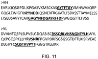
- the CAR may comprise an antigen-binding domain which comprises a variable heavy chain (VH) having the sequence shown as SEQ ID No. 1 and a variable light chain (VL) having the sequence shown as SEQ ID No. 2.
- VH variable heavy chain
- VL variable light chain
- the CAR may comprise an antigen-binding domain which comprises an scFv having the amino acid sequence shown as SEQ ID No. 3.
- the CAR may comprise an amino acid sequence selected from SEQ ID No. 33, 34 and 35.
- the CAR may comprise a VH CDR3 and/or a VL CDR3 from those listed in Table 1.
- the CAR may comprise an antibody or functional fragment thereof which comprises:
- the CAR may comprise a VH CDR3 and/or a VL CDR3 from those listed in Table 2.
- the CAR may comprise an antibody or functional fragment thereof which comprises:
- the present invention provides a nucleic acid sequence encoding a CAR according to the first aspect of the invention.
- a vector which comprises a nucleic acid sequence according to the second aspect of the invention.
- a cell which comprises a CAR according to the first aspect of the invention.
- the cell may be a cytolytic immune cell, such as a T-cell or natural killer (NK) cell.
- a method for making a cell according to the fourth aspect of the invention which comprises the step of transducing or transfecting a cell with a nucleic acid sequence according to the second aspect of the invention or a vector according to the third aspect of the invention.
- a cell according to the fourth aspect of the invention for use in a method for treating a T-cell lymphoma or leukaemia in a subject which comprises the step of administrating the cell comprising the TCRB1 or TCRB2 selective CAR to the subject, to cause selective depletion of the malignant T-cells, together with normal T-cells expressing the same TRBC as the malignant T-cells, but not to cause depletion of normal T-cells expressing the TRBC not expressed by the malignant T-cells.
- the method may also comprise the step of investigating the TCR beta constant region (TCRB) of a malignant T cell from the subject to determine whether it expresses TRBC1 or TRBC2.
- TCRB TCR beta constant region
- a method for treating a T-cell lymphoma or leukaemia in a subject which comprises the step of administering a TCRB1 or TCRB2 selective agent to the subject, wherein the agent causes selective depletion of the malignant T-cells, together with normal T-cells expressing the same TRBC as the malignant T-cells, but does not cause depletion of normal T-cells expressing the TRBC not expressed by the malignant T-cells.
- the agent is a TCRB1 selective agent. In a second aspect of this aspect of the disclosure the agent is a TRBC2 selective agent.
- the method may also comprise the step of investigating the TCR beta constant region (TRBC) of a malignant T-cell to determine whether it expresses TRBC1 or TRBC2, prior to the administration step.
- TRBC TCR beta constant region
- the agent may be a depleting monoclonal antibody or a fragment thereof.
- the agent may be a conjugated antibody, which may comprise a chemotherapeutic entity.
- the agent may be a bispecific T-cell engager.
- the agent may be a chimeric antigen receptor (CAR) expressing T-cell.
- the CAR may comprise an amino acid sequence selected from the group consisting of SEQ ID No. 33, 34 and 35.
- the agent may comprise the JOVI-1 antibody or a functional fragment thereof.
- the agent may comprise an antibody or a functional fragment thereof having a variable heavy chain (VH) and a variable light chain (VL) which comprise the following complementarity determining regions (CDRs):
- the agent may comprise an antibody of functional fragment thereof which comprises a variable heavy chain (VH) having the amino acid sequence shown as SEQ ID No. 1 and a variable light chain (VL) having the amino acid sequence shown as SEQ ID No. 2.
- VH variable heavy chain
- VL variable light chain
- the agent may comprise an ScFv having the amino acid sequence shown as SEQ ID No. 3.
- the agent may be provided as a pharmaceutical composition.
- the T-cell lymphoma or leukaemia may be selected from peripheral T-cell lymphoma, not otherwise specified (PTCL-NOS); angio-immunoblastic T-cell lymphoma (AITL), anaplastic large cell lymphoma (ALCL), enteropathy-associated T-cell lymphoma (EATL), hepatosplenic T-cell lymphoma (HSTL), extranodal NK/T-cell lymphoma nasal type, cutaneous T-cell lymphoma,primary cutaneous ALCL, T cell prolymphocytic leukaemia and T-cell acute lymphoblastic leukaemia.
- PTCL-NOS peripheral T-cell lymphoma
- AITL angio-immunoblastic T-cell lymphoma
- ALCL anaplastic large cell lymphoma
- EATL enteropathy-associated T-cell lymphoma
- HSTL hepatosplenic T-cell lymphoma
- the present disclosure also provides an agent for use in treating a T-cell lymphoma or leukaemia according to such a method.
- the present disclosure also provides a kit comprising an agent for use as defined above.
- the present disclosure also provides the use of an agent in the manufacture of a medicament for treatment of a T-cell lymphoma or leukaemia according to the above method.
- the present disclosure also provides a method for diagnosing a T-cell lymphoma or leukaemia in a subject which comprises the step of determining the percentage of total T-cells in a sample which are TRBC1 or TRBC2 positive.
- a percentage of TRBC1 or TRBC2 positive T-cells which is greater than about 80% may indicate the presence of a T-cell lymphoma or leukaemia.
- the sample may be a peripheral blood sample or a biopsy.
- the agent which binds total T-cells may bind CD3.
- the present disclosure provides agents, such as chimeric antigen receptors (CARs) which selectively bind TRBC1 or TRBC2.
- CARs chimeric antigen receptors
- Such agents are useful in methods for treating a T-cell lymphoma or leukaemia in a subject.
- T cell malignancies are clonal, so they either express TRBC1 or TRBC2.
- the agent By administering a TCRB1 or TCRB2 selective agent to the subject, the agent causes selective depletion of the malignant T-cells, together with normal T-cells expressing the same TRBC as the malignant T-cells, but does not cause depletion of normal T-cells expressing the TRBC not expressed by the malignant T-cells.
- T-cell receptor is expressed on the surface of T lymphocytes and is responsible for recognizing antigens bound to major histocompatibility complex (MHC) molecules.
- MHC major histocompatibility complex
- the T lymphocyte is activated through a series of biochemical events mediated by associated enzymes, co-receptors, specialized adaptor molecules, and activated or released transcription factors.
- the TCR is a disulfide-linked membrane-anchored heterodimer normally consisting of the highly variable alpha ( ⁇ ) and beta ( ⁇ ) chains expressed as part of a complex with the invariant CD3 chain molecules. T-cells expressing this receptor are referred to as ⁇ : ⁇ (or ⁇ ) T-cells (-95% total T-cells). A minority of T-cells express an alternate receptor, formed by variable gamma ( ⁇ ) and delta ( ⁇ ) chains, and are referred to as ⁇ T-cells (-5% total T cells).
- Each ⁇ and ⁇ chain is composed of two extracellular domains: Variable (V) region and a Constant (C) region, both of Immunoglobulin superfamily (IgSF) domain forming antiparallel ⁇ -sheets.
- the constant region is proximal to the cell membrane, followed by a transmembrane region and a short cytoplasmic tail, while the variable region binds to the peptide/MHC complex (see Figure 1 ).
- the constant region of the TCR consists of short connecting sequences in which a cysteine residue forms disulfide bonds, which forms a link between the two chains.
- variable domains of both the TCR ⁇ -chain and ⁇ -chain have three hypervariable or complementarity determining regions (CDRs).
- CDRs hypervariable or complementarity determining regions
- the variable region of the ⁇ -chain also has an additional area of hypervariability (HV4), however, this does not normally contact antigen and is therefore not considered a CDR.
- the TCR also comprises up to five invariant chains ⁇ , ⁇ , ⁇ (collectively termed CD3) and ⁇ .
- CD3 and ⁇ subunits mediate TCR signalling through specific cytoplasmic domains which interact with second-messenger and adapter molecules following the recognition of the antigen by ⁇ or ⁇ .
- Cell-surface expression of the TCR complex is preceded by the pair-wise assembly of subunits in which both the transmembrane and extracellular domains of TCR ⁇ and ⁇ and CD3 ⁇ and ⁇ play a role
- TCRs are therefore commonly composed of the CD3 complex and the TCR ⁇ and ⁇ chains, which are in turn composed of variable and constant regions ( Figure 1 ).
- TRBC1 and TRBC2 The locus (Chr7:q34) which supplies the TCR ⁇ -constant region (TRBC) has duplicated in evolutionary history to produce two almost identical and functionally equivalent genes: TRBC1 and TRBC2 ( Figure 2 ), which differ by only 4 amino acid in the mature protein produced by each ( Figure 3 ).
- TRBC1 and TRBC2 Figure 2
- Each TCR will comprise, in a mutually exclusive fashion, either TRBC1 or TRBC2 and as such, each ⁇ T-cell will express either TRBC1 or TRBC2, in a mutually exclusive manner.
- the present inventors have determined that, despite the similarity between the sequence of the TRBC1 and TRBC2, it is possible to discriminate between them.
- the inventors have also determined that amino acid sequences of TRBC1 and TRBC2 can be discriminated whilst in situ on the surface of a cell, for example a T-cell.
- 'malignant' is used herein according to its standard meaning to refer to a cell which is not self-limited in its growth, may be capable of invading into adjacent tissues and may be capable of spreading to distant tissue.
- 'malignant T cell' is used herein to refer to a clonally expanded T cell in the context of a lymphoma or leukaemia.
- the present invention involves determining the TRBC of a malignant T-cell. This may be performed using methods known in the art. For example it may be determined by PCR, western blot, flow cytometry or fluorescent microscopy methods.
- the appropriate TRBC1 or TRBC2 selective agent is administered to the subject.
- the 'appropriate TRBC selective agent' means that where the malignant T-cell is determined to express TRBC1, a TRBC1 selective agent is administered, whereas where the malignant T-cell is determined to express TRBC2, a TRBC2 selective agent is administered.
- the selective agent binds to either TRBC1 or TRBC2 in a mutually exclusive manner.
- each ⁇ T-cell expresses a TCR which comprises either TRBC1 or TRBC2.
- a clonal T-cell disorder such as a T-cell lymphoma or leukaemia
- malignant T-cells derived from the same clone will all express either TRBC1 or TRBC2.
- the present disclosure comprises the step of administering a TRBC1 or TRBC2 selective agent to the subject, wherein the agent causes selective depletion of the malignant T-cells, together with normal T-cells which express the same TRBC as the malignant T-cells, but does not cause significant depletion of normal T-cells expressing the other TRBC from the malignant T-cells.
- the TRBC selective agent does not cause significant depletion of normal T-cells expressing the other TRBC from the malignant T-cells it does not cause depletion of the entire T-cell compartment. Retention of a proportion of the subject's T-cell compartment (i.e. T-cells which do not express the same TRBC as the malignant T-cell) results in reduced toxicity and reduced cellular and humoral immunodeficiency, thereby reducing the risk of infection.
- Administration of a TRBC1 selective agent according to the present disclosure may result in a 5, 10, 20, 50, 75, 90, 95 or 99% depletion, i.e. reduction in the number of T-cells expressing TRBC1.
- Administration of a TRBC2 selective agent according to the present disclosure may result in a 5, 10, 20, 50, 75, 90, 95 or 99% depletion, i.e.reduction in the number of T-cells expressing TRBC2.
- TRBC1 selective agent may bind TRBC1 with an at least 2-fold, 4-fold, 5-fold, 7-fold or 10-fold greater affinity that TRBC2.
- TRBC2 selective agent may bind TRBC2 with an at least 2-fold, 4-fold, 5-fold, 7-fold or 10-fold greater affinity that TRBC1.
- a TRBC1 selective agent causes depletion of a greater proportion of TRBC1-expressing T-cells in cell population than TRBC2-expressing cell.
- the ratio of depletion of TRBC1-expressing T-cells to TRBC2-expressing cells may be at least 60%:40%, 70%:30%, 80%:20%, 90%:10% or 95%:5%.
- a TRBC2 selective agent causes depletion of a greater proportion of TRBC1-expressing T-cells in cell population than TRBC2-expressing cell.
- the ratio of depletion of TRBC2-expressing T-cells to TRBC1-expressing cells may be at least 60%:40%, 70%:30%, 80%:20%, 90%:10% or 95%:5%.
- malignant T-cells are deleted in a subject, without affecting a significant proportion of healthy T cells.
- a significant proportion it is meant that a sufficient proportion of T cells expressing the TRBC different from the maltgnant T cells survive in order to maintain T-cell function in the subject.
- the agent may cause depletion of less than 20%, 15%, 10% or 5% of the T-cell population expressing the other TCRB.
- the selective agent may be selective for either TRBC1 or TRBC2 because it discriminates residues as listed below:
- the selective agent may discriminate any combination of the differences above differences.
- the agent used in the method of the present disclosure may be a depleting monoclonal antibody (mAb) or a functional fragment thereof, or an antibody mimetic.
- mAb monoclonal antibody
- the term 'depleting antibody' is used in the conventional sense to relate to an antibody which binds to an antigen present on a target T-cell and mediates death of the target T-cell.
- the administration of a depleting antibody to a subject therefore results in a reduction/decrease in the number of cells within the subject which express the target antigen.
- antibody means a polypeptide having an antigen binding site which comprises at least one complementarity determining region CDR.
- the antibody may comprise 3 CDRs and have an antigen binding site which is equivalent to that of a domain antibody (dAb).
- the antibody may comprise 6 CDRs and have an antigen binding site which is equivalent to that of a classical antibody molecule.
- the remainder of the polypeptide may be any sequence which provides a suitable scaffold for the antigen binding site and displays it in an appropriate manner for it to bind the antigen.
- the antibody may be a whole immunoglobulin molecule or a part thereof such as a Fab, F(ab)'2, Fv, single chain Fv (ScFv) fragment or Nanobody.
- the antibody may be a bifunctional antibody.
- the antibody may be non-human, chimeric, humanised or fully human.
- the antibody may therefore be any functional fragment which retains the antigen specificity of the full antibody.
- the agent of the present invention for use in the method of the present disclosure may comprise an antibody or a functional fragment thereof having a variable heavy chain (VH) and a variable light chain (VL) which comprises one or more of the following complementarity determining regions (CDRs):
- the one or more CDRs each independently may or may not comprise one or more amino acid mutations (eg substitutions) compared to the sequences given as SEQ ID No. 7 to 12, provided that the resultant antibody retains the ability to selectively bind to TRBC1.
- CDRs L3 and H3 are prevalently responsible for high energy interactions with the antigen, so the antibody or functional fragment thereof, may comprise VH CDR3 and/or VL CDR3 as outlined above.
- the agent may comprise an antibody or a functional fragment thereof having a variable heavy chain (VH) and/or a variable light chain (VL) which comprises one or more of the complementarity determining regions (CDR3s) shown in the Table 1.
- VH variable heavy chain
- VL variable light chain
- CDR3s complementarity determining regions
- agent is a domain antibody it may comprise 3 CDRs, i.e. either VH CDR1-CDR3 or VL CDR1-CDR3.
- the agent may comprise an antibody of functional fragment thereof which comprises a variable heavy chain (VH) having the amino acid sequence shown as SEQ ID No. 1 and a variable light chain (VL) having the amino acid sequence shown as SEQ ID No. 2.
- VH variable heavy chain
- VL variable light chain
- the agent may comprise an ScFv having the amino acid sequence shown as SEQ ID No. 3.
- the agent may comprise an antibody or functional fragment thereof which comprises:
- the agent may comprise an antibody or a functional fragment thereof having a variable heavy chain (VH) and/or a variable light chain (VL) which comprises one or more of the complementarity determining regions (CDR3s) shown in the Table 2.
- VH variable heavy chain
- VL variable light chain
- CDR3s complementarity determining regions
- the agent may comprise an antibody or functional fragment thereof which comprises:
- Variants of the above amino acid sequences may also be used in the present disclosure provided that the resulting antibody binds TRBC1 or TRBC2 and does not significantly cross-react. Typically such variants have a high degree of sequence identity with one of the sequences specified above.
- NCBI Basic Local Alignment Search Tool is available from several sources, including the National Center for Biotechnology Information (NCBI, Bethesda, Md.) and on the internet, for use in connection with the sequence analysis programs blastp, blastn, blastx, tblastn and tblastx. A description of how to determine sequence identity using this program is available on the NCBI website on the internet.
- Variants of the VL or VH domain or scFv typically have at least about 75%, for example at least about 80%, 90%, 95%, 96%, 97%, 98% or 99% sequence identity with the sequences given as SEQ ID Nos 1-3, 13-32..
- variants may contain one or more conservative amino acid substitutions compared to the original amino acid or nucleic acid sequence.
- Conservative substitutions are those substitutions that do not substantially affect or decrease the affinity of an antibody to bind TRBC1 or TRBC2.
- a human antibody that specifically binds TRBC1 or TRBC2 may include up to 1, up to 2, up to 5, up to 10, or up to 15 conservative substitutions in either or both of the VH or VL compared to any of the sequences given as SEQ ID No. 1-3 or 13-32 and retain specific binding to TRBC1 or TRBC2.
- amino acids which may be exchanged by way of conservative substitution are well known to one of ordinary skill in the art.
- the following six groups are examples of amino acids that are considered to be conservative substitutions for one another: 1) Alanine (A), Serine (S), Threonine (T); 2) Aspartic acid (D), Glutamic acid (E); 3) Asparagine (N), Glutamine (Q); 4) Arginine (R), Lysine (K); 5) Isoleucine (I), Leucine (L), Methionine (M), Valine (V); and 6) Phenylalanine (F), Tyrosine (Y), Tryptophan (W).
- Antibodies may be obtained from animal serum, or, in the case of monoclonal antibodies or fragments thereof, produced in cell culture. Recombinant DNA technology may be used to produce the antibodies according to established procedure, in bacterial or mammalian cell culture.
- monoclonal antibodies are typically made by fusing myeloma cells with the spleen cells from a mouse or rabbit that has been immunized with the desired antigen.
- the desired antigen is TRBC1 or TRBC2 peptide, or a TCR ⁇ chain comprising either TRBC1 or TRBC2.
- antibodies and related molecules may be made outside the immune system by combining libraries of VH and VL chains in a recombinant manner.
- libraries may be constructed and screened using phage-display technology as described in Example 12.
- Antibodies which are selective for either TRBC1 or TRBC2 may be identified using methods which are standard in the art. Methods for determining the binding specificity of an antibody include, but are not limited to, ELISA, western blot, immunohistochemistry, flow cytometry, Förster resonance energy transfer (FRET), phage display libraries, yeast two-hybrid screens, co-immunoprecipitation, bimolecular fluorescence complementation and tandem affinity purification.
- FRET Förster resonance energy transfer
- an antibody which is selective for either TRBC1 or TRBC2 the binding of the antibody to each of TRBC1 and TRBC2 is assessed. Typically, this is assessed by determining the binding of the antibody to each TRBC separately. An antibody which is selective binds to either TRBC1 or TRBC2 without significant binding to the other TRBC.
- the agent may alternatively be a molecule which is not derived from or based on an immunoglobulin.
- a number of "antibody mimetic" designed repeat proteins (DRPs) have been developed to exploit the binding abilities of non-antibody polypeptides.
- Repeat proteins such as ankyrin or leucine-rich repeat proteins are ubiquitous binding molecules which occur, unlike antibodies, intra- and extracellularly. Their unique modular architecture features repeating structural units (repeats), which stack together to form elongated repeat domains displaying variable and modular target-binding surfaces. Based on this modularity, combinatorial libraries of polypeptides with highly diversified binding specificities can be generated.
- DARPins Designed Ankyrin Repeat Proteins
- the binding specificity is derived from lipocalins, a family of proteins which perform a range of functions in vivo associated with physiological transport and storage of chemically sensitive or insoluble compounds.
- Lipocalins have a robust intrinsic structure comprising a highly conserved ⁇ -barrel which supports four loops at one terminus of the protein. These loops for the entrance to a binding pocket and conformational differences in this part of the molecule account for the variation in binding specificity between different lipocalins.
- Avimers are evolved from a large family of human extracellular receptor domainsby in vitro exon shuffling and phage display, generating multi-domain proteins with binding and inhibitory properties.
- Versabodies are small proteins of 3-5kDa with >15% cysteines which form a high disulfide density scaffold, replacing the hydrophobic core present in most proteins.
- the replacement of a large number of hydrophobic amino acids, comprising the hydrophobic core, with a small number of disulphides results in a protein that is smaller, more hydrophilic, more resistant to proteases and heat and has a lower density of T-cell epitopes. All four of these properties result in a protein having considerably reduced immunogenicity. They may also be manufactured in E. coli, and are highly soluble and stable.
- the antibody or mimetic may be a conjugate of the antibody or mimetic and another agent or antibody, for example the conjugate may be a detectable entity or a chemotherapeutic entity.
- the detectable entity may be a fluorescent moiety, for example a fluorescent peptide.
- a "fluorescent peptide” refers to a polypeptide which, following excitation, emits light at a detectable wavelength.
- fluorescent proteins include, but are not limited to, fluorescein isothiocyanate (FITC), phycoerythrin (PE), allophycocyanin (APC), green fluorescent protein (GFP), enhanced GFP, red fluorescent protein (RFP), blue fluorescent protein (BFP) and mCherry.
- a selective TRBC1 or TRBC2 agent conjugated to a detectable entity may be used to determine the TRBC of a malignant T cell.
- a chemotherapeutic entity as used herein refers to an entity which is destructive to a cell, that is the entity reduces the viability of the cell.
- the chemotherapeutic entity may be a cytotoxic drug.
- a chemotherapeutic agent contemplated includes, without limitation, alkylating agents, nitrosoureas, ethylenimines/methylmelamine, alkyl sulfonates, antimetabolites, pyrimidine analogs, epipodophylotoxins, enzymes such as L-asparaginase; biological response modifiers such as IFN ⁇ , IL-2, G-CSF and GM-CSF; platinium coordination complexes such as cisplatin and carboplatin, anthracenediones, substituted urea such as hydroxyurea, methylhydrazine derivatives including N-methylhydrazine (MIH) and procarbazine, adrenocortical suppressants such as mitotane (o,p'
- a TRBC selective agent conjugated to a chemotherapeutic entity enables the targeted delivery of the chemotherapeutic entity to cells which express either TRBC1 or TRBC2.
- Bispecific T-cell engaging molecules are a class of bispecific antibody-type molecules that have been developed, primarily for the use as anti-cancer drugs. They direct a host's immune system, more specifically the T cells' cytotoxic activity, against a target cell, such as a cancer cell. In these molecules, one binding domain binds to a T cell via the CD3 receptor, and the other to a target cells such as a tumor cell (via a tumor specific molecule). Since the bispecific molecule binds both the target cell and the T cell, it brings the target cell into proximity with the T cell, so that the T cell can exert its effect, for example, a cytotoxic effect on a cancer cell.
- T cell:bispecific Ab:cancer cell complex induces signaling in the T cell leading to, for example, the release of cytotoxic mediators.
- the agent only induces the desired signaling in the presence of the target cell, leading to selective killing.
- Bispecific T-cell engaging molecules have been developed in a number of different formats, but one of the most common is a fusion consisting of two single-chain variable fragments (scFvs) of different antibodies. These are sometimes known as BiTEs (Bi-specific T-cell Engagers).
- the agent used in the method of the present disclosure may be a bi-specific molecule which selectively recognises TRBC1 or TRBC2 and is capable of activating a T cell.
- the agent may be a BiTE.
- the agent used in the method may comprise:
- the bi-specific molecule may comprise a signal peptide to aid in its production.
- the signal peptide may cause the bi-specific molecule to be secreted by a host cell, such that the bi-specific molecule can be harvested from the host cell supernatant.
- the signal peptide may be at the amino terminus of the molecule.
- the bi-specific molecule may have the general formula: Signal peptide - first domain - second domain.
- the bi-specific molecule may comprise a spacer sequence to connect the first domain with the second domain and spatially separate the two domains.
- the spacer sequence may, for example, comprise an IgG1 hinge or a CD8 stalk.
- the linker may alternatively comprise an alternative linker sequence which has similar length and/or domain spacing properties as an IgG1 hinge or a CD8 stalk.
- the bi-specific molecule may comprise JOVI-1, or a functional fragment thereof, as defined above.
- Chimeric antigen receptors also known as chimeric T-cell receptors, artificial T-cell receptors and chimeric immunoreceptors, are engineered receptors, which graft an arbitrary specificity onto an immune effector cell.
- CARs also known as chimeric T-cell receptors, artificial T-cell receptors and chimeric immunoreceptors, are engineered receptors, which graft an arbitrary specificity onto an immune effector cell.
- the specificity of a monoclonal antibody is grafted on to a T-cell.
- CAR-encoding nucleic acids may be transferred to T-cells using, for example, retroviral vectors. In this way, a large number of cancer-specific T-cells can be generated for adoptive cell transfer. Phase I clinical studies of this approach show efficacy.
- the target-antigen binding domain of a CAR is commonly fused via a spacer and transmembrane domain to an endodomain, which comprises or associates with an intrcellular T-cell signalling domain.
- an endodomain which comprises or associates with an intrcellular T-cell signalling domain.
- the agent used in the method of the present disclosure may be a CAR which selectively recognises TRBC1 or TRBC2.
- the agent may be a T-cell which expresses a CAR which selectively recognises TRBC1 or TRBC2.
- the CAR may also comprise a transmembrane domain which spans the membrane. It may comprise a hydrophobic alpha helix.
- the transmembrane domain may be derived from CD28, which gives good receptor stability.
- the endodomain is the portion of the CAR involved in signal-transmission.
- the endodomain either comprises or associates with an intracellular T-cell signalling domain. After antigen recognition, receptors cluster and a signal is transmitted to the cell.
- the most commonly used T-cell signalling component is that of CD3-zeta which contains 3 ITAMs. This transmits an activation signal to the T-cell after antigen is bound.
- CD3-zeta may not provide a fully competent activation signal and additional co-stimulatory signaling may be needed.
- chimeric CD28 and OX40 can be used with CD3-Zeta to transmit a proliferative / survival signal, or all three can be used together.
- the endodomain of the CAR may comprise the CD28 endodomain and OX40 and CD3-Zeta endodomain.
- the CAR may comprise a signal peptide so that when the CAR is expressed inside a cell, such as a T-cell, the nascent protein is directed to the endoplasmic reticulum and subsequently to the cell surface, where it is expressed.
- the CAR may comprise a spacer sequence to connect the TRBC-binding domain with the transmembrane domain and spatially separate the TRBC-binding domain from the endodomain.
- a flexible spacer allows to the TRBC-binding domain to orient in different directions to enable TRBC binding.
- the spacer sequence may, for example, comprise an IgG1 Fc region, an IgG1 hinge or a CD8 stalk, or a combination thereof.
- the linker may alternatively comprise an alternative linker sequence which has similar length and/or domain spacing properties as an IgG1 Fc region, an IgG1 hinge or a CD8 stalk.
- the spacer may an therefore comprise an IgG1 hinge or a CD8 stalk or a spacer which has a similar length and/or domain spacing properties as an IgG1 hinge or a CD8 stalk.
- a human IgG1 spacer may be altered to remove Fc binding motifs.
- the CAR may comprise the JOVI-1 antibody, or a functional fragment thereof, as defined above.
- the CAR may comprise an amino acid sequence selected from the group consisting of SEQ ID No. 33, 34 and 35.
- one or more of the 6 CDRs each independently may or may not comprise one or more amino acid mutations (eg substitutions) compared to the sequences given as SEQ ID No. 7 to 12, provided that the resultant CAR retains the ability to bind to TRBC1.
- Variants of the above amino acid sequences may also be used in the present disclosure provided that the resulting CAR binds TRBC1 or TRBC2 and does not significantly cross-react. Typically such variants have a high degree of sequence identity with one of the sequences given as SEQ ID No. 33, 34 or 35.
- Variants of the CAR typically have at least about 75%, for example at least about 80%, 90%, 95%, 96%, 97%, 98% or 99% sequence identity with one of the sequences given as SEQ ID Nos 33, 34 and 35.
- the present disclosure further provides a nucleic acid encoding an agent such as a BiTE or CAR of the first aspect of the disclosure.
- the nucleic acid sequence may encode a CAR comprising one of the amino acid sequences shown as SEQ ID No. 33, 34 and 35..
- polynucleotide As used herein, the terms “polynucleotide”, “nucleotide”, and “nucleic acid” are intended to be synonymous with each other.
- Nucleic acids according to the invention may comprise DNA or RNA. They may be single-stranded or double-stranded. They may also be polynucleotides which include within them synthetic or modified nucleotides. A number of different types of modification to oligonucleotides are known in the art. These include methylphosphonate and phosphorothioate backbones, addition of acridine or polylysine chains at the 3' and/or 5' ends of the molecule. For the purposes of the use as described herein, it is to be understood that the polynucleotides may be modified by any method available in the art. Such modifications may be carried out in order to enhance the in vivo activity or life span of polynucleotides of interest.
- variant in relation to a nucleotide sequence include any substitution of, variation of, modification of, replacement of, deletion of or addition of one (or more) nucleic acid from or to the sequence.
- the present disclosure also provides a vector, or kit of vectors, which comprises one or more nucleic acid sequence(s) of the disclosure.
- a vector may be used to introduce the nucleic acid sequence(s) into a host cell so that it expresses a CAR according to the first aspect of the disclosure
- the vector may, for example, be a plasmid or a viral vector, such as a retroviral vector or a lentiviral vector, or a transposon based vector or synthetic mRNA.
- the vector may be capable of transfecting or transducing a T cell or a NK cell.
- the present disclosure also relates to a cell, such as an immune cell, comprising a CAR according to the first aspect of the disclosure.
- the cell may comprise a nucleic acid or a vector of the present disclosure
- the cell may be a T-cell or a natural killer (NK) cell.
- NK natural killer
- T cell may be T cells or T lymphocytes which are a type of lymphocyte that play a central role in cell-mediated immunity. They can be distinguished from other lymphocytes, such as B cells and natural killer cells (NK cells), by the presence of a T-cell receptor (TCR) on the cell surface.
- TCR T-cell receptor
- Helper T helper cells assist other white blood cells in immunologic processes, including maturation of B cells into plasma cells and memory B cells, and activation of cytotoxic T cells and macrophages.
- TH cells express CD4 on their surface.
- TH cells become activated when they are presented with peptide antigens by MHC class II molecules on the surface of antigen presenting cells (APCs).
- APCs antigen presenting cells
- These cells can differentiate into one of several subtypes, including TH1, TH2, TH3, TH17, Th9, or TFH, which secrete different cytokines to facilitate different types of immune responses.
- Cytolytic T cells destroy virally infected cells and tumor cells, and are also implicated in transplant rejection.
- CTLs express the CD8 at their surface. These cells recognize their targets by binding to antigen associated with MHC class I, which is present on the surface of all nucleated cells.
- MHC class I MHC class I
- IL-10 adenosine and other molecules secreted by regulatory T cells, the CD8+ cells can be inactivated to an anergic state, which prevent autoimmune diseases such as experimental autoimmune encephalomyelitis.
- Memory T cells are a subset of antigen-specific T cells that persist long-term after an infection has resolved. They quickly expand to large numbers of effector T cells upon re-exposure to their cognate antigen, thus providing the immune system with "memory" against past infections.
- Memory T cells comprise three subtypes: central memory T cells (TCM cells) and two types of effector memory T cells (TEM cells and TEMRA cells). Memory cells may be either CD4+ or CD8+. Memory T cells typically express the cell surface protein CD45RO.
- Treg cells Regulatory T cells
- suppressor T cells are crucial for the maintenance of immunological tolerance. Their major role is to shut down T cell-mediated immunity toward the end of an immune reaction and to suppress auto-reactive T cells that escaped the process of negative selection in the thymus.
- Treg cells Two major classes of CD4+ Treg cells have been described - naturally occurring Treg cells and adaptive Treg cells.
- Naturally occurring Treg cells arise in the thymus and have been linked to interactions between developing T cells with both myeloid (CD11c+) and plasmacytoid (CD123+) dendritic cells that have been activated with TSLP.
- Naturally occurring Treg cells can be distinguished from other T cells by the presence of an intracellular molecule called FoxP3. Mutations of the FOXP3 gene can prevent regulatory T cell development, causing the fatal autoimmune disease IPEX.
- Adaptive Treg cells may originate during a normal immune response.
- the cell may be a Natural Killer cell (or NK cell).
- NK cells form part of the innate immune system. NK cells provide rapid responses to innate signals from virally infected cells in an MHC independent manner NK cells (belonging to the group of innate lymphoid cells) are defined as large granular lymphocytes (LGL) and constitute the third kind of cells differentiated from the common lymphoid progenitor generating B and T lymphocytes. NK cells are known to differentiate and mature in the bone marrow, lymph node, spleen, tonsils and thymus where they then enter into the circulation.
- LGL large granular lymphocytes
- the CAR cells of the disclosure may be any of the cell types mentioned above.
- T or NK cells expressing the a CAR according to the first aspect of the invention may either be created ex vivo either from a patient's own peripheral blood (1st party), or in the setting of a haematopoietic stem cell transplant from donor peripheral blood (2nd party), or peripheral blood from an unconnected donor (3rd party).
- T or NK cells expressing a CAR according to the first aspect of the invention may be derived from ex vivo differentiation of inducible progenitor to T or NK cells.
- an immortalized T-cell line which retains its lytic function and could act as a therapeutic may be used.
- CAR cells are generated by introducing DNA or RNA coding for the CAR by one of many means including transduction with a viral vector, transfection with DNA or RNA.
- the CAR cell of the invention may be an ex vivo T or NK cell from a subject.
- the T or NK cell may be from a peripheral blood mononuclear cell (PBMC) sample.
- PBMC peripheral blood mononuclear cell
- T or NK cells may be activated and/or expanded prior to being transduced with nucleic acid encoding a CAR according to the first aspect of the invention, for example by treatment with an anti-CD3 monoclonal antibody.
- the T or NK cell of the invention may be made by:
- the T or NK cells may then by purified, for example, selected on the basis of expression of the antigen-binding domain of the antigen-binding polypeptide.
- the present disclosure also provides a kit which comprises a T or NK cell comprising a CAR according to the first aspect of the invention.
- the present invention also relates to a pharmaceutical composition containing a plurality of cells expressing a CAR of the first aspect of the invention.
- the pharmaceutical composition may additionally comprise a pharmaceutically acceptable carrier, diluent or excipient.
- the pharmaceutical composition may optionally comprise one or more further pharmaceutically active polypeptides and/or compounds.
- Such a formulation may, for example, be in a form suitable for intravenous infusion.
- the present invention relates to agents and cells for use in methods for treating a T-cell lymphoma and/or leukaemia.
- a method for treating a T-cell lymphoma and/or leukaemia relates to the therapeutic use of an agent.
- the agent may be administered to a subject having an existing disease of T-cell lymphoma and/or leukaemia in order to lessen, reduce or improve at least one symptom associated with the disease and/or to slow down, reduce or block the progression of the disease.
- the method of the present disclosure may be used for the treatment of any lymphoma and/or leukaemia associated with the clonal expansion of a cell expressing a T-cell receptor (TCR) comprising a ⁇ constant region.
- TCR T-cell receptor
- the present document discloses a method for treating a disease which involves malignant T cells which express a TCR comprising a TRBC.
- the method of the present disclosure may be used to treat a T-cell lymphoma in which the malignant T-cell expresses a TCR comprising a TRBC.
- 'Lymphoma' is used herein according to its standard meaning to refer to a cancer which typically develops in the lymph nodes, but may also affect the spleen, bone marrow, blood and other organs. Lymphoma typically presents as a solid tumour of lymphoid cells.
- the primary symptom associated with lymphoma is lymphadenopathy, although secondary (B) symptoms can include fever, night sweats, weight loss, loss of appetite, fatigue, respiratory distress and itching.
- the method of the present disclosure may be used to treat a T-cell leukaemia in which the malignant T-cell expresses a TCR comprising a TRBC.
- 'Leukaemia' is used herein according to its standard meaning to refer to a cancer of the blood or bone marrow.
- T-cell lymphomas are relatively uncommon lymphomas and account fewer than 10% of all non-Hodgkin lymphomas (NHL). However, they are associated with an aggressive clinical course and the causes and precise cellular origins of most T-cell lymphomas are still not well defined.
- NHL non-Hodgkin lymphomas
- Lymphoma usually first presents as swelling in the neck, underarm or groin. Additional swelling may occur where other lymph nodes are located such as in the spleen. In general, enlarged lymph nodes can encroach on the space of blood vessels, nerves, or the stomach, leading to swollen arms and legs, to tingling and numbness, or to feelings of being full, respectively. Lymphoma symptoms also include nonspecific symptoms such as fever, chills, unexplained weight loss, night sweats, lethargy, and itching.
- the WHO classification utilizes morphologic and immunophenotypic features in conjunction with clinical aspects and in some instances genetics to delineate a prognostically and therapeutically meaningful categorization for peripheral T-cell lymphomas ( Swerdlow et al.; WHO classification of tumours of haematopoietic and lymphoid tissues. 4th ed.; Lyon: IARC Press; 2008 ).
- the anatomic localization of neoplastic T-cells parallels in part their proposed normal cellular counterparts and functions and as such T-cell lymphomas are associated with lymph nodes and peripheral blood. This approach allows for better understanding of some of the manifestations of the T-cell lymphomas, including their cellular distribution, some aspects of morphology and even associated clinical findings.
- T-cell lymphomas peripheral T-cell lymphoma, not otherwise specified (PTCL-NOS) comprising 25% overall, followed by angioimmunoblastic T-cell lymphoma (AITL) (18.5%)
- PTCL-NOS peripheral T-cell lymphoma
- AITL angioimmunoblastic T-cell lymphoma
- PTCL-NOS comprises over 25% of all peripheral T-cell lymphomas and NK/T-cell lymphomas and is the most common subtype. It is determined by a diagnosis of exclusion, not corresponding to any of the specific mature T-cell lymphoma entities listed in the current WHO 2008. As such it is analogous to diffuse large B-cell lymphoma, not otherwise specified (DLBCL-NOS).
- lymphoepithelioid (Lennert) variant
- T-zone variant a morphologically defined variant
- follicular variant The lymphoepithelioid variant of PTCL contains abundant background epithelioid histiocytes and is commonly positive for CD8. It has been associated with a better prognosis.
- the follicular variant of PTCL-NOS is emerging as a potentially distinct clinicopathologic entity.
- PTCL-NOS have a mature T-cell phenotype and most cases are CD4-positive. 75% of cases show variable loss of at least one pan T-cell marker (CD3, CD2, CD5 or CD7), with CD7 and CD5 being most often downregulated. CD30 and rarely CD15 can be expressed, with CD15 being an adverse prognostic feature. CD56 expression, although uncommon, also has negative prognostic impact. Additional adverse pathologic prognostic factors include a proliferation rate greater than 25% based on KI-67 expression, and presence of more than 70% transformed cells. Immunophenotypic analysis of these lymphomas has offered little insight into their biology.
- AITL is a systemic disease characterized by a polymorphous infiltrate involving lymph nodes, prominent high endothelial venules (HEV) and peri-vascular expansion of follicular dendritic cell (FDC) meshworks.
- AITL is considered as a de-novo T-cell lymphoma derived from ⁇ T-cells of follicular helper type (TFH), normally found in the germinal centres.
- AITL is the second most common entity among peripheral T-cell lymphoma and NK/T-cell lymphomas, comprising about 18.5% of cases. It occurs in middle aged to elderly adults, with a median age of 65 years old, and an approximately equal incidence in males and females. Clinically, patients usually have advanced stage disease, with generalized lymphadenopathy, hepatosplenomegaly and prominent constitutional symptoms. Skin rash with associated pruritus is commonly present. There is often polyclonal hypergammaglobulinemia, associated with autoimmune phenomena.
- AITL Three different morphologic patterns are described in AITL.
- the early lesion of AITL usually shows preserved architecture with characteristic hyperplastic follicles.
- the neoplastic proliferation is localized to the periphery of the follicles.
- Pattern II the nodal architecture is partially effaced with retention of few regressed follicles.
- the subcapsular sinuses are preserved and even dilated.
- the paracortex contains arborizing HEV and there is a proliferation of FDC beyond the B-cell follicle.
- the neoplastic cells are small to medium in size, with minimal cytologic atypia. They often have clear to pale cytoplasm, and may show distincT-cell membranes. A polymorphous inflammatory background is usually evident.
- AITL is a T-cell malignancy, there is a characteristic expansion of B-cells and plasma cells, which likely reflects the function of the neoplastic cells as TFH cells. Both EBV-positive and EBV-negative B-cells are present. Occasionally, the atypical B-cells may resemble Hodgkin/Reed-Sternberg-like cells morphologically and immunophenotypically, sometimes leading to a diagnostic confusion with that entity.
- the B-cell proliferation in AITL may be extensive and some patients develop secondary EBV-positive diffuse large B-cell lymphomas (DLBCL) or - more rarely - EBV-negative B-cell tumors, often with plasmacytic differentiation.
- DLBCL diffuse large B-cell lymphomas
- the neoplastic CD4-positive T-cells of AITL show strong expression of CD10 and CD279 (PD-1) and are positive for CXCL13.
- CXCL13 leads to an increased B-cell recruitment to lymph nodes via adherence to the HEV, B-cell activation, plasmacytic differentiation and expansion of the FDC meshworks, all contributing to the morphologic and clinical features of AITL.
- Intense PD-1-expression in the perifollicular tumor cells is particularly helpful in distinguishing AITL Pattern I from reactive follicular and paracortical hyperplasia.
- the follicular variant of PTCL-NOS is another entity with a TFH phenotype. In contradistinction to AITL, it does not have prominent HEV or extra-follicular expansion of FDC meshworks.
- the neoplastic cells may form intrafollicular aggregates, mimicking B-cell follicular lymphoma, but also can have interfollicular growth pattern or involve expanded mantle zones.
- the follicular variant of PTCL-NOS is distinct from AITL as patients more often present with early stage disease with partial lymph node involvement and may lack the constitutional symptoms associated with AITL.
- ALCL may be subdivided as ALCL-'anaplastic lymphoma kinase' (ALK)+ or ALCL-ALK-.
- ALCL-ALK+ is one of the best-defined entities within the peripheral T-cell lymphomas, with characteristic "hallmark cells” bearing horseshoe-shaped nuclei and expressing ALK and CD30. It accounts for about 7% of all peripheral T-cell and NK-cell lymphomas and is most common in the first three decades of life. Patients often present with lymphadenopathy, but the involvement of extranodal sites (skin, bone, soft tissues, lung, liver) and B symptoms is common.
- ALCL, ALK+ shows a wide morphologic spectrum, with 5 different patterns described, but all variants contain some hallmark cells.
- Hallmark cells have eccentric horseshoe- or kidney-shaped nuclei, and a prominent perinuclear eosinophilic Golgi region.
- the tumour cells grow in a cohesive pattern with predilection for sinus involvement. Smaller tumour cells predominate in the small cell variant, and in the lymphohistiocytic variant abundant histiocytes mask the presence of tumour cells, many of which are small.
- ALK expression is a result of a characteristic recurrent genetic alteration consisting of a rearrangement of ALK gene on chromosome 2p23 to one of the many partner genes, resulting in an expression of chimeric protein.
- the most common partner gene, occurring in 75% of cases, is Nucleophosmin ( NPM1 ) on chromosome 5q35, resulting in t(2;5)(p23;q35).
- the cellular distribution of ALK in different translocation variants may vary depending on the partner gene.
- ALCL-ALK- is included as a provisional category in the 2008 WHO classification. It is defined as a CD30 positive T-cell lymphoma that is morphologically indistinguishable from ALCL-ALK+ with a cohesive growth pattern and presence of hallmark cells, but lacking ALK protein expression.
- ALCL-ALK+ Patients are usually adults between the ages of 40 and 65, in contrast to ALCL-ALK+, which is more common in children and young adults.
- ALCL-ALK- can involve both lymph nodes and extranodal tissues, although the latter is seen less commonly than in ALCL-ALK+.
- Most cases of ALCL-ALK- demonstrate effacement of lymph node architecture by sheets of cohesive neoplastic cells with typical "hallmark" features. In contrast to the ALCL-ALK+, the small cell morphologic variant is not recognized.
- ALCL-ALK- shows a greater preservation of surface T-cell marker expression, while the expression of cytotoxic markers and epithelial membrane antigen (EMA) is less likely.
- EMA epithelial membrane antigen
- ALCL-ALK- is clinically distinct from both ALCL-ALK+ and PTCL-NOS, with significant differences in prognosis among these three different entities.
- the 5 year overall survival of ALCL-ALK- is reported as 49% which is not as good as that of ALCL-ALK+ (at 70%), but at the same time it is significantly better than that of PTCL-NOS (32%).
- EATL is an aggressive neoplasm which thought to be derived from the intraepithelial T-cells of the intestine.
- Two morphologically, immunohistochemically and genetically distinct types of EATL are recognized in the 2008 WHO classification: Type I (representing the majority of EATL) and Type II (comprising 10-20% of cases).
- Type I EATL is usually associated with overt or clinically silent gluten-sensitive enteropathy, and is more often seen in patients of Northern European extraction due to high prevalence of celiac disease in this population.
- EATL EATL ⁇ EATL ⁇ EATL ⁇ ⁇ EATL ⁇ ⁇ ⁇ ⁇ ⁇ ⁇ ⁇ ⁇ ⁇ ⁇ ⁇ ⁇ ⁇ ⁇ ⁇ ⁇ ⁇ ⁇ ⁇ ⁇ ⁇ ⁇ ⁇ ⁇ ⁇ ⁇ ⁇ ⁇ ⁇ ⁇ ⁇ ⁇ ⁇ ⁇ ⁇ ⁇ ⁇ ⁇ ⁇ ⁇ ⁇ ⁇ ⁇ ⁇ ⁇ ⁇ ⁇ ⁇ ⁇ ⁇ ⁇ ⁇ ⁇ ⁇ ⁇ ⁇ ⁇ ⁇ ⁇ ⁇ ⁇ ⁇ ⁇ ⁇ ⁇ ⁇ ⁇ ⁇ ⁇ ⁇ ⁇ ⁇ ⁇ ⁇ ⁇ ⁇ ⁇ ⁇ ⁇ ⁇ ⁇ ⁇ ⁇ ⁇ ⁇ ⁇ ⁇ ⁇ ⁇ ⁇ ⁇ ⁇ ⁇ ⁇ ⁇ ⁇ ⁇ ⁇ ⁇ ⁇ ⁇ ⁇ ⁇ ⁇ ⁇ ⁇ ⁇ ⁇ ⁇ ⁇ ⁇ ⁇ ⁇ ⁇ ⁇
- EATL type I The cytological spectrum of EATL type I is broad, and some cases may contain anaplastic cells. There is a polymorphous inflammatory background, which may obscure the neoplastic component in some cases.
- the intestinal mucosa in regions adjacent to the tumour often shows features of celiac disease with blunting of the villi and increased numbers of intraepithelial lymphocytes (IEL), which may represent lesional precursor cells.
- IEL intraepithelial lymphocytes
- the neoplastic cells are often CD3+CD4-CD8-CD7+CD5-CD56- ⁇ F1+, and contain cytotoxic granule-associated proteins (TIA-1, granzyme B, perforin). CD30 is partially expressed in almost all cases.
- CD103 which is a mucosal homing receptor, can be expressed in EATL.
- Type II EATL also referred to as monomorphic CD56+ intestinal T-cell lymphoma
- EATL is defined as an intestinal tumour composed of small- to medium-sized monomorphic T-cells that express both CD8 and CD56.
- Type II EATL has a more world-wide distribution than Type I EATL and is often seen in Asians or Hispanic populations, in whom celiac disease is rare. In individuals of European descent EATL, II represents about 20% of intestinal T-cell lymphomas, with a history of celiac disease in at least a subset of cases. The clinical course is aggressive.
- HSTL is an aggressive systemic neoplasm generally derived from ⁇ cytotoxic T-cells of the innate immune system, however, it may also be derived from ⁇ T-cells in rare cases. It is one of the rarest T-cell lymphomas, and typically affects adolescents and young adults (median age, 35 years) with a strong male predominance.
- Extranodal NK/T-cell lymphoma, nasal type, is an aggressive disease, often with destructive midline lesions and necrosis. Most cases are of NK-cell derivation, but some cases are derived from cytotoxic T-cells. It is universally associated with Epstein-Barr Virus (EBV).
- EBV Epstein-Barr Virus
- the method of the present disclosure may also be used to treat cutaneous T-cell lymphoma.
- Cutaneous T-cell lymphoma is characterised by migration of malignant T-cells to the skin, which causes various lesions to appear. These lesions change shape as the disease progresses, typically beginning as what appears to be a rash and eventually forming plaques and tumours before metastasizing to other parts of the body.
- Cutaneous T-cell lymphomas include those mentioned in the following illustrative, non-exhaustive list; mycosis fungoides, pagetoid reticulosis, Sezary syndrome, granulomatous slack skin, lymphomatoid papulosis, pityriasis lichenoides chronica, CD30+ cutaneous T-cell lymphoma, secondary cutaneous CD30+ large cell lymphoma, non-mycosis fungoides CD30- cutaneous large T-cell lymphoma, pleomorphic T-cell lymphoma, Lennert lymphoma, subcutaneous T-cell lymphoma and angiocentric lymphoma.
- CTCL The signs and symptoms of CTCL vary depending on the specific disease, of which the two most common types are mycosis fungoides and Sezary syndrome.
- Classic mycosis fungoides is divided into three stages:
- Sézary syndrome is defined by erythroderma and leukemia. Signs and symptoms include edematous skin, lymphadenopathy, palmar and/or plantar hyperkeratosis, alopecia, nail dystrophy, ectropion and hepatosplenomegaly.
- cutaneous T-cell lymphoma encompasses a wide variety of disorders.
- T-cell lymphoma ie, mycosis fungoides
- Mycosis fungoides may be preceded by a T-cell-mediated chronic inflammatory skin disease, which may occasionally progress to a fatal lymphoma.
- C-ALCL PRIMARY CUTANEOUS ALCL
- C-ALCL is often indistinguishable from ALC-ALK- by morphology. It is defined as a cutaneous tumour of large cells with anaplastic, pleomorphic or immunoblastic morphology with more than 75% of cells expressing CD30. Together with lymphomatoid papulosis (LyP), C-ALCL belongs to the spectrum of primary cutaneous CD30-positive T-cell lymphoproliferative disorders, which as a group comprise the second most common group of cutaneous T-cell lymphoproliferations after mycosis fungoides.
- the immunohistochemical staining profile is quite similar to ALCL-ALK-, with a greater proportion of cases staining positive for cytotoxic markers. At least 75% of the tumour cells should be positive for CD30. CD15 may also be expressed, and when lymph node involvement occurs, the differential with classical Hodgkin lymphoma can be difficult. Rare cases of ALCL-ALK+ may present with localized cutaneous lesions, and may resemble C-ALCL.
- T-cell acute lymphoblastic leukaemia accounts for about 15% and 25% of ALL in paediatric and adult cohorts respectively. Patients usually have high white blood cell counts and may present with organomegaly, particularly mediastinal enlargement and CNS involvement.
- the method of the present disclosure may be used to treat T-ALL which is associated with a malignant T cell which expresses a TCR comprising a TRBC.
- T-cell-prolymphocytic leukemia is a mature T-cell leukaemia with aggressive behaviour and predilection for blood, bone marrow, lymph nodes, liver, spleen, and skin involvement. T-PLL primarily affects adults over the age of 30. Other names include T-cell chronic lymphocytic leukaemia, "knobby" type of T-cell leukaemia, and T-prolymphocytic leukaemia/T-cell lymphocytic leukaemia.
- T-PLL In the peripheral blood, T-PLL consists of medium-sized lymphocytes with single nucleoli and basophilic cytoplasm with occasional blebs or projections.
- the nuclei are usually round to oval in shape, with occasional patients having cells with a more irregular nuclear outline that is similar to the cerebriform nuclear shape seen in Sezary syndrome.
- a small cell variant comprises 20% of all T-PLL cases, and the Sezary cell-like (cerebriform) variant is seen in 5% of cases.
- T-PLL has the immunophenotype of a mature (post-thymic) T-lymphocyte, and the neoplastic cells are typically positive for pan-T antigens CD2, CD3, and CD7 and negative for TdT and CD1a.
- the immunophenotype CD4+/CD8- is present in 60% of cases, the CD4+/CD8+ immunophenotype is present in 25%, and the CD4-/CD8+ immunophenotype is present in 15% of cases
- the method of the present disclosure may comprise the step of administering the agent in the form of a pharmaceutical composition.
- the agent may be administered with a pharmaceutically acceptable carrier, diluent, excipient or adjuvant.
- a pharmaceutically acceptable carrier diluent, excipient or adjuvant.
- the choice of pharmaceutical carrier, excipient or diluent can be selected with regard to the intended route of administration and standard pharmaceutical practice.
- the pharmaceutical compositions may comprise as (or in addition to) the carrier, excipient or diluent, any suitable binder(s), lubricant(s), suspending agent(s), coating agent(s), solubilising agent(s), and other carrier agents.
- the administration of the agent can be accomplished using any of a variety of routes that make the active ingredient bioavailable.
- the agent can be administered by oral and parenteral routes, intraperitoneally, intravenously, subcutaneously, transcutaneously, intramuscularly, via local delivery for example by catheter or stent.
- a physician will determine the actual dosage which will be most suitable for an individual subject and it will vary with the age, weight and response of the particular patient.
- the dosage is such that it is sufficient to reduce or deplete the number of clonal T-cells expressing either TRBC1 or TRBC2.
- the present invention also provides an agent for use in treating a T-cell lymphoma according to the method of the disclosure.
- the agent may be any agent as defined above.
- the present invention also relates to the use of an agent as defined above in the manufacture of a medicament for the treatment of a T-cell lymphoma according to the disclosed method.
- the present invention further provides a kit comprising an agent as defined above for use in the treatment of a T-cell lymphoma according to the method of the disclosure.
- the kit may also comprise a reagent(s) suitable for determining the TRBC of a malignant T-cell.
- a reagent(s) suitable for determining the TRBC of a malignant T-cell may comprise PCR primers or an antibody (antibodies) which are specific for either TRBC1 or TRBC2.
- the present invention further relates to a method for determining the presence of a T-cell lymphoma or leukaemia in a subject which comprises the step of determining the proportion of T-cells in a sample from a subject which are either TRBC1 or TRBC2 positive.
- T-cell lymphomas involve the clonal expansion of individual malignant T-cells. As such the presence of a T-cell lymphoma in a subject may be identified by determining the proportion of either TRBC1 or TRBC2 T-cells in a sample derived from a patient.
- the sample may be a peripheral blood sample, a lymph sample or a sample taken directly from a tumour e.g. a biopsy sample.
- the proportion of total T-cells which are TRBC1 or TRBC2 positive which indicates the presence of a T-cell lymphoma or leukaemia may be, for example 80, 85, 90, 95, 98 or 99% of a total population of cells.
- the method may involve determining infiltration by a distinct population of T-cells in a biopsy or a sample.
- a T-cell lymphoma or leukaemia is indicated where 80, 85, 90, 95, 98 or 99% of a total population of T cells in the sample are either TRBC1 or TRBC2.
- the total T-cells in a sample may identified by determining the number of cells in the sample which express CD3, CD4, CD8 and/or CD45. A combination of these markers may also be used.
- the proportion of total T cells in a sample which express either TRBC1 or TRBC2 may be determined using methods which are known in the art, for example flow cytometry, immunohistochemistry or fluorescent microscopy.
- the JOVI-1 antibody has been previously disclosed by Viney et al. (Hybridoma; 1992; 11(6); 701- 713 ) and is available commercially (Abcam, ab5465). The present inventors determined that JOVI-1 is able to discriminate cells based on specific expression of TRBC1 or TRBC2.
- the inventors generated two plasmid vectors supplying the complete variable and constant regions of the TCR, differing only in expression of either TRBC1 or TRBC2. These plasmids were used to generate retroviral supernatant by transient transfection of 293T-cells. This supernatant was used to stably transduce Jurkats TCR-knockout T-cells (a T-ALL cell line with a mutation at the TCR beta chain locus precluding expression of this chain, and thereby the entire surface TCR/CD3 complex). This resulted in the production of cell lines which were identical other than expression of either TRBC1 or TRBC2. Staining of these cell lines revealed full expression of the surface TCR/CD3 complex, and that only cells expressing TRBC1 stained with the JOVI-1 antibody ( Figure 4 ).
- Example 2 Normal donor CD4+ and CD8+ T-cells contain separate TRBC1-positive and TRBC1-negative populations
- Example 3 - Clonal T-cell lines expressing TCR are TRBC1 positive or negative
- T-cell lines are derived from an original clonal tumour population in a patient. Staining of T-cell lines expressing TCR reveals that T-cells express either TRBC1 or TRBC2, confirming this as a marker of clonality. Of three T-cell lines tested, Jurkats cells (known to be TRBC1+) and not HPB-ALL or HD-Mar-2 (known to be TRBC2+) cells stain with JOVI-1, supporting exclusive expression of either TRBC1 or 2 ( Figure 8 ).
- Example 4 Primary clonal T-cell in patients with T-prolymphocyctic leukaemia are TRBC1 positive or negative
- T-PLL T-prolymphocyctic leukaemia
- Plasmid vectors coding for TCRs which are identical, except for hybrid TRBC1/2 mutations in the TCR ⁇ chain constant region, were generated. Analysis showed that JOVI-1 recognizes differences in residues at positions 3 and 4 of TCR ⁇ constant chain indicating that these residues are accessible to antibody recognition and are likely the best targets to generate agents discriminating TRBC1 from TRBC2 or TRBC2 from TRBC1 ( Figure 5 ).
- Example 6 Specific Lysis of TRBC1 but not TRBC2 TCR expressing T-cells.
- Wild-type Jurkat T-cells (CD34-, TRBC1+) were mixed with TCR ⁇ knock-out Jurkat T-cells transduced with TRBC2 co-expressed with the CD34 marker gene (CD34+TRBC2+). These cells were incubated with JOVI-1 alone or incubated with JOVI-1 and complement for 1 hour. Cells were washed and stained for CD34, Annexin V and 7-AAD. Cells were analysed by flow-cytometry.
- CD34 expression in the live population as defined by Annexin-V negative and 71AAD dim population is shown in Figure 9 .
- Selective killing of TRBC1 T-cells (CD34-) was observed ( Figure 9 ).
- Wild-type Jurkat T-cells are naturally TRBC1+ and do not express the truncated CD34 marker gene.
- the inventors derived a TRBC2+ Jurkat line by transducing TCR ⁇ knock-out Jurkat T-cells with a retroviral vector which codes for a TRBC2 TCR as well as the truncated CD34 marker gene. These T-cells were then mixed together. Next, the inventors incubated the T cells with either JOVI-1 alone or with JOVI-1 and complement for 1 hour. Conveniently, the inventors could discriminate TRBC1 and 2 populations by staining for the CD34 marker gene and thus avoided failing to detect TRBC1 TCRs due to TCR internalization after prolonged exposure to anti-TCR mAb.
- Example 7 Polyclonal Epstein Barr Virus (EBV) specific T-cells can be split into two approximately equal TRBC1/2 populations.
- EBV Epstein Barr Virus
- Peripheral blood T-cells were drawn from a normal blood donor. Mononuclear cells were isolated and most of the cells were cryopreserved. A small number of cells were infected with a laboratory strain of EBV (B95-8). Over some weeks, an immortalized EBV infected cell line, known as a lymphoblastoid cell line (LCL) emerged. Such a cell line is known to present a large collection of different EBV antigens. The previously cryopreserved mononuclear cells were thawed and repeatedly stimulated with this LCL line weekly for 4 weeks in the presence of IL2. This process selectively expands EBV specific T-cells from the peripheral blood mononuclear population.
- LCL lymphoblastoid cell line
- EBV immunity is regarded as a model system for an immune response it is reasonable to postulate that immunity to other pathogens would be equally conserved.
- Example 8 JOV1 staining of a circulating peripheral T-cell lymphoma.
- T-cell lymphomas being clonal, would express either TRB1 or TRBC2 T-cell receptors, while normal T-cells being polyclonal would comprise of a population of T-cells which are a mixture of those that have TRBC1 or TRBC2.
- a blood sample of a T-cell lymphoma from a patient whose lymphoma was circulating in peripheral blood was obtained.
- Peripheral blood mononuclear cells were isolated and stained with a panel of antibodies which included CD5 and JOVI1. The total T-cell population (which contains both lymphoma and normal T-cells) was first identified.
- This population was comprised of T-cells with normal (bright) CD5 expression and T-cells with intermediate/dim CD5 expression.
- the former represent normal T-cells, while the latter represent the lymphoma.
- JOVI-1 binding was investigated next and the results are shown in Figure 12 .
- the CD5 intermediate and dim populations (the tumour) were all TRBC2 positive.
- VH and VL sequence are SEQ ID 1 and 2 respectively (see above).
- An annotated sequence of VH and VL is shown in Figure 11 .
- VH and VL sequences were cloned back in frame with mouse IgG heavy chain and kappa light chain respectively.
- VH and VL were fused to form a single-chain variable fragment (scFv), this was fused to the hinge-CH2-CH3 region of mouse IgG2a to create a scFv-Fv.
- the amino acid sequence of the scFv is given in the Detailed Description as SEQ ID No. 3.
- Recombinant antibody and recombinant scFv-Fc were generated by transfection into 293T cells.
- the JOVI-1 scFv was cloned into CAR formats.
- 3 rd generation CARs were generated with either a human Fc spacer, a human CD8 stalk spacer or with a spacer derived from an IgG1 hinge ( Figure 4 ).
- Primary human T-cells from normal donors were transduced with these CARs and killing of Jurkats and Jurkats with TCR knocked-out were compared.
- CARs with JOVI-1 scFvs which had either an IgG1 hinge spacer or a CD8 stalk spacer killed Jurkats, but not Jurkats with TCR knockout ( Figure 5 ), demonstrating the expected specificity.
- a tri-cistronic retroviral cassette was generated which coded for a well characterized human TCR as well as a convenient marker gene.
- the coding sequences for the TCR ⁇ and ⁇ chains were generated using de-novo gene synthesis from overlapping oligonucleotides. These chains were connected in frame to a foot-and-mouth disease 2A peptide to allow co-expression.
- the truncated CD34 marker gene was cloned from cDNA by PCR and co-expressed with the TCR chains using an internal ribosome entry sequence (IRES). This cassette was introduced into a retroviral vector. Variants of this construct were generated by splice by over-lap PCR with primers which introduced the desired mutations.
- the veracity of the constructs was confirmed by Sanger sequencing.
- the Jurkat 76 line is a well characterized derivate of the Jurkat T-cell line which has both TCR ⁇ and ⁇ chains knocked out. This Jurkat line was transduced with the above retroviral vectors using standard techniques.
- Jurkats were obtained from ECACC and engineered as detailed above. Other T-cell lines were also obtained from ECACC. Peripheral blood was drawn by venopunture from normal donors. Blood was ficolled to isolate mononuclear cells. Cells were stained with JOVI-1 as well as commercially available monoclonal antibodes which recognize all TCRs and CD3. In the case of engineered T-cells, cells were stained with antibodies which recognize CD34. In case of peripheral blood mononuclear cells, cells were stained with antibodies which recognize CD4 and CD8. The antibodies were purchased conjugated with suitable fluorophores so that independent fluorescent signals could be obtained while analyzing the cells with a flow-cytometer.
- TRBC1 - TCR Wild-type Jurkat T-cells
- Jurkat T-cells TCR KO with TRBC2 TCR introduced were mixed together at a ratio of 1:1. This mixture of Jurkats was then incubated with JOVI-1 monoclonal antibody at 1ug/ml in the absence or presence of complement. Four hours later, cells were stained with Annexin-V and 7AAD and CD34. Conveniently, the marker gene CD34 can distinguish between wild-type (TRBC1) and transgenic (TRBC2) Jurkats. Cell populations were analysed by flow-cytometry. Live cells were selectively studied by gating on flow cytometric events which are negative for Annexin-V and dim for 7AAD. In this way, the survival of transgenic (TRBC2) vs wild-type (TRBC1) T-cells was studied.
- T-LGL T-cell large granular lymphocyte lymphoproliferative disorder
- PCTL peripheral T-cell lymphoma
- PBMCs Peripheral blood mononuclear cells
- PBMCs Peripheral blood mononuclear cells
- Freshly obtained PBMCs were pelleted and stained for 20 minutes with appropriate pre-conjugated antibodies.
- the cells were then washed and resuspended in phosphate buffered saline for immediate flow cytometric analysis on BD LSR Fortessa II.
- Live lymphocytes were identified by FSc/SSc properties and failure to uptake a dead cell discriminating dye.
- T-cells were identified by staining with anti-TCR alpha/beta antibody. Tumour and normal T-cell populations were identified using appropriate cell surface stains for each sample, based upon immunophenotype previously identified by clinical laboratory analysis.
- Example 12 Generation of monoclonal human antibodies which distinguish between the two isoforms of the T-cell receptor ⁇ chain constant domain using phage display
- cysteine modified forms of TRBC1 and TRBC2 were subsequently conjugated to modified bovine serum albumin (Imm-Link BSA, Innova 462-001) or ovalbumin (Imm-Link Ovalbumin, Innova 461-001) according to manufacturers recommended conditions.
- modified bovine serum albumin Imm-Link BSA, Innova 462-001
- ovalbumin Imm-Link Ovalbumin, Innova 461-001
- a human phage display library was constructed and phage selections carried out as described in as described in ( Schofield et al., 2007 Genome Biol 8, R254 ).
- phage display selections were carried out in order to identify TRBC1 and TRBC2 specific antibodies from the antibody library.
- Two phage selection strategies were used in parallel to maximise the chance of generating a large panel of specific binders. These strategies are known as solid phase and solution phase selections ( Figure 21 ).
- solid phase selections phage antibodies are allowed to bind to the target antigen immobilised on a solid surface (Schofield et al., 2007, as above).
- phage antibodies binds to the biotinylated antigen in solution and the phage antibody-antigen complex is then captured by streptavidin or neutravidin coated paramagnetic beads.
- streptavidin or neutravidin coated paramagnetic beads Two different immobilisation or antigen presentation approaches were employed. Using the first approach, TRBC peptides conjugated to bovine serum albumin (BSA) or ovalbumin (OA) were immobilised on MaxisorpTM immunotubes via direct adsorption. Using the second approach, biotinylated TRBC peptides were immobilised indirectly on MaxisorpTM immunotubes tubes that were pre-coated with streptavidin or neutravidin.
- BSA bovine serum albumin
- OA ovalbumin
- the first strategy was to switch the conjugation or immobilisation partner between rounds of selection.
- directly immobilised peptides the first round of selections were carried out on BSA-peptide and for round-2 OA-peptide conjugate was used.
- biotinylated TRBC peptides were immobilised on streptavidin for the round-1 and neutravidin was used for immobilisation in round-2.
- the second strategy was to deplete the phage library of any binders to the conjugation/immobilisation partner by performing a 'deselection' in round-1.
- 'deselection was performed by carrying out the phage-peptide binding step in the presence of 10-fold molar excess of free BSA in solution.
- the phage library was pre-incubated with streptavidin coated paramagnetic beads. The beads were removed prior to the addition of the phage to antigen tubes thereby limiting the entry streptavidin binders into the selection.
- the different selection conditions used are summarised in figure 21 . See Table 3 for detailed information on selection conditions.
- Polyclonal phage prepared from round-2 selection output was tested in ELISA using various presentations of the peptides or the support proteins. This included TRBC peptides directly immobilised as either BSA or OA conjugates or biotinylated peptides indirectly immobilised on streptavidin or neutravidin. Control proteins included were streptavidin, neutravidin, BSA and an irrelevant antigen. Phage binding was detected using a mouse anti-M13 antibody (GE healthcare) followed by an anti-mouse Fc antibody labelled with Europium (Perkin Elmers) using time resolved fluorescence ( Figure 22 ). This result demonstrated the preferential binding of polyclonal phage populations to the respective TRBC peptide (as compared to the opposing TRBC peptide). For example, polyclonal phage prepared from TRBC1 selections showed significantly higher binding signal to TRBC1 than TRBC2 and vice versa. There was limited or no binding to the immobilisation or conjugation partners and the irrelevant antigen.
- the scFv populations from round-2 and round-3 selection outputs were sub-cloned into the pSANG10-3F expression vector and transformed into E.coli BL21 (DE3) cells.
- 1128 individual transformants (564 clones/TRBC peptide) were picked into 12 x 96 well culture plates (94 clones/plate) and antibody expression was induced using autoinduction media.
- Recombinant monoclonal antibodies secreted into culture supernatant after overnight induction were tested for binding to biotinylated TRBC1 and TRBC2 immobilised on neutravidin coated Nunc MaxisorpTM 96 well plates.
- TRBC2-specific clones and their VH CDR3 and VL CDR3 sequences are summarised in Table 2 above.
- Table 3A Details of solid phase TRBC selections Solid phase selections Selection antigen No. of rounds Antigen concentration Immobilisation Round 1 deselection antigens Round 2 deselection antigens BSA TRBC1 (Round 1), OA TRBC1 (Round 2) 2 10 ⁇ g/ml Direct BSA (100 ⁇ g/ml), TRBC2 (30 ⁇ M) TRBC2 (30 ⁇ M) BSA TRBC2 (Round 1), OA TRBC2 (Round 2) 2 10 ⁇ g/ml Direct BSA (100 ⁇ g/ml), TRBC1 (30 ⁇ M) TRBC1 (30 ⁇ M) Bio-TRBC1 (Round 1&2) 2 3 ⁇ g/ml Streptavidin beads (Round 1), Neutravidin (Round 2) Streptavidin beads, TRBC2 (30 ⁇ g/ml) Neutravidin beads, TR
- TRBC1 and TRBC2 peptides were synthesized. Keyhole Lympet Hemocyanin was conjugated to TRBC1 and TRBC2 peptides via C terminal cysteines present on the peptides.
- New England rabbits were immunized a total of three times with KLH conjugated TRBC1 or TRBC2 peptide. After the third immunization rabbits were sacrificed and bled, and the serum collected for purification.
- the crude serum obtained from the rabbits were passed through a crosslinked beaded agarose resin column coupled with the peptide used for immunization to collect antibodies specific for the common segments and the TRBC isoform specific epitope of the peptide.
- the initially purified supernatant was then purified further through a column with the alternative peptide immobilized, to remove the antibodies specific to common segments of the peptide.
Landscapes
- Health & Medical Sciences (AREA)
- Life Sciences & Earth Sciences (AREA)
- Immunology (AREA)
- Chemical & Material Sciences (AREA)
- Medicinal Chemistry (AREA)
- General Health & Medical Sciences (AREA)
- Cell Biology (AREA)
- Pharmacology & Pharmacy (AREA)
- Animal Behavior & Ethology (AREA)
- Public Health (AREA)
- Veterinary Medicine (AREA)
- Organic Chemistry (AREA)
- Engineering & Computer Science (AREA)
- Microbiology (AREA)
- Epidemiology (AREA)
- Molecular Biology (AREA)
- Hematology (AREA)
- Mycology (AREA)
- Biochemistry (AREA)
- Oncology (AREA)
- Biomedical Technology (AREA)
- Urology & Nephrology (AREA)
- Proteomics, Peptides & Aminoacids (AREA)
- Genetics & Genomics (AREA)
- Biophysics (AREA)
- General Chemical & Material Sciences (AREA)
- Nuclear Medicine, Radiotherapy & Molecular Imaging (AREA)
- Chemical Kinetics & Catalysis (AREA)
- Bioinformatics & Cheminformatics (AREA)
- Analytical Chemistry (AREA)
- General Physics & Mathematics (AREA)
- Pathology (AREA)
- Physics & Mathematics (AREA)
- Food Science & Technology (AREA)
- Biotechnology (AREA)
- Hospice & Palliative Care (AREA)
- Gastroenterology & Hepatology (AREA)
- Zoology (AREA)
- Toxicology (AREA)
- Virology (AREA)
Priority Applications (2)
| Application Number | Priority Date | Filing Date | Title |
|---|---|---|---|
| PL15709319T PL3125934T3 (pl) | 2014-03-05 | 2015-03-05 | Chimeryczny receptor antygenowy (CAR) z domenami wiążącymi antygen do regionu stałego receptora komórek T beta |
| EP17172755.5A EP3241561A1 (en) | 2014-03-05 | 2015-03-05 | Chimeric antigen receptor (car) with antigen binding domains to the t cell receptor beta constant region |
Applications Claiming Priority (3)
| Application Number | Priority Date | Filing Date | Title |
|---|---|---|---|
| GB201403905A GB201403905D0 (en) | 2014-03-05 | 2014-03-05 | Method |
| GB201416908A GB201416908D0 (en) | 2014-09-25 | 2014-09-25 | Method |
| PCT/GB2015/050643 WO2015132598A1 (en) | 2014-03-05 | 2015-03-05 | Chimeric antigen receptor (car) with antigen binding domains to the t cell receptor beta constant region |
Related Child Applications (2)
| Application Number | Title | Priority Date | Filing Date |
|---|---|---|---|
| EP17172755.5A Division EP3241561A1 (en) | 2014-03-05 | 2015-03-05 | Chimeric antigen receptor (car) with antigen binding domains to the t cell receptor beta constant region |
| EP17172755.5A Division-Into EP3241561A1 (en) | 2014-03-05 | 2015-03-05 | Chimeric antigen receptor (car) with antigen binding domains to the t cell receptor beta constant region |
Publications (2)
| Publication Number | Publication Date |
|---|---|
| EP3125934A1 EP3125934A1 (en) | 2017-02-08 |
| EP3125934B1 true EP3125934B1 (en) | 2019-10-23 |
Family
ID=52649058
Family Applications (2)
| Application Number | Title | Priority Date | Filing Date |
|---|---|---|---|
| EP15709319.6A Active EP3125934B1 (en) | 2014-03-05 | 2015-03-05 | Chimeric antigen receptor (car) with antigen binding domains to the t cell receptor beta constant region |
| EP17172755.5A Pending EP3241561A1 (en) | 2014-03-05 | 2015-03-05 | Chimeric antigen receptor (car) with antigen binding domains to the t cell receptor beta constant region |
Family Applications After (1)
| Application Number | Title | Priority Date | Filing Date |
|---|---|---|---|
| EP17172755.5A Pending EP3241561A1 (en) | 2014-03-05 | 2015-03-05 | Chimeric antigen receptor (car) with antigen binding domains to the t cell receptor beta constant region |
Country Status (23)
| Country | Link |
|---|---|
| US (2) | US20170066827A1 (es) |
| EP (2) | EP3125934B1 (es) |
| JP (5) | JP6767872B2 (es) |
| KR (3) | KR102235202B1 (es) |
| CN (3) | CN112094347A (es) |
| AU (3) | AU2015225944B2 (es) |
| BR (1) | BR112016020304B1 (es) |
| CA (1) | CA2940564C (es) |
| CL (4) | CL2016002195A1 (es) |
| CU (1) | CU24475B1 (es) |
| DK (1) | DK3125934T3 (es) |
| ES (1) | ES2758173T3 (es) |
| HU (1) | HUE047641T2 (es) |
| IL (3) | IL278878B (es) |
| MX (3) | MX2016010856A (es) |
| MY (1) | MY173567A (es) |
| PE (2) | PE20170125A1 (es) |
| PH (2) | PH12016501646A1 (es) |
| PL (1) | PL3125934T3 (es) |
| PT (1) | PT3125934T (es) |
| RU (3) | RU2021101502A (es) |
| SG (3) | SG10201906461SA (es) |
| WO (1) | WO2015132598A1 (es) |
Cited By (1)
| Publication number | Priority date | Publication date | Assignee | Title |
|---|---|---|---|---|
| US20220144948A1 (en) * | 2019-01-29 | 2022-05-12 | Shanghai Jiao Tong University | Chimeric antigen receptor and use thereof |
Families Citing this family (36)
| Publication number | Priority date | Publication date | Assignee | Title |
|---|---|---|---|---|
| US11385233B2 (en) | 2015-03-05 | 2022-07-12 | Autolus Limited | Methods of depleting malignant T-cells |
| US11885807B2 (en) | 2014-03-05 | 2024-01-30 | Autolus Limited | Method for depleting malignant T-cells |
| GB201403972D0 (en) | 2014-03-06 | 2014-04-23 | Ucl Business Plc | Chimeric antigen receptor |
| ES2861901T3 (es) | 2014-10-03 | 2021-10-06 | Univ Oxford Innovation Ltd | Análisis de monotipia de células T |
| CA2937157A1 (en) * | 2016-07-25 | 2018-01-25 | Ucl Business Plc | Protein-based t-cell receptor knockdown |
| CN110944651A (zh) | 2017-02-08 | 2020-03-31 | 蜻蜓疗法股份有限公司 | 用于自然杀伤细胞激活的多特异性结合蛋白及其治疗癌症的治疗性用途 |
| CA3235295A1 (en) | 2017-02-20 | 2018-08-23 | Dragonfly Therapeutics, Inc. | Proteins binding her2, nkg2d and cd16 |
| RU2766234C2 (ru) * | 2017-04-04 | 2022-02-10 | Ф. Хоффманн-Ля Рош Аг | Новые биспецифические антигенсвязывающие молекулы, обладающие способностью специфически связываться с cd40 и fap |
| GB201709203D0 (en) * | 2017-06-09 | 2017-07-26 | Autolus Ltd | Antigen-binding domain |
| CN111164203A (zh) * | 2017-08-02 | 2020-05-15 | 奥托路斯有限公司 | 表达嵌合抗原受体或工程化tcr并包含选择性表达的核苷酸序列的细胞 |
| AU2018322178A1 (en) * | 2017-08-23 | 2020-02-20 | Dragonfly Therapeutics, Inc. | Proteins binding NKG2D, CD16 and a tumor-associated antigen |
| WO2019129850A1 (en) * | 2017-12-29 | 2019-07-04 | Cellectis | Off-the-shelf engineered cells for therapy |
| EP3684399A1 (en) | 2017-12-29 | 2020-07-29 | Cellectis | Method for improving production of car t cells |
| GB201800298D0 (en) * | 2018-01-09 | 2018-02-21 | Autolus Ltd | Method |
| WO2019143961A2 (en) * | 2018-01-19 | 2019-07-25 | The Trustees Of The University Of Pennsylvania | Compositions and methods for targeting gamma delta t cells with chimeric antigen receptors |
| US11884733B2 (en) | 2018-02-08 | 2024-01-30 | Dragonfly Therapeutics, Inc. | Antibody variable domains targeting the NKG2D receptor |
| US20210283180A1 (en) * | 2018-07-06 | 2021-09-16 | The Arizona Board Of Regents On Behalf Of The University Of Arizona | Car t cell therapy to target t cell specific cancers |
| US20210277120A1 (en) * | 2018-07-18 | 2021-09-09 | The General Hospital Corporation | Compositions and methods for treatment of t cell malignancies |
| GB201812650D0 (en) | 2018-08-03 | 2018-09-19 | Autolus Ltd | Molecular assessment of TRBC usage |
| GB201817172D0 (en) | 2018-10-22 | 2018-12-05 | Autolus Ltd | Antibody |
| GB201817822D0 (en) | 2018-10-31 | 2018-12-19 | Autolus Ltd | Binding domain |
| WO2020172571A1 (en) * | 2019-02-21 | 2020-08-27 | Elstar Therapeutics, Inc. | Multifunctional molecules that bind to t cell related cancer cells and uses thereof |
| CN110317266A (zh) * | 2019-07-17 | 2019-10-11 | 东北农业大学 | 三种scFv抗体、其编码基因及其在制备治疗或预防O型口蹄疫病制剂中的应用 |
| WO2021188454A1 (en) * | 2020-03-16 | 2021-09-23 | Marengo Therapeutics, Inc. | Engineered cell compositions and methods of use thereof |
| GB202004263D0 (en) | 2020-03-24 | 2020-05-06 | Autolus Ltd | Antibody conjugate |
| GB202005216D0 (en) | 2020-04-08 | 2020-05-20 | Autolus Ltd | Cell |
| US20230133554A1 (en) | 2020-04-09 | 2023-05-04 | Autolus Limited | Molecule |
| GB202101491D0 (en) | 2021-02-03 | 2021-03-17 | Autolus Ltd | Molecule |
| EP4139363A4 (en) * | 2020-04-24 | 2024-09-04 | Marengo Therapeutics Inc | MULTIFUNCTIONAL MOLECULES BINDING TO T-CELL ASSOCIATED CANCER CELLS AND USES THEREOF |
| CN111537735A (zh) * | 2020-05-15 | 2020-08-14 | 江西赛基生物技术有限公司 | 抗体检测试剂盒以及在免疫分析方面的应用 |
| CN111549044B (zh) * | 2020-07-13 | 2020-10-23 | 北京市肿瘤防治研究所 | 靶向trbc1 car-t细胞的制备方法与应用 |
| US20230287345A1 (en) * | 2020-08-18 | 2023-09-14 | Medigene Immunotherapies Gmbh | ENRICHMENT OF T CELLS USING AN ANTI-Cß ANTIBODY |
| CA3190573A1 (en) * | 2020-08-26 | 2022-03-03 | Andreas Loew | Methods of detecting trbc1 or trbc2 |
| US20220267415A1 (en) * | 2021-02-17 | 2022-08-25 | The Board Of Regents Of The University Of Texas System | Multimeric sars-cov-2 binding molecules and uses thereof |
| CA3211203A1 (en) * | 2021-02-17 | 2022-08-25 | The Johns Hopkins University | Methods and materials for treating clonal t cell expansions |
| GB202112923D0 (en) * | 2021-09-10 | 2021-10-27 | Ucl Business Ltd | Binding domain |
Family Cites Families (16)
| Publication number | Priority date | Publication date | Assignee | Title |
|---|---|---|---|---|
| JPH06502529A (ja) * | 1990-08-09 | 1994-03-24 | ナショナル ジューイッシュ センター フォア イミュノロジィ アンド レスパラトリィ メディスン | 組換えdnaベクターを使用するヒトt細胞受容体要素に対するモノクローナル抗体の製造方法 |
| US6790604B1 (en) * | 1998-05-12 | 2004-09-14 | Stefan A. Cohen | Methods for identifying or diagnosing carcinoma cells with metastatic potential based on the measurement of lymphoid genes or their products in carcinoma cells |
| EP1497332A1 (en) * | 2002-04-29 | 2005-01-19 | GenPat77 Pharmacogenetics AG | Bispecific antibody binding tcr and tirc7 and its use in therapy and diagnosis |
| RU2355703C2 (ru) * | 2002-10-09 | 2009-05-20 | Медиджен Лимитед | Одноцепочечные рекомбинантные т-клеточные рецепторы |
| GB0304068D0 (en) * | 2003-02-22 | 2003-03-26 | Avidex Ltd | Substances |
| US7741045B2 (en) * | 2006-11-16 | 2010-06-22 | General Electric Company | Sequential analysis of biological samples |
| WO2009151628A2 (en) * | 2008-06-12 | 2009-12-17 | Gov't Of The Usa, As Represented By The Secretary, Department Of Health Human Services | Monitoring tcr-b to determine hiv therapy and disease progression |
| CA2735193A1 (en) * | 2008-08-26 | 2010-03-11 | Macrogenics, Inc. | T-cell receptor antibodies and methods of use thereof |
| PL2519543T3 (pl) * | 2009-12-29 | 2016-12-30 | Białka wiążące heterodimery i ich zastosowania | |
| JP6000507B2 (ja) * | 2010-09-30 | 2016-09-28 | パナソニックヘルスケアホールディングス株式会社 | 診療情報登録装置 |
| KR102243575B1 (ko) * | 2010-12-09 | 2021-04-22 | 더 트러스티스 오브 더 유니버시티 오브 펜실바니아 | 암을 치료하기 위한 키메릭 항원 수용체 변형 t 세포의 용도 |
| WO2013074916A1 (en) * | 2011-11-18 | 2013-05-23 | Board Of Regents, The University Of Texas System | Car+ t cells genetically modified to eliminate expression of t- cell receptor and/or hla |
| CN104718284A (zh) * | 2012-05-25 | 2015-06-17 | 塞勒克提斯公司 | 工程化用于免疫疗法的异体和免疫抑制耐受性t细胞的方法 |
| US10443100B2 (en) * | 2014-05-22 | 2019-10-15 | The Scripps Research Institute | Gene expression profiles associated with sub-clinical kidney transplant rejection |
| CN107072184A (zh) * | 2014-09-19 | 2017-08-18 | 瑞泽恩制药公司 | 嵌合抗原受体 |
| CN115537396A (zh) * | 2015-03-27 | 2022-12-30 | 哈佛学院校长同事会 | 经过修饰的t细胞及其制备和使用方法 |
-
2015
- 2015-03-05 DK DK15709319.6T patent/DK3125934T3/da active
- 2015-03-05 MY MYPI2016703188A patent/MY173567A/en unknown
- 2015-03-05 SG SG10201906461SA patent/SG10201906461SA/en unknown
- 2015-03-05 SG SG10201906460PA patent/SG10201906460PA/en unknown
- 2015-03-05 US US15/123,287 patent/US20170066827A1/en not_active Abandoned
- 2015-03-05 KR KR1020207006445A patent/KR102235202B1/ko active IP Right Grant
- 2015-03-05 PT PT157093196T patent/PT3125934T/pt unknown
- 2015-03-05 EP EP15709319.6A patent/EP3125934B1/en active Active
- 2015-03-05 CA CA2940564A patent/CA2940564C/en active Active
- 2015-03-05 IL IL278878A patent/IL278878B/en unknown
- 2015-03-05 PE PE2016001548A patent/PE20170125A1/es unknown
- 2015-03-05 KR KR1020167027423A patent/KR102088082B1/ko active IP Right Grant
- 2015-03-05 HU HUE15709319A patent/HUE047641T2/hu unknown
- 2015-03-05 ES ES15709319T patent/ES2758173T3/es active Active
- 2015-03-05 EP EP17172755.5A patent/EP3241561A1/en active Pending
- 2015-03-05 CN CN202010858243.4A patent/CN112094347A/zh active Pending
- 2015-03-05 WO PCT/GB2015/050643 patent/WO2015132598A1/en active Application Filing
- 2015-03-05 RU RU2021101502A patent/RU2021101502A/ru unknown
- 2015-03-05 RU RU2016138421A patent/RU2744046C2/ru active
- 2015-03-05 RU RU2018140694A patent/RU2018140694A/ru unknown
- 2015-03-05 SG SG11201606583VA patent/SG11201606583VA/en unknown
- 2015-03-05 PE PE2021000759A patent/PE20211887A1/es unknown
- 2015-03-05 KR KR1020207006444A patent/KR102235201B1/ko active IP Right Grant
- 2015-03-05 CN CN201580011306.XA patent/CN106068276B/zh active Active
- 2015-03-05 CN CN202010859460.5A patent/CN112094353A/zh active Pending
- 2015-03-05 JP JP2016554603A patent/JP6767872B2/ja active Active
- 2015-03-05 PL PL15709319T patent/PL3125934T3/pl unknown
- 2015-03-05 AU AU2015225944A patent/AU2015225944B2/en active Active
- 2015-03-05 CU CU2016000129A patent/CU24475B1/es unknown
- 2015-03-05 MX MX2016010856A patent/MX2016010856A/es active IP Right Grant
- 2015-03-05 BR BR112016020304-6A patent/BR112016020304B1/pt active IP Right Grant
-
2016
- 2016-08-04 IL IL247110A patent/IL247110A0/en active IP Right Grant
- 2016-08-17 PH PH12016501646A patent/PH12016501646A1/en unknown
- 2016-08-19 MX MX2019013845A patent/MX2019013845A/es unknown
- 2016-08-19 MX MX2019013846A patent/MX2019013846A/es unknown
- 2016-09-01 CL CL2016002195A patent/CL2016002195A1/es unknown
-
2017
- 2017-05-26 US US15/606,480 patent/US20170334998A1/en not_active Abandoned
- 2017-05-31 JP JP2017107689A patent/JP6767931B2/ja active Active
-
2018
- 2018-10-30 CL CL2018003096A patent/CL2018003096A1/es unknown
-
2019
- 2019-06-07 CL CL2019001568A patent/CL2019001568A1/es unknown
- 2019-06-07 CL CL2019001567A patent/CL2019001567A1/es unknown
- 2019-07-09 AU AU2019204925A patent/AU2019204925B2/en active Active
- 2019-07-09 AU AU2019204921A patent/AU2019204921C1/en active Active
-
2020
- 2020-03-25 JP JP2020054667A patent/JP7149304B2/ja active Active
- 2020-04-01 PH PH12020550203A patent/PH12020550203A1/en unknown
- 2020-11-22 IL IL278879A patent/IL278879A/en unknown
-
2021
- 2021-09-15 JP JP2021150189A patent/JP2021184773A/ja not_active Withdrawn
-
2024
- 2024-04-03 JP JP2024060080A patent/JP2024074907A/ja active Pending
Non-Patent Citations (3)
| Title |
|---|
| B. SAVOLDO ET AL: "Epstein Barr virus specific cytotoxic T lymphocytes expressing the anti-CD30 artificial chimeric T-cell receptor for immunotherapy of Hodgkin disease", BLOOD, vol. 110, no. 7, 1 October 2007 (2007-10-01), pages 2620 - 2630, XP055564731, ISSN: 0006-4971, DOI: 10.1182/blood-2006-11-059139 * |
| CAMERON J TURTLE ET AL: "Engineered T cells for anti-cancer therapy", CURRENT OPINION IN IMMUNOLOGY., vol. 24, no. 5, 18 July 2012 (2012-07-18), GB, pages 633 - 639, XP055272888, ISSN: 0952-7915, DOI: 10.1016/j.coi.2012.06.004 * |
| S. MATHAS ET AL: "Gene deregulation and spatial genome reorganization near breakpoints prior to formation of translocations in anaplastic large cell lymphoma", PROCEEDINGS OF THE NATIONAL ACADEMY OF SCIENCES, vol. 106, no. 14, 7 April 2009 (2009-04-07), US, pages 5831 - 5836, XP055491246, ISSN: 0027-8424, DOI: 10.1073/pnas.0900912106 * |
Cited By (1)
| Publication number | Priority date | Publication date | Assignee | Title |
|---|---|---|---|---|
| US20220144948A1 (en) * | 2019-01-29 | 2022-05-12 | Shanghai Jiao Tong University | Chimeric antigen receptor and use thereof |
Also Published As
Similar Documents
| Publication | Publication Date | Title |
|---|---|---|
| AU2019204925B2 (en) | Chimeric Antigen Receptor (CAR) with Antigen Binding Domains to the T Cell Receptor Beta Constant Region. | |
| US11385233B2 (en) | Methods of depleting malignant T-cells | |
| US11885806B2 (en) | Method for depleting malignant T-cells | |
| NZ761844B2 (en) | Conjugated antibody with antigen binding domains to the t cell receptor beta constant region | |
| NZ723307B2 (en) | Chimeric antigen receptor (car) with antigen binding domains to the t cell receptor beta constant region | |
| NZ761848B2 (en) | Bispecific t-cell engager with antigen binding domains to the t cell receptor beta constant domain | |
| CN118852462A (zh) | 具有针对T细胞受体beta恒定区的抗原结合域的嵌合抗原受体(CAR) |
Legal Events
| Date | Code | Title | Description |
|---|---|---|---|
| STAA | Information on the status of an ep patent application or granted ep patent |
Free format text: STATUS: THE INTERNATIONAL PUBLICATION HAS BEEN MADE |
|
| PUAI | Public reference made under article 153(3) epc to a published international application that has entered the european phase |
Free format text: ORIGINAL CODE: 0009012 |
|
| STAA | Information on the status of an ep patent application or granted ep patent |
Free format text: STATUS: REQUEST FOR EXAMINATION WAS MADE |
|
| 17P | Request for examination filed |
Effective date: 20161005 |
|
| AK | Designated contracting states |
Kind code of ref document: A1 Designated state(s): AL AT BE BG CH CY CZ DE DK EE ES FI FR GB GR HR HU IE IS IT LI LT LU LV MC MK MT NL NO PL PT RO RS SE SI SK SM TR |
|
| AX | Request for extension of the european patent |
Extension state: BA ME |
|
| DAV | Request for validation of the european patent (deleted) | ||
| DAX | Request for extension of the european patent (deleted) | ||
| REG | Reference to a national code |
Ref country code: HK Ref legal event code: DE Ref document number: 1228785 Country of ref document: HK |
|
| STAA | Information on the status of an ep patent application or granted ep patent |
Free format text: STATUS: EXAMINATION IS IN PROGRESS |
|
| 17Q | First examination report despatched |
Effective date: 20171113 |
|
| REG | Reference to a national code |
Ref country code: DE Ref legal event code: R079 Ref document number: 602015040225 Country of ref document: DE Free format text: PREVIOUS MAIN CLASS: A61K0039395000 Ipc: G01N0033574000 |
|
| RIC1 | Information provided on ipc code assigned before grant |
Ipc: C07K 14/725 20060101ALI20190325BHEP Ipc: G01N 33/574 20060101AFI20190325BHEP Ipc: C07K 16/28 20060101ALI20190325BHEP Ipc: A61K 47/68 20170101ALI20190325BHEP |
|
| GRAP | Despatch of communication of intention to grant a patent |
Free format text: ORIGINAL CODE: EPIDOSNIGR1 |
|
| STAA | Information on the status of an ep patent application or granted ep patent |
Free format text: STATUS: GRANT OF PATENT IS INTENDED |
|
| INTG | Intention to grant announced |
Effective date: 20190607 |
|
| GRAS | Grant fee paid |
Free format text: ORIGINAL CODE: EPIDOSNIGR3 |
|
| GRAJ | Information related to disapproval of communication of intention to grant by the applicant or resumption of examination proceedings by the epo deleted |
Free format text: ORIGINAL CODE: EPIDOSDIGR1 |
|
| GRAL | Information related to payment of fee for publishing/printing deleted |
Free format text: ORIGINAL CODE: EPIDOSDIGR3 |
|
| STAA | Information on the status of an ep patent application or granted ep patent |
Free format text: STATUS: EXAMINATION IS IN PROGRESS |
|
| INTC | Intention to grant announced (deleted) | ||
| GRAR | Information related to intention to grant a patent recorded |
Free format text: ORIGINAL CODE: EPIDOSNIGR71 |
|
| STAA | Information on the status of an ep patent application or granted ep patent |
Free format text: STATUS: GRANT OF PATENT IS INTENDED |
|
| GRAA | (expected) grant |
Free format text: ORIGINAL CODE: 0009210 |
|
| STAA | Information on the status of an ep patent application or granted ep patent |
Free format text: STATUS: THE PATENT HAS BEEN GRANTED |
|
| INTG | Intention to grant announced |
Effective date: 20190912 |
|
| AK | Designated contracting states |
Kind code of ref document: B1 Designated state(s): AL AT BE BG CH CY CZ DE DK EE ES FI FR GB GR HR HU IE IS IT LI LT LU LV MC MK MT NL NO PL PT RO RS SE SI SK SM TR |
|
| REG | Reference to a national code |
Ref country code: GB Ref legal event code: FG4D |
|
| REG | Reference to a national code |
Ref country code: CH Ref legal event code: EP |
|
| REG | Reference to a national code |
Ref country code: IE Ref legal event code: FG4D |
|
| REG | Reference to a national code |
Ref country code: DE Ref legal event code: R096 Ref document number: 602015040225 Country of ref document: DE |
|
| REG | Reference to a national code |
Ref country code: AT Ref legal event code: REF Ref document number: 1194235 Country of ref document: AT Kind code of ref document: T Effective date: 20191115 |
|
| REG | Reference to a national code |
Ref country code: RO Ref legal event code: EPE |
|
| REG | Reference to a national code |
Ref country code: PT Ref legal event code: SC4A Ref document number: 3125934 Country of ref document: PT Date of ref document: 20191216 Kind code of ref document: T Free format text: AVAILABILITY OF NATIONAL TRANSLATION Effective date: 20191204 |
|
| RAP2 | Party data changed (patent owner data changed or rights of a patent transferred) |
Owner name: UCL BUSINESS LTD |
|
| REG | Reference to a national code |
Ref country code: SE Ref legal event code: TRGR |
|
| REG | Reference to a national code |
Ref country code: DK Ref legal event code: T3 Effective date: 20200130 |
|
| REG | Reference to a national code |
Ref country code: DE Ref legal event code: R081 Ref document number: 602015040225 Country of ref document: DE Owner name: AUTOLUS LTD., GB Free format text: FORMER OWNER: UCL BUSINESS PLC, LONDON, GB Ref country code: DE Ref legal event code: R082 Ref document number: 602015040225 Country of ref document: DE Representative=s name: D YOUNG & CO LLP, DE Ref country code: DE Ref legal event code: R081 Ref document number: 602015040225 Country of ref document: DE Owner name: UCL BUSINESS LTD, GB Free format text: FORMER OWNER: UCL BUSINESS PLC, LONDON, GB |
|
| REG | Reference to a national code |
Ref country code: NL Ref legal event code: FP |
|
| REG | Reference to a national code |
Ref country code: LT Ref legal event code: MG4D |
|
| REG | Reference to a national code |
Ref country code: NO Ref legal event code: T2 Effective date: 20191023 |
|
| REG | Reference to a national code |
Ref country code: GR Ref legal event code: EP Ref document number: 20200400191 Country of ref document: GR Effective date: 20200415 |
|
| PG25 | Lapsed in a contracting state [announced via postgrant information from national office to epo] |
Ref country code: LT Free format text: LAPSE BECAUSE OF FAILURE TO SUBMIT A TRANSLATION OF THE DESCRIPTION OR TO PAY THE FEE WITHIN THE PRESCRIBED TIME-LIMIT Effective date: 20191023 Ref country code: LV Free format text: LAPSE BECAUSE OF FAILURE TO SUBMIT A TRANSLATION OF THE DESCRIPTION OR TO PAY THE FEE WITHIN THE PRESCRIBED TIME-LIMIT Effective date: 20191023 |
|
| REG | Reference to a national code |
Ref country code: ES Ref legal event code: FG2A Ref document number: 2758173 Country of ref document: ES Kind code of ref document: T3 Effective date: 20200504 |
|
| REG | Reference to a national code |
Ref country code: HU Ref legal event code: AG4A Ref document number: E047641 Country of ref document: HU |
|
| PG25 | Lapsed in a contracting state [announced via postgrant information from national office to epo] |
Ref country code: IS Free format text: LAPSE BECAUSE OF FAILURE TO SUBMIT A TRANSLATION OF THE DESCRIPTION OR TO PAY THE FEE WITHIN THE PRESCRIBED TIME-LIMIT Effective date: 20200224 Ref country code: HR Free format text: LAPSE BECAUSE OF FAILURE TO SUBMIT A TRANSLATION OF THE DESCRIPTION OR TO PAY THE FEE WITHIN THE PRESCRIBED TIME-LIMIT Effective date: 20191023 Ref country code: RS Free format text: LAPSE BECAUSE OF FAILURE TO SUBMIT A TRANSLATION OF THE DESCRIPTION OR TO PAY THE FEE WITHIN THE PRESCRIBED TIME-LIMIT Effective date: 20191023 |
|
| PG25 | Lapsed in a contracting state [announced via postgrant information from national office to epo] |
Ref country code: AL Free format text: LAPSE BECAUSE OF FAILURE TO SUBMIT A TRANSLATION OF THE DESCRIPTION OR TO PAY THE FEE WITHIN THE PRESCRIBED TIME-LIMIT Effective date: 20191023 |
|
| REG | Reference to a national code |
Ref country code: DE Ref legal event code: R097 Ref document number: 602015040225 Country of ref document: DE |
|
| PG2D | Information on lapse in contracting state deleted |
Ref country code: IS |
|
| PG25 | Lapsed in a contracting state [announced via postgrant information from national office to epo] |
Ref country code: EE Free format text: LAPSE BECAUSE OF FAILURE TO SUBMIT A TRANSLATION OF THE DESCRIPTION OR TO PAY THE FEE WITHIN THE PRESCRIBED TIME-LIMIT Effective date: 20191023 Ref country code: IS Free format text: LAPSE BECAUSE OF FAILURE TO SUBMIT A TRANSLATION OF THE DESCRIPTION OR TO PAY THE FEE WITHIN THE PRESCRIBED TIME-LIMIT Effective date: 20200223 |
|
| REG | Reference to a national code |
Ref country code: BE Ref legal event code: HC Owner name: UCL BUSINESS LTD; GB Free format text: DETAILS ASSIGNMENT: CHANGE OF OWNER(S), CHANGEMENT DE NOM DU PROPRIETAIRE; FORMER OWNER NAME: UCL BUSINESS PLC. Effective date: 20200604 |
|
| PLBE | No opposition filed within time limit |
Free format text: ORIGINAL CODE: 0009261 |
|
| STAA | Information on the status of an ep patent application or granted ep patent |
Free format text: STATUS: NO OPPOSITION FILED WITHIN TIME LIMIT |
|
| PG25 | Lapsed in a contracting state [announced via postgrant information from national office to epo] |
Ref country code: SM Free format text: LAPSE BECAUSE OF FAILURE TO SUBMIT A TRANSLATION OF THE DESCRIPTION OR TO PAY THE FEE WITHIN THE PRESCRIBED TIME-LIMIT Effective date: 20191023 Ref country code: SK Free format text: LAPSE BECAUSE OF FAILURE TO SUBMIT A TRANSLATION OF THE DESCRIPTION OR TO PAY THE FEE WITHIN THE PRESCRIBED TIME-LIMIT Effective date: 20191023 |
|
| 26N | No opposition filed |
Effective date: 20200724 |
|
| PG25 | Lapsed in a contracting state [announced via postgrant information from national office to epo] |
Ref country code: MC Free format text: LAPSE BECAUSE OF FAILURE TO SUBMIT A TRANSLATION OF THE DESCRIPTION OR TO PAY THE FEE WITHIN THE PRESCRIBED TIME-LIMIT Effective date: 20191023 |
|
| PG25 | Lapsed in a contracting state [announced via postgrant information from national office to epo] |
Ref country code: SI Free format text: LAPSE BECAUSE OF FAILURE TO SUBMIT A TRANSLATION OF THE DESCRIPTION OR TO PAY THE FEE WITHIN THE PRESCRIBED TIME-LIMIT Effective date: 20191023 |
|
| PG25 | Lapsed in a contracting state [announced via postgrant information from national office to epo] |
Ref country code: LU Free format text: LAPSE BECAUSE OF NON-PAYMENT OF DUE FEES Effective date: 20200305 |
|
| REG | Reference to a national code |
Ref country code: DE Ref legal event code: R081 Ref document number: 602015040225 Country of ref document: DE Owner name: AUTOLUS LTD., GB Free format text: FORMER OWNER: UCL BUSINESS LTD, LONDON, GB |
|
| REG | Reference to a national code |
Ref country code: GB Ref legal event code: 732E Free format text: REGISTERED BETWEEN 20220217 AND 20220223 |
|
| REG | Reference to a national code |
Ref country code: FI Ref legal event code: PCE Owner name: AUTOLUS LIMITED |
|
| PG25 | Lapsed in a contracting state [announced via postgrant information from national office to epo] |
Ref country code: MT Free format text: LAPSE BECAUSE OF FAILURE TO SUBMIT A TRANSLATION OF THE DESCRIPTION OR TO PAY THE FEE WITHIN THE PRESCRIBED TIME-LIMIT Effective date: 20191023 Ref country code: CY Free format text: LAPSE BECAUSE OF FAILURE TO SUBMIT A TRANSLATION OF THE DESCRIPTION OR TO PAY THE FEE WITHIN THE PRESCRIBED TIME-LIMIT Effective date: 20191023 |
|
| REG | Reference to a national code |
Ref country code: NO Ref legal event code: CHAD Owner name: AUTOLUS LIMITED, GB |
|
| REG | Reference to a national code |
Ref country code: HU Ref legal event code: GB9C Owner name: AUTOLUS LIMITED, GB Free format text: FORMER OWNER(S): UCL BUSINESS PLC., GB; UCL BUSINESS LTD, GB |
|
| PG25 | Lapsed in a contracting state [announced via postgrant information from national office to epo] |
Ref country code: MK Free format text: LAPSE BECAUSE OF FAILURE TO SUBMIT A TRANSLATION OF THE DESCRIPTION OR TO PAY THE FEE WITHIN THE PRESCRIBED TIME-LIMIT Effective date: 20191023 |
|
| REG | Reference to a national code |
Ref country code: BE Ref legal event code: PD Owner name: AUTOLUS LIMITED; GB Free format text: DETAILS ASSIGNMENT: CHANGE OF OWNER(S), ASSIGNMENT; FORMER OWNER NAME: UCL BUSINESS LTD Effective date: 20220519 |
|
| REG | Reference to a national code |
Ref country code: NL Ref legal event code: PD Owner name: UCL BUSINESS LTD; GB Free format text: DETAILS ASSIGNMENT: CHANGE OF OWNER(S), ASSIGNMENT; FORMER OWNER NAME: UCL BUSINESS PLC. Effective date: 20221115 |
|
| P01 | Opt-out of the competence of the unified patent court (upc) registered |
Effective date: 20230513 |
|
| PGFP | Annual fee paid to national office [announced via postgrant information from national office to epo] |
Ref country code: GR Payment date: 20240319 Year of fee payment: 10 |
|
| PGFP | Annual fee paid to national office [announced via postgrant information from national office to epo] |
Ref country code: IE Payment date: 20240319 Year of fee payment: 10 Ref country code: NL Payment date: 20240320 Year of fee payment: 10 |
|
| PGFP | Annual fee paid to national office [announced via postgrant information from national office to epo] |
Ref country code: AT Payment date: 20240318 Year of fee payment: 10 |
|
| PGFP | Annual fee paid to national office [announced via postgrant information from national office to epo] |
Ref country code: RO Payment date: 20240221 Year of fee payment: 10 Ref country code: HU Payment date: 20240301 Year of fee payment: 10 Ref country code: FI Payment date: 20240319 Year of fee payment: 10 Ref country code: DE Payment date: 20240321 Year of fee payment: 10 Ref country code: CZ Payment date: 20240223 Year of fee payment: 10 Ref country code: BG Payment date: 20240315 Year of fee payment: 10 Ref country code: GB Payment date: 20240322 Year of fee payment: 10 Ref country code: PT Payment date: 20240227 Year of fee payment: 10 |
|
| PGFP | Annual fee paid to national office [announced via postgrant information from national office to epo] |
Ref country code: TR Payment date: 20240223 Year of fee payment: 10 Ref country code: SE Payment date: 20240321 Year of fee payment: 10 Ref country code: PL Payment date: 20240326 Year of fee payment: 10 Ref country code: NO Payment date: 20240227 Year of fee payment: 10 Ref country code: IT Payment date: 20240329 Year of fee payment: 10 Ref country code: FR Payment date: 20240319 Year of fee payment: 10 Ref country code: DK Payment date: 20240321 Year of fee payment: 10 Ref country code: BE Payment date: 20240320 Year of fee payment: 10 |
|
| PGFP | Annual fee paid to national office [announced via postgrant information from national office to epo] |
Ref country code: CH Payment date: 20240401 Year of fee payment: 10 |
|
| PGFP | Annual fee paid to national office [announced via postgrant information from national office to epo] |
Ref country code: ES Payment date: 20240417 Year of fee payment: 10 |
|
| REG | Reference to a national code |
Ref country code: AT Ref legal event code: UEP Ref document number: 1194235 Country of ref document: AT Kind code of ref document: T Effective date: 20191023 |