EP3073263A1 - System, verfahren und vorrichtung für gewebebasierte diagnose - Google Patents
System, verfahren und vorrichtung für gewebebasierte diagnose Download PDFInfo
- Publication number
- EP3073263A1 EP3073263A1 EP16154949.8A EP16154949A EP3073263A1 EP 3073263 A1 EP3073263 A1 EP 3073263A1 EP 16154949 A EP16154949 A EP 16154949A EP 3073263 A1 EP3073263 A1 EP 3073263A1
- Authority
- EP
- European Patent Office
- Prior art keywords
- tissue
- energy
- liquefaction
- lpm
- liquefied
- Prior art date
- Legal status (The legal status is an assumption and is not a legal conclusion. Google has not performed a legal analysis and makes no representation as to the accuracy of the status listed.)
- Granted
Links
- 238000000034 method Methods 0.000 title claims abstract description 129
- 238000003745 diagnosis Methods 0.000 title abstract description 30
- 239000003814 drug Substances 0.000 claims abstract description 75
- 239000000203 mixture Substances 0.000 claims abstract description 75
- 229940079593 drug Drugs 0.000 claims abstract description 70
- 230000001737 promoting effect Effects 0.000 claims abstract description 51
- -1 polyoxyethylene Polymers 0.000 claims description 50
- 108090000623 proteins and genes Proteins 0.000 claims description 46
- 238000002604 ultrasonography Methods 0.000 claims description 46
- 102000004169 proteins and genes Human genes 0.000 claims description 41
- 108020004707 nucleic acids Proteins 0.000 claims description 28
- 102000039446 nucleic acids Human genes 0.000 claims description 28
- 150000007523 nucleic acids Chemical class 0.000 claims description 28
- 229920003171 Poly (ethylene oxide) Polymers 0.000 claims description 25
- 239000002202 Polyethylene glycol Substances 0.000 claims description 22
- 229920001223 polyethylene glycol Polymers 0.000 claims description 22
- 239000002736 nonionic surfactant Substances 0.000 claims description 21
- 230000003287 optical effect Effects 0.000 claims description 20
- 239000002888 zwitterionic surfactant Substances 0.000 claims description 20
- CMCBDXRRFKYBDG-UHFFFAOYSA-N 1-dodecoxydodecane Chemical compound CCCCCCCCCCCCOCCCCCCCCCCCC CMCBDXRRFKYBDG-UHFFFAOYSA-N 0.000 claims description 19
- 150000002632 lipids Chemical class 0.000 claims description 15
- WKALLSVICJPZTM-UHFFFAOYSA-N 3-[decyl(dimethyl)azaniumyl]propane-1-sulfonate Chemical compound CCCCCCCCCC[N+](C)(C)CCCS([O-])(=O)=O WKALLSVICJPZTM-UHFFFAOYSA-N 0.000 claims description 14
- 230000001225 therapeutic effect Effects 0.000 claims description 11
- 239000003082 abrasive agent Substances 0.000 claims description 10
- 238000005299 abrasion Methods 0.000 claims description 8
- RTZKZFJDLAIYFH-UHFFFAOYSA-N ether Substances CCOCC RTZKZFJDLAIYFH-UHFFFAOYSA-N 0.000 claims description 7
- IZWSFJTYBVKZNK-UHFFFAOYSA-N lauryl sulfobetaine Chemical compound CCCCCCCCCCCC[N+](C)(C)CCCS([O-])(=O)=O IZWSFJTYBVKZNK-UHFFFAOYSA-N 0.000 claims description 7
- 108090000765 processed proteins & peptides Proteins 0.000 claims description 5
- KCXFHTAICRTXLI-UHFFFAOYSA-N propane-1-sulfonic acid Chemical compound CCCS(O)(=O)=O KCXFHTAICRTXLI-UHFFFAOYSA-N 0.000 claims description 5
- UMCMPZBLKLEWAF-BCTGSCMUSA-N 3-[(3-cholamidopropyl)dimethylammonio]propane-1-sulfonate Chemical compound C([C@H]1C[C@H]2O)[C@H](O)CC[C@]1(C)[C@@H]1[C@@H]2[C@@H]2CC[C@H]([C@@H](CCC(=O)NCCC[N+](C)(C)CCCS([O-])(=O)=O)C)[C@@]2(C)[C@@H](O)C1 UMCMPZBLKLEWAF-BCTGSCMUSA-N 0.000 claims description 4
- 239000007853 buffer solution Substances 0.000 claims description 4
- 239000008366 buffered solution Substances 0.000 claims description 4
- 239000002537 cosmetic Substances 0.000 claims description 4
- 239000013078 crystal Substances 0.000 claims description 4
- 239000000428 dust Substances 0.000 claims description 4
- 239000004744 fabric Substances 0.000 claims description 4
- NOESYZHRGYRDHS-UHFFFAOYSA-N insulin Chemical compound N1C(=O)C(NC(=O)C(CCC(N)=O)NC(=O)C(CCC(O)=O)NC(=O)C(C(C)C)NC(=O)C(NC(=O)CN)C(C)CC)CSSCC(C(NC(CO)C(=O)NC(CC(C)C)C(=O)NC(CC=2C=CC(O)=CC=2)C(=O)NC(CCC(N)=O)C(=O)NC(CC(C)C)C(=O)NC(CCC(O)=O)C(=O)NC(CC(N)=O)C(=O)NC(CC=2C=CC(O)=CC=2)C(=O)NC(CSSCC(NC(=O)C(C(C)C)NC(=O)C(CC(C)C)NC(=O)C(CC=2C=CC(O)=CC=2)NC(=O)C(CC(C)C)NC(=O)C(C)NC(=O)C(CCC(O)=O)NC(=O)C(C(C)C)NC(=O)C(CC(C)C)NC(=O)C(CC=2NC=NC=2)NC(=O)C(CO)NC(=O)CNC2=O)C(=O)NCC(=O)NC(CCC(O)=O)C(=O)NC(CCCNC(N)=N)C(=O)NCC(=O)NC(CC=3C=CC=CC=3)C(=O)NC(CC=3C=CC=CC=3)C(=O)NC(CC=3C=CC(O)=CC=3)C(=O)NC(C(C)O)C(=O)N3C(CCC3)C(=O)NC(CCCCN)C(=O)NC(C)C(O)=O)C(=O)NC(CC(N)=O)C(O)=O)=O)NC(=O)C(C(C)CC)NC(=O)C(CO)NC(=O)C(C(C)O)NC(=O)C1CSSCC2NC(=O)C(CC(C)C)NC(=O)C(NC(=O)C(CCC(N)=O)NC(=O)C(CC(N)=O)NC(=O)C(NC(=O)C(N)CC=1C=CC=CC=1)C(C)C)CC1=CN=CN1 NOESYZHRGYRDHS-UHFFFAOYSA-N 0.000 claims description 4
- 102000014150 Interferons Human genes 0.000 claims description 3
- 108010050904 Interferons Proteins 0.000 claims description 3
- 239000003242 anti bacterial agent Substances 0.000 claims description 3
- 229940088710 antibiotic agent Drugs 0.000 claims description 3
- 150000001720 carbohydrates Chemical class 0.000 claims description 3
- 239000010432 diamond Substances 0.000 claims description 3
- JOBSZHNFZICKTN-UHFFFAOYSA-N 2,3-dimethylheptadecan-2-amine Chemical compound CCCCCCCCCCCCCCC(C)C(C)(C)N JOBSZHNFZICKTN-UHFFFAOYSA-N 0.000 claims description 2
- XCLZMLSHYUVCMH-UHFFFAOYSA-N 3-azaniumyl-2,2-dimethylnonadecane-3-sulfonate Chemical compound C(CCCCCCCCCCCCCCC)C(C(C)(C)C)(S(=O)(=O)[O-])[NH3+] XCLZMLSHYUVCMH-UHFFFAOYSA-N 0.000 claims description 2
- 102000003951 Erythropoietin Human genes 0.000 claims description 2
- 108090000394 Erythropoietin Proteins 0.000 claims description 2
- 108090001061 Insulin Proteins 0.000 claims description 2
- 102000004877 Insulin Human genes 0.000 claims description 2
- 229940121363 anti-inflammatory agent Drugs 0.000 claims description 2
- 239000002260 anti-inflammatory agent Substances 0.000 claims description 2
- 230000003110 anti-inflammatory effect Effects 0.000 claims description 2
- 229940121375 antifungal agent Drugs 0.000 claims description 2
- 239000003443 antiviral agent Substances 0.000 claims description 2
- 229940121357 antivirals Drugs 0.000 claims description 2
- 229910003460 diamond Inorganic materials 0.000 claims description 2
- 229940105423 erythropoietin Drugs 0.000 claims description 2
- 229940125396 insulin Drugs 0.000 claims description 2
- 229940079322 interferon Drugs 0.000 claims description 2
- 239000002502 liposome Substances 0.000 claims description 2
- 239000003589 local anesthetic agent Substances 0.000 claims description 2
- 229960005015 local anesthetics Drugs 0.000 claims description 2
- 239000002105 nanoparticle Substances 0.000 claims description 2
- 125000001117 oleyl group Chemical group [H]C([*])([H])C([H])([H])C([H])([H])C([H])([H])C([H])([H])C([H])([H])C([H])([H])C([H])([H])/C([H])=C([H])\C([H])([H])C([H])([H])C([H])([H])C([H])([H])C([H])([H])C([H])([H])C([H])([H])C([H])([H])[H] 0.000 claims description 2
- OXCMYAYHXIHQOA-UHFFFAOYSA-N potassium;[2-butyl-5-chloro-3-[[4-[2-(1,2,4-triaza-3-azanidacyclopenta-1,4-dien-5-yl)phenyl]phenyl]methyl]imidazol-4-yl]methanol Chemical compound [K+].CCCCC1=NC(Cl)=C(CO)N1CC1=CC=C(C=2C(=CC=CC=2)C2=N[N-]N=N2)C=C1 OXCMYAYHXIHQOA-UHFFFAOYSA-N 0.000 claims description 2
- 229940126586 small molecule drug Drugs 0.000 claims description 2
- 230000003381 solubilizing effect Effects 0.000 claims description 2
- 238000003756 stirring Methods 0.000 claims description 2
- AMRBZKOCOOPYNY-QXMHVHEDSA-N 2-[dimethyl-[(z)-octadec-9-enyl]azaniumyl]acetate Chemical compound CCCCCCCC\C=C/CCCCCCCC[N+](C)(C)CC([O-])=O AMRBZKOCOOPYNY-QXMHVHEDSA-N 0.000 claims 1
- IXOCGRPBILEGOX-UHFFFAOYSA-N 3-[3-(dodecanoylamino)propyl-dimethylazaniumyl]-2-hydroxypropane-1-sulfonate Chemical compound CCCCCCCCCCCC(=O)NCCC[N+](C)(C)CC(O)CS([O-])(=O)=O IXOCGRPBILEGOX-UHFFFAOYSA-N 0.000 claims 1
- MRUAUOIMASANKQ-UHFFFAOYSA-N cocamidopropyl betaine Chemical compound CCCCCCCCCCCC(=O)NCCC[N+](C)(C)CC([O-])=O MRUAUOIMASANKQ-UHFFFAOYSA-N 0.000 claims 1
- 229940073507 cocamidopropyl betaine Drugs 0.000 claims 1
- 239000002993 sponge (artificial) Substances 0.000 claims 1
- 239000000470 constituent Substances 0.000 abstract description 47
- 238000001514 detection method Methods 0.000 abstract description 35
- 238000005070 sampling Methods 0.000 abstract description 34
- 239000003795 chemical substances by application Substances 0.000 abstract description 29
- 201000010099 disease Diseases 0.000 abstract description 25
- 208000037265 diseases, disorders, signs and symptoms Diseases 0.000 abstract description 25
- 238000005202 decontamination Methods 0.000 abstract description 5
- 230000003588 decontaminative effect Effects 0.000 abstract description 5
- 238000004393 prognosis Methods 0.000 abstract description 5
- 230000009885 systemic effect Effects 0.000 abstract description 4
- 238000001647 drug administration Methods 0.000 abstract description 3
- 210000001519 tissue Anatomy 0.000 description 520
- 239000000523 sample Substances 0.000 description 150
- 239000012491 analyte Substances 0.000 description 103
- 239000002609 medium Substances 0.000 description 85
- 239000004094 surface-active agent Substances 0.000 description 77
- 239000000126 substance Substances 0.000 description 48
- 238000004458 analytical method Methods 0.000 description 40
- 235000018102 proteins Nutrition 0.000 description 40
- 238000009472 formulation Methods 0.000 description 38
- LOKCTEFSRHRXRJ-UHFFFAOYSA-I dipotassium trisodium dihydrogen phosphate hydrogen phosphate dichloride Chemical compound P(=O)(O)(O)[O-].[K+].P(=O)(O)([O-])[O-].[Na+].[Na+].[Cl-].[K+].[Cl-].[Na+] LOKCTEFSRHRXRJ-UHFFFAOYSA-I 0.000 description 35
- 244000005700 microbiome Species 0.000 description 35
- 239000002953 phosphate buffered saline Substances 0.000 description 35
- 230000035945 sensitivity Effects 0.000 description 35
- 230000033001 locomotion Effects 0.000 description 34
- 239000003623 enhancer Substances 0.000 description 32
- LFQSCWFLJHTTHZ-UHFFFAOYSA-N Ethanol Chemical compound CCO LFQSCWFLJHTTHZ-UHFFFAOYSA-N 0.000 description 30
- IAZDPXIOMUYVGZ-UHFFFAOYSA-N Dimethylsulphoxide Chemical compound CS(C)=O IAZDPXIOMUYVGZ-UHFFFAOYSA-N 0.000 description 27
- 108020004414 DNA Proteins 0.000 description 26
- 238000012360 testing method Methods 0.000 description 24
- HVYWMOMLDIMFJA-DPAQBDIFSA-N cholesterol Chemical compound C1C=C2C[C@@H](O)CC[C@]2(C)[C@@H]2[C@@H]1[C@@H]1CC[C@H]([C@H](C)CCCC(C)C)[C@@]1(C)CC2 HVYWMOMLDIMFJA-DPAQBDIFSA-N 0.000 description 23
- QKNYBSVHEMOAJP-UHFFFAOYSA-N 2-amino-2-(hydroxymethyl)propane-1,3-diol;hydron;chloride Chemical compound Cl.OCC(N)(CO)CO QKNYBSVHEMOAJP-UHFFFAOYSA-N 0.000 description 22
- 241000894006 Bacteria Species 0.000 description 22
- FAPWRFPIFSIZLT-UHFFFAOYSA-M Sodium chloride Chemical compound [Na+].[Cl-] FAPWRFPIFSIZLT-UHFFFAOYSA-M 0.000 description 22
- 210000004027 cell Anatomy 0.000 description 22
- 239000000243 solution Substances 0.000 description 21
- 238000005259 measurement Methods 0.000 description 20
- KCXVZYZYPLLWCC-UHFFFAOYSA-N EDTA Chemical compound OC(=O)CN(CC(O)=O)CCN(CC(O)=O)CC(O)=O KCXVZYZYPLLWCC-UHFFFAOYSA-N 0.000 description 18
- DBMJMQXJHONAFJ-UHFFFAOYSA-M Sodium laurylsulphate Chemical compound [Na+].CCCCCCCCCCCCOS([O-])(=O)=O DBMJMQXJHONAFJ-UHFFFAOYSA-M 0.000 description 18
- 230000000694 effects Effects 0.000 description 18
- 230000008569 process Effects 0.000 description 18
- WPMWEFXCIYCJSA-UHFFFAOYSA-N Tetraethylene glycol monododecyl ether Chemical compound CCCCCCCCCCCCOCCOCCOCCOCCO WPMWEFXCIYCJSA-UHFFFAOYSA-N 0.000 description 17
- 239000012528 membrane Substances 0.000 description 17
- XLYOFNOQVPJJNP-UHFFFAOYSA-N water Substances O XLYOFNOQVPJJNP-UHFFFAOYSA-N 0.000 description 17
- 239000013504 Triton X-100 Substances 0.000 description 16
- 229920004890 Triton X-100 Polymers 0.000 description 16
- 238000003556 assay Methods 0.000 description 16
- 108700004121 sarkosyl Proteins 0.000 description 16
- BACYUWVYYTXETD-UHFFFAOYSA-N N-Lauroylsarcosine Chemical compound CCCCCCCCCCCC(=O)N(C)CC(O)=O BACYUWVYYTXETD-UHFFFAOYSA-N 0.000 description 15
- 238000002474 experimental method Methods 0.000 description 15
- 239000000872 buffer Substances 0.000 description 14
- 239000000356 contaminant Substances 0.000 description 14
- 238000012377 drug delivery Methods 0.000 description 14
- 238000012544 monitoring process Methods 0.000 description 14
- 238000012546 transfer Methods 0.000 description 14
- 230000001580 bacterial effect Effects 0.000 description 13
- 238000011084 recovery Methods 0.000 description 13
- 239000011780 sodium chloride Substances 0.000 description 13
- 235000002639 sodium chloride Nutrition 0.000 description 13
- 239000003656 tris buffered saline Substances 0.000 description 13
- ISWSIDIOOBJBQZ-UHFFFAOYSA-N Phenol Chemical compound OC1=CC=CC=C1 ISWSIDIOOBJBQZ-UHFFFAOYSA-N 0.000 description 12
- 238000003753 real-time PCR Methods 0.000 description 12
- 235000019333 sodium laurylsulphate Nutrition 0.000 description 12
- 238000002965 ELISA Methods 0.000 description 11
- 229920001213 Polysorbate 20 Polymers 0.000 description 11
- 239000003945 anionic surfactant Substances 0.000 description 11
- 235000012000 cholesterol Nutrition 0.000 description 11
- 230000007613 environmental effect Effects 0.000 description 11
- 238000003752 polymerase chain reaction Methods 0.000 description 11
- 239000000256 polyoxyethylene sorbitan monolaurate Substances 0.000 description 11
- 235000010486 polyoxyethylene sorbitan monolaurate Nutrition 0.000 description 11
- 150000003384 small molecules Chemical class 0.000 description 11
- VYPSYNLAJGMNEJ-UHFFFAOYSA-N Silicium dioxide Chemical compound O=[Si]=O VYPSYNLAJGMNEJ-UHFFFAOYSA-N 0.000 description 10
- 208000015181 infectious disease Diseases 0.000 description 10
- 239000000463 material Substances 0.000 description 10
- 206010028980 Neoplasm Diseases 0.000 description 9
- 238000011156 evaluation Methods 0.000 description 9
- 239000007788 liquid Substances 0.000 description 9
- 239000013612 plasmid Substances 0.000 description 9
- 238000004321 preservation Methods 0.000 description 9
- 238000005063 solubilization Methods 0.000 description 9
- 230000007928 solubilization Effects 0.000 description 9
- 239000002904 solvent Substances 0.000 description 9
- 230000002123 temporal effect Effects 0.000 description 9
- 230000035899 viability Effects 0.000 description 9
- 241000588724 Escherichia coli Species 0.000 description 8
- 206010020751 Hypersensitivity Diseases 0.000 description 8
- 238000000605 extraction Methods 0.000 description 8
- 230000001965 increasing effect Effects 0.000 description 8
- 239000003112 inhibitor Substances 0.000 description 8
- 230000014759 maintenance of location Effects 0.000 description 8
- 230000003534 oscillatory effect Effects 0.000 description 8
- 239000002245 particle Substances 0.000 description 8
- 239000000137 peptide hydrolase inhibitor Substances 0.000 description 8
- 239000003381 stabilizer Substances 0.000 description 8
- 239000000758 substrate Substances 0.000 description 8
- 102000004127 Cytokines Human genes 0.000 description 7
- 108090000695 Cytokines Proteins 0.000 description 7
- 102000003855 L-lactate dehydrogenase Human genes 0.000 description 7
- 108700023483 L-lactate dehydrogenases Proteins 0.000 description 7
- 241000699670 Mus sp. Species 0.000 description 7
- 108091005804 Peptidases Proteins 0.000 description 7
- 230000007815 allergy Effects 0.000 description 7
- 239000003153 chemical reaction reagent Substances 0.000 description 7
- 238000004925 denaturation Methods 0.000 description 7
- 230000036425 denaturation Effects 0.000 description 7
- 235000014113 dietary fatty acids Nutrition 0.000 description 7
- 239000000194 fatty acid Substances 0.000 description 7
- 229930195729 fatty acid Natural products 0.000 description 7
- 150000004665 fatty acids Chemical class 0.000 description 7
- 239000003161 ribonuclease inhibitor Substances 0.000 description 7
- JNYAEWCLZODPBN-JGWLITMVSA-N (2r,3r,4s)-2-[(1r)-1,2-dihydroxyethyl]oxolane-3,4-diol Chemical compound OC[C@@H](O)[C@H]1OC[C@H](O)[C@H]1O JNYAEWCLZODPBN-JGWLITMVSA-N 0.000 description 6
- 108091032973 (ribonucleotides)n+m Proteins 0.000 description 6
- 102000016911 Deoxyribonucleases Human genes 0.000 description 6
- 108010053770 Deoxyribonucleases Proteins 0.000 description 6
- 229940124158 Protease/peptidase inhibitor Drugs 0.000 description 6
- HEMHJVSKTPXQMS-UHFFFAOYSA-M Sodium hydroxide Chemical compound [OH-].[Na+] HEMHJVSKTPXQMS-UHFFFAOYSA-M 0.000 description 6
- 208000026935 allergic disease Diseases 0.000 description 6
- 230000000903 blocking effect Effects 0.000 description 6
- 229940106189 ceramide Drugs 0.000 description 6
- 150000001783 ceramides Chemical class 0.000 description 6
- 210000001072 colon Anatomy 0.000 description 6
- 238000013461 design Methods 0.000 description 6
- 230000002708 enhancing effect Effects 0.000 description 6
- 230000006870 function Effects 0.000 description 6
- 239000002117 illicit drug Substances 0.000 description 6
- 238000011534 incubation Methods 0.000 description 6
- 238000003780 insertion Methods 0.000 description 6
- 210000000936 intestine Anatomy 0.000 description 6
- 238000011835 investigation Methods 0.000 description 6
- 230000000813 microbial effect Effects 0.000 description 6
- 239000013641 positive control Substances 0.000 description 6
- 238000003860 storage Methods 0.000 description 6
- 208000024172 Cardiovascular disease Diseases 0.000 description 5
- 108010067770 Endopeptidase K Proteins 0.000 description 5
- 241000233866 Fungi Species 0.000 description 5
- 241000700605 Viruses Species 0.000 description 5
- 230000002411 adverse Effects 0.000 description 5
- 125000000129 anionic group Chemical group 0.000 description 5
- 239000002518 antifoaming agent Substances 0.000 description 5
- 208000010668 atopic eczema Diseases 0.000 description 5
- 230000008901 benefit Effects 0.000 description 5
- 108010005774 beta-Galactosidase Proteins 0.000 description 5
- 230000008859 change Effects 0.000 description 5
- 238000006243 chemical reaction Methods 0.000 description 5
- 230000001276 controlling effect Effects 0.000 description 5
- 210000003722 extracellular fluid Anatomy 0.000 description 5
- 239000012530 fluid Substances 0.000 description 5
- 230000004907 flux Effects 0.000 description 5
- 238000002347 injection Methods 0.000 description 5
- 239000007924 injection Substances 0.000 description 5
- 150000002500 ions Chemical class 0.000 description 5
- 229910052751 metal Inorganic materials 0.000 description 5
- 239000002184 metal Substances 0.000 description 5
- 230000004048 modification Effects 0.000 description 5
- 238000012986 modification Methods 0.000 description 5
- 229920002113 octoxynol Polymers 0.000 description 5
- 239000000575 pesticide Substances 0.000 description 5
- 150000003904 phospholipids Chemical class 0.000 description 5
- 238000000527 sonication Methods 0.000 description 5
- 238000011282 treatment Methods 0.000 description 5
- LENZDBCJOHFCAS-UHFFFAOYSA-N tris Chemical compound OCC(N)(CO)CO LENZDBCJOHFCAS-UHFFFAOYSA-N 0.000 description 5
- 239000000107 tumor biomarker Substances 0.000 description 5
- 210000001215 vagina Anatomy 0.000 description 5
- 241000251468 Actinopterygii Species 0.000 description 4
- 229920001817 Agar Polymers 0.000 description 4
- IJGRMHOSHXDMSA-UHFFFAOYSA-N Atomic nitrogen Chemical compound N#N IJGRMHOSHXDMSA-UHFFFAOYSA-N 0.000 description 4
- 108091003079 Bovine Serum Albumin Proteins 0.000 description 4
- 102100021936 C-C motif chemokine 27 Human genes 0.000 description 4
- 229920000742 Cotton Polymers 0.000 description 4
- 201000004624 Dermatitis Diseases 0.000 description 4
- 108010010803 Gelatin Proteins 0.000 description 4
- 229920001202 Inulin Polymers 0.000 description 4
- KFZMGEQAYNKOFK-UHFFFAOYSA-N Isopropanol Chemical compound CC(C)O KFZMGEQAYNKOFK-UHFFFAOYSA-N 0.000 description 4
- 102000035195 Peptidases Human genes 0.000 description 4
- 239000004365 Protease Substances 0.000 description 4
- 239000007983 Tris buffer Substances 0.000 description 4
- XSQUKJJJFZCRTK-UHFFFAOYSA-N Urea Chemical compound NC(N)=O XSQUKJJJFZCRTK-UHFFFAOYSA-N 0.000 description 4
- 229960004150 aciclovir Drugs 0.000 description 4
- MKUXAQIIEYXACX-UHFFFAOYSA-N aciclovir Chemical compound N1C(N)=NC(=O)C2=C1N(COCCO)C=N2 MKUXAQIIEYXACX-UHFFFAOYSA-N 0.000 description 4
- 239000008272 agar Substances 0.000 description 4
- 239000012298 atmosphere Substances 0.000 description 4
- WQZGKKKJIJFFOK-FPRJBGLDSA-N beta-D-galactose Chemical compound OC[C@H]1O[C@@H](O)[C@H](O)[C@@H](O)[C@H]1O WQZGKKKJIJFFOK-FPRJBGLDSA-N 0.000 description 4
- 230000004071 biological effect Effects 0.000 description 4
- 239000000090 biomarker Substances 0.000 description 4
- 229940098773 bovine serum albumin Drugs 0.000 description 4
- 210000000481 breast Anatomy 0.000 description 4
- 201000011510 cancer Diseases 0.000 description 4
- 239000005018 casein Substances 0.000 description 4
- BECPQYXYKAMYBN-UHFFFAOYSA-N casein, tech. Chemical compound NCCCCC(C(O)=O)N=C(O)C(CC(O)=O)N=C(O)C(CCC(O)=N)N=C(O)C(CC(C)C)N=C(O)C(CCC(O)=O)N=C(O)C(CC(O)=O)N=C(O)C(CCC(O)=O)N=C(O)C(C(C)O)N=C(O)C(CCC(O)=N)N=C(O)C(CCC(O)=N)N=C(O)C(CCC(O)=N)N=C(O)C(CCC(O)=O)N=C(O)C(CCC(O)=O)N=C(O)C(COP(O)(O)=O)N=C(O)C(CCC(O)=N)N=C(O)C(N)CC1=CC=CC=C1 BECPQYXYKAMYBN-UHFFFAOYSA-N 0.000 description 4
- 229940071162 caseinate Drugs 0.000 description 4
- 235000021240 caseins Nutrition 0.000 description 4
- 230000015556 catabolic process Effects 0.000 description 4
- 239000002738 chelating agent Substances 0.000 description 4
- 150000001875 compounds Chemical class 0.000 description 4
- 238000006731 degradation reaction Methods 0.000 description 4
- 238000009792 diffusion process Methods 0.000 description 4
- 230000002255 enzymatic effect Effects 0.000 description 4
- 235000013861 fat-free Nutrition 0.000 description 4
- 210000001035 gastrointestinal tract Anatomy 0.000 description 4
- 239000000499 gel Substances 0.000 description 4
- 239000008273 gelatin Substances 0.000 description 4
- 229920000159 gelatin Polymers 0.000 description 4
- 235000019322 gelatine Nutrition 0.000 description 4
- 235000011852 gelatine desserts Nutrition 0.000 description 4
- 230000037431 insertion Effects 0.000 description 4
- 229940029339 inulin Drugs 0.000 description 4
- JYJIGFIDKWBXDU-MNNPPOADSA-N inulin Chemical compound O[C@H]1[C@H](O)[C@@H](CO)O[C@@]1(CO)OC[C@]1(OC[C@]2(OC[C@]3(OC[C@]4(OC[C@]5(OC[C@]6(OC[C@]7(OC[C@]8(OC[C@]9(OC[C@]%10(OC[C@]%11(OC[C@]%12(OC[C@]%13(OC[C@]%14(OC[C@]%15(OC[C@]%16(OC[C@]%17(OC[C@]%18(OC[C@]%19(OC[C@]%20(OC[C@]%21(OC[C@]%22(OC[C@]%23(OC[C@]%24(OC[C@]%25(OC[C@]%26(OC[C@]%27(OC[C@]%28(OC[C@]%29(OC[C@]%30(OC[C@]%31(OC[C@]%32(OC[C@]%33(OC[C@]%34(OC[C@]%35(OC[C@]%36(O[C@@H]%37[C@@H]([C@@H](O)[C@H](O)[C@@H](CO)O%37)O)[C@H]([C@H](O)[C@@H](CO)O%36)O)[C@H]([C@H](O)[C@@H](CO)O%35)O)[C@H]([C@H](O)[C@@H](CO)O%34)O)[C@H]([C@H](O)[C@@H](CO)O%33)O)[C@H]([C@H](O)[C@@H](CO)O%32)O)[C@H]([C@H](O)[C@@H](CO)O%31)O)[C@H]([C@H](O)[C@@H](CO)O%30)O)[C@H]([C@H](O)[C@@H](CO)O%29)O)[C@H]([C@H](O)[C@@H](CO)O%28)O)[C@H]([C@H](O)[C@@H](CO)O%27)O)[C@H]([C@H](O)[C@@H](CO)O%26)O)[C@H]([C@H](O)[C@@H](CO)O%25)O)[C@H]([C@H](O)[C@@H](CO)O%24)O)[C@H]([C@H](O)[C@@H](CO)O%23)O)[C@H]([C@H](O)[C@@H](CO)O%22)O)[C@H]([C@H](O)[C@@H](CO)O%21)O)[C@H]([C@H](O)[C@@H](CO)O%20)O)[C@H]([C@H](O)[C@@H](CO)O%19)O)[C@H]([C@H](O)[C@@H](CO)O%18)O)[C@H]([C@H](O)[C@@H](CO)O%17)O)[C@H]([C@H](O)[C@@H](CO)O%16)O)[C@H]([C@H](O)[C@@H](CO)O%15)O)[C@H]([C@H](O)[C@@H](CO)O%14)O)[C@H]([C@H](O)[C@@H](CO)O%13)O)[C@H]([C@H](O)[C@@H](CO)O%12)O)[C@H]([C@H](O)[C@@H](CO)O%11)O)[C@H]([C@H](O)[C@@H](CO)O%10)O)[C@H]([C@H](O)[C@@H](CO)O9)O)[C@H]([C@H](O)[C@@H](CO)O8)O)[C@H]([C@H](O)[C@@H](CO)O7)O)[C@H]([C@H](O)[C@@H](CO)O6)O)[C@H]([C@H](O)[C@@H](CO)O5)O)[C@H]([C@H](O)[C@@H](CO)O4)O)[C@H]([C@H](O)[C@@H](CO)O3)O)[C@H]([C@H](O)[C@@H](CO)O2)O)[C@@H](O)[C@H](O)[C@@H](CO)O1 JYJIGFIDKWBXDU-MNNPPOADSA-N 0.000 description 4
- 239000002207 metabolite Substances 0.000 description 4
- 235000013336 milk Nutrition 0.000 description 4
- 239000008267 milk Substances 0.000 description 4
- 210000004080 milk Anatomy 0.000 description 4
- TWNQGVIAIRXVLR-UHFFFAOYSA-N oxo(oxoalumanyloxy)alumane Chemical compound O=[Al]O[Al]=O TWNQGVIAIRXVLR-UHFFFAOYSA-N 0.000 description 4
- 102000004196 processed proteins & peptides Human genes 0.000 description 4
- 210000002307 prostate Anatomy 0.000 description 4
- 229940124272 protein stabilizer Drugs 0.000 description 4
- 230000002829 reductive effect Effects 0.000 description 4
- 230000000717 retained effect Effects 0.000 description 4
- 239000000377 silicon dioxide Substances 0.000 description 4
- 231100000765 toxin Toxicity 0.000 description 4
- 241001148471 unidentified anaerobic bacterium Species 0.000 description 4
- JKXYOQDLERSFPT-UHFFFAOYSA-N 2-[2-[2-[2-[2-[2-[2-[2-[2-[2-[2-[2-[2-[2-[2-[2-[2-[2-[2-(2-octadecoxyethoxy)ethoxy]ethoxy]ethoxy]ethoxy]ethoxy]ethoxy]ethoxy]ethoxy]ethoxy]ethoxy]ethoxy]ethoxy]ethoxy]ethoxy]ethoxy]ethoxy]ethoxy]ethoxy]ethanol Chemical compound CCCCCCCCCCCCCCCCCCOCCOCCOCCOCCOCCOCCOCCOCCOCCOCCOCCOCCOCCOCCOCCOCCOCCOCCOCCOCCO JKXYOQDLERSFPT-UHFFFAOYSA-N 0.000 description 3
- 102000004190 Enzymes Human genes 0.000 description 3
- 108090000790 Enzymes Proteins 0.000 description 3
- WQZGKKKJIJFFOK-GASJEMHNSA-N Glucose Natural products OC[C@H]1OC(O)[C@H](O)[C@@H](O)[C@@H]1O WQZGKKKJIJFFOK-GASJEMHNSA-N 0.000 description 3
- 108060001084 Luciferase Proteins 0.000 description 3
- 239000005089 Luciferase Substances 0.000 description 3
- 238000011529 RT qPCR Methods 0.000 description 3
- 102100037486 Reverse transcriptase/ribonuclease H Human genes 0.000 description 3
- PRXRUNOAOLTIEF-ADSICKODSA-N Sorbitan trioleate Chemical compound CCCCCCCC\C=C/CCCCCCCC(=O)OC[C@@H](OC(=O)CCCCCCC\C=C/CCCCCCCC)[C@H]1OC[C@H](O)[C@H]1OC(=O)CCCCCCC\C=C/CCCCCCCC PRXRUNOAOLTIEF-ADSICKODSA-N 0.000 description 3
- 230000002159 abnormal effect Effects 0.000 description 3
- 230000000172 allergic effect Effects 0.000 description 3
- 239000007864 aqueous solution Substances 0.000 description 3
- QVGXLLKOCUKJST-UHFFFAOYSA-N atomic oxygen Chemical compound [O] QVGXLLKOCUKJST-UHFFFAOYSA-N 0.000 description 3
- 230000027455 binding Effects 0.000 description 3
- 239000003131 biological toxin Substances 0.000 description 3
- 239000003093 cationic surfactant Substances 0.000 description 3
- 230000001413 cellular effect Effects 0.000 description 3
- 230000008878 coupling Effects 0.000 description 3
- 238000010168 coupling process Methods 0.000 description 3
- 238000005859 coupling reaction Methods 0.000 description 3
- 230000006378 damage Effects 0.000 description 3
- 230000001627 detrimental effect Effects 0.000 description 3
- 238000001962 electrophoresis Methods 0.000 description 3
- 229940088598 enzyme Drugs 0.000 description 3
- 239000000284 extract Substances 0.000 description 3
- 210000001508 eye Anatomy 0.000 description 3
- 239000008103 glucose Substances 0.000 description 3
- 230000036541 health Effects 0.000 description 3
- 238000003018 immunoassay Methods 0.000 description 3
- 238000000338 in vitro Methods 0.000 description 3
- 230000036512 infertility Effects 0.000 description 3
- 230000000670 limiting effect Effects 0.000 description 3
- 210000004185 liver Anatomy 0.000 description 3
- 210000004072 lung Anatomy 0.000 description 3
- 239000011859 microparticle Substances 0.000 description 3
- 239000001301 oxygen Substances 0.000 description 3
- 229910052760 oxygen Inorganic materials 0.000 description 3
- 230000001717 pathogenic effect Effects 0.000 description 3
- 239000008188 pellet Substances 0.000 description 3
- 239000008177 pharmaceutical agent Substances 0.000 description 3
- 230000000144 pharmacologic effect Effects 0.000 description 3
- 238000007747 plating Methods 0.000 description 3
- 235000019833 protease Nutrition 0.000 description 3
- 235000019419 proteases Nutrition 0.000 description 3
- 210000001550 testis Anatomy 0.000 description 3
- 229940126585 therapeutic drug Drugs 0.000 description 3
- 238000005406 washing Methods 0.000 description 3
- GVJHHUAWPYXKBD-UHFFFAOYSA-N (±)-α-Tocopherol Chemical compound OC1=C(C)C(C)=C2OC(CCCC(C)CCCC(C)CCCC(C)C)(C)CCC2=C1C GVJHHUAWPYXKBD-UHFFFAOYSA-N 0.000 description 2
- AXTGDCSMTYGJND-UHFFFAOYSA-N 1-dodecylazepan-2-one Chemical compound CCCCCCCCCCCCN1CCCCCC1=O AXTGDCSMTYGJND-UHFFFAOYSA-N 0.000 description 2
- 108020004465 16S ribosomal RNA Proteins 0.000 description 2
- 102100022900 Actin, cytoplasmic 1 Human genes 0.000 description 2
- 108010085238 Actins Proteins 0.000 description 2
- 208000024827 Alzheimer disease Diseases 0.000 description 2
- 108091093088 Amplicon Proteins 0.000 description 2
- CIWBSHSKHKDKBQ-JLAZNSOCSA-N Ascorbic acid Chemical compound OC[C@H](O)[C@H]1OC(=O)C(O)=C1O CIWBSHSKHKDKBQ-JLAZNSOCSA-N 0.000 description 2
- 108010077805 Bacterial Proteins Proteins 0.000 description 2
- 102100023702 C-C motif chemokine 13 Human genes 0.000 description 2
- 101710112613 C-C motif chemokine 13 Proteins 0.000 description 2
- 102100023698 C-C motif chemokine 17 Human genes 0.000 description 2
- 102100036849 C-C motif chemokine 24 Human genes 0.000 description 2
- 101710112538 C-C motif chemokine 27 Proteins 0.000 description 2
- 102100032367 C-C motif chemokine 5 Human genes 0.000 description 2
- 108010083647 Chemokine CCL24 Proteins 0.000 description 2
- 108010055166 Chemokine CCL5 Proteins 0.000 description 2
- HEDRZPFGACZZDS-UHFFFAOYSA-N Chloroform Chemical compound ClC(Cl)Cl HEDRZPFGACZZDS-UHFFFAOYSA-N 0.000 description 2
- 238000007400 DNA extraction Methods 0.000 description 2
- 238000012286 ELISA Assay Methods 0.000 description 2
- 108050001049 Extracellular proteins Proteins 0.000 description 2
- 229940123457 Free radical scavenger Drugs 0.000 description 2
- 102000004457 Granulocyte-Macrophage Colony-Stimulating Factor Human genes 0.000 description 2
- 108010017213 Granulocyte-Macrophage Colony-Stimulating Factor Proteins 0.000 description 2
- NTYJJOPFIAHURM-UHFFFAOYSA-N Histamine Chemical compound NCCC1=CN=CN1 NTYJJOPFIAHURM-UHFFFAOYSA-N 0.000 description 2
- 101000978362 Homo sapiens C-C motif chemokine 17 Proteins 0.000 description 2
- 101000897494 Homo sapiens C-C motif chemokine 27 Proteins 0.000 description 2
- 101000878605 Homo sapiens Low affinity immunoglobulin epsilon Fc receptor Proteins 0.000 description 2
- UFHFLCQGNIYNRP-UHFFFAOYSA-N Hydrogen Chemical compound [H][H] UFHFLCQGNIYNRP-UHFFFAOYSA-N 0.000 description 2
- 108090000174 Interleukin-10 Proteins 0.000 description 2
- 102000003814 Interleukin-10 Human genes 0.000 description 2
- 102000013462 Interleukin-12 Human genes 0.000 description 2
- 108010065805 Interleukin-12 Proteins 0.000 description 2
- 102000003816 Interleukin-13 Human genes 0.000 description 2
- 108090000176 Interleukin-13 Proteins 0.000 description 2
- 102000049772 Interleukin-16 Human genes 0.000 description 2
- 101800003050 Interleukin-16 Proteins 0.000 description 2
- 102000004388 Interleukin-4 Human genes 0.000 description 2
- 108090000978 Interleukin-4 Proteins 0.000 description 2
- 102000000743 Interleukin-5 Human genes 0.000 description 2
- 108010002616 Interleukin-5 Proteins 0.000 description 2
- XEEYBQQBJWHFJM-UHFFFAOYSA-N Iron Chemical compound [Fe] XEEYBQQBJWHFJM-UHFFFAOYSA-N 0.000 description 2
- JVTAAEKCZFNVCJ-UHFFFAOYSA-M Lactate Chemical compound CC(O)C([O-])=O JVTAAEKCZFNVCJ-UHFFFAOYSA-M 0.000 description 2
- 102100038007 Low affinity immunoglobulin epsilon Fc receptor Human genes 0.000 description 2
- TWRXJAOTZQYOKJ-UHFFFAOYSA-L Magnesium chloride Chemical compound [Mg+2].[Cl-].[Cl-] TWRXJAOTZQYOKJ-UHFFFAOYSA-L 0.000 description 2
- 102000016943 Muramidase Human genes 0.000 description 2
- 108010014251 Muramidase Proteins 0.000 description 2
- 108010062010 N-Acetylmuramoyl-L-alanine Amidase Proteins 0.000 description 2
- WCUXLLCKKVVCTQ-UHFFFAOYSA-M Potassium chloride Chemical compound [Cl-].[K+] WCUXLLCKKVVCTQ-UHFFFAOYSA-M 0.000 description 2
- 101710115215 Protease inhibitors Proteins 0.000 description 2
- 201000004681 Psoriasis Diseases 0.000 description 2
- 102000006382 Ribonucleases Human genes 0.000 description 2
- 108010083644 Ribonucleases Proteins 0.000 description 2
- 108010077895 Sarcosine Proteins 0.000 description 2
- IYFATESGLOUGBX-YVNJGZBMSA-N Sorbitan monopalmitate Chemical compound CCCCCCCCCCCCCCCC(=O)OC[C@@H](O)[C@H]1OC[C@H](O)[C@H]1O IYFATESGLOUGBX-YVNJGZBMSA-N 0.000 description 2
- 238000000692 Student's t-test Methods 0.000 description 2
- 241000282887 Suidae Species 0.000 description 2
- 108700012920 TNF Proteins 0.000 description 2
- 208000024780 Urticaria Diseases 0.000 description 2
- LWZFANDGMFTDAV-BURFUSLBSA-N [(2r)-2-[(2r,3r,4s)-3,4-dihydroxyoxolan-2-yl]-2-hydroxyethyl] dodecanoate Chemical compound CCCCCCCCCCCC(=O)OC[C@@H](O)[C@H]1OC[C@H](O)[C@H]1O LWZFANDGMFTDAV-BURFUSLBSA-N 0.000 description 2
- 230000003187 abdominal effect Effects 0.000 description 2
- 238000002835 absorbance Methods 0.000 description 2
- 238000010521 absorption reaction Methods 0.000 description 2
- 230000003213 activating effect Effects 0.000 description 2
- 239000002390 adhesive tape Substances 0.000 description 2
- 150000001413 amino acids Chemical class 0.000 description 2
- 230000003321 amplification Effects 0.000 description 2
- 238000012801 analytical assay Methods 0.000 description 2
- 238000000137 annealing Methods 0.000 description 2
- 210000000709 aorta Anatomy 0.000 description 2
- 238000013459 approach Methods 0.000 description 2
- WQZGKKKJIJFFOK-VFUOTHLCSA-N beta-D-glucose Chemical compound OC[C@H]1O[C@@H](O)[C@H](O)[C@@H](O)[C@@H]1O WQZGKKKJIJFFOK-VFUOTHLCSA-N 0.000 description 2
- 238000001574 biopsy Methods 0.000 description 2
- 210000000988 bone and bone Anatomy 0.000 description 2
- 210000004556 brain Anatomy 0.000 description 2
- 230000001680 brushing effect Effects 0.000 description 2
- 239000004202 carbamide Substances 0.000 description 2
- 125000002091 cationic group Chemical group 0.000 description 2
- 230000003833 cell viability Effects 0.000 description 2
- 238000005119 centrifugation Methods 0.000 description 2
- ZPUCINDJVBIVPJ-LJISPDSOSA-N cocaine Chemical compound O([C@H]1C[C@@H]2CC[C@@H](N2C)[C@H]1C(=O)OC)C(=O)C1=CC=CC=C1 ZPUCINDJVBIVPJ-LJISPDSOSA-N 0.000 description 2
- 230000000052 comparative effect Effects 0.000 description 2
- CVSVTCORWBXHQV-UHFFFAOYSA-N creatine Chemical compound NC(=[NH2+])N(C)CC([O-])=O CVSVTCORWBXHQV-UHFFFAOYSA-N 0.000 description 2
- 230000003436 cytoskeletal effect Effects 0.000 description 2
- 230000001086 cytosolic effect Effects 0.000 description 2
- 230000007423 decrease Effects 0.000 description 2
- KXGVEGMKQFWNSR-LLQZFEROSA-N deoxycholic acid Chemical compound C([C@H]1CC2)[C@H](O)CC[C@]1(C)[C@@H]1[C@@H]2[C@@H]2CC[C@H]([C@@H](CCC(O)=O)C)[C@@]2(C)[C@@H](O)C1 KXGVEGMKQFWNSR-LLQZFEROSA-N 0.000 description 2
- 229960003964 deoxycholic acid Drugs 0.000 description 2
- 206010012601 diabetes mellitus Diseases 0.000 description 2
- 238000010790 dilution Methods 0.000 description 2
- 239000012895 dilution Substances 0.000 description 2
- OGGXGZAMXPVRFZ-UHFFFAOYSA-N dimethylarsinic acid Chemical compound C[As](C)(O)=O OGGXGZAMXPVRFZ-UHFFFAOYSA-N 0.000 description 2
- 229940042399 direct acting antivirals protease inhibitors Drugs 0.000 description 2
- BNIILDVGGAEEIG-UHFFFAOYSA-L disodium hydrogen phosphate Chemical compound [Na+].[Na+].OP([O-])([O-])=O BNIILDVGGAEEIG-UHFFFAOYSA-L 0.000 description 2
- 229910000397 disodium phosphate Inorganic materials 0.000 description 2
- 235000019800 disodium phosphate Nutrition 0.000 description 2
- 239000006185 dispersion Substances 0.000 description 2
- 238000004090 dissolution Methods 0.000 description 2
- 229920001971 elastomer Polymers 0.000 description 2
- 238000004520 electroporation Methods 0.000 description 2
- 239000003337 fertilizer Substances 0.000 description 2
- 239000011888 foil Substances 0.000 description 2
- 235000021588 free fatty acids Nutrition 0.000 description 2
- 239000000417 fungicide Substances 0.000 description 2
- 102000006602 glyceraldehyde-3-phosphate dehydrogenase Human genes 0.000 description 2
- 108020004445 glyceraldehyde-3-phosphate dehydrogenase Proteins 0.000 description 2
- 230000012010 growth Effects 0.000 description 2
- 210000004209 hair Anatomy 0.000 description 2
- 150000004820 halides Chemical class 0.000 description 2
- 238000003306 harvesting Methods 0.000 description 2
- 210000002216 heart Anatomy 0.000 description 2
- 208000019622 heart disease Diseases 0.000 description 2
- 229910001385 heavy metal Inorganic materials 0.000 description 2
- 239000004009 herbicide Substances 0.000 description 2
- 229930195733 hydrocarbon Natural products 0.000 description 2
- 150000002430 hydrocarbons Chemical class 0.000 description 2
- 239000001257 hydrogen Substances 0.000 description 2
- 229910052739 hydrogen Inorganic materials 0.000 description 2
- 238000011065 in-situ storage Methods 0.000 description 2
- 238000010348 incorporation Methods 0.000 description 2
- 230000000266 injurious effect Effects 0.000 description 2
- 150000002484 inorganic compounds Chemical class 0.000 description 2
- 229910010272 inorganic material Inorganic materials 0.000 description 2
- 239000002917 insecticide Substances 0.000 description 2
- PHTQWCKDNZKARW-UHFFFAOYSA-N isoamylol Chemical compound CC(C)CCO PHTQWCKDNZKARW-UHFFFAOYSA-N 0.000 description 2
- 238000002955 isolation Methods 0.000 description 2
- 239000006210 lotion Substances 0.000 description 2
- 235000010335 lysozyme Nutrition 0.000 description 2
- 229960000274 lysozyme Drugs 0.000 description 2
- 239000004325 lysozyme Substances 0.000 description 2
- 238000012423 maintenance Methods 0.000 description 2
- 239000003550 marker Substances 0.000 description 2
- 244000005706 microflora Species 0.000 description 2
- 230000003020 moisturizing effect Effects 0.000 description 2
- 210000000282 nail Anatomy 0.000 description 2
- 229910052757 nitrogen Inorganic materials 0.000 description 2
- 231100001160 nonlethal Toxicity 0.000 description 2
- 230000009871 nonspecific binding Effects 0.000 description 2
- 238000003199 nucleic acid amplification method Methods 0.000 description 2
- 239000002674 ointment Substances 0.000 description 2
- 210000000056 organ Anatomy 0.000 description 2
- 230000003204 osmotic effect Effects 0.000 description 2
- 238000004806 packaging method and process Methods 0.000 description 2
- 108700009886 palmitoyl sarcosine Proteins 0.000 description 2
- 210000000496 pancreas Anatomy 0.000 description 2
- 230000035699 permeability Effects 0.000 description 2
- 239000002957 persistent organic pollutant Substances 0.000 description 2
- 239000008363 phosphate buffer Substances 0.000 description 2
- 229920003023 plastic Polymers 0.000 description 2
- 239000004033 plastic Substances 0.000 description 2
- 239000002985 plastic film Substances 0.000 description 2
- BASFCYQUMIYNBI-UHFFFAOYSA-N platinum Chemical compound [Pt] BASFCYQUMIYNBI-UHFFFAOYSA-N 0.000 description 2
- 229920001983 poloxamer Polymers 0.000 description 2
- 229920000642 polymer Polymers 0.000 description 2
- 229920001184 polypeptide Polymers 0.000 description 2
- 229920001296 polysiloxane Polymers 0.000 description 2
- 229920000136 polysorbate Polymers 0.000 description 2
- 238000002360 preparation method Methods 0.000 description 2
- 230000004952 protein activity Effects 0.000 description 2
- 238000011158 quantitative evaluation Methods 0.000 description 2
- 239000002516 radical scavenger Substances 0.000 description 2
- 239000011535 reaction buffer Substances 0.000 description 2
- 230000009467 reduction Effects 0.000 description 2
- 230000001105 regulatory effect Effects 0.000 description 2
- 230000002441 reversible effect Effects 0.000 description 2
- FSYKKLYZXJSNPZ-UHFFFAOYSA-N sarcosine Chemical group C[NH2+]CC([O-])=O FSYKKLYZXJSNPZ-UHFFFAOYSA-N 0.000 description 2
- 238000012216 screening Methods 0.000 description 2
- 238000005201 scrubbing Methods 0.000 description 2
- 238000011896 sensitive detection Methods 0.000 description 2
- 238000013207 serial dilution Methods 0.000 description 2
- HBMJWWWQQXIZIP-UHFFFAOYSA-N silicon carbide Chemical compound [Si+]#[C-] HBMJWWWQQXIZIP-UHFFFAOYSA-N 0.000 description 2
- 229910010271 silicon carbide Inorganic materials 0.000 description 2
- 244000005714 skin microbiome Species 0.000 description 2
- 239000011734 sodium Substances 0.000 description 2
- 229910052708 sodium Inorganic materials 0.000 description 2
- 239000001509 sodium citrate Substances 0.000 description 2
- NLJMYIDDQXHKNR-UHFFFAOYSA-K sodium citrate Chemical compound O.O.[Na+].[Na+].[Na+].[O-]C(=O)CC(O)(CC([O-])=O)C([O-])=O NLJMYIDDQXHKNR-UHFFFAOYSA-K 0.000 description 2
- KSAVQLQVUXSOCR-UHFFFAOYSA-M sodium lauroyl sarcosinate Chemical compound [Na+].CCCCCCCCCCCC(=O)N(C)CC([O-])=O KSAVQLQVUXSOCR-UHFFFAOYSA-M 0.000 description 2
- 239000012064 sodium phosphate buffer Substances 0.000 description 2
- AUHKUMFBHOJIMU-UHFFFAOYSA-M sodium;2-[hexadecanoyl(methyl)amino]acetate Chemical compound [Na+].CCCCCCCCCCCCCCCC(=O)N(C)CC([O-])=O AUHKUMFBHOJIMU-UHFFFAOYSA-M 0.000 description 2
- 239000007787 solid Substances 0.000 description 2
- 235000011067 sorbitan monolaureate Nutrition 0.000 description 2
- 241000894007 species Species 0.000 description 2
- 239000000725 suspension Substances 0.000 description 2
- 230000002195 synergetic effect Effects 0.000 description 2
- 238000002560 therapeutic procedure Methods 0.000 description 2
- 230000000451 tissue damage Effects 0.000 description 2
- 231100000827 tissue damage Toxicity 0.000 description 2
- 239000012049 topical pharmaceutical composition Substances 0.000 description 2
- 150000003626 triacylglycerols Chemical class 0.000 description 2
- SZEMGTQCPRNXEG-UHFFFAOYSA-M trimethyl(octadecyl)azanium;bromide Chemical compound [Br-].CCCCCCCCCCCCCCCCCC[N+](C)(C)C SZEMGTQCPRNXEG-UHFFFAOYSA-M 0.000 description 2
- 239000000439 tumor marker Substances 0.000 description 2
- 210000003932 urinary bladder Anatomy 0.000 description 2
- SNICXCGAKADSCV-JTQLQIEISA-N (-)-Nicotine Chemical compound CN1CCC[C@H]1C1=CC=CN=C1 SNICXCGAKADSCV-JTQLQIEISA-N 0.000 description 1
- BQPPJGMMIYJVBR-UHFFFAOYSA-N (10S)-3c-Acetoxy-4.4.10r.13c.14t-pentamethyl-17c-((R)-1.5-dimethyl-hexen-(4)-yl)-(5tH)-Delta8-tetradecahydro-1H-cyclopenta[a]phenanthren Natural products CC12CCC(OC(C)=O)C(C)(C)C1CCC1=C2CCC2(C)C(C(CCC=C(C)C)C)CCC21C BQPPJGMMIYJVBR-UHFFFAOYSA-N 0.000 description 1
- CHGIKSSZNBCNDW-UHFFFAOYSA-N (3beta,5alpha)-4,4-Dimethylcholesta-8,24-dien-3-ol Natural products CC12CCC(O)C(C)(C)C1CCC1=C2CCC2(C)C(C(CCC=C(C)C)C)CCC21 CHGIKSSZNBCNDW-UHFFFAOYSA-N 0.000 description 1
- YYGNTYWPHWGJRM-UHFFFAOYSA-N (6E,10E,14E,18E)-2,6,10,15,19,23-hexamethyltetracosa-2,6,10,14,18,22-hexaene Chemical compound CC(C)=CCCC(C)=CCCC(C)=CCCC=C(C)CCC=C(C)CCC=C(C)C YYGNTYWPHWGJRM-UHFFFAOYSA-N 0.000 description 1
- GVJHHUAWPYXKBD-IEOSBIPESA-N (R)-alpha-Tocopherol Natural products OC1=C(C)C(C)=C2O[C@@](CCC[C@H](C)CCC[C@H](C)CCCC(C)C)(C)CCC2=C1C GVJHHUAWPYXKBD-IEOSBIPESA-N 0.000 description 1
- FFJCNSLCJOQHKM-CLFAGFIQSA-N (z)-1-[(z)-octadec-9-enoxy]octadec-9-ene Chemical compound CCCCCCCC\C=C/CCCCCCCCOCCCCCCCC\C=C/CCCCCCCC FFJCNSLCJOQHKM-CLFAGFIQSA-N 0.000 description 1
- FPIPGXGPPPQFEQ-UHFFFAOYSA-N 13-cis retinol Natural products OCC=C(C)C=CC=C(C)C=CC1=C(C)CCCC1(C)C FPIPGXGPPPQFEQ-UHFFFAOYSA-N 0.000 description 1
- XYTLYKGXLMKYMV-UHFFFAOYSA-N 14alpha-methylzymosterol Natural products CC12CCC(O)CC1CCC1=C2CCC2(C)C(C(CCC=C(C)C)C)CCC21C XYTLYKGXLMKYMV-UHFFFAOYSA-N 0.000 description 1
- IHPYMWDTONKSCO-UHFFFAOYSA-N 2,2'-piperazine-1,4-diylbisethanesulfonic acid Chemical compound OS(=O)(=O)CCN1CCN(CCS(O)(=O)=O)CC1 IHPYMWDTONKSCO-UHFFFAOYSA-N 0.000 description 1
- NHBKXEKEPDILRR-UHFFFAOYSA-N 2,3-bis(butanoylsulfanyl)propyl butanoate Chemical compound CCCC(=O)OCC(SC(=O)CCC)CSC(=O)CCC NHBKXEKEPDILRR-UHFFFAOYSA-N 0.000 description 1
- NMSBTWLFBGNKON-UHFFFAOYSA-N 2-(2-hexadecoxyethoxy)ethanol Chemical compound CCCCCCCCCCCCCCCCOCCOCCO NMSBTWLFBGNKON-UHFFFAOYSA-N 0.000 description 1
- SXGZJKUKBWWHRA-UHFFFAOYSA-N 2-(N-morpholiniumyl)ethanesulfonate Chemical compound [O-]S(=O)(=O)CC[NH+]1CCOCC1 SXGZJKUKBWWHRA-UHFFFAOYSA-N 0.000 description 1
- IDOQDZANRZQBTP-UHFFFAOYSA-N 2-[2-(2,4,4-trimethylpentan-2-yl)phenoxy]ethanol Chemical compound CC(C)(C)CC(C)(C)C1=CC=CC=C1OCCO IDOQDZANRZQBTP-UHFFFAOYSA-N 0.000 description 1
- MGYUQZIGNZFZJS-KTKRTIGZSA-N 2-[2-[(z)-octadec-9-enoxy]ethoxy]ethanol Chemical compound CCCCCCCC\C=C/CCCCCCCCOCCOCCO MGYUQZIGNZFZJS-KTKRTIGZSA-N 0.000 description 1
- HNUQMTZUNUBOLQ-UHFFFAOYSA-N 2-[2-[2-[2-[2-[2-[2-[2-[2-(2-octadecoxyethoxy)ethoxy]ethoxy]ethoxy]ethoxy]ethoxy]ethoxy]ethoxy]ethoxy]ethanol Chemical compound CCCCCCCCCCCCCCCCCCOCCOCCOCCOCCOCCOCCOCCOCCOCCOCCO HNUQMTZUNUBOLQ-UHFFFAOYSA-N 0.000 description 1
- NLMKTBGFQGKQEV-UHFFFAOYSA-N 2-[2-[2-[2-[2-[2-[2-[2-[2-[2-[2-[2-[2-[2-[2-[2-[2-[2-[2-(2-hexadecoxyethoxy)ethoxy]ethoxy]ethoxy]ethoxy]ethoxy]ethoxy]ethoxy]ethoxy]ethoxy]ethoxy]ethoxy]ethoxy]ethoxy]ethoxy]ethoxy]ethoxy]ethoxy]ethoxy]ethanol Chemical compound CCCCCCCCCCCCCCCCOCCOCCOCCOCCOCCOCCOCCOCCOCCOCCOCCOCCOCCOCCOCCOCCOCCOCCOCCOCCO NLMKTBGFQGKQEV-UHFFFAOYSA-N 0.000 description 1
- IEQAICDLOKRSRL-UHFFFAOYSA-N 2-[2-[2-[2-[2-[2-[2-[2-[2-[2-[2-[2-[2-[2-[2-[2-[2-[2-[2-[2-[2-[2-(2-dodecoxyethoxy)ethoxy]ethoxy]ethoxy]ethoxy]ethoxy]ethoxy]ethoxy]ethoxy]ethoxy]ethoxy]ethoxy]ethoxy]ethoxy]ethoxy]ethoxy]ethoxy]ethoxy]ethoxy]ethoxy]ethoxy]ethoxy]ethanol Chemical compound CCCCCCCCCCCCOCCOCCOCCOCCOCCOCCOCCOCCOCCOCCOCCOCCOCCOCCOCCOCCOCCOCCOCCOCCOCCOCCOCCO IEQAICDLOKRSRL-UHFFFAOYSA-N 0.000 description 1
- JKMHFZQWWAIEOD-UHFFFAOYSA-N 2-[4-(2-hydroxyethyl)piperazin-1-yl]ethanesulfonic acid Chemical compound OCC[NH+]1CCN(CCS([O-])(=O)=O)CC1 JKMHFZQWWAIEOD-UHFFFAOYSA-N 0.000 description 1
- DVLFYONBTKHTER-UHFFFAOYSA-N 3-(N-morpholino)propanesulfonic acid Chemical compound OS(=O)(=O)CCCN1CCOCC1 DVLFYONBTKHTER-UHFFFAOYSA-N 0.000 description 1
- FPTJELQXIUUCEY-UHFFFAOYSA-N 3beta-Hydroxy-lanostan Natural products C1CC2C(C)(C)C(O)CCC2(C)C2C1C1(C)CCC(C(C)CCCC(C)C)C1(C)CC2 FPTJELQXIUUCEY-UHFFFAOYSA-N 0.000 description 1
- XZIIFPSPUDAGJM-UHFFFAOYSA-N 6-chloro-2-n,2-n-diethylpyrimidine-2,4-diamine Chemical compound CCN(CC)C1=NC(N)=CC(Cl)=N1 XZIIFPSPUDAGJM-UHFFFAOYSA-N 0.000 description 1
- FHVDTGUDJYJELY-UHFFFAOYSA-N 6-{[2-carboxy-4,5-dihydroxy-6-(phosphanyloxy)oxan-3-yl]oxy}-4,5-dihydroxy-3-phosphanyloxane-2-carboxylic acid Chemical compound O1C(C(O)=O)C(P)C(O)C(O)C1OC1C(C(O)=O)OC(OP)C(O)C1O FHVDTGUDJYJELY-UHFFFAOYSA-N 0.000 description 1
- 108010088751 Albumins Proteins 0.000 description 1
- 102000009027 Albumins Human genes 0.000 description 1
- 206010002091 Anaesthesia Diseases 0.000 description 1
- 208000023275 Autoimmune disease Diseases 0.000 description 1
- 241000193738 Bacillus anthracis Species 0.000 description 1
- 208000035143 Bacterial infection Diseases 0.000 description 1
- 239000004322 Butylated hydroxytoluene Substances 0.000 description 1
- NLZUEZXRPGMBCV-UHFFFAOYSA-N Butylhydroxytoluene Chemical compound CC1=CC(C(C)(C)C)=C(O)C(C(C)(C)C)=C1 NLZUEZXRPGMBCV-UHFFFAOYSA-N 0.000 description 1
- QYOVMAREBTZLBT-KTKRTIGZSA-N CCCCCCCC\C=C/CCCCCCCCOCCOCCOCCOCCOCCOCCOCCOCCOCCOCCO Chemical compound CCCCCCCC\C=C/CCCCCCCCOCCOCCOCCOCCOCCOCCOCCOCCOCCOCCO QYOVMAREBTZLBT-KTKRTIGZSA-N 0.000 description 1
- 101100327692 Caenorhabditis elegans hsp-60 gene Proteins 0.000 description 1
- OYPRJOBELJOOCE-UHFFFAOYSA-N Calcium Chemical compound [Ca] OYPRJOBELJOOCE-UHFFFAOYSA-N 0.000 description 1
- 239000004215 Carbon black (E152) Substances 0.000 description 1
- CXRFDZFCGOPDTD-UHFFFAOYSA-M Cetrimide Chemical compound [Br-].CCCCCCCCCCCCCC[N+](C)(C)C CXRFDZFCGOPDTD-UHFFFAOYSA-M 0.000 description 1
- LZZYPRNAOMGNLH-UHFFFAOYSA-M Cetrimonium bromide Chemical compound [Br-].CCCCCCCCCCCCCCCC[N+](C)(C)C LZZYPRNAOMGNLH-UHFFFAOYSA-M 0.000 description 1
- 108010012236 Chemokines Proteins 0.000 description 1
- 102000019034 Chemokines Human genes 0.000 description 1
- 208000032544 Cicatrix Diseases 0.000 description 1
- 102000008186 Collagen Human genes 0.000 description 1
- 108010035532 Collagen Proteins 0.000 description 1
- 102000007644 Colony-Stimulating Factors Human genes 0.000 description 1
- 108010071942 Colony-Stimulating Factors Proteins 0.000 description 1
- 208000035473 Communicable disease Diseases 0.000 description 1
- 229920001651 Cyanoacrylate Polymers 0.000 description 1
- ZZZCUOFIHGPKAK-UHFFFAOYSA-N D-erythro-ascorbic acid Natural products OCC1OC(=O)C(O)=C1O ZZZCUOFIHGPKAK-UHFFFAOYSA-N 0.000 description 1
- 238000001712 DNA sequencing Methods 0.000 description 1
- 206010012438 Dermatitis atopic Diseases 0.000 description 1
- 206010013786 Dry skin Diseases 0.000 description 1
- 240000001624 Espostoa lanata Species 0.000 description 1
- 235000009161 Espostoa lanata Nutrition 0.000 description 1
- 108010037362 Extracellular Matrix Proteins Proteins 0.000 description 1
- 102000010834 Extracellular Matrix Proteins Human genes 0.000 description 1
- 102100027286 Fanconi anemia group C protein Human genes 0.000 description 1
- BKLIAINBCQPSOV-UHFFFAOYSA-N Gluanol Natural products CC(C)CC=CC(C)C1CCC2(C)C3=C(CCC12C)C4(C)CCC(O)C(C)(C)C4CC3 BKLIAINBCQPSOV-UHFFFAOYSA-N 0.000 description 1
- 102000002812 Heat-Shock Proteins Human genes 0.000 description 1
- 108010004889 Heat-Shock Proteins Proteins 0.000 description 1
- GVGLGOZIDCSQPN-PVHGPHFFSA-N Heroin Chemical compound O([C@H]1[C@H](C=C[C@H]23)OC(C)=O)C4=C5[C@@]12CCN(C)[C@@H]3CC5=CC=C4OC(C)=O GVGLGOZIDCSQPN-PVHGPHFFSA-N 0.000 description 1
- XQFRJNBWHJMXHO-RRKCRQDMSA-N IDUR Chemical compound C1[C@H](O)[C@@H](CO)O[C@H]1N1C(=O)NC(=O)C(I)=C1 XQFRJNBWHJMXHO-RRKCRQDMSA-N 0.000 description 1
- 238000012404 In vitro experiment Methods 0.000 description 1
- 102000015696 Interleukins Human genes 0.000 description 1
- 108010063738 Interleukins Proteins 0.000 description 1
- 108010076876 Keratins Proteins 0.000 description 1
- 102000011782 Keratins Human genes 0.000 description 1
- LOPKHWOTGJIQLC-UHFFFAOYSA-N Lanosterol Natural products CC(CCC=C(C)C)C1CCC2(C)C3=C(CCC12C)C4(C)CCC(C)(O)C(C)(C)C4CC3 LOPKHWOTGJIQLC-UHFFFAOYSA-N 0.000 description 1
- OYHQOLUKZRVURQ-HZJYTTRNSA-N Linoleic acid Chemical compound CCCCC\C=C/C\C=C/CCCCCCCC(O)=O OYHQOLUKZRVURQ-HZJYTTRNSA-N 0.000 description 1
- 108090001030 Lipoproteins Proteins 0.000 description 1
- 102000004895 Lipoproteins Human genes 0.000 description 1
- 239000006142 Luria-Bertani Agar Substances 0.000 description 1
- UPYKUZBSLRQECL-UKMVMLAPSA-N Lycopene Natural products CC(=C/C=C/C=C(C)/C=C/C=C(C)/C=C/C1C(=C)CCCC1(C)C)C=CC=C(/C)C=CC2C(=C)CCCC2(C)C UPYKUZBSLRQECL-UKMVMLAPSA-N 0.000 description 1
- JEVVKJMRZMXFBT-XWDZUXABSA-N Lycophyll Natural products OC/C(=C/CC/C(=C\C=C\C(=C/C=C/C(=C\C=C\C=C(/C=C/C=C(\C=C\C=C(/CC/C=C(/CO)\C)\C)/C)\C)/C)\C)/C)/C JEVVKJMRZMXFBT-XWDZUXABSA-N 0.000 description 1
- MWCLLHOVUTZFKS-UHFFFAOYSA-N Methyl cyanoacrylate Chemical compound COC(=O)C(=C)C#N MWCLLHOVUTZFKS-UHFFFAOYSA-N 0.000 description 1
- 241000699666 Mus <mouse, genus> Species 0.000 description 1
- FSVCELGFZIQNCK-UHFFFAOYSA-N N,N-bis(2-hydroxyethyl)glycine Chemical compound OCCN(CCO)CC(O)=O FSVCELGFZIQNCK-UHFFFAOYSA-N 0.000 description 1
- YNLCVAQJIKOXER-UHFFFAOYSA-N N-[tris(hydroxymethyl)methyl]-3-aminopropanesulfonic acid Chemical compound OCC(CO)(CO)NCCCS(O)(=O)=O YNLCVAQJIKOXER-UHFFFAOYSA-N 0.000 description 1
- JOCBASBOOFNAJA-UHFFFAOYSA-N N-tris(hydroxymethyl)methyl-2-aminoethanesulfonic acid Chemical compound OCC(CO)(CO)NCCS(O)(=O)=O JOCBASBOOFNAJA-UHFFFAOYSA-N 0.000 description 1
- SEQKRHFRPICQDD-UHFFFAOYSA-N N-tris(hydroxymethyl)methylglycine Chemical compound OCC(CO)(CO)[NH2+]CC([O-])=O SEQKRHFRPICQDD-UHFFFAOYSA-N 0.000 description 1
- CAHGCLMLTWQZNJ-UHFFFAOYSA-N Nerifoliol Natural products CC12CCC(O)C(C)(C)C1CCC1=C2CCC2(C)C(C(CCC=C(C)C)C)CCC21C CAHGCLMLTWQZNJ-UHFFFAOYSA-N 0.000 description 1
- IGFHQQFPSIBGKE-UHFFFAOYSA-N Nonylphenol Natural products CCCCCCCCCC1=CC=C(O)C=C1 IGFHQQFPSIBGKE-UHFFFAOYSA-N 0.000 description 1
- 108700001237 Nucleic Acid-Based Vaccines Proteins 0.000 description 1
- 108010058846 Ovalbumin Proteins 0.000 description 1
- 229930012538 Paclitaxel Natural products 0.000 description 1
- YGYAWVDWMABLBF-UHFFFAOYSA-N Phosgene Chemical compound ClC(Cl)=O YGYAWVDWMABLBF-UHFFFAOYSA-N 0.000 description 1
- 229920002732 Polyanhydride Polymers 0.000 description 1
- 229920001273 Polyhydroxy acid Polymers 0.000 description 1
- 229920001710 Polyorthoester Polymers 0.000 description 1
- 239000004372 Polyvinyl alcohol Substances 0.000 description 1
- 108010026552 Proteome Proteins 0.000 description 1
- 240000004808 Saccharomyces cerevisiae Species 0.000 description 1
- DYAHQFWOVKZOOW-UHFFFAOYSA-N Sarin Chemical compound CC(C)OP(C)(F)=O DYAHQFWOVKZOOW-UHFFFAOYSA-N 0.000 description 1
- BUGBHKTXTAQXES-UHFFFAOYSA-N Selenium Chemical compound [Se] BUGBHKTXTAQXES-UHFFFAOYSA-N 0.000 description 1
- XUIMIQQOPSSXEZ-UHFFFAOYSA-N Silicon Chemical compound [Si] XUIMIQQOPSSXEZ-UHFFFAOYSA-N 0.000 description 1
- 208000028990 Skin injury Diseases 0.000 description 1
- 108020004459 Small interfering RNA Proteins 0.000 description 1
- 239000004280 Sodium formate Substances 0.000 description 1
- BCKXLBQYZLBQEK-KVVVOXFISA-M Sodium oleate Chemical compound [Na+].CCCCCCCC\C=C/CCCCCCCC([O-])=O BCKXLBQYZLBQEK-KVVVOXFISA-M 0.000 description 1
- NWGKJDSIEKMTRX-AAZCQSIUSA-N Sorbitan monooleate Chemical compound CCCCCCCC\C=C/CCCCCCCC(=O)OC[C@@H](O)[C@H]1OC[C@H](O)[C@H]1O NWGKJDSIEKMTRX-AAZCQSIUSA-N 0.000 description 1
- HVUMOYIDDBPOLL-XWVZOOPGSA-N Sorbitan monostearate Chemical compound CCCCCCCCCCCCCCCCCC(=O)OC[C@@H](O)[C@H]1OC[C@H](O)[C@H]1O HVUMOYIDDBPOLL-XWVZOOPGSA-N 0.000 description 1
- 239000004147 Sorbitan trioleate Substances 0.000 description 1
- 101710172711 Structural protein Proteins 0.000 description 1
- QAOWNCQODCNURD-UHFFFAOYSA-L Sulfate Chemical compound [O-]S([O-])(=O)=O QAOWNCQODCNURD-UHFFFAOYSA-L 0.000 description 1
- 108010006785 Taq Polymerase Proteins 0.000 description 1
- 239000004809 Teflon Substances 0.000 description 1
- 229920006362 Teflon® Polymers 0.000 description 1
- BHEOSNUKNHRBNM-UHFFFAOYSA-N Tetramethylsqualene Natural products CC(=C)C(C)CCC(=C)C(C)CCC(C)=CCCC=C(C)CCC(C)C(=C)CCC(C)C(C)=C BHEOSNUKNHRBNM-UHFFFAOYSA-N 0.000 description 1
- 208000002474 Tinea Diseases 0.000 description 1
- 208000026062 Tissue disease Diseases 0.000 description 1
- 241000893966 Trichophyton verrucosum Species 0.000 description 1
- 229920004929 Triton X-114 Polymers 0.000 description 1
- 229920004923 Triton X-15 Polymers 0.000 description 1
- 229920004893 Triton X-165 Polymers 0.000 description 1
- 229920004894 Triton X-305 Polymers 0.000 description 1
- 229920004896 Triton X-405 Polymers 0.000 description 1
- 229920004897 Triton X-45 Polymers 0.000 description 1
- 108090000631 Trypsin Proteins 0.000 description 1
- 102000004142 Trypsin Human genes 0.000 description 1
- 102000004243 Tubulin Human genes 0.000 description 1
- 108090000704 Tubulin Proteins 0.000 description 1
- 102000018472 Type I Keratins Human genes 0.000 description 1
- 108010091525 Type I Keratins Proteins 0.000 description 1
- 102000007962 Type II Keratins Human genes 0.000 description 1
- 108010089374 Type II Keratins Proteins 0.000 description 1
- JXLYSJRDGCGARV-WWYNWVTFSA-N Vinblastine Natural products O=C(O[C@H]1[C@](O)(C(=O)OC)[C@@H]2N(C)c3c(cc(c(OC)c3)[C@]3(C(=O)OC)c4[nH]c5c(c4CCN4C[C@](O)(CC)C[C@H](C3)C4)cccc5)[C@@]32[C@H]2[C@@]1(CC)C=CCN2CC3)C JXLYSJRDGCGARV-WWYNWVTFSA-N 0.000 description 1
- FPIPGXGPPPQFEQ-BOOMUCAASA-N Vitamin A Natural products OC/C=C(/C)\C=C\C=C(\C)/C=C/C1=C(C)CCCC1(C)C FPIPGXGPPPQFEQ-BOOMUCAASA-N 0.000 description 1
- 229930003268 Vitamin C Natural products 0.000 description 1
- 229930003427 Vitamin E Natural products 0.000 description 1
- 208000027418 Wounds and injury Diseases 0.000 description 1
- IJCWFDPJFXGQBN-RYNSOKOISA-N [(2R)-2-[(2R,3R,4S)-4-hydroxy-3-octadecanoyloxyoxolan-2-yl]-2-octadecanoyloxyethyl] octadecanoate Chemical compound CCCCCCCCCCCCCCCCCC(=O)OC[C@@H](OC(=O)CCCCCCCCCCCCCCCCC)[C@H]1OC[C@H](O)[C@H]1OC(=O)CCCCCCCCCCCCCCCCC IJCWFDPJFXGQBN-RYNSOKOISA-N 0.000 description 1
- TTZKGYULRVDFJJ-GIVMLJSASA-N [(2r)-2-[(2s,3r,4s)-3,4-dihydroxyoxolan-2-yl]-2-[(z)-octadec-9-enoyl]oxyethyl] (z)-octadec-9-enoate Chemical compound CCCCCCCC\C=C/CCCCCCCC(=O)OC[C@@H](OC(=O)CCCCCCC\C=C/CCCCCCCC)[C@H]1OC[C@H](O)[C@H]1O TTZKGYULRVDFJJ-GIVMLJSASA-N 0.000 description 1
- 239000000853 adhesive Substances 0.000 description 1
- 238000004026 adhesive bonding Methods 0.000 description 1
- 230000001070 adhesive effect Effects 0.000 description 1
- 239000011543 agarose gel Substances 0.000 description 1
- 238000000246 agarose gel electrophoresis Methods 0.000 description 1
- 239000000809 air pollutant Substances 0.000 description 1
- 231100001243 air pollutant Toxicity 0.000 description 1
- 150000001298 alcohols Chemical class 0.000 description 1
- 229940072056 alginate Drugs 0.000 description 1
- 235000010443 alginic acid Nutrition 0.000 description 1
- 229920000615 alginic acid Polymers 0.000 description 1
- OENHQHLEOONYIE-UKMVMLAPSA-N all-trans beta-carotene Natural products CC=1CCCC(C)(C)C=1/C=C/C(/C)=C/C=C/C(/C)=C/C=C/C=C(C)C=CC=C(C)C=CC1=C(C)CCCC1(C)C OENHQHLEOONYIE-UKMVMLAPSA-N 0.000 description 1
- FPIPGXGPPPQFEQ-OVSJKPMPSA-N all-trans-retinol Chemical compound OC\C=C(/C)\C=C\C=C(/C)\C=C\C1=C(C)CCCC1(C)C FPIPGXGPPPQFEQ-OVSJKPMPSA-N 0.000 description 1
- 239000013566 allergen Substances 0.000 description 1
- 208000030961 allergic reaction Diseases 0.000 description 1
- 229940087168 alpha tocopherol Drugs 0.000 description 1
- 230000004075 alteration Effects 0.000 description 1
- AZDRQVAHHNSJOQ-UHFFFAOYSA-N alumane Chemical class [AlH3] AZDRQVAHHNSJOQ-UHFFFAOYSA-N 0.000 description 1
- BFNBIHQBYMNNAN-UHFFFAOYSA-N ammonium sulfate Chemical compound N.N.OS(O)(=O)=O BFNBIHQBYMNNAN-UHFFFAOYSA-N 0.000 description 1
- 229910052921 ammonium sulfate Inorganic materials 0.000 description 1
- 235000011130 ammonium sulphate Nutrition 0.000 description 1
- 230000037005 anaesthesia Effects 0.000 description 1
- 230000000692 anti-sense effect Effects 0.000 description 1
- 239000002246 antineoplastic agent Substances 0.000 description 1
- 239000003963 antioxidant agent Substances 0.000 description 1
- 235000006708 antioxidants Nutrition 0.000 description 1
- 239000012062 aqueous buffer Substances 0.000 description 1
- 201000008937 atopic dermatitis Diseases 0.000 description 1
- 208000022362 bacterial infectious disease Diseases 0.000 description 1
- 230000004888 barrier function Effects 0.000 description 1
- SRSXLGNVWSONIS-UHFFFAOYSA-M benzenesulfonate Chemical compound [O-]S(=O)(=O)C1=CC=CC=C1 SRSXLGNVWSONIS-UHFFFAOYSA-M 0.000 description 1
- 208000036815 beta tubulin Diseases 0.000 description 1
- 102000005936 beta-Galactosidase Human genes 0.000 description 1
- 235000013734 beta-carotene Nutrition 0.000 description 1
- 239000011648 beta-carotene Substances 0.000 description 1
- TUPZEYHYWIEDIH-WAIFQNFQSA-N beta-carotene Natural products CC(=C/C=C/C=C(C)/C=C/C=C(C)/C=C/C1=C(C)CCCC1(C)C)C=CC=C(/C)C=CC2=CCCCC2(C)C TUPZEYHYWIEDIH-WAIFQNFQSA-N 0.000 description 1
- 229960002747 betacarotene Drugs 0.000 description 1
- 239000012867 bioactive agent Substances 0.000 description 1
- 230000033228 biological regulation Effects 0.000 description 1
- 210000004369 blood Anatomy 0.000 description 1
- 239000008280 blood Substances 0.000 description 1
- GEHJBWKLJVFKPS-UHFFFAOYSA-N bromochloroacetic acid Chemical compound OC(=O)C(Cl)Br GEHJBWKLJVFKPS-UHFFFAOYSA-N 0.000 description 1
- 235000010354 butylated hydroxytoluene Nutrition 0.000 description 1
- 229940095259 butylated hydroxytoluene Drugs 0.000 description 1
- 229950004243 cacodylic acid Drugs 0.000 description 1
- 239000011575 calcium Substances 0.000 description 1
- 229910052791 calcium Inorganic materials 0.000 description 1
- 201000011529 cardiovascular cancer Diseases 0.000 description 1
- 210000000170 cell membrane Anatomy 0.000 description 1
- 231100000481 chemical toxicant Toxicity 0.000 description 1
- 238000004587 chromatography analysis Methods 0.000 description 1
- 238000003759 clinical diagnosis Methods 0.000 description 1
- 229960003920 cocaine Drugs 0.000 description 1
- 229920001436 collagen Polymers 0.000 description 1
- 230000001332 colony forming effect Effects 0.000 description 1
- 229940047120 colony stimulating factors Drugs 0.000 description 1
- 230000002860 competitive effect Effects 0.000 description 1
- 239000003246 corticosteroid Substances 0.000 description 1
- 229960001334 corticosteroids Drugs 0.000 description 1
- 239000006071 cream Substances 0.000 description 1
- 229960003624 creatine Drugs 0.000 description 1
- 239000006046 creatine Substances 0.000 description 1
- 238000007821 culture assay Methods 0.000 description 1
- 238000012258 culturing Methods 0.000 description 1
- 230000001186 cumulative effect Effects 0.000 description 1
- 229940127089 cytotoxic agent Drugs 0.000 description 1
- 230000003247 decreasing effect Effects 0.000 description 1
- 239000003398 denaturant Substances 0.000 description 1
- 230000001419 dependent effect Effects 0.000 description 1
- 238000002405 diagnostic procedure Methods 0.000 description 1
- 229960002069 diamorphine Drugs 0.000 description 1
- MTHSVFCYNBDYFN-UHFFFAOYSA-N diethylene glycol Chemical compound OCCOCCO MTHSVFCYNBDYFN-UHFFFAOYSA-N 0.000 description 1
- 238000003748 differential diagnosis Methods 0.000 description 1
- QBSJHOGDIUQWTH-UHFFFAOYSA-N dihydrolanosterol Natural products CC(C)CCCC(C)C1CCC2(C)C3=C(CCC12C)C4(C)CCC(C)(O)C(C)(C)C4CC3 QBSJHOGDIUQWTH-UHFFFAOYSA-N 0.000 description 1
- 239000012470 diluted sample Substances 0.000 description 1
- 239000003085 diluting agent Substances 0.000 description 1
- 239000004205 dimethyl polysiloxane Substances 0.000 description 1
- 235000013870 dimethyl polysiloxane Nutrition 0.000 description 1
- 229940079881 disodium lauroamphodiacetate Drugs 0.000 description 1
- QKQCPXJIOJLHAL-UHFFFAOYSA-L disodium;2-[2-(carboxylatomethoxy)ethyl-[2-(dodecanoylamino)ethyl]amino]acetate Chemical compound [Na+].[Na+].CCCCCCCCCCCC(=O)NCCN(CC([O-])=O)CCOCC([O-])=O QKQCPXJIOJLHAL-UHFFFAOYSA-L 0.000 description 1
- IINDYWXDQHZLEZ-UHFFFAOYSA-L disodium;tridecyl phosphate Chemical compound [Na+].[Na+].CCCCCCCCCCCCCOP([O-])([O-])=O IINDYWXDQHZLEZ-UHFFFAOYSA-L 0.000 description 1
- PRAKJMSDJKAYCZ-UHFFFAOYSA-N dodecahydrosqualene Natural products CC(C)CCCC(C)CCCC(C)CCCCC(C)CCCC(C)CCCC(C)C PRAKJMSDJKAYCZ-UHFFFAOYSA-N 0.000 description 1
- DDXLVDQZPFLQMZ-UHFFFAOYSA-M dodecyl(trimethyl)azanium;chloride Chemical compound [Cl-].CCCCCCCCCCCC[N+](C)(C)C DDXLVDQZPFLQMZ-UHFFFAOYSA-M 0.000 description 1
- 230000003828 downregulation Effects 0.000 description 1
- 239000003937 drug carrier Substances 0.000 description 1
- 230000037336 dry skin Effects 0.000 description 1
- 230000002500 effect on skin Effects 0.000 description 1
- 239000012149 elution buffer Substances 0.000 description 1
- 239000000839 emulsion Substances 0.000 description 1
- 238000005516 engineering process Methods 0.000 description 1
- 238000007824 enzymatic assay Methods 0.000 description 1
- 230000007515 enzymatic degradation Effects 0.000 description 1
- 210000003238 esophagus Anatomy 0.000 description 1
- ADFOJJHRTBFFOF-RBRWEJTLSA-N estramustine phosphate Chemical compound ClCCN(CCCl)C(=O)OC1=CC=C2[C@H]3CC[C@](C)([C@H](CC4)OP(O)(O)=O)[C@@H]4[C@@H]3CCC2=C1 ADFOJJHRTBFFOF-RBRWEJTLSA-N 0.000 description 1
- 229960004750 estramustine phosphate Drugs 0.000 description 1
- HQPMKSGTIOYHJT-UHFFFAOYSA-N ethane-1,2-diol;propane-1,2-diol Chemical compound OCCO.CC(O)CO HQPMKSGTIOYHJT-UHFFFAOYSA-N 0.000 description 1
- 230000001747 exhibiting effect Effects 0.000 description 1
- 231100000573 exposure to toxins Toxicity 0.000 description 1
- 210000002744 extracellular matrix Anatomy 0.000 description 1
- 230000002538 fungal effect Effects 0.000 description 1
- WIGCFUFOHFEKBI-UHFFFAOYSA-N gamma-tocopherol Natural products CC(C)CCCC(C)CCCC(C)CCCC1CCC2C(C)C(O)C(C)C(C)C2O1 WIGCFUFOHFEKBI-UHFFFAOYSA-N 0.000 description 1
- 238000001502 gel electrophoresis Methods 0.000 description 1
- 102000034238 globular proteins Human genes 0.000 description 1
- 108091005896 globular proteins Proteins 0.000 description 1
- 150000004676 glycans Chemical class 0.000 description 1
- PCHJSUWPFVWCPO-UHFFFAOYSA-N gold Chemical compound [Au] PCHJSUWPFVWCPO-UHFFFAOYSA-N 0.000 description 1
- 239000010931 gold Substances 0.000 description 1
- 229910052737 gold Inorganic materials 0.000 description 1
- 239000005431 greenhouse gas Substances 0.000 description 1
- 239000003102 growth factor Substances 0.000 description 1
- YQOKLYTXVFAUCW-UHFFFAOYSA-N guanidine;isothiocyanic acid Chemical compound N=C=S.NC(N)=N YQOKLYTXVFAUCW-UHFFFAOYSA-N 0.000 description 1
- 231100001261 hazardous Toxicity 0.000 description 1
- 239000000383 hazardous chemical Substances 0.000 description 1
- 238000004128 high performance liquid chromatography Methods 0.000 description 1
- 229960001340 histamine Drugs 0.000 description 1
- 238000007489 histopathology method Methods 0.000 description 1
- 238000009396 hybridization Methods 0.000 description 1
- 150000002433 hydrophilic molecules Chemical class 0.000 description 1
- VDEGQTCMQUFPFH-UHFFFAOYSA-N hydroxy-dimethyl-arsine Natural products C[As](C)O VDEGQTCMQUFPFH-UHFFFAOYSA-N 0.000 description 1
- 230000001900 immune effect Effects 0.000 description 1
- 210000000987 immune system Anatomy 0.000 description 1
- 230000002458 infectious effect Effects 0.000 description 1
- 230000000977 initiatory effect Effects 0.000 description 1
- 208000014674 injury Diseases 0.000 description 1
- 230000003993 interaction Effects 0.000 description 1
- 229940047124 interferons Drugs 0.000 description 1
- 229940047122 interleukins Drugs 0.000 description 1
- 229910052742 iron Inorganic materials 0.000 description 1
- 229940115478 isopropyl lauroyl sarcosinate Drugs 0.000 description 1
- 238000002372 labelling Methods 0.000 description 1
- 229940058690 lanosterol Drugs 0.000 description 1
- CAHGCLMLTWQZNJ-RGEKOYMOSA-N lanosterol Chemical compound C([C@]12C)C[C@@H](O)C(C)(C)[C@H]1CCC1=C2CC[C@]2(C)[C@H]([C@H](CCC=C(C)C)C)CC[C@@]21C CAHGCLMLTWQZNJ-RGEKOYMOSA-N 0.000 description 1
- 238000012092 latex agglutination test Methods 0.000 description 1
- 229940071145 lauroyl sarcosinate Drugs 0.000 description 1
- 235000020778 linoleic acid Nutrition 0.000 description 1
- OYHQOLUKZRVURQ-IXWMQOLASA-N linoleic acid Natural products CCCCC\C=C/C\C=C\CCCCCCCC(O)=O OYHQOLUKZRVURQ-IXWMQOLASA-N 0.000 description 1
- 239000008206 lipophilic material Substances 0.000 description 1
- 239000006193 liquid solution Substances 0.000 description 1
- 235000012680 lutein Nutrition 0.000 description 1
- 229960005375 lutein Drugs 0.000 description 1
- 239000001656 lutein Substances 0.000 description 1
- KBPHJBAIARWVSC-RGZFRNHPSA-N lutein Chemical compound C([C@H](O)CC=1C)C(C)(C)C=1\C=C\C(\C)=C\C=C\C(\C)=C\C=C\C=C(/C)\C=C\C=C(/C)\C=C\[C@H]1C(C)=C[C@H](O)CC1(C)C KBPHJBAIARWVSC-RGZFRNHPSA-N 0.000 description 1
- ORAKUVXRZWMARG-WZLJTJAWSA-N lutein Natural products CC(=C/C=C/C=C(C)/C=C/C=C(C)/C=C/C1=C(C)CCCC1(C)C)C=CC=C(/C)C=CC2C(=CC(O)CC2(C)C)C ORAKUVXRZWMARG-WZLJTJAWSA-N 0.000 description 1
- 235000012661 lycopene Nutrition 0.000 description 1
- 229960004999 lycopene Drugs 0.000 description 1
- 239000001751 lycopene Substances 0.000 description 1
- OAIJSZIZWZSQBC-GYZMGTAESA-N lycopene Chemical compound CC(C)=CCC\C(C)=C\C=C\C(\C)=C\C=C\C(\C)=C\C=C\C=C(/C)\C=C\C=C(/C)\C=C\C=C(/C)CCC=C(C)C OAIJSZIZWZSQBC-GYZMGTAESA-N 0.000 description 1
- 210000002751 lymph Anatomy 0.000 description 1
- 239000012139 lysis buffer Substances 0.000 description 1
- 229910001629 magnesium chloride Inorganic materials 0.000 description 1
- 201000004792 malaria Diseases 0.000 description 1
- 238000007726 management method Methods 0.000 description 1
- 238000004949 mass spectrometry Methods 0.000 description 1
- 230000007246 mechanism Effects 0.000 description 1
- 238000002844 melting Methods 0.000 description 1
- 230000008018 melting Effects 0.000 description 1
- 210000004379 membrane Anatomy 0.000 description 1
- 230000004060 metabolic process Effects 0.000 description 1
- 229960001252 methamphetamine Drugs 0.000 description 1
- MYWUZJCMWCOHBA-VIFPVBQESA-N methamphetamine Chemical compound CN[C@@H](C)CC1=CC=CC=C1 MYWUZJCMWCOHBA-VIFPVBQESA-N 0.000 description 1
- 244000000010 microbial pathogen Species 0.000 description 1
- 239000003094 microcapsule Substances 0.000 description 1
- 239000004005 microsphere Substances 0.000 description 1
- 230000005012 migration Effects 0.000 description 1
- 238000013508 migration Methods 0.000 description 1
- 230000003278 mimic effect Effects 0.000 description 1
- 229910000402 monopotassium phosphate Inorganic materials 0.000 description 1
- 235000019796 monopotassium phosphate Nutrition 0.000 description 1
- 210000000214 mouth Anatomy 0.000 description 1
- 210000003097 mucus Anatomy 0.000 description 1
- 229920005615 natural polymer Polymers 0.000 description 1
- 239000013642 negative control Substances 0.000 description 1
- 239000003958 nerve gas Substances 0.000 description 1
- 229960002715 nicotine Drugs 0.000 description 1
- SNICXCGAKADSCV-UHFFFAOYSA-N nicotine Natural products CN1CCCC1C1=CC=CN=C1 SNICXCGAKADSCV-UHFFFAOYSA-N 0.000 description 1
- 229920000847 nonoxynol Polymers 0.000 description 1
- SNQQPOLDUKLAAF-UHFFFAOYSA-N nonylphenol Chemical compound CCCCCCCCCC1=CC=CC=C1O SNQQPOLDUKLAAF-UHFFFAOYSA-N 0.000 description 1
- 230000037311 normal skin Effects 0.000 description 1
- 229940066429 octoxynol Drugs 0.000 description 1
- 239000003921 oil Substances 0.000 description 1
- 229940005483 opioid analgesics Drugs 0.000 description 1
- 210000003463 organelle Anatomy 0.000 description 1
- 230000001151 other effect Effects 0.000 description 1
- 229940092253 ovalbumin Drugs 0.000 description 1
- 229960001592 paclitaxel Drugs 0.000 description 1
- 239000012188 paraffin wax Substances 0.000 description 1
- 239000013618 particulate matter Substances 0.000 description 1
- 244000052769 pathogen Species 0.000 description 1
- 230000007170 pathology Effects 0.000 description 1
- 239000013610 patient sample Substances 0.000 description 1
- 230000035515 penetration Effects 0.000 description 1
- 230000002093 peripheral effect Effects 0.000 description 1
- 238000002205 phenol-chloroform extraction Methods 0.000 description 1
- 230000035790 physiological processes and functions Effects 0.000 description 1
- 229910052697 platinum Inorganic materials 0.000 description 1
- 229920001993 poloxamer 188 Polymers 0.000 description 1
- 229920000233 poly(alkylene oxides) Polymers 0.000 description 1
- 229920001308 poly(aminoacid) Polymers 0.000 description 1
- 229920000435 poly(dimethylsiloxane) Polymers 0.000 description 1
- 229920002627 poly(phosphazenes) Polymers 0.000 description 1
- 150000008442 polyphenolic compounds Chemical class 0.000 description 1
- 235000013824 polyphenols Nutrition 0.000 description 1
- 229920001282 polysaccharide Polymers 0.000 description 1
- 239000005017 polysaccharide Substances 0.000 description 1
- 229920002451 polyvinyl alcohol Polymers 0.000 description 1
- 239000001103 potassium chloride Substances 0.000 description 1
- 235000011164 potassium chloride Nutrition 0.000 description 1
- GNSKLFRGEWLPPA-UHFFFAOYSA-M potassium dihydrogen phosphate Chemical compound [K+].OP(O)([O-])=O GNSKLFRGEWLPPA-UHFFFAOYSA-M 0.000 description 1
- 230000003389 potentiating effect Effects 0.000 description 1
- 239000000955 prescription drug Substances 0.000 description 1
- XLCIFRJORZNGEV-UHFFFAOYSA-N propan-2-yl 2-[dodecanoyl(methyl)amino]acetate Chemical compound CCCCCCCCCCCC(=O)N(C)CC(=O)OC(C)C XLCIFRJORZNGEV-UHFFFAOYSA-N 0.000 description 1
- 238000000751 protein extraction Methods 0.000 description 1
- 238000005086 pumping Methods 0.000 description 1
- 238000012113 quantitative test Methods 0.000 description 1
- 239000010453 quartz Substances 0.000 description 1
- 230000005855 radiation Effects 0.000 description 1
- 150000003254 radicals Chemical class 0.000 description 1
- 229940043230 sarcosine Drugs 0.000 description 1
- 231100000241 scar Toxicity 0.000 description 1
- 230000037387 scars Effects 0.000 description 1
- 238000007790 scraping Methods 0.000 description 1
- 229910052711 selenium Inorganic materials 0.000 description 1
- 239000011669 selenium Substances 0.000 description 1
- 235000011649 selenium Nutrition 0.000 description 1
- 229940091258 selenium supplement Drugs 0.000 description 1
- 108091006024 signal transducing proteins Proteins 0.000 description 1
- 102000034285 signal transducing proteins Human genes 0.000 description 1
- 210000003491 skin Anatomy 0.000 description 1
- 238000007390 skin biopsy Methods 0.000 description 1
- 230000036559 skin health Effects 0.000 description 1
- WXMKPNITSTVMEF-UHFFFAOYSA-M sodium benzoate Chemical compound [Na+].[O-]C(=O)C1=CC=CC=C1 WXMKPNITSTVMEF-UHFFFAOYSA-M 0.000 description 1
- 235000010234 sodium benzoate Nutrition 0.000 description 1
- 239000004299 sodium benzoate Substances 0.000 description 1
- 229960003885 sodium benzoate Drugs 0.000 description 1
- BBMHARZCALWXSL-UHFFFAOYSA-M sodium dihydrogenphosphate monohydrate Chemical compound O.[Na+].OP(O)([O-])=O BBMHARZCALWXSL-UHFFFAOYSA-M 0.000 description 1
- BTURAGWYSMTVOW-UHFFFAOYSA-M sodium dodecanoate Chemical compound [Na+].CCCCCCCCCCCC([O-])=O BTURAGWYSMTVOW-UHFFFAOYSA-M 0.000 description 1
- HLBBKKJFGFRGMU-UHFFFAOYSA-M sodium formate Chemical compound [Na+].[O-]C=O HLBBKKJFGFRGMU-UHFFFAOYSA-M 0.000 description 1
- 235000019254 sodium formate Nutrition 0.000 description 1
- 229940082004 sodium laurate Drugs 0.000 description 1
- 229940057950 sodium laureth sulfate Drugs 0.000 description 1
- 229940045885 sodium lauroyl sarcosinate Drugs 0.000 description 1
- 229940060304 sodium myristoyl sarcosinate Drugs 0.000 description 1
- SXHLENDCVBIJFO-UHFFFAOYSA-M sodium;2-[2-(2-dodecoxyethoxy)ethoxy]ethyl sulfate Chemical compound [Na+].CCCCCCCCCCCCOCCOCCOCCOS([O-])(=O)=O SXHLENDCVBIJFO-UHFFFAOYSA-M 0.000 description 1
- FCZYGJBVLGLYQU-UHFFFAOYSA-M sodium;2-[2-[2-[4-(2,4,4-trimethylpentan-2-yl)phenoxy]ethoxy]ethoxy]ethanesulfonate Chemical compound [Na+].CC(C)(C)CC(C)(C)C1=CC=C(OCCOCCOCCS([O-])(=O)=O)C=C1 FCZYGJBVLGLYQU-UHFFFAOYSA-M 0.000 description 1
- LXXIWMVZOWQLIH-UHFFFAOYSA-M sodium;2-[decanoyl(methyl)amino]acetate Chemical compound [Na+].CCCCCCCCCC(=O)N(C)CC([O-])=O LXXIWMVZOWQLIH-UHFFFAOYSA-M 0.000 description 1
- KHCOJQDJOCNUGV-UHFFFAOYSA-M sodium;2-[methyl(tetradecanoyl)amino]acetate Chemical compound [Na+].CCCCCCCCCCCCCC(=O)N(C)CC([O-])=O KHCOJQDJOCNUGV-UHFFFAOYSA-M 0.000 description 1
- 239000003802 soil pollutant Substances 0.000 description 1
- 229940035044 sorbitan monolaurate Drugs 0.000 description 1
- 235000011069 sorbitan monooleate Nutrition 0.000 description 1
- 235000019337 sorbitan trioleate Nutrition 0.000 description 1
- 229960000391 sorbitan trioleate Drugs 0.000 description 1
- 235000011078 sorbitan tristearate Nutrition 0.000 description 1
- 230000009870 specific binding Effects 0.000 description 1
- 238000011895 specific detection Methods 0.000 description 1
- 238000002798 spectrophotometry method Methods 0.000 description 1
- 238000001228 spectrum Methods 0.000 description 1
- 229940031439 squalene Drugs 0.000 description 1
- TUHBEKDERLKLEC-UHFFFAOYSA-N squalene Natural products CC(=CCCC(=CCCC(=CCCC=C(/C)CCC=C(/C)CC=C(C)C)C)C)C TUHBEKDERLKLEC-UHFFFAOYSA-N 0.000 description 1
- 230000006641 stabilisation Effects 0.000 description 1
- 238000011105 stabilization Methods 0.000 description 1
- 150000003431 steroids Chemical class 0.000 description 1
- 210000002784 stomach Anatomy 0.000 description 1
- 208000024891 symptom Diseases 0.000 description 1
- RCINICONZNJXQF-MZXODVADSA-N taxol Chemical compound O([C@@H]1[C@@]2(C[C@@H](C(C)=C(C2(C)C)[C@H](C([C@]2(C)[C@@H](O)C[C@H]3OC[C@]3([C@H]21)OC(C)=O)=O)OC(=O)C)OC(=O)[C@H](O)[C@@H](NC(=O)C=1C=CC=CC=1)C=1C=CC=CC=1)O)C(=O)C1=CC=CC=C1 RCINICONZNJXQF-MZXODVADSA-N 0.000 description 1
- 229940124597 therapeutic agent Drugs 0.000 description 1
- 238000009210 therapy by ultrasound Methods 0.000 description 1
- 238000005382 thermal cycling Methods 0.000 description 1
- 230000003685 thermal hair damage Effects 0.000 description 1
- 238000004809 thin layer chromatography Methods 0.000 description 1
- 229960000984 tocofersolan Drugs 0.000 description 1
- AOBORMOPSGHCAX-DGHZZKTQSA-N tocofersolan Chemical compound OCCOC(=O)CCC(=O)OC1=C(C)C(C)=C2O[C@](CCC[C@H](C)CCC[C@H](C)CCCC(C)C)(C)CCC2=C1C AOBORMOPSGHCAX-DGHZZKTQSA-N 0.000 description 1
- 231100000331 toxic Toxicity 0.000 description 1
- 230000002588 toxic effect Effects 0.000 description 1
- 239000003440 toxic substance Substances 0.000 description 1
- 239000003053 toxin Substances 0.000 description 1
- 108700012359 toxins Proteins 0.000 description 1
- ZCIHMQAPACOQHT-ZGMPDRQDSA-N trans-isorenieratene Natural products CC(=C/C=C/C=C(C)/C=C/C=C(C)/C=C/c1c(C)ccc(C)c1C)C=CC=C(/C)C=Cc2c(C)ccc(C)c2C ZCIHMQAPACOQHT-ZGMPDRQDSA-N 0.000 description 1
- KBPHJBAIARWVSC-XQIHNALSSA-N trans-lutein Natural products CC(=C/C=C/C=C(C)/C=C/C=C(C)/C=C/C1=C(C)CC(O)CC1(C)C)C=CC=C(/C)C=CC2C(=CC(O)CC2(C)C)C KBPHJBAIARWVSC-XQIHNALSSA-N 0.000 description 1
- 238000002834 transmittance Methods 0.000 description 1
- GAJQCIFYLSXSEZ-UHFFFAOYSA-L tridecyl phosphate Chemical compound CCCCCCCCCCCCCOP([O-])([O-])=O GAJQCIFYLSXSEZ-UHFFFAOYSA-L 0.000 description 1
- AQZSPJRLCJSOED-UHFFFAOYSA-M trimethyl(octyl)azanium;chloride Chemical compound [Cl-].CCCCCCCC[N+](C)(C)C AQZSPJRLCJSOED-UHFFFAOYSA-M 0.000 description 1
- CEYYIKYYFSTQRU-UHFFFAOYSA-M trimethyl(tetradecyl)azanium;chloride Chemical compound [Cl-].CCCCCCCCCCCCCC[N+](C)(C)C CEYYIKYYFSTQRU-UHFFFAOYSA-M 0.000 description 1
- GPRLSGONYQIRFK-MNYXATJNSA-N triton Chemical compound [3H+] GPRLSGONYQIRFK-MNYXATJNSA-N 0.000 description 1
- 239000012588 trypsin Substances 0.000 description 1
- 239000006150 trypticase soy agar Substances 0.000 description 1
- 201000008827 tuberculosis Diseases 0.000 description 1
- 230000003827 upregulation Effects 0.000 description 1
- 229960003048 vinblastine Drugs 0.000 description 1
- JXLYSJRDGCGARV-XQKSVPLYSA-N vincaleukoblastine Chemical compound C([C@@H](C[C@]1(C(=O)OC)C=2C(=CC3=C([C@]45[C@H]([C@@]([C@H](OC(C)=O)[C@]6(CC)C=CCN([C@H]56)CC4)(O)C(=O)OC)N3C)C=2)OC)C[C@@](C2)(O)CC)N2CCC2=C1NC1=CC=CC=C21 JXLYSJRDGCGARV-XQKSVPLYSA-N 0.000 description 1
- 230000003612 virological effect Effects 0.000 description 1
- 230000000007 visual effect Effects 0.000 description 1
- 235000019155 vitamin A Nutrition 0.000 description 1
- 239000011719 vitamin A Substances 0.000 description 1
- 235000019154 vitamin C Nutrition 0.000 description 1
- 239000011718 vitamin C Substances 0.000 description 1
- 235000019165 vitamin E Nutrition 0.000 description 1
- 239000011709 vitamin E Substances 0.000 description 1
- 229940046009 vitamin E Drugs 0.000 description 1
- 229940045997 vitamin a Drugs 0.000 description 1
- 239000003403 water pollutant Substances 0.000 description 1
- 238000001262 western blot Methods 0.000 description 1
- FJHBOVDFOQMZRV-XQIHNALSSA-N xanthophyll Natural products CC(=C/C=C/C=C(C)/C=C/C=C(C)/C=C/C1=C(C)CC(O)CC1(C)C)C=CC=C(/C)C=CC2C=C(C)C(O)CC2(C)C FJHBOVDFOQMZRV-XQIHNALSSA-N 0.000 description 1
- 239000002076 α-tocopherol Substances 0.000 description 1
- 235000004835 α-tocopherol Nutrition 0.000 description 1
- OENHQHLEOONYIE-JLTXGRSLSA-N β-Carotene Chemical compound CC=1CCCC(C)(C)C=1\C=C\C(\C)=C\C=C\C(\C)=C\C=C\C=C(/C)\C=C\C=C(/C)\C=C\C1=C(C)CCCC1(C)C OENHQHLEOONYIE-JLTXGRSLSA-N 0.000 description 1
Images
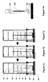
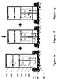


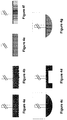
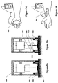
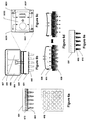


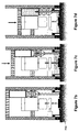

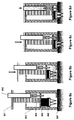


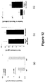
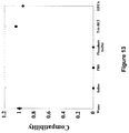
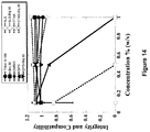
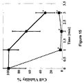
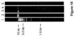
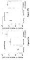

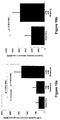
Classifications
-
- A—HUMAN NECESSITIES
- A61—MEDICAL OR VETERINARY SCIENCE; HYGIENE
- A61B—DIAGNOSIS; SURGERY; IDENTIFICATION
- A61B10/00—Other methods or instruments for diagnosis, e.g. instruments for taking a cell sample, for biopsy, for vaccination diagnosis; Sex determination; Ovulation-period determination; Throat striking implements
- A61B10/02—Instruments for taking cell samples or for biopsy
-
- A—HUMAN NECESSITIES
- A61—MEDICAL OR VETERINARY SCIENCE; HYGIENE
- A61B—DIAGNOSIS; SURGERY; IDENTIFICATION
- A61B10/00—Other methods or instruments for diagnosis, e.g. instruments for taking a cell sample, for biopsy, for vaccination diagnosis; Sex determination; Ovulation-period determination; Throat striking implements
- A61B10/0045—Devices for taking samples of body liquids
-
- A—HUMAN NECESSITIES
- A61—MEDICAL OR VETERINARY SCIENCE; HYGIENE
- A61B—DIAGNOSIS; SURGERY; IDENTIFICATION
- A61B10/00—Other methods or instruments for diagnosis, e.g. instruments for taking a cell sample, for biopsy, for vaccination diagnosis; Sex determination; Ovulation-period determination; Throat striking implements
- A61B10/02—Instruments for taking cell samples or for biopsy
- A61B10/0233—Pointed or sharp biopsy instruments
- A61B10/0283—Pointed or sharp biopsy instruments with vacuum aspiration, e.g. caused by retractable plunger or by connected syringe
-
- A—HUMAN NECESSITIES
- A61—MEDICAL OR VETERINARY SCIENCE; HYGIENE
- A61B—DIAGNOSIS; SURGERY; IDENTIFICATION
- A61B18/00—Surgical instruments, devices or methods for transferring non-mechanical forms of energy to or from the body
- A61B18/04—Surgical instruments, devices or methods for transferring non-mechanical forms of energy to or from the body by heating
-
- A—HUMAN NECESSITIES
- A61—MEDICAL OR VETERINARY SCIENCE; HYGIENE
- A61K—PREPARATIONS FOR MEDICAL, DENTAL OR TOILETRY PURPOSES
- A61K31/00—Medicinal preparations containing organic active ingredients
- A61K31/33—Heterocyclic compounds
- A61K31/395—Heterocyclic compounds having nitrogen as a ring hetero atom, e.g. guanethidine or rifamycins
- A61K31/495—Heterocyclic compounds having nitrogen as a ring hetero atom, e.g. guanethidine or rifamycins having six-membered rings with two or more nitrogen atoms as the only ring heteroatoms, e.g. piperazine or tetrazines
- A61K31/505—Pyrimidines; Hydrogenated pyrimidines, e.g. trimethoprim
- A61K31/519—Pyrimidines; Hydrogenated pyrimidines, e.g. trimethoprim ortho- or peri-condensed with heterocyclic rings
- A61K31/52—Purines, e.g. adenine
- A61K31/522—Purines, e.g. adenine having oxo groups directly attached to the heterocyclic ring, e.g. hypoxanthine, guanine, acyclovir
-
- A—HUMAN NECESSITIES
- A61—MEDICAL OR VETERINARY SCIENCE; HYGIENE
- A61K—PREPARATIONS FOR MEDICAL, DENTAL OR TOILETRY PURPOSES
- A61K38/00—Medicinal preparations containing peptides
- A61K38/16—Peptides having more than 20 amino acids; Gastrins; Somatostatins; Melanotropins; Derivatives thereof
- A61K38/17—Peptides having more than 20 amino acids; Gastrins; Somatostatins; Melanotropins; Derivatives thereof from animals; from humans
- A61K38/22—Hormones
- A61K38/28—Insulins
-
- A—HUMAN NECESSITIES
- A61—MEDICAL OR VETERINARY SCIENCE; HYGIENE
- A61M—DEVICES FOR INTRODUCING MEDIA INTO, OR ONTO, THE BODY; DEVICES FOR TRANSDUCING BODY MEDIA OR FOR TAKING MEDIA FROM THE BODY; DEVICES FOR PRODUCING OR ENDING SLEEP OR STUPOR
- A61M37/00—Other apparatus for introducing media into the body; Percutany, i.e. introducing medicines into the body by diffusion through the skin
- A61M37/0015—Other apparatus for introducing media into the body; Percutany, i.e. introducing medicines into the body by diffusion through the skin by using microneedles
-
- A—HUMAN NECESSITIES
- A61—MEDICAL OR VETERINARY SCIENCE; HYGIENE
- A61N—ELECTROTHERAPY; MAGNETOTHERAPY; RADIATION THERAPY; ULTRASOUND THERAPY
- A61N7/00—Ultrasound therapy
-
- C—CHEMISTRY; METALLURGY
- C12—BIOCHEMISTRY; BEER; SPIRITS; WINE; VINEGAR; MICROBIOLOGY; ENZYMOLOGY; MUTATION OR GENETIC ENGINEERING
- C12M—APPARATUS FOR ENZYMOLOGY OR MICROBIOLOGY; APPARATUS FOR CULTURING MICROORGANISMS FOR PRODUCING BIOMASS, FOR GROWING CELLS OR FOR OBTAINING FERMENTATION OR METABOLIC PRODUCTS, i.e. BIOREACTORS OR FERMENTERS
- C12M23/00—Constructional details, e.g. recesses, hinges
- C12M23/28—Constructional details, e.g. recesses, hinges disposable or single use
-
- C—CHEMISTRY; METALLURGY
- C12—BIOCHEMISTRY; BEER; SPIRITS; WINE; VINEGAR; MICROBIOLOGY; ENZYMOLOGY; MUTATION OR GENETIC ENGINEERING
- C12M—APPARATUS FOR ENZYMOLOGY OR MICROBIOLOGY; APPARATUS FOR CULTURING MICROORGANISMS FOR PRODUCING BIOMASS, FOR GROWING CELLS OR FOR OBTAINING FERMENTATION OR METABOLIC PRODUCTS, i.e. BIOREACTORS OR FERMENTERS
- C12M31/00—Means for providing, directing, scattering or concentrating light
-
- C—CHEMISTRY; METALLURGY
- C12—BIOCHEMISTRY; BEER; SPIRITS; WINE; VINEGAR; MICROBIOLOGY; ENZYMOLOGY; MUTATION OR GENETIC ENGINEERING
- C12M—APPARATUS FOR ENZYMOLOGY OR MICROBIOLOGY; APPARATUS FOR CULTURING MICROORGANISMS FOR PRODUCING BIOMASS, FOR GROWING CELLS OR FOR OBTAINING FERMENTATION OR METABOLIC PRODUCTS, i.e. BIOREACTORS OR FERMENTERS
- C12M35/00—Means for application of stress for stimulating the growth of microorganisms or the generation of fermentation or metabolic products; Means for electroporation or cell fusion
- C12M35/08—Chemical, biochemical or biological means, e.g. plasma jet, co-culture
-
- G—PHYSICS
- G01—MEASURING; TESTING
- G01N—INVESTIGATING OR ANALYSING MATERIALS BY DETERMINING THEIR CHEMICAL OR PHYSICAL PROPERTIES
- G01N1/00—Sampling; Preparing specimens for investigation
- G01N1/28—Preparing specimens for investigation including physical details of (bio-)chemical methods covered elsewhere, e.g. G01N33/50, C12Q
- G01N1/286—Preparing specimens for investigation including physical details of (bio-)chemical methods covered elsewhere, e.g. G01N33/50, C12Q involving mechanical work, e.g. chopping, disintegrating, compacting, homogenising
-
- G—PHYSICS
- G01—MEASURING; TESTING
- G01N—INVESTIGATING OR ANALYSING MATERIALS BY DETERMINING THEIR CHEMICAL OR PHYSICAL PROPERTIES
- G01N1/00—Sampling; Preparing specimens for investigation
- G01N1/28—Preparing specimens for investigation including physical details of (bio-)chemical methods covered elsewhere, e.g. G01N33/50, C12Q
- G01N1/40—Concentrating samples
- G01N1/4044—Concentrating samples by chemical techniques; Digestion; Chemical decomposition
-
- A—HUMAN NECESSITIES
- A61—MEDICAL OR VETERINARY SCIENCE; HYGIENE
- A61B—DIAGNOSIS; SURGERY; IDENTIFICATION
- A61B10/00—Other methods or instruments for diagnosis, e.g. instruments for taking a cell sample, for biopsy, for vaccination diagnosis; Sex determination; Ovulation-period determination; Throat striking implements
- A61B10/0096—Casings for storing test samples
-
- A—HUMAN NECESSITIES
- A61—MEDICAL OR VETERINARY SCIENCE; HYGIENE
- A61B—DIAGNOSIS; SURGERY; IDENTIFICATION
- A61B10/00—Other methods or instruments for diagnosis, e.g. instruments for taking a cell sample, for biopsy, for vaccination diagnosis; Sex determination; Ovulation-period determination; Throat striking implements
- A61B10/02—Instruments for taking cell samples or for biopsy
- A61B10/04—Endoscopic instruments
-
- A—HUMAN NECESSITIES
- A61—MEDICAL OR VETERINARY SCIENCE; HYGIENE
- A61B—DIAGNOSIS; SURGERY; IDENTIFICATION
- A61B18/00—Surgical instruments, devices or methods for transferring non-mechanical forms of energy to or from the body
- A61B18/18—Surgical instruments, devices or methods for transferring non-mechanical forms of energy to or from the body by applying electromagnetic radiation, e.g. microwaves
- A61B18/20—Surgical instruments, devices or methods for transferring non-mechanical forms of energy to or from the body by applying electromagnetic radiation, e.g. microwaves using laser
-
- A—HUMAN NECESSITIES
- A61—MEDICAL OR VETERINARY SCIENCE; HYGIENE
- A61B—DIAGNOSIS; SURGERY; IDENTIFICATION
- A61B10/00—Other methods or instruments for diagnosis, e.g. instruments for taking a cell sample, for biopsy, for vaccination diagnosis; Sex determination; Ovulation-period determination; Throat striking implements
- A61B10/02—Instruments for taking cell samples or for biopsy
- A61B2010/0208—Biopsy devices with actuators, e.g. with triggered spring mechanisms
-
- A—HUMAN NECESSITIES
- A61—MEDICAL OR VETERINARY SCIENCE; HYGIENE
- A61B—DIAGNOSIS; SURGERY; IDENTIFICATION
- A61B17/00—Surgical instruments, devices or methods, e.g. tourniquets
- A61B2017/00743—Type of operation; Specification of treatment sites
- A61B2017/00747—Dermatology
- A61B2017/00761—Removing layer of skin tissue, e.g. wrinkles, scars or cancerous tissue
-
- A—HUMAN NECESSITIES
- A61—MEDICAL OR VETERINARY SCIENCE; HYGIENE
- A61B—DIAGNOSIS; SURGERY; IDENTIFICATION
- A61B17/00—Surgical instruments, devices or methods, e.g. tourniquets
- A61B17/32—Surgical cutting instruments
- A61B2017/320004—Surgical cutting instruments abrasive
-
- A—HUMAN NECESSITIES
- A61—MEDICAL OR VETERINARY SCIENCE; HYGIENE
- A61B—DIAGNOSIS; SURGERY; IDENTIFICATION
- A61B18/00—Surgical instruments, devices or methods for transferring non-mechanical forms of energy to or from the body
- A61B2018/00315—Surgical instruments, devices or methods for transferring non-mechanical forms of energy to or from the body for treatment of particular body parts
- A61B2018/00452—Skin
-
- A—HUMAN NECESSITIES
- A61—MEDICAL OR VETERINARY SCIENCE; HYGIENE
- A61M—DEVICES FOR INTRODUCING MEDIA INTO, OR ONTO, THE BODY; DEVICES FOR TRANSDUCING BODY MEDIA OR FOR TAKING MEDIA FROM THE BODY; DEVICES FOR PRODUCING OR ENDING SLEEP OR STUPOR
- A61M37/00—Other apparatus for introducing media into the body; Percutany, i.e. introducing medicines into the body by diffusion through the skin
- A61M37/0015—Other apparatus for introducing media into the body; Percutany, i.e. introducing medicines into the body by diffusion through the skin by using microneedles
- A61M2037/0023—Drug applicators using microneedles
-
- A—HUMAN NECESSITIES
- A61—MEDICAL OR VETERINARY SCIENCE; HYGIENE
- A61M—DEVICES FOR INTRODUCING MEDIA INTO, OR ONTO, THE BODY; DEVICES FOR TRANSDUCING BODY MEDIA OR FOR TAKING MEDIA FROM THE BODY; DEVICES FOR PRODUCING OR ENDING SLEEP OR STUPOR
- A61M37/00—Other apparatus for introducing media into the body; Percutany, i.e. introducing medicines into the body by diffusion through the skin
- A61M37/0015—Other apparatus for introducing media into the body; Percutany, i.e. introducing medicines into the body by diffusion through the skin by using microneedles
- A61M2037/0046—Solid microneedles
-
- A—HUMAN NECESSITIES
- A61—MEDICAL OR VETERINARY SCIENCE; HYGIENE
- A61N—ELECTROTHERAPY; MAGNETOTHERAPY; RADIATION THERAPY; ULTRASOUND THERAPY
- A61N7/00—Ultrasound therapy
- A61N2007/0004—Applications of ultrasound therapy
- A61N2007/0034—Skin treatment
-
- A—HUMAN NECESSITIES
- A61—MEDICAL OR VETERINARY SCIENCE; HYGIENE
- A61N—ELECTROTHERAPY; MAGNETOTHERAPY; RADIATION THERAPY; ULTRASOUND THERAPY
- A61N7/00—Ultrasound therapy
- A61N7/02—Localised ultrasound hyperthermia
-
- G—PHYSICS
- G01—MEASURING; TESTING
- G01N—INVESTIGATING OR ANALYSING MATERIALS BY DETERMINING THEIR CHEMICAL OR PHYSICAL PROPERTIES
- G01N1/00—Sampling; Preparing specimens for investigation
- G01N1/28—Preparing specimens for investigation including physical details of (bio-)chemical methods covered elsewhere, e.g. G01N33/50, C12Q
- G01N1/44—Sample treatment involving radiation, e.g. heat
-
- Y—GENERAL TAGGING OF NEW TECHNOLOGICAL DEVELOPMENTS; GENERAL TAGGING OF CROSS-SECTIONAL TECHNOLOGIES SPANNING OVER SEVERAL SECTIONS OF THE IPC; TECHNICAL SUBJECTS COVERED BY FORMER USPC CROSS-REFERENCE ART COLLECTIONS [XRACs] AND DIGESTS
- Y10—TECHNICAL SUBJECTS COVERED BY FORMER USPC
- Y10T—TECHNICAL SUBJECTS COVERED BY FORMER US CLASSIFICATION
- Y10T436/00—Chemistry: analytical and immunological testing
- Y10T436/25—Chemistry: analytical and immunological testing including sample preparation
- Y10T436/2575—Volumetric liquid transfer
Definitions
- tissue molecular composition of human tissues, represented by a multitude of lipids, proteins, nucleic acids, and other miscellaneous molecules, is a sensitive indicator of local pathologies, such as cancer, allergies, and eczema, as well as several systemic diseases, such as cardiovascular disease, Alzheimer's disease, and diabetes.
- tissue molecular composition also holds critical information about the body's exposure to exogenous chemical and biological entities.
- this information is not currently used in diagnostic methods due to a lack of patient-friendly and standardized methods for routine sample collection from tissues. Instead, clinical diagnosis is invariably performed by visual observation and histopathological analysis of tissue biopsies, which are highly limited due to their qualitative nature, leading to increased misdiagnosis and inappropriate use.
- current methods also fall short in explaining a complete molecular genesis of diseases, and fail to distinguish between diseases.
- the current invention describes system, method and device, as well as compositions useful in such systems, methods and devices, involving application of energy to a tissue of interest to generate a liquefied sample comprising tissue constituents so as to provide for rapid tissue sampling, as well as qualitative and/or quantitative detection of analytes that may be part of tissue constituents (e.g., several types of biomolecules, drugs, and microbes). Determination of tissue composition can be used in a variety of applications, including diagnosis or prognosis of diseases, evaluating bioavailability of therapeutics in different tissues following drug administration, forensic detection of drugs-of-abuse, evaluating changes in the tissue microenvironment following exposure to a harmful agent, tissue decontamination and various other applications.
- the current invention provides methods and devices for generating a liquefied tissue sample from a subject - living or diseased.
- the device and method involve applying energy and a liquefaction promoting medium to a tissue of interest of a subject, the applying producing a liquefied tissue sample, and collecting the liquefied tissue sample.
- an analysis for the presence or absence of at least one analyte in the liquefied tissue sample is performed, wherein the analysis facilitates diagnosis of a condition of interest.
- the analysis involves generating an analyte profile from the liquefied tissue sample and comparing the analyte profile to a reference analyte profile, wherein the comparing facilitates diagnosis of a condition of interest.
- the purpose of said tissue liquefaction is to remove, or decontaminate the tissue from undesired substances.
- undesired substances include chemicals, environmental contaminants, biological toxins, and in general substances that are considered toxic or hazardous to the body.
- the said method of decontamination is performed by continuously moving the tissue liquefaction device over tissue-of-interest until removal of undesired substances at a preferred level is attained.
- the liquefaction promoting agent comprises of one of more of sodium chloride, potassium chloride, sodium phosphate dibasic, potassium phosphate monobasic, 3- ⁇ [tris(hydroxymethyl)methyl]amino ⁇ propanesulfonic acid, N,N-bis(2-hydroxyethyl)glycine, tris (hydroxymethyl) methylamine, N-tris(hydroxymethyl)methylglycine, 4-2-hydroxyethyl-1-piperazineethanesulfonic acid, 2- ⁇ [tris(hydroxymethyl)methyl]amino ⁇ ethanesulfonic acid, 3-(N-morpholino)propanesulfonic acid, piperazine-N,N'-bis(2-ethanesulfonic acid), dimethylarsinic acid, saline sodium citrate, 2-(N-morpholino)ethanesulfonic acid.
- the liquefaction promoting agent comprises of one or more of a protease inhibitor, an RNase inhibitor, or a DNase inhibitor. In certain embodiments, the liquefaction promoting agent comprises at least one of free radical scavenger, a defoaming agent, and a protein stabilizer.
- the liquefaction promoting agent comprises at least one of Brij-30, 3-(Decyl dimethyl ammonio) propane sulfonate (DPS), 3-(Dodecyl dimethyl ammonio) propane sulfonate (DDPS), N-lauroyl sarcosine (NLS), Triton X-100, Sodium Dodecyl Sulfate, DMSO, fatty acids, azone, EDTA, or sodium hydroxide.
- the liquefaction promoting agent comprises a suspension of abrasive particles.
- the abrasive particles comprise silica or aluminum oxide.
- the energy is applied in the form of ultrasound, mechanical, optical, thermal, or electrical energy.
- the mechanical energy is applied by an abrasive material.
- the thermal energy is applied in the form of radio frequency energy.
- the optical energy is applied in the form of a laser.
- the liquefied tissue sample is generated for each of a healthy tissue of interest of the subject and a suspected diseased tissue of interest of the subject, and the analysis comprises comparing analytical results from the healthy tissue sample with analytical results from the suspected diseased tissue sample, wherein the comparing facilitates diagnosis of a condition of interest.
- the liquefied tissue sample is generated for multiple tissue sites and the analysis comprises comparing analytical results from the multiple tissue sites, wherein said comparing facilitates diagnosis of a condition of interest.
- the liquefied tissue sample is collected from multiple tissue sites, and the samples are combined to make a diagnosis.
- the liquefied tissue sample is collected by aspiration. In certain embodiments, the collecting is by retaining the liquefaction agent in a housing placed in contact with the tissue. In certain embodiments, the collecting is by mechanized transfer of the liquefied tissue sample in a housing located in the device.
- the liquefied tissue sample is mixed with a substance which assists in further liquefaction and in stabilization of analytes of interest for storage or transportation.
- the transferred tissue sample from that sample container is mixed with the substances which are pre-stored in a container.
- a protein stabilizer such as protease inhibitor, a nucleic-acid stabilizer such as EDTA, phenol, nonspecific proteinase, an RNase inhibitor and a DNase inhibitor, a defoaming agent, and surfactants such as Triton X-100, Sodium Dodecyl Sulfate, and DMSO
- abrasive particles comprise silica or aluminum oxide.
- the device evaluates the tissue of interest prior, during, or after liquefaction process.
- the evaluation is performed by electrochemical, biochemical, or optical means.
- the evaluation involves measurement of tissue's electrical conductivity.
- electrical conductivity is measured by a means applying an AC electrical signal across the tissue of interest. The said electrical signal has voltage between 0.1 mV and 10 V and frequency between 1 Hz and 100 kHz.
- the device involves detecting certain tissue constituents in the liquefied tissue sample prior to analysis of an analyte of interest, such as a disease marker.
- the detecting is by electrochemical, biochemical, or optical means.
- the electrochemical means of detecting is an ion-elective electrode.
- the optical means of detecting is measuring the absorption or scattering coefficient of a liquid solution.
- the energy is applied to a tissue in the form of ultrasound with a mechanical index between 0.1 and 50. In certain embodiments, the energy is applied by contacting the tissue with a moving abrasive surface. In certain embodiments, the energy is applied to the tissue by contacting the tissue with a moving brushing device comprising a plurality of bristles. In certain embodiments, the energy is applied to the tissue by mechanical insertion of a patch bearing plurality of micro-needles into the tissue; and further injection of liquefaction medium through the micro-needles into the tissue. In some embodiments, additional energy is applied by moving the said micro-needle patch after its insertion into the tissue.
- the energy is applied to the tissue by mechanized stirring of the liquefaction agent. In certain embodiments, the energy is applied to the tissue by contacting the tissue with a high velocity jet comprising of liquefaction promoting medium, which may also contain abrasive particles in different embodiments.
- the tissue comprises breast, prostate, eye, vagina, bladder, nail, hair, colon, testicles, or intestine.
- the tissue comprises skin or a mucosal membrane.
- the tissue comprises lung, brain, pancreas, liver, heart, bone, or aorta wall.
- the analyte comprises a small molecule, a drug or metabolite thereof, a polypeptide, a lipid, a nucleic acid, or a microbe.
- the analyte comprises an antibody, a cytokine, an illicit drug, or a cancer biomarker.
- the liquefied tissue sample is held in a container, and the analyte profile is generated by integrating the liquid container with one or more analytical devices.
- the tissue liquefaction device contains a means for measuring the concentration of a calibrator analyte to provide a means for calibrating the analysis of the analyte.
- the device involves diagnosing allergic disease in a subject, and the device comprises means for analyzing the liquefied tissue sample for the presence or absence of IgE and IgG antibodies, cytokines such as IL4, IL5, IL10, IL-12, IL13, IL-16, GM-CSF, RANTES, MCP-4, CTACK/CCL27, IFN-g, TNFa, CD23, CD-40, Eotaxin-2, and TARC, wherein the analysis facilitates diagnosis of allergic disease in the subject.
- cytokines such as IL4, IL5, IL10, IL-12, IL13, IL-16, GM-CSF, RANTES, MCP-4, CTACK/CCL27, IFN-g, TNFa, CD23, CD-40, Eotaxin-2, and TARC
- the device involves diagnosing cancer in a subject, and the device comprises means for analyzing the liquefied tissue sample for the presence or absence of one or more cancer markers, wherein the analysis facilitates diagnosis of cancer in the subject.
- the tissue of interest is breast, colon, prostate, skin, testicle, intestine, or mouth.
- the device involves diagnosing heart disease in a subject, and the device comprises means for analyzing the liquefied tissue sample for the presence or absence of one or more of cholesterol, triglycerides, lipoproteins, free fatty acids, and ceramides, wherein the analysis facilitates diagnosis of heart disease in the subject.
- the device involves detecting the presence of an illicit drug, or metabolite thereof, in a subject, and the device comprises means for analyzing the liquefied tissue sample for the presence or absence of an illicit drug, or metabolites thereof, wherein the analysis provides for detection of illicit drugs in the subject.
- the device involves detecting a microorganism in a subject, and the device comprises means for applying energy and a liquefaction medium to a tissue of interest in a subject and analyzing the liquefaction medium for the presence or absence of a microorganism, wherein the analysis provides for detection of the presence or absence of a microorganism.
- Another object of the current invention is to provide a method and device for liquefying a tissue of a subject for facilitating the passage of a drug across or into the tissue.
- the method and device disclosed above are applicable not only to collection of tissue constituents but also to drug delivery.
- the device and method involve applying energy and a liquefaction medium to a tissue of interest of a subject, and delivering a drug through or into the site of the tissue to be liquefied.
- the advantage of using the present invention is 1) to provide higher fluxes of drugs into a tissue, and 2) to allow greater control of fluxes into a tissue. Drugs which would simply not pass through the tissues such as the skin are forced through the tissues when the method is applied.
- the present invention offers a method for delivering one or more drugs through the tissue to be liquefied into the circulatory system, which circumvents degradation in the gastrointestinal tract and rapid metabolism by the liver from which drugs to be routinely administered either orally or by injection suffer.
- the current invention provides a method and device for delivering one or more drugs locally to the tissue of interest, thus limiting side effects to the healthy tissues.
- the method and device may also be applicable for enhancing transport to cellular membranes.
- the device of the present invention consists of the following major components: 1) an energy generator; 2) a liquefaction promoting medium; 3) a reservoir to hold drugs to be delivered and/ or collect the liquefied tissue sample.
- a drug to be administered can be added into the liquefaction medium prior or during tissue liquefaction process.
- application of energy is in combination of the liquefaction medium which does not contain a drug can be used for liquefying a tissue, and subsequently a drug in an appropriate carrier such as a patch can be applied on a site of the tissue to be liquefied.
- the transport of drug into the tissue can be further enhanced by the simultaneous or subsequent application of a secondary driving force such as chemical permeability or transport enhancers, convection, osmotic pressure gradient, concentration gradient, iontophoresis, electroporation, magnetic field, ultrasound, or mechanical pressure.
- a secondary driving force such as chemical permeability or transport enhancers, convection, osmotic pressure gradient, concentration gradient, iontophoresis, electroporation, magnetic field, ultrasound, or mechanical pressure.
- the driving force can be applied continuously over a period of time or at intervals during the period of liquefaction.
- the tissue to be administered comprises an organs as well as biological surfaces.
- the biological surfaces comprise a biological membrane and cellular membrane.
- the biological membrane comprises skin or a mucosal membrane.
- the biological membrane comprises a buccal membrane, eye, vagina, colon, or intestine.
- the tissue comprises a diseased tissue.
- a device that can be used on a tissue to obtain a liquefied sample comprising an energy source operably coupled to the tissue, and a chamber, operably coupled to said tissue, capable of delivering liquefaction promoting medium to and/or collecting said liquefied sample from said tissue.
- the device can be used on a tissue which is a part of a living organism; and the tissue can be excised from the organism prior to diagnosis.
- the device of claim 1 wherein the liquefied tissue sample is transferred to an assay for monitoring the presence or absence of at least one analyte.
- the chamber of the device can be a sponge-bellow assembly where the sponge is capable of storing said liquefaction promoting medium and/ or liquefied tissue sample.
- a device comprising an energy source operably coupled to the tissue, and a chamber, operably coupled to said tissue, capable of delivering liquefaction promoting medium to and/or collecting said liquefied sample from said tissue; also comprises a tube/needle, connected to said chamber, capable of delivering the liquefaction promoting medium to and/or aspirating liquefied tissue sample from the tissue.
- a device comprising an energy source operably coupled to the tissue, and a chamber, operably coupled to said tissue, capable of delivering liquefaction promoting medium to and/or collecting said liquefied sample from said tissue; also comprises a sample container, operably connected to said chamber, capable of storing aspirated liquefied tissue sample containing analytes, or transferring said aspirated liquefied tissue sample to an ancillary chamber; wherein the chamber is used only to deliver the liquefaction promoting medium to the chamber.
- a pressurized container and/or vacuum container is part of the device, which facilitates transfer of said liquefaction promoting medium and/ or liquefied tissue sample.
- the energy emitted from the energy source in the deice is in the form of ultrasound, mechanical, optical, thermal, or electrical energy.
- the mechanical energy is applied to the tissue by an abrasive material, vacuum, pressure or shear force.
- the thermal energy is applied to the tissue in the form of radio frequency energy.
- the optical energy is applied to the tissue in the form of a laser.
- a device comprising an energy source operably coupled to the tissue, and a chamber, operably coupled to said tissue, capable of delivering liquefaction promoting medium to and/or collecting said liquefied sample from said tissue further comprising a sample container, operably connected to said chamber, capable of storing aspirated liquefied tissue sample containing analytes, or transferring said aspirated liquefied tissue sample to an ancillary chamber; wherein the chamber is used only to deliver the liquefaction promoting medium to the chamber.
- a device comprising an energy source operably coupled to the tissue, and a chamber, operably coupled to said tissue, capable of delivering liquefaction promoting medium to and/or collecting said liquefied sample from said tissue, wherein the energy source comprises of a pad connected to a shaft.
- the shaft has a pressure sensing unit, which maintains a predetermined pressure profile on to the tissue upon contact.
- the pad is selected from a group consisting of an abrasive surface and a patch comprising of a plurality of micro-needles.
- the device further comprises a plunger, operably connected to the top of the chamber.
- the device is divided into an upper and lower unit, and wherein the lower unit is detachable from said upper unit; wherein the upper unit comprises the energy source and the lower unit comprises the chamber.
- the device further comprises an analytical unit operably connected to the chamber, and where the analytic unit is capable of performing temporal monitoring of the tissue sample by electrochemical, biochemical or optical means; or the analytic unit is capable of analyzing the analytes within said liquefied tissue sample.
- the device is connected to a diagnostic probe or a catheter; wherein the diagnostic probe is selected from a group consisting of endoscope, colonoscope, and laparoscope.
- the use of the device results in situ liquefaction of the tissue sample.
- the device contains a liquefaction promoting medium that can preserve and enhance the detection of proteins, lipids and nucleic acids, comprising: 3-(decyl dimethyl ammonio) propane sulfonate (DPS) and polyethylene glycol dodecyl ether (Brij 30) dissolved in a buffered solution; and where the concentration of 3-(decyl dimethyl ammonio) propane sulfonate and polyethylene glycol dodecyl ether (B30) is between 0.01 - 10% (w/v); and where the 3-(decyl dimethyl ammonio) propane sulfonate and polyethylene glycol dodecyl ether are present at a ratio of 50:50.
- a liquefaction promoting medium that can preserve and enhance the detection of proteins, lipids and nucleic acids, comprising: 3-(decyl dimethyl ammonio) propane sulfonate (DPS) and polyethylene glycol dodecyl ether (Brij 30)
- the liquefaction promoting medium within the device is buffered in a solution comprising either phosphate-buffered saline, Tris-buffered saline, Tris-HCL or EDTA.
- liquefaction promoting medium within the device comprises a nonionic surfactant selected from a Brij series surfactant, a Triton-X surfactant, and a Sorbitan surfactant; an anionic or a zwitterionic surfactant; and a hydrophilic solvent; wherein the medium has a total concentration of the surfactants from about 0.01%-10% (w/v).
- Energy as used herein means any appropriate energy that can be applied to tissue to accomplish the objective of the methods disclosed herein (e.g., liquefying tissue).
- exemplary types of energy include mechanical energy (e.g., abrasion, shear, vacuum, pressure, suction), ultrasound, optical (e.g., laser), magnetic, thermal, and electrical energy.
- an “analyte” as used herein means any biomolecule (e.g., polypeptide, nucleic acid, lipid, and the like), drug (e.g., therapeutic drugs, drugs-of-abuse, and the like), small molecule (e.g., natural moisturizing factors, nicotine, and the like, with the understanding that small molecules can also be drugs), warfare agent, environmental contaminant (e.g., pesticides, etc.), microbe (e.g., bacterium, virus, fungus, yeast, and the like) and the like that is present in or on the tissue and can be extracted from the tissue of interest (e.g., skin, a mucosal membrane, and the like) and detected, analyzed, and/or quantified.
- biomolecule e.g., polypeptide, nucleic acid, lipid, and the like
- drug e.g., therapeutic drugs, drugs-of-abuse, and the like
- small molecule e.g., natural moisturizing factors, nicotine
- liquefaction is used to describe the process by which tissue and/or tissue constituents are converted to a sufficiently soluble state through exposure to sufficient energy and, optionally, a liquefaction promoting medium, and can involve conversion of at least a portion of a tissue structure of interest to a liquid form.
- a tissue sample that has been subjected to liquefaction as sometimes referred to herein as a "liquefied" sample.
- LPM liquid phase transfer medium
- tissue constituents facilitates solubilization of one or more tissue constituents, facilitates conversion of at least a portion of a tissue structure into a liquid when exposed to energy, and/or facilitates preservation of bioactivity of one or more solubilized tissue constituents.
- LPA liquefaction-promoting agent
- a “calibration analyte” as used herein means any molecule naturally present in a tissue of interest at a known concentration, which can serve as a reference analyte (e.g., as a positive control to ensure a desired degree of liquefaction was achieved).
- biomolecule as used herein means any molecule or ion which has a biological origin or function.
- biomolecules include proteins (e.g., disease biomarkers such as cancer biomarkers, antibodies: IgE, IgG, IgA, IgD, or IgM, and the like), peptides, lipids (e.g., cholesterol, ceramides, or fatty acids), nucleic acids (RNA and DNA), small molecules (e.g., glucose, urea, creatine), small molecule drugs or metabolites thereof, microbes, inorganic molecules, elements, or ions (e.g., iron, Ca2+, K+, Na+, and the like).
- the biomolecule is other than glucose and/or is other than a cancer marker.
- abused drug or “drug-of-abuse” or “illicit drug” are used interchangeably herein to refer to any substance which is regulated by a governmental (e.g. federally or state regulated) of which presence in a human tissue, and/or presence above a certain level in a human tissue, is illegal or can be harmful to a human being.
- abused drugs include: cocaine, heroin, methyl amphetamine, and prescription drugs taken in excess of dosage, or taken without a prescription (e.g., painkillers such as opioids).
- warfare agent refers to any molecule, compound, or composition of either biological or chemical origin that may be used as a weapon.
- warfare agents include nerve gases (e.g. VX, Sarin), phosgene, toxins, spores (e.g., anthrax), and the like.
- environmental contaminant includes any molecule, compound, or composition which can be detrimental to an individual, e.g., when at concentrations elevated above a risk threshold.
- examples include water pollutants (e.g., fertilizers, pesticides, fungicides, insecticides, herbicides, heavy metals, halides), soil pollutants (e.g., fertilizers, pesticides, fungicides, insecticides, herbicides, heavy metals, halides), air pollutants (e.g., NOx, SOx, greenhouse gases, persistent organic pollutants (POPs), particulate matter, smog).
- water pollutants e.g., fertilizers, pesticides, fungicides, insecticides, herbicides, heavy metals, halides
- soil pollutants e.g., fertilizers, pesticides, fungicides, insecticides, herbicides, heavy metals, halides
- air pollutants e.g., NOx, SOx, greenhouse gases, persistent organic pollutants (POPs), particulate matter,
- decontamination includes removal from tissues of any unwanted or undesired molecule, compound, or composition which can be detrimental to an individual. Examples include environmental contaminants (as defined above), toxic chemicals, and biological toxins.
- natural moisturizing factor means any one of several types of small molecules, including but not limited to free amino acids, lactate, and urea, which are derivatives of fillagrin. NMFs can be used as analytes to facilitate assessment of general skin health (e.g., dry skin, flaky skin, normal skin, etc.).
- drug delivery means the delivery of one or more drugs into blood, lymph, interstitial fluid, a cell or tissue.
- sensitivity enhancer means a substance or a mixture of substances that is mixed with LPM to stabilize liquefied tissue analytes and facilitate their analysis in terms of enhancing the sensitivity and specificity of the diagnostic analytical tests.
- blocking reagent is used to describe a component which is used to prevent non specific binding of analytes to substrates used in a diagnostic assay.
- the current invention provides systems, methods and devices, as well as compositions useful in such systems, methods and devices, involving application of energy to a tissue of interest to generate a liquefied sample comprising tissue constituents so as to provide for rapid tissue sampling, as well as qualitative and/or quantitative detection of analytes that may be part of tissue constituents (e.g., several types of biomolecules, drugs, and microbes - might want a paragraph to formally defined what you mean by tissue constituents).
- Determination of tissue composition or constituents can be used in a variety of applications, including diagnosis or prognosis of local as well as systemic diseases, evaluating bioavailability of therapeutics in different tissues following drug administration, forensic detection of drugs-of abuse, evaluating changes in the tissue microenvironment following exposure to a harmful agent, decontamination, and various other applications.
- Another object of the current invention is to provide a method and device for liquefying a tissue of a subject for facilitating the passage of a drug across or into the tissue.
- the method and device disclosed above are applicable not only to collection of tissue constituents but also to drug delivery.
- the device and method involve applying energy and a liquefaction medium to a tissue of interest of a subject, and delivering a drug through or into the site of the tissue to be liquefied.
- the advantage of using the present invention is 1) to provide higher fluxes of drugs into a tissue, and 2) to allow greater control of fluxes into a tissue. Drugs which would simply not pass through the tissues such as the skin and into the circulatory system are forced through the tissues when the method is applied.
- the tissue liquefaction devices disclosed herein can be generally described as having an energy source/generator operably coupled to a reservoir unit/housing, where the reservoir houses a medium in which analytes are collected and which, in most embodiments, facilitates transfer of energy to the tissue of interest and can thus, where desired, facilitate liquefaction of a tissue sample.
- the reservoir housing is placed in contact with the subject's tissue to make contact between the medium and the tissue, and the energy source is activated.
- the device can be operably coupled to additional energy sources, (e.g., abrasive actuator, piezoelectric transducer, suction or pressure), which can also be applied to the tissue to facilitate transfer of energy to the tissue.
- the medium can be retained in the reservoir housing, or alternatively be transferred to a separate container.
- the reservoir housing or container can be operably coupled to a detection device that can quantitatively measure the tissue constituents present in the medium.
- Energy can be applied to the tissue from a single energy source or as a combination of sources.
- exemplary energy sources include mechanical (e.g., abrasion, shear, vacuum, pressure, and the like), piezoelectric transducer, ultrasound, optical (e.g., laser), thermal, and electrical energy.
- the intensity of the energy applied, as well as the duration of the energy application, may be appropriately adjusted for the particular tissue of interest and the particular application of the method.
- the energy intensity and duration of application may also be appropriately adjusted based on the particular liquefaction promoting medium (LPM) used in connection with the energy.
- LPM liquefaction promoting medium
- an energy exposure time of greater than 1 minute, greater than 90 seconds, or greater than 2 minutes is provided in order to produce a suitable liquefied tissue sample.
- the magnitude of energy depends on the analyte of interest and the selection of LPM. Higher energies are required to liquefy tissues in the absence of surfactants or particles in the LPM. Use of high energies is limited by their adverse effects on the tissue or its constituents. A significant adverse effect is injurious tissue damage. In some embodiments, therefore, it might be necessary to incorporate certain device components that provide temporal monitoring (ideally, in real-time) of the change in tissue properties or the extent of tissue liquefaction such that, once safe limit for energy exposure is reached, the device can be stopped. The temporal evaluation can be performed prior, during, and after liquefaction process.
- the temporal evaluation is performed by electrochemical (e.g., tissue's electrical conductivity, measurement of certain ions by ion-selective electrodes, etc), biochemical (e.g., measurement of certain tissue components in the LPM by enzymatic assays such as ELISA and the like), or optical (e.g., measurement of LPM turbidity by spectrophotometer, etc) means.
- tissue's temporal electrical conductivity is measured by applying a pre-defined AC electrical voltage across the tissue with a signal generator, and analyzing the resultant electrical current by a multimeter. Another significant adverse effect of high energy exposure is attributed to temperature elevation in the tissue, also known as thermal effects. In some embodiments, therefore, it might be necessary to incorporate a temperature sensing element (e.g., a thermocouple) that allows monitoring of the temperature of the tissue and/or the LPM, facilitating the judgment of a safe amount of energy exposure to the tissue.
- a temperature sensing element e.g., a thermocouple
- LPM low-power lipoprotein
- use of energy to liquefy tissue may lead to reduction in biological activity of solubilized tissue constituents, necessitating selection of LPM which adequately preserve the bioactivity of tissue's molecules as well as aid tissue solubilization.
- LPM tissue solubilization
- incorporation of one or more surfactants such as DPS, NLS and Brij-30 at a concentration of 1 %w/v in LPM facilitated complete preservation of the bioactivity of solubilized proteins and nucleic acids under ultrasonic energy exposure.
- energy can be applied to a tissue using an energy delivery chamber that includes an energy producing element.
- the chamber when placed on the tissue, will expose the tissue to the energy producing element and allow energy to be applied to the tissue with minimal interference.
- Such a chamber can contain LPM and provide for contact of the LPM with the tissue such that, upon application of energy, tissue constituents can be directly collected into the solution.
- the energy delivery chamber containing the LPM may also comprise a diagnostic device, for example, an analyte sensor, for detecting and, optionally, quantifying analytes that may be present in the LPM.
- a diagnostic device for example, an analyte sensor, for detecting and, optionally, quantifying analytes that may be present in the LPM.
- These diagnostic devices can serve as chemical sensors, biosensors, or can provide other measurements to form a complete sampling and measurement system.
- An element having an internal channel for fluid transfer can be fabricated together with a sensor to form a disposable unit.
- the device can also be adapted to include or be provided as a disposable unit that provides for collection of analytes in the LPM for analysis.
- the diagnostic element can be located elsewhere (e.g., separate from the energy device) and the contents of the energy delivery chamber in contact with tissue can be pumped using mechanical forces, capillary forces, ultrasound, vacuum, or electroosmotic forces into a sensing chamber and analyzed.
- the unit when evaluating topical formulations or determining pharmacological parameters, can be constructed to function as a closed loop drug delivery unit, including drug delivery means, analyte recovery means, sensing means to measure the analyte, and control means to provide a signal to the drug delivery means.
- a portable disposable unit is inserted into a portable or bench-top energy generator.
- the energy generator may also include circuitry for tissue resistance measurements, analyte concentration measurements, and display of analyte concentration measurements.
- the system e.g., energy applicator and disposable unit
- the system is placed against the tissue, and energy is applied for a certain period of time, either alone or as a combination with other physical, mechanical, electrical, and chemical forces.
- the tissue of interest is liquefied, and analytes from the liquefied tissue are collected in the disposable unit and are measured using appropriate assays.
- FIGS. 1 through 19 of the drawings like numerals being used for like and corresponding parts of the various drawings.
- FIGS. 1a through 1g the structure, components and functioning of abrasive energy-based tissue liquefaction devices are shown.
- Panels a through c of FIG. 1 show the sequential working of a device that utilizes a rotary abrasive component 101 as means for applying energy to tissues for liquefaction. Liquefaction is achieved by placing and setting abrasive component 101 in motion against a tissue of interest 107.
- Abrasive component 101 is attached to a shaft 102, which is further connected to a rotary motor 103 in the device.
- shaft 102 is designed to sense and control the pressure applied by abrasive component 101 on tissue 107.
- shaft 102 is constructed of shaft 1021 and shaft 1022 which are connected to each other by a pressure-sensitive spring 1023 ( FIG. 1d ).
- shaft 1021 and shaft 1022 sandwich between them a pressure-sensing piezoelectric crystal for monitoring and controlling applied pressure to tissue 107.
- a battery pack 104 powers motor 103, which can subsequently set abrasive component 101 in rotary motion when directed by the device operator.
- abrasive component 101 Prior to liquefaction, abrasive component 101 is designed to be held in isolation against tissue 107 using a housing 105, and specifically, a thin sheet 106 located on the base of housing 105 ( FIG. 1a ).
- LPM stored in a cartridge 108 is transferred to the housing 105 ( FIG. 1a ), whereupon the LPM contacts the surface of the sheet material 106, followed by setting the abrasive component 101 in motion against sheet 106.
- Material of sheet 106 is chosen such that it can be quickly abraded by abrasive component 101, allowing LPM and abrasive component 101 to come in contact with tissue 107 leading to tissue liquefaction ( FIG. 1b ).
- Non-limiting examples of sheet 106 include sheet of paper, rubber sheet, metal foil, plastic sheet, or any water-soluble sheet.
- certain device components are designed as disposable units such that, after each use of the device, these components can be replaced to allow sterile usage.
- Such components may include housing 105, abrasive component 101, cartridge 108, sample container 110, and other fluid-handling device components, as deemed necessary to maintain device sterility.
- the whole device may be made disposable.
- LPM storing cartridge 108 can be replaced with a sponge-bellow assembly for storage and release of LPM.
- Panels e through g of FIG. 1 show the sequential working of such a device.
- a flexible bellow-shaped housing 112 contains a sponge 111 filled with LPM ( FIG. 1e ).
- sponge-bellow housing is squeezed to release LPM and abrasive component 101 is set in motion ( FIG. 1f ).
- motor 103 stops and LPM containing tissue constituents is transferred to a sample container 110 ( FIG. 1g ). Collection of the sample is facilitated by a suction pump 109.
- the suction pump 109 and container 110 may be avoided by collecting the sample into the sponge by lifting the device back into its original position.
- FIGS. 2a and 2b the structure and components of moveable tissue liquefaction devices designed for continuous sampling of a large area of tissue are shown.
- Panel a of FIG. 2 show a device that utilizes a rotary abrasive component 201 as means for applying energy to tissues for liquefaction. Liquefaction is achieved by placing and setting abrasive component 201 in motion against a tissue of interest 207.
- Abrasive component 201 is attached to a shaft 202, which is further connected to a rotary motor 203 in the device.
- shaft 202 is designed to sense and control the pressure applied by abrasive component 201 on tissue 207.
- shaft 202 is constructed of two distinct shafts which are connected to each other by a pressure-sensitive spring or a pressure-sensing piezoelectric crystal for monitoring and controlling the applied pressure to tissue 207.
- a battery pack 204 powers motor 203, which can subsequently set abrasive component 201 in rotary motion when directed by the device operator.
- a continuous liquefaction procedure is initiated by performing three key processes - LPM stored in a cartridge 208 is continuously delivered to housing 212 at the device-tissue interface; abrasive component 201 is set in motion against tissue 207; and liquefied tissue sample is continuously collected in a sample container 210 using a suction pump 209.
- the device can be moved around such that additional tissue surfaces are exposed to the device and liquefied.
- the liquefaction process can be stopped by switching-off motor 103 and cumulative tissue sample can be accessed from container 210.
- suction pump 209 can be used to create a vacuum-assisted seal between tissue 207 and chamber 206 located in a flanged housing 205 around the device.
- Panel b of FIG. 2 show a device that utilizes a piezoelectric element 251 as means for applying mechanical energy to tissues for liquefaction.
- Piezoelectric element 251 is placed in a housing 252 that interfaces with a tissue of interest 259, and liquefaction is achieved by activating piezoelectric element 251 with LPM present as a coupling fluid between tissue 259 and piezoelectric element 251.
- Piezoelectric element 251 is a transducer of electrical energy, which is supplied to it by means of circuitry placed in a flexible tubing 253.
- LPM is supplied to housing 252 by a flexible tubing 254 using an operator-controlled injection system 256.
- Liquefied tissue sample can be simultaneously collected from housing 252 into a sample container 257 using a flexible tubing 255. Sample collection is facilitated by a suction pump 258 which is serially connected to sample container 257.
- suction pressure created in housing 252 by suction pump 258 may provide for an effective seal between housing 252 and tissue 259 for preventing LPM leakage from housing 252 during liquefaction.
- suction pressure created in housing 252 by suction pump 258 may provide for an additional source of energy for liquefaction.
- housing 252 may be moved to liquefy additional tissue surfaces and collect a sample representing tissue constituents accumulated from various tissue surfaces.
- LPM is continuously supplied to housing 252 by tubing 254 and sample is continuously collected by tubing 255.
- the device in FIG. 2b may operate without a piezoelectric element 251.
- the LPM which flows from a tubing 254 into the housing 252 makes contact with the tissue and liquefies the tissue.
- Liquefied tissue is collected from the housing by tubing 255.
- the housing may be moved continuously or intermittently to collect samples from a large tissue area.
- the device may have additional means that are practically necessary to allow the movement of the device on a tissue, liquefaction of tissue and collection of liquefied tissue.
- either pressure or vacuum but not both may be used to direct LPM towards the tissue and collect liquefied tissue.
- liquefaction devices may be integrated with a diagnostic probe such as endoscope, colonoscope, laparoscope, and the like.
- FIGS. 3a through 3c the structure and components of liquefaction devices that utilize an oscillating abrasive component as means for applying energy to tissues for liquefaction are shown.
- liquefaction is achieved by placing and setting abrasive component 301 in motion against a tissue of interest 311.
- Linear motion can be achieved, for example, by a rack and pinion arrangement ( FIG. 3a ).
- abrasive component 301 is attached to a rack 302, which slides in a linear oscillatory motion using a circular gear 303 (pinion).
- Gear 303 is driven in oscillatory circular motion by a motor 304.
- a battery pack 305 powers motor 304.
- motor 304 is a servo motor which may require an electronic microchip controller 306 to produce oscillatory circular motion.
- abrasive component 301 Prior to liquefaction, abrasive component 301 is designed to be held in isolation against tissue 311 using a housing 307, and specifically, a thin sheet 308 located on the base of housing 307. LPM can be pre-stored in housing 307, for instance, so that it is in contact with 308. In some embodiments, LPM may be transferred to housing 307 from a cartridge located elsewhere in the device. Liquefaction process is initiated by setting the abrasive component 301 in linear motion against sheet 308.
- Material of sheet 308 is chosen such that it can be quickly abraded by abrasive component 301, allowing LPM and abrasive component 301 to come in contact with tissue 311 leading to tissue liquefaction.
- Non-limiting examples of sheet 311 include sheet of paper, rubber sheet, metal foil, plastic sheet, or any water-soluble sheet.
- motor 304 stops and LPM containing tissue constituents is transferred to a sample container 309. Collection of the sample is facilitated by a suction pump 310. In certain embodiments, the sample may be directly collected in a pre-vacuumized container, avoiding the need of suction pump 310 and container 310.
- certain device components are designed as disposable units such that, after each use of the device, these components can be replaced to allow sterile usage.
- Such components may include housing 307, abrasive component 301, sample container 309, and other fluid-handling device components, as deemed necessary to maintain device sterility.
- the whole device may be made disposable.
- the linear oscillatory motion of abrasive component 301 may be generated by other mechanism such as using linear motors, linear motion actuators, ball screw assembly, leadscrew assembly, jackscrew assembly, and other devices for translating rotational motion to linear motion.
- a single rack and pinion system as described in FIG. 3a may be replaced with an arrangement of multiple gears and a belt as exemplified in FIG. 3b .
- a belt 327 (not clear where belt is on figure - need revised figure) is mounted on gears 321, 322, 323, 324, 325 and 326.
- An abrasive component 328 is attached to belt 327 and is set in a linear oscillatory motion when gear 321 is driven by motor 304 in an oscillatory rotation motion. While gears 321, 322 and 326 are fixed to the housing of device, gears 323, 324 and 325 are mounted on shaft 328. Shaft 328 is fixed to the housing of device.
- shaft 328 has a flexible length such that, as abrasive component 328 is pressed against a non-flat tissue surface, shafts 328 attached with gears 323, 324 and 325 are able to adjust their lengths in order to make abrasive component 328 contour with the non-flat tissue surface. Additionally, shaft 328 may be designed to sense and control the pressure applied by abrasive component 328 on tissue surface. In an exemplary embodiment, shaft 328 is constructed of shaft 3281 and shaft 3282 which are connected to each other by a pressure-sensitive spring 3283 ( FIG. 3c ).
- FIG. 4a illustrates an abrasive component comprising of a sheet of abrasive material with uniform thickness.
- abrasive material with uniform thickness include fabric, abrasive crystals (e.g., quartz, metal, silica, silicon carbide, dust and derivatives of aluminum (such as AlO 2 ), diamond dust, polymeric and natural sponge, and the like, etc.
- abrasive crystals e.g., quartz, metal, silica, silicon carbide, dust and derivatives of aluminum (such as AlO 2 ), diamond dust, polymeric and natural sponge, and the like, etc.
- it may be advantageous to design an abrasive component with heterogeneous abrasiveness for example, those having spatial variation of abrasiveness.
- abrasive component is a disc with a gradient of abrasiveness that varies from high abrasiveness at disc's center to low abrasiveness at the disc periphery ( FIG. 4b ).
- the shape of abrasive component may be varied to a non-planar geometry.
- FIG. 4c shows an abrasive component with a smooth and rounded tissue-facing surface (aspect ratio - defined as the ratio of height and width - may vary from 10 to 0.1), and
- FIG. 4d shows a circular ringshaped abrasive component.
- FIGS. 4e through 4g show embodiments of abrasive components using brush as means for tissue abrasion.
- FIG. 4e illustrates an abrasive component comprising of a brush with bristles of uniform height and abrasiveness.
- abrasive component comprises of a brush with bristles of different height and/or abrasiveness.
- FIG. 4f shows an exemplary embodiment of a circular disc-shaped brush with bristles of high abrasiveness at the center surrounded by bristles with low abrasiveness in the disc periphery.
- FIG. 4g shows an exemplary embodiment of a brush with bristles of different lengths forming a smooth and rounded tissue-facing surface (aspect ratio - defined as the ratio of height and width of abrasive component- may vary from 10 to 0.1).
- tissue components for measuring a tissue's electrical conductivity are disclosed. While high energy exposure favorably liquefies tissues, its use may lead to significant adverse effects such as injurious tissue damage. In some embodiments, therefore, it might be necessary to incorporate certain device components that provide temporal monitoring (ideally, in real-time) of the change in tissue properties, e.g., tissue's electrical conductivity, such that, once safe limit for energy exposure is reached, the device can be stopped.
- Temporal measurement and monitoring of tissue's electrical conductivity during liquefaction process can be done by applying a pre-defined AC electrical voltage across the tissue of interest 503 using a measurement electrode 501 placed on tissue 503 and a reference electrode 502 placed in the vicinity of the region on tissue 503 that is being liquefied.
- the resultant electrical current across the two electrodes, as measured by an ammeter 504, can be taken as a measure of tissue's electrical conductivity.
- measurement electrode 501 is maintained in electrical contact with LPM, or directly with the region on tissue 503 that is being liquefied.
- measurement electrode 501 is located as an inner surface lining of LPM housing 509 ( FIG. 5a ).
- measurement electrode is a sliding contact 506 that is fastened to a motorized shaft 510 immersed in LPM ( FIG. 5b ). Electrical current is transmitted by sliding contact 506 to an isolated stud 505 secured on the device housing.
- reference electrode 502 is an extension of LPM housing 509 and is placed in peripheral vicinity of the region on tissue 503 that is being liquefied ( FIG. 5a and FIG. 5b ).
- reference electrode is a handheld cylindrical electrode 507 that is electrically connected with the electrical conductivity measurement components located in the liquefaction device ( FIG. 5c ).
- reference electrode is a patch electrode 508 that is electrically connected with the electrical conductivity measurement components located in the liquefaction device ( FIG. 5d ).
- FIGS. 6a through 6g structure, components and functioning of devices utilizing microneedle-based tissue liquefaction are disclosed.
- Microneedle-based devices apply energy to tissues through mechanical disruption of tissue components which is primarily accomplished by pushing microneedles into the tissue.
- FIG. 6a shows the basic design of a microneedle patch 601 bearing a multitude of microneedles 602 which are pre-filled with LPM 613.
- Microneedle patch 601 can be inserted in the tissue of interest allowing disruption and dissolution of tissue components in LPM 613.
- LPM 613 can be later aspirated from patch 601 for diagnostic analysis.
- FIG. 6b illustrates a vibratory component 603 which may be secured on microneedle patch 601, which after insertion of patch 601 into tissue can be activated to vigorously shake microneedles 602 inside the tissue.
- Vibratory component 603 contains a multitude of mechanical vibrators 6031 and a battery-operated electronic circuit board 6032 for powering and controlling the motion of mechanical vibrators in desired directions.
- mechanical vibrators 6031 can be vibrated in directions parallel and perpendicular to the axis of microneedles 602.
- motion of microneedles post-insertion may be produced by the motion of each microneedle 602 with respect to patch 601.
- FIG. 6f discloses an electromagnet 612 placed on top of patch 601.
- Electromagnet 612 may be used to produce oscillatory motion of each microneedle 602 along its axis. This can be achieved by fastening a magnet 611 on top of each microneedle 602, such that magnet 611 responds to an alternating polarity profile of electromagnet 612 leading to oscillatory linear motion of microneedles 602.
- rotary motion of microneedles may be desired.
- Electromagnets 6121, 6122, 6123 and 6124 are placed symmetrically around patch 601 ( FIG. 6g ). Magnet 611 attached on top of each microneedle 602 responds to alternating polarity profile of electromagnet 6121, 6122, 6123 and 6124 leading to rotary motion of microneedles 602.
- FIG. 6b-6e additional energy for liquefaction may be further applied by forced motion of LPM in tissue using active injection and withdrawal of LPM through microneedles.
- a housing 604 placed in the device may contain a compressed air container 605 which can be utilized to force LPM contained in patch 601 to flow inside tissue.
- a suction pump 606 in housing 604 may be used to apply vacuum for withdrawing LPM from tissue.
- compressed air container 605 and suction pump 606 may be alternatively used for repeated injection and withdrawal of LPM from tissue for enhanced liquefaction.
- a battery-operated electronic circuit board 607 in housing 604 is used for powering and controlling compressed air container 605 and suction pump 606.
- suction pump 606 may be additionally connected to a sample container to aspirate and transfer liquefied tissue sample from patch 601 to the sample container.
- housing 604 may be replaced by a flexible elastic cap 608 (see Fig. 6d ) fitted on top of patch 601. Flexible cap 608 may be repeated pushed in and pushed out, for example, by pushing with a finger, such that LPM is repeatedly injected and withdrawn from the tissue through microneedles 602.
- Microneedles 602 may be coated with a substance 610 to enhance tissue liquefaction ( FIG. 6e ).
- substance 610 is an abrasive material which may help in enhanced disruption of tissue constituents and their faster dissolution in LPM.
- substance 610 is an enzyme which may cleave specific tissue components such as extracellular matrix for enhanced tissue liquefaction.
- substance 610 is a molecule that specifically binds to tissue analytes of interest leading to enhanced recovery of the analyte from the tissue.
- substance 610 is an antibody.
- certain device components may be designed as disposable such that, after each use of the device, these components can be replaced to allow sterile usage.
- Such components may include microneedle patch 601, microneedles 602, compressed air container 605, suction pump 606 and other fluid-handling device components, as deemed necessary to maintain device sterility.
- the whole device may be made disposable.
- the LPM can be designed to serve one or more of the following four purposes: a) it facilitates dispersion of tissues into its constituents, b) it acts as a medium to collect liquefied tissue constituents, and c) it inhibits degradation of the sampled constituents such that their chemical or biological activity is retained (e.g., by preserving various molecules' structural conformation and by preserving the ability of sampled microbes to multiply), and d) ensure compatibility to the subsequent analytical techniques.
- LPM comprises a solvent, such as aqueous solutions (e.g., Tris-HCl, phosphate buffered saline, etc) or organic ("non-aqueous") liquids (e.g., DMSO, ethanol, and the like), which may additionally contain a variety of liquefaction-promoting agents, including but not limited to surfactants (non-ionic, anionic, or cationic), fatty acids, azone-like molecules, chelating agents (e.g., EDTA, etc), inorganic compounds, and abrasive substances.
- aqueous solutions e.g., Tris-HCl, phosphate buffered saline, etc
- organic (“non-aqueous") liquids e.g., DMSO, ethanol, and the like
- liquefaction-promoting agents including but not limited to surfactants (non-ionic, anionic, or cationic), fatty acids, azone-like molecules, chelating agents
- Liquefaction-promoting agent refers to a component of a LPM which can facilitate liquefaction of a tissue sample and/or solubilization of tissue constituents.
- constituents of the LPM can be rationally selected based on the criteria described above. For example, a delicate tissue, such as mucosal membrane, can be liquefied by a saline solution with minimal or no surfactants, whereas keratinized tissues, such as skin, will require additional constituents, such as surfactants.
- the liquefaction promoting agents within the LPM can comprise a variety of suitable components including, but not limited to: water, tris-HCl,, saline (phosphate-buffered saline (PBS), and tris-buffered saline (TBS)), alcohols (including ethanol and isopropanol (e.g., in a concentration range of 10-100 % in aqueous solution)), abrasive substances, such as dust or derivatives of silica, aluminum oxide, or silicon carbide (e.g., in a concentration range of 0.01-99 % (w/v) in water-based solution), surfactants, such as Brij (various chain lengths, e.g., Brij-30), 3-(Decyl dimethyl ammonio) propane sulfonate (DPS), 3-(Dodecyl dimethyl ammonio) propane sulfonate (DDPS),N-lauroyl sarcosine (NLS), Triton
- the LPM may contain surfactants other than TWEEN, CTAB, SPAN, or Sodium Alkyl Sulfate. In some embodiments, the LPM may contain surfactants other than cationic surfactants. Where the LPM includes a surfactant, the total concentration of the surfactant (w/v) in the LPM can range from at least 0.5%, to 10%, and can be, for example, about 0.5%, about 1%, about 1.5%, about 2%, about 2.5%, or about 3%.
- the LPM can include agents that facilitate preservation of bioactivity of an analyte of interest.
- the LPM can contain free radical scavengers (e.g., antioxidants (e.g., polyphenol, beta-carotene, lutein, lycopene, selenium, etc), vitamin A, vitamin C, vitamin E, alpha-tocopherol, butylated hydroxytoluene, sodium benzoate, sodium formate, and the like); defoaming agents (e.g., silicone or non-silicone anti-foaming agents such as dimethylpolysiloxane, hydrocarbon oil, low fatty acid diglyceride, and the like); and shear protectants (e.g., polyethylene glycol, polyvinyl alcohol, pluronic F68, and the like).
- free radical scavengers e.g., antioxidants (e.g., polyphenol, beta-carotene, lutein, lycopene, selenium,
- Bioactivity refers to a structural conformation that facilitates detection (e.g., such as an epitope bound by a specific antibody or other structural feature that is sensitive to denaturation), and may also include a biological activity of an analyte (e.g., enzymatic activity).
- LPM of particular interest are those that contain a combination of surfactants that when used in connection with the devices, methods and systems disclosed herein provides for a desired level of tissue constituents in the LPM while providing for preservation of bioactivity of analytes in the LPM, particularly so as to provide for maintenance of structural conformation of an analyte (e.g., avoid denaturation of a protein analyte).
- Non-limiting examples of non-ionic surfactants of interest include Brij series surfactants (e.g., Polyethylene glycol dodecyl ether (Brij 30), Polyoxyethylene 23-lauryl ether (Brij 35), Polyoxyethylene 2-cetyl ether (Brij 52), Polyoxyethylene 10-cetyl ether (Brij 56), Polyoxyethylene 20-cetyl ether (Brij 58), Polyoxyethylene 2-stearyl ether (Brij 72), Polyoxyethylene 10-stearyl ether (Brij 76), Polyoxyethylene 20-stearyl ether (Brij 78), Polyoxyethylene 2-oleyl ether (Brij 92), Polyoxyethylene 10-oleyl ether (Brij 96), Polyoxyethylene 100-stearyl ether (Brij 700), Polyoxyethylene 21-stearyl ether (Brij 721), and the like); Triton X (e.g., Triton X-15, Triton X-45, Triton X-100, Triton X-114,
- Non-limiting examples of zwitterionic surfactants of interest include 3-(Decyl dimethyl ammonio) propane sulfonate, 3-(Dodecyl dimethyl ammonio) propane sulfonate, Myristyldimethyl ammonio propane sulfonate, Hexadecyldimethyl ammonio propane sulfonate, ChemBetaine C, ChemBetaine Oleyl, ChemBetaine CAS, and 3-(3-cholamidopropyl)-dimethylammonio-1-propanesulfonate.
- Non-limiting examples of anionic surfactants of interest include N-lauroyl sarcosine, Sodium Cocoyl Sarcosinate, Sodium Myristoyl Sarcosinate, Isopropyl Lauroylsarcosinate, Sodium Palmitoyl Sarcosinate, and Disodium Lauroamphodiacetate Lauroyl Sarcosinate.
- non-ionic surfactants are combined with zwitterionic surfactants. In certain embodiments, non-ionic surfactants are combined with anionic surfactants. In these embodiments, the ratio of non-ionic surfactant to zwitterionic, or anionic surfactant present in the LPM can be adjusted to achieve desired results.
- Non-limiting ratios of interest include 25:75 non-ionic:zwitterionic surfactant, 50:50 non-ionic: zwitterionic surfactant, 75:25 non-ionic: zwitterionic surfactant, 25:75 non-ionic:anionic surfactant, 50:50 non-ionic:anionic surfactant, and 75:25 non-ionic:anionic surfactant.
- a mixture of particular interest is a 50:50 surfactant mixture of a Brij series surfactant (e.g., Brij-30) and N-lauroyl sarcosine (NLS).
- Another mixture of particular interest is a 50:50 surfactant mixture of a Brij series surfactant (e.g., Brij-30) and 3-(Decyl dimethyl ammonio) propane sulfonate (DPS).
- these combinations of surfactants when included in the LPM at a total surfactant concentration of 0.5-1% (w/v), provided for solubilization of a high level of tissue constituents as assessed by total protein concentration, and provided for retention of bioactivity (as assessed by ELISA technique).
- LPM compositions can be used to achieve desired results.
- Saline and tris-Hcl were used as an LPM to provide for collection of a wide variety of skin-resident bacteria, and additionally, these microbes remained potent to multiply and grow ex vivo.
- an LPM may contain an enrichment broth medium to supports growth of sampled microbes.
- Some of anaerobic bacteria are sensitive to an oxygen atmosphere.
- the LPM for collecting anaerobic bacteria may contain a nitrogen and hydrogen atmosphere. It will be evident to the ordinarily skilled artisan upon reading the present disclosure that LPM compositions varying in components can be readily produced for use in specific applications.
- LPM can also include stabilizers of analytes of interest, such as protease-inhibitors, RNase-inhibitors, and DNase-inhibitors, which can provide for collection and at least temporary storage of analytes with minimal or no detectable degradation or loss of bioactivity.
- stabilizers of analytes of interest such as protease-inhibitors, RNase-inhibitors, and DNase-inhibitors, which can provide for collection and at least temporary storage of analytes with minimal or no detectable degradation or loss of bioactivity.
- Other exemplary liquefaction-promoting agents are described in U.S. Patent No. 5,947,921 , which is incorporated herein by reference in its entirety.
- the liquefaction-promoting agent can include surfactants, abrasive particles, and biomolecule stabilizers.
- the LPM is composed of a solution of 1% w/v mixture ofNLS and Brij-30 in sterile PBS. In another exemplary embodiment, the LPM is composed of a solution of 0.5% w/v mixture of DPS and Brij-30 in sterile PBS. In certain embodiments, specifically where the analytes are one or more proteins, the LPM contains a 1-10 % v/v protease inhibitor cocktail (e.g., catalog number: P8340, provided by Sigma-Aldrich, St. Louis, MO). In certain embodiments, the LPM is a saline solution. In certain embodiments, the LPM is a tris-HCl solution.
- LPM can also include agents defined as "sensitivity enhancers", which are used to stabilize liquefied tissue analytes and facilitate their analysis in terms of enhancing the sensitivity and specificity of the diagnostic analytical tests.
- sensitivity enhancers can be added into LPM prior, during or after tissue liquefaction process, or prior or during the diagnostic analysis.
- the sensitivity enhancer may be pre-stored in a container, and later the liquefied tissue sample may be mixed.
- sensitivity enhancers are formulated of substances that synergistically act with specific components of LPM (as disclosed above) to enhance the detection sensitivity and specificity of analytes of interest.
- sensitivity enhancers are formulated of substances for preventing non-specific binding of protein analytes present in tissue sample to various diagnostic assay substrates, resulting in their sensitive and specific detection.
- sensitivity enhancers are formulated to stabilize analytes of interest by deactivating molecules such as protease, RNase and DNase.
- sensitivity enhancers may be formulated of substances that activate proteases to prevent non specific biding of certain analytes of interest with proteins present in the liquefied sample.
- sensitivity enhancers are used to adjust the physiological state (for example, pH) of the liquefied samples to facilitate downstream analysis of analytes of interest.
- sensitivity enhancer may comprise of a solvent, such as aqueous solutions (e.g., phosphate buffered saline, tris-buffered saline, etc) or organic liquids ("non-aqueous") liquids (e.g., DMSO, ethanol, phenol and the like), which may additionally contain but not limited to blocking reagents (e.g., Tween 20, Triton X-100, bovine serum albumin, non-fat dry milk, casein, caseinate, fish gelatin, sonicated-sperm-nucleic acids and the like), stabilizers such as protease, protease-inhibitors, RNase-inhibitors and DNase-inhibitors, broth mediums.
- aqueous solutions e.g., phosphate buffered saline, tris-buffered saline, etc
- organic liquids e.g., DMSO, ethanol, phenol and the like
- blocking reagents
- the sensitivity enhancer comprises of 100 mM NaCl, 10 mM Tris Cl (pH 8), 25 mM EDTA (pH 8), 0.5% SDS, and 0.1 mg/ml protease K.
- Protease K may not only facilitates liquefaction of the skin but may also stabilize nucleic acids by decomposing DNase and RNase - present in the sample as a tissue analyte.
- sensitivity enhancer may comprise of a variety of suitable components including, but not limited to: solvent (e.g., water, a buffer solution (e.g., phosphate-buffered saline, tris-HCl, tris-buffered saline, etc), and the like), a stabilizer such as a protease inhibitor, and a blocking reagent such as Tween 20, Triton X-100, bovine serum albumin (e.g., in a concentration range of 1-5%), non-fat dry milk (e.g., in a concentration range of 0.1-0.5%), casein or caseinate (e.g., in a concentration range of 1-5%), fish gelatin (e.g., in a concentration range of 1-5%).
- solvent e.g., water, a buffer solution (e.g., phosphate-buffered saline, tris-HCl, tris-buffered saline, etc), and the like)
- a stabilizer such as
- sensitivity enhancer may comprise of various suitable components including, but not limited to: water, a buffer solution (e.g., TE, TAE, sodium citrate, etc), a chelating agents such as EDTA, a stabilizer (e.g., RNase-inhibitor, DNase-inhibitor, protease, phenol, ammonium sulfate, guanidine isothiocyanate, etc), a surfactant such as sodium dodecyl sulfate, and blocking reagents such as sonicated-sperm-nucleic acids, Tween 20, Triton X-100, bovine serum albumin (e.g., in a concentration range of 1-5%), non-fat dry milk (e.g., in a concentration range of 0.1-0.5%), casein or caseinate (e.g., in a concentration range of 1-5%), fish gelatin (e.g.,
- the LPM has to be chosen so as to avoid inclusion of PCR-inhibitors as LPA.
- PCR-compatible LPM is Tris-Hcl buffer, or EDTA buffer.
- sensitivity enhancer may comprise an enrichment broth medium so as to facilitate growth of microbes ex vivo .
- Some of anaerobic bacteria are sensitive to an oxygen atmosphere.
- the sensitivity enhancer for collecting anaerobic bacteria may contain a nitrogen and hydrogen atmosphere.
- the thermal properties (e.g., temperature, heat-capacity, and the like) of the LPM can be manipulated before or during tissue liquefaction so as to reduce the adverse thermal effects of energy exposure on tissue and/or its constituents.
- the temperature of the LPM is maintained low enough not to induce melting of the tissue constituents.
- a pre-cooled LPM having temperature lower than the ambient temperature (about 25°C) can be used for ultrasound liquefaction.
- the temperature of the LPM can be continuously reduced during energy exposure by transferring its heat to a pre-cooled liquid flowing through a heat-transfer jacket coupled to the LPM-containing reservoir.
- analytes can be detected (qualitatively or quantitatively) with the devices, methods and systems disclosed herein and, optionally, characterized to provide an analyte profile of the tissue in question.
- Non-limiting examples include: structural and signaling proteins (e.g., keratins (e.g., basic keratins, acidic keratins), ⁇ -actin, interleukins, chemokines, growth factors, colony-stimulating factors, interferons, antibodies (IgE, IgG, IgA, IgD, IgM), cancer biomarkers (e.g., CEA, and the like), heat shock proteins (e.g., Hsp-60, Hsp-70, Hsp-90, etc.), and the like, lipids (e.g., cholesterol), ceramides (e.g., ceramides 1-6), fatty acids, triglycerides, paraffin hydrocarbons, squalene, cholesteryl esters
- the devices, methods and systems disclosed herein can be adapted to detect tumor markers that are present in tissue structures, but which may or may not also be present in interstitial fluid.
- antibodies against allergens and cytokines are liquefied (are these liquefied or is the tissue liquefied to produce these soluble analytes) and characterized to provide an allergy profile for the tissue and the subject in question.
- Specific types of antibodies include but are not limited to IgE and IgG antibodies.
- Specific types of cytokines include but are not limited to IL4, IL5, IL10, IL-12, IL13, IL-16, GM-CSF, RANTES, MCP-4, CTACK/CCL27, IFN-g, TNFa, CD23, CD-40, Eotaxin-2, and TARC.
- the analytes can be analyzed in many ways, which can be readily selected by the ordinarily skilled artisan in accordance with the analyte to be evaluated.
- a reservoir or collecting container can be applied to the site for collection of sample, which is then measured using analytical techniques.
- Application of energy can be optimized to maximize analyte recovery. It may be desirable for certain applications to maintain the relative levels of the analyte to other components of the sample.
- Exemplary assay methods include but are not limited to gel electrophoresis, agar plating, enzymatic testing, antibody-based tests (e.g., western blot tests, Enzyme-Linked Immuno Sorbent Assay (ELISA), lateral flow assays, and the like), thin layer chromatography, HPLC, mass spectrometry, radiation-based tests, DNA/RNA electrophoresis, (UV/Vis) spectrophotometry, flow assays, and the like.
- antibody-based tests e.g., western blot tests, Enzyme-Linked Immuno Sorbent Assay (ELISA), lateral flow assays, and the like
- thin layer chromatography HPLC
- mass spectrometry e.g., mass spectrometry
- radiation-based tests e.g., DNA/RNA electrophoresis, (UV/Vis) spectrophotometry, flow assays, and the like.
- a quantitative measurement of the presence of tissue constituents in the liquefied tissue sample can assess the extent of tissue liquefaction.
- Such an internal calibration can be accomplished by measuring one or more optical properties of the liquefied tissue sample such as absorbance, transmittance, scattering, or fluorescence emission upon being irradiated by a source emitting electromagnetic waves. Additional sample parameters such as gravimetric-weight, total protein content, pH, and electrical conductance can be used for calibrating the extent of liquefaction. Further, measurement of tissue properties such as thickness, rate of water loss, and electrical conductivity can be used.
- Direct measurement of the concentration of one or more sampled analytes such as ⁇ -actin, ⁇ -tubulin, GAPDH (glyceraldehyde 3-phosphate dehydrogenase), LDH (lactate dehydrogenase), or any other abundantly-present biomolecule whose concentration is expected to remain constant in the tissue, can be used for calibrating the extent of tissue liquefaction.
- Analytes could also be quantified using immunological based assay (i.e. radioimmuno; Eliza; FACs).
- tissue under analysis in addition to the analytes described above, whole cells of tissue under analysis, as well as a variety of microorganisms, can be detected in tissues of interest using the devices, methods and systems disclosed herein.
- Tissue cells and most microorganisms are much larger than the analytes described above, and their extraction from a tissue of interest can be accomplished using various embodiments of the current invention.
- Pathogenic and nonpathogenic bacteria, virus, protozoa, and fungi play well-known roles in various infectious diseases, and their detection can facilitate a diagnosis of a disease caused by the microorganism (e.g., tuberculosis, herpes, malaria, ringworm, etc.).
- the disease state exhibits either the presence of a novel microorganisms or an alteration in the proportion of resident microorganisms.
- the devices, methods and systems disclosed herein can be used to quantify or detect the presence or absence of a microorganism, and facilitate diagnosis of the condition.
- Non-pathogenic microorganisms are normally present in healthy tissues ("normal flora"), and can play a role in many bodily functions and maintenance of health of a subject. Detection of these normal flora microorganisms (e.g., bacteria) in a tissue of interest can also be accomplished with the current method and device.
- a subject's tissue can be sampled and analyzed using the devices, methods and systems disclosed herein to examine the various microorganisms that are naturally present.
- tissue of the subject can be sampled according to the devices, methods and systems disclosed herein to detect the presence or absence of a change in a profile of non-pathogenic microorganisms relative to that of a normal, healthy subject. A change in this microorganism profile can facilitate diagnosis of a condition of interest in the subject.
- tissues can be liquefied to recover their cells or microorganisms residing therein.
- Application of the present devices, methods and systems using energy provide for collection of bacteria from skin of a subject into a collection medium which may optionally contain an LPA.
- a collection medium which may optionally contain an LPA.
- application of ultrasound energy to the tissue of interest using tris-Hcl or PBS is sufficient to collect bacterial microflora.
- use of a device utilizing this method involves application of a sufficient level of ultrasound energy so as to dislodge microorganisms from the tissue and enter the collection medium, which is then collected for subsequent analysis, which may include culturing the medium to determine whether certain microorganisms are present, directly assaying the medium (e.g., using ELISA techniques, e.g., involving microorganism-specific antibodies, e.g. involving a latex agglutination test, e.g., using a nucleic-acid-based diagnostic assay including the polymerase chain reaction hybridization, DNA sequencing method ), or a combination of these approaches. Detection of microorganisms in the medium facilitates diagnosis of a condition of interest. Furthermore, a high yield collection of microorganisms could shorten or eliminate a process to amplify the number of nucleic acids for diagnostics.
- the invention described herein can also be used to collect cells from the tissue.
- Application of energy with an appropriate LPM that liquefies tissues without disrupting cell membranes can be used to harvest whole cells, including viable whole cells from tissues.
- LPM in this case may comprise chemicals including but not limited to ion chelating agents such as EDTA or enzymes such as trypsin to dislodge the cells.
- the devices, methods and systems of the present disclosure can be used to collect nuclei or other cellular organelles.
- tissue A variety of tissues are well suited to the devices, methods and systems disclosed herein. These tissues include but are not limited to skin, mucosal membranes (nasal, gut, colon, buccal, vagina etc.) or mucus, breast, prostate, eye, intestine, bladder, stomach, esophagus, nail, testicles, hair, lung, brain, pancreas, liver, heart, bone, or aorta wall.
- the tissue is skin, which can be skin of the face, arms, hands, legs, back, or any other location. While skin and mucosal surfaces are highly accessible for performing liquefaction, liquefaction devices, methods and systems described in this disclosure can be designed to readily adapt to various internal tissues listed above.
- Exemplary devices specific to internal tissues that can find use in the methods disclosed herein include those disclosed in U.S. 5,704,361 , U.S. 5,713,363 , and U.S. 5,895,397 , each of which are incorporated herein by reference in their entirety.
- the tissue of interest is other than a tumor or a tissue suspected of being a tumor.
- the tissue of interest is one suspected of containing a microorganism (e.g., a tissue suspected of having an infection, particularly a deep tissue infection, e.g., infection of the dermal and/or subdermal layers of the skin, including such layers of mucosal membranes).
- the methods disclosed herein can be used for a broad range of tissue evaluations, including assessment of the presence or absence of an analyte(s) of interest to facilitate diagnosis of a condition of interest.
- the methods find use where, for example, the patient presents with clinical signs and symptoms suggestive of one or more conditions, where the methods disclosed herein can facilitate a differential diagnosis.
- the current invention provides methods that involve comparing a test analyte profile generated from a patient sample to a reference analyte profile.
- a "reference analyte profile” or “analyte profile for a reference tissue” generally refers to qualitative or quantitative levels of a selected analyte or set of 1, 2, 3, 4, 5, 6, 7, 8, 9, 10 or more analytes, which are characteristic of a condition of interest.
- Exemplary conditions of interest for which a reference analyte profile may be provided include, but are not limited to, normal reference analyte profile (e.g., healthy tissue (i.e., absence of disease), general tissue health, acceptable or tolerated levels of an analyte (e.g., a drug, environmental contaminant, etc.), disease reference analyte profile (e.g., an analyte profile characteristic of the presence of, for example, microbial infection (e.g., bacterial, viral, fungal, or other microbial infection), localized diseases in tissues (e.g., dermatitis, psoriasis, cancers (prostate, breast, lung, etc.), urticaria, etc.), systemic diseases manifested in tissues (e.g., allergies, diabetes, Alzheimer's disease, cardio-vascular diseases, and the like); etc.), environmental contaminant reference analyte profile (e.g., an analyte profile characteristic of the presence of unacceptably
- Reference analyte profiles may include analytes that are members of one or more classes of analytes (e.g., proteins (e.g., antibodies, cancer biomarkers, cytokines, cytoskeletal/cytoplasmic/extra-cellular proteins, and the like), nucleic acids (DNA, RNA), lipids (which include ceramides, cholesterol, phospholipids, etc.), biologically-derived small molecules, drugs (e.g., therapeutic drugs, drugs-of-abuse), environmental contaminants, warfare agents, etc.) or members of a subclass of analytes (e.g., antibodies, phospholipids).
- proteins e.g., antibodies, cancer biomarkers, cytokines, cytoskeletal/cytoplasmic/extra-cellular proteins, and the like
- nucleic acids DNA, RNA
- lipids which include ceramides, cholesterol, phospholipids, etc.
- biologically-derived small molecules drugs (e.g., therapeutic drugs, drugs-of-abuse), environmental contaminants
- Reference analyte profiles of a given condition of interest may be previously known in the art or may be derived from the tissue using the methods described in this invention.
- Reference analyte profiles can be stored in electronic form (e.g., in a database) to provide for ready comparison to a test analyte profile to facilitate analysis and diagnosis.
- test analyte profile or “analyte profile for a tissue of interest” refers to qualitative or quantitative levels of a selected analyte or set of 1, 2, 3, 4, 5, 6, 7, 8, 9, 10 or more analytes, to facilitate diagnosis or prognosis of a condition of interest.
- a test analyte profile may include analytes that are members of one or more classes of analytes (e.g., proteins, nucleic acids, lipids, biologically-derived small molecules, drugs (e.g., proteins (e.g., antibodies, cancer biomarkers, cytokines, cytoskeletal/cytoplasmic/extra-cellular proteins, and the like), nucleic acids (DNA, RNA), lipids (which include ceramides, cholesterol, phospholipids, etc.), biologically-derived small molecules, drugs (e.g., therapeutic drugs, drugs of abuse), environmental contaminants, warfare agents, etc.) or members of a subclass of analytes (e.g., antibodies, phospholipids).
- analytes e.g., proteins, nucleic acids, lipids, biologically-derived small molecules, drugs (e.g., proteins (e.g., antibodies, cancer biomarkers, cytokines, cytoskeletal/cytoplasmic/extra-cellular proteins, and the like), nu
- the analytes selected for analysis to generate a test analyte profile are selected according to analytes of a desired reference analyte profile. Comparison of a test analyte profile to an appropriate reference analyte profile facilitates determining the presence or absence of the condition or state of interest, e.g., by assessing whether there is a substantial "match" between a test analyte profile and a reference analyte profile.
- Methods for generating reference and test analyte profiles of a selected analyte or set of analytes can be accomplished using methods available in the art, and will be selected according to the analyte(s) to be assessed.
- the current methods can be used for a broad range of tissue evaluations.
- Energy-assisted tissue liquefaction can provide a quantitative evaluation and profile of normal tissue. Comparison of the normal tissue profile with a profile of tissue under investigation can facilitate diagnosis of changes in tissue microenvironment (e.g. up/down-regulation of several proteins, lipids, nucleic acids, small molecules, drugs, etc) which can indicate various diseased conditions such as allergies, cardio-vascular disease, dermatitis, etc.
- the methods can also be used as a tool for monitoring tissue recovery and evaluating therapeutic efficacy of various treatments (as in monitoring of therapy, which can be combined with modification of therapy as desired or needed).
- the analyte profiling methods can also provide tools for the personal-care industry for evaluation of topical formulations (e.g., as in cosmetics). This methodology can be utilized for determining pharmacologic parameters by liquefying tissues and detecting the drug molecules therein. In a similar manner, rapid and routine testing of chemicals, bio-hazardous contaminants, and drugs-of-abuse can also be quantitatively accomplished. The methods can also be used for sensitive detection and diagnosis of pathogenic microflora.
- the current methods provide a profile of normal tissue, wherein normal tissue is defined by the absence of the abnormal tissue condition of interest.
- Energy is applied to the normal tissue, e.g., by ultrasound exposure or abrasion, in the presence of a liquefaction-promoting agent.
- Various tests are performed upon the liquefied tissue sample to isolate and identify the analytes present in the tissue.
- the methods can be applied to facilitate diagnosis of various tissue diseases which are characterized by a quantitative evaluation of a change in the tissue microenvironment. This evaluation is performed by comparing an analyte profile of a reference tissue (e.g., a reference analyte profile, which may be stored in a database) with the analyte profile of the tissue of interest (i.e., the test analyte profile).
- a reference tissue e.g., a reference analyte profile, which may be stored in a database
- the quantitative presence or absence of a certain analyte or set of analytes present in a tissue under investigation when compared to the quantitative presence or absence of the same analytes in a reference tissue will indicate the presence or absence of a particular disease, and thus facilitate diagnosis of the condition.
- the reference analyte profile can be one characteristic of tissue which is known to not be affected with the disease in question, or can be a reference analyte profile characteristic of the disease in question for the
- the tissue under investigation is skin and/or mucosal membranes
- the quantitative test analyte profile is compared to a reference analyte profile to determine the presence or absence of a disease such as allergy, urticaria, microbial infection, auto-immune disease, cardiovascular disease, or cancer.
- this method can be used to monitor tissue recovery. This monitoring is performed by comparing an analyte profile of reference tissue with the analyte profile of tissue under investigation.
- the quantitative presence or absence of a certain analyte or composition of analytes present in a tissue under investigation, when compared to the quantitative presence or absence of the same analytes in a reference tissue can indicate whether or not the tissue is returning to its healthy state.
- the reference tissue is usually tissue that is in a healthy state.
- the current methods can be used to evaluate the therapeutic effect of various treatments, including bioavailability of therapeutics in tissues of interest.
- the analyte in the liquefied tissue sample can be quantified to indicate how much of the analyte is present in the tissue.
- the quantitative presence or absence of a certain analyte or composition of analytes present in a tissue under investigation, when compared to the quantitative presence or absence of the same analytes in a reference tissue can indicate whether or not the dosed therapeutic agent is staying in the specific tissue or body long enough to achieve its desired effect.
- the reference tissue is usually tissue that is in a healthy state.
- the methods disclosed herein can be used to evaluate therapeutic formulations on a tissue such as skin, specifically, whether component(s) of a formulation (e.g., lotions, creams, salves, and the like) are being absorbed by the tissue, and if the amount delivered is therapeutically effective.
- a formulation e.g., lotions, creams, salves, and the like
- the methods disclosed herein can include a closed loop system, in which the same system can apply the therapeutic formulation, liquefy the analytes, analyze the analyte profile, and adjust the delivery of the formulation accordingly.
- the reference tissue in this case would be healthy tissue, or tissue at various levels of recovery from the condition that the therapeutic formulation was treating.
- the current methods can be used to determine the analyte profile for use in determining pharmacological parameters or efficacy of pharmaceutical agents.
- the presence or absence of certain analytes e.g., immune system responders, cytokines
- the methods disclosed herein can be used to detect the presence or absence of certain chemicals, including but not limited to bio-hazardous contaminants, warfare agents, illicit drugs, known pharmaceutical agents, and the like. Such methods find use in, for example, law enforcement, regulation of doping in competitive sports, evaluation of exposure and/or risk of disease as a result of exposure to toxins or contaminants, and the like.
- the current methods can be used for detecting or diagnosing pathogenic microbes (e.g., bacteria, fungi, viruses, and the like).
- pathogenic microbes e.g., bacteria, fungi, viruses, and the like.
- Current methodologies for microbial diagnostics in tissues such as replica plating, swabbing, and washing, are unattractive due to large variability and low dispersion of extracts, which leads to decreased sensitivity and high protocol-dependency.
- Various tests can be performed upon the liquefied tissue sample to isolate and identify the microbial analytes present in the tissue. In certain embodiments, these tests include plating on agar plates.
- the present invention provides a method and device involving liquefaction of a tissue so as to control and enhance the flux of drugs into or through the tissue.
- the method includes the steps of 1) applying energy and a liquefaction promoting medium to a tissue where transport is desired of a subject; and 2) delivering one or more drugs into or through the tissue to be liquefied continuously or repeatedly.
- the method may further include reliquefy the tissue over the period of time during which transport occurs.
- the method comprising liquefying a tissue can perturb the barrier properties of a tissue or biological surface, leading to reducing the resistance to the drug's passage.
- the advantage of the present invention is that the rate and efficiency of transfer is both improved and controlled.
- Drugs which would simply not pass through the biological surfaces, or pass at a rate which is inadequate or variable over time, are forced into the biological surfaces when energy in combination of a LPM is applied.
- the rate of transfer is controlled.
- the transport of drugs can be modulated or enhanced by the simultaneous or subsequent application of a secondary driving force such as chemical permeability or transport enhancers, convection, osmotic pressure gradient, concentration gradient, iontophoresis, electroporation, magnetic field, ultrasound, or mechanical pressure.
- a secondary driving force such as chemical permeability or transport enhancers, convection, osmotic pressure gradient, concentration gradient, iontophoresis, electroporation, magnetic field, ultrasound, or mechanical pressure.
- Enhancement of the disclosed method was demonstrated by the following non-limiting example employing 3 H-labelled Acyclovir and Inulin.
- the required type, length of time, and intensity of energy and formulation of a LPM are dependent on a number of factors including the type of tissues and the property of drugs, which varies from species to species, with age, injury or disease, and by location on the body.
- Drugs to be administered include a variety of bioactive agents, but are preferably proteins or peptides. Specific examples include insulin, erythropoietin, and interferon. Other substances, including nucleic acid molecules such as antisense, siRNA and genes encoding therapeutic proteins, synthetic organic and inorganic molecules including anti-inflammatories, antivirals, antifungals, antibiotics, local anesthetics, and saccharides, can also be administered.
- the drug will typically be administered in an appropriate pharmaceutically acceptable carrier having an absorption coefficient similar to water, such as an aqueous gel. Alternatively, a patch can be used as a carrier. Drug can be administered in a gel, ointment, lotion, or suspension.
- the drug is in the form of or encapsulated in a delivery device such as liposome, lipid vesicle, emulsion or polymeric nanoparticles, microparticle, microcapsule, or microsphere (referred to collectively as microparticles unless otherwise stated).
- a delivery device such as liposome, lipid vesicle, emulsion or polymeric nanoparticles, microparticle, microcapsule, or microsphere
- a delivery device such as liposome, lipid vesicle, emulsion or polymeric nanoparticles, microparticle, microcapsule, or microsphere (referred to collectively as microparticles unless otherwise stated).
- a delivery device such as liposome, lipid vesicle, emulsion or polymeric nanoparticles, microparticle, microcapsule, or microsphere (referred to collectively as microparticles unless otherwise stated).
- These can be formed of polymers such as polyhydroxy acids, polyorthoesters, polyanhydrides
- the drugs are preferably administered, using the liquefaction devices mentioned, to the tissues at a site selected based on convenience to the patient as well as to achieve desired treatment results.
- tissues including biological surfaces are well suited to the current method. These tissues include but are not limited to skin, mucosal membranes (nasal, gut, colon, buccal, intestine, vagina, etc.).
- the method of the current invention is preferably administered to the skin of the face, arms, hands, legs, back, or any other location. While skin is highly accessible for performing liquefaction, the devices described in this disclosure can be designed to readily adapt to various internal membranes listed above.
- the tissue to be administered is a diseased tissue such as infectious organs, tissues that is inflamed, and solid tumors.
- the present invention comprises using the liquefying devices on the healthy tissues in the vicinity of and/or the diseased tissue, and delivering drugs across the healthy tissues and/or into the site of the diseases.
- Steroids such as corticosteroids and many of chemotherapeutic agents including estramustine phosphate, paclitaxel, and vinblastine have potentially severe side effects. Hence, if given systemically, they are likely to cause undesirable side effects. This problem is overcome by delivering these drugs locally to the disease tissues.
- Other indications include delivery of drugs into abnormal skin such as psoriasis, atopic dermatitis, and scars.
- the current invention is used to enhance the passage of a compound such as a large molecular weight or polar molecule through the tissue such as skin, mucosal membranes (nasal, gut, colon, intestine, buccal, vagina etc.). Greater control and drug utilization are achieved by increasing the rate and directional control of the applied drug. The percentage of drug which quickly enters the bloodstream is increased accordingly and undesirable side effects are avoided. Drugs through the tissues stated above are infused into the bloodstream at an optimal rate.
- LDM Liquefaction Promoting Medium
- a LPM is also an important component for drug delivery.
- the design of the LPM for drug delivery is overlapping somewhat to that of the LPM for sample collection.
- the LPM can be designed to serve one or more of the following five purposes: a) it couples energy to a tissue, b) it facilitates liquefaction of the tissue, c) it storages drugs to be delivered into the tissue, d) it increases the solubility of the drugs, and e) it inhibits degradation of the drugs such that their biological or chemical activity is retained.
- the LPM may also contain a drug prior or during tissue liquefaction process.
- application of energy and the LPM which excludes a drug can be used for liquefying a tissue, and subsequently a drug in an appropriate carrier such as a patch can be applied on a site of the tissue to be liquefied.
- kits for practicing the current methods can include, for example, the entire energy application device and a liquefaction-promoting agent to liquefy tissues of interest, reagents for conducting assays to detect and analyze (qualitatively or quantitatively) the presence or absence of tissue analytes in the liquefied tissue sample generated through methods disclosed herein.
- the various components of the kit may be present in separate containers, or certain compatible components may be pre-combined into a single container, as desired.
- kits typically further include instructions for using the components of the kit to practice the methods.
- the instructions for practicing the subject methods are generally recorded on a suitable recording medium.
- the instructions may be printed on a substrate, such as paper or plastic, etc.
- the instructions may be present in the kits as a package insert, in the labeling of the container of the kit or components thereof (i.e., associated with the packaging or sub-packaging) etc.
- the instructions are present as an electronic storage data file present on a suitable computer readable storage medium, e.g. CD-ROM, diskette, etc.
- the actual instructions are not present in the kit, but means for obtaining the instructions from a remote source, e.g. via the internet, are provided.
- An example of this embodiment is a kit that includes a web address where the instructions can be viewed and/or from which the instructions can be downloaded. As with the instructions, this means for obtaining the instructions is recorded on a suitable substrate.
- an abrasive energy-based tissue liquefaction device for sampling of skin tissue is described.
- Device is assembled from three components - 751 (assembly of device housing 701 containing motor 704 and electrical conductivity components 705 and 706); 752 (disposable assembly of LPM cartridge 708, collection container 707 and needle 709); and 753 (disposable assembly of LPM housing 712, abrasive pad 711 and shaft 710) ( FIG. 7a ).
- the assembled device is placed against a pre-identified region of interest on skin 713, such that abrasive pad 711 is facing skin 713 ( FIG. 7b ).
- Sliding plunger 702 located on top of the device is pushed towards skin, which pushes needle 709 into LPM cartridge 708, breaking its sterile seal and transfers LPM into housing 712.
- Sliding plunger 702 also energizes motor 704 through battery pack 703, setting the shaft 710 and abrasive pad 711 in rotary motion against skin tissue 713.
- tissue components are dissolved in LPM contained in housing 712.
- Electrical conductivity of skin tissue 713 is also simultaneously measured using sliding contact 705 fastened to shaft 710 as measurement electrode and reference electrode 706. Once the safe energy exposure limit is reached as determined by threshold electrical conductivity, motor 704 stops.
- Sliding plunger 702 is further pushed towards skin such that needle 709 punctures a pre-vacuumized sample container 707, which aspirates the sample from housing 712 in it ( FIG. 7d ).
- the device is removed form skin and disassembled.
- the device component 752 is further dissembled and sample container 707 is processed for detection of analytes.
- a microneedle-based tissue liquefaction device for sampling of skin tissue is described.
- the device is placed against a pre-identified region of interest on skin 807, such that microneedle bearing patch 805 is facing skin 807 ( FIG. 8a ).
- Sliding plunger 801 located on top of the device is pushed towards skin 807 such that LPM soaked sponge 804 is squeezed and releases LPM into housing 803 ( FIG. 8b ). Consequently, microneedles in patch 805 and housing 803 at the skin interface are filled with LPM.
- sliding plunger 801 is further pushed into skin tissue 807 leading to insertion of microneedles 805 into skin tissue 807 ( FIG.
- sample container 802 As skin tissue is liquefied, tissue components are dissolved in LPM contained in housing 803. Upon completion of skin liquefaction, pre-vacuumized sample container 802 is pushed towards skin tissue 807 such that needle 806 punctures sample container 802 resulting in aspiration of sample from housing 803 in it ( FIG. 8d ). The device is removed from skin and disassembled. Sample container 802 is retrieved for analyte analysis and the rest of the device components are disposed off.
- Reservoir housing for capturing tissue analytes
- FIG. 9 a design for a reservoir housing to capture tissue analytes from liquefied tissue samples is described.
- the reservoir housing (901) is intended to be used with energy-application devices described herein, as a container to collect the liquefied tissue sample.
- the housing is coated with capture substrates (902) which selectively bind to tissue analytes (903) present in the sample. Upon sufficient incubation of the tissue sample, the sample is discarded while the analytes (903) are held in the housing.
- the analytes are eluted by an elution buffer in the housing for subsequent capture of the analytes as a separate sample (904).
- the housing can be integrated in an analytical tool for analyzing the bound analytes (903).
- LPM liquefaction promoting medium
- a library of 153 binary surfactant formulations was created using 19 surfactants belonging to four distinct categories: (i) anionic surfactants (sodium lauryl sulfate (SLS), sodium laureth sulfate (SLA), sodium tridecyl phosphate (TDP), sodium deoxycholate (SDC), sodium decanoyl sarcosinate (NDS), sodium lauroyl sarcosinate (NLS), sodium palmitoyl sarcosinate (NPS)); (ii) cationic surfactants (octyl trimethyl ammonium chloride (OTAB), dodecyl trimethyl ammonium chloride (DDTAB), tetradecyl trimethyl ammonium chloride (TTAB)); (iii) zwitterionic surfactants (3-[(3-Cholamidopropyl) di
- nonionic surfactants Only a handful of surfactants from these categories (for example, nonionic surfactants) have been traditionally utilized for extracting functional tissue proteome. Additionally, these surfactants are highly limited in their ability to efficiently solubilize tissue constituents. As such, across all surfactant types, extraction potential and bioactivity preservation of tissue constituents are largely considered as mutually conflicting properties.
- nonionic surfactants with other types of surfactants that have been previously described for their high solubilization ability (anionic, cationic and zwitterionic surfactants), we show the discovery of new families of surfactant formulations that simultaneously possess superior solubilization as well as non-denaturing capabilities.
- the surfactant library was first screened for identifying non-denaturing surfactant formulations that retain protein bioactivity in extracts, and subsequently ranked for the ability of formulations to solubilize tissue proteins.
- Figure 10a shows the potency of 153 surfactant formulations to preserve the specific functionality of a model protein - IgE antibody. Specifically, binding ability of IgE antibody with ovalbumin was tested.
- the x -axis in this figure represents the formulation index unique to each binary formulation.
- the y -axis represents % IgE bioactivity retention, defined as the fractional IgE binding activity in surfactant formulation compared with IgE binding activity when surfactants in pure solvent (phosphate buffered saline, PBS).
- the formulations spanned a wide range of denaturing potentials. Surprisingly, an increasing number of denaturing surfactants upon combination with gentler nonionic surfactant yielded a high synergistic gain in IgE functionality retention. Non-denaturing potential, averaged over all binary surfactant formulations was found to be significantly higher than their constituent single surfactant formulations ( p ⁇ 0.006; two-tailed heteroscedastic student's t-test); further demonstrating unique synergic interactions.
- Figure 10c compares the leading surfactant formulation - 0.5% (w/v) DPS-B30, with 1% (w/v) SDS for skin sampling. Despite a moderate extraction ability (0.16 ⁇ 0.07 mg/cm 2 ), SDS is highly denaturing which results in a low yield of functional protein recovery (product of fractional bioactivity retained and total extracted protein).
- DPS-B30 formulation not only extracts more skin proteins (0.48 ⁇ 0.12 mg/cm 2 ) but also preserves protein activity, amounting to an excess of 100-fold enhancement in expected functional protein recovery over SDS. Similarly, more than 10-folds of protein recovery were accomplished over commonly used non-denaturing surfactant 1% (w/v) Triton X-100 and PBS.
- a globular protein (IgE) and two representative enzymes - lactate dehydrogenase (LDH) and beta-galactosidase ( ⁇ -Gal) were dissolved in 0.5% (w/v) DPS-B30 surfactant formulation and sonicated to determine retention of protein bioactivity over time.
- Proteins dissolved in saline (PBS) were prepared as comparative controls.
- a progressively sharp decrease in functionality was observed for IgE dissolved in PBS; however, 0.5% (w/v) DPS-B30 formulation, surprisingly, extended protection to IgE proteins towards ultrasonic denaturing stress ( Fig. 11a ).
- Irrespective of ultrasound treatment IgE dissolved in SDS showed complete state of denaturation.
- LPM saline solution of 0.5% (w/v) DPS-B30
- mice allergic to egg Sampling of allergy-specific IgE antibodies from the skin of mice allergic to egg was demonstrated.
- Six to eight weeks old female BALB/CJ mice were purchased from Charles River Labs (Wilmington, MA) and maintained under pathogen-free conditions. Allergic reaction was induced in mice by an epicutaneous exposure protocol. After anesthesia with 1.25-4% isofluorane in oxygen, the skin on the back of the mice was shaved and then tape stripped 10-times (Scotch Magic tape, 3M Health Care, St Paul, MN) to introduce a standardized skin injury.
- a gauze patch (1 cm x 1 cm) soaked with 100 ⁇ L of 0.1% OVA was placed on the back skin and secured with a breathable elastic cloth-based adhesive tape. The patches were kept affixed for 1 week.
- the whole experiment comprised a total of three 1-week exposures with a 2-week interval between each exposure week.
- Sampling was performed by gluing a custom made flanged chamber (skin exposure area of 1.33 cm 2 ) to the shaven skin area with a minimal amount of cyanoacrylate-based adhesive.
- the chamber was filled with 1.8 ml of 0.5% (w/v) DPS-B30 surfactant formulation and 20 kHz ultrasound was applied at 50% duty cycle, 2.4 W/cm 2 for 5 minutes. Skin biopsies of ultrasound treated or untreated eczema skin sites were obtained, and skin homogenate samples were prepared as positive controls.
- Figure 12a shows that ultrasound-assisted sampling successfully sampled significantly more amount of allergy-specific IgE antibodies from allergic mice skin as compared to healthy mice. Expectedly, no difference was seen in the amount of IgG antibodies in the samples from allergic and healthy mice skins.
- swabbing has several limitations including poor recoveries of the microorganisms and lack of a standardized protocol, which suggests it either does not accurately represent the microorganisms on the skin or provide quantitative data.
- Ultrasound-assisted sampling can effectively address these limitations.
- excised porcine skin was sampled by swabbing with a cotton ball soaked in saline (PBS), and by ultrasound-assisted sampling with 0.5% (w/v) DPM-Brij30 as LPM in separate experiments.
- the bacterial genome was purified from each sample by standard phenol-chloroform extraction method.
- samples were first incubated in a solution consisting of 20 mM Tris at pH 8.0 (BP154-1, Fisher Scientific), 2 mM EDTA(BP120-500, Fisher Scientific), 1.2% Triton X-100 (BP151-100, Fisher Scientific), and 20 mg/ml lysozyme (62970-1G-F, Sigma-Aldrich) for 30 min at 37°C [9].
- Genomic DNA was then extracted with an equal volume of phenol (P4557, Sigma-Aldrich), followed by extraction with phenol/chloroform/isoamyl alcohol, 25:24:1 (P2069, Sigma-Aldrich). The DNA was precipitated by incubation with ethanol and centrifugation for 20 min.
- LPMs have to be carefully chosen.
- Compatibility of several LPM components with nucleic-acid-based analytical technique was tested. Specifically, the compatibility of LPM components with qPCR - the most common gene-based test, was evaluated by measuring the test's ability to amplify plasmid DNA added in different LPMs.
- Luciferase plasmid Ten million copies of Luciferase plasmid (E1741, Promega Corp.) were spiked in 10 ⁇ l of different solutions: (i) water, (ii) 0.91% (w/v) sodium chloride (BP358-1, Fisher Scientific) in water, (iii) PBS (P4417, Sigma-Aldrich), (iv) 10 mM Tris-HCl, pH 7.9 (BP154-1, Fisher Scientific), (v) 0.075 M sodium phosphate buffer, pH 7.9, derived from sodium phosphate monobasic monohydrate and sodium phosphate dibasic (S9638-25G, S7907-100G, Sigma-Aldrich), and (vi) 0.5 mM EDTA (BP120-500, Fisher Scientific) in water.
- Luciferase amplification primers were 5'-GCC TGA AGT CTC TGA TTA AGT-3' for the forward primer and 5'-ACA CCT GCG TCG AAG-3' for the reverse primer, creating an amplicon of 96 bp [10].
- Amplification reactions were performed in a 20 ⁇ l solution containing MgCl 2 at 1.5 mM, primers at 0.2 ⁇ M (each), and 0.2 mM dNTPs in PCR buffer and 0.025 units/ ⁇ l of Taq polymerase (10966-034, Invitrogen) and SYBR-green (S-7563, Invitrogen) at 1:45,000.
- Figure 13 shows that sodium chloride, PBS, and sodium phosphate buffer was incompatible as detection buffer for quantitative PCR assay as compared with control.
- use of tris-HCl or EDTA as buffer increased the analytical assay's detection ability.
- LPMs were prepared by adding surfactants at various concentrations in 10 mM Tris-HCl buffer. To mimic the process of tissue liquefaction as disclosed in this text, each LPM was mixed with 0.2 mg/ml of pig skin homogenate and ten million copies of Luciferase plasmid (E1741, Promega Corp.) were spiked per 10 ⁇ l of LPM. This solution was combined with 10 ⁇ l of PCR reaction buffer. qPCR was performed according to the protocol described in Example 4.
- Purified plasmid DNA were diluted in a tris-HCl solution to generate a standard curve. Compatibility of each LPM was calculated by detrmining the amount of plasmid amplified by qPCR and comparing it with the control buffer (plasmid DNA in tris-HCl without surfactant).
- FIG 14 shows that Triton X-100, Brij 30, DMSO, OTAB, OTAB-Brij 30, and DPS-Brij 30 were highly compatible with quantitative PCR; however LPMs consisting of NLS or NLS-Brij30 failed to amplify the DNA.
- DPS-Brij 30 as a LPM effectively samples biomolecules from tissues, retains protein activity and is compatible with analytical methods including ELISA, chromatography and qPCR. Therefore, DPS-Brij 30 is most desirable as liquefaction promoting media for analyzing proteins, lipids and nucleic acids.
- Triton X-100 and DMSO which have been known as a facilitator of PCR [11], were consistently shown to effectively produce polymerase-chain reactions; however, they do not yield satisfactory tissue extraction.
- This example describes a nonlethal condition of ultrasound to efficiently collect living microorganisms from tissues.
- Microorganisms can be collected from tissue by applying various form of energy to tissues; however use of high energies is highly detrimental to the viability of microorganisms. Therefore, it is essential to find out nonlethal conditions of energy application for sampling living microorganisms.
- E. Coli strain DH10 ⁇ (18290-015, Invitrogen) were grown in Luria-Bertani (BP1426, Fisher Scientific) at 37°C, 250 rpm or as solid culture on Agar plates (37°C). Culture was harvested by centrifugation and the resulting pellet was suspended in LPM comprising of 10 mM Tris-HCl, pH 7.9 at a concentration of 10 9 cells/ml.
- E . Coli cells were quantified with a spectrophotometer (Biophotometer, Eppendorf), and a bacterial culture of 0.25 x 10 9 cells/ml was considered to correspond to an optical density absorbance value of 0.25 at a wavelength of 600 nm.
- Figure 15 shows that viability of E. coli exposed to ultrasound at an intensity of 1.7 W/cm 2 for up to 2 min was statistically insignificant to the viability of non-treated E. coli samples. This suggests that these ultrasonic liquefaction conditions can be used for sampling bacteria without a major loss of viability. However, samples sonicated at higher power output exhibited a more rapid decrease with application time, and the cell viabilities were significantly different compared with non-treated cells. Even after 1 minute exposure at a higher intensity, viability was reduced to 3.6% ( p ⁇ 0.05). This observation is in agreement with bacterial genome integrity as assessed by electrophoresis ( Figure 16 ).
- a brief exposure of ultrasonic energy coupled with LPM can sample viable bacteria from skin.
- Skin bacteria sampled by ultrasound were quantified by the conventional colony counting assay as well as real-time quantitative PCR, and evaluated by comparing with standard sampling methods such as swabbing and the surfactant scrubbing technique.
- the area of the sample site was standardized by holding a sterilized metal ring enclosing an area of 3.3 cm 2 onto the skin surface. The skin surface was rubbed gently and repeatedly for approximately 20 seconds. Each swab was extracted with 1 ml of PBS. Skin bacteria were also sampled by the surfactant scrub technique of Williamson and Kligman [2, 12]. A sterile metal ring was firmly held against the skin surface and 1 ml of 0.1% Triton X-100 in 0.075 M phosphate buffer, pH 7.9, was pipette into it. The skin surface within the ring was rubbed firmly for 1 min with a Teflon cell scraper and the resulting sample was collected in a sterile centrifuge-tube.
- the DNA pellets were washed once with 70% ethanol, allowed to dry, and resuspended in 80 ⁇ l of Buffer AE. Negative controls were also prepared using untreated sterile cotton swabs in PBS. Analysis of the 16S genes was performed on the iCycler PCR machine (Bio-Rad Laboratories, Inc.) using optical grade 96-well plates. A portion of the bacterial 16S gene was amplified using forward primer 63F (5'-AGAGTTTGATCCTGGCTCAG-3') and reverse primer 355R (5'-GACGGGCGGTGTGTRCA-3 [9, 13]. A standard curve was constructed by amplifying serial dilutions of genomic DNA from known quantities of E.
- Coli cells in 10 ⁇ l of Buffer AE 10 ⁇ l of purified DNA was mixed with 2 pmol of each primer and Platinum PCR Supermix (11784, Invitrogen) to a final reaction volume of 20 ⁇ l.
- Thermal cycling was set as follows: initial denaturation at 94° for 5 min, followed by 32 cycles of a 30-sec 94°C denaturation, 30-sec annealing at 66°C, and 30-sec elongation at 72°C, all followed by a final extension of 10 min at 72°C. For each sample, three replicates were performed.
- Figure 17 shows comparison of the sampling efficacies of different techniques.
- Figure 17a shows that ultrasonic sampling recovered approximately 17-fold higher number of bacteria from skin than cotton swabbing ( p ⁇ 0.05). Notably, counts of the total number of bacteria collected by ultrasound did not differ significantly from the positive control (surfactant scraping method).
- the effectiveness of ultrasonic sampling was further tested using quantitative real-time PCR based on amplifying the 16S rDNA bacterial gene ( Figure 17b ). Consistently, ultrasound collected 1.7 x 10 4 bacteria/cm 2 which is significantly higher than swabbing (4.5 x 10 3 bacteria/cm 2 ), and equivalent to scrubbing technique (1.6 x 10 4 bacteria/cm 2 ).
- sensitivity enhancers to facilitate detection of a model analyte - human IgE antibody, which was dissolved in a model LPM - 1% w/v NLS-Brij 30 in a PBS.
- ELISA assay was used to evaluate detection of human IgE antibody in presence or absence of sensitivity enhancers in LPM. Specifically, 1 microgram of antibodies (A80-108A, Bethyl laboratory, TX) with specific binding to human IgE antibodies was coated per well of a 96-well ELISA plate. Human IgE (RC80-108, Bethyl laboratory, TX) was dissolved in the LPM with or withour sensitivity enhancer at a concentration of 0-100 ng/ml.
- human IgE samples were prepared by dissolving in a standard diluent containing 1% w/v BSA and 0.05% w/v Tween 20 (P7949, Sigma-aldrich, MO) in 50 mM Tris-buffered saline (T6664, Sigma-aldrich, MO) which is commonly used in immunoassays.
- Two types of sensitivity enhancers were formulated: 10% BSA and 0.5% Tween 20 in PBS and 10% BSA and 0.5% Tween 20 in 50 mM Tris-buffered saline. Each of these sensitivity enhancers was separately added to LPM containing IgE in a ratio of 1:10.
- HRP-conjugated-secondary antibodies at a concentration of 1 microgram/ml were incubated in each well for 1 hour. After washing, a HRP-based chemiluminescence signal (induced by substrates 54-61-00, KPL, MD), signifying detection ability of IgE antibodies by ELISA, was measured for each test case using a spectrophotometer.
- Figure 18 plots the chemiluminescence signal intensity from various test cases as a function of analyte concentration. Results show that LPM by itself was not a suitable detection reagent for ELISA assay as compared with positive control. However, adding sensitivity enhancers to LPM increased the analytical assay's detection ability. Additionally, tris-buffered saline was shown to elevate the signal intensity as compared with phosphate-buffered saline, when they are used as solvents to prepare sensitivity enhancers. These results demonstrated that LPM by itself is not efficient in facilitating analyte detection by ELISA; however, addition of sensitivity enhancers can significantly enhance detection ability of analytes by ELISA.
- the electrical conductivity of the skin was measured to ensure its integrity.
- the skin was considered damaged if the initial conductivity was more than 2.2 microA/cm 2 .
- Ultrasound was applied using a sonicator (VCX 400, Sonics and Materials) operating at a frequency of 20 kHz at an intensity of 2.4 W/cm 2 for 5 minutes.
- the donor compartment was filled with 10 microCi/ml solution of Inulin (NET086L001MC, PerkinElimer Life and Analytical Sciences, Inc., MA) in PBS. Samples were taken from the receiver compartments 24 hours after ultrasound application.
- a rotating abrasive surface (a circular brush with plastic bristles) was introduced in the donor chamber such that it directly contacted the skin sample.
- 10 microCi/ml solution of Acyclovir was placed on the skin for 24 hours.
- the skin was washed by a saline and dissolved in Solvable (PerkinElmer, MA).
- the concentrations of those samples were measured by a scintillation counter (Tri-Carb 2100 TR, Packard, CT). All experiments were conducted at room temperature, 22°C. Neither ultrasound nor abrasive device was applied on the controls. Error bars indicate the standard deviation.
Applications Claiming Priority (3)
| Application Number | Priority Date | Filing Date | Title |
|---|---|---|---|
| US15258509P | 2009-02-13 | 2009-02-13 | |
| PCT/US2010/024010 WO2010093861A2 (en) | 2009-02-13 | 2010-02-12 | System, method and device for tissue-based diagnosis |
| EP10741763.6A EP2396654B1 (de) | 2009-02-13 | 2010-02-12 | Zusammensetzung und Verfahren für die gewebebasierte Diagnose |
Related Parent Applications (1)
| Application Number | Title | Priority Date | Filing Date |
|---|---|---|---|
| EP10741763.6A Division EP2396654B1 (de) | 2009-02-13 | 2010-02-12 | Zusammensetzung und Verfahren für die gewebebasierte Diagnose |
Publications (2)
| Publication Number | Publication Date |
|---|---|
| EP3073263A1 true EP3073263A1 (de) | 2016-09-28 |
| EP3073263B1 EP3073263B1 (de) | 2020-04-29 |
Family
ID=42562287
Family Applications (2)
| Application Number | Title | Priority Date | Filing Date |
|---|---|---|---|
| EP10741763.6A Not-in-force EP2396654B1 (de) | 2009-02-13 | 2010-02-12 | Zusammensetzung und Verfahren für die gewebebasierte Diagnose |
| EP16154949.8A Active EP3073263B1 (de) | 2009-02-13 | 2010-02-12 | Vorrichtung, zusammensetzung und verfahren für gewebebasierte diagnose |
Family Applications Before (1)
| Application Number | Title | Priority Date | Filing Date |
|---|---|---|---|
| EP10741763.6A Not-in-force EP2396654B1 (de) | 2009-02-13 | 2010-02-12 | Zusammensetzung und Verfahren für die gewebebasierte Diagnose |
Country Status (7)
| Country | Link |
|---|---|
| US (3) | US8945482B2 (de) |
| EP (2) | EP2396654B1 (de) |
| JP (3) | JP2012518171A (de) |
| CN (2) | CN104655828B (de) |
| CA (2) | CA3028277A1 (de) |
| ES (2) | ES2568605T3 (de) |
| WO (1) | WO2010093861A2 (de) |
Cited By (1)
| Publication number | Priority date | Publication date | Assignee | Title |
|---|---|---|---|---|
| KR102350083B1 (ko) * | 2020-07-10 | 2022-01-10 | 이상주 | 레이저 시술용 마커 조성물 및 이를 이용한 레이저 조사방법 |
Families Citing this family (66)
| Publication number | Priority date | Publication date | Assignee | Title |
|---|---|---|---|---|
| US9814422B2 (en) | 2007-08-06 | 2017-11-14 | The Regents Of The University Of California | Compositions for solubilizing cells and/or tissue |
| US8642664B2 (en) * | 2007-08-06 | 2014-02-04 | Samir Mitragotri | Composition for solubilizing tissue and cells comprising N-tetradecyl-N,N-dimethyl-3-ammonio-1-propanesulfonate and polyoxyethylene (10) cetyl ether |
| CN104655828B (zh) | 2009-02-13 | 2018-04-27 | 加州大学评议会 | 基于组织的诊断的系统、方法和装置 |
| WO2011151941A1 (ja) | 2010-06-04 | 2011-12-08 | 国立大学法人東京大学 | 制御性t細胞の増殖または集積を誘導する作用を有する組成物 |
| JP5567932B2 (ja) * | 2010-08-03 | 2014-08-06 | 田中貴金属工業株式会社 | イムノクロマトグラフィー用試薬組成物およびそれを用いた測定方法 |
| JP6039872B2 (ja) | 2011-01-24 | 2016-12-07 | ビー. アデイ、ニルズ | 材料試料から材料を抽出するための装置、システム、及びその方法 |
| WO2012151563A2 (en) * | 2011-05-04 | 2012-11-08 | Dxupclose | Device and method for identifying microbes and counting microbes and determining antimicrobial sensitivity |
| CN102796726B (zh) * | 2011-05-24 | 2014-08-06 | 博奥生物集团有限公司 | 混合微生物核酸的提取方法 |
| NL2007461C2 (en) * | 2011-09-23 | 2013-03-26 | Ambro B V | System for transporting fluid across or into a biological barrier, device and capsule as part of the system. |
| EP3978598A1 (de) | 2011-12-01 | 2022-04-06 | The University of Tokyo | Von menschen stammende bakterien zur induzierung der proliferation oder akkumulation von regulatorischen t-zellen |
| DE102011089723A1 (de) * | 2011-12-23 | 2013-06-27 | Robert Bosch Gmbh | Mikronadelarray-Applikator und Verfahren zur Applikation eines Mikronadelarrays |
| EP2636424A1 (de) * | 2012-03-09 | 2013-09-11 | LTS LOHMANN Therapie-Systeme AG | Therapeutisches System mit hohlen Mikronadeln |
| KR102099487B1 (ko) * | 2012-05-16 | 2020-04-09 | 더 리젠츠 오브 더 유니버시티 오브 캘리포니아 | 세포 및/또는 조직을 가용화시키기 위한 조성물 |
| EP2893347A4 (de) * | 2012-09-04 | 2016-03-23 | Mbio Diagnostics Inc | System und verfahren zur verbesserung von biomarkertests |
| US20140094662A1 (en) * | 2012-10-03 | 2014-04-03 | Allergan, Inc. | Method of assessing capsular formation and/or contracture |
| US9341639B2 (en) | 2013-07-26 | 2016-05-17 | Industrial Technology Research Institute | Apparatus for microfluid detection |
| US9811641B2 (en) | 2013-08-23 | 2017-11-07 | Elwha Llc | Modifying a cosmetic product based on a microbe profile |
| US9526480B2 (en) | 2013-11-27 | 2016-12-27 | Elwha Llc | Devices and methods for profiling microbiota of skin |
| US9557331B2 (en) | 2013-08-23 | 2017-01-31 | Elwha Llc | Systems, methods, and devices for assessing microbiota of skin |
| US9549703B2 (en) | 2013-11-27 | 2017-01-24 | Elwha Llc | Devices and methods for sampling and profiling microbiota of skin |
| US10152529B2 (en) | 2013-08-23 | 2018-12-11 | Elwha Llc | Systems and methods for generating a treatment map |
| US10010704B2 (en) | 2013-08-23 | 2018-07-03 | Elwha Llc | Systems, methods, and devices for delivering treatment to a skin surface |
| US9805171B2 (en) | 2013-08-23 | 2017-10-31 | Elwha Llc | Modifying a cosmetic product based on a microbe profile |
| US9390312B2 (en) | 2013-08-23 | 2016-07-12 | Elwha Llc | Systems, methods, and devices for assessing microbiota of skin |
| US9456777B2 (en) | 2013-08-23 | 2016-10-04 | Elwha Llc | Systems, methods, and devices for assessing microbiota of skin |
| US9610037B2 (en) | 2013-11-27 | 2017-04-04 | Elwha Llc | Systems and devices for profiling microbiota of skin |
| US9526450B2 (en) | 2013-11-27 | 2016-12-27 | Elwha Llc | Devices and methods for profiling microbiota of skin |
| US9186278B2 (en) | 2013-11-27 | 2015-11-17 | Elwha Llc | Systems and devices for sampling and profiling microbiota of skin |
| JP6193409B2 (ja) * | 2014-01-29 | 2017-09-06 | 久光製薬株式会社 | アプリケータ |
| US20160033505A1 (en) * | 2014-07-31 | 2016-02-04 | Laborie Medical Technologies Canada Ulc | Mobile Phase Test Strip Components, Conjugate Pad Pre-treatment Solutions, And Related Methods Thereof |
| CN113827279A (zh) * | 2014-09-17 | 2021-12-24 | 豪洛捷公司 | 可分离样本收集装置 |
| CN105749434B (zh) * | 2014-12-17 | 2018-12-11 | 重庆融海超声医学工程研究中心有限公司 | 一种电子扫描阵列超声波组合治疗带 |
| CN104548221A (zh) * | 2014-12-23 | 2015-04-29 | 李永杰 | 一种内分泌积液分析引流器 |
| EP3250901A1 (de) | 2015-01-31 | 2017-12-06 | Roche Diagnostics GmbH | Systeme und verfahren zur meso-dissektion |
| WO2016120434A1 (en) | 2015-01-31 | 2016-08-04 | Roche Diagnostics Gmbh | Systems and methods for meso-dissection |
| ES2945899T3 (es) * | 2015-04-17 | 2023-07-10 | Georgia Tech Res Inst | Dispositivos de administración de fármacos que tienen microagujas separables |
| US10287543B2 (en) * | 2015-11-19 | 2019-05-14 | Miltenyi Biotec, Gmbh | Process and device for isolating cells from biological tissue |
| CN105823810A (zh) * | 2016-01-04 | 2016-08-03 | 杭州电子科技大学 | 紫杉醇化疗效果检测装置及检测方法 |
| US10585101B2 (en) | 2016-03-10 | 2020-03-10 | Wavesense, Inc. | Prostatic liquid biopsy for the detection of prostate cancer and benign prostatic hyperplasia |
| EP3431133B1 (de) | 2016-03-16 | 2022-04-20 | Cosmed Pharmaceutical Co., Ltd. | Mikronadel-patch-gehäuse |
| US10252043B2 (en) * | 2016-04-21 | 2019-04-09 | Mayo Foundation For Medical Education And Research | Vaccination delivery system and method |
| CN106198360A (zh) * | 2016-08-23 | 2016-12-07 | 冯庆宇 | 一种肿瘤标识物分析装置及肿瘤治疗设备 |
| US10876933B2 (en) | 2016-11-09 | 2020-12-29 | Ventana Medical Systems, Inc. | Automated tissue dissection instrument and methods of using the same |
| KR101866249B1 (ko) * | 2016-11-29 | 2018-06-12 | 박순현 | 생체 조직 투명화용 조성물 및 이를 이용한 생체 조직 투명화 방법 |
| EP3412148A1 (de) * | 2017-06-06 | 2018-12-12 | General Courier Vallès, SL | System und verfahren zum gesteuerten transport biologischer proben |
| JP6527195B2 (ja) * | 2017-06-06 | 2019-06-05 | ザ リージェンツ オブ ザ ユニバーシティ オブ カリフォルニア | 細胞および/または組織を可溶化するための組成物 |
| CN107664619A (zh) * | 2017-08-28 | 2018-02-06 | 青岛贝美生物技术有限公司 | 一种游离脂肪酸测定试剂盒 |
| US11730937B2 (en) | 2017-10-11 | 2023-08-22 | Georgia Tech Research Corporation | Separable microneedle arrays for sustained release of drug |
| CN108318672B (zh) * | 2018-01-17 | 2019-12-03 | 刘庆玲 | 一种内科及检验科用活体取样分析器 |
| EP3768172B1 (de) * | 2018-03-23 | 2024-02-28 | Adenocyte Ltd. | Verfahren zur induktion der exfoliation von zellen und / oder gewebefragmenten zur verbesserten zytopathologischen zellentnahme |
| WO2019217478A1 (en) * | 2018-05-09 | 2019-11-14 | Dermtech, Inc. | Novel gene classifiers and uses thereof in autoimmune diseases |
| CN109596834B (zh) * | 2018-10-18 | 2021-09-10 | 东软威特曼生物科技(南京)有限公司 | 一种应用于体外诊断试剂的表面活性剂组合物 |
| CN109412257B (zh) * | 2018-11-26 | 2020-05-22 | 孙绍武 | 一种油田地面驱动螺杆泵采油用ups电源 |
| CN109402238A (zh) * | 2018-12-19 | 2019-03-01 | 广州海思医疗科技有限公司 | 用于血液、口腔拭子进行pcr的裂解液及试剂盒 |
| CN109486903A (zh) * | 2018-12-25 | 2019-03-19 | 广州海思医疗科技有限公司 | 一种用于将拭子上的细胞洗脱、裂解释放核酸的洗脱裂解液、试剂盒和方法 |
| CN110179500A (zh) * | 2019-06-26 | 2019-08-30 | 赵绪龙 | 一种毒品检测用取样装置、取样系统及取样方法 |
| US20220362548A1 (en) * | 2019-07-18 | 2022-11-17 | Ramot At Tel-Aviv University Ltd. | Device and methods for tissue molecular profiling using electroporation based molecular extraction |
| US11813056B2 (en) * | 2019-07-31 | 2023-11-14 | Washington University | Materials and methods for implementing immunoassay on microneedle patch for detection and quantification of bioanalytes in interstitial fluid |
| WO2021222572A1 (en) * | 2020-04-29 | 2021-11-04 | The University Of North Carolina At Charlotte | Methods and systems for surfactant enhanced laser-induced vapor bubbles for use in laser lithotripsy |
| FR3110854B1 (fr) * | 2020-06-02 | 2022-06-24 | Aptar France Sas | Applicateur à micro-aiguilles |
| CN112858687B (zh) * | 2020-12-30 | 2023-09-15 | 宁波职业技术学院 | 一种血清淀粉样蛋白a检测试剂及其制备方法 |
| US11707292B2 (en) | 2021-02-01 | 2023-07-25 | Mazor Robotics Ltd. | Disc cleaning surgical tool |
| FR3119546A1 (fr) * | 2021-02-09 | 2022-08-12 | Aptar France Sas | Applicateur à micro-aiguilles |
| WO2022219514A1 (en) * | 2021-04-16 | 2022-10-20 | Grifols Diagnostic Solutions Inc. | Compositions and methods for storing a biological sample |
| FR3128364A1 (fr) | 2021-10-26 | 2023-04-28 | Hôpital Fondation Adolphe de Rothschild | Dispositif a succion oculaire |
| CN114019160A (zh) * | 2022-01-05 | 2022-02-08 | 广州科方生物技术股份有限公司 | 一种用于冠状病毒释放n蛋白的释放剂及其制备方法和应用 |
Citations (7)
| Publication number | Priority date | Publication date | Assignee | Title |
|---|---|---|---|---|
| EP0743519A2 (de) * | 1995-05-16 | 1996-11-20 | Bayer Corporation | Verfahren und Reagenszusammensetzung zur Durchführung von differenzierenden Leukocytzählungen in frischen und gealteren Vollblutproben, auf der Grundlage der bereitsvorhandenen Peroxidaseaktivität der Leukocyten |
| US5704361A (en) | 1991-11-08 | 1998-01-06 | Mayo Foundation For Medical Education And Research | Volumetric image ultrasound transducer underfluid catheter system |
| US5713363A (en) | 1991-11-08 | 1998-02-03 | Mayo Foundation For Medical Education And Research | Ultrasound catheter and method for imaging and hemodynamic monitoring |
| US5895397A (en) | 1990-05-21 | 1999-04-20 | Cardiovascular Imaging Systems, Inc. | Intravascular catheter having combined imaging abrasion head |
| US5947921A (en) | 1995-12-18 | 1999-09-07 | Massachusetts Institute Of Technology | Chemical and physical enhancers and ultrasound for transdermal drug delivery |
| US6165500A (en) * | 1990-08-24 | 2000-12-26 | Idea Ag | Preparation for the application of agents in mini-droplets |
| US20060046261A1 (en) * | 2004-08-25 | 2006-03-02 | Sigma-Aldrich Co. | Compositions and methods employing zwitterionic detergent combinations |
Family Cites Families (48)
| Publication number | Priority date | Publication date | Assignee | Title |
|---|---|---|---|---|
| DE2925363C2 (de) * | 1978-06-23 | 1985-09-05 | Fuji Photo Film Co., Ltd., Minami-Ashigara, Kanagawa | Schutzmittel vom Emulsionstyp für die Oberfläche von lithographischen Druckplatten |
| US4842757A (en) * | 1988-01-21 | 1989-06-27 | The Clorox Company | Thickened liquid, improved stability abrasive cleanser |
| AU626837B2 (en) * | 1988-04-01 | 1992-08-13 | Clorox Company, The | Thickened pourable aqueous abrasive cleanser |
| US5492126A (en) * | 1994-05-02 | 1996-02-20 | Focal Surgery | Probe for medical imaging and therapy using ultrasound |
| US5398690A (en) * | 1994-08-03 | 1995-03-21 | Batten; Bobby G. | Slaved biopsy device, analysis apparatus, and process |
| US6544211B1 (en) * | 1995-02-06 | 2003-04-08 | Mark S. Andrew | Tissue liquefaction and aspiration |
| US5804452A (en) * | 1995-04-27 | 1998-09-08 | Quidel Corporation | One step urine creatinine assays |
| WO1997022002A1 (de) * | 1995-12-08 | 1997-06-19 | Reimann Hans Juergen | Verfahren und vorrichtung zur in-vitro diagnose allergischer erkrankungen |
| US5739432A (en) * | 1996-05-30 | 1998-04-14 | The Regents Of The University Of California | Ultrasonic characterization of single drops of liquids |
| US5913833A (en) * | 1997-02-07 | 1999-06-22 | Abbott Laboratories | Method and apparatus for obtaining biological fluids |
| KR100523685B1 (ko) * | 1997-08-04 | 2005-10-26 | 가부시끼가이샤 센탈 세메 가가꾸 겐꾸쇼 | C형 간염 바이러스의 코어 영역의 아미노산 서열을 인지하는 모노클로날 항체 |
| US6560478B1 (en) * | 1998-03-16 | 2003-05-06 | The Research Foundation Of City University Of New York | Method and system for examining biological materials using low power CW excitation Raman spectroscopy |
| US6440138B1 (en) * | 1998-04-06 | 2002-08-27 | Kyphon Inc. | Structures and methods for creating cavities in interior body regions |
| US6007497A (en) * | 1998-06-30 | 1999-12-28 | Ethicon Endo-Surgery, Inc. | Surgical biopsy device |
| ES2339726T3 (es) * | 1998-09-30 | 2010-05-24 | Agensys, Inc. | Gen expresado en el cancer de protata. |
| WO2000037597A1 (en) * | 1998-12-22 | 2000-06-29 | Kimberly-Clark Worldwide, Inc. | Aqueous liquid cleaning formulation |
| US6939346B2 (en) * | 1999-04-21 | 2005-09-06 | Oratec Interventions, Inc. | Method and apparatus for controlling a temperature-controlled probe |
| US20050214825A1 (en) * | 2000-02-07 | 2005-09-29 | John Stuelpnagel | Multiplex sample analysis on universal arrays |
| US6589173B1 (en) * | 2000-07-17 | 2003-07-08 | The Regents Of The University Of California | Ultrasound system for disease detection and patient treatment |
| AU2001287423B2 (en) * | 2000-08-21 | 2007-04-05 | Queen's University At Kingston | Methods and kits for separation and detection of proteins in biological samples |
| AU2002228955B2 (en) * | 2000-12-20 | 2005-09-29 | Alcon Inc. | Solution for removing cataracts via liquefracture |
| IL140494A0 (en) * | 2000-12-22 | 2002-02-10 | Pneumatic control system for a biopsy device | |
| US7597900B2 (en) * | 2001-03-27 | 2009-10-06 | Schott Ag | Tissue abrasives |
| AUPR522501A0 (en) * | 2001-05-25 | 2001-06-14 | Proteome Systems Ltd | Increased solubilisation of hydrophobic proteins |
| EP1795594A3 (de) * | 2001-05-31 | 2008-04-02 | Fuso Pharmaceutical Industries, Ltd. | Verfahren zur Beurteilung der Phagozytenfunktion und dessen Anwendung |
| US6626890B2 (en) * | 2001-06-06 | 2003-09-30 | Tony R. Brown | Fat removal device and method |
| EP1419230A4 (de) * | 2001-08-03 | 2004-09-29 | Lg Household & Health Care Ltd | Tensidkomplexsystem |
| WO2003045235A1 (en) * | 2001-11-21 | 2003-06-05 | Optiscan Biomedical Corporation | Method for adjusting a blood analyte measurement |
| JP4527950B2 (ja) * | 2002-08-09 | 2010-08-18 | シスメックス株式会社 | 脂質測定試薬 |
| WO2004064769A2 (en) * | 2003-01-21 | 2004-08-05 | Hector Herrera | Methods for making and using topical delivery agents |
| JP2004279157A (ja) * | 2003-03-14 | 2004-10-07 | Rohto Pharmaceut Co Ltd | 癌病巣判定のためのキット並びに方法 |
| JP4296480B2 (ja) * | 2003-04-25 | 2009-07-15 | 東洋紡績株式会社 | 検出物質の安定化方法およびその試薬 |
| US20040214180A1 (en) * | 2003-04-28 | 2004-10-28 | Hironori Kobayashi | Method and kit for determining quantity of protein |
| US8277762B2 (en) * | 2003-08-01 | 2012-10-02 | Tioga Research, Inc. | Apparatus and methods for evaluating the barrier properties of a membrane |
| US20060078599A1 (en) * | 2004-10-12 | 2006-04-13 | Mathew Ebmeier | Pharmaceutical composition applicable to body tissue |
| US20060100569A1 (en) * | 2004-11-11 | 2006-05-11 | Depuy Mitek, Inc | Methods and devices for selective bulk removal and precision sculpting of tissue |
| JP2006167428A (ja) * | 2004-11-16 | 2006-06-29 | Sysmex Corp | 分析物抽出装置、分析装置、分析物抽出方法および分析方法 |
| US20060166222A1 (en) * | 2005-01-21 | 2006-07-27 | Yi Lu | Nucleic acid enzyme ligation sensor |
| JP4143114B2 (ja) * | 2005-03-02 | 2008-09-03 | メリディアン カンパニー リミテッド | 低出力レーザーを使用した脂肪分解装置 |
| US7967763B2 (en) * | 2005-09-07 | 2011-06-28 | Cabochon Aesthetics, Inc. | Method for treating subcutaneous tissues |
| US20070059268A1 (en) | 2005-09-09 | 2007-03-15 | Laura Magee | Compositions, methods and kits for treating allergic dermatitis of skin |
| US20080003575A1 (en) | 2006-06-28 | 2008-01-03 | Sigma-Aldrich Co. | Methods and composition for RNA extraction |
| FR2905067B1 (fr) | 2006-08-23 | 2008-10-31 | Oreal | Procede de peeling avec des tensioactifs |
| US8389582B2 (en) * | 2007-08-06 | 2013-03-05 | Samir Mitragotri | Composition for solubilizing tissue comprising 3-(decyl dimethyl ammonio) propane sulfonate and tetraethylene glycol dodecyl ether |
| WO2009048681A2 (en) * | 2007-08-06 | 2009-04-16 | The Regents Of The University Of California | Methods of tissue-based diagnosis |
| US7700530B2 (en) * | 2008-06-30 | 2010-04-20 | Kimberly Clark Worldwide, Inc. | Polysensorial personal care cleanser comprising a quaternary silicone surfactant |
| CN104655828B (zh) | 2009-02-13 | 2018-04-27 | 加州大学评议会 | 基于组织的诊断的系统、方法和装置 |
| CN102764281B (zh) * | 2012-08-01 | 2014-03-12 | 惠州市九惠制药股份有限公司 | 一种治疗尖锐湿疣的软膏剂及其制备方法 |
-
2010
- 2010-02-12 CN CN201410815217.8A patent/CN104655828B/zh not_active Expired - Fee Related
- 2010-02-12 US US13/126,105 patent/US8945482B2/en not_active Expired - Fee Related
- 2010-02-12 ES ES10741763.6T patent/ES2568605T3/es active Active
- 2010-02-12 CA CA3028277A patent/CA3028277A1/en not_active Abandoned
- 2010-02-12 EP EP10741763.6A patent/EP2396654B1/de not_active Not-in-force
- 2010-02-12 CN CN2010800172722A patent/CN102395881A/zh active Pending
- 2010-02-12 CA CA2752617A patent/CA2752617A1/en not_active Abandoned
- 2010-02-12 EP EP16154949.8A patent/EP3073263B1/de active Active
- 2010-02-12 JP JP2011550250A patent/JP2012518171A/ja active Pending
- 2010-02-12 ES ES16154949T patent/ES2799891T3/es active Active
- 2010-02-12 WO PCT/US2010/024010 patent/WO2010093861A2/en active Application Filing
-
2014
- 2014-05-12 JP JP2014098574A patent/JP5964349B2/ja not_active Expired - Fee Related
-
2015
- 2015-01-07 US US14/591,792 patent/US9328324B2/en active Active
-
2016
- 2016-05-02 US US15/143,925 patent/US9848853B2/en not_active Expired - Fee Related
- 2016-06-28 JP JP2016127840A patent/JP6548611B2/ja not_active Expired - Fee Related
Patent Citations (7)
| Publication number | Priority date | Publication date | Assignee | Title |
|---|---|---|---|---|
| US5895397A (en) | 1990-05-21 | 1999-04-20 | Cardiovascular Imaging Systems, Inc. | Intravascular catheter having combined imaging abrasion head |
| US6165500A (en) * | 1990-08-24 | 2000-12-26 | Idea Ag | Preparation for the application of agents in mini-droplets |
| US5704361A (en) | 1991-11-08 | 1998-01-06 | Mayo Foundation For Medical Education And Research | Volumetric image ultrasound transducer underfluid catheter system |
| US5713363A (en) | 1991-11-08 | 1998-02-03 | Mayo Foundation For Medical Education And Research | Ultrasound catheter and method for imaging and hemodynamic monitoring |
| EP0743519A2 (de) * | 1995-05-16 | 1996-11-20 | Bayer Corporation | Verfahren und Reagenszusammensetzung zur Durchführung von differenzierenden Leukocytzählungen in frischen und gealteren Vollblutproben, auf der Grundlage der bereitsvorhandenen Peroxidaseaktivität der Leukocyten |
| US5947921A (en) | 1995-12-18 | 1999-09-07 | Massachusetts Institute Of Technology | Chemical and physical enhancers and ultrasound for transdermal drug delivery |
| US20060046261A1 (en) * | 2004-08-25 | 2006-03-02 | Sigma-Aldrich Co. | Compositions and methods employing zwitterionic detergent combinations |
Cited By (1)
| Publication number | Priority date | Publication date | Assignee | Title |
|---|---|---|---|---|
| KR102350083B1 (ko) * | 2020-07-10 | 2022-01-10 | 이상주 | 레이저 시술용 마커 조성물 및 이를 이용한 레이저 조사방법 |
Also Published As
| Publication number | Publication date |
|---|---|
| JP5964349B2 (ja) | 2016-08-03 |
| EP2396654A4 (de) | 2013-08-28 |
| US20150344834A1 (en) | 2015-12-03 |
| CA2752617A1 (en) | 2010-08-19 |
| CN104655828B (zh) | 2018-04-27 |
| JP2017003592A (ja) | 2017-01-05 |
| EP2396654B1 (de) | 2016-02-10 |
| EP3073263B1 (de) | 2020-04-29 |
| JP6548611B2 (ja) | 2019-07-24 |
| CN104655828A (zh) | 2015-05-27 |
| US9848853B2 (en) | 2017-12-26 |
| EP2396654A2 (de) | 2011-12-21 |
| WO2010093861A3 (en) | 2010-12-02 |
| US8945482B2 (en) | 2015-02-03 |
| WO2010093861A2 (en) | 2010-08-19 |
| US9328324B2 (en) | 2016-05-03 |
| ES2799891T3 (es) | 2020-12-22 |
| US20110212485A1 (en) | 2011-09-01 |
| US20160235392A1 (en) | 2016-08-18 |
| CN102395881A (zh) | 2012-03-28 |
| WO2010093861A4 (en) | 2011-01-20 |
| CA3028277A1 (en) | 2010-08-19 |
| JP2012518171A (ja) | 2012-08-09 |
| JP2014210189A (ja) | 2014-11-13 |
| ES2568605T3 (es) | 2016-05-03 |
Similar Documents
| Publication | Publication Date | Title |
|---|---|---|
| US9848853B2 (en) | System, method and devices for tissue-based diagnosis | |
| US9909098B2 (en) | Methods of tissue-based diagnosis | |
| US8609041B2 (en) | Apparatus for solubilizing tissue | |
| Wang et al. | Why minimally invasive skin sampling techniques? A bright scientific future | |
| Ferrara et al. | Beyond liquid biopsy: Toward non-invasive assays for distanced cancer diagnostics in pandemics | |
| Chang et al. | A needle-free technique for interstitial fluid sample acquisition using a lorentz-force actuated jet injector | |
| US20210196246A1 (en) | Suction Based Saliva Treatment and Collector Device | |
| Hu et al. | Identification of microbial mixtures by capillary electrophoresis/selective tandem mass spectrometry | |
| US20170105673A1 (en) | Methods for Solubilizing Cells and/or Tissue | |
| JP7031892B2 (ja) | 粒子状ターゲットの単離及び処理のための方法及び装置 | |
| JP6527195B2 (ja) | 細胞および/または組織を可溶化するための組成物 | |
| CN108344620A (zh) | 一种浓缩和保存尿液样本中基因组dna的方法及其装置 | |
| AU2012380348B2 (en) | Compositions for solubilizing cells and/or tissue | |
| WO2023193063A1 (en) | Substance collection and testing device | |
| Collins et al. | truCOLLECT®-Blood: A Novel Whole Blood Collection, Preservation and Biomarker Extraction System |
Legal Events
| Date | Code | Title | Description |
|---|---|---|---|
| PUAI | Public reference made under article 153(3) epc to a published international application that has entered the european phase |
Free format text: ORIGINAL CODE: 0009012 |
|
| AC | Divisional application: reference to earlier application |
Ref document number: 2396654 Country of ref document: EP Kind code of ref document: P |
|
| AK | Designated contracting states |
Kind code of ref document: A1 Designated state(s): AT BE BG CH CY CZ DE DK EE ES FI FR GB GR HR HU IE IS IT LI LT LU LV MC MK MT NL NO PL PT RO SE SI SK SM TR |
|
| STAA | Information on the status of an ep patent application or granted ep patent |
Free format text: STATUS: REQUEST FOR EXAMINATION WAS MADE |
|
| 17P | Request for examination filed |
Effective date: 20170328 |
|
| RBV | Designated contracting states (corrected) |
Designated state(s): AT BE BG CH CY CZ DE DK EE ES FI FR GB GR HR HU IE IS IT LI LT LU LV MC MK MT NL NO PL PT RO SE SI SK SM TR |
|
| REG | Reference to a national code |
Ref country code: HK Ref legal event code: DE Ref document number: 1228509 Country of ref document: HK |
|
| REG | Reference to a national code |
Ref country code: DE Ref legal event code: R079 Ref document number: 602010064175 Country of ref document: DE Free format text: PREVIOUS MAIN CLASS: G01N0033487000 Ipc: A61B0010040000 |
|
| RIC1 | Information provided on ipc code assigned before grant |
Ipc: A61M 27/00 20060101ALI20190718BHEP Ipc: A61N 7/02 20060101ALI20190718BHEP Ipc: A61K 31/522 20060101ALI20190718BHEP Ipc: G01N 1/44 20060101ALI20190718BHEP Ipc: C12M 1/00 20060101ALI20190718BHEP Ipc: G01N 33/487 20060101ALI20190718BHEP Ipc: G01N 1/40 20060101ALI20190718BHEP Ipc: A61K 38/28 20060101ALI20190718BHEP Ipc: G01N 33/48 20060101ALI20190718BHEP Ipc: A61B 18/00 20060101ALI20190718BHEP Ipc: A61N 7/00 20060101ALI20190718BHEP Ipc: G01N 1/28 20060101ALI20190718BHEP Ipc: A61B 10/04 20060101AFI20190718BHEP Ipc: A61B 17/32 20060101ALI20190718BHEP Ipc: A61B 18/04 20060101ALI20190718BHEP Ipc: A61B 10/00 20060101ALI20190718BHEP Ipc: A61M 37/00 20060101ALI20190718BHEP Ipc: A61B 18/20 20060101ALI20190718BHEP Ipc: C12M 1/42 20060101ALI20190718BHEP Ipc: A61B 17/00 20060101ALI20190718BHEP Ipc: A61B 10/02 20060101ALI20190718BHEP |
|
| GRAP | Despatch of communication of intention to grant a patent |
Free format text: ORIGINAL CODE: EPIDOSNIGR1 |
|
| STAA | Information on the status of an ep patent application or granted ep patent |
Free format text: STATUS: GRANT OF PATENT IS INTENDED |
|
| INTG | Intention to grant announced |
Effective date: 20191114 |
|
| GRAS | Grant fee paid |
Free format text: ORIGINAL CODE: EPIDOSNIGR3 |
|
| GRAA | (expected) grant |
Free format text: ORIGINAL CODE: 0009210 |
|
| STAA | Information on the status of an ep patent application or granted ep patent |
Free format text: STATUS: THE PATENT HAS BEEN GRANTED |
|
| AC | Divisional application: reference to earlier application |
Ref document number: 2396654 Country of ref document: EP Kind code of ref document: P |
|
| AK | Designated contracting states |
Kind code of ref document: B1 Designated state(s): AT BE BG CH CY CZ DE DK EE ES FI FR GB GR HR HU IE IS IT LI LT LU LV MC MK MT NL NO PL PT RO SE SI SK SM TR |
|
| REG | Reference to a national code |
Ref country code: GB Ref legal event code: FG4D |
|
| REG | Reference to a national code |
Ref country code: CH Ref legal event code: EP |
|
| REG | Reference to a national code |
Ref country code: AT Ref legal event code: REF Ref document number: 1262136 Country of ref document: AT Kind code of ref document: T Effective date: 20200515 |
|
| REG | Reference to a national code |
Ref country code: DE Ref legal event code: R096 Ref document number: 602010064175 Country of ref document: DE |
|
| REG | Reference to a national code |
Ref country code: IE Ref legal event code: FG4D |
|
| REG | Reference to a national code |
Ref country code: NL Ref legal event code: MP Effective date: 20200429 |
|
| REG | Reference to a national code |
Ref country code: LT Ref legal event code: MG4D |
|
| PG25 | Lapsed in a contracting state [announced via postgrant information from national office to epo] |
Ref country code: LT Free format text: LAPSE BECAUSE OF FAILURE TO SUBMIT A TRANSLATION OF THE DESCRIPTION OR TO PAY THE FEE WITHIN THE PRESCRIBED TIME-LIMIT Effective date: 20200429 Ref country code: PT Free format text: LAPSE BECAUSE OF FAILURE TO SUBMIT A TRANSLATION OF THE DESCRIPTION OR TO PAY THE FEE WITHIN THE PRESCRIBED TIME-LIMIT Effective date: 20200831 Ref country code: IS Free format text: LAPSE BECAUSE OF FAILURE TO SUBMIT A TRANSLATION OF THE DESCRIPTION OR TO PAY THE FEE WITHIN THE PRESCRIBED TIME-LIMIT Effective date: 20200829 Ref country code: SE Free format text: LAPSE BECAUSE OF FAILURE TO SUBMIT A TRANSLATION OF THE DESCRIPTION OR TO PAY THE FEE WITHIN THE PRESCRIBED TIME-LIMIT Effective date: 20200429 Ref country code: GR Free format text: LAPSE BECAUSE OF FAILURE TO SUBMIT A TRANSLATION OF THE DESCRIPTION OR TO PAY THE FEE WITHIN THE PRESCRIBED TIME-LIMIT Effective date: 20200730 Ref country code: FI Free format text: LAPSE BECAUSE OF FAILURE TO SUBMIT A TRANSLATION OF THE DESCRIPTION OR TO PAY THE FEE WITHIN THE PRESCRIBED TIME-LIMIT Effective date: 20200429 Ref country code: NO Free format text: LAPSE BECAUSE OF FAILURE TO SUBMIT A TRANSLATION OF THE DESCRIPTION OR TO PAY THE FEE WITHIN THE PRESCRIBED TIME-LIMIT Effective date: 20200729 |
|
| REG | Reference to a national code |
Ref country code: AT Ref legal event code: MK05 Ref document number: 1262136 Country of ref document: AT Kind code of ref document: T Effective date: 20200429 |
|
| PG25 | Lapsed in a contracting state [announced via postgrant information from national office to epo] |
Ref country code: HR Free format text: LAPSE BECAUSE OF FAILURE TO SUBMIT A TRANSLATION OF THE DESCRIPTION OR TO PAY THE FEE WITHIN THE PRESCRIBED TIME-LIMIT Effective date: 20200429 Ref country code: LV Free format text: LAPSE BECAUSE OF FAILURE TO SUBMIT A TRANSLATION OF THE DESCRIPTION OR TO PAY THE FEE WITHIN THE PRESCRIBED TIME-LIMIT Effective date: 20200429 Ref country code: BG Free format text: LAPSE BECAUSE OF FAILURE TO SUBMIT A TRANSLATION OF THE DESCRIPTION OR TO PAY THE FEE WITHIN THE PRESCRIBED TIME-LIMIT Effective date: 20200729 |
|
| REG | Reference to a national code |
Ref country code: ES Ref legal event code: FG2A Ref document number: 2799891 Country of ref document: ES Kind code of ref document: T3 Effective date: 20201222 |
|
| PG25 | Lapsed in a contracting state [announced via postgrant information from national office to epo] |
Ref country code: NL Free format text: LAPSE BECAUSE OF FAILURE TO SUBMIT A TRANSLATION OF THE DESCRIPTION OR TO PAY THE FEE WITHIN THE PRESCRIBED TIME-LIMIT Effective date: 20200429 |
|
| PG25 | Lapsed in a contracting state [announced via postgrant information from national office to epo] |
Ref country code: AT Free format text: LAPSE BECAUSE OF FAILURE TO SUBMIT A TRANSLATION OF THE DESCRIPTION OR TO PAY THE FEE WITHIN THE PRESCRIBED TIME-LIMIT Effective date: 20200429 Ref country code: SM Free format text: LAPSE BECAUSE OF FAILURE TO SUBMIT A TRANSLATION OF THE DESCRIPTION OR TO PAY THE FEE WITHIN THE PRESCRIBED TIME-LIMIT Effective date: 20200429 Ref country code: IT Free format text: LAPSE BECAUSE OF FAILURE TO SUBMIT A TRANSLATION OF THE DESCRIPTION OR TO PAY THE FEE WITHIN THE PRESCRIBED TIME-LIMIT Effective date: 20200429 Ref country code: EE Free format text: LAPSE BECAUSE OF FAILURE TO SUBMIT A TRANSLATION OF THE DESCRIPTION OR TO PAY THE FEE WITHIN THE PRESCRIBED TIME-LIMIT Effective date: 20200429 Ref country code: RO Free format text: LAPSE BECAUSE OF FAILURE TO SUBMIT A TRANSLATION OF THE DESCRIPTION OR TO PAY THE FEE WITHIN THE PRESCRIBED TIME-LIMIT Effective date: 20200429 Ref country code: CZ Free format text: LAPSE BECAUSE OF FAILURE TO SUBMIT A TRANSLATION OF THE DESCRIPTION OR TO PAY THE FEE WITHIN THE PRESCRIBED TIME-LIMIT Effective date: 20200429 Ref country code: DK Free format text: LAPSE BECAUSE OF FAILURE TO SUBMIT A TRANSLATION OF THE DESCRIPTION OR TO PAY THE FEE WITHIN THE PRESCRIBED TIME-LIMIT Effective date: 20200429 |
|
| REG | Reference to a national code |
Ref country code: DE Ref legal event code: R097 Ref document number: 602010064175 Country of ref document: DE |
|
| PG25 | Lapsed in a contracting state [announced via postgrant information from national office to epo] |
Ref country code: PL Free format text: LAPSE BECAUSE OF FAILURE TO SUBMIT A TRANSLATION OF THE DESCRIPTION OR TO PAY THE FEE WITHIN THE PRESCRIBED TIME-LIMIT Effective date: 20200429 Ref country code: SK Free format text: LAPSE BECAUSE OF FAILURE TO SUBMIT A TRANSLATION OF THE DESCRIPTION OR TO PAY THE FEE WITHIN THE PRESCRIBED TIME-LIMIT Effective date: 20200429 |
|
| PLBE | No opposition filed within time limit |
Free format text: ORIGINAL CODE: 0009261 |
|
| STAA | Information on the status of an ep patent application or granted ep patent |
Free format text: STATUS: NO OPPOSITION FILED WITHIN TIME LIMIT |
|
| 26N | No opposition filed |
Effective date: 20210201 |
|
| PG25 | Lapsed in a contracting state [announced via postgrant information from national office to epo] |
Ref country code: SI Free format text: LAPSE BECAUSE OF FAILURE TO SUBMIT A TRANSLATION OF THE DESCRIPTION OR TO PAY THE FEE WITHIN THE PRESCRIBED TIME-LIMIT Effective date: 20200429 |
|
| REG | Reference to a national code |
Ref country code: DE Ref legal event code: R119 Ref document number: 602010064175 Country of ref document: DE |
|
| PG25 | Lapsed in a contracting state [announced via postgrant information from national office to epo] |
Ref country code: MC Free format text: LAPSE BECAUSE OF FAILURE TO SUBMIT A TRANSLATION OF THE DESCRIPTION OR TO PAY THE FEE WITHIN THE PRESCRIBED TIME-LIMIT Effective date: 20200429 |
|
| GBPC | Gb: european patent ceased through non-payment of renewal fee |
Effective date: 20210212 |
|
| REG | Reference to a national code |
Ref country code: BE Ref legal event code: MM Effective date: 20210228 |
|
| PG25 | Lapsed in a contracting state [announced via postgrant information from national office to epo] |
Ref country code: LI Free format text: LAPSE BECAUSE OF NON-PAYMENT OF DUE FEES Effective date: 20210228 Ref country code: LU Free format text: LAPSE BECAUSE OF NON-PAYMENT OF DUE FEES Effective date: 20210212 Ref country code: CH Free format text: LAPSE BECAUSE OF NON-PAYMENT OF DUE FEES Effective date: 20210228 |
|
| PG25 | Lapsed in a contracting state [announced via postgrant information from national office to epo] |
Ref country code: DE Free format text: LAPSE BECAUSE OF NON-PAYMENT OF DUE FEES Effective date: 20210901 Ref country code: FR Free format text: LAPSE BECAUSE OF NON-PAYMENT OF DUE FEES Effective date: 20210228 Ref country code: GB Free format text: LAPSE BECAUSE OF NON-PAYMENT OF DUE FEES Effective date: 20210212 Ref country code: IE Free format text: LAPSE BECAUSE OF NON-PAYMENT OF DUE FEES Effective date: 20210212 |
|
| REG | Reference to a national code |
Ref country code: ES Ref legal event code: FD2A Effective date: 20220510 |
|
| PG25 | Lapsed in a contracting state [announced via postgrant information from national office to epo] |
Ref country code: ES Free format text: LAPSE BECAUSE OF NON-PAYMENT OF DUE FEES Effective date: 20210213 Ref country code: BE Free format text: LAPSE BECAUSE OF NON-PAYMENT OF DUE FEES Effective date: 20210228 |
|
| PG25 | Lapsed in a contracting state [announced via postgrant information from national office to epo] |
Ref country code: HU Free format text: LAPSE BECAUSE OF FAILURE TO SUBMIT A TRANSLATION OF THE DESCRIPTION OR TO PAY THE FEE WITHIN THE PRESCRIBED TIME-LIMIT; INVALID AB INITIO Effective date: 20100212 Ref country code: CY Free format text: LAPSE BECAUSE OF FAILURE TO SUBMIT A TRANSLATION OF THE DESCRIPTION OR TO PAY THE FEE WITHIN THE PRESCRIBED TIME-LIMIT Effective date: 20200429 |
|
| REG | Reference to a national code |
Ref country code: HK Ref legal event code: WD Ref document number: 1228509 Country of ref document: HK |
|
| PG25 | Lapsed in a contracting state [announced via postgrant information from national office to epo] |
Ref country code: MK Free format text: LAPSE BECAUSE OF FAILURE TO SUBMIT A TRANSLATION OF THE DESCRIPTION OR TO PAY THE FEE WITHIN THE PRESCRIBED TIME-LIMIT Effective date: 20200429 |