EP2867255B1 - Verfahren zur auswahl und herstellung massgeschneiderter, selektiver und multispezifischer therapiemoleküle mit mindestens zwei verschiedenen targeting-einheiten sowie verwendungen davon - Google Patents
Verfahren zur auswahl und herstellung massgeschneiderter, selektiver und multispezifischer therapiemoleküle mit mindestens zwei verschiedenen targeting-einheiten sowie verwendungen davon Download PDFInfo
- Publication number
- EP2867255B1 EP2867255B1 EP13734693.8A EP13734693A EP2867255B1 EP 2867255 B1 EP2867255 B1 EP 2867255B1 EP 13734693 A EP13734693 A EP 13734693A EP 2867255 B1 EP2867255 B1 EP 2867255B1
- Authority
- EP
- European Patent Office
- Prior art keywords
- binding
- antibody
- dna
- linker
- polynucleotide
- Prior art date
- Legal status (The legal status is an assumption and is not a legal conclusion. Google has not performed a legal analysis and makes no representation as to the accuracy of the status listed.)
- Active
Links
- 0 CCCC*CCOCC*CCCCCCC(C(NCC(CC1)CCC1[O-])=O)=O Chemical compound CCCC*CCOCC*CCCCCCC(C(NCC(CC1)CCC1[O-])=O)=O 0.000 description 4
- FEPSCQHOPUPWJL-UHFFFAOYSA-N CCC(NCC(NCC(NC(COC)COC)=O)=O)=O Chemical compound CCC(NCC(NCC(NC(COC)COC)=O)=O)=O FEPSCQHOPUPWJL-UHFFFAOYSA-N 0.000 description 1
Images


Classifications
-
- C—CHEMISTRY; METALLURGY
- C07—ORGANIC CHEMISTRY
- C07K—PEPTIDES
- C07K16/00—Immunoglobulins [IGs], e.g. monoclonal or polyclonal antibodies
-
- A—HUMAN NECESSITIES
- A61—MEDICAL OR VETERINARY SCIENCE; HYGIENE
- A61P—SPECIFIC THERAPEUTIC ACTIVITY OF CHEMICAL COMPOUNDS OR MEDICINAL PREPARATIONS
- A61P35/00—Antineoplastic agents
-
- C—CHEMISTRY; METALLURGY
- C07—ORGANIC CHEMISTRY
- C07K—PEPTIDES
- C07K16/00—Immunoglobulins [IGs], e.g. monoclonal or polyclonal antibodies
- C07K16/18—Immunoglobulins [IGs], e.g. monoclonal or polyclonal antibodies against material from animals or humans
- C07K16/28—Immunoglobulins [IGs], e.g. monoclonal or polyclonal antibodies against material from animals or humans against receptors, cell surface antigens or cell surface determinants
- C07K16/2863—Immunoglobulins [IGs], e.g. monoclonal or polyclonal antibodies against material from animals or humans against receptors, cell surface antigens or cell surface determinants against receptors for growth factors, growth regulators
-
- C—CHEMISTRY; METALLURGY
- C07—ORGANIC CHEMISTRY
- C07K—PEPTIDES
- C07K16/00—Immunoglobulins [IGs], e.g. monoclonal or polyclonal antibodies
- C07K16/18—Immunoglobulins [IGs], e.g. monoclonal or polyclonal antibodies against material from animals or humans
-
- C—CHEMISTRY; METALLURGY
- C07—ORGANIC CHEMISTRY
- C07K—PEPTIDES
- C07K16/00—Immunoglobulins [IGs], e.g. monoclonal or polyclonal antibodies
- C07K16/18—Immunoglobulins [IGs], e.g. monoclonal or polyclonal antibodies against material from animals or humans
- C07K16/32—Immunoglobulins [IGs], e.g. monoclonal or polyclonal antibodies against material from animals or humans against translation products of oncogenes
-
- C—CHEMISTRY; METALLURGY
- C07—ORGANIC CHEMISTRY
- C07K—PEPTIDES
- C07K16/00—Immunoglobulins [IGs], e.g. monoclonal or polyclonal antibodies
- C07K16/46—Hybrid immunoglobulins
- C07K16/468—Immunoglobulins having two or more different antigen binding sites, e.g. multifunctional antibodies
-
- C—CHEMISTRY; METALLURGY
- C12—BIOCHEMISTRY; BEER; SPIRITS; WINE; VINEGAR; MICROBIOLOGY; ENZYMOLOGY; MUTATION OR GENETIC ENGINEERING
- C12Q—MEASURING OR TESTING PROCESSES INVOLVING ENZYMES, NUCLEIC ACIDS OR MICROORGANISMS; COMPOSITIONS OR TEST PAPERS THEREFOR; PROCESSES OF PREPARING SUCH COMPOSITIONS; CONDITION-RESPONSIVE CONTROL IN MICROBIOLOGICAL OR ENZYMOLOGICAL PROCESSES
- C12Q1/00—Measuring or testing processes involving enzymes, nucleic acids or microorganisms; Compositions therefor; Processes of preparing such compositions
- C12Q1/26—Measuring or testing processes involving enzymes, nucleic acids or microorganisms; Compositions therefor; Processes of preparing such compositions involving oxidoreductase
-
- C—CHEMISTRY; METALLURGY
- C12—BIOCHEMISTRY; BEER; SPIRITS; WINE; VINEGAR; MICROBIOLOGY; ENZYMOLOGY; MUTATION OR GENETIC ENGINEERING
- C12Q—MEASURING OR TESTING PROCESSES INVOLVING ENZYMES, NUCLEIC ACIDS OR MICROORGANISMS; COMPOSITIONS OR TEST PAPERS THEREFOR; PROCESSES OF PREPARING SUCH COMPOSITIONS; CONDITION-RESPONSIVE CONTROL IN MICROBIOLOGICAL OR ENZYMOLOGICAL PROCESSES
- C12Q1/00—Measuring or testing processes involving enzymes, nucleic acids or microorganisms; Compositions therefor; Processes of preparing such compositions
- C12Q1/58—Measuring or testing processes involving enzymes, nucleic acids or microorganisms; Compositions therefor; Processes of preparing such compositions involving urea or urease
-
- C—CHEMISTRY; METALLURGY
- C12—BIOCHEMISTRY; BEER; SPIRITS; WINE; VINEGAR; MICROBIOLOGY; ENZYMOLOGY; MUTATION OR GENETIC ENGINEERING
- C12Q—MEASURING OR TESTING PROCESSES INVOLVING ENZYMES, NUCLEIC ACIDS OR MICROORGANISMS; COMPOSITIONS OR TEST PAPERS THEREFOR; PROCESSES OF PREPARING SUCH COMPOSITIONS; CONDITION-RESPONSIVE CONTROL IN MICROBIOLOGICAL OR ENZYMOLOGICAL PROCESSES
- C12Q1/00—Measuring or testing processes involving enzymes, nucleic acids or microorganisms; Compositions therefor; Processes of preparing such compositions
- C12Q1/66—Measuring or testing processes involving enzymes, nucleic acids or microorganisms; Compositions therefor; Processes of preparing such compositions involving luciferase
-
- G—PHYSICS
- G01—MEASURING; TESTING
- G01N—INVESTIGATING OR ANALYSING MATERIALS BY DETERMINING THEIR CHEMICAL OR PHYSICAL PROPERTIES
- G01N33/00—Investigating or analysing materials by specific methods not covered by groups G01N1/00 - G01N31/00
- G01N33/48—Biological material, e.g. blood, urine; Haemocytometers
- G01N33/50—Chemical analysis of biological material, e.g. blood, urine; Testing involving biospecific ligand binding methods; Immunological testing
- G01N33/53—Immunoassay; Biospecific binding assay; Materials therefor
- G01N33/563—Immunoassay; Biospecific binding assay; Materials therefor involving antibody fragments
-
- G—PHYSICS
- G01—MEASURING; TESTING
- G01N—INVESTIGATING OR ANALYSING MATERIALS BY DETERMINING THEIR CHEMICAL OR PHYSICAL PROPERTIES
- G01N33/00—Investigating or analysing materials by specific methods not covered by groups G01N1/00 - G01N31/00
- G01N33/48—Biological material, e.g. blood, urine; Haemocytometers
- G01N33/50—Chemical analysis of biological material, e.g. blood, urine; Testing involving biospecific ligand binding methods; Immunological testing
- G01N33/53—Immunoassay; Biospecific binding assay; Materials therefor
- G01N33/574—Immunoassay; Biospecific binding assay; Materials therefor for cancer
- G01N33/57484—Immunoassay; Biospecific binding assay; Materials therefor for cancer involving compounds serving as markers for tumor, cancer, neoplasia, e.g. cellular determinants, receptors, heat shock/stress proteins, A-protein, oligosaccharides, metabolites
- G01N33/57492—Immunoassay; Biospecific binding assay; Materials therefor for cancer involving compounds serving as markers for tumor, cancer, neoplasia, e.g. cellular determinants, receptors, heat shock/stress proteins, A-protein, oligosaccharides, metabolites involving compounds localized on the membrane of tumor or cancer cells
-
- G—PHYSICS
- G01—MEASURING; TESTING
- G01N—INVESTIGATING OR ANALYSING MATERIALS BY DETERMINING THEIR CHEMICAL OR PHYSICAL PROPERTIES
- G01N33/00—Investigating or analysing materials by specific methods not covered by groups G01N1/00 - G01N31/00
- G01N33/48—Biological material, e.g. blood, urine; Haemocytometers
- G01N33/50—Chemical analysis of biological material, e.g. blood, urine; Testing involving biospecific ligand binding methods; Immunological testing
- G01N33/68—Chemical analysis of biological material, e.g. blood, urine; Testing involving biospecific ligand binding methods; Immunological testing involving proteins, peptides or amino acids
- G01N33/6854—Immunoglobulins
- G01N33/6857—Antibody fragments
-
- C—CHEMISTRY; METALLURGY
- C07—ORGANIC CHEMISTRY
- C07K—PEPTIDES
- C07K2317/00—Immunoglobulins specific features
- C07K2317/30—Immunoglobulins specific features characterized by aspects of specificity or valency
- C07K2317/31—Immunoglobulins specific features characterized by aspects of specificity or valency multispecific
-
- C—CHEMISTRY; METALLURGY
- C07—ORGANIC CHEMISTRY
- C07K—PEPTIDES
- C07K2317/00—Immunoglobulins specific features
- C07K2317/50—Immunoglobulins specific features characterized by immunoglobulin fragments
- C07K2317/55—Fab or Fab'
-
- C—CHEMISTRY; METALLURGY
- C07—ORGANIC CHEMISTRY
- C07K—PEPTIDES
- C07K2317/00—Immunoglobulins specific features
- C07K2317/60—Immunoglobulins specific features characterized by non-natural combinations of immunoglobulin fragments
- C07K2317/62—Immunoglobulins specific features characterized by non-natural combinations of immunoglobulin fragments comprising only variable region components
- C07K2317/622—Single chain antibody (scFv)
-
- C—CHEMISTRY; METALLURGY
- C07—ORGANIC CHEMISTRY
- C07K—PEPTIDES
- C07K2317/00—Immunoglobulins specific features
- C07K2317/90—Immunoglobulins specific features characterized by (pharmaco)kinetic aspects or by stability of the immunoglobulin
- C07K2317/94—Stability, e.g. half-life, pH, temperature or enzyme-resistance
-
- G—PHYSICS
- G01—MEASURING; TESTING
- G01N—INVESTIGATING OR ANALYSING MATERIALS BY DETERMINING THEIR CHEMICAL OR PHYSICAL PROPERTIES
- G01N2333/00—Assays involving biological materials from specific organisms or of a specific nature
- G01N2333/435—Assays involving biological materials from specific organisms or of a specific nature from animals; from humans
- G01N2333/705—Assays involving receptors, cell surface antigens or cell surface determinants
- G01N2333/71—Assays involving receptors, cell surface antigens or cell surface determinants for growth factors; for growth regulators
-
- G—PHYSICS
- G01—MEASURING; TESTING
- G01N—INVESTIGATING OR ANALYSING MATERIALS BY DETERMINING THEIR CHEMICAL OR PHYSICAL PROPERTIES
- G01N2333/00—Assays involving biological materials from specific organisms or of a specific nature
- G01N2333/90—Enzymes; Proenzymes
- G01N2333/91—Transferases (2.)
- G01N2333/912—Transferases (2.) transferring phosphorus containing groups, e.g. kinases (2.7)
Definitions
- tumor-specific therapeutic proteins including antibodies, antibody fragments, and ligands for cell surface receptors have been developed and clinically tested. These therapeutic proteins have been conjugated to several classes of therapeutic toxins such as small molecule drugs, enzymes, radioisotopes, protein toxins, and other toxins for specific delivery to patients.
- Effective delivery to the site of disease is a prerequisite for high efficacy and low toxicity of any therapeutic molecule.
- antibodies can participate in this context. If the antibody is not the therapeutic by itself conjugation of a drug to an antibody makes it possible to achieve excellent localization of the drug at the desired site within the human body. This increases the effective drug concentration within this target area, thereby optimizing the therapeutic effect of the agent.
- the clinician may be able to lower the dose of the therapeutic agent - something that is particularly relevant if the drug payload has associated toxicities or if it is to be used in the treatment of chronic conditions (see e.g. McCarron, P.A., et al., Mol. Interventions 5 (2005) 368-380 ).
- bispecific antibodies The generation of bispecific antibodies is e.g. reported in WO 2004/081051 .
- a broad spectrum of bispecific antibody formats has been designed and developed (see e.g. Fischer, N. and Leger, O., Pathobiology 74 (2007) 3-14 ).
- Chelating recombinant antibodies (CRAbs) are originally reported by Neri, D., et al. ( Neri, D., et al., J. Mol. Biol. 246 (1995) 367-373 ).
- Wright, M.J. and Deonarain, M.P. Molecular Immunology 44 (2007) 2860-2869 ) reported a phage display library for generation of chelating recombinant antibodies.
- WO 2010/118169 reports human protein scaffolds with controlled serum pharmacokinetics. Methods and compositions related to peptides and proteins with C-terminal elements cross-reference to related applications is reported in WO 2009/105671 . In WO 2007/038658 antibody-drug conjugates and methods of use are reported. Compositions and methods for targeted biological delivery of molecular carriers are reported in WO 2004/062602 . In WO 2002/072141 targeted ligands are reported.
- a method for providing a tailor-made, highly specific multispecific therapeutic molecule for the treatment of a disease, such as cancer, in a patient in need of a treatment whereby the therapeutic molecule is adapted to the characteristics of the disease of the patient and/or to the genotype/phenotype of the patient.
- Such adaptation is achieved by making a tailor-made molecule taking into account the genotype/phenotype of the disease harboring/affected cells of the patient.
- the genotype/phenotype of the cells e.g. the presence and number/quantity of disease-specific cell surface antigens
- the genotype/phenotype of the cells is determined. This can be achieved, e.g. by cell imaging techniques such as immunohistochemical staining (IHC, immunohistochemistry) of patient's cells derived e.g. from blood and/or biopsied material using fluorescently labeled monospecific (therapeutic or diagnostic) antibodies.
- IHC immunohistochemical staining
- the genotype/phenotype of the cells can be analyzed after staining with labeled therapeutic or diagnostic antibodies using FACS-based methods.
- In vivo imaging techniques including optical imaging, molecular imaging, fluorescence imaging, bioluminescence Imaging, MRI, PET, SPECT, CT, and intravital microscopy may be used also for determination of the genotype/phenotype of disease-related cells of a patient.
- a tailor-made combination of targeting/binding entities can be/is chosen and are combined in a therapeutic molecule.
- a therapeutic molecule may be for example a bispecific antibody.
- Such tailor-made therapeutic molecules i) will be highly specific, ii) will have a good efficacy, and iii) will induce less side effects compared to conventionally chosen therapeutics. This can be achieved by endowing the therapeutic molecule with improved targeting and/or improved tailor-made delivery properties, e.g. for a therapeutic payload to its intended site of action.
- the improved delivery of the therapeutic molecule to its site of action can be achieved by a higher/increased selectivity and/or specificity of the targeted therapeutic molecule compared to conventionally chosen therapeutic molecules.
- the therapeutic molecule comprises at least two entities that specifically bind to different antigens (e.g. two different surface markers) or to different epitopes on the same antigen (e.g. two different epitopes on the same surface marker).
- the increased selectivity and/or specificity of the tailor-made therapeutic molecule can be achieved by the simultaneous binding of both targeting entities to their respective targets/epitopes, i.e. it is achieved by avidity effects.
- Especially suited is the combination of two binding entities having a low to medium affinity for its respective targets/epitopes. Additionally, off-target binding is greatly reduced or can even be eliminated totally.
- the binding specificities are provided separately by the starting components of which the multispecific therapeutic molecule is formed.
- a multispecific therapeutic molecule such as a bispecific antibody
- a complex comprising polypeptide and polynucleotide components is especially useful.
- the effector moiety, the polypeptide component and the polynucleotide linker of the complex are non-covalently bound to each other.
- This allows a modular production of the individual components of the complex. Due to the modular architecture of the complex's individual components can be changed without the need to change the other components of the complex. This allows for an easy and efficient assembly of a multitude of complex variants, e.g. for the provision of a library, based on which tailor-made, highly specific multispecific therapeutic molecule can be selected.
- a method for the selection of at least two binding entities from a collection/library of binding entities which are assembled in a single multispecific binding molecule by incubating (a) an antibody Fab fragment or a scFv antibody fragment each comprising or conjugated to a first partner or member of a first binding pair, whereby the Fab fragment or scFv specifically binds to a first cell surface marker or to a first epitope of a first cell surface marker, (b) an antibody Fab fragment or a scFv antibody fragment each comprising or conjugated to a first partner or member of a second binding pair, whereby the Fab fragment or scFv antibody fragment specifically binds to a second cell surface marker or to a second epitope of a first cell surface marker, and (c) a linker comprising at one of its termini the second member of the first binding pair and at the respective other terminus the second member of the second binding pair, for use as a therapeutic agent.
- Such an agent has improved targeting/delivery
- One aspect as reported herein is a method for producing a bispecific antibody comprising the following steps
- One aspect as reported herein is a method for determining a combination of antigen binding sites comprising the following steps
- the binding entities are independently of each other selected from a darpin domain based binding entity, an anticalin domain based binding entity, a T-cell receptor fragment like scTCR domain based binding entity, a camel VH domain based binding entity, a tenth fibronectin 3 domain based binding entity, a tenascin domain based binding entity, a cadherin domain based binding entity, an ICAM domain based binding entity, a titin domain based binding entity, a GCSF-R domain based binding entity, a cytokine receptor domain based binding entity, a glycosidase inhibitor domain based binding entity, a superoxide dismutase domain based binding entity, or antibody fragments (Fab or scFv fragments).
- the first and second binding entity is independently of each other an antibody fragment.
- the antibody fragment is selected from the group comprising Fv, Fab, Fab', Fab'-SH, F(ab') 2 , diabody, linear antibody, scFv, scFabs, and dsFvs.
- At least two components of the bispecific antibody comprising the effector moiety, the binding specificities and the polynucleotide linker are non-covalently associated with each other.
- the binding entity is selected from antibodies, antibody fragments, receptors, receptor ligands, and target binding scaffolds, with the proviso that the receptor ligand is not an incretin receptor ligand polypeptide.
- the antibody fragment is selected from the group comprising Fv, Fab, Fab', Fab'-SH, F(ab') 2 , diabody, linear antibody, scFv, scFabs, and dsFvs.
- the target binding scaffold is selected from darpins, hemopexin-like molecule, and anticalins.
- the receptor is selected from T-cell receptor fragments and scTCR.
- the multispecific binding molecule is a complex comprising
- the complex is a non-covalent complex.
- the complex further comprises a further polypeptide i) that specifically binds to a second target, and ii) that is conjugated to a first member of a second binding pair, and the polynucleotide linker is conjugated to the second member of the second binding pair.
- the complex further comprises an effector moiety that is conjugated to a polynucleotide that is complementary to at least a part of the polynucleotide linker.
- the complex further comprises an effector moiety conjugated to a polynucleotide that is i) complementary to at least a part of the polynucleotide that is conjugated to the first or second binding entity or Fab fragment or scFv antibody fragment and ii) not complementary to the polynucleotide linker.
- the first and second binding entity or Fab fragment or scFv antibody fragment bind to the same target and to non-overlapping epitopes thereon.
- polynucleotide linker comprises of from 8, 10, 15, 20, 25, 50, 100 nucleotides. In one embodiment the polynucleotide linker comprises up to 500, 750, 1000, or 2000 nucleotides. In one embodiment the polynucleotide linker comprises of from 10 to 500 nucleotides.
- the polynucleotide linker is enantiomeric DNA.
- the enantiomeric DNA is L-DNA.
- the L-DNA is single stranded L-DNA (ss-L-DNA).
- the effector moiety is selected from the group consisting of a binding moiety, a labeling moiety, and a biologically active moiety.
- polynucleotide linker is conjugated to the binding entity, or Fab fragment, or scFv antibody fragment at its first or second terminus.
- polynucleotide linker is conjugated to two second members of two binding pairs, whereby the second member of the first binding pair is conjugated to the first terminus of the polynucleotide linker and the second member of the second binding pair is conjugated to the second terminus of the polynucleotide linker.
- first and second members of the first binding pair comprise the nucleic acid sequences of SEQ ID NO: 05 and SEQ ID NO: 08, respectively.
- first and second members of the second binding pair comprise the nucleic acid sequences of SEQ ID NO: 06 and SEQ ID NO: 07, respectively.
- Reported herein is a pharmaceutical formulation comprising the multispecific binding molecule or the bispecific antibody as reported herein and optionally a pharmaceutically acceptable carrier.
- the medicament is for treatment of cancer.
- Reported herein is a method of treating an individual having cancer comprising administering to the individual an effective amount of the multispecific binding molecule or the bispecific antibody as reported herein.
- an antibody means one antibody or more than one antibody.
- acceptor human framework is a framework comprising the amino acid sequence of a light chain variable domain (VL) framework or a heavy chain variable domain (VH) framework derived from a human immunoglobulin framework or a human consensus framework, as defined below.
- An acceptor human framework "derived from” a human immunoglobulin framework or a human consensus framework may comprise the same amino acid sequence or it may contain amino acid sequence changes. In some embodiments, the number of amino acid changes are 10 or less, 9 or less, 8 or less, 7 or less, 6 or less, 5 or less, 4 or less, 3 or less, or 2 or less.
- the VL acceptor human framework is identical in sequence to the VL human immunoglobulin framework sequence or human consensus framework sequence.
- affinity denotes the strength of the sum total of non-covalent interactions between a single binding site of a molecule (e.g. a polypeptide or an antibody) and its binding partner (e.g. a target or an antigen).
- binding affinity refers to intrinsic binding affinity which reflects a 1:1 interaction between members of a binding pair (e.g. in a polypeptide-polynucleotide-complex, or between a polypeptide and its target, or between an antibody and its antigen).
- the affinity of a molecule X for its partner Y can generally be represented by the dissociation constant (kD). Affinity can be measured by common methods known in the art, such as surface plasmon resonance and also including those reported herein.
- an “affinity matured” antibody refers to an antibody with one or more alterations in one or more hypervariable regions (HVRs), compared to a parent antibody which does not possess such alterations, such alterations resulting in an improvement in the affinity of the antibody for antigen.
- HVRs hypervariable regions
- the term "caged” denotes that the effector is protected with a protecting group which has a controlled half-life in serum and body fluids.
- the protecting group can be enzymatically cleaved by endogenous enzymes.
- the protecting group can be removed, cleaved, degraded, enzymatically digested or metabolized by a second effector which is externally administered by injection or given orally, such as ascorbic acid.
- the caged effector molecules can be activated by enzymes which are naturally occurring in body fluids.
- the caged effector moieties can be activated by reducing agents also occurring in body fluids such as ascorbic acid.
- effector moiety denotes any molecule or combination of molecules whose activity it is desired to be delivered (in)to and/or localize at a cell. Effector moieties include, but are not limited to labels, cytotoxins (e.g. Pseudomonas exotoxin, ricin, abrin, Diphtheria toxin, and the like), enzymes, growth factors, transcription factors, drugs, radionuclides, ligands, antibodies, antibody Fc-regions, liposomes, nanoparticles, viral particles, cytokines, and the like.
- cytotoxins e.g. Pseudomonas exotoxin, ricin, abrin, Diphtheria toxin, and the like
- enzymes e.g. Pseudomonas exotoxin, ricin, abrin, Diphtheria toxin, and the like
- enzymes e.g. Pseudomonas exotoxin,
- antibody herein is used in the broadest sense and encompasses various antibody structures, including but not limited to monoclonal antibodies and antibody fragments so long as they exhibit the desired antigen-binding activity.
- antibody fragment denotes a fragment of a complete or full length antibody that retains the ability to specifically bind to an antigen.
- antibody fragments include but are not limited to Fv, FAB, FAB', FAB'-SH, F(ab') 2 ; diabodies; linear antibodies; single-chain antibody molecules (e.g. scFv).
- Fv fragment of a complete or full length antibody that retains the ability to specifically bind to an antigen.
- antibody fragments include but are not limited to Fv, FAB, FAB', FAB'-SH, F(ab') 2 ; diabodies; linear antibodies; single-chain antibody molecules (e.g. scFv).
- scFv single-chain antibody molecules
- a monovalent antibody fragment consisting of the VL, VH, CL and CH1 domains (for discussion of FAB and F(ab') 2 fragments comprising salvage receptor binding epitope residues and having increased in vivo half-life, see US 5,869,046 ), (ii) a F(ab')2 fragment, i.e.
- a bivalent fragment comprising two FAB fragments linked by a disulfide bridge at the hinge region, (iii) a Fd fragment consisting of the VH and CH1 domains, (iv) a Fv fragment consisting of the VL and VH domains of a single arm of an antibody (see, e.g., Plueckthun, in The Pharmacology of Monoclonal Antibodies, vol. 113, Rosenburg and Moore (eds.), (Springer-Verlag, New York), (1994) pp. 269-315 , WO 93/16185 , US 5,571,894 , US 5,587,458 ), (v) a dAb fragment (see e.g.
- VL and VH are coded by separate genes, they can be joined, using recombinant methods, by a synthetic linker that enables them to be made as a single protein chain in which the VL and VH regions pair to form monovalent molecules (known as single chain Fv (scFv), see e.g., Bird, R.E., et al., Science 242 (1988) 423-426 ; Huston, J.S., et al., Proc. Natl. Acad. Sci. USA 85 (1988) 5879-5883 ).
- scFv single chain Fv
- an "antibody that binds to the same epitope" as a reference antibody refers to an antibody that blocks binding of the reference antibody to its antigen in a competition assay by 50 % or more, and conversely, the reference antibody blocks binding of the antibody to its antigen in a competition assay by 50 % or more.
- chimeric antibody refers to an antibody in which a portion of the heavy and/or light chain is derived from a particular source or species, while the remainder of the heavy and/or light chain is derived from a different source or species.
- the "class" of an antibody refers to the type of constant domain or constant region possessed by its heavy chain.
- the heavy chain constant domains that correspond to the different classes of immunoglobulins are called ⁇ , ⁇ , ⁇ , ⁇ , and ⁇ , respectively.
- chemotherapeutic agent is a chemical compound useful in the treatment of cancer.
- examples of chemotherapeutic agents include alkylating agents such as thiotepa and cyclosphosphamide (CYTOXAN TM ); alkyl sulfonates such as busulfan, improsulfan and piposulfan; aziridines such as benzodopa, carboquone, meturedopa, and uredopa; ethylenimines and methylamylamines including altretamine, triethylenemelamine, trietylenephosphoramide, triethylenethiophosphoramide and trimethylomelamine; nitrogen mustards such as chlorambucil, chlornaphazine, chlorophosphamide, estramustine, ifosfamide, mechlorethamine, mechlorethamine oxide hydrochloride, melphalan, novembichin, phenesterine, prednimustine, trofosfamide
- paclitaxel (TAXOL®, Bristol-Myers Squibb Oncology, Princeton, NJ) and docetaxel (TAXOTERE®, Rh6ne-Poulenc Rorer, Antony, France); chlorambucil; gemcitabine; 6-thioguanine; mercaptopurine; methotrexate; platinum analogs such as cisplatin and carboplatin; vinblastine; platinum; etoposide (VP-16); ifosfamide; mitomycin C; mitoxantrone; vincristine; vinorelbine; navelbine; novantrone; teniposide; daunomycin; aminopterin; xeloda; ibandronate; CPT-II; 35 topoisomerase inhibitor RFS 2000; difluoromethylornithine (DMFO); retinoic acid; esperamicins; capecitabine; and pharmaceutically acceptable salts, acids or derivatives of any of the above
- anti-hormonal agents that act to regulate or inhibit hormone action on tumors
- anti-estrogens including for example tamoxifen, raloxifene, aromatase inhibiting 4(5)-imidazoles, 4-hydroxytamoxifen, trioxifene, keoxifene, LY117018, onapristone, and toremifene (Fareston); and anti-androgens such as flutamide, nilutamide, bicalutamide, leuprolide, and goserelin; and pharmaceutically acceptable salts, acids or derivatives of any of the above.
- an "anti-angiogenic agent” refers to a compound which blocks, or interferes with to some degree, the development of blood vessels.
- the anti-angiogenic agent may, for instance, be a small molecule or an antibody that binds to a growth factor or growth factor receptor involved in promoting angiogenesis.
- the anti-angiogenic factor is in one embodiment an antibody that binds to Vascular Endothelial Growth Factor (VEGF).
- VEGF Vascular Endothelial Growth Factor
- cytokine is a generic term for proteins released by one cell population which act on another cell as intercellular mediators.
- cytokines are lymphokines, monokines, and traditional polypeptide hormones. Included among the cytokines are growth hormone such as human growth hormone, N-methionyl human growth hormone, and bovine growth hormone; parathyroid hormone; thyroxine; insulin; proinsulin; relaxin; prorelaxin; glycoprotein hormones such as follicle stimulating hormone (FSH), thyroid stimulating hormone (TSH), and luteinizing hormone (LH); hepatic growth factor; fibroblast growth factor; prolactin; placental lactogen; tumor necrosis factor-a and -P; mullerian-inhibiting substance; mouse gonadotropin-associated peptide; inhibin; activin; vascular endothelial growth factor; integrin; thrombopoietin (TPO); nerve growth factors such as NGF-p; platelet growth factor;
- growth hormone
- fMLP denotes the tripeptide consisting of N-formylmethionine, leucine and phenylalanine.
- the effector moiety is fMLP or a derivative thereof.
- phenotype of a patient denotes the composition of cell surface receptors in a kind of cells from a patient.
- the composition can be a qualitative as well as a quantitative composition.
- the cell for which the genotype is determined/given can be a single cell or a sample comprising cells.
- prodrug refers to a precursor or derivative form of a pharmaceutically active substance that is less cytotoxic to tumor cells compared to the parent drug and is capable of being enzymatically activated or converted into the more active parent form.
- Wilman "Prodrugs in Cancer Chemotherapy” Biochemical Society Transactions, Vol. 14, 615th Meeting Harbor (1986) pp. 375-382 and Stella, et al., “Prodrugs: A Chemical Approach to Targeted Drug Delivery”, Directed Drug Delivery, Borchardt, et al., (eds.), pp. 247-267, Humana Press (1985 ).
- the prodrugs that can be used as effector moiety include, but are not limited to, phosphate-containing prodrugs, thiophosphate-containing prodrugs, sulfate-containing prodrugs, peptide-containing prodrugs, D-amino acid-modified prodrugs, glycosylated prodrugs, b-lactam-containing prodrugs, optionally substituted phenoxyacetamide-containing prodrugs or optionally substituted phenylacetamide-containing prodrugs, 5-fluorocytosine and other 5-fluorouridine prodrugs which can be converted into the more active cytotoxic free drug.
- cytotoxic drugs that can be derivatized into a prodrug form for use in this invention include, but are not limited to, those chemotherapeutic agents described herein.
- cytotoxic moiety refers to a substance that inhibits or prevents a cellular function and/or causes cell death or destruction.
- Cytotoxic agents include, but are not limited to, radioactive isotopes (e.g., At 211 , I 131 , I 125 , Y 90 , Re , Re , Sm 153 , Bi 212 , P 32 , Pb 212 and radioactive isotopes of Lu); chemotherapeutic agents or drugs (e.g., methotrexate, adriamicin, vinca alkaloids (vincristine, vinblastine, etoposide), doxorubicin, melphalan, mitomycin C, chlorambucil, daunorubicin or other intercalating agents); growth inhibitory agents; enzymes and fragments thereof such as nucleolytic enzymes; antibiotics; toxins such as small molecule toxins or enzymatically active toxins of bacterial, fungal, plant or animal origin, including
- an "effective amount" of an agent refers to an amount effective, at dosages and for periods of time necessary, to achieve the desired therapeutic or prophylactic result.
- Fc-region is used herein to define a C-terminal region of an immunoglobulin heavy chain that contains at least a portion of the constant region.
- the term includes native sequence Fc-regions and variant Fc-regions.
- a human IgG heavy chain Fc-region extends from Cys226, or from Pro230, to the carboxyl-terminus of the heavy chain.
- the C-terminal lysine (Lys447) of the Fc-region may or may not be present.
- EU numbering system also called the EU index, as described in Kabat, et al., Sequences of Proteins of Immunological Interest, 5th Ed. Public Health Service, National Institutes of Health, Bethesda, MD (1991 ).
- FR refers to variable domain residues other than hypervariable region (HVR) residues.
- the FR of a variable domain generally consists of four FR domains: FR1, FR2, FR3, and FR4. Accordingly, the HVR and FR sequences generally appear in the following sequence in VH (or VL): FR1-H1(L1)-FR2-H2(L2)-FR3-H3(L3)-FR4.
- full length antibody “intact antibody”, and “whole antibody” are used herein interchangeably to refer to an antibody having a structure substantially similar to a native antibody structure or having heavy chains that contain an Fc-region as defined herein. Such an antibody generally comprises two heavy chains and two light chains.
- a "human antibody” is an antibody which possesses an amino acid sequence which corresponds to that of an antibody produced by a human or a human cell or derived from a non-human source that utilizes human antibody repertoires or other human antibody-encoding sequences. This definition of a human antibody specifically excludes a humanized antibody comprising non-human antigen-binding residues.
- a “humanized” antibody refers to a chimeric antibody comprising amino acid residues from non-human HVRs and amino acid residues from human FRs.
- a humanized antibody will comprise substantially all of at least one, and typically two, variable domains, in which all or substantially all of the HVRs (e.g. CDRs) correspond to those of a non-human antibody, and all or substantially all of the FRs correspond to those of a human antibody.
- a humanized antibody optionally may comprise at least a portion of an antibody constant region derived from a human antibody.
- a "humanized form" of an antibody, e.g., a non-human antibody refers to an antibody that has undergone humanization.
- hypervariable region refers to each of the regions of an antibody variable domain which are hypervariable in sequence and/or form structurally defined loops ("hypervariable loops").
- native four-chain antibodies comprise six HVRs; three in the VH (H1, H2, H3), and three in the VL (L1, L2, L3).
- HVRs generally comprise amino acid residues from the hypervariable loops and/or from the "complementarity determining regions" (CDRs), the latter being of highest sequence variability and/or involved in antigen recognition.
- CDRs complementarity determining regions
- Exemplary hypervariable loops occur at amino acid residues 26-32 (L1), 50-52 (L2), 91-96 (L3), 26-32 (H1), 53-55 (H2), and 96-101 (H3) (see Chothia, C. and Lesk, A.M., J. Mol. Biol. 196 (1987) 901-917 ).
- CDRs (CDR-L1, CDR-L2, CDR-L3, CDR-H1, CDR-H2, and CDR-H3) occur at amino acid residues 24-34 of L1, 50-56 of L2, 89-97 of L3, 31-35B of L1, 50-65 of H2, and 95-102 of H3 (see Kabat, et al., Sequences of Proteins of Immunological Interest, 5th Ed. Public Health Service, National Institutes of Health, Bethesda, MD (1991 )). With the exception of CDR1 in VH, CDRs generally comprise the amino acid residues that form the hypervariable loops. CDRs also comprise "specificity determining residues" or "SDRs,” which are residues that contact the antigen.
- SDRs are contained within regions of the CDRs called abbreviated-CDRs, or ⁇ -CDRs.
- Exemplary a-CDRs (a-CDR-L1, a-CDR-L2, a-CDR-L3, a-CDR-H1, a-CDR-H2, and a-CDR-H3) occur at amino acid residues 31-34 of L1, 50-55 of L2, 89-96 of L3, 31-35B of L1, 50-58 of H2, and 95-102 of H3 (see Almagro, J.C. and Fransson, J., Front. Biosci. 13 (2008) 1619-1633 ).
- HVR residues and other residues in the variable domain are numbered herein according to Kabat et al., supra.
- an “immunoconjugate” is an antibody or antibody fragment conjugated to one or more non-antibody derived molecules, including but not limited to a member of a binding pair, a nucleic acid, or an effector moiety.
- mammals include, but are not limited to, domesticated animals (e.g., cows, sheep, cats, dogs, and horses), primates (e.g., humans and non-human primates such as monkeys), rabbits, and rodents (e.g., mice and rats).
- domesticated animals e.g., cows, sheep, cats, dogs, and horses
- primates e.g., humans and non-human primates such as monkeys
- rabbits e.g., mice and rats
- rodents e.g., mice and rats.
- the individual or subject is a human.
- the term "monoclonal antibody” refers to an antibody obtained from a population of substantially homogeneous antibodies, i.e., the individual antibodies comprising the population are identical and/or bind the same epitope, except for possible variant antibodies, e.g., containing naturally occurring mutations or arising during production of a monoclonal antibody preparation, such variants generally being present in minor amounts.
- polyclonal antibody preparations typically include different antibodies directed against different determinants (epitopes)
- each monoclonal antibody of a monoclonal antibody preparation is directed against a single determinant on an antigen.
- the modifier "monoclonal” indicates the character of the antibody as being obtained from a substantially homogeneous population of antibodies, and is not to be construed as requiring production of the antibody by any particular method.
- the monoclonal antibodies or monoclonal antibody fragments to be used in the complex as reported herein may be made by a variety of techniques, including but not limited to the hybridoma method, recombinant DNA methods, phage-display methods, and methods utilizing transgenic animals containing all or part of the human immunoglobulin loci, such methods and other exemplary methods for making monoclonal antibodies being described herein.
- monovalent binding polypeptide or “monovalent binding antibody fragment” denotes a molecule that has only a single site or region for binding to its target or antigen.
- monovalent binding polypeptides are peptides, peptide mimetics, aptamers, small organic molecules (inhibitors capable of specific binding to a target polypeptide), darpins, ankyrin repeat proteins, Kunitz type domain, single domain antibodies (see: Hey, T., et al., Trends Biotechnol. 23 (2005) 514-522 ), (natural) ligands of a cell surface receptor, monovalent fragments of full length antibodies, and the like.
- a full length antibody has two bindings sites for its target and is, thus, bivalent, where as a scFv or FAB' antibody fragment has only one binding site for its target and is, thus, monovalent.
- this site is called the paratope.
- naked antibody or naked antibody fragment denotes an antibody or antibody fragment that is not conjugated to a non-antibody moiety (e.g. a nucleic acid, or a cytotoxic moiety, or radiolabel).
- a non-antibody moiety e.g. a nucleic acid, or a cytotoxic moiety, or radiolabel.
- Native antibodies refer to naturally occurring immunoglobulin molecules with varying structures.
- native IgG antibodies are heterotetrameric glycoproteins of about 150,000 Daltons, composed of two identical light chains and two identical heavy chains that are disulfide-bonded. From N- to C-terminus, each heavy chain has a variable region (VH), also called a variable heavy domain or a heavy chain variable domain, followed by three constant domains (CH1, CH2, and CH3).
- VH variable region
- VL variable region
- the light chain of an antibody may be assigned to one of two types, called kappa ( ⁇ ) and lambda ( ⁇ ), based on the amino acid sequence of its constant domain.
- pharmaceutical formulation refers to a preparation which is in such form as to permit the biological activity of an active ingredient contained therein to be effective, and which contains no additional components which are unacceptably toxic to a subject to which the formulation would be administered.
- a “pharmaceutically acceptable carrier” refers to an ingredient in a pharmaceutical formulation, other than an active ingredient, which is nontoxic to a subject.
- a pharmaceutically acceptable carrier includes, but is not limited to, a buffer, excipient, stabilizer, or preservative.
- polynucleotide or "nucleic acid sequence” denotes a short, generally single stranded, polynucleotides that comprise at least 8 nucleotides and at most about 1000 nucleotides. In one embodiment a polynucleotide has a length of at least 9, or 10, or 11, or 12, or 15, or 18, or 21, or 24, or 27, or 30 nucleotides. In one embodiment a polynucleotide has a length of no more than 200, or 150, or 100, or 90, or 80, or 70, or 60, or 50, or 45, or 40, or 35, or 30 nucleotides.
- a polynucleotide has a length of at least 9, or 10, or 11, or 12, or 15, or 18, or 21, or 24, or 27, or 30 nucleotides and of no more than 200, or 150, or 100, or 90, or 80, or 70, or 60, or 50, or 45, or 40, or 35, or 30 nucleotides.
- L-polynucleotide denotes a nucleic acid that comprises more than 50 % L-nucleotides as monomeric building blocks, such as L-DNA. In one embodiment an L-polynucleotide comprises only L-nucleotides. The number of nucleotides of such a L-polynucleotides it is to be understood to range from one L-nucleotide to any number. However, in one embodiment the number or L-nucleotides is at least 10, or 15, or 20, or 25, or 30, or 35, or 40, or 45, or 50, or 55, or 60, or 70, or 80, or 90, or 100 nucleotides.
- the L-polynucleotides are made of L-A, L-G, L-C, L-U, L-T and combinations thereof, whereby L-A denotes L-ribose-adenine etc.
- the L-polydeoxynucleotides are made of L-dA, L-dG, L-dC, L-dU, L-dT and combinations thereof, whereby L-dA denotes L-deoxyribose-adenine etc.
- polynucleotide linker denotes a moiety linking two nucleotide sequences together.
- the polynucleotide linker is a polynucleotide.
- the polynucleotide linker comprises at least one polynucleotide and at least one non-polynucleotide.
- the non-polynucleotide can be a polypeptide, a polymer, or a polysaccharide.
- the polynucleotide linker comprises a polynucleotide of from 10 to 30 nucleotides in length and a linear poly (ethylene glycol).
- polypeptide is a polymer consisting of amino acids joined by peptide bonds, whether produced naturally or synthetically. Polypeptides of less than about 20 amino acid residues may be referred to as "peptides", whereas molecules consisting of two or more polypeptides or comprising one polypeptide of more than 100 amino acid residues may be referred to as "proteins".
- a polypeptide may also comprise non-amino acid components, such as carbohydrate groups, metal ions, or carboxylic acid esters. The non-amino acid components may be added by the cell, in which the polypeptide is expressed, and may vary with the type of cell. Polypeptides are defined herein in terms of their amino acid backbone structure or the nucleic acid encoding the same. Additions such as carbohydrate groups are generally not specified, but may be present nonetheless.
- a "polypeptide epitope” denotes the binding site on a polypeptidic target bound by a corresponding monovalent binding polypeptide. It is generally composed of amino acids.
- the binding polypeptide either binds to a linear epitope, i.e. an epitope consisting of a stretch of 5 to 12 consecutive amino acids, or the binding polypeptide binds to a three-dimensional structure formed by the spatial arrangement of several short stretches of the polypeptidic target.
- Three-dimensional epitopes recognized by a binding polypeptide e.g. by the antigen recognition site or paratope of an antibody or antibody fragment, can be thought of as three-dimensional surface features of an antigen molecule. These features fit precisely (in)to the corresponding binding site of the binding polypeptide and thereby binding between the binding polypeptide and its target is facilitated.
- the term "specifically binding” denotes that the polypeptide or antibody or antibody fragments binds to its target with an dissociation constant (KD) of 10 -8 M or less, in one embodiment of from 10 -5 M to 10 -13 M, in one embodiment of from 10 -5 M to 10 -10 M, in one embodiment of from 10 -5 M to 10 -7 M, in one embodiment of from 10 -8 M to 10 -13 M, or in one embodiment of from 10 -9 M to 10 -13 M.
- KD dissociation constant
- the term is further used to indicate that the polypeptide does not specifically bind to other biomolecules present, i.e. it binds to other biomolecules with a dissociation constant (KD) of 10 -4 M or more, in one embodiment of from 10 -4 M to 1 M.
- treatment refers to clinical intervention in an attempt to alter the natural course of the individual being treated, and can be performed either for prophylaxis or during the course of clinical pathology. Desirable effects of treatment include, but are not limited to, preventing occurrence or recurrence of disease, alleviation of symptoms, diminishment of any direct or indirect pathological consequences of the disease, preventing metastasis, decreasing the rate of disease progression, amelioration or palliation of the disease state, and remission or improved prognosis.
- complexes as reported herein are used to delay development of a disease or to slow the progression of a disease.
- variable region refers to the domain of an antibody heavy or light chain that is involved in binding the antibody to its antigen.
- the variable domains of the heavy chain and light chain (VH and VL, respectively) of a native antibody generally have similar structures, with each domain comprising four conserved framework regions (FRs) and three hypervariable regions (HVRs) (see, e.g., Kindt, et al., Kuby Immunology, 6th ed., W.H. Freeman and Co., page 91 (2007 )).
- FRs conserved framework regions
- HVRs hypervariable regions
- antibodies that bind a particular antigen may be isolated using a VH or VL domain from an antibody that binds the antigen to screen a library of complementary VL or VH domains, respectively (see, e.g., Portolano, S., et al., J. Immunol. 150 (1993) 880-887 , Clarckson, T., et al., Nature 352 (1991) 624-628 ).
- vector refers to a nucleic acid molecule capable of propagating another nucleic acid to which it is linked.
- the term includes the vector as a self-replicating nucleic acid structure as well as the vector incorporated into the genome of a host cell into which it has been introduced.
- Certain vectors are capable of directing the expression of nucleic acids to which they are operatively linked. Such vectors are referred to herein as "expression vectors.”
- target clinically relevant surface receptors
- efficacy of standardized antibody based drugs is thus very different. This applies specifically for bi- and multispecific binding molecules whose mode of action is to target two different epitopes/receptors simultaneously.
- a series of binding entities are specifically chosen from a library and combined to a multispecific binding molecule as the patient specific drug.
- binding molecules are specifically chosen with respect to the respective disease-associate cell such as e.g. a tumor cell based e.g. on the expression level of surface receptors and, thus, the need and phenotype of the individual patient.
- Variations in length of the linker that combines/connects the binding entities enables the choice of the right flexibility and distances which might be required for simultaneously binding of both binding entities and, thus, for selectivity and/or specificity and/or efficacy.
- payloads such as effector functions or toxins
- payloads can be added by specific hybridization of the payload with the linker. This possibility further increases the breath of therapeutic applications.
- Selected patient specific multispecific binding molecules can be tested in various cellular in vitro assays/cell samples for relevant criteria (for example optimal binding/binding partners, optimal linker length etc.):
- the higher selectivity and specificity of multispecific binding molecule is due to simultaneous binding (avidity) by the combination of two "low affinity” binders, which reduces possible "off-target” bindings.
- Each cell from an individual is different in view of the expressed cell surface molecules, such as receptors, in number and kind. This is especially true for cancer cells and non-cancer cells. Thus, a cell can be characterized by the cell surface molecules presented.
- Such a characterization can be effected by in vitro and in vivo based cell imaging techniques.
- In vivo imaging techniques include e.g. optical imaging, molecular imaging, fluorescence imaging, bioluminescence Imaging, MRI, PET, SPECT, CT, and intravital microscopy.
- In vitro imaging techniques include e.g. immunohistochemical staining of patient cells with e.g. fluorescently labeled antibodies recognizing specific cell surface markers and analysis of the fluorescence signals by microscopy. Alternatively the genotype/phenotype of the cells can be analyzed after staining with labeled therapeutic or diagnostic antibodies using FACS-based methods.
- the genotype/phenotype of patient-derived cells is determined by a FACS-based method.
- the cell surface markers are determined by using fluorescently labeled diagnostic or therapeutic antibodies.
- fluorescently labeled therapeutic antibodies are used.
- Certain diseases can be correlated with a change in the number of specific cell surface molecules or with occurrence of a new cell surface molecule.
- Individuals affected by such a disease will display within certain ranges a disease and/or an individual-specific cell surface marker pattern.
- a number of therapeutic antibodies directed against cell surface molecules and their ligands are known which can be used for the selection and construction of tailor-made multi-specific targeting entities, such as Rituxan/MabThera/Rituximab, 2H7/Ocrelizumab, Zevalin/Ibrizumomab, Arzerra/Ofatumumab (CD20), HLL2/Epratuzumab, Inotuzomab (CD22), Zenapax/Daclizumab, Simulect/Basiliximab (CD25), Herceptin/Trastuzumab, Pertuzumab (Her2/ERBB2), Mylotarg/Gemtuzumab (CD33), Raptiva/Efalizumab (Cd11a), Erbitux/Cetuximab (EGFR, epidermal growth factor receptor), IMC-1121B (VEGF receptor 2), Tysabri/Natalizumab ( ⁇ 4-subunit of ⁇ 4 ⁇ 1
- FACS fluorescence activated cell sorting
- the phenotyping of the sample (cell population) is achieved by analyzing individual cells with respect to the presented cell surface markers using fluorescently labeled antibodies directed against these markers optionally including the statistical distribution of surface markers in the cell population. It is especially suitable to use therapeutic antibodies that have been labeled with a fluorescent label for this purpose as therewith it is ensured that the later tailor-made multispecific binding molecule will bind to the same epitope as the diagnostic antibody.
- the multispecific binding molecules/bispecific antibodies as reported herein can be used in the preparation of medicaments for the treatment of e.g. an oncologic disease, a cardiovascular disease, an infectious disease, an inflammatory disease, an autoimmune disease, a metabolic (e.g., endocrine) disease, or a neurological (e.g. neurodegenerative) disease.
- an oncologic disease e.g. an oncologic disease, a cardiovascular disease, an infectious disease, an inflammatory disease, an autoimmune disease, a metabolic (e.g., endocrine) disease, or a neurological (e.g. neurodegenerative) disease.
- Non-limiting examples of these diseases are Alzheimer's disease, non-Hodgkin's lymphomas, B-cell acute and chronic lymphoid leukemias, Burkitt lymphoma, Hodgkin's lymphoma, hairy cell leukemia, acute and chronic myeloid leukemias, T-cell lymphomas and leukemias, multiple myeloma, glioma, Waldenstrom's macroglobulinemia, carcinomas (such as carcinomas of the oral cavity, gastrointestinal tract, colon, stomach, pulmonary tract, lung, breast, ovary, prostate, uterus, endometrium, cervix, urinary bladder, pancreas, bone, liver, gall bladder, kidney, skin, and testes), melanomas, sarcomas, gliomas, and skin cancers, acute idiopathic thrombocytopenic purpura, chronic idiopathic thrombocytopenic purpura, dermatomyositis, Sydenham
- cell surface markers and their ligands are known.
- cancer cells have been reported to express at least one of the following cell surface markers and or ligands, including but not limited to, carbonic anhydrase IX, alpha-fetoprotein, alpha-ctinin-4, A3 (antigen specific for A33 antibody), ART-4, B7, Ba-733, BAGE, BrE3-antigen, CA125, CAMEL, CAP-1, CASP-8/m, CCCL19, CCCL21, CD1, CD1a, CD2, CD3, CD4, CDS, CD8, CD1-1A, CD14, CD15, CD16, CD18, CD19, CD20, CD21, CD22, CD23, CD25, CD29, CD30, CD32b, CD33, CD37, CD38, CD40, CD40L, CD45, CD46, CD54, CD55, CD59, CD64, CD66a-e, CD67, CD70, CD74, CD79a, CD80, CD83, CD95
- a cell surface marker is a polypeptide located on the surface of a cell (e.g. a disease-related cell) that is e.g. associated with signaling event or ligand binding.
- multispecific binding molecules/bispecific antibodies are used that target tumor-associated antigens, such as those reported in Herberman, "Immunodiagnosis of Cancer", in Fleisher ed., "The Clinical Biochemistry of Cancer", page 347 (American Association of Clinical Chemists, 1979 ) and in US 4,150,149 ; US 4,361,544 ; and US 4,444,744 .
- TAAs tumor associated antigens
- targeted antigens may be selected from the group consisting of CD4, CD5, CD8, CD14, CD15, CD19, CD20, CD21, CD22, CD23, CD25, CD33, CD37, CD38, CD40, CD40L, CD46, CD54, CD67, CD74, CD79a, CD80, CD126, CD138, CD154, CXCR4, B7, MUC1 or 1a, HM1.24, HLA-DR, tenascin, VEGF, P1GF, ED-B fibronectin, an oncogene, an oncogene product (e.g., c-met or PLAGL2), CD66a-d, necrosis antigens, IL-2, T101, TAG, IL-6, MIF, TRAIL-R1 (DR4) and TRAIL-R2 (DR5).
- bispecific antibodies directed against two different targets such as BCMA/CD3, different antigens of the HER family in combination (EGFR, HER2, HER3), CD19/CD3, IL17RA/IL7R, IL-6/IL-23, IL-1-beta/IL-8, IL-6 or IL-6R/ IL-21 or IL-21R, first specificity directed to a glycoepitope of an antigen selected from the group consisting of Lewis x-, Lewis b- and Lewis y-structures, Globo H-structures, KH1, Tn-antigen, TF-antigen and carbohydrate structures of Mucins, CD44, glycolipids and glycosphingolipids, such as Gg3, Gb3, GD3, GD2, Gb5, Gm1, Gm2, sialyltetraosylceramide and a second specificity directed to an ErbB receptor tyrosine kinase selected from the group consisting of EGFR, HER2, HER3,
- tailor-made bispecific therapeutic antibodies can be provided. These antibodies are tailor-made with respect to cell surface molecules actually present on the cells of an individual in need of a treatment or with respect to ligands interacting with such a cell surface molecule. By determining the cell surface molecule status of an individual a tailor-made combination of therapeutic targets can be chosen.
- first binding entity such as an antibody Fab fragment
- second antibody Fab fragment another specific binding entity
- a first binding entity shows better properties when linking it to a number of different other binding entities.
- the second binding entities can either bind to different targets/epitopes/antigens, or can bind to the same antigen but to different epitopes, or can bind to the same epitope but be different variants of a single binding entity (e.g. humanization candidates).
- an automated platform can perform the tasks to pipette, purify and combine the binding entities and their reactions or derivatives.
- Any platform that uses e.g. 96-well plates or other high throughput formats is suitable, such as an Eppendorf epMotion 5075vac pipetting robot.
- the plasmid with the binding entity encoding nucleic acid is usually obtained by gene synthesis, whereby the C-terminal region of the encoded binding entity contains a sortase-motive and a His-tag.
- the plasmids are individually transferred into a separate well of a multi-well plate (a whole plate can be loaded). Thereafter, the plasmids are digested with a restriction enzyme mix that cuts out the binding entity-coding region. It is desirable to design all gene synthesis in a way that only one restriction enzyme mix is needed for all plasmids. Afterwards, an optional cleaning step yields purified DNA fragments.
- the cloning procedure can be performed by a SLIC-mediated cloning step.
- the automated platforms transfers all ligation mixes into a further multi-well plate with competent E. coli cells (e.g. Top10 Multi Shot, Invitrogen) and a transformation reaction is performed.
- the cells are cultivated to the desired density. From an aliquot of the cultivation mixture glycerol stocks can be obtained. From the culture plasmid is isolated (e.g. using a plasmid isolation mini kit (e.g. NucleoSpin 96 Plasmid, Macherey& Nagel)).
- Plasmid identity is checked by digesting an aliquot with an appropriate restriction mix and SDS-gel electrophoresis (e.g. E-Gel 48, Invitrogen). Afterwards, a new plate can be loaded with an aliquot of the plasmid for performing a control sequencing reaction.
- SDS-gel electrophoresis e.g. E-Gel 48, Invitrogen.
- HEK cells are seeded onto a multi-well plate (e.g. a 48-well-plate) and are transfected with the isolated plasmids (containing the binding entity-coding region in an appropriate backbone vector).
- Transfected HEK cells are cultivated for several days and harvested (e.g. by filtrating through a 1.2 ⁇ m and a 0.22 ⁇ m filter plate by using a vacuum station). Titers can be monitored by performing e.g. an ELISA.
- the binding entities can be covalently linked to the respective members of oligonucleotide binding pairs using a sortase-mediated transpeptidation reaction.
- the binding entity and the sortase reaction mix are combined in a multi-well format.
- the binding entity-oligonucleotide conjugates are harvested by using a negative His-tag selection procedure (the mixture is applied onto e.g. His MultiTrap HP plates (GE Healthcare) and filtrated, whereby all molecules that still have a His-tag are bound on the chromatography column, whereas all other molecules like the oligonucleotide conjugates are found in the filtrate; with the filtrate a buffer exchange should be made, e.g.
- binding entity-oligonucleotide conjugates by applying the binding entity-oligonucleotide conjugate onto an ultrafiltration membrane or by using a plate containing an affinity medium that is specific for the binding entity; after buffer exchange, which also removes excess free oligonucleotide, the binding entity-oligonucleotide conjugates can be linked to become a multispecific binding molecule.
- the multispecific binding molecules are made using the Combimatrix approach, see Table below).
- linker molecule in equal molar ratios to the binding entity-oligonucleotide conjugates and an appropriate buffer (e.g. PBS with 150 mM NaCl, 1.5 mM MgCl 2 ) is added.
- the linking reaction can be performed at room temperature or by denaturing the mixture at 60°C and then cooling down slowly. Afterwards, an optional purification step by e.g. size exclusion chromatography can be performed.
- the multispecific binding molecules are then ready for evaluation in cell-based assays.
- One aspect as reported herein is a method for producing a bispecific antibody comprising the following steps
- One aspect as reported herein is a method for determining a combination of antigen binding sites comprising the following steps
- the complex is a non-covalent complex.
- the complex further comprises an effector moiety that is conjugated to a polynucleotide that is complementary to at least a part of the polynucleotide linker.
- the complex further comprises a further polypeptide i) that specifically binds to a second target, and ii) that is conjugated to a first member of a second binding pair, and the polynucleotide linker is conjugated to the second member of the second binding pair.
- the complex further comprises an effector moiety conjugated to a polynucleotide that is i) complementary to at least a part of the polynucleotide that is conjugated to the first effector moiety and ii) not complementary to the polynucleotide linker.
- the first and second Fab fragment or scFv antibody fragment bind to the same target and to non-overlapping epitopes thereon.
- polynucleotide linker comprises of from 8 to 1000 nucleotides. In one embodiment the polynucleotide linker comprises of from 10 to 500 nucleotides.
- the enantiomeric DNA is L-DNA.
- the L-DNA is single stranded L-DNA (ss-L-DNA).
- the effector moiety is selected from the group consisting of a binding moiety, a labeling moiety, and a biologically active moiety.
- polynucleotide linker is conjugated to the Fab fragment or scFv antibody fragment at its first or second terminus.
- polynucleotide linker is conjugated to two second members of two binding pairs, whereby the second member of the first binding pair is conjugated to the first terminus of the polynucleotide linker and the second member of the second binding pair is conjugated to the second terminus of the polynucleotide linker.
- first and second members of the first binding pair comprise the nucleic acid sequences of SEQ ID NO: 05 and SEQ ID NO: 08, respectively.
- first and second members of the second binding pair comprise the nucleic acid sequences of SEQ ID NO: 06 and SEQ ID NO: 07, respectively.
- a multispecific binding molecule such as a bispecific antibody, that is a complex that comprises at least two components that are connected by a non-covalent interaction, whereby the components are more resistant to proteolytic and enzymatic degradation in vivo than isolated RNA or DNA, especially D-DNA.
- the complex has a high affinity for its target exploiting binding avidity and has a good solubility.
- the complex can be used for the delivery of one or more effector moieties to a target.
- a complex comprising a mixture of polypeptidic and polynucleotidic parts, especially L-polynucleotidic parts, fulfills these requirements and is especially suited for the delivery of an effector moiety in vivo.
- the multispecific binding molecule e.g. a bispecific antibody
- the multispecific binding molecule comprises a linker polynucleotide and two or more polypeptides (binding entities) that specifically bind to non-overlapping epitopes and it is constructed such that the linker polynucleotide has the optimal length for synergistic binding of the polypeptides specifically binding to these cell surface molecules.
- the complex as reported herein can be obtained according to standard procedures by hybridization between the members of the binding pair conjugated to the individual components of the complex.
- the complex can be separated by chromatography from other conjugation side-products.
- This procedure can be facilitated by using a dye labeled binding pair member and/or a charged linker.
- a dye labeled binding pair member and/or a charged linker By using this kind of labeled and highly negatively charged binding pair member, mono conjugated binding entities/polypeptides are easily separated from non-labeled binding entities/polypeptides and binding entities/polypeptides which carry more than one linker, since the difference in charge and molecular weight can be used for separation.
- the fluorescent dye can be useful for purifying the complex from non-bound components, like a labeled monovalent binder.
- the complex can additionally contain one or several counter ions Y to equalize the charge.
- suitable negatively charged counter ions are halogenides, OH-, carbonate, alkylcarboxylate, e.g. trifluoroacetate, sulphate, hexafluorophosphate and tetrafluoroborate groups. Hexafluorophosphate, trifluoroacetate and tetrafluoroborate groups are especially suited.
- Other suited positively charged counter ions are monovalent cations such as alkaline metal ions and/or ammonium ions.
- a full library of complexes as reported herein can easily be provided, analyzed and a suitable binding agent out of such library can be produced at large scale, as required.
- the library refers to a set of complexes as reported herein, wherein the binding entity, the length of the polynucleotide linker is adjusted to best meet the requirements set out for the binding agent.
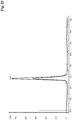
- a method for the selection of a binding entity-polynucleotide-complex from a library comprising a multitude of complexes with different polynucleotide linker length In one example of this method several linker molecules with polynucleotide linkers of various lengths are synthesized and used in the formation of a complex as reported herein comprising polynucleotide linkers of variable length and those complexes are selected having an improvement in the K diss of at least 5-fold over the better of the two monovalent polypeptide binders. Selection of a bivalent binding agent with the desired K diss in one embodiment is performed by BIAcore-analysis as disclosed in the Examples.
- the first single stranded L-DNA moiety does not hybridize with the second single stranded L-DNA moiety and does not hybridize with the second single stranded L-DNA linker moiety.
- the second single stranded L-DNA moiety does not hybridize with the first single stranded L-DNA moiety and does not hybridize with the first single stranded L-DNA linker moiety.
- the first and/or second single stranded L-DNA moiety has a length of from 10 to 50 nucleotides. In one embodiment the length is of from 15 to 35 nucleotides. In one embodiment the length is of from 20 to 30 nucleotides.
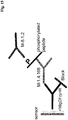
- the linker comprises a first single stranded L-DNA linker moiety, a second single stranded L-DNA linker moiety, and a single stranded docking moiety.
- the linker further comprises a linear non-nucleotide moiety.
- the linear non-nucleotide moiety is a polypeptide or a non-ionic polymer.
- the non-ionic polymer is linear poly (ethylene glycol).
- the linear poly (ethylene glycol) comprises of from 1 to 100 ethylene glycol units.
- the linear poly (ethylene glycol) comprises of from 1 to 50 ethylene glycol units.
- the linear poly (ethylene glycol) comprises of from 1 to 25 ethylene glycol units.
- the complex comprises
- the linker comprises in 3' to 5' orientation
- the docking single stranded L-DNA moiety does not hybridize with the first single stranded L-DNA moiety or its complementary first single stranded linker moiety and it does not hybridize with the second single stranded L-DNA moiety or its complementary second single stranded L-DNA linker moiety.
- the linker comprises in 3' to 5' orientation
- the linker comprises in 3' to 5' orientation
- the linker comprises in 3' to 5' orientation
- the linker comprises in 3' to 5' orientation
- the first non-nucleotide moiety and the second non-nucleotide moiety are the same or different.
- the linear non-nucleotide moiety is a polypeptide or a non-ionic polymer.
- the non-ionic polymer is linear poly (ethylene glycol).
- the linear poly (ethylene glycol) comprises of from 1 to 100 ethylene glycol units.
- the linear poly (ethylene glycol) comprises of from 1 to 50 ethylene glycol units.
- the linear poly (ethylene glycol) comprises of from 1 to 25 ethylene glycol units.
- the binding entity component is the binding entity component
- Monoclonal antibody techniques allow for the production of specifically binding agents in the form of specifically binding monoclonal antibodies or fragments thereof.
- techniques such as immunizing mice, rabbits, hamsters, or any other mammal with a polypeptide, i.e. the target of the antibody, or/and nucleic acid encoding the polypeptide can be used.
- monoclonal antibodies, or fragments thereof can be obtained by the use of phage libraries of scFv (single chain variable region), specifically human scFv (see e.g. US 5,885,793 , WO 92/01047 , WO 99/06587 ).
- the binding entity that specifically binds to a target is a monovalent antibody fragment.
- the monovalent antibody fragment is derived from a monoclonal antibody.
- Monovalent antibody fragments include, but are not limited to Fab, Fab'-SH, single domain antibody, F(ab') 2 , Fv, and scFv fragments.
- the monovalent antibody fragment is selected from the group comprising Fab, Fab'-SH, single domain antibody, F(ab') 2 , Fv, and scFv fragments.
- At least one of the binding entities of the complex as reported herein is a single domain antibody, or a Fab-fragment, or a Fab'-fragment of a monoclonal antibody.
- both of the binding entities of the complex as reported herein are independently of each other a single domain antibody, or a Fab-fragment, or a Fab'-fragment of a monoclonal antibody.
- both of the binding entities of the complex as reported herein are single domain antibodies, or Fab-fragments, or Fab'-fragments.
- targets or epitopes specifically bound by the binding entities do not overlap.
- Diabodies are antibody fragments with two antigen-binding sites that may be bivalent or bispecific (see e.g. EP 0 404 097 , WO 93/01161 , Hudson, P.J., et al., Nat. Med. 9 (2003) 129-134 , and Holliger, P., et al., Proc. Natl. Acad. Sci. USA 90 (1993) 6444-6448 ). Triabodies and tetrabodies are also described in Hudson, P.J., et al., Nat. Med. 9 (2003) 129-134 .
- Single-domain antibodies are antibody fragments comprising all or a portion of the heavy chain variable domain or all or a portion of the light chain variable domain of an antibody.
- a single-domain antibody is a human single-domain antibody (Domantis, Inc., Waltham, MA; US 6,248,516 ).
- An Fv is a minimum antibody fragment that contains a complete antigen-binding site and is devoid of constant region.
- Plueckthun in The Pharmacology of Monoclonal Antibodies, vol. 113, Rosenburg and Moore (eds.), (Springer-Verlag, New York, 1994), pp. 269-315 , WO 93/16185 , US 5,571,894 , US 5,587,458 .
- six hyper variable regions confer antigen-binding specificity to an antibody.
- a single variable domain or half of an Fv comprising only three HVRs specific for an antigen
- the monovalent antibody fragments is a two-chain Fv species consisting of a dimer of one heavy- and one light-chain variable domain in tight, non-covalent association.
- the monovalent antibody fragments is a single-chain Fv (scFv) species consisting of one heavy-chain and one light-chain variable domain covalently linked by a flexible peptide linker.
- scFv single-chain Fv
- a Fab fragment of an antibody contains the heavy-chain and light-chain variable domains as well as the constant domain of the light chain and the first constant domain (CH1) of the heavy chain.
- a Fab' fragments differ from a Fab fragment by the addition of a few residues at the carboxy terminus of the heavy chain CH1 domain including one or more cysteines from the antibody hinge region.
- Fab'-SH denotes a Fab' in which the cysteine residue(s) of the constant domains bear a free thiol group.
- antibody fragments can be obtained via proteolytic digestion of full length antibodies (see, e.g., Morimoto, K., et al., J. Biochem. Biophys. Meth. 24 (1992) 107-117 , Brennan, M., et al., Science 229 (1985) 81-83 ).
- papain digestion of full length antibodies results in two identical antigen-binding fragments, called "Fab" fragments, each with a single antigen-binding site, and a residual "Fc" fragment, whose name reflects its ability to crystallize readily.
- Fab antigen-binding fragments
- Antibody fragments can also be produced directly by recombinant means. Fab, Fv and scFv antibody fragments can all be expressed in and secreted from e.g. E. coli, thus, allowing the facile production of large amounts of these fragments. Antibody fragments can be isolated from antibody phage libraries according to standard procedures. Alternatively, Fab'-SH fragments can be directly recovered from E. coli. ( Carter, P., et al., Bio/Technology 10 (1992) 163-167 ). Mammalian cell systems can be also used to express and, if desired, secrete antibody fragments.
- the binding entity that specifically binds to an antigen is a single-domain antibody.
- a single-domain antibody is a human single-domain antibody (see, e.g., US 6,248,516 ).
- a single-domain antibody consists of all or a portion of the heavy chain variable domain of an antibody.
- a single-domain antibody is a single polypeptide chain comprising all or a portion of the heavy chain variable domain or all or a portion of the light chain variable domain of an antibody.
- the binding entity binds to its target with a dissociation constant (KD) of ⁇ 10 nM, ⁇ 1 nM, ⁇ 0.1 nM, ⁇ 0.01 nM, or ⁇ 0.001 nM (e.g. 10 -8 M or less, e.g. from 10 -8 M to 10 -13 M, e.g., from 10 -9 M to 10 -13 M).
- KD dissociation constant
- the binding entity binds to its target with a dissociation constant (KD) of 10 -5 M to 10 -13 M, or of 10 -5 M to 10 -10 M, or of 10 -5 M to 10 -8 M.
- KD dissociation constant
- the dissociation constant is determined by a radiolabeled antigen binding assay (RIA) performed with the Fab fragment of the antibody and its antigen as described by the following assay.
- RIA radiolabeled antigen binding assay
- Solution binding affinity of Fabs for antigen is measured by equilibrating Fab with a minimal concentration of ( 125 I)-labeled antigen in the presence of a titration series of unlabeled antigen, then capturing bound antigen with an anti-Fab antibody-coated plate (see, e.g., Chen, Y., et al., J. Mol. Biol. 293 (1999) 865-881 ).
- MICROTITER ® multi-well plates (Thermo Scientific) are coated overnight with 5 ⁇ g/ml of a capturing anti-FAB antibody (Cappel Labs) in 50 mM sodium carbonate (pH 9.6), and subsequently blocked with 2 % (w/v) bovine serum albumin in PBS for two to five hours at room temperature (approximately 23 °C).
- a non-adsorbent plate (Nunc #269620) 100 pM or 26 pM [ 125 I]-antigen are mixed with serial dilutions of a Fab of interest (e.g., consistent with assessment of the anti-VEGF antibody, Fab-12, in Presta, L.G., et al., Cancer Res. 57 (1997) 4593-4599 ).
- the FAB of interest is then incubated overnight; however, the incubation may continue for a longer period (e.g., about 65 hours) to ensure that equilibrium is reached. Thereafter, the mixtures are transferred to the capture plate for incubation at room temperature (e.g., for one hour).
- the dissociation constant is determined using surface plasmon resonance assays using a BIACORE ® -2000 or a BIACORE ® -3000 or a BIACORE ® A-100 (BIAcore, Inc., Piscataway, NJ) at 25 °C with immobilized antigen CM5 chips at ⁇ 10 response units (RU).
- carboxymethylated dextran biosensor chips (CM5, BIACORE, Inc.) are activated with N- ethyl- N '-(3-dimethylaminopropyl)-carbodiimide hydrochloride (EDC) and N -hydroxysuccinimide (NHS) according to the supplier's instructions.
- EDC N-ethyl- N '-(3-dimethylaminopropyl)-carbodiimide hydrochloride
- NHS N -hydroxysuccinimide
- Antigen is diluted with 10 mM sodium acetate, pH 4.8, to 5 ⁇ g/ml ( ⁇ 0.2 ⁇ M) before injection at a flow rate of 5 ⁇ l/minute to achieve approximately 10 response units (RU) of coupled protein.
- 1 M ethanolamine is injected to block unreacted groups.
- a cooperative binding event can be generated, which can be in dependence of the polynucleotide linker length.
- Binding specificity is further increased, when the addressed binding sites are independently located on two different target molecules, which e.g. might be co-localized on the surface of a tumor cell.
- the binding entity specifically binding to a target likely carries one or more free OH, COOH, NH 2 and/or SH groups, which could potentially react with certain coupling reagents.
- one of the coupling chemistries as given in the following Table 1 can be chosen.
- Table 1 provides an overview over reactive groups for covalently binding the polypeptides to the respective member of a binding pair as well as for covalently binding the linker to the respective members of a binding pair.
- the above bi-orthogonal coupling chemistries are especially appropriate for the conjugation of the monovalent binding polypeptides. If the two binding partners are not carrying certain reactive functional groups, e.g. in the case of combination of two aptamers there is more freedom in selection of the reactive sites. Therefore in addition or in combination with the pairs of corresponding reactive sites given in the above table, amino/active ester (e.g. NHS ester), and SH/SH or SH/maleinimido can be used for orthogonal coupling.
- amino/active ester e.g. NHS ester
- SH/SH or SH/maleinimido can be used for orthogonal coupling.
- the monovalent binding polypeptide may also be a synthetic peptide or peptide mimic.
- a polypeptide is chemically synthesized, amino acids with orthogonal chemical reactivity can be incorporated during such synthesis (see e.g. de Graaf, A.J., et al., Bioconjug. Chem. 20 (2009) 1281-1295 ). Since a great variety of orthogonal functional groups is at stake and can be introduced into a synthetic peptide, conjugation of such peptide to a linker is standard chemistry.
- the polynucleotide component is the polynucleotide component
- the complex as reported herein comprises a (polynucleotide) linker.
- the linker can either be covalently bound to the polypeptide(s) or the (polynucleotide) linker and the polypeptide(s) can be bound to each other by a specific binding pair.
- polynucleotide is to be understood broadly and includes DNA and RNA as well as analogs and modifications thereof.
- the polynucleotide linker is composed of a mixture of different types of monomers as long as more than 20 % of the monomers are nucleosides. In one embodiment the polynucleotide linker is composed of a mixture of different types of monomers as long as more than 30 % of the monomers are nucleosides. In one embodiment the polynucleotide linker is composed of a mixture of different types of monomers as long as more than 40 % of the monomers are nucleosides. In one embodiment the polynucleotide linker is composed of a mixture of different types of monomers as long as more than 50 % of the monomers are nucleosides.
- the linker can be composed exclusively of nucleosides or it can be a mixture of nucleosides and amino acids, and/or sugar residues, and/or diols, and/or phospho-sugar units, and/or non-ionic polymer building blocks.
- An oligonucleotide may for example contain a substituted nucleotide carrying a substituent at the standard bases deoxyadenosine (dA), deoxyguanosine (dG), deoxycytosine (dC), deoxythymidine (dT), deoxyuracil (dU).
- dA deoxyadenosine
- dG deoxyguanosine
- dC deoxycytosine
- dT deoxythymidine
- deoxyuracil deoxyuracil
- substituted nucleobases are 5-substituted pyrimidines (like 5-methyl-dC, aminoallyl-dU or -dC, 5-(aminoethyl-3-acrylimido)-dU, 5-propinyl-dU or -dC), 5-halogenated-dU or -dC, N-substituted pyrimidines (like N4-ethyl-dC), N-substituted purines (like N6-ethyl-dA, N2-ethyl-dG), 8-substituted purines (like 8-[6-amino)-hex-1-yl]-8-amino-dG or -dA), 8-halogenated-dA or -dG, 8-alkyl-dG or -dA, and 2-substituted-dA (like 2-amino-dA).
- 5-substituted pyrimidines like 5-methyl-dC, amino
- An oligonucleotide may contain a nucleotide or a nucleoside analog.
- nucleobase analogs like 5-nitroindol-D-riboside, 3-nitro-pyrrole-D-riboside, deoxyinosine (dI), deoyxanthosine (dX), 7-deaza-dG, -dA, -dI or -dX, 7-deaza-8-aza-dG, -dA, -dI or - dX, 8-aza-dA, -dG, -dI or -dX, D-Formycin, pseudo-dU, pseudo-iso-dC, 4-thio-dT, 6-thio-dG, 2-thio-dT, iso-dG, 5-methyl-iso-dC, N8-linked-8-aza-7-deaza-dA, 5,6-dihydro-5-aza-dC, e
- nucleobase in the complementary strand has to be selected in such manner that duplex formation is specific. If, for example, 5-methyl-iso-dC is used in one strand (e.g. (a)) iso-dG has to be in the complementary strand (e.g. (a')).
- the oligonucleotide backbone of the linker is modified to contain substituted sugar residues, sugar analogs, modifications in the inter-nucleoside phosphate moiety, and/or is a PNA (having a backbone without phosphate and d-ribose).
- An oligonucleotide may for example contain a nucleotide with a substituted deoxy ribose like 2'-methoxy-, 2'-fluoro-, 2'-methylseleno-, 2'-allyloxy-, 4'-methyl-dN (wherein N is a nucleobase, e.g., A, G, C, T or U).
- Sugar analogs are for example xylose, 2',4'-bridged ribose like (2'-O, 4'-C methylene) (oligomer known as LNA), or (2'-O, 4'-C ethylene) (oligomer known as ENA), L-ribose, L-D-ribose, hexitol (oligomer known as HNA), cyclohexenyl (oligomer known as CeNA), altritol (oligomer known as ANA), a tricyclic ribose analog where C3' and C5' atoms are connected by an ethylene bridge that is fused to a cyclopropane ring (oligomer known as tricyclo DNA), glycerin (oligomer known as GNA), glucopyranose (oligomer known as Homo DNA), carbaribose (with a cyclopentane instead of a tetrahydrofurane subunit), hydroxymethyl-morpho
- inter-nucleosidic phosphate moiety A great number of modification of the inter-nucleosidic phosphate moiety are also known not to interfere with hybridization properties and such backbone modifications can also be combined with substituted nucleotides or nucleotide analogs. Examples are phosphorthioate, phosphordithioate, phosphoramidate and methylphosphonate oligonucleotides.
- the (polynucleotide) linker has a length of from 1 nm to 100 nm. In one embodiment the (polynucleotide) linker has a length of from 4 nm to 80 nm. In one embodiment the (polynucleotide) linker has a length of from 5 nm to 50 nm or of from 6 nm to 40 nm. In one embodiment the (polynucleotide) linker has a length of 10 nm or longer or of 15 nm or longer. In one embodiment the (polynucleotide) linker has a length between 10 nm and 50 nm.
- the members of a binding pair conjugated to the (polynucleotide) linker have a length of at least 2.5 nm each.
- the length of the (polynucleotide) linker can be calculated by using known bond distances and bond angles of components which are chemically similar to the entities. Such bond distances are summarized for some molecules in standard text books (see e.g. CRC Handbook of Chemistry and Physics, 91st edition (2010-2011), Section 9).
- a) for calculating lengths of non-nucleosidic entities an average bond length of 130 pm with an bond angle of 180° independently of the nature of the linked atoms is used, b) one nucleotide in a single strand is calculated with 500 pm, and c) one nucleotide in a double strand is calculated with 330 pm.
- the value of 130 pm is based on calculation of the distance of the two terminal carbon atoms of a C(sp3)-C(sp3)-C(sp3) chain with a bond angle of 109°28' and a distance of 153 pm between two C(sp3) which is approx. 250 pm which translates with an assumed bond angle of 180° and bond distance between two C(Sp3) with 125 pm. Taking in account that heteroatoms like P and S and sp2 and sp1 C atoms could also be part of the linker the value 130 pm is taken. If the linker comprises a cyclic structure like cycloalkyl or aryl the distance is calculated in analogous manner by counting the number of the bonds of the cyclic structure which are part of the overall chain of atoms which are defining the distance.
- the length of the (polynucleotide) linker in a complex as reported herein can be varied as desired.
- linkers In order to easily make available linkers of variable length, i.e. a library, it is suitable to have a simple synthetic access to the different linkers of such library.
- a combinatorial solid phase synthesis of the linker is suited. Since linkers have to be synthesized up to a length of about 100 nm, the synthesis strategy is chosen in such a manner that the monomeric synthetic building blocks are assembled during solid phase synthesis with high efficiency.
- the synthesis of deoxy oligonucleotides based on the assembly of phosphoramidite as monomeric building blocks meet this requirement. In such a linker monomeric units within a linker are linked in each case via a phosphate or phosphate analog moiety.
- the (polynucleotide) linker can contain as in one embodiment free positively or/and negatively charged groups of polyfunctional amino-carboxylic acids, e.g. amino, carboxylate or phosphate.
- the charge carriers can be derived from trifunctional aminocarboxylic acids which contain a) an amino group and two carboxylate groups, or b) two amino groups and one carboxylate group.
- trifunctional aminocarboxylic acids examples include lysine, ornithine, hydroxylysine, ⁇ , ⁇ -diamino propionic acid, arginine, aspartic acid and glutamic acid, carboxy glutamic acid and symmetric trifunctional carboxylic acids like those described in EP 0 618 192 or US 5,519,142 .
- one of the carboxylate groups in the trifunctional aminocarboxylic acids of a) can be replaced by a phosphate, sulphonate or sulphate group.
- An example of such a trifunctional amino acid is phosphoserine.
- the (polynucleotide) linker can also contain as in one embodiment uncharged hydrophilic groups.
- uncharged hydrophilic groups are ethylene oxide or poly (ethylene oxide) groups comprising especially at least three building blocks, such as ethylene oxide, sulphoxide, sulphone, carboxylic acid amide, carboxylic acid ester, phosphonic acid amide, phosphonic acid ester, phosphoric acid amide, phosphoric acid ester, sulphonic acid amide, sulphonic acid ester, sulphuric acid amide and sulphuric acid ester groups.
- the amide groups are in one embodiment primary amide groups, especially carboxylic acid amide residues in amino acid side groups, e.g. of the amino acids asparagine and glutamine.
- the esters are especially derived from hydrophilic alcohols, in particular C1-C3 alcohols, or diols, or triols.
- Enantiomeric L-DNA is known for its orthogonal hybridization behavior, its nuclease resistance, and for ease of synthesis of polynucleotides of variable length.
- all polynucleotides in the complex are enantiomeric L-DNA or L-RNA. In one embodiment all polynucleotides in the complex are enantiomeric L-DNA.
- Enantiomeric, single stranded L-DNA combines high molecular flexibility and stability in body fluids.
- ss-L-DNA is used as a linker between two or more independent binding molecules, these binding molecules can get adjusted to virtually any binding angle and binding distance, which are just dependent from the ss-L-DNA linker length.
- the (polynucleotide) linker is synthesized in segments that can hybridize with each other.
- the linker can be formed by hybridization of the segments with one another.
- the resulting linker construct comprises oligonucleotide duplex portions.
- the linker is constructed that way the sequence of the hybridizable polynucleotide entity forming the duplex is chosen in such a manner that no hybridization or interference with the binding pair nucleic acids can occur.
- polynucleotide linker is synthesized in ss-L-DNA segments, e.g. A and B, which can hybridize with each other.
- the polynucleotide linker can be build up by the hybridization of the segments with one another. Therefore, the linker length can be self-adjusted to the distance between two binding sites simply by sequential application of the concatemer forming building blocks, i.e. A and B as exemplified.
- the linker is characterized in that the nucleic acid termini of the established linker hybridize with lower melting point temperature (i.e. TM1) to the ss-L-DNA labeled binding molecules than the inter-concatemeric melting point temperature (i.e. TM2, thus with TM2 > TM1).
- TM1 lower melting point temperature
- TM2 inter-concatemeric melting point temperature
- the obtained complex is incubated at a third temperature (i.e.
- TM3 that is above the first melting point temperature but below the second melting point temperature (i.e. TM3 > TM1 and TM3 ⁇ TM2).
- the temperature-eluted linker can be analyzed by standard methods e.g. using ethidiumbromide stained agarose gel.
- the linker length can also be calculated, because the length of each concatemer is known.
- the individual concatemers can be labeled in one embodiment.
- duplex portions can rigidize the oligonucleotide linker. This can be used to reduce the linker mobility and flexibility.
- one or more L-DNA oligonucleotides are hybridized to the oligonucleotide L-DNA linker.
- the oligonucleotide linker is rigidized via L-DNA duplex formation.
- an L-DNA/poly (ethylene glycol) hybrid is used as (oligonucleotide) linker.
- an L-DNA/D-DNA/poly (ethylene glycol) hybrid is used as (oligonucleotide) linker.
- an L-DNA/D-DNA/poly (ethylene glycol)/polypeptide hybrid is used as (oligonucleotide) linker.
- one or more L-DNA oligonucleotides are hybridized to the L-DNA/poly (ethylene glycol) hybrid (oligonucleotide) linker.
- one or more L-DNA oligonucleotides which are covalently coupled to a poly (ethylene glycol) molecule of varying length, are hybridized to the oligonucleotide L-DNA poly (ethylene glycol) hybrid (oligonucleotide) linker.
- an L-DNA/D-DNA hybrid is used as (oligonucleotide) linker.
- an L-DNA/D-DNA hybrid is used as (oligonucleotide) linker, wherein one or more D-DNA oligonucleotides are hybridized to the oligonucleotide D-DNA portion of the (oligonucleotide) linker to form double stranded D-DNA.
- an L-DNA/D-DNA hybrid is used as linker, wherein one or more L-DNA oligonucleotides are hybridized to the oligonucleotide L-DNA portion of the (oligonucleotide) linker to form double stranded L-DNA.
- DNA-duplexes can be used to modify or adjust the in vivo half-life of the complex making it available for the enzymatic action of nucleases.
- a simple way to build the (polynucleotide) linker is to use standard D or L nucleoside phosphoramidite building blocks.
- a single strand stretch of dT is used.
- Hybridization can be used in order to vary the (polynucleotide) linker length (distance between the binding pair members at the termini of the polynucleotide linker) and the flexibility of the spacer, because the double strand length is reduced compared to the single strand and the double strand is more rigid than a single strand.
- oligonucleotides modified with a functional moiety are used.
- the oligonucleotide used for hybridization can have one or two terminal extensions not hybridizing with the linker and/or is branched internally. Such terminal extensions that are not hybridizing with the linker (and not interfering with the members of the binding pairs) can be used for further hybridization events.
- an oligonucleotide hybridizing with a terminal extension is oligonucleotide comprising an effector moiety.
- This labeled oligonucleotide again may comprise terminal extensions or being branched in order to allow for further hybridization, thereby a polynucleotide aggregate or dendrimer can be obtained.
- a poly-oligonucleic acid dendrimer is especially used in order to produce a polylabel, or in order to get a high local concentration of an effector moiety.
- Modified nucleotides which do not interfere with the hybridization of polynucleotides can be incorporated into those polynucleotides. Suited modified nucleotides are C5-substituted pyrimidines or C7-substituted 7-deaza purines. Polynucleotides can be modified internally or at the 5' or 3' terminus with non-nucleotidic entities which are used for the introduction of the effector moiety.
- non-nucleotidic entities are located within the (polynucleotide) linker between the two binding pair members conjugated to its ends.
- non-nucleotidic building blocks for construction of a polynucleotide are known in literature and a great variety is commercially available.
- non-nucleosidic bifunctional building blocks or non-nucleosidic trifunctional building blocks can either be used as CPG for terminal labeling or as phosphoramidite for internal labeling (see e.g. Wojczewski, C., et al., Synlett 10 (1999) 1667-1678 ).
- Bifunctional spacer building blocks in one embodiment are non-nucleosidic components.
- such linkers are C2 - C18 alkyl, alkenyl, alkinyl carbon chains, whereas the alkyl, alkenyl, alkinyl chains may be interrupted by additional ethyleneoxy and/or amide moieties or quarternized cationic amine moieties in order to increase hydrophilicity of the linker.
- Cyclic moieties like C5-C6-cycloalkyl, C4N-, C5N-, C4O-, C5O-heterocycloalkyl, phenyl which are optionally substituted with one or two C1-C6 alkyl groups can also be used as non-nucleosidic bifunctional linkers.
- Suited bifunctional building blocks comprise C3-C6 alkyl moieties and tri- to hexa- ethylene glycol chains.
- Tables 2a and 2b show some examples of nucleotidic bifunctional spacer building blocks with different hydrophilicity, different rigidity and different charges.
- trifunctional building blocks allow for positioning of a functional moiety to any location within a polynucleotide.
- Trifunctional building blocks are also a prerequisite for synthesis using solid supports, e.g. controlled pore glass (CPG), which are used for 3' terminal labeling of polynucleotides.
- CPG controlled pore glass
- the trifunctional linkers is connected to a functional moiety or a - if necessary - a protected functional moiety via an C2 - C18 alkyl, alkenyl, alkinyl carbon chains, whereas the alkyl, alkenyl, alkinyl chains may be interrupted by additional ethyleneoxy and/or amide moieties in order to increase hydrophilicity of the linker and comprises a hydroxyl group which is attached via a cleavable spacer to a solid phase and a hydroxyl group which is protected with an acid labile protecting group. After removal of this protecting group a hydroxyl group is liberated that could thereafter react with a phosphoramidite.
- Trifunctional building blocks may be non-nucleosidic or nucleosidic.
- Non-nucleosidic trifunctional building blocks are C2 - C18 alkyl, alkenyl, alkinyl carbon chains, whereas the alkyl, alkenyl, alkinyl are optionally interrupted by additional ethyleneoxy and/or amide moieties in order to increase hydrophilicity of the linker.
- Other trifunctional building blocks are cyclic groups like C5-C6-cycloalkyl, C4N-, C5N-, C4O-, C5O-heterocycloalkyl, phenyl which are optionally substituted with one or two C1-C6 alkyl groups.
- Preferred trifunctional building blocks are C3-C6 alkyl, cycloalkyl, C5O-heterocycloalkyl moieties optionally comprising one amide bond and substituted with a C1 - C6 alkyl O-PG group, wherein PG is an acid labile protecting group, preferably monomethoxytrityl, dimethoxytrityl, pixyl, xanthyl most preferred dimethoxytrityl.
- Non-limiting, yet suited examples for non-nucleosidic trifunctional building blocks are e.g. summarized in Table 3.
- Table 3. Trifunctional introduction of Reference Nelson, P.S., et al., Nucleic Acids Research 20 (1992) 6253-6259 . EP 0 313 219 ; US 5,585,481 ; US 5,451,463 ; EP 0 786 468 ; WO 92/11388 , WO 89/02439 Su, Sheng-Hui, et al., Bioorganic & Medicinal Chemistry Letters 7 (1997) 1639-1644 . WO 97/43451 Putnam, W.C.
- Nucleosidic trifunctional building blocks are used for internal labeling whenever it is necessary not to influence the polynucleotide hybridization properties compared to a non-modified polynucleotide. Therefore nucleosidic building blocks comprise a base or a base analog which is still capable of hybridizing with a complementary base.
- the general formula of a labeling compound for labeling a nucleic acid sequence of one or more of a, a', b, b' or S comprised in a complex as reported herein is given in Formula II.
- PG is an acid labile protecting group, especially monomethoxytrityl, dimethoxytrityl, pixyl, xanthyl, especially dimethoxytrityl, wherein Y is C2 - C18 alkyl, alkenyl alkinyl, wherein the alkyl, alkenyl, alkinyl may comprise ethyleneoxy and/or amide moieties, wherein Y preferably is C4 - C18 alkyl, alkenyl or alkinyl and contains one amide moiety and wherein X is a functional moiety.
- Specific positions of the base may be chosen for such substitution to minimize the influence on hybridization properties. Therefore the following positions are especially suited for substitution: a) with natural bases: uracil substituted at C5, cytosine substituted at C5 or at N4, adenine substituted at C8 or at N6, and guanine substituted at C8 or at N2, and b) with base analogs: 7-deaza-A and 7-deaza-G substituted at C7, 7-deaza-8-aza-A and 7-deaza-8-aza-G substituted at C7, 7-deaza-aza-2-amino-A substituted at C7, pseudouridine substituted at N1 and formycin substituted at N2. Table 4.
- terminal oxygen atom of bifunctional moiety or one of the terminal oxygen atoms of a trifunctional moiety are part of a phosphoramidite that is not shown in full detail but obvious to the skilled artisan.
- the second terminal oxygen atom of trifunctional building block is protected with an acid labile protecting group PG, as defined for Formula II above.
- Post-synthetic modification is another strategy for introducing a covalently bound functional moiety into a linker.
- an amino group is introduced by using bifunctional or trifunctional building blocks during solid phase synthesis. After cleavage from the support and purification of the amino modified linker the linker is reacted with an activated ester of a functional moiety or with a bifunctional reagent wherein one functional group is an active ester.
- active esters are NHS ester or pentafluor phenyl esters.
- Post-synthetic modification is especially useful for introducing a functional moiety which is not stable during solid phase synthesis and deprotection.
- Examples are modification with triphenylphosphincarboxymethyl ester for Staudinger ligation ( Wang, Charles C.-Y., et al., Bioconjugate Chemistry 14 (2003) 697-701 ), modification with digoxigenin or for introducing a maleinimido group using commercial available sulfo SMCC.
- each member of a binding pair is of/has a molecular weight of 10 kDa or less. In one embodiment the molecular weight of each member of a binding pair is 8 kDa, or 7 kDa, or 6 kDa, or 5 kDa, or 4 kDa or less.
- the members of both binding pairs in the complex as reported herein are different.
- the difference between the binding pairs a:a' and b:b' is e.g. acknowledged if the dissociation constant for the reciprocal binding, e.g. binding of a as well as a' to b or b', is 10 times the dissociation constant of the pair a:a' or more.
- dissociation constant for the reciprocal binding i.e. binding of a as well as a' to b or b', respectively, is 20 times the dissociation constant of the pair a:a' or more. In one embodiment the dissociation constant is 50 times the dissociation constant within the pair a:a' or more. In one embodiment the reciprocal (cross-reactive) binding dissociation constant is 100 times or more the dissociation constant within a binding pair.
- the members of the binding pairs are selected from the group consisting of leucine zipper domain dimers and hybridizing nucleic acid sequences. In one embodiment both binding pairs are leucine zipper domain dimers.
- both binding pairs are hybridizing nucleic acid sequences. In one embodiment all binding pair members are L-DNA sequences. In one embodiment both binding pairs are hybridizing L-DNAs.
- both member of the binding pairs represent leucine zipper domains.
- leucine zipper domain denotes a dimerization domain characterized by the presence of a leucine residue at every seventh residue in a stretch of approximately 35 residues.
- Leucine zipper domains are peptides that promote oligomerization of the proteins in which they are found. Leucine zippers were originally identified in several DNA-binding proteins ( Landschulz, W.H., et al., Science 240 (1988) 1759-1764 ). Among the known leucine zippers are naturally occurring peptides and derivatives thereof that dimerize or trimerize.
- leucine zipper domains suitable for producing soluble multimeric proteins are those reported in WO 94/10308 , and the leucine zipper derived from lung surfactant protein D (SPD) as reported in Hoppe, H.J., et al., FEBS Lett. 344 (1994) 191-195 .
- SPD lung surfactant protein D
- Leucine zipper domains form dimers (binding pairs) held together by an alpha-helical coiled coil.
- a coiled coil has 3.5 residues per turn, which means that every seventh residue occupies an equivalent position with respect to the helix axis.
- the regular array of leucines inside the coiled coil stabilizes the structure by hydrophobic and Van der Waals interactions.
- leucine zipper domains form the first binding pair and the second binding pair, both leucine zipper sequences are different, i.e. the members of the first binding pair do not bind to the members of the second binding pair.
- Leucine zipper domains may be isolated from natural proteins known to contain such domains, such as transcription factors.
- One leucine zipper domain may e.g. come from the transcription factor fos and a second one from the transcription factor jun.
- Leucine zipper domains may also be designed and synthesized artificially, using standard techniques for synthesis and design known in the art.
- both binding pairs are hybridizing nucleic acid sequences.
- each binding pair i.e. a and a' as well as b and b', hybridize to one another, respectively.
- the nucleic acid sequences comprised in the first binding pair on the one hand and in the second binding pair on the other hand are different, i.e. do not hybridize with each other.
- binding pairs are both hybridizing nucleic acid pairs, wherein the hybridizing nucleic acid sequences of the different binding pairs do not hybridize with one another.
- nucleic acids of the first binding pair hybridize to each other but do not bind to any of the nucleic acids of the second binding pair or interfere with their hybridization and vice versa.
- Hybridization kinetics and hybridization specificity can easily be monitored by melting point analyses. Specific hybridization of a binding pair and non-interference is acknowledged, if the melting temperature for the binding pair as compared to any possible combination with other binding pairs or combination of binding pair members is at least 20 °C higher.
- the nucleic acid sequences forming a binding pair may comprise in principle any naturally occurring nucleobase or an analogue thereto and may have in principle a modified or a non-modified backbone as described above provided it is capable of forming a stable duplex via multiple base pairing.
- Stable denotes that the melting temperature of the duplex is higher than 30 °C, especially higher than 37 °C.
- the double strand is in one embodiment consisting of two fully complementary single stranded polynucleotides.
- a nucleic acid duplex can be further stabilized by inter-strand crosslinking.
- inter-strand crosslinking Several appropriate cross-linking methods are known, e.g. methods using psoralen or based on thionucleosides.
- nucleic acid sequences representing the members of a binding pair in one embodiment consist of from 12 to 50 nucleotides. In one embodiment such nucleic acid sequences consist of from 15 to 35 nucleotides.
- RNAses are ubiquitous and special care has to be taken to avoid unwanted digestion of RNA-based binding pairs and/or linker sequences. While RNA-based binding pairs and/or linkers can be used, binding pairs and/or linkers based on DNA are especially suited.
- hybridizing nucleic acid sequences can easily be designed to provide for more than two pairs of orthogonal complementary polynucleotides, allowing for an easy generation and use of more than two binding pairs.
- Another advantage of using hybridizing nucleic acid sequences in a complex as reported herein is that modifications can be easily introduced.
- Modified building blocks are commercially available which e.g. allow for an easy synthesis of a polynucleotide comprising a functional moiety. Such functional moiety can be easily introduced at any desired position and in any of the members of the first and/or second binding pair and/or the polynucleotide linker, provided they all represent a polynucleotide.
- the (polynucleotide) linker comprising members of bindings pairs at its termini can be provided for and synthesize as a single polynucleotide.
- the polypeptides specifically binding to a target can each be coupled to hybridizing nucleic acid sequences, i.e. members of binding pairs.
- the length of the (polynucleotide) linker can easily be varied in any desired manner.
- the reaction of a maleinimido moiety with a cysteine residue within the polypeptide is used.
- C-terminal modification of a protein e.g. of a FAB-fragment can e.g. be performed as described ( Sunbul, M. and Yin, J., Org. Biomol. Chem. 7 (2009) 3361-3371 ).
- site specific reaction and covalent coupling of a binding pair member to a monovalent binding polypeptide is based on transforming a natural amino acid into an amino acid with a reactivity which is orthogonal to the reactivity of the other functional groups present in a polypeptide.
- a specific cysteine within a rare sequence context can be enzymatically converted in an aldehyde (see e.g. Frese, M-A. and Dierks, T., ChemBioChem 10 (2009) 425-427 ). It is also possible to obtain a desired amino acid modification by utilizing the specific enzymatic reactivity of certain enzymes with a natural amino acid in a given sequence context (see e.g.: Taki, M., et al., Prot. Eng. Des. Sel. 17 (2004) 119-126 , Gautier, A., et al., Chem. Biol. 15 (2008) 128-136 ; Bordusa, F., in Highlights in Bioorganic Chemistry (2004), Schmuck, C. and Wennemers, H., (eds.), Wiley VCH, Weinheim, pp. 389-403 ).
- Site specific reaction and covalent coupling of a binding pair member to a monovalent binding polypeptide can also be achieved by the selective reaction of terminal amino acids with appropriate modifying reagents.
- EP 1 074 563 reports a conjugation method which is based on the faster reaction of a cysteine within a stretch of negatively charged amino acids with a cysteine located in a stretch of positively charged amino acids.
- the effector moiety can be selected from the group consisting of a binding moiety, a labeling moiety, a biologically active moiety, and a reactive moiety. If more than one effector moiety is present in the complex, each such effector moiety can in each case be independently a binding moiety, a labeling moiety, a biologically active moiety, or a reactive moiety.
- the binding moiety will be selected to have no interference with each of the binding pairs.
- the effector moiety is selected from the group consisting of a binding moiety, a labeling moiety, and a biologically active moiety.
- the effector moiety is a binding moiety.
- binding moieties are the members of a bioaffine binding pair which can specifically interact with each other.
- Suitable bioaffine binding pairs are hapten or antigen and antibody; biotin or biotin analogues such as aminobiotin, iminobiotin or desthiobiotin and avidin or streptavidin; sugar and lectin, polynucleotide and complementary polynucleotide, receptor and ligand, e.g., steroid hormone receptor and steroid hormone; and the pair of an 104-aa fragment of bovine ribonuclease A (known as S-protein) and a 15-aa fragment of bovine ribonuclease A (known as S-peptide).
- the effector moiety is a binding moiety and is covalently bound to at least one of the components of the complex.
- the smaller partner of a bioaffine binding pair e.g. biotin or an analogue thereto, a receptor ligand, a hapten or a polynucleotide is covalently bound to at least one of the polynucleotides comprised in the complex as reported herein.
- the effector moiety is a binding moiety selected from hapten, biotin or biotin analogues such as aminobiotin, iminobiotin or desthiobiotin; polynucleotide and steroid hormone.
- the effector moiety is a labeling group.
- the labeling group can be selected from any known detectable group.
- the labeling group is selected from dyes like luminescent labeling groups such as chemiluminescent groups e.g. acridinium esters or dioxetanes or fluorescent dyes e.g. fluorescein, coumarin, rhodamine, oxazine, resorufin, cyanine and derivatives thereof, luminescent metal complexes such as ruthenium or europium complexes, enzymes as used for CEDIA (Cloned Enzyme Donor Immunoassay, e.g. EP 0 061 888 ), microparticles or nanoparticles e.g. latex particles or metal sols, and radioisotopes.
- chemiluminescent groups e.g. acridinium esters or dioxetanes or fluorescent dyes e.g. fluorescein, coumarin, rhodamine, oxazine, resorufin, cyanine and derivatives
- the labeling group is a luminescent metal complex and the compound has a structure of the general formula (I): [M(L 1 L 2 L 3 )] n - Y - X m A (I) in which M is a divalent or trivalent metal cation selected from rare earth or transition metal ions, L 1 , L 2 and L 3 are the same or different and denote ligands with at least two nitrogen-containing heterocycles in which L 1 , L2 and L3 are bound to the metal cation via nitrogen atoms, X is a reactive functional group which is covalently bound to at least one of the ligands L 1 , L2 and L3 via a linker Y, n is an integer from 1 to 10, especially 1 to 4, m is 1 or 2, or especially 1 and A denotes the counter ion which may be required to equalize the charge.
- M is a divalent or trivalent metal cation selected from rare earth or transition metal ions
- L 1 , L 2 and L 3
- the metal complex is in one embodiment a luminescent metal complex i.e. a metal complex which undergoes a detectable luminescence reaction after appropriate excitation.
- the luminescence reaction can for example be detected by fluorescence or by electrochemiluminescense measurement.
- the metal cation in this complex is for example a transition metal or a rare earth metal.
- the metal is in one embodiment ruthenium, osmium, rhenium, iridium, rhodium, platinum, indium, palladium, molybdenum, technetium, copper, chromium or tungsten. Ruthenium, iridium, rhenium, chromium and osmium are especially suited. Ruthenium is most suited.
- Gold nanorods can also be used as labeling moiety in the complexes as reported herein.
- the nanorods can have a length of from 10 to 100 nm, inclusive, and including all integers there between.
- the GNRs have an average length of from 70-75 nm.
- the GNRs can have a diameter of from 5 to 45 nm inclusive, and including all integers there between.
- the GNRs have an average diameter of 25-30 nm.
- the GNRs can be pure gold, or may be from 90 % to 99 %, inclusive, including all integers there between, pure gold.
- the GNRs may contain up to 1 % silver on their surfaces, and may contain cetyltrimethylammonium bromide (CTAB).
- CTAB cetyltrimethylammonium bromide
- GNRs can be made by any suitable method.
- electrochemical synthesis in solution membrane templating, photochemical synthesis, microwave synthesis, and seed mediated growth are all suitable and non-limiting examples of methods of making the GNRs.
- the gold nanorods are made using the seed-mediated growth method in cetyltrimethylammonium bromide (CTAB).
- CTAB cetyltrimethylammonium bromide
- the surfaces of the gold nanorods can be functionalized so as impart a positive zeta potential suitable for electrostatically complexing the GNRs with DNA or RNA polynucleotides. Any suitable method of creating a positive zeta potential on the gold nanorods may be used.
- the surfaces of the gold nanorods can be functionalized with bifunctional molecules, such as thiolated-PEG-NH2 or thiolated-PEG-COOH.
- the surface functionalization is achieved by coating the CTAB-coated gold nanorods first with the anionic polyelectrolyte poly (3,4-ethylenedioxythi-6-phene)/poly (styrene sulfate) (PEDT/PSS), then with the cationic polyelectrolyte poly (diallyl dimethyl ammonium chloride) (PDDAC).
- anionic polyelectrolyte poly (3,4-ethylenedioxythi-6-phene)/poly (styrene sulfate)
- PDDAC cationic polyelectrolyte poly (diallyl dimethyl ammonium chloride)
- the positively charged gold nanorods are electrostatically complexed to the DNA polynucleotides using electrostatic interactions.
- nanoplexes can be confirmed from an observed red-shift in localized longitudinal plasmon resonance peak of the gold nanorods, as well as from restricted electrophoretic mobility of the nanoplexes using gel electrophoresis.
- the effector moiety X is a therapeutically active substance.
- Therapeutically active substances have different ways in which they are effective, e.g. in inhibiting cancer, damaging the DNA template by alkylation, by cross-linking, or by double-strand cleavage of DNA.
- Other therapeutically active substances can block RNA synthesis by intercalation.
- Some agents are spindle poisons, such as vinca alkaloids, or anti-metabolites that inhibit enzyme activity, or hormonal and anti-hormonal agents.
- the effector moiety may be selected from alkylating agents, antimetabolites, antitumor antibiotics, vinca alkaloids, epipodophyllotoxins, nitrosoureas, hormonal and anti-hormonal agents, and toxins.
- Suited alkylating agents are cyclophosphamide, chlorambucil, busulfan, melphalan, thiotepa, ifosphamide, or nitrogen mustard.
- Suited antimetabolites are methotrexate, 5-Fluorouracil, cytosine arabinoside, 6-thioguanine, 6-mercaptopurin.
- doxorubicin doxorubicin
- daunorubicin idorubicin
- nimitoxantron dactinomycin
- bleomycin bleomycin
- mitomycin plicamycin
- Suited spindle poisons are maytansine and maytansinoids, vinca alkaloids and epipodophyllotoxins may be exemplified by vincristin, vinblastin, vindestin, Etoposide, Teniposide.
- taxane agents may be exemplified by Paclitaxel, Docetaxel, SB-T-1214.
- nitrosoureas are carmustine, lomustine, semustine, streptozocin.
- adrenocorticoids adrenocorticoids
- estrogens antiestrogens
- progestins progestins
- aromatase inhibitors androgens, anti-androgens.
- Suited random synthetic agents are dacarbazine, hexamethylmelamine, hydroxyurea, mitotane, procarbazide, cisplatin, carboplatin.
- fMLP f-Met-Leu-Phe
- fMLP f-Met-Leu-Phe-o-methyl ester
- formyl-norleucyl-phenylalanine formyl-methionyl-phenylalanine.
- NK cell attracting factors are IL-12, IL-15, IL-18, IL-2, and CCL5, the FC portion of an antibody.
- the effector moiety X is an antibody Fc-region or fragment thereof.
- human antibody Fc-region is of human IgG1 subclass, or of human IgG2 subclass, or of human IgG3 subclass, or of human IgG4 subclass.
- the antibody Fc-region is a human antibody Fc-region of the human IgG1 subclass, or of the human IgG4 subclass.
- the human antibody Fc-region comprises a mutation of the naturally occurring amino acid residue at least at one of the following amino acid positions 228, 233, 234, 235, 236, 237, 297, 318, 320, 322, 329, and/or 331 to a different residue, wherein the residues in the antibody Fc-region are numbered according to the EU index of Kabat.
- the human antibody Fc-region comprises a mutation of the naturally occurring amino acid residue at position 329 and at least one further mutation of at least one amino acid residue selected from the group comprising amino acid residues at position 228, 233, 234, 235, 236, 237, 297, 318, 320, 322 and 331 to a different residue, wherein the residues in the Fc-region are numbered according to the EU index of Kabat.
- the change of these specific amino acid residues results in an altering of the effector function of the Fc-region compared to the non-modified (wild-type) Fc-region.
- the human antibody Fc-region has a reduced affinity to the human Fc ⁇ RIIIA, and/or Fc ⁇ RIIA, and/or Fc ⁇ RI compared to a conjugate comprising the corresponding wild-type IgG Fc-region.
- amino acid residue at position 329 in the human antibody Fc-region is substituted with glycine, or arginine, or an amino acid residue large enough to destroy the proline sandwich within the Fc-region.
- the mutation in the human antibody Fc-region of the naturally occurring amino acid residue is at least one of S228P, E233P, L234A, L235A, L235E, N297A, N297D, P329G, and/or P331S.
- the mutation is L234A and L235A if the antibody Fc-region is of human IgG1 subclass, or S228P and L235E if the antibody Fc-region is of human IgG4 subclass.
- the antibody Fc-region comprises the mutation P329G.
- the effector moiety can be bound either covalently or via an additional binding pair to at least one of the components of the complex.
- the effector moiety can be comprised for one to several (n) times in the complex as reported herein, whereby (n) is an integer and 0 or 1 or more than one. In one embodiment (n) is between 1 and 1,000,000. In one embodiment (n) is between 1,000 and 300,000. In one embodiment (n) is 1 to 50. In one embodiment (n) is 1 to 10, or 1 to 5. In one embodiment (n) is 1 or 2
- any appropriate coupling chemistry can be used. It is also possible to incorporate a functional moiety by use of appropriate building blocks when synthesizing the members of the first and/or second binding pair and/or the (polynucleotide) linker, especially in the members of the binding pairs conjugated to the polypeptide or the (polynucleotide) linker.
- effector moiety is bound to the (polynucleotide) linker of the complex as reported herein.
- the effector moiety is covalently bound to a member of a binding pair conjugated to the polypeptide or the (polynucleotide) linker of the complex as reported herein.
- an effector moiety is located within a hybridizing polynucleotide it is especially suited to bind it to a modified nucleotide or is attached to the internucleosidic P atom (see e.g. WO 2007/059816 ).
- Bifunctional building blocks (as described above) can be used to connect an effector moiety or a - if necessary - a protected effector moiety to a phosphoramidite group for attaching the building block at the 5'-end (regular synthesis) or at the 3'-end (inverted synthesis) to the terminal hydroxyl group of a growing polynucleotide chain.
- Trifunctional building blocks can be used to connect (i) a effector moiety or a - if necessary - a protected effector moiety, (ii) a phosphoramidite group for coupling the reporter or the effector moiety or a - if necessary - a protected effector moiety, during the polynucleotide synthesis to a hydroxyl group of the growing polynucleotide chain and (iii) a hydroxyl group which is protected with an acid labile protecting group especially with a dimethoxytrityl protecting group. After removal of this acid labile protecting group a hydroxyl group is liberated which can react with further phosphoramidites.
- the effector moiety is bound in one embodiment to at least one of the members of the first and/or second binding pair or to the polynucleotide linker via an additional third binding pair.
- the third binding pair is a pair of hybridizing nucleic acid sequences. The members of the third binding pair do not interfere with the binding of the members of the other binding pairs to each other.
- the additional binding pair to which an effector moiety can be bound is especially a leucine zipper domain or a hybridizing nucleic acid.
- the binding pair member to which the effector moiety is bound and the first and second binding pairs members, respectively all are selected to have different specificity.
- the members of the first and second binding pair and the binding pair to which the effector moiety is bound each bind to (e.g. hybridize with) their respective partner without interfering with the binding of any of the other binding pairs.
- the complementary nucleic acids of the binding pairs and/or the (polynucleotide) linker is made at least partly of L-DNA, or L-RNA, or LNA, or iso-C nucleic acid, or iso-G nucleic acid, or any combination thereof. In one embodiment the (polynucleotide) linker is made at least to 50 % of L-DNA, or L-RNA, or LNA, or iso-C nucleic acid, or iso-G nucleic acid, or any combination thereof. In one embodiment the (polynucleotide) linker is an L-polynucleotide (a aptmer). In one embodiment the L-polynucleotide is L-DNA.
- the (polynucleotide) linker is DNA. In one embodiment the (polynucleotide) linker is the L-stereoisomer of DNA also known as beta-L-DNA or L-DNA or mirror image DNA.
- This stereoisomeric DNA features advantages like orthogonal hybridization behavior, which means that a duplex is formed only between two complementary single strands of L-DNA but no duplex is formed between a single strands of L-DNA and the complementary D-DNA strand, nuclease resistance and ease of synthesis even of a long linker.
- the ease of synthesis and variability in spacer length are important for providing a linker library.
- Linkers of variable length are useful in identifying complexes as reported herein having a polynucleotide linker of optimal length, thus, providing for the optimal distance between two polypeptide specifically binding a target.
- the complex is a non-covalent complex. In one embodiment the non-covalent complex is formed via binding pairs.
- the effector moiety is a therapeutic drug.
- the effector moiety can be a therapeutic radionuclide, hormone, cytokine, interferon, antibody or antibody fragment, nucleic acid aptamer, enzyme, polypeptide, toxin, cytotoxin, a chemotherapeutic agent, or a radiation sensitizer.
- a method of killing a cell wherein a complex as reported herein is administered to the cell in an amount sufficient to kill the cell.
- the cell is a cancer cell.
- a method of retarding or stopping the growth of a cancer cell in a mammal wherein a complex as reported herein is administered to the mammal in an amount sufficient to retard or stop growth of the cancer cell.
- the method is a method for inhibiting the growth or proliferation of a cancer cell.
- polypeptide specifically binding to a target is specifically binding to a cell surface molecule of a cell.
- cell surface molecule is specifically present on cancer cells.
- the first and second polypeptide specifically binding to a target are independently from each other selected from the group consisting of an antibody, an antibody fragment, a single-chain variable region antibody, a small peptidic molecule, a cyclic polypeptide, a peptidomimetic, and an aptamer.
- first and the second polypeptide specifically binding to a target are monovalent binding polypeptides.
- polypeptide is an antibody fragment.
- antibody fragment is from an internalizing antibody that specifically binds to a cell surface molecule.
- the conjugation of an effector moiety to a complex as reported herein allows for specific localization of the effector moiety at the desired site on a cell.
- the localization increases the effective concentration of the effector moiety on the target cell and thereby optimizes the effect of the effector moiety.
- the complex can be administered at a lower dose compare to a non-targeted effector moiety. This can be particularly relevant if the effector moiety has associated toxicities or if it is to be used in the treatment of chronic diseases.
- L-DNA is a useful nucleotide in the formation of complexes as reported herein.
- L-DNA does not, by itself, hybridize to the naturally occurring form of DNA (D-DNA) or RNA. Since L-DNA is not a natural substrate for many enzymes, the stability of an L-DNA in vivo can be greater than that of D-DNA.
- L-DNA duplexes have the same physical characteristics in terms of solubility, duplex stability and selectivity as D-DNA but form a left-helical double-helix. It is to be understood that the L-polynucleotide as used herein may also comprise some D-polynucleotides.
- L-polynucleotides Due to the chemical nature of the L-polynucleotides these are not metabolized so that the pharmacokinetics underlying the use of L-nucleotides is not or at least not to such an extend affected by DNA specific degradation processes. In view of the increased stability of the L-polynucleotides the in vivo half-life of the complex as reported herein in a mammal is, thus, factually unlimited. Of particular importance is the fact that the L-polynucleotides are not nephrotoxic.
- the mammal is selected from humans, monkeys, dogs, cats, horses, rats, or mice.
- the polynucleotide linker comprises D-DNA and L-DNA nucleotides, i.e. the polynucleotide linker is a mixture of D-DNA and L-DNA.
- Each of the polynucleotides present in the complex as reported herein can comprise one or more effector moieties. Effector moieties allow the use of the complex as reported herein in the treatment of a disease.
- the effector moieties can be used e.g. for carrier purposes, i.e. the delivery of an effector function, and/or modulation of pharmacokinetic behavior, and/or modulation of the physico-chemical properties.
- the effector moiety is selected from lipophilic moieties, peptides, proteins, carbohydrates and liposomes.
- the polynucleotide is an L-polynucleotide.
- the L-poly (deoxy) nucleotides can be present either as single- or as double-stranded polynucleotide.
- the L-poly (deoxy) nucleotide is present as single-stranded nucleic acid, which may form (defined) secondary structures and also tertiary structures. In such secondary structures also double-stranded stretches can be present.
- the L-poly (deoxy) nucleotide can also be present at least partly as double-stranded molecule in the meaning that two strands, which are complementary to each other, are hybridized.
- the L-polynucleotide(s) can also be modified. The modification can be related to the individual nucleotides of the polynucleotide.
- 2,4-Dihydroxy-5-methylpyrimidin (T) can be used as nucleobase in one embodiment.
- the L-polynucleotides in the complex as reported herein are in one embodiment susceptible to "self-hybridization”.
- the L-polynucleotides are more readily able to hybridize with complementary L-polynucleotide sequences but do not form a stable duplex with natural DNA or RNA.
- the nucleotides in the L-DNA segment have a conformation of 1'S, 3'R, and 4' S.
- the L-DNA polynucleotide linker is conjugated through hybridization of the members of the binding pairs at its termini with the polypeptide(s) of the complex.
- the polynucleotide linker has a length of at least 1 nm. In one embodiment the polynucleotide linker has a length of from 6 nm to 100 nm. In one embodiment the polynucleotide linker has a length of at least 70 nucleotides.
- the polynucleotide linker may also comprise a tag sequence.
- the tag sequence may be selected from commonly used protein recognition tags such as YPYDVPDYA (HA-Tag, SEQ ID NO: 64) or GLNDIFEAQKIEWHE (Avi-Tag, SEQ ID NO: 65).
- the complex as reported herein not comprising an effector moiety is administered first and allowed to bind to its target(s) and afterwards the effector moiety conjugated to a polynucleotide complementary to at least a part of the (polynucleotide) linker is administered.
- the effector moiety is co-located to the complex bound to its target by hybridizing to the complex as reported herein in situ.
- the complex binds to the cell surface of a tumor cell and locally enriches to a high density or high local concentration of the effector moiety.
- the effector moiety is labeled ss-L-DNA, which is administered simultaneously or subsequently to the initial target association of the complex.
- the labeled ss-L-DNA effector moiety hybridizes to ss-L-DNA (oligonucleotide) linker of the complex.
- NK cells cytotoxic lymphocytes
- NK cells play a major role in the rejection of tumors and cells infected by viruses. They kill cells by releasing small cytoplasmic granules of proteins called perforin and granzyme that cause the target cell to die by apoptosis.
- the complex as reported herein is used to attract NK cells into close proximity of the bound complex.
- ss-L-DNA conjugated to a cytokine is used as effector moiety.
- This cytokine labeled effector moiety can be used to attract NK cells.
- Cytokines involved in NK activation include IL-12, IL-15, IL-18, IL-2, and CCL5.
- ss-L-DNA conjugated to an Fc portion of an antibody is used as effector moiety.
- FcR Fc receptor
- FC-gamma-RIII CD16
- one or more or a combination of ss-L-DNA conjugated to one or more Fc parts is / are used as effector moieties.
- the complex can be used to modulate the ADCC and/or the complement activation (CDC).
- this complex is used in a method to screen engineered Fc compartments for their efficacy in engaging ADCC and CDC.
- the complex is used to inhibit seminal fluid phosphatase.
- the complex can be used to avoid NK cell inactivation.
- the polypeptide specifically binding to a target is conjugated to a ligand for a target receptor or large molecule that is more easily engulfed by the endocytotic mechanisms of a cell in order to increase the uptake of the complex into the cell presenting the target.
- the target bound complex can then be internalized by endocytosis and the effector moiety released inside the cell.
- the binding entity (polypeptide) specifically binding to a target is in one embodiment an antibody fragment.
- single-chain variable region fragment denotes a variable, antigen-binding region of a single antibody light chain and single antibody heavy chain linked together by a covalent linkage having a length sufficient to allow the light and heavy chain portions to form an antigen binding site.
- a linker may be as short as a covalent bond.
- Especially suited linkers comprise of from 2 to 50 amino acid residues, and especially of from 5 to 25 amino acid residues.
- diabodies Other antibody fragments are diabodies, first described by Holliger, P., et al. (PNAS (USA) 90 (1993) 6444-6448 ). These may be constructed using heavy and light chains of an antibody, as well as by using individual CDR regions of an antibody. Typically, diabodies comprise a heavy chain variable domain (VH) connected to a light chain variable domain (VL) by a linker which is too short to allow pairing between the two domains on the same chain. Accordingly, the VH and VL domains of one fragment are forced to pair with the complementary VH and VL domains of another fragment, thereby forming two antigen-binding sites. Triabodies can be similarly constructed with three antigen-binding sites.
- VH heavy chain variable domain
- VL light chain variable domain
- Fv antibody fragment contains a complete antigen-binding site which includes a VL domain and a VH domain held together by non-covalent interactions.
- Fv fragments also include constructs in which the VH and VL domains are crosslinked through glutaraldehyde, intermolecular disulfide bonds, or other linkers.
- the variable domains of the heavy and light chains can be fused together to form a single chain variable fragment (scFv), which retains the original specificity of the parent antibody.
- Single chain Fv (scFv) dimers first described by Gruber, M., et al., J. Immunol.
- 152 (1994) 5368-5374 may be constructed using heavy and light chains of an antibody, as well as by using individual CDR regions of an antibody. Many techniques known in the art can be used to prepare the specific binding constructs suitable in the complex as reported herein (see e.g., US 2007/0196274 , US 2005/0163782 ).
- Bispecific antibodies can be generated by chemical cross-linking or by the hybrid hybridoma technology.
- bispecific antibody molecules can be produced by recombinant techniques. Dimerization can be promoted by reducing the length of the linker joining the VH and the VL domain from about 15 amino acids, routinely used to produce scFv fragments, to about 5 amino acids. These linkers favor intrachain assembly of the VH and VL domains.
- a suitable short linker is SGGGS (SEQ ID NO: 66) but other linkers can be used. Thus, two fragments assemble into a dimeric molecule. Further reduction of the linker length to zero to two amino acid residues can generate trimeric (triabodies) or tetrameric (tetrabodies) molecules.
- the binding entity (polypeptide) specifically binding to a target e.g. an antibody specifically binding to a cell surface receptor
- a target e.g. an antibody specifically binding to a cell surface receptor
- the binding entity (polypeptide) specifically binding to a target can be linked to a ligand for a target receptor or large molecule that is more easily engulfed by the cell's endocytotic mechanisms.
- the complex can be used to increase the uptake of the complex into the cell presenting the target.
- the target bound complex can then be internalized by endocytosis and the effector moiety released by acid hydrolysis or enzymatic activity when the endocytotic vesicle fuses with lysosomes.
- the complexes as reported herein can be used to deliver the effector moiety intracellularly and extracellularly.
- the complex can be used to recognize cancer cells in situ making them attractive candidates for the development of targeted therapeutics.
- appropriate associative interactions include, but are not limited to, antibody-antigen, receptor-hormone, avidin-biotin pairs, streptavidin-biotin, metal-chelate, small molecule/polynucleotide (see, e.g., Dervan, P.B., Bioorg. Med. Chem. 9 (2001) 2215-2235 ; Zahn, Z.Y. and Dervan, P.B., Bioorg. Med. Chem.
- polynucleotide/complementary polynucleotide e.g., dimeric and trimeric helices
- aptamer/small molecule e.g., aptamer/polypeptide, coiled-coil
- polynucleotide/polypeptide e.g. zinc finger, helix-tum-helix, leucine zipper, and helix-loop-helix motifs that bind to DNA sequences.
- the complex as reported herein can be used to deliver a variety of effector moieties such as cytotoxic drugs including therapeutic drugs, components emitting radiation, molecules of plants, fungal, or bacterial origin, biological proteins, and mixtures thereof to a cell.
- cytotoxic drug e.g., can be an intracellularly acting cytotoxic drug, such as short-range radiation emitters, including, for example, short-range, high-energy ⁇ -emitters.
- the effector moiety is a liposome encapsulating a drug (e.g. an anti-cancer drug such as abraxane, doxorubicin, pamidronate disodium, anastrozole, exemestane, cyclophosphamide, epirubicin, toremifene, letrozole, trastuzumab, megestroltamoxifen, paclitaxel, docetaxel, capecitabine, goserelin acetate, zoledronic acid, vinblastine, etc.), an antigen that stimulates recognition of the bound cell by components of the immune system, an antibody that specifically binds immune system components and directs them to the cell, and the like.
- a drug e.g. an anti-cancer drug such as abraxane, doxorubicin, pamidronate disodium, anastrozole, exemestane, cyclophosphamide, epirubicin, toremifen
- the effector moiety can comprise a radiosensitizer that enhances the cytotoxic effect of ionizing radiation (e.g., such as might be produced by 60 Co or an X-ray source) on a cell.
- a radiosensitizer that enhances the cytotoxic effect of ionizing radiation (e.g., such as might be produced by 60 Co or an X-ray source) on a cell.
- the effector moiety is selected from monocytes chemotactic factors, or f-Met-Leu-Phe (fMLP), or f-Met-Leu-Phe-o-methyl ester, or formyl-norleucyl-phenylalanine, or formyl-methionyl-phenylalanine, or derivatives thereof.
- the effector moiety is a reactive group.
- the reactive group can be selected from any known reactive group, like Amino, Sulflrydryl, Carboxylate, Hydroxyl, Azido, Alkinyl or Alkenyl.
- the reactive group is selected from Maleinimido, Succinimidyl, Dithiopyridyl, Nitrophenylester, Hexafluorophenylester.
- the L-DNA nature of the linker entities allows specific hybridization with a second L-DNA oligonucleotide modified with the same or a different effector moiety.
- the number of effector moieties which are bound to the second L-DNA has to be limited in order that there is no response induced by the single effector modified L-DNA. If desired, the second L-DNA compromises a further site which is capable of specifically hybridizing with a third L-DNA oligonucleotide modified with the same or a different effector moiety. Since it is easy to select many different sequences which form specifically a duplex in the presence of other duplexes a multimeric complex can be built up easily.
- Multimeric complexes can be built up by using oligonucleotides with overlapping sequences to form a linear multimeric complex or by using branched oligonucleotides, wherein the branches are capable of hybridizing with a third oligonucleotide which results in formation of dendritic, multimeric complexes.
- the effector moiety is an alpha emitter, i.e. a radioactive isotope that emits alpha particles.
- alpha emitters include, but are not limited to Bi, 213 Bi, 211 At, and the like.
- the effector moiety can also comprise a ligand, an epitope tag, an antibody Fc-region, or an antibody.
- Enzymatically active toxins and fragments thereof can be selected from diphtheria toxin A fragment, non-binding active fragments of diphtheria toxin, exotoxin A (from Pseudomonas aeruginosa), ricin A chain, abrin A chain, modeccin A chain, .alpha.-sacrin, certain Aleurites fordii proteins, certain Dianthin proteins, Phytolacca americana proteins (PAP, PAPII and PAP-S), Morodica charantia inhibitor, curcin, crotin, Saponaria officinalis inhibitor, gelonin, mitogillin, restrictocin, phenomycin, and enomycin.
- one or more L-DNA oligonucleotides, modified with a high density of caged effector moieties are hybridized to the L-DNA linker.
- Cancer cells differ from normal cells in a variety of ways, one of which is the molecular composition of the cell surface.
- the altered surface chemistry allows cancer cells to respond efficiently to external signals for growth and survival and to interact directly with a variety of host tissue elements to migrate, enter the circulation, extravasate, and become colonized at a distant site.
- tumor cell surface molecules are valuable targets for therapy due to their relatively easy accessibility to targeting molecules administered to the bloodstream or extracellular space ( Feng, A., et al., Mol. Cancer Ther. 7 (2008) 569-578 ).
- Contemplated tumor specific antigens include, but are not limited, to CEA, CD20, HER1, HER2/neu, HER4, PSCA, PSMA, CA-125, CA-19-9, c-Met, MUC1, RCAS1, Ep-CAM, Melan-A/MART1, RHA-MM, VEGF, EGFR, integrins, ED-B of fibronectin, ChL6, Lym-1, CD1b, CD3, CD5, CD14, CD20, CD22, CD33, CD56, TAO-72, interleukin-2 receptor (IL-2R), ferritin, neural cell adhesion molecule (NCAM), melanoma-associated antigen, ganglioside Gm, EOF receptor, tenascin, c-Met (HGFR).
- IL-2R interleukin-2 receptor
- NCAM neural cell adhesion molecule
- melanoma-associated antigen ganglioside Gm
- EOF receptor tenascin, c-
- the antibody is specifically binding to a post-translationally modified target on a cell surface receptor.
- the post-translationally target is modified by phosphorylation or glycosylation.
- first polypeptide and the second polypeptide bind to the same or an overlapping epitope.
- a posttranslationally modified target polypeptide can be detected by a complex consisting of two monovalent polypeptides specifically binding to a target that are linked to each other via a polynucleotide linker, wherein the first polypeptide binds to a polypeptide epitope of the target, the second polypeptide binds to a posttranslational polypeptide modification, wherein each monovalent binder has a Kdiss in the range of 10 -2 /sec to 10 -3 /sec, and wherein the complex has a Kdiss of 10 -4 /sec or less.
- the posttranslational modification is selected from the group consisting of acetylation, phosphorylation, acylation, methylation, glycosylation, ubiquitinylation, sumoylation, sulfatation and nitration.
- Acetylation (+42 Da molecular weight change) is a rather stable secondary modification. Examples are the acetylation which is found on the N-termini of many proteins or the acetylation on lysine or serine residues. Usually acetylation of a lysine residue is found at one or more well-defined position(s) within a polypeptide chain, while other lysine residues are acetylated less frequently or not at all.
- Phosphorylation and de-phosphorylation (the net balance of which may be referred to as phosphorylation status) of a protein is known to be one of the key elements in regulating a proteins biological activity.
- a low percentage of phosphorylated amino acid residues may already be sufficient to trigger a certain biological activity.
- Phosphorylation results in a mass increase of 80 Da (molecular weight increase).
- the amino acids tyrosine (Y), serine (S), threonine (T), histidine (H), and aspartic acid (D) can be phosphorylated.
- RTKs receptor tyrosine kinases
- Polypeptides may be acylated by farnesyl, myristoyl or palmitoyl groups. Acylation usually occurs on the side chain of a cysteine residue.
- Methylation as a secondary modification occurs via the side chain of a lysine residue. It has been shown that the binding properties of regulatory proteins that are able to bind to a nucleic acid can e.g. be modulated via methylation.
- Glycosylation is a common secondary modification. It has a major influence on protein-protein interactions, on solubilization of proteins, their stability, also.
- Two different types of glycosylation are known: the N-linked (via the amino acid N (asparagine)) side chains and the O-linked side chains (via serine (S) or threonine (T)).
- S amino acid
- T threonine
- Many different polysaccharides linear or with branched side chains, some containing sugar derivatives like O-Glc-NAc, have been identified.
- Ubiquitinylation and sumoylation are known to influence the half-life of proteins in the circulation. Ubiquitinylation may serve as a destruction signal, resulting in cleavage and/or removal of ubiquitinylated polypeptides.
- Sulfatation via a tyrosine residue appears to be important in the modulation of protein-protein (cell-cell) interaction as well as in protein ligand-interaction.
- Nitration of tyrosine residues appears to be a hall-mark of oxidative damage as e.g. in inflammatory processes.
- the L-deoxynucleoside phosphoramidite units L-dT, L-dC, L-dA and L-dG can be prepared according to the literature (see e.g. Urata, H., et al., Nucl. Acids Res. 20 (1992) 3325-3332 ; Shi, Z.D., et al., Tetrahed. 58 (2002) 3287-3296 ).
- the L-deoxyribose derivative can be synthesized from L-arabinose through 8 steps.
- the L-deoxynucleosides can be obtained by a glycosylation of appropriate nucleobase derivatives with the L-deoxyribose derivative.
- nucleoside phosphoramidites After derivatization to nucleoside phosphoramidites, they can be incorporated into oligodeoxynucleosides by a solid phase DNA synthesis method.
- the oligomer can be purified by reverse phase HPLC and poly acrylamide gel electrophoresis (PAGE).
- L-DNA can be synthesized like DNA in large scales by using standard synthesis protocols.
- the scFv encoding nucleic acid can be cloned into an expression and/or secretion vector, such as pUC119mycHis, which would result in the addition of a c-myc epitope tag and hexahistidine tag at the C-terminus of the scFv.
- an expression and/or secretion vector such as pUC119mycHis
- the c-myc epitope tag can be genetically removed from pUC119mycHis, and a free cysteine can be introduced at the C-terminus of the scFv preceding the hexahistidine tag.
- scFv or (scFv') 2 dimer protein can be harvested from the bacterial periplasm and purified by immobilized metal affinity chromatography and gel filtration ( Nielsen, U.B., et al., Biochim. Biophys. Acta 1591 (2002) 109-118 ).
- scFvs can be produced by introducing the structural genes encoding scFvs an expression vector imparting a c-myc and a hexahistidine tag at the C-terminus ( Liu, B., et al., Cancer Res. 64 (2004) 704-710 ).
- a second vector can be used to impart a cysteine and a hexahistidine tag at the C-terminus.
- antibody fragments can be purified from bacterial periplasmic space on nitrilotriacetic acid-nickel beads.
- scFvs can be biotinylated using EZ-Link Sulfo-NHS-LC-Biotin (Pierce) according to the manufacturer's instructions.
- the a cell line expressing the respective target surface molecule can be grown to 90 % confluency in suitable medium such as RPMI 1640 supplemented with 10 % FCS.
- suitable medium such as RPMI 1640 supplemented with 10 % FCS.
- the cells can be harvested by brief digestion with trypsin (0.2 %) in 2 mmol/l EDTA/PBS.
- Biotinylated scFvs can be incubated with 10 5 cells for 4 h at 4 °C in PBS/0.25 % bovine serum albumin.
- Bound scFvs can be detected by streptavidin-phycoerythrin and analyzed by FACS. Data can be curve fitted and KD values can be calculated using GraphPad Prism (Graph-Pad Software).
- tissue sections from frozen and/or paraffin-embedded blocks can be used.
- tissue sections can be incubated with purified dimeric scFv (e.g. 50 ⁇ g/ml in 2 % milk/PBS) at 4 °C for four hours, washed with PBS, incubated with an anti-(His)6 antibody diluted 1:400 (Santa Cruz Biotechnology), followed by biotinylated anti-anti-(His)6 antibody antibody diluted 1:400 (Vector Lab) and horseradish peroxidase-conjugated streptavidin diluted 1:400 (Sigma). Binding can be detected using diaminobenzidine as the substrate (Sigma).
- frozen sections of test and control tissues can be stained with biotinylated scFvs (250 nmol/l) at room temperature for one hour.
- a scFv that does not bind to the test cell lines by FACS can be used as a control for all experiments.
- Bound scFvs can be detected by streptavidin-horseradish peroxidase using 3,3'-diaminobenzidine substrate.
- the stained tissues can be counter-stained with hematoxylin, dried in 70 %, 95 % and 100 % ethanol, mounted and analyzed.
- cells can be grown to about 80 % confluency in 24-well plates and co-incubated with non-targeted or targeted complexes labeled with 1,1'-dioctadecyl-3,3,3',3'-tetramethylindocarbocyanine-5,5'-disulfonic acid for four hours at 37 °C.
- the cells can be washed with PBS and examined with a Nikon Eclipse TE300 fluorescence microscope.
- cells can be incubated with 1,1'-dioctadecyl-3,3,3',3'-tetramethylindocarbocyanine-5,5'-disulfonic acid-labeled complexes at 37 °C for two hours, removed from the dish by trypsin digestion, exposed to glycine buffer (pH 2.8; 150 mmol/l NaCl) at room temperature for 5 min. to remove surface-bound liposomes, and analyzed by FACS (LSRII; BD Biosciences). Mean fluorescence intensity values can be used to calculate the percentage of internalized liposomes (resistant to glycine treatment) over total cell associated liposomes (before glycine treatment).
- cancer cells at about 30 % - 80 % confluence can be incubated with various concentrations of affinity-purified complex at 37 °C for 72 h in medium containing 1 % FCS. Growth status can be assessed using the tetrazolium salt 3-(4,5-dimethylthizaol-2-yl)-2,5-diphenyltetrazolium bromide assay (Promega), and the IC 50 can be calculated using KaleidaGraph 3.5 (Synergy Software).
- the complex can be biotinylated with sulfo-NHS-LC-biotin (Pierce) and incubated with target cells at 37 °C for various amounts of time.
- Cells can be washed with 100 mM glycine buffer (pH 2.8), fixed with 2 % formaldehyde, permeabilized with ice-cold 100 % methanol, and incubated with streptavidin-FITC.
- the stained cells can be first examined with an Axiophot fluorescence microscope (Zeiss) and further studied with a Leica TCS NT confocal laser fluorescence microscope (Leica).
- cells can be plated at 6,000 per well in 96-well plates and incubated with a complex as reported herein at varying concentrations (0-10 ⁇ g/ml) for two hours at 37 °C. After removal of the complex, the cells can be washed once with RPMI 1640 supplemented with 10 % FCS and incubated for an additional 70 h at 37 °C. The cell viability can be assayed using Cell Counting Kit-8 (Dojindo) according to the manufacturer's instructions. The data can be expressed as the percent of viable cells compared with that of untreated control cells.
- cancer cells can be seeded in 96-well plates (6,000 cells per well for e.g. PC3 and Du-145 cells or 10,000 cells per well for e.g. LNCaP cells) and incubated with the complex as reported herein (0 - 10 ⁇ g/ml) for 4 h at 37 °C.
- Cells can be washed twice with supplemented RPMI 1640 to remove drugs and incubated with fresh medium for an additional 72 h at 37 °C.
- Cell viabilities can be assayed using the 3-(4,5-dimethylthiazol-2-yl)-2,5-diphenyltetrazolium bromide staining ( Carmichael, J., et al., Cancer Res. 47 (1987) 936-942 ), and the results can be read at 570 nm using a microtiter plate reader (SpectraMax 190, Molecular Devices).
- the cell viability can be determined using the Cell counting kit-8 (Dojindo) according to the manufacturer's instructions.
- the following method can be used.
- the complex can be added to cells along with 1 ⁇ g/ml of purified (His)6-tagged scFv, incubated at 37 °C for 30 min, and washed three times with saline containing 1 mM EDTA to remove cell surface-bound complexes that failed to internalize. Uptake of the complex can be determined by microfluorimetry with a Gemini microfluorometer (Molecular Devices) and by an inverted fluorescence microscope (Nikon).
- binding entities such as antibody fragments or members of a binding pair may be produced using recombinant methods and compositions, e.g., as described in US 4,816,567 .
- nucleic acid encoding each binding entity of the complex as reported herein is provided.
- Such nucleic acid may encode an amino acid sequence comprising the VL and/or an amino acid sequence comprising the VH of an antibody (e.g., the light and/or heavy chains of the antibody) and/or an amino acid sequence of a member of a binding pair.
- an antibody e.g., the light and/or heavy chains of the antibody
- a host cell comprising such nucleic acid.
- a host cell comprises (e.g., has been transformed with): (1) a vector comprising a nucleic acid that encodes an amino acid sequence comprising the VL of the antibody and an amino acid sequence comprising the VH of the antibody, or (2) a first vector comprising a nucleic acid that encodes an amino acid sequence comprising the VL of the antibody and a second vector comprising a nucleic acid that encodes an amino acid sequence comprising the VH of the antibody.
- the host cell is a eukaryotic cell, e.g.
- CHO Chinese Hamster Ovary
- lymphoid cell e.g., Y0, NS0, Sp20 cell.
- CHO Chinese Hamster Ovary
- lymphoid cell e.g., Y0, NS0, Sp20 cell.
- eukaryotic microbes such as filamentous fungi or yeast are suitable cloning or expression hosts for antibody-encoding vectors (see Gerngross, T.U., Nat. Biotech. 22 (2003) 1409-1414 ), and Li, H., et al., Nat. Biotech. 24 (2006) 210-215 ).
- Plant cell cultures can also be utilized as hosts (see, e.g., US 5,959,177 , US 6,040,498 , US 6,420,548 , US 7,125,978 , and US 6,417,429 ).
- Vertebrate cells may also be used as hosts.
- mammalian cell lines that are adapted to grow in suspension may be useful.
- Other examples of useful mammalian host cell lines are monkey kidney CV1 line transformed by SV40 (COS-7), human embryonic kidney line (HEK 293), baby hamster kidney cells (BHK), mouse sertoli cells (TM4 cells as described, e.g., in Mather, J.P., Biol. Reprod.
- monkey kidney cells (CV1), African green monkey kidney cells (VERO-76), human cervical carcinoma cells (HELA), canine kidney cells (MDCK), buffalo rat liver cells (BRL 3A), human lung cells (W138), human liver cells (Hep G2), mouse mammary tumor cells (MMT 060562), TRI cells, as described, e.g., in Mather, J.P., et al., Annals N.Y. Acad. Sci. 383 (1982) 44-68 , MRC 5 cells, and FS4 cells.
- Other useful mammalian host cell lines include Chinese hamster ovary (CHO) cells, including DHFR - CHO cells ( Urlaub, G., et al., Proc.
- the host cells used to produce the polypeptides of the complex as reported herein can be cultured in a variety of media.
- Commercially available media such as Ham's F10 (Sigma), Minimal Essential Medium ((MEM), (Sigma), RPMI-I640 (Sigma), and Dulbecco's Modified Eagle's Medium ((DMEM), Sigma) are suitable for culturing the host cells.
- any of the media described in Ham, R.G., et al., Meth. Enzymol. 58 (1979) 44-93 Barnes, D., et al., Anal. Biochem.
- any of these media may be supplemented as necessary with hormones and/or other growth factors (such as insulin, transferrin, or epidermal growth factor), salts (such as sodium chloride, calcium, magnesium, and phosphate), buffers (such as HEPES), nucleotides (such as adenosine and thymidine), antibiotics (such as GENTAMYCIN TM drug), trace elements (defined as inorganic components usually present at final concentrations in the micromolar range), and glucose or an equivalent energy source. Any other necessary supplements may also be included at appropriate concentrations that would be known to those skilled in the art.
- the culture conditions such as temperature, pH, and the like, are those previously used with the host cell selected for expression, and will be apparent to the ordinarily skilled artisan.
- the binding entity/polypeptide can be produced intracellularly, in the periplasmic space, or directly secreted into the medium. If the polypeptide is produced intracellularly, as a first step, the particulate debris, either host cells or lysed fragments, is removed, for example, by centrifugation or ultrafiltration.
- the particulate debris either host cells or lysed fragments.
- cell paste is thawed in the presence of sodium acetate (pH 3.5), EDTA, and phenylmethylsulfonylfluoride (PMSF) over about 30 min.
- PMSF phenylmethylsulfonylfluoride
- Cell debris can be removed by centrifugation.
- supernatants from such expression systems are generally first concentrated using a commercially available protein concentration filter, for example, an Amicon or Millipore Pellicon ultrafiltration unit.
- a protease inhibitor such as PMSF may be included in any of the foregoing steps to inhibit proteolysis and antibiotics may be included to prevent the growth of adventitious contaminants.
- the polypeptide composition prepared from the cells can be purified using, for example, hydroxylapatite chromatography, gel electrophoresis, dialysis, and affinity chromatography, with affinity chromatography being the preferred purification technique for antibodies.
- affinity chromatography is the preferred purification technique for antibodies.
- the suitability of protein A as an affinity ligand depends on the species and subclass of any immunoglobulin Fc domain that is present in the antibody. Protein A can be used to purify antibodies that are based on human ⁇ 1, ⁇ 2, or ⁇ 4 heavy chains ( Lindmark, R., et al., J. Immunol. Meth. 62 (1983) 1-13 ). Protein G is recommended for all mouse subclasses and for human ⁇ 3 ( Guss, B., et al., EMBO J.
- the matrix to which the affinity ligand is attached is most often agarose, but other matrices are available. Mechanically stable matrices such as controlled pore glass or poly (styrene divinyl) benzene allow for faster flow rates and shorter processing times than can be achieved with agarose.
- the antibody comprises a CH3 domain
- the Bakerbond ABX TM resin J.T. Baker, Phillipsburg, NJ, USA is useful for purification.
- the mixture comprising the polypeptide of interest and contaminants may be subjected to low pH hydrophobic interaction chromatography using an elution buffer at a pH between about 2.5-4.5, especially performed at low salt concentrations (e.g., from about 0-0.25 M salt).
- the complex and its individual components as reported herein can be isolated and purified as desired. Unwanted components of a reaction mixture in which the complex is formed are e.g. polypeptides and polynucleotides that did not end up in the desired complex but constitute its building blocks.
- the complex is purified to greater than 80 % purity as determined by analytical size exclusion chromatography. In some embodiments, the complex is purified to greater than 90 %, 95 %, 98 %, or 99 % purity by weight as determined by analytical size exclusion chromatography, respectively. Purity can alternatively e.g. be easily determined by SDS-PAGE under reducing or non-reducing conditions using, for example, Coomassie blue or silver stain in protein detection. In case purity is assessed on the complex level, size exclusion chromatography can be applied to separate the complex from side products and the OD at 260 nm is monitored to assess its purity.
- cytotoxic agents such as chemotherapeutic agents or drugs
- growth inhibitory agents e.g., toxins (e.g., protein toxins, enzymatically active toxins of bacterial, fungal, plant, or animal origin, or fragments thereof), or radioactive isotopes.
- toxins e.g., protein toxins, enzymatically active toxins of bacterial, fungal, plant, or animal origin, or fragments thereof
- radioactive isotopes e.g., radioactive isotopes.
- the effector moiety is a drug, including but not limited to a maytansinoid (see US 5,208,020 , US 5,416,064 , EP 0 425 235 ), an auristatin such as monomethylauristatin drug moieties DE and DF (MMAE and MMAF, see US 5,635,483 , US 5,780,588 , US 7,498,298 ), a dolastatin, a calicheamicin or derivative thereof (see US 5,712,374 , US 5,714,586 , US 5,739,116 , US 5,767,285 , US 5,770,701 , US 5,770,710 , US 5,773,001 , US 5,877,296 , Hinman, L.M., et al., Cancer Res.
- a maytansinoid see US 5,208,020 , US 5,416,064 , EP 0 425 235
- an auristatin such as monomethylauristat
- the effector moiety is an enzymatically active toxin or fragment thereof, including but not limited to diphtheria A chain, nonbinding active fragments of diphtheria toxin, exotoxin A chain (from Pseudomonas aeruginosa), ricin A chain, abrin A chain, modeccin A chain, alpha-sarcin, Aleurites fordii proteins, dianthin proteins, Phytolaca americana proteins (PAPI, PAPII, and PAP-S), momordica charantia inhibitor, curcin, crotin, Saponaria officinalis inhibitor, gelonin, mitogellin, restrictocin, phenomycin, enomycin, and the tricothecenes.
- diphtheria A chain nonbinding active fragments of diphtheria toxin
- exotoxin A chain from Pseudomonas aeruginosa
- ricin A chain abrin A chain, mode
- the effector moiety is a radioactive atom.
- radioactive isotopes are available for the production of radioconjugates. Examples include At 211 , I 131 , I 125 , Y 90 , Re 186 , Re 188 , Sm 153 , Bi 212 , P 32 , Pb 212 , and radioactive isotopes of Lu.
- the radioconjugate When used for detection, it may comprise a radioactive atom for scintigraphic studies, for example Tc 99 m or I 123 , or a spin label for nuclear magnetic resonance (NMR) imaging (also known as magnetic resonance imaging, MRI) such as I 123 again, I 131 , In 111 , F 19 , C 13 , N 15 , O 17 , gadolinium, manganese or iron.
- NMR nuclear magnetic resonance
- the effector moiety can be conjugated to any component of the complex as reported herein using a variety of bifunctional protein coupling agents such as N-succinimidyl-3-(2-pyridyldithio) propionate (SPDP), succinimidyl-4-(N-maleiimidomethyl) cyclohexane-1-carboxylate (SMCC), iminothiolane (IT), bifunctional derivatives of imidoesters (such as dimethyl adipimidate HCl), active esters (such as disuccinimidyl suberate), aldehydes (such as glutaraldehyde), bis-azido components (such as bis (p-azidobenzoyl) hexane diamine), bis-diazonium derivatives (such as bis-(p-diazoniumbenzoyl)-ethylene diamine), diisocyanates (such as toluene 2,6-diisocyanate), and bis-active fluor
- a ricin immunotoxin can be prepared as described in Vitetta, E.S., et al., Science 238 (1987) 1098-1104 .
- Carbon-14-labeled 1-isothiocyanatobenzyl-3-methyldiethylene triamine penta acetic acid (MX-DTPA) is an exemplary chelating agent for conjugation of radionucleotide to the complex (see WO 94/11026 ).
- the linker for conjugating the toxic moiety to the complex as reported herein can be a "cleavable linker" facilitating release of a cytotoxic drug in the cell.
- an acid-labile linker peptidase-sensitive linker, photolabile linker, dimethyl linker, or disulfide-containing linker ( Chari, R.V., et al., Cancer Res. 52 (1992) 127-131 , US 5,208,020 ) can be used.
- the effector moiety may be conjugated to a compound of the complex as reported herein, but are not limited to such conjugates prepared with cross-linker reagents including, but not limited to, BMPS, EMCS, GMBS, HBVS, LC-SMCC, MBS, MPBH, SBAP, SIA, SIAB, SMCC, SMPB, SMPH, sulfo-EMCS, sulfo-GMBS, sulfo-KMUS, sulfo-MBS, sulfo-SIAB, sulfo-SMCC, and sulfo-SMPB, and SVSB (succinimidyl-(4-vinylsulfone)benzoate) which are commercially available (e.g., from Pierce Biotechnology, Inc., Rockford, IL., U.S.A).
- cross-linker reagents including, but not limited to, BMPS, EMCS, GMBS, HBVS, LC
- compositions of a multispecific binding molecule are prepared by mixing such multispecific binding molecule having the desired degree of purity with one or more optional pharmaceutically acceptable carriers ( Osol, A. (ed.), Remington's Pharmaceutical Sciences, 16th edition, Mack Publishing Company (1980 )), in the form of lyophilized formulations or aqueous solutions.
- Pharmaceutically acceptable carriers are generally nontoxic to recipients at the dosages and concentrations employed, and include, but are not limited to: buffers such as phosphate, citrate, and other organic acids; antioxidants including ascorbic acid and methionine; preservatives (such as octadecyldimethylbenzyl ammonium chloride, hexamethonium chloride, benzalkonium chloride, benzethonium chloride, phenol, butyl or benzyl alcohol, alkyl parabens such as methyl or propyl paraben, catechol, resorcinol, cyclohexanol, 3-pentanol, and m-cresol), low molecular weight (less than about 10 residues) polypeptides, proteins, such as serum albumin, gelatin, or immunoglobulins, hydrophilic polymers such as poly vinylpyrrolidone, amino acids such as glycine, glutamine, asparagine, histidine, arginine
- sHASEGP soluble neutral-active hyaluronidase glycoproteins
- rHuPH20 HYLENEX ® , Baxter International, Inc.
- Certain exemplary sHASEGPs and methods of use, including rHuPH20, are described in US 2005/0260186 and US 2006/0104968 .
- a sHASEGP is combined with one or more additional glycosaminoglycanases such as chondroitinases.
- Exemplary lyophilized antibody formulations are described in US 6,267,958 .
- Aqueous antibody formulations include those described in US 6,171,586 and WO 2006/044908 , the latter formulations including a histidine-acetate buffer.
- the formulation herein may also contain more than one active ingredients as necessary for the particular indication being treated, especially those with complementary activities that do not adversely affect each other.
- active ingredients are suitably present in combination in amounts that are effective for the purpose intended.
- Active ingredients may be entrapped in microcapsules prepared, for example, by coacervation techniques or by interfacial polymerization, for example, hydroxymethylcellulose or gelatin-microcapsules and poly-(methyl methacrylate) microcapsules, respectively, in colloidal drug delivery systems (for example, liposomes, albumin microspheres, microemulsions, nanoparticles and nanocapsules) or in macroemulsions.
- colloidal drug delivery systems for example, liposomes, albumin microspheres, microemulsions, nanoparticles and nanocapsules
- Sustained-release preparations may be prepared. Suitable examples of sustained-release preparations include semipermeable matrices of solid hydrophobic polymers containing the antibody, which matrices are in the form of shaped articles, e.g. films, or microcapsules.
- the formulations to be used for in vivo administration are generally sterile. Sterility may be readily accomplished, e.g., by filtration through sterile filtration membranes.
- any of the multispecific binding molecule e.g. bispecific antibodies
- the multispecific binding molecule e.g. bispecific antibodies
- a pharmaceutical formulation comprising any of the multispecific binding molecules or bispecific antibodies as provided herein, e.g., for use in any of the above therapeutic methods is provided.
- a pharmaceutical formulation comprises any of the multispecific binding molecules or bispecific antibodies provided herein and a pharmaceutically acceptable carrier.
- a pharmaceutical formulation comprises any of the multispecific binding molecules or bispecific antibodies as reported herein and at least one additional therapeutic agent.
- multispecific binding molecules or bispecific antibodies as reported herein can be used either alone or in combination with other agents in a therapy.
- a multispecific binding molecule or bispecific antibody as reported herein may be co-administered with at least one additional therapeutic agent.
- combination therapies noted above encompass combined administration (where two or more therapeutic agents are included in the same or separate formulations), and separate administration, in which case, administration of the multispecific binding molecule or bispecific antibody of the invention can occur prior to, simultaneously, and/or following, administration of the additional therapeutic agent and/or adjuvant.
- Multispecific binding molecules or bispecific antibodies as reported herein can also be used in combination with radiation therapy.
- a multispecific binding molecule or bispecific antibody as reported herein can be administered by any suitable means, including parenteral, intrapulmonary, and intranasal, and, if desired for local treatment, intralesional administration.
- Parenteral infusions include intramuscular, intravenous, intraarterial, intraperitoneal, or subcutaneous administration. Dosing can be by any suitable route, e.g. by injections, such as intravenous or subcutaneous injections, depending in part on whether the administration is brief or chronic.
- Various dosing schedules including but not limited to single or multiple administrations over various time-points, bolus administration, and pulse infusion are contemplated herein.
- Multispecific binding molecule and bispecific antibodies as reported herein would be formulated, dosed, and administered in a fashion consistent with good medical practice. Factors for consideration in this context include the particular disorder being treated, the particular mammal being treated, the clinical condition of the individual patient, the cause of the disorder, the site of delivery of the agent, the method of administration, the scheduling of administration, and other factors known to medical practitioners.
- the multispecific binding molecule or bispecific antibody need not be, but is optionally formulated with one or more agents currently used to prevent or treat the disorder in question. The effective amount of such other agents depends on the amount of multispecific binding molecule or bispecific antibody present in the formulation, the type of disorder or treatment, and other factors discussed above. These are generally used in the same dosages and with administration routes as described herein, or about from 1 % to 99 % of the dosages described herein, or in any dosage and by any route that is empirically/clinically determined to be appropriate.
- the appropriate dosage of a multispecific binding molecule or bispecific antibody as reported herein will depend on the type of disease to be treated, the type of multispecific binding molecule or bispecific antibody, the severity and course of the disease, whether the multispecific binding molecule or bispecific antibody is administered for preventive or therapeutic purposes, previous therapy, the patient's clinical history and response to the multispecific binding molecule or bispecific antibody, and the discretion of the attending physician.
- the multispecific binding molecule or bispecific antibody is suitably administered to the patient at one time or over a series of treatments. Depending on the type and severity of the disease, about 1 ⁇ g/kg to 15 mg/kg (e.g.
- 0.1 mg/kg-10 mg/kg of multispecific binding molecule or bispecific antibody can be an initial candidate dosage for administration to the patient, whether, for example, by one or more separate administrations, or by continuous infusion.
- One typical daily dosage might range from about 1 ⁇ g/kg to 100 mg/kg or more, depending on the factors mentioned above.
- the treatment would generally be sustained until a desired suppression of disease symptoms occurs.
- One exemplary dosage of the multispecific binding molecule or bispecific antibody would be in the range from about 0.05 mg/kg to about 10 mg/kg.
- one or more doses of about 0.5 mg/kg, 2.0 mg/kg, 4.0 mg/kg or 10 mg/kg (or any combination thereof) may be administered to the patient.
- Such doses may be administered intermittently, e.g. every week or every three weeks (e.g. such that the patient receives from about two to about twenty, or e.g. about six doses of the multispecific binding molecule or bispecific antibody).
- An initial higher loading dose, followed by one or more lower doses may be administered.
- the progress of this therapy can be easily monitored by conventional techniques and assays.
- the article of manufacture comprises a container and a label or package insert on or associated with the container.
- Suitable containers include, for example, bottles, vials, syringes, IV solution bags, etc.
- the containers may be formed from a variety of materials such as glass or plastic.
- the container holds a composition which is by itself or combined with another composition effective for treating, preventing and/or diagnosing the condition and may have a sterile access port (for example the container may be an intravenous solution bag or a vial having a stopper pierceable by a hypodermic injection needle).
- At least one active agent in the composition is a complex as reported herein.
- the label or package insert indicates that the composition is used for treating the condition of choice.
- the article of manufacture may comprise (a) a first container with a composition contained therein, wherein the composition comprises a complex as reported herein; and (b) a second container with a composition contained therein, wherein the composition comprises a further cytotoxic or otherwise therapeutic agent.
- the article of manufacture in this embodiment of the invention may further comprise a package insert indicating that the compositions can be used to treat a particular condition.
- the article of manufacture may further comprise a second (or third) container comprising a pharmaceutically-acceptable buffer, such as bacteriostatic water for injection (BWFI), phosphate-buffered saline, Ringer's solution and dextrose solution. It may further include other materials desirable from a commercial and user standpoint, including other buffers, diluents, filters, needles, and syringes.
- BWFI bacteriostatic water for injection
- phosphate-buffered saline such as
- SEQ ID NO: 10 Epitope "B" for anti-Troponin antibody b.
- SEQ ID NO: 11 IGF-1R (1340-1366)
- SEQ ID NO: 13 35-mer L-DNA polynucleotide linker
- SEQ ID NO: 14 75-mer L-DNA polynucleotide linker
- SEQ ID NO: 15 95-mer L-DNA polynucleotide linker
- SEQ ID NO: 16 4D5 FAB' heavy chain amino acid sequence
- SEQ ID NO: 17 4D5 FAB' light chain amino acid sequence
- SEQ ID NO: 19 2C4 FAB' light chain amino acid sequence
- SEQ ID NO: 20 Residues 22-645 within the extracellular domain (ECD) of ErbB2 SEQ ID NO: 21 5'-AGT CTA TTA ATG CTT CTG C
- the purified monoclonal antibodies are protease digested with either pre-activated papain (anti-epitope a MAb) or pepsin (anti-epitope b MAb) yielding F(ab') 2 fragments that are subsequently reduced to FAB'-fragments with a low concentration of cysteamine at 37 °C.
- the reaction is stopped by separating the cysteamine on a Sephadex G-25 column (GE Healthcare) from the polypeptide-containing part of the sample.
- the FAB'-fragments are conjugated with the below described activated ss-DNAa and ss-DNAb oligonucleotides.
- the oligonucleotides of SEQ ID NO: 05 or 06, respectively, have been synthesized by state of the art oligonucleotide synthesis methods.
- the introduction of the maleinimido group was done via reaction of the amino group of Y with the succinimidyl group of Z which was incorporated during the solid phase oligonucleotide synthesis process.
- the single-stranded DNA constructs shown above bear a thiol-reactive maleimido group that reacts with a cysteine of the FAB' hinge region generated by the cysteamine treatment.
- a thiol-reactive maleimido group that reacts with a cysteine of the FAB' hinge region generated by the cysteamine treatment.
- the relative molar ratio of ss-DNA to FAB'-fragment is kept low.
- oligonucleotides used in the ss-DNA linkers L1, L2 and L3, respectively have been synthesized by state of the art oligonucleotide synthesis methods and employing a biotinylated phosphoramidite reagent for biotinylation.
- a biotinylated ss-DNA linker 2 with a 10mer spacer has the following composition:
- Synthetic peptides have been construed that individually only have a moderate affinity to the corresponding FAB'-fragment derived from the anti-Troponin T antibodies a and b, respectively.
- the epitope "A" for antibody a is comprised in:
- BIAcore 3000 instrument GE Healthcare
- Preconditioning was done at 100 ⁇ l/min with 3x1 min injection of 1 M NaCl in 50 mM NaOH and 1 min 10 mM HCl.
- HBS-ET (10 mM HEPES pH 7.4, 150 mM NaCl, 1mM EDTA, 0.05% Tween® 20 was used as system buffer.
- the sample buffer was identical to the system buffer.
- the BIAcore 3000 System was driven under the control software V1.1.1.
- Flow cell 1 was saturated with 7 RU D-biotin.
- 1063 RU biotinylated ss-DNA linker L1 was immobilized.
- 879 RU biotinylated ss-DNA linker L2 was immobilized.
- 674 RU biotinylated ss-DNA linker L3 was captured.
- FAB fragment DNA conjugate A was injected at 600 nM.
- FAB fragment DNA conjugate B was injected into the system at 900 nM.
- the conjugates were injected for 3 min at a flow rate of 2 ⁇ l/min.
- the conjugates were consecutively injected to monitor the respective saturation signal of each FAB fragment DNA conjugate on its respective linker.
- FAB combinations were driven with a single FAB fragment DNA conjugate A, a single FAB fragment DNA conjugate B and both FAB fragment DNA conjugates A and B present on the respective linker. Stable baselines were generated after the linkers have been saturated by the FAB fragment DNA conjugates, which was a prerequisite for further kinetic measurements.
- the peptidic analytes TnT1 and TnT2 were injected as analytes in solution into the system in order to interact with the surface presented FAB fragments.
- TnT1 was injected at 500 nM
- TnT2 was injected at 900 nM analyte concentration. Both peptides were injected at 50 ⁇ l/min for 4 min association time. The dissociation was monitored for 5 min. Regeneration was done by a 1 min injection at 50 ⁇ l/min of 50 mM NaOH over all flow cells.
- the avidity effect is further dependent on the length of the linker.
- the 30mer linker L3 shows the lowest dissociation rate or highest complex stability
- the 10mer L2 linker exhibits the lowest dissociation rate or highest complex stability.
- the first antibody is anti-HER2 antibody 4D5 (huMAb4D5-8, rhuMab HER2, trastuzumab or HERCEPTIN ® ; see US 5,821,337 ).
- the second antibody is anti-HER2 antibody 2C4 (Pertuzumab).
- the purified monoclonal antibodies are protease digested with either pre-activated papain or pepsin yielding F(ab') 2 fragments. These are subsequently reduced to FAB'-fragments with a low concentration of cysteamine at 37 °C. The reaction is stopped by separating the cysteamine on a Sephadex G-25 column (GE Healthcare) from the polypeptide-containing part of the sample.
- the obtained FAB'-fragments are conjugated with the activated ss-DNA polynucleotides.
- the polynucleotides of SEQ ID NO: 05 or SEQ ID NO: 06, respectively, have been synthesized by state of the art polynucleotide synthesis methods.
- the introduction of the maleinimido group was done via reaction of the amino group of Y which was incorporated during the solid phase polynucleotide synthesis process with the Sulfosuccinimidyl 4-[N-maleimidomethyl] cyclohexane-1-carboxylate (ThermoFischer).
- the single-stranded DNA constructs bear a thiol-reactive maleimido group that reacts with a cysteine of the FAB' hinge region generated by the cysteamine treatment.
- a cysteine of the FAB' hinge region generated by the cysteamine treatment.
- the relative molar ratio of ss-DNA to FAB'-fragment is kept low.
- the system buffer was HBS-EP (10 mM HEPES pH 7.4, 150 mM NaCl, 1mM EDTA, 0.05 % P 20).
- the sample buffer was the system buffer supplemented with 1 mg/ml CMD (carboxymethyldextrane).
- Biotinylated ss-L-DNA linkers were captured on the SA surface in the respective flow cells.
- Flow cell 1 was saturated with amino-PEO-Biotin (PIERCE).
- the dissociation phase was monitored at 100 ⁇ l/min for 15 min.
- the system was regenerated by a 30 sec injection at 20 ⁇ l/min of 100 mM glycine buffer (Glycine pH 11, 150 mM NaCl), followed by a second 1 min injection of water at 30 ⁇ l/min.
- 100 mM glycine buffer Glycine pH 11, 150 mM NaCl
- the signals were measured as analyte concentration-dependent, time resolved sensorgrams.
- the data was evaluated using the BIAcore BIAevaluation software 4.1.
- As a fitting model a standard Langmuir binary binding model was used.
- Linker Surface presented biotinylated ss-L-DNA polynucleotide linker, Oligo_35mer-Bi, Oligo_75mer-Bi and Oligo_95mer-Bi differing in linker length.
- ss-L-DNA-FAB 2C4-ss-L-DNA: anti-HER2 antibody 2C4-FAB'-ss-L-DNA labeled with 19mer-Fluorescein.
- 4D5-ss-L-DNA anti-HER2 antibody 4D5-FAB'-ss-L-DNA labeled with 17mer-Fluorescein.
- 4D5-+2C4-ss-L-DNA surface presented combination of both fragments.
- LRU mass in response units, which is hybridized on the sensor surface.
- Antigen An 87 kDa HER2-ECD was used as analyte in solution.
- ka association rate in (1/Ms).
- kd dissociation rate in (1/s).
- t1/2 diss antigen complex halftime calculated in hours according to the solution ln(2)/kD*3600 of a first order kinetic equation.
- kD affinity in molar.
- kD affinity calculated in picomolar.
- R max Maximum analyte response signal at saturation in response units (RU).
- MR Molar Ratio, indicating the stoichiometry of the interaction.
- Chi2, U-value quality indicator of the measurements.
- the BIAcore sensorgrams show concentration dependent measurements of the 35-mer complex HER2-ECD interaction ( Figure 3 ).
- This linker is consisting of solely the hybridization motives sequences of the DNA labels.
- the kinetic data indicates that the fully established complex shows no improvement of the kinetic performance. This is due to the insufficient linker length and lacking flexibility of the 35-mer.
- the BIAcore sensorgrams showing concentration dependent measurements of the 75-mer complex HER2-ECD interaction ( Figure 4 ).
- the 75-mer linker carries poly-T to increase the linker length compared to the 35-mer linker.
- the kinetic data indicates that the fully established complex shows a dramatic improvement of its kinetic performance. This is due to an optimal linker length and flexibility of the 75-mer.
- the BIAcore sensorgrams showing concentration dependent measurements of the 95-mer complex HER2-ECD interaction ( Figure 5 ).
- the 95-mer linker carries poly-T to increase the linker length compared to the 35-mer linker.
- the kinetic data indicates that the fully established complex shows a dramatic improvement of its kinetic performance. This is due to increased linker length and flexibility of the 95-mer.
- the BIAcore assay setup comprised the following (see also Figure 1 ): ss-L-DNA-bi linkers were presented on a BIAcore SA sensor. Flow cell 1 served as a control. As analyte in solution Her2-ECD was used. Anti-HER2 antibody 2C4-FAB'-ss-L-DNA and anti-HER2 antibody 2C4-FAB'-ss-L-DNA were hybridized to the surface presented linkers.
- the avidity constants for synergizing pairs of monoclonal antibodies or for a chemically cross-linked bispecific F(ab')2 is generally only up to 15 times greater than the affinity constants for the individual monoclonal antibodies, which is significantly less than the theoretical avidity expected for ideal combination between the reactants ( Cheong, H.S., et al., Biochem. Biophys. Res. Commun. 173 (1990) 795-800 ). Without being bound by this theory one reason for this might be that the individual epitope/paratope interactions involved in a synergistic binding (resulting in a high avidity) must be orientated in a particular way relative to each other for optimal synergy.
- the 35-mer linker is a rigid, double helix L-DNA construct.
- the hybridization generates a double L-DNA helix, which is shorter and less flexible than the ss-L-DNA sequence.
- the helix shows reduced degrees of freedom and can be seen as a rigid linker construct.
- Table 7 shows, that the 35-mer linker isn't able to generate a cooperative binding event.
- Extending the linker length by a highly flexible poly-T ss-L-DNA to form a 75-mer and a 95-mer, respectively, provides for an increase in affinity and especially in antigen complex stability kD (1/s).
- the chi2 values indicate a high quality of the measurements. All measurements show extremely small errors.
- the data can be fitted to a Langmuir 1:1 fitting model residuals deviate only +/- 1 RU, small chi2 values and only 10 iterative calculations were necessary for obtaining the data.
- a cooperative binding effect works according to the physical law, in that the free binding energies ⁇ G1 and ⁇ G2 summarize.
- the affinities are in the range of 100 fmol/l. It is obvious, that a cooperative binding event occurs.
- association rates of all fully established complex s are faster, when compared to the singly hybridized constructs. Despite showing a higher molecular weight the association rate increases.
- ECD extracellular domain
- Both FAB fragments bind to genuine epitopes on the HER2-ECD (PDB 1S78 and PDB 1N82). Additionally both FAB fragments strongly differ in their binding angles.
- the signals were measured as analyte concentration-dependent, time resolved sensorgrams.
- the data was evaluated using the BIAcore BIAevaluation software 4.1.
- As a fitting model a standard Langmuir binary binding model was used.
- a BIAcore 3000 instrument was mounted with a CM-5 sensor chip.
- the sensor was preconditioned as recommended by the manufacturer (GE healthcare, Uppsala, Sweden).
- the system buffer was (10 mM HEPES pH 7.4, 150 mM NaCl, 1 mM EDTA, 0.05 % Tween® 20).
- the system buffer was also used as the sample buffer. The system was operated at 25 °C under the control software 4.1.
- hHER2-ECDpresSFc human HER2 extracellular receptor FC chimera
- 10 nM human HER2 extracellular receptor FC chimera hHER2-ECDpresSFc
- 100 response units of the prebuilt homodimeric hHER2-ECDpresSFc were captured via the human FC portion on flow cell 2 by a goat anti human IgG-Fc gamma antibody.
- 130 response units of huIgG were captured via the human FC portion on flow cell 1.
- the signal on flow cell 2 was referenced versus flow cell 1.
- the ss-L-DNA labeled FAB fragments anti-HER2 antibody 4D5-FAB'-ss-L-DNA and anti-HER2 antibody 2C4-FAB'-ss-L-DNA were hybridized with the 75mer ss-L-DNA linker by a 1:1:1 molar stoichiometry.
- the fully established complex 2C4-75mer-4D5 was injected for three minutes at 50 nM into the system.
- the single FAB fragments were injected at 50 nM into the system.
- the BIAcore assay setup comprised the following (see also Figure 6 ): A polyclonal goat anti human IgG-Fc gamma antibody was immobilized on the BIAcore CM5 sensor and serves as a capture system for the huFc chimera HER2 ECD. Anti-HER2 antibody 2C4-FAB'-ss-L-DNA (2C4 FAB), anti-HER2 antibody 4D5-FAB'-ss-L-DNA (4D5 FAB) and fully established complexes were injected, followed by the injection of streptavidin (SA). The aim of the experiment is to demonstrate the presence and accessibility of the biotin moiety within the 75mer ss-L-DNA linker.
- the BIAcore sensorgram shows an overlay plot of interaction signals upon 50 nM injections of anti-HER2 antibody 2C4-FAB'-ss-L-DNA (2C4), anti-HER2 antibody 4D5-FAB'-ss-L-DNA (4D5) and fully established complex (2C4-75mer-4D5) connected by a 75mer ss-L-DNA linker.
- the overlay plot shows that due to its higher mass of 137 kDa the fully established complex binder (2C4-75mer-4D5 + buffer) generates a higher signal response level, when compared to the FAB fragment injections (2C4 + buffer, 4D5 + buffer).
- the FAB fragments have a calculated molecular weight of 57 kDa, each. Immediately after injection end at 420 sec, 250 nM streptavidin or system buffer was injected. The double headed arrow marks the 14 RU signal shift ( ⁇ RU) induced by the 250 nM streptavidin injection (2C4-75mer-4D5 + SA) compared to the buffer injection (2C4-75mer-4D5 + buffer). The FAB fragments show no signal shift upon SA injection and remain at the buffer signal level ((2C4 + SA), (2C4 + buffer), (4D5 + SA), (4D5 + buffer)). Streptavidin is the effector moiety. It shows the accessibility of the ss-L-DNA linker.
- BIAcore sensorgram showing an overlay plot of concentration-dependent measurements of the fully established 75-mer complex as analyte in solution interacting with the surface presented huFc chimera HER2 ECD is shown in Figure 8 .
- the black lines represent the 1:1 Langmuir fit on the data.
- 4D5-75mer and 2C4-75mer are the FAB fragments, but hybridized to the ss-L-DNA 75mer linker.
- Table 8 Ligand Ligand (RU) Analyte ka (1/Ms) kd (1/s) t1/2 diss (min) KD (M) KD (nM) Rmax (RU) MR Chi2 huFC chim.
- the amplification factor by cooperativity is not so high when compared to the previous assay format, wherein the complex was assembled on the sensor surface.
- KDcoop is triggered up to 6-fold. Without being bound by theory, this could be due to the nature of the homodimeric huFc chimeric HER2 ECD. Potentially the dual binder recognizes the two separated HER2 ECDs in the huFc HER2 chimera and cannot fully establish cooperativity.
- the efficient delivery of an effector moiety in form of a dye could be shown by the FACS analysis (see next example) sing the phycoerythrin-labeled streptavidin probe on living cells.
- the streptavidin labeled probe could easily access the biotin moiety in the 75mer ss-L-DNA linker construct.
- the following amino modified precursors comprising the sequences given in SEQ ID NOs: 05 and 06, respectively, were synthesized according to standard methods.
- the below given oligonucleotides not only comprise the so-called aminolinker, but also a fluorescent dye. As the skilled artisan will readily appreciate, this fluorescent dye is very convenient to facilitate purification of the oligonucleotide as such, as well as of components comprising them.
- Synthesis was performed on an ABI 394 synthesizer at a 10 ⁇ mol scale in the trityl on (for 5' amino modification) or trityl off mode (for 3' amino modification) using commercially available CPGs as solid supports and standard dA(bz), dT, dG (iBu) and dC(Bz) phosphoramidites (Sigma Aldrich).
- dA(tac), dT, dG(tac), dC(tac) phosphoramidites (Sigma Aldrich), were used and deprotection with 33% ammonia was performed for 2h at room temperature.
- L- DNA oligonucleotides were synthesized by using beta-L-dA(bz), dT, dG (iBu) and dC(Bz) phosphoramidites (ChemGenes)
- oligonucleotides Purification of fluorescein modified hybridizable oligonucleotides was performed by a two-step procedure: First the oligonucleotides were purified on reversed-phase HPLC (Merck-Hitachi-HPLC; RP-18 column; gradient system [A: 0.1 M (Et 3 NH)OAc (pH 7.0)/MeCN 95:5; B: MeCN]: 3 min, 20% B in A, 12 min, 20-50% B in A and 25 min, 20% B in A with a flow rate of 1.0 ml/min, detection at 260 nm. The fractions (monitored by analytical RP HPLC) containing the desired product were combined and evaporated to dryness.
- reversed-phase HPLC Merck-Hitachi-HPLC; RP-18 column; gradient system [A: 0.1 M (Et 3 NH)OAc (pH 7.0)/MeCN 95:5; B: MeCN]: 3 min, 20% B in A,
- Cy5 labeled oligomers were used after the first purification on reversed-phase HPLC (Merck-Hitachi-HPLC; RP-18 column; gradient system [A: 0.1 M (Et 3 NH)OAc (pH 7.0)/MeCN 95:5; B: MeCN]: 3 min, 20% B in A, 12 min, 20-50% B in A and 25 min, 20% B in A with a flow rate of 1.0 ml/min, detection at 260 nm.
- the oligomers were desalted by dialysis and lyophilized on a Speed-Vac evaporator to yield solids which were frozen at -24 °C.
- the dialysate was concentrated by evaporation and directly used for conjugation with a monovalent binder comprising a thiol group.
- linker oligonucleotides comprising hybridizable oligonucleotides at both ends
- Oligonucleotides were synthesized by standard methods on an ABI 394 synthesizer at a 10 ⁇ mol scale in the trityl on mode using commercially available dT-CPG as solid supports and using standard dA(bz), dT, dG (iBu) and dC(Bz) phosphoramidites (Sigma Aldrich).
- L- DNA oligonucleotides were synthesized by using commercially available beta L-dT-CPG as solid support and beta-L-dA(Bz), dT, dG (iBu) and dC(Bz) phosphoramidites (ChemGenes)
- oligonucleotides Purification of the oligonucleotides was performed as described under Example 3 on a reversed-phase HPLC. The fractions (monitored by analytical RP HPLC) containing the desired product were combined and evaporated to dryness. Detritylation was performed by incubating with 80 % acetic acid for 15 min). The acetic acid was removed by evaporation. The reminder was dissolved in water and lyophilized
- amidites and CPG supports were used to introduce the C18 spacer, digoxigenin and biotin group during oligonucleotide synthesis:
- Linker 1 Linker 2: Linker 3: Linker 4: Linker 5: Linker 6: Linker 7: Linker 8: Linker 9: Linker 10: Linker 11: Linker 12: Linker 13: Linker 14: Linker 15: Linker 16: Linker 17:
- the above bridging construct examples comprise at least a first hybridizable oligonucleotide and a second hybridizable oligonucleotide.
- Linkers 3 to 18 in addition to the hybridizable nucleic acid stretches comprise a central biotinylated or digoxigenylated thymidine, respectively, or a spacer consisting of thymidine units of the length given above.
- the 5'-hybridizable oligonucleotide corresponds to SEQ ID NO: 07 and the 3'-hybridizable oligonucleotide corresponds to SEQ ID NO: 08, respectively.
- the oligonucleotide of SEQ ID NO: 07 will readily hybridize with the oligonucleotide of SED ID NO: 06.
- the oligonucleotide of SEQ ID NO: 08 will readily hybridize with the oligonucleotide of SED ID NO: 05.
- bridging construct examples [B-L] indicates that an L-DNA oligonucleotide sequence is given; spacer C 18, Biotin and Biotin dT respectively, refer to the C18 spacer, the Biotin and the Biotin-dT as derived from the above given building blocks; and T with a number indicates the number of thymidine residues incorporated into the linker at the position given.
- FAB' fragments Purified monoclonal antibodies were cleaved with the help of pepsin protease yielding F(ab') 2 fragments that are subsequently reduced to FAB' fragments by treatment with low concentrations of cysteamine at 37°C. The reaction is stopped via separation of cysteamine on a PD 10 column.
- the FAB' fragments are labeled with an activated oligonucleotide as produced according to Example 3.
- the anti-pIGF-1R complex is based on two FAB' fragments that target different epitopes of the intracellular domain of IGF-1R: FAB' 8.1.2 detects a phosphorylation site (pTyr 1346) and FAB' 1.4.168 a non-phospho site of the target protein.
- the FAB' fragments have been covalently linked to single-stranded DNA (ss-DNA): FAB' 1.4.168 to a 17mer ss-DNA comprising SEQ ID NO: 05 and containing fluorescein as a fluorescent marker and FAB' 8.1.2 to a 19mer ss-DNA comprising SEQ ID NO: 06 and containing Cy5 as fluorescent marker.
- Complex assembly is mediated by a linker (i.e. a bridging construct comprising two complementary ss-DNA oligonucleotides (SEQ ID NOs: 7 and 8, respectively) that hybridize to the corresponding ss-DNAs of the ss-FAB' fragments.
- the distance between the two ss-FAB' fragments of the complex can be modified by using spacers, e.g. C18-spacer or DNAs of different length, respectively.
- HBS-ET (10 mM HEPES pH 7.4, 150 mM NaCl, 1mM EDTA, 0.05% Tween® 20 was used as system buffer.
- the sample buffer was identical with the system buffer.
- the BIAcore 2000 System was driven under the control software V1.1.1.
- biotinylated peptides were captured on the SA surface in the respective flow cells.
- 16 RU of IGF-1R(1340-1366)[1346-pTyr; Glu(Bi-PEG-1340]amid i.e. the - 1346 tyrosine phosphorylated - peptide of SEQ ID NO:11 comprising a PEG-linker bound via glutamic acid corresponding to position 1340 and being biotinylated at the other end of the linker
- 18 RU of IGF-1R(1340-1366); Glu(Bi-PEG-1340]amid i.e.
- the signals were monitored as time-dependent BIAcore sensorgrams.
- Report points were set at the end of the analyte association phase (Binding Late, BL) and at the end of the analyte dissociation phase (Stability Late, SL) to monitor the response unit signal heights of each interaction.
- the dissociation rates kd (1/s) were calculated according to a linear 1:1 Langmuir fit using the BIAcore evaluation software 4.1.
- the complex halftimes in minutes were calculated upon the formula ln(2)/(60*kD).
- FAB' 8.1.2 however, binds only to the phosphorylated version of the IGF1-R peptide but exhibits some undesired cross reactivity with phosphorylated Insulin Receptor.
- the complex discriminates well between the pIGF-1R peptide and both other peptides (see Figure 13 ) and thus helps to overcome issues of unspecific binding. Note that the gain in specificity is lost when both FAB's are applied without linker DNA ( Figure 14 ). The gain in affinity of the Complex towards the pIGF-1R peptide manifests in increased dissociation half times compared to individual FAB's and the FAB' mix omitting the linker DNA ( Figure 12 and Figure 14 ). Although the tested Complex s with two different DNA linker share an overall positive effect on target binding specificity and affinity, the longer linker (with T40-Dig as a spacer) (i.e. linker 15) seems to be advantageous with respect to both criteria.
- a BIAcore T100 instrument (GE Healthcare) was used with a BIAcore CM5 sensor mounted into the system.
- the sensor was preconditioned by a 1 min injection at 100 ⁇ l/min of 0.1 % SDS, 50 mM NaOH, 10 mM HCl and 100 mM H3PO4.
- the system buffer was HBS-ET (10 mM HEPES pH 7.4, 150 mM NaCl, 1mM EDTA, 0.05% Tween® 20).
- the sample buffer was the system buffer.
- the BIAcore T100 System was driven under the control software V1.1.1.
- Polyclonal rabbit IgG antibody ⁇ IgGFC ⁇ M>R Jackson ImmunoResearch Laboratories Inc.
- 10 mM Na-Acetate pH 4.5 was immobilized at 10 000 RU on the flow cells 1, 2, 3, and 4, respectively, via EDC/NHS chemistry according to the manufacturer's instructions.
- the sensor surface was blocked with 1M ethanolamine. The complete experiment was driven at 13 °C.
- 500 nM primary mAb M-1.004.168-IgG was captured for 1 min at 10 ⁇ l /min on the ⁇ IgGFC ⁇ M>R surface.
- 3 ⁇ M of an IgG fragment mixture (of IgG classes IgG1, IgG2a, IgG2b, IgG3) containing blocking solution was injected at 30 ⁇ l/min for 5 min.
- the peptide IGF-1R(1340-1366)[1346-pTyr; Glu(Bi-PEG-1340]amid was injected at 300 nM for 3 min at 30 ⁇ l/min.
- 300 nM secondary antibody M-8.1.2-IgG was injected at 30 ⁇ l min.
- the sensor was regenerated using 10 mM Glycine-HCl pH 1.7 at 50 ⁇ l/min for 3 min.
- the system buffer was HBS-ET (10 mM HEPES pH 7.4, 150 mM NaCl, 1mM EDTA, 0.05% Tween® 20).
- the sample buffer was the system buffer.
- the BIAcore 3000 System was driven under the control software V4.1.
- 300 nM ss-FAB 8.1.2 and 300 nM ss-FAB 1.004.168 were injected into the system at 50 ⁇ l/min for 3 min.
- 300 nM ss-FAB 8.1.2 or 300 nM ss-FAB 1.004.168 was injected to test the kinetic contribution of each ss-FAB.
- buffer was injected instead of the ss-Fabs.
- the peptides IGF-1R(1340-1366)[1346-pTyr]amid, INR(1355-1382)[1361-pTyr]amid IGF-1R(1340-1366)amid were injected into system at 50 ⁇ l/min for 4 min, free in solution, in concentration steps of 0 nM, 4 nM, 11 nM, 33 nM (twice), 100 nM and 300 nM.
- the dissociation was monitored at 50 ⁇ l/min for 5.3 min.
- the system was regenerated after each concentration step with a 12 sec pulse of 250 mM NaOH and was reloaded with ss-FAB ligand.
- Figure 17 schematically describes the assay setup on the BIAcore instrument.
- the tables given in Figure 18 show the quantification results from this approach.
- Figure 19 , Figure 20 and Figure 21 depict exemplary BIAcore results from this assay setup.
- the table in Figure 18 demonstrates the benefits of the complex concept.
- the T40 dual binding agent (a dual binding agent with linker 10 of example 2.4, i.e. a linker with a spacer of T20-Biotin-dT-T20) results in a 2-fold improved antigen complex halftime (414 min) and a 3-fold improved affinity (10 pM) as compared to the T0 dual binding agent (i.e. a dual binding agent with linker 16) with 192 min and 30 pM, respectively.
- This underlines the necessity to optimize the linker length to generate the optimal cooperative binding effect.
- the T40 dual binding agent i.e. the dual binding agent comprising the T40-Bi linker (linker 10)
- the cooperative binding effect especially becomes obvious from the dissociation rates against the phosphorylated IGF-1R peptide, where the complex shows 414 min antigen complex halftime, versus 0.5 min with the monovalent binder 8.1.2 alone and versus 3 min with the monovalent binder 1.4.168 alone, respectively.
- the fully assembled construct roughly multiplies its dissociation rates kd (1/s), when compared to the singly FAB hybridized constructs ( Fig 21 , Figure 20 , Figure 21 and table in Figure 18 ).
- the association rate ka (1/Ms) slightly increases when compared to the single FAB interaction events, this may be due to an increase of the construct's molecular flexibility.
- the ss-L-DNA detection oligonucleotide Probe-Cy5 5' Cy5-Y- ATG CGA-GTA CCT TAG AGT C -Z-Cy5 3' (SEQ ID NO: 72), has been synthesized by state of the art oligonucleotide synthesis methods.
- the introduction of the Cy5 dye was done via reaction of the amino groups with Cy5 monoreactive NHS ester. (GE Healthcare Lifescience, STADT, LAND).
- L-DNA amidites ChemGenes, STADT, LAND
- the ss-L-DNA oligonucleotide linker SEQ ID NO: 73 5'-G CAG AAG CAT TAA TAG ACT-T20-GAC TCT AAG GTA CTC GCA T-T20-TGG ACG ACG ATA GAA CT-3' has been synthesized by state of the art oligonucleotide synthesis methods.
- the complex was assembled by hybridizing the anti-HER2 antibody 2C4-FAB'-ss-L-DNA labeled with FITC and the anti-HER2 antibody 4D5-FAB'-ss-L-DNA labeled with FITC in equimolar stoichiometry with the ss-L-DNA linker of SEQ ID NO: 73.
- the complex was subjected to an SEC chromatography step and was filtered through a sterile filter.
- Human breast cancer KPL-4 cells were seeded with a concentration of 2x10 6 cells/ml in a volume of 30 ⁇ l into ⁇ -slides VI (ibidi, Germany). Three hours thereafter, 70 ⁇ l medium (RPMI 1640, 2 mM L-glutamine, 10% FCS) was added to allow the cells to adhere.
- 70 ⁇ l medium RPMI 1640, 2 mM L-glutamine, 10% FCS
- the pre-mixed procedure was performed by first mixing the complex and the detection Probe. Thereafter it was added to the cells (concentrations see above) followed by incubation for 45 minutes.
- Xolair® a humanized IgG1 monoclonal antibody targeting human IgE immunoglobulin was used as a negative control and Herceptin® labeled with Cy5 targeting human HER-2 receptor was used as a positive control. Both antibodies were applied at the same concentration (2.5 ⁇ g/ml).
- Immunodeficient SCID beige mice with established KPL-4 tumors were injected i.v. with 50 ⁇ g complex in 100 ⁇ l PBS and 18 hours thereafter the Cy5-labeled DNA-probe was injected at an equimolar concentration (2.63 ⁇ g per mouse). Tumors were explanted 48 hours thereafter and examined by multispectral fluorescence analysis using the MAESTRO system (CRi, Cambridge, USA).
- the complex is doubly FITC labeled via each of its FAB'-ss-L-DNA components.
- the detection probe is a doubly Cy5 labeled ss-L-DNA 20-mer oligonucleotide probe, which can be hybridized to the 95mer ss-L-DNA linker of the complex.
- Herceptin-Cy5 specifically stained the tumor cells ( Figure 22 ).
- the FITC labeled 4D5-2C4-95mer complex specifically binds to KPL-4 tumor cells as can be seen by sequential incubation with the detection Probe (measured in the Cy5 fluorescence channel) which is co-localized with the complex to the tumor cells indicating the hybridization of the detection oligonucleotide Probe-Cy5 to the complex.
- the sequential incubation mode as well as in the pre-mixed setting specific staining of the tumor cells to the Her-2 antigen could be demonstrated ( Figure 2 ).
- the detection of the complex via the Cy5 labeled ss-L-DNA detection probe, which was sequentially hybridized, can be seen.
- the Cy5 signal of the detection oligonucleotide appears as membrane staining, showing bright lighting rings surrounding the DAPI stained cell nuclei.
- FIG 23 the near infrared (NIRF) imaging of KPL-4 cells is shown.
- Figure 23A the results of the sequential application of FITC labeled 4D5-2C4 complex and the Cy5 labeled detection probe is shown.
- Figure 23B the results of the incubation of KPL-4 cells with premixed FITC labeled 4D5-2C4 complex and Cy5 labeled detection probe is shown. Both images show membrane-located signals. As a control, cells were stained with DAPI.
- the complex as reported herein can first be applied in order to specifically target HER-2 positive cells.
- the labeled detection probe can be applied in order to hybridize to the target bound complex.
- the fluorescence labeled detection probe is thereby a proof of concept for the time delayed, sequential application and specific targeting of an oligonucleotide-based effector moiety.
- the payload is a fluorescent dye for the purpose of in vitro cell imaging.
- Figure 24 shows explanted KPL-4 tumors subjected to NIRF Imaging.
- Cy5 fluorescence signals obtained from three KPL-4 tumors explanted from mice, which were sequentially treated with the first the 4D5-2C4 complex and thereafter the detection probe is shown.
- the right image it is shown that no fluorescence signal was obtained from three KPL-4 tumors, when three mice where treated with detection probe alone, omitting the 4D5-2C4 complex.
- the anti-HER2 antibody 2C4 (Pertuzumab) showed a maximum inhibition of 44 %.
- Pertuzumab was tested as full length IgG antibody with two HER2 binding sites, whereas the complex comprises a single Pertuzumab Fab fragment with a single HER2 binding site.
- the complex was assembled by hybridizing the anti-HER2 antibody 2C4-FAB'-ss-L-DNA labeled with FITC and the anti-HER2 antibody 4D5-FAB'-ss-L-DNA labeled with FITC in equimolar stoichiometry with the ss-L-DNA linker of SEQ ID NO: 73.
- the complex was subjected to a SEC chromatography step and was filtered through a sterile filter.
- the complex was re-chromatographed. Fifty ⁇ l of the complex (1.5 mg/ml) were analyzed by analytical SEC using a TSK3000 column (GE). The running buffer was 0.1 M KH 2 PO 4 pH 6.8. The flow rate was 1 ml/min. The chromatogram is shown in Figure 26 .
- Desired proteins were expressed by transient transfection of human embryonic kidney cells (HEK 293).
- HEK 293 human embryonic kidney cells
- a transcription unit comprising the following functional elements was used:
- Beside the expression unit/cassette including the desired gene to be expressed the basic/standard mammalian expression plasmid contains
- the plasmid with the binding entity encoding nucleic acid is usually obtained by gene synthesis, whereby the C-terminal region of the encoded binding entity contains a sortase-motive and a His-tag.
- the plasmids are individually transferred into a separate well of a multi-well plate (a whole plate can be loaded). Thereafter, the plasmids are digested with a restriction enzyme mix that cuts out the binding entity-coding region. It is desirable to design all gene synthesis in a way that only one restriction enzyme mix is needed for all plasmids. Afterwards, an optional cleaning step yields purified DNA fragments.
- the cloning procedure can be performed by a SLIC-mediated cloning step.
- the automated platforms transfers all ligation mixes into a further multi-well plate with competent E. coli cells (e.g. Top10 Multi Shot, Invitrogen) and a transformation reaction is performed.
- the cells are cultivated to the desired density. From an aliquot of the cultivation mixture glycerol stocks can be obtained. From the culture plasmid is isolated (e.g. using a plasmid isolation mini kit (e.g. NucleoSpin 96 Plasmid, Macherey& Nagel)).
- Plasmid identity is checked by digesting an aliquot with an appropriate restriction mix and SDS-gel electrophoresis (e.g. E-Gel 48, Invitrogen). Afterwards, a new plate can be loaded with an aliquot of the plasmid for performing a control sequencing reaction.
- SDS-gel electrophoresis e.g. E-Gel 48, Invitrogen.
- the antibody Fab fragments were generated by transient transfection of HEK293 cells (human embryonic kidney cell line 293-derived) cultivated in F17 Medium (Invitrogen Corp.). For transfection "293-Fectin" Transfection Reagent (Invitrogen) was used. The antibody Fab fragments were expressed from two different plasmids, coding for a full length light chain and a corresponding truncated heavy chain containing a C-terminal LPXTG sequences (SEQ ID NO: 74). The two plasmids were used at an equimolar plasmid ratio upon transfection. Transfections were performed as specified in the manufacturer's instructions. Antibody Fab fragment-containing cell culture supernatants were harvested seven days after transfection. Supernatants were stored frozen temperature until purification.
- the antibody Fab fragment-containing culture supernatants were filtered and purified by two chromatographic steps.
- the antibody Fab fragments were captured by affinity chromatography using HisTrap HP Ni-NTA columns (GE Healthcare) equilibrated with PBS comprising 20 mM imidazole (1 mM KH 2 PO 4 , 10 mM Na 2 HPO 4 , 137 mM NaCl, 2.7 mM KCl, 20 mM imidazole), pH 7.4. Unbound proteins were removed by washing with equilibration buffer.
- the histidine-tagged protein was eluted with a 20 mM to 400 mM linear imidazole gradient in PBS (1 mM KH 2 PO 4 10 mM Na 2 HPO 4 , 137 mM NaCl, 2.7 mM KCl, 400 mM Imidazole) in 10 column volumes. Size exclusion chromatography on Superdex 200 TM (GE Healthcare) was used as second purification step. The size exclusion chromatography was performed in 40 mM Tris-HCl buffer, 0.15 M NaCl, pH 7.5. The antibody Fab fragments were concentrated with an Ultrafree-CL centrifugal filter unit equipped with a Biomax-SK membrane (Millipore, Billerica, MA) and stored at -80 °C.
- the protein concentration of the antibody Fab fragments was determined by measuring the optical density (OD) at 280 nm, using the molar extinction coefficient calculated on the basis of the amino acid sequence. Purity and proper antibody Fab formation were analyzed by SDS-PAGE in the presence and absence of a reducing agent (5 mM 1. 4-dithiotreitol) and staining with Coomassie brilliant blue.
- the reaction of the antibody Fab fragment with the oligonucleotide was performed in the filtrated HEK medium after the expression of the antibody Fab fragment by adding 10x sortase buffer (1x: 50 mM Tris-HCl, 150 mM NaCl, 5 mM CaCl 2 , pH 7.5), sortase enzyme (0.15 ⁇ g enzyme per ⁇ g Fab) and the oligonucleotide in 4-fold molar excess to the antibody Fab fragment.
- the oligonucleotides consisted of an 18mer L-DNA to which two glycine residues were added through an amino linker and a spacer (5'-(Gly) 4 -Aminolinker-(Spacer C3) 3 -AG TTC TAT CGT CGT CCA-Fluorescein-3') (SEQ ID NO: 75). The incubation was at 37 °C for 4 - 16 h.
- the solution is transferred to a new plate and the volume adjusted to 200 ⁇ l. An aliquot is removed for quantitation.
- an affinity-based approach can be applied, requiring a matrix that allows binding of the conjugate, while free oligonucleotide remains unbound.
- Examples of such a matrix are KappaSelect (GE Healthcare, cat.no. 17-5458-01), CaptoL (GE Healthcare, cat.no. 17-5478-99) or CaptureSelect IgG-CH1 Affinity Matrix (BAC, cat.no. 191.3120.05).
- the Matrix can be available as columns within a 96well filter plate or it can be bought as a suspension.
- the sample is applied (diluted in PBS pH 4.0) and allowed to bind to the matrix for 90 min with agitation.
- Three wash steps with 400 ⁇ l PBS pH 4.0 are performed, followed by two elution steps with 200 ⁇ l elution buffer (0.1 M glycine, 250 mM NaCl, 5 % PEG, pH 2.5).
- 200 ⁇ l elution buffer 0.1 M glycine, 250 mM NaCl, 5 % PEG, pH 2.5
- 30 ⁇ l neutralization buffer (1M Tris-HCl pH 8.0) are added to each of the 200 ⁇ l buffer.
- an ultrafiltration step can be performed to bring the sample in another buffer like PBS.
- two antibody Fab fragment-oligonucleotide conjugates are pipetted together including the linker L-DNA (5'-G CAG AAG CAT TAA TAG ACT- T10-GAC TCT AAG GTA CTC GCA T -T10-TGG ACG ACG ATA GAA CT-3', SEQ ID NO: 76) in equal molar ratios.
- the linker DNA hybridizes with its 5' end to the first antibody Fab fragment-oligonucleotide conjugate, while its 3' end is complementary to the second antibody Fab fragment-oligonucleotide conjugate, thereby establishing a physical connection between the two different antibody Fab fragment-oligonucleotide conjugates.
- the solution is heated to 60°C and then slowly cooled down to room temperature and thereafter, for storage, to 4°C. For fast protocols, room temperature for a few minutes is also sufficient.
- the bispecific binding molecule is purified by preparative size exclusion chromatography. On a Superdex200 column with 2x PBS as running buffer, the protein fractions are separated according to their size. A typical chromatogram (analytical SEC) is shown in Figure 27 .
- a high molecular species is formed that is clearly distinguishable from the pure Fab, the conjugate and the intermediate product of one antibody Fab fragment-oligonucleotide conjugate associated with the linker.
- the fraction containing bispecific binding molecule can be further analyzed in cellular assays.
- the sortase reaction mixture is applied on a Superdex200 column with 1x PBS as running buffer. Fractions are collected in 0.4 ml volumes. Afterwards aliquots of all relevant fractions are analyzed via the LabChip system (Perkin Elmer) to determine the fractions containing the antibody Fab fragment-oligonucleotide conjugate. Moreover, aliquots of all relevant fractions are loaded on an agarose-gel, whereby a so-called catcher oligonucleotide is added to the sample before it is applied on the gel.
- This catcher oligonucleotide is complementary to the oligonucleotide of the antibody Fab fragment-oligonucleotide conjugate, thereby resulting in a dsDNA moiety, which can be more easily visualized on the agarose gel than the ssDNA of the antibody Fab fragment-oligonucleotide conjugate alone.
- Those fractions, that contain the antibody Fab fragment-oligonucleotide conjugate (as seen on the LabChip system) and that do not contain free oligonucleotide (as seen on the agarose gel) are pooled and, if needed, concentrated by ultrafiltration e.g. with Amicon Ultra 0.5ML,10K (Millipore).
- two antibody Fab fragment-oligonucleotide conjugate molecules are pipetted together including the linker L-DNA in equal molar ratios and treated as outlined in the previous section.
- the efficiency of the sortase reaction and of the cleaning process can be monitored with protein gels or with a LabChip system (Perkin Elmer).
- the latter one delivers electrospherograms with sizing and concentration determination.
- peaks lower than 10 kDa are so-called system peaks, which are immanent for the LabChip device and do not belong to the samples.
- the antibody Fab fragment has a size of 55-56 kDa, depending on the buffer.
- Figure 28-1 shows the starting material (pure antibody Fab fragment)
- Figure 28-2 the start of the sortase reaction (note the appearance of the sortase with a mass of 27 kDa).
Claims (11)
- Verfahren zur Herstellung eines bispezifischen Antikörpers, umfassend die folgenden Schritte:(i) Bestimmen von Oberflächenmarkern, die auf der Oberfläche einer Zelle in einer Probe vorhanden sind, und Auswählen daraus eines ersten Oberflächenmarkers und eines zweiten Oberflächenmarkers,(ii) Inkubieren (a) eines Antikörper-Fab-Fragments oder eines scFv-Antikörper-Fragments, das an einen ersten Partner eines ersten Bindungspaares konjugiert ist, wobei das Fab-Fragment oder scFv-Fragment spezifisch an den ersten Oberflächenmarker bindet, (b) eines Antikörper-Fab-Fragments oder eines scFv-Antikörper-Fragments, das an einen ersten Partner eines zweiten Bindungspaars konjugiert ist, wobei das Fab-Fragment oder scFv-Fragment spezifisch an den zweiten Oberflächenmarker bindet, und (c) eines enantiomeren DNA-Polynucleotid-Linkers, der an einem seiner Enden den zweiten Partner des ersten Bindungspaares und am entsprechenden anderen Ende den zweiten Partner des zweiten Bindungspaares umfasst,und dadurch Herstellen des bispezifischen Antikörpers.
- Verfahren zum Bestimmen einer Kombination aus Antigenbindungsstellen, umfassend die folgenden Schritte:(i) Bestimmen der Bindungsspezifität und/oder -affinität einer Menge an bizspezifischen Antikörpern, die hergestellt werden durch das Kombinieren jeden Mitglieds einer ersten Menge an Antikörper-Fab-Fragmenten oder scFv-Antikörper-Fragmenten, die alle mit dem gleichen ersten Partner eines ersten Bindungspaars verbunden sind, mit jedem Mitglied einer zweiten Menge an Antikörper-Fab-Fragmenten oder scFv-Antikörper-Fragmenten, die alle mit dem gleichen ersten Partner eines zweiten Bindungspaares verbunden sind, und einem enantiomeren DNA-Polynucleotid-Linker, der an einem seiner Enden den zweiten Partner des ersten Bindungspaares und am entsprechenden anderen Ende den zweiten Partner des zweiten Bindungspaares umfasst,
wobei die erste Menge spezifisch an einen ersten Zelloberflächenmarker und die zweite Menge spezifisch an einen zweiten Zelloberflächenmarker bindet, und(ii) Auswählen eines bispezifischen Antikörpers mit geeigneter Bindungsspezifität und/oder -affinität und dadurch Bestimmen einer Kombination aus Antigenbindungsstellen. - Verfahren nach einem der vorangehenden Ansprüche, dadurch gekennzeichnet, dass der bispezifische Antikörper ein Komplex ist, der umfasst:a) ein erstes Fab-Fragment oder scFv-Antikörper-Fragment,i) das spezifisch an einen ersten Oberflächenmarker bindet, undii) das an einen ersten Partner eines ersten Bindungspaares konjugiert ist,b) ein zweites Fab-Fragment oder scFv-Antikörper-Fragment,i) das spezifisch an einen zweiten Oberflächenmarker bindet, undii) das an einen ersten Partner eines zweiten Bindungspaares konjugiert ist, undc) einen enantiomeren DNA-Polynucleotid-Linker,i) der an den zweiten Partner des ersten Bindungspaares konjugiert ist, undii) der an den zweiten Partner des zweiten Bindungspaares konjugiert ist.
- Verfahren nach einem der vorangehenden Ansprüche, dadurch gekennzeichnet, dass der Komplex ein nicht-kovalenter Komplex ist.
- Verfahren nach einem der vorangehenden Ansprüche, dadurch gekennzeichnet, dass das Bindungspaar ein komplementäres Polynucleotidpaar ist.
- Verfahren nach einem der vorangehenden Ansprüche, dadurch gekennzeichnet, dass der Komplex des Weiteren einen Effektor-Anteil umfasst, der an ein Polynucleotid konjugiert ist, das zu mindestens zu einem Teil des Linkers komplementär ist.
- Verfahren nach Anspruch 6, dadurch gekennzeichnet, dass der Komplex einen zweiten Effektor-Anteil umfasst, der an ein Polynucleotid konjugiert ist, das zu mindestens zu einem Teil des Polynucleotids komplementär ist, das an den ersten Effektor-Anteil konjugiert ist, und zu dem Polynucleotid-Linker nicht komplementär ist.
- Verfahren nach einem der vorangehenden Ansprüche, dadurch gekennzeichnet, dass das erste und zweite Fab-Fragment oder scFv-Antikörper-Fragment an das gleiche Target und an nicht-überlappende Epitope davon binden.
- Verfahren nach einem der vorangehenden Ansprüche, dadurch gekennzeichnet, dass der Polynucleotid-Linker 8 bis 1000 Nucleotide umfasst.
- Verfahren nach einem der vorangehenden Ansprüche, dadurch gekennzeichnet, dass die enantiomere DNA L-DNA ist.
- Verfahren nach Anspruch 10, dadurch gekennzeichnet, dass die L-DNA einzelsträngige L-DNA (ss-L-DNA) ist.
Priority Applications (1)
| Application Number | Priority Date | Filing Date | Title |
|---|---|---|---|
| EP13734693.8A EP2867255B1 (de) | 2012-06-27 | 2013-06-25 | Verfahren zur auswahl und herstellung massgeschneiderter, selektiver und multispezifischer therapiemoleküle mit mindestens zwei verschiedenen targeting-einheiten sowie verwendungen davon |
Applications Claiming Priority (3)
| Application Number | Priority Date | Filing Date | Title |
|---|---|---|---|
| EP12173878 | 2012-06-27 | ||
| EP13734693.8A EP2867255B1 (de) | 2012-06-27 | 2013-06-25 | Verfahren zur auswahl und herstellung massgeschneiderter, selektiver und multispezifischer therapiemoleküle mit mindestens zwei verschiedenen targeting-einheiten sowie verwendungen davon |
| PCT/EP2013/063260 WO2014001326A1 (en) | 2012-06-27 | 2013-06-25 | Method for the selection and production of tailor-made, selective and multi-specific therapeutic molecules comprising at least two different targeting entities and uses thereof |
Publications (2)
| Publication Number | Publication Date |
|---|---|
| EP2867255A1 EP2867255A1 (de) | 2015-05-06 |
| EP2867255B1 true EP2867255B1 (de) | 2017-07-19 |
Family
ID=48748178
Family Applications (1)
| Application Number | Title | Priority Date | Filing Date |
|---|---|---|---|
| EP13734693.8A Active EP2867255B1 (de) | 2012-06-27 | 2013-06-25 | Verfahren zur auswahl und herstellung massgeschneiderter, selektiver und multispezifischer therapiemoleküle mit mindestens zwei verschiedenen targeting-einheiten sowie verwendungen davon |
Country Status (11)
| Country | Link |
|---|---|
| US (1) | US20150232560A1 (de) |
| EP (1) | EP2867255B1 (de) |
| JP (1) | JP6445429B2 (de) |
| KR (1) | KR20150023906A (de) |
| CN (1) | CN104395340B9 (de) |
| BR (1) | BR112014029888A2 (de) |
| CA (1) | CA2871386A1 (de) |
| HK (1) | HK1207865A1 (de) |
| MX (1) | MX2014014065A (de) |
| RU (1) | RU2644263C2 (de) |
| WO (1) | WO2014001326A1 (de) |
Families Citing this family (56)
| Publication number | Priority date | Publication date | Assignee | Title |
|---|---|---|---|---|
| JP5616428B2 (ja) | 2009-04-07 | 2014-10-29 | ロシュ グリクアート アクチェンゲゼルシャフト | 三価の二重特異性抗体 |
| US9676845B2 (en) | 2009-06-16 | 2017-06-13 | Hoffmann-La Roche, Inc. | Bispecific antigen binding proteins |
| AU2010296018B2 (en) | 2009-09-16 | 2016-05-05 | Genentech, Inc. | Coiled coil and/or tether containing protein complexes and uses thereof |
| TW201138821A (en) | 2010-03-26 | 2011-11-16 | Roche Glycart Ag | Bispecific antibodies |
| RU2013110875A (ru) | 2010-08-24 | 2014-09-27 | Ф.Хоффманн-Ля Рош Аг | БИСПЕЦИФИЧЕСКИЕ АНТИТЕЛА, СОДЕРЖАЩИЕ СТАБИЛИЗИРОВАННЫЙ ДИСУЛЬФИДОМ ФРАГМЕНТ Fv |
| SG191153A1 (en) | 2010-12-23 | 2013-07-31 | Hoffmann La Roche | Polypeptide-polynucleotide-complex and its use in targeted effector moiety delivery |
| KR101638224B1 (ko) | 2011-02-28 | 2016-07-08 | 에프. 호프만-라 로슈 아게 | 항원 결합 단백질 |
| KR20140127854A (ko) | 2012-02-10 | 2014-11-04 | 제넨테크, 인크. | 단일-쇄 항체 및 다른 이종다량체 |
| WO2014001325A1 (en) | 2012-06-27 | 2014-01-03 | F. Hoffmann-La Roche Ag | Method for making antibody fc-region conjugates comprising at least one binding entity that specifically binds to a target and uses thereof |
| EP2867253B1 (de) | 2012-06-27 | 2016-09-14 | F. Hoffmann-La Roche AG | Verfahren zur auswahl und herstellung massgeschneiderter, hochselektiver und multispezifischer targeting-einheiten mit mindestens zwei verschiedenen bindungseinheiten sowie verwendungen davon |
| WO2014041072A1 (en) | 2012-09-14 | 2014-03-20 | F. Hoffmann-La Roche Ag | Method for the production and selection of molecules comprising at least two different entities and uses thereof |
| KR20160044060A (ko) | 2013-10-11 | 2016-04-22 | 에프. 호프만-라 로슈 아게 | 다중특이적 도메인 교환된 통상의 가변 경쇄 항체 |
| EP3129065A4 (de) * | 2014-03-12 | 2018-01-24 | Invictus Oncology Pvt. Ltd. | Gezielte wirkstofffreisetzung durch affinitätsbasierte linker |
| GB2539857B (en) * | 2014-04-03 | 2021-03-31 | T Gjerde Douglas | Devices and methods for purification, detection and use of biological cells |
| GB201409558D0 (en) * | 2014-05-29 | 2014-07-16 | Ucb Biopharma Sprl | Method |
| GB201412658D0 (en) | 2014-07-16 | 2014-08-27 | Ucb Biopharma Sprl | Molecules |
| GB201412659D0 (en) | 2014-07-16 | 2014-08-27 | Ucb Biopharma Sprl | Molecules |
| ES2764111T3 (es) | 2014-12-03 | 2020-06-02 | Hoffmann La Roche | Anticuerpos multiespecíficos |
| WO2016145416A2 (en) * | 2015-03-11 | 2016-09-15 | The Broad Institute, Inc. | Proteomic analysis with nucleic acid identifiers |
| AR104368A1 (es) * | 2015-04-03 | 2017-07-19 | Lilly Co Eli | Anticuerpos biespecíficos anti-cd20- / anti-baff |
| GB201601073D0 (en) | 2016-01-20 | 2016-03-02 | Ucb Biopharma Sprl | Antibodies |
| GB201601075D0 (en) | 2016-01-20 | 2016-03-02 | Ucb Biopharma Sprl | Antibodies molecules |
| GB201601077D0 (en) | 2016-01-20 | 2016-03-02 | Ucb Biopharma Sprl | Antibody molecule |
| GB201521389D0 (en) | 2015-12-03 | 2016-01-20 | Ucb Biopharma Sprl | Method |
| GB201521382D0 (en) | 2015-12-03 | 2016-01-20 | Ucb Biopharma Sprl | Antibodies |
| GB201521383D0 (en) | 2015-12-03 | 2016-01-20 | Ucb Biopharma Sprl And Ucb Celltech | Method |
| GB201521393D0 (en) * | 2015-12-03 | 2016-01-20 | Ucb Biopharma Sprl | Antibodies |
| GB201521391D0 (en) * | 2015-12-03 | 2016-01-20 | Ucb Biopharma Sprl | Antibodies |
| IL260983B (en) * | 2016-02-19 | 2022-07-01 | Genisphere Llc | Nucleic acid carriers and methods of medical use |
| CN109153728A (zh) | 2016-03-21 | 2019-01-04 | 埃尔斯塔治疗公司 | 多特异性和多功能分子及其用途 |
| US10738338B2 (en) | 2016-10-18 | 2020-08-11 | The Research Foundation for the State University | Method and composition for biocatalytic protein-oligonucleotide conjugation and protein-oligonucleotide conjugate |
| CN108264561B (zh) * | 2016-12-30 | 2021-09-10 | 惠和生物技术(上海)有限公司 | 一种结合cd19、cd3和t细胞负共刺激分子的三功能分子及其应用 |
| WO2018151820A1 (en) | 2017-02-16 | 2018-08-23 | Elstar Therapeutics, Inc. | Multifunctional molecules comprising a trimeric ligand and uses thereof |
| EP3618867A4 (de) | 2017-05-02 | 2021-01-20 | Cornell University | Verfahren und reagenzien für gegen einen tumor mit höherer wirksamkeit und geringerer toxizität |
| CN108866635B (zh) * | 2017-05-09 | 2021-11-26 | 安升(上海)医药科技有限公司 | 多特异性蛋白药物及其文库、以及制备方法和应用 |
| WO2018222901A1 (en) | 2017-05-31 | 2018-12-06 | Elstar Therapeutics, Inc. | Multispecific molecules that bind to myeloproliferative leukemia (mpl) protein and uses thereof |
| WO2019035938A1 (en) | 2017-08-16 | 2019-02-21 | Elstar Therapeutics, Inc. | MULTISPECIFIC MOLECULES BINDING TO BCMA AND USES THEREOF |
| CN108179134B (zh) * | 2017-12-27 | 2020-11-24 | 武汉大学 | 基于EpCAM/PSMA双抗体功能化微流控芯片及其制备方法和应用 |
| US20210009711A1 (en) | 2018-03-14 | 2021-01-14 | Elstar Therapeutics, Inc. | Multifunctional molecules and uses thereof |
| WO2019178362A1 (en) | 2018-03-14 | 2019-09-19 | Elstar Therapeutics, Inc. | Multifunctional molecules that bind to calreticulin and uses thereof |
| GB201810327D0 (en) * | 2018-06-22 | 2018-08-08 | Bicycletx Ltd | Peptide ligands for binding to IL-17 |
| CA3105448A1 (en) | 2018-07-03 | 2020-01-09 | Elstar Therapeutics, Inc. | Anti-tcr antibody molecules and uses thereof |
| EP3670666A1 (de) * | 2018-12-20 | 2020-06-24 | Freie Universität Berlin | Verfahren zur optischen abbildung eines zielmoleküls in einer probe, protein-oligonukleotid-konjugat zur verwendung in dem verfahren und kit zur durchführung des verfahrens |
| WO2020172571A1 (en) | 2019-02-21 | 2020-08-27 | Elstar Therapeutics, Inc. | Multifunctional molecules that bind to t cell related cancer cells and uses thereof |
| JP2022521750A (ja) | 2019-02-21 | 2022-04-12 | マレンゴ・セラピューティクス,インコーポレーテッド | カルレティキュリンに結合する多機能性分子およびその使用 |
| CN114127111A (zh) | 2019-02-21 | 2022-03-01 | 马伦戈治疗公司 | 与nkp30结合的抗体分子及其用途 |
| AU2020226904A1 (en) | 2019-02-21 | 2021-09-16 | Marengo Therapeutics, Inc. | Anti-TCR antibody molecules and uses thereof |
| AU2020224680A1 (en) | 2019-02-21 | 2021-09-16 | Marengo Therapeutics, Inc. | Multifunctional molecules that bind to T cells and uses thereof to treat autoimmune disorders |
| US20220387590A1 (en) * | 2019-10-10 | 2022-12-08 | University Of Massachusetts | Photolytic compounds and triplet-triplet annihilation mediated photolysis |
| CN112746331A (zh) * | 2019-10-29 | 2021-05-04 | 安升(上海)医药科技有限公司 | 半衰期延长的药物及其文库、以及制备方法和应用 |
| EP4084821A4 (de) | 2020-01-03 | 2024-04-24 | Marengo Therapeutics Inc | An cd33 bindende multifunktionsmoleküle und ihre verwendungen |
| JP2023523011A (ja) | 2020-04-24 | 2023-06-01 | マレンゴ・セラピューティクス,インコーポレーテッド | T細胞関連のがん細胞に結合する多機能性分子およびその使用 |
| KR20230074487A (ko) | 2020-08-26 | 2023-05-30 | 마렝고 테라퓨틱스, 인크. | Trbc1 또는 trbc2를 검출하는 방법 |
| AU2021331075A1 (en) | 2020-08-26 | 2023-04-06 | Marengo Therapeutics, Inc. | Multifunctional molecules that bind to calreticulin and uses thereof |
| EP4204096A2 (de) | 2020-08-26 | 2023-07-05 | Marengo Therapeutics, Inc. | An nkp30 bindende antikörpermoleküle und verwendungen davon |
| WO2022216993A2 (en) | 2021-04-08 | 2022-10-13 | Marengo Therapeutics, Inc. | Multifuntional molecules binding to tcr and uses thereof |
Family Cites Families (81)
| Publication number | Priority date | Publication date | Assignee | Title |
|---|---|---|---|---|
| US4150149A (en) | 1976-11-29 | 1979-04-17 | Professional Staff Association Of The Los Angeles County Harbor General Hospital | Method and means for the early detection and diagnosis of certain types of cancers |
| USRE30985E (en) | 1978-01-01 | 1982-06-29 | Serum-free cell culture media | |
| US4444744A (en) | 1980-03-03 | 1984-04-24 | Goldenberg Milton David | Tumor localization and therapy with labeled antibodies to cell surface antigens |
| US4361544A (en) | 1980-03-03 | 1982-11-30 | Goldenberg Milton David | Tumor localization and therapy with labeled antibodies specific to intracellular tumor-associated markers |
| GB2095818B (en) | 1981-03-27 | 1985-10-02 | Exxon Research Engineering Co | Staged adsorption/resorption heat pump |
| US4560655A (en) | 1982-12-16 | 1985-12-24 | Immunex Corporation | Serum-free cell culture medium and process for making same |
| US4657866A (en) | 1982-12-21 | 1987-04-14 | Sudhir Kumar | Serum-free, synthetic, completely chemically defined tissue culture media |
| US4816567A (en) | 1983-04-08 | 1989-03-28 | Genentech, Inc. | Recombinant immunoglobin preparations |
| US4767704A (en) | 1983-10-07 | 1988-08-30 | Columbia University In The City Of New York | Protein-free culture medium |
| GB8516415D0 (en) | 1985-06-28 | 1985-07-31 | Celltech Ltd | Culture of animal cells |
| US4927762A (en) | 1986-04-01 | 1990-05-22 | Cell Enterprises, Inc. | Cell culture medium with antioxidant |
| US5606040A (en) | 1987-10-30 | 1997-02-25 | American Cyanamid Company | Antitumor and antibacterial substituted disulfide derivatives prepared from compounds possessing a methyl-trithio group |
| US5770701A (en) | 1987-10-30 | 1998-06-23 | American Cyanamid Company | Process for preparing targeted forms of methyltrithio antitumor agents |
| EP0435911B1 (de) | 1988-09-23 | 1996-03-13 | Cetus Oncology Corporation | Zellenzuchtmedium für erhöhtes zellenwachstum, zur erhöhung der langlebigkeit und expression der produkte |
| AU634186B2 (en) | 1988-11-11 | 1993-02-18 | Medical Research Council | Single domain ligands, receptors comprising said ligands, methods for their production, and use of said ligands and receptors |
| DE3920358A1 (de) | 1989-06-22 | 1991-01-17 | Behringwerke Ag | Bispezifische und oligospezifische, mono- und oligovalente antikoerperkonstrukte, ihre herstellung und verwendung |
| US5208020A (en) | 1989-10-25 | 1993-05-04 | Immunogen Inc. | Cytotoxic agents comprising maytansinoids and their therapeutic use |
| CA2026147C (en) | 1989-10-25 | 2006-02-07 | Ravi J. Chari | Cytotoxic agents comprising maytansinoids and their therapeutic use |
| US5959177A (en) | 1989-10-27 | 1999-09-28 | The Scripps Research Institute | Transgenic plants expressing assembled secretory antibodies |
| GB9015198D0 (en) | 1990-07-10 | 1990-08-29 | Brien Caroline J O | Binding substance |
| US5122469A (en) | 1990-10-03 | 1992-06-16 | Genentech, Inc. | Method for culturing Chinese hamster ovary cells to improve production of recombinant proteins |
| US5571894A (en) | 1991-02-05 | 1996-11-05 | Ciba-Geigy Corporation | Recombinant antibodies specific for a growth factor receptor |
| EP0590058B1 (de) | 1991-06-14 | 2003-11-26 | Genentech, Inc. | Humanisierter Heregulin Antikörper |
| GB9114948D0 (en) | 1991-07-11 | 1991-08-28 | Pfizer Ltd | Process for preparing sertraline intermediates |
| JP3951062B2 (ja) | 1991-09-19 | 2007-08-01 | ジェネンテック・インコーポレーテッド | 少なくとも遊離のチオールとして存在するシステインを有する抗体フラグメントの大腸菌での発現、2官能性F(ab’)2抗体の産生のための使用 |
| US5587458A (en) | 1991-10-07 | 1996-12-24 | Aronex Pharmaceuticals, Inc. | Anti-erbB-2 antibodies, combinations thereof, and therapeutic and diagnostic uses thereof |
| WO1993011236A1 (en) | 1991-12-02 | 1993-06-10 | Medical Research Council | Production of anti-self antibodies from antibody segment repertoires and displayed on phage |
| ATE419355T1 (de) | 1992-02-06 | 2009-01-15 | Novartis Vaccines & Diagnostic | Marker für krebs und biosynthetisches bindeprotein dafür |
| ES2198414T3 (es) | 1992-10-23 | 2004-02-01 | Immunex Corporation | Procedimientos para preparar proteinas oligomericas solubles. |
| DK0669836T3 (da) | 1992-11-13 | 1996-10-14 | Idec Pharma Corp | Terapeutisk anvendelse af kimære og radioaktivt mærkede antistoffer og humant B-lymfocytbegrænset differentieringsantigen til behandling af B-cellelymfom |
| US5635483A (en) | 1992-12-03 | 1997-06-03 | Arizona Board Of Regents Acting On Behalf Of Arizona State University | Tumor inhibiting tetrapeptide bearing modified phenethyl amides |
| US5780588A (en) | 1993-01-26 | 1998-07-14 | Arizona Board Of Regents | Elucidation and synthesis of selected pentapeptides |
| DE4310141A1 (de) | 1993-03-29 | 1994-10-06 | Boehringer Mannheim Gmbh | Homobidentale trifunktionelle Linker |
| US5635602A (en) | 1993-08-13 | 1997-06-03 | The Regents Of The University Of California | Design and synthesis of bispecific DNA-antibody conjugates |
| JPH08507549A (ja) | 1993-12-27 | 1996-08-13 | バクスター、インターナショナル、インコーポレイテッド | 水溶性の非免疫原性ポリアミド架橋剤 |
| US5773001A (en) | 1994-06-03 | 1998-06-30 | American Cyanamid Company | Conjugates of methyltrithio antitumor agents and intermediates for their synthesis |
| US5789199A (en) | 1994-11-03 | 1998-08-04 | Genentech, Inc. | Process for bacterial production of polypeptides |
| US5840523A (en) | 1995-03-01 | 1998-11-24 | Genetech, Inc. | Methods and compositions for secretion of heterologous polypeptides |
| US5869046A (en) | 1995-04-14 | 1999-02-09 | Genentech, Inc. | Altered polypeptides with increased half-life |
| US5712374A (en) | 1995-06-07 | 1998-01-27 | American Cyanamid Company | Method for the preparation of substantiallly monomeric calicheamicin derivative/carrier conjugates |
| US5714586A (en) | 1995-06-07 | 1998-02-03 | American Cyanamid Company | Methods for the preparation of monomeric calicheamicin derivative/carrier conjugates |
| AU6163196A (en) * | 1995-06-07 | 1996-12-30 | Smithkline Beecham Corporation | Method for obtaining receptor agonist antibodies |
| SE504798C2 (sv) * | 1995-06-16 | 1997-04-28 | Ulf Landegren | Immunanalys och testkit med två reagens som kan tvärbindas om de adsorberats till analyten |
| US6267958B1 (en) | 1995-07-27 | 2001-07-31 | Genentech, Inc. | Protein formulation |
| US6171586B1 (en) | 1997-06-13 | 2001-01-09 | Genentech, Inc. | Antibody formulation |
| WO1999006587A2 (en) | 1997-08-01 | 1999-02-11 | Morphosys Ag | Novel method and phage for the identification of nucleic acid sequences encoding members of a multimeric (poly)peptide complex |
| US6040498A (en) | 1998-08-11 | 2000-03-21 | North Caroline State University | Genetically engineered duckweed |
| US6573043B1 (en) | 1998-10-07 | 2003-06-03 | Genentech, Inc. | Tissue analysis and kits therefor |
| EP1074563A1 (de) | 1999-08-02 | 2001-02-07 | F. Hoffmann-La Roche Ag | Erhöhte Bildung von Chimären Polypeptiden durch Ausbildung von Dimeren mittels elektrostatischen wechselwirkungen und Disulfidbindungen sowie Verfahren zur Herstellung und Anwendungen |
| PT1222292E (pt) | 1999-10-04 | 2005-11-30 | Medicago Inc | Metodo para regulacao da transcricao de genes exogenos na presenca de azoto |
| US7125978B1 (en) | 1999-10-04 | 2006-10-24 | Medicago Inc. | Promoter for regulating expression of foreign genes |
| CA2395660A1 (en) | 1999-12-29 | 2001-07-12 | Immunogen, Inc. | Cytotoxic agents comprising modified doxorubicins and daunorubicins and their therapeutic use |
| US6511809B2 (en) | 2000-06-13 | 2003-01-28 | E. I. Du Pont De Nemours And Company | Method for the detection of an analyte by means of a nucleic acid reporter |
| US20050118164A1 (en) | 2001-03-09 | 2005-06-02 | William Herman | Targeted ligands |
| AU2004204763A1 (en) | 2003-01-09 | 2004-07-29 | Arizeke Pharmaceuticals Inc. | Compositions and methods for targeted biological delivery of molecular carriers |
| US7871607B2 (en) | 2003-03-05 | 2011-01-18 | Halozyme, Inc. | Soluble glycosaminoglycanases and methods of preparing and using soluble glycosaminoglycanases |
| US20060104968A1 (en) | 2003-03-05 | 2006-05-18 | Halozyme, Inc. | Soluble glycosaminoglycanases and methods of preparing and using soluble glycosaminogly ycanases |
| GB0305702D0 (en) | 2003-03-12 | 2003-04-16 | Univ Birmingham | Bispecific antibodies |
| JP5068072B2 (ja) | 2003-06-27 | 2012-11-07 | バイオジェン・アイデック・エムエイ・インコーポレイテッド | 連結ペプチドを含む改変された結合分子 |
| US20050095627A1 (en) * | 2003-09-03 | 2005-05-05 | The Salk Institute For Biological Studies | Multiple antigen detection assays and reagents |
| PT2489364E (pt) | 2003-11-06 | 2015-04-16 | Seattle Genetics Inc | Compostos de monometilvalina conjugados com anticorpos |
| WO2005117973A2 (en) * | 2004-05-05 | 2005-12-15 | Merrimack Pharmaceuticals, Inc. | Bispecific binding agents for modulating biological activity |
| JO3000B1 (ar) | 2004-10-20 | 2016-09-05 | Genentech Inc | مركبات أجسام مضادة . |
| DE602005023529D1 (de) | 2004-11-03 | 2010-10-21 | Iris Molecular Diagnostics Inc | Homogener nachweis von analyten |
| US7795009B2 (en) | 2005-06-15 | 2010-09-14 | Saint Louis University | Three-component biosensors for detecting macromolecules and other analytes |
| AU2006294554B2 (en) | 2005-09-26 | 2013-03-21 | E. R. Squibb & Sons, L.L.C. | Antibody-drug conjugates and methods of use |
| DE502005008153D1 (de) | 2005-11-23 | 2009-10-29 | Roche Diagnostics Gmbh | Polynukleotid mit Phosphatmimetikum |
| US20100266620A1 (en) | 2006-01-20 | 2010-10-21 | Le Sun | Immunoconjugates for treatment of infectious diseases |
| KR101188861B1 (ko) * | 2006-04-21 | 2012-10-08 | 주식회사 피플바이오 | 삼차원적 상호작용을 이용하여 멀티머-형성 폴리펩타이드의 모노머로부터 멀티머를 분별 검출하는 방법 |
| US20100062436A1 (en) * | 2006-10-31 | 2010-03-11 | Noxxon Pharma Ag | Methods for Detection of a Single- or Double-Stranded Nucleic Acid Molecule |
| EP2156186A4 (de) * | 2007-05-03 | 2010-11-03 | Tethys Bioscience Inc | Bindungsreagenzien mit kleinen epitopbindenden molekülen |
| KR101562580B1 (ko) * | 2007-06-18 | 2015-10-22 | 머크 샤프 앤 도메 비.브이. | 사람 프로그램된 사멸 수용체 pd-1에 대한 항체 |
| US8709727B2 (en) * | 2007-09-17 | 2014-04-29 | Mattias Strömberg | Magnetic detection of small entities |
| US20090181037A1 (en) | 2007-11-02 | 2009-07-16 | George Heavner | Semi-Synthetic GLP-1 Peptide-FC Fusion Constructs, Methods and Uses |
| AU2009215426B2 (en) | 2008-02-21 | 2015-06-11 | Burnham Institute For Medical Research | Methods and compositions related to peptides and proteins with C-terminal elements |
| US20120076728A1 (en) | 2009-04-08 | 2012-03-29 | The Regents Of The University Of California | Human protein scaffold with controlled serum pharmacokinetics |
| CA2817448C (en) * | 2010-12-23 | 2019-01-22 | F. Hoffmann-La Roche Ag | Binding agent |
| JP6046049B2 (ja) * | 2010-12-23 | 2016-12-14 | エフ.ホフマン−ラ ロシュ アーゲーF. Hoffmann−La Roche Aktiengesellschaft | 二価結合剤による翻訳後修飾されたポリペプチドの検出 |
| JP5913363B2 (ja) * | 2010-12-23 | 2016-04-27 | エフ.ホフマン−ラ ロシュ アーゲーF. Hoffmann−La Roche Aktiengesellschaft | 二価結合剤によるポリペプチド二量体の検出 |
| SG191153A1 (en) * | 2010-12-23 | 2013-07-31 | Hoffmann La Roche | Polypeptide-polynucleotide-complex and its use in targeted effector moiety delivery |
| GB201409558D0 (en) * | 2014-05-29 | 2014-07-16 | Ucb Biopharma Sprl | Method |
-
2013
- 2013-06-25 CN CN201380033855.8A patent/CN104395340B9/zh active Active
- 2013-06-25 EP EP13734693.8A patent/EP2867255B1/de active Active
- 2013-06-25 CA CA2871386A patent/CA2871386A1/en not_active Abandoned
- 2013-06-25 MX MX2014014065A patent/MX2014014065A/es unknown
- 2013-06-25 BR BR112014029888A patent/BR112014029888A2/pt not_active IP Right Cessation
- 2013-06-25 KR KR20157002302A patent/KR20150023906A/ko not_active Application Discontinuation
- 2013-06-25 RU RU2015100231A patent/RU2644263C2/ru not_active IP Right Cessation
- 2013-06-25 WO PCT/EP2013/063260 patent/WO2014001326A1/en active Application Filing
- 2013-06-25 JP JP2015519058A patent/JP6445429B2/ja active Active
-
2014
- 2014-12-22 US US14/579,165 patent/US20150232560A1/en not_active Abandoned
-
2015
- 2015-08-31 HK HK15108437.7A patent/HK1207865A1/xx unknown
Non-Patent Citations (1)
| Title |
|---|
| None * |
Also Published As
| Publication number | Publication date |
|---|---|
| CN104395340B (zh) | 2018-10-02 |
| CA2871386A1 (en) | 2014-01-03 |
| JP2015522265A (ja) | 2015-08-06 |
| CN104395340B9 (zh) | 2018-11-30 |
| JP6445429B2 (ja) | 2018-12-26 |
| BR112014029888A2 (pt) | 2020-05-12 |
| US20150232560A1 (en) | 2015-08-20 |
| MX2014014065A (es) | 2015-02-04 |
| RU2015100231A (ru) | 2016-08-20 |
| WO2014001326A1 (en) | 2014-01-03 |
| EP2867255A1 (de) | 2015-05-06 |
| KR20150023906A (ko) | 2015-03-05 |
| CN104395340A (zh) | 2015-03-04 |
| HK1207865A1 (en) | 2016-02-12 |
| RU2644263C2 (ru) | 2018-02-08 |
Similar Documents
| Publication | Publication Date | Title |
|---|---|---|
| EP2867255B1 (de) | Verfahren zur auswahl und herstellung massgeschneiderter, selektiver und multispezifischer therapiemoleküle mit mindestens zwei verschiedenen targeting-einheiten sowie verwendungen davon | |
| US11618790B2 (en) | Polypeptide-polynucleotide-complex and its use in targeted effector moiety delivery | |
| US11192950B2 (en) | Humanized and affinity matured antibodies to FcRH5 and methods of use | |
| JP7095061B2 (ja) | 抗her2抗体及び使用方法 | |
| US20180057593A1 (en) | Masked anti-cd3 antibodies and methods of use | |
| EP2760891B1 (de) | Hybride konstante regionen | |
| RU2684595C2 (ru) | Ковалентно связанные конъюгаты антиген-антитело | |
| JP2020503260A (ja) | 抗cd20/抗cd3二重特異性抗体による処置のための投与 | |
| KR20200118065A (ko) | 이중특이적 항원-결합 분자 및 이의 사용 방법 | |
| JP2024010057A (ja) | 抗ctla-4抗体およびその使用 | |
| TW201825522A (zh) | 抗-globo h 抗體 | |
| JP7472056B2 (ja) | FcRH5に対するヒト化親和性成熟抗体及び使用方法 |
Legal Events
| Date | Code | Title | Description |
|---|---|---|---|
| PUAI | Public reference made under article 153(3) epc to a published international application that has entered the european phase |
Free format text: ORIGINAL CODE: 0009012 |
|
| 17P | Request for examination filed |
Effective date: 20150127 |
|
| AK | Designated contracting states |
Kind code of ref document: A1 Designated state(s): AL AT BE BG CH CY CZ DE DK EE ES FI FR GB GR HR HU IE IS IT LI LT LU LV MC MK MT NL NO PL PT RO RS SE SI SK SM TR |
|
| AX | Request for extension of the european patent |
Extension state: BA ME |
|
| DAX | Request for extension of the european patent (deleted) | ||
| 17Q | First examination report despatched |
Effective date: 20160105 |
|
| GRAP | Despatch of communication of intention to grant a patent |
Free format text: ORIGINAL CODE: EPIDOSNIGR1 |
|
| INTG | Intention to grant announced |
Effective date: 20161027 |
|
| GRAJ | Information related to disapproval of communication of intention to grant by the applicant or resumption of examination proceedings by the epo deleted |
Free format text: ORIGINAL CODE: EPIDOSDIGR1 |
|
| GRAP | Despatch of communication of intention to grant a patent |
Free format text: ORIGINAL CODE: EPIDOSNIGR1 |
|
| INTC | Intention to grant announced (deleted) | ||
| INTG | Intention to grant announced |
Effective date: 20170301 |
|
| GRAS | Grant fee paid |
Free format text: ORIGINAL CODE: EPIDOSNIGR3 |
|
| GRAA | (expected) grant |
Free format text: ORIGINAL CODE: 0009210 |
|
| AK | Designated contracting states |
Kind code of ref document: B1 Designated state(s): AL AT BE BG CH CY CZ DE DK EE ES FI FR GB GR HR HU IE IS IT LI LT LU LV MC MK MT NL NO PL PT RO RS SE SI SK SM TR |
|
| REG | Reference to a national code |
Ref country code: GB Ref legal event code: FG4D |
|
| REG | Reference to a national code |
Ref country code: CH Ref legal event code: EP |
|
| REG | Reference to a national code |
Ref country code: IE Ref legal event code: FG4D |
|
| REG | Reference to a national code |
Ref country code: AT Ref legal event code: REF Ref document number: 910306 Country of ref document: AT Kind code of ref document: T Effective date: 20170815 |
|
| REG | Reference to a national code |
Ref country code: DE Ref legal event code: R096 Ref document number: 602013023741 Country of ref document: DE |
|
| REG | Reference to a national code |
Ref country code: NL Ref legal event code: MP Effective date: 20170719 |
|
| REG | Reference to a national code |
Ref country code: LT Ref legal event code: MG4D |
|
| REG | Reference to a national code |
Ref country code: AT Ref legal event code: MK05 Ref document number: 910306 Country of ref document: AT Kind code of ref document: T Effective date: 20170719 |
|
| PG25 | Lapsed in a contracting state [announced via postgrant information from national office to epo] |
Ref country code: NO Free format text: LAPSE BECAUSE OF FAILURE TO SUBMIT A TRANSLATION OF THE DESCRIPTION OR TO PAY THE FEE WITHIN THE PRESCRIBED TIME-LIMIT Effective date: 20171019 Ref country code: AT Free format text: LAPSE BECAUSE OF FAILURE TO SUBMIT A TRANSLATION OF THE DESCRIPTION OR TO PAY THE FEE WITHIN THE PRESCRIBED TIME-LIMIT Effective date: 20170719 Ref country code: HR Free format text: LAPSE BECAUSE OF FAILURE TO SUBMIT A TRANSLATION OF THE DESCRIPTION OR TO PAY THE FEE WITHIN THE PRESCRIBED TIME-LIMIT Effective date: 20170719 Ref country code: NL Free format text: LAPSE BECAUSE OF FAILURE TO SUBMIT A TRANSLATION OF THE DESCRIPTION OR TO PAY THE FEE WITHIN THE PRESCRIBED TIME-LIMIT Effective date: 20170719 Ref country code: SE Free format text: LAPSE BECAUSE OF FAILURE TO SUBMIT A TRANSLATION OF THE DESCRIPTION OR TO PAY THE FEE WITHIN THE PRESCRIBED TIME-LIMIT Effective date: 20170719 Ref country code: LT Free format text: LAPSE BECAUSE OF FAILURE TO SUBMIT A TRANSLATION OF THE DESCRIPTION OR TO PAY THE FEE WITHIN THE PRESCRIBED TIME-LIMIT Effective date: 20170719 Ref country code: FI Free format text: LAPSE BECAUSE OF FAILURE TO SUBMIT A TRANSLATION OF THE DESCRIPTION OR TO PAY THE FEE WITHIN THE PRESCRIBED TIME-LIMIT Effective date: 20170719 |
|
| PG25 | Lapsed in a contracting state [announced via postgrant information from national office to epo] |
Ref country code: IS Free format text: LAPSE BECAUSE OF FAILURE TO SUBMIT A TRANSLATION OF THE DESCRIPTION OR TO PAY THE FEE WITHIN THE PRESCRIBED TIME-LIMIT Effective date: 20171119 Ref country code: RS Free format text: LAPSE BECAUSE OF FAILURE TO SUBMIT A TRANSLATION OF THE DESCRIPTION OR TO PAY THE FEE WITHIN THE PRESCRIBED TIME-LIMIT Effective date: 20170719 Ref country code: ES Free format text: LAPSE BECAUSE OF FAILURE TO SUBMIT A TRANSLATION OF THE DESCRIPTION OR TO PAY THE FEE WITHIN THE PRESCRIBED TIME-LIMIT Effective date: 20170719 Ref country code: GR Free format text: LAPSE BECAUSE OF FAILURE TO SUBMIT A TRANSLATION OF THE DESCRIPTION OR TO PAY THE FEE WITHIN THE PRESCRIBED TIME-LIMIT Effective date: 20171020 Ref country code: PL Free format text: LAPSE BECAUSE OF FAILURE TO SUBMIT A TRANSLATION OF THE DESCRIPTION OR TO PAY THE FEE WITHIN THE PRESCRIBED TIME-LIMIT Effective date: 20170719 Ref country code: LV Free format text: LAPSE BECAUSE OF FAILURE TO SUBMIT A TRANSLATION OF THE DESCRIPTION OR TO PAY THE FEE WITHIN THE PRESCRIBED TIME-LIMIT Effective date: 20170719 Ref country code: BG Free format text: LAPSE BECAUSE OF FAILURE TO SUBMIT A TRANSLATION OF THE DESCRIPTION OR TO PAY THE FEE WITHIN THE PRESCRIBED TIME-LIMIT Effective date: 20171019 |
|
| REG | Reference to a national code |
Ref country code: DE Ref legal event code: R097 Ref document number: 602013023741 Country of ref document: DE |
|
| PG25 | Lapsed in a contracting state [announced via postgrant information from national office to epo] |
Ref country code: DK Free format text: LAPSE BECAUSE OF FAILURE TO SUBMIT A TRANSLATION OF THE DESCRIPTION OR TO PAY THE FEE WITHIN THE PRESCRIBED TIME-LIMIT Effective date: 20170719 Ref country code: RO Free format text: LAPSE BECAUSE OF FAILURE TO SUBMIT A TRANSLATION OF THE DESCRIPTION OR TO PAY THE FEE WITHIN THE PRESCRIBED TIME-LIMIT Effective date: 20170719 Ref country code: CZ Free format text: LAPSE BECAUSE OF FAILURE TO SUBMIT A TRANSLATION OF THE DESCRIPTION OR TO PAY THE FEE WITHIN THE PRESCRIBED TIME-LIMIT Effective date: 20170719 |
|
| REG | Reference to a national code |
Ref country code: FR Ref legal event code: PLFP Year of fee payment: 6 |
|
| PLBE | No opposition filed within time limit |
Free format text: ORIGINAL CODE: 0009261 |
|
| STAA | Information on the status of an ep patent application or granted ep patent |
Free format text: STATUS: NO OPPOSITION FILED WITHIN TIME LIMIT |
|
| PG25 | Lapsed in a contracting state [announced via postgrant information from national office to epo] |
Ref country code: IT Free format text: LAPSE BECAUSE OF FAILURE TO SUBMIT A TRANSLATION OF THE DESCRIPTION OR TO PAY THE FEE WITHIN THE PRESCRIBED TIME-LIMIT Effective date: 20170719 Ref country code: SK Free format text: LAPSE BECAUSE OF FAILURE TO SUBMIT A TRANSLATION OF THE DESCRIPTION OR TO PAY THE FEE WITHIN THE PRESCRIBED TIME-LIMIT Effective date: 20170719 Ref country code: EE Free format text: LAPSE BECAUSE OF FAILURE TO SUBMIT A TRANSLATION OF THE DESCRIPTION OR TO PAY THE FEE WITHIN THE PRESCRIBED TIME-LIMIT Effective date: 20170719 Ref country code: SM Free format text: LAPSE BECAUSE OF FAILURE TO SUBMIT A TRANSLATION OF THE DESCRIPTION OR TO PAY THE FEE WITHIN THE PRESCRIBED TIME-LIMIT Effective date: 20170719 |
|
| 26N | No opposition filed |
Effective date: 20180420 |
|
| PG25 | Lapsed in a contracting state [announced via postgrant information from national office to epo] |
Ref country code: SI Free format text: LAPSE BECAUSE OF FAILURE TO SUBMIT A TRANSLATION OF THE DESCRIPTION OR TO PAY THE FEE WITHIN THE PRESCRIBED TIME-LIMIT Effective date: 20170719 |
|
| REG | Reference to a national code |
Ref country code: BE Ref legal event code: MM Effective date: 20180630 |
|
| REG | Reference to a national code |
Ref country code: IE Ref legal event code: MM4A |
|
| PG25 | Lapsed in a contracting state [announced via postgrant information from national office to epo] |
Ref country code: MC Free format text: LAPSE BECAUSE OF FAILURE TO SUBMIT A TRANSLATION OF THE DESCRIPTION OR TO PAY THE FEE WITHIN THE PRESCRIBED TIME-LIMIT Effective date: 20170719 Ref country code: LU Free format text: LAPSE BECAUSE OF NON-PAYMENT OF DUE FEES Effective date: 20180625 |
|
| PG25 | Lapsed in a contracting state [announced via postgrant information from national office to epo] |
Ref country code: IE Free format text: LAPSE BECAUSE OF NON-PAYMENT OF DUE FEES Effective date: 20180625 |
|
| PG25 | Lapsed in a contracting state [announced via postgrant information from national office to epo] |
Ref country code: BE Free format text: LAPSE BECAUSE OF NON-PAYMENT OF DUE FEES Effective date: 20180630 |
|
| PG25 | Lapsed in a contracting state [announced via postgrant information from national office to epo] |
Ref country code: MT Free format text: LAPSE BECAUSE OF NON-PAYMENT OF DUE FEES Effective date: 20180625 |
|
| PG25 | Lapsed in a contracting state [announced via postgrant information from national office to epo] |
Ref country code: TR Free format text: LAPSE BECAUSE OF FAILURE TO SUBMIT A TRANSLATION OF THE DESCRIPTION OR TO PAY THE FEE WITHIN THE PRESCRIBED TIME-LIMIT Effective date: 20170719 |
|
| PG25 | Lapsed in a contracting state [announced via postgrant information from national office to epo] |
Ref country code: PT Free format text: LAPSE BECAUSE OF FAILURE TO SUBMIT A TRANSLATION OF THE DESCRIPTION OR TO PAY THE FEE WITHIN THE PRESCRIBED TIME-LIMIT Effective date: 20170719 |
|
| PG25 | Lapsed in a contracting state [announced via postgrant information from national office to epo] |
Ref country code: MK Free format text: LAPSE BECAUSE OF NON-PAYMENT OF DUE FEES Effective date: 20170719 Ref country code: CY Free format text: LAPSE BECAUSE OF FAILURE TO SUBMIT A TRANSLATION OF THE DESCRIPTION OR TO PAY THE FEE WITHIN THE PRESCRIBED TIME-LIMIT Effective date: 20170719 Ref country code: HU Free format text: LAPSE BECAUSE OF FAILURE TO SUBMIT A TRANSLATION OF THE DESCRIPTION OR TO PAY THE FEE WITHIN THE PRESCRIBED TIME-LIMIT; INVALID AB INITIO Effective date: 20130625 |
|
| PG25 | Lapsed in a contracting state [announced via postgrant information from national office to epo] |
Ref country code: AL Free format text: LAPSE BECAUSE OF FAILURE TO SUBMIT A TRANSLATION OF THE DESCRIPTION OR TO PAY THE FEE WITHIN THE PRESCRIBED TIME-LIMIT Effective date: 20170719 |
|
| PGFP | Annual fee paid to national office [announced via postgrant information from national office to epo] |
Ref country code: FR Payment date: 20230509 Year of fee payment: 11 Ref country code: DE Payment date: 20230509 Year of fee payment: 11 |
|
| PGFP | Annual fee paid to national office [announced via postgrant information from national office to epo] |
Ref country code: GB Payment date: 20230510 Year of fee payment: 11 Ref country code: CH Payment date: 20230701 Year of fee payment: 11 |