EP2860194B1 - Modified self-assembling peptides - Google Patents
Modified self-assembling peptides Download PDFInfo
- Publication number
- EP2860194B1 EP2860194B1 EP14185394.5A EP14185394A EP2860194B1 EP 2860194 B1 EP2860194 B1 EP 2860194B1 EP 14185394 A EP14185394 A EP 14185394A EP 2860194 B1 EP2860194 B1 EP 2860194B1
- Authority
- EP
- European Patent Office
- Prior art keywords
- peptide
- cells
- self
- cell
- amino acid
- Prior art date
- Legal status (The legal status is an assumption and is not a legal conclusion. Google has not performed a legal analysis and makes no representation as to the accuracy of the status listed.)
- Not-in-force
Links
- 108090000765 processed proteins & peptides Proteins 0.000 title claims description 790
- 102000004196 processed proteins & peptides Human genes 0.000 title description 218
- 210000004027 cell Anatomy 0.000 claims description 513
- 150000001413 amino acids Chemical class 0.000 claims description 254
- 239000011159 matrix material Substances 0.000 claims description 158
- 239000000203 mixture Substances 0.000 claims description 115
- 239000000017 hydrogel Substances 0.000 claims description 111
- 238000000034 method Methods 0.000 claims description 109
- 108090000623 proteins and genes Proteins 0.000 claims description 109
- 210000001519 tissue Anatomy 0.000 claims description 92
- 102000004169 proteins and genes Human genes 0.000 claims description 91
- 239000003102 growth factor Substances 0.000 claims description 72
- 239000000499 gel Substances 0.000 claims description 62
- 239000003795 chemical substances by application Substances 0.000 claims description 45
- 239000002121 nanofiber Substances 0.000 claims description 44
- 239000000835 fiber Substances 0.000 claims description 38
- 230000004069 differentiation Effects 0.000 claims description 34
- 230000007547 defect Effects 0.000 claims description 32
- 210000000988 bone and bone Anatomy 0.000 claims description 31
- DHMQDGOQFOQNFH-UHFFFAOYSA-N Glycine Chemical compound NCC(O)=O DHMQDGOQFOQNFH-UHFFFAOYSA-N 0.000 claims description 21
- 210000000963 osteoblast Anatomy 0.000 claims description 21
- 230000010478 bone regeneration Effects 0.000 claims description 17
- 238000011069 regeneration method Methods 0.000 claims description 17
- 230000008929 regeneration Effects 0.000 claims description 16
- 230000008439 repair process Effects 0.000 claims description 15
- 239000011343 solid material Substances 0.000 claims description 15
- 150000001875 compounds Chemical class 0.000 claims description 13
- 239000011148 porous material Substances 0.000 claims description 12
- 210000002901 mesenchymal stem cell Anatomy 0.000 claims description 9
- 238000001879 gelation Methods 0.000 claims description 8
- 230000001737 promoting effect Effects 0.000 claims description 8
- 238000003860 storage Methods 0.000 claims description 8
- 229910017053 inorganic salt Inorganic materials 0.000 claims description 7
- 230000011164 ossification Effects 0.000 claims description 7
- OYPRJOBELJOOCE-UHFFFAOYSA-N Calcium Chemical compound [Ca] OYPRJOBELJOOCE-UHFFFAOYSA-N 0.000 claims description 6
- 239000011575 calcium Substances 0.000 claims description 6
- 229910052791 calcium Inorganic materials 0.000 claims description 6
- 239000000945 filler Substances 0.000 claims description 6
- 239000007943 implant Substances 0.000 claims description 6
- 230000000399 orthopedic effect Effects 0.000 claims description 6
- 229910019142 PO4 Inorganic materials 0.000 claims description 3
- 230000032459 dedifferentiation Effects 0.000 claims description 3
- NBIIXXVUZAFLBC-UHFFFAOYSA-K phosphate Chemical compound [O-]P([O-])([O-])=O NBIIXXVUZAFLBC-UHFFFAOYSA-K 0.000 claims description 3
- 239000010452 phosphate Substances 0.000 claims description 3
- 230000000921 morphogenic effect Effects 0.000 claims description 2
- 108010093617 osteogenic growth peptide ALK Proteins 0.000 claims 1
- 108010016184 phenylalanyl-histidyl-arginyl-arginyl-isoleucyl-lysyl-alanine Proteins 0.000 claims 1
- MWUXSHHQAYIFBG-UHFFFAOYSA-N Nitric oxide Chemical compound O=[N] MWUXSHHQAYIFBG-UHFFFAOYSA-N 0.000 description 390
- 239000000243 solution Substances 0.000 description 280
- 235000001014 amino acid Nutrition 0.000 description 239
- 229940024606 amino acid Drugs 0.000 description 239
- 235000018102 proteins Nutrition 0.000 description 84
- 230000027455 binding Effects 0.000 description 79
- 238000001338 self-assembly Methods 0.000 description 70
- 230000015572 biosynthetic process Effects 0.000 description 58
- 239000000463 material Substances 0.000 description 57
- IYMAXBFPHPZYIK-BQBZGAKWSA-N Arg-Gly-Asp Chemical compound NC(N)=NCCC[C@H](N)C(=O)NCC(=O)N[C@@H](CC(O)=O)C(O)=O IYMAXBFPHPZYIK-BQBZGAKWSA-N 0.000 description 53
- HTTJABKRGRZYRN-UHFFFAOYSA-N Heparin Chemical compound OC1C(NC(=O)C)C(O)OC(COS(O)(=O)=O)C1OC1C(OS(O)(=O)=O)C(O)C(OC2C(C(OS(O)(=O)=O)C(OC3C(C(O)C(O)C(O3)C(O)=O)OS(O)(=O)=O)C(CO)O2)NS(O)(=O)=O)C(C(O)=O)O1 HTTJABKRGRZYRN-UHFFFAOYSA-N 0.000 description 50
- 230000000694 effects Effects 0.000 description 50
- 229960002897 heparin Drugs 0.000 description 49
- 102000010834 Extracellular Matrix Proteins Human genes 0.000 description 46
- 108010037362 Extracellular Matrix Proteins Proteins 0.000 description 46
- 210000002889 endothelial cell Anatomy 0.000 description 42
- 210000002744 extracellular matrix Anatomy 0.000 description 40
- 229920000669 heparin Polymers 0.000 description 39
- 210000002469 basement membrane Anatomy 0.000 description 37
- 150000002500 ions Chemical class 0.000 description 36
- 210000001185 bone marrow Anatomy 0.000 description 35
- 239000008194 pharmaceutical composition Substances 0.000 description 34
- HEMHJVSKTPXQMS-UHFFFAOYSA-M Sodium hydroxide Chemical compound [OH-].[Na+] HEMHJVSKTPXQMS-UHFFFAOYSA-M 0.000 description 33
- 230000003252 repetitive effect Effects 0.000 description 31
- 241000282414 Homo sapiens Species 0.000 description 30
- 108010035532 Collagen Proteins 0.000 description 29
- 102000008186 Collagen Human genes 0.000 description 29
- 229920001436 collagen Polymers 0.000 description 29
- 230000004663 cell proliferation Effects 0.000 description 25
- 210000000130 stem cell Anatomy 0.000 description 25
- 229920002971 Heparan sulfate Polymers 0.000 description 24
- 108010085895 Laminin Proteins 0.000 description 24
- 102000007547 Laminin Human genes 0.000 description 24
- 108010019530 Vascular Endothelial Growth Factors Proteins 0.000 description 24
- 230000006378 damage Effects 0.000 description 24
- 239000000126 substance Substances 0.000 description 24
- 102000005789 Vascular Endothelial Growth Factors Human genes 0.000 description 23
- 230000033115 angiogenesis Effects 0.000 description 23
- 108091005601 modified peptides Proteins 0.000 description 23
- 230000008569 process Effects 0.000 description 23
- 238000012360 testing method Methods 0.000 description 23
- 108090000379 Fibroblast growth factor 2 Proteins 0.000 description 22
- 238000012258 culturing Methods 0.000 description 22
- 230000003993 interaction Effects 0.000 description 21
- 239000010410 layer Substances 0.000 description 21
- 108020004414 DNA Proteins 0.000 description 20
- 208000014674 injury Diseases 0.000 description 20
- 208000027418 Wounds and injury Diseases 0.000 description 19
- 238000004113 cell culture Methods 0.000 description 19
- 102100024785 Fibroblast growth factor 2 Human genes 0.000 description 18
- 238000013459 approach Methods 0.000 description 18
- 239000006143 cell culture medium Substances 0.000 description 18
- 230000006870 function Effects 0.000 description 18
- 238000001727 in vivo Methods 0.000 description 18
- 238000002347 injection Methods 0.000 description 18
- 239000007924 injection Substances 0.000 description 18
- 230000007935 neutral effect Effects 0.000 description 18
- 230000035755 proliferation Effects 0.000 description 18
- 150000003839 salts Chemical class 0.000 description 18
- 230000001225 therapeutic effect Effects 0.000 description 18
- QORWJWZARLRLPR-UHFFFAOYSA-H tricalcium bis(phosphate) Chemical compound [Ca+2].[Ca+2].[Ca+2].[O-]P([O-])([O-])=O.[O-]P([O-])([O-])=O QORWJWZARLRLPR-UHFFFAOYSA-H 0.000 description 18
- 102000002260 Alkaline Phosphatase Human genes 0.000 description 17
- 108020004774 Alkaline Phosphatase Proteins 0.000 description 17
- 125000005647 linker group Chemical group 0.000 description 17
- 239000003550 marker Substances 0.000 description 17
- 102000039446 nucleic acids Human genes 0.000 description 17
- 108020004707 nucleic acids Proteins 0.000 description 17
- 150000007523 nucleic acids Chemical class 0.000 description 17
- -1 poly(ethylene glycol) Polymers 0.000 description 17
- 238000007792 addition Methods 0.000 description 16
- 239000011324 bead Substances 0.000 description 16
- 230000003833 cell viability Effects 0.000 description 16
- 230000000295 complement effect Effects 0.000 description 16
- 230000002209 hydrophobic effect Effects 0.000 description 16
- 238000004519 manufacturing process Methods 0.000 description 16
- FAPWRFPIFSIZLT-UHFFFAOYSA-M Sodium chloride Chemical compound [Na+].[Cl-] FAPWRFPIFSIZLT-UHFFFAOYSA-M 0.000 description 15
- 238000000338 in vitro Methods 0.000 description 15
- 108010082117 matrigel Proteins 0.000 description 15
- 239000002609 medium Substances 0.000 description 15
- 229920001184 polypeptide Polymers 0.000 description 15
- 230000017423 tissue regeneration Effects 0.000 description 15
- 230000001965 increasing effect Effects 0.000 description 14
- 230000014759 maintenance of location Effects 0.000 description 14
- 102000007469 Actins Human genes 0.000 description 13
- 108010085238 Actins Proteins 0.000 description 13
- 102100037362 Fibronectin Human genes 0.000 description 13
- 108010067306 Fibronectins Proteins 0.000 description 13
- LOKCTEFSRHRXRJ-UHFFFAOYSA-I dipotassium trisodium dihydrogen phosphate hydrogen phosphate dichloride Chemical compound P(=O)(O)(O)[O-].[K+].P(=O)(O)([O-])[O-].[Na+].[Na+].[Cl-].[K+].[Cl-].[Na+] LOKCTEFSRHRXRJ-UHFFFAOYSA-I 0.000 description 13
- 230000012010 growth Effects 0.000 description 13
- 210000000056 organ Anatomy 0.000 description 13
- 239000002953 phosphate buffered saline Substances 0.000 description 13
- 239000000758 substrate Substances 0.000 description 13
- XLYOFNOQVPJJNP-UHFFFAOYSA-N water Chemical compound O XLYOFNOQVPJJNP-UHFFFAOYSA-N 0.000 description 13
- PEDCQBHIVMGVHV-UHFFFAOYSA-N Glycerine Chemical compound OCC(O)CO PEDCQBHIVMGVHV-UHFFFAOYSA-N 0.000 description 12
- 102000003745 Hepatocyte Growth Factor Human genes 0.000 description 12
- 108090000100 Hepatocyte Growth Factor Proteins 0.000 description 12
- 238000004630 atomic force microscopy Methods 0.000 description 12
- 230000006399 behavior Effects 0.000 description 12
- 239000012620 biological material Substances 0.000 description 12
- 230000021164 cell adhesion Effects 0.000 description 12
- 238000013508 migration Methods 0.000 description 12
- 210000002569 neuron Anatomy 0.000 description 12
- 239000002356 single layer Substances 0.000 description 12
- 230000002792 vascular Effects 0.000 description 12
- 210000003556 vascular endothelial cell Anatomy 0.000 description 12
- 241001465754 Metazoa Species 0.000 description 11
- 102000016611 Proteoglycans Human genes 0.000 description 11
- 108010067787 Proteoglycans Proteins 0.000 description 11
- 125000003275 alpha amino acid group Chemical group 0.000 description 11
- 230000004888 barrier function Effects 0.000 description 11
- 230000004071 biological effect Effects 0.000 description 11
- 239000001506 calcium phosphate Substances 0.000 description 11
- 230000001413 cellular effect Effects 0.000 description 11
- 238000004132 cross linking Methods 0.000 description 11
- 238000003786 synthesis reaction Methods 0.000 description 11
- VNTJGCYVIRTGMZ-PXGLAOGESA-N 2-[[2-[[(2s)-2-[[2-[[(2s)-2-[[(2s)-2-[[(2s,3r)-2-[[(2s)-2-[[2-[[(2s)-5-amino-2-[[(2s)-2-[[(2s)-6-amino-2-[[(2s)-2-[[(2s)-2-aminopropanoyl]amino]-4-methylpentanoyl]amino]hexanoyl]amino]-5-(diaminomethylideneamino)pentanoyl]amino]-5-oxopentanoyl]amino]acety Chemical compound C([C@H](NC(=O)[C@H](CC(C)C)NC(=O)[C@H]([C@@H](C)O)NC(=O)[C@H](CCCN=C(N)N)NC(=O)CNC(=O)[C@H](CCC(N)=O)NC(=O)[C@H](CCCN=C(N)N)NC(=O)[C@H](CCCCN)NC(=O)[C@@H](NC(=O)[C@H](C)N)CC(C)C)C(=O)NCC(=O)N[C@@H](CC=1C=CC=CC=1)C(=O)NCC(=O)NCC(O)=O)C1=CC=C(O)C=C1 VNTJGCYVIRTGMZ-PXGLAOGESA-N 0.000 description 10
- 101800003595 Osteogenic growth peptide Proteins 0.000 description 10
- 239000007864 aqueous solution Substances 0.000 description 10
- 238000003556 assay Methods 0.000 description 10
- 210000004369 blood Anatomy 0.000 description 10
- 239000008280 blood Substances 0.000 description 10
- 150000001768 cations Chemical class 0.000 description 10
- 230000024245 cell differentiation Effects 0.000 description 10
- 201000010099 disease Diseases 0.000 description 10
- 208000037265 diseases, disorders, signs and symptoms Diseases 0.000 description 10
- 238000009826 distribution Methods 0.000 description 10
- 230000005012 migration Effects 0.000 description 10
- 229920000642 polymer Polymers 0.000 description 10
- 235000019731 tricalcium phosphate Nutrition 0.000 description 10
- 101150021185 FGF gene Proteins 0.000 description 9
- 239000004471 Glycine Substances 0.000 description 9
- 102000004264 Osteopontin Human genes 0.000 description 9
- 108010081689 Osteopontin Proteins 0.000 description 9
- 108010038512 Platelet-Derived Growth Factor Proteins 0.000 description 9
- 102000010780 Platelet-Derived Growth Factor Human genes 0.000 description 9
- 102000004887 Transforming Growth Factor beta Human genes 0.000 description 9
- 108090001012 Transforming Growth Factor beta Proteins 0.000 description 9
- 210000004204 blood vessel Anatomy 0.000 description 9
- 238000011161 development Methods 0.000 description 9
- 230000018109 developmental process Effects 0.000 description 9
- 239000001963 growth medium Substances 0.000 description 9
- 238000002513 implantation Methods 0.000 description 9
- 102000006495 integrins Human genes 0.000 description 9
- 108010044426 integrins Proteins 0.000 description 9
- 230000010874 maintenance of protein location Effects 0.000 description 9
- 108010008217 nidogen Proteins 0.000 description 9
- 210000004940 nucleus Anatomy 0.000 description 9
- 239000000546 pharmaceutical excipient Substances 0.000 description 9
- 239000000523 sample Substances 0.000 description 9
- 230000028327 secretion Effects 0.000 description 9
- ZRKFYGHZFMAOKI-QMGMOQQFSA-N tgfbeta Chemical compound C([C@H](NC(=O)[C@H](C(C)C)NC(=O)CNC(=O)[C@H](CCC(O)=O)NC(=O)[C@H](CCCNC(N)=N)NC(=O)[C@H](CC(N)=O)NC(=O)[C@H](CC(C)C)NC(=O)[C@H]([C@@H](C)O)NC(=O)[C@H](CCC(O)=O)NC(=O)[C@H]([C@@H](C)O)NC(=O)[C@H](CC(C)C)NC(=O)CNC(=O)[C@H](C)NC(=O)[C@H](CO)NC(=O)[C@H](CCC(N)=O)NC(=O)[C@@H](NC(=O)[C@H](C)NC(=O)[C@H](C)NC(=O)[C@@H](NC(=O)[C@H](CC(C)C)NC(=O)[C@@H](N)CCSC)C(C)C)[C@@H](C)CC)C(=O)N[C@@H]([C@@H](C)O)C(=O)N[C@@H](C(C)C)C(=O)N[C@@H](CC=1C=CC=CC=1)C(=O)N[C@@H](C)C(=O)N1[C@@H](CCC1)C(=O)N[C@@H]([C@@H](C)O)C(=O)N[C@@H](CC(N)=O)C(=O)N[C@@H](CCC(O)=O)C(=O)N[C@@H](C)C(=O)N[C@@H](CC=1C=CC=CC=1)C(=O)N[C@@H](CCCNC(N)=N)C(=O)N[C@@H](C)C(=O)N[C@@H](CC(C)C)C(=O)N1[C@@H](CCC1)C(=O)N1[C@@H](CCC1)C(=O)N[C@@H](CCCNC(N)=N)C(=O)N[C@@H](CCC(O)=O)C(=O)N[C@@H](CCCNC(N)=N)C(=O)N[C@@H](CO)C(=O)N[C@@H](CCCNC(N)=N)C(=O)N[C@@H](CC(C)C)C(=O)N[C@@H](CC(C)C)C(O)=O)C1=CC=C(O)C=C1 ZRKFYGHZFMAOKI-QMGMOQQFSA-N 0.000 description 9
- WQZGKKKJIJFFOK-QTVWNMPRSA-N D-mannopyranose Chemical compound OC[C@H]1OC(O)[C@@H](O)[C@@H](O)[C@@H]1O WQZGKKKJIJFFOK-QTVWNMPRSA-N 0.000 description 8
- 102000004067 Osteocalcin Human genes 0.000 description 8
- 108090000573 Osteocalcin Proteins 0.000 description 8
- 125000003917 carbamoyl group Chemical group [H]N([H])C(*)=O 0.000 description 8
- 210000003711 chorioallantoic membrane Anatomy 0.000 description 8
- 230000003511 endothelial effect Effects 0.000 description 8
- 108010057670 laminin 1 Proteins 0.000 description 8
- 230000004048 modification Effects 0.000 description 8
- 238000012986 modification Methods 0.000 description 8
- 239000000047 product Substances 0.000 description 8
- 150000003384 small molecules Chemical class 0.000 description 8
- 239000011780 sodium chloride Substances 0.000 description 8
- 238000010186 staining Methods 0.000 description 8
- 230000035899 viability Effects 0.000 description 8
- 102000035195 Peptidases Human genes 0.000 description 7
- 108091005804 Peptidases Proteins 0.000 description 7
- 239000004365 Protease Substances 0.000 description 7
- 229930006000 Sucrose Natural products 0.000 description 7
- CZMRCDWAGMRECN-UGDNZRGBSA-N Sucrose Chemical compound O[C@H]1[C@H](O)[C@@H](CO)O[C@@]1(CO)O[C@@H]1[C@H](O)[C@@H](O)[C@H](O)[C@@H](CO)O1 CZMRCDWAGMRECN-UGDNZRGBSA-N 0.000 description 7
- 239000000316 bone substitute Substances 0.000 description 7
- 150000001720 carbohydrates Chemical class 0.000 description 7
- 230000012292 cell migration Effects 0.000 description 7
- 230000000875 corresponding effect Effects 0.000 description 7
- 239000003814 drug Substances 0.000 description 7
- 238000001415 gene therapy Methods 0.000 description 7
- 229910052588 hydroxylapatite Inorganic materials 0.000 description 7
- 238000012423 maintenance Methods 0.000 description 7
- 238000010899 nucleation Methods 0.000 description 7
- XYJRXVWERLGGKC-UHFFFAOYSA-D pentacalcium;hydroxide;triphosphate Chemical compound [OH-].[Ca+2].[Ca+2].[Ca+2].[Ca+2].[Ca+2].[O-]P([O-])([O-])=O.[O-]P([O-])([O-])=O.[O-]P([O-])([O-])=O XYJRXVWERLGGKC-UHFFFAOYSA-D 0.000 description 7
- 238000002360 preparation method Methods 0.000 description 7
- 238000000518 rheometry Methods 0.000 description 7
- 230000009870 specific binding Effects 0.000 description 7
- 239000005720 sucrose Substances 0.000 description 7
- 210000003606 umbilical vein Anatomy 0.000 description 7
- GJQDTIRGBUXHLF-PKNHJBQWSA-N (2s,3r)-2-[[(2s)-1-[(2s)-2-[[2-[[(2s)-2-[[(2r)-2-amino-3-phenylpropanoyl]amino]-4-methylpentanoyl]amino]acetyl]amino]-3-phenylpropanoyl]pyrrolidine-2-carbonyl]amino]-3-hydroxybutanoic acid Chemical class C([C@@H](N)C(=O)N[C@@H](CC(C)C)C(=O)NCC(=O)N[C@@H](CC=1C=CC=CC=1)C(=O)N1[C@@H](CCC1)C(=O)N[C@@H]([C@@H](C)O)C(O)=O)C1=CC=CC=C1 GJQDTIRGBUXHLF-PKNHJBQWSA-N 0.000 description 6
- 101100491149 Caenorhabditis elegans lem-3 gene Proteins 0.000 description 6
- 229920002683 Glycosaminoglycan Polymers 0.000 description 6
- 241000700159 Rattus Species 0.000 description 6
- 230000002378 acidificating effect Effects 0.000 description 6
- 235000004279 alanine Nutrition 0.000 description 6
- 239000000427 antigen Substances 0.000 description 6
- 108091007433 antigens Proteins 0.000 description 6
- 102000036639 antigens Human genes 0.000 description 6
- 210000002798 bone marrow cell Anatomy 0.000 description 6
- 235000014633 carbohydrates Nutrition 0.000 description 6
- 230000015556 catabolic process Effects 0.000 description 6
- 239000006285 cell suspension Substances 0.000 description 6
- 238000006243 chemical reaction Methods 0.000 description 6
- 238000001218 confocal laser scanning microscopy Methods 0.000 description 6
- 210000002808 connective tissue Anatomy 0.000 description 6
- 239000003431 cross linking reagent Substances 0.000 description 6
- 238000006731 degradation reaction Methods 0.000 description 6
- 238000009792 diffusion process Methods 0.000 description 6
- 239000012530 fluid Substances 0.000 description 6
- 238000002073 fluorescence micrograph Methods 0.000 description 6
- 229910052739 hydrogen Inorganic materials 0.000 description 6
- 230000000977 initiatory effect Effects 0.000 description 6
- 210000004185 liver Anatomy 0.000 description 6
- 210000002751 lymph Anatomy 0.000 description 6
- 238000005259 measurement Methods 0.000 description 6
- 108010083475 myelopeptides Proteins 0.000 description 6
- 230000008520 organization Effects 0.000 description 6
- 230000003204 osmotic effect Effects 0.000 description 6
- 239000002243 precursor Substances 0.000 description 6
- 210000003491 skin Anatomy 0.000 description 6
- 210000003625 skull Anatomy 0.000 description 6
- 239000007787 solid Substances 0.000 description 6
- 230000007480 spreading Effects 0.000 description 6
- 238000003892 spreading Methods 0.000 description 6
- 239000000725 suspension Substances 0.000 description 6
- 229910000391 tricalcium phosphate Inorganic materials 0.000 description 6
- 229940078499 tricalcium phosphate Drugs 0.000 description 6
- 238000005406 washing Methods 0.000 description 6
- 241000282412 Homo Species 0.000 description 5
- 108010052285 Membrane Proteins Proteins 0.000 description 5
- 102000002151 Microfilament Proteins Human genes 0.000 description 5
- 108010040897 Microfilament Proteins Proteins 0.000 description 5
- 206010028980 Neoplasm Diseases 0.000 description 5
- UGPMCIBIHRSCBV-XNBOLLIBSA-N Thymosin beta 4 Chemical compound N([C@@H](CC(O)=O)C(=O)N[C@@H](CCSC)C(=O)N[C@@H](C)C(=O)N[C@@H](CCC(O)=O)C(=O)N[C@@H]([C@@H](C)CC)C(=O)N[C@@H](CCC(O)=O)C(=O)N[C@@H](CCCCN)C(=O)N[C@@H](CC=1C=CC=CC=1)C(=O)N[C@@H](CC(O)=O)C(=O)N[C@@H](CCCCN)C(=O)N[C@@H](CO)C(=O)N[C@@H](CCCCN)C(=O)N[C@@H](CC(C)C)C(=O)N[C@@H](CCCCN)C(=O)N[C@@H](CCCCN)C(=O)N[C@@H]([C@@H](C)O)C(=O)N[C@@H](CCC(O)=O)C(=O)N[C@@H]([C@@H](C)O)C(=O)N[C@@H](CCC(N)=O)C(=O)N[C@@H](CCC(O)=O)C(=O)N[C@@H](CCCCN)C(=O)N[C@@H](CC(N)=O)C(=O)N1[C@@H](CCC1)C(=O)N[C@@H](CC(C)C)C(=O)N1[C@@H](CCC1)C(=O)N[C@@H](CO)C(=O)N[C@@H](CCCCN)C(=O)N[C@@H](CCC(O)=O)C(=O)N[C@@H]([C@@H](C)O)C(=O)N[C@@H]([C@@H](C)CC)C(=O)N[C@@H](CCC(O)=O)C(=O)N[C@@H](CCC(N)=O)C(=O)N[C@@H](CCC(O)=O)C(=O)N[C@@H](CCCCN)C(=O)N[C@@H](CCC(N)=O)C(=O)N[C@@H](C)C(=O)NCC(=O)N[C@@H](CCC(O)=O)C(=O)N[C@@H](CO)C(O)=O)C(=O)[C@@H]1CCCN1C(=O)[C@H](CCCCN)NC(=O)[C@H](CC(O)=O)NC(=O)[C@H](CO)NC(C)=O UGPMCIBIHRSCBV-XNBOLLIBSA-N 0.000 description 5
- 102100035000 Thymosin beta-4 Human genes 0.000 description 5
- 238000004458 analytical method Methods 0.000 description 5
- 230000006907 apoptotic process Effects 0.000 description 5
- 125000004429 atom Chemical group 0.000 description 5
- 230000008827 biological function Effects 0.000 description 5
- 229910000389 calcium phosphate Inorganic materials 0.000 description 5
- 235000011010 calcium phosphates Nutrition 0.000 description 5
- 230000010261 cell growth Effects 0.000 description 5
- 210000002919 epithelial cell Anatomy 0.000 description 5
- 238000002474 experimental method Methods 0.000 description 5
- 210000002950 fibroblast Anatomy 0.000 description 5
- 239000012634 fragment Substances 0.000 description 5
- 230000005714 functional activity Effects 0.000 description 5
- 239000007788 liquid Substances 0.000 description 5
- 238000004949 mass spectrometry Methods 0.000 description 5
- 238000002156 mixing Methods 0.000 description 5
- 230000003534 oscillatory effect Effects 0.000 description 5
- 238000002823 phage display Methods 0.000 description 5
- 230000001603 reducing effect Effects 0.000 description 5
- 239000002904 solvent Substances 0.000 description 5
- 241000894007 species Species 0.000 description 5
- 229920002994 synthetic fiber Polymers 0.000 description 5
- 108010079996 thymosin beta(4) Proteins 0.000 description 5
- 238000011282 treatment Methods 0.000 description 5
- LMDZBCPBFSXMTL-UHFFFAOYSA-N 1-ethyl-3-(3-dimethylaminopropyl)carbodiimide Chemical compound CCN=C=NCCCN(C)C LMDZBCPBFSXMTL-UHFFFAOYSA-N 0.000 description 4
- 102000003974 Fibroblast growth factor 2 Human genes 0.000 description 4
- QNAYBMKLOCPYGJ-REOHCLBHSA-N L-alanine Chemical compound C[C@H](N)C(O)=O QNAYBMKLOCPYGJ-REOHCLBHSA-N 0.000 description 4
- 102000002297 Laminin Receptors Human genes 0.000 description 4
- 108010000851 Laminin Receptors Proteins 0.000 description 4
- 102000018697 Membrane Proteins Human genes 0.000 description 4
- 229920001410 Microfiber Polymers 0.000 description 4
- 241000699666 Mus <mouse, genus> Species 0.000 description 4
- KPKZJLCSROULON-QKGLWVMZSA-N Phalloidin Chemical compound N1C(=O)[C@@H]([C@@H](O)C)NC(=O)[C@H](C)NC(=O)[C@H](C[C@@](C)(O)CO)NC(=O)[C@H](C2)NC(=O)[C@H](C)NC(=O)[C@@H]3C[C@H](O)CN3C(=O)[C@@H]1CSC1=C2C2=CC=CC=C2N1 KPKZJLCSROULON-QKGLWVMZSA-N 0.000 description 4
- 241000283984 Rodentia Species 0.000 description 4
- 150000001447 alkali salts Chemical class 0.000 description 4
- 150000001412 amines Chemical class 0.000 description 4
- 230000008901 benefit Effects 0.000 description 4
- 230000000975 bioactive effect Effects 0.000 description 4
- 210000001124 body fluid Anatomy 0.000 description 4
- 210000000170 cell membrane Anatomy 0.000 description 4
- 206010008118 cerebral infarction Diseases 0.000 description 4
- 208000026106 cerebrovascular disease Diseases 0.000 description 4
- 210000001612 chondrocyte Anatomy 0.000 description 4
- 230000021615 conjugation Effects 0.000 description 4
- 238000013270 controlled release Methods 0.000 description 4
- 230000001419 dependent effect Effects 0.000 description 4
- 230000010595 endothelial cell migration Effects 0.000 description 4
- 230000002708 enhancing effect Effects 0.000 description 4
- 210000003953 foreskin Anatomy 0.000 description 4
- 108020001507 fusion proteins Proteins 0.000 description 4
- 102000037865 fusion proteins Human genes 0.000 description 4
- 239000001257 hydrogen Substances 0.000 description 4
- NOESYZHRGYRDHS-UHFFFAOYSA-N insulin Chemical compound N1C(=O)C(NC(=O)C(CCC(N)=O)NC(=O)C(CCC(O)=O)NC(=O)C(C(C)C)NC(=O)C(NC(=O)CN)C(C)CC)CSSCC(C(NC(CO)C(=O)NC(CC(C)C)C(=O)NC(CC=2C=CC(O)=CC=2)C(=O)NC(CCC(N)=O)C(=O)NC(CC(C)C)C(=O)NC(CCC(O)=O)C(=O)NC(CC(N)=O)C(=O)NC(CC=2C=CC(O)=CC=2)C(=O)NC(CSSCC(NC(=O)C(C(C)C)NC(=O)C(CC(C)C)NC(=O)C(CC=2C=CC(O)=CC=2)NC(=O)C(CC(C)C)NC(=O)C(C)NC(=O)C(CCC(O)=O)NC(=O)C(C(C)C)NC(=O)C(CC(C)C)NC(=O)C(CC=2NC=NC=2)NC(=O)C(CO)NC(=O)CNC2=O)C(=O)NCC(=O)NC(CCC(O)=O)C(=O)NC(CCCNC(N)=N)C(=O)NCC(=O)NC(CC=3C=CC=CC=3)C(=O)NC(CC=3C=CC=CC=3)C(=O)NC(CC=3C=CC(O)=CC=3)C(=O)NC(C(C)O)C(=O)N3C(CCC3)C(=O)NC(CCCCN)C(=O)NC(C)C(O)=O)C(=O)NC(CC(N)=O)C(O)=O)=O)NC(=O)C(C(C)CC)NC(=O)C(CO)NC(=O)C(C(C)O)NC(=O)C1CSSCC2NC(=O)C(CC(C)C)NC(=O)C(NC(=O)C(CCC(N)=O)NC(=O)C(CC(N)=O)NC(=O)C(NC(=O)C(N)CC=1C=CC=CC=1)C(C)C)CC1=CN=CN1 NOESYZHRGYRDHS-UHFFFAOYSA-N 0.000 description 4
- 230000003834 intracellular effect Effects 0.000 description 4
- 230000000670 limiting effect Effects 0.000 description 4
- 229920002521 macromolecule Polymers 0.000 description 4
- 108020004999 messenger RNA Proteins 0.000 description 4
- 239000003658 microfiber Substances 0.000 description 4
- 208000010125 myocardial infarction Diseases 0.000 description 4
- 210000005036 nerve Anatomy 0.000 description 4
- 230000014511 neuron projection development Effects 0.000 description 4
- 235000015097 nutrients Nutrition 0.000 description 4
- KAQKFAOMNZTLHT-OZUDYXHBSA-N prostaglandin I2 Chemical compound O1\C(=C/CCCC(O)=O)C[C@@H]2[C@@H](/C=C/[C@@H](O)CCCCC)[C@H](O)C[C@@H]21 KAQKFAOMNZTLHT-OZUDYXHBSA-N 0.000 description 4
- 238000000746 purification Methods 0.000 description 4
- 230000035882 stress Effects 0.000 description 4
- 229910052717 sulfur Inorganic materials 0.000 description 4
- 230000008093 supporting effect Effects 0.000 description 4
- 229940124597 therapeutic agent Drugs 0.000 description 4
- 125000003396 thiol group Chemical group [H]S* 0.000 description 4
- 239000003104 tissue culture media Substances 0.000 description 4
- VBEQCZHXXJYVRD-GACYYNSASA-N uroanthelone Chemical compound C([C@@H](C(=O)N[C@H](C(=O)N[C@@H](CS)C(=O)N[C@@H](CC(N)=O)C(=O)N[C@@H](CS)C(=O)N[C@H](C(=O)N[C@@H]([C@@H](C)CC)C(=O)NCC(=O)N[C@@H](CC=1C=CC(O)=CC=1)C(=O)N[C@@H](CO)C(=O)NCC(=O)N[C@@H](CC(O)=O)C(=O)N[C@@H](CCCNC(N)=N)C(=O)N[C@@H](CS)C(=O)N[C@@H](CCC(N)=O)C(=O)N[C@@H]([C@@H](C)O)C(=O)N[C@@H](CCCNC(N)=N)C(=O)N[C@@H](CC(O)=O)C(=O)N[C@@H](CC(C)C)C(=O)N[C@@H](CCCNC(N)=N)C(=O)N[C@@H](CC=1C2=CC=CC=C2NC=1)C(=O)N[C@@H](CC=1C2=CC=CC=C2NC=1)C(=O)N[C@@H](CCC(O)=O)C(=O)N[C@@H](CC(C)C)C(=O)N[C@@H](CCCNC(N)=N)C(O)=O)C(C)C)[C@@H](C)O)NC(=O)[C@H](CO)NC(=O)[C@H](CC(O)=O)NC(=O)[C@H](CC(C)C)NC(=O)[C@H](CO)NC(=O)[C@H](CCC(O)=O)NC(=O)[C@@H](NC(=O)[C@H](CC=1NC=NC=1)NC(=O)[C@H](CCSC)NC(=O)[C@H](CS)NC(=O)[C@@H](NC(=O)CNC(=O)CNC(=O)[C@H](CC(N)=O)NC(=O)[C@H](CC(C)C)NC(=O)[C@H](CS)NC(=O)[C@H](CC=1C=CC(O)=CC=1)NC(=O)CNC(=O)[C@H](CC(O)=O)NC(=O)[C@H](CC=1C=CC(O)=CC=1)NC(=O)[C@H](CO)NC(=O)[C@H](CO)NC(=O)[C@H]1N(CCC1)C(=O)[C@H](CS)NC(=O)CNC(=O)[C@H]1N(CCC1)C(=O)[C@H](CC=1C=CC(O)=CC=1)NC(=O)[C@H](CO)NC(=O)[C@@H](N)CC(N)=O)C(C)C)[C@@H](C)CC)C1=CC=C(O)C=C1 VBEQCZHXXJYVRD-GACYYNSASA-N 0.000 description 4
- 239000013598 vector Substances 0.000 description 4
- 230000000007 visual effect Effects 0.000 description 4
- 125000003088 (fluoren-9-ylmethoxy)carbonyl group Chemical group 0.000 description 3
- 102000016289 Cell Adhesion Molecules Human genes 0.000 description 3
- 108010067225 Cell Adhesion Molecules Proteins 0.000 description 3
- 238000002965 ELISA Methods 0.000 description 3
- 102000004190 Enzymes Human genes 0.000 description 3
- 108090000790 Enzymes Proteins 0.000 description 3
- 101800003838 Epidermal growth factor Proteins 0.000 description 3
- WSFSSNUMVMOOMR-UHFFFAOYSA-N Formaldehyde Chemical compound O=C WSFSSNUMVMOOMR-UHFFFAOYSA-N 0.000 description 3
- 150000008575 L-amino acids Chemical class 0.000 description 3
- 102000007330 LDL Lipoproteins Human genes 0.000 description 3
- 108010007622 LDL Lipoproteins Proteins 0.000 description 3
- 101000844802 Lacticaseibacillus rhamnosus Teichoic acid D-alanyltransferase Proteins 0.000 description 3
- 108010025020 Nerve Growth Factor Proteins 0.000 description 3
- 102000015336 Nerve Growth Factor Human genes 0.000 description 3
- 102000008730 Nestin Human genes 0.000 description 3
- 108010088225 Nestin Proteins 0.000 description 3
- 108091034117 Oligonucleotide Proteins 0.000 description 3
- 101710135467 Outer capsid protein sigma-1 Proteins 0.000 description 3
- 102100033237 Pro-epidermal growth factor Human genes 0.000 description 3
- 108010029485 Protein Isoforms Proteins 0.000 description 3
- 102000001708 Protein Isoforms Human genes 0.000 description 3
- PZBFGYYEXUXCOF-UHFFFAOYSA-N TCEP Chemical compound OC(=O)CCP(CCC(O)=O)CCC(O)=O PZBFGYYEXUXCOF-UHFFFAOYSA-N 0.000 description 3
- 108091008605 VEGF receptors Proteins 0.000 description 3
- 102000009484 Vascular Endothelial Growth Factor Receptors Human genes 0.000 description 3
- 108010031318 Vitronectin Proteins 0.000 description 3
- 102100035140 Vitronectin Human genes 0.000 description 3
- 230000001133 acceleration Effects 0.000 description 3
- 239000013543 active substance Substances 0.000 description 3
- 125000003295 alanine group Chemical class N[C@@H](C)C(=O)* 0.000 description 3
- 230000000735 allogeneic effect Effects 0.000 description 3
- 230000004075 alteration Effects 0.000 description 3
- 235000009697 arginine Nutrition 0.000 description 3
- 210000000270 basal cell Anatomy 0.000 description 3
- 239000003637 basic solution Substances 0.000 description 3
- 239000011230 binding agent Substances 0.000 description 3
- 230000033558 biomineral tissue development Effects 0.000 description 3
- 210000004899 c-terminal region Anatomy 0.000 description 3
- 229920006317 cationic polymer Polymers 0.000 description 3
- 230000032823 cell division Effects 0.000 description 3
- 230000003915 cell function Effects 0.000 description 3
- 230000008859 change Effects 0.000 description 3
- 230000003399 chemotactic effect Effects 0.000 description 3
- 239000011248 coating agent Substances 0.000 description 3
- 238000000576 coating method Methods 0.000 description 3
- 239000002299 complementary DNA Substances 0.000 description 3
- 239000000470 constituent Substances 0.000 description 3
- 230000007850 degeneration Effects 0.000 description 3
- 229940124447 delivery agent Drugs 0.000 description 3
- VHJLVAABSRFDPM-QWWZWVQMSA-N dithiothreitol Chemical compound SC[C@@H](O)[C@H](O)CS VHJLVAABSRFDPM-QWWZWVQMSA-N 0.000 description 3
- 210000001671 embryonic stem cell Anatomy 0.000 description 3
- 210000003038 endothelium Anatomy 0.000 description 3
- 210000003989 endothelium vascular Anatomy 0.000 description 3
- 210000001339 epidermal cell Anatomy 0.000 description 3
- 229940116977 epidermal growth factor Drugs 0.000 description 3
- 238000011156 evaluation Methods 0.000 description 3
- 230000001605 fetal effect Effects 0.000 description 3
- 229910052731 fluorine Inorganic materials 0.000 description 3
- 150000004676 glycans Chemical class 0.000 description 3
- 239000008187 granular material Substances 0.000 description 3
- 210000002216 heart Anatomy 0.000 description 3
- 210000003494 hepatocyte Anatomy 0.000 description 3
- 230000000971 hippocampal effect Effects 0.000 description 3
- 125000001165 hydrophobic group Chemical group 0.000 description 3
- 230000001900 immune effect Effects 0.000 description 3
- 238000010348 incorporation Methods 0.000 description 3
- 238000011534 incubation Methods 0.000 description 3
- 230000001939 inductive effect Effects 0.000 description 3
- 238000007918 intramuscular administration Methods 0.000 description 3
- 238000001990 intravenous administration Methods 0.000 description 3
- PGLTVOMIXTUURA-UHFFFAOYSA-N iodoacetamide Chemical class NC(=O)CI PGLTVOMIXTUURA-UHFFFAOYSA-N 0.000 description 3
- 208000023589 ischemic disease Diseases 0.000 description 3
- 210000002510 keratinocyte Anatomy 0.000 description 3
- 239000003446 ligand Substances 0.000 description 3
- 210000005229 liver cell Anatomy 0.000 description 3
- 238000011068 loading method Methods 0.000 description 3
- 210000004698 lymphocyte Anatomy 0.000 description 3
- 235000018977 lysine Nutrition 0.000 description 3
- 238000000386 microscopy Methods 0.000 description 3
- 239000007758 minimum essential medium Substances 0.000 description 3
- 210000003205 muscle Anatomy 0.000 description 3
- 210000000663 muscle cell Anatomy 0.000 description 3
- 230000035772 mutation Effects 0.000 description 3
- 239000002086 nanomaterial Substances 0.000 description 3
- 229940053128 nerve growth factor Drugs 0.000 description 3
- 210000000653 nervous system Anatomy 0.000 description 3
- 210000005055 nestin Anatomy 0.000 description 3
- 230000002611 ovarian Effects 0.000 description 3
- 210000000496 pancreas Anatomy 0.000 description 3
- 230000037361 pathway Effects 0.000 description 3
- 239000004033 plastic Substances 0.000 description 3
- 229920003023 plastic Polymers 0.000 description 3
- 229920001282 polysaccharide Polymers 0.000 description 3
- 239000005017 polysaccharide Substances 0.000 description 3
- 239000013641 positive control Substances 0.000 description 3
- 229910052700 potassium Inorganic materials 0.000 description 3
- 125000002924 primary amino group Chemical group [H]N([H])* 0.000 description 3
- 230000004044 response Effects 0.000 description 3
- 238000004626 scanning electron microscopy Methods 0.000 description 3
- 231100000241 scar Toxicity 0.000 description 3
- 238000012216 screening Methods 0.000 description 3
- 210000000329 smooth muscle myocyte Anatomy 0.000 description 3
- 239000007790 solid phase Substances 0.000 description 3
- 230000023895 stem cell maintenance Effects 0.000 description 3
- 238000007920 subcutaneous administration Methods 0.000 description 3
- 150000003573 thiols Chemical class 0.000 description 3
- 239000003981 vehicle Substances 0.000 description 3
- YBJHBAHKTGYVGT-ZKWXMUAHSA-N (+)-Biotin Chemical compound N1C(=O)N[C@@H]2[C@H](CCCCC(=O)O)SC[C@@H]21 YBJHBAHKTGYVGT-ZKWXMUAHSA-N 0.000 description 2
- PUPZLCDOIYMWBV-UHFFFAOYSA-N (+/-)-1,3-Butanediol Chemical compound CC(O)CCO PUPZLCDOIYMWBV-UHFFFAOYSA-N 0.000 description 2
- RBAFCMJBDZWZIV-UHFFFAOYSA-N (2,5-dioxopyrrolidin-1-yl) 4-azido-2-hydroxybenzoate Chemical compound OC1=CC(N=[N+]=[N-])=CC=C1C(=O)ON1C(=O)CCC1=O RBAFCMJBDZWZIV-UHFFFAOYSA-N 0.000 description 2
- 150000003923 2,5-pyrrolediones Chemical class 0.000 description 2
- BGFPKYKDLYYTJH-UHFFFAOYSA-N 2-[[2-[[2-[[2-[[2-amino-3-(4-hydroxyphenyl)propanoyl]amino]acetyl]amino]-3-phenylpropanoyl]amino]acetyl]amino]acetic acid Chemical compound C=1C=CC=CC=1CC(C(=O)NCC(=O)NCC(O)=O)NC(=O)CNC(=O)C(N)CC1=CC=C(O)C=C1 BGFPKYKDLYYTJH-UHFFFAOYSA-N 0.000 description 2
- 239000004475 Arginine Substances 0.000 description 2
- CIWBSHSKHKDKBQ-JLAZNSOCSA-N Ascorbic acid Chemical compound OC[C@H](O)[C@H]1OC(=O)C(O)=C1O CIWBSHSKHKDKBQ-JLAZNSOCSA-N 0.000 description 2
- 201000001320 Atherosclerosis Diseases 0.000 description 2
- 208000010392 Bone Fractures Diseases 0.000 description 2
- 108091016585 CD44 antigen Proteins 0.000 description 2
- VTYYLEPIZMXCLO-UHFFFAOYSA-L Calcium carbonate Chemical compound [Ca+2].[O-]C([O-])=O VTYYLEPIZMXCLO-UHFFFAOYSA-L 0.000 description 2
- 102000012422 Collagen Type I Human genes 0.000 description 2
- 108010022452 Collagen Type I Proteins 0.000 description 2
- 102000004266 Collagen Type IV Human genes 0.000 description 2
- 108010042086 Collagen Type IV Proteins 0.000 description 2
- 108091035707 Consensus sequence Proteins 0.000 description 2
- 108050006400 Cyclin Proteins 0.000 description 2
- 102000004127 Cytokines Human genes 0.000 description 2
- 108090000695 Cytokines Proteins 0.000 description 2
- 150000008574 D-amino acids Chemical class 0.000 description 2
- SRBFZHDQGSBBOR-IOVATXLUSA-N D-xylopyranose Chemical compound O[C@@H]1COC(O)[C@H](O)[C@H]1O SRBFZHDQGSBBOR-IOVATXLUSA-N 0.000 description 2
- 102400000686 Endothelin-1 Human genes 0.000 description 2
- 101800004490 Endothelin-1 Proteins 0.000 description 2
- 108050001049 Extracellular proteins Proteins 0.000 description 2
- 102000003971 Fibroblast Growth Factor 1 Human genes 0.000 description 2
- 108090000386 Fibroblast Growth Factor 1 Proteins 0.000 description 2
- 102100026121 Flap endonuclease 1 Human genes 0.000 description 2
- 238000005033 Fourier transform infrared spectroscopy Methods 0.000 description 2
- 241000287828 Gallus gallus Species 0.000 description 2
- DHCLVCXQIBBOPH-UHFFFAOYSA-N Glycerol 2-phosphate Chemical compound OCC(CO)OP(O)(O)=O DHCLVCXQIBBOPH-UHFFFAOYSA-N 0.000 description 2
- WZUVPPKBWHMQCE-UHFFFAOYSA-N Haematoxylin Chemical compound C12=CC(O)=C(O)C=C2CC2(O)C1C1=CC=C(O)C(O)=C1OC2 WZUVPPKBWHMQCE-UHFFFAOYSA-N 0.000 description 2
- 102000008055 Heparan Sulfate Proteoglycans Human genes 0.000 description 2
- 102100031000 Hepatoma-derived growth factor Human genes 0.000 description 2
- 101000913035 Homo sapiens Flap endonuclease 1 Proteins 0.000 description 2
- 101001083798 Homo sapiens Hepatoma-derived growth factor Proteins 0.000 description 2
- 101000633778 Homo sapiens SLAM family member 5 Proteins 0.000 description 2
- OAKJQQAXSVQMHS-UHFFFAOYSA-N Hydrazine Chemical compound NN OAKJQQAXSVQMHS-UHFFFAOYSA-N 0.000 description 2
- 108060003951 Immunoglobulin Proteins 0.000 description 2
- 102000004877 Insulin Human genes 0.000 description 2
- 108090001061 Insulin Proteins 0.000 description 2
- CKLJMWTZIZZHCS-REOHCLBHSA-N L-aspartic acid Chemical compound OC(=O)[C@@H](N)CC(O)=O CKLJMWTZIZZHCS-REOHCLBHSA-N 0.000 description 2
- RHGKLRLOHDJJDR-BYPYZUCNSA-N L-citrulline Chemical compound NC(=O)NCCC[C@H]([NH3+])C([O-])=O RHGKLRLOHDJJDR-BYPYZUCNSA-N 0.000 description 2
- ZDXPYRJPNDTMRX-VKHMYHEASA-N L-glutamine Chemical compound OC(=O)[C@@H](N)CCC(N)=O ZDXPYRJPNDTMRX-VKHMYHEASA-N 0.000 description 2
- AGPKZVBTJJNPAG-WHFBIAKZSA-N L-isoleucine Chemical compound CC[C@H](C)[C@H](N)C(O)=O AGPKZVBTJJNPAG-WHFBIAKZSA-N 0.000 description 2
- COLNVLDHVKWLRT-QMMMGPOBSA-N L-phenylalanine Chemical compound OC(=O)[C@@H](N)CC1=CC=CC=C1 COLNVLDHVKWLRT-QMMMGPOBSA-N 0.000 description 2
- OUYCCCASQSFEME-QMMMGPOBSA-N L-tyrosine Chemical compound OC(=O)[C@@H](N)CC1=CC=C(O)C=C1 OUYCCCASQSFEME-QMMMGPOBSA-N 0.000 description 2
- KDXKERNSBIXSRK-UHFFFAOYSA-N Lysine Natural products NCCCCC(N)C(O)=O KDXKERNSBIXSRK-UHFFFAOYSA-N 0.000 description 2
- 239000004472 Lysine Substances 0.000 description 2
- 241000124008 Mammalia Species 0.000 description 2
- 102000005741 Metalloproteases Human genes 0.000 description 2
- 108010006035 Metalloproteases Proteins 0.000 description 2
- 238000000636 Northern blotting Methods 0.000 description 2
- 208000018262 Peripheral vascular disease Diseases 0.000 description 2
- 108010009711 Phalloidine Proteins 0.000 description 2
- 108010089430 Phosphoproteins Proteins 0.000 description 2
- 102000007982 Phosphoproteins Human genes 0.000 description 2
- 229920000954 Polyglycolide Polymers 0.000 description 2
- WCUXLLCKKVVCTQ-UHFFFAOYSA-M Potassium chloride Chemical compound [Cl-].[K+] WCUXLLCKKVVCTQ-UHFFFAOYSA-M 0.000 description 2
- 102100036691 Proliferating cell nuclear antigen Human genes 0.000 description 2
- 108010067520 RADA16-I Proteins 0.000 description 2
- 238000012228 RNA interference-mediated gene silencing Methods 0.000 description 2
- 102100029216 SLAM family member 5 Human genes 0.000 description 2
- 108091027967 Small hairpin RNA Proteins 0.000 description 2
- 108090000054 Syndecan-2 Proteins 0.000 description 2
- IQFYYKKMVGJFEH-XLPZGREQSA-N Thymidine Chemical compound O=C1NC(=O)C(C)=CN1[C@@H]1O[C@H](CO)[C@@H](O)C1 IQFYYKKMVGJFEH-XLPZGREQSA-N 0.000 description 2
- 125000002777 acetyl group Chemical group [H]C([H])([H])C(*)=O 0.000 description 2
- 125000002252 acyl group Chemical group 0.000 description 2
- 125000000539 amino acid group Chemical group 0.000 description 2
- 125000003277 amino group Chemical group 0.000 description 2
- 238000002399 angioplasty Methods 0.000 description 2
- PYMYPHUHKUWMLA-UHFFFAOYSA-N arabinose Natural products OCC(O)C(O)C(O)C=O PYMYPHUHKUWMLA-UHFFFAOYSA-N 0.000 description 2
- YZXBAPSDXZZRGB-DOFZRALJSA-N arachidonic acid Chemical compound CCCCC\C=C/C\C=C/C\C=C/C\C=C/CCCC(O)=O YZXBAPSDXZZRGB-DOFZRALJSA-N 0.000 description 2
- ODKSFYDXXFIFQN-UHFFFAOYSA-N arginine Natural products OC(=O)C(N)CCCNC(N)=N ODKSFYDXXFIFQN-UHFFFAOYSA-N 0.000 description 2
- QVGXLLKOCUKJST-UHFFFAOYSA-N atomic oxygen Chemical compound [O] QVGXLLKOCUKJST-UHFFFAOYSA-N 0.000 description 2
- 210000003050 axon Anatomy 0.000 description 2
- 210000000227 basophil cell of anterior lobe of hypophysis Anatomy 0.000 description 2
- SRBFZHDQGSBBOR-UHFFFAOYSA-N beta-D-Pyranose-Lyxose Natural products OC1COC(O)C(O)C1O SRBFZHDQGSBBOR-UHFFFAOYSA-N 0.000 description 2
- 239000013060 biological fluid Substances 0.000 description 2
- 229920001222 biopolymer Polymers 0.000 description 2
- 210000002449 bone cell Anatomy 0.000 description 2
- 210000004556 brain Anatomy 0.000 description 2
- DEGAKNSWVGKMLS-UHFFFAOYSA-N calcein Chemical compound O1C(=O)C2=CC=CC=C2C21C1=CC(CN(CC(O)=O)CC(O)=O)=C(O)C=C1OC1=C2C=C(CN(CC(O)=O)CC(=O)O)C(O)=C1 DEGAKNSWVGKMLS-UHFFFAOYSA-N 0.000 description 2
- 201000011510 cancer Diseases 0.000 description 2
- 239000011203 carbon fibre reinforced carbon Substances 0.000 description 2
- 210000000845 cartilage Anatomy 0.000 description 2
- 238000005266 casting Methods 0.000 description 2
- 210000005056 cell body Anatomy 0.000 description 2
- 230000008568 cell cell communication Effects 0.000 description 2
- 230000007348 cell dedifferentiation Effects 0.000 description 2
- 230000011712 cell development Effects 0.000 description 2
- 230000006037 cell lysis Effects 0.000 description 2
- 239000013553 cell monolayer Substances 0.000 description 2
- 210000003855 cell nucleus Anatomy 0.000 description 2
- 210000001175 cerebrospinal fluid Anatomy 0.000 description 2
- 238000012512 characterization method Methods 0.000 description 2
- 238000010382 chemical cross-linking Methods 0.000 description 2
- 239000003153 chemical reaction reagent Substances 0.000 description 2
- 239000002975 chemoattractant Substances 0.000 description 2
- 238000002983 circular dichroism Methods 0.000 description 2
- 239000002131 composite material Substances 0.000 description 2
- 238000004624 confocal microscopy Methods 0.000 description 2
- 230000008878 coupling Effects 0.000 description 2
- 238000010168 coupling process Methods 0.000 description 2
- 238000005859 coupling reaction Methods 0.000 description 2
- 125000000151 cysteine group Chemical group N[C@@H](CS)C(=O)* 0.000 description 2
- 230000007423 decrease Effects 0.000 description 2
- 230000003247 decreasing effect Effects 0.000 description 2
- 210000004443 dendritic cell Anatomy 0.000 description 2
- 230000008021 deposition Effects 0.000 description 2
- 238000013461 design Methods 0.000 description 2
- 239000003085 diluting agent Substances 0.000 description 2
- LRHXBHUTQWIZTN-UHFFFAOYSA-N dimethyl heptanediimidate;dihydrochloride Chemical compound Cl.Cl.COC(=N)CCCCCC(=N)OC LRHXBHUTQWIZTN-UHFFFAOYSA-N 0.000 description 2
- FRTGEIHSCHXMTI-UHFFFAOYSA-N dimethyl octanediimidate Chemical compound COC(=N)CCCCCCC(=N)OC FRTGEIHSCHXMTI-UHFFFAOYSA-N 0.000 description 2
- 229940079593 drug Drugs 0.000 description 2
- 239000000975 dye Substances 0.000 description 2
- 239000003792 electrolyte Substances 0.000 description 2
- 238000005516 engineering process Methods 0.000 description 2
- 230000007613 environmental effect Effects 0.000 description 2
- 230000007717 exclusion Effects 0.000 description 2
- 238000000605 extraction Methods 0.000 description 2
- 230000002349 favourable effect Effects 0.000 description 2
- 210000004700 fetal blood Anatomy 0.000 description 2
- 238000011049 filling Methods 0.000 description 2
- 238000001943 fluorescence-activated cell sorting Methods 0.000 description 2
- 238000002825 functional assay Methods 0.000 description 2
- 238000007306 functionalization reaction Methods 0.000 description 2
- 230000009368 gene silencing by RNA Effects 0.000 description 2
- 230000002518 glial effect Effects 0.000 description 2
- 239000004220 glutamic acid Substances 0.000 description 2
- ZDXPYRJPNDTMRX-UHFFFAOYSA-N glutamine Natural products OC(=O)C(N)CCC(N)=O ZDXPYRJPNDTMRX-UHFFFAOYSA-N 0.000 description 2
- 125000003630 glycyl group Chemical group [H]N([H])C([H])([H])C(*)=O 0.000 description 2
- 238000004128 high performance liquid chromatography Methods 0.000 description 2
- 108010034423 historphin Proteins 0.000 description 2
- 230000013632 homeostatic process Effects 0.000 description 2
- 238000003384 imaging method Methods 0.000 description 2
- 230000028993 immune response Effects 0.000 description 2
- 238000003119 immunoblot Methods 0.000 description 2
- 102000018358 immunoglobulin Human genes 0.000 description 2
- 238000012744 immunostaining Methods 0.000 description 2
- 230000028709 inflammatory response Effects 0.000 description 2
- 229940125396 insulin Drugs 0.000 description 2
- 230000000302 ischemic effect Effects 0.000 description 2
- 229960000310 isoleucine Drugs 0.000 description 2
- AGPKZVBTJJNPAG-UHFFFAOYSA-N isoleucine Natural products CCC(C)C(N)C(O)=O AGPKZVBTJJNPAG-UHFFFAOYSA-N 0.000 description 2
- 210000003734 kidney Anatomy 0.000 description 2
- 150000002632 lipids Chemical class 0.000 description 2
- 230000033001 locomotion Effects 0.000 description 2
- 210000004072 lung Anatomy 0.000 description 2
- 210000004924 lung microvascular endothelial cell Anatomy 0.000 description 2
- 210000002540 macrophage Anatomy 0.000 description 2
- 210000004962 mammalian cell Anatomy 0.000 description 2
- 230000002503 metabolic effect Effects 0.000 description 2
- 238000010208 microarray analysis Methods 0.000 description 2
- 239000003068 molecular probe Substances 0.000 description 2
- 230000000877 morphologic effect Effects 0.000 description 2
- 239000013642 negative control Substances 0.000 description 2
- 210000004498 neuroglial cell Anatomy 0.000 description 2
- 239000002858 neurotransmitter agent Substances 0.000 description 2
- 210000000440 neutrophil Anatomy 0.000 description 2
- 231100000252 nontoxic Toxicity 0.000 description 2
- 230000003000 nontoxic effect Effects 0.000 description 2
- 239000000346 nonvolatile oil Substances 0.000 description 2
- 239000002773 nucleotide Substances 0.000 description 2
- 125000003729 nucleotide group Chemical group 0.000 description 2
- 229960002378 oftasceine Drugs 0.000 description 2
- 230000001582 osteoblastic effect Effects 0.000 description 2
- 210000002997 osteoclast Anatomy 0.000 description 2
- 210000004409 osteocyte Anatomy 0.000 description 2
- 229910052760 oxygen Inorganic materials 0.000 description 2
- 239000001301 oxygen Substances 0.000 description 2
- 238000010647 peptide synthesis reaction Methods 0.000 description 2
- 230000002093 peripheral effect Effects 0.000 description 2
- 230000009120 phenotypic response Effects 0.000 description 2
- 230000035790 physiological processes and functions Effects 0.000 description 2
- 229920001606 poly(lactic acid-co-glycolic acid) Polymers 0.000 description 2
- 229920001223 polyethylene glycol Polymers 0.000 description 2
- 239000004633 polyglycolic acid Substances 0.000 description 2
- 230000004481 post-translational protein modification Effects 0.000 description 2
- 239000001103 potassium chloride Substances 0.000 description 2
- 238000012545 processing Methods 0.000 description 2
- YIBNHAJFJUQSRA-YNNPMVKQSA-N prostaglandin H2 Chemical compound C1[C@@H]2OO[C@H]1[C@H](/C=C/[C@@H](O)CCCCC)[C@H]2C\C=C/CCCC(O)=O YIBNHAJFJUQSRA-YNNPMVKQSA-N 0.000 description 2
- 235000019419 proteases Nutrition 0.000 description 2
- 102000005962 receptors Human genes 0.000 description 2
- 108020003175 receptors Proteins 0.000 description 2
- 230000009467 reduction Effects 0.000 description 2
- 230000001105 regulatory effect Effects 0.000 description 2
- 230000000284 resting effect Effects 0.000 description 2
- 238000003757 reverse transcription PCR Methods 0.000 description 2
- PYWVYCXTNDRMGF-UHFFFAOYSA-N rhodamine B Chemical compound [Cl-].C=12C=CC(=[N+](CC)CC)C=C2OC2=CC(N(CC)CC)=CC=C2C=1C1=CC=CC=C1C(O)=O PYWVYCXTNDRMGF-UHFFFAOYSA-N 0.000 description 2
- 238000002702 ribosome display Methods 0.000 description 2
- 230000011664 signaling Effects 0.000 description 2
- 210000004872 soft tissue Anatomy 0.000 description 2
- 239000008279 sol Substances 0.000 description 2
- 238000010532 solid phase synthesis reaction Methods 0.000 description 2
- 230000000087 stabilizing effect Effects 0.000 description 2
- 238000010561 standard procedure Methods 0.000 description 2
- 210000002784 stomach Anatomy 0.000 description 2
- UCSJYZPVAKXKNQ-HZYVHMACSA-N streptomycin Chemical compound CN[C@H]1[C@H](O)[C@@H](O)[C@H](CO)O[C@H]1O[C@@H]1[C@](C=O)(O)[C@H](C)O[C@H]1O[C@@H]1[C@@H](NC(N)=N)[C@H](O)[C@@H](NC(N)=N)[C@H](O)[C@H]1O UCSJYZPVAKXKNQ-HZYVHMACSA-N 0.000 description 2
- 210000003518 stress fiber Anatomy 0.000 description 2
- 125000004434 sulfur atom Chemical group 0.000 description 2
- 238000001356 surgical procedure Methods 0.000 description 2
- 230000004083 survival effect Effects 0.000 description 2
- 210000000225 synapse Anatomy 0.000 description 2
- 230000008467 tissue growth Effects 0.000 description 2
- 230000001131 transforming effect Effects 0.000 description 2
- 230000014616 translation Effects 0.000 description 2
- 238000004627 transmission electron microscopy Methods 0.000 description 2
- 210000005239 tubule Anatomy 0.000 description 2
- 230000006442 vascular tone Effects 0.000 description 2
- 229940124549 vasodilator Drugs 0.000 description 2
- 239000003071 vasodilator agent Substances 0.000 description 2
- 230000007998 vessel formation Effects 0.000 description 2
- 239000013603 viral vector Substances 0.000 description 2
- 230000003612 virological effect Effects 0.000 description 2
- 239000011534 wash buffer Substances 0.000 description 2
- 239000002699 waste material Substances 0.000 description 2
- 230000029663 wound healing Effects 0.000 description 2
- JWDFQMWEFLOOED-UHFFFAOYSA-N (2,5-dioxopyrrolidin-1-yl) 3-(pyridin-2-yldisulfanyl)propanoate Chemical compound O=C1CCC(=O)N1OC(=O)CCSSC1=CC=CC=N1 JWDFQMWEFLOOED-UHFFFAOYSA-N 0.000 description 1
- XQQUSYWGKLRJRA-RABCQHRBSA-N (2s)-2-[[(2s)-2-[[(2s)-2-[[(2s)-6-amino-2-[[(2s,3s)-2-amino-3-methylpentanoyl]amino]hexanoyl]amino]-3-methylbutanoyl]amino]propanoyl]amino]-3-methylbutanoic acid Chemical compound CC[C@H](C)[C@H](N)C(=O)N[C@@H](CCCCN)C(=O)N[C@@H](C(C)C)C(=O)N[C@@H](C)C(=O)N[C@@H](C(C)C)C(O)=O XQQUSYWGKLRJRA-RABCQHRBSA-N 0.000 description 1
- ABTDBGOGTZYEND-JTQLQIEISA-N (2s)-3-(1h-indol-3-yl)-2-(sulfanylamino)propanoic acid Chemical group C1=CC=C2C(C[C@@H](C(=O)O)NS)=CNC2=C1 ABTDBGOGTZYEND-JTQLQIEISA-N 0.000 description 1
- WRIDQFICGBMAFQ-UHFFFAOYSA-N (E)-8-Octadecenoic acid Natural products CCCCCCCCCC=CCCCCCCC(O)=O WRIDQFICGBMAFQ-UHFFFAOYSA-N 0.000 description 1
- 108091032973 (ribonucleotides)n+m Proteins 0.000 description 1
- 102000040650 (ribonucleotides)n+m Human genes 0.000 description 1
- OVXIMRGEBNSORH-UHFFFAOYSA-N 2-[[2-[2-[[2-[[1-[1-[5-amino-2-[[2-amino-3-(1h-indol-3-yl)propanoyl]amino]-5-oxopentanoyl]pyrrolidine-2-carbonyl]pyrrolidine-2-carbonyl]amino]-5-(diaminomethylideneamino)pentanoyl]amino]propanoylamino]-5-(diaminomethylideneamino)pentanoyl]amino]-3-methylp Chemical compound CCC(C)C(C(O)=O)NC(=O)C(CCCN=C(N)N)NC(=O)C(C)NC(=O)C(CCCN=C(N)N)NC(=O)C1CCCN1C(=O)C1N(C(=O)C(CCC(N)=O)NC(=O)C(N)CC=2C3=CC=CC=C3NC=2)CCC1 OVXIMRGEBNSORH-UHFFFAOYSA-N 0.000 description 1
- GOJUJUVQIVIZAV-UHFFFAOYSA-N 2-amino-4,6-dichloropyrimidine-5-carbaldehyde Chemical group NC1=NC(Cl)=C(C=O)C(Cl)=N1 GOJUJUVQIVIZAV-UHFFFAOYSA-N 0.000 description 1
- LQJBNNIYVWPHFW-UHFFFAOYSA-N 20:1omega9c fatty acid Natural products CCCCCCCCCCC=CCCCCCCCC(O)=O LQJBNNIYVWPHFW-UHFFFAOYSA-N 0.000 description 1
- GJXCLGKEGAGUQC-UHFFFAOYSA-N 3-[(3-amino-3-oxopropyl)disulfanyl]propanamide Chemical compound NC(=O)CCSSCCC(N)=O GJXCLGKEGAGUQC-UHFFFAOYSA-N 0.000 description 1
- CHZAMJVESILJGH-UHFFFAOYSA-N 3-[bis(2-cyanoethyl)phosphanyl]propanenitrile Chemical compound N#CCCP(CCC#N)CCC#N CHZAMJVESILJGH-UHFFFAOYSA-N 0.000 description 1
- QRYXYRQPMWQIDM-UHFFFAOYSA-N 3-benzoyl-3-(2,5-dioxopyrrol-1-yl)-1-hydroxypyrrolidine-2,5-dione Chemical compound O=C1N(O)C(=O)CC1(C(=O)C=1C=CC=CC=1)N1C(=O)C=CC1=O QRYXYRQPMWQIDM-UHFFFAOYSA-N 0.000 description 1
- APRZHQXAAWPYHS-UHFFFAOYSA-N 4-[5-[3-(carboxymethoxy)phenyl]-3-(4,5-dimethyl-1,3-thiazol-2-yl)tetrazol-3-ium-2-yl]benzenesulfonate Chemical compound S1C(C)=C(C)N=C1[N+]1=NC(C=2C=C(OCC(O)=O)C=CC=2)=NN1C1=CC=C(S([O-])(=O)=O)C=C1 APRZHQXAAWPYHS-UHFFFAOYSA-N 0.000 description 1
- WOVKYSAHUYNSMH-RRKCRQDMSA-N 5-bromodeoxyuridine Chemical compound C1[C@H](O)[C@@H](CO)O[C@H]1N1C(=O)NC(=O)C(Br)=C1 WOVKYSAHUYNSMH-RRKCRQDMSA-N 0.000 description 1
- FHVDTGUDJYJELY-UHFFFAOYSA-N 6-{[2-carboxy-4,5-dihydroxy-6-(phosphanyloxy)oxan-3-yl]oxy}-4,5-dihydroxy-3-phosphanyloxane-2-carboxylic acid Chemical compound O1C(C(O)=O)C(P)C(O)C(O)C1OC1C(C(O)=O)OC(OP)C(O)C1O FHVDTGUDJYJELY-UHFFFAOYSA-N 0.000 description 1
- QSBYPNXLFMSGKH-UHFFFAOYSA-N 9-Heptadecensaeure Natural products CCCCCCCC=CCCCCCCCC(O)=O QSBYPNXLFMSGKH-UHFFFAOYSA-N 0.000 description 1
- 101150079978 AGRN gene Proteins 0.000 description 1
- 108091005508 Acid proteases Proteins 0.000 description 1
- 101800000263 Acidic protein Proteins 0.000 description 1
- 102100040026 Agrin Human genes 0.000 description 1
- 108700019743 Agrin Proteins 0.000 description 1
- 102000004400 Aminopeptidases Human genes 0.000 description 1
- 108090000915 Aminopeptidases Proteins 0.000 description 1
- 108010048154 Angiopoietin-1 Proteins 0.000 description 1
- 102000009088 Angiopoietin-1 Human genes 0.000 description 1
- 108010048036 Angiopoietin-2 Proteins 0.000 description 1
- 102000009075 Angiopoietin-2 Human genes 0.000 description 1
- 102000009840 Angiopoietins Human genes 0.000 description 1
- 108010009906 Angiopoietins Proteins 0.000 description 1
- 102000004411 Antithrombin III Human genes 0.000 description 1
- 108090000935 Antithrombin III Proteins 0.000 description 1
- DCXYFEDJOCDNAF-UHFFFAOYSA-N Asparagine Natural products OC(=O)C(N)CC(N)=O DCXYFEDJOCDNAF-UHFFFAOYSA-N 0.000 description 1
- 102000004580 Aspartic Acid Proteases Human genes 0.000 description 1
- 108010017640 Aspartic Acid Proteases Proteins 0.000 description 1
- 208000037260 Atherosclerotic Plaque Diseases 0.000 description 1
- 101800001288 Atrial natriuretic factor Proteins 0.000 description 1
- 101800001890 Atrial natriuretic peptide Proteins 0.000 description 1
- 102400001282 Atrial natriuretic peptide Human genes 0.000 description 1
- 102100036597 Basement membrane-specific heparan sulfate proteoglycan core protein Human genes 0.000 description 1
- 108091005658 Basic proteases Proteins 0.000 description 1
- DWRXFEITVBNRMK-UHFFFAOYSA-N Beta-D-1-Arabinofuranosylthymine Natural products O=C1NC(=O)C(C)=CN1C1C(O)C(O)C(CO)O1 DWRXFEITVBNRMK-UHFFFAOYSA-N 0.000 description 1
- 102000007350 Bone Morphogenetic Proteins Human genes 0.000 description 1
- 108010007726 Bone Morphogenetic Proteins Proteins 0.000 description 1
- 108091003079 Bovine Serum Albumin Proteins 0.000 description 1
- 108090000715 Brain-derived neurotrophic factor Proteins 0.000 description 1
- 102000004219 Brain-derived neurotrophic factor Human genes 0.000 description 1
- 125000001433 C-terminal amino-acid group Chemical group 0.000 description 1
- 241000282472 Canis lupus familiaris Species 0.000 description 1
- 101710132601 Capsid protein Proteins 0.000 description 1
- 102000005367 Carboxypeptidases Human genes 0.000 description 1
- 108010006303 Carboxypeptidases Proteins 0.000 description 1
- 201000009030 Carcinoma Diseases 0.000 description 1
- 102000011727 Caspases Human genes 0.000 description 1
- 108010076667 Caspases Proteins 0.000 description 1
- 102000053642 Catalytic RNA Human genes 0.000 description 1
- 108090000994 Catalytic RNA Proteins 0.000 description 1
- 102000000844 Cell Surface Receptors Human genes 0.000 description 1
- 108010001857 Cell Surface Receptors Proteins 0.000 description 1
- 102000019034 Chemokines Human genes 0.000 description 1
- 108010012236 Chemokines Proteins 0.000 description 1
- 108091026890 Coding region Proteins 0.000 description 1
- 108010041390 Collagen Type II Proteins 0.000 description 1
- 102000000503 Collagen Type II Human genes 0.000 description 1
- 239000004971 Cross linker Substances 0.000 description 1
- CMSMOCZEIVJLDB-UHFFFAOYSA-N Cyclophosphamide Chemical compound ClCCN(CCCl)P1(=O)NCCCO1 CMSMOCZEIVJLDB-UHFFFAOYSA-N 0.000 description 1
- 102000005927 Cysteine Proteases Human genes 0.000 description 1
- 108010005843 Cysteine Proteases Proteins 0.000 description 1
- 108010015742 Cytochrome P-450 Enzyme System Proteins 0.000 description 1
- FBPFZTCFMRRESA-KVTDHHQDSA-N D-Mannitol Chemical compound OC[C@@H](O)[C@@H](O)[C@H](O)[C@H](O)CO FBPFZTCFMRRESA-KVTDHHQDSA-N 0.000 description 1
- HMFHBZSHGGEWLO-SOOFDHNKSA-N D-ribofuranose Chemical compound OC[C@H]1OC(O)[C@H](O)[C@@H]1O HMFHBZSHGGEWLO-SOOFDHNKSA-N 0.000 description 1
- 102000053602 DNA Human genes 0.000 description 1
- 230000006820 DNA synthesis Effects 0.000 description 1
- 101100481408 Danio rerio tie2 gene Proteins 0.000 description 1
- 241000702421 Dependoparvovirus Species 0.000 description 1
- 229920000045 Dermatan sulfate Polymers 0.000 description 1
- 229920002307 Dextran Polymers 0.000 description 1
- 208000007342 Diabetic Nephropathies Diseases 0.000 description 1
- BWGNESOTFCXPMA-UHFFFAOYSA-N Dihydrogen disulfide Chemical compound SS BWGNESOTFCXPMA-UHFFFAOYSA-N 0.000 description 1
- 102100031695 DnaJ homolog subfamily C member 2 Human genes 0.000 description 1
- 206010059866 Drug resistance Diseases 0.000 description 1
- 206010058314 Dysplasia Diseases 0.000 description 1
- KCXVZYZYPLLWCC-UHFFFAOYSA-N EDTA Chemical compound OC(=O)CN(CC(O)=O)CCN(CC(O)=O)CC(O)=O KCXVZYZYPLLWCC-UHFFFAOYSA-N 0.000 description 1
- 238000011891 EIA kit Methods 0.000 description 1
- 238000012286 ELISA Assay Methods 0.000 description 1
- 108010067770 Endopeptidase K Proteins 0.000 description 1
- 241000283086 Equidae Species 0.000 description 1
- 241000282326 Felis catus Species 0.000 description 1
- 108010073385 Fibrin Proteins 0.000 description 1
- 102000009123 Fibrin Human genes 0.000 description 1
- BWGVNKXGVNDBDI-UHFFFAOYSA-N Fibrin monomer Chemical compound CNC(=O)CNC(=O)CN BWGVNKXGVNDBDI-UHFFFAOYSA-N 0.000 description 1
- 102000003972 Fibroblast growth factor 7 Human genes 0.000 description 1
- 108090000385 Fibroblast growth factor 7 Proteins 0.000 description 1
- 102100031812 Fibulin-1 Human genes 0.000 description 1
- 101710170731 Fibulin-1 Proteins 0.000 description 1
- 102100031813 Fibulin-2 Human genes 0.000 description 1
- 238000012413 Fluorescence activated cell sorting analysis Methods 0.000 description 1
- 229930091371 Fructose Natural products 0.000 description 1
- 239000005715 Fructose Substances 0.000 description 1
- RFSUNEUAIZKAJO-ARQDHWQXSA-N Fructose Chemical compound OC[C@H]1O[C@](O)(CO)[C@@H](O)[C@@H]1O RFSUNEUAIZKAJO-ARQDHWQXSA-N 0.000 description 1
- 108010058643 Fungal Proteins Proteins 0.000 description 1
- WQZGKKKJIJFFOK-GASJEMHNSA-N Glucose Natural products OC[C@H]1OC(O)[C@H](O)[C@@H](O)[C@@H]1O WQZGKKKJIJFFOK-GASJEMHNSA-N 0.000 description 1
- SXRSQZLOMIGNAQ-UHFFFAOYSA-N Glutaraldehyde Chemical compound O=CCCCC=O SXRSQZLOMIGNAQ-UHFFFAOYSA-N 0.000 description 1
- 102000003886 Glycoproteins Human genes 0.000 description 1
- 108090000288 Glycoproteins Proteins 0.000 description 1
- 108010017213 Granulocyte-Macrophage Colony-Stimulating Factor Proteins 0.000 description 1
- 102100039620 Granulocyte-macrophage colony-stimulating factor Human genes 0.000 description 1
- 102000009465 Growth Factor Receptors Human genes 0.000 description 1
- 108010009202 Growth Factor Receptors Proteins 0.000 description 1
- 101001092197 Homo sapiens RNA binding protein fox-1 homolog 3 Proteins 0.000 description 1
- AVXURJPOCDRRFD-UHFFFAOYSA-N Hydroxylamine Chemical compound ON AVXURJPOCDRRFD-UHFFFAOYSA-N 0.000 description 1
- 108010021625 Immunoglobulin Fragments Proteins 0.000 description 1
- 102000008394 Immunoglobulin Fragments Human genes 0.000 description 1
- 108700005091 Immunoglobulin Genes Proteins 0.000 description 1
- 206010061218 Inflammation Diseases 0.000 description 1
- 108090000723 Insulin-Like Growth Factor I Proteins 0.000 description 1
- 102000012411 Intermediate Filament Proteins Human genes 0.000 description 1
- 108010061998 Intermediate Filament Proteins Proteins 0.000 description 1
- 102000004310 Ion Channels Human genes 0.000 description 1
- 229920000288 Keratan sulfate Polymers 0.000 description 1
- ODKSFYDXXFIFQN-BYPYZUCNSA-N L-arginine Chemical compound OC(=O)[C@@H](N)CCCN=C(N)N ODKSFYDXXFIFQN-BYPYZUCNSA-N 0.000 description 1
- 229930064664 L-arginine Natural products 0.000 description 1
- 235000014852 L-arginine Nutrition 0.000 description 1
- 239000002211 L-ascorbic acid Substances 0.000 description 1
- 235000000069 L-ascorbic acid Nutrition 0.000 description 1
- DCXYFEDJOCDNAF-REOHCLBHSA-N L-asparagine Chemical compound OC(=O)[C@@H](N)CC(N)=O DCXYFEDJOCDNAF-REOHCLBHSA-N 0.000 description 1
- WHUUTDBJXJRKMK-VKHMYHEASA-N L-glutamic acid Chemical compound OC(=O)[C@@H](N)CCC(O)=O WHUUTDBJXJRKMK-VKHMYHEASA-N 0.000 description 1
- ROHFNLRQFUQHCH-YFKPBYRVSA-N L-leucine Chemical compound CC(C)C[C@H](N)C(O)=O ROHFNLRQFUQHCH-YFKPBYRVSA-N 0.000 description 1
- 108090001090 Lectins Proteins 0.000 description 1
- 102000004856 Lectins Human genes 0.000 description 1
- 241000713666 Lentivirus Species 0.000 description 1
- ROHFNLRQFUQHCH-UHFFFAOYSA-N Leucine Natural products CC(C)CC(N)C(O)=O ROHFNLRQFUQHCH-UHFFFAOYSA-N 0.000 description 1
- 108010013563 Lipoprotein Lipase Proteins 0.000 description 1
- 102100022119 Lipoprotein lipase Human genes 0.000 description 1
- 231100000070 MTS assay Toxicity 0.000 description 1
- 238000000719 MTS assay Methods 0.000 description 1
- PEEHTFAAVSWFBL-UHFFFAOYSA-N Maleimide Chemical compound O=C1NC(=O)C=C1 PEEHTFAAVSWFBL-UHFFFAOYSA-N 0.000 description 1
- 229930195725 Mannitol Natural products 0.000 description 1
- 101000811743 Mesostigma viride Uncharacterized protein ycf20 Proteins 0.000 description 1
- 101001098398 Mus musculus Osteocalcin Proteins 0.000 description 1
- 101100518501 Mus musculus Spp1 gene Proteins 0.000 description 1
- 101100115705 Mus musculus Stfa1 gene Proteins 0.000 description 1
- 101100481410 Mus musculus Tek gene Proteins 0.000 description 1
- 241000699670 Mus sp. Species 0.000 description 1
- 125000001429 N-terminal alpha-amino-acid group Chemical group 0.000 description 1
- 102000003945 NF-kappa B Human genes 0.000 description 1
- 108010057466 NF-kappa B Proteins 0.000 description 1
- 206010028851 Necrosis Diseases 0.000 description 1
- 102100037369 Nidogen-1 Human genes 0.000 description 1
- 102000008299 Nitric Oxide Synthase Human genes 0.000 description 1
- 108010021487 Nitric Oxide Synthase Proteins 0.000 description 1
- 239000005642 Oleic acid Substances 0.000 description 1
- ZQPPMHVWECSIRJ-UHFFFAOYSA-N Oleic acid Natural products CCCCCCCCC=CCCCCCCCC(O)=O ZQPPMHVWECSIRJ-UHFFFAOYSA-N 0.000 description 1
- 241000283973 Oryctolagus cuniculus Species 0.000 description 1
- 102000009890 Osteonectin Human genes 0.000 description 1
- 108010077077 Osteonectin Proteins 0.000 description 1
- 208000001132 Osteoporosis Diseases 0.000 description 1
- 101150053185 P450 gene Proteins 0.000 description 1
- 229930182555 Penicillin Natural products 0.000 description 1
- JGSARLDLIJGVTE-MBNYWOFBSA-N Penicillin G Chemical compound N([C@H]1[C@H]2SC([C@@H](N2C1=O)C(O)=O)(C)C)C(=O)CC1=CC=CC=C1 JGSARLDLIJGVTE-MBNYWOFBSA-N 0.000 description 1
- 108010067902 Peptide Library Proteins 0.000 description 1
- ZYFVNVRFVHJEIU-UHFFFAOYSA-N PicoGreen Chemical compound CN(C)CCCN(CCCN(C)C)C1=CC(=CC2=[N+](C3=CC=CC=C3S2)C)C2=CC=CC=C2N1C1=CC=CC=C1 ZYFVNVRFVHJEIU-UHFFFAOYSA-N 0.000 description 1
- 102100024616 Platelet endothelial cell adhesion molecule Human genes 0.000 description 1
- 108010039918 Polylysine Proteins 0.000 description 1
- 241000288906 Primates Species 0.000 description 1
- 229910052774 Proactinium Inorganic materials 0.000 description 1
- ONIBWKKTOPOVIA-UHFFFAOYSA-N Proline Natural products OC(=O)C1CCCN1 ONIBWKKTOPOVIA-UHFFFAOYSA-N 0.000 description 1
- 102100035530 RNA binding protein fox-1 homolog 3 Human genes 0.000 description 1
- 101100372762 Rattus norvegicus Flt1 gene Proteins 0.000 description 1
- 108020004511 Recombinant DNA Proteins 0.000 description 1
- PYMYPHUHKUWMLA-LMVFSUKVSA-N Ribose Natural products OC[C@@H](O)[C@@H](O)[C@@H](O)C=O PYMYPHUHKUWMLA-LMVFSUKVSA-N 0.000 description 1
- 102000003800 Selectins Human genes 0.000 description 1
- 108090000184 Selectins Proteins 0.000 description 1
- 229920002684 Sepharose Polymers 0.000 description 1
- 108010022999 Serine Proteases Proteins 0.000 description 1
- 102000012479 Serine Proteases Human genes 0.000 description 1
- 102000013275 Somatomedins Human genes 0.000 description 1
- 241000282887 Suidae Species 0.000 description 1
- QAOWNCQODCNURD-UHFFFAOYSA-L Sulfate Chemical compound [O-]S([O-])(=O)=O QAOWNCQODCNURD-UHFFFAOYSA-L 0.000 description 1
- 239000004809 Teflon Substances 0.000 description 1
- 229920006362 Teflon® Polymers 0.000 description 1
- 208000007536 Thrombosis Diseases 0.000 description 1
- 102000003978 Tissue Plasminogen Activator Human genes 0.000 description 1
- 108090000373 Tissue Plasminogen Activator Proteins 0.000 description 1
- 108091023040 Transcription factor Proteins 0.000 description 1
- 102000040945 Transcription factor Human genes 0.000 description 1
- 239000013504 Triton X-100 Substances 0.000 description 1
- 229920004890 Triton X-100 Polymers 0.000 description 1
- GLNADSQYFUSGOU-GPTZEZBUSA-J Trypan blue Chemical compound [Na+].[Na+].[Na+].[Na+].C1=C(S([O-])(=O)=O)C=C2C=C(S([O-])(=O)=O)C(/N=N/C3=CC=C(C=C3C)C=3C=C(C(=CC=3)\N=N\C=3C(=CC4=CC(=CC(N)=C4C=3O)S([O-])(=O)=O)S([O-])(=O)=O)C)=C(O)C2=C1N GLNADSQYFUSGOU-GPTZEZBUSA-J 0.000 description 1
- 102000004243 Tubulin Human genes 0.000 description 1
- 108090000704 Tubulin Proteins 0.000 description 1
- 108060008682 Tumor Necrosis Factor Proteins 0.000 description 1
- 102000000852 Tumor Necrosis Factor-alpha Human genes 0.000 description 1
- 238000005411 Van der Waals force Methods 0.000 description 1
- 108010073929 Vascular Endothelial Growth Factor A Proteins 0.000 description 1
- 102000016549 Vascular Endothelial Growth Factor Receptor-2 Human genes 0.000 description 1
- 108010053099 Vascular Endothelial Growth Factor Receptor-2 Proteins 0.000 description 1
- 102100039037 Vascular endothelial growth factor A Human genes 0.000 description 1
- 206010047139 Vasoconstriction Diseases 0.000 description 1
- 241000700605 Viruses Species 0.000 description 1
- 108091027569 Z-DNA Proteins 0.000 description 1
- 101710112814 Zuotin Proteins 0.000 description 1
- XRICQUGWKQNRNJ-UHFFFAOYSA-N [2-(2,5-dioxopyrrolidin-1-yl)acetyl]sulfanyl acetate Chemical compound CC(=O)OSC(=O)CN1C(=O)CCC1=O XRICQUGWKQNRNJ-UHFFFAOYSA-N 0.000 description 1
- QAOWNCQODCNURD-KHWXYDKHSA-N ac1l2g1h Chemical compound O[35S](O)(=O)=O QAOWNCQODCNURD-KHWXYDKHSA-N 0.000 description 1
- 238000009825 accumulation Methods 0.000 description 1
- 230000009471 action Effects 0.000 description 1
- 239000000853 adhesive Substances 0.000 description 1
- 230000001070 adhesive effect Effects 0.000 description 1
- 210000001789 adipocyte Anatomy 0.000 description 1
- 210000000577 adipose tissue Anatomy 0.000 description 1
- 210000004504 adult stem cell Anatomy 0.000 description 1
- 230000032683 aging Effects 0.000 description 1
- 150000001299 aldehydes Chemical class 0.000 description 1
- 229940072056 alginate Drugs 0.000 description 1
- 229920000615 alginic acid Polymers 0.000 description 1
- 235000010443 alginic acid Nutrition 0.000 description 1
- 125000001931 aliphatic group Chemical group 0.000 description 1
- HMFHBZSHGGEWLO-UHFFFAOYSA-N alpha-D-Furanose-Ribose Natural products OCC1OC(O)C(O)C1O HMFHBZSHGGEWLO-UHFFFAOYSA-N 0.000 description 1
- WQZGKKKJIJFFOK-PHYPRBDBSA-N alpha-D-galactose Chemical compound OC[C@H]1O[C@H](O)[C@H](O)[C@@H](O)[C@H]1O WQZGKKKJIJFFOK-PHYPRBDBSA-N 0.000 description 1
- AVJBPWGFOQAPRH-FWMKGIEWSA-N alpha-L-IdopA-(1->3)-beta-D-GalpNAc4S Chemical compound CC(=O)N[C@H]1[C@H](O)O[C@H](CO)[C@H](OS(O)(=O)=O)[C@@H]1O[C@H]1[C@H](O)[C@@H](O)[C@H](O)[C@H](C(O)=O)O1 AVJBPWGFOQAPRH-FWMKGIEWSA-N 0.000 description 1
- 150000001408 amides Chemical class 0.000 description 1
- 239000002870 angiogenesis inducing agent Substances 0.000 description 1
- 230000002491 angiogenic effect Effects 0.000 description 1
- 230000006427 angiogenic response Effects 0.000 description 1
- 150000001450 anions Chemical class 0.000 description 1
- 239000003242 anti bacterial agent Substances 0.000 description 1
- 230000003527 anti-angiogenesis Effects 0.000 description 1
- 230000006909 anti-apoptosis Effects 0.000 description 1
- 229940088710 antibiotic agent Drugs 0.000 description 1
- 239000003146 anticoagulant agent Substances 0.000 description 1
- 229940127219 anticoagulant drug Drugs 0.000 description 1
- 229940121375 antifungal agent Drugs 0.000 description 1
- 239000003429 antifungal agent Substances 0.000 description 1
- 239000002246 antineoplastic agent Substances 0.000 description 1
- 229960005348 antithrombin iii Drugs 0.000 description 1
- PYMYPHUHKUWMLA-WDCZJNDASA-N arabinose Chemical compound OC[C@@H](O)[C@@H](O)[C@H](O)C=O PYMYPHUHKUWMLA-WDCZJNDASA-N 0.000 description 1
- 229940114079 arachidonic acid Drugs 0.000 description 1
- 235000021342 arachidonic acid Nutrition 0.000 description 1
- 150000001484 arginines Chemical class 0.000 description 1
- 108010072041 arginyl-glycyl-aspartic acid Proteins 0.000 description 1
- 238000000149 argon plasma sintering Methods 0.000 description 1
- 238000003491 array Methods 0.000 description 1
- 210000001188 articular cartilage Anatomy 0.000 description 1
- 239000002473 artificial blood Substances 0.000 description 1
- 239000012237 artificial material Substances 0.000 description 1
- 125000003118 aryl group Chemical group 0.000 description 1
- 229960005070 ascorbic acid Drugs 0.000 description 1
- 235000009582 asparagine Nutrition 0.000 description 1
- 229960001230 asparagine Drugs 0.000 description 1
- 229940009098 aspartate Drugs 0.000 description 1
- CKLJMWTZIZZHCS-REOHCLBHSA-L aspartate group Chemical class N[C@@H](CC(=O)[O-])C(=O)[O-] CKLJMWTZIZZHCS-REOHCLBHSA-L 0.000 description 1
- 235000003704 aspartic acid Nutrition 0.000 description 1
- 210000001130 astrocyte Anatomy 0.000 description 1
- 230000003143 atherosclerotic effect Effects 0.000 description 1
- 239000002585 base Substances 0.000 description 1
- WQZGKKKJIJFFOK-VFUOTHLCSA-N beta-D-glucose Chemical compound OC[C@H]1O[C@@H](O)[C@H](O)[C@@H](O)[C@@H]1O WQZGKKKJIJFFOK-VFUOTHLCSA-N 0.000 description 1
- IQFYYKKMVGJFEH-UHFFFAOYSA-N beta-L-thymidine Natural products O=C1NC(=O)C(C)=CN1C1OC(CO)C(O)C1 IQFYYKKMVGJFEH-UHFFFAOYSA-N 0.000 description 1
- OQFSQFPPLPISGP-UHFFFAOYSA-N beta-carboxyaspartic acid Natural products OC(=O)C(N)C(C(O)=O)C(O)=O OQFSQFPPLPISGP-UHFFFAOYSA-N 0.000 description 1
- 230000001588 bifunctional effect Effects 0.000 description 1
- 238000006065 biodegradation reaction Methods 0.000 description 1
- 230000033228 biological regulation Effects 0.000 description 1
- 230000005540 biological transmission Effects 0.000 description 1
- 238000005460 biophysical method Methods 0.000 description 1
- 230000001164 bioregulatory effect Effects 0.000 description 1
- 229960002685 biotin Drugs 0.000 description 1
- 235000020958 biotin Nutrition 0.000 description 1
- 239000011616 biotin Substances 0.000 description 1
- 210000002459 blastocyst Anatomy 0.000 description 1
- 230000000903 blocking effect Effects 0.000 description 1
- 230000017531 blood circulation Effects 0.000 description 1
- 230000036770 blood supply Effects 0.000 description 1
- 239000010839 body fluid Substances 0.000 description 1
- 230000037396 body weight Effects 0.000 description 1
- 230000004097 bone metabolism Effects 0.000 description 1
- 230000018678 bone mineralization Effects 0.000 description 1
- 229940112869 bone morphogenetic protein Drugs 0.000 description 1
- 229940077737 brain-derived neurotrophic factor Drugs 0.000 description 1
- 239000000337 buffer salt Substances 0.000 description 1
- 239000006227 byproduct Substances 0.000 description 1
- BQRGNLJZBFXNCZ-UHFFFAOYSA-N calcein am Chemical compound O1C(=O)C2=CC=CC=C2C21C1=CC(CN(CC(=O)OCOC(C)=O)CC(=O)OCOC(C)=O)=C(OC(C)=O)C=C1OC1=C2C=C(CN(CC(=O)OCOC(C)=O)CC(=O)OCOC(=O)C)C(OC(C)=O)=C1 BQRGNLJZBFXNCZ-UHFFFAOYSA-N 0.000 description 1
- 229910000019 calcium carbonate Inorganic materials 0.000 description 1
- 125000000837 carbohydrate group Chemical group 0.000 description 1
- 229910052799 carbon Inorganic materials 0.000 description 1
- 125000002843 carboxylic acid group Chemical group 0.000 description 1
- 239000011111 cardboard Substances 0.000 description 1
- 210000005242 cardiac chamber Anatomy 0.000 description 1
- NSQLIUXCMFBZME-MPVJKSABSA-N carperitide Chemical compound C([C@H]1C(=O)NCC(=O)NCC(=O)N[C@@H](CCCNC(N)=N)C(=O)N[C@@H](CCSC)C(=O)N[C@@H](CC(O)=O)C(=O)N[C@@H](CCCNC(N)=N)C(=O)N[C@H](C(NCC(=O)N[C@@H](C)C(=O)N[C@@H](CCC(N)=O)C(=O)N[C@@H](CO)C(=O)NCC(=O)N[C@@H](CC(C)C)C(=O)NCC(=O)N[C@@H](CSSC[C@@H](C(=O)N1)NC(=O)[C@H](CO)NC(=O)[C@H](CO)NC(=O)[C@H](CCCNC(N)=N)NC(=O)[C@H](CCCNC(N)=N)NC(=O)[C@H](CC(C)C)NC(=O)[C@@H](N)CO)C(=O)N[C@@H](CC(N)=O)C(=O)N[C@@H](CO)C(=O)N[C@@H](CC=1C=CC=CC=1)C(=O)N[C@@H](CCCNC(N)=N)C(=O)N[C@@H](CC=1C=CC(O)=CC=1)C(O)=O)=O)[C@@H](C)CC)C1=CC=CC=C1 NSQLIUXCMFBZME-MPVJKSABSA-N 0.000 description 1
- 239000000969 carrier Substances 0.000 description 1
- 230000003848 cartilage regeneration Effects 0.000 description 1
- 125000002091 cationic group Chemical group 0.000 description 1
- 230000004956 cell adhesive effect Effects 0.000 description 1
- 238000012832 cell culture technique Methods 0.000 description 1
- 230000022131 cell cycle Effects 0.000 description 1
- 239000013592 cell lysate Substances 0.000 description 1
- 230000008619 cell matrix interaction Effects 0.000 description 1
- 230000005859 cell recognition Effects 0.000 description 1
- 238000002659 cell therapy Methods 0.000 description 1
- 230000008668 cellular reprogramming Effects 0.000 description 1
- 230000036755 cellular response Effects 0.000 description 1
- 230000005754 cellular signaling Effects 0.000 description 1
- 150000005829 chemical entities Chemical class 0.000 description 1
- 239000007795 chemical reaction product Substances 0.000 description 1
- 210000000991 chicken egg Anatomy 0.000 description 1
- UHZZMRAGKVHANO-UHFFFAOYSA-M chlormequat chloride Chemical compound [Cl-].C[N+](C)(C)CCCl UHZZMRAGKVHANO-UHFFFAOYSA-M 0.000 description 1
- 229960002173 citrulline Drugs 0.000 description 1
- 238000004140 cleaning Methods 0.000 description 1
- 230000015271 coagulation Effects 0.000 description 1
- 238000005345 coagulation Methods 0.000 description 1
- 238000003271 compound fluorescence assay Methods 0.000 description 1
- 238000007906 compression Methods 0.000 description 1
- 230000006835 compression Effects 0.000 description 1
- 238000010276 construction Methods 0.000 description 1
- 238000011109 contamination Methods 0.000 description 1
- 230000008602 contraction Effects 0.000 description 1
- 239000012050 conventional carrier Substances 0.000 description 1
- 229920001577 copolymer Polymers 0.000 description 1
- 230000002596 correlated effect Effects 0.000 description 1
- 239000013078 crystal Substances 0.000 description 1
- 229960004397 cyclophosphamide Drugs 0.000 description 1
- LEVWYRKDKASIDU-IMJSIDKUSA-N cystine group Chemical group C([C@@H](C(=O)O)N)SSC[C@@H](C(=O)O)N LEVWYRKDKASIDU-IMJSIDKUSA-N 0.000 description 1
- 230000003436 cytoskeletal effect Effects 0.000 description 1
- 210000004292 cytoskeleton Anatomy 0.000 description 1
- 229940127089 cytotoxic agent Drugs 0.000 description 1
- 238000002784 cytotoxicity assay Methods 0.000 description 1
- 231100000263 cytotoxicity test Toxicity 0.000 description 1
- 230000000254 damaging effect Effects 0.000 description 1
- 230000034994 death Effects 0.000 description 1
- 230000007812 deficiency Effects 0.000 description 1
- 230000003412 degenerative effect Effects 0.000 description 1
- 239000007857 degradation product Substances 0.000 description 1
- 230000003413 degradative effect Effects 0.000 description 1
- 230000002939 deleterious effect Effects 0.000 description 1
- 238000004925 denaturation Methods 0.000 description 1
- 230000036425 denaturation Effects 0.000 description 1
- 229940051593 dermatan sulfate Drugs 0.000 description 1
- 238000001514 detection method Methods 0.000 description 1
- LSXWFXONGKSEMY-UHFFFAOYSA-N di-tert-butyl peroxide Chemical compound CC(C)(C)OOC(C)(C)C LSXWFXONGKSEMY-UHFFFAOYSA-N 0.000 description 1
- 239000012969 di-tertiary-butyl peroxide Substances 0.000 description 1
- 208000033679 diabetic kidney disease Diseases 0.000 description 1
- 235000005911 diet Nutrition 0.000 description 1
- 230000037213 diet Effects 0.000 description 1
- 235000014113 dietary fatty acids Nutrition 0.000 description 1
- 230000029087 digestion Effects 0.000 description 1
- 238000010790 dilution Methods 0.000 description 1
- 239000012895 dilution Substances 0.000 description 1
- 229940042399 direct acting antivirals protease inhibitors Drugs 0.000 description 1
- 150000002016 disaccharides Chemical class 0.000 description 1
- 239000002270 dispersing agent Substances 0.000 description 1
- 239000006185 dispersion Substances 0.000 description 1
- 239000002612 dispersion medium Substances 0.000 description 1
- 239000012153 distilled water Substances 0.000 description 1
- 150000002019 disulfides Chemical class 0.000 description 1
- 238000002296 dynamic light scattering Methods 0.000 description 1
- 150000002066 eicosanoids Chemical class 0.000 description 1
- 239000008151 electrolyte solution Substances 0.000 description 1
- 230000013020 embryo development Effects 0.000 description 1
- 239000003995 emulsifying agent Substances 0.000 description 1
- 239000000839 emulsion Substances 0.000 description 1
- 238000005538 encapsulation Methods 0.000 description 1
- 230000007515 enzymatic degradation Effects 0.000 description 1
- 238000006911 enzymatic reaction Methods 0.000 description 1
- YQGOJNYOYNNSMM-UHFFFAOYSA-N eosin Chemical compound [Na+].OC(=O)C1=CC=CC=C1C1=C2C=C(Br)C(=O)C(Br)=C2OC2=C(Br)C(O)=C(Br)C=C21 YQGOJNYOYNNSMM-UHFFFAOYSA-N 0.000 description 1
- 238000001317 epifluorescence microscopy Methods 0.000 description 1
- 210000000981 epithelium Anatomy 0.000 description 1
- 229960001123 epoprostenol Drugs 0.000 description 1
- 210000003238 esophagus Anatomy 0.000 description 1
- 210000003527 eukaryotic cell Anatomy 0.000 description 1
- 125000004030 farnesyl group Chemical group [H]C([*])([H])C([H])=C(C([H])([H])[H])C([H])([H])C([H])([H])C([H])=C(C([H])([H])[H])C([H])([H])C([H])([H])C([H])=C(C([H])([H])[H])C([H])([H])[H] 0.000 description 1
- 235000019197 fats Nutrition 0.000 description 1
- 229930195729 fatty acid Natural products 0.000 description 1
- 239000000194 fatty acid Substances 0.000 description 1
- 125000005313 fatty acid group Chemical group 0.000 description 1
- 150000004665 fatty acids Chemical class 0.000 description 1
- 239000012894 fetal calf serum Substances 0.000 description 1
- 210000000604 fetal stem cell Anatomy 0.000 description 1
- 229950003499 fibrin Drugs 0.000 description 1
- 230000003480 fibrinolytic effect Effects 0.000 description 1
- 108010034065 fibulin 2 Proteins 0.000 description 1
- 238000001914 filtration Methods 0.000 description 1
- 239000012467 final product Substances 0.000 description 1
- 238000007667 floating Methods 0.000 description 1
- 239000007850 fluorescent dye Substances 0.000 description 1
- 239000011888 foil Substances 0.000 description 1
- 239000008098 formaldehyde solution Substances 0.000 description 1
- 125000000524 functional group Chemical group 0.000 description 1
- 229920001002 functional polymer Polymers 0.000 description 1
- 229930182830 galactose Natural products 0.000 description 1
- 210000000232 gallbladder Anatomy 0.000 description 1
- 210000000609 ganglia Anatomy 0.000 description 1
- 210000001035 gastrointestinal tract Anatomy 0.000 description 1
- 238000002523 gelfiltration Methods 0.000 description 1
- 239000003349 gelling agent Substances 0.000 description 1
- 238000012239 gene modification Methods 0.000 description 1
- 230000005017 genetic modification Effects 0.000 description 1
- 235000013617 genetically modified food Nutrition 0.000 description 1
- 239000011521 glass Substances 0.000 description 1
- 239000008103 glucose Substances 0.000 description 1
- 229930195712 glutamate Natural products 0.000 description 1
- 150000002306 glutamic acid derivatives Chemical class 0.000 description 1
- 150000002333 glycines Chemical class 0.000 description 1
- 238000003306 harvesting Methods 0.000 description 1
- 230000036541 health Effects 0.000 description 1
- 238000007490 hematoxylin and eosin (H&E) staining Methods 0.000 description 1
- 230000011132 hemopoiesis Effects 0.000 description 1
- 230000023597 hemostasis Effects 0.000 description 1
- 108010038082 heparin proteoglycan Proteins 0.000 description 1
- 210000005260 human cell Anatomy 0.000 description 1
- 230000036571 hydration Effects 0.000 description 1
- 238000006703 hydration reaction Methods 0.000 description 1
- 230000005660 hydrophilic surface Effects 0.000 description 1
- 206010020718 hyperplasia Diseases 0.000 description 1
- 238000010191 image analysis Methods 0.000 description 1
- 210000000987 immune system Anatomy 0.000 description 1
- 230000000899 immune system response Effects 0.000 description 1
- 238000010166 immunofluorescence Methods 0.000 description 1
- 230000005847 immunogenicity Effects 0.000 description 1
- 230000016784 immunoglobulin production Effects 0.000 description 1
- 230000007365 immunoregulation Effects 0.000 description 1
- 230000004957 immunoregulator effect Effects 0.000 description 1
- 230000001976 improved effect Effects 0.000 description 1
- 239000012535 impurity Substances 0.000 description 1
- 238000007901 in situ hybridization Methods 0.000 description 1
- 238000000099 in vitro assay Methods 0.000 description 1
- 208000015181 infectious disease Diseases 0.000 description 1
- 230000004054 inflammatory process Effects 0.000 description 1
- 239000003112 inhibitor Substances 0.000 description 1
- 230000002401 inhibitory effect Effects 0.000 description 1
- 239000007972 injectable composition Substances 0.000 description 1
- 229940102223 injectable solution Drugs 0.000 description 1
- 229940102213 injectable suspension Drugs 0.000 description 1
- 230000010354 integration Effects 0.000 description 1
- 230000008611 intercellular interaction Effects 0.000 description 1
- 230000002452 interceptive effect Effects 0.000 description 1
- 230000009878 intermolecular interaction Effects 0.000 description 1
- 210000000936 intestine Anatomy 0.000 description 1
- 238000001361 intraarterial administration Methods 0.000 description 1
- 238000007913 intrathecal administration Methods 0.000 description 1
- 238000007914 intraventricular administration Methods 0.000 description 1
- 108010088381 isoleucyl-lysyl-valyl-alanyl-valine Proteins 0.000 description 1
- QXJSBBXBKPUZAA-UHFFFAOYSA-N isooleic acid Natural products CCCCCCCC=CCCCCCCCCC(O)=O QXJSBBXBKPUZAA-UHFFFAOYSA-N 0.000 description 1
- 239000007951 isotonicity adjuster Substances 0.000 description 1
- KXCLCNHUUKTANI-RBIYJLQWSA-N keratan Chemical compound CC(=O)N[C@@H]1[C@@H](O)C[C@@H](COS(O)(=O)=O)O[C@H]1O[C@@H]1[C@@H](O)[C@H](O[C@@H]2[C@H](O[C@@H](O[C@H]3[C@H]([C@@H](COS(O)(=O)=O)O[C@@H](O)[C@@H]3O)O)[C@H](NC(C)=O)[C@H]2O)COS(O)(=O)=O)O[C@H](COS(O)(=O)=O)[C@@H]1O KXCLCNHUUKTANI-RBIYJLQWSA-N 0.000 description 1
- 210000001039 kidney glomerulus Anatomy 0.000 description 1
- 230000003902 lesion Effects 0.000 description 1
- 125000003473 lipid group Chemical group 0.000 description 1
- 239000000314 lubricant Substances 0.000 description 1
- 210000005265 lung cell Anatomy 0.000 description 1
- 210000004880 lymph fluid Anatomy 0.000 description 1
- 150000002669 lysines Chemical class 0.000 description 1
- 239000000594 mannitol Substances 0.000 description 1
- 235000010355 mannitol Nutrition 0.000 description 1
- 230000000873 masking effect Effects 0.000 description 1
- 230000035800 maturation Effects 0.000 description 1
- 230000007246 mechanism Effects 0.000 description 1
- 230000001404 mediated effect Effects 0.000 description 1
- 230000005541 medical transmission Effects 0.000 description 1
- 210000004379 membrane Anatomy 0.000 description 1
- 239000012528 membrane Substances 0.000 description 1
- 230000003924 mental process Effects 0.000 description 1
- 230000004060 metabolic process Effects 0.000 description 1
- 239000002207 metabolite Substances 0.000 description 1
- 229910052751 metal Inorganic materials 0.000 description 1
- 239000002184 metal Substances 0.000 description 1
- 238000002493 microarray Methods 0.000 description 1
- 210000005063 microvascular endothelium Anatomy 0.000 description 1
- 238000010232 migration assay Methods 0.000 description 1
- 230000003278 mimic effect Effects 0.000 description 1
- 239000003226 mitogen Substances 0.000 description 1
- 230000000394 mitotic effect Effects 0.000 description 1
- 239000011259 mixed solution Substances 0.000 description 1
- 238000000302 molecular modelling Methods 0.000 description 1
- 150000002772 monosaccharides Chemical class 0.000 description 1
- 230000004660 morphological change Effects 0.000 description 1
- 210000002464 muscle smooth vascular Anatomy 0.000 description 1
- 230000003387 muscular Effects 0.000 description 1
- 210000004165 myocardium Anatomy 0.000 description 1
- 210000001087 myotubule Anatomy 0.000 description 1
- VMGAPWLDMVPYIA-HIDZBRGKSA-N n'-amino-n-iminomethanimidamide Chemical compound N\N=C\N=N VMGAPWLDMVPYIA-HIDZBRGKSA-N 0.000 description 1
- 230000017074 necrotic cell death Effects 0.000 description 1
- 230000014508 negative regulation of coagulation Effects 0.000 description 1
- 230000001537 neural effect Effects 0.000 description 1
- 210000002241 neurite Anatomy 0.000 description 1
- 208000015122 neurodegenerative disease Diseases 0.000 description 1
- 230000009207 neuronal maturation Effects 0.000 description 1
- 238000006386 neutralization reaction Methods 0.000 description 1
- 229910052757 nitrogen Inorganic materials 0.000 description 1
- 230000001453 nonthrombogenic effect Effects 0.000 description 1
- 235000016709 nutrition Nutrition 0.000 description 1
- 210000004416 odontoblast Anatomy 0.000 description 1
- ZQPPMHVWECSIRJ-KTKRTIGZSA-N oleic acid Chemical compound CCCCCCCC\C=C/CCCCCCCC(O)=O ZQPPMHVWECSIRJ-KTKRTIGZSA-N 0.000 description 1
- 230000003287 optical effect Effects 0.000 description 1
- 230000005305 organ development Effects 0.000 description 1
- 150000002894 organic compounds Chemical class 0.000 description 1
- 125000000962 organic group Chemical group 0.000 description 1
- 230000010355 oscillation Effects 0.000 description 1
- 230000002188 osteogenic effect Effects 0.000 description 1
- 210000001672 ovary Anatomy 0.000 description 1
- 210000003101 oviduct Anatomy 0.000 description 1
- 230000003647 oxidation Effects 0.000 description 1
- 238000007254 oxidation reaction Methods 0.000 description 1
- 239000000123 paper Substances 0.000 description 1
- 239000012188 paraffin wax Substances 0.000 description 1
- 239000002245 particle Substances 0.000 description 1
- 238000005192 partition Methods 0.000 description 1
- 230000001575 pathological effect Effects 0.000 description 1
- 239000008188 pellet Substances 0.000 description 1
- 229940049954 penicillin Drugs 0.000 description 1
- 239000000813 peptide hormone Substances 0.000 description 1
- 239000000137 peptide hydrolase inhibitor Substances 0.000 description 1
- 210000003668 pericyte Anatomy 0.000 description 1
- KHIWWQKSHDUIBK-UHFFFAOYSA-N periodic acid Chemical compound OI(=O)(=O)=O KHIWWQKSHDUIBK-UHFFFAOYSA-N 0.000 description 1
- 230000000737 periodic effect Effects 0.000 description 1
- 210000004976 peripheral blood cell Anatomy 0.000 description 1
- 210000001428 peripheral nervous system Anatomy 0.000 description 1
- 108010049224 perlecan Proteins 0.000 description 1
- 108700010839 phage proteins Proteins 0.000 description 1
- 229940124531 pharmaceutical excipient Drugs 0.000 description 1
- 230000000144 pharmacologic effect Effects 0.000 description 1
- 239000012071 phase Substances 0.000 description 1
- COLNVLDHVKWLRT-UHFFFAOYSA-N phenylalanine Natural products OC(=O)C(N)CC1=CC=CC=C1 COLNVLDHVKWLRT-UHFFFAOYSA-N 0.000 description 1
- 125000002467 phosphate group Chemical group [H]OP(=O)(O[H])O[*] 0.000 description 1
- 229910052698 phosphorus Inorganic materials 0.000 description 1
- 230000004962 physiological condition Effects 0.000 description 1
- 230000001766 physiological effect Effects 0.000 description 1
- 239000013612 plasmid Substances 0.000 description 1
- 238000007747 plating Methods 0.000 description 1
- 210000001316 polygonal cell Anatomy 0.000 description 1
- 229920000656 polylysine Polymers 0.000 description 1
- 108091033319 polynucleotide Proteins 0.000 description 1
- 102000040430 polynucleotide Human genes 0.000 description 1
- 239000002157 polynucleotide Substances 0.000 description 1
- 229920001451 polypropylene glycol Polymers 0.000 description 1
- 230000003389 potentiating effect Effects 0.000 description 1
- 239000002244 precipitate Substances 0.000 description 1
- 239000003755 preservative agent Substances 0.000 description 1
- 230000003449 preventive effect Effects 0.000 description 1
- 210000001236 prokaryotic cell Anatomy 0.000 description 1
- 230000000644 propagated effect Effects 0.000 description 1
- 108010064377 prostacyclin synthetase Proteins 0.000 description 1
- 210000002307 prostate Anatomy 0.000 description 1
- 108020001580 protein domains Proteins 0.000 description 1
- 230000017854 proteolysis Effects 0.000 description 1
- 230000002797 proteolythic effect Effects 0.000 description 1
- 230000000541 pulsatile effect Effects 0.000 description 1
- 230000009257 reactivity Effects 0.000 description 1
- 238000011084 recovery Methods 0.000 description 1
- 230000007115 recruitment Effects 0.000 description 1
- 230000002829 reductive effect Effects 0.000 description 1
- 230000034515 regulation of cell shape Effects 0.000 description 1
- 230000020691 regulation of establishment of cell polarity Effects 0.000 description 1
- 230000007363 regulatory process Effects 0.000 description 1
- BOLDJAUMGUJJKM-LSDHHAIUSA-N renifolin D Natural products CC(=C)[C@@H]1Cc2c(O)c(O)ccc2[C@H]1CC(=O)c3ccc(O)cc3O BOLDJAUMGUJJKM-LSDHHAIUSA-N 0.000 description 1
- 230000008672 reprogramming Effects 0.000 description 1
- 238000011160 research Methods 0.000 description 1
- 230000000717 retained effect Effects 0.000 description 1
- 238000010839 reverse transcription Methods 0.000 description 1
- 238000012552 review Methods 0.000 description 1
- 108091092562 ribozyme Proteins 0.000 description 1
- 238000010079 rubber tapping Methods 0.000 description 1
- 210000004116 schwann cell Anatomy 0.000 description 1
- 238000003345 scintillation counting Methods 0.000 description 1
- 230000003248 secreting effect Effects 0.000 description 1
- 238000000926 separation method Methods 0.000 description 1
- 238000012163 sequencing technique Methods 0.000 description 1
- 210000002966 serum Anatomy 0.000 description 1
- 210000004683 skeletal myoblast Anatomy 0.000 description 1
- 210000002460 smooth muscle Anatomy 0.000 description 1
- 239000008247 solid mixture Substances 0.000 description 1
- 210000000278 spinal cord Anatomy 0.000 description 1
- 210000003594 spinal ganglia Anatomy 0.000 description 1
- 210000000952 spleen Anatomy 0.000 description 1
- 208000010110 spontaneous platelet aggregation Diseases 0.000 description 1
- 239000008223 sterile water Substances 0.000 description 1
- 239000003206 sterilizing agent Substances 0.000 description 1
- 230000004936 stimulating effect Effects 0.000 description 1
- 238000003756 stirring Methods 0.000 description 1
- 229960005322 streptomycin Drugs 0.000 description 1
- 238000006467 substitution reaction Methods 0.000 description 1
- 239000013589 supplement Substances 0.000 description 1
- 230000003319 supportive effect Effects 0.000 description 1
- 239000004094 surface-active agent Substances 0.000 description 1
- 239000000375 suspending agent Substances 0.000 description 1
- 238000010408 sweeping Methods 0.000 description 1
- 230000008961 swelling Effects 0.000 description 1
- 208000024891 symptom Diseases 0.000 description 1
- 230000002194 synthesizing effect Effects 0.000 description 1
- 238000010189 synthetic method Methods 0.000 description 1
- 230000001839 systemic circulation Effects 0.000 description 1
- 230000009885 systemic effect Effects 0.000 description 1
- 230000008685 targeting Effects 0.000 description 1
- 230000002123 temporal effect Effects 0.000 description 1
- 210000001550 testis Anatomy 0.000 description 1
- 238000002560 therapeutic procedure Methods 0.000 description 1
- 230000008719 thickening Effects 0.000 description 1
- 239000002562 thickening agent Substances 0.000 description 1
- 238000006177 thiolation reaction Methods 0.000 description 1
- QERYCTSHXKAMIS-UHFFFAOYSA-N thiophene-2-carboxylic acid Chemical compound OC(=O)C1=CC=CS1 QERYCTSHXKAMIS-UHFFFAOYSA-N 0.000 description 1
- 229940104230 thymidine Drugs 0.000 description 1
- 210000001541 thymus gland Anatomy 0.000 description 1
- 210000001685 thyroid gland Anatomy 0.000 description 1
- 230000009772 tissue formation Effects 0.000 description 1
- 229960000187 tissue plasminogen activator Drugs 0.000 description 1
- 238000004448 titration Methods 0.000 description 1
- 229950003937 tolonium Drugs 0.000 description 1
- HNONEKILPDHFOL-UHFFFAOYSA-M tolonium chloride Chemical compound [Cl-].C1=C(C)C(N)=CC2=[S+]C3=CC(N(C)C)=CC=C3N=C21 HNONEKILPDHFOL-UHFFFAOYSA-M 0.000 description 1
- 210000003437 trachea Anatomy 0.000 description 1
- 238000013518 transcription Methods 0.000 description 1
- 230000035897 transcription Effects 0.000 description 1
- 238000001890 transfection Methods 0.000 description 1
- 238000012546 transfer Methods 0.000 description 1
- 230000001052 transient effect Effects 0.000 description 1
- 230000032258 transport Effects 0.000 description 1
- 230000008733 trauma Effects 0.000 description 1
- 125000000430 tryptophan group Chemical group [H]N([H])C(C(=O)O*)C([H])([H])C1=C([H])N([H])C2=C([H])C([H])=C([H])C([H])=C12 0.000 description 1
- OUYCCCASQSFEME-UHFFFAOYSA-N tyrosine Natural products OC(=O)C(N)CC1=CC=C(O)C=C1 OUYCCCASQSFEME-UHFFFAOYSA-N 0.000 description 1
- 238000009281 ultraviolet germicidal irradiation Methods 0.000 description 1
- 241000701161 unidentified adenovirus Species 0.000 description 1
- 241001529453 unidentified herpesvirus Species 0.000 description 1
- 241001430294 unidentified retrovirus Species 0.000 description 1
- 238000009827 uniform distribution Methods 0.000 description 1
- 210000000626 ureter Anatomy 0.000 description 1
- 210000003708 urethra Anatomy 0.000 description 1
- 210000003932 urinary bladder Anatomy 0.000 description 1
- 210000004291 uterus Anatomy 0.000 description 1
- 230000004218 vascular function Effects 0.000 description 1
- 230000006439 vascular pathology Effects 0.000 description 1
- 210000004509 vascular smooth muscle cell Anatomy 0.000 description 1
- 210000005166 vasculature Anatomy 0.000 description 1
- 230000002227 vasoactive effect Effects 0.000 description 1
- 230000025033 vasoconstriction Effects 0.000 description 1
- 231100000747 viability assay Toxicity 0.000 description 1
- 238000003026 viability measurement method Methods 0.000 description 1
- 239000011345 viscous material Substances 0.000 description 1
- 238000011179 visual inspection Methods 0.000 description 1
- 238000012800 visualization Methods 0.000 description 1
- 108010047303 von Willebrand Factor Proteins 0.000 description 1
- 102100036537 von Willebrand factor Human genes 0.000 description 1
- 229960001134 von willebrand factor Drugs 0.000 description 1
- 238000001262 western blot Methods 0.000 description 1
- 239000000080 wetting agent Substances 0.000 description 1
Images
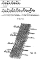
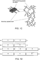
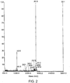
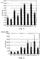
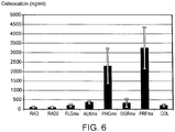


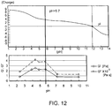
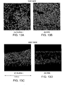

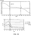
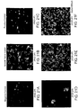

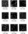
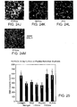
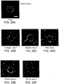

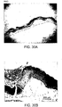
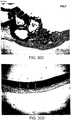


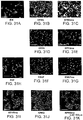
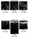

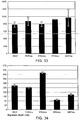


Classifications
-
- C—CHEMISTRY; METALLURGY
- C07—ORGANIC CHEMISTRY
- C07K—PEPTIDES
- C07K14/00—Peptides having more than 20 amino acids; Gastrins; Somatostatins; Melanotropins; Derivatives thereof
- C07K14/001—Peptides having more than 20 amino acids; Gastrins; Somatostatins; Melanotropins; Derivatives thereof by chemical synthesis
-
- A—HUMAN NECESSITIES
- A61—MEDICAL OR VETERINARY SCIENCE; HYGIENE
- A61K—PREPARATIONS FOR MEDICAL, DENTAL OR TOILETRY PURPOSES
- A61K47/00—Medicinal preparations characterised by the non-active ingredients used, e.g. carriers or inert additives; Targeting or modifying agents chemically bound to the active ingredient
- A61K47/50—Medicinal preparations characterised by the non-active ingredients used, e.g. carriers or inert additives; Targeting or modifying agents chemically bound to the active ingredient the non-active ingredient being chemically bound to the active ingredient, e.g. polymer-drug conjugates
- A61K47/51—Medicinal preparations characterised by the non-active ingredients used, e.g. carriers or inert additives; Targeting or modifying agents chemically bound to the active ingredient the non-active ingredient being chemically bound to the active ingredient, e.g. polymer-drug conjugates the non-active ingredient being a modifying agent
- A61K47/62—Medicinal preparations characterised by the non-active ingredients used, e.g. carriers or inert additives; Targeting or modifying agents chemically bound to the active ingredient the non-active ingredient being chemically bound to the active ingredient, e.g. polymer-drug conjugates the non-active ingredient being a modifying agent the modifying agent being a protein, peptide or polyamino acid
- A61K47/66—Medicinal preparations characterised by the non-active ingredients used, e.g. carriers or inert additives; Targeting or modifying agents chemically bound to the active ingredient the non-active ingredient being chemically bound to the active ingredient, e.g. polymer-drug conjugates the non-active ingredient being a modifying agent the modifying agent being a protein, peptide or polyamino acid the modifying agent being a pre-targeting system involving a peptide or protein for targeting specific cells
-
- A—HUMAN NECESSITIES
- A61—MEDICAL OR VETERINARY SCIENCE; HYGIENE
- A61L—METHODS OR APPARATUS FOR STERILISING MATERIALS OR OBJECTS IN GENERAL; DISINFECTION, STERILISATION OR DEODORISATION OF AIR; CHEMICAL ASPECTS OF BANDAGES, DRESSINGS, ABSORBENT PADS OR SURGICAL ARTICLES; MATERIALS FOR BANDAGES, DRESSINGS, ABSORBENT PADS OR SURGICAL ARTICLES
- A61L27/00—Materials for grafts or prostheses or for coating grafts or prostheses
- A61L27/14—Macromolecular materials
- A61L27/22—Polypeptides or derivatives thereof, e.g. degradation products
- A61L27/227—Other specific proteins or polypeptides not covered by A61L27/222, A61L27/225 or A61L27/24
-
- A—HUMAN NECESSITIES
- A61—MEDICAL OR VETERINARY SCIENCE; HYGIENE
- A61L—METHODS OR APPARATUS FOR STERILISING MATERIALS OR OBJECTS IN GENERAL; DISINFECTION, STERILISATION OR DEODORISATION OF AIR; CHEMICAL ASPECTS OF BANDAGES, DRESSINGS, ABSORBENT PADS OR SURGICAL ARTICLES; MATERIALS FOR BANDAGES, DRESSINGS, ABSORBENT PADS OR SURGICAL ARTICLES
- A61L27/00—Materials for grafts or prostheses or for coating grafts or prostheses
- A61L27/50—Materials characterised by their function or physical properties, e.g. injectable or lubricating compositions, shape-memory materials, surface modified materials
- A61L27/54—Biologically active materials, e.g. therapeutic substances
-
- A—HUMAN NECESSITIES
- A61—MEDICAL OR VETERINARY SCIENCE; HYGIENE
- A61P—SPECIFIC THERAPEUTIC ACTIVITY OF CHEMICAL COMPOUNDS OR MEDICINAL PREPARATIONS
- A61P19/00—Drugs for skeletal disorders
-
- A—HUMAN NECESSITIES
- A61—MEDICAL OR VETERINARY SCIENCE; HYGIENE
- A61P—SPECIFIC THERAPEUTIC ACTIVITY OF CHEMICAL COMPOUNDS OR MEDICINAL PREPARATIONS
- A61P19/00—Drugs for skeletal disorders
- A61P19/02—Drugs for skeletal disorders for joint disorders, e.g. arthritis, arthrosis
-
- A—HUMAN NECESSITIES
- A61—MEDICAL OR VETERINARY SCIENCE; HYGIENE
- A61P—SPECIFIC THERAPEUTIC ACTIVITY OF CHEMICAL COMPOUNDS OR MEDICINAL PREPARATIONS
- A61P19/00—Drugs for skeletal disorders
- A61P19/04—Drugs for skeletal disorders for non-specific disorders of the connective tissue
-
- A—HUMAN NECESSITIES
- A61—MEDICAL OR VETERINARY SCIENCE; HYGIENE
- A61P—SPECIFIC THERAPEUTIC ACTIVITY OF CHEMICAL COMPOUNDS OR MEDICINAL PREPARATIONS
- A61P19/00—Drugs for skeletal disorders
- A61P19/08—Drugs for skeletal disorders for bone diseases, e.g. rachitism, Paget's disease
-
- A—HUMAN NECESSITIES
- A61—MEDICAL OR VETERINARY SCIENCE; HYGIENE
- A61P—SPECIFIC THERAPEUTIC ACTIVITY OF CHEMICAL COMPOUNDS OR MEDICINAL PREPARATIONS
- A61P35/00—Antineoplastic agents
-
- A—HUMAN NECESSITIES
- A61—MEDICAL OR VETERINARY SCIENCE; HYGIENE
- A61P—SPECIFIC THERAPEUTIC ACTIVITY OF CHEMICAL COMPOUNDS OR MEDICINAL PREPARATIONS
- A61P43/00—Drugs for specific purposes, not provided for in groups A61P1/00-A61P41/00
-
- A—HUMAN NECESSITIES
- A61—MEDICAL OR VETERINARY SCIENCE; HYGIENE
- A61P—SPECIFIC THERAPEUTIC ACTIVITY OF CHEMICAL COMPOUNDS OR MEDICINAL PREPARATIONS
- A61P9/00—Drugs for disorders of the cardiovascular system
-
- A—HUMAN NECESSITIES
- A61—MEDICAL OR VETERINARY SCIENCE; HYGIENE
- A61P—SPECIFIC THERAPEUTIC ACTIVITY OF CHEMICAL COMPOUNDS OR MEDICINAL PREPARATIONS
- A61P9/00—Drugs for disorders of the cardiovascular system
- A61P9/10—Drugs for disorders of the cardiovascular system for treating ischaemic or atherosclerotic diseases, e.g. antianginal drugs, coronary vasodilators, drugs for myocardial infarction, retinopathy, cerebrovascula insufficiency, renal arteriosclerosis
-
- B—PERFORMING OPERATIONS; TRANSPORTING
- B82—NANOTECHNOLOGY
- B82Y—SPECIFIC USES OR APPLICATIONS OF NANOSTRUCTURES; MEASUREMENT OR ANALYSIS OF NANOSTRUCTURES; MANUFACTURE OR TREATMENT OF NANOSTRUCTURES
- B82Y5/00—Nanobiotechnology or nanomedicine, e.g. protein engineering or drug delivery
-
- C—CHEMISTRY; METALLURGY
- C07—ORGANIC CHEMISTRY
- C07K—PEPTIDES
- C07K14/00—Peptides having more than 20 amino acids; Gastrins; Somatostatins; Melanotropins; Derivatives thereof
- C07K14/435—Peptides having more than 20 amino acids; Gastrins; Somatostatins; Melanotropins; Derivatives thereof from animals; from humans
- C07K14/78—Connective tissue peptides, e.g. collagen, elastin, laminin, fibronectin, vitronectin or cold insoluble globulin [CIG]
-
- A—HUMAN NECESSITIES
- A61—MEDICAL OR VETERINARY SCIENCE; HYGIENE
- A61L—METHODS OR APPARATUS FOR STERILISING MATERIALS OR OBJECTS IN GENERAL; DISINFECTION, STERILISATION OR DEODORISATION OF AIR; CHEMICAL ASPECTS OF BANDAGES, DRESSINGS, ABSORBENT PADS OR SURGICAL ARTICLES; MATERIALS FOR BANDAGES, DRESSINGS, ABSORBENT PADS OR SURGICAL ARTICLES
- A61L2300/00—Biologically active materials used in bandages, wound dressings, absorbent pads or medical devices
- A61L2300/20—Biologically active materials used in bandages, wound dressings, absorbent pads or medical devices containing or releasing organic materials
- A61L2300/25—Peptides having up to 20 amino acids in a defined sequence
-
- A—HUMAN NECESSITIES
- A61—MEDICAL OR VETERINARY SCIENCE; HYGIENE
- A61L—METHODS OR APPARATUS FOR STERILISING MATERIALS OR OBJECTS IN GENERAL; DISINFECTION, STERILISATION OR DEODORISATION OF AIR; CHEMICAL ASPECTS OF BANDAGES, DRESSINGS, ABSORBENT PADS OR SURGICAL ARTICLES; MATERIALS FOR BANDAGES, DRESSINGS, ABSORBENT PADS OR SURGICAL ARTICLES
- A61L2300/00—Biologically active materials used in bandages, wound dressings, absorbent pads or medical devices
- A61L2300/40—Biologically active materials used in bandages, wound dressings, absorbent pads or medical devices characterised by a specific therapeutic activity or mode of action
- A61L2300/412—Tissue-regenerating or healing or proliferative agents
- A61L2300/414—Growth factors
-
- A—HUMAN NECESSITIES
- A61—MEDICAL OR VETERINARY SCIENCE; HYGIENE
- A61L—METHODS OR APPARATUS FOR STERILISING MATERIALS OR OBJECTS IN GENERAL; DISINFECTION, STERILISATION OR DEODORISATION OF AIR; CHEMICAL ASPECTS OF BANDAGES, DRESSINGS, ABSORBENT PADS OR SURGICAL ARTICLES; MATERIALS FOR BANDAGES, DRESSINGS, ABSORBENT PADS OR SURGICAL ARTICLES
- A61L2430/00—Materials or treatment for tissue regeneration
- A61L2430/38—Materials or treatment for tissue regeneration for reconstruction of the spine, vertebrae or intervertebral discs
Definitions
- the extracellular matrix is composed of a diverse set of macromolecules, including both proteins and polysaccharides, which form the three-dimensional environment within which cells exist in the body and constitute the space-filling material between cells.
- the ECM can also be organized into a sheet-like layer known as the basal lamina or basement membrane.
- the basement membrane lies beneath layers or tubes of epithelial cells (e.g., endothelial cells lining blood vessels) or surrounds individual cells of various types such as muscle cells, often serving to separate cell layers from one another or from adjacent connective tissue.
- the ECM consists primarily of molecules that are secreted locally and assemble into a matrix that stabilizes and supports the physical structure of cell layers and tissues. However, rather than being merely an inert substrate for cell attachment, the ECM constitutes an environment that is rich in biological information. It is recognized that the ECM, and various biomolecules associated with it (e.g., biomolecules secreted locally or transported to a particular site from elsewhere), exert a significant influence on many aspects of cell behavior and phenotype, regulating processes such as migration and proliferation, influencing cell development and differentiation, and affecting cell shape and function. The structure of the ECM is, in turn, influenced by the cells within it. Not only do these cells secrete many ECM constituents, but they also help to pattern the matrix. Thus it is evident that cell-ECM interactions are of vital importance.
- tissue culture substrates such as glass or plastic
- tissue repair and tissue engineering such as for growing cells, tissues, and/or artificial organs or for use in cell-based bioreactors for production of biomolecules.
- ECM constituents by recombinant DNA techniques.
- expression constructs encoding ECM proteins can be introduced into prokaryotic or eukaryotic cells, and the protein of interest can be purified from the cells or from the medium in the case of secreted proteins. Proteins can be combined in vitro in desired ratios. While likely reducing the likelihood of disease transmission, this approach also suffers from several disadvantages. For example, whenever proteins are manufactured through a biological rather than purely synthetic process, there remains the possibility that undefined components from the culture system will be present even in highly purified preparations. In addition, purification can be time-consuming and costly and can result in protein degradation or denaturation.
- ECM consists largely of proteins and proteoglycans
- significant efforts have been directed to development of cell culture and tissue engineering materials based on a variety of synthetic, non-amino acid based polymers.
- aliphatic hydroesters have been widely used for various tissue engineering applications.
- fully synthetic materials are polyglycolic acid (PGA), poly(lactic-co-glycolic acid) (PLGA), poly(ethylene glycol) (PEG), poly(propylene glycol), and various copolymers of these and other compounds.
- PGA polyglycolic acid
- PLGA poly(lactic-co-glycolic acid)
- PEG poly(ethylene glycol)
- poly(propylene glycol) poly(propylene glycol)
- these materials also suffer from a number of disadvantages. For example, they form fibers with diameters on the order of tens of microns, and the methods required to introduce cells into matrices formed from such materials are not readily compatible with the physiological requirements for cell viability.
- Zhang et al. (Seminars in Cancer Biology 2005, 15:413-420 ) and Zhang et al. (Reactive & Functional Polymers 1999, 41:91-102 ) describe self-assembling peptide domains.
- WO 2005/014615 Genove et al. (Biomaterials 2005, 26:3341-3351 ), and Hucknall (2005, master thesis, Massachusetts Institute of Technology ) describe functuionalized self-assembling peptides.
- WO 2006/076042 Bellamkonda et al. (J. Neurosci. Res. 1995, 41:501-509 ), WO 01/85180 , Cui et al. (J. Bioactive & Compatible Pol.
- compositions and materials for cell culture and tissue engineering purposes that would allow the creation of an environment that mimics the native cellular environment, but without the disadvantages associated with materials derived from natural sources. For example, it would be desirable to develop a material that could provide biologically relevant stimuli to cells akin to those provided by native ECM components. For applications involving implantation into the body, there remains a particular need for such compositions and materials that elicit no or minimal immune or inflammatory response and for compositions and materials that are degradable within the body. In addition, there remains a need in the art for compositions and materials that would influence cell properties and functions in desirable ways.
- the present invention is defined by the appended claims. It is possible to modify self-assembling peptides by incorporating an additional domain that does not self-assemble, while still permitting assembly of the self-assembling portion.
- the additional domain can confer a variety of properties on the resulting peptide.
- the resulting peptide self-assembles to form nanofibers, beta-sheets and/or a macroscopic structure. Materials formed by self-assembly of the peptides have a wide variety of uses, particularly in the areas of cell culture, tissue engineering, and tissue repair.
- the present invention includes a composition for use in a method of treating a subject according to any one of claims 1-15.
- a self-assembling peptide comprising: (a) a first amino acid domain that mediates self-assembly, wherein the domain comprises alternating hydrophobic and hydrophilic amino acids that are complementary and structurally compatible and self-assemble into a macroscopic structure when present in unmodified form; and (b) a second amino acid domain that does not mediate self-assembly in isolated form, wherein the second amino acid domain comprises at least one minimal biologically active sequence.
- modified self-assembling peptides are described herein as "modified self-assembling peptides.”
- the second amino acid domain can comprise at least two minimal biologically active sequences.
- the modified self-assembling peptide can comprise a minimal biologically active sequence which has an affinity to, or complexes with, a biological molecule.
- composition comprising a modified self-assembling peptide.
- a matrix comprising a modified self-assembling peptide.
- the matrix may be a gel or hydrogel.
- Also methods of making the matrix, and of using the matrix as a defect filler for bone and/or tissue e.g., for use in tissue engineering and tissue repair).
- matrices comprising a modified self-assembling peptide which enhance orthopedic tissue regeneration by increasing progenitor cell activity in proliferation and differentiation.
- These matrices can be used in vivo as defect fillers for bone and/or tissue to promote bone and/or tissue regeneration.
- Disclosed are methods of enhancing cell differentiation or functional activity comprising administering a modified self-assembling peptide, a solution thereof, a pharmaceutical composition thereof, or a matrix made therefrom, to a subject in need thereof.
- a method of treating a subject comprising introducing a modified self-assembling peptide, a solution thereof, a pharmaceutical composition thereof, or a matrix made therefrom, to a site on or within the subject's body.
- the site is of an orthopedic field; a bone defect; a bone adjent; an ectopic bone formation; an ischemic region; a myocardial infarction region; peripheral vascular region; cerebral infarction region; or a skin defect.
- kits comprising a modified self-assembling peptide, a solution thereof, a pharmaceutical composition thereof, or a matrix made therefrom, and methods of using such kits.
- compositions that possess the advantages typically associated with a fully synthetic material and yet possess certain desirable features of materials derived from natural sources.
- the compositions can be used either in vitro or in vivo for purposes including, but not limited to, cell culture or tissue engineering, tissue regeneration and/or repair, and/or as delivery agents for biologically active molecules such as therapeutic agents.
- peptides are complementary and structurally compatible. They are composed of repeating units of alternating hydrophilic and hydrophobic amino acids, in which the charged residues can include alternating positive and negative charges. In general, these peptides self-assemble into various macroscopic structures upon exposure to a sufficient concentration of ions (e.g., monovalent cations) to form a stable macroscopic porous matrix.
- ions e.g., monovalent cations
- the matrix can assume various physical forms such as ribbons, tape-like structures, two or three dimensional matrices, etc.
- Preferred matrices are composed of orderly interwoven filaments typically approximately 10-20 nm or 10-30 nm in diameter, with a pore size on the order of 50-100 nm in diameter (or other relevant dimension).
- the materials are hydrogels which contain approximately at least 50%, 55%, 60%, 65%, 70%, 75%, 80%, 85%, 90%, 95%, or 99% or greater water content. Certain of the peptides undergo self-assembly to form nanofibers that are very highly hydrated ( e.g ., up to 99.5-99.9%% (1-5 mg/ml) water).
- the hydrogel has such an extremely high water content, cells can freely migrate and form intercellular contacts when cultured on or within the matrix. Such an environment also permits diffusion of small molecules including proteins and signaling molecules.
- certain of the hydrogels have a low elastic modulus. While not wishing to be bound by any theory, it has been suggested that the low elastic modulus may facilitate cell-cell communication.
- the properties of the hydrogels contrast with those of many synthetic materials that are being explored for cell culture and/or tissue engineering or repair. For example, many of these materials comprise microfibers and have much larger pore sizes, presenting an environment that is likely not on the appropriate scale by comparison with cells and their natural environment.
- the hydrogels can be formed so as to encapsulate cells during the gel formation process, rather than preformed structures in which cells and seeded and/or embedded.
- a peptide structure formed by self-assembly of RAD16-I (AcN-RADARADARADARADA-CONH 2 ) (SEQ ID NO: 21) was able to serve as a substrate for neurite outgrowth and formation of functionally active synapses when neurons were grown on their surface (Holmes (2000)).
- peptide gels support endothelial cell attachment, migration, proliferation and capillary-like structure formation and survival for a period of at least 3 weeks (see, for example, PCT application Publication WO2003/096972 ).
- endothelial cell attachment migration, proliferation and capillary-like structure formation and survival for a period of at least 3 weeks (see, for example, PCT application Publication WO2003/096972 ).
- this angiogenic response was observed in the absence of externally supplied angiogenic factors, and with no significant signs of apoptosis or proteolytic gel degradation.
- chondrocytes encapsulated within a hydrogel structure formed by self-assembly of KLD12 (AcNKLDLKLDLKLDL-CONH 2 ) (SEQ ID NO:53) retained their morphology and developed a cartilage-like, mechanically functional ECM rich in proteoglycans and type-II collagen, indicative of a stable chondrocyte phenotype (see, for example, Kisiday (2002)).
- Liver progenitor cells encapsulated within a peptide matrix made by selfassembly of RAD16-I expressed markers indicative of hepatocyte phenotype (Semino (2003)). Results such as these indicate that the nanoscale environment provided by the self-assembling peptide gels can enhance functional activity of a diverse set of cell types and is permissive for cellular instruction and expression of differentiated cell phenotypes.
- the materials of the present invention interact with cells on a nanoscale rather tha n a microscale.
- the materials are made of nanofibers rather than the microfibers believed that the small size of the fibers and/or the open weave structure of the materials promote extension of cell processes and allow diffusion of nutrients, waste products, etc., in a manner that provides unique advantages for cell growth.
- the nanofibers that comprise the material may be ordered during self assembly in a complementary fashion due to weak interactive molecular forces. However, they may also be randomly ordered, which may be preferred for certain applications. In other words, while the fibers may have an orderly internal structure, they may lack directionality or alignment with respect to one another. For example, the fibers may not be substantially parallel to one another.
- the inventors have now unexpectedly discovered that it is possible to extensively modify the previously described self-assembling peptides by incorporating additional amino acid domains (which may also referred to herein as peptide domains if at least 3 amino acids in length) that does not necessarily conform to the structural requirements for self-assembly, without eliminating the ability of the modified peptides to self-assemble to form a macroscopic structure.
- the self-assembling peptides can be modified to incorporate a non self-assembling amino acid domain which can comprise, for example, a minimal biologically active sequence for interaction with a biomolecule.
- the resulting peptides thus comprise a first self-assembling peptide domain and a second non-self-assembling amino acid domain.
- non-self-assembling amino acid domain an amino acid domain that does not self-assemble when present as an isolated peptide ( i.e., when not joined to an unmodified self-assembling peptide) under conditions (e.g., ionic concentration, peptide concentration, pH, temperature) that would result in self-assembly of an unmodified self-assembling peptide, and as designed in accordance with the principles described herein.
- does not self-assemble is meant that the amino acid domain or peptide does not form nanofilaments, nanofibers, ⁇ -sheets, nanofibers, and the like, or does not form a macroscopic structure.
- the following section describes the self-assembling peptides that can be modified in accordance with the present invention and methods by which the process of self-assembly and/or the features of the assembled structure may be controlled. Subsequent sections describe the methods and compositions of the invention, methods for characterizing structures formed from the inventive modified selfassembling peptides, methods for evaluating cell phenotype and for selecting amino acid domains that are used for modification, etc., in further detail.
- the previously described unmodified self-assembling peptides comprise a family of complementary and structurally compatible molecules.
- the peptides and their properties are described in U.S. Patent Application Publication Nos. 2002/0160471 , 2005/0181973 , and in U.S. Patent Nos. 5,955,343 and 5,670,483 . These materials are composed of repeating units of hydrophilic and hydrophobic amino acids, in various alternating patterns.
- EAK16-11 (AEAEAKAKAEAEAKAK, A, alanine, E, glutamine, and K, lysine; SEQ ID NO:38), a 16 amino acid peptide
- EAK16-11 AEAEAKAKAEAEAKAK, A, alanine, E, glutamine, and K, lysine; SEQ ID NO:38
- SEQ ID NO:38 16 amino acid peptide
- peptides contain two distinct surfaces, one hydrophilic and the other hydrophobic, and form complementary ionic bonds with regular repeats on the hydrophilic surface (Zhang (1999)).
- the side-chains of the peptides partition into two faces, a polar face with charged ionic side chains and a nonpolar face, e.g., with alanines or other hydrophobic groups.
- These ionic side chains are self-complementary to one another in that the positively charged and negatively charged amino acid residues can form complementary ionic pairs. These peptides are therefore called "ionic self-complementary peptides.”
- modulus I The complementary ionic sides have been classified into several moduli, i.e., modulus I, II, III, IV, etc., and mixed moduli (Zhang (1999)). If the ionic residues alternate with one positively and one negatively charged residue (-+-+-+), the peptides are described as "modulus I;” if the ionic residues alternate with two positively and two negatively charged residues (-++-++), the peptides are described as "modulus II”; if the ionic residues alternate with four positively and two negatively charged residues (-++++), the peptides are described as "modulus IV.”
- Peptides meeting the afore-mentioned criteria may be referred to herein as "unmodified self-assembling peptides" to distinguish them from the modifed self-assembling peptides of the present invention, which includes a peptide domain that does not meet the foregoing criteria and does not self-assemble.
- modulus I and II self-complementary peptides such as EAKA16-I (SEQ ID NO:26), RADA16-I (SEQ ID NO:21), RAEA16-I (SEQ ID NO:28), and KADA16-I (SEQ ID NO:30) have been analyzed previously (Table 1). These peptides are also referred to as RAD16-I, RAE16-I, KAD16-I, etc. ( i.e., the last amino acid in the four amino acid module may be omitted in the abbreviation).
- Modulus IV ionic self-complementary peptides containing 16 amino acids such as EAK16- IV, KAE16-IV, DAR16-IV and RAD16-IV; have also been studied. If the charged residues in these self-assembling peptides are appropriately substituted without changing the overall pattern described above (e.g., the positive charged lysines are replaced by positively charged arginines and the negatively charged glutamates are replaced by negatively charged aspartates), there are essentially no significant effects on the self-assembly process.
- the peptides can no longer undergo self-assembly to form macroscopic structures; however, they can still form a ⁇ -sheet structure in the presence of salt.
- hydrophilic residues such as asparagine and glutamine, that form hydrogen bonds may be incorporated into the peptides instead of or in addition to charged residues. If the alanines in the peptides are changed to more hydrophobic residues, such as leucine, isoleucine, phenylalanine or tyrosine, these peptides have a greater tendency to self-assemble and form peptide matrices with enhanced strength. Some peptides that have similar compositions and lengths as these aforementioned peptides form ⁇ -helices and random-coils rather than ⁇ -sheets. Such peptides typically do not form macroscopic structures although structure formation is not absolutely precluded. Thus, in addition to self-complementarity, other factors are likely to be important for the formation of macroscopic structures, such as the peptide length, the degree of intermolecular interaction, and the ability to form staggered arrays.
- a group or radical such as an acyl group (RCO-, where R is an organic group), e.g., an acetyl group (CH 3 CO-; Ac-) is present at the N terminus of the peptides in order to neutralize an extra charge positive that may otherwise be present (e.g., a charge not resulting from the side chain of the N-terminal amino acid).
- RCO- an acyl group
- a group such as an amine group (-NH 2 ) may be used to neutralize an extra negative charge that may otherwise be present at the C terminus (e.g., a charge not resulting from the side chain of C-terminal amino acid), thus converting the C terminus into an amide (-CONH 2 ).
- the neutralization of charges on the terminal N and C molecules may facilitate self-assembly.
- One of ordinary skill in the art will be able to select other suitable groups.
- the peptides self-assemble to form macroscopic structures under a variety of conditions, e.g., upon the addition of monovalent cations to an aqueous peptide solution or upon the introduction of a peptide solution to a solution containing monovalent cations.
- the peptides Prior to self-assembly the peptides may be dissolved in a solution that is substantially free of monovalent ions ( e.g., cations) or contains only a low concentration of such ions, e.g., less than 10 mM, 5 mM, 1 mM, 0.5 mM or 0.1 mM.
- Self-assembly may be initiated or substantially accelerated by the addition of an ionic solute to a peptide solution or by a change in pH.
- an ionic solute for example, NaCl at a concentration of between 5 mM and 5 M induces the assembly of the peptides to form macroscopic structures within a few minutes. Lower concentrations of NaCl may also induce assembly but at a slower rate.
- Certain of the peptides can also self-assemble in the absence of significant concentrations of ions, in a process that may be dependent on pH. For example, certain of the peptides may remain in solution at a pH of approximately 3.0 but may self-assemble when the pH is raised.
- self-assembly may be initiated by introducing the peptides into a solution comprising ions, e.g., standard phosphate buffered saline (PBS), tissue culture medium, or a physiological fluid such as blood, cerebrospinal fluid (CSF), and the like.
- ions e.g., standard phosphate buffered saline (PBS), tissue culture medium, or a physiological fluid such as blood, cerebrospinal fluid (CSF), and the like.
- PBS standard phosphate buffered saline
- CSF cerebrospinal fluid
- the peptides can thus self-assemble at a location in vivo.
- Exemplary ions include, but are not limited to, Li + , Na + , K + and Cs + .
- the concentration of the ion is at least 5 mM, 10 mM, 20 mM or 50 mM in order to induce or substantially accelerate self-assembly.
- the strength of the resulting structure is increased in the presence of ions relative to the strength in the absence of ions, or at a lower ionic concentration, although it is noted that a plateau may be reached at which an increase in ion concentration does not result in increased strength.
- FIG. 1A shows the structure of a representative peptide, RAD16-II (RARADADARARADADA, R, arginine, A, alanine, D, aspartic acid) (SEQ ID NO. 24).
- the material self-assembles into interwoven fibers of ⁇ 10-20 nm diameter forming enclosures of ⁇ 50-100 nm diameter.
- the nanofiber network can give rise to hydrogel formation, creating a macroscopic structure preferably of a size that can be observed with the naked eye and can be three-dimensional.
- Modifed or unmodified self-assembling peptides forming the macroscopic structure may contain between 8 and 200 amino acids, between 8 to 64 amino acids, between 8 to 46 amino acids, between 8 to 36 amino acids, or between 8 to 16 amino acids, inclusive.
- modifed or unmodified self-assembling peptides contain between 8-46 amino acids, between 8-36 amino acids, between 8-26 amino acids, between 8-16 amino acids, between 10-16 amino acids, or between 12-16 amino acids, inclusive.
- the concentration of the self-assembling peptide solution prior to matrix formation can range, for example, between 0.01% (0.1 mg/ml) and 99.99% (999.9 mg/ml), inclusive.
- the concentration of the self-assembling peptide solution prior to matrix formation is between 0.1% (1 mg/ml) and 10% (100 mg/ml), inclusive, particularly for cell culture and/or therapeutic applications. In some embodiments, the concentration of the self-assembling peptide solution prior to matrix formation is between 0.1% (1 mg/ml) and 5% (50 mg/ml), inclusive, or between 0.5% (5 mg/ml) and 5% (50 mg/ml), inclusive. In yet other embodiments, the concentration of the self-assembling peptide solution prior to matrix formation is approximately 5 mg/ml, approximately 10 mg/ml, approximately 15 mg/ml, or approximately 20 mg/ml.
- matrices may be "three dimensional," i.e., can be formed with a predetermined shape or volume.
- an aqueous peptide solution may be placed in a pre-shaped casting mold, and the peptides induced to self-assemble into a matrix by the addition of an ion, as described herein.
- Matrices having a three-dimensional is meant that the matrix is not just a thin flat coating substance on the other material, but has a thickness which at least plurality of cells can embedded into the thickness.
- the ion may be added to the modifed or unmodified self-assembling peptide solution shortly before placing the solution into the mold, provided that care is taken to place the solution into the mold before substantial assembly occurs.
- the resulting material characteristics, time required for assembly, and geometry and dimensions of the macroscopic peptide matrix are governed by parameters including the concentration and amount of peptide solution that is applied, the concentration of ion used to induce assembly of the matrix, the pH, the particular modified self-assembling peptide sequence, and the dimensions of the casting apparatus.
- the shape may be selected based upon the intended implantation site.
- the matrix can exist as a thin layer, such as a coating at the bottom of a conventional tissue culture or floating in a solution.
- the layer can be several microns thick, e.g., 10 microns, 10-50 microns, 50-100 microns, 100-200 microns, etc.
- the layer may comprise multiple ⁇ -sheet layers.
- Modifed or unmodified self-assembling peptide nanoscale matrices can be formed with varying degrees of stiffness or elasticity.
- the peptide matrices typically have a low elastic modulus, e.g., in the range of 1-10 kPa as measured in a standard cone-plate rheometer. Such low values permit matrix deformation as a result of cell contraction, and this deformation may provide a means for cell-cell communication.
- Matrix stiffness can be controlled by a variety of techniques including changes in peptide sequence, changes in peptide concentration, and changes in peptide length.
- the modifed and/or unmodified self-assembling peptides may include L-amino acids, D-amino acids, natural amino acids, nonnatural amino acids, or a combination thereof. If L-amino acids are present in the matrix, degradation produces amino acids that may be reused, e.g., by cells in culture or by cells in a host tissue.
- L-amino acids are present in the matrix, degradation produces amino acids that may be reused, e.g., by cells in culture or by cells in a host tissue.
- the fact that the basic monomeric subunit of the peptides in certain embodiments of the invention are L-amino acids, which occur naturally within the body, distinguishes this class of compounds from numerous other biocompatible substances and may offer unique advantages.
- the peptides may be chemically synthesized or purified from natural or recombinant sources, and the amino- and carboxy-termini of the peptides may be protected or not protected.
- the peptide matrix may be formed from one or more distinct molecular species of peptides which are complementary and structurally compatible with each other. Peptides containing mismatched pairs, such as the repulsive pairing of two similarly charged residues from adjacent peptides, may also form structures if the disruptive force is dominated by stabilizing interactions between the peptides. Peptide matrices may also be referred to herein as peptide hydrogels or peptide hydrogel matrices.
- Peptides including peptides capable of being cross-linked, and modified or unmodified self-assembling peptides, may be synthesized using standard F-moc chemistry, which includes both solution and solid phase synthesis, and purified using high pressure liquid chromatography.
- the formation of a peptide matrix may be initiated by the addition of ions or salts thereof as described herein.
- Hydrophobic residues with aromatic side chains may be cross-linked by exposure to UV irradiation.
- the extent of the cross-linking may be precisely controlled by the predetermined length of exposure to UV light and the predetermined peptide concentration.
- the extent of cross-linking may be determined by light scattering, gel filtration, or scanning electron microscopy using standard methods.
- the extent of cross-linking may also be examined by HPLC or mass spectrometry analysis of the structure after digestion with a protease. Material strength may be determined before and after cross-linking.
- Peptide matrices formed either from unmodified self-assembling peptides or the modified self-assembling peptides of the present invention may be used in a variety of ways for culturing cells and tissues.
- Cells and tissues can be cultured on the surface of a matrices structure. While not wishing to be bound by any theory, inventors suggest that such an environment more closely mimics the natural cellular environment than culture on a rigid substrate such as a conventional plastic tissue culture dish. If the matrix forms a three-dimensional structure, cells can extend processes into the structure or migrate into it.
- Cells can also be encapsulated within the matrix.
- peptides and living cells may be incubated in an aqueous solution having an iso-osmotic solute at an appropriate concentration to support cell viability, under conditions that in which the peptides are not substantially selfassembled.
- the solution contains a monovalent cation concentration of less than 10 mM, 5 mM, 1 mM or 0.1 mM, or is substantially free of monovalent cations.
- the solution may also contain less than less than 10 mM, 5 mM, 1 mM or 0.1 mM or be substantially free of other ionic species, e.g., other cations or anions.
- Sufficient ion e.g., monovalent cation
- a macroscopic structure such as a Q-sheet macroscopic structure
- the encapsulated cells are present in the macroscopic structure in a three-dimensional arrangement.
- the solution may be contained in a pre-shaped mold dimensioned to establish a desired volume or shape of the macroscopic structure.
- the concentration of the added ion is at least 5 mM, 10 mM, 20 mM or 50 mM.
- Suitable ions include, but are not limited to, Li + , Na + , K + and Cs + .
- the concentration of the iso-osmotic solute is at least 50 mM, 150 mM or 300 mM.
- the concentration of the iso-osmotic solute is contained in one of the following ranges 200 to 250 mM, 250 to 270 mM, 270 to 300 mM, 300 to 400 mM, 400 to 500 mM, 500 to 600 mM, 600 to 700 mM, 700 to 800 mM, or 800 to 900 mM, inclusive.
- Suitable iso-osmotic solutes include, but are not limited to, carbohydrates, such as sucrose, mannitol, and the like.
- Other iso-osmotic solutes, preferably non-toxic to cells at the concentration used, may be employed.
- Self-assembly can also be effected by a change in pH (e.g., a rise from a low pH to a higher pH).
- Cells and agents such as biological molecules (e.g., differentiation-inducing agents, prolferation agents, growth factors, and the like) and therapeutic compounds may be introduced into the peptide solution prior to self-assembly.
- the self-assembly process then forms a structure that encapsulates the cells or molecules.
- To achieve even distribution of the cells or molecules within the structure it may be desirable to thoroughly mix the solution prior to initiation of self-assembly. It may be desirable to maintain the cells or agents in a solution that contains substantially no ions or only low concentration of ions in order to avoid initiation or acceleration of self-assembly immediately upon combining the cells or agents with the peptide solution.
- the cells are maintained in an iso-osmotic solution (e.g., a sucrose solution) prior to combination with the peptide solution.
- the peptides themselves may be dissolved in an iso-osmotic solution to which cells (e.g ., a cell pellet, cell suspension) or agents are added.
- the resulting composition may be mixed to achieve a more uniform distribution of cells and/or agents, following which the composition is exposed to ions (e.g ., ions are added to the composition, or the composition is mixed with a solution containing ions).
- Cells may be cultured on the surface of a peptide matrix structure in a similar manner to that in which they are cultured on a conventional substrate such as a tissue culture dish or slide, or a tissue culture dish or slide that is coated with a biologically derived material such as collagen, Matrigel, and the like.
- a biologically derived material such as collagen, Matrigel, and the like.
- cells can be cultured at any desired degree of confluence.
- encapsulated cells are present in the macroscopic structure in a three-dimensional arrangement.
- the density of the cells may be, for example, between 5 ⁇ 10 3 /ml to 5 ⁇ 10 4 /ml, between 5 ⁇ 10 4 /ml to 5 ⁇ 10 5 /ml, between 5 ⁇ 10 5 /ml to 5 ⁇ 10 6 /ml, or between 5 ⁇ 10 6 /ml to 5 ⁇ 10 7 /ml, with endpoints included. Other ranges may also be used.
- the conditions for culturing should be close to physiological conditions.
- the pH of the culture medium is close to physiological pH.
- the pH is between about 6-8.
- the pH is between about 7 to 7.8.
- the pH is about 7.4.
- the temperatures of the culture medium is close to physiological temperature. Physiological temperatures range between about 30°C to 40°C for mammalian cells.
- cells are cultured at temperatures between about 32°C to about 38°C, or between about 35°C to about 37°C.
- Cells may be cultured on or within the peptide structure for any appropriate time, depending upon the cell number and density desired, the proliferation rate of the cells, and the time required for the desired cellular reprogramming to occur. These parameters will vary depending upon the particular cells and purposes for which the invention is to be used. One of ordinary skill in the art will be able to vary these parameters and to observe the effects of doing so, in order to determine the optimal time for maintaining cells in culture on or within the structure. In certain embodiments of the invention the cell are cultured for approximately 3 days, 7 days, 14 days, 21 days, 28 days, 56 days, or 90 days.
- the cells are cultured for between 1 and 3 days inclusive, between 4 and 7 days inclusive, between 8 and 14 days inclusive, between 15 and 21 days inclusive, between 22 and 28 days inclusive, between 29 and 56 days inclusive, or between 57 and 90 days inclusive. Longer or shorter culture periods may also be used.
- At least 40%, 50%, 60%, 70%, 80%, 90% or 95% of the cells are viable 1, 2, 3, 4, 5, 6 or more weeks after formation of the macroscopic matrix. In other embodiments, at least 50%, at least 60%, at least 70%, at least 80% or at least 90% of the cells are viable one day or one week after formation of the macroscopic matrix.
- any cell type can be cultured and/or encapsulated in accordance with the present invention including, but not limited to, vascular endothelial cells and precursors thereof, bone marrow cells, periosteal cells, perichondrial cells, fibroblasts, skeletal myoblasts or myocytes, neuronal cells, hippocampal cells, epidermal cells, non-vascular endothelial cells or smooth muscle cells, keratinocytes, basal cells, spinous cells, granular cells, stem cells (e.g., embryonic, fetal, cord blood, adult), lung cells, immune system cells, ovarian cells, pancreatic cells, cervical cells, liver cells, or foreskin cells.
- the cells may comprise embryonic stem cells, fetal stem cells, cord blood stem cells, or adult stem cells, e.g., stem cells that are able to or can be induced to differentiate into any of the preceding cell types.
- Sources of the cells may also include fetal or adult organisms, particularly mammals or established cell lines. Numerous established cell lines are known in the art, many of which are available through the American Type Culture Collection (see web site having URL www.atcc.org , which also provides references describing these cell lines).
- the phrase "derived from” indicates that a cell is obtained from a particular source, or that the cell is a descendant of a cell obtained from that source.
- a liver-derived cell is a cell that is obtained from the liver or the progeny or descendant of such a cell.
- progeny refers not only to the immediate products of cell division but also to the products of subsequent cell divisions, i .
- a cell that is derived from a cell line is a member of that cell line or is the progeny or descendant of a cell that is a member of that cell line.
- a cell derived from an organ, tissue, individual, cell line, etc. may be modified in vitro after it is obtained. Such a cell is still considered to be derived from the original source.
- Cells harvested from an individual may be used either with or without a period of expansion in culture. Alternately, cells that have been propagated in culture as a stable cell line may be used.
- the cells are autologous while in other embodiments of the invention the cells are allogeneic or xenogeneic.
- the cells may be treated in various ways prior to introduction into the body in order to reduce the likelihood or reduce the extent of an immune system response by the subject. Such treatments can include modifying, masking, or eliminating an antigen on the surface of a cell as described, for example, in PCT Application Publication No. WO/2001/007568 .
- cells are harvested from a subject (such as, for example, a human patient), and a clonal cell line is derived from one or more of these cells.
- Clonal lines may be obtained by limiting dilution plating or single cell sorting. Methods for deriving clonal cell lines are well known in the art and are described in, for example, Puck and Marcus, Experimental Medicine (1956) 103:653 ; C. V. Ramakrishnan, Ed., Cell Culture, "Clone size distribution in the study of inhomogeneity of growth rates in tissue culture," Dr. W. Junk Publishers, Netherlands: 1965 ; and Leong et al., Mutat. Res.(1985) 150:403-410 .
- Cells from the cell line are used in the practice of the invention. When intended for treatment of a particular patient, cells from a matched donor may be advantageously used. Cells isolated from an individual or maintained as a cell line may be cultured according to any appropriate technique including standard cell culture techniques prior to their use in the practice of the present invention.
- Such methods typically include introducing genetic material such as a nucleic acid molecule (e.g., DNA) into the cell, wherein the nucleic acid molecule encodes a product to be expressed by the cell.
- the product can be, for example, a reprogramming agent such as a growth factor, a transcription factor which will in turn induce expression of other gene products, and the like.
- a selectable marker is introduced into the cells.
- a gene that encodes a selectable marker is introduced into the cells, such as, for example, a gene encoding a protein that confers drug resistance or a detectable marker (e.g., GFP) under the control of a tissue-specific promoter.
- the expression of the detectable marker is used as a means to determine whether the cell or its progeny has differentiated, dedifferentiated, or transdifferentiated along a particular cell lineage pathway characteristic of that tissue.
- the detectable marker is used for isolating cells that have differentiated, dedifferentiated, or transdifferentiated along a particular pathway, e.g., by using immunological methods, FACS, or other methods well known in the art.
- tissue-specific, organ-specific, and lineage-specific promoters are well known. Genes may be introduced under the control of either a constitutive or an inducible promoter of which many are known in the art.
- Matrices on which cells are cultured or in which cells are encapsulated may be subjected to various environmental conditions that may affect cell phenotype.
- matrices may be subjected to various defined or predetermined mechanical stresses, e.g., shear stress, compression schemes,and the like, that may result in altered synthesis of ECM components.
- flow e.g., pulsatile flow
- Cells in native articular cartilage and in tissue-engineered constructs respond to mechanical stimuli through multiple regulatory pathways. Such stimuli result in altered intra- and intercellular signalling, alterations in transcription level, protein translation, post-translational modifications, and synthesis of intracellular and extracellular macromolecules (Kisiday (2002).
- the inventors have discovered an unanticipated flexibility in that the self-assembling peptides (as provided in Table 1) can be modified by the addition of additional amino acid domains that would not self-assemble if present in isolated form under conditions (e.g., ionic strength, peptide concentration, pH, temperature) to form modified self-assembling peptides. Furthermore, in certain embodiments, the addition of one or more non self-assembling amino acid domains does not prevent the modified peptide from self-assembling, e.g., to form nanofibers and/or a macroscopic structure. In certain embodiments, the modified peptide self-assembles to form a macroscopic structure comprising nanofibers. Although the resulting structure may be less strong and/or stable than a structure resulting from self-assembly of the unmodified peptide, visual observation and/or rheological studies, as described in the examples, confirm that hydrogel formation occurs.
- modified peptides may thus be used for the various purposes described above and herein.
- a self-assembling peptide that possesses the structural features described in the previous section and does not include a portion lacking those characteristics will be referred to as an "unmodified self-assembling peptide.”
- an unmodified self-assembling peptide may be altered in any of a number of ways described above that do not include addition of amino acids to the peptide. Such altered self-assembling peptides are not referred to as "modified” within the meaning of the term as used herein but rather as “altered” or "derivatized”.
- modified self-assembling peptides of the invention are distinct from naturally occurring molecules, i.e., they are not found in naturally occurring molecules, although one or more of the amino acid domains in an inventive peptide may occur in a naturally occurring molecule. They can therefore be considered “isolated” or “synthetic", meaning that the total sequence of the peptide does not occur in nature without the intervention of man.
- a peptide that includes an amino acid domain comprising an unmodified self-assembling peptide and a second amino acid domain that lacks one or more of the structural features of an unmodified self-assembling peptide, and thus does not self-assemble to form nanofibers or to form a macroscopic structure under conditions that would result in self-assembly of an unmodified self-assembling peptide is referred to as a "modified self-assembling peptide" if it is capable of self-assembly ( e.g., to form nanofibers and/or a macroscopic structure) under conditions that would result in self-assembly of an unmodified self-assembling peptide.
- a modified self-assembling peptide will correspond to a particular unmodified self-assembling peptide that does not include the second amino acid domain but that has the same self-assembling portion.
- the conditions under which self-assembly of the modified self-assembling peptide occurs are the same as the conditions under which the corresponding unmodified self-assembling peptide assembles.
- the conditions under which self-assembly of the modified self-assembling peptide occurs are different from the conditions under which the corresponding unmodified self-assembling peptide assembles.
- the conditions for self-assembly of the modified self-assembling peptide are the same as the conditions under which a different (non-corresponding) unmodified self-assembling peptide self-assembles.
- the inventive modified self-assembling peptides thus comprise (a) a first amino acid domain that mediates self-assembly, wherein the domain comprises alternating hydrophobic and hydrophilic amino acids that are complementary and structurally compatible and self-assemble into a macroscopic structure; and (b) a second amino acid domain that does not self-assemble in isolated form ( i.e ., when present as the only peptide in a solution under conditions that would result in assembly of an unmodified self-assembling peptide as described above).
- the second amino acid domain permits assembly of the first amino acid domain so that the peptide assembles to form nanofibers, and/or a macroscopic structure.
- the peptide forms ⁇ -sheets.
- an amino acid domain is at least 3 amino acids; at least 4 amino acids, at least 5 amino acids, at least 6 amino acids, at least 7 amino acids, at least 8 amino acids, at least 9 amino acids, at least 10 amino acids, or more, e.g., 15, 16, 17, 18, 19, 20 etc. amino acids.
- the first amino acid domain (self-assembling portion) is at least 3 amino acids; at least 4 amino acids, at least 5 amino acids, at least 6 amino acids, at least 7 amino acids, at least 8 amino acids, at least 9 amino acids, at least 10 amino acids, or more, e.g., at least 15, 16, 17, 18, 19 or 20 amino acids.
- the first amino acid domain is at least 12 to 16 amino acids in length.
- the second amino acid domain (non-assembling portion) is at least 3 amino acids; at least 4 amino acids, at least 5 amino acids, at least 6 amino acids, at least 7 amino acids, at least 8 amino acids, at least 9 amino acids, at least 10 amino acids, or more, e.g., at least 15, 16, 17, 18, 19 or 20 amino acids.
- the second amino acid domain is limited to 20 amino acids or less, 18 amino acids or less, 16 amino acids or less, 14 amino acids or less, 12 amino acids or less, 10 amino acids or less, 9 amino acids or less, 8 amino acids or less, 6 amino acids or less, or 4 amino acids or less.
- the second amino acid domain is 50% or less of the total number of amino acids in the peptide.
- the non-self assembling amino acid domain is present between two self assembling domains. In some embodiments, each of the self-assembling domains flanking the non self-assembling domain is greater than 8, 12, 14, 16, 18 or 20 amino acids in length.
- Figure 1A shows molecular models representing a representative self-assembling peptide RAD16-I (top) and peptide RAD16-I modified to include a non self-assembling amino acid domain at its C terminus (bottom).
- Figure ID shows a variety of possible configurations for modified self-assembling peptides in accordance with the invention.
- Domain A represents a self-assembling amino acid domain.
- Domain B represents a non self-assembling amino acid domain (modifying domain).
- a modifying domain may be present at the N and/or C terminus of the self-assembling domain, or may be present between two self- assembling domains.
- the amino acid domains may be joined via a linker group comprising one or more amino acids or a different molecular entity.
- An amino acid linker group consisting of one or more glycine or alanine residues, e.g ., 1, 2, 3, 4, 5, etc. glycines, may be used.
- the amino acid linker group consists of one or more glycine residues. As glycine and alanine are small and nonpolar, incorporation of such a linker group minimizes the likelihood of interference with self-assembly.
- modified peptides described in the examples were made by solid phase synthesis of the extended peptide, resulting in a linear chain, variations in which the modifying motif is conjugated or cross-linked to a side chain are also encompassed within the present invention.
- Methods for achieving such conjugation or cross-linking are well known in the art.
- a peptide containing a cysteine residue or any amino acid modified to include a sulfur atom
- the modified self-assembling peptide may be a single linear polymer of amino acids joined by peptide bonds, or may have a branched structure in which two polymers of amino acids (e.g., each being a polymer of amino acids joined by peptide bonds) are attached to one another either covalently (e.g., via di-sulfide bonds, carbon-carbon bonds, and the like) or non-covalently (e.g., via a biotin-avidin interaction).
- covalently e.g., via di-sulfide bonds, carbon-carbon bonds, and the like
- non-covalently e.g., via a biotin-avidin interaction
- cross-linking methods include, but are not limited to, the glutaraldehyde method which couples primarily through the V-amino group and W-amino group, maleimide-sulfhydryl coupling chemistries (e.g., the maleimidobenzoyl-N-hydroxysuccinimide ester (MBS) method) and periodate oxidation methods.
- glutaraldehyde method which couples primarily through the V-amino group and W-amino group
- maleimide-sulfhydryl coupling chemistries e.g., the maleimidobenzoyl-N-hydroxysuccinimide ester (MBS) method
- MBS maleimidobenzoyl-N-hydroxysuccinimide ester
- numerous cross- linking agents are known.
- cross-linking agents include, but are not limited to, carboiimides, N-hydroxysuccinimidyl-4-azidosalicylic acid (NHS-ASA), dimethyl pimelimidate dihydrochloride (DMP), dimethylsuberimidate (DMS), and 3,3'-dithiobispropionimidate (DTBP).
- NHS-ASA N-hydroxysuccinimidyl-4-azidosalicylic acid
- DMP dimethyl pimelimidate dihydrochloride
- DMS dimethylsuberimidate
- DTBP 3,3'-dithiobispropionimidate
- Bifunctional crosslinking reagents contain two reactive groups, thereby providing a means of covalently linking two target groups.
- the reactive groups in a chemical crosslinking reagent typically belong to the classes of functional groups including succinimidyl esters, maleimides, and iodoacetamides.
- a number of common schemes for forming a heteroconjugate involve the indirect coupling of an amine group on one biomolecule to a thiol group on a second biomolecule, usually by a two or three step reaction sequence. The high reactivity of thiols and their relative rarity in most biomolecules make thiol groups good targets for controlled chemical crosslinking.
- neither molecule contains a thiol group
- one or more can be introduced using one of several thiolation methods.
- the thiol-containing biomolecule may then be reacted with an amine-containing biomolecule using a heterobifunctional crosslinking reagent, e.g., a reagent containing both a succinimidyl ester and either a maleimide or an iodoacetamide.
- a heterobifunctional crosslinking reagent e.g., a reagent containing both a succinimidyl ester and either a maleimide or an iodoacetamide.
- Amine-carboxylic acid and thiol-carboxylic acid crosslinking may also be used.
- 1-ethyl-3-(3-dimethylaminopropyl) carbodiimide can react with biomolecules to form "zero-length" crosslinks, usually within a molecule or between subunits of a protein complex.
- the crosslinking reagent is not incorporated into the final product.
- Disulfide crosslinks of cystines in proteins can be reduced to cysteine residues by dithiothreitol (DTT), tris-(2-carboxyethyl) phosphine (TCEP), or or tris-(2-cyanoethyl)phosphine.
- DTT dithiothreitol
- TCEP tris-(2-carboxyethyl) phosphine
- 2-cyanoethyl)phosphine tris-(2-cyanoethyl)phosphine
- Amines can be indirectly thiolated by reaction with succinimidyl 3-(2-pyridyldithio)propionate (SPDP) followed by reduction of the 3-(2-pyridyldithio) propionyl conjugate with DTT or TCEP.
- Amines can be indirectly thiolated by reaction with succinimidyl acetylthioacetate followed by removal of the acetyl group with 50 mM hydroxylamine or hydrazine at near- neutral pH. Tryptophan residues in thiol-free proteins can be oxidized to mercaptotryptophan residues, which can then be modified by iodoacetamides or maleimides.
- the invention encompasses the recognition that it is possible to mix one or more unmodified self-assembling peptides together with one or more modified self-assembling peptides of the present invention in various ratios, and that macroscopic structures comprising both unmodified and modified self-assembling peptides can be formed from such mixtures.
- the resulting structures can have certain advantageous features relative to structures formed by self-assembly of a homogeneous self-assembling peptide (i.e., 100% of a single peptide).
- the modified self-assembling peptides may result in macroscopic structures that are weaker than structures formed from the corrresponding unmodified peptide, e.g., they may have less gel-like character.
- the modified peptides may induce certain physiological effects.
- the modified peptides induce desirable cell phenotypes.
- the modified peptides alter cell behavior.
- the modified peptides alter the binding of ECM molecules.
- a macroscopic structure formed from a composite (also referred to as a blend) of unmodified and modified self-assembling peptide(s) may have mechanical properties resembling those of macroscopic structures formed by self-assembly of the unmodified peptide while possessing the ability to influence cell behavor, phenotype, etc., in desirable ways.
- ratios of unmodified to modified peptide can be used, depending on the desired properties of the macroscopic matrix to be formed.
- the ratio of unmodified to modified peptide can be, for example, 100:1, 99:1, 50:1, 25:1, 10:1, 9:1, 5:1, 2:1, 1:1, 1:2, 1:5, 1:10, 1:25, 1:50, 1:100, 1:99, 1:50, 1:25, 1:10, 1:9, 1:5, 1:4, 1:2, 1:1, and the like. These ratios are listed for exemplary purposes and are not intended to be limiting.
- the peptides can be mixed on a molar basis or based on weight, volume of peptide solution, etc.
- the peptides can be mixed in dry form, following which a mixed peptide solution is formed.
- each peptide may be dissolved, and the resulting solutions mixed together.
- the invention also encompasses mixing multiple modified self-assembling peptides with or without the addition of an unmodified self-assembling peptide so as to generate a macroscopic structure that combines desired features of the different modified self-assembling peptides.
- the various modified self-assembling peptides may contain different amino acid domains, either from the same naturally occurring protein or from different proteins, or entirely artificial amino acid domains, etc.
- a modified self-assembling peptide does not self-assemble when present as the sole peptide in a solution under conditions that would result in assembly of the corresponding unmodified peptide, but does self-assemble together with unmodified self-assembling peptides when present in combination with such peptides.
- self-assembly results in a composition comprising nanofibers optionally forming a macroscopic structure that contain a mixture of both unmodified peptides and modified peptides, e.g., primarily unmodified peptides.
- self-assembly results in a composition comprising some nanofibers that do not contain any modified peptides while other nanofibers which do contain such peptides.
- any of a wide variety of different amino acid domains may be added to the unmodified self-assembling peptides, provided that the presence of the additional domain does not prevent self-assembly (e.g., to form nanofibers, beta-sheets and/or a macroscopic structure).
- the additional domains may confer any of a number of desirable properties on the resulting peptides.
- the additional domain may mediate a biological activity, e.g., may affect the behavior of cells contacted with a matrix made by self-assembly of the peptides.
- the additional domain may bind to either a naturally occurring or artificial biomolecule, such as an ECM protein, cell-surface molecule, growth factor, protein, peptide, antibody, etc.
- the additional domain may bind to an inorganic substance such as a metal, ion, etc.
- the presence of the additional domain may alter the material properties (e.g., strength, elasticity, etc .) of a macroscopic structure created by self-assembly of the peptide, relative to the properties of a macroscopic structure created by selfassembly of the unmodified peptide.
- material properties e.g., strength, elasticity, etc .
- exemplary amino acid domains that can be added to an unmodified self-assembling peptide and methods for their selection.
- Table 2a presents a non-limiting list of various miminal biologically active sequences that have been shown to have biological activities in various systems. Any of these sequences can be incorporated into or used as the second amino acid domain for modification of an "unmodified" self-assembling peptide to form a "modified" one.
- the second amino acid domain comprises at least one minimal biologically active Sequence. In certain embodiments, the second amino acid domain comprises at least two minimal biologically active sequences.
- any of these sequences, or others, can be tested by actually synthesizing the modified peptide and testing its ability to self-assemble, e.g., to form beta-sheets, nanofibers, and/or a macroscopic structure, under conditions that would result in self-assembly of an unmodified peptide. Methods for performing such tests are described below, and the tests themselves require no more than routine experimentation. Testing can be carried out under conditions suitable for self-assembly of unmodified self-assembling peptides. In certain embodiments, modified peptides that self-assemble into beta-sheets, nanofibers, and/or macroscopic structures are selected.
- any of a wide variety of additional miminal biologically active sequences in addition to those listed in Table 2a can be used as biologically active peptide sequences to modify the unmodified self-assembling peptides.
- These sequences may be derived from any of a diverse range of naturally occurring proteins and peptides including ECM components, cell adhesion molecules, cell surface receptors, growth factors, cytokines, chemokines, etc.
- the -RGD- sequence is a prototypic cell recognition sequence found in fibronectin and well known to be recognized by integrins and to mediate cell attachment. Table 2a.
- Exemplary minimal biologically active sequences provided in functionalized motifs -PRGDSGYRGD- -DGRGDSVAYG- -ALKRQGRTLYGF- -PFSSTKT- -FLGFPT- -KLTWQELYQLKYKGI- -SKPPGTSS- -STFTKSP- -IKVAV- -FHRRIKA- -IKLLI- -RGD- -REDV- -LKKTETQ-
- the inventors created a variety of modified peptides (Table 2b) incorporating the above miminal biologically active sequences and tested their ability to assemble into macroscopic structures that could be used for purposes such as cell culture, tissue engineering, or therapeutic applications either with or without cells. Surprisingly, it was found that extensive modifications may be made without preventing matrix formation. The inventors tested the ability of these new materials to support monolayer formation, growth, and function of mouse pre-osteoblast cells, human umbilical vein endothelial cells (HUVEC), human adipose derived stem cells (ADSC) and human mesenchymal stem cell (MSC).
- HUVEC human umbilical vein endothelial cells
- ADSC human adipose derived stem cells
- MSC mesenchymal stem cell
- SEQ ID NO.1 Ac(RADA) 4 GGPFSSTKT-CONH 2 Bone marrow homing
- SEQ ID NO.2 Ac(RADA) 4 GGFLGFPT-CONH 2 MP-1 (bone marrow purification)
- SEQ ID NO.3 AC(RADA) 4 GGALKRQGRTLYGF-CONH 2 Osteogenic growth Peptide
- SEQ ID NO.4 Ac(RADA) 4 GPRGDSGYRGDS-CONH 2 Repetitive RGD binding sequence
- SEQ ID NO.5 Ac(RADA) 4 GGDGRGDSVAYG-CONH 2 Cell adhes dom.
- RGD Repetitive RGD binding sequence
- SEQ ID NO.4 PRGmx
- SEQ ID NO. 7 DSG
- RGD is a key binding sequence for cell attachment specifically working with integrins.
- -PRGDS- and -YRGDS- are the most commonly appeared RGD motifs in natural proteins.
- -RGD- binding can affect structural conformations of the protein ( Kantlehner et al., Chembiochem (2000) 1:107-114 ). Repetitions of the RGD binding sequence increases the possibility of the cell attachment and increases the possibility of effective conformation.
- Osteopontin motif for osteoclast and osteoblasts (-DGRGDSVAYG-, SEQ ID NO. 5: DGRmx): Osteopontin, which has 264-301 amino acids depending on species, is synthesized and phosphorylated by both osteoblasts and osteoclasts in bone, and is also synthesized by odontoblasts during semetogenesis. It is also referred to as uropontin or bone sialoproteinl (BSP1), 2aR, 2B7, eta-1, 44Kd bone phosphoprotein and secrete phosphoprotein ( Kreis and Vale, (1999) Guide book to the extracellular matrix, anchor, and adhesion proteins, 2nd Edition, Oxford University Press. Oxford, UK ). Osteopontin regulates cell adhesion, migration, and survival, NF-kB activity, NO synthesis, and calcium crystal formation. Several motifs have been found to be important for this protein.
- Osteogenic growth peptide (-ALKRQGRTLYGF-, SEQ ID NO. 3: ALKmx):
- the osteogenic growth peptide (OGP) (ALKRQGRTLYGFGG) is a key factor in the mechanism of the systemic osteogenic response to local bone marrow injury. When administered in vivo, OGP stimulates osteogenesis and hematopoiesis.
- the C-terminal pentapeptide OGP (10-14) is the minimal amino acid sequence that retains the full OGP-like activity. It is apparently also the physiologic active form of OGP. Residues Tyr (10), Phe (12), Gly (13), and Gly (14) of OGP are essential for the OGP (10-14) activity.
- Phage display selected bone marrow peptide (-PFSSTKT-, SEQ ID NO.1: PFSmx, -SKPPGTSS-; SEQ ID NO.12: SKP, -STFTKSP-, SEQ ID NO. 18: STF): Becker and colleagues used phage display 7- peptide residue library to select bone marrow homing assay. They found a family of peptides rich in K, P, F, S, S, T, and T.
- Myelopeptides (-FLGFPT-, SEQ ID NO. 2: FLGmx): Bone marrow cells of humans and various animals also produce a group of bioregulatory mediators, peptides called myelopeptides (MPs).
- MPs bioregulatory mediators
- MP-1 Phe-Leu-Gly-Phe-Pro-Thr
- the hexapeptide displayed pronounced immunoregulatory activity ( Petrov et al., Biopolymers (1997) 43: 139-146 ).
- the peptide has been used in clinical trials in Russia to treat a number of diseases ( Petrov et al., Ann N. Y. Acad Sci.
- MPs influence the differentiation of bone marrow and peripheral blood cells derived from healthy and leukemic donors. They induce terminal differentiation in the leukemic human HL-60 cell line. MPs seem to provide not only immunoregulation but also participate in complex interactions between different systems in the organism ( Petrov et al., Biosci Rep. (1995) 15, 1-14 ). MP-1 enhances a decreased level of antibody production in cyclophosphamide (Cy)-treated mice, but does not influence the antibody formation in normal animals. ( Mikhailova et al., Regul Pept. (2003) 114:183-187 ).
- Heparin-binding motif sequence (-FHRRIKA-, SEQ ID NO. 6: FHR).
- FHR heparin binding motif sequences in cell attachment proteins such as fibronectin, vitronectin, laminin, collagen, VEGF, FGFs, PDGF, HGF, TGF- ⁇ and BMP.
- Many of these sequences maintain heparin-binding motif consensus sequences - XBBXBX- and -XBBBXXBX-, wherein X represents non-charged or hydrophobic amino acid (e.g., F, I, L, P, M, W, Y, V, A, C and Q), and B represents positive charged peptides ( e.g., R, H and K).
- Heparin binding motif also synergistically work with - RGD- motif to promote mineralization of osteoblast ( Rezania A et.al, Biotechnol. Prog. 1999, 15, 19-32 ).
- heparin binding motifs as functional motifs of modified peptide in self-assembly peptide matrices
- heparin or heparan sulfate can be incorporated into the matrices.
- Self-assembly peptide matrices incorporated heparin or heparan sulfate binds can be used for controlled release of heparin-binding growth factors.
- Laminin cell adhesion domain (-IKVAV-, SEQ ID NO 20: SIK; - IKLLS-, SEQ ID NO 21: SEI).
- Laminin is the main component in basement membrane and considered to be important for stem cell maintenance.
- -IKVAV- (included in SEQ20)
- IKLLS (included in SEQ21) are known as the cell adhesion sequences in laminin. They are useful to preserve viability, reduce apoptosis and reduce secretion of insulin in encapsulated beta-cells ( Laney et al., Biomaterials (2007) 28:3004-3011 ).
- III-CS domain of human plasma fibronectin (-REDV-, SEQ ID NO. 10: REDmx).
- III-CS domain of human plasma fibronectin (REDV) supports the attachment of vascular endothelial cells and the spreading of endothelial cells ( Shin et al., Biomaterials (2003) 24:4353-4364 ; Welsh et al., Biomaterials (2000) 1:23-30 ).
- Actin binding site on thymosin ⁇ 4 (-LKKTETQ-, SEQ ID NO. 9: LKKmx).
- the actin binding site on thymosin ⁇ 4 (-LKKTETQ-) can promote endothelial cell migration and adhesion, tubule formation, aortic ring sprouting, and angiogenesis ( Huff et al., FASEB Journal (2003) 17:2103-2105 )
- Fibronectin cell adhesive domains (-RGD-, SEQ ID NO.: 4: PRGmx; SEQ ID NO.5: DGRmx; SEQ ID NO. 7:DSG; SEQ ID NO. 8:2G; SEQ ID NO.9: 4G).
- RGD is present not only in fibronectin but also many other extracellular matrix proteins such as laminin, bone sianoprotein, osteopontin and vitronectin.
- RGD sequence is important for cell adhesion. Immobilization of RGD motif to polymers and alginate gel improves bone formation in vitro and in vivo ( Alsberg E et.al, J Dent Res 80:2025-2029.DJ (2001 ))
- the cellular basement membrane is a three-dimensional network mainly composed primarily of laminins, collagens and proteoglycans.
- Basement membranes underlie sheets and tubes of epithelial cells and also surround individual cells of various types such as muscle cells, fat cells, and Schwann cells. In certain locations (e.g., lung, kidney glomerulus) the basement membrane separates sheets of cells and serves as a filter.
- basement membranes Under electon microscopic visualization, basement membranes can be seen to include two distinct layers: an electron-lucent layer adjacent to the basal plasma membrane of cells resting on the basement membrane and an electron-dense layer below. Basement membranes may also include a third layer that connects the lower layer to underlying connective tissue.
- the role of the ECM in modulating cell function is of particular interest in the context of the vascular system, in which vessels and heart chambers contain an inner endothelial cell monolayer resting atop a basement membrane that separates the endothelial cells from muscular layers below.
- the basement membrane is believed to play an important role in processes such as vascularization, and its integrity is important in maintaining proper vascular function. Damage or alterations in the endothelial cell layer and/or underlying basement membrane are likely to play a key role in various disease processes such as atherosclerosis.
- Short peptide sequences present in proteins of the basement membrane have been identified as participating in a variety of important biological functions including cell attachment, proliferation, differentiation and migration (see, for example, Iwamoto (1987) Kleinman. (1989); Koliakos (1989); Skubitz (1990); Tsilibary (1998); and Sakamoto (1991)).
- Peptide sequences that mediate binding between different ECM molecules or binding between ECM molecules and non-ECM molecules in the body have also been identified.
- the following sections describe features of various basement membrane molecules that can interact with minimal biologically active sequences, as described above and herein, and provide further details regarding relevant aspects of the vascular system, endothelial cells, and the endothelial basement membrane.
- Laminins represent a protein family of heterotrimers containing alpha, beta and gamma chains. Laminins are primarily located in basement membranes but also in mesenchymal compartments. Laminins create physical boundaries between stromal matrix and epithelial, endothelial, muscle, and nerve cells. So far at least 11 laminin isoforms containing various combinations of alpha, beta and gamma chains have been identified. Common to all of them is a coiled-coil domain which plays a key role in heterotrimer assembly. Laminin-1 is the earliest laminin produced during mouse development prior to the blastocyst stage, contributing to epithelial tissues during organogenesis (see, for example, Dziadek and Timpl (1985)).
- Laminin-1 is involved in a number of important interactions including both homotypic interactions or between different isoforms. It also interacts with other proteins of the extracellular matrix forming bridges between the protein complex and cell membranes through cell-membrane receptors, or directly interacting with cells through several integrin and non-integrin receptors (see Kreis and Vale (1999)).
- Major biological functions attributed to laminin-1 include promoting cell adhesion, cell migration, cell differentiation and proliferation, enhancing neurite outgrowth, regulation of cell shape, and establishment of cell polarity of a variety of cell types (see, for example, Martin & Timpl (1987), Timpl, (1980), Beck (1990); and Engel (1992)).
- Collagenous proteins constitute a superfamily of extracellular matrix proteins with a structural role as their primary function. All collagens have domains with a triple helical conformation. Such domains are formed by three subunits (alpha chains), each containing a (Gly-X-Y) n repetitive sequence motif.
- Collagen IV is the major collagenous component of the basement membrane, forming a network structure that involve the interaction with other basement membrane components including laminin, nidogen, and heparan sulfate proteoglycan (Kreis and Vale, (1999)). Collagen molecules are composed of two alpha 1 (IV) chains and one alpha 2 (IV) chain. Collagen IV is believed to interact with cells indirectly through laminin via direct low affinity interactions (Yurchenco and O'Rear (1994); Charonis (1985)) or by strong binding mediated by nidogen, a glycoprotein of about 150 KDa, which binds tightly to laminin (Paulsson (1987); Poschl (1996)) and has binding sites also for collagen IV (Timpl (1996)).
- Type IV collagen also binds to heparin and heparan sulfate proteoglycan (see, for example, Yurchenco and O'Rear (1994); Tsilibary(1988); Koliakos (1989); and Fujiwata (1984)).
- Nidogen (previously referred to in the literature as entactin) consists of a single polypeptide chain that binds to the laminin-1 .gamma. chain by a single module (LE) of 56 residues (Poschl (1996)). Nidogen also interacts with collagen type IV via a separate epitope and it is considered to act as a linker molecule between laminin-1 and collagen IV in basement membranes. In addition, nidogen contains RGD sequences which may serve as cell attachment sites via integrin molecules (Timpl (1989)).
- Proteoglycans are a set of proteins found in a variety of locations including cell surfaces, within intracellular vesicles, and incorporated into extracellular matrices. They are defined and classified by the presence of a common post-translational modification, a special type of polysaccharides, the family of glycosaminoglycans. Proteoglycans are a diverse set of macromolecules composed of a core protein which can consist of a small or large polypeptide chain (10-400 kDa) carrying from one to hundreds of glycosaminoglycan chains. There are many known activities of proteoglycans. Among those, they are known to regulate cell-cell and cell-matrix interactions by binding with other extracellular matrix proteins. They regulate extracellular matrix assembly and structure and they immobilize diffusible molecules within the extracellular matrix as storing and releasing compartments (Kreis and Vale (1999)).
- Vascular endothelial cells provide an interface between the systemic circulation and soft tissues and participate in critical processes including inflammation, coagulation and hemostasis.
- Vascular endothelium also plays a role in a diverse set of pathological conditions ranging from atherosclerosis to diabetic nephropathy.
- Extension of pre-existing blood vessels and/or generation of new blood vessels from existing vasculature (angiogenesis) plays an essential role in tissue repair and regeneration as well as in embryonic development. In general, these processes involve the formation of a three-dimensional vascular network that provides nutrients and oxygen and removes waste products from the cells.
- Angiogenesis has been the subject of intensive research efforts during the past two decades (Carmeliet (2000), and Han and Liu, (1999)).
- vascularised three-dimensional structures are today one of the major challenges in tissue engineering (Eiselt (1998)). Cells can stay alive by diffusion of nutrients only when they are located within .about. 100-200 .mu.m of a blood supply (Colton (1995)). It would be desirable to develop methods of creating artificial vascular networks for use in tissue engineering applications that go beyond thin structures such as skin in which nutrients and oxygen can be delivered by diffusion (Cassell (2002)), or avascular tissues such as cartilage.
- the general structure of a blood vessel comprises an inner lining of flattened endothelial cells with a cobblestone morphology forming a cell monolayer that is separated from the underlying smooth muscle cells by a thin extracellular matrix, the basement membrane.
- Endothelial cells possess a negative outer charge that repels platelet adherence, produce glycosaminoglycans that bind antithrombin III, as well as tissue plasminogen activator, which facilitate the anticoagulant and fibrinolytic activities of endothelial cells.
- the basement membrane of blood vessels is mainly composed of laminin-1, collagen IV, nidogen, and proteoglycans.
- Heparin and heparan sulfate are members of the glycosaminoglycan group of complex polysaccharides that includes other carbohydrates such as condroitin sulfate, dermatan sulfate, and keratan sulfate etc. HS is ubiquitously distributed on the cell surface and in the extracellular matrix. Heparin and HS are known to interact with a variety of proteins such as heparin-binding growth factors, extracellular matrix components, selectins, protease inhibitors and lipoprotein lipase and are thereby, implicated not only in various dynamic cellular behaviors including cell proliferation, differentiation, adhesion, migration, and morphological regulation during development.
- Growth factors are naturally occurring proteins capable of promoting cellular proliferation, differentiation and maturation. There are many growth factors well known: Basic fibroblast growth factor (acidic FGF(FGF1) and basic FGF(FGF2)), Vascular endothelial growth factors (VEGF), BMP (bone morphogenic protein), Transforming growth factor beta (TGF- ⁇ ), Nerve growth factor (NGF), Platelet-derived growth factor (PDGF), Epidermal growth factor (EGF), Hepatocyte growth factor (HGF).
- the growth factors are not only used for in vitro cell culturing, but also therapeutic purpose. EGF and FGF is used for accelerating wound healing. VEGF and HGF are used for promoting angiogenesis in ischemic disease. BMP is used for accelerating bone regeneration.
- Growth factors are proteins which have total charge and local charge distribution as shown in Figure 20 , for example.
- the growth factors have local hydrophobic interaction field as well.
- each of growth factors has different affinity to the different amino acid sequence which has different total charge, local charge distribution and local hydrophobic interaction field.
- the difference in amino acid sequence used in self-assembling sequence or functional sequence leads to different affinity to certain growth factor, thus the modified self-assembling peptide matrices can be used for controlled release and retention of growth factors.
- the endothelial monolayer has multiple functions including facilitating blood flow by providing a nonthrombogenic surface, being a permeable barrier and transport interface for metabolites, modulating the inflammatory response, modulating the contractility of the vascular smooth muscle and the myocardium, and regulating vascular tone and homeostasis, among others (Boeynaems and Pirroton (1994); and Cines (1998)).
- Nitric oxide is one of the best known vasodilators. In biological systems, NO is synthesized from L-arginine by the action of nitric oxide synthase being the other reaction product L-citrulline. Another potent vasodilator and inhibitor of platelet aggregation is prostacyclin (or also called prostaglandin I 2 , PGI 2 ).
- PGI 2 belongs to the family of eicosanoids, and it is formed from arachidonic acid via prostaglandin H 2 (PGH 2 ) by prostacyclin synthetase.
- PGW 2 prostaglandin H 2
- a 21 amino acid peptide, endothelin-1 (ET-1) has been shown to promote vasoconstriction. These three substances (and likely others as well) act together as regulators of vessel tone.
- soluble peptide can be used as a control.
- a range of peptide concentrations both substrate-bound, present as part of a modified self-assembling peptide, or in soluble form may be used.
- any of a variety of indicators of cell phenotype, cell viability or proliferation, cell phenotype, and/or the functional state of the cells or various cellular behaviors there are a number of methods for assessing cell viability, proliferation, and for assessing various aspects of cell behavior and phenotype.
- any appropriate method may be employed to investigate and assess the effects of culturing cells under the conditions described herein.
- the effects of the cells on the overall composition and properties of a cell/hydrogel assembly may be monitored. Such features as protein content, strength, etc., can be examined.
- Cell viability may be assessed by examining vital dye exclusion (e.g., trypan blue exclusion).
- Cell division may be observed by light microscopy and is indicated by the presence of mitotic figures.
- An increase in cell number accompanying division may also be observed, e.g., by counting with a hemacytometer.
- Morphological changes such as cell rounding may also accompany division.
- DNA synthesis may be monitored by detecting and/or measuring incorporation of various substances such as radiolabeled nucleotides (e.g., 3 [H] thymidine), bromodeoxyuridine (BrdU), etc., into DNA. Numerous other assays are also available.
- the MTS reagent a tetrazolium salt
- a color compound formazan
- Kits such as the LIVE/DEADTM Viability/Cytotoxicity Assay Kit (Molecular Probes, catalog #L-3224) are widely available.
- Immunostaining using antibodies that bind to proteins indicative of cell proliferation such as proliferating cell nuclear antigen (PCNA) can also be used.
- Methods for assessing apoptosis are well known in the art and include visual examination, TUNEL, and determination of the level of mRNA or proteins associated with apoptosis, e.g., caspases.
- a convenient method to measure cell attachment promoting activity of a peptide is to conjugate the peptide to beads (e.g., sepharose beads) and incubate cells in the presence of the functionalized beads with or without added soluble peptide.
- Nonfunctionalized beads can be used as a control.
- cells can be detached using various methods such as treatment with an EDTA-containing solution, stained, and analyzed by microscopy, FACS, etc. (See Nomizu (2001) for further details). Similar methods can be used to test cell attachment to matrices comprising modified and/or unmodified self-assembling peptides.
- Cell spreading involves a number of morphological features that may differ depending on the cell type.
- human foreskin fibroblasts typically assume an elongated polygonal cell shape (Hem and Hubbell (1998)) when spread and display an organized F-actin stress fiber network.
- Cell morphology can be assessed by light microscopy, and the number of cells assuming a morphology characteristic of cell spreading on the modified versus unmodified matrices and/or in the presence or absence of competitor soluble peptide can be assessed.
- Certain peptides affect the organization of the cytoskeleton, which influences cell shape and migration.
- the organization of F-actin stress fibers within cells can be examined by staining with rhodamine-conjugated phalloidin (Hern and Hubbel, (1998)).
- rhodamine-conjugated phalloidin Hern and Hubbel, (1998).
- cells cultured on a peptide matrix can be fixed in 4% formaldehyde solution, permeabilized (e.g., with 0.1% Triton X-100 in PBS), incubated with rhodamine-conjugated phalloidin, and fixed again. They can then be visualized microscopically, e.g., using epifluorescence microscopy.
- a number of assays are available to quantify cell migration on or through various substrates including matrices comprising the modified self-assembling peptides of the invention.
- migration can be assessed using a fence-style assay in which cells are cultured on a substrate, e.g., a matrix comprising modified self-assembling peptides, in the presence of a barrier. The location of the barrier is recorded. The barrier is then removed, and the cells are maintained for a period of time. The number of cells that cross the barrier in a given period of time is a measure of cell migration. Values can be corrected to account for cell proliferation, which may differ depending on the substrate.
- substrates comprising two or more different surfaces can be created by separating regions of a container by a barrier which is then removed. Differential migration onto various surfaces can be assessed. Migration can also be quantified using a radial Teflon fence migration assay as described for smooth muscle cells (Mann (2002)) and human microvascular endothelial cells (HMVEC) (Sagnella (2004)).
- mRNA and/or protein expression may be detected by techniques well known in the art. For example, mRNA may be detected and quantified using Northern blots, cDNA or oligonucleotide microarray analysis, RT-PCR, etc. Protein expression may be detected and quantified using immunoblotting analysis, immunofluorescence, FACS analysis, ELISA assays, etc. Such immunological techniques may employ monoclonal or polyclonal antibodies that bind to the protein.
- nestin is an intermediate filament protein expressed in neuroepithelial neuronal precursor stem cells, and its expression decreases with neuronal maturation (Lendahl (1990)).
- Nestin is considered a marker for immature neurons, and nestin-positive cells can differentiate into either neurons or glia.
- NeuN is a neuron-specific marker expressed in postmitotic cells (Sarnat (1998)).
- Glial fibrillarary acidic protein (GFAP) is a classic glial astrocyte marker.
- Beta III tubulin is another neuron-specific protein. Expression of CYP450 proteins is characteristic of hepatocytes.
- vascular endothelial cells are markers that may be used to identify vascular endothelial cells and to evaluate functional activity of vascular endothelial cells.
- Von Willebrand factor is a widely recognized marker for vascular endothelial cells.
- Other markers for vascular endothelial cells include CD31, DC102, CD106, and isolectin B4 ( Williams K S, 1995; "Microvascular endothelium from adipose tissue” Endothelial Cell Culture. Roy Bicknell (ed). Cambridge University Press, 1996 ).
- the peptide matrix promotes angiogenesis.
- Markers of angiogenesis include angiogenesis-related growth factors VEGF, Angiopoietins 1 and 2, and their receptors Flt-1, Flk-1, Tie2 (Ferrara (2001); Gale and Yancopoulos (1999)).
- Monoclonal antibodies directly conjugated with fluorescent dyes that bind to various of these markers are commercially available, e.g., from Dako, Chemicon, etc.
- Functional assays can also be used to assess cell phenotype or state. For example, the ability of a cell to take up, produce, or secrete a certain molecule characteristic of a particular cell type, or to perform an enzymatic reaction characteristic of a particular cell type, can be assessed. Uptake of low density lipoprotein (LDL) and release of NO are characteristic of endothelial cells.
- LDL low density lipoprotein
- Extracellular protein production can be measured by addition of [ 3 H]-proline to the media.
- the radiolabeled proline is taken up by the cells and incorporated into newly synthesized proteins. Following a time period (e.g., 16-24 hours) in the radiolabeled media, free [ 3 H]-proline is removed by rinsing.
- the extracellular protein may be digested, e.g., by incubation in a proteinase K solution overnight at approximately 60 °C, and the radioactivity present in the digested protein quantitated by scintillation counting.
- Proteoglycan production can be measured similarly, except that [ 35 S]-sulfate is added to the media instead of [ 3 H]-proline.
- the total accumulation of GAG can be measured based on fluorometric analysis of the amount of DMMB dye bound ( Chandrasekhar et al., Analytical Biochemistry (1987) 161:103-108 ).
- nervous system tissue e.g., nerves, glial cells
- Various parameters indicative of nervous tissue function can be measured. For example, neurite outgrowth can be assessed by microscopic examination (and, optionally digital image processing) of isolated cells cultured on top of peptide matrices, e.g., PC12 cells (Holmes (2000)). Such cells can also be encapsulated.
- Another approach is to encapsulate dorsal root ganglia dissected from animals such as chicken and measure average neurite length extending from the ganglia at different time points (Schense and Hubbell (1999); Schense et al., Nature Biotechnology (2000) 18: 415-419 ). Synapse formation can be assessed as described, for example, in Holmes (2000). Production of neurotransmitters and enzymes known to be involved in neurotransmitter synthesis provide additional means of assessing nervous tissue functional activity.
- aortic ring assay in which aortic rings are isolated from animals such as rats, cultured on top of peptide matrices for a period of time to allow sprouting, followed by fixing and microscopic examination can be used to quantify the ability of a candidate biologically active peptide to induce endothelial sprouting, an important aspect of angiogenesis (Malinda (1999)).
- Cell organization and formation of capillary-like structures following culture of ECs on the surface of or encapsulated within peptide matrices can be quantitated at various time points, e.g., 2 hr, 8 hr, 12 hr, 24 hr, 3 days, 1 week and 2 weeks after seeding by determining the correlation length as described in PCT Application Publication No. WO/2003/096972 .
- Staining with hematoxylin and eosin, Massone's trichrome, as well as immunostaining for actin enables visual assessment of endothelial cell cluster formation, sprouting, capillary-like structure formation.
- the presence of a lumen in the capillary-like structures can be assessed visually and by using automated imaging systems including three-dimensional imaging systems.
- Methods for assessing tube formation are well known in the art (e.g., Davis (2000); Bell (2001); Davis (2002)).
- One approach to identifying additional amino acid domains that may be used as the non self-assembling portion of an inventive peptide is to systematically screen peptides derived from the sequence of a naturally occurring protein that is believed to interact with cells, bind to a particular molecule, etc. This approach has been used to identify numerous biologically active peptides as described above. In general, a set of peptides, optionally overlapping, that together encompass all or a significant portion of the sequence is synthesized in vitro. Cells, or a potential binding molecule, are then contacted with individual peptides, and the cellular response, degree of binding, etc., is assessed. The peptides can be labelled to facilitate detection.
- peptides obtained from the sequence of any naturally occurring protein can be screened including, but not limited to, the proteins specifically discussed above.
- One of ordinary skill in the art will readily be able to locate the complete sequence for these proteins, related proteins, other proteins of interest (e.g., other ECM proteins), genes encoding the proteins, etc., in publicly available databases such as those available through the National Center for Biotechnology Information (see web site having URL www.ncbi.nlm.nih.gov) using, for example, the Entrez search engine. Databases can be searched by gene or protein name, by nucleotide sequence (e.g. by searching GenBank) or protein sequence, etc.
- genes encoding collagen IV proteins can be identified by searching the Gene database using the term "collagen.”
- the results contain links to the protein sequence, from which candidate amino acid domains of interest can be selected (links have been removed). Proteins containing a particular sequence domain or a domain related in sequence to a particular domain can be identified by searching with the sequence of interest. Proteins containing similar but not identical sequences could readily be identified.
- proteins of interest include additional ECM proteins, e.g., additional basement membrane proteins.
- Peptide matrices particularly suitable for different cell types may be created by screening proteins known to be present in the ECM on or in which that cell type is typically found.
- the basement membrane in which different cell types are commonly found may contain different isoforms of laminin, collagen, etc.
- Amino acid domains can be selected from proteins present in the extracellular environment of any cell type of interest. While not wishing to be bound by any theory, it is possible that such proteins will be favorable for culture of that cell type and/or for introduction into the body at a site known to contain cells of that type or at which it is desired to implant or attract cells of that type.
- the invention provides a method for designing a self-assembling peptide for formation of a peptide matrix for a cell type of interest comprising identifying an amino acid domain present in a naturally occurring protein present in the extracellular environment of that cell type (e.g., in the ECM or basement membrane on or in which the cell type is naturally found), screening the peptide to determine whether it produces a biological effect on the cell (either when present in a peptide matrix or otherwise present in the environment of a cell, as described above), utilizing the amino acid domain as the second amino acid domain of a modified self-assembling peptide, and testing a matrix comprising the modified self-assembling peptide to determine whether it is favorable for cell growth, results in altered cell phenotype, etc.
- the screening step can be omitted, i.e., the candidate peptide can be directly incorporated as part of a modified self-assembling peptide without first testing it for biological effect, if desired.
- Phage display and cell surface display employ chimeric proteins that are naturally present on the surface of a phage or cell.
- the chimeric protein generally contains all or part of a naturally occurring phage or cell surface protein, and a second domain that contains a random peptide sequence encoded by a nucleic acid expressed by the phage or cell.
- a portion of the nucleic acid has typically been randomized by any of various molecular biological techniques prior to its introduction into the phage or cell (or a precursor thereof), or can be altered thereafter by mutation.
- a library of phage or cells, each expressing a different version of the chimeric protein on its surface, is contacted with a target (e.g., a ligand which can be immobilized or a population of cells).
- a target e.g., a ligand which can be immobilized or a population of cells.
- weakly binding cells or phage expressing chimeric proteins are washed away while strong binders remain. The process is repeated to enrich for tight binders (biopanning).
- Various methods of performning directed evolution can also be used.
- Another approach to identifying useful amino acid domains is to make a limited number of mutations to known biologically active peptide motifs, binding peptides, etc., e.g., by substitution of one or more amino acids. This may identify peptides with improved properties relative to the starting sequence.
- the invention encompasses the use of amino acid domains whose sequence differs from that of sequences listed herein by 1, 2, or 3 amino acid residues.
- Structures formed by any of the unmodified self-assembling peptides or the modified self-assembling peptides of the present invention, or composites thereof, may be characterized using various biophysical and optical techniques. Suitable techniques include visual inspection, circular dichroism (CD), dynamic light scattering, Fourier transform infrared spectroscopy (FTIR), rheological assays such as oscillatory rheometry, atomic force microscopy (ATM), scanning electron microscopy (SEM), and transmission electron microscopy (TEM). For example, biophysical methods may be used to determine the degree of beta-sheet secondary structure in the peptide matrix. Additionally, filament and pore size, fiber diameter, length, elasticity, and volume fraction may be determined using quantitative image analysis of scanning and transmission electron microscopy.
- FTIR Fourier transform infrared spectroscopy
- rheological assays such as oscillatory rheometry
- ATM atomic force microscopy
- SEM scanning electron microscopy
- TEM transmission electron microscopy
- Peptide matrices may also be examined using several standard mechanical testing techniques to measure the extent of swelling, the effect of pH and ion concentration on matrix formation, the level of hydration under various conditions, and the tensile strength. Parameters such as the elastic modulus can be measured. These methods allow one of ordinary skill in the art to determine which of the various modifications and additions to the peptides described below are suitable for use in the methods of the invention. Non-limiting descriptions of certain techniques that may be used are described in the following sections.
- a reproducible assessment of gel formation involves probing a composition using a paperclip by stirring and sweeping the material up the sides of a vessel containing the composition, such as a microfuge tube ( Sperinde, J C and Griffith L G, Macromolecules (2000), 33:5476-5480 ).
- a composition can be considered to undergo gelation when it seizes to the paper clip. Further gelation or reversibility of the gel state can be assessed with additional probing. Further gelation would result in a stronger, less viscous material.
- Rheological assays can be performed to test the vicoelastic properties of the peptide matrices.
- oscillatory rheometry which subjects samples to oscillating stresses or oscillating strains
- a controlled strain rheometer which shears the sample at a controlled strain within a range of frequencies.
- Various rheometers that can be used are commercially available.
- G * G ′ + iG "
- the viscous component of the complex modulus typically decreases with decreasing oscillatory frequencies, and the storage modulus G' is low.
- G' and G" are relatively constant with oscillatory frequency.
- the magnitudes of dG'/dw and dG"/dw are less than 2 or, more preferably, less than 1, over a frequency range within the linear region, when measured with a dynamic frequency sweep test, where G' and G" are measured in Pascals (Pa) and w represents frequency in rad/s.
- the frequency range can be, for example, between 0.1 and 1 rad/s, between 1 and 10 rad/sec, etc.
- the magnitudes of dG'/dw and dG"/dw are less than 0.5, less than 0.2, or less than 0.1 over a frequency range within the linear region, when measured with a dynamic frequency sweep test, where G' and G" are measured in Pa and w represents frequency in rad/s.
- G' is typically greater than zero.
- G' of a composition formed by self-assembly of an inventive modified self-assembling peptide is greater than or equal to 0.5 Pa.
- G' of a composition formed by self-assembly of an inventive modified self-assembling peptide is greater than or equal to 1.0 Pa, greater than or equal to 5 Pa, greater than or equal to 10 Pa, between 10 and 100 Pa, or greater than 100 Pa when measured in a linear region using a dynamic frequency sweep test.
- the linear region may be, for example, between 0.1 and 1 rad/s, between 1 and 10 rad/sec, or between 10 and 100 rad/sec.
- a dynamic strain sweep may first performed on the material to set the linear viscoelastic region of the test and to select a fixed strain for dynamic frequency sweep tests.
- This linear viscoelastic range is generally defined by constant moduli, G' and G". It is important to select a strain within this range to obtain reliable results (see, for example, Schramm G. (1994) A Practical Approach to Rheology and Rheometry, Gebrueder HAAKE GmbH, Düsseldorf, Germany ). For example, for the peptide matrices tested in Example 1 the strain selected was 0.01 (dimensionless) and was applied in all assays.
- a gel has been defined as "any substance that in solution or not creates cross-linking interactions (covalent or non-covalent) to form a network," with the understanding that the substance retains some elastic properties in terms of the deformation of the material under low stress ( Scaling Concepts in Polymer Physics by Pierre-Gilles de Gennes, Cornell University Press (1979 )).
- the network retains a signficant amount of water.
- Atomic force microscopy is a technique that allows resolution of surface structures down to the nanometer scale by measuring the interaction of a microscopic sharp tip used to scan the sample surface and the sample.
- AFM involves scanning a sharp tip on the end of a flexible cantilever across a sample surface while maintaining a small, constant force. The scanning motion is conducted by a piezoelectric tube scanner which scans the tip in a raster pattern with respect to the sample (or scans the sample with respect to the tip).
- the tip-sample interaction is typically monitored by a reflecting laser off the back of the cantilever into a split photodiode detector. By detecting the difference in the photodetector output voltages, changes in the cantilever deflection or oscillation amplitude can determined.
- contact mode AFM operates by measuring repulsive forces between a tip and the sample (Binning et al, 1986). The instrument lightly touches the tip.
- TappingMode.TM. AFM the images are derived from measurements of attractive forces, the tip does not touch the sample. It oscillates at its resonance frequency lightly "tapping" on the surface during scanning.
- TappingMode.TM. AFM may be particularly appropriate for assessing nanofiber formation in peptide matrices of the invention because it allows measurements to be made on soft, fragile and adhesive surfaces without damaging them, which can be a drawback for contact mode AFM.
- a peptide structure made by self-assembly of the modified self-assembling peptides of the invention or by self-assembly of combinations of modified and unmodified self-assembling peptides described herein, may be used to treat a variety of tissue defects and diseases.
- Peptide hydrogel structures, either with or without cells growing on the surface or encapsulated within may be implanted into the body, e.g., surgically or using any other type of suitable procedure. Other routes, including oral, percutaneous, intramuscular, intravenous, and subcutaneous may be employed.
- One of ordinary skill in the art will be able to select an appropriate delivery technique.
- the methods and compositions of the invention may be useful in any situation involving injury or damage to tissue.
- Such injury may occur as a result of surgery, trauma, tumor, degenerative disease, or other diseases or conditions.
- the injury may, but need not, involve death of cells.
- the methods and compositions are useful to restore structural and/or functional integrity to the tissue, i.e., to aid in restoring the tissue to the functional or structural condition that existed prior to the injury.
- Certain injuries may result in physical barriers that can impede regeneration or repair of tissue. Such barriers may include areas of necrosis, cavitation, or scar tissue formation.
- introducing the materials described herein at a site of injury allows cell or tissue growth from a location proximal to the site of injury or barrier to a location distal to the site of injury or barrier.
- compositions and methods of the present invention may be used to ameliorate the effects of disease or degeneration of an organ, to repair an injury to an organ or other body structure or to form an organ or other body structure.
- organs or body structures include, but are not necessarily limited to, vascular tissue, brain, nervous tissue, esophagus, fallopian tube, heart, intestines, gallbladder, kidney, liver, lung, ovaries, pancreas, prostate, bladder, bone, spinal cord, spleen, stomach, testes, thymus, thyroid, trachea, ureter, urethra, uterus, and skin.
- a variety of devices may be used to introduce the matrix material at the site of injury. Delivery via a syringe is one convenient technique. Peptides can also be introduced by catheter or directly at a site of surgery. In certain embodiments of the invention a peptide solution in which peptides are unassembled or minimally assembled (i.e ., a solution that has not formed a gel) is introduced into the body. In other embodiments of the invention matrix formation is allowed to occur in vitro and the assembled matrix is introduced into the body.
- Peptide scaffods encapsulating cells may be used for a variety of therapeutic purposes. Unassembled peptides and cells may be mixed in vitro and the structure may then self-assemble after administration and encapsulate the cells in vivo. As described above, in certain embodiments of the invention the administered solution contains less than 10 mM, 5 mM, 1.0 mM, or 0.1 mM ion ( e.g., cation) or is substantially free of ions ( e.g., cations), and the concentration of the iso-osmotic solute is at least 50 mM, 150 mM, or 300 mM.
- the concentration of iso-osmotic solute is contained in one of the following ranges 200 to 250 mM, 250 to 270 mM, 270 to 300 mM, 300 to 400 mM, 400 to 500 mM, 500 to 600 mM, 600 to 700 mM, 700 to 800 mM, or 800 to 900 mM, inclusive.
- Suitable iso-osmotic solutes have been described above and herein, and include, but are not limited to, carbohydrates, such as sucrose.
- compositions of the present invention may include a variety of other cell types and/or precursors of such cell types.
- the cell(s) may be autologous or non-autologous. They may be allogeneic or non-allogeneic. They may be from the same species as the subject into which they are to be introduced or from a different species. They may be fetal or adult.
- one or more additional substances is added to the peptide matrix either prior to or following self-assembly.
- the substance may serve any of a number of purposes, including, but not limited to, those described below. If the matrix is implanted into the body, growing cell processes or tissues may contact the substance as they extend or grow into the area occupied by the peptide matrix. In certain embodiments of the invention the substance is released from the matrix, e.g., by diffusion, or by release from the matrix as it degrades over time.
- the particular peptide sequence, and/or peptide concentration and parameters such as degree of cross-linking may be selected to provide a desired rate of degradation and release of the substance.
- the substance may contact cells and tissues at or near the site of implantation and/or may enter the bloodstream and travel to more distant locations.
- Substances that can be added include, but are not limited to, antibiotics or antifungal agents to treat or reduce the risk of infection, chemotherapeutic agents to treat tumors, etc.
- the peptide solution, of a macroscopic structure formed therefrom may thus comprise a therapeutically active compound or chemoattractant.
- RNAi RNA interference
- siRNAs short inteferering RNAs
- shRNAs short hairpin RNAs
- ribozymes e.g., ribozymes, or plasmids
- peptides or proteins such as integrins or cell adhesion molecules
- proteins such as antibodies, etc.
- the peptide solution, or a macroscopic structure formed therefrom incorporates an agent that enhances or promotes differentiation, dedifferentiation, or transdifferentiation, e.g., a growth factor, such as vascular endothelial growth factor, granulocyte macrophage colony stimulating factor, angiopoietin 1 or 2, epidermal growth factor, nerve growth factor, transforming growth factor-.alpha., transforming growth factor-beta, tumor necrosis factor alpha, platelet-derived growth factor, insulin-like growth factor, acidic fibroblast growth factor, basic fibroblast growth factor, hepatocyte growth factor, brain-derived neurotrophic factor, keratinocyte growth factor, a bone morphogenetic protein, or a cartilage-derived growth factor.
- a growth factor such as vascular endothelial growth factor, granulocyte macrophage colony stimulating factor, angiopoietin 1 or 2, epidermal growth factor, nerve growth factor, transforming growth factor-.alpha.,
- the macroscopic structure may incorporate an agent that induces reentry into the cell cycle.
- agents may be added to the peptide solution or to the electrolyte solution prior to initiation of self-assembly. In this case the concentration of the agent will likely be substantially uniform within the assembled structure.
- the agent is added to media with which the peptide structure is incubated before or after addition of cells. After addition to the media, a portion of the agent enters the peptide structure, e.g., through diffusion. This process may create a gradient. Cells on or in different regions of the structure may exhibit different responses to the agent depending upon the concentration of the agent at the location of the cell. Substances that counteract the effect of a molecule that is inhibitory for tissue regeneration or repair, whether by causing degradation, sequestering, reducing expression, or blocking interaction of the molecule with a cell, may also be incorporated.
- Growth factors are typically used at concentrations ranging between about 1 fg/ml to 1 mg/ml. Frequently growth factors are used at concentrations in the low nanomolar range, e.g., 1-10 nM. In certain embodiments of the invention growth factors are used at concentrations that are not typically used in the prior art or that are not typically found in vivo under normal conditions. For example, growth factors may be used at concentrations that are 5 fold greater, 10 fold greater, 20 fold greater, 100 fold greater, etc., than is typically required to produce effects or than typically occurs in vivo. Titration experiments can be performed to determine the optimal concentration of a particular agent, such as a growth factor, depending upon the particular effects desired. Factors may be added in purified form or as components of a complex biological mixture such as serum.
- repair and regeneration of tissue can be enhanced by supplying factors such as growth factors, cell adhesion molecules, integrins, etc.
- factors such as growth factors, cell adhesion molecules, integrins, etc.
- One way to provide such molecules (or others), is to deliver cells at the site of injury.
- the cells may produce molecules that promote regeneration or otherwise contribute to producing an environment permissive for regeneration.
- Various progenitor cells may also be useful. Any of these cell types may be incorporated into the matrix.
- any of these cell types (or others) can be genetically modified, e.g., to increase the production of a regeneration promoting factor, prior to incorporation into the matrix.
- Gene therapy techniques may be used to increase expression of genes that encode desirable products.
- Gene therapy encompasses delivery of nucleic acids comprising templates for synthesis of a molecule of interest to a cell of interest.
- the nucleic acid (or a nucleic acid derived from the nucleic acid as, for example, by reverse transcription) may be incorporated into the genome of the cell or remain permanently in the cell as an episome.
- gene therapy also encompasses delivery of nucleic acids that do not integrate or remain permanently in the cell to which they are delivered. Such approaches permit temporary or transient synthesis of a molecule of interest.
- Vectors and vehicles that provide nucleic acids comprising templates for synthesis of desirable molecules may be incorporated into peptide matrices, from which they may be taken up by cells at or near a site of injury.
- the nucleic acid includes a coding sequence for a gene to be expressed in a cell of interest and also includes appropriate expression signals, e.g., promoters, terminators, etc., to ensure proper expression.
- the expression signal(s) are cell type specific, so that the gene will only be expressed in cells of a particular cell type.
- the vector is a viral vector that is able to infect neurons.
- herpes virus, adenovirus, adeno-associated virus, retroviruses, or lentiviruses may be used. It may be preferable to avoid the use of intact viruses in delivering templates to cells. Thus it may be preferable to deliver DNA vectors or linear DNA molecules.
- These vectors may, but need not, include viral sequences such as long terminal repeats, etc.
- Any of a wide variety of agents useful for transfection may be used to enhance uptake of nucleic acids by cells. Such agents include a wide variety of cationic polymers and modified cationic polymers, lipids, etc.
- Cell-type specific targeting ligands e.g., ligands or antibodies that specifically bind to a molecule expressed on a cell type of interest may be attached to a gene therapy delivery agent to allow introduction of the agent into only certain cell types.
- the nucleic acid and any appropriate gene therapy delivery agent e.g., a cationic polymer
- a therapeutically desirable genetic modification may be made.
- a particular receptor e.g., a growth factor receptor
- the number of cells to be administered for therapeutic purposes, the relative proportion of cells of different phenotypes, and/or the concentration of cells within a peptide structure can be altered as appropriate for the particular condition or injury to be treated.
- cells e.g., vascular endothelial cells and/or their progeny that have proliferated and/or differentiated on or within a peptide structure are removed or extracted from the structure. Removal or extraction may be accomplished by any suitable means, including removal with a suction pipette, mechanical disruption of the matrix, enzymatic degradation of the structure in vitro, etc.
- the method selected results in removal or extraction of approximately 10% of cells, between 10% and 25% of the cells inclusive, between 25% and 50% of the cells inclusive, between 50% and 75% of the cells inclusive, or between 75% and 100% of the cells inclusive. Methods that result in any convenient range may be selected.
- the method selected may depend upon the purposes for which the cells are to be used, the number of cells required, etc.
- the viability of the removed or extracted cells is approximately 10% of cells, between 10% and 25% inclusive, between 25% and 50% of cells inclusive, between 50 and 75% of cells, inclusive, or between 75% and 100% of cells inclusive. Methods that result in any convenient range may be selected. The method selected may depend upon the purposes for which the cells are to be used, the number of cells required, etc.
- the extracted cells may be further cultured in vitro, either on or in a peptide hydrogel structure or in any other culture vessel.
- the extracted cells may be administered to a subject by any appropriate route, including intravenous, subcutaneous, oral, percutaneous, intramuscular, or surgical.
- the administered cells may be used to supplement a tissue, organ, or body structure, e.g., a tissue, organ, or body structure suffering from a deficiency of vascular tissue.
- the administered cells may synthesize or otherwise supply a therapeutic agent.
- the administered cells may supply a protein, e.g., an enzyme that the individual lacks.
- the administered cells may be genetically modified and thus used as a means to deliver gene therapy.
- the self-assembling peptides of the invention may be used to promote formation of a layer of vascular endothelium at a site of injury, e.g., following a procedure such as angioplasty. They can also be used as coating materials, e.g., for devices such as vascular grafts or stents, to promote endothelialization.
- the peptides form a layer on the inner surface of an artificial conduit such as an artificial blood vessel.
- Endothelial cells are cultured on the layer formed by self-assembly of the peptides for a period of time. The cells secrete ECM components. The cells may then be removed, leaving behind an intact basement membrane layer containing ECM molecules synthesized by the cells.
- the artificial conduit is then implanted into a host.
- the conduit is implanted into a host without removal of the endothelial cells.
- inventive self-assembling modified peptides and structures containing them can be used as or within nerve guides, e.g., to promote regeneration of axons and nerves in the peripheral nervous system.
- the inventive self-assembling modified peptides and structures containing them can be used as or within bone, e.g., to promote the regeneration of bone cells and acceleration of bone regeneration, and/or by the enhancement of osteoblast activity mineralization and pre-osteoblast orstem cell proliferation differentiation.
- kits that may be used for culturing cells and/or for forming peptide matrices that can beintroduced into the body of a subject.
- the kits comprise one or more self-assembling peptides of the invention, which may be provided in dry or lyophilized form, in solution, or in assembled or partially assembled form.
- kits may further comprise one or more of the following items: a population of cells, cell or tissue culture medium, a predetermined amount of a growth factor, a predetermined amount of an ion or salt thereof, instructions for preparing the self-assembling peptide for cell culture, instructions for culturing cells on or within a peptide hydrogel structure (e.g., for encapsulating cells), instructions for introducing the self-assembling peptide into a subject, a vessel in which cell culture may be .performed, a liquid in which the peptide can be dissolved, a syringe, an ion or salt thereof for initiating peptide self-assembly, one or more growth or differentiation factors, medium for tissue culture, cells (e.g., vascular endothelial cells), control peptide, etc. Additional items may also be included.
- the present invention provides a modified self-assembling peptide comprising:
- first amino acid domain comprises at least two instances of any one of the following amino acid sequences: -RADA-; -IEIK-; or-FKFQ-.
- the first amino acid domain comprises any one of the amino acid sequences provided in Table 1.
- the first amino acid domain comprises -RADARADARADARADA- (SEQ ID NO: 21); -FKFQFKFQFKFQ-(SEQ ID NO: 51); -IEIKIEIK- (SEQ ID NO: 52) or -KLDLKLDLKLDL- (SEQ ID NO: 53).
- the second amino acid domain comprises at least two minimal biologically active sequences. In other embodiments, the second amino acid domain comprises at least three minimal biologically active sequences. In yet other embodiments, the minimal biologically active sequences are selected from the sequences as provided in Table 2a. In certain embodiments, the at least one minimal biologically active sequence includes -RGD-.
- the second amino acid domain is covalently or non-covalently attached to the C-terminus of the first amino acid domain. In other embodiments, the second amino acid domain is covalently attached to the C-terminus of the first amino acid domain by a covalent peptide bond. In still yet other embodiments, the second amino acid domain is covalently attached to the C-terminus of the first amino acid domain by an amino acid linker group, as is described herein.
- the distance between the last amino acid of the first amino acid domain and the first amino acid of the minimal biological active sequence in the second amino acid domain is at least 2-mer (6.9 Angstrom). In other embodiments, the distance between the last amino acid of the first amino acid domain and the first amino acid of the minimal biological active sequence in the second amino acid domain is about 2-mer (6.9 Angstrom). In yet other embodiments, the distance between the last amino acid of the first amino acid domain and the first amino acid of the minimal biological active sequence in the second amino acid domain is less than about 2-mer (6.9 Angstrom).
- the distance between the last amino acid of the first amino acid domain and the first amino acid of the minimal biological active sequence in the second amino acid domain is at least 4-mer (13.8 Angstrom). In other embodiments, the distance between the last amino acid of the first amino acid domain and the first amino acid of the minimal biological active sequence in the second amino acid domain is about 4-mer (13.8 Angstrom). In yet other embodiments, the distance between the last amino acid of the first amino acid domain and the first amino acid of the minimal biological active sequence in the second amino acid domain is less than about 4-mer (13.8 Angstrom).
- the distance between the last amino acid of the first amino acid domain and the first amino acid of the minimal biological active sequence in the second amino acid domain is at least 8-mer (27.5 A Angstrom). In other embodiments, the distance between the last amino acid of the first amino acid domain and the first amino acid of the minimal biological active sequence in the second amino acid domain is about 8-mer (27.5 A Angstrom). In yet other embodiments, the distance between the last amino acid of the first amino acid domain and the first amino acid of the minimal biological active sequence in the second amino acid domain is less than about 8-mer (27.5 A Angstrom).
- the second amino acid domain is biologically active in isolated form.
- the second amino acid domain is derived from a heparin binding domain of a cell attachment protein.
- the cell attachment protein is selected from the group consisting of fibronectin, vitronectin, laminin, collagen, VEGF, FGFs, PDGF, HGF, TGF- ⁇ and BMP.
- the heparin binding domain is of the formulae (I) or (II):
- the modified self-assemblig peptide has a net positive or negative charge. In some embodiments, this charge is measured in an aqueous solution of pH of between about 5-9, inclusive. In other embodiments, the self-assembling peptide peptide has a pI of greater than or equal to 8. In still yet other embodiments, the net charge of the self-assembling peptide is greater than or equal to about 2 when the pH of the solution is between about 6-8, inclusive.
- the first amino acid domain is near-neutral, or has a positive or negative charge. In some embodiments, this charge is measured in an aqueous solution of pH of between about 5-9, inclusive. In other embodiments, the first amino acid domain is has a charge of between -1 to 1 when the pH of the solution is between about 6-8, inclusive. In yet other embodiments, the first amino acid domain has a pI of between 5 to 9, inclusive.
- the second amino acid domain has a positive or negative charge. In some embodiments, this charge is measured in an aqueous solution of pH of between about 5-9, inclusive. In other embodiments, the second amino acid domain has a charge of is less than or equal to -2 when the pH of the solution is between about 6-8, inclusive.
- the second amino acid domain does not self-assemble when present in isolated form but permits assembly of the first amino acid domain such that the peptide assembles to form a macroscopic structure.
- this macroscopic structure comprises nanofibers and/or beta-sheets.
- the peptide self-assembles when present as the sole peptide in a solution.
- the solution is an aqueous solution.
- the self-assembling peptide does not self-assemble when present as the only peptide in a solution but does self-assemble when present in a solution with an unmodified self-assembling peptide, as is defined herein.
- the modified self-assembling peptide further comprises a third amino acid domain, wherein the third amino acid domain comprises either:
- the modified self-assembling peptide is selected from the peptides of Table 2b.
- the modified self-assembling peptide comprises a minimal biologically active sequence which has an affinity to, or complexes with, a biological molecule.
- biological molecules include, but are not limited to, proteins or peptides (e.g., heparin, heparan sulfate, growth factors, and the like) and/or cells (e.g., osteoblasts, chondrocytes, bone marrow cells, osteocytes, periosteal cells, perichondrial cells, fibroblasts, mesenchymal cell , mesenchymal stem cell, adipose derived cells, adipose derived stem cells, neuronal cells, hippocampal cells, epidermal cells, endothelial cells, epithelial cells, keratinocytes, basal cells, spinous cells, granular cells, embryonic stem cells, ovarian cells, pancreatic cells, cervical cells, liver cells, fore
- proteins or peptides e.g.
- the present invention also provides pharmaceutical compositions comprising a modified self-assembling peptide, as described above and herein.
- the modified self-assembling peptides, pharmaceutical compositions thereof and/or matrices of the present invention may be injected or surgically implanted into the body.
- the peptide/composition/matrix may be injected into the body via a variety of routes, such as intravenous, intramuscular, intraarterial, intramedullary, intrathecal, subcutaneous, and/or intraventricular injection.
- routes such as intravenous, intramuscular, intraarterial, intramedullary, intrathecal, subcutaneous, and/or intraventricular injection.
- routes are regional administration via blood and/or lymph supply, and/or direct surgical administration to the site of the defect.
- the most appropriate route of administration will depend upon a variety of factors including the nature of the peptide/composition/matrix employed (e.g., its stability in the environment delivered), the location of the defect, and the condition of the subject ( e.g., whether the subject is able to tolerate such administration), etc.
- sterile injectable aqueous or oleaginous suspensions may be formulated according to the known art using suitable dispersing or wetting agents and suspending agents.
- the sterile injectable preparation may be a sterile injectable solution, suspension or emulsion in a nontoxic parenterally acceptable diluent or solvent, for example, as a solution in 1,3-butanediol.
- acceptable vehicles and solvents that may be employed are water, Ringer's solution, U.S.P. and isotonic sodium chloride solution.
- sterile, fixed oils are conventionally employed as a solvent or suspending medium.
- any bland fixed oil can be employed including synthetic mono- or diglycerides.
- fatty acids such as oleic acid are used in the preparation of injectables.
- the injectable formulations can be sterilized, for example, by filtration through a bacterial-retaining filter, or by incorporating sterilizing agents in the form of sterile solid compositions which can be dissolved or dispersed in sterile water or other sterile injectable medium prior to use.
- the self-assembling peptides, pharmaceutical compositions thereof and/or matrices thereof may be administered ( via injection and/or surgical implantation) using any amount/weight/concentration effective for treatment.
- the compositions may be formulated in dosage unit form for ease of administration.
- the specific therapeutically effective amount for any particular subject or organism will depend upon a variety of factors, including, but not limited to, the nature and/or activity of the peptide/composition/matrix employed; the age, body weight, general health, sex and diet of the subject; the time and/or route of administration; the biodegradation rate of the peptide/composition/matrix employed; the duration of the treatment; and/or additional biologically active agents used in combination or coincidental with the administration of the peptide/composition/matrix.
- the pharmaceutical formulation is a modified self-assembling peptide solution (e.g., for injection).
- the pharmaceutical formulation is a matrix comprising a modified self-assembling peptide ( e.g., for injection and/or implantation).
- the matrix is a gel or hydrogel.
- the present invention provides a pharmaceutical composition comprising an injectable modified self-assembling peptide solution.
- the composition is injected to a defect by syringe with a needle or thick tube.
- the composition is injected by syringe to a defect by catheter ( e.g., for trans vascular delivery and endoscopic delivery).
- the pharmaceutical composition comprises pre-mixing a gellation agent and an an injectable modified self-assembling peptide solution immediately prior to injection (see, for example, Figure 36A which depicts administration of a peptide solution and a gellation agent via syringe/catheter, wherein the peptide solution and gellation agent pre-mix at the tip of the syringe/catheter immediately prior to injection into a body).
- the present invention provides a pharmaceutical composition comprising an injectable modified self-assembling peptide matrix.
- the composition is injected to a defect by syringe with a needle or thick tube.
- the composition is injected by syringe to a defect by catheter ( e.g., for trans vascular delivery and endoscopic delivery).
- the pharmaceutical composition further comprises one or more growth factors.
- one or more growth factors are pre-mixed with a modified self-assembling peptide solution prior to injection.
- the pre-mixed composition is stored in a sterilized syringe or container (e.g., a storage bottle, vial, etc ).
- the a solution of one or more growth factors and the peptide solution are separately stored, and the growth factor solution is pre-mixed with a modified self-assembling peptide solution only immediately prior to injection (such as, for example, the set-up depicted in Figure 36A ).
- the pharmaceutical composition further comprises a solid material (e.g., a granule).
- a solid material is pre-mixed with a modified self-assembling peptide solution prior to injection.
- the pre-mixed composition is stored in a sterilized syringe or container (e.g., a storage bottle, vial, etc ).
- a solid material and the peptide solution are separately stored, and the solid material is pre-mixed with a modified self-assembling peptide solution only immediately prior to injection (such as, for example, the set-up depicted in Figure 36A ).
- the pharmaceutical composition further comprises an biological molecule (e.g., such as a protein or peptide, as described herein).
- the biological molecule is pre-mixed with a modified self-assembling peptide solution prior to injection.
- the biological molecule is stored in a sterilized syringe or container (e.g., a storage bottle, vial, etc ).
- the biological molecule and the peptide solution are separately stored, and a solution of the biological molecule is pre-mixed with a modified self-assembling peptide solution only immediately prior to injection (such as, for example, the set-up depicted in Figure 36A ).
- the pharmaceutical composition further comprises cells.
- cells are pre-mixed with a modified self-assembling peptide solution prior to injection.
- the pre-mixed composition is stored in a sterilized syringe or container (e.g., a storage bottle, vial, etc ).
- a sterilized syringe or container e.g., a storage bottle, vial, etc .
- cells/cell suspension and the peptide solution are separately stored, and the cells/cell suspension is pre-mixed with a modified self-assembling peptide solution only immediately prior to injection (such as, for example, the set-up depicted in Figure 36B ).
- the pharmaceutical composition further comprises an additional unmodified self-assembling peptide (e.g., such as the peptides depicted in Table 1) comprising alternating hydrophobic and hydrophilic amino acids that are complementary and structurally compatible and self-assemble into a beta-sheet, and wherein the additional self-assembling peptide does not contain an amino acid domain that reduces compatibility of the peptide with cell attachment or cell viability.
- the modified self-assembling peptide and the additional (unmodified) self-assembling peptide are present in a ratio of approximately 1:1, 5:1, 9:1, or 99:1.
- the stored peptide solution is at an acidic pH.
- the pH of the stored peptide solution is adjusted to near-neutral pH prior to administration.
- the peptide solution is adjusted to near pH prior to administration and stored in a sterilized syringe or container.
- the pH is adjusted to near-neutral pH with an adjusting solution (e.g., a basic solution such as an NaOH solution).
- the pharmaceutical composition is substantially free of a gellation agent and self-assembles within the body to form a macroscopic structure (e.g., nanofibers and/or beta-sheets).
- a macroscopic structure e.g., nanofibers and/or beta-sheets.
- such pharmaceutical composition sel-assembles within the body upon contact with bodily fluids (e.g., blood, lymph, etc .)
- the pharmaceutical composition further comprises a pharmaceutically acceptable excipient.
- exemplary pharmaceutically acceptable excipients include any and all solvents, dispersion media, diluents, or other liquid vehicles, dispersion or suspension aids, surface active agents, isotonic agents, thickening or emulsifying agents, preservatives, solid binders, lubricants and the like, as suited to the particular form of administration and dosage.
- Remington's The Science and Practice of Pharmacy, 21st Edition, A. R. Gennaro, (Lippincott, Williams & Wilkins, Baltimore, MD, 2006 ) discloses various carriers used in formulating pharmaceutical compositions and known techniques for the preparation thereof.
- any conventional carrier medium is incompatible with a substance or its derivatives, such as by producing any undesirable biological effect or otherwise interacting in a deleterious manner with any other component(s) of the pharmaceutical composition, its use is contemplated to be within the scope of this invention.
- the pharmaceutically acceptable excipient is at least 95%, 96%, 97%, 98%, 99%, or 100% pure. In some embodiments, the excipient is approved for use in humans and for veterinary use. In some embodiments, the excipient is approved by United States Food and Drug Administration. In some embodiments, the excipient is pharmaceutical grade. In some embodiments, the excipient meets the standards of the United States Pharmacopoeia (USP), the European Pharmacopoeia (EP), the British Pharmacopoeia, and/or the International Pharmacopoeia.
- USP United States Pharmacopoeia
- EP European Pharmacopoeia
- British Pharmacopoeia the British Pharmacopoeia
- International Pharmacopoeia International Pharmacopoeia
- compositions suitable for administration to humans are principally directed to pharmaceutical compositions which are suitable for administration to humans, it will be understood by the skilled artisan that such compositions are generally suitable for administration to animals of all sorts. Modification of pharmaceutical compositions suitable for administration to humans in order to render the compositions suitable for administration to various animals is well understood, and the ordinarily skilled veterinary pharmacologist can design and/or perform such modification with merely ordinary, if any, experimentation.
- the present invention also provides inventive matrices comprising modified self assembling peptides, as is described above and herein.
- the matrix is a gel or a hydrogel.
- the matrix is three-dimensional (i.e ., has a shape).
- the matrix further comprises an additional biological molecule.
- the additional biological molecule has a net positive or negative charge.
- the additional biological molecule is a protein or peptide.
- the additional biological molecule is a growth factor or a self-assembling peptide.
- the additional biological molecule is a self-assembling peptide. In other embodiments, the additional biological molecule is a modified self-assembling peptide ( e.g., as described in Table 2a). In yet other embodiments, the additional biological molecule is an unmodified self-assembling peptide ( e.g., as described in Table 1).
- the additional biological molecule is a growth factor. In certain embodiments, the additional biological molecule is a heparin-binding growth factor. In some embodiments, the heparin-binding growth factor is selected from the group consisting of VEGF, FGFs, PDGF, HGF, TGF- ⁇ and BMP.
- the additional biological molecule is a glycosaminoglycan.
- the glycosaminoglycan is heparin or heparin sulfate.
- the matrix further comprises a plurality of cells attached to the surface of the matrix or encapsulated within the matrix.
- the cells are substantially uniformly distributed within the matrix.
- the cells are selected from the group consisting of osteoblasts, chondrocytes, bone marrow cells, osteocytes, periosteal cells, perichondrial cells, fibroblasts, mesenchymal cell, mesenchymal stem cell, adipose derived cells, adipose derived stem cells, neuronal cells, hippocampal cells, epidermal cells, endothelial cells, epithelial cells, keratinocytes, basal cells, spinous cells, granular cells, embryonic stem cells, ovarian cells, pancreatic cells, cervical cells, liver cells, foreskin cells, neutrophils, lymphocytes, macrophages, dendritic cells, or precursors of any of the foregoing.
- the matrix as described above and herein, further comprises a solid material.
- the solid material is a salt.
- the solid material is an inorganic salt.
- the inorganic salt comprises calcium and/or phosphate.
- the inorganic salt is selected from the group consisting of calcium phosphate, tricalcium phosphate, hydroxyapatite and calcium carbonate.
- the solid material as described above, has pore diameter of between about 100-500 microns, inclusive.
- the solid material as described above, has a block-like, cylindrical, plate-like or granule-like shape.
- the present invention also provides methods of making inventive matrices.
- the present invention provides a method of making a matrix of the present invention comprising the steps of (i) dissolving a modified self-assembling peptide in an aqueous solution and (ii) adjusting the pH.
- the method further comprises the step of (iii) adding a gellation agent.
- the present invention provides a method of making a matrix of the present invention comprising the steps of (i) dissolving the self-assembling peptide in an aqueous solution and (ii) adding a gellation agent. In certain embodiments, the method further comprises (iii) adjusting the pH of the solution.
- the gellation agent is an electrolyte.
- the gellation agent is NaCl, saline, PBS, cell culture medium, or a biological fluid (e.g., blood, lymph, and the like).
- the pH is adjusted from acidic (e.g., a pH of between about 2-3) to basic. In certain embodiments, the pH is adjusted to about 5-9, inclusive. In some embodiments, the pH is adjusted to about 6-8, inclusive. In other embodiments, the pH is adjusted to about 5-7, inclusive. In other embodiments, the pH is adjusted to about 6-8, inclusive. In other embodiments, the pH is adjusted to about 5.7 to 5.8, inclusive.
- acidic e.g., a pH of between about 2-3
- the pH is adjusted to about 5-9, inclusive.
- the pH is adjusted to about 6-8, inclusive.
- the pH is adjusted to about 5-7, inclusive.
- the pH is adjusted to about 6-8, inclusive.
- the pH is adjusted to about 5.7 to 5.8, inclusive.
- tissue includes any external or internal bodily tissue.
- Exemplary tissues include, but are not limited to, brain, skin, liver, pancreas, stomach, kidney, gastrointestinal tract, esophageal tract, heart, muscle, connective tissue, cartilage, nerve, fat, or bone marrow tissue.
- methods of enhancing cell differentiation or functional activity comprising administering a self-assembling peptide, a pharmaceutical composition thereof, or a matrix made therefrom to a subject in need thereof.
- a method of treating a subject comprising introducing a administering a self-assembling peptide, a pharmaceutical composition thereof, or a matrix made therefrom to a site on or within the subject's body.
- the site is of an orthopedic field; a bone defect; a bone adjent; an ectopic bone formation; an ischemic region; a myocardial infarction region; peripheral vascular region; cerebral infarction region; or a skin defect.
- kits comprising the inventive self-assembling peptide, a pharmaceutical composition thereof, or a matrix made therefrom.
- Kits are typically provided in a suitable container (e.g., for example, a foil, plastic, or cardboard package).
- an inventive kit may include one or more pharmaceutical excipients, pharmaceutical additives, and the like, as is described herein.
- an inventive kit may include means for proper administration or for culturing cells within inventive matrices, such as, for example, graduated cups, syringes, needles, cleaning aids, and the like.
- an inventive kit may include instructions for proper administration or culturing and/or preparation for proper administration or culturing.
- the present invention provides a culture kit comprising: (a) an inventive self-assembling peptid, a pharmaceutical composition thereof, or a matrix made therefrom; (b) instructions for initiating self-assembly of the peptide into a macroscopic structure; and (c) at least one component selected from the group consisting of: a population of cells, cell or tissue culture medium, a predetermined amount of a growth factor, a predetermined amount of an ion or salt thereof, instructions for preparing the self assembling peptide for cell culture, instructions for culturing cells on or within a peptide hydrogel structure, instructions for introducing the self-assembling peptide into a subject, a vessel in which cell culture may be performed, a liquid in which the peptide can be dissolved, a syringe, an ion or salt thereof for initiating peptide self-assembly, and one or more growth or differentiation factors.
- the present invention provides a method of culturing cells comprising contacting the cells with an inventive self-assembling peptid, a pharmaceutical composition thereof, or a matrix made therefrom and maintaining the matrix for a period of time under conditions suitable for cell culture.
- the cells are cultured on the surface of the matrix and/or are cultured embedded within the matrix.
- Bone fracture in an older person is a prevalent problem due to osteoporosis but also due to poor bone regeneration.
- the acceleration of bone regeneration by implanting biomaterials and/or a tissue engineering approach are of interest.
- the enhancement of osteoblast activity mineralization and pre-osteoblast orstem cell proliferation differentiation is of interest.
- Bone regeneration was tested using functional motifs as provided in Table 2 (Synpep Corporation).
- Mouse preosteoblast cell line (MC3T3-E1) was used to compare the proliferation and differentiation effects of the motifs with RAD16-1 and Collagen-I hydrogels as controls.
- the proliferation was measured by DNA contents and the differentiation effects were evaluated using Alkaline phosphatase (ALP) activity and secreted Osteocalcin.
- Alkaline phosphatase is known to be differentiation marker of osteoblast in earlier stage.
- Secreted Osteocalcin is known to be differentiation marker of osteoblast in later stage and its activity is correlated with the bone mineralization.
- Figure 1 shows the mass spectroscopy data of SEQ ID NO. 4 peptide, which was synthesized by the Fmoc solid phase synthetic method as described in Grant, G.A. ed.: Synthetic Peptides. A User's Guide, UWBC Biotechnical Resource Series (Burgess, R.R. Series Editor), Oxford University Press (1992 ) and Atherton, E. and Sheppard, R. C.: Solid phase peptide synthesis. A practical approach, IRL Press, Oxford (1989 ). The relative intensity of signal is related to the amount of the peptide belong to the peak, although it is not quantitative (see Table 3). The data shows that placing the functionalized motif at C-terminal side successfully preserves the functional motif sequence. Table 3.
- the peptide is synthesized starting from the C-terminus.
- the most common of synthetic by-products are shorter peptides, and are typically the main source of impurity.
- the functionalized motif is placed at C-terminus, the material produced will have shorter self-assembling sequence but they have full sequence in the functionalized motif.
- the one or a few amino acid missing in self-assembling peptide component does not affect the functionality of whole hydrogel.
- even one or a few amino acid missing in the functionalized motif causes a loss in functionality.
- the functionalized motif should be placed at C-terminus side of self-assembling sequence to ensure functionality.
- the functional motif can be placed on both sides of the self-assembling sequence; and in doing so can increase the concentration of the functional motif.
- These functional motifs at the end of the self-assembling sequence can be the same amino acid sequence (functional motif) or a different amino acid sequence (functional motif).
- the cost of manufacturing a peptide with two different functional motifs on each side of the self-assembling sequence is relatively inexpensive compared to the synthesis of individual peptides.
- 1% RAD16-I solution [self-assembling peptide solution] was obtained as RAD16-I (PuramatrixTM, 3DM Inc./ BD Bioscience).
- the functionalized peptides of Table 2 were dissolved in water at final concentration of 1% (v/w).
- the functionalized peptide solutions were then mixed with 1% RAD16-I solution at a ratio of 1:1 (Table 4).
- Each peptide solution was loaded in the Culture plate inserts (10mm diameter, Millicell-CM, Millipore).
- the maintenance medium described below was added to induce hydrogel formation.
- Collagen I gel from rat tail (BD Bioscience) was loaded in the Culture plate inserts as control. Table 4.
- Hydrogel components Code Hydrogel contents RAD 1% RAD16-1 peptide solution RAD2 0.5% RAD16-1 peptide solution PFSmx 1% SEQ ID NO. 1 peptide solution and 1%RAD16-1 peptide solution mix by 1:1 FLGmx 1% SEQ ID NO. 2 peptide solution and 1%RAD16-I peptide solution mix by 1:1 ALKmx 1% SEQ ID NO. 3 peptide solution and 1%RAD16-1 peptide solution mix by 1:1 PRGmx 1% SEQ ID NO. 4 peptide solution and 1%RAD16-I peptide solution mix by 1:1 DGRmx 1% SEQ ID NO. 5 peptide solution and 1%RAD16-I peptide solution mix by 1:1 PRFmx 1% SEQ ID NO. 4 peptide solution, 1% SEQ6 peptide solution and 1%RAD16-1 peptide solution mix by 1:1:2 COL Collagen-1 gel
- MC3T3-E1 cells were obtained from ATCC. The cells were maintained by the maintenance medium.
- the maintenance medium is an ⁇ -modified minimum essential medium ( ⁇ -MEM, Gibco) containing 10% fetal calf serum (FCS, Gibco) and antibodies (penicillin and streptomycin).
- FCS fetal calf serum
- FCS fetal calf serum
- Alkaline phosphatase (ALP) activity Alkaline phosphatase (ALP) activity of the functional peptides) is larger or similar compared to controls (RAD16-I, Collagen -I) ( Figure 4 ). These results are confirmed by the alkaline phosphatase staining ( Figures 5A-5I ). The darker (bluish) areas depicted in Figures 5A-5I show higher areas of ALP activity.
- Osteocalcin secretion in culture medium For the results of Osteocalcin secretion, the functionalized peptides had higher concentration compared to controls ( Figure 6 ). The results shows that self-assembling peptides with functional motif have the potential to promote differentiation of pre-osteoblasts.
- the functionalized self-assembling peptide matrices have potential to enhance osteoblast proliferation and osteoblastic differentiation. They may be useful as matrices for bone tissue regeneration and bone metabolism studies.
- the hydrogels PRGmx, DGRmx and PRFmx have -RGD- adhesion motif(s) in common. This shows that having RGD motif promotes both proliferation and differentiation.
- PRGmx and PRFmx which have the motif -P RGD SGY RGD S, have several times higher osteocalcin secretion than other peptides.
- RGD is well presented to the cell compared to the motif -DG RGD SVAYG.
- the RGD motif is close to the self-assembled fiber, the cell's integrin is not connected to RGD because the fiber interferes with the cell surface.
- linker length was evaluated by changing the distance between functional motif (RGD) and self-assembling sequence (RADA) (Tables 5-7). Cells were cultured on hydrogels for 7 days. Cell proliferation was evaluated using DNA contents in the hydrogel as same as previous experiment. Table 5. Functionalized peptides for evaluating linker length effects No. Sequence Description SEQ ID NO. 8 Ac(RADA) 4 GGRGDSCONH 2 RGD binding sequence with 2 linker Glycine SEQ ID NO.9 Ac(RADA) 4 GGGGRGDSCONH 2 RGD binding sequence with 4 linker Glycine Table 6. The distance between functional motif (RGD) and self-assembling sequence (RADA) No.
- Hydrogels were prepared so that the mixture ratio of RAD and DSG was varied from 100:0 to 0:100 ( Figure 8 ). Cells were prepared and seeded as described in Example 1. Cells were cultured on hydrogels for 7 days. Cell proliferation was evaluated using DNA contents in the hydrogel as same as Example 1.
- the results are depicted in Figure 8 .
- the drop in DSG 100% hydrogel may caused by the difference of the mechanical properties of the hydrogel, as DSG form lower stiffness gel compared to RAD.
- Figure 9 shows the 3D reconstruction of confocal microscopy of the cell cultured on the different functional peptide concentration.
- Figure 9A shows when DSG ratio is 10%;
- Figure 9B shows when DSG ratio is 70%.
- Figure 9A cells stayed on the surface of the peptide matrix, while in Figure 9B , cells migrated into the matrix up to 300 um. This data shows that cell behavior can be controlled by changing the ratio of the functionalized peptide.
- Self assembling peptides hydrogels described above and herein provides cells with a synthetic extra cellular matrix (ECM) environment for 3-dimensional growth similar to that of collagen.
- ECM extra cellular matrix
- the self assembling peptide solution should be stored at low pH (usually a pH of between 2-3) to keep viscous solution state.
- low pH usually a pH of between 2-3
- mixing living cells into low pH solution for long time significantly impacts cell viability (e.g., it is known that a few minutes in pH 2-3 solution, most human cell viability is significantly lost).
- the pH is immediately brought to near-neutral (pH between 5-8) or basic.
- Figure 10 shows the isoelectric plot and corresponding rheological rheological data when 0.1 N NaOH solution is added to RAD16-1 1% solution.
- the viscous solution become gel and has a higher G' (stiffness), G" (viscosity) value.
- G' siniffness
- G" viscosity
- a salt-containing solution e.g., a solution containing a gellation agent or an iso-osmotic solute.
- Self assembling peptides are known to be supportive for cell growth are composed by the repetitive sequence of [positive charge]-[hydrophobic]-[negative charge]-[hydrophobic]-, such as -RADA- or -KLDL-, and pI value is close to 7 (neutral).
- Self assembling peptides behave similarly with respect to changes in pH (i.e., as the pH becomes higher, and approaches the pI value, the peptide fibers lose their charge and aggregate to form gel).
- the heparin-binding motif consensus sequences are -XBBXBX- and -XBBBXXBX-, which are both highly positively charged.
- X represents non-charged or hydrophobic amino acid.
- B represents positive charged peptides.
- Figure 11 depicts the structure of self assembling peptide AC(RADA) 4 GGFHRRIKA-CONH 2 (SEQ6) (Table 10). Black areas represent positively charged amino acid(s); grey areas represent negatively charged amino acid(s).
- Table 10 Code Hydrogel contents FHR 1% SEQ ID NO. 6 peptide (Ac(RADA) 4 GGFHRRIKA CONH 2 )solution
- FIG. 12 depicts the isoelectric plot and corresponding rheological data when 0.1 N NaOH solution is added to FHR solution to adjust pH.
- the net charge of the peptide molecule is kept ⁇ 2 when pH ⁇ 10.
- the solution keeps viscous state.
- This movement of pH point where the peptide solution become gel is controlled by the charge induced by the additional motif to the self-assembling peptide.
- the induced charge prevents the peptide to aggregate to form the gel.
- the cell and the peptide solution can be mixed near neutral pH.
- the gel formation can be made by adding salt containing solution (e.g., a solution containing a gellation agent or an iso-osmotic solute).
- Hydrogel containing cells can be formed by adding a salt-containing solution (e.g., containing a gelling agent or an iso-osmotic solute) (Caplan 2000, Caplan 2002a).
- a salt-containing solution e.g., containing a gelling agent or an iso-osmotic solute
- the pH adjusted peptide solution can be mixed with a cell containing solution/suspension (e.g., a stem cell suspension, adipose derived cell containing solution/suspension or a bone marrow cell containing solution/suspension).
- a cell containing solution/suspension e.g., a stem cell suspension, adipose derived cell containing solution/suspension or a bone marrow cell containing solution/suspension.
- the peptide solution can form a hydrogel by contacting the cell-containing solution or by contacting bodily fluid such as blood or lymph.
- the peptide solution can be directly injected with or without cell-containing solution.
- the cells migrate to and from the hydrogel into adjacent tissue or blood (Caplan 2000, Caplan 2002a).
- MC3T3-E1 cells were obtained and grown to be seeded using the same protocol as described in Example 1. Cells were centrifuged and re-suspended into sucrose solution (10%) at 4x10 6 cells/ml. FHR peptide was dissolved into water to provide a 1.0% w/t solution. 1% RAD16-I solution was obtained as RAD16-I (PuramatrixTM, 3DM Inc./ BD Bioscience). The pH of the FHR peptide solution was adjusted to pH 5.7 by NaOH solution (i.e., so that cells can be mixed near neutral pH). The cell suspension and the peptide solution (FHR / RAD16-I) were mixed in a ratio of 1:5. The mixed solution were placed in the inserts and the cell culture medium was added to the insert to form the hydrogel. The cells were maintained by the maintenance medium (as described herein).
- the peptide matrix promotes three dimensional and two dimensional cell proliferation because the peptide fiber is positively charged in neutral solution. As the cells are negatively charged, the cell is attracted by the positively charged fibers. Thus, this effect is also possible by attaching the other positively charged sequence such as poly-lysine.
- Figures 14-16A /B show that FHR has also some function to promote differentiation pre-osteoblast, compared to Figures 3-4 . However, when comparing Figure 17 to Figure 6 , FHR's effect to promote differentiation is not so strong when compared to PRGmx or PRFmx which both contain the RGD sequence.
- Figure 18 depicts the isoelectric plot and Rheological data of PFS 1% solution when 0.1 N NaOH solution is added.
- 1% Ac(RADA) 4 GGSKPPGTSS-CONH 2 solution (Code: SKP, peptide sequence: SEQ ID NO. 7) is a hydrogel composed of peptides with a self-assembling sequence and positively-charged functional motif. Although the pI value of the peptide is 9.74 and it keeps positive charge at near pH 7 range, the gel is formed at the point of pH 4.5. This is because the charge of the molecule around pH 7 range is +1 and smaller than the charge of Ac(RADA) 4 GGFHRRIKA CONH 2 (FHR) (bigger than 3).
- the charge of the whole peptide should be larger than 2 when the cell is mixed. This pH should be somewhere in 6-8 in order to minimize the damage to the cells.
- the fiber can attach to the heparin molecule which is abundant at cell surface. This heparin-binding motif provides a specific anchor to the cell. As described in Example 1, this binding synergistically works with the other binding motif (e.g., RGD) to induce biological stimuli for cell proliferation and differentiation. This effect is not limited to the sequences previously describe, but the other motifs, including - XBBXBX- and -XBBBXXBX- sequences.
- the self-assembling peptide with heparin-binding motif described previously is also useful for growth factor and/or the therapeutic protein retention and/or delivery in tissue engineering and other therapeutic purpose.
- Many growth factors such as VEGF, FGFs, PDGF, HGF, TGF- ⁇ and BMPs are known to bind to heparin or heparan sulfate.
- the peptide binds heparin or heparan sulfate by the heparin-binding motif and the bound heparin or heparan sulfate binds growth factors.
- the growth factors are thus held for a longer time in the peptide matrix, continuously stimulating the cells, and are slowly released by the degradation of the peptide matrix.
- heparin and/or growth factors are not necessary to add to the matrix in order to have desired biological properties.
- the peptide matrix can be implanted in the subject's body, and heparin and/or growth factors present in the subject's body can be supplied ( e.g.., migrate to) to the implanted matrix.
- the addition of positively or negatively charged motif to the self-assembling sequence is also useful for growth factor retention and delivery in tissue engineering and other therapeutic purpose.
- Many growth factors such as VEGF, FGFs, PDGF, HGF, TGF- ⁇ and BMPs are polar charged and they have affinity to positively or negatively charged peptide matrix according to the molecularly charge distribution of both growth factors and peptide matrix.
- One example of such peptide is Ac(RADA) 4 -GGDGRGDSVAYG-CONH 2 (SEQ ID NO. 9).
- SEQ ID NO. 9 has itself cell binding function.
- RAD16-I has neutral self-assembling sequence.
- SEQ ID NO. 5 has neutral self-assembling sequence but negatively charged cell attachment sequence (-DGRGDSVAYG-) is attached using linker sequence.
- SEQ ID NO. 6 has neutral self-assembling sequence and positively charged heparin binding sequence (-FHRRIKA-) is attached using linker sequence.
- SEQ ID NO. 51 consists of positively charged self-assembling sequence. Table 12. Hydrogel components Code Hydrogel contents RAD-2P 2% RAD16-1 peptide solution DGR-2P 2% SEQ ID NO. 5 peptide solution FHR-2P 2% SEQ ID NO. 6 solution FKF-2P 2% SEQ ID NO. 51 peptide solution
- the peptide solutions provided in Table 12 were loaded in the culture inserts (10mm diameter, Millicell-CM, Millipore) and gels were formed using PBS. After gelation, bFGF solution in PBS was added inside and outside and plated for overnight. bFGF was loaded at 175ng/insert. After loading bFGF, the PBS inside and outside the inserts were exchanged three times (2 hours, 9 hours and 12 hours) to remove unbound bFGF (Washing).
- the PBS was exchanged to the cell culture medium (EGM-2-MV, Camblex) without attached growth factor (bFGF, VEGF and EGF) and placed more than a day (such that the fluid contained in the gel is exchanged from PBS to cell culture medium), then HUVEC (human umbilical vein endothelial cells) were plated at 7.5x10 4 cells on the inserts and cultured for three days.
- EGF cell culture medium
- HUVEC human umbilical vein endothelial cells
- Cell culturing using cell culture medium was used without attached growth factor on peptide matrix as negative control.
- Cell culturing using cell culture medium was used with attached growth factor on peptide matrix without loading growth factor as positive control.
- the bFGF concentration in washing buffer (PBS) and cell culture medium was measured using ELISA.
- the washing buffer and cell culture medium were exchanged at each measurement period (2 hours, 9 hours and 12 hours for washing, and at the cell seeding (D0), Day1 (D1), Day2 (D2) and Day3(D3)). Growth factor retention rate was calculated by accumulating the amount of measured bFGF.
- the results are shown in Figure 19 .
- the retention rate is high in the order of KFQ-2P, FHR-2P, RAD-2P then DGR-2P.
- KFQ-2P and FHR-2P are positively charged and DGR-2P is negatively charged.
- bFGF is positively charged molecule, the local charge distribution of bFGF has disparity as shown in Figure 20 , thus positively charged matrix can attract positively charged protein.
- Increasing the protein release rate is just opposite of increasing the retention rate.
- the protein release rate can be also controlled by molelcular charge distribution of the peptide matrix.
- Figure 20 depicts the molecular charge distribution of bFGF.
- Figure 21 shows the fluorescence microscopy image of cell on the peptide matrix (RAD-2P and DGR-2P) at Day 3.
- the green stain shows nuclei
- the red stain shows actin fiber.
- the cell attachment and cell spread shown by the spread of actin fiber area
- was superior in growth factor loaded compared not only to negative control but also to positive control, and both in RAD-2P and DGR-2P.
- RAD-2P shows the dominant effect. This data indicates that the growth factor retention using charge controlled peptide matrix is useful for cell maintenance. This also indicates that the retention or immobilization of the growth factor in the peptide matrix may be more effective than that provided in soluble form.
- Figure 22 shows the growth factor retention rate when bFGF was mixed to the peptide solution first and form gel. They kept much higher retention rate compared to when bFGF was loaded after gelation ( i.e., as depicted in Figure 19 ) while the bFGF release rate become smaller.
- the rats in this study underwent creation of two bone defects of 4 mm diameter, bilaterally in each skull.
- the defects were filled with the hydrogels provided in Table 13. Some of the defects were left empty as controls.
- Beta-tricalcium phosphate ( ⁇ -TCP) is a synthetic bone substitute and was used for comparison (as can be seen in Figure 23B , ⁇ -TCP was placed into right side of the skull (white granules)).
- Table 13 In vivo application of the functionalized peptides Code Hydrogels RAD 1% RAD16-1 peptide solution ALKmx 1% SEQ ID NO. 3 peptide solution and 1%RAD16-I peptide solution mix by 1:1 PRGmx 1% SEQ ID NO.
- the peptide solution was directly injected to the defects without cells.
- the peptide solution formed a hydrogel immediately after injection by the salt in the body fluid (e.g., blood, lymph fluid, and the like).
- Bone Formation Evaluation All animals from each group were euthanized 2 and 4 weeks later. The X-ray image of the skulls were taken. The skulls were collected, hemisected, and fixed in formalin. The defects were then trimmed, decalcified, embedded in paraffin, stained by HE staining method, and evaluated by a Veterinary pathologist, who characterized by histomorphometry the type of tissue contained within the defects. The degree of bone regeneration is graded as Complete recovery, 1 (more than 80%), 2 (more than 50%), or 3 (weak in X-ray).
- the peptide hydrogel can be used in combination with synthetic bone substitutes such as beta-tricalcium phosphate or hydroxyapatite.
- Synthetic bone substitutes like beta-tricalcium phosphate or hydroxyapatite contains pores which are helpful for bone regeneration by providing mechanically stable space for osteoblast to function. They have porous structure which have more than 60% porosity and consists of pores diameter of 100-500 microns. The each pore is connected to the other pore so that the cell and blood vessel can be migrated into the material.
- the shape of the synthetic bone substitute is block of one of a few of centimeters, cylinder of a few centimeters length, plates of a few centimeter, and granules of a few millimeters so on. The shape is easily adjusted by the surgeon at the time of the implantation according to the defects size.
- the mixture of the peptide hydrogel and the synthetic bone substitute has possibility of enhancing the bone regeneration by providing both biological and mechanical support for osteoblast function.
- peptide solutions consisting of no to little charged peptide in near neutral range cannot be mixed with basic salt like calcium phosphate, tricalcium phosphate or hydroxyapatite. This is because the no to little charged peptide molecule is soluble only when the pH of solution is highly acidic. In such acidic condition, basic salt like calcium phosphate, tricalcium phosphate and Hydroxyapatite are dissolved in the peptide solution and cannot maintain solid. Increasing the pH of the peptide solution turns the peptide solution into fragile hydrogel. This inhibits integration of the peptide hydrogel and the synthetic bone substitute.
- the positively charged peptide solution like FHR can be adjusted to the solution pH to near neutral without transforming into a hydrogel.
- the peptide solution is low-viscous liquid, the peptide migrates into the pore in the pore.
- salty solution such as a culture medium and/or bodily fluid, such as blood
- the peptide hydrogel and the porous matrix become well integrated.
- SEQ ID NO. 51 is positively charged in neutral pH range and FKF stays solution phase in neutral pH range (Caplan 2002a).
- the mixture of FKF and the basic salt porous matrix like calcium phosphate, tricalcium phosphate or hydroxyapatite in neutral pH range is possible.
- the hydrogel can be formed by adding salty solution (Table 17). FKF supports cell proliferation like pre-osteoblast when it becomes hydrogel. This kind of hydrogel consists of positively charged self-assembling peptide sequence is useful as protein retention as described in Example 3.
- Functionalized peptides such as SEQ ID NO. 1 - SEQ ID NO.
- angiogenesis and vascularization itself has a huge potential in therapeutic field, especially ischemic diseases like myocardial infarction, peripheral vascular disease and cerebral infarction.
- Angiogenesis and vascularization have also important roles in tissue regeneration, such as wound healing and bone regeneration. They also are very important in the construction of various types of three dimensional engineered tissue, such as skin, liver, pancreas, bone, muscle and the like. It is important to develop matrices which help and promote angiogenesis and vascularization by providing proper microenvironments to the cells which consists the vessel structure, such as endothelial cells, smooth muscle cells, pericytes, and fibroblasts which form connective tissue which supports vessels.
- thymosin ⁇ 4 can promote endothelial cell migration and adhesion, tubule formation, aortic ring sprouting, and angiogenesis ( Huff et al., FASEB Journal (2003) 17:2103-2105 ).
- the III-CS domain of human plasma fibronectin (-REDV-) supports the attachment of vascular endothelial cells and the spreading of endothelial cells ( Shin et al., Biomaterials (2003) 24:4353-4364 ; Welsh et al., Biomaterials (2000) 1:23-30 ).
- the VEGF binding interface (-KLTWQELYQLKYKGI-) reproducing a region of the VEGF helix region 17-25 induces endothelial cells proliferation, activated cell signaling dependent on VEGF, and promotes capillary formation and organization in in vitro assays ( D'Andrea et al., PNAS (2005) 102:14215-14220 ).
- HUVEC human umbilical vein endothelial cells
- the peptide solutions shown in Table 19 were loaded in the culture inserts (10mm diameter, Millicell-CM, Millipore) and gels were formed using cell culture medium(EGM-2-MV, Camblex). Collagen-1 and Matrigel were used as control matrices.
- HUVEC human umbilical vein endothelial cells
- HUVEC human umbilical vein endothelial cells
- Hydrogel components Code Hydrogel contents RAD 1% RAD16-1 peptide solution LKKmx 1% SEQ ID NO 9 peptide solution and 1%RAD16-I peptide solution mix by 1:1 REDmx 1% SEQ ID NO 10 peptide solution and 1%RAD16-I peptide solution mix by 1:1 PRGmx 1% SEQ ID NO 4 peptide solution and 1%RAD16-I peptide solution mix by 1:1 KLTmx 1% SEQ ID NO 11 peptide solution and 1%RAD16-I peptide solution mix by 1:1 FHR 1% SEQ ID NO 6 peptide solution FHRmx 1% SEQ ID NO 6 peptide solution and 1%RAD16-I peptide solution mix by 1:1 PFS 1% SEQ ID NO 1 peptide solution PFSmx 1% SEQ ID NO 1 peptide solution and 1%RAD16-I peptide solution mix by 1:1 SKPmx 1% SEQ ID NO 12 peptide solution and 1% RAD16-I peptide solution mix by 1:1 COL Collagen
- Figures 24A-24M shows the fluorescence confocal microscopy images of HUVEC cultured on the hydrogels for two days.
- the green stain shows nuclei
- the red stain shows actin fiber.
- Collagen, Matrigel, PRGmx, KLTmx, FHRmx, PFSmx gel had higher cell attachment compared to RAD.
- Matrigel HUVEC cells showed capillary structure which is considered to related to the vessel formation ability.
- KLTmx cells exhibited capillary structure resembling Matrigel.
- Endothelial cell migration effect on the hydogels was evaluated.
- the endothelial cell migration is important when the vessel has in-growth into the matrix.
- HUVEC human umbilical vein endothelial cells
- Collagen coated Dextran beads Cytodex3, microcarrier beads, Sigma
- Figures 26A-26F show the fluorescence microscopy image of beads on the peptide matrix at Day 1 and Day 2 after placing the beads.
- the green stain shows nuclei
- the red stain shows actin fiber.
- the white bar represents 100 um.
- Matrigel only small amount of the cells were migrating to the hydrogel.
- Figure 27 shows the number of the cells migrated from the beads on Day 2. The cells are counted in fluorescence microscopy image. The above observation were also confirmed in the graph.
- HUVEC cells were suspended in 10% sucrose solution and mixed with peptide solution.
- the cell suspended peptide solutions were transferred to the culture inserts (10mm diameter, Millicell-CM, Millipore) and gels were immediately formed using cell culture medium(EGM-2-MV, Camblex).
- Cell numbers seeded in the well were 1x10 5 cells/well.
- Figure 28 shows the reconstructed fluorescence confocal microscopy image of Day 2 from the seeding. The green shows live cell using calcein staining, red shows dead cells nuclei. These images shows high cell viability in the hydrogels.
- PRGmx and KLTmx shows extended cell morphology which implies good cell attachment.
- VEGF (20 ng/ml) was added to the cell culture medium and the cells were cultured for 7 days.
- the 3-dimensional capillary structure was observed in PRGmx using the fluorescence confocal microscopy.
- Figures 29A-29B show the reconstructed image.
- the green stain shows nuclei, the red stain shows actin fiber.
- Figure 29A shows the capillary cavity (arrows).
- Figure 29B shows the cross-section at the dotted line in Figure 29A .
- the capillary cavity are also shown in the cross section image (dotted circle).
- CAM Chorioallantoic Membrane
- FIG 30A-30G shows histological images of Chorioallantoic Membrane (CAM) incubated with different hydrogels.
- RAD Figures 30A-B
- PRGmx Figure 30E
- Collagen Figure 30F
- KLTmx Figures 30C-D
- Matrigel Figure 30G
- KLTmx shows angiogenesis with ingrowth of the connective tissue inside the gel (black arrows).
- KLTmx also shows angiogenesis on the border of gel and Chorioallantoic Membrane (black arrows).
- RAD16-1 ((RADA)4) was used as basic building block of self-assembling.
- (IEIK)2 is short sequence consists from 8 residues, but forms stiffer gels compared to (RADA)4 as it uses Isoleucine (I) as hydrophilic amino acid components which are less flexible to Alanine (A).
- Stiffer gels can be obtained by making the self-assembling repetitive sequence longer, such as (IEIK)3, (IEIK)4, and the like.
- FKFQ is positively charged in neutral pH range and FKF stays in solution at neutral pH range as described previously.
- Functionalized peptides based on (IEIK)2 and (FKFQ) 3 were synthesized as shown in Table 21.
- the hydrogel composed from the peptides in Table 22 are shown in Table 22.
- Solutions are provided from functionalized peptide or from a mixture of functionalized peptide with basic repetitive sequence (SEQ ID NO. 54 or SEQ ID NO. 55).
- Table 21 Functional peptides based on various self-assembling sequence No. Sequence Description SEQ ID NO.
- HUVEC human umbilical vein endothelial cells
- the peptide solutions shown in Table 22 were loaded in the culture inserts (10 mm diameter, Millicell-CM, Millipore) and gels were formed using cell culture medium(EGM-2-MV, Camblex). HUVEC were plated at 8x10 4 cells on the inserts and cultured for three days.
- Figures 31A-31K shows the fluorescence microscopy images of HUVEC.
- the green stain shows cell nuclei
- the red stain shows actin fiber.
- Hydrogels which contain repetitive RGD binding sequences have higher cell attachment compared to basic self-assembling peptide hydrogel (EIK and FKF).
- EIK and FKF basic self-assembling peptide hydrogel
- stem cells especially mesenchymal stem cell such as bone marrow derived mesenchymal stem cells (MSC), and adipose derived stem cells (ADSC) were clinically evaluated for therapeutic purpose. It is important in cell-based therapies to maintain cell viability until the implanted cells are supported by the surrounding microenvironment. Stem cells require not only keeping cell viability but also maintaining cellular status. For example, in orthopedic regeneration, such as bone or cartilage regeneration, it is important to maintain differentiation ability to form a specific cell type or to direct to a specific differentiation lineage.
- MSC bone marrow derived mesenchymal stem cells
- ADSC adipose derived stem cells
- ischemic disease such as like myocardial infarction, peripheral vascular disease and cerebral infarction
- stemness to keep the ability for secreting cytokine growth factors useful for anti-apoptosis or angiogenesis in supporting damaged tissue cells.
- the functionalized self-assembling peptide matrixes are useful when injected to the damaged region in combination with stem cells.
- RGD cell adhesion motifs, heparin binding domain and bone marrow homing motifs have already shown the ability of maintaining cell viability in various cell types, including osteoblast and endotherial cells, and can be used for stem cell maintenance.
- Laminin is the main component in basement membrane and considered to be important for stem cell maintenance.
- IKVAV included in SEQ20
- IKLLS included in SEQ21
- They are useful to preserve viability, reduce apoptosis and reduce secretion of insulin in encapsulated beta-cells ( Laney et al., Biomaterials (2007) 28:3004-3011 ).
- ADSC 3-dimensional adipose derived stem cells
- Hydrogel components Code Hydrogel contents RAD 1% RAD16-1 peptide solution PRGmx 1% SEQ ID NO. 4 peptide solution and 1%RAD16-I peptide solution mix by 1:1 FHR 1% SEQ ID NO. 6 peptide solution FHRmx 1% SEQ ID NO. 6 peptide solution and 1%RAD16-I peptide solution mix by 1:1 PFS 1% SEQ ID NO. 1 peptide solution PFSmx 1% SEQ ID NO. 1 peptide solution and 1%RAD16-I peptide solution mix by 1:1 SKPmx 1% SEQ ID NO. 12 peptide solution and 1%RAD16-I peptide solution mix by 1:1 STF 1% SEQ ID NO.
- the peptide solutions shown in Table 25 were loaded in the culture inserts (10mm diameter, Millicell-CM, Millipore) and gels were formed using cell culture medium(DMEM-F12+10%FBS, Gibco). ADSC were plated at 4x10 4 cells on the inserts and cultured up to 7 Days.
- Figures 32A-32E shows the reconstructed extended focus view of fluorescence confocal microscopy image after 7 days of culturing.
- the upper shows vertical view, the lower shows corresponding horizontal view.
- the green shows cell nuclei, red shows actin fiber in the cell.
- the cells were seeded on the surface of the matrix, the cells were migrated well into the matrix.
- Figure 33 shows the density of the cells. The density of the cells were increased from the initial seeding density (shown in dotted line). This shows that ADSC were proliferated on and in the matrix.
- Figure 34 shows the migration depth of cells.
- the cells were migrated deeply in PRGmx hydrogel.
- RADmx and FHRmx the cells were migrated less deeper than PRGmx, but the portion of the cell migrated into the hyrogels were larger.
- the growth factors secreted into the culture medium were also evaluated using ELISA method ( Figures 35A-35B ).
- the growth factors secreted from stem cell are considered to be main source of the cell therapy by reducing apotosis and increasing angiogenesis.
- HGF and VEGF are considered to key components for reducing apotosis and increasing angiogenesis.
- the medium of ADSC cultured on tissue culture plate was used as control (shown as TCP). There was a significant increase of HGF when cultured in FHRmx and RAD from 1 day up to 7 days of culture. For VEGF, all peptide matrix had increase of VEGF secretion in 1 day culture medium.
Landscapes
- Health & Medical Sciences (AREA)
- Chemical & Material Sciences (AREA)
- Life Sciences & Earth Sciences (AREA)
- Medicinal Chemistry (AREA)
- General Health & Medical Sciences (AREA)
- Engineering & Computer Science (AREA)
- Organic Chemistry (AREA)
- Veterinary Medicine (AREA)
- Public Health (AREA)
- Animal Behavior & Ethology (AREA)
- Bioinformatics & Cheminformatics (AREA)
- Pharmacology & Pharmacy (AREA)
- General Chemical & Material Sciences (AREA)
- Chemical Kinetics & Catalysis (AREA)
- Molecular Biology (AREA)
- Nuclear Medicine, Radiotherapy & Molecular Imaging (AREA)
- Proteomics, Peptides & Aminoacids (AREA)
- Biophysics (AREA)
- Epidemiology (AREA)
- Physical Education & Sports Medicine (AREA)
- Oral & Maxillofacial Surgery (AREA)
- Gastroenterology & Hepatology (AREA)
- Nanotechnology (AREA)
- Genetics & Genomics (AREA)
- Dermatology (AREA)
- Biochemistry (AREA)
- Transplantation (AREA)
- Biomedical Technology (AREA)
- Biotechnology (AREA)
- Orthopedic Medicine & Surgery (AREA)
- Heart & Thoracic Surgery (AREA)
- Zoology (AREA)
- Toxicology (AREA)
- Cardiology (AREA)
- Cell Biology (AREA)
- Rheumatology (AREA)
- General Engineering & Computer Science (AREA)
- Medical Informatics (AREA)
- Crystallography & Structural Chemistry (AREA)
- Immunology (AREA)
Applications Claiming Priority (3)
| Application Number | Priority Date | Filing Date | Title |
|---|---|---|---|
| US84730306P | 2006-09-26 | 2006-09-26 | |
| PCT/US2007/020754 WO2008039483A2 (en) | 2006-09-26 | 2007-09-26 | Modified self-assembling peptides |
| EP07861376A EP2066697A2 (en) | 2006-09-26 | 2007-09-26 | Modified self-assembling peptides |
Related Parent Applications (1)
| Application Number | Title | Priority Date | Filing Date |
|---|---|---|---|
| EP07861376A Division EP2066697A2 (en) | 2006-09-26 | 2007-09-26 | Modified self-assembling peptides |
Publications (2)
| Publication Number | Publication Date |
|---|---|
| EP2860194A1 EP2860194A1 (en) | 2015-04-15 |
| EP2860194B1 true EP2860194B1 (en) | 2018-11-07 |
Family
ID=39230811
Family Applications (2)
| Application Number | Title | Priority Date | Filing Date |
|---|---|---|---|
| EP14185394.5A Not-in-force EP2860194B1 (en) | 2006-09-26 | 2007-09-26 | Modified self-assembling peptides |
| EP07861376A Withdrawn EP2066697A2 (en) | 2006-09-26 | 2007-09-26 | Modified self-assembling peptides |
Family Applications After (1)
| Application Number | Title | Priority Date | Filing Date |
|---|---|---|---|
| EP07861376A Withdrawn EP2066697A2 (en) | 2006-09-26 | 2007-09-26 | Modified self-assembling peptides |
Country Status (5)
| Country | Link |
|---|---|
| US (1) | US8022178B2 (enExample) |
| EP (2) | EP2860194B1 (enExample) |
| JP (4) | JP5496671B2 (enExample) |
| ES (1) | ES2709125T3 (enExample) |
| WO (1) | WO2008039483A2 (enExample) |
Cited By (2)
| Publication number | Priority date | Publication date | Assignee | Title |
|---|---|---|---|---|
| US12383700B2 (en) | 2019-06-13 | 2025-08-12 | Hollister Incorporated | Reusable urinary catheter products |
| US12440644B2 (en) | 2019-06-25 | 2025-10-14 | Hollister Incorporated | Reusable urinary catheter products |
Families Citing this family (66)
| Publication number | Priority date | Publication date | Assignee | Title |
|---|---|---|---|---|
| US9327010B2 (en) | 2005-04-25 | 2016-05-03 | Massachusetts Institute Of Technology | Compositions and methods for promoting hemostasis and other physiological activities |
| CN101472620A (zh) | 2006-04-25 | 2009-07-01 | 麻省理工学院 | 用于影响污染物、机体流体或其它实体移动和/或影响其它生理状况的组合物和方法 |
| US20090112215A1 (en) | 2007-10-29 | 2009-04-30 | Sherman Jason T | Opto-electric indicators for orthopaedic instruments |
| EP2345433B1 (en) * | 2008-10-06 | 2017-03-01 | 3-D Matrix, Ltd. | Tissue plug |
| CN102348717B (zh) † | 2009-03-09 | 2015-11-25 | 株式会社美你康 | 自组装肽和高强度肽凝胶 |
| JP2011012042A (ja) * | 2009-07-03 | 2011-01-20 | Three D Matrix:Kk | 核酸徐放担体 |
| US20110117195A1 (en) * | 2009-11-18 | 2011-05-19 | National Cheng Kung University | Method for improving myocardial infarction by intramyocardial or transendocardial injection of peptide nanofibers |
| US9439941B2 (en) | 2009-12-14 | 2016-09-13 | The University Of Hong Kong | Nano cancer barrier device (NCBD) to immobilize and inhibit the division of metastic cancer stem cells |
| WO2011132018A1 (en) | 2010-04-19 | 2011-10-27 | Universita' Degli Studi Di Milano Bicocca | Novel self-assembling peptides and their use in the formation of hydrogels |
| US8741833B2 (en) | 2010-07-16 | 2014-06-03 | Massachusetts Institute Of Technology | Self-assembling peptides incorporating modifications and methods of use thereof |
| WO2012008967A1 (en) * | 2010-07-16 | 2012-01-19 | Massachusetts Institute Of Technology | Self-assembling peptides incorporating modifications and methods of use thereof |
| WO2012027218A2 (en) | 2010-08-27 | 2012-03-01 | Corning Incorporated | Peptide-modified surfaces for cell culture |
| US20130281602A1 (en) * | 2010-10-08 | 2013-10-24 | Solvay Sa | Thermo-responsive polymer covalently bound with a peptide |
| JP2012082180A (ja) * | 2010-10-14 | 2012-04-26 | Nagoya Univ | 骨再生用自己組織化ペプチドハイドロゲル |
| JP6084352B2 (ja) * | 2010-12-24 | 2017-02-22 | 国立大学法人 岡山大学 | 高強度ペプチドゲル |
| WO2012162637A2 (en) | 2011-05-26 | 2012-11-29 | Kansas State University Research Foundation | Vaccine adjuvants from self-assembling peptides |
| GB2496654A (en) | 2011-11-18 | 2013-05-22 | Reckitt & Colman Overseas | Self-assembling peptides |
| EP2607373A1 (en) * | 2011-12-23 | 2013-06-26 | Solvay Sa | Liquid phase synthesis of self-assembling peptides to be linked to polymers or to other bioactive and/or self-assembling peptides |
| CN103288929A (zh) * | 2012-03-01 | 2013-09-11 | 上海中医药大学 | 一种促进移植干细胞存活和分化的纳米生物材料 |
| KR101348096B1 (ko) | 2012-03-29 | 2014-01-09 | 한국과학기술연구원 | 줄기세포의 연골 분화 유도 활성을 가지는 폴리펩타이드 |
| CN102952176A (zh) * | 2012-05-22 | 2013-03-06 | 西北工业大学 | 具有促成骨生长的生物活性多肽及其制备方法 |
| JP6389170B2 (ja) | 2012-07-06 | 2018-09-12 | スリーディー マトリックス, エルティーディー3−D Matrix, Ltd. | ペプチド溶液のためのフィル−フィニッシュ過程 |
| ES2933048T3 (es) * | 2012-08-17 | 2023-01-31 | Amsilk Gmbh | Uso de polipéptidos de autoensamblaje como adhesivos tisulares |
| WO2014056199A1 (zh) * | 2012-10-12 | 2014-04-17 | 清华大学 | 多肽的产生和纯化方法 |
| US9642695B2 (en) | 2012-10-26 | 2017-05-09 | Jms Co., Ltd. | Artificial blood vessel, and method for producing artificial blood vessel |
| US9241891B2 (en) | 2012-10-30 | 2016-01-26 | The Procter & Gamble Company | Personal care compositions comprising self-assembling peptides |
| CA2886109A1 (en) * | 2012-12-07 | 2014-06-12 | Kansas State University Research Foundation | Peptide-albumin hydrogel properties and its applications |
| KR20160003651A (ko) * | 2013-03-14 | 2016-01-11 | 가부시끼가이샤 쓰리디 매트릭스 | 위장 폐색 예방용 물질 |
| EP2968135A4 (en) * | 2013-03-14 | 2016-10-26 | Massachusetts Inst Technology | INJECTABLE, MULTILAYER, SELF-COMPOSITE PEPTIDE RUBBER HYDROGELS FOR LONG-TERM AND DELAYED RELEASE OF HUMAN ANTIBODIES |
| US9526762B1 (en) | 2013-08-09 | 2016-12-27 | William Marsh Rice University | Multidomain peptides for promoting angiogenesis |
| WO2015022883A1 (ja) | 2013-08-14 | 2015-02-19 | 独立行政法人理化学研究所 | 光透過性に優れた生物材料を調製するための組成物およびその利用 |
| CN105658252A (zh) * | 2013-08-22 | 2016-06-08 | 阿奇生物外科有限公司 | 用于控制流体移动的可植入网状物 |
| ES2712608T3 (es) | 2014-03-10 | 2019-05-14 | 3 D Matrix Ltd | Péptidos autoensamblantes para el tratamiento de las bullas pulmonares |
| CA2939696C (en) | 2014-03-10 | 2023-01-10 | 3-D Matrix, Ltd. | Self-assembling peptide compositions |
| JP6684719B2 (ja) | 2014-03-10 | 2020-04-22 | 株式会社スリー・ディー・マトリックス | ペプチド組成物の滅菌および濾過 |
| KR101595063B1 (ko) * | 2014-04-24 | 2016-02-18 | 단국대학교 천안캠퍼스 산학협력단 | 세포결합 서열, 헤파린 결합 도메인 및 오스테오칼신 도메인을 포함하는 융합단백질 및 이를 포함하는 약학 조성물 |
| WO2015194194A1 (en) * | 2014-06-20 | 2015-12-23 | 3-D Matrix, Ltd. | Materials and methods for filling bone voids |
| US10821136B2 (en) | 2014-06-30 | 2020-11-03 | National University Corporation Tokai National Higher Education And Research System | Bone formation promoter |
| WO2016004216A2 (en) * | 2014-07-01 | 2016-01-07 | Vicus Therapeutics, Llc | Hydrogels for treating and ameliorating infections and methods of making and using them |
| US10201338B1 (en) | 2014-09-10 | 2019-02-12 | Abyrx, Inc. | Device for the application of surgical materials to tissue |
| KR101678740B1 (ko) * | 2014-10-24 | 2016-11-23 | 단국대학교 천안캠퍼스 산학협력단 | 세포 부착활성 및 골분화 촉진활성을 나타내는 피브로넥틴 융합단백질 |
| PT3219330T (pt) * | 2014-11-14 | 2020-01-17 | 3 D Matrix Ltd | Síntese de nano agregado de quitosano modificado por péptido de auto-montagem e sua aplicação na aplicação de proteína |
| DE102015000363A1 (de) | 2015-01-20 | 2016-07-21 | Emc Microcollections Gmbh | Neue modular funktionalisierbare Peptid-Hydrogele |
| JP6886402B2 (ja) | 2015-08-24 | 2021-06-16 | 株式会社スリー・ディー・マトリックス | 生分解性インジェクタブルゲル |
| US10800822B2 (en) | 2015-11-30 | 2020-10-13 | The Board Of Trustees Of The University Of Illinois | Histatins and method of use thereof |
| WO2017120092A1 (en) | 2016-01-06 | 2017-07-13 | 3-D Matrix, Ltd. | Combination compositions |
| CN105669870B (zh) * | 2016-03-16 | 2020-01-10 | 重庆医科大学附属第一医院 | 自组装多肽d-RADA16-RGD及其制备方法和应用 |
| US11246924B2 (en) | 2016-04-01 | 2022-02-15 | Duke University | Alpha-helical peptide nanofibers as a self-adjuvanting vaccine platform |
| EP3463495A1 (en) * | 2016-06-01 | 2019-04-10 | 3-D Matrix Ltd. | Hemostatic dressings with self-assembling peptide hydrogels |
| US10767164B2 (en) | 2017-03-30 | 2020-09-08 | The Research Foundation For The State University Of New York | Microenvironments for self-assembly of islet organoids from stem cells differentiation |
| US11185568B2 (en) * | 2017-04-14 | 2021-11-30 | President And Fellows Of Harvard College | Methods for generation of cell-derived microfilament network |
| US20220387605A1 (en) * | 2017-07-18 | 2022-12-08 | Duke University | Compositions and methods comprising self-assembling peptide-polymer nanofibers for sublingual immunization |
| EP3723726A1 (en) | 2017-12-15 | 2020-10-21 | 3-D Matrix, Ltd. | Surfactant peptide nanostructures and uses in drug delivery |
| WO2020171161A1 (ja) * | 2019-02-20 | 2020-08-27 | 国立大学法人東京農工大学 | 自己組織化ペプチド |
| CN110256572B (zh) * | 2019-05-07 | 2022-11-01 | 西北大学 | 一种rhc-(rada)4融合蛋白 |
| US20220088275A1 (en) * | 2019-05-20 | 2022-03-24 | Convalesce Inc. | Hydrogels, methods of making, and methods of use |
| CN110201184A (zh) * | 2019-05-28 | 2019-09-06 | 南方医科大学珠江医院 | 双靶向配体的心脑微血管药物及其制备方法 |
| WO2021062374A1 (en) | 2019-09-27 | 2021-04-01 | Duke University | Tabletization of peptide self-assemblies and methods of making and using the same |
| US20210106506A1 (en) * | 2019-10-11 | 2021-04-15 | Elc Management Llc | Methods for cosmetic skin remodeling |
| JP7537678B2 (ja) * | 2020-03-16 | 2024-08-21 | 国立大学法人 東京医科歯科大学 | 自己組織化ペプチドを含む組成物 |
| US20230203116A1 (en) * | 2020-07-30 | 2023-06-29 | National University Corporation Tokyo Medical And Dental University | Self-assembling peptide |
| JP2024506048A (ja) * | 2021-02-10 | 2024-02-08 | 株式会社スリー・ディー・マトリックス | 特異的な自己組織化ペプチドrada16を含む、炎症性腸疾患などの下部消化管疾患を治療又は予防するための医薬組成物 |
| JP2024507871A (ja) * | 2021-02-23 | 2024-02-21 | ノースウェスタン ユニバーシティ | 動力学的生理活性足場及びcns損傷後のその治療的使用 |
| CN113512094B (zh) * | 2021-06-29 | 2023-04-14 | 兰州大学 | 一种共价光交联多肽、共价光交联多肽自组装形成的胶原蛋白仿生材料 |
| CN114230640A (zh) * | 2021-12-21 | 2022-03-25 | 西安交通大学 | 一种具有自组装能力的氨基酸缩合物 |
| CN116715730A (zh) * | 2023-04-27 | 2023-09-08 | 暨南大学 | 一种自组装小分子肽及其在抗血管生成中的应用 |
Family Cites Families (23)
| Publication number | Priority date | Publication date | Assignee | Title |
|---|---|---|---|---|
| US4315074A (en) * | 1978-05-19 | 1982-02-09 | Pierce Chemical Company | Molecular transformation procedure |
| GB8427651D0 (en) * | 1984-11-01 | 1984-12-05 | Beecham Group Plc | Compounds |
| US5109113A (en) * | 1986-05-02 | 1992-04-28 | Genentech, Inc. | Membrane anchor fusion polypeptides |
| IL85372A (en) | 1987-02-12 | 1992-12-01 | Us Health | Penta to nona-peptides with laminin - like activity having an amino acid sequence and anti- metastatic compositions containing them |
| US5843156A (en) | 1988-08-24 | 1998-12-01 | Endoluminal Therapeutics, Inc. | Local polymeric gel cellular therapy |
| ES2054365T3 (es) * | 1989-06-30 | 1994-08-01 | Massachusetts Inst Technology | Inhibicion de la via de la regla del n final en celulas vivas. |
| US5670483A (en) | 1992-12-28 | 1997-09-23 | Massachusetts Insititute Of Technology | Stable macroscopic membranes formed by self-assembly of amphiphilic peptides and uses therefor |
| US5955343A (en) | 1992-12-28 | 1999-09-21 | Massachusetts Institute Of Technology | Stable macroscopic membranes formed by self-assembly of amphiphilic peptides and uses therefor |
| US5763212A (en) * | 1994-02-04 | 1998-06-09 | California Institute Of Technology | Inhibiting degradation of a degron-bearing protein |
| US6673604B1 (en) | 1999-07-23 | 2004-01-06 | Diacrin, Inc. | Muscle cells and their use in cardiac repair |
| JP2001161813A (ja) * | 1999-12-09 | 2001-06-19 | Nissho Corp | 医療用チューブ |
| JP2003520050A (ja) | 2000-01-24 | 2003-07-02 | フィロス インク. | タンパク質分析のための高感度多重化診断アッセイ法 |
| US6884778B2 (en) * | 2000-04-14 | 2005-04-26 | William Marsh Rice University | Biocompatible macromers |
| ATE419333T1 (de) * | 2001-02-06 | 2009-01-15 | Massachusetts Inst Technology | Peptidgerüstverkapselung von gewebszellen und verwendungen davon |
| US7449180B2 (en) | 2001-02-06 | 2008-11-11 | John Kisiday | Macroscopic scaffold containing amphiphilic peptides encapsulating cells |
| WO2003096972A2 (en) | 2002-05-13 | 2003-11-27 | Massachusetts Institute Of Technology | Angiogenesis and cardiac tissue engineering with peptide hydrogels and related compositions and methods of use thereof |
| CA2387257C (en) | 2002-05-23 | 2009-07-28 | Suncor Energy Inc. | Static deaeration conditioner for processing of bitumen froth |
| JP4419448B2 (ja) * | 2003-06-17 | 2010-02-24 | 株式会社ジェイ・エム・エス | 医療用チューブ |
| US7713923B2 (en) | 2003-06-25 | 2010-05-11 | Massachusetts Institute Of Technology | Self-assembling peptides incorporating modifications and methods of use thereof |
| DK1636250T3 (en) * | 2003-06-25 | 2016-02-22 | Massachusetts Inst Technology | SELF-ORGANIZING PEPTIDES, CONTAINING MODIFICATIONS, AND APPLICATIONS THEREOF |
| US7884185B2 (en) * | 2004-07-28 | 2011-02-08 | University Of Delaware | Hydrogels and uses thereof |
| US7429567B2 (en) | 2005-01-04 | 2008-09-30 | The Brigham And Women's Hospital, Inc. | Sustained delivery of PDGF using self-assembling peptide nanofibers |
| US9327010B2 (en) | 2005-04-25 | 2016-05-03 | Massachusetts Institute Of Technology | Compositions and methods for promoting hemostasis and other physiological activities |
-
2007
- 2007-09-26 US US11/904,278 patent/US8022178B2/en not_active Expired - Fee Related
- 2007-09-26 EP EP14185394.5A patent/EP2860194B1/en not_active Not-in-force
- 2007-09-26 EP EP07861376A patent/EP2066697A2/en not_active Withdrawn
- 2007-09-26 ES ES14185394T patent/ES2709125T3/es active Active
- 2007-09-26 JP JP2009530404A patent/JP5496671B2/ja not_active Expired - Fee Related
- 2007-09-26 WO PCT/US2007/020754 patent/WO2008039483A2/en not_active Ceased
-
2013
- 2013-11-05 JP JP2013229859A patent/JP2014062102A/ja active Pending
-
2015
- 2015-08-10 JP JP2015158226A patent/JP6224040B2/ja not_active Expired - Fee Related
-
2017
- 2017-06-30 JP JP2017129570A patent/JP6430595B2/ja not_active Expired - Fee Related
Non-Patent Citations (1)
| Title |
|---|
| None * |
Cited By (2)
| Publication number | Priority date | Publication date | Assignee | Title |
|---|---|---|---|---|
| US12383700B2 (en) | 2019-06-13 | 2025-08-12 | Hollister Incorporated | Reusable urinary catheter products |
| US12440644B2 (en) | 2019-06-25 | 2025-10-14 | Hollister Incorporated | Reusable urinary catheter products |
Also Published As
| Publication number | Publication date |
|---|---|
| JP6430595B2 (ja) | 2018-11-28 |
| EP2066697A2 (en) | 2009-06-10 |
| WO2008039483A9 (en) | 2009-04-16 |
| ES2709125T3 (es) | 2019-04-15 |
| JP2010504972A (ja) | 2010-02-18 |
| US20090162437A1 (en) | 2009-06-25 |
| JP2014062102A (ja) | 2014-04-10 |
| JP2018019682A (ja) | 2018-02-08 |
| JP5496671B2 (ja) | 2014-05-21 |
| WO2008039483A3 (en) | 2008-11-27 |
| JP2016028689A (ja) | 2016-03-03 |
| JP6224040B2 (ja) | 2017-11-01 |
| EP2860194A1 (en) | 2015-04-15 |
| WO2008039483A2 (en) | 2008-04-03 |
| US8022178B2 (en) | 2011-09-20 |
Similar Documents
| Publication | Publication Date | Title |
|---|---|---|
| EP2860194B1 (en) | Modified self-assembling peptides | |
| US8901084B2 (en) | Self-assembling peptide incorporating modifications and methods of use thereof | |
| EP1636250B1 (en) | Self-assembling peptides incorporating modifications and uses thereof | |
| Jain et al. | Controlling neuronal cell growth through composite laminin supramolecular hydrogels | |
| Ren et al. | A collagen mimetic peptide-modified hyaluronic acid hydrogel system with enzymatically mediated degradation for mesenchymal stem cell differentiation | |
| US20020160471A1 (en) | Peptide scaffold encapsulation of tissue cells and uses thereof | |
| KR20160091993A (ko) | 바이오패브리케이션 및 프린팅을 위한 빌딩 블록으로서의 자기 조립형 펩티드, 펩티드 모방체 및 펩티드 접합체 | |
| Yaylaci et al. | Chondrogenic differentiation of mesenchymal stem cells on glycosaminoglycan-mimetic peptide nanofibers | |
| Sedaghati et al. | Investigation of Schwann cell behaviour on RGD-functionalised bioabsorbable nanocomposite for peripheral nerve regeneration | |
| Mohanty et al. | Bioactive hydrogels inspired by laminin: an emerging biomaterial for tissue engineering applications | |
| Tsai et al. | A Roadmap of Peptide‐Based Materials in Neural Regeneration | |
| US20230372536A1 (en) | Nanomaterial and methods of use thereof | |
| Koutsopoulos | Self-assembling peptides in biomedicine and bioengineering: Tissue engineering, regenerative medicine, drug delivery, and biotechnology | |
| KR20190040757A (ko) | 성장인자 모사 단백질이 도입된 생체물질, 이의 제조방법 및 이의 응용 | |
| CA2530482A1 (en) | Self-assembling peptides incorporating modifications and methods of use thereof | |
| WO2022148458A1 (en) | A biomaterial with a high glycosaminoglycan/hydroxyproline ratio, composition, methods and applications | |
| Park et al. | Esophageal Reconstruction with Myogenesis-Inducing Gene Transfected Mesenchymal Stem Cell-Seeded Film with Leaf-Stacked Structure | |
| WO2025252946A1 (en) | Skeletal and hematopoietic micro-environment | |
| Kolberg-Edelbrock | Development of Bioactive Peptide Amphiphile Injectable Hydrogels for Regeneration of the Central Nervous System | |
| Zhu | The Development of Biomimetic Biomaterials to Present Microenvironmental Cues for the Regulation of hBMSC Differentiations | |
| Ferreira | Molecularly designed self-assembling matrices for applications in regenerative medicine | |
| Li | Designing self-assembled multicomponent scaffolds for regenerative medicine | |
| Tombuloğlu | Characterization of Peptide Amphiphile Nanofibers Their Interactions with Chondroprogenitor Cells and Morphological Analysis of Tissues from Transgenic Animals |
Legal Events
| Date | Code | Title | Description |
|---|---|---|---|
| PUAI | Public reference made under article 153(3) epc to a published international application that has entered the european phase |
Free format text: ORIGINAL CODE: 0009012 |
|
| 17P | Request for examination filed |
Effective date: 20140918 |
|
| AC | Divisional application: reference to earlier application |
Ref document number: 2066697 Country of ref document: EP Kind code of ref document: P |
|
| AK | Designated contracting states |
Kind code of ref document: A1 Designated state(s): AT BE BG CH CY CZ DE DK EE ES FI FR GB GR HU IE IS IT LI LT LU LV MC MT NL PL PT RO SE SI SK TR |
|
| R17P | Request for examination filed (corrected) |
Effective date: 20151015 |
|
| RBV | Designated contracting states (corrected) |
Designated state(s): AT BE BG CH CY CZ DE DK EE ES FI FR GB GR HU IE IS IT LI LT LU LV MC MT NL PL PT RO SE SI SK TR |
|
| 17Q | First examination report despatched |
Effective date: 20160128 |
|
| STAA | Information on the status of an ep patent application or granted ep patent |
Free format text: STATUS: EXAMINATION IS IN PROGRESS |
|
| GRAP | Despatch of communication of intention to grant a patent |
Free format text: ORIGINAL CODE: EPIDOSNIGR1 |
|
| STAA | Information on the status of an ep patent application or granted ep patent |
Free format text: STATUS: GRANT OF PATENT IS INTENDED |
|
| INTG | Intention to grant announced |
Effective date: 20180515 |
|
| GRAS | Grant fee paid |
Free format text: ORIGINAL CODE: EPIDOSNIGR3 |
|
| GRAA | (expected) grant |
Free format text: ORIGINAL CODE: 0009210 |
|
| STAA | Information on the status of an ep patent application or granted ep patent |
Free format text: STATUS: THE PATENT HAS BEEN GRANTED |
|
| AC | Divisional application: reference to earlier application |
Ref document number: 2066697 Country of ref document: EP Kind code of ref document: P |
|
| AK | Designated contracting states |
Kind code of ref document: B1 Designated state(s): AT BE BG CH CY CZ DE DK EE ES FI FR GB GR HU IE IS IT LI LT LU LV MC MT NL PL PT RO SE SI SK TR |
|
| REG | Reference to a national code |
Ref country code: GB Ref legal event code: FG4D |
|
| REG | Reference to a national code |
Ref country code: CH Ref legal event code: EP Ref country code: AT Ref legal event code: REF Ref document number: 1061968 Country of ref document: AT Kind code of ref document: T Effective date: 20181115 |
|
| REG | Reference to a national code |
Ref country code: IE Ref legal event code: FG4D |
|
| REG | Reference to a national code |
Ref country code: DE Ref legal event code: R096 Ref document number: 602007056785 Country of ref document: DE |
|
| REG | Reference to a national code |
Ref country code: CH Ref legal event code: NV Representative=s name: PATENTANWALT MATTHIAS W. SCHAEFER, CH |
|
| REG | Reference to a national code |
Ref country code: SE Ref legal event code: TRGR |
|
| REG | Reference to a national code |
Ref country code: NL Ref legal event code: MP Effective date: 20181107 |
|
| REG | Reference to a national code |
Ref country code: LT Ref legal event code: MG4D |
|
| REG | Reference to a national code |
Ref country code: ES Ref legal event code: FG2A Ref document number: 2709125 Country of ref document: ES Kind code of ref document: T3 Effective date: 20190415 Ref country code: AT Ref legal event code: MK05 Ref document number: 1061968 Country of ref document: AT Kind code of ref document: T Effective date: 20181107 |
|
| PG25 | Lapsed in a contracting state [announced via postgrant information from national office to epo] |
Ref country code: LV Free format text: LAPSE BECAUSE OF FAILURE TO SUBMIT A TRANSLATION OF THE DESCRIPTION OR TO PAY THE FEE WITHIN THE PRESCRIBED TIME-LIMIT Effective date: 20181107 Ref country code: IS Free format text: LAPSE BECAUSE OF FAILURE TO SUBMIT A TRANSLATION OF THE DESCRIPTION OR TO PAY THE FEE WITHIN THE PRESCRIBED TIME-LIMIT Effective date: 20190307 Ref country code: FI Free format text: LAPSE BECAUSE OF FAILURE TO SUBMIT A TRANSLATION OF THE DESCRIPTION OR TO PAY THE FEE WITHIN THE PRESCRIBED TIME-LIMIT Effective date: 20181107 Ref country code: BG Free format text: LAPSE BECAUSE OF FAILURE TO SUBMIT A TRANSLATION OF THE DESCRIPTION OR TO PAY THE FEE WITHIN THE PRESCRIBED TIME-LIMIT Effective date: 20190207 Ref country code: LT Free format text: LAPSE BECAUSE OF FAILURE TO SUBMIT A TRANSLATION OF THE DESCRIPTION OR TO PAY THE FEE WITHIN THE PRESCRIBED TIME-LIMIT Effective date: 20181107 Ref country code: AT Free format text: LAPSE BECAUSE OF FAILURE TO SUBMIT A TRANSLATION OF THE DESCRIPTION OR TO PAY THE FEE WITHIN THE PRESCRIBED TIME-LIMIT Effective date: 20181107 |
|
| PG25 | Lapsed in a contracting state [announced via postgrant information from national office to epo] |
Ref country code: NL Free format text: LAPSE BECAUSE OF FAILURE TO SUBMIT A TRANSLATION OF THE DESCRIPTION OR TO PAY THE FEE WITHIN THE PRESCRIBED TIME-LIMIT Effective date: 20181107 Ref country code: GR Free format text: LAPSE BECAUSE OF FAILURE TO SUBMIT A TRANSLATION OF THE DESCRIPTION OR TO PAY THE FEE WITHIN THE PRESCRIBED TIME-LIMIT Effective date: 20190208 Ref country code: PT Free format text: LAPSE BECAUSE OF FAILURE TO SUBMIT A TRANSLATION OF THE DESCRIPTION OR TO PAY THE FEE WITHIN THE PRESCRIBED TIME-LIMIT Effective date: 20190307 |
|
| PG25 | Lapsed in a contracting state [announced via postgrant information from national office to epo] |
Ref country code: DK Free format text: LAPSE BECAUSE OF FAILURE TO SUBMIT A TRANSLATION OF THE DESCRIPTION OR TO PAY THE FEE WITHIN THE PRESCRIBED TIME-LIMIT Effective date: 20181107 Ref country code: CZ Free format text: LAPSE BECAUSE OF FAILURE TO SUBMIT A TRANSLATION OF THE DESCRIPTION OR TO PAY THE FEE WITHIN THE PRESCRIBED TIME-LIMIT Effective date: 20181107 Ref country code: PL Free format text: LAPSE BECAUSE OF FAILURE TO SUBMIT A TRANSLATION OF THE DESCRIPTION OR TO PAY THE FEE WITHIN THE PRESCRIBED TIME-LIMIT Effective date: 20181107 |
|
| REG | Reference to a national code |
Ref country code: DE Ref legal event code: R097 Ref document number: 602007056785 Country of ref document: DE |
|
| PG25 | Lapsed in a contracting state [announced via postgrant information from national office to epo] |
Ref country code: RO Free format text: LAPSE BECAUSE OF FAILURE TO SUBMIT A TRANSLATION OF THE DESCRIPTION OR TO PAY THE FEE WITHIN THE PRESCRIBED TIME-LIMIT Effective date: 20181107 Ref country code: SK Free format text: LAPSE BECAUSE OF FAILURE TO SUBMIT A TRANSLATION OF THE DESCRIPTION OR TO PAY THE FEE WITHIN THE PRESCRIBED TIME-LIMIT Effective date: 20181107 Ref country code: EE Free format text: LAPSE BECAUSE OF FAILURE TO SUBMIT A TRANSLATION OF THE DESCRIPTION OR TO PAY THE FEE WITHIN THE PRESCRIBED TIME-LIMIT Effective date: 20181107 |
|
| PLBE | No opposition filed within time limit |
Free format text: ORIGINAL CODE: 0009261 |
|
| STAA | Information on the status of an ep patent application or granted ep patent |
Free format text: STATUS: NO OPPOSITION FILED WITHIN TIME LIMIT |
|
| 26N | No opposition filed |
Effective date: 20190808 |
|
| PG25 | Lapsed in a contracting state [announced via postgrant information from national office to epo] |
Ref country code: SI Free format text: LAPSE BECAUSE OF FAILURE TO SUBMIT A TRANSLATION OF THE DESCRIPTION OR TO PAY THE FEE WITHIN THE PRESCRIBED TIME-LIMIT Effective date: 20181107 |
|
| PG25 | Lapsed in a contracting state [announced via postgrant information from national office to epo] |
Ref country code: TR Free format text: LAPSE BECAUSE OF FAILURE TO SUBMIT A TRANSLATION OF THE DESCRIPTION OR TO PAY THE FEE WITHIN THE PRESCRIBED TIME-LIMIT Effective date: 20181107 |
|
| REG | Reference to a national code |
Ref country code: CH Ref legal event code: PCAR Free format text: NEW ADDRESS: SPLUEGENSTRASSE 10, 8002 ZUERICH (CH) |
|
| PG25 | Lapsed in a contracting state [announced via postgrant information from national office to epo] |
Ref country code: MC Free format text: LAPSE BECAUSE OF FAILURE TO SUBMIT A TRANSLATION OF THE DESCRIPTION OR TO PAY THE FEE WITHIN THE PRESCRIBED TIME-LIMIT Effective date: 20181107 |
|
| PG25 | Lapsed in a contracting state [announced via postgrant information from national office to epo] |
Ref country code: LU Free format text: LAPSE BECAUSE OF NON-PAYMENT OF DUE FEES Effective date: 20190926 |
|
| REG | Reference to a national code |
Ref country code: BE Ref legal event code: MM Effective date: 20190930 |
|
| PG25 | Lapsed in a contracting state [announced via postgrant information from national office to epo] |
Ref country code: BE Free format text: LAPSE BECAUSE OF NON-PAYMENT OF DUE FEES Effective date: 20190930 |
|
| PG25 | Lapsed in a contracting state [announced via postgrant information from national office to epo] |
Ref country code: CY Free format text: LAPSE BECAUSE OF FAILURE TO SUBMIT A TRANSLATION OF THE DESCRIPTION OR TO PAY THE FEE WITHIN THE PRESCRIBED TIME-LIMIT Effective date: 20181107 |
|
| PG25 | Lapsed in a contracting state [announced via postgrant information from national office to epo] |
Ref country code: MT Free format text: LAPSE BECAUSE OF FAILURE TO SUBMIT A TRANSLATION OF THE DESCRIPTION OR TO PAY THE FEE WITHIN THE PRESCRIBED TIME-LIMIT Effective date: 20181107 Ref country code: HU Free format text: LAPSE BECAUSE OF FAILURE TO SUBMIT A TRANSLATION OF THE DESCRIPTION OR TO PAY THE FEE WITHIN THE PRESCRIBED TIME-LIMIT; INVALID AB INITIO Effective date: 20070926 |
|
| PGFP | Annual fee paid to national office [announced via postgrant information from national office to epo] |
Ref country code: FR Payment date: 20210927 Year of fee payment: 15 Ref country code: IE Payment date: 20210927 Year of fee payment: 15 Ref country code: IT Payment date: 20210922 Year of fee payment: 15 |
|
| PGFP | Annual fee paid to national office [announced via postgrant information from national office to epo] |
Ref country code: SE Payment date: 20210929 Year of fee payment: 15 Ref country code: GB Payment date: 20210927 Year of fee payment: 15 Ref country code: DE Payment date: 20210929 Year of fee payment: 15 |
|
| PGFP | Annual fee paid to national office [announced via postgrant information from national office to epo] |
Ref country code: ES Payment date: 20211001 Year of fee payment: 15 |
|
| PGFP | Annual fee paid to national office [announced via postgrant information from national office to epo] |
Ref country code: CH Payment date: 20211004 Year of fee payment: 15 |
|
| REG | Reference to a national code |
Ref country code: DE Ref legal event code: R119 Ref document number: 602007056785 Country of ref document: DE |
|
| REG | Reference to a national code |
Ref country code: CH Ref legal event code: PL |
|
| REG | Reference to a national code |
Ref country code: SE Ref legal event code: EUG |
|
| GBPC | Gb: european patent ceased through non-payment of renewal fee |
Effective date: 20220926 |
|
| P01 | Opt-out of the competence of the unified patent court (upc) registered |
Effective date: 20230528 |
|
| PG25 | Lapsed in a contracting state [announced via postgrant information from national office to epo] |
Ref country code: LI Free format text: LAPSE BECAUSE OF NON-PAYMENT OF DUE FEES Effective date: 20220930 Ref country code: IE Free format text: LAPSE BECAUSE OF NON-PAYMENT OF DUE FEES Effective date: 20220926 Ref country code: FR Free format text: LAPSE BECAUSE OF NON-PAYMENT OF DUE FEES Effective date: 20220930 Ref country code: DE Free format text: LAPSE BECAUSE OF NON-PAYMENT OF DUE FEES Effective date: 20230401 Ref country code: CH Free format text: LAPSE BECAUSE OF NON-PAYMENT OF DUE FEES Effective date: 20220930 |
|
| PG25 | Lapsed in a contracting state [announced via postgrant information from national office to epo] |
Ref country code: SE Free format text: LAPSE BECAUSE OF NON-PAYMENT OF DUE FEES Effective date: 20220927 |
|
| PG25 | Lapsed in a contracting state [announced via postgrant information from national office to epo] |
Ref country code: IT Free format text: LAPSE BECAUSE OF NON-PAYMENT OF DUE FEES Effective date: 20220926 Ref country code: GB Free format text: LAPSE BECAUSE OF NON-PAYMENT OF DUE FEES Effective date: 20220926 |
|
| REG | Reference to a national code |
Ref country code: ES Ref legal event code: FD2A Effective date: 20231103 |
|
| PG25 | Lapsed in a contracting state [announced via postgrant information from national office to epo] |
Ref country code: ES Free format text: LAPSE BECAUSE OF NON-PAYMENT OF DUE FEES Effective date: 20220927 |
|
| PG25 | Lapsed in a contracting state [announced via postgrant information from national office to epo] |
Ref country code: ES Free format text: LAPSE BECAUSE OF NON-PAYMENT OF DUE FEES Effective date: 20220927 |