EP2788472B1 - Method of directed differentiation producing corneal endothelial cells - Google Patents
Method of directed differentiation producing corneal endothelial cells Download PDFInfo
- Publication number
- EP2788472B1 EP2788472B1 EP12856068.7A EP12856068A EP2788472B1 EP 2788472 B1 EP2788472 B1 EP 2788472B1 EP 12856068 A EP12856068 A EP 12856068A EP 2788472 B1 EP2788472 B1 EP 2788472B1
- Authority
- EP
- European Patent Office
- Prior art keywords
- cells
- cec
- cell
- expression
- corneal endothelial
- Prior art date
- Legal status (The legal status is an assumption and is not a legal conclusion. Google has not performed a legal analysis and makes no representation as to the accuracy of the status listed.)
- Active
Links
- 210000000399 corneal endothelial cell Anatomy 0.000 title claims description 394
- 238000000034 method Methods 0.000 title claims description 126
- 230000004069 differentiation Effects 0.000 title claims description 47
- 210000004027 cell Anatomy 0.000 claims description 620
- 239000003112 inhibitor Substances 0.000 claims description 110
- 241000282414 Homo sapiens Species 0.000 claims description 75
- 210000000933 neural crest Anatomy 0.000 claims description 70
- 108090000623 proteins and genes Proteins 0.000 claims description 64
- 210000000130 stem cell Anatomy 0.000 claims description 63
- 101710099523 Dickkopf-related protein 2 Proteins 0.000 claims description 45
- 102100030091 Dickkopf-related protein 2 Human genes 0.000 claims description 45
- 102000004888 Aquaporin 1 Human genes 0.000 claims description 40
- 108090001004 Aquaporin 1 Proteins 0.000 claims description 40
- 102100040990 Platelet-derived growth factor subunit B Human genes 0.000 claims description 35
- 101710103494 Platelet-derived growth factor subunit B Proteins 0.000 claims description 35
- 102000008186 Collagen Human genes 0.000 claims description 33
- 108010035532 Collagen Proteins 0.000 claims description 33
- 229920001436 collagen Polymers 0.000 claims description 33
- 230000011664 signaling Effects 0.000 claims description 31
- 239000011159 matrix material Substances 0.000 claims description 30
- FHYUGAJXYORMHI-UHFFFAOYSA-N SB 431542 Chemical compound C1=CC(C(=O)N)=CC=C1C1=NC(C=2C=C3OCOC3=CC=2)=C(C=2N=CC=CC=2)N1 FHYUGAJXYORMHI-UHFFFAOYSA-N 0.000 claims description 28
- 239000000556 agonist Substances 0.000 claims description 28
- -1 ZO-1 Proteins 0.000 claims description 26
- 230000003247 decreasing effect Effects 0.000 claims description 23
- 102000045246 noggin Human genes 0.000 claims description 23
- 108700007229 noggin Proteins 0.000 claims description 23
- 102100025276 Monocarboxylate transporter 4 Human genes 0.000 claims description 21
- 108091006601 SLC16A3 Proteins 0.000 claims description 21
- 108050003627 Wnt Proteins 0.000 claims description 21
- 102000013814 Wnt Human genes 0.000 claims description 21
- 108091006112 ATPases Proteins 0.000 claims description 20
- 102000057290 Adenosine Triphosphatases Human genes 0.000 claims description 20
- 102000004196 processed proteins & peptides Human genes 0.000 claims description 19
- 108090000765 processed proteins & peptides Proteins 0.000 claims description 19
- 108010079245 Cystic Fibrosis Transmembrane Conductance Regulator Proteins 0.000 claims description 18
- 229920001184 polypeptide Polymers 0.000 claims description 18
- 102000004169 proteins and genes Human genes 0.000 claims description 18
- 239000002356 single layer Substances 0.000 claims description 16
- 102100024616 Platelet endothelial cell adhesion molecule Human genes 0.000 claims description 15
- 102100033040 Carbonic anhydrase 12 Human genes 0.000 claims description 14
- 238000012258 culturing Methods 0.000 claims description 13
- 102000012422 Collagen Type I Human genes 0.000 claims description 12
- 108010022452 Collagen Type I Proteins 0.000 claims description 12
- 231100000277 DNA damage Toxicity 0.000 claims description 12
- 101001046564 Homo sapiens Krueppel-like factor 13 Proteins 0.000 claims description 12
- 108010032605 Nerve Growth Factor Receptors Proteins 0.000 claims description 12
- 102100022254 Krueppel-like factor 13 Human genes 0.000 claims description 11
- 108010085895 Laminin Proteins 0.000 claims description 10
- 102000007547 Laminin Human genes 0.000 claims description 10
- 102000039446 nucleic acids Human genes 0.000 claims description 10
- 108020004707 nucleic acids Proteins 0.000 claims description 10
- 150000007523 nucleic acids Chemical class 0.000 claims description 10
- 102100037362 Fibronectin Human genes 0.000 claims description 8
- 108010067306 Fibronectins Proteins 0.000 claims description 8
- KWGKDLIKAYFUFQ-UHFFFAOYSA-M lithium chloride Chemical compound [Li+].[Cl-] KWGKDLIKAYFUFQ-UHFFFAOYSA-M 0.000 claims description 8
- 238000002360 preparation method Methods 0.000 claims description 8
- 101150077014 sox10 gene Proteins 0.000 claims description 8
- 229920002971 Heparan sulfate Polymers 0.000 claims description 7
- FABQUVYDAXWUQP-UHFFFAOYSA-N N4-(1,3-benzodioxol-5-ylmethyl)-6-(3-methoxyphenyl)pyrimidine-2,4-diamine Chemical compound COC1=CC=CC(C=2N=C(N)N=C(NCC=3C=C4OCOC4=CC=3)C=2)=C1 FABQUVYDAXWUQP-UHFFFAOYSA-N 0.000 claims description 7
- 102100037369 Nidogen-1 Human genes 0.000 claims description 7
- 108010087132 Sodium-Bicarbonate Symporters Proteins 0.000 claims description 7
- 108010031318 Vitronectin Proteins 0.000 claims description 7
- 102100035140 Vitronectin Human genes 0.000 claims description 7
- 210000002469 basement membrane Anatomy 0.000 claims description 7
- 108010008217 nidogen Proteins 0.000 claims description 7
- 230000008789 oxidative DNA damage Effects 0.000 claims description 7
- 230000036542 oxidative stress Effects 0.000 claims description 7
- PHEDXBVPIONUQT-UHFFFAOYSA-N Cocarcinogen A1 Natural products CCCCCCCCCCCCCC(=O)OC1C(C)C2(O)C3C=C(C)C(=O)C3(O)CC(CO)=CC2C2C1(OC(C)=O)C2(C)C PHEDXBVPIONUQT-UHFFFAOYSA-N 0.000 claims description 6
- 108010067787 Proteoglycans Proteins 0.000 claims description 6
- 102000016611 Proteoglycans Human genes 0.000 claims description 6
- REFJWTPEDVJJIY-UHFFFAOYSA-N Quercetin Chemical compound C=1C(O)=CC(O)=C(C(C=2O)=O)C=1OC=2C1=CC=C(O)C(O)=C1 REFJWTPEDVJJIY-UHFFFAOYSA-N 0.000 claims description 6
- 206010039491 Sarcoma Diseases 0.000 claims description 6
- 101001025061 Xenopus laevis Forkhead box protein C1-A Proteins 0.000 claims description 6
- PHEDXBVPIONUQT-RGYGYFBISA-N phorbol 13-acetate 12-myristate Chemical compound C([C@]1(O)C(=O)C(C)=C[C@H]1[C@@]1(O)[C@H](C)[C@H]2OC(=O)CCCCCCCCCCCCC)C(CO)=C[C@H]1[C@H]1[C@]2(OC(C)=O)C1(C)C PHEDXBVPIONUQT-RGYGYFBISA-N 0.000 claims description 6
- DDLZLOKCJHBUHD-WAVHTBQISA-N 6-bromoindirubin-3'-oxime Chemical compound O=C/1NC2=CC(Br)=CC=C2C\1=C\1/C(=N/O)/C2=CC=CC=C2N/1 DDLZLOKCJHBUHD-WAVHTBQISA-N 0.000 claims description 4
- 101000601661 Homo sapiens Paired box protein Pax-7 Proteins 0.000 claims description 4
- 102100025036 Norrin Human genes 0.000 claims description 4
- 101710085992 Norrin Proteins 0.000 claims description 4
- 102100037503 Paired box protein Pax-7 Human genes 0.000 claims description 4
- ITBGJNVZJBVPLJ-UHFFFAOYSA-N WAY-316606 Chemical compound FC(F)(F)C1=CC=C(S(=O)(=O)C=2C=CC=CC=2)C=C1S(=O)(=O)NC1CCNCC1 ITBGJNVZJBVPLJ-UHFFFAOYSA-N 0.000 claims description 4
- 125000005842 heteroatom Chemical group 0.000 claims description 4
- HQWTUOLCGKIECB-XZWHSSHBSA-N (6S,9aS)-6-[(4-hydroxyphenyl)methyl]-8-(1-naphthalenylmethyl)-4,7-dioxo-N-(phenylmethyl)-3,6,9,9a-tetrahydro-2H-pyrazino[1,2-a]pyrimidine-1-carboxamide Chemical compound C1=CC(O)=CC=C1C[C@H]1C(=O)N(CC=2C3=CC=CC=C3C=CC=2)C[C@H]2N1C(=O)CCN2C(=O)NCC1=CC=CC=C1 HQWTUOLCGKIECB-XZWHSSHBSA-N 0.000 claims description 3
- IPICFSFUAKDZEI-UHFFFAOYSA-N 3-methyl-2-[2-[[3-methyl-2-[2-[(2-methylpropan-2-yl)oxycarbonylamino]propanoyloxy]butanoyl]amino]propanoyloxy]butanoic acid Chemical compound CC(C)C(C(O)=O)OC(=O)C(C)NC(=O)C(C(C)C)OC(=O)C(C)NC(=O)OC(C)(C)C IPICFSFUAKDZEI-UHFFFAOYSA-N 0.000 claims description 3
- 108010072083 NSC 668036 Proteins 0.000 claims description 3
- ZVOLCUVKHLEPEV-UHFFFAOYSA-N Quercetagetin Natural products C1=C(O)C(O)=CC=C1C1=C(O)C(=O)C2=C(O)C(O)=C(O)C=C2O1 ZVOLCUVKHLEPEV-UHFFFAOYSA-N 0.000 claims description 3
- HWTZYBCRDDUBJY-UHFFFAOYSA-N Rhynchosin Natural products C1=C(O)C(O)=CC=C1C1=C(O)C(=O)C2=CC(O)=C(O)C=C2O1 HWTZYBCRDDUBJY-UHFFFAOYSA-N 0.000 claims description 3
- 102100030053 Secreted frizzled-related protein 3 Human genes 0.000 claims description 3
- 102000005789 Vascular Endothelial Growth Factors Human genes 0.000 claims description 3
- 108010019530 Vascular Endothelial Growth Factors Proteins 0.000 claims description 3
- 108010020277 WD repeat containing planar cell polarity effector Proteins 0.000 claims description 3
- KLGQSVMIPOVQAX-UHFFFAOYSA-N XAV939 Chemical compound N=1C=2CCSCC=2C(O)=NC=1C1=CC=C(C(F)(F)F)C=C1 KLGQSVMIPOVQAX-UHFFFAOYSA-N 0.000 claims description 3
- 229930193106 apicularen Natural products 0.000 claims description 3
- 229930192649 bafilomycin Natural products 0.000 claims description 3
- XDHNQDDQEHDUTM-UHFFFAOYSA-N bafliomycin A1 Natural products COC1C=CC=C(C)CC(C)C(O)C(C)C=C(C)C=C(OC)C(=O)OC1C(C)C(O)C(C)C1(O)OC(C(C)C)C(C)C(O)C1 XDHNQDDQEHDUTM-UHFFFAOYSA-N 0.000 claims description 3
- SRJYZPCBWDVSGO-UHFFFAOYSA-N cladochrome E Natural products COC1=CC(O)=C(C(C(OC)=C(CC(C)OC(=O)OC=2C=CC(O)=CC=2)C2=3)=O)C2=C1C1=C(OC)C=C(O)C(C(C=2OC)=O)=C1C=3C=2CC(C)OC(=O)C1=CC=CC=C1 SRJYZPCBWDVSGO-UHFFFAOYSA-N 0.000 claims description 3
- MWDZOUNAPSSOEL-UHFFFAOYSA-N kaempferol Natural products OC1=C(C(=O)c2cc(O)cc(O)c2O1)c3ccc(O)cc3 MWDZOUNAPSSOEL-UHFFFAOYSA-N 0.000 claims description 3
- RJMUSRYZPJIFPJ-UHFFFAOYSA-N niclosamide Chemical compound OC1=CC=C(Cl)C=C1C(=O)NC1=CC=C([N+]([O-])=O)C=C1Cl RJMUSRYZPJIFPJ-UHFFFAOYSA-N 0.000 claims description 3
- 229960001920 niclosamide Drugs 0.000 claims description 3
- 229960002778 pyrvinium Drugs 0.000 claims description 3
- 229960001285 quercetin Drugs 0.000 claims description 3
- 235000005875 quercetin Nutrition 0.000 claims description 3
- XELRMPRLCPFTBH-UHFFFAOYSA-N quinazoline-2,4-diamine Chemical compound C1=CC=CC2=NC(N)=NC(N)=C21 XELRMPRLCPFTBH-UHFFFAOYSA-N 0.000 claims description 3
- 102100030074 Dickkopf-related protein 1 Human genes 0.000 claims description 2
- 101710099518 Dickkopf-related protein 1 Proteins 0.000 claims description 2
- 102100037985 Dickkopf-related protein 3 Human genes 0.000 claims description 2
- 101710099550 Dickkopf-related protein 3 Proteins 0.000 claims description 2
- 102100037986 Dickkopf-related protein 4 Human genes 0.000 claims description 2
- 101710099554 Dickkopf-related protein 4 Proteins 0.000 claims description 2
- 101000867855 Homo sapiens Carbonic anhydrase 12 Proteins 0.000 claims 2
- 102000006633 Sodium-Bicarbonate Symporters Human genes 0.000 claims 2
- 102000008371 intracellularly ATP-gated chloride channel activity proteins Human genes 0.000 claims 2
- 102000007339 Nerve Growth Factor Receptors Human genes 0.000 claims 1
- QMHSXPLYMTVAMK-UHFFFAOYSA-N pyrvinium Chemical compound C1=CC2=CC(N(C)C)=CC=C2[N+](C)=C1\C=C\C(=C1C)C=C(C)N1C1=CC=CC=C1 QMHSXPLYMTVAMK-UHFFFAOYSA-N 0.000 claims 1
- 230000014509 gene expression Effects 0.000 description 154
- 210000004087 cornea Anatomy 0.000 description 61
- 230000003511 endothelial effect Effects 0.000 description 47
- 238000010494 dissociation reaction Methods 0.000 description 42
- 230000005593 dissociations Effects 0.000 description 42
- 238000002054 transplantation Methods 0.000 description 40
- 208000037265 diseases, disorders, signs and symptoms Diseases 0.000 description 39
- 201000010099 disease Diseases 0.000 description 38
- 238000011282 treatment Methods 0.000 description 37
- 239000011435 rock Substances 0.000 description 35
- 210000000871 endothelium corneal Anatomy 0.000 description 34
- 239000000203 mixture Substances 0.000 description 34
- 102000004142 Trypsin Human genes 0.000 description 33
- 108090000631 Trypsin Proteins 0.000 description 33
- 239000003550 marker Substances 0.000 description 33
- 239000012588 trypsin Substances 0.000 description 33
- 239000000872 buffer Substances 0.000 description 32
- 108010041788 rho-Associated Kinases Proteins 0.000 description 32
- 102000000568 rho-Associated Kinases Human genes 0.000 description 32
- 102000007374 Smad Proteins Human genes 0.000 description 29
- 108010007945 Smad Proteins Proteins 0.000 description 29
- 102100024337 Collagen alpha-1(VIII) chain Human genes 0.000 description 28
- 101000909492 Homo sapiens Collagen alpha-1(VIII) chain Proteins 0.000 description 28
- 102100033725 Tumor necrosis factor receptor superfamily member 16 Human genes 0.000 description 26
- 239000002609 medium Substances 0.000 description 26
- 210000002555 descemet membrane Anatomy 0.000 description 22
- 102000005962 receptors Human genes 0.000 description 22
- 108020003175 receptors Proteins 0.000 description 22
- 230000008672 reprogramming Effects 0.000 description 22
- 210000001519 tissue Anatomy 0.000 description 22
- 210000001671 embryonic stem cell Anatomy 0.000 description 21
- 239000000243 solution Substances 0.000 description 21
- 210000001082 somatic cell Anatomy 0.000 description 21
- 108020004999 messenger RNA Proteins 0.000 description 19
- 208000024891 symptom Diseases 0.000 description 19
- 238000002560 therapeutic procedure Methods 0.000 description 19
- 239000005557 antagonist Substances 0.000 description 18
- 239000001963 growth medium Substances 0.000 description 18
- 108010082117 matrigel Proteins 0.000 description 18
- 239000003795 chemical substances by application Substances 0.000 description 17
- 239000002243 precursor Substances 0.000 description 17
- 235000018102 proteins Nutrition 0.000 description 17
- 102000012605 Cystic Fibrosis Transmembrane Conductance Regulator Human genes 0.000 description 16
- 230000009977 dual effect Effects 0.000 description 16
- 238000003306 harvesting Methods 0.000 description 16
- 102100024633 Carbonic anhydrase 2 Human genes 0.000 description 15
- 101710167917 Carbonic anhydrase 2 Proteins 0.000 description 15
- KCXVZYZYPLLWCC-UHFFFAOYSA-N EDTA Chemical compound OC(=O)CN(CC(O)=O)CCN(CC(O)=O)CC(O)=O KCXVZYZYPLLWCC-UHFFFAOYSA-N 0.000 description 15
- 102100024644 Carbonic anhydrase 4 Human genes 0.000 description 14
- 101710167916 Carbonic anhydrase 4 Proteins 0.000 description 14
- 108060005980 Collagenase Proteins 0.000 description 14
- 102000029816 Collagenase Human genes 0.000 description 14
- 239000010410 layer Substances 0.000 description 14
- 241000699666 Mus <mouse, genus> Species 0.000 description 13
- 238000011529 RT qPCR Methods 0.000 description 13
- 229960002424 collagenase Drugs 0.000 description 13
- 230000001965 increasing effect Effects 0.000 description 13
- 210000001578 tight junction Anatomy 0.000 description 13
- 108010087312 carbonic anhydrase XII Proteins 0.000 description 12
- 230000007423 decrease Effects 0.000 description 12
- 230000006870 function Effects 0.000 description 12
- 230000037361 pathway Effects 0.000 description 12
- 239000000758 substrate Substances 0.000 description 12
- 102000000905 Cadherin Human genes 0.000 description 11
- 108050007957 Cadherin Proteins 0.000 description 11
- 239000000427 antigen Substances 0.000 description 11
- 108091007433 antigens Proteins 0.000 description 11
- 102000036639 antigens Human genes 0.000 description 11
- 230000000694 effects Effects 0.000 description 11
- 230000006698 induction Effects 0.000 description 11
- 238000004519 manufacturing process Methods 0.000 description 11
- CIWBSHSKHKDKBQ-JLAZNSOCSA-N Ascorbic acid Chemical compound OC[C@H](O)[C@H]1OC(=O)C(O)=C1O CIWBSHSKHKDKBQ-JLAZNSOCSA-N 0.000 description 10
- 108090000379 Fibroblast growth factor 2 Proteins 0.000 description 10
- 239000011324 bead Substances 0.000 description 10
- 238000005516 engineering process Methods 0.000 description 10
- 210000001778 pluripotent stem cell Anatomy 0.000 description 10
- 238000012360 testing method Methods 0.000 description 10
- 108010047303 von Willebrand Factor Proteins 0.000 description 10
- FWBHETKCLVMNFS-UHFFFAOYSA-N 4',6-Diamino-2-phenylindol Chemical compound C1=CC(C(=N)N)=CC=C1C1=CC2=CC=C(C(N)=N)C=C2N1 FWBHETKCLVMNFS-UHFFFAOYSA-N 0.000 description 9
- 102000002260 Alkaline Phosphatase Human genes 0.000 description 9
- 108020004774 Alkaline Phosphatase Proteins 0.000 description 9
- 108060003393 Granulin Proteins 0.000 description 9
- 230000015572 biosynthetic process Effects 0.000 description 9
- 210000002950 fibroblast Anatomy 0.000 description 9
- 239000003102 growth factor Substances 0.000 description 9
- 238000012744 immunostaining Methods 0.000 description 9
- 239000003018 immunosuppressive agent Substances 0.000 description 9
- 150000003384 small molecules Chemical class 0.000 description 9
- HCAJQHYUCKICQH-VPENINKCSA-N 8-Oxo-7,8-dihydro-2'-deoxyguanosine Chemical compound C1=2NC(N)=NC(=O)C=2NC(=O)N1[C@H]1C[C@H](O)[C@@H](CO)O1 HCAJQHYUCKICQH-VPENINKCSA-N 0.000 description 8
- 102100040496 Collagen alpha-2(VIII) chain Human genes 0.000 description 8
- 102000003974 Fibroblast growth factor 2 Human genes 0.000 description 8
- 101000749886 Homo sapiens Collagen alpha-2(VIII) chain Proteins 0.000 description 8
- 101000590830 Homo sapiens Monocarboxylate transporter 1 Proteins 0.000 description 8
- 102100026818 Inhibin beta E chain Human genes 0.000 description 8
- 102100034068 Monocarboxylate transporter 1 Human genes 0.000 description 8
- 102100025272 Monocarboxylate transporter 2 Human genes 0.000 description 8
- 101100011750 Mus musculus Hsp90b1 gene Proteins 0.000 description 8
- 108091006604 SLC16A7 Proteins 0.000 description 8
- 108091006647 SLC9A1 Proteins 0.000 description 8
- 102100030980 Sodium/hydrogen exchanger 1 Human genes 0.000 description 8
- 102000004887 Transforming Growth Factor beta Human genes 0.000 description 8
- 108090001012 Transforming Growth Factor beta Proteins 0.000 description 8
- 239000000488 activin Substances 0.000 description 8
- 210000001691 amnion Anatomy 0.000 description 8
- 230000000903 blocking effect Effects 0.000 description 8
- 230000021164 cell adhesion Effects 0.000 description 8
- 210000002304 esc Anatomy 0.000 description 8
- 230000003328 fibroblastic effect Effects 0.000 description 8
- 239000011325 microbead Substances 0.000 description 8
- 238000007747 plating Methods 0.000 description 8
- 238000011536 re-plating Methods 0.000 description 8
- 230000002829 reductive effect Effects 0.000 description 8
- 230000019491 signal transduction Effects 0.000 description 8
- 101150117196 tra-1 gene Proteins 0.000 description 8
- 210000003556 vascular endothelial cell Anatomy 0.000 description 8
- 102100032157 Adenylate cyclase type 10 Human genes 0.000 description 7
- 102100034598 Angiopoietin-related protein 7 Human genes 0.000 description 7
- 101710085823 Angiopoietin-related protein 7 Proteins 0.000 description 7
- 102100024505 Bone morphogenetic protein 4 Human genes 0.000 description 7
- PMATZTZNYRCHOR-CGLBZJNRSA-N Cyclosporin A Chemical compound CC[C@@H]1NC(=O)[C@H]([C@H](O)[C@H](C)C\C=C\C)N(C)C(=O)[C@H](C(C)C)N(C)C(=O)[C@H](CC(C)C)N(C)C(=O)[C@H](CC(C)C)N(C)C(=O)[C@@H](C)NC(=O)[C@H](C)NC(=O)[C@H](CC(C)C)N(C)C(=O)[C@H](C(C)C)NC(=O)[C@H](CC(C)C)N(C)C(=O)CN(C)C1=O PMATZTZNYRCHOR-CGLBZJNRSA-N 0.000 description 7
- 229930105110 Cyclosporin A Natural products 0.000 description 7
- 108010036949 Cyclosporine Proteins 0.000 description 7
- 102100021474 Electrogenic sodium bicarbonate cotransporter 1 Human genes 0.000 description 7
- 102100021084 Forkhead box protein C1 Human genes 0.000 description 7
- 101000775498 Homo sapiens Adenylate cyclase type 10 Proteins 0.000 description 7
- 101000762379 Homo sapiens Bone morphogenetic protein 4 Proteins 0.000 description 7
- 101000818310 Homo sapiens Forkhead box protein C1 Proteins 0.000 description 7
- 101000595669 Homo sapiens Pituitary homeobox 2 Proteins 0.000 description 7
- 102100036090 Pituitary homeobox 2 Human genes 0.000 description 7
- 101100247004 Rattus norvegicus Qsox1 gene Proteins 0.000 description 7
- 229960001265 ciclosporin Drugs 0.000 description 7
- 230000006378 damage Effects 0.000 description 7
- 238000011161 development Methods 0.000 description 7
- 230000018109 developmental process Effects 0.000 description 7
- 238000012423 maintenance Methods 0.000 description 7
- 210000004940 nucleus Anatomy 0.000 description 7
- 239000002953 phosphate buffered saline Substances 0.000 description 7
- 238000010186 staining Methods 0.000 description 7
- 108010059616 Activins Proteins 0.000 description 6
- 102100027468 Anion exchange protein 2 Human genes 0.000 description 6
- 102100024506 Bone morphogenetic protein 2 Human genes 0.000 description 6
- 102100022544 Bone morphogenetic protein 7 Human genes 0.000 description 6
- 241000283690 Bos taurus Species 0.000 description 6
- 108010010803 Gelatin Proteins 0.000 description 6
- 102000006395 Globulins Human genes 0.000 description 6
- 108010044091 Globulins Proteins 0.000 description 6
- PEDCQBHIVMGVHV-UHFFFAOYSA-N Glycerine Chemical compound OCC(O)CO PEDCQBHIVMGVHV-UHFFFAOYSA-N 0.000 description 6
- 101000762366 Homo sapiens Bone morphogenetic protein 2 Proteins 0.000 description 6
- 101000899361 Homo sapiens Bone morphogenetic protein 7 Proteins 0.000 description 6
- 102100025748 Mothers against decapentaplegic homolog 3 Human genes 0.000 description 6
- 101710143111 Mothers against decapentaplegic homolog 3 Proteins 0.000 description 6
- 108010076089 accutase Proteins 0.000 description 6
- 239000012190 activator Substances 0.000 description 6
- 239000002771 cell marker Substances 0.000 description 6
- NGOGFTYYXHNFQH-UHFFFAOYSA-N fasudil Chemical compound C=1C=CC2=CN=CC=C2C=1S(=O)(=O)N1CCCNCC1 NGOGFTYYXHNFQH-UHFFFAOYSA-N 0.000 description 6
- 229960002435 fasudil Drugs 0.000 description 6
- 238000000684 flow cytometry Methods 0.000 description 6
- 239000008273 gelatin Substances 0.000 description 6
- 229920000159 gelatin Polymers 0.000 description 6
- 235000019322 gelatine Nutrition 0.000 description 6
- 235000011852 gelatine desserts Nutrition 0.000 description 6
- 230000012010 growth Effects 0.000 description 6
- 210000003566 hemangioblast Anatomy 0.000 description 6
- 229940125721 immunosuppressive agent Drugs 0.000 description 6
- NOESYZHRGYRDHS-UHFFFAOYSA-N insulin Chemical compound N1C(=O)C(NC(=O)C(CCC(N)=O)NC(=O)C(CCC(O)=O)NC(=O)C(C(C)C)NC(=O)C(NC(=O)CN)C(C)CC)CSSCC(C(NC(CO)C(=O)NC(CC(C)C)C(=O)NC(CC=2C=CC(O)=CC=2)C(=O)NC(CCC(N)=O)C(=O)NC(CC(C)C)C(=O)NC(CCC(O)=O)C(=O)NC(CC(N)=O)C(=O)NC(CC=2C=CC(O)=CC=2)C(=O)NC(CSSCC(NC(=O)C(C(C)C)NC(=O)C(CC(C)C)NC(=O)C(CC=2C=CC(O)=CC=2)NC(=O)C(CC(C)C)NC(=O)C(C)NC(=O)C(CCC(O)=O)NC(=O)C(C(C)C)NC(=O)C(CC(C)C)NC(=O)C(CC=2NC=NC=2)NC(=O)C(CO)NC(=O)CNC2=O)C(=O)NCC(=O)NC(CCC(O)=O)C(=O)NC(CCCNC(N)=N)C(=O)NCC(=O)NC(CC=3C=CC=CC=3)C(=O)NC(CC=3C=CC=CC=3)C(=O)NC(CC=3C=CC(O)=CC=3)C(=O)NC(C(C)O)C(=O)N3C(CCC3)C(=O)NC(CCCCN)C(=O)NC(C)C(O)=O)C(=O)NC(CC(N)=O)C(O)=O)=O)NC(=O)C(C(C)CC)NC(=O)C(CO)NC(=O)C(C(C)O)NC(=O)C1CSSCC2NC(=O)C(CC(C)C)NC(=O)C(NC(=O)C(CCC(N)=O)NC(=O)C(CC(N)=O)NC(=O)C(NC(=O)C(N)CC=1C=CC=CC=1)C(C)C)CC1=CN=CN1 NOESYZHRGYRDHS-UHFFFAOYSA-N 0.000 description 6
- 230000004807 localization Effects 0.000 description 6
- 238000002493 microarray Methods 0.000 description 6
- UCSJYZPVAKXKNQ-HZYVHMACSA-N streptomycin Chemical compound CN[C@H]1[C@H](O)[C@@H](O)[C@H](CO)O[C@H]1O[C@@H]1[C@](C=O)(O)[C@H](C)O[C@H]1O[C@@H]1[C@@H](NC(N)=N)[C@H](O)[C@@H](NC(N)=N)[C@H](O)[C@H]1O UCSJYZPVAKXKNQ-HZYVHMACSA-N 0.000 description 6
- 238000001356 surgical procedure Methods 0.000 description 6
- 230000004083 survival effect Effects 0.000 description 6
- XLYOFNOQVPJJNP-UHFFFAOYSA-N water Substances O XLYOFNOQVPJJNP-UHFFFAOYSA-N 0.000 description 6
- 238000001262 western blot Methods 0.000 description 6
- 229940121396 wnt pathway inhibitor Drugs 0.000 description 6
- 108091032973 (ribonucleotides)n+m Proteins 0.000 description 5
- 108060000903 Beta-catenin Proteins 0.000 description 5
- 102000015735 Beta-catenin Human genes 0.000 description 5
- 208000002177 Cataract Diseases 0.000 description 5
- 206010010356 Congenital anomaly Diseases 0.000 description 5
- 108010069241 Connexin 43 Proteins 0.000 description 5
- 102000001045 Connexin 43 Human genes 0.000 description 5
- 230000005778 DNA damage Effects 0.000 description 5
- 102000010834 Extracellular Matrix Proteins Human genes 0.000 description 5
- 108010037362 Extracellular Matrix Proteins Proteins 0.000 description 5
- 201000001925 Fuchs' endothelial dystrophy Diseases 0.000 description 5
- 208000003923 Hereditary Corneal Dystrophies Diseases 0.000 description 5
- 206010053678 Iridocorneal endothelial syndrome Diseases 0.000 description 5
- 241001465754 Metazoa Species 0.000 description 5
- 241000700159 Rattus Species 0.000 description 5
- 102000040945 Transcription factor Human genes 0.000 description 5
- 108091023040 Transcription factor Proteins 0.000 description 5
- 238000004458 analytical method Methods 0.000 description 5
- 239000000227 bioadhesive Substances 0.000 description 5
- 230000003115 biocidal effect Effects 0.000 description 5
- 210000004271 bone marrow stromal cell Anatomy 0.000 description 5
- 238000004113 cell culture Methods 0.000 description 5
- XHBVYDAKJHETMP-UHFFFAOYSA-N dorsomorphin Chemical compound C=1C=C(C2=CN3N=CC(=C3N=C2)C=2C=CN=CC=2)C=CC=1OCCN1CCCCC1 XHBVYDAKJHETMP-UHFFFAOYSA-N 0.000 description 5
- 239000003814 drug Substances 0.000 description 5
- 210000002889 endothelial cell Anatomy 0.000 description 5
- 210000003038 endothelium Anatomy 0.000 description 5
- 230000002255 enzymatic effect Effects 0.000 description 5
- 238000002474 experimental method Methods 0.000 description 5
- 239000012530 fluid Substances 0.000 description 5
- 238000001727 in vivo Methods 0.000 description 5
- 210000001161 mammalian embryo Anatomy 0.000 description 5
- 230000000149 penetrating effect Effects 0.000 description 5
- 230000026731 phosphorylation Effects 0.000 description 5
- 238000006366 phosphorylation reaction Methods 0.000 description 5
- 201000004207 posterior polymorphous corneal dystrophy Diseases 0.000 description 5
- 230000008569 process Effects 0.000 description 5
- 230000035755 proliferation Effects 0.000 description 5
- 238000011321 prophylaxis Methods 0.000 description 5
- 208000037921 secondary disease Diseases 0.000 description 5
- ZRKFYGHZFMAOKI-QMGMOQQFSA-N tgfbeta Chemical compound C([C@H](NC(=O)[C@H](C(C)C)NC(=O)CNC(=O)[C@H](CCC(O)=O)NC(=O)[C@H](CCCNC(N)=N)NC(=O)[C@H](CC(N)=O)NC(=O)[C@H](CC(C)C)NC(=O)[C@H]([C@@H](C)O)NC(=O)[C@H](CCC(O)=O)NC(=O)[C@H]([C@@H](C)O)NC(=O)[C@H](CC(C)C)NC(=O)CNC(=O)[C@H](C)NC(=O)[C@H](CO)NC(=O)[C@H](CCC(N)=O)NC(=O)[C@@H](NC(=O)[C@H](C)NC(=O)[C@H](C)NC(=O)[C@@H](NC(=O)[C@H](CC(C)C)NC(=O)[C@@H](N)CCSC)C(C)C)[C@@H](C)CC)C(=O)N[C@@H]([C@@H](C)O)C(=O)N[C@@H](C(C)C)C(=O)N[C@@H](CC=1C=CC=CC=1)C(=O)N[C@@H](C)C(=O)N1[C@@H](CCC1)C(=O)N[C@@H]([C@@H](C)O)C(=O)N[C@@H](CC(N)=O)C(=O)N[C@@H](CCC(O)=O)C(=O)N[C@@H](C)C(=O)N[C@@H](CC=1C=CC=CC=1)C(=O)N[C@@H](CCCNC(N)=N)C(=O)N[C@@H](C)C(=O)N[C@@H](CC(C)C)C(=O)N1[C@@H](CCC1)C(=O)N1[C@@H](CCC1)C(=O)N[C@@H](CCCNC(N)=N)C(=O)N[C@@H](CCC(O)=O)C(=O)N[C@@H](CCCNC(N)=N)C(=O)N[C@@H](CO)C(=O)N[C@@H](CCCNC(N)=N)C(=O)N[C@@H](CC(C)C)C(=O)N[C@@H](CC(C)C)C(O)=O)C1=CC=C(O)C=C1 ZRKFYGHZFMAOKI-QMGMOQQFSA-N 0.000 description 5
- 239000013598 vector Substances 0.000 description 5
- IYOZTVGMEWJPKR-YWRZDDHQSA-N C1C[C@@H](C(N)C)CC[C@@H]1C(=O)NC1=CC=NC=C1 Chemical compound C1C[C@@H](C(N)C)CC[C@@H]1C(=O)NC1=CC=NC=C1 IYOZTVGMEWJPKR-YWRZDDHQSA-N 0.000 description 4
- 102000016554 Cytoglobin Human genes 0.000 description 4
- 108010053020 Cytoglobin Proteins 0.000 description 4
- 238000000116 DAPI staining Methods 0.000 description 4
- 102100035619 DNA-(apurinic or apyrimidinic site) lyase Human genes 0.000 description 4
- 102100036949 Developmental pluripotency-associated protein 2 Human genes 0.000 description 4
- 102000004190 Enzymes Human genes 0.000 description 4
- 108090000790 Enzymes Proteins 0.000 description 4
- 102000003969 Fibroblast growth factor 4 Human genes 0.000 description 4
- 108090000381 Fibroblast growth factor 4 Proteins 0.000 description 4
- 102000003972 Fibroblast growth factor 7 Human genes 0.000 description 4
- 108090000385 Fibroblast growth factor 7 Proteins 0.000 description 4
- 101100518985 Gallus gallus PAX1 gene Proteins 0.000 description 4
- 108090000100 Hepatocyte Growth Factor Proteins 0.000 description 4
- 102100021866 Hepatocyte growth factor Human genes 0.000 description 4
- 101001137256 Homo sapiens DNA-(apurinic or apyrimidinic site) lyase Proteins 0.000 description 4
- 101000804948 Homo sapiens Developmental pluripotency-associated protein 2 Proteins 0.000 description 4
- 102100020880 Kit ligand Human genes 0.000 description 4
- 101710177504 Kit ligand Proteins 0.000 description 4
- 102000004058 Leukemia inhibitory factor Human genes 0.000 description 4
- 108090000581 Leukemia inhibitory factor Proteins 0.000 description 4
- NWIBSHFKIJFRCO-WUDYKRTCSA-N Mytomycin Chemical compound C1N2C(C(C(C)=C(N)C3=O)=O)=C3[C@@H](COC(N)=O)[C@@]2(OC)[C@@H]2[C@H]1N2 NWIBSHFKIJFRCO-WUDYKRTCSA-N 0.000 description 4
- 108091093105 Nuclear DNA Proteins 0.000 description 4
- 108010038512 Platelet-Derived Growth Factor Proteins 0.000 description 4
- 102000010780 Platelet-Derived Growth Factor Human genes 0.000 description 4
- 206010043276 Teratoma Diseases 0.000 description 4
- 239000000853 adhesive Substances 0.000 description 4
- 239000000969 carrier Substances 0.000 description 4
- 230000008859 change Effects 0.000 description 4
- JNGZXGGOCLZBFB-IVCQMTBJSA-N compound E Chemical compound N([C@@H](C)C(=O)N[C@@H]1C(N(C)C2=CC=CC=C2C(C=2C=CC=CC=2)=N1)=O)C(=O)CC1=CC(F)=CC(F)=C1 JNGZXGGOCLZBFB-IVCQMTBJSA-N 0.000 description 4
- 150000001875 compounds Chemical class 0.000 description 4
- 230000001143 conditioned effect Effects 0.000 description 4
- 238000012790 confirmation Methods 0.000 description 4
- 229930182912 cyclosporin Natural products 0.000 description 4
- 238000001514 detection method Methods 0.000 description 4
- 210000002308 embryonic cell Anatomy 0.000 description 4
- 229940088598 enzyme Drugs 0.000 description 4
- 210000002744 extracellular matrix Anatomy 0.000 description 4
- 230000004438 eyesight Effects 0.000 description 4
- 238000009472 formulation Methods 0.000 description 4
- 210000001654 germ layer Anatomy 0.000 description 4
- RWSXRVCMGQZWBV-WDSKDSINSA-N glutathione Chemical compound OC(=O)[C@@H](N)CCC(=O)N[C@@H](CS)C(=O)NCC(O)=O RWSXRVCMGQZWBV-WDSKDSINSA-N 0.000 description 4
- 230000001506 immunosuppresive effect Effects 0.000 description 4
- 230000001939 inductive effect Effects 0.000 description 4
- 239000000463 material Substances 0.000 description 4
- 238000005259 measurement Methods 0.000 description 4
- 238000010369 molecular cloning Methods 0.000 description 4
- 230000001537 neural effect Effects 0.000 description 4
- 229920003213 poly(N-isopropyl acrylamide) Polymers 0.000 description 4
- 230000002265 prevention Effects 0.000 description 4
- 150000003839 salts Chemical class 0.000 description 4
- 238000000926 separation method Methods 0.000 description 4
- 239000000725 suspension Substances 0.000 description 4
- 230000001225 therapeutic effect Effects 0.000 description 4
- VBEQCZHXXJYVRD-GACYYNSASA-N uroanthelone Chemical compound C([C@@H](C(=O)N[C@H](C(=O)N[C@@H](CS)C(=O)N[C@@H](CC(N)=O)C(=O)N[C@@H](CS)C(=O)N[C@H](C(=O)N[C@@H]([C@@H](C)CC)C(=O)NCC(=O)N[C@@H](CC=1C=CC(O)=CC=1)C(=O)N[C@@H](CO)C(=O)NCC(=O)N[C@@H](CC(O)=O)C(=O)N[C@@H](CCCNC(N)=N)C(=O)N[C@@H](CS)C(=O)N[C@@H](CCC(N)=O)C(=O)N[C@@H]([C@@H](C)O)C(=O)N[C@@H](CCCNC(N)=N)C(=O)N[C@@H](CC(O)=O)C(=O)N[C@@H](CC(C)C)C(=O)N[C@@H](CCCNC(N)=N)C(=O)N[C@@H](CC=1C2=CC=CC=C2NC=1)C(=O)N[C@@H](CC=1C2=CC=CC=C2NC=1)C(=O)N[C@@H](CCC(O)=O)C(=O)N[C@@H](CC(C)C)C(=O)N[C@@H](CCCNC(N)=N)C(O)=O)C(C)C)[C@@H](C)O)NC(=O)[C@H](CO)NC(=O)[C@H](CC(O)=O)NC(=O)[C@H](CC(C)C)NC(=O)[C@H](CO)NC(=O)[C@H](CCC(O)=O)NC(=O)[C@@H](NC(=O)[C@H](CC=1NC=NC=1)NC(=O)[C@H](CCSC)NC(=O)[C@H](CS)NC(=O)[C@@H](NC(=O)CNC(=O)CNC(=O)[C@H](CC(N)=O)NC(=O)[C@H](CC(C)C)NC(=O)[C@H](CS)NC(=O)[C@H](CC=1C=CC(O)=CC=1)NC(=O)CNC(=O)[C@H](CC(O)=O)NC(=O)[C@H](CC=1C=CC(O)=CC=1)NC(=O)[C@H](CO)NC(=O)[C@H](CO)NC(=O)[C@H]1N(CCC1)C(=O)[C@H](CS)NC(=O)CNC(=O)[C@H]1N(CCC1)C(=O)[C@H](CC=1C=CC(O)=CC=1)NC(=O)[C@H](CO)NC(=O)[C@@H](N)CC(N)=O)C(C)C)[C@@H](C)CC)C1=CC=C(O)C=C1 VBEQCZHXXJYVRD-GACYYNSASA-N 0.000 description 4
- 239000013603 viral vector Substances 0.000 description 4
- CDKIEBFIMCSCBB-UHFFFAOYSA-N 1-(6,7-dimethoxy-3,4-dihydro-1h-isoquinolin-2-yl)-3-(1-methyl-2-phenylpyrrolo[2,3-b]pyridin-3-yl)prop-2-en-1-one;hydrochloride Chemical compound Cl.C1C=2C=C(OC)C(OC)=CC=2CCN1C(=O)C=CC(C1=CC=CN=C1N1C)=C1C1=CC=CC=C1 CDKIEBFIMCSCBB-UHFFFAOYSA-N 0.000 description 3
- 101800001622 120 kDa surface-exposed protein Proteins 0.000 description 3
- WNWVKZTYMQWFHE-UHFFFAOYSA-N 4-ethylmorpholine Chemical compound [CH2]CN1CCOCC1 WNWVKZTYMQWFHE-UHFFFAOYSA-N 0.000 description 3
- 102000000872 ATM Human genes 0.000 description 3
- 102100034134 Activin receptor type-1B Human genes 0.000 description 3
- 108010004586 Ataxia Telangiectasia Mutated Proteins Proteins 0.000 description 3
- 101710128223 Chloride channel protein Proteins 0.000 description 3
- 102100023509 Chloride channel protein 2 Human genes 0.000 description 3
- 102100040501 Contactin-associated protein 1 Human genes 0.000 description 3
- ZZZCUOFIHGPKAK-UHFFFAOYSA-N D-erythro-ascorbic acid Natural products OCC1OC(=O)C(O)=C1O ZZZCUOFIHGPKAK-UHFFFAOYSA-N 0.000 description 3
- 102400001368 Epidermal growth factor Human genes 0.000 description 3
- 101800003838 Epidermal growth factor Proteins 0.000 description 3
- HKVAMNSJSFKALM-GKUWKFKPSA-N Everolimus Chemical compound C1C[C@@H](OCCO)[C@H](OC)C[C@@H]1C[C@@H](C)[C@H]1OC(=O)[C@@H]2CCCCN2C(=O)C(=O)[C@](O)(O2)[C@H](C)CC[C@H]2C[C@H](OC)/C(C)=C/C=C/C=C/[C@@H](C)C[C@@H](C)C(=O)[C@H](OC)[C@H](O)/C(C)=C/[C@@H](C)C(=O)C1 HKVAMNSJSFKALM-GKUWKFKPSA-N 0.000 description 3
- 102000018233 Fibroblast Growth Factor Human genes 0.000 description 3
- 108050007372 Fibroblast Growth Factor Proteins 0.000 description 3
- 108010014612 Follistatin Proteins 0.000 description 3
- 102000016970 Follistatin Human genes 0.000 description 3
- 101710107035 Gamma-glutamyltranspeptidase Proteins 0.000 description 3
- 101710173228 Glutathione hydrolase proenzyme Proteins 0.000 description 3
- 101000799189 Homo sapiens Activin receptor type-1B Proteins 0.000 description 3
- 101000936546 Homo sapiens Anion exchange protein 2 Proteins 0.000 description 3
- 101000906633 Homo sapiens Chloride channel protein 2 Proteins 0.000 description 3
- 101000652169 Homo sapiens Mothers against decapentaplegic homolog 9 Proteins 0.000 description 3
- 101001024608 Homo sapiens Neuroblastoma breakpoint family member 3 Proteins 0.000 description 3
- 101000579123 Homo sapiens Phosphoglycerate kinase 1 Proteins 0.000 description 3
- 101000733249 Homo sapiens Tumor suppressor ARF Proteins 0.000 description 3
- 101000850658 Homo sapiens Voltage-dependent anion-selective channel protein 3 Proteins 0.000 description 3
- 102000004877 Insulin Human genes 0.000 description 3
- 108090001061 Insulin Proteins 0.000 description 3
- 108700021430 Kruppel-Like Factor 4 Proteins 0.000 description 3
- ZDXPYRJPNDTMRX-VKHMYHEASA-N L-glutamine Chemical compound OC(=O)[C@@H](N)CCC(N)=O ZDXPYRJPNDTMRX-VKHMYHEASA-N 0.000 description 3
- 229930182816 L-glutamine Natural products 0.000 description 3
- 108010057081 Merozoite Surface Protein 1 Proteins 0.000 description 3
- 102100025744 Mothers against decapentaplegic homolog 1 Human genes 0.000 description 3
- 102100025751 Mothers against decapentaplegic homolog 2 Human genes 0.000 description 3
- 101710143123 Mothers against decapentaplegic homolog 2 Proteins 0.000 description 3
- 102100030610 Mothers against decapentaplegic homolog 5 Human genes 0.000 description 3
- 101710143113 Mothers against decapentaplegic homolog 5 Proteins 0.000 description 3
- 102100030590 Mothers against decapentaplegic homolog 6 Human genes 0.000 description 3
- 101710143114 Mothers against decapentaplegic homolog 6 Proteins 0.000 description 3
- 102100030608 Mothers against decapentaplegic homolog 7 Human genes 0.000 description 3
- 102100030607 Mothers against decapentaplegic homolog 9 Human genes 0.000 description 3
- 241001529936 Murinae Species 0.000 description 3
- 206010028980 Neoplasm Diseases 0.000 description 3
- 108010025020 Nerve Growth Factor Proteins 0.000 description 3
- 102000015336 Nerve Growth Factor Human genes 0.000 description 3
- 102100034388 Netrin-4 Human genes 0.000 description 3
- 101710121532 Netrin-4 Proteins 0.000 description 3
- 102100037371 Nidogen-2 Human genes 0.000 description 3
- 101710091705 Nidogen-2 Proteins 0.000 description 3
- 102000003940 Occludin Human genes 0.000 description 3
- 108090000304 Occludin Proteins 0.000 description 3
- KJWZYMMLVHIVSU-IYCNHOCDSA-N PGK1 Chemical compound CCCCC[C@H](O)\C=C\[C@@H]1[C@@H](CCCCCCC(O)=O)C(=O)CC1=O KJWZYMMLVHIVSU-IYCNHOCDSA-N 0.000 description 3
- 229930182555 Penicillin Natural products 0.000 description 3
- JGSARLDLIJGVTE-MBNYWOFBSA-N Penicillin G Chemical group N([C@H]1[C@H]2SC([C@@H](N2C1=O)C(O)=O)(C)C)C(=O)CC1=CC=CC=C1 JGSARLDLIJGVTE-MBNYWOFBSA-N 0.000 description 3
- BELBBZDIHDAJOR-UHFFFAOYSA-N Phenolsulfonephthalein Chemical compound C1=CC(O)=CC=C1C1(C=2C=CC(O)=CC=2)C2=CC=CC=C2S(=O)(=O)O1 BELBBZDIHDAJOR-UHFFFAOYSA-N 0.000 description 3
- 102100028251 Phosphoglycerate kinase 1 Human genes 0.000 description 3
- 102100022407 Probable 28S rRNA (cytosine(4447)-C(5))-methyltransferase Human genes 0.000 description 3
- 108010029485 Protein Isoforms Proteins 0.000 description 3
- 102000001708 Protein Isoforms Human genes 0.000 description 3
- 108091030071 RNAI Proteins 0.000 description 3
- IJYPHMXWKKKHGT-VAWYXSNFSA-N SIS3 free base Chemical compound C1C=2C=C(OC)C(OC)=CC=2CCN1C(=O)\C=C\C(C1=CC=CN=C1N1C)=C1C1=CC=CC=C1 IJYPHMXWKKKHGT-VAWYXSNFSA-N 0.000 description 3
- 101700032040 SMAD1 Proteins 0.000 description 3
- 101700026522 SMAD7 Proteins 0.000 description 3
- QJJXYPPXXYFBGM-LFZNUXCKSA-N Tacrolimus Chemical compound C1C[C@@H](O)[C@H](OC)C[C@@H]1\C=C(/C)[C@@H]1[C@H](C)[C@@H](O)CC(=O)[C@H](CC=C)/C=C(C)/C[C@H](C)C[C@H](OC)[C@H]([C@H](C[C@H]2C)OC)O[C@@]2(O)C(=O)C(=O)N2CCCC[C@H]2C(=O)O1 QJJXYPPXXYFBGM-LFZNUXCKSA-N 0.000 description 3
- 108010017842 Telomerase Proteins 0.000 description 3
- 102000000591 Tight Junction Proteins Human genes 0.000 description 3
- 108010002321 Tight Junction Proteins Proteins 0.000 description 3
- 102000004338 Transferrin Human genes 0.000 description 3
- 108090000901 Transferrin Proteins 0.000 description 3
- 102100033254 Tumor suppressor ARF Human genes 0.000 description 3
- 229930003268 Vitamin C Natural products 0.000 description 3
- 108010022109 Voltage-Dependent Anion Channel 2 Proteins 0.000 description 3
- 102100033099 Voltage-dependent anion-selective channel protein 3 Human genes 0.000 description 3
- 208000027418 Wounds and injury Diseases 0.000 description 3
- 230000004913 activation Effects 0.000 description 3
- 210000002159 anterior chamber Anatomy 0.000 description 3
- 239000003242 anti bacterial agent Substances 0.000 description 3
- 230000000781 anti-lymphocytic effect Effects 0.000 description 3
- 230000001857 anti-mycotic effect Effects 0.000 description 3
- 230000001494 anti-thymocyte effect Effects 0.000 description 3
- 239000002543 antimycotic Substances 0.000 description 3
- 229960002170 azathioprine Drugs 0.000 description 3
- LMEKQMALGUDUQG-UHFFFAOYSA-N azathioprine Chemical compound CN1C=NC([N+]([O-])=O)=C1SC1=NC=NC2=C1NC=N2 LMEKQMALGUDUQG-UHFFFAOYSA-N 0.000 description 3
- 229960004669 basiliximab Drugs 0.000 description 3
- 201000011510 cancer Diseases 0.000 description 3
- 230000001413 cellular effect Effects 0.000 description 3
- 238000005119 centrifugation Methods 0.000 description 3
- 102000006533 chordin Human genes 0.000 description 3
- 108010008846 chordin Proteins 0.000 description 3
- 230000001010 compromised effect Effects 0.000 description 3
- 210000003683 corneal stroma Anatomy 0.000 description 3
- 239000003246 corticosteroid Substances 0.000 description 3
- 229960001334 corticosteroids Drugs 0.000 description 3
- 210000004748 cultured cell Anatomy 0.000 description 3
- 229960002806 daclizumab Drugs 0.000 description 3
- 238000009795 derivation Methods 0.000 description 3
- 108010007093 dispase Proteins 0.000 description 3
- 238000009826 distribution Methods 0.000 description 3
- 230000006862 enzymatic digestion Effects 0.000 description 3
- 229940116977 epidermal growth factor Drugs 0.000 description 3
- 239000003797 essential amino acid Substances 0.000 description 3
- 229960005167 everolimus Drugs 0.000 description 3
- 229940126864 fibroblast growth factor Drugs 0.000 description 3
- 239000012634 fragment Substances 0.000 description 3
- 102000006640 gamma-Glutamyltransferase Human genes 0.000 description 3
- 230000009368 gene silencing by RNA Effects 0.000 description 3
- 229960003444 immunosuppressant agent Drugs 0.000 description 3
- 238000000338 in vitro Methods 0.000 description 3
- 238000011534 incubation Methods 0.000 description 3
- 210000004263 induced pluripotent stem cell Anatomy 0.000 description 3
- 230000005764 inhibitory process Effects 0.000 description 3
- 208000014674 injury Diseases 0.000 description 3
- 229940125396 insulin Drugs 0.000 description 3
- 230000000366 juvenile effect Effects 0.000 description 3
- 101150111214 lin-28 gene Proteins 0.000 description 3
- 210000004379 membrane Anatomy 0.000 description 3
- 239000012528 membrane Substances 0.000 description 3
- 210000002901 mesenchymal stem cell Anatomy 0.000 description 3
- 210000003716 mesoderm Anatomy 0.000 description 3
- 230000002503 metabolic effect Effects 0.000 description 3
- HPNSFSBZBAHARI-UHFFFAOYSA-N micophenolic acid Natural products OC1=C(CC=C(C)CCC(O)=O)C(OC)=C(C)C2=C1C(=O)OC2 HPNSFSBZBAHARI-UHFFFAOYSA-N 0.000 description 3
- 239000002679 microRNA Substances 0.000 description 3
- 229950007856 mofetil Drugs 0.000 description 3
- 230000035772 mutation Effects 0.000 description 3
- 229960000951 mycophenolic acid Drugs 0.000 description 3
- HPNSFSBZBAHARI-RUDMXATFSA-N mycophenolic acid Chemical compound OC1=C(C\C=C(/C)CCC(O)=O)C(OC)=C(C)C2=C1C(=O)OC2 HPNSFSBZBAHARI-RUDMXATFSA-N 0.000 description 3
- 229940053128 nerve growth factor Drugs 0.000 description 3
- 235000015097 nutrients Nutrition 0.000 description 3
- 229940049954 penicillin Drugs 0.000 description 3
- 229960003531 phenolsulfonphthalein Drugs 0.000 description 3
- 230000001817 pituitary effect Effects 0.000 description 3
- 238000006116 polymerization reaction Methods 0.000 description 3
- 208000037920 primary disease Diseases 0.000 description 3
- LVTJOONKWUXEFR-FZRMHRINSA-N protoneodioscin Natural products O(C[C@@H](CC[C@]1(O)[C@H](C)[C@@H]2[C@]3(C)[C@H]([C@H]4[C@@H]([C@]5(C)C(=CC4)C[C@@H](O[C@@H]4[C@H](O[C@H]6[C@@H](O)[C@@H](O)[C@@H](O)[C@H](C)O6)[C@@H](O)[C@H](O[C@H]6[C@@H](O)[C@@H](O)[C@@H](O)[C@H](C)O6)[C@H](CO)O4)CC5)CC3)C[C@@H]2O1)C)[C@H]1[C@H](O)[C@H](O)[C@H](O)[C@@H](CO)O1 LVTJOONKWUXEFR-FZRMHRINSA-N 0.000 description 3
- ZAHRKKWIAAJSAO-UHFFFAOYSA-N rapamycin Natural products COCC(O)C(=C/C(C)C(=O)CC(OC(=O)C1CCCCN1C(=O)C(=O)C2(O)OC(CC(OC)C(=CC=CC=CC(C)CC(C)C(=O)C)C)CCC2C)C(C)CC3CCC(O)C(C3)OC)C ZAHRKKWIAAJSAO-UHFFFAOYSA-N 0.000 description 3
- 230000001105 regulatory effect Effects 0.000 description 3
- 229960004641 rituximab Drugs 0.000 description 3
- 210000002966 serum Anatomy 0.000 description 3
- 229960002930 sirolimus Drugs 0.000 description 3
- QFJCIRLUMZQUOT-HPLJOQBZSA-N sirolimus Chemical compound C1C[C@@H](O)[C@H](OC)C[C@@H]1C[C@@H](C)[C@H]1OC(=O)[C@@H]2CCCCN2C(=O)C(=O)[C@](O)(O2)[C@H](C)CC[C@H]2C[C@H](OC)/C(C)=C/C=C/C=C/[C@@H](C)C[C@@H](C)C(=O)[C@H](OC)[C@H](O)/C(C)=C/[C@@H](C)C(=O)C1 QFJCIRLUMZQUOT-HPLJOQBZSA-N 0.000 description 3
- 239000007787 solid Substances 0.000 description 3
- 210000003594 spinal ganglia Anatomy 0.000 description 3
- 229960005322 streptomycin Drugs 0.000 description 3
- 239000000126 substance Substances 0.000 description 3
- 229960001967 tacrolimus Drugs 0.000 description 3
- QJJXYPPXXYFBGM-SHYZHZOCSA-N tacrolimus Natural products CO[C@H]1C[C@H](CC[C@@H]1O)C=C(C)[C@H]2OC(=O)[C@H]3CCCCN3C(=O)C(=O)[C@@]4(O)O[C@@H]([C@H](C[C@H]4C)OC)[C@@H](C[C@H](C)CC(=C[C@@H](CC=C)C(=O)C[C@H](O)[C@H]2C)C)OC QJJXYPPXXYFBGM-SHYZHZOCSA-N 0.000 description 3
- 239000012581 transferrin Substances 0.000 description 3
- 230000003827 upregulation Effects 0.000 description 3
- 230000004304 visual acuity Effects 0.000 description 3
- 235000019154 vitamin C Nutrition 0.000 description 3
- 239000011718 vitamin C Substances 0.000 description 3
- DGVVWUTYPXICAM-UHFFFAOYSA-N β‐Mercaptoethanol Chemical compound OCCS DGVVWUTYPXICAM-UHFFFAOYSA-N 0.000 description 3
- WQZGKKKJIJFFOK-SVZMEOIVSA-N (+)-Galactose Chemical compound OC[C@H]1OC(O)[C@H](O)[C@@H](O)[C@H]1O WQZGKKKJIJFFOK-SVZMEOIVSA-N 0.000 description 2
- OZFAFGSSMRRTDW-UHFFFAOYSA-N (2,4-dichlorophenyl) benzenesulfonate Chemical compound ClC1=CC(Cl)=CC=C1OS(=O)(=O)C1=CC=CC=C1 OZFAFGSSMRRTDW-UHFFFAOYSA-N 0.000 description 2
- LAQPKDLYOBZWBT-NYLDSJSYSA-N (2s,4s,5r,6r)-5-acetamido-2-{[(2s,3r,4s,5s,6r)-2-{[(2r,3r,4r,5r)-5-acetamido-1,2-dihydroxy-6-oxo-4-{[(2s,3s,4r,5s,6s)-3,4,5-trihydroxy-6-methyloxan-2-yl]oxy}hexan-3-yl]oxy}-3,5-dihydroxy-6-(hydroxymethyl)oxan-4-yl]oxy}-4-hydroxy-6-[(1r,2r)-1,2,3-trihydrox Chemical compound O[C@H]1[C@H](O)[C@H](O)[C@H](C)O[C@H]1O[C@H]([C@@H](NC(C)=O)C=O)[C@@H]([C@H](O)CO)O[C@H]1[C@H](O)[C@@H](O[C@]2(O[C@H]([C@H](NC(C)=O)[C@@H](O)C2)[C@H](O)[C@H](O)CO)C(O)=O)[C@@H](O)[C@@H](CO)O1 LAQPKDLYOBZWBT-NYLDSJSYSA-N 0.000 description 2
- XSLHNXBPPDZDAU-UHFFFAOYSA-M 2-[(e)-2-(2,5-dimethyl-1-phenylpyrrol-3-yl)ethenyl]-n,n,1-trimethylquinolin-1-ium-6-amine;hydroxide Chemical compound [OH-].C1=CC2=CC(N(C)C)=CC=C2[N+](C)=C1\C=C\C(=C1C)C=C(C)N1C1=CC=CC=C1 XSLHNXBPPDZDAU-UHFFFAOYSA-M 0.000 description 2
- SQDAZGGFXASXDW-UHFFFAOYSA-N 5-bromo-2-(trifluoromethoxy)pyridine Chemical compound FC(F)(F)OC1=CC=C(Br)C=N1 SQDAZGGFXASXDW-UHFFFAOYSA-N 0.000 description 2
- 102100033793 ALK tyrosine kinase receptor Human genes 0.000 description 2
- 101710168331 ALK tyrosine kinase receptor Proteins 0.000 description 2
- VHUUQVKOLVNVRT-UHFFFAOYSA-N Ammonium hydroxide Chemical compound [NH4+].[OH-] VHUUQVKOLVNVRT-UHFFFAOYSA-N 0.000 description 2
- 108020005544 Antisense RNA Proteins 0.000 description 2
- 201000004569 Blindness Diseases 0.000 description 2
- 108091003079 Bovine Serum Albumin Proteins 0.000 description 2
- AQGNHMOJWBZFQQ-UHFFFAOYSA-N CT 99021 Chemical compound CC1=CNC(C=2C(=NC(NCCNC=3N=CC(=CC=3)C#N)=NC=2)C=2C(=CC(Cl)=CC=2)Cl)=N1 AQGNHMOJWBZFQQ-UHFFFAOYSA-N 0.000 description 2
- OYPRJOBELJOOCE-UHFFFAOYSA-N Calcium Chemical compound [Ca] OYPRJOBELJOOCE-UHFFFAOYSA-N 0.000 description 2
- UXVMQQNJUSDDNG-UHFFFAOYSA-L Calcium chloride Chemical compound [Cl-].[Cl-].[Ca+2] UXVMQQNJUSDDNG-UHFFFAOYSA-L 0.000 description 2
- 229920001287 Chondroitin sulfate Polymers 0.000 description 2
- 208000028006 Corneal injury Diseases 0.000 description 2
- 102100037126 Developmental pluripotency-associated protein 4 Human genes 0.000 description 2
- 101100179813 Drosophila melanogaster Actbeta gene Proteins 0.000 description 2
- 239000012591 Dulbecco’s Phosphate Buffered Saline Substances 0.000 description 2
- 241000282326 Felis catus Species 0.000 description 2
- 108010073385 Fibrin Proteins 0.000 description 2
- 102000009123 Fibrin Human genes 0.000 description 2
- BWGVNKXGVNDBDI-UHFFFAOYSA-N Fibrin monomer Chemical compound CNC(=O)CNC(=O)CN BWGVNKXGVNDBDI-UHFFFAOYSA-N 0.000 description 2
- 102100024785 Fibroblast growth factor 2 Human genes 0.000 description 2
- 102000001267 GSK3 Human genes 0.000 description 2
- 108060006662 GSK3 Proteins 0.000 description 2
- 229930182566 Gentamicin Natural products 0.000 description 2
- CEAZRRDELHUEMR-URQXQFDESA-N Gentamicin Chemical compound O1[C@H](C(C)NC)CC[C@@H](N)[C@H]1O[C@H]1[C@H](O)[C@@H](O[C@@H]2[C@@H]([C@@H](NC)[C@@](C)(O)CO2)O)[C@H](N)C[C@@H]1N CEAZRRDELHUEMR-URQXQFDESA-N 0.000 description 2
- 108010024636 Glutathione Proteins 0.000 description 2
- 101000799193 Homo sapiens Activin receptor type-1C Proteins 0.000 description 2
- 101000881868 Homo sapiens Developmental pluripotency-associated protein 4 Proteins 0.000 description 2
- 101001043594 Homo sapiens Low-density lipoprotein receptor-related protein 5 Proteins 0.000 description 2
- 101000740178 Homo sapiens Sal-like protein 4 Proteins 0.000 description 2
- 101000664703 Homo sapiens Transcription factor SOX-10 Proteins 0.000 description 2
- UQSXHKLRYXJYBZ-UHFFFAOYSA-N Iron oxide Chemical compound [Fe]=O UQSXHKLRYXJYBZ-UHFFFAOYSA-N 0.000 description 2
- 102100021926 Low-density lipoprotein receptor-related protein 5 Human genes 0.000 description 2
- 102000009483 Member 1 Group A Nuclear Receptor Subfamily 6 Human genes 0.000 description 2
- 108010034263 Member 1 Group A Nuclear Receptor Subfamily 6 Proteins 0.000 description 2
- 102000018697 Membrane Proteins Human genes 0.000 description 2
- 108010052285 Membrane Proteins Proteins 0.000 description 2
- 108700011259 MicroRNAs Proteins 0.000 description 2
- 101100369076 Mus musculus Tdgf1 gene Proteins 0.000 description 2
- 241000699670 Mus sp. Species 0.000 description 2
- 238000000636 Northern blotting Methods 0.000 description 2
- 108700020796 Oncogene Proteins 0.000 description 2
- 241000283973 Oryctolagus cuniculus Species 0.000 description 2
- 108010069381 Platelet Endothelial Cell Adhesion Molecule-1 Proteins 0.000 description 2
- RJKFOVLPORLFTN-LEKSSAKUSA-N Progesterone Chemical compound C1CC2=CC(=O)CC[C@]2(C)[C@@H]2[C@@H]1[C@@H]1CC[C@H](C(=O)C)[C@@]1(C)CC2 RJKFOVLPORLFTN-LEKSSAKUSA-N 0.000 description 2
- 102000001253 Protein Kinase Human genes 0.000 description 2
- 102000003923 Protein Kinase C Human genes 0.000 description 2
- 108090000315 Protein Kinase C Proteins 0.000 description 2
- 239000012980 RPMI-1640 medium Substances 0.000 description 2
- 101150086694 SLC22A3 gene Proteins 0.000 description 2
- 108091006260 SLC4A2 Proteins 0.000 description 2
- 108091006262 SLC4A4 Proteins 0.000 description 2
- 102100037192 Sal-like protein 4 Human genes 0.000 description 2
- BUGBHKTXTAQXES-UHFFFAOYSA-N Selenium Chemical compound [Se] BUGBHKTXTAQXES-UHFFFAOYSA-N 0.000 description 2
- 108020004459 Small interfering RNA Proteins 0.000 description 2
- UIIMBOGNXHQVGW-UHFFFAOYSA-M Sodium bicarbonate Chemical compound [Na+].OC([O-])=O UIIMBOGNXHQVGW-UHFFFAOYSA-M 0.000 description 2
- 108010016283 TCF Transcription Factors Proteins 0.000 description 2
- 102000000479 TCF Transcription Factors Human genes 0.000 description 2
- 102000043168 TGF-beta family Human genes 0.000 description 2
- 108091085018 TGF-beta family Proteins 0.000 description 2
- 102100032938 Telomerase reverse transcriptase Human genes 0.000 description 2
- 102100038808 Transcription factor SOX-10 Human genes 0.000 description 2
- YOLMUJOPRUSEGJ-QRPNPIFTSA-N [2-(4-amino-1,2,5-oxadiazol-3-yl)-1-ethylimidazo[4,5-c]pyridin-7-yl]-[(3s)-3-aminopyrrolidin-1-yl]methanone;hydrochloride Chemical compound Cl.C=12N(CC)C(C=3C(=NON=3)N)=NC2=CN=CC=1C(=O)N1CC[C@H](N)C1 YOLMUJOPRUSEGJ-QRPNPIFTSA-N 0.000 description 2
- 230000005856 abnormality Effects 0.000 description 2
- 230000001464 adherent effect Effects 0.000 description 2
- 230000002411 adverse Effects 0.000 description 2
- 239000000908 ammonium hydroxide Substances 0.000 description 2
- 230000003321 amplification Effects 0.000 description 2
- 238000010171 animal model Methods 0.000 description 2
- 229960005070 ascorbic acid Drugs 0.000 description 2
- 235000010323 ascorbic acid Nutrition 0.000 description 2
- 239000011668 ascorbic acid Substances 0.000 description 2
- 238000003556 assay Methods 0.000 description 2
- 210000001109 blastomere Anatomy 0.000 description 2
- 210000001185 bone marrow Anatomy 0.000 description 2
- 239000011575 calcium Substances 0.000 description 2
- 229910052791 calcium Inorganic materials 0.000 description 2
- 239000001110 calcium chloride Substances 0.000 description 2
- 229910001628 calcium chloride Inorganic materials 0.000 description 2
- 230000024245 cell differentiation Effects 0.000 description 2
- 230000010261 cell growth Effects 0.000 description 2
- 230000004663 cell proliferation Effects 0.000 description 2
- 230000017455 cell-cell adhesion Effects 0.000 description 2
- 238000012512 characterization method Methods 0.000 description 2
- 239000002738 chelating agent Substances 0.000 description 2
- SAQUSDSPQYQNBG-UHFFFAOYSA-N chembl355496 Chemical compound N1C2=CC=CC=C2C(N=O)=C1C1=C(O)NC2=CC(Br)=CC=C21 SAQUSDSPQYQNBG-UHFFFAOYSA-N 0.000 description 2
- 238000006243 chemical reaction Methods 0.000 description 2
- 239000003153 chemical reaction reagent Substances 0.000 description 2
- 229940059329 chondroitin sulfate Drugs 0.000 description 2
- 239000003184 complementary RNA Substances 0.000 description 2
- 239000000356 contaminant Substances 0.000 description 2
- 208000021921 corneal disease Diseases 0.000 description 2
- 210000003981 ectoderm Anatomy 0.000 description 2
- 238000004520 electroporation Methods 0.000 description 2
- 210000002242 embryoid body Anatomy 0.000 description 2
- 210000001900 endoderm Anatomy 0.000 description 2
- 210000003989 endothelium vascular Anatomy 0.000 description 2
- 210000003560 epithelium corneal Anatomy 0.000 description 2
- DEFVIWRASFVYLL-UHFFFAOYSA-N ethylene glycol bis(2-aminoethyl)tetraacetic acid Chemical compound OC(=O)CN(CC(O)=O)CCOCCOCCN(CC(O)=O)CC(O)=O DEFVIWRASFVYLL-UHFFFAOYSA-N 0.000 description 2
- 230000001747 exhibiting effect Effects 0.000 description 2
- 239000012091 fetal bovine serum Substances 0.000 description 2
- 230000001605 fetal effect Effects 0.000 description 2
- 229950003499 fibrin Drugs 0.000 description 2
- 210000001035 gastrointestinal tract Anatomy 0.000 description 2
- 239000000499 gel Substances 0.000 description 2
- 230000002068 genetic effect Effects 0.000 description 2
- 239000011521 glass Substances 0.000 description 2
- 229960003180 glutathione Drugs 0.000 description 2
- 238000012789 harvest method Methods 0.000 description 2
- 210000002216 heart Anatomy 0.000 description 2
- 150000003840 hydrochlorides Chemical class 0.000 description 2
- 229910052739 hydrogen Inorganic materials 0.000 description 2
- 230000001976 improved effect Effects 0.000 description 2
- 208000015181 infectious disease Diseases 0.000 description 2
- 230000003834 intracellular effect Effects 0.000 description 2
- 230000004410 intraocular pressure Effects 0.000 description 2
- 101150002688 kremen1 gene Proteins 0.000 description 2
- 238000000608 laser ablation Methods 0.000 description 2
- 239000003446 ligand Substances 0.000 description 2
- 230000000670 limiting effect Effects 0.000 description 2
- 150000002632 lipids Chemical class 0.000 description 2
- 239000007788 liquid Substances 0.000 description 2
- 230000007774 longterm Effects 0.000 description 2
- 230000001404 mediated effect Effects 0.000 description 2
- 108091070501 miRNA Proteins 0.000 description 2
- 238000001000 micrograph Methods 0.000 description 2
- 229960004857 mitomycin Drugs 0.000 description 2
- 238000012544 monitoring process Methods 0.000 description 2
- NFVJNJQRWPQVOA-UHFFFAOYSA-N n-[2-chloro-5-(trifluoromethyl)phenyl]-2-[3-(4-ethyl-5-ethylsulfanyl-1,2,4-triazol-3-yl)piperidin-1-yl]acetamide Chemical compound CCN1C(SCC)=NN=C1C1CN(CC(=O)NC=2C(=CC=C(C=2)C(F)(F)F)Cl)CCC1 NFVJNJQRWPQVOA-UHFFFAOYSA-N 0.000 description 2
- YOVNFNXUCOWYSG-UHFFFAOYSA-N n-[3-[2-(4-amino-1,2,5-oxadiazol-3-yl)-1-ethylimidazo[4,5-c]pyridin-6-yl]oxyphenyl]-4-(2-morpholin-4-ylethoxy)benzamide Chemical compound C1=C2N(CC)C(C=3C(=NON=3)N)=NC2=CN=C1OC(C=1)=CC=CC=1NC(=O)C(C=C1)=CC=C1OCCN1CCOCC1 YOVNFNXUCOWYSG-UHFFFAOYSA-N 0.000 description 2
- 210000000276 neural tube Anatomy 0.000 description 2
- 238000010899 nucleation Methods 0.000 description 2
- 238000003199 nucleic acid amplification method Methods 0.000 description 2
- 230000003287 optical effect Effects 0.000 description 2
- 230000004792 oxidative damage Effects 0.000 description 2
- 101710135378 pH 6 antigen Proteins 0.000 description 2
- 239000002245 particle Substances 0.000 description 2
- 239000008188 pellet Substances 0.000 description 2
- 239000008194 pharmaceutical composition Substances 0.000 description 2
- 239000000825 pharmaceutical preparation Substances 0.000 description 2
- 239000013612 plasmid Substances 0.000 description 2
- 229920000642 polymer Polymers 0.000 description 2
- 239000013641 positive control Substances 0.000 description 2
- 239000000047 product Substances 0.000 description 2
- 230000002035 prolonged effect Effects 0.000 description 2
- 230000001737 promoting effect Effects 0.000 description 2
- 108060006633 protein kinase Proteins 0.000 description 2
- 238000005086 pumping Methods 0.000 description 2
- KIDHWZJUCRJVML-UHFFFAOYSA-N putrescine Chemical compound NCCCCN KIDHWZJUCRJVML-UHFFFAOYSA-N 0.000 description 2
- 238000003908 quality control method Methods 0.000 description 2
- 230000009467 reduction Effects 0.000 description 2
- 238000011160 research Methods 0.000 description 2
- 230000004044 response Effects 0.000 description 2
- 239000000523 sample Substances 0.000 description 2
- 210000003497 sciatic nerve Anatomy 0.000 description 2
- 238000007790 scraping Methods 0.000 description 2
- 229910052711 selenium Inorganic materials 0.000 description 2
- 239000011669 selenium Substances 0.000 description 2
- 229940091258 selenium supplement Drugs 0.000 description 2
- 238000011896 sensitive detection Methods 0.000 description 2
- 210000003491 skin Anatomy 0.000 description 2
- 238000002415 sodium dodecyl sulfate polyacrylamide gel electrophoresis Methods 0.000 description 2
- 239000013589 supplement Substances 0.000 description 2
- 229920000208 temperature-responsive polymer Polymers 0.000 description 2
- 210000000515 tooth Anatomy 0.000 description 2
- 238000001890 transfection Methods 0.000 description 2
- 238000012546 transfer Methods 0.000 description 2
- 230000002792 vascular Effects 0.000 description 2
- 230000000007 visual effect Effects 0.000 description 2
- 229940088594 vitamin Drugs 0.000 description 2
- 229930003231 vitamin Natural products 0.000 description 2
- 235000013343 vitamin Nutrition 0.000 description 2
- 239000011782 vitamin Substances 0.000 description 2
- 150000003722 vitamin derivatives Chemical class 0.000 description 2
- YBJHBAHKTGYVGT-ZKWXMUAHSA-N (+)-Biotin Chemical compound N1C(=O)N[C@@H]2[C@H](CCCCC(=O)O)SC[C@@H]21 YBJHBAHKTGYVGT-ZKWXMUAHSA-N 0.000 description 1
- JLEPZAUPTZFVIM-RHIZIOMBSA-N (3s,5s,9r,10s,13r,17r)-3-hydroxy-10,13-dimethyl-17-[(2r)-6-methylheptan-2-yl]-1,2,3,4,5,6,9,11,12,15,16,17-dodecahydrocyclopenta[a]phenanthrene-14-carbaldehyde Chemical compound C1[C@@H](O)CC[C@]2(C)[C@@H](CC[C@@]3([C@@H]([C@H](C)CCCC(C)C)CCC33C=O)C)C3=CC[C@H]21 JLEPZAUPTZFVIM-RHIZIOMBSA-N 0.000 description 1
- OYHQOLUKZRVURQ-NTGFUMLPSA-N (9Z,12Z)-9,10,12,13-tetratritiooctadeca-9,12-dienoic acid Chemical compound C(CCCCCCC\C(=C(/C\C(=C(/CCCCC)\[3H])\[3H])\[3H])\[3H])(=O)O OYHQOLUKZRVURQ-NTGFUMLPSA-N 0.000 description 1
- PHIQHXFUZVPYII-ZCFIWIBFSA-N (R)-carnitine Chemical compound C[N+](C)(C)C[C@H](O)CC([O-])=O PHIQHXFUZVPYII-ZCFIWIBFSA-N 0.000 description 1
- QKNYBSVHEMOAJP-UHFFFAOYSA-N 2-amino-2-(hydroxymethyl)propane-1,3-diol;hydron;chloride Chemical compound Cl.OCC(N)(CO)CO QKNYBSVHEMOAJP-UHFFFAOYSA-N 0.000 description 1
- PMUNIMVZCACZBB-UHFFFAOYSA-N 2-hydroxyethylazanium;chloride Chemical compound Cl.NCCO PMUNIMVZCACZBB-UHFFFAOYSA-N 0.000 description 1
- IYOZTVGMEWJPKR-VOMCLLRMSA-N 4-[(1R)-1-aminoethyl]-N-pyridin-4-yl-1-cyclohexanecarboxamide Chemical compound C1CC([C@H](N)C)CCC1C(=O)NC1=CC=NC=C1 IYOZTVGMEWJPKR-VOMCLLRMSA-N 0.000 description 1
- IDDDVXIUIXWAGJ-DDSAHXNVSA-N 4-[(1r)-1-aminoethyl]-n-pyridin-4-ylcyclohexane-1-carboxamide;dihydrochloride Chemical compound Cl.Cl.C1CC([C@H](N)C)CCC1C(=O)NC1=CC=NC=C1 IDDDVXIUIXWAGJ-DDSAHXNVSA-N 0.000 description 1
- CDOVNWNANFFLFJ-UHFFFAOYSA-N 4-[6-[4-(1-piperazinyl)phenyl]-3-pyrazolo[1,5-a]pyrimidinyl]quinoline Chemical compound C1CNCCN1C1=CC=C(C2=CN3N=CC(=C3N=C2)C=2C3=CC=CC=C3N=CC=2)C=C1 CDOVNWNANFFLFJ-UHFFFAOYSA-N 0.000 description 1
- 108010052946 Activin Receptors Proteins 0.000 description 1
- 102000018918 Activin Receptors Human genes 0.000 description 1
- 102100034135 Activin receptor type-1C Human genes 0.000 description 1
- 208000030090 Acute Disease Diseases 0.000 description 1
- 102100027211 Albumin Human genes 0.000 description 1
- 108010088751 Albumins Proteins 0.000 description 1
- 101100227202 Arabidopsis thaliana FLA4 gene Proteins 0.000 description 1
- 102100031650 C-X-C chemokine receptor type 4 Human genes 0.000 description 1
- 108091016585 CD44 antigen Proteins 0.000 description 1
- 101100518995 Caenorhabditis elegans pax-3 gene Proteins 0.000 description 1
- 101100257359 Caenorhabditis elegans sox-2 gene Proteins 0.000 description 1
- 108010031425 Casein Kinases Proteins 0.000 description 1
- 102000005403 Casein Kinases Human genes 0.000 description 1
- 108091006146 Channels Proteins 0.000 description 1
- 208000017667 Chronic Disease Diseases 0.000 description 1
- 101150103829 Col8a1 gene Proteins 0.000 description 1
- 208000034656 Contusions Diseases 0.000 description 1
- 208000006069 Corneal Opacity Diseases 0.000 description 1
- OMFXVFTZEKFJBZ-UHFFFAOYSA-N Corticosterone Natural products O=C1CCC2(C)C3C(O)CC(C)(C(CC4)C(=O)CO)C4C3CCC2=C1 OMFXVFTZEKFJBZ-UHFFFAOYSA-N 0.000 description 1
- 108020004414 DNA Proteins 0.000 description 1
- 230000033616 DNA repair Effects 0.000 description 1
- 241000252212 Danio rerio Species 0.000 description 1
- 102000016911 Deoxyribonucleases Human genes 0.000 description 1
- 108010053770 Deoxyribonucleases Proteins 0.000 description 1
- 206010018338 Glioma Diseases 0.000 description 1
- 102100034062 Glutathione hydrolase 5 proenzyme Human genes 0.000 description 1
- 102100034063 Glutathione hydrolase 7 Human genes 0.000 description 1
- 102000008055 Heparan Sulfate Proteoglycans Human genes 0.000 description 1
- 241000282412 Homo Species 0.000 description 1
- 101000922348 Homo sapiens C-X-C chemokine receptor type 4 Proteins 0.000 description 1
- 101000864647 Homo sapiens Dickkopf-related protein 2 Proteins 0.000 description 1
- 101000926237 Homo sapiens Glutathione hydrolase 5 proenzyme Proteins 0.000 description 1
- 101000926240 Homo sapiens Glutathione hydrolase 7 Proteins 0.000 description 1
- 101000599951 Homo sapiens Insulin-like growth factor I Proteins 0.000 description 1
- 101000620359 Homo sapiens Melanocyte protein PMEL Proteins 0.000 description 1
- 101100519221 Homo sapiens PDGFB gene Proteins 0.000 description 1
- 101000785063 Homo sapiens Serine-protein kinase ATM Proteins 0.000 description 1
- 101000713310 Homo sapiens Sodium bicarbonate cotransporter 3 Proteins 0.000 description 1
- 101000701334 Homo sapiens Sodium/potassium-transporting ATPase subunit alpha-1 Proteins 0.000 description 1
- 101000687905 Homo sapiens Transcription factor SOX-2 Proteins 0.000 description 1
- 206010061598 Immunodeficiency Diseases 0.000 description 1
- 102100037852 Insulin-like growth factor I Human genes 0.000 description 1
- 102000004125 Interleukin-1alpha Human genes 0.000 description 1
- 108010082786 Interleukin-1alpha Proteins 0.000 description 1
- 108060001084 Luciferase Proteins 0.000 description 1
- 239000005089 Luciferase Substances 0.000 description 1
- 241000124008 Mammalia Species 0.000 description 1
- 102100022430 Melanocyte protein PMEL Human genes 0.000 description 1
- 101100288960 Mus musculus Lefty1 gene Proteins 0.000 description 1
- 101100518997 Mus musculus Pax3 gene Proteins 0.000 description 1
- 101100257363 Mus musculus Sox2 gene Proteins 0.000 description 1
- 102100038895 Myc proto-oncogene protein Human genes 0.000 description 1
- 101710135898 Myc proto-oncogene protein Proteins 0.000 description 1
- 241000204031 Mycoplasma Species 0.000 description 1
- WRKPZSMRWPJJDH-UHFFFAOYSA-N N-(6-methyl-1,3-benzothiazol-2-yl)-2-[(4-oxo-3-phenyl-6,7-dihydrothieno[3,2-d]pyrimidin-2-yl)thio]acetamide Chemical compound S1C2=CC(C)=CC=C2N=C1NC(=O)CSC1=NC=2CCSC=2C(=O)N1C1=CC=CC=C1 WRKPZSMRWPJJDH-UHFFFAOYSA-N 0.000 description 1
- 208000022873 Ocular disease Diseases 0.000 description 1
- 206010030113 Oedema Diseases 0.000 description 1
- 102000007354 PAX6 Transcription Factor Human genes 0.000 description 1
- 101150081664 PAX6 gene Proteins 0.000 description 1
- 101150031628 PITX2 gene Proteins 0.000 description 1
- 108010001014 Plasminogen Activators Proteins 0.000 description 1
- 102000001938 Plasminogen Activators Human genes 0.000 description 1
- 102000001393 Platelet-Derived Growth Factor alpha Receptor Human genes 0.000 description 1
- 108010068588 Platelet-Derived Growth Factor alpha Receptor Proteins 0.000 description 1
- 241000288906 Primates Species 0.000 description 1
- 239000005700 Putrescine Substances 0.000 description 1
- 238000010802 RNA extraction kit Methods 0.000 description 1
- 239000013614 RNA sample Substances 0.000 description 1
- 102000004278 Receptor Protein-Tyrosine Kinases Human genes 0.000 description 1
- 108090000873 Receptor Protein-Tyrosine Kinases Proteins 0.000 description 1
- 108700008625 Reporter Genes Proteins 0.000 description 1
- FTALBRSUTCGOEG-UHFFFAOYSA-N Riluzole Chemical compound C1=C(OC(F)(F)F)C=C2SC(N)=NC2=C1 FTALBRSUTCGOEG-UHFFFAOYSA-N 0.000 description 1
- 241000283984 Rodentia Species 0.000 description 1
- 238000011579 SCID mouse model Methods 0.000 description 1
- 102000037054 SLC-Transporter Human genes 0.000 description 1
- 108091006207 SLC-Transporter Proteins 0.000 description 1
- 101100240355 Schizosaccharomyces pombe (strain 972 / ATCC 24843) ned1 gene Proteins 0.000 description 1
- 102100020824 Serine-protein kinase ATM Human genes 0.000 description 1
- 102000000344 Sirtuin 1 Human genes 0.000 description 1
- 108010041191 Sirtuin 1 Proteins 0.000 description 1
- 102100036911 Sodium bicarbonate cotransporter 3 Human genes 0.000 description 1
- 102000052126 Sodium-Hydrogen Exchangers Human genes 0.000 description 1
- 102100030458 Sodium/potassium-transporting ATPase subunit alpha-1 Human genes 0.000 description 1
- 108091006672 Sodium–hydrogen antiporter Proteins 0.000 description 1
- 108090000054 Syndecan-2 Proteins 0.000 description 1
- 108091005735 TGF-beta receptors Proteins 0.000 description 1
- 102100024270 Transcription factor SOX-2 Human genes 0.000 description 1
- 101710150448 Transcriptional regulator Myc Proteins 0.000 description 1
- 102000016715 Transforming Growth Factor beta Receptors Human genes 0.000 description 1
- 102000011117 Transforming Growth Factor beta2 Human genes 0.000 description 1
- 101800000304 Transforming growth factor beta-2 Proteins 0.000 description 1
- 206010052779 Transplant rejections Diseases 0.000 description 1
- 101710187830 Tumor necrosis factor receptor superfamily member 1B Proteins 0.000 description 1
- 241000700605 Viruses Species 0.000 description 1
- 208000013521 Visual disease Diseases 0.000 description 1
- SKKXULHUDAKFJA-PDGUBNMISA-N [(3s)-2,2-dimethyl-8-oxo-3,4-dihydropyrano[3,2-g]chromen-3-yl] (e)-3-(3,4-dihydroxyphenyl)prop-2-enoate Chemical compound O([C@H]1CC2=CC=3C=CC(=O)OC=3C=C2OC1(C)C)C(=O)\C=C\C1=CC=C(O)C(O)=C1 SKKXULHUDAKFJA-PDGUBNMISA-N 0.000 description 1
- 230000002159 abnormal effect Effects 0.000 description 1
- 238000005299 abrasion Methods 0.000 description 1
- 230000032683 aging Effects 0.000 description 1
- 238000013019 agitation Methods 0.000 description 1
- DTOSIQBPPRVQHS-PDBXOOCHSA-N alpha-linolenic acid Chemical compound CC\C=C/C\C=C/C\C=C/CCCCCCCC(O)=O DTOSIQBPPRVQHS-PDBXOOCHSA-N 0.000 description 1
- 235000020661 alpha-linolenic acid Nutrition 0.000 description 1
- 229940088710 antibiotic agent Drugs 0.000 description 1
- 239000003963 antioxidant agent Substances 0.000 description 1
- 230000003078 antioxidant effect Effects 0.000 description 1
- 235000006708 antioxidants Nutrition 0.000 description 1
- 230000006907 apoptotic process Effects 0.000 description 1
- 238000013459 approach Methods 0.000 description 1
- 210000001742 aqueous humor Anatomy 0.000 description 1
- 238000003491 array Methods 0.000 description 1
- 108010083526 asialo-von Willebrand Factor Proteins 0.000 description 1
- 230000003190 augmentative effect Effects 0.000 description 1
- 230000001580 bacterial effect Effects 0.000 description 1
- 230000004888 barrier function Effects 0.000 description 1
- 230000002146 bilateral effect Effects 0.000 description 1
- 239000012620 biological material Substances 0.000 description 1
- 230000033228 biological regulation Effects 0.000 description 1
- 210000004204 blood vessel Anatomy 0.000 description 1
- 210000004556 brain Anatomy 0.000 description 1
- 239000006143 cell culture medium Substances 0.000 description 1
- 230000003915 cell function Effects 0.000 description 1
- 210000003855 cell nucleus Anatomy 0.000 description 1
- 239000006285 cell suspension Substances 0.000 description 1
- 230000003833 cell viability Effects 0.000 description 1
- 239000013043 chemical agent Substances 0.000 description 1
- 238000003501 co-culture Methods 0.000 description 1
- 239000011248 coating agent Substances 0.000 description 1
- 238000000576 coating method Methods 0.000 description 1
- 239000000470 constituent Substances 0.000 description 1
- 239000000882 contact lens solution Substances 0.000 description 1
- 238000001816 cooling Methods 0.000 description 1
- 238000012937 correction Methods 0.000 description 1
- OMFXVFTZEKFJBZ-HJTSIMOOSA-N corticosterone Chemical compound O=C1CC[C@]2(C)[C@H]3[C@@H](O)C[C@](C)([C@H](CC4)C(=O)CO)[C@@H]4[C@@H]3CCC2=C1 OMFXVFTZEKFJBZ-HJTSIMOOSA-N 0.000 description 1
- 239000002537 cosmetic Substances 0.000 description 1
- 230000008878 coupling Effects 0.000 description 1
- 238000010168 coupling process Methods 0.000 description 1
- 238000005859 coupling reaction Methods 0.000 description 1
- 239000011557 critical solution Substances 0.000 description 1
- 238000012136 culture method Methods 0.000 description 1
- 238000005520 cutting process Methods 0.000 description 1
- 210000000805 cytoplasm Anatomy 0.000 description 1
- 229940068840 d-biotin Drugs 0.000 description 1
- 230000018044 dehydration Effects 0.000 description 1
- 238000006297 dehydration reaction Methods 0.000 description 1
- 230000001419 dependent effect Effects 0.000 description 1
- 238000000502 dialysis Methods 0.000 description 1
- 229910003460 diamond Inorganic materials 0.000 description 1
- 239000010432 diamond Substances 0.000 description 1
- 238000010790 dilution Methods 0.000 description 1
- 239000012895 dilution Substances 0.000 description 1
- 239000000539 dimer Substances 0.000 description 1
- LOKCTEFSRHRXRJ-UHFFFAOYSA-I dipotassium trisodium dihydrogen phosphate hydrogen phosphate dichloride Chemical compound P(=O)(O)(O)[O-].[K+].P(=O)(O)([O-])[O-].[Na+].[Na+].[Cl-].[K+].[Cl-].[Na+] LOKCTEFSRHRXRJ-UHFFFAOYSA-I 0.000 description 1
- BVTBRVFYZUCAKH-UHFFFAOYSA-L disodium selenite Chemical compound [Na+].[Na+].[O-][Se]([O-])=O BVTBRVFYZUCAKH-UHFFFAOYSA-L 0.000 description 1
- 208000035475 disorder Diseases 0.000 description 1
- 239000012153 distilled water Substances 0.000 description 1
- 231100000371 dose-limiting toxicity Toxicity 0.000 description 1
- 230000003828 downregulation Effects 0.000 description 1
- 230000007783 downstream signaling Effects 0.000 description 1
- 239000006196 drop Substances 0.000 description 1
- 229940079593 drug Drugs 0.000 description 1
- 239000000428 dust Substances 0.000 description 1
- 230000004064 dysfunction Effects 0.000 description 1
- 230000002500 effect on skin Effects 0.000 description 1
- 210000002257 embryonic structure Anatomy 0.000 description 1
- 230000009762 endothelial cell differentiation Effects 0.000 description 1
- 239000002158 endotoxin Substances 0.000 description 1
- 210000002919 epithelial cell Anatomy 0.000 description 1
- 238000011156 evaluation Methods 0.000 description 1
- 238000001704 evaporation Methods 0.000 description 1
- 230000008020 evaporation Effects 0.000 description 1
- 230000007717 exclusion Effects 0.000 description 1
- 239000003889 eye drop Substances 0.000 description 1
- 229940012356 eye drops Drugs 0.000 description 1
- 239000007850 fluorescent dye Substances 0.000 description 1
- 108091006047 fluorescent proteins Proteins 0.000 description 1
- 102000034287 fluorescent proteins Human genes 0.000 description 1
- 239000012737 fresh medium Substances 0.000 description 1
- 125000000524 functional group Chemical group 0.000 description 1
- 238000010353 genetic engineering Methods 0.000 description 1
- 230000005484 gravity Effects 0.000 description 1
- 230000036541 health Effects 0.000 description 1
- 238000010438 heat treatment Methods 0.000 description 1
- 210000002443 helper t lymphocyte Anatomy 0.000 description 1
- 102000048882 human DKK2 Human genes 0.000 description 1
- 210000005260 human cell Anatomy 0.000 description 1
- 238000009396 hybridization Methods 0.000 description 1
- 238000010166 immunofluorescence Methods 0.000 description 1
- 230000002163 immunogen Effects 0.000 description 1
- 238000011532 immunohistochemical staining Methods 0.000 description 1
- 238000002650 immunosuppressive therapy Methods 0.000 description 1
- 238000002513 implantation Methods 0.000 description 1
- 230000006872 improvement Effects 0.000 description 1
- 238000011503 in vivo imaging Methods 0.000 description 1
- 230000036512 infertility Effects 0.000 description 1
- 230000002401 inhibitory effect Effects 0.000 description 1
- 238000013383 initial experiment Methods 0.000 description 1
- 210000004692 intercellular junction Anatomy 0.000 description 1
- 230000007794 irritation Effects 0.000 description 1
- 238000002955 isolation Methods 0.000 description 1
- 238000011901 isothermal amplification Methods 0.000 description 1
- 229940043355 kinase inhibitor Drugs 0.000 description 1
- 108010038862 laminin 10 Proteins 0.000 description 1
- 229960004488 linolenic acid Drugs 0.000 description 1
- KQQKGWQCNNTQJW-UHFFFAOYSA-N linolenic acid Natural products CC=CCCC=CCC=CCCCCCCCC(O)=O KQQKGWQCNNTQJW-UHFFFAOYSA-N 0.000 description 1
- 238000001638 lipofection Methods 0.000 description 1
- 210000004072 lung Anatomy 0.000 description 1
- 230000008774 maternal effect Effects 0.000 description 1
- 238000002483 medication Methods 0.000 description 1
- 210000002780 melanosome Anatomy 0.000 description 1
- 238000000520 microinjection Methods 0.000 description 1
- 238000000386 microscopy Methods 0.000 description 1
- 230000003278 mimic effect Effects 0.000 description 1
- 210000004944 mitochondria-rich cell Anatomy 0.000 description 1
- 230000002969 morbid Effects 0.000 description 1
- 230000000877 morphologic effect Effects 0.000 description 1
- 230000004660 morphological change Effects 0.000 description 1
- 210000002894 multi-fate stem cell Anatomy 0.000 description 1
- 239000013642 negative control Substances 0.000 description 1
- 210000001982 neural crest cell Anatomy 0.000 description 1
- 210000005155 neural progenitor cell Anatomy 0.000 description 1
- 238000010606 normalization Methods 0.000 description 1
- 238000012576 optical tweezer Methods 0.000 description 1
- 210000000056 organ Anatomy 0.000 description 1
- 230000002018 overexpression Effects 0.000 description 1
- 238000012856 packing Methods 0.000 description 1
- 230000008186 parthenogenesis Effects 0.000 description 1
- 230000001776 parthenogenetic effect Effects 0.000 description 1
- 230000007170 pathology Effects 0.000 description 1
- 239000000546 pharmaceutical excipient Substances 0.000 description 1
- 239000003757 phosphotransferase inhibitor Substances 0.000 description 1
- 230000009894 physiological stress Effects 0.000 description 1
- 229940127126 plasminogen activator Drugs 0.000 description 1
- 230000001124 posttranscriptional effect Effects 0.000 description 1
- 230000002028 premature Effects 0.000 description 1
- 238000004321 preservation Methods 0.000 description 1
- 238000011809 primate model Methods 0.000 description 1
- 238000012545 processing Methods 0.000 description 1
- 229960003387 progesterone Drugs 0.000 description 1
- 239000000186 progesterone Substances 0.000 description 1
- 239000002510 pyrogen Substances 0.000 description 1
- 238000011555 rabbit model Methods 0.000 description 1
- 108091006082 receptor inhibitors Proteins 0.000 description 1
- 230000000306 recurrent effect Effects 0.000 description 1
- 230000008439 repair process Effects 0.000 description 1
- 229960000342 retinol acetate Drugs 0.000 description 1
- QGNJRVVDBSJHIZ-QHLGVNSISA-N retinyl acetate Chemical compound CC(=O)OC\C=C(/C)\C=C\C=C(/C)\C=C\C1=C(C)CCCC1(C)C QGNJRVVDBSJHIZ-QHLGVNSISA-N 0.000 description 1
- 235000019173 retinyl acetate Nutrition 0.000 description 1
- 239000011770 retinyl acetate Substances 0.000 description 1
- 230000001177 retroviral effect Effects 0.000 description 1
- 238000010839 reverse transcription Methods 0.000 description 1
- 238000003757 reverse transcription PCR Methods 0.000 description 1
- 229960004181 riluzole Drugs 0.000 description 1
- 231100000279 safety data Toxicity 0.000 description 1
- 238000009781 safety test method Methods 0.000 description 1
- 239000012723 sample buffer Substances 0.000 description 1
- 239000004576 sand Substances 0.000 description 1
- 238000012216 screening Methods 0.000 description 1
- 230000035807 sensation Effects 0.000 description 1
- 230000035945 sensitivity Effects 0.000 description 1
- 239000000779 smoke Substances 0.000 description 1
- 235000017557 sodium bicarbonate Nutrition 0.000 description 1
- 229910000030 sodium bicarbonate Inorganic materials 0.000 description 1
- 229960001471 sodium selenite Drugs 0.000 description 1
- 235000015921 sodium selenite Nutrition 0.000 description 1
- 239000011781 sodium selenite Substances 0.000 description 1
- 230000000392 somatic effect Effects 0.000 description 1
- 238000011895 specific detection Methods 0.000 description 1
- 230000002269 spontaneous effect Effects 0.000 description 1
- 238000007447 staining method Methods 0.000 description 1
- 150000003431 steroids Chemical class 0.000 description 1
- 239000011550 stock solution Substances 0.000 description 1
- 125000001424 substituent group Chemical group 0.000 description 1
- 239000006228 supernatant Substances 0.000 description 1
- 230000008685 targeting Effects 0.000 description 1
- 231100000041 toxicology testing Toxicity 0.000 description 1
- 230000009466 transformation Effects 0.000 description 1
- 229940072041 transforming growth factor beta 2 Drugs 0.000 description 1
- HTJNEBVCZXHBNJ-XCTPRCOBSA-H trimagnesium;(2r)-2-[(1s)-1,2-dihydroxyethyl]-3,4-dihydroxy-2h-furan-5-one;diphosphate Chemical compound [Mg+2].[Mg+2].[Mg+2].[O-]P([O-])([O-])=O.[O-]P([O-])([O-])=O.OC[C@H](O)[C@H]1OC(=O)C(O)=C1O HTJNEBVCZXHBNJ-XCTPRCOBSA-H 0.000 description 1
- 210000002993 trophoblast Anatomy 0.000 description 1
- 238000000108 ultra-filtration Methods 0.000 description 1
- 238000011870 unpaired t-test Methods 0.000 description 1
- 230000009750 upstream signaling Effects 0.000 description 1
- 238000010200 validation analysis Methods 0.000 description 1
- 230000035899 viability Effects 0.000 description 1
- 208000029257 vision disease Diseases 0.000 description 1
- 238000003466 welding Methods 0.000 description 1
Images
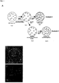
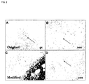

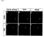


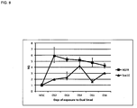

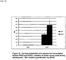
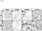


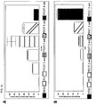
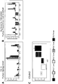


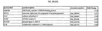
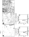
Classifications
-
- C—CHEMISTRY; METALLURGY
- C12—BIOCHEMISTRY; BEER; SPIRITS; WINE; VINEGAR; MICROBIOLOGY; ENZYMOLOGY; MUTATION OR GENETIC ENGINEERING
- C12N—MICROORGANISMS OR ENZYMES; COMPOSITIONS THEREOF; PROPAGATING, PRESERVING, OR MAINTAINING MICROORGANISMS; MUTATION OR GENETIC ENGINEERING; CULTURE MEDIA
- C12N5/00—Undifferentiated human, animal or plant cells, e.g. cell lines; Tissues; Cultivation or maintenance thereof; Culture media therefor
- C12N5/06—Animal cells or tissues; Human cells or tissues
- C12N5/0602—Vertebrate cells
- C12N5/0618—Cells of the nervous system
- C12N5/0621—Eye cells, e.g. cornea, iris pigmented cells
-
- A—HUMAN NECESSITIES
- A61—MEDICAL OR VETERINARY SCIENCE; HYGIENE
- A61K—PREPARATIONS FOR MEDICAL, DENTAL OR TOILETRY PURPOSES
- A61K35/00—Medicinal preparations containing materials or reaction products thereof with undetermined constitution
- A61K35/12—Materials from mammals; Compositions comprising non-specified tissues or cells; Compositions comprising non-embryonic stem cells; Genetically modified cells
- A61K35/30—Nerves; Brain; Eyes; Corneal cells; Cerebrospinal fluid; Neuronal stem cells; Neuronal precursor cells; Glial cells; Oligodendrocytes; Schwann cells; Astroglia; Astrocytes; Choroid plexus; Spinal cord tissue
-
- A—HUMAN NECESSITIES
- A61—MEDICAL OR VETERINARY SCIENCE; HYGIENE
- A61K—PREPARATIONS FOR MEDICAL, DENTAL OR TOILETRY PURPOSES
- A61K45/00—Medicinal preparations containing active ingredients not provided for in groups A61K31/00 - A61K41/00
- A61K45/06—Mixtures of active ingredients without chemical characterisation, e.g. antiphlogistics and cardiaca
-
- A—HUMAN NECESSITIES
- A61—MEDICAL OR VETERINARY SCIENCE; HYGIENE
- A61K—PREPARATIONS FOR MEDICAL, DENTAL OR TOILETRY PURPOSES
- A61K9/00—Medicinal preparations characterised by special physical form
- A61K9/0012—Galenical forms characterised by the site of application
- A61K9/0048—Eye, e.g. artificial tears
-
- A—HUMAN NECESSITIES
- A61—MEDICAL OR VETERINARY SCIENCE; HYGIENE
- A61L—METHODS OR APPARATUS FOR STERILISING MATERIALS OR OBJECTS IN GENERAL; DISINFECTION, STERILISATION OR DEODORISATION OF AIR; CHEMICAL ASPECTS OF BANDAGES, DRESSINGS, ABSORBENT PADS OR SURGICAL ARTICLES; MATERIALS FOR BANDAGES, DRESSINGS, ABSORBENT PADS OR SURGICAL ARTICLES
- A61L27/00—Materials for grafts or prostheses or for coating grafts or prostheses
- A61L27/36—Materials for grafts or prostheses or for coating grafts or prostheses containing ingredients of undetermined constitution or reaction products thereof, e.g. transplant tissue, natural bone, extracellular matrix
- A61L27/38—Materials for grafts or prostheses or for coating grafts or prostheses containing ingredients of undetermined constitution or reaction products thereof, e.g. transplant tissue, natural bone, extracellular matrix containing added animal cells
- A61L27/3804—Materials for grafts or prostheses or for coating grafts or prostheses containing ingredients of undetermined constitution or reaction products thereof, e.g. transplant tissue, natural bone, extracellular matrix containing added animal cells characterised by specific cells or progenitors thereof, e.g. fibroblasts, connective tissue cells, kidney cells
- A61L27/3808—Endothelial cells
-
- A—HUMAN NECESSITIES
- A61—MEDICAL OR VETERINARY SCIENCE; HYGIENE
- A61L—METHODS OR APPARATUS FOR STERILISING MATERIALS OR OBJECTS IN GENERAL; DISINFECTION, STERILISATION OR DEODORISATION OF AIR; CHEMICAL ASPECTS OF BANDAGES, DRESSINGS, ABSORBENT PADS OR SURGICAL ARTICLES; MATERIALS FOR BANDAGES, DRESSINGS, ABSORBENT PADS OR SURGICAL ARTICLES
- A61L27/00—Materials for grafts or prostheses or for coating grafts or prostheses
- A61L27/36—Materials for grafts or prostheses or for coating grafts or prostheses containing ingredients of undetermined constitution or reaction products thereof, e.g. transplant tissue, natural bone, extracellular matrix
- A61L27/38—Materials for grafts or prostheses or for coating grafts or prostheses containing ingredients of undetermined constitution or reaction products thereof, e.g. transplant tissue, natural bone, extracellular matrix containing added animal cells
- A61L27/3839—Materials for grafts or prostheses or for coating grafts or prostheses containing ingredients of undetermined constitution or reaction products thereof, e.g. transplant tissue, natural bone, extracellular matrix containing added animal cells characterised by the site of application in the body
-
- A—HUMAN NECESSITIES
- A61—MEDICAL OR VETERINARY SCIENCE; HYGIENE
- A61L—METHODS OR APPARATUS FOR STERILISING MATERIALS OR OBJECTS IN GENERAL; DISINFECTION, STERILISATION OR DEODORISATION OF AIR; CHEMICAL ASPECTS OF BANDAGES, DRESSINGS, ABSORBENT PADS OR SURGICAL ARTICLES; MATERIALS FOR BANDAGES, DRESSINGS, ABSORBENT PADS OR SURGICAL ARTICLES
- A61L27/00—Materials for grafts or prostheses or for coating grafts or prostheses
- A61L27/36—Materials for grafts or prostheses or for coating grafts or prostheses containing ingredients of undetermined constitution or reaction products thereof, e.g. transplant tissue, natural bone, extracellular matrix
- A61L27/38—Materials for grafts or prostheses or for coating grafts or prostheses containing ingredients of undetermined constitution or reaction products thereof, e.g. transplant tissue, natural bone, extracellular matrix containing added animal cells
- A61L27/3895—Materials for grafts or prostheses or for coating grafts or prostheses containing ingredients of undetermined constitution or reaction products thereof, e.g. transplant tissue, natural bone, extracellular matrix containing added animal cells using specific culture conditions, e.g. stimulating differentiation of stem cells, pulsatile flow conditions
-
- C—CHEMISTRY; METALLURGY
- C12—BIOCHEMISTRY; BEER; SPIRITS; WINE; VINEGAR; MICROBIOLOGY; ENZYMOLOGY; MUTATION OR GENETIC ENGINEERING
- C12N—MICROORGANISMS OR ENZYMES; COMPOSITIONS THEREOF; PROPAGATING, PRESERVING, OR MAINTAINING MICROORGANISMS; MUTATION OR GENETIC ENGINEERING; CULTURE MEDIA
- C12N5/00—Undifferentiated human, animal or plant cells, e.g. cell lines; Tissues; Cultivation or maintenance thereof; Culture media therefor
- C12N5/06—Animal cells or tissues; Human cells or tissues
- C12N5/0602—Vertebrate cells
- C12N5/0618—Cells of the nervous system
- C12N5/0623—Stem cells
-
- A—HUMAN NECESSITIES
- A61—MEDICAL OR VETERINARY SCIENCE; HYGIENE
- A61L—METHODS OR APPARATUS FOR STERILISING MATERIALS OR OBJECTS IN GENERAL; DISINFECTION, STERILISATION OR DEODORISATION OF AIR; CHEMICAL ASPECTS OF BANDAGES, DRESSINGS, ABSORBENT PADS OR SURGICAL ARTICLES; MATERIALS FOR BANDAGES, DRESSINGS, ABSORBENT PADS OR SURGICAL ARTICLES
- A61L2430/00—Materials or treatment for tissue regeneration
- A61L2430/16—Materials or treatment for tissue regeneration for reconstruction of eye parts, e.g. intraocular lens, cornea
-
- C—CHEMISTRY; METALLURGY
- C12—BIOCHEMISTRY; BEER; SPIRITS; WINE; VINEGAR; MICROBIOLOGY; ENZYMOLOGY; MUTATION OR GENETIC ENGINEERING
- C12N—MICROORGANISMS OR ENZYMES; COMPOSITIONS THEREOF; PROPAGATING, PRESERVING, OR MAINTAINING MICROORGANISMS; MUTATION OR GENETIC ENGINEERING; CULTURE MEDIA
- C12N2500/00—Specific components of cell culture medium
- C12N2500/60—Buffer, e.g. pH regulation, osmotic pressure
-
- C—CHEMISTRY; METALLURGY
- C12—BIOCHEMISTRY; BEER; SPIRITS; WINE; VINEGAR; MICROBIOLOGY; ENZYMOLOGY; MUTATION OR GENETIC ENGINEERING
- C12N—MICROORGANISMS OR ENZYMES; COMPOSITIONS THEREOF; PROPAGATING, PRESERVING, OR MAINTAINING MICROORGANISMS; MUTATION OR GENETIC ENGINEERING; CULTURE MEDIA
- C12N2500/00—Specific components of cell culture medium
- C12N2500/90—Serum-free medium, which may still contain naturally-sourced components
-
- C—CHEMISTRY; METALLURGY
- C12—BIOCHEMISTRY; BEER; SPIRITS; WINE; VINEGAR; MICROBIOLOGY; ENZYMOLOGY; MUTATION OR GENETIC ENGINEERING
- C12N—MICROORGANISMS OR ENZYMES; COMPOSITIONS THEREOF; PROPAGATING, PRESERVING, OR MAINTAINING MICROORGANISMS; MUTATION OR GENETIC ENGINEERING; CULTURE MEDIA
- C12N2501/00—Active agents used in cell culture processes, e.g. differentation
- C12N2501/10—Growth factors
- C12N2501/115—Basic fibroblast growth factor (bFGF, FGF-2)
-
- C—CHEMISTRY; METALLURGY
- C12—BIOCHEMISTRY; BEER; SPIRITS; WINE; VINEGAR; MICROBIOLOGY; ENZYMOLOGY; MUTATION OR GENETIC ENGINEERING
- C12N—MICROORGANISMS OR ENZYMES; COMPOSITIONS THEREOF; PROPAGATING, PRESERVING, OR MAINTAINING MICROORGANISMS; MUTATION OR GENETIC ENGINEERING; CULTURE MEDIA
- C12N2501/00—Active agents used in cell culture processes, e.g. differentation
- C12N2501/10—Growth factors
- C12N2501/135—Platelet-derived growth factor [PDGF]
-
- C—CHEMISTRY; METALLURGY
- C12—BIOCHEMISTRY; BEER; SPIRITS; WINE; VINEGAR; MICROBIOLOGY; ENZYMOLOGY; MUTATION OR GENETIC ENGINEERING
- C12N—MICROORGANISMS OR ENZYMES; COMPOSITIONS THEREOF; PROPAGATING, PRESERVING, OR MAINTAINING MICROORGANISMS; MUTATION OR GENETIC ENGINEERING; CULTURE MEDIA
- C12N2501/00—Active agents used in cell culture processes, e.g. differentation
- C12N2501/10—Growth factors
- C12N2501/15—Transforming growth factor beta (TGF-β)
-
- C—CHEMISTRY; METALLURGY
- C12—BIOCHEMISTRY; BEER; SPIRITS; WINE; VINEGAR; MICROBIOLOGY; ENZYMOLOGY; MUTATION OR GENETIC ENGINEERING
- C12N—MICROORGANISMS OR ENZYMES; COMPOSITIONS THEREOF; PROPAGATING, PRESERVING, OR MAINTAINING MICROORGANISMS; MUTATION OR GENETIC ENGINEERING; CULTURE MEDIA
- C12N2501/00—Active agents used in cell culture processes, e.g. differentation
- C12N2501/10—Growth factors
- C12N2501/155—Bone morphogenic proteins [BMP]; Osteogenins; Osteogenic factor; Bone inducing factor
-
- C—CHEMISTRY; METALLURGY
- C12—BIOCHEMISTRY; BEER; SPIRITS; WINE; VINEGAR; MICROBIOLOGY; ENZYMOLOGY; MUTATION OR GENETIC ENGINEERING
- C12N—MICROORGANISMS OR ENZYMES; COMPOSITIONS THEREOF; PROPAGATING, PRESERVING, OR MAINTAINING MICROORGANISMS; MUTATION OR GENETIC ENGINEERING; CULTURE MEDIA
- C12N2501/00—Active agents used in cell culture processes, e.g. differentation
- C12N2501/40—Regulators of development
- C12N2501/415—Wnt; Frizzeled
-
- C—CHEMISTRY; METALLURGY
- C12—BIOCHEMISTRY; BEER; SPIRITS; WINE; VINEGAR; MICROBIOLOGY; ENZYMOLOGY; MUTATION OR GENETIC ENGINEERING
- C12N—MICROORGANISMS OR ENZYMES; COMPOSITIONS THEREOF; PROPAGATING, PRESERVING, OR MAINTAINING MICROORGANISMS; MUTATION OR GENETIC ENGINEERING; CULTURE MEDIA
- C12N2501/00—Active agents used in cell culture processes, e.g. differentation
- C12N2501/70—Enzymes
- C12N2501/72—Transferases (EC 2.)
- C12N2501/727—Kinases (EC 2.7.)
-
- C—CHEMISTRY; METALLURGY
- C12—BIOCHEMISTRY; BEER; SPIRITS; WINE; VINEGAR; MICROBIOLOGY; ENZYMOLOGY; MUTATION OR GENETIC ENGINEERING
- C12N—MICROORGANISMS OR ENZYMES; COMPOSITIONS THEREOF; PROPAGATING, PRESERVING, OR MAINTAINING MICROORGANISMS; MUTATION OR GENETIC ENGINEERING; CULTURE MEDIA
- C12N2501/00—Active agents used in cell culture processes, e.g. differentation
- C12N2501/999—Small molecules not provided for elsewhere
-
- C—CHEMISTRY; METALLURGY
- C12—BIOCHEMISTRY; BEER; SPIRITS; WINE; VINEGAR; MICROBIOLOGY; ENZYMOLOGY; MUTATION OR GENETIC ENGINEERING
- C12N—MICROORGANISMS OR ENZYMES; COMPOSITIONS THEREOF; PROPAGATING, PRESERVING, OR MAINTAINING MICROORGANISMS; MUTATION OR GENETIC ENGINEERING; CULTURE MEDIA
- C12N2506/00—Differentiation of animal cells from one lineage to another; Differentiation of pluripotent cells
- C12N2506/02—Differentiation of animal cells from one lineage to another; Differentiation of pluripotent cells from embryonic cells
-
- C—CHEMISTRY; METALLURGY
- C12—BIOCHEMISTRY; BEER; SPIRITS; WINE; VINEGAR; MICROBIOLOGY; ENZYMOLOGY; MUTATION OR GENETIC ENGINEERING
- C12N—MICROORGANISMS OR ENZYMES; COMPOSITIONS THEREOF; PROPAGATING, PRESERVING, OR MAINTAINING MICROORGANISMS; MUTATION OR GENETIC ENGINEERING; CULTURE MEDIA
- C12N2506/00—Differentiation of animal cells from one lineage to another; Differentiation of pluripotent cells
- C12N2506/08—Differentiation of animal cells from one lineage to another; Differentiation of pluripotent cells from cells of the nervous system
-
- C—CHEMISTRY; METALLURGY
- C12—BIOCHEMISTRY; BEER; SPIRITS; WINE; VINEGAR; MICROBIOLOGY; ENZYMOLOGY; MUTATION OR GENETIC ENGINEERING
- C12N—MICROORGANISMS OR ENZYMES; COMPOSITIONS THEREOF; PROPAGATING, PRESERVING, OR MAINTAINING MICROORGANISMS; MUTATION OR GENETIC ENGINEERING; CULTURE MEDIA
- C12N2506/00—Differentiation of animal cells from one lineage to another; Differentiation of pluripotent cells
- C12N2506/45—Differentiation of animal cells from one lineage to another; Differentiation of pluripotent cells from artificially induced pluripotent stem cells
-
- C—CHEMISTRY; METALLURGY
- C12—BIOCHEMISTRY; BEER; SPIRITS; WINE; VINEGAR; MICROBIOLOGY; ENZYMOLOGY; MUTATION OR GENETIC ENGINEERING
- C12N—MICROORGANISMS OR ENZYMES; COMPOSITIONS THEREOF; PROPAGATING, PRESERVING, OR MAINTAINING MICROORGANISMS; MUTATION OR GENETIC ENGINEERING; CULTURE MEDIA
- C12N2533/00—Supports or coatings for cell culture, characterised by material
- C12N2533/50—Proteins
Definitions
- This disclosure generally relates to methods for directed differentiation of corneal cells from stem cells.
- Compositions of corneal endothelial cells, including pharmaceutical compositions, and uses thereof, are also disclosed.
- cell-based therapies for treatment of visual disorders, including disorders of the cornea are also disclosed.
- the cornea performs functions critical for normal vision and maintenance of eye health, including providing about two-thirds of the optical power of the eye and protecting the eye from injury or infection.
- Corneal disease and injury is a leading cause of blindness worldwide.
- Many corneal disease and injuries can be treated by transplantation of donor corneas.
- the cornea is the most transplanted organ in the body and has a high success rate over 15 years. For example, approximately 40,000 corneal transplantations are performed per year in the U.S.
- demand for corneas for transplantation greatly exceeds the current supply worldwide, and the limited quality and quantity of available donor tissue hinders treatment.
- One factor contributing to the inadequate supply of donated corneas is that up to 30% of donated corneas are rejected for transplantation due to poor quality of the corneal endothelium.
- Quality of the corneal endothelium generally decreases with donor age because, as the cornea ages or is injured, the endothelial cells die and are not replaced. Therefore, as the population ages, the supply of donor tissue having suitably healthy corneal endothelium decreases. Moreover, the number and quality of donated corneas is expected to decline as the popularity of LASIK surgery increases (these corneas are rejected for transplantation).
- Diseases of the cornea may involve one or more of the cornea's five layers: the corneal epithelium, Bowman's layer, the corneal stroma, Descemet's membrane, and the corneal endothelium.
- the corneal epithelium, corneal stroma, and corneal endothelium are cellular layers, while Bowman's layer and Descemet's membrane are primarily composed of collagen fibrils.
- the corneal endothelium is a single layer of cells on the inner surface of the cornea. It faces the chamber formed between the cornea and the iris and keeps the cornea transparent by regulating fluid levels. Without functional corneal endothelium, the cornea becomes cloudy and vision is lost.
- Properly functioning corneal endothelial cells maintain the proper fluid levels in the cornea, e.g., the balance between "leakage” of fluid into the stroma and active pumping that continuously operates to move fluid from the stroma to the anterior chamber of the eye.
- Corneal endothelial cells have been reported to have little or no capacity to proliferate in vivo, such that they are not replaced when injured or otherwise lost.
- the corneal endothelial cell layer is most densely packed at birth and cell density thereafter decreases rapidly as the eyes grow (reflecting the same number of cells covering a larger area). Thereafter, corneal cell density gradually declines with age, apparently reflecting the gradual loss of cells which are not replaced. As cell density decreases, each cell spreads out and covers a larger area to maintain the cell layer's barrier and pump functions.
- the cell density of tightly packed corneal endothelium in vivo has been reported to be as high as 5624 cells/mm 2 in infants two months of age, falling to 4252 cells/mm 2 within the first year from birth, and subsequently decreasing rapidly during early childhood (associated with the increase in corneal size as eyes grow).
- corneal endothelium density falls to approximately 3591 plus or minus 399 cells/mm 2 , and falls farther to approximately 2697 plus or minus 246 cells/mm 2 by 10 years of age, and further declines by approximately 0.6% per year throughout adulthood. See Peh et al., Transplantation. 2011 Apr 27;91(8):811-9 .
- Glare endothelium Primary diseases that affect the corneal endothelium include Fuch's dystrophy, iridocorneal endothelial syndrome, posterior polymorphous dystrophy, and congenital hereditary endothelial dystrophy. Secondary diseases for which the most effective treatment is replacement of the corneal endothelium include several corneal dystrophies, contact lens usage, cataract surgery, and late endothelial failure in cornea transplantation.
- the preferred treatment when only the corneal endothelium is compromised is Descemet's stripping with endothelial keratoplasty (DSEK), which includes the removal of Descemet's membrane and the corneal endothelium, and subsequent transplantation of donor tissue. Alternatively, in penetrating keratoplasty (PKP) the entire cornea is removed and replaced.
- DSEK Descemet's stripping with endothelial keratoplasty
- PGP penetrating keratoplasty
- corneal transplantation includes obtaining a donor cornea (e.g., from a post-mortem anatomical gift), determining whether the donor cornea is of sufficient quality and otherwise suitable for use, and surgical replacement of the damaged or diseased cornea.
- Procedures have been developed to replace the entire cornea (penetrating keratoplasty) or leave the patient's Descemet's membrane and endothelium and replace the remaining layers with donated tissue (lamellar keratoplasty); the latter procedure may decrease the risk of transplant rejection but may also give inferior visual acuity post-transplant.
- lamellar keratoplasty may not be suitable for treatment of some conditions for which replacement of the patient's corneal endothelium and/or Descemet's membrane may be the indicated treatment.
- Additional disclosures generally related to methods of obtaining and using corneal cells including therapeutic methods, culture methods, preservation methods, compositions containing or that that may be used in conjunction therewith, and the like are included in U.S. 2007/0275365 , US 2010/0209402 , US 2010/0233240 , US 2011/0009488 , US 2009/0232772 , U.S. Pat. 5166048 , US 2007/0092550 , US 2005/0214259 , US 2007/0148137 , US Pat. 4959319 , US Pat. 5310728 , US Pat. 5589451 , US 2010/0215717 , US Pat.
- the present invention provides a method of producing corneal endothelial cells (CECs), comprising contacting neural crest stem cells (NCSCs) with at least one dickkopf-related protein 2 (DKK2) agonist and/or at least one platelet-derived growth factor subunit B (PDGFB) agonist that induces differentiation of the NCSCs into CECs, wherein the DKK2 agonist is selected from the group consisting of: DKK2; Dkk 1; Dkk 3; Dkk 4; Soggy; secreted frizzled related proteins (Frzb); Wnt inhibitor factor (WIF); IWR; pyrvinium; ICG-001; PKF115-584; IWP; Antl.4Br/Ant 1.4Cl; Niclosamide; apicularen; bafilomycin; XAV939; NSC668036; 2,4-diamino-quinazoline; Quercetin; Wnt proteins, nucleic acids encoding Wnt proteins, LiCl,
- the at least one DKK2 agonist may comprise DKK2 polypeptide.
- the at least one PDGFB agonist may comprise PDGFB polypeptide.
- the at least one DKK2 agonist may comprise DKK2 polypeptide and said at least one PDGFB agonist may comprise PDGFB polypeptide.
- the concentration of said DKK2 polypeptide may be between 1 ng/ml and 15 ⁇ g/ml, between 10 ng/ml and 15 ⁇ g/ml, between 1 ng/ml and 1 ⁇ g/ml, between 1 ng/ml and 100 ng/ml, between 2 ng/ml and 20 ng/ml, between 5 ng/ml and 20 ng/ml, or about 10 ng/ml.
- the concentration of said PDGFB polypeptide may be between 0.1 ng/ml and 250 ng/ml, between 0.5 ng/ml and 150 ng/ml, between 1 ng/ml and 50 ng/ml, between 2 ng/ml and 20 ng/ml, or about 10 ng/ml.
- the method may comprise culturing said CEC on a matrix.
- the matrix may be selected from the group consisting of: laminin, fibronectin, vitronectin, proteoglycan, entactin, collagen, collagen I, collagen IV, collagen VIII, heparan sulfate, Matrigel (a soluble preparation from Engelbreth-Holm-Swarm (EHS) mouse sarcoma cells), a human basement membrane extract, and any combination thereof.
- laminin a soluble preparation from Engelbreth-Holm-Swarm (EHS) mouse sarcoma cells
- EHS Engelbreth-Holm-Swarm
- the matrix may be of human or non-human animal origin.
- the matrix may be of bovine, mouse or rat origin.
- the matrix may comprise Matrigel.
- NSCs neural crest stem cells
- DKK2 dickkopf-related protein 2
- PDGFB platelet-derived growth factor subunit B
- the passaging may be effected between 1 hour and 5 days, between 2 hours and 4 days, between 3 hours and 3 days, between 4 hours and 2 days, or about 1 day subsequent to commencement of step (a).
- the passaging may be effected by a method which may comprise contacting the cells with a cell dissociation buffer, or by a method which may comprise mechanical dissociation of the cells or a subset thereof, or by a method which may comprise optical isolation the cells or a subset thereof.
- the cell dissociation buffer may be non-enzymatic.
- the cell dissociation buffer may comprise ethylenediaminetetraacetic acid (EDTA).
- EDTA ethylenediaminetetraacetic acid
- the duration of said contacting NCSCs with a factor that induces differentiation of said neural crest cells into CEC may be at least 2 days, between 2 and 25 days, or may be between 2 and 10 days.
- the CEC may be purified using affinity-based depletion of other cell types, which may be effected concurrently with passaging or at an earlier or later time.
- Depletion methods may utilize a binding molecule having affinity for a marker of a non-CEC cell type, e.g., an antibody or fragment thereof, which may be directly or indirectly coupled to a bead or other substrate, such as a magnetic microbead.
- a substrate may be coupled to one or more antibodies that can deplete stem cells or fibroblasts.
- Exemplary markers that may be utilized include CD271, SSEA-1, TRA-1-60, SSEA-4, and/or CD326.
- Anti-SSEA-1 MicroBeads (Miltenyi Biotech, cat# 130-094-530), Anti-TRA-1-60 MicroBead Kit (Miltenyi Biotech, cat# 130-095-816), Anti-SSEA-4 MicroBeads (Miltenyi Biotech, cat# 130-097-855), and Anti-fibroblast CD326 (Miltenyi Biotech, cat# 130-050-601).
- Step (a) further may comprise culturing said NCSCs on a matrix, which may be selected from the group consisting of: laminin, fibronectin, vitronectin, proteoglycan, entactin, collagen, collagen I, collagen IV, collagen VIII, heparan sulfate, Matrigel (a soluble preparation from Engelbreth-Holm-Swarm (EHS) mouse sarcoma cells), a human basement membrane extract, and any combination thereof, may be of human or non-human animal origin, may be of bovine, mouse or rat origin, or may comprise Matrigel.
- a matrix which may be selected from the group consisting of: laminin, fibronectin, vitronectin, proteoglycan, entactin, collagen, collagen I, collagen IV, collagen VIII, heparan sulfate, Matrigel (a soluble preparation from Engelbreth-Holm-Swarm (EHS) mouse sarcoma cells), a human basement membrane extract, and any combination thereof
- the NCSCs may be obtained from an animal tissue, which may be selected from the group consisting of: the gut, dorsal root ganglia, bone marrow, skin, heart, cornea, caratoid body, neural tube, teeth, and sciatic nerve.
- the animal tissue may be human tissue.
- the NCSCs may be produced by transdifferentiation of a somatic cell.
- the NCSCs may be obtained from cultures of neural rosettes.
- the neural crest stem cells may be produced from ES cells by a method which may comprise culturing ES cells with MS5 stromal feeder cells.
- the NCSCs may be produced from ES cells.
- the neural crest stem cells may be produced from ES cells by a method which may comprise contacting ES cells with one or more inhibitors of SMA/Mothers against Decapentaplegic (SMAD) protein signaling.
- SMA/Mothers against Decapentaplegic (SMAD) protein signaling may comprise contacting ES cells with one or more inhibitors of SMA/Mothers against Decapentaplegic (SMAD) protein signaling.
- the one or more inhibitors of SMAD protein signaling may prevent the binding of a TGF- ⁇ family ligands to its corresponding receptor.
- the one or more inhibitors of SMAD protein signaling may prevent the activation of a TGF- ⁇ receptor.
- the one or more inhibitors of SMAD protein signaling inhibits one or more SMAD intracellular proteins/transcription factors.
- the one or more inhibitors of SMAD protein signaling may comprise Leukemia Inhibitory Factor (LIF), GSK3 inhibitor (CHIR 99021), Compound E ( ⁇ secretase inhibitor XXI), SB431542, or any combination thereof.
- LIF Leukemia Inhibitory Factor
- CHIR 99021 GSK3 inhibitor
- Compound E ⁇ secretase inhibitor XXI
- SB431542 or any combination thereof.
- the one or more inhibitors of SMAD protein signaling may comprise Chordin, Follistatin, dominant negative receptors or blocking antibodies that sequester BMP2, BMP4, and/or BMP7, dorsomorphin (or Compound C), SIS3 (6,7-Dimethoxy-2-((2E)-3-(1-methyl-2-phenyl-1H-pyrrolo[2,3-b]pyridin-3-yl-prop-2-enoyl))-1,2,3,4-tetrahydroisoquinoline, Specific Inhibitor of Smad3 (SIS3), an inhibitor SMAD, SMAD6, SMAD7, SMAD10, an antagonist of a receptor SMAD, an antagonist of SMAD 1, an antagonist of SMAD2, an antagonist of SMAD3, an antagonist of SMAD5, an antagonist of SMAD8/9, or any combination thereof.
- SIS3 Specific Inhibitor of Smad3
- the duration of said contacting ES cells with one or more inhibitors of SMAD protein signaling may be at least 2 days, between 1 and 10 days, or may be between 2 and 6 days.
- the neural crest stem cells may be produced from ES cells by a method further which may comprise contacting ES cells with at least one Wnt agonist.
- the neural crest stem cells may be produced from ES cells by a method further which may comprise contacting ES cells with at least one Wnt agonist which may comprise (2'Z,3'E)-6-bromoindirubin-3'-oxime (BIO) and/or Wnt3a.
- Wnt agonist which may comprise (2'Z,3'E)-6-bromoindirubin-3'-oxime (BIO) and/or Wnt3a.
- the neural crest stem cells may be produced from ES cells by a method further which may comprise contacting ES cells with at least one Wnt agonist selected from the group consisting of: Wnt proteins, nucleic acids encoding Wnt proteins, LiCl, Axin antagonists; APC antagonists; norrin; R-spondin2; (hetero)arylpyrimidines; IQ1; BIO(6-bromoindirubin-3'-oxime); 2-amino-4-[3,4-(methylenedioxy)benzyl-amino]-6-(3-methoxyphenyl)pyrimidine; WAY-316606; QS11; SB-216763; SB-216763; DCA; and any combination thereof.
- Wnt agonist selected from the group consisting of: Wnt proteins, nucleic acids encoding Wnt proteins, LiCl, Axin antagonists; APC antagonists; norrin; R-spondin2; (hetero)arylpyrimidine
- the neural crest stem cells may be produced from ES cells by a method which may comprise contacting ES cells with a first inhibitor of SMAD protein signaling and a second inhibitor of SMAD protein signaling.
- the first inhibitor of SMAD protein signaling may be selected from the group consisting of: Noggin polypeptide, dorsomorphin, LDN-193189, and any combination thereof.
- the first inhibitor of SMAD protein may comprise Noggin polypeptide.
- the Noggin polypeptide may be present in a concentration between 10 ng/ml and 5,000 ng/ml, between 100 ng/ml and 700 ng/ml, between 400 ng/ml and 600 ng/ml, or about 500 ng/ml.
- the first inhibitor of SMAD protein signaling may be selected from the group consisting of: antagonists of BMP2; antagonists of BMP4; antagonists of BMP7; and antagonists of TG ⁇ ;
- the second inhibitor of SMAD protein signaling may comprise an inhibitor of an anaplastic lymphoma kinase signaling pathway.
- the second inhibitor of SMAD protein signaling inhibits a signaling pathway selected from the group consisting of Lefty, Activin, and TGFbeta.
- the second inhibitor of SMAD protein signaling inhibits both activin and nodal signaling.
- the second inhibitor of SMAD protein signaling inhibits the Lefty/Activin/TGFbeta pathways by blocking phosphorylation of the ALK4, ALK5 and ALK7 receptors.
- the second inhibitor of SMAD protein signaling may be an ALK4 receptor inhibitor.
- the second inhibitor of SMAD protein signaling may be selected from the group consisting of: 4-[4-(1,3-benzodioxol-5-yl)-5-(2-pyridinyl)-1H-imidazol-2-yl]benzamide (SB431542) and derivatives thereof.
- the second inhibitor of SMAD protein signaling may comprise SB431542.
- the SB431542 may be present in a concentration between 10 nM and 100 ⁇ M, between 0.1 ⁇ M and 50 ⁇ M, between 0.1 and 20 ⁇ M, between 1 and 20 ⁇ M, or about 10 ⁇ M.
- the second inhibitor of SMAD protein signaling blocks phosphorylation of ACTRIB, TGF ⁇ R1, and ACTRIC receptors.
- the second inhibitor of SMAD protein signaling inhibits TGF ⁇ /Activin/Nodal signaling.
- the second inhibitor of SMAD protein signaling blocks endogenous Activin and BMP signals.
- the first inhibitor of SMAD protein signaling and/or said second inhibitor of SMAD protein signaling may be each selected from the group consisting of: Chordin, Follistatin, dominant negative receptors or blocking antibodies that sequester BMP2, BMP4, and/or BMP7, dorsomorphin (or Compound C), SIS3 (6,7-Dimethoxy-2-((2E)-3-(1-methyl-2-phenyl-1H-pyrrolo[2,3-b]pyridin-3-yl-prop-2-enoyl))-1,2,3,4-tetrahydroisoquinoline, Specific Inhibitor of Smad3 (SIS3), an inhibitor SMAD, SMAD6, SMAD7, SMAD10, an antagonist of a receptor SMAD, an antagonist of SMAD1, an antagonist of SMAD2, an antagonist of SMAD3, an antagonist of SMAD5, and an antagonist of SMAD8/9.
- SIS3 Specific Inhibitor of Smad3
- the first inhibitor of SMAD protein signaling may comprise Noggin and said second inhibitor of SMAD protein signaling may comprise SB431542.
- the duration of said contacting ES cells with said first inhibitor of SMAD protein signaling and said second inhibitor of SMAD protein signaling may be at least 2 days, between 1 and 10 days, or may be between 2 and 6 days.
- the ES cells may be human ES cells.
- the ES cells may be iPS cells.
- the ES cells may not exhibit changes or mutations in genes associated with a disease of corneal endothelial cells.
- the ES cells may exhibit a normal karyotype.
- said ES cells Prior to differentiation into NCSCs, said ES cells may be maintained in culture in the absence of feeder cells.
- said ES cells Prior to differentiation into NCSCs said ES cells may be cultured on a matrix, which may be selected from the group consisting of: laminin, fibronectin, vitronectin, proteoglycan, entactin, collagen, collagen I, collagen IV, heparan sulfate, Matrigel (a soluble preparation from Engelbreth-Holm-Swarm (EHS) mouse sarcoma cells), a human basement membrane extract, and any combination thereof, or may be of human or non-human animal origin, or may be of bovine, mouse or rat origin, or may comprise Matrigel.
- a matrix which may be selected from the group consisting of: laminin, fibronectin, vitronectin, proteoglycan, entactin, collagen, collagen I, collagen IV, heparan sulfate, Matrigel (a soluble preparation from Engelbreth-Holm-Swarm (EHS) mouse sarcoma cells), a human basement membrane extract, and any combination thereof
- the NCSCs may express one or more markers selected from the group consisting of: Sox10, AP2, HNK1, PAX7, p75 (NGFR), and any combination thereof.
- the CEC may express one or more markers selected from the group consisting of: Na+/K+ ATPase, ZO-1, KLF13, AQP1, Collagen VIII, SLC16A3, CFTR, NBC1, CA2, AE2/ SCL4A2, SCL16A1, CA12, CA4, FoxC1, and any combination thereof.
- the CEC may express the markers CollagenVIII, Na+K+ATPase pump, and ZO-1, and may not express the markers vWF and CD31.
- the CEC may express one or more corneal endothelial pump markers.
- the CEC may express one or more periocular neural crest markers.
- the CEC may express one or more cell adhesion and matrix proteins.
- the CEC may express at least one corneal endothelial pump marker, at least one periocular neural crest marker, and at least one cell adhesion and matrix protein.
- the one or more corneal endothelial pump markers may be selected from the group consisting of: AQP1, CA2, CA4, CA12, SCL14A2, SLC16A1, SLC16A3, SLC16A7, CFTR, NHE1, ADCY10, voltage-dependent anion channels VDAC2 and VDAC3, chloride channel proteins CLCN2 and CLC.
- the periocular neural crest markers may be selected from the group consisting of: PITX2, and FOXC1.
- the cell adhesion and matrix proteins may be selected from the group consisting of: Occludin, Connexin 43, 9.3E antigen, Collagen III, Collagen IV, N cadherin, VE cadherin, E cadherin, beta catenin, p120, p190 Laminin alpha 4, Nidogen-2, and Netrin 4.
- the CEC may express the markers CollagenVIII, Na+K+ATPase pump, AQP1, CA2, CA4, CA12, SCL14A2, SLC16A1, SLC16A3, SLC16A7, CFTR, NHE1, ADCY10, PITX2, and FOXC1, and may not express the markers vWF and CD31.
- the CEC may form a monolayer of uniformly sized cells with a predominantly hexagonal shape, such as at least 50%, at least 60%, at least 70%, at least 80%, or at least 90% of said CEC may exhibit said hexagonal shape.
- the CEC may allow unidirectional leakage of solutes and nutrients.
- the CEC may actively pump water in the opposite direction of said unidirectional leakage.
- the CEC may exhibit a high level of metabolic activity that may be comparable to animal-derived CEC.
- the CEC may be human CEC.
- the CEC may exhibit a culture density of at least 1000 cells/mm 2 , at least 2000 cells/mm 2 , at least 3000 cells/mm 2 , at least 4000 cells/mm 2 , at least 5000 cells/mm 2 , at least 6000 cells/mm 2 , at least 7000 cells/mm 2 , at least 8000 cells/mm 2 , at least 9000 cells/mm 2 , between 2000 and 9000 cells/mm 2 between 2000 and 8000 cells/mm 2 between 2000 and 7000 cells/mm 2 , between 2000 and 6000 cells/mm 2 , between 2000 and 5,000 cells/mm 2 , between 2000 and 4000 cells/mm 2 , between 2000 and 3500 cells/mm 2 , or about 2500 cells/mm 2 .
- the CEC may exhibit a decreased level of accumulated oxidative stress and/or DNA damage compared to CEC isolated from a living host.
- the level of oxidative stress and/or DNA damage may be detected by measuring the quantity of one or more of: nuclear DNA damage foci; level of expression of p21Cip1, level of expression of p16INK4a; level of expression of cytoglobin protein, level of expression of GPX-1 protein, and level of 8-hydroxy-2_-deoxyguanosine (8-OHdG).
- the CEC may comprise at least 50%, at least 60%, at least 70%, at least 80%, or at least 90% of the cells in the resulting culture.
- composition comprising corneal endothelial cells (CEC) produced according to any method as described herein.
- the composition comprising a sheet of CEC may have a culture density of at least 1000 cells/mm 2 , at least 2000 cells/mm 2 , between 2000 and 6000 cells/mm 2 , between 2000 and 5,000 cells/mm 2 , between 2000 and 4000 cells/mm 2 , between 2000 and 3500 cells/mm 2 , or about 2500 cells/mm 2 .
- composition comprising CEC may exhibit a decreased level of accumulated oxidative stress and/or DNA damage compared to CEC isolated from a living host.
- composition comprising corneal endothelial cells may be produced from neural crest stem cells (NCSCs).
- the NCSCs may be produced from embryonic stem (ES) cells.
- ES embryonic stem
- the NCSCs may be produced from ES cells by a method which may comprise contacting said ES cells with one or more inhibitors of SMAD signaling.
- composition comprising corneal endothelial cells (CEC) produced from embryonic stem (ES) cells.
- the ES cells may be human ES cells.
- the ES cells may be iPS cells.
- the ES cells may not exhibit changes or mutations in genes associated with a disease of corneal endothelial cells.
- the ES cells may exhibit a normal karyotype.
- said ES cells Prior to differentiation into NCSCs, said ES cells may be maintained in culture in the absence of feeder cells.
- said ES cells may be cultured on a matrix, such as a matrix may be selected from the group consisting of: laminin, fibronectin, vitronectin, proteoglycan, entactin, collagen, collagen I, collagen IV, heparan sulfate, Matrigel (a soluble preparation from Engelbreth-Holm-Swarm (EHS) mouse sarcoma cells), a human basement membrane extract, and any combination thereof, which may be of human or non-human animal origin, such as bovine, mouse or rat origin, or Matrigel.
- a matrix may be selected from the group consisting of: laminin, fibronectin, vitronectin, proteoglycan, entactin, collagen, collagen I, collagen IV, heparan sulfate, Matrigel (a soluble preparation from Engelbreth-Holm-Swarm (EHS) mouse sarcoma cells), a human basement membrane extract, and any combination thereof, which may be of human or non-human animal origin
- the NCSCs may express one or more markers selected from the group consisting of: Sox10, AP2, HNK1, PAX7, p75 (NGFR), and any combination thereof.
- the CEC may express one or more markers selected from the group consisting of: Na+/K+ ATPase, ZO-1, KLF13, AQP1, Collagen VIII, SLC16A3, CFTR, NBC1, CA2, AE2/ SCL4A2, SCL16A1, CA12, CA4, FoxC1, and any combination thereof.
- the CEC may express the markers CollagenVIII, Na+K+ATPase pump, and ZO-1, and may not express the markers vWF and CD31.
- the CEC may express one or more corneal endothelial pump markers.
- the CEC may express one or more periocular neural crest markers.
- the CEC may express one or more cell adhesion and matrix proteins.
- the CEC may express at least one corneal endothelial pump marker, at least one periocular neural crest marker, and at least one cell adhesion and matrix protein.
- the one or more corneal endothelial pump markers may be selected from the group consisting of: AQP1, CA2, CA4, CA12, SCL14A2, SLC16A1, SLC16A3, SLC16A7, CFTR, NHE1, ADCY10, voltage-dependent anion channels VDAC2 and VDAC3, chloride channel proteins CLCN2 and CLC.
- the periocular neural crest markers may be selected from the group consisting of: PITX2, and FOXC1.
- the cell adhesion and matrix proteins may be selected from the group consisting of: Occludin, Connexin 43, 9.3E antigen, Collagen III, Collagen IV, N cadherin, VE cadherin, E cadherin, beta catenin, p120, p190 Laminin alpha 4, Nidogen-2, and Netrin 4.
- the CEC may express the markers CollagenVIII, Na+K+ATPase pump, AQP1, CA2, CA4, CA12, SCL14A2, SLC16A1, SLC16A3, SLC16A7, CFTR, NHE1, ADCY10, PITX2, and FOXC1, and may not express the markers vWF and CD31.
- the CEC may form a monolayer of uniformly sized cells with a predominantly hexagonal shape, such as at least 50%, at least 60%, at least 70%, at least 80%, or at least 90% of said CEC may exhibit said hexagonal shape.
- the CEC may allow unidirectional leakage of solutes and nutrients.
- the CEC may actively pump water in the opposite direction of said unidirectional leakage.
- the CEC may exhibit a high level of metabolic activity that may be comparable to animal-derived CEC
- the CEC may be human CEC.
- the CEC may exhibit a culture density of at least 1000 cells/mm 2 , at least 2000 cells/mm 2 , at least 3000 cells/mm 2 , at least 4000 cells/mm 2 , at least 5000 cells/mm 2 , at least 6000 cells/mm 2 , at least 7000 cells/mm 2 , at least 8000 cells/mm 2 , at least 9000 cells/mm 2 , between 2000 and 9000 cells/mm 2 between 2000 and 8000 cells/mm 2 between 2000 and 7000 cells/mm 2 , between 2000 and 6000 cells/mm 2 , between 2000 and 5,000 cells/mm 2 , between 2000 and 4000 cells/mm 2 , between 2000 and 3500 cells/mm 2 , or about 2500 cells/mm 2 .
- the CEC may exhibit a decreased level of accumulated oxidative stress and/or DNA damage compared to CEC isolated from a living host, which may for example be detected by measuring the quantity of one or more of: nuclear DNA damage foci; level of expression of p21Cip1, level of expression of pl6INK4a; level of expression of cytoglobin protein, level of expression of GPX-1 protein, and level of 8-hydroxy-2_-deoxyguanosine (8-OHdG).
- the CEC may comprise at least 50%, at least 60%, at least 70%, at least 80%, or at least 90% of the cells in the culture in which they are contained.
- the CEC may be contained in a sheet of CEC.
- the sheet of cells may comprise or consists essentially of an approximately circular disc of cells having a diameter of at least 1, 2, 3, 4, 5, 6, 7, 8, 9, 10, 11, or 12 mm.
- the CEC may exhibit a culture density of at least 1000 cells/mm 2 , at least 2000 cells/mm 2 , at least 3000 cells/mm 2 , at least 4000 cells/mm 2 , at least 5000 cells/mm 2 , at least 6000 cells/mm 2 , at least 7000 cells/mm 2 , at least 8000 cells/mm 2 , at least 9000 cells/mm 2 , between 2000 and 9000 cells/mm 2 between 2000 and 8000 cells/mm 2 between 2000 and 7000 cells/mm 2 , between 2000 and 6000 cells/mm 2 , between 2000 and 5,000 cells/mm 2 , between 2000 and 4000 cells/mm 2 , between 2000 and 3500 cells/mm 2 , or about 2500 cells/mm 2 .
- the total number of cells may be between about 800 and about 800,000 cells, e.g., at least about 10,000, at least about 20,000, at least about 50,000, at least about 100,000, at least about 200,000, at least about 300,000, at least about 400,000, at least about 500,000, at least about 600,000 or at at least about 700,000 cells, such between about 100,000 and about 800,000 cells, between about 150,000 cells and about 675,000 cells, between about 250,000 cells and aboug 550,000 cells, as well as other numerical ranges within these values.
- the CEC may be situated on a carrier.
- the CEC may be cultured on a substrate and released onto a carrier.
- the substrate may comprise a thermoresponsive polymer or a thermoresponsive poly(N-isopropylacrylamide) (PNIPAAm)-grafted surface.
- PNIPAAm thermoresponsive poly(N-isopropylacrylamide)
- the carrier may comprise gelatin, fibrin-based matrixes, endothelium-denuded corneal buttons, denuded Descemet's membrane, devitalized stromal cornea, fresh corneal stromal discs, and/or an amniotic membrane.
- the CEC may be in suspension.
- composition may further comprise an inhibitor of Rho-associated kinase (ROCK).
- ROCK Rho-associated kinase
- Rho-associated kinase may comprise Y-27632.
- the CEC may exhibit a decreased level of accumulated oxidative stress and/or DNA damage compared to CEC isolated from a living host, which may for example be detected by measuring the quantity of one or more of: nuclear DNA damage foci; level of expression of p21Cip1, level of expression of p16INK4a; level of expression of cytoglobin protein, level of expression of GPX-1 protein, and level of 8-hydroxy-2_-deoxyguanosine (8-OHdG).
- the composition may comprise at least 2000, at least 3000, at least 8000, at least 11000, at least 18000, at least 25000. at least 31000, at least 44000, at least 49000, at least 69000, at least 71000, at least 96000, at least 99000, at least 126000, at least 135000, at least 159000, at least 176000, at least 196000, at least 223000, at least 275000, at least 237000, at least 283000, at least 332000, or at least 396000 CEC.
- composition may comprise at least 50%, at least 60%, at least 70%, at least 80%, or at least 90% CEC.
- composition may further comprise a pharmaceutically acceptable excipient.
- composition may further comprise an immunosuppressive or immune tolerizing agent.
- the immunosuppressive or immune tolerizing agent may comprise one or more of: mesenchymal stem cells, anti-lymphocyte globulin (ALG) polyclonal antibody, anti-thymocyte globulin (ATG) polyclonal antibody, azathioprine, BASILIXIMAB® (anti-IL-2R ⁇ receptor antibody), cyclosporin (cyclosporin A), DACLIZUMAB® (anti-IL-2R ⁇ receptor antibody), everolimus, mycophenolic acid, RITUXIMAB® (anti-CD20 antibody), sirolimus, tacrolimus, mycophemolate mofetil , and corticosteroids.
- ALG anti-lymphocyte globulin
- ATG anti-thymocyte globulin
- azathioprine BASILIXIMAB® (anti-IL-2R ⁇ receptor antibody), cyclosporin (cyclosporin A), DACLIZUMAB® (anti-IL-2R ⁇ receptor antibody), everolimus,
- the composition may be free of detectable bacterial contaminants , mycoplasmal contaminants, and viruses.
- composition may comprise human CEC.
- composition may be suitable for transplantation into the eye of a patient in need thereof, such as a human patient.
- composition may be used in the manufacture of a medicament.
- the medicament may be for the treatment of a disease of corneal endothelial cells.
- the disease of corneal endothelial cells may comprise Fuch's dystrophy, iridocorneal endothelial syndrome, posterior polymorphous dystrophy, or congenital hereditary endothelial dystrophy, and/or secondary diseases for which an effective treatment may be replacement of the corneal endothelium including corneal dystrophies, contact lens usage, cataract surgery, and late endothelial failure in cornea transplantation.
- the disease of corneal endothelial cells may comprise pleomorphism, a significant disruption in the regular hexagonal pattern of the endothelium that can cause a decrease in endothelial mosaic stability.
- Pleomorphism may occur secondary to another disease of the cornea, such as physiological stress from ocular disease, contact lens wear or normal aging changes.
- the medicament may be adapted for administration by a method which may comprise Descemet's stripping with endothelial keratoplasty (DSEK), Penetrating Keratoplasty (PKP), lamellar keratoplasty, Descemet's Membrane Endothelial Keratoplasty (DMEK), DSAEK, and DLEK .
- DSEK Descemet's stripping with endothelial keratoplasty
- PDP Penetrating Keratoplasty
- lamellar keratoplasty lamellar keratoplasty
- DMEK Descemet's Membrane Endothelial Keratoplasty
- DSAEK Descemet's Membrane Endothelial Keratoplasty
- DLEK DLEK
- Described herein is a method of treatment of a disease of corneal endothelial cells or injured corneal endothelial cells, which may comprise administering a composition comprising CEC to a patient in need thereof.
- the CEC may be administered by a method which may comprise Descemet's stripping with endothelial keratoplasty (DSEK), Penetrating Keratoplasty (PKP), lamellar keratoplasty, Descemet's Membrane Endothelial Keratoplasty (DMEK), DSAEK, DMEK and DLEK.
- DSEK Descemet's stripping with endothelial keratoplasty
- PDP Penetrating Keratoplasty
- lamellar keratoplasty lamellar keratoplasty
- DMEK Descemet's Membrane Endothelial Keratoplasty
- DSAEK DMEK
- DMEK DLEK
- the disease of corneal endothelial cells may be selected from the group consisting of: Fuch's dystrophy, iridocorneal endothelial syndrome, posterior polymorphous dystrophy, and congenital hereditary endothelial dystrophy, and secondary diseases for which an effective treatment may be replacement of the corneal endothelium including corneal dystrophies, contact lens usage, cataract surgery, and late endothelial failure in cornea transplantation.
- the method of treatment may comprise administering an immunosuppressive agent or immune tolerizing agent to said patient.
- the immunosuppressive agent or immune tolerizing agent may be administered in an amount sufficient to reduce the risk of rejection of said CEC.
- the immunosuppressive agent or immune tolerizing agent may be administered prior to, concurrently with, and/or subsequent to administration of said CEC to said patient.
- composition comprising CEC further may comprise an immunosuppressive agent or immune tolerizing agent.
- the immunosuppressive or immune tolerizing agent may comprise one or more of: mesenchymal stem cells, anti-lymphocyte globulin (ALG) polyclonal antibody, anti-thymocyte globulin (ATG) polyclonal antibody, azathioprine, BASILIXIMAB® (anti-IL-2R ⁇ receptor antibody), cyclosporin (cyclosporin A), DACLIZUMAB® (anti-IL-2R ⁇ receptor antibody), everolimus, mycophenolic acid, RITUXIMAB® (anti-CD20 antibody), sirolimus, tacrolimus, mycophemolate mofetil, and corticosteroids.
- ALG anti-lymphocyte globulin
- ATG anti-thymocyte globulin
- azathioprine azathioprine
- BASILIXIMAB® anti-IL-2R ⁇ receptor antibody
- cyclosporin cyclosporin A
- DACLIZUMAB® anti-IL-2R ⁇
- the present application describes methods for obtaining corneal endothelial cells through directed differentiation of pluripotent or multipotent stem cells, including human embryonic stem cells (hESC), somatic cells (including transdifferentiated cells and stem cells such as neural crest stem cells), and induced human pluripotent stem cells (hiPSC). It is expected that these cells can provide an alternative to the burdensome collection of donated corneas for therapeutic use.
- pluripotent or multipotent stem cells including human embryonic stem cells (hESC), somatic cells (including transdifferentiated cells and stem cells such as neural crest stem cells), and induced human pluripotent stem cells (hiPSC). It is expected that these cells can provide an alternative to the burdensome collection of donated corneas for therapeutic use.
- hESC human embryonic stem cells
- populations of corneal endothelial cells may be tested for presence of other cell types, e.g., any remaining pluripotent cells and/or neural crest stem cells.
- Exemplary methods that may be used to detect other cell types in a corneal endothelial cell population include methods that can detect expression of markers of pluripotent cells, such as Northern blotting, Western blotting, immunostaining, PCR, and other methods known in the art.
- cells may be cultured on a matrix.
- the matrix may comprise one or more of: transforming growth factor beta (TGF-beta), epidermal growth factor (EGF), insulin-like growth factor 1, fibroblast growth factor 2 (FGF2, sometimes also referred to as bFGF), platelet-derived growth factor (PDGF), laminin (e.g., laminin-511, which may be recombinant), fibronectin, vitronectin (e.g., recombinant zebrafish modified vitronectin), proteoglycan, entactin, collagen, collagen I, collagen IV, collagen VIII, heparan sulfate, Matrigel (a soluble preparation from Engelbreth-Holm-Swarm (EHS) mouse sarcoma cells, the major components of which include laminin, collagen IV, entactin and heparan sulfate proteoglycan and which
- the matrix is from human or non-human animal (e.g., bovine, mouse or rat origin).
- cells e.g., hES cells, NCSCs, and/or CEC
- feeder cells including but not limited to MEFs, NHDFs, other fibroblasts, or other non-fibroblast cell types
- Suitable matrixes and/or feeder cells generally permit the maintenance of hES cells, hES cell differentiation into NCSCs, or differentiation of NCSCs into CEC.
- Additional exemplary embodiments may include sensitive and specific detection of other cell types (e.g., ES cells and/or neural crest stem cells) in a CEC cell population, using methods disclosed in co-owned international application ser. no. PCT/US11/45232 and U.S. Provisional Applications Ser. Nos. 61/414,770 and 61/367,038 .
- a cell population may be stained for two markers indicative of ES cells (e.g., stained for alkaline phosphatase (AP) expression and stained expression of an ES cell-specific marker such as Oct-4, Nanog, etc.), and cells may be examined to detect any cells that express those ES cell markers.
- AP alkaline phosphatase
- the population may be stained for the presence of two or more markers indicative of neural crest stem cells and examined to detect any cells that express those neural crest stem cell markers.
- highly sensitive detection can be attained using these methods through examination of a sufficiently large numbers of cells, e.g., between at least one million cells and at least 10 million cells.
- at least one stain is detectable under visible light, permitting the cells to be viewed under visible light to detect at least one of the markers, which may greatly increase throughput; the second marker may be visible under UV light and any cell positive for said first marker may be visualized under UV light to detect expression of said second marker, wherein a cell detected to express both markers is scored as a cell of the type to be detected.
- Applicants have demonstrated methods for production of corneal endothelial cells (CEC) by differentiation of hES cells through contact with certain factors.
- CEC corneal endothelial cells
- other factors e.g., equivalents, agonists, etc.
- Suitable combinations of factors can be readily and concentrations thereof can be readily determined by those of ordinary skill in the art.
- hES cell populations can be readily contacted with candidate compositions and monitored for adoption of neural crest stem cell fate, such as expression of genetic markers thereof including those identified herein and in WO/2010/096496 .
- neural crest stem cell populations can be contacted with candidate compositions and monitored for adoption of corneal endothelial cell fate, such as the characteristic morphology (see, e.g., FIG. 2 ), as well as expression of one or more markers characteristic of corneal endothelial cells, including those identified herein and others known in the art.
- Cells may be further monitored for decreased or absent expression of one or more markers indicative of one or more other cell types, said decreased or absent expression typically being evaluated relative to the expression level of cells of said other type, for example, decreased or absent expression of one or more vascular endothelial cell markers (such as VWF and/or CD31/PECAM-1), and/or decreased or absent expression of one or more ES cell markers, and/or decreased or absent expression of neural crest stem cell markers in CEC.
- Cells such as hES cells or neural crest stem cells may be exposed to different factors or combinations of factors and may be monitored for adoption of CEC phenotypes, optionally together with suitable positive and negative controls, to identify operative factors and concentrations thereof, or to determine operative or optimal concentrations of factors.
- Exemplary embodiments of the presently disclosed method include differentiating hESC in the presence of Noggin (e.g., human Noggin polypeptide, such as NP_005441.1 or the mature polypeptide contained therein) and SB431542 (collectively, "dual SMAD inhibitors").
- Noggin e.g., human Noggin polypeptide, such as NP_005441.1 or the mature polypeptide contained therein
- SB431542 collectively, “dual SMAD inhibitors”
- Such cultures may produce neural crest stem cells, and further culturing (e.g., in the presence of additional factors) may produce CEC or other cell types.
- alternative factors (individually and/or in combination) could be used in the disclosed methods in place of either or both of the dual SMAD inhibitors, and/or be used in addition to one or both of these factors. Though these factors are sometimes referred to as "dual" SMAD inhibitors, more or fewer than two factors may be utilized within the scope of
- Noggin is a secreted BMP inhibitor that reportedly binds BMP2, BMP4, and BMP7 with high affinity to block TGF ⁇ family activity.
- SB431542 is a small molecule that reportedly inhibits TGF ⁇ /Activin/Nodal by blocking phosphorylation of ACTRIB, TGF ⁇ R1, and ACTRIC receptors. SB431542 is thought to destabilize the Activin- and Nanog-mediated pluripotency network as well as suppress BMP induced trophoblast, mesoderm, and endodermal cell fates by blocking endogenous Activin and BMP signals.
- agents having one or more of the aforementioned activities could replace or augment the functions of one or both of Noggin and SB431542, e.g., as they are used in the context of the disclosed methods.
- the protein Noggin and/or the small molecule SB4312542 could be replaced or augmented by one or more inhibitors that affect any or all of the following three target areas: 1) preventing the binding of the ligand to the receptor; 2) blocking activation of receptor (e.g., dorsomorphin), and 3) inhibition of SMAD intracellular proteins/transcription factors.
- Exemplary potentially suitable factors include the natural secreted BMP inhibitors Chordin (which blocks BMP4) and Follistatin (which blocks Activin), as well as analogs or mimetics thereof. Additional exemplary factors that may mimic the effect of Noggin include use of dominant negative receptors or blocking antibodies that would sequester BMP2, BMP4, and/or BMP7. Additionally, with respect to blocking receptor phosphorylation, dorsomorphin (or Compound C) has been reported to have similar effects on stem cells.
- Inhibition of SMAD proteins may also be effected using soluble inhibitors such as SIS3 (6,7-Dimethoxy-2-((2E)-3-(1-methyl-2-phenyl-1H-pyrrolo[2,3-b]pyridin-3-yl-prop-2-enoyl))-1,2,3,4-tetrahydroisoquinoline, Specific Inhibitor of Smad3, SIS3), overexpression of one or more of the inhibitor SMADs (e.g., SMAD6, SMAD7, SMAD10) or RNAi for one of the receptor SMADs (SMAD1, SMAD2, SMAD3, SMAD5, SMAD8/9).
- soluble inhibitors such as SIS3 (6,7-Dimethoxy-2-((2E)-3-(1-methyl-2-phenyl-1H-pyrrolo[2,3-b]pyridin-3-yl-prop-2-enoyl))-1,2,3,4-tetrahydrois
- Another combination of factors expected to be suitable for generating neural progenitors comprises a cocktail of Leukemia Inhibitory Factor (LIF), GSK3 inhibitor (CHIR 99021), Compound E ( ⁇ secretase inhibitor XXI) and the TGF ⁇ inhibitor SB431542 which has been previously shown to be efficacious for generating neural crest stem cells ( Li et al., Proc Natl Acad Sci U S A. 2011 May 17;108(20):8299-304 ).
- Additional exemplary factors may include derivatives of SB431542, e.g., molecules that include one or more added or different substituents, analogous functional groups, etc. and that have a similar inhibitory effect on one or more SMAD proteins.
- Suitable factors or combinations of factors may be identified, for example, by contacting hESC with said factor(s) and monitoring for adoption of neural crest stem cell phenotypes, such as characteristic gene expression (including expression of the markers described herein, expression of a reporter gene coupled to a neural crest stem cell promoter, or the like) or the ability to form CEC or another cell type capable of differentiating from neural crest stem cells.
- characteristic gene expression including expression of the markers described herein, expression of a reporter gene coupled to a neural crest stem cell promoter, or the like
- CEC another cell type capable of differentiating from neural crest stem cells.
- hES cells such as hES cells undergoing neural crest induction, may be cultured in the presence of SB431542, which may be present in the culture media in a concentration as low as 10 nM, 20 nM, 50 nM, 0.1 ⁇ M, or lower, or as high as 20 ⁇ M, 50 ⁇ M, 100 ⁇ M, or higher, such as 10 nM to 100 ⁇ M, 0.1 ⁇ M to 50 ⁇ M, 0.1-20 ⁇ M, or 1-20 ⁇ M, preferably about 10 ⁇ M.
- hES cells such as hES cells undergoing neural crest induction, may be cultured in the presence of Noggin, which may be present in the culture media in a concentration as low as 10 ng, 20 ng/ml, 50 ng/ml, 100 ng/ml, or lower, or as high as 700 ng/ml, 1000 ng/ml, 1500 ng/ml, 2000 ng/ml, 3000 ng/ml, 4000 ng/ml, 5000 ng/ml, or higher, such as 10 ng/ml to 5,000 ng/ml, 100 ng/ml to 700 ng/ml, or 400 ng/ml to 600 ng/ml, preferably about 500 ng/ml.
- hES cells may also be cultured with combinations of SB431542 and Noggin, e.g., combinations of the foregoing concentrations.
- Exemplary embodiments of the presently disclosed method include culturing neural crest stem cells in the presence of PDGFB (e.g., human PDGFB polypeptide, such as NP_002599.1 or the mature polypeptide contained therein), and culture media comprising PDGFB (sometimes referred to in the literature as PDGFBB due to its existence as a dimer).
- PDGFB e.g., human PDGFB polypeptide, such as NP_002599.1 or the mature polypeptide contained therein
- culture media comprising PDGFB sometimes referred to in the literature as PDGFBB due to its existence as a dimer.
- PDGFB reportedly signals through PDGFR ⁇ , which then activates the PKC pathway. Both PDGFR ⁇ and PDGFR ⁇ are expressed in the developing cornea, so PDGFAA or PDGFAB may be suitable for use in addition or alternative to PDGFB.
- Phorbol 12-myristate 13-acetate (PMA) (which is thought to activate PKC) may be used instead of or in addition to PDGFB.
- PMA Phorbol 12-myristate 13-acetate
- VEGF is structurally related to PDGF and therefore may also be used instead of or in addition to PDGFB.
- cultures comprising neural crest stem cells are cultured in the presence of PDGFB which may be present in the culture media in a concentration as low as 0.1 ng/ml, 0.2 ng/ml, 0.5 ng/ml, 1 ng/ml, or lower, or as high as 10 ng/ml, 20 ng/ml, 30 ng/ml, 50 ng/ml, 75 ng/ml, 100 ng/ml, 125 ng/ml, 150 ng/ml, 200 ng/ml, 250 ng/ml, or higher, such as 0.1 ng/ml to 250 ng/ml, 0.5 ng/ml to 150 ng/ml, 1-50 ng/ml, 2-20 ng/ml, preferably about 10 ng/ml.
- Exemplary embodiments of the presently disclosed method include culturing cells in the presence of DKK2 (e.g., human DKK2 polypeptide, such as NP_055236.1 or the mature polypeptide contained therein), and culture media comprising DKK2.
- DKK2 e.g., human DKK2 polypeptide, such as NP_055236.1 or the mature polypeptide contained therein
- culture media comprising DKK2.
- neural crest stem cells whether obtained from differentiating hES cells or other sources
- differentiating hES cells cultured under conditions expected to produced neural crest stem cells e.g., culture in the presence of dual SMAD inhibitors
- DKK2 e.g., a DKK2 agonist.
- DKK2 has been shown to be both an inhibitor and activator of the Wnt pathway in various cell-dependent contexts.
- Exemplary DKK2 agonists include any Wnt pathway activators and/or inhibitors that may functionally replace DKK2 in the differentiation of CEC.
- DKK2 acts as an inhibitor it reportedly binds to the co-receptor LRP5/6 and Kremen, which then causes the receptor to be internalized and degraded.
- RNAi that targets and knocks down the expression of LRP5/6 or Kremen, may be used in addition to or instead of DKK2.
- Wnt pathway inhibitors such as DKK 1,3,4 and Soggy, secreted frizzled related proteins (Frzb), and Wnt inhibitor factor (WIF) may also be used in addition to or instead of DKK2.
- Another potentially suitable Wnt pathway inhibitor is Casein Kinase 1-7 which is expected to block signal transduction.
- Factors such as small molecules
- affecting ⁇ catenin for example factors that stabilize or destabilize ⁇ catenin, may also be used in addition to or instead of DKK2.
- modulating the LEF/TCF transcription factor members may be used in addition to or instead of DKK2.
- Exemplary Wnt pathway activators include Wnt proteins, nucleic acids encoding Wnt proteins, LiCl, inhibitors of negative regulators of Wnt pathway (e.g., RNAi or other inhibitors targeting Axin and/or APC), norrin, R-spondin2.
- Small molecule Wnt pathway activators include: (hetero)arylpyrimidines, IQ1, BIO(6-bromoindirubin-3'-oxime), 2-amino-4-[3,4-(methylenedioxy)benzyl-amino]-6-(3-methoxyphenyl)pyrimidine, WAY-316606, QS11, SB-216763, SB-216763, and DCA.
- Small molecule Wnt pathway inhibitors include: IWR, pyrvinium, ICG-001, PKF115-584 (and several other compounds), IWP, Antl.4Br/Ant 1.4Cl, Niclosamide, apicularen and bafilomycin, XAV939, NSC668036, 2,4-diamino-quinazoline, and Quercetin.
- Additional exemplary WNT pathway inhibitors which may be utilized include ID8 ( Hasagawa et al., Stem Cells Transl Med.
- Combinations of the foregoing factors may also be used in addition to or instead of DKK2, e.g., combinations comprising more than one Wnt pathway activator, more than one Wnt pathway inhibitor, or at least one Wnt pathway activator and at least one Wnt pathway inhibitor.
- cultures comprising neural crest stem cells are cultured in the presence of DKK2, which may be present in the culture media in a concentration as low as 1 ng/ml, 2 ng/ml, 3 ng/ml, 4 ng/ml, 5 ng/ml, 6 ng/ml, or lower, or as high as 10 ng/ml, 20 ng/ml, 50 ng/ml, 100 ng/ml, 1 ⁇ g/ml, 5 ⁇ g/ml, 10 ⁇ g/ml, 15ug/ml, or higher, such as 1 ng/ml - 15 ⁇ g/ml, 10 ng/ml - 15 ⁇ g/ml, 1 ng/ml -1 ⁇ g/ml, 1 ng/ml - 100 ng/ml, 2 ng/ml-20 ng/ml
- Angiopoietin-like protein 7 (ANGPL7)
- ANGPL7 may be a positive factor for generating corneal endothelial cells, particularly for producing CEC in a high-density sheet or layer which may be preferred for transplant. Accordingly, ANGPL7 may optionally be used in the differentiation of corneal endothelial cells, for example by its inclusion in the culture during and/or after formation of corneal endothelial cells. ANGPL1-8 may be used in place of or in addition to ANGPL7.
- FGF2 may be present in cultures of hES cells, e.g., during neural crest induction, which may be present in the culture media in a concentration as low as 1 ng/ml, 2 ng/ml, 3 ng/ml, 4 ng/ml, 5 ng/ml, 6 ng/ml, or lower, or up to 10 ng/ml, 20 ng/ml, 50 ng/ml, 100 ng/ml, 500 ng/ml, or 1 ⁇ g/ml, such as 1ng/ml-1ug/ml, 1ng/ml-100ng/ml, 2ng/ml-10ng/ml, 6ng/ml-100 ng/ml, preferably about 6ng/ml.
- FGF2 may be present in cultures of neural crest stem cells or CEC, e.g., during CEC differentiation from neural crest stem cells or in cultures comprising CEC, which may be present in the culture media in a concentration as low as 0.1 ng/ml, 0.2 ng/ml, 0.5 ng/ml, 1 ng/ml, 2 ng/ml, 3 ng/ml, 4 ng/ml, 5 ng/ml, 6 ng/ml, or lower, or up to 10 ng/ml, 20 ng/ml, 50 ng/ml, 100 ng/ml, 500 ng/ml, or 1 ⁇ g/ml, such as 0.1ng/ml-1ug/ml, 0.1ng/ml-400 ng/ml, 0.1ng/ml-100ng/ml, 0.1 ng/ml-10 ng/ml, 1 ng/ml-100 ng/ml, preferably about 6ng/
- B27 (B-27 Serum-Free Supplement (50X) liquid, Invitrogen cat #17504-044) is a culture medium supplement containing d-Biotin, BSA, L-Carnitine HC, Corticosterone, Ethanolamine HCl, D-Galactose (Anhyd.), Insulin (Human, Zn), Linoleic Acid, Linolenic Acid, Progesterone, Putrescine.2HCl, Sodium Selenite (1000X), T-3/Albumin Complex, Transferrin (Human, Iron-Poor), and Vitamin A Acetate, which is prepared in distilled water ( Brewer et al., J. Neurosci. Res. 35:567-576 (1993 )).
- CEC may be cultured in a medium comprising B27 (such as after differentiation or during differentiation) in a concentration as low as 0.01x, 0.02x, 0.05x, 0.1x, or lower, or as high as 0.5x, 1x, 2x, 3x, 5x, 10x, or higher, such as 0.01x-10x, 0.02x-5x, 0.05x-1x, or 0.1x-1x, preferably about 0.1 x.
- TGF ⁇ 2 may be used to promote differentiation of neural crest stem cells into corneal endothelial cells.
- the corneal endothelium is absent.
- the neural crest migrates at the correct time and place, the corneal endothelium does not form.
- TGF ⁇ 2 is highly expressed in the lens at the time the neural crest are adjacent to the lens. Based in part on the foregoing, Applicants envision that in exemplary embodiments, TGF ⁇ 2 may be used to promote the differentiation of neural crest into corneal endothelial cells.
- HGF Hepatocyte growth factor
- KGF Keratinocyte growth factor
- IL1 ⁇ has been shown to upregulate HGF and KGF in corneal stromal fibroblasts. Applicants envision that IL1 ⁇ may contribute to the formation and/or maintenance of corneal endothelial cells as well. Accordingly, in exemplary embodiments, IL1 ⁇ may be included in a culture of cells during and/or subsequent to formation of corneal endothelial cells.
- a culture of corneal endothelial cells such as a sheet or monolayer of CEC, wherein the CEC are produced by differentiation of embryonic stem cells.
- the sheet or monolayer may have a cell density of at least 1,000 cells/mm 2 , at least 1,500 cells/mm 2 , 2,000 cells/mm 2 , at least 2,500 cells/mm 2 , at least 3,000 cells/mm 2 , at least 3,500 cells/mm 2 , at least 4,000 cells/mm 2 , at least 4,500 cells/mm 2 , at least 5,000 cells/mm 2 , or higher.
- the sheet or monolayer of corneal endothelial cells may further comprise a Descemet's membrane or other matrix produced by the corneal endothelial cells.
- a matrix may be prepared from media conditioned by CEC (e.g., CEC produced from hES cells), e.g., by concentrating proteins in said media, and optionally adding matrix components, growth factors, or the like.
- Donor-derived CEC reportedly accumulate numerous markers and changes in gene expression that are thought to result from accumulated oxidative damage and other insults. See Joyce et al., Invest. Ophthalmol. Vis. Sci. March 24, 2011 vol. 52 no. 3 1641-1649 . CEC produced by the present methods are expected to exhibit one or more attributes of "youthful" CEC.
- the CEC may exhibit decreased levels of oxidative damage; nuclear DNA damage foci; decreased levels of expression of p21Cip1; decreased levels of expression of p16INK4a; decreased levels of expression of cytoglobin protein; decreased levels of expression of GPX-1 protein, and decreased levels of 8-hydroxy-2_-deoxyguanosine (8-OHdG).
- exemplary carriers may be suitable for transplantation.
- exemplary carriers include carriers that may dissolve or otherwise disappear in vivo when the sheet or monolayer of corneal endothelial cells is transplanted into a host organism, such as gelatin (see Hsiue et al., supra ).
- Additional exemplary carriers include fibrin-based matrixes, endothelium-denuded corneal buttons, denuded Descemet's membrane, directly onto the stromal layer, e.g., devitalized stromal cornea (e.g., from a human cadaver or non-human animal), fresh corneal stromal discs (from a human or non-human animal), and/or an amniotic membrane (preferably without amniotic cells).
- the dimensions and/or thickness of the carrier are preferably suitable for implantation into the eye, e.g., about 100 microns thick or less.
- the CEC or precursor thereof may be cultured on a substrate from which an intact sheet or monolayer of cells can be released, e.g., a substrate such as a thermoresponsive polymer such as a thermoresponsive poly(N-isopropylacrylamide) (PNIPAAm)-grafted surface, upon which cells adhere and proliferate at the culture temperature, and then upon a temperature shift, the surface characteristics are altered causing release the cultured cell sheets (e.g., by cooling to below the lower critical solution temperature (LCST) (see da Silva et al., Trends Biotechnol. 2007 Dec;25(12):577-83 ; Hsiue et al., Transplantation. 2006 Feb 15;81(3):473-6 ; Ide, T.
- LCST lower critical solution temperature
- cultured CEC may be released onto a carrier, such as a carrier suitable for transplantation, e.g., as described in the preceding paragraph.
- the CEC or precursor thereof may be contained in a laminate, which may comprise a transparent type I collagen sheet that optionally has been coated with an adhesive factor or a bioadhesive and a cultured layer of CEC or precursor thereof provided on said transparent type I collagen sheet, wherein said transparent type I collagen sheet may have a thickness ranging from 5 to 50 micrometers.
- the adhesive factor or bioadhesive layer may be on the opposite side from the cultured layer of human corneal endothelial cells, and/or between said transparent type I collagen sheet and said CEC or precursor thereof.
- the adhesive factor may be a fibronectin, e.g., human plasma fibronectin.
- a laminate may be prepared by placing or releasing a sheet or monolayer of CEC or precursors thereof onto a transparent type I collagen sheet that optionally has been coated with an adhesive factor or a bioadhesive, and/or by culturing a suspension comprising CEC or precursors thereof in contact with said transparent type I collagen sheet and allowing said cells to form a laminate comprising CEC or a precursor thereof.
- exemplary compositions, substrates, methods, and the like, in which the cells of the present disclosure may be used are described in U.S. 7,959,939 and US 2010/0233240 .
- CEC of the present disclosure may be maintained in culture for prolonged periods of time and may form a population that is non-dividing.
- compositions may include an inhibitor of Rho-associated kinase (ROCK), such as Y-27632 (also referred to as (+)-trans-4-(1-aminoethyl)-1-(4-pyridyl carbamoyl)cyclohexane or by its IUPAC name (1R,4r)-4-((R)-1-aminoethyl)-N-(pyridin-4-yl)cyclohexanecarboxamide; see Watanabe et al., Nat Biotechnol. 2007 Jun;25(6):681-6 )), preferably in an amount sufficient to promote survival of the cells.
- ROCK Rho-associated kinase
- the CEC may be cultured in the presence of Y-27632 and/or another ROCK inhibitor, for example before, during and/or after passaging, which may improve cell viability.
- Additional exemplary ROCK inhibitors include small molecules, siRNAs, miRNAs, antisense RNA, or the like, that may target a rho-associated kinase or member of the ROCK signaling pathway.
- ROCK inhibitors include H-1152, Y-30141, Wf-536, HA-1077, hydroxyl-HA-1077, GSK269962A and SB-772077-B, 1-(5-isoquinolinesulfonyl)homopiperazine (fasudil), (+)-trans-4-(1-aminoethyl)-1-(4-pyridylcarbamoyl)cyclohexane (Y-27632), as well as salts thereof, preferably pharmaceutically acceptable salts such as hydrochloride salts.
- the ROCK inhibitor may have a concentration of about 0.05 to about 50 microM, for example, at least or about 0.05, 0.1, 0.2, 0.5, 0.8, 1, 1.5, 2, 2.5, 5, 7.5, 10, 15, 20, 25, 30, 35, 40, 45, or 50 microM, including any range derivable therein, or any concentration effective for promoting cell growth and/or survival.
- the CEC of the present disclosure may also be cultured under conditions that promote proliferation.
- the CEC may be contacted with an effective amount of at least one growth factor that promotes proliferation of corneal endothelial cells (e.g., an antibody that specifically binds to a cell surface protein on the corneal adult human corneal endothelial cell that is involved in cell-cell adhesion, such as antibody that specifically binds to a protein selected from the group consisting of a cadherin, ZO-1 protein, and connexin-43, which may results in interruption in cell-cell contacts in at least 15%, at least 50%, or at least 80% of the CEC, wherein the concentration of said growth factor may be about 0.02-3.0 mg/ml or about 0.2-2.0 mg/ml); and subsequently exposed to an effective amount of at least one agent that promotes interruption of cell-cell contacts between adjacent corneal endothelial cells (such as a calcium chelator, e.g., ethylenediaminetetraacetic acid (ED
- the CEC may be cultured in a medium comprising a serum-free cell culture medium comprising insulin, transferrin, and selenium; fibroblast growth factor (pituitary); epidermal growth factor; nerve growth factor; a calcium chelator, and optionally an antibiotic antimycotic solution.
- a serum-free cell culture medium comprising insulin, transferrin, and selenium
- fibroblast growth factor pituitary
- epidermal growth factor e.g. 2930-2935
- antibiotic antimycotic solution e.g. 6,548,059 and Senoo et al. Investigative Ophthalmology & Visual Science, September 2000, Vol. 41, No. 10, pg. 2930-2935 .
- the CEC may be cultured in a medium comprising insulin; transferrin; selenium; 1-100 ng/ml nerve growth factor; 5-400 ng/ml fibroblast growth factor (pituitary); 1-200 ng/ml epidermal growth factor; 1-25% fetal bovine serum; 10-50 micrograms/ml ascorbic acid; 0.001-0.01% human lipids; 0.01-0.12% chondroitin sulfate; 100-300 micrograms/ml calcium chloride.
- the medium may further comprise one or more of 10-100 micrograms/ml gentamycin; RPMI-1640 multiple vitamin solution (1/50-1/200); and antibiotic antimycotic solution (1/50-1/200).
- the CEC may be cultured in growth medium may comprise 20 ng/ml nerve growth factor; 40 ng/ml fibroblast growth factor (pituitary); 5 ng/ml epidermal growth factor; 8% fetal bovine serum; 20 micrograms/ml ascorbic acid; 0.005% human lipids; 0.08% chondroitin sulfate; 200 micrograms/ml calcium chloride.
- the medium may further comprise one or more of: 50 micrograms/ml gentamycin; RPMI-1640 multiple vitamin solution (1/100); and antibiotic antimycotic solution (1/100). See U.S. 6,541,256 .
- the CEC may be placed or cultured on an amniotic membrane, such as the basement membrane side of the amniotic membrane, e.g., with or without amniotic cells, wherein the amniotic membrane may have an extracellular matrix.
- the CEC may be placed on the amniotic membrane as a sheet or applied thereto as a individual cells, such as cells in a suspension, which may be allowed to settle by gravity and/or centrifugation.
- the CEC may be cultured on the amniotic membrane and allowed to proliferate.
- the CEC placed or cultured on an amniotic membrane may also be used as a surgical graft (or used in the manufacture of a medicament), e.g., for the treatment of a disease of corneal endothelial cells. See, e.g., U.S. Pub. No. 2007/0254361 .
- Described herein are therapeutic methods for the prevention and/or treatment of disease, preferably diseases affecting corneal endothelial cells or amenable to treatment by the transplantation or administration thereof, including, for example, primary diseases such as Fuch's dystrophy, iridocorneal endothelial syndrome, posterior polymorphous dystrophy, and congenital hereditary endothelial dystrophy, and secondary diseases for which an effective treatment is replacement of the corneal endothelium including corneal dystrophies, contact lens usage, cataract surgery, and late endothelial failure in cornea transplantation.
- primary diseases such as Fuch's dystrophy, iridocorneal endothelial syndrome, posterior polymorphous dystrophy, and congenital hereditary endothelial dystrophy
- secondary diseases for which an effective treatment is replacement of the corneal endothelium including corneal dystrophies, contact lens usage, cataract surgery, and late endothelial failure in cornea transplantation.
- Exemplary therapeutic methods may further include administration of an immunosuppressive agent.
- Immunosuppressants that may be used include but are not limited to anti-lymphocyte globulin (ALG) polyclonal antibody, anti-thymocyte globulin (ATG) polyclonal antibody, azathioprine, BASILIXIMAB® (anti-IL-2R ⁇ receptor antibody), cyclosporin (cyclosporin A), DACLIZUMAB® (anti-IL-2R ⁇ receptor antibody), everolimus, mycophenolic acid, RITUXIMAB® (anti-CD20 antibody), sirolimus, tacrolimus, mycophemolate mofetil , corticosteroids and mesenchymal stem cells.
- ALG anti-lymphocyte globulin
- ATG anti-thymocyte globulin
- azathioprine azathioprine
- BASILIXIMAB® anti-IL-2R ⁇ receptor antibody
- cyclosporin cyclosporin
- the immunosuppressants may be dosed at least about 1, 2, 4, 5, 6, 7, 8, 9, or 10 mg/kg. When immunosuppressants are used, they may be administered systemically or locally, and they may be administered prior to, concomitantly with, or following administration of the CEC. Immunosuppressive therapy may continue for weeks, months, years, or indefinitely following administration of cells. For example, the patient may be administered 5 mg/kg cyclosporin for 6 weeks following administration of the CEC. Furthermore, a composition of CEC may comprise an immunosuppressive agent, e.g., any of the foregoing.
- Described herein are therapeutic methods comprising transplantation of a cultured sheet or monolayer of CEC or precursors thereof into the eye of a subject in need thereof, e.g., an individual suffering from a disease of corneal endothelial cells.
- the eye of the subject may be prepared by removal of the Descemet's membrane, and said cultured sheet or monolayer of CEC may be placed into the anterior chamber of said eye, e.g., in contact with (and preferably attached or affixed to) the posterior corneal stroma.
- the sheet or monolayer of CEC or precursors thereof may be provided on a carrier, e.g., as described above, and administered to an eye of a patient.
- Descemet's stripping with endothelial keratoplasty which includes the removal of diseased Descemet's membrane and the corneal endothelium, and subsequent transplantation of donor tissue.
- DSEK Descemet's stripping with endothelial keratoplasty
- Procedures have been developed to replace the entire cornea (penetrating keratoplasty or PK) or leave the patient's Descemet's membrane and endothelium and replace the remaining layers with donated tissue (lamellar keratoplasty). See, generally, US Pat. 5755785 , US Pat. 5649944 , US Pat. 7147648 , US Pat. 7300653 , US Pat. 5584881 , US Pat.
- DMEK Descemet's Membrane Endothelial Keratoplasty
- corneal endothelial reconstruction in which corneal endothelial cells are cultured in vitro prior to transplantation.
- compositions described herein may be formulation suitable for use in treating a human patient, such as pyrogen-free or essentially pyrogen-free, and pathogen-free.
- the pharmaceutical preparations described in this disclosure may be in a pyrogen-free, pathogen-free, physiologically acceptable form.
- the preparation may be suitable for administration to a human patient, and more preferably pyrogen free and/or free of non-human animal products.
- the preparation may be suitable for administration to a non-human veterinarian mammal, such as a dog, cat or horse.
- Antagonist refers to the named agent and any others that may function in the place thereof.
- an agonist of a given factor may be recognized by similar differentiation result (such as formation of NCSCs or CEC) being obtained in the presence of the agonist and the absence (or decreased concentration or duration of exposure) of said agent.
- An agonist may also be recognized, for example, by its having a similar effect on a process affected by the subject agent (e.g., similar degree of activation or inhibition of the Wnt pathway).
- Effective amount refers broadly to the amount of a compound or cells that, when administered to a patient for treating a disease, is sufficient to effect such treatment for the disease.
- the effective amount may be an amount effective for prophylaxis, and/or an amount effective for prevention.
- the effective amount may be an amount effective to reduce, an amount effective to prevent the incidence of signs/symptoms, to reduce the severity of the incidence of signs/symptoms, to eliminate the incidence of signs/symptoms, to slow the development of the incidence of signs/symptoms, to prevent the development of the incidence of signs/symptoms, and/or effect prophylaxis of the incidence of signs/symptoms.
- the “effective amount” may vary depending on the disease and its severity and the age, weight, medical history, susceptibility, and preexisting conditions, of the patient to be treated.
- the term “effective amount” is synonymous with "therapeutically effective amount” for purposes of this invention.
- Pluripotent cells and “pluripotent stem cells” as used herein, refers broadly to a cell capable of prolonged or virtually indefinite proliferation in vitro while retaining their undifferentiated state, exhibiting a stable (preferably normal) karyotype, and having the capacity to differentiate into all three germ layers (i.e., ectoderm, mesoderm and endoderm) under the appropriate conditions.
- pluripotent cells are capable of inducing teratomas when transplanted in immunodeficient (SCID) mice; (b) are capable of differentiating to cell types of all three germ layers (e.g., ectodermal, mesodermal, and endodermal cell types); and (c) express at least one hES cell marker (such as Oct-4, alkaline phosphatase, SSEA 3 surface antigen, SSEA 4 surface antigen, NANOG, TRA 1 60, TRA 1 81, SOX2, REX1).
- hES cell marker such as Oct-4, alkaline phosphatase, SSEA 3 surface antigen, SSEA 4 surface antigen, NANOG, TRA 1 60, TRA 1 81, SOX2, REX1.
- Exemplary pluripotent cells may express Oct-4, alkaline phosphatase, SSEA 3 surface antigen, SSEA 4 surface antigen, TRA 1 60, and/or TRA 1 81.
- Additional exemplary pluripotent cells include but are not limited to embryonic stem cells, induced pluripotent cells (iPS) cells, and parthenogenetic ES cells,.
- Exemplary pluripotent cells may be produced without destruction of an embryo.
- induced pluripotent cells may be produced from cells obtained without embryo destruction.
- Pluripotent cells may be genetically modified or otherwise modified to increase longevity, potency, homing, or to deliver a desired factor in cells that are differentiated from such pluripotent cells (for example, MSCs, and hemangioblasts).
- the pluripotent cells may be genetically modified to express Sirt1 (thereby increasing longevity), express one or more telomerase subunit genes optionally under the control of an inducible or repressible promoter, incorporate a fluorescent label, incorporate iron oxide particles or other such reagent (which could be used for cell tracking via in vivo imaging, MRI, etc., see Thu et al., Nat Med. 2012 Feb 26;18(3):463-7 ), express bFGF which may improve longevity (see Go et al., J. Biochem. 142, 741-748 (2007 )), express CXCR4 for homing (see Shi et al., Haematologica.
- Embryo or “embryonic,” as used herein refers broadly to a developing cell mass that has not implanted into the uterine membrane of a maternal host.
- An “embryonic cell” is a cell that can be isolated from or contained in an embryo. This also includes blastomeres, obtained as early as the two-cell stage, and aggregated blastomeres.
- Embryonic stem cells encompasses pluripotent cells that can be produced from embryonic cells as well as induced pluripotent cells (further described below).
- ES cells typically include at least mammalian ES cells, such as human ES cells (“hES cells” or “hESC”) as well as murine, primate, non-human primate, bovine, porcine, etc. Frequently such cells are or have been serially passaged as cell lines.
- Embryonic stem cells may be used as a pluripotent stem cell in the processes of producing hemangioblasts as described herein.
- ES cells may be produced by parthenogenesis As further discussed above (see “pluripotent cells), ES cells may be genetically modified or otherwise modified to increase longevity, potency, homing, or to deliver a desired factor in cells that are differentiated from such pluripotent cells (for example, MSCs, and hemangioblasts).
- pluripotent cells for example, MSCs, and hemangioblasts.
- ES cells may be generated with homozygosity or hemizygosity in one or more HLA genes, e.g., through genetic manipulation, screening for spontaneous loss of heterozygosity, etc.
- ES cells may be genetically modified or otherwise modified to increase longevity, potency, homing, or to deliver a desired factor in cells that are differentiated from such pluripotent cells (for example, MSCs and hemangioblasts).
- Embryonic stem cells regardless of their source or the particular method used to produce them, typically possess one or more of the following attributes: (i) the ability to differentiate into cells of all three germ layers, (ii) expression of at least Oct-4 and alkaline phosphatase, and (iii) the ability to produce teratomas when transplanted into immunocompromised animals.
- Embryonic stem cells that may be used in embodiments of the present invention include, but are not limited to, human ES cells ("ESC” or "hES cells”). Additional exemplary cell lines include NED1, NED2, NED3, NED4, NED5, and NED7. See also NIH Human Embryonic Stem Cell Registry.
- the human ES cells used in accordance with exemplary embodiments of the present invention may be derived and maintained in accordance with GMP standards.
- Exemplary hES cell markers include but are not limited to: such as alkaline phosphatase, Oct-4, Nanog, Stage-specific embryonic antigen-3 (SSEA-3), Stage-specific embryonic antigen-4 (SSEA-4), TRA-1-60, TRA-1-81, TRA-2-49/6E, Sox2, growth and differentiation factor 3 (GDF3), reduced expression 1 (REX1), fibroblast growth factor 4 (FGF4), embryonic cell-specific gene 1 (ESG1), developmental pluripotency-associated 2 (DPPA2), DPPA4, telomerase reverse transcriptase (hTERT), SALL4, E-CADHERIN, Cluster designation 30 (CD30), Cripto (TDGF-1), GCTM-2, Genesis, Germ cell nuclear factor, and Stem cell factor (SCF or c-Kit ligand).
- embryonic stem cells may express Oct-4, alkaline phosphatase, SSEA 3 surface antigen, SSEA 4 surface antigen, TRA 1 60, and/or TRA 1
- the ESCs may be initially co-cultivated with murine embryonic feeder cells (MEF) cells.
- MEF murine embryonic feeder cells
- the MEF cells may be mitotically inactivated by exposure to mitomycin C prior to seeding ESCs in co culture, and thus the MEFs do not propagate in culture. Additionally, ESC cell cultures may be examined microscopically and colonies containing non ESC cell morphology may be picked and discarded, e.g., using a stem cell cutting tool, by laser ablation, or other means.
- no additional MEF cells are used.
- Exemplary ESC cell markers may also include, but are not limited to: alkaline phosphatase, Oct-4, Nanog, Stage-specific embryonic antigen-3 (SSEA-3), Stage-specific embryonic antigen-4 (SSEA-4), TRA-1-60, TRA-1-81, TRA-2-49/6E, Sox2, growth and differentiation factor 3 (GDF3), reduced expression 1 (REX1), fibroblast growth factor 4 (FGF4), embryonic cell-specific gene 1 (ESG1), developmental pluripotency-associated 2 (DPPA2), DPPA4, telomerase reverse transcriptase (hTERT), SALL4, E-CADHERIN, Cluster designation 30 (CD30), Cripto (TDGF-1), GCTM-2, Genesis, Germ cell nuclear factor, and Stem cell factor (SCF or c-Kit ligand).
- SSEA-3 Stage-specific embryonic antigen-3
- SSEA-4 Stage-specific embryonic antigen-4
- TRA-1-60 TRA-1-81
- TRA-2-49/6E Sox
- iPS cells refers to a further exemplary type of pluripotent stem cells generated by reprogramming a somatic cell by expressing or inducing expression of a combination of factors ("reprogramming factors").
- iPS cells may be generated using cells from a variety of sources such as fetal, postnatal, newborn, juvenile, or adult somatic cells. iPS cells may be obtained from a cell bank. Alternatively, iPS cells may be newly generated (e.g., by processes known in the art) prior to commencing differentiation to CEC, NCSC, or cells or another cell type. The making of iPS cells may be an initial step in the production of differentiated cells.
- iPS cells may be specifically generated using material from a particular patient or matched donor with the goal of generating tissue-matched cells.
- iPS cells can be produced from cells that are not substantially immunogenic in an intended recipient, e.g., produced from autologous cells or from cells histocompatible to an intended recipient.
- pluripotent cells including iPS cells may be genetically modified or otherwise modified to increase longevity, potency, homing, or to deliver a desired factor in cells that are differentiated from such pluripotent cells (for example, MSCs and hemangioblasts).
- induced pluripotent stem cells may be generated by reprogramming a somatic or other cell by contacting the cell with one or more reprogramming factors.
- the reprogramming factor(s) may be expressed by the cell, e.g., from an exogenous nucleic acid added to the cell, or from an endogenous gene in response to a factor such as a small molecule, microRNA, or the like that promotes or induces expression of that gene (see Suh and Blelloch, Development 138, 1653-1661 (2011 ); Miyosh et al., Cell Stem Cell (2011), doi:10.1016/j.stem.2011.05.001 ; Sancho-Martinez et al., Journal of Molecular Cell Biology (2011) 1-3 ; Anokye-Danso et al., Cell Stem Cell 8, 376-388, April 8, 2011 ; Orkin and Hochedlinger, Cell 145, 835-850, June 10, 2011 ).
- Reprogramming factors may be provided from an exogenous source, e.g., by being added to the culture media, and may be introduced into cells by methods known in the art such as through coupling to cell entry peptides, protein or nucleic acid transfection agents, lipofection, electroporation, biolistic particle delivery system (gene gun), microinjection, and the like.
- iPS cells can be generated using fetal, postnatal, newborn, juvenile, or adult somatic cells.
- factors that can be used to reprogram somatic cells to pluripotent stem cells include, for example, a combination of Oct4 (sometimes referred to as Oct 3/4), Sox2, c-Myc, and Klf4.
- factors that can be used to reprogram somatic cells to pluripotent stem cells include, for example, a combination of Oct-4, Sox2, Nanog, and Lin28.
- somatic cells are reprogrammed by expressing at least 2 reprogramming factors, at least three reprogramming factors, or four reprogramming factors.
- additional reprogramming factors are identified and used alone or in combination with one or more known reprogramming factors to reprogram a somatic cell to a pluripotent stem cell.
- iPS cells typically can be identified by expression of the same markers as embryonic stem cells, though a particular iPS cell line may vary in its expression profile.
- the induced pluripotent stem cell may be produced by expressing or inducing the expression of one or more reprogramming factors in a somatic cell.
- the somatic cell is a fibroblast, such as a dermal fibroblast, synovial fibroblast, or lung fibroblast, or a non-fibroblastic somatic cell.
- the somatic cell is reprogrammed by expressing at least 1, 2, 3, 4, 5 reprogramming factors.
- the reprogramming factors may be selected from Oct 3/4, Sox2, NANOG, Lin28, c Myc, and Klf4. Expression of the reprogramming factors may be induced by contacting the somatic cells with at least one agent, such as a small organic molecule agents, that induce expression of reprogramming factors.
- the somatic cell may also be reprogrammed using a combinatorial approach wherein the reprogramming factor is expressed (e.g., using a viral vector, plasmid, and the like) and the expression of the reprogramming factor is induced (e.g., using a small organic molecule.)
- reprogramming factors may be expressed in the somatic cell by infection using a viral vector, such as a retroviral vector or a lentiviral vector.
- reprogramming factors may be expressed in the somatic cell using a non-integrative vector, such as an episomal plasmid. See, e.g., Yu et al., Science. 2009 May 8;324(5928):797-801 .
- the factors When reprogramming factors are expressed using non-integrative vectors, the factors may be expressed in the cells using electroporation, transfection, or transformation of the somatic cells with the vectors. For example, in mouse cells, expression of four factors (Oct3/4, Sox2, c myc, and Klf4) using integrative viral vectors is sufficient to reprogram a somatic cell. In human cells, expression of four factors (Oct3/4, Sox2, NANOG, and Lin28) using integrative viral vectors is sufficient to reprogram a somatic cell.
- the cells may be cultured. Over time, cells with ES characteristics appear in the culture dish. The cells may be chosen and subcultured based on, for example, ES morphology, or based on expression of a selectable or detectable marker. The cells may be cultured to produce a culture of cells that resemble ES cells-these are putative iPS cells. iPS cells typically can be identified by expression of the same markers as other embryonic stem cells, though a particular iPS cell line may vary in its expression profile. Exemplary iPS cells may express Oct-4, alkaline phosphatase, SSEA 3 surface antigen, SSEA 4 surface antigen, TRA 1 60, and/or TRA 1 81.
- the cells may be tested in one or more assays of pluripotency.
- the cells may be tested for expression of ES cell markers; the cells may be evaluated for ability to produce teratomas when transplanted into SCID mice; the cells may be evaluated for ability to differentiate to produce cell types of all three germ layers.
- a pluripotent iPS cell is obtained it may be used to produce hemangioblast and MSC cells.
- EDC Embryo-derived cells
- Embryo-derived cells refers broadly to pluripotent cells, or other pluripotent stem cells including primitive endoderm, ectoderm, and mesoderm and their derivatives.
- EDC excludes human embryonic stem cells that have been passaged as cell lines.
- markers refers to a gene (e.g., as an RNA) or protein whose presence identifies a particular cell or cell type.
- a marker for a cell may not be limited to one marker, markers may refer to a "pattern" of markers such that a designated group of markers may identity a cell or cell type from another cell or cell type, e.g., a pattern including expression of some markers and absence or low expression of other markers indicative of other cell types.
- a population of CEC may be positive for markers of CEC and negative for markers indicative of other cell types, such as absence of markers that are expressed on other endothelial cell types, absence of markers expressed by hES cells, and/or absence of markers expressed by neural crest stem cells.
- marker expression is detected by cell staining methods (e.g., immunofluorescence and the like) a cell may be identified as positive for a particular marker given an expected staining pattern, such as tight junction localization of the marker ZO-1.
- Expression of the markers may be detected by any method known in the art, including but not limited to: Western Blotting, mRNA amplification-based methods (e.g., PCR, isothermal amplification, etc., which may include reverse transcription and may be applied to detect expression from single cells or multiple cells), Northern blotting, immunostaining, etc. Additionally, expression of said markers may be inferred by expression of a reporter construct (such as a fluorescent protein whose expression may be visually detected, an antibiotic resistance gene whose expression may be detected by cell survival in the presence of the antibiotic, etc.) under the control of a genetic element that confers cell type specific expression, such as the promoter of one of the foregoing markers or a fragment thereof.
- a reporter construct such as a fluorescent protein whose expression may be visually detected, an antibiotic resistance gene whose expression may be detected by cell survival in the presence of the antibiotic, etc.
- Exemplary reporter constructs from the literature is the pOCT4-GFP and pOCT4-LUC genes which drive expression of GFP and luciferase, respectively, in ES cells, expression of either of which is readily detectable using conventional methodologies. Further methods of detecting marker expression that may be used are known in the art.
- CEC Corneal endothelial cells
- ES cells neural crest stem cells
- CEC differentiated from NCSCs or ES cells may be identified or recognized by their exhibition of one or more of the attributes of endogenous CEC, such as expression of CEC markers, ability to form a monolayer of uniformly sized cells with a predominantly hexagonal shape, ability to form a "leaky pump” which allows leakage of solutes and nutrients from the aqueous humor to the more superficial layers of the cornea while at the same time actively pumping water in the opposite direction, from the stroma to the aqueous.
- endogenous CEC such as expression of CEC markers, ability to form a monolayer of uniformly sized cells with a predominantly hexagonal shape, ability to form a "leaky pump” which allows leakage of solutes and nutrients from the aqueous humor to the more superficial layers of the cornea while at the same time actively pumping water in the opposite direction, from the stroma to the aqueous.
- Exemplary CEC markers include but are not limited to: Na+/K+ ATPase, ZO-1, KLF13, AQP1, Collagen VIII, SLC16A3, CFTR, NBC1, CA2, AE2/ SCL4A2, SCL16A1, CA12, CA4, FoxC1).
- CEC typically express CollagenVIII, Na+K+ATPase pump, and ZO-1, and do not express vWF and CD31 (the latter being present in vascular endothelial cells).
- CEC may express one or more corneal endothelial pump markers (which include: AQP1, CA2, CA4, CA12, SCL14A2, SLC16A1, SLC16A3, SLC16A7, CFTR, NHE1, ADCY10, voltage-dependent anion channels VDAC2 and VDAC3, chloride channel proteins CLCN2 and CLC), periocular neural crest markers (which include: PITX2, and FOXC1), and/or cell adhesion and matrix proteins (which include: Occludin, Connexin 43, 9.3E antigen, Collagen III, Collagen IV, N cadherin, VE cadherin, E cadherin, beta catenin, p120, p190 Laminin alpha 4, Nidogen-2, and Netrin 4).
- CEC may express at least one corneal endothelial pump marker, at least one periocular neural crest marker, and at least one cell adhesion and matrix protein.
- Passaging refers generally to the removal of cells from a culture substrate, followed by plating of the cells to grow on a culture substrate.
- passaging may effect a change in culture density, e.g., an increase or decrease the number of cells per unit area.
- the cells may be passaged 1:1, i.e., removed from and replated on culture substrates having equal or approximately equal culture area per cell.
- the cells may also be passaged at a different ratio, e.g., 1:2 (i.e., having twice the culture area per cell), 1:3, 1:4, etc., or 2:1 (i.e., having half the culture area per cell), 3:1 (i.e., having three times the culture area per cell), or higher or lower ratios or non-whole number ratios. Passaging may be effected using a variety of different methods to remove the cells from the culture substrate.
- Exemplary methods include use of chemical agents, enzymatic agents, and/or mechanical steps, e.g., use of ethylenediaminetetraacetic acid solution (EDTA), Cell Dissociation buffer (Invitrogen cat# 13151-014) Gentle Cell Dissociation Buffer (Stemcell Technologies cat # 07174), Enzyme Free Cell Dissociation Solution PBS Based (Millipore cat#S-014-C), Cell Dissociation Solution Non-enzymatic (Sigma cat#C5789), Cellstripper (Cellgro cat# 25-056-CI), mechanical scraping, optical tweezers, laser catapulting, tituration, circulation of a fluid (e.g., by orbital shaking or rocking), trypsin, Accutase, a collagenase such as Collagenase B.
- EDTA ethylenediaminetetraacetic acid solution
- Cell Dissociation buffer Invitrogen cat# 13151-014)
- Gentle Cell Dissociation Buffer Stemcell Technologies cat
- Combinations may be used, e.g., one or more chemical or enzymatic agent in combination with tituration, rocking, shaking, or other mechanical dissociation.
- a preferred exemplary cell dissociation buffers is EDTA 0.02% in DPBS (0.5 mM) (Sigma Cat# E8008-100ML), Cell Dissociation buffer (Invitrogen cat# 13151-014) and Gentle Cell Dissociation Buffer (Stemcell Technologies cat # 07174).
- Additional exemplary cell dissociation buffers that may be used include: Enzyme Free Cell Dissociation Solution PBS Based (Millipore cat#S-014-C), Cell Dissociation Solution Non-enzymatic (Sigma cat#C5789), Cellstripper (Cellgro cat# 25-056-CI).
- anaplastic lymphoma kinase refers to family of membrane-associated membrane associated tyrosine kinase receptors that includes ALK4 (an activin receptor), ALK5 (a TGF- ⁇ 1 receptor), ALK7 (a Nodal and Nodal-related protein receptor).
- ALK4 an activin receptor
- ALK5 a TGF- ⁇ 1 receptor
- ALK7 a Nodal and Nodal-related protein receptor
- “Signs” of disease refers broadly to any abnormality indicative of disease, discoverable on examination of the patient; an objective indication of disease, in contrast to a symptom, which is a subjective indication of disease.
- Symptoms of disease refers broadly to any morbid phenomenon or departure from the normal in structure, function, or sensation, experienced by the patient and indicative of disease.
- “Therapy,” “therapeutic,” “treating,” or “treatment”, as used herein, refers broadly to treating a disease, arresting or reducing the development of the disease or condition or its clinical symptoms, and/or relieving the disease or condition, causing regression of the disease or condition or its clinical symptoms.
- Therapy encompasses prophylaxis, prevention, treatment, cure, remedy, reduction, alleviation, and/or providing relief from a disease, signs, and/or symptoms of a disease. Therapy encompasses an alleviation of signs and/or symptoms in patients with ongoing disease signs and/or symptoms Therapy also encompasses prophylaxis and prevention.
- Prophylaxis includes preventing disease occurring subsequent to treatment of a disease in a patient or reducing the incidence or severity of the disease in a patient.
- the term “reduced”, for purpose of therapy, refers broadly to the clinical significant reduction in signs and/or symptoms.
- Therapy includes treating relapses or recurrent signs and/or symptoms. Therapy encompasses but is not limited to precluding the appearance of signs and/or symptoms anytime as well as reducing existing signs and/or symptoms and eliminating existing signs and/or symptoms.
- Therapy includes treating chronic disease ("maintenance") and acute disease.
- treatment includes treating or preventing relapses or the recurrence of signs and/or symptoms. Exemplary treatments that may be efficacious for diseases of corneal endothelial cells are described in the section "Therapeutic Methods," supra.
- Neural crest stem cells generally refer to a neural progenitor cell having the developmental potential to produce pigmented cells co-expresssing the melanosome marker, HMB45.
- Neural crest stem cells may be differentiated from hES cells, e.g., using dual SMAD inhibitors as described herein or as described in WO/2010/096496 .
- Neural crest stem cells may be differentiated from hES cells using a combination of Wnt agonists (such as e.g., Wnt3a and/or (2'Z,3'E)-6-bromoindirubin-3'-oxime (BIO)) and SMAD inhibitors (such as SB431542 and/or Noggin); see Menendez et al., PNAS November 29, 2011 vol. 108 no. 48 19240-19245 .
- Wnt agonists such as e.g., Wnt3a and/or (2'Z,3'E)-6-bromoindirubin-3'-oxime (BIO)
- SMAD inhibitors such as SB431542 and/or Noggin
- NCSCs may also be obtainable from cultures of neural rosettes, for example by culturing hES cells on MS5 stromal feeder cells (see Lee, et al., Stem Cells 25 (8), 1931-1393 (2007 ).
- NCSCs are also obtainable from numerous tissues, including in developing embryos, in the neural tube, sciatic nerve, gut, and dorsal root ganglia; and in the juvenile and adult, in the dorsal root ganglia, bone marrow, skin, heart, cornea, teeth, and caratoid body. See Nagoshi et al., Journal of Cellular Biochemistry 107:1046-1052 (2009 ); Crane and Trainor, Annu. Rev. Cell Dev. Biol. 2006. 22:267-86 ; and Blum, Brain Research Bulletin 83 (2010) 189-193 .
- Neural crest stem cells may be identified by expression of markers identified herein and known in the art.
- Exemplary neural crest stem cell markers include but are not limited to: Sox10, AP2, HNK1, Pax3, PAX7, and p75 (NGFR), as well as low or absent Pax6 expression.
- Disease of corneal endothelial cells or “diseases of corneal endothelial cells” includes any disease or condition amenable to treatment by administration of CEC, including diseases in which a subject's CEC decrease in numbers or die, decrease in density, or otherwise become dysfunctional.
- Primary diseases that affect the corneal endothelium include Fuch's dystrophy, iridocorneal endothelial syndrome, posterior polymorphous dystrophy, and congenital hereditary endothelial dystrophy.
- Secondary diseases or conditions for which an effective treatment may include replacement of the corneal endothelium include corneal dystrophies, contact lens usage, cataract surgery, and late endothelial failure in cornea transplantation.
- Diseases of corneal endothelial cells additionally include any injury to the cornea, e.g., caused by chemical irritation, injury due to contact lens use, reaction or sensitivity (e.g., to contact lens solutions, cosmetics, eye drops, medications, smoke, etc.), scratches, scrapes, abrasions, bruising, a foreign object in the eye (e.g., sand or dust), or exposure to ultraviolet light (from e.g., sunlight, sun lamps, snow reflections, water reflections, or arc-welding or other exposure).
- any injury to the cornea e.g., caused by chemical irritation, injury due to contact lens use, reaction or sensitivity (e.g., to contact lens solutions, cosmetics, eye drops, medications, smoke, etc.), scratches, scrapes, abrasions, bruising, a foreign object in the eye (e.g., sand or dust), or exposure to ultraviolet light (from e.g., sunlight, sun lamps, snow reflections, water reflections, or arc
- transdifferentiation refers to conversion of one differentiated cell type to another desired cell type.
- An example of transdifferentiation is the changes in a differentiated cell (e.g., human somatic cell in tissue culture), that result upon introduction of one or more transcription factors, growth factors, microRNAs, small molecules, and/or a component of a cell of a different cell type than said differentiated cell (e.g., cytoplasm, nucleoplasm, whole cell extract, or a fraction or component thereof).
- ROCK inhibitors refer to any substance that inhibits or reduces the function of Rho-associated kinase or its signaling pathway in a cell, such as a small molecule, an siRNA, a miRNA, an antisense RNA, or the like.
- ROCK signaling pathway may include any signal processors involved in the ROCK-related signaling pathway, such as the Rho-ROCK-Myosin II signaling pathway, its upstream signaling pathway, or its downstream signaling pathway in a cell.
- ROCK inhibitor that may be used is Stemgent's Stemolecule Y-27632, a rho-associated protein kinase (ROCK) inhibitor (see Watanabe et al., Nat Biotechnol. 2007 Jun;25(6):681-6 )
- ROCK inhibitors include, e.g., H-1152, Y-30141, Wf-536, HA-1077, hydroxyl-HA-1077, GSK269962A and SB-772077-B.
- ROCK inhibitors may be utilized with concentrations and/or culture conditions as known in the art, for example as described in US PGPub No. 2012/0276063 . Additional examples of the Rho-associated kinase inhibitors include compounds disclosed in the following references: U.S. Pat. No. 4,678,783 , U.S. Pat. No.
- Such compounds can be produced according to the method described in each of the respective references.
- Specific examples include 1-(5-isoquinolinesulfonyl)homopiperazine (fasudil), (+)-trans-4-(1-aminoethyl)-1-(4-pyridylcarbamoyl)cyclohexane (Y-27632) and the like, as well as salts thereof, preferably pharmaceutically acceptable salts such as hydrochloride salts.
- the ROCK inhibitor may have a concentration of about 0.05 to about 50 microM, for example, at least or about 0.05, 0.1, 0.2, 0.5, 0.8, 1, 1.5, 2, 2.5, 5, 7.5, 10, 15, 20, 25, 30, 35, 40, 45, or 50 microM, including any range derivable therein, or any concentration effective for promoting cell growth or survival.
- Examples 1 and 2 illustrate directed differentiation of corneal endothelial cells from embryonic stem cells.
- Corneal endothelial cells were produced from feeder-free cultures of embryonic stem cells, without the need for embryoid bodies, neural rosettes, or stromal inducer cells.
- the resulting corneal endothelial cells are expected to be particularly suitable for clinical use because xenogeneic cells were not used in their derivation.
- the resulting cells exhibited the hexagonal or polygonal shape and tight adherence to one another that are characteristic of naturally occurring human corneal endothelial cells. Further, analysis by qPCR and immunostaining revealed that these cells express markers indicative of corneal endothelial cells including the Na+K+ATPase pump, ZO-1, and KLF13. Further, these cells were distinguished from vascular endothelial cells (whose marker expression has some overlap with corneal endothelial cells) by the lack of expression of the vascular endothelial cell markers vWF and CD31 (assayed by qPCR and immunostaining), which are typical vascular endothelium proteins, but are not expressed in human corneal endothelial cells. Based on our observations, we concluded that the resulting hESC-derived cells were corneal endothelial cells.
- Step 1 Human embryonic stem cells (hESC) were cultured in the absence of feeder cells (specifically, on Matrigel with mTESR1 media). hESC were passaged as clumps (using EDTA or dispase). hESC passaged with dispase were routinely split at a ratio of 1:6 to 1:10 every 5-7 day when large colonies were beginning to touch or about 75% confluency. Typical colony size is significantly larger than with EDTA method, with the colony number around 150 per well in a 6 well culture dish. Passaging was essentially as outlined by manufacturer (Stem Cell Tecnhologies).
- hESC passaged with EDTA were routinely passaged at a ratio of 1:8-1:12 every 3-5 days when the colonies are beginning to touch or about 75% confluency. Colony size was significantly smaller than the Dispase method, with the colony number around 300 per well in a 6 well culture dish.
- mTeSR1 media was prepared as directed by the manufacture, optionally with the addition of antibiotics (e.g., pen-strep at 1x concentration). It is expected that other culture conditions could also be used (which may or may not include use of murine embryonic fibroblasts (MEFs) or other feeder cells), however, it may be preferred to avoid feeder cells (particularly xenogeneic feeder cells) when the resulting cells are intended for clinical use. It is expected that other passaging methodologies could be used (e.g., manually, or using other enzymes such as trypsin, optionally in combination with an inhibitor of Rho-associated kinase (ROCK), such as Y-27632 ( Watanabe et al., Nat Biotechnol. 2007 Jun:25(6):681-6 )).
- ROCK Rho-associated kinase
- Step 2 Cells were grown to approximately the density achieved 1-2 days before they would be ready to be split (timing was determined during routine passaging as described in step 1).
- Step 3 Day 0: Medium was changed to dual SMAD medium (see formulation below) containing the dual SMAD inhibitors, Noggin (500 ng/mL human recombinant Noggin, obtained from Peprotech (cat #120-10C)) and SB431542 (10 micromolar SB431542, obtained from Tocris (cat# 1614) or Stemgent (cat#04-0010)).
- Noggin is thought to bind BMP2, BMP4, and BMP7
- SB431542 is thought to block phosphorylation of ACTRIB, TGF ⁇ R1, and ACTRIC receptors, and it is envisioned that one or more of the alternative factors discussed above may be used in place of or in addition to Noggin, SB431542, or both.
- formation of neural crest may be monitored, e.g., by detecting the level of mRNA for neural crest markers such as Sox10 and NGFR over time. Elevated expression of these markers was detected about one day after commencing exposure to the dual SMAD inhibitors. In addition, the corneal endothelium marker ColVIII was detected to be upregulated, while expression of Nanog (a marker of pluripotency) was decreased (see FIG. 9 ).
- Step 4 Day 2: The culture medium was changed to Cornea medium, which included PDGFB and DKK2 (see formulation below). At this point the cells exhausted the culture medium quickly, and fresh Cornea medium was added daily. This was apparently due to high metabolic activity of the cells, which is an expected CEC phenotype and provides further confirmation of the cells' identity. It is envisioned that after some number of days in culture (e.g., after 2, 3, 4, 5, or 6 days, or up to one or two weeks) the cells could be maintained in culture in the absence of DKK2, which may optionally be gradually decreased in concentration and then omitted from the culture medium.
- PDGF and/or DKK2 could be used instead of, or in addition to, those respective factors.
- Other components of the media may also include: Angiopoietin like protein 7, Transforming growth factor ⁇ 2, Hepatocyte growth factor, Keratinocyte growth factor, and Interleukin 1 ⁇ (again, it is envisioned that potential alternatives, including those identified above, may be used instead of or in addition to one or both of these factors).
- Angiopoietin like protein 7 Transforming growth factor ⁇ 2
- Hepatocyte growth factor Hepatocyte growth factor
- Keratinocyte growth factor Keratinocyte growth factor
- Interleukin 1 ⁇ Interleukin 1 ⁇
- Step 5 Cultures become confluent. At the edges of the colonies, the morphology is altered, cells become larger and shape is more polygonal or hexagonal.
- Step 6 Hexagonal cells can be seen at the contact points of the colonies as well as circular colonies of hexagonal cells scattered throughout the dish.
- Step 7 the cultures expressed mRNA for collagen 8 (Col8a1 gene), a major component of Descemet's membrane, ( FIG. 7A ), Na+K+ATPase pump (ATPa1 gene), a major component of endothelium pump function ( FIG. 4 ), as well as the following potential components of the endothelium pump function: Carbonic anhydrase II (CA2), Anion exchanger 2 (AE2/ SCL4A2), Solute Carrier Family 16 A1 (SCL16A1), SCL16A3, Carbonic anhydrase 12 (CA12), Carbonic Anhydrase 4 (CA4), cystic fibrosis transmembrane conductance regulator (CFTR).
- CA2 Carbonic anhydrase II
- AE2/ SCL4A2 Anion exchanger 2
- Solute Carrier Family 16 A1 SCL16A1
- SCL16A3 Carbonic anhydrase 12
- CA12 Carbonic Anhydrase 4
- the cells also expressed the transcription factors FoxC1 and Pitx2 which are expressed by ocular neural crest during development and mutations in which can result in corneal abnormalities ( FIG. 10 ).
- the cells were ZO-1 ( FIG. 3 ) and Na+K+ATPase ( FIG. 4 ) positive and expressed at the cell junction.
- the cells were negative for vascular endothelium cell markers, von Wildebrand factor (VWF) and CD31/PECAM-1 ( FIG. 6 ).
- Step 8 At Day 11 and Day 17, cells surrounding the hexagonal colonies are optionally removed (e.g., manually or by laser ablation) to increase room for hexagonal colonies to expand.
- Step 9 At Day 19, hexagonal cell colonies were dissected in solid sheets and transferred to Matrigel coated glass coverslips for immunostaining and grown to expand colonies.
- Step 10 At Day 25, hexagonal cell colonies were collected for analysis, e.g., immunostaining or detection of expressed mRNAs.
- cells expressed KLF13 mRNA and protein a transcription factor expressed in corneal endothelial cells, FIG. 5 ).
- Cornea media (modified Wi medium containing PDGFB, DKK2, B27)
- Steps 1 to 4 were performed as in Example 1, above; it is expected that the alternative methodologies, factors, and timing discussed therein would be similarly suitable for use with this method.
- Preferred cell dissociation buffers include ethylenediaminetetraacetic acid solution (EDTA) 0.02% in DPBS (0.5 mM) (Sigma Cat# E8008-100ML), Cell Dissociation buffer (Invitrogen cat# 13151-014) and Gentle Cell Dissociation Buffer (Stemcell Technologies cat # 07174). Additional exemplary cell dissociation buffers that may be used include: Enzyme Free Cell Dissociation Solution PBS Based (Millipore cat#S-014-C), Cell Dissociation Solution Non-enzymatic (Sigma cat#C5789), Cellstripper (Cellgro cat# 25-056-CI).
- Passaging was effected using EDTA as follows: 1 ml of buffer was added and the plate was left undisturbed for 9-12 minutes. Cells were checked for signs of detachment by light microscopy. Cell dissociation buffer was then aspirated. Cornea cell medium was then added in a dropwise fashion to gently lift the cells from the plate. No agitation was needed to remove cells. The dissociated cells were then transferred to a 15 ml falcon tube. Remaining cells in the dish were rinsed with additional corneal cell medium and added to the 15 ml falcon tube. Cells are then appropriated diluted at a range of 1:1-1:5 and transferred to new wells of Matrigel coated plates.
- Step 6 At Day 8, cultures contained large numbers of hexagonal cells with smaller patches of putative progenitor cells (distinguished based on non-polygonal morphology) interspersed.
- Step 7 At Day 10, the plate contained mostly hexagonal cells with very small ball-like colonies of putative progenitor cells. Cell expressed the same markers as described above in Example 1 (including CA2, SCL4A2, SLC16A1, SLC16A3, CA12, CA4, and CFTR). Expression of additional pump mRNAs were also detected: Aquaporin 1 (AQP1), Sodium Hydrogen exchanger (NHE1) and Sodium Bicarbonate transporter (NBC-1) (Table 1, FIG. 7B, C ).
- AQP1 Aquaporin 1
- NHE1 Sodium Hydrogen exchanger
- NBC-1 Sodium Bicarbonate transporter
- hESC-derived CEC were then evaluated by qPCR for expression of pump genes indicative of CEC identity.
- the cultures were positive for expression of all 12 pump genes tested. In initial experiments the expression of only 10 out of the 12 pump genes was detected, however, use of different PCR primers permitted detection of expression of the remaining two pump genes.
- Table 1 hESC derived corneal endothelial cells expressed 12 out of 12 corneal pump markers (detected by qPCR). Expression levels are denoted “high,” “medium,” or “low” based on the Ct value (the cycle number at which abundance of the RT-PCR product exceeded a threshold level that is within the exponential phase of amplification; lower values indicate greater abundance).
- This example describes production of a gel-like matrix from media conditioned by a culture of hES-derived CEC.
- the CEC secrete one or more factors that can form a gel, which Applicants hypothesize include components of Descemet's membrane (which is produced by CEC in vivo ).
- the CEC strongly upregulate expression of ColVIII (a component of Descemet's membrane), suggesting that secreted ColVIII is a component of the observed matrix.
- Western blotting was performed to confirm that the matrix indeed contained COL8A1 and COL8A2 proteins.
- a matrix produced from factors secreted by the CEC may be used as a CEC culture substrate and/or as a carrier to be used during cell transplantation.
- Corneal endothelial cells were produced from hES cells as described in the preceding examples and cultured.
- Conditioned cell medium was removed from corneal endothelial cultures after at least one day of culture from two wells in a 6-well plate. The combined volume was 8 mLs.
- the conditioned cell medium was centrifuged to remove cell debris and other large fragments.
- Medium was centrifuged for 5 min at 1000 x g and 10°C to remove cell debris.
- Supernatant was added to an Amicon Ultra 15 with a 10,000 Da molecular weight cut-off (MWCO).
- the medium was concentrated in this device by centrifugation in a Beckman Coulter Allegra X-15R with a swinging bucket rotor (two spins at 4000 x g for 15 min at 4°C), and the volume was reduced from 8 mLs to ⁇ 500 ⁇ E.
- 32 ⁇ L of the concentrated medium was removed and added to an SDS-containing solution with a final concentration of 30 mM Tris-HCl, 1% SDS, and 10% glycerol. Upon heating this solution to 95 °C for 2 min, the solution appeared brown in color and had polymerized into a gel-like semi-solid.
- concentrated media may optionally be polymerized with or without additional components such as the cornea media, a basal culture medium, additional growth factors or other matrix proteins, etc.
- concentration method e.g., pressure ultrafiltration, evaporation, dialysis against glycerol, and others. Incubation time and temperature may be varied to permit the gel to form, for example at temperatures ranging from 37°C to 95°C and durations between 0.5 minutes to overnight. After polymerization, liquid may be removed.
- CEC may be cultured on the resulting matrix, e.g., seeded directly on the matrix, which (prior to, during, or subsequent to polymerization) may be supplemented with DKK2, PDGFB, and/or other factors that contribute to growth.
- the protein constituents of the matrix were further confirmed by Western blotting to detect the presence of COL8A1 and COL8A2 proteins.
- Cells were removed and lysed from the subcellular ECM using Ammonium Hydroxide as described previously (Sugino et al., 2010). This allowed removal of the CEC without damaging the extracellular matrix that was secreted below the cells.
- the plate was washed three times with PBS to dislodge any remaining cells. Reducing sample buffer was then added to collect the extracellular matrix protein. Standard SDS PAGE and Western blotting was performed using antibodies to COL8A1 ( FIG. 19A ) and COL8A2 ( FIG. 19B ), and both proteins were detected, indicating that these proteins were secreted by the hESC-derived CEC to form an extracellular-like matrix.
- This example tests the effect of various harvest methods on CEC morphology, yield, and marker expression, which are potentially indicative of the suitability of the cells for transplantation.
- CEC were produced as described in Example 2 and harvested using each of the methods enumerated in Table 2, i.e., manually, using trypsin (with differing time and concentration), trypsin in combination with cell dissociation buffer, Accutase, Collagenase B, Collagenase B in combination with cell dissociation buffer, or cell dissociation buffer alone.
- Cells were replated for one week and then analyzed microscopically for morphology and yield (indicated by whether confluent cultures were subsequently obtained). Additionally, expression of the markers COL8A1 and AQP1 was determined by qPCR. Results are summarized in Table 2. Table 2. Influence of dissociation methods on CEC yield, morphology, and marker expression.
- CEC were produced as described in Example 2 and harvested using the method indicated in each row.
- “Morphology” indicates polygonal or hexagonal cell shape
- “-” indicates fibroblastic morphology
- “+/-” indicates a mixture of polygonal and fibroblastic morphology.
- Confluent indicates that the culture exhibited an uninterrupted solid sheet of cells ("+"), or did not ("-") after one week in culture subsequent to plating.
- the columns COL8A1 and AQP1 indicate the level of mRNA expression of these respective genes as detected by QPCR relative to PD1T cells; "+” indicates comparable levels and "+/-” indicates reduced mRNA levels.
- AQP1 and COL8A1 mRNA levels were also tested by qPCR ( FIG. 14A-C ). AQP1 and COL8A1 levels were elevated in each instance relative to hESC. However, compared to cells which had not been harvested (produced as in Example 2), all harvesting methods resulted in decreased expression levels of AQP1 and increased expression levels of COL8A1.
- trypsin alone or in combination with cell dissociation buffer
- Other methodologies may potentially affect cell morphology after one week of culturing, however, this may or may not adversely affect cell function or suitability for transplantation.
- CEC were derived as described in Example 2 and then harvested under different experimental conditions further described below. All enzymatic harvesting methods included a rinse with Phosphate buffered saline (Life Technologies cat# 14190-250) before enzymatic digestion. Cells were then harvested by the following methods and then plated onto a Matrigel plate and cultured for an additional week. Cells were analyzed for their morphology: polygonal, a mixture of polygonal and fibroblastic, and fibroblastic. The cells were analyzed to see if they were able to return to 100% confluency after harvesting and replating. After the week of culture, the RNA was collected and analyzed for the expression of COL8A1 and AQP1 by QPCR.
- a fine glass dissecting tool was made from a Pasteur pipet under a flame. Under a dissecting scope and sterile hood, sections of corneal endothelial cells were gently peeled from the plate. Cells were then transferred to a new plate to test for viability after removal.
- Trypsin-EDTA (1X) Phenol Red Life technologies cat # 25300-054 was used. Cells were treated with 0.05% trypsin at 37 degrees C for 10 minutes. Cells were then triturated with the trypsin solution to remove cells from well. The enzymatic digestion was stopped with the addition of cornea media. Cells were then centrifuged to pellet cells, resuspended, and plated onto a fresh matrigel plate.
- Trypsin Cell dissociation buffer 1:1
- 0.25% Trypsin-EDTA (1X) Phenol Red Life technologies cat # 25200056 and Cell Dissociation Buffer, enzyme-free, PBS Life technologies cat #13151-014 were used.
- Cells were treated with 1:1 dilution of 0.25% trypsin and Cell dissociation buffer at 37 degrees C for 15 minutes, and treated as described above for 0.05% Trypsin.
- ACCUTASETM Cell Detachment Solution Stem cell technologies cat # 07920 was used. Cells were treated with Accutase at 37 degrees C for 25 minutes, and treated as described above for 0.05% Trypsin.
- Collagenase B For “Collagenase B” treatment, cells were treated with collagenase B (2 mg/ml) at 37 degrees C for overnight, and treated as described above for 0.05% Trypsin.
- Collagenase B + Cell dissociation buffer cells were treated with collagenase B (1mg/ml) at 37 overnight C for 1 hr. The collagenase B was then replaced with Cell dissociation buffer (see above) at 37 degrees C for 10 minutes and treated as described above for 0.05% Trypsin.
- Cell Dissociation Buffer Cell Dissociation Buffer, enzyme-free, PBS Life technologies cat #13151-014 was used. Cells were treated with cell dissociation buffer at 37 degrees C for 1 hr, and treated as described above for 0.05% Trypsin.
- This example quantifies the density of CEC cultures produced from hESC as described in Example 2.
- CEC were stained with ZO-1 to confirm identity and manually counted in random fields. From two separate experiments, three random 40x fields were counted.
- the density of the cultured corneal endothelial cells was 7605 ⁇ 379 cells/ mm 2 (mean ⁇ SEM). This is higher than the reported CEC cell density even for infants (i.e., 5624 cells/mm 2 ) and well in excess of the low density levels associated with dysfunction (e.g., lower than about 500 to 1000 cells/mm 2 ). See Peh et al., Transplantation. 2011 Apr 27;91(8):811-9 ). These results indicate that the subject methods can be used to produce sheets of cells having a density well above the level associated with in vivo corneal function.
- hESC corneal endothelial cells were grown on matrigel coated coverslips for 1 week. Immunostaining was performed as previously described with an antibody to ZO-1. After immunostaining, nuclei were detected by exposing the coverslips to DAPI. ZO-1 staining was used to identify corneal endothelial cells, and pictures of the DAPI staining for the same field were taken and counted. Cell number was manually counted in the program Image J 1.45s (NIH). The area of the 40x field was calculated by measuring the size of a 0.1 mm reference and the area of the 40x field of view was calculated to by 0.0767 mm 2 . Cell counts were divided by 0.0767 mm 2 to obtain the number of cells/ mm 2 .
- This example further characterizes the morphology and gene expression of CEC cultures produced from hESC as described in Example 2.
- CEC cultures were stained for expression of the CEC protein markers ZO-1 and AQP1 at each week between weeks 1 and 4.
- ZO-1 expression was detected at the tight junctions between CEC ( FIG. 12 ).
- the tight junctions became more uniform in appearance over the four weeks depicted.
- AQP1 expression was detected at the cell borders as expected ( FIG. 13 ) at each time point.
- CEC cultures were also tested for the level of AQP1 and COL8A1 mRNA expression at each week between weeks 1 and 4.
- Both COL8A1 ( FIG. 15A ) and AQP1 ( FIG. 15B ) were highly expressed at all time points (normalized to hESC).
- AQP1 expression increased monotonically over the four week course of the experiment and paralleled the time-course of adoption of hexagonal morphology as illustrated in FIG. 11 .
- COL8A1 was highly expressed at all time-points tested.
- the temporary increase in COL8A1 expression detected at week 2 coincided with the transfer to a new tissue culture well and is thought to reflect the production of extracellular matrix.
- Many pumps mRNA increased expression relative to hESC.
- ROCK rho-associated protein kinase
- ASC ASC2P vitamin C isoform
- the harvested cells were further examined for morphology, ability to reestablished a confluent culture, and retaining expression of the CEC markers COL8A1, AQP1, and SLC16A by qPCR.
- the results demonstrate that treatment with ROCK inhibitor can help maintain cell morphology, particularly when ROCK inhibitor is present when the cells are replated after harvesting.
- Maintenance with media containing ROCK inhibitor can help the cells maintain a high quality CEC morphology after harvesting and replating.
- Rock inhibitor can improve cell survival after harvesting.
- the ROCK inhibitor Y-27632 and/or ASC2P vitamin C isoform (“ASC”) was added to hESC-derived CEC as indicated under “Treatment,” and harvested cells were evaluated based on their morphology and gene expression after replating.
- “O/N confluent” indicates the confluence of cultured cells after overnight plating, reflecting the degree of cell survival and/or attachment.
- Morphology indicates the appearance of the cultured cells after 1 week in culture which was recorded as a high quality polygonal/hexagonal cell shape ("++”),a polygonal/hexagonal shape ("+”),or fibroblastic morphology ("-").
- COL8A, AQP1, SLC16A3 indicate expression ("+") or non-expression ("-") of the respective mRNAs, as detected by QPCR.
- ND not determined.
- Abbreviations: o/n overnight.
- Rock inhibitor Y-27362 (Wako, cat # 253-00513) was dissolved in distilled H20 to make a 10 mM stock solution and sterile filtered and aliquoted. Rock inhibitor aliquot was thawed and added fresh to media daily.
- Cornea cells were grown as described in Example 2 and harvested by treatment with 0.25% Trypsin as described in Example 4 and replated, with variations (i.e., inclusion of ROCK inhibitor and/or ASC as described below).
- cells were viewed to identify any morphological changes, and specifically whether they regained their polygonal/hexagonal shape, were a mixed population of polygonal and fibroblastic shape, or if they had turned fibroblastic.
- CEC were produced as described in Example 2.
- the cornea media was removed and replaced with either 10 microM Rock or 10 microM Rock with 0.3 mM ASC-P2 (L ascorbic acid phosphate magnesium salt n-hydrate (Wako, cat# 013-19641)) for 1 hour before harvesting.
- Cells were harvested using 0.25% trypsin as described above, however, no Rock inhibitor was added during harvesting.
- CEC were produced as described in Example 2 , harvested with 0.25% trypsin as described above, and then plated overnight in the presence of 10 microM Rock inhibito. The following morning, the media was removed, and no further Rock inhibitor was added to the media.
- CEC were produced as described in Example 2, harvested using 0.25% trypsin as described above, and were maintained in Rock inhibitor added daily to fresh media.
- CEC were produced as described in Example 2, harvested using 0.25% trypsin as described above, and cultured in presence of Rock inhibitor for 1 week. Cornea cells were then grown for additional week in the presence or absence of Rock inhibitor, to determine whether the ROCK inhibitor contributed to the maintenance of their morphology.
- hESC-derived CEC maintain structural integrity when harvested in sheets
- hESC-derived CEC were produced as described in Example 2 and cultured on UPCELL tissue culture plates. These culture plates include a temperature sensitive cell culture surface that can release cells as sheet of cells in response to a temperature shift.
- UPCELL tissue culture plates contain a temperature sensitive cell culture surface that release cells as sheet of cells without requiring enzymatic treatment.
- the manufacturer suggests optimizing the amount of matrix to coat plates and coating at 37 degrees C even if normally done at room temperature. All solutions were equilibrated at 37 degrees C prior to contact with the plates to prevent premature cell detachment.
- Cornea cells were produced as described in Example 2 and harvested using 0.25% trypsin for 10 minutes. Cells were then triturated with the trypsin solution to remove cells from well. The enzymatic digestion was stopped with the addition of cornea media. Cells were then centrifuged to pellet cells and resuspended.
- Matrigel was quickly removed and cells were plated at the normal density in the presence of 10 microM Rock inhibitor Y-27362 (Wako, cat # 253-00513). For all media changes, media was pre-warmed to 37 degrees C. Cells were grown up to two weeks. To detach the cells, incubation at room temperature for 30 minutes (in accord with the manufacturer's directions) was insufficient to achieve full release. However, incubation for 1 hour at 4 degrees C was sufficient to detach cells as a sheet. After detachment, cells were stained for ZO-1 and microscopically examined.
- This example describes comparison of global gene expression by hESC-derived CEC to donated CEC.
- the high degree of shared gene expression further confirm the identity of the hESC-derived cells as CEC. Further, the differences in gene expression indicate that the hESC-derived CEC exhibit gene expression characteristic of more "youthful" cells, suggesting that the cells may be even more efficacious for transplantation than donor-derived tissues.
- hESC CEC When comparing hESC CEC to adult primary corneal endothelial cells (donated cells), only 792/28374 genes were changed more than 3 fold, indicating the cell types express 97% of all genes at similar levels. This indicates the hESC derived corneal endothelial cells are highly similar to Adult corneal endothelial cells. Additionally, we found that the following corneal endothelial genes were expressed by both populations including but not limited to: ADCY10, ATP1A1, CA2, CFTR, COL8A2, FOXC1, KLF13, PITX2, SLC16A1, SLC16A3, SLC16A7, SLC4A2, SLC9A1, ZO-1.
- hESC CEC would be more youthful than the primary adult CEC, as reflected by indications of decreased level of accumulated oxidative stress and/or DNA damage, as well as increased ability to recapture glutathione compared to CEC isolated from a donor corneal endothelium.
- Genes exhibiting more than a 3-fold difference expression between hESC-derived CEC and donated CEC are listed in FIG 20 . Among these genes are genes indicative of a more youthful phenotype of the hESC-derived CEC as further described below.
- GGT gamma-glutamyltranspeptidase
- BMI1 polycomb ring finger oncogene (BMI1), specifically by 2.5 fold ( Wang et al., Mol Vis. 2012;18:803-15 ).
- the adult HCEC had 1.6 fold less BMI1 compared to hESC CEC.
- Table 5 Genes found by microarray that indicate that hESC CEC are more youthful than Adult primary CEC. A negative value indicates that the expression is lower in the adult CEC compared to the hESC CEC.
- Literature-reported changes in gene expression for "youthful" CEC as compared to "old” CEC are shown for reference (negative values in this column indicate that expression is lower in the old CEC as compared to young CEC).
- hESC corneal endothelium was grown in three separate experiments as described in Example 2 but with an extra week of growth (PD1T 2 week).
- Adult Human Corneal Endothelium was isolated and expanded in culture as previously described ( Joyce et al., Molecular Vision 2010; 16:897-906 ) and collected at Passage 0.
- RNA was isolated using RNA easy RNA isolation kit (Qiagen, cat# 74104) along with DNAase treatment (Qiagen, cat# 79254) and processed by manufacturer's directions. RNA samples were further processed for hybridization to Affymetrix' Human Exome ST arrays at the University of Miami Genomics Core Facility. Raw data were normalized and analyzed using Genespring GX12 and GeneGo Metacore software applications.
- probes were filtered using 2 different criteria: expression values between 20 and 99 th percentile and at least 2 out of the 3 samples in at least one condition passed these criteria. 28374 out of 28869 total probes (98%) were used for subsequent analysis. Genes were compared using an unpaired T Test with Asymptotic p-value comparison. Multiple testing correction was done with Benjamini-Hochberg.
- Antibody-coupled magnetic beads were used to preferentially capture non-CEC from the culture (see, e.g., Peh et al., Int J Biomater. 2012;2012:601302 ). Specifically, microbeads coupled to an anti-CD271 antibody were utilized. CEC cultures were produced without passaging ( FIG. 21A ), with passaging at day 10 ( FIG. 21B ), or with passaging and removal of CD271 + cells by magnetic bead separation which produced cultures that appeared to be somewhat less uniform in hexagonal/polygonal shape ( FIG. 21C ). Flow cytometry confirmed that cells expressing higher levels of CD271 were successfully removed, with 23% of cells being positive for CD271 expression before subtraction ( FIG. 21D ) but only 2% of cells being positive for CD271 after subtraction ( FIG. 21E ).
- hESC-derived CEC expressed low levels of CD271 (detected by both flow cytometry and from the microarray data) and based thereon it is thought that other markers may be even more effective for improving purity. Nonetheless, the results above generally validate the use of magnetic bead subtraction with hESC-derived CEC and indicate that the method would be similarly effective in removing cells that express other markers, such as pluripotent cell markers (e.g., SSEA-1, TRA-1-60, and/or SSEA-4, etc.), epithelial cell markers (e.g., CD326), or NCSC markers, whether individually or in combination.
- pluripotent cell markers e.g., SSEA-1, TRA-1-60, and/or SSEA-4, etc.
- epithelial cell markers e.g., CD326
- NCSC markers whether individually or in combination.
- CD271 + (nerve growth factor receptor) cells were depleted from hESC CEC cultures using the CD271 microbeads (Militenyi Biotec, cat#130-097-128) according to the manufacturer's directions. Briefly, hESC CEC were trypsinized to remove cells from plate. After centrifugation to remove trypsin, cells were resuspended and put through the Pre-separation Filter (Militenyi Biotec, Cat 130-041-407) to remove any cell clumps. A portion of the unsorted cells were saved for Flow Cytometry analysis.
- CD271-APC antibody Militenyi Biotec, cat#130-091-884
- the events of unlabeled cells for unsorted and sorted cells were used to gate at 99.5%.
- CE corneal endothelial
- Clinical grade corneal endothelial (CE) cells are generated at a GMP-compliant clinical production facility.
- CE cells are subjected to strict validation and quality controls prior to final release of the CE cell suspension for transplantation.
- Each lot of CE cells undergoes a battery of quality control safety testing including testing for sterility, presence of mycoplasma, presence of endotoxins, absence of pluripotent stem cells, and karyotyping. Identity is confirmed by DNA fingerprinting, appropriate endothelial morphology, and marker expression consistent with CE cells.
- CE-specific proteins including Na+K+ATPase pump, ZO-1, and KLF13.
- each lot is characterized by qRT-PCR to demonstrate downregulation of hESC markers (OCT-4, NANOG, and Sox-2) and upregulation of CE cell specific genes in accordance with validated specifications.
- Corneal endothelial cells produced according to the methods described herein may be used in non-human animal models to establish potential safety and/or efficacy for human use.
- Pharmaceutical compositions of cells are used in non-human animal models that assess CEC function.
- Cells e.g., as a sheets or suspension
- CEC compositions may be used in rabbit models as described in one or more of Honda et al., Arch Ophthalmol. 2009 Oct;127(10):1321-6 ; Hitani et al., Mol Vis. 2008 Jan 3;14:1-9 ; Mimura et al., (Invest Ophthalmol Vis Sci.
- Corneal endothelial cells produced according to the methods described herein may be used for patient therapy as follows: (i) patients initially receive an immunosuppressive treatment (e.g., steroids); (ii) patients are optionally assigned to a treatment cohort (e.g., four cohorts of three patients each); (iii) escalating doses of cells are administered to the cohorts (preferably unilaterally, i.e., to one of each patient's eyes). Each patient's clinical course is monitored post-transplant, e.g., over the first 6 weeks post-transplant, and optionally at further (prior or subsequent) timepoints, preferably for at least one year.
- an immunosuppressive treatment e.g., steroids
- patients are optionally assigned to a treatment cohort (e.g., four cohorts of three patients each);
- escalating doses of cells are administered to the cohorts (preferably unilaterally, i.e., to one of each patient's eyes).
- Each patient's clinical course is monitored post-
- Primary evaluation of patients includes monitoring for adverse events (AE) and dose-limiting toxicities (DTL) including assays for detection of immune-mediated pathology, teratoma formation, and/or abnormal blood vessel growth. Patients are additionally assessed for secondary endpoints including efficacy with regard to intraocular pressure (IOP), visual acuity, and/or endothelial cell count of the graft. Long term follow-up preferably continues for up to 15 years or more to evaluate long term affects. As satisfactory safety data are obtained from the initial patient cohorts, unilateral or bilateral treatment of additional patients is undertaken. Additionally, patients in the initial unilateral cohorts may be offered the opportunity to receive therapy in the previously untreated cornea.
- AE adverse events
- DTL dose-limiting toxicities
Landscapes
- Health & Medical Sciences (AREA)
- Life Sciences & Earth Sciences (AREA)
- Engineering & Computer Science (AREA)
- Biomedical Technology (AREA)
- Chemical & Material Sciences (AREA)
- Cell Biology (AREA)
- Zoology (AREA)
- General Health & Medical Sciences (AREA)
- Public Health (AREA)
- Veterinary Medicine (AREA)
- Animal Behavior & Ethology (AREA)
- Epidemiology (AREA)
- Medicinal Chemistry (AREA)
- Biotechnology (AREA)
- Developmental Biology & Embryology (AREA)
- Genetics & Genomics (AREA)
- Wood Science & Technology (AREA)
- Bioinformatics & Cheminformatics (AREA)
- Organic Chemistry (AREA)
- Dermatology (AREA)
- Botany (AREA)
- Oral & Maxillofacial Surgery (AREA)
- Transplantation (AREA)
- Chemical Kinetics & Catalysis (AREA)
- Neurology (AREA)
- Ophthalmology & Optometry (AREA)
- Neurosurgery (AREA)
- Pharmacology & Pharmacy (AREA)
- General Engineering & Computer Science (AREA)
- Microbiology (AREA)
- Biochemistry (AREA)
- Virology (AREA)
- Immunology (AREA)
- Vascular Medicine (AREA)
- Urology & Nephrology (AREA)
- Medicines That Contain Protein Lipid Enzymes And Other Medicines (AREA)
- Medicines Containing Material From Animals Or Micro-Organisms (AREA)
- Micro-Organisms Or Cultivation Processes Thereof (AREA)
Priority Applications (5)
| Application Number | Priority Date | Filing Date | Title |
|---|---|---|---|
| EP18215237.1A EP3517604A1 (en) | 2011-12-06 | 2012-12-06 | Method of directed differentiation producing corneal endothelial cells, compositions thereof, and uses thereof |
| RS20190371A RS58526B1 (sr) | 2011-12-06 | 2012-12-06 | Postupak usmerene diferencijacije koja proizvodi kornealne endotelijalne ćelije |
| PL12856068T PL2788472T3 (pl) | 2011-12-06 | 2012-12-06 | Sposób ukierunkowanego różnicowania z wytwarzaniem komórek śródbłonka rogówki |
| SI201231565T SI2788472T1 (sl) | 2011-12-06 | 2012-12-06 | Način usmerjene diferencijacije, produciranja endotelijskih celic roženice |
| HRP20190460TT HRP20190460T1 (hr) | 2011-12-06 | 2019-03-08 | Postupak proizvodnje endotelnih stanica rožnice usmjerenom diferencijacijom |
Applications Claiming Priority (2)
| Application Number | Priority Date | Filing Date | Title |
|---|---|---|---|
| US201161567479P | 2011-12-06 | 2011-12-06 | |
| PCT/US2012/068305 WO2013086236A2 (en) | 2011-12-06 | 2012-12-06 | Method of directed differentiation producing corneal endothelial cells, compositions thereof, and uses thereof |
Related Child Applications (2)
| Application Number | Title | Priority Date | Filing Date |
|---|---|---|---|
| EP18215237.1A Division-Into EP3517604A1 (en) | 2011-12-06 | 2012-12-06 | Method of directed differentiation producing corneal endothelial cells, compositions thereof, and uses thereof |
| EP18215237.1A Division EP3517604A1 (en) | 2011-12-06 | 2012-12-06 | Method of directed differentiation producing corneal endothelial cells, compositions thereof, and uses thereof |
Publications (3)
| Publication Number | Publication Date |
|---|---|
| EP2788472A2 EP2788472A2 (en) | 2014-10-15 |
| EP2788472A4 EP2788472A4 (en) | 2015-07-29 |
| EP2788472B1 true EP2788472B1 (en) | 2019-02-20 |
Family
ID=48575057
Family Applications (2)
| Application Number | Title | Priority Date | Filing Date |
|---|---|---|---|
| EP12856068.7A Active EP2788472B1 (en) | 2011-12-06 | 2012-12-06 | Method of directed differentiation producing corneal endothelial cells |
| EP18215237.1A Pending EP3517604A1 (en) | 2011-12-06 | 2012-12-06 | Method of directed differentiation producing corneal endothelial cells, compositions thereof, and uses thereof |
Family Applications After (1)
| Application Number | Title | Priority Date | Filing Date |
|---|---|---|---|
| EP18215237.1A Pending EP3517604A1 (en) | 2011-12-06 | 2012-12-06 | Method of directed differentiation producing corneal endothelial cells, compositions thereof, and uses thereof |
Country Status (15)
| Country | Link |
|---|---|
| US (2) | US9752118B2 (tr) |
| EP (2) | EP2788472B1 (tr) |
| CA (2) | CA2858173C (tr) |
| CY (1) | CY1121383T1 (tr) |
| DK (1) | DK2788472T3 (tr) |
| ES (1) | ES2716200T3 (tr) |
| HR (1) | HRP20190460T1 (tr) |
| HU (1) | HUE041929T2 (tr) |
| LT (1) | LT2788472T (tr) |
| PL (1) | PL2788472T3 (tr) |
| PT (1) | PT2788472T (tr) |
| RS (1) | RS58526B1 (tr) |
| SI (1) | SI2788472T1 (tr) |
| TR (1) | TR201903889T4 (tr) |
| WO (1) | WO2013086236A2 (tr) |
Families Citing this family (25)
| Publication number | Priority date | Publication date | Assignee | Title |
|---|---|---|---|---|
| US10842669B2 (en) * | 2008-11-13 | 2020-11-24 | Gholam A. Peyman | Ophthalmic drug delivery method |
| US9616101B2 (en) * | 2012-07-06 | 2017-04-11 | Kyoto Prefectural Public University Corporation | Differentiation marker and differentiation control of eye cell |
| JP6525962B2 (ja) | 2013-05-03 | 2019-06-05 | エメトロープ オフサルミクス エルエルシー | ヒト角膜内皮細胞(hcec)の同定及び分離 |
| CA2919316C (en) | 2013-07-30 | 2021-08-10 | Kyoto Prefectural Public University Corporation | Corneal endothelial ecm therapeutic medicaments |
| EP3029140A4 (en) | 2013-07-30 | 2017-06-28 | Kyoto Prefectural Public University Corporation | Corneal endothelial cell marker |
| WO2015020091A1 (ja) | 2013-08-06 | 2015-02-12 | 独立行政法人理化学研究所 | 前眼部組織の製造方法 |
| US20160230142A1 (en) * | 2013-09-17 | 2016-08-11 | Singapore Health Services Pte Ltd | Corneal stromal keratocyte culture |
| WO2015064768A1 (ja) | 2013-10-31 | 2015-05-07 | 京都府公立大学法人 | 角膜内皮の小胞体細胞死関連疾患治療薬 |
| EP3074508B1 (en) * | 2013-11-27 | 2019-11-06 | Kyoto Prefectural Public University Corporation | Application of laminin to corneal endothelial cell culture |
| WO2016067629A1 (ja) | 2014-10-31 | 2016-05-06 | 京都府公立大学法人 | ラミニンによる網膜および神経の新規治療 |
| JP6925805B2 (ja) * | 2014-10-31 | 2021-08-25 | 京都府公立大学法人 | ラミニンによる角膜の新規治療 |
| AU2016235176B2 (en) | 2015-03-23 | 2022-05-19 | Astellas Institute For Regenerative Medicine | Improved assays for potency of human retinal pigment epithelium (RPE) cells and photoreceptor progenitors |
| TWI772270B (zh) | 2015-08-18 | 2022-08-01 | 美商安斯泰來再生醫藥協會 | 臨床調配物 |
| US11781110B2 (en) | 2016-04-29 | 2023-10-10 | The Regents Of The University Of California | Induction of corneal endothelial cells |
| WO2018187428A1 (en) * | 2017-04-04 | 2018-10-11 | The Board Of Supervisors Of Louisiana State University And Agricultural And Mechanical College | A modified cell culture medium and uses thereof |
| JPWO2018230713A1 (ja) * | 2017-06-16 | 2020-04-16 | 学校法人同志社 | カスパーゼ阻害活性を有する化合物、これらの化合物を含む、角膜内皮の症状、障害または疾患を治療または予防するための医薬およびその応用 |
| KR102619458B1 (ko) * | 2017-06-16 | 2023-12-29 | 학교법인 도시샤 | mTOR 인히비터를 포함하는, 눈의 증상, 장해 또는 질환을 치료 또는 예방하기 위한 의약 및 그 응용 |
| CN107475200B (zh) * | 2017-08-08 | 2020-12-22 | 浙江大学 | 一种背根神经节源的神经嵴干细胞的分离、培养与分化方法 |
| KR102010478B1 (ko) * | 2017-11-01 | 2019-08-13 | 차의과학대학교 산학협력단 | 줄기 세포의 신경 능선 세포로의 분화용 조성물, 키트, 및 이를 이용한 방법 |
| EP3715452A4 (en) * | 2017-11-24 | 2021-08-18 | Sumitomo Chemical Company, Limited | METHOD OF MANUFACTURING A CELL MASS WITH NEURAL CELLS / TISSUE AND NON-NEURAL EPITHELIAL TISSUE AND CELL MASS FROM THEM |
| KR102114787B1 (ko) * | 2018-04-02 | 2020-05-26 | 중앙대학교 산학협력단 | 리보핵산분해효소 5 고발현된 각막 내피세포를 포함하는 3차원 각막 내피 이식체 제조방법 |
| CN113966393A (zh) * | 2019-04-05 | 2022-01-21 | 香港大学 | 哺乳动物扩增潜能干细胞的培养基、组合物及其方法 |
| KR102446567B1 (ko) * | 2019-04-17 | 2022-09-26 | 연세대학교 산학협력단 | 각막 내피 세포의 기능 부전 진단용 바이오마커 |
| AU2022270611A1 (en) | 2021-05-03 | 2023-10-12 | Astellas Institute For Regenerative Medicine | Methods of generating mature corneal endothelial cells |
| CN113355424B (zh) * | 2021-07-14 | 2022-03-25 | 江苏先声医学诊断有限公司 | Pcdh11x突变在预测非小细胞肺癌患者对免疫检查点抑制剂疗法敏感性中的应用 |
Family Cites Families (88)
| Publication number | Priority date | Publication date | Assignee | Title |
|---|---|---|---|---|
| US3421217A (en) | 1967-06-29 | 1969-01-14 | Schick Electric Inc | Hair receptacle for electric shaver |
| US5166048A (en) | 1981-03-02 | 1992-11-24 | Soll David B | Protection of human corneal endothelial cells |
| US4678783B1 (en) | 1983-11-04 | 1995-04-04 | Asahi Chemical Ind | Substituted isoquinolinesulfonyl compounds |
| US4959319A (en) | 1985-08-01 | 1990-09-25 | Skelnik Debra L | Process of corneal enhancement |
| US5269812A (en) | 1986-05-16 | 1993-12-14 | White Thomas C | Methods and devices employed in replacement of the corneal endothelium |
| WO1992002247A1 (en) | 1990-08-01 | 1992-02-20 | Chiron Ophthalmics, Inc. | Method for treating corneal endothelial wounds |
| US5703047A (en) | 1992-09-21 | 1997-12-30 | Board Of Regents, The University Of Texas System | Methods and treatments for corneal healing with growth factors |
| US5589451A (en) | 1992-09-21 | 1996-12-31 | Board Of Regents, The University Of Texas System | Methods and treaments for corneal healing with hepatocyte and keratinocyte growth factors |
| US5755785A (en) | 1994-08-12 | 1998-05-26 | The University Of South Florida | Sutureless corneal transplantation method |
| US5649944A (en) | 1994-08-12 | 1997-07-22 | Collins; Joseph Patrick | Apparatus for preparing cornea material for tabbed (sutureless) transplantation |
| US5584881A (en) | 1994-08-12 | 1996-12-17 | Rowsey; J. James | Sutureless corneal transplantation apparatus and method |
| US5686414A (en) | 1995-11-14 | 1997-11-11 | Xoma Corporation | Methods of treating conditions associated with corneal transplantation |
| WO1998030679A1 (en) | 1997-01-10 | 1998-07-16 | Life Technologies, Inc. | Embryonic stem cell serum replacement |
| AU9646198A (en) | 1997-10-22 | 1999-05-10 | Nippon Shinyaku Co. Ltd. | Isoquinoline derivative and drug |
| DE69931051D1 (de) | 1998-05-25 | 2006-06-01 | Santen Pharmaceutical Co Ltd | Neue vinylbenzolderivate |
| US6541256B1 (en) | 1999-02-11 | 2003-04-01 | The Schepens Eye Research Institute, Inc. | Growth medium for human corneal endothelial cells |
| US6548059B1 (en) | 1999-07-22 | 2003-04-15 | The Schepens Eye Research Institute, Inc. | Promotion of proliferation of adult corneal endothelial cells |
| AUPR069500A0 (en) * | 2000-10-11 | 2000-11-09 | Coster, Douglas John | Tissue modification |
| PT1370553E (pt) | 2001-03-23 | 2006-09-29 | Bayer Corp | Inibidores de rhoquinase |
| ES2280517T3 (es) | 2001-03-23 | 2007-09-16 | Bayer Pharmaceuticals Corporation | Inhibidor de rho-cinasa. |
| JPWO2002100833A1 (ja) | 2001-06-12 | 2004-09-24 | 住友製薬株式会社 | Rhoキナーゼ阻害剤 |
| DE60320933D1 (de) | 2002-01-10 | 2008-06-26 | Bayer Healthcare Ag | Rho-kinase inhibitoren |
| JP4423043B2 (ja) | 2002-01-23 | 2010-03-03 | バイエル ファーマセチカル コーポレーション | Rho−キナーゼ阻害剤 |
| US20050214259A1 (en) | 2002-04-30 | 2005-09-29 | Yoichiro Sano | Corneal endothelium-like sheet and method of constructing the same |
| JPWO2004009555A1 (ja) | 2002-07-22 | 2005-11-17 | 旭化成ファーマ株式会社 | 5−置換イソキノリン誘導体 |
| DE10237945A1 (de) | 2002-08-20 | 2004-03-11 | Quintis Gmbh | Laserbasierte Vorrichtung zur nichtmechanischen, dreidimensionalen Trepanation bei Hornhauttransplantationen |
| CA2400996A1 (en) | 2002-09-03 | 2004-03-03 | Lisa Mckerracher | 1,4-substituted cyclohexane derivatives |
| ES2475290T3 (es) | 2003-02-06 | 2014-07-10 | Cellseed Inc. | Láminas celulares relacionadas con el segmento ocular anterior, estructuras tridimensionales, y procesos para producir las mismas |
| KR101228251B1 (ko) | 2003-02-20 | 2013-01-30 | 가부시키가이샤 셀시드 | 재생 각막 내피 세포 시트, 제조 방법 및 그 이용 방법 |
| AU2004242586C1 (en) | 2003-05-28 | 2011-02-24 | Regeneron Pharmaceuticals, Inc. | Method of treating corneal transplant rejection by using VEGF antagonists |
| US7300654B2 (en) | 2003-05-28 | 2007-11-27 | Regeneron Pharmaceuticals, Inc. | Method of treating corneal transplant rejection in high risk keratoplasty patients |
| US7160894B2 (en) | 2003-06-06 | 2007-01-09 | Asahi Kasei Pharma Corporation | Tricyclic compound |
| WO2005003101A2 (en) | 2003-07-02 | 2005-01-13 | Biofocus Discovery Limited | Pyrazine and pyridine derivatives as rho kinase inhibitors |
| US7147648B2 (en) | 2003-07-08 | 2006-12-12 | Board Of Supervisors Of Louisiana State University And Agricultural And Mechanical College | Device for cutting and holding a cornea during a transplant procedure |
| US20050182061A1 (en) | 2003-10-02 | 2005-08-18 | Jeremy Green | Phthalimide compounds useful as protein kinase inhibitors |
| EP1670466A4 (en) | 2003-10-06 | 2007-04-25 | Glaxo Group Ltd | PREPARATION OF 1,6,7-TRISUBSTITUTED AZABENIC ZIMIDAZOLES AS KINASE-INHIBITORS |
| JP2007507547A (ja) | 2003-10-06 | 2007-03-29 | グラクソ グループ リミテッド | キナーゼ阻害剤としての1,7−二置換アザベンゾイミダゾールの調製 |
| ATE552834T1 (de) | 2003-10-06 | 2012-04-15 | Glaxosmithkline Llc | Zubereitung von 1,6-disubstituierten azabenzimidazolen als kinasehemmer |
| KR101319227B1 (ko) | 2003-10-10 | 2013-10-16 | 게 밍 루이 | 각막 내피세포 및 관련세포를 생체고분자 위에서성장시키고, 인공의 이식용 각막을 제조하는 방법 및조성물 |
| DK1675944T3 (da) | 2003-10-10 | 2011-02-28 | Ge Ming Lui | Humane corneale endotelceller og fremgangsmåder til opnåelse og dyrkning af celler til corneal celletransplantation |
| JP2007008816A (ja) | 2003-10-15 | 2007-01-18 | Ube Ind Ltd | 新規イソキノリン誘導体 |
| TWI344961B (en) | 2003-10-15 | 2011-07-11 | Ube Industries | Novel indazole derivative |
| JP2007015928A (ja) | 2003-10-15 | 2007-01-25 | Ube Ind Ltd | 新規オレフィン誘導体 |
| US7087369B2 (en) | 2003-12-17 | 2006-08-08 | The Regents Of The University Of California | Cornea preservation medium |
| JP4834802B2 (ja) | 2004-02-18 | 2011-12-14 | 聡 山上 | ヒト角膜内皮細胞の培養物層積層体及びその作製方法 |
| CA2556589A1 (en) | 2004-02-24 | 2005-09-01 | Bioaxone Therapeutique Inc. | 4-substituted piperidine derivatives |
| US20060216821A1 (en) * | 2004-02-26 | 2006-09-28 | Reliance Life Sciences Pvt. Ltd. | Pluripotent embryonic-like stem cells derived from corneal limbus, methods of isolation and uses thereof |
| AU2005236002A1 (en) | 2004-04-02 | 2005-11-03 | Vertex Pharmaceuticals Incorporated | Azaindoles useful as inhibitors of rock and other protein kinases |
| JP2006006249A (ja) * | 2004-06-28 | 2006-01-12 | Hiroshima Univ | 羊膜由来細胞の培養方法及びその利用 |
| EP1781776A2 (en) * | 2004-07-29 | 2007-05-09 | Stem Cell Innovations, Inc. | Differentiation of stem cells |
| EP1829876A4 (en) | 2004-11-26 | 2009-04-01 | Asahi Kasei Pharma Corp | NITROGENIC TRICYCLIC COMPOUND |
| WO2006092894A1 (ja) | 2004-12-09 | 2006-09-08 | Shiro Amano | ヒト角膜内皮細胞由来の前駆細胞、細胞凝集体及びそれら作製方法、並びに前駆細胞および細胞凝集体の移植方法 |
| US9999497B2 (en) | 2005-01-31 | 2018-06-19 | Yichieh Shiuey | Corneal implants and methods and systems for placement |
| WO2006110552A2 (en) | 2005-04-12 | 2006-10-19 | Cleo Cosmetic And Pharmaceutical Co., Llc | Composition and method for in vitro preservation of corneal tissues |
| WO2007026664A1 (ja) | 2005-08-30 | 2007-03-08 | Asahi Kasei Pharma Corporation | スルホンアミド化合物 |
| CN101384704B (zh) | 2005-10-12 | 2012-02-08 | 细胞生物工程公司 | 可再吸收的角膜扣状体 |
| US20070128722A1 (en) * | 2005-12-05 | 2007-06-07 | Industrial Technology Research Institute | Human mesenchymal stem cells and culturing methods thereof |
| WO2007083685A1 (ja) | 2006-01-19 | 2007-07-26 | Senju Pharmaceutical Co., Ltd. | 生体内で細胞増殖可能な角膜内皮製剤 |
| TWI407967B (zh) * | 2006-04-28 | 2013-09-11 | Ray Jui Fang Tsai | 人類角膜內皮幹細胞之擴增方法 |
| US20080050423A1 (en) | 2006-08-23 | 2008-02-28 | National Tsing Hua University | Biopolymer-bioengineered cell sheet construct |
| WO2008103191A1 (en) * | 2006-09-28 | 2008-08-28 | Tissuetech, Inc. | Rnai methods and compositions for stimulating proliferation of cells with adherent junctions |
| US20100022517A1 (en) * | 2006-12-18 | 2010-01-28 | Richards Lori A | Ophthalmic formulation of rho kinase inhibitor compound |
| US8071779B2 (en) * | 2006-12-18 | 2011-12-06 | Inspire Pharmaceuticals, Inc. | Cytoskeletal active rho kinase inhibitor compounds, composition and use |
| JP5409359B2 (ja) * | 2007-07-13 | 2014-02-05 | 田辺三菱製薬株式会社 | 細胞の単離方法、細胞用無血清培養培地および細胞の培養方法 |
| KR101617243B1 (ko) * | 2007-07-31 | 2016-05-02 | 라이프스캔, 인코포레이티드 | 인간 배아 줄기 세포의 분화 |
| CN103931608B (zh) | 2007-08-29 | 2016-02-03 | 千寿制药株式会社 | 促进角膜内皮细胞粘附的药剂 |
| WO2009052459A1 (en) * | 2007-10-19 | 2009-04-23 | University Of Medicine And Dentistry Of New Jersey | A method of using an extracellular matrix to enhance cell transplant survival and differentiation |
| CA2705465C (en) | 2007-10-30 | 2016-01-19 | Board Of Supervisors Of Louisiana State University And Agricultural And Mechanical College | Lipoxin a4 protection for cornea endothelial cells |
| EP2280676A2 (en) | 2008-04-25 | 2011-02-09 | The Regents of the University of California | Device to store and inject corneal graft |
| JP5305066B2 (ja) * | 2008-05-09 | 2013-10-02 | 国立大学法人大阪大学 | 組織幹細胞/組織前駆細胞からの角膜内皮細胞の生成方法 |
| US20100184033A1 (en) * | 2008-07-16 | 2010-07-22 | West Michael D | Methods to accelerate the isolation of novel cell strains from pluripotent stem cells and cells obtained thereby |
| WO2008155748A2 (en) | 2008-09-04 | 2008-12-24 | Massimo Busin | A new technique for preparing, storing and transplanting endothelial grafts |
| EP2383334A4 (en) * | 2009-01-23 | 2013-01-16 | Univ Osaka | NOURISHING CELL FOR INDUCING TARGET CELLS |
| WO2010096496A2 (en) | 2009-02-17 | 2010-08-26 | Memorial Sloan-Kettering Cancer Center | Methods of neural conversion of human embryonic stem cells |
| US8628572B2 (en) | 2009-02-26 | 2014-01-14 | Wake Forest University Health Sciences | Corneal endothelial scaffolds and methods of use |
| WO2010108126A2 (en) * | 2009-03-19 | 2010-09-23 | Fate Therapeutics, Inc. | Reprogramming compositions and methods of using the same |
| US20120282318A1 (en) * | 2009-08-19 | 2012-11-08 | Koyoto University | Sheet for corneal transplants |
| US20120219535A1 (en) * | 2009-10-02 | 2012-08-30 | University Of Southern California | Cranial neural crest stem cells and culture condition that supports their growth |
| US20110143415A1 (en) * | 2009-12-14 | 2011-06-16 | Sergei Paylian | Reprogramming normal and cancerous human cell lines into human induced poluripotent stem cells by co-electroporation with living xenopus laevis frog oocytes |
| WO2011080984A1 (en) * | 2009-12-29 | 2011-07-07 | Senju Pharmaceutical Co., Ltd. | Therapeutic agent (y - 39983 ) for corneal endothelial dysfunction |
| EP2532738B1 (en) * | 2010-02-05 | 2016-12-21 | Cornea Regeneration Institute Co., Ltd. | Method for culture of corneal endothelial cells, process for production of corneal endothelial cell sheet for transplantation purposes, and culture kit for corneal endothelial cells |
| US20130108590A1 (en) * | 2010-05-10 | 2013-05-02 | Riken | Method for producing cell sheet |
| DK2577318T3 (da) | 2010-05-25 | 2019-10-07 | Memorial Sloan Kettering Cancer Center | Fremgangsmåde til nociceptor-differentiering af humane embryoniske stamceller og anvendelser deraf |
| EP2596119B8 (en) * | 2010-07-23 | 2021-06-02 | Astellas Institute for Regenerative Medicine | Methods for detection of rare subpopulations of cells and highly purified compositions of cells |
| US20120028933A1 (en) * | 2010-07-28 | 2012-02-02 | Baust John M | Cell Culture Media Supplement and Method of Molecular Stress Control |
| CA2831609C (en) | 2011-03-30 | 2019-06-11 | Cellular Dynamics International, Inc. | Priming of pluripotent stem cells for neural differentiation |
| WO2013012087A1 (ja) * | 2011-07-15 | 2013-01-24 | 国立大学法人大阪大学 | 角膜内皮細胞の調製方法 |
| JP6041270B2 (ja) * | 2011-10-06 | 2016-12-07 | 学校法人慶應義塾 | 角膜内皮細胞の製造方法 |
-
2012
- 2012-12-06 CA CA2858173A patent/CA2858173C/en active Active
- 2012-12-06 TR TR2019/03889T patent/TR201903889T4/tr unknown
- 2012-12-06 EP EP12856068.7A patent/EP2788472B1/en active Active
- 2012-12-06 DK DK12856068.7T patent/DK2788472T3/en active
- 2012-12-06 CA CA3178138A patent/CA3178138A1/en active Pending
- 2012-12-06 LT LTEP12856068.7T patent/LT2788472T/lt unknown
- 2012-12-06 HU HUE12856068A patent/HUE041929T2/hu unknown
- 2012-12-06 RS RS20190371A patent/RS58526B1/sr unknown
- 2012-12-06 PT PT12856068T patent/PT2788472T/pt unknown
- 2012-12-06 PL PL12856068T patent/PL2788472T3/pl unknown
- 2012-12-06 SI SI201231565T patent/SI2788472T1/sl unknown
- 2012-12-06 US US14/362,775 patent/US9752118B2/en active Active
- 2012-12-06 EP EP18215237.1A patent/EP3517604A1/en active Pending
- 2012-12-06 WO PCT/US2012/068305 patent/WO2013086236A2/en active Application Filing
- 2012-12-06 ES ES12856068T patent/ES2716200T3/es active Active
-
2017
- 2017-08-01 US US15/666,518 patent/US20180072989A1/en active Pending
-
2019
- 2019-03-08 HR HRP20190460TT patent/HRP20190460T1/hr unknown
- 2019-03-15 CY CY20191100314T patent/CY1121383T1/el unknown
Non-Patent Citations (1)
| Title |
|---|
| None * |
Also Published As
| Publication number | Publication date |
|---|---|
| LT2788472T (lt) | 2019-04-10 |
| CA3178138A1 (en) | 2013-06-13 |
| US9752118B2 (en) | 2017-09-05 |
| PT2788472T (pt) | 2019-04-01 |
| EP2788472A2 (en) | 2014-10-15 |
| CA2858173C (en) | 2023-09-26 |
| CA2858173A1 (en) | 2013-06-13 |
| CY1121383T1 (el) | 2020-05-29 |
| RS58526B1 (sr) | 2019-04-30 |
| SI2788472T1 (sl) | 2019-04-30 |
| HRP20190460T1 (hr) | 2019-04-19 |
| PL2788472T3 (pl) | 2019-06-28 |
| US20180072989A1 (en) | 2018-03-15 |
| US20140370007A1 (en) | 2014-12-18 |
| WO2013086236A3 (en) | 2013-08-01 |
| TR201903889T4 (tr) | 2019-04-22 |
| EP2788472A4 (en) | 2015-07-29 |
| ES2716200T3 (es) | 2019-06-11 |
| HUE041929T2 (hu) | 2019-06-28 |
| DK2788472T3 (en) | 2019-04-08 |
| EP3517604A1 (en) | 2019-07-31 |
| WO2013086236A2 (en) | 2013-06-13 |
Similar Documents
| Publication | Publication Date | Title |
|---|---|---|
| EP2788472B1 (en) | Method of directed differentiation producing corneal endothelial cells | |
| US20210102164A1 (en) | Modalities for the treatment of degenerative diseases of the retina | |
| JP2020146575A (ja) | 網膜変性疾患治療のための改良された様式 | |
| JP2022023092A (ja) | 多能性幹細胞から生成された光受容体および光受容体前駆体 | |
| KR20170020527A (ko) | 배양된 포유동물 윤부 줄기세포, 이를 생성하는 방법, 및 이의 용도 | |
| WO2022110494A1 (zh) | 用于重编程细胞的方法 |
Legal Events
| Date | Code | Title | Description |
|---|---|---|---|
| PUAI | Public reference made under article 153(3) epc to a published international application that has entered the european phase |
Free format text: ORIGINAL CODE: 0009012 |
|
| 17P | Request for examination filed |
Effective date: 20140707 |
|
| AK | Designated contracting states |
Kind code of ref document: A2 Designated state(s): AL AT BE BG CH CY CZ DE DK EE ES FI FR GB GR HR HU IE IS IT LI LT LU LV MC MK MT NL NO PL PT RO RS SE SI SK SM TR |
|
| DAX | Request for extension of the european patent (deleted) | ||
| A4 | Supplementary search report drawn up and despatched |
Effective date: 20150629 |
|
| RIC1 | Information provided on ipc code assigned before grant |
Ipc: A61K 35/12 20150101ALI20150623BHEP Ipc: C12N 5/071 20100101AFI20150623BHEP Ipc: A61L 27/38 20060101ALI20150623BHEP |
|
| RAP1 | Party data changed (applicant data changed or rights of an application transferred) |
Owner name: ASTELLAS INSTITUTE FOR REGENERATIVE MEDICINE |
|
| 17Q | First examination report despatched |
Effective date: 20161124 |
|
| REG | Reference to a national code |
Ref country code: DE Ref legal event code: R079 Ref document number: 602012056937 Country of ref document: DE Free format text: PREVIOUS MAIN CLASS: C12N0005071000 Ipc: C12N0005073500 |
|
| GRAP | Despatch of communication of intention to grant a patent |
Free format text: ORIGINAL CODE: EPIDOSNIGR1 |
|
| RIC1 | Information provided on ipc code assigned before grant |
Ipc: A61K 45/06 20060101ALI20180723BHEP Ipc: A61K 9/00 20060101ALI20180723BHEP Ipc: A61K 35/00 20060101ALI20180723BHEP Ipc: C12N 5/079 20100101ALI20180723BHEP Ipc: A61K 35/12 20060101ALI20180723BHEP Ipc: A61K 35/30 20060101ALI20180723BHEP Ipc: A61L 27/38 20060101ALI20180723BHEP Ipc: C12N 5/0735 20100101AFI20180723BHEP |
|
| INTG | Intention to grant announced |
Effective date: 20180816 |
|
| GRAS | Grant fee paid |
Free format text: ORIGINAL CODE: EPIDOSNIGR3 |
|
| GRAA | (expected) grant |
Free format text: ORIGINAL CODE: 0009210 |
|
| AK | Designated contracting states |
Kind code of ref document: B1 Designated state(s): AL AT BE BG CH CY CZ DE DK EE ES FI FR GB GR HR HU IE IS IT LI LT LU LV MC MK MT NL NO PL PT RO RS SE SI SK SM TR |
|
| REG | Reference to a national code |
Ref country code: GB Ref legal event code: FG4D |
|
| REG | Reference to a national code |
Ref country code: CH Ref legal event code: EP Ref country code: CH Ref legal event code: NV Representative=s name: VALIPAT S.A. GEVERS SA, CH |
|
| REG | Reference to a national code |
Ref country code: HR Ref legal event code: TUEP Ref document number: P20190460 Country of ref document: HR |
|
| REG | Reference to a national code |
Ref country code: DE Ref legal event code: R096 Ref document number: 602012056937 Country of ref document: DE |
|
| REG | Reference to a national code |
Ref country code: AT Ref legal event code: REF Ref document number: 1098190 Country of ref document: AT Kind code of ref document: T Effective date: 20190315 |
|
| REG | Reference to a national code |
Ref country code: IE Ref legal event code: FG4D Ref country code: RO Ref legal event code: EPE |
|
| REG | Reference to a national code |
Ref country code: CH Ref legal event code: PCAR Free format text: NEW ADDRESS: RUE DES NOYERS 11, 2000 NEUCHATEL (CH) |
|
| REG | Reference to a national code |
Ref country code: PT Ref legal event code: SC4A Ref document number: 2788472 Country of ref document: PT Date of ref document: 20190401 Kind code of ref document: T Free format text: AVAILABILITY OF NATIONAL TRANSLATION Effective date: 20190320 |
|
| REG | Reference to a national code |
Ref country code: DK Ref legal event code: T3 Effective date: 20190401 |
|
| REG | Reference to a national code |
Ref country code: SE Ref legal event code: TRGR |
|
| REG | Reference to a national code |
Ref country code: NL Ref legal event code: FP |
|
| REG | Reference to a national code |
Ref country code: HR Ref legal event code: T1PR Ref document number: P20190460 Country of ref document: HR |
|
| REG | Reference to a national code |
Ref country code: NO Ref legal event code: T2 Effective date: 20190220 |
|
| REG | Reference to a national code |
Ref country code: EE Ref legal event code: FG4A Ref document number: E017144 Country of ref document: EE Effective date: 20190320 |
|
| REG | Reference to a national code |
Ref country code: GR Ref legal event code: EP Ref document number: 20190400869 Country of ref document: GR Effective date: 20190524 |
|
| REG | Reference to a national code |
Ref country code: ES Ref legal event code: FG2A Ref document number: 2716200 Country of ref document: ES Kind code of ref document: T3 Effective date: 20190611 |
|
| REG | Reference to a national code |
Ref country code: HU Ref legal event code: AG4A Ref document number: E041929 Country of ref document: HU |
|
| REG | Reference to a national code |
Ref country code: DE Ref legal event code: R097 Ref document number: 602012056937 Country of ref document: DE |
|
| PG25 | Lapsed in a contracting state [announced via postgrant information from national office to epo] |
Ref country code: SM Free format text: LAPSE BECAUSE OF FAILURE TO SUBMIT A TRANSLATION OF THE DESCRIPTION OR TO PAY THE FEE WITHIN THE PRESCRIBED TIME-LIMIT Effective date: 20190220 |
|
| REG | Reference to a national code |
Ref country code: HR Ref legal event code: ODRP Ref document number: P20190460 Country of ref document: HR Payment date: 20191120 Year of fee payment: 8 |
|
| PLBE | No opposition filed within time limit |
Free format text: ORIGINAL CODE: 0009261 |
|
| STAA | Information on the status of an ep patent application or granted ep patent |
Free format text: STATUS: NO OPPOSITION FILED WITHIN TIME LIMIT |
|
| 26N | No opposition filed |
Effective date: 20191121 |
|
| REG | Reference to a national code |
Ref country code: HR Ref legal event code: ODRP Ref document number: P20190460 Country of ref document: HR Payment date: 20201123 Year of fee payment: 9 |
|
| REG | Reference to a national code |
Ref country code: AT Ref legal event code: UEP Ref document number: 1098190 Country of ref document: AT Kind code of ref document: T Effective date: 20190220 |
|
| REG | Reference to a national code |
Ref country code: HR Ref legal event code: ODRP Ref document number: P20190460 Country of ref document: HR Payment date: 20211122 Year of fee payment: 10 |
|
| REG | Reference to a national code |
Ref country code: HR Ref legal event code: ODRP Ref document number: P20190460 Country of ref document: HR Payment date: 20221122 Year of fee payment: 11 |
|
| P01 | Opt-out of the competence of the unified patent court (upc) registered |
Effective date: 20230519 |
|
| PGFP | Annual fee paid to national office [announced via postgrant information from national office to epo] |
Ref country code: AL Payment date: 20221212 Year of fee payment: 11 |
|
| REG | Reference to a national code |
Ref country code: HR Ref legal event code: ODRP Ref document number: P20190460 Country of ref document: HR Payment date: 20231120 Year of fee payment: 12 |
|
| PGFP | Annual fee paid to national office [announced via postgrant information from national office to epo] |
Ref country code: SK Payment date: 20231120 Year of fee payment: 12 |
|
| PGFP | Annual fee paid to national office [announced via postgrant information from national office to epo] |
Ref country code: GR Payment date: 20231229 Year of fee payment: 12 Ref country code: GB Payment date: 20231227 Year of fee payment: 12 |
|
| PGFP | Annual fee paid to national office [announced via postgrant information from national office to epo] |
Ref country code: MC Payment date: 20231122 Year of fee payment: 12 |
|
| PGFP | Annual fee paid to national office [announced via postgrant information from national office to epo] |
Ref country code: IS Payment date: 20231123 Year of fee payment: 12 |
|
| PGFP | Annual fee paid to national office [announced via postgrant information from national office to epo] |
Ref country code: TR Payment date: 20231127 Year of fee payment: 12 Ref country code: SI Payment date: 20231121 Year of fee payment: 12 Ref country code: SE Payment date: 20231227 Year of fee payment: 12 Ref country code: RS Payment date: 20231120 Year of fee payment: 12 Ref country code: RO Payment date: 20231124 Year of fee payment: 12 Ref country code: PT Payment date: 20231122 Year of fee payment: 12 Ref country code: NO Payment date: 20231227 Year of fee payment: 12 Ref country code: NL Payment date: 20231226 Year of fee payment: 12 Ref country code: MT Payment date: 20231120 Year of fee payment: 12 Ref country code: LV Payment date: 20231121 Year of fee payment: 12 Ref country code: LU Payment date: 20231227 Year of fee payment: 12 Ref country code: LT Payment date: 20231120 Year of fee payment: 12 Ref country code: IT Payment date: 20231220 Year of fee payment: 12 Ref country code: IE Payment date: 20231227 Year of fee payment: 12 Ref country code: HU Payment date: 20231129 Year of fee payment: 12 Ref country code: HR Payment date: 20231120 Year of fee payment: 12 Ref country code: FR Payment date: 20231227 Year of fee payment: 12 Ref country code: FI Payment date: 20231227 Year of fee payment: 12 Ref country code: EE Payment date: 20231121 Year of fee payment: 12 Ref country code: DK Payment date: 20231229 Year of fee payment: 12 Ref country code: CZ Payment date: 20231123 Year of fee payment: 12 Ref country code: CY Payment date: 20231122 Year of fee payment: 12 Ref country code: BG Payment date: 20231218 Year of fee payment: 12 Ref country code: AT Payment date: 20231121 Year of fee payment: 12 |
|
| PGFP | Annual fee paid to national office [announced via postgrant information from national office to epo] |
Ref country code: PL Payment date: 20231121 Year of fee payment: 12 Ref country code: BE Payment date: 20231227 Year of fee payment: 12 |
|
| PGFP | Annual fee paid to national office [announced via postgrant information from national office to epo] |
Ref country code: MK Payment date: 20231121 Year of fee payment: 12 |
|
| PGFP | Annual fee paid to national office [announced via postgrant information from national office to epo] |
Ref country code: ES Payment date: 20240102 Year of fee payment: 12 |
|
| PGFP | Annual fee paid to national office [announced via postgrant information from national office to epo] |
Ref country code: DE Payment date: 20231229 Year of fee payment: 12 Ref country code: CH Payment date: 20240101 Year of fee payment: 12 |