EP2758433B1 - Proteinbasierte therapie und diagnose von tau-vermittelter pathologie bei morbus alzheimer - Google Patents
Proteinbasierte therapie und diagnose von tau-vermittelter pathologie bei morbus alzheimer Download PDFInfo
- Publication number
- EP2758433B1 EP2758433B1 EP12788639.8A EP12788639A EP2758433B1 EP 2758433 B1 EP2758433 B1 EP 2758433B1 EP 12788639 A EP12788639 A EP 12788639A EP 2758433 B1 EP2758433 B1 EP 2758433B1
- Authority
- EP
- European Patent Office
- Prior art keywords
- seq
- tau
- antibody
- isolated
- antibodies
- Prior art date
- Legal status (The legal status is an assumption and is not a legal conclusion. Google has not performed a legal analysis and makes no representation as to the accuracy of the status listed.)
- Active
Links
- 208000024827 Alzheimer disease Diseases 0.000 title claims description 190
- 102000013498 tau Proteins Human genes 0.000 title description 702
- 108010026424 tau Proteins Proteins 0.000 title description 702
- 108090000623 proteins and genes Proteins 0.000 title description 70
- 102000004169 proteins and genes Human genes 0.000 title description 53
- 238000002560 therapeutic procedure Methods 0.000 title description 16
- 238000003745 diagnosis Methods 0.000 title description 13
- 230000007170 pathology Effects 0.000 title description 13
- 230000001404 mediated effect Effects 0.000 title description 8
- 230000001575 pathological effect Effects 0.000 claims description 158
- 230000027455 binding Effects 0.000 claims description 154
- 238000000034 method Methods 0.000 claims description 133
- 241000282414 Homo sapiens Species 0.000 claims description 112
- 210000004027 cell Anatomy 0.000 claims description 94
- 125000003275 alpha amino acid group Chemical group 0.000 claims description 73
- 239000000203 mixture Substances 0.000 claims description 68
- 238000011282 treatment Methods 0.000 claims description 62
- 208000034799 Tauopathies Diseases 0.000 claims description 57
- 239000000427 antigen Substances 0.000 claims description 54
- 108091007433 antigens Proteins 0.000 claims description 52
- 102000036639 antigens Human genes 0.000 claims description 52
- 239000003814 drug Substances 0.000 claims description 52
- 239000012634 fragment Substances 0.000 claims description 50
- 239000003795 chemical substances by application Substances 0.000 claims description 48
- 150000001875 compounds Chemical class 0.000 claims description 45
- 150000007523 nucleic acids Chemical class 0.000 claims description 40
- 238000002965 ELISA Methods 0.000 claims description 38
- 102000039446 nucleic acids Human genes 0.000 claims description 38
- 108020004707 nucleic acids Proteins 0.000 claims description 38
- 239000013598 vector Substances 0.000 claims description 37
- 108060003951 Immunoglobulin Proteins 0.000 claims description 36
- 230000003053 immunization Effects 0.000 claims description 36
- 102000018358 immunoglobulin Human genes 0.000 claims description 36
- 238000002649 immunization Methods 0.000 claims description 35
- 238000004220 aggregation Methods 0.000 claims description 34
- 230000002776 aggregation Effects 0.000 claims description 33
- 238000004519 manufacturing process Methods 0.000 claims description 32
- 239000003112 inhibitor Substances 0.000 claims description 30
- 210000004408 hybridoma Anatomy 0.000 claims description 27
- 229940079593 drug Drugs 0.000 claims description 26
- -1 IFN-Y Proteins 0.000 claims description 22
- 239000008194 pharmaceutical composition Substances 0.000 claims description 18
- 210000001519 tissue Anatomy 0.000 claims description 18
- 239000003085 diluting agent Substances 0.000 claims description 15
- 230000002265 prevention Effects 0.000 claims description 15
- 238000003556 assay Methods 0.000 claims description 14
- 239000003937 drug carrier Substances 0.000 claims description 14
- 210000002966 serum Anatomy 0.000 claims description 14
- 210000001744 T-lymphocyte Anatomy 0.000 claims description 12
- 229940021182 non-steroidal anti-inflammatory drug Drugs 0.000 claims description 12
- 108091003079 Bovine Serum Albumin Proteins 0.000 claims description 11
- 229940098773 bovine serum albumin Drugs 0.000 claims description 11
- 238000000338 in vitro Methods 0.000 claims description 11
- 230000037396 body weight Effects 0.000 claims description 10
- 238000001262 western blot Methods 0.000 claims description 10
- 239000002738 chelating agent Substances 0.000 claims description 8
- 238000001990 intravenous administration Methods 0.000 claims description 8
- 108010045069 keyhole-limpet hemocyanin Proteins 0.000 claims description 8
- 229960000814 tetanus toxoid Drugs 0.000 claims description 8
- 102000004127 Cytokines Human genes 0.000 claims description 7
- 108090000695 Cytokines Proteins 0.000 claims description 7
- 108010017213 Granulocyte-Macrophage Colony-Stimulating Factor Proteins 0.000 claims description 7
- 102100039620 Granulocyte-macrophage colony-stimulating factor Human genes 0.000 claims description 7
- 239000003963 antioxidant agent Substances 0.000 claims description 7
- 239000012530 fluid Substances 0.000 claims description 7
- 238000002372 labelling Methods 0.000 claims description 7
- 238000012216 screening Methods 0.000 claims description 7
- 108010043324 Amyloid Precursor Protein Secretases Proteins 0.000 claims description 6
- 102000002659 Amyloid Precursor Protein Secretases Human genes 0.000 claims description 6
- 102000002812 Heat-Shock Proteins Human genes 0.000 claims description 6
- 108010004889 Heat-Shock Proteins Proteins 0.000 claims description 6
- 108010002350 Interleukin-2 Proteins 0.000 claims description 6
- 102000001253 Protein Kinase Human genes 0.000 claims description 6
- 239000003524 antilipemic agent Substances 0.000 claims description 6
- 229940082638 cardiac stimulant phosphodiesterase inhibitors Drugs 0.000 claims description 6
- 239000000544 cholinesterase inhibitor Substances 0.000 claims description 6
- 239000003102 growth factor Substances 0.000 claims description 6
- 229940088597 hormone Drugs 0.000 claims description 6
- 239000005556 hormone Substances 0.000 claims description 6
- 238000007918 intramuscular administration Methods 0.000 claims description 6
- 239000002571 phosphodiesterase inhibitor Substances 0.000 claims description 6
- 108060006633 protein kinase Proteins 0.000 claims description 6
- 238000007920 subcutaneous administration Methods 0.000 claims description 6
- 229910052723 transition metal Inorganic materials 0.000 claims description 6
- 150000003624 transition metals Chemical class 0.000 claims description 6
- 102000009027 Albumins Human genes 0.000 claims description 5
- 108010088751 Albumins Proteins 0.000 claims description 5
- 102000019034 Chemokines Human genes 0.000 claims description 5
- 108010012236 Chemokines Proteins 0.000 claims description 5
- 102000003814 Interleukin-10 Human genes 0.000 claims description 5
- 108090000174 Interleukin-10 Proteins 0.000 claims description 5
- 229940127523 NMDA Receptor Antagonists Drugs 0.000 claims description 5
- 108010034949 Thyroglobulin Proteins 0.000 claims description 5
- 102000009843 Thyroglobulin Human genes 0.000 claims description 5
- 238000003018 immunoassay Methods 0.000 claims description 5
- 238000003364 immunohistochemistry Methods 0.000 claims description 5
- 238000007912 intraperitoneal administration Methods 0.000 claims description 5
- 210000000056 organ Anatomy 0.000 claims description 5
- 229960002175 thyroglobulin Drugs 0.000 claims description 5
- 108010002352 Interleukin-1 Proteins 0.000 claims description 4
- 102000000589 Interleukin-1 Human genes 0.000 claims description 4
- 239000000443 aerosol Substances 0.000 claims description 4
- 229940049595 antibody-drug conjugate Drugs 0.000 claims description 4
- 244000052616 bacterial pathogen Species 0.000 claims description 4
- 239000000611 antibody drug conjugate Substances 0.000 claims description 3
- 230000002860 competitive effect Effects 0.000 claims description 3
- 238000011503 in vivo imaging Methods 0.000 claims description 3
- 238000000185 intracerebroventricular administration Methods 0.000 claims description 3
- 238000007919 intrasynovial administration Methods 0.000 claims description 3
- 238000007913 intrathecal administration Methods 0.000 claims description 3
- 239000005022 packaging material Substances 0.000 claims description 3
- 238000001114 immunoprecipitation Methods 0.000 claims description 2
- 101000815628 Homo sapiens Regulatory-associated protein of mTOR Proteins 0.000 claims 2
- 101000652747 Homo sapiens Target of rapamycin complex 2 subunit MAPKAP1 Proteins 0.000 claims 2
- 101000648491 Homo sapiens Transportin-1 Proteins 0.000 claims 2
- 102100028748 Transportin-1 Human genes 0.000 claims 2
- 102000001327 Chemokine CCL5 Human genes 0.000 claims 1
- 108010055166 Chemokine CCL5 Proteins 0.000 claims 1
- 230000036967 uncompetitive effect Effects 0.000 claims 1
- 108090000765 processed proteins & peptides Proteins 0.000 description 177
- 230000001225 therapeutic effect Effects 0.000 description 101
- 241000700159 Rattus Species 0.000 description 94
- 102000004196 processed proteins & peptides Human genes 0.000 description 74
- 210000004556 brain Anatomy 0.000 description 73
- 230000009261 transgenic effect Effects 0.000 description 68
- 210000002682 neurofibrillary tangle Anatomy 0.000 description 65
- 235000001014 amino acid Nutrition 0.000 description 64
- 239000002671 adjuvant Substances 0.000 description 63
- 229940024606 amino acid Drugs 0.000 description 62
- 108010047041 Complementarity Determining Regions Proteins 0.000 description 60
- 150000001413 amino acids Chemical class 0.000 description 59
- 101000854943 Enterobacteria phage T4 Valyl-tRNA ligase modifier Proteins 0.000 description 58
- 230000002163 immunogen Effects 0.000 description 52
- 235000018102 proteins Nutrition 0.000 description 51
- 208000037265 diseases, disorders, signs and symptoms Diseases 0.000 description 44
- 201000010099 disease Diseases 0.000 description 42
- 108091033319 polynucleotide Proteins 0.000 description 37
- 102000040430 polynucleotide Human genes 0.000 description 37
- 239000002157 polynucleotide Substances 0.000 description 37
- 210000005013 brain tissue Anatomy 0.000 description 36
- 239000002953 phosphate buffered saline Substances 0.000 description 35
- FAPWRFPIFSIZLT-UHFFFAOYSA-M Sodium chloride Chemical compound [Na+].[Cl-] FAPWRFPIFSIZLT-UHFFFAOYSA-M 0.000 description 33
- 230000015572 biosynthetic process Effects 0.000 description 32
- 108020004414 DNA Proteins 0.000 description 30
- 230000000694 effects Effects 0.000 description 26
- 230000028993 immune response Effects 0.000 description 26
- 238000012360 testing method Methods 0.000 description 25
- 238000002255 vaccination Methods 0.000 description 25
- 241000699670 Mus sp. Species 0.000 description 24
- 238000002198 surface plasmon resonance spectroscopy Methods 0.000 description 24
- 241001465754 Metazoa Species 0.000 description 23
- 241000699666 Mus <mouse, genus> Species 0.000 description 23
- 238000001727 in vivo Methods 0.000 description 23
- 101000891579 Homo sapiens Microtubule-associated protein tau Proteins 0.000 description 22
- 210000004602 germ cell Anatomy 0.000 description 21
- 102000057063 human MAPT Human genes 0.000 description 21
- 208000024891 symptom Diseases 0.000 description 21
- 241000894006 Bacteria Species 0.000 description 20
- 238000011156 evaluation Methods 0.000 description 20
- 239000000243 solution Substances 0.000 description 20
- 108010029485 Protein Isoforms Proteins 0.000 description 19
- 102000001708 Protein Isoforms Human genes 0.000 description 19
- 238000009472 formulation Methods 0.000 description 19
- 210000000133 brain stem Anatomy 0.000 description 18
- 238000002347 injection Methods 0.000 description 18
- 239000007924 injection Substances 0.000 description 18
- 230000009467 reduction Effects 0.000 description 18
- 239000002773 nucleotide Substances 0.000 description 17
- 239000000523 sample Substances 0.000 description 17
- 239000011780 sodium chloride Substances 0.000 description 17
- 238000013459 approach Methods 0.000 description 16
- 239000000969 carrier Substances 0.000 description 16
- 238000012217 deletion Methods 0.000 description 16
- 230000037430 deletion Effects 0.000 description 16
- 230000002998 immunogenetic effect Effects 0.000 description 16
- 238000009169 immunotherapy Methods 0.000 description 16
- 125000003729 nucleotide group Chemical group 0.000 description 16
- 230000008685 targeting Effects 0.000 description 16
- 229920001213 Polysorbate 20 Polymers 0.000 description 15
- 230000001580 bacterial effect Effects 0.000 description 15
- 230000000875 corresponding effect Effects 0.000 description 15
- 238000005516 engineering process Methods 0.000 description 15
- 230000003834 intracellular effect Effects 0.000 description 15
- 239000000256 polyoxyethylene sorbitan monolaurate Substances 0.000 description 15
- 235000010486 polyoxyethylene sorbitan monolaurate Nutrition 0.000 description 15
- 241000894007 species Species 0.000 description 15
- 238000004458 analytical method Methods 0.000 description 14
- 210000001175 cerebrospinal fluid Anatomy 0.000 description 14
- 230000007850 degeneration Effects 0.000 description 14
- 238000003119 immunoblot Methods 0.000 description 14
- 238000000746 purification Methods 0.000 description 14
- XLYOFNOQVPJJNP-UHFFFAOYSA-N water Substances O XLYOFNOQVPJJNP-UHFFFAOYSA-N 0.000 description 14
- 102000014914 Carrier Proteins Human genes 0.000 description 13
- HNDVDQJCIGZPNO-YFKPBYRVSA-N L-histidine Chemical compound OC(=O)[C@@H](N)CC1=CN=CN1 HNDVDQJCIGZPNO-YFKPBYRVSA-N 0.000 description 13
- 108091008324 binding proteins Proteins 0.000 description 13
- 239000006166 lysate Substances 0.000 description 13
- 238000002360 preparation method Methods 0.000 description 13
- 230000004044 response Effects 0.000 description 13
- 239000000126 substance Substances 0.000 description 13
- PEDCQBHIVMGVHV-UHFFFAOYSA-N Glycerine Chemical compound OCC(O)CO PEDCQBHIVMGVHV-UHFFFAOYSA-N 0.000 description 12
- 108091028043 Nucleic acid sequence Proteins 0.000 description 12
- 238000006243 chemical reaction Methods 0.000 description 12
- 229960002885 histidine Drugs 0.000 description 12
- 230000004048 modification Effects 0.000 description 12
- 238000012986 modification Methods 0.000 description 12
- 230000003188 neurobehavioral effect Effects 0.000 description 12
- 230000001105 regulatory effect Effects 0.000 description 12
- 229940124597 therapeutic agent Drugs 0.000 description 12
- 101100402621 Homo sapiens MSANTD4 gene Proteins 0.000 description 11
- 102000029749 Microtubule Human genes 0.000 description 11
- 108091022875 Microtubule Proteins 0.000 description 11
- 102100031642 Myb/SANT-like DNA-binding domain-containing protein 4 Human genes 0.000 description 11
- 210000004369 blood Anatomy 0.000 description 11
- 239000008280 blood Substances 0.000 description 11
- 208000010877 cognitive disease Diseases 0.000 description 11
- HNDVDQJCIGZPNO-UHFFFAOYSA-N histidine Natural products OC(=O)C(N)CC1=CN=CN1 HNDVDQJCIGZPNO-UHFFFAOYSA-N 0.000 description 11
- 230000003902 lesion Effects 0.000 description 11
- 210000003141 lower extremity Anatomy 0.000 description 11
- 210000004688 microtubule Anatomy 0.000 description 11
- 229920000642 polymer Polymers 0.000 description 11
- 229920001184 polypeptide Polymers 0.000 description 11
- 238000006467 substitution reaction Methods 0.000 description 11
- 229960005486 vaccine Drugs 0.000 description 11
- 125000000539 amino acid group Chemical group 0.000 description 10
- 230000006735 deficit Effects 0.000 description 10
- 239000013604 expression vector Substances 0.000 description 10
- 230000006870 function Effects 0.000 description 10
- 230000002401 inhibitory effect Effects 0.000 description 10
- 238000005259 measurement Methods 0.000 description 10
- 230000035772 mutation Effects 0.000 description 10
- 102000012745 Immunoglobulin Subunits Human genes 0.000 description 9
- 108010079585 Immunoglobulin Subunits Proteins 0.000 description 9
- DNIAPMSPPWPWGF-UHFFFAOYSA-N Propylene glycol Chemical compound CC(O)CO DNIAPMSPPWPWGF-UHFFFAOYSA-N 0.000 description 9
- DBMJMQXJHONAFJ-UHFFFAOYSA-M Sodium laurylsulphate Chemical compound [Na+].CCCCCCCCCCCCOS([O-])(=O)=O DBMJMQXJHONAFJ-UHFFFAOYSA-M 0.000 description 9
- 241000700605 Viruses Species 0.000 description 9
- 239000003153 chemical reaction reagent Substances 0.000 description 9
- 238000011161 development Methods 0.000 description 9
- 230000018109 developmental process Effects 0.000 description 9
- 239000000839 emulsion Substances 0.000 description 9
- 210000001320 hippocampus Anatomy 0.000 description 9
- 239000012139 lysis buffer Substances 0.000 description 9
- 238000013507 mapping Methods 0.000 description 9
- 239000012528 membrane Substances 0.000 description 9
- 239000000178 monomer Substances 0.000 description 9
- 230000008499 blood brain barrier function Effects 0.000 description 8
- 210000001218 blood-brain barrier Anatomy 0.000 description 8
- 239000002299 complementary DNA Substances 0.000 description 8
- 239000000539 dimer Substances 0.000 description 8
- 238000010494 dissociation reaction Methods 0.000 description 8
- 230000005593 dissociations Effects 0.000 description 8
- 230000004927 fusion Effects 0.000 description 8
- 108020001507 fusion proteins Proteins 0.000 description 8
- 102000037865 fusion proteins Human genes 0.000 description 8
- 230000002068 genetic effect Effects 0.000 description 8
- 230000001939 inductive effect Effects 0.000 description 8
- 230000003993 interaction Effects 0.000 description 8
- 150000002632 lipids Chemical class 0.000 description 8
- 230000004770 neurodegeneration Effects 0.000 description 8
- 238000007474 nonparametric Mann- Whitney U test Methods 0.000 description 8
- 239000000546 pharmaceutical excipient Substances 0.000 description 8
- 230000001737 promoting effect Effects 0.000 description 8
- 238000010186 staining Methods 0.000 description 8
- 238000011824 transgenic rat model Methods 0.000 description 8
- 108091032973 (ribonucleotides)n+m Proteins 0.000 description 7
- DHMQDGOQFOQNFH-UHFFFAOYSA-N Glycine Chemical compound NCC(O)=O DHMQDGOQFOQNFH-UHFFFAOYSA-N 0.000 description 7
- 241001529936 Murinae Species 0.000 description 7
- 238000002835 absorbance Methods 0.000 description 7
- 238000010171 animal model Methods 0.000 description 7
- 210000003719 b-lymphocyte Anatomy 0.000 description 7
- 230000008859 change Effects 0.000 description 7
- 230000006999 cognitive decline Effects 0.000 description 7
- 230000007423 decrease Effects 0.000 description 7
- 230000003247 decreasing effect Effects 0.000 description 7
- 238000001514 detection method Methods 0.000 description 7
- 230000029087 digestion Effects 0.000 description 7
- 238000002474 experimental method Methods 0.000 description 7
- 230000001965 increasing effect Effects 0.000 description 7
- 238000012544 monitoring process Methods 0.000 description 7
- 229940035032 monophosphoryl lipid a Drugs 0.000 description 7
- 229920001223 polyethylene glycol Polymers 0.000 description 7
- 239000000047 product Substances 0.000 description 7
- 238000012552 review Methods 0.000 description 7
- IJGRMHOSHXDMSA-UHFFFAOYSA-N Atomic nitrogen Chemical compound N#N IJGRMHOSHXDMSA-UHFFFAOYSA-N 0.000 description 6
- 208000028698 Cognitive impairment Diseases 0.000 description 6
- 208000031124 Dementia Alzheimer type Diseases 0.000 description 6
- KCXVZYZYPLLWCC-UHFFFAOYSA-N EDTA Chemical compound OC(=O)CN(CC(O)=O)CCN(CC(O)=O)CC(O)=O KCXVZYZYPLLWCC-UHFFFAOYSA-N 0.000 description 6
- 241000588724 Escherichia coli Species 0.000 description 6
- 108010021625 Immunoglobulin Fragments Proteins 0.000 description 6
- 102000008394 Immunoglobulin Fragments Human genes 0.000 description 6
- 239000004480 active ingredient Substances 0.000 description 6
- 235000006708 antioxidants Nutrition 0.000 description 6
- 230000001413 cellular effect Effects 0.000 description 6
- 238000005119 centrifugation Methods 0.000 description 6
- 230000001747 exhibiting effect Effects 0.000 description 6
- 238000009396 hybridization Methods 0.000 description 6
- 230000001976 improved effect Effects 0.000 description 6
- 239000007788 liquid Substances 0.000 description 6
- 239000003550 marker Substances 0.000 description 6
- 210000000274 microglia Anatomy 0.000 description 6
- 230000007659 motor function Effects 0.000 description 6
- 210000002569 neuron Anatomy 0.000 description 6
- 239000003921 oil Substances 0.000 description 6
- 239000012071 phase Substances 0.000 description 6
- YBYRMVIVWMBXKQ-UHFFFAOYSA-N phenylmethanesulfonyl fluoride Chemical compound FS(=O)(=O)CC1=CC=CC=C1 YBYRMVIVWMBXKQ-UHFFFAOYSA-N 0.000 description 6
- 229920000136 polysorbate Polymers 0.000 description 6
- 238000013207 serial dilution Methods 0.000 description 6
- 239000000725 suspension Substances 0.000 description 6
- 238000013518 transcription Methods 0.000 description 6
- 230000035897 transcription Effects 0.000 description 6
- QKNYBSVHEMOAJP-UHFFFAOYSA-N 2-amino-2-(hydroxymethyl)propane-1,3-diol;hydron;chloride Chemical compound Cl.OCC(N)(CO)CO QKNYBSVHEMOAJP-UHFFFAOYSA-N 0.000 description 5
- 208000037259 Amyloid Plaque Diseases 0.000 description 5
- 206010012289 Dementia Diseases 0.000 description 5
- 102000004190 Enzymes Human genes 0.000 description 5
- 108090000790 Enzymes Proteins 0.000 description 5
- 241000282412 Homo Species 0.000 description 5
- 108010074328 Interferon-gamma Proteins 0.000 description 5
- QIVBCDIJIAJPQS-VIFPVBQESA-N L-tryptophane Chemical compound C1=CC=C2C(C[C@H](N)C(O)=O)=CNC2=C1 QIVBCDIJIAJPQS-VIFPVBQESA-N 0.000 description 5
- 239000000020 Nitrocellulose Substances 0.000 description 5
- 239000004743 Polypropylene Substances 0.000 description 5
- QIVBCDIJIAJPQS-UHFFFAOYSA-N Tryptophan Natural products C1=CC=C2C(CC(N)C(O)=O)=CNC2=C1 QIVBCDIJIAJPQS-UHFFFAOYSA-N 0.000 description 5
- 238000007792 addition Methods 0.000 description 5
- 230000004071 biological effect Effects 0.000 description 5
- 210000001124 body fluid Anatomy 0.000 description 5
- 239000010839 body fluid Substances 0.000 description 5
- 239000000872 buffer Substances 0.000 description 5
- 210000004899 c-terminal region Anatomy 0.000 description 5
- 230000015556 catabolic process Effects 0.000 description 5
- 230000003920 cognitive function Effects 0.000 description 5
- 238000010168 coupling process Methods 0.000 description 5
- 238000005859 coupling reaction Methods 0.000 description 5
- 238000006731 degradation reaction Methods 0.000 description 5
- 238000009826 distribution Methods 0.000 description 5
- 239000002552 dosage form Substances 0.000 description 5
- 239000003623 enhancer Substances 0.000 description 5
- 229940088598 enzyme Drugs 0.000 description 5
- 239000013613 expression plasmid Substances 0.000 description 5
- 238000002523 gelfiltration Methods 0.000 description 5
- 238000003384 imaging method Methods 0.000 description 5
- 230000001900 immune effect Effects 0.000 description 5
- 229940072221 immunoglobulins Drugs 0.000 description 5
- 239000013642 negative control Substances 0.000 description 5
- 210000002511 neuropil thread Anatomy 0.000 description 5
- 229920001220 nitrocellulos Polymers 0.000 description 5
- 235000019198 oils Nutrition 0.000 description 5
- 239000002245 particle Substances 0.000 description 5
- 229920001155 polypropylene Polymers 0.000 description 5
- 230000008569 process Effects 0.000 description 5
- 230000000069 prophylactic effect Effects 0.000 description 5
- 230000001681 protective effect Effects 0.000 description 5
- 238000011552 rat model Methods 0.000 description 5
- 230000002829 reductive effect Effects 0.000 description 5
- 230000006641 stabilisation Effects 0.000 description 5
- 238000011105 stabilization Methods 0.000 description 5
- 238000010972 statistical evaluation Methods 0.000 description 5
- 238000003786 synthesis reaction Methods 0.000 description 5
- 229960004799 tryptophan Drugs 0.000 description 5
- 239000003981 vehicle Substances 0.000 description 5
- 239000013603 viral vector Substances 0.000 description 5
- GEYOCULIXLDCMW-UHFFFAOYSA-N 1,2-phenylenediamine Chemical compound NC1=CC=CC=C1N GEYOCULIXLDCMW-UHFFFAOYSA-N 0.000 description 4
- IHPYMWDTONKSCO-UHFFFAOYSA-N 2,2'-piperazine-1,4-diylbisethanesulfonic acid Chemical compound OS(=O)(=O)CCN1CCN(CCS(O)(=O)=O)CC1 IHPYMWDTONKSCO-UHFFFAOYSA-N 0.000 description 4
- XKRFYHLGVUSROY-UHFFFAOYSA-N Argon Chemical compound [Ar] XKRFYHLGVUSROY-UHFFFAOYSA-N 0.000 description 4
- 125000001433 C-terminal amino-acid group Chemical group 0.000 description 4
- 102000000013 Chemokine CCL3 Human genes 0.000 description 4
- 241000701022 Cytomegalovirus Species 0.000 description 4
- BWGNESOTFCXPMA-UHFFFAOYSA-N Dihydrogen disulfide Chemical compound SS BWGNESOTFCXPMA-UHFFFAOYSA-N 0.000 description 4
- WQZGKKKJIJFFOK-GASJEMHNSA-N Glucose Chemical compound OC[C@H]1OC(O)[C@H](O)[C@@H](O)[C@@H]1O WQZGKKKJIJFFOK-GASJEMHNSA-N 0.000 description 4
- 241000238631 Hexapoda Species 0.000 description 4
- 101000914514 Homo sapiens T-cell-specific surface glycoprotein CD28 Proteins 0.000 description 4
- 108700005091 Immunoglobulin Genes Proteins 0.000 description 4
- 101150008942 J gene Proteins 0.000 description 4
- QNAYBMKLOCPYGJ-REOHCLBHSA-N L-alanine Chemical compound C[C@H](N)C(O)=O QNAYBMKLOCPYGJ-REOHCLBHSA-N 0.000 description 4
- COLNVLDHVKWLRT-QMMMGPOBSA-N L-phenylalanine Chemical compound OC(=O)[C@@H](N)CC1=CC=CC=C1 COLNVLDHVKWLRT-QMMMGPOBSA-N 0.000 description 4
- QPCDCPDFJACHGM-UHFFFAOYSA-N N,N-bis{2-[bis(carboxymethyl)amino]ethyl}glycine Chemical compound OC(=O)CN(CC(O)=O)CCN(CC(=O)O)CCN(CC(O)=O)CC(O)=O QPCDCPDFJACHGM-UHFFFAOYSA-N 0.000 description 4
- 125000001429 N-terminal alpha-amino-acid group Chemical group 0.000 description 4
- 102000057297 Pepsin A Human genes 0.000 description 4
- 108090000284 Pepsin A Proteins 0.000 description 4
- 102000003992 Peroxidases Human genes 0.000 description 4
- 239000002202 Polyethylene glycol Substances 0.000 description 4
- QAOWNCQODCNURD-UHFFFAOYSA-N Sulfuric acid Chemical compound OS(O)(=O)=O QAOWNCQODCNURD-UHFFFAOYSA-N 0.000 description 4
- 102100027213 T-cell-specific surface glycoprotein CD28 Human genes 0.000 description 4
- 230000002378 acidificating effect Effects 0.000 description 4
- 235000004279 alanine Nutrition 0.000 description 4
- 230000010056 antibody-dependent cellular cytotoxicity Effects 0.000 description 4
- 210000000628 antibody-producing cell Anatomy 0.000 description 4
- 230000003542 behavioural effect Effects 0.000 description 4
- 230000009286 beneficial effect Effects 0.000 description 4
- 230000008901 benefit Effects 0.000 description 4
- WQZGKKKJIJFFOK-VFUOTHLCSA-N beta-D-glucose Chemical compound OC[C@H]1O[C@@H](O)[C@H](O)[C@@H](O)[C@@H]1O WQZGKKKJIJFFOK-VFUOTHLCSA-N 0.000 description 4
- AFYNADDZULBEJA-UHFFFAOYSA-N bicinchoninic acid Chemical compound C1=CC=CC2=NC(C=3C=C(C4=CC=CC=C4N=3)C(=O)O)=CC(C(O)=O)=C21 AFYNADDZULBEJA-UHFFFAOYSA-N 0.000 description 4
- 238000001574 biopsy Methods 0.000 description 4
- 230000000903 blocking effect Effects 0.000 description 4
- 210000002421 cell wall Anatomy 0.000 description 4
- 238000012512 characterization method Methods 0.000 description 4
- 230000004540 complement-dependent cytotoxicity Effects 0.000 description 4
- 239000000562 conjugate Substances 0.000 description 4
- 230000008878 coupling Effects 0.000 description 4
- XUJNEKJLAYXESH-UHFFFAOYSA-N cysteine Natural products SCC(N)C(O)=O XUJNEKJLAYXESH-UHFFFAOYSA-N 0.000 description 4
- 238000010790 dilution Methods 0.000 description 4
- 239000012895 dilution Substances 0.000 description 4
- VHJLVAABSRFDPM-QWWZWVQMSA-N dithiothreitol Chemical compound SC[C@@H](O)[C@H](O)CS VHJLVAABSRFDPM-QWWZWVQMSA-N 0.000 description 4
- 238000012377 drug delivery Methods 0.000 description 4
- 210000001353 entorhinal cortex Anatomy 0.000 description 4
- 210000003527 eukaryotic cell Anatomy 0.000 description 4
- 230000000977 initiatory effect Effects 0.000 description 4
- 239000003446 ligand Substances 0.000 description 4
- 239000002502 liposome Substances 0.000 description 4
- 210000004962 mammalian cell Anatomy 0.000 description 4
- 239000002609 medium Substances 0.000 description 4
- 229910052757 nitrogen Inorganic materials 0.000 description 4
- 230000036961 partial effect Effects 0.000 description 4
- 239000008188 pellet Substances 0.000 description 4
- 229940111202 pepsin Drugs 0.000 description 4
- 108040007629 peroxidase activity proteins Proteins 0.000 description 4
- 230000035790 physiological processes and functions Effects 0.000 description 4
- 238000003752 polymerase chain reaction Methods 0.000 description 4
- 238000002600 positron emission tomography Methods 0.000 description 4
- 238000012910 preclinical development Methods 0.000 description 4
- 230000000750 progressive effect Effects 0.000 description 4
- 210000001236 prokaryotic cell Anatomy 0.000 description 4
- 238000003259 recombinant expression Methods 0.000 description 4
- 238000010839 reverse transcription Methods 0.000 description 4
- 230000002441 reversible effect Effects 0.000 description 4
- 239000007787 solid Substances 0.000 description 4
- 238000010561 standard procedure Methods 0.000 description 4
- 239000000829 suppository Substances 0.000 description 4
- 229940124598 therapeutic candidate Drugs 0.000 description 4
- 231100000331 toxic Toxicity 0.000 description 4
- 230000002588 toxic effect Effects 0.000 description 4
- 238000011830 transgenic mouse model Methods 0.000 description 4
- 230000032258 transport Effects 0.000 description 4
- YYGNTYWPHWGJRM-UHFFFAOYSA-N (6E,10E,14E,18E)-2,6,10,15,19,23-hexamethyltetracosa-2,6,10,14,18,22-hexaene Chemical compound CC(C)=CCCC(C)=CCCC(C)=CCCC=C(C)CCC=C(C)CCC=C(C)C YYGNTYWPHWGJRM-UHFFFAOYSA-N 0.000 description 3
- NFGXHKASABOEEW-UHFFFAOYSA-N 1-methylethyl 11-methoxy-3,7,11-trimethyl-2,4-dodecadienoate Chemical group COC(C)(C)CCCC(C)CC=CC(C)=CC(=O)OC(C)C NFGXHKASABOEEW-UHFFFAOYSA-N 0.000 description 3
- 241000283707 Capra Species 0.000 description 3
- 108010039939 Cell Wall Skeleton Proteins 0.000 description 3
- 239000006144 Dulbecco’s modified Eagle's medium Substances 0.000 description 3
- LFQSCWFLJHTTHZ-UHFFFAOYSA-N Ethanol Chemical compound CCO LFQSCWFLJHTTHZ-UHFFFAOYSA-N 0.000 description 3
- 239000004471 Glycine Substances 0.000 description 3
- 108010067060 Immunoglobulin Variable Region Proteins 0.000 description 3
- 102000017727 Immunoglobulin Variable Region Human genes 0.000 description 3
- XUJNEKJLAYXESH-REOHCLBHSA-N L-Cysteine Chemical compound SC[C@H](N)C(O)=O XUJNEKJLAYXESH-REOHCLBHSA-N 0.000 description 3
- KDXKERNSBIXSRK-YFKPBYRVSA-N L-lysine Chemical compound NCCCC[C@H](N)C(O)=O KDXKERNSBIXSRK-YFKPBYRVSA-N 0.000 description 3
- AYFVYJQAPQTCCC-GBXIJSLDSA-N L-threonine Chemical compound C[C@@H](O)[C@H](N)C(O)=O AYFVYJQAPQTCCC-GBXIJSLDSA-N 0.000 description 3
- KDXKERNSBIXSRK-UHFFFAOYSA-N Lysine Natural products NCCCCC(N)C(O)=O KDXKERNSBIXSRK-UHFFFAOYSA-N 0.000 description 3
- 241000124008 Mammalia Species 0.000 description 3
- 239000007990 PIPES buffer Substances 0.000 description 3
- 241001494479 Pecora Species 0.000 description 3
- 206010035226 Plasma cell myeloma Diseases 0.000 description 3
- WCUXLLCKKVVCTQ-UHFFFAOYSA-M Potassium chloride Chemical compound [Cl-].[K+] WCUXLLCKKVVCTQ-UHFFFAOYSA-M 0.000 description 3
- 108010008281 Recombinant Fusion Proteins Proteins 0.000 description 3
- 102000007056 Recombinant Fusion Proteins Human genes 0.000 description 3
- 240000004808 Saccharomyces cerevisiae Species 0.000 description 3
- 229920002684 Sepharose Polymers 0.000 description 3
- BHEOSNUKNHRBNM-UHFFFAOYSA-N Tetramethylsqualene Natural products CC(=C)C(C)CCC(=C)C(C)CCC(C)=CCCC=C(C)CCC(C)C(=C)CCC(C)C(C)=C BHEOSNUKNHRBNM-UHFFFAOYSA-N 0.000 description 3
- 101710120037 Toxin CcdB Proteins 0.000 description 3
- 101150117115 V gene Proteins 0.000 description 3
- KZSNJWFQEVHDMF-UHFFFAOYSA-N Valine Natural products CC(C)C(N)C(O)=O KZSNJWFQEVHDMF-UHFFFAOYSA-N 0.000 description 3
- 238000010521 absorption reaction Methods 0.000 description 3
- 239000013543 active substance Substances 0.000 description 3
- 230000002411 adverse Effects 0.000 description 3
- 229940037003 alum Drugs 0.000 description 3
- 230000007792 alzheimer disease pathology Effects 0.000 description 3
- 230000001588 bifunctional effect Effects 0.000 description 3
- 239000011230 binding agent Substances 0.000 description 3
- 238000004422 calculation algorithm Methods 0.000 description 3
- 229910052799 carbon Inorganic materials 0.000 description 3
- 125000003178 carboxy group Chemical group [H]OC(*)=O 0.000 description 3
- 238000005277 cation exchange chromatography Methods 0.000 description 3
- 210000004520 cell wall skeleton Anatomy 0.000 description 3
- 229920002678 cellulose Polymers 0.000 description 3
- 235000010980 cellulose Nutrition 0.000 description 3
- 238000010382 chemical cross-linking Methods 0.000 description 3
- 238000011260 co-administration Methods 0.000 description 3
- 238000007796 conventional method Methods 0.000 description 3
- 229920001577 copolymer Polymers 0.000 description 3
- 230000001054 cortical effect Effects 0.000 description 3
- 235000018417 cysteine Nutrition 0.000 description 3
- 230000001419 dependent effect Effects 0.000 description 3
- 238000011033 desalting Methods 0.000 description 3
- 239000008121 dextrose Substances 0.000 description 3
- LOKCTEFSRHRXRJ-UHFFFAOYSA-I dipotassium trisodium dihydrogen phosphate hydrogen phosphate dichloride Chemical compound P(=O)(O)(O)[O-].[K+].P(=O)(O)([O-])[O-].[Na+].[Na+].[Cl-].[K+].[Cl-].[Na+] LOKCTEFSRHRXRJ-UHFFFAOYSA-I 0.000 description 3
- PRAKJMSDJKAYCZ-UHFFFAOYSA-N dodecahydrosqualene Natural products CC(C)CCCC(C)CCCC(C)CCCCC(C)CCCC(C)CCCC(C)C PRAKJMSDJKAYCZ-UHFFFAOYSA-N 0.000 description 3
- 239000012636 effector Substances 0.000 description 3
- 238000000605 extraction Methods 0.000 description 3
- 239000000499 gel Substances 0.000 description 3
- 238000001415 gene therapy Methods 0.000 description 3
- 230000036541 health Effects 0.000 description 3
- 238000011532 immunohistochemical staining Methods 0.000 description 3
- 239000003022 immunostimulating agent Substances 0.000 description 3
- 230000003308 immunostimulating effect Effects 0.000 description 3
- 230000001024 immunotherapeutic effect Effects 0.000 description 3
- 230000001771 impaired effect Effects 0.000 description 3
- 230000006698 induction Effects 0.000 description 3
- 208000015181 infectious disease Diseases 0.000 description 3
- 230000002458 infectious effect Effects 0.000 description 3
- 238000001802 infusion Methods 0.000 description 3
- 230000000670 limiting effect Effects 0.000 description 3
- 239000000463 material Substances 0.000 description 3
- 229910052751 metal Inorganic materials 0.000 description 3
- 239000002184 metal Substances 0.000 description 3
- 230000003278 mimic effect Effects 0.000 description 3
- 238000010369 molecular cloning Methods 0.000 description 3
- 210000003205 muscle Anatomy 0.000 description 3
- 201000000050 myeloid neoplasm Diseases 0.000 description 3
- 230000007137 neurofibrillary pathology Effects 0.000 description 3
- 230000007935 neutral effect Effects 0.000 description 3
- 244000052769 pathogen Species 0.000 description 3
- 210000005259 peripheral blood Anatomy 0.000 description 3
- 239000011886 peripheral blood Substances 0.000 description 3
- COLNVLDHVKWLRT-UHFFFAOYSA-N phenylalanine Natural products OC(=O)C(N)CC1=CC=CC=C1 COLNVLDHVKWLRT-UHFFFAOYSA-N 0.000 description 3
- 229960005190 phenylalanine Drugs 0.000 description 3
- 229920000747 poly(lactic acid) Polymers 0.000 description 3
- 235000010482 polyoxyethylene sorbitan monooleate Nutrition 0.000 description 3
- 229920000053 polysorbate 80 Polymers 0.000 description 3
- 239000003755 preservative agent Substances 0.000 description 3
- 125000002924 primary amino group Chemical group [H]N([H])* 0.000 description 3
- 230000009257 reactivity Effects 0.000 description 3
- 229910052702 rhenium Inorganic materials 0.000 description 3
- WUAPFZMCVAUBPE-UHFFFAOYSA-N rhenium atom Chemical compound [Re] WUAPFZMCVAUBPE-UHFFFAOYSA-N 0.000 description 3
- 150000003839 salts Chemical class 0.000 description 3
- 125000003607 serino group Chemical group [H]N([H])[C@]([H])(C(=O)[*])C(O[H])([H])[H] 0.000 description 3
- 238000002415 sodium dodecyl sulfate polyacrylamide gel electrophoresis Methods 0.000 description 3
- 125000006850 spacer group Chemical group 0.000 description 3
- 229940031439 squalene Drugs 0.000 description 3
- TUHBEKDERLKLEC-UHFFFAOYSA-N squalene Natural products CC(=CCCC(=CCCC(=CCCC=C(/C)CCC=C(/C)CC=C(C)C)C)C)C TUHBEKDERLKLEC-UHFFFAOYSA-N 0.000 description 3
- 239000000758 substrate Substances 0.000 description 3
- 229910052714 tellurium Inorganic materials 0.000 description 3
- 230000000699 topical effect Effects 0.000 description 3
- 230000001988 toxicity Effects 0.000 description 3
- 231100000419 toxicity Toxicity 0.000 description 3
- 239000013638 trimer Substances 0.000 description 3
- 241001529453 unidentified herpesvirus Species 0.000 description 3
- 238000005406 washing Methods 0.000 description 3
- YBJHBAHKTGYVGT-ZKWXMUAHSA-N (+)-Biotin Chemical compound N1C(=O)N[C@@H]2[C@H](CCCCC(=O)O)SC[C@@H]21 YBJHBAHKTGYVGT-ZKWXMUAHSA-N 0.000 description 2
- QMXCRMQIVATQMR-UHFFFAOYSA-N (2,5-dioxopyrrolidin-1-yl) 3-pyridin-2-ylsulfanylpropanoate Chemical compound O=C1CCC(=O)N1OC(=O)CCSC1=CC=CC=N1 QMXCRMQIVATQMR-UHFFFAOYSA-N 0.000 description 2
- HZAXFHJVJLSVMW-UHFFFAOYSA-N 2-Aminoethan-1-ol Chemical compound NCCO HZAXFHJVJLSVMW-UHFFFAOYSA-N 0.000 description 2
- 229920000936 Agarose Polymers 0.000 description 2
- 241000710929 Alphavirus Species 0.000 description 2
- 108010090849 Amyloid beta-Peptides Proteins 0.000 description 2
- 102000013455 Amyloid beta-Peptides Human genes 0.000 description 2
- 206010003062 Apraxia Diseases 0.000 description 2
- 241000589968 Borrelia Species 0.000 description 2
- 101710155856 C-C motif chemokine 3 Proteins 0.000 description 2
- 108700012434 CCL3 Proteins 0.000 description 2
- 208000014912 Central Nervous System Infections Diseases 0.000 description 2
- 241000606161 Chlamydia Species 0.000 description 2
- 108020004635 Complementary DNA Proteins 0.000 description 2
- 238000001712 DNA sequencing Methods 0.000 description 2
- 241000702421 Dependoparvovirus Species 0.000 description 2
- 206010061818 Disease progression Diseases 0.000 description 2
- 241000196324 Embryophyta Species 0.000 description 2
- 241000283073 Equus caballus Species 0.000 description 2
- ZHNUHDYFZUAESO-UHFFFAOYSA-N Formamide Chemical compound NC=O ZHNUHDYFZUAESO-UHFFFAOYSA-N 0.000 description 2
- 102000038624 GSKs Human genes 0.000 description 2
- 108010043121 Green Fluorescent Proteins Proteins 0.000 description 2
- 102000004144 Green Fluorescent Proteins Human genes 0.000 description 2
- 101000631695 Homo sapiens Succinate dehydrogenase assembly factor 3, mitochondrial Proteins 0.000 description 2
- PMMYEEVYMWASQN-DMTCNVIQSA-N Hydroxyproline Chemical compound O[C@H]1CN[C@H](C(O)=O)C1 PMMYEEVYMWASQN-DMTCNVIQSA-N 0.000 description 2
- 102100037850 Interferon gamma Human genes 0.000 description 2
- 102000008070 Interferon-gamma Human genes 0.000 description 2
- 102000004125 Interleukin-1alpha Human genes 0.000 description 2
- 108010082786 Interleukin-1alpha Proteins 0.000 description 2
- 102000015696 Interleukins Human genes 0.000 description 2
- 108010063738 Interleukins Proteins 0.000 description 2
- ZDXPYRJPNDTMRX-VKHMYHEASA-N L-glutamine Chemical compound OC(=O)[C@@H](N)CCC(N)=O ZDXPYRJPNDTMRX-VKHMYHEASA-N 0.000 description 2
- AGPKZVBTJJNPAG-WHFBIAKZSA-N L-isoleucine Chemical compound CC[C@H](C)[C@H](N)C(O)=O AGPKZVBTJJNPAG-WHFBIAKZSA-N 0.000 description 2
- ROHFNLRQFUQHCH-YFKPBYRVSA-N L-leucine Chemical compound CC(C)C[C@H](N)C(O)=O ROHFNLRQFUQHCH-YFKPBYRVSA-N 0.000 description 2
- OUYCCCASQSFEME-QMMMGPOBSA-N L-tyrosine Chemical compound OC(=O)[C@@H](N)CC1=CC=C(O)C=C1 OUYCCCASQSFEME-QMMMGPOBSA-N 0.000 description 2
- KZSNJWFQEVHDMF-BYPYZUCNSA-N L-valine Chemical compound CC(C)[C@H](N)C(O)=O KZSNJWFQEVHDMF-BYPYZUCNSA-N 0.000 description 2
- ROHFNLRQFUQHCH-UHFFFAOYSA-N Leucine Natural products CC(C)CC(N)C(O)=O ROHFNLRQFUQHCH-UHFFFAOYSA-N 0.000 description 2
- 108060001084 Luciferase Proteins 0.000 description 2
- 239000005089 Luciferase Substances 0.000 description 2
- 208000016604 Lyme disease Diseases 0.000 description 2
- 239000004472 Lysine Substances 0.000 description 2
- 102000007651 Macrophage Colony-Stimulating Factor Human genes 0.000 description 2
- 108010046938 Macrophage Colony-Stimulating Factor Proteins 0.000 description 2
- 102100040243 Microtubule-associated protein tau Human genes 0.000 description 2
- 241000699660 Mus musculus Species 0.000 description 2
- NWIBSHFKIJFRCO-WUDYKRTCSA-N Mytomycin Chemical compound C1N2C(C(C(C)=C(N)C3=O)=O)=C3[C@@H](COC(N)=O)[C@@]2(OC)[C@@H]2[C@H]1N2 NWIBSHFKIJFRCO-WUDYKRTCSA-N 0.000 description 2
- HOKKHZGPKSLGJE-GSVOUGTGSA-N N-Methyl-D-aspartic acid Chemical compound CN[C@@H](C(O)=O)CC(O)=O HOKKHZGPKSLGJE-GSVOUGTGSA-N 0.000 description 2
- 108700020354 N-acetylmuramyl-threonyl-isoglutamine Proteins 0.000 description 2
- 101800000135 N-terminal protein Proteins 0.000 description 2
- 108091061960 Naked DNA Proteins 0.000 description 2
- 108091034117 Oligonucleotide Proteins 0.000 description 2
- 101800001452 P1 proteinase Proteins 0.000 description 2
- 102000035195 Peptidases Human genes 0.000 description 2
- 108091005804 Peptidases Proteins 0.000 description 2
- 102000004160 Phosphoric Monoester Hydrolases Human genes 0.000 description 2
- 108090000608 Phosphoric Monoester Hydrolases Proteins 0.000 description 2
- 108091000080 Phosphotransferase Proteins 0.000 description 2
- 229920000954 Polyglycolide Polymers 0.000 description 2
- 239000004365 Protease Substances 0.000 description 2
- 108020004511 Recombinant DNA Proteins 0.000 description 2
- 241000283984 Rodentia Species 0.000 description 2
- 102100028996 Succinate dehydrogenase assembly factor 3, mitochondrial Human genes 0.000 description 2
- 239000012505 Superdex™ Substances 0.000 description 2
- 108091008874 T cell receptors Proteins 0.000 description 2
- 102000016266 T-Cell Antigen Receptors Human genes 0.000 description 2
- GKLVYJBZJHMRIY-OUBTZVSYSA-N Technetium-99 Chemical compound [99Tc] GKLVYJBZJHMRIY-OUBTZVSYSA-N 0.000 description 2
- AYFVYJQAPQTCCC-UHFFFAOYSA-N Threonine Natural products CC(O)C(N)C(O)=O AYFVYJQAPQTCCC-UHFFFAOYSA-N 0.000 description 2
- 239000004473 Threonine Substances 0.000 description 2
- 229910052775 Thulium Inorganic materials 0.000 description 2
- IQFYYKKMVGJFEH-XLPZGREQSA-N Thymidine Chemical compound O=C1NC(=O)C(C)=CN1[C@@H]1O[C@H](CO)[C@@H](O)C1 IQFYYKKMVGJFEH-XLPZGREQSA-N 0.000 description 2
- 108700019146 Transgenes Proteins 0.000 description 2
- 241000589886 Treponema Species 0.000 description 2
- 102000004243 Tubulin Human genes 0.000 description 2
- 108090000704 Tubulin Proteins 0.000 description 2
- 108060008682 Tumor Necrosis Factor Proteins 0.000 description 2
- 241000700618 Vaccinia virus Species 0.000 description 2
- 241000710959 Venezuelan equine encephalitis virus Species 0.000 description 2
- 241000711975 Vesicular stomatitis virus Species 0.000 description 2
- 238000012452 Xenomouse strains Methods 0.000 description 2
- 239000002253 acid Substances 0.000 description 2
- 230000004913 activation Effects 0.000 description 2
- 239000000654 additive Substances 0.000 description 2
- 238000012382 advanced drug delivery Methods 0.000 description 2
- 125000001931 aliphatic group Chemical group 0.000 description 2
- 210000000975 allocortex Anatomy 0.000 description 2
- NWMHDZMRVUOQGL-CZEIJOLGSA-N almurtide Chemical compound OC(=O)CC[C@H](C(N)=O)NC(=O)[C@H](C)NC(=O)CO[C@@H]([C@H](O)[C@H](O)CO)[C@@H](NC(C)=O)C=O NWMHDZMRVUOQGL-CZEIJOLGSA-N 0.000 description 2
- WNROFYMDJYEPJX-UHFFFAOYSA-K aluminium hydroxide Chemical compound [OH-].[OH-].[OH-].[Al+3] WNROFYMDJYEPJX-UHFFFAOYSA-K 0.000 description 2
- 108010064539 amyloid beta-protein (1-42) Proteins 0.000 description 2
- 230000003941 amyloidogenesis Effects 0.000 description 2
- 210000004102 animal cell Anatomy 0.000 description 2
- 230000009830 antibody antigen interaction Effects 0.000 description 2
- 210000000612 antigen-presenting cell Anatomy 0.000 description 2
- 239000007864 aqueous solution Substances 0.000 description 2
- 229910052786 argon Inorganic materials 0.000 description 2
- 230000002238 attenuated effect Effects 0.000 description 2
- 210000003050 axon Anatomy 0.000 description 2
- 230000003376 axonal effect Effects 0.000 description 2
- 230000006736 behavioral deficit Effects 0.000 description 2
- 239000011616 biotin Substances 0.000 description 2
- 229960002685 biotin Drugs 0.000 description 2
- 238000004364 calculation method Methods 0.000 description 2
- 238000004113 cell culture Methods 0.000 description 2
- 239000001913 cellulose Substances 0.000 description 2
- 208000015114 central nervous system disease Diseases 0.000 description 2
- 210000004978 chinese hamster ovary cell Anatomy 0.000 description 2
- 230000001684 chronic effect Effects 0.000 description 2
- 238000003776 cleavage reaction Methods 0.000 description 2
- 230000019771 cognition Effects 0.000 description 2
- 230000007278 cognition impairment Effects 0.000 description 2
- 230000001149 cognitive effect Effects 0.000 description 2
- 238000004590 computer program Methods 0.000 description 2
- IQFVPQOLBLOTPF-HKXUKFGYSA-L congo red Chemical compound [Na+].[Na+].C1=CC=CC2=C(N)C(/N=N/C3=CC=C(C=C3)C3=CC=C(C=C3)/N=N/C3=C(C4=CC=CC=C4C(=C3)S([O-])(=O)=O)N)=CC(S([O-])(=O)=O)=C21 IQFVPQOLBLOTPF-HKXUKFGYSA-L 0.000 description 2
- NKLPQNGYXWVELD-UHFFFAOYSA-M coomassie brilliant blue Chemical compound [Na+].C1=CC(OCC)=CC=C1NC1=CC=C(C(=C2C=CC(C=C2)=[N+](CC)CC=2C=C(C=CC=2)S([O-])(=O)=O)C=2C=CC(=CC=2)N(CC)CC=2C=C(C=CC=2)S([O-])(=O)=O)C=C1 NKLPQNGYXWVELD-UHFFFAOYSA-M 0.000 description 2
- 230000002596 correlated effect Effects 0.000 description 2
- 125000000151 cysteine group Chemical group N[C@@H](CS)C(=O)* 0.000 description 2
- 239000000032 diagnostic agent Substances 0.000 description 2
- 229940039227 diagnostic agent Drugs 0.000 description 2
- 238000002059 diagnostic imaging Methods 0.000 description 2
- 229910003460 diamond Inorganic materials 0.000 description 2
- 239000010432 diamond Substances 0.000 description 2
- 230000008034 disappearance Effects 0.000 description 2
- 230000005750 disease progression Effects 0.000 description 2
- BNIILDVGGAEEIG-UHFFFAOYSA-L disodium hydrogen phosphate Chemical compound [Na+].[Na+].OP([O-])([O-])=O BNIILDVGGAEEIG-UHFFFAOYSA-L 0.000 description 2
- 229910000397 disodium phosphate Inorganic materials 0.000 description 2
- 208000035475 disorder Diseases 0.000 description 2
- PMMYEEVYMWASQN-UHFFFAOYSA-N dl-hydroxyproline Natural products OC1C[NH2+]C(C([O-])=O)C1 PMMYEEVYMWASQN-UHFFFAOYSA-N 0.000 description 2
- 238000004520 electroporation Methods 0.000 description 2
- 239000003995 emulsifying agent Substances 0.000 description 2
- 230000002255 enzymatic effect Effects 0.000 description 2
- 150000002148 esters Chemical class 0.000 description 2
- 210000001723 extracellular space Anatomy 0.000 description 2
- 235000013861 fat-free Nutrition 0.000 description 2
- GNBHRKFJIUUOQI-UHFFFAOYSA-N fluorescein Chemical compound O1C(=O)C2=CC=CC=C2C21C1=CC=C(O)C=C1OC1=CC(O)=CC=C21 GNBHRKFJIUUOQI-UHFFFAOYSA-N 0.000 description 2
- 230000009760 functional impairment Effects 0.000 description 2
- 230000002538 fungal effect Effects 0.000 description 2
- 238000001476 gene delivery Methods 0.000 description 2
- 238000010353 genetic engineering Methods 0.000 description 2
- 239000005090 green fluorescent protein Substances 0.000 description 2
- 229910001385 heavy metal Inorganic materials 0.000 description 2
- 208000002672 hepatitis B Diseases 0.000 description 2
- IPCSVZSSVZVIGE-UHFFFAOYSA-N hexadecanoic acid Chemical compound CCCCCCCCCCCCCCCC(O)=O IPCSVZSSVZVIGE-UHFFFAOYSA-N 0.000 description 2
- 229960002591 hydroxyproline Drugs 0.000 description 2
- FDGQSTZJBFJUBT-UHFFFAOYSA-N hypoxanthine Chemical compound O=C1NC=NC2=C1NC=N2 FDGQSTZJBFJUBT-UHFFFAOYSA-N 0.000 description 2
- 230000036039 immunity Effects 0.000 description 2
- 239000007943 implant Substances 0.000 description 2
- 230000006872 improvement Effects 0.000 description 2
- 238000011534 incubation Methods 0.000 description 2
- 230000002757 inflammatory effect Effects 0.000 description 2
- 230000002452 interceptive effect Effects 0.000 description 2
- 229960003130 interferon gamma Drugs 0.000 description 2
- 229940047122 interleukins Drugs 0.000 description 2
- 238000007917 intracranial administration Methods 0.000 description 2
- 229910052740 iodine Inorganic materials 0.000 description 2
- 238000004255 ion exchange chromatography Methods 0.000 description 2
- 238000002955 isolation Methods 0.000 description 2
- 229960000310 isoleucine Drugs 0.000 description 2
- AGPKZVBTJJNPAG-UHFFFAOYSA-N isoleucine Natural products CCC(C)C(N)C(O)=O AGPKZVBTJJNPAG-UHFFFAOYSA-N 0.000 description 2
- BPHPUYQFMNQIOC-NXRLNHOXSA-N isopropyl beta-D-thiogalactopyranoside Chemical compound CC(C)S[C@@H]1O[C@H](CO)[C@H](O)[C@H](O)[C@H]1O BPHPUYQFMNQIOC-NXRLNHOXSA-N 0.000 description 2
- 239000007951 isotonicity adjuster Substances 0.000 description 2
- JVTAAEKCZFNVCJ-UHFFFAOYSA-N lactic acid Chemical compound CC(O)C(O)=O JVTAAEKCZFNVCJ-UHFFFAOYSA-N 0.000 description 2
- 239000012160 loading buffer Substances 0.000 description 2
- 230000033001 locomotion Effects 0.000 description 2
- 210000002751 lymph Anatomy 0.000 description 2
- 210000004698 lymphocyte Anatomy 0.000 description 2
- HQKMJHAJHXVSDF-UHFFFAOYSA-L magnesium stearate Chemical compound [Mg+2].CCCCCCCCCCCCCCCCCC([O-])=O.CCCCCCCCCCCCCCCCCC([O-])=O HQKMJHAJHXVSDF-UHFFFAOYSA-L 0.000 description 2
- 239000011159 matrix material Substances 0.000 description 2
- 230000007246 mechanism Effects 0.000 description 2
- 210000004379 membrane Anatomy 0.000 description 2
- 108020004999 messenger RNA Proteins 0.000 description 2
- BDAGIHXWWSANSR-UHFFFAOYSA-N methanoic acid Natural products OC=O BDAGIHXWWSANSR-UHFFFAOYSA-N 0.000 description 2
- 229930182817 methionine Natural products 0.000 description 2
- DWCZIOOZPIDHAB-UHFFFAOYSA-L methyl green Chemical compound [Cl-].[Cl-].C1=CC(N(C)C)=CC=C1C(C=1C=CC(=CC=1)[N+](C)(C)C)=C1C=CC(=[N+](C)C)C=C1 DWCZIOOZPIDHAB-UHFFFAOYSA-L 0.000 description 2
- JMUHBNWAORSSBD-WKYWBUFDSA-N mifamurtide Chemical compound CCCCCCCCCCCCCCCC(=O)OC[C@@H](OC(=O)CCCCCCCCCCCCCCC)COP(O)(=O)OCCNC(=O)[C@H](C)NC(=O)CC[C@H](C(N)=O)NC(=O)[C@H](C)NC(=O)[C@@H](C)O[C@H]1[C@H](O)[C@@H](CO)OC(O)[C@@H]1NC(C)=O JMUHBNWAORSSBD-WKYWBUFDSA-N 0.000 description 2
- 229960005225 mifamurtide Drugs 0.000 description 2
- 229910000402 monopotassium phosphate Inorganic materials 0.000 description 2
- 230000001537 neural effect Effects 0.000 description 2
- 230000009871 nonspecific binding Effects 0.000 description 2
- 231100000252 nontoxic Toxicity 0.000 description 2
- 230000003000 nontoxic effect Effects 0.000 description 2
- 238000007911 parenteral administration Methods 0.000 description 2
- 230000008506 pathogenesis Effects 0.000 description 2
- 229960003330 pentetic acid Drugs 0.000 description 2
- 229940124531 pharmaceutical excipient Drugs 0.000 description 2
- 230000026731 phosphorylation Effects 0.000 description 2
- 238000006366 phosphorylation reaction Methods 0.000 description 2
- USRGIUJOYOXOQJ-GBXIJSLDSA-N phosphothreonine Chemical group OP(=O)(O)O[C@H](C)[C@H](N)C(O)=O USRGIUJOYOXOQJ-GBXIJSLDSA-N 0.000 description 2
- 102000020233 phosphotransferase Human genes 0.000 description 2
- 239000013612 plasmid Substances 0.000 description 2
- 229940012957 plasmin Drugs 0.000 description 2
- 229920002401 polyacrylamide Polymers 0.000 description 2
- 239000000843 powder Substances 0.000 description 2
- 235000008476 powdered milk Nutrition 0.000 description 2
- 238000000159 protein binding assay Methods 0.000 description 2
- 238000001742 protein purification Methods 0.000 description 2
- 230000017854 proteolysis Effects 0.000 description 2
- 230000005180 public health Effects 0.000 description 2
- 239000012521 purified sample Substances 0.000 description 2
- 230000002285 radioactive effect Effects 0.000 description 2
- 239000000700 radioactive tracer Substances 0.000 description 2
- 238000003127 radioimmunoassay Methods 0.000 description 2
- 210000001609 raphe nuclei Anatomy 0.000 description 2
- 238000010188 recombinant method Methods 0.000 description 2
- 230000006798 recombination Effects 0.000 description 2
- 108010054624 red fluorescent protein Proteins 0.000 description 2
- PYWVYCXTNDRMGF-UHFFFAOYSA-N rhodamine B Chemical compound [Cl-].C=12C=CC(=[N+](CC)CC)C=C2OC2=CC(N(CC)CC)=CC=C2C=1C1=CC=CC=C1C(O)=O PYWVYCXTNDRMGF-UHFFFAOYSA-N 0.000 description 2
- 229920006395 saturated elastomer Polymers 0.000 description 2
- 230000007017 scission Effects 0.000 description 2
- 230000028327 secretion Effects 0.000 description 2
- 238000000926 separation method Methods 0.000 description 2
- 150000003384 small molecules Chemical class 0.000 description 2
- 239000007790 solid phase Substances 0.000 description 2
- 239000002904 solvent Substances 0.000 description 2
- 238000000527 sonication Methods 0.000 description 2
- 230000009870 specific binding Effects 0.000 description 2
- 210000000278 spinal cord Anatomy 0.000 description 2
- 210000004989 spleen cell Anatomy 0.000 description 2
- 239000003381 stabilizer Substances 0.000 description 2
- 238000003756 stirring Methods 0.000 description 2
- 238000003860 storage Methods 0.000 description 2
- UCSJYZPVAKXKNQ-HZYVHMACSA-N streptomycin Chemical compound CN[C@H]1[C@H](O)[C@@H](O)[C@H](CO)O[C@H]1O[C@@H]1[C@](C=O)(O)[C@H](C)O[C@H]1O[C@@H]1[C@@H](NC(N)=N)[C@H](O)[C@@H](NC(N)=N)[C@H](O)[C@H]1O UCSJYZPVAKXKNQ-HZYVHMACSA-N 0.000 description 2
- KZNICNPSHKQLFF-UHFFFAOYSA-N succinimide Chemical compound O=C1CCC(=O)N1 KZNICNPSHKQLFF-UHFFFAOYSA-N 0.000 description 2
- JJAHTWIKCUJRDK-UHFFFAOYSA-N succinimidyl 4-(N-maleimidomethyl)cyclohexane-1-carboxylate Chemical compound C1CC(CN2C(C=CC2=O)=O)CCC1C(=O)ON1C(=O)CCC1=O JJAHTWIKCUJRDK-UHFFFAOYSA-N 0.000 description 2
- 235000000346 sugar Nutrition 0.000 description 2
- 238000013268 sustained release Methods 0.000 description 2
- 239000012730 sustained-release form Substances 0.000 description 2
- 230000002123 temporal effect Effects 0.000 description 2
- 125000000341 threoninyl group Chemical group [H]OC([H])(C([H])([H])[H])C([H])(N([H])[H])C(*)=O 0.000 description 2
- 239000003053 toxin Substances 0.000 description 2
- 231100000765 toxin Toxicity 0.000 description 2
- 108700012359 toxins Proteins 0.000 description 2
- 230000009466 transformation Effects 0.000 description 2
- 238000000844 transformation Methods 0.000 description 2
- 230000001131 transforming effect Effects 0.000 description 2
- XETCRXVKJHBPMK-MJSODCSWSA-N trehalose 6,6'-dimycolate Chemical compound C([C@@H]1[C@H]([C@H](O)[C@@H](O)[C@@H](O[C@@H]2[C@@H]([C@@H](O)[C@H](O)[C@@H](COC(=O)C(CCCCCCCCCCC3C(C3)CCCCCCCCCCCCCCCCCC)C(O)CCCCCCCCCCCCCCCCCCCCCCCCC)O2)O)O1)O)OC(=O)C(C(O)CCCCCCCCCCCCCCCCCCCCCCCCC)CCCCCCCCCCC1CC1CCCCCCCCCCCCCCCCCC XETCRXVKJHBPMK-MJSODCSWSA-N 0.000 description 2
- 102000003390 tumor necrosis factor Human genes 0.000 description 2
- OUYCCCASQSFEME-UHFFFAOYSA-N tyrosine Natural products OC(=O)C(N)CC1=CC=C(O)C=C1 OUYCCCASQSFEME-UHFFFAOYSA-N 0.000 description 2
- 229960004441 tyrosine Drugs 0.000 description 2
- 239000004474 valine Substances 0.000 description 2
- 235000013311 vegetables Nutrition 0.000 description 2
- 230000003612 virological effect Effects 0.000 description 2
- 239000000080 wetting agent Substances 0.000 description 2
- MTCFGRXMJLQNBG-REOHCLBHSA-N (2S)-2-Amino-3-hydroxypropansäure Chemical compound OC[C@H](N)C(O)=O MTCFGRXMJLQNBG-REOHCLBHSA-N 0.000 description 1
- PKXWXXPNHIWQHW-RCBQFDQVSA-N (2S)-2-hydroxy-3-methyl-N-[(2S)-1-[[(5S)-3-methyl-4-oxo-2,5-dihydro-1H-3-benzazepin-5-yl]amino]-1-oxopropan-2-yl]butanamide Chemical compound C1CN(C)C(=O)[C@@H](NC(=O)[C@H](C)NC(=O)[C@@H](O)C(C)C)C2=CC=CC=C21 PKXWXXPNHIWQHW-RCBQFDQVSA-N 0.000 description 1
- HFOXKFUFXCZIKS-HOTWGDJZSA-N (2s,3r,4s,5r,6r)-2-[(2r,3r,4s,5r,6r)-4,5-dihydroxy-6-(hydroxymethyl)-2-propyl-2-[(2s,3r,4s,5r,6r)-3,4,5-trihydroxy-6-(hydroxymethyl)oxan-2-yl]oxyoxan-3-yl]oxy-6-(hydroxymethyl)oxane-3,4,5-triol Chemical compound O([C@]1(CCC)[C@@H]([C@@H](O)[C@@H](O)[C@@H](CO)O1)O[C@H]1[C@@H]([C@@H](O)[C@@H](O)[C@@H](CO)O1)O)[C@@H]1O[C@H](CO)[C@H](O)[C@H](O)[C@H]1O HFOXKFUFXCZIKS-HOTWGDJZSA-N 0.000 description 1
- YHQZWWDVLJPRIF-JLHRHDQISA-N (4R)-4-[[(2S,3R)-2-[acetyl-[(3R,4R,5S,6R)-3-amino-4-[(1R)-1-carboxyethoxy]-5-hydroxy-6-(hydroxymethyl)oxan-2-yl]amino]-3-hydroxybutanoyl]amino]-5-amino-5-oxopentanoic acid Chemical compound C(C)(=O)N([C@@H]([C@H](O)C)C(=O)N[C@H](CCC(=O)O)C(N)=O)C1[C@H](N)[C@@H](O[C@@H](C(=O)O)C)[C@H](O)[C@H](O1)CO YHQZWWDVLJPRIF-JLHRHDQISA-N 0.000 description 1
- ZGNWFSOVIDZVCK-RHEINJKJSA-N (4s,4as,5as,6s,12ar)-4-(dimethylamino)-1,6,10,11,12a-pentahydroxy-6-methyl-n-(morpholin-4-ylmethyl)-3,12-dioxo-4,4a,5,5a-tetrahydrotetracene-2-carboxamide;(3r,5s,6s,7r,8s,9r,12r,13r,14s,15r)-6-[(2s,3r,4s,6r)-4-(dimethylamino)-3-hydroxy-6-methyloxan-2-yl]o Chemical compound OC([C@@]1(O)C(=O)C=2[C@@H]([C@](C3=CC=CC(O)=C3C=2O)(C)O)C[C@H]1[C@@H](C1=O)N(C)C)=C1C(=O)NCN1CCOCC1.O1[C@@H](C)[C@H](O)[C@@H](OC)C[C@@H]1O[C@@H]1[C@@H](C)C(=O)O[C@H](C)[C@H](C)[C@H](O)[C@@H](C)C(=O)[C@@]2(OC2)C[C@H](C)[C@H](O[C@H]2[C@@H]([C@H](C[C@@H](C)O2)N(C)C)O)[C@H]1C ZGNWFSOVIDZVCK-RHEINJKJSA-N 0.000 description 1
- PNDPGZBMCMUPRI-HVTJNCQCSA-N 10043-66-0 Chemical compound [131I][131I] PNDPGZBMCMUPRI-HVTJNCQCSA-N 0.000 description 1
- 102100027831 14-3-3 protein theta Human genes 0.000 description 1
- WEEMDRWIKYCTQM-UHFFFAOYSA-N 2,6-dimethoxybenzenecarbothioamide Chemical compound COC1=CC=CC(OC)=C1C(N)=S WEEMDRWIKYCTQM-UHFFFAOYSA-N 0.000 description 1
- IVLXQGJVBGMLRR-UHFFFAOYSA-N 2-aminoacetic acid;hydron;chloride Chemical compound Cl.NCC(O)=O IVLXQGJVBGMLRR-UHFFFAOYSA-N 0.000 description 1
- BRMWTNUJHUMWMS-UHFFFAOYSA-N 3-Methylhistidine Natural products CN1C=NC(CC(N)C(O)=O)=C1 BRMWTNUJHUMWMS-UHFFFAOYSA-N 0.000 description 1
- OSWFIVFLDKOXQC-UHFFFAOYSA-N 4-(3-methoxyphenyl)aniline Chemical compound COC1=CC=CC(C=2C=CC(N)=CC=2)=C1 OSWFIVFLDKOXQC-UHFFFAOYSA-N 0.000 description 1
- LQILVUYCDHSGEU-UHFFFAOYSA-N 4-[(2,5-dioxopyrrol-1-yl)methyl]cyclohexane-1-carboxylic acid Chemical compound C1CC(C(=O)O)CCC1CN1C(=O)C=CC1=O LQILVUYCDHSGEU-UHFFFAOYSA-N 0.000 description 1
- TVZGACDUOSZQKY-LBPRGKRZSA-N 4-aminofolic acid Chemical compound C1=NC2=NC(N)=NC(N)=C2N=C1CNC1=CC=C(C(=O)N[C@@H](CCC(O)=O)C(O)=O)C=C1 TVZGACDUOSZQKY-LBPRGKRZSA-N 0.000 description 1
- WOVKYSAHUYNSMH-RRKCRQDMSA-N 5-bromodeoxyuridine Chemical compound C1[C@H](O)[C@@H](CO)O[C@H]1N1C(=O)NC(=O)C(Br)=C1 WOVKYSAHUYNSMH-RRKCRQDMSA-N 0.000 description 1
- 229940117976 5-hydroxylysine Drugs 0.000 description 1
- WOJKKJKETHYEAC-UHFFFAOYSA-N 6-Maleimidocaproic acid Chemical compound OC(=O)CCCCCN1C(=O)C=CC1=O WOJKKJKETHYEAC-UHFFFAOYSA-N 0.000 description 1
- 102100031126 6-phosphogluconolactonase Human genes 0.000 description 1
- 108010029731 6-phosphogluconolactonase Proteins 0.000 description 1
- STQGQHZAVUOBTE-UHFFFAOYSA-N 7-Cyan-hept-2t-en-4,6-diinsaeure Natural products C1=2C(O)=C3C(=O)C=4C(OC)=CC=CC=4C(=O)C3=C(O)C=2CC(O)(C(C)=O)CC1OC1CC(N)C(O)C(C)O1 STQGQHZAVUOBTE-UHFFFAOYSA-N 0.000 description 1
- CJIJXIFQYOPWTF-UHFFFAOYSA-N 7-hydroxycoumarin Natural products O1C(=O)C=CC2=CC(O)=CC=C21 CJIJXIFQYOPWTF-UHFFFAOYSA-N 0.000 description 1
- ZCYVEMRRCGMTRW-UHFFFAOYSA-N 7553-56-2 Chemical compound [I] ZCYVEMRRCGMTRW-UHFFFAOYSA-N 0.000 description 1
- 102100033639 Acetylcholinesterase Human genes 0.000 description 1
- 108010022752 Acetylcholinesterase Proteins 0.000 description 1
- 241000251468 Actinopterygii Species 0.000 description 1
- 229920001817 Agar Polymers 0.000 description 1
- 208000006888 Agnosia Diseases 0.000 description 1
- 241001047040 Agnosia Species 0.000 description 1
- XZWXFWBHYRFLEF-FSPLSTOPSA-N Ala-His Chemical compound C[C@H](N)C(=O)N[C@H](C(O)=O)CC1=CN=CN1 XZWXFWBHYRFLEF-FSPLSTOPSA-N 0.000 description 1
- IPWKGIFRRBGCJO-IMJSIDKUSA-N Ala-Ser Chemical compound C[C@H]([NH3+])C(=O)N[C@@H](CO)C([O-])=O IPWKGIFRRBGCJO-IMJSIDKUSA-N 0.000 description 1
- 102100036826 Aldehyde oxidase Human genes 0.000 description 1
- 102000002260 Alkaline Phosphatase Human genes 0.000 description 1
- 108020004774 Alkaline Phosphatase Proteins 0.000 description 1
- GUBGYTABKSRVRQ-XLOQQCSPSA-N Alpha-Lactose Chemical compound O[C@@H]1[C@@H](O)[C@@H](O)[C@@H](CO)O[C@H]1O[C@@H]1[C@@H](CO)O[C@H](O)[C@H](O)[C@H]1O GUBGYTABKSRVRQ-XLOQQCSPSA-N 0.000 description 1
- 208000022099 Alzheimer disease 2 Diseases 0.000 description 1
- 239000004382 Amylase Substances 0.000 description 1
- 102000013142 Amylases Human genes 0.000 description 1
- 108010065511 Amylases Proteins 0.000 description 1
- 229920000856 Amylose Polymers 0.000 description 1
- 108010032595 Antibody Binding Sites Proteins 0.000 description 1
- 108010060159 Apolipoprotein E4 Proteins 0.000 description 1
- 241000272478 Aquila Species 0.000 description 1
- 239000004475 Arginine Substances 0.000 description 1
- IIFDPDVJAHQFSR-WHFBIAKZSA-N Asn-Glu Chemical compound NC(=O)C[C@H](N)C(=O)N[C@H](C(O)=O)CCC(O)=O IIFDPDVJAHQFSR-WHFBIAKZSA-N 0.000 description 1
- JHFNSBBHKSZXKB-VKHMYHEASA-N Asp-Gly Chemical compound OC(=O)C[C@H](N)C(=O)NCC(O)=O JHFNSBBHKSZXKB-VKHMYHEASA-N 0.000 description 1
- DCXYFEDJOCDNAF-UHFFFAOYSA-N Asparagine Natural products OC(=O)C(N)CC(N)=O DCXYFEDJOCDNAF-UHFFFAOYSA-N 0.000 description 1
- 201000001320 Atherosclerosis Diseases 0.000 description 1
- 241000972773 Aulopiformes Species 0.000 description 1
- 241000271566 Aves Species 0.000 description 1
- 108091008875 B cell receptors Proteins 0.000 description 1
- WOVKYSAHUYNSMH-UHFFFAOYSA-N BROMODEOXYURIDINE Natural products C1C(O)C(CO)OC1N1C(=O)NC(=O)C(Br)=C1 WOVKYSAHUYNSMH-UHFFFAOYSA-N 0.000 description 1
- 244000063299 Bacillus subtilis Species 0.000 description 1
- 235000014469 Bacillus subtilis Nutrition 0.000 description 1
- 231100000699 Bacterial toxin Toxicity 0.000 description 1
- DWRXFEITVBNRMK-UHFFFAOYSA-N Beta-D-1-Arabinofuranosylthymine Natural products O=C1NC(=O)C(C)=CN1C1C(O)C(O)C(CO)O1 DWRXFEITVBNRMK-UHFFFAOYSA-N 0.000 description 1
- 241000283690 Bos taurus Species 0.000 description 1
- 241000701822 Bovine papillomavirus Species 0.000 description 1
- 206010006100 Bradykinesia Diseases 0.000 description 1
- WKBOTKDWSSQWDR-OIOBTWANSA-N Bromine-77 Chemical compound [77Br] WKBOTKDWSSQWDR-OIOBTWANSA-N 0.000 description 1
- UXVMQQNJUSDDNG-UHFFFAOYSA-L Calcium chloride Chemical compound [Cl-].[Cl-].[Ca+2] UXVMQQNJUSDDNG-UHFFFAOYSA-L 0.000 description 1
- 102000003846 Carbonic anhydrases Human genes 0.000 description 1
- 108090000209 Carbonic anhydrases Proteins 0.000 description 1
- 102000000844 Cell Surface Receptors Human genes 0.000 description 1
- 108010001857 Cell Surface Receptors Proteins 0.000 description 1
- 101710098119 Chaperonin GroEL 2 Proteins 0.000 description 1
- 229920001661 Chitosan Polymers 0.000 description 1
- 208000007190 Chlamydia Infections Diseases 0.000 description 1
- 108020004998 Chloroplast DNA Proteins 0.000 description 1
- 108020005133 Chloroplast RNA Proteins 0.000 description 1
- 206010008631 Cholera Diseases 0.000 description 1
- 102000009016 Cholera Toxin Human genes 0.000 description 1
- 108010049048 Cholera Toxin Proteins 0.000 description 1
- 240000004307 Citrus medica Species 0.000 description 1
- 108091026890 Coding region Proteins 0.000 description 1
- 108020004705 Codon Proteins 0.000 description 1
- 241000557626 Corvus corax Species 0.000 description 1
- WYVKPHCYMTWUCW-YUPRTTJUSA-N Cys-Thr Chemical compound C[C@@H]([C@@H](C(=O)O)NC(=O)[C@H](CS)N)O WYVKPHCYMTWUCW-YUPRTTJUSA-N 0.000 description 1
- 101150097493 D gene Proteins 0.000 description 1
- IGXWBGJHJZYPQS-SSDOTTSWSA-N D-Luciferin Chemical compound OC(=O)[C@H]1CSC(C=2SC3=CC=C(O)C=C3N=2)=N1 IGXWBGJHJZYPQS-SSDOTTSWSA-N 0.000 description 1
- FBPFZTCFMRRESA-KVTDHHQDSA-N D-Mannitol Chemical compound OC[C@@H](O)[C@@H](O)[C@H](O)[C@H](O)CO FBPFZTCFMRRESA-KVTDHHQDSA-N 0.000 description 1
- 150000008574 D-amino acids Chemical class 0.000 description 1
- YVGGHNCTFXOJCH-UHFFFAOYSA-N DDT Chemical compound C1=CC(Cl)=CC=C1C(C(Cl)(Cl)Cl)C1=CC=C(Cl)C=C1 YVGGHNCTFXOJCH-UHFFFAOYSA-N 0.000 description 1
- 108010014303 DNA-directed DNA polymerase Proteins 0.000 description 1
- 102000016928 DNA-directed DNA polymerase Human genes 0.000 description 1
- CYCGRDQQIOGCKX-UHFFFAOYSA-N Dehydro-luciferin Natural products OC(=O)C1=CSC(C=2SC3=CC(O)=CC=C3N=2)=N1 CYCGRDQQIOGCKX-UHFFFAOYSA-N 0.000 description 1
- 229920002307 Dextran Polymers 0.000 description 1
- SHIBSTMRCDJXLN-UHFFFAOYSA-N Digoxigenin Natural products C1CC(C2C(C3(C)CCC(O)CC3CC2)CC2O)(O)C2(C)C1C1=CC(=O)OC1 SHIBSTMRCDJXLN-UHFFFAOYSA-N 0.000 description 1
- IAZDPXIOMUYVGZ-UHFFFAOYSA-N Dimethylsulphoxide Chemical compound CS(C)=O IAZDPXIOMUYVGZ-UHFFFAOYSA-N 0.000 description 1
- BVTJGGGYKAMDBN-UHFFFAOYSA-N Dioxetane Chemical compound C1COO1 BVTJGGGYKAMDBN-UHFFFAOYSA-N 0.000 description 1
- 201000010374 Down Syndrome Diseases 0.000 description 1
- 238000008157 ELISA kit Methods 0.000 description 1
- LVGKNOAMLMIIKO-UHFFFAOYSA-N Elaidinsaeure-aethylester Natural products CCCCCCCCC=CCCCCCCCC(=O)OCC LVGKNOAMLMIIKO-UHFFFAOYSA-N 0.000 description 1
- 208000027776 Extrapyramidal disease Diseases 0.000 description 1
- 108010087819 Fc receptors Proteins 0.000 description 1
- 102000009109 Fc receptors Human genes 0.000 description 1
- 108091006020 Fc-tagged proteins Proteins 0.000 description 1
- 241000192125 Firmicutes Species 0.000 description 1
- BJGNCJDXODQBOB-UHFFFAOYSA-N Fivefly Luciferin Natural products OC(=O)C1CSC(C=2SC3=CC(O)=CC=C3N=2)=N1 BJGNCJDXODQBOB-UHFFFAOYSA-N 0.000 description 1
- 208000000666 Fowlpox Diseases 0.000 description 1
- 101150094690 GAL1 gene Proteins 0.000 description 1
- 102100028501 Galanin peptides Human genes 0.000 description 1
- GYHNNYVSQQEPJS-OIOBTWANSA-N Gallium-67 Chemical compound [67Ga] GYHNNYVSQQEPJS-OIOBTWANSA-N 0.000 description 1
- GYHNNYVSQQEPJS-YPZZEJLDSA-N Gallium-68 Chemical compound [68Ga] GYHNNYVSQQEPJS-YPZZEJLDSA-N 0.000 description 1
- 229940125373 Gamma-Secretase Inhibitor Drugs 0.000 description 1
- 108700028146 Genetic Enhancer Elements Proteins 0.000 description 1
- 108700039691 Genetic Promoter Regions Proteins 0.000 description 1
- CEAZRRDELHUEMR-URQXQFDESA-N Gentamicin Chemical compound O1[C@H](C(C)NC)CC[C@@H](N)[C@H]1O[C@H]1[C@H](O)[C@@H](O[C@@H]2[C@@H]([C@@H](NC)[C@@](C)(O)CO2)O)[C@H](N)C[C@@H]1N CEAZRRDELHUEMR-URQXQFDESA-N 0.000 description 1
- 229930182566 Gentamicin Natural products 0.000 description 1
- SSHIXEILTLPAQT-WHFBIAKZSA-N Gln-Asp Chemical compound NC(=O)CC[C@H](N)C(=O)N[C@@H](CC(O)=O)C(O)=O SSHIXEILTLPAQT-WHFBIAKZSA-N 0.000 description 1
- OYRVWOGRRQDEQH-MLVLNPCWSA-N Gln-Tyr-Ile-Lys-Ala-Asn-Ser-Lys-Phe-Ile-Gly-Ile-Thr-Glu-Leu Chemical compound C([C@@H](C(=O)N[C@@H](C(C)CC)C(=O)NCC(=O)N[C@@H](C(C)CC)C(=O)N[C@@H](C(C)O)C(=O)N[C@@H](CCC(O)=O)C(=O)N[C@@H](CC(C)C)C(O)=O)NC(=O)[C@H](CCCCN)NC(=O)[C@H](CO)NC(=O)[C@H](CC(N)=O)NC(=O)[C@H](C)NC(=O)[C@H](CCCCN)NC(=O)[C@@H](NC(=O)[C@H](CC=1C=CC(O)=CC=1)NC(=O)[C@@H](N)CCC(N)=O)C(C)CC)C1=CC=CC=C1 OYRVWOGRRQDEQH-MLVLNPCWSA-N 0.000 description 1
- PABVKUJVLNMOJP-WHFBIAKZSA-N Glu-Cys Chemical compound OC(=O)CC[C@H](N)C(=O)N[C@@H](CS)C(O)=O PABVKUJVLNMOJP-WHFBIAKZSA-N 0.000 description 1
- 239000004366 Glucose oxidase Substances 0.000 description 1
- 108010015776 Glucose oxidase Proteins 0.000 description 1
- 108010018962 Glucosephosphate Dehydrogenase Proteins 0.000 description 1
- WHUUTDBJXJRKMK-UHFFFAOYSA-N Glutamic acid Natural products OC(=O)C(N)CCC(O)=O WHUUTDBJXJRKMK-UHFFFAOYSA-N 0.000 description 1
- SXRSQZLOMIGNAQ-UHFFFAOYSA-N Glutaraldehyde Chemical compound O=CCCCC=O SXRSQZLOMIGNAQ-UHFFFAOYSA-N 0.000 description 1
- AEMRFAOFKBGASW-UHFFFAOYSA-N Glycolic acid Polymers OCC(O)=O AEMRFAOFKBGASW-UHFFFAOYSA-N 0.000 description 1
- 239000012981 Hank's balanced salt solution Substances 0.000 description 1
- HTTJABKRGRZYRN-UHFFFAOYSA-N Heparin Chemical compound OC1C(NC(=O)C)C(O)OC(COS(O)(=O)=O)C1OC1C(OS(O)(=O)=O)C(O)C(OC2C(C(OS(O)(=O)=O)C(OC3C(C(O)C(O)C(O3)C(O)=O)OS(O)(=O)=O)C(CO)O2)NS(O)(=O)=O)C(C(O)=O)O1 HTTJABKRGRZYRN-UHFFFAOYSA-N 0.000 description 1
- WSDOHRLQDGAOGU-BQBZGAKWSA-N His-Asn Chemical compound NC(=O)C[C@@H](C(O)=O)NC(=O)[C@@H](N)CC1=CN=CN1 WSDOHRLQDGAOGU-BQBZGAKWSA-N 0.000 description 1
- 101000928314 Homo sapiens Aldehyde oxidase Proteins 0.000 description 1
- 101000823051 Homo sapiens Amyloid-beta precursor protein Proteins 0.000 description 1
- 101100121078 Homo sapiens GAL gene Proteins 0.000 description 1
- 101001091242 Homo sapiens Immunoglobulin kappa joining 1 Proteins 0.000 description 1
- 108010001336 Horseradish Peroxidase Proteins 0.000 description 1
- 241000725303 Human immunodeficiency virus Species 0.000 description 1
- VEXZGXHMUGYJMC-UHFFFAOYSA-N Hydrochloric acid Chemical compound Cl VEXZGXHMUGYJMC-UHFFFAOYSA-N 0.000 description 1
- 208000035150 Hypercholesterolemia Diseases 0.000 description 1
- 208000006083 Hypokinesia Diseases 0.000 description 1
- UGQMRVRMYYASKQ-UHFFFAOYSA-N Hypoxanthine nucleoside Natural products OC1C(O)C(CO)OC1N1C(NC=NC2=O)=C2N=C1 UGQMRVRMYYASKQ-UHFFFAOYSA-N 0.000 description 1
- 102000009786 Immunoglobulin Constant Regions Human genes 0.000 description 1
- 108010009817 Immunoglobulin Constant Regions Proteins 0.000 description 1
- 108010054477 Immunoglobulin Fab Fragments Proteins 0.000 description 1
- 102000001706 Immunoglobulin Fab Fragments Human genes 0.000 description 1
- 102000013463 Immunoglobulin Light Chains Human genes 0.000 description 1
- 108010065825 Immunoglobulin Light Chains Proteins 0.000 description 1
- 102100034892 Immunoglobulin kappa joining 1 Human genes 0.000 description 1
- 102100034349 Integrase Human genes 0.000 description 1
- 108010050904 Interferons Proteins 0.000 description 1
- 102000014150 Interferons Human genes 0.000 description 1
- 108010065805 Interleukin-12 Proteins 0.000 description 1
- 108010002616 Interleukin-5 Proteins 0.000 description 1
- 108010029660 Intrinsically Disordered Proteins Proteins 0.000 description 1
- ZCYVEMRRCGMTRW-AHCXROLUSA-N Iodine-123 Chemical compound [123I] ZCYVEMRRCGMTRW-AHCXROLUSA-N 0.000 description 1
- 239000007836 KH2PO4 Substances 0.000 description 1
- ONIBWKKTOPOVIA-BYPYZUCNSA-N L-Proline Chemical compound OC(=O)[C@@H]1CCCN1 ONIBWKKTOPOVIA-BYPYZUCNSA-N 0.000 description 1
- ODKSFYDXXFIFQN-BYPYZUCNSA-P L-argininium(2+) Chemical compound NC(=[NH2+])NCCC[C@H]([NH3+])C(O)=O ODKSFYDXXFIFQN-BYPYZUCNSA-P 0.000 description 1
- DCXYFEDJOCDNAF-REOHCLBHSA-N L-asparagine Chemical compound OC(=O)[C@@H](N)CC(N)=O DCXYFEDJOCDNAF-REOHCLBHSA-N 0.000 description 1
- CKLJMWTZIZZHCS-REOHCLBHSA-N L-aspartic acid Chemical compound OC(=O)[C@@H](N)CC(O)=O CKLJMWTZIZZHCS-REOHCLBHSA-N 0.000 description 1
- WHUUTDBJXJRKMK-VKHMYHEASA-N L-glutamic acid Chemical compound OC(=O)[C@@H](N)CCC(O)=O WHUUTDBJXJRKMK-VKHMYHEASA-N 0.000 description 1
- 229930182816 L-glutamine Natural products 0.000 description 1
- FFEARJCKVFRZRR-BYPYZUCNSA-N L-methionine Chemical compound CSCC[C@H](N)C(O)=O FFEARJCKVFRZRR-BYPYZUCNSA-N 0.000 description 1
- GUBGYTABKSRVRQ-QKKXKWKRSA-N Lactose Natural products OC[C@H]1O[C@@H](O[C@H]2[C@H](O)[C@@H](O)C(O)O[C@@H]2CO)[C@H](O)[C@@H](O)[C@H]1O GUBGYTABKSRVRQ-QKKXKWKRSA-N 0.000 description 1
- 108090001090 Lectins Proteins 0.000 description 1
- 102000004856 Lectins Human genes 0.000 description 1
- 108010028921 Lipopeptides Proteins 0.000 description 1
- DDWFXDSYGUXRAY-UHFFFAOYSA-N Luciferin Natural products CCc1c(C)c(CC2NC(=O)C(=C2C=C)C)[nH]c1Cc3[nH]c4C(=C5/NC(CC(=O)O)C(C)C5CC(=O)O)CC(=O)c4c3C DDWFXDSYGUXRAY-UHFFFAOYSA-N 0.000 description 1
- 239000006137 Luria-Bertani broth Substances 0.000 description 1
- 101150070547 MAPT gene Proteins 0.000 description 1
- 102000013460 Malate Dehydrogenase Human genes 0.000 description 1
- 108010026217 Malate Dehydrogenase Proteins 0.000 description 1
- 229930195725 Mannitol Natural products 0.000 description 1
- 102000018697 Membrane Proteins Human genes 0.000 description 1
- 108010052285 Membrane Proteins Proteins 0.000 description 1
- IMTUWVJPCQPJEE-IUCAKERBSA-N Met-Lys Chemical compound CSCC[C@H](N)C(=O)N[C@H](C(O)=O)CCCCN IMTUWVJPCQPJEE-IUCAKERBSA-N 0.000 description 1
- 108020005196 Mitochondrial DNA Proteins 0.000 description 1
- 102000007474 Multiprotein Complexes Human genes 0.000 description 1
- 108010085220 Multiprotein Complexes Proteins 0.000 description 1
- 108010014251 Muramidase Proteins 0.000 description 1
- 102000016943 Muramidase Human genes 0.000 description 1
- 208000002740 Muscle Rigidity Diseases 0.000 description 1
- 208000002033 Myoclonus Diseases 0.000 description 1
- DTERQYGMUDWYAZ-ZETCQYMHSA-N N(6)-acetyl-L-lysine Chemical compound CC(=O)NCCCC[C@H]([NH3+])C([O-])=O DTERQYGMUDWYAZ-ZETCQYMHSA-N 0.000 description 1
- JDHILDINMRGULE-LURJTMIESA-N N(pros)-methyl-L-histidine Chemical compound CN1C=NC=C1C[C@H](N)C(O)=O JDHILDINMRGULE-LURJTMIESA-N 0.000 description 1
- 108010062010 N-Acetylmuramoyl-L-alanine Amidase Proteins 0.000 description 1
- BACYUWVYYTXETD-UHFFFAOYSA-N N-Lauroylsarcosine Chemical compound CCCCCCCCCCCC(=O)N(C)CC(O)=O BACYUWVYYTXETD-UHFFFAOYSA-N 0.000 description 1
- JJIHLJJYMXLCOY-BYPYZUCNSA-N N-acetyl-L-serine Chemical compound CC(=O)N[C@@H](CO)C(O)=O JJIHLJJYMXLCOY-BYPYZUCNSA-N 0.000 description 1
- 108700015872 N-acetyl-nor-muramyl-L-alanyl-D-isoglutamine Proteins 0.000 description 1
- PYUSHNKNPOHWEZ-YFKPBYRVSA-N N-formyl-L-methionine Chemical compound CSCC[C@@H](C(O)=O)NC=O PYUSHNKNPOHWEZ-YFKPBYRVSA-N 0.000 description 1
- 238000005481 NMR spectroscopy Methods 0.000 description 1
- 208000012902 Nervous system disease Diseases 0.000 description 1
- 206010029350 Neurotoxicity Diseases 0.000 description 1
- 239000004677 Nylon Substances 0.000 description 1
- 108010058846 Ovalbumin Proteins 0.000 description 1
- 101150096038 PTH1R gene Proteins 0.000 description 1
- 235000021314 Palmitic acid Nutrition 0.000 description 1
- 108090000526 Papain Proteins 0.000 description 1
- 241001631646 Papillomaviridae Species 0.000 description 1
- 208000018737 Parkinson disease Diseases 0.000 description 1
- 208000037273 Pathologic Processes Diseases 0.000 description 1
- 235000019483 Peanut oil Nutrition 0.000 description 1
- 208000037581 Persistent Infection Diseases 0.000 description 1
- 206010057249 Phagocytosis Diseases 0.000 description 1
- 229920001744 Polyaldehyde Polymers 0.000 description 1
- 239000004698 Polyethylene Substances 0.000 description 1
- 229920002873 Polyethylenimine Polymers 0.000 description 1
- 108010020346 Polyglutamic Acid Proteins 0.000 description 1
- 108010039918 Polylysine Proteins 0.000 description 1
- 239000004793 Polystyrene Substances 0.000 description 1
- 108010050254 Presenilins Proteins 0.000 description 1
- 241000288906 Primates Species 0.000 description 1
- HMNSRTLZAJHSIK-YUMQZZPRSA-N Pro-Arg Chemical compound NC(=N)NCCC[C@@H](C(O)=O)NC(=O)[C@@H]1CCCN1 HMNSRTLZAJHSIK-YUMQZZPRSA-N 0.000 description 1
- ONIBWKKTOPOVIA-UHFFFAOYSA-N Proline Natural products OC(=O)C1CCCN1 ONIBWKKTOPOVIA-UHFFFAOYSA-N 0.000 description 1
- 108010059712 Pronase Proteins 0.000 description 1
- 239000013614 RNA sample Substances 0.000 description 1
- 108010092799 RNA-directed DNA polymerase Proteins 0.000 description 1
- 241000711798 Rabies lyssavirus Species 0.000 description 1
- 206010071390 Resting tremor Diseases 0.000 description 1
- 241000220317 Rosa Species 0.000 description 1
- KJTLSVCANCCWHF-UHFFFAOYSA-N Ruthenium Chemical compound [Ru] KJTLSVCANCCWHF-UHFFFAOYSA-N 0.000 description 1
- WINXNKPZLFISPD-UHFFFAOYSA-M Saccharin sodium Chemical compound [Na+].C1=CC=C2C(=O)[N-]S(=O)(=O)C2=C1 WINXNKPZLFISPD-UHFFFAOYSA-M 0.000 description 1
- 241000235070 Saccharomyces Species 0.000 description 1
- 241000607142 Salmonella Species 0.000 description 1
- 241000293869 Salmonella enterica subsp. enterica serovar Typhimurium Species 0.000 description 1
- 241000221016 Schoepfia arenaria Species 0.000 description 1
- 241000239226 Scorpiones Species 0.000 description 1
- 241000710961 Semliki Forest virus Species 0.000 description 1
- 206010039966 Senile dementia Diseases 0.000 description 1
- UJTZHGHXJKIAOS-WHFBIAKZSA-N Ser-Gln Chemical compound OC[C@H](N)C(=O)N[C@H](C(O)=O)CCC(N)=O UJTZHGHXJKIAOS-WHFBIAKZSA-N 0.000 description 1
- LZLREEUGSYITMX-JQWIXIFHSA-N Ser-Trp Chemical compound C1=CC=C2C(C[C@H](NC(=O)[C@H](CO)N)C(O)=O)=CNC2=C1 LZLREEUGSYITMX-JQWIXIFHSA-N 0.000 description 1
- MTCFGRXMJLQNBG-UHFFFAOYSA-N Serine Natural products OCC(N)C(O)=O MTCFGRXMJLQNBG-UHFFFAOYSA-N 0.000 description 1
- 241000607715 Serratia marcescens Species 0.000 description 1
- 241000607768 Shigella Species 0.000 description 1
- 241000710960 Sindbis virus Species 0.000 description 1
- VMHLLURERBWHNL-UHFFFAOYSA-M Sodium acetate Chemical compound [Na+].CC([O-])=O VMHLLURERBWHNL-UHFFFAOYSA-M 0.000 description 1
- PRXRUNOAOLTIEF-ADSICKODSA-N Sorbitan trioleate Chemical compound CCCCCCCC\C=C/CCCCCCCC(=O)OC[C@@H](OC(=O)CCCCCCC\C=C/CCCCCCCC)[C@H]1OC[C@H](O)[C@H]1OC(=O)CCCCCCC\C=C/CCCCCCCC PRXRUNOAOLTIEF-ADSICKODSA-N 0.000 description 1
- 229920002472 Starch Polymers 0.000 description 1
- 101000874347 Streptococcus agalactiae IgA FC receptor Proteins 0.000 description 1
- 206010044221 Toxic encephalopathy Diseases 0.000 description 1
- 206010044565 Tremor Diseases 0.000 description 1
- 206010044688 Trisomy 21 Diseases 0.000 description 1
- 239000013504 Triton X-100 Substances 0.000 description 1
- 229920004890 Triton X-100 Polymers 0.000 description 1
- LWFWZRANSFAJDR-JSGCOSHPSA-N Trp-Val Chemical compound C1=CC=C2C(C[C@H](N)C(=O)N[C@@H](C(C)C)C(O)=O)=CNC2=C1 LWFWZRANSFAJDR-JSGCOSHPSA-N 0.000 description 1
- VNYDHJARLHNEGA-RYUDHWBXSA-N Tyr-Pro Chemical compound C([C@H](N)C(=O)N1[C@@H](CCC1)C(O)=O)C1=CC=C(O)C=C1 VNYDHJARLHNEGA-RYUDHWBXSA-N 0.000 description 1
- JAQGKXUEKGKTKX-HOTGVXAUSA-N Tyr-Tyr Chemical compound C([C@H](N)C(=O)N[C@@H](CC=1C=CC(O)=CC=1)C(O)=O)C1=CC=C(O)C=C1 JAQGKXUEKGKTKX-HOTGVXAUSA-N 0.000 description 1
- 206010046865 Vaccinia virus infection Diseases 0.000 description 1
- JXLYSJRDGCGARV-WWYNWVTFSA-N Vinblastine Natural products O=C(O[C@H]1[C@](O)(C(=O)OC)[C@@H]2N(C)c3c(cc(c(OC)c3)[C@]3(C(=O)OC)c4[nH]c5c(c4CCN4C[C@](O)(CC)C[C@H](C3)C4)cccc5)[C@@]32[C@H]2[C@@]1(CC)C=CCN2CC3)C JXLYSJRDGCGARV-WWYNWVTFSA-N 0.000 description 1
- JLCPHMBAVCMARE-UHFFFAOYSA-N [3-[[3-[[3-[[3-[[3-[[3-[[3-[[3-[[3-[[3-[[3-[[5-(2-amino-6-oxo-1H-purin-9-yl)-3-[[3-[[3-[[3-[[3-[[3-[[5-(2-amino-6-oxo-1H-purin-9-yl)-3-[[5-(2-amino-6-oxo-1H-purin-9-yl)-3-hydroxyoxolan-2-yl]methoxy-hydroxyphosphoryl]oxyoxolan-2-yl]methoxy-hydroxyphosphoryl]oxy-5-(5-methyl-2,4-dioxopyrimidin-1-yl)oxolan-2-yl]methoxy-hydroxyphosphoryl]oxy-5-(6-aminopurin-9-yl)oxolan-2-yl]methoxy-hydroxyphosphoryl]oxy-5-(6-aminopurin-9-yl)oxolan-2-yl]methoxy-hydroxyphosphoryl]oxy-5-(6-aminopurin-9-yl)oxolan-2-yl]methoxy-hydroxyphosphoryl]oxy-5-(6-aminopurin-9-yl)oxolan-2-yl]methoxy-hydroxyphosphoryl]oxyoxolan-2-yl]methoxy-hydroxyphosphoryl]oxy-5-(5-methyl-2,4-dioxopyrimidin-1-yl)oxolan-2-yl]methoxy-hydroxyphosphoryl]oxy-5-(4-amino-2-oxopyrimidin-1-yl)oxolan-2-yl]methoxy-hydroxyphosphoryl]oxy-5-(5-methyl-2,4-dioxopyrimidin-1-yl)oxolan-2-yl]methoxy-hydroxyphosphoryl]oxy-5-(5-methyl-2,4-dioxopyrimidin-1-yl)oxolan-2-yl]methoxy-hydroxyphosphoryl]oxy-5-(6-aminopurin-9-yl)oxolan-2-yl]methoxy-hydroxyphosphoryl]oxy-5-(6-aminopurin-9-yl)oxolan-2-yl]methoxy-hydroxyphosphoryl]oxy-5-(4-amino-2-oxopyrimidin-1-yl)oxolan-2-yl]methoxy-hydroxyphosphoryl]oxy-5-(4-amino-2-oxopyrimidin-1-yl)oxolan-2-yl]methoxy-hydroxyphosphoryl]oxy-5-(4-amino-2-oxopyrimidin-1-yl)oxolan-2-yl]methoxy-hydroxyphosphoryl]oxy-5-(6-aminopurin-9-yl)oxolan-2-yl]methoxy-hydroxyphosphoryl]oxy-5-(4-amino-2-oxopyrimidin-1-yl)oxolan-2-yl]methyl [5-(6-aminopurin-9-yl)-2-(hydroxymethyl)oxolan-3-yl] hydrogen phosphate Polymers Cc1cn(C2CC(OP(O)(=O)OCC3OC(CC3OP(O)(=O)OCC3OC(CC3O)n3cnc4c3nc(N)[nH]c4=O)n3cnc4c3nc(N)[nH]c4=O)C(COP(O)(=O)OC3CC(OC3COP(O)(=O)OC3CC(OC3COP(O)(=O)OC3CC(OC3COP(O)(=O)OC3CC(OC3COP(O)(=O)OC3CC(OC3COP(O)(=O)OC3CC(OC3COP(O)(=O)OC3CC(OC3COP(O)(=O)OC3CC(OC3COP(O)(=O)OC3CC(OC3COP(O)(=O)OC3CC(OC3COP(O)(=O)OC3CC(OC3COP(O)(=O)OC3CC(OC3COP(O)(=O)OC3CC(OC3COP(O)(=O)OC3CC(OC3COP(O)(=O)OC3CC(OC3COP(O)(=O)OC3CC(OC3COP(O)(=O)OC3CC(OC3CO)n3cnc4c(N)ncnc34)n3ccc(N)nc3=O)n3cnc4c(N)ncnc34)n3ccc(N)nc3=O)n3ccc(N)nc3=O)n3ccc(N)nc3=O)n3cnc4c(N)ncnc34)n3cnc4c(N)ncnc34)n3cc(C)c(=O)[nH]c3=O)n3cc(C)c(=O)[nH]c3=O)n3ccc(N)nc3=O)n3cc(C)c(=O)[nH]c3=O)n3cnc4c3nc(N)[nH]c4=O)n3cnc4c(N)ncnc34)n3cnc4c(N)ncnc34)n3cnc4c(N)ncnc34)n3cnc4c(N)ncnc34)O2)c(=O)[nH]c1=O JLCPHMBAVCMARE-UHFFFAOYSA-N 0.000 description 1
- HMNZFMSWFCAGGW-XPWSMXQVSA-N [3-[hydroxy(2-hydroxyethoxy)phosphoryl]oxy-2-[(e)-octadec-9-enoyl]oxypropyl] (e)-octadec-9-enoate Chemical compound CCCCCCCC\C=C\CCCCCCCC(=O)OCC(COP(O)(=O)OCCO)OC(=O)CCCCCCC\C=C\CCCCCCCC HMNZFMSWFCAGGW-XPWSMXQVSA-N 0.000 description 1
- 230000001594 aberrant effect Effects 0.000 description 1
- 230000002159 abnormal effect Effects 0.000 description 1
- 230000021736 acetylation Effects 0.000 description 1
- 238000006640 acetylation reaction Methods 0.000 description 1
- 229940022698 acetylcholinesterase Drugs 0.000 description 1
- 150000007513 acids Chemical class 0.000 description 1
- 230000009471 action Effects 0.000 description 1
- 230000009056 active transport Effects 0.000 description 1
- 230000033289 adaptive immune response Effects 0.000 description 1
- 238000011374 additional therapy Methods 0.000 description 1
- 230000000240 adjuvant effect Effects 0.000 description 1
- 239000008272 agar Substances 0.000 description 1
- 230000032683 aging Effects 0.000 description 1
- 238000013019 agitation Methods 0.000 description 1
- 125000003295 alanine group Chemical group N[C@@H](C)C(=O)* 0.000 description 1
- 238000012867 alanine scanning Methods 0.000 description 1
- 108010070944 alanylhistidine Proteins 0.000 description 1
- 230000001476 alcoholic effect Effects 0.000 description 1
- 108010030291 alpha-Galactosidase Proteins 0.000 description 1
- 102000005840 alpha-Galactosidase Human genes 0.000 description 1
- AZDRQVAHHNSJOQ-UHFFFAOYSA-N alumane Chemical group [AlH3] AZDRQVAHHNSJOQ-UHFFFAOYSA-N 0.000 description 1
- ILRRQNADMUWWFW-UHFFFAOYSA-K aluminium phosphate Chemical compound O1[Al]2OP1(=O)O2 ILRRQNADMUWWFW-UHFFFAOYSA-K 0.000 description 1
- 230000009435 amidation Effects 0.000 description 1
- 238000007112 amidation reaction Methods 0.000 description 1
- 150000001408 amides Chemical class 0.000 description 1
- 125000003277 amino group Chemical group 0.000 description 1
- 229960003896 aminopterin Drugs 0.000 description 1
- 238000012870 ammonium sulfate precipitation Methods 0.000 description 1
- AVKUERGKIZMTKX-NJBDSQKTSA-N ampicillin Chemical compound C1([C@@H](N)C(=O)N[C@H]2[C@H]3SC([C@@H](N3C2=O)C(O)=O)(C)C)=CC=CC=C1 AVKUERGKIZMTKX-NJBDSQKTSA-N 0.000 description 1
- 229960000723 ampicillin Drugs 0.000 description 1
- 230000003321 amplification Effects 0.000 description 1
- 235000019418 amylase Nutrition 0.000 description 1
- 230000006933 amyloid-beta aggregation Effects 0.000 description 1
- 238000005571 anion exchange chromatography Methods 0.000 description 1
- 125000000129 anionic group Chemical group 0.000 description 1
- 230000002744 anti-aggregatory effect Effects 0.000 description 1
- 239000004599 antimicrobial Substances 0.000 description 1
- 201000007201 aphasia Diseases 0.000 description 1
- 101150031224 app gene Proteins 0.000 description 1
- 230000036528 appetite Effects 0.000 description 1
- 235000019789 appetite Nutrition 0.000 description 1
- 239000012062 aqueous buffer Substances 0.000 description 1
- 239000008365 aqueous carrier Substances 0.000 description 1
- 239000003125 aqueous solvent Substances 0.000 description 1
- ODKSFYDXXFIFQN-UHFFFAOYSA-N arginine Natural products OC(=O)C(N)CCCNC(N)=N ODKSFYDXXFIFQN-UHFFFAOYSA-N 0.000 description 1
- 125000003118 aryl group Chemical group 0.000 description 1
- 229960001230 asparagine Drugs 0.000 description 1
- 235000009582 asparagine Nutrition 0.000 description 1
- 235000003704 aspartic acid Nutrition 0.000 description 1
- 108010047857 aspartylglycine Proteins 0.000 description 1
- 230000006472 autoimmune response Effects 0.000 description 1
- 238000011888 autopsy Methods 0.000 description 1
- 239000000688 bacterial toxin Substances 0.000 description 1
- 229950001863 bapineuzumab Drugs 0.000 description 1
- 239000011324 bead Substances 0.000 description 1
- 230000006399 behavior Effects 0.000 description 1
- IQFYYKKMVGJFEH-UHFFFAOYSA-N beta-L-thymidine Natural products O=C1NC(=O)C(C)=CN1C1OC(CO)C(O)C1 IQFYYKKMVGJFEH-UHFFFAOYSA-N 0.000 description 1
- OQFSQFPPLPISGP-UHFFFAOYSA-N beta-carboxyaspartic acid Natural products OC(=O)C(N)C(C(O)=O)C(O)=O OQFSQFPPLPISGP-UHFFFAOYSA-N 0.000 description 1
- 239000003150 biochemical marker Substances 0.000 description 1
- 230000008827 biological function Effects 0.000 description 1
- 239000000090 biomarker Substances 0.000 description 1
- 235000020958 biotin Nutrition 0.000 description 1
- 125000004057 biotinyl group Chemical group [H]N1C(=O)N([H])[C@]2([H])[C@@]([H])(SC([H])([H])[C@]12[H])C([H])([H])C([H])([H])C([H])([H])C([H])([H])C(*)=O 0.000 description 1
- 230000036983 biotransformation Effects 0.000 description 1
- 210000000601 blood cell Anatomy 0.000 description 1
- 230000017531 blood circulation Effects 0.000 description 1
- 210000001185 bone marrow Anatomy 0.000 description 1
- KDPAWGWELVVRCH-UHFFFAOYSA-N bromoacetic acid Chemical compound OC(=O)CBr KDPAWGWELVVRCH-UHFFFAOYSA-N 0.000 description 1
- 239000001110 calcium chloride Substances 0.000 description 1
- 229910001628 calcium chloride Inorganic materials 0.000 description 1
- 239000001506 calcium phosphate Substances 0.000 description 1
- 229910000389 calcium phosphate Inorganic materials 0.000 description 1
- 235000011010 calcium phosphates Nutrition 0.000 description 1
- BPKIGYQJPYCAOW-FFJTTWKXSA-I calcium;potassium;disodium;(2s)-2-hydroxypropanoate;dichloride;dihydroxide;hydrate Chemical compound O.[OH-].[OH-].[Na+].[Na+].[Cl-].[Cl-].[K+].[Ca+2].C[C@H](O)C([O-])=O BPKIGYQJPYCAOW-FFJTTWKXSA-I 0.000 description 1
- 239000002775 capsule Substances 0.000 description 1
- 150000001720 carbohydrates Chemical class 0.000 description 1
- 235000014633 carbohydrates Nutrition 0.000 description 1
- 239000005018 casein Substances 0.000 description 1
- BECPQYXYKAMYBN-UHFFFAOYSA-N casein, tech. Chemical compound NCCCCC(C(O)=O)N=C(O)C(CC(O)=O)N=C(O)C(CCC(O)=N)N=C(O)C(CC(C)C)N=C(O)C(CCC(O)=O)N=C(O)C(CC(O)=O)N=C(O)C(CCC(O)=O)N=C(O)C(C(C)O)N=C(O)C(CCC(O)=N)N=C(O)C(CCC(O)=N)N=C(O)C(CCC(O)=N)N=C(O)C(CCC(O)=O)N=C(O)C(CCC(O)=O)N=C(O)C(COP(O)(O)=O)N=C(O)C(CCC(O)=N)N=C(O)C(N)CC1=CC=CC=C1 BECPQYXYKAMYBN-UHFFFAOYSA-N 0.000 description 1
- 235000021240 caseins Nutrition 0.000 description 1
- 125000002091 cationic group Chemical group 0.000 description 1
- 230000010261 cell growth Effects 0.000 description 1
- 239000013592 cell lysate Substances 0.000 description 1
- 239000003638 chemical reducing agent Substances 0.000 description 1
- 108091006116 chimeric peptides Proteins 0.000 description 1
- 208000028512 chlamydia infectious disease Diseases 0.000 description 1
- VDQQXEISLMTGAB-UHFFFAOYSA-N chloramine T Chemical compound [Na+].CC1=CC=C(S(=O)(=O)[N-]Cl)C=C1 VDQQXEISLMTGAB-UHFFFAOYSA-N 0.000 description 1
- 238000013375 chromatographic separation Methods 0.000 description 1
- 230000002759 chromosomal effect Effects 0.000 description 1
- 238000010367 cloning Methods 0.000 description 1
- 230000003931 cognitive performance Effects 0.000 description 1
- 238000004440 column chromatography Methods 0.000 description 1
- 230000000052 comparative effect Effects 0.000 description 1
- 238000002967 competitive immunoassay Methods 0.000 description 1
- 230000000295 complement effect Effects 0.000 description 1
- 208000004209 confusion Diseases 0.000 description 1
- 239000000470 constituent Substances 0.000 description 1
- 238000010276 construction Methods 0.000 description 1
- 239000000356 contaminant Substances 0.000 description 1
- 239000013068 control sample Substances 0.000 description 1
- 238000013270 controlled release Methods 0.000 description 1
- 229940028617 conventional vaccine Drugs 0.000 description 1
- 238000001816 cooling Methods 0.000 description 1
- 238000012937 correction Methods 0.000 description 1
- 239000007822 coupling agent Substances 0.000 description 1
- 238000004132 cross linking Methods 0.000 description 1
- 238000012258 culturing Methods 0.000 description 1
- ATDGTVJJHBUTRL-UHFFFAOYSA-N cyanogen bromide Chemical compound BrC#N ATDGTVJJHBUTRL-UHFFFAOYSA-N 0.000 description 1
- 230000001086 cytosolic effect Effects 0.000 description 1
- 230000006378 damage Effects 0.000 description 1
- 125000001295 dansyl group Chemical group [H]C1=C([H])C(N(C([H])([H])[H])C([H])([H])[H])=C2C([H])=C([H])C([H])=C(C2=C1[H])S(*)(=O)=O 0.000 description 1
- STQGQHZAVUOBTE-VGBVRHCVSA-N daunorubicin Chemical compound O([C@H]1C[C@@](O)(CC=2C(O)=C3C(=O)C=4C=CC=C(C=4C(=O)C3=C(O)C=21)OC)C(C)=O)[C@H]1C[C@H](N)[C@H](O)[C@H](C)O1 STQGQHZAVUOBTE-VGBVRHCVSA-N 0.000 description 1
- 229960000975 daunorubicin Drugs 0.000 description 1
- YSMODUONRAFBET-UHFFFAOYSA-N delta-DL-hydroxylysine Natural products NCC(O)CCC(N)C(O)=O YSMODUONRAFBET-UHFFFAOYSA-N 0.000 description 1
- 239000000412 dendrimer Substances 0.000 description 1
- 229920000736 dendritic polymer Polymers 0.000 description 1
- 238000009795 derivation Methods 0.000 description 1
- 238000001212 derivatisation Methods 0.000 description 1
- 239000007933 dermal patch Substances 0.000 description 1
- 238000013461 design Methods 0.000 description 1
- 239000003599 detergent Substances 0.000 description 1
- 230000006866 deterioration Effects 0.000 description 1
- 229960002086 dextran Drugs 0.000 description 1
- 229940041984 dextran 1 Drugs 0.000 description 1
- 229960000633 dextran sulfate Drugs 0.000 description 1
- 238000002405 diagnostic procedure Methods 0.000 description 1
- 238000010586 diagram Methods 0.000 description 1
- BUACSMWVFUNQET-UHFFFAOYSA-H dialuminum;trisulfate;hydrate Chemical compound O.[Al+3].[Al+3].[O-]S([O-])(=O)=O.[O-]S([O-])(=O)=O.[O-]S([O-])(=O)=O BUACSMWVFUNQET-UHFFFAOYSA-H 0.000 description 1
- 238000000502 dialysis Methods 0.000 description 1
- QONQRTHLHBTMGP-UHFFFAOYSA-N digitoxigenin Natural products CC12CCC(C3(CCC(O)CC3CC3)C)C3C11OC1CC2C1=CC(=O)OC1 QONQRTHLHBTMGP-UHFFFAOYSA-N 0.000 description 1
- SHIBSTMRCDJXLN-KCZCNTNESA-N digoxigenin Chemical compound C1([C@@H]2[C@@]3([C@@](CC2)(O)[C@H]2[C@@H]([C@@]4(C)CC[C@H](O)C[C@H]4CC2)C[C@H]3O)C)=CC(=O)OC1 SHIBSTMRCDJXLN-KCZCNTNESA-N 0.000 description 1
- 229960001760 dimethyl sulfoxide Drugs 0.000 description 1
- 206010013023 diphtheria Diseases 0.000 description 1
- 235000019800 disodium phosphate Nutrition 0.000 description 1
- 206010013395 disorientation Diseases 0.000 description 1
- 239000012153 distilled water Substances 0.000 description 1
- 230000003828 downregulation Effects 0.000 description 1
- 238000005553 drilling Methods 0.000 description 1
- 230000009977 dual effect Effects 0.000 description 1
- 238000013399 early diagnosis Methods 0.000 description 1
- 208000025688 early-onset autosomal dominant Alzheimer disease Diseases 0.000 description 1
- 239000003792 electrolyte Substances 0.000 description 1
- 230000008030 elimination Effects 0.000 description 1
- 238000003379 elimination reaction Methods 0.000 description 1
- 230000008451 emotion Effects 0.000 description 1
- 230000002996 emotional effect Effects 0.000 description 1
- 230000001804 emulsifying effect Effects 0.000 description 1
- 210000002889 endothelial cell Anatomy 0.000 description 1
- 230000002708 enhancing effect Effects 0.000 description 1
- YSMODUONRAFBET-UHNVWZDZSA-N erythro-5-hydroxy-L-lysine Chemical compound NC[C@H](O)CC[C@H](N)C(O)=O YSMODUONRAFBET-UHNVWZDZSA-N 0.000 description 1
- LVGKNOAMLMIIKO-QXMHVHEDSA-N ethyl oleate Chemical compound CCCCCCCC\C=C/CCCCCCCC(=O)OCC LVGKNOAMLMIIKO-QXMHVHEDSA-N 0.000 description 1
- 229940093471 ethyl oleate Drugs 0.000 description 1
- 230000003492 excitotoxic effect Effects 0.000 description 1
- 231100000063 excitotoxicity Toxicity 0.000 description 1
- 208000015756 familial Alzheimer disease Diseases 0.000 description 1
- 239000000835 fiber Substances 0.000 description 1
- 235000019688 fish Nutrition 0.000 description 1
- 238000000684 flow cytometry Methods 0.000 description 1
- 235000019253 formic acid Nutrition 0.000 description 1
- 238000004108 freeze drying Methods 0.000 description 1
- 238000010230 functional analysis Methods 0.000 description 1
- 230000008717 functional decline Effects 0.000 description 1
- 230000000799 fusogenic effect Effects 0.000 description 1
- 230000005021 gait Effects 0.000 description 1
- 239000003540 gamma secretase inhibitor Substances 0.000 description 1
- 239000007789 gas Substances 0.000 description 1
- 230000002496 gastric effect Effects 0.000 description 1
- 238000001502 gel electrophoresis Methods 0.000 description 1
- 238000012637 gene transfection Methods 0.000 description 1
- 239000011521 glass Substances 0.000 description 1
- 108060003196 globin Proteins 0.000 description 1
- 102000018146 globin Human genes 0.000 description 1
- 229940116332 glucose oxidase Drugs 0.000 description 1
- 235000019420 glucose oxidase Nutrition 0.000 description 1
- 235000013922 glutamic acid Nutrition 0.000 description 1
- 239000004220 glutamic acid Substances 0.000 description 1
- ZDXPYRJPNDTMRX-UHFFFAOYSA-N glutamine Natural products OC(=O)C(N)CCC(N)=O ZDXPYRJPNDTMRX-UHFFFAOYSA-N 0.000 description 1
- 150000004676 glycans Chemical class 0.000 description 1
- 125000005456 glyceride group Chemical group 0.000 description 1
- 150000002334 glycols Chemical class 0.000 description 1
- 239000001963 growth medium Substances 0.000 description 1
- 239000001307 helium Substances 0.000 description 1
- 229910052734 helium Inorganic materials 0.000 description 1
- SWQJXJOGLNCZEY-UHFFFAOYSA-N helium atom Chemical compound [He] SWQJXJOGLNCZEY-UHFFFAOYSA-N 0.000 description 1
- 210000003958 hematopoietic stem cell Anatomy 0.000 description 1
- 229920000669 heparin Polymers 0.000 description 1
- 229960002897 heparin Drugs 0.000 description 1
- 229920000140 heteropolymer Polymers 0.000 description 1
- 210000004295 hippocampal neuron Anatomy 0.000 description 1
- 210000005260 human cell Anatomy 0.000 description 1
- 230000002209 hydrophobic effect Effects 0.000 description 1
- 230000033444 hydroxylation Effects 0.000 description 1
- 238000005805 hydroxylation reaction Methods 0.000 description 1
- 230000006951 hyperphosphorylation Effects 0.000 description 1
- 125000001841 imino group Chemical group [H]N=* 0.000 description 1
- 210000002865 immune cell Anatomy 0.000 description 1
- 230000036737 immune function Effects 0.000 description 1
- 230000008105 immune reaction Effects 0.000 description 1
- 210000000987 immune system Anatomy 0.000 description 1
- 230000002055 immunohistochemical effect Effects 0.000 description 1
- 238000013198 immunometric assay Methods 0.000 description 1
- 230000002434 immunopotentiative effect Effects 0.000 description 1
- 230000036046 immunoreaction Effects 0.000 description 1
- 238000012744 immunostaining Methods 0.000 description 1
- 230000001506 immunosuppresive effect Effects 0.000 description 1
- 238000011293 immunotherapeutic strategy Methods 0.000 description 1
- 230000002637 immunotoxin Effects 0.000 description 1
- 239000002596 immunotoxin Substances 0.000 description 1
- 229940051026 immunotoxin Drugs 0.000 description 1
- 231100000608 immunotoxin Toxicity 0.000 description 1
- 238000002513 implantation Methods 0.000 description 1
- 229910052738 indium Inorganic materials 0.000 description 1
- APFVFJFRJDLVQX-UHFFFAOYSA-N indium atom Chemical compound [In] APFVFJFRJDLVQX-UHFFFAOYSA-N 0.000 description 1
- APFVFJFRJDLVQX-AHCXROLUSA-N indium-111 Chemical compound [111In] APFVFJFRJDLVQX-AHCXROLUSA-N 0.000 description 1
- 239000011261 inert gas Substances 0.000 description 1
- 206010022000 influenza Diseases 0.000 description 1
- 230000004941 influx Effects 0.000 description 1
- 239000004615 ingredient Substances 0.000 description 1
- 238000003780 insertion Methods 0.000 description 1
- 230000037431 insertion Effects 0.000 description 1
- 229940047124 interferons Drugs 0.000 description 1
- 238000001361 intraarterial administration Methods 0.000 description 1
- 238000010255 intramuscular injection Methods 0.000 description 1
- 239000007927 intramuscular injection Substances 0.000 description 1
- 239000007928 intraperitoneal injection Substances 0.000 description 1
- 238000010253 intravenous injection Methods 0.000 description 1
- XMBWDFGMSWQBCA-BJUDXGSMSA-N iodane Chemical compound [126IH] XMBWDFGMSWQBCA-BJUDXGSMSA-N 0.000 description 1
- 239000011630 iodine Substances 0.000 description 1
- ZCYVEMRRCGMTRW-YPZZEJLDSA-N iodine-125 Chemical compound [125I] ZCYVEMRRCGMTRW-YPZZEJLDSA-N 0.000 description 1
- JDNTWHVOXJZDSN-UHFFFAOYSA-N iodoacetic acid Chemical compound OC(=O)CI JDNTWHVOXJZDSN-UHFFFAOYSA-N 0.000 description 1
- SZVJSHCCFOBDDC-UHFFFAOYSA-N iron(II,III) oxide Inorganic materials O=[Fe]O[Fe]O[Fe]=O SZVJSHCCFOBDDC-UHFFFAOYSA-N 0.000 description 1
- 230000001788 irregular Effects 0.000 description 1
- 230000007794 irritation Effects 0.000 description 1
- 150000002540 isothiocyanates Chemical class 0.000 description 1
- 235000015110 jellies Nutrition 0.000 description 1
- 235000014655 lactic acid Nutrition 0.000 description 1
- 239000004310 lactic acid Substances 0.000 description 1
- 239000008101 lactose Substances 0.000 description 1
- 229910052747 lanthanoid Inorganic materials 0.000 description 1
- 150000002602 lanthanoids Chemical class 0.000 description 1
- 238000011031 large-scale manufacturing process Methods 0.000 description 1
- 230000002045 lasting effect Effects 0.000 description 1
- 239000004816 latex Substances 0.000 description 1
- 229920000126 latex Polymers 0.000 description 1
- 239000002523 lectin Substances 0.000 description 1
- 231100000518 lethal Toxicity 0.000 description 1
- 230000001665 lethal effect Effects 0.000 description 1
- 210000000265 leukocyte Anatomy 0.000 description 1
- 239000007791 liquid phase Substances 0.000 description 1
- 239000006193 liquid solution Substances 0.000 description 1
- 239000006194 liquid suspension Substances 0.000 description 1
- 238000011068 loading method Methods 0.000 description 1
- 230000007108 local immune response Effects 0.000 description 1
- 230000004807 localization Effects 0.000 description 1
- HWYHZTIRURJOHG-UHFFFAOYSA-N luminol Chemical compound O=C1NNC(=O)C2=C1C(N)=CC=C2 HWYHZTIRURJOHG-UHFFFAOYSA-N 0.000 description 1
- 239000004325 lysozyme Substances 0.000 description 1
- 229960000274 lysozyme Drugs 0.000 description 1
- 235000010335 lysozyme Nutrition 0.000 description 1
- 229920002521 macromolecule Polymers 0.000 description 1
- ZLNQQNXFFQJAID-UHFFFAOYSA-L magnesium carbonate Chemical compound [Mg+2].[O-]C([O-])=O ZLNQQNXFFQJAID-UHFFFAOYSA-L 0.000 description 1
- 239000001095 magnesium carbonate Substances 0.000 description 1
- 229910000021 magnesium carbonate Inorganic materials 0.000 description 1
- 235000019359 magnesium stearate Nutrition 0.000 description 1
- 201000004792 malaria Diseases 0.000 description 1
- 238000007726 management method Methods 0.000 description 1
- 239000000594 mannitol Substances 0.000 description 1
- 235000010355 mannitol Nutrition 0.000 description 1
- 238000000816 matrix-assisted laser desorption--ionisation Methods 0.000 description 1
- 235000012054 meals Nutrition 0.000 description 1
- 238000002483 medication Methods 0.000 description 1
- 230000008897 memory decline Effects 0.000 description 1
- 230000006386 memory function Effects 0.000 description 1
- 206010027175 memory impairment Diseases 0.000 description 1
- 230000006996 mental state Effects 0.000 description 1
- QSHDDOUJBYECFT-UHFFFAOYSA-N mercury Chemical compound [Hg] QSHDDOUJBYECFT-UHFFFAOYSA-N 0.000 description 1
- 229910052753 mercury Inorganic materials 0.000 description 1
- QSHDDOUJBYECFT-NJFSPNSNSA-N mercury-203 Chemical compound [203Hg] QSHDDOUJBYECFT-NJFSPNSNSA-N 0.000 description 1
- 150000002739 metals Chemical class 0.000 description 1
- 230000011987 methylation Effects 0.000 description 1
- 238000007069 methylation reaction Methods 0.000 description 1
- 239000011859 microparticle Substances 0.000 description 1
- 230000008880 microtubule cytoskeleton organization Effects 0.000 description 1
- 230000029115 microtubule polymerization Effects 0.000 description 1
- 239000002480 mineral oil Substances 0.000 description 1
- 235000010446 mineral oil Nutrition 0.000 description 1
- 108091064355 mitochondrial RNA Proteins 0.000 description 1
- 229960004857 mitomycin Drugs 0.000 description 1
- 238000002156 mixing Methods 0.000 description 1
- 238000001823 molecular biology technique Methods 0.000 description 1
- 230000009456 molecular mechanism Effects 0.000 description 1
- 210000001616 monocyte Anatomy 0.000 description 1
- 235000019796 monopotassium phosphate Nutrition 0.000 description 1
- 230000036651 mood Effects 0.000 description 1
- 238000010172 mouse model Methods 0.000 description 1
- 229940126619 mouse monoclonal antibody Drugs 0.000 description 1
- 210000004400 mucous membrane Anatomy 0.000 description 1
- 125000001446 muramyl group Chemical group N[C@@H](C=O)[C@@H](O[C@@H](C(=O)*)C)[C@H](O)[C@H](O)CO 0.000 description 1
- 238000002703 mutagenesis Methods 0.000 description 1
- 231100000350 mutagenesis Toxicity 0.000 description 1
- 230000000869 mutational effect Effects 0.000 description 1
- WQEPLUUGTLDZJY-UHFFFAOYSA-N n-Pentadecanoic acid Natural products CCCCCCCCCCCCCCC(O)=O WQEPLUUGTLDZJY-UHFFFAOYSA-N 0.000 description 1
- 210000004897 n-terminal region Anatomy 0.000 description 1
- 239000007922 nasal spray Substances 0.000 description 1
- 229940097496 nasal spray Drugs 0.000 description 1
- 230000012106 negative regulation of microtubule depolymerization Effects 0.000 description 1
- 210000000478 neocortex Anatomy 0.000 description 1
- 208000015122 neurodegenerative disease Diseases 0.000 description 1
- 230000000926 neurological effect Effects 0.000 description 1
- 230000016273 neuron death Effects 0.000 description 1
- 230000002981 neuropathic effect Effects 0.000 description 1
- 230000007135 neurotoxicity Effects 0.000 description 1
- 231100000228 neurotoxicity Toxicity 0.000 description 1
- 239000012457 nonaqueous media Substances 0.000 description 1
- 230000036963 noncompetitive effect Effects 0.000 description 1
- 239000000346 nonvolatile oil Substances 0.000 description 1
- 238000009206 nuclear medicine Methods 0.000 description 1
- 238000010899 nucleation Methods 0.000 description 1
- 238000003199 nucleic acid amplification method Methods 0.000 description 1
- 230000000474 nursing effect Effects 0.000 description 1
- 235000015097 nutrients Nutrition 0.000 description 1
- 229920001778 nylon Polymers 0.000 description 1
- 239000002674 ointment Substances 0.000 description 1
- 239000004006 olive oil Substances 0.000 description 1
- 235000008390 olive oil Nutrition 0.000 description 1
- 230000000771 oncological effect Effects 0.000 description 1
- 229940126701 oral medication Drugs 0.000 description 1
- 150000002895 organic esters Chemical class 0.000 description 1
- 229940092253 ovalbumin Drugs 0.000 description 1
- 239000006179 pH buffering agent Substances 0.000 description 1
- 238000004806 packaging method and process Methods 0.000 description 1
- 210000002741 palatine tonsil Anatomy 0.000 description 1
- 229940055729 papain Drugs 0.000 description 1
- 235000019834 papain Nutrition 0.000 description 1
- 239000012188 paraffin wax Substances 0.000 description 1
- 239000006072 paste Substances 0.000 description 1
- 231100000915 pathological change Toxicity 0.000 description 1
- 230000036285 pathological change Effects 0.000 description 1
- 230000009054 pathological process Effects 0.000 description 1
- 239000000312 peanut oil Substances 0.000 description 1
- 238000010647 peptide synthesis reaction Methods 0.000 description 1
- 229940023041 peptide vaccine Drugs 0.000 description 1
- 230000000737 periodic effect Effects 0.000 description 1
- 230000002093 peripheral effect Effects 0.000 description 1
- 210000001322 periplasm Anatomy 0.000 description 1
- 230000035699 permeability Effects 0.000 description 1
- 239000003208 petroleum Substances 0.000 description 1
- 238000002823 phage display Methods 0.000 description 1
- 230000008782 phagocytosis Effects 0.000 description 1
- 230000003285 pharmacodynamic effect Effects 0.000 description 1
- 230000000144 pharmacologic effect Effects 0.000 description 1
- 239000008363 phosphate buffer Substances 0.000 description 1
- PJNZPQUBCPKICU-UHFFFAOYSA-N phosphoric acid;potassium Chemical compound [K].OP(O)(O)=O PJNZPQUBCPKICU-UHFFFAOYSA-N 0.000 description 1
- BZQFBWGGLXLEPQ-REOHCLBHSA-N phosphoserine Chemical compound OC(=O)[C@@H](N)COP(O)(O)=O BZQFBWGGLXLEPQ-REOHCLBHSA-N 0.000 description 1
- 238000002428 photodynamic therapy Methods 0.000 description 1
- 230000010399 physical interaction Effects 0.000 description 1
- 239000006187 pill Substances 0.000 description 1
- 210000002381 plasma Anatomy 0.000 description 1
- 229920003229 poly(methyl methacrylate) Polymers 0.000 description 1
- 229920001515 polyalkylene glycol Polymers 0.000 description 1
- 239000004417 polycarbonate Substances 0.000 description 1
- 229920000515 polycarbonate Polymers 0.000 description 1
- 229920000573 polyethylene Polymers 0.000 description 1
- 229920002643 polyglutamic acid Polymers 0.000 description 1
- 229920000656 polylysine Polymers 0.000 description 1
- 229920001282 polysaccharide Polymers 0.000 description 1
- 239000005017 polysaccharide Substances 0.000 description 1
- 229920002223 polystyrene Polymers 0.000 description 1
- 239000004800 polyvinyl chloride Substances 0.000 description 1
- 229920000915 polyvinyl chloride Polymers 0.000 description 1
- 229950003486 ponezumab Drugs 0.000 description 1
- 230000027317 positive regulation of immune response Effects 0.000 description 1
- 229910052700 potassium Inorganic materials 0.000 description 1
- 239000001103 potassium chloride Substances 0.000 description 1
- 235000011164 potassium chloride Nutrition 0.000 description 1
- GNSKLFRGEWLPPA-UHFFFAOYSA-M potassium dihydrogen phosphate Chemical compound [K+].OP(O)([O-])=O GNSKLFRGEWLPPA-UHFFFAOYSA-M 0.000 description 1
- 230000003389 potentiating effect Effects 0.000 description 1
- 239000002243 precursor Substances 0.000 description 1
- 150000003141 primary amines Chemical class 0.000 description 1
- 230000000770 proinflammatory effect Effects 0.000 description 1
- 108010004914 prolylarginine Proteins 0.000 description 1
- QLNJFJADRCOGBJ-UHFFFAOYSA-N propionamide Chemical compound CCC(N)=O QLNJFJADRCOGBJ-UHFFFAOYSA-N 0.000 description 1
- 235000019833 protease Nutrition 0.000 description 1
- 235000019419 proteases Nutrition 0.000 description 1
- 230000002685 pulmonary effect Effects 0.000 description 1
- 230000000541 pulsatile effect Effects 0.000 description 1
- 238000003751 purification from natural source Methods 0.000 description 1
- 238000011002 quantification Methods 0.000 description 1
- 238000011158 quantitative evaluation Methods 0.000 description 1
- 239000001397 quillaja saponaria molina bark Substances 0.000 description 1
- 230000005855 radiation Effects 0.000 description 1
- 150000003254 radicals Chemical class 0.000 description 1
- 230000008707 rearrangement Effects 0.000 description 1
- 239000002464 receptor antagonist Substances 0.000 description 1
- 229940044551 receptor antagonist Drugs 0.000 description 1
- 238000005215 recombination Methods 0.000 description 1
- 238000009877 rendering Methods 0.000 description 1
- 230000003252 repetitive effect Effects 0.000 description 1
- 230000010076 replication Effects 0.000 description 1
- 230000000717 retained effect Effects 0.000 description 1
- 230000001177 retroviral effect Effects 0.000 description 1
- 238000002702 ribosome display Methods 0.000 description 1
- 239000012146 running buffer Substances 0.000 description 1
- 229910052707 ruthenium Inorganic materials 0.000 description 1
- KJTLSVCANCCWHF-NJFSPNSNSA-N ruthenium-103 Chemical compound [103Ru] KJTLSVCANCCWHF-NJFSPNSNSA-N 0.000 description 1
- KJTLSVCANCCWHF-RNFDNDRNSA-N ruthenium-105 Chemical compound [105Ru] KJTLSVCANCCWHF-RNFDNDRNSA-N 0.000 description 1
- KJTLSVCANCCWHF-AHCXROLUSA-N ruthenium-97 Chemical compound [97Ru] KJTLSVCANCCWHF-AHCXROLUSA-N 0.000 description 1
- 235000019515 salmon Nutrition 0.000 description 1
- 229930182490 saponin Natural products 0.000 description 1
- 150000007949 saponins Chemical class 0.000 description 1
- 229940016590 sarkosyl Drugs 0.000 description 1
- 108700004121 sarkosyl Proteins 0.000 description 1
- SIXSYDAISGFNSX-NJFSPNSNSA-N scandium-47 Chemical compound [47Sc] SIXSYDAISGFNSX-NJFSPNSNSA-N 0.000 description 1
- 230000003248 secreting effect Effects 0.000 description 1
- 238000010187 selection method Methods 0.000 description 1
- 229950001900 semagacestat Drugs 0.000 description 1
- 238000012163 sequencing technique Methods 0.000 description 1
- 230000002295 serotoninergic effect Effects 0.000 description 1
- 230000035939 shock Effects 0.000 description 1
- 238000002741 site-directed mutagenesis Methods 0.000 description 1
- 210000003625 skull Anatomy 0.000 description 1
- 239000011734 sodium Substances 0.000 description 1
- 239000001632 sodium acetate Substances 0.000 description 1
- 235000017281 sodium acetate Nutrition 0.000 description 1
- 239000001509 sodium citrate Substances 0.000 description 1
- NLJMYIDDQXHKNR-UHFFFAOYSA-K sodium citrate Chemical compound O.O.[Na+].[Na+].[Na+].[O-]C(=O)CC(O)(CC([O-])=O)C([O-])=O NLJMYIDDQXHKNR-UHFFFAOYSA-K 0.000 description 1
- 239000001488 sodium phosphate Substances 0.000 description 1
- 159000000000 sodium salts Chemical class 0.000 description 1
- PUZPDOWCWNUUKD-ULWFUOSBSA-M sodium;fluorine-18(1-) Chemical compound [18F-].[Na+] PUZPDOWCWNUUKD-ULWFUOSBSA-M 0.000 description 1
- 239000008247 solid mixture Substances 0.000 description 1
- 230000000392 somatic effect Effects 0.000 description 1
- 239000003549 soybean oil Substances 0.000 description 1
- 235000012424 soybean oil Nutrition 0.000 description 1
- 210000000952 spleen Anatomy 0.000 description 1
- 210000004988 splenocyte Anatomy 0.000 description 1
- 230000000087 stabilizing effect Effects 0.000 description 1
- 239000008107 starch Substances 0.000 description 1
- 235000019698 starch Nutrition 0.000 description 1
- 239000008174 sterile solution Substances 0.000 description 1
- 239000008223 sterile water Substances 0.000 description 1
- 230000004936 stimulating effect Effects 0.000 description 1
- 229960005322 streptomycin Drugs 0.000 description 1
- 229960002385 streptomycin sulfate Drugs 0.000 description 1
- 238000010254 subcutaneous injection Methods 0.000 description 1
- 239000007929 subcutaneous injection Substances 0.000 description 1
- 210000003523 substantia nigra Anatomy 0.000 description 1
- 229960002317 succinimide Drugs 0.000 description 1
- 150000008163 sugars Chemical class 0.000 description 1
- 230000003319 supportive effect Effects 0.000 description 1
- 239000004094 surface-active agent Substances 0.000 description 1
- 239000000375 suspending agent Substances 0.000 description 1
- 230000002459 sustained effect Effects 0.000 description 1
- 238000007910 systemic administration Methods 0.000 description 1
- 239000003826 tablet Substances 0.000 description 1
- 238000011191 terminal modification Methods 0.000 description 1
- 231100001274 therapeutic index Toxicity 0.000 description 1
- 150000003568 thioethers Chemical class 0.000 description 1
- JADVWWSKYZXRGX-UHFFFAOYSA-M thioflavine T Chemical compound [Cl-].C1=CC(N(C)C)=CC=C1C1=[N+](C)C2=CC=C(C)C=C2S1 JADVWWSKYZXRGX-UHFFFAOYSA-M 0.000 description 1
- 125000003396 thiol group Chemical group [H]S* 0.000 description 1
- FRNOGLGSGLTDKL-YPZZEJLDSA-N thulium-167 Chemical compound [167Tm] FRNOGLGSGLTDKL-YPZZEJLDSA-N 0.000 description 1
- 229940104230 thymidine Drugs 0.000 description 1
- 238000011200 topical administration Methods 0.000 description 1
- 229960001479 tosylchloramide sodium Drugs 0.000 description 1
- 231100000041 toxicology testing Toxicity 0.000 description 1
- 230000005030 transcription termination Effects 0.000 description 1
- 230000002103 transcriptional effect Effects 0.000 description 1
- 230000037317 transdermal delivery Effects 0.000 description 1
- 238000001890 transfection Methods 0.000 description 1
- 238000012546 transfer Methods 0.000 description 1
- 230000007704 transition Effects 0.000 description 1
- 238000013519 translation Methods 0.000 description 1
- 108091005703 transmembrane proteins Proteins 0.000 description 1
- 102000035160 transmembrane proteins Human genes 0.000 description 1
- 150000003626 triacylglycerols Chemical class 0.000 description 1
- QORWJWZARLRLPR-UHFFFAOYSA-H tricalcium bis(phosphate) Chemical compound [Ca+2].[Ca+2].[Ca+2].[O-]P([O-])([O-])=O.[O-]P([O-])([O-])=O QORWJWZARLRLPR-UHFFFAOYSA-H 0.000 description 1
- 108010020532 tyrosyl-proline Proteins 0.000 description 1
- 108010003137 tyrosyltyrosine Proteins 0.000 description 1
- 238000000108 ultra-filtration Methods 0.000 description 1
- ORHBXUUXSCNDEV-UHFFFAOYSA-N umbelliferone Chemical compound C1=CC(=O)OC2=CC(O)=CC=C21 ORHBXUUXSCNDEV-UHFFFAOYSA-N 0.000 description 1
- HFTAFOQKODTIJY-UHFFFAOYSA-N umbelliferone Natural products Cc1cc2C=CC(=O)Oc2cc1OCC=CC(C)(C)O HFTAFOQKODTIJY-UHFFFAOYSA-N 0.000 description 1
- 241000701161 unidentified adenovirus Species 0.000 description 1
- 241001515965 unidentified phage Species 0.000 description 1
- 241001430294 unidentified retrovirus Species 0.000 description 1
- 238000011144 upstream manufacturing Methods 0.000 description 1
- 208000007089 vaccinia Diseases 0.000 description 1
- 229910052720 vanadium Inorganic materials 0.000 description 1
- 235000015112 vegetable and seed oil Nutrition 0.000 description 1
- 239000008158 vegetable oil Substances 0.000 description 1
- 238000012795 verification Methods 0.000 description 1
- 229960003048 vinblastine Drugs 0.000 description 1
- JXLYSJRDGCGARV-XQKSVPLYSA-N vincaleukoblastine Chemical compound C([C@@H](C[C@]1(C(=O)OC)C=2C(=CC3=C([C@]45[C@H]([C@@]([C@H](OC(C)=O)[C@]6(CC)C=CCN([C@H]56)CC4)(O)C(=O)OC)N3C)C=2)OC)C[C@@](C2)(O)CC)N2CCC2=C1NC1=CC=CC=C21 JXLYSJRDGCGARV-XQKSVPLYSA-N 0.000 description 1
- 210000002845 virion Anatomy 0.000 description 1
- 238000012800 visualization Methods 0.000 description 1
- 239000001993 wax Substances 0.000 description 1
- 230000003442 weekly effect Effects 0.000 description 1
- 238000009736 wetting Methods 0.000 description 1
- 229910052727 yttrium Inorganic materials 0.000 description 1
- VWQVUPCCIRVNHF-UHFFFAOYSA-N yttrium atom Chemical compound [Y] VWQVUPCCIRVNHF-UHFFFAOYSA-N 0.000 description 1
Images


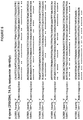

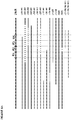


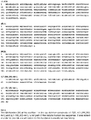
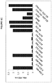


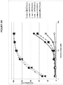

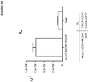
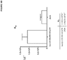
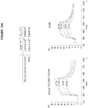
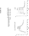
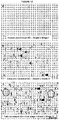


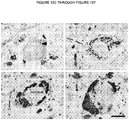


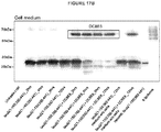

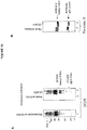
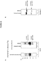

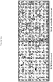


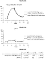

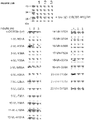
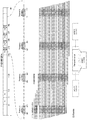


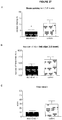
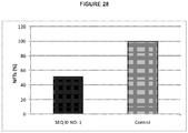
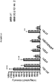

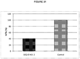
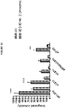
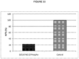

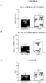

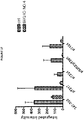
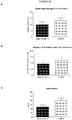
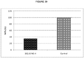
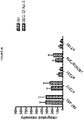


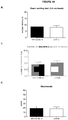
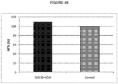

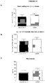
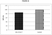
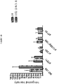
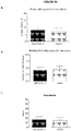
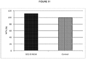


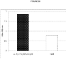
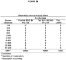
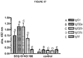
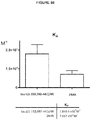
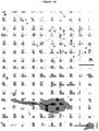

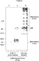

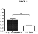
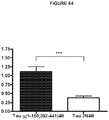

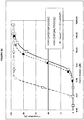
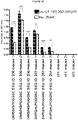
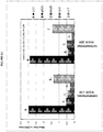

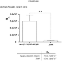
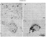
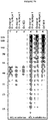
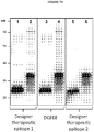
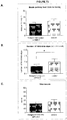
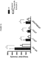
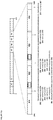
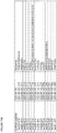

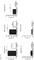
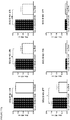
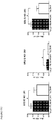
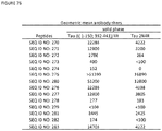
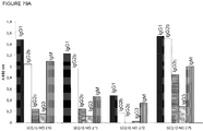
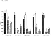
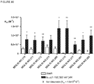
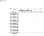
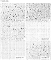


Classifications
-
- A—HUMAN NECESSITIES
- A61—MEDICAL OR VETERINARY SCIENCE; HYGIENE
- A61K—PREPARATIONS FOR MEDICAL, DENTAL OR TOILETRY PURPOSES
- A61K39/00—Medicinal preparations containing antigens or antibodies
- A61K39/395—Antibodies; Immunoglobulins; Immune serum, e.g. antilymphocytic serum
- A61K39/39533—Antibodies; Immunoglobulins; Immune serum, e.g. antilymphocytic serum against materials from animals
-
- A—HUMAN NECESSITIES
- A61—MEDICAL OR VETERINARY SCIENCE; HYGIENE
- A61K—PREPARATIONS FOR MEDICAL, DENTAL OR TOILETRY PURPOSES
- A61K38/00—Medicinal preparations containing peptides
- A61K38/16—Peptides having more than 20 amino acids; Gastrins; Somatostatins; Melanotropins; Derivatives thereof
- A61K38/17—Peptides having more than 20 amino acids; Gastrins; Somatostatins; Melanotropins; Derivatives thereof from animals; from humans
- A61K38/1703—Peptides having more than 20 amino acids; Gastrins; Somatostatins; Melanotropins; Derivatives thereof from animals; from humans from vertebrates
- A61K38/1709—Peptides having more than 20 amino acids; Gastrins; Somatostatins; Melanotropins; Derivatives thereof from animals; from humans from vertebrates from mammals
-
- A—HUMAN NECESSITIES
- A61—MEDICAL OR VETERINARY SCIENCE; HYGIENE
- A61K—PREPARATIONS FOR MEDICAL, DENTAL OR TOILETRY PURPOSES
- A61K39/00—Medicinal preparations containing antigens or antibodies
- A61K39/0005—Vertebrate antigens
- A61K39/0007—Nervous system antigens; Prions
-
- A—HUMAN NECESSITIES
- A61—MEDICAL OR VETERINARY SCIENCE; HYGIENE
- A61K—PREPARATIONS FOR MEDICAL, DENTAL OR TOILETRY PURPOSES
- A61K39/00—Medicinal preparations containing antigens or antibodies
- A61K39/395—Antibodies; Immunoglobulins; Immune serum, e.g. antilymphocytic serum
- A61K39/39533—Antibodies; Immunoglobulins; Immune serum, e.g. antilymphocytic serum against materials from animals
- A61K39/3955—Antibodies; Immunoglobulins; Immune serum, e.g. antilymphocytic serum against materials from animals against proteinaceous materials, e.g. enzymes, hormones, lymphokines
-
- A—HUMAN NECESSITIES
- A61—MEDICAL OR VETERINARY SCIENCE; HYGIENE
- A61K—PREPARATIONS FOR MEDICAL, DENTAL OR TOILETRY PURPOSES
- A61K45/00—Medicinal preparations containing active ingredients not provided for in groups A61K31/00 - A61K41/00
- A61K45/06—Mixtures of active ingredients without chemical characterisation, e.g. antiphlogistics and cardiaca
-
- A—HUMAN NECESSITIES
- A61—MEDICAL OR VETERINARY SCIENCE; HYGIENE
- A61K—PREPARATIONS FOR MEDICAL, DENTAL OR TOILETRY PURPOSES
- A61K47/00—Medicinal preparations characterised by the non-active ingredients used, e.g. carriers or inert additives; Targeting or modifying agents chemically bound to the active ingredient
- A61K47/50—Medicinal preparations characterised by the non-active ingredients used, e.g. carriers or inert additives; Targeting or modifying agents chemically bound to the active ingredient the non-active ingredient being chemically bound to the active ingredient, e.g. polymer-drug conjugates
- A61K47/51—Medicinal preparations characterised by the non-active ingredients used, e.g. carriers or inert additives; Targeting or modifying agents chemically bound to the active ingredient the non-active ingredient being chemically bound to the active ingredient, e.g. polymer-drug conjugates the non-active ingredient being a modifying agent
- A61K47/68—Medicinal preparations characterised by the non-active ingredients used, e.g. carriers or inert additives; Targeting or modifying agents chemically bound to the active ingredient the non-active ingredient being chemically bound to the active ingredient, e.g. polymer-drug conjugates the non-active ingredient being a modifying agent the modifying agent being an antibody, an immunoglobulin or a fragment thereof, e.g. an Fc-fragment
- A61K47/6835—Medicinal preparations characterised by the non-active ingredients used, e.g. carriers or inert additives; Targeting or modifying agents chemically bound to the active ingredient the non-active ingredient being chemically bound to the active ingredient, e.g. polymer-drug conjugates the non-active ingredient being a modifying agent the modifying agent being an antibody, an immunoglobulin or a fragment thereof, e.g. an Fc-fragment the modifying agent being an antibody or an immunoglobulin bearing at least one antigen-binding site
- A61K47/6843—Medicinal preparations characterised by the non-active ingredients used, e.g. carriers or inert additives; Targeting or modifying agents chemically bound to the active ingredient the non-active ingredient being chemically bound to the active ingredient, e.g. polymer-drug conjugates the non-active ingredient being a modifying agent the modifying agent being an antibody, an immunoglobulin or a fragment thereof, e.g. an Fc-fragment the modifying agent being an antibody or an immunoglobulin bearing at least one antigen-binding site the antibody targeting a material from animals or humans
-
- A—HUMAN NECESSITIES
- A61—MEDICAL OR VETERINARY SCIENCE; HYGIENE
- A61K—PREPARATIONS FOR MEDICAL, DENTAL OR TOILETRY PURPOSES
- A61K47/00—Medicinal preparations characterised by the non-active ingredients used, e.g. carriers or inert additives; Targeting or modifying agents chemically bound to the active ingredient
- A61K47/50—Medicinal preparations characterised by the non-active ingredients used, e.g. carriers or inert additives; Targeting or modifying agents chemically bound to the active ingredient the non-active ingredient being chemically bound to the active ingredient, e.g. polymer-drug conjugates
- A61K47/51—Medicinal preparations characterised by the non-active ingredients used, e.g. carriers or inert additives; Targeting or modifying agents chemically bound to the active ingredient the non-active ingredient being chemically bound to the active ingredient, e.g. polymer-drug conjugates the non-active ingredient being a modifying agent
- A61K47/68—Medicinal preparations characterised by the non-active ingredients used, e.g. carriers or inert additives; Targeting or modifying agents chemically bound to the active ingredient the non-active ingredient being chemically bound to the active ingredient, e.g. polymer-drug conjugates the non-active ingredient being a modifying agent the modifying agent being an antibody, an immunoglobulin or a fragment thereof, e.g. an Fc-fragment
- A61K47/6835—Medicinal preparations characterised by the non-active ingredients used, e.g. carriers or inert additives; Targeting or modifying agents chemically bound to the active ingredient the non-active ingredient being chemically bound to the active ingredient, e.g. polymer-drug conjugates the non-active ingredient being a modifying agent the modifying agent being an antibody, an immunoglobulin or a fragment thereof, e.g. an Fc-fragment the modifying agent being an antibody or an immunoglobulin bearing at least one antigen-binding site
- A61K47/6849—Medicinal preparations characterised by the non-active ingredients used, e.g. carriers or inert additives; Targeting or modifying agents chemically bound to the active ingredient the non-active ingredient being chemically bound to the active ingredient, e.g. polymer-drug conjugates the non-active ingredient being a modifying agent the modifying agent being an antibody, an immunoglobulin or a fragment thereof, e.g. an Fc-fragment the modifying agent being an antibody or an immunoglobulin bearing at least one antigen-binding site the antibody targeting a receptor, a cell surface antigen or a cell surface determinant
-
- A—HUMAN NECESSITIES
- A61—MEDICAL OR VETERINARY SCIENCE; HYGIENE
- A61K—PREPARATIONS FOR MEDICAL, DENTAL OR TOILETRY PURPOSES
- A61K49/00—Preparations for testing in vivo
- A61K49/0004—Screening or testing of compounds for diagnosis of disorders, assessment of conditions, e.g. renal clearance, gastric emptying, testing for diabetes, allergy, rheuma, pancreas functions
-
- A—HUMAN NECESSITIES
- A61—MEDICAL OR VETERINARY SCIENCE; HYGIENE
- A61P—SPECIFIC THERAPEUTIC ACTIVITY OF CHEMICAL COMPOUNDS OR MEDICINAL PREPARATIONS
- A61P25/00—Drugs for disorders of the nervous system
-
- A—HUMAN NECESSITIES
- A61—MEDICAL OR VETERINARY SCIENCE; HYGIENE
- A61P—SPECIFIC THERAPEUTIC ACTIVITY OF CHEMICAL COMPOUNDS OR MEDICINAL PREPARATIONS
- A61P25/00—Drugs for disorders of the nervous system
- A61P25/02—Drugs for disorders of the nervous system for peripheral neuropathies
-
- A—HUMAN NECESSITIES
- A61—MEDICAL OR VETERINARY SCIENCE; HYGIENE
- A61P—SPECIFIC THERAPEUTIC ACTIVITY OF CHEMICAL COMPOUNDS OR MEDICINAL PREPARATIONS
- A61P25/00—Drugs for disorders of the nervous system
- A61P25/28—Drugs for disorders of the nervous system for treating neurodegenerative disorders of the central nervous system, e.g. nootropic agents, cognition enhancers, drugs for treating Alzheimer's disease or other forms of dementia
-
- A—HUMAN NECESSITIES
- A61—MEDICAL OR VETERINARY SCIENCE; HYGIENE
- A61P—SPECIFIC THERAPEUTIC ACTIVITY OF CHEMICAL COMPOUNDS OR MEDICINAL PREPARATIONS
- A61P43/00—Drugs for specific purposes, not provided for in groups A61P1/00-A61P41/00
-
- C—CHEMISTRY; METALLURGY
- C07—ORGANIC CHEMISTRY
- C07K—PEPTIDES
- C07K14/00—Peptides having more than 20 amino acids; Gastrins; Somatostatins; Melanotropins; Derivatives thereof
- C07K14/435—Peptides having more than 20 amino acids; Gastrins; Somatostatins; Melanotropins; Derivatives thereof from animals; from humans
- C07K14/46—Peptides having more than 20 amino acids; Gastrins; Somatostatins; Melanotropins; Derivatives thereof from animals; from humans from vertebrates
- C07K14/47—Peptides having more than 20 amino acids; Gastrins; Somatostatins; Melanotropins; Derivatives thereof from animals; from humans from vertebrates from mammals
- C07K14/4701—Peptides having more than 20 amino acids; Gastrins; Somatostatins; Melanotropins; Derivatives thereof from animals; from humans from vertebrates from mammals not used
- C07K14/4711—Alzheimer's disease; Amyloid plaque core protein
-
- C—CHEMISTRY; METALLURGY
- C07—ORGANIC CHEMISTRY
- C07K—PEPTIDES
- C07K16/00—Immunoglobulins [IGs], e.g. monoclonal or polyclonal antibodies
- C07K16/18—Immunoglobulins [IGs], e.g. monoclonal or polyclonal antibodies against material from animals or humans
-
- G—PHYSICS
- G01—MEASURING; TESTING
- G01N—INVESTIGATING OR ANALYSING MATERIALS BY DETERMINING THEIR CHEMICAL OR PHYSICAL PROPERTIES
- G01N33/00—Investigating or analysing materials by specific methods not covered by groups G01N1/00 - G01N31/00
- G01N33/48—Biological material, e.g. blood, urine; Haemocytometers
- G01N33/50—Chemical analysis of biological material, e.g. blood, urine; Testing involving biospecific ligand binding methods; Immunological testing
- G01N33/53—Immunoassay; Biospecific binding assay; Materials therefor
-
- G—PHYSICS
- G01—MEASURING; TESTING
- G01N—INVESTIGATING OR ANALYSING MATERIALS BY DETERMINING THEIR CHEMICAL OR PHYSICAL PROPERTIES
- G01N33/00—Investigating or analysing materials by specific methods not covered by groups G01N1/00 - G01N31/00
- G01N33/48—Biological material, e.g. blood, urine; Haemocytometers
- G01N33/50—Chemical analysis of biological material, e.g. blood, urine; Testing involving biospecific ligand binding methods; Immunological testing
- G01N33/68—Chemical analysis of biological material, e.g. blood, urine; Testing involving biospecific ligand binding methods; Immunological testing involving proteins, peptides or amino acids
- G01N33/6893—Chemical analysis of biological material, e.g. blood, urine; Testing involving biospecific ligand binding methods; Immunological testing involving proteins, peptides or amino acids related to diseases not provided for elsewhere
- G01N33/6896—Neurological disorders, e.g. Alzheimer's disease
-
- A—HUMAN NECESSITIES
- A61—MEDICAL OR VETERINARY SCIENCE; HYGIENE
- A61K—PREPARATIONS FOR MEDICAL, DENTAL OR TOILETRY PURPOSES
- A61K39/00—Medicinal preparations containing antigens or antibodies
- A61K2039/505—Medicinal preparations containing antigens or antibodies comprising antibodies
-
- A—HUMAN NECESSITIES
- A61—MEDICAL OR VETERINARY SCIENCE; HYGIENE
- A61K—PREPARATIONS FOR MEDICAL, DENTAL OR TOILETRY PURPOSES
- A61K39/00—Medicinal preparations containing antigens or antibodies
- A61K2039/555—Medicinal preparations containing antigens or antibodies characterised by a specific combination antigen/adjuvant
- A61K2039/55505—Inorganic adjuvants
-
- A—HUMAN NECESSITIES
- A61—MEDICAL OR VETERINARY SCIENCE; HYGIENE
- A61K—PREPARATIONS FOR MEDICAL, DENTAL OR TOILETRY PURPOSES
- A61K39/00—Medicinal preparations containing antigens or antibodies
- A61K2039/555—Medicinal preparations containing antigens or antibodies characterised by a specific combination antigen/adjuvant
- A61K2039/55511—Organic adjuvants
- A61K2039/55566—Emulsions, e.g. Freund's adjuvant, MF59
-
- A—HUMAN NECESSITIES
- A61—MEDICAL OR VETERINARY SCIENCE; HYGIENE
- A61K—PREPARATIONS FOR MEDICAL, DENTAL OR TOILETRY PURPOSES
- A61K39/00—Medicinal preparations containing antigens or antibodies
- A61K2039/60—Medicinal preparations containing antigens or antibodies characteristics by the carrier linked to the antigen
- A61K2039/6031—Proteins
- A61K2039/6081—Albumin; Keyhole limpet haemocyanin [KLH]
-
- C—CHEMISTRY; METALLURGY
- C07—ORGANIC CHEMISTRY
- C07K—PEPTIDES
- C07K2317/00—Immunoglobulins specific features
- C07K2317/20—Immunoglobulins specific features characterized by taxonomic origin
- C07K2317/24—Immunoglobulins specific features characterized by taxonomic origin containing regions, domains or residues from different species, e.g. chimeric, humanized or veneered
-
- C—CHEMISTRY; METALLURGY
- C07—ORGANIC CHEMISTRY
- C07K—PEPTIDES
- C07K2317/00—Immunoglobulins specific features
- C07K2317/30—Immunoglobulins specific features characterized by aspects of specificity or valency
- C07K2317/34—Identification of a linear epitope shorter than 20 amino acid residues or of a conformational epitope defined by amino acid residues
-
- C—CHEMISTRY; METALLURGY
- C07—ORGANIC CHEMISTRY
- C07K—PEPTIDES
- C07K2317/00—Immunoglobulins specific features
- C07K2317/50—Immunoglobulins specific features characterized by immunoglobulin fragments
- C07K2317/54—F(ab')2
-
- C—CHEMISTRY; METALLURGY
- C07—ORGANIC CHEMISTRY
- C07K—PEPTIDES
- C07K2317/00—Immunoglobulins specific features
- C07K2317/50—Immunoglobulins specific features characterized by immunoglobulin fragments
- C07K2317/55—Fab or Fab'
-
- C—CHEMISTRY; METALLURGY
- C07—ORGANIC CHEMISTRY
- C07K—PEPTIDES
- C07K2317/00—Immunoglobulins specific features
- C07K2317/50—Immunoglobulins specific features characterized by immunoglobulin fragments
- C07K2317/56—Immunoglobulins specific features characterized by immunoglobulin fragments variable (Fv) region, i.e. VH and/or VL
- C07K2317/565—Complementarity determining region [CDR]
-
- C—CHEMISTRY; METALLURGY
- C07—ORGANIC CHEMISTRY
- C07K—PEPTIDES
- C07K2317/00—Immunoglobulins specific features
- C07K2317/60—Immunoglobulins specific features characterized by non-natural combinations of immunoglobulin fragments
- C07K2317/62—Immunoglobulins specific features characterized by non-natural combinations of immunoglobulin fragments comprising only variable region components
- C07K2317/622—Single chain antibody (scFv)
-
- C—CHEMISTRY; METALLURGY
- C07—ORGANIC CHEMISTRY
- C07K—PEPTIDES
- C07K2317/00—Immunoglobulins specific features
- C07K2317/70—Immunoglobulins specific features characterized by effect upon binding to a cell or to an antigen
-
- G—PHYSICS
- G01—MEASURING; TESTING
- G01N—INVESTIGATING OR ANALYSING MATERIALS BY DETERMINING THEIR CHEMICAL OR PHYSICAL PROPERTIES
- G01N2800/00—Detection or diagnosis of diseases
- G01N2800/28—Neurological disorders
- G01N2800/2814—Dementia; Cognitive disorders
- G01N2800/2821—Alzheimer
-
- G—PHYSICS
- G01—MEASURING; TESTING
- G01N—INVESTIGATING OR ANALYSING MATERIALS BY DETERMINING THEIR CHEMICAL OR PHYSICAL PROPERTIES
- G01N2800/00—Detection or diagnosis of diseases
- G01N2800/52—Predicting or monitoring the response to treatment, e.g. for selection of therapy based on assay results in personalised medicine; Prognosis
Definitions
- Protein-based (e.g., antibodies, peptides) methods and means for interfering with the production and clearance of certain forms of tau that are involved in the promotion and/or development of pathological tau-tau aggregates in Alzheimer's disease are herein disclosed, as well as methods for producing anti-tau antibodies that are useful for diagnosis and treatment of Alzheimer's disease.
- the present disclosure further concerns methods and means for diagnosing Alzheimer's disease, including methods for staging and evaluating treatment progression.
- AD Alzheimer's disease
- the disease leads to deficits in cognitive function and declines in memory, learning, language, and in the ability to perform intentional and purposeful movements.
- AD is also accompanied by concomitant behavioral, emotional, interpersonal, and social deterioration. These cognitive and behavioral deficits render living difficult (Burns et al., 2002).
- Late-stage AD patients are often unable to speak, comprehend language, and handle their own basic personal care, eventually requiring full-time care and supervision, and are often dependent on family members and nursing homes.
- AD is the leading cause of senile dementia, and is predicted to increase in prevalence as the proportion of elderly persons in the population grows.
- AD The total number of persons with AD is predicted to increase at least threefold just between 2000 and 2050, rendering AD a world-wide public health problem (Sloane et al., 2002).
- Clinical management of AD remains largely supportive. That is, patients are given treatments aimed at prevention, control, or relief of complications and side effects from AD, and to improve their comfort and quality of life. There is still an unmet need for treatments that directly target the disease process and have disease-modifying effects.
- AD is histologically characterized by the presence of extraneuronal plaques and intracellular and extracellular neurofibrillary tangles in the brain. Plaques are composed mainly of ⁇ amyloid (A ⁇ ), whereas tangles comprise pathological forms of tau, such as pathological tau conformers and their aggregates. The relationship between plaques and tangles and the disease process remains unclear, although studies suggest a link between amyloid and tau pathogenesis (Hardy et al., 1998; Oddo et al., 2004; Rapoport et al., 2002; Roberson, et al., 2007; Shipton et al., 2011).
- a central role for A ⁇ in AD pathology was initially proposed in a hypothesis called the "A ⁇ cascade," wherein A ⁇ deposition is followed by tau phosphorylation and tangle formation, and then neuronal death (Hardy and Allsop, 1991; Hardy and Selkoe, 2002; for a review see, Walsh and Selkoe, 2004; also see Seabrook et al. 2007). Accordingly, initial therapeutic approaches for AD focused primarily on targeting A ⁇ . However, there is a documented lack of correlation between the extent of brain A ⁇ pathology in AD patients and clinical progression of the disease (Braak and Braak, 1991).
- asymptomatic individuals have shown extensive, often diffuse, amyloid deposition at autopsy (Braak and Braak, 1991), and at least in early-stage AD, neuronal loss and amyloid deposition occur in different regions of the brain (Carter and Lippa, 2001). Therefore targeting A ⁇ alone cannot suffice to alter the disease process in any or all patients. Nevertheless, the most advanced disease-targeting therapies undergoing clinical trials in AD patients remain those aimed at the production and clearance of A ⁇ . These therapies include passive immunotherapies, e.g., BAPINEUZUMAB, SOLANEUZUMAB, and PONEZUMAB, as well as the small molecule gamma-secretase inhibitor SEMAGACESTAT (for review see Citron et al., 2010).
- passive immunotherapies e.g., BAPINEUZUMAB, SOLANEUZUMAB, and PONEZUMAB
- SEMAGACESTAT small molecule gamma-secretase inhibitor SEMAGACESTAT
- a recognized role for tau in AD pathology has been demonstrated in numerous studies. For example, Braak showed that the closest correlate for AD neurodegeneration was the presence of tau tangles, and not of amyloid plaques (Braak and Braak, 1991). In another study, A ⁇ neurotoxicity in cultured neurons appeared to depend on tau (Rapoport et al., 2002). Recently, reducing endogenous tau prevented behavioral deficits in transgenic mice that expressed the human amyloid precursor protein, without altering their high A ⁇ levels (Roberson et al., 2007). Tau reduction also protected both transgenic and nontransgenic mice against excitotoxicity. Id.. Santacruz et al. demonstrated that a reduction in the amount of tau restored memory function in a model of tauopathy (Santacruz et al., 2005). Thus, therapies aimed at reducing tau can represent an effective strategy for treating AD and other tau-related disease conditions.
- Tau belongs to a family of intrinsically disordered proteins, characterized by the absence of a rigid three-dimensional structure in their physiological environment (Zilka et al., 2008)
- tau truncation and hyperphosphorylation can cause pathological transformations from an intrinsically disordered state to multiple soluble and insoluble misdisordered structures, including paired helical filaments (PHFs) and other aggregates (Wischik et al., 1988a; Wischik et al., 1988b; Novak et al., 1993; Skrabana et al., 2006; Zilka et al., 2008; Kovacech et al., 2010).
- PHFs paired helical filaments
- Tau's physiological function is in mediating the assembly of tubulin monomers into microtubules that constitute the neuronal microtubules network (Buee et al., 2000). Tau binds to microtubules through repetitive regions located in the C-terminal portion of the protein. Id. These repeat domains (R1-R4), are not identical to each other, but comprise highly conserved 31-32 amino acids (Taniguchi et al., 2005b).
- Fig. 1 shows the six human isoforms (2N4R, 1 N4R, 2N3R, 0N4R, 1 N3R, and 0N3R). It has been proposed that the most potent part of tau to induce microtubule polymerization is the 274-KVQIINKK-281 region (SEQ ID NO: 113), overlapping R1-R2. Id.
- tau's pathological and physiological functions appear to be influenced by the specific structural conformation, and the intrinsically disordered structure, adopted by the full length protein isoforms and their fragments.
- Kontsekova et al. described a conformational region (encompassing residues 297-IKHVPGGGSVQIVYKPVDLSKVTSKCGSL-325 (SEQ ID NO: 114)) within certain truncated tau molecules which had a significant relationship to the function of those truncated tau molecules on microtubule assembly ( WO 2004/007547 ).
- tau repeats are believed to participate in the formation of pathological tau aggregates and other structures.
- tau-targeted therapeutic and diagnostic approaches that are capable of discriminating between physiological and pathological repeat-mediated activities.
- PHFs pathological paired helical filaments
- the pronase resistant core of pathological paired helical filaments consists of the microtubule binding regions of 3- and 4-repeat tau isoforms (Jakes et al., 1991; Wischik, et al. 1988a; Wischik, et al. 1988b).
- tau is considered an intracellular cytoplasmic protein
- intracellular tau can be released into the extracellular space and contribute to neurodegeneration (Gómez-Ramos et al., 2006).
- neuronal loss has been linked to the topographic distribution of neurofibrillary tangles (made up of tau protein) in AD brains (West et al., 1994; Gomez-Isla et al., 1996, 1997).
- the levels of total tau and phosphorylated tau are increased in the cerebrospinal fluid (CSF) of patients with AD (Hampel et al., 2010), and extracellular tau has been described as "ghost tangles" in the brain (Frost and Diamond, 2009), indicating that intracellular tau is released into extracellular space.
- extracellular tau aggregates can enter cells and stimulate fibrillization of intracellular tau, further seeding tau monomer for production of pathological tau aggregates (Frost et al., 2009).
- extracellular, insoluble tau could act as a transmissible agent to spread tau pathology throughout the brain in a prion-like fashion (Frost et al., 2009; Frost and Diamond, 2009).
- tau appears to play a pathological role in the clinical manifestation of AD, the development of drugs that work against tau has been slow, in part due to tau's importance in physiologic microtubule dynamics and to its complex biology (Dickey and Petrucelli, 2006).
- an increased understanding of the molecular mechanisms underlying the pathological transformations of tau has opened up the possibility of specifically targeting pathological modifications of tau for therapeutic purposes.
- a number of therapeutic approaches that directly or indirectly target the tau cascade have emerged (for review articles, see, e.g.
- AD treatments face the hurdle of overcoming the blood-brain-barrier (BBB)
- BBB blood-brain-barrier
- a growing number of pre-clinical and clinical studies report that antibody-mediated therapies can clear AD aggregates from the brain, and propose multiple mechanisms of action, such as (i) antibody uptake into the brain via an altered BBB permeability in AD, or BBB leakage; (ii) antibodies working as "peripheral sinks" for soluble plaque-forming amyloid species; (iii) entrance of antibody-secreting cells from the periphery into the brain, delivering antibodies locally; and (iv) transport of IgG within and across cells.
- therapeutic antibodies targeting disease forms of tau represent a prospective approach for treatment and/or diagnosis of AD and other tauopathies ( WO 2004/007547 , US2008/0050383 ).
- Treated animals produced anti-tau antibodies, which were detected in the brain and colocalized with antibodies that recognized pathological tau (Asuni et al., 2007).
- AD Alzheimer's disease
- an isolated antibody is herein provided, wherein the antibody binds to one or more tau epitopes and is capable of two or more of the following:
- this isolated antibody is such that each of the one or more epitopes is independently selected from epitopes within:
- the isolated antibody having the properties described in the embodiments of the previous paragraphs is capable of binding to one or more forms of pathological tau chosen from misordered tau, misdisordered tau, sarkosyl-insoluble tau, neurofibrillary tangles, neuropil threads, and neuritic plaques in a brain biopsy of a human Alzheimer's disease patient, in a brain sample from an animal model of Alzheimer's disease, or in both.
- the isolated antibody is such that at least one of the epitopes that it recognizes is a conformational epitope.
- an isolated antibody is herein provided, wherein the antibody binds to one or more tau epitopes and is capable of two or more of the following:
- this isolated antibody is such that each of the one or more epitopes is independently selected from epitopes within:
- this isolated antibody is such that each of the one or more epitopes it binds to is independently selected from epitopes within:
- An isolated antibody is herein provided, wherein said antibody binds one or more epitopes on tau in a conformationally-specific manner wherein:
- An isolated antibody is Iso disclosed herein, said antibody binding one or more epitopes on tau in a conformationally-specific manner wherein:
- this antibody is DC8E8, wherein DC8E8 is an antibody produced by the hybridoma deposited under American Type Culture Collection Patent Deposit no. PTA-11994.
- the isolated antibody binds to one or more of the same epitopes on tau as those bound by DC8E8. In an embodiment, the isolated antibody competes with monoclonal antibody DC8E8 for binding to tau.
- the invention also provides an isolated antibody comprising in its epitope binding domain one or more complementarity determining region (CDR) sequences chosen from:
- any of the antibodies described in the previous embodiments can consist of or comprise a Fab, Fab', F(ab') 2 , Fabc, Fv fragment, any other antigen-binding fragment; or an antigen-binding antibody portion thereof; having one or more of the following immunological binding characteristics:
- any of the antibodies described in the previous embodiments can consist of or comprise a Fab, Fab', F(ab') 2 , Fabc, Fv fragment, any other antigen-binding fragment; or an antigen-binding antibody portion thereof; having one or more of the following immunological binding characteristics:
- Isolated antibodies that competitively bind to tau against any of the isolated antibodies described in the previous embodiments are also disclosed.
- the isolated antibody competitively binds to tau when tested against isolated DC8E8 for binding to tau.
- the antibody comprises a light chain comprising SEQ ID NO.: 141. In some embodiments, the antibody comprises a light chain comprising SEQ ID NO.:138. In some embodiments, the antibody comprises a light chain comprising SEQ ID NO.: 141 and a light chain comprising SEQ ID NO.:138.
- the invention provides that the antibodies provided by the invention can be chosen from:
- any of the isolated antibodies provided herein can be raised in a mammal.
- the isolated antibody is produced by a recombinant animal or by a recombinant host cell.
- any of the isolated anti-tau antibodies provided herein can be such that they are detectably labeled with one or more labeling agents.
- at least one labeling agent is chosen from an enzyme, a radioisotope, a fluorophore, a nuclear magnetic resonance marker, and a heavy metal.
- the antibody comprises at least one drug (combination agent) attached to the antibody molecule.
- isolated nucleic acids encoding at least one CDR, or at least the binding domain or variable region of an immunoglobulin chain of any of the anti-tau antibodies described in the previous embodiments.
- isolated vectors comprising any of those nucleic acids.
- an isolated host cell comprising one or more of these isolated nucleic acids and vectors.
- the isolated cell line expressing any of the anti-tau antibodies described in the previous embodiments.
- the isolated cell line is a hybridoma.
- the isolated cell line is the hybridoma from which monoclonal antibody DC8E8 is produced, and which cell line has been deposited with the American Type Culture Collection, Manassas, VA, USA, on July 13, 2011, with the ATCC Patent Deposit Designation PTA-11994.
- the antibodies are comprised in a pharmaceutical composition, further comprising pharmaceutically acceptable carrier and/or diluent.
- the pharmaceutical composition comprises a combination of antibodies and a pharmaceutically acceptable carrier and/or diluent, wherein the combination comprises at least two different antibodies, and wherein each of the antibodies is independently selected from the antibodies described in the previous embodiments.
- at least one of the antibodies is DC8E8, or a human version of DC8E8, or a humanized version of DC8E8.
- the antibodies are comprised in a composition, further comprising a diluent and/or a carrier.
- the composition can be a pharmaceutical composition, a diagnostic composition, or any other composition.
- the composition can further comprise at least one compound or agent selected from a detectable label, keyhole limpet hemocyanin, tetanus toxoid or a toxoid derived from other pathogenic bacteria, serum albumins, bovine serum albumin, an immunoglobulin molecule or fragment thereof, thyroglobulin, ovoglobulin, a universal T-cell epitope, a cytokine, a chemokine, interleukin 1-alpha (IL-1 ⁇ ), IL-1 ⁇ , IL-2, IL-10, interferon -gamma (IFN- ⁇ ), granulocyte macrophage colony-stimulating factor (GM-CSF), macrophage inflammatory protein 1 alpha (MIP1 ⁇ ), MIP1 ⁇ , and RANTES
- an article of manufacture e.g., a kit
- a container comprising a solution of a lyophilized form any one or more of the anti-tau antibodies provided herein.
- the container is a component of a device or system for delivery of the antibody to a subject.
- a medical device comprising an anti-tau antibody as provided herein (see above), wherein the device is suitable for contacting or administering the antibody by at least one mode selected from parenteral, subcutaneous, intramuscular, intravenous, intrarticular, intrabronchial, intraabdominal, intracapsular, intracartilaginous, intracavitary, intracelial, intracerebellar, intracerebroventricular, intrathecal, intracolic, intracervical, intragastric, intrahepatic, intramyocardial, intraosteal, intrapelvic, intrapericardiac, intraperitoneal, intrapleural, intraprostatic, intrapulmonary, intrarectal, intrarenal, intraretinal, intraspinal, intrasynovial, intrathoracic, intrauterine, intravesical, intralesional, bolus, vaginal, rectal, buccal, sublingual, intranasal, and transdermal.
- parenteral subcutaneous, intramuscular, intravenous, intrarticular, intra
- a method of treating or preventing the progression of Alzheimer's disease or a related tauopathy in a subject comprising administering to said subject an effective amount of at least one of the anti-tau antibodies provided herein.
- the method is capable of reducing motor impairment, improving motor function, reducing cognitive impairment, improving cognitive function, or of a combination thereof.
- a method of ameliorating at least one of the symptoms associated with Alzheimer's disease or a related tauopathy in a subject comprising administering to said subject an effective amount of at least one of the anti-tau antibodies provided herein.
- a method of diagnosing or screening a subject for the presence of Alzheimer's disease or a related tauopathy in a subject, or for determining a subject's risk for developing Alzheimer's disease or a related tauopathy comprising:
- a method of monitoring a subject for the presence, progression, regression, or stabilization of Alzheimer's disease or a related tauopathy in a subject, or for determining the stage of Alzheimer's disease or a related tauopathy in a subject comprising:
- the antibody is administered intravenously, intramuscularly, subcutaneously, intraperitoneally, intranasally, intracerebroventricularly, intrathecally, or as an aerosol.
- the effective amount of each antibody is at least 1 mg/kg body weight of the subject, per dose. In some embodiments, the effective amount of each antibody is at least 10 mg/kg body weight of the subject, per dose. In some embodiments, at least one of the antibodies is administered in multiple dosages over a period of at least six months. In some embodiments, the antibody is administered peripherally to a human subject to exert its beneficial effects.
- the antibody when administered peripherally to a human subject, binds to soluble tau, sarkosyl-insoluble tau, or to both. In some embodiments, the antibody, when administered peripherally to a human subject, binds to tau, wherein tau is in one or more pathological forms chosen from misordered tau, misdisordered tau, sarkosyl-insoluble tau, neurofibrillary tangles, neuropil threads, and neuritic plaques in a brain biopsy of a human Alzheimer's disease patient, in a brain sample from an animal model of Alzheimer's disease. In some embodiments, the antibody, when administered peripherally to a human subject, exerts one or more effector-function mediated beneficial effects on the subject.
- the antibody is delivered to the periphery by injection/implantation of an antibody-expressing cell into the subject's brain.
- the antibody-expressing cell is an hybridoma cell.
- the hybridoma cell is a hybridoma expressing DC8E8.
- the disclosure provides an isolated peptide, wherein:
- the therapeutic epitope comprises a therapeutic epitope selected from those within:
- the disclosure provides an isolated peptide, wherein:
- the therapeutic epitope comprises a therapeutic epitope selected from those within:
- the therapeutic epitope is selected from:
- the isolated peptide is a sequence selected from SEQ ID NOs: 1-4, SEQ ID NOs: 9-101, and SEQ ID NOs: 108-112, NIKAVPGGGS (SEQ ID NO: 200), NIKHVPGGGS (SEQ ID NO: 201), IKHVPGGGS (SEQ ID NO: 202), KHVPGGGSV (SEQ ID NO: 203), HVPGGGSVQ (SEQ ID NO: 204), VPGGGSVQ (SEQ ID NO: 205), GWSIHSPGGGSC (SEQ ID NO: 250), SVFQHLPGGGSC (SEQ ID NO: 251), ANIKHVPGGGS (SEQ ID NO: 144), DAIKHVPGGGS (SEQ ID NO: 146), DNAKHVPGGGS (SEQ ID NO: 149), DNIAHVPGGGS (SEQ ID NO: 151), DNIKAVPGGGS (SEQ ID NO: 159), DNIKHAPGGGS (SEQ ID NO: 111),
- the isolated peptide is a sequence selected from SEQ ID NO: 270 (TENLKHQPGGGK); SEQ ID NO: 271 (KHQPGGG), SEQ ID NO: 272 (HQPGGG); SEQ ID NO: 275 (ENLKHQPGGGKVQIINKKLDLSNVQSKCGSKDNIKHVPGGGS), SEQ ID NO: 276 (KHVPGGG), SEQ ID NO: 277 (HVPGGG), SEQ ID NO: 280 (DNIKHVPGGGSVQIVYKPV), SEQ ID NO: 281 (HHKPGGG), SEQ ID NO: 282 (HKPGGG), and SEQ ID NO: 283 (THVPGGG).
- the isolated peptide is a sequence selected from SEQ ID NO: 270 (TENLKHQPGGGK); SEQ ID NO: 271 (KHQPGGG), SEQ ID NO: 272 (HQPGGG); SEQ ID NO: 275 (ENLKHQPGGGKVQIINKKLDLSNVQSKCGSKDNIKHVPGGGS), SEQ ID NO: 276 (KHVPGGG), SEQ ID NO: 277 (HVPGGG), SEQ ID NO: 280 (DNIKHVPGGGSVQIVYKPV), SEQ ID NO: 281 (HHKPGGG), SEQ ID NO: 282 (HKPGGG), and SEQ ID NO: 283 (THVPGGG); and the therapeutic epitope is selected from:
- the isolated peptide is a sequence selected from SEQ ID NO: 272 (HQPGGG) and SEQ ID NO: 277 (HVPGGG).
- the isolated peptide is active in at least one assay, selected from assays that measure the peptide's:
- the disclosure also relates to compounds comprising any of the isolated peptides provided herein and a moiety.
- the moiety is N-terminal, C-terminal, or linked to an internal amino acid of the peptide, and wherein the moiety is selected from one or more of a cysteine residue, phospho group, keyhole limpet hemocyanin, tetanus toxoid or a toxoid derived from other pathogenic bacteria, serum albumins, bovine serum albumin, an immunoglobulin molecule or fragment thereof, thyroglobulin, ovoglobulin, a universal T-cell epitope, a cytokine, a chemokine, interleukin 1-alpha (IL-1 ⁇ ), IL-1 ⁇ , IL-2, IL-10, interferon - gamma (IFN- ⁇ ), granulocyte macrophage colony-stimulating factor (GM-CSF), macrophage inflammatory protein 1 alpha (MIP1 ⁇ ), M
- compositions comprising one or more of the isolated peptides and/or compounds provided by the disclosure and a pharmaceutically acceptable carrier, and/or a diluent, and/or an adjuvant.
- the pharmaceutical composition is adapted to provide a dosage of the peptide or of the compound between 1 ng and 10 mg. In certain embodiments, the pharmaceutical composition is adapted to provide a dosage of the peptide or of the compound greater than 10 micrograms.
- the disclosure also relates to an article of manufacture (e.g., a kit) for pharmaceutical or diagnostic use, comprising packaging material and a container comprising a solution or a lyophilized form of a peptide and/or compound provided herein.
- the container is a component of a device or system for delivery of the peptide or the compound to a subject.
- medical devices comprising a peptide, a compound, and/or a peptide/compound composition as provided above, wherein the device is suitable for contacting or administering the antibody by at least one mode selected from parenteral, subcutaneous, intramuscular, intravenous, intrarticular, intrabronchial, intraabdominal, intracapsular, intracartilaginous, intracavitary, intracelial, intracerebellar, intracerebroventricular, intrathecal, intracolic, intracervical, intragastric, intrahepatic, intramyocardial, intraosteal, intrapelvic, intrapericardiac, intraperitoneal, intrapleural, intraprostatic, intrapulmonary, intrarectal, intrarenal, intraretinal, intraspinal, intrasynovial, intrathoracic, intrauterine, intravesical, intralesional, bolus, vaginal, rectal, buccal, sublingual, intranasal, and transdermal.
- parenteral subcutaneous, intramuscular
- the disclosure provides a method of treating or preventing the progression of Alzheimer's disease or related tauopathies in a subject, the method comprising administering to said subject an effective amount of at least one peptide and/or at least one compound as provided herein.
- the method is capable of reducing motor impairment, improving motor function, reducing cognitive impairment, improving cognitive function, or a combination thereof.
- the disclosure provides a method of ameliorating at least one of the symptoms associated with Alzheimer's disease or related tauopathies in a subject, the method comprising administering to said subject an effective amount of at least one peptide and/or at least one compound as provided herein.
- the method comprises administering to a human patient a peptide and/or a compound as provided babove, and/or an adjuvant that augments the immune response, which method effects an immune response comprising antibodies against pathological tau, thereby treating, preventing the progression, or ameliorating at least one of the symptoms associated with AD in the human patient.
- a method of producing an antibody that is able to compete with DC8E8 for binding to tau comprising immunizing a subject with at least one peptide and/or with at least one compound as provided herein.
- at least one peptide is a peptide is chosen from any one of SEQ ID NOs: 1-4, SEQ ID NOs: 9-101, and SEQ ID NOs: 108-112, NIKHVPGGGS (SEQ ID NO: 201), IKHVPGGGS (SEQ ID NO: 202), KHVPGGGSV (SEQ ID NO: 203), HVPGGGSVQ (SEQ ID NO: 204), VPGGGSVQ (SEQ ID NO: 205), GWSIHSPGGGSC (SEQ ID NO: 250), SVFQHLPGGGSC (SEQ ID NO: 251), ANIKHVPGGGS (SEQ ID NO: 144), DAIKHVPGGGS (SEQ ID NO: 146), DNAKHVPGGGS (SEQ ID NO:
- the peptide is chosen from SEQ ID NOs: 1-4. In another embodiment, the peptide is SEQ ID NO. 108. In one embodiment, the peptide is GWSIHSPGGGSC (SEQ ID NO: 250). In certain embodiments, the peptide is SVFQHLPGGGSC (SEQ ID NO: 251).
- the peptide is selected from SEQ ID NO: 270 (TENLKHQPGGGK); SEQ ID NO: 271 (KHQPGGG), SEQ ID NO: 272 (HQPGGG); SEQ ID NO: 275 (ENLKHQPGGGKVQIINKKLDLSNVQSKCGSKDNIKHVPGGGS), SEQ ID NO: 276 (KHVPGGG), SEQ ID NO: 277 (HVPGGG), SEQ ID NO: 280 (DNIKHVPGGGSVQIVYKPV), SEQ ID NO: 281 (HHKPGGG), SEQ ID NO: 282 (HKPGGG), and SEQ ID NO: 283 (THVPGGG).
- the peptide is selected from SEQ ID NO: 272 (HQPGGG) and SEQ ID NO: 277 (HVPGGG).
- a method of diagnosing or screening a subject for the presence of Alzheimer's disease or related tauopathies in a subject, or for determining a subject's risk for developing Alzheimer's disease or related tauopathies comprising:
- a method of monitoring a subject for the presence, progression, regression, or stabilization of Alzheimer's disease or related tauopathies, or for determining the stage of Alzheimer's disease or related tauopathies in a subject comprising:
- the antibody, peptide, and/or compound is administered intravenously, intramuscularly, subcutaneously, intraperitoneally, intranasally, intracerebroventricularly, intrathecally, or as an aerosol.
- the effective amount of each peptide and/or compound is at least 1 ⁇ g per dose, at least 10 ⁇ g per dose, at least 100 ⁇ g per dose.
- each peptide and/or compound is at least 10 ⁇ g per dose in the presence of an adjuvant, and at least 100 ⁇ g per dose in the absence of an adjuvant. In some embodiments, at least one peptide or compound is administered in multiple dosages over a period of at least six months.
- a method of treating or preventing the progression of Alzheimer's disease or related tauopathies in a subject comprising administering to said subject an effective amount of at least one antibody of the invention, and/or at least one peptide, and/or at least one compound as provided herein, in combination with at least one combination agent chosen from acetylcholinesterase inhibitors, N-Methyl-D-aspartate (NMDA) receptor antagonists, transition metal chelators, growth factors, hormones, non-steroidal anti-inflammatory drugs (NSAID), antioxidants, lipid lowering agents, selective phosphodiesterase inhibitors, inhibitors of tau aggregation, inhibitors of protein kinases, inhibitors of heat shock proteins, anti-amyloid passive and active immunization, anti-amyloid aggregation inhibitors, and secretase inhibitors.
- the method is capable of reducing motor impairment, improving motor function, reducing cognitive impairment, improving cognitive function, or a combination thereof.
- a method of ameliorating at least one of the symptoms associated with Alzheimer's disease or related tauopathies in a subject comprising administering to said subject an effective amount of at least one antibody of the invention, at least one peptide, and/or at least one compound as provided herein, in combination with at least one combination agent chosen from acetylcholinesterase inhibitors, NMDA receptor antagonists, transition metal chelators, growth factors, hormones, non-steroidal anti-inflammatory drugs (NSAID), antioxidants, lipid lowering agents, selective phosphodiesterase inhibitors, inhibitors of tau aggregation, inhibitors of protein kinases, inhibitors of heat shock proteins, anti-amyloid-passive and -active immunization reagents, anti-amyloid aggregation inhibitors, and secretase inhibitors.
- at least one combination agent chosen from acetylcholinesterase inhibitors, NMDA receptor antagonists, transition metal chelators, growth factors, hormones, non-steroidal anti-inflammatory drugs (NSAID),
- the method comprises administering to a human patient an effective amount of at least one antibody of the invention, at least one peptide, and/or at least one compound as provided herein, and/or an adjuvant that augments the immune response; in combination with at least one combination agent chosen from acetylcholinesterase inhibitors, NMDA receptor antagonists, transition metal chelators, growth factors, hormones, non-steroidal anti-inflammatory drugs (NSAID), antioxidants, lipid lowering agents, selective phosphodiesterase inhibitors, inhibitors of tau aggregation, inhibitors of protein kinases, inhibitors of heat shock proteins, anti-amyloid passive and -active immunization, anti-amyloid aggregation inhibitors, and secretase inhibitors; wherein the method effects an immune response comprising antibodies against pathological tau, thereby treating, preventing the progression, or ameliorating at least one
- at least one combination agent chosen from acetylcholinesterase inhibitors, NMDA receptor antagonists, transition metal chelators, growth factors,
- the combination agent is administered prior to, simultaneously with, or after the administration of an antibody of the invention, a peptide, and/or a compound as provided herein.
- the disclosure also provides a pharmaceutical composition
- a pharmaceutical composition comprising a pharmaceutically acceptable carrier and/or diluent;
- antibody refers to an immunoglobulin, whether genetically engineered, natural, or wholly or partially synthetically or recombinantly produced. All derivatives, portions, and fragments thereof that maintain antigen-binding properties and at least one of the tau-related characteristic properties disclosed herein are also included in the term.
- the term also encompasses any protein having a binding domain which is homologous or largely homologous to an immunoglobulin binding domain. These proteins can be derived from natural sources, or partly or wholly synthetically or recombinantly produced.
- An antibody can be monoclonal or polyclonal.
- the antibody can be a member of any immunoglobulin class, including any of the human classes: IgG, IgM, IgA, IgD, and IgE. Derivatives of the IgG class are preferred in some embodiments.
- isolated antibody and "isolated peptide” refer to a protein or peptide produced from cDNA-, recombinant RNA-, or any other synthetic-origin, or some combination thereof; as well as to proteins and peptides that, by virtue of their origin, or source of derivation, either (1) are not associated with proteins found in nature, (2) are free of other proteins from the same source, e.g. free of murine proteins, (3) are expressed by a cell from a different species, or (4) do not occur in nature.
- the antibodies disclosed herein include, in addition, such antibodies having "conservative sequence modifications," nucleotide and amino acid sequence modifications which do not affect or alter the above-mentioned characteristics of the antibody. Modifications can be introduced by standard techniques known in the art, such as site-directed mutagenesis and PCR-mediated mutagenesis. Conservative amino acid substitutions include ones in which the amino acid residue is replaced with an amino acid residue having a similar side chain. Families of amino acid residues having similar side chains have been defined in the art. These families include amino acids with basic side chains (e.g., lysine, arginine, histidine), acidic side chains (e.g., aspartic acid, glutamic acid), uncharged polar side chains (e.g.
- glycine asparagine, glutamine, serine, threonine, tyrosine, cysteine, tryptophan
- nonpolar side chains e.g., alanine, valine, leucine, isoleucine, proline, phenylalanine, methionine
- beta-branched side chains e.g., threonine, valine, isoleucine
- aromatic side chains e.g., tyrosine, phenylalanine, tryptophan, histidine.
- a predicted nonessential amino acid residue in a anti-tau antibody can be for example replaced with another amino acid residue from the same side chain family.
- Antibody fragments and “antibody portions” comprise a portion of a full length antibody, generally at least the antigen binding portion/domain or the variable region thereof.
- antibody fragments include diabodies, single-chain antibody molecules, immunotoxins, and multispecific antibodies formed from antibody fragments.
- antibody fragments comprise single chain polypeptides having the characteristics of a VH chain binding pathological tau, namely being able to assemble together with a VL chain or of a VL chain binding to pathological tau, namely being able to assemble together with a VH chain to form a functional antigen binding pocket and thereby providing the property of binding to pathological tau.
- the terms also comprise fragments that per se are not able to provide effector functions (e.g., ADCC/CDC) but provide this function after being combined with the appropriate antibody constant domain(s).
- chimeric antibody refers to a monoclonal antibody comprising a variable region, i.e., binding region, from one source or species and at least a portion of a constant region derived from a different source or species, usually prepared by recombinant DNA techniques. Chimeric antibodies comprising a murine variable region and a human constant region are especially preferred. Such murine/human chimeric antibodies are the product of expressed immunoglobulin genes comprising DNA segments encoding murine immunoglobulin variable regions and DNA segments encoding human immunoglobulin constant regions. Other forms of "chimeric antibodies” encompassed herein are those in which the class or subclass has been modified or changed from that of the original antibody.
- Such “chimeric” antibodies are also referred to as "class-switched antibodies.”
- Methods for producing chimeric antibodies involve conventional recombinant DNA and gene transfection techniques now known in the art. See, e.g., Morrison, S. L., et al., Proc. Natl. Acad Sci. USA 81 (1984) 6851-6855 ; U.S. Pat. Nos. 5,202,238 and 5,204,244 .
- humanized antibody refers to antibodies in which the framework regions (FR) and/or the complementarity determining regions (CDR) have been modified to comprise the CDR of an immunoglobulin of different specificity as compared to that of the parent immunoglobulin.
- a murine CDR is grafted into the framework region of a human antibody to prepare the "humanized antibody.” See, e.g., Riechmann, L., et al., Nature 332 (1988) 323-327 ; and Neuberger, M. S., et al., Nature 314 (1985) 268-270 .
- Particularly preferred CDRs correspond to those representing sequences recognizing the antigens and epitopes described herein as "therapeutic epitopes" on tau.
- human antibody is intended to include antibodies having variable and constant regions derived from human germline immunoglobulin sequences.
- the constant regions of the antibody can be, for example, constant regions of human IgG1 type. Such regions can be allotypic and are described by, e.g., Johnson, G., and Wu, T. T., Nucleic Acids Res. 28 (2000) 214-218 and the databases referenced therein, and are preferentially useful for some embodiments, as long as the properties of induction of ADCC and for example CDC disclosed herein are retained.
- recombinant human antibody is intended to include all human antibodies that are prepared, expressed, created or isolated by recombinant means, such as antibodies isolated from a host cell such as a NSO or CHO cell or from an animal (e.g. a mouse such as a XENOMOUSE, a genetically modified mouse that produces antibodies having amino acid sequences of human antibodies, e.g., human framework (FR) and human constant region amino acid sequences) that is transgenic for human immunoglobulin genes or antibodies expressed using a recombinant expression vector transfected into a host cell.
- recombinant human antibodies have variable and constant regions derived from human germline immunoglobulin sequences in a rearranged form.
- the recombinant human antibodies have been subjected to in vivo somatic hypermutation.
- the amino acid sequences of the VH and VL regions of the recombinant antibodies are sequences that, while derived from and related to human germline VH and VL sequences, can not naturally exist within the human antibody germline repertoire in vivo.
- effector functions includes, but is not limited to, C1 q binding; complement dependent cytotoxicity (CDC); Fc receptor binding; antibody-dependent cell-mediated cytotoxicity (ADCC); phagocytosis; and down regulation of cell surface receptors (e.g. B cell receptor; BCR).
- epitope is used here to refer to binding sites recognized by a binding protein or an antibody.
- Epitopes can be any molecule or grouping thereof, including, but not limited to, amino acids, amino acid side chains, sugars, and lipids, and can have a specific three-dimensional structure or conformation.
- an epitope can comprise any portion of a tau peptide/protein molecule that includes primary, secondary, tertiary, or quaternary structure, as those terms are generally known in the art.
- A"linear epitope” is made up of a continuous sequence of amino acid residues.
- a linear epitope is one that is present on physiological tau (e.g., is present in tau 2N/4R).
- a “conformational epitope” is an epitope to which the antibody or binding protein binds in a conformational-specific manner.
- the binding can depend on the epitope-carrying-protein's secondary, tertiary, or quaternary structure.
- the antibody binds in a structure-specific manner, a tertiary-structure-specific manner, or a quaternary-structure-specific manner.
- a conformational epitope is one that is present in pathological tau (e.g., present in tau ⁇ (1-150;392-441)/4R)).
- therapeutic epitope refers to regions within tau that were identified herein and were found to promote tau-tau aggregation, when in certain conformations (recognized by the DC8E8 antibody).
- Antibodies (and other binding proteins) that bind to one or more of these regions inhibit early and late stages of tau aggregation, including the conversion of tau monomer to dimer, and conversion to higher aggregate forms; i.e , the antibodies inhibit the conversion from physiological tau to pathological tau.
- These regions within tau can be involved in promoting tau fibrillization into paired helical filaments (PHFs), by promoting the formation of beta-sheets within adjacent regions of tau.
- PHFs paired helical filaments
- Therapeutic epitopes are comprised within 267-KHQPGGG-273 (within 1 st repeat domain of tau protein), 298-KHVPGGG-304 (within 2 nd repeat domain of tau protein), 329-HHKPGGG-335 (within 3 rd repeat domain of tau protein), and 361-THVPGGG-367(within 4 th repeat domain of tau protein.
- the therapeutic epitopes are each comprised within 268-HQPGGG-273 (within 1 st repeat domain of tau protein), 299-HVPGGG-304 (within 2 nd repeat domain of tau protein), 330-HKPGGG-335 (within 3 rd repeat domain of tau protein), and 362-HVPGGG-367, respectively.
- the term "displaying a higher affinity for pathological tau than for physiological tau” refers to a higher degree of interaction between the antibody and at least one form of pathological tau than between the antibody and at least one form of physiological tau.
- the interaction can be measured by, e.g., ELISA or surface plasmon resonance (SPR), as described in the EXAMPLES below.
- Specific binding can be characterized by a dissociation constant of about 1 ⁇ 10 -6 M or smaller, for example less than about 100 nM, and most for example less than 10 nM.
- Methods for determining whether two molecules specifically bind are known in the art and include, for example, equilibrium dialysis, surface plasmon resonance, and the like.
- an antibody or antigen-binding fragment thereof is a molecule that binds the antigen or an epitope with such a dissociation constant of at least about 1 ⁇ 10 -6 M or smaller, but does not bind other molecules with such a dissociation constant.
- Preferentially bind refers to binding with higher affinity to pathological tau than to physiological tau, for example, binding with higher affinity to tau ⁇ (1-150;392-441)/4R than to 2N4R.
- a "universal T-cell epitope” is a sequence selected from Influenza Hemagluttinin: HA307-319 (PKYVKQNTLKLAT) (SEQ ID NO: 123); PADRE (AKXVAAWTLKAAA) (SEQ ID NO: 124); Malaria CS: T3 epitope (EKKIAKMEKASSVFNV) (SEQ ID NO: 125); Hepatitis B surface antigen: HBsA919_28 (FFLLTRILTI) (SEQ ID NO: 126); Heat Shock Protein 65: hsp65153_171 (DQSIGDLIAEAMDKVGNEG) (SEQ ID NO: 127); bacille Calmette-Guerin (QVHFQPLPPAVVKL) (SEQ ID NO: 128); Tetanus toxoid: T1830-844 (QYIKANSKFIGITEL) (SEQ ID NO: 129); Tetanus toxoid: T19
- Intrinsically disordered tau refers to the normal/physiological form of tau protein, which lacks any defined 3D structure. It exists in the healthy brain (Kovacech et al., 2010).
- Misdisordered tau refers to the forms of tau that differ conformationally from normal/physiological intrinsically disordered tau, and does not have a firm/defined 3D-structure. Misdisordered truncated tau is able to induce neurofibrillary degeneration in vivo. It does not exist in a healthy brain (Kovacech et al., 2010). “Misordered tau” refers to a structured pathological form of tau assembled into polymers of PHFs, which form NFTs. Misordered tau does not exist in a healthy brain (Kovacech et al., 2010).
- SHR24 refers to transgenic rat line that expresses tau type IIB (151-391/R3).
- the transgenic rats developed progressive age-dependent neurofibrillary degeneration in the cortical brain areas.
- Neurofibrillary tangles (NFTs) in SHR24 rats satisfy several key histological criteria used to identify neurofibrillary degeneration in human Alzheimer's disease including argyrophilia, Congo red birefringence, and Thioflavin S reactivity. These criteria can be used for analysis of neurofibrillary degeneration in subjects receiving any of the embodiments of the invention.
- Neurofibrillary tangles were also identified with antibodies used to detect pathologic tau in the human brain, including DC11, recognizing an abnormal tau conformation and antibodies that are specific for hyperphosphorylated forms of tau protein. Moreover, neurofibrillary degeneration was characterized by extensive formation of sarkosyl insoluble tau protein complexes consisting of rat endogenous and truncated tau species (Filipcik et al., 2010).
- SHR72 refers to transgenic rats that express human truncated tau ⁇ (1-150;392-441)/4R according to the International Patent Application PCT WO 2004/007547), in several brain regions and spinal cord. Generation of this rat line was described by Zilka et al., 2006, and tau pathology was described in Koson et al., 2008.
- Tau type IA refers to N- and C- terminally double truncated tau proteins that have at least the first 236 N-terminal amino acids and at least the last 45 C- terminal amino acids of the 4 repeat containing tau43 truncated.
- the molecules are detectable in Alzheimer's diseased brain tissue whereas the molecules are not detectable in normal healthy brain tissue ( WO2004/007547 A2 ).
- Tau type IB refers to N- and C- terminally double truncated tau proteins that have at least the first 236 N-terminal amino acids and at least the last 45 C- terminal amino acids of the 3 repeat containing tau44 truncated.
- the molecules are detectable in Alzheimer's diseased brain tissue whereas the molecules are not detectable in normal healthy brain tissue ( WO2004/007547 A2 ).
- Tau type IIA refers to N- and C- terminally double truncated tau proteins that have at least the first 68 N-terminal amino acids and at least the last 40 C- terminal amino acids of the 4 repeat containing tau43 truncated.
- the molecules are detectable in Alzheimer's diseased brain tissue whereas the molecules are not detectable in normal healthy brain tissue ( WO2004/007547 A2 ).
- Tau type IIB refers to N- and C- terminally double truncated tau proteins that have at least the first 68 N-terminal amino acids and at least the last 20 C- terminal amino acids of the 3 repeat containing tau44 truncated.
- the molecules are detectable in Alzheimer's diseased brain tissue whereas the molecules are not detectable in normal healthy brain tissue ( WO2004/007547 A2 ).
- treatment refers to obtaining a desired pharmacologic and/or physiologic effect.
- the effect can be prophylactic in terms of completely or partially preventing a disease or symptom thereof and/or can be therapeutic in terms of a partial or complete cure for a disease and/or adverse affect attributable to the disease.
- Treatment also covers any treatment of AD or related tauopathies in a mammal, particularly in a human, and includes: (a) preventing the disease from occurring in a subject which can be predisposed to the disease or at risk of acquiring the disease but has not yet been diagnosed as having it; (b) inhibiting the disease, i.e., arresting its development; and (c) relieving the disease, i.e., causing regression of the disease. Preferred embodiments of "treatment” are further discussed below.
- “treating” refers to administering a therapeutic agent to a patient suspected of suffering or already suffering from AD or another tauopathy. It can also refer to reducing, eliminating, or at least partially arresting, as well as to exerting any beneficial effect, on one or more symptoms of the disease and/or associated with the disease and/or its complications.
- Prevention refers to administration to a patient susceptible to, or otherwise at risk of, a particular disease.
- An individual in the general population is at risk for AD. Some individuals have an increased, genetic risk for AD.
- Prevention can eliminate or reduce the risk or delay the onset of disease. Delay of onset or progression can be measured based on standard times of disease progression in similar populations or individuals.
- Tauopathy refers to a disease associated with the formation of pathological tau.
- Physiological tau refers to any one of the 6 isoforms of normal human tau, namely:
- Pathological tau includes pathological tau conformers and structures and encompasses all of the following: Tau Type IA, IB, IIA, and IIB, misordered, misdisordered tau (monomer, dimer, trimer, oligomer), misdisordered soluble tau, sarkosyl-insoluble tau, extracellular tau deposits, tau aggregates, paired helical filaments, neurofibrillary pathology, including neurofibrillary lesions, tangles, threads, fibrils, axonal spheriods, highly phosphorylated forms of truncated tau and of full-length tau, or any other form of tau associated with AD or another tauopathy.
- Linked refers to attachment of a moiety to a peptide, antibody, or compound.
- the moiety can be coupled, or complexed, or covalently or non-covalently attached.
- the moiety can be chemically crosslinked or expressed or synthesized as a fusion with the peptide or antibody.
- Moiety refers to any compound, organic, peptide, protein, nucleic acid, carrier, adjuvant, that is able to be attached to the peptide, antibody, or binding protein, but that is not the claimed peptide, antibody, or binding protein itself.
- Immunogenic refers to something that can elicit an immune response.
- the immune response can be antibody- or cell-mediated, or both.
- Adjuvant refers to a substance that is capable of increasing, amplifying, or modulating the immune response to the accompanying peptide.
- “Other therapy” refers to additional therapies that the subject patients can receive.
- “Clearance” refers to a reduction in levels or detection of pathological tau and/or a pathological tau structure. Clearance does not have to be a complete disappearance of pathological tau, i.e., it can be a partial disappearance.
- promoting encompasses inducing, improving, or increasing.
- Brain tissue refers to any neuronal tissue, e.g., from the brain, brain stem, and spinal cord.
- the term “specific binding” and “high affinity”, respectively, refers to antibody binding to a predetermined antigen, i.e. the tau epitope defined above.
- the antibody binds with a dissociation constant (KD) of 10 -6 M or less, and binds to the predetermined antigen with a KD that is at least twofold less than its KD for binding to a nonspecific antigen (e.g., BSA, casein, or any other specified polypeptide) other than the predetermined antigen.
- KD dissociation constant
- an antibody recognizing an antigen and “an antibody specific for an antigen” are used interchangeably herein with the term “an antibody which binds specifically to an antigen”.
- highly specific binding means that the relative K D of the antibody for misdisordered tau is at least 4-fold less than the K D for binding that antibody to other ligands or to normal full-length tau.
- prokaryotic is meant to include all bacteria which can be transformed or transfected with a DNA or RNA molecule for the expression of an antibody of the invention or one or more of the corresponding immunoglobulin chains.
- Prokaryotic hosts can include gram negative as well as gram positive bacteria such as, for example, E. coli, S. typhimurium, Serratia marcescens and Bacillus subtilis.
- eukaryotic is meant to include yeast, higher plant, insect and for example mammalian cells, most for example HEK 293, NSO, and CHO cells.
- chemical derivative describes a molecule that contains additional chemical moieties that are not normally a part of the base molecule. Such moieties can improve the solubility, half-life, absorption, etc. of the base molecule. Alternatively the moieties can attenuate undesirable side effects of the base molecule or decrease the toxicity of the base molecule.
- nucleic acid refers to a precise sequence of nucleotides, modified or not, defining a fragment or a region of a nucleic acid, containing unnatural nucleotides or not, and being either a double-strand DNA, a single-strand DNA or transcription products of said DNAs.
- isolated polynucleotide or “isolated nucleic acid” as used herein shall mean a polynucleotide of genomic, cDNA, or synthetic origin or some combination thereof, which by virtue of its origin either (1) is not associated with all or a portion of a polynucleotide in which the "isolated polynucleotide” is found in nature, (2) is operably linked to a polynucleotide which it is not linked to in nature, or (3) does not occur in nature as part of a larger sequence.
- 267-KHQPGGG-273 SEQ ID NO: 98
- epitope #1 is located within 267-
- These antibodies are capable of recognizing misordered and misdisordered tau in human AD brains, as well as in transgenic rat models of AD and related tauopathies, expressing human misdisordered truncated tau ⁇ (1-150;392-441)/3R or tau ⁇ (1-150;392-441)/4R.
- the isolated antibodies are also capable of interfering with one or several of the multiple tau-mediated activities contributing to AD pathology, including: (i) transition from either misordered or from physiological tau to misdisordered tau; (ii) formation of "pathological tau” monomers, dimers, trimers, and other tau multimers;” (iii) formation of insoluble tau aggregates; and (iv) promoting clearance of extracellular tau.
- the disclosed invention is based, in part, on the discovery that antibodies that specifically bind to one of four previously unidentified functional regions of tau selected from 267-KHQPGGG-273 (SEQ ID NO: 98) (within 1 st repeat domain of tau protein), 298-KHVPGGG-304 (SEQ ID NO: 99) (within 2 nd repeat domain of tau protein), 329-HHKPGGG-335 (SEQ ID NO: 100) (within 3 rd repeat domain of tau protein), and 361-THVPGGG-367 (SEQ ID NO: 101) (within 4 th repeat domain of tau protein) are capable of inhibiting formation of pathological tau aggregates , and of detecting various pathological forms of tau, some of which are the earliest formed in the disease (e.g., pathological monomers).
- Hybridomas produced against human misdisordered tau II (151-391/4R), which is also referred in this application as tau ⁇ (1-150;392-441)/4R, were screened for the production of monoclonal antibodies specific to human PHFs both by immunohistochemistry (IHC) and Enzyme-linked Immuno Assays (ELISAs).
- the resulting set included mouse monoclonal antibody (mAb) DC8E8, which is of the IgG1 subclass.
- mAb mouse monoclonal antibody
- Epitope mapping of DC8E8 revealed it to bind four previously unidentified epitopes on human tau.
- further functional analysis of DC8E8 revealed that each epitope represents a distinct functional region within tau.
- regions which can now be described as novel targets for AD diagnosis and therapy, and thus are referred to as "therapeutic epitopes,” are comprised within 267-KHQPGGG-273 (SEQ ID NO: 98) (within 1 st repeat domain of tau protein), 298-KHVPGGG-304 (SEQ ID NO: 99) (within 2 nd repeat domain of tau protein), 329-HHKPGGG-335 (SEQ ID NO: 100) (within 3 rd repeat domain of tau protein), and 361-THVPGGG-367 (SEQ ID NO: 101) (within 4 th repeat domain of tau protein).
- SEQ ID NO: 98 within 1 st repeat domain of tau protein
- 298-KHVPGGG-304 SEQ ID NO: 99
- 329-HHKPGGG-335 SEQ ID NO: 100
- 361-THVPGGG-367 SEQ ID NO: 101
- one or more of the therapeutic epitopes is comprised within 268-HQPGGG-273 (SEQ ID NO: 223) (within 1 st repeat domain of tau protein), 299-HVPGGG-304 (SEQ ID NO: 154) (within 2 nd repeat domain of tau protein), 330-HKPGGG-335 (SEQ ID NO: 224) (within 3 rd repeat domain of tau protein), and 362-HVPGGG-367 (SEQ ID NO: 154) (within 4 th repeat domain of tau protein).
- at least one of the therapeutic epitopes is comprised within 299-HVPGGG-304 (SEQ ID NO: 154) (within 2 nd repeat domain of tau protein).
- one or more of the therapeutic epitopes is 299-HVPGGG-304 (SEQ ID NO: 154).
- DC8E8 is capable of discriminating between pathological and normal tau proteins, suggesting that at least one of these four epitopes is conformational.
- DC8E8 revealed that at least one of the regions encompassed by each of the four therapeutic epitopes shows a conformation in pathological tau that is different from the shape(s) it assumes in intrinsically disordered tau (normal tau).
- DC8E8 is able to sense or detect that change in that it binds to pathological tau with a higher affinity than it binds to physiological tau.
- DC8E8 binding to tau is capable of inhibiting tau-tau interactions leading up to the formation of pathological tau aggregates, as measured by DC8E8's ability to inhibit the formation of insoluble tau aggregates in vitro.
- DC8E8 binding to normal tau is capable of preventing one or more of the conformational/shape changes discussed above for the regions encompassing the therapeutic epitopes.
- the structure/shape/conformation of one or more of these epitopes influences the structure of adjacent regions, such that fixing its shape within the tau molecule by binding of DC8E8 to it interferes with the adjacent region's (e.g., 274-281) ability or tendency to form beta-sheets, where beta-sheet formation is needed for tau-tau aggregation.
- adjacent region's e.g., 274-281
- beta-sheet formation is needed for tau-tau aggregation.
- binding of DC8E8 to one of these four regions within normal tau is capable of preventing one of the earliest pathological changes in tau identified to date: a change that is needed to promote, or that iself promotes or allows for the formation of beta-sheets within tau.
- binding of DC8E8 to one of these four regions within misdisordered/pathological tau is still capable of inhibiting pathological tau-tau aggregation at least because it still inhibits beta-sheet formation, blocks tau-tau physical interaction, or both.
- DC8E8 as a tool to identify novel targets or functional regions within tau
- four specific DC8E8 binding sites on tau were assigned a role in Alzheimer's disease. This was done through the recognition that one or more of these tau sites are involved in the formation of pathological tau monomers and multimers, at least because binding of DC8E8 to one or more of them is capable of inhibiting those processes.
- antibodies e.g., DC8E8 that bind to one or more of these therapeutic epitopes, are capable of promoting the clearance of pathological tau from the extracellular environment, at least because they are capable of mediating the uptake and degradation of pathological tau by microglia, in vitro; a decrease in extracellular and intracellular tau in the brain, in vivo; or both. In other words, these antibodies have the capacity to help reduce the damage that such pathological forms of tau cause to the brain.
- antibodies that specifically bind to one or more therapeutic epitopes on tau wherein each of the therapeutic epitopes is separately located within amino acid residues 267-KHQPGGG-273 (SEQ ID NO: 98) (epitope #1, within 1 st repeat domain of tau protein), 298-KHVPGGG-304 (SEQ ID NO: 99) (epitope #2, within 2 nd repeat domain of tau protein), 329-HHKPGGG-335 (SEQ ID NO: 100) (epitope #3, within 3 rd repeat domain of tau protein), and 361-THVPGGG-367 (SEQ ID NO: 101) (epitope #4, within 4 th repeat domain of tau protein).
- the therapeutic epitopes #1 through 4 are comprised within 268-HQPGGG-273 (SEQ ID NO: 223) (within 1 st repeat domain of tau protein), 299-HVPGGG-304 (SEQ ID NO: 154) (within 2 nd repeat domain of tau protein), 330-HKPGGG-335 (SEQ ID NO: 224) (within 3 rd repeat domain of tau protein), and 362-HVPGGG-367 (SEQ ID NO: 154) (within 4 th repeat domain of tau protein).
- the antibodies can be monoclonal or polyclonal. Also included are antigen-binding antibody portions, antibody fragments, antibody variants, engineered proteins, and polymer scaffolds.
- immunoglobulin molecules that comprises at least a portion of an immunoglobulin molecule, such as, but not limited to, at least one complementarity determining region (CDR) of a heavy or light chain or a ligand binding portion thereof, a heavy chain or light chain variable region, a heavy chain or light chain constant region, a framework region, or any portion thereof.
- CDR complementarity determining region
- a suitable antibody, antibody portion, fragment, or variant, as provided herein can bind to at least one of the described therapeutic epitopes.
- the term "antibody” also includes antibody digestion fragments, specified antibody portions and variants thereof, including single chain antibodies and fragments thereof. Functional fragments include antigen-binding fragments that bind to one or more therapeutic epitopes.
- antibody fragments capable of binding to a therapeutic epitope include, but are not limited to Fab (e.g., by papain digestion), Fab' (e.g., by pepsin digestion and partial reduction) and F(ab') 2 (e.g., by pepsin digestion), facb (e.g., by plasmin digestion), pFc' (e.g., by pepsin or plasmin digestion), Fd (e.g., by pepsin digestion, partial reduction and re-aggregation), Fv or scFv (e.g., by molecular biology techniques) fragments. See also, William E.
- Antibodies can also be produced in a variety of truncated forms using antibody genes in which one or more stop codons have been introduced upstream of the natural stop site.
- a combination gene encoding an F(ab') 2 heavy chain portion can be designed to include DNA sequences encoding the CH1 domain and/or hinge region of the heavy chain.
- the various portions of antibodies can be joined together chemically by conventional techniques, or can be prepared as a contiguous protein using routine genetic engineering techniques.
- the basic antibody structural unit is known to comprise a tetramer.
- Each tetramer is composed of two identical pairs of polypeptide chains, each pair having one "light” (about 25-kDa) and one "heavy” chain (about 50-70 kDa).
- the amino-terminal portion of each chain includes a variable region of about 100 to 110 or more amino acids primarily responsible for antigen recognition.
- the carboxyl-terminal portion of each chain defines a constant region primarily responsible for effector function.
- Human light chains are classified as kappa and lambda light chains.
- Heavy chains are classified as mu, delta, gamma, alpha, or epsilon, and define the antibody's isotype as IgM, IgD, IgA, and IgE, respectively.
- the variable and constant regions are joined by a "J" region of about 12 or more amino acids, with the heavy chain also including a "D” region of about 10 more amino acids. See generally, Fundamental Immunology Ch. 7 (Paul, W., ed., 2nd ed. Raven Press, N.Y. (1989 )).
- the variable regions of each light/heavy chain pair form the antibody binding site.
- an intact antibody has two binding sites. Except in bifunctional or bispecific antibodies, the two binding sites are the same.
- the chains all exhibit the same general structure of relatively conserved framework regions (FR) joined by three hyper variable regions, also called complementarity determining regions or CDRs.
- the CDRs from the two chains of each pair are aligned by the framework regions, enabling binding to a specific epitope.
- both light and heavy chains comprise the domains FR1, CDR1, FR2, CDR2, FR3, CDR3 and FR4.
- the assignment of amino acids to each domain is in accordance with the definitions of IMGT.
- Alternative definitions are also known to one of ordinary skill in the art. See, e.g. Kabat Sequences of Proteins of Immunological Interest (National Institutes of Health, Bethesda, Md. (1987 and 1991 )), or Chothia & Lesk J. Mol. Biol. 196:901-917 (1987 ); Chothia et al. Nature 342:878-883 (1989 ).
- a subject antibody comprises a light chain comprising an amino acid sequence having at least about 80%, at least about 85%, at least about 90%, at least about 95%, at least about 98%, or at least about 99% amino acid sequence identity to any one of SEQ ID NOs:141, 143, 152, and 153.
- a subject antibody comprises a light chain comprising an amino acid sequence that differs from any one of SEQ ID NOs:141, 143, 152, and 153 by only one, two, three, four, five, six, seven, eight, nine, or ten amino acids. Those of ordinary skill in the art can determine which amino acids in a light chain variable region can be altered.
- a subject antibody comprises a light chain comprising an amino acid sequence as set forth in any one of SEQ ID NOs:141, 143, 152, and 153.
- a subject antibody comprises a heavy chain comprising an amino acid sequence having at least about 80%, at least about 85%, at least about 90%, at least about 95%, at least about 98%, or at least about 99% amino acid sequence identity to any one of SEQ ID NOs:138, 140, 147, and 148.
- a subject antibody comprises a heavy chain comprising an amino acid sequence that differs from any one of SEQ ID NOs:138, 140, 147, and 148 by only one, two, three, four, five, six, seven, eight, nine, or ten amino acids. Those of ordinary skill in the art can determine which amino acids in a heavy chain variable region can be altered.
- a subject antibody comprises a heavy chain comprising an amino acid sequence as set forth in any one of SEQ ID NOs:138, 140, 147, and 148.
- a subject antibody comprises a light chain comprising an amino acid sequence as set forth in SEQ ID NO:141 and a heavy chain comprising an amino acid sequence as set forth in SEQ ID NO:138. In some embodiments, a subject antibody comprises a light chain comprising an amino acid sequence as set forth in SEQ ID NO:141 and a heavy chain comprising an amino acid sequence as set forth in SEQ ID NO:140. In some embodiments, a subject antibody comprises a light chain comprising an amino acid sequence as set forth in SEQ ID NO:141 and a heavy chain comprising an amino acid sequence as set forth in SEQ ID NO:147.
- a subject antibody comprises a light chain comprising an amino acid sequence as set forth in SEQ ID NO:141 and a heavy chain comprising an amino acid sequence as set forth in SEQ ID NO:148. In some embodiments, a subject antibody comprises a light chain comprising an amino acid sequence as set forth in SEQ ID NO:143 and a heavy chain comprising an amino acid sequence as set forth in SEQ ID NO:138. In some embodiments, a subject antibody comprises a light chain comprising an amino acid sequence as set forth in SEQ ID NO:143 and a heavy chain comprising an amino acid sequence as set forth in SEQ ID NO:140.
- a subject antibody comprises a light chain comprising an amino acid sequence as set forth in SEQ ID NO:143 and a heavy chain comprising an amino acid sequence as set forth in SEQ ID NO:147. In some embodiments, a subject antibody comprises a light chain comprising an amino acid sequence as set forth in SEQ ID NO:143 and a heavy chain comprising an amino acid sequence as set forth in SEQ ID NO:148. In some embodiments, a subject antibody comprises a light chain comprising an amino acid sequence as set forth in SEQ ID NO:152 and a heavy chain comprising an amino acid sequence as set forth in SEQ ID NO:138.
- a subject antibody comprises a light chain comprising an amino acid sequence as set forth in SEQ ID NO:152 and a heavy chain comprising an amino acid sequence as set forth in SEQ ID NO:140. In some embodiments, a subject antibody comprises a light chain comprising an amino acid sequence as set forth in SEQ ID NO:152 and a heavy chain comprising an amino acid sequence as set forth in SEQ ID NO:147. In some embodiments, a subject antibody comprises a light chain comprising an amino acid sequence as set forth in SEQ ID NO:152 and a heavy chain comprising an amino acid sequence as set forth in SEQ ID NO:148.
- a subject antibody comprises a light chain comprising an amino acid sequence as set forth in SEQ ID NO:153 and a heavy chain comprising an amino acid sequence as set forth in SEQ ID NO:138. In some embodiments, a subject antibody comprises a light chain comprising an amino acid sequence as set forth in SEQ ID NO:153 and a heavy chain comprising an amino acid sequence as set forth in SEQ ID NO:140. In some embodiments, a subject antibody comprises a light chain comprising an amino acid sequence as set forth in SEQ ID NO:153 and a heavy chain comprising an amino acid sequence as set forth in SEQ ID NO:147. In some embodiments, a subject antibody comprises a light chain comprising an amino acid sequence as set forth in SEQ ID NO:153 and a heavy chain comprising an amino acid sequence as set forth in SEQ ID NO:148.
- the subject antibody comprises a light chain variable region comprising at least one, at least two, or three CDRs chosen from SEQ ID NOs. 117-119. In some embodiments, the subject antibody comprises a heavy chain variable region comprising at least one, at least two, or three CDRs chosen from SEQ ID NOs. 120-122. Also provided are embodiments in which any one of these six CDRs is altered as described in EXAMPLE 14. In some embodiments, at least one of the altered CDRs in the light chain is chosen from SEQ ID NO: 247 for CDR1, SEQ ID NO: 253 for CDR2, and any one of SEQ ID NOs: 255, 257, 258, 259, and 260 for CDR3.
- At least one of the altered CDRs in the heavy chain is chosen from SEQ ID NO: 261, or SEQ ID NO: 262 for CDR1, SEQ ID NO: 264, or SEQ ID NO: 265 for CDR2, and SEQ ID NO: 266, SEQ ID NO: 267, or SEQ ID NO: 269 for CDR3.
- a bispecific or bifunctional antibody is an artificial hybrid antibody having two different heavy/light chain pairs and two different binding sites.
- Bispecific antibodies can be produced by a variety of methods including fusion of hybridomas or linking of Fab' fragments. See, e.g., Songsivilai & Lachmann Clin. Exp. Immunol. 79: 315-321 (1990 ), Kostelny et al. J. Immunol. 148:1547-1553 (1992 ). Production of bispecific antibodies can be a relatively labor intensive process compared with production of conventional antibodies and yields and degree of purity are generally lower for bispecific antibodies. Bispecific antibodies do not exist in the form of fragments having a single binding site (e.g., Fab, Fab', and Fv).
- the invention does not relate to antibodies in natural form, i.e., they are not taken from their natural environment but are isolated and obtained by purification from natural sources, or obtained by genetic recombination or chemical synthesis, and thus they can carry unnatural amino acids.
- the twenty conventional amino acids and their abbreviations follow conventional usage. See Immunology-A Synthesis (2nd Edition, E. S. Golub and D. R. Gren, Eds., Sinauer Associates, Sunderland, Mass. (1991 )).
- Stereoisomers e.g., D-amino acids, Nle, Nva, Cha, Orn, Hie, Chg, Hch, or Har
- conventional amino acids unnatural amino acids such as .alpha.-, .alpha.-disubstituted amino acids, N-alkyl amino acids, lactic acid, and other unconventional amino acids
- unnatural amino acids such as .alpha.-, .alpha.-disubstituted amino acids
- N-alkyl amino acids lactic acid
- other unconventional amino acids can also be suitable components for polypeptides disclosed herein.
- Examples of unconventional amino acids include (i.e., are not limited to): 4-hydroxyproline, gamma.-carboxyglutamate, epsilon.-N,N,N-trimethyllysine, .epsilon.-N-acetyllysine, O-phosphoserine, N-acetylserine, N-formylmethionine, 3-methylhistidine, 5-hydroxylysine, .sigma.-N-methylarginine, and other similar amino acids and imino acids (e.g., 4-hydroxyproline).
- the left-hand direction is the amino terminal direction
- the right-hand direction is the carboxy-terminal direction, in accordance with standard usage and convention
- nucleotide sequences in their natural chromosomal environment i.e., in a natural state.
- the sequences of the present invention have been isolated and purified, i.e., they were sampled directly or indirectly, for example by a copy, their environment having been at least partially modified.
- Isolated nucleic acids obtained by recombinant genetics, by means, for example, of host cells, or obtained by chemical synthesis are also provided
- the "percentage identity" between two sequences of nucleic acids or amino acids means the percentage of identical nucleotides or amino acid residues between the two sequences to be compared, obtained after optimal alignment, this percentage being purely statistical and the differences between the two sequences being distributed randomly along their length.
- the comparison of two nucleic acid or amino acid sequences is traditionally carried out by comparing the sequences after having optimally aligned them, said comparison being able to be conducted by segment or by using an "alignment window”.
- Optimal alignment of the sequences for comparison can be carried out, in addition to comparison by hand, by means of the local homology algorithm of Smith and Waterman (1981) [Ad. App. Math.
- the percentage identity between two nucleic acid or amino acid sequences is determined by comparing the two optimally-aligned sequences in which the nucleic acid or amino acid sequence to compare can have additions or deletions compared to the reference sequence for optimal alignment between the two sequences. Percentage identity is calculated by determining the number of positions at which the amino acid nucleotide or residue is identical between the two sequences, for example between the two complete sequences, dividing the number of identical positions by the total number of positions in the alignment window and multiplying the result by 100 to obtain the percentage identity between the two sequences.
- the BLAST program "BLAST 2 sequences” ( Tatusova et al., "Blast 2 sequences - a new tool for comparing protein and nucleotide sequences", FEMS Microbiol., 1999, Lett. 174:247-250 ) available on the site http://www.ncbi.nlm.nih.gov/gorf/bl2.html, can be used with the default parameters (notably for the parameters "open gap penalty”: 5, and “extension gap penalty”: 2; the selected matrix being for example the "BLOSUM 62" matrix proposed by the program); the percentage identity between the two sequences to compare is calculated directly by the program.
- amino acid sequence exhibiting at least 80%, for example 85%, 90%, 95% and 98% identity with a reference amino acid sequence
- preferred examples include those containing the reference sequence, certain modifications, notably a deletion, addition or substitution of at least one amino acid, truncation or extension.
- substitutions are preferred in which the substituted amino acids are replaced by "equivalent” amino acids.
- the expression “equivalent amino acids” is meant to indicate any amino acids likely to be substituted for one of the structural amino acids without however modifying the biological activities of the corresponding antibodies and of those specific examples defined below.
- Equivalent amino acids can be determined either on their structural homology with the amino acids for which they are substituted or on the results of comparative tests of biological activity between the various antibodies likely to be generated.
- the table below summarizes the possible substitutions likely to be carried out without resulting in a significant modification of the biological activity of the corresponding modified antibody; inverse substitutions are naturally possible under the same conditions.
- the invention provides an antibody produced by the mouse hybridoma cell line deposited with the American Type Culture Collection, (ATCC, 10801 University Boulevard, Manassas, VA, USA) on July 13, 2011, with the ATCC Patent Deposit Designation PTA-11994 (issued July 29, 2011), as described in Examples 1-2.
- Other suitable antibodies can be produced by a cell line, a mixed cell line, an immortalized cell or clonal population of immortalized cells, as known in the art. See, e.g., Ausubel et al. (Ed.), Current Protocols in Molecular Biology, (John Wiley & Sons, Inc., New York, N.Y.
- a hybridoma is produced by fusing a suitable immortal cell line (e.g., a myeloma cell line) with one of a variety of antibody-producing cells.
- suitable immortal cell lines include, but not limited to, Sp2/0, Sp2/0-AG14, P3/NS1/Ag4-1, NSO, P3X63Ag8.653, MCP-11, S-194, heteromyelomas, fusion products thereof, or any cell or fusion cell derived therefrom, or any other suitable cell line as known in the art, and/or commercially available for this purpose (e.g., ATCC).
- Suitable antibody producing cells include, but are not limited to, isolated or cloned spleen, peripheral blood, lymph, tonsil, or other immune or B cell containing cells, or any other cells expressing heavy or light chain constant or variable or framework or CDR sequences, either as endogenous or heterologous nucleic acid, as recombinant or endogenous, viral, bacterial, algal, prokaryotic, amphibian, insect, reptilian, fish, mammalian, rodent, equine, ovine, goat, sheep, primate, eukaryotic, genomic DNA, cDNA, rDNA, mitochondrial DNA or RNA, chloroplast DNA or RNA, hnRNA, mRNA, tRNA, single, double or triple stranded, hybridized, and the like or any combination of the same. See, e.g., Ausubel, supra, and Colligan, Immunology, supra, Chapter 2.
- the antibodies are optimized full-length antibodies, chimeric or humanized, which can be produced by any one or a combination of known techniques, as listed and exemplified in, for example, Chapters 3, 4, and 5, of "Business Insights, Preclinical Development of Monoclonal Antibodies and Related Biologicals-Emerging technologies and new therapeutic candidates, by James Shirvill, 2010,", such as: CDR grafting, such as UBC's SLAM technology, PDL's SMART technology, Arana Therapeutics plc's Superhumanization, Framework patching, techniques for making composit human antibodies, BioAtla LLC's ATLAb platform, humaneering, Mutational Lineage Guided (MLG) strategies, deimmunisation strategies, humanation strategies, human engineering (e.g., XOMA's HE technology), FcX, Biolex Therapeutics Inc (Pittsboro, NC, US) LEX system, Potelligent approaches (e.g, BioWa), Complegent
- XmAb XmAb, Sugar Engineered Antibodies (e.g, Seattle Genetics Inc (Bothell, WA, US)), "Wox” (tryptophan oxidized) antibodies (e.g., InNexus Biotechnology Inc (Vancouver, BC, Canada)); and the like.
- Sugar Engineered Antibodies e.g, Seattle Genetics Inc (Bothell, WA, US)
- Wox tryptophan oxidized antibodies
- InNexus Biotechnology Inc Vancouver, BC, Canada
- the antibodies are fully human monoclonal antibodies, and can be produced by one or a combination of technology platforms, as listed and exemplified in, for example, Chapter 4 of "Business Insights, Preclinical Development of Monoclonal Antibodies and Related Biologicals-Emerging technologies and new therapeutic candidates, by James Shirvill, 2010,” and including, but not limited to: phage display (e.g., PDL, Dyax Corp; Cambridge, MA, US); Molecule Based Antibody Screening (MBAS) (e.g., Affitech A/S, described in, e.g., EP0547201 and US 6,730,483 ); cell based antibody selection (CBAS) platforms; Human Combinatorial Antibody Libraries (HuCAL; e.g., MorphoSys AG); MAbstract platforms (e.g., Crucell NV), including those with the PER.C6 cell line; Adimab platforms; XenoMouse; UltiMAb
- the antibodies are modified by linking them to non-antibody agents, using one or more of the technology platforms and methods as described in Chapter 5 of "Business Insights, Preclinical Development of Monoclonal Antibodies and Related Biologicals-Emerging technologies and new therapeutic candidates, by James Shirvill, 2010,” including: antibody drug conjugate (e.g., ADC, Seattle Genetics); targeted antibody payload (TAP; Immunogen Inc); Probodies (e.g., CytomX Therapeutics); antibody cloaking (e.g., BioTransformations); targeted photodynamic therapy (e.g., PhotoBiotics; AlbudAb (e.g., GSK); hyFc (e.g., Genexine); Ligand traps (e.g., BioLogix); CovX-Body (e.g., CovX); Dynamic CrossLinking (e.g., InNexus Biotechnology); LEC Technology (e.g., Pivotal BioSciences, Morphotek); and the like.
- the antibody or its encoding cDNAs can be further modified.
- methods of producing the antibodies of the various embodiments are provided, wherein the methods comprise any one of the step(s) of producing a chimeric antibody, humanized antibody, or an analog of any one of those.
- the production of chimeric antibodies is as described in international application WO89/09622 .
- Methods for the production of humanized antibodies are described in, e.g., US Patent No. 6,548,640 or Canadian Patent No. 1340879 (CDR grafting).
- the antibody or its encoding cDNAs can be further modified.
- methods comprising any one of the step(s) of producing a single-chain antibody, Fab-fragment, bi-specific antibody, fusion antibody, labeled antibody or an analog of any one of those, are also provided.
- the antibody disclosed herein can exist in a variety of forms besides complete antibodies; including, for example, Fv, Fab and F(ab)2, as well as in single chains. See e.g. international application WO88/09344 .
- diabodies and V-like domain binding molecules are well-known to the person skilled in the art; see, e.g. U.S. Pat. No. 7,166,697 .
- the antibodies are modified or serve as the basis for making binding molecules with one or more of the antigen-binding properties described for the DC8E8 antibody.
- binding proteins can be made by one or more of the techniques listed and exemplified in, for example, Chapter 6 of "Business Insights, Preclinical Development of Monoclonal Antibodies and Related Biologicals-Emerging technologies and new therapeutic candidates, by James Shirvill, 2010,” including:Fab, TetraMABs (e.g., Galileo Oncologics); scFv; Immuna (e.g., ESBA Tech AG); [scFv]2, including binding molecules that bind any two of the four therapeutic epitopes of DC8E8; BiTE (Affitech, Micromet AG); Avibodies (e.g., Avipep Pty); TandAb (e.g., Affimed Therapeutics); Flexibody (e.g., Affimed); V-N
- GSK Global System for Mobile Communications
- Heteropolymer e.g., Elusys Therapeutics Inc.
- Unibody e.g., GenMab A/S
- Domain Exchanged Antibodies e.g., Calmune Corporation, Science. 2003 Jun 27;300(5628):2065-71
- Small Modular ImmunoPharmaceuticals SMIP
- SCORPION molecules e.g., Trubion Pharmaceuticals
- Dual Variable Domain Immunoglobulin, DVD-Ig (Abbott Laboratories); and the like.
- the antibodies of the present invention or their corresponding immunoglobulin chain(s) can be further modified using conventional techniques known in the art, for example, by using amino acid deletion(s), insertion(s), substitution(s), addition(s), and/or recombination(s) and/or any other modification(s) known in the art either alone or in combination. See, e.g., the EXAMPLES provided further below. Methods for introducing such modifications in the DNA sequence underlying the amino acid sequence of an immunoglobulin chain are known to the person skilled in the art. See, e.g., Sambrook (supra) and Ausubel (Supra).
- Modifications of the antibody include chemical and/or enzymatic derivatizations at one or more constituent amino acids, including side chain modifications, backbone modifications, and N- and C-terminal modifications including acetylation, hydroxylation, methylation, amidation, and the attachment or removal of carbohydrate or lipid moieties, cofactors, and the like.
- the present disclosure encompasses the production of chimeric proteins which comprise the described antibody or some fragment thereof at the amino terminus fused to heterologous molecule such as an immunostimulatory ligand at the carboxyl terminus. See, e.g., international application WO00/30680 for corresponding technical details.
- the invention relates to a method for the production of an antibody or a binding fragment or immunoglobulin chain(s) thereof, the method comprising
- the transformed hosts can be grown in fermentors and cultured according to techniques known in the art to achieve optimal cell growth.
- the whole antibodies, their dimers, individual light and heavy chains, or other immunoglobulin forms provided herein can be purified according to standard procedures of the art, including ammonium sulfate precipitation, affinity columns, column chromatography, gel electrophoresis and the like; see, Scopes, "Protein Purification", Springer Verlag, N.Y. (1982 ).
- the antibody or its corresponding immunoglobulin chain(s) can then be isolated from the growth medium, cellular lysates, or cellular membrane fractions.
- isolation and purification of the, e.g., recombinantly expressed antibodies or immunoglobulin chains provided herein can be done by any conventional means such as, for example preparative chromatographic separations and immunological separations, like those involving the use of monoclonal or polyclonal antibodies directed against the constant region of the antibody.
- Substantially pure immunoglobulins of at least about 90 to 95% homogeneity are preferred, and 98 to 99% or more homogeneity most preferred, for pharmaceutical uses.
- the antibodies can then be used therapeutically (including extracorporally) or in developing and performing assay procedures.
- Antibodies coupled to other moieties for purposes such as drug targeting and imaging applications are also provided. Such coupling can be conducted chemically after expression of the antibody to site of attachment or the coupling product can be engineered into the antibody at the DNA level. The DNAs are then expressed in a suitable host system, and the expressed proteins are collected and renatured, if necessary.
- a method for producing cells capable of expressing an antibody of the invention or its corresponding immunoglobulin chain(s) comprising genetically engineering cells with the polynucleotide or with the vector of the invention is also provided.
- the cells obtainable by this method can be used, for example, to test the interaction of said antibody with its antigen.
- the invention provides also antibody-producing cell lines and recombinant cells, as a source of the antibodies provided by the present invention.
- the present invention further relates to diagnostic assays and kits that comprise the antibodies provided by the invention and to therapeutic methods based thereon.
- the present disclosure also relates to polynucleotides encoding one or more of the antibody-based agents provided herein.
- the nucleotide for example encodes at least the binding domain or variable region of an immunoglobulin chain of the antibodies described above.
- said variable region encoded by the polynucleotide comprises at least one complementarity determining region (CDR) of the VH and/or VL of the variable region of the said antibody.
- CDR complementarity determining region
- each variable domain (the heavy chain VH and light chain VL) of an antibody comprises three hypervariable regions, sometimes called complementarity determining regions or "CDRs" flanked by four relatively conserved framework regions or "FRs" and refer to the amino acid residues of an antibody which are responsible for antigen-binding.
- CDRs complementarity determining regions
- FRs relatively conserved framework regions
- the hypervariable regions or CDRs of the human IgG subtype of antibody comprise amino acid residues from residues 24-34 (L1), 50-56 (L2) and 89-97 (L3) in the light chain variable domain and 31-35 (H1), 50-65 (H2) and 95-102 (H3) in the heavy chain variable domain as described by Kabat et al., Sequences of Proteins of Immunological Interest, 5th Ed Public Health Service, National Institutes of Health, Bethesda, Md. (1991) and/or those residues from a hypervariable loop, i.e.
- the IMGT unique numbering provides a standardized delimitation of the framework regions (FR1-IMGT: positions 1 to 26, FR2-IMGT: 39 to 55, FR3-IMGT: 66 to 104 and FR4-IMGT: 118 to 128) and of the complementarity determining regions: CDR1-IMGT: 27 to 38, CDR2-IMGT: 56 to 65 and CDR3-IMGT: 105 to 117.
- the IMGT unique numbering is used in 2D graphical representations, designated as IMGT Colliers de Perles. See, e.g., Ruiz, M. and Lefranc, M.-P., Immunogenetics, 53, 857-883 (2002 ); Kaas, Q.
- nucleic acid characterized in that it is selected among the following nucleic acids (including any degenerate genetic code):
- Nucleic sequences exhibiting a percentage identity of at least 80%, for example 85%, 90%, 95% and 98%, after optimal alignment with a preferred sequence means nucleic sequences exhibiting, with respect to the reference nucleic sequence, certain modifications such as, in particular, a deletion, a truncation, an extension, a chimeric fusion and/or a substitution, notably punctual.
- these are sequences which code for the same amino acid sequences as the reference sequence, this being related to the degeneration of the genetic code, or complementarity sequences that are likely to hybridize specifically with the reference sequences, for example under highly stringent conditions, notably those defined below.
- Hybridization under highly stringent conditions means that conditions related to temperature and ionic strength are selected in such a way that they allow hybridization to be maintained between two complementarity DNA fragments.
- the highly stringent conditions of the hybridization step for the purpose of defining the polynucleotide fragments described above are advantageously as follows.
- DNA-DNA or DNA-RNA hybridization is carried out in two steps: (1) prehybridization at 42°C for three hours in phosphate buffer (20 mM, pH 7.5) containing 5X SSC (1X SSC corresponds to a solution of 0.15 M NaCl + 0.015 M sodium citrate), 50% formamide, 7% sodium dodecyl sulfate (SDS), 10X Denhardt's, 5% dextran sulfate and 1% salmon sperm DNA; (2) primary hybridization for 20 hours at a temperature depending on the length of the probe (i.e.: 42°C for a probe >100 nucleotides in length) followed by two 20-minute washings at 20°C in 2X SSC + 2% SDS, one 20-minute washing at 20°C in 0.1 X SSC + 0.1 % SDS.
- phosphate buffer 20 mM, pH 7.5
- 5X SSC corresponds to a solution of 0.15 M NaCl + 0.015 M sodium citrate),
- the last washing is carried out in 0.1 X SSC + 0.1% SDS for 30 minutes at 60°C for a probe >100 nucleotides in length.
- the highly stringent hybridization conditions described above for a polynucleotide of defined size can be adapted by a person skilled in the art for longer or shorter oligonucleotides, according to the procedures described in Sambrook, et al. (Molecular cloning: a laboratory manual, Cold Spring Harbor Laboratory; 3rd edition, 2001 ).
- the affinity or avidity of an antibody for an antigen can be determined experimentally using any suitable method; see, for example, Pope ME, Soste MV, Eyford BA, Anderson NL, Pearson TW. (2009) J Immunol Methods. 341(1-2):86-96 . and methods described herein.
- the measured affinity of a particular antibody-antigen interaction can vary if measured under different conditions, e.g., salt concentration, pH.
- affinity and other antigen-binding parameters e.g., K sub D, IC50, are for example made with standardized solutions of antibody and antigen, and a standardized buffer.
- variable domain of the antibody having the above-described variable domain can be used for the construction of other polypeptides or antibodies of desired specificity and biological function.
- polypeptides and antibodies comprising at least one CDR of the above-described variable domain and which advantageously have substantially the same or similar binding properties as the antibody described in the appended examples are also disclosed herein.
- the person skilled in the art will appreciate that using the variable domains or CDRs described herein antibodies can be constructed according to methods known in the art, e.g., as described in European patent applications EP 0 451 216 A1 and EP 0 549 581 A1 .
- the person skilled in the art knows that binding affinity can be enhanced by making amino acid substitutions within the CDRs or within the hypervariable loops ( Chothia and Lesk, J. Mol. Biol. 196 (1987), 901-917 ) which partially overlap with the CDRs, as defined by Kabat.
- antibodies wherein one or more of the mentioned CDRs comprise one or more, for example not more than two amino acid substitutions are also disclosed herein.
- the antibody of the invention comprises in one or both of its immunoglobulin chains two or all three CDRs of the variable regions as set forth in FIGURES 3B and 3E .
- the antibody of the invention comprises in one or both of its immunoglobulin chains two or all three CDRs as set forth in FIGURE 25B .
- the polynucleotides or nucleic acids encoding the above described antibodies can be, e.g., DNA, cDNA, RNA or synthetically produced DNA or RNA or a recombinantly produced chimeric nucleic acid molecule comprising any of those polynucleotides either alone or in combination.
- the polynucleotide is part of a vector.
- Such vectors can comprise further genes such as marker genes which allow for the selection of said vector in a suitable host cell and under suitable conditions.
- the polynucleotide is operatively linked to one or more expression control sequences, allowing expression in prokaryotic or eukaryotic cells.
- Expression of said polynucleotide comprises transcription of the polynucleotide into a translatable mRNA.
- Regulatory elements ensuring expression in eukaryotic cells for example mammalian cells, are known to those skilled in the art. They usually comprise regulatory sequences ensuring initiation of transcription and optionally poly-A signals ensuring termination of transcription and stabilization of the transcript. Additional regulatory elements can include transcriptional as well as translational enhancers, and/or naturally associated or heterologous promoter regions.
- polynucleotides encoding at least the variable domain of the light and/or heavy chain can encode the variable domains of both immunoglobulin chains or only one.
- said polynucleotides can be under the control of the same promoter or can be separately controlled for expression.
- Possible regulatory elements permitting expression in prokaryotic host cells comprise, e.g., the PL, lac, trp or tac promoter in E.
- coli and examples for regulatory elements permitting expression in eukaryotic host cells are the AOX1 or GAL1 promoter in yeast or the CMV-, SV40-, RSV-promoter, CMV-enhancer, SV40-enhancer or a globin intron in mammalian and other animal cells.
- Such regulatory elements can also comprise transcription termination signals, such as the SV40-poly-A site or the tk-poly-A site, downstream of the polynucleotide.
- transcription termination signals such as the SV40-poly-A site or the tk-poly-A site, downstream of the polynucleotide.
- leader sequences capable of directing the polypeptide to a cellular compartment or secreting it into the medium can be added to the coding sequence of the polynucleotides and are known in the art.
- the leader sequence(s) is (are) assembled in appropriate phase with translation, initiation and termination sequences, and optionally, a leader sequence capable of directing secretion of translated protein, or a portion thereof, into the periplasmic space or extracellular medium.
- the heterologous sequence can encode a fusion protein including a C- or N-terminal identification peptide imparting desired characteristics, e.g., stabilization or simplified purification of expressed recombinant product.
- suitable expression vectors include, without limitation, the Okayama-Berg cDNA expression vector pcDV1 (Pharmacia), pCDM8, pRc/CMV, pcDNA1, pcDNA3 (Invitrogen), and pSPORT1 (GIBCO BRL).
- the expression control sequences can be eukaryotic promoter systems in vectors capable of transforming or transfecting eukaryotic host cells, but control sequences for prokaryotic hosts can also be used.
- the vector Once the vector has been incorporated into the appropriate host, the host is maintained under conditions suitable for high level expression of the nucleotide sequences, and, as desired, the collection and purification of the immunoglobulin light chains, heavy chains, light/heavy chain dimers or intact antibodies, binding fragments or other immunoglobulin forms can follow. See, e.g., Beychok, Cells of Immunoglobulin Synthesis, Academic Press, N.Y., (1979 ).
- vectors particularly plasmids, cosmids, viruses and bacteriophages used conventionally in genetic engineering that comprise a polynucleotide encoding a variable domain of an immunoglobulin chain of an antibody of the invention; optionally in combination with a polynucleotide of the invention that encodes the variable domain of the other immunoglobulin chain of an antibody of the invention.
- said vector is an expression vector and/or a gene transfer or targeting vector.
- Expression vectors derived from viruses such as retroviruses, vaccinia virus, adeno-associated virus, herpes viruses, or bovine papilloma virus, can be used for delivery of the polynucleotides or vector of the invention into targeted cell population.
- any methods that are known to those skilled in the art can be used to construct recombinant viral vectors. See, for example, the techniques described in Sambrook (supra) and Ausubel (supra).
- the polynucleotides and vectors provided by the disclosure can be reconstituted into liposomes for delivery to target cells.
- the vectors containing the polynucleotides provided by the disclosure e.g., the heavy and/or light variable domain(s) of the immunoglobulin chains encoding sequences and expression control sequences
- the present invention furthermore relates to host cells transformed with a polynucleotide or vector provided by the invention.
- the host cell can be a prokaryotic or eukaryotic cell.
- the polynucleotide or vector that is present in the host cell can either be integrated into the genome of the host cell or it can be maintained extrachromosomally.
- the host cell can be any prokaryotic or eukaryotic cell, such as a bacterial, insect, fungal, plant, animal or human cell.
- Preferred fungal cells are, for example, those of the genus Saccharomyces, in particular those of the species S. cerevisiae.
- the antibodies or immunoglobulin chains encoded by the polynucleotide of the present disclosure can be glycosylated or can be non-glycosylated. Certain antibodies provided by the disclosure or the corresponding immunoglobulin chains, can also include an initial methionine amino acid residue.
- a polynucleotide of the invention can be used to transform or transfect the host using any of the techniques commonly known to those of ordinary skill in the art. Furthermore, methods for preparing fused, operably linked genes and expressing them in, e.g., mammalian cells and bacteria are well-known in the art. See, e.g., Sambrook.
- the genetic constructs and methods described therein can be utilized for expression of the antibodies provided by the disclosure or their corresponding immunoglobulin chains, in eukaryotic or prokaryotic hosts.
- expression vectors containing promoter sequences which facilitate the efficient transcription of the inserted polynucleotide are used in connection with the host.
- the expression vector typically contains an origin of replication, a promoter, and a terminator, as well as specific genes which are capable of providing phenotypic selection of the transformed cells.
- Suitable source cells for the DNA sequences and host cells for immunoglobulin expression and secretion can be obtained from a number of sources, such as the American Type Culture Collection (" Catalogue of Cell Lines and Hybridomas," Fifth edition (1985) Manassas, VA, U.S.A ., and other available version).
- transgenic animals for example mammals, comprising said cells of the invention can be used for the large scale production of the antibody.
- the present description encompasses small peptides including those containing a binding molecule as described above, for example containing the CDR3 region of the variable region of any one of the mentioned antibodies, in particular CDR3 of the heavy chain since it has frequently been observed that, for certain antibodies, the heavy chain CDR3 (HCDR3) is the region having a greater degree of variability and a predominant participation in antigen-antibody interaction.
- Such peptides can be synthesized or produced by recombinant means to produce a binding agent useful according to the present disclosure. Such methods are known to those of ordinary skill in the art.
- Peptides can be synthesized for example, using automated peptide synthesizers which are commercially available.
- the peptides can also be produced by recombinant techniques by incorporating the DNA expressing the peptide into an expression vector and transforming cells with the expression vector to produce the peptide.
- the above described fusion proteins can further comprise a cleavable linker or cleavage site for proteinases, which can be called spacer moieties. These spacer moieties, in turn, can be either insoluble or soluble ( Diener et al., Science 231 (1986), 148 ) and can be selected to enable drug release from the antibody at the target site.
- therapeutic agents which can be coupled to the antibodies of the present invention for immunotherapy are drugs, radioisotopes, lectins, and toxins.
- the drugs that can be conjugated to the antibodies and antigens of the present invention include compounds which are classically referred to as drugs such as mitomycin C, daunorubicin, and vinblastine.
- radioisotopically conjugated antibodies or antigens of the invention for, e.g., immunotherapy, certain isotopes can be more preferable than others depending on such factors as leukocyte distribution as well as isotype stability and emission. Depending on the autoimmune response, some emitters can be preferable to others. In general, alpha and beta particle emitting radioisotopes are preferred in immunotherapy. In certain preferred cases, the radioisotopes are short range, high energy alpha emitters such as 212 Bi.
- radioisotopes which can be bound to the antibodies or antigens of the invention for therapeutic purposes are 125 I, 131 I, 90 Y, 67 Cu, 212 Bi, 212 At , 211 Pb, 47 Sc, 109 Pd and 188 Re. In certain cases, the radiolabel is 64 Cu.
- Other therapeutic agents which can be coupled to the antibody or antigen as well as ex vivo and in vivo therapeutic protocols, are known, or can be ascertained, by those of ordinary skill in the art. Wherever appropriate, the person skilled in the art can use a polynucleotide encoding (and as the source for) any one of the above described antibodies, antigens, or the corresponding vectors, instead of the proteinaceous material itself.
- a binding molecule or an antibody for the preparation of a composition for use in vivo for suppressing formation of, or for otherwise reducing the levels of, misdisordered and/or misordered tau in a subject; or for extra-corporeal extraction of pathological tau compounds or their precursors from body fluids, is also provided herein.
- These methods can be used for improving cognition or slowing or reversing cognitive decline associated with diseases.
- the antibody provided by the invention, or chemical derivatives thereof can be administered directly to the blood or CSF and sequestered in a subsequent step by affinity capture from the blood or CSF, whereby misordered and misdisordered tau is sequestered together with the aforementioned binding molecule.
- a method of treating or preventing the onset or progression of Alzheimer's disease or related tauopathies in a subject comprising removing blood or CSF from the body of the subject, subjecting the blood and CSF and returning to the subject the blood and CSF, respectively, so obtained.
- Molecules and particles with an antibody of the invention, a peptide, or a binding molecule/protein as disclosed herein also have diagnostic utility.
- the invention provides antibodies that recognize and distinguish distinct forms of tau protein that are present at distinct stages of Alzheimer's disease. These antibodies are capable of detecting tau (and its various conformational changes) both in vitro and in vivo.
- the antibodies can distinguish between physiological and pathological tau in a variety of assays, including biochemical, immunoprecipitation, ELISA, Western blotting, and immunohistochemistry assays (e.g., fresh, fixed, frozen, paraffin-embeded), as well as in vivo imaging using, e.g., radiolabeled DC8E8 (including fragments of DC8E8 such as single chain DC8E8), which distinguishes physiological from pathological tau (see EXAMPLES). They are capable of doing so in both solid and fluid (e.g., blood, plasma, CSF, homogenates) animal (e.g., rodents, humans) samples and biopsies. Some of these detection assays are described in the EXAMPLES below.
- the antibodies of the present invention can be labeled (e.g., fluorescent, radioactive, enzyme, nuclear magnetic, heavy metal) and used to detect specific targets in vivo or in vitro including immunochemistry-like assays in vitro (see, e.g., the EXAMPLES described below). Also, in vivo, they could be used in a manner similar to nuclear medicine imaging techniques to detect tissues, cells, or other material having misdisordered tau and deposits thereof.
- the antibodies described herein can be used for the preparation of a composition for, and in methods of, tau detection and/or targeting a diagnostic agent to pathological tau and neurofibrillary lesions of the brain for AD diagnosis. These compositions and methods can be used as part of a treatment protocol for AD and related tauopathies.
- Antibodies suitable for use in immunoassays in which they can be utilized in liquid phase or bound to a solid phase carrier are also provided.
- immunoassays which can utilize said antibodies are competitive and non-competitive immunoassays in either a direct or indirect format.
- examples of such immunoassays are the radioimmunoassay (RIA), the sandwich (immunometric assay), flow cytometry and the Western blot assay.
- RIA radioimmunoassay
- sandwich immunometric assay
- flow cytometry flow cytometry
- Western blot assay The antibodies can be bound to one of many different carriers and used to isolate cells specifically bound thereto.
- Examples of known carriers include glass, polystyrene, polyvinyl chloride, polypropylene, polyethylene, polycarbonate, dextran, nylon, amyloses, natural and modified celluloses, polyacrylamides, agaroses, and magnetite.
- the carrier can be either soluble or insoluble. There are many different labels and methods of labeling known to those of ordinary skill in the art.
- labels examples include enzymes, radioisotopes and radionuclides, colloidal metals, fluorescent compounds, chemiluminescent compounds, biotinyl groups, predetermined polypeptide epitopes recognized by a secondary reporter (e.g., leucine zipper pair sequences, binding sites for secondary antibodies, metal binding domains, epitope tags), and chemi/electrochemi/bioluminescent compounds.
- a secondary reporter e.g., leucine zipper pair sequences, binding sites for secondary antibodies, metal binding domains, epitope tags
- chemi/electrochemi/bioluminescent compounds examples include enzymes, radioisotopes and radionuclides, colloidal metals, fluorescent compounds, chemiluminescent compounds, biotinyl groups, predetermined polypeptide epitopes recognized by a secondary reporter (e.g., leucine zipper pair sequences, binding sites for secondary antibodies, metal binding domains, epitope tags), and chemi/electrochemi/bio
- the enzymes include peroxidase (e.g, HRP), luciferase, alkaline phosphatase, ⁇ -D-galactosidase, glucose oxidase, glucose amylase, carbonic anhydrase, acetyl-cholinesterase, lysozyme, malate dehydrogenase or glucose-6 phosphate dehydrogenase.
- HRP peroxidase
- luciferase alkaline phosphatase
- alkaline phosphatase ⁇ -D-galactosidase
- glucose oxidase glucose amylase
- carbonic anhydrase acetyl-cholinesterase
- lysozyme malate dehydrogenase or glucose-6 phosphate dehydrogenase.
- the label is biotin, digoxigenin, or 5-bromo-desoxyuridine.
- Fluorescent labels can be also combined with the antibodies of the invention and tau-binding proteins provided herein, including rhodamine, lanthanide phosphors, fluorescein and its derivatives, fluorochromes, rhodamine and its derivatives, green fluorescent protein (GFP), Red Fluorescent Protein (RFP) and others, dansyl, umbelliferone.
- the antibodies/binding proteins can be prepared by methods known to a person skilled in the art.
- Conjugates carrying fluorescein labels can be prepared by, for example, reaction with an isothiocyanate. In certain situations, the label or marker can also be therapeutic.
- Others conjugates can include chemiluminescent labels such as luminol and dioxetane, bioluminescent labels such as luciferase and luciferin, or radioactive labels such as iodine 123 , iodine 125 , iodine 126 , iodine 133,131 , bromine 77 , technetium 99m , indium 111 , indium 113m , gallium 67 , gallium 68 , ruthenium 95 , ruthenium 97 , ruthenium 103 , ruthenium 105 , mercury 107 , mercury 203 , rhenium 99m , rhenium 101 , rhenium 105 , scandium 47 , tellurium 121m , tellurium 122m , tellurium 125m , thulium 165 , thulium 167 , thulium 168 , fluorine 18 , yt
- Antibodiesof the invention and other tau-binding molecules can also be used in a method for the diagnosis of a disorder in an individual by obtaining a body fluid sample from the individual, which can be a blood sample, a lymph sample or any other body fluid sample and contacting the body fluid sample with an antibody of the instant invention under conditions enabling the formation of antibody-antigen complexes. The presence and/or amount of such complexes is then determined by methods known in the art, a level significantly higher than that formed in a control sample indicating the presence of disease in the tested individual.
- the present invention relates to an in vitro immunoassay comprising an antibody of the invention.
- the present disclosure relates to in vivo imaging techniques employing any one of the tau-binding molecules disclosed herein.
- the medical imaging technique Positron emission tomography (PET) which produces a three-dimensional image of body parts is based on the detection of radiation from the emission of positrons.
- PET Positron emission tomography
- a biomolecule is radioactively labeled, e.g. it incorporates a radioactive tracer isotope.
- the radioactively labeled biomolecule becomes concentrated in tissues of interest.
- the subject is then placed in the imaging scanner, which detects the emission of positrons.
- a labeled, for example 64 Cu labeled binding molecule such as an antibody is administered to a subject and detection of the binding molecule and thus misdisordered or misordered tau is performed by placing the subject in an imaging scanner and detecting the emission of positrons, thereby indicating a neurological disorder if emission is detected.
- the present disclosure thus encompasses a method for PET imagining, comprising the step of administering a 64 Cu-labelled or equivalent labeled binding molecule disclosed herein to a subject.
- an article of manufacture such as pharmaceutical and diagnostic packs or kits comprising one or more containers filled with one or more of the above described ingredients, i.e. binding molecule, antibody or binding fragment thereof, polynucleotide, vector or cell.
- Associated with such container(s) can be a notice in the form prescribed by a governmental agency regulating the manufacture, use or sale of pharmaceuticals or biological products, which notice reflects approval by the agency of manufacture, use or sale for human administration.
- the kit comprises reagents and/or instructions for use in appropriate diagnostic assays.
- the composition or kit of the present invention is suitable for the diagnosis, prevention, and treatment of Alzheimer's disease and related tauopathies
- the biological activity of the antibodies provided by the invention suggests that they have sufficient affinity to make them candidates for drug localization/drug delivery to cells or tissue.
- the targeting and binding to misdisordered tau deposits could be useful for the delivery of therapeutically or diagnostically active agents and gene therapy/gene delivery.
- the antibodies described herein can be used for the preparation of a composition for, and in methods of, detection and/or targeting a therapeutic or diagnostic agent to pathological tau and neurofibrillary lesions of the brain. These compositions and methods can be used as part of a treatment protocol for AD and related tauopathies.
- compositions comprising one or more of the aforementioned compounds, including binding molecules, antibodies, binding fragments; chemical derivatives thereof; polynucleotides, vectors, and cells.
- Certain compositions can further comprise one or more pharmaceutically acceptable carriers and one or more pharmaceutically acceptable diluents.
- Certain chemical derivatives comprise chemical moieties that are not normally a part of the base molecule or cell (e.g, of the antibody, binding molecule, polynucleotides, vectors, and cells) but are linked to them by routine methods. Such moieties can function to; for example, improve the solubility, half-life, visualization, detectability, and/or absorption, of the base molecule or cell. Alternatively, the moieties can attenuate undesirable side effects of the base molecule or decrease the toxicity of the base molecule.
- the invention also provides pharmaceutical compositions comprising combinations of the antibodies of the invention with further agents, such as with interleukins or interferons, depending on the intended use of the pharmaceutical composition.
- additional agent can be selected from the group consisting of small organic molecules, anti-tau antibodies, anti-beta-amlyoid antibodies, and combinations thereof.
- agents include, but are not limited to, acetylcholinesterase inhibitors, NMDA receptor antagonists, transition metal chelators, growth factors, hormones, non-steroidal anti-inflammatory drugs (NSAID), antioxidants, lipid lowering agents, selective phosphodiesterase inhibitors, inhibitors of tau aggregation, inhibitors of protein kinases, inhibitors of heat shock proteins, anti-amyloid-passive and -active immunization reagents, anti-amyloid aggregation inhibitors, and secretase inhibitors.
- NSAID non-steroidal anti-inflammatory drugs
- the binding molecule, antibody or binding fragment of the present invention or of a binding molecule having substantially the same binding specificities of any one thereof, the polynucleotide, the vector or the cell of the present invention can be used for the preparation of a pharmaceutical or diagnostic composition for treating or preventing the progression of Alzheimer's disease or related tauopathies; for the amelioration of symptoms associated with Alzheimer's disease or related tauopathies; for diagnosing or screening a subject for the presence of Alzheimer's disease or related tauopathies for determining a subject's risk for developing Alzheimer's disease or related tauopathies.
- the present invention is based in part on the discovery that certain fragments of tau are active in inducing an immune response to pathological tau when injected into rat models of AD, and would be expected to do so in humans.
- These immunogenic tau fragments which comprise one or more of the regions of tau identified above, through DC8E8, as promoters or at least participants in the development and progression of AD, were found capable of (i) promoting clearance of extracellular tau deposits within AD brains (rat models); (ii) inducing the production of protective antibodies against AD in an animal model; and/or (iii) slowing the progression of AD in the recipient subjects, as measured by one or more biochemical and neurological assays, in an animal model. They can also directly physically interfere with the ability of tau to form pathological tau-tau interactions along these regions
- Immunogens or immunogenic peptides derived from newly identified regions of the tau protein that are important for the formation of the core of PHFs and promote PHF assembly in vitro are herein disclosed. Strategically targeting these regions ("therapeutic epitopes") can lead to the successful treatment of AD and related tauopathies. Immunogens can be screened for therapeutic efficacy in an animal model, such as the transgenic rat models described below.
- tau peptides for example encompass one of the following amino acid sequences, within which is separetely comprised each of the four therapeutic epitopes: a) SEQ ID NO:98 tau 267-KHQPGGG-273, b) SEQ ID NO:99 tau 298-KHVPGGG-304, c) SEQ ID NO:100 tau 329-HHKPGGG-335, and d) SEQ ID NO:101 tau 361-THVPGGG-367 (numbered according to the longest human tau isoform tau 2N4R, 441 residues-long, see SEQ ID NO:102).
- tau peptides comprise at least one therapeutic epitope, wherein the therapeutic epitope is selected from SEQ ID NO:223 tau 268-HQPGGG-273, SEQ ID NO:154 tau 299-HVPGGG-304, SEQ ID NO:224 tau 330-HKPGGG-335, and SEQ ID NO:154 tau 362-HVPGGG-367.
- the present diclosure provides 30-amino acid long immunogens, such as any one of the SEQ ID NOs shown in the Table 1.
- Each one of the immunogens included in Table 1 is an isolated fragment of tau that contains one of the therapeutic epitopes, located within SEQ ID NO:98, SEQ ID NO:99, SEQ ID NO:100, and SEQ ID NO:101.
- Table 1 Tau 30-mer peptides each carrying one therapeutic epitope SEQ ID NO Immunogen Sequences SEQ ID NO:1 Tau251-280 PDLKNVKSKIGS TENLKHQPGGGKVQIINK SEQ ID NO:2 Tau256-285 VKSKIGSTENLKHQP GGGKVQIINK KLDLS SEQ ID NO:3 Tau259-288 KIGSTENLKHQP GGGKVQIINK KLDLSNVQ SEQ ID NO:4 Tau275-304 VQIINKKLDL SNVQSKCGSK DNIKHVPGGG SEQ ID NO:9 Tau244-273 QTAPVPMPDLKNVKSKIGSTENLKHQPGGG SEQ ID NO:10 Tau245-274 TAPVPMPDLKNVKSKIGSTENLKHQPGGGK SEQ ID NO:11 Tau246-275 APVPMPDLKNVKSKIGSTENLKHQPGGGKV SEQ ID NO:12 Tau247-276 PVPMPDLKNVKSKIGSTENLKHQPGGGKVQ SEQ ID
- the immunogenic peptide is chosen from SEQ ID NO:1 tau 251-PDLKNVKSKIGSTENLKHQPGGGKVQIINK-280; SEQ ID NO:2 tau 256-VKSKIGSTENLKHQPGGGKVQIINKKLDLS-285; SEQ ID NO:3 tau 259-KIGSTENLKHQPGGGKVQIINK KLDLSNVQ-288; and SEQ ID NO:4 tau 275-VQIINKKLDLSNVQSKCGSKDNIKHVPGGG-304.
- the present disclosure also provides for shorter and longer immunogenic peptides for use as described herein that contain one or more of the amino acid sequences SEQ ID NO:98 267-KHQPGGG-273, or amino acids SEQ ID NO:99 298-KHVPGGG-304, or amino acids SEQ ID NO:100 329-HHKPGGG-335, or amino acids SEQ ID NO:101 361-THVPGGG-367 can be derived from any one of the six isoforms of human tau protein.
- an immunogenic peptide comprises at least one therapeutic epitope, wherein the therapeutic epitope is selected from SEQ ID NO:223 tau 268-HQPGGG-273, SEQ ID NO:154 tau 299-HVPGGG-304, SEQ ID NO:224 tau 330-HKPGGG-335, and SEQ ID NO:154 tau 362-HVPGGG-367.
- the immunogenic peptide comprises a sequence selected from SEQ ID NO:109 Tau 314-DLSKVTSKCGSLGNIHHKPGGGQVEVKSE-342; SEQ ID NO:110 Tau 352-SKIGSLDNITHVPGGGNKKIETHKLTFREN-380; SEQ ID NO:111 Tau 325-LGNIHHKPGGGQ-336; SEQ ID NO:112 Tau 357-LDNITHVPGGGN-368; SEQ ID NO:108 Tau 294-305 KDNIKHVPGGGS.
- At least one immunogenic peptide is chosen from any one of SEQ ID NOs: 1-4, SEQ ID NOs: 9-101, and SEQ ID NOs: 108-112, NIKAVPGGGS (SEQ ID NO: 200), NIKHVPGGGS (SEQ ID NO: 201), IKHVPGGGS (SEQ ID NO: 202), KHVPGGGSV (SEQ ID NO: 203), HVPGGGSVQ (SEQ ID NO: 204), VPGGGSVQ (SEQ ID NO: 205), GWSIHSPGGGSC (SEQ ID NO: 250), and SVFQHLPGGGSC (SEQ ID NO: 251), ANIKHVPGGGS (SEQ ID NO: 144), DAIKHVPGGGS (SEQ ID NO: 146), DNAKHVPGGGS (SEQ ID NO: 149), DNIAHVPGGGS (SEQ ID NO: 151), DNIKAVPGGGS (SEQ ID NO: 159), DNIKHAPGGGS (
- amino acid sequences corresponding to the human tau isoforms are given in SEQ ID NOs:102-107
- the use of peptide-based vaccines to elicit immune responses in diseases for which no conventional vaccines are yet available is attractive (Brown, 1994; BenYedidia, et al., 1997).
- small peptides are poor immunogens because they act as haptens that lack the necessary Th-cell epitopes and/or that are captured with low efficiency by antigen presenting cells (APC).
- the immunogenic epitopes can be longer polypeptides that include a protective epitope of tau peptide, or analogue together with other amino acids.
- agents described herein for inducing an immune response contain the appropriate epitope for inducing an immune response against pathological tau and tau deposits but are too small to be immunogenic.
- a peptide immunogen can be linked to a suitable carrier to help elicit an immune response.
- suitable carriers include serum albumins, keyhole limpet hemocyanin, immunoglobulin molecules, thyroglobulin, ovalbumin, tetanus toxoid, or a toxoid from other pathogenic bacteria, such as diphtheria, E. coli, cholera, or H. pylori, or an attenuated toxin derivative.
- cytokines such as IL-1, IL-1 ⁇ and ⁇ peptides, IL-2, ⁇ INF, IL-10, GM-CSF, and chemokines, such as M1P1 ⁇ and ⁇ and RANTES.
- Immunogenic agents can also be linked to peptides that enhance transport across tissues, as described in O'Mahony, WO 97/17613 and WO 97/17614 .
- Immunogenic agents can be linked to carriers by chemical crosslinking.
- Techniques for linking an immunogen to a carrier include the formation of disulfide linkages using N-succinimidyl-3-(2-pyridyl-thio) propionate (SPDP) and succinimidyl 4-(N-maleimidomethyl)cyclohexane-1-carboxylate (SMCC) (if the peptide lacks a sulfhydryl group, this can be provided by addition of a cysteine residue).
- SPDP N-succinimidyl-3-(2-pyridyl-thio) propionate
- SMCC succinimidyl 4-(N-maleimidomethyl)cyclohexane-1-carboxylate
- reagents create a disulfide linkage between themselves and peptide cysteine resides on one protein and an amide linkage through the .epsilon.-amino on a lysine, or other free amino group in other amino acids.
- disulfide/amide-forming agents are described by Immun. Rev. 62, 185 (1982 ).
- Other bifunctional coupling agents form a thioether rather than a disulfide linkage.
- Many of these thioether-forming agents are commercially available and include reactive esters of 6-maleimidocaproic acid, 2-bromoacetic acid, and 2-iodoacetic acid, 4-(N-maleimidomethyl)cyclohexane-1-carboxylic acid.
- the carboxyl groups can be activated by combining them with succinimide or 1-hydroxyl-2-nitro-4-sulfonic acid, sodium salt.
- Immunogenic peptides can also be expressed as fusion proteins with carriers.
- the immunogenic peptide can be linked at the amino terminus, the carboxyl terminus, or at a site anywhere within the peptide (internally) to the carrier.
- multiple repeats of the immunogenic peptide can be present in the fusion protein.
- immunogens that include fusion proteins comprising a tau peptide carrying a protective B cell epitope linked to a promiscuous non-natural Pan DR Th-cell epitope that induces a B-cell response against the protective epitope.
- immunogens are provided, that can be designed as polymers (Jackson et a., 1997), multiple antigen peptide systems (MAP) (Tam and Recent, 1996), immunostimulating complexes (ISCOM) (Barr, I. G.
- the therapeutic peptides can be applied alone or in combination, bound or not to a pharmaceutically acceptable carrier including KLH, tetanus toxoid, albumin binding protein, bovine serum albumin, dendrimer (MAP; Biol. Chem. 358: 581) as well as adjuvant substances, or their combinations, described e.g. in O'Hagan et al. (2003) (in particular the endogenous immunopotentiating compounds and dispensing systems described therein) and in Wilson-Welderer et al. (2009) (in particular those indicated in table 2 and 3 of said document) or mixtures thereof.
- a pharmaceutically acceptable carrier including KLH, tetanus toxoid, albumin binding protein, bovine serum albumin, dendrimer (MAP; Biol. Chem. 358: 581) as well as adjuvant substances, or their combinations, described e.g. in O'Hagan et al. (2003) (in particular the endogenous immunopotent
- an immunogenic agent as described herein can be bound or linked to a suitable carrier by chemical crosslinking to increase the immune response against pathological tau, including tau deposits.
- the bound or linked pharmaceutically acceptable carrier is keyhole limpet hemocyanin (KLH), tetanus toxoid, bovine serum albumin (BSA), immunoglobulin (Ig) molecule, thyroglobulin, or ovoglobulin.
- KLH keyhole limpet hemocyanin
- BSA bovine serum albumin
- Ig immunoglobulin
- Other carriers for stimulation of immune response include cytokines (such as IL-1, IL-2, IL-10 IFN ⁇ , GM-CSF) and chemokines (such as M1P1 ⁇ and ⁇ ).
- Tau peptides or analogs can be synthesized by solid phase peptide synthesis or recombinant expression, or can be obtained from natural sources.
- Automatic peptide synthesizers are commercially available from numerous suppliers, such as Applied Biosystems, EZBiolab, or Antagene.
- Recombinant expression systems can include bacteria, such as E. coli, yeast, insect cells, or mammalian cells. Procedures for the manipulation of DNA and preparation DNA constructs for recombinant expression are described by Sambrook et al.
- the immunogenic agents disclosed herein can be expressed by a virus or bacteria as a vector or carrier.
- a nucleic acid encoding the immunogenic peptide is incorporated into a genome or episome of the virus or bacteria.
- the immunogenic peptides can be expressed as a secreted protein or as a fusion protein with an outer surface protein of a virus or can be displayed as a transmembrane protein of bacteria.
- Viruses or bacteria used in such methods are generally nonpathogenic or attenuated. Suitable viruses include adenovirus, HSV, Venezuelan equine encephalitis virus and other alpha viruses, vesicular stomatitis virus, and other rhabdo viruses, vaccinia and fowl pox. Suitable bacteria include Salmonella and Shigella. Alternatively, fusion of an immunogenic peptide to HBsAg of HBV is suitable.
- a further aspect of the present disclosure relates to the therapeutic agent or immunogen, which can also be an analogue of the various peptides described in the various embodiments (e.g., SEQ ID No: 1-4; 9-97) or of fragments thereof.
- the present disclosure is also based on the discovery of novel peptides, designated in this application as designer therapeutic epitopes.
- this disclosure features designer therapeutic epitopes that are capable of having a shape (e.g., an intrinsically disordered structure, a tertiary structure, a conformation) that mimics that of one or more of the tau "therapeutic epitopes" described above.
- these designer therapeutic epitopes can be useful to generate antibodies against them, such as antibodies that compete with DC8E8.
- These peptides are able to compete with tau or tau fragments for binding to the DC8E8 antibody, disclosed above.
- immunogenic designer therapeutic epitopes capable of inducing an immune response to pathological tau when injected into rat models of AD and which would be expected to do so in humans.
- mouse antibodies/antisera produced in response to immunization with one or more designer therapeutic epitopes, and capable of (i) recognizing one or more epitopes that are or mimic those of DC8E8; (ii) discriminating between pathological tau and normal tau; and/or (iii) recognizing neurofibrillary lesions in human AD brain and/or in transgenic rat models of AD.
- compositions for the prevention, treatment, and/or diagnosis of Alzheimer's disease are also provided, wherein the composition comprises (i) a means for treating Alzheimer's disease in a subject by inhibiting tau-tau aggregation; and (2) a pharmaceutically acceptable carrier and/or diluent.
- compositions for the prevention, treatment, and/or diagnosis of Alzheimer's disease are also provided, wherein the composition comprises (i) a means for treating Alzheimer's disease in a subject by binding to one or more "therapeutic epitopes" in pathological tau; and (2) a pharmaceutically acceptable carrier and/or diluents.
- compositions for the prevention, treatment, and/or diagnosis of Alzheimer's disease are also provided, wherein the composition comprises (i) a means for decreasing tau-tau aggregation by binding to one or more "therapeutic epitopes" in pathological tau; and (2) a pharmaceutically acceptable carrier and/or diluents.
- Agents of the invention can be administered as pharmaceutical formulations comprising a therapeutic agent (e.g., antibody or peptide, as described above) and one or more of other pharmaceutically acceptable components. See Remington's Pharmaceutical Science (15th ed., Mack Publishing Company, Easton, Pa., 1980 ).
- formulations include, for example, powders, pastes, ointments, jellies, waxes, oils, lipids, lipid (cationic or anionic) containing vesicles (such as LIPOFECTIN), DNA conjugates, anhydrous absorption pastes, oil-in-water and water-in-oil emulsions, emulsions carbowax (polyethylene glycols of various molecular weights), semi-solid gels, and semi-solid mixtures containing carbowax.
- vesicles such as LIPOFECTIN
- DNA conjugates such as LIPOFECTIN
- anhydrous absorption pastes oil-in-water and water-in-oil emulsions
- emulsions carbowax polyethylene glycols of various molecular weights
- semi-solid gels and semi-solid mixtures containing carbowax.
- the chosen formulation depends on the intended mode of administration and therapeutic application.
- the formulations can also include pharmaceutically-acceptable, non-toxic carriers or diluents, which are defined as vehicles commonly used to formulate pharmaceutical compositions for animal or human administration.
- the diluent is selected so as not to affect the biological activity of the combination. Examples of such diluents are distilled water, physiological phosphate-buffered saline, Ringer's solutions, dextrose solution, and Hank's solution.
- the pharmaceutical composition or formulation can also include other carriers, adjuvants, or nontoxic, nontherapeutic, nonimmunogenic stabilizers and the like. However, some reagents suitable for administration to animals, such as Complete Freund's adjuvant are not typically included in compositions for human use.
- Suitable pharmaceutical carriers include phosphate buffered saline solutions, water, emulsions, such as oil/water emulsions, various types of wetting agents, sterile solutions etc. Compositions comprising such carriers can be formulated by known conventional methods. More carriers are described further below.
- Therapeutic agents, immunogens, described herein can be administered in combination with adjuvants, i.e., substances that do not themselves cause adaptive immune responses, but amplify or modulate the response to an accompanying antigen.
- adjuvants i.e., substances that do not themselves cause adaptive immune responses, but amplify or modulate the response to an accompanying antigen.
- a variety of adjuvants can be used in combination with the therapeutic peptides and antibodies in the present invention, in order to elicit an immune response.
- Preferred adjuvant(s) augment the intrinsic response to an immunogen without causing conformational changes in the immunogen that would affect the qualitative form of the response.
- the adjuvant is an aluminum salt (alum), such as aluminum hydroxide, aluminum phosphate, and aluminum sulphate (Hunter, 2002).
- alum aluminum hydroxide, aluminum phosphate, and aluminum sulphate
- Such adjuvants can be used with or without other specific immunostimulating agents, such as 3 de-O-acylated monophosphoryl lipid A (MPL) or 3-DMP, polymeric or monomeric amino acids, such as polyglutamic acid or polylysine.
- Such adjuvants can be used with or without other specific immunostimulating agents, such as muramyl peptides (e.g., N-acetylmuramyl-L-threonyl-D-isoglutamine (thr-MDP), N-acetyl-normuramyl-L-alanyl-D-isoglutamine (nor-MDP), N-acetylmuramyl-L-alanyl-D-isoglutaminyl-L-alanine-2-(1'-2'dipalmitoyl-sn--glycero-3-hydroxyphosphoryloxy)-ethylamine (MTP-PE), N-acetylglucsaminyl-N-acetylmuramyl-L-Al-D-isoglu-L-Aladipalmitoxy propylamide (DTP-DPP) theramideTM), or other bacterial cell wall components.
- muramyl peptides
- adjuvants are oil-in-water emulsions and include (a) MF59 ( WO 90/14837 to Van Nest et al. ), containing 5% Squalene, 0.5% Tween 80, and 0.5% Span 85 (optionally containing various amounts of MTP-PE) formulated into submicron particles using a microfluidizer such as Model 110Y microfuidizer (Microfluidics, Newton Mass.), (b) SAF, containing 10% Squalene, 0.4% Tween 80, 5% pluronic-bloeked polymer L121, and thr-MDP, either microfluidized into a submicron emulsion or vortexed to generate a larger particle size emulsion, and (c) Ribi.TM.
- MF59 WO 90/14837 to Van Nest et al.
- Span 85 optionally containing various amounts of MTP-PE
- SAF containing 10% Squalene, 0.4% Tween 80,
- RAS adjuvant system
- MPL monophosphoryllipid A
- TDM trehalose dimycolate
- CWS cell wall skeleton
- the adjuvant is a saponin, such as StimulonTM (QS21, Aquila, Worcester, Mass.) or particles generated therefrom such as ISCOMs (immunostimulating complexes) and ISCOMATRIX.Other adjuvants include Complete Freund's Adjuvant (CFA) and Incomplete Freund's Adjuvant (IFA), cytokines, such as interleukins (IL-1, IL-2, and IL-12), macrophage colony stimulating factor (M-CSF), and tumor necrosis factor (TNF).
- CFA Complete Freund's Adjuvant
- IFA Incomplete Freund's Adjuvant
- cytokines such as interleukins (IL-1, IL-2, and IL-12
- M-CSF macrophage colony stimulating factor
- TNF tumor necrosis factor
- tau peptides and their immunogenic analogues can be coupled to an adjuvant.
- a lipopeptide version of a tau peptide "A” can be prepared by coupling palmitic acid or other lipids directly to the N-terminus of "A” as described for hepatitis B antigen vaccination ( Livingston, J. Immunol. 159, 1383-1392 (1997 )).
- palmitic acid or other lipids directly to the N-terminus of "A” as described for hepatitis B antigen vaccination ( Livingston, J. Immunol. 159, 1383-1392 (1997 )).
- such coupling should not substantially change the conformation of the tau peptide "A” so as to affect the nature of the immune response thereto.
- An adjuvant can be administered with an immunogen as a single composition, or can be administered before, concurrent with, or after administration of the immunogen.
- Immunogen and adjuvant can be packaged and supplied in the same vial or can be packaged in separate vials and mixed before use. Immunogen and adjuvant are typically packaged with a label, indicating the intended therapeutic application. If immunogen and adjuvant are packaged separately, the packaging typically includes instructions for mixing before use.
- an adjuvant and/or carrier depends on the stability of the immunogenic formulation containing the adjuvant, the route of administration, the dosing schedule, the efficacy of the adjuvant for the species being vaccinated, and, in humans, a pharmaceutically acceptable adjuvant is one that has been approved or is approvable for human administration by pertinent regulatory bodies.
- Complete Freund's adjuvant is not suitable for human administration.
- alum, MPL or Incomplete Freund's adjuvant ( Chang et al., Advanced Drug Delivery Reviews 32:173-186 (1998 ) alone or optionally in combination with any of alum, QS21, and MPL and all combinations thereof are suitable for human administration.
- compositions can also include large, slowly metabolized macromolecules, such as proteins, polysaccharides like chitosan, polylactic acids, polyglycolic acids and copolymers (e.g., latex functionalized sepharose, agarose, cellulose, and the like), polymeric amino acids, amino acid copolymers, and lipid aggregates (e.g., oil droplets or liposomes). Additionally, these carriers can function as immunostimulating agents (i.e., adjuvants).
- macromolecules such as proteins, polysaccharides like chitosan, polylactic acids, polyglycolic acids and copolymers (e.g., latex functionalized sepharose, agarose, cellulose, and the like), polymeric amino acids, amino acid copolymers, and lipid aggregates (e.g., oil droplets or liposomes).
- these carriers can function as immunostimulating agents (i.e., adjuvants).
- compositions and methods of treatment that combine the antibodies and peptides described herein with other treatments for AD and related tauopathies are provided herein.
- tau-related therapeutic strategies mainly focus on the drugs that inhibit tau kinases or activate phosphatases (Iqbal and Grundke-Iqbal, 2004, 2005, 2007; Noble et al. 2005), the drugs that stabilize microtubules (Zhang et al. 2005), the drugs that facilitate the proteolytic degradation of misfolded tau protein (Dickey et al. 2005; Dickey and Petrucelli, 2006; Dickey et al. 2006), compounds that prevent or reverse tau aggregation (Wischik et al. 1996; Pickhardt et al.
- multiple targeting e.g., targeting both tau and beta-amyloid can substantially increase treatment efficiency.
- agents disclosed herein can also be administered in conjunction with other agents that increase passage of the agents across the blood-brain barrier.
- Agents for inducing an immune response can be administered by parenteral, topical, intradermal, intravenous, oral, subcutaneous, intraperitoneal, intranasal or intramuscular means for prophylactic and/or therapeutic treatment.
- a typical route of administration is subcutaneous although others can be equally effective.
- Another typical route is intramuscular injection. This type of injection is most typically performed in the arm or leg muscles.
- Intravenous injections as well as intraperitoneal injections, intraarterial, intracranial, or intradermal injections are also effective in generating an immune response.
- agents are injected directly into a particular tissue where deposits have accumulated.
- Aerosol formulations such as nasal spray formulations include purified aqueous or other solutions of the active agent with preservative agents and isotonic agents. Such formulations are for example adjusted to a pH and isotonic state compatible with the nasal mucous membranes. Formulations for rectal or vaginal administration can be presented as a suppository with a suitable carrier.
- therapeutic peptidesdisclosed herein can be administered as injectable dosages of a solution or suspension of the substance in a physiologically acceptable diluent with a pharmaceutical carrier that can be a sterile liquid such as water, oil, saline, glycerol, or ethanol.
- a pharmaceutical carrier that can be a sterile liquid such as water, oil, saline, glycerol, or ethanol.
- auxiliary substances such as wetting or emulsifying agents, surfactants, pH buffering substances and the like can be present in compositions.
- Other components of pharmaceutical compositions are those of petroleum, animal, vegetable, or synthetic origin. Peanut oil, soybean oil, and mineral oil are all examples of useful materials.
- glycols such as propylene glycol or polyethylene glycol, are preferred liquid carriers, particularly for injectable solutions.
- Agentsdisclosed herein can also be administered in the form of a depot injection or implant preparation which can be formulated in such a manner as to permit a sustained release of the active ingredient.
- An exemplary composition comprises monoclonal antibody at 5 mg/mL, formulated in aqueous buffer consisting of 50 mM L-histidine, 150 mM NaCl, adjusted to pH 6.0 with HCl.
- Preparations for parenteral administration include sterile aqueous or non-aqueous solutions, suspensions, and emulsions.
- non-aqueous solvents are propylene glycol, polyethylene glycol, vegetable oils such as olive oil, and injectable organic esters such as ethyl oleate.
- Aqueous carriers include water, alcoholic/aqueous solutions, emulsions or suspensions, including saline and buffered media.
- Parenteral vehicles include sodium chloride solution, Ringer's dextrose, dextrose and sodium chloride, lactated Ringer's, or fixed oils.
- Intravenous vehicles include fluid and nutrient replenishers, electrolyte replenishers (such as those based on Ringer's dextrose), and the like. Preservatives and other additives can also be present such as, for example, antimicrobials, anti-oxidants, chelating agents, and inert gases and the like.
- compositions are prepared as injectables, either as liquid solutions or suspensions; solid forms suitable for solution in, or suspension in, liquid vehicles prior to injection can also be prepared.
- the preparation also can be emulsified or encapsulated in liposomes or micro particles such as polylactide, polyglycolide, or copolymer for enhanced adjuvant effect, as discussed above (see Langer, Science 249, 1527 (1990 ) and Hanes, Advanced Drug Delivery Reviews 28, 97-119 (1997 ).
- the agents disclosed herein can be administered in the form of a depot injection or implant preparation which can be formulated in such a manner as to permit a sustained or pulsatile release of the active ingredient.
- Additional formulations suitable for other modes of administration include oral, intranasal, and pulmonary formulations, suppositories, and transdermal applications.
- binders and carriers include, for example, polyalkylene glycols or triglycerides; such suppositories can be formed from mixtures containing the active ingredient in the range of 0.5% to 10%, for example 1%-2%.
- Oral formulations include excipients, such as pharmaceutical grades of mannitol, lactose, starch, magnesium stearate, sodium saccharine, cellulose, and magnesium carbonate. These compositions take the form of solutions, suspensions, tablets, pills, capsules, sustained release formulations or powders and contain 10%-95% of active ingredient, for example 25%-70%.
- Topical application can result in transdermal or intradermal delivery.
- Topical administration can be facilitated by co-administration of the agent with cholera toxin or detoxified derivatives or subunits thereof or other similar bacterial toxins (See Glenn et al., Nature 391, 851 (1998 )).
- Co-administration can be achieved by using the components as a mixture or as linked molecules obtained by chemical crosslinking or expression as a fusion protein.
- transdermal delivery can be achieved using a skin patch or using transferosomes ( Paul et al., Eur. J. Immunol. 25, 3521-24 (1995 ); Cevc et al., Biochem. Biophys. Acta 1368, 201-15 (1998 )).
- Subcutaneous administration of a subject antibody, peptide, or compound is accomplished using standard methods and devices, e.g., needle and syringe, a subcutaneous injection port delivery system, and the like.
- Intramuscular administration is accomplished by standard means, e.g., needle and syringe, continuous delivery system, etc.
- a subject antibody, peptide, and/or compound is delivered by a continuous delivery system.
- continuous delivery system is used interchangeably herein with “controlled delivery system” and encompasses continuous (e.g., controlled) delivery medical devices (e.g., pumps) in combination with catheters, injection devices, and the like, a wide variety of which are known in the art.
- Mechanical or electromechanical infusion pumps can also be suitable for use as disclosed herein. Examples of such devices include those described in, for example, U.S. Pat. Nos. 4,692,147 ; 4,360,019 ; 4,487,603 ; 4,360,019 ; 4,725,852 ; 5,820,589 ; 5,643,207 ; 6,198,966 ; and the like.
- the present methods of drug delivery can be accomplished using any of a variety of refillable, pump systems. Pumps provide consistent, controlled release over time.
- the agents can also be administered by the now standard procedure of drilling a small hole in the skull to administer a drug.
- the binding molecule, especially antibody or antibody-based drug of the present invention can cross the blood-brain barrier, which allows for intravenous or oral administration.
- the agents in pharmaceutical dosage forms, can be administered in the form of their pharmaceutically acceptable salts, or they can also be used alone or in appropriate association, as well as in combination, with other pharmaceutically active compounds.
- the methods and excipients described herein are merely exemplary and are in no way limiting.
- a subject antibody, peptide, or compound can be formulated into preparations for injection by dissolving, suspending or emulsifying them in an aqueous or nonaqueous solvent, such as vegetable or other similar oils, synthetic aliphatic acid glycerides, esters of higher aliphatic acids or propylene glycol; and if desired, with conventional additives such as solubilizers, isotonic agents, suspending agents, emulsifying agents, stabilizers and preservatives.
- Unit dosage forms for injection or intravenous administration can comprise the active agent in a composition as a solution in sterile water, normal saline or another pharmaceutically acceptable carrier.
- unit dosage form refers to physically discrete units suitable as unitary dosages for human and animal subjects, each unit containing a predetermined quantity of a subject antibody calculated in an amount sufficient to produce the desired effect in association with a pharmaceutically acceptable diluent, carrier or vehicle.
- dose refers to physically discrete units suitable as unitary dosages for human and animal subjects, each unit containing a predetermined quantity of a subject antibody calculated in an amount sufficient to produce the desired effect in association with a pharmaceutically acceptable diluent, carrier or vehicle.
- the specifications for the present unit dosage forms depend on the particular compound employed and the effect to be achieved, and the pharmacodynamics associated with each compound in the host.
- Immune responses against pathological tau proteins and tau deposits can also be induced by administration of nucleic acids encoding therapeutic tau peptides.
- nucleic acids can be DNA or RNA.
- a nucleic acid segment encoding the immunogen is linked to regulatory elements, such as a promoter and enhancer that allow expression of such a DNA segment in the intended target cells of a patient.
- regulatory elements such as a promoter and enhancer that allow expression of such a DNA segment in the intended target cells of a patient.
- promoter and enhancer elements from immunoglobulin genes (light or heavy chain) or the CMV major intermediate early promoter and enhancer are suitable to direct expression in the blood cells, which are the desirable target for induction of an immune response.
- the linked regulatory elements and coding sequences are often cloned into a vector.
- a number of viral vector systems are available including retroviral systems (see, e.g., Lawrie et al., Cur. Opin. Genet. Develop. 3:102-109 (1993 )); adenoviral vectors ( Bett et al., J. Virol. 67:5911 (1993 )); adeno-associated virus vectors ( Zhou et al., J. Exp. Med. 179:1867 (1994 )), viral vectors from the pox family including vaccinia virus and the avian pox viruses, viral vectors from the alpha virus genus, such as those derived from Sindbis and Semliki Forest Viruses ( Dubensky et al., J.
- vesicular stomatitis virus see WO 96/34625 to Rose
- papillomaviruses Ohe, et al., Human Gene Therapy 6:325-333 (1995 ); WO 94/12629 to Woo et al. ; and Xiao & Brandsma
- DNA encoding an immunogen can be packaged into liposomes. Suitable lipids and related analogs are described by U.S. Pat. Nos. 5,208,036 , 5,264,618 , 5,279,833 and 5,283,185 . Vectors and DNA encoding an immunogen can also be adsorbed to or associated with particulate carriers, examples of which include polymethyl methacrylate polymers and polylactides and poly(lactide-co-glycolides), see, e.g., McGee et al., J. Micro Encap. (1996).
- Gene therapy vectors or naked DNA can be delivered in vivo by administration to an individual patient, typically by systemic administration (e.g., intravenous, intraperitoneal, nasal, gastric, intradermal, intramuscular, subdermal, or intracranial infusion) or topical application (see e.g., U.S. Pat. No. 5,399,346 ).
- DNA can also be administered using a gene gun (see U.S. Pat. No. 6,436,709 ).
- the DNA encoding an immunogen is precipitated onto the surface of microscopic metal beads.
- the microprojectiles are accelerated with a shock wave or expanding helium gas, and penetrate tissues to a depth of several cell layers (reviewed in Haynes et al., 1996).
- DNA can also be delivered by electroporation (e.g. as described in Trollet et al., 2008 and references therein).
- naked DNA can pass through skin into the blood stream simply by spotting the DNA onto skin with chemical or mechanical irritation (see WO 95/05853 ) or tattooing (e.g. as described by van den Berg et al., 2009).
- DNA or vectors encoding immunogens can be delivered to cells ex vivo, such as cells explanted from an individual patient (e.g., lymphocytes, bone marrow aspirates, tissue biopsy) or universal donor hematopoietic stem cells, followed by reimplantation of the cells into a patient, for example after verification of the expression of the immunogen and usually after selection for cells which have incorporated the vector.
- cells ex vivo such as cells explanted from an individual patient (e.g., lymphocytes, bone marrow aspirates, tissue biopsy) or universal donor hematopoietic stem cells, followed by reimplantation of the cells into a patient, for example after verification of the expression of the immunogen and usually after selection for cells which have incorporated the vector.
- Subjects amenable to treatment include individuals at risk of Alzheimer's disease or related tauopathies but not showing symptoms, as well as patients already showing symptoms. In the case of Alzheimer's disease, virtually anyone is at risk of suffering from Alzheimer's disease if he or she lives long enough. Therefore, the present treatments or therapies can even be administered prophylactically to the general population without any assessment of the risk of the subject patient.
- the vaccines presented in this patent can be especially useful for individuals who have a known genetic risk of Alzheimer's disease. Such individuals include those having relatives who suffered from this disease, and those whose risk is determined by the presence of genetic or biochemical markers.
- Genetic markers of risk of early onset familial Alzheimer's disease include mutations in the APP gene, presenilin genes PS1 and PS2, and markers for late onset Alzheimer's disease in the ApoE4 gene (recently reviewed by Bertram and Tanzi, 2008). Additional risk factors include family history of AD, hypercholesterolemia or atherosclerosis. Individuals presently suffering from Alzheimer's disease can be recognized from characteristic dementia, as well as the presence of the risk factors described above. In addition, a number of diagnostic tests are available for identifying individuals who have AD. These include measurement of total tau, phospho-tau and amyloid ⁇ (1-42) levels in CSF. Elevated tau and/or phospho-tau and decreased amyloid ⁇ (1-42) levels indicate the presence of AD. Individuals suffering from Alzheimer's disease can also be diagnosed by MMSE, ADRDA or other criteria.
- treatment can begin at any age (e.g., 10, 20, 30). However, it may not be necessary to begin treatment until a patient reaches 40, 50, 60 or 70. Treatment can entail multiple dosages over a period of time. Treatment can be monitored by assaying antibody, or activated T-cell or B-cell responses to the therapeutic agent (e.g., tau peptide) over time. If the response falls, a booster dosage is indicated. In the case of potential Down's syndrome patients, who are at higher risk for AD or related tauopathies, treatment can begin pre-natally, by administering therapeutic agent to the mother, or shortly after birth.
- therapeutic agent e.g., tau peptide
- DC8E8 (or a chimeric, humanized, human, or other derivative/portion/fragment thereof) is the antibody or passive vaccine intended for use in aged immunosenescent Alzheimer's disease (AD) patients showing significant decrease in the levels of the co-stimulatory molecule CD28 on T-cells.
- a decrease in co-stimulatory molecule CD28 is indicative of impaired immune response (Saurwein-Teissl et al., 2002).
- CD8 + CD28 - T cell clones which are frequently CD45RA + (immunophenotype: CD8 + CD28 - CD45RA + ) produce large amount of pro-inflammatory cytokine IFN- ⁇ and marginal amounts of IL-5.
- passive immunotherapy e.g., with DC8E8 (or a chimeric, humanized, human, or other derivative/portion/fragment thereof) provides a means to circumvent the failing immune system of a large population of AD patients and target the pathological tau proteins causing neurofibrillary degeneration.
- one of the tau therapeutic epitopes (or a peptide comprising one of the tau therapeutic epitopes described herein) is used as an active vaccine, intended for use in aged immunocompetent Alzheimer's disease patients.
- the immunophenotype of immunocompetent patients is CD8 + CD28 + CD45RA + . Therefore levels of co-stimulatory molecule CD28 on CD8 + T cells will be determined and used as a selection marker of patients for active vaccination.
- CSF and blood taken from patients will be tested for antibodies against Borrelia, Treponema, Chlamydia, Herpesvirus and other brain pathogens to exclude individuals with chronic infectious and inflammatory CNS disorders that can mimic or aggravate the symptoms of AD (Balin et al., 2008; Itzhaki and Wozniak, 2008; Miklossy, 2008; Andreasen, 2010).
- CNS infections often compromise the function of the blood-brain barrier (BBB), especially Chlamydia infections of the brain endothelial cells, which can lead to an increased influx of monocytes into the brain parenchyma and thus can influence the local immune response (Balin et al., 2008).
- BBB blood-brain barrier
- CSF and blood taken from patients can be tested for antibodies against Borrelia, Treponema, Chlamydia, Herpesvirus and other brain pathogens to exclude individuals with chronic infectious and inflammatory CNS disorders that can mimic or aggravate the symptoms of AD (Balin et al., 2008; Itzhaki and Wozniak, 2008; Miklossy, 2008; Andreasen, 2010).
- the active vaccine is a designer epitope (e.g., see EXAMPLES), inducing the production of strictly selective antibodies targeting the therapeutic epitope on pathological tau proteins.
- the vaccine does not contain any amino acid sequence shared with normal/physiological tau protein.
- compositions or medicaments are administered to a patient susceptible to, or otherwise at risk of, a particular disease in an amount sufficient to eliminate or reduce the risk or delay the outset of the disease.
- compositions or medicaments are administered to a patient suspected of, or already suffering from such a disease in an amount sufficient to cure, or at least partially arrest, the symptoms of the disease and its complications. An amount adequate to accomplish this is defined as a therapeutically or pharmaceutically effective dose.
- agents are usually administered in several dosages until a sufficient immune response has been achieved. Typically, the immune response is monitored and repeated dosages are given if the immune response starts to fade.
- Effective doses of the compositions of the present invention for the treatment of the above described conditions, vary depending upon many factors, including means of administration, target site, physiological state of the patient, whether the patient is human or an animal, other medications administered, and whether the treatment is prophylactic or therapeutic. Usually, the patient is a human. Treatment dosages need to be titrated to optimize safety and efficacy. Accordingly, treatment with an antibody or tau-binding protein will typically entail multiple dosages over a period of time. For passive immunization with an antibody, the dosage ranges from about 0.0001 to 100 mg/kg, and more usually 0.01 to 5 mg/kg of the host body weight.
- the amount of antibody or tau-binding protein can be administered at a dosage of at least 0.1 mg/kg of body weight, at a dosage of at least 0.5 mg/kg of body weight, 1 mg/kg of body weight, or any combination of dosages between 0.1 and 10 mg/kg of body weight.
- the antibody or tau-binding protein can be administered in multiple dosages (equal or different) over a period of at least 1 month, at least 3 months, or at least 6 months.
- the total number of doses over any one treatment period can be, for example, between 4 and 6, although other numbers can be used depending on the factors discussed above. Treatment can be monitored by any of the methods described further below.
- the amount of immunogen depends on whether adjuvant is also administered, with higher dosages being required in the absence of adjuvant.
- the amount of an immunogen for administration sometimes varies from 1 ⁇ g - 500 ⁇ g per patient and more usually from 5-500 ⁇ g per injection for human administration. Occasionally, a higher dose of 1-2 mg per injection is used. Typically about 10, 20, 50 or 100 ⁇ g is used for each human injection.
- the timing of injections can vary significantly from once a day, to once a year, to once a decade.
- the dosage is greater than 1 ⁇ g/patient and usually greater than 10 ⁇ g/patient if adjuvant is also administered, and greater than 10 ⁇ g/patient and usually greater than 100 ⁇ g/patient in the absence of adjuvant.
- a typical regimen consists of an immunization followed by booster injections at 6 weekly intervals.
- Another regimen consists of an immunization followed by booster injections 1, 2 and 12 months later.
- Another regimen entails an injection every two months for life.
- booster injections can be on an irregular basis as indicated by monitoring of immune response.
- the active vaccine will be formulated with a suitable carrier, preferentially KLH, and aluminum hydroxide as an adjuvant.
- 100 ⁇ g peptide /dose/patient (but also 1 ⁇ g, 10 ⁇ g 100 ⁇ g and 1 mg will be applied in pre-clinical phase and 10 ⁇ g 100 ⁇ g 200 ⁇ g in Phase I toxicity studies) will be applied once in 4 weeks, 5 doses in total.
- Doses for nucleic acids encoding immunogens range from about 10 ng to 1 g, 100 ng to 100 mg, 1 ⁇ g to 10 mg, or 30-300 ⁇ g DNA per patient.
- Doses for infectious viral vectors vary from 10-10 9 , or more, virions per dose. Treatment can be monitored by assaying antibody, or activated T-cell or B-cell responses to the therapeutic agent over time. If the response falls, a booster dose can be indicated.
- the dosage regimen will be determined by the attending physician and by clinical factors.
- dosages for any one patient depend upon many factors, including the patient's size, body surface area, age, the particular compound to be administered, sex, time and route of administration, general health, and other drugs being administered concurrently.
- a typical dose can be, for example, in the range of 0.001 to 1000 mg; however, doses below or above this exemplary range are envisioned, especially considering the aforementioned factors.
- the regimen as a regular administration of the pharmaceutical composition should be in the range of 1 mg to 10 mg units per day. If the regimen is a continuous infusion, it should also be in the range of 1 mg to 10 mg per kilogram of body weight per minute, respectively. Progress can be monitored by periodic assessment.
- a therapeutically effective dose or amount refers to that amount of the active ingredient sufficient to ameliorate the symptoms or condition.
- Therapeutic efficacy and toxicity of such compounds can be determined by standard pharmaceutical procedures in cell cultures or experimental animals, e.g., ED50 (the dose therapeutically effective in 50% of the population) and LD50 (the dose lethal to 50% of the population). The dose ratio between therapeutic and toxic effects is the therapeutic index, and it can be expressed as the ratio, LD50/ED50.
- the therapeutic agent in the composition is present in an amount sufficient to restore normal behavior and/or cognitive properties in case of Alzheimer's disease and related tauopathies.
- Different measures can be relied upon for evaluation of treatment effectiveness with any of the agents disclosde herein.
- Examples include, but are not limited to, decreased levels of one or more pathological tau forms (e.g., within the brain), increased clearance of pathological tau from the brain and/or CSF; improved cognitive performance measures, such as cognitive functions (tested by, for example, Clinical Dementia Rating - CDR, Alzheimer's disease Assessment Scale - Cognitive Subscale - ADAS-Cog Mini Mental State Examination - MMSE); improved motor function tests (e.g., Grip strength test, Timed Up & Go (TUG) test, TUG manual, Talking while Walking test, Unified Parkinson's disease Rating Scale - UPDRS); improved performance of basic activities of daily living (ADL) tests (e.g, hygiene, dressing, continence, eating, meal preparation, telephoning, going on an outing, finance, and correspondence; Disability Assessment in Dementia tests); and lessened severity/grading of AD impaired grip strength, locomotion, and apra
- the levels and distribution of tau can be assayed by any of the methods described herein, and/or by any other methods available to detect tau.
- the levels of tau could be measured in vivo (Positron emission tomography) using novel imaging radiotracer 18F-THK523, which selectively binds tau and tau pathology in vitro, ex vivo (tissue slices) and in vivo (transgenic mice) (Fodero-Tavoletti et al., 2011, Brain).
- Tau could be identified in the cerebrospinal fluid and in the blood as well using ELISA kits recognizing either total tau or phospho-tau.
- Alzheimer's disease is characterized clinically by progressive memory impairment and cognitive decline, behavioral changes and psychological symptoms (disturbances in mood, emotion, appetite, wake sleep cycle, confusion, agitation and depression) and impaired motor function (apraxia, myoclonus, gait impairment, decreased muscle strength, extrapyramidal features such as bradykinesia, rigidity and resting tremor) (Goldman et al., 1999; Boyle et al., 2009).
- AD Alzheimer's disease
- the motor signs can precede the cognitive impairment and predict cognitive and functional decline, institutionalization and mortality in Alzheimer disease (Morris et al., 1989; Soininen et al., 1992; Kraemer et al., 1994; Chui et al., 1994; Scarmeas et al., 2004; Scarmeas et al., 2005). It has been shown that decreased muscle strength precedes the development of cognitive impairment (Buchman et al., 2007b; Boyle et al., 2009).
- AD motor signs in AD has been associated with neuronal degeneration and neuronal loss in the brainstem (Zarow et al. 2003; Burns et al. 2005; Grudzien et al. 2007; Simic et al., 2009; Wai et al., 2009; Braak and DelTredici, 2011). Moreover, several studies have suggested that neurofibrillary degeneration originates in the brainstem and precedes cortical neurodegeneration (Hertz, 1989; Simic et al., 2009; Braak and DelTredici, 2011).
- pathological tau e.g., NFT in cortex/hippocampus
- the density of pathological tau has been correlated with cognitive deficit and the severity of the Alzheimer's disease (Braak and Braak, 1991; Bierer et al., 1995; Berg et al., 1998; Duyckaerts et al., 1998; Giannakopoulos et al., 1998, 2003).
- Pathological tau e.g., NFTs, neuropil threads
- entorhinal cortex and hippocampus are inversely associated with longitudinal changes in memory (Reitz et al., 2009).
- pathological tau occurs in the dorsal raphe nucleus at a very early stage; the other raphe nuclei are subsequently affected.
- AD pathological tau
- the extrapyramidal symptoms have been correlated with the substantia nigra tau pathology (Liu et al., 1997). Accordingly, a treatment agent that can affect one or more of these AD distribution patterns will likely have a beneficial effect in AD.
- Tau deletion mutants and peptides derived from the isoform 2N3R are marked by "3R" to indicate that the second microtubule binding repeat (amino acids 275-305 of 2N4R) is missing.
- Production of tau proteins involved the following steps: a) expression of tau in bacteria; b) tau purification by ion exchange chromatography; c) tau purification by gel-filtration; d) concentration and storage of isolated tau; and e) immunoaffinity purification (this is an exception adopted only for tau ⁇ (1-150;392-441)/4R, which was used in the microglia uptake experiments, see Example 10, Figure 17 ).
- human tau (above) expression plasmids were transformed into Escherichia coli ( E. coli ), production strain BL21(DE3). Bacterial cells containing the appropriate expression plasmid were cultivated and induced as described in "Molecular Cloning: A Laboratory Manual” by Sambrook and Russell (2001).
- a single colony of BL21(DE3) bacteria, transformed with pET-17b plasmid driving expression of a tau protein or its fragment wase grown at 37°C in 500 ml of Luria broth medium with 100 ⁇ g/ml ampicillin at 300 rpm and induced by the addition of isopropyl- ⁇ -D-1-thiogalactopyranoside (IPTG) to a final concentration of 0.4 mM. After further incubation at 37°C for 3 hours, bacteria were collected by centrifugation at 3,000xg for 15 min at 4°C.
- IPTG isopropyl- ⁇ -D-1-thiogalactopyranoside
- the bacterial pellets were resuspended in 10 ml of lysis buffer (50 mM 1,4-piperazinediethanesulfonic acid (PIPES) pH 6.9, 50 mM sodium chloride (NaCl), 1 mM ethylenediaminetetraacetic acid (EDTA), 5 mM dithiothreitol (DTT), 0.1 mM phenylmethylsulfonyl fluoride (PMSF), 5% (v/v) glycerol), quickly frozen in liquid nitrogen, and stored at -80°C until used for purification of tau proteins. For tau protein purification, the frozen bacterial suspensions were quickly thawed and placed on ice.
- PPES 1,4-piperazinediethanesulfonic acid
- Bacterial cell walls were broken by sonication on ice by using Sonopuls HD 2200, tip TT-13 (Bandelin, Germany) set to 50% duty cycle, 50 W power output, 6 times for 30 s with 30 s pauses.
- the lysates were clarified by centrifugation (21,000xg for 15 min at 4°C) and the supernates were filtered through a 0.45 ⁇ m membrane filter. Large-scale purification of the recombinant tau proteins was done at 6°C using an ⁇ KTA-FPLC workstation (Amersham Biosciences, Sweden).
- the filtered lysates were loaded at a 3 ml/min flow rate onto a 5-ml HiTr ap SP HP column (GE Healthcare, Uppsala, Sweden) equilibrated with the lysis buffer, and washed extensively with 60 ml of the lysis buffer until the baseline at 280 nm became stable.
- Bound tau proteins were eluted by a gradient (0-30% within 15 ml) of Buffer B (lysis buffer supplemented with 1 M NaCl. Individual 1 ml fractions were collected and analyzed by sodium dodecyl sulfate-polyacrylamide gel electrophoresis (SDS-PAGE).
- the fractions containing tau protein were pooled and purified by a second cation-exchange chromatography step, using a 5-ml HiTrap SP HP column (GE Healthcare, Uppsala, Sweden) with a less steep gradient of Buffer B (0-30% in 45 ml).
- Bacterial cell walls were broken by sonication on ice by using Sonopuls HD 2200, tip TT-13 (Bandelin, Germany) set to 50% duty cycle, 50 W power output, 6 times for 30 s with 30 s pauses.
- the lysates were clarified by centrifugation (21,000xg for 15 min at 4°C).
- Bacterial lysates were precipitated by 1% streptomycin sulfate (Medexport, Russia), incubated on ice for 5 min, clarified by centrifugation (21,000xg for 15 min at 4°C), and filtered through a 0.45 ⁇ m membrane filter.
- the filtered streptomycin precipitated lysates were loaded at 3 ml/min flow rate onto a 5ml HiTrap QSepharose HP column (Amersham Biosciences, Sweden) and washed extensively with 30-50ml histidine lysis buffer until the A280 baseline became stable.
- Tau proteins were eluted with a two-step salt gradient (0.05-0.5M NaCl in 40ml followed by 0.5-1M NaCl in 20 ml) in histidine lysis buffer.
- the buffer was exchanged to phosphate-buffered saline (PBS, 8.09 mM disodium phosphate (Na 2 HPO 4 ), 1.47 mM potassium dihydrogen phosphate (KH 2 PO 4 ), 136.89 mM NaCl, 2.7 mM potassium chloride (KCl)) saturated with argon, using a 5 ml HiTrap Desalting column (GE Healthcare).
- PBS phosphate-buffered saline
- disodium phosphate Na 2 HPO 4
- KH 2 PO 4 1.47 mM potassium dihydrogen phosphate
- KCl 136.89 mM NaCl
- KCl potassium chloride
- Protein quantitation of purified samples was done using bicinchoninic acid (BCA) quantitation kits (Pierce, USA), with bovine serum albumin (BSA) as a standard. Tau proteins were aliquoted into working aliquots, snap-frozen in liquid nitrogen, and stored at
- the recombinant tau protein was purified by a modified method, as follows. After the first cation-exchange chromatography step, the fractions containing tau were pooled and 1/20 volume of ice-cold 5% polyethylenimine was added while stirring. The stirring continued for another 30 min, on ice. The sample was centrifuged at 20,000xg for 15 min at 4°C.
- the column was pre-equilibrated in 20 mM Tris-HCl, pH 7.4, 150 mM NaCl, 0.1% Tween 20 (TBS-Tween). After binding, the column was washed with 5 column volumes of TBS-Tween and the bound tau proteins were eluted with 0.1 M glycine, pH 2.7. The collected fractions were neutralized by adding 1/30 volume of 1 M Tris-HCl pH 8.8 and pooled. Lastly, the buffer was exchanged to PBS (saturated with argon), using an HiTrap Desalting column, 5 ml (GE Healthcare).
- Protein quantitation of purified samples was done using a bicinchoninic acid (BCA) quantitation kit (Pierce, USA), with BSA as a standard. The protein was aliquoted into working aliquots, snap-frozen in liquid nitrogen, and stored at -70°C.
- BCA bicinchoninic acid
- the purified DC25 antibody (Axon Neuroscience, Vienna, Austria) used for the DC25 affinity column (supra) was prepared as follows. Serum free DC25 hybridoma culture supernate was adjusted to pH 7.5 by adding 0.2 volume of PBS, precleared by centrifugation at 20,000xg for 10 minutes at 4 °C, and the supernate filtered through a 0.2 ⁇ m filter. The pre-cleared DC25 hybridoma culture supernate was loaded onto a PBS-equilibrated HiTrap Protein G HP column (5 ml, GE Healthcare) at 1 ml/min.
- mice Six-week-old Balb/c mice were primed subcutaneously with 50 ⁇ g of recombinant tau ⁇ (1-150;392-441)/4R (prepared as described in Example 1) in complete Freund's adjuvant (SIGMA), and boosted five times at five-week intervals with 50 ⁇ g of the same antigen in incomplete Freund's adjuvant. Three days before the fusion, mice were injected intravenously with 50 ⁇ g of the same antigen in PBS. Spleen cells from immunized mice were fused with NS/0 myeloma cells according to the method of Kontsekova et al. (1988).
- Splenocytes (10 8 ) were mixed with 2x10 7 NS/0 myeloma cells (ratio 5:1) and fused for 1 minute in 1 ml of 50% polyethylene glycol (PEG) 1550 (Serva) in serum free Dulbecco's modified Eagle's medium (DMEM) supplemented with 10% dimethyl sulphoxide.
- PEG polyethylene glycol
- DMEM serum free Dulbecco's modified Eagle's medium
- the fused cells were resuspended in DMEM containing 20% horse serum, L-glutamine (2 mM), hypoxanthine (0.1 mM), aminopterin (0.04 mM), thymidine (0.016 mM), and gentamycin (40 U/ml), at a density of 2.5 x10 5 spleen cells per well on 96-well plates.
- the cells were incubated for 10 days at 37°C and growing hybridomas were screened for the production of anti-tau ⁇ (1-150;392-441)/4R-specific monoclonal antibodies by an enzyme-linked immunosorbent assay (ELISA).
- ELISA enzyme-linked immunosorbent assay
- An ELISA was used to detect monoclonal antibodies in hybridoma culture supernates directed against tau ⁇ (1-150;392-441)/4R (a misdisordered form of tau).
- Microtiter plates were coated overnight with tau ⁇ (1-150;392-441)/4R (5 ⁇ g/ml, 50 ⁇ l/well) at 37°C in PBS. After blocking with 1% nonfat dried milk to reduce nonspecific binding, the plates were washed with PBS-0.05% Tween 20 and incubated with 50 ⁇ l/well of hybridoma culture supernate for 1 hr at 37°C.
- Bound monoclonal antibodies were detected with sheep anti-mouse immunoglobulin (Ig) conjugated with horse radish peroxidase (HRP, DAKO). The reaction was developed with orthophenylenediamine solution as a peroxidase substrate and stopped with 50 ⁇ l of 2 M H 2 SO 4 . Absorbance at 492 nm was measured using a Multiscan MCC/340 ELISA reader (Labsystems). Readouts with an absorbance value of at least twice the value of the negative controls (PBS) were considered positive. Positive hybridoma cultures were further tested by immunohistochemistry (accordingly to method of Zilka et al., 2003) and subcloned in soft agar according to the procedure described in Kontsekova et al. (1991).
- the monoclonal antibody DC8E8 (produced by the mouse hybridoma cell line deposited with the American Type Culture Collection on July 13, 2011, with the ATCC Patent Deposit Designation PTA-11994) was identified among the positive hybridoma cultures so produced and selected. DC8E8 was further characterized as described below.
- the antibody isotype was determined to be murine IgG1 by ELISA using a mouse Ig isotyping kit (ISO-2, SIGMA).
- EXAMPLE 3 SEQUENCING OF VARIABLE REGIONS OF DC8E8 AND ITS HUMANIZATION BY CDR-GRAFTING
- composition of the reagents for the 2x reverse transcription master-mix was as follows (quantities per 20 ⁇ L reaction): 2 ⁇ L of 10x RT buffer; 0.8 ⁇ L of 25x dNTP Mix (100 mM); 2 ⁇ l of 10x RT Random Primers (50 ⁇ M); 1 ⁇ L of MultiScribeTM Reverse Transcriptase (50 U/ ⁇ L); 4.2 ⁇ L of nuclease-free H 2 O.
- RNA sample 2 ⁇ g/10 ⁇ L
- cDNA was synthesized under the following conditions: 10 min at 25°C, 120 min at 37°C, 5 min at 85°C, and final cooling to 4°C.
- Amplification of the genes encoding the variable regions of the light and heavy chains was done by polymerase chain reaction (PCR) using Phusion® High-Fidelity DNA Polymerase (Finnzymes, Finland).
- the forward primers (8E8L-sense 5'-ACATTGTGATGTCACAGTCTCCATCCTCC-3' (SEQ ID NO: 132) and 8E8H-sense 5'-CTCCTCCAATTGCAGCAGTCTGG-3'(SEQ ID NO: 133)) were designed according to the protein sequence of the N-terminal ends of DC8E8 light (DIVMSQSPSS) (SEQ ID NO: 134) and heavy (QVQLQQSGPE) (SEQ ID NO: 135) chains.
- DIVMSQSPSS DC8E8 light
- QVQLQQSGPE QVQLQQSGPE
- the reverse primers for the light and heavy chains were derived from kappa and IgG1 chains constant regions, respectively.
- CDRs Complementarity determining regions
- FR Framework regions
- DC8E8 Humanization of DC8E8.
- the human germline gene with the highest sequence identity to DC8E8 was determined using ClustalX2 pairwise alignment of the DC8E8 nucleotide sequence against a selected set of human immunoglobulin genes extracted from IMGT/LIGM-DB flat file release 201112-6 (www.imgt.org).
- IgKv4-1*01 was identified as the closest human germline gene for the DC8E8 light chain ( Figure 4 )
- IgHV1-69*10 was identified as the closest germline gene for the DC8E8 heavy chain ( Figure 5 ).
- Method 2 The following approach (Method 1 and Method 2) was designed and can be used to prepare one or more humanized versions of the DC8E8 antibody.
- an appropriate antibody expression system e.g., mammalian expression vectors used for antibody expression in vitro (e.g., HEK293 cells) or in vivo (transgenic animals)
- the resulting humanized, recombinant antibodies can be tested for activity (e.g., biochemical and therapeutic activity) according to any of the methods used for characterization of DC8E8's activity.
- Method 1 CDR grafting and mutations in the framework region (FR), if necessary (CDRs are in bold underlined, FR mutations are in bold): Heavy chain variable region (SEQ ID NOS 138-140, respectively, in order of appearance): DC8E8_heavy human_germ_heavy SEQ ID No.140 QVQLVQSG P EVKKPGSSVKV P CKAS GYIFTDYVIS WVRQA T GQGLEWMG E IFPRSGST NY DC8E8_heavy human_germ_heavy SEQ ID No.140 AQKFQGRVTITADKSTSTAYMELSSLRSEDTAVYYC ARDYYGTSFAMDY WGQGTTVTVSS Light chain variable region (SEQ ID NOS 141-143, respectively, in order of appearance): DC8E8_light human_germ_light SEQ ID No.143 DIVMTQSPDSLAVSLGERATINCKSS QSLLNSRTRKNY LAWYQQKPGQ S
- Method 2 The mouse ( Figure 3 ) and human ( Figures 4 and 5 ) germline immunoglobulins with the highest sequence identity with DC8E8 were found and aligned to the DC8E8 protein sequence. The CDR regions were identified following the IMGT numbering system. The most probable antigen-contacting residues within the DC8E8 combining site were identified on the basis of the work of MacCallum et al, J. Mol. Biol. 1996 .
- EXAMPLE 4 MAPPING OF THE DC8E8 EPITOPE USING RECOMBINANT TAU DELETION MUTANTS AND TAU-DERIVED PEPTIDES
- Microtiter plates were coated overnight at 37°C with either recombinant tau proteins or with tau peptides (5 ⁇ g/ml in PBS, 50 ⁇ l/well). After blocking with 1% nonfat dried milk to reduce nonspecific binding, the plates were washed with PBS-0.05% Tween 20 and incubated with 50 ⁇ l/well of DC8E8 hybridoma culture supernate, for 1 hr at 37°C. Bound monoclonal antibody was detected with sheep anti-mouse Ig HRP-conjugated (DAKO). The reaction was developed with orthophenylenediamine solution as a peroxidase substrate and stopped with 50 ⁇ l of 2 M H 2 SO 4 . Absorbance was measured at 492 nm using a Multiscan MCC/340 ELISA reader (Labsystems). Readouts with an absorbance value of at least twice the value of the negative controls (PBS) were considered positive.
- DC8E8 recognized the following human tau proteins: ⁇ 358-441, ⁇ 421-441, ⁇ 134-168, ⁇ 1-220, ⁇ 1-126, ⁇ (1-296;392-441)/4R and ⁇ (1-150;392-441)/4R, but failed to recognize the tau proteins with deletions ⁇ 222-427, ⁇ 306-400, ⁇ 228-441, ⁇ 300-312, ⁇ 257-400, ⁇ 137-441, and ⁇ 283-441 ( Figure 6B , 6C ).
- DC8E8 recognized the physiological tau isoforms 2N4R and 2N3R to a lesser extent than it recognized the pathological/misdisordered tau ⁇ (1-296;392-441)/4R, tau ⁇ (1-150;392-441)/4R and the tau deletion mutants ( ⁇ 358-441, ⁇ 421-441, ⁇ 134-168, ⁇ 1-220, ⁇ 1-126) of tau 2N4R ( Figure 6C ). More detailed epitope mapping, using tau peptides, revealed that DC8E8 did not recognize tau peptides 240-270, 270-300, and 301-330 ( Figure 7A , 7B , 7C ).
- DC8E8 has four binding sites or epitopes on human tau, each of which is located in the microtubule-binding repeat domain region of the tau protein, and each of which epitopes is separately located within one of the following tau sequences: 267-KHQPGGG-273 (SEQ ID NO: 98) (1 st repeat domain of tau protein), 298-KHVPGGG-304 (SEQ ID NO: 99) (2nd repeat domain of tau protein), 329-HHKPGGG-335 (SEQ ID NO: 100) (3rd repeat domain of tau protein), and 361-THVPGGG-367 (SEQ ID NO: 101) (4th repeat domain of tau protein) ( Figure 7D ).
- DC8E8 binds to the truncated forms of tau better than to the full-length 3-repeat and 4-repeat tau
- these results also suggest that DC8E8 binds better to disease forms of tau than to physiological tau (tau39 (2N3R) and tau40 (2N4R)).
- tau is thought to change conformation from physiological tau (intrinsically disordered) to disease tau (misdisordered and misordered, Kovacech et al ., 2010)
- these results suggest that one or more of the binding sites for DC8E8 (the DC8E8 epitopes) has a different conformation in physiological tau than it does in disease tau, and that DC8E8 is capable of detecting that conformational change.
- the IWAKI high bind plates were washed 4 times with PBS/Tween 20 (0.05% v/v), and blocked with PBS/Tween 20 for 2 h at 25°C.
- Each of the peptides was separately dissolved in PBS at a final concentration of 5 mM.
- Serial dilutions (2-fold) of the peptides in PBS/Tween 20 were prepared in polypropylene plates with conical well bottom (Greiner, #651201) (concentration range 80 ⁇ M, 40 ⁇ M, 20 ⁇ M, 10 ⁇ M, 5 ⁇ M, and 2.5 ⁇ M). 100 ⁇ l of each dilution were added per well.
- Purified DC8E8 monoclonal antibody (purification was done as described below in Example 5) was diluted to a concentration of 2 ⁇ g/ml in PBS/Tween 20 and 100 ⁇ l of this diluted antibody was mixed with each serial dilution of peptides resulting in 200 ⁇ l mixtures with 100 ng of antibody/100 ⁇ l containing each respective test peptide at a concentration of 40 ⁇ M, 20 ⁇ M, 10 ⁇ M, 5 ⁇ M, 2.5 ⁇ M, and 1.25 ⁇ M. The antibody/peptide mixtures were incubated for 1 hr at 25°C on a rotating platform set to 250 rpm.
- the plates were washed 4x times with PBS/Tween.
- the samples/plates were then incubated with 100 ⁇ l of a 1.5 mg/2ml solution of o-PDA (o-phenylenediamine, SIGMA, P1526) in 0.1 M Na-Acetate pH 6.0 (Roth, #6779) supplemented with 1.5 ⁇ l/2ml of 30% H 2 O 2 (SIGMA, H-0904) for 10 minutes at 25°C, in the dark.
- the reaction was stopped by adding 100 ⁇ l of 2 M H 2 SO 4 (Merck, 1.00731.1000).
- the extent of reaction was followed by reading the absorbance of the samples/plates at 490 nm (e.g. using the Victor Multilabel Counter (Wallac).
- Figure 8B shows the results of the competition ELISA performed with the following six peptides: NIKAVPGGGS (SEQ ID NO: 200), NIKHVPGGGS (SEQ ID NO: 201), IKHVPGGGS (SEQ ID NO: 202), KHVPGGGSV (SEQ ID NO: 203), HVPGGGSVQ (SEQ ID NO: 204), and VPGGGSVQ (SEQ ID NO: 205).
- the aforementioned mapping experiments suggested the presence of the amino acid sequence PGGG within one ore more of epitopes of the DC8E8 antibody. Furthermore, this amino acid sequence is present in all four epitopes on tau protein bound by DC8E8 (see SEQ ID NOs: 98, 99, 100, 101).
- alanine scanning experiments were done on tau peptide 295-DNIKHVPGGGS-305, which comprises the DC8E8 epitope (within 298-KHVPGGG-304, SEQ ID NO: 99) that falls within the 2 nd repeat domain of tau.
- the binding capacity of the mutant peptides to DC8E8 was assessed by each peptide's ability to compete with tau ⁇ (1-150;392-441/4R) for binding to DC8E8.
- Seven peptides were synthesized by EZBiolabs (USA) with purity higher than 85%: ANIKHVPGGGS (SEQ ID NO: 144), DAIKHVPGGGS (SEQ ID NO: 146), DNAKHVPGGGS (SEQ ID NO: 149), DNIAHVPGGGS (SEQ ID NO: 151), DNIKAVPGGGS (SEQ ID NO: 159), DNIKHAPGGGS (SEQ ID NO: 161), and the peptide with the original sequence DNIKHVPGGGS (SEQ ID NO: 171).
- the competition ELISA was carried out according to the following standard protocol.
- ELISA plates (IWAKI high bind plate, #3801-096, Bertoni GmbH, Austria) were coated overnight at 4°C with 100 ⁇ l/well of 5 ⁇ g/ml of recombinant purified tau ⁇ (1-150;392-441/4R) in PBS.
- the IWAKI high bind plates were washed 4 times with PBS/Tween 20 (0.05% v/v), and blocked with PBS/Tween 20 for 2 h at 25°C.
- Each of the peptides was separately dissolved in PBS at a final concentration of 5 mM.
- Serial dilutions (2-fold) of the peptides in PBS/Tween 20 were prepared in polypropylene plates with conical well bottom (Greiner, #651201) (concentration range 320 ⁇ M, 160 ⁇ M, 80 ⁇ M, 40 ⁇ M, 20 ⁇ M, 10 ⁇ M, 5 ⁇ M, and 2.5 ⁇ M). 100 ⁇ l of each dilution were added per well.
- Purified DC8E8 monoclonal antibody (purification was done as described below in Example 5) was diluted to a concentration of 2 ⁇ g/ml in PBS/Tween 20 and 100 ⁇ l of this diluted antibody was mixed with each serial dilution of peptides resulting in 200 ⁇ l mixtures with 100 ng of antibody/100 ⁇ l containing each respective test peptide at a concentration of 160 ⁇ M, 80 ⁇ M, 40 ⁇ M, 20 ⁇ M, 10 ⁇ M, 5 ⁇ M, 2.5 ⁇ M, and 1.25 ⁇ M. The antibody/peptide mixtures were incubated for 1 hr at 25°C on a rotating platform set to 250 rpm.
- the plates were washed 4x times with PBS/Tween.
- the samples/plates were then incubated with 100 ⁇ l of a 1.5 mg/2ml solution of o-PDA (o-phenylenediamine, SIGMA, P1526) in 0.1 M Na-Acetate pH 6.0 (Roth, #6779) supplemented with 1.5 ⁇ l/2ml of 30% H 2 O 2 (SIGMA, H-0904) for 10 minutes at 25°C, in the dark.
- the reaction was stopped by adding 100 ⁇ l of 2 M H 2 SO 4 (Merck, 1.00731.1000).
- the extent of reaction was followed by reading the absorbance of the samples/plates at 490 nm (e.g. using the Victor Multilabel Counter (Wallac).
- Figure 8C shows the results of the competition ELISA performed with the following seven peptides: ANIKHVPGGGS (SEQ ID NO: 144), DAIKHVPGGGS (SEQ ID NO: 146), DNAKHVPGGGS (SEQ ID NO: 149), DNIAHVPGGGS (SEQ ID NO: 151), DNIKAVPGGGS (SEQ ID NO: 159), DNIKHAPGGGS (SEQ ID NO: 161), and DNIKHVPGGGS (SEQ ID NO: 171).
- a point mutation changing a histidine to alanine (at corresponding tau position 299, in "epitope #2”) lead to a complete loss of competing activity with tau ⁇ (1-150;392-441/4R) for binding to DC8E8 (peptide DNIK A VPGGGS, SEQ ID NO: 159).
- EXAMPLE 5 DC8E8 RECOGNIZES MISDISORDERED TAU ⁇ (1-150;151-391)/4R, AS ASSESSED BY SURFACE PLASMON RESONANCE
- SPR Surface plasmon resonance
- a BIACORE3000 instrument with a CM5 sensor chip (Biacore AB, Uppsala) was used for the SPR assays.
- Amine-coupling reagents (EDC, NHS, ethanolamine pH 8.5), P20 detergent, and 10 mM sodium acetate pH 5.0 were obtained from Biacore AB. These experiments were done at 25°C in PBS pH 7.4 with 0.005% of P20 (PBS-P) as the running buffer.
- PBS-P PBS-P
- 5,000 RU (response units) of polyclonal anti-mouse antibody No. Z 0420; DakoCytomation, Glostrup, Denmark was coupled at pH 5.0 via primary amines simultaneously in two flow cells, one of which was used as a reference measurement.
- EXAMPLE 6 DC8E8 RECOGNIZES ALL DEVELOPMENTAL STAGES OF NEUROFIBRILLARY DEGENERATION IN HUMAN ALZHEIMER'S DISEASE BRAIN
- Human brain tissue (on paraffin blocks) were obtained from the Netherlands brain bank. The blocks were cut on a microtome. Paraffin-sections (8 ⁇ m) of the hippocampus-entorhinal cortex from Alzheimer's disease brain (Braak's stage VI) and non-demented control (Braak's stage I and III) were treated with cold (+4°C) 99% formic acid for 1 min at room temperature (25°C). The tissue sections were incubated in blocking solution (5% BSA, 0.3 % Triton X-100 in 50 nM Tris-HCl) and then overnight with purified primary antibody DC8E8 (7.8 mg/ml; prepared as described in Example 5), which was diluted 1:2,000 in blocking solution.
- blocking solution 5% BSA, 0.3 % Triton X-100 in 50 nM Tris-HCl
- the sections were incubated with a biotinylated secondary antibody (Vectastain Elite ABC Kit, Vector Laboratories) at room temperature for an hour and then reacted with avidin-biotin peroxidase-complex for 60 minutes (Vectastain Elite ABC Kit, Vector Laboratories), both at room temperature (25°C).
- the immunoreaction was visualized with peroxidase substrate kit (Vector VIP, Vector laboratories, Ca, USA) and counterstained with methyl green (Vector Laboratories).
- the sections were examined with an Olympus BX71 microscope.
- mAb DC8E8 recognizes all developmental stages of neurofibrillary lesions in human Alzheimer's disease brain tissue, including monomeric, dimeric, early-oligomeric stage ( Figure 11D1 ) and late-oligomeric pre-tangle stage ( Figure11D2 ), as well as late developmental stages of pathological tau polymers - intracellular ( Figure11D3 ) and extracellular neurofibrillary tangles ( Figure11D4 ). This reactivity of mAb DC8E8 is thus useful for both diagnostic and therapeutic applications of this antibody.
- EXAMPLE 7 DC8E8 RECOGNIZES ALL DEVELOPMENTAL STAGES OF NEUROFIBRILLARY DEGENERATION IN THE BRAIN OF TRANSGENIC RATS SHR72, AS SEEN IN HUMAN ALZHEIMER'S DISEASE
- the SHR24 transgenic rat line expresses tau ⁇ (1-150;392-441)/3R, a protein described in International Patent Application PCT WO 2004/007547 . Generation and characterization of this transgenic line has been described in Filipcik et al ., 2010. These transgenic rats develop progressive age-dependent neurofibrillary degeneration in the cortical brain areas. Neurofibrillary tangles (NFTs) satisfied several key histological criteria used to identify neurofibrillary degeneration in human Alzheimer's disease including argyrophilia, Congo red birefringence, and Thioflavin S reactivity.
- NFTs Neurofibrillary tangles
- Neurofibrillary tangles were also identified with antibodies used to detect pathologic tau in the human brain, including DC11, which recognizes a disease tau conformation (Vechterova et al. 2003; Kovacech et al. 2010), and antibodies that are specific for hyperphosphorylated forms of tau protein.
- neurofibrillary degeneration was characterized by extensive formation of sarkosyl insoluble tau protein complexes consisting of rat endogenous and transgenic truncated tau species (Filipcik et al ., 2010). The most prominent histopathological feature of these transgenic rats is extensive neurofibrillary pathology - neurofibrillary tangles in the cortex.
- the SHR72 transgenic rat line These transgenic rats express human truncated tau ⁇ (1-150;392-441)/4R according to International Patent Application PCT WO 2004/007547 ) in several brain regions and spinal cord. Generation of this rat line was described by Zilka et al., 2006, and tau pathology was described in Koson et al., 2008. The most prominent histopathological feature of these transgenic rats is extensive neurofibrillary pathology, e.g., neurofibrillary tangles. The appearance of NFTs satisfied several histological criteria used to identify neurofibrillary degeneration in human AD including argyrophilia, Congo red birefringence, and Thioflavin S reactivity.
- NFTs were also identified with antibodies used to detect pathologic tau in the human brain, including DC11, recognizing an abnormal tau conformation (see US Patent No. 7,446,180 ), and antibodies that are specific for hyperphosphorylated forms of tau protein.
- Transgenic rat lines SHR24 (express tau ⁇ (1-150;392-441)/3R) and SHR72 (express tau ⁇ (1-150;392-441)/4R) develop extensive neurofibrillary degeneration in the brain and spinal cord.
- Transgenic rat line SHR24 displays severe neurodegeneration in the isocortex, brainstem and spinal cord, while SHR72 transgenic rats develop NFT mainly in the brainstem and spinal cord but not in cortex. Progression of sensorimotor and neurological impairment is similar in both transgenic lines; however SHR72 transgenic rats show shorter lifespan.
- transgenic rat studies presented in this application hemizygous transgenic rats were used (SHR24 and SHR72). All rats were housed under standard laboratory conditions with free access to water and food and were kept under diurnal lighting conditions (12 hour light/dark cycles with light starting at 7:00 a.m.). Efforts were made to minimize the number of rats utilized and to limit their discomfort, pain, and suffering.
- Tissue sections were pretreated for 20 min with an antigen unmasking solution (Vector laboratories, CA, USA) and for 1 min with cold (+4°C) 90% formic acid (Applichem, Germany), at room temperature (25°C). After blocking, the sections were incubated overnight with purified monoclonal antibody DC8E8 (7.8 mg/ml) that was diluted 1:2000 in blocking solution (5% bovine serum albumin, 0.3 % Triton X 100 in 50 nM Tris-HCl).
- the sections were incubated with a biotinylated secondary antibody (Vectastain Elite ABC Kit, Vector Laboratories) at room temperature for an hour, and then reacted with an avidin-biotin peroxidase-complex solution for 60 minutes (Vectastain Elite ABC Kit, Vector Laboratories), at room temperature (25°C).
- the immunoreaction was visualized with a peroxidase substrate kit (Vector VIP, Vector laboratories, Ca, USA), and the sections were counterstained with methyl green (Vector Laboratories). Sections were examined with an Olympus BX71 microscope.
- mAb DC8E8 recognized the disease stage of pathological tau oligomers (arrows) and the disease stage of pathological tau polymers (tangles) ( Figure 12A ). Moreover, DC8E8 reacted with misfolded tau that is located in the axonal fibers. In age-matched control rat brains the antibody did not stain neuronal soma or axonal processes ( Figure 12B ).
- mAb DC8E8 recognized all developmental stages of neurofibrillary lesions in the brain of SHR72 transgenic rats, including diseased monomeric, dimeric, and early-oligomeric stage ( Figure 12C ) and late-oligomeric pre tangle stage tau ( Figure 12D ), as well as late developmental stages of pathological tau polymers-intracellular ( Figure 12E ) and extracellular neurofibrillary tangles ( Figure 12F ).
- DC8E8 also recognized neurofibrillary tangles in the brain of transgenic rats expressing tau ⁇ (1-150;392-441)/3R) (SHR24, Figure 13A ; SHR72, Figure 13B ).
- EXAMPLE 8 DC8E8 RECOGNIZES BOTH SOLUBLE MISDISORDERED TAU AND INSOLUBLE TAU SPECIES IN HUMAN ALZHEIMER'S DISEASE AND IN BRAINS OF TAU TRANSGENIC RATS
- Soluble tau and insoluble tau complexes were isolated either from human AD brains or from disease tau transgenic rat brains (SHR24 and SHR72 lines described in Example 7), using the sarkosyl method (Greenberg and Davies, 1990).
- frozen human AD brain tissues allocortex, samples of Braak stages V and VI obtained form the Netherlands brain bank
- tissues from transgenic SHR24 rats isocortex, 10, 12 and 14 months old
- transgenic SHR72 rats (brain stem, 7.5 months old) were homogenized in 10 volumes of cold extraction buffer (10 mM Tris pH 7.4, 0.8 M NaCl, 1 mM EGTA, and 10% sucrose).
- the homogenates were centrifuged for 20 min at 20,000xg and 50 ⁇ l of the supernates were used for the analysis of soluble tau.
- sarkosyl-insoluble tau To prepare sarkosyl-insoluble tau, the remaining supernates were supplemented with N-lauroylsarcosine (SIGMA) to a final concentration of 1% and incubated for 1 h at room temperature, while shaking. After centrifugation at 100,000xg for 1h, the resulting supernates were discarded, and the pellets comprise the sarkosyl-insoluble tau fraction.
- SIGMA N-lauroylsarcosine
- Soluble tau and sarkosyl-insoluble tau fractions were analyzed by immunoblotting. Soluble tau fractions were diluted with an equal volume of 2x SDS-sample loading buffer (with ⁇ -mercaptoethanol) (Laemmli, 1970) and 15 ⁇ g of proteins were loaded per lane. For sarkosyl-insoluble tau fractions, the pellets were dissolved in 1x SDS-sample loading buffer, in 1/50 volume of the soluble fraction used for the preparation of the insoluble tau fraction. Then, equal volumes of soluble tau and sarkosyl-insoluble tau fractions were used for immunoblot analysis, which corresponded to 15 ⁇ g of total protein in the soluble fraction (see Filipcik et al.
- PVDF polyvinylidene fluoride
- the membranes were blocked in 5% non-fat dry milk in phosphate-buffered-saline (PBS; 136.89 mM NaCl, 2.7 mM KCl, 8.09 mM Na 2 HPO 4 , 1.47 mM KH 2 PO 4 ) for 1 h at room temperature, and then incubated for 1 h with DC8E8 hybridoma culture supernate, diluted 1:1 with TBST-milk (20 mM Tris-HCl, pH 7.4, 150 mM NaCl, 0.1% Tween 20, 5% non-fat dried milk), followed by three washes with large volumes of PBS.
- PBS phosphate-buffered-saline
- the membranes were incubated (1 h at room temperature) with HRP-conjugated goat anti-mouse Ig (DAKO, Denmark), diluted 1:4,000 with PBS, as a secondary antibody. This incubation was followed by washing (three times) with 0.2% Igepal CA-630 (SIGMA) in PBS.
- the blots were developed with SuperSignal West Pico Chemiluminescent Substrate (Pierce, U.S.A), and the protein signals detected using a LAS3000 imaging system (FUJI Photo Film Co., Japan).
- the chemiluminescence signal intensities were quantified using AIDA (Advanced Image Data Analyzer, Raytest, Straubenhardt, Germany) software.
- DC8E8 recognized both soluble human tau ⁇ (1-150;392-441)/3R and physiological rat tau isoforms in SHR24 transgenic rats ( Figure 14A ). Moreover, DC8E8 recognized tau ⁇ (1-150;392-441)/3R and pathological rat tau proteins in the sarcosyl-insoluble tau fractions from SHR24 rat brains ( Figure 14B ). Importantly, DC8E8 strongly recognized pathological human tau proteins in the sarcosyl-insoluble tau fraction in the human AD brains (Braak stages V and VI, Figure 14B and 14C ).
- DC8E8 recognized both soluble human tau ⁇ (1-150;392-441)/4R and full-length (physiological) rat tau isoforms in SHR72 transgenic rats ( Figure 14D ). Significantly, DC8E8 specifically recognized pathological tau ⁇ (1-150;392-441)/4R and pathological forms of rat tau in the sarcosyl-insoluble tau fractions from SHR72 rat brains ( Figure 14D ).
- the tau fibrillization assay An in vitro tau fibrillization assay was used to determine whether DC8E8 had an inhibitory effect on pathological tau-tau interactions.
- the assay is based on an intrinsic property of tau proteins, namely their ability to undergo a conformational change upon interaction with polyanions, such as the sulfated glycosaminoglycan heparin. This altered conformation on one tau molecule further leads to its pathological interactions with another tau molecule, stabilization of the tau-tau complex through formation of cross- ⁇ sheet structures in the microtubule binding regions of the interacting tau molecules, and, lastly, formation of Alzheimer's disease-like paired helical filaments (PHFs) (Skrabana et al., 2006).
- PHFs Alzheimer's disease-like paired helical filaments
- the formation of the beta-sheet-rich structures can be detected by fluorescent dyes, like Thioflavin T.
- the assay to measure the effect of DC8E8 on pathological tau-tau interactions was setup in PBS (filtered through a 0.2 ⁇ m filter) containing: 20 ⁇ M (final concentration) of either one of the tested recombinant tau proteins (tau ⁇ (1-150; 392-441)/4R or tau ⁇ (1-296;392-441)/4R), purified as described in Example 1; 5 ⁇ M heparin (Heparin sodium salt from porcine intestinal mucosa, ⁇ 150 IU/mg, dry basis, from SIGMA); and 12.5 ⁇ M (final concentration) Thioflavin T.
- DC8E8 This inhibitory activity of DC8E8 was statistically significant when analyzed by a non-parametric t-test ("DC8E8", p ⁇ 0.001 and p p ⁇ 0.01 in Figures 15A and 15B , respectively).
- An irrelevant antibody, Rab50 Macikova et al ., 1992
- Rab50 Macikova et al ., 1992
- the antibody DC11 which recognizes certain pathologically altered conformations of tau (Vechterova et al ., 2003 and US Patent No. 7,446,180 ), further promoted the formation of fibrilized tau; this effect can reflect a stabilization of the pathological conformation of tau required for the abnormal tau-tau interaction and fibril formation by DC11.
- DC8E8 also inhibited the formation of tau dimers, trimers, and oligomers by misdisordered tau ⁇ (1-296;392-441/4R) ( Figure 16 ).
- Recombinant tau ⁇ (1-296;392-441)/4R was incubated for 1, 4, and 20 h either in the presence or in the absence of DC8E8 as described above for the fibrillization assay.
- the reaction was stopped by addition of SDS-sample loading buffer.
- 10 ⁇ l of each fibrillization reaction was loaded onto 5-20% gradient SDS polyacrylamide gels and electrophoresed in Tris-glycine-SDS buffer system for 40 minutes at 25 mA.
- EXAMPLE 10 DC8E8 MEDIATES UPTAKE AND DEGRADATION OF MISDISORDERED TAU
- Mouse BV2 microglia cells were treated in 6-well plates for different time periods with either 1 ⁇ M recombinant tau ⁇ (1-150;392-441)/4R alone, or with a mixture/complex of tau ⁇ (1-150;392-441)/4R and DC8E8.
- the medium was collected and the cells were washed first with PBS, and then for 1 min with mild acid wash solution (0.5 M NaCl, 0.2 M acetic acid, pH 3).
- the washed cells were then lysed in TTL buffer (20 mM Tris pH 7.4, 150 mM NaCl, 1 mM EDTA, 1 mM DTT, 0.5% Triton X-100, 50 mM NaF, 1 mM Na 3 VO 4 , Roche - protease inhibitor complete) and quickly frozen in liquid nitrogen.
- TTL buffer (20 mM Tris pH 7.4, 150 mM NaCl, 1 mM EDTA, 1 mM DTT, 0.5% Triton X-100, 50 mM NaF, 1 mM Na 3 VO 4 , Roche - protease inhibitor complete
- the proteins were transferred onto nitrocellulose membranes (Millipore, Billerica, MA, USA) and stained with Ponceau S to confirm uniform protein transfer, and then the membranes were probed with DC25 hybridoma culture supernate recognizing tau residues 347-353, and referred to as a pan-tau antibody (Axon Neuroscience, Vienna, Austria).
- Western blotting with an anti-GADPH antibody (1:1,000, Abcam) was used as a protein loading control.
- Incubation with DC25 primary antibody was followed by washes and incubation with a polyclonal goat anti-mouse IgG secondary antibody, which was HRP-conjugated (1:3,000; Dako, Glostrup, Denmark).
- the blots were developed with SuperSignal West Pico Chemiluminescent Substrate (Pierce, U.S.A), and the chemiluminescence signals detected using a LAS3000 imaging system (FUJI Photo Film Co., Japan).
- Tau ⁇ (1-150;392-441)/4R was added to cultures of mouse BV2 cells at the concentration of 1 ⁇ M either alone or with 1 ⁇ M mAb DC8E8, as described in the previous paragraph. After incubation for 2, 4, 6, and 12 hours, the cellular proteins were extracted, and the levels of internalized tau were analyzed by Western blotting. Pan-tau antibody DC25 showed the presence of misdisordered tau inside the microglial cells. The western blot profile revealed that the degradation of misdisordered tau was faster in the presence of DC8E8 ( Figure 17A ).
- DC8E8 antibody itself was also found present inside the BV2 cells.
- Figure 17A The DC8E8 antibody itself was also found present inside the BV2 cells.
- Figure 17B The DC8E8 antibody itself was also found present inside the BV2 cells.
- DC8E8 reduced the load of soluble misdisordered tau in the cell medium, which can reflect the activation of the extracellular proteolytic machinery ( Figure 17B ).
- DC8E8 (purified as described in Example 5) was diluted to concentration of 2 mg/ml in PBS and aliquots (100 ⁇ l) were incubated at 37°C. At various 1-month intervals, aliquots were frozen at -20°C. An aliquot of DC8E8 (2mg/ml) kept stored at -20°C throughout the duration of the experiment was used as a "control.” After 4 months (when all samples were collected) analysis of DC8E8 activity (binding to recombinant tau ⁇ (1-150;392-441)/4R as a solid phase), hence shelf-life stability at 37°C, was done by ELISA, as described in Example 2.
- DC8E8 was diluted 2,000-fold (i.e., 2,000x or 1:2,000), 4,000x, 8,000x,16,000x, 32,000x, 64,000x,128,000x, 256,000x, and 512,000x.
- DC8E8 was active (as measured by its ability to bind tau ⁇ (1-150;392-441)/4R) and thus stable even after 4 months of incubation at the 37°C, as compared to the "control" ( Figure 18 ).
- EXAMPLE 12 DC8E8 IS CAPABLE OF BINDING TO AND IMMUNOPRECIPITATING BOTH SOLUBLE AND INSOLUBLE TAU FROM HUMAN AD BRAIN AND FROM THE BRAIN OF SHR72 RATS, UNDER NATIVE EX VIVO -LIKE CONDITIONS
- Sarkosyl-insoluble misfolded tau proteins were biochemically isolated either from human AD brains or from tau transgenic rat brains (line SHR72 described in Example 7) using the sarkosyl method (Greenberg and Davies, 1990).
- unfixed frozen human AD brain transentorhinal cortex, Braak stage V, obtained from the Netherlands Brain Bank, Netherlands
- SHR72 transgenic rat isocortex, 7.5 months old animals
- tissues were homogenized in 10 volumes of ice-cold extraction buffer [10 mM Tris pH 7.4, 0.8 M NaCl, 1 mM EGTA, and 10% sucrose (supplemented with 50 mM NaF, 1 mM Na 3 VO 4 , and the cocktail of protease inhibitors Complete® without EDTA (from Roche)].
- the homogenates were centrifuged for 20 min at 20,000xg to remove membraneous material.
- the supernates were supplemented with N-lauroylsarcosine (SIGMA) to a final concentration of 1% and incubated for 1 h at room temperature, while shaking. After centrifugation at 100,000xg for 1h, the resulting supernates were discarded and the pellets were washed once in 3 ml of phosphate-buffered saline (PBS, 8.09 mM Na 2 HPO 4 , 1.47 mM KH 2 PO 4 , 136.89 mM NaCl, 2.7 mM (KCl)).
- PBS phosphate-buffered saline
- pellets which represent the brain protein fraction enriched in oligomeric and polymeric misfolded tau species (i.e. the disease tau proteins), were then re-suspended in 1 ml of PBS (supplemented with 50 mM NaF, 1 mM Na 3 VO 4 and the cocktail of protease inhibitors Complete® without EDTA (Roche)) by sonication for 2 minutes on ice using a Bandelin Sonopuls HD2200/UW2200 equipped with a MS72 probe, at 20% duty cycle with the output set at 20% (Bandelin Electronic, Germany).
- PBS supplied with 50 mM NaF, 1 mM Na 3 VO 4 and the cocktail of protease inhibitors Complete® without EDTA (Roche)
- the resulting suspensions (both from human AD brain and from the brain of rat AD model), enriched in the disease tau proteins, were split into two 500 ⁇ l portions and each portion received 25 ⁇ g of one of two purified antibodies: either DC8E8 or control antibody Rab50 (recognizing an envelope protein of the rabies virus; Macikova et al ., 1992).
- the suspensions were incubated with the antibodies with head-over-tail rotation at 6°C for 2 hours.
- 50 ⁇ l of 50% suspension of Protein G Mag Sepharose beads (GE Healthcare) equilibrated in PBS were added into each suspension reaction, which were further incubated at 6°C for 1 hour.
- the beads with bound antibody-tau complexes were harvested and washed three times with PBS (supplemented with 50 mM NaF, 1 mM Na 3 VO 4 , 0.02% IGEPAL CA-630 (SIGMA) and the cocktail of protease inhibitors Complete® without EDTA (Roche)).
- PBS supplemented with 50 mM NaF, 1 mM Na 3 VO 4 , 0.02% IGEPAL CA-630 (SIGMA) and the cocktail of protease inhibitors Complete® without EDTA (Roche)
- the bound antibody complexes were eluted from the beads by three separate 5-min incubations in 100 ⁇ l of 200 mM formic acid pH 2.7.
- the 100 ⁇ l eluates were pooled, lyophilized, the proteins dissolved in SDS-PAGE sample loading buffer (Laemmli, 1970), separated on 12% SDS-PAGE gels, transferred onto nitrocellulose membranes, and the tau proteins detected by incubation with the pan-tau antibody DC25 (epitope 347-353 of 2N4R tau, Axon Neuroscience, Vienna, Austria), HRP-conjugated as above (Kementec, Denmark). Incubation (1 h at room temperature) was followed by washing (three times) with 0.2% Igepal CA-630 (SIGMA) in PBS. The blots were developed with a SuperSignal West Pico Chemiluminescent Substrate system (Pierce, U.S.A) and the signals detected using aLAS3000 imaging system (FUJI Photo Film Co., Japan).
- DC8E8 recognizes, targets, and binds all forms of the disease tau proteins: oligomeric and polymeric misfolded tau species present in the brain of a patient with Alzheimer's disease ( Figure 19A ).
- Lane 1 in Figure 19A shows biochemically extracted pathological tau species from human AD brain.
- Lane 3 shows tau species recognized, bound by, and isolated by immunoprecipitation with DC8E8. The pattern of DC8E8-bound/immunoprecipitated disease tau species was that of the biochemically extracted tau proteins.
- Figure 19B shows the amount of mock antibody (Rab50) and DC8E8 (lanes 2 and 3, respectively) used for the immunopurification of tau proteins.
- the positions of the heavy and light chains of DC8E8 are also marked in lane 3 of Figure 19A .
- the presence of the higher amount of antibody chains distorts the pattern of disease tau.
- DC8E8 also recognizes and targets all forms of misfolded (diseased) tau in the brain of the SHR72 rat model of Alzheimer's disease.
- Figure 20A , lane 1 shows biochemically extracted disease tau species from the brain of the transgenic rats. Incubation of DC8E8 antibody with the transgenic rat brain extract allowed immunopurification of disease tau species present in the brain ( Figure 20A , lane 3).
- the DC8E8 purified tau species showed a pattern identical to that of biochemically isolated tau proteins ( Figure 20A , lane 1), which confirms that DC8E8 recognizes and binds all pathological tau species present in the transgenic rat brain.
- the slight distortion of the tau proteins banding pattern is caused by the presence of DC8E8 antibody heavy and light chains (marked in Figure 20A , lane 3).
- Mock antibody Rab50 ( Figure 20A , lane 2) did not bind any of the tau proteins.
- Figure 20B shows the amount of mock antibody (Rab50) and DC8E8 (lanes 2 and 3, respectively) used for the immunopurification of tau proteins from the brain extract of rats of the transgenic model of Alzheimer's disease.
- the positions of the heavy and light chains of DC8E8 are also marked in lane 3 of Figure 20A .
- the presence of the higher amount of the antibody chains slightly distorted the pattern of disease tau.
- EXAMPLE 13 DC8E8 MONOCLONAL ANTIBODY REMOVES PATHOLOGICAL TAU FROM THE BRAIN OF TRANSGENIC RATS SHR72
- Hybridoma cells producing either DC8E8 or Rab50 (a negative control antibody, recognizing virus rabbies) were cultivated in DMEM containing 10% NHS and 1% glutamine. The cells were counted in a Bürker counting chamber. Cell suspensions containing 500,000 cells per milliliter were spun down at 100xg for 5 min and the pellets were resuspended in 1ml of PBS. The cell suspensions were spun down again at 100xg for 5 min, and the pellets resuspended in 5 ⁇ l of PBS.
- Transgenic rats SHR72 (6 months old) were used for these experiments (3 rats per group). At least one hour before surgery, an immunosuppressive drug-Sandimmun (15mg/kg)-was applied to the rats subcutaneously. Transgenic rats were anaesthetized by mixture of Tiletamine-Zolazepam (100mg/ml)/ Xylazine (20mg/ml) in 3:5 ratio, injected intraperitoneally. Dosing of anesthetics was as follows: Zoletil (30 mg/kg) and Xylariem (10 mg/kg).
- the heads of the anesthetized rats were fixed in a stereotactic apparatus (David Kopf Instruments, Tujunga, CA, USA) by placing fixating arms into the ear canals of each animal. Holes were drilled on each animal's head, using the surgical drill, according to chosen stereotactic coordinates (lateral 5 mm; anterior-posterior 4 mm; dorsoventral -5 mm, relative to bregma).
- the hybridoma cell suspensions, producing either DC8E8 (10 5 cells) or Rab50 (10 5 cells) were bilaterally injected into the fimbria of hippocampi of the rats' brains. Shortly after operation, Ketonal (5mg/kg) was administer intramuscularly. Sandimmun (15mg/kg) was applied subcutaneously for 8 days after application. Enroxil (20mg/kg/24h) was administered in drinking water for 10 days.
- the rats were anaesthetized by a mixture of Zoletil (30 mg/kg) and Xylariem (10 mg/kg). After 2-5 minutes, rats were mounted on a dissecting stage, and their abdominal cavity was opened. Before perfusion the blood was collected for analysis of tau and antibody levels in the blood serum. A perfusion needle was placed into the left heart ventricle, and the rats were perfused with PBS for 2 minutes using a peristaltic pump (Type pp1-05, Zalimp, speed - 10x, degree 7 - 22 ml/1 min of perfusion liquid). Each rat was decapitated, its skull was opened by paean, and the brain (with part of the spinal cord) carefully removed. Brains were cut sagitally into two parts; the right side was fixed in 4% PFA (4°C) overnight. The left side was cut and the brainstem and two cortical areas were quickly frozen in liquid nitrogen.
- Zoletil 30 mg/kg
- Xylariem 10 mg/kg
- the amount of DC8E8 antibody in the serum of the treated animals was determined by ELISA, as described below in Example 19 using tau ⁇ (1-150; 392-441)4R as a solid phase.
- the serum of each animal (A, B, C) was serially diluted from 100x to 12,800x ( Figure 21A ).
- the serum concentration of DC8E8 antibody was determined using purified DC8E8 as a standard.
- DC8E8 reached concentrations of 466, 200, and 273 ng/ml in treated animals (A, B, C, of the DC8E8 treatment group, respectively).
- the concentration of tau in the sera of each treated animal was also determined. This was done using a Innotest hTAU ELISA kit (Innogenetics, Belgium), according to the manufacturer's protocol. Treatment with DC8E8 caused transport of the antibody-tau complexes into the blood, where tau reached the average concentration of 350 pg/ml. This effect of DC8E8 helps to eliminate pathological tau proteins from the brain. On the other hand, no tau proteins were detected in the sera of the animals treated with mock antibody (Rab50), which recognizes the envelope protein of the rabies virus (Macikova et al ., 1992). The graph shows means with standard errors of the mean (SEM) ( Figure 21B ).
- the fixed brain tissues were embedded in paraffin and cut on a microtome. Immunohistochemistry was done on 8 ⁇ m paraffin-embedded sections. Tissue sections were pretreated for 20 minutes with boiling antigen unmasking solution (Vector Laboratories, Burlingame, CA, USA) and for 1 minute with 85% formic acid (Applichem, Darmstadt, Germany). After blocking, the tissue sections were incubated with mAb DC8E8 overnight, followed by washes and incubation (1 hour, at room temperature) with biotinylated secondary antibody (Vectastain Elite ABC Kit, Vector Laboratories).
- EXAMPLE 14 MAPPING OF THE RESIDUES WITHIN THE DC8E8 COMBINING SITE THAT INFLUENCE DC8E8'S RECOGNITION OF TAU'S THERAPEUTIC EPITOPES
- mutant single chains are listed below, relative to each original DC8E8 sequence. The mutated residues are in bold and underlined.
- the names of the mutant single chains consist of the number of the construct, variable heavy or light chain acronym (VH or VL), the single-letter code of the original amino acid in DC8E8 followed by its position in the light and heavy chain sequence SEQ ID Nos.141 and No.138, respectively, and then A for the substituting alanine.
- VH or VL variable heavy or light chain acronym
- mutant 1-VL-N31A corresponds to single chain mutant number 1, where the variable light chain was mutated at position 31 (relative to DC8E8), by replacing Asparagine 31 (N31) with Alanine (A)):
- DC8E8 Light chain variable region [SEQ ID No. 141]: Mutations in CDR1 [SEQ ID No. 117] (the mutated residues are in bold and underlined): QSLL N S R TRKN Y ( SEQ ID NO: 117) (original DC8E8 sequence of CDR1)
- a 100 ⁇ l aliquot of each culture was removed, mixed with 2/3 of the volume of 100% glycerol and stored frozen at -80°C for later use.
- the expression of the recombinant protein was induced by the addition of isopropyl- ⁇ -D-1-thiogalactopyranoside (IPTG) to a final concentration of 1 mM and incubation continued for an additional 3 hours.
- IPTG isopropyl- ⁇ -D-1-thiogalactopyranoside
- the cells were collected by centrifugation in a benchtop centrifuge at 4°C for 1 min at 10,000xg, the supernate was discarded, the cell pellet was resuspended in 1x SDS-sample buffer (Laemmli 1970) and boiled for 5 minutes.
- the samples were centrifuged for 5 minutes at 10,000xg and supernates (bacterial lysates) analyzed by SDS-polyacrylamide gel electrophoresis (SDS-PAGE). The separated proteins were visualized by staining with Coomassie Brilliant Blue R250 (SIGMA).
- scDC8E8v antibody For preparative expression of each scDC8E8v antibody (native and mutant), bacterial cells containing the respective expression plasmids were cultivated and induced as described in "Molecular Cloning: A Laboratory manual” by Sambrook and Russell (2001). Transformed cells were grown at 37°C in 100-500 ml of LB medium with 100 ⁇ g/ml ampicillin at 230 rpm. When absorbance of the culture at 600 nm reached 0.8-1.0, IPTG was added to a final concentration of 1 mM to induce the expression of scDC8E8v. After further incubation at 37°C for 3 hours, the cells were collected by centrifugation at 3,000g for 15 min at 4°C.
- the cell pellet was resuspended in 10 ml of lysis buffer (50 mM PIPES pH 6.9, 50 mM NaCl, 0.1 mM PMSF) and sonicated on ice 6 times for 30 s with 30 s pauses using a TT-13 tip (50% duty cycle, 50 W power output, Sonopuls HD 2200, Bandelin, Germany).
- the lysate was clarified by centrifugation (21,000xg for 15 min at 4°C). Lysates were filtered through a 0.45 ⁇ m membrane filter and stored at -80°C until use.
- EXAMPLE 15 RECOMBINANT scFV FRAGMENT OF MONOCLONAL ANTIBODY DC8E8 (scDC8E8v) RECOGNIZES PATHOLOGICAL TAU (TAU ⁇ (1-150;392-441)/4R)
- the protein lysate from bacteria expressing scDC8E8v was diluted 16-fold into TBS-T buffer (20 mM Tris pH 7.4, 150 mM NaCl, 0.1% Tween 20) and was used to overlay the nitrocellulose membrane containing 500, 250, and 125 ng of recombinant truncated tau protein (tau ⁇ (1-150;392-441)/4R) and recombinant C-terminally truncated tau protein (tau ⁇ 228-441) (Ponceau S staining of the proteins, Figure 23B ).
- Bound scDC8E8v was detected by immunoblotting with an anti-6xhisTag (SEQ ID NO: 163) antibody (No. A00174; GenScript, Piscataway, NJ, USA) and visualized with anti-rabbit HRP-conjugated antibody (DAKO Cytomation, Denmark) ( Figure 23C ).
- the blots were developed with SuperSignal West Pico Chemiluminescent Substrate (Pierce, U.S.A) detected using anLAS3000 imaging system (FUJI Photo Film Co., Japan).
- EXAMPLE 16 RECOMBINANT SCFV FRAGMENT OF MONOCLONAL ANTIBODY DC8E8 (scDC8E8v) EXHIBITS TAU BINDING PROPERTIES SIMILAR TO THE DC8E8 ANTIBODY - SELECTIVELY RECOGNIZES PATHOLOGICAL TAU (TAU ⁇ (1-150;392-441)/4R) AND SIGNIFICANTLY DISCRIMINATES IT FROM PHYSIOLOGICAL, NATIVE TAU (TAU2N4R).
- EXAMPLE 17 IDENTIFICATION OF RESIDUES IN THE scDC8E8v COMBINING SITE THAT INFLUENCE sCDC8E8v/DC8E8'S RECOGNITION OF PATHOLOGICAL TAU EPITOPES.
- Figure 25B shows PonceauS staining of nitrocellulose membranes containing various amounts of truncated tau ⁇ (1-150;392-441)/4R protein. Identical membranes were used for detection of the truncated tau by the various mutant single chain antibodies ( Figure 25C ).
- amino acid residues in the variable region of DC8E8 were classified into three main categories, as follows:
- Category 1 The residues listed in this category (in bold) contributed the most for binding of scDC8E8v to pathological truncated AD tau (tau ⁇ (1-150;392-441)/4R). Mutation of any one of these residues to alanine most prevented recognition of DC8E8 epitopes on tau protein.
- Category 2 The residues listed in this category contribute to binding of scDC8E8v to pathological tau (tau ⁇ (1-150:392-441)/4R). Mutation of any one of these residues to alanine reduces reactivity of scDC8E8v toward truncated AD tau (tau ⁇ (1-150;392-441)/4R), but prevents it to a lesser degree than mutations to Category 1 residues:
- Category 3 The residues listed in this last category contribute the least to binding of scDC8E8v to pathological AD tau (tau ⁇ (1-150;392-441)/4R). The mutation of these residues to alanine does not change the reactivity of scDC8E8v toward truncated tau (tau ⁇ (1-150;392-441)/4R):
- EXAMPLE 18 ACTIVE VACCINATION WITH THERAPEUTIC TAU EPITOPES: PREPARATION OF IMMUNOGENS FOR VACCINES BASED ON ONE OF THE DC8E8 BINDING EPITOPES ON TAU AND VACCINE ADMINISTRATION
- Peptides Peptide immunogens consisting of synthetic fragments of human tau protein 2N4R were synthesized by Antagene, Inc. (Sunnyvale, CA) and EZBiolab, USA, with purity higher than 95%. Each peptide sequence was designed to encompass at least one of the sequences believed to be involved and/or promote tau fibrillization/aggregation (novel target "therapeutic epitopes"), and which epitopes were identified above as binding sites to DC8E8 and by the assays described in Examples 1-11. See Figure 26A . These four sequences are also referred to herein as "therapeutic epitopes" (see below).
- These epitopes represent tau-tau interaction motifs (aggregation epitopes), which are important for tau fibrillization/PHF assembly. Therefore, targeting these strategic therapeutic epitopes can lead to the successful treatment of AD and related tauopathies. Furthermore, usage of such a specific anti-tau therapy would prove safer than widely aimed therapies relying on randomly selected tau epitopes, because targeting some tau epitopes has been thought to provoke an autoimmune response and potentially aggravate the disease (Furlan et al., 2003; Gruden et al., 2004).
- tau-based immunotherapy several sets of tau peptides were designated for use as immunogens. All of the residues refer to those of full-length tau 2N4R, which has 441 amino acid residues.
- a first set of immunogens was made up of SEQ ID NO:1 tau 251-PDLKNVKSKIGSTENLKHQPGGGKVQIINK-280; SEQ ID NO:2 tau 256-VKSKIGSTENLKHQPGGGKVQIINKKLDLS-285; SEQ ID NO:3 tau 259-KIGSTENLKHQPGGGKVQIINKKLDLSNVQ-288; SEQ ID NO:4 tau 275-VQIINKKLDL SNVQSKCGSKDNIKHVPGGG-304; SEQ ID NO:5 tau 201-GSPGTPGSRS RTPSLPTPPTREPKKVAVVR-230; SEQ ID NO:6 tau 379-RENAKAKTDHGAEI VYKSPVVSGDTSPRHL-408; SEQ ID NO:
- tau peptide SEQ ID NO:5 contains phosphorylated threonine at position 217
- tau peptide SEQ ID NO:6 contains phosphorylated serine residues at position 396 and 404
- tau peptide SEQ ID NO:7 contains phosphorylated serine at position 202 and threonine at position 205.
- Tau peptide SEQ ID NO:2 was synthesized in unphosphorylated and phosphorylated form. The phosphorylated form contains phosphorylated serine residue at position 262.
- tau peptides SEQ ID NOs:1-4 and 108 encompass one of the therapeutic epitopes represented either by SEQ ID NO:98, (tau 267-KHQPGGG-273) or SEQ ID NO:99 (tau 298-KHVPGGG-304).
- the another subset of tau peptides (SEQ ID NOs:5-7) encompass phosphorylated sites, found in Alzheimer's disease and other tauopathies.
- Tau peptide SEQ ID NO:8 does not carry any of the mentioned epitopes and was used as a control. ( Figure 26 )
- the second set of peptides was made up of SEQ ID NOs: 9 through 97, which represent overlapping sequences spanning residues 244-390 of human tau 2N4R. Each of these peptides comprises one of the tau therapeutic epitope sequences in a different tau-based environment. In other words, in each subset, each of the epitopes #1 through #4 is surrounded by different tau residues on both its N- and C-terminus. ( Figure 26 ).
- Tau peptides SEQ ID NOs: 2, 4, 7, and 108 were conjugated to keyhole limpet hemocyanin (KLH) via a cysteine link.
- KLH keyhole limpet hemocyanin
- tau peptides SEQ ID NO: 2, 4, 7, and 108 were synthetized as cysteinated peptides with an extra N-terminally located cysteine residue with the aim to obtain oriented attachment of the peptide on the surface of the KLH protein.
- Peptides were coupled to the KLH carrier via bifunctional cross-linker N-[ ⁇ -maleimidobutyryloxy]succinimide ester (GMBS).
- GMBS bifunctional cross-linker N-[ ⁇ -maleimidobutyryloxy]succinimide ester
- the resulting conjugates were dialyzed against a 100-fold excess of PBS, with four dialysis buffer changes to remove unconjugated peptide. After dialysis, the conjugates were centrifuged at 21,000xg for 15 min at 2°C. Completeness of conjugation was confirmed by the absence of free peptide in the dialysis buffer, measured using LC-MS/MS. The conjugates were aliquoted and stored at -20°C until used.
- Vaccine Preparation To prepare immunization doses with Aluminum/Alum adjuvant AdjuPhos (Brenntag Biosector, Denmark), 200 ⁇ g of each respective tau peptide conjugate (dissolved in 150 ⁇ l of PBS) were emulsified at a 1:1 (vol/vol) ratio with AdjuPhos adjuvant, in a final dose volume of 300 ⁇ l. Each suspension/emulsion was incubated with rotation at 4°C overnight to allow the peptide to adsorb onto the aluminum phosphate particles.
- Vaccine administration Prepared vaccine doses were injected into tau transgenic rats carrying a human truncated tau transgene (SHR72 line described above in Example 7, expressing tau ⁇ (1-150;392-441)/4R (Zilka et al., 2006). The rats received the first subcutaneous injection of 200 ⁇ g of immunogen in a final volume of 300 ⁇ l at 2 months of age, followed by the second injection three weeks later, and thereafter on a monthly schedule. The control transgenic rats received the adjuvant mixed 1:1 with PBS. The evaluation of the efficacy of the immunotherapy is described in Example 19.
- Fetal rat tau purification was done essentially as described in Ivanovova et al., 2008, using 1% perchloric acid. Brain tissue obtained from 1-7 day old rat pups was homogenized in ice-cold 1% perchloric acid (1.5 g tissue per 5 ml of perchloric acid, PCA) and allowed to stand on ice for 20 min.
- the homogenate was spun at 15,000xg for 20 min, and the clear supernate was concentrated and simultaneously the buffer was changed to washing buffer (20 mM Tris, pH 7.4, 150 mM NaCl, 0.1 % Tween 20) using an Amicon Ultra Centrifugal Filter device (Millipore).
- the filtered PCA brain extracts containing about 10 mg of total proteins, were loaded at a flow rate of 0.2 ml/min onto a Poly-Prep column C10/10 (GE Healthcare) packed with Sepharose carrying immobilized pan-tau mAb DC25 (see above). Unbound proteins were washed off with 10-15 ml washing buffer until the absorbance of the eluting fractions (at 280 nm) became stable.
- Fetal tau, bound to mAb DC25 was eluted with 0.1 M glycine, pH 2.6. Eluted 0.5 ml fractions were immediately neutralized with 50 ⁇ l of 1 M Tris-HCl, pH 9, and assayed by SDS-PAGE. Fractions containing fetal tau were concentrated using Amicon Ultra Centrifugal Filter devices (Millipore) with simultaneous buffer exchange to PBS. Fetal tau purified by affinity chromatography was precipitated according to Chen et al., (2005) by addition of four volumes of ice-cold acetone containing 10% trichloroacetic acid. The mixture was incubated at -20°C for 2 hours and centrifuged at 15,000xg for 20 min at 2°C.
- EXAMPLE 19 GENERAL METHODS OF EVALUATING TAU PEPTIDE VACCINES IN TRANSGENIC RAT MODELS OF ALZHEIMER'S DISEASE
- Active vaccines were evaluated by the following approaches: (a) biochemically, by evaluating the effect of their administration on the levels of phosphorylated forms of insoluble tau using immunoblot analysis of rat brain samples with phosphorylation-specific monoclonal antibodies AT270, DC209, DC217, and AT8, and total amount of insoluble tau using the pan-tau monoclonal antibody DC25; (b) neurobehaviorally, using a NeuroScale evaluation; (c) immunohistochemically, including NFT quantification; and (d) by analysis of the induced antibody response. Passive vaccines can also be evaluated pre-clinically by one or more of these approaches, among others.
- the membranes were blocked in 5% non-fat dry milk in PBS for 1 h at room temperature and then incubated for 1 h with primary (tau-specific) monoclonal antibodies (see below for more detailed descriptions of each antibody), followed by three washes with a large volume of PBS. After washes, HRP-conjugated goat anti-mouse Ig (DAKO, Denmark) diluted 1:4000 with PBS was used as a secondary antibody. Incubation (1 h at room temperature) was followed by washing (three times) with 0.2% Igepal in PBS.
- the blots were developed with SuperSignal West Pico Chemiluminescent Substrate (Pierce, U.S.A) and the signal detected using a LAS3000 imaging system (FUJI Photo Film Co., Japan). The signal intensities were quantified using AIDA software (Advanced Image Data Analyzer, Raytest, Straubenhardt, Germany). Fetal tau (0.6 ⁇ g/lane) was used as an internal standard for quantification analysis.
- Monoclonal antibodies AT8 and AT270 were purchased from Innogenetics (Belgium) and both were used for immunoblot analysis. AT8 was also used for immunohistochemistry. AT8 recognizes phosphorylated serine 202 and threonine 205 located in the proline-rich domain of tau, which contributes to a major part of tau's microtubule-binding affinity. AT270 recognizes phosphorylated threonine 181 located also in the proline-rich domain. Both antibodies bind soluble and insoluble tau.
- the phosphorylation-specific monoclonal antibodies DC209 (recognizes pT231), DC217 (recognizes pT217) used in this study were prepared by Axon Neuroscience, GmbH (Vienna, Austria).
- Pan-tau antibody DC25 (Axon Neuroscience, GmbH) recognizes an epitope in the fourth microtubule-binding repeat repeat at the C-terminus of tau protein (347-353) in all forms of soluble and insoluble tau.
- DC25 antibody recognizes tau proteins independent of their phosphorylation level.
- Neurobehavioral evaluation Neurobehavioral responses in the rats were assessed 10 days after the final vaccine dose using the NeuroScale, which is a battery of behavioral tests originally designed for transgenic rats expressing human truncated tau protein (Korenova et al., 2009).
- the NeuroScale is composed of sensorimotor (beam walking test), neuromuscular (prehensile traction test), and neurological tasks (placing, righting, postural, pinna, startle, and hindlimb escape extension reflex), enriched with basic observational assessments.
- the general observation involved assessment of posture and limb functions; the neurological examination included the basic reflex response, all graded on a 1-point scale (normal response 0; delayed or incomplete response 1 point). Assessment of the hind-limb escape extension reflex was graded on a 3-point scale (normal response 0-1; deficit 2-3 points).
- a rat was allowed to grasp, with its forepaws, a horizontal steel wire (3 mm in diameter) suspended 76 cm above a padded surface. Latency to fall from the wire was measured. The maximal number of points awardable was 5, reflecting serious impairment of neuromuscular functionality and muscular weakness. The longer the latency to fall, the lower the score obtained from the task, reflecting forelimb muscular strength and the agility of the tested animal.
- the NeuroScale score was calculated from the scores obtained in the individual tests. A maximum total score of 49 points was possible by adding the contribution of the observational assessment, the neurological examination, the three series of beam walking test and the prehensile traction test. The more severe the neurobehavioral impairment, the higher the NeuroScale score.
- Affinity measurements were performed using SPR, as described above in Example 5. Briefly, experiments were performed at 25°C in phosphate-buffered saline pH 7.4 with 0.005% of P20 (PBS-P) as the running buffer. 3000 RU (response units) of polyclonal anti-mouse antibody (No. Z 0420; DakoCytomation, Glostrup, Denmark) were coupled at a concentration of 5 ⁇ g/ml (prepared by 40-times dilution of 200 ⁇ g/ml 0.5xPBS stock in 10 mM sodium acetate buffer pH 4.5) via primary amines, simultaneously in two flow cells, one of which was used as a reference in the measurement.
- EXAMPLE 20 TAU PEPTIDE VACCINES COMPRISING AT LEAST ONE OF FOUR THERAPEUTIC EPITOPE SEQUENCES ARE BENEFICIAL IN TRANSGENIC RATS THAT MODEL HUMAN ALZHEIMER'S DISEASE
- Transgenic rats (SHR72) were immunized with tau peptide SEQ ID NO:1 formulated with Adju-phos adjuvant.
- the NeuroScale score (described in Example 19, above) was calculated from the values obtained in the beam walking tests, prehensile traction tests and neurological examinations (basic reflexes, hind-limb escape extension reflex).
- the total NeuroScale score confirmed the neurobehavioral improvement of treated rats compared to untreated rats. All statistical data were obtained using the nonparametric Mann-Whitney U-test.
- NFTs Neurofibrillary tangles
- the treatment had an impact on the level of another tau phosphoepitope, which is created by two phosphoresidues Ser202/Thr205 and which is known as a marker of AD pathology.
- the vaccine induced a 80% reduction of this epitope in immunized transgenic rats, in comparison to non-treated animals.
- the levels of the phosphorylated tau epitopes Thr217, Thr231, and Thr181, were decreased by 72% (p ⁇ 0.001), 64%, and 74% (p ⁇ 0.01), respectively.
- Figure 30 shows results obtained in the beam walking test ( Figure 30A ), hind-limb slips test ( Figure 30B ) and neuroscale analysis ( Figure 30C , * p,0.05).
- Motor impairment tests together with the prehensile traction test and neurological examination were summarized into Neuroscale score.
- the immunization with tau peptide SEQ ID NO: 2 improved the overall motor performance.
- Improvement on neurobehavioral level correlated with decrease in the amount of insoluble tau (AD-template tau) in immunized animals compared to controls.
- SEQ ID NO:2 tau 256-VKSKIGSTENLKHQPGGGKVQIINKKLDLS-285 with phosphorylated Ser262.
- SEQ ID NO:2 containing phosphorylated serine at position 262 was conjugated to KLH.
- the immunohistochemical profile shows that immunotherapy with the phosphopeptide SEQ ID NO: 2 led to a decrease in the number of transgenic rats with extensive NFTs in the brainstem. A 78% decrease in the number of transgenic rats with extensive NFTs was observed in the immunized group, when compared to the control non-immunized group. The immunization halted the development of the brain tau pathology in immunized animals. A similar effect was obtained at the biochemical level, with a reduction of insoluble tau on the AD-relevant epitopes analyzed ( Figure 26B and 32 ).
- Figure 34 demonstrates that immunotherapy reduces insoluble tau in the immunized rats compared to control rats that received adjuvant alone.
- a decrease in the insoluble tau level was detected for all analyzed tau epitopes (i.e, for 347-353/DC25 antibody, pT217/DC217 antibody, pT231/DC209 antibody, pS202/pT205/AT8 antibody, and pT181/AT270 antibody).
- the level of total insoluble tau was reduced by 40%, as revealed by immunodetection with pan-tau antibody DC25.
- the vaccine induced a 30% reduction in tau protein phosphorylated at pT217, as shown in Figure 26B and Figure 34 .
- Figure 35A-C shows results obtained by neurobehavioral evaluation.
- treatment led to a decrease in numbers of hind limb slips in the group of immunized rats compared to nontreated rats, but this effect was not statistically significant ( Figure 35B , p 0.15).
- SEQ ID NO:4 tau 275-VQIINKKLDLSNVQSKCGSKDNIKHVPGGG-304.
- Tau peptide SEQ ID NO:4 was conjugated to KLH and was used for immunization with alum adjuvant (AdjuPhos).
- FIG 37 and Figure 26B show that immunotherapy with SEQ ID NO:4 leads to a reduction in the amount of insoluble tau in the immunized rats, compared to control rats that received adjuvant alone.
- the data show that a decrease in the insoluble tau level was detected for all analyzed tau epitopes. Total insoluble tau levels were reduced by 63%, as revealed by immunoreactivity with pan-tau antibody DC25.
- the vaccine induced reductions in tau protein phosphorylated at pThr217 (92%), Thr231 (95%), Thr181 (87%), and Ser202/Thr205 (95%), as shown in Figure 26B . It has been shown previously that the level of insoluble tau correlates with the progress of tau pathology (Zilka et al., 2006).
- the present results show that immunotherapy directed at the therapeutic tau epitope comprised within SEQ ID NO: 99 can decrease the levels of insoluble tau and retards the progress of tau pathology.
- the total Neuroscale score was calculated from the data obtained in the beam walking tests, prehensile traction tests, and neurological examinations (basic reflexes, hind-limb escape extension reflex).
- the immunization improved the Neuroscale score of the rats treated with peptide SEQ ID NO:4, compared to the control treatment group ( Figure 38C ).
- the total Neuroscale score confirmed the neurobehavioral improvement of treated transgenic rats when compared to untreated transgenic rats.
- NFTs Neurofibrillary tangles
- AT8 and DC217 which recognize phosphorylated epitopes on pathological tau
- Brain tissue from animals which received adjuvant only contained AT8- and DC217-positive NFTs in all areas of the brain stem, and mainly in the reticular formation of brain stem.
- Immunohistochemical analysis showed a 66% reduction of neurofibrillary pathology in the vaccine-treated transgenic rats SHR72 ( Figure 39 ).
- insoluble tau levels in the brain stem of transgenic rat line SHR72 are a sensitive indicator of treatment effect.
- Insoluble total tau, as well as phosphorylated tau levels were both effectively reduced in the brain stem by treatment with tau peptide SEQ ID NO:4 (63-95% reduction; Figure 26B ).
- the decrease of insoluble tau levels measured biochemically correlated with the results obtained from immunohistochemical analysis. This analysis showed more than 60% reduction of neurofibrillary pathology in the brain stem of vaccine-injected transgenic rats ( Figure 38 ).
- the data from immunization with SEQ ID NO:4, show the treatment capacity of this peptide, containing DC8E8 epitope No.2.
- SEQ ID NO:6/ pS396/pS404 lacks any of the therapeutic epitopes represented by SEQ NO:98-101, but contains a phosphoepitope over-represented in AD brain tau proteins (Greenberg et al. 1992; Otvos et al. 1994).
- AD-relevant epitope pS202/pT205 was an exception ( Figure 26B ).
- the level of tau protein carrying this phosphoepitope was reduced by 19% compared to control rats.
- Increased levels of multiple disease-relevant pathological insoluble tau protein indicate an undesirable negative effect of this vaccine on the rats.
- Figure 45 shows the percentage of transgenic rats with extensive tau pathology, as assessed by immunohistochemistry with AT8. Immunotherapy with tau peptide SEQ ID NO:6/ pS396/pS404 increased the tangle load by 9% relative to the control group. Thus, the negative effect of the SEQ ID NO:6/ pS396/pS404 at the immunohistochemical level was confirmed at the biochemical and neurobehavioral levels.
- the phosphopeptide was conjugated to KLH and administered to transgenic rats (SHR72 line) in Freund's adjuvant
- Peptide SEQ ID NO:7 carries a phoshorylated tau epitope which does not contain any of the DC8E8 epitopes.
- the results of the immunizations with peptides SEQ ID NO:5-8 show that the presence of at least one complete therapeutic epitope (located within SEQ ID NO:98-101) is needed for the desired positive effect of the vaccine - reduction of tau pathology and improvement in at least one neurobehavioral parameters.
- SEQ ID NO:108 tau 294-KDNIKHVPGGGS-305 SHR72 rats were immunized with tau peptide SEQ ID NO:108 conjugated to the carrier KLH, formulated with alum adjuvant and administered in dose of 100 ⁇ g per animal.
- Vaccination of transgenic rats SHR72 with tau peptide SEQ ID NO:108 statistically significantly reduced insoluble pathological tau (p ⁇ 0.001). Since the Alzheimer' disease pathology in SHR 72 is caused by pathological insoluble tau forms (represented by monomers, dimers, oligomers and polymers of pathological tau), the impact of treatment with the 12-mer passive vaccine SEQ ID NO:108 on the insoluble tau levels was analyzed. The brain stem of the transgenic animals immunized with the respective immunogen and control group immunized with adjuvant alone were used for the extraction of sarkosyl-insoluble pathological tau (as described above).
- Vaccination of transgenic rats SHR72 with tau peptide SEQ ID NO:108 resulted in 60% reduction of neurofibrillary tangle (NFT) load.
- Immunohistochemical analysis of the neurofibrillary tau pathology (neurofibrillary tangles, NFT) in the brain stem showed reduction of NFTs achieved in the vaccine treated SHR72 rats ( Figure 54 ).
- the number of immunized transgenic rats with extensive NFTs in the brain stem compared to the adjuvant treated animals was decreased by more than 60%.
- the immunization reduced tau pathology pathological tau polymers, PHFs
- tau peptide SEQ ID NO: 108 has capacity for the treatment of AD.
- EXAMPLE 21 IMMUNOTHERAPY WITH AD THERAPEUTIC PEPTIDES IS IMMUNOGENIC AND INDUCES THE PRODUCTION OF DISEASE-TAU-SPECIFIC ANTIBODIES IN TRANSGENIC RATS
- anti-tau specific antibodies were generated after immunization with 275-VQIINKKLDLSNVQSKCGSKDNIKHVPGGG-304 ( SEQ ID NO: 4).
- Antibodies induced by tau peptide SEQ ID NO: 4 exhibited approximately 3-fold higher binding activity to misdisordered tau ⁇ (1-150; 392-441)/4R than to tau 2N4R ( Figure 55 ; 1 : 3 ,200 dilution).
- the vaccine generated high levels of antibody isotypes (IgG1, IgG2a, IgG2b and IgG2c), which are considered to be the high affinity antibodies.
- isotypic profile showed very low levels of IgM antibodies. These are considered to have low affinity to the antigen.
- the presence of high titers of IgG antibodies with preferential affinity to pathological tau indicated that the immune response induced by the vaccine is directed against pathological tau species.
- the control sera obtained from mock-immunized rats (which received adjuvant alone) were negative.
- the antibodies induced by immunization exhibited a preferential affinity for tau ⁇ (1-150;392-441)/4R, binding this tau protein with three-times higher affinity for truncated AD tau compared to their affinity for the corresponding full-length isoform 2N4R. This binding difference was determined to be, in part, due to the approximately five-times greater k ON rate for tau ⁇ (1-150;392-441)/4R (data not shown).
- the antisera can be used for immunohistochemical staining of frozen sections of hippocampus from human AD brain.
- antibodies induced after immunization with SEQ ID NO: 4 were evaluated by this assay.
- the hippocampus from human AD brain was fixed with 4% paraformaldehyde 4°C for 2 days, followed by a treatment with 25% sucrose for 72 hours to provide cryoprotection.
- the material was then frozen in cold 2-methylbutane (-42°C) for 30 seconds and sectioned on cryomicrotome. Coronal sections (40 ⁇ m) were cut in a cryostat at -18°C.
- Free-floating sections were used for immunohistochemical studies.Free floating tissue sections were treated with cold (+4°C) 99% formic acid for 1 min. at room temperature (25°C). Brain sections were incubated for 20 minutes at room temperature in 0.01 M of PBS, pH 7.4, containing 0.3% Triton X-100 and 1% H 2 O 2 , followed by a 30-minute incubation in the blocking solution (0.01 M PBS, containing 0.3% Triton X-100, 1% horse serum), followed by overnight incubation with sera from transgenic rats immunized with vaccine containing peptide SEQ ID NO: 4 (diluted 1:1000) at 4°C.
- Vaccination of transgenic rats SHR72 with tau peptide SEQ ID NO:108 induced antibodies recognizing pathological tau proteins in the sections from the human Alzheimer's disease brain tissues.
- their sera were used for immunohistochemical staining of the entorhinal cortex using frozen sections of human AD brain (Braak stage VI). Free floating tissue sections were incubated with sera (diluted 1:1000) from immunized transgenic rats at 4°C. The individual sera from the animals vaccinated with SEQ ID NO: 108 were used separately, while sera from animals vaccinated with adjuvant only were pooled.
- FIG. 60 shows representative immunostaining with the rat sera collected from five vaccinated transgenic rats SHR72. Sera of the animals vaccinated with SEQ ID NO:108 decorated the neurofibrillary pathology very intensively, confirming that the antibodies efficiently targeted pathological tau proteins. Sera from control rats, which received adjuvant alone, were used as negative controls. They did not recognize any neurofibrillary pathology ( Figure 60F ).
- Antibodies induced by the SEQ ID NO:108 vaccine recognize pathological tau proteins extracted from brains of SHR72 and from human AD brains.
- the specificity of the sera from rats immunized with tau peptide SEQ ID NO:108 was further examined on pathological forms of soluble and insoluble pathological tau using immunoblot method (as described in Example 19).
- the brain stems of SHR72 rats in the late stage of the pathology were used for the extraction of soluble insoluble pathological tau proteins.
- Temporal cortex of human AD brain (Braak stage VI; obtained from the Netherlands Brain Bank, Netherlands) was used for the extraction of human pathological AD tau.
- the soluble and insoluble pathological tau proteins were prepared using the same method as described in Example 8. For soluble tau fractions 15 ⁇ g of total proteins were loaded per lane.
- insoluble tau fractions the pellets were dissolved in 1x sodium dodecyl sulfate (SDS) sample loading buffer (Laemmli, 1970) in 1/50 volume of the soluble fraction used for the preparation of the insoluble tau fraction and equal volume was loaded onto SDS-PAGE.
- SDS sodium dodecyl sulfate
- Pooled sera from immunized animals were diluted 1:1000 in PBS and used as a primary antibody. Incubation with primary antibody was followed by polyclonal rabbit anti-rat immunoglobulins conjugated to horseradish peroxidase (1:3000; Dako, Glostrup, Denmark).
- Western blot signal was digitized with LAS3000 CCD imaging system (Fujifilm, Japan). The results of this immunoblot analysis are shown in Figure 61 .
- the generated antibodies also bind oligomeric forms of pathological tau and target them for degradation, e.g. by microglia, as it was described in Example 10 for mAb DC8E8.
- EXAMPLE 22 IMMUNOTHERAPY WITH AD THERAPEUTIC PEPTIDES INDUCES THE PRODUCTION OF DISEASE-TAU-SPECIFIC ANTIBODIES IN MICE
- SEQ ID NO:109 Peptides SEQ ID NO:109, SEQ ID NO:110 (SEQ ID NO: 88 corresponds to SEQ ID NO: 110 plus an additional N-terminal Cys for conjugation), SEQ ID NO:111, and SEQ ID NO:112, carrying one of the therapeutic epitopes within either SEQ ID NO: 100 or 101, induced production of antibodies in immunized mice.
- the resulting antibodies show statistically significantly higher binding activity to pathological tau ⁇ (1-150; 392-441)/4R than to physiological tau 2N4R.
- Peptides were conjugated to KLH via their N-terminal Cys residue as described in Examples 18-19.
- Vaccines for immunizations were prepared with peptide-KLH conjugates, containing 100 ⁇ g of conjugated peptide, in 100 ⁇ l of PBS and emulsified 1:1 (vol/vol) with Freund's adjuvant in a final dose volume of 200 ⁇ l. Five Balb/c mice were used per treatment group. The first immunization was done using the peptide-conjugate in PBS formulated with Freund's complete adjuvant.
- EXAMPLE 23 IDENTIFICATION OF DESIGNER PEPTIDES (DESIGNER THERAPEUTIC EPITOPES) CAPABLE OF COMPETING WITH PATHOLOGICAL TAU FOR BINDING TO AT LEAST ONE DC8E8 EPITOPE.
- peptides were analyzed for their ability to compete with pathological tau by competition ELISA.
- ELISA plates IWAKI high bind plate, #3801-096, Bertoni GmbH, Austria
- the coated ELISA plates were washed 4 times with PBS/Tween 20 (phosphate-buffered saline supplemented with 0.05% v/v Tween 20), and blocked with PBS/Tween 20 for 2 h at 25°C.
- PBS/Tween 20 phosphate-buffered saline supplemented with 0.05% v/v Tween 20
- PBS/Tween 20 blocked with PBS/Tween 20 for 2 h at 25°C.
- Each of the peptides was separately dissolved in PBS at the final concentration of 5 mM.
- tau ⁇ (1-150;392-441)4R was included as a positive control.
- the antibody/peptide mixtures were incubated for 1 hr at 25°C on a rotating platform set to 250 rpm.
- One hundred microliters of the antibody/peptide mixtures were then transferred into the prepared ELISA plates and incubated for 1 hr at 25°C on a rotating platform set to 250 rpm.
- the ELISA plates were washed 4x times with PBS/Tween 20.
- the ELISA plates were then incubated with 100 ⁇ l of Polyclonal Goat Anti-Mouse Immunoglobulins/ HRP (Dako, #P0447) diluted 1:4 000 in PBS/Tween 20 and incubated for 1 hr at 25°C on a rotating platform set to 250 rpm. The ELISA plates were washed 4x times with PBS/Tween20.
- the reaction was stopped by adding 100 ⁇ l of 2M H 2 SO 4 (Merck, 1.00731.1000).
- the developed signal was measured by reading at 490nm (e.g. using the Victor Multilabel Counter (Wallac).
- the two designer peptides namely GWSIHSPGGGSC (SEQ ID NO: 250) and SVFQHLPGGGSC (SEQ ID NO: 251), were both able to compete with tauA(1-150;392-44)4R for binding to DC8E8 ( Figure 66 ).
- the affinity of the peptide SVFQHLPGGGSC (SEQ ID NO: 251), termed designer therapeutic epitope 2, for DC8E8 was more than one order of magnitude higher than the affinity of disease tau (tau ⁇ (1-150;392-441)4R for this antibody.
- EXAMPLE 24 IN VIVO TESTING OF DESIGNER THERAPEUTIC EPITOPES 1 AND 2 FOR IMMUNOGENICITY AND SPECIFICITY OF THE IMMUNE RESPONSE.
- the designer therapeutic epitope 1 (GWSIHSPGGGSC, SEQ ID NO: 250) and designer therapeutic epitope 2 (SVFQHLPGGGSC, SEQ ID NO: 251) were conjugated to KLH via their C-terminal Cys residue (as described in Example 18) and used to immunize Balb/c mice. Three mice were used for each designer therapeutic epitope. Immunizations were done as follows. Vaccines for immunizations were prepared with designer therapeutic epitope-KLH conjugates, containing 100 ⁇ g of the conjugated peptide in 100 ⁇ l of PBS and emulsified 1:1 (vol/vol) with Freund's adjuvant in a final dose volume of 200 ⁇ l.
- the first immunization was done using the designer therapeutic epitope-KLH conjugate in PBS formulated with Freund's complete adjuvant.
- the four following immunizations, in four-week intervals, were performed using the designer therapeutic epitope-KLH conjugates in PBS formulated with Freund's incomplete adjuvant.
- PBS was used instead of designer therapeutic epitope-conjugates. Sera were prepared 14 days after the last immunization.
- Antibodies discriminate pathological tau. To determine the specificity of sera, two tau proteins were used: recombinant pathological tau ⁇ (1-150;392-441)4R and physiological Tau 2N4R. The sera from each mouse were serially diluted from 1:100 to 1:12,800 and tested in triplicates. Antibody titers were determined using Polyclonal Goat Anti-Mouse Immunoglobulins/ HRP (Dako, #P0447) diluted 1:4,000. Figure 67 shows representative results for 1:3200 dilution.
- Both designer therapeutic epitopes generated a high immune response in immunized mice. Furthermore, both tested designer therapeutic epitopes induced antibodies that recognized pathological tau ⁇ (1-150:392-441)4R with higher affinity compared to physiological Tau 2N4R ( Figure 67 ). This discrimination is statistically significant for sera from all immunized animals. Altogether, and in combination with the results obtained with the activity observed with DC8E8, these results show that both designer therapeutic epitopes 1 and 2 are immunogenic and induce antibody response with therapeutic potential to target pathological tau proteins in the brains of Alzheimer's disease patients.
- Antibody isotype To determine the specific isotypes of the antibodies produced in response to these designer therapeutic epitopes 1 and 2, sera from mice of the same group were pooled, serially diluted from 1:100 to 1:12,800, and tested in triplicates by antibody isotype ELISA.
- anti-mouse subclass specific HRP-conjugated secondary antibodies were used (antibodies purchased from Lifespan Biosciences, anti IgG1 - #LS-C59107, anti IgG2a - # LS-C59112, anti IgG2b - #LS-C59117, anti-IgG3 - #LS-C59125 and anti-IgM - #LS-C55875). Antisera obtained from control mice were negative.
- Figure 68 shows representative results for 1:800 dilution.
- the antibodies induced by vaccination with designer therapeutic epitopes 1 and 2 discriminated between pathological tau ⁇ (1-150:392-441)4R and physiological Tau 2N4R ( Figure 69A and 69B ).
- Designer therapeutic epitopes induce antibody response recognizing pathological tau species in human AD brains. To determine the specificity of the antibodies generated in mice immunized with designer therapeutic epitopes, immunohistochemical staining was done on frozen sections of human AD brains.
- the human AD brain tissue samples (entorhinal cortex, AD Braak VI, provided by the Netherlands Brain Bank) were fixed with 4% paraformaldehyde in PBS for 2 days at 4 °C and then cryoprotected (25% sucrose), frozen in cold 2-methylbutane (-42°C) and sectioned on cryotome. Free floating tissue sections (40 ⁇ m) were treated with cold (4°C) 99% formic acid for 1 min at room temperature (25°C). The sections were immunostained using the standard avidin-biotin peroxidase method (ABC Elite, Vector Laboratories, Burlingame, CA).
- Designer therapeutic epitope 2 induces antibody response recognizing pathological tau species in the brains of transgenic rat model of Alzheimer's disease.
- immunohistochemical staining was done on paraffin embedded sections of the brains of transgenic rats SHR72.
- Transgenic rats of the strain SHR72 (7 months old) were perfused transcardially with PBS for 1 min under deep anesthesia followed by perfusion with 100 ml of 4% paraformaldehyde (pH 7.4). After perfusion, the head was cut off and the brain was quickly removed. The brain was cut sagittally into two equal-sized hemispheres using disposable scalpel blades. The brain tissues were post-fixed in 4% paraformaldehyde, embedded in paraffin, and cut into sections on a microtome. Immunohistochemistry and histopathology were done on 8 ⁇ m paraffin-embedded tissue sections.
- Tissue sections were pre-treated for 20 min with an antigen unmasking solution (Vector laboratories, CA, USA) and for 1 min with cold (+4°C) 90% formic acid (Applichem, Germany), at room temperature (25°C). After blocking, the sections were incubated overnight with serum generated against designer therapeutic epitope 2 (SVFQHLPGGGSC, SEQ ID NO: 251) that was diluted 1:1000 in blocking solution (5% bovine serum albumin, 0.3 % Triton X 100 in 50 nM Tris-HCl).
- Serum generated against designer therapeutic epitope 2 (SVFQHLPGGGSC, SEQ ID NO: 251) recognized oligomeric pre tangle stage tau ( Figure 70I ), as well as intracellular neurofibrillary tangles ( Figure 70J ).
- Antibodies induced by Designer therapeutic epitopes 1 and 2 recognize soluble and insoluble pathological tau in human AD brain: Sarkosyl soluble and insoluble pathological tau was isolated from the temporal cortex of human Alzheimer's disease (obtained from the Netherlands Brain Bank) and analyzed by immunoblotting, as described in Example 8.
- the membrane containing soluble and insoluble pathological tau protein fractions were incubated either with DC8E8 hybridoma supernate diluted 1:1 with 5% non-fat dry milk in PBST or with pooled mouse antisera generated against designer therapeutic epitope 1 (SEQ ID NO: 250, GWSIHSPGGGSC) or with pooled mouse antisera generated against designer therapeutic epitope 2 (SVFQHLPGGGSC, SEQ ID NO: 251) whereby both pooled antisera were diluted 1:100 in 5 % non-fat dry milk in PBST.
- Membranes were washed and then incubated with peroxidase-conjugated goat anti-mouse IgG (DAKO, Denmark) diluted 1:4000.
- the blots were developed with SuperSignal West Pico Chemiluminescent Substrate (Pierce, U.S.A) detected using the LAS3000 imaging system (FUJI Photo Film Co., Japan). The signal intensities were quantified using AIDA software (Advanced Image Data Analyzer, Raytest, Straubenhardt, Germany).
- Immunotherapy with designer therapeutic epitopes showed improvement in neurobehavioral parameters of treated rats.
- Designer therapeutic epitope 2 (SEQ ID NO: 251) was selected for the immunotherapy in transgenic rats SHR 72. Rats were immunized subcutaneously with vaccine doses containing designer therapeutic epitope 2 (SEQ ID NO: 251) conjugated to KLH combined with Adju-phos adjuvant. Vaccines were prepared as described in Example 18. One dose contained 100 ⁇ g of conjugated designer therapeutic epitope 2. Complex motor impairment was measured by the set of standard motor tests combined with the neurological examination in the composite score - Neuroscale. At 6.5 months of age, transgenic rats SHR72 treated with vaccine containing designer therapeutic epitope 2 (SEQ ID NO: 251) were subjected to behavioral tests with aim to determine the effect of immunotherapy.
- Rats treated with the designer therapeutic epitope 2 showed decreased escape latencies by 27% in the beam walking test than the transgenic control rats that received adjuvant alone ( Figure 73A ).
- the number of the hind-limb slips was statistically significantly reduced (p ⁇ 0.05) by 44% in the vaccinated group in comparison with transgenic controls ( Figure 73B ).
- the Neuroscale score was calculated from the values obtained in the beam walking tests, prehensile traction tests and neurological examinations (basic reflexes, hind-limb escape extension reflex).
- the immunization significantly improved the Neuroscale score of the rats treated with peptide SEQ ID NO: 251 by 26% compared to the control group ( Figure 73C ).
- Total Neuroscale score confirmed the neurobehavioral improvement of treated transgenic rats when compared to untreated transgenic rats. All statistical data were obtained using nonparametric Mann-Whitney U-test..
- Immunotherapy with designer therapeutic epitope 2 showed statistically significant reduction (p ⁇ 0.05) of pathological tau in the brains of treated Alzheimer transgenic rats.
- To confirm the effect of the immunization with designer therapeutic epitope 2 (SEQ ID NO: 251) on the levels of insoluble pathological tau we used immunoblot analysis of the rat brain samples. Brain tissue (the brain stem) of the transgenic animals immunized with designer therapeutic epitope 2 (SEQ ID NO: 251) and control group of transgenic animals immunized with adjuvant alone were used for the preparation of sarkosyl-insoluble tau fraction as described in Example 8. Immunoblot analysis was done as described in Example 19. The statistical analysis was done by T-test. Phosphorylation dependent monoclonal antibodies AT8, DC209, DC217 and pan-tau monoclonal antibody DC25 were used in the study.
- ELISA plates (Sarstedt, #821581001) were coated with 50 ⁇ l/well of 5 ⁇ g/ml of recombinant purified tau ⁇ (1-150;392-441)/4R) in PBS overnight at 37°C. The coated plates were washed 5 times with PBS/Tween 20 (0.05% v/v), and blocked with PBS/Tween 20 (0.05% v/v) for 1 h at 25°C. Each of the peptides was separately dissolved in PBS at a final concentration of 1 mM.
- Serial dilutions (2.5x) of peptides in PBS/Tween 20 were prepared in polypropylene microtiter plates with conical well bottom (Greiner, #651201) within the concentration range of 200 ⁇ M; 80 ⁇ M; 32 ⁇ M; 12.8 ⁇ M; 5.12 ⁇ M; 2.048 ⁇ M; 0.8192 ⁇ M; 0.32768 ⁇ M).
- the validation monoclonal antibody DC8E8 was diluted to a concentration of 0.6 ⁇ g/ml in PBS and 60 ⁇ l of this diluted antibody was added into each well to serial dilution of peptides resulting in 120 ⁇ l/well of mixture.
- the antibody/peptide mixtures were incubated for 1 hr at 25°C on a rotating platform set to 230 rpm. 50 ⁇ l/well of antibody/peptide mixtures were transferred from polypropylene plates into tau ⁇ (1-150:392-441)/4R coated and PBS/Tween 20 blocked ELISA plates (in duplicates) and incubated for 1 hr at 25°C. The plates were washed 5x times with PBS/Tween 20 and incubated with 50 ⁇ l/well of Polyclonal Goat Anti-Mouse Immunoglobulins/HRP (Dako, #P0447) diluted 1:1000 in PBS/Tween 20 for 1 hr at 25°C.
- Figure 76 shows the results of the competition ELISA performed with the following peptides: TENLKHQPGGGK (SEQ ID NO: 270), KHQPGGG (SEQ ID NO: 271), H QPGG G (SEQ ID NO: 272), HQPGG (SEQ ID NO: 273), QPGGG (SEQ ID NO: 274), ENLKHQPGGGKVQIINKKLDLSNVQSKCGSKDNIKHVPGGGS (SEQ ID NO: 275), KHVPGGG (SEQ ID NO: 276), H VPGG G (SEQ ID NO: 277), HVPGG (SEQ ID NO: 278), VPGGG (SEQ ID NO: 279), DNIKHVPGGGSVQIVYKPV (SEQ ID NO: 280), HHKPGGG (SEQ ID NO: 281), HKPGGG (SEQ ID NO: 282) and THVPGGG (SEQ ID NO: 283).
- peptides comprising six amino acid residues create therapeutic 3D structure responsible for the biological activity measured by competitive ELISA and form minimal epitopes (therapeutic core units) of DC8E8.
- five amino acid residues are important for DC8E8 recognition, which are conserved (histidine, proline, and the three glycine residues in the sequence HxPGGG).
- the minimal DC8E8 epitope on human tau consists of 6 amino acids, which comprise the residues HQPGGG (located within MTBR1), HVPGGG (within MTBR2), HKPGGG (within MTBR3) and HVPGGG (within MTBR4).
- EXAMPLE 27 DETERMINATION OF THE IMMUNOGENICITY OF PEPTIDES CARRYING DC8E8 MINIMAL EPITOPES
- tau peptides were synthetized as cysteinated peptides with an extra N-terminally located cysteine residue with the aim to obtain oriented attachment of the peptide on the surface of the KLH protein.
- Peptides were coupled to the KLH carrier via bifunctional cross-linker N-[ ⁇ -maleimidobutyryloxy]succinimide ester (GMBS).
- GMBS bifunctional cross-linker N-[ ⁇ -maleimidobutyryloxy]succinimide ester
- the resulting conjugates were dialyzed against a 100-fold excess of PBS, with four dialysis buffer changes to remove unconjugated peptide. After dialysis, the conjugates were centrifuged at 21,000 x g for 15 min at 2°C. The conjugates were aliquoted and stored at -20°C until used.
- Vaccines for immunizations were prepared with peptide-KLH conjugates, containing 100 ⁇ g of conjugated peptide, in 100 ⁇ l of PBS and emulsified 1:1 (vol/vol) with Freund's adjuvant in a final dose volume of 200 ⁇ l.
- Five C57/BL mice were used per treatment group.
- the first immunization was performed using the peptide-conjugate in PBS formulated with Freund's complete adjuvant.
- the two following immunizations, in one-week intervals were performed using the peptide-conjugate in PBS formulated with Freund's incomplete adjuvant. Animals were bled one week after the last booster dose and collected sera were used for antibody titer determination.
- the titers of specific anti-tau antibodies were determined by ELISA, as described in Example 19. Serial dilutions (1:100 to 1:102400) of each serum were tested against pathological tau ⁇ (1-150; 392-441)/4R and physiological tau 2N4R, used as a solid phase.
- Figure 77A-C shows a summary of the results for sera at 1:800 dilution.
- the ELISA results were statistically evaluated using the Mann-Whitney non-parametric test. Titers were defined as the reciprocal of the dilution of serum giving one half of maximum OD and summarized in Figure 78 .
- mice with tau peptides TENLKHQPGGGK (SEQ ID NO: 270), KHQPGGG (SEQ ID NO: 271), ENLKHQPGGGKVQIINKKLDLSN VQSKCGS KDNIKHVPGGGS (SEQ ID NO: 275), KHVPGGG (SEQ ID NO: 276), HVPGGG (SEQ ID NO: 277), DNIKHVPGGGSVQIVYKPV (SEQ ID NO: 280), HHKPGGG (SEQ ID NO: 281) and THVPGGG (SEQ ID NO: 283) induced high levels of tau specific antibodies in immunized mice.
- induced antibodies exhibited higher affinity to pathological tau ⁇ (1-150; 392-441)/4R than to physiological tau 2N4R ( Fig. 77A-C ).
- This discrimination was statistically significant for sera from all immunized animals (SEQ ID NO: 270, p ⁇ 0.0079; SEQ ID NO: 271, p ⁇ 0.0052; SEQ ID NO: 275,p ⁇ 0.0079; SEQ ID NO: 276,p ⁇ 0.0079; SEQ ID NO: 277,p ⁇ 0.0379; SEQ ID NO: 280, p ⁇ 0.0159; SEQ ID NO: 281,p ⁇ 0.0379, and SEQ ID NO: 283, p ⁇ 0.0286).
- the geometric mean antibody titers to pathological tau ⁇ (1-150; 392-441)/4R were three- to five-fold higher than that to physiological tau 2N4R ( Fig. 78 ).
- the highest antibody titers to pathological tau were induced by tau peptide SEQ ID NO: 275 (GMT 51200), SEQ ID NO: 280 (GMT 51200), SEQ ID NO: 270 (GMT 22286) and SEQ ID NO: 276 (GMT 22286).
- 5-mer peptides SEQ ID NOS: 273, 274, 278, 279), which appear to lack the therapeutic 3D structure, were not able to induce tau specific antibodies in immunized animals.
- peptides SEQ ID NOS: 270, 271, 272, 275, 276, 277, 280, 281 and 283 carrying the minimal therapeutic epitopes (therapeutic core units) are immunogenic and induce antibodies with therapeutic potential to target pathological tau proteins in the brains of Alzheimer's disease patients.
- the aforementioned epitope mapping experiments showed that peptide SEQ ID NO: 282 creates a therapeutic 3D structure (e.g., at least partially mimicks the minimal DC8E8 epitope), nevertheless, it did not induce a specific antibody response in immunized mice (GMT for pathological tau was 174, Fig. 78 ).
- Isotypic profile Vaccination of C57/BL mice with tau peptides SEQ ID NOS: 270, 271, 275, 276, 277, 280, 281 and 283 preferentially induced formation of IgG1 and IgG2b antibody isotypes specific to pathological tau.
- tau peptides SEQ ID NOS: 270, 271, 275, 276, 277, 280, 281 and 283 preferentially induced formation of IgG1 and IgG2b antibody isotypes specific to pathological tau.
- the analysis was done by surface plasmon resonance on BIACORE3000 using a CM5 sensor chip (Biacore AB, Uppsala) as described in Examples 5 and 19.
- the analysis showed that antibodies induced by immunization with the peptides were able to discriminate between the recognition of tau ⁇ (1-150;392-441)/4R and physiological tau isoform 2N4R ( Figure 80 ).
- Peptide-induced antibodies recognize pathological forms of tau by western blot Antibodies induced by immunization of mice C57/BL with individual tau peptides were examined for pathological forms of tau using immunoblot method (as described in Example 19). The brain stems of SHR72 rats in the late stage of neurofibrillary pathology were used for the extraction of insoluble pathological tau proteins. Temporal cortex of human AD brain (Braak stage VI; obtained from the Netherlands Brain Bank, Netherlands) was used for the extraction of human pathological AD tau. Extracted tau proteins were prepared according to sarcosyl method (Greenberg and Davies 1990). Pooled sera from immunized animals were diluted 1:1000 in PBS and used as a primary antibody.
- Antibodies elicited by vaccination with peptides likely creating therapeutic 3D structure of DC8E8 epitope(s) (SEQ ID NOS: 270, 271, 272, 275, 276, 277, 280, 281 and 283) recognized all pathological forms of tau protein extracted from SHR72 and from AD brain tissues, including the A68 tau triplet characteristic for AD. Nevertheless, peptide SEQ ID NO: 282 which appears to create a therapeutic 3D structure (competes with tau ⁇ (1-150;392-441)/4R for binding to DC8E8), did not induce specific antibody response in this immunized mice, thus reactivity was negative. Similarly, 5-mers peptides that were not able to induce tau specific antibody response were negative in this analysis.
- Peptide-induced antibodies recognize pathological tau proteins in the sections from the human Alzheimer's disease brain tissues: Tau-specific antibodies elicited by vaccination of mice C57/BL with individual peptides were tested on human brain tissue (paraffin blocks) obtained from the Netherlands brain bank. The blocks were cut on a microtome. Paraffin-sections (8 ⁇ m) of the hippocampus- CA1 sector from Alzheimer's disease brain (Braak stage V) were treated with cold (+4°C) 99% formic acid for 1 min at room temperature (25°C). The tissue sections were incubated in blocking solution (5% BSA, 0.3 % Triton X-100 in 50 nM Tris-HCl) and then overnight with serum diluted 1:1000 in blocking solution.
- blocking solution 5% BSA, 0.3 % Triton X-100 in 50 nM Tris-HCl
- the sections were incubated with a biotinylated secondary antibody (Vectastain Elite ABC Kit, Vector Laboratories) at room temperature for one hour and then reacted with avidin-biotin peroxidase-complex for another one hour (Vectastain Elite ABC Kit, Vector Laboratories), both at 25°C.
- the immunoreaction was visualized with peroxidase substrate kit (Vector VIP, Vector laboratories, Ca, USA) and counterstained with methyl green (Vector Laboratories).
- the sections were examined with an Olympus BX71 microscope. Immunohistochemical staining ( Fig. 82A-C , Fig.
- F-THK523 a novel in vivo tau imaging ligand for Alzheimer's disease. Brain 134; 1089-1100 (2011 )
- Extracellular tau promotes intracellular calcium increase through M1 and M3 muscarinic receptors in neuronal cells. Mol Cell Neurosci 37(4), 673-81 .
- Herpes Simplex Virus Type 1 in Alzheimer's disease The Enemy Within. J Alzheimers Dis. 13:393-405 .
- Immunotoxins hybrid molecules combining high specificity and potent cytotoxicity. Immun. Rev. 62, 185-216
- Apolipoprotein E polymorphism is associated with both senile plaque load and Alzheimer-type neurofibrillary tangle formation. Ann N Y Acad Sci. 777:276-80 .
- Rapoport M Dawson HN, Binder LI, Vitek MP, Ferreira A Tau is essential to beta -amyloid-induced neurotoxicity. Proc Natl Acad Sci U S A 99:6364-6369 (2002 ).
- Taniguchi T Sumida M, Hiraoka S, Tomoo K, Kakehi T, Minoura K, Sugiyama S, Inaka K, Ishida T, Saito N, Tanaka C (Effects of different anti-tau antibodies on tau fibrillogenesis: RTA-1 and RTA-2 counteract tau aggregation. FEBS Lett 579:1399-1404 (2005b ).
- DC11 a novel monoclonal antibody revealing Alzheimer's disease-specific tau epitope.
- Wischik CM Edwards PC, Lai RY, Roth M, Harrington CR (Selective inhibition of Alzheimer disease-like tau aggregation by phenothiazines. Proc Natl Acad Sci U S A 93:11213-11218 (1996 ).
- Wischik CM Edwards PC, Lai RY, Roth M, Harrington CR (Selective inhibition of Alzheimer disease-like tau aggregation by phenothiazines. Proc Natl Acad Sci U S A 93:11213-11218 (1996 ).
Claims (35)
- Isolierter Antikörper oder antigenbindendes Fragment davon, das sich spezifisch an ein oder mehrere Epitope bindet, wobei der genannte isolierte Antikörper oder das antigenbindende Fragment davon umfasst:(i) eine Sequenz, die ausgewählt ist aus SEQ ID NR. 117 und SEQ ID NR: 248 für eine leichte Kette CDR1;(ii) eine Sequenz, die ausgewählt ist aus SEQ ID NR: 118 und SEQ ID NR: 253 für eine leichte Kette CDR2,(iii) eine Sequenz, die ausgewählt ist aus SEQ ID NR: 119, SEQ ID NR: 254, SEQ ID NR: 255, SEQ ID NR: 257, SEQ ID NR: 258, SEQ ID NR: 259 und SEQ ID NR: 260 für eine leichte Kette CDR3; und(iv) eine Sequenz, die ausgewählt ist aus SEQ ID NR: 120, SEQ ID NR: 261 und SEQ ID NR: 262 für eine schwere Kette CDR1;(v) eine Sequenz, die ausgewählt ist aus SEQ ID NR: 121, SEQ ID NR: 264 und SEQ ID NR: 265 für eine schwere Kette CDR2; und(v9) eine Sequenz, die ausgewählt ist aus SEQ ID NR: 122, SEQ ID NR: 266, SEQ ID NR: 267 und SEQ ID NR: 269 für eine schwere Kette CDR3.
- Isolierter Antikörper gemäß Anspruch 1, wobei das genannte antigenbindende Fragment davon ein Fab, Fab', F(ab')2, Facb, pFc', Fd, Fv oder scFv-Fragment ist.
- Isolierter Antikörper gemäß irgendeinem der Ansprüche 1 oder 2, wobei der genannte Antikörper umfasst:a) eine variable Antikörper-Leichtkettenregion, umfassend:i. eine Sequenz, die ausgewählt ist aus SEQ ID NR: 117, und eine SEQ ID NR: 248 für CDR1;ii. eine Sequenz, die ausgewählt ist aus SEQ ID NR: 118 und SEQ ID NR: 253 für CDR2;iii. eine Sequenz, die ausgewählt ist aus SEQ ID NR: 119, SEQ ID NR: 254, SEQ ID NR: 255, SEQ ID NR: 257, SEQ ID NR: 258, SEQ ID NR: 259 und SEQ ID NR: 260 für CDR3; undb) eine variable Antikörper-Schwerkettenregion, umfassend:i. eine Sequenz, die ausgewählt ist aus SEQ ID NR: 120, SEQ ID NR: 261 und SEQ ID NR: 262 für CDR1;ii. eine Sequenz, die ausgewählt ist aus SEQ ID NR: 121, SEQ ID NR: 264 und SEQ ID NR: 265 für CDR2; undiii. eine Sequenz, die ausgewählt ist aus SEQ ID NR: 122, SEQ ID NR: 266, SEQ ID NR: 267 und SEQ ID NR: 269 für CDR3
- Isolierter Antikörper gemäß irgendeinem der Ansprüche 1 bis 3, wobei der Antikörper umfasst:a) eine variable Antikörper-Leichtkettenregion, umfassend:i. die Sequenz SEQ ID NR: 117 für CDR1;ii. die Sequenz SEQ ID NR: 118 für CDR2;iii. die Sequenz SEQ ID NR: 119 für CDR2; undb) eine variable Antikörper-Schwerkettenregion, umfassend:iv. die Sequenz SEQ ID NR: 120 für CDR1v. die Sequenz SEQ ID NR: 121 für CDR2; undvi. die Sequenz SEQ ID NR: 122 für CDR3.
- Isolierter Antikörper gemäß irgendeinem der Ansprüche 1 bis 4, wobei der genannte Antikörper eine variable Antikörper-Leichtkettenregion umfasst, umfassend die Aminosäuresequenz SEQ ID NR: 141 und eine variable Antikörper-Schwerkettenregion, umfassend die Aminosäuresequenz SEQ ID NR: 138.
- Isolierter Antikörper gemäß irgendeinem der Ansprüche 1 bis 5, wobei der genannte Antikörper ausgewählt ist aus:a) einem monoklonalen Antikörper;b) einem rekombinanten Antikörper;c) einem chimärischen Antikörper;d) einem humanisierten Antikörper; unde) einem antigenbindenden Fragment oder antigenbindenden Abschnitt irgendeines von (a) bis einschließlich (d).
- Isolierter Antikörper gemäß irgendeinem der Ansprüche 1 bis 6, wobei der genannte Antikörper der monoklonale Antikörper DC8E8 ist, wobei DC8E8 ein Antikörper ist, der durch das Hybridom produziert wird, das unter der Patentanmeldung der Zuchtsammlung vom amerikanischen Typ Nr. PTA-11994 angemeldet wurde.
- Isolierter Antikörper gemäß irgendeinem der Ansprüche 1 bis 7, wobei der genannte Antikörper eine humanisierte Version von DC8E8 ist, wobei DC8E8 ein Antikörper ist, der von dem Hybridom produziert wurde, das unter der Patentanmeldung der Zuchtsammlung vom amerikanischen Typ Nr. PTA-11994 angemeldet wurde.
- Isolierter Antikörper gemäß irgendeinem der Ansprüche 1 bis 8, wobei der genannte Antikörper nachweisbar mit einem oder mehreren Markern markiert ist.
- Isolierter Antikörper gemäß irgendeinem der Ansprüche 1 bis 9, wobei er an einen Nicht-Antikörperwirkstoff gebunden ist.
- Isolierter Antikörper gemäß Anspruch 10, wobei der genannte Antikörper, der an einen Nicht-Antikörperwirkstoff gebunden ist, ein Antikörper-Medikamentenkonjugat (ADC) ist.
- Isolierte Nukleinsäure oder isolierte Nukleinsäuren, die einen Antikörper oder ein antigenbindendes Fragment davon gemäß irgendeinem der Ansprüche 1 bis 11 bindet / binden.
- Isolierter Vektor, umfassend die isolierte Nukleinsäure oder isolierte Nukleinsäuren gemäß Anspruch 12.
- Isolierte Wirtszelle, umfassend die isolierte Nukleinsäure oder isolierte Nukleinsäuren gemäß Anspruch 12 und / oder einen Vektor gemäß Anspruch 13.
- Isolierte Zelllinie gemäß Anspruch 14, die den isolierten Antikörper gemäß irgendeinem der Ansprüche 1 bis 8 exprimiert.
- Isolierte Zelllinie gemäß Anspruch 15, wobei die genannte Zelllinie das Hybridom ist, das unter der Patentanmeldung der Zuchtsammlung vom amerikanischen Typ Nr. PTA-11994 angemeldet wurde.
- Isolierter Antikörper gemäß irgendeinem der Ansprüche 1 bis 11, isolierte Nukleinsäure gemäß Anspruch 12 oder isolierte Wirtszelle gemäß Anspruch 14 für ihre Verwendung als Medikament.
- Isolierter Antikörper gemäß Anspruch 17 für die Verwendung bei der Prävention oder der Behandlung einer Alzheimer-Erkrankung oder damit verbundener Tauopathien.
- Zusammensetzung, umfassend wenigstens einen Antikörper gemäß irgendeinem der Ansprüche 1 bis 11.
- Zusammensetzung gemäß Anspruch 19, weiterhin umfassend wenigstens eine Verbindung oder einen Wirkstoff, die / der aus einem nachweisbaren Marker, Keyhole Limpet Hämocyanin, Tetanus-Toxoid oder einem Toxoid ausgewählt ist, das aus pathogenen Bakterien, Serumalbuminen, bovinem Serumalbumin, einem Immunglobulinmolekül oder Fragment davon, Thyroglobulin, Ovoglobulin, einem universellen T-Zellenepitop, einem Zytokin, einem Chemokin, IL-1 a, IL-, IL-2, IL-10, IFN-Y, GM-CSF, MIP1 a, MIP1 β und RANTES abgeleitet ist.
- Zusammensetzung, umfassend einen Antikörper gemäß irgendeinem der Ansprüche 1 bis 11 für seine Verwendung als Medikament zur Prävention oder zur Behandlung einer Alzheimer-Erkrankung oder damit verbundener Tauopathien.
- Pharmazeutische Zusammensetzung, umfassend den Antikörper gemäß irgendeinem der Ansprüche 1 bis 11 und einen pharmazeutisch akzeptablen Träger und / oder ein pharmazeutisch akzeptables Verdünnungsmittel.
- Herstellungserzeugnis zur pharmazeutischen oder diagnostischen Verwendung, umfassend Verpackungsmaterial und einen Behälter, umfassend eine Lösung oder eine lyophilisierte Form des Antikörpers gemäß irgendeinem der Ansprüche 1 bis 11.
- Herstellungserzeugnis gemäß Anspruch 23, wobei der Behälter eine Verbindung einer Vorrichtung oder ein System zur Ausgabe des Antikörpers an einen Patienten ist.
- Medizinische Vorrichtung, umfassend den Antikörper gemäß irgendeinem der Ansprüche 1 bis 11, wobei die Vorrichtung zum Kontaktieren oder Verabreichen des Antikörpers durch wenigstens einen Modus geeignet ist, der aus dem parenteralen, subkutanen, intramuskulären, intravenösen, intraartikulären, intrabronchialen, intraabdominalen, intrakapsulären, intraknorpeligen, interakavernen, intrazellulären, intrazerebralen, intracerebroventrikulären, intrathekalen, intrakolischen, intrazervikalen, intragastrischen, intrahepatischen, intramyokardialen, intraostealen, intrapelvischen, intraperikardialen, intraperitonealen, intrapleuralen, intraprostatischen, intrapulmonalen, intrarektalen, intrarenalen, intraretinalen, intraspinalen, intrasynovialen, intrathorax-, intrauterinen, intravesikalen, intraläsionalen, Bolus-, vaginalen, rektalen, bukkalen, sublingualen, intranasalen und transdermalen Modus ausgewählt ist.
- Isolierter Antikörper gemäß irgendeinem der Ansprüche 1 bis 11, für seine Verwendung für die Prävention oder die Behandlung einer Alzheimer-Erkrankung oder damit verbundener Tauopathien.
- Isolierter Antikörper gemäß Anspruch 26, wobei der genannte Antikörper intravenös, intramuskulär, subkutan, intraperitoneal, intranasal, intracerebroventrikulär, intrathekal oder als ein Aerosol verabreicht wird.
- Isolierter Antikörper gemäß Anspruch 26 oder 27, wobei der genannte Antikörper einem menschlichen Patienten peripher verabreicht wird.
- Isolierter Antikörper gemäß irgendeinem der Ansprüche 26 bis 28, wobei die Menge des genannten verabreichten Antikörpers pro Dosis wenigstens 1 mg/kg Körpergewicht des Patienten beträgt.
- Isolierter Antikörper gemäß irgendeinem der Ansprüche 26 bis 29, wobei die Menge des genannten verabreichten Antikörpers pro Dosis wenigstens 10 mg/kg Körpergewicht des Patienten beträgt.
- Isolierter Antikörper zur Verwendung gemäß irgendeinem der Ansprüche 26 bis 30, wobei der genannte Antikörper in multiplen, gleichen oder unterschiedlichen Dosierungen über einen Zeitraum von wenigstens drei Monaten, bevorzugt wenigstens sechs Monaten, verabreicht wird.
- Isolierter Antikörper zur Verwendung gemäß irgendeinem der Ansprüche 26 bis 31, wobei der genannte isolierte Antikörper in Kombination mit wenigstens einem Kombinationswirkstoff verabreicht wird, der ausgewählt ist aus: Acetylcholinesterase-Inhibitoren, NMDA-Rezeptorantagonisten, Transitions-Metallchelatoren, Wachstumsfaktoren, Hormonen, nicht-steroiden, antiinflammatorischen Medikamenten (NSAID), Antioxidantien, lipidsenkende Wirkstoffe, selektive Phosphodiesterase-Inhibitoren, Inhibitoren der Tau-Aggregation, Inhibitoren der Proteinkinase, Inhibitoren von Wärmeschockproteinen, passive und aktive Anti-Amyloidimmunisierung, Anti-Amyloidaggregationsinhibitoren und Sekretaseinhibitoren.
- Verfahren zur in vitro-Diagnose oder zum Screening eines Patienten auf das Vorhandensein einer Alzheimer-Krankheit oder einer damit verbundenen Tauopathie oder zur Bestimmung des Risikos eines Patienten zum Auftreten einer Alzheimer-Erkrankung oder einer damit verbundenen Tauopathie, wobei das Verfahren umfasst:a) Kontaktieren einer Zelle, eines Gewebes, eines Organs, einer Flüssigkeit oder irgendeiner Probe des Patienten mit einer wirksamen Menge wenigstens eines Antikörpers gemäß irgendeinem der Ansprüche 1 bis 9; undb) Bestimmen des Vorhandenseins eines Komplexes, umfassend pathologisches Tau und den Antikörper, wobei das Vorhandensein des Komplexes indikativ für das Vorhandensein einer Alzheimer-Erkrankung oder einer verbundenen Tauopathie ist, die mit dem Vorhandensein eines pathologischen Taus verbunden ist.
- Verfahren gemäß Anspruch 33, wobei die Bestimmung des Vorhandenseins eines Komplexes, umfassend ein pathologisches Tau und den Antikörper, in einem Assay durchgeführt wird, der ausgewählt ist aus: kompetitivem und unkompetitivem Immunassay, Immunfällung, ELISA, Western Blotting-Assay und immunhistochemischem Assay.
- Antikörper gemäß irgendeinem der Ansprüche 1 bis 9 zur Verwendung in einem Verfahren zur Diagnose oder zum Screening eines Patienten auf das Vorhandensein einer Alzheimer-Erkrankung oder einer damit verbundenen Tauopathie oder zur Bestimmung des Risikos eines Patienten zum Auftreten einer Alzheimer-Erkrankung oder einer damit verbundenen Tauopathie durch in vivo-Bildgebung.
Priority Applications (6)
| Application Number | Priority Date | Filing Date | Title |
|---|---|---|---|
| RS20180054A RS56852B1 (sr) | 2011-09-19 | 2012-09-14 | Terapija na osnovu proteina i dijagnoza patologije u kojoj posreduje tau u alchajmerovoj bolesti |
| SI201231187T SI2758433T1 (en) | 2011-09-19 | 2012-09-14 | PROTEINLY ESTABLISHED THERAPY AND DIAGNOSIS THIS INTERMEDIATE PATHOLOGY IN ALCHHEIMER'S DISEASES |
| EP17190247.1A EP3275461A1 (de) | 2011-09-19 | 2012-09-14 | Proteinbasierte therapie und diagnose von tau-vermittelter pathologie im bereich von morbus alzheimer |
| PL12788639T PL2758433T3 (pl) | 2011-09-19 | 2012-09-14 | Oparta na białku terapia i diagnozowanie patologii zależnej od Tau w chorobie Alzheimera |
| MEP-2018-18A ME03008B (de) | 2011-09-19 | 2012-09-14 | Proteinbasierte therapie und diagnose von tau-vermittelter pathologie bei morbus alzheimer |
| HRP20180083TT HRP20180083T8 (hr) | 2011-09-19 | 2018-01-17 | Terapija i dijagnoza tau-posredovane patologije u alzheimerovoj bolesti zasnovana na proteinu |
Applications Claiming Priority (3)
| Application Number | Priority Date | Filing Date | Title |
|---|---|---|---|
| US201161536339P | 2011-09-19 | 2011-09-19 | |
| US201261653115P | 2012-05-30 | 2012-05-30 | |
| PCT/IB2012/002246 WO2013041962A1 (en) | 2011-09-19 | 2012-09-14 | Protein-based therapy and diagnosis of tau-mediated pathology in alzheimer's disease |
Related Child Applications (2)
| Application Number | Title | Priority Date | Filing Date |
|---|---|---|---|
| EP17190247.1A Division EP3275461A1 (de) | 2011-09-19 | 2012-09-14 | Proteinbasierte therapie und diagnose von tau-vermittelter pathologie im bereich von morbus alzheimer |
| EP17190247.1A Division-Into EP3275461A1 (de) | 2011-09-19 | 2012-09-14 | Proteinbasierte therapie und diagnose von tau-vermittelter pathologie im bereich von morbus alzheimer |
Publications (2)
| Publication Number | Publication Date |
|---|---|
| EP2758433A1 EP2758433A1 (de) | 2014-07-30 |
| EP2758433B1 true EP2758433B1 (de) | 2017-10-18 |
Family
ID=47216372
Family Applications (2)
| Application Number | Title | Priority Date | Filing Date |
|---|---|---|---|
| EP17190247.1A Pending EP3275461A1 (de) | 2011-09-19 | 2012-09-14 | Proteinbasierte therapie und diagnose von tau-vermittelter pathologie im bereich von morbus alzheimer |
| EP12788639.8A Active EP2758433B1 (de) | 2011-09-19 | 2012-09-14 | Proteinbasierte therapie und diagnose von tau-vermittelter pathologie bei morbus alzheimer |
Family Applications Before (1)
| Application Number | Title | Priority Date | Filing Date |
|---|---|---|---|
| EP17190247.1A Pending EP3275461A1 (de) | 2011-09-19 | 2012-09-14 | Proteinbasierte therapie und diagnose von tau-vermittelter pathologie im bereich von morbus alzheimer |
Country Status (30)
| Country | Link |
|---|---|
| US (5) | US9518101B2 (de) |
| EP (2) | EP3275461A1 (de) |
| JP (7) | JP6253583B2 (de) |
| KR (3) | KR102285743B1 (de) |
| CN (3) | CN104185640B (de) |
| AU (3) | AU2012311234B2 (de) |
| BR (1) | BR112014006376B1 (de) |
| CA (1) | CA2848346A1 (de) |
| CL (3) | CL2014000679A1 (de) |
| CY (1) | CY1119792T1 (de) |
| DK (1) | DK2758433T3 (de) |
| ES (1) | ES2656442T3 (de) |
| HK (1) | HK1199459A1 (de) |
| HR (1) | HRP20180083T8 (de) |
| HU (1) | HUE036177T2 (de) |
| IL (4) | IL231201B (de) |
| LT (1) | LT2758433T (de) |
| ME (1) | ME03008B (de) |
| MX (1) | MX347505B (de) |
| MY (1) | MY183989A (de) |
| PL (1) | PL2758433T3 (de) |
| PT (1) | PT2758433T (de) |
| RS (1) | RS56852B1 (de) |
| RU (2) | RU2018102103A (de) |
| SG (4) | SG10201912964PA (de) |
| SI (1) | SI2758433T1 (de) |
| TR (1) | TR201800669T4 (de) |
| UA (2) | UA123390C2 (de) |
| WO (1) | WO2013041962A1 (de) |
| ZA (1) | ZA201401333B (de) |
Cited By (1)
| Publication number | Priority date | Publication date | Assignee | Title |
|---|---|---|---|---|
| WO2023161528A1 (en) | 2022-02-28 | 2023-08-31 | Tridem Bioscience Gmbh & Co Kg | A CONJUGATE COMPRISING AT LEAST A ß-GLUCAN |
Families Citing this family (73)
| Publication number | Priority date | Publication date | Assignee | Title |
|---|---|---|---|---|
| CA2826286C (en) * | 2011-01-31 | 2021-09-21 | Intellect Neurosciences Inc. | Treatment of tauopathies |
| KR102494798B1 (ko) | 2012-07-03 | 2023-02-06 | 워싱턴 유니버시티 | Tau에 대한 항체 |
| DK2885010T3 (da) | 2012-08-16 | 2020-02-03 | Ipierian Inc | Fremgangsmåder til behandling med tauopati |
| US8980270B2 (en) | 2013-01-18 | 2015-03-17 | Ipierian, Inc. | Methods of treating a tauopathy |
| MX2015012225A (es) | 2013-03-13 | 2016-01-08 | Prothena Biosciences Ltd | Inmunoterapia tau. |
| JP6629201B2 (ja) * | 2013-11-27 | 2020-01-15 | アイピエリアン,インコーポレイティド | タウオパチーの処置方法 |
| JP6654899B2 (ja) * | 2013-12-26 | 2020-02-26 | 東亞合成株式会社 | カルレティキュリンの発現促進方法および該方法に用いられる合成ペプチド |
| JP2017512751A (ja) | 2014-02-14 | 2017-05-25 | アイピエリアン,インコーポレイティド | タウペプチド、抗タウ抗体、およびそれらの使用方法 |
| US10301379B2 (en) * | 2014-06-26 | 2019-05-28 | Janssen Vaccines & Prevention B.V. | Antibodies and antigen-binding fragments that specifically bind to microtubule-associated protein tau |
| ZA201608812B (en) | 2014-06-26 | 2019-08-28 | Janssen Vaccines & Prevention Bv | Antibodies and antigen-binding fragments that specifically bind to microtubule-associated protein tau |
| TWI664190B (zh) | 2014-06-27 | 2019-07-01 | 美商C2N醫療診斷有限責任公司 | 人類化抗-tau抗體 |
| US9616114B1 (en) | 2014-09-18 | 2017-04-11 | David Gordon Bermudes | Modified bacteria having improved pharmacokinetics and tumor colonization enhancing antitumor activity |
| AU2015325043B2 (en) | 2014-09-30 | 2021-02-18 | Washington University | Tau kinetic measurements |
| ES2848376T3 (es) * | 2014-11-19 | 2021-08-09 | Axon Neuroscience Se | Anticuerpos de tau humanizados en la enfermedad de Alzheimer |
| JO3576B1 (ar) | 2015-02-26 | 2020-07-05 | Lilly Co Eli | أجسام مضادة لـ tau واستخداماتها |
| SG10201911349YA (en) | 2015-06-05 | 2020-01-30 | Genentech Inc | Anti-tau antibodies and methods of use |
| KR20180025962A (ko) | 2015-07-06 | 2018-03-09 | 유씨비 바이오파마 에스피알엘 | 타우 결합 항체 |
| MA41669A1 (fr) | 2015-07-06 | 2018-05-31 | Ucb Biopharma Sprl | Anticorps se liant a tau |
| JO3711B1 (ar) * | 2015-07-13 | 2021-01-31 | H Lundbeck As | أجسام مضادة محددة لبروتين تاو وطرق استعمالها |
| GB201518675D0 (en) * | 2015-10-21 | 2015-12-02 | Cellcap Technologies Ltd | Detection of structural forms of proteins |
| WO2017189959A1 (en) | 2016-04-29 | 2017-11-02 | Voyager Therapeutics, Inc. | Compositions for the treatment of disease |
| US11299751B2 (en) | 2016-04-29 | 2022-04-12 | Voyager Therapeutics, Inc. | Compositions for the treatment of disease |
| SG11201808434WA (en) * | 2016-05-02 | 2018-10-30 | Prothena Biosciences Ltd | Antibodies recognizing tau |
| WO2017191561A1 (en) | 2016-05-02 | 2017-11-09 | Prothena Biosciences Limited | Antibodies recognizing tau |
| PL3452507T3 (pl) | 2016-05-02 | 2023-01-09 | Prothena Biosciences Limited | Immunoterapia tau |
| AU2017267665C1 (en) | 2016-05-18 | 2023-10-05 | Voyager Therapeutics, Inc. | Modulatory polynucleotides |
| KR101997319B1 (ko) * | 2016-06-21 | 2019-07-08 | 전남대학교산학협력단 | 이형태체 항원인식 항체 생산을 유도하는 플라젤린 백신보조제 기반의 백신의 제조 및 그 응용 |
| JP6949102B2 (ja) | 2016-08-09 | 2021-10-13 | イーライ リリー アンド カンパニー | 併用療法 |
| FR3058143B1 (fr) * | 2016-10-27 | 2021-03-12 | Univ Grenoble Alpes | Nanocorps anti-tau |
| WO2018106781A1 (en) | 2016-12-07 | 2018-06-14 | Genentech, Inc | Anti-tau antibodies and methods of use |
| MX2019006330A (es) | 2016-12-07 | 2019-09-26 | Genentech Inc | Anticuerpos anti-tau y metodos de uso. |
| US11180535B1 (en) | 2016-12-07 | 2021-11-23 | David Gordon Bermudes | Saccharide binding, tumor penetration, and cytotoxic antitumor chimeric peptides from therapeutic bacteria |
| US11129906B1 (en) | 2016-12-07 | 2021-09-28 | David Gordon Bermudes | Chimeric protein toxins for expression by therapeutic bacteria |
| US11001615B2 (en) | 2016-12-21 | 2021-05-11 | Adel Inc. | Mutated tau protein fragment and use thereof |
| EP3583123A1 (de) | 2017-02-17 | 2019-12-25 | Denali Therapeutics Inc. | Anti-tau-antikörper und verfahren zu deren verwendung |
| CN110418804A (zh) * | 2017-03-28 | 2019-11-05 | 扬森疫苗与预防公司 | 与tau特异性地结合的结合分子 |
| US20210147481A1 (en) * | 2017-04-21 | 2021-05-20 | Ohio University | Peptide-based inhibitors of mark family proteins |
| MX2019013045A (es) | 2017-05-02 | 2020-02-12 | Prothena Biosciences Ltd | Anticuerpos que reconocen tau. |
| JP7029718B2 (ja) * | 2017-07-31 | 2022-03-04 | 国立研究開発法人量子科学技術研究開発機構 | リン酸化タウタンパク質の測定方法 |
| US20190102511A1 (en) | 2017-10-02 | 2019-04-04 | Blackthorn Therapeutics, Inc. | Methods and tools for detecting, diagnosing, predicting, prognosticating, or treating a neurobehavioral phenotype in a subject |
| TWI809562B (zh) | 2017-10-16 | 2023-07-21 | 日商衛材R&D企管股份有限公司 | 抗tau抗體及其用途 |
| MX2020004338A (es) | 2017-10-25 | 2020-10-14 | Janssen Pharmaceuticals Inc | Composiciones de peptidos tau fosforilados y sus usos. |
| WO2019120527A1 (en) * | 2017-12-20 | 2019-06-27 | Michael Heneka | Novel means and methods for treating neurodegenerative diseases |
| EP3774889A4 (de) | 2018-03-11 | 2021-12-08 | Koorosh Shahpasand | Konformationsunabhängige antikörper gegen neurotoxische tau-proteine |
| MX2020009991A (es) * | 2018-03-28 | 2020-10-14 | Axon Neuroscience Se | Metodos basados en anticuerpos para detectar y tratar la enfermedad de alzheimer. |
| RU2679059C1 (ru) * | 2018-04-17 | 2019-02-05 | Александр Олегович Морозов | Пептид и способ лечения болезни альцгеймера |
| RU2679080C1 (ru) * | 2018-04-17 | 2019-02-05 | Александр Олегович Морозов | Пептид и способ лечения болезни альцгеймера |
| US11085935B2 (en) | 2018-05-03 | 2021-08-10 | Washington University | Methods of treating based on site-specific tau phosphorylation |
| AU2019262220A1 (en) * | 2018-05-03 | 2020-11-12 | Washington University | Methods of diagnosing and treating based on site-specific tau phosphorylation |
| CA3103054A1 (en) | 2018-07-03 | 2020-01-09 | F. Hoffmann-La Roche Ag | Oligonucleotides for modulating tau expression |
| CN112543648A (zh) | 2018-07-31 | 2021-03-23 | 伊莱利利公司 | 组合疗法 |
| JOP20210098A1 (ar) * | 2018-11-08 | 2023-01-30 | Prothena Biosciences Ltd | أجسام مضادة تتعرف على تاو |
| EP3883602A4 (de) * | 2018-11-19 | 2023-04-12 | The Board Of Regents Of The University Of Texas System | Tau-peptid-antigene und daran bindende antikörper zur behandlung von tauopathien |
| CA3128392A1 (en) * | 2019-02-08 | 2020-08-13 | Prothena Biosciences Limited | Antibodies recognizing tau |
| SG11202108414UA (en) | 2019-03-03 | 2021-08-30 | Prothena Biosciences Ltd | Antibodies recognizing tau |
| EP3947446A1 (de) * | 2019-03-25 | 2022-02-09 | INSERM (Institut National de la Santé et de la Recherche Médicale) | Behandlung von tauopathie-erkrankungen durch abzielen auf neue tau-spezies |
| MX2021014473A (es) * | 2019-05-31 | 2022-01-06 | Lilly Co Eli | Compuestos y metodos dirigidos a la tau humana. |
| GB201909393D0 (en) | 2019-06-28 | 2019-08-14 | Gen2 Neuroscience Ltd | Tau epitope and binding molecules |
| TWI832183B (zh) * | 2019-08-06 | 2024-02-11 | 香港商新旭生技股份有限公司 | 結合至病理性tau種類之抗體及其用途 |
| CN114555631A (zh) * | 2019-08-13 | 2022-05-27 | 华盛顿大学 | 检测MTBR tau同种型的方法及其用途 |
| JPWO2021029181A1 (de) * | 2019-08-15 | 2021-02-18 | ||
| MX2022002852A (es) | 2019-09-09 | 2022-03-25 | Axon Neuroscience Se | Biomarcadores y tratamientos de la enfermedad de alzheimer y deterioro cognitivo leve. |
| IL270800A (en) * | 2019-11-20 | 2021-05-31 | Yeda Res & Dev | A method for treating Alzheimer's disease |
| CN110841059A (zh) * | 2019-12-03 | 2020-02-28 | 南通大学 | 老年性痴呆小鼠模型的制备方法 |
| JPWO2021157634A1 (de) * | 2020-02-05 | 2021-08-12 | ||
| CA3203308A1 (en) | 2020-11-30 | 2022-06-02 | Enigma Biointelligence, Inc. | Non-invasive assessment of alzheimer's disease |
| EP4291234A1 (de) * | 2021-02-14 | 2023-12-20 | Prothena Biosciences Limited | Verfahren zur verwendung von antikörpern zur erkennung von tau |
| TW202246321A (zh) * | 2021-02-19 | 2022-12-01 | 日商衛材R&D企管股份有限公司 | 抗pt217 tau抗體 |
| CN113430230A (zh) * | 2021-06-24 | 2021-09-24 | 南通大学 | tau截断体蛋白在诱导tau病理聚集中的应用 |
| WO2023060278A2 (en) * | 2021-10-08 | 2023-04-13 | Tiberias Technology (Hk) Limited | Tibtech methods and compositions for detecting cdh17 |
| CN113861441A (zh) * | 2021-10-28 | 2021-12-31 | 南开大学 | 一种tau蛋白特异的纳米分子伴侣的制备方法及其在抑制tau聚集中的应用 |
| WO2023092004A1 (en) | 2021-11-17 | 2023-05-25 | Voyager Therapeutics, Inc. | Compositions and methods for the treatment of tau-related disorders |
| CN116554342A (zh) * | 2022-01-29 | 2023-08-08 | 元本(珠海横琴)生物科技有限公司 | 一种融合表达Tau-4R蛋白的大肠杆菌工程菌株 |
Family Cites Families (63)
| Publication number | Priority date | Publication date | Assignee | Title |
|---|---|---|---|---|
| US4360019A (en) | 1979-02-28 | 1982-11-23 | Andros Incorporated | Implantable infusion device |
| US4424200A (en) | 1979-05-14 | 1984-01-03 | Nuc Med Inc. | Method for radiolabeling proteins with technetium-99m |
| US4692147A (en) | 1980-04-02 | 1987-09-08 | Medtronic, Inc. | Drug administration device |
| US4479930A (en) | 1982-07-26 | 1984-10-30 | Trustees Of The University Of Massachusetts | Amines coupled wth dicyclic dianhydrides capable of being radiolabeled product |
| US4487603A (en) | 1982-11-26 | 1984-12-11 | Cordis Corporation | Implantable microinfusion pump system |
| US5208036A (en) | 1985-01-07 | 1993-05-04 | Syntex (U.S.A.) Inc. | N-(ω, (ω-1)-dialkyloxy)- and N-(ω, (ω-1)-dialkenyloxy)-alk-1-yl-N,N,N-tetrasubstituted ammonium lipids and uses therefor |
| US4725852A (en) | 1985-05-09 | 1988-02-16 | Burlington Industries, Inc. | Random artificially perturbed liquid apparatus and method |
| US6548640B1 (en) | 1986-03-27 | 2003-04-15 | Btg International Limited | Altered antibodies |
| GB8607679D0 (en) | 1986-03-27 | 1986-04-30 | Winter G P | Recombinant dna product |
| US5811310A (en) * | 1986-09-30 | 1998-09-22 | Albert Einstein College Of Medicine Of Yeshiva Univ. | The Alz-50 monoclonal antibody and diagnostic assay for alzheimer's disease |
| DE3853515T3 (de) | 1987-05-21 | 2005-08-25 | Micromet Ag | Multifunktionelle proteine mit vorbestimmter zielsetzung. |
| US5204244A (en) | 1987-10-27 | 1993-04-20 | Oncogen | Production of chimeric antibodies by homologous recombination |
| US5202238A (en) | 1987-10-27 | 1993-04-13 | Oncogen | Production of chimeric antibodies by homologous recombination |
| WO1989009622A1 (en) | 1988-04-15 | 1989-10-19 | Protein Design Labs, Inc. | Il-2 receptor-specific chimeric antibodies |
| EP0368684B2 (de) | 1988-11-11 | 2004-09-29 | Medical Research Council | Klonierung von Immunglobulin sequenzen aus den variabelen Domänen. |
| IL162181A (en) | 1988-12-28 | 2006-04-10 | Pdl Biopharma Inc | A method of producing humanized immunoglubulin, and polynucleotides encoding the same |
| HU212924B (en) | 1989-05-25 | 1996-12-30 | Chiron Corp | Adjuvant formulation comprising a submicron oil droplet emulsion |
| US5399346A (en) | 1989-06-14 | 1995-03-21 | The United States Of America As Represented By The Department Of Health And Human Services | Gene therapy |
| AU638762B2 (en) | 1989-10-05 | 1993-07-08 | Optein Inc | Cell-free synthesis and isolation of novel genes and polypeptides |
| US5279833A (en) | 1990-04-04 | 1994-01-18 | Yale University | Liposomal transfection of nucleic acids into animal cells |
| US5264618A (en) | 1990-04-19 | 1993-11-23 | Vical, Inc. | Cationic lipids for intracellular delivery of biologically active molecules |
| GB9020282D0 (en) | 1990-09-17 | 1990-10-31 | Gorman Scott D | Altered antibodies and their preparation |
| US6277969B1 (en) | 1991-03-18 | 2001-08-21 | New York University | Anti-TNF antibodies and peptides of human tumor necrosis factor |
| DE4122599C2 (de) | 1991-07-08 | 1993-11-11 | Deutsches Krebsforsch | Phagemid zum Screenen von Antikörpern |
| US5283185A (en) | 1991-08-28 | 1994-02-01 | University Of Tennessee Research Corporation | Method for delivering nucleic acids into cells |
| DK0618968T3 (da) * | 1991-12-06 | 2000-04-10 | Max Planck Gesellschaft | Midler til diagnostisering og behandling af Alzheimer's sygdom |
| WO1994012629A1 (en) | 1992-12-02 | 1994-06-09 | Baylor College Of Medicine | Episomal vectors for gene therapy |
| CA2169635C (en) | 1993-08-26 | 2002-11-12 | Dennis A. Carson | Method, compositions and devices for administration of naked polynucleotides which encode biologically active peptides |
| US5505947A (en) | 1994-05-27 | 1996-04-09 | The University Of North Carolina At Chapel Hill | Attenuating mutations in Venezuelan Equine Encephalitis virus |
| DE69535901D1 (de) * | 1994-10-28 | 2009-01-29 | Max Planck Gesellschaft | Proteinkinase npk-110 |
| GB9506197D0 (en) | 1995-03-27 | 1995-05-17 | Hoffmann La Roche | Inhibition of tau-tau association. |
| US5643207A (en) | 1995-04-28 | 1997-07-01 | Medtronic, Inc. | Implantable techniques for infusing a therapeutic agent with endogenous bodily fluid |
| US7153510B1 (en) | 1995-05-04 | 2006-12-26 | Yale University | Recombinant vesiculoviruses and their uses |
| EP1281718A3 (de) | 1995-11-10 | 2005-01-19 | ELAN CORPORATION, Plc | Peptiden die den Transport über Geweben erhöhen |
| US5820589A (en) | 1996-04-30 | 1998-10-13 | Medtronic, Inc. | Implantable non-invasive rate-adjustable pump |
| AUPP221098A0 (en) | 1998-03-06 | 1998-04-02 | Diatech Pty Ltd | V-like domain binding molecules |
| AU1631700A (en) | 1998-11-23 | 2000-06-13 | Idec Pharmaceuticals Corporation | Tumor antigen-specific antibody-gp39 chimeric protein constructs |
| US6198966B1 (en) | 1999-02-26 | 2001-03-06 | Medtronic, Inc. | Recirculating implantable drug delivery system |
| JP3383618B2 (ja) | 1999-08-18 | 2003-03-04 | 松下電器産業株式会社 | 移動体通信端末装置及び受信強度検出方法 |
| WO2001018544A1 (en) * | 1999-09-09 | 2001-03-15 | Mcgill University | Diagnosis, prognosis and treatment of trinucleotide repeat-associated diseases and intranuclear inclusions-associated diseases |
| ATE326700T1 (de) | 1999-09-09 | 2006-06-15 | Max Planck Gesellschaft | SCREENING NACH INHIBITOREN VON ßGEPAARTEN HELIKALEN FILAMENTENß |
| US6589746B1 (en) | 1999-10-21 | 2003-07-08 | University Of Cincinnati | Method of detecting axonally-derived protein tau in patients with traumatic CNS injury |
| AT500379B8 (de) * | 2001-02-02 | 2009-08-15 | Axon Neuroscience | Tau-proteine |
| EP1287831B1 (de) | 2001-09-03 | 2006-11-22 | Bio Life Science Forschungs- und Entwicklungsges.m.b.H. | Antigen-Mimotope und Vakzine gegen Krebserkrankungen |
| TWI226905B (en) | 2001-10-19 | 2005-01-21 | Bioware Technology Co Ltd | Low pressure-accelerated particle gene gun |
| DK1521831T3 (da) | 2002-07-12 | 2008-06-30 | Axon Neuroscience | Transgent dyr der udtrykker trunkeret alzheimers tauprotein |
| EP1911765A3 (de) * | 2002-07-24 | 2008-04-23 | Innogenetics N.V. | Vorbeugung, Behandlung und Diagnose von Erkrankungen im Zusammenhang mit Beta-Amyloid-Bildung und/oder -Aggregation |
| EP1516930A1 (de) | 2003-09-16 | 2005-03-23 | Georg-August Universität Göttingen | Zelluläres Model der Tauopathien zur "Lead"-Identifikation und zur Entdeckung von Wirkstoffen |
| CA2540165A1 (en) | 2003-10-08 | 2005-04-21 | Ebioscience | Native immunoglobulin binding reagents and methods for making and using same |
| CN1721437A (zh) * | 2004-07-13 | 2006-01-18 | 中南大学 | 一种与Tau蛋白相关的多肽抗原及抗体 |
| US8012936B2 (en) | 2006-03-29 | 2011-09-06 | New York University | Tau fragments for immunotherapy |
| US20070280935A1 (en) * | 2006-04-07 | 2007-12-06 | Bernd Bohrmann | Antibody that recognizes phosphorylated peptides |
| WO2008084483A1 (en) * | 2007-01-11 | 2008-07-17 | Ramot At Tel Aviv University Ltd. | Novel therapeutics based on tau/microtubule dynamics |
| EP2149584B1 (de) * | 2007-04-20 | 2017-12-13 | The Chemo-Sero-Therapeutic Research Institute | Verfahren zur verstärkung der immunreaktion mit einem peptid |
| US20110212075A1 (en) | 2007-06-25 | 2011-09-01 | Siemens Aktiengesellschaft | Screening method for polymorphic markers in htra1 gene in neurodegenerative disorders |
| UA107571C2 (xx) * | 2009-04-03 | 2015-01-26 | Фармацевтична композиція | |
| EP2440234A4 (de) * | 2009-06-10 | 2013-11-06 | Univ New York | Immunologische anzielung pathologischer tau-proteine |
| US8609097B2 (en) | 2009-06-10 | 2013-12-17 | Hoffmann-La Roche Inc. | Use of an anti-Tau pS422 antibody for the treatment of brain diseases |
| CA2772379C (en) | 2009-08-28 | 2019-09-24 | Rakez Kayed | Antibodies that bind tau oligomers |
| WO2011053565A2 (en) * | 2009-10-29 | 2011-05-05 | Biomedical Sciences Research Centre "Alexander Fleming" | Compositions and methods for detecting a tauopathy |
| US9040521B2 (en) | 2009-11-06 | 2015-05-26 | The J. David Gladstone Institutes | Methods and compositions for modulating tau levels |
| WO2011061625A2 (en) * | 2009-11-19 | 2011-05-26 | Solis Biodyne | Compositions for increasing polypeptide stability and activity, and related methods |
| CL2010000019A1 (es) * | 2010-01-11 | 2010-06-11 | Univ Chile | Anticuerpo monoclonal contra el tnf humano y sus fragmentos, secuencias nucleotidicas que lo codifican, vector de expresion y celulas que las contienen, composiciones y kit que comprenden el anticuerpo, uso de los mismos y metodo para su obtencion. |
-
2012
- 2012-09-14 US US14/345,561 patent/US9518101B2/en active Active
- 2012-09-14 ES ES12788639.8T patent/ES2656442T3/es active Active
- 2012-09-14 PT PT127886398T patent/PT2758433T/pt unknown
- 2012-09-14 PL PL12788639T patent/PL2758433T3/pl unknown
- 2012-09-14 CN CN201280056856.XA patent/CN104185640B/zh active Active
- 2012-09-14 UA UAA201706478A patent/UA123390C2/uk unknown
- 2012-09-14 MY MYPI2014000803A patent/MY183989A/en unknown
- 2012-09-14 SG SG10201912964PA patent/SG10201912964PA/en unknown
- 2012-09-14 TR TR2018/00669T patent/TR201800669T4/tr unknown
- 2012-09-14 KR KR1020207018712A patent/KR102285743B1/ko active IP Right Grant
- 2012-09-14 LT LTEP12788639.8T patent/LT2758433T/lt unknown
- 2012-09-14 SG SG11201400125RA patent/SG11201400125RA/en unknown
- 2012-09-14 WO PCT/IB2012/002246 patent/WO2013041962A1/en active Application Filing
- 2012-09-14 JP JP2014530333A patent/JP6253583B2/ja active Active
- 2012-09-14 DK DK12788639.8T patent/DK2758433T3/en active
- 2012-09-14 EP EP17190247.1A patent/EP3275461A1/de active Pending
- 2012-09-14 HU HUE12788639A patent/HUE036177T2/hu unknown
- 2012-09-14 RU RU2018102103A patent/RU2018102103A/ru unknown
- 2012-09-14 UA UAA201404170A patent/UA115657C2/uk unknown
- 2012-09-14 CN CN202210373330.XA patent/CN115417916A/zh active Pending
- 2012-09-14 AU AU2012311234A patent/AU2012311234B2/en active Active
- 2012-09-14 RS RS20180054A patent/RS56852B1/sr unknown
- 2012-09-14 SG SG10201912955PA patent/SG10201912955PA/en unknown
- 2012-09-14 ME MEP-2018-18A patent/ME03008B/de unknown
- 2012-09-14 CA CA2848346A patent/CA2848346A1/en not_active Abandoned
- 2012-09-14 EP EP12788639.8A patent/EP2758433B1/de active Active
- 2012-09-14 BR BR112014006376-1A patent/BR112014006376B1/pt active IP Right Grant
- 2012-09-14 SI SI201231187T patent/SI2758433T1/en unknown
- 2012-09-14 RU RU2014115481A patent/RU2645259C2/ru active
- 2012-09-14 KR KR1020147010525A patent/KR102130439B1/ko active IP Right Grant
- 2012-09-14 CN CN201810662577.7A patent/CN109265543B/zh active Active
- 2012-09-14 KR KR1020217024024A patent/KR20210099167A/ko not_active Application Discontinuation
- 2012-09-14 MX MX2014003357A patent/MX347505B/es active IP Right Grant
- 2012-09-14 SG SG10201703771WA patent/SG10201703771WA/en unknown
-
2014
- 2014-02-21 ZA ZA2014/01333A patent/ZA201401333B/en unknown
- 2014-02-27 IL IL231201A patent/IL231201B/en active IP Right Grant
- 2014-03-19 CL CL2014000679A patent/CL2014000679A1/es unknown
- 2014-12-24 HK HK14112922.2A patent/HK1199459A1/xx unknown
-
2015
- 2015-05-14 CL CL2015001314A patent/CL2015001314A1/es unknown
-
2016
- 2016-01-27 CL CL2016000215A patent/CL2016000215A1/es unknown
- 2016-11-03 US US15/342,629 patent/US9828421B2/en active Active
- 2016-11-28 JP JP2016230258A patent/JP6360869B2/ja active Active
-
2017
- 2017-02-24 JP JP2017033138A patent/JP6286590B2/ja active Active
- 2017-05-19 US US15/599,685 patent/US9845352B2/en active Active
- 2017-11-02 US US15/801,341 patent/US11098106B2/en active Active
- 2017-12-07 AU AU2017272259A patent/AU2017272259B2/en active Active
-
2018
- 2018-01-17 HR HRP20180083TT patent/HRP20180083T8/hr unknown
- 2018-01-17 CY CY20181100058T patent/CY1119792T1/el unknown
- 2018-03-27 JP JP2018059946A patent/JP6499345B2/ja active Active
- 2018-06-03 IL IL259775A patent/IL259775B/en active IP Right Grant
- 2018-07-12 JP JP2018132100A patent/JP6637124B2/ja active Active
-
2019
- 2019-12-19 JP JP2019228943A patent/JP6950981B2/ja active Active
-
2020
- 2020-01-29 AU AU2020200624A patent/AU2020200624B2/en active Active
- 2020-05-04 IL IL274440A patent/IL274440B/en unknown
-
2021
- 2021-07-27 US US17/443,779 patent/US20220010004A1/en active Pending
- 2021-09-15 JP JP2021150176A patent/JP7182316B2/ja active Active
- 2021-11-02 IL IL287794A patent/IL287794A/en unknown
Non-Patent Citations (1)
| Title |
|---|
| None * |
Cited By (2)
| Publication number | Priority date | Publication date | Assignee | Title |
|---|---|---|---|---|
| WO2023161528A1 (en) | 2022-02-28 | 2023-08-31 | Tridem Bioscience Gmbh & Co Kg | A CONJUGATE COMPRISING AT LEAST A ß-GLUCAN |
| WO2023161526A1 (en) | 2022-02-28 | 2023-08-31 | Tridem Bioscience Gmbh & Co Kg | A CONJUGATE CONSISTING OF OR COMPRISING AT LEAST A ß-GLUCAN OR A MANNAN |
Also Published As
Similar Documents
| Publication | Publication Date | Title |
|---|---|---|
| AU2020200624B2 (en) | Protein-based therapy and diagnosis of tau-mediated pathology in Alzheimer's disease | |
| EP3166688A1 (de) | Tau-bildgebungsliganden und deren verwendung in der diagnose und behandlung von tauopathie |
Legal Events
| Date | Code | Title | Description |
|---|---|---|---|
| PUAI | Public reference made under article 153(3) epc to a published international application that has entered the european phase |
Free format text: ORIGINAL CODE: 0009012 |
|
| 17P | Request for examination filed |
Effective date: 20140307 |
|
| AK | Designated contracting states |
Kind code of ref document: A1 Designated state(s): AL AT BE BG CH CY CZ DE DK EE ES FI FR GB GR HR HU IE IS IT LI LT LU LV MC MK MT NL NO PL PT RO RS SE SI SK SM TR |
|
| AX | Request for extension of the european patent |
Extension state: BA ME |
|
| 17Q | First examination report despatched |
Effective date: 20150306 |
|
| GRAP | Despatch of communication of intention to grant a patent |
Free format text: ORIGINAL CODE: EPIDOSNIGR1 |
|
| INTG | Intention to grant announced |
Effective date: 20170426 |
|
| GRAS | Grant fee paid |
Free format text: ORIGINAL CODE: EPIDOSNIGR3 |
|
| RAP1 | Party data changed (applicant data changed or rights of an application transferred) |
Owner name: AXON NEUROSCIENCE SE |
|
| GRAA | (expected) grant |
Free format text: ORIGINAL CODE: 0009210 |
|
| AK | Designated contracting states |
Kind code of ref document: B1 Designated state(s): AL AT BE BG CH CY CZ DE DK EE ES FI FR GB GR HR HU IE IS IT LI LT LU LV MC MK MT NL NO PL PT RO RS SE SI SK SM TR |
|
| AX | Request for extension of the european patent |
Extension state: BA ME |
|
| REG | Reference to a national code |
Ref country code: GB Ref legal event code: FG4D |
|
| REG | Reference to a national code |
Ref country code: CH Ref legal event code: EP |
|
| REG | Reference to a national code |
Ref country code: AT Ref legal event code: REF Ref document number: 937867 Country of ref document: AT Kind code of ref document: T Effective date: 20171115 Ref country code: IE Ref legal event code: FG4D |
|
| REG | Reference to a national code |
Ref country code: DE Ref legal event code: R096 Ref document number: 602012038721 Country of ref document: DE |
|
| REG | Reference to a national code |
Ref country code: RO Ref legal event code: EPE |
|
| REG | Reference to a national code |
Ref country code: DK Ref legal event code: T3 Effective date: 20180109 |
|
| REG | Reference to a national code |
Ref country code: HR Ref legal event code: TUEP Ref document number: P20180083 Country of ref document: HR |
|
| REG | Reference to a national code |
Ref country code: PT Ref legal event code: SC4A Ref document number: 2758433 Country of ref document: PT Date of ref document: 20180119 Kind code of ref document: T Free format text: AVAILABILITY OF NATIONAL TRANSLATION Effective date: 20180112 |
|
| REG | Reference to a national code |
Ref country code: SE Ref legal event code: TRGR |
|
| REG | Reference to a national code |
Ref country code: CH Ref legal event code: NV Representative=s name: MICHELI AND CIE SA, CH Ref country code: NL Ref legal event code: FP |
|
| REG | Reference to a national code |
Ref country code: HR Ref legal event code: T1PR Ref document number: P20180083 Country of ref document: HR |
|
| REG | Reference to a national code |
Ref country code: ES Ref legal event code: FG2A Ref document number: 2656442 Country of ref document: ES Kind code of ref document: T3 Effective date: 20180227 |
|
| REG | Reference to a national code |
Ref country code: NO Ref legal event code: T2 Effective date: 20171018 |
|
| REG | Reference to a national code |
Ref country code: EE Ref legal event code: FG4A Ref document number: E015075 Country of ref document: EE Effective date: 20180228 |
|
| REG | Reference to a national code |
Ref country code: GR Ref legal event code: EP Ref document number: 20180400152 Country of ref document: GR Effective date: 20180518 |
|
| REG | Reference to a national code |
Ref country code: HU Ref legal event code: AG4A Ref document number: E036177 Country of ref document: HU |
|
| REG | Reference to a national code |
Ref country code: DE Ref legal event code: R097 Ref document number: 602012038721 Country of ref document: DE |
|
| REG | Reference to a national code |
Ref country code: HR Ref legal event code: T8IS Ref document number: P20180083 Country of ref document: HR |
|
| REG | Reference to a national code |
Ref country code: SK Ref legal event code: T3 Ref document number: E 27129 Country of ref document: SK |
|
| PLBE | No opposition filed within time limit |
Free format text: ORIGINAL CODE: 0009261 |
|
| STAA | Information on the status of an ep patent application or granted ep patent |
Free format text: STATUS: NO OPPOSITION FILED WITHIN TIME LIMIT |
|
| REG | Reference to a national code |
Ref country code: FR Ref legal event code: PLFP Year of fee payment: 7 |
|
| 26N | No opposition filed |
Effective date: 20180719 |
|
| REG | Reference to a national code |
Ref country code: HR Ref legal event code: ODRP Ref document number: P20180083 Country of ref document: HR Payment date: 20190627 Year of fee payment: 8 |
|
| REG | Reference to a national code |
Ref country code: HR Ref legal event code: ODRP Ref document number: P20180083 Country of ref document: HR Payment date: 20200625 Year of fee payment: 9 |
|
| REG | Reference to a national code |
Ref country code: AT Ref legal event code: UEP Ref document number: 937867 Country of ref document: AT Kind code of ref document: T Effective date: 20171018 |
|
| REG | Reference to a national code |
Ref country code: NL Ref legal event code: RE Free format text: SEIZURE, PRE-JUDGEMENT ATTACHEMENT Effective date: 20201127 |
|
| REG | Reference to a national code |
Ref country code: SK Ref legal event code: PD4A Ref document number: E 27129 Country of ref document: SK Effective date: 20201116 |
|
| REG | Reference to a national code |
Ref country code: GB Ref legal event code: 732E Free format text: REGISTERED BETWEEN 20210729 AND 20210804 |
|
| REG | Reference to a national code |
Ref country code: HR Ref legal event code: ODRP Ref document number: P20180083 Country of ref document: HR Payment date: 20210625 Year of fee payment: 10 |
|
| PGFP | Annual fee paid to national office [announced via postgrant information from national office to epo] |
Ref country code: AL Payment date: 20210916 Year of fee payment: 10 |
|
| REG | Reference to a national code |
Ref country code: HR Ref legal event code: ODRP Ref document number: P20180083 Country of ref document: HR Payment date: 20230222 Year of fee payment: 11 |
|
| PGFP | Annual fee paid to national office [announced via postgrant information from national office to epo] |
Ref country code: NO Payment date: 20230321 Year of fee payment: 11 Ref country code: FR Payment date: 20230224 Year of fee payment: 11 Ref country code: DK Payment date: 20230227 Year of fee payment: 11 |
|
| PGFP | Annual fee paid to national office [announced via postgrant information from national office to epo] |
Ref country code: TR Payment date: 20230217 Year of fee payment: 11 Ref country code: SE Payment date: 20230227 Year of fee payment: 11 Ref country code: RS Payment date: 20230302 Year of fee payment: 11 Ref country code: PL Payment date: 20230220 Year of fee payment: 11 Ref country code: LV Payment date: 20230220 Year of fee payment: 11 Ref country code: IT Payment date: 20230220 Year of fee payment: 11 Ref country code: HR Payment date: 20230222 Year of fee payment: 11 Ref country code: BE Payment date: 20230227 Year of fee payment: 11 |
|
| PGFP | Annual fee paid to national office [announced via postgrant information from national office to epo] |
Ref country code: MT Payment date: 20230220 Year of fee payment: 11 |
|
| PGFP | Annual fee paid to national office [announced via postgrant information from national office to epo] |
Ref country code: MK Payment date: 20230221 Year of fee payment: 11 |
|
| REG | Reference to a national code |
Ref country code: HR Ref legal event code: ODRP Ref document number: P20180083 Country of ref document: HR Payment date: 20240219 Year of fee payment: 12 |
|
| PGFP | Annual fee paid to national office [announced via postgrant information from national office to epo] |
Ref country code: GR Payment date: 20240214 Year of fee payment: 12 |
|
| PGFP | Annual fee paid to national office [announced via postgrant information from national office to epo] |
Ref country code: IS Payment date: 20240229 Year of fee payment: 12 |
|
| PGFP | Annual fee paid to national office [announced via postgrant information from national office to epo] |
Ref country code: LT Payment date: 20240229 Year of fee payment: 12 |
|
| PGFP | Annual fee paid to national office [announced via postgrant information from national office to epo] |
Ref country code: LU Payment date: 20240325 Year of fee payment: 12 Ref country code: NL Payment date: 20240215 Year of fee payment: 12 Ref country code: ES Payment date: 20240215 Year of fee payment: 12 Ref country code: IE Payment date: 20240216 Year of fee payment: 12 |
|
| PGFP | Annual fee paid to national office [announced via postgrant information from national office to epo] |
Ref country code: AT Payment date: 20240223 Year of fee payment: 12 |
|
| PGFP | Annual fee paid to national office [announced via postgrant information from national office to epo] |
Ref country code: MC Payment date: 20240322 Year of fee payment: 12 |
|
| PGFP | Annual fee paid to national office [announced via postgrant information from national office to epo] |
Ref country code: SM Payment date: 20240326 Year of fee payment: 12 Ref country code: RO Payment date: 20240216 Year of fee payment: 12 Ref country code: HU Payment date: 20240221 Year of fee payment: 12 Ref country code: EE Payment date: 20240215 Year of fee payment: 12 Ref country code: DE Payment date: 20240306 Year of fee payment: 12 Ref country code: CZ Payment date: 20240216 Year of fee payment: 12 Ref country code: CY Payment date: 20240219 Year of fee payment: 12 Ref country code: BG Payment date: 20240228 Year of fee payment: 12 Ref country code: FI Payment date: 20240215 Year of fee payment: 12 Ref country code: GB Payment date: 20240215 Year of fee payment: 12 Ref country code: PT Payment date: 20240214 Year of fee payment: 12 Ref country code: CH Payment date: 20240215 Year of fee payment: 12 Ref country code: SK Payment date: 20240215 Year of fee payment: 12 |
|
| PGFP | Annual fee paid to national office [announced via postgrant information from national office to epo] |
Ref country code: SI Payment date: 20240215 Year of fee payment: 12 |