EP2572661B1 - Stepped cannula - Google Patents
Stepped cannula Download PDFInfo
- Publication number
- EP2572661B1 EP2572661B1 EP12189626.0A EP12189626A EP2572661B1 EP 2572661 B1 EP2572661 B1 EP 2572661B1 EP 12189626 A EP12189626 A EP 12189626A EP 2572661 B1 EP2572661 B1 EP 2572661B1
- Authority
- EP
- European Patent Office
- Prior art keywords
- cannula
- tubing
- vector
- fused silica
- delivered
- Prior art date
- Legal status (The legal status is an assumption and is not a legal conclusion. Google has not performed a legal analysis and makes no representation as to the accuracy of the status listed.)
- Expired - Lifetime
Links
Images


Classifications
-
- A—HUMAN NECESSITIES
- A61—MEDICAL OR VETERINARY SCIENCE; HYGIENE
- A61M—DEVICES FOR INTRODUCING MEDIA INTO, OR ONTO, THE BODY; DEVICES FOR TRANSDUCING BODY MEDIA OR FOR TAKING MEDIA FROM THE BODY; DEVICES FOR PRODUCING OR ENDING SLEEP OR STUPOR
- A61M5/00—Devices for bringing media into the body in a subcutaneous, intra-vascular or intramuscular way; Accessories therefor, e.g. filling or cleaning devices, arm-rests
- A61M5/14—Infusion devices, e.g. infusing by gravity; Blood infusion; Accessories therefor
- A61M5/158—Needles for infusions; Accessories therefor, e.g. for inserting infusion needles, or for holding them on the body
-
- A—HUMAN NECESSITIES
- A61—MEDICAL OR VETERINARY SCIENCE; HYGIENE
- A61B—DIAGNOSIS; SURGERY; IDENTIFICATION
- A61B17/00—Surgical instruments, devices or methods
- A61B17/34—Trocars; Puncturing needles
- A61B17/3417—Details of tips or shafts, e.g. grooves, expandable, bendable; Multiple coaxial sliding cannulas, e.g. for dilating
- A61B17/3421—Cannulas
-
- A—HUMAN NECESSITIES
- A61—MEDICAL OR VETERINARY SCIENCE; HYGIENE
- A61B—DIAGNOSIS; SURGERY; IDENTIFICATION
- A61B17/00—Surgical instruments, devices or methods
- A61B17/34—Trocars; Puncturing needles
- A61B17/3478—Endoscopic needles, e.g. for infusion
-
- A—HUMAN NECESSITIES
- A61—MEDICAL OR VETERINARY SCIENCE; HYGIENE
- A61B—DIAGNOSIS; SURGERY; IDENTIFICATION
- A61B90/00—Instruments, implements or accessories specially adapted for surgery or diagnosis and not covered by any of the groups A61B1/00 - A61B50/00, e.g. for luxation treatment or for protecting wound edges
- A61B90/10—Instruments, implements or accessories specially adapted for surgery or diagnosis and not covered by any of the groups A61B1/00 - A61B50/00, e.g. for luxation treatment or for protecting wound edges for stereotaxic surgery, e.g. frame-based stereotaxis
- A61B90/11—Instruments, implements or accessories specially adapted for surgery or diagnosis and not covered by any of the groups A61B1/00 - A61B50/00, e.g. for luxation treatment or for protecting wound edges for stereotaxic surgery, e.g. frame-based stereotaxis with guides for needles or instruments, e.g. arcuate slides or ball joints
-
- A—HUMAN NECESSITIES
- A61—MEDICAL OR VETERINARY SCIENCE; HYGIENE
- A61M—DEVICES FOR INTRODUCING MEDIA INTO, OR ONTO, THE BODY; DEVICES FOR TRANSDUCING BODY MEDIA OR FOR TAKING MEDIA FROM THE BODY; DEVICES FOR PRODUCING OR ENDING SLEEP OR STUPOR
- A61M25/00—Catheters; Hollow probes
-
- A—HUMAN NECESSITIES
- A61—MEDICAL OR VETERINARY SCIENCE; HYGIENE
- A61M—DEVICES FOR INTRODUCING MEDIA INTO, OR ONTO, THE BODY; DEVICES FOR TRANSDUCING BODY MEDIA OR FOR TAKING MEDIA FROM THE BODY; DEVICES FOR PRODUCING OR ENDING SLEEP OR STUPOR
- A61M5/00—Devices for bringing media into the body in a subcutaneous, intra-vascular or intramuscular way; Accessories therefor, e.g. filling or cleaning devices, arm-rests
- A61M5/007—Devices for bringing media into the body in a subcutaneous, intra-vascular or intramuscular way; Accessories therefor, e.g. filling or cleaning devices, arm-rests for contrast media
-
- A—HUMAN NECESSITIES
- A61—MEDICAL OR VETERINARY SCIENCE; HYGIENE
- A61M—DEVICES FOR INTRODUCING MEDIA INTO, OR ONTO, THE BODY; DEVICES FOR TRANSDUCING BODY MEDIA OR FOR TAKING MEDIA FROM THE BODY; DEVICES FOR PRODUCING OR ENDING SLEEP OR STUPOR
- A61M5/00—Devices for bringing media into the body in a subcutaneous, intra-vascular or intramuscular way; Accessories therefor, e.g. filling or cleaning devices, arm-rests
- A61M5/14—Infusion devices, e.g. infusing by gravity; Blood infusion; Accessories therefor
- A61M5/142—Pressure infusion, e.g. using pumps
-
- A—HUMAN NECESSITIES
- A61—MEDICAL OR VETERINARY SCIENCE; HYGIENE
- A61M—DEVICES FOR INTRODUCING MEDIA INTO, OR ONTO, THE BODY; DEVICES FOR TRANSDUCING BODY MEDIA OR FOR TAKING MEDIA FROM THE BODY; DEVICES FOR PRODUCING OR ENDING SLEEP OR STUPOR
- A61M5/00—Devices for bringing media into the body in a subcutaneous, intra-vascular or intramuscular way; Accessories therefor, e.g. filling or cleaning devices, arm-rests
- A61M5/178—Syringes
- A61M5/31—Details
-
- A—HUMAN NECESSITIES
- A61—MEDICAL OR VETERINARY SCIENCE; HYGIENE
- A61M—DEVICES FOR INTRODUCING MEDIA INTO, OR ONTO, THE BODY; DEVICES FOR TRANSDUCING BODY MEDIA OR FOR TAKING MEDIA FROM THE BODY; DEVICES FOR PRODUCING OR ENDING SLEEP OR STUPOR
- A61M2210/00—Anatomical parts of the body
- A61M2210/06—Head
- A61M2210/0693—Brain, cerebrum
Definitions
- This invention is in the field of cannulas.
- the invention relates to cannulas for delivering a material, for example a biologically active agent, into the central nervous system, to systems comprising these cannulas.
- Cannulas can be used to deliver materials into the central nervous system (CNS) of a subject.
- CNS central nervous system
- care must be taken to prevent reflux of the material along the injection track. Quereshi et al. (2000) Neurosurgery 46(3): 663-69 .
- a substantial portion of the material being delivered can be lost due to exposure of the material to the large surface area of the inside of the cannula.
- exposure to stainless steel can cause substantial loss of the material to be delivered.
- various groups have demonstrated that a substantial amount of adenovirus vectors preparations exposed to stainless steel surfaces are lost. Naimark et al. (2003) Hum. Gene Ther. 14:161-6 ; Tsui et al. (2001) Mol. Ther. 3:122-5 ; Marshall et al. (2000) Mol. Ther. 1(5 Pt 1):423-9 .
- the problem is exacerbated when very small volumes of material are being delivered, because the smaller the volume, the greater the ratio of surface area to volume within the cannula. Given that the use of small volumes of material is particularly desirable in situations where the material is expensive or difficult to obtain, it would be desirable to have devices and methods in which both reflux and loss of material are minimized.
- a cannula capable of introducing materials into the brain of a subject without reflux of the material along the needle track.
- a need also exists for cannula designs that reduce the loss of agents to the inner surface(s), and, accordingly, can deliver small volumes of material effectively.
- the present invention solves these and other problems by providing cannula designs that reduce or eliminate reflux and/or loss of the delivered material.
- the present invention relates to cannulas for the delivery of agents to a target tissue in an animal.
- the target tissue is the central nervous system (e.g., brain).
- the agent is a biologically active agent.
- the cannula comprises an external step design.
- the diameter of the cannula in contact with the material to be delivered decreases in a stepwise fashion at defined points along its length.
- the invention includes a stepped cannula having an exterior diameter, a distal end, a proximal end and a lumen extending between the proximal and distal ends, the stepped cannula comprising two or more co-axially disposed segments, each segment having an exterior diameter that defines the exterior diameter of the cannula, wherein the exterior diameter of the segments is different.
- the exterior diameter has the step configuration while the interior surface in contact with the material does not have the step configuration.
- the decrease in diameter may be in a proximal to distal direction (i.e., the step at the proximal end of the cannula having the largest diameter and the step at the distal end having the smallest diameter).
- a proximal to distal direction i.e., the step at the proximal end of the cannula having the largest diameter and the step at the distal end having the smallest diameter.
- the diameter of the steps may increase by the same amount from step to step ( i.e., the difference in diameter between adjacent steps is uniform).
- the difference in diameter of the steps may vary from step to step along the length of the cannula.
- the distance between steps may be the same or it may vary.
- the cannula has the structure and dimensions as described below in Example 1 and in reference to FIGS. 3A and/or 3B.
- the cannulas described herein may be made of any material, including metals, metal alloys, polymers or combinations thereof.
- the cannula comprises stainless steel exterior with a non-stainless steel surface that contacts the product to be delivered.
- the surface of the lumen of the cannula (which is contact with the material to be delivered) may be comprised of a polymeric coating over the stainless steel.
- the cannula may further comprise one or more tubes extending through the lumen of the cannula, for example fused silica tubing encased in the stainless steel outer sleeve of the cannula, such that in use product contacts the inner surface of the fused silica tubing rather than stainless steel.
- the surface in contact with the material to be delivered may or may not have a step configuration.
- the cannula is constructed as shown in FIGS. 3A and 3B from the materials discussed below.
- the cannula may comprise two or more materials.
- a stainless steel cannula surrounds a fused silica tubing, in which the surface contacted by the material to be delivered is quartz silica.
- a stainless steel exterior surrounds a fused silica inner surface, in which the surface contacted by the material to be delivered is fused silica.
- the cannula has the structure, dimensions and is made of the materials as described in Example 1 and shown in FIGs. 3A and 3B .
- the invention is a cannula assembly or system comprising said cannula assembly as defined by the appended claims 1 or 9 said cannula assembly comprising: any of the cannulas described herein and a reservoir comprising the one or more materials comprising an adeno-associated virus (AAV) vector to be delivered through the cannula, the reservoir operably connected to the lumen of the cannula.
- AAV adeno-associated virus
- the one or more materials e.g., potentially therapeutic formulations
- the reservoir comprises a syringe.
- the cannula and/or reservoir can be operably linked to one or more pumps (e.g., syringe pumps).
- the cannulas are operably linked to the pump(s) via tubing that extends through the lumen of the cannula.
- the systems described herein further comprise a stereotactic frame ( see, e.g., FIG. 5 ).
- the materials delivered by these systems comprise adeno-associated virus (AAV) vector and may comprise one or more biologically active agents (e.g., proteins, drugs, etc.), dyes, tracers, markers, contrast agents or combinations thereof.
- AAV adeno-associated virus
- the systems are adapted for delivery to the central nervous system, most preferably to the brain of an animal.
- the invention provides a cannula that has a decreased hold-up volume.
- Also disclosed is a method of delivering one or more materials to a target area in a subject comprising the steps of positioning a cannula or cannula assembly as described herein at the target area of the subject; and delivering the one or more materials to the target area through the cannula.
- the target area is in the central nervous system, for example, the brain.
- the present invention is as defined by the appended claims and relates to novel cannulas for delivery of materials (e.g. formulations comprising potentially therapeutic agents) to a target tissue of an animal, such as the brain.
- materials e.g. formulations comprising potentially therapeutic agents
- the cannulas described herein greatly reduce or eliminate reflux during delivery of the materials.
- Such materials are referred to herein generally as "product.” More specifically, the present invention enables delivery of product to well-defined locations within the brain of a subject with minimal reflux of product along the needle track, with minimal hold-up volume, and with minimal losses of product to the internal surfaces of the cannula.
- the cannula has a step design in which the diameter of the cannula decreases in a stepwise fashion at defined points along its length (from proximal to distal region). The smallest cannula diameter is at the distal most portion of the cannula. As noted above, this step design reduces reflux of product along the needle track.
- the exterior surface of the cannula comprises five segments differing in external diameter, forming four steps, and in another embodiment it has the structure and dimensions discussed below.
- the surface of the cannula may be smooth, as in the embodiment illustrated in FIGS. 3A and 3B .
- FIG. 1 shows an overview of an exemplary system comprising a stepped cannula 2 with a stainless steel exterior.
- Fused silica tubing 1, 3 extends through the lumen of cannula 2 and links cannula 2 to syringe 4 via hub and/or blunt needle 6 on the syringe 4.
- Syringe 4 is also attached to computerized syringe pump 5.
- the cannula includes a means for reducing or eliminating reflux of the material to be delivered, for example, tubing (e.g., fused silica) which extends through the lumen of the stepped cannula and is in contact the material to be delivered.
- the exemplary embodiment depicted in FIG. 1 shows a cannula 2 having a total of four "steps.” It will be apparent that the steps nearest the distal end of the cannula are those that enter the target tissue first, and, accordingly, the number of steps entering the target tissue (e.g. brain) will depend on the depth of penetration needed to reach that target in the subject animal. With respect to delivery to the brain, the operator can readily determine the appropriate depth of penetration, taking into account both the size of the animal being treated and the location within the brain that is being targeted.
- the target tissue e.g. brain
- proximal refers to points close to the syringe 4 from which product is dispensed
- distal refers to points close to the point of ultimate product delivery (e.g. the target tissue).
- FIG. 1 depicts an exemplary embodiment in which the two proximal-most segments, which border the proximal-most step, have approximately the same length, while the four distal-most segments are of varying lengths from each other and from the two proximal-most segments.
- the segments between the steps may have the same length as other segments.
- Non-limiting examples of materials which may be used to the various components of the cannula and/or systems comprising the cannula are shown in the following table: Component (in reference to FIG. 1 ) Part Source Composition Product Contact Tubing 1 at distal end of cannula Fused Silica Tubing at tip Polymicro Quartz silica and Polyimide coating Yes; Silica portion only Cannula 2 23G to 15G steel tubing Ranfac Stainless Steel No Tubing 3* connecting cannula 2 to syringe 4 Fused Silica Tubing Polymicro Quartz silica and Polyimide coating Yes; Silica portion only Syringe 4 Syringe BD USP Class VII Polypropylene Yes Pump 5 Pump Medfusion Multiple materials No Luer joint 6 Luer Hub/blunt needle (from 23 G x 1 1 ⁇ 2 needle) BD (USP Class VII Polypropylene) and Stainless Yes Joints** Glue joints Locktight Cyanoacrylate Yes * fused silica inside FEP Class VI tubing.
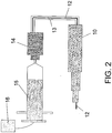
- FIG. 2 shows an overview of an exemplary system similar to that shown in FIG. 1 .
- the embodiment shown in FIG. 2 comprises a stepped stainless steel cannula 10 with fused silica tubing 12 running through the lumen of the stainless steel cannula and extending beyond the distal end of the cannula 10.
- tubing (FEP) 13 covering the fused silica tubing 12, as well as a Luer compression fitting 14 and 2.54cm (1 inch) stainless steel (23 ga) between fused silica tubing 12 and FEP tubing 13.
- the Luer compression fitting 14 is connected to a syringe 15, which in turn is connected to a pump 16.
- Exemplary materials and exemplary commercial sources of these materials that can be used in making an embodiment such as that shown in FIG. 2 are set forth in the following table: Component (reference # in FIG. 2 ) Exemplary Commercial Source Composition Product Contact Cannula 10 Ranfac 304 SS No Fused Silica Tubing 12 PolymicroTechnologies Fused silica w/polyimide coating on outside Yes Teflon Tubing 13 Western Analytical Products Teflon® (FEP) No Luer fitting 14 Upchurch Scientific Polypropylene with ETFE Yes Syringe 15 BD Polypropylene Yes Pump 16 Medfusion N/A No
- tubing extends through the lumen of cannula and the product(s) to be delivered are delivered through this tubing.
- the tubing may be flush with the distal end of the cannula.
- the tubing extends from the distal end of the cannula. In such embodiments, the amount which the tubing extends may vary depending on the application.
- the tubing will extend from about 1 mm to about 1 cm from the cannula (or any length therebetween), more preferably from about 1 to about 50 mm (or any length therebetween), and even more preferably from about 1 mm to about 25 mm (or any length therebetween, including, but not limited to, 1 mm, 2 mm, 3 mm, 4 mm, 5 mm, 6 mm, 7 mm, 8 mm, 9 mm, 10 mm, 11 mm, 12 mm, 13 mm, 14 mm, 15 mm, 16 mm, 17 mm, 18 mm, 19 mm, 20 mm, 21 mm, 22 mm, 23 mm, 24 mm or 25 mm). In one preferred embodiment, the tubing extends approximately 10 mm beyond the distal end thereof.
- tubing extending through the cannula may have one more coatings or surrounding materials in one or more regions, for example to protect the tubing in contact with the produce to be delivered.
- tubing e.g., FEP (Teflon) tubing
- FEP Teflon
- the fused silica tubing may be connected to the syringe by any suitable means, including, but not limited to, a Luer compression fitting, and the syringe is driven by a syringe pump (manual, electronic and/or computerized). It will apparent that the syringe size can be selected by the operator to deliver the appropriate amount of product(s). Thus, 1 mL, 2.5 mL, 5 mL, or even larger syringes maybe used.
- the Luer compression fitting comprises a 2.54 cm (1 inch) stainless steel 23G spacer between the fused silica (inner) tubing and the FEP (outer) tubing.
- the optional spacer provides mechanical rigidity at the Luer compression fitting and helps seal the gap between the inner and outer tubing when the ferrule is glued in place using Loctite® adhesive. This gap must be filled to prevent product from entering the space between the inner and outer tubing as it is being administered to a subject.
- the proximal end of the spacer represents the only stainless steel product contact surface of the systems and cannulas described herein.
- This minimal stainless steel product contact surface may be eliminated if desired by applying Loctite® adhesive or other coating to cover the otherwise exposed end of the spacer, to provide a system with absolutely no stainless steel contact with product.
- the spacer could be comprised of a different material.
- FIG. 3A depicts selected exemplary steps in making a stepped cannula as described herein. See, also, Example 1.
- the step design cannula that reduced reflux may be assembled in the order shown by the arrows (top to bottom), namely by adding components (20, 22, 24, 26, 28, 30) of increasing diameters.
- various length segments are joined together to form the step design.
- the joints should not allow materials to leak from the cannula into the target tissue or vice versa (from the target tissue into the cannula). Accordingly, the joints are preferably sealed.
- the joints can be sealed in a variety of ways, including but not limited to, welding ( e.g., laser welding), adhesives, sealants, heating ( e.g., for thermoplastic polymers) and combinations thereof. It will be apparent that the nature of the seal will depend on the material used to make the cannula, for example welding may be used for stainless steel cannulas while heating may be used for thermoplastic polymers.
- a step design cannula as described herein can also be formed in a single integral piece, for example by injection molding a stepped cannula as described herein.
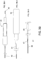
- FIGs. 3B1-6 depict assembly of an exemplary stepped cannula as described herein.
- the stepped cannula 35 shown in FIG. 3A is prepared by removing the needle guard 32 and inserting an inner tubing component 40 through the cannula 35 until it extends from the ends of the cannula. ( FIG. 3B-1 ).
- Any material may be used for the inner tubing component 40, including but not limited to fused silica tubing.
- the inside of the steel cannula 35 can be coated with one or more materials that contact the product to be delivered, thereby reducing loss of the product to the steel cannula during delivery.
- Various techniques of coating of stainless steel materials are known and may be used.
- adhesive may be placed on the tubing 40 such that the tubing is secured to the needle.
- Any suitable adhesive can be used, for example, Loctite® adhesive.
- the bond strength of the adhesive is at least about 17.79N (4 lbs), more preferably at least about 22.24N (5 lbs),
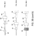
- the needle guard 32 may be replaced and a previously-cut length of tubing 31 (e.g., FEP tubing) extended over the fused silica tubing 40 through the cannula 35 ( FIG. 3B-2 ).
- the length of the outer tubing 31 can be determined by the indication and can range from 0.25m to 4.57m (10 inches to 5 yards) in length (or any value therebetween).
- the outer tubing covers the full-length of the inner tubing and may extend over the inner tubing.
- the outer tubing 31 does not fully extend over the length of the inner tubing 40 ( FIG. 3B-2 ).
- Any suitable adhesive may be used to secure the outer tubing 31 to the assembly, for example at the ends of the outer tubing 31.
- the bond strength of the adhesive is preferably at least about 22.24N (5 lbs).
- one or more spacer components 47 may be inserted over the inner and/or outer tubing 40, 31.
- the spacer 47 may be made of any material including metals, metal alloys, polymers and combinations thereof.
- the spacer 47 comprises stainless steel.
- the spacer 47 can be any length, although it is preferably that does not extend over the needle.
- a component may be included to help seal the components of the assembly, for example a length of PVC shrink tubing 49. See, Example 1 for exemplary dimensions of space and PVC tubing components.
- the assembly may be fitted with one or more components that allow it to be conveniently linked to a product delivery reservoir.
- a product delivery reservoir For example, as shown in FIG. 3B-4 , appropriately sized female Luer compression fitting 50 is slid over a length of the outer tubing 31 and a ferrule 51 is placed over the outer tubing 31, preferably such that it is flush with the end of the outer tubing 31.
- Adhesive may be optionally applied to one or more of the components (e.g., outside of the end of outer tubing prior to fitting of ferrule on the end and/or to seal the joints between the inner tubing, spacer, outer tubing and ferrule.
- the length of inner tubing 40 extending from the ferrule 51 may be removed, for example by scoring the tubing and snapping or cutting it off and the ferrule 51 fitted inside of the Luer compression fitting 50 ( FIG. 3B-5 ).
- the shrink tubing 49 may be heated to seal the joint.
- a male Luer compression fitting 55 can be assembled and fitting onto the female Luer compression fitting 50 and ferrule 51.
- the stepped cannulas described herein may be made out of the variety of materials that are physiologically acceptable, including but not limited to metals, metal alloys, polymers, organic fibers, inorganic fibers and/or combinations thereof.
- the cannula comprises stainless steel (e.g. 316SS or 304SS).
- a product-contact surface may extend through the lumen of the cannula.
- a variety of materials may also be used for the optional product-contact surface, including but not limited to metals, metal alloys, polymers, organic fibers, inorganic fibers and/or combinations thereof.
- the product-contact surface is not stainless steel.
- the outer cannula must still be made of a material physiologically compatible with the target tissue, but there since there is no product contact it need not be compatible with the biologically active agent or product formulation.
- the FEP (Teflon) tubing shown in the Figures may be replaced with other tubing without regard to whether the tubing material is compatible with the biologically active agent or product formulation.
- the product-contact surface of the cannula comprises or consists of fused silica (e.g., quartz silica and polyimide coating) (Polymicro, Phoenix, Arizona).
- fused silica e.g., quartz silica and polyimide coating
- the use of fused silica for the product contact surfaces greatly reduces losses of product when compared with prior art cannulas, in which product is exposed to stainless steel. Indeed, while only 59 ⁇ 14% of an adeno-associated virus vector was recovered from a prior art injection device that had been pre-flushed with product, 101 ⁇ 6% was recovered from a device comprising a cannula of the present invention even without pre-flushing. See, Example 2.
- tubing with small internal diameter such as fused silica tubing with an ID of 100 ⁇ m
- ID small internal diameter
- use of small ID fused silica tubing does not cause large losses of delivered products, for example, AAV vectors. Without intending to be limited by theory, the result may be explained by the increased linear flow rate that results when a given delivery rate (volume of product delivered per unit time) is maintained constant using smaller ID tubing.
- AAV appears to have little affinity for the surface of the fused silica tubing, which may account for of the low losses.
- the small ID of the fused silica tubing used in Example 1 has the additional advantage of reducing the hold-up volume of the system.
- a 1.22m long (four-foot-long) segment of fused silica tubing with an ID of 100 ⁇ m has a lumen volume of less than 15 ⁇ l.
- Such low volumes reduce sample consumption and significantly reduce waste of sample due to the hold-up volume of the delivery system.
- Reduced wastage of product is particularly valuable when the biologically active agent is difficult and/or expensive to obtain, for example many recombinant proteins or gene therapy vectors.
- FIG. 4 depicts an overview of an exemplary syringe pump that may be used in combination with cannulas as described herein. Shown in FIG. 4 are syringe saddle 60, syringe clamp 62, syringe clamp groove (retainer) 64, clutch lever 66, syringe driver 68, syringe plunger retainer 70, liquid crystal display 72 and on/off switch 74.
- Syringe pumps useful in systems with the cannulas described herein are commercially available, for example under the name Medfusion 2010i (Medex, Inc., Carlsbad, California).
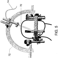
- FIG. 5 depicts an overview of a system including a cannula 70 as described herein attached to a stereotactic frame 72.
- Cannula 70 may also be attached to syringe pump, for example via tubing 74.
- Stereotactic frames are commercially available, for example Lexell stereotactic frames (Ranfac Corp., Avon, Mass.).
- the product contact portion may comprise quartz silica (fused silica tubing in the cannula), USP class VII polypropylene (syringe and Luer hubs), cyanoacrylate (glue joints), and stainless steel (23G spacer).
- the systems described herein are able to deliver product to the brain with far less exposure to stainless steel than using previously described systems, in which product is in contact with stainless steel along some or all of the entire length of the cannula.
- Reduced exposure of product to stainless steel reduces losses.
- the cannula illustrated in FIG. 3B-6 as configured in the system illustrated in FIG. 1 , has product contact surfaces comprising almost exclusively fused silica tubing and a USP Class VII polypropylene syringe.
- the only other contact surface is the glue joint between the proximal end of the fused silica tubing and the Luer hub of the syringe, at which location product contacts cyanoacrylate adhesive and the cross-sectional surface of the proximal end of the stainless steel spacer.
- the exposure to steel in this system is minimal.
- Cannulas of the present invention may also combine the step design and the internal fused silica product contact surfaces to provide an improved cannula with reduced reflux, reduced surface-related losses of agent and reduced hold-up volume.
- Cannulas of the present invention may be sterilized using techniques known in the art including, for example, by standard ethylene oxide. Sterilized cannulas may optionally be individually packaged in a Tyvek® pouch.
- Agents that can be delivered using a cannula of the present invention include any material that may have a desired effect in the target tissue.
- therapeutic drugs, proteins, plasmids or gene therapy vectors may be delivered into the brain of a subject.
- Non-therapeutic agents may also be added such a dyes, tracers, contrast agents and markers for imaging, diagnostic or research purposes.
- retroviral gene therapy systems have been described. See, e.g., U.S. Patent No. 5,219,740 ; Miller and Rosman, BioTechniques (1989) 7:980-990 ; Miller, A.D., Human Gene Therapy (1990) 1:5-14 ; Scarpa et al., Virology (1991) 180:849-852 ; Burns et al., Proc. Natl. Acad. Sci. USA (1993) 90:8033-8037 ; and Boris-Lawrie and Temin, Cur. Opin. Genet. Develop. (1993) 3:102-109 .
- a number of adenovirus vectors have also been described. See, e.g., U.S. Patent Nos.
- AAV vectors can be readily constructed using techniques well known in the art. See, e.g., U.S. Patent Nos. 5,173,414 and 5,139,941 ; International Publication Nos. WO 92/01070 (published 23 January 1992 ) and WO 93/03769 (published 4 March 1993 ); Lebkowski et al., Molec. Cell. Biol. (1988) 8:3988-3996 ; Vincent et al., Vaccines 90 (1990) (Cold Spring Harbor Laboratory Press ); Carter, B.J. Current Opinion in Biotechnology (1992) 3:533-539 ; Muzyczka, N.
- Cannulas of the present invention can be used as part of a convection-enhanced delivery (CED) system for administration to the CNS.
- CED convection-enhanced delivery
- U.S. Patent No. 6, 309, 634 describes methods of gene therapy in which agents are delivered to regions of the central nervous system by CED.
- recombinant vectors can be delivered to many cells over large areas of the CNS.
- the delivered vectors efficiently express transgenes in CNS cells (e.g., glial cells).
- Cannulas of the present invention may be used with any convection-enhanced delivery device for delivery of recombinant vectors.
- the device is an osmotic pump or an infusion pump. Both osmotic and infusion pumps are commercially available from a variety of suppliers, for example Alzet Corporation (Cupertino, California), Hamilton Corporation, or Alza, Inc. (Palo Alto, California).
- Cannulas of the present invention can also be used for direct injection or other methods of infusion, rather than CED.
- Product may be delivered to a target tissue at a variety of flow rates, including but not limited to 0.2, 0.5, 0.7, 1.0, 1.5, 2.0, 3.0, 5.0, 10, 15 or 20 ⁇ l/min.
- flow rates above 10-20 ⁇ l/min are difficult to achieve using 1.22m (four feet) of fused silica tubing with 100 ⁇ m ID because of excessive backpressure at such high flow rates. This does not represent a serious limitation for convection enhanced delivery methods, however, which are preferably performed at relatively low flow rates.
- Flow rates lower than 0.2 ⁇ l/min may be difficult to achieve using the system illustrated in FIGS. 1A and 2A because the pump lacks a slow enough setting, but one of skill in the art would be able to use a different pump and/or syringe configuration to achieve such low delivery rates.
- the flow rate, and thus the pressure of the product as it is delivered to the target tissue, may be increased, decreased, or held steady throughout delivery.
- the flow rate is held substantially constant throughout delivery, rather than being "ramped up" to a plateau.
- a recombinant vector is delivered via CED devices as follows.
- An improved cannula of the present invention is inserted into CNS tissue in the chosen subject.
- Stereotactic maps and positioning devices are available, for example from ASI Instruments, Warren, MI. Positioning may also be conducted by using anatomical maps obtained by CT and/or MRI imaging to help guide the injection device to the chosen target.
- Examples 2-5 disclose use of a cannula of the present invention to deliver the gene encoding hAADC to the brain of humans, rats and non-human primates. Delivery of the hAADC gene may be helpful in treatment of Parkinson's disease (PD).
- PD is characterized in part by the progressive loss of dopaminergic neurons in the substantia nigra and a severe decrease of dopamine in the putamen ( Hornykiewicz (1975) Nat'l Inst. Drug Abuse Res. Monogr. Ser.(3): 13-21 ).
- AADC is an enzyme in the dopamine biosynthetic pathway that converts L-dopa to dopamine.
- Example 2 provides a protocol and experimental results for delivery of a gene to the brain of a primate using a cannula of the present invention (Clinical Device B).
- rAAV virions encoding hAADC (AAV-hAADC-2) are infused into the putamen of four normal rhesus monkeys and the distribution of AADC expression is determined by immunohistochemistry.
- Two infusion protocols are tested: a ramped procedure (slow stepwise increases in rate from 0.2 ⁇ L/min to 1 ⁇ L/min), and a non-ramped infusion at a constant rate of 1 ⁇ L/min.
- the primary endpoints are safety evaluation of the infusion procedures and assessment of transgene expression at 5.5 weeks post-infusion.
- An exemplary stepped cannula (such as shown in FIGs. 3A and 3B ) was produced as follows.
- Stainless steel tubing segments were cut in lengths and welded using a Lasag Nd:YAG or Neodinium YAG (Yttrium aluminum garnet) laser, an ultraviolet laser in the 454 nm wavelength region.
- the weld between the 23G and the 19G segments was tested to be leak free and all weld joints were tested to withstand a minimum pull force of 44.48N (10 lbs).
- the weld between the 23G and 19G segments should be leak free to prevent any product that may reflux up the outside of the needle from leaking into the lumen of the cannula.
- the glue joint between the exposed distal end of the fused silica tubing and the steel tubing portions of the cannula should be liquid-tight.
- the needle was passivated and ultrasonically cleaned after laser steps are completed.
- the fused silica tubing is cut and assembled to the stainless cannula with cyanoacrylate glue.
- a BD needle hub is attached to the distal end of the tubing to finish the assembly.
- a plastic needle guard is placed over the proximal end of the cannula to protect the tip, then the entire assembly is packaged in a pre-labeled Tyvek® pouch for sterilization.
- the injection needle sub-assembly was assembled by sliding a succession segments of stainless steel tubing over a core segment 20 of tubing (24.56cm (9.67 inch) long, 23 RW cutoff, 0.0250/0.0255 OD, 0.0125/0.0140 ID, 0.006 wall).
- FIG. 3A All dimensions relating to inner diameter (ID), outer diameter (OD) and tubing wall thickness ("wall”) are provided in inches, with paired values X/Y representing minimum and maximum tolerances.
- segment 22 (21. 03 cm (8.28 inches) long, 19 RW cutoff, 0.0415/0.0425 OD, 0.0255/0.0285 ID, 0.0075 wall) was placed over the core segment 20 to leave a 0.390 inch (10 mm) of the core extending beyond the distal end of segment 22.
- Segment 24 (6.31 inches long, 17 RW cutoff, 0.0575/0.0585 OD, 0.0405/0.0435 ID, 0.008 wall) was placed over the segments 20 and 22 to leave 5cm (1.97 inches) of segment 22 extending beyond the distal end of segment 24.
- Segment 26 (16.03cm (6.31 inches) long, 15 RW cutoff, 0.0715/0.0725 OD, 0.0595/0.0615 ID, 0.006 wall) was placed over the segments 20, 22, 24 to leave 1.970 inches of segment 22 extending beyond the distal end of segment 26.
- Segment 28 (16.03cm (6.31 inches) long, 0.086/0.087 OD, 0.0735/0.0750 ID, 0.006 wall) was placed over the segments 20, 22, 24, 26 to leave 1.970 inches of segment 22 extending beyond the distal end of segment 28.
- Segment 30 (4.01cm (1.58 inches) long, 0.108/0.110 OD, 0.0880/0.0895 ID, 0.010 wall) was placed over the segments 20, 22, 24, 26, 28 to leave 18.01cm (7.090 inches) of segment 20 extending beyond the distal end of segment 30.
- the INSA was passivated and ultrasonically cleaned as follows: Oakite aluminum cleaned for 10 minutes, spray rinsed with deionized water for 7 minutes, ultrasonically rinsed in alcohol, and air dried.
- a needle guard 32 (22.86cm (9 inches) long, 0.156 OD, 0.104/0.108 ID, 0.025 wall) is placed over assembled segments 20, 22, 24, 26, 28, 30 to leave a 2cm (0.8 inch) segment of segment 30 extending beyond the proximal end of the assembled segments.
- INSAs were inspected to be free of traces of acid and cleaning solution as follows: removed needle guard 32, soaked in alcohol bath, replaced needle guard 32, blew air through distal end of needle guard 7, inspected liquid effluent at proximal end of segment 20, repeated until all effluent appeared to be clean.
- the distal end of the INSA was inspected to ensure that it was straight.
- FIG. 3B illustrates the assembly of an exemplary injection needle assembly as described herein.
- the needle guard 32 was removed from the INSA 35 as described above, and a length of fused silica tubing 40 was threaded through the core 23G tubing 15 of the INSA 35, starting at the proximal end, until approximately 5.08cm (2 inches) extended beyond the distal end of the INSA 35.
- Loctite® adhesive Loctite® Prism® 4011 adhesive, low viscosity
- the bond strength of the adhesive bond is at least 5 pounds.
- the exposed fused silica tubing is trimmed so that 0.390 inches (10 mm) remained extending from the distal end of the INSA, and the needle guard 32 was replaced.
- a 23G stainless steel spacer 47 (2.54cm (1 inch) long, 23 RW) was placed over the fused silica tubing 40.
- Loctite® adhesive was applied to the outside of the fused silica tubing 40 and to the outside of the spacer 47, and the spacer 47 was inserted into the proximal end of the FEP tubing 31 until the proximal ends were flush.
- a 12.7mm (0.5 inch) long segment of PVC shrink tubing 49 (0.125 ID) was slipped over the proximal end of the FEP tubing 31.
- a 1/16 female Luer compression fitting 50 was then slipped over the proximal end of the FEP tubing 31 and a ferrule 51 was placed approximately 2.54cm (1 inch) over the proximal end of the FEP tubing 31.
- Loctite® adhesive is applied to the outside of the FEP tubing 31 and the ferrule 51 was pushed to place the proximal end of the ferrule flush with the proximal end of the FEP tubing 31.
- Loctite® adhesive is applied to seal the joints between the fused silica tubing 40, spacer 47, FEP tubing 31 and the ferrule 51.
- the remaining fused silica tubing 40 extending proximally beyond the ferrule 51 was scored and snapped off. As shown in FIG. 3B-5 , the ferrule 51 was then seated snuggly in the Luer compression fitting 50 (13.34N (3 pounds) minimum pull force) and the heat shrink tubing 49 was then heat shrinked over the joint between the proximal end of the INSA 35 and the FEP tubing 31.
- the assembly was tested for air leaking, and a male Luer cap 55 was added to the compression fitting 50. ( FIG. 3B-6 ).
- the assembled INA may then be packaged and sealed in a Tyvek® pouch (4 x 58.42cm (23 inches)) with a label, and placed in a labeled box for storage or shipment.
- tubing 31 was made of Teflon FEP, one of skill in the art would recognized that any suitable tubing material could be used, or the tubing could be omitted altogether.
- the FEP tubing 31 was included as protection for the fused silica tubing 40, and to help make sure the very thin fused silica tubing was visible to operators of the system. Neither of these functions is essential. In addition, because the FEP tubing does not contact product tubing of other materials may be used without regard to biocompatibility.
- the finished cannula produced as described in herein comprises five layers of stainless steel tubing over 16.03cm (6.31 inches) of its length (e.g. the length comprising tubing element 28 ), with an internal diameter of 0.32mm - 0.36mm (0.0125-0.014 inches) and an exterior diameter of 2.18mm - 2.2mm (0.086-0.087 inches)
- This cannula has substantial rigidity along this segment, which prevents flexing of the cannula as it is inserted into the target tissue (e.g. the brain).
- a sixth layer of steel tubing 30 adds even greater strength to the cannula over a 4.01 cm (1.58 inch) segment, which prevents the cannula from being crushed or deformed when it is mounted in a stereotactic frame during use, as illustrated in FIG. 5 .
- the cannula 80 is composed of four layers of 304 surgical steel fused together by laser welding in a step design, ending in 30 gauge tubing.
- the steel cannula (approximately 24.6 cm from end to end, including needle tip) is lined with fused silica of 100 ⁇ m inner diameter 82 which also forms the tip of the delivery device by extending 1 cm beyond the steel.
- fused silica 100 ⁇ m inner diameter 82 which also forms the tip of the delivery device by extending 1 cm beyond the steel.
- Teflon tubing 84 connect to a Luer hub 86.
- a 2.54 cm (1 inch) 30 gauge steel spacer 88 between the fused silica and Teflon tubing is sealed and attached to the Luer hub with medical grade cyanoacrylate glue.
- AAV-hAADC-2 Recombinant AAV vector encoding human AADC
- Recombinant AAV2 was generated by a triple transfection protocol ( Matsushita et al. (1998) Gene Ther. 5(7): 938-45 ). Briefly, after expansion of cells from the HEK 293 working cell bank through a series of disposable culture ware in DMEM containing 10% fetal bovine serum and 2 mM glutamine, cells were co-transfected with three plasmids (pAAV-hAADC-2, pHLP 19 and pladeno5). The rAAV-hAADC-2 vector clone is the same as that described previously ( Sanftner et al. (2004) Mol. Ther. 9(3): 403-9 ). Plasmids pHLP 19 and pladeno5 are described more fully at U.S. Pat. Nos. 5,139,941 ; 5,622,856 ; 6,001,650 and 6,004,797 .
- the medium containing the transfection reagent was replaced with serum-free medium and the cells were incubated further to allow vector production.
- Cells were harvested, concentrated by centrifugation, and lysed by a freeze/thaw method to release the AAV-hAADC-2 vector. After centrifugation to remove cellular debris, the lysate was treated with Benzonase®, calcium chloride, and precipitated with polyethylene glycol.
- Vector was purified by two cycles of isopycnic gradient ultracentrifugation in cesium chloride.
- AAV-hAADC-2 was concentrated, and diafiltered with sterile, buffered saline (PBS) containing 5% sorbitol.
- PBS buffered saline
- Poloxamer 188TM (0.001%) was added, the material is sterile filtered (0.22 ⁇ m), and stored frozen at -70°C. Vector purity was assessed by SDS-PAGE. Purified rAAV2 vector used in this study showed only VP1, VP2, and VP3 by silver staining of SDS-PAGE gels. Titer was determined by real-time Q-PCR analysis of vector genomes.
- Magnetic resonance imaging was performed on each monkey prior to surgery to identify stereotaxic coordinates (based on the anatomical structure of the putamen). Two sites were targeted in each hemisphere with one site centered in the rostral putamen and a second in the caudal putamen.
- Anesthesia was induced with isoflurane (Aerane®, Omeda PPD, Inc., Liberty, New Jersey) at 5% v/v, and then maintained at 1%-3% v/v for the duration of the surgery.
- the animal's head was placed in an MRI-compatible stereotaxic frame. Core temperature was maintained with a circulating water blanket while electrocardiogram, heart rate, oxygen saturation and body temperature are continuously monitored during the procedure. Burr-holes were made in the skull with a dental drill to expose areas of the dura just above the target sites.
- AAV-hAADC-2 was infused by CED ( Lieberman et al. (1995) J. Neurosurg. 82(6): 1021-9 ; Bankiewicz et al. (2000) Exp.
- mice were perfused via intracardiac saline infusion followed by 10% neutral buffered formalin (NBF).
- NBF neutral buffered formalin
- the brains were then removed and sliced in a brain mold into coronal blocks (8-10 mm).
- Harvested brain blocks were fixed by immersion in 10% NBF fixative.
- the tissue blocks were transferred 2-3 days after fixation into ascending concentrations of PBS/sucrose solution (10, 20 and 30%) over a 3-5 day period.
- Brains were frozen in a bath of isopentane, cooled on dry ice and cut serially into 40 ⁇ m thick coronal sections on a cryostat.
- hAADC immunostaining was determined by the formula ( n x 10 x 40 ⁇ m) where n is the number of sections with hAADC-positive cells, 40 ⁇ m is the thickness of the section, and every tenth section was examined.
- the volume of distribution was estimated in serial sections (every tenth), stained for AADC with the Optical Fractionator-Optical Dissector design-based stereology method under 63X magnification on a Zeiss microscope equipped with a video camera and StereoinvestigatorTM stereology software (Microbrightfield, Williston, VT). CEE is ⁇ 5% for each group. Results are reported as mean ⁇ SD. Student's t-test was used to measure statistical significance.
- the vector AAV-hAADC-2 used in this study contains the human AADC target cDNA.
- the real-time Q-PCR primers and probe anneal to exons 2 and 3 of the AADC gene, spanning an intron not present in the vector sequence, thereby minimizing amplification of genomic DNA.
- Real-time Q-PCR is standardized with linearized plasmid DNA containing the vector insert and vector genomes were quantified as described previously ( Sommer et al. (2003) Mol. Ther. 7(1): 122-8 ).
- NAb neutralizing antibody
- a half-log serial dilution of the test serum in NMS was made to determine the highest dilution of test serum that results in 50% or greater inhibition of ⁇ -galactosidase expression.
- Each dilution series was tested in triplicate.
- a reference plasma with a well-defined AAV2 neutralizing titer was run in each assay and a negative control (NMS only) was used to determine the assay background.
- the titer of NAb was defined as the two dilutions that bracket the 50% inhibition level, e.g. 1:100 to 1:316.
- Biotinylated AAV2 particles can only be captured by multivalent antibodies forming a bridge between two AAV2 particles.
- a very low non-specific background signal in this assay permitted testing of undiluted or low dilutions of test articles, and the assay has higher sensitivity than a classical ELISA, in which primary antibody in the test sample is detected by an enzyme-conjugated secondary antibody.
- the bridging assay allows direct titer comparisons between different species and classes of antibodies.
- the assay was standardized with known amounts of purified mouse monoclonal antibody "A20" that recognizes AAV2 (Grimm et al. 1998).
- the quantification limit of this assay was approximately 15 ng/mL anti-AAV2 antibody.
- Human samples with a NAb titer of 1:100 contained between 1 and 10 ⁇ g/mL of antibody equivalent to A20.
- the average inter-assay variability for 65 human samples that underwent replicate testing by this assay was 23%.
- AAV2 vectors transduce brain tissue efficiently, but transduction levels decline significantly in the presence of high neutralizing antibodies (NAb) titers (> 1:1200) ( Sanftner et al. (2004) Mol. Ther. 9(3): 403-9 ). Therefore, four male rhesus monkeys with NAb titers of ⁇ 1:100 were selected for AAV2 infusions (Table 1). MRI scans were performed prior to AAV2 delivery to determine stereotaxic coordinates for vector administration. Animals were bilaterally infused with 1.5x10 11 vg of AAV-hAADC-2 in two 50 ⁇ L infusions (7.5x10 10 vg/site) in each hemisphere (3.0x10 11 vg/brain).
- Ascending infusion rates (ramp) of 0.2 ⁇ L/min (10 min), 0.5 ⁇ L/min (10 min.), 0.8 ⁇ L/min (10 min) and 1 ⁇ L/min (35 min) were used for the left hemispheres, whereas a constant rate of 1 ⁇ L/min for 50 min (non-ramp) was used for the right hemispheres.
- Animals were monitored for 5.5 weeks, a time span satisfactory for hAADC expression to become relatively stable.
- serum samples, collected at baseline and at the end of the study were tested for the presence of both neutralizing and total antibodies against AAV.
- CDA Circular Device A
- the CDA cannula was composed of four stepped layers of medical grade stainless steel tubing to provide rigidity and minimize internal hold-up volume.
- the steel CDA cannula was connected to a syringe via 1.2 meters of Teflon® tubing.
- Vector recovery studies at flow rates up to 1 ⁇ L/min reveal that 90% of the vector product was adsorbing to the device (Table 2), despite the 0.01% Poloxamer 188 included as a surfactant in the product formulation.
- a 1-hr flush of the device with vector improves subsequent recovery, but vector loss was still approximately 40%.
- Further testing for vector absorption included testing of stepped stainless steel cannulas in which the product contacts different tubing materials at flow rates of ⁇ 1 ⁇ L/min. (Example 1A and 1B). Excellent vector recovery was observed for cannulas comprising fused silica, Tygon®, and silicone tubing in contact with the AAV vector. Other materials such as steel, Teflon (PTFE and FEP) and polyimide bound significant amounts of vector.
- CDB Copper Device B
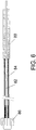
- the steel cannula surrounds the fused silica to provide rigidity, and the fused silica extends 10 mm beyond the tip of the steel cannula ( FIG. 6 ).
- Two external steps near the needle tip are included to minimize potential reflux along the needle track.
- An additional 1.2 meters of fused silica connects the CDB cannula to a Luer hub and is covered by Teflon tubing only to provide protection.
- the CDB was manufactured and assembled in accordance with cGMP and terminally sterilized by ethylene oxide gas.
- Quantitative recovery of vector was evaluated through mock infusions with preclinical and clinical devices.
- 400 ⁇ L of vector solution was drawn from the distal end into a length of Teflon tubing that was then coupled to a 7 cm cannula composed of fused silica surrounded by a 4 cm piece of 27-gauge steel tubing. After filling the cannula at 100 ⁇ L/min, an additional 20 ⁇ L flush was dispensed before collecting vector for recovery assays.
- Four samples were collected from two devices at flow rates from 0.2 to 1.0 ⁇ L/min (ramped procedure) with a programmable syringe pump.
- AAV-hAADC-2 vector was diluted to 5x10 11 vg/mL, loaded into syringes and attached to the devices. After fill, Clinical Device A was flushed with 500 ⁇ L of vector solution at 8 ⁇ L/min (62.5 min), while Clinical Device B was flushed with a total of 50 ⁇ L of vector solution at 4 ⁇ L/min (12.5 min). Two sequential aliquots of 50 ⁇ L were collected from three sets of each device at flow rates from 0.2 to 1.0 ⁇ L/min. Vector concentration in each sample was determined by real-time quantitative PCR (Q-PCR).
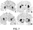
- FIG. 7 shows immunohistochemical staining for the hAADC transgene in cross-sections through the infusion site. Images are of whole mounts of sections from animals MR15102M (A), MR15109M (B), R23700M (C) and R211101M (D). Sections are oriented from a caudal view with the right hemisphere on the right side of the image and the left hemisphere on the left side of the image. In all animals, transgene expression was localized to the putamen. No hAADC expression was detected in cortical regions except in direct line with the infusion track as illustrated in FIG. 7B . No difference in the number of AADC-positive cells or intensity of hAADC staining was seen in a comparison of the right and left hemispheres.
- FIG. 8A A higher magnification image of the infusion site of the putamen in a representative animal from the left hemisphere that received ramped infusion ( FIG. 8A ), or the right hemisphere that received non-ramped infusion ( FIG. 8B ) illustrates the hAADC transgene expression in medium spiny neurons. Immunohistochemical staining for hAADC expression was seen in all (8/8) of the infused hemispheres. AAV-hAADC-2 administration resulted in good expression and coverage of the putamen with a similar distribution of AAV-hAADC-2 with either the ramped (left hemisphere) or non-ramped (right hemisphere) infusion procedure.
- the mean A-P distribution for the left hemisphere (ramped delivery) was 9,600 ⁇ m ⁇ 2,422 ⁇ m (SD) and the mean volume was 238 mm 3 ⁇ 121 mm 3 .
- the mean A-P distribution for the right hemisphere (non-ramped delivery) was 9,606 ⁇ m ⁇ 2,037 ⁇ m and the mean volume was 284 mm 3 ⁇ 55 mm 3 .
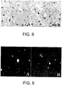
- FIG. 9 shows H&E stained sections within the putamen from a representative animal, R211101M, at 5X magnification.
- Animal R211101M received bilateral CED of AAV-hAADC-2 by the non-ramped infusion procedure in the right hemisphere (Panel A) and the ramped infusion procedure in the left hemisphere (Panel B). Images illustrate the area adjacent to the cannula track at the mid-caudal putamen level.
- Neutralizing antibody (NAb) and total antibody titers to AAV capsid were determined for serum samples collected prior to infusion of vector, and at the time of necropsy. Slight rises in anti-AAV antibody levels were detected by bridging ELISA in all animals after bilateral infusion of AAV-hAADC-2 (Table 1). The results for two NAb assays are shown in Table 1. The bridging ELISA is standardized with anti-AAV2 mouse monoclonal antibody. The average of two results is shown for post-treatment samples and a single result is shown for pre-treatment samples.
- the animal (R23700M) with the highest serum neutralizing antibody titer (1:10 to 1:100) before treatment has a post-treatment antibody increase to 1:31 - 1:316 on Day 42.
- This animal had similar hAADC transgene distribution when compared to the other animals and thus there was no apparent inhibition of vector spread associated with the higher titer.
- An embodiment of the cannula of the present invention was tested to assess its ability to effectively deliver rAAV vector to primate brain, which may serve as a model for delivery of therapeutic rAAV vectors for treatment of Parkinson's disease in a human subject.
- Mock infusions designed to test vector delivery established that essentially 100% of the intended dose can be delivered with a cannula as described herein, preferably avoiding contact of vector with Teflon or steel surfaces.
- AAV-mediated gene delivery into any compartment is potential neutralization by anti-AAV antibodies.
- AAV neutralizing antibody titers in humans ( Blacklow et al. (1968) J. Natl. Cancer Inst. 40(2): 319-27 ) that have the potential of adversely affecting the efficacy of gene therapy techniques. Any AAV-mediated gene therapy approach must anticipate such hurdles.
Landscapes
- Health & Medical Sciences (AREA)
- Life Sciences & Earth Sciences (AREA)
- General Health & Medical Sciences (AREA)
- Public Health (AREA)
- Animal Behavior & Ethology (AREA)
- Veterinary Medicine (AREA)
- Engineering & Computer Science (AREA)
- Biomedical Technology (AREA)
- Heart & Thoracic Surgery (AREA)
- Surgery (AREA)
- Anesthesiology (AREA)
- Hematology (AREA)
- Pathology (AREA)
- Nuclear Medicine, Radiotherapy & Molecular Imaging (AREA)
- Medical Informatics (AREA)
- Molecular Biology (AREA)
- Vascular Medicine (AREA)
- Oral & Maxillofacial Surgery (AREA)
- Biophysics (AREA)
- Pulmonology (AREA)
- Infusion, Injection, And Reservoir Apparatuses (AREA)
- Surgical Instruments (AREA)
- Materials For Medical Uses (AREA)
- Pharmaceuticals Containing Other Organic And Inorganic Compounds (AREA)
Priority Applications (1)
| Application Number | Priority Date | Filing Date | Title |
|---|---|---|---|
| PL12189626T PL2572661T3 (pl) | 2004-10-05 | 2005-10-05 | Stopniowana kaniula |
Applications Claiming Priority (4)
| Application Number | Priority Date | Filing Date | Title |
|---|---|---|---|
| US61623804P | 2004-10-05 | 2004-10-05 | |
| US64155105P | 2005-01-04 | 2005-01-04 | |
| PCT/US2005/036110 WO2006042090A1 (en) | 2004-10-05 | 2005-10-05 | Stepped cannula |
| EP05823438.6A EP1807009B1 (en) | 2004-10-05 | 2005-10-05 | Stepped cannula |
Related Parent Applications (3)
| Application Number | Title | Priority Date | Filing Date |
|---|---|---|---|
| EP05823438.6 Division | 2005-10-05 | ||
| EP05823438.6A Division-Into EP1807009B1 (en) | 2004-10-05 | 2005-10-05 | Stepped cannula |
| EP05823438.6A Division EP1807009B1 (en) | 2004-10-05 | 2005-10-05 | Stepped cannula |
Publications (2)
| Publication Number | Publication Date |
|---|---|
| EP2572661A1 EP2572661A1 (en) | 2013-03-27 |
| EP2572661B1 true EP2572661B1 (en) | 2019-11-20 |
Family
ID=35762442
Family Applications (2)
| Application Number | Title | Priority Date | Filing Date |
|---|---|---|---|
| EP12189626.0A Expired - Lifetime EP2572661B1 (en) | 2004-10-05 | 2005-10-05 | Stepped cannula |
| EP05823438.6A Expired - Lifetime EP1807009B1 (en) | 2004-10-05 | 2005-10-05 | Stepped cannula |
Family Applications After (1)
| Application Number | Title | Priority Date | Filing Date |
|---|---|---|---|
| EP05823438.6A Expired - Lifetime EP1807009B1 (en) | 2004-10-05 | 2005-10-05 | Stepped cannula |
Country Status (15)
| Country | Link |
|---|---|
| US (4) | US7815623B2 (enExample) |
| EP (2) | EP2572661B1 (enExample) |
| JP (1) | JP4838255B2 (enExample) |
| CN (1) | CN102626336A (enExample) |
| AU (1) | AU2005294247B2 (enExample) |
| BR (1) | BRPI0516463B1 (enExample) |
| CA (1) | CA2581714C (enExample) |
| DK (1) | DK1807009T3 (enExample) |
| ES (1) | ES2531425T3 (enExample) |
| IL (2) | IL182351A (enExample) |
| MX (2) | MX2007003850A (enExample) |
| PL (2) | PL1807009T3 (enExample) |
| PT (1) | PT1807009E (enExample) |
| SI (1) | SI1807009T1 (enExample) |
| WO (1) | WO2006042090A1 (enExample) |
Families Citing this family (98)
| Publication number | Priority date | Publication date | Assignee | Title |
|---|---|---|---|---|
| TWI406690B (zh) * | 2004-02-26 | 2013-09-01 | Semiconductor Energy Lab | 運動器具,娛樂工具,和訓練工具 |
| RU2424792C2 (ru) | 2004-05-03 | 2011-07-27 | Хермес Байесайенсиз, Инк. | Липосомы, используемые для доставки лекарственных средств |
| US8658203B2 (en) | 2004-05-03 | 2014-02-25 | Merrimack Pharmaceuticals, Inc. | Liposomes useful for drug delivery to the brain |
| CA2581714C (en) | 2004-10-05 | 2017-09-12 | Avigen, Inc. | Stepped cannula |
| US20060253101A1 (en) * | 2005-03-16 | 2006-11-09 | Andreas Hartlep | Intracranial catheter |
| WO2007024841A2 (en) * | 2005-08-23 | 2007-03-01 | The Regents Of The University Of California | Reflux resistant cannula and system for chronic delivery of therapeutic agents using convection-enhanced delivery |
| EP2023949A4 (en) * | 2006-04-26 | 2009-08-26 | Univ California | COMPOSITIONS AND METHODS FOR CONVECTION-REINFORCED RELEASE OF NEUROTHERAPEUTICS WITH HIGH MOLECULAR WEIGHT |
| GB0616411D0 (en) * | 2006-08-18 | 2006-09-27 | Renishaw Plc | Neurosurgical instruments |
| US7837668B2 (en) * | 2006-10-10 | 2010-11-23 | Ceregene, Inc. | Needle assembly for use in delivering precise dosages of proteinaceous pharmaceutical compositions and methods for use of same |
| US7766394B2 (en) | 2006-10-30 | 2010-08-03 | Medtronic, Inc. | Breakaway connectors and systems |
| EP1970001B1 (de) * | 2007-03-16 | 2014-07-23 | Brainlab AG | Katheter mit Drucksensorik |
| US20080275466A1 (en) * | 2007-05-01 | 2008-11-06 | James Grant Skakoon | Dual cannula system and method for using same |
| WO2008144585A1 (en) * | 2007-05-17 | 2008-11-27 | Medgenesis Therapeutix Inc. | Convection-enhanced delivery catheter with removable stiffening member and method for using same |
| WO2009029508A1 (en) * | 2007-08-28 | 2009-03-05 | Johnnie B. Byrd, Sr. Alzheimer's Center And Research Institute, Inc. | Intracranial catheter and methods of use |
| US7766875B2 (en) * | 2007-09-28 | 2010-08-03 | Codman & Shurtleff, Inc. | Catheter for reduced reflux in targeted tissue delivery of a therapeutic agent |
| US8147480B2 (en) | 2007-09-28 | 2012-04-03 | Codman & Shurtleff, Inc. | Catheter for reduced reflux in targeted tissue delivery of a therapeutic agent |
| US8480626B2 (en) * | 2007-11-30 | 2013-07-09 | Medtronic, Inc. | Infusion catheter assembly with reduced backflow |
| WO2009094389A1 (en) * | 2008-01-22 | 2009-07-30 | Medtronic, Inc. | Burr hole anchors, systems, and methods |
| BRPI1007155A2 (pt) | 2009-01-29 | 2017-05-30 | Univ Of California San Francisco | métodos para tratar um distúrbio neurológico cortical, e para dispensar um agente terapêutico para o córtex em um primata |
| US7985188B2 (en) | 2009-05-13 | 2011-07-26 | Cv Holdings Llc | Vessel, coating, inspection and processing apparatus |
| DK2251454T3 (da) | 2009-05-13 | 2014-10-13 | Sio2 Medical Products Inc | Coating og inspektion af beholder |
| WO2010144419A2 (en) | 2009-06-08 | 2010-12-16 | Surgivision, Inc. | Mri-guided interventional systems that can track and generate dynamic visualizations of flexible intrabody devices in near real time |
| EP2442717B1 (en) | 2009-06-16 | 2020-11-25 | ClearPoint Neuro, Inc. | Mri-guided devices and mri-guided interventional systems that can track and generate dynamic visualizations of the devices in near real time |
| US9458536B2 (en) | 2009-07-02 | 2016-10-04 | Sio2 Medical Products, Inc. | PECVD coating methods for capped syringes, cartridges and other articles |
| US20110046540A1 (en) * | 2009-08-24 | 2011-02-24 | Alterman Ron L | Apparatus for Trans-Cerebral Electrophoresis and Methods of Use Thereof |
| WO2011130107A2 (en) | 2010-04-16 | 2011-10-20 | Surgivision, Inc. | Mri surgical systems including mri-compatible surgical cannulae for transferring a substance to and/or from a patient |
| US8738151B2 (en) | 2010-04-28 | 2014-05-27 | Medtronic, Inc. | Body portal anchors and systems |
| US11624115B2 (en) | 2010-05-12 | 2023-04-11 | Sio2 Medical Products, Inc. | Syringe with PECVD lubrication |
| US9878101B2 (en) | 2010-11-12 | 2018-01-30 | Sio2 Medical Products, Inc. | Cyclic olefin polymer vessels and vessel coating methods |
| US9272095B2 (en) | 2011-04-01 | 2016-03-01 | Sio2 Medical Products, Inc. | Vessels, contact surfaces, and coating and inspection apparatus and methods |
| CA3125150A1 (en) | 2011-08-01 | 2013-02-07 | Alcyone Lifesciences, Inc. | Microfluidic drug delivery devices |
| AU2012318242A1 (en) | 2011-11-11 | 2013-05-30 | Sio2 Medical Products, Inc. | Passivation, pH protective or lubricity coating for pharmaceutical package, coating process and apparatus |
| US11116695B2 (en) | 2011-11-11 | 2021-09-14 | Sio2 Medical Products, Inc. | Blood sample collection tube |
| EP2830515A2 (en) * | 2012-03-30 | 2015-02-04 | Koninklijke Philips N.V. | Nested cannula tips |
| EP2846755A1 (en) | 2012-05-09 | 2015-03-18 | SiO2 Medical Products, Inc. | Saccharide protective coating for pharmaceutical package |
| US20150297800A1 (en) | 2012-07-03 | 2015-10-22 | Sio2 Medical Products, Inc. | SiOx BARRIER FOR PHARMACEUTICAL PACKAGE AND COATING PROCESS |
| US10751513B2 (en) | 2012-07-24 | 2020-08-25 | Renishaw Plc | Neurosurgical apparatus and methods |
| US8992427B2 (en) | 2012-09-07 | 2015-03-31 | Gynesonics, Inc. | Methods and systems for controlled deployment of needle structures in tissue |
| US20140243783A1 (en) * | 2012-10-06 | 2014-08-28 | Raghu Raghavan | Method of backflow reduction during material delivery through a needle into tissue |
| CA2890066C (en) | 2012-11-01 | 2021-11-09 | Sio2 Medical Products, Inc. | Coating inspection method |
| WO2014078666A1 (en) | 2012-11-16 | 2014-05-22 | Sio2 Medical Products, Inc. | Method and apparatus for detecting rapid barrier coating integrity characteristics |
| EP2925903B1 (en) | 2012-11-30 | 2022-04-13 | Si02 Medical Products, Inc. | Controlling the uniformity of pecvd deposition on medical syringes, cartridges, and the like |
| US9764093B2 (en) | 2012-11-30 | 2017-09-19 | Sio2 Medical Products, Inc. | Controlling the uniformity of PECVD deposition |
| US9919129B2 (en) | 2012-12-18 | 2018-03-20 | Alcyone Lifesciences, Inc. | Systems and methods for reducing or preventing backflow in a delivery system |
| EP2961858B1 (en) | 2013-03-01 | 2022-09-07 | Si02 Medical Products, Inc. | Coated syringe. |
| CN105392916B (zh) | 2013-03-11 | 2019-03-08 | Sio2医药产品公司 | 涂布包装材料 |
| US9937099B2 (en) | 2013-03-11 | 2018-04-10 | Sio2 Medical Products, Inc. | Trilayer coated pharmaceutical packaging with low oxygen transmission rate |
| EP2971227B1 (en) | 2013-03-15 | 2017-11-15 | Si02 Medical Products, Inc. | Coating method. |
| EP2991560B1 (en) | 2013-04-30 | 2020-07-29 | Cedars-Sinai Medical Center | Stabilization apparatuses for medical procedures |
| WO2014204954A1 (en) | 2013-06-17 | 2014-12-24 | Alcyone Lifesciences, Inc. | Methods and devices for protecting catheter tips and stereotactic fixtures for microcatheters |
| ES2738298T3 (es) | 2013-07-31 | 2020-01-21 | Alcyone Lifesciences Inc | Sistemas y métodos de suministro de fármacos, tratamiento y monitoreo |
| US9891296B2 (en) | 2013-09-13 | 2018-02-13 | MRI Interventions, Inc. | Intrabody fluid transfer devices, systems and methods |
| EP3107610B1 (en) * | 2014-02-20 | 2018-05-30 | Brainlab AG | Agent delivery catheter |
| EP3693493A1 (en) | 2014-03-28 | 2020-08-12 | SiO2 Medical Products, Inc. | Antistatic coatings for plastic vessels |
| EP4600255A3 (en) | 2014-05-02 | 2025-10-22 | Genzyme Corporation | Aav vectors for retinal and cns gene therapy |
| WO2015191508A1 (en) | 2014-06-09 | 2015-12-17 | Voyager Therapeutics, Inc. | Chimeric capsids |
| WO2016069936A1 (en) * | 2014-10-29 | 2016-05-06 | Cedars-Sinai Medical Center | Apparatuses, systems and methods for controlled delivery of therapeutics and related substances |
| CN107106689A (zh) | 2014-11-05 | 2017-08-29 | 沃雅戈治疗公司 | 用于治疗帕金森病的aadc多核苷酸 |
| US10758264B2 (en) | 2014-11-13 | 2020-09-01 | The Regents Of The University Of California | Adjustable stepped cannula |
| MX2017006216A (es) | 2014-11-14 | 2018-08-29 | Voyager Therapeutics Inc | Composiciones y métodos para tratar la esclerosis lateral amiotrófica (ela). |
| KR102584655B1 (ko) | 2014-11-14 | 2023-10-06 | 보이저 테라퓨틱스, 인크. | 조절성 폴리뉴클레오티드 |
| US11697825B2 (en) | 2014-12-12 | 2023-07-11 | Voyager Therapeutics, Inc. | Compositions and methods for the production of scAAV |
| US10806396B2 (en) | 2015-01-26 | 2020-10-20 | Alcyone Lifesciences, Inc. | Drug delivery methods with tracer |
| AU2016219398A1 (en) | 2015-02-10 | 2017-09-28 | Genzyme Corporation | Enhanced delivery of viral particles to the striatum and cortex |
| WO2016183123A1 (en) | 2015-05-11 | 2016-11-17 | Alcyone Lifesciences, Inc. | Drug delivery systems and methods |
| US11077233B2 (en) | 2015-08-18 | 2021-08-03 | Sio2 Medical Products, Inc. | Pharmaceutical and other packaging with low oxygen transmission rate |
| SI3352776T1 (sl) | 2015-09-23 | 2025-08-29 | Sangamo Therapeutics, Inc. | Represorji Htt in njihove uporabe |
| BR112018006922B1 (pt) | 2015-10-16 | 2023-11-21 | Ipsen Biopharm Ltd | Composições de irinotecano lipossômico estabilizado para armazenamento |
| JP2019502473A (ja) | 2016-01-04 | 2019-01-31 | アルキオーネ・ライフサイエンシズ・インコーポレイテッドAlcyone Lifesciences, Inc. | 脳卒中を治療するための方法および装置 |
| WO2017142698A1 (en) | 2016-02-17 | 2017-08-24 | MRI Interventions, Inc. | Intrabody surgical fluid transfer assemblies with adjustable exposed cannula to needle tip length, related systems and methods |
| WO2017189964A2 (en) | 2016-04-29 | 2017-11-02 | Voyager Therapeutics, Inc. | Compositions for the treatment of disease |
| US11326182B2 (en) | 2016-04-29 | 2022-05-10 | Voyager Therapeutics, Inc. | Compositions for the treatment of disease |
| AU2017268382B2 (en) | 2016-05-18 | 2023-09-28 | Voyager Therapeutics, Inc. | Compositions and methods of treating Huntington's disease |
| RU2758488C2 (ru) | 2016-05-18 | 2021-10-28 | Вояджер Терапьютикс, Инк. | Модулирующие полинуклеотиды |
| JP2019531787A (ja) | 2016-08-30 | 2019-11-07 | ザ リージェンツ オブ ザ ユニバーシティ オブ カリフォルニア | 生物医学的ターゲティング及びデリバリーの方法並びにそれを実行するための装置及びシステム |
| WO2018089523A1 (en) | 2016-11-11 | 2018-05-17 | Gynesonics, Inc. | Controlled treatment of tissue and dynamic interaction with, and comparison of, tissue and/or treatment data |
| MX2019006426A (es) | 2016-12-01 | 2019-08-14 | Sangamo Therapeutics Inc | Reguladores de tau, y composiciones y metodos para su administracion. |
| CN114887153A (zh) | 2016-12-21 | 2022-08-12 | 亚克安娜治疗学有限公司 | 药物递送系统和方法 |
| AU2018261790B2 (en) | 2017-05-05 | 2024-10-03 | Voyager Therapeutics, Inc. | Compositions and methods of treating amyotrophic lateral sclerosis (ALS) |
| WO2018204803A1 (en) | 2017-05-05 | 2018-11-08 | Voyager Therapeutics, Inc. | Compositions and methods of treating huntington's disease |
| JOP20190269A1 (ar) | 2017-06-15 | 2019-11-20 | Voyager Therapeutics Inc | بولي نوكليوتيدات aadc لعلاج مرض باركنسون |
| CN111132626B (zh) | 2017-07-17 | 2024-01-30 | 沃雅戈治疗公司 | 轨迹阵列引导系统 |
| EP3687582A4 (en) | 2017-09-29 | 2021-07-14 | Voyager Therapeutics, Inc. | RESCUE OF CENTRAL AND PERIPHERAL NEUROLOGICAL PHENOTYPE OF FRIEDREICH ATAXIA BY INTRAVENOUS ADMINISTRATION |
| EP4454654A3 (en) | 2017-10-16 | 2025-02-19 | Voyager Therapeutics, Inc. | Treatment of amyotrophic lateral sclerosis (als) |
| US11434502B2 (en) | 2017-10-16 | 2022-09-06 | Voyager Therapeutics, Inc. | Treatment of amyotrophic lateral sclerosis (ALS) |
| EP3781074A1 (en) * | 2018-05-09 | 2021-02-24 | ClearPoint Neuro, Inc. | Mri compatible intrabody fluid transfer systems and related devices and methods |
| US11253237B2 (en) | 2018-05-09 | 2022-02-22 | Clearpoint Neuro, Inc. | MRI compatible intrabody fluid transfer systems and related devices and methods |
| MX2020012077A (es) | 2018-05-15 | 2021-03-09 | Voyager Therapeutics Inc | Composiciones y metodos para el tratamiento de la enfermedad de parkinson. |
| WO2020010035A1 (en) * | 2018-07-02 | 2020-01-09 | Voyager Therapeutics, Inc. | Cannula system |
| SG11202103151RA (en) | 2018-09-28 | 2021-04-29 | Voyager Therapeutics Inc | Frataxin expression constructs having engineered promoters and methods of use thereof |
| CN113226333A (zh) | 2018-10-02 | 2021-08-06 | 桑格摩生物治疗股份有限公司 | 用于调控Tau蛋白的方法和组合物 |
| AU2020219357B2 (en) * | 2019-02-08 | 2025-03-20 | Rebound Therapeutics Corporation | Lighted cannula system |
| PE20212332A1 (es) | 2019-04-23 | 2021-12-14 | Sangamo Therapeutics Inc | Moduladores de la expresioon del gen de marco de lectura abierto 72 del cromosoma 9 y usos de los mismos |
| CA3142345A1 (en) * | 2019-05-31 | 2020-12-03 | The Trustees Of The University Of Pennsylvania | Infusion device and method for drug delivery |
| US11684750B2 (en) | 2019-10-08 | 2023-06-27 | Clearpoint Neuro, Inc. | Extension tube assembly and related medical fluid transfer systems and methods |
| US20210318397A1 (en) * | 2020-04-08 | 2021-10-14 | Clearpoint Neuro, Inc. | Mri surgical systems including mri-compatible surgical cannulas for transferring a substance to and/or from a patient |
| GB2601752A (en) | 2020-12-08 | 2022-06-15 | Maavrx Ltd | Expression vector |
| WO2025004001A1 (en) | 2023-06-30 | 2025-01-02 | Takeda Pharmaceutical Company Limited | Htt repressors and uses thereof |
Family Cites Families (75)
| Publication number | Priority date | Publication date | Assignee | Title |
|---|---|---|---|---|
| GB1255551A (en) | 1968-03-30 | 1971-12-01 | Heraeus Schott Quarzschmelze | Improvements in or relating to externally coated fused silica tube |
| LU77252A1 (enExample) * | 1976-05-06 | 1977-08-22 | ||
| US4239042A (en) * | 1979-04-05 | 1980-12-16 | Dow Corning K.K. | Catheter placement system |
| DE3025785C2 (de) * | 1980-07-08 | 1984-08-16 | Storz, Karl, 7200 Tuttlingen | Dilatator, Verfahren zu seiner Verwendung und Vorrichtung zur Durchführung des Verfahrens |
| US4335718A (en) * | 1980-10-02 | 1982-06-22 | Becton, Dickinson And Company | Needle cannula |
| US4448532A (en) * | 1981-03-31 | 1984-05-15 | Kla Instruments Corporation | Automatic photomask inspection method and system |
| DE8222222U1 (de) * | 1982-08-06 | 1982-11-25 | Mehler, Doron, Dr., 3000 Hannover | Katheterset |
| JPS60234671A (ja) * | 1984-05-09 | 1985-11-21 | テルモ株式会社 | カテ−テル導入具 |
| US4597421A (en) * | 1984-11-19 | 1986-07-01 | Varian Associates, Inc. | Method and device for on-column injection of a liquid sample into small diameter columns |
| US5139941A (en) | 1985-10-31 | 1992-08-18 | University Of Florida Research Foundation, Inc. | AAV transduction vectors |
| US4739768B2 (en) * | 1986-06-02 | 1995-10-24 | Target Therapeutics Inc | Catheter for guide-wire tracking |
| US4738658A (en) * | 1986-09-19 | 1988-04-19 | Aries Medical Incorporated | Tapered hemostatic device for use in conjunction with a catheter for alleviating blood leakage and method for using same |
| US5219740A (en) | 1987-02-13 | 1993-06-15 | Fred Hutchinson Cancer Research Center | Retroviral gene transfer into diploid fibroblasts for gene therapy |
| US4781691A (en) * | 1987-07-17 | 1988-11-01 | The Kendall Company | Stepped needle |
| US4909800A (en) * | 1987-07-17 | 1990-03-20 | The Kendall Company | Stepped needle |
| DE8714069U1 (de) * | 1987-10-21 | 1987-12-03 | Richard Wolf Gmbh, 7134 Knittlingen | Endoskopischer Instrumentensatz |
| US4978334A (en) * | 1988-09-08 | 1990-12-18 | Toye Frederic J | Apparatus and method for providing passage into body viscus |
| US5069673A (en) * | 1990-02-07 | 1991-12-03 | Cordis Corporation | Catheter with double step-down bore |
| WO1992001070A1 (en) | 1990-07-09 | 1992-01-23 | The United States Of America, As Represented By The Secretary, U.S. Department Of Commerce | High efficiency packaging of mutant adeno-associated virus using amber suppressions |
| US5173414A (en) | 1990-10-30 | 1992-12-22 | Applied Immune Sciences, Inc. | Production of recombinant adeno-associated virus vectors |
| US5256157A (en) * | 1991-01-31 | 1993-10-26 | Baxter International Inc. | Automated infusion pump with replaceable memory cartridges |
| DE69233013T2 (de) | 1991-08-20 | 2004-03-04 | The Government Of The United States Of America As Represented By The Secretary Of National Institute Of Health, Office Of Technology Transfer | Adenovirus vermittelter gentransfer in den gastrointestinaltrakt |
| AU7676894A (en) * | 1993-08-27 | 1995-03-21 | Government Of The United States Of America, As Represented By The Secretary Of The Department Of Health And Human Services, The | Convection-enhanced drug delivery |
| NL9301642A (nl) * | 1993-09-22 | 1995-04-18 | Cordis Europ | Microcatheter. |
| US5626862A (en) * | 1994-08-02 | 1997-05-06 | Massachusetts Institute Of Technology | Controlled local delivery of chemotherapeutic agents for treating solid tumors |
| JP3573531B2 (ja) * | 1994-08-03 | 2004-10-06 | 鐘淵化学工業株式会社 | マイクロカテーテル |
| EP1445322B2 (en) | 1995-06-15 | 2012-06-06 | Crucell Holland B.V. | Packaging systems for human recombinant adenovirus to be used in gene therapy |
| US6001650A (en) | 1995-08-03 | 1999-12-14 | Avigen, Inc. | High-efficiency wild-type-free AAV helper functions |
| US5622856A (en) | 1995-08-03 | 1997-04-22 | Avigen | High efficiency helper system for AAV vector production |
| US5593393A (en) * | 1995-09-07 | 1997-01-14 | Trudell; Roger J. | Lacrimal irrigating cannula |
| US6351659B1 (en) * | 1995-09-28 | 2002-02-26 | Brainlab Med. Computersysteme Gmbh | Neuro-navigation system |
| US6004797A (en) | 1995-11-09 | 1999-12-21 | Avigen, Inc. | Adenovirus helper-free recombinant AAV Virion production |
| US6020196A (en) * | 1996-05-09 | 2000-02-01 | Baxter International Inc. | Devices for harvesting and homogenizing adipose tissue containing autologous endothelial cells |
| US5902282A (en) * | 1996-12-26 | 1999-05-11 | Johnson & Johnson Medical, Inc. | Step-down catheter |
| US6048551A (en) | 1997-03-27 | 2000-04-11 | Hilfinger; John M. | Microsphere encapsulation of gene transfer vectors |
| US6458088B1 (en) * | 1997-03-27 | 2002-10-01 | Cordis Corporation | Glass core guidewire compatible with magnetic resonance |
| US6524299B1 (en) * | 1997-04-09 | 2003-02-25 | Target Therapeutics, Inc. | Flow-directed catheter |
| US6042579A (en) * | 1997-04-30 | 2000-03-28 | Medtronic, Inc. | Techniques for treating neurodegenerative disorders by infusion of nerve growth factors into the brain |
| US6026316A (en) * | 1997-05-15 | 2000-02-15 | Regents Of The University Of Minnesota | Method and apparatus for use with MR imaging |
| US6030369A (en) * | 1997-07-03 | 2000-02-29 | Target Therapeutics Inc. | Micro catheter shaft |
| US6186986B1 (en) * | 1998-01-21 | 2001-02-13 | St. Jude Medical Cardiovascular Group, Inc. | Micro-catheters and methods of their manufacture |
| US6199554B1 (en) * | 1998-03-27 | 2001-03-13 | The Brigham And Women's Hospital, Inc. | Method and apparatus for combining injury-mediated therapy and drug delivery |
| ES2326893T3 (es) * | 1998-05-27 | 2009-10-21 | Genzyme Corporation | Vectores aav para la fabricacion de medicamentos para administracion potenciada por conveccion. |
| US6210392B1 (en) * | 1999-01-15 | 2001-04-03 | Interventional Technologies, Inc. | Method for treating a wall of a blood vessel |
| US6663606B1 (en) * | 1999-10-28 | 2003-12-16 | Scimed Life Systems, Inc. | Biocompatible medical devices |
| US6579484B1 (en) * | 1999-12-16 | 2003-06-17 | Advanced Cardiovascular Systems, Inc. | Co-extruded taper shaft |
| US20040092879A1 (en) * | 2000-11-06 | 2004-05-13 | Medamicus, Inc. | Safety introducer apparatus and method therefor |
| US6641564B1 (en) * | 2000-11-06 | 2003-11-04 | Medamicus, Inc. | Safety introducer apparatus and method therefor |
| CA2327208A1 (en) * | 2000-11-30 | 2002-05-30 | The Government Of The United States Of America | Methods of increasing distribution of therapeutic agents |
| US20020087152A1 (en) * | 2001-01-04 | 2002-07-04 | Endocare, Inc. | Systems and methods for delivering a probe into tissue |
| US6533751B2 (en) * | 2001-01-09 | 2003-03-18 | Andrew Cragg | Micro catheter and guidewire system having improved pushability and control |
| JP4271444B2 (ja) * | 2001-01-31 | 2009-06-03 | 富士通株式会社 | 歪補償装置 |
| US7182944B2 (en) * | 2001-04-25 | 2007-02-27 | The United States Of America As Represented By The Department Of Health And Human Services | Methods of increasing distribution of nucleic acids |
| US20030073934A1 (en) * | 2001-10-17 | 2003-04-17 | David A. Putz | Double slotted-cannula device and method of use |
| EP1491154A1 (en) | 2002-01-22 | 2004-12-29 | JFE Steel Corporation | Ceramic-coated instruments for medical use, ceramic-coated instruments for studying living organisms and process for producing the same |
| JP2004147830A (ja) * | 2002-10-30 | 2004-05-27 | Jfe Steel Kk | 遺伝子制御用セラミック被覆針およびその製造方法およびその製造装置 |
| GB0205772D0 (en) * | 2002-03-12 | 2002-04-24 | Gill Steven S | Catheter |
| US20050256503A1 (en) * | 2002-05-07 | 2005-11-17 | Cardiac Pacemakers, Inc. | Tapered catheter delivery system |
| ES2581781T3 (es) * | 2002-09-24 | 2016-09-07 | The Government Of The United States Of America, As Represented By The Secretary, Department Of Health And Human Services | Método por imágenes para administración mejorada por convección de agentes terapéuticos |
| EP1572262B1 (en) | 2002-10-10 | 2010-03-10 | Becton Dickinson and Company | Needle assembly of delivering local anesthesia |
| US20040209810A1 (en) * | 2003-02-24 | 2004-10-21 | Gill Steven S. | Method of treating Parkinson's disease in humans by intraputaminal infusion of glial cell-line derived neurotrophic factor |
| US7241283B2 (en) * | 2003-04-25 | 2007-07-10 | Ad-Tech Medical Instrument Corp. | Method for intracranial catheter treatment of brain tissue |
| US8658203B2 (en) * | 2004-05-03 | 2014-02-25 | Merrimack Pharmaceuticals, Inc. | Liposomes useful for drug delivery to the brain |
| JP4449587B2 (ja) * | 2004-06-09 | 2010-04-14 | 富士フイルム株式会社 | 画像マッピング方法及び画像マッピングプログラム |
| US20060073119A1 (en) * | 2004-09-01 | 2006-04-06 | Avigen, Inc. | Methods for treating neurodegenerative disorders |
| CA2581714C (en) * | 2004-10-05 | 2017-09-12 | Avigen, Inc. | Stepped cannula |
| US20060217664A1 (en) * | 2004-11-15 | 2006-09-28 | Hattler Brack G | Telescoping vascular dilator |
| US20060129126A1 (en) * | 2004-11-19 | 2006-06-15 | Kaplitt Michael G | Infusion device and method for infusing material into the brain of a patient |
| WO2007024841A2 (en) * | 2005-08-23 | 2007-03-01 | The Regents Of The University Of California | Reflux resistant cannula and system for chronic delivery of therapeutic agents using convection-enhanced delivery |
| SI2019683T2 (sl) * | 2006-04-25 | 2022-10-28 | The Regents Of The University Of California | Dajanje rastnih faktorjev za zdravljenje motenj CŽS |
| GB0616411D0 (en) * | 2006-08-18 | 2006-09-27 | Renishaw Plc | Neurosurgical instruments |
| EP1974755A1 (de) * | 2007-03-16 | 2008-10-01 | BrainLAB AG | Katheter mit sich ändernden Materialeigenschaften |
| US7766875B2 (en) * | 2007-09-28 | 2010-08-03 | Codman & Shurtleff, Inc. | Catheter for reduced reflux in targeted tissue delivery of a therapeutic agent |
| US8147480B2 (en) * | 2007-09-28 | 2012-04-03 | Codman & Shurtleff, Inc. | Catheter for reduced reflux in targeted tissue delivery of a therapeutic agent |
| US8480626B2 (en) * | 2007-11-30 | 2013-07-09 | Medtronic, Inc. | Infusion catheter assembly with reduced backflow |
-
2005
- 2005-10-05 CA CA2581714A patent/CA2581714C/en not_active Expired - Lifetime
- 2005-10-05 EP EP12189626.0A patent/EP2572661B1/en not_active Expired - Lifetime
- 2005-10-05 JP JP2007535832A patent/JP4838255B2/ja not_active Expired - Lifetime
- 2005-10-05 DK DK05823438.6T patent/DK1807009T3/en active
- 2005-10-05 MX MX2007003850A patent/MX2007003850A/es active IP Right Grant
- 2005-10-05 AU AU2005294247A patent/AU2005294247B2/en not_active Expired
- 2005-10-05 SI SI200531937T patent/SI1807009T1/sl unknown
- 2005-10-05 US US11/243,756 patent/US7815623B2/en active Active
- 2005-10-05 BR BRPI0516463-0A patent/BRPI0516463B1/pt active IP Right Grant
- 2005-10-05 CN CN2012100734364A patent/CN102626336A/zh active Pending
- 2005-10-05 PL PL05823438T patent/PL1807009T3/pl unknown
- 2005-10-05 PL PL12189626T patent/PL2572661T3/pl unknown
- 2005-10-05 WO PCT/US2005/036110 patent/WO2006042090A1/en not_active Ceased
- 2005-10-05 ES ES05823438T patent/ES2531425T3/es not_active Expired - Lifetime
- 2005-10-05 MX MX2013001394A patent/MX349748B/es unknown
- 2005-10-05 PT PT58234386T patent/PT1807009E/pt unknown
- 2005-10-05 EP EP05823438.6A patent/EP1807009B1/en not_active Expired - Lifetime
-
2007
- 2007-04-01 IL IL182351A patent/IL182351A/en active IP Right Grant
-
2010
- 2010-09-27 US US12/891,043 patent/US8337458B2/en not_active Expired - Lifetime
-
2012
- 2012-03-25 IL IL218812A patent/IL218812A0/en unknown
- 2012-11-16 US US13/679,762 patent/US9302070B2/en not_active Expired - Lifetime
-
2016
- 2016-02-26 US US15/055,065 patent/US20160296694A1/en not_active Abandoned
Non-Patent Citations (1)
| Title |
|---|
| None * |
Also Published As
| Publication number | Publication date |
|---|---|
| US20130079748A1 (en) | 2013-03-28 |
| CA2581714A1 (en) | 2006-04-20 |
| IL218812A0 (en) | 2012-05-31 |
| ES2531425T3 (es) | 2015-03-13 |
| US20060135945A1 (en) | 2006-06-22 |
| AU2005294247A1 (en) | 2006-04-20 |
| WO2006042090A1 (en) | 2006-04-20 |
| US20110046552A1 (en) | 2011-02-24 |
| MX2007003850A (es) | 2007-11-21 |
| MX349748B (es) | 2017-08-09 |
| IL182351A (en) | 2012-04-30 |
| CA2581714C (en) | 2017-09-12 |
| US7815623B2 (en) | 2010-10-19 |
| JP2008515554A (ja) | 2008-05-15 |
| JP4838255B2 (ja) | 2011-12-14 |
| SI1807009T1 (sl) | 2015-04-30 |
| EP1807009A1 (en) | 2007-07-18 |
| EP2572661A1 (en) | 2013-03-27 |
| BRPI0516463A (pt) | 2008-09-02 |
| PL2572661T3 (pl) | 2020-06-01 |
| BRPI0516463B1 (pt) | 2021-05-11 |
| US20160296694A1 (en) | 2016-10-13 |
| US8337458B2 (en) | 2012-12-25 |
| US9302070B2 (en) | 2016-04-05 |
| EP1807009B1 (en) | 2014-11-26 |
| CN102626336A (zh) | 2012-08-08 |
| DK1807009T3 (en) | 2015-03-02 |
| PL1807009T3 (pl) | 2015-06-30 |
| PT1807009E (pt) | 2015-02-25 |
| AU2005294247B2 (en) | 2011-08-11 |
| IL182351A0 (en) | 2007-07-24 |
Similar Documents
| Publication | Publication Date | Title |
|---|---|---|
| EP2572661B1 (en) | Stepped cannula | |
| Sanftner et al. | AAV2-mediated gene delivery to monkey putamen: evaluation of an infusion device and delivery parameters | |
| US9662472B2 (en) | Method for convection enhanced therapeutic delivery | |
| AU2009202335B2 (en) | Pulsatile flux drug delivery | |
| US20110306879A1 (en) | Methods and devices for treating tissue | |
| CN101123919A (zh) | 台阶式插管 | |
| AU2011250715B2 (en) | Method of delivering materials to the central nervous system | |
| WO2008064073A2 (en) | Microsyringe for pre-packaged delivery of pharmaceuticals | |
| US20250034281A1 (en) | Method and apparatus for using omalizumab | |
| Leung et al. | Assessment of hippocampal adeno-associated viral vector gene delivery via frameless stereotaxis in a nonhuman primate | |
| Lee et al. | IntraBrain Injector (IBI): A Stereotactic-Guided Device for Repeated Delivery of Therapeutic Agents Into the Brain Parenchyma | |
| US20220226608A1 (en) | Infusion device and method for drug delivery |
Legal Events
| Date | Code | Title | Description |
|---|---|---|---|
| PUAI | Public reference made under article 153(3) epc to a published international application that has entered the european phase |
Free format text: ORIGINAL CODE: 0009012 |
|
| AC | Divisional application: reference to earlier application |
Ref document number: 1807009 Country of ref document: EP Kind code of ref document: P |
|
| AK | Designated contracting states |
Kind code of ref document: A1 Designated state(s): AT BE BG CH CY CZ DE DK EE ES FI FR GB GR HU IE IS IT LI LT LU LV MC NL PL PT RO SE SI SK TR |
|
| 17P | Request for examination filed |
Effective date: 20130927 |
|
| RBV | Designated contracting states (corrected) |
Designated state(s): AT BE BG CH CY CZ DE DK EE ES FI FR GB GR HU IE IS IT LI LT LU LV MC NL PL PT RO SE SI SK TR |
|
| 17Q | First examination report despatched |
Effective date: 20140515 |
|
| STAA | Information on the status of an ep patent application or granted ep patent |
Free format text: STATUS: EXAMINATION IS IN PROGRESS |
|
| RAP1 | Party data changed (applicant data changed or rights of an application transferred) |
Owner name: THE REGENTS OF THE UNIVERSITY OF CALIFORNIA Owner name: GENZYME CORPORATION |
|
| GRAP | Despatch of communication of intention to grant a patent |
Free format text: ORIGINAL CODE: EPIDOSNIGR1 |
|
| STAA | Information on the status of an ep patent application or granted ep patent |
Free format text: STATUS: GRANT OF PATENT IS INTENDED |
|
| INTG | Intention to grant announced |
Effective date: 20190529 |
|
| RIN1 | Information on inventor provided before grant (corrected) |
Inventor name: BANKIEWICZ, KRZYSZTOF Inventor name: SOMMER, JURG, M. |
|
| GRAS | Grant fee paid |
Free format text: ORIGINAL CODE: EPIDOSNIGR3 |
|
| GRAA | (expected) grant |
Free format text: ORIGINAL CODE: 0009210 |
|
| STAA | Information on the status of an ep patent application or granted ep patent |
Free format text: STATUS: THE PATENT HAS BEEN GRANTED |
|
| AC | Divisional application: reference to earlier application |
Ref document number: 1807009 Country of ref document: EP Kind code of ref document: P |
|
| AK | Designated contracting states |
Kind code of ref document: B1 Designated state(s): AT BE BG CH CY CZ DE DK EE ES FI FR GB GR HU IE IS IT LI LT LU LV MC NL PL PT RO SE SI SK TR |
|
| REG | Reference to a national code |
Ref country code: GB Ref legal event code: FG4D |
|
| REG | Reference to a national code |
Ref country code: CH Ref legal event code: EP |
|
| REG | Reference to a national code |
Ref country code: DE Ref legal event code: R096 Ref document number: 602005056432 Country of ref document: DE |
|
| REG | Reference to a national code |
Ref country code: IE Ref legal event code: FG4D |
|
| REG | Reference to a national code |
Ref country code: AT Ref legal event code: REF Ref document number: 1203268 Country of ref document: AT Kind code of ref document: T Effective date: 20191215 |
|
| REG | Reference to a national code |
Ref country code: NL Ref legal event code: MP Effective date: 20191120 |
|
| REG | Reference to a national code |
Ref country code: LT Ref legal event code: MG4D |
|
| PG25 | Lapsed in a contracting state [announced via postgrant information from national office to epo] |
Ref country code: GR Free format text: LAPSE BECAUSE OF FAILURE TO SUBMIT A TRANSLATION OF THE DESCRIPTION OR TO PAY THE FEE WITHIN THE PRESCRIBED TIME-LIMIT Effective date: 20200221 Ref country code: SE Free format text: LAPSE BECAUSE OF FAILURE TO SUBMIT A TRANSLATION OF THE DESCRIPTION OR TO PAY THE FEE WITHIN THE PRESCRIBED TIME-LIMIT Effective date: 20191120 Ref country code: LV Free format text: LAPSE BECAUSE OF FAILURE TO SUBMIT A TRANSLATION OF THE DESCRIPTION OR TO PAY THE FEE WITHIN THE PRESCRIBED TIME-LIMIT Effective date: 20191120 Ref country code: FI Free format text: LAPSE BECAUSE OF FAILURE TO SUBMIT A TRANSLATION OF THE DESCRIPTION OR TO PAY THE FEE WITHIN THE PRESCRIBED TIME-LIMIT Effective date: 20191120 Ref country code: BG Free format text: LAPSE BECAUSE OF FAILURE TO SUBMIT A TRANSLATION OF THE DESCRIPTION OR TO PAY THE FEE WITHIN THE PRESCRIBED TIME-LIMIT Effective date: 20200220 Ref country code: ES Free format text: LAPSE BECAUSE OF FAILURE TO SUBMIT A TRANSLATION OF THE DESCRIPTION OR TO PAY THE FEE WITHIN THE PRESCRIBED TIME-LIMIT Effective date: 20191120 Ref country code: LT Free format text: LAPSE BECAUSE OF FAILURE TO SUBMIT A TRANSLATION OF THE DESCRIPTION OR TO PAY THE FEE WITHIN THE PRESCRIBED TIME-LIMIT Effective date: 20191120 Ref country code: NL Free format text: LAPSE BECAUSE OF FAILURE TO SUBMIT A TRANSLATION OF THE DESCRIPTION OR TO PAY THE FEE WITHIN THE PRESCRIBED TIME-LIMIT Effective date: 20191120 |
|
| PG25 | Lapsed in a contracting state [announced via postgrant information from national office to epo] |
Ref country code: IS Free format text: LAPSE BECAUSE OF FAILURE TO SUBMIT A TRANSLATION OF THE DESCRIPTION OR TO PAY THE FEE WITHIN THE PRESCRIBED TIME-LIMIT Effective date: 20200320 |
|
| PG25 | Lapsed in a contracting state [announced via postgrant information from national office to epo] |
Ref country code: CZ Free format text: LAPSE BECAUSE OF FAILURE TO SUBMIT A TRANSLATION OF THE DESCRIPTION OR TO PAY THE FEE WITHIN THE PRESCRIBED TIME-LIMIT Effective date: 20191120 Ref country code: RO Free format text: LAPSE BECAUSE OF FAILURE TO SUBMIT A TRANSLATION OF THE DESCRIPTION OR TO PAY THE FEE WITHIN THE PRESCRIBED TIME-LIMIT Effective date: 20191120 Ref country code: PT Free format text: LAPSE BECAUSE OF FAILURE TO SUBMIT A TRANSLATION OF THE DESCRIPTION OR TO PAY THE FEE WITHIN THE PRESCRIBED TIME-LIMIT Effective date: 20200412 Ref country code: EE Free format text: LAPSE BECAUSE OF FAILURE TO SUBMIT A TRANSLATION OF THE DESCRIPTION OR TO PAY THE FEE WITHIN THE PRESCRIBED TIME-LIMIT Effective date: 20191120 Ref country code: DK Free format text: LAPSE BECAUSE OF FAILURE TO SUBMIT A TRANSLATION OF THE DESCRIPTION OR TO PAY THE FEE WITHIN THE PRESCRIBED TIME-LIMIT Effective date: 20191120 |
|
| REG | Reference to a national code |
Ref country code: AT Ref legal event code: MK05 Ref document number: 1203268 Country of ref document: AT Kind code of ref document: T Effective date: 20191120 |
|
| REG | Reference to a national code |
Ref country code: DE Ref legal event code: R097 Ref document number: 602005056432 Country of ref document: DE |
|
| PG25 | Lapsed in a contracting state [announced via postgrant information from national office to epo] |
Ref country code: SK Free format text: LAPSE BECAUSE OF FAILURE TO SUBMIT A TRANSLATION OF THE DESCRIPTION OR TO PAY THE FEE WITHIN THE PRESCRIBED TIME-LIMIT Effective date: 20191120 |
|
| PLBE | No opposition filed within time limit |
Free format text: ORIGINAL CODE: 0009261 |
|
| STAA | Information on the status of an ep patent application or granted ep patent |
Free format text: STATUS: NO OPPOSITION FILED WITHIN TIME LIMIT |
|
| 26N | No opposition filed |
Effective date: 20200821 |
|
| PG25 | Lapsed in a contracting state [announced via postgrant information from national office to epo] |
Ref country code: AT Free format text: LAPSE BECAUSE OF FAILURE TO SUBMIT A TRANSLATION OF THE DESCRIPTION OR TO PAY THE FEE WITHIN THE PRESCRIBED TIME-LIMIT Effective date: 20191120 Ref country code: SI Free format text: LAPSE BECAUSE OF FAILURE TO SUBMIT A TRANSLATION OF THE DESCRIPTION OR TO PAY THE FEE WITHIN THE PRESCRIBED TIME-LIMIT Effective date: 20191120 |
|
| PG25 | Lapsed in a contracting state [announced via postgrant information from national office to epo] |
Ref country code: IT Free format text: LAPSE BECAUSE OF FAILURE TO SUBMIT A TRANSLATION OF THE DESCRIPTION OR TO PAY THE FEE WITHIN THE PRESCRIBED TIME-LIMIT Effective date: 20191120 |
|
| REG | Reference to a national code |
Ref country code: CH Ref legal event code: PL |
|
| PG25 | Lapsed in a contracting state [announced via postgrant information from national office to epo] |
Ref country code: MC Free format text: LAPSE BECAUSE OF FAILURE TO SUBMIT A TRANSLATION OF THE DESCRIPTION OR TO PAY THE FEE WITHIN THE PRESCRIBED TIME-LIMIT Effective date: 20191120 Ref country code: LU Free format text: LAPSE BECAUSE OF NON-PAYMENT OF DUE FEES Effective date: 20201005 |
|
| REG | Reference to a national code |
Ref country code: BE Ref legal event code: MM Effective date: 20201031 |
|
| PG25 | Lapsed in a contracting state [announced via postgrant information from national office to epo] |
Ref country code: LI Free format text: LAPSE BECAUSE OF NON-PAYMENT OF DUE FEES Effective date: 20201031 Ref country code: BE Free format text: LAPSE BECAUSE OF NON-PAYMENT OF DUE FEES Effective date: 20201031 Ref country code: CH Free format text: LAPSE BECAUSE OF NON-PAYMENT OF DUE FEES Effective date: 20201031 |
|
| PG25 | Lapsed in a contracting state [announced via postgrant information from national office to epo] |
Ref country code: IE Free format text: LAPSE BECAUSE OF NON-PAYMENT OF DUE FEES Effective date: 20201005 |
|
| PG25 | Lapsed in a contracting state [announced via postgrant information from national office to epo] |
Ref country code: TR Free format text: LAPSE BECAUSE OF FAILURE TO SUBMIT A TRANSLATION OF THE DESCRIPTION OR TO PAY THE FEE WITHIN THE PRESCRIBED TIME-LIMIT Effective date: 20191120 Ref country code: CY Free format text: LAPSE BECAUSE OF FAILURE TO SUBMIT A TRANSLATION OF THE DESCRIPTION OR TO PAY THE FEE WITHIN THE PRESCRIBED TIME-LIMIT Effective date: 20191120 |
|
| PGFP | Annual fee paid to national office [announced via postgrant information from national office to epo] |
Ref country code: GB Payment date: 20240829 Year of fee payment: 20 |
|
| PGFP | Annual fee paid to national office [announced via postgrant information from national office to epo] |
Ref country code: FR Payment date: 20240909 Year of fee payment: 20 |
|
| PGFP | Annual fee paid to national office [announced via postgrant information from national office to epo] |
Ref country code: PL Payment date: 20240916 Year of fee payment: 20 |
|
| PGFP | Annual fee paid to national office [announced via postgrant information from national office to epo] |
Ref country code: DE Payment date: 20240904 Year of fee payment: 20 |
|
| REG | Reference to a national code |
Ref country code: DE Ref legal event code: R071 Ref document number: 602005056432 Country of ref document: DE |
|
| REG | Reference to a national code |
Ref country code: GB Ref legal event code: PE20 Expiry date: 20251004 |