EP2325302B1 - Cellules qui co-expriment une sulfatase et une enzyme générant de la C-formylglycine et procédés et utilisations desdites cellules - Google Patents
Cellules qui co-expriment une sulfatase et une enzyme générant de la C-formylglycine et procédés et utilisations desdites cellules Download PDFInfo
- Publication number
- EP2325302B1 EP2325302B1 EP10182644.4A EP10182644A EP2325302B1 EP 2325302 B1 EP2325302 B1 EP 2325302B1 EP 10182644 A EP10182644 A EP 10182644A EP 2325302 B1 EP2325302 B1 EP 2325302B1
- Authority
- EP
- European Patent Office
- Prior art keywords
- sulfatase
- fge
- seq
- cell
- activity
- Prior art date
- Legal status (The legal status is an assumption and is not a legal conclusion. Google has not performed a legal analysis and makes no representation as to the accuracy of the status listed.)
- Expired - Lifetime
Links
- 108060007951 sulfatase Proteins 0.000 title claims abstract description 261
- 102000005262 Sulfatase Human genes 0.000 title claims abstract description 251
- 238000000034 method Methods 0.000 title claims abstract description 71
- XMTCKNXTTXDPJX-UHFFFAOYSA-N 3-oxoalanine Chemical compound O=CC(N)C(O)=O XMTCKNXTTXDPJX-UHFFFAOYSA-N 0.000 title claims description 54
- 102000004190 Enzymes Human genes 0.000 title claims description 24
- 108090000790 Enzymes Proteins 0.000 title claims description 24
- 208000035032 Multiple sulfatase deficiency Diseases 0.000 claims abstract description 79
- 208000000149 Multiple Sulfatase Deficiency Disease Diseases 0.000 claims abstract description 77
- 230000007812 deficiency Effects 0.000 claims abstract description 36
- 101710192607 Formylglycine-generating enzyme Proteins 0.000 claims description 378
- 101710192761 Serine-type anaerobic sulfatase-maturating enzyme Proteins 0.000 claims description 376
- 102100028875 Formylglycine-generating enzyme Human genes 0.000 claims description 350
- 210000004027 cell Anatomy 0.000 claims description 275
- 230000000694 effects Effects 0.000 claims description 176
- 108090000765 processed proteins & peptides Proteins 0.000 claims description 156
- 102000004196 processed proteins & peptides Human genes 0.000 claims description 126
- 229920001184 polypeptide Polymers 0.000 claims description 113
- 241000282414 Homo sapiens Species 0.000 claims description 107
- 230000014509 gene expression Effects 0.000 claims description 87
- 230000035772 mutation Effects 0.000 claims description 67
- 102100031688 N-acetylgalactosamine-6-sulfatase Human genes 0.000 claims description 65
- 239000012634 fragment Substances 0.000 claims description 52
- 230000001965 increasing effect Effects 0.000 claims description 49
- 235000001014 amino acid Nutrition 0.000 claims description 36
- 102100029199 Iduronate 2-sulfatase Human genes 0.000 claims description 33
- 101710096421 Iduronate 2-sulfatase Proteins 0.000 claims description 33
- 150000001413 amino acids Chemical class 0.000 claims description 27
- 101710099863 N-acetylgalactosamine-6-sulfatase Proteins 0.000 claims description 22
- 102100022146 Arylsulfatase A Human genes 0.000 claims description 12
- 102100023943 Arylsulfatase L Human genes 0.000 claims description 12
- 101710115246 Arylsulfatase L Proteins 0.000 claims description 12
- 238000000338 in vitro Methods 0.000 claims description 12
- 208000002678 Mucopolysaccharidoses Diseases 0.000 claims description 9
- 206010028093 mucopolysaccharidosis Diseases 0.000 claims description 9
- 239000008194 pharmaceutical composition Substances 0.000 claims description 9
- 235000009697 arginine Nutrition 0.000 claims description 8
- 102100031491 Arylsulfatase B Human genes 0.000 claims description 7
- 108010087999 Steryl-Sulfatase Proteins 0.000 claims description 7
- 102100038021 Steryl-sulfatase Human genes 0.000 claims description 7
- 108020001507 fusion proteins Proteins 0.000 claims description 7
- 102000037865 fusion proteins Human genes 0.000 claims description 7
- 102100033887 Arylsulfatase D Human genes 0.000 claims description 6
- 102100033886 Arylsulfatase F Human genes 0.000 claims description 6
- 108010036867 Cerebroside-Sulfatase Proteins 0.000 claims description 6
- 201000011442 Metachromatic leukodystrophy Diseases 0.000 claims description 6
- 108010006140 N-sulfoglucosamine sulfohydrolase Proteins 0.000 claims description 6
- 102100027661 N-sulphoglucosamine sulphohydrolase Human genes 0.000 claims description 6
- 125000000151 cysteine group Chemical group N[C@@H](CS)C(=O)* 0.000 claims description 6
- 208000022018 mucopolysaccharidosis type 2 Diseases 0.000 claims description 6
- 101710115233 Arylsulfatase D Proteins 0.000 claims description 5
- 101710115249 Arylsulfatase F Proteins 0.000 claims description 5
- 210000003527 eukaryotic cell Anatomy 0.000 claims description 5
- 208000012253 mucopolysaccharidosis IVA Diseases 0.000 claims description 5
- 208000000690 mucopolysaccharidosis VI Diseases 0.000 claims description 5
- 102100033890 Arylsulfatase G Human genes 0.000 claims description 4
- 101710115232 Arylsulfatase G Proteins 0.000 claims description 4
- 108010027520 N-Acetylgalactosamine-4-Sulfatase Proteins 0.000 claims description 4
- 102100023282 N-acetylglucosamine-6-sulfatase Human genes 0.000 claims description 4
- 108010023320 N-acetylglucosamine-6-sulfatase Proteins 0.000 claims description 4
- 208000001001 X-linked ichthyosis Diseases 0.000 claims description 4
- 239000003937 drug carrier Substances 0.000 claims description 4
- 206010021198 ichthyosis Diseases 0.000 claims description 4
- 201000002273 mucopolysaccharidosis II Diseases 0.000 claims description 4
- 208000026079 recessive X-linked ichthyosis Diseases 0.000 claims description 4
- 150000001484 arginines Chemical class 0.000 claims description 3
- 238000004113 cell culture Methods 0.000 claims description 3
- 208000025797 Mucopolysaccharidosis type 4A Diseases 0.000 claims description 2
- 208000002522 X-linked chondrodysplasia punctata 1 Diseases 0.000 claims description 2
- 239000002552 dosage form Substances 0.000 claims description 2
- 230000000699 topical effect Effects 0.000 claims description 2
- 239000000203 mixture Substances 0.000 abstract description 15
- 238000011282 treatment Methods 0.000 abstract description 15
- 230000004048 modification Effects 0.000 abstract description 14
- 238000012986 modification Methods 0.000 abstract description 14
- 230000004481 post-translational protein modification Effects 0.000 abstract description 8
- 238000003745 diagnosis Methods 0.000 abstract description 3
- 108090000623 proteins and genes Proteins 0.000 description 177
- 150000007523 nucleic acids Chemical class 0.000 description 100
- 102000039446 nucleic acids Human genes 0.000 description 91
- 108020004707 nucleic acids Proteins 0.000 description 91
- 239000002299 complementary DNA Substances 0.000 description 81
- 102000004169 proteins and genes Human genes 0.000 description 79
- 239000002773 nucleotide Substances 0.000 description 76
- 125000003729 nucleotide group Chemical group 0.000 description 76
- 235000018102 proteins Nutrition 0.000 description 75
- 108020004414 DNA Proteins 0.000 description 69
- 101000648611 Homo sapiens Formylglycine-generating enzyme Proteins 0.000 description 59
- 102000050003 human SUMF1 Human genes 0.000 description 53
- 125000003275 alpha amino acid group Chemical group 0.000 description 51
- 239000000047 product Substances 0.000 description 50
- 239000013598 vector Substances 0.000 description 50
- 230000014616 translation Effects 0.000 description 49
- 238000013519 translation Methods 0.000 description 47
- 101001066305 Homo sapiens N-acetylgalactosamine-6-sulfatase Proteins 0.000 description 46
- 210000000349 chromosome Anatomy 0.000 description 30
- 238000006467 substitution reaction Methods 0.000 description 29
- 241000699666 Mus <mouse, genus> Species 0.000 description 26
- 229940024606 amino acid Drugs 0.000 description 24
- 230000006870 function Effects 0.000 description 24
- 210000002950 fibroblast Anatomy 0.000 description 23
- 239000013612 plasmid Substances 0.000 description 22
- 108091060211 Expressed sequence tag Proteins 0.000 description 21
- 239000000758 substrate Substances 0.000 description 21
- 238000004458 analytical method Methods 0.000 description 20
- 238000003752 polymerase chain reaction Methods 0.000 description 20
- 238000001890 transfection Methods 0.000 description 20
- 108091028043 Nucleic acid sequence Proteins 0.000 description 18
- 238000003556 assay Methods 0.000 description 18
- 230000015572 biosynthetic process Effects 0.000 description 18
- 239000000872 buffer Substances 0.000 description 18
- 239000002502 liposome Substances 0.000 description 18
- 238000013518 transcription Methods 0.000 description 18
- 230000035897 transcription Effects 0.000 description 18
- FBOZXECLQNJBKD-ZDUSSCGKSA-N L-methotrexate Chemical compound C=1N=C2N=C(N)N=C(N)C2=NC=1CN(C)C1=CC=C(C(=O)N[C@@H](CCC(O)=O)C(O)=O)C=C1 FBOZXECLQNJBKD-ZDUSSCGKSA-N 0.000 description 17
- 208000037265 diseases, disorders, signs and symptoms Diseases 0.000 description 17
- 210000004349 growth plate Anatomy 0.000 description 17
- 229960000485 methotrexate Drugs 0.000 description 17
- 210000001519 tissue Anatomy 0.000 description 17
- 241000894006 Bacteria Species 0.000 description 16
- 101710132601 Capsid protein Proteins 0.000 description 16
- 108020004705 Codon Proteins 0.000 description 16
- 238000002360 preparation method Methods 0.000 description 16
- 241000699670 Mus sp. Species 0.000 description 15
- FAPWRFPIFSIZLT-UHFFFAOYSA-M Sodium chloride Chemical compound [Na+].[Cl-] FAPWRFPIFSIZLT-UHFFFAOYSA-M 0.000 description 15
- 241000700605 Viruses Species 0.000 description 15
- 230000008685 targeting Effects 0.000 description 15
- 108091032973 (ribonucleotides)n+m Proteins 0.000 description 14
- 241000283690 Bos taurus Species 0.000 description 14
- 239000013613 expression plasmid Substances 0.000 description 14
- 239000013604 expression vector Substances 0.000 description 14
- 238000012546 transfer Methods 0.000 description 14
- 235000018417 cysteine Nutrition 0.000 description 13
- 238000012217 deletion Methods 0.000 description 13
- 230000037430 deletion Effects 0.000 description 13
- 239000011159 matrix material Substances 0.000 description 13
- 241000206602 Eukaryota Species 0.000 description 12
- XUJNEKJLAYXESH-REOHCLBHSA-N L-Cysteine Chemical compound SC[C@H](N)C(O)=O XUJNEKJLAYXESH-REOHCLBHSA-N 0.000 description 12
- 241001529936 Murinae Species 0.000 description 12
- 238000002474 experimental method Methods 0.000 description 12
- 238000000746 purification Methods 0.000 description 12
- 241000894007 species Species 0.000 description 12
- 108010038807 Oligopeptides Proteins 0.000 description 11
- 102000015636 Oligopeptides Human genes 0.000 description 11
- 210000001612 chondrocyte Anatomy 0.000 description 11
- XUJNEKJLAYXESH-UHFFFAOYSA-N cysteine Natural products SCC(N)C(O)=O XUJNEKJLAYXESH-UHFFFAOYSA-N 0.000 description 11
- 238000011534 incubation Methods 0.000 description 11
- 239000002609 medium Substances 0.000 description 11
- 230000001105 regulatory effect Effects 0.000 description 11
- 230000002441 reversible effect Effects 0.000 description 11
- 239000000523 sample Substances 0.000 description 11
- 108700028369 Alleles Proteins 0.000 description 10
- 108091026890 Coding region Proteins 0.000 description 10
- 108020004635 Complementary DNA Proteins 0.000 description 10
- DCXYFEDJOCDNAF-REOHCLBHSA-N L-asparagine Chemical compound OC(=O)[C@@H](N)CC(N)=O DCXYFEDJOCDNAF-REOHCLBHSA-N 0.000 description 10
- 238000007792 addition Methods 0.000 description 10
- 239000003795 chemical substances by application Substances 0.000 description 10
- 230000002950 deficient Effects 0.000 description 10
- 201000010099 disease Diseases 0.000 description 10
- 210000002472 endoplasmic reticulum Anatomy 0.000 description 10
- 238000010361 transduction Methods 0.000 description 10
- 230000026683 transduction Effects 0.000 description 10
- 241001430294 unidentified retrovirus Species 0.000 description 10
- WEVYAHXRMPXWCK-UHFFFAOYSA-N Acetonitrile Chemical compound CC#N WEVYAHXRMPXWCK-UHFFFAOYSA-N 0.000 description 9
- 230000004988 N-glycosylation Effects 0.000 description 9
- 230000008859 change Effects 0.000 description 9
- 238000006243 chemical reaction Methods 0.000 description 9
- 238000001727 in vivo Methods 0.000 description 9
- 239000007924 injection Substances 0.000 description 9
- 238000002347 injection Methods 0.000 description 9
- 230000010076 replication Effects 0.000 description 9
- 210000001550 testis Anatomy 0.000 description 9
- 239000003981 vehicle Substances 0.000 description 9
- HSHNITRMYYLLCV-UHFFFAOYSA-N 4-methylumbelliferone Chemical compound C1=C(O)C=CC2=C1OC(=O)C=C2C HSHNITRMYYLLCV-UHFFFAOYSA-N 0.000 description 8
- QTBSBXVTEAMEQO-UHFFFAOYSA-N Acetic acid Chemical compound CC(O)=O QTBSBXVTEAMEQO-UHFFFAOYSA-N 0.000 description 8
- 108700024394 Exon Proteins 0.000 description 8
- MTCFGRXMJLQNBG-UHFFFAOYSA-N Serine Natural products OCC(N)C(O)=O MTCFGRXMJLQNBG-UHFFFAOYSA-N 0.000 description 8
- DTQVDTLACAAQTR-UHFFFAOYSA-N Trifluoroacetic acid Chemical compound OC(=O)C(F)(F)F DTQVDTLACAAQTR-UHFFFAOYSA-N 0.000 description 8
- 238000001514 detection method Methods 0.000 description 8
- 230000003828 downregulation Effects 0.000 description 8
- 230000002255 enzymatic effect Effects 0.000 description 8
- 230000037433 frameshift Effects 0.000 description 8
- 210000003917 human chromosome Anatomy 0.000 description 8
- 238000012163 sequencing technique Methods 0.000 description 8
- 241000702421 Dependoparvovirus Species 0.000 description 7
- 238000002965 ELISA Methods 0.000 description 7
- 241000588724 Escherichia coli Species 0.000 description 7
- 101000648617 Homo sapiens Inactive C-alpha-formylglycine-generating enzyme 2 Proteins 0.000 description 7
- 102100028867 Inactive C-alpha-formylglycine-generating enzyme 2 Human genes 0.000 description 7
- 210000004899 c-terminal region Anatomy 0.000 description 7
- 208000035475 disorder Diseases 0.000 description 7
- 230000002708 enhancing effect Effects 0.000 description 7
- 239000000284 extract Substances 0.000 description 7
- 238000004519 manufacturing process Methods 0.000 description 7
- 238000004949 mass spectrometry Methods 0.000 description 7
- 239000000463 material Substances 0.000 description 7
- 230000001404 mediated effect Effects 0.000 description 7
- 210000001589 microsome Anatomy 0.000 description 7
- 238000001824 photoionisation detection Methods 0.000 description 7
- 239000011780 sodium chloride Substances 0.000 description 7
- 230000003827 upregulation Effects 0.000 description 7
- 239000004475 Arginine Substances 0.000 description 6
- 101710115248 Arylsulfatase A Proteins 0.000 description 6
- 241001644925 Corynebacterium efficiens Species 0.000 description 6
- 241000252867 Cupriavidus metallidurans Species 0.000 description 6
- 241000701022 Cytomegalovirus Species 0.000 description 6
- 108700039691 Genetic Promoter Regions Proteins 0.000 description 6
- TWRXJAOTZQYOKJ-UHFFFAOYSA-L Magnesium chloride Chemical compound [Mg+2].[Cl-].[Cl-] TWRXJAOTZQYOKJ-UHFFFAOYSA-L 0.000 description 6
- 241000589195 Mesorhizobium loti Species 0.000 description 6
- 241000190905 Microscilla Species 0.000 description 6
- 241000699660 Mus musculus Species 0.000 description 6
- 102100038895 Myc proto-oncogene protein Human genes 0.000 description 6
- 101710135898 Myc proto-oncogene protein Proteins 0.000 description 6
- 108010076504 Protein Sorting Signals Proteins 0.000 description 6
- 241000589196 Sinorhizobium meliloti Species 0.000 description 6
- 101710150448 Transcriptional regulator Myc Proteins 0.000 description 6
- 239000007983 Tris buffer Substances 0.000 description 6
- ODKSFYDXXFIFQN-UHFFFAOYSA-N arginine Natural products OC(=O)C(N)CCCNC(N)=N ODKSFYDXXFIFQN-UHFFFAOYSA-N 0.000 description 6
- 230000001580 bacterial effect Effects 0.000 description 6
- 150000001875 compounds Chemical class 0.000 description 6
- 239000003623 enhancer Substances 0.000 description 6
- 230000002068 genetic effect Effects 0.000 description 6
- 239000003446 ligand Substances 0.000 description 6
- 239000003550 marker Substances 0.000 description 6
- 238000001840 matrix-assisted laser desorption--ionisation time-of-flight mass spectrometry Methods 0.000 description 6
- 108020004999 messenger RNA Proteins 0.000 description 6
- 230000000051 modifying effect Effects 0.000 description 6
- 230000001177 retroviral effect Effects 0.000 description 6
- 208000024891 symptom Diseases 0.000 description 6
- 238000012360 testing method Methods 0.000 description 6
- HBOMLICNUCNMMY-KJFJCRTCSA-N 1-[(4s,5s)-4-azido-5-(hydroxymethyl)oxolan-2-yl]-5-methylpyrimidine-2,4-dione Chemical compound O=C1NC(=O)C(C)=CN1C1O[C@H](CO)[C@@H](N=[N+]=[N-])C1 HBOMLICNUCNMMY-KJFJCRTCSA-N 0.000 description 5
- JKMHFZQWWAIEOD-UHFFFAOYSA-N 2-[4-(2-hydroxyethyl)piperazin-1-yl]ethanesulfonic acid Chemical compound OCC[NH+]1CCN(CCS([O-])(=O)=O)CC1 JKMHFZQWWAIEOD-UHFFFAOYSA-N 0.000 description 5
- 241001409861 Caulobacter vibrioides CB15 Species 0.000 description 5
- 239000006144 Dulbecco’s modified Eagle's medium Substances 0.000 description 5
- DHMQDGOQFOQNFH-UHFFFAOYSA-N Glycine Chemical compound NCC(O)=O DHMQDGOQFOQNFH-UHFFFAOYSA-N 0.000 description 5
- 101000901140 Homo sapiens Arylsulfatase A Proteins 0.000 description 5
- 229920000288 Keratan sulfate Polymers 0.000 description 5
- 101000648613 Mus musculus Formylglycine-generating enzyme Proteins 0.000 description 5
- 238000000636 Northern blotting Methods 0.000 description 5
- 241000930992 Paraburkholderia fungorum Species 0.000 description 5
- 241000192137 Prochlorococcus marinus Species 0.000 description 5
- 241000320117 Pseudomonas putida KT2440 Species 0.000 description 5
- 229920002684 Sepharose Polymers 0.000 description 5
- 241000187432 Streptomyces coelicolor Species 0.000 description 5
- 241000282898 Sus scrofa Species 0.000 description 5
- 241000255588 Tephritidae Species 0.000 description 5
- 230000015556 catabolic process Effects 0.000 description 5
- 230000003197 catalytic effect Effects 0.000 description 5
- 210000004978 chinese hamster ovary cell Anatomy 0.000 description 5
- 238000003776 cleavage reaction Methods 0.000 description 5
- 238000005056 compaction Methods 0.000 description 5
- 108091036078 conserved sequence Proteins 0.000 description 5
- 230000001939 inductive effect Effects 0.000 description 5
- 230000003993 interaction Effects 0.000 description 5
- 238000001990 intravenous administration Methods 0.000 description 5
- KXCLCNHUUKTANI-RBIYJLQWSA-N keratan Chemical compound CC(=O)N[C@@H]1[C@@H](O)C[C@@H](COS(O)(=O)=O)O[C@H]1O[C@@H]1[C@@H](O)[C@H](O[C@@H]2[C@H](O[C@@H](O[C@H]3[C@H]([C@@H](COS(O)(=O)=O)O[C@@H](O)[C@@H]3O)O)[C@H](NC(C)=O)[C@H]2O)COS(O)(=O)=O)O[C@H](COS(O)(=O)=O)[C@@H]1O KXCLCNHUUKTANI-RBIYJLQWSA-N 0.000 description 5
- 210000003734 kidney Anatomy 0.000 description 5
- 210000004185 liver Anatomy 0.000 description 5
- 210000004962 mammalian cell Anatomy 0.000 description 5
- 210000002569 neuron Anatomy 0.000 description 5
- 239000000825 pharmaceutical preparation Substances 0.000 description 5
- 230000002829 reductive effect Effects 0.000 description 5
- 102200063048 rs137852846 Human genes 0.000 description 5
- 150000003839 salts Chemical class 0.000 description 5
- 230000007017 scission Effects 0.000 description 5
- 125000003607 serino group Chemical group [H]N([H])[C@]([H])(C(=O)[*])C(O[H])([H])[H] 0.000 description 5
- 238000002560 therapeutic procedure Methods 0.000 description 5
- LENZDBCJOHFCAS-UHFFFAOYSA-N tris Chemical compound OCC(N)(CO)CO LENZDBCJOHFCAS-UHFFFAOYSA-N 0.000 description 5
- 241000701161 unidentified adenovirus Species 0.000 description 5
- 230000003612 virological effect Effects 0.000 description 5
- 239000011534 wash buffer Substances 0.000 description 5
- XLYOFNOQVPJJNP-UHFFFAOYSA-N water Substances O XLYOFNOQVPJJNP-UHFFFAOYSA-N 0.000 description 5
- -1 ARSC Proteins 0.000 description 4
- 241000256182 Anopheles gambiae Species 0.000 description 4
- 241001599018 Melanogaster Species 0.000 description 4
- 206010028095 Mucopolysaccharidosis IV Diseases 0.000 description 4
- 241000233540 Novosphingobium aromaticivorans Species 0.000 description 4
- 102000004020 Oxygenases Human genes 0.000 description 4
- 108090000417 Oxygenases Proteins 0.000 description 4
- CDBYLPFSWZWCQE-UHFFFAOYSA-L Sodium Carbonate Chemical compound [Na+].[Na+].[O-]C([O-])=O CDBYLPFSWZWCQE-UHFFFAOYSA-L 0.000 description 4
- VMHLLURERBWHNL-UHFFFAOYSA-M Sodium acetate Chemical compound [Na+].CC([O-])=O VMHLLURERBWHNL-UHFFFAOYSA-M 0.000 description 4
- QAOWNCQODCNURD-UHFFFAOYSA-L Sulfate Chemical compound [O-]S([O-])(=O)=O QAOWNCQODCNURD-UHFFFAOYSA-L 0.000 description 4
- QAOWNCQODCNURD-UHFFFAOYSA-N Sulfuric acid Chemical compound OS(O)(=O)=O QAOWNCQODCNURD-UHFFFAOYSA-N 0.000 description 4
- 238000009825 accumulation Methods 0.000 description 4
- 230000003213 activating effect Effects 0.000 description 4
- 230000001154 acute effect Effects 0.000 description 4
- 238000004587 chromatography analysis Methods 0.000 description 4
- 238000010367 cloning Methods 0.000 description 4
- 230000007423 decrease Effects 0.000 description 4
- 230000003247 decreasing effect Effects 0.000 description 4
- 238000006731 degradation reaction Methods 0.000 description 4
- 238000004520 electroporation Methods 0.000 description 4
- 238000003018 immunoassay Methods 0.000 description 4
- 238000010166 immunofluorescence Methods 0.000 description 4
- 208000015181 infectious disease Diseases 0.000 description 4
- 238000007918 intramuscular administration Methods 0.000 description 4
- 239000007788 liquid Substances 0.000 description 4
- 238000013507 mapping Methods 0.000 description 4
- 230000007246 mechanism Effects 0.000 description 4
- 239000012528 membrane Substances 0.000 description 4
- 230000002018 overexpression Effects 0.000 description 4
- 210000000496 pancreas Anatomy 0.000 description 4
- 230000036961 partial effect Effects 0.000 description 4
- 239000002245 particle Substances 0.000 description 4
- 239000002953 phosphate buffered saline Substances 0.000 description 4
- 238000011321 prophylaxis Methods 0.000 description 4
- 230000000717 retained effect Effects 0.000 description 4
- 102220001053 rs137852845 Human genes 0.000 description 4
- 102200063015 rs137852850 Human genes 0.000 description 4
- 239000001632 sodium acetate Substances 0.000 description 4
- 235000017281 sodium acetate Nutrition 0.000 description 4
- 239000000243 solution Substances 0.000 description 4
- 239000002904 solvent Substances 0.000 description 4
- 238000003786 synthesis reaction Methods 0.000 description 4
- 230000001225 therapeutic effect Effects 0.000 description 4
- 230000005945 translocation Effects 0.000 description 4
- 238000011144 upstream manufacturing Methods 0.000 description 4
- 238000005406 washing Methods 0.000 description 4
- 108091093088 Amplicon Proteins 0.000 description 3
- 241000256186 Anopheles <genus> Species 0.000 description 3
- 101710115247 Arylsulfatase B Proteins 0.000 description 3
- DCXYFEDJOCDNAF-UHFFFAOYSA-N Asparagine Natural products OC(=O)C(N)CC(N)=O DCXYFEDJOCDNAF-UHFFFAOYSA-N 0.000 description 3
- 108091003079 Bovine Serum Albumin Proteins 0.000 description 3
- 241000283707 Capra Species 0.000 description 3
- 102000036364 Cullin Ring E3 Ligases Human genes 0.000 description 3
- 241000252212 Danio rerio Species 0.000 description 3
- 241000255925 Diptera Species 0.000 description 3
- 241000255581 Drosophila <fruit fly, genus> Species 0.000 description 3
- 241000255601 Drosophila melanogaster Species 0.000 description 3
- 241000950314 Figura Species 0.000 description 3
- 241000238631 Hexapoda Species 0.000 description 3
- 241000282412 Homo Species 0.000 description 3
- 101000925559 Homo sapiens Arylsulfatase D Proteins 0.000 description 3
- 101000925557 Homo sapiens Arylsulfatase F Proteins 0.000 description 3
- 241000701044 Human gammaherpesvirus 4 Species 0.000 description 3
- 108091092195 Intron Proteins 0.000 description 3
- ONIBWKKTOPOVIA-BYPYZUCNSA-N L-Proline Chemical compound OC(=O)[C@@H]1CCCN1 ONIBWKKTOPOVIA-BYPYZUCNSA-N 0.000 description 3
- 208000024556 Mendelian disease Diseases 0.000 description 3
- 102000008109 Mixed Function Oxygenases Human genes 0.000 description 3
- 108010074633 Mixed Function Oxygenases Proteins 0.000 description 3
- 241000713869 Moloney murine leukemia virus Species 0.000 description 3
- 208000025915 Mucopolysaccharidosis type 6 Diseases 0.000 description 3
- UGJBHEZMOKVTIM-UHFFFAOYSA-N N-formylglycine Chemical compound OC(=O)CNC=O UGJBHEZMOKVTIM-UHFFFAOYSA-N 0.000 description 3
- 241000288906 Primates Species 0.000 description 3
- ONIBWKKTOPOVIA-UHFFFAOYSA-N Proline Natural products OC(=O)C1CCCN1 ONIBWKKTOPOVIA-UHFFFAOYSA-N 0.000 description 3
- 102000006010 Protein Disulfide-Isomerase Human genes 0.000 description 3
- 240000004808 Saccharomyces cerevisiae Species 0.000 description 3
- 238000002105 Southern blotting Methods 0.000 description 3
- 229930006000 Sucrose Natural products 0.000 description 3
- CZMRCDWAGMRECN-UGDNZRGBSA-N Sucrose Chemical compound O[C@H]1[C@H](O)[C@@H](CO)O[C@@]1(CO)O[C@@H]1[C@H](O)[C@@H](O)[C@H](O)[C@@H](CO)O1 CZMRCDWAGMRECN-UGDNZRGBSA-N 0.000 description 3
- 241000269368 Xenopus laevis Species 0.000 description 3
- 241000269457 Xenopus tropicalis Species 0.000 description 3
- 125000000539 amino acid group Chemical group 0.000 description 3
- 235000009582 asparagine Nutrition 0.000 description 3
- 229960001230 asparagine Drugs 0.000 description 3
- 230000000903 blocking effect Effects 0.000 description 3
- 210000000988 bone and bone Anatomy 0.000 description 3
- 230000001413 cellular effect Effects 0.000 description 3
- 239000003153 chemical reaction reagent Substances 0.000 description 3
- 238000011210 chromatographic step Methods 0.000 description 3
- 230000002759 chromosomal effect Effects 0.000 description 3
- KRKNYBCHXYNGOX-UHFFFAOYSA-N citric acid Chemical compound OC(=O)CC(O)(C(O)=O)CC(O)=O KRKNYBCHXYNGOX-UHFFFAOYSA-N 0.000 description 3
- 238000010276 construction Methods 0.000 description 3
- 150000001945 cysteines Chemical class 0.000 description 3
- 230000007547 defect Effects 0.000 description 3
- 238000000502 dialysis Methods 0.000 description 3
- 239000003814 drug Substances 0.000 description 3
- 238000005516 engineering process Methods 0.000 description 3
- 238000002641 enzyme replacement therapy Methods 0.000 description 3
- 238000009472 formulation Methods 0.000 description 3
- 230000004927 fusion Effects 0.000 description 3
- 239000005090 green fluorescent protein Substances 0.000 description 3
- 210000002216 heart Anatomy 0.000 description 3
- 102000049489 human GALNS Human genes 0.000 description 3
- 210000005260 human cell Anatomy 0.000 description 3
- 125000002887 hydroxy group Chemical group [H]O* 0.000 description 3
- 238000000099 in vitro assay Methods 0.000 description 3
- 230000003834 intracellular effect Effects 0.000 description 3
- 230000002132 lysosomal effect Effects 0.000 description 3
- 229920002521 macromolecule Polymers 0.000 description 3
- 229910001629 magnesium chloride Inorganic materials 0.000 description 3
- ZNNZYHKDIALBAK-UHFFFAOYSA-M potassium thiocyanate Chemical compound [K+].[S-]C#N ZNNZYHKDIALBAK-UHFFFAOYSA-M 0.000 description 3
- 230000008569 process Effects 0.000 description 3
- 235000013930 proline Nutrition 0.000 description 3
- 108020003519 protein disulfide isomerase Proteins 0.000 description 3
- 102000005962 receptors Human genes 0.000 description 3
- 108020003175 receptors Proteins 0.000 description 3
- 102220001052 rs137852844 Human genes 0.000 description 3
- 102200063050 rs137852847 Human genes 0.000 description 3
- 102200063044 rs137852848 Human genes 0.000 description 3
- 102200063054 rs137852852 Human genes 0.000 description 3
- 238000000926 separation method Methods 0.000 description 3
- 210000001626 skin fibroblast Anatomy 0.000 description 3
- 210000004872 soft tissue Anatomy 0.000 description 3
- 238000010186 staining Methods 0.000 description 3
- 239000007858 starting material Substances 0.000 description 3
- 230000004960 subcellular localization Effects 0.000 description 3
- 238000007920 subcutaneous administration Methods 0.000 description 3
- 239000000126 substance Substances 0.000 description 3
- 239000005720 sucrose Substances 0.000 description 3
- 239000006228 supernatant Substances 0.000 description 3
- 125000000430 tryptophan group Chemical group [H]N([H])C(C(=O)O*)C([H])([H])C1=C([H])N([H])C2=C([H])C([H])=C([H])C([H])=C12 0.000 description 3
- 201000008827 tuberculosis Diseases 0.000 description 3
- 239000013603 viral vector Substances 0.000 description 3
- 238000001262 western blot Methods 0.000 description 3
- YBJHBAHKTGYVGT-ZKWXMUAHSA-N (+)-Biotin Chemical compound N1C(=O)N[C@@H]2[C@H](CCCCC(=O)O)SC[C@@H]21 YBJHBAHKTGYVGT-ZKWXMUAHSA-N 0.000 description 2
- PUPZLCDOIYMWBV-UHFFFAOYSA-N (+/-)-1,3-Butanediol Chemical compound CC(O)CCO PUPZLCDOIYMWBV-UHFFFAOYSA-N 0.000 description 2
- UAIUNKRWKOVEES-UHFFFAOYSA-N 3,3',5,5'-tetramethylbenzidine Chemical compound CC1=C(N)C(C)=CC(C=2C=C(C)C(N)=C(C)C=2)=C1 UAIUNKRWKOVEES-UHFFFAOYSA-N 0.000 description 2
- FVFVNNKYKYZTJU-UHFFFAOYSA-N 6-chloro-1,3,5-triazine-2,4-diamine Chemical group NC1=NC(N)=NC(Cl)=N1 FVFVNNKYKYZTJU-UHFFFAOYSA-N 0.000 description 2
- 108010085238 Actins Proteins 0.000 description 2
- 102000007469 Actins Human genes 0.000 description 2
- 206010006187 Breast cancer Diseases 0.000 description 2
- UXVMQQNJUSDDNG-UHFFFAOYSA-L Calcium chloride Chemical compound [Cl-].[Cl-].[Ca+2] UXVMQQNJUSDDNG-UHFFFAOYSA-L 0.000 description 2
- 108090000565 Capsid Proteins Proteins 0.000 description 2
- 108091035707 Consensus sequence Proteins 0.000 description 2
- 241000699800 Cricetinae Species 0.000 description 2
- 102100024746 Dihydrofolate reductase Human genes 0.000 description 2
- 101100186820 Drosophila melanogaster sicily gene Proteins 0.000 description 2
- YQYJSBFKSSDGFO-UHFFFAOYSA-N Epihygromycin Natural products OC1C(O)C(C(=O)C)OC1OC(C(=C1)O)=CC=C1C=C(C)C(=O)NC1C(O)C(O)C2OCOC2C1O YQYJSBFKSSDGFO-UHFFFAOYSA-N 0.000 description 2
- 108700028146 Genetic Enhancer Elements Proteins 0.000 description 2
- 239000004471 Glycine Substances 0.000 description 2
- 108010043121 Green Fluorescent Proteins Proteins 0.000 description 2
- 102000004144 Green Fluorescent Proteins Human genes 0.000 description 2
- WZUVPPKBWHMQCE-UHFFFAOYSA-N Haematoxylin Chemical compound C12=CC(O)=C(O)C=C2CC2(O)C1C1=CC=C(O)C(O)=C1OC2 WZUVPPKBWHMQCE-UHFFFAOYSA-N 0.000 description 2
- 101000923070 Homo sapiens Arylsulfatase B Proteins 0.000 description 2
- 101000925538 Homo sapiens Arylsulfatase G Proteins 0.000 description 2
- 101000975827 Homo sapiens Arylsulfatase L Proteins 0.000 description 2
- 101000840540 Homo sapiens Iduronate 2-sulfatase Proteins 0.000 description 2
- 101000829992 Homo sapiens N-acetylglucosamine-6-sulfatase Proteins 0.000 description 2
- 101001059454 Homo sapiens Serine/threonine-protein kinase MARK2 Proteins 0.000 description 2
- 101000661600 Homo sapiens Steryl-sulfatase Proteins 0.000 description 2
- 108010001336 Horseradish Peroxidase Proteins 0.000 description 2
- 241000725303 Human immunodeficiency virus Species 0.000 description 2
- 102000018251 Hypoxanthine Phosphoribosyltransferase Human genes 0.000 description 2
- 108010091358 Hypoxanthine Phosphoribosyltransferase Proteins 0.000 description 2
- 108010025815 Kanamycin Kinase Proteins 0.000 description 2
- FFEARJCKVFRZRR-BYPYZUCNSA-N L-methionine Chemical compound CSCC[C@H](N)C(O)=O FFEARJCKVFRZRR-BYPYZUCNSA-N 0.000 description 2
- QIVBCDIJIAJPQS-VIFPVBQESA-N L-tryptophane Chemical compound C1=CC=C2C(C[C@H](N)C(O)=O)=CNC2=C1 QIVBCDIJIAJPQS-VIFPVBQESA-N 0.000 description 2
- 208000015439 Lysosomal storage disease Diseases 0.000 description 2
- 108010052285 Membrane Proteins Proteins 0.000 description 2
- 102000018697 Membrane Proteins Human genes 0.000 description 2
- 108091092878 Microsatellite Proteins 0.000 description 2
- 241001646725 Mycobacterium tuberculosis H37Rv Species 0.000 description 2
- 108700035964 Mycobacterium tuberculosis HsaD Proteins 0.000 description 2
- 206010028980 Neoplasm Diseases 0.000 description 2
- 241000383839 Novosphingobium Species 0.000 description 2
- 241000277275 Oncorhynchus mykiss Species 0.000 description 2
- 241000276569 Oryzias latipes Species 0.000 description 2
- 241001631646 Papillomaviridae Species 0.000 description 2
- NBIIXXVUZAFLBC-UHFFFAOYSA-N Phosphoric acid Chemical compound OP(O)(O)=O NBIIXXVUZAFLBC-UHFFFAOYSA-N 0.000 description 2
- 108091000080 Phosphotransferase Proteins 0.000 description 2
- 229920001213 Polysorbate 20 Polymers 0.000 description 2
- 241000589776 Pseudomonas putida Species 0.000 description 2
- 108020005067 RNA Splice Sites Proteins 0.000 description 2
- 208000037340 Rare genetic disease Diseases 0.000 description 2
- 241000277289 Salmo salar Species 0.000 description 2
- 102100028904 Serine/threonine-protein kinase MARK2 Human genes 0.000 description 2
- UIIMBOGNXHQVGW-UHFFFAOYSA-M Sodium bicarbonate Chemical compound [Na+].OC([O-])=O UIIMBOGNXHQVGW-UHFFFAOYSA-M 0.000 description 2
- 108700026226 TATA Box Proteins 0.000 description 2
- 108700019146 Transgenes Proteins 0.000 description 2
- 239000013504 Triton X-100 Substances 0.000 description 2
- 229920004890 Triton X-100 Polymers 0.000 description 2
- 102000004142 Trypsin Human genes 0.000 description 2
- 108090000631 Trypsin Proteins 0.000 description 2
- QIVBCDIJIAJPQS-UHFFFAOYSA-N Tryptophan Natural products C1=CC=C2C(CC(N)C(O)=O)=CNC2=C1 QIVBCDIJIAJPQS-UHFFFAOYSA-N 0.000 description 2
- 230000005856 abnormality Effects 0.000 description 2
- 239000012190 activator Substances 0.000 description 2
- 238000001042 affinity chromatography Methods 0.000 description 2
- 125000003172 aldehyde group Chemical group 0.000 description 2
- 150000001299 aldehydes Chemical class 0.000 description 2
- 230000000692 anti-sense effect Effects 0.000 description 2
- 229940019748 antifibrinolytic proteinase inhibitors Drugs 0.000 description 2
- 230000000890 antigenic effect Effects 0.000 description 2
- 230000009286 beneficial effect Effects 0.000 description 2
- 102000005936 beta-Galactosidase Human genes 0.000 description 2
- 108010005774 beta-Galactosidase Proteins 0.000 description 2
- 210000004369 blood Anatomy 0.000 description 2
- 239000008280 blood Substances 0.000 description 2
- 229940098773 bovine serum albumin Drugs 0.000 description 2
- 210000004556 brain Anatomy 0.000 description 2
- 239000001110 calcium chloride Substances 0.000 description 2
- 229910001628 calcium chloride Inorganic materials 0.000 description 2
- 244000309466 calf Species 0.000 description 2
- 201000011510 cancer Diseases 0.000 description 2
- 239000002775 capsule Substances 0.000 description 2
- 210000000170 cell membrane Anatomy 0.000 description 2
- 108091092356 cellular DNA Proteins 0.000 description 2
- 238000005119 centrifugation Methods 0.000 description 2
- OSASVXMJTNOKOY-UHFFFAOYSA-N chlorobutanol Chemical compound CC(C)(O)C(Cl)(Cl)Cl OSASVXMJTNOKOY-UHFFFAOYSA-N 0.000 description 2
- 239000013599 cloning vector Substances 0.000 description 2
- 238000012761 co-transfection Methods 0.000 description 2
- 238000001246 colloidal dispersion Methods 0.000 description 2
- 201000010897 colon adenocarcinoma Diseases 0.000 description 2
- 208000029742 colonic neoplasm Diseases 0.000 description 2
- 230000000295 complement effect Effects 0.000 description 2
- 210000000805 cytoplasm Anatomy 0.000 description 2
- 230000001086 cytosolic effect Effects 0.000 description 2
- 238000007257 deesterification reaction Methods 0.000 description 2
- 238000002716 delivery method Methods 0.000 description 2
- 238000013461 design Methods 0.000 description 2
- 238000003795 desorption Methods 0.000 description 2
- 238000011161 development Methods 0.000 description 2
- 230000018109 developmental process Effects 0.000 description 2
- 238000010586 diagram Methods 0.000 description 2
- 235000014113 dietary fatty acids Nutrition 0.000 description 2
- 108020001096 dihydrofolate reductase Proteins 0.000 description 2
- 239000003085 diluting agent Substances 0.000 description 2
- 229940079593 drug Drugs 0.000 description 2
- 230000001516 effect on protein Effects 0.000 description 2
- 230000008030 elimination Effects 0.000 description 2
- 238000003379 elimination reaction Methods 0.000 description 2
- 238000010828 elution Methods 0.000 description 2
- 210000001671 embryonic stem cell Anatomy 0.000 description 2
- 239000000839 emulsion Substances 0.000 description 2
- 210000002919 epithelial cell Anatomy 0.000 description 2
- 229930195729 fatty acid Natural products 0.000 description 2
- 239000000194 fatty acid Substances 0.000 description 2
- 150000004665 fatty acids Chemical class 0.000 description 2
- 230000003485 founder effect Effects 0.000 description 2
- 238000001415 gene therapy Methods 0.000 description 2
- ZDXPYRJPNDTMRX-UHFFFAOYSA-N glutamine Natural products OC(=O)C(N)CCC(N)=O ZDXPYRJPNDTMRX-UHFFFAOYSA-N 0.000 description 2
- 230000036541 health Effects 0.000 description 2
- 238000010562 histological examination Methods 0.000 description 2
- 102000043401 human ARSB Human genes 0.000 description 2
- 102000043439 human ARSD Human genes 0.000 description 2
- 102000050182 human ARSG Human genes 0.000 description 2
- 238000009396 hybridization Methods 0.000 description 2
- 210000004408 hybridoma Anatomy 0.000 description 2
- 230000001976 improved effect Effects 0.000 description 2
- 238000010348 incorporation Methods 0.000 description 2
- 230000006698 induction Effects 0.000 description 2
- 239000004615 ingredient Substances 0.000 description 2
- 230000002401 inhibitory effect Effects 0.000 description 2
- 230000005764 inhibitory process Effects 0.000 description 2
- 230000000977 initiatory effect Effects 0.000 description 2
- 230000010354 integration Effects 0.000 description 2
- 238000000534 ion trap mass spectrometry Methods 0.000 description 2
- 208000032839 leukemia Diseases 0.000 description 2
- 230000000670 limiting effect Effects 0.000 description 2
- 150000002632 lipids Chemical class 0.000 description 2
- 230000014759 maintenance of location Effects 0.000 description 2
- 238000001254 matrix assisted laser desorption--ionisation time-of-flight mass spectrum Methods 0.000 description 2
- 230000002503 metabolic effect Effects 0.000 description 2
- 229930182817 methionine Natural products 0.000 description 2
- 239000000693 micelle Substances 0.000 description 2
- 230000000813 microbial effect Effects 0.000 description 2
- 238000010369 molecular cloning Methods 0.000 description 2
- 238000012544 monitoring process Methods 0.000 description 2
- 238000010172 mouse model Methods 0.000 description 2
- 208000005340 mucopolysaccharidosis III Diseases 0.000 description 2
- 208000010978 mucopolysaccharidosis type 4 Diseases 0.000 description 2
- 230000000869 mutational effect Effects 0.000 description 2
- 108091027963 non-coding RNA Proteins 0.000 description 2
- 102000042567 non-coding RNA Human genes 0.000 description 2
- 239000000346 nonvolatile oil Substances 0.000 description 2
- 238000007899 nucleic acid hybridization Methods 0.000 description 2
- 230000003647 oxidation Effects 0.000 description 2
- 238000007254 oxidation reaction Methods 0.000 description 2
- 238000004806 packaging method and process Methods 0.000 description 2
- 230000007170 pathology Effects 0.000 description 2
- 239000000137 peptide hydrolase inhibitor Substances 0.000 description 2
- 230000003285 pharmacodynamic effect Effects 0.000 description 2
- 102000020233 phosphotransferase Human genes 0.000 description 2
- 239000000256 polyoxyethylene sorbitan monolaurate Substances 0.000 description 2
- 235000010486 polyoxyethylene sorbitan monolaurate Nutrition 0.000 description 2
- SCVFZCLFOSHCOH-UHFFFAOYSA-M potassium acetate Chemical compound [K+].CC([O-])=O SCVFZCLFOSHCOH-UHFFFAOYSA-M 0.000 description 2
- 108020001580 protein domains Proteins 0.000 description 2
- 230000017854 proteolysis Effects 0.000 description 2
- RXWNCPJZOCPEPQ-NVWDDTSBSA-N puromycin Chemical compound C1=CC(OC)=CC=C1C[C@H](N)C(=O)N[C@H]1[C@@H](O)[C@H](N2C3=NC=NC(=C3N=C2)N(C)C)O[C@@H]1CO RXWNCPJZOCPEPQ-NVWDDTSBSA-N 0.000 description 2
- 230000002285 radioactive effect Effects 0.000 description 2
- 239000011535 reaction buffer Substances 0.000 description 2
- 238000011084 recovery Methods 0.000 description 2
- 230000022532 regulation of transcription, DNA-dependent Effects 0.000 description 2
- 238000011160 research Methods 0.000 description 2
- 230000004044 response Effects 0.000 description 2
- 238000004007 reversed phase HPLC Methods 0.000 description 2
- 102200063046 rs137852849 Human genes 0.000 description 2
- 210000002966 serum Anatomy 0.000 description 2
- 238000002741 site-directed mutagenesis Methods 0.000 description 2
- 229910000029 sodium carbonate Inorganic materials 0.000 description 2
- 239000007787 solid Substances 0.000 description 2
- 238000001228 spectrum Methods 0.000 description 2
- 210000000952 spleen Anatomy 0.000 description 2
- 239000000725 suspension Substances 0.000 description 2
- 230000002459 sustained effect Effects 0.000 description 2
- 230000002195 synergetic effect Effects 0.000 description 2
- 239000003826 tablet Substances 0.000 description 2
- 125000003396 thiol group Chemical group [H]S* 0.000 description 2
- 230000032258 transport Effects 0.000 description 2
- 239000012588 trypsin Substances 0.000 description 2
- 210000003556 vascular endothelial cell Anatomy 0.000 description 2
- 230000003442 weekly effect Effects 0.000 description 2
- DIGQNXIGRZPYDK-WKSCXVIASA-N (2R)-6-amino-2-[[2-[[(2S)-2-[[2-[[(2R)-2-[[(2S)-2-[[(2R,3S)-2-[[2-[[(2S)-2-[[2-[[(2S)-2-[[(2S)-2-[[(2R)-2-[[(2S,3S)-2-[[(2R)-2-[[(2S)-2-[[(2S)-2-[[(2S)-2-[[2-[[(2S)-2-[[(2R)-2-[[2-[[2-[[2-[(2-amino-1-hydroxyethylidene)amino]-3-carboxy-1-hydroxypropylidene]amino]-1-hydroxy-3-sulfanylpropylidene]amino]-1-hydroxyethylidene]amino]-1-hydroxy-3-sulfanylpropylidene]amino]-1,3-dihydroxypropylidene]amino]-1-hydroxyethylidene]amino]-1-hydroxypropylidene]amino]-1,3-dihydroxypropylidene]amino]-1,3-dihydroxypropylidene]amino]-1-hydroxy-3-sulfanylpropylidene]amino]-1,3-dihydroxybutylidene]amino]-1-hydroxy-3-sulfanylpropylidene]amino]-1-hydroxypropylidene]amino]-1,3-dihydroxypropylidene]amino]-1-hydroxyethylidene]amino]-1,5-dihydroxy-5-iminopentylidene]amino]-1-hydroxy-3-sulfanylpropylidene]amino]-1,3-dihydroxybutylidene]amino]-1-hydroxy-3-sulfanylpropylidene]amino]-1,3-dihydroxypropylidene]amino]-1-hydroxyethylidene]amino]-1-hydroxy-3-sulfanylpropylidene]amino]-1-hydroxyethylidene]amino]hexanoic acid Chemical compound C[C@@H]([C@@H](C(=N[C@@H](CS)C(=N[C@@H](C)C(=N[C@@H](CO)C(=NCC(=N[C@@H](CCC(=N)O)C(=NC(CS)C(=N[C@H]([C@H](C)O)C(=N[C@H](CS)C(=N[C@H](CO)C(=NCC(=N[C@H](CS)C(=NCC(=N[C@H](CCCCN)C(=O)O)O)O)O)O)O)O)O)O)O)O)O)O)O)N=C([C@H](CS)N=C([C@H](CO)N=C([C@H](CO)N=C([C@H](C)N=C(CN=C([C@H](CO)N=C([C@H](CS)N=C(CN=C(C(CS)N=C(C(CC(=O)O)N=C(CN)O)O)O)O)O)O)O)O)O)O)O)O DIGQNXIGRZPYDK-WKSCXVIASA-N 0.000 description 1
- WRIDQFICGBMAFQ-UHFFFAOYSA-N (E)-8-Octadecenoic acid Natural products CCCCCCCCCC=CCCCCCCC(O)=O WRIDQFICGBMAFQ-UHFFFAOYSA-N 0.000 description 1
- 102000040650 (ribonucleotides)n+m Human genes 0.000 description 1
- LDGWQMRUWMSZIU-LQDDAWAPSA-M 2,3-bis[(z)-octadec-9-enoxy]propyl-trimethylazanium;chloride Chemical compound [Cl-].CCCCCCCC\C=C/CCCCCCCCOCC(C[N+](C)(C)C)OCCCCCCCC\C=C/CCCCCCCC LDGWQMRUWMSZIU-LQDDAWAPSA-M 0.000 description 1
- BFSVOASYOCHEOV-UHFFFAOYSA-N 2-diethylaminoethanol Chemical compound CCN(CC)CCO BFSVOASYOCHEOV-UHFFFAOYSA-N 0.000 description 1
- LQJBNNIYVWPHFW-UHFFFAOYSA-N 20:1omega9c fatty acid Natural products CCCCCCCCCCC=CCCCCCCCC(O)=O LQJBNNIYVWPHFW-UHFFFAOYSA-N 0.000 description 1
- 108020005345 3' Untranslated Regions Proteins 0.000 description 1
- HVCOBJNICQPDBP-UHFFFAOYSA-N 3-[3-[3,5-dihydroxy-6-methyl-4-(3,4,5-trihydroxy-6-methyloxan-2-yl)oxyoxan-2-yl]oxydecanoyloxy]decanoic acid;hydrate Chemical compound O.OC1C(OC(CC(=O)OC(CCCCCCC)CC(O)=O)CCCCCCC)OC(C)C(O)C1OC1C(O)C(O)C(O)C(C)O1 HVCOBJNICQPDBP-UHFFFAOYSA-N 0.000 description 1
- FWMNVWWHGCHHJJ-SKKKGAJSSA-N 4-amino-1-[(2r)-6-amino-2-[[(2r)-2-[[(2r)-2-[[(2r)-2-amino-3-phenylpropanoyl]amino]-3-phenylpropanoyl]amino]-4-methylpentanoyl]amino]hexanoyl]piperidine-4-carboxylic acid Chemical compound C([C@H](C(=O)N[C@H](CC(C)C)C(=O)N[C@H](CCCCN)C(=O)N1CCC(N)(CC1)C(O)=O)NC(=O)[C@H](N)CC=1C=CC=CC=1)C1=CC=CC=C1 FWMNVWWHGCHHJJ-SKKKGAJSSA-N 0.000 description 1
- FUYLLJCBCKRIAL-UHFFFAOYSA-N 4-methylumbelliferone sulfate Chemical compound C1=C(OS(O)(=O)=O)C=CC2=C1OC(=O)C=C2C FUYLLJCBCKRIAL-UHFFFAOYSA-N 0.000 description 1
- 101710169336 5'-deoxyadenosine deaminase Proteins 0.000 description 1
- QSBYPNXLFMSGKH-UHFFFAOYSA-N 9-Heptadecensaeure Natural products CCCCCCCC=CCCCCCCCC(O)=O QSBYPNXLFMSGKH-UHFFFAOYSA-N 0.000 description 1
- WRDABNWSWOHGMS-UHFFFAOYSA-N AEBSF hydrochloride Chemical compound Cl.NCCC1=CC=C(S(F)(=O)=O)C=C1 WRDABNWSWOHGMS-UHFFFAOYSA-N 0.000 description 1
- HRPVXLWXLXDGHG-UHFFFAOYSA-N Acrylamide Chemical compound NC(=O)C=C HRPVXLWXLXDGHG-UHFFFAOYSA-N 0.000 description 1
- 241000251468 Actinopterygii Species 0.000 description 1
- 102000055025 Adenosine deaminases Human genes 0.000 description 1
- 102000002260 Alkaline Phosphatase Human genes 0.000 description 1
- 108020004774 Alkaline Phosphatase Proteins 0.000 description 1
- 108010039627 Aprotinin Proteins 0.000 description 1
- 102000009133 Arylsulfatases Human genes 0.000 description 1
- 240000006439 Aspergillus oryzae Species 0.000 description 1
- 235000002247 Aspergillus oryzae Nutrition 0.000 description 1
- 241000972773 Aulopiformes Species 0.000 description 1
- 241001112741 Bacillaceae Species 0.000 description 1
- VGGGPCQERPFHOB-MCIONIFRSA-N Bestatin Chemical compound CC(C)C[C@H](C(O)=O)NC(=O)[C@@H](O)[C@H](N)CC1=CC=CC=C1 VGGGPCQERPFHOB-MCIONIFRSA-N 0.000 description 1
- VGGGPCQERPFHOB-UHFFFAOYSA-N Bestatin Natural products CC(C)CC(C(O)=O)NC(=O)C(O)C(N)CC1=CC=CC=C1 VGGGPCQERPFHOB-UHFFFAOYSA-N 0.000 description 1
- 101000648609 Bos taurus Formylglycine-generating enzyme Proteins 0.000 description 1
- 208000026310 Breast neoplasm Diseases 0.000 description 1
- 241001453380 Burkholderia Species 0.000 description 1
- 241000208199 Buxus sempervirens Species 0.000 description 1
- BHPQYMZQTOCNFJ-UHFFFAOYSA-N Calcium cation Chemical compound [Ca+2] BHPQYMZQTOCNFJ-UHFFFAOYSA-N 0.000 description 1
- 101001007681 Candida albicans (strain WO-1) Kexin Proteins 0.000 description 1
- 241000282465 Canis Species 0.000 description 1
- 201000009030 Carcinoma Diseases 0.000 description 1
- 102000014914 Carrier Proteins Human genes 0.000 description 1
- 241000863012 Caulobacter Species 0.000 description 1
- 241000010804 Caulobacter vibrioides Species 0.000 description 1
- 102100032212 Caveolin-3 Human genes 0.000 description 1
- 102100023321 Ceruloplasmin Human genes 0.000 description 1
- 241000606153 Chlamydia trachomatis Species 0.000 description 1
- 208000037088 Chromosome Breakage Diseases 0.000 description 1
- 241001430149 Clostridiaceae Species 0.000 description 1
- 108010062580 Concanavalin A Proteins 0.000 description 1
- 208000027205 Congenital disease Diseases 0.000 description 1
- 208000029767 Congenital, Hereditary, and Neonatal Diseases and Abnormalities Diseases 0.000 description 1
- 241000186216 Corynebacterium Species 0.000 description 1
- 241001487058 Corynebacterium efficiens YS-314 Species 0.000 description 1
- 102000053602 DNA Human genes 0.000 description 1
- BWGNESOTFCXPMA-UHFFFAOYSA-N Dihydrogen disulfide Chemical compound SS BWGNESOTFCXPMA-UHFFFAOYSA-N 0.000 description 1
- MYMOFIZGZYHOMD-UHFFFAOYSA-N Dioxygen Chemical compound O=O MYMOFIZGZYHOMD-UHFFFAOYSA-N 0.000 description 1
- 101710201734 E3 protein Proteins 0.000 description 1
- KCXVZYZYPLLWCC-UHFFFAOYSA-N EDTA Chemical compound OC(=O)CN(CC(O)=O)CCN(CC(O)=O)CC(O)=O KCXVZYZYPLLWCC-UHFFFAOYSA-N 0.000 description 1
- 102100021807 ER degradation-enhancing alpha-mannosidase-like protein 1 Human genes 0.000 description 1
- 241000196324 Embryophyta Species 0.000 description 1
- 102000004533 Endonucleases Human genes 0.000 description 1
- 108010042407 Endonucleases Proteins 0.000 description 1
- 241000991587 Enterovirus C Species 0.000 description 1
- 108700039887 Essential Genes Proteins 0.000 description 1
- YPZRHBJKEMOYQH-UYBVJOGSSA-L FADH2(2-) Chemical compound C1=NC2=C(N)N=CN=C2N1[C@@H]([C@H](O)[C@@H]1O)O[C@@H]1COP([O-])(=O)OP([O-])(=O)OC[C@@H](O)[C@@H](O)[C@@H](O)CN1C(NC(=O)NC2=O)=C2NC2=C1C=C(C)C(C)=C2 YPZRHBJKEMOYQH-UYBVJOGSSA-L 0.000 description 1
- 102100039556 Galectin-4 Human genes 0.000 description 1
- 108010024636 Glutathione Proteins 0.000 description 1
- 108010053070 Glutathione Disulfide Proteins 0.000 description 1
- 229930186217 Glycolipid Natural products 0.000 description 1
- 102000003886 Glycoproteins Human genes 0.000 description 1
- 108090000288 Glycoproteins Proteins 0.000 description 1
- 229920002683 Glycosaminoglycan Polymers 0.000 description 1
- 241000713858 Harvey murine sarcoma virus Species 0.000 description 1
- 208000028782 Hereditary disease Diseases 0.000 description 1
- 108010033040 Histones Proteins 0.000 description 1
- 101000869042 Homo sapiens Caveolin-3 Proteins 0.000 description 1
- 101000895701 Homo sapiens ER degradation-enhancing alpha-mannosidase-like protein 1 Proteins 0.000 description 1
- 101000608765 Homo sapiens Galectin-4 Proteins 0.000 description 1
- 101000975428 Homo sapiens Inositol 1,4,5-trisphosphate receptor type 1 Proteins 0.000 description 1
- 101000960936 Homo sapiens Interleukin-5 receptor subunit alpha Proteins 0.000 description 1
- 101001043996 Homo sapiens LIM and cysteine-rich domains protein 1 Proteins 0.000 description 1
- 101001024703 Homo sapiens Nck-associated protein 5 Proteins 0.000 description 1
- 101000986765 Homo sapiens Oxytocin receptor Proteins 0.000 description 1
- 101000591312 Homo sapiens Putative MORF4 family-associated protein 1-like protein UPP Proteins 0.000 description 1
- 241000701024 Human betaherpesvirus 5 Species 0.000 description 1
- 102100024039 Inositol 1,4,5-trisphosphate receptor type 1 Human genes 0.000 description 1
- 102100039881 Interleukin-5 receptor subunit alpha Human genes 0.000 description 1
- 241000588747 Klebsiella pneumoniae Species 0.000 description 1
- QNAYBMKLOCPYGJ-REOHCLBHSA-N L-alanine Chemical compound C[C@H](N)C(O)=O QNAYBMKLOCPYGJ-REOHCLBHSA-N 0.000 description 1
- ODKSFYDXXFIFQN-BYPYZUCNSA-P L-argininium(2+) Chemical compound NC(=[NH2+])NCCC[C@H]([NH3+])C(O)=O ODKSFYDXXFIFQN-BYPYZUCNSA-P 0.000 description 1
- AGPKZVBTJJNPAG-WHFBIAKZSA-N L-isoleucine Chemical compound CC[C@H](C)[C@H](N)C(O)=O AGPKZVBTJJNPAG-WHFBIAKZSA-N 0.000 description 1
- KZSNJWFQEVHDMF-BYPYZUCNSA-N L-valine Chemical compound CC(C)[C@H](N)C(O)=O KZSNJWFQEVHDMF-BYPYZUCNSA-N 0.000 description 1
- 102100021620 LIM and cysteine-rich domains protein 1 Human genes 0.000 description 1
- GDBQQVLCIARPGH-UHFFFAOYSA-N Leupeptin Natural products CC(C)CC(NC(C)=O)C(=O)NC(CC(C)C)C(=O)NC(C=O)CCCN=C(N)N GDBQQVLCIARPGH-UHFFFAOYSA-N 0.000 description 1
- 241000124008 Mammalia Species 0.000 description 1
- 229910021380 Manganese Chloride Inorganic materials 0.000 description 1
- GLFNIEUTAYBVOC-UHFFFAOYSA-L Manganese chloride Chemical compound Cl[Mn]Cl GLFNIEUTAYBVOC-UHFFFAOYSA-L 0.000 description 1
- 241000970829 Mesorhizobium Species 0.000 description 1
- 102000003792 Metallothionein Human genes 0.000 description 1
- 108090000157 Metallothionein Proteins 0.000 description 1
- 208000019695 Migraine disease Diseases 0.000 description 1
- 102000005431 Molecular Chaperones Human genes 0.000 description 1
- 108010006519 Molecular Chaperones Proteins 0.000 description 1
- 241000204795 Muraena helena Species 0.000 description 1
- 241000711408 Murine respirovirus Species 0.000 description 1
- 241000186359 Mycobacterium Species 0.000 description 1
- 241000187479 Mycobacterium tuberculosis Species 0.000 description 1
- HRNLUBSXIHFDHP-UHFFFAOYSA-N N-(2-aminophenyl)-4-[[[4-(3-pyridinyl)-2-pyrimidinyl]amino]methyl]benzamide Chemical compound NC1=CC=CC=C1NC(=O)C(C=C1)=CC=C1CNC1=NC=CC(C=2C=NC=CC=2)=N1 HRNLUBSXIHFDHP-UHFFFAOYSA-N 0.000 description 1
- OVRNDRQMDRJTHS-CBQIKETKSA-N N-Acetyl-D-Galactosamine Chemical compound CC(=O)N[C@H]1[C@@H](O)O[C@H](CO)[C@H](O)[C@@H]1O OVRNDRQMDRJTHS-CBQIKETKSA-N 0.000 description 1
- MBLBDJOUHNCFQT-UHFFFAOYSA-N N-acetyl-D-galactosamine Natural products CC(=O)NC(C=O)C(O)C(O)C(O)CO MBLBDJOUHNCFQT-UHFFFAOYSA-N 0.000 description 1
- 125000000729 N-terminal amino-acid group Chemical group 0.000 description 1
- 102100036946 Nck-associated protein 5 Human genes 0.000 description 1
- 239000005642 Oleic acid Substances 0.000 description 1
- ZQPPMHVWECSIRJ-UHFFFAOYSA-N Oleic acid Natural products CCCCCCCCC=CCCCCCCCC(O)=O ZQPPMHVWECSIRJ-UHFFFAOYSA-N 0.000 description 1
- 206010033128 Ovarian cancer Diseases 0.000 description 1
- 102100028139 Oxytocin receptor Human genes 0.000 description 1
- 241000588701 Pectobacterium carotovorum Species 0.000 description 1
- 102000035195 Peptidases Human genes 0.000 description 1
- 108091005804 Peptidases Proteins 0.000 description 1
- 102000010292 Peptide Elongation Factor 1 Human genes 0.000 description 1
- 108010077524 Peptide Elongation Factor 1 Proteins 0.000 description 1
- 108010002747 Pfu DNA polymerase Proteins 0.000 description 1
- 241001505332 Polyomavirus sp. Species 0.000 description 1
- 241000192138 Prochlorococcus Species 0.000 description 1
- 208000020853 Progressive neurologic deterioration Diseases 0.000 description 1
- 239000004365 Protease Substances 0.000 description 1
- 241000589516 Pseudomonas Species 0.000 description 1
- 241000589774 Pseudomonas sp. Species 0.000 description 1
- 102100034096 Putative MORF4 family-associated protein 1-like protein UPP Human genes 0.000 description 1
- 101710185720 Putative ethidium bromide resistance protein Proteins 0.000 description 1
- 108020005115 Pyruvate Kinase Proteins 0.000 description 1
- 102000013009 Pyruvate Kinase Human genes 0.000 description 1
- 241000232299 Ralstonia Species 0.000 description 1
- 208000033787 Rare developmental defect during embryogenesis Diseases 0.000 description 1
- 108020004511 Recombinant DNA Proteins 0.000 description 1
- 241000191043 Rhodobacter sphaeroides Species 0.000 description 1
- 241000283984 Rodentia Species 0.000 description 1
- 239000006146 Roswell Park Memorial Institute medium Substances 0.000 description 1
- 241000714474 Rous sarcoma virus Species 0.000 description 1
- 239000012722 SDS sample buffer Substances 0.000 description 1
- 230000021194 SRP-dependent cotranslational protein targeting to membrane Effects 0.000 description 1
- 101150106042 SUMF1 gene Proteins 0.000 description 1
- 206010039491 Sarcoma Diseases 0.000 description 1
- 241000700584 Simplexvirus Species 0.000 description 1
- 241001135312 Sinorhizobium Species 0.000 description 1
- UIIMBOGNXHQVGW-DEQYMQKBSA-M Sodium bicarbonate-14C Chemical compound [Na+].O[14C]([O-])=O UIIMBOGNXHQVGW-DEQYMQKBSA-M 0.000 description 1
- 241000187747 Streptomyces Species 0.000 description 1
- 241001446311 Streptomyces coelicolor A3(2) Species 0.000 description 1
- 206010042566 Superinfection Diseases 0.000 description 1
- 102000017299 Synapsin-1 Human genes 0.000 description 1
- 108050005241 Synapsin-1 Proteins 0.000 description 1
- 241001441722 Takifugu rubripes Species 0.000 description 1
- 108020005038 Terminator Codon Proteins 0.000 description 1
- 241001441724 Tetraodontidae Species 0.000 description 1
- AYFVYJQAPQTCCC-UHFFFAOYSA-N Threonine Natural products CC(O)C(N)C(O)=O AYFVYJQAPQTCCC-UHFFFAOYSA-N 0.000 description 1
- 239000004473 Threonine Substances 0.000 description 1
- 102000006601 Thymidine Kinase Human genes 0.000 description 1
- 108020004440 Thymidine kinase Proteins 0.000 description 1
- 241000700618 Vaccinia virus Species 0.000 description 1
- KZSNJWFQEVHDMF-UHFFFAOYSA-N Valine Natural products CC(C)C(N)C(O)=O KZSNJWFQEVHDMF-UHFFFAOYSA-N 0.000 description 1
- 241000251539 Vertebrata <Metazoa> Species 0.000 description 1
- 108020000999 Viral RNA Proteins 0.000 description 1
- 241000607479 Yersinia pestis Species 0.000 description 1
- 230000001594 aberrant effect Effects 0.000 description 1
- 239000002253 acid Substances 0.000 description 1
- 150000007513 acids Chemical class 0.000 description 1
- 230000009471 action Effects 0.000 description 1
- 230000004913 activation Effects 0.000 description 1
- 239000004480 active ingredient Substances 0.000 description 1
- 239000013543 active substance Substances 0.000 description 1
- 230000002411 adverse Effects 0.000 description 1
- 239000000556 agonist Substances 0.000 description 1
- 235000004279 alanine Nutrition 0.000 description 1
- AFVLVVWMAFSXCK-VMPITWQZSA-N alpha-cyano-4-hydroxycinnamic acid Chemical compound OC(=O)C(\C#N)=C\C1=CC=C(O)C=C1 AFVLVVWMAFSXCK-VMPITWQZSA-N 0.000 description 1
- 230000004075 alteration Effects 0.000 description 1
- 229910000147 aluminium phosphate Inorganic materials 0.000 description 1
- 230000003321 amplification Effects 0.000 description 1
- 238000010171 animal model Methods 0.000 description 1
- 150000001450 anions Chemical class 0.000 description 1
- 239000005557 antagonist Substances 0.000 description 1
- 239000003242 anti bacterial agent Substances 0.000 description 1
- 230000003466 anti-cipated effect Effects 0.000 description 1
- 229940088710 antibiotic agent Drugs 0.000 description 1
- 229960004405 aprotinin Drugs 0.000 description 1
- 239000000823 artificial membrane Substances 0.000 description 1
- 230000002358 autolytic effect Effects 0.000 description 1
- 230000008901 benefit Effects 0.000 description 1
- 229960000686 benzalkonium chloride Drugs 0.000 description 1
- CADWTSSKOVRVJC-UHFFFAOYSA-N benzyl(dimethyl)azanium;chloride Chemical compound [Cl-].C[NH+](C)CC1=CC=CC=C1 CADWTSSKOVRVJC-UHFFFAOYSA-N 0.000 description 1
- 102000007478 beta-N-Acetylhexosaminidases Human genes 0.000 description 1
- 108010085377 beta-N-Acetylhexosaminidases Proteins 0.000 description 1
- 108091008324 binding proteins Proteins 0.000 description 1
- 230000003115 biocidal effect Effects 0.000 description 1
- 230000004071 biological effect Effects 0.000 description 1
- 230000007321 biological mechanism Effects 0.000 description 1
- 230000033228 biological regulation Effects 0.000 description 1
- 239000012472 biological sample Substances 0.000 description 1
- 239000000090 biomarker Substances 0.000 description 1
- 229960002685 biotin Drugs 0.000 description 1
- 235000020958 biotin Nutrition 0.000 description 1
- 239000011616 biotin Substances 0.000 description 1
- 230000008468 bone growth Effects 0.000 description 1
- 210000001185 bone marrow Anatomy 0.000 description 1
- 210000002798 bone marrow cell Anatomy 0.000 description 1
- KGBXLFKZBHKPEV-UHFFFAOYSA-N boric acid Chemical compound OB(O)O KGBXLFKZBHKPEV-UHFFFAOYSA-N 0.000 description 1
- 239000004327 boric acid Substances 0.000 description 1
- 239000006172 buffering agent Substances 0.000 description 1
- 235000019437 butane-1,3-diol Nutrition 0.000 description 1
- 230000002308 calcification Effects 0.000 description 1
- 239000001506 calcium phosphate Substances 0.000 description 1
- 229910000389 calcium phosphate Inorganic materials 0.000 description 1
- 235000011010 calcium phosphates Nutrition 0.000 description 1
- 210000000845 cartilage Anatomy 0.000 description 1
- 238000013216 cat model Methods 0.000 description 1
- 125000002091 cationic group Chemical group 0.000 description 1
- 230000003915 cell function Effects 0.000 description 1
- 239000013592 cell lysate Substances 0.000 description 1
- 210000003855 cell nucleus Anatomy 0.000 description 1
- 239000006285 cell suspension Substances 0.000 description 1
- 230000030570 cellular localization Effects 0.000 description 1
- 230000006364 cellular survival Effects 0.000 description 1
- 210000003169 central nervous system Anatomy 0.000 description 1
- 230000003196 chaotropic effect Effects 0.000 description 1
- 238000012512 characterization method Methods 0.000 description 1
- 229960004926 chlorobutanol Drugs 0.000 description 1
- 208000001020 chondrodysplasia punctata Diseases 0.000 description 1
- 230000004186 co-expression Effects 0.000 description 1
- 230000008045 co-localization Effects 0.000 description 1
- 239000011248 coating agent Substances 0.000 description 1
- 238000000576 coating method Methods 0.000 description 1
- 239000000084 colloidal system Substances 0.000 description 1
- 238000000205 computational method Methods 0.000 description 1
- 210000002808 connective tissue Anatomy 0.000 description 1
- 238000007796 conventional method Methods 0.000 description 1
- 108010090068 copper-containing nitrite reductase Proteins 0.000 description 1
- 230000008878 coupling Effects 0.000 description 1
- 238000010168 coupling process Methods 0.000 description 1
- 238000005859 coupling reaction Methods 0.000 description 1
- 230000001351 cycling effect Effects 0.000 description 1
- WISCONQAEAWCKO-UHFFFAOYSA-N cyclohexa-2,4-diene-1-carboxylic acid Chemical compound OC(=O)C1CC=CC=C1 WISCONQAEAWCKO-UHFFFAOYSA-N 0.000 description 1
- 230000000120 cytopathologic effect Effects 0.000 description 1
- 230000002939 deleterious effect Effects 0.000 description 1
- 210000004443 dendritic cell Anatomy 0.000 description 1
- OJSUWTDDXLCUFR-YVKIRAPASA-N deoxy-bigchap Chemical compound C([C@@H]1CC2)[C@@H](O)CC[C@]1(C)[C@@H]1[C@@H]2[C@@H]2CC[C@@H]([C@@H](CCC(=O)N(CCCNC(=O)[C@@H](O)[C@@H](O)[C@H](O)[C@H](O)CO)CCCNC(=O)[C@@H](O)[C@@H](O)[C@H](O)[C@H](O)CO)C)[C@@]2(C)[C@H](O)C1 OJSUWTDDXLCUFR-YVKIRAPASA-N 0.000 description 1
- 238000001212 derivatisation Methods 0.000 description 1
- 238000011033 desalting Methods 0.000 description 1
- 208000022734 developmental defect during embryogenesis Diseases 0.000 description 1
- 238000001085 differential centrifugation Methods 0.000 description 1
- 230000029087 digestion Effects 0.000 description 1
- 238000010790 dilution Methods 0.000 description 1
- 239000012895 dilution Substances 0.000 description 1
- PSLWZOIUBRXAQW-UHFFFAOYSA-M dimethyl(dioctadecyl)azanium;bromide Chemical compound [Br-].CCCCCCCCCCCCCCCCCC[N+](C)(C)CCCCCCCCCCCCCCCCCC PSLWZOIUBRXAQW-UHFFFAOYSA-M 0.000 description 1
- 229910001882 dioxygen Inorganic materials 0.000 description 1
- LOKCTEFSRHRXRJ-UHFFFAOYSA-I dipotassium trisodium dihydrogen phosphate hydrogen phosphate dichloride Chemical compound P(=O)(O)(O)[O-].[K+].P(=O)(O)([O-])[O-].[Na+].[Na+].[Cl-].[K+].[Cl-].[Na+] LOKCTEFSRHRXRJ-UHFFFAOYSA-I 0.000 description 1
- 239000002270 dispersing agent Substances 0.000 description 1
- 238000009826 distribution Methods 0.000 description 1
- 241001493065 dsRNA viruses Species 0.000 description 1
- 230000009977 dual effect Effects 0.000 description 1
- 238000000132 electrospray ionisation Methods 0.000 description 1
- 239000003480 eluent Substances 0.000 description 1
- 238000005538 encapsulation Methods 0.000 description 1
- 230000012202 endocytosis Effects 0.000 description 1
- 210000002889 endothelial cell Anatomy 0.000 description 1
- 238000006911 enzymatic reaction Methods 0.000 description 1
- YQGOJNYOYNNSMM-UHFFFAOYSA-N eosin Chemical compound [Na+].OC(=O)C1=CC=CC=C1C1=C2C=C(Br)C(=O)C(Br)=C2OC2=C(Br)C(O)=C(Br)C=C21 YQGOJNYOYNNSMM-UHFFFAOYSA-N 0.000 description 1
- 230000005284 excitation Effects 0.000 description 1
- 238000000605 extraction Methods 0.000 description 1
- 230000002349 favourable effect Effects 0.000 description 1
- 239000012894 fetal calf serum Substances 0.000 description 1
- 239000000945 filler Substances 0.000 description 1
- 239000004503 fine granule Substances 0.000 description 1
- MHMNJMPURVTYEJ-UHFFFAOYSA-N fluorescein-5-isothiocyanate Chemical compound O1C(=O)C2=CC(N=C=S)=CC=C2C21C1=CC=C(O)C=C1OC1=CC(O)=CC=C21 MHMNJMPURVTYEJ-UHFFFAOYSA-N 0.000 description 1
- 239000007850 fluorescent dye Substances 0.000 description 1
- 238000001215 fluorescent labelling Methods 0.000 description 1
- 125000002485 formyl group Chemical group [H]C(*)=O 0.000 description 1
- 238000005194 fractionation Methods 0.000 description 1
- 102000054766 genetic haplotypes Human genes 0.000 description 1
- RWSXRVCMGQZWBV-WDSKDSINSA-N glutathione Chemical compound OC(=O)[C@@H](N)CCC(=O)N[C@@H](CS)C(=O)NCC(O)=O RWSXRVCMGQZWBV-WDSKDSINSA-N 0.000 description 1
- YPZRWBKMTBYPTK-BJDJZHNGSA-N glutathione disulfide Chemical compound OC(=O)[C@@H](N)CCC(=O)N[C@H](C(=O)NCC(O)=O)CSSC[C@@H](C(=O)NCC(O)=O)NC(=O)CC[C@H](N)C(O)=O YPZRWBKMTBYPTK-BJDJZHNGSA-N 0.000 description 1
- 210000003958 hematopoietic stem cell Anatomy 0.000 description 1
- 238000004128 high performance liquid chromatography Methods 0.000 description 1
- 238000002744 homologous recombination Methods 0.000 description 1
- 230000006801 homologous recombination Effects 0.000 description 1
- 210000004754 hybrid cell Anatomy 0.000 description 1
- 150000007857 hydrazones Chemical class 0.000 description 1
- 208000003906 hydrocephalus Diseases 0.000 description 1
- 230000002209 hydrophobic effect Effects 0.000 description 1
- 230000001969 hypertrophic effect Effects 0.000 description 1
- 230000001900 immune effect Effects 0.000 description 1
- 230000008073 immune recognition Effects 0.000 description 1
- 238000002649 immunization Methods 0.000 description 1
- 230000003053 immunization Effects 0.000 description 1
- 238000003317 immunochromatography Methods 0.000 description 1
- 230000008676 import Effects 0.000 description 1
- 231100000986 in utero exposure Toxicity 0.000 description 1
- 230000002458 infectious effect Effects 0.000 description 1
- 238000001802 infusion Methods 0.000 description 1
- ZPNFWUPYTFPOJU-LPYSRVMUSA-N iniprol Chemical compound C([C@H]1C(=O)NCC(=O)NCC(=O)N[C@H]2CSSC[C@H]3C(=O)N[C@@H](CCCCN)C(=O)N[C@@H](C)C(=O)N[C@@H](CCCNC(N)=N)C(=O)N[C@H](C(N[C@H](C(=O)N[C@@H](CCCNC(N)=N)C(=O)N[C@@H](CC=4C=CC(O)=CC=4)C(=O)N[C@@H](CC=4C=CC=CC=4)C(=O)N[C@@H](CC=4C=CC(O)=CC=4)C(=O)N[C@@H](CC(N)=O)C(=O)N[C@@H](C)C(=O)N[C@@H](CCCCN)C(=O)N[C@@H](C)C(=O)NCC(=O)N[C@@H](CC(C)C)C(=O)N[C@@H](CSSC[C@H](NC(=O)[C@H](CC(O)=O)NC(=O)[C@H](CCC(O)=O)NC(=O)[C@H](C)NC(=O)[C@H](CO)NC(=O)[C@H](CCCCN)NC(=O)[C@H](CC=4C=CC=CC=4)NC(=O)[C@H](CC(N)=O)NC(=O)[C@H](CC(N)=O)NC(=O)[C@H](CCCNC(N)=N)NC(=O)[C@H](CCCCN)NC(=O)[C@H](C)NC(=O)[C@H](CCCNC(N)=N)NC2=O)C(=O)N[C@@H](CCSC)C(=O)N[C@@H](CCCNC(N)=N)C(=O)N[C@@H]([C@@H](C)O)C(=O)N[C@@H](CSSC[C@H](NC(=O)[C@H](CC=2C=CC=CC=2)NC(=O)[C@H](CC(O)=O)NC(=O)[C@H]2N(CCC2)C(=O)[C@@H](N)CCCNC(N)=N)C(=O)N[C@@H](CC(C)C)C(=O)N[C@@H](CCC(O)=O)C(=O)N2[C@@H](CCC2)C(=O)N2[C@@H](CCC2)C(=O)N[C@@H](CC=2C=CC(O)=CC=2)C(=O)N[C@@H]([C@@H](C)O)C(=O)NCC(=O)N2[C@@H](CCC2)C(=O)N3)C(=O)NCC(=O)NCC(=O)N[C@@H](C)C(O)=O)C(=O)N[C@@H](CCC(N)=O)C(=O)N[C@H](C(=O)N[C@@H](CC=2C=CC=CC=2)C(=O)N[C@H](C(=O)N1)C(C)C)[C@@H](C)O)[C@@H](C)CC)=O)[C@@H](C)CC)C1=CC=C(O)C=C1 ZPNFWUPYTFPOJU-LPYSRVMUSA-N 0.000 description 1
- 229940102223 injectable solution Drugs 0.000 description 1
- 229940102213 injectable suspension Drugs 0.000 description 1
- 238000003780 insertion Methods 0.000 description 1
- 230000037431 insertion Effects 0.000 description 1
- 238000002743 insertional mutagenesis Methods 0.000 description 1
- 210000002490 intestinal epithelial cell Anatomy 0.000 description 1
- 230000010189 intracellular transport Effects 0.000 description 1
- 238000004255 ion exchange chromatography Methods 0.000 description 1
- 150000002500 ions Chemical class 0.000 description 1
- BKWBIMSGEOYWCJ-UHFFFAOYSA-L iron;iron(2+);sulfanide Chemical group [SH-].[SH-].[Fe].[Fe+2] BKWBIMSGEOYWCJ-UHFFFAOYSA-L 0.000 description 1
- 229960000310 isoleucine Drugs 0.000 description 1
- AGPKZVBTJJNPAG-UHFFFAOYSA-N isoleucine Natural products CCC(C)C(N)C(O)=O AGPKZVBTJJNPAG-UHFFFAOYSA-N 0.000 description 1
- QXJSBBXBKPUZAA-UHFFFAOYSA-N isooleic acid Natural products CCCCCCCC=CCCCCCCCCC(O)=O QXJSBBXBKPUZAA-UHFFFAOYSA-N 0.000 description 1
- 210000002510 keratinocyte Anatomy 0.000 description 1
- 238000011813 knockout mouse model Methods 0.000 description 1
- 238000011031 large-scale manufacturing process Methods 0.000 description 1
- 231100000518 lethal Toxicity 0.000 description 1
- 230000001665 lethal effect Effects 0.000 description 1
- 210000000265 leukocyte Anatomy 0.000 description 1
- GDBQQVLCIARPGH-ULQDDVLXSA-N leupeptin Chemical compound CC(C)C[C@H](NC(C)=O)C(=O)N[C@@H](CC(C)C)C(=O)N[C@H](C=O)CCCN=C(N)N GDBQQVLCIARPGH-ULQDDVLXSA-N 0.000 description 1
- 108010052968 leupeptin Proteins 0.000 description 1
- 238000005567 liquid scintillation counting Methods 0.000 description 1
- 238000011068 loading method Methods 0.000 description 1
- 238000012153 long-term therapy Methods 0.000 description 1
- 239000007937 lozenge Substances 0.000 description 1
- 210000004072 lung Anatomy 0.000 description 1
- 210000004698 lymphocyte Anatomy 0.000 description 1
- 230000001320 lysogenic effect Effects 0.000 description 1
- 210000003712 lysosome Anatomy 0.000 description 1
- 230000001868 lysosomic effect Effects 0.000 description 1
- 230000002101 lytic effect Effects 0.000 description 1
- 238000012423 maintenance Methods 0.000 description 1
- 208000035853 malformation syndrome Diseases 0.000 description 1
- 239000011565 manganese chloride Substances 0.000 description 1
- 238000001819 mass spectrum Methods 0.000 description 1
- 238000005259 measurement Methods 0.000 description 1
- 238000011177 media preparation Methods 0.000 description 1
- 201000001441 melanoma Diseases 0.000 description 1
- 230000004060 metabolic process Effects 0.000 description 1
- 229910021645 metal ion Inorganic materials 0.000 description 1
- 238000000520 microinjection Methods 0.000 description 1
- 230000003228 microsomal effect Effects 0.000 description 1
- 206010027599 migraine Diseases 0.000 description 1
- 230000011278 mitosis Effects 0.000 description 1
- 239000003068 molecular probe Substances 0.000 description 1
- 229940045641 monobasic sodium phosphate Drugs 0.000 description 1
- 229910000403 monosodium phosphate Inorganic materials 0.000 description 1
- 235000019799 monosodium phosphate Nutrition 0.000 description 1
- 230000000877 morphologic effect Effects 0.000 description 1
- 208000011045 mucopolysaccharidosis type 3 Diseases 0.000 description 1
- 210000000663 muscle cell Anatomy 0.000 description 1
- 210000004165 myocardium Anatomy 0.000 description 1
- ZIUHHBKFKCYYJD-UHFFFAOYSA-N n,n'-methylenebisacrylamide Chemical compound C=CC(=O)NCNC(=O)C=C ZIUHHBKFKCYYJD-UHFFFAOYSA-N 0.000 description 1
- ZLDPNFYTUDQDMJ-UHFFFAOYSA-N n-octadecyloctadecan-1-amine;hydrobromide Chemical compound Br.CCCCCCCCCCCCCCCCCCNCCCCCCCCCCCCCCCCCC ZLDPNFYTUDQDMJ-UHFFFAOYSA-N 0.000 description 1
- 210000004897 n-terminal region Anatomy 0.000 description 1
- 239000007922 nasal spray Substances 0.000 description 1
- 210000003061 neural cell Anatomy 0.000 description 1
- 210000004498 neuroglial cell Anatomy 0.000 description 1
- 101150004639 nirK gene Proteins 0.000 description 1
- 231100000252 nontoxic Toxicity 0.000 description 1
- 230000003000 nontoxic effect Effects 0.000 description 1
- 238000003199 nucleic acid amplification method Methods 0.000 description 1
- 210000004940 nucleus Anatomy 0.000 description 1
- ZQPPMHVWECSIRJ-KTKRTIGZSA-N oleic acid Chemical compound CCCCCCCC\C=C/CCCCCCCC(O)=O ZQPPMHVWECSIRJ-KTKRTIGZSA-N 0.000 description 1
- 210000000056 organ Anatomy 0.000 description 1
- 230000008520 organization Effects 0.000 description 1
- YPZRWBKMTBYPTK-UHFFFAOYSA-N oxidized gamma-L-glutamyl-L-cysteinylglycine Natural products OC(=O)C(N)CCC(=O)NC(C(=O)NCC(O)=O)CSSCC(C(=O)NCC(O)=O)NC(=O)CCC(N)C(O)=O YPZRWBKMTBYPTK-UHFFFAOYSA-N 0.000 description 1
- 238000007911 parenteral administration Methods 0.000 description 1
- 230000001575 pathological effect Effects 0.000 description 1
- 230000037361 pathway Effects 0.000 description 1
- 239000008188 pellet Substances 0.000 description 1
- 150000002972 pentoses Chemical class 0.000 description 1
- 108010091212 pepstatin Proteins 0.000 description 1
- FAXGPCHRFPCXOO-LXTPJMTPSA-N pepstatin A Chemical compound OC(=O)C[C@H](O)[C@H](CC(C)C)NC(=O)[C@H](C)NC(=O)C[C@H](O)[C@H](CC(C)C)NC(=O)[C@H](C(C)C)NC(=O)[C@H](C(C)C)NC(=O)CC(C)C FAXGPCHRFPCXOO-LXTPJMTPSA-N 0.000 description 1
- 102000023856 peptide binding proteins Human genes 0.000 description 1
- 108091008399 peptide binding proteins Proteins 0.000 description 1
- 238000010647 peptide synthesis reaction Methods 0.000 description 1
- 210000005259 peripheral blood Anatomy 0.000 description 1
- 239000011886 peripheral blood Substances 0.000 description 1
- 210000001428 peripheral nervous system Anatomy 0.000 description 1
- 230000001766 physiological effect Effects 0.000 description 1
- 210000002826 placenta Anatomy 0.000 description 1
- 229920003023 plastic Polymers 0.000 description 1
- 239000004033 plastic Substances 0.000 description 1
- 102000054765 polymorphisms of proteins Human genes 0.000 description 1
- 102000040430 polynucleotide Human genes 0.000 description 1
- 108091033319 polynucleotide Proteins 0.000 description 1
- 239000002157 polynucleotide Substances 0.000 description 1
- 235000011056 potassium acetate Nutrition 0.000 description 1
- 239000000843 powder Substances 0.000 description 1
- 239000002244 precipitate Substances 0.000 description 1
- 239000002243 precursor Substances 0.000 description 1
- 230000035935 pregnancy Effects 0.000 description 1
- 239000003755 preservative agent Substances 0.000 description 1
- 125000002924 primary amino group Chemical group [H]N([H])* 0.000 description 1
- 230000001566 pro-viral effect Effects 0.000 description 1
- 230000002062 proliferating effect Effects 0.000 description 1
- 238000001243 protein synthesis Methods 0.000 description 1
- 229950010131 puromycin Drugs 0.000 description 1
- 230000005855 radiation Effects 0.000 description 1
- 238000002708 random mutagenesis Methods 0.000 description 1
- 239000011541 reaction mixture Substances 0.000 description 1
- 230000009467 reduction Effects 0.000 description 1
- 230000011514 reflex Effects 0.000 description 1
- 230000008929 regeneration Effects 0.000 description 1
- 238000011069 regeneration method Methods 0.000 description 1
- 239000011369 resultant mixture Substances 0.000 description 1
- 238000010839 reverse transcription Methods 0.000 description 1
- 102220001055 rs137852851 Human genes 0.000 description 1
- 102200063052 rs137852853 Human genes 0.000 description 1
- 102200063020 rs137852854 Human genes 0.000 description 1
- 102220001056 rs137852855 Human genes 0.000 description 1
- 102200063016 rs2819590 Human genes 0.000 description 1
- 102200031099 rs62508646 Human genes 0.000 description 1
- 102220095028 rs876659729 Human genes 0.000 description 1
- 239000010979 ruby Substances 0.000 description 1
- 229910001750 ruby Inorganic materials 0.000 description 1
- 235000019515 salmon Nutrition 0.000 description 1
- 238000005070 sampling Methods 0.000 description 1
- 238000012216 screening Methods 0.000 description 1
- 238000010845 search algorithm Methods 0.000 description 1
- 230000009291 secondary effect Effects 0.000 description 1
- 230000003248 secreting effect Effects 0.000 description 1
- 230000035945 sensitivity Effects 0.000 description 1
- 238000013207 serial dilution Methods 0.000 description 1
- 238000007493 shaping process Methods 0.000 description 1
- 238000001542 size-exclusion chromatography Methods 0.000 description 1
- 231100001055 skeletal defect Toxicity 0.000 description 1
- 210000002027 skeletal muscle Anatomy 0.000 description 1
- 208000017520 skin disease Diseases 0.000 description 1
- 238000003307 slaughter Methods 0.000 description 1
- 229910000030 sodium bicarbonate Inorganic materials 0.000 description 1
- 235000017557 sodium bicarbonate Nutrition 0.000 description 1
- AJPJDKMHJJGVTQ-UHFFFAOYSA-M sodium dihydrogen phosphate Chemical compound [Na+].OP(O)([O-])=O AJPJDKMHJJGVTQ-UHFFFAOYSA-M 0.000 description 1
- 238000002415 sodium dodecyl sulfate polyacrylamide gel electrophoresis Methods 0.000 description 1
- 159000000000 sodium salts Chemical class 0.000 description 1
- 210000001082 somatic cell Anatomy 0.000 description 1
- 238000009987 spinning Methods 0.000 description 1
- 230000010473 stable expression Effects 0.000 description 1
- 229910001220 stainless steel Inorganic materials 0.000 description 1
- 239000010935 stainless steel Substances 0.000 description 1
- 238000012289 standard assay Methods 0.000 description 1
- 238000010561 standard procedure Methods 0.000 description 1
- 210000000130 stem cell Anatomy 0.000 description 1
- 239000011550 stock solution Substances 0.000 description 1
- 239000012089 stop solution Substances 0.000 description 1
- 238000003860 storage Methods 0.000 description 1
- 239000000829 suppository Substances 0.000 description 1
- 239000000375 suspending agent Substances 0.000 description 1
- 239000006188 syrup Substances 0.000 description 1
- 235000020357 syrup Nutrition 0.000 description 1
- 230000009885 systemic effect Effects 0.000 description 1
- 238000004885 tandem mass spectrometry Methods 0.000 description 1
- 238000010846 tandem mass spectrometry analysis Methods 0.000 description 1
- 108091035539 telomere Proteins 0.000 description 1
- 102000055501 telomere Human genes 0.000 description 1
- 210000003411 telomere Anatomy 0.000 description 1
- WROMPOXWARCANT-UHFFFAOYSA-N tfa trifluoroacetic acid Chemical compound OC(=O)C(F)(F)F.OC(=O)C(F)(F)F WROMPOXWARCANT-UHFFFAOYSA-N 0.000 description 1
- 229940124597 therapeutic agent Drugs 0.000 description 1
- RTKIYNMVFMVABJ-UHFFFAOYSA-L thimerosal Chemical compound [Na+].CC[Hg]SC1=CC=CC=C1C([O-])=O RTKIYNMVFMVABJ-UHFFFAOYSA-L 0.000 description 1
- 229940033663 thimerosal Drugs 0.000 description 1
- 125000004055 thiomethyl group Chemical group [H]SC([H])([H])* 0.000 description 1
- 239000003104 tissue culture media Substances 0.000 description 1
- 230000002103 transcriptional effect Effects 0.000 description 1
- 239000012096 transfection reagent Substances 0.000 description 1
- 230000010474 transient expression Effects 0.000 description 1
- QORWJWZARLRLPR-UHFFFAOYSA-H tricalcium bis(phosphate) Chemical compound [Ca+2].[Ca+2].[Ca+2].[O-]P([O-])([O-])=O.[O-]P([O-])([O-])=O QORWJWZARLRLPR-UHFFFAOYSA-H 0.000 description 1
- 229950009811 ubenimex Drugs 0.000 description 1
- 241000701447 unidentified baculovirus Species 0.000 description 1
- 241001529453 unidentified herpesvirus Species 0.000 description 1
- 210000000689 upper leg Anatomy 0.000 description 1
- 239000004474 valine Substances 0.000 description 1
- 230000002792 vascular Effects 0.000 description 1
- 210000005167 vascular cell Anatomy 0.000 description 1
- 230000009385 viral infection Effects 0.000 description 1
- 210000002845 virion Anatomy 0.000 description 1
- 208000012500 vitamin K-antagonist embryofetopathy Diseases 0.000 description 1
- 108010047303 von Willebrand Factor Proteins 0.000 description 1
- PJVWKTKQMONHTI-UHFFFAOYSA-N warfarin Chemical compound OC=1C2=CC=CC=C2OC(=O)C=1C(CC(=O)C)C1=CC=CC=C1 PJVWKTKQMONHTI-UHFFFAOYSA-N 0.000 description 1
- 229960005080 warfarin Drugs 0.000 description 1
- 239000000080 wetting agent Substances 0.000 description 1
- DGVVWUTYPXICAM-UHFFFAOYSA-N β‐Mercaptoethanol Chemical compound OCCS DGVVWUTYPXICAM-UHFFFAOYSA-N 0.000 description 1
Images
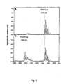
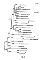

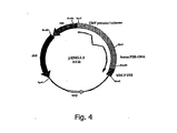

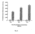
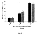
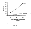

Classifications
-
- A—HUMAN NECESSITIES
- A61—MEDICAL OR VETERINARY SCIENCE; HYGIENE
- A61K—PREPARATIONS FOR MEDICAL, DENTAL OR TOILETRY PURPOSES
- A61K48/00—Medicinal preparations containing genetic material which is inserted into cells of the living body to treat genetic diseases; Gene therapy
- A61K48/005—Medicinal preparations containing genetic material which is inserted into cells of the living body to treat genetic diseases; Gene therapy characterised by an aspect of the 'active' part of the composition delivered, i.e. the nucleic acid delivered
- A61K48/0066—Manipulation of the nucleic acid to modify its expression pattern, e.g. enhance its duration of expression, achieved by the presence of particular introns in the delivered nucleic acid
-
- C—CHEMISTRY; METALLURGY
- C12—BIOCHEMISTRY; BEER; SPIRITS; WINE; VINEGAR; MICROBIOLOGY; ENZYMOLOGY; MUTATION OR GENETIC ENGINEERING
- C12N—MICROORGANISMS OR ENZYMES; COMPOSITIONS THEREOF; PROPAGATING, PRESERVING, OR MAINTAINING MICROORGANISMS; MUTATION OR GENETIC ENGINEERING; CULTURE MEDIA
- C12N9/00—Enzymes; Proenzymes; Compositions thereof; Processes for preparing, activating, inhibiting, separating or purifying enzymes
- C12N9/0004—Oxidoreductases (1.)
- C12N9/0071—Oxidoreductases (1.) acting on paired donors with incorporation of molecular oxygen (1.14)
-
- A—HUMAN NECESSITIES
- A61—MEDICAL OR VETERINARY SCIENCE; HYGIENE
- A61K—PREPARATIONS FOR MEDICAL, DENTAL OR TOILETRY PURPOSES
- A61K38/00—Medicinal preparations containing peptides
- A61K38/16—Peptides having more than 20 amino acids; Gastrins; Somatostatins; Melanotropins; Derivatives thereof
- A61K38/43—Enzymes; Proenzymes; Derivatives thereof
- A61K38/44—Oxidoreductases (1)
-
- A—HUMAN NECESSITIES
- A61—MEDICAL OR VETERINARY SCIENCE; HYGIENE
- A61K—PREPARATIONS FOR MEDICAL, DENTAL OR TOILETRY PURPOSES
- A61K38/00—Medicinal preparations containing peptides
- A61K38/16—Peptides having more than 20 amino acids; Gastrins; Somatostatins; Melanotropins; Derivatives thereof
- A61K38/43—Enzymes; Proenzymes; Derivatives thereof
- A61K38/46—Hydrolases (3)
- A61K38/465—Hydrolases (3) acting on ester bonds (3.1), e.g. lipases, ribonucleases
-
- A—HUMAN NECESSITIES
- A61—MEDICAL OR VETERINARY SCIENCE; HYGIENE
- A61P—SPECIFIC THERAPEUTIC ACTIVITY OF CHEMICAL COMPOUNDS OR MEDICINAL PREPARATIONS
- A61P15/00—Drugs for genital or sexual disorders; Contraceptives
-
- A—HUMAN NECESSITIES
- A61—MEDICAL OR VETERINARY SCIENCE; HYGIENE
- A61P—SPECIFIC THERAPEUTIC ACTIVITY OF CHEMICAL COMPOUNDS OR MEDICINAL PREPARATIONS
- A61P15/00—Drugs for genital or sexual disorders; Contraceptives
- A61P15/08—Drugs for genital or sexual disorders; Contraceptives for gonadal disorders or for enhancing fertility, e.g. inducers of ovulation or of spermatogenesis
-
- A—HUMAN NECESSITIES
- A61—MEDICAL OR VETERINARY SCIENCE; HYGIENE
- A61P—SPECIFIC THERAPEUTIC ACTIVITY OF CHEMICAL COMPOUNDS OR MEDICINAL PREPARATIONS
- A61P17/00—Drugs for dermatological disorders
-
- A—HUMAN NECESSITIES
- A61—MEDICAL OR VETERINARY SCIENCE; HYGIENE
- A61P—SPECIFIC THERAPEUTIC ACTIVITY OF CHEMICAL COMPOUNDS OR MEDICINAL PREPARATIONS
- A61P19/00—Drugs for skeletal disorders
-
- A—HUMAN NECESSITIES
- A61—MEDICAL OR VETERINARY SCIENCE; HYGIENE
- A61P—SPECIFIC THERAPEUTIC ACTIVITY OF CHEMICAL COMPOUNDS OR MEDICINAL PREPARATIONS
- A61P19/00—Drugs for skeletal disorders
- A61P19/08—Drugs for skeletal disorders for bone diseases, e.g. rachitism, Paget's disease
-
- A—HUMAN NECESSITIES
- A61—MEDICAL OR VETERINARY SCIENCE; HYGIENE
- A61P—SPECIFIC THERAPEUTIC ACTIVITY OF CHEMICAL COMPOUNDS OR MEDICINAL PREPARATIONS
- A61P25/00—Drugs for disorders of the nervous system
-
- A—HUMAN NECESSITIES
- A61—MEDICAL OR VETERINARY SCIENCE; HYGIENE
- A61P—SPECIFIC THERAPEUTIC ACTIVITY OF CHEMICAL COMPOUNDS OR MEDICINAL PREPARATIONS
- A61P25/00—Drugs for disorders of the nervous system
- A61P25/02—Drugs for disorders of the nervous system for peripheral neuropathies
-
- A—HUMAN NECESSITIES
- A61—MEDICAL OR VETERINARY SCIENCE; HYGIENE
- A61P—SPECIFIC THERAPEUTIC ACTIVITY OF CHEMICAL COMPOUNDS OR MEDICINAL PREPARATIONS
- A61P3/00—Drugs for disorders of the metabolism
-
- A—HUMAN NECESSITIES
- A61—MEDICAL OR VETERINARY SCIENCE; HYGIENE
- A61P—SPECIFIC THERAPEUTIC ACTIVITY OF CHEMICAL COMPOUNDS OR MEDICINAL PREPARATIONS
- A61P43/00—Drugs for specific purposes, not provided for in groups A61P1/00-A61P41/00
-
- C—CHEMISTRY; METALLURGY
- C12—BIOCHEMISTRY; BEER; SPIRITS; WINE; VINEGAR; MICROBIOLOGY; ENZYMOLOGY; MUTATION OR GENETIC ENGINEERING
- C12N—MICROORGANISMS OR ENZYMES; COMPOSITIONS THEREOF; PROPAGATING, PRESERVING, OR MAINTAINING MICROORGANISMS; MUTATION OR GENETIC ENGINEERING; CULTURE MEDIA
- C12N9/00—Enzymes; Proenzymes; Compositions thereof; Processes for preparing, activating, inhibiting, separating or purifying enzymes
- C12N9/0004—Oxidoreductases (1.)
- C12N9/0051—Oxidoreductases (1.) acting on a sulfur group of donors (1.8)
-
- C—CHEMISTRY; METALLURGY
- C12—BIOCHEMISTRY; BEER; SPIRITS; WINE; VINEGAR; MICROBIOLOGY; ENZYMOLOGY; MUTATION OR GENETIC ENGINEERING
- C12N—MICROORGANISMS OR ENZYMES; COMPOSITIONS THEREOF; PROPAGATING, PRESERVING, OR MAINTAINING MICROORGANISMS; MUTATION OR GENETIC ENGINEERING; CULTURE MEDIA
- C12N9/00—Enzymes; Proenzymes; Compositions thereof; Processes for preparing, activating, inhibiting, separating or purifying enzymes
- C12N9/14—Hydrolases (3)
- C12N9/16—Hydrolases (3) acting on ester bonds (3.1)
-
- C—CHEMISTRY; METALLURGY
- C12—BIOCHEMISTRY; BEER; SPIRITS; WINE; VINEGAR; MICROBIOLOGY; ENZYMOLOGY; MUTATION OR GENETIC ENGINEERING
- C12Y—ENZYMES
- C12Y108/00—Oxidoreductases acting on sulfur groups as donors (1.8)
- C12Y108/99—Oxidoreductases acting on sulfur groups as donors (1.8) with other acceptors (1.8.99)
-
- C—CHEMISTRY; METALLURGY
- C12—BIOCHEMISTRY; BEER; SPIRITS; WINE; VINEGAR; MICROBIOLOGY; ENZYMOLOGY; MUTATION OR GENETIC ENGINEERING
- C12Y—ENZYMES
- C12Y301/00—Hydrolases acting on ester bonds (3.1)
- C12Y301/06—Sulfuric ester hydrolases (3.1.6)
- C12Y301/06013—Iduronate-2-sulfatase (3.1.6.13)
-
- A—HUMAN NECESSITIES
- A61—MEDICAL OR VETERINARY SCIENCE; HYGIENE
- A61K—PREPARATIONS FOR MEDICAL, DENTAL OR TOILETRY PURPOSES
- A61K39/00—Medicinal preparations containing antigens or antibodies
- A61K2039/51—Medicinal preparations containing antigens or antibodies comprising whole cells, viruses or DNA/RNA
- A61K2039/53—DNA (RNA) vaccination
-
- A—HUMAN NECESSITIES
- A61—MEDICAL OR VETERINARY SCIENCE; HYGIENE
- A61K—PREPARATIONS FOR MEDICAL, DENTAL OR TOILETRY PURPOSES
- A61K38/00—Medicinal preparations containing peptides
-
- Y—GENERAL TAGGING OF NEW TECHNOLOGICAL DEVELOPMENTS; GENERAL TAGGING OF CROSS-SECTIONAL TECHNOLOGIES SPANNING OVER SEVERAL SECTIONS OF THE IPC; TECHNICAL SUBJECTS COVERED BY FORMER USPC CROSS-REFERENCE ART COLLECTIONS [XRACs] AND DIGESTS
- Y02—TECHNOLOGIES OR APPLICATIONS FOR MITIGATION OR ADAPTATION AGAINST CLIMATE CHANGE
- Y02A—TECHNOLOGIES FOR ADAPTATION TO CLIMATE CHANGE
- Y02A50/00—TECHNOLOGIES FOR ADAPTATION TO CLIMATE CHANGE in human health protection, e.g. against extreme weather
- Y02A50/30—Against vector-borne diseases, e.g. mosquito-borne, fly-borne, tick-borne or waterborne diseases whose impact is exacerbated by climate change
Definitions
- This invention relates to cells that coexpress a sulfatase and a C-formylyglycine generating enzyme and methods and uses thereof.
- Sulfatases are members of a highly conserved gene family, sharing extensive sequence homology ( Franco, B., et al., Cell, 1995, 81:15-25 ; Parenti, G., et al, Curr. Opin. Gen. Dev., 1997, 7:386-391 ), a high degree of structural similarity ( Bond, C.S., et al., Structure, 1997, 5:277-289 ; Lukatela, G., et al., Biochemistry, 1998, 37:3654-64 ), and a unique post-translational modification that is essential for sulfate ester cleavage ( Schmidt, B., et al, Cell, 1995, 82:271-278 ; Selmer, T., et al, Eur.
- the post-translational modification involves the oxidation of a conserved cysteine (in eukaryotes) or serine (in certain prokaryotes) residue, at C ⁇ , yielding L-C ⁇ -formylglycine (a.k.a. FGly; 2-amino-3-oxopropanoic acid ) in which an aldehyde group replaces the thiomethyl group of the side chain.
- the aldehyde is an essential part of the catalytic site of the sulfatase and likely acts as an aldehyde hydrate.
- One of the geminal hydroxyl groups accepts the sulfate during sulfate ester cleavage leading to the formation of a covalently sulfated enzyme intermediate.
- the other hydroxyl is required for the subsequent elimination of the sulfate and regeneration of the aldehyde group.
- This modification occurs in the endoplasmic reticulum during, or shortly after, import of the nascent sulfatase polypeptide and is directed by a short linear sequence surrounding the cysteine (or serine) residue to be modified.
- This highly conserved sequence is hexapeptide L/V-C(S)-X-P-S-R (SEQ ID NO:32), present in the N-terminal region of all eukaryotic sulfatases and most frequently carries a hydroxyl or thiol group on residue X ( Dierks, T., et al, Proc. Natl. Acad Sci. U. S. A., 1997, 94:11963-11968 ).
- WO 01/60991 discloses human kinases (PKIN) and polynucleotides that identify and encode PKIN. It also discusses suitable expression vectors, host cells, antibodies, agonists, and antagonists, as well as methods for diagnosing, treating, or preventing disorders associated with aberrant expression of PKIN.
- PKIN human kinases
- the CTPSR motif was transferred to a heterologous protein leading to low-efficient formylglycine formation. Efficiency is said to have reached control values when seven additional residues (AALLTGR) directly following the CTPSR motif in arylsulfatase A were present. Mutating up to four residues simultaneously within this heptamer sequence is indicated to have inhibited the modification only moderately. It was concluded that AALLTGR may have an auxiliary function in presenting the core motif to the modifying enzyme and that within the two motifs, the key residues are fully conserved, and other residues are highly conserved among all known members of the sulfatase family.
- sulfatase genes have been identified in humans. They encode enzymes with different substrate specificity and subcellular localization such as lysosomes, Golgi and ER.
- ARSC encoding arylsulfatase C, D, E and F, respectively.
- Xp22.3 chromosomal region
- sulfatases in human metabolism is underscored by the identification of at least eight human monogenic diseases caused by the deficiency of individual sulfatase activities. Most of these conditions are lysosomal storage disorders in which phenotypic consequences derive from the type and tissue distribution of the stored material. Among them are five different types of mucopolysaccharidoses (MPS types II, IIIA, IIID, IVA, and VI) due to deficiencies of sulfatases acting on the catabolism of glycosaminogl yeans ( Neufeld and Muenzer, 2001, The mucopolysaccharidoses, In The Metabolic and Molecular Bases of Inherited Disease, CR. Scriver, A.L.
- MLD metachromatic leukodystrophy
- Sulfatases are also implicated in drug-induced human malformation syndromes, such as Warfarin embryopathy, caused by inhibition of ARSE activity due to in utero exposure to warfarin during pregnancy.
- MSD multiple sulfatase deficiency
- all sulfatase activities are simultaneously defective. Consequently, the phenotype of this severe multisystemic disease combines the features observed in individual sulfatase deficiencies.
- Cells from patients with MSD are deficient in sulfatase activities even after transfection with cDNAs encoding human sulfatases, suggesting the presence of a common mechanism required for the activity of all sulfatases ( Rommerskirch and von Figura, Proc. Natl. Acad Sci., USA, 1992, 89:2561-2565 ).
- This invention provides a cell according to any of claims 1 to 15, a method according to any of claims 1 to 23, or a medical use according to claim 24 or 25.
- FGE F ormylglycine G enerating E nzyme
- FGE enhances the activity of sulfatases, including, but not limited to, Iduronate 2-Sulfatase, Sulfamidase, N-Acetylgalactosamine 6-Sulfatase, N-Acetylglucosamine 6-Sulfatase, Arylsulfatase A, Arylsulfatase B, Arylsulfatase C, Arylsulfatase D, Arylsulfatase E, Arylsulfatase F, Arylsulfatase G, HSulf-1, HSulf-2, HSulf-3, HSulf-4, HSulf-5, and HSulf-6.
- the molecules of the present invention can be used in the diagnosis and treatment of Multiple Sulfatase Deficiency as well as other sulfatase deficiencies.
- a sulfatase-producing cell wherein the ratio of active sulfatase to total sulfatase produced by the cell is increased, is provided, as set forth in claim 1.
- the cell may comprise: (i) a sulfatase with an increased expression, and (ii) a Formylglycine Generating Enzyme with an increased expression, wherein the ratio of active sulfatase to total sulfatase (i.e., the specific activity of the sulfatase) produced by the cell is increased by at least 5% over the ratio of active sulfatase to total sulfatase produced by the cell in the absence of the Formylglycine Generating Enzyme.
- the ratio of active sulfatase to total sulfatase produced by the cell is increased by at least 10%, 15%, 20%, 50%, 100%), 200%), 500%, 1000%, over the ratio of active sulfatase to total sulfatase produced by the cell in the absence of the Formylglycine. Generating Enzyme.
- the invention is set out in claims 1 to 25. It is based upon the discovery of the gene that encodes Formylglycine Generating Enzyme (FGE), an enzyme responsible for the unique post-translational modification occurring on sulfatases that is essential for sulfatase function: the formation of L-C ⁇ -formylglycine (a.k.a. FGly and/or 2-amino-3-oxopropanoic acid ). It has been discovered, unexpectedly, that mutations in the FGE gene lead to the development of Multiple Sulfatase Deficiency (MSD) in subjects.
- FGE Formylglycine Generating Enzyme
- FGE enhances the activity of sulfatases, including, but not limited to, Iduronate 2-Sulfatase, Sulfamidase, N-Acetylgalactosamine 6-Sulfatase, N-Acetylglucosamine 6-Sulfatase, Arylsulfatase A, Arylsulfatase B, Arylsulfatase C, Arylsulfatase D, Arylsulfatase E, Arylsulfatase F, Arylsulfatase G, HSulf-1, HSulf-2, HSulf-3, HSulf-4, HSulf-5, and HSulf-6, and sulfatases described in U.S. Provisional applications with publication numbers 20030073118 , 20030147875 , 20030148920 , 20030162279 , and 20030166283 .
- C ⁇ -formylglycine generating activity refers to the ability of a molecule to form, or enhance the formation of, FGly on a substrate.
- the substrate may be a sulfatase as described elsewhere herein, or a synthetic oligopeptide (see, e.g., SEQ ID NO:33, and the Examples).
- the substrate preferably contains the conserved hexapeptide of SEQ ID NO:32 [L/V-C(S)-X-P-S-R]. Methods for assaying FGly formation are as described in the art (see, e.g., Dierks, T., et al., Proc. Natl. Acad. Sci. U. S.
- a "molecule,” as used herein, embraces both “nucleic acids” and “polypeptides.” FGE molecules are capable of forming, or enhancing/increasing formation of, FGly both in vivo and in vitro.
- Enhancing (or “increasing”)” C ⁇ -formylglycine generating activity typically refers to increased expression of FGE and/or its encoded polypeptide.
- Increased expression refers to increasing (i.e., to a detectable extent) replication, transcription, and/or translation of any of the nucleic acids of the invention (FGE nucleic acids as described elsewhere herein), since upregulation of any of these processes results in concentration/amount increase of the polypeptide encoded by the gene (nucleic acid).
- Enhancing (or increasing) C ⁇ -formylglycine generating activity also refers to preventing or inhibiting FGE degradation (e.g., via increased ubiquitinization), downregulation, etc., resulting, for example, in increased or stable FGE molecule t 1/2 (half-life) when compared to a control.
- Downregulation or decreased expression refers to decreased expression of a gene and/or its encoded polypeptide.
- the upregulation or downregulation of gene expression can be directly determined by detecting an increase or decrease, respectively, in the level of mRNA for the gene (e.g., FGE), or the level of protein expression of the gene-encoded polypeptide, using any suitable means known to the art, such as nucleic acid hybridization or antibody detection methods, respectively, and in comparison to controls.
- Upregulation or downregulation of FGE gene expression can also be determined indirectly by detecting a change in C ⁇ -formylglycine generating activity.
- “Expression,” as used herein, refers to nucleic acid and/or polypeptide expression, as well as to activity of the polypeptide molecule (e.g., C ⁇ -formylglycine generating activity of the molecule).
- a subject is a mammal or a non-human mammal. In all embodiments human FGE and human subjects are preferred.
- isolated means: (i) amplified in vitro by, for example, polymerase chain reaction (PCR); (ii) recombinantly produced by cloning; (iii) purified, as by cleavage and gel separation; or (iv) synthesized by, for example, chemical synthesis.
- An isolated nucleic acid is one which is readily manipulated by recombinant DNA techniques well known in the art.
- PCR polymerase chain reaction
- An isolated nucleic acid may be substantially purified, but need not be.
- a nucleic acid that is isolated within a cloning or expression vector is not pure in that it may comprise only a tiny percentage of the material in the cell in which it resides.
- Such a nucleic acid is isolated, however, as the term is used herein because it is readily manipulated by standard techniques known to those of ordinary skill in the art.
- isolated means separated from its native environment in sufficiently pure form so that it can be manipulated or used for any one of the purposes of described herein. Thus, isolated means sufficiently pure to be used (i) to raise and/or isolate antibodies, (ii) as a reagent in an assay, (iii) for sequencing, (iv) as a therapeutic, etc.
- Homologs and alleles of the FGE nucleic acids described herein also having C ⁇ -formylglycine generating activity can be provided.
- Homologs, as described herein, include the molecules identified elsewhere herein (see e.g., SEQ ID NOs:4, 5, 45-78, and 80-87) i.e. orthologs and paralogs. Further homologs can be identified following the teachings of the present invention as well as by conventional techniques. Since the FGE homologs described herein all share C ⁇ -formylglycine generating activity, they can be used interchangeably with the human FGE molecule in all aspects of the invention.
- homologs and alleles typically will share at least 40% nucleotide identity and/or at least 50% amino acid identity to SEQ ID NO:1 and SEQ ID NO:2, respectively, in some instances will share at least 50% nucleotide identity and/or at least 65% amino acid identity and in still other instances will share at least 60% nucleotide identity and/or at least 75% amino acid identity. In further instances, homologs and alleles typically will share at least 90%), 95%, or even 99%) nucleotide identity and/or at least 95%, 9S ⁇ o, or even 99% amino acid identity to SEQ ID NO: 1 and SEQ ID NO:2, respectively.
- the homology can be calculated using various, publicly available software tools developed by NCBI (Bethesda, Maryland).
- Exemplary tools include the heuristic algorithm of Altschul SF, et al., (J Mol Biol, 1990, 215:403-410 ), also known as BLAST. Pairwise and ClustalW alignments (BLOSUM30 matrix setting) as well as Kyte-Doolittle hydropathic analysis can be obtained using public (EMBL, Heidelberg, Germany) and commercial (e.g., the MacNector sequence analysis software from Oxford Molecular Group/enetics Computer Group, Madison, WI). Watson-Crick complements of the foregoing nucleic acids can also be used.
- a Southern blot may be performed using the foregoing conditions, together with a radioactive probe. After washing the membrane to which the DNA is finally transferred, the membrane can be placed against X-ray film or a phosphoimager plate to detect the radioactive signal.
- Degenerate nucleic acids which include alternative codons to those present in the native materials can be provided.
- serine residues are encoded by the codons TCA, AGT, TCC, TCG, TCT and AGC.
- any of the serine-encoding nucleotide triplets may be employed to direct the protein synthesis apparatus, in vitro or in vivo, to incorporate a serine residue into an elongating FGE polypeptide.
- nucleotide sequence triplets which encode other amino acid residues include, but are not limited to: CCA, CCC, CCG and CCT (proline codons); CGA, CGC, CGG, CGT, AGA and AGG (arginine codons); ACA, ACC, ACG and ACT (threonine codons); AAC and AAT (asparagine codons); and ATA, ATC and ATT (isoleucine codons).
- Other amino acid residues may be encoded similarly by multiple nucleotide sequences.
- degenerate nucleic acids that differ from the biologically isolated nucleic acids in codon sequence due to the degeneracy of the genetic code can be provided.
- a unique fragment is one that is a 'signature' for the larger nucleic acid.
- the unique fragment is long enough to assure that its precise sequence is not found in molecules within the human genome outside of the FGE nucleic acids defined above (and human alleles).
- Those of ordinary skill in the art may apply no more than routine procedures to determine if a fragment is unique within the human genome.
- Unique fragments exclude fragments completely composed of the nucleotide sequences selected from the group consisting of SEQ ID NO:4, and/or other previously published sequences as of the filing date of this application.
- a fragment which is completely composed of the sequence described in the foregoing GenBank deposits is one which does not include any of the nucleotides unique to the sequences of the invention.
- a unique fragment according to the invention must contain a nucleotide sequence other than the exact sequence of those in the GenBank deposits or fragments thereof. The difference may be an addition, deletion or substitution with respect to the GenBank sequence or it may be a sequence wholly separate from the GenBank sequence.
- Unique fragments can be used as probes in Southern and Northern blot assays to identify such nucleic acids, or can be used in amplification assays such as those employing PCR. As known to those skilled in the art, large probes such as 200, 250, 300 or more nucleotides are preferred for certain uses such as Southern and Northern blots, while smaller fragments will be preferred for uses such as PCR. Unique fragments also can be used to produce fusion proteins for generating antibodies or determining binding of the polypeptide fragments, as demonstrated in the Examples, or for generating immunoassay components. Likewise, unique fragments can be employed to produce nonfused fragments of the FGE polypeptides, useful, for example, in the preparation of antibodies, immunoassays or therapeutic applications. Unique fragments further can be used as antisense molecules to inhibit the expression of FGE nucleic acids and polypeptides respectively.
- the size of the unique fragment will depend upon its conservancy in the genetic code. Thus, some regions of SEQ ID NO:1 or SEQ ID NO: 3 and complements will require longer segments to be unique while others will require only short segments, typically between 12 and 32 nucleotides long (e.g. 12, 13, 14, 15, 16, 17, 18, 19, 20, 21, 22, 23, 24, 25, 26, 27, 28, 29, 30, 31 and 32 bases) or more, up to the entire length of the disclosed sequence.
- this disclosure intends to embrace each and every fragment of each sequence, beginning at the first nucleotide, the second nucleotide and so on, up to 8 nucleotides short of the end, and ending anywhere from nucleotide number 8, 9, 10 and so on for each sequence, up to the very last nucleotide, (provided the sequence is unique as described above).
- Virtually any segment of the region of SEQ ID NO:1 beginning at nucleotide 1 and ending at nucleotide 1180, or SEQ ID NO:3 beginning at nucleotide 1 and ending at nucleotide 1122, or complements thereof, that is 20 or more nucleotides in length will be unique.
- the cells of the invention can be provided by methods for increasing C ⁇ -formylglycine generating activity in a cell. In important embodiments, this is accomplished by the use of vectors ("expression vectors” and/or “targeting vectors").
- Vectors may be any of a number of nucleic acids into which a desired sequence may be inserted by restriction and ligation for transport between different genetic environments or for expression in a host cell.
- Vectors are typically composed of DNA although RNA vectors are also available.
- Vectors include, but are not limited to, plasmids, phagemids and virus genomes.
- a cloning vector is one which is able to replicate in a host cell, and which is further characterized by one or more endonuclease restriction sites at which the vector may be cut in a determinable fashion and into which a desired DNA sequence may be ligated such that the new recombinant vector retains its ability to replicate in the host cell.
- replication of the desired sequence may occur many times as the plasmid increases in copy number within the host bacterium or just a single time per host before the host reproduces by mitosis. In the case of phage, replication may occur actively during a lytic phase or passively during a lysogenic phase.
- An "expression vector" is one into which a desired DNA sequence (e.g., the FGE cDNA of SEQ ID NO:3) may be inserted by restriction and ligation such that it is operably joined to regulatory sequences and may be expressed as an RNA transcript Vectors may further contain one or more marker sequences suitable for use in the identification of cells which have or have not been transformed or transfected with the vector.
- Markers include, for example, genes encoding proteins which increase or decrease either resistance or sensitivity to antibiotics or other compounds, genes which encode enzymes whose activities are detectable by standard assays known in the art (e.g., ⁇ -galactosidase or alkaline phosphatase), and genes which visibly affect the phenotype of transformed or transfected cells, hosts, colonies or plaques (e.g., green fluorescent protein).
- a “targeting vector” is one which typically contains targeting constructs/sequences that are used, for example, to insert a regulatory sequence within an endogenous gene (e.g., within the sequences of an exon and/or intron), within the endogenous gene promoter sequences, or upstream of the endogenous gene promoter sequences.
- a targeting vector may contain the gene of interest (e.g., encoded by the cDNA of SEQ ID NO:1) and other sequences necessary for the targeting of the gene to a preferred location in the genome (e.g., a transcriptionally active location, for example downstream of an endogenous promoter of an unrelated gene). Construction of targeting constructs and vectors are described in detail in U.S. Patents 5,641,670 and 6,270,989 .
- any cells, prokaryotic or eukaryotic, which can be transformed with heterologous DNA or RNA and which can be grown or maintained in culture may be used in the practice of the invention.
- Examples include bacterial cells such as Escherichia coli, insect cells, and mammalian cells such as human, mouse, hamster, pig, goat, primate, etc. They may be primary or secondary cell strains (which exhibit a finite number of mean population doublings in culture and are not immortalized) and immortalized cell lines (which exhibit an apparently unlimited lifespan in culture).
- Primary and secondary cells include, for example, fibroblasts, keratinocytes, epithelial cells (e.g., mammary epithelial cells, intestinal epithelial cells), endothelial cells, glial cells, neural cells, formed elements of the blood (e.g., lymphocytes, bone marrow cells), muscle cells and precursors of these somatic cell types including embryonic stem cells.
- epithelial cells e.g., mammary epithelial cells, intestinal epithelial cells
- endothelial cells e.g., glial cells, neural cells, formed elements of the blood (e.g., lymphocytes, bone marrow cells), muscle cells and precursors of these somatic cell types including embryonic stem cells.
- primary cells are preferably obtained from the individual to whom the manipulated cells are administered. However, primary cells can be obtained from a donor (other than the recipient) of the same species.
- immortalized human cell lines which may be used with the DNA constructs and methods of the present invention include, but are not limited to, HT-1080 cells (ATCC CCL 121), HeLa cells and derivatives of HeLa cells (ATCC CCL 2, 2.1 and 2.2), MCF-7 breast cancer cells (ATCC BTH 22), K-562 leukemia cells (ATCC CCL 243), KB carcinoma cells (ATCC CCL 17), 2780AD ovarian carcinoma cells ( Van der Buck, A. M.
- fibroblast strains such as WI-38 (ATCC CCL 75) and MRC-5 (ATCC CCL 171) may also be used. Further discussion of the types of cells that may be used in practicing the methods of the present invention are described in U.S. Patents 5,641,670 and 6,270,989 . Cell-free transcription systems also may be used in lieu of cells.
- the cells of the invention are maintained under conditions, as are known in the art, which result in expression of the FGE protein or functional fragments thereof. Proteins expressed using the methods described may be purified from cell lysates or cell supernatants. Proteins made according to this method can be prepared as a pharmaceutically-useful formulation and delivered to a human or non-human animal by conventional pharmaceutical routes as is known in the art (e.g., oral, intravenous, intramuscular, intranasal, intratracheal or subcutaneous). As described elsewhere herein, the recombinant cells can be immortalized, primary, or secondary cells, preferably human. The use of cells from other species may be desirable in cases where the non-human cells are advantageous for protein production purposes where the non-human FGE produced is useful therapeutically.
- a coding sequence and regulatory sequences are said to be "operably” joined when they are covalently linked in such a way as to place the expression or transcription of the coding sequence under the influence or control of the regulatory sequences.
- two DNA sequences are said to be operably joined if induction of a promoter in the 5'regulatory sequences results in the transcription of the coding sequence and if the nature of the linkage between the two DNA sequences does not (1) result in the introduction of a frame-shift mutation, (2) interfere with the ability of the promoter region to direct the transcription of the coding sequences, or (3) interfere with the ability of the corresponding RNA transcript to be translated into a protein.
- a promoter region would be operably joined to a coding sequence if the promoter region were capable of effecting transcription of that DNA sequence such that the resulting transcript might be translated into the desired protein or polypeptide.
- regulatory sequences needed for gene expression may vary between species or cell types, but shall in general include, as necessary, 5' non-transcribed and 5' non-translated sequences involved with the initiation of transcription and translation respectively, such as a TATA box, capping sequence, CAAT sequence, and the like.
- 5' non-transcribed regulatory sequences will include a promoter region which includes a promoter sequence for transcriptional control of the operably joined gene.
- Regulatory sequences may also include enhancer sequences or upstream activator sequences as desired.
- the vectors may optionally include 5' leader or signal sequences. The choice and design of an appropriate vector is within the ability and discretion of one of ordinary skill in the art.
- RNA heterologous DNA
- FGE polypeptide FGE polypeptide or fragment or variant thereof. That heterologous DNA (RNA) is placed under operable control of transcriptional elements to permit the expression of the heterologous DNA in the host cell.
- Preferred systems for mRNA expression in mammalian cells are those such as pRc/CMV (available from Invitrogen, Carlsbad, CA) that contain a selectable marker such as a gene that confers G418 resistance (which facilitates the selection of stably transfected cell lines) and the human cytomegalovirus (CMV) enhancer-promoter sequences.
- pRc/CMV available from Invitrogen, Carlsbad, CA
- CMV human cytomegalovirus
- suitable for expression in primate or canine cell lines is the pCEP4 vector (Invitrogen, Carlsbad, CA), which contains an Epstein Barr virus (EBV) origin of replication, facilitating the maintenance of plasmid as a multicopy extrachromosomal element.
- EBV Epstein Barr virus
- Another expression vector is the pEF-BOS plasmid containing the promoter of polypeptide Elongation Factor 1 ⁇ , which stimulates efficiently transcription in vitro.
- the plasmid is described by Mishizuma and Nagata (Nuc. Acids Res. 18:5322, 1990 ), and its use in transfection experiments is disclosed by, for example, Demoulin ( Mol. Cell. Biol. 16:4710-4716, 1996 ).
- Still another preferred expression vector is an adenovirus, described by Stratford-Perricaudet, which is defective for E1 and E3 proteins ( J. Clin. Invest. 90:626-630, 1992 ).
- adenovirus as an Adeno.PIA recombinant is disclosed by Warmer et al., in intradermal injection in mice for immunization against P1A ( Int. J. Cancer, 67:303-310, 1996 ).
- Expression kits can be provided, which allow the artisan to prepare a desired expression vector or vectors. Such expression kits include at least separate portions of each of the previously discussed coding sequences. Other components may be added, as desired, as long as the previously mentioned sequences, which are required, are included.
- FGE cDNA sequence containing expression vectors can be used to transfect host cells and cell lines, be these prokaryotic (e.g., Escherichia coli), or eukaryotic (e.g., CHO cells, COS cells, yeast expression systems and recombinant baculovirus expression in insect cells).
- prokaryotic e.g., Escherichia coli
- eukaryotic e.g., CHO cells, COS cells, yeast expression systems and recombinant baculovirus expression in insect cells.
- mammalian cells such as human, mouse, hamster, pig, goat, primate, etc. They may be of a wide variety of tissue types, and include primary cells and immortalized cell lines as described elsewhere herein. Specific examples include HT-1080 cells, CHO cells, dendritic cells, U293 cells, peripheral blood leukocytes, bone marrow stem cells, embryonic stem cells, and insect cells.
- Isolated polypeptides (including whole proteins and partial proteins), encoded by the foregoing FGE nucleic acid can be provided, and include the polypeptide of SEQ ID NO: 2 and unique fragments thereof. Such polypeptides are useful, for example, alone or as part of fusion proteins to generate antibodies, as components of an immunoassay, etc.
- Polypeptides can be isolated from biological samples including tissue or cell homogenates, and can also be expressed recombinantly in a variety of prokaryotic and eukaryotic expression systems by constructing an expression vector appropriate to the expression system, introducing the expression vector into the expression system, and isolating the recombinantly expressed protein. Short polypeptides, including antigenic peptides (such as are presented by MHC molecules on the surface of a cell for immune recognition) also can be synthesized chemically using well-established methods of peptide synthesis.
- a unique fragment of a FGE polypeptide in general, has the features and characteristics of unique fragments as discussed above in connection with nucleic acids. As will be recognized by those skilled in the art, the size of the unique fragment will depend upon factors such as whether the fragment constitutes a portion of a conserved protein domain. Thus, some regions of SEQ ID NO:2 will require longer segments to be unique while others will require only short segments, typically between 5 and 12 amino acids (e.g. 5,6, 7, 8, 9, 10, 11 and 12 amino acids long or more, including each integer up to the full length, 287 amino acids long).
- Unique fragments of a polypeptide preferably are those fragments which retain a distinct functional capability of the polypeptide.
- Functional capabilities which can be retained in a unique fragment of a polypeptide include interaction with antibodies, interaction with other polypeptides or fragments thereof, interaction with other molecules, etc.
- One important activity is the ability to act as a signature for identifying the polypeptide.
- Those skilled in the art are well versed in methods for selecting unique amino acid sequences, typically on the basis of the ability of the unique fragment to selectively distinguish the sequence of interest from non-family members. A comparison of the sequence of the fragment to those on known databases typically is all that is necessary.
- a "variant" of a FGE polypeptide is a polypeptide which contains one or more modifications to the primary amino acid sequence of a FGE polypeptide. Modifications which create a FGE polypeptide variant are typically made to the nucleic acid which encodes the FGE polypeptide, and can include deletions, point mutations, truncations, amino acid substitutions and addition of amino acids or non-amino acid moieties to: 1) reduce or eliminate an activity of a FGE polypeptide; 2) enhance a property of a FGE polypeptide, such as protein stability in an expression system or the stability of protein-ligand binding; 3) provide a novel activity or property to a FGE polypeptide, such as addition of an antigenic epitope or addition of a detectable moiety; or 4) to provide equivalent or better binding to a FGE polypeptide receptor or other molecule.
- modifications can be made directly to the polypeptide, such as by cleavage, addition of a linker molecule, addition of a detectable moiety, such as biotin, addition of a fatty acid, and the like. Modifications also embrace fusion proteins comprising all or part of the FGE amino acid sequence.
- One of skill in the art will be familiar with methods for predicting the effect on protein conformation of a change in protein sequence, and can thus "design" a variant FGE polypeptide according to known methods: One example of such a method is described by Dahiyat and Mayo in Science 278:82-87, 1997 , whereby proteins can be designed de novo. The method can be applied to a known protein to vary only a portion of the polypeptide sequence. By applying the computational methods of Dahiyat and Mayo, specific variants of the FGE polypeptide can be proposed and tested to determine whether the variant retains a desired conformation.
- Variants can include FGE polypeptides which are modified specifically to alter a feature of the polypeptide unrelated to its physiological activity. For example, cysteine residues can be substituted or deleted to prevent unwanted disulfide linkages. Similarly, certain amino acids can be changed to enhance expression of a FGE polypeptide by eliminating proteolysis by proteases in an expression system (e.g., dibasic amino acid residues in yeast expression systems in which KEX2 protease activity is present).
- Mutations of a nucleic acid which encodes a FGE polypeptide preferably preserve the amino acid reading frame of the coding sequence, and preferably do not create regions in the nucleic acid which are likely to hybridize to form secondary structures, such a hairpins or loops, which can be deleterious to expression of the variant polypeptide.
- Mutations can be made by selecting an amino acid substitution, or by random mutagenesis of a selected site in a nucleic acid which encodes the polypeptide. Variant polypeptides are then expressed and tested for one or more activities to determine which mutation provides a variant polypeptide with the desired properties. Further mutations can be made to variants (or to non-variant FGE polypeptides) which are silent as to the amino acid sequence of the polypeptide, but which provide preferred codons for translation in a particular host, or alter the structure of the mRNA to, for example, enhance stability and/or expression. The preferred codons for translation of a nucleic acid in, e.g., Escherichia coli, mammalian cells, etc. are well known to those of ordinary skill in the art. Still other mutations can be made to the noncoding sequences of a FGE gene or cDNA clone to enhance expression of the polypeptide.
- conservative amino acid substitutions may be made in FGE polypeptides to provide functionally equivalent variants of the foregoing polypeptides, i.e, the variants retain the functional capabilities of the FGE polypeptides.
- a "conservative amino acid substitution” refers to an amino acid substitution which does not significantly alter the the tertiary structure and/or activity of the polypeptide.
- Variants can be prepared according to methods for altering polypeptide sequence known to one of ordinary skill in the art, and include those that are found in references which compile such methods, e.g. Molecular Cloning: A Laboratory Manual, J.
- FGE polypeptides include conservative amino acid substitutions of SEQ ID NO:2.
- Conservative substitutions of amino acids include substitutions made amongst amino acids within the following groups: (a) M, I, L, V; (b) F, Y, W; (c) K, R, H; (d) A, G; (e) S, T; (f) Q, N; and (g) E, D.
- variants of FGE polypeptides i.e., variants of FGE polypeptides which retain the function of the natural FGE polypeptides
- amino acid sequence of FGE polypeptides to produce functionally equivalent variants of FGE polypeptides typically are made by alteration of a nucleic acid encoding FGE polypeptides (SEQ ID NOs:1, 3).
- substitutions can be made by a variety of methods known to one of ordinary skill in the art. For example, amino acid substitutions may be made by PCR-directed mutation, site-directed mutagenesis according to the method of Kunkel ( Kunkel, Proc. Not. Acad. Sci. U.S.A.
- FGE polypeptides can be tested by cloning the gene encoding the altered FGE polypeptide into a bacterial or mammalian expression vector, introducing the vector into an appropriate host cell, expressing the altered FGE polypeptide, and testing for a functional capability of the FGE polypeptides as disclosed herein (e.g., C ⁇ -formylglycine generating activity, etc.).
- FGE polypeptides include, but are not limited to, immunochromatography, HPLC, size-exclusion chromatography, ion-exchange chromatography and immune-affinity chromatography.
- FGE polypeptides preferably are produced recombinantly, although such polypeptides may be isolated from biological extracts.
- Recombinantly produced FGE polypeptides include chimeric proteins comprising a fusion of a FGE protein with another polypeptide, e.g., a polypeptide capable of providing or enhancing protein-protein binding, sequence specific nucleic acid binding (such as GAL4), enhancing stability of the FGE polypeptide under assay conditions, or providing a detectable moiety, such as green fluorescent protein.
- a polypeptide fused to a FGE polypeptide or fragment may also provide means of readily detecting the fusion protein, e.g., by immunological recognition or by fluorescent labeling.
- a method for increasing C ⁇ -formylglycine generating activity in a cell involves contacting the cell with an isolated nucleic acid molecule of the invention (e.g., a nucleic acid of SEQ ID NO. l), or an expression product thereof (e.g., a peptide of SEQ ID NO.2), in an amount effective to increase C ⁇ -formylglycine generating activity in the cell.
- the method involves activating the endogenous FGE gene to increase C ⁇ -formylglycine generating activity in the cell.
- the nucleic acid may be operatively coupled to a gene expression sequence which directs the expression of the nucleic acid molecule within a eukaryotic cell such as an HT-1080 cell.
- the "gene expression sequence” is any regulatory nucleotide sequence, such as a promoter sequence or promoter-enhancer combination, which facilitates the efficient transcription and translation of the nucleic acid to which it is operably linked.
- the gene expression sequence may, for example, be a mammalian or viral promoter, such as a constitutive or inducible promoter.
- Constitutive mammalian promoters include, but are not limited to, the promoters for the following genes: hypoxanthine phosphoribosyl transferase (HPTR), adenosine deaminase, pyruvate kinase, ⁇ -actin promoter and other constitutive promoters.
- HPTR hypoxanthine phosphoribosyl transferase
- adenosine deaminase pyruvate kinase
- ⁇ -actin promoter ⁇ -actin promoter
- Exemplary viral promoters which function constitutively in eukaryotic cells include, for example, promoters from the simian virus, papilloma virus, adenovirus, human immunodeficiency virus (HIV), Rous sarcoma virus, cytomegalovirus, the long terminal repeats (LTR) of moloney leukemia virus and other retroviruses, and the thymidine kinase promoter of herpes simplex virus.
- Other constitutive promoters are known to those of ordinary skill in the art.
- the promoters useful as gene expression sequences of the invention also include inducible promoters. Inducible promoters are activated in the presence of an inducing agent. For example, the metallothionein promoter is activated to increase transcription and translation in the presence of certain metal ions. Other inducible promoters are known to those of ordinary skill in the art.
- the gene expression sequence shall include, as necessary, 5' non-transcribing and 5' non-translating sequences involved with the initiation of transcription and translation, respectively, such as a TATA box, capping sequence, CAAT sequence, and the like.
- 5' non-transcribing sequences will include a promoter region which includes a promoter sequence for transcriptional control of the operably joined nucleic acid.
- the gene expression sequences optionally includes enhancer sequences or upstream activator sequences as desired.
- any of the FGE nucleic acid molecules described herein is linked to a gene expression sequence which permits expression of the nucleic acid molecule in a cell of a specific cell lineage, e.g., a neuron.
- a sequence which permits expression of the nucleic acid molecule in a cell such as a neuron is one which is selectively active in such a cell type, thereby causing expression of the nucleic acid molecule in these cells.
- the synapsin-1 promoter for example, can be used to express any of the foregoing nucleic acid molecules of the invention in a neuron; and the von Willebrand factor gene promoter, for example, can be used to express a nucleic acid molecule in a vascular endothelial cell.
- the von Willebrand factor gene promoter for example, can be used to express a nucleic acid molecule in a vascular endothelial cell.
- nucleic acid sequence and the gene expression sequence are said to be "operably linked” when they are covalently linked in such a way as to place the transcription and/or translation of the nucleic acid coding sequence (e.g., in the case of FGE, SEQ ID NO. 3) under the influence or control of the gene expression sequence.
- nucleic acid sequence be translated into a functional protein
- two DNA sequences are said to be operably linked if induction of a promoter in the 5' gene expression sequence results in the transcription of the nucleic acid sequence and if the nature of the linkage between the two DNA sequences does not (1) result in the introduction of a frame-shift mutation, (2) interfere with the ability of the promoter region to direct the transcription of the nucleic acid sequence, and/or (3) interfere with the ability of the corresponding RNA transcript to be-translated into a protein.
- a gene expression sequence would be operably linked to a nucleic acid sequence if the gene expression sequence were capable of effecting transcription of that nucleic acid sequence such that the resulting transcript might be translated into the desired protein or polypeptide.
- a "vector” is any vehicle capable of facilitating: (1) delivery of a molecule to a target cell and/or (2) uptake of the molecule by a target cell.
- the delivery vectors transport the molecule into the target cell with reduced degradation relative to the extent of degradation that would result in the absence of the vector.
- a "targeting ligand” can be attached to the vector to selectively deliver the vector to a cell which expresses on its surface the cognate receptor for the targeting ligand.
- the vector (containing a nucleic acid or a protein) can be selectively delivered to a neuron.
- Methodologies for targeting include conjugates, such as those described in U.S. Patent 5,391,723 to Priest.
- Another example of a well-known targeting vehicle is a liposome. Liposomes are commercially available from Gibco BRL. Numerous methods are published for making targeted liposomes.
- useful vectors include, but are not limited to, plasmids, phagemids, viruses, other vehicles derived from viral or bacterial sources that have been manipulated by the insertion or incorporation of the nucleic acid sequences described herein and additional nucleic acid fragments (e.g., enhancers, promoters) which can be attached to the nucleic acid sequences.
- additional nucleic acid fragments e.g., enhancers, promoters
- Viral vectors are a preferred type of vector and include, but are not limited to, nucleic acid sequences from the following viruses: adenovirus; adeno-associated virus; retrovirus, such as moloney murine leukemia virus; harvey murine sarcoma virus; murine mammary tumor virus; rouse sarcoma virus; SV40-type viruses; polyoma viruses; Epstein-Barr viruses; papilloma viruses; herpes virus; vaccinia virus; polio virus; and RNA virus such as a retrovirus.
- viruses include, but are not limited to, nucleic acid sequences from the following viruses: adenovirus; adeno-associated virus; retrovirus, such as moloney murine leukemia virus; harvey murine sarcoma virus; murine mammary tumor virus; rouse sarcoma virus; SV40-type viruses; polyoma viruses; Epstein-Barr viruses; papillo
- a particularly preferred virus for certain applications is the adeno-associated virus, a double-stranded DNA virus.
- the adeno-associated virus is capable of infecting a wide range of cell types and species and can be engineered to be replication-deficient. It further has advantages, such as heat and lipid solvent stability, high transduction frequencies in cells of diverse lineages, including hematopoietic cells, and lack of superinfection inhibition thus allowing multiple series of transductions.
- the adeno-associated virus can integrate into human cellular DNA in a site-specific manner, thereby minimizing the possibility of insertional mutagenesis and variability of inserted gene expression.
- adeno-associated virus infections have been followed in tissue culture for greater than 100 passages in the absence of selective pressure, implying that the adeno-associated virus genomic integration is a relatively stable event
- the adeno-associated virus can also function in an extrachromosomal fashion.
- Non-cytopathic viruses include retroviruses, the life cycle of which involves reverse transcription of genomic viral RNA into DNA with subsequent proviral integration into host cellular DNA.
- retroviruses and retroviruses have been approved for human gene therapy trials.
- the retroviruses are replication-deficient (i.e., capable of directing synthesis of the desired proteins, but incapable of manufacturing an infectious particle).
- retroviral expression vectors have general utility for the high-efficiency transduction of genes in vivo.
- Another preferred retroviral vector is the vector derived from the moloney murine leukemia virus, as described in Nabel, E.G., et al., Science, 1990, 249:1285-1288 . These vectors reportedly were effective for the delivery of genes to all three layers of the arterial wall, including the media. Other preferred vectors are disclosed in Flugelman, et al., Circulation, 1992, 85:1110-1117 . Additional vectors that are useful for delivering molecules described herein are described in U.S. Patent No. 5,674,722 by Mulligan, et. al.
- a molecule of the invention may be used to deliver a molecule of the invention to a cell such as a neuron, liver, fibroblast, and/or a vascular endothelial cell, and facilitate uptake thereby.
- a preferred such delivery method of the invention is a colloidal dispersion system.
- Colloidal dispersion systems include lipid-based systems including oil-in-water emulsions, micelles, mixed micelles, and liposomes.
- a preferred colloidal system of the invention is a liposome.
- Liposomes are artificial membrane vessels which are useful as a delivery vector in vivo or in vitro. It has been shown that large unilamellar vessels (LUV), which range in size from 0.2 - 4.0 ⁇ m can encapsulate large macromolecules. RNA, DNA, and intact virions can be encapsulated within the aqueous interior and be delivered to cells in a biologically active form ( Fraley, et al., Trends Biochem.
- a liposome In order for a liposome to be an efficient gene transfer vector, one or more of the following characteristics should be present: (1) encapsulation of the gene of interest at high efficiency with retention of biological activity; (2) preferential and substantial binding to a target cell in comparison to non-target cells; (3) delivery of the aqueous contents of the vesicle to the target cell cytoplasm at high efficiency; and (4) accurate and effective expression of genetic information.
- Liposomes may be targeted to a particular tissue, such as the myocardium or the vascular cell wall, by coupling the liposome to a specific ligand such as a monoclonal antibody, sugar, glycolipid, or protein.
- Ligands which may be useful for targeting a liposome to the vascular wall include, but are not limited to the viral coat protein of the Hemagglutinating virus of Japan.
- the vector may be coupled to a nuclear targeting peptide, which will direct the nucleic acid to the nucleus of the host cell.
- Liposomes are commercially available from Gibco BRL, for example, as LIPOFECTINTM and LEPOFECTACETM, which are formed of cationic lipids such as N-[1-(2, 3 dioleyloxy)-propyl]-N, N, N-trimethylammonium chloride (DOTMA) and dimethyl dioctadecylammonium bromide (DDAB).
- LIPOFECTINTM and LEPOFECTACETM are formed of cationic lipids such as N-[1-(2, 3 dioleyloxy)-propyl]-N, N, N-trimethylammonium chloride (DOTMA) and dimethyl dioctadecylammonium bromide (DDAB).
- DOTMA N-[1-(2, 3 dioleyloxy)-propyl]-N, N, N-trimethylammonium chloride
- DDAB dimethyl dioctadecylammonium bromide
- Compaction agents also can be used in combination with a vector.
- the compaction agents can be used alone, i.e., to deliver an isolated nucleic acid of the invention in a form that is more efficiently taken up by the cell or, more preferably, in combination with one or more of the above-described vectors.
- Methods for increasing sulfatase activity in a cell can involve contacting a cell expressing a sulfatase with an isolated nucleic acid molecule encoding an FGE.
- Increasing sulfatase activity refers to increased affinity for, and/or conversion of, the specific substrate for the sulfatase, typically the result of an increase in FGly formation on the sulfatase molecule.
- the cell expresses a sulfatase at levels higher than those of wild type cells.
- incrementasing sulfatase activity in a cell also refers to increasing activity of a sulfatase that is secreted by the cell.
- the cell may express an endogenous and/or an exogenous sulfatase. Said contacting of the FGE molecule also refers to activating the cell's endogenous FGE gene. In important embodiments, the endogenous sulfatase is activated.
- the sulfatase is Iduronate 2-Sulfatase, Sulfamidase, N-Acetylgalactosamine 6-Sulfatase, N-Acetylglucosamine 6-Sulfatase, Arylsulfatase A, Arylsulfatase B, Arylsulfatase C, Arylsulfatase D, Arylsulfatase E, Arylsulfatase F, Arylsulfatase G, HSulf-1, HSulf-2, HSulf-3, HSulf-4, HSulf-5, and/or HSulf-6.
- the cell is a mammalian cell.
- a pharmaceutical composition can be provided.
- the composition can comprise activated sulfatase that is produced by a cell of the present invention, in a pharmaceutically effective amount to treat a sulfatase deficiency, and a pharmaceutically acceptable carrier.
- the sulfatase is expressed at higher levels than normal/control cells.
- the sulfatase producing cell can comprise: (i) a sulfatase with an increased activity compared to a control, and (ii) a Formylglycine Generating Enzyme with an increased activity compared to a control, wherein the ratio of active sulfatase to total sulfatase produced by the cell is increased by at least 5% over the ratio of active sulfatase to total sulfatase produced by the cell in the absence of the Formylglycine Generating Enzyme. It is known in the art that overexpression of sulfatases can decrease the activity of endogenous sulfatases ( Anson et al., Biochem.
- the sulfatase producing cell or the sulfatase produced can be used for the therapy of sulfatase deficiencies.
- Such disorders include, but are not limited to, Multiple Sulfatase Deficiency, Mucopolysaccharidosis II (MPS II; Hunter Syndrome), Mucopolysaccharidosis IIIA (MPS IIIA; Sanfilippo Syndrome A), Mucopolysaccharidosis VIII (MPS VIII), Mucopolysaccharidosis IVA (MPS IVA; Morquio Syndrome A), Mucopolysaccharidosis VI (MPS VI; Maroteaux-Lamy Syndrome), Metachromatic Leukodystrophy (MLD), X-linked Recessive Chondrodysplasia Punctata 1, and X-linked Ichthyosis (Steroid Sulfatase Deficiency).
- an acute treatment refers to the treatment of subjects having a particular condition.
- Prophylactic treatment refers to the treatment of subjects at risk of having the condition, but not presently having or experiencing the symptoms of the condition.
- treatment refers to both acute and prophylactic treatments. If the subject in need of treatment is experiencing a condition (or has or is having a particular condition), then treating the condition refers to ameliorating, reducing or eliminating the condition or one or more symptoms arising from the condition. In some preferred embodiments, treating the condition refers to ameliorating, reducing or eliminating a specific symptom or a specific subset of symptoms associated with the condition. If the subject in need of treatment is one who is at risk of having a condition, then treating the subject refers to reducing the risk of the subject having the condition.
- the mode of administration and dosage of a therapeutic agent of the invention will vary with the particular stage of the condition being treated, the age and physical condition of the subject being treated, the duration of the treatment, the nature of the concurrent therapy (if any), the specific route of administration, and the like factors within the knowledge and expertise of the health practitioner.
- an effective amount is any amount that can cause a beneficial change in a desired tissue of a subject.
- an effective amount is that amount sufficient to cause a favorable phenotypic change in a particular condition such as a lessening, alleviation or elimination of a symptom or of a condition as a whole.
- an effective amount is that amount of a pharmaceutical preparation that alone, or together with further doses, produces the desired response. This may involve only slowing the progression of the condition temporarily, although more preferably, it involves halting the progression of the condition permanently or delaying the onset of or preventing the condition from occurring. This can be monitored by routine methods.
- doses of active compounds would be from about 0.01 mg/kg per day to 1000 mg/kg per day. It is expected that doses ranging from 50 ⁇ g-500 mg/kg will be suitable, preferably orally and in one or several administrations per day.
- Such amounts will depend, of course, on the particular condition being treated, the severity of the condition, the individual patient parameters including age, physical condition, size and weight, the duration of the treatment, the nature of concurrent therapy (if any), the specific route of administration and like factors within the knowledge and expertise of the health practitioner.
- Lower doses will result from certain forms of administration, such as intravenous administration.
- higher doses or effectively higher doses by a different, more localized delivery route
- Multiple doses per day are contemplated to achieve appropriate systemic levels of compounds. It is preferred generally that a maximum dose be used, that is, the highest safe dose according to sound medical judgment. It will be understood by those of ordinary skill in the art, however, that a patient may insist upon a lower dose or tolerable dose for medical reasons, psychological reasons or for virtually any other reasons.
- the agents may be combined, optionally, with a pharmaceutically-acceptable carrier to form a pharmaceutical preparation.
- pharmaceutically-acceptable carrier means one or more compatible solid or liquid fillers, diluents or encapsulating substances which are suitable for administration into a human.
- carrier denotes an organic or inorganic ingredient, natural or synthetic, with which the active ingredient is combined to facilitate the application.
- the components of the pharmaceutical compositions also are capable of being co-mingled with the molecules of the present invention, and with each other, in a manner such that there is no interaction which would substantially impair the desired pharmaceutical efficacy.
- the pharmaceutical preparations comprise an agent in an amount effective to treat a disorder.
- the pharmaceutical preparations may contain suitable buffering agents, including: acetic acid in a salt; citric acid in a salt; boric acid in a salt; or phosphoric acid in a salt.
- suitable buffering agents including: acetic acid in a salt; citric acid in a salt; boric acid in a salt; or phosphoric acid in a salt.
- suitable preservatives such as: benzalkonium chloride; chlorobutanol; parabens or thimerosal.
- a variety of administration routes are available. The particular mode selected will depend, of course, upon the particular drug selected, the severity of the condition being treated and the dosage required for therapeutic efficacy.
- the methods of treatment generally speaking, may be practiced using any mode of administration that is medically acceptable, meaning any mode that produces effective levels of the active compounds without causing clinically unacceptable adverse effects.
- Such modes of administration include oral, rectal, topical, nasal, intradermal, transdermal, or parenteral routes.
- parenteral includes subcutaneous, intravenous, intraomental, intramuscular, or infusion. Intravenous or intramuscular routes are not particularly suitable for long-term therapy and prophylaxis.
- pharmaceutical compositions for the acute treatment of subjects having a migraine headache may be formulated in a variety of different ways and for a variety of administration modes including tablets, capsules, powders, suppositories, injections and nasal sprays.
- the pharmaceutical preparations may conveniently be presented in unit dosage form and may be prepared by any of the methods well-known in the art of pharmacy.
- compositions are prepared by uniformly and intimately bringing the active compound into association with a liquid carrier, a finely divided solid carrier, or both, and then, if necessary, shaping the product.
- compositions suitable for oral administration may be presented as discrete units, such as capsules, tablets, lozenges, each containing a predetermined amount of the active compound.
- Other compositions include suspensions in aqueous liquids or non-aqueous liquids such as a syrup, elixir or an emulsion.
- compositions suitable for parenteral administration conveniently comprise a sterile aqueous preparation of an agent of the invention, which is preferably isotonic with the blood of the recipient.
- This aqueous preparation may be formulated according to known methods using suitable dispersing or wetting agents and suspending agents.
- the sterile injectable preparation also may be a sterile injectable solution or suspension in a non-toxic parenterally-acceptable diluent or solvent, for example, as a solution in 1,3-butane diol.
- Suitable vehicles and solvents that may be employed are water, Ringer's solution, and isotonic sodium chloride solution.
- sterile, fixed oils are conventionally employed as a solvent or suspending medium.
- any bland fixed oil may be employed including synthetic mono-or di-glycerides.
- fatty acids such as oleic acid may be used in the preparation of injectables.
- Formulations suitable for oral, subcutaneous, intravenous, intramuscular, etc. administrations can be found in Remington's Pharmaceutical Sciences, Mack Publishing Co., Easton, PA .
- a method for increasing C ⁇ -formylglycine generating activity in a cell involves contacting the cell with an isolated nucleic acid molecule of the invention (e.g., a nucleic acid of SEQ ID NO.1), or an expression product thereof (e.g., a peptide of SEQ ID NO.2), in an amount effective to increase C ⁇ -formylglycine generating activity in the cell.
- the method involves activating the endogenous FGE gene to increase C ⁇ -formylglycine generating activity in the cell.
- the contacting is performed under conditions that permit entry of a molecule of the invention into the cell.
- nucleic acid transgene it is meant to describe all of the nucleic acids of the invention with or without the associated vectors.
- polypeptide it is meant to describe entry of the polypeptide through the cell membrane and into the cell cytoplasm, and if necessary, utilization of the cell cytoplasmic machinery to functionally modify the polypeptide (e.g., to an active form).
- nucleic acids may be introduced in vitro or in vivo in a host.
- Such techniques include transfection of nucleic acid-CaPO 4 precipitates, transfection of nucleic acids associated with DEAE, transfection with a retrovirus including the nucleic acid of interest, liposome mediated transfection, and the like.
- a vehicle used for delivering a nucleic acid of the invention into a cell can have a targeting molecule attached thereto.
- a molecule such as an antibody specific for a surface membrane protein on the target cell or a ligand for a receptor on the target cell can be bound to or incorporated within the nucleic acid delivery vehicle.
- proteins which bind to a surface membrane protein associated with endocytosis may be incorporated into the liposome formulation for targeting and/or to facilitate uptake.
- Such proteins include capsid proteins or fragments thereof tropic for a particular cell type, antibodies for proteins which undergo internalization in cycling, proteins that target intracellular localization and enhance intracellular half life, and the like.
- Polymeric delivery systems also have been used successfully to deliver nucleic acids into cells, as is known by those skilled in the art. Such systems even permit oral delivery of nucleic acids.
- the present invention allows the provision of a sulfatase-producing cell wherein the ratio of active sulfatase to total sulfatase produced (i.e., the specific activity) by the cell is increased.
- the cell can comprise: (i) a sulfatase with an increased expression, and (ii) a Formylglycine Generating Enzyme with an increased expression, wherein the ratio of active sulfatase to total sulfatase produced by the cell is increased by at least 5% over the ratio of active sulfatase to total sulfatase produced by the cell in the absence of the Formylglycine Generating Enzyme.
- Increased expression refers to increasing (i.e., to a detectable extent) replication, transcription, and or translation of any of the sulfatase nucleic acids (sulfatase nucleic acids as described elsewhere herein), since upregulation of any of these processes results in concentration/amount increase of the polypeptide encoded by the gene (nucleic acid).
- a typical control would be an identical cell transfected with a vector plasmid(s).
- Enhancing (or increasing) sulfatase activity also refers to preventing or inhibiting sulfatase degradation (e.g., via increased ubiquitinization), downregulation, etc., resulting, for example, in increased or stable sulfatase molecule ty 2 (half-life) when compared to a control.
- Downregulation or decreased expression refers to decreased expression of a gene and/or its encoded polypeptide.
- the upregulation or downregulation of gene expression can be directly determined by detecting an increase or decrease, respectively, in the level of mRNA for the gene (e.g., a sulfatase), or the level of protein expression of the gene-encoded polypeptide, using any suitable means known to the art, such as nucleic acid hybridization or antibody detection methods, respectively, and in comparison to controls.
- Upregulation or downregulation of sulfatase gene expression can also be determined indirectly by detecting a change in sulfatase activity.
- Increased expression refers to increasing (i.e., to a detectable extent) replication, transcription, and/or translation of any of the FGE nucleic acids (as described elsewhere herein), since upregulation of any of these processes results in concentration/amount increase of the polypeptide encoded by the gene (nucleic acid). This can be accomplished using the methods described above (for the sulfatases), and elsewhere herein.
- the ratio of active sulfatase to total sulfatase produced by the cell is increased by at least 10%, 15%, 20%, 50%, 100%, 200%, 500%, 1000%, over the ratio of active sulfatase to total sulfatase produced by the cell in the absence of the Formylglycine Generating Enzyme.
- An improved method for treating a sulfatase deficiency in a subject involves administering to a subject in need of such treatment a sulfatase in an effective amount to treat the sulfatase deficiency in the subject, wherein the sulfatase is contacted with a Formylglycine Generating Enzyme in an amount effective to increase the specific activity of the sulfatase.
- specific activity refers to the ratio of active sulfatase to total sulfatase produced.
- Contacted refers to FGE post-translationally modifying the sulfatase as described elsewhere herein.
- an FGE can contact a sulfatase and modify it if nucleic acids encoding FGE and a sulfatase are co-expressed in a cell, or even if an isolated FGE polypeptide contacts an isolated sulfatase polypeptide in vivo or in vitro. Even though an isolated FGE polypeptide can be co-administered with an isolated sulfatase polypeptide to a subject to treat a sulfatase deficiency in the subject, it is preferred that the contact between FGE and the sulfatase takes place in vitro prior to administration of the sulfatase to the subject. This improved method of treatment is beneficial to a subject since lower amounts of the sulfatase need to be administered, and/or with less frequency, since the sulfatase is of higher specific activity.
- N-acetylated and C-amidated 23mer peptide P23 (MTDFYVPVSLCTPSRAALLTGRS) (SEQ ID NO:33) was used as substrate.
- the conversion of the Cysteine residue in position 11 to FGly was monitored by MALDI-TOF mass spectrometry.
- 0.5 ⁇ l of the eluate was mixed with 0.5 ⁇ l of matrix solution (5 mg/ml a-cyano-4-hydroxy-cinnamic acid (Bruker Daltonics, Billerica, MA) in 50% acetonitrile, 0.1% TFA) on a stainless steel target.
- matrix solution 5 mg/ml a-cyano-4-hydroxy-cinnamic acid (Bruker Daltonics, Billerica, MA) in 50% acetonitrile, 0.1% TFA) on a stainless steel target.
- MALDI-TOF mass spectrometry was performed with a Reflex III (Bruker Daltonics) using reflectron mode and laser energy just above the desorption/ionization threshold. All spectra were averages of 200-300 shots from several spots on the target. The mass axis was calibrated using peptides of molecular masses ranging from 1000 to 3000 Da as external standards.
- Monoisotopic MH + of P23 is 2526.28 and of the FGly containing product 2508.29.
- Activity (pmol product / h) was calculated on the basis of the peak height of the product divided by the sum of the peak heights of P23 and the product.
- Bovine testes were obtained from the local slaughter house and stored for up to 20 h on ice. The parenchyme was freed from connective tissue and homogenized in a waring blendor and by three rounds of motor pottering. Preparation of rough microsomes (RM) by cell fractionation of the obtained homogenate was performed as described ( Meyer et al., J. Biol. Chem., 2000, 275:14550-14557 ) with the following modifications. Three differential centrifugation steps, 20 minutes each at 4°C, were performed at 500 g (JA10 rotor), 3000 g (JA10) and 10000 g (JA20).
- RM rough microsomes
- RM membranes were sedimented (125000 g, Ti45 rotor, 45 min, 4°C), homogenized by motor pottering and layered on a sucrose cushion (50 mM Hepes, pH 7.6, 50 mM KAc, 6 mM MgAc 2 , 1 mM EDTA, 1.3 M sucrose, 5 mM ⁇ -mercaptoethanol).
- RMs were recovered from the pellet after spinning for 210 minutes at 45000 rpm in a Ti45 rotor at 4°C.
- 100000-150000 equivalents RM as defined by Walter and Blobel (Methods Enzymol., 1983, 96:84-93 ), were obtained from 1 kg of testis tissue.
- the reticuloplasm i.e. the luminal content of the RM, was obtained by differential extraction at low concentrations of deoxy Big Chap, as described ( Fey et al., J. Biol. Chem., 2001, 276:47021-47028 ).
- FGE purification 95 ml of reticuloplasm were dialyzed for 20 h at 4 °C against 20 mM Tris/HCl, pH 8.0, 2.5 mM DTT, and cleared by centrifugation at 125000 g for 1 h.
- the fractions containing FGE activity, eluting at 50-165 mM NaCl, of three runs were pooled (42 ml) and mixed with 2 ml of Concanavalin A-Sepharose (Amersham Biosciences) that had been washed with 50 mM Hepes buffer, pH 7.4, containing 0.5 M KCI, 1 mM MgCl 2 , 1 mM MnCl 2 , 1 mM CaCl 2 , and 2.5 mM DTT. After incubation for 16 h at 4 °C, the Concanavalin A-Sepharose was collected in a column and washed with 6 ml of the same Hepes buffer.
- the bound material was eluted by incubating the column for 1 h at room temperature with 6 ml 0.5 M a-methylmannoside in 50 mM Hepes, pH 7.4, 2.5 mM DTT. The elution was repeated with 4 ml of the same eluent.
- the combined eluates (10 ml) from Concanavalin A-Sepharose were adjusted to pH 8.0 with 0.5 M Tris/HCl, pH 9.0, and mixed with 2 ml of Affigel 10 (Bio-Rad Laboratories, Hercules, CA) that had been derivatized with 10 mg of the scrambled peptide (PVSLPTRSCAALLTGR) (SEQ ID NO:34) and washed with buffer A (50 mM Hepes, pH 8.0, containing 0.15 M potassium acetate, 0.125 M sucrose, 1 mM MgCl 2 , and 2.5 mM DTT). After incubation for 3 h at 4 °C the affinity matrix was collected in a column.
- buffer A 50 mM Hepes, pH 8.0, containing 0.15 M potassium acetate, 0.125 M sucrose, 1 mM MgCl 2 , and 2.5 mM DTT.
- the flow through and a wash fraction with 4 ml of buffer A were collected, combined and mixed with 2 ml of Affigel 10 that had been substituted with 10 mg of the Ser69 peptide (PVSLSTPSRAALLTGR) (SEQ ID NO:35) and washed with buffer A.
- the affinity matrix was collected in a column, washed 3 times with 6 ml of buffer B (buffer A containing 2 M NaCl and a mixture of the 20 proteinogenic amino acids, each at 50 mg/ml).
- the bound material was eluted from the affinity matrix by incubating the Affigel twice for 90 min each with 6 ml buffer B containing 25 mM Ser69 peptide.
- peptide mass fingerprint analysis the purified polypeptides were in-gel digested with trypsin ( Shevchenko et al., Anal. Chem., 1996,68:850-855 ), desalted on C18 ZipTip and analyzed by MALDI-TOF mass spectrometry using dihydrobenzoic acid as matrix and two autolytic peptides from trypsin (m/z 842.51 and 2211.10) as internal standards.
- MALDI-TOF mass spectrometry selected analyzed by MALDI-TOF post-source decay mass spectrometry.
- RNA prepared from human fibroblasts using the RNEASYTM Mini kit (Qiagen, Inc., Valencia, CA) was reverse transcribed using the OMNISCRIPT RTTM kit (Qiagen, Inc., Valencia, CA) and either an oligo(dT) primer or the FGE-specific primer 1199nc (CCAATGTAGGTCAGACACG) (SEQ ID NO:36).
- the first strand cDNA was amplified by PCR using the forward primer 1c (ACATGGCCCGCGGGAC) (SEQ ID NO:37) and, as reverse primer, either 1199nc or 1182nc (CGACTGCTCCTTGGACTGG) (SEQ ID NO:38).
- the PCR products were cloned directly into the pCR4-TOPOTM vector (Invitrogen Corporation, Carlsbad, CA). By sequencing multiple of the cloned PCR products, which had been obtained from various individuals and from independent RT and PCR reactions, the coding sequence of the FGE cDNA was determined (SEQ ID NOs: 1 and 3).
- the fibroblasts from MSD patients 1-6 were obtained from E. Christenson (Rigshospitalet Copenhagen), M. Beck (Universticianskinderklinik Mainz), A. Kohl connectorer (Universticianskrankenhaus Eppendorf, Hamburg), E. Zammarchi (Meyer Hospital, University of Florence), K. Harzer (Institut fUr Himforschung, Vietnamese Tübingen), and A. Fensom (Guy's Hospital, London), respectively.
- Human skin fibroblasts, HT-1080, BHK21 and CHO cells were maintained at 37°C under 5% COz in Dulbecco's modified Eagle's medium containing 10% fetal calf serum.
- the FGE cDNA was equipped with a 5' EcoRI-site and either a 3' HA-, c-Myc or RGS-His 6 -tag sequence, followed by a stop-codon and a HindIII site, by add-on PCR using Pfu polymerase (Stratagene, La Jolla, CA) and the following primers: GGAATTCGGGACAACATGGCTGCG (EcoRI) (SEQ ID NO:39), CCCAAGCTTATGC GTAGTCAGGCACATCATACGGATAGTCCATGGTGGGCAGGC(HA)(SEQ ID NO:40), CCCAAGCTTACAGGTCTTCTTCAGAAATCAGCTTITGTTCGTCCATGGTGGGCAG GC (c-Myc) (SEQ ID NO:41), CCCAAGCTTAGTGATGGTGATGGTGATGCGATC CTCTGTCCATGGTGGGCAGGC (RGS-His 6 ) (SEQ ID NO:42).
- telomeres were cloned as EcoRI/HindIII fragments into pMPSVEH ( Artelt et al., Gene, 1988, 68:213-219 ).
- the plasmids obtained were transiently transfected into HT-1080, BHK21 and CHO cells, grown on cover slips, using EFFECTENETM (Qiagen) as transfection reagent. 48h after transfection the cells were analyzed by indirect immunofluorescence as described previously ( Lübke et al., Nat. Gen., 2001, 28:73-76 ; Hansske et al., J. Clin.
- Immunofluorescence images were obtained on a Leica TCS Sp2 AOBS laser scan microscope.
- the same monoclonal antibodies and a HRP-conjugated anti-mouse IgG as secondary antibody were used.
- FGE activity the trypsinised cells were washed with phosphate buffered saline containing a mixture of proteinase inhibitors (208 ⁇ M 4-(2- aminoethyl)benzene sulfonyl fluoride hydrochloride, 0.16 ⁇ M aprotinin, 4.2 ⁇ M leupeptin, 7.2 ⁇ M bestatin, 3 ⁇ M pepstatin A, 2.8 ⁇ M E-64), solubilized in 10 mM Tris, pH 8.0, containing 2.5 mM DTT, the proteinase inhibitors and 1% Triton X-100, and cleared by centrifugation at 125,000 g for 1h.
- proteinase inhibitors 208 ⁇ M 4-(2- amino
- cDNAs of interest were cloned into the Moloney murine leukemia virus based vector pLPCX and pLNCX2 (BD Biosciences Clontech, Palo Alto, CA).
- the transfection of ecotropic FNX-Eco cells (ATCC, Manassas, VA) and the transduction of amphotropic RETROPACKTM PT67 cells (BD Biosciences Clontech) and human fibroblasts was performed as described ( Lübke et al., Nat. Gen., 2001, 28:73-76 ; Thiel et al., Biochem. J., 2002, 376, 195-201 ).
- pLPCX-transduced PT67 cells were selected with puromycin prior determination of sulfatase activities.
- the cysteine and the FGly containing forms of P23 could be identified and quantified by matrix-assisted laser desorption/ionisation time of flight (MALDI-TOF) mass spectrometry.
- MALDI-TOF matrix-assisted laser desorption/ionisation time of flight
- the presence of the FGly residue in position 11 of P23 was verified by MALDI-TOF post source decay mass spectrometry (see Peng et al., J. Mass Spec., 2003, 38:80-86 ).
- Incubation of P23 with extracts from microsomes of bovine pancreas or bovine testis converted up to 95% of the peptide into a FGly containing derivative ( Fig. 1 ).
- FGE FGE soluble fraction
- bovine testis microsomes served as the starting material.
- the specific activity of FGE was 10-20 times higher than that in reticuloplasm from bovine pancreas microsomes ( Fey et al., J. Biol. Chem., 2001, 276:47021-47028 ).
- Purification of FGE was achieved by a combination of four chromatographic steps. The first two steps were chromatography on a MonoQ anion exchanger and on Concanavalin A-Sepharose. At pH 8 the FGE activity bound to MonoQ and was eluted at 50-165 mM NaCl with 60-90% recovery.
- This peptide did not inhibit FGE activity when added at 10 mM concentration to the in vitro assay and, when immobilized to Affigel 10, did not retain FGE activity.
- Chromatography on the scrambled peptide affinity matrix removed peptide binding proteins including chaperones of the endoplasmic reticulum.
- the second affinity matrix was Affigel 10 substituted with a variant of the ASA65-80 peptide, in which the Cys69 was replaced by a serine (Ser69 peptide PVSLSTPSRAALLTGR-SEQ ID NO:35).
- the Ser69 peptide affinity matrix efficiently bound FGE.
- the FGE activity could be eluted with either 2 M KSCN or 25 mM Ser69 peptide with 20-40% recovery.
- KSCN or Ser69 peptide Prior to activity determination the KSCN or Ser69 peptide had to be removed by dialysis. The substitution of Cys69 by serine was crucial for the elution of active FGE. Affigel 10 substituted with the wildtype ASA65-80 peptide bound FGE efficiently. However, nearly no activity could be recovered in eluates with chaotropic salts (KSCN, MgCl 2 ), peptides (ASA65-80 or Ser69 peptide) or buffers with low or high pH. In Fig. 2 the polypeptide pattern of the starting material and of the active fractions obtained after the four chromatographic steps of a typical purification is shown. In the final fraction 5% of the starting FGE activity and 0.0006% of the starting protein were recovered (8333-fold purification).
- the purified 39.5 and 41.5 kDa polypeptides are encoded by a single gene
- the 39.5 and 41.5 kDa polypeptides in the purified FGE preparation were subjected to peptide mass fingerprint analysis.
- the mass spectra of the tryptic peptides of the two polypeptides obtained by MALDI-TOF mass spectrometry were largely overlapping, suggesting that the two proteins originate from the same gene.
- the tryptic peptides of both polypeptides two abundant peptides MH + 1580.73, SQNTPDSSASNLGFR (SEQ ID NO:43), and MH + 2049.91, MVPIPAGVFTMGTDDPQIK -SEQ ID NO:44 plus two methionine oxidations) were found, which matched to the protein encoded by a cDNA with GenBank Acc. No.
- AK075459 (SEQ ID NO:4).
- the amino acid sequence of the two peptides was confirmed by MALDI-TOF post source decay spectra and by MS/MS analysis using offline nano-electrospray ionisation (ESI) iontrap mass spectrometry.
- ESI nano-electrospray ionisation
- An EST sequence of the bovine ortholog of the human cDNA covering the C-terminal part of the FGE and matching the sequences of both peptides provided additional sequence information for bovine FGE.
- the gene for human FGE is encoded by the cDNA of (SEQ ID NOs:1 and/or 3) and located on chromosome 3p26. It spans ⁇ 105 kb and the coding sequence is distributed over 9 exons. Three orthologs of the human FGE gene are found in mouse (87% identity), Drosophilia melanogaster (48% identity), and Anopheles gambiae (47% identity). Orthologous EST sequences are found for 8 further species including cow, pig, Xenopus laevis, Silurana tropicalis, zebra fish, salmon and other fish species (for details see Example 2).
- the exon-intron structure between the human and the mouse gene is conserved and the mouse gene on chromosome 6E2 is located within a region syntenic to the human chromosome 3p26.
- the genomes of S. cerevisiae and C . elegans lack FGE homologs. In prokaryotes 12 homologs of human FGE were found.
- the cDNA for human FGE is predicted to encode a protein of 374 residues ( Fig. 3 and SEQ ID NO:2).
- the protein contains a cleavable signal sequence of 33 residues, which indicates translocation of FGE into the endoplasmic reticulum, and contains a single N-glycosylation site at Asn141.
- the N-terminal subdomain (residues 91-154 in human FGE) has a sequence identity of 46% and a similarity of 79% within the four known eukaryotic FGE orthologs. In human FGE, this domain carries the N-glycosylation site at Asn 141, which is conserved in the other orthologs.
- the middle part of FGE (residues 179-308 in human FGE) is represented by a tryptophan-rich subdomain (12 tryptophans per 129 residues). The identity of the eukaryotic orthologs within this subdomain is 57%, the similarity is 82%.
- the C-terminal subdomain (residues 327-366 in human FGE) is the most highly conserved sequence within the FGE family.
- the sequence identity of the human C-terminal subdomain with the eukaryotic orthologs (3 full length sequences and 8 ESTs) is 85%, the similarity 97%.
- Within the 40 residues of the subdomain 3 four cysteine residues are fully conserved.
- Three of cysteins are also conserved in the prokaryotic FGE orthologs.
- the 12 prokaryotic members of the FGE-family (for details see Example 2) share the subdomain structure with eukaryotic FGEs.
- the boundaries between the three subdomains are more evident in the prokaryotic FGE family due to non-conserved sequences of variable length separating the subdomains from each other.
- the human and the mouse genome encode two closely related homologs of FGE (SEQ ID NOs:43 and 44, GenBank Acc. No. NM_015411, in man, and SEQ ID NOs:45 and 46, GenBank Acc. No. AK076022, in mouse).
- the two paralogs are 86% identical. Their genes are located on syntenic chromosome regions (7q11 in human, 5G1 in mouse). Both paralogs share with the FGE orthologs the subdomain structure and are 35% identical and 47% similar to human FGE.
- the cysteine containing undecamer sequence of the subdomain 3 is missing.
- a single transcript of 2.1 kb is detectable by Northern blot analysis of total RNA from skin fibroblasts and poly A + RNA from heart, brain, placenta, lung, liver, skeletal muscle, kidney and pancreas. Relative to ⁇ -actin RNA the abundance varies by one order of magnitude and is highest in pancreas and kidney and lowest in brain.
- Various eukaryotic cell lines stably or transiently expressing the cDNA of human FGE or FGE derivatives C-terminally extended by a HA-, Myc- or His 6 -tag were assayed for FGE activity and subcellular localization of FGE. Transient expression of tagged and non-tagged FGE increased the FGE activity 1.6 - 3.9-fold.
- Stable expression of FGE in PT67 cells increased the activity of FGE about 100-fold.
- Detection of the tagged FGE form by indirect immunofluorescence in BHK 21, CHO, and HT1080 cells showed a colocalization of the variously tagged FGE forms with proteindisulfide isomerase, a lumenal protein of the endoplasmic reticulum.
- Western blot analysis of extracts from BHK 21 cells transiently transfected with cDNA encoding tagged forms of FGE showed a single immunoreactive band with an apparent size between 42 to 44 kDa.
- the FGE gene carries mutations in MSD
- MSD is caused by a deficiency to generate FGly residues in sulfatases ( Schmidt, B., et al., Cell, 1995, 82:271-278 ).
- the FGE gene is therefore a candidate gene for MSD.
- the first patient was heterozygous for a 1076C>A substitution converting the codon for serine 359 into a stop codon (S359X) and a mutation causing the deletion of the 25 residues 149-173 that are encoded by exon 3 and space the first and the second domain of the protein.
- Genomic sequencing revealed a deletion of nucleotides +5-8 of the third intron (IVS3+5-8 del) thereby destroying the splice donor site of intron 3.
- the second patient was heterozygous for the mutation causing the loss of exon 3 (IVS3+5-8 del) and a 979C>T substitution converting the codon for arginine 327 into a stop codon (R327X).
- the truncated FGE encoded by the 979C>T allele lacks most of subdomain 3.
- the third patient was homozygous for a 1045C>T substitution replacing the conserved arginine 349 in subdomain 3 by tryptophan (R349W).
- the fourth patient was heterozygous for two missense mutations replacing conserved residues in the FGE domain: a 1046>T substitution replacing arginine 349 by glutamine (R349Q) and a 1006T>C substitution replacing cysteine 336 by arginine (C336R).
- the fifth patient was heterozygous for a 836 C>T substitution replacing the conserved alanine 279 by valine (A279V).
- the second mutation is a single nucleotide deletion (243delC) changing the sequence after proline 81 and causing a translation stop after residue 139.
- the sixth patient was heterozygous for the deletion of a single nucleotide (661delG) changing the amino acid sequence after residue 220 and introducing a stop codon after residue 266.
- the second mutation is a splice acceptor site mutation of intron 6 (IVS6-1G>A) causing an in-frame deletion of exon 7 encoding residues 281-318.
- IVS6-1G>A splice acceptor site mutation of intron 6
- the seventh patient the same 1045C>T substitution was found as in the third patient.
- we detected two polymorphisms in the coding region of 18 FGE alleles from controls and MSD patients. 22% carried a 188G>A substitution, replacing serine 63 by asparagine (S63N) and 28% a silent 1116C>T substitution.
- MSD fibroblasts For control fibroblasts the mean and the variation of 6-11 cell lines is given. Where indicated the range of two cultures transduced in parallel is given for MSD fibroblasts. ° The number of MSD fibroblasts refers to that of the patient in Table 1. + Activity determination prior to selection. ++ Activity determination after selection. n.d.: not determined
- FGE is a highly conserved glycoprotein of the endoplasmic reticulum.
- FGE FGE-derived neuropeptides
- bovine testis Purification of FGE from bovine testis yielded two polypeptides of 39.5 and 41.5 kDa which originate from the same gene.
- the substitution of Cys69 in ASA65-80 peptide by serine was critical for the purification of FGE by affinity chromatography.
- FGE has a cleavable signal sequence that mediates translocation across the membrane of the endoplasmic reticulum.
- the greater part of the mature protein (275 residues out of 340) defines a unique domain, which is likely to be composed of three subdomains (see Example 2), for none of the three subdomains homologs exist in proteins with known function.
- the recognition of the linear FGly modification motif in newly synthesized sulfatase polypeptides could be the function of a FGE subdomain.
- the catalytic domain could catalyse the FGly formation in several ways. It has been proposed that FGE abstracts electrons from the thiol group of the cysteine and transfers them to an acceptor.
- FGE could act as a mixed-function oxygenase (monooxygenase) introducing one atom of O 2 into the cysteine and the other in H 2 O with the help of an electron donor such as FADH 2 .
- the resulting thioaldehyde hydrate derivative of cysteine would spontaneously react to FGly and H 2 S.
- Preliminary experiments with a partially purified FGE preparation showed a critical dependence of the FGly formation on molecular oxygen. This would suggest that FGE acts as a mixed-function oxygenase.
- Recombinant FGE is localized in the endoplasmic reticulum, which is compatible with the proposed site of its action. FGly residues are generated in newly synthesized sulfatases during or shortly after their translocation into the endoplasmic reticulum ( Dierks et al., Proc. Natl. Acad. Sci. U.S.A., 1997, 94:11963-11968 ; Dierks et al., FEBS Lett., 1998, 423:61-65 ). FGE itself does not contain an ER-retention signal of the KDEL type. Its retention in the endoplasmic reticulum may therefore be mediated by the interaction with other ER proteins. Components of the translocation/ N-glycosylation machinery are attractive candidates for such interacting partners.
- MSD is both clinically and biochemically heterogenous.
- MSD MSD has been classified into two groups. In group I the residual activity of sulfatases is below 15% including that of ASB. In group II the residual activity of sulfatases is higher and particularly that of ASB may reach values of up to 50-100% of control.
- patients 1 and 6 are neonatal cases, while patients 2-4 and 7 have the common and patient 5 the mucopolysaccharidosis-like form of MSD.
- the phenotypic heterogeneity suggests that the different mutations in MSD patients are associated with different residual activities of FGE.
- Preliminary data on PT67 cells stably expressing FGE IVS3+5-8del indicate that the in-frame deletion of exon 3 abolishes FGE activity completely.
- the characterization of the mutations in MSD, of the biochemical properties of the mutant FGE and of the residual content of FGly in sulfatases using a recently developed highly sensitive mass spectrometric method ( Peng et al., J. Mass Spec., 2003, 38:80-86 ) will provide a better understanding of the genotype-phenotype correlation in MSD.
- the human FGE gene defines a new gene family modifying sulfatases which is conserved from prokaryotes to eukaryotes
- Protein secondary structure prediction was done using Psipred ( Jones, D., J Mol Biol., 1999, 292:1950-202 ; McGuffin, L., et al., Bioinformatics, 2000, 16:404-405 ).
- Similarity scores of the subdomains were computed from alignments using the cons algorithm form EMBOSS with default parameters.
- the metaalignments were generated by aligning consensus sequences of the FGE-family subgroups. Genomic loci organisation and synteny were determined using the NCBI's human and mouse genome resources at NCBI (Bethesda, MD) and Softberry's (Mount Kisco, NY) Human- Mouse-Rat Synteny. Bacterial genome sequences were downloaded from the NCBI-FTP-server. The NCBI microbial genome annotation was used to obtain an overview of the genomic loci of bacterial FGE genes.
- the human FGE gene (SEQ ID NOs:1, 3) encodes the FGE protein (SEQ ID NO:2) which is predicted to have 374 residues.
- a cleavage signal between residues 22-33 (Heijne-Score of 15.29) and a hydropathy-score ( Kyte, J. and Doolittle, R., J Mol Biol., 1982, 157:105-132 ) of residues 17-29 between 1.7 and 3.3 indicate that the 33 N-terminal residues are cleaved off after ER-translocation.
- cleavage of the signal sequence is predicted after residue 34.
- the protein has a single potential N-glycosylation site at Asn 141.
- FIG. 2 shows the bootstraped tree displaying the relationship between human FGE and the remaining 21 DUF323 seed proteins. The tree can be used to subdivide the seed members into two categories: homologs closely related to human FGE and the remaining, less related genes.
- the topmost 7 proteins have a phylogenetic distance between 0.41 and 0.73 to human FGE. They only contain a single domain, DUF323. The homology within this group extends over the whole amino acid sequence, the greater part of which consists of the DUF323 domain. The DUF323 domain is strongly conserved within this group of homologs, while the other 15 proteins of the seed are less related to human FGE (phylogenetic distance between 1.14 and 1.93). Their DUF323 domain diverges considerably from the highly conserved DUF323-domain of the first group (cf. section "Subdomains of FGE and mutations in the FGE gene"). Most of these 15 proteins are hypothetical, six of them have been further investigated.
- a serine/threonine kinase (TrEMBL:O84147) from C. trachomatis contains other domains in addition to DUF323: an ATP-binding domain and a kinase domain.
- the sequences from R. sphaeroides (TrEMBL: Q9ALV8) and Pseudomonas sp.(TrEMBL: 052577) encode the protein NirV, a gene cotranscribed with the copper-containing nitrite reductase nirK ( Jain, R. and Shapleigh, J., Microbiology, 2001, 147:2505-2515 ).
- CarC (TrEMBL: Q9XB56) is an oxygenase involved in the synthesis of a P-lactam antibiotic from E. carotovora ( McGowan, S., et al., Mol Microbial, 1996, 22:415-426 ; Khaleeli N, T. C., and Busby RW, Biochemistry, 2000, 39:8666-8673 ).
- XylR (TrEMBL: 031397) and BH0900 (TrEMBL: Q9KEF2) are enhancer binding proteins involved in the regulation of pentose utilisation ( Rodionov, D., et al., FEMS Microbiol Lett., 2001, 205:305-314 ) in bacillaceae and clostridiaceae.
- the latter include a serine/threonine kinase and XylR, a transcription enhancer as well as FGE, a FGly generating enzyme and CarC, an oxygenase.
- FGE might also exert its cysteine modifying function as an oxygenase, suggesting that FGE and non-FGE members of the DUF323 seed may share an oxygenase function.
- mouse sequence GI 22122361 is predicted in GenBank to encode a protein of 284 aa, although the cDNA sequence NM 145937 encodes for a protein of 372 residues. This misprediction is based on the omission of the first exon of the murine FGE gene. All sequences found in the NR database are from higher eukaryotes or prokaryotes. FGE-homologs were not detected in archaebacteriae or plants. Searches with even lowered thresholds in the fully sequenced genomes of C . elegans and S. cerevisiae and the related ORF databases did not reveal any homologs.
- eukaryotic orthologs human, mouse, mosquito and fruitfly FGE
- eukaryotic paralogs human and mouse FGE paralog
- prokaryotic orthologs closely related to FGE Streptomyces and Corynebacterium and other prokaryotic orthologs (Caulobacter, Pseudomonas, Mycobacterium, Prochlorococcus, Mesorhizobium, Sinorhizobium, Novosphingobium, Ralstonia, Burkholderia, and Microscilla).
- the eukaryotic orthologs show an overall identity to human FGE of 87% (mouse), 48% (fruitfly) and 47% (anopheles). While FGE orthologs are found in prokaryotes and higher eukaryotes, they are missing in the completely sequenced genomes of lower eukaryotes phylogenetically situated between S. cerevisiae and D. melanogaster. In addition, FGE homologs are absent in the fully sequenced genomes of E. coli and the pufferfish.
- the FGE paralogs found in human and mouse may have a minor FGly -generating activity and contribute to the residual activities of sulfatases found in MSD patients.
- the members of the FGE gene family have three highly conserved parts/domains (as described elsewhere herein). In addition to the two non-conserved sequences separating the former, they have non-conserved extensions at the N- and C- terminus.
- the three conserved parts are considered to represent subdomains of the DUF323 domain because they are spaced by non-conserved parts of varying length.
- the length of the part spacing subdomains 1 and 2 varies between 22 and 29 residues and that spacing subdomains 2 and 3 between 7 to 38 amino acids.
- the N- and C-terminal non-conserved parts show an even stronger variation in length (N-terminal: 0-90 AA, Cterminal: 0-28 AA).
- the sequence for the FGE gene from Ralstonia metallidurans is probably incomplete as it lacks the first subdomain.
- the hydrophobic ER-signal (residues 1-33) is predicted to contain helix-structures confirming the signal prediction of the von-Heijne algorithm.
- the N-terminal non-conserved region (aa 34-89) and the spacing region between subdomains 2 and 3 (aa 308-327) contain coiled sections.
- the region spacing subdomains 1 and 2 contains a coil.
- the ⁇ -helix at aa 65/66 has a low predicition confidence and is probably a prediction artefact.
- the subdomain boundaries are situated within coils and do not interrupt ⁇ -helices or ⁇ -strands.
- the first subdomain is made up of several ⁇ -strands and an ⁇ -helix
- the second subdomain contains two ⁇ -strands and four ⁇ -helices.
- the third subdomain has a ⁇ -helix region flanked by a sheet a the beginning and the end of the subdomain.
- the secondary structure is in agreement with the proposed subdomain structure as the subdomain boundaries are situated within coils and the subdomains contain structural elements ⁇ -helices and ⁇ -strands).
- the first subdomain of the FGE-family shows the weakest homology across the subgroups.
- the eukaryotic orthologs it carries the N-glycosylation site: at residue Asn 141 in human, at Asn 139 in the mouse and Asn 120 in the fruit fly.
- no asparagine is found at the residue 130 homologous to D. melanogaster Asn 120.
- a change of two nucleotides would create an N-glycosylation site Asn 130 in anopheles. Therefore, the sequence encompassing residue 130 needs to be resequenced.
- the second subdomain is rich in tryptophans with 12 Trp in 129 residues of human FGE. Ten of these tryptophans are conserved in the FGE family.
- subdomain 3 between eukaryotic orthologs are 100% similar and 90% identical.
- the importance of the third subdomain for the function of the protein is underlined by the observation that this subdomain is a hot spot for disease causing mutations in MSD patients.
- Seven of nine mutations identified in six MSD patients described in Example 1 are located in sequences that encode the 40 residues of subdomain 3.
- the residues contain four cysteines, three of which are conserved among the pro- and eukaryotic orthologs.
- the two eukaryotic paralogs show the lowest homology to the other members of the FGE-family, e.g. they lack two of the three conserved cysteines of subdomain 3.
- Subdomain 3 which is strongly conserved in the FGE family, is shorter and has a significantly weaker homology to the eukaryotic subdomain 3 (similarity of about 20%) as compared to the prokaryotic FGE family members (similarity of about 60%). Thus they lack all of the conserved cysteine residues of subdomain 3.
- the only conserved features are the initial RVXXGG(A)S motif (SEQ ID NO:79) and the terminal GFR motif.
- the human FGE gene is located on chromosome 3p26. It encompasses 105 kb and 9 exons for the translated sequence.
- the murine FGE gene has a length of 80 Kb and is located on chromosome 6E2.
- the 9 exons of the murine FGE gene have nearly the same size as the human exons ( Figure 3 ).
- Major differences between the human and the mouse gene are the lower conservation of the 3'-UTR in exon 9 and the length of exon 9, which is 461 bp longer in the murine gene. Segment 6E2 of mouse chromosome 6 is highly syntenic to the human chromosome segment 3p26.
- both the human and the murine FGE loci are flanked by the genes coding for LMCD1, KIAA0212, ITPR1, AXCAM, and IL5RA.
- both FGE loci are flanked by the loci of CAV3 and OXTR.
- sulfatases are classified either as cysteine- or serine-type sulfatases depending on the residue that is converted to FGly in their active center ( Miech, C., et al., J Biol Chem., 1998, 273:4835-4837 ; Dierks, T., et al., J Biol Chem., 1998, 273:25560-25564 ).
- Miech, C., et al., J Biol Chem., 1998, 273:4835-4837 Dierks, T., et al., J Biol Chem., 1998, 273:25560-25564 .
- Klebsiella pneumoniae E.
- the serine-type sulfatases are part of an operon with AtsB, which encodes a cytosolic protein containing iron-sulfur cluster motifs and is critical for the generation of FGly from serine residues ( Marquordt, C., et al., J Biol Chem., 2003, 278:2212-2218 ; Szameit, C., et al., J Biol Chem., 1999, 274:15375-15381 ).
- prokaryotic FGE genes are localized in proximity to cysteine-type sulfatases that are the substrates of FGE.
- prokaryotic FGE genes shown in Table 3 seven have fully sequenced genomes allowing a neighbourhood analysis of the FGE loci. Indeed, in four of the 7 genomes (C. efficiens: PID 25028125, P. putida: PID 26990068, C. crescentus: PID 16125425 and M. tuberculosis: PID 15607852) a cysteine-type sulfatase is found in direct vicinity of FGE compatible with a cotranscription of FGE and the sulfatase. In two of them (C.
- FGE and the sulfatase have even overlapping ORFs, strongly pointing to their coexpression. Furthermore, the genomic neighbourhood of FGE and sulfatase genes in four prokaryotes provides additional evidence for the assumption that the bacterial FGEs are functional orthologs.
- the remaining three organisms do contain cysteine-type sulfatases (S. coelicolor: PID 24413927, M. loti: PID 13476324, S. meliloti: PIDs 16262963, 16263377, 15964702), however, the genes neighbouring FGE in these organisms neither contain a canonical sulfatase signature ( Dierks, T., et al., J Biol Chef., 1998, 273:25560-25564 ) nor a domain that would indicate their function. In these organims the expression of FGE and cysteine-type sulfatases is therefore likely to be regulated in trans .
- cysteine-type sulfatase genes some of these lower eukaryotes, e.g. C. elegans, have cysteine-type sulfatase genes. This points to the existence of a second FGly generating system acting on cysteine-type sulfatases. This assumption is supported by the observation that E. coli, which lacks FGE, can generate FGly in cysteine-type sulfatases ( Dierks, T., et al., J Biol Chem., 1998, 273:25560-25564 ).
- FGE expression causes significant increases in sulfatase activity in cell lines that overexpress a sulfatase
- HT-1080 cells expressing human sulfatases Iduronate 2-Sulfatase (I2S) or N-Acetylgalactosamine 6-Sulfatase (GALNS) were transfected in duplicate with either a FGE expression construct, pXMG.1.3 (Table 7 and Fig. 4 ) or a control plasmid, pXMG.1.2 (FGE in antisense orientation incapable of producing functional FGE, Table 7).
- Media samples were harvested 24, 48, and 72 hours following a 24 hour post-electroporation medium change.
- HT-1080 cells were harvested to obtain 9-12 x 10 6 cells for each electroporation.
- Two plasmids were transfected in duplicate: one to be tested (FGE) and a control; in this case the control plasmid contained the FGE cDNA cloned in the reverse orientation with respect to the CMV promoter.
- Cells were centrifuged at approximately 1000 RPM for 5 minutes.
- Cells were suspended in 1X PBS at 16x10 6 cells/mL.
- 100 ⁇ g of plasmid DNA was added, 750 ⁇ L of cell suspension (12x10 6 cells) was added to the DNA solution in the cuvette.
- the cells and DNA were mixed gently with a plastic transfer pipette, being careful not to create bubbles.
- the cells were electroporated at 450 V, 250 ⁇ F (BioRad Gene Pulser). The time constant was recorded.
- the electroporated cells were allowed to sit undisturbed for 10-30 minutes. 1.25 mL of DMEM/10% calf serum was then added to each cuvette, mixed, and all the cells transferred to a fresh T75 flask containing 20 mL DMEM/10. After 24 hours, the flask was re-fed with 20 mL DMEM/10 to remove dead cells. 48-72 hours after transfection, media samples were collected and the cells harvested from duplicate T75 flasks.
- MTX methotrexate
- I2S Iduronate 2-Sulfatase
- NAP5 Desalting columns (Amersham Pharmacia Biotech AB, Uppsala, Sweden) were equilibrated with Dialysis Buffer (5 mM sodium acetate, 5 mM tris, pH 7.0). I2S-containing sample was applied to the column and allowed to enter the bed. The sample was eluted in 1 mL of Dialysis Buffer. Desalted samples were further diluted to approximately 100 ng/mL I2S in Reaction Buffer (5 mM sodium acetate, 0.5 mg/L BSA, 0.1 % Triton X-100, pH 4.5).
- Stop Buffer 332.5 mM glycine, 207.5 mM sodium carbonate, pH 10.7
- Stock 4-methylumbelliferone (4-MUF, Sigma, St. Louis, MO) was prepared as the product standard in reagent grade water to a final concentration of 1 ⁇ M.
- 150 ⁇ L of 1 ⁇ M 4-MUF Stock and 150 ⁇ L Stop Buffer were added to one top row well in the plate.
- 150 ⁇ L of Stop Buffer was added to every remaining well in the 96-well plate. Two fold serial dilutions were made from the top row of each column down to the last row of the plate.
- the plate was read on a Fusion Universal Microplate Anal yzer (Packard, Meriden, CT) with an excitation filter wavelength of 330 nm and an emission filter wavelength of 440 nm.
- a standard curve of ⁇ moles of 4-MUF stock versus fluorescence was generated, and unknown samples have their fluorescence extrapolated from this curve. Results are reported as Units/mL where one Unit of activity was equal to 1 ⁇ mole of 4-MUF produced per minute at 37°C.
- GALNS N-Acetylgalactosamine 6-Suffatase
- the GALNS activity assay makes use of the fluorescent substrate, 4-methylumbelliferyl- ⁇ -D-galactopyranoside-6-sulfate (Toronto Research Chemicals Inc., Catalogue No. M33448).
- the assay was comprised of two-steps. At the first step, 75 ⁇ L of the 1.3 mM substrate prepared in reaction buffer (0.1M sodium acetate, 0.1M sodium chloride, pH 4.3) was incubated for 4 hours at 37°C with 10 ⁇ L of media/protein sample or its corresponding dilutions.
- the reaction was stopped by the addition of 5 ⁇ L of 2M monobasic sodium phosphate to inhibit the GALNS activity. Following the addition of approximately 500 U of ⁇ -galactosidase from Aspergillus oryzae (Sigma, Catalogue No. G5160), the reaction mixture was incubated at 37°C for an additional hour to release the fluorescent moiety of the substrate. The second reaction was stopped by the addition of 910 ⁇ L of stop solution (1% glycine, 1% sodium carbonate, pH 10.7). The fluorescence of the resultant mixture was measured by using a measurement wavelength of 359 nm and a reference wavelength of 445 nm with 4-methylumbelliferone (sodium salt from Sigma, Catalogue No. M1508) serving as a reference standard. One unit of the activity corresponds to nmoles of released 4-methylumbelliferone per hour.
- Iduronate 2-Sulfatase I2S
- a 96-well flat bottom plate was coated with a mouse monoclonal anti-I2S antibody diluted to 10 ⁇ g/mL in 50 nM sodium bicarbonate pH 9.6 for 1 hour at 37°C.
- the mouse monoclonal anti-I2S antibody was developed under contract by Maine Biotechnology Services, Inc. (Portland, ME) to a purified, recombinantly-produced, full-length, human I2S polypepdde using standard hybridoma-producing technology.
- the plate was washed 3 times with 1X PBS containing 0.1% Tween-20 and blocked for 1 hour with 2% BSA in wash buffer at 37°C.
- wash buffer with 2% BSA was used to dilute samples and standards.
- I2S standard was diluted and used from 100 ng/mL to 1.56 ng/mL. After removal of the blocking buffer, samples and standards were applied to the plate and incubated for 1 hour at 37°C.
- Detecting antibody horseradish peroxidase-conjugated mouse anti-I2S antibody, was diluted to 0.15 ⁇ g/mL in wash buffer with 2% BSA. The plate was washed 3 times, detecting antibody added to the plate, and it was incubated for 30 minutes at 37°C. To develop the plate, TMB substrate (Bio-Rad, Hercules, CA) was prepared.
- GALNS N-Acetylgalactosamine 6-Sulfatase
- the first antibody, for capture of GALNS was used to coat a F96 MaxiSorp Nunc-Immuno Plate (Nalge Nunc, Catalogue No. 442404) in a coating buffer (50 mM sodium bicarbonate, pH 9.6). After incubation for one hour at 37°C and washing with a wash buffer, the plate was blocked with blocking buffer (PBS, 0.05% Tween-20, 2% BSA) for one hour at 37°C. Experimental and control samples along with GALNS standards were then loaded onto the plate and further incubated for one hour at 37°C.
- a coating buffer 50 mM sodium bicarbonate, pH 9.6
- the second, detection antibody conjugated to HRP was applied in blocking buffer followed by 30 minute incubation at 37°C. After washing the plate again, the Bio-Rad TMB substrate reagent was added and incubated for 15 minutes. 2N sulfuric acid was then added to stop the reaction and results were scored spectrophotometrically by using a Molecular Device plate reader at 450 nm wavelength.
- GALNS An approximately 50-fold increase in total GALNS activity was observed over the control levels ( Figure 5 ). This level of increased activity was observed with all three medium sampling time points. Moreover, the GALNS activity was accumulated linearly over time with a four-fold increase between 24 and 48 hours and a two-fold increase between the 48 hour and 72 hour timepoints.
- I2S I2S . Although of smaller absolute magnitude, a similar effect was observed for total I2S activity where an approximately 5-fold increase in total I2S activity was observed over the control levels. This level of increased activity was sustained for the duration of the experiment. I2S activity accumulated in the medium linearly over time, similar to the results seen with GALNS (2.3-fold between 24 and 48 hours, and 1.8-fold between 48 and 72 hours).
- GALNS Expression of FGE in 36F cells enhanced apparent specific activity of GALNS (ratio of enzyme activity to total enzyme estimated by ELISA) by 40-60 fold over the control levels ( Figure 6 ). The increase in specific activity was sustained over the three time points in the study and appeared to increase over the three days of post-transfection accumulation.
- I2S I2S .
- the gene for MSD maps to chromosome 3p26
- ArylsulfataseA (SEQ ID NO: 15), ArylsulfataseB (ARSB) (SEQ ID NO:17), and ArylsulfataseC (ARSC) (SEQ ID NO:19) activities were tested for each of the approximately 440 clones (20 x 22). This analysis clearly indicated that sulfatase activities of several clones deriving from the chromosome 3 transfer was significantly higher compared to that of all the other clones. A striking variability was observed when analyzing the activities of each individual clone from the chromosome 3 transfer.
- each clone had an intact human chromosome 3 from the donor cell line, we used a panel of 23 chromosome 3 polymorphic genetic markers, evenly distributed along the length of the chromosome and previously selected on the basis of having different alleles between the donor and the patient cell lines. This allowed us to examine for the presence of the donor chromosome and to identify possible loss of specific regions due to incidental chromosomal breakage. Each clone having high enzymatic activity retained the entire chromosome 3 from the donor cell line, whereas clones with low activities appeared to have lost the entire chromosome on the basis of the absence of chromosome 3 alleles from the donor cell line.
- Neo-tagged chromosome 3 hybrids which were found to have lost various portions of the chromosome.
- a higher resolution analysis using additional markers from this region mapped the putative location for the complementing gene between markers D3S3630 and D3S2397.
- BA428 Burch et al 1986 severe neonatal 5 661delG frameshift 3.
- BA431 Zenger et al, 1989 moderate 1 2T>G M1R 2 276delC frameshift 4.
- BA799 Burk et al 1981 mild-moderate 3 463T>C S155P 3 463T>C S155P 5.
- BA806 unpublished severe neonatal 9 1045T>C R349W 6.
- BA809 Couchot et al, 1974 mild-moderate 1 1A>G MIV 9 1042G>C A348P 8.
- the FGE gene and protein are The FGE gene and protein
- the consensus cDNA sequence of the human FGE (also used interchangeably herein as SUMF1) cDNA (SEQ ID NO:1) was assembled from several expressed sequence tag (EST) clones and partly from the corresponding genomic sequence. The gene contains nine exons and spans approximately 105 kb (see Example 1). Sequence comparison also identified the presence of a FGE gene paralog located on human chromosome 7 that we designated FGE2 (also used interchangeably herein as SUMF2) (SEQ ID NOs: 45, 46).
- Fibroblasts from two patients (case 1 and 12 in Table 8) with MSD in whom we identified mutations of the FGE (SUMF1) gene were infected with HSV viruses containing the wild type and two mutated forms of the FGE (SUMF1) cDNA (R327X and ⁇ ex3). ARSA, ARSB, and ARSC activities were tested 72 hrs after infection. Expression of the wild type FGE (SUMF1) cDNA resulted in functional complementation of all three activities, while mutant FGE (SUMF1) cDNAs did not (Table 9). These data provide conclusive evidence for the identity of FGE (SUMF1) as the MSD gene and they prove the functional relevance of the mutations found in patients.
- MSD cell lines BA426 and BA920 were infected with the HSV amplicon alone, and with constructs carrying either mutant or wild-type SUMF1 cDNAs.
- microcell-mediated chromosome transfer to an immortalized cell line from a patient with MSD. This technique has been successfully used for the identification of genes whose predicted function could be assessed in cell lines (e.g. by measuring enzymatic activity or by detecting morphologic features).
- sulfatases ARSA, ARSB and ARSC
- the results of chromosome transfer clearly indicated mapping of the complementing gene to chromosome 3. Subregional mapping was achieved by generating a radiation hybrid panel for chromosome 3.
- the FGE (SUMF1) gene was also identified by mutation analysis in patients' DNA. Mutations were found in all patients tested, proving that a single gene is involved in MSD. The mutations found were of different types, the majority (e.g. splice site, start site, nonsense, frameshift) putatively result in a loss function of the encoded protein, as expected for a recessive disease. Most missense mutations affect codons corresponding to amino acids that have been highly conserved during evolution, suggesting that also these mutations cause a loss of function. No correlations could be drawn between the type of mutation and the severity of the phenotype, indicating that the latter is due to unrelated factors. Unexpectedly for a rare genetic disease, many patients were found to be compound heterozygotes, carrying two different mutations. However, a founder effect was identified for one mutation originating from a small town in Sicily.
- FGE (SUMF1) FGE (SUMF1) gene as the "complementing factor” was demonstrated definitively by rescuing the enzymatic deficiency of four different sulfatases upon expression of exogenous FGE (SUMF1) cDNA, inserted into a viral vector, in two different patient cell lines. In each case a consistent, albeit partial, restoration of all sulfatase activities tested was observed, as compared to control patient cell lines transfected with empty vectors. On average, the increase of enzyme activities ranged between 1.7 to 4.9 fold and reached approximately half of the levels observed in normal cell lines. Enzyme activity correlates with the number of virus particles used in each experiment and with the efficiency of the infection as tested by marker protein (GFP) analysis. In the same experiments vectors containing FGE (SUMF1) cDNAs carrying two of the mutations found in the patients, R327X and ⁇ ex3, were used and no significant increase of enzyme activity was observed, thus demonstrating the functional relevance of these mutations.
- the FGE (SUMF1) gene shows an extremely high degree of sequence conservation across all distantly related species analyzed, from bacteria to man.
- the Drosophila homologue of the human FGE (SUMF1) gene is able to activate overexpressed human sulfatases, proving that the observed high level of sequence similarity of the FGE (SUMF1) genes of distantly related species correlates with a striking functional conservation.
- yeast which appears to lack the FGE (SUMF1) gene as well as any sulfatase encoding genes, indicating that sulfatase function is not required by this organism and suggesting the presence of a reciprocal influence on the evolution of FGE (SUMP1) and sulfatase genes.
- FGE FGE1
- FGE2 FGE2
- KS keratan sulfate
- GALNS epiphyseal disk
- mice Four-week-old ICR (normal) mice were given a single IV injection of 5 mg/kg FGE-activated GALNS. Liver, femur (bone), heart, kidney and spleen were collected two hours after injection and prepared for histological examination. A monoclonal anti-human GALNS antibody was used to detect the presence of injected GALNS in the various tissues. GALNS was detected in all tissues examined as compared to the vehicle controls. Moreover, GALNS was readily observed in all tissues examined using a horseradish-peroxidase reporter system, with the exception of bone.
- vacuolization of chondrocytes and accumulation of presumed non-KS GAG in the growth plate in MKI mice was, in general, surprising and unexpected due to the. known lack of KS in the growth plate of mice. These particular observations likely reflect the fact that, in the knock-in mice, high levels of mutant GALNS are present (as opposed to knock-out mice where there is no residual mutant GALNS, no growth plate chondrocyte vacuolization and no GAG accumulation- Tomatsu S. et al., Human Molecular Genetics, 2003, 12:3349-3358 ).
- the vacuolization phenomenon in the growth plate may be indicative of a secondary effect on a subset of cells expressing mutant GALNS. Nonetheless, enzyme injection over the course of 16 weeks demonstrated strong evidence of multiple tissue FGE-activated GALNS delivery and in vivo enzymatic activity.
Landscapes
- Health & Medical Sciences (AREA)
- Life Sciences & Earth Sciences (AREA)
- Chemical & Material Sciences (AREA)
- Engineering & Computer Science (AREA)
- Organic Chemistry (AREA)
- Bioinformatics & Cheminformatics (AREA)
- General Health & Medical Sciences (AREA)
- Medicinal Chemistry (AREA)
- Genetics & Genomics (AREA)
- Wood Science & Technology (AREA)
- Zoology (AREA)
- Pharmacology & Pharmacy (AREA)
- Animal Behavior & Ethology (AREA)
- Public Health (AREA)
- Veterinary Medicine (AREA)
- Biochemistry (AREA)
- General Engineering & Computer Science (AREA)
- General Chemical & Material Sciences (AREA)
- Chemical Kinetics & Catalysis (AREA)
- Nuclear Medicine, Radiotherapy & Molecular Imaging (AREA)
- Biomedical Technology (AREA)
- Molecular Biology (AREA)
- Biotechnology (AREA)
- Microbiology (AREA)
- Epidemiology (AREA)
- Immunology (AREA)
- Gastroenterology & Hepatology (AREA)
- Proteomics, Peptides & Aminoacids (AREA)
- Physical Education & Sports Medicine (AREA)
- Reproductive Health (AREA)
- Neurosurgery (AREA)
- Endocrinology (AREA)
- Neurology (AREA)
- Hematology (AREA)
- Orthopedic Medicine & Surgery (AREA)
- Gynecology & Obstetrics (AREA)
- Obesity (AREA)
- Dermatology (AREA)
- Rheumatology (AREA)
- Pregnancy & Childbirth (AREA)
Claims (27)
- Cellule qui co-exprime une sulfatase et une enzyme de génération de Cα-formylyglycine (FGE) de sorte qu'une sulfatase activée soit produite ; où la cellule comprend un ADN ou ARN hétérologue qui conduit à une expression augmentée de la sulfatase activée par rapport à celle présente en l'absence d'ADN ou ARN hétérologue ;
et où la FGE est un polypeptide ayant une activité de génération de Cα-formylyglycine qui :a) a une séquence choisie dans le groupe constitué de SEQ ID NO. 2, 5, 46, 48, 50, 52, 54, 56, 58, 60, 62, 64, 66, 68, 70, 72, 74, 76, 78, ou les acides aminés 34-374 de SEQ ID NO. 2 ; oub) a au moins 50 % d'identité de séquence avec SEQ ID No. 2 ; ouc) a une ou plusieurs mutations d'acide aminé conservatrice par rapport à un polypeptide tel que décrit dans a) ou b) ci-dessus ; oud) est un fragment d'un polypeptide tel que décrit dans l'un quelconque de a) à c) ci-dessus ; oue) est une protéine de fusion de l'un quelconque de a) à d) ci-dessus. - Cellule selon la revendication 1 ; dans laquelle la FGE est exprimée à un taux plus élevé que celui qui est présent en l'absence de l'ADN ou ARN hétérologue.
- Cellule selon la revendication 1 ou la revendication 2 ; dans laquelle le degré d'identité de séquence dans la partie b) de la revendication 1 est d'au moins 75 %.
- Cellule selon la revendication 3 ; dans laquelle le degré d'identité de séquence dans la partie b) de la revendication 1 est d'au moins 95 %.
- Cellule selon la revendication 1 ; dans laquelle la FGE est un polypeptide tel que décrit dans la partie a) de celle-ci.
- Cellule selon l'une quelconque des revendications 1 à 5 ; dans laquelle la FGE est un polypeptide qui comprend SEQ ID No 2, ou un fragment de celle-ci ayant une activité de génération de Cα-formylyglycine.
- Cellule selon la revendication 6 ; dans laquelle la FGE est un polypeptide qui comprend SEQ ID No 2 ou les acides aminés 34-374 de SEQ ID NO. 2.
- Cellule selon l'une quelconque des revendications 1 à 5 ; dans laquelle la FGE est un polypeptide qui a un sous-domaine 3 qui comprend au moins un des suivants :(i) un motif GFR(ii) un motif RVXXGG(A)S(iii) un heptamère contenant trois arginines(iv) trois résidus cystéine.
- Cellule selon l'une quelconque des revendications précédentes ; dans laquelle la sulfatase est une sulfatase d'eucaryote, une sulfatase de mammifère ou une sulfatase humaine.
- Cellule selon l'une quelconque des revendications précédentes ; dans laquelle la sulfatase est choisie dans le groupe constitué des iduronate 2-sulfatase, sulfamidase, N-acétylgalactosamine 6-sulfatase, N-acétylglucosamine 6-sulfatase, arylsulfatase A, arylsulfatase B, arylsulfatase C, arylsulfatase D, arylsulfatase E, arylsulfatase F, arylsulfatase G, HSulf-1, HSulf-2, HSulf-3, HSulf-4, HSulf-5, ou HSulf-6, ou un fragment de celles-ci ayant une activité sulfatase.
- Cellule selon la revendication 10, dans laquelle la sulfatase est l'iduronate 2-sulfatase.
- Cellule selon l'une quelconque des revendications précédentes ; dans laquelle la sulfatase est exogène, par rapport à la cellule sans l'ADN ou ARN hétérologue.
- Cellule selon l'une quelconque des revendications précédentes qui est une cellule eucaryote.
- Cellule selon l'une quelconque des revendications précédentes caractérisée en ce que la cellule a été transformée au moyen d'un ADN ou ARN hétérologue de la revendication 1 ou provient d'une culture de cellules dérivée de celle-ci.
- Cellule selon l'une quelconque des revendications précédentes qui est une cellule primaire, ou une cellule secondaire, ou est une cellule d'une lignée cellulaire immortalisée.
- Procédé in vitro comprenant l'utilisation d'une ou plusieurs cellules selon l'une quelconque des revendications 1 à 15 pour produire une sulfatase activée.
- Procédé selon la revendication 16 lorsqu'il est utilisé pour produire une sulfatase avec une proportion plus élevée de sulfatase activée par rapport à la sulfatase totale par rapport à celle qui serait présente pour une sulfatase produite à partir desdites une ou plusieurs cellules en l'absence de l'ADN ou ARN hétérologue.
- Procédé selon la revendication 16 ou 17, dans lequel la sulfatase est fournie sous une forme qui peut être utilisée dans une composition pharmaceutique pour traiter un déficit en sulfatase.
- Procédé selon l'une quelconque des revendications 16 à 18 comprenant la fourniture d'une composition pharmaceutique qui comprend une sulfatase activée et un véhicule pharmaceutiquement acceptable.
- Procédé selon la revendication 18 ou 19, dans lequel la composition pharmaceutique est fournie sous une forme pour administration par voie orale, rectale, topique, nasale, intradermique, transdermique ou parentérale.
- Procédé selon l'une quelconque des revendications 18 à 20, dans lequel la composition pharmaceutique est fournie sous une forme qui est adaptée pour utilisation dans le traitement d'un déficit en sulfatase.
- Procédé selon l'une quelconque des revendications 18 à 21, dans lequel la composition pharmaceutique est fournie dans une forme pharmaceutique unitaire qui est adaptée pour utilisation dans le traitement d'un déficit en sulfatase.
- Procédé selon l'une quelconque des revendications 16 à 22 comprenant le dosage de l'activité sulfatase.
- Cellule selon l'une quelconque des revendications 1 à 15, pour utilisation dans un procédé de traitement d'un déficit en sulfatase.
- Cellule selon l'une quelconque des revendications 1 à 15, pour utilisation dans un procédé de traitement d'un déficit en sulfatase ; où le déficit en sulfatase est choisi dans le groupe constitué des : déficience multiple en sulfatase, mucopolysaccharidose II, mucopolysaccharidose IIIA, mucopolysaccharidose IVA, mucopolysaccharidose VI, mucopolysaccharidose VIII, leucodystrophie métachromatique, chondrodysplasie récessive liée à l'X 1 et ichtyose liée à l'X.
- Sulfatase pour utilisation dans un procédé de traitement d'un déficit en sulfatase chez un sujet, caractérisée en ce que la sulfatase est produite par une cellule ayant une expression augmentée d'enzyme de génération de formylglycine (FGE) afin d'augmenter le rapport de la sulfatase active à la sulfatase totale en une quantité efficace pour augmenter l'activité spécifique de ladite sulfatase, où la FGE est un polypeptide ayant une activité de génération de Cα-formylglycine qui :a) a une séquence choisie dans le groupe constitué de SEQ ID NO. 2, 5, 46, 48, 50, 52, 54, 56, 58, 60, 62, 64, 66, 68, 70, 72, 74, 76, 78, ou les acides aminés 34-374 de SEQ ID NO. 2 ; oub) a au moins 50 % d'identité de séquence avec SEQ ID No 2 ; ouc) a une ou plusieurs mutations d'acide aminé conservatrice par rapport à un polypeptide tel que décrit dans a) ou b) ci-dessus ; oud) est un fragment d'un polypeptide tel que décrit dans l'un quelconque de a) à c) ci-dessus ; oue) est une protéine de fusion de l'un quelconque de a) à d) ci-dessus.
- Sulfatase pour utilisation selon la revendication 26, caractérisée en ce que la sulfatase est l'iduronate 2-sulfatase.
Priority Applications (2)
| Application Number | Priority Date | Filing Date | Title |
|---|---|---|---|
| SI200432307A SI2325302T1 (sl) | 2003-02-11 | 2004-02-10 | Celice, ki soizražajo sulfatazo in c-formilglicin generirajoči encim ter postopki in uporabe le-teh |
| CY20161100271T CY1117346T1 (el) | 2003-02-11 | 2016-04-05 | Κυτταρα που συνεκφραζουν μια σουλφαταση και ενα ενζυμο παραγωγης c-φορμυλογλυκινης και μεθοδοι και χρησεις αυτων |
Applications Claiming Priority (2)
| Application Number | Priority Date | Filing Date | Title |
|---|---|---|---|
| US44774703P | 2003-02-11 | 2003-02-11 | |
| EP04709824A EP1592786B8 (fr) | 2003-02-11 | 2004-02-10 | Diagnostic et traitement du deficit multiple en sulfatases ainsi que d'autres deficits en sulfatases utilisant une enzyme qui produit formylglycine |
Related Parent Applications (2)
| Application Number | Title | Priority Date | Filing Date |
|---|---|---|---|
| EP04709824A Division EP1592786B8 (fr) | 2003-02-11 | 2004-02-10 | Diagnostic et traitement du deficit multiple en sulfatases ainsi que d'autres deficits en sulfatases utilisant une enzyme qui produit formylglycine |
| EP04709824.9 Division | 2004-02-10 |
Publications (3)
| Publication Number | Publication Date |
|---|---|
| EP2325302A1 EP2325302A1 (fr) | 2011-05-25 |
| EP2325302A8 EP2325302A8 (fr) | 2012-02-22 |
| EP2325302B1 true EP2325302B1 (fr) | 2016-01-06 |
Family
ID=32869644
Family Applications (3)
| Application Number | Title | Priority Date | Filing Date |
|---|---|---|---|
| EP04709824A Expired - Lifetime EP1592786B8 (fr) | 2003-02-11 | 2004-02-10 | Diagnostic et traitement du deficit multiple en sulfatases ainsi que d'autres deficits en sulfatases utilisant une enzyme qui produit formylglycine |
| EP10182644.4A Expired - Lifetime EP2325302B1 (fr) | 2003-02-11 | 2004-02-10 | Cellules qui co-expriment une sulfatase et une enzyme générant de la C-formylglycine et procédés et utilisations desdites cellules |
| EP10182740.0A Expired - Lifetime EP2325301B1 (fr) | 2003-02-11 | 2004-02-10 | Diagnostic et traitement d'une déficience multiple à la sulfatase et d'autres déficiences à la sulfatase utilisant une enzyme qui produit formylglycine (FGE) |
Family Applications Before (1)
| Application Number | Title | Priority Date | Filing Date |
|---|---|---|---|
| EP04709824A Expired - Lifetime EP1592786B8 (fr) | 2003-02-11 | 2004-02-10 | Diagnostic et traitement du deficit multiple en sulfatases ainsi que d'autres deficits en sulfatases utilisant une enzyme qui produit formylglycine |
Family Applications After (1)
| Application Number | Title | Priority Date | Filing Date |
|---|---|---|---|
| EP10182740.0A Expired - Lifetime EP2325301B1 (fr) | 2003-02-11 | 2004-02-10 | Diagnostic et traitement d'une déficience multiple à la sulfatase et d'autres déficiences à la sulfatase utilisant une enzyme qui produit formylglycine (FGE) |
Country Status (18)
| Country | Link |
|---|---|
| US (4) | US8227212B2 (fr) |
| EP (3) | EP1592786B8 (fr) |
| JP (3) | JP5241101B2 (fr) |
| CN (4) | CN103055306B (fr) |
| AT (1) | ATE534730T1 (fr) |
| AU (2) | AU2004210936C1 (fr) |
| CA (1) | CA2515708A1 (fr) |
| CY (1) | CY1117346T1 (fr) |
| DK (2) | DK2325302T3 (fr) |
| ES (3) | ES2566641T3 (fr) |
| HK (3) | HK1090088A1 (fr) |
| HU (1) | HUE027210T2 (fr) |
| MX (2) | MX345056B (fr) |
| NZ (4) | NZ570201A (fr) |
| PT (1) | PT1592786E (fr) |
| SI (1) | SI2325302T1 (fr) |
| WO (1) | WO2004072275A2 (fr) |
| ZA (1) | ZA200506378B (fr) |
Cited By (1)
| Publication number | Priority date | Publication date | Assignee | Title |
|---|---|---|---|---|
| EP3166626B1 (fr) * | 2014-07-11 | 2021-04-28 | Biostrategies LC | Matériaux et procédés pour traiter des troubles associés aux enzymes sulfatases |
Families Citing this family (44)
| Publication number | Priority date | Publication date | Assignee | Title |
|---|---|---|---|---|
| ATE534730T1 (de) * | 2003-02-11 | 2011-12-15 | Shire Human Genetic Therapies | Diagnose und behandlung von multiplen sulfatase- erkrankungen und anderen mit hilfe eines formylglycin bildenden enzyms |
| US8741260B2 (en) * | 2005-10-07 | 2014-06-03 | Armagen Technologies, Inc. | Fusion proteins for delivery of GDNF to the CNS |
| EP2051734B1 (fr) | 2006-08-18 | 2016-10-05 | Armagen Technologies, Inc. | Agents pour barrière hémato-encéphalique |
| US7985783B2 (en) * | 2006-09-21 | 2011-07-26 | The Regents Of The University Of California | Aldehyde tags, uses thereof in site-specific protein modification |
| CA2694762A1 (fr) * | 2007-07-27 | 2009-02-05 | Armagen Technologies, Inc. | Procedes et compositions destines a augmenter une activite a-iduronidase dans le snc |
| RU2499599C2 (ru) | 2007-09-28 | 2013-11-27 | Интрексон Корпорейшн | Конструкции терапевтического переключения генов и биореакторы для экспрессии биотерапевтических молекул и их применение |
| DK2245145T3 (en) | 2008-01-18 | 2017-03-20 | Biomarin Pharm Inc | PREPARATION OF ACTIVE STRENGTH PHOSPHORIALLY HUMAN LYSOSOMAL SULPHATASE ENZYMES AND APPLICATIONS THEREOF |
| US7722865B2 (en) | 2008-01-18 | 2010-05-25 | Biomarin Pharmaceutical Inc. | Manufacture of active highly phosphorylated human lysosomal sulfatase enzymes and uses thereof |
| US8729232B2 (en) * | 2008-03-27 | 2014-05-20 | The Regents Of The University Of California | Aldehyde tags, uses thereof in site-specific protein modification |
| US9238878B2 (en) | 2009-02-17 | 2016-01-19 | Redwood Bioscience, Inc. | Aldehyde-tagged protein-based drug carriers and methods of use |
| EP2408474B1 (fr) | 2009-03-18 | 2019-06-26 | Armagen, Inc. | Compositions et procédés pour le transport de protéines de fusion igg-récepteur leurre à travers la barrière hémato-encéphalique |
| US20120315244A1 (en) | 2009-09-30 | 2012-12-13 | President And Fellows Of Harvard College | Methods for Modulation of Autophagy Through the Modulation of Autophagy-Inhibiting Gene Products |
| PL2485761T3 (pl) * | 2009-10-09 | 2019-10-31 | Armagen Inc | Sposoby i kompozycje do zwiększania aktywności 2-sulfatazy iduronianu w cns |
| US8351868B2 (en) * | 2009-11-20 | 2013-01-08 | Motorola Solutions, Inc. | Radio status indicator |
| EP2394667A1 (fr) | 2010-06-10 | 2011-12-14 | Laboratorios Del Dr. Esteve, S.A. | Vecteurs et séquences pour le traitement de maladies |
| AR082319A1 (es) | 2010-07-22 | 2012-11-28 | Biomarin Pharm Inc | Produccion de una n-acetilgalactosamina-6-sulfatasa humana activa altamente fosforilada y sus usos |
| KR20140016262A (ko) | 2011-01-14 | 2014-02-07 | 레드우드 바이오사이언스 인코포레이티드 | 알데하이드-태깅된 면역글로불린 폴리펩타이드 및 이의 사용 방법 |
| WO2012101671A1 (fr) | 2011-01-25 | 2012-08-02 | Jcr Pharmaceuticals Co., Ltd. | Procédé de production d'iduronate 2-sulfatase recombinante humaine |
| KR101158673B1 (ko) | 2011-06-24 | 2012-07-03 | 주식회사 지씨바이오 | 재조합 인간 이듀로네이트-2-설파타아제를 포함하는 조성물, 제제 및 이의 제조방법 |
| US20160145589A1 (en) | 2011-06-24 | 2016-05-26 | Green Cross Corporation | Composition and formulation comprising recombinant human iduronate-2-sulfatase and preparation method thereof |
| EP4338797A2 (fr) | 2011-12-02 | 2024-03-20 | Armagen, Inc. | Procédés et compositions pour augmenter l'activité d'arylsulfatase a dans le snc |
| JP6124439B2 (ja) | 2011-12-20 | 2017-05-10 | Jcrファーマ株式会社 | ホルミルグリシン残基の分析法 |
| WO2013148323A1 (fr) * | 2012-03-30 | 2013-10-03 | Shire Human Genetic Therapies | Procédés d'analyse et de préparation de compositions protéiques |
| EP2830642B1 (fr) | 2012-03-30 | 2019-08-28 | Shire Human Genetic Therapies, Inc. | Administration sous-cutanée d'iduronate 2-sulfatase |
| US9150841B2 (en) * | 2012-06-29 | 2015-10-06 | Shire Human Genetic Therapies, Inc. | Cells for producing recombinant iduronate-2-sulfatase |
| US20140004097A1 (en) * | 2012-06-29 | 2014-01-02 | Shire Human Genetic Therapies, Inc. | Method of producing recombinant iduronate-2-sulfatase |
| KR101380740B1 (ko) | 2012-06-29 | 2014-04-11 | 쉐어 휴먼 제네텍 세러피스, 인코포레이티드 | 이듀로네이트-2-설파타제의 정제 |
| WO2014016873A1 (fr) | 2012-07-26 | 2014-01-30 | Jcr Pharmaceuticals Co., Ltd. | Procédé de production d'alpha-galactosidase a humaine recombinante |
| JP6692293B2 (ja) * | 2013-07-22 | 2020-05-13 | アーマジェン・インコーポレイテッドArmagen, Inc. | Cnsにおける酵素活性を増大するための方法および組成物 |
| MA41198A (fr) | 2014-12-18 | 2017-10-24 | Shire Human Genetic Therapies | Essais d'activité enzymatique pour i2s |
| US10538589B2 (en) | 2015-01-14 | 2020-01-21 | Armagen Inc. | Methods and compositions for increasing N-acetylglucosaminidase (NAGLU) activity in the CNS using a fusion antibody comprising an anti-human insulin receptor antibody and NAGLU |
| WO2016116966A1 (fr) | 2015-01-22 | 2016-07-28 | Jcr Pharmaceuticals Co., Ltd. | Procédé de purification d'une alpha-galactosidase a recombinante humaine à partir de matériel contenant des protéines contaminantes des cellules hôtes |
| WO2016121715A1 (fr) * | 2015-01-26 | 2016-08-04 | 国立大学法人名古屋大学 | Procédé de fourniture d'informations pour évaluer le pronostic d'un patient atteint de cancer du poumon, procédé de prédiction de pronostic de patient atteint de cancer du poumon, standard interne, anticorps, dispositif pour prédire le pronostic d'un patient atteint de cancer du poumon, programme pour dispositif de prédiction de pronostic, et support d'enregistrement |
| CA2973343C (fr) | 2015-02-05 | 2023-08-08 | R.P. Scherer Technologies, Llc | Enzymes generant de la formylglycine activees et leurs procedes de production et d'utilisation |
| WO2017189432A1 (fr) | 2016-04-26 | 2017-11-02 | R.P. Scherer Technologies, Llc | Conjugués d'anticorps et méthodes de fabrication et d'utilisation de ceux-ci |
| EP3519569B1 (fr) * | 2016-09-30 | 2020-12-02 | Esteve Pharmaceuticals, S.A. | Vecteurs viraux recombinés adéno-associés pour le traitement de la mucopolysaccharidose |
| CA3048381A1 (fr) | 2016-12-28 | 2018-07-05 | Jcr Pharmaceuticals Co., Ltd. | Preparation lyophilisee |
| CA3049449A1 (fr) | 2017-01-18 | 2018-07-26 | Visterra, Inc. | Conjugues molecule d'anticorps-medicament et leurs utilisations |
| CR20200129A (es) | 2017-10-02 | 2020-08-22 | Denali Therapeutics Inc | Proteínas de fusión que comprenden enzimas de terapia de reemplazo de enzimas |
| CN109628426B (zh) * | 2019-01-02 | 2023-05-09 | 山东大学 | 一种δ4,5-己糖醛酸-2-o-硫酸酯酶及其编码基因与应用以及同类酶的鉴定方法 |
| CN109781997A (zh) * | 2019-01-22 | 2019-05-21 | 中国人民解放军总医院 | 用于诊断粘多糖病ⅱ型的生物标志物及其应用 |
| CN116710146A (zh) | 2020-04-03 | 2023-09-05 | 威特拉公司 | 抗体分子-药物偶联物及其用途 |
| IL296986A (en) * | 2020-04-06 | 2022-12-01 | Homology Medicines Inc | Adeno-associated virus preparations for ids gene transfer and methods of use thereof |
| CN112630430B (zh) * | 2020-11-16 | 2021-08-27 | 北京美联泰科生物技术有限公司 | 一种定量检测uchl-1的试剂盒及其应用 |
Family Cites Families (45)
| Publication number | Priority date | Publication date | Assignee | Title |
|---|---|---|---|---|
| US3307A (en) | 1843-10-18 | Laben eddy | ||
| US307A (en) | 1837-07-29 | Rqss winans | ||
| US3854480A (en) | 1969-04-01 | 1974-12-17 | Alza Corp | Drug-delivery system |
| US4458066A (en) | 1980-02-29 | 1984-07-03 | University Patents, Inc. | Process for preparing polynucleotides |
| US4675189A (en) | 1980-11-18 | 1987-06-23 | Syntex (U.S.A.) Inc. | Microencapsulation of water soluble active polypeptides |
| US4452775A (en) | 1982-12-03 | 1984-06-05 | Syntex (U.S.A.) Inc. | Cholesterol matrix delivery system for sustained release of macromolecules |
| US5225539A (en) | 1986-03-27 | 1993-07-06 | Medical Research Council | Recombinant altered antibodies and methods of making altered antibodies |
| US5075109A (en) | 1986-10-24 | 1991-12-24 | Southern Research Institute | Method of potentiating an immune response |
| US4748024A (en) | 1987-04-06 | 1988-05-31 | Endocon, Inc. | Flash flow fused medicinal implants |
| US5674722A (en) | 1987-12-11 | 1997-10-07 | Somatix Therapy Corporation | Genetic modification of endothelial cells |
| ATE189526T1 (de) | 1988-10-28 | 2000-02-15 | Genentech Inc | Verfahren zum nachweis von aktiven domänen und aminosäureresten in polypeptiden und hormonvarianten |
| US5530101A (en) | 1988-12-28 | 1996-06-25 | Protein Design Labs, Inc. | Humanized immunoglobulins |
| US5133974A (en) | 1989-05-05 | 1992-07-28 | Kv Pharmaceutical Company | Extended release pharmaceutical formulations |
| US5391723A (en) | 1989-05-31 | 1995-02-21 | Neorx Corporation | Oligonucleotide conjugates |
| US5399346A (en) | 1989-06-14 | 1995-03-21 | The United States Of America As Represented By The Department Of Health And Human Services | Gene therapy |
| US5859205A (en) | 1989-12-21 | 1999-01-12 | Celltech Limited | Humanised antibodies |
| GB9019812D0 (en) | 1990-09-11 | 1990-10-24 | Scotgen Ltd | Novel antibodies for treatment and prevention of infection in animals and man |
| US5641670A (en) | 1991-11-05 | 1997-06-24 | Transkaryotic Therapies, Inc. | Protein production and protein delivery |
| US6270989B1 (en) | 1991-11-05 | 2001-08-07 | Transkaryotic Therapies, Inc. | Protein production and delivery |
| US5733761A (en) | 1991-11-05 | 1998-03-31 | Transkaryotic Therapies, Inc. | Protein production and protein delivery |
| US5932211A (en) * | 1991-11-12 | 1999-08-03 | Women's And Children's Hospital | Glycosylation variants of iduronate 2-sulfatase |
| US5407686A (en) | 1991-11-27 | 1995-04-18 | Sidmak Laboratories, Inc. | Sustained release composition for oral administration of active ingredient |
| US5645829A (en) | 1993-06-18 | 1997-07-08 | Beth Israel Hospital Association | Mesothelial cell gene therapy |
| CA2190121A1 (fr) | 1994-03-15 | 1995-09-21 | Edith Mathiowitz | Systeme de liberation de genes polymeres |
| JPH10500939A (ja) * | 1994-03-16 | 1998-01-27 | ザ リージェンツ オブ ザ ユニバーシティ オブ カリフォルニア | 特定のスルファターゼ酵素類および/または硫酸化阻害剤の投与による炎症の治療 |
| US5863782A (en) * | 1995-04-19 | 1999-01-26 | Women's And Children's Hospital | Synthetic mammalian sulphamidase and genetic sequences encoding same |
| US5643599A (en) | 1995-06-07 | 1997-07-01 | President And Fellows Of Harvard College | Intracellular delivery of macromolecules |
| US5736152A (en) | 1995-10-27 | 1998-04-07 | Atrix Laboratories, Inc. | Non-polymeric sustained release delivery system |
| US7368531B2 (en) * | 1997-03-07 | 2008-05-06 | Human Genome Sciences, Inc. | Human secreted proteins |
| AU3382800A (en) * | 1999-02-26 | 2000-09-14 | Millennium Pharmaceuticals, Inc. | Secreted proteins and uses thereof |
| US7083793B2 (en) | 1999-02-26 | 2006-08-01 | Millennium Pharmaceuticals, Inc. | Tango 243 polypeptides and uses thereof |
| JP2002017376A (ja) * | 1999-07-08 | 2002-01-22 | Herikkusu Kenkyusho:Kk | 分泌蛋白質、または膜蛋白質 |
| CA2383800A1 (fr) * | 1999-09-03 | 2001-03-15 | Human Genome Sciences Inc. | 52 proteines humaines secretees |
| US6780627B1 (en) | 2000-01-31 | 2004-08-24 | Millennium Pharmaceuticals, Inc. | 22438, 23553, 25278, and 26212 novel human sulfatases |
| AU2001238488A1 (en) * | 2000-02-17 | 2001-08-27 | Incyte Genomics, Inc. | Human kinases |
| CA2400638A1 (fr) * | 2000-03-17 | 2001-09-27 | Human Genome Sciences, Inc. | 7 proteines humaines ovariennes et proteines associees au cancer des ovaires |
| CA2405563A1 (fr) * | 2000-04-12 | 2001-10-25 | Human Genome Sciences, Inc. | Proteines de fusion d'albumine |
| WO2002052019A2 (fr) | 2000-12-21 | 2002-07-04 | Millennium Pharmaceuticals, Inc. | 22437, nouvelle sulfatase humaine et utilisations correspondantes |
| US20030148920A1 (en) * | 2000-12-27 | 2003-08-07 | Steven Rosen | Sulfatases and methods of use thereof |
| US20030147875A1 (en) | 2000-12-27 | 2003-08-07 | Steven Rosen | Sulfatases and methods of use thereof |
| WO2002098455A2 (fr) * | 2001-06-07 | 2002-12-12 | Hemebiotech A/S | Production de l'enzyme arylsulfatase a humaine recombinee |
| EP1427842A4 (fr) | 2001-08-31 | 2005-06-15 | Millenium Pharmaceuticals Inc | Mid 9002, membre de la famille des sulfatases humaines, et utilisations correspondantes |
| ATE534730T1 (de) * | 2003-02-11 | 2011-12-15 | Shire Human Genetic Therapies | Diagnose und behandlung von multiplen sulfatase- erkrankungen und anderen mit hilfe eines formylglycin bildenden enzyms |
| US8510225B2 (en) | 2004-09-01 | 2013-08-13 | Research In Motion Limited | Split channel authenticity queries in multi-party dialog |
| CN105450838B (zh) | 2014-09-01 | 2019-12-24 | 联想(北京)有限公司 | 一种信息处理方法及电子设备 |
-
2004
- 2004-02-10 AT AT04709824T patent/ATE534730T1/de active
- 2004-02-10 CN CN201210302070.3A patent/CN103055306B/zh not_active Expired - Lifetime
- 2004-02-10 CN CN201910517058.6A patent/CN110496230A/zh active Pending
- 2004-02-10 EP EP04709824A patent/EP1592786B8/fr not_active Expired - Lifetime
- 2004-02-10 US US10/775,678 patent/US8227212B2/en active Active
- 2004-02-10 CA CA002515708A patent/CA2515708A1/fr not_active Abandoned
- 2004-02-10 SI SI200432307A patent/SI2325302T1/sl unknown
- 2004-02-10 DK DK10182644.4T patent/DK2325302T3/en active
- 2004-02-10 JP JP2006503413A patent/JP5241101B2/ja not_active Expired - Lifetime
- 2004-02-10 ES ES10182644.4T patent/ES2566641T3/es not_active Expired - Lifetime
- 2004-02-10 ES ES10182740.0T patent/ES2555056T3/es not_active Expired - Lifetime
- 2004-02-10 EP EP10182644.4A patent/EP2325302B1/fr not_active Expired - Lifetime
- 2004-02-10 EP EP10182740.0A patent/EP2325301B1/fr not_active Expired - Lifetime
- 2004-02-10 MX MX2014003572A patent/MX345056B/es unknown
- 2004-02-10 CN CN2004800064900A patent/CN1759176B/zh not_active Expired - Lifetime
- 2004-02-10 HU HUE10182644A patent/HUE027210T2/en unknown
- 2004-02-10 AU AU2004210936A patent/AU2004210936C1/en not_active Expired
- 2004-02-10 WO PCT/US2004/003632 patent/WO2004072275A2/fr active Application Filing
- 2004-02-10 NZ NZ570201A patent/NZ570201A/en not_active IP Right Cessation
- 2004-02-10 NZ NZ596420A patent/NZ596420A/xx not_active IP Right Cessation
- 2004-02-10 NZ NZ603330A patent/NZ603330A/en not_active IP Right Cessation
- 2004-02-10 NZ NZ542267A patent/NZ542267A/en not_active IP Right Cessation
- 2004-02-10 PT PT04709824T patent/PT1592786E/pt unknown
- 2004-02-10 MX MXPA05008533A patent/MXPA05008533A/es active IP Right Grant
- 2004-02-10 CN CN2008102130733A patent/CN101444621B/zh not_active Expired - Lifetime
- 2004-02-10 DK DK04709824.9T patent/DK1592786T3/da active
- 2004-02-10 ES ES04709824T patent/ES2380147T3/es not_active Expired - Lifetime
-
2005
- 2005-08-10 ZA ZA200506378A patent/ZA200506378B/en unknown
-
2006
- 2006-09-28 HK HK06110799.6A patent/HK1090088A1/xx not_active IP Right Cessation
-
2010
- 2010-08-10 AU AU2010212261A patent/AU2010212261C1/en not_active Expired
-
2011
- 2011-06-16 HK HK11106228.8A patent/HK1152337A1/zh not_active IP Right Cessation
- 2011-06-16 HK HK11106227.9A patent/HK1152336A1/xx not_active IP Right Cessation
- 2011-10-27 JP JP2011235922A patent/JP5527854B2/ja not_active Expired - Lifetime
-
2012
- 2012-02-27 US US13/406,154 patent/US20130172403A1/en not_active Abandoned
- 2012-06-20 US US13/528,657 patent/US20130028881A1/en not_active Abandoned
-
2014
- 2014-02-27 JP JP2014036642A patent/JP6007203B2/ja not_active Expired - Lifetime
-
2016
- 2016-02-03 US US15/014,346 patent/US20160367703A1/en not_active Abandoned
- 2016-04-05 CY CY20161100271T patent/CY1117346T1/el unknown
Non-Patent Citations (2)
| Title |
|---|
| DIERKS ET AL: "Sequence determinants directing conversion of cysteine to formylglycine in eukaryotic sulfatases", EMBO JOURNAL, OXFORD UNIVERSITY PRESS, SURREY, GB, vol. 18, 1 January 1999 (1999-01-01), pages 2084 - 2091, XP008104160, ISSN: 0261-4189, DOI: 10.1093/EMBOJ/18.8.2084 * |
| DIERKS T ET AL: "Conversion of cysteine to formylglycine: a protein modification in the endoplasmic reticulum.", PROCEEDINGS OF THE NATIONAL ACADEMY OF SCIENCES OF THE UNITED STATES OF AMERICA 28 OCT 1997 LNKD- PUBMED:9342345, vol. 94, no. 22, 28 October 1997 (1997-10-28), pages 11963 - 11968, ISSN: 0027-8424 * |
Cited By (1)
| Publication number | Priority date | Publication date | Assignee | Title |
|---|---|---|---|---|
| EP3166626B1 (fr) * | 2014-07-11 | 2021-04-28 | Biostrategies LC | Matériaux et procédés pour traiter des troubles associés aux enzymes sulfatases |
Also Published As
Similar Documents
| Publication | Publication Date | Title |
|---|---|---|
| EP2325302B1 (fr) | Cellules qui co-expriment une sulfatase et une enzyme générant de la C-formylglycine et procédés et utilisations desdites cellules | |
| Dierks et al. | Multiple sulfatase deficiency is caused by mutations in the gene encoding the human Cα-formylglycine generating enzyme | |
| AU2012206984B2 (en) | Diagnosis and treatment of multiple sulfatase deficiency and other using a formylglycine generating enzyme (FGE) | |
| US6093396A (en) | Modified glutamic acid decarboxylase (GAD) | |
| US5928923A (en) | Human short-chain dehydrogenase | |
| CA2300801A1 (fr) | Proteines rab | |
| US5932444A (en) | Vacuolar atpase subunit AC45 | |
| AU2013209580A1 (en) | Enhanced glycemic control using Ad36E4orf1 and AKT1 inhibitor |
Legal Events
| Date | Code | Title | Description |
|---|---|---|---|
| PUAI | Public reference made under article 153(3) epc to a published international application that has entered the european phase |
Free format text: ORIGINAL CODE: 0009012 |
|
| AC | Divisional application: reference to earlier application |
Ref document number: 1592786 Country of ref document: EP Kind code of ref document: P |
|
| AK | Designated contracting states |
Kind code of ref document: A1 Designated state(s): AT BE BG CH CY CZ DE DK EE ES FI FR GB GR HU IE IT LI LU MC NL PT RO SE SI SK TR |
|
| 17P | Request for examination filed |
Effective date: 20110913 |
|
| RTI1 | Title (correction) |
Free format text: DIAGNOSIS AND TREATMENT OF MULTIPLE SULFATASE DEFICIENCY AND OTHER SULFATASE DEFICIENCIES USING A FORMYLGLYCIN GENERATING ENZYME (FGE) |
|
| RAP1 | Party data changed (applicant data changed or rights of an application transferred) |
Owner name: SHIRE HUMAN GENETIC THERAPIES, INC. |
|
| REG | Reference to a national code |
Ref country code: HK Ref legal event code: DE Ref document number: 1152337 Country of ref document: HK |
|
| 17Q | First examination report despatched |
Effective date: 20120515 |
|
| GRAP | Despatch of communication of intention to grant a patent |
Free format text: ORIGINAL CODE: EPIDOSNIGR1 |
|
| INTG | Intention to grant announced |
Effective date: 20140919 |
|
| GRAS | Grant fee paid |
Free format text: ORIGINAL CODE: EPIDOSNIGR3 |
|
| GRAP | Despatch of communication of intention to grant a patent |
Free format text: ORIGINAL CODE: EPIDOSNIGR1 |
|
| GRAJ | Information related to disapproval of communication of intention to grant by the applicant or resumption of examination proceedings by the epo deleted |
Free format text: ORIGINAL CODE: EPIDOSDIGR1 |
|
| GRAS | Grant fee paid |
Free format text: ORIGINAL CODE: EPIDOSNIGR3 |
|
| GRAP | Despatch of communication of intention to grant a patent |
Free format text: ORIGINAL CODE: EPIDOSNIGR1 |
|
| INTG | Intention to grant announced |
Effective date: 20141219 |
|
| INTG | Intention to grant announced |
Effective date: 20150113 |
|
| GRAP | Despatch of communication of intention to grant a patent |
Free format text: ORIGINAL CODE: EPIDOSNIGR1 |
|
| INTG | Intention to grant announced |
Effective date: 20150629 |
|
| GRAA | (expected) grant |
Free format text: ORIGINAL CODE: 0009210 |
|
| AC | Divisional application: reference to earlier application |
Ref document number: 1592786 Country of ref document: EP Kind code of ref document: P |
|
| AK | Designated contracting states |
Kind code of ref document: B1 Designated state(s): AT BE BG CH CY CZ DE DK EE ES FI FR GB GR HU IE IT LI LU MC NL PT RO SE SI SK TR |
|
| REG | Reference to a national code |
Ref country code: GB Ref legal event code: FG4D |
|
| REG | Reference to a national code |
Ref country code: CH Ref legal event code: EP |
|
| REG | Reference to a national code |
Ref country code: IE Ref legal event code: FG4D |
|
| REG | Reference to a national code |
Ref country code: AT Ref legal event code: REF Ref document number: 768898 Country of ref document: AT Kind code of ref document: T Effective date: 20160215 |
|
| REG | Reference to a national code |
Ref country code: FR Ref legal event code: PLFP Year of fee payment: 13 |
|
| REG | Reference to a national code |
Ref country code: DE Ref legal event code: R096 Ref document number: 602004048488 Country of ref document: DE |
|
| REG | Reference to a national code |
Ref country code: DK Ref legal event code: T3 Effective date: 20160310 |
|
| REG | Reference to a national code |
Ref country code: CH Ref legal event code: NV Representative=s name: E. BLUM AND CO. AG PATENT- UND MARKENANWAELTE , CH |
|
| REG | Reference to a national code |
Ref country code: RO Ref legal event code: EPE |
|
| REG | Reference to a national code |
Ref country code: PT Ref legal event code: SC4A Free format text: AVAILABILITY OF NATIONAL TRANSLATION Effective date: 20160330 |
|
| REG | Reference to a national code |
Ref country code: NL Ref legal event code: FP |
|
| REG | Reference to a national code |
Ref country code: ES Ref legal event code: FG2A Ref document number: 2566641 Country of ref document: ES Kind code of ref document: T3 Effective date: 20160414 |
|
| REG | Reference to a national code |
Ref country code: SE Ref legal event code: TRGR |
|
| REG | Reference to a national code |
Ref country code: EE Ref legal event code: FG4A Ref document number: E011832 Country of ref document: EE Effective date: 20160406 |
|
| REG | Reference to a national code |
Ref country code: SK Ref legal event code: T3 Ref document number: E 21005 Country of ref document: SK |
|
| REG | Reference to a national code |
Ref country code: GR Ref legal event code: EP Ref document number: 20160400650 Country of ref document: GR Effective date: 20160601 |
|
| REG | Reference to a national code |
Ref country code: DE Ref legal event code: R097 Ref document number: 602004048488 Country of ref document: DE |
|
| REG | Reference to a national code |
Ref country code: HU Ref legal event code: AG4A Ref document number: E027210 Country of ref document: HU |
|
| PLBE | No opposition filed within time limit |
Free format text: ORIGINAL CODE: 0009261 |
|
| STAA | Information on the status of an ep patent application or granted ep patent |
Free format text: STATUS: NO OPPOSITION FILED WITHIN TIME LIMIT |
|
| PG25 | Lapsed in a contracting state [announced via postgrant information from national office to epo] |
Ref country code: RO Free format text: LAPSE BECAUSE OF NON-PAYMENT OF DUE FEES Effective date: 20160106 |
|
| 26N | No opposition filed |
Effective date: 20161007 |
|
| REG | Reference to a national code |
Ref country code: HK Ref legal event code: GR Ref document number: 1152337 Country of ref document: HK |
|
| REG | Reference to a national code |
Ref country code: FR Ref legal event code: PLFP Year of fee payment: 14 |
|
| REG | Reference to a national code |
Ref country code: FR Ref legal event code: PLFP Year of fee payment: 15 |
|
| REG | Reference to a national code |
Ref country code: AT Ref legal event code: UEP Ref document number: 768898 Country of ref document: AT Kind code of ref document: T Effective date: 20160106 |
|
| PGFP | Annual fee paid to national office [announced via postgrant information from national office to epo] |
Ref country code: RO Payment date: 20230206 Year of fee payment: 20 Ref country code: MC Payment date: 20230124 Year of fee payment: 20 Ref country code: LU Payment date: 20230119 Year of fee payment: 20 Ref country code: IE Payment date: 20230120 Year of fee payment: 20 Ref country code: FR Payment date: 20230119 Year of fee payment: 20 Ref country code: FI Payment date: 20230117 Year of fee payment: 20 Ref country code: ES Payment date: 20230301 Year of fee payment: 20 Ref country code: DK Payment date: 20230119 Year of fee payment: 20 Ref country code: CZ Payment date: 20230123 Year of fee payment: 20 Ref country code: CH Payment date: 20230307 Year of fee payment: 20 Ref country code: BG Payment date: 20230206 Year of fee payment: 20 Ref country code: AT Payment date: 20230120 Year of fee payment: 20 |
|
| PGFP | Annual fee paid to national office [announced via postgrant information from national office to epo] |
Ref country code: TR Payment date: 20230126 Year of fee payment: 20 Ref country code: SK Payment date: 20230123 Year of fee payment: 20 Ref country code: SI Payment date: 20230126 Year of fee payment: 20 Ref country code: SE Payment date: 20230119 Year of fee payment: 20 Ref country code: PT Payment date: 20230123 Year of fee payment: 20 Ref country code: IT Payment date: 20230120 Year of fee payment: 20 Ref country code: HU Payment date: 20230130 Year of fee payment: 20 Ref country code: GR Payment date: 20230120 Year of fee payment: 20 Ref country code: GB Payment date: 20230120 Year of fee payment: 20 Ref country code: EE Payment date: 20230120 Year of fee payment: 20 Ref country code: DE Payment date: 20230119 Year of fee payment: 20 Ref country code: CY Payment date: 20230120 Year of fee payment: 20 Ref country code: BE Payment date: 20230119 Year of fee payment: 20 |
|
| PGFP | Annual fee paid to national office [announced via postgrant information from national office to epo] |
Ref country code: NL Payment date: 20230119 Year of fee payment: 20 |
|
| REG | Reference to a national code |
Ref country code: DE Ref legal event code: R071 Ref document number: 602004048488 Country of ref document: DE |
|
| REG | Reference to a national code |
Ref country code: DK Ref legal event code: EUP Expiry date: 20240210 |
|
| REG | Reference to a national code |
Ref country code: NL Ref legal event code: MK Effective date: 20240209 |
|
| REG | Reference to a national code |
Ref country code: CH Ref legal event code: PL |
|
| REG | Reference to a national code |
Ref country code: BE Ref legal event code: MK Effective date: 20240210 |
|
| REG | Reference to a national code |
Ref country code: SK Ref legal event code: MK4A Ref document number: E 21005 Country of ref document: SK Expiry date: 20240210 |
|
| REG | Reference to a national code |
Ref country code: GB Ref legal event code: PE20 Expiry date: 20240209 |
|
| REG | Reference to a national code |
Ref country code: AT Ref legal event code: MK07 Ref document number: 768898 Country of ref document: AT Kind code of ref document: T Effective date: 20240210 |
|
| REG | Reference to a national code |
Ref country code: IE Ref legal event code: MK9A |
|
| PG25 | Lapsed in a contracting state [announced via postgrant information from national office to epo] |
Ref country code: IE Free format text: LAPSE BECAUSE OF EXPIRATION OF PROTECTION Effective date: 20240210 |
|
| PG25 | Lapsed in a contracting state [announced via postgrant information from national office to epo] |
Ref country code: IE Free format text: LAPSE BECAUSE OF EXPIRATION OF PROTECTION Effective date: 20240210 Ref country code: CZ Free format text: LAPSE BECAUSE OF EXPIRATION OF PROTECTION Effective date: 20240210 Ref country code: SK Free format text: LAPSE BECAUSE OF EXPIRATION OF PROTECTION Effective date: 20240210 Ref country code: PT Free format text: LAPSE BECAUSE OF EXPIRATION OF PROTECTION Effective date: 20240221 Ref country code: GB Free format text: LAPSE BECAUSE OF EXPIRATION OF PROTECTION Effective date: 20240209 |
|
| PG25 | Lapsed in a contracting state [announced via postgrant information from national office to epo] |
Ref country code: SI Free format text: LAPSE BECAUSE OF EXPIRATION OF PROTECTION Effective date: 20240211 |
|
| REG | Reference to a national code |
Ref country code: ES Ref legal event code: FD2A Effective date: 20240506 |