EP2319405A1 - Système et procédé pour identifier des tissus à l'aide d'interférométrie à faible cohérence - Google Patents
Système et procédé pour identifier des tissus à l'aide d'interférométrie à faible cohérence Download PDFInfo
- Publication number
- EP2319405A1 EP2319405A1 EP10182976A EP10182976A EP2319405A1 EP 2319405 A1 EP2319405 A1 EP 2319405A1 EP 10182976 A EP10182976 A EP 10182976A EP 10182976 A EP10182976 A EP 10182976A EP 2319405 A1 EP2319405 A1 EP 2319405A1
- Authority
- EP
- European Patent Office
- Prior art keywords
- tissue
- radiation
- imaging system
- needle
- source
- Prior art date
- Legal status (The legal status is an assumption and is not a legal conclusion. Google has not performed a legal analysis and makes no representation as to the accuracy of the status listed.)
- Granted
Links
- 238000000034 method Methods 0.000 title claims abstract description 54
- 238000005305 interferometry Methods 0.000 title claims description 9
- 238000003384 imaging method Methods 0.000 claims abstract description 52
- 230000003287 optical effect Effects 0.000 claims abstract description 39
- 230000005855 radiation Effects 0.000 claims description 93
- 238000012545 processing Methods 0.000 claims description 35
- 238000002310 reflectometry Methods 0.000 claims description 16
- 230000003595 spectral effect Effects 0.000 claims description 16
- 230000008569 process Effects 0.000 claims description 14
- 238000003860 storage Methods 0.000 claims description 12
- 230000026676 system process Effects 0.000 claims description 3
- 239000000835 fiber Substances 0.000 abstract description 48
- 238000001574 biopsy Methods 0.000 abstract description 23
- 239000013307 optical fiber Substances 0.000 abstract description 20
- 238000013188 needle biopsy Methods 0.000 abstract description 9
- 230000000007 visual effect Effects 0.000 abstract description 8
- 230000004069 differentiation Effects 0.000 abstract description 2
- 230000003466 anti-cipated effect Effects 0.000 abstract 1
- 230000008713 feedback mechanism Effects 0.000 abstract 1
- 210000001519 tissue Anatomy 0.000 description 140
- 239000000523 sample Substances 0.000 description 86
- 238000003780 insertion Methods 0.000 description 12
- 230000037431 insertion Effects 0.000 description 12
- 210000003205 muscle Anatomy 0.000 description 9
- 238000012014 optical coherence tomography Methods 0.000 description 9
- 238000004458 analytical method Methods 0.000 description 7
- 210000000577 adipose tissue Anatomy 0.000 description 6
- 230000007246 mechanism Effects 0.000 description 6
- 238000013179 statistical model Methods 0.000 description 6
- 238000002835 absorbance Methods 0.000 description 5
- 230000008878 coupling Effects 0.000 description 5
- 238000010168 coupling process Methods 0.000 description 5
- 238000005859 coupling reaction Methods 0.000 description 5
- 238000001514 detection method Methods 0.000 description 5
- 230000010287 polarization Effects 0.000 description 5
- XLYOFNOQVPJJNP-UHFFFAOYSA-N water Substances O XLYOFNOQVPJJNP-UHFFFAOYSA-N 0.000 description 5
- 230000008901 benefit Effects 0.000 description 4
- 230000008859 change Effects 0.000 description 4
- 230000003247 decreasing effect Effects 0.000 description 4
- 238000010586 diagram Methods 0.000 description 4
- 238000005259 measurement Methods 0.000 description 4
- 238000001228 spectrum Methods 0.000 description 4
- 210000004027 cell Anatomy 0.000 description 3
- 230000005670 electromagnetic radiation Effects 0.000 description 3
- 239000000284 extract Substances 0.000 description 3
- 210000003195 fascia Anatomy 0.000 description 3
- 238000009593 lumbar puncture Methods 0.000 description 3
- 210000001165 lymph node Anatomy 0.000 description 3
- 238000012986 modification Methods 0.000 description 3
- 230000004048 modification Effects 0.000 description 3
- 238000002168 optical frequency-domain reflectometry Methods 0.000 description 3
- 238000000253 optical time-domain reflectometry Methods 0.000 description 3
- 230000002123 temporal effect Effects 0.000 description 3
- 102000008186 Collagen Human genes 0.000 description 2
- 108010035532 Collagen Proteins 0.000 description 2
- 230000004913 activation Effects 0.000 description 2
- 238000000149 argon plasma sintering Methods 0.000 description 2
- 210000004369 blood Anatomy 0.000 description 2
- 239000008280 blood Substances 0.000 description 2
- 238000005253 cladding Methods 0.000 description 2
- 229920001436 collagen Polymers 0.000 description 2
- 238000004891 communication Methods 0.000 description 2
- 239000003814 drug Substances 0.000 description 2
- 238000011156 evaluation Methods 0.000 description 2
- 239000012530 fluid Substances 0.000 description 2
- 230000000762 glandular Effects 0.000 description 2
- 238000007689 inspection Methods 0.000 description 2
- 238000001990 intravenous administration Methods 0.000 description 2
- 150000002632 lipids Chemical class 0.000 description 2
- 238000002595 magnetic resonance imaging Methods 0.000 description 2
- 210000000944 nerve tissue Anatomy 0.000 description 2
- 238000002281 optical coherence-domain reflectometry Methods 0.000 description 2
- 230000036961 partial effect Effects 0.000 description 2
- 239000002245 particle Substances 0.000 description 2
- 238000012549 training Methods 0.000 description 2
- 238000012935 Averaging Methods 0.000 description 1
- VRDIULHPQTYCLN-UHFFFAOYSA-N Prothionamide Chemical compound CCCC1=CC(C(N)=S)=CC=N1 VRDIULHPQTYCLN-UHFFFAOYSA-N 0.000 description 1
- 239000000853 adhesive Substances 0.000 description 1
- 230000001070 adhesive effect Effects 0.000 description 1
- 210000000481 breast Anatomy 0.000 description 1
- 230000001427 coherent effect Effects 0.000 description 1
- 230000000052 comparative effect Effects 0.000 description 1
- 238000013170 computed tomography imaging Methods 0.000 description 1
- 238000004624 confocal microscopy Methods 0.000 description 1
- 239000000470 constituent Substances 0.000 description 1
- 238000010276 construction Methods 0.000 description 1
- 238000013480 data collection Methods 0.000 description 1
- 238000013461 design Methods 0.000 description 1
- 238000003745 diagnosis Methods 0.000 description 1
- 238000005516 engineering process Methods 0.000 description 1
- 238000000605 extraction Methods 0.000 description 1
- 238000001914 filtration Methods 0.000 description 1
- 238000010191 image analysis Methods 0.000 description 1
- 238000013275 image-guided biopsy Methods 0.000 description 1
- 238000002347 injection Methods 0.000 description 1
- 239000007924 injection Substances 0.000 description 1
- 238000001361 intraarterial administration Methods 0.000 description 1
- 210000005228 liver tissue Anatomy 0.000 description 1
- 210000002751 lymph Anatomy 0.000 description 1
- 238000004519 manufacturing process Methods 0.000 description 1
- 239000000463 material Substances 0.000 description 1
- 239000011159 matrix material Substances 0.000 description 1
- 230000017074 necrotic cell death Effects 0.000 description 1
- 230000001338 necrotic effect Effects 0.000 description 1
- 210000005036 nerve Anatomy 0.000 description 1
- 210000002990 parathyroid gland Anatomy 0.000 description 1
- 230000002093 peripheral effect Effects 0.000 description 1
- 210000002307 prostate Anatomy 0.000 description 1
- 230000009467 reduction Effects 0.000 description 1
- 210000005084 renal tissue Anatomy 0.000 description 1
- 238000011160 research Methods 0.000 description 1
- 238000005096 rolling process Methods 0.000 description 1
- 238000005070 sampling Methods 0.000 description 1
- 229910052594 sapphire Inorganic materials 0.000 description 1
- 239000010980 sapphire Substances 0.000 description 1
- 230000035945 sensitivity Effects 0.000 description 1
- 210000003491 skin Anatomy 0.000 description 1
- 238000010183 spectrum analysis Methods 0.000 description 1
- 210000000278 spinal cord Anatomy 0.000 description 1
- 210000000952 spleen Anatomy 0.000 description 1
- 210000000701 subdural space Anatomy 0.000 description 1
- 238000001356 surgical procedure Methods 0.000 description 1
- 230000001360 synchronised effect Effects 0.000 description 1
- 230000008685 targeting Effects 0.000 description 1
- 238000012731 temporal analysis Methods 0.000 description 1
- 230000001225 therapeutic effect Effects 0.000 description 1
- 238000002560 therapeutic procedure Methods 0.000 description 1
- 230000003685 thermal hair damage Effects 0.000 description 1
- 230000036962 time dependent Effects 0.000 description 1
- 238000003325 tomography Methods 0.000 description 1
- 238000012876 topography Methods 0.000 description 1
- 238000002604 ultrasonography Methods 0.000 description 1
Images


Classifications
-
- A—HUMAN NECESSITIES
- A61—MEDICAL OR VETERINARY SCIENCE; HYGIENE
- A61B—DIAGNOSIS; SURGERY; IDENTIFICATION
- A61B5/00—Measuring for diagnostic purposes; Identification of persons
- A61B5/68—Arrangements of detecting, measuring or recording means, e.g. sensors, in relation to patient
- A61B5/6846—Arrangements of detecting, measuring or recording means, e.g. sensors, in relation to patient specially adapted to be brought in contact with an internal body part, i.e. invasive
- A61B5/6847—Arrangements of detecting, measuring or recording means, e.g. sensors, in relation to patient specially adapted to be brought in contact with an internal body part, i.e. invasive mounted on an invasive device
- A61B5/6852—Catheters
-
- A—HUMAN NECESSITIES
- A61—MEDICAL OR VETERINARY SCIENCE; HYGIENE
- A61B—DIAGNOSIS; SURGERY; IDENTIFICATION
- A61B5/00—Measuring for diagnostic purposes; Identification of persons
- A61B5/0059—Measuring for diagnostic purposes; Identification of persons using light, e.g. diagnosis by transillumination, diascopy, fluorescence
- A61B5/0062—Arrangements for scanning
- A61B5/0066—Optical coherence imaging
-
- A—HUMAN NECESSITIES
- A61—MEDICAL OR VETERINARY SCIENCE; HYGIENE
- A61B—DIAGNOSIS; SURGERY; IDENTIFICATION
- A61B5/00—Measuring for diagnostic purposes; Identification of persons
- A61B5/41—Detecting, measuring or recording for evaluating the immune or lymphatic systems
- A61B5/414—Evaluating particular organs or parts of the immune or lymphatic systems
- A61B5/415—Evaluating particular organs or parts of the immune or lymphatic systems the glands, e.g. tonsils, adenoids or thymus
-
- A—HUMAN NECESSITIES
- A61—MEDICAL OR VETERINARY SCIENCE; HYGIENE
- A61B—DIAGNOSIS; SURGERY; IDENTIFICATION
- A61B5/00—Measuring for diagnostic purposes; Identification of persons
- A61B5/41—Detecting, measuring or recording for evaluating the immune or lymphatic systems
- A61B5/414—Evaluating particular organs or parts of the immune or lymphatic systems
- A61B5/416—Evaluating particular organs or parts of the immune or lymphatic systems the spleen
-
- A—HUMAN NECESSITIES
- A61—MEDICAL OR VETERINARY SCIENCE; HYGIENE
- A61B—DIAGNOSIS; SURGERY; IDENTIFICATION
- A61B5/00—Measuring for diagnostic purposes; Identification of persons
- A61B5/41—Detecting, measuring or recording for evaluating the immune or lymphatic systems
- A61B5/414—Evaluating particular organs or parts of the immune or lymphatic systems
- A61B5/418—Evaluating particular organs or parts of the immune or lymphatic systems lymph vessels, ducts or nodes
-
- A—HUMAN NECESSITIES
- A61—MEDICAL OR VETERINARY SCIENCE; HYGIENE
- A61B—DIAGNOSIS; SURGERY; IDENTIFICATION
- A61B5/00—Measuring for diagnostic purposes; Identification of persons
- A61B5/68—Arrangements of detecting, measuring or recording means, e.g. sensors, in relation to patient
- A61B5/6846—Arrangements of detecting, measuring or recording means, e.g. sensors, in relation to patient specially adapted to be brought in contact with an internal body part, i.e. invasive
- A61B5/6847—Arrangements of detecting, measuring or recording means, e.g. sensors, in relation to patient specially adapted to be brought in contact with an internal body part, i.e. invasive mounted on an invasive device
- A61B5/6848—Needles
Definitions
- the present invention relates to an apparatus and method for identifying tissue types using interferometric ranging during needle biopsy. More particularly, the present invention relates to an imaging system including a needle probe and algorithms for detecting various tissue types during a biopsy. Also provided is a method for differentiating tissue types using the imaging system.
- a significant cause of inefficiency of intraoperative and biopsy procedures is the inability of a physician to identify tissue type by gross inspection.
- the inability to differentiate muscle, fat, lymph node, and parathyroid glands by gross inspection leads to unnecessary operative time, resulting in an increase in the cost of these procedures.
- fine needle aspiration biopsies yield non-diagnostic tissue in 25% to 35% of cases.
- Optical coherence tomography is LCI imaging that is performed by obtaining many axial scans while scanning a sample arm beam across a specimen, creating a two dimensional image.
- OCT optical coherence tomography
- a needle biopsy probe that would use conventional syringe and needle combinations to avoid the high cost of developing and manufacturing custom barrels or needles.
- Such an exemplary system would also desirably be able to provide real time, or near real time, feedback regarding progress and location of the biopsy needle.
- Such a probe should also be able to identify various tissue types and interfaces and be able to alert a user when a target site has been reached or if an inappropriate tissue has been encountered.
- Interfaces are refractive index interfaces which occur when one tissue having optical refractive index is adjacent to another. The refractive index is unique to the molecular constituents of tissue and therefore interfaces occur throughout tissue. These refractive index interfaces may give rise to scattering which is the signal detected by LCI and OCT
- the present invention generally provides devices, processes, software arrangements and storage media for identifying tissue types using interferometric ranging.
- the probe or disposable portion of the device uses a solitary single mode optical fiber, which is inexpensive and may fit into the lumen of a clinically available needle.
- the solitary single mode optical fiber can be between 125 ⁇ m and 250 ⁇ m in diameter.
- two dimensional imaging is not required.
- the requirements of the imaging system are significantly reduced. Such requirements include, but are not limited to the use of
- the system that uses one-dimensional interferometric ranging to identify tissue allows for a decreased cost and size of the system console and a significantly decreased cost of the disposable data collection probe.
- Disposable probes accordign to the present invention may be constructed with material cost far below that of existing systems, and the light source and detection devices required also cost significantly less than those of conventional OCT systems. These considerations could allow these probes to be used in very common procedures, such as placing an intravenous catheter or guiding a lumbar puncture. Further, due to the cost savings and reduced size of the system components, the present invention may be implemented in a hand-held unit.
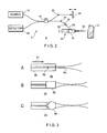
- Fig. 2 illustrates an tissue identification system 2 according to one embodiment of the present invention for tissue 10 identification using interferometric ranging.
- the tissue identification system 2 utilizes a one-dimensional data set in order to identify tissue. Unlike many prior art systems, which use two-dimensional data in order to acquire sufficient information to identify tissue, the tissue identification system 2 is able to identify tissue using a one-dimensional data set. Differences between two types of tissue may be understood from a one-dimensional data set. For example.
- Fig. 1 illustrates two graphs that represent a one-dimensional interferometric ranging axial scan of two different tissue types.
- adipose tissue shown in the bottom graph
- the tissue identification system 5 includes an imaging system 5 and a probe 50.
- the imaging system 5 includes a light source 12, which is provided in an interferometer 14.
- the interferometer 14 can be a fiber optic interferometer 14. Also, while light is used in the disclosure herein as an exemplary embodiment of the light source 12, it should be understood that other appropriate electromagnetic radiation can be used, such as, microwave, radio frequency, x-ray, and the like.
- the interferometer 14 or other beam splitting device known to those skilled in the art may make use of circulators for increased sample arm power efficiency.
- the interferometer 14 includes a beam splitter 18, a reference arm 20, a sample arm 24, and a communications link to at least one detector 26.
- the light source 12 is connected to the interferometer 14 such that the light emitted from the light source 12 is transmitted to the beam splitter 18.
- the beam splitter 18 directs portion of the light emitted by the source 12 towards a reference arm 20, while the remainder of light is directed to a sample arm 24.
- the reference arm 20 includes a mechanism 26.
- the mechanism 26 produces a time dependent optical delay.
- the mechanism 26 can be a movable reference reflector or mirror.
- the movable reference reflector or mirror can create a variable time delay suitable for a specific application.
- An optical fiber 29, associated with the sample arm 24, is connected to an optical coupler 58.
- the optical coupler 58 is also connected to an optical fiber 25, which is inserted into the probe 50, as described below.
- the light signals returned from the sample arm 24 and the reference arm 20 are combined by the beam splitter 18 and reflectivity as a function of depth within the tissue sample 10 (e.g., see Figs. 1A and 1B ) is determined by measuring the interference between the two arms with at least one detector 26. Detection of a tissue birefringence (i.e., by splitting a ray into two parallel rays polarized perpendicularly) can be accomplished by using, e.g., two detectors 26, one for each polarization eigenstate. Depending on the type of interferometric ranging used, one to four detectors 26 may be employed.
- one of three types of interferometric ranging can be used: (i) optical time domain reflectrometry, (ii) spectral domain reflectrometry or (iii) optical frequency domain reflectrometry. It should be understood that additional alternate types of interferometric ranging could be used with the tissue identification system 2. If optical time domain reflectometry is utilized, the source 12 can be is a broad bandwidth light source, the interferometer 14 is needed, the reference arm 20 may be a low speed reference arm with delay scanning performing 20 to 50 scans per second, and the detector 26 can include one to four detectors. Optical time domain reflectometry is described in more detail by C.
- Optical Coherence-Domain Reflectometry A New Optical Evaluation Technique
- Opt. Lett. 12, 158-160 (1987 )
- K. Takada et al. "New Measurement System For Fault Location in Optical Waveguide Devices Based on an Interferometric Technique” Appl. Opt. 26, 1603-1606 (1987 ), the entire disclosure of which are incorporated herein by reference.
- the source 12 is a broad bandwidth light source
- the interferometer 14 is required
- the detection arm includes a spectrometer
- the detector 26 includes a single detector
- low coherence interferometry data is obtained by taking the Fourier transform of the measured spectrum.
- Spectral domain reflectometry is described in more detail by J. Deboer et al., "Improved Signal to Noise Ratio In Spectral Domain Compared With Time Domain Optical Coherence Topography", Optics Letters 2003, vol. 28, p. 2067 - 69 ; and Published Patent No. WO 03062802 , entitled “Apparatus and Method for Ranging and Noise Reduction of Low Coherence Interferometry (LCI) and Optical Coherence Tomography (OCT) Signals by Parallel Detection of Spectral Bands", to Deboer et al., the entire disclosure of both of which are incorporated herein.
- LCI Low Coherence Interferometry
- OCT Optical Coherence Tomography
- the source 12 is a swept wavelength optical source
- the interferometer 14 is required
- the detector 26 includes one to four detectors
- low coherence interferometry data is obtained by taking the Fourier transform of the measured spectrum.
- Optical frequency domain reflectometry is described in more detail by S. Yun et al., "High Speed Optical Frequency Domain Imaging", Optics Express 2003, vol. 11., p. 2953 - 63 , and C. Youngquist et al., "Optical Coherence-Domain Reflectometry: A New Optical Evaluation Technique, Opt. Lett., 12, 158-160 (1987 ), and K.
- the interferometer 14 is a Mach-Zehinder interferometer, a Michelson interferometer, a non-reciprocal or circular interferometer, a Sagnac interferometer, a Twyman-Green interferometer and the like.
- a probe 50 can include a biopsy device 51, which includes a needle 52 having a bore (not shown) associated with a syringe 54 through which the optical fiber 25 is introduced.
- the fiber 25 may be inserted into the probe 50 and in turn into the needle 52.
- the needle 52 and fiber 25 can be inserted percutaneously (or otherwise) toward the tissue 10 to be sampled.
- the needle 52 can be a generic barrel, a specialized barrel, a needle, a stylet, and the like.
- the fiber 25 includes a cladding 60 and a cleaved anoptical fiber core 62, as shown in protion A of Fig. 3 .
- the fiber 25 When light signal is directed through the fiber 25 it forms a beam waist 64.
- the beam waist may be about 9 ⁇ m in diameter.
- Other lenses or optical elements may be attached to the fiber 25 to allow for focusing deeper into tissue, including a gradient index lens 66 (see portion B of Fig. 3 ), sometimes referred to as a GRIN lens, a ball lens 68 (see portion C of Fig. 3 ), a drum lens, a microlens, a tapered fiber end, a prism and the like.
- the fiber 25 may be angle cleaved or otherwise configured to produce an arbitrary pattern of electromagnetic radiation.
- the cladding 60 has an outer diameter of 125 ⁇ m and the anoptical fiber core has an outer diameter of 9 ⁇ m.
- the fiber 25 may be inserted into the biopsy needle 52 as shown in Fig. 4 and may be embedded within the needle biopsy device, or inserted through the lumen 70 of the needle 52. These types of procedures do not use fine needle aspiration. The lump or mass is not manually identifiable, but can only be identified through some other non-invasive imaging technique, such as CT or MRI. These and other guided needle biopsy procedures may use a larger and longer needle, while still utilizing the fiber 25 to assist in guiding the biopsy procedure.
- CT computerized tomography
- MRI magnetic resonance imaging
- ultrasound guidance the fiber 25 may be inserted into the biopsy needle 52 as shown in Fig. 4 and may be embedded within the needle biopsy device, or inserted through the lumen 70 of the needle 52. These types of procedures do not use fine needle aspiration. The lump or mass is not manually identifiable, but can only be identified through some other non-invasive imaging technique, such as CT or MRI. These and other guided needle biopsy procedures may use a larger and longer needle, while still utilizing the fiber 25 to assist in guiding the biopsy procedure.
- the fiber 25 may be inserted through an aperture 72, wherein Figure 5 does not shown the aperture 72 in the body, in the body of the syringe 51 and then (i) into the needle 52 as shown in Fig. 5 , (ii) through the plunger 74 of the syringe 51, and then provided into the needle 52 as shown in Fig. 6 , (iii) through an intermediate piece 76 that is attached between the syringe 51 and the needle 52 as shown in Fig. 7 , and/or by other insertion configuration.
- the probe 50 can be configured to allow suction for the aspiration of cells from the tissue 10, while allowing free movement of the fiber 25 at the tip of the needle 52.
- a probe 100 utilizes the imaging system 5 as described above to identify tissue.
- the probe 100 includes an input fiber 102 attached to the imaging system 5 at one end, and to an optical connector 58 at the other end.
- the optical fiber 102 is connected to a single mode input fiber 104.
- the optical fiber 102 is inserted through an intermediate adapter 106, located between a syringe 108 and a needle lock 110.
- a needle 112 is attached to the needle lock 110.
- a motion transducer 114 may be used as a result of too little space between the outer surface of the fiber 104 and the inner bore surface of the needle 112.
- the motion transducer 114 generally allows the fiber 104 to be repositioned in order to allow aspiration of the tissue 10.
- the motion transducer 114 can be a manual motion transducer, an automated motion transducer, or the like.
- the needle lock 110 is a Luer lock.
- Fig. 9 illustrates a tissue identification system 122 that includes the syringe 108 held within a device known in the art as a gun 120. This configuration allows for easy aspiration of the tissue 10 into the bore of the needle 112. Many of the components described above can also be incorporated into the tissue identification system 122 for easy access and convenience.
- Fig. 10 illustrates an exemplary operation of placing a cannula 200 for IV access, pleural, perineal taps, and the like according to a further embodiment of the present invention.
- the cannula 200 includes a guide catheter 202 and a fiber optic probe 204.
- the fiber optic probe 204 is provided within the guide catheter 202.
- the probe 204 may be inserted through the lumen 206 of the guide catheter 202 as shown in Fig. 11 .
- Fig. 12 illustrates an intra-operative exemplary embodiment 300 of a probe 306 according to still another embodiment of the present invention.
- the probe 306 is incorporated into an electrocautery device 301.
- An optical window 302 may be placed near the distal fiber tip 304 to protect the probe 306 against thermal damage by the cautery electrode 308.
- the probe 306 may be incorporated into a scalpel, an independent hand-held device and the like instead of being incorporated into the electrocautery device 301.
- the optical window 302 can be made of sapphire.
- the internal lumen 400 of a standard needle housing 402 can be modified such that the internal lumen 404 of a modified needle 406 is tapered, as shown in Fig. 13B .
- Fig. 14 illustrates an interferometric ranging probe optical connector 500, which is one side of the optical coupling 58, which can be used according to the present invention.
- the optical coupling 58 which connects the probe 50 to the imaging system 5, should be robust and simple to use.
- the optical coupling 58 includes a bare fiber connector attached to the probe 50, which is relatively inexpensive, and the interferometric ranging probe optical connector 500 attached to the imaging system 5, which is relatively expensive.
- the use of a bare fiber connector attached to the probe 50 does not increase the cost of the probe 50.
- the more expensive portion of the optical coupling 58 is attached to the imaging system 5.
- the interferometric ranging probe optical connector 500 is constructed so as to engage with a bare fiber connector, such that a robust connection is made.
- the interferometric ranging probe optical connector 500 may include a cleaved (angle cleaved) fiber 502 (the proximate end of which is connected to the imaging system 5, not shown for the sake of clarity) inserted through a housing 504 having a ferrule 506 connected to a tapered v-groove 508.
- the tapered v-groove 508 is terminated by a fiber stop 510.
- the housing 504 has a taper 516 at one end through which a fiber 518 is inserted.
- the fiber 518 is inserted into the housing 504 via the taper 516 until it reaches the fiber stop 514. Once the fiber 518 comes to a stop, a clamp 512 holds the fiber 518 in place, away from the fiber-fiber interface, such that an air or fluid gap 514 is maintained.
- the fiber 518 is connected to the probe 50.
- coupling gel may be used with flat cleaves to eliminate back-reflection from the gap 514.
- FIG. 15 illustrates a schematic diagram of a system 600 with components of an imaging system 5 and the optical fiber 29 connected to the fiber 25 via the optical connector 58.
- the fiber 25 is operatively associated with a syringe 51 and passes through the bore of a needle 52.
- a holder 612 is associated with the syringe 51 by the syringe barrel 614.
- a feedback unit 620 can be associated with the holder 612 in any of several ways.
- the holder 612 can be attached to the syringe 51.
- the holder 612 is removably attachable to the syringe 51, such as, but not limited to, snap fit, removable adhesive, clamping, clipping or the like.
- the holder 612 and associated feedback unit 620 can be reused while the syringe 608 can be disposable, thereby enabling conventional syringes to be used and eliminating the need for a custom developed and expensive probes.
- the holder 612 is removably attachable to the syringe 51 using a gun or syringe holder.
- the system 600 is integrally related to the gun (described above in relation to Fig. 9 ).
- the system 600 is embedded within the gun 634, which holds the feedback unit 620 and fiber 606 and improves the ability of the physician to aspirate tissue into the needle 610.
- the feedback unit 620 provides information to the user of the system 600, including that the system 600 has detected tissue of a particular type.
- the feedback unit 620 is a visual display, such as, LED, VGA, or other visual feedback system.
- the software algorithm and tissue identification determinations, as described hereinbelow can use an output signal to drive one or more LEDs, which can be actuated when the probe tip passes through or in proximity to differing tissue interface types (e.g., adipose versus muscle).
- tissue of interest such as a masticular lump
- an LED light can change color or a different colored LED can be actuated to provide the physician feedback that the lump has been contacted and that the biopsy aspiration or other sampling can commence.
- the feedback unit 620 is an audible tone generator, which provides audio feedback as different tissue or other structures are detected by the system 600.
- the feedback unit 620 is a vibration generator.
- Each of the visual, audio and vibration feedback units provide simple and yet useful feedback to users of the system 600 to better target a biopsy probe in real time and with confidence.
- the feedback unit 620 is a visual display screen that can be used to display a one or two-dimensional rolling plot image, comprising accumulated backscattered intensity as a function of z or depth within tissue, i.e. I z , over time to form an image.
- the visual display can be a conventional CRT display or an LCD display for providing more detailed or multimodal feedback.
- the visual display can be as small as or smaller than a conventional cell phone display or large to afford the user of the system 600 with a magnified view of the tissue 10.
- the feedback unit 620 is communicatively coupled to the imaging system 602 by a physical cable connection 622 or via a wireless connection.
- the wireless connection can be a radio frequency ("RF") connection, electromagnetic radiation signal, or the like.
- RF radio frequency
- a wireless signal connection allows for reduced weight of the biopsy probe and fewer wires in the surgical site.
- a simple feedback system can be utilized so that the physician can operate the biopsy probe with one hand and have feedback proximate to the probe body so that the physician's concentration and visual focus does not leave the biopsy area.
- Fig. 16 illustrates a further embodiment of the feedback unit 620 including a display 630, a manually operated button or switch 632 and a gun 634.
- the switch 632 is operatively connected to the display 630.
- the actuation of the switch 632 causes the display 630 to show selection of standard biopsy procedures, such as, but not limited to, biopsy of breast tissue, liver tissue, spleen tissue, muscle tissue, lymph tissue, kidney tissue, prostate tissue and the like.
- standard biopsy procedures such as, but not limited to, biopsy of breast tissue, liver tissue, spleen tissue, muscle tissue, lymph tissue, kidney tissue, prostate tissue and the like.
- Each of these biopsy procedures involves the probe 50 passing through relatively consistent types of layers, including skin, muscle, fascia, and the like, in a similar order for a given procedure.
- the order of layers the probe 50 would encounter are skin fascia, vertebrae, muscle, fascia, disk, subdural space, epidural space, the spinal cord fluid area.
- Each of these tissues can produce a relatively consistent and determinable imaging signal peak which, when normalized over a substantial patient base by comparative image analysis and subtracting the curves of normalized data versus actual patient data, offers an accurate picture of what will be encountered during the biopsy procedure.
- the imaging system 600 detects the actual signal, and compares it to reference signals stored in a database.
- a series of lines corresponding to the peaks of the sample may be obtained.
- the various consecutive peaks can be displayed by the feedback unit 620 to provide the user with accurate feedback of where the probe is and to assist the user in guiding the probe to the target area.
- the feedback unit can also incorporate an "anti-algorithm" to provide immediate feedback if the probe has wandered, overshot the target site or encountered a tissue type not expected to be detected during a particular procedure, such as, in the example of lumbar puncture, if the probe has passed the target area and hit a nerve. Such feedback can enable the physician to relocate the probe tip to the appropriate area.
- the system and process according to the present invention is also able to determine when a target site has been reached.
- the system processes data from the reflected light to look for backscattering signatures that are indicative of a tissue type within the target site during a given procedure.
- Such processing consists of feature extraction and inserting these features into a model that predicts tissue type.
- This model can be a physical model, a chemometric model, or a combination of the two.
- a physical model generally predicts the scattering signal based on physical principles of light scattering.
- a chemometric model uses a training set and statistically extracts features using techniques such as Partial Least Squares (“PLS”) or Principle Component Analysis (“PCA”), Such model is developed based on known samples, and the new data can be tested using this model. It should also be understood that fringes may be acquired and processed to determine other tissue features including birefringence, Doppler flow, and spectral characteristics.
- PLS Partial Least Squares
- PCA Principle Component Analysis
- Tissue identification can be accomplished by visualizing the intensity, birefringence, Doppler, spectroscopic axial reflectivity profile and/or the like. Additionally, the frequency spectrum (Fourier transform of the intensity data) of the reflectivity scan will provide information relating to the spacing of the scattering structures in the tissue which relates to tissue structure.
- a more sophisticated analysis including, but not limited to, variance analysis, one-dimensional texture discrimination (including fractal dimension, spatial gray level co-occurrence matrix parameters, Markovian distance, edge counting), power spectral analysis (including Fourier domain and time domain), n th order histogram moment analysis, and temporal analysis of the reflectivity information (comparing one scan to another separated by a fixed time) using correlation techniques will provide information relating to the type of tissue observed.
- tissue types include, measurement of the backscattering coefficient, total attenuation coefficient, estimation of the anisotropy coefficient (particle size) from the onset of multiple scattering, particle shape and size from the detected spectrum using coherence-gated light scattering spectroscopy, and the like.
- tissue characteristics may be found using the system and process of the present invention: adipose tissue, muscle tissue, collagen, nerve tissue, lymph node tissue, necrosis tissue, blood, glandular tissue and the like.
- Adipose tissue exhibits low absorbance at water peaks, high low spatial frequency components from the LCI intensity image and a high anisotropy coefficient.
- Muscle tissue exhibits high absorbance at water peaks, moderate birefringence, moderate anisotropy and decreased variance.
- Collagen exhibits very high birefringence.
- Nerve tissue exhibits moderate to high birefringence, high water absorbance and decreased power spectral density.
- Lymph node tissue exhibits a low anisotropy coefficient and a low temporal variance.
- Necrotic tissue exhibits high temporal variance of LCI signal, high attenuation coefficient, high water absorbance and low birefringence. Blood exhibits high Doppler shift, high water absorbance, high total attenuation coefficient, and high temporal variance. Glandular tissue exhibits moderate spatial frequency variance and low birefringence. It should be understood that the present invention contemplates the use of more than one analysis method, i.e., a multimodal system. This may provide enhanced detection and analysis of tissue types.
- Fig. 17 illustrates a process 700 for differentiating fat tissue from fibrous tissue according to an exemplary embodiment of the present invention.
- the signal measured by the system 600 is an average of a number M axial scans.
- the system 600 detects the tissue sample surface using a signal threshold T1 at block 702.
- the detected signal is divided into N number of windows at block 704.
- Signal processing is conducted at block 706 to obtain a parameter derived from the interferometric ranging signal, such as the average deviation (ADEV) or standard deviation (STDEV) of the signal in each window (such as, but not limited to, the technique described in "Numerical Recipes in C", Press, W. et al., Cambridge University Press, New York, NY 1992, the entire disclosure of which is incorporated herein by reference) is calculated.
- ADCV average deviation
- STDEV standard deviation
- ADEV or STDEV
- tissue identification for the purpose of intraoperative guidance, needle biopsy guidance, fine needle aspiration, image guided biopsy, guiding placement of peripheral or central intravenous or intra-arterial catheters, and the like.
- Different methods of imaging can be used for different applications. These different applications include: guided biopsy; cell methodology; veni-arterio, pattern or Doppler recognition; lumbar, pattern; therapy guidance, pattern and optical methods; and the like.
- the probe 50 can also be used as a targeting and delivery device for therapeutics.
- the probe 50 can image the target area to make sure that needle injection of a therapeutic has reached and/or entered the target tissue or site by detecting the tissue type and interface change, i.e. a change in the refractive index of the tissue.
- the process 700 utilizes standard image processing techniques to process data in order to differentiate fibrous (adipose) and fat tissue.
- Table 1 illustrates different measurements of fibrous and adipose tissue following the image processing of the data: TISSUE TYPE SENS SPECIFICITY Fibrous .95 .98 Adipose .97 .94 Fibrous/Adipose .96 .84
- sensitivity is true positive, i.e. true positive + false negative, while specificity is true negative, i.e. true negative + false positive.
- Fig. 18 illustrates an interferometric ranging diagnostic system 800 for identifying tissue according to the present invention.
- the system 800 uses a light source configured to emit light having a optical wavelength of I.3 microns, 300 microwatts power and a 48 nm bandwidth.
- the light source allows the system 800 to interrogate tissue with a 15 micron resolution.
- the system 800 utilizes a low scanning frequency of the reference arm because building a coherent image is not the purpose of the system 800.
- Information can be gathered to identify the tissue through an average of several A-scans.
- the A-scan can be performed by one sweep of the reference arm, which corresponds to one depth scan.
- the system 800 processes and stores digital data, and the tissue type information is displayed on the feedback device in real time.
- the LED source of the system 800 is a 300 microwatts SUPERLUM LED, which can be temperature and current controlled.
- the light is linearly polarized using a fiber optic polarizer P and sent to a beam splitter.
- the sample is interrogated with two orthogonally polarized states of the light in order to get birefringence information.
- the two orthogonally polarized states of the light are created by passing the light through the beam splitter, to two polarization controller PC paddles, one in each arm of the beam splitter.
- the two orthogonal polarization states are sent alternatively to the fiber optic circulator CIR, which directs the light to the fiber optic Michelson interferometer IF.
- the optical switch OSW is synchronized with the optical delay line ODL galvanometer, so that the polarization would change alternatively from one scan to the other.
- a very simple delay line consisting in a retroreflector mounted to an lever driven by a galvanometer, was used to do the depth scanning.
- the probe attached to the sample arm of the interferometer IF includes a bare fiber introduced into a syringe needle.
- the backscattered light is coherently added to the light coming from the ODL and sent to the detectors D1, D2.
- a polarization splitter PS is used to select the two orthogonal states.
- the output signals of the detectors are preamplified and digitized using a NI DAQ card.
- the system performs the digital acquisition, filtering, and averaging of the fringes, and provides the following information: (1) depth intensity at 15 microns resolution and spectral information, (2) birefringence information: computes stokes parameters-IQUV and extracts phase retardation, and (3) Doppler shift information.
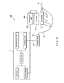
- Fig. 19 illustrates a flow diagram 900 of the signal processing sequence according to an exemplary embodiment of the present invention.
- the simplest form of tissue identification is differentiation of two tissue types. The difference between two tissue types can be seen in Fig. 1 , which shows the results of a feasibility study to distinguish cadaver fat from fibrous tissue. For example, adipose tissue has an appearance of multiple peaks separated by low interferometric ranging signal segments, whereas fibrous tissue has a lower degree of variance and decays exponentially.
- an apparatus for identifying characteristics of tissue comprising: a radiation source configured to perform an axial scan of the tissue using radiation; and an imaging system adapted to receive axial scan radiation based on the axial scan, and to process data relating to the axial scan radiation to identify characteristics of the tissue.
- the radiation source is a light source configured to emit light.
- an apparatus wherein the light source is a broad bandwidth light source.
- an apparatus wherein the light source is a swept wavelength optical source.
- an apparatus wherein the light source delivers radiation to the tissue via an optical fiber disposed in an insertion device having a distal end at least partially disposed within the insertion device and a proximal end.
- an apparatus wherein the insertion device is configured to provide the distal end of the optical fiber adjacent to the tissue.
- an apparatus wherein the insertion device is one of a barrel, a needle, and a stylet.
- the imaging system further includes an interferometer adapted to direct a portion of the radiation emitted by the radiation source into a sample arm and detecting radiation reflected from the tissue back through the sample arm.
- an apparatus wherein the interferometer directs another portion of the radiation into a reference arm.
- an apparatus wherein the imaging system identifies characteristics of the tissue by processing the axial scan radiation to provide the characteristics of the tissue, the axial scan radiation including radiation received from the reference arm and radiation received from the sample arm, and comparing the characteristics of the tissue with a database of normalized characteristics of a plurality of tissue types.
- the axial scan radiation includes at least one of backscattering, spectral properties, birefringence and Doppler shift.
- an apparatus wherein the imaging system processes the axial scan radiation by performing at least one of standard deviation, average deviation, and slope of the axial reflectivity profile.
- an apparatus wherein the imaging system inputs data derived from the axial scan radiation into a statistical model to predict tissue type.
- an apparatus wherein the statistical model extracts features from data derived from the axial scan radiation.
- the statistical model is at least one of partial least squares or principle component analysis.
- an apparatus wherein the imaging system identifies the characteristics of the tissue by determining reflectance characteristics of the axial scan radiation using interferometric ranging, and comparing the characteristics of the tissue with normalized reflectance characteristics of a plurality of types of tissue stored in a database.
- an apparatus wherein the type of interferometric ranging is at least one of optical time domain reflectometry, spectral domain reflectometry and optical frequency domain reflectometry.
- a method for identifying characteristics of tissue comprising the steps: performing an axial scan of the tissue using radiation ; and processing data relating to the axial scan radiation based on the axial scan to identify characteristics of the tissue.
- the axial scan radiation includes at least one of backscattering, spectral properties, birefringence and Doppler shift.
- the processing step identifies the characteristics of the tissue by performing at least one of standard deviation of data associated with the axial scan radiation, average deviation of data associated with the axial scan radiation, and slope of the axial reflectivity profile of data associated with the axial scan radiation.
- a light source delivers the radiation to perform the axial scan of the tissue via an optical fiber disposed in an insertion device having a distal end at least partially disposed within the insertion device and a proximal end.
- the processing step identifies the characteristics of the tissue by inputting data derived from the axial scan radiation into a statistical model to predict tissue type.
- the processing step identifies the characteristics of the tissue by determining reflectance characteristics of the the axial scan radiation using interferometric ranging and comparing the characteristics of the tissue with a database of stored normalized reflectance characteristics of a plurality of types of tissue.
- a storage medium storing a software program for identifying characteristics of tissue, wherein the software program, when executed by a processing arrangement, is configured to cause the processing arrangement to execute the steps comprising of : performing an axial scan of the tissue using radiation; and processing data relating to the axial scan radiation to identify characteristics of the tissue.
- the axial scan radiation includes at least one of backscattering, spectral properties, birefringence and Doppler shift.
- a storage medium wherein the processing step identifies the characteristics of the tissue by performing at least one of standard deviation of data associated with the axial scan radiation, average deviation of data associated with the axial scan radiation, and slope of the axial reflectivity profile of data associated with the axial scan radiation.
- a storage medium wherein a light source delivers the radiation to perform the axial scan of the tissue via an optical fiber disposed in an insertion device having a distal end at least partially disposed within the insertion device and a proximal end.
- a storage medium wherein the processing step identifies the characteristics of the tissue by inputting data derived from the axial scan radiation into a statistical model to predict tissue type.
- a storage medium wherein the processing step identifies the characteristics of the tissue by determining reflectance characteristics of the the axial scan radiation using interferometric ranging and comparing the characteristics of the tissue with a database of stored normalized reflectance characteristics of a plurality of types of tissue.
- a logic arrangement for for identifying characteristics of tissue which, when executed by a processing arrangement, is operable to perform the steps comprising of : performing an axial scan of the tissue using radiation; and processing data relating to the axial scan radiation to identify characteristics of the tissue.
- the axial scan radiation includes at least one of backscattering, spectral properties, birefringence and Doppler shift.
- the processing step identifies the characteristics of the tissue by performing at least one of standard deviation of data associated with the axial scan radiation, average deviation of data associated with the axial scan radiation, and slope of the axial reflectivity profile of data associated with the axial scan radiation.
- a light source delivers the radiation to perform the axial scan of the tissue via an optical fiber disposed in an insertion device having a distal end at least partially disposed within the insertion device and a proximal end.
- the processing step identifies the characteristics of the tissue by inputting data derived from the axial scan radiation into a statistical model to predict tissue type.
- the processing step identifies the characteristics of the tissue by determining reflectance characteristics of the the axial scan radiation using interferometric ranging and comparing the characteristics of the tissue with a database of stored normalized reflectance characteristics of a plurality of types of tissue.
- an apparatus for identifying characteristics of tissue comprising: a radiation source configured to deliver radiation to the tissue; and an imaging system adapted to receive the radiation and process unidimensional data relating to the radiation to identify characteristics of the tissue.
- an apparatus for identifying characteristics of tissue comprising: at least one optical fiber, one of the at lest one optical fiber disposed in a needle having a distal end at least partially disposed within the needle and a proximal end, the distal end of the one of the at least one optical fiber adapted to be placed adjacent to tissue; and, an imaging system in communication with the proximal end of the one of the at least one optical fiber, and configured to process reflected light from the at least one optical fiber to identify the tissue.
- an apparatus wherein the reflected light is unidimensional light.
- an apparatus wherein the reflected light is generated to perform an axial scan.
Applications Claiming Priority (2)
| Application Number | Priority Date | Filing Date | Title |
|---|---|---|---|
| US44239203P | 2003-01-24 | 2003-01-24 | |
| EP04705347.5A EP1596716B1 (fr) | 2003-01-24 | 2004-01-26 | Système et procédé pour l'identification tissulaire utilisant l'interférométrie à faible cohérence |
Related Parent Applications (2)
| Application Number | Title | Priority Date | Filing Date |
|---|---|---|---|
| EP04705347.5 Division | 2004-01-26 | ||
| EP04705347.5A Division-Into EP1596716B1 (fr) | 2003-01-24 | 2004-01-26 | Système et procédé pour l'identification tissulaire utilisant l'interférométrie à faible cohérence |
Publications (2)
| Publication Number | Publication Date |
|---|---|
| EP2319405A1 true EP2319405A1 (fr) | 2011-05-11 |
| EP2319405B1 EP2319405B1 (fr) | 2013-09-18 |
Family
ID=32825214
Family Applications (3)
| Application Number | Title | Priority Date | Filing Date |
|---|---|---|---|
| EP10183037.0A Expired - Lifetime EP2319404B1 (fr) | 2003-01-24 | 2004-01-26 | Système et procédé pour identifier des tissus à l'aide d'interférométrie à faible cohérence |
| EP04705347.5A Expired - Lifetime EP1596716B1 (fr) | 2003-01-24 | 2004-01-26 | Système et procédé pour l'identification tissulaire utilisant l'interférométrie à faible cohérence |
| EP10182976.0A Expired - Lifetime EP2319405B1 (fr) | 2003-01-24 | 2004-01-26 | Système et procédé pour identifier des tissus à l'aide d'interférométrie à faible cohérence |
Family Applications Before (2)
| Application Number | Title | Priority Date | Filing Date |
|---|---|---|---|
| EP10183037.0A Expired - Lifetime EP2319404B1 (fr) | 2003-01-24 | 2004-01-26 | Système et procédé pour identifier des tissus à l'aide d'interférométrie à faible cohérence |
| EP04705347.5A Expired - Lifetime EP1596716B1 (fr) | 2003-01-24 | 2004-01-26 | Système et procédé pour l'identification tissulaire utilisant l'interférométrie à faible cohérence |
Country Status (7)
| Country | Link |
|---|---|
| US (2) | US7761139B2 (fr) |
| EP (3) | EP2319404B1 (fr) |
| JP (4) | JP2006516739A (fr) |
| CN (1) | CN1741768A (fr) |
| AU (1) | AU2004206998B2 (fr) |
| CA (1) | CA2514189A1 (fr) |
| WO (1) | WO2004066824A2 (fr) |
Families Citing this family (223)
| Publication number | Priority date | Publication date | Assignee | Title |
|---|---|---|---|---|
| US20050131465A1 (en) | 2000-02-04 | 2005-06-16 | Freeman Gary A. | Integrated resuscitation |
| EP2308557A3 (fr) | 2000-02-04 | 2011-08-24 | Zoll Medical Corporation | Réanimation intégré |
| US20060064131A1 (en) * | 2000-02-04 | 2006-03-23 | Freeman Gary A | User interface for defibrillator for use by persons with limited training and experience |
| JP4241038B2 (ja) | 2000-10-30 | 2009-03-18 | ザ ジェネラル ホスピタル コーポレーション | 組織分析のための光学的な方法及びシステム |
| US9295391B1 (en) | 2000-11-10 | 2016-03-29 | The General Hospital Corporation | Spectrally encoded miniature endoscopic imaging probe |
| EP2333521B1 (fr) | 2001-04-30 | 2019-12-04 | The General Hospital Corporation | Procédé et appareil permettant d'améliorer la clarté et la sensibilité de l'image en tomographie à cohérence optique au moyen d'une interaction permettant de contrôler les propriétés focales et la synchronisation de cohérence |
| GB2408797B (en) | 2001-05-01 | 2006-09-20 | Gen Hospital Corp | Method and apparatus for determination of atherosclerotic plaque type by measurement of tissue optical properties |
| US7355716B2 (en) | 2002-01-24 | 2008-04-08 | The General Hospital Corporation | Apparatus and method for ranging and noise reduction of low coherence interferometry LCI and optical coherence tomography OCT signals by parallel detection of spectral bands |
| US7016048B2 (en) * | 2002-04-09 | 2006-03-21 | The Regents Of The University Of California | Phase-resolved functional optical coherence tomography: simultaneous imaging of the stokes vectors, structure, blood flow velocity, standard deviation and birefringence in biological samples |
| US8054468B2 (en) | 2003-01-24 | 2011-11-08 | The General Hospital Corporation | Apparatus and method for ranging and noise reduction of low coherence interferometry LCI and optical coherence tomography OCT signals by parallel detection of spectral bands |
| US7623908B2 (en) * | 2003-01-24 | 2009-11-24 | The Board Of Trustees Of The University Of Illinois | Nonlinear interferometric vibrational imaging |
| US7567349B2 (en) | 2003-03-31 | 2009-07-28 | The General Hospital Corporation | Speckle reduction in optical coherence tomography by path length encoded angular compounding |
| EP2319404B1 (fr) * | 2003-01-24 | 2015-03-11 | The General Hospital Corporation | Système et procédé pour identifier des tissus à l'aide d'interférométrie à faible cohérence |
| US7102758B2 (en) | 2003-05-06 | 2006-09-05 | Duke University | Fourier domain low-coherence interferometry for light scattering spectroscopy apparatus and method |
| ES2310744T3 (es) | 2003-06-06 | 2009-01-16 | The General Hospital Corporation | Fuente de luz sintonizable en longitudes de onda. |
| EP2293031B8 (fr) | 2003-10-27 | 2024-03-20 | The General Hospital Corporation | Procédé et appareil pour une source de lumière à réglage de longueur d'onde |
| US7610074B2 (en) * | 2004-01-08 | 2009-10-27 | The Board Of Trustees Of The University Of Illinois | Multi-functional plasmon-resonant contrast agents for optical coherence tomography |
| US20050254059A1 (en) * | 2004-05-14 | 2005-11-17 | Alphonse Gerard A | Low coherence interferometric system for optical metrology |
| US7474408B2 (en) * | 2004-05-14 | 2009-01-06 | Medeikon Corporation | Low coherence interferometry utilizing phase |
| EP1754016B1 (fr) | 2004-05-29 | 2016-05-18 | The General Hospital Corporation | Procede, systeme et logiciel destines a la compensation de dispersion chromatique au moyen de couches reflechissantes dans l'imagerie par tomographie par coherence optique (oct) |
| US7447408B2 (en) | 2004-07-02 | 2008-11-04 | The General Hospital Corproation | Imaging system and related techniques |
| JP5053845B2 (ja) | 2004-08-06 | 2012-10-24 | ザ ジェネラル ホスピタル コーポレイション | 光学コヒーレンス断層撮影法を使用して試料中の少なくとも1つの位置を決定するための方法、システムおよびソフトウェア装置 |
| EP1793731B1 (fr) | 2004-08-24 | 2013-12-25 | The General Hospital Corporation | Appareil d'imagerie comprenant un dispositif de distribution de fluide et un dispositif de rétraction |
| EP1989997A1 (fr) * | 2004-08-24 | 2008-11-12 | The General Hospital Corporation | Procèdé, Système et logiciel pour la mesure de la contrainte mécanique et des propriétés élastiques d'un echantillon |
| US8545488B2 (en) | 2004-09-17 | 2013-10-01 | The Spectranetics Corporation | Cardiovascular imaging system |
| EP1804638B1 (fr) | 2004-09-29 | 2012-12-19 | The General Hospital Corporation | Systeme et procede d'imagerie a coherence optique |
| WO2006058346A1 (fr) * | 2004-11-29 | 2006-06-01 | The General Hospital Corporation | Ensembles, dispositifs, endoscopes, catheters et methodes d'imagerie optique permettant d'eclairer et de detecter simultanement plusieurs points sur un echantillon |
| US7586618B2 (en) | 2005-02-28 | 2009-09-08 | The Board Of Trustees Of The University Of Illinois | Distinguishing non-resonant four-wave-mixing noise in coherent stokes and anti-stokes Raman scattering |
| ES2432560T3 (es) * | 2005-03-10 | 2013-12-04 | Anatoly Babchenko | Sensor óptico |
| US7725169B2 (en) * | 2005-04-15 | 2010-05-25 | The Board Of Trustees Of The University Of Illinois | Contrast enhanced spectroscopic optical coherence tomography |
| ATE451669T1 (de) | 2005-04-28 | 2009-12-15 | Gen Hospital Corp | Bewertung von bildmerkmalen einer anatomischen struktur in optischen kohärenztomographiebildern |
| WO2006119431A2 (fr) | 2005-04-29 | 2006-11-09 | The Regents Of The University Of Colorado, A Body Corporate | Caractérisation électromagnétique de tissus |
| US7859679B2 (en) | 2005-05-31 | 2010-12-28 | The General Hospital Corporation | System, method and arrangement which can use spectral encoding heterodyne interferometry techniques for imaging |
| EP1889037A2 (fr) | 2005-06-01 | 2008-02-20 | The General Hospital Corporation | Appareil, methode et systeme pour effectuer une imagerie de domaine de frequence optique a resolution de phase |
| EP2267404B1 (fr) | 2005-08-09 | 2016-10-05 | The General Hospital Corporation | Dispositif et procédé servant a effectuer une démodulation en quadrature à base de polarisation en tomographie à cohérence optique |
| US20070049833A1 (en) * | 2005-08-16 | 2007-03-01 | The General Hospital Corporation | Arrangements and methods for imaging in vessels |
| US7650181B2 (en) | 2005-09-14 | 2010-01-19 | Zoll Medical Corporation | Synchronization of repetitive therapeutic interventions |
| US7872759B2 (en) | 2005-09-29 | 2011-01-18 | The General Hospital Corporation | Arrangements and methods for providing multimodality microscopic imaging of one or more biological structures |
| US8537366B2 (en) * | 2005-10-11 | 2013-09-17 | Duke University | Systems and methods for endoscopic angle-resolved low coherence interferometry |
| CA2626116C (fr) * | 2005-10-11 | 2012-08-21 | Duke University | Systemes et procede endoscopiques d'interferometrie a faible coherence et a resolution angulaire |
| EP1945094B1 (fr) | 2005-10-14 | 2018-09-05 | The General Hospital Corporation | Imagerie fluorescente codee par frequence et par spectre |
| EP2062530A3 (fr) * | 2005-11-29 | 2009-08-12 | Surgi-Vision, Inc. | Systèmes de mise en place de dérivation et/ou de localisation guidés par IRM et procédés, dispositifs et programmes informatiques associés |
| WO2007082228A1 (fr) | 2006-01-10 | 2007-07-19 | The General Hospital Corporation | Systèmes et procédés de génération de données basés sur une ou plusieurs technique(s) d'endoscopie spectralement codées |
| WO2007084849A1 (fr) * | 2006-01-18 | 2007-07-26 | The General Hospital Corporation | Système et procédé pour produire des données au moyen d'une ou plusieurs techniques de microscopie endoscopique |
| EP2289397A3 (fr) | 2006-01-19 | 2011-04-06 | The General Hospital Corporation | Procédés et systèmes pour visualiser de manière optique des organes intracavitaires épithéliaux lors d'un balayage au faisceau |
| US8145018B2 (en) | 2006-01-19 | 2012-03-27 | The General Hospital Corporation | Apparatus for obtaining information for a structure using spectrally-encoded endoscopy techniques and methods for producing one or more optical arrangements |
| US20080002211A1 (en) * | 2006-01-20 | 2008-01-03 | The General Hospital Corporation | System, arrangement and process for providing speckle reductions using a wave front modulation for optical coherence tomography |
| WO2007090147A2 (fr) | 2006-01-31 | 2007-08-09 | The Board Of Trustees Of The University Of Illinois | Procédé et appareil de mesure des propriétés optiques de tissus |
| US10426548B2 (en) | 2006-02-01 | 2019-10-01 | The General Hosppital Corporation | Methods and systems for providing electromagnetic radiation to at least one portion of a sample using conformal laser therapy procedures |
| EP1986545A2 (fr) | 2006-02-01 | 2008-11-05 | The General Hospital Corporation | Appareil destiné à appliquer une pluralité de rayonnements électromagnétiques à un échantillon |
| US9777053B2 (en) | 2006-02-08 | 2017-10-03 | The General Hospital Corporation | Methods, arrangements and systems for obtaining information associated with an anatomical sample using optical microscopy |
| EP2306141A1 (fr) | 2006-02-24 | 2011-04-06 | The General Hospital Corporation | Procédés et systèmes destinés à réaliser une tomographie par cohérence optique dans le domaine de Fourier avec résolution angulaire |
| WO2007109540A2 (fr) * | 2006-03-17 | 2007-09-27 | The General Hospital Corporation | Appareil, procédé et support accessible par ordinateur pour l'identification de caractéristiques d'au moins une partie d'un vaisseau sanguin compris à l'intérieur d'un tissu au moyen d'une interférométrie faible cohérence à domaine spectral |
| JP5631585B2 (ja) * | 2006-03-22 | 2014-11-26 | コーニンクレッカ フィリップス エレクトロニクス エヌ.ヴィ. | 光ファイバ機器センシングシステム |
| US7742173B2 (en) | 2006-04-05 | 2010-06-22 | The General Hospital Corporation | Methods, arrangements and systems for polarization-sensitive optical frequency domain imaging of a sample |
| US8175685B2 (en) | 2006-05-10 | 2012-05-08 | The General Hospital Corporation | Process, arrangements and systems for providing frequency domain imaging of a sample |
| WO2007133964A2 (fr) | 2006-05-12 | 2007-11-22 | The General Hospital Corporation | Processus, agencements et systèmes pour produire une carte d'épaisseur de couche de fibres sur la base d'images de tomographie à cohérence optique |
| JP2009538672A (ja) * | 2006-05-30 | 2009-11-12 | コーニンクレッカ フィリップス エレクトロニクス エヌ ヴィ | 組織属性の奥行き解像された測定のための装置 |
| US7488930B2 (en) * | 2006-06-02 | 2009-02-10 | Medeikon Corporation | Multi-channel low coherence interferometer |
| AU2007275018A1 (en) * | 2006-07-21 | 2008-01-24 | Oncoscope, Inc. | Protective probe tip, particularly for use on a fiber-optic probe used in an endoscopic application |
| WO2008016927A2 (fr) * | 2006-08-01 | 2008-02-07 | The General Hospital Corporation | Systèmes et procédés pour recevoir et/ou analyser des informations associées à un rayonnement électromagnétique |
| WO2008024948A2 (fr) | 2006-08-25 | 2008-02-28 | The General Hospital Corporation | Appareil et procédés permettant d'améliorer l'imagerie de tomographie par cohérence optique mettant en œuvre des techniques de filtrage volumétrique |
| CN101626719B (zh) * | 2006-09-26 | 2012-07-18 | 俄勒冈健康与科学大学 | 体内结构和流动成像 |
| WO2008049118A2 (fr) * | 2006-10-19 | 2008-04-24 | The General Hospital Corporation | Dispositif et procédé d'obtention et de fourniture d'informations d'image associées à au moins une portion d' échantillon et permettant de réaliser une telle portion |
| EP2662674A3 (fr) * | 2007-01-19 | 2014-06-25 | The General Hospital Corporation | Réflexion de disque rotatif pour balayage rapide de longueur d'onde de lumière à large bande dispersée |
| US8460195B2 (en) * | 2007-01-19 | 2013-06-11 | Sunnybrook Health Sciences Centre | Scanning mechanisms for imaging probe |
| JP2010516304A (ja) * | 2007-01-19 | 2010-05-20 | サニーブルック・ヘルス・サイエンシズ・センター | 超音波と光学を複合した画像手段を有する撮像プローブ |
| US8044999B2 (en) * | 2007-03-06 | 2011-10-25 | The United States Of America As Represented By The Secretary Of The Navy | Image enhancer for detecting and identifying objects in turbid media |
| US20090069673A1 (en) * | 2007-03-16 | 2009-03-12 | The Charles Stark Draper Laboratory, Inc. | Spinal needle optical sensor |
| US20080234567A1 (en) * | 2007-03-19 | 2008-09-25 | The General Hospital Corporation | Apparatus and method for providing a noninvasive diagnosis of internal bleeding |
| US9176319B2 (en) | 2007-03-23 | 2015-11-03 | The General Hospital Corporation | Methods, arrangements and apparatus for utilizing a wavelength-swept laser using angular scanning and dispersion procedures |
| WO2008121844A1 (fr) * | 2007-03-30 | 2008-10-09 | The General Hospital Corporation | Système et procédé pour fournir une imagerie à granularité laser en vue de détecter une plaque à risque |
| US8045177B2 (en) | 2007-04-17 | 2011-10-25 | The General Hospital Corporation | Apparatus and methods for measuring vibrations using spectrally-encoded endoscopy |
| FR2915077B1 (fr) * | 2007-04-19 | 2010-09-10 | Inst Radioprot Et De Surete Nu | Dispositif d'aide au diagnostic et pronostic de modifications physiopathologiques des tissus. |
| WO2008137637A2 (fr) | 2007-05-04 | 2008-11-13 | The General Hospital Corporation | Procédés, agencements et systèmes pour obtenir des informations associées à un échantillon à l'aide d'une microscopie optique |
| US8175677B2 (en) * | 2007-06-07 | 2012-05-08 | MRI Interventions, Inc. | MRI-guided medical interventional systems and methods |
| US8172757B2 (en) * | 2007-06-18 | 2012-05-08 | Sunnybrook Health Sciences Centre | Methods and devices for image-guided manipulation or sensing or anatomic structures |
| WO2009018456A2 (fr) | 2007-07-31 | 2009-02-05 | The General Hospital Corporation | Systèmes et procédés pour fournir des motifs de balayage de faisceau pour une imagerie dans le domaine de la fréquence optique doppler de vitesse élevée |
| US7682089B2 (en) * | 2007-08-15 | 2010-03-23 | Rohlen Brooks H | System and method for positioning a probe |
| US8040608B2 (en) | 2007-08-31 | 2011-10-18 | The General Hospital Corporation | System and method for self-interference fluorescence microscopy, and computer-accessible medium associated therewith |
| JP5579606B2 (ja) * | 2007-09-13 | 2014-08-27 | デユーク・ユニバーシテイ | 低コヒーレンス干渉法(lci)のための装置、システムおよび方法 |
| WO2009036453A1 (fr) * | 2007-09-15 | 2009-03-19 | The General Hospital Corporation | Appareil, support accessible par ordinateur et procédé de mesure de compositions chimiques et/ou moléculaires de plaques athéroscléreuses coronariennes dans des structures anatomiques |
| US8315689B2 (en) * | 2007-09-24 | 2012-11-20 | MRI Interventions, Inc. | MRI surgical systems for real-time visualizations using MRI image data and predefined data of surgical tools |
| EP2194906B8 (fr) * | 2007-09-24 | 2015-04-22 | Mri Interventions, Inc. | Système d'intervention médical guidé par irm |
| EP2207469A4 (fr) * | 2007-10-12 | 2012-07-11 | Gen Hospital Corp | Systèmes et procédés d'imagerie optique de structures anatomiques luminales |
| TWI343266B (en) * | 2007-10-15 | 2011-06-11 | Univ Nat Yang Ming | An injection appraatus for locating and detecting spinal cord's epidural space by using fiber optic technology |
| CN101411611B (zh) * | 2007-10-15 | 2010-12-15 | 倪蔚民 | 用于人体生物组织医学诊断的光学成像装置 |
| WO2009059034A1 (fr) | 2007-10-30 | 2009-05-07 | The General Hospital Corporation | Système et procédé permettant une détection de mode de gaine |
| AU2008324814B2 (en) * | 2007-11-05 | 2013-05-16 | Solventum Intellectual Properties Company | Identification of tissue for debridement |
| CA2711643A1 (fr) | 2008-01-08 | 2009-07-16 | Oncoscope, Inc. | Systemes et procedes pour l'examen, le diagnostic, le traitement et/ou la surveillance de tissu |
| US8115934B2 (en) | 2008-01-18 | 2012-02-14 | The Board Of Trustees Of The University Of Illinois | Device and method for imaging the ear using optical coherence tomography |
| US8983580B2 (en) | 2008-01-18 | 2015-03-17 | The Board Of Trustees Of The University Of Illinois | Low-coherence interferometry and optical coherence tomography for image-guided surgical treatment of solid tumors |
| US7751057B2 (en) | 2008-01-18 | 2010-07-06 | The Board Of Trustees Of The University Of Illinois | Magnetomotive optical coherence tomography |
| US11123047B2 (en) | 2008-01-28 | 2021-09-21 | The General Hospital Corporation | Hybrid systems and methods for multi-modal acquisition of intravascular imaging data and counteracting the effects of signal absorption in blood |
| US9332942B2 (en) | 2008-01-28 | 2016-05-10 | The General Hospital Corporation | Systems, processes and computer-accessible medium for providing hybrid flourescence and optical coherence tomography imaging |
| EP2103249B9 (fr) * | 2008-03-19 | 2016-10-19 | Carl Zeiss Meditec AG | Système de microscopie chirurgicale disposant d'une installation de tomographie à cohérence optique |
| US8208996B2 (en) * | 2008-03-24 | 2012-06-26 | Carl Zeiss Meditec, Inc. | Imaging of polarization scrambling tissue |
| JP5607610B2 (ja) * | 2008-05-07 | 2014-10-15 | ザ ジェネラル ホスピタル コーポレイション | 構造の特徴を決定する装置、装置の作動方法およびコンピュータアクセス可能な媒体 |
| WO2009144653A2 (fr) * | 2008-05-30 | 2009-12-03 | Koninklijke Philips Electronics N.V. | Aiguille avec détecteur de photons intégré |
| ES2517915T3 (es) * | 2008-06-02 | 2014-11-04 | Lightlab Imaging, Inc. | Métodos cuantitativos para obtener características de un tejido a partir de imágenes de tomografía por coherencia óptica |
| JP4575474B2 (ja) * | 2008-06-11 | 2010-11-04 | 国立大学法人東京工業大学 | 生体組織識別装置および方法 |
| US8690762B2 (en) | 2008-06-18 | 2014-04-08 | Raytheon Company | Transparent endoscope head defining a focal length |
| JP5795531B2 (ja) | 2008-06-20 | 2015-10-14 | ザ ジェネラル ホスピタル コーポレイション | フューズドファイバオプティックカプラ構造、及びその使用方法 |
| EP2309923B1 (fr) | 2008-07-14 | 2020-11-25 | The General Hospital Corporation | Appareil et procédés d'endoscopie couleur |
| US8486735B2 (en) | 2008-07-30 | 2013-07-16 | Raytheon Company | Method and device for incremental wavelength variation to analyze tissue |
| US8446586B2 (en) * | 2008-10-15 | 2013-05-21 | Allan Yang Wu | Method and apparatus for increasing adipose vascular fraction |
| WO2010068764A2 (fr) * | 2008-12-10 | 2010-06-17 | The General Hospital Corporation | Systèmes, appareil et procédés d'extension de la plage de profondeur d'imagerie de tomographie par cohérence optique, par le biais du sous-échantillonnage optique |
| JP2012515576A (ja) * | 2009-01-20 | 2012-07-12 | ザ ジェネラル ホスピタル コーポレイション | 内視鏡生検装置、システム、及び方法 |
| EP2382456A4 (fr) | 2009-01-26 | 2012-07-25 | Gen Hospital Corp | Système, procédé et support accessible par ordinateur permettant de fournir une microscopie de super-résolution à large champ |
| WO2010091190A2 (fr) * | 2009-02-04 | 2010-08-12 | The General Hospital Corporation | Appareil et procédé d'utilisation d'une source d'ajustement de longueur d'onde optique à grande vitesse |
| WO2010105197A2 (fr) | 2009-03-12 | 2010-09-16 | The General Hospital Corporation | Système optique sans contact, support accessible par ordinateur et procédé de mesure d'au moins une propriété mécanique d'un tissu à l'aide d'une ou plusieurs techniques cohérentes de dispersion |
| TW201100053A (en) | 2009-05-04 | 2011-01-01 | Oregon Health & Science University | Method and apparatus for quantitative imaging of blood perfusion in living tissue |
| JP5819823B2 (ja) * | 2009-07-14 | 2015-11-24 | ザ ジェネラル ホスピタル コーポレイション | 血管の内部の流れおよび圧力を測定する装置および装置の作動方法 |
| ES2636767T3 (es) * | 2009-09-17 | 2017-10-09 | Mauna Kea Technologies | Un método, una sonda óptica y un sistema de microscopía confocal para la inspección de un órgano sólido |
| US9661996B2 (en) * | 2009-10-01 | 2017-05-30 | Sarcos Lc | Needle delivered imaging device |
| JP5856061B2 (ja) * | 2009-10-06 | 2016-02-09 | ザ ジェネラル ホスピタル コーポレイション | スペクトル符号化共焦点顕微鏡法を用いた特定の細胞を撮像するための装置及び方法 |
| JP5711260B2 (ja) * | 2009-12-08 | 2015-04-30 | ザ ジェネラル ホスピタル コーポレイション | 光コヒーレンストモグラフィにより声帯襞を分析、診断及び治療モニタリングする方法及び装置 |
| JP2013518256A (ja) | 2010-01-22 | 2013-05-20 | デユーク・ユニバーシテイ | 分光光コヒーレンストモグラフィ(oct)およびフーリエドメイン低コヒーレンス干渉法のための多重ウィンドウ処理スキーム |
| US9823127B2 (en) | 2010-01-22 | 2017-11-21 | Duke University | Systems and methods for deep spectroscopic imaging of biological samples with use of an interferometer and spectrometer |
| CN103002794B (zh) | 2010-02-08 | 2016-08-17 | 奥勒冈保健科学大学 | 用于超高灵敏光学微血管显影术之设备及方法 |
| PT2542145T (pt) | 2010-03-05 | 2020-11-04 | Massachusetts Gen Hospital | Sistemas, métodos e meios acessíveis por computador que proporcionam imagens microscópicas de pelo menos uma estrutura anatómica numa resolução particular |
| US8597202B2 (en) | 2010-03-30 | 2013-12-03 | Siteselect Medical Technologies, Inc. | Tissue excision device with a modified cutting edge |
| US9069130B2 (en) | 2010-05-03 | 2015-06-30 | The General Hospital Corporation | Apparatus, method and system for generating optical radiation from biological gain media |
| US11105686B2 (en) | 2010-05-10 | 2021-08-31 | University of Pittshurgh-Of the Commonwealth System of Higher Education | Spatial-domain low-coherence quantitative phase microscopy |
| US9557154B2 (en) | 2010-05-25 | 2017-01-31 | The General Hospital Corporation | Systems, devices, methods, apparatus and computer-accessible media for providing optical imaging of structures and compositions |
| EP2575598A2 (fr) | 2010-05-25 | 2013-04-10 | The General Hospital Corporation | Appareil, systèmes, procédés et support accessible par ordinateur pour l'analyse spectrale d'images de tomographie par cohérence optique |
| US10285568B2 (en) | 2010-06-03 | 2019-05-14 | The General Hospital Corporation | Apparatus and method for devices for imaging structures in or at one or more luminal organs |
| US9635349B2 (en) | 2010-06-11 | 2017-04-25 | The Florida International University Board Of Trustees | Second generation hand held optical imager |
| US8279446B2 (en) | 2010-07-19 | 2012-10-02 | Lumetrics, Inc. | Fiber-based interferometric device for measuring axial dimensions of a human eye |
| US9185357B2 (en) * | 2010-09-17 | 2015-11-10 | Lltech Management | Optical tissue sectioning using full field optical coherence tomography |
| WO2012058381A2 (fr) | 2010-10-27 | 2012-05-03 | The General Hospital Corporation | Appareil, systèmes et méthodes de mesure de la pression sanguine dans au moins un vaisseau |
| WO2012057779A1 (fr) * | 2010-10-29 | 2012-05-03 | Analogic Corporation | Identification d'objets au moyen de composantes spectrales éparses |
| JP2012100825A (ja) * | 2010-11-09 | 2012-05-31 | J Morita Tokyo Mfg Corp | 歯科用計測装置 |
| US9179843B2 (en) | 2011-04-21 | 2015-11-10 | Hassan Ghaderi MOGHADDAM | Method and system for optically evaluating proximity to the inferior alveolar nerve in situ |
| US8721077B2 (en) | 2011-04-29 | 2014-05-13 | The General Hospital Corporation | Systems, methods and computer-readable medium for determining depth-resolved physical and/or optical properties of scattering media by analyzing measured data over a range of depths |
| JP2014523536A (ja) | 2011-07-19 | 2014-09-11 | ザ ジェネラル ホスピタル コーポレイション | 光コヒーレンストモグラフィーにおいて偏波モード分散補償を提供するためのシステム、方法、装置およびコンピュータアクセス可能な媒体 |
| WO2013029047A1 (fr) | 2011-08-25 | 2013-02-28 | The General Hospital Corporation | Procédés, systèmes, configurations et support accessible par ordinateur permettant de mettre en œuvre des procédures de tomographie par cohérence micro-optique |
| WO2013066631A1 (fr) | 2011-10-18 | 2013-05-10 | The General Hospital Corporation | Appareil et procédés de production et/ou d'utilisation de retard(s) optique(s) de recirculation |
| US10674918B2 (en) | 2012-01-06 | 2020-06-09 | The Florida International University Board Of Trustees | Near-infrared (NIR) optical scanner |
| US20140031677A1 (en) * | 2012-01-20 | 2014-01-30 | Physical Sciences, Inc. | Apparatus and Method for Aiding Needle Biopsies |
| WO2013148306A1 (fr) | 2012-03-30 | 2013-10-03 | The General Hospital Corporation | Système d'imagerie, procédé et fixation distale permettant une endoscopie à champ de vision multidirectionnel |
| US10215642B2 (en) * | 2012-05-17 | 2019-02-26 | The University Of Akron | System and method for polarimetric wavelet fractal detection and imaging |
| EP2852315A4 (fr) | 2012-05-21 | 2016-06-08 | Gen Hospital Corp | Appareil, dispositif et procédé pour microscopie par capsule |
| EP2888616A4 (fr) | 2012-08-22 | 2016-04-27 | Gen Hospital Corp | Système, procédé et support accessible par ordinateur pour fabriquer des endoscopes miniatures à l'aide d'une lithographie douce |
| US9192446B2 (en) | 2012-09-05 | 2015-11-24 | MRI Interventions, Inc. | Trajectory guide frame for MRI-guided surgeries |
| US9792836B2 (en) | 2012-10-30 | 2017-10-17 | Truinject Corp. | Injection training apparatus using 3D position sensor |
| WO2014070799A1 (fr) | 2012-10-30 | 2014-05-08 | Truinject Medical Corp. | Système d'entraînement à l'injection |
| US9677869B2 (en) | 2012-12-05 | 2017-06-13 | Perimeter Medical Imaging, Inc. | System and method for generating a wide-field OCT image of a portion of a sample |
| WO2014106137A1 (fr) * | 2012-12-28 | 2014-07-03 | The General Hospital Corporation | Appareil, systèmes et procédés de sondage optique pour guider une évaluation de tissu |
| WO2014117130A1 (fr) | 2013-01-28 | 2014-07-31 | The General Hospital Corporation | Appareil et procédé devant fournir une spectroscopie diffuse co-enregistrée avec imagerie de domaine de fréquence optique |
| US10893806B2 (en) | 2013-01-29 | 2021-01-19 | The General Hospital Corporation | Apparatus, systems and methods for providing information regarding the aortic valve |
| US11179028B2 (en) | 2013-02-01 | 2021-11-23 | The General Hospital Corporation | Objective lens arrangement for confocal endomicroscopy |
| US20160000413A1 (en) * | 2013-02-01 | 2016-01-07 | Trustees Of Boston University | Optically augmented fine needle aspiration biopsy device and method of using the same |
| GB201302886D0 (en) * | 2013-02-19 | 2013-04-03 | Univ Singapore | Diagnostic instrument and method |
| US9623211B2 (en) | 2013-03-13 | 2017-04-18 | The Spectranetics Corporation | Catheter movement control |
| US10758308B2 (en) | 2013-03-14 | 2020-09-01 | The Spectranetics Corporation | Controller to select optical channel parameters in a catheter |
| US11642169B2 (en) | 2013-03-14 | 2023-05-09 | The Spectranetics Corporation | Smart multiplexed medical laser system |
| US9757200B2 (en) | 2013-03-14 | 2017-09-12 | The Spectranetics Corporation | Intelligent catheter |
| JP6378311B2 (ja) | 2013-03-15 | 2018-08-22 | ザ ジェネラル ホスピタル コーポレイション | 物体を特徴付ける方法とシステム |
| US20140323928A1 (en) | 2013-04-30 | 2014-10-30 | Zoll Medical Corporation | Compression Depth Monitor with Variable Release Velocity Feedback |
| WO2014186353A1 (fr) | 2013-05-13 | 2014-11-20 | The General Hospital Corporation | Détection de la phase et de l'amplitude d'une fluorescence auto-interférente |
| EP3021735A4 (fr) | 2013-07-19 | 2017-04-19 | The General Hospital Corporation | Détermination de mouvement oculaire au moyen d'une imagerie de la rétine avec rétroaction d'un mouvement de l'oeil par imagerie de la rétine et fournir des informations en retour pour l'acquisition de signaux venant de la rétine |
| WO2015009932A1 (fr) | 2013-07-19 | 2015-01-22 | The General Hospital Corporation | Appareil d'imagerie et procédé utilisant une endoscopie à champ de vision multidirectionnel |
| EP3025173B1 (fr) | 2013-07-26 | 2021-07-07 | The General Hospital Corporation | Appareil avec dispositif laser utilisant de la dispersion optique pour applications en tomographie en cohérence optique dans le domaine de fourier |
| CN103393439A (zh) * | 2013-08-22 | 2013-11-20 | 南京大学 | 一种基于光声成像技术的穿刺活检方法 |
| US9655524B2 (en) * | 2013-09-13 | 2017-05-23 | Novartis Ag | OCT probe with bowing flexor |
| US9517014B2 (en) | 2013-09-16 | 2016-12-13 | Novartis Ag | OCT probe with pivoting fiber |
| US20150088016A1 (en) | 2013-09-25 | 2015-03-26 | Zoll Medical Corporation | Mobile Device Control |
| US10258242B2 (en) | 2014-01-06 | 2019-04-16 | The Florida International University Board Of Trustees | Near-infrared optical imaging system for hemodynamic imaging, pulse monitoring, and mapping spatio-temporal features |
| US9733460B2 (en) | 2014-01-08 | 2017-08-15 | The General Hospital Corporation | Method and apparatus for microscopic imaging |
| WO2015109251A1 (fr) | 2014-01-17 | 2015-07-23 | Truinject Medical Corp. | Système de formation aux sites d'injection |
| WO2015116986A2 (fr) | 2014-01-31 | 2015-08-06 | The General Hospital Corporation | Système et procédé pour faciliter une imagerie volumétrique manuelle et/ou automatique avec un retour de tension ou d'effort en temps réel au moyen d'un dispositif d'imagerie amarré |
| US10290231B2 (en) | 2014-03-13 | 2019-05-14 | Truinject Corp. | Automated detection of performance characteristics in an injection training system |
| WO2015153982A1 (fr) | 2014-04-04 | 2015-10-08 | The General Hospital Corporation | Appareil et procédé de commande de la propagation et/ou de la transmission d'un rayonnement électromagnétique dans un ou des guides d'ondes flexibles |
| DE102014107342B4 (de) | 2014-05-24 | 2023-05-04 | Frank Braun | Vorrichtung und Verfahren zur Erkennung von Krebstumoren und anderen Gewebeveränderungen |
| US10987168B2 (en) | 2014-05-29 | 2021-04-27 | Spectranetics Llc | System and method for coordinated laser delivery and imaging |
| EP3158052A4 (fr) | 2014-06-19 | 2018-02-21 | Sipple, Daniel | Système et appareil de détection et d'identification de biomarqueurs |
| WO2015200712A1 (fr) * | 2014-06-25 | 2015-12-30 | Massachusetts Institute Of Technology | Capteur optique permettant un diagnostic et une identification de tissus de pointe d'aiguille |
| ES2907287T3 (es) | 2014-07-25 | 2022-04-22 | Massachusetts Gen Hospital | Aparato para imagenología y diagnóstico in vivo |
| US10772553B2 (en) * | 2014-08-07 | 2020-09-15 | Teleflex Medical Incorporated | Urine sensing optical fiber probe and system for percutaneous nephrostomy |
| WO2016041640A1 (fr) | 2014-09-19 | 2016-03-24 | Carl Zeiss Meditec Ag | Système de tomographie par cohérence optique comprenant un système kepler zoomable |
| EP3227880B1 (fr) | 2014-12-01 | 2018-09-26 | Truinject Corp. | Outil de formation à une injection émettant une lumière omnidirectionnelle |
| ES2864714T3 (es) | 2014-12-12 | 2021-10-14 | Lightlab Imaging Inc | Método para detectar y representar visualmente características endovasculares |
| CN104483289B (zh) * | 2014-12-15 | 2017-02-22 | 东南大学 | 基于扫频光学相干层析技术的双折射率检测装置及其方法 |
| US10646274B2 (en) | 2014-12-30 | 2020-05-12 | Regents Of The University Of Minnesota | Laser catheter with use of reflected light and force indication to determine material type in vascular system |
| US10646118B2 (en) * | 2014-12-30 | 2020-05-12 | Regents Of The University Of Minnesota | Laser catheter with use of reflected light to determine material type in vascular system |
| US10646275B2 (en) | 2014-12-30 | 2020-05-12 | Regents Of The University Of Minnesota | Laser catheter with use of determined material type in vascular system in ablation of material |
| US10416391B2 (en) * | 2015-03-27 | 2019-09-17 | Intuitive Surgical Operations, Inc. | Interferometric alignment of optical multicore fibers to be connected |
| EP4035586A1 (fr) | 2015-04-16 | 2022-08-03 | Gentuity LLC | Sondes micro-optiques de neurologie |
| JP2016217860A (ja) * | 2015-05-20 | 2016-12-22 | キヤノン株式会社 | 制御装置、測定装置、制御方法、プログラム、記憶媒体 |
| IL239113A (en) * | 2015-06-01 | 2016-12-29 | Elbit Systems Land & C4I Ltd | A system and method for determining audio characteristics from a body |
| JP6981967B2 (ja) | 2015-08-31 | 2021-12-17 | ジェンテュイティ・リミテッド・ライアビリティ・カンパニーGentuity, LLC | 撮像プローブおよびデリバリデバイスを含む撮像システム |
| WO2017070391A2 (fr) | 2015-10-20 | 2017-04-27 | Truinject Medical Corp. | Système d'injection |
| KR101803617B1 (ko) * | 2015-11-27 | 2017-12-28 | 포항공과대학교 산학협력단 | 편광 민감 광 간섭 단층영상시스템 |
| US9846300B2 (en) * | 2016-01-08 | 2017-12-19 | Optomak, Inc. | Microscope with multiple image sensors for fluorescence imaging of multiple locations and/or wavelengths |
| WO2017147605A1 (fr) * | 2016-02-26 | 2017-08-31 | Samark Technology Llc | Seringue à aiguille vidéo |
| WO2017151441A2 (fr) | 2016-02-29 | 2017-09-08 | Truinject Medical Corp. | Dispositifs, procédés et systèmes de sécurité d'injection thérapeutique et cosmétique |
| US10849688B2 (en) | 2016-03-02 | 2020-12-01 | Truinject Corp. | Sensory enhanced environments for injection aid and social training |
| WO2017151716A1 (fr) | 2016-03-02 | 2017-09-08 | Truinject Medical Corp. | Système de détermination de position tridimensionnelle d'un outil d'essai |
| GB201611819D0 (en) * | 2016-07-07 | 2016-08-17 | Univ Court Of The Univ Of Edinburgh The | Imaging method and apparatus |
| JP7019700B2 (ja) | 2016-12-21 | 2022-02-15 | アキュセラ インコーポレイテッド | 網膜の厚さを測定するための光干渉断層撮影(oct)システム |
| US10650703B2 (en) | 2017-01-10 | 2020-05-12 | Truinject Corp. | Suture technique training system |
| PL238495B1 (pl) * | 2017-01-14 | 2021-08-30 | Sds Optic Spolka Z Ograniczona Odpowiedzialnoscia | Urządzenie do wykrywania i/lub oznaczania stężenia analitu obecnego w tkance oraz sposób wykorzystujący to urządzenie |
| WO2018136901A1 (fr) | 2017-01-23 | 2018-07-26 | Truinject Corp. | Appareil de mesure de dose et de position de seringue |
| US10905497B2 (en) | 2017-04-21 | 2021-02-02 | Clearpoint Neuro, Inc. | Surgical navigation systems |
| SG11201911779TA (en) * | 2017-06-30 | 2020-01-30 | Agency Science Tech & Res | Sers-active opto-fluidic photonic crystal fiber probe as biopsy needle and optofluidic sensor |
| WO2019014767A1 (fr) | 2017-07-18 | 2019-01-24 | Perimeter Medical Imaging, Inc. | Récipient d'échantillon pour stabiliser et aligner des échantillons de tissu biologique excisés pour analyse ex vivo |
| US11129679B2 (en) | 2017-11-14 | 2021-09-28 | Mako Surgical Corp. | Fiber optic tracking system |
| JP7160935B2 (ja) | 2017-11-28 | 2022-10-25 | ジェンテュイティ・リミテッド・ライアビリティ・カンパニー | 撮像システム |
| WO2019147908A1 (fr) * | 2018-01-26 | 2019-08-01 | Diagnostic Photonics, Inc. | Procédé et appareil de classification d'échantillons de base de biopsie avec tomographie par cohérence optique |
| EP3560415A1 (fr) * | 2018-04-24 | 2019-10-30 | Koninklijke Philips N.V. | Évaluation de la marge d'une tumeur |
| EP3809948A4 (fr) | 2018-06-20 | 2022-03-16 | Acucela Inc. | Système de tomographie par cohérence optique à faible coût et mobile miniaturisé pour applications ophtalmiques à domicile |
| US11446055B1 (en) | 2018-10-18 | 2022-09-20 | Lumoptik, Inc. | Light assisted needle placement system and method |
| EP3673795A1 (fr) * | 2018-12-28 | 2020-07-01 | SDS Optic Spolka Akcyjna | Dispositif de protection d'un capteur à fibre optique pour des applications médicales |
| WO2021134087A1 (fr) | 2019-12-26 | 2021-07-01 | Acucela Inc. | Système d'alignement de patient par tomographie par cohérence optique pour applications ophtalmiques à domicile |
| US10959613B1 (en) | 2020-08-04 | 2021-03-30 | Acucela Inc. | Scan pattern and signal processing for optical coherence tomography |
| CA3188255A1 (fr) | 2020-08-14 | 2022-02-17 | Ryo Kubota | Systeme et procede d'alignement par courbure decroissante de balayage a de tomographie par coherence optique |
| US11393094B2 (en) | 2020-09-11 | 2022-07-19 | Acucela Inc. | Artificial intelligence for evaluation of optical coherence tomography images |
| EP4221565A1 (fr) | 2020-09-30 | 2023-08-09 | Acucela Inc. | Dispositif de prédiction, de diagnostic, de planification et de surveillance de la myopie |
| CN116897007A (zh) * | 2020-11-25 | 2023-10-17 | 阿尔什·加里雷·巴吉 | 确定眼科参数 |
| KR20230154434A (ko) * | 2021-02-25 | 2023-11-08 | 더 제너럴 하스피탈 코포레이션 | 진피 필러 시술들의 안전한 주사를 위한 방법들 및 시스템들 |
| US11497396B2 (en) | 2021-03-24 | 2022-11-15 | Acucela Inc. | Axial length measurement monitor |
| WO2023080846A2 (fr) * | 2021-11-05 | 2023-05-11 | National University Of Singapore | Sonde pour mesurer des spectres, et système et procédé associés |
Citations (4)
| Publication number | Priority date | Publication date | Assignee | Title |
|---|---|---|---|---|
| US442392A (en) | 1890-12-09 | Fingee look foe | ||
| WO1992019930A1 (fr) * | 1991-04-29 | 1992-11-12 | Massachusetts Institute Of Technology | Procede et appareil d'imagerie optique et de mesure |
| WO2000042906A2 (fr) * | 1999-01-22 | 2000-07-27 | Massachusetts Institute Of Technology | Sondes a aiguille a fibre optique pour l'imagerie topographique de coherence optique |
| WO2002083003A1 (fr) * | 2001-04-11 | 2002-10-24 | Clarke Dana S | Identification de structures tissulaires lors de la progression d'un instrument |
Family Cites Families (302)
| Publication number | Priority date | Publication date | Assignee | Title |
|---|---|---|---|---|
| US26735A (en) * | 1860-01-03 | Washing-machine | ||
| US2339754A (en) | 1941-03-04 | 1944-01-25 | Westinghouse Electric & Mfg Co | Supervisory apparatus |
| GB1257778A (fr) | 1967-12-07 | 1971-12-22 | ||
| US3601480A (en) | 1968-07-10 | 1971-08-24 | Physics Int Co | Optical tunnel high-speed camera system |
| JPS4932484U (fr) | 1972-06-19 | 1974-03-20 | ||
| US3872407A (en) * | 1972-09-01 | 1975-03-18 | Us Navy | Rapidly tunable laser |
| JPS584481Y2 (ja) * | 1973-06-23 | 1983-01-26 | オリンパス光学工業株式会社 | ナイシキヨウシヤヘンカンコウガクケイ |
| FR2253410A5 (fr) | 1973-12-03 | 1975-06-27 | Inst Nat Sante Rech Med | |
| US3941121A (en) | 1974-12-20 | 1976-03-02 | The University Of Cincinnati | Focusing fiber-optic needle endoscope |
| US4141362A (en) | 1977-05-23 | 1979-02-27 | Richard Wolf Gmbh | Laser endoscope |
| US4300816A (en) | 1979-08-30 | 1981-11-17 | United Technologies Corporation | Wide band multicore optical fiber |
| US4295738A (en) | 1979-08-30 | 1981-10-20 | United Technologies Corporation | Fiber optic strain sensor |
| US4428643A (en) * | 1981-04-08 | 1984-01-31 | Xerox Corporation | Optical scanning system with wavelength shift correction |
| US5065331A (en) | 1981-05-18 | 1991-11-12 | Vachon Reginald I | Apparatus and method for determining the stress and strain in pipes, pressure vessels, structural members and other deformable bodies |
| US4479499A (en) | 1982-01-29 | 1984-10-30 | Alfano Robert R | Method and apparatus for detecting the presence of caries in teeth using visible light |
| US4601036A (en) | 1982-09-30 | 1986-07-15 | Honeywell Inc. | Rapidly tunable laser |
| HU187188B (en) | 1982-11-25 | 1985-11-28 | Koezponti Elelmiszeripari | Device for generating radiation of controllable spectral structure |
| CH663466A5 (fr) | 1983-09-12 | 1987-12-15 | Battelle Memorial Institute | Procede et dispositif pour determiner la position d'un objet par rapport a une reference. |
| US4639999A (en) * | 1984-11-02 | 1987-02-03 | Xerox Corporation | High resolution, high efficiency I.R. LED printing array fabrication method |
| EP0590268B1 (fr) | 1985-03-22 | 1998-07-01 | Massachusetts Institute Of Technology | Sonde comprenant des fibres optiques destiné à l'analyse spectrale de tissus |
| US4734578A (en) * | 1985-03-27 | 1988-03-29 | Olympus Optical Co., Ltd. | Two-dimensional scanning photo-electric microscope |
| US4607622A (en) | 1985-04-11 | 1986-08-26 | Charles D. Fritch | Fiber optic ocular endoscope |
| US4631498A (en) | 1985-04-26 | 1986-12-23 | Hewlett-Packard Company | CW Laser wavemeter/frequency locking technique |
| US4650327A (en) * | 1985-10-28 | 1987-03-17 | Oximetrix, Inc. | Optical catheter calibrating assembly |
| US5040889A (en) | 1986-05-30 | 1991-08-20 | Pacific Scientific Company | Spectrometer with combined visible and ultraviolet sample illumination |
| CA1290019C (fr) | 1986-06-20 | 1991-10-01 | Hideo Kuwahara | Recepteur de signaux lumineux a dedoublement |
| US4770492A (en) | 1986-10-28 | 1988-09-13 | Spectran Corporation | Pressure or strain sensitive optical fiber |
| CA1339426C (fr) | 1987-09-01 | 1997-09-02 | Michael R. Layton | Circuit et methode de demodulation pour hydrophone |
| US5202931A (en) | 1987-10-06 | 1993-04-13 | Cell Analysis Systems, Inc. | Methods and apparatus for the quantitation of nuclear protein |
| US4909631A (en) * | 1987-12-18 | 1990-03-20 | Tan Raul Y | Method for film thickness and refractive index determination |
| US4890901A (en) * | 1987-12-22 | 1990-01-02 | Hughes Aircraft Company | Color corrector for embedded prisms |
| US4892406A (en) * | 1988-01-11 | 1990-01-09 | United Technologies Corporation | Method of and arrangement for measuring vibrations |
| FR2626367B1 (fr) | 1988-01-25 | 1990-05-11 | Thomson Csf | Capteur de temperature multipoints a fibre optique |
| FR2626383B1 (fr) | 1988-01-27 | 1991-10-25 | Commissariat Energie Atomique | Procede de microscopie optique confocale a balayage et en profondeur de champ etendue et dispositifs pour la mise en oeuvre du procede |
| US4925302A (en) | 1988-04-13 | 1990-05-15 | Hewlett-Packard Company | Frequency locking device |
| US4998972A (en) * | 1988-04-28 | 1991-03-12 | Thomas J. Fogarty | Real time angioscopy imaging system |
| US4905169A (en) * | 1988-06-02 | 1990-02-27 | The United States Of America As Represented By The United States Department Of Energy | Method and apparatus for simultaneously measuring a plurality of spectral wavelengths present in electromagnetic radiation |
| DE68929464T2 (de) | 1988-07-13 | 2003-11-20 | Optiscan Pty Ltd | Rastermikroskop |
| GB8817672D0 (en) | 1988-07-25 | 1988-09-01 | Sira Ltd | Optical apparatus |
| US4868834A (en) | 1988-09-14 | 1989-09-19 | The United States Of America As Represented By The Secretary Of The Army | System for rapidly tuning a low pressure pulsed laser |
| DE3833602A1 (de) | 1988-10-03 | 1990-02-15 | Krupp Gmbh | Spektrometer zur gleichzeitigen intensitaetsmessung in verschiedenen spektralbereichen |
| EP0449883B1 (fr) | 1988-12-21 | 1996-01-31 | Massachusetts Institute Of Technology | Procede de fluorescence de tissus induite par laser |
| US5046501A (en) | 1989-01-18 | 1991-09-10 | Wayne State University | Atherosclerotic identification |
| US5085496A (en) * | 1989-03-31 | 1992-02-04 | Sharp Kabushiki Kaisha | Optical element and optical pickup device comprising it |
| US5317389A (en) | 1989-06-12 | 1994-05-31 | California Institute Of Technology | Method and apparatus for white-light dispersed-fringe interferometric measurement of corneal topography |
| US4984888A (en) | 1989-12-13 | 1991-01-15 | Imo Industries, Inc. | Two-dimensional spectrometer |
| US5039193A (en) | 1990-04-03 | 1991-08-13 | Focal Technologies Incorporated | Fibre optic single mode rotary joint |
| US5262644A (en) | 1990-06-29 | 1993-11-16 | Southwest Research Institute | Remote spectroscopy for raman and brillouin scattering |
| US5197470A (en) | 1990-07-16 | 1993-03-30 | Eastman Kodak Company | Near infrared diagnostic method and instrument |
| GB9015793D0 (en) | 1990-07-18 | 1990-09-05 | Medical Res Council | Confocal scanning optical microscope |
| US5845639A (en) | 1990-08-10 | 1998-12-08 | Board Of Regents Of The University Of Washington | Optical imaging methods |
| US5127730A (en) | 1990-08-10 | 1992-07-07 | Regents Of The University Of Minnesota | Multi-color laser scanning confocal imaging system |
| US5305759A (en) | 1990-09-26 | 1994-04-26 | Olympus Optical Co., Ltd. | Examined body interior information observing apparatus by using photo-pulses controlling gains for depths |
| US5202745A (en) | 1990-11-07 | 1993-04-13 | Hewlett-Packard Company | Polarization independent optical coherence-domain reflectometry |
| US5275594A (en) * | 1990-11-09 | 1994-01-04 | C. R. Bard, Inc. | Angioplasty system having means for identification of atherosclerotic plaque |
| JP3035336B2 (ja) | 1990-11-27 | 2000-04-24 | 興和株式会社 | 血流測定装置 |
| US5784162A (en) | 1993-08-18 | 1998-07-21 | Applied Spectral Imaging Ltd. | Spectral bio-imaging methods for biological research, medical diagnostics and therapy |
| US6198532B1 (en) | 1991-02-22 | 2001-03-06 | Applied Spectral Imaging Ltd. | Spectral bio-imaging of the eye |
| US5293872A (en) | 1991-04-03 | 1994-03-15 | Alfano Robert R | Method for distinguishing between calcified atherosclerotic tissue and fibrous atherosclerotic tissue or normal cardiovascular tissue using Raman spectroscopy |
| US6501551B1 (en) | 1991-04-29 | 2002-12-31 | Massachusetts Institute Of Technology | Fiber optic imaging endoscope interferometer with at least one faraday rotator |
| US6485413B1 (en) * | 1991-04-29 | 2002-11-26 | The General Hospital Corporation | Methods and apparatus for forward-directed optical scanning instruments |
| US5465147A (en) | 1991-04-29 | 1995-11-07 | Massachusetts Institute Of Technology | Method and apparatus for acquiring images using a ccd detector array and no transverse scanner |
| US5748598A (en) | 1995-12-22 | 1998-05-05 | Massachusetts Institute Of Technology | Apparatus and methods for reading multilayer storage media using short coherence length sources |
| US6111645A (en) | 1991-04-29 | 2000-08-29 | Massachusetts Institute Of Technology | Grating based phase control optical delay line |
| US6134003A (en) * | 1991-04-29 | 2000-10-17 | Massachusetts Institute Of Technology | Method and apparatus for performing optical measurements using a fiber optic imaging guidewire, catheter or endoscope |
| US5956355A (en) | 1991-04-29 | 1999-09-21 | Massachusetts Institute Of Technology | Method and apparatus for performing optical measurements using a rapidly frequency-tuned laser |
| US5441053A (en) | 1991-05-03 | 1995-08-15 | University Of Kentucky Research Foundation | Apparatus and method for multiple wavelength of tissue |
| US5281811A (en) * | 1991-06-17 | 1994-01-25 | Litton Systems, Inc. | Digital wavelength division multiplex optical transducer having an improved decoder |
| DE4128744C1 (fr) | 1991-08-29 | 1993-04-22 | Siemens Ag, 8000 Muenchen, De | |
| DE69228550T2 (de) * | 1991-09-06 | 1999-08-19 | Commw Scient Ind Res Org | Messverfahren und -vorrichtung |
| US5353790A (en) | 1992-01-17 | 1994-10-11 | Board Of Regents, The University Of Texas System | Method and apparatus for optical measurement of bilirubin in tissue |
| US5248876A (en) | 1992-04-21 | 1993-09-28 | International Business Machines Corporation | Tandem linear scanning confocal imaging system with focal volumes at different heights |
| US5283795A (en) * | 1992-04-21 | 1994-02-01 | Hughes Aircraft Company | Diffraction grating driven linear frequency chirped laser |
| US5486701A (en) | 1992-06-16 | 1996-01-23 | Prometrix Corporation | Method and apparatus for measuring reflectance in two wavelength bands to enable determination of thin film thickness |
| US5716324A (en) * | 1992-08-25 | 1998-02-10 | Fuji Photo Film Co., Ltd. | Endoscope with surface and deep portion imaging systems |
| US5698397A (en) | 1995-06-07 | 1997-12-16 | Sri International | Up-converting reporters for biological and other assays using laser excitation techniques |
| US5772597A (en) | 1992-09-14 | 1998-06-30 | Sextant Medical Corporation | Surgical tool end effector |
| US5383467A (en) | 1992-11-18 | 1995-01-24 | Spectrascience, Inc. | Guidewire catheter and apparatus for diagnostic imaging |
| ATE151615T1 (de) | 1992-11-18 | 1997-05-15 | Spectrascience Inc | Diagnosebildgerät |
| US5987346A (en) * | 1993-02-26 | 1999-11-16 | Benaron; David A. | Device and method for classification of tissue |
| JP3112595B2 (ja) * | 1993-03-17 | 2000-11-27 | 安藤電気株式会社 | 光周波数シフタを用いる光ファイバ歪位置測定装置 |
| DE4309056B4 (de) | 1993-03-20 | 2006-05-24 | Häusler, Gerd, Prof. Dr. | Verfahren und Vorrichtung zur Ermittlung der Entfernung und Streuintensität von streuenden Punkten |
| DE4310209C2 (de) | 1993-03-29 | 1996-05-30 | Bruker Medizintech | Optische stationäre Bildgebung in stark streuenden Medien |
| DE4314189C1 (de) | 1993-04-30 | 1994-11-03 | Bodenseewerk Geraetetech | Vorrichtung zur Untersuchung von Lichtleitfasern aus Glas mittels Heterodyn-Brillouin-Spektroskopie |
| US5424827A (en) | 1993-04-30 | 1995-06-13 | Litton Systems, Inc. | Optical system and method for eliminating overlap of diffraction spectra |
| US5454807A (en) | 1993-05-14 | 1995-10-03 | Boston Scientific Corporation | Medical treatment of deeply seated tissue using optical radiation |
| EP0627643B1 (fr) | 1993-06-03 | 1999-05-06 | Hamamatsu Photonics K.K. | Système optique de balayage à laser utilisant un axicon |
| US5803082A (en) | 1993-11-09 | 1998-09-08 | Staplevision Inc. | Omnispectramammography |
| US5983125A (en) | 1993-12-13 | 1999-11-09 | The Research Foundation Of City College Of New York | Method and apparatus for in vivo examination of subcutaneous tissues inside an organ of a body using optical spectroscopy |
| JP3261244B2 (ja) * | 1993-12-17 | 2002-02-25 | ブラザー工業株式会社 | 走査光学装置 |
| US5450203A (en) | 1993-12-22 | 1995-09-12 | Electroglas, Inc. | Method and apparatus for determining an objects position, topography and for imaging |
| US5411016A (en) | 1994-02-22 | 1995-05-02 | Scimed Life Systems, Inc. | Intravascular balloon catheter for use in combination with an angioscope |
| US5590660A (en) | 1994-03-28 | 1997-01-07 | Xillix Technologies Corp. | Apparatus and method for imaging diseased tissue using integrated autofluorescence |
| DE4411017C2 (de) | 1994-03-30 | 1995-06-08 | Alexander Dr Knuettel | Optische stationäre spektroskopische Bildgebung in stark streuenden Objekten durch spezielle Lichtfokussierung und Signal-Detektion von Licht unterschiedlicher Wellenlängen |
| TW275570B (fr) | 1994-05-05 | 1996-05-11 | Boehringer Mannheim Gmbh | |
| US5459325A (en) | 1994-07-19 | 1995-10-17 | Molecular Dynamics, Inc. | High-speed fluorescence scanner |
| US6159445A (en) | 1994-07-20 | 2000-12-12 | Nycomed Imaging As | Light imaging contrast agents |
| DE69533903T2 (de) | 1994-08-18 | 2005-12-08 | Carl Zeiss Meditec Ag | Mit optischer Kohärenz-Tomographie gesteuerter chirurgischer Apparat |
| US5491524A (en) | 1994-10-05 | 1996-02-13 | Carl Zeiss, Inc. | Optical coherence tomography corneal mapping apparatus |
| US5740808A (en) | 1996-10-28 | 1998-04-21 | Ep Technologies, Inc | Systems and methods for guilding diagnostic or therapeutic devices in interior tissue regions |
| US5817144A (en) | 1994-10-25 | 1998-10-06 | Latis, Inc. | Method for contemporaneous application OF laser energy and localized pharmacologic therapy |
| US6033721A (en) | 1994-10-26 | 2000-03-07 | Revise, Inc. | Image-based three-axis positioner for laser direct write microchemical reaction |
| US6454761B1 (en) * | 1995-01-30 | 2002-09-24 | Philip D. Freedman | Laser surgery device and method |
| US5600486A (en) | 1995-01-30 | 1997-02-04 | Lockheed Missiles And Space Company, Inc. | Color separation microlens |
| RU2100787C1 (ru) | 1995-03-01 | 1997-12-27 | Геликонов Валентин Михайлович | Оптоволоконный интерферометр и оптоволоконный пьезоэлектрический преобразователь |
| US5526338A (en) | 1995-03-10 | 1996-06-11 | Yeda Research & Development Co. Ltd. | Method and apparatus for storage and retrieval with multilayer optical disks |
| US5697373A (en) | 1995-03-14 | 1997-12-16 | Board Of Regents, The University Of Texas System | Optical method and apparatus for the diagnosis of cervical precancers using raman and fluorescence spectroscopies |
| US5735276A (en) | 1995-03-21 | 1998-04-07 | Lemelson; Jerome | Method and apparatus for scanning and evaluating matter |
| US5565983A (en) | 1995-05-26 | 1996-10-15 | The Perkin-Elmer Corporation | Optical spectrometer for detecting spectra in separate ranges |
| US5785651A (en) | 1995-06-07 | 1998-07-28 | Keravision, Inc. | Distance measuring confocal microscope |
| US5621830A (en) | 1995-06-07 | 1997-04-15 | Smith & Nephew Dyonics Inc. | Rotatable fiber optic joint |
| WO1997001167A1 (fr) | 1995-06-21 | 1997-01-09 | Massachusetts Institute Of Technology | Appareil et procede permettant d'acceder a des donnees enregistrees sur des supports optiques multichouches |
| ATA107495A (de) | 1995-06-23 | 1996-06-15 | Fercher Adolf Friedrich Dr | Kohärenz-biometrie und -tomographie mit dynamischem kohärentem fokus |
| CA2230228C (fr) | 1995-08-24 | 2006-11-14 | Purdue Research Foundation | Imagerie et spectroscopie des tissus, basees sur la duree de vie de la fluorescence et autres supports programmables |
| US6016197A (en) * | 1995-08-25 | 2000-01-18 | Ceramoptec Industries Inc. | Compact, all-optical spectrum analyzer for chemical and biological fiber optic sensors |
| US6763261B2 (en) * | 1995-09-20 | 2004-07-13 | Board Of Regents, The University Of Texas System | Method and apparatus for detecting vulnerable atherosclerotic plaque |
| US6615071B1 (en) | 1995-09-20 | 2003-09-02 | Board Of Regents, The University Of Texas System | Method and apparatus for detecting vulnerable atherosclerotic plaque |
| DE19542955C2 (de) | 1995-11-17 | 1999-02-18 | Schwind Gmbh & Co Kg Herbert | Endoskop |
| US5719399A (en) | 1995-12-18 | 1998-02-17 | The Research Foundation Of City College Of New York | Imaging and characterization of tissue based upon the preservation of polarized light transmitted therethrough |
| JP3699761B2 (ja) * | 1995-12-26 | 2005-09-28 | オリンパス株式会社 | 落射蛍光顕微鏡 |
| US5840023A (en) | 1996-01-31 | 1998-11-24 | Oraevsky; Alexander A. | Optoacoustic imaging for medical diagnosis |
| US5862273A (en) | 1996-02-23 | 1999-01-19 | Kaiser Optical Systems, Inc. | Fiber optic probe with integral optical filtering |
| US5843000A (en) | 1996-05-07 | 1998-12-01 | The General Hospital Corporation | Optical biopsy forceps and method of diagnosing tissue |
| ATA84696A (de) | 1996-05-14 | 1998-03-15 | Adolf Friedrich Dr Fercher | Verfahren und anordnungen zur kontrastanhebung in der optischen kohärenztomographie |
| US6020963A (en) * | 1996-06-04 | 2000-02-01 | Northeastern University | Optical quadrature Interferometer |
| US5795295A (en) | 1996-06-25 | 1998-08-18 | Carl Zeiss, Inc. | OCT-assisted surgical microscope with multi-coordinate manipulator |
| US5842995A (en) | 1996-06-28 | 1998-12-01 | Board Of Regents, The Univerisity Of Texas System | Spectroscopic probe for in vivo measurement of raman signals |
| US6544193B2 (en) * | 1996-09-04 | 2003-04-08 | Marcio Marc Abreu | Noninvasive measurement of chemical substances |
| US5801831A (en) | 1996-09-20 | 1998-09-01 | Institute For Space And Terrestrial Science | Fabry-Perot spectrometer for detecting a spatially varying spectral signature of an extended source |
| WO1998013715A1 (fr) | 1996-09-27 | 1998-04-02 | Vincent Lauer | Microscope generant une representation tridimensionnelle d'un objet |
| DE19640495C2 (de) | 1996-10-01 | 1999-12-16 | Leica Microsystems | Vorrichtung zur konfokalen Oberflächenvermessung |
| US5843052A (en) | 1996-10-04 | 1998-12-01 | Benja-Athon; Anuthep | Irrigation kit for application of fluids and chemicals for cleansing and sterilizing wounds |
| US6044288A (en) | 1996-11-08 | 2000-03-28 | Imaging Diagnostics Systems, Inc. | Apparatus and method for determining the perimeter of the surface of an object being scanned |
| US5872879A (en) | 1996-11-25 | 1999-02-16 | Boston Scientific Corporation | Rotatable connecting optical fibers |
| US6517532B1 (en) * | 1997-05-15 | 2003-02-11 | Palomar Medical Technologies, Inc. | Light energy delivery head |
| US5871449A (en) | 1996-12-27 | 1999-02-16 | Brown; David Lloyd | Device and method for locating inflamed plaque in an artery |
| US5991697A (en) | 1996-12-31 | 1999-11-23 | The Regents Of The University Of California | Method and apparatus for optical Doppler tomographic imaging of fluid flow velocity in highly scattering media |
| US5760901A (en) | 1997-01-28 | 1998-06-02 | Zetetic Institute | Method and apparatus for confocal interference microscopy with background amplitude reduction and compensation |
| US5801826A (en) | 1997-02-18 | 1998-09-01 | Williams Family Trust B | Spectrometric device and method for recognizing atomic and molecular signatures |
| US5836877A (en) | 1997-02-24 | 1998-11-17 | Lucid Inc | System for facilitating pathological examination of a lesion in tissue |
| US6120516A (en) | 1997-02-28 | 2000-09-19 | Lumend, Inc. | Method for treating vascular occlusion |
| US6010449A (en) | 1997-02-28 | 2000-01-04 | Lumend, Inc. | Intravascular catheter system for treating a vascular occlusion |
| US5968064A (en) | 1997-02-28 | 1999-10-19 | Lumend, Inc. | Catheter system for treating a vascular occlusion |
| US6201989B1 (en) | 1997-03-13 | 2001-03-13 | Biomax Technologies Inc. | Methods and apparatus for detecting the rejection of transplanted tissue |
| US5994690A (en) | 1997-03-17 | 1999-11-30 | Kulkarni; Manish D. | Image enhancement in optical coherence tomography using deconvolution |
| US6117128A (en) | 1997-04-30 | 2000-09-12 | Kenton W. Gregory | Energy delivery catheter and method for the use thereof |
| US5887009A (en) | 1997-05-22 | 1999-03-23 | Optical Biopsy Technologies, Inc. | Confocal optical scanning system employing a fiber laser |
| DE69840791D1 (de) * | 1997-06-02 | 2009-06-10 | Joseph A Izatt | Doppler-abbildung einer strömung mittels optischer kohaerenztomografie |
| US6002480A (en) | 1997-06-02 | 1999-12-14 | Izatt; Joseph A. | Depth-resolved spectroscopic optical coherence tomography |
| US6208415B1 (en) | 1997-06-12 | 2001-03-27 | The Regents Of The University Of California | Birefringence imaging in biological tissue using polarization sensitive optical coherent tomography |
| US5920390A (en) | 1997-06-26 | 1999-07-06 | University Of North Carolina | Fiberoptic interferometer and associated method for analyzing tissue |
| US6048349A (en) | 1997-07-09 | 2000-04-11 | Intraluminal Therapeutics, Inc. | Systems and methods for guiding a medical instrument through a body |
| US5921926A (en) * | 1997-07-28 | 1999-07-13 | University Of Central Florida | Three dimensional optical imaging colposcopy |
| US5892583A (en) | 1997-08-21 | 1999-04-06 | Li; Ming-Chiang | High speed inspection of a sample using superbroad radiation coherent interferometer |
| US6014214A (en) | 1997-08-21 | 2000-01-11 | Li; Ming-Chiang | High speed inspection of a sample using coherence processing of scattered superbroad radiation |
| US6069698A (en) | 1997-08-28 | 2000-05-30 | Olympus Optical Co., Ltd. | Optical imaging apparatus which radiates a low coherence light beam onto a test object, receives optical information from light scattered by the object, and constructs therefrom a cross-sectional image of the object |
| US5920373A (en) | 1997-09-24 | 1999-07-06 | Heidelberg Engineering Optische Messysteme Gmbh | Method and apparatus for determining optical characteristics of a cornea |
| US5951482A (en) | 1997-10-03 | 1999-09-14 | Intraluminal Therapeutics, Inc. | Assemblies and methods for advancing a guide wire through body tissue |
| US6193676B1 (en) | 1997-10-03 | 2001-02-27 | Intraluminal Therapeutics, Inc. | Guide wire assembly |
| US6091984A (en) | 1997-10-10 | 2000-07-18 | Massachusetts Institute Of Technology | Measuring tissue morphology |
| US6134010A (en) | 1997-11-07 | 2000-10-17 | Lucid, Inc. | Imaging system using polarization effects to enhance image quality |
| US6107048A (en) | 1997-11-20 | 2000-08-22 | Medical College Of Georgia Research Institute, Inc. | Method of detecting and grading dysplasia in epithelial tissue |
| US6134033A (en) | 1998-02-26 | 2000-10-17 | Tyco Submarine Systems Ltd. | Method and apparatus for improving spectral efficiency in wavelength division multiplexed transmission systems |
| US6831781B2 (en) | 1998-02-26 | 2004-12-14 | The General Hospital Corporation | Confocal microscopy with multi-spectral encoding and system and apparatus for spectroscopically encoded confocal microscopy |
| AU758078B2 (en) | 1998-02-26 | 2003-03-13 | General Hospital Corporation, The | Confocal microscopy with multi-spectral encoding |
| US6048742A (en) | 1998-02-26 | 2000-04-11 | The United States Of America As Represented By The Secretary Of The Air Force | Process for measuring the thickness and composition of thin semiconductor films deposited on semiconductor wafers |
| US6174291B1 (en) | 1998-03-09 | 2001-01-16 | Spectrascience, Inc. | Optical biopsy system and methods for tissue diagnosis |
| US6066102A (en) | 1998-03-09 | 2000-05-23 | Spectrascience, Inc. | Optical biopsy forceps system and method of diagnosing tissue |
| US6151522A (en) | 1998-03-16 | 2000-11-21 | The Research Foundation Of Cuny | Method and system for examining biological materials using low power CW excitation raman spectroscopy |
| DE19814057B4 (de) | 1998-03-30 | 2009-01-02 | Carl Zeiss Meditec Ag | Anordnung zur optischen Kohärenztomographie und Kohärenztopographie |
| US6175669B1 (en) | 1998-03-30 | 2001-01-16 | The Regents Of The Universtiy Of California | Optical coherence domain reflectometry guidewire |
| US6384915B1 (en) | 1998-03-30 | 2002-05-07 | The Regents Of The University Of California | Catheter guided by optical coherence domain reflectometry |
| US6996549B2 (en) | 1998-05-01 | 2006-02-07 | Health Discovery Corporation | Computer-aided image analysis |
| US6053613A (en) | 1998-05-15 | 2000-04-25 | Carl Zeiss, Inc. | Optical coherence tomography with new interferometer |
| US6549801B1 (en) | 1998-06-11 | 2003-04-15 | The Regents Of The University Of California | Phase-resolved optical coherence tomography and optical doppler tomography for imaging fluid flow in tissue with fast scanning speed and high velocity sensitivity |
| EP1100392B1 (fr) | 1998-07-15 | 2009-02-25 | Corazon Technologies, Inc. | Dispositifs pour reduire la teneur en mineraux de lesions vasculaires calcifiees |
| US6166373A (en) | 1998-07-21 | 2000-12-26 | The Institute For Technology Development | Focal plane scanner with reciprocating spatial window |
| WO2000019889A1 (fr) | 1998-10-08 | 2000-04-13 | University Of Kentucky Research Foundation | Methodes et appareil d'identification et de caracterisation in vivo de plaques d'atherosclerose vulnerables |
| US6274871B1 (en) | 1998-10-22 | 2001-08-14 | Vysis, Inc. | Method and system for performing infrared study on a biological sample |
| US6324419B1 (en) | 1998-10-27 | 2001-11-27 | Nejat Guzelsu | Apparatus and method for non-invasive measurement of stretch |
| AU1524700A (en) * | 1998-11-13 | 2000-06-05 | Research And Development Institute, Inc. | Programmable frequency reference for laser frequency stabilization, and arbitrary optical clock generator, using persistent spectral hole burning |
| US6228076B1 (en) * | 1999-01-09 | 2001-05-08 | Intraluminal Therapeutics, Inc. | System and method for controlling tissue ablation |
| US6191862B1 (en) | 1999-01-20 | 2001-02-20 | Lightlab Imaging, Llc | Methods and apparatus for high speed longitudinal scanning in imaging systems |
| US6272376B1 (en) | 1999-01-22 | 2001-08-07 | Cedars-Sinai Medical Center | Time-resolved, laser-induced fluorescence for the characterization of organic material |
| US6445944B1 (en) | 1999-02-01 | 2002-09-03 | Scimed Life Systems | Medical scanning system and related method of scanning |
| US6615072B1 (en) | 1999-02-04 | 2003-09-02 | Olympus Optical Co., Ltd. | Optical imaging device |
| US6185271B1 (en) | 1999-02-16 | 2001-02-06 | Richard Estyn Kinsinger | Helical computed tomography with feedback scan control |
| US6264610B1 (en) | 1999-05-05 | 2001-07-24 | The University Of Connecticut | Combined ultrasound and near infrared diffused light imaging system |
| US6353693B1 (en) | 1999-05-31 | 2002-03-05 | Sanyo Electric Co., Ltd. | Optical communication device and slip ring unit for an electronic component-mounting apparatus |
| US6993170B2 (en) | 1999-06-23 | 2006-01-31 | Icoria, Inc. | Method for quantitative analysis of blood vessel structure |
| US6611833B1 (en) | 1999-06-23 | 2003-08-26 | Tissueinformatics, Inc. | Methods for profiling and classifying tissue using a database that includes indices representative of a tissue population |
| US6208887B1 (en) | 1999-06-24 | 2001-03-27 | Richard H. Clarke | Catheter-delivered low resolution Raman scattering analyzing system for detecting lesions |
| US7426409B2 (en) | 1999-06-25 | 2008-09-16 | Board Of Regents, The University Of Texas System | Method and apparatus for detecting vulnerable atherosclerotic plaque |
| GB9915082D0 (en) * | 1999-06-28 | 1999-08-25 | Univ London | Optical fibre probe |
| DE60020566T2 (de) | 1999-07-30 | 2006-05-04 | Boston Scientific Ltd., St. Michael | Katheter mit antrieb und kupplung zur dreh- und längsverschiebung |
| US6725073B1 (en) * | 1999-08-17 | 2004-04-20 | Board Of Regents, The University Of Texas System | Methods for noninvasive analyte sensing |
| US6687010B1 (en) | 1999-09-09 | 2004-02-03 | Olympus Corporation | Rapid depth scanning optical imaging device |
| US6198956B1 (en) | 1999-09-30 | 2001-03-06 | Oti Ophthalmic Technologies Inc. | High speed sector scanning apparatus having digital electronic control |
| US6393312B1 (en) | 1999-10-13 | 2002-05-21 | C. R. Bard, Inc. | Connector for coupling an optical fiber tissue localization device to a light source |
| US6308092B1 (en) | 1999-10-13 | 2001-10-23 | C. R. Bard Inc. | Optical fiber tissue localization device |
| JP2001125009A (ja) | 1999-10-28 | 2001-05-11 | Asahi Optical Co Ltd | 内視鏡装置 |
| JP2003515129A (ja) | 1999-11-19 | 2003-04-22 | ジョビン イヴォン、インコーポレーテッド | コンパクトな分光蛍光計 |
| US6806963B1 (en) | 1999-11-24 | 2004-10-19 | Haag-Streit Ag | Method and device for measuring the optical properties of at least two regions located at a distance from one another in a transparent and/or diffuse object |
| US6680780B1 (en) | 1999-12-23 | 2004-01-20 | Agere Systems, Inc. | Interferometric probe stabilization relative to subject movement |
| US6556305B1 (en) | 2000-02-17 | 2003-04-29 | Veeco Instruments, Inc. | Pulsed source scanning interferometer |
| AU2001251114A1 (en) | 2000-03-28 | 2001-10-08 | Board Of Regents, The University Of Texas System | Enhancing contrast in biological imaging |
| US6692430B2 (en) * | 2000-04-10 | 2004-02-17 | C2Cure Inc. | Intra vascular imaging apparatus |
| US6889075B2 (en) | 2000-05-03 | 2005-05-03 | Rocky Mountain Biosystems, Inc. | Optical imaging of subsurface anatomical structures and biomolecules |
| US6301048B1 (en) | 2000-05-19 | 2001-10-09 | Avanex Corporation | Tunable chromatic dispersion and dispersion slope compensator utilizing a virtually imaged phased array |
| US6441959B1 (en) | 2000-05-19 | 2002-08-27 | Avanex Corporation | Method and system for testing a tunable chromatic dispersion, dispersion slope, and polarization mode dispersion compensator utilizing a virtually imaged phased array |
| JP4460117B2 (ja) | 2000-06-29 | 2010-05-12 | 独立行政法人理化学研究所 | グリズム |
| US6293674B1 (en) * | 2000-07-11 | 2001-09-25 | Carl Zeiss, Inc. | Method and apparatus for diagnosing and monitoring eye disease |
| US6441356B1 (en) * | 2000-07-28 | 2002-08-27 | Optical Biopsy Technologies | Fiber-coupled, high-speed, angled-dual-axis optical coherence scanning microscopes |
| AU2001279603A1 (en) | 2000-08-11 | 2002-02-25 | Crystal Fibre A/S | Optical wavelength converter |
| US7625335B2 (en) | 2000-08-25 | 2009-12-01 | 3Shape Aps | Method and apparatus for three-dimensional optical scanning of interior surfaces |
| DE10042840A1 (de) * | 2000-08-30 | 2002-03-14 | Leica Microsystems | Vorrichtung und Verfahren zur Anregung von Fluoreszenzmikroskopmarkern bei der Mehrphotonen-Rastermikroskopie |
| JP2002172117A (ja) * | 2000-09-05 | 2002-06-18 | Fuji Photo Film Co Ltd | 光断層画像診断情報出力装置 |
| JP3564373B2 (ja) * | 2000-09-08 | 2004-09-08 | 独立行政法人 科学技術振興機構 | 光計測システム |
| JP4241038B2 (ja) | 2000-10-30 | 2009-03-18 | ザ ジェネラル ホスピタル コーポレーション | 組織分析のための光学的な方法及びシステム |
| US6687036B2 (en) * | 2000-11-03 | 2004-02-03 | Nuonics, Inc. | Multiplexed optical scanner technology |
| US6665075B2 (en) | 2000-11-14 | 2003-12-16 | Wm. Marshurice University | Interferometric imaging system and method |
| US6558324B1 (en) | 2000-11-22 | 2003-05-06 | Siemens Medical Solutions, Inc., Usa | System and method for strain image display |
| US6856712B2 (en) | 2000-11-27 | 2005-02-15 | University Of Washington | Micro-fabricated optical waveguide for use in scanning fiber displays and scanned fiber image acquisition |
| US7027633B2 (en) | 2000-11-30 | 2006-04-11 | Foran David J | Collaborative diagnostic systems |
| US6501878B2 (en) | 2000-12-14 | 2002-12-31 | Nortel Networks Limited | Optical fiber termination |
| US6687007B1 (en) | 2000-12-14 | 2004-02-03 | Kestrel Corporation | Common path interferometer for spectral image generation |
| DE60124585T2 (de) | 2000-12-28 | 2007-10-04 | Palomar Medical Technologies, Inc., Burlington | Apparat zur therapeutischen elektromagnetischen Strahlentherapie von der Haut |
| US6697652B2 (en) | 2001-01-19 | 2004-02-24 | Massachusetts Institute Of Technology | Fluorescence, reflectance and light scattering spectroscopy for measuring tissue |
| EP1358443A2 (fr) | 2001-01-22 | 2003-11-05 | Jonathan E. Roth | Procede et appareil de tomographie par coherence optique sensible a la polarisation |
| US6563995B2 (en) | 2001-04-02 | 2003-05-13 | Lightwave Electronics | Optical wavelength filtering apparatus with depressed-index claddings |
| US6552796B2 (en) | 2001-04-06 | 2003-04-22 | Lightlab Imaging, Llc | Apparatus and method for selective data collection and signal to noise ratio enhancement using optical coherence tomography |
| US7139598B2 (en) | 2002-04-04 | 2006-11-21 | Veralight, Inc. | Determination of a measure of a glycation end-product or disease state using tissue fluorescence |
| DE10118760A1 (de) | 2001-04-17 | 2002-10-31 | Med Laserzentrum Luebeck Gmbh | Verfahren zur Ermittlung der Laufzeitverteilung und Anordnung |
| EP2333521B1 (fr) * | 2001-04-30 | 2019-12-04 | The General Hospital Corporation | Procédé et appareil permettant d'améliorer la clarté et la sensibilité de l'image en tomographie à cohérence optique au moyen d'une interaction permettant de contrôler les propriétés focales et la synchronisation de cohérence |
| GB2408797B (en) * | 2001-05-01 | 2006-09-20 | Gen Hospital Corp | Method and apparatus for determination of atherosclerotic plaque type by measurement of tissue optical properties |
| US6879851B2 (en) | 2001-06-07 | 2005-04-12 | Lightlab Imaging, Llc | Fiber optic endoscopic gastrointestinal probe |
| US6702744B2 (en) * | 2001-06-20 | 2004-03-09 | Advanced Cardiovascular Systems, Inc. | Agents that stimulate therapeutic angiogenesis and techniques and devices that enable their delivery |
| US6685885B2 (en) | 2001-06-22 | 2004-02-03 | Purdue Research Foundation | Bio-optical compact dist system |
| US20040166593A1 (en) | 2001-06-22 | 2004-08-26 | Nolte David D. | Adaptive interferometric multi-analyte high-speed biosensor |
| US20030030816A1 (en) * | 2001-08-11 | 2003-02-13 | Eom Tae Bong | Nonlinearity error correcting method and phase angle measuring method for displacement measurement in two-freqency laser interferometer and displacement measurement system using the same |
| EP1293925A1 (fr) | 2001-09-18 | 2003-03-19 | Agfa-Gevaert | Méthode d'évaluation de radiographies |
| US6980299B1 (en) | 2001-10-16 | 2005-12-27 | General Hospital Corporation | Systems and methods for imaging a sample |
| US7006231B2 (en) | 2001-10-18 | 2006-02-28 | Scimed Life Systems, Inc. | Diffraction grating based interferometric systems and methods |
| DE60220541T2 (de) | 2001-12-14 | 2007-10-04 | Agilent Technologies, Inc. (n.d.Ges.d. Staates Delaware), Santa Clara | Externer resonator mit retro-reflektierender vorrichtung insbesondere für abstimmbare laser |
| US6947787B2 (en) | 2001-12-21 | 2005-09-20 | Advanced Cardiovascular Systems, Inc. | System and methods for imaging within a body lumen |
| EP1468245B1 (fr) * | 2002-01-11 | 2011-03-30 | The General Hospital Corporation | Appareil pour imagerie par tomographie a coherence optique a foyer en ligne axiale permettant d'ameliorer la resolution et la profondeur de champ |
| US7355716B2 (en) | 2002-01-24 | 2008-04-08 | The General Hospital Corporation | Apparatus and method for ranging and noise reduction of low coherence interferometry LCI and optical coherence tomography OCT signals by parallel detection of spectral bands |
| US20030165263A1 (en) | 2002-02-19 | 2003-09-04 | Hamer Michael J. | Histological assessment |
| US7116887B2 (en) | 2002-03-19 | 2006-10-03 | Nufern | Optical fiber |
| US7006232B2 (en) * | 2002-04-05 | 2006-02-28 | Case Western Reserve University | Phase-referenced doppler optical coherence tomography |
| US20030236443A1 (en) | 2002-04-19 | 2003-12-25 | Cespedes Eduardo Ignacio | Methods and apparatus for the identification and stabilization of vulnerable plaque |
| JP4135551B2 (ja) | 2002-05-07 | 2008-08-20 | 松下電工株式会社 | ポジションセンサ |
| JP3834789B2 (ja) * | 2002-05-17 | 2006-10-18 | 独立行政法人科学技術振興機構 | 自律型超短光パルス圧縮・位相補償・波形整形装置 |
| US7272252B2 (en) | 2002-06-12 | 2007-09-18 | Clarient, Inc. | Automated system for combining bright field and fluorescent microscopy |
| US6947147B2 (en) * | 2002-08-21 | 2005-09-20 | Agilent Technologies, Inc. | De-embedment of optical component characteristics and calibration of optical receivers using rayleigh backscatter |
| US7244230B2 (en) * | 2002-11-08 | 2007-07-17 | Siemens Medical Solutions Usa, Inc. | Computer aided diagnostic assistance for medical imaging |
| US6847449B2 (en) | 2002-11-27 | 2005-01-25 | The United States Of America As Represented By The Secretary Of The Navy | Method and apparatus for reducing speckle in optical coherence tomography images |
| EP1426799A3 (fr) | 2002-11-29 | 2005-05-18 | Matsushita Electric Industrial Co., Ltd. | Démultiplexeur optique, multiplexeur/démultiplexeur optique, et dispositif optique |
| GB0229734D0 (en) | 2002-12-23 | 2003-01-29 | Qinetiq Ltd | Grading oestrogen and progesterone receptors expression |
| EP2319404B1 (fr) | 2003-01-24 | 2015-03-11 | The General Hospital Corporation | Système et procédé pour identifier des tissus à l'aide d'interférométrie à faible cohérence |
| US7567349B2 (en) | 2003-03-31 | 2009-07-28 | The General Hospital Corporation | Speckle reduction in optical coherence tomography by path length encoded angular compounding |
| US8054468B2 (en) * | 2003-01-24 | 2011-11-08 | The General Hospital Corporation | Apparatus and method for ranging and noise reduction of low coherence interferometry LCI and optical coherence tomography OCT signals by parallel detection of spectral bands |
| US7075658B2 (en) * | 2003-01-24 | 2006-07-11 | Duke University | Method for optical coherence tomography imaging with molecular contrast |
| KR100580622B1 (ko) * | 2003-03-19 | 2006-05-16 | 삼성전자주식회사 | 비침습적 혈액성분 측정방법 및 장치 |
| JP4135550B2 (ja) | 2003-04-18 | 2008-08-20 | 日立電線株式会社 | 半導体発光デバイス |
| WO2004098396A2 (fr) * | 2003-05-01 | 2004-11-18 | The Cleveland Clinic Foundation | Procede et appareil pour mesurer une caracteristique d'une sous-couche de retine |
| US7376455B2 (en) | 2003-05-22 | 2008-05-20 | Scimed Life Systems, Inc. | Systems and methods for dynamic optical imaging |
| US7362500B2 (en) * | 2003-05-29 | 2008-04-22 | The Regents Of The University Of Michigan | Double-clad fiber scanning microscope |
| ES2310744T3 (es) * | 2003-06-06 | 2009-01-16 | The General Hospital Corporation | Fuente de luz sintonizable en longitudes de onda. |
| US7458683B2 (en) | 2003-06-16 | 2008-12-02 | Amo Manufacturing Usa, Llc | Methods and devices for registering optical measurement datasets of an optical system |
| US7467119B2 (en) * | 2003-07-21 | 2008-12-16 | Aureon Laboratories, Inc. | Systems and methods for treating, diagnosing and predicting the occurrence of a medical condition |
| AU2004268576A1 (en) * | 2003-08-21 | 2005-03-10 | Ischem Corporation | Automated methods and systems for vascular plaque detection and analysis |
| US20050059894A1 (en) | 2003-09-16 | 2005-03-17 | Haishan Zeng | Automated endoscopy device, diagnostic method, and uses |
| US7935055B2 (en) | 2003-09-19 | 2011-05-03 | Siemens Medical Solutions Usa, Inc. | System and method of measuring disease severity of a patient before, during and after treatment |
| CN1997642B (zh) * | 2003-09-22 | 2012-06-13 | 詹森药业有限公司 | 7-氨基亚烷基-杂环喹诺酮类和萘啶酮类 |
| DE10351319B4 (de) | 2003-10-31 | 2005-10-20 | Med Laserzentrum Luebeck Gmbh | Interferometer für die optische Kohärenztomographie |
| WO2005054780A1 (fr) | 2003-11-28 | 2005-06-16 | The General Hospital Corporation | Procede et appareil d'imagerie codee de maniere spectrale tridimensionnelle |
| KR100634500B1 (ko) * | 2004-01-20 | 2006-10-13 | 삼성전자주식회사 | 비침습적 체내성분 측정장치 및 방법 |
| US7447408B2 (en) | 2004-07-02 | 2008-11-04 | The General Hospital Corproation | Imaging system and related techniques |
| DE102004035269A1 (de) * | 2004-07-21 | 2006-02-16 | Rowiak Gmbh | Laryngoskop mit OCT |
| JP5053845B2 (ja) * | 2004-08-06 | 2012-10-24 | ザ ジェネラル ホスピタル コーポレイション | 光学コヒーレンス断層撮影法を使用して試料中の少なくとも1つの位置を決定するための方法、システムおよびソフトウェア装置 |
| US7113625B2 (en) | 2004-10-01 | 2006-09-26 | U.S. Pathology Labs, Inc. | System and method for image analysis of slides |
| JP5175101B2 (ja) | 2004-10-29 | 2013-04-03 | ザ ジェネラル ホスピタル コーポレイション | 偏光感応性光コヒーレンストモグラフィを用いて偏光非解消の偏光パラメータを測定するジョーンズ行列に基づく解析を行うシステム及び方法 |
| US7450242B2 (en) * | 2004-12-10 | 2008-11-11 | Fujifilm Corporation | Optical tomography apparatus |
| US7336366B2 (en) * | 2005-01-20 | 2008-02-26 | Duke University | Methods and systems for reducing complex conjugate ambiguity in interferometric data |
| US7330270B2 (en) * | 2005-01-21 | 2008-02-12 | Carl Zeiss Meditec, Inc. | Method to suppress artifacts in frequency-domain optical coherence tomography |
| US7342659B2 (en) | 2005-01-21 | 2008-03-11 | Carl Zeiss Meditec, Inc. | Cross-dispersed spectrometer in a spectral domain optical coherence tomography system |
| US7664300B2 (en) | 2005-02-03 | 2010-02-16 | Sti Medical Systems, Llc | Uterine cervical cancer computer-aided-diagnosis (CAD) |
| EP1910996A1 (fr) * | 2005-02-23 | 2008-04-16 | Lyncee Tec S.A. | Procede et dispositif de detection de front d'onde |
| US7530948B2 (en) | 2005-02-28 | 2009-05-12 | University Of Washington | Tethered capsule endoscope for Barrett's Esophagus screening |
| KR20080013919A (ko) | 2005-04-22 | 2008-02-13 | 더 제너럴 하스피탈 코포레이션 | 스펙트럼 도메인 편광 민감형 광간섭 단층촬영을 제공할 수있는 장치, 시스템 및 방법 |
| WO2006116362A2 (fr) * | 2005-04-25 | 2006-11-02 | The Trustees Of Boston University | Substrats structures pour le profilage optique de surface |
| US7859679B2 (en) | 2005-05-31 | 2010-12-28 | The General Hospital Corporation | System, method and arrangement which can use spectral encoding heterodyne interferometry techniques for imaging |
| US7450241B2 (en) * | 2005-09-30 | 2008-11-11 | Infraredx, Inc. | Detecting vulnerable plaque |
| EP2289397A3 (fr) * | 2006-01-19 | 2011-04-06 | The General Hospital Corporation | Procédés et systèmes pour visualiser de manière optique des organes intracavitaires épithéliaux lors d'un balayage au faisceau |
| US7742173B2 (en) | 2006-04-05 | 2010-06-22 | The General Hospital Corporation | Methods, arrangements and systems for polarization-sensitive optical frequency domain imaging of a sample |
| US7599074B2 (en) * | 2006-06-19 | 2009-10-06 | The Board Of Trustees Of The Leland Stanford Junior University | Grating angle magnification enhanced angular sensor and scanner |
| US7496220B2 (en) * | 2006-08-28 | 2009-02-24 | Thermo Electron Scientific Instruments Llc | Spectroscopic microscopy with image-driven analysis |
| BRPI0810177A2 (pt) * | 2007-04-10 | 2014-12-30 | Univ Southern California | Métodos e sistemas para medição de fluxo sanguíneo usando tomografia de coerência doppler |
| US7898656B2 (en) | 2008-04-30 | 2011-03-01 | The General Hospital Corporation | Apparatus and method for cross axis parallel spectroscopy |
| JP5546112B2 (ja) * | 2008-07-07 | 2014-07-09 | キヤノン株式会社 | 眼科撮像装置および眼科撮像方法 |
| US8070289B2 (en) * | 2009-06-26 | 2011-12-06 | Peyman Gholam A | Non-contact optical coherence tomography imaging of the central and peripheral retina |
-
2004
- 2004-01-26 EP EP10183037.0A patent/EP2319404B1/fr not_active Expired - Lifetime
- 2004-01-26 JP JP2006503161A patent/JP2006516739A/ja active Pending
- 2004-01-26 EP EP04705347.5A patent/EP1596716B1/fr not_active Expired - Lifetime
- 2004-01-26 CA CA002514189A patent/CA2514189A1/fr not_active Abandoned
- 2004-01-26 CN CN200480002818.1A patent/CN1741768A/zh active Pending
- 2004-01-26 EP EP10182976.0A patent/EP2319405B1/fr not_active Expired - Lifetime
- 2004-01-26 AU AU2004206998A patent/AU2004206998B2/en not_active Expired
- 2004-01-26 WO PCT/US2004/002563 patent/WO2004066824A2/fr active Application Filing
- 2004-01-26 US US10/765,430 patent/US7761139B2/en active Active
-
2010
- 2010-07-19 US US12/839,165 patent/US20110092823A1/en not_active Abandoned
- 2010-08-06 JP JP2010177747A patent/JP6087488B2/ja not_active Expired - Lifetime
-
2013
- 2013-07-26 JP JP2013155504A patent/JP5744979B2/ja not_active Expired - Lifetime
-
2015
- 2015-08-03 JP JP2015153393A patent/JP6198783B2/ja not_active Expired - Lifetime
Patent Citations (4)
| Publication number | Priority date | Publication date | Assignee | Title |
|---|---|---|---|---|
| US442392A (en) | 1890-12-09 | Fingee look foe | ||
| WO1992019930A1 (fr) * | 1991-04-29 | 1992-11-12 | Massachusetts Institute Of Technology | Procede et appareil d'imagerie optique et de mesure |
| WO2000042906A2 (fr) * | 1999-01-22 | 2000-07-27 | Massachusetts Institute Of Technology | Sondes a aiguille a fibre optique pour l'imagerie topographique de coherence optique |
| WO2002083003A1 (fr) * | 2001-04-11 | 2002-10-24 | Clarke Dana S | Identification de structures tissulaires lors de la progression d'un instrument |
Non-Patent Citations (8)
| Title |
|---|
| C. YOUNGQUIST ET AL.: "Optical Coherence-Domain Reflectometry: A New Optical Evaluation Technique", OPT. LETT., vol. 12, 1987, pages 158 - 160 |
| J. DEBOER ET AL.: "Improved Signal to Noise Ratio In Spectral Domain Compared With Time Domain Optical Coherence Tomography", OPTICS LETTERS, vol. 28, 2003, pages 2067 - 69 |
| K. TAKADA ET AL.: "New Measurement System For Fault Location In Optical Waveguide Devices Based on an Interferometric Technique", APPL. OPT., vol. 26, 1987, pages 1603 - 1606 |
| K. TALCADA ET AL.: "New Measurement System For Fault Location in Optical Waveguide Devices Based on an Interferometric Technique", APPL. OPT., vol. 26, 1987, pages 1603 - 1606 |
| LEITGEB R ET AL: "SPECTRAL MEASUREMENT OF ABSORPTION BY SPECTROSCOPIC FREQUENCY-DOMAIN OPTICAL COHERENCE TOMOGRAPHY", OPTICS LETTERS, OSA, OPTICAL SOCIETY OF AMERICA, WASHINGTON, DC, US, vol. 25, no. 11, 20 June 2000 (2000-06-20), pages 820 - 822, XP000951878, ISSN: 0146-9592 * |
| PRESS, W. ET AL.: "Numerical Recipes in C", 1992, CAMBRIDGE UNIVERSITY PRESS |
| S. YUN ET AL.: "High Speed Optical Frequency Domain Imaging", OPTICS EXPRESS, vol. 11, 2003, pages 2953 - 63 |
| SCHMITT J M ET AL: "MEASUREMENT OF OPTICAL PROPERTIES OF BIOLOGICAL TISSUES BY LOW-COHERENCE REFLECTOMETRY", APPLIED OPTICS, OSA, OPTICAL SOCIETY OF AMERICA, WASHINGTON, DC, US, vol. 32, no. 30, 20 October 1993 (1993-10-20), pages 6032 - 6042, XP000398606, ISSN: 0003-6935 * |
Also Published As
| Publication number | Publication date |
|---|---|
| WO2004066824A2 (fr) | 2004-08-12 |
| EP2319404B1 (fr) | 2015-03-11 |
| US7761139B2 (en) | 2010-07-20 |
| EP2319404A1 (fr) | 2011-05-11 |
| CA2514189A1 (fr) | 2004-08-12 |
| JP2010271330A (ja) | 2010-12-02 |
| JP6087488B2 (ja) | 2017-03-01 |
| EP1596716B1 (fr) | 2014-04-30 |
| JP2013213844A (ja) | 2013-10-17 |
| AU2004206998B2 (en) | 2009-12-17 |
| EP2319405B1 (fr) | 2013-09-18 |
| WO2004066824A3 (fr) | 2004-12-29 |
| US20050004453A1 (en) | 2005-01-06 |
| CN1741768A (zh) | 2006-03-01 |
| EP1596716A4 (fr) | 2007-03-14 |
| JP5744979B2 (ja) | 2015-07-08 |
| JP6198783B2 (ja) | 2017-09-20 |
| JP2006516739A (ja) | 2006-07-06 |
| EP1596716A2 (fr) | 2005-11-23 |
| US20110092823A1 (en) | 2011-04-21 |
| JP2015194506A (ja) | 2015-11-05 |
| AU2004206998A1 (en) | 2004-08-12 |
Similar Documents
| Publication | Publication Date | Title |
|---|---|---|
| EP2319405B1 (fr) | Système et procédé pour identifier des tissus à l'aide d'interférométrie à faible cohérence | |
| US20070239033A1 (en) | Arrangement, method and computer-accessible medium for identifying characteristics of at least a portion of a blood vessel contained within a tissue using spectral domain low coherence interferometry | |
| Gambichler et al. | Optical coherence tomography in dermatology: technical and clinical aspects | |
| AU2007211061B2 (en) | Method and apparatus for measurement of optical properties in tissue | |
| Kennedy et al. | Needle optical coherence elastography for the measurement of microscale mechanical contrast deep within human breast tissues | |
| US8046057B2 (en) | Tissue structure identification in advance of instrument | |
| EP2259716B1 (fr) | Systeme de guidage pour biopsie mis en oeuvre au moyen d'un systeme de guidage par rayons x et d'une aiguille photonique | |
| Aspres et al. | Imaging the skin | |
| US20060264745A1 (en) | Optical biopsy system with single use needle probe | |
| Jung et al. | Optical coherence tomography for rapid tissue screening and directed histological sectioning | |
| Goldberg et al. | Automated algorithm for differentiation of human breast tissue using low coherence interferometry for fine needle aspiration biopsy guidance | |
| Wang et al. | Current technologies for the in vivo diagnosis of cutaneous melanomas | |
| EP3405095B1 (fr) | Analyse d'échantillon de tissu | |
| Jung et al. | Optical coherence tomography for rapid tissue screening and directed histological sectioning | |
| CN107430770A (zh) | 处理光学相干断层成像扫描 | |
| Kuo | Polarization-sensitive optical coherence tomography in cardiology | |
| Ni | Quantifying Tissue Characteristics Using Photoacoustic Methods | |
| Dauscher et al. | Reflectance Confocal Microscopy: An Introduction | |
| Scolaro | Optical Coherence Tomography for" optical Biopsy" of Axillary Lymph Nodes Involved in Breast Cancer Metastasis | |
| Flueraru et al. | Quantitative analysis of arterial tissue with optical coherence tomography |
Legal Events
| Date | Code | Title | Description |
|---|---|---|---|
| PUAI | Public reference made under article 153(3) epc to a published international application that has entered the european phase |
Free format text: ORIGINAL CODE: 0009012 |
|
| AC | Divisional application: reference to earlier application |
Ref document number: 1596716 Country of ref document: EP Kind code of ref document: P |
|
| AK | Designated contracting states |
Kind code of ref document: A1 Designated state(s): AT BE BG CH CY CZ DE DK EE ES FI FR GB GR HU IE IT LI LU MC NL PT RO SE SI SK TR |
|
| 17P | Request for examination filed |
Effective date: 20111111 |
|
| RIN1 | Information on inventor provided before grant (corrected) |
Inventor name: PITMAN, MARTHA Inventor name: SHISHKOV, MILEN, STEFANOV Inventor name: BOUMA, BRETT, EUGENE Inventor name: IFTIMIA, NICURSOR Inventor name: TEARNEY, GUILLERMO, J. |
|
| GRAP | Despatch of communication of intention to grant a patent |
Free format text: ORIGINAL CODE: EPIDOSNIGR1 |
|
| INTG | Intention to grant announced |
Effective date: 20130405 |
|
| GRAS | Grant fee paid |
Free format text: ORIGINAL CODE: EPIDOSNIGR3 |
|
| GRAA | (expected) grant |
Free format text: ORIGINAL CODE: 0009210 |
|
| AC | Divisional application: reference to earlier application |
Ref document number: 1596716 Country of ref document: EP Kind code of ref document: P |
|
| AK | Designated contracting states |
Kind code of ref document: B1 Designated state(s): AT BE BG CH CY CZ DE DK EE ES FI FR GB GR HU IE IT LI LU MC NL PT RO SE SI SK TR |
|
| REG | Reference to a national code |
Ref country code: GB Ref legal event code: FG4D |
|
| REG | Reference to a national code |
Ref country code: CH Ref legal event code: EP |
|
| REG | Reference to a national code |
Ref country code: IE Ref legal event code: FG4D |
|
| REG | Reference to a national code |
Ref country code: AT Ref legal event code: REF Ref document number: 632295 Country of ref document: AT Kind code of ref document: T Effective date: 20131015 |
|
| REG | Reference to a national code |
Ref country code: DE Ref legal event code: R096 Ref document number: 602004043395 Country of ref document: DE Effective date: 20131114 |
|
| PG25 | Lapsed in a contracting state [announced via postgrant information from national office to epo] |
Ref country code: CY Free format text: LAPSE BECAUSE OF FAILURE TO SUBMIT A TRANSLATION OF THE DESCRIPTION OR TO PAY THE FEE WITHIN THE PRESCRIBED TIME-LIMIT Effective date: 20130717 Ref country code: SE Free format text: LAPSE BECAUSE OF FAILURE TO SUBMIT A TRANSLATION OF THE DESCRIPTION OR TO PAY THE FEE WITHIN THE PRESCRIBED TIME-LIMIT Effective date: 20130918 |
|
| REG | Reference to a national code |
Ref country code: NL Ref legal event code: VDEP Effective date: 20130918 |
|
| REG | Reference to a national code |
Ref country code: AT Ref legal event code: MK05 Ref document number: 632295 Country of ref document: AT Kind code of ref document: T Effective date: 20130918 |
|
| PG25 | Lapsed in a contracting state [announced via postgrant information from national office to epo] |
Ref country code: GR Free format text: LAPSE BECAUSE OF FAILURE TO SUBMIT A TRANSLATION OF THE DESCRIPTION OR TO PAY THE FEE WITHIN THE PRESCRIBED TIME-LIMIT Effective date: 20131219 Ref country code: FI Free format text: LAPSE BECAUSE OF FAILURE TO SUBMIT A TRANSLATION OF THE DESCRIPTION OR TO PAY THE FEE WITHIN THE PRESCRIBED TIME-LIMIT Effective date: 20130918 Ref country code: SI Free format text: LAPSE BECAUSE OF FAILURE TO SUBMIT A TRANSLATION OF THE DESCRIPTION OR TO PAY THE FEE WITHIN THE PRESCRIBED TIME-LIMIT Effective date: 20130918 |
|
| PG25 | Lapsed in a contracting state [announced via postgrant information from national office to epo] |
Ref country code: BE Free format text: LAPSE BECAUSE OF FAILURE TO SUBMIT A TRANSLATION OF THE DESCRIPTION OR TO PAY THE FEE WITHIN THE PRESCRIBED TIME-LIMIT Effective date: 20130918 Ref country code: CY Free format text: LAPSE BECAUSE OF FAILURE TO SUBMIT A TRANSLATION OF THE DESCRIPTION OR TO PAY THE FEE WITHIN THE PRESCRIBED TIME-LIMIT Effective date: 20130918 |
|
| PG25 | Lapsed in a contracting state [announced via postgrant information from national office to epo] |
Ref country code: CZ Free format text: LAPSE BECAUSE OF FAILURE TO SUBMIT A TRANSLATION OF THE DESCRIPTION OR TO PAY THE FEE WITHIN THE PRESCRIBED TIME-LIMIT Effective date: 20130918 Ref country code: EE Free format text: LAPSE BECAUSE OF FAILURE TO SUBMIT A TRANSLATION OF THE DESCRIPTION OR TO PAY THE FEE WITHIN THE PRESCRIBED TIME-LIMIT Effective date: 20130918 Ref country code: NL Free format text: LAPSE BECAUSE OF FAILURE TO SUBMIT A TRANSLATION OF THE DESCRIPTION OR TO PAY THE FEE WITHIN THE PRESCRIBED TIME-LIMIT Effective date: 20130918 Ref country code: SK Free format text: LAPSE BECAUSE OF FAILURE TO SUBMIT A TRANSLATION OF THE DESCRIPTION OR TO PAY THE FEE WITHIN THE PRESCRIBED TIME-LIMIT Effective date: 20130918 |
|
| PG25 | Lapsed in a contracting state [announced via postgrant information from national office to epo] |
Ref country code: AT Free format text: LAPSE BECAUSE OF FAILURE TO SUBMIT A TRANSLATION OF THE DESCRIPTION OR TO PAY THE FEE WITHIN THE PRESCRIBED TIME-LIMIT Effective date: 20130918 Ref country code: ES Free format text: LAPSE BECAUSE OF FAILURE TO SUBMIT A TRANSLATION OF THE DESCRIPTION OR TO PAY THE FEE WITHIN THE PRESCRIBED TIME-LIMIT Effective date: 20130918 |
|
| REG | Reference to a national code |
Ref country code: DE Ref legal event code: R097 Ref document number: 602004043395 Country of ref document: DE |
|
| PG25 | Lapsed in a contracting state [announced via postgrant information from national office to epo] |
Ref country code: PT Free format text: LAPSE BECAUSE OF FAILURE TO SUBMIT A TRANSLATION OF THE DESCRIPTION OR TO PAY THE FEE WITHIN THE PRESCRIBED TIME-LIMIT Effective date: 20140120 |
|
| PLBE | No opposition filed within time limit |
Free format text: ORIGINAL CODE: 0009261 |
|
| STAA | Information on the status of an ep patent application or granted ep patent |
Free format text: STATUS: NO OPPOSITION FILED WITHIN TIME LIMIT |
|
| 26N | No opposition filed |
Effective date: 20140619 |
|
| PG25 | Lapsed in a contracting state [announced via postgrant information from national office to epo] |
Ref country code: LU Free format text: LAPSE BECAUSE OF FAILURE TO SUBMIT A TRANSLATION OF THE DESCRIPTION OR TO PAY THE FEE WITHIN THE PRESCRIBED TIME-LIMIT Effective date: 20140126 Ref country code: MC Free format text: LAPSE BECAUSE OF FAILURE TO SUBMIT A TRANSLATION OF THE DESCRIPTION OR TO PAY THE FEE WITHIN THE PRESCRIBED TIME-LIMIT Effective date: 20130918 Ref country code: IT Free format text: LAPSE BECAUSE OF FAILURE TO SUBMIT A TRANSLATION OF THE DESCRIPTION OR TO PAY THE FEE WITHIN THE PRESCRIBED TIME-LIMIT Effective date: 20130918 |
|
| REG | Reference to a national code |
Ref country code: CH Ref legal event code: PL |
|
| PG25 | Lapsed in a contracting state [announced via postgrant information from national office to epo] |
Ref country code: DK Free format text: LAPSE BECAUSE OF FAILURE TO SUBMIT A TRANSLATION OF THE DESCRIPTION OR TO PAY THE FEE WITHIN THE PRESCRIBED TIME-LIMIT Effective date: 20130918 |
|
| REG | Reference to a national code |
Ref country code: DE Ref legal event code: R097 Ref document number: 602004043395 Country of ref document: DE Effective date: 20140619 |
|
| PG25 | Lapsed in a contracting state [announced via postgrant information from national office to epo] |
Ref country code: LI Free format text: LAPSE BECAUSE OF NON-PAYMENT OF DUE FEES Effective date: 20140131 Ref country code: CH Free format text: LAPSE BECAUSE OF NON-PAYMENT OF DUE FEES Effective date: 20140131 |
|
| REG | Reference to a national code |
Ref country code: IE Ref legal event code: MM4A |
|
| PG25 | Lapsed in a contracting state [announced via postgrant information from national office to epo] |
Ref country code: IE Free format text: LAPSE BECAUSE OF NON-PAYMENT OF DUE FEES Effective date: 20140126 |
|
| REG | Reference to a national code |
Ref country code: FR Ref legal event code: PLFP Year of fee payment: 13 |
|
| PG25 | Lapsed in a contracting state [announced via postgrant information from national office to epo] |
Ref country code: RO Free format text: LAPSE BECAUSE OF FAILURE TO SUBMIT A TRANSLATION OF THE DESCRIPTION OR TO PAY THE FEE WITHIN THE PRESCRIBED TIME-LIMIT Effective date: 20130918 Ref country code: BG Free format text: LAPSE BECAUSE OF FAILURE TO SUBMIT A TRANSLATION OF THE DESCRIPTION OR TO PAY THE FEE WITHIN THE PRESCRIBED TIME-LIMIT Effective date: 20130918 |
|
| PG25 | Lapsed in a contracting state [announced via postgrant information from national office to epo] |
Ref country code: HU Free format text: LAPSE BECAUSE OF FAILURE TO SUBMIT A TRANSLATION OF THE DESCRIPTION OR TO PAY THE FEE WITHIN THE PRESCRIBED TIME-LIMIT; INVALID AB INITIO Effective date: 20040126 Ref country code: TR Free format text: LAPSE BECAUSE OF FAILURE TO SUBMIT A TRANSLATION OF THE DESCRIPTION OR TO PAY THE FEE WITHIN THE PRESCRIBED TIME-LIMIT Effective date: 20130918 |
|
| REG | Reference to a national code |
Ref country code: FR Ref legal event code: PLFP Year of fee payment: 14 |
|
| REG | Reference to a national code |
Ref country code: FR Ref legal event code: PLFP Year of fee payment: 15 |
|
| PGFP | Annual fee paid to national office [announced via postgrant information from national office to epo] |
Ref country code: FR Payment date: 20230125 Year of fee payment: 20 |
|
| PGFP | Annual fee paid to national office [announced via postgrant information from national office to epo] |
Ref country code: GB Payment date: 20230127 Year of fee payment: 20 Ref country code: DE Payment date: 20230127 Year of fee payment: 20 |
|
| P01 | Opt-out of the competence of the unified patent court (upc) registered |
Effective date: 20230526 |
|
| REG | Reference to a national code |
Ref country code: DE Ref legal event code: R071 Ref document number: 602004043395 Country of ref document: DE |
|
| REG | Reference to a national code |
Ref country code: GB Ref legal event code: PE20 Expiry date: 20240125 |
|
| PG25 | Lapsed in a contracting state [announced via postgrant information from national office to epo] |
Ref country code: GB Free format text: LAPSE BECAUSE OF EXPIRATION OF PROTECTION Effective date: 20240125 |