EP2264145A1 - Culture system for rapid expansion of human embryonic stem cells - Google Patents
Culture system for rapid expansion of human embryonic stem cells Download PDFInfo
- Publication number
- EP2264145A1 EP2264145A1 EP10174954A EP10174954A EP2264145A1 EP 2264145 A1 EP2264145 A1 EP 2264145A1 EP 10174954 A EP10174954 A EP 10174954A EP 10174954 A EP10174954 A EP 10174954A EP 2264145 A1 EP2264145 A1 EP 2264145A1
- Authority
- EP
- European Patent Office
- Prior art keywords
- cells
- medium
- culture
- cell
- pps
- Prior art date
- Legal status (The legal status is an assumption and is not a legal conclusion. Google has not performed a legal analysis and makes no representation as to the accuracy of the status listed.)
- Withdrawn
Links
- 210000001671 embryonic stem cell Anatomy 0.000 title abstract description 12
- 210000004027 cell Anatomy 0.000 claims abstract description 595
- 238000012258 culturing Methods 0.000 claims abstract description 24
- 210000002950 fibroblast Anatomy 0.000 claims abstract description 21
- 238000000034 method Methods 0.000 claims description 57
- 102000018233 Fibroblast Growth Factor Human genes 0.000 claims description 22
- 108050007372 Fibroblast Growth Factor Proteins 0.000 claims description 22
- 229940126864 fibroblast growth factor Drugs 0.000 claims description 20
- 210000002744 extracellular matrix Anatomy 0.000 claims description 18
- 102000010834 Extracellular Matrix Proteins Human genes 0.000 claims description 17
- 108010037362 Extracellular Matrix Proteins Proteins 0.000 claims description 17
- 241000288906 Primates Species 0.000 claims description 14
- 239000001963 growth medium Substances 0.000 claims description 11
- 238000002360 preparation method Methods 0.000 claims description 4
- 210000004881 tumor cell Anatomy 0.000 claims description 2
- 239000002609 medium Substances 0.000 abstract description 137
- 230000004069 differentiation Effects 0.000 abstract description 39
- 210000001778 pluripotent stem cell Anatomy 0.000 abstract description 33
- 210000001654 germ layer Anatomy 0.000 abstract description 24
- 230000035755 proliferation Effects 0.000 abstract description 13
- 239000012737 fresh medium Substances 0.000 abstract description 11
- 238000002560 therapeutic procedure Methods 0.000 abstract description 7
- 238000004519 manufacturing process Methods 0.000 abstract description 6
- 238000007877 drug screening Methods 0.000 abstract 1
- 239000003636 conditioned culture medium Substances 0.000 description 76
- 108010082117 matrigel Proteins 0.000 description 41
- 102000003974 Fibroblast growth factor 2 Human genes 0.000 description 36
- 108090000379 Fibroblast growth factor 2 Proteins 0.000 description 36
- 108010085895 Laminin Proteins 0.000 description 29
- 102000007547 Laminin Human genes 0.000 description 29
- 241000699666 Mus <mouse, genus> Species 0.000 description 28
- 102100020880 Kit ligand Human genes 0.000 description 27
- 230000014509 gene expression Effects 0.000 description 26
- 239000000203 mixture Substances 0.000 description 25
- 108090000623 proteins and genes Proteins 0.000 description 24
- 210000001519 tissue Anatomy 0.000 description 21
- 210000000130 stem cell Anatomy 0.000 description 20
- 239000003102 growth factor Substances 0.000 description 19
- 230000001143 conditioned effect Effects 0.000 description 17
- 210000002966 serum Anatomy 0.000 description 16
- 102000004058 Leukemia inhibitory factor Human genes 0.000 description 15
- 108090000581 Leukemia inhibitory factor Proteins 0.000 description 15
- 230000012010 growth Effects 0.000 description 14
- 102000004169 proteins and genes Human genes 0.000 description 14
- 230000000644 propagated effect Effects 0.000 description 13
- 210000002459 blastocyst Anatomy 0.000 description 12
- 238000004113 cell culture Methods 0.000 description 12
- 231100000118 genetic alteration Toxicity 0.000 description 12
- 230000004077 genetic alteration Effects 0.000 description 12
- 239000006144 Dulbecco’s modified Eagle's medium Substances 0.000 description 11
- 102100035423 POU domain, class 5, transcription factor 1 Human genes 0.000 description 11
- 101710126211 POU domain, class 5, transcription factor 1 Proteins 0.000 description 11
- 230000000694 effects Effects 0.000 description 11
- 238000002474 experimental method Methods 0.000 description 11
- 150000002632 lipids Chemical class 0.000 description 11
- 239000013598 vector Substances 0.000 description 11
- 108091003079 Bovine Serum Albumin Proteins 0.000 description 10
- 102100037362 Fibronectin Human genes 0.000 description 10
- 108010067306 Fibronectins Proteins 0.000 description 10
- 108010043121 Green Fluorescent Proteins Proteins 0.000 description 10
- 102000004144 Green Fluorescent Proteins Human genes 0.000 description 10
- 108010017842 Telomerase Proteins 0.000 description 10
- 239000003814 drug Substances 0.000 description 10
- 210000001900 endoderm Anatomy 0.000 description 10
- 239000003797 essential amino acid Substances 0.000 description 10
- 235000020776 essential amino acid Nutrition 0.000 description 10
- 239000005090 green fluorescent protein Substances 0.000 description 10
- 238000012360 testing method Methods 0.000 description 10
- 108010035532 Collagen Proteins 0.000 description 9
- 102000008186 Collagen Human genes 0.000 description 9
- 229920001436 collagen Polymers 0.000 description 9
- 210000003981 ectoderm Anatomy 0.000 description 9
- 210000002242 embryoid body Anatomy 0.000 description 9
- 238000003365 immunocytochemistry Methods 0.000 description 9
- 210000003716 mesoderm Anatomy 0.000 description 9
- 235000015097 nutrients Nutrition 0.000 description 9
- 239000013612 plasmid Substances 0.000 description 9
- 102000029816 Collagenase Human genes 0.000 description 8
- 108060005980 Collagenase Proteins 0.000 description 8
- 229960002424 collagenase Drugs 0.000 description 8
- 150000001875 compounds Chemical class 0.000 description 8
- 239000012091 fetal bovine serum Substances 0.000 description 8
- NOESYZHRGYRDHS-UHFFFAOYSA-N insulin Chemical compound N1C(=O)C(NC(=O)C(CCC(N)=O)NC(=O)C(CCC(O)=O)NC(=O)C(C(C)C)NC(=O)C(NC(=O)CN)C(C)CC)CSSCC(C(NC(CO)C(=O)NC(CC(C)C)C(=O)NC(CC=2C=CC(O)=CC=2)C(=O)NC(CCC(N)=O)C(=O)NC(CC(C)C)C(=O)NC(CCC(O)=O)C(=O)NC(CC(N)=O)C(=O)NC(CC=2C=CC(O)=CC=2)C(=O)NC(CSSCC(NC(=O)C(C(C)C)NC(=O)C(CC(C)C)NC(=O)C(CC=2C=CC(O)=CC=2)NC(=O)C(CC(C)C)NC(=O)C(C)NC(=O)C(CCC(O)=O)NC(=O)C(C(C)C)NC(=O)C(CC(C)C)NC(=O)C(CC=2NC=NC=2)NC(=O)C(CO)NC(=O)CNC2=O)C(=O)NCC(=O)NC(CCC(O)=O)C(=O)NC(CCCNC(N)=N)C(=O)NCC(=O)NC(CC=3C=CC=CC=3)C(=O)NC(CC=3C=CC=CC=3)C(=O)NC(CC=3C=CC(O)=CC=3)C(=O)NC(C(C)O)C(=O)N3C(CCC3)C(=O)NC(CCCCN)C(=O)NC(C)C(O)=O)C(=O)NC(CC(N)=O)C(O)=O)=O)NC(=O)C(C(C)CC)NC(=O)C(CO)NC(=O)C(C(C)O)NC(=O)C1CSSCC2NC(=O)C(CC(C)C)NC(=O)C(NC(=O)C(CCC(N)=O)NC(=O)C(CC(N)=O)NC(=O)C(NC(=O)C(N)CC=1C=CC=CC=1)C(C)C)CC1=CN=CN1 NOESYZHRGYRDHS-UHFFFAOYSA-N 0.000 description 8
- 239000003550 marker Substances 0.000 description 8
- 102000005962 receptors Human genes 0.000 description 8
- 108020003175 receptors Proteins 0.000 description 8
- 239000000758 substrate Substances 0.000 description 8
- DGVVWUTYPXICAM-UHFFFAOYSA-N β‐Mercaptoethanol Chemical compound OCCS DGVVWUTYPXICAM-UHFFFAOYSA-N 0.000 description 8
- LAQPKDLYOBZWBT-NYLDSJSYSA-N (2s,4s,5r,6r)-5-acetamido-2-{[(2s,3r,4s,5s,6r)-2-{[(2r,3r,4r,5r)-5-acetamido-1,2-dihydroxy-6-oxo-4-{[(2s,3s,4r,5s,6s)-3,4,5-trihydroxy-6-methyloxan-2-yl]oxy}hexan-3-yl]oxy}-3,5-dihydroxy-6-(hydroxymethyl)oxan-4-yl]oxy}-4-hydroxy-6-[(1r,2r)-1,2,3-trihydrox Chemical compound O[C@H]1[C@H](O)[C@H](O)[C@H](C)O[C@H]1O[C@H]([C@@H](NC(C)=O)C=O)[C@@H]([C@H](O)CO)O[C@H]1[C@H](O)[C@@H](O[C@]2(O[C@H]([C@H](NC(C)=O)[C@@H](O)C2)[C@H](O)[C@H](O)CO)C(O)=O)[C@@H](O)[C@@H](CO)O1 LAQPKDLYOBZWBT-NYLDSJSYSA-N 0.000 description 7
- 238000012413 Fluorescence activated cell sorting analysis Methods 0.000 description 7
- ZDXPYRJPNDTMRX-VKHMYHEASA-N L-glutamine Chemical compound OC(=O)[C@@H](N)CCC(N)=O ZDXPYRJPNDTMRX-VKHMYHEASA-N 0.000 description 7
- 108010014608 Proto-Oncogene Proteins c-kit Proteins 0.000 description 7
- 102000016971 Proto-Oncogene Proteins c-kit Human genes 0.000 description 7
- 150000001413 amino acids Chemical class 0.000 description 7
- 210000004413 cardiac myocyte Anatomy 0.000 description 7
- 230000010261 cell growth Effects 0.000 description 7
- 229940079593 drug Drugs 0.000 description 7
- 238000005516 engineering process Methods 0.000 description 7
- 102100023635 Alpha-fetoprotein Human genes 0.000 description 6
- KCXVZYZYPLLWCC-UHFFFAOYSA-N EDTA Chemical compound OC(=O)CN(CC(O)=O)CCN(CC(O)=O)CC(O)=O KCXVZYZYPLLWCC-UHFFFAOYSA-N 0.000 description 6
- 102000004243 Tubulin Human genes 0.000 description 6
- 108090000704 Tubulin Proteins 0.000 description 6
- 108010026331 alpha-Fetoproteins Proteins 0.000 description 6
- 238000004458 analytical method Methods 0.000 description 6
- 230000009286 beneficial effect Effects 0.000 description 6
- 208000036815 beta tubulin Diseases 0.000 description 6
- 210000003494 hepatocyte Anatomy 0.000 description 6
- 238000010899 nucleation Methods 0.000 description 6
- 239000002777 nucleoside Substances 0.000 description 6
- 125000003835 nucleoside group Chemical group 0.000 description 6
- 230000002062 proliferating effect Effects 0.000 description 6
- 238000010186 staining Methods 0.000 description 6
- 102000007469 Actins Human genes 0.000 description 5
- 108010085238 Actins Proteins 0.000 description 5
- 241000283707 Capra Species 0.000 description 5
- 102000004889 Interleukin-6 Human genes 0.000 description 5
- 108090001005 Interleukin-6 Proteins 0.000 description 5
- 229930182816 L-glutamine Natural products 0.000 description 5
- 229930040373 Paraformaldehyde Natural products 0.000 description 5
- 102100032938 Telomerase reverse transcriptase Human genes 0.000 description 5
- 238000003556 assay Methods 0.000 description 5
- 238000009826 distribution Methods 0.000 description 5
- 108700014844 flt3 ligand Proteins 0.000 description 5
- 238000009472 formulation Methods 0.000 description 5
- 238000000338 in vitro Methods 0.000 description 5
- 239000011159 matrix material Substances 0.000 description 5
- 230000000877 morphologic effect Effects 0.000 description 5
- 230000001537 neural effect Effects 0.000 description 5
- 210000002569 neuron Anatomy 0.000 description 5
- 229920002866 paraformaldehyde Polymers 0.000 description 5
- 238000007747 plating Methods 0.000 description 5
- 230000001225 therapeutic effect Effects 0.000 description 5
- 238000001890 transfection Methods 0.000 description 5
- 108010005939 Ciliary Neurotrophic Factor Proteins 0.000 description 4
- 102100031614 Ciliary neurotrophic factor Human genes 0.000 description 4
- IAZDPXIOMUYVGZ-UHFFFAOYSA-N Dimethylsulphoxide Chemical compound CS(C)=O IAZDPXIOMUYVGZ-UHFFFAOYSA-N 0.000 description 4
- 102100020715 Fms-related tyrosine kinase 3 ligand protein Human genes 0.000 description 4
- 101710162577 Fms-related tyrosine kinase 3 ligand protein Proteins 0.000 description 4
- 108090001061 Insulin Proteins 0.000 description 4
- 102000004877 Insulin Human genes 0.000 description 4
- 108090000630 Oncostatin M Proteins 0.000 description 4
- 108010038512 Platelet-Derived Growth Factor Proteins 0.000 description 4
- 102000010780 Platelet-Derived Growth Factor Human genes 0.000 description 4
- 108010039445 Stem Cell Factor Proteins 0.000 description 4
- 206010043276 Teratoma Diseases 0.000 description 4
- 230000006978 adaptation Effects 0.000 description 4
- 230000008901 benefit Effects 0.000 description 4
- HVYWMOMLDIMFJA-DPAQBDIFSA-N cholesterol Chemical compound C1C=C2C[C@@H](O)CC[C@]2(C)[C@@H]2[C@@H]1[C@@H]1CC[C@H]([C@H](C)CCCC(C)C)[C@@]1(C)CC2 HVYWMOMLDIMFJA-DPAQBDIFSA-N 0.000 description 4
- MHMNJMPURVTYEJ-UHFFFAOYSA-N fluorescein-5-isothiocyanate Chemical compound O1C(=O)C2=CC(N=C=S)=CC=C2C21C1=CC=C(O)C=C1OC1=CC(O)=CC=C21 MHMNJMPURVTYEJ-UHFFFAOYSA-N 0.000 description 4
- BRZYSWJRSDMWLG-CAXSIQPQSA-N geneticin Chemical compound O1C[C@@](O)(C)[C@H](NC)[C@@H](O)[C@H]1O[C@@H]1[C@@H](O)[C@H](O[C@@H]2[C@@H]([C@@H](O)[C@H](O)[C@@H](C(C)O)O2)N)[C@@H](N)C[C@H]1N BRZYSWJRSDMWLG-CAXSIQPQSA-N 0.000 description 4
- 229940125396 insulin Drugs 0.000 description 4
- 239000003446 ligand Substances 0.000 description 4
- 210000003205 muscle Anatomy 0.000 description 4
- 125000003729 nucleotide group Chemical group 0.000 description 4
- 108091033319 polynucleotide Proteins 0.000 description 4
- 102000040430 polynucleotide Human genes 0.000 description 4
- 239000002157 polynucleotide Substances 0.000 description 4
- 239000002243 precursor Substances 0.000 description 4
- 238000011160 research Methods 0.000 description 4
- 230000004083 survival effect Effects 0.000 description 4
- 108020004414 DNA Proteins 0.000 description 3
- 102000004190 Enzymes Human genes 0.000 description 3
- 108090000790 Enzymes Proteins 0.000 description 3
- 108090000381 Fibroblast growth factor 4 Proteins 0.000 description 3
- 102100028072 Fibroblast growth factor 4 Human genes 0.000 description 3
- 108060003951 Immunoglobulin Proteins 0.000 description 3
- 102100031942 Oncostatin-M Human genes 0.000 description 3
- 102100033237 Pro-epidermal growth factor Human genes 0.000 description 3
- 102000004142 Trypsin Human genes 0.000 description 3
- 108090000631 Trypsin Proteins 0.000 description 3
- 230000003321 amplification Effects 0.000 description 3
- 230000008859 change Effects 0.000 description 3
- 239000003153 chemical reaction reagent Substances 0.000 description 3
- 210000000038 chest Anatomy 0.000 description 3
- 210000004748 cultured cell Anatomy 0.000 description 3
- 210000002308 embryonic cell Anatomy 0.000 description 3
- 229940088598 enzyme Drugs 0.000 description 3
- 230000001605 fetal effect Effects 0.000 description 3
- 210000003754 fetus Anatomy 0.000 description 3
- 238000001943 fluorescence-activated cell sorting Methods 0.000 description 3
- 230000004217 heart function Effects 0.000 description 3
- 210000003958 hematopoietic stem cell Anatomy 0.000 description 3
- 102000018358 immunoglobulin Human genes 0.000 description 3
- 210000001161 mammalian embryo Anatomy 0.000 description 3
- 210000003061 neural cell Anatomy 0.000 description 3
- 238000003199 nucleic acid amplification method Methods 0.000 description 3
- 239000002773 nucleotide Substances 0.000 description 3
- 210000000963 osteoblast Anatomy 0.000 description 3
- 108010055896 polyornithine Proteins 0.000 description 3
- 230000035935 pregnancy Effects 0.000 description 3
- 230000000717 retained effect Effects 0.000 description 3
- 238000012340 reverse transcriptase PCR Methods 0.000 description 3
- 231100000241 scar Toxicity 0.000 description 3
- 230000019491 signal transduction Effects 0.000 description 3
- 241000894007 species Species 0.000 description 3
- 239000000725 suspension Substances 0.000 description 3
- 239000012588 trypsin Substances 0.000 description 3
- 108010088751 Albumins Proteins 0.000 description 2
- 102000009027 Albumins Human genes 0.000 description 2
- 102000002260 Alkaline Phosphatase Human genes 0.000 description 2
- 108020004774 Alkaline Phosphatase Proteins 0.000 description 2
- 206010059866 Drug resistance Diseases 0.000 description 2
- 101800003838 Epidermal growth factor Proteins 0.000 description 2
- LFQSCWFLJHTTHZ-UHFFFAOYSA-N Ethanol Chemical compound CCO LFQSCWFLJHTTHZ-UHFFFAOYSA-N 0.000 description 2
- OHCQJHSOBUTRHG-KGGHGJDLSA-N FORSKOLIN Chemical compound O=C([C@@]12O)C[C@](C)(C=C)O[C@]1(C)[C@@H](OC(=O)C)[C@@H](O)[C@@H]1[C@]2(C)[C@@H](O)CCC1(C)C OHCQJHSOBUTRHG-KGGHGJDLSA-N 0.000 description 2
- 102000015779 HDL Lipoproteins Human genes 0.000 description 2
- 108010010234 HDL Lipoproteins Proteins 0.000 description 2
- 101001052035 Homo sapiens Fibroblast growth factor 2 Proteins 0.000 description 2
- 102000007330 LDL Lipoproteins Human genes 0.000 description 2
- 108010007622 LDL Lipoproteins Proteins 0.000 description 2
- 102100021747 Leukemia inhibitory factor receptor Human genes 0.000 description 2
- 101710142062 Leukemia inhibitory factor receptor Proteins 0.000 description 2
- 101100011750 Mus musculus Hsp90b1 gene Proteins 0.000 description 2
- 241000699670 Mus sp. Species 0.000 description 2
- 206010028980 Neoplasm Diseases 0.000 description 2
- LCTONWCANYUPML-UHFFFAOYSA-M Pyruvate Chemical compound CC(=O)C([O-])=O LCTONWCANYUPML-UHFFFAOYSA-M 0.000 description 2
- 238000011579 SCID mouse model Methods 0.000 description 2
- 101150086694 SLC22A3 gene Proteins 0.000 description 2
- 102000004338 Transferrin Human genes 0.000 description 2
- 108090000901 Transferrin Proteins 0.000 description 2
- 102100036859 Troponin I, cardiac muscle Human genes 0.000 description 2
- 101710128251 Troponin I, cardiac muscle Proteins 0.000 description 2
- 208000035896 Twin-reversed arterial perfusion sequence Diseases 0.000 description 2
- 108010073929 Vascular Endothelial Growth Factor A Proteins 0.000 description 2
- 102000005789 Vascular Endothelial Growth Factors Human genes 0.000 description 2
- 108010019530 Vascular Endothelial Growth Factors Proteins 0.000 description 2
- 239000000654 additive Substances 0.000 description 2
- SHGAZHPCJJPHSC-YCNIQYBTSA-N all-trans-retinoic acid Chemical compound OC(=O)\C=C(/C)\C=C\C=C(/C)\C=C\C1=C(C)CCCC1(C)C SHGAZHPCJJPHSC-YCNIQYBTSA-N 0.000 description 2
- 238000010171 animal model Methods 0.000 description 2
- 210000001130 astrocyte Anatomy 0.000 description 2
- 238000010009 beating Methods 0.000 description 2
- 210000000988 bone and bone Anatomy 0.000 description 2
- 239000006285 cell suspension Substances 0.000 description 2
- 238000002659 cell therapy Methods 0.000 description 2
- 235000012000 cholesterol Nutrition 0.000 description 2
- 230000003750 conditioning effect Effects 0.000 description 2
- CVSVTCORWBXHQV-UHFFFAOYSA-N creatine Chemical compound NC(=[NH2+])N(C)CC([O-])=O CVSVTCORWBXHQV-UHFFFAOYSA-N 0.000 description 2
- 230000001086 cytosolic effect Effects 0.000 description 2
- 230000003247 decreasing effect Effects 0.000 description 2
- 201000010099 disease Diseases 0.000 description 2
- 208000037265 diseases, disorders, signs and symptoms Diseases 0.000 description 2
- 229940116977 epidermal growth factor Drugs 0.000 description 2
- 238000000684 flow cytometry Methods 0.000 description 2
- 238000012239 gene modification Methods 0.000 description 2
- 238000010353 genetic engineering Methods 0.000 description 2
- 230000005017 genetic modification Effects 0.000 description 2
- 235000013617 genetically modified food Nutrition 0.000 description 2
- ZDXPYRJPNDTMRX-UHFFFAOYSA-N glutamine Natural products OC(=O)C(N)CCC(N)=O ZDXPYRJPNDTMRX-UHFFFAOYSA-N 0.000 description 2
- 229940088597 hormone Drugs 0.000 description 2
- 239000005556 hormone Substances 0.000 description 2
- 239000004615 ingredient Substances 0.000 description 2
- 238000002955 isolation Methods 0.000 description 2
- 210000005229 liver cell Anatomy 0.000 description 2
- 210000004962 mammalian cell Anatomy 0.000 description 2
- 239000000463 material Substances 0.000 description 2
- 230000000921 morphogenic effect Effects 0.000 description 2
- 210000004248 oligodendroglia Anatomy 0.000 description 2
- 239000002245 particle Substances 0.000 description 2
- 230000000737 periodic effect Effects 0.000 description 2
- 239000013641 positive control Substances 0.000 description 2
- 238000003753 real-time PCR Methods 0.000 description 2
- 230000001172 regenerating effect Effects 0.000 description 2
- 230000001105 regulatory effect Effects 0.000 description 2
- 230000008439 repair process Effects 0.000 description 2
- 229930002330 retinoic acid Natural products 0.000 description 2
- 238000003757 reverse transcription PCR Methods 0.000 description 2
- 238000012216 screening Methods 0.000 description 2
- 239000013589 supplement Substances 0.000 description 2
- 238000004114 suspension culture Methods 0.000 description 2
- XOAAWQZATWQOTB-UHFFFAOYSA-N taurine Chemical compound NCCS(O)(=O)=O XOAAWQZATWQOTB-UHFFFAOYSA-N 0.000 description 2
- 230000017423 tissue regeneration Effects 0.000 description 2
- 101150117196 tra-1 gene Proteins 0.000 description 2
- 239000012581 transferrin Substances 0.000 description 2
- 229960001727 tretinoin Drugs 0.000 description 2
- VBEQCZHXXJYVRD-GACYYNSASA-N uroanthelone Chemical compound C([C@@H](C(=O)N[C@H](C(=O)N[C@@H](CS)C(=O)N[C@@H](CC(N)=O)C(=O)N[C@@H](CS)C(=O)N[C@H](C(=O)N[C@@H]([C@@H](C)CC)C(=O)NCC(=O)N[C@@H](CC=1C=CC(O)=CC=1)C(=O)N[C@@H](CO)C(=O)NCC(=O)N[C@@H](CC(O)=O)C(=O)N[C@@H](CCCNC(N)=N)C(=O)N[C@@H](CS)C(=O)N[C@@H](CCC(N)=O)C(=O)N[C@@H]([C@@H](C)O)C(=O)N[C@@H](CCCNC(N)=N)C(=O)N[C@@H](CC(O)=O)C(=O)N[C@@H](CC(C)C)C(=O)N[C@@H](CCCNC(N)=N)C(=O)N[C@@H](CC=1C2=CC=CC=C2NC=1)C(=O)N[C@@H](CC=1C2=CC=CC=C2NC=1)C(=O)N[C@@H](CCC(O)=O)C(=O)N[C@@H](CC(C)C)C(=O)N[C@@H](CCCNC(N)=N)C(O)=O)C(C)C)[C@@H](C)O)NC(=O)[C@H](CO)NC(=O)[C@H](CC(O)=O)NC(=O)[C@H](CC(C)C)NC(=O)[C@H](CO)NC(=O)[C@H](CCC(O)=O)NC(=O)[C@@H](NC(=O)[C@H](CC=1NC=NC=1)NC(=O)[C@H](CCSC)NC(=O)[C@H](CS)NC(=O)[C@@H](NC(=O)CNC(=O)CNC(=O)[C@H](CC(N)=O)NC(=O)[C@H](CC(C)C)NC(=O)[C@H](CS)NC(=O)[C@H](CC=1C=CC(O)=CC=1)NC(=O)CNC(=O)[C@H](CC(O)=O)NC(=O)[C@H](CC=1C=CC(O)=CC=1)NC(=O)[C@H](CO)NC(=O)[C@H](CO)NC(=O)[C@H]1N(CCC1)C(=O)[C@H](CS)NC(=O)CNC(=O)[C@H]1N(CCC1)C(=O)[C@H](CC=1C=CC(O)=CC=1)NC(=O)[C@H](CO)NC(=O)[C@@H](N)CC(N)=O)C(C)C)[C@@H](C)CC)C1=CC=C(O)C=C1 VBEQCZHXXJYVRD-GACYYNSASA-N 0.000 description 2
- 238000005406 washing Methods 0.000 description 2
- 230000003442 weekly effect Effects 0.000 description 2
- PHIQHXFUZVPYII-ZCFIWIBFSA-N (R)-carnitine Chemical compound C[N+](C)(C)C[C@H](O)CC([O-])=O PHIQHXFUZVPYII-ZCFIWIBFSA-N 0.000 description 1
- 108091032973 (ribonucleotides)n+m Proteins 0.000 description 1
- MSWZFWKMSRAUBD-GASJEMHNSA-N 2-amino-2-deoxy-D-galactopyranose Chemical compound N[C@H]1C(O)O[C@H](CO)[C@H](O)[C@@H]1O MSWZFWKMSRAUBD-GASJEMHNSA-N 0.000 description 1
- HVCOBJNICQPDBP-UHFFFAOYSA-N 3-[3-[3,5-dihydroxy-6-methyl-4-(3,4,5-trihydroxy-6-methyloxan-2-yl)oxyoxan-2-yl]oxydecanoyloxy]decanoic acid;hydrate Chemical compound O.OC1C(OC(CC(=O)OC(CCCCCCC)CC(O)=O)CCCCCCC)OC(C)C(O)C1OC1C(O)C(O)C(O)C(C)O1 HVCOBJNICQPDBP-UHFFFAOYSA-N 0.000 description 1
- XAUDJQYHKZQPEU-KVQBGUIXSA-N 5-aza-2'-deoxycytidine Chemical compound O=C1N=C(N)N=CN1[C@@H]1O[C@H](CO)[C@@H](O)C1 XAUDJQYHKZQPEU-KVQBGUIXSA-N 0.000 description 1
- WOVKYSAHUYNSMH-RRKCRQDMSA-N 5-bromodeoxyuridine Chemical compound C1[C@H](O)[C@@H](CO)O[C@H]1N1C(=O)NC(=O)C(Br)=C1 WOVKYSAHUYNSMH-RRKCRQDMSA-N 0.000 description 1
- 208000007788 Acute Liver Failure Diseases 0.000 description 1
- 206010000804 Acute hepatic failure Diseases 0.000 description 1
- 102100026189 Beta-galactosidase Human genes 0.000 description 1
- 101000766308 Bos taurus Serotransferrin Proteins 0.000 description 1
- 102100037597 Brain-derived neurotrophic factor Human genes 0.000 description 1
- 108090000715 Brain-derived neurotrophic factor Proteins 0.000 description 1
- FERIUCNNQQJTOY-UHFFFAOYSA-M Butyrate Chemical compound CCCC([O-])=O FERIUCNNQQJTOY-UHFFFAOYSA-M 0.000 description 1
- OYPRJOBELJOOCE-UHFFFAOYSA-N Calcium Chemical compound [Ca] OYPRJOBELJOOCE-UHFFFAOYSA-N 0.000 description 1
- 208000032170 Congenital Abnormalities Diseases 0.000 description 1
- IVOMOUWHDPKRLL-KQYNXXCUSA-N Cyclic adenosine monophosphate Chemical compound C([C@H]1O2)OP(O)(=O)O[C@H]1[C@@H](O)[C@@H]2N1C(N=CN=C2N)=C2N=C1 IVOMOUWHDPKRLL-KQYNXXCUSA-N 0.000 description 1
- 230000007067 DNA methylation Effects 0.000 description 1
- 230000006820 DNA synthesis Effects 0.000 description 1
- SUZLHDUTVMZSEV-UHFFFAOYSA-N Deoxycoleonol Natural products C12C(=O)CC(C)(C=C)OC2(C)C(OC(=O)C)C(O)C2C1(C)C(O)CCC2(C)C SUZLHDUTVMZSEV-UHFFFAOYSA-N 0.000 description 1
- 101100118093 Drosophila melanogaster eEF1alpha2 gene Proteins 0.000 description 1
- 108090000394 Erythropoietin Proteins 0.000 description 1
- 102000003951 Erythropoietin Human genes 0.000 description 1
- 108091008794 FGF receptors Proteins 0.000 description 1
- 102000044168 Fibroblast Growth Factor Receptor Human genes 0.000 description 1
- 108010010803 Gelatin Proteins 0.000 description 1
- 229930186217 Glycolipid Natural products 0.000 description 1
- 208000013875 Heart injury Diseases 0.000 description 1
- 229920002971 Heparan sulfate Polymers 0.000 description 1
- 108010088652 Histocompatibility Antigens Class I Proteins 0.000 description 1
- 102000008949 Histocompatibility Antigens Class I Human genes 0.000 description 1
- 101000976075 Homo sapiens Insulin Proteins 0.000 description 1
- 101000599951 Homo sapiens Insulin-like growth factor I Proteins 0.000 description 1
- 101000992170 Homo sapiens Oncostatin-M Proteins 0.000 description 1
- 101000766306 Homo sapiens Serotransferrin Proteins 0.000 description 1
- 101000713575 Homo sapiens Tubulin beta-3 chain Proteins 0.000 description 1
- 108090000144 Human Proteins Proteins 0.000 description 1
- 102000003839 Human Proteins Human genes 0.000 description 1
- 108091006905 Human Serum Albumin Proteins 0.000 description 1
- 102000008100 Human Serum Albumin Human genes 0.000 description 1
- 102100037852 Insulin-like growth factor I Human genes 0.000 description 1
- 239000007760 Iscove's Modified Dulbecco's Medium Substances 0.000 description 1
- 101710177504 Kit ligand Proteins 0.000 description 1
- MIJPAVRNWPDMOR-ZAFYKAAXSA-N L-ascorbic acid 2-phosphate Chemical compound OC[C@H](O)[C@H]1OC(=O)C(OP(O)(O)=O)=C1O MIJPAVRNWPDMOR-ZAFYKAAXSA-N 0.000 description 1
- 241000713666 Lentivirus Species 0.000 description 1
- 206010067125 Liver injury Diseases 0.000 description 1
- 102000007651 Macrophage Colony-Stimulating Factor Human genes 0.000 description 1
- 108010046938 Macrophage Colony-Stimulating Factor Proteins 0.000 description 1
- 229930193140 Neomycin Natural products 0.000 description 1
- 108010025020 Nerve Growth Factor Proteins 0.000 description 1
- 102000007072 Nerve Growth Factors Human genes 0.000 description 1
- 108010069196 Neural Cell Adhesion Molecules Proteins 0.000 description 1
- 102100027347 Neural cell adhesion molecule 1 Human genes 0.000 description 1
- 102000004230 Neurotrophin 3 Human genes 0.000 description 1
- 108090000742 Neurotrophin 3 Proteins 0.000 description 1
- 102100037369 Nidogen-1 Human genes 0.000 description 1
- 102000002584 Octamer Transcription Factor-3 Human genes 0.000 description 1
- 108010068425 Octamer Transcription Factor-3 Proteins 0.000 description 1
- 102000004140 Oncostatin M Human genes 0.000 description 1
- 102000016979 Other receptors Human genes 0.000 description 1
- 229910019142 PO4 Inorganic materials 0.000 description 1
- 101710098940 Pro-epidermal growth factor Proteins 0.000 description 1
- 102000003923 Protein Kinase C Human genes 0.000 description 1
- 108090000315 Protein Kinase C Proteins 0.000 description 1
- 108010067787 Proteoglycans Proteins 0.000 description 1
- 102000016611 Proteoglycans Human genes 0.000 description 1
- 238000010802 RNA extraction kit Methods 0.000 description 1
- FAPWRFPIFSIZLT-UHFFFAOYSA-M Sodium chloride Chemical compound [Na+].[Cl-] FAPWRFPIFSIZLT-UHFFFAOYSA-M 0.000 description 1
- 108091005735 TGF-beta receptors Proteins 0.000 description 1
- IQFYYKKMVGJFEH-XLPZGREQSA-N Thymidine Chemical compound O=C1NC(=O)C(C)=CN1[C@@H]1O[C@H](CO)[C@@H](O)C1 IQFYYKKMVGJFEH-XLPZGREQSA-N 0.000 description 1
- 102000016715 Transforming Growth Factor beta Receptors Human genes 0.000 description 1
- 108091000117 Tyrosine 3-Monooxygenase Proteins 0.000 description 1
- 102000048218 Tyrosine 3-monooxygenases Human genes 0.000 description 1
- IVOMOUWHDPKRLL-UHFFFAOYSA-N UNPD107823 Natural products O1C2COP(O)(=O)OC2C(O)C1N1C(N=CN=C2N)=C2N=C1 IVOMOUWHDPKRLL-UHFFFAOYSA-N 0.000 description 1
- 230000005856 abnormality Effects 0.000 description 1
- 230000002378 acidificating effect Effects 0.000 description 1
- 230000009471 action Effects 0.000 description 1
- 239000012190 activator Substances 0.000 description 1
- 230000001464 adherent effect Effects 0.000 description 1
- 230000004075 alteration Effects 0.000 description 1
- 210000004102 animal cell Anatomy 0.000 description 1
- 239000000427 antigen Substances 0.000 description 1
- 102000036639 antigens Human genes 0.000 description 1
- 108091007433 antigens Proteins 0.000 description 1
- 239000003963 antioxidant agent Substances 0.000 description 1
- 230000003078 antioxidant effect Effects 0.000 description 1
- 235000006708 antioxidants Nutrition 0.000 description 1
- 210000001367 artery Anatomy 0.000 description 1
- 235000019463 artificial additive Nutrition 0.000 description 1
- 210000002469 basement membrane Anatomy 0.000 description 1
- 238000010923 batch production Methods 0.000 description 1
- MSWZFWKMSRAUBD-UHFFFAOYSA-N beta-D-galactosamine Natural products NC1C(O)OC(CO)C(O)C1O MSWZFWKMSRAUBD-UHFFFAOYSA-N 0.000 description 1
- 108010005774 beta-Galactosidase Proteins 0.000 description 1
- 230000015572 biosynthetic process Effects 0.000 description 1
- 229940077737 brain-derived neurotrophic factor Drugs 0.000 description 1
- 239000011575 calcium Substances 0.000 description 1
- 229910052791 calcium Inorganic materials 0.000 description 1
- 201000011510 cancer Diseases 0.000 description 1
- 230000000747 cardiac effect Effects 0.000 description 1
- 229960004203 carnitine Drugs 0.000 description 1
- 210000000845 cartilage Anatomy 0.000 description 1
- 239000006143 cell culture medium Substances 0.000 description 1
- 230000024245 cell differentiation Effects 0.000 description 1
- 230000004663 cell proliferation Effects 0.000 description 1
- 230000001413 cellular effect Effects 0.000 description 1
- 210000003169 central nervous system Anatomy 0.000 description 1
- 239000003638 chemical reducing agent Substances 0.000 description 1
- 239000013611 chromosomal DNA Substances 0.000 description 1
- 238000010367 cloning Methods 0.000 description 1
- 238000000576 coating method Methods 0.000 description 1
- OHCQJHSOBUTRHG-UHFFFAOYSA-N colforsin Natural products OC12C(=O)CC(C)(C=C)OC1(C)C(OC(=O)C)C(O)C1C2(C)C(O)CCC1(C)C OHCQJHSOBUTRHG-UHFFFAOYSA-N 0.000 description 1
- 230000005757 colony formation Effects 0.000 description 1
- 238000011109 contamination Methods 0.000 description 1
- 230000037020 contractile activity Effects 0.000 description 1
- 210000004351 coronary vessel Anatomy 0.000 description 1
- 229960003624 creatine Drugs 0.000 description 1
- 239000006046 creatine Substances 0.000 description 1
- 238000012136 culture method Methods 0.000 description 1
- 239000012228 culture supernatant Substances 0.000 description 1
- 229940095074 cyclic amp Drugs 0.000 description 1
- 230000002559 cytogenic effect Effects 0.000 description 1
- 210000000805 cytoplasm Anatomy 0.000 description 1
- 230000003013 cytotoxicity Effects 0.000 description 1
- 231100000135 cytotoxicity Toxicity 0.000 description 1
- 230000006378 damage Effects 0.000 description 1
- 230000007423 decrease Effects 0.000 description 1
- 230000002950 deficient Effects 0.000 description 1
- 230000001419 dependent effect Effects 0.000 description 1
- 230000000779 depleting effect Effects 0.000 description 1
- 238000011161 development Methods 0.000 description 1
- 230000018109 developmental process Effects 0.000 description 1
- UREBDLICKHMUKA-CXSFZGCWSA-N dexamethasone Chemical compound C1CC2=CC(=O)C=C[C@]2(C)[C@]2(F)[C@@H]1[C@@H]1C[C@@H](C)[C@@](C(=O)CO)(O)[C@@]1(C)C[C@@H]2O UREBDLICKHMUKA-CXSFZGCWSA-N 0.000 description 1
- 229960003957 dexamethasone Drugs 0.000 description 1
- 230000003205 diastolic effect Effects 0.000 description 1
- 235000014113 dietary fatty acids Nutrition 0.000 description 1
- 239000003085 diluting agent Substances 0.000 description 1
- 238000006471 dimerization reaction Methods 0.000 description 1
- 239000006185 dispersion Substances 0.000 description 1
- 210000005064 dopaminergic neuron Anatomy 0.000 description 1
- 238000007876 drug discovery Methods 0.000 description 1
- 230000008406 drug-drug interaction Effects 0.000 description 1
- 230000002900 effect on cell Effects 0.000 description 1
- 239000012636 effector Substances 0.000 description 1
- 230000010102 embolization Effects 0.000 description 1
- 239000002158 endotoxin Substances 0.000 description 1
- 230000006862 enzymatic digestion Effects 0.000 description 1
- 210000002919 epithelial cell Anatomy 0.000 description 1
- 229940105423 erythropoietin Drugs 0.000 description 1
- 235000021321 essential mineral Nutrition 0.000 description 1
- 238000010195 expression analysis Methods 0.000 description 1
- 229930195729 fatty acid Natural products 0.000 description 1
- 239000000194 fatty acid Substances 0.000 description 1
- 150000004665 fatty acids Chemical class 0.000 description 1
- 230000004720 fertilization Effects 0.000 description 1
- 239000012894 fetal calf serum Substances 0.000 description 1
- 239000012530 fluid Substances 0.000 description 1
- 210000003953 foreskin Anatomy 0.000 description 1
- 239000012634 fragment Substances 0.000 description 1
- 230000006870 function Effects 0.000 description 1
- 125000000524 functional group Chemical group 0.000 description 1
- 230000005021 gait Effects 0.000 description 1
- 239000000499 gel Substances 0.000 description 1
- 239000008273 gelatin Substances 0.000 description 1
- 229920000159 gelatin Polymers 0.000 description 1
- 235000019322 gelatine Nutrition 0.000 description 1
- 235000011852 gelatine desserts Nutrition 0.000 description 1
- 238000001415 gene therapy Methods 0.000 description 1
- 238000007429 general method Methods 0.000 description 1
- 238000011331 genomic analysis Methods 0.000 description 1
- 230000000762 glandular Effects 0.000 description 1
- 230000002710 gonadal effect Effects 0.000 description 1
- 238000003306 harvesting Methods 0.000 description 1
- 231100000234 hepatic damage Toxicity 0.000 description 1
- 206010073071 hepatocellular carcinoma Diseases 0.000 description 1
- 231100000844 hepatocellular carcinoma Toxicity 0.000 description 1
- 229940121372 histone deacetylase inhibitor Drugs 0.000 description 1
- 239000003276 histone deacetylase inhibitor Substances 0.000 description 1
- 102000043703 human OSM Human genes 0.000 description 1
- 102000046142 human TUBB3 Human genes 0.000 description 1
- 210000003917 human chromosome Anatomy 0.000 description 1
- 238000012760 immunocytochemical staining Methods 0.000 description 1
- 238000012744 immunostaining Methods 0.000 description 1
- 238000009169 immunotherapy Methods 0.000 description 1
- 230000006872 improvement Effects 0.000 description 1
- 238000010348 incorporation Methods 0.000 description 1
- 239000000411 inducer Substances 0.000 description 1
- 208000015181 infectious disease Diseases 0.000 description 1
- 238000001802 infusion Methods 0.000 description 1
- 238000013383 initial experiment Methods 0.000 description 1
- PBGKTOXHQIOBKM-FHFVDXKLSA-N insulin (human) Chemical compound C([C@@H](C(=O)N[C@@H](CC(C)C)C(=O)N[C@H]1CSSC[C@H]2C(=O)N[C@H](C(=O)N[C@@H](CO)C(=O)N[C@H](C(=O)N[C@H](C(N[C@@H](CO)C(=O)N[C@@H](CC(C)C)C(=O)N[C@@H](CC=3C=CC(O)=CC=3)C(=O)N[C@@H](CCC(N)=O)C(=O)N[C@@H](CC(C)C)C(=O)N[C@@H](CCC(O)=O)C(=O)N[C@@H](CC(N)=O)C(=O)N[C@@H](CC=3C=CC(O)=CC=3)C(=O)N[C@@H](CSSC[C@H](NC(=O)[C@H](C(C)C)NC(=O)[C@H](CC(C)C)NC(=O)[C@H](CC=3C=CC(O)=CC=3)NC(=O)[C@H](CC(C)C)NC(=O)[C@H](C)NC(=O)[C@H](CCC(O)=O)NC(=O)[C@H](C(C)C)NC(=O)[C@H](CC(C)C)NC(=O)[C@H](CC=3NC=NC=3)NC(=O)[C@H](CO)NC(=O)CNC1=O)C(=O)NCC(=O)N[C@@H](CCC(O)=O)C(=O)N[C@@H](CCCNC(N)=N)C(=O)NCC(=O)N[C@@H](CC=1C=CC=CC=1)C(=O)N[C@@H](CC=1C=CC=CC=1)C(=O)N[C@@H](CC=1C=CC(O)=CC=1)C(=O)N[C@@H]([C@@H](C)O)C(=O)N1[C@@H](CCC1)C(=O)N[C@@H](CCCCN)C(=O)N[C@@H]([C@@H](C)O)C(O)=O)C(=O)N[C@@H](CC(N)=O)C(O)=O)=O)CSSC[C@@H](C(N2)=O)NC(=O)[C@H](CCC(N)=O)NC(=O)[C@H](CCC(O)=O)NC(=O)[C@H](C(C)C)NC(=O)[C@@H](NC(=O)CN)[C@@H](C)CC)[C@@H](C)CC)[C@@H](C)O)NC(=O)[C@H](CCC(N)=O)NC(=O)[C@H](CC(N)=O)NC(=O)[C@@H](NC(=O)[C@@H](N)CC=1C=CC=CC=1)C(C)C)C1=CN=CN1 PBGKTOXHQIOBKM-FHFVDXKLSA-N 0.000 description 1
- 230000003993 interaction Effects 0.000 description 1
- 210000004692 intercellular junction Anatomy 0.000 description 1
- 229940100601 interleukin-6 Drugs 0.000 description 1
- 239000007927 intramuscular injection Substances 0.000 description 1
- 238000010255 intramuscular injection Methods 0.000 description 1
- 239000007928 intraperitoneal injection Substances 0.000 description 1
- 238000007913 intrathecal administration Methods 0.000 description 1
- 238000011835 investigation Methods 0.000 description 1
- 230000003902 lesion Effects 0.000 description 1
- 230000000670 limiting effect Effects 0.000 description 1
- 238000001638 lipofection Methods 0.000 description 1
- 210000004185 liver Anatomy 0.000 description 1
- 230000008818 liver damage Effects 0.000 description 1
- 230000003908 liver function Effects 0.000 description 1
- 210000005228 liver tissue Anatomy 0.000 description 1
- 230000007774 longterm Effects 0.000 description 1
- 230000002934 lysing effect Effects 0.000 description 1
- 238000012423 maintenance Methods 0.000 description 1
- 230000014759 maintenance of location Effects 0.000 description 1
- 230000003211 malignant effect Effects 0.000 description 1
- 241001515942 marmosets Species 0.000 description 1
- 230000035800 maturation Effects 0.000 description 1
- 230000001404 mediated effect Effects 0.000 description 1
- 210000004379 membrane Anatomy 0.000 description 1
- 239000012528 membrane Substances 0.000 description 1
- 108020004999 messenger RNA Proteins 0.000 description 1
- 230000007102 metabolic function Effects 0.000 description 1
- 230000031864 metaphase Effects 0.000 description 1
- MYWUZJCMWCOHBA-VIFPVBQESA-N methamphetamine Chemical compound CN[C@@H](C)CC1=CC=CC=C1 MYWUZJCMWCOHBA-VIFPVBQESA-N 0.000 description 1
- 239000003226 mitogen Substances 0.000 description 1
- 238000010369 molecular cloning Methods 0.000 description 1
- 210000002894 multi-fate stem cell Anatomy 0.000 description 1
- 210000004165 myocardium Anatomy 0.000 description 1
- 239000013642 negative control Substances 0.000 description 1
- 229960004927 neomycin Drugs 0.000 description 1
- 210000001178 neural stem cell Anatomy 0.000 description 1
- 210000004498 neuroglial cell Anatomy 0.000 description 1
- 229940032018 neurotrophin 3 Drugs 0.000 description 1
- 108010008217 nidogen Proteins 0.000 description 1
- 210000004940 nucleus Anatomy 0.000 description 1
- QYSGYZVSCZSLHT-UHFFFAOYSA-N octafluoropropane Chemical compound FC(F)(F)C(F)(F)C(F)(F)F QYSGYZVSCZSLHT-UHFFFAOYSA-N 0.000 description 1
- 238000005457 optimization Methods 0.000 description 1
- 230000003204 osmotic effect Effects 0.000 description 1
- 230000037361 pathway Effects 0.000 description 1
- 239000008194 pharmaceutical composition Substances 0.000 description 1
- 239000000546 pharmaceutical excipient Substances 0.000 description 1
- 230000000144 pharmacologic effect Effects 0.000 description 1
- NBIIXXVUZAFLBC-UHFFFAOYSA-K phosphate Chemical compound [O-]P([O-])([O-])=O NBIIXXVUZAFLBC-UHFFFAOYSA-K 0.000 description 1
- 239000010452 phosphate Substances 0.000 description 1
- 230000003169 placental effect Effects 0.000 description 1
- 229920000768 polyamine Polymers 0.000 description 1
- 231100000683 possible toxicity Toxicity 0.000 description 1
- OXCMYAYHXIHQOA-UHFFFAOYSA-N potassium;[2-butyl-5-chloro-3-[[4-[2-(1,2,4-triaza-3-azanidacyclopenta-1,4-dien-5-yl)phenyl]phenyl]methyl]imidazol-4-yl]methanol Chemical compound [K+].CCCCC1=NC(Cl)=C(CO)N1CC1=CC=C(C=2C(=CC=CC=2)C2=N[N-]N=N2)C=C1 OXCMYAYHXIHQOA-UHFFFAOYSA-N 0.000 description 1
- 102000004196 processed proteins & peptides Human genes 0.000 description 1
- 108090000765 processed proteins & peptides Proteins 0.000 description 1
- 239000000047 product Substances 0.000 description 1
- 230000001737 promoting effect Effects 0.000 description 1
- 230000001902 propagating effect Effects 0.000 description 1
- XJMOSONTPMZWPB-UHFFFAOYSA-M propidium iodide Chemical compound [I-].[I-].C12=CC(N)=CC=C2C2=CC=C(N)C=C2[N+](CCC[N+](C)(CC)CC)=C1C1=CC=CC=C1 XJMOSONTPMZWPB-UHFFFAOYSA-M 0.000 description 1
- XNSAINXGIQZQOO-SRVKXCTJSA-N protirelin Chemical compound NC(=O)[C@@H]1CCCN1C(=O)[C@@H](NC(=O)[C@H]1NC(=O)CC1)CC1=CN=CN1 XNSAINXGIQZQOO-SRVKXCTJSA-N 0.000 description 1
- 150000004728 pyruvic acid derivatives Chemical class 0.000 description 1
- 238000003908 quality control method Methods 0.000 description 1
- 238000011552 rat model Methods 0.000 description 1
- 108091006084 receptor activators Proteins 0.000 description 1
- 108091008598 receptor tyrosine kinases Proteins 0.000 description 1
- 102000027426 receptor tyrosine kinases Human genes 0.000 description 1
- 230000008929 regeneration Effects 0.000 description 1
- 238000011069 regeneration method Methods 0.000 description 1
- 230000003362 replicative effect Effects 0.000 description 1
- 230000002207 retinal effect Effects 0.000 description 1
- 238000013341 scale-up Methods 0.000 description 1
- 238000007790 scraping Methods 0.000 description 1
- 230000009291 secondary effect Effects 0.000 description 1
- 238000000926 separation method Methods 0.000 description 1
- 239000012679 serum free medium Substances 0.000 description 1
- 231100000188 sister chromatid exchange Toxicity 0.000 description 1
- 229940126586 small molecule drug Drugs 0.000 description 1
- 210000002460 smooth muscle Anatomy 0.000 description 1
- 239000007787 solid Substances 0.000 description 1
- 210000000278 spinal cord Anatomy 0.000 description 1
- 230000002269 spontaneous effect Effects 0.000 description 1
- 230000006641 stabilisation Effects 0.000 description 1
- 238000011105 stabilization Methods 0.000 description 1
- 238000009168 stem cell therapy Methods 0.000 description 1
- 238000011476 stem cell transplantation Methods 0.000 description 1
- 238000009580 stem-cell therapy Methods 0.000 description 1
- 238000003860 storage Methods 0.000 description 1
- 239000000126 substance Substances 0.000 description 1
- 238000006467 substitution reaction Methods 0.000 description 1
- 230000009469 supplementation Effects 0.000 description 1
- 238000003786 synthesis reaction Methods 0.000 description 1
- 229960003080 taurine Drugs 0.000 description 1
- 238000012546 transfer Methods 0.000 description 1
- 230000001052 transient effect Effects 0.000 description 1
- 238000003146 transient transfection Methods 0.000 description 1
- 241000701161 unidentified adenovirus Species 0.000 description 1
- 241001430294 unidentified retrovirus Species 0.000 description 1
- 230000002861 ventricular Effects 0.000 description 1
- 230000035899 viability Effects 0.000 description 1
- 210000000605 viral structure Anatomy 0.000 description 1
- 239000013603 viral vector Substances 0.000 description 1
- 229930003231 vitamin Natural products 0.000 description 1
- 239000011782 vitamin Substances 0.000 description 1
- 235000013343 vitamin Nutrition 0.000 description 1
- 229940088594 vitamin Drugs 0.000 description 1
- 102000009310 vitamin D receptors Human genes 0.000 description 1
- 108050000156 vitamin D receptors Proteins 0.000 description 1
- XLYOFNOQVPJJNP-UHFFFAOYSA-N water Substances O XLYOFNOQVPJJNP-UHFFFAOYSA-N 0.000 description 1
Images


Classifications
-
- C—CHEMISTRY; METALLURGY
- C12—BIOCHEMISTRY; BEER; SPIRITS; WINE; VINEGAR; MICROBIOLOGY; ENZYMOLOGY; MUTATION OR GENETIC ENGINEERING
- C12N—MICROORGANISMS OR ENZYMES; COMPOSITIONS THEREOF; PROPAGATING, PRESERVING, OR MAINTAINING MICROORGANISMS; MUTATION OR GENETIC ENGINEERING; CULTURE MEDIA
- C12N15/00—Mutation or genetic engineering; DNA or RNA concerning genetic engineering, vectors, e.g. plasmids, or their isolation, preparation or purification; Use of hosts therefor
- C12N15/09—Recombinant DNA-technology
- C12N15/10—Processes for the isolation, preparation or purification of DNA or RNA
- C12N15/1096—Processes for the isolation, preparation or purification of DNA or RNA cDNA Synthesis; Subtracted cDNA library construction, e.g. RT, RT-PCR
-
- A—HUMAN NECESSITIES
- A61—MEDICAL OR VETERINARY SCIENCE; HYGIENE
- A61P—SPECIFIC THERAPEUTIC ACTIVITY OF CHEMICAL COMPOUNDS OR MEDICINAL PREPARATIONS
- A61P43/00—Drugs for specific purposes, not provided for in groups A61P1/00-A61P41/00
-
- C—CHEMISTRY; METALLURGY
- C12—BIOCHEMISTRY; BEER; SPIRITS; WINE; VINEGAR; MICROBIOLOGY; ENZYMOLOGY; MUTATION OR GENETIC ENGINEERING
- C12N—MICROORGANISMS OR ENZYMES; COMPOSITIONS THEREOF; PROPAGATING, PRESERVING, OR MAINTAINING MICROORGANISMS; MUTATION OR GENETIC ENGINEERING; CULTURE MEDIA
- C12N15/00—Mutation or genetic engineering; DNA or RNA concerning genetic engineering, vectors, e.g. plasmids, or their isolation, preparation or purification; Use of hosts therefor
- C12N15/09—Recombinant DNA-technology
- C12N15/10—Processes for the isolation, preparation or purification of DNA or RNA
- C12N15/1034—Isolating an individual clone by screening libraries
-
- C—CHEMISTRY; METALLURGY
- C12—BIOCHEMISTRY; BEER; SPIRITS; WINE; VINEGAR; MICROBIOLOGY; ENZYMOLOGY; MUTATION OR GENETIC ENGINEERING
- C12N—MICROORGANISMS OR ENZYMES; COMPOSITIONS THEREOF; PROPAGATING, PRESERVING, OR MAINTAINING MICROORGANISMS; MUTATION OR GENETIC ENGINEERING; CULTURE MEDIA
- C12N5/00—Undifferentiated human, animal or plant cells, e.g. cell lines; Tissues; Cultivation or maintenance thereof; Culture media therefor
- C12N5/06—Animal cells or tissues; Human cells or tissues
- C12N5/0602—Vertebrate cells
- C12N5/0603—Embryonic cells ; Embryoid bodies
- C12N5/0606—Pluripotent embryonic cells, e.g. embryonic stem cells [ES]
-
- C—CHEMISTRY; METALLURGY
- C12—BIOCHEMISTRY; BEER; SPIRITS; WINE; VINEGAR; MICROBIOLOGY; ENZYMOLOGY; MUTATION OR GENETIC ENGINEERING
- C12N—MICROORGANISMS OR ENZYMES; COMPOSITIONS THEREOF; PROPAGATING, PRESERVING, OR MAINTAINING MICROORGANISMS; MUTATION OR GENETIC ENGINEERING; CULTURE MEDIA
- C12N5/00—Undifferentiated human, animal or plant cells, e.g. cell lines; Tissues; Cultivation or maintenance thereof; Culture media therefor
- C12N5/06—Animal cells or tissues; Human cells or tissues
- C12N5/0602—Vertebrate cells
- C12N5/0618—Cells of the nervous system
- C12N5/0619—Neurons
-
- C—CHEMISTRY; METALLURGY
- C12—BIOCHEMISTRY; BEER; SPIRITS; WINE; VINEGAR; MICROBIOLOGY; ENZYMOLOGY; MUTATION OR GENETIC ENGINEERING
- C12N—MICROORGANISMS OR ENZYMES; COMPOSITIONS THEREOF; PROPAGATING, PRESERVING, OR MAINTAINING MICROORGANISMS; MUTATION OR GENETIC ENGINEERING; CULTURE MEDIA
- C12N5/00—Undifferentiated human, animal or plant cells, e.g. cell lines; Tissues; Cultivation or maintenance thereof; Culture media therefor
- C12N5/06—Animal cells or tissues; Human cells or tissues
- C12N5/0602—Vertebrate cells
- C12N5/0618—Cells of the nervous system
- C12N5/0622—Glial cells, e.g. astrocytes, oligodendrocytes; Schwann cells
-
- C—CHEMISTRY; METALLURGY
- C12—BIOCHEMISTRY; BEER; SPIRITS; WINE; VINEGAR; MICROBIOLOGY; ENZYMOLOGY; MUTATION OR GENETIC ENGINEERING
- C12N—MICROORGANISMS OR ENZYMES; COMPOSITIONS THEREOF; PROPAGATING, PRESERVING, OR MAINTAINING MICROORGANISMS; MUTATION OR GENETIC ENGINEERING; CULTURE MEDIA
- C12N11/00—Carrier-bound or immobilised enzymes; Carrier-bound or immobilised microbial cells; Preparation thereof
- C12N11/02—Enzymes or microbial cells immobilised on or in an organic carrier
- C12N11/04—Enzymes or microbial cells immobilised on or in an organic carrier entrapped within the carrier, e.g. gel or hollow fibres
-
- C—CHEMISTRY; METALLURGY
- C12—BIOCHEMISTRY; BEER; SPIRITS; WINE; VINEGAR; MICROBIOLOGY; ENZYMOLOGY; MUTATION OR GENETIC ENGINEERING
- C12N—MICROORGANISMS OR ENZYMES; COMPOSITIONS THEREOF; PROPAGATING, PRESERVING, OR MAINTAINING MICROORGANISMS; MUTATION OR GENETIC ENGINEERING; CULTURE MEDIA
- C12N2500/00—Specific components of cell culture medium
- C12N2500/05—Inorganic components
- C12N2500/10—Metals; Metal chelators
- C12N2500/20—Transition metals
- C12N2500/24—Iron; Fe chelators; Transferrin
- C12N2500/25—Insulin-transferrin; Insulin-transferrin-selenium
-
- C—CHEMISTRY; METALLURGY
- C12—BIOCHEMISTRY; BEER; SPIRITS; WINE; VINEGAR; MICROBIOLOGY; ENZYMOLOGY; MUTATION OR GENETIC ENGINEERING
- C12N—MICROORGANISMS OR ENZYMES; COMPOSITIONS THEREOF; PROPAGATING, PRESERVING, OR MAINTAINING MICROORGANISMS; MUTATION OR GENETIC ENGINEERING; CULTURE MEDIA
- C12N2500/00—Specific components of cell culture medium
- C12N2500/90—Serum-free medium, which may still contain naturally-sourced components
-
- C—CHEMISTRY; METALLURGY
- C12—BIOCHEMISTRY; BEER; SPIRITS; WINE; VINEGAR; MICROBIOLOGY; ENZYMOLOGY; MUTATION OR GENETIC ENGINEERING
- C12N—MICROORGANISMS OR ENZYMES; COMPOSITIONS THEREOF; PROPAGATING, PRESERVING, OR MAINTAINING MICROORGANISMS; MUTATION OR GENETIC ENGINEERING; CULTURE MEDIA
- C12N2500/00—Specific components of cell culture medium
- C12N2500/98—Xeno-free medium and culture conditions
-
- C—CHEMISTRY; METALLURGY
- C12—BIOCHEMISTRY; BEER; SPIRITS; WINE; VINEGAR; MICROBIOLOGY; ENZYMOLOGY; MUTATION OR GENETIC ENGINEERING
- C12N—MICROORGANISMS OR ENZYMES; COMPOSITIONS THEREOF; PROPAGATING, PRESERVING, OR MAINTAINING MICROORGANISMS; MUTATION OR GENETIC ENGINEERING; CULTURE MEDIA
- C12N2500/00—Specific components of cell culture medium
- C12N2500/99—Serum-free medium
-
- C—CHEMISTRY; METALLURGY
- C12—BIOCHEMISTRY; BEER; SPIRITS; WINE; VINEGAR; MICROBIOLOGY; ENZYMOLOGY; MUTATION OR GENETIC ENGINEERING
- C12N—MICROORGANISMS OR ENZYMES; COMPOSITIONS THEREOF; PROPAGATING, PRESERVING, OR MAINTAINING MICROORGANISMS; MUTATION OR GENETIC ENGINEERING; CULTURE MEDIA
- C12N2501/00—Active agents used in cell culture processes, e.g. differentation
- C12N2501/065—Modulators of histone acetylation
-
- C—CHEMISTRY; METALLURGY
- C12—BIOCHEMISTRY; BEER; SPIRITS; WINE; VINEGAR; MICROBIOLOGY; ENZYMOLOGY; MUTATION OR GENETIC ENGINEERING
- C12N—MICROORGANISMS OR ENZYMES; COMPOSITIONS THEREOF; PROPAGATING, PRESERVING, OR MAINTAINING MICROORGANISMS; MUTATION OR GENETIC ENGINEERING; CULTURE MEDIA
- C12N2501/00—Active agents used in cell culture processes, e.g. differentation
- C12N2501/10—Growth factors
- C12N2501/105—Insulin-like growth factors [IGF]
-
- C—CHEMISTRY; METALLURGY
- C12—BIOCHEMISTRY; BEER; SPIRITS; WINE; VINEGAR; MICROBIOLOGY; ENZYMOLOGY; MUTATION OR GENETIC ENGINEERING
- C12N—MICROORGANISMS OR ENZYMES; COMPOSITIONS THEREOF; PROPAGATING, PRESERVING, OR MAINTAINING MICROORGANISMS; MUTATION OR GENETIC ENGINEERING; CULTURE MEDIA
- C12N2501/00—Active agents used in cell culture processes, e.g. differentation
- C12N2501/10—Growth factors
- C12N2501/115—Basic fibroblast growth factor (bFGF, FGF-2)
-
- C—CHEMISTRY; METALLURGY
- C12—BIOCHEMISTRY; BEER; SPIRITS; WINE; VINEGAR; MICROBIOLOGY; ENZYMOLOGY; MUTATION OR GENETIC ENGINEERING
- C12N—MICROORGANISMS OR ENZYMES; COMPOSITIONS THEREOF; PROPAGATING, PRESERVING, OR MAINTAINING MICROORGANISMS; MUTATION OR GENETIC ENGINEERING; CULTURE MEDIA
- C12N2501/00—Active agents used in cell culture processes, e.g. differentation
- C12N2501/10—Growth factors
- C12N2501/119—Other fibroblast growth factors, e.g. FGF-4, FGF-8, FGF-10
-
- C—CHEMISTRY; METALLURGY
- C12—BIOCHEMISTRY; BEER; SPIRITS; WINE; VINEGAR; MICROBIOLOGY; ENZYMOLOGY; MUTATION OR GENETIC ENGINEERING
- C12N—MICROORGANISMS OR ENZYMES; COMPOSITIONS THEREOF; PROPAGATING, PRESERVING, OR MAINTAINING MICROORGANISMS; MUTATION OR GENETIC ENGINEERING; CULTURE MEDIA
- C12N2501/00—Active agents used in cell culture processes, e.g. differentation
- C12N2501/10—Growth factors
- C12N2501/125—Stem cell factor [SCF], c-kit ligand [KL]
-
- C—CHEMISTRY; METALLURGY
- C12—BIOCHEMISTRY; BEER; SPIRITS; WINE; VINEGAR; MICROBIOLOGY; ENZYMOLOGY; MUTATION OR GENETIC ENGINEERING
- C12N—MICROORGANISMS OR ENZYMES; COMPOSITIONS THEREOF; PROPAGATING, PRESERVING, OR MAINTAINING MICROORGANISMS; MUTATION OR GENETIC ENGINEERING; CULTURE MEDIA
- C12N2501/00—Active agents used in cell culture processes, e.g. differentation
- C12N2501/10—Growth factors
- C12N2501/13—Nerve growth factor [NGF]; Brain-derived neurotrophic factor [BDNF]; Cilliary neurotrophic factor [CNTF]; Glial-derived neurotrophic factor [GDNF]; Neurotrophins [NT]; Neuregulins
-
- C—CHEMISTRY; METALLURGY
- C12—BIOCHEMISTRY; BEER; SPIRITS; WINE; VINEGAR; MICROBIOLOGY; ENZYMOLOGY; MUTATION OR GENETIC ENGINEERING
- C12N—MICROORGANISMS OR ENZYMES; COMPOSITIONS THEREOF; PROPAGATING, PRESERVING, OR MAINTAINING MICROORGANISMS; MUTATION OR GENETIC ENGINEERING; CULTURE MEDIA
- C12N2501/00—Active agents used in cell culture processes, e.g. differentation
- C12N2501/10—Growth factors
- C12N2501/145—Thrombopoietin [TPO]
-
- C—CHEMISTRY; METALLURGY
- C12—BIOCHEMISTRY; BEER; SPIRITS; WINE; VINEGAR; MICROBIOLOGY; ENZYMOLOGY; MUTATION OR GENETIC ENGINEERING
- C12N—MICROORGANISMS OR ENZYMES; COMPOSITIONS THEREOF; PROPAGATING, PRESERVING, OR MAINTAINING MICROORGANISMS; MUTATION OR GENETIC ENGINEERING; CULTURE MEDIA
- C12N2501/00—Active agents used in cell culture processes, e.g. differentation
- C12N2501/10—Growth factors
- C12N2501/155—Bone morphogenic proteins [BMP]; Osteogenins; Osteogenic factor; Bone inducing factor
-
- C—CHEMISTRY; METALLURGY
- C12—BIOCHEMISTRY; BEER; SPIRITS; WINE; VINEGAR; MICROBIOLOGY; ENZYMOLOGY; MUTATION OR GENETIC ENGINEERING
- C12N—MICROORGANISMS OR ENZYMES; COMPOSITIONS THEREOF; PROPAGATING, PRESERVING, OR MAINTAINING MICROORGANISMS; MUTATION OR GENETIC ENGINEERING; CULTURE MEDIA
- C12N2501/00—Active agents used in cell culture processes, e.g. differentation
- C12N2501/20—Cytokines; Chemokines
- C12N2501/23—Interleukins [IL]
- C12N2501/2306—Interleukin-6 (IL-6)
-
- C—CHEMISTRY; METALLURGY
- C12—BIOCHEMISTRY; BEER; SPIRITS; WINE; VINEGAR; MICROBIOLOGY; ENZYMOLOGY; MUTATION OR GENETIC ENGINEERING
- C12N—MICROORGANISMS OR ENZYMES; COMPOSITIONS THEREOF; PROPAGATING, PRESERVING, OR MAINTAINING MICROORGANISMS; MUTATION OR GENETIC ENGINEERING; CULTURE MEDIA
- C12N2501/00—Active agents used in cell culture processes, e.g. differentation
- C12N2501/20—Cytokines; Chemokines
- C12N2501/23—Interleukins [IL]
- C12N2501/235—Leukemia inhibitory factor [LIF]
-
- C—CHEMISTRY; METALLURGY
- C12—BIOCHEMISTRY; BEER; SPIRITS; WINE; VINEGAR; MICROBIOLOGY; ENZYMOLOGY; MUTATION OR GENETIC ENGINEERING
- C12N—MICROORGANISMS OR ENZYMES; COMPOSITIONS THEREOF; PROPAGATING, PRESERVING, OR MAINTAINING MICROORGANISMS; MUTATION OR GENETIC ENGINEERING; CULTURE MEDIA
- C12N2501/00—Active agents used in cell culture processes, e.g. differentation
- C12N2501/20—Cytokines; Chemokines
- C12N2501/237—Oncostatin M [OSM]
-
- C—CHEMISTRY; METALLURGY
- C12—BIOCHEMISTRY; BEER; SPIRITS; WINE; VINEGAR; MICROBIOLOGY; ENZYMOLOGY; MUTATION OR GENETIC ENGINEERING
- C12N—MICROORGANISMS OR ENZYMES; COMPOSITIONS THEREOF; PROPAGATING, PRESERVING, OR MAINTAINING MICROORGANISMS; MUTATION OR GENETIC ENGINEERING; CULTURE MEDIA
- C12N2501/00—Active agents used in cell culture processes, e.g. differentation
- C12N2501/20—Cytokines; Chemokines
- C12N2501/26—Flt-3 ligand (CD135L, flk-2 ligand)
-
- C—CHEMISTRY; METALLURGY
- C12—BIOCHEMISTRY; BEER; SPIRITS; WINE; VINEGAR; MICROBIOLOGY; ENZYMOLOGY; MUTATION OR GENETIC ENGINEERING
- C12N—MICROORGANISMS OR ENZYMES; COMPOSITIONS THEREOF; PROPAGATING, PRESERVING, OR MAINTAINING MICROORGANISMS; MUTATION OR GENETIC ENGINEERING; CULTURE MEDIA
- C12N2501/00—Active agents used in cell culture processes, e.g. differentation
- C12N2501/998—Proteins not provided for elsewhere
-
- C—CHEMISTRY; METALLURGY
- C12—BIOCHEMISTRY; BEER; SPIRITS; WINE; VINEGAR; MICROBIOLOGY; ENZYMOLOGY; MUTATION OR GENETIC ENGINEERING
- C12N—MICROORGANISMS OR ENZYMES; COMPOSITIONS THEREOF; PROPAGATING, PRESERVING, OR MAINTAINING MICROORGANISMS; MUTATION OR GENETIC ENGINEERING; CULTURE MEDIA
- C12N2502/00—Coculture with; Conditioned medium produced by
- C12N2502/13—Coculture with; Conditioned medium produced by connective tissue cells; generic mesenchyme cells, e.g. so-called "embryonic fibroblasts"
-
- C—CHEMISTRY; METALLURGY
- C12—BIOCHEMISTRY; BEER; SPIRITS; WINE; VINEGAR; MICROBIOLOGY; ENZYMOLOGY; MUTATION OR GENETIC ENGINEERING
- C12N—MICROORGANISMS OR ENZYMES; COMPOSITIONS THEREOF; PROPAGATING, PRESERVING, OR MAINTAINING MICROORGANISMS; MUTATION OR GENETIC ENGINEERING; CULTURE MEDIA
- C12N2502/00—Coculture with; Conditioned medium produced by
- C12N2502/99—Coculture with; Conditioned medium produced by genetically modified cells
-
- C—CHEMISTRY; METALLURGY
- C12—BIOCHEMISTRY; BEER; SPIRITS; WINE; VINEGAR; MICROBIOLOGY; ENZYMOLOGY; MUTATION OR GENETIC ENGINEERING
- C12N—MICROORGANISMS OR ENZYMES; COMPOSITIONS THEREOF; PROPAGATING, PRESERVING, OR MAINTAINING MICROORGANISMS; MUTATION OR GENETIC ENGINEERING; CULTURE MEDIA
- C12N2503/00—Use of cells in diagnostics
- C12N2503/02—Drug screening
-
- C—CHEMISTRY; METALLURGY
- C12—BIOCHEMISTRY; BEER; SPIRITS; WINE; VINEGAR; MICROBIOLOGY; ENZYMOLOGY; MUTATION OR GENETIC ENGINEERING
- C12N—MICROORGANISMS OR ENZYMES; COMPOSITIONS THEREOF; PROPAGATING, PRESERVING, OR MAINTAINING MICROORGANISMS; MUTATION OR GENETIC ENGINEERING; CULTURE MEDIA
- C12N2506/00—Differentiation of animal cells from one lineage to another; Differentiation of pluripotent cells
- C12N2506/02—Differentiation of animal cells from one lineage to another; Differentiation of pluripotent cells from embryonic cells
-
- C—CHEMISTRY; METALLURGY
- C12—BIOCHEMISTRY; BEER; SPIRITS; WINE; VINEGAR; MICROBIOLOGY; ENZYMOLOGY; MUTATION OR GENETIC ENGINEERING
- C12N—MICROORGANISMS OR ENZYMES; COMPOSITIONS THEREOF; PROPAGATING, PRESERVING, OR MAINTAINING MICROORGANISMS; MUTATION OR GENETIC ENGINEERING; CULTURE MEDIA
- C12N2510/00—Genetically modified cells
- C12N2510/04—Immortalised cells
-
- C—CHEMISTRY; METALLURGY
- C12—BIOCHEMISTRY; BEER; SPIRITS; WINE; VINEGAR; MICROBIOLOGY; ENZYMOLOGY; MUTATION OR GENETIC ENGINEERING
- C12N—MICROORGANISMS OR ENZYMES; COMPOSITIONS THEREOF; PROPAGATING, PRESERVING, OR MAINTAINING MICROORGANISMS; MUTATION OR GENETIC ENGINEERING; CULTURE MEDIA
- C12N2533/00—Supports or coatings for cell culture, characterised by material
- C12N2533/50—Proteins
-
- C—CHEMISTRY; METALLURGY
- C12—BIOCHEMISTRY; BEER; SPIRITS; WINE; VINEGAR; MICROBIOLOGY; ENZYMOLOGY; MUTATION OR GENETIC ENGINEERING
- C12N—MICROORGANISMS OR ENZYMES; COMPOSITIONS THEREOF; PROPAGATING, PRESERVING, OR MAINTAINING MICROORGANISMS; MUTATION OR GENETIC ENGINEERING; CULTURE MEDIA
- C12N2533/00—Supports or coatings for cell culture, characterised by material
- C12N2533/90—Substrates of biological origin, e.g. extracellular matrix, decellularised tissue
-
- C—CHEMISTRY; METALLURGY
- C12—BIOCHEMISTRY; BEER; SPIRITS; WINE; VINEGAR; MICROBIOLOGY; ENZYMOLOGY; MUTATION OR GENETIC ENGINEERING
- C12N—MICROORGANISMS OR ENZYMES; COMPOSITIONS THEREOF; PROPAGATING, PRESERVING, OR MAINTAINING MICROORGANISMS; MUTATION OR GENETIC ENGINEERING; CULTURE MEDIA
- C12N5/00—Undifferentiated human, animal or plant cells, e.g. cell lines; Tissues; Cultivation or maintenance thereof; Culture media therefor
- C12N5/0062—General methods for three-dimensional culture
-
- C—CHEMISTRY; METALLURGY
- C12—BIOCHEMISTRY; BEER; SPIRITS; WINE; VINEGAR; MICROBIOLOGY; ENZYMOLOGY; MUTATION OR GENETIC ENGINEERING
- C12N—MICROORGANISMS OR ENZYMES; COMPOSITIONS THEREOF; PROPAGATING, PRESERVING, OR MAINTAINING MICROORGANISMS; MUTATION OR GENETIC ENGINEERING; CULTURE MEDIA
- C12N5/00—Undifferentiated human, animal or plant cells, e.g. cell lines; Tissues; Cultivation or maintenance thereof; Culture media therefor
- C12N5/0068—General culture methods using substrates
- C12N5/0075—General culture methods using substrates using microcarriers
-
- C—CHEMISTRY; METALLURGY
- C12—BIOCHEMISTRY; BEER; SPIRITS; WINE; VINEGAR; MICROBIOLOGY; ENZYMOLOGY; MUTATION OR GENETIC ENGINEERING
- C12N—MICROORGANISMS OR ENZYMES; COMPOSITIONS THEREOF; PROPAGATING, PRESERVING, OR MAINTAINING MICROORGANISMS; MUTATION OR GENETIC ENGINEERING; CULTURE MEDIA
- C12N5/00—Undifferentiated human, animal or plant cells, e.g. cell lines; Tissues; Cultivation or maintenance thereof; Culture media therefor
- C12N5/06—Animal cells or tissues; Human cells or tissues
- C12N5/0602—Vertebrate cells
- C12N5/0603—Embryonic cells ; Embryoid bodies
-
- C—CHEMISTRY; METALLURGY
- C12—BIOCHEMISTRY; BEER; SPIRITS; WINE; VINEGAR; MICROBIOLOGY; ENZYMOLOGY; MUTATION OR GENETIC ENGINEERING
- C12N—MICROORGANISMS OR ENZYMES; COMPOSITIONS THEREOF; PROPAGATING, PRESERVING, OR MAINTAINING MICROORGANISMS; MUTATION OR GENETIC ENGINEERING; CULTURE MEDIA
- C12N5/00—Undifferentiated human, animal or plant cells, e.g. cell lines; Tissues; Cultivation or maintenance thereof; Culture media therefor
- C12N5/06—Animal cells or tissues; Human cells or tissues
- C12N5/0602—Vertebrate cells
- C12N5/0607—Non-embryonic pluripotent stem cells, e.g. MASC
-
- C—CHEMISTRY; METALLURGY
- C12—BIOCHEMISTRY; BEER; SPIRITS; WINE; VINEGAR; MICROBIOLOGY; ENZYMOLOGY; MUTATION OR GENETIC ENGINEERING
- C12N—MICROORGANISMS OR ENZYMES; COMPOSITIONS THEREOF; PROPAGATING, PRESERVING, OR MAINTAINING MICROORGANISMS; MUTATION OR GENETIC ENGINEERING; CULTURE MEDIA
- C12N5/00—Undifferentiated human, animal or plant cells, e.g. cell lines; Tissues; Cultivation or maintenance thereof; Culture media therefor
- C12N5/06—Animal cells or tissues; Human cells or tissues
- C12N5/0602—Vertebrate cells
- C12N5/0652—Cells of skeletal and connective tissues; Mesenchyme
- C12N5/0662—Stem cells
-
- C—CHEMISTRY; METALLURGY
- C12—BIOCHEMISTRY; BEER; SPIRITS; WINE; VINEGAR; MICROBIOLOGY; ENZYMOLOGY; MUTATION OR GENETIC ENGINEERING
- C12N—MICROORGANISMS OR ENZYMES; COMPOSITIONS THEREOF; PROPAGATING, PRESERVING, OR MAINTAINING MICROORGANISMS; MUTATION OR GENETIC ENGINEERING; CULTURE MEDIA
- C12N5/00—Undifferentiated human, animal or plant cells, e.g. cell lines; Tissues; Cultivation or maintenance thereof; Culture media therefor
- C12N5/06—Animal cells or tissues; Human cells or tissues
- C12N5/0602—Vertebrate cells
- C12N5/067—Hepatocytes
- C12N5/0671—Three-dimensional culture, tissue culture or organ culture; Encapsulated cells
-
- C—CHEMISTRY; METALLURGY
- C12—BIOCHEMISTRY; BEER; SPIRITS; WINE; VINEGAR; MICROBIOLOGY; ENZYMOLOGY; MUTATION OR GENETIC ENGINEERING
- C12N—MICROORGANISMS OR ENZYMES; COMPOSITIONS THEREOF; PROPAGATING, PRESERVING, OR MAINTAINING MICROORGANISMS; MUTATION OR GENETIC ENGINEERING; CULTURE MEDIA
- C12N5/00—Undifferentiated human, animal or plant cells, e.g. cell lines; Tissues; Cultivation or maintenance thereof; Culture media therefor
- C12N5/06—Animal cells or tissues; Human cells or tissues
- C12N5/0602—Vertebrate cells
- C12N5/067—Hepatocytes
- C12N5/0672—Stem cells; Progenitor cells; Precursor cells; Oval cells
-
- C—CHEMISTRY; METALLURGY
- C12—BIOCHEMISTRY; BEER; SPIRITS; WINE; VINEGAR; MICROBIOLOGY; ENZYMOLOGY; MUTATION OR GENETIC ENGINEERING
- C12N—MICROORGANISMS OR ENZYMES; COMPOSITIONS THEREOF; PROPAGATING, PRESERVING, OR MAINTAINING MICROORGANISMS; MUTATION OR GENETIC ENGINEERING; CULTURE MEDIA
- C12N5/00—Undifferentiated human, animal or plant cells, e.g. cell lines; Tissues; Cultivation or maintenance thereof; Culture media therefor
- C12N5/06—Animal cells or tissues; Human cells or tissues
- C12N5/0602—Vertebrate cells
- C12N5/0676—Pancreatic cells
- C12N5/0678—Stem cells; Progenitor cells; Precursor cells
-
- C—CHEMISTRY; METALLURGY
- C12—BIOCHEMISTRY; BEER; SPIRITS; WINE; VINEGAR; MICROBIOLOGY; ENZYMOLOGY; MUTATION OR GENETIC ENGINEERING
- C12N—MICROORGANISMS OR ENZYMES; COMPOSITIONS THEREOF; PROPAGATING, PRESERVING, OR MAINTAINING MICROORGANISMS; MUTATION OR GENETIC ENGINEERING; CULTURE MEDIA
- C12N5/00—Undifferentiated human, animal or plant cells, e.g. cell lines; Tissues; Cultivation or maintenance thereof; Culture media therefor
- C12N5/06—Animal cells or tissues; Human cells or tissues
- C12N5/0602—Vertebrate cells
- C12N5/0679—Cells of the gastro-intestinal tract
- C12N5/068—Stem cells; Progenitors
-
- C—CHEMISTRY; METALLURGY
- C12—BIOCHEMISTRY; BEER; SPIRITS; WINE; VINEGAR; MICROBIOLOGY; ENZYMOLOGY; MUTATION OR GENETIC ENGINEERING
- C12N—MICROORGANISMS OR ENZYMES; COMPOSITIONS THEREOF; PROPAGATING, PRESERVING, OR MAINTAINING MICROORGANISMS; MUTATION OR GENETIC ENGINEERING; CULTURE MEDIA
- C12N5/00—Undifferentiated human, animal or plant cells, e.g. cell lines; Tissues; Cultivation or maintenance thereof; Culture media therefor
- C12N5/06—Animal cells or tissues; Human cells or tissues
- C12N5/0602—Vertebrate cells
- C12N5/069—Vascular Endothelial cells
- C12N5/0692—Stem cells; Progenitor cells; Precursor cells
-
- C—CHEMISTRY; METALLURGY
- C12—BIOCHEMISTRY; BEER; SPIRITS; WINE; VINEGAR; MICROBIOLOGY; ENZYMOLOGY; MUTATION OR GENETIC ENGINEERING
- C12N—MICROORGANISMS OR ENZYMES; COMPOSITIONS THEREOF; PROPAGATING, PRESERVING, OR MAINTAINING MICROORGANISMS; MUTATION OR GENETIC ENGINEERING; CULTURE MEDIA
- C12N5/00—Undifferentiated human, animal or plant cells, e.g. cell lines; Tissues; Cultivation or maintenance thereof; Culture media therefor
- C12N5/06—Animal cells or tissues; Human cells or tissues
- C12N5/0602—Vertebrate cells
- C12N5/0693—Tumour cells; Cancer cells
-
- C—CHEMISTRY; METALLURGY
- C12—BIOCHEMISTRY; BEER; SPIRITS; WINE; VINEGAR; MICROBIOLOGY; ENZYMOLOGY; MUTATION OR GENETIC ENGINEERING
- C12N—MICROORGANISMS OR ENZYMES; COMPOSITIONS THEREOF; PROPAGATING, PRESERVING, OR MAINTAINING MICROORGANISMS; MUTATION OR GENETIC ENGINEERING; CULTURE MEDIA
- C12N5/00—Undifferentiated human, animal or plant cells, e.g. cell lines; Tissues; Cultivation or maintenance thereof; Culture media therefor
- C12N5/06—Animal cells or tissues; Human cells or tissues
- C12N5/0602—Vertebrate cells
- C12N5/0696—Artificially induced pluripotent stem cells, e.g. iPS
Definitions
- This invention relates generally to the field of cell biology of embryonic cells. More specifically, it relates to the propagation of pluripotent stem cells, and culture conditions and materials that facilitate propagation and use of human embryonic stem cells.
- pluripotent stem cells are traditionally cultured on a layer of feeder cells to prevent differentiation ( U.S. 5,843,780 ; U.S. 6,090,622 ).
- hPS cells cultured without feeders soon die, or differentiate into a heterogeneous population of committed cells.
- Leukemia inhibitory factor (LIF) inhibits differentiation of mouse ES cells, but it does not replace the role of feeder cells in preventing differentiation of human ES cells.
- a cell culture medium is described for growing primate-derived primordial stem cells in a substantially undifferentiated state, having a low osmotic pressure and low endotoxin levels.
- the basic medium can be combined with a serum effective to support the growth of primate-derived primordial stem cells on a substrate of feeder cells or a feeder cell matrix.
- the medium may also include non-essential amino acids, an anti-oxidant, and growth factors that are either nucleosides or a pyruvate salt.
- New technology to facilitate growing and manipulating undifferentiated pluripotent stem cells would be a substantial achievement towards realizing the full commercial potential of embryonic cell therapy.
- This disclosure provides an improved system for expanding primate pluripotent stem (pPS) cells.
- the technology allows the user to rapidly produce high-quality pPS cells for use in therapy are drug discovery, free of undesired contamination by cells of other species and other tissue types.
- the environment will contain a support structure, a culture medium, and one or more factors added to the medium that support proliferation of the pPS cells in an undifferentiated state.
- Exemplary support structures are made from isolated extracellular matrix components.
- Exemplary culture media comprise an isotonic buffer, a protein or amino acid source, and may also comprise nucleotides, lipids, and hormones.
- An exemplary factor for adding to the medium is a fibroblast growth factor. It has been discovered that sufficient FGF in a suitable medium is sufficient to maintain pPS cells in a substantially undifferentiated state through extended culture. Other factors listed in this disclosure can be added to improve the quality and expansion rate of the culture when desired.
- the culture environment can be essentially free of feeder cells, since feeder cells are not required to keep the pPS cells proliferating in an undifferentiated state.
- the cells consist essentially of undifferentiated pPS cells, and progeny thereof that may have begun differentiation or adopted an altered phenotype. Since they are all derived from the same pPS cells, all of the cells in the culture will have the same genotype, which means that the cells have the same chromosomal DNA (plus or minus karyotype abnormalities or deliberate genetic alterations). This can be ascertained by demonstrating that essentially all the cells in the culture are derived from the same pPS cells. Included are mixed populations made by combining different lines of pPS cells and their progeny, as long as essentially each of the cells in the culture are progeny of one of the starting cell lines.
- Another virtue of the system is the ability to adjust conditions so that the cells expand more rapidly (as much as 1 1 ⁇ 2 times faster) than they do when cultured on feeder cells according to traditional techniques, or in conditioned medium. While the user need not expand the cells rapidly in order to use this invention, she has the option of growing the cells with a doubling time of as little as 24 hours.
- pluripotent stem cells can be obtained that are expanded 10-fold or more when compared with the starting population. Even after expansion, a high proportion of the cells are still undifferentiated, according to morphological characteristics, phenotypic markers, or the ability to differentiate into derivatives of the three embryonic germ layers (endoderm, mesoderm, and ectoderm).
- Embodiments of this invention include the culture environment in which the pPS cells are expanded and its use, the combined composition of the environment and the pPS cells, and various methods for expanding pPS cells using the reagents and techniques described in this disclosure.
- This system can be used with pPS cells of various types, exemplified by cells isolated or propagated from human blastocysts, such as established human embryonic stem cell lines and their equivalents.
- This system can be used to generate genetically altered pPS cells.
- the cells are transfected with a suitable vector for effecting the desired genetic alteration, such as a DNA-lipid complex. This is facilitated in the feeder-free culture systems of this invention.
- the genetically altered cell population can be expanded as already described, before or after genetic alteration and/or selection of the altered genotype.
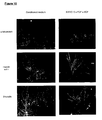
- This system can also be used to generate differentiated cell types of various kinds. After the undifferentiated pPS cells are expanded to the desired number, they are caused to differentiate according to any of a variety of differentiation paradigms provided later in this disclosure. Differentiated populations can be obtained in which at least 95% of the cells represent the same tissue type or germ layer: for example, neural cells, hepatocytes, cardiomyocytes, mesenchymal cells, or osteoblasts.
- pPS pluripotent stem
- This disclosure provides a system for rapidly expanding primate pluripotent stem (pPS) cells in vitro without requiring a layer of feeder cells to support the culture and inhibit differentiation.
- pPS pluripotent stem
- the pPS cells are grown in medium that has been preconditioned in a separate culture of feeder cells of mouse or human origin ( Figure 1 ).
- the feeder cells are grown to confluence in their own culture environment, inactivated, and then cultured in one or more batches of fresh medium to allow them to release an effective combination of factors.
- the medium is then harvested, and used to support growth of undifferentiated pPS cells plated onto a suitable substrate. Doubling rate is comparable to hES grown on feeder cells.
- the medium is changed daily, and the cells are split and passaged every 6 or 7 days.
- the pPS cells are grown in medium that has not been preconditioned, but has been supplemented with ingredients that perform essentially the same function as factors secreted from feeder cells.
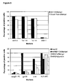
- Certain factor combinations comprising moderate to high levels of fibroblast growth factors and other cells generate cultures that can proliferate 20-fold or more through 6 or more passages, while maintaining a majority of the cells in the culture in an undifferentiated state ( Figures 6 and 8 ).
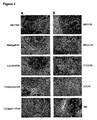
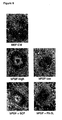
- Near confluence, most of the cells have morphological features of undifferentiated cells, and express characteristic phenotypic markers: SSEA-4, Tra-1-60, Tra-1-81, Oct-4, and telomerase reverse transcriptase (TERT)
- Prototype "primate Pluripotent Stem cells” are pluripotent cells derived from pre-embryonic, embryonic, or fetal tissue at any time after fertilization, and have the characteristic of being capable under the right conditions of producing progeny of several different cell types.
- pPS cells are capable of producing progeny that are derivatives of each of the three germ layers: endoderm, mesoderm, and ectoderm, according to a standard art-accepted test, such as the ability to form a teratoma in a suitable host, or the ability to differentiate into cells stainable for markers representing tissue types of all three germ layers in culture.
- pPS cells include embryonic cells of various types, exemplified by human embryonic stem (hES) cells, defined below; embryonic stem cells from other primates, such as Rhesus or marmoset stem cells ( Thomson et al., Proc. Natl. Acad. Sci. USA 92:7844, 1995 ; Developmental Biology 38:133, 1998 ); and human embryonic germ (hEG) cells ( Shamblott et al., Proc. Natl. Acad. Sci. USA 95:13726, 1998 ). Other types of pluripotent cells are also included in the term.
- hES human embryonic stem
- hEG human embryonic germ
- Any cells of primate origin that are capable of producing progeny that are derivatives of all three germinal layers are included, regardless of whether they were derived from embryonic tissue, fetal tissue, or other sources. It is beneficial to use pPS cells that are karyotypically normal and not derived from a malignant source.
- hES cells Prototype "human Embryonic Stem cells” (hES cells) are described by Thomson et al. (Science 282:1145, 1998 ; U.S. Patent 6,200,806 ). The scope of the term covers pluripotent stem cells that are derived from a human embryo at the blastocyst stage, or before substantial differentiation of the cells into the three germ layers. Those skilled in the art will appreciate that except where explicitly required otherwise, the term includes primary tissue and established lines that bear phenotypic characteristics of hES cells, and derivatives of such lines that still have the capacity of producing progeny of each of the three germ layers.
- pPS cell cultures are described as "undifferentiated” or “substantially undifferentiated” when a substantial proportion of stem cells and their derivatives in the population display morphological characteristics of undifferentiated cells, clearly distinguishing them from differentiated cells of embryo or adult origin.
- Undifferentiated pPS cells are easily recognized by those skilled in the art, and typically appear in the two dimensions of a microscopic view with high nuclear/cytoplasmic ratios and prominent nucleoli. It is understood that colonies of undifferentiated cells within the population will often be surrounded by neighboring cells that are differentiated. Nevertheless, the undifferentiated colonies persist when the population is cultured or passaged under appropriate conditions, and individual undifferentiated cells constitute a substantial proportion of the cell population. Cultures that are substantially undifferentiated contain at least 20% undifferentiated pPS cells, and may contain at least 40%, 60%, or 80% in order of increasing preference (in terms percentage of cells with the same genotype that are undifferentiated).
- proliferating without differentiation
- the composition is substantially undifferentiated according to the preceding definition.
- Populations that proliferate through at least four passages ( ⁇ 20 doublings) without differentiation will contain substantially the same proportion of undifferentiated cells (or possibly a higher proportion of undifferentiated cells) when evaluated at the same degree of confluence as the originating culture.
- Feeer cells or “feeders” are terms used to describe cells of one type that are co-cultured with cells of another type, to provide an environment in which the cells of the second type can grow.
- pPS cell populations are said to be "essentially free” of feeder cells if the cells have been grown through at least one round after splitting in which fresh feeder cells are not added to support the growth of pPS cells.
- a feeder free culture will contain less than about ⁇ 5% feeder cells.
- Compositions containing less than 1%, 0.2%, 0.05%, or 0.01% feeder cells are increasingly more preferred.
- a “growth environment” is an environment in which cells of interest will proliferate in vitro.
- the environment include the medium in which the cells are cultured, and a supporting structure (such as a substrate on a solid surface) if present.
- a "nutrient medium” is a medium for culturing cells containing nutrients that promote proliferation.
- the nutrient medium may contain any of the following in an appropriate combination: isotonic saline, buffer, amino acids, serum or serum replacement, and other exogenously added factors.
- a "conditioned medium” is prepared by culturing a first population of cells in a medium, and then harvesting the medium.
- the conditioned medium (along with anything secreted into the medium by the cells) may then be used to support the growth of a second population of cells.
- a particular ingredient or factor is described as having been added to the medium, what is meant is that the factor (or a cell or particle engineered to secrete the factor) has been mixed into the medium by deliberate manipulation.
- a "fresh medium” is a medium that has not been purposely conditioned by culturing with a different cell type before being used with the cell type it is ultimately designed to support. Otherwise, no limitations are intended as to its manner of preparation, storage, or use. It is added fresh (by exchange or infusion) into the ultimate culture, where it may be consumed or otherwise processed by the cell types that are present.
- a cell is said to be "genetically altered”, “transfected”, or “genetically transformed” when a polynucleotide has been transferred into the cell by any suitable means of artificial manipulation, or where the cell is a progeny of the originally altered cell that has inherited the polynucleotide.
- the polynucleotide will often comprise a transcribable sequence encoding a protein of interest, which enables the cell to express the protein at an elevated level.
- the genetic alteration is said to be “inheritable” if progeny of the altered cell have the same alteration.
- antibody refers to both polyclonal and monoclonal antibody of any species.
- the ambit of the term encompasses not only intact immunoglobulin molecules, but also fragments and genetically engineered derivatives of immunoglobulin molecules and equivalent antigen binding molecules that retain the desired binding specificity.
- Reagents, cloning vectors, and kits for genetic manipulation referred to in this disclosure are available from commercial vendors such as BioRad, Stratagene, Invitrogen, ClonTech, and Sigma-Aldrich Co.
- Suitable source cells for culturing and differentiation include established lines of pluripotent cells derived from tissue formed after gestation.
- Exemplary primary tissue sources are embryonic tissue (such as a blastocyst), or fetal tissue taken any time during gestation, typically but not necessarily before 10 weeks gestation.
- Non-limiting exemplars are lines of primate embryonic stem (ES) cells, described in Thomson et al., Science 282:114, 1998 , and U.S. Patent 6,200,806 ; and embryonic germ (EG) cells, described in Shamblott et al., Proc. Natl. Acad. Sci. USA 95:13726, 1998 , and U.S. Patent 6,090,622 .
- Also contemplated is use of the techniques of this disclosure during the initial establishment or stabilization of such cells, in which case the source cells would be primary pluripotent cells taken directly from the tissues listed.
- Feeder-free pPS cell cultures can be obtained either by passaging cells grown on feeder into feeder-free conditions, or by first deriving the cells from blastocysts into a feeder-free environment.
- the pPS cells are cultured in an environment that supports proliferation without differentiation.
- Aspects of culture that can affect differentiation include the substrate upon which the cells are cultured, the medium in which they are cultured, and the manner in which they are split and passaged to new culture environments.
- pPS cells may be supported in feeder-free culture on an extracellular matrix.
- the matrix can be deposited by preculturing and lysing a matrix-forming cell line ( WO 99/20741 ), such as the STO mouse fibroblast line (ATCC Accession No. CRL-1503), or human placental fibroblasts.
- the matrix can also be coated directly into the culture vessel with isolated matrix components.
- Matrigel® is a soluble preparation from Engelbreth-Holm-Swarm tumor cells that gels at room temperature to form a reconstituted basement membrane.
- Other suitable extracellular matrix components may include laminin, fibronectin, proteoglycan, entactin, heparan sulfate, and so on, alone or in various combinations.
- Substrates that can be tested using the experimental procedures described herein include not only other extracellular matrix components, but also polyamines, and other commercially available coatings.
- This invention contemplates adding extracellular matrix to the fluid phase of a culture at the time of passaging the cells or as part of a regular feeding.
- This invention also contemplates extracellular matrix deposited into the culture by cells within the culture (such as pPS cells that have formed around the periphery of an undifferentiated colony).
- the pluripotent cells are plated onto the substrate in a suitable distribution and in the presence of a medium that promotes cell survival, propagation, and retention of the desirable characteristics. These characteristics benefit from careful attention to the seeding distribution.
- One feature of the distribution is the plating density. It has been found that plating densities of at least ⁇ 15,000 cells cm -2 (typically 90,000 cm -2 to 170,000 cm -2 ) promote survival and limit differentiation.
- Another consideration is cell dispersion.
- enzymatic digestion is halted before cells become completely dispersed (say, ⁇ 5 min with collagenase IV).
- the plate is then scraped gently with a pipette, and the cells are triturated into clumps of adherent cells, about 10-2000 cells in size, which are then passaged into the new culture environment.
- primate PS cells can be passaged between feeder-free cultures as a finer cell suspension, providing that an appropriate enzyme and medium are chosen, and the plating density is sufficiently high.
- confluent human embryonic stem cells cultured in the absence of feeders are removed from the plates by incubating with 0.05% (wt/vol) trypsin and 0.053 mM EDTA for 5-15 min at 37°C.
- the remaining cells in the plate are removed, triturated with the pipette until dispersed into single cells and small clusters, and then replated.
- the cells are harvested without enzymes before the plate reaches confluence.
- the cells are incubated ⁇ 5 min in 0.5 mM EDTA alone in PBS, washed from the culture vessel, and then replated without further dispersal.
- the medium will generally contain the usual components to enhance cell survival, including isotonic buffer, essential minerals, and either serum or a serum replacement of some kind. To inhibit differentiation, the medium is formulated to supply some of the elements provided by feeder cells or their equivalents.
- the base nutrient medium used for conditioning can have any of several different formulae.
- Exemplary serum-containing ES medium is made with 80% DMEM (typically KO DMEM), 20% defined fetal bovine serum (FBS), 1% non-essential amino acids, 1 mM L-glutamine, and 0.1 mM ⁇ -mereaptoethanol
- Serum-free ES medium is made with 80% KO DMEM, 20% serum replacement, 1% non-essential amino acids, 1 mM L-glutamine, and 0.1 mM ⁇ -mercaptoethanol. Not all serum replacements work; an effective serum replacement is Gibco #10828-028.
- Suitable base media are X-VIVO TM 10 expansion medium (Biowhittaker) and QBSFTM-60 (Quality Biological Inc.) (Example 8). See also WO 98/30679 (Life Technologies Inc.) and U.S. 5,405,772 (Amgen ).
- the medium will typically contain a protein nutrient, in the form of serum (such as FBS), serum replacement, albumin, or essential and non-essential amino acids in an effective combination. It will also typically contain lipids, fatty acids, or cholesterol as artificial additives or the HDL or LDL extract of serum.
- beneficial factors that can be included are hormones like insulin or transferrin, nucleosides or nucleotides, pyruvate, and a reducing agent such as ⁇ -mercaptoethanol.
- the nutrient medium used for culturing the pPS cells comprises one or more factors that promote proliferation of the pPS cells without differentiation.
- the supplementation can occur by preculturing the medium with cells that secrete such factors, by adding such factors to the medium artificially, or by both techniques in combination.
- Conditioned medium can be prepared by culturing irradiated primary mouse embryonic fibroblasts (Example 1) or other cells (Example 4) at a density of ⁇ 5-6 x 10 4 cm -2 in a serum free medium such as KO DMEM supplemented with 20% serum replacement and ⁇ 4-8 ng/mL basic fibroblast growth factor (bFGF). The culture supernatant is harvested after ⁇ 1 day at 37°C. The cells are cultured in the medium for sufficient time to allow adequate concentration of released factors that support pPS cell culture. Typically, medium conditioned by culturing for 24 hours at 37°C contains a concentration of factors that support pPS cell culture for at least 24 hours. However, the culturing period can be adjusted upwards or downwards, determining empirically what constitutes an adequate period. Medium that has been conditioned for 1-2 days is typically used to support pPS cell culture for 1-2 days, and then exchanged.
- a serum free medium such as KO DMEM supplemented with 20%
- Non-conditioned medium that supports pPS cell growth in an undifferentiated state can be created by adding to a suitable base medium certain factors that invoke the appropriate signal transduction pathways in undifferentiated cells.
- fibroblast growth factor family is especially effective in this regard.
- exemplary are basic FGF (FGF-2), and FGF-4, but other members of the family can also be used.
- FGF-2 basic FGF
- FGF-4 FGF-4
- species homologs artificial analogs, antibodies to the respective FGF receptor, and other receptor activator molecules.
- FGF-1 acidic FGF
- This invention includes a method for determining additional factors that facilitate the action of FGF and equivalents in their support of undifferentiated pPS cell growth.
- the method involves combining a plurality of factors into functional groups, and culturing the cells with the groups in various combinations. Once the effective groups are determined, the rest can be eliminated, and the group can be dissected to determine the minimal effective combination. This strategy is illustrated in Example 7.
- ligands that bind c-kit such as stem cell factor (SCF, Steel factor), antibodies to c-kit, and other activators of the same signal transduction pathway may also be beneficial.
- SCF stem cell factor
- SCF is dimeric and occurs in soluble and membrane-bound forms. It transduces signals by ligand- mediated dimerization of c-kit, which is a receptor tyrosine kinase related to the receptors for platelet-derived growth factor (PDGF), macrophage colony-stimulating factor, Flt-3 ligand and vascular endothelial growth factor (VEGF).
- PDGF platelet-derived growth factor
- VEGF vascular endothelial growth factor
- factors that elevate cyclic AMP levels such as forskolin. These factors or their equivalents may be used individually or in an effective combination with other influential factors in the medium, as already described.
- the formulations provided in the Example section below were primarily designed for culturing hES cells. Where appropriate, the illustrations in this disclosure can be adapted to other types of pPS cells and multipotent cells by accommodating the known properties of the cells.
- the hEG cells claimed in U.S. 6,090,622 are dependent on the presence of both bFGF and an inducer of gp130 (such as LIF or Oncostatin-M).
- the culture media for growing hEG cells can be adapted accordingly.
- a medium formulation can be tested for its ability to support pPS cells by swapping it into a feeder-free culture system in place of medium conditioned by primary mouse embryonic fibroblasts (mEF), or some other proven standard (Examples 5-8). If pPS cells grow in a substantially undifferentiated state, then the medium can be characterized as supporting pPS cells in feeder free culture.
- mEF primary mouse embryonic fibroblasts
- One of the virtues of using fresh medium in this culture system is the ability to adjust conditions so that the cells expand more rapidly than they do when cultured on feeder cells according to traditional techniques, or in conditioned medium.
- Populations of pluripotent stem cells can be obtained that are 10-, 20-, 50-, 100-, or 1000-fold expanded when compared to the starting population. Under suitable conditions, cells in the expanded population will be 50%, 70% or more in the undifferentiated state.
- the degree of expansion per passage is calculated by dividing the number of cells harvested at the end of the culture by the number of cells originally seeded into the culture. Where geometry of the culture environment is limiting or for other reasons, the cells may optionally be passaged into a similar culture environment for further expansion. The total expansion is the product of all the expansions in each of the passages. Of course, it is not necessary to retain all the expanded cells on each passage. For example, if the cells expand 2-fold in each culture, but only ⁇ 50% of the cells are retained on each passage, then approximately the same number of cells will be carried forward. But after 4 cultures, the cells are said to have undergone an expansion of 16-fold.
- hES cells on mouse embryonic fibroblast (mEF) feeder cells have a doubling time of about 31-33 hours (Example 1).
- Certain culture environments of this invention comprising fresh medium support doubling of hES cells in less than ⁇ 24 hours (Example 8), potentially in less than ⁇ 16 hours.
- the system can be used to expand hES cells by 10- to potentially 50-fold per week. Improved efficiency is believed to be the result both of the more rapid doubling time, and the higher proportion of pPS cells that take in the new environment after passaging.
- phenotypic markers it is permissible for a few phenotypic markers to undertake a quantitative adjustment befitting adaptation to particular conditions (say, up or down 2- or 5-fold) - they will typically revert to previous levels when the cells are placed back into their previous environment (Example 8).
- An effective test for whether a cell is still pluripotent is the demonstration that the cell can still be caused to differentiate into progeny that represents (or bears antibody or PCR-detectable phenotypes) of each of the three embryonic germ layers.
- Nutrient medium and other culture characteristics formulated according to this invention can be adapted to any culture device suitable for growing pPS cells.
- Devices having a suitable surface include regular tissue culture wells, T-flasks, roller bottles, gas-permeable containers, and flat or parallel plate bioreactors.
- Fresh medium can be introduced into any of these environments by batch exchange (replacement of spent medium with fresh medium), fed-batch process (fresh medium added with no removal), or ongoing exchange in which a proportion of the medium is replaced with fresh medium on a continuous or periodic basis.
- Human ES cells have the characteristic morphological features of undifferentiated stem cells. In the two dimensions of a standard microscopic image, hES cells have high nuclear/cytoplasmic ratios in the plane of the image, prominent nucleoli, and compact colony formation with poorly discernable cell junctions.
- Cell lines can be karyotyped using a standard G-banding technique (available at many clinical diagnostics labs that provides routine karyotyping services, such as the Cytogenetics Lab at Oakland CA) and compared to published human karyotypes. It is desirable to obtain cells that have a "normal karyotype", which means that the cells are euploid, wherein all human chromosomes are present and are not noticeably altered.
- hES and hEG cells can also be characterized by expressed cell markers detectable by antibody (flow cytometry or immunocytochemistry) or by reverse transcriptase PCR.
- Human ES cells typically have antibody-detectable SSEA-4, Tra-1-60, and Tra-1-81, but little SSEA-1.
- pPS cells can also be characterized by the presence of alkaline phosphatase activity, which can be detected by fixing the cells with 4% paraformaldehyde, and then developing with Vector Red as a substrate, as described by the manufacturer (Vector Laboratories, Burlingame CA).
- Expression of hTERT and OCT-4 (detectable by RT-PCR) and telomerase activity are also characteristic of many types of undifferentiated pPS cells (Example 3).
- Another desirable feature of propagated pPS cells is a potential to differentiate into cells of all three germ layers: endoderm, mesoderm, and ectoderm.
- Pluripotency of hES cells can be confirmed by forming teratomas in SCID mice, and examining them for representative tissues of all three germ layers.
- pluripotency can be determined by allowing pPS cells to differentiate non-specifically (for example, by forming embryoid bodies), and then determining the cell types represented in the culture by immunocytochemistry ( Figure 10 ). Potential of pPS cells to differentiate into particular cell lines can be determined according to procedures described later in this disclosure.
- Certain cell populations described in this disclosure are substantially undifferentiated, and can be passaged between multiple cultures in the conditions described. During passage, some cells may differentiate (particularly when replated as single cells at low density, or when large clusters are allowed to form). However, cultures typically reestablish a larger proportion of undifferentiated cells as they reapproach confluence.
- This disclosure also provides a system for obtaining pPS cells that have been genetically altered, either in a transient or stable fashion.
- the cells may be modified to give them desired properties in the undifferentiated state, to give them desired properties after differentiation into other cell types, or to provide a method to positively or negatively select for particular undifferentiated or differentiated phenotypes.
- the hES cells are transfected with a drug susceptibility gene under control of a promoter specific for undifferentiated cells ( WO 02/42445 ).
- Suitable vector plasmids for transfecting into hES cells include lipid/DNA complexes, such as those described in U.S. Patent Nos. 5,578,475 ; 6,020,202 ; and 6,051,429 , and exemplified in Example 5.
- Viral vector systems for producing hES cells with stable genetic alterations can be based on adenovirus, retrovirus, or lentivirus, prepared using commercially available virus components.
- the genetically altered cells can be enriched by selecting for a functional feature of the new genotype.
- a particularly effective way of enriching genetically altered cells is positive selection using resistance to a drug such as neomycin.
- the cells can be genetically altered by contacting simultaneously with vector systems for the marker gene or gene of interest, and a vector system that provides the drug resistance gene. Alternatively, the drug resistance gene can be built into the same vector as the gene of interest.
- pPS cells are especially amenable to genetic alteration when they are grown in feeder-free culture, elaborated throughout this disclosure.
- Transient transfection using DNA/lipid complexes can be as high as 60%.
- the cells are easier to manipulate, and there are no feeder cells around to act as a sink for the vector.
- Following genetic alteration and drug selection on drug-resistant feeders or feeder-free culture, it is possible to pick colonies that demonstrate the altered phenotype, and culture them separately. The picked colonies are dispersed into small clumps of 25-100 cells, and replated in a suitable environment. It is possible to achieve cultures of pPS cells in which a high proportion (up to 90%) of the undifferentiated cells are genetically altered.
- pPS cells cultured according to this invention can be used to make differentiated cells of various commercially and therapeutically important tissue types.
- ⁇ For example, scientists at Geron Corporation have discovered methods for obtaining highly enriched populations of cells of the neural lineage.
- Cells are changed to a culture medium containing one or more neurotrophins (such as neurotrophin 3 or brain-derived neurotrophic factor) and one or more mitogens (such as epidermal growth factor, basic fibroblast growth factor, platelet-derived growth factor, insulin-like growth factor 1, and erythropoietin).
- Cultured cells are optionally separated based on whether they express a marker such as A2B5 or NCAM.
- Neural precursors can be obtained having the capacity to generate both neuronal cells (including mature neurons), and glial cells (including astrocytes and oligodendrocytes).
- replicative neuronal precursors can be obtained that have the capacity to form differentiated cell populations in which at least ⁇ 5% of all the cells in the population express tyrosine hydroxylase, a marker of dopaminergic neurons. See PCT publication WO 01/88104 and PCT application PCT/US01/15861 .
- culturing pPS cells or embryoid body cells in the presence of a histone deacetylase inhibitor such as n-butyrate creates a population of cells highly enriched for markers of the hepatocyte lineage.
- the cultured cells are optionally cultured simultaneously or sequentially with a hepatocyte maturation factor, such as EGF, insulin, and FGF. Further details can be found in PCT publication WO 01/81549 .
- hES cell derived cells that have characteristic markers of cardiomyocytes and spontaneous periodic contractile activity. Differentiation is facilitated by nucleotide analogs that affect DNA methylation (such as 5-aza-deoxy-cytidine), growth factors, and bone morphogenic proteins.
- the cells can be further enriched by density-based cell separation, and maintained in media containing creatine, carnitine, and taurine. See PCT application PCT/US02122245 .
- hES cells into mesenchymal cells in a medium containing a bone morphogenic protein (BMP), a iigand for the human TGF- ⁇ receptor, or a ligand for the human vitamin D receptor.
- BMP bone morphogenic protein
- the medium may further comprise dexamethasone, ascorbic acid-2-phosphate, and sources of calcium and phosphate.
- derivative cells can have phenotypic features of cells of the osteoblast lineage. See PCT application PCT/US02/20998 .
- differentiated cell populations be substantially free of undifferentiated pPS cells.
- One way of depleting undifferentiated stem cells from the population is to transfect them with a vector in which an effector gene under control of a promoter (such s the TERT promoter) that causes preferential expression in undifferentiated cells.
- a promoter such as the TERT promoter
- pPS cells can be used to screen for factors (such as small molecule drugs, peptides, polynucleotides, and the like) or conditions (such as culture conditions or manipulation) that affect the characteristics of pPS cells in culture.
- factors such as small molecule drugs, peptides, polynucleotides, and the like
- conditions such as culture conditions or manipulation
- This system has the advantage of not being complicated by a secondary effect caused by perturbation of the feeder cells by the test compound.
- growth affecting substances are tested.
- the conditioned medium is withdrawn from the culture and a simpler medium (such as KO DMEM) is substituted.
- Different wells are then treated with different cocktails of soluble factors that are candidates for replacing the components of the conditioned medium. Efficacy of each mixture is determined if the treated cells are maintained and proliferate in a satisfactory manner, optimally as well as in conditioned medium.
- Potential differentiation factors or conditions can be tested by treating the cells according to the test protocol, and then determining whether the treated cell develop
- Feeder-free pPS cultures can also be used for the testing of pharmaceutical compounds in drug research.
- Assessment of the activity of candidate pharmaceutical compounds generally involves combining the differentiated cells of this invention with the candidate compound, determining any resulting change, and then correlating the effect of the compound with the observed change.
- the screening may be done, for example, either because the compound is designed to have a pharmacological effect on certain cell types, or because a compound designed to have effects elsewhere may have unintended side effects.
- Two or more drugs can be tested in combination (by combining with the cells either simultaneously or sequentially), to detect possible drug-drug interaction effects.
- compounds are screened initially for potential toxicity ( Castell et al., pp 375-410 in In vitro Methods in Pharmaceutical Research, Academic Press, 1997 ).
- Cytotoxicity can be determined by the effect on cell viability, survival, morphology, on the expression or release of certain markers, receptors or enzymes, on DNA synthesis or repair, measured by [ 3 H]-thymidine or BrdU incorporation, or on sister chromatid exchange, determined by metaphase spread.
- the reader is referred generally to the standard textbook In vitro Methods in Pharmaceutical Research, Academic Press, 1997 , and U.S. Patent 5,030,015 .
- Differentiated cells of this invention can also be used for tissue reconstitution or regeneration in a human patient in need thereof.
- the cells are administered in a manner that permits them to graft to the intended tissue site and reconstitute or regenerate the functionally deficient area.
- neural stem cells are transplanted directly into parenchymal or intrathecal sites of the central nervous system, according to the disease being treated. Grafts are done using single cell suspension or small aggregates at a density of 25,000-500,000 cells per ⁇ L ( U.S. Patent 5,968,829 ). The efficacy of neural cell transplants can be assessed in a rat model for acutely injured spinal cord as described by McDonald et al. (Nat. Med. 5:1410, 1999 ), and Kim et at. (Nature 418:50, 2002 ).
- a successful transplant will show transplant-derived cells present in the lesion 2-5 weeks later, differentiated into astrocytes, oligodendrocytes, and/or neurons, and migrating along the cord from the lesioned end, and an improvement in gait, coordination, and weight-bearing.
- cardiomyocytes can be assessed in an animal model for cardiac cryoinjury, which causes 55% of the left ventricular wall tissue to become scar tissue without treatment ( Li et al., Ann. Thorac. Surg. 62:654, 1996 ; Sakai et al., Ann. Thorac. Surg. 8:2074, 1999 , Sakai et al., J. Thorac. Cardiovasc. Surg. 118:715, 1999 ).
- Successful treatment will reduce the area of the scar, limit scar expansion, and improve heart function as determined by systolic, diastolic, and developed pressure.
- Cardiac injury can also be modeled using an embolization coil in the distal portion of the left anterior descending artery ( Watanabe et al., Cell Transplant. 7:239, 1998 ), or by ligation of the left anterior descending coronary artery ( Min et al., J. Appl. Physiol. 92:288, 2002 ). Efficacy of treatment can be evaluated by histology and cardiac function. Cardiomyocyte preparations embodied in this invention can be used in therapy to regenerate cardiac muscle and treat insufficient cardiac function ( U.S. Patent 5,919,449 and WO 99/03973 ).
- Hepatocytes and hepatocyte precursors can be assessed in animal models for ability to repair liver damage.
- One such example is damage caused by intraperitoneal injection of D-galactosamine ( Dabeva et al., Am. J. Pathol. 143:1606, 1993 ).
- Efficacy of treatment can be determined by immunocytochemical staining for liver cell markers, microscopic determination of whether canalicular structures form in growing tissue, and the ability of the treatment to restore synthesis of liver-specific proteins.
- Liver cells can be used in therapy by direct administration, or as part of a bioassist device that provides temporary liver function while the subject's liver tissue regenerates itself following fulminant hepatic failure.
- cells prepared according to this invention are typically supplied in the form of a pharmaceutical composition comprising an isotonic excipient, and prepared under conditions that are sufficiently sterile for human administration.
- a pharmaceutical composition comprising an isotonic excipient
- the reader is referred to Cell Therapy: Stem Cell Transplantation, Gene Therapy, and Cellular Immunotherapy, by G. Morstyn & W. Sheridan eds, Cambridge University Press, 1996 ; and Hematopoietic Stem Cell Therapy, E.D. Ball, J. Lister & P. Law, Churchill Livingstone, 2000 .
- the cells may be packaged in a device or container suitable for distribution or clinical use, optionally accompanied by information relating to use of the cells in tissue regeneration, or restoring a therapeutically important metabolic function.
- Example 1 Growing hES cells without feeder cells in conditioned medium
- undifferentiated hES cells that had been maintained on primary mouse embryonic feeder cells were maintained in the absence of feeders.
- the culture wells were coated with Matrigel®, and the cells were cultured in the presence of conditioned nutrient medium obtained from a culture of irradiated primary fibroblasts.
- CM Conditioned medium
- the fibroblasts were harvested from T150 flasks by washing once with Ca ++ /Mg ++ free PBS and incubating in trypsin/EDTA (Gibco). After the fibroblasts detached from the flask, they were collected in mEF medium (DMEM + 10% FBS). The cells were irradiated at 4000 rad, counted and seeded at about 55,000 cells cm -2 in mEF medium. After at least 4 hours, the medium was exchanged with SR containing ES medium. Conditioned medium was collected daily for feeding of hES cultures. Alternatively, medium was prepared using mEF plated in culture flasks, exchanging medium daily. Before addition to the hES cultures, the conditioned medium was supplemented with 4 ng/mL of human bFGF (Gibco). Fibroblast cultures were used for about 1 week before replacing with newly prepared cells.
- Undifferentiated hES colonies were harvested from hES cultures by incubating in -200 U/mL collagenase IV for about 5 minutes at 37 °C. Colonies were harvested by picking individual colonies up with a 20 ⁇ L pipet ti or by scraping and dissociating into small clusters in conditioned medium. These cells were then seeded onto Matrigel® coated plates (0.75-1 mL diluted -1:30) in conditioned medium at 15 colonies per well.
- hES cells were visible as small colonies and there were cells in between the colonies that appeared to be differentiating or dying. As the hES cells proliferated, the colonies became quite large and very compact, representing the majority of surface area of the culture dish.
- the hES cells in the colonies had a high nucleus to cytoplasm ratio and had prominent nucleoli, similar to hES cells maintained on feeder cells.
- the differentiated cells in between the colonies represented less than 10% of the cells in the culture.
- the cultures were split using Collagenase IV, gently triturated into small clusters of 10-2,000 cells, and then re-seeded on Matrigel® coated plates in conditioned medium at ⁇ 90,000 to 170,000 cells cm -2 . Medium was changed daily, and the cells were split and passaged at 13 and 19 days after initial seeding.
- hES cells Cultures of hES cells have been grown in the absence of feeder cells for over 147 days with no apparent change in the proliferative capacity or phenotype.
- Human ES cells maintained on Matrigel® in mEF conditioned medium have a doubling time of about 31-33 hours, similar to the proliferation rate for hES cells grown on mEF feeder cells.
- H1 cells after 64 days of feeder-free culture showed a normal karyotype.
- fibronectin or collagen IV had colonies of undifferentiated hES cells, although the cultures on fibronectin or collagen IV did not contain as many undifferentiated colonies as the cultures on Matrigel® or laminin.
- the cells on Matrigel® or laminin reached confluence, the cells within the colonies became very compact, were morphologically very similar to the cells maintained on feeders and were serially passaged. After 40 days (6 passages), cells on Matrigel® and laminin contained a high proportion of colonies which continued to display ES-like morphology in long term culture. However, cells maintained on fibronectin or collagen IV had fewer colonies displaying appropriate ES-morphology.
- cells cultured on Matrigel® or laminin in non-conditioned medium appeared to be proliferating more slowly and showed a differentiated morphology after a few passages.
- Figure 1 shows the morphology of hES cells in feeder-free culture.
- Panel A (Left Side) shows morphology of hES cells of the H1 line cultured on feeder cells in non-conditioned medium (mEF/RM), on Matrigel®, laminin, fibronectin, or collagen IV in mEF conditioned medium.
- Panel B shows morphology of hES cells of the H9 line maintained on Matrigel® in various types of conditioned medium, described in Example 4.
- Human ES cells maintained on Matrigel® in mEF conditioned medium showed a doubling time of about 31-33 hours.
- H1 cells after 64 days of feeder-free culture showed a normal karyotype.
- Undifferentiated hES cells express SSEA-4, Tra-1-60, Tra-1-81, OCT-4, and hTERT. In order to assess whether the cells maintained in feeder-free conditions retained these markers, cells were evaluated by immunostaining, reverse transcriptase PCR amplification, and assay for telomerase activity.
- the hES cells were dissociated in 0.5 mM EDTA in PBS and resuspended to about 5 x 10 5 cells in 50 ⁇ L diluent containing 0.1% BSA in PBS. They were labeled with specific primary antibody and then fluorescent second antibody, and analyzed on a Flow Cytometer.
- FACS fluorescence-activated cell sorting
- Figure 2 shows marker expression detected by immunocytochemistry.
- Cells were incubated with primary antibody, fixed in 2% paraformaldehyde, and then visualized with FITC-conjugated goat anti-mouse immunoglobulin.
- the results show that SSEA-4, Tra-1-60, Tra-1-81, and alkaline phosphatase were expressed by the hES colonies on Matrigel® or laminin, as seen for the cells on feeders - but not by the differentiated cells in between the colonies.
- Figure 3 shows OCT-4 and hTERT expression of H1 cells grown on feeders or in a feeder free environment, as detected by reverse-transcriptase PCR amplification (detailed in WO 01/51616 ).
- the POU transcription factor OCT-4 is normally expressed in the undifferentiated hES cells and is down-regulated upon differentiation.
- CM conditioned medium
- RM unconditioned regular medium
- hTERT and OCT-4 expression was seen in all the culture conditions except Matrigel® and regular medium. After exposure of cells to retinoic acid (RA) or dimethyl sulfoxide (DMSO), factors that promote cell differentiation, the expression of hTERT was markedly decreased.
- RA retinoic acid
- DMSO dimethyl sulfoxide
- telomerase activity was measured by telomeric repeat amplification protocol (TRAP assay: Kim et al., Science 266:2011, 1997 ; Weinrich et al., Nature Genetics 17:498, 1997 ). All the cultures conditions showed positive telomerase activity after 40 days on Matrigel®, laminin, fibronectin or collagen IV in mEF conditioned medium.
- Example 3 Pluripotency of hES cells in feeder-free culture
- H1 hES cells maintained in conditioned medium on Matrigel®, laminin, fibronectin or collagen IV for 26 days.
- the hES cells were dissociated into small clumps by incubating in ⁇ 200 U/mL collagenase IV at 37°C for 10 min, and cultured in suspension to form embryoid bodies (EBs) in medium containing DMEM, 20% FBS (Hyclone), 1 mM glutamine, 0.1 mM ⁇ -mercaptoethanol, and 1% non-essential amino acids (Gibco).
- EBs embryoid bodies
- the aggregates were transferred onto poly-ornithine-coated plates, and cultured for additional 7 days. The cultures were then examined for the presence of beating cells, and processed for immunocytochemistry.
- the staining patterns were consistent with cells of the neuron and cardiomyocyte lineages ( ⁇ -tubulin III and cardiac troponin I, respectively). About 8 days after differentiation, beating regions were identified in all cultures. There were also cells staining for ⁇ -fetoprotein, a marker of endoderm lineage.
- hES cells were also tested for their ability to form teratomas by intramuscular injection into SCID mice.
- Cells maintained on feeders or off feeders were harvested, resuspended in PBS and injected intramuscularly into SCID/beige mice (5 x 10 6 cells per site). Tumors were excised and processed for histological analysis. Cystic epithelial structures, probable dental component, cartilage and glandular epithelial or neural components were found in teratomas derived from feeder-free hES cultures.
- Example 4 Sources of conditioned medium for feeder-free culture
- mEF primary mouse embryonic fibroblasts
- the NHG190 cell line is a telomerized mouse embryonic fibroblast line described in WO 01/51616 .
- STO is a transformed mouse fibroblast line available from the ATCC.
- BJ 5ta is a telomerized human foreskin fibroblast cell line.
- hTERT-RPE is a telomerized human retinal epithelial cell line.
- conditioned medium To prepare conditioned medium, the respective cell lines were harvested by washing once with Ca ++ /Mg ++ free PBS, incubating in trypsin/EDTA (Gibco) for about 5 min, and suspending in mEF medium. The cells were irradiated at ⁇ 4000 rad, counted, and plated into culture vessels. After at least 4 h, the medium was exchanged with ES medium containing 4 ng/mL bFGF. Conditioned medium was collected daily thereafter, and used for feeding of hES cultures. Before addition to the hES cultures, each conditioned medium was supplemented with 4 ng/mL of human basic fibroblast growth factor (hbFGF; Gibco).
- hbFGF human basic fibroblast growth factor
- FIG. 1 Panel B (Right Side) shows morphology of hES cells of the H9 line maintained on Matrigel® in medium conditioned by mEF, NHG190, STO and BJ 5ta cells, compared with unconditioned regular medium (RM).
- the cells in RPE conditioned medium differentiated within the first week of culture.
- the cells in the other conditioned mediums all had hES colonies with appropriate ES-morphology.
- Based on the morphology, confluence of the culture, and the ratio of differentiated to undifferentiated cells the conditioned medium can be ranked in order of decreasing preference: primary mEF, NHG190, STO, and BJ 5ta.
- cells on Matrigel® or laminin in medium conditioned by other cell lines including NHG190, STO and BJ 5ta, expressed high levels of SSEA-4, Tra-1-60 and Tra-1-81 but low levels of SSEA-1 as analyzed by FACS.
- Cells on Matrigel® or laminin in mEF conditioned medium or NHG190 conditioned medium were able to differentiate into three germ layer cell types. Immunocytochemical analysis of the differentiated cultures showed positive staining for ⁇ -tubulin III consistent with neurons (ectoderm lineage), cardiac troponin I consistent with cardiomyocytes (mesoderm lineage), and ⁇ -fetoprotein (endoderm lineage).
- LIF leukemia inhibitory factor
- hES cells maintained in feeder-free culture on laminin in conditioned medium were genetically modified by transfecting with a plasmid carrying green fluorescent protein (GFP) driven by the CMV promoter.
- GFP green fluorescent protein
- hES cells of the H9 line maintained on laminin in mEF-conditioned medium were transfected with a plasmid carrying GFP driven by the CMV promoter (ClonTech cat. # 6084-1) at 24 or 48 h after plating.
- Initial experiments used a mixture of 5 ⁇ g of plasmid and 12 ⁇ L of Lipofectamine 2000TM (Gibco, cat # 11668-019).
- Cells received 1 mL of DNA/lipid complex and were incubated for 4 h at 37° before the addition of 3 mL of mEF-conditioned medium, and then monitored for GFP expression 24 h after transfection.
- Figure 4 shows the results of this experiment.
- Panel A morphology of H9 cells maintained on laminin.
- Panel B GFP-positive cells observed in the same colony shown in A.
- Panel C FACS analysis of % GFP-positive cells in SSEA-4 high population(undifferentiated cells).
- Cells were transfected 24 (bar 1 and 2) or 48 h (bar 3 and 4) after the seeding and analyzed 24 (bar 1 and 3) or 48 h (bar 2 and 4) after the transfection.
- Bright green cells were observed in compact areas of undifferentiated ES colonies on laminin 24 h after transfection (Panels A & B). Transfection at 48 h after initial seeding gave the highest efficiency: 38% of the cells were GFP-positive as determined by FACS analysis 24 h after the transfection (Panel C).
- H1 hES cells maintained on Matrigel® were cotransfected with a mixture of 7.5 ⁇ g plasmid carrying ⁇ -galactosidase driven by the EF1a promoter, and 2.5 ⁇ g of plasmid carrying the PGK promoter driving the neophosphotransferase gene.
- the cells were transfected 48 h after plating on Matrigel® in mEF-conditioned medium.
- Ten ⁇ g of plasmid plus 15 ⁇ L of FuGENETM (Roche Diagnostics Corp.) was incubated with the cells in 1 mL for 4 h before adding 2.5 mL of mEF-conditioned medium.
- Example 6 Additives that promote undifferentiated ES cell rowth in fresh medium
- hES medium contained 20% Serum Replacement (Gibco #10828-028), 80% Knockout DMEM (Gibco #10829-018), 1% non-essential amino acids (Gibco #11140-050), 1 mM L-glutamine (Gibco #15039-027), and 2.5 mM ⁇ -mercaptoethanol (Sigma #M7522).
- This medium was supplemented with 40 ng/mL bFGF; 15 ng/mL stem.
- SCF SCF, R&D System #255SC
- 100 ng/mL leukemia inhibitory factor LIF, Sigma #L5283 or Chemicon #LIF 1010
- CNTF ciliary neurotrophic factor
- OSM human Oncostatin M
- IL-6 interleukin 6
- the H9 cell line (passage 31) was harvested from a culture in conditioned medium, plated onto Matrigel®, and cultured with hES medium with the factors at the concentrations indicated above, or 5- or 25-fold lower. Cells grown in the fully supplemented medium displayed undifferentiated hES morphology. A higher degree of differentiation was observed after 4 passages for the cultures grown at lower concentrations of the growth factors, and the cells maintained without growth factors were almost completely differentiated. These cultures were terminated.
- SSEA 4 clone MC 813 mouse IgG3; Tra-1 60, mouse IgM; Tra-1 81, mouse IgM; SSEA-1, mouse IgM; c-kit, BD PharMingen #555714; R-PE labeled mouse anti-human CD117; mouse IgG1, clone YB5.B8; gp130, R&D System #FAB 228P; R-PE labeled mouse IgG1, clone 28123.111; R-PE labeled isotype control mouse IgG1, PharMingen #33815; isotype control mouse IgG3, Sigma #M3645.
- Goat anti-mouse IgG3 FITC labeled was obtained from Southern Biotechnology #1102-02.
- the cells were washed with warm PBS for 3-5 minutes, incubated with 3 mL 0.5 mM EDTA at 37°C for 10 min, and collected into a 15- mL tube containing 10 mL medium. They were spun down at 1200 rpm (400 g), washed in 1% BSA/PBS, and suspended in 100 ⁇ L of diluted primary antibody at 4°C for 30 min. After rewashing in 1% BSA/PBS, they were incubated with goat anti-mouse IgG3 FITC (1:100) at 4°C for 15-30 min, then washed and resuspended in 500 ⁇ L 1:1000 propidium iodine.
- Figure 5 shows the results of FACS analysis of markers for undifferentiated phenotype for H9 cells maintained for 8 passages in the growth factor mixture. Expression patterns and levels of surface markers, including SSEA-1, SSEA-4, Tra 1-60 and Tra 1-81 in cultures maintained in high concentrations of growth factors were similar to cells maintained in MEF conditioned medium (MEF-CM).
- conditioned medium contains factors that promote stem cell growth without differentiation, and that these factors can be either be secreted into the medium by cells being used for the conditioning, or added to the medium artificially.
- Figure 5 shows receptor-associated molecules expressed by the H9 cells after 9 passages in MEF-CM (control), or the artificial mixture of growth factors (GF) on either Matrigel® or laminin.
- the hepatocellular carcinoma cell line HepG2 serves as a positive control for gp130.
- HLA-ABC histocompatibility Class I antigen
- mslgG1 isotype control
- gp130 associated with the LIF receptor
- ligands for c-kit help support undifferentiated hES cell growth.
- EBs were plated onto gelatin coated plates and differentiated an additional 16 days. They were then fixed in 4% paraformaldehyde, permeabilized with ethanol, blocked with 10% normal goat serum, and then analyzed for phenotypic markers by indirect antibody staining.
- Immunocytochemical analysis showed the presence of ⁇ -tubulin III in cells having neuronal morphology. Other cells stained for ⁇ -fetoprotein or smooth muscle actin. This demonstrates that hES cells cultured in medium comprising SCF and other factors have the capacity to differentiate into derivatives of all three germ layers.
- H9 hES cell line was cultured in non-conditioned ES medium supplemented with bFGF alone at high (40 ng/mL) or low (8 ng/mL) concentration, or bFGF (40 ng/mL) in combination with SCF (15 ng/mL) or Flt-3 ligand (75 ng/mL).
- FIG. 6 shows the results. Cultures with high concentrations of bFGF contained ⁇ 30-50% cells having undifferentiated morphology, with a higher proportion in cultures also containing SCF or Flt-3 ligand. FACS analysis showed that ⁇ 60% of the cells in these cultures expressed SSEA-4. In comparison, cultures in conditioned medium contained ⁇ 80% with undifferentiated morphology and ⁇ 90% expressing SSEA-4.
- the hES cell line H1 at passage 27 or the H7 line at passage 35 hES cells previously maintained in conditioned medium were cultured in fresh ES medium containing these growth factors.
- Table 2 Factors added to fresh ES medium for hES cell culture Condition bFGF Other Growth Factors A 8 ng/mL B C 40 ng/mL D 40 ng/mL SCF(15ng/mL) E 40 ng/mL Flt-3L (75 ng/mL) F 40 ng/mL TPO (100 ng/mL) G 40 ng/mL LIF (100 ng/mL) H 40 ng/mL SCF (15 ng/mL), IL-6 (15 ng/mL), LIF (100 ng/mL), CNTF (50 ng/mL), OSM (50 ng/mL) I SCF (15 ng/mL) J SCF (100 ng/mL) K Flt-3L (75 ng/mL) L TPO
- Figure 7 shows expression of SSEA-4 as evaluated by flow cytometry (gated for low or high staining level) at passage 9 (H7 cells) or passage 10 (H1 cells) after being transferred from conditioned medium, H7 cells grown in any of these conditions showed telomerase activity at passage 15.
- Example 7 Other base media for growing hES cells in feeder-free culture
- hES cells passaged 29 times in conditioned medium were weaned onto an alternative medium designed for proliferation and development of hematopoietic cells.
- Ex vivo expansion medium was obtained by arrangement with a commercial supplier, and is thought to be based on the medium described in U.S. Patent 5,405,772 (Ponting, Amgen Inc.).
- the Ponting medium comprises the following components: Iscove's modified Dulbecco's medium; amino acids; vitamins, bovine albumin; bovine transferrin (100 ⁇ g/mL); lipids and cholesterol; ⁇ -mercaptoethanol; pyruvate; nucleotides; epidermal growth factor (15 ng/mL); fibroblast growth factor (2 ng/mL); platelet-derived growth factor (10 ng/mL); and insulin (10 ⁇ glmL).
- the medium was further supplemented with 2 mM L-glutamine, 1% non-essential amino acids (Gibco), 0.1 mM ⁇ -mercaptoethanof, and 8 ng/mL bFGF.
- the cells were first passaged onto Matrigel® coated plates using collagenase IV, and cultured for 2 days with conditioned medium. On day 2, the 100% conditioned medium was replaced with medium containing 80% conditioned medium plus 20% fresh expansion medium. Cells were fed fresh daily and passaged weekly. The proportion of expansion medium was increased by 20% approximately every 2 days until the cells were completely weaned, and then grown until they had been passaged a further 8 times.
- embryoid bodies were formed and analyzed by immunocytochemistry for phenotypic markers representing each of the three germ layers. After passage 4 in expansion medium, the cells were dissociated into small clumps using 200 U/mL collagenase IV at 37°C for 10 min placed in suspension culture in differentiation medium (DMEM + 10% FBS) for 4 days, then transferred onto poly-L-ornithine hydrobromide coated plates and cultured a further 10 days.
- differentiation medium DMEM + 10% FBS
- mice were fixed in 4% paraformaldehyde, permeabilized, and labeled alternately with mouse anti human ⁇ -tubulin isotype III clone SDL.3D10, mouse anti human muscle actin clone HHF35, or mouse anti ⁇ -fetoprotein. Primary antibody was visualized using FITC labeled goat anti-mouse IgG. Results showed that hES cells passaged repeatedly in expansion medium (not previously conditioned), and then differentiated, were positive for ⁇ -tubulin and muscle actin.
- Example 8 Rapid expansion method for producing pluripotent stem cells
- X-VIVO TM 10 expansion medium was obtained from Biowhittaker; QBSFTM-60 was obtained from Quality Biological Inc.
- the X-VIVO TM 10 formulation contains pharmaceutical grade human albumin, recombinant human insulin and pasteurized human transferrin. Exogenous growth factors, artificial stimulators of cellular proliferation or undefined supplements are not included in the X-VIVO TM 10 medium. They are also devoid of any protein-kinase C stimulators.
- QBSFTM-60 is a serum-free formulation that contains recombinant or pasteurized human proteins.
- the X-VIVO TM 10 medium was supplemented with 2 mM L-glutamine, 1% non-essential amino acids (Gibco), 0.1 mM ⁇ -mercaptoethanol, and 8 ng/mL bFGF.
- the medium was further supplemented with 8 ng/mL or 40 ng/mL of bFGF (Gibco); 40 ng/mL of bFGF and 15 ng/mL of SCF (R & D System); or 40 ng/mL of bFGF and 75 ng/mL of Flt3 ligand (R & D System).
- QBSFTM-60 medium was supplemented with 0.1 mM ⁇ -mercaptoethanol, 1% non-essential amino acids (Gibco) and 40 ng/mL of bFGF.
- hES cells cultured in mEF conditioned medium was used as control in these experiments.
- the hES cells were first passaged onto Matrigel® coated plates using collagenase IV, and cultured for 2 days with conditioned medium. On day 2, the conditioned medium was replaced with 80% unconditioned ES medium plus 20% expansion medium. Cells were fed fresh daily and passaged weekly. The proportion of expansion medium was increased by 20% approximately every 2 days until the cells were completely weaned, and then grown until they had been passaged 6 more times.
- Figure 8 shows colonies of hES cell at the end of 6 passages (sufficient for full adaptation) in the following media: (A) mEF conditioned medium + bFGF (8ng/mL); (B) X-VIVOTM 10 + bFGF (40ng/mL); (C) X-VIVOTM 10 + bFGF (40ng/mL) + stem cell factor (SCF, Steel factor) (15 ng/mL); (D) X-VIVOTM 10 + bFGF (40ng/mL) + Flt3 ligand (75 ng/mL); (E) OBSFTM-60 + bFGF (40ng/mL).
- Table 3 shows the average total cell expansion per passage, for undifferentiated hES cells cultured for 4 passages in mEF conditioned medium, or for 7 passages in X-VIVOTM 10 or QBSFTM-60.
- Table 3 Growth Rates for ES Cell Cultures Medium Average Cell Expansion per Passage mEF conditioned medium 2.2 fold X-VIVO TM 10 + bFGF (40ng/mL) 6.0 fold X-VIVO TM 10 + bFGF (40ng/mL) + SCF (15 ng/mL) 8.2 fold X-VIVO TM 10 + bFGF (40ng/mL) + Flt3 ligand (75 ng/mL) 5.0 fold OBSFTM-60 + bFGF (40ng/mL) 6.4 fold
- the average expansion of cells per passage in X-VIVO TM 10 and QBSFTM-60 was greater than the cells cultured in mEF conditioned medium culture.
- the cells in mEF conditioned medium were passaged on average every 7 days, while the cells in X-VIVO TM 10 and QBSFTM-60 were passaged on average every 5 days.
- the rate of expansion in unconditioned X-VIVO TM 10 or QBSFTM-60 was ⁇ 3.2 to 5.2 times faster than in mEF conditioned ES medium.
- Figure 9 shows the gene expression profile of hTERT and Oct3/4.
- the RNA was isolated from the cells using High Pure RNA Isolation Kit (Roche Diagnostics) and evaluated by TaqmanTM assay (real time RT-PCR).
- the gene expression in each of the test condition is plotted relative to expression in the control culture. Taking into consideration the instrument error and assay variability, differences in expression between the test and control samples are only significant if greater than 2-fold.
- the analysis shows expression of hTERT and Oct-3/4 decreases somewhat upon adaptation to unconditioned X-VIVO TM 10 or QBSFTM-60 medium (first four bars in each set), but returns to standard levels when the cells are passaged back into mEF conditioned medium (last three bars in each set).
- embryoid bodies were formed and analyzed by immunocytochemistry for phenotypic markers representing each of the three germ layers.
- the cells were dissociated into small clumps using 200 U/mL collagenase IV at 37°C for 10 min, placed in suspension culture in differentiation medium (DMEM + 10% FBS) for 4 days, then transferred onto poly-L-ornithine hydrobromide coated plates for a further 10 days. They were fixed in 4% paraformaldehyde, permeabilized, and labeled by immunocytochemistry.
- Figure 10 shows the results. hES cells passaged 7 times in unconditioned X-VIVO TM 10 medium stained for ⁇ -fetoprotein (representing endoderm); muscle actin (representing mesoderm), and ⁇ -tubulin III (representing ectoderm).
- hES cells can be expanded in fresh (non-conditioned) media in a feeder-free environment at a rapid rate suitable for commercial production.
- the cells retain the morphology of undifferentiated hES cells, and can be differentiated into derivative cells representing all three germ layers.
Landscapes
- Health & Medical Sciences (AREA)
- Engineering & Computer Science (AREA)
- Life Sciences & Earth Sciences (AREA)
- Biomedical Technology (AREA)
- Genetics & Genomics (AREA)
- Chemical & Material Sciences (AREA)
- Organic Chemistry (AREA)
- Bioinformatics & Cheminformatics (AREA)
- Zoology (AREA)
- Biotechnology (AREA)
- Wood Science & Technology (AREA)
- General Engineering & Computer Science (AREA)
- General Health & Medical Sciences (AREA)
- Biochemistry (AREA)
- Microbiology (AREA)
- Cell Biology (AREA)
- Neurology (AREA)
- Physics & Mathematics (AREA)
- Plant Pathology (AREA)
- Reproductive Health (AREA)
- Gynecology & Obstetrics (AREA)
- Developmental Biology & Embryology (AREA)
- Neurosurgery (AREA)
- Molecular Biology (AREA)
- Biophysics (AREA)
- Crystallography & Structural Chemistry (AREA)
- Bioinformatics & Computational Biology (AREA)
- General Chemical & Material Sciences (AREA)
- Chemical Kinetics & Catalysis (AREA)
- Medicinal Chemistry (AREA)
- Nuclear Medicine, Radiotherapy & Molecular Imaging (AREA)
- Pharmacology & Pharmacy (AREA)
- Animal Behavior & Ethology (AREA)
- Public Health (AREA)
- Veterinary Medicine (AREA)
- Micro-Organisms Or Cultivation Processes Thereof (AREA)
- Medicines Containing Material From Animals Or Micro-Organisms (AREA)
Applications Claiming Priority (3)
| Application Number | Priority Date | Filing Date | Title |
|---|---|---|---|
| US31747801P | 2001-09-05 | 2001-09-05 | |
| US10/235,094 US7410798B2 (en) | 2001-01-10 | 2002-09-04 | Culture system for rapid expansion of human embryonic stem cells |
| EP02757586A EP1430111A4 (en) | 2001-09-05 | 2002-09-05 | CULTURE SYSTEM FOR FAST EXPANSION OF HUMAN EMBRYONIC STEM CELLS |
Related Parent Applications (1)
| Application Number | Title | Priority Date | Filing Date |
|---|---|---|---|
| EP02757586.9 Division | 2002-09-05 |
Publications (1)
| Publication Number | Publication Date |
|---|---|
| EP2264145A1 true EP2264145A1 (en) | 2010-12-22 |
Family
ID=27558647
Family Applications (2)
| Application Number | Title | Priority Date | Filing Date |
|---|---|---|---|
| EP10174954A Withdrawn EP2264145A1 (en) | 2001-09-05 | 2002-09-05 | Culture system for rapid expansion of human embryonic stem cells |
| EP02757586A Withdrawn EP1430111A4 (en) | 2001-09-05 | 2002-09-05 | CULTURE SYSTEM FOR FAST EXPANSION OF HUMAN EMBRYONIC STEM CELLS |
Family Applications After (1)
| Application Number | Title | Priority Date | Filing Date |
|---|---|---|---|
| EP02757586A Withdrawn EP1430111A4 (en) | 2001-09-05 | 2002-09-05 | CULTURE SYSTEM FOR FAST EXPANSION OF HUMAN EMBRYONIC STEM CELLS |
Country Status (8)
| Country | Link |
|---|---|
| US (8) | US7410798B2 (OSRAM) |
| EP (2) | EP2264145A1 (OSRAM) |
| JP (4) | JP5255174B2 (OSRAM) |
| AU (1) | AU2002323593B2 (OSRAM) |
| CA (1) | CA2459957C (OSRAM) |
| GB (1) | GB2394723B (OSRAM) |
| IL (2) | IL160403A0 (OSRAM) |
| WO (1) | WO2003020920A1 (OSRAM) |
Cited By (14)
| Publication number | Priority date | Publication date | Assignee | Title |
|---|---|---|---|---|
| US11608486B2 (en) | 2015-07-02 | 2023-03-21 | Terumo Bct, Inc. | Cell growth with mechanical stimuli |
| US11613727B2 (en) | 2010-10-08 | 2023-03-28 | Terumo Bct, Inc. | Configurable methods and systems of growing and harvesting cells in a hollow fiber bioreactor system |
| US11624046B2 (en) | 2017-03-31 | 2023-04-11 | Terumo Bct, Inc. | Cell expansion |
| US11629332B2 (en) | 2017-03-31 | 2023-04-18 | Terumo Bct, Inc. | Cell expansion |
| US11634677B2 (en) | 2016-06-07 | 2023-04-25 | Terumo Bct, Inc. | Coating a bioreactor in a cell expansion system |
| US11667876B2 (en) | 2013-11-16 | 2023-06-06 | Terumo Bct, Inc. | Expanding cells in a bioreactor |
| US11667881B2 (en) | 2014-09-26 | 2023-06-06 | Terumo Bct, Inc. | Scheduled feed |
| US11685883B2 (en) | 2016-06-07 | 2023-06-27 | Terumo Bct, Inc. | Methods and systems for coating a cell growth surface |
| US11795432B2 (en) | 2014-03-25 | 2023-10-24 | Terumo Bct, Inc. | Passive replacement of media |
| US11965175B2 (en) | 2016-05-25 | 2024-04-23 | Terumo Bct, Inc. | Cell expansion |
| US12043823B2 (en) | 2021-03-23 | 2024-07-23 | Terumo Bct, Inc. | Cell capture and expansion |
| US12152699B2 (en) | 2022-02-28 | 2024-11-26 | Terumo Bct, Inc. | Multiple-tube pinch valve assembly |
| US12234441B2 (en) | 2017-03-31 | 2025-02-25 | Terumo Bct, Inc. | Cell expansion |
| USD1099116S1 (en) | 2022-09-01 | 2025-10-21 | Terumo Bct, Inc. | Display screen or portion thereof with a graphical user interface for displaying cell culture process steps and measurements of an associated bioreactor device |
Families Citing this family (192)
| Publication number | Priority date | Publication date | Assignee | Title |
|---|---|---|---|---|
| US7410798B2 (en) | 2001-01-10 | 2008-08-12 | Geron Corporation | Culture system for rapid expansion of human embryonic stem cells |
| US7005252B1 (en) * | 2000-03-09 | 2006-02-28 | Wisconsin Alumni Research Foundation | Serum free cultivation of primate embryonic stem cells |
| US20100173410A1 (en) * | 2000-03-09 | 2010-07-08 | Wicell Research Institute, Inc. | Cultivation of Primate Embryonic Stem Cells |
| US7439064B2 (en) * | 2000-03-09 | 2008-10-21 | Wicell Research Institute, Inc. | Cultivation of human embryonic stem cells in the absence of feeder cells or without conditioned medium |
| US6576464B2 (en) * | 2000-11-27 | 2003-06-10 | Geron Corporation | Methods for providing differentiated stem cells |
| US6921665B2 (en) * | 2000-11-27 | 2005-07-26 | Roslin Institute (Edinburgh) | Selective antibody targeting of undifferentiated stem cells |
| CN1599793A (zh) * | 2001-12-07 | 2005-03-23 | 杰龙公司 | 人胚胎干细胞衍生的软骨细胞前体 |
| GB0220145D0 (en) * | 2002-08-30 | 2002-10-09 | Thromb X Nv | Novel compositions for the in vitro derivation and culture of embryonic stem (ES) cell lines with germline transmission capability |
| JP4408371B2 (ja) | 2001-12-21 | 2010-02-03 | トロンボジェニクス・ナムローゼ・フエンノートシャップ | 生殖細胞系伝達能力を有する胚性幹(es)細胞のインビトロの誘導および培養のための組成物 |
| US20040043482A1 (en) * | 2002-01-04 | 2004-03-04 | Kye-Hyung Paik | Method of producing stem cell lines |
| ES2705683T3 (es) | 2002-12-16 | 2019-03-26 | Technion Res & Dev Foundation | Medio de cultivo de células madre pluripotentes |
| US7153650B2 (en) | 2003-03-13 | 2006-12-26 | Geron Corporation | Marker system for preparing and characterizing high-quality human embryonic stem cells |
| US9592258B2 (en) | 2003-06-27 | 2017-03-14 | DePuy Synthes Products, Inc. | Treatment of neurological injury by administration of human umbilical cord tissue-derived cells |
| US7820439B2 (en) * | 2003-09-03 | 2010-10-26 | Reliance Life Sciences Pvt Ltd. | In vitro generation of GABAergic neurons from pluripotent stem cells |
| US20050054100A1 (en) * | 2003-09-08 | 2005-03-10 | Rennard Stephen I. | Methods for fibroblast differentiation |
| US20070020754A1 (en) * | 2003-10-20 | 2007-01-25 | Rui Yuge | Cell handling device, human tissue regeneration composition, and human tissue regeneration method |
| US20050106725A1 (en) * | 2003-11-19 | 2005-05-19 | Palecek Sean P. | Method of reducing cell differentiation |
| TWI280280B (en) * | 2003-12-09 | 2007-05-01 | Ind Tech Res Inst | Culture system and method for expansion and undifferentiated growth of human embryonic stem cells |
| JP4632400B2 (ja) * | 2003-12-16 | 2011-02-16 | キヤノン株式会社 | 細胞培養用基板、その製造方法、それを用いた細胞スクリーニング法 |
| US20060030042A1 (en) * | 2003-12-19 | 2006-02-09 | Ali Brivanlou | Maintenance of embryonic stem cells by the GSK-3 inhibitor 6-bromoindirubin-3'-oxime |
| WO2005065354A2 (en) * | 2003-12-31 | 2005-07-21 | The Burnham Institute | Defined media for pluripotent stem cell culture |
| NZ548623A (en) | 2004-01-02 | 2010-04-30 | Advanced Cell Tech Inc | Novel culture systems for ex vivo development of stem cells in telolecithal or eutelolecithal eggs |
| US20050186672A1 (en) * | 2004-01-27 | 2005-08-25 | Reliance Life Sciences Pvt. Ltd. | Tissue system with undifferentiated stem cells derived from corneal limbus |
| KR101386690B1 (ko) * | 2004-03-04 | 2014-04-22 | 도쿠리츠교세이호진 고쿠리츠간켄큐센터 | 래트 배아 줄기 세포 |
| JP4688793B2 (ja) * | 2004-03-23 | 2011-05-25 | 敏宏 赤池 | 多能性幹細胞の増殖方法 |
| WO2005093047A2 (en) * | 2004-03-24 | 2005-10-06 | Universite De Geneve | 3d-cardiac tissue engineering for the cell therapy of heart failure |
| EP1749091B1 (en) * | 2004-05-21 | 2013-07-31 | Wicell Research Institute, Inc. | Feeder independent extended culture of embryonic stem cells |
| US20050277124A1 (en) | 2004-06-10 | 2005-12-15 | White Steven M | Cardiac conduction system cells and uses thereof |
| JP2006000059A (ja) * | 2004-06-17 | 2006-01-05 | Two Cells Co Ltd | 細胞外基質を用いた動物細胞の増殖及び分化促進方法 |
| EP2267116B1 (en) * | 2004-07-13 | 2017-05-31 | Asterias Biotherapeutics, Inc. | Growth medium for primate embryonic stem cells |
| DE102004043256B4 (de) * | 2004-09-07 | 2013-09-19 | Rheinische Friedrich-Wilhelms-Universität Bonn | Skalierbarer Prozess zur Kultivierung undifferenzierter Stammzellen in Suspension |
| DK3196296T3 (en) * | 2004-09-08 | 2019-02-04 | Wisconsin Alumini Res Foundation | Cultivation of human embryonic stem cells |
| KR101264940B1 (ko) * | 2004-09-08 | 2013-05-15 | 위스콘신 얼럼나이 리서어치 화운데이션 | 배아 줄기 세포용 배지 및 이의 배양물 |
| WO2006033103A2 (en) * | 2004-09-21 | 2006-03-30 | Nvr Labs. Ltd. | Compositions and methods for stem cell expansion and differentiation |
| ES2800973T3 (es) * | 2004-09-28 | 2021-01-07 | Wisconsin Alumni Res Found | Cultivo de células madre embrionarias de primates |
| JP2008514214A (ja) * | 2004-09-29 | 2008-05-08 | セルアーティス アーベー | ヒト胚盤胞由来幹細胞(hBS)から肝細胞様細胞を発生させる方法 |
| JP2006115767A (ja) * | 2004-10-21 | 2006-05-11 | Kyoto Univ | フィーダー細胞の不在下で精原幹細胞を増殖させる方法 |
| US8017395B2 (en) | 2004-12-17 | 2011-09-13 | Lifescan, Inc. | Seeding cells on porous supports |
| CA2596231A1 (en) * | 2005-01-28 | 2006-08-03 | Novathera Ltd | Methods for embryonic stem cell culture |
| US20060263880A1 (en) * | 2005-05-19 | 2006-11-23 | Oakland University | Protein factors supporting undifferentiated growth of human embryonic stem cells |
| ES2400916T3 (es) | 2005-06-08 | 2013-04-15 | Janssen Biotech, Inc. | Una terapia celular para degeneración ocular |
| AU2012203350B9 (en) * | 2005-06-22 | 2015-11-26 | Asterias Biotherapeutics, Inc. | Suspension culture of human embryonic stem cells |
| EP1910518B1 (en) | 2005-06-22 | 2019-05-08 | Asterias Biotherapeutics, Inc. | Differentiation of primate pluripotent stem cells to cardiomyocyte-lineage cells |
| EP1910516B1 (en) | 2005-06-22 | 2019-06-19 | Asterias Biotherapeutics, Inc. | Suspension culture of human embryonic stem cells |
| CN101611142A (zh) * | 2005-07-07 | 2009-12-23 | 综合医院公司 | 用于控制干细胞分化的方法 |
| EP3354723B1 (en) | 2005-08-29 | 2023-12-13 | Technion Research & Development Foundation Ltd. | Media for culturing stem cells |
| US7413900B2 (en) * | 2005-10-31 | 2008-08-19 | President And Fellows Of Harvard College | Immortalized fibroblasts |
| WO2007080590A2 (en) | 2006-01-11 | 2007-07-19 | Technion Research & Development Foundation Ltd. | Human embryonic stem cell-derived connective tissue progenitors for tissue engineering |
| US8741643B2 (en) | 2006-04-28 | 2014-06-03 | Lifescan, Inc. | Differentiation of pluripotent stem cells to definitive endoderm lineage |
| US7964402B2 (en) | 2006-05-25 | 2011-06-21 | Sanford-Burnham Medical Research Institute | Methods for culture and production of single cell populations of human embryonic stem cells |
| US20080050814A1 (en) * | 2006-06-05 | 2008-02-28 | Cryo-Cell International, Inc. | Procurement, isolation and cryopreservation of fetal placental cells |
| US20080064098A1 (en) * | 2006-06-05 | 2008-03-13 | Cryo-Cell International, Inc. | Procurement, isolation and cryopreservation of maternal placental cells |
| US9040297B2 (en) | 2006-08-02 | 2015-05-26 | Technion Research & Development Foundation Limited | Methods of expanding embryonic stem cells in a suspension culture |
| EP2061871A1 (en) * | 2006-09-14 | 2009-05-27 | ProBioGen AG | Modular culture system for maintenance, differentiation and proliferation of cells |
| AU2007308746B2 (en) * | 2006-10-24 | 2014-05-08 | Icmstemcell Pty Ltd | Method for the isolation of pluripotent cells from a pre-implantation embryo in a culture medium free from animal serum |
| CA2679597A1 (en) * | 2006-11-30 | 2008-06-05 | Michael B. Dancu | System and method for producing dynamic conditions including in vivo hemodynamic conditions in a three dimensional tubular structure |
| WO2008075378A2 (en) * | 2006-12-19 | 2008-06-26 | Stempeutics Research Private Limited | Xeno-free culture conditions for human embryonic stem cells and methods thereof |
| WO2008075377A2 (en) * | 2006-12-19 | 2008-06-26 | Stempeutics Research Private Limited | Germ lineage derived feeder cells and methods thereof |
| US8975068B2 (en) * | 2007-01-25 | 2015-03-10 | The General Hospital Corporation | Isolated stem cell comprising a Xic flanking region transgene |
| EP2607477B1 (en) | 2007-05-03 | 2020-09-23 | The Brigham and Women's Hospital, Inc. | Multipotent stem cells and uses thereof |
| US9080145B2 (en) | 2007-07-01 | 2015-07-14 | Lifescan Corporation | Single pluripotent stem cell culture |
| CA3207103A1 (en) | 2007-07-31 | 2009-02-05 | Janssen Biotech, Inc. | Differentiation of human embryonic stem cells to pancreatic endocrine |
| ATE523585T1 (de) | 2007-11-27 | 2011-09-15 | Lifescan Inc | Differenzierung menschlicher embryonaler stammzellen |
| US8372643B2 (en) * | 2007-12-06 | 2013-02-12 | Agency For Science, Technology And Research | Method for extracellular matrix mediated differentiation and proliferation of stem cells |
| RU2551772C2 (ru) | 2008-02-21 | 2015-05-27 | Сентокор Орто Байотек Инк. | Способы, поверхностно-модифицированные носители и композиции для иммобилизации, культивирования и открепления клеток |
| KR102242305B1 (ko) | 2008-03-27 | 2021-04-19 | 아스테리아스 바이오세라퓨틱스, 인크. | 영장류 다능성 줄기 세포의 조혈계 세포로의 분화 |
| US8623648B2 (en) * | 2008-04-24 | 2014-01-07 | Janssen Biotech, Inc. | Treatment of pluripotent cells |
| JP2011526784A (ja) * | 2008-06-30 | 2011-10-20 | セントコア・オーソ・バイオテツク・インコーポレーテツド | 多能性幹細胞の分化 |
| BRPI0914116A2 (pt) | 2008-06-30 | 2019-09-17 | Centocor Ortho Biotech Inc | diferenciação de células-tronco pluripotentes |
| RU2522001C2 (ru) | 2008-10-31 | 2014-07-10 | Сентокор Орто Байотек Инк. | Дифференцирование человеческих эмбриональных стволовых клеток в линию панкреатических эндокринных клеток |
| CN107435038B (zh) | 2008-10-31 | 2021-07-09 | 詹森生物科技公司 | 人胚胎干细胞向胰腺内分泌谱系的分化 |
| JP2012509085A (ja) | 2008-11-20 | 2012-04-19 | ヤンセン バイオテツク,インコーポレーテツド | マイクロキャリア上での多能性幹細胞の培養 |
| CA2744227C (en) | 2008-11-20 | 2018-10-02 | Centocor Ortho Biotech Inc. | Methods and compositions for cell attachment and cultivation on planar substrates |
| EP2362900A2 (en) * | 2008-12-01 | 2011-09-07 | Katholieke Universiteit Leuven, K.U. Leuven R&D | MAINTENANCE/EXPANSION OF HSCs |
| CA2995883C (en) | 2008-12-03 | 2023-01-24 | The Scripps Research Institute | Compounds and methods for stabilizing cell cultures |
| DE102009003439A1 (de) * | 2009-02-05 | 2010-08-26 | Cevec Pharmaceuticals Gmbh | Neue permanente humane Zelllinie |
| WO2010096496A2 (en) | 2009-02-17 | 2010-08-26 | Memorial Sloan-Kettering Cancer Center | Methods of neural conversion of human embryonic stem cells |
| ES2689703T3 (es) | 2009-03-13 | 2018-11-15 | Allergan, Inc. | Células útiles para ensayos de actividad de serotipo A de toxina botulínica basados en la respuesta inmunológica |
| EP2233566A1 (en) | 2009-03-17 | 2010-09-29 | Vrije Universiteit Brussel | Generation of pancreatic progenitor cells |
| CA2755870C (en) | 2009-03-20 | 2019-04-09 | Angioblast Systems, Inc. | Production of reprogrammed pluripotent cells |
| SG177481A1 (en) | 2009-07-20 | 2012-02-28 | Janssen Biotech Inc | Differentiation of human embryonic stem cells |
| CA2768720C (en) | 2009-07-20 | 2018-12-18 | Janssen Biotech, Inc. | Differentiation of human embryonic stem cells |
| EP2456858B1 (en) | 2009-07-20 | 2018-08-29 | Janssen Biotech, Inc. | Differentiation of human embryonic stem cells |
| PH12012500818A1 (en) * | 2009-10-29 | 2019-11-29 | Janssen Biotech Inc | Pluripotent stem cells |
| DK2499236T3 (da) | 2009-11-12 | 2020-03-23 | Technion Res & Dev Foundation | Dyrkningsmedier, cellekulturer og metoder til dyrkning af pluripotente stamceller i en udifferentieret tilstand |
| WO2011068879A2 (en) * | 2009-12-02 | 2011-06-09 | Research Development Foundation | Selection of stem cell clones with defined differentiation capabilities |
| CA2784415C (en) | 2009-12-23 | 2019-06-18 | Jean Xu | Differentiation of human embryonic stem cells |
| SG181685A1 (en) * | 2009-12-23 | 2012-07-30 | Centocor Ortho Biotech Inc | Differentiation of human embryonic stem cells |
| SG183535A1 (en) | 2010-03-01 | 2012-10-30 | Janssen Biotech Inc | Methods for purifying cells derived from pluripotent stem cells |
| CA2828315A1 (en) | 2010-04-08 | 2011-10-13 | The University Court Of The University Of Edinburgh | Chondrogenic progenitor cells, protocol for derivation of cells and uses thereof |
| CA2796464C (en) | 2010-04-16 | 2021-08-03 | Immune Disease Institute, Inc. | Sustained polypeptide expression from synthetic, modified rnas and uses thereof |
| MX351515B (es) | 2010-05-12 | 2017-10-17 | Janssen Biotech Inc | Diferenciacion de celulas madre embrionarias humanas. |
| US8513015B2 (en) | 2010-06-11 | 2013-08-20 | Corning Incorporated | Laminin-entactin complex and cell culture article and methods thereof |
| US20110312001A1 (en) | 2010-06-15 | 2011-12-22 | Emile Nuwaysir | Compendium of ready-built stem cell models for interrogation of biological response |
| AU2011267849B2 (en) | 2010-06-15 | 2015-02-05 | FUJIFILM Cellular Dynamics, Inc. | Generation of induced pluripotent stem cells from small volumes of peripheral blood |
| EP2591093B1 (en) | 2010-07-07 | 2018-11-28 | FUJIFILM Cellular Dynamics, Inc. | Endothelial cell production by programming |
| JP5876045B2 (ja) | 2010-07-12 | 2016-03-02 | ユニバーシティー オブ サザン カリフォルニア | 幹細胞と標的組織の相互接続を促進する生体適合性基材およびそれを埋め込む方法 |
| KR101780257B1 (ko) * | 2010-08-16 | 2017-09-21 | 세벡 파마세우티컬스 게엠베하 | 인플루엔자 바이러스의 제조를 위한 영구 세포주 |
| ES2658146T3 (es) | 2010-08-31 | 2018-03-08 | Janssen Biotech, Inc. | Diferenciación de células madre embrionarias humanas |
| CN103221536B (zh) | 2010-08-31 | 2016-08-31 | 詹森生物科技公司 | 人胚胎干细胞的分化 |
| AU2011296383B2 (en) | 2010-08-31 | 2016-03-10 | Janssen Biotech, Inc. | Differentiation of pluripotent stem cells |
| JP6283223B2 (ja) | 2010-09-07 | 2018-02-21 | テクニオン リサーチ アンド ディベロップメント ファウンデーション リミテッド | 多能性幹細胞を培養するための新規な方法および培養培地 |
| CN103459611B (zh) | 2010-09-17 | 2016-11-02 | 哈佛大学校长及研究员协会 | 对多能干细胞的效用和安全性进行表征的功能基因组学研究 |
| EP2671944A4 (en) | 2011-01-31 | 2014-12-03 | Nat Ct For Global Health & Medicine | HIGH-FUNCTIONAL LIVER CELLS DERIVED FROM PLURIPOTENTIAL STEM CELLS, METHOD OF MANUFACTURING THEREOF AND METHOD FOR TESTING THE METABOLISM TOXICITY OF A MEDICAMENT |
| CA2826386C (en) | 2011-02-08 | 2020-04-28 | Cellular Dynamics International, Inc. | Hematopoietic precursor cell production by programming |
| WO2012135253A1 (en) | 2011-03-29 | 2012-10-04 | Geron Corporation | Enriched populations of cardiomyocyte lineage cells from pluripotent stem cells |
| US10478206B2 (en) | 2011-04-29 | 2019-11-19 | University Of Southern California | Instruments and methods for the implantation of cell-seeded substrates |
| US8877489B2 (en) | 2011-12-05 | 2014-11-04 | California Institute Of Technology | Ultrathin parylene-C semipermeable membranes for biomedical applications |
| WO2013010965A1 (en) | 2011-07-15 | 2013-01-24 | Universite Libre De Bruxelles | Generation of mesodermal cells from pluripotent stem cells |
| CN102952780A (zh) * | 2011-08-29 | 2013-03-06 | 北京清美联创干细胞科技有限公司 | 一种培养嗅鞘细胞的培养基 |
| IL286793B (en) | 2011-11-04 | 2022-09-01 | Memorial Sloan Kettering Cancer Center | Dopamine neurons from the midbrain for transplantation |
| JP6506023B2 (ja) | 2011-11-14 | 2019-04-24 | アステラス インスティテュート フォー リジェネレイティブ メディシン | ヒトrpe細胞医薬品およびその使用 |
| EP2784153B1 (en) * | 2011-11-25 | 2018-01-03 | Kyoto University | Method for culturing pluripotent stem cell |
| US9248013B2 (en) | 2011-12-05 | 2016-02-02 | California Institute Of Technology | 3-Dimensional parylene scaffold cage |
| AU2012355698B2 (en) | 2011-12-22 | 2018-11-29 | Janssen Biotech, Inc. | Differentiation of human embryonic stem cells into single hormonal insulin positive cells |
| EP3822340A1 (en) | 2012-01-13 | 2021-05-19 | The General Hospital Corporation | Isolated human lung progenitor cells and uses thereof |
| US8940294B2 (en) | 2012-03-02 | 2015-01-27 | Tissuetech, Inc. | Methods of isolating and culturing stem cells |
| US9879278B2 (en) | 2012-03-05 | 2018-01-30 | Wake Forest University Health Sciences | Non-viral episomal suicide construct |
| IN2012DE00661A (OSRAM) | 2012-03-07 | 2015-08-21 | Univ Delhi | |
| US9434920B2 (en) | 2012-03-07 | 2016-09-06 | Janssen Biotech, Inc. | Defined media for expansion and maintenance of pluripotent stem cells |
| SI3401400T1 (sl) | 2012-05-25 | 2019-10-30 | Univ California | Postopki in sestavki za RNA usmerjeno modifikacijo tarčne DNA in za RNA usmerjeno modulacijo prepisovanja |
| MX358590B (es) | 2012-06-08 | 2018-08-24 | Janssen Biotech Inc | Diferenciacion de celulas madre embrionarias humanas en celulas endocrinas pancreaticas. |
| JP2014082956A (ja) | 2012-10-19 | 2014-05-12 | Somar Corp | 細胞培養基材、およびそれを用いた細胞培養方法並びに多能性幹細胞の分化誘導方法 |
| AU2013370228B2 (en) | 2012-12-31 | 2018-10-18 | Janssen Biotech, Inc. | Suspension and clustering of human pluripotent cells for differentiation into pancreatic endocrine cells |
| SG10201707811XA (en) | 2012-12-31 | 2017-11-29 | Janssen Biotech Inc | Differentiation of human embryonic stem cells into pancreatic endocrine cells using hb9 regulators |
| US10344264B2 (en) | 2012-12-31 | 2019-07-09 | Janssen Biotech, Inc. | Culturing of human embryonic stem cells at the air-liquid interface for differentiation into pancreatic endocrine cells |
| US10370644B2 (en) | 2012-12-31 | 2019-08-06 | Janssen Biotech, Inc. | Method for making human pluripotent suspension cultures and cells derived therefrom |
| EP2958992A1 (en) | 2013-02-22 | 2015-12-30 | Cellular Dynamics International, Inc. | Hepatocyte production via forward programming by combined genetic and chemical engineering |
| WO2014165663A1 (en) | 2013-04-03 | 2014-10-09 | Cellular Dynamics International, Inc. | Methods and compositions for culturing endoderm progenitor cells in suspension |
| WO2014176606A1 (en) | 2013-04-26 | 2014-10-30 | Memorial Sloan-Kettering Center Center | Cortical interneurons and other neuronal cells produced by the directed differentiation of pluripotent and multipotent cells |
| EP3008170A4 (en) | 2013-06-11 | 2016-11-09 | Harvard College | SC-CELLS AND COMPOSITIONS AND METHOD FOR THE PRODUCTION THEREOF |
| AU2014350051A1 (en) | 2013-11-18 | 2016-07-07 | Crispr Therapeutics Ag | CRISPR-Cas system materials and methods |
| JP6723918B2 (ja) | 2013-11-21 | 2020-07-15 | メモリアル スローン ケタリング キャンサー センター | ヒト多能性幹細胞からの機能的頭蓋プラコード派生体の特定 |
| US9994831B2 (en) | 2013-12-12 | 2018-06-12 | The Regents Of The University Of California | Methods and compositions for modifying a single stranded target nucleic acid |
| US20170107486A1 (en) | 2014-04-21 | 2017-04-20 | Cellular Dynamics International, Inc. | Hepatocyte production via forward programming by combined genetic and chemical engineering |
| SG10201810739VA (en) | 2014-05-16 | 2019-01-30 | Janssen Biotech Inc | Use of small molecules to enhance mafa expression in pancreatic endocrine cells |
| US20170114323A1 (en) | 2014-06-19 | 2017-04-27 | Whitehead Institute For Biomedical Research | Uses of kinase inhibitors for inducing and maintaining pluripotency |
| CA2956787A1 (en) | 2014-07-31 | 2016-02-04 | Universite D'aix-Marseille | Method for generating neuronal and muscular cells |
| WO2016036754A1 (en) | 2014-09-02 | 2016-03-10 | The Regents Of The University Of California | Methods and compositions for rna-directed target dna modification |
| US10443042B2 (en) | 2014-12-18 | 2019-10-15 | President And Fellows Of Harvard College | Serum-free in vitro directed differentiation protocol for generating stem cell-derived beta cells and uses thereof |
| ES2985135T3 (es) | 2014-12-18 | 2024-11-04 | Harvard College | Procedimientos para generar células ß derivadas de células madre y usos de las mismas |
| CN107614678B (zh) | 2014-12-18 | 2021-04-30 | 哈佛学院校长同事会 | 干细胞来源的β细胞的产生方法及其使用方法 |
| US10384207B2 (en) | 2015-07-21 | 2019-08-20 | Neuro Probe Incorporated | Assay apparatus and methods |
| JP2018532402A (ja) | 2015-09-24 | 2018-11-08 | クリスパー セラピューティクス アーゲー | Rnaプログラム可能エンドヌクレアーゼの新規のファミリーならびにゲノム編集および他の適用におけるそれらの使用 |
| IL242380A0 (en) | 2015-10-29 | 2016-02-01 | Yeda Res & Dev | A method for inducing the division of cardiomyocytes and treating heart diseases |
| SG11201805533QA (en) | 2016-01-14 | 2018-07-30 | Depuy Synthes Products Inc | Composition and methods for cryopreservation of hutc |
| WO2017147536A1 (en) | 2016-02-24 | 2017-08-31 | The Rockefeller University | Embryonic cell-based therapeutic candidate screening systems, models for huntington's disease and uses thereof |
| CN109219657A (zh) | 2016-03-30 | 2019-01-15 | 阿斯特利亚斯生物治疗股份公司 | 少突胶质细胞祖细胞组合物 |
| MA45479A (fr) | 2016-04-14 | 2019-02-20 | Janssen Biotech Inc | Différenciation de cellules souches pluripotentes en cellules de l'endoderme de l'intestin moyen |
| US20190330603A1 (en) | 2016-06-17 | 2019-10-31 | Genesis Technologies Limited | Crispr-cas system, materials and methods |
| CN110022907A (zh) | 2016-10-10 | 2019-07-16 | 国家生物技术研究所公司 | 非细胞毒性的修饰细胞及其用途 |
| AU2018225180B2 (en) | 2017-02-22 | 2024-09-12 | Io Biosciences, Inc. | Nucleic acid constructs comprising gene editing multi-sites and uses thereof |
| US10828330B2 (en) | 2017-02-22 | 2020-11-10 | IO Bioscience, Inc. | Nucleic acid constructs comprising gene editing multi-sites and uses thereof |
| CN107523544B (zh) * | 2017-10-16 | 2020-03-31 | 中国人民解放军第三军医大学第一附属医院 | 一种肿瘤细胞球的制备方法和应用 |
| EP3707155A2 (en) | 2017-11-09 | 2020-09-16 | Vertex Pharmaceuticals Incorporated | Crispr/cas systems for treatment of dmd |
| CA3081762A1 (en) | 2017-11-15 | 2019-05-23 | Semma Therapeutics, Inc. | Islet cell manufacturing compositions and methods of use |
| WO2019106680A1 (en) | 2017-12-03 | 2019-06-06 | Yeda Research And Development Co. Ltd. | Treatment of an ischemic heart disease |
| CN111801417B (zh) | 2017-12-14 | 2024-10-29 | 克里斯珀医疗股份公司 | 新的rna-可编程的内切核酸酶系统及其在基因组编辑和其他应用中的用途 |
| EP3768834B1 (en) | 2018-03-19 | 2025-08-13 | CRISPR Therapeutics AG | Novel rna-programmable endonuclease systems and uses thereof |
| EP3788138A1 (en) | 2018-05-02 | 2021-03-10 | Novartis AG | Regulators of human pluripotent stem cells and uses thereof |
| EP3572512A1 (en) | 2018-05-24 | 2019-11-27 | B.R.A.I.N. Ag | A method for engineering a protein |
| CA3108275A1 (en) | 2018-08-10 | 2020-02-13 | Vertex Pharmaceuticals Incorporated | Stem cell derived islet differentiation |
| CN113710799B (zh) | 2018-11-28 | 2024-11-12 | 克里斯珀医疗股份公司 | 用于在LNP中使用的编码CAS9的优化mRNA |
| KR20210121112A (ko) | 2019-01-23 | 2021-10-07 | 아스테리아스 바이오세라퓨틱스, 인크. | 인간 만능 줄기 세포로부터의 배측-유래된 희소돌기아교세포 전구 세포 |
| CN113811607A (zh) | 2019-03-07 | 2021-12-17 | 加利福尼亚大学董事会 | CRISPR-Cas效应子多肽和其使用方法 |
| WO2020209959A1 (en) | 2019-03-08 | 2020-10-15 | Crispr Therapeutics Ag | Nucleobase-editing fusion protein systems, compositions, and uses thereof |
| WO2020186059A2 (en) | 2019-03-12 | 2020-09-17 | Crispr Therapeutics Ag | Novel high fidelity rna-programmable endonuclease systems and uses thereof |
| EP3966327A1 (en) | 2019-05-08 | 2022-03-16 | Vertex Pharmaceuticals Incorporated | Crispr/cas all-in-two vector systems for treatment of dmd |
| US20220411759A1 (en) | 2019-05-21 | 2022-12-29 | President And Fellows Of Harvard College | Endocrine differentiation-inducing molecule |
| EP3990624A1 (en) | 2019-06-25 | 2022-05-04 | Vertex Pharmaceuticals Incorporated | Enhanced differentiation of beta cells |
| EP3772538A1 (en) * | 2019-08-09 | 2021-02-10 | Urosphere | Method for differentiating epithelial stem cells |
| WO2021030424A1 (en) | 2019-08-13 | 2021-02-18 | Semma Therapeutics, Inc. | Pancreatic differentiation |
| KR20220094194A (ko) | 2019-09-20 | 2022-07-05 | 베이징 인스티튜트 포 스템 셀 앤드 리제너레이티브 메디슨 | 다능성 줄기 세포, 약제학적 조성물, 및 이의 제조 방법 및 이의 적용 |
| WO2021104624A1 (de) * | 2019-11-27 | 2021-06-03 | Symrise Ag | Vorrichtung und verfahren zur analytischen und sensorischen bestimmung der freisetzung eines wirkstoffs aus einem freisetzungssystem |
| CN111117959A (zh) * | 2020-01-08 | 2020-05-08 | 山东龙辰生物技术有限公司 | Dc-cik细胞培养基、dc-cik细胞的培养方法及应用 |
| WO2021250058A2 (en) | 2020-06-12 | 2021-12-16 | Bayer Aktiengesellschaft | CRISPR-Cas12a DIRECTED RANDOM MUTAGENESIS AGENTS AND METHODS |
| EP4189067A4 (en) | 2020-07-31 | 2024-10-23 | Vertex Pharmaceuticals Incorporated | DIFFERENTIATION OF PANCREATIC ENDOCRINE CELLS |
| JP2024513520A (ja) | 2021-04-11 | 2024-03-25 | プレジデント アンド フェローズ オブ ハーバード カレッジ | 心筋細胞及び組成物ならびにそれらの生産方法 |
| WO2022235586A1 (en) | 2021-05-03 | 2022-11-10 | Astellas Institute For Regenerative Medicine | Methods of generating mature corneal endothelial cells |
| KR20240005887A (ko) | 2021-05-07 | 2024-01-12 | 아스텔라스 인스티튜트 포 리제너러티브 메디슨 | 성숙한 간세포를 발생시키는 방법 |
| EP4101928A1 (en) | 2021-06-11 | 2022-12-14 | Bayer AG | Type v rna programmable endonuclease systems |
| BR112023023768A2 (pt) | 2021-06-11 | 2024-02-27 | Bayer Ag | Sistemas de endonucleases programáveis por rna tipo v |
| US11735303B2 (en) | 2021-06-22 | 2023-08-22 | David Haase | Apparatus and method for determining a composition of a replacement therapy treatment |
| EP4144841A1 (en) | 2021-09-07 | 2023-03-08 | Bayer AG | Novel small rna programmable endonuclease systems with impoved pam specificity and uses thereof |
| JP2023039157A (ja) * | 2021-09-08 | 2023-03-20 | 公立大学法人秋田県立大学 | 遺伝子組み換え細胞製造方法、培養方法、遺伝子組み換え細胞、発現ベクター製造方法、及び発現ベクター |
| JP2024540987A (ja) | 2021-10-21 | 2024-11-06 | バーテックス ファーマシューティカルズ インコーポレイテッド | 低免疫細胞 |
| AU2022377078A1 (en) | 2021-11-01 | 2024-04-04 | Vertex Pharmaceuticals Incorporated | Stem cell derived pancreatic islet differentiation |
| EP4453191A1 (en) | 2021-12-23 | 2024-10-30 | Bayer Aktiengesellschaft | Novel small type v rna programmable endonuclease systems |
| CA3244827A1 (en) * | 2021-12-30 | 2023-07-06 | Atelier Meats Corp. | NON-HUMAN STEM CELLS AND THEIR USE FOR CULTIVATED MEAT PRODUCTION |
| IL316824A (en) | 2022-06-10 | 2025-01-01 | Bayer Ag | Novel small, programmable endonuclease systems for type V RNA |
| CA3263372A1 (en) | 2022-08-23 | 2024-02-29 | Astellas Institute For Regenerative Medicine | PHOTORECEPTOR RESCUE CELL (PRC) COMPOSITIONS AND METHODS FOR THE TREATMENT OF EYE DISORDERS |
| WO2025126208A1 (en) | 2023-12-12 | 2025-06-19 | Accellta Ltd. | Media for maintaining, differentiating, or both, pluripotent stem cells |
Citations (18)
| Publication number | Priority date | Publication date | Assignee | Title |
|---|---|---|---|---|
| US5030015A (en) | 1986-11-19 | 1991-07-09 | Rolls-Royce Plc | Fluid bearings |
| EP0531733A1 (en) * | 1991-08-12 | 1993-03-17 | Biomaterial Co., Ltd. | Carrier for animal cell culture |
| US5405772A (en) | 1993-06-18 | 1995-04-11 | Amgen Inc. | Medium for long-term proliferation and development of cells |
| US5578475A (en) | 1993-07-12 | 1996-11-26 | Life Technologies, Inc. | Composition and methods for transfecting eukaryotic cells |
| WO1998030679A1 (en) | 1997-01-10 | 1998-07-16 | Life Technologies, Inc. | Embryonic stem cell serum replacement |
| US5843780A (en) | 1995-01-20 | 1998-12-01 | Wisconsin Alumni Research Foundation | Primate embryonic stem cells |
| WO1999003973A1 (en) | 1997-07-14 | 1999-01-28 | Osiris Therapeutics, Inc. | Cardiac muscle regeneration using mesenchymal stem cells |
| WO1999020741A1 (en) | 1997-10-23 | 1999-04-29 | Geron Corporation | Methods and materials for the growth of primate-derived primordial stem cells |
| US5906940A (en) * | 1995-02-16 | 1999-05-25 | Forschungszentrum Julich Gmbh | Culturing cells on macroporous glass carriers coated with gelatin, extracellular matrix protein and stromal cells |
| US5919449A (en) | 1995-05-30 | 1999-07-06 | Diacrin, Inc. | Porcine cardiomyocytes and their use in treatment of insufficient cardiac function |
| US5968829A (en) | 1997-09-05 | 1999-10-19 | Cytotherapeutics, Inc. | Human CNS neural stem cells |
| US6051429A (en) | 1995-06-07 | 2000-04-18 | Life Technologies, Inc. | Peptide-enhanced cationic lipid transfections |
| US6090622A (en) | 1997-03-31 | 2000-07-18 | The Johns Hopkins School Of Medicine | Human embryonic pluripotent germ cells |
| WO2001051616A2 (en) | 2000-01-11 | 2001-07-19 | Geron Corporation | Techniques for growth and differentiation of human pluripotent stem cells |
| WO2001066697A2 (en) * | 2000-03-09 | 2001-09-13 | Wisconsin Alumni Research Foundation | Serum free cultivation of primate embryonic stem cells |
| WO2001081549A2 (en) | 2000-04-27 | 2001-11-01 | Geron Corporation | Hepatocyte lineage cells derived from pluripotent stem cells |
| WO2001088104A2 (en) | 2000-05-17 | 2001-11-22 | Geron Corporation | Neural progenitor cell populations |
| WO2002042445A2 (en) | 2000-11-27 | 2002-05-30 | Geron Corporation | Differentiated cells suitable for human therapy |
Family Cites Families (86)
| Publication number | Priority date | Publication date | Assignee | Title |
|---|---|---|---|---|
| US4256042A (en) * | 1979-10-04 | 1981-03-17 | Miner Enterprises, Inc. | Railway hopper car sliding gate lock |
| US4282366A (en) * | 1979-11-06 | 1981-08-04 | International Paper Company | Organosilicon quaternary ammonium antimicrobial compounds |
| US4423145A (en) | 1981-05-07 | 1983-12-27 | Stampfer Martha R | Enhanced growth medium and method for culturing human mammary epithelial cells |
| US4829000A (en) * | 1985-08-30 | 1989-05-09 | The United States Of America As Represented By The Secretary Of The Department Of Health And Human Services | Reconstituted basement membrane complex with biological activity |
| US5104795A (en) * | 1985-11-13 | 1992-04-14 | Zoma Corporation | Shortened phosphoglycerate kinase promoter |
| US6022742A (en) | 1986-11-26 | 2000-02-08 | Kopf; Henry B. | Culture device and method |
| JP2740320B2 (ja) | 1988-08-04 | 1998-04-15 | アムラド・コーポレイション・リミテッド | 胚幹細胞のインビトロ増殖 |
| US5234809A (en) * | 1989-03-23 | 1993-08-10 | Akzo N.V. | Process for isolating nucleic acid |
| US5464764A (en) * | 1989-08-22 | 1995-11-07 | University Of Utah Research Foundation | Positive-negative selection methods and vectors |
| US5411883A (en) | 1989-12-26 | 1995-05-02 | Somatix Therapy Corporation | Proliferated neuron progenitor cell product and process |
| US5635387A (en) * | 1990-04-23 | 1997-06-03 | Cellpro, Inc. | Methods and device for culturing human hematopoietic cells and their precursors |
| US5614396A (en) * | 1990-06-14 | 1997-03-25 | Baylor College Of Medicine | Methods for the genetic modification of endogenous genes in animal cells by homologous recombination |
| US5981165A (en) * | 1991-07-08 | 1999-11-09 | Neurospheres Holdings Ltd. | In vitro induction of dopaminergic cells |
| US5851832A (en) | 1991-07-08 | 1998-12-22 | Neurospheres, Ltd. | In vitro growth and proliferation of multipotent neural stem cells and their progeny |
| EP1559778A1 (en) | 1991-08-07 | 2005-08-03 | Albert Einstein College Of Medicine Of Yeshiva University | Proliferation of hepatocyte precursors |
| US5332672A (en) | 1991-12-02 | 1994-07-26 | Regeneron Pharmaceuticals, Inc. | Prevention of ES cell differentiation by ciliary neurotrophic factor |
| WO1994000470A1 (en) * | 1992-06-30 | 1994-01-06 | The Wistar Institute Of Anatomy And Biology | DEVELOPMENTAL EMBRYONIC MOUSE cDNA LIBRARIES |
| US5840484A (en) * | 1992-07-17 | 1998-11-24 | Incyte Pharmaceuticals, Inc. | Comparative gene transcript analysis |
| US5849553A (en) | 1992-07-27 | 1998-12-15 | California Institute Of Technology | Mammalian multipotent neural stem cells |
| US5693482A (en) * | 1992-07-27 | 1997-12-02 | California Institute Of Technology | Neural chest stem cell assay |
| AU6136394A (en) | 1992-10-06 | 1994-04-26 | Government Of The United States Of America, As Represented By The Secretary Of The Department Of Health And Human Services, The | Long-term proliferation of primordial germ cells |
| US5453357A (en) | 1992-10-08 | 1995-09-26 | Vanderbilt University | Pluripotential embryonic stem cells and methods of making same |
| US5766948A (en) | 1993-01-06 | 1998-06-16 | The Regents Of The University Of California | Method for production of neuroblasts |
| US5362642A (en) | 1993-02-10 | 1994-11-08 | Hyclone Laboratories | Methods and containment system for storing, reconstituting, dispensing and harvesting cell culture media |
| GB9308271D0 (en) | 1993-04-21 | 1993-06-02 | Univ Edinburgh | Method of isolating and/or enriching and/or selectively propagating pluripotential animal cells and animals for use in said method |
| US5523226A (en) | 1993-05-14 | 1996-06-04 | Biotechnology Research And Development Corp. | Transgenic swine compositions and methods |
| US6686198B1 (en) | 1993-10-14 | 2004-02-03 | President And Fellows Of Harvard College | Method of inducing and maintaining neuronal cells |
| US5643761A (en) * | 1993-10-27 | 1997-07-01 | The Trustees Of Columbia University In The City Of New York | Method for generating a subtracted cDNA library and uses of the generated library |
| US5591625A (en) * | 1993-11-24 | 1997-01-07 | Case Western Reserve University | Transduced mesenchymal stem cells |
| US5534404A (en) | 1993-12-10 | 1996-07-09 | Cytotherapeutics, Inc. | Glucose responsive insulin secreting β-cell lines and method for producing same |
| US5639618A (en) | 1994-05-13 | 1997-06-17 | Plurion, Inc. | Method of isolating a lineage specific stem cell in vitro |
| US5583016A (en) | 1994-07-07 | 1996-12-10 | Geron Corporation | Mammalian telomerase |
| US5874301A (en) | 1994-11-21 | 1999-02-23 | National Jewish Center For Immunology And Respiratory Medicine | Embryonic cell populations and methods to isolate such populations |
| US5914268A (en) | 1994-11-21 | 1999-06-22 | National Jewish Center For Immunology & Respiratory Medicine | Embryonic cell populations and methods to isolate such populations |
| WO1996017627A2 (en) | 1994-12-09 | 1996-06-13 | Mosca Joseph D | System for the maintenance, growth and differentiation of human and non-human primate pluripotent stem, progenitor and mature bone marrow cells |
| US6238908B1 (en) | 1995-06-07 | 2001-05-29 | Aastrom Biosciences, Inc. | Apparatus and method for maintaining and growth biological cells |
| US5968506A (en) * | 1995-08-04 | 1999-10-19 | Geron Corporation | Purified telomerase |
| WO1997018298A1 (en) | 1995-11-14 | 1997-05-22 | Regents Of The University Of Minnesota | Ex vivo culture of stem cells |
| FR2744133A1 (fr) | 1996-01-30 | 1997-08-01 | Imedex | Procede de culture de cellules a potentiel angiogenique pour induire la morphogenese vasculaire desdites cellules, modeles in vitro de morphogenese vasculaire ainsi obtenus, et leurs applications notamment au criblage de drogues |
| JP2000505294A (ja) | 1996-02-16 | 2000-05-09 | ザ ユニバーシティ オブ エディンバラ | 分化の阻害のためのdia/lif―欠損胚幹細胞により発現されるサイトカイン |
| US5753506A (en) * | 1996-05-23 | 1998-05-19 | Cns Stem Cell Technology, Inc. | Isolation propagation and directed differentiation of stem cells from embryonic and adult central nervous system of mammals |
| WO1997047734A1 (en) | 1996-06-14 | 1997-12-18 | The Regents Of The University Of California | In vitro derivation and culture of primate pluripotent stem cells and therapeutic uses thereof |
| US5856136A (en) | 1996-07-03 | 1999-01-05 | Incyte Pharmaceuticals, Inc. | Human stem cell antigens |
| AU6131198A (en) | 1997-01-07 | 1998-08-03 | Dennis A. Steindler | Isolated mammalian neural stem cells, methods of making such cells, and methods of using such cells |
| US6331406B1 (en) | 1997-03-31 | 2001-12-18 | The John Hopkins University School Of Medicine | Human enbryonic germ cell and methods of use |
| US6361996B1 (en) | 1997-05-07 | 2002-03-26 | University Of Utah Research Foundation | Neuroepithelial stem cells and glial-restricted intermediate precursors |
| DE19727962A1 (de) | 1997-07-02 | 1999-01-14 | Juergen Hescheler | Fluoreszierende Proteine als zelltypspezifische Reporter |
| AU755657B2 (en) | 1997-07-04 | 2002-12-19 | University Of Utah Research Foundation | Lineage-restricted neuronal precursors |
| US6235527B1 (en) | 1997-11-29 | 2001-05-22 | University Of Utah Research Foundation | Lineage restricted glial precursors from the central nervous system |
| US7144997B2 (en) | 1997-07-24 | 2006-12-05 | Curis, Inc. | Vertebrate embryonic patterning-inducing proteins, compositions and uses related therto |
| AU9200298A (en) | 1997-08-22 | 1999-03-16 | Yale University | A process to study changes in gene expression in stem cells |
| AU2680999A (en) | 1998-02-19 | 1999-09-06 | University Of Southern California | Method of promoting embryonic stem cell proliferation |
| WO1999043785A1 (en) | 1998-02-27 | 1999-09-02 | Medical College Of Hampton Roads | Derivation of cells and tissues from embryonic pre-stem cells for transplantation therapies |
| EP1068295A4 (en) | 1998-04-09 | 2002-08-21 | Bresagen Ltd | CELL DIFFERENTIATION / PROLIFERATION AND CONSERVATION FACTOR AND METHODS OF USE THEREOF |
| JP4709382B2 (ja) | 1998-09-22 | 2011-06-22 | ニューラルステム バイオファーマシューティカルズ、リミテッド | 安定な神経幹細胞系 |
| US7413904B2 (en) * | 1998-10-23 | 2008-08-19 | Geron Corporation | Human embryonic stem cells having genetic modifications |
| US7410798B2 (en) | 2001-01-10 | 2008-08-12 | Geron Corporation | Culture system for rapid expansion of human embryonic stem cells |
| WO2000027995A1 (en) * | 1998-11-09 | 2000-05-18 | Monash University | Embryonic stem cells |
| US6372494B1 (en) | 1999-05-14 | 2002-04-16 | Advanced Tissue Sciences, Inc. | Methods of making conditioned cell culture medium compositions |
| NZ516236A (en) | 1999-06-30 | 2004-12-24 | Advanced Cell Tech Inc | Altering life span of a recipient cell by transferring cell cytoplasm from a donor cell for use in gene therapy and cloning |
| JP2003511028A (ja) * | 1999-10-07 | 2003-03-25 | ジェネンテック・インコーポレーテッド | 新規ポリペプチド、その核酸および新脈管形成および血管新生におけるその使用のための方法 |
| US6280718B1 (en) * | 1999-11-08 | 2001-08-28 | Wisconsin Alumni Reasearch Foundation | Hematopoietic differentiation of human pluripotent embryonic stem cells |
| US7455983B2 (en) * | 2000-01-11 | 2008-11-25 | Geron Corporation | Medium for growing human embryonic stem cells |
| CA2397292A1 (en) | 2000-01-14 | 2001-07-19 | Bresagen Limited | Cell production |
| US7256042B2 (en) | 2000-04-27 | 2007-08-14 | Geron Corporation | Process for making hepatocytes from pluripotent stem cells |
| US7282366B2 (en) | 2000-04-27 | 2007-10-16 | Geron Corporation | Hepatocytes for therapy and drug screening made from embryonic stem cells |
| US7473555B2 (en) * | 2000-04-27 | 2009-01-06 | Geron Corporation | Protocols for making hepatocytes from embryonic stem cells |
| US7250294B2 (en) | 2000-05-17 | 2007-07-31 | Geron Corporation | Screening small molecule drugs using neural cells differentiated from human embryonic stem cells |
| US6921665B2 (en) * | 2000-11-27 | 2005-07-26 | Roslin Institute (Edinburgh) | Selective antibody targeting of undifferentiated stem cells |
| GB2392674B (en) | 2001-07-06 | 2005-08-10 | Geron Corp | Mesenchymal cells and osteoblasts from human embryonic stem cell |
| GB2393734B (en) | 2001-07-12 | 2005-07-27 | Geron Corp | Cells of the cardiomyocyte lineage produced from human pluripotent stem cells |
| WO2003038070A1 (en) | 2001-10-31 | 2003-05-08 | Asahi Kasei Kabushiki Kaisha | Base material for culturing embryo stem cells and culture method |
| CN1599793A (zh) | 2001-12-07 | 2005-03-23 | 杰龙公司 | 人胚胎干细胞衍生的软骨细胞前体 |
| EP2292734A1 (en) | 2001-12-07 | 2011-03-09 | Geron Corporation | Hematopoietic cells from human embryonic stem cells |
| CA2692325C (en) | 2001-12-07 | 2015-10-20 | Geron Corporation | Islet cells from human embryonic stem cells |
| US20030113910A1 (en) * | 2001-12-18 | 2003-06-19 | Mike Levanduski | Pluripotent stem cells derived without the use of embryos or fetal tissue |
| US7285415B2 (en) | 2002-07-11 | 2007-10-23 | The Regents Of The University Of California | Oligodendrocytes derived from human embryonic stem cells for remyelination and treatment of spinal cord injury |
| ES2705683T3 (es) | 2002-12-16 | 2019-03-26 | Technion Res & Dev Foundation | Medio de cultivo de células madre pluripotentes |
| RU2392315C2 (ru) | 2003-10-03 | 2010-06-20 | Кейити ФУКУДА | Способ индукции дифференциации стволовых клеток в миокардиальные |
| WO2005065354A2 (en) * | 2003-12-31 | 2005-07-21 | The Burnham Institute | Defined media for pluripotent stem cell culture |
| AU2005224670B2 (en) | 2004-03-19 | 2010-11-11 | Asterias Biotherapeutics, Inc. | Method for making high purity cardiomyocyte preparations suitable for regenerative medicine |
| EP1749091B1 (en) | 2004-05-21 | 2013-07-31 | Wicell Research Institute, Inc. | Feeder independent extended culture of embryonic stem cells |
| DE102004043256B4 (de) | 2004-09-07 | 2013-09-19 | Rheinische Friedrich-Wilhelms-Universität Bonn | Skalierbarer Prozess zur Kultivierung undifferenzierter Stammzellen in Suspension |
| EP2410044B1 (en) | 2004-12-29 | 2022-06-22 | Hadasit Medical Research Services And Development Ltd. | Stem cells culture systems |
| US8968994B2 (en) | 2006-07-06 | 2015-03-03 | Jeremy Micah Crook | Method for stem cell culture and cells derived therefrom |
| US9040297B2 (en) | 2006-08-02 | 2015-05-26 | Technion Research & Development Foundation Limited | Methods of expanding embryonic stem cells in a suspension culture |
-
2002
- 2002-09-04 US US10/235,094 patent/US7410798B2/en not_active Expired - Lifetime
- 2002-09-05 EP EP10174954A patent/EP2264145A1/en not_active Withdrawn
- 2002-09-05 JP JP2003525623A patent/JP5255174B2/ja not_active Expired - Fee Related
- 2002-09-05 CA CA2459957A patent/CA2459957C/en not_active Expired - Fee Related
- 2002-09-05 WO PCT/US2002/028200 patent/WO2003020920A1/en not_active Ceased
- 2002-09-05 EP EP02757586A patent/EP1430111A4/en not_active Withdrawn
- 2002-09-05 GB GB0404910A patent/GB2394723B/en not_active Expired - Fee Related
- 2002-09-05 IL IL16040302A patent/IL160403A0/xx unknown
- 2002-09-05 AU AU2002323593A patent/AU2002323593B2/en not_active Ceased
-
2004
- 2004-02-15 IL IL160403A patent/IL160403A/en not_active IP Right Cessation
- 2004-06-21 US US10/873,922 patent/US7297539B2/en not_active Expired - Lifetime
- 2004-12-10 US US11/010,140 patent/US20050153445A1/en not_active Abandoned
- 2004-12-10 US US11/009,571 patent/US20050153444A1/en not_active Abandoned
-
2008
- 2008-07-09 US US12/170,219 patent/US8637311B2/en not_active Expired - Fee Related
-
2009
- 2009-11-30 JP JP2009271501A patent/JP5792428B2/ja not_active Expired - Fee Related
-
2010
- 2010-02-22 US US12/710,078 patent/US8951800B2/en not_active Expired - Lifetime
- 2010-04-20 US US12/763,884 patent/US8097458B2/en not_active Expired - Fee Related
-
2011
- 2011-12-12 US US13/323,567 patent/US10059939B2/en not_active Expired - Fee Related
-
2013
- 2013-02-08 JP JP2013023581A patent/JP2013081484A/ja active Pending
-
2015
- 2015-01-13 JP JP2015003889A patent/JP2015062435A/ja active Pending
Patent Citations (20)
| Publication number | Priority date | Publication date | Assignee | Title |
|---|---|---|---|---|
| US5030015A (en) | 1986-11-19 | 1991-07-09 | Rolls-Royce Plc | Fluid bearings |
| EP0531733A1 (en) * | 1991-08-12 | 1993-03-17 | Biomaterial Co., Ltd. | Carrier for animal cell culture |
| US5405772A (en) | 1993-06-18 | 1995-04-11 | Amgen Inc. | Medium for long-term proliferation and development of cells |
| US5578475A (en) | 1993-07-12 | 1996-11-26 | Life Technologies, Inc. | Composition and methods for transfecting eukaryotic cells |
| US6020202A (en) | 1993-07-12 | 2000-02-01 | Life Technologies, Inc. | Composition and methods for transfecting eukaryotic cells |
| US5843780A (en) | 1995-01-20 | 1998-12-01 | Wisconsin Alumni Research Foundation | Primate embryonic stem cells |
| US6200806B1 (en) | 1995-01-20 | 2001-03-13 | Wisconsin Alumni Research Foundation | Primate embryonic stem cells |
| US5906940A (en) * | 1995-02-16 | 1999-05-25 | Forschungszentrum Julich Gmbh | Culturing cells on macroporous glass carriers coated with gelatin, extracellular matrix protein and stromal cells |
| US5919449A (en) | 1995-05-30 | 1999-07-06 | Diacrin, Inc. | Porcine cardiomyocytes and their use in treatment of insufficient cardiac function |
| US6051429A (en) | 1995-06-07 | 2000-04-18 | Life Technologies, Inc. | Peptide-enhanced cationic lipid transfections |
| WO1998030679A1 (en) | 1997-01-10 | 1998-07-16 | Life Technologies, Inc. | Embryonic stem cell serum replacement |
| US6090622A (en) | 1997-03-31 | 2000-07-18 | The Johns Hopkins School Of Medicine | Human embryonic pluripotent germ cells |
| WO1999003973A1 (en) | 1997-07-14 | 1999-01-28 | Osiris Therapeutics, Inc. | Cardiac muscle regeneration using mesenchymal stem cells |
| US5968829A (en) | 1997-09-05 | 1999-10-19 | Cytotherapeutics, Inc. | Human CNS neural stem cells |
| WO1999020741A1 (en) | 1997-10-23 | 1999-04-29 | Geron Corporation | Methods and materials for the growth of primate-derived primordial stem cells |
| WO2001051616A2 (en) | 2000-01-11 | 2001-07-19 | Geron Corporation | Techniques for growth and differentiation of human pluripotent stem cells |
| WO2001066697A2 (en) * | 2000-03-09 | 2001-09-13 | Wisconsin Alumni Research Foundation | Serum free cultivation of primate embryonic stem cells |
| WO2001081549A2 (en) | 2000-04-27 | 2001-11-01 | Geron Corporation | Hepatocyte lineage cells derived from pluripotent stem cells |
| WO2001088104A2 (en) | 2000-05-17 | 2001-11-22 | Geron Corporation | Neural progenitor cell populations |
| WO2002042445A2 (en) | 2000-11-27 | 2002-05-30 | Geron Corporation | Differentiated cells suitable for human therapy |
Non-Patent Citations (39)
| Title |
|---|
| "In vitro Methods in Pharmaceutical Research", 1997, ACADEMIC PRESS |
| AROSELLI: "Creating a High Performance Culture", 1996, HU. RES. DEV. PR. |
| CASTELL ET AL.: "In vitro Methods in Pharmaceutical Research", 1997, ACADEMIC PRESS, pages: 375 - 410 |
| CHUNG YOUNG ET AL: "Human embryonic stem cell lines generated without embryo destruction", CELL STEM CELL, ELSEVIER, CELL PRESS, AMSTERDAM, NL, vol. 2, no. 2, 7 February 2008 (2008-02-07), pages 113 - 117, XP002604696, ISSN: 1934-5909, DOI: 10.1016/J.STEM.2007.12.013 * |
| D.H. MEADOWS: "Limits to Growth", 1974, UNIVERSE PUBL. |
| DABEVA ET AL., AM. J. PATHOL., vol. 143, 1993, pages 1606 |
| DEVELOPMENTAL BIOLOGY, vol. 38, 1998, pages 133 |
| E.D. BALL; J. LISTER; P. LAW: "Hematopoietic Stem Cell Therapy", 2000 |
| F.M. AUSUBEL ET AL: "Current Protocols in Molecular Biology", WILEY & SONS |
| G. MORSTYN & W. SHERIDAN: "Cell Therapy: Stem Cell Transplantation, Gene Therapy, and Cellular Immunotherapy", 1996, CAMBRIDGE UNIVERSITY PRESS |
| J.E. COLLIGAN ET AL.: "Current Protocols in Immunology", WILEY & SONS |
| J.E. COLLIGAN ET AL.: "Current Protocols in Protein Science", WILEY & SONS |
| J.S. BONIFACINO ET AL.: "Current Protocols in Cell Biology", WILEY & SONS |
| JELINEK N. ET AL: "Neue Bioreaktoren zur Kultivierung hämatopoetischer Zellen", ISSUECHEMIE INGENIEUR TECHNIKCHEMIE INGENIEUR TECHNIK, vol. 73, no. 7, 16 July 2001 (2001-07-16), pages 894 - 898, XP002603434 * |
| K. TURKSEN ED.: "Embryonic Stem Cells: Methods and Protocols", HUMANA PRESS |
| KIM ET AL., SCIENCE, vol. 266, 1997, pages 2011 |
| KIM, NATURE, vol. 418, 2002, pages 50 |
| KLIMANSKAYA I ET AL: "HUMAN EMBRYONIC STEM CELL LINES DERIVED FROM SINGLE BLASTOMERES", NATURE: INTERNATIONAL WEEKLY JOURNAL OF SCIENCE (AND SUPPLEMENTARY INFORMATION), NATURE PUBLISHING GROUP, UNITED KINGDOM, vol. 444, no. 7118, 1 November 2006 (2006-11-01), pages 481 - 485, XP009076989, ISSN: 0028-0836, DOI: 10.1038/NATURE05142 * |
| LEBKOWSKI ET AL., CANCER J., vol. 7, no. 2, 2001, pages 83 |
| LEIST C H ET AL: "Potential and problems of animal cells in suspension culture", JOURNAL OF BIOTECHNOLOGY, ELSEVIER SCIENCE PUBLISHERS, AMSTERDAM, NL, vol. 15, no. 1-2, 1 July 1990 (1990-07-01), pages 1 - 46, XP023944037, ISSN: 0168-1656, [retrieved on 19900701] * |
| LI ET AL., ANN. THORAC. SURG., vol. 62, 1996, pages 654 |
| M.A. HARRISON; I.F. RAE: "General Techniques of Cell Culture", CAMBRIDGE UNIV. PRESS |
| MCDONALD ET AL., NAT. MED., vol. 5, no. 1410, pages 3999 |
| MIN ET AL., J. APPL. PHYSIOL., vol. 92, 2002, pages 288 |
| PEDERSEN, REPROD. FERTIL. DEV., vol. 6, 1994, pages 543 |
| PROC. NATL. ACAD. SCI. USA, vol. 92, 1995, pages 7844 |
| R.I. FRESHNEY: "Culture of Animal Cells: A Manual of Basic Technique", WILEY & SONS |
| ROBERTSON, METH. CELL BIOL., vol. 75, 1997, pages 173 |
| SAKAI ET AL., ANN. THORAC. SURG., vol. 8, 1999, pages 2074 |
| SAKAI ET AL., J. THORAC. CARDIOVASC. SURG., vol. 118, 1999, pages 715 |
| SAMBROOK ET AL.: "Molecular Cloning: A Laboratory Manual", COLD SPRING HARBOR |
| SCIENCE, vol. 282, 1998, pages 114 |
| SHAMBLOTT ET AL., PROC. NATL. ACAD. SCI. USA, vol. 95, 1998, pages 13726 |
| THOMSON ET AL., PROC. NATL. ACAD. SCI. USA, vol. 92, 1995, pages 7844 |
| THOMSON ET AL., SCIENCE, vol. 282, 1998, pages 114 |
| THOMSON ET AL., SCIENCE, vol. 282, 1998, pages 1145 |
| WATANABE ET AL., CELL TRANSPLANT., vol. 7, 1998, pages 239 |
| WEINRICH ET AL., NATURE GENETICS, vol. 17, 1997, pages 498 |
| XU ET AL., NATURE BIOTECHNOLOGY, vol. 19, 2001, pages 971 |
Cited By (23)
| Publication number | Priority date | Publication date | Assignee | Title |
|---|---|---|---|---|
| US11746319B2 (en) | 2010-10-08 | 2023-09-05 | Terumo Bct, Inc. | Customizable methods and systems of growing and harvesting cells in a hollow fiber bioreactor system |
| US11613727B2 (en) | 2010-10-08 | 2023-03-28 | Terumo Bct, Inc. | Configurable methods and systems of growing and harvesting cells in a hollow fiber bioreactor system |
| US11773363B2 (en) | 2010-10-08 | 2023-10-03 | Terumo Bct, Inc. | Configurable methods and systems of growing and harvesting cells in a hollow fiber bioreactor system |
| US11667876B2 (en) | 2013-11-16 | 2023-06-06 | Terumo Bct, Inc. | Expanding cells in a bioreactor |
| US11708554B2 (en) | 2013-11-16 | 2023-07-25 | Terumo Bct, Inc. | Expanding cells in a bioreactor |
| US11795432B2 (en) | 2014-03-25 | 2023-10-24 | Terumo Bct, Inc. | Passive replacement of media |
| US12065637B2 (en) | 2014-09-26 | 2024-08-20 | Terumo Bct, Inc. | Scheduled feed |
| US11667881B2 (en) | 2014-09-26 | 2023-06-06 | Terumo Bct, Inc. | Scheduled feed |
| US11608486B2 (en) | 2015-07-02 | 2023-03-21 | Terumo Bct, Inc. | Cell growth with mechanical stimuli |
| US11965175B2 (en) | 2016-05-25 | 2024-04-23 | Terumo Bct, Inc. | Cell expansion |
| US11999929B2 (en) | 2016-06-07 | 2024-06-04 | Terumo Bct, Inc. | Methods and systems for coating a cell growth surface |
| US11685883B2 (en) | 2016-06-07 | 2023-06-27 | Terumo Bct, Inc. | Methods and systems for coating a cell growth surface |
| US11634677B2 (en) | 2016-06-07 | 2023-04-25 | Terumo Bct, Inc. | Coating a bioreactor in a cell expansion system |
| US12077739B2 (en) | 2016-06-07 | 2024-09-03 | Terumo Bct, Inc. | Coating a bioreactor in a cell expansion system |
| US12234441B2 (en) | 2017-03-31 | 2025-02-25 | Terumo Bct, Inc. | Cell expansion |
| US11702634B2 (en) | 2017-03-31 | 2023-07-18 | Terumo Bct, Inc. | Expanding cells in a bioreactor |
| US11629332B2 (en) | 2017-03-31 | 2023-04-18 | Terumo Bct, Inc. | Cell expansion |
| US11624046B2 (en) | 2017-03-31 | 2023-04-11 | Terumo Bct, Inc. | Cell expansion |
| US12359170B2 (en) | 2017-03-31 | 2025-07-15 | Terumo Bct, Inc. | Expanding cells in a bioreactor |
| US12043823B2 (en) | 2021-03-23 | 2024-07-23 | Terumo Bct, Inc. | Cell capture and expansion |
| US12209689B2 (en) | 2022-02-28 | 2025-01-28 | Terumo Kabushiki Kaisha | Multiple-tube pinch valve assembly |
| US12152699B2 (en) | 2022-02-28 | 2024-11-26 | Terumo Bct, Inc. | Multiple-tube pinch valve assembly |
| USD1099116S1 (en) | 2022-09-01 | 2025-10-21 | Terumo Bct, Inc. | Display screen or portion thereof with a graphical user interface for displaying cell culture process steps and measurements of an associated bioreactor device |
Also Published As
Similar Documents
| Publication | Publication Date | Title |
|---|---|---|
| CA2459957C (en) | Culture system for rapid expansion of human embryonic stem cells | |
| AU2002323593A1 (en) | Culture system for rapid expansion of human embryonic stem cells | |
| US7455983B2 (en) | Medium for growing human embryonic stem cells | |
| EP2267116B1 (en) | Growth medium for primate embryonic stem cells | |
| KR20150067394A (ko) | 인간 배아 줄기 세포의 현탁 배양 | |
| US7413904B2 (en) | Human embryonic stem cells having genetic modifications | |
| HK1152344A (en) | Culture system for rapid expansion of human embryonic stem cells |
Legal Events
| Date | Code | Title | Description |
|---|---|---|---|
| PUAI | Public reference made under article 153(3) epc to a published international application that has entered the european phase |
Free format text: ORIGINAL CODE: 0009012 |
|
| AC | Divisional application: reference to earlier application |
Ref document number: 1430111 Country of ref document: EP Kind code of ref document: P |
|
| AK | Designated contracting states |
Kind code of ref document: A1 Designated state(s): AT BE BG CH CY CZ DE DK EE ES FI FR GB GR IE IT LI LU MC NL PT SE SK TR |
|
| 17P | Request for examination filed |
Effective date: 20110617 |
|
| REG | Reference to a national code |
Ref country code: HK Ref legal event code: DE Ref document number: 1152344 Country of ref document: HK |
|
| 17Q | First examination report despatched |
Effective date: 20121009 |
|
| RAP1 | Party data changed (applicant data changed or rights of an application transferred) |
Owner name: ASTERIAS BIOTHERAPEUTICS, INC. |
|
| STAA | Information on the status of an ep patent application or granted ep patent |
Free format text: STATUS: THE APPLICATION IS DEEMED TO BE WITHDRAWN |
|
| 18D | Application deemed to be withdrawn |
Effective date: 20150505 |
|
| REG | Reference to a national code |
Ref country code: HK Ref legal event code: WD Ref document number: 1152344 Country of ref document: HK |