EP1910518B1 - Differentiation of primate pluripotent stem cells to cardiomyocyte-lineage cells - Google Patents
Differentiation of primate pluripotent stem cells to cardiomyocyte-lineage cells Download PDFInfo
- Publication number
- EP1910518B1 EP1910518B1 EP06785229.3A EP06785229A EP1910518B1 EP 1910518 B1 EP1910518 B1 EP 1910518B1 EP 06785229 A EP06785229 A EP 06785229A EP 1910518 B1 EP1910518 B1 EP 1910518B1
- Authority
- EP
- European Patent Office
- Prior art keywords
- cells
- bmp
- cell
- cardiomyocyte
- medium
- Prior art date
- Legal status (The legal status is an assumption and is not a legal conclusion. Google has not performed a legal analysis and makes no representation as to the accuracy of the status listed.)
- Not-in-force
Links
Images


Classifications
-
- C—CHEMISTRY; METALLURGY
- C12—BIOCHEMISTRY; BEER; SPIRITS; WINE; VINEGAR; MICROBIOLOGY; ENZYMOLOGY; MUTATION OR GENETIC ENGINEERING
- C12N—MICROORGANISMS OR ENZYMES; COMPOSITIONS THEREOF; PROPAGATING, PRESERVING, OR MAINTAINING MICROORGANISMS; MUTATION OR GENETIC ENGINEERING; CULTURE MEDIA
- C12N5/00—Undifferentiated human, animal or plant cells, e.g. cell lines; Tissues; Cultivation or maintenance thereof; Culture media therefor
- C12N5/06—Animal cells or tissues; Human cells or tissues
- C12N5/0602—Vertebrate cells
- C12N5/0652—Cells of skeletal and connective tissues; Mesenchyme
- C12N5/0657—Cardiomyocytes; Heart cells
-
- C—CHEMISTRY; METALLURGY
- C12—BIOCHEMISTRY; BEER; SPIRITS; WINE; VINEGAR; MICROBIOLOGY; ENZYMOLOGY; MUTATION OR GENETIC ENGINEERING
- C12N—MICROORGANISMS OR ENZYMES; COMPOSITIONS THEREOF; PROPAGATING, PRESERVING, OR MAINTAINING MICROORGANISMS; MUTATION OR GENETIC ENGINEERING; CULTURE MEDIA
- C12N5/00—Undifferentiated human, animal or plant cells, e.g. cell lines; Tissues; Cultivation or maintenance thereof; Culture media therefor
- C12N5/06—Animal cells or tissues; Human cells or tissues
- C12N5/0602—Vertebrate cells
- C12N5/0603—Embryonic cells ; Embryoid bodies
- C12N5/0606—Pluripotent embryonic cells, e.g. embryonic stem cells [ES]
-
- C—CHEMISTRY; METALLURGY
- C12—BIOCHEMISTRY; BEER; SPIRITS; WINE; VINEGAR; MICROBIOLOGY; ENZYMOLOGY; MUTATION OR GENETIC ENGINEERING
- C12N—MICROORGANISMS OR ENZYMES; COMPOSITIONS THEREOF; PROPAGATING, PRESERVING, OR MAINTAINING MICROORGANISMS; MUTATION OR GENETIC ENGINEERING; CULTURE MEDIA
- C12N2500/00—Specific components of cell culture medium
- C12N2500/90—Serum-free medium, which may still contain naturally-sourced components
-
- C—CHEMISTRY; METALLURGY
- C12—BIOCHEMISTRY; BEER; SPIRITS; WINE; VINEGAR; MICROBIOLOGY; ENZYMOLOGY; MUTATION OR GENETIC ENGINEERING
- C12N—MICROORGANISMS OR ENZYMES; COMPOSITIONS THEREOF; PROPAGATING, PRESERVING, OR MAINTAINING MICROORGANISMS; MUTATION OR GENETIC ENGINEERING; CULTURE MEDIA
- C12N2501/00—Active agents used in cell culture processes, e.g. differentation
- C12N2501/10—Growth factors
- C12N2501/105—Insulin-like growth factors [IGF]
-
- C—CHEMISTRY; METALLURGY
- C12—BIOCHEMISTRY; BEER; SPIRITS; WINE; VINEGAR; MICROBIOLOGY; ENZYMOLOGY; MUTATION OR GENETIC ENGINEERING
- C12N—MICROORGANISMS OR ENZYMES; COMPOSITIONS THEREOF; PROPAGATING, PRESERVING, OR MAINTAINING MICROORGANISMS; MUTATION OR GENETIC ENGINEERING; CULTURE MEDIA
- C12N2501/00—Active agents used in cell culture processes, e.g. differentation
- C12N2501/10—Growth factors
- C12N2501/155—Bone morphogenic proteins [BMP]; Osteogenins; Osteogenic factor; Bone inducing factor
-
- C—CHEMISTRY; METALLURGY
- C12—BIOCHEMISTRY; BEER; SPIRITS; WINE; VINEGAR; MICROBIOLOGY; ENZYMOLOGY; MUTATION OR GENETIC ENGINEERING
- C12N—MICROORGANISMS OR ENZYMES; COMPOSITIONS THEREOF; PROPAGATING, PRESERVING, OR MAINTAINING MICROORGANISMS; MUTATION OR GENETIC ENGINEERING; CULTURE MEDIA
- C12N2501/00—Active agents used in cell culture processes, e.g. differentation
- C12N2501/10—Growth factors
- C12N2501/16—Activin; Inhibin; Mullerian inhibiting substance
-
- C—CHEMISTRY; METALLURGY
- C12—BIOCHEMISTRY; BEER; SPIRITS; WINE; VINEGAR; MICROBIOLOGY; ENZYMOLOGY; MUTATION OR GENETIC ENGINEERING
- C12N—MICROORGANISMS OR ENZYMES; COMPOSITIONS THEREOF; PROPAGATING, PRESERVING, OR MAINTAINING MICROORGANISMS; MUTATION OR GENETIC ENGINEERING; CULTURE MEDIA
- C12N2506/00—Differentiation of animal cells from one lineage to another; Differentiation of pluripotent cells
- C12N2506/02—Differentiation of animal cells from one lineage to another; Differentiation of pluripotent cells from embryonic cells
-
- C—CHEMISTRY; METALLURGY
- C12—BIOCHEMISTRY; BEER; SPIRITS; WINE; VINEGAR; MICROBIOLOGY; ENZYMOLOGY; MUTATION OR GENETIC ENGINEERING
- C12N—MICROORGANISMS OR ENZYMES; COMPOSITIONS THEREOF; PROPAGATING, PRESERVING, OR MAINTAINING MICROORGANISMS; MUTATION OR GENETIC ENGINEERING; CULTURE MEDIA
- C12N2533/00—Supports or coatings for cell culture, characterised by material
- C12N2533/50—Proteins
- C12N2533/54—Collagen; Gelatin
Definitions
- This invention relates to a method for obtaining cardiomyocyte-lineage cells from human embryonic stem (hES) cells by direct differentiation.
- a central challenge for research in regenerative medicine is to develop cell compositions that can help reconstitute cardiac function. It is estimated that nearly one in five men and women have some form of cardiovascular disease (National Health and Nutrition Examination Survey III, 1988-94, Center of Disease Control and the American Heart Association). Widespread conditions include coronary heart disease (5% of the population), congenital cardiovascular defects (0.5%), and congestive heart failure (3%).
- the pharmaceutical arts have produced small molecule drugs and biological compounds that can help limit the damage that occurs as a result of heart disease, but there is nothing commercially available to help regenerate the damaged tissue.
- a more direct approach would be to use cells that are already committed to be functional cardiomyocytes.
- Syngeneic neonatal or postnatal cardiac cells have been used in animal models to repair damage resulting from permanent coronary occlusion ( Reffelmann et al., J. Mol. Cell Cardiol. 35:607, 2003 ; Yao et al., J. Molec. Cell. Cardiol. 35:607, 2003 ). Accordingly, if such cells were available for human therapy, they could be very effective for the treatment of ischemic heart disease.
- cardiomyocyte cells can be used for screening compounds such as pharmaceuticals. Passier et al.
- the method comprises the co-culture with the END-2 cell line but no treatment with Activin A or a BMP is included.
- the present invention provides methods of obtaining cardiomyocyte-lineage cells from human embryonic stem (hES) cells by direct differentiation as defined in the appended claims.
- Cardiomyocyte-lineage cells have many possible uses, including, but not limited to, screening of potential pharmaceuticals, screening for cytotoxic chemicals, and therapeutic applications such as in vivo repair of damaged or diseased hearts.
- a method for obtaining cardiomyocyte-lineage cells from human embryonic stem (hES) cells by direct differentiation comprising:
- the hES cells may be cultured in the presence of Activin A for about one day and then subsequently cultured in the presence of the BMP for about four days.
- the method of the invention may further comprise subsequent to the BMP culturing step a culturing step in which the medium does not contain Activin A or a BMP.
- the further culturing step may be performed for at least one week.
- the further culturing step may be performed for at least two weeks.
- the further culturing step may be done in medium containing IGF-I or IGF-II, suitably IGF-I.
- the method of the invention may further comprise harvesting cells from the culture subsequent to the BMP culturing step and enriching the harvested cell population for cardiomyocyte-lineage cells.
- the harvested cell population may be enriched by Percoll gradient.
- the enrichment may involve the formation of cardiac bodies.
- a method of obtaining an enriched population of cardiomyocyte-lineage cells from hES cells comprising in the following order:
- the harvested cells are at least 10% positive for ⁇ -myosin heavy chain ( ⁇ MHC). In certain embodiments of the invention, the harvested cells are at least 10% cardiac troponin I (cTnI) positive). In certain embodiments of the invention, the harvested cells are at least 25% cardiac troponin I (cTnI) positive).
- cardiac bodies are formed to enrich and/or expand the population of cardiomyocyte-lineage cells.
- the methods further comprise separating cells in the enriched cell population that are present as single cells from cells that are present as clusters; resuspending the cells present as clusters in nutrient medium; reculturing the resuspended cells in the nutrient medium; and collecting and washing the recultured cells.
- the differentiation of primate pluripotent stem cells to cardiomyocyte-lineage cells occurs in a serum-free medium. In certain embodiments of the invention, the differentiation of primate pluripotent stem cells to cardiomyocyte-lineage cells occurs in a medium that contains less than 0.5% serum. In certain embodiments of the invention, the differentiation of primate pluripotent stem cells to cardiomyocyte-lineage cells occurs in a medium that contains less than 1% serum. In certain embodiments of the invention, the differentiation of primate pluripotent stem cells to cardiomyocyte-lineage cells occurs in a medium that contains less than 5% serum.
- the cells are adhered to a substrate that comprises one or more of gelatin, Matrigel, laminin, fibronectin, and/or vitronectin during the differentiation of primate pluripotent stem cells to cardiomyocyte-lineage cells.
- the primate pluripotent stem cells are cultured in MEM-CM plus bFGF for one to seven days before the Activin culture step. In certain embodiments, the primate pluripotent stem cells are cultured in MEM-CM plus bFGF for about six days before the Activin culture step.
- the medium RPMI plus 1X B27 is used when culturing the cells in the presence of an Activin.
- the medium RPMI plus 1X B27 is used when culturing the cells in the presence of an a BMP.
- the medium RPMI plus N2 is used when culturing the cells in the presence of an Activin.
- the medium RPMI plus N2 is used when culturing the cells in the presence of a BMP.
- cardiomyocyte-lineage cells refers generally to both cardiomyocyte precursor cells and mature cardiomyocytes. Reference to cardiomyocyte-lineage cells, precursors, or cardiomyocytes in this disclosure can be taken to apply equally to cells at any stage of cardiomyocyte ontogeny without restriction, as defined above, unless otherwise specified.
- cardiomyocyte-lineage cells may have one or more markers (sometimes at least 3 or 5 markers) from the following list: cardiac troponin I (cTnI), cardiac troponin T (cTnT), sarcomeric myosin heavy chain (MHC), GATA-4, Nkx2.5, N-cadherin, ⁇ 1-adrenoceptor ( ⁇ 1-AR), ANF, the MEF-2 family of transcription factors, creatine kinase MB(CK-MB), myoglobin, or atrial natriuretic factor (ANF).
- markers sometimes at least 3 or 5 markers
- markers from the following list: cardiac troponin I (cTnI), cardiac troponin T (cTnT), sarcomeric myosin heavy chain (MHC), GATA-4, Nkx2.5, N-cadherin, ⁇ 1-adrenoceptor ( ⁇ 1-AR), ANF, the MEF-2 family of transcription factors, creatine kinase MB(CK-MB),
- embryoid bodies refers to heterogeneous clusters comprising differentiated and partly differentiated cells that appear when primate pluripotent stem cells are allowed to differentiate in a non-specific fashion in suspension cultures or aggregates.
- primordial pluripotent stem cells refers to cells that are derived from any kind of embryonic tissue (fetal or pre-fetal tissue) and that have the characteristic of being capable under appropriate conditions of producing progeny of different cell types that are derivatives of all of the 3 germinal layers (endoderm, mesoderm, and ectoderm), according to a standard art-accepted test such as the ability to form a teratoma in 8-12 week old SCID mice or the ability to form identifiable cells of all three germ layers in tissue culture.
- primate pluripotent stem cells include embryonic cells of various types, exemplified by human embryonic stem (hES) cells, (see, e.g., for reference only, Thomson et al. (Science 282:1145, 1998 )) and reference human embryonic germ (hEG) cells (see, e.g., Shamblott et al., Proc. Natl. Acad. Sci. USA 95:13726, 1998 ); reference embryonic stem cells from other primates, such as Rhesus stem cells (see, e.g., Thomson et al., Proc. Natl. Acad. Sci. USA 92:7844, 1995 ), marmoset stem cells (see, e.g., Thomson et al., Biol, Reprod. 55:254, 1996 ).
- hES human embryonic stem
- hEG human embryonic germ
- undifferentiated primate pluripotent stem cells refers to a cell culture where a substantial proportion of primate pluripotent stem cells and their derivatives in the population display morphological characteristics of undifferentiated cells. It is understood that colonies of undifferentiated cells within the population will often be surrounded by neighboring cells that are partly differentiated.
- embryonic stem cell refers to reference pluripotent stem cells that are derived from a human embryo at the blastocyst stage, or before substantial differentiation of the cells into the three germ layers. Except where explicitly required otherwise, the term includes, for reference only, primary tissue and established lines that bear phenotypic characteristics of hES cells, and progeny of such lines that still have the capacity of producing progeny of each of the three germ layers. Prototype reference “human Embryonic Stem cells” (hES cells) are described by Thomson et al. (Science 282:1145, 1998 ; U.S. Patent 6,200,806 ).
- Activin refers to a polypeptide growth factor that is a member of the transforming growth factor- ⁇ (TGF- ⁇ ) superfamily.
- TGF- ⁇ transforming growth factor- ⁇
- BMP Bone Morphogenetic Protein
- TGF- ⁇ superfamily TGF- ⁇ superfamily. There are currently about 20 known members in the BMP family. For the purposes of this application, the term “BMP” does not include BMP-1.
- enrich refers to increasing the level of a component in a mixture. For example, in certain embodiments of the present invention, a given cell population may be enriched by increasing the proportion of cardiomyocyte-lineage cells in that population.
- cardiomy body refers to a cluster of primate pluripotent stem cell-derived cells in suspension, bearing two or more characteristics of human cardiomyocyte-lineage cells.
- direct differentiation refers to a process for differentiating primate pluripotent stem cells into progeny that are enriched for cells of a particular tissue type without forming embryoid bodies as an intermediate.
- direct differentiation encompasses processes in which a small number of cell aggregates form inadvertently.
- genetic alteration refers to a process where a polynucleotide has been transferred into a cell by any suitable means of artificial manipulation, or where the cell is a progeny of the originally altered cell and has inherited the polynucleotide.
- the polynucleotide will often comprise a transcribable sequence encoding a protein of interest, which enables the cell to express the protein at an elevated level or may comprise a sequence encoding a molecule such as siRNA or antisense RNA that affects the expression of a protein (either expressed by the unmodified cell or as the result of the introduction of another polynucleotide sequence) without itself encoding a protein.
- the genetic alteration is said to be “inheritable” if progeny of the altered cell have the same alteration.
- serum-free refers to a condition where the referenced composition contains no added serum.
- feeder cells refers to cells of a different tissue type, and typically a different genome, that may act to promote proliferation and/or control differentiation of cells they are cocultured with.
- undifferentiated primate pluripotent stem cells can be cocultured with feeder cells that help maintain the undifferentiated state
- primate pluripotent stem cells in the process of being differentiated can be cocultured with feeders that direct differentiation towards a particular tissue type (e . g ., cardiomyocyte-lineage cells).
- feeder-free refers to a condition where the referenced composition contains no added feeder cells.
- feeder-free encompasses, inter alia, situations where primate pluripotent stem cells are passaged from a culture with feeders into a culture without added feeders even if some of the feeders from the first culture are present in the second culture.
- culturing refers to the process of maintaining and/or expanding cells in vitro.
- primate genome refers to the genomes of a primate pluripotent stem cell and a differentiated cell derived from that primate pluripotent stem cell and means that the chromosomal DNA will be over 90% identical between the primate pluripotent stem cell and the derived cell as determined by Restriction Fragment Length Polymorphism ("RFLP") or SNP analysis. Even if the primate pluripotent stem cell or the derived cell has been genetically altered, those cells will be considered to have the same genome as the cell from which it was derived or the cell derived from it, since all non-manipulated genetic elements are preserved.
- RFLP Restriction Fragment Length Polymorphism
- Microgel refers to BD MatrigelTM Basement Membrane Matrix, which is a commercial preparation of basement membrane produced by Engelbreth-Holm-Swarm tumor cells and containing extracellular matrix components such as laminin. Matrigel is available commercially through Becton, Dickinson and Company (Franklin Lakes, NJ).
- RPMI refers to RPMI Medium 1640 (Invitrogen, Carlsbad, CA).
- primordial pluripotent stem cells include, but are not limited to, embryonic stem cells.
- embryonic stem cells can be isolated from blastocysts of primate species ( U.S. Patent 5,843,780 ; Thomson et al., Proc. Natl. Acad. Sci. USA 92:7844, 1995 ).
- Reference human embryonic stem (hES) cells can be prepared from human blastocyst cells using, for example, the techniques described by Thomson et al. (U.S. Patent 6,200,806 ; Science 282:1145, 1998 ; Curr. Top. Dev. Biol.
- EPL ectoderm-like cells
- hEG human embryonic germ
- Embryonic stem cells mentioned here for reference only may be chosen from embryonic stem cell lines or may be obtained directly from primary embryonic tissue.
- a large number of reference embryonic stem cell lines have been established including, but not limited to, H1, H7, H9, H13 or H14 (reference Thompson); hESBGN-01, hESBGN-02, hESBGN-03 (BresaGen, Inc., Athens, GA); HES-1, HES-2, HES-3, HES-4, HES-5, HES-6 (from ES Cell International, Inc., Singapore); HSF-1, HSF-6 (from University of California at San Francisco); 13, 13.2, 13.3, 14, 1 6, I 6.2, J 3, J 3.2 (derived at the Technion-Israel Institute of Technology, Haifa, Israel); UCSF-1 and UCSF-2 ( Genbacev et al., Fertil.
- primate pluripotent stem cells used may have been derived in a feeder-free manner (see, e . g ., Klimanskaya et al., Lancet, 365(9471):1636-41 (2005 )).
- Primate pluripotent stem cells may be cultured using a variety of substrates, media, and other supplements and factors known in the art. Primate pluripotent stem cells can be propagated continuously in culture, using culture conditions that promote proliferation while inhibiting differentiation. Exemplary medium is made with 80% DMEM (such as Knock-Out DMEM, Gibco), 20% of either defined fetal bovine serum (FBS, Hyclone) or serum replacement ( US 2002/0076747 A1 , Life Technologies Inc.), 1% non-essential amino acids, 1 mM L-glutamine, and 0.1 mM ⁇ -mercaptoethanol.
- DMEM such as Knock-Out DMEM, Gibco
- FBS defined fetal bovine serum
- FBS defined fetal bovine serum
- serum replacement US 2002/0076747 A1 , Life Technologies Inc.
- non-essential amino acids 1 mM L-glutamine
- 1 mM L-glutamine 1 mM L-glutamine
- primate pluripotent stem cells are cultured on a layer of feeder cells, typically fibroblasts derived from embryonic or fetal tissue ( Thomson et al., Science 282:1145, 1998 ).
- those feeder cells are from human or mouse.
- Human feeder cells can be isolated from various human tissues or derived by differentiation of human embryonic stem cells into fibroblast cells (see, e.g., WO01/51616 ).
- human feeder cells that may be used include, but are not limited to, placental fibroblasts (see, e . g ., Genbacev et al., Fertil. Steril.
- fallopian tube epithelial cells see, e . g ., Richards et al., Nat. Biotechnol., 20:933-36, 2002
- foreskin fibroblasts see, e . g ., Amit et al., Biol. Reprod. 68:2150-56, 2003
- uterine endometrial cells see, e.g., Lee et al., Biol. Reprod. 72(1):42-49, 2005 )
- embryonic stem cells may be maintained in an undifferentiated state without added feeder cells (see, e . g ., Rosier et al., Dev. Dynam. 229:259-274, 2004 ).
- Feeder-free cultures are typically supported by a nutrient medium containing factors that promote proliferation of the cells without differentiation (see, e.g., U.S. Patent No. 6,800,480 ).
- factors may be introduced into the medium by culturing the medium with cells secreting such factors, such as irradiated ( ⁇ 4,000 rad) primary mouse embryonic fibroblasts, telomerized mouse fibroblasts, or fibroblast-like cells derived from primate pluripotent stem cells ( U.S.
- Medium can be conditioned by plating the feeders in a serum free medium such as KO DMEM supplemented with 20% serum replacement and 4 ng/mL bFGF.
- a serum free medium such as KO DMEM supplemented with 20% serum replacement and 4 ng/mL bFGF.
- Medium that has been conditioned for 1-2 days is supplemented with further bFGF, and used to support primate pluripotent stem cell culture for 1-2 days (see. e.g., WO 01/51616 ; Xu et al., Nat. Biotechnol. 19:971, 2001 ).
- fresh or non-conditioned medium can be used, which has been supplemented with added factors (like a fibroblast growth factor or forskolin) that promote proliferation of the cells in an undifferentiated form.
- a base medium like X-VIVOTM 10 (Biowhittaker) or QBSFTM-60 (Quality Biological Inc.), supplemented with bFGF at 40-80 ng/mL, and optionally containing stem cell factor (15 ng/mL), or Flt3 ligand (75 ng/mL) (see, e . g ., Xu et al., Stem Cells 23(3):315-23, 2005 ).
- These medium formulations have the advantage of supporting cell growth at 2-3 times the rate in other systems (see, e.g ., WO 03/020920 ).
- the primate pluripotent stem cells are plated at >15,000 cells cm -2 (optimally 90,000 cm -1 to 170,000 cm -2 ).
- enzymatic digestion is halted before cells become completely dispersed (say, ⁇ 5 min with collagenase IV).
- Clumps of ⁇ 10 to 2,000 cells are then plated directly onto the substrate without further dispersal.
- the cells can be harvested without enzymes before the plate reaches confluence by incubating ⁇ 5 min in a solution of 0.5 mM EDTA in PBS or by simply detaching the desired cells from the plate mechanically, such as by scraping or isolation with a fine pipet.
- the cells After washing from the culture vessel, the cells are plated into a new culture without further dispersal.
- confluent human embryonic stem cells cultured in the absence of feeders are removed from the plates by incubating with a solution of 0.05% (wt/vol) trypsin (Gibco) and 0.053 mM EDTA for 5-15 min at 37°C.
- the remaining cells in the plate are removed and the cells are triturated into a suspension comprising single cells and small clusters, and then plated at densities of 50,000-200,000 cells cm -2 to promote survival and limit differentiation.
- primate pluripotent stem cells appear with high nuclear/cytoplasmic ratios, prominent nucleoli, and compact colony formation with poorly discernable cell junctions.
- Primate primate pluripotent stem cells typically express the stage-specific embryonic antigens (SSEA) 3 and 4, and markers detectable using antibodies designated Tra-1-60 and Tra-1-81.
- Undifferentiated human embryonic stem cells also typically express the transcription factor Oct-3/4, Cripto, gastrin-releasing peptide (GRP) receptor, podocalyxin-like protein (PODXL), and human telomerase reverse transcriptase (hTERT) ( US 2003/0224411 A1 ), as detected by RT-PCR.
- SSEA stage-specific embryonic antigens
- Undifferentiated human embryonic stem cells also typically express the transcription factor Oct-3/4, Cripto, gastrin-releasing peptide (GRP) receptor, podocalyxin-like protein (PODXL), and human telomerase reverse transcriptase (hTERT
- the present invention provides, inter alia, methods for direct differentiation of human embryonic stem (hES) cells into cardiomyocyte-lineage cells by the sequential culturing of the hES cells first in the presence of Activin A with subsequent culturing in the presence of a BMP.
- the BMP is excluded during the culturing step with Activin A.
- the Activin A is excluded during the subsequent culturing step with the BMP.
- Activin is included in the culture medium at a concentration between 10 ng/ml and 200 ng/ml, or between 25 ng/ml and 100 ng/ml, or between 50 ng/ml and 100 ng/ml. As described herein, Activin is included in the culture medium at a concentration below 10 ng/ml or above 200 ng/ml.
- the BMP is included in the culture medium at a concentration between 10 ng/ml and 200 ng/ml, or between 25 ng/ml and 100 ng/ml, or between 50 ng/ml and 100 ng/ml. As described herein, the BMP is included in the culture medium at a concentration below 10 ng/ml or above 200 ng/ml.
- TGF ⁇ superfamily members such as TGF ⁇ , nodal, or lefty may be substituted instead of or in addition to the Activin in the methods of the present invention.
- the BMP used in the differentiation is BMP-2, BMP-4, or BMP-7.
- the BMP is a BMP other than BMP-2, BMP-4 or BMP-7 (excluding BMP-1).
- BMP-7 excluding BMP-1
- more than one BMP may be used.
- the differentiating cells are cultured in the absence of both Activin and BMP after the BMP step.
- an IGF is included in that culture step.
- the IGF is included at a concentration between 10 ng/ml and 500 ng/ml; or between 25 ng/ml and 100 ng/ml; or between 50 ng/ml and 100 ng/ml.
- the IGF is included at concentrations less than 10 ng/ml or more than 500 ng/ml.
- the IGF may be IGF-1 or IGF-2.
- insulin may be substituted for the IGF in the methods of the present invention.
- the cells are cultured in the presence of the Activin, BMP, or IGF for various specified time periods.
- the culture step with Activin is between 12 hours and 36 hours in length, or between 12 hours and 2 days in length, or between 6 hours and 4 days in length, or between 4 hours and 5 days in length.
- the culture step with Activin is longer than 5 days.
- the culture step with the BMP is between 3 days and 5 days in length, or between 2 days and 8 days in length, or between 1 day and 14 days in length. As described herein, the culture step with the BMP is longer than 14 days.
- the culture step with the IGF is between 3 days and 5 days in length, or between 2 days and 8 days, or between 1 day and 4 weeks in length. As described herein, the culture step with the IGF is longer than 4 weeks long.
- human embryonic stem cells plated on Matrigel may be first cultured with 50 ng/ml Activin A in the absence of a BMP for about one day, then cultured with 50 ng/ml BMP-4 in the absence of an Activin for about four days, and then cultured in the presence of 50 ng/ml IGF-1 in the absence of both an Activin and a BMP for two weeks.
- the resulting cardiomyocyte-lineage cells are harvested and enriched by Percoll gradient as described in Reference Example 3.
- the hES cells are differentiated into cardiomyocyte-lineage cells by direct differentiation.
- Differentiation paradigms for primate pluripotent stem cells traditionally involve the deliberate formation of embryoid bodies, which allows cross-talk between different cell types, thought to promote tissue formation in a manner reminiscent of an embryo.
- the medium can be formulated so that it contains an artificial nutritional supplement that supports differentiated cells like cardiomyocytes or neurons.
- an artificial nutritional supplement that supports differentiated cells like cardiomyocytes or neurons.
- Exemplary are B27 supplement, N2 supplement, and G5 supplement (Life Technologies/Gibco).
- supplements comprise nutrients and cofactors like human insulin (500 ⁇ g/L), human transferrin (5-10 mg/mL), and selenium (0.5 ⁇ g/mL), and may also contain putrescine (1.5 mg/L), biotin (1 ⁇ g/L), hydrocortisone (0.4 ⁇ g/L), or progesterone (0.6 ⁇ g/L), and/or low levels of mitogens like EGF or FGF (1 ⁇ g/L).
- putrescine 1.5 mg/L
- biotin (1 ⁇ g/L
- hydrocortisone 0.4 ⁇ g/L
- progesterone 0.6 ⁇ g/L
- low levels of mitogens like EGF or FGF (1 ⁇ g/L
- the culture medium used during the differentiation steps is serum-free, As described herein, the culture medium used during the differentiation steps contains less than 0.25% serum, or less than 0.5% serum, or less than 1.0% serum, or less than 2.0% serum, or less than 5.0% serum, or less than 10% serum.
- the differentiating cells are cultured on a substrate during the methods of the invention.
- Substrates that can be used in this invention include, but are not limited to collagen, laminin, fibronectin, vitronectin, hyaluronate poly-L-lysine-coated tissue culture plastic, or Matrigel.
- the microcarriers are beads. Those beads come in various forms such as Cytodex Dextran microcarrier beads with positive charge groups to augment cell attachment, gelatin/collagen-coated beads for cell attachment, and macroporous microcarrier beads with different porosities for attachment of cells.
- the Cytodex dextran, gelatin-coated and the macroporous microcarrier beads are commercially available (Sigma-Aldrich, St.
- the beads are 90-200 ⁇ m in size with an area of 350-500 cm 2 .
- Beads may be composed of a variety of materials such as, but not limited to, glass or plastic.
- Disks may be used in stirred-tank bioreactors for attachment of the cells. Disks are sold by companies such as New Brunswick Scientific Co, Inc. (Edison, NJ).
- the disks are Fibra-cel Disks, which are polyester/polypropylene disks. A gram of these disks provide a surface area of 1200 cm 2 .
- the solid surface may be made of a variety of substances including, but not limited to, glass or plastic such as polystyrene, polyvinylchloride, polycarobnate, polytetrafluorethylene, melinex, or thermanox.
- the solid surfaces may three-dimensional in shape. Exemplary three-dimensional solid surfaces are described, e . g ., in US20050031598 .
- the cells are in a single-cell suspension during the methods of the invention.
- the single-cell suspension may be performed in various ways including, but not limited to, culture in a spinner flask, in a shaker flask, or in a fermentors. Fermentors that may be used include, but are not limited to, Celligen Plus (New Brunswick Scientific Co, Inc., Edison, NJ), and the STR or the Stirred-Tank Reactor (Applikon Inc., Foster City, CA).
- the bioreactors may be continuously perfused with media or used in a fed-batch mode. Bioreactors come in different sizes like 2.2 L, 5 L, 7.5 L, 14 L or 20 L.
- the present invention provides methods for obtaining high purity cardiomyocyte-lineage cell populations without an enrichment step as defined in the claims. However, the addition of one or more enrichment steps may produce an even higher purity cardiomyocyte-lineage cell population. Thus, methods of the invention as defined in the claims may include steps for enriching and/or expanding cardiomyocyte-lineage cells obtained by the differentiation steps of the invention.
- Various methods for enriching specific cell types are known in the art and include, but are not limited to, mechanical separation, density separation, cell sorting, magnetic sorting, and genetic selection techniques (for a general discussion of cell separation, see Freshney, Culture of Animal Cells, Wiley-Liss, New York, 2000 - Chapter 14 ). Examples of some of those methodologies are discussed below.
- cardiomyocyte-lineage cells are enriched by density gradient separation using density gradient mediums such as, but not limited to, Percoll (see, e.g ., Example 3 herein and Xu et al., Circ. Res. 91(6):501-08, 2002 ), Ficoll (Pharmacia), metrizamide (Nygaard), RediGrad (GE Healthcare) and dextran.
- Percoll see, e.g ., Example 3 herein and Xu et al., Circ. Res. 91(6):501-08, 2002
- Ficoll Pulcoll
- metrizamide Neygaard
- RediGrad GE Healthcare
- cell sorting techniques are available for sorting cardiomyocyte-lineage cells from non-cariomyocyte-lineage cells. Those cell sorting techniques include, but are not limited to negative immunoselection and positive immunoselection.
- Immunoselection is a generic term that encompasses a variety of techniques in which the specificity of a selection system is conferred by an antibody or an antibody-like molecule such as a lectin or hapten.
- An example of such specificity is the affinity of an antibody for a specific cell surface antigen.
- Two general types of immunoselection techniques are practiced. Negative immunoselection involves the elimination of a specific subpopulation of components from a heterogeneous population such as the elimination on non-cardiomyocyte-lineage cells from the cell population that results from the differentiation of primate pluripotent stem cells according to the methods herein.
- positive immunoselection refers to the direct selection and recovery of a specific component, such as the direct selection and recovery of cardiomyocyte-lineage cells from the differentiation of primate pluripotent stem cells according to the methods herein.
- a specific component such as the direct selection and recovery of cardiomyocyte-lineage cells from the differentiation of primate pluripotent stem cells according to the methods herein.
- Various types of immunoselection may be used in the practice of the present invention, including, but not limited to, flow cytometry (FACS), immunomagnetic techniques, antibody columns, immunoprecipitation, and immunopanning.
- cardiomyocyte-lineage cells may be further expanded or enriched by allowing them to grow in clusters that are referred to as cardiac bodies.
- a cell population is generated that contains cells with phenotype characteristics of cardiomyocyte-lineage cells, and optionally enriched by density separation or other technique.
- the cells are then allowed to form clusters, and single cells in the suspension are removed. This can be accomplished by letting the clusters settle, and pipetting out the supernatant containing single cells. Before proceeding, it is sometimes beneficial to break apart the clusters (for example, by brief trypsinization and/or mechanical dispersion).
- the cells are then cultured in suspension in low adhesion plates in fresh culture medium (exemplified by medium containing fetal bovine serum, serum substitute, or CCT), and allowed to reaggregate into "secondary" cardiac bodies. Culturing then continues with periodic refeeding, as necessary, with cardiomyocyte-lineage cells remaining as clusters of 10 to 5000 cells (typically 50 to 1000 cells) in size.
- the cultured cells can be harvested for characterization, or used in drug screening or pharmaceutical manufacture.
- the purification effect may improve if the cells are taken through further cycles of removing single cells and reculturing the clusters, over a period of 8 days or more.
- Each cycle can optionally incorporate a step in which the clusters of cells are dispersed into single cells, or smaller cell clusters, to allow for further expansion. Larger clusters may form, either by aggregation of the suspended cells, or by proliferation within the cluster, or both. It is a hypothesis of this invention that cardiomyocyte-lineage cells have a tendency to form such clusters under appropriate conditions, and that the removal of single cells helps eliminate other cell types and increase homogeneity.
- the cardiac body technique can be used to expand and/or enrich the cardiomyocytes in the cell population at any time in the differentiation process. As exemplified below, the technique can be used after a previous enrichment step by density separation. Implementation of the technique has benefits that were not anticipated before the making of this invention.
- the expression of myosin heavy chain detected by real-time PCR increases 10- to 100-fold when the cells are cultured for a 7 day period.
- a large proportion of the clusters in the composition exhibit spontaneous contractile activity: usually over 50%, and potentially between about 80% and 100% when processed in the manner described.
- cardiomyocyte-lineage cells obtained according to the techniques of this invention can be characterized according to a number of phenotypic criteria.
- Cardiomyocytes and precursor cells derived from primate pluripotent stem cell lines often have morphological characteristics of cardiomyocytes from other sources. They can be spindle, round, triangular or multi-angular shaped, and they may show striations characteristic of sarcomeric structures detectable by immunostaining ( Figure 1 ). They may form flattened sheets of cells, or aggregates that stay attached to the substrate or float in suspension, showing typical sarcomeres and atrial granules when examined by electron microscopy.
- primate pluripotent stem cell-derived cardiomyocytes often show spontaneous periodic contractile activity. This means that when they are cultured in a suitable tissue culture environment with an appropriate Ca ++ concentration and electrolyte balance, the cells can be observed to contract across one axis of the cell, and then release from contraction, without having to add any additional components to the culture medium.
- the contractions are periodic, which means that they repeat on a regular or irregular basis, at a frequency between ⁇ 6 and 200 contractions per minute, and often between ⁇ 20 and ⁇ 90 contractions per minute in normal buffer ( Figure 2 ).
- Individual cells may show spontaneous periodic contractile activity on their own, or they may show spontaneous periodic contractile activity in concert with neighboring cells in a tissue, cell aggregate, or cultured cell mass.
- the contractile activity of the cells can be characterized according to the influence of culture conditions on the nature and frequency of contractions.
- Compounds that reduce available Ca ++ concentration or otherwise interfere with transmembrane transport of Ca ++ often affect contractile activity.
- the L-type calcium channel blocker diltiazem inhibits contractile activity in a dose-dependent manner.
- adrenoceptor agonists like isoprenaline and phenylephrine have a positive chronotropic effect.
- Further characterization of functional properties of the cell can involve characterizing channels for Na + , K + , and Ca ++ .
- Electrophysiology can be studied by patch clamp analysis for cardiomyocyte like action potentials. See Igelmund et al., Pflugers Arch. 437:669, 1999 ; Wobus et al., Ann. N.Y. Acad. Sci. 27:752, 1995 ; and Doevendans et al., J. Mol. Cell Cardiol. 32:839, 2000
- Cardiomyocyte-lineage cells typically have at least one of the following cardiomyocyte specific markers:
- the cells will also typically express at least one (and often at least 3, 5, or more) of the following markers:
- Tissue-specific markers may be detected using suitable immunological techniques - such as flow immunocytometry or affinity adsorption for cell-surface markers, immunocytochemistry (for example, of fixed cells or tissue sections) for intracellular or cell-surface markers, Western blot analysis of cellular extracts, and enzyme-linked immunoassay, for cellular extracts or products secreted into the medium.
- suitable immunological techniques such as flow immunocytometry or affinity adsorption for cell-surface markers, immunocytochemistry (for example, of fixed cells or tissue sections) for intracellular or cell-surface markers, Western blot analysis of cellular extracts, and enzyme-linked immunoassay, for cellular extracts or products secreted into the medium.
- Antibodies that distinguish cardiac markers like cTnI and cTnT from other isoforms are available commercially from suppliers like Sigma and Spectral Diagnostics.
- Expression of an antigen by a cell is said to be antibody-detectable if a significantly detectable amount of antibody will bind to the antigen in a standard immunocytochemistry or flow cytometry assay, optionally after fixation of the cells, and optionally using a labeled secondary antibody.
- tissue-specific gene products may also be detected at the mRNA level by Northern blot analysis, dot-blot hybridization analysis, or by reverse transcriptase initiated polymerase chain reaction (RT-PCR) using sequence-specific primers in standard amplification methods using publicly available sequence data (GenBank).
- RT-PCR reverse transcriptase initiated polymerase chain reaction
- Expression of tissue-specific markers as detected at the protein or mRNA level is considered positive if the level is at least 2-fold, and preferably more than 10- or 50-fold above that of an undifferentiated primate pluripotent stem cell.
- tissue-specific gene products may also be detected at the mRNA level by Northern blot analysis, dot-blot hybridization analysis, or by reverse transcriptase initiated polymerase chain reaction (RT-PCR) using sequence-specific primers in standard amplification methods. See U.S. Patent No. 5,843,780 for further details. Sequence data for the particular markers listed in this disclosure can be obtained from public databases such as GenBank (URL www.ncbi.nlm.nih.gov:80/entrez). Expression at the mRNA level is said to be "detectable” according to one of the assays described in this disclosure if the performance of the assay on cell samples according to standard procedures in a typical controlled experiment results in clearly discernable hybridization or amplification product. Expression of tissue-specific markers as detected at the protein or mRNA level is considered positive if the level is at least 2-fold, and preferably more than 10- or 50-fold above that of an undifferentiated primate pluripotent stem cell.
- markers Once markers have been identified on the surface of cells of the desired phenotype, they can be used for immunoselection to further enrich the population by techniques such as immunopanning or antibody-mediated fluorescence-activated cell sorting.
- the cell populations and isolated cells described herein can be characterized as having the same genome as the line from which they are derived. This means that the chromosomal DNA will be over 90% identical by RFLP or by SNP analysis between the primate pluripotent stem cells and the cardiac cells, which can be inferred if the cardiac cells are obtained from the undifferentiated line through the course of normal mitotic division.
- the characteristic that cardiomyocyte-lineage cells are derived from the parent cell population is important in several respects.
- the undifferentiated cell population can be used for producing additional cells with a shared genome - either a further batch of cardiac cells, or another cell type that may be useful in therapy - such as a population that can pre-tolerize the patient to the histocompatibility type of the cardiac allograft ( US 2002/0086005 A1 ; WO 03/050251 ),
- the cells described herein can be made to contain one or more genetic alterations by genetic engineering of the cells either before or after differentiation ( US 2002/0168766 A1 ).
- the cells can be processed to increase their replication potential by genetically altering the cells to express telomerase reverse transcriptase, either before or after they progress to restricted developmental lineage cells or terminally differentiated cells ( US 2003/0022367 A1 ).
- the cells described herein can also be genetically altered in order to enhance their ability to be involved in tissue regeneration, or to deliver a therapeutic gene to a site of administration.
- a vector is designed using the known encoding sequence for the desired gene, operatively linked to a promoter that is either pan-specific or specifically active in the differentiated cell type.
- cells that are genetically altered to express one or more growth factors of various types such as FGF, cardiotropic factors such as atrial natriuretic factor, cripto, and cardiac transcription regulation factors, such as GATA-4, Nkx2.5, and MEF2-C. Production of these factors at the site of administration may facilitate adoption of the functional phenotype, enhance the beneficial effect of the administered cell, or increase proliferation or activity of host cells neighboring the treatment site.
- non-human cardiomyocyte-lineage cells such that the expression of one or more antigens is reduced or eliminated so that the immunogenecity of those cells is reduced. This could be useful, for example, in xenotransplantation of non-human cardiomyocyte-lineage cells into a human.
- This invention provides a method as defined in the claims to produce large numbers of cells of the cardiomyocyte-lineage. These cell populations can be used for a number of important research, development, and commercial purposes.
- Cardiomyocytes described herein can be used commercially to screen for factors (such as solvents, small molecule drugs, peptides, oligonucleotides) or environmental conditions (such as culture conditions or manipulation) that affect the characteristics of such cells and their various progeny.
- factors such as solvents, small molecule drugs, peptides, oligonucleotides
- environmental conditions such as culture conditions or manipulation
- primate pluripotent stem cells are used to screen factors that promote maturation into later-stage cardiomyocyte precursors, or terminally differentiated cells, or to promote proliferation and maintenance of such cells in long-term culture.
- candidate maturation factors or growth factors are tested by adding them to cells in different wells, and then determining any phenotypic change that results, according to desirable criteria for further culture and use of the cells.
- screening applications relate to the testing of pharmaceutical compounds for their effect on cardiac muscle tissue maintenance or repair. Screening may be done either because the compound is designed to have a pharmacological effect on the cells, or because a compound designed to have effects elsewhere may have unintended side effects on cells of this tissue type.
- the screening can be conducted using any of the precursor cells or terminally differentiated cells described herein.
- Assessment of the activity of candidate pharmaceutical compounds generally involves combining the differentiated cells of this invention with the candidate compound, either alone or in combination with other drugs.
- the investigator determines any change in the morphology, marker phenotype, or functional activity of the cells that is attributable to the compound (compared with untreated cells or cells treated with an inert compound), and then correlates the effect of the compound with the observed change.
- Cytotoxicity can be determined in the first instance by the effect on cell viability, survival, morphology, and the expression of certain markers and receptors. Effects of a drug on chromosomal DNA can be determined by measuring DNA synthesis or repair. [ 3 H]-thymidine or BrdU incorporation, especially at unscheduled times in the cell cycle, or above the level required for cell replication, is consistent with a drug effect. Unwanted effects can also include unusual rates of sister chromatid exchange, determined by metaphase spread. The reader is referred to A. Vickers (pp 375-410 in In vitro Methods in Pharmaceutical Research, Academic Press, 1997 ) for further elaboration.
- Effect of cell function can be assessed using any standard assay to observe phenotype or activity of cardiomyocytes, such as marker expression, receptor binding, contractile activity, or electrophysiology - either in cell culture or in vivo. Pharmaceutical candidates can also be tested for their effect on contractile activity - such as whether they increase or decrease the extent or frequency of contraction. Where an effect is observed, the concentration of the compound can be titrated to determine the median effective dose (ED 50 ).
- ED 50 median effective dose
- cardiomyocytes and their precursors to enhance tissue maintenance or repair of cardiac muscle for any perceived need, such as an inborn error in metabolic function, the effect of a disease condition, or the result of significant trauma
- the cells can first be tested in a suitable animal model. At one level, cells are assessed for their ability to survive and maintain their phenotype in vivo. Cell compositions are administered to immunodeficient animals (such as nude mice, or animals rendered immunodeficient chemically or by irradiation). Tissues are harvested after a period of regrowth, and assessed as to whether pluripotent stem derived cells are still present.
- immunodeficient animals such as nude mice, or animals rendered immunodeficient chemically or by irradiation.
- a detectable label such as green fluorescent protein, or ⁇ -galactosidase
- a constitutive cell marker for example, using human-specific antibody.
- the presence and phenotype of the administered cells can be assessed by immunohistochemistry or ELISA using human-specific antibody, or by RT-PCR analysis using primers and hybridization conditions that cause amplification to be specific for human polynucleotides, according to published sequence data.
- Suitability can also be determined by assessing the degree of cardiac recuperation that ensues from treatment with a population of cardiomyocyte-lineage cells.
- a number of animal models are available for such testing. For example, hearts can be cryoinjured by placing a precooled aluminum rod in contact with the surface of the anterior left ventricle wall ( Murry et al., J. Clin. Invest 98:2209, 1996 ; Reinecke et al., Circulation 100:193, 1999 ; U.S. Patent 6,099,832 ; Reinecke et al., Circ Res. , Epub Mar 2004).
- cryoinjury can be effected by placing a 30-50 mm copper disk probe cooled in liquid N 2 on the anterior wall of the left ventricle for ⁇ 20 min ( Chiu et al., Ann. Thorac. Surg. 60:12, 1995 ). Infarction can be induced by ligating the left main coronary artery ( Li et al., J. Clin. Invest. 100: 1991, 1997 ) or by using an ameroid constriction device that gradually swells to occlude an artery. Injured sites are treated with cell preparations of this invention, and the heart tissue is examined by histology for the presence of the cells in the damaged area. Cardiac function can be monitored by determining such parameters as left ventricular end-diastolic pressure, developed pressure, rate of pressure rise, and rate of pressure decay.
- differentiated cells described herein can be used for tissue reconstitution or regeneration in a human patient or other subject in need of such treatment.
- the cells are administered in a manner that permits them to graft or migrate to the intended tissue site and reconstitute or regenerate the functionally deficient area.
- Special devices are available that are adapted for administering cells capable of reconstituting cardiac function directly to the chambers of the heart, the pericardium, or the interior of the cardiac muscle at the desired location.
- the patient receiving an allograft of cardiomyocyte-lineage cells can be treated to reduce immune rejection of the transplanted cells.
- Methods contemplated include the administration of traditional immunosuppressive drugs like cyclosporin A ( Dunn et al., Drugs 61:1957, 2001 ), or inducing immunotolerauce using a matched population of pluripotent stem derived cells ( WO 02/44343 ; U.S. Patent 6,280,718 ; WO 03/050251 ).
- Another approach is to adapt the cardiomyocyte-lineage cell population to decrease the amount of uric acid produced by the cells upon transplantation into a subject, for example, by treating them with allopurinol.
- the patient is prepared by administering allopurinol, or an enzyme that metabolizes uric acid, such as urate oxidase ( WO2005/066330 ).
- Patients suitable for receiving regenerative medicine described herein include those having acute and chronic heart conditions of various kinds, such as coronary heart disease, cardiomyopathy, endocarditis, congenital cardiovascular defects, and congestive heart failure. Efficacy of treatment can be monitored by clinically accepted criteria, such as reduction in area occupied by scar tissue or revascularization of scar tissue, and in the frequency and severity of angina; or an improvement in developed pressure, systolic pressure, end diastolic pressure, ⁇ pressure/ ⁇ time, patient mobility, and quality of life.
- the cardiomyocyte-lineage cells described herein can be supplied in the form of a pharmaceutical composition, comprising an isotonic excipient prepared under sufficiently sterile conditions for human administration.
- a pharmaceutical composition comprising an isotonic excipient prepared under sufficiently sterile conditions for human administration.
- the cells may be treated by heat shock or cultured with ⁇ 0.5 U/mL erythropoietin ⁇ 24 hours before administration.
- the composition may also comprise or be accompanied with one or more other ingredients that facilitate the engraftment or functional mobilization of the cardiomyocyte-lineage cells. Suitable ingredients include matrix proteins that support or promote adhesion of the cardiomyocyte-lineage cells, or complementary cell types, especially endothelial cells.
- a reagent system comprising a set or combination of cells that exist at any time during manufacture, distribution, or use.
- the cell sets comprise any combination of two or more cell populations described in this disclosure, exemplified but not limited to a type of differentiated pluripotent stem-derived cell (cardiomyocytes, cardiomyocyte precursors, cardiac bodies, and so on), in combination with undifferentiated primate pluripotent stem cells or other differentiated cell types, often sharing the same genome.
- Each cell type in the set may be packaged together, or in separate containers in the same facility, or at different locations, at the same or different times, under control of the same entity or different entities sharing a business relationship.
- compositions described herein may optionally be packaged in a suitable container with written instructions for a desired purpose, such as the reconstitution of cardiomyocyte-lineage cell function to improve a disease condition or abnormality of the cardiac muscle.
- the cells described herein can be used to prepare a cDNA library relatively uncontaminated with cDNA preferentially expressed in cells from other lineages.
- cardiomyocyte-lineage cells are collected by centrifugation at 1000 rpm for 5 min, and then mRNA is prepared and reverse transcribed.
- Expression patterns of the cardiomyocyte-lineage cells can be compared with other cell types by microarray analysis, reviewed generally by Fritz et al Science 288:316, 2000 ; " Microarray Biochip Technology", L Shi, www.Gene-Chips.com .
- the differentiated cells described herein can also be used to prepare antibodies that are specific for markers of cardiomyocyte-lineage cells.
- Polyclonal antibodies can be prepared by injecting a vertebrate animal with cells described herein in an immunogenic form. Production of monoclonal antibodies is described in such standard references as Harrow & Lane (1988), U.S. Patent Nos. 4,491,632 , 4,472,500 and 4,444,887 , and Methods in Enzymology 73B:3 (1981 ).
- Preparation of a gelatin / FBS-coated surface 1 ml/well of 0.5% gelatin solution was added to the wells of a 6-well plate and incubated at 37° C overnight. The gelatin solution was removed and sufficient 20% FBS-containing medium (e . g ., 20% FBS (Sigma) in Knockout DMEM) was added to cover the surface of the wells. The plate incubated at 37° C for a further 5-6 hours. Prior to addition of the human embryonic stem cells, the medium was removed from well.
- FBS-containing medium e . g ., 20% FBS (Sigma) in Knockout DMEM
- Plating undifferentiated human embryonic stem cells for subsequent differentiation 1 well of a 6 well plate of undifferentiated human embryonic stem cells was dissociated by a) removing medium; b) rinsing well once with PBS; and c) adding 1 ml of 0.25% trypsin/500 mM EDTA solution. The well was incubated at 37° C for 10 minutes and then triturated ten times with 1 ml pipettor. The well was examined under a microscope to see that the cells were dissociated completely. Two ml of 20% FBS-containing medium (e.g., 20% FBS in Knockout DMEM) was added to inactivate the trypsin. The cells were counted and this number used to plate cells derived from the remaining wells at the desired density.
- 20% FBS-containing medium e.g., 20% FBS in Knockout DMEM
- the medium was removed from the remaining wells.
- a solution of 20 unit/ml collagenase was added to the wells (1 ml/well).
- the wells were incubated at 37 degrees for 10 minutes and the collagenase solution removed.
- 1 ml of MEF-conditioned medium plus 8 ng/ml bFGF was added to the wells.
- the wells were scraped with a 5 ml pipet until the cells were detached (in small clusters); no further trituration was performed.
- the cells were diluted to the desired density and plated into 6 well plate prepared as described above (in this case, 670,000 cells in a volume of 5 ml per well; 3 wells were plated).
- the ES cells were re-fed daily (for cells plated on a Thursday, the feeding on Saturday is usually skipped) by removing the spent medium and replacing it with new MEF-CM plus 8 ng/ml bFGF.
- Growth factor treatment After 6 days of growth as described above, the cells' media was removed and replaced with RPMI plus 1X B27 supplement (Invitrogen) plus 50 ng/ml Activin A (R&D Systems). After 18-24 hours, the medium was removed and replaced with RPMI plus 1X B27 supplement plus 50 ng/ml BMP-4 (R&D Systems). After a total of 4 days in BMP-4-containing medium, the medium was removed and replaced with RPMI plus 1X B27 supplement plus 50 ng/ml IGF-1 (R&D Systems) without the Activin or BMP. The cultures were re-fed every 2-3 days by removing spent medium and replacing it with fresh RPMI plus 1X B27 supplement plus 50 ng/ml IGF-1 without the Activin or BMP.
- the trypsin digestion was stopped by the addition of 1 ml of 20% FBS-containing medium (20% FBS in Knockout DMEM).
- the cell concentration was assessed by counting, and about 500,000 cells were allocated for each staining (EMA, isotype, cTnI, cTnI plus EMA; each in a 15 ml conical tube).
- Tubes containing cells were spun in a centrifuge at 400 x g for 5 minutes. The medium was aspirated and the cell pellets were resuspended in 1 ml of staining buffer (PBS plus 1% heat inactivated goat serum and 0.1% sodium azide).
- staining buffer PBS plus 1% heat inactivated goat serum and 0.1% sodium azide.
- EMA staining cells received EMA to a final concentration of 5 micrograms/ml. These samples were incubated on ice in the dark for 15 minutes, then pelleted as described above.
- the EMA-treated samples were resuspended in 500 microliters of PBS and exposed to light for 10 minutes.
- the EMA-treated samples received 500 microliters of 4% paraformaldehyde and were incubated in the dark at room temperature for 15 minutes.
- Samples that did not receive EMA but that were subsequently stained with antibodies were pelleted as described above, resuspended in 500 microliters of PBS and then received 500 microliters of 4% paraformaldehyde and were incubated in the dark at room temperature for 15 minutes.
- each stained sample a 50 microliter aliquot of cells was dispensed into an individual 12 x 75 mm polystyrene tube.
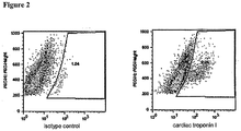
- Each sample to be stained received 50 microliters of either cardiac troponin I antibody (Spectral Diagnostics) or isotype control (final amount of antibody per tube was 1.2 micrograms). Samples were incubated at 4 degrees for 30 minutes.
- samples were pelleted as described above. This wash step was repeated. After removal of the 2 nd wash supernatant, the samples were resuspended in 50 microliters of 5% normal goat serum in PBS containing 0.25 micrograms of the secondary antibody (Molecular Probes goat antimouse IgG labeled with alexa 488). Samples were incubated at 4 degrees for 30 minutes in the dark, and washed with the addition of 2 ml staining buffer and pelleting as described above. The supernatant was decanted and the samples were resuspended in 300 microliters of staining buffer for flow acquisition on a FACScalibur machine.
- the secondary antibody Molecular Probes goat antimouse IgG labeled with alexa 488
- Plating undifferentiated hES cells for subsequent differentiation 1) The cells in 1 well of the 6 well plate of undifferentiated hES cells were dissociated by a) removing medium; b) rinsing well once with PBS; c) adding 1 ml of 0.05% trypsin/500 mM EDTA solution. The well was incubated in a 37 degree incubator for 10 minutes. The cells were triturated with a pipettor until cells were dissociated completely. 2 ml of 20% FBS-containing medium ( e . g ., 20% FBS in Knockout DMEM) were added to inactivate trypsin. The cells were counted, and this number used to plate cells derived from the remaining wells at the desired density.
- 20% FBS-containing medium e . g ., 20% FBS in Knockout DMEM
- the medium was removed from the remaining wells.
- a solution of collagenase 200 units/ml was added at 1 ml/well, and the well incubated at 37° C for 10 minutes.
- the collagenase solution was removed, and MEF-conditioned medium plus 8 ng/ml bFGF was added.
- the well was scraped with a pipet until cells were detached (in small clusters); no further trituration was performed.
- the cells were diluted to a desired density and plated into a 6-well plate prepared as described above (1.85 million cells in a volume of 5 ml per well).
- the plated hES cells were fed daily (except not on the second day) by removing the spent medium and replacing it with new MEF-CM plus 8 ng/ml bFGF.
- Growth factor treatment After 6 days of growth as described above, the cells' media was removed and replaced with RPMI plus 1X B27 supplement plus 50 ng/ml Activin A. After 18-24 hours, the medium was removed and replaced with RPMI plus 1X B27 supplement plus 50 ng/ml BMP-4 without the Activin A. After a total of 4 days in the BMP-4-containing medium, the medium was removed and replaced with RPMI plus 1X B27 supplement plus 50 ng/ml IGF-1 without the Activin or BMP. The culture were re-fed every 2-3 days by removing spent medium and replacing it with fresh RPMI plus 1X B27 supplement plus 50 ng/ml IGF-1 without the Activin or BMP.
- the trypsin digestion was stopped by the addition of 2 ml of 20% FBS-containing medium (20% FBS in Knockout DMEM).
- the cell concentration was assessed by counting, and about 500,000 cells were allocated for each staining (EMA, isotype, cTnI, cTnI plus EMA; each in a 15 ml conical tube). Tubes containing cells were spun in a centrifuge at 400 x g for 5 minutes. The medium was aspirated and the cell pellets were resuspended in 1 ml of staining buffer (PBS plus 2% heat inactivated fetal calf serum and 0.1% sodium azide). For EMA staining, cells received EMA to a final concentration of 5 micrograms/ml.
- All samples next received 900 microliters of ice-cold 100% methanol and were incubated on ice for 30 minutes. All samples received 1 ml of staining buffer (PBS plus 2% heat inactivated fetal calf serum and 0.2 microgram/0.5 x 10 6 cells of rat antimouse Fc block (BD) and pelleted as described above. The supernatant was aspirated and the cells resuspended in blocking buffer (PBS plus 20% normal goat serum and 0.1% sodium azide) at a density of about 500,000 cells/100 microliters. Samples were incubated at 4 degrees for 10-15 minutes.
- staining buffer PBS plus 2% heat inactivated fetal calf serum and 0.2 microgram/0.5 x 10 6 cells of rat antimouse Fc block (BD) and pelleted as described above.
- the supernatant was aspirated and the cells resuspended in blocking buffer (PBS plus 20% normal goat serum and 0.1% sodium azide) at a density
- each stained sample a 100 microliter aliquot of cells was dispensed into an individual 12 x 75 mm polystyrene tube. Each sample to be stained received 20 microliters of either cardiac troponin I antibody (Spectral Diagnostics) or isotype control (final amount of antibody per tube was 1.2 micrograms). Samples were incubated at 4 degrees for 30 minutes. After the addition of 4 ml staining buffer, samples were pelleted as described above.
- the samples were resuspended in 50 microliters of 5% normal goat serum in PBS containing 0.25 micrograms of the secondary antibody (Molecular Probes goat antimouse IgG1 labeled with alexa 647). Samples were incubated at 4 degrees for 30 minutes in the dark, and washed with the addition of 4 ml staining buffer and pelleting as described above. The supernatant was decanted and the samples were resuspended in 300 microliters of staining buffer plus 0.5% paraformaldehyde for flow acquisition on a FACScalibur machine. The results were analyzed using Flojo software. In this experiment, 69% of the total cells were viable after the trypsin dissociation. Of these viable cells, 8.9% were stained with an antibody against the cardiomyocyte specific protein cardiac troponin I (see Figure 2 ).
- the secondary antibody Molecular Probes goat antimouse IgG1 labeled with alexa 647
- Cardiomyocytes were generated from hES cells of the H7 line by forming embryoid bodies for 4 days, and then proliferating on gelatin-coated plates for 17 days (5-aza-deoxy-cytidine and growth factors were not used). The cells were then dissociated using collagenase B, resuspended in differentiation medium. The cell suspension was then layered onto a discontinuous gradient of Percoll, and centrifuged at 1500 g for 30 min. Four fractions were collected: I. The upper interface; II. The 40.5% layer; III. The lower interface; IV. The 58.5% layer. The cells were washed and resuspended in differentiation medium. Cells for immunostaining were seeded into chamber slides at 10 4 cells per well, cultured for 2 or 7, and then fixed and stained.
- Fractions III and IV contained the highest percentage.
- Phenotype of the cells as determined by indirect immunocytochemistry is shown in Table 4.
- TABLE 4 Characteristics of Separated Cell Populations Epitope Cardiomyocyte-lineage Non-cardiac cells cTn1 ++ - cardiac-specific ⁇ / ⁇ MHC ++ - cardiac ⁇ MHC ++ - sarcomeric MHC ++ - N-cadherin ++ ⁇ smooth muscle actin ++ subset myogenin - - ⁇ -fetoprotein - - ⁇ -tubulin III - - Ki67 subset subset BrdU subset subset SSEA-4 - - Tra-1-81 - - -
- Cardiomyocyte populations separated by density gradient centrifugation could be distinguished by staining for cTnI and MHC. Absence of staining for myogenin, ⁇ -fetoprotein, or ⁇ -tubulin III showed the absence of skeletal muscle, endoderm cell types, and neurons. Lack of staining for SSEA-4 and Tra-1-81 confirms the absence of undifferentiated hES cells.
- ⁇ -Smooth muscle actin is reportedly present in embryonic and fetal cardiomyocytes, but not adult cardiomyocytes ( Leor et al., Circulation 97:11332, 1996 ; Etzion et al., Mol. Cell Cardiol. 33:1321, 2001 ). Virtually all cTnI-positive cells and a subset of cTnI negative cells obtained in the cardiomyocyte differentiation protocol were positive for SMA, suggesting that they may be at an early stage and capable of proliferation.
- This example illustrates the subsequent culturing of cardiomyocyte clusters as cardiac bodies to enrich for cells having characteristics desirable for therapeutic use and other purposes.
- the EBs were returned to their original low attachment plates and maintained in suspension in 20% FBS containing medium for 3 additional days, then transferred to a total of three gelatin-coated 225 cm 2 tissue culture flasks. Two days after transfer to the gelatin coated flasks, the medium was removed and each flask was re-fed with 75 mL 20% FBS containing medium. Similar re-feedings occurred on day 8, 11, 13, 15, and 18. On day 20, the differentiated cultures were dissociated with Blendzyme (Roche Applied Sciences, Penzberg, DE) and fractionated on discontinuous Percoll gradients as before. Fraction IV (the highest density fraction) was recovered and counted, yielding ⁇ 3.7 ⁇ 10 6 single cells and small clusters.
- Fraction IV cells were resuspended in ⁇ 6.5 mL of 20% FBS containing medium, transferred to a 15 mL conical tube, and allowed to settle at room temperature without agitation for 10 min.
- the medium (containing 2.8 ⁇ 10 6 cells, which is most of the single cells) was removed and replaced with fresh medium.
- the cell suspension was transferred to a single low attachment six well plate ( ⁇ 4 mL of cell suspension per well).
- the CBs were re-fed in a similar manner (transfer to 50 mL tube, settling for 10 min, medium removal and replacement) every 48 h.
- Figure 3 shows the expression of the sarcomeric genes ⁇ MHC and cardiac troponin I as measured by real-time PCR. Relative to the expression after 20 days of culture on gelatin, separating the cells by Percoll increased expression by 2-5 fold in Fraction IV cells. Removing the single cells and collecting clusters increased expression to 5-20 fold. After 8 days of culturing the cells as cardiac bodies, the expression was 100- to 500- fold higher than the unseparated cells.
- the cardiac bodies When CBs are replated onto gelatin or Matrigel, the clusters adhere, flatten, and produce large patches of spontaneously contracting cells.
- the cardiac bodies For use in animal testing, the cardiac bodies may be implanted directly, or dispersed into suspensions of single cells.
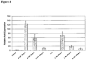
- a differentiation of H7 hES cells was performed as in Example 1, except that the differentiation was performed in a 24-well plate instead of a 6-well plate and the volume for the factors was 1 ml per well.
- BMP-2 and BMP-4 were used at concentrations of 25 ng/ml, 50 ng/ml, and 100 ng/ml. Each concentration was done in triplicate.
- Figure 4 shows the results expressed as a relative fold of the control, which involved performing the protocol but without the addition of an Activin, a BMP, or IGF-I. It can be seen that BMP-2 is also effective in the differentiation protocol.
- a 6-well plate of confluent H7 hES cells were washed with 2 ml PBS. Then, 2 ml of 0.5 mM EDTA in PBS was added to each well, and the plate was incubated for 10 minutes in 37° C. The EDTA solution was replaced with 1 ml mouse embryonic fibroblast-conditioned medium (MEF-CM) plus 8 ng/ml bFGF ("Medium A"). The undifferentiated ES cells were detached by pipetting 2-3 times and then seeded onto a 24-well plate at 400,000 cells /well in Medium A. The cells were incubated for two days at 37° C
Description
- This invention relates to a method for obtaining cardiomyocyte-lineage cells from human embryonic stem (hES) cells by direct differentiation.
- A central challenge for research in regenerative medicine is to develop cell compositions that can help reconstitute cardiac function. It is estimated that nearly one in five men and women have some form of cardiovascular disease (National Health and Nutrition Examination Survey III, 1988-94, Center of Disease Control and the American Heart Association). Widespread conditions include coronary heart disease (5% of the population), congenital cardiovascular defects (0.5%), and congestive heart failure (3%). The pharmaceutical arts have produced small molecule drugs and biological compounds that can help limit the damage that occurs as a result of heart disease, but there is nothing commercially available to help regenerate the damaged tissue.
- With the objective of developing a cell population capable of cardiac regeneration, research has been conducted on several different fronts. Clinical trials are underway at several centers to test the use of autologous bone marrow derived cells for therapy after myocardial infarction (Perin et al., Circulation 107:2294, 2003; Strauer et al., Circulation 106:1913, 2002; Zeiher et al., Circulation 106:3009, 2002; Tse et al., Lancet 361:47, 2003; Stamm et al., Lancet 3661:45, 2003). It has been hypothesized that the cells may have a cleansing function to improve blood perfusion of the heart tissue. Clinical trials are also underway to test the use of autologous skeletal muscle myoblasts for heart therapy (Menasche et al., J. Am. Coll. Cardiol. 41:1078, 2003; Pagani et al., J. Am. Coll. Cardiol. 41:879, 2003; Hagege et al., Lancet 361:491, 2003). However, it is unclear if the contraction of striatal muscle cells can coordinate adequately with cardiac rhythm.
- A more direct approach would be to use cells that are already committed to be functional cardiomyocytes. Syngeneic neonatal or postnatal cardiac cells have been used in animal models to repair damage resulting from permanent coronary occlusion (Reffelmann et al., J. Mol. Cell Cardiol. 35:607, 2003; Yao et al., J. Molec. Cell. Cardiol. 35:607, 2003). Accordingly, if such cells were available for human therapy, they could be very effective for the treatment of ischemic heart disease. In addition, cardiomyocyte cells can be used for screening compounds such as pharmaceuticals. Passier et al. disclose a method of differentiating hES cells into cardiomyocytes in a serum-free medium which does not rely on the formation of embryoid bodies(Stem Cells 23:772, 2005). The method comprises the co-culture with the END-2 cell line but no treatment with Activin A or a BMP is included.
- The present invention provides methods of obtaining cardiomyocyte-lineage cells from human embryonic stem (hES) cells by direct differentiation as defined in the appended claims. Cardiomyocyte-lineage cells have many possible uses, including, but not limited to, screening of potential pharmaceuticals, screening for cytotoxic chemicals, and therapeutic applications such as in vivo repair of damaged or diseased hearts.
- According to a first aspect of the invention, there is provided a method for obtaining cardiomyocyte-lineage cells from human embryonic stem (hES) cells by direct differentiation, comprising:
- a) culturing the hES cells in the presence of Activin A in the absence of a BMP; and
- b) subsequently culturing the cells in the presence of a BMP and the absence of Activin A,
- In some embodiments, the hES cells may be cultured in the presence of Activin A for about one day and then subsequently cultured in the presence of the BMP for about four days.
- The method of the invention may further comprise subsequent to the BMP culturing step a culturing step in which the medium does not contain Activin A or a BMP. In this embodiment, the further culturing step may be performed for at least one week. Alternatively, the further culturing step may be performed for at least two weeks. The further culturing step may be done in medium containing IGF-I or IGF-II, suitably IGF-I.
- The method of the invention may further comprise harvesting cells from the culture subsequent to the BMP culturing step and enriching the harvested cell population for cardiomyocyte-lineage cells. The harvested cell population may be enriched by Percoll gradient. The enrichment may involve the formation of cardiac bodies.
- According to a second aspect of the invention, there is provided a method of obtaining an enriched population of cardiomyocyte-lineage cells from hES cells, comprising in the following order:
- a) culturing the hES cells in a serum-free medium in the presence of Activin A in the absence of a BMP for about one day;
- b) subsequently culturing in a serum-free medium in the presence of a BMP in the absence of an Activin for about four days;
- c) harvesting cells from the culture; and
- d) enriching the harvested cell population for cardiomyocyte-lineage cells.
- As described herein, the harvested cells are at least 10% positive for α-myosin heavy chain (αMHC). In certain embodiments of the invention, the harvested cells are at least 10% cardiac troponin I (cTnI) positive). In certain embodiments of the invention, the harvested cells are at least 25% cardiac troponin I (cTnI) positive).
- As described herein, cardiac bodies are formed to enrich and/or expand the population of cardiomyocyte-lineage cells. The methods further comprise separating cells in the enriched cell population that are present as single cells from cells that are present as clusters; resuspending the cells present as clusters in nutrient medium; reculturing the resuspended cells in the nutrient medium; and collecting and washing the recultured cells.
- As described herein, the differentiation of primate pluripotent stem cells to cardiomyocyte-lineage cells occurs in a serum-free medium. In certain embodiments of the invention, the differentiation of primate pluripotent stem cells to cardiomyocyte-lineage cells occurs in a medium that contains less than 0.5% serum. In certain embodiments of the invention, the differentiation of primate pluripotent stem cells to cardiomyocyte-lineage cells occurs in a medium that contains less than 1% serum. In certain embodiments of the invention, the differentiation of primate pluripotent stem cells to cardiomyocyte-lineage cells occurs in a medium that contains less than 5% serum.
- As described herein, the cells are adhered to a substrate that comprises one or more of gelatin, Matrigel, laminin, fibronectin, and/or vitronectin during the differentiation of primate pluripotent stem cells to cardiomyocyte-lineage cells.
- As described herein, the primate pluripotent stem cells are cultured in MEM-CM plus bFGF for one to seven days before the Activin culture step. In certain embodiments, the primate pluripotent stem cells are cultured in MEM-CM plus bFGF for about six days before the Activin culture step.
- As described herein, the medium RPMI plus 1X B27 is used when culturing the cells in the presence of an Activin. As described herein, the medium RPMI plus 1X B27 is used when culturing the cells in the presence of an a BMP. As described herein, the medium RPMI plus N2 is used when culturing the cells in the presence of an Activin. As described herein, the medium RPMI plus N2 is used when culturing the cells in the presence of a BMP.
-
-
Figure 1 - H7 cells were plated onto wells coated with gelatin and FBS and subsequently differentiated according to the method described in Example 1. On day 24 after the original addition of activin, cultures were dissociated with trypsin-EDTA, fixed, permeablized, and stained with an antibody against cardic troponin I. Prior to fixation, cells were incubated with EMA to distinguish live cells (cells excluding EMA) from dead cells (cells incorporating EMA). Samples were analyzed on a FACScalibur and dead cells were excluded from the analysis. In this experiment, approximately 54% of the cells survived the trypsin dissociation; of these live cells, 24-27% were cardiomyocytes as determined by labeling with the cardiac troponin I-specific antibody. Two different gating methods were used in the 2 panels (Figure 1A : histogram- andFigure 1B : scatter-plot based); the percentage of cardiomyocytes was similar by either method. -
Figure 2 - H7 cells were plated onto wells coated with Matrigel and subsequently differentiated according to the method described in Example 2. On day 21 after the original addition of activin, cultures were dissociated with trypsin-EDTA, fixed, permeablized, and stained with an antibody against cardic troponin I. Prior to fixation, cells were incubated with EMA to distinguish live cells (cells excluding EMA) from dead cells (cells incorporating EMA). Samples were analyzed on a FACScalibur and dead cells were excluded from the analysis. In this experiment, approximately 69% of the cells survived the trypsin dissociation; of these live cells, 8.9% were cardiomyocytes as determined by labeling with the cardiac troponin I-specific antibody. -
Figure 3 shows the expression of cTnI measured in cardiac bodies formed from each of the four Percoll fractions. Undifferentiated hES cells are used as a negative control. Culturing the Fraction IV cells as cardiac bodies enriched for αMHC or cTnI expression by 100-to 500- fold. -
Figure 4 shows the expression of αMHC in cell populations that result from the differentiation of hES cells using different concentrations of BMP-2 and BMP-4. - The term "cardiomyocyte-lineage cells" refers generally to both cardiomyocyte precursor cells and mature cardiomyocytes. Reference to cardiomyocyte-lineage cells, precursors, or cardiomyocytes in this disclosure can be taken to apply equally to cells at any stage of cardiomyocyte ontogeny without restriction, as defined above, unless otherwise specified. As described below, cardiomyocyte-lineage cells may have one or more markers (sometimes at least 3 or 5 markers) from the following list: cardiac troponin I (cTnI), cardiac troponin T (cTnT), sarcomeric myosin heavy chain (MHC), GATA-4, Nkx2.5, N-cadherin, β1-adrenoceptor (β1-AR), ANF, the MEF-2 family of transcription factors, creatine kinase MB(CK-MB), myoglobin, or atrial natriuretic factor (ANF).
- The term "embryoid bodies" refers to heterogeneous clusters comprising differentiated and partly differentiated cells that appear when primate pluripotent stem cells are allowed to differentiate in a non-specific fashion in suspension cultures or aggregates.
- As used herein, "primate pluripotent stem cells" refers to cells that are derived from any kind of embryonic tissue (fetal or pre-fetal tissue) and that have the characteristic of being capable under appropriate conditions of producing progeny of different cell types that are derivatives of all of the 3 germinal layers (endoderm, mesoderm, and ectoderm), according to a standard art-accepted test such as the ability to form a teratoma in 8-12 week old SCID mice or the ability to form identifiable cells of all three germ layers in tissue culture. Included in the definition of primate pluripotent stem cells are embryonic cells of various types, exemplified by human embryonic stem (hES) cells, (see, e.g., for reference only, Thomson et al. (Science 282:1145, 1998)) and reference human embryonic germ (hEG) cells (see, e.g., Shamblott et al., Proc. Natl. Acad. Sci. USA 95:13726, 1998); reference embryonic stem cells from other primates, such as Rhesus stem cells (see, e.g., Thomson et al., Proc. Natl. Acad. Sci. USA 92:7844, 1995), marmoset stem cells (see, e.g., Thomson et al., Biol, Reprod. 55:254, 1996).
- As used herein, "undifferentiated primate pluripotent stem cells" refers to a cell culture where a substantial proportion of primate pluripotent stem cells and their derivatives in the population display morphological characteristics of undifferentiated cells. It is understood that colonies of undifferentiated cells within the population will often be surrounded by neighboring cells that are partly differentiated.
- As used herein, "embryonic stem cell" refers to reference pluripotent stem cells that are derived from a human embryo at the blastocyst stage, or before substantial differentiation of the cells into the three germ layers. Except where explicitly required otherwise, the term includes, for reference only, primary tissue and established lines that bear phenotypic characteristics of hES cells, and progeny of such lines that still have the capacity of producing progeny of each of the three germ layers. Prototype reference "human Embryonic Stem cells" (hES cells) are described by Thomson et al. (Science 282:1145, 1998;
U.S. Patent 6,200,806 ). - As used herein, "Activin" refers to a polypeptide growth factor that is a member of the transforming growth factor-β (TGF-β) superfamily. Currently there are four know Activins - A, AB, B, and C.
- As used herein, "Bone Morphogenetic Protein (BMP)" refers to a polypeptide growth factor of the TGF-β superfamily. There are currently about 20 known members in the BMP family. For the purposes of this application, the term "BMP" does not include BMP-1. As used herein, "enrich" refers to increasing the level of a component in a mixture. For example, in certain embodiments of the present invention, a given cell population may be enriched by increasing the proportion of cardiomyocyte-lineage cells in that population.
- As used herein, "cardiac body" refers to a cluster of primate pluripotent stem cell-derived cells in suspension, bearing two or more characteristics of human cardiomyocyte-lineage cells.
- As used herein, "direct differentiation" refers to a process for differentiating primate pluripotent stem cells into progeny that are enriched for cells of a particular tissue type without forming embryoid bodies as an intermediate. To clarify, the term direct differentiation encompasses processes in which a small number of cell aggregates form inadvertently.
- As used herein, "genetically altered", "transfected", or "genetically transformed" refer to a process where a polynucleotide has been transferred into a cell by any suitable means of artificial manipulation, or where the cell is a progeny of the originally altered cell and has inherited the polynucleotide. The polynucleotide will often comprise a transcribable sequence encoding a protein of interest, which enables the cell to express the protein at an elevated level or may comprise a sequence encoding a molecule such as siRNA or antisense RNA that affects the expression of a protein (either expressed by the unmodified cell or as the result of the introduction of another polynucleotide sequence) without itself encoding a protein. The genetic alteration is said to be "inheritable" if progeny of the altered cell have the same alteration.
- As used herein, "serum-free" refers to a condition where the referenced composition contains no added serum.
- As used herein, "feeder cells" refers to cells of a different tissue type, and typically a different genome, that may act to promote proliferation and/or control differentiation of cells they are cocultured with. For example, undifferentiated primate pluripotent stem cells can be cocultured with feeder cells that help maintain the undifferentiated state, while primate pluripotent stem cells in the process of being differentiated can be cocultured with feeders that direct differentiation towards a particular tissue type (e.g., cardiomyocyte-lineage cells).
- As used herein, "feeder-free" refers to a condition where the referenced composition contains no added feeder cells. To clarify, the term feeder-free encompasses, inter alia, situations where primate pluripotent stem cells are passaged from a culture with feeders into a culture without added feeders even if some of the feeders from the first culture are present in the second culture.
- As used herein, "culturing" refers to the process of maintaining and/or expanding cells in vitro.
- As used herein, "same genome" refers to the genomes of a primate pluripotent stem cell and a differentiated cell derived from that primate pluripotent stem cell and means that the chromosomal DNA will be over 90% identical between the primate pluripotent stem cell and the derived cell as determined by Restriction Fragment Length Polymorphism ("RFLP") or SNP analysis. Even if the primate pluripotent stem cell or the derived cell has been genetically altered, those cells will be considered to have the same genome as the cell from which it was derived or the cell derived from it, since all non-manipulated genetic elements are preserved.
- As used herein, "Matrigel" refers to BD Matrigel™ Basement Membrane Matrix, which is a commercial preparation of basement membrane produced by Engelbreth-Holm-Swarm tumor cells and containing extracellular matrix components such as laminin. Matrigel is available commercially through Becton, Dickinson and Company (Franklin Lakes, NJ).
- As used herein, "RPMI" refers to RPMI Medium 1640 (Invitrogen, Carlsbad, CA).
- General Techniques - For further elaboration of general techniques useful in the practice of this invention, the practitioner can refer to standard textbooks and reviews in cell biology, tissue culture, embryology, and cardiophysiology.
- With respect to tissue and cell culture and embryonic stem cells, the reader may wish to refer to Teratocarcinomas and embryonic stem cells: A practical approach (E.J. Robertson, ed., IRL Press Ltd. 1987); Guide to Techniques in Mouse Development (P.M. Wasserman et al. eds., Academic Press 1993); Embryonic Stem Cell Differentiation in Vitro (M.V. Wiles, Meth. Enzymol. 225:900, 1993); Properties and uses of Embryonic Stem Cells: Prospects for Application to Human Biology and Gene Therapy (P.D. Rathjen et al., Reprod. Fertil. Dev. 10:31, 1998; and R.I. Freshney, Culture of Animal Cells, Wiley-Liss, New York, 2000). With respect to the culture of heart cells, standard references include The Heart Cell in Culture (A. Pinson ed., CRC Press 1987), Isolated Adult Cardiomyocytes (Vols. I & II, Piper & Isenberg eds., CRC Press 1989), and Heart Development (Harvey & Rosenthal, Academic Press 1998). General methods in molecular and cellular biochemistry can be found in such standard textbooks as Short Protocols in Molecular Biology, 4th Ed.; Immunology Methods Manual (I. Lefkovits ed., Academic Press 1997); and Cell and Tissue Culture: Laboratory Procedures in Biotechnology (Doyle & Griffiths, John Wiley & Sons 1998).
- The present invention provides methods as defined in the claims for differentiating hES cells into cardiomyocyte-lineage cells. Primate pluripotent stem cells include, but are not limited to, embryonic stem cells. By way of reference embryonic stem cells can be isolated from blastocysts of primate species (
U.S. Patent 5,843,780 ; Thomson et al., Proc. Natl. Acad. Sci. USA 92:7844, 1995). Reference human embryonic stem (hES) cells can be prepared from human blastocyst cells using, for example, the techniques described byThomson et al. (U.S. Patent 6,200,806 ; Science 282:1145, 1998; Curr. Top. Dev. Biol. 38:133 ff., 1998) and Reubinoff et al, Nature Biotech. 18:399, 2000. Other reference primate pluripotent stem cell types include, but are not limited to, primitive ectoderm-like (EPL) cells, outlined inWO 01/51610 (Bresagen - Embryonic stem cells mentioned here for reference only may be chosen from embryonic stem cell lines or may be obtained directly from primary embryonic tissue. A large number of reference embryonic stem cell lines have been established including, but not limited to, H1, H7, H9, H13 or H14 (reference Thompson); hESBGN-01, hESBGN-02, hESBGN-03 (BresaGen, Inc., Athens, GA); HES-1, HES-2, HES-3, HES-4, HES-5, HES-6 (from ES Cell International, Inc., Singapore); HSF-1, HSF-6 (from University of California at San Francisco); 13, 13.2, 13.3, 14, 1 6, I 6.2, J 3, J 3.2 (derived at the Technion-Israel Institute of Technology, Haifa, Israel); UCSF-1 and UCSF-2 (Genbacev et al., Fertil. Steril. 83(5):1517-29, 2005); lines HUES 1-17 (Cowan et al., NEJM 350(13):1353-56, 2004); and line ACT-14 (Klimanskaya et al., Lancet, 365(9471):1636-41, 2005).
- As described herein, primate pluripotent stem cells used may have been derived in a feeder-free manner (see, e.g., Klimanskaya et al., Lancet, 365(9471):1636-41 (2005)).
- Primate pluripotent stem cells may be cultured using a variety of substrates, media, and other supplements and factors known in the art. Primate pluripotent stem cells can be propagated continuously in culture, using culture conditions that promote proliferation while inhibiting differentiation. Exemplary medium is made with 80% DMEM (such as Knock-Out DMEM, Gibco), 20% of either defined fetal bovine serum (FBS, Hyclone) or serum replacement (
US 2002/0076747 A1 , Life Technologies Inc.), 1% non-essential amino acids, 1 mM L-glutamine, and 0.1 mM β-mercaptoethanol. - As described herein, primate pluripotent stem cells are cultured on a layer of feeder cells, typically fibroblasts derived from embryonic or fetal tissue (Thomson et al., Science 282:1145, 1998). As described herein, those feeder cells are from human or mouse. Human feeder cells can be isolated from various human tissues or derived by differentiation of human embryonic stem cells into fibroblast cells (see, e.g.,
WO01/51616 - As described herein, embryonic stem cells may be maintained in an undifferentiated state without added feeder cells (see, e.g., Rosier et al., Dev. Dynam. 229:259-274, 2004). Feeder-free cultures are typically supported by a nutrient medium containing factors that promote proliferation of the cells without differentiation (see, e.g.,
U.S. Patent No. 6,800,480 ). As described herein, such factors may be introduced into the medium by culturing the medium with cells secreting such factors, such as irradiated (∼4,000 rad) primary mouse embryonic fibroblasts, telomerized mouse fibroblasts, or fibroblast-like cells derived from primate pluripotent stem cells (U.S. Patent 6,642,048 ). Medium can be conditioned by plating the feeders in a serum free medium such as KO DMEM supplemented with 20% serum replacement and 4 ng/mL bFGF. Medium that has been conditioned for 1-2 days is supplemented with further bFGF, and used to support primate pluripotent stem cell culture for 1-2 days (see. e.g.,WO 01/51616 - Alternatively, fresh or non-conditioned medium can be used, which has been supplemented with added factors (like a fibroblast growth factor or forskolin) that promote proliferation of the cells in an undifferentiated form. Exemplary is a base medium like X-VIVO™ 10 (Biowhittaker) or QBSF™-60 (Quality Biological Inc.), supplemented with bFGF at 40-80 ng/mL, and optionally containing stem cell factor (15 ng/mL), or Flt3 ligand (75 ng/mL) (see, e.g., Xu et al., Stem Cells 23(3):315-23, 2005). These medium formulations have the advantage of supporting cell growth at 2-3 times the rate in other systems (see, e.g.,
WO 03/020920 - For example, the primate pluripotent stem cells are plated at >15,000 cells cm-2 (optimally 90,000 cm-1 to 170,000 cm-2). Typically, enzymatic digestion is halted before cells become completely dispersed (say, ∼5 min with collagenase IV). Clumps of ∼10 to 2,000 cells are then plated directly onto the substrate without further dispersal. Alternatively, the cells can be harvested without enzymes before the plate reaches confluence by incubating ∼5 min in a solution of 0.5 mM EDTA in PBS or by simply detaching the desired cells from the plate mechanically, such as by scraping or isolation with a fine pipet. After washing from the culture vessel, the cells are plated into a new culture without further dispersal. In a further illustration, confluent human embryonic stem cells cultured in the absence of feeders are removed from the plates by incubating with a solution of 0.05% (wt/vol) trypsin (Gibco) and 0.053 mM EDTA for 5-15 min at 37°C. The remaining cells in the plate are removed and the cells are triturated into a suspension comprising single cells and small clusters, and then plated at densities of 50,000-200,000 cells cm-2 to promote survival and limit differentiation.
- Under the microscope, primate pluripotent stem cells appear with high nuclear/cytoplasmic ratios, prominent nucleoli, and compact colony formation with poorly discernable cell junctions. Primate primate pluripotent stem cells typically express the stage-specific embryonic antigens (SSEA) 3 and 4, and markers detectable using antibodies designated Tra-1-60 and Tra-1-81. Undifferentiated human embryonic stem cells also typically express the transcription factor Oct-3/4, Cripto, gastrin-releasing peptide (GRP) receptor, podocalyxin-like protein (PODXL), and human telomerase reverse transcriptase (hTERT) (
US 2003/0224411 A1 ), as detected by RT-PCR. - The present invention provides, inter alia, methods for direct differentiation of human embryonic stem (hES) cells into cardiomyocyte-lineage cells by the sequential culturing of the hES cells first in the presence of Activin A with subsequent culturing in the presence of a BMP. The BMP is excluded during the culturing step with Activin A. The Activin A is excluded during the subsequent culturing step with the BMP.
- As described herein, Activin is included in the culture medium at a concentration between 10 ng/ml and 200 ng/ml, or between 25 ng/ml and 100 ng/ml, or between 50 ng/ml and 100 ng/ml. As described herein, Activin is included in the culture medium at a concentration below 10 ng/ml or above 200 ng/ml.
- As described herein, the BMP is included in the culture medium at a concentration between 10 ng/ml and 200 ng/ml, or between 25 ng/ml and 100 ng/ml, or between 50 ng/ml and 100 ng/ml. As described herein, the BMP is included in the culture medium at a concentration below 10 ng/ml or above 200 ng/ml.
- As described herein, other TGFβ superfamily members such as TGFβ, nodal, or lefty may be substituted instead of or in addition to the Activin in the methods of the present invention.
- As described herein, the BMP used in the differentiation is BMP-2, BMP-4, or BMP-7. As described herein, the BMP is a BMP other than BMP-2, BMP-4 or BMP-7 (excluding BMP-1). As described herein, more than one BMP may be used.
- As described herein, the differentiating cells are cultured in the absence of both Activin and BMP after the BMP step. In certain of those embodiments, an IGF is included in that culture step. As described herein, the IGF is included at a concentration between 10 ng/ml and 500 ng/ml; or between 25 ng/ml and 100 ng/ml; or between 50 ng/ml and 100 ng/ml. As described herein, the IGF is included at concentrations less than 10 ng/ml or more than 500 ng/ml. The IGF may be IGF-1 or IGF-2. As described herein, insulin may be substituted for the IGF in the methods of the present invention.
- During the differentiation of the primate pluripotent stem cells to cardiomyocyte-lineage cells, the cells are cultured in the presence of the Activin, BMP, or IGF for various specified time periods. As described herein, the culture step with Activin is between 12 hours and 36 hours in length, or between 12 hours and 2 days in length, or between 6 hours and 4 days in length, or between 4 hours and 5 days in length. As described herein, the culture step with Activin is longer than 5 days.
- As described herein, the culture step with the BMP is between 3 days and 5 days in length, or between 2 days and 8 days in length, or between 1 day and 14 days in length. As described herein, the culture step with the BMP is longer than 14 days.
- As described herein, the culture step with the IGF is between 3 days and 5 days in length, or between 2 days and 8 days, or between 1 day and 4 weeks in length. As described herein, the culture step with the IGF is longer than 4 weeks long.
- For example, human embryonic stem cells plated on Matrigel may be first cultured with 50 ng/ml Activin A in the absence of a BMP for about one day, then cultured with 50 ng/ml BMP-4 in the absence of an Activin for about four days, and then cultured in the presence of 50 ng/ml IGF-1 in the absence of both an Activin and a BMP for two weeks. As described herein, the resulting cardiomyocyte-lineage cells are harvested and enriched by Percoll gradient as described in Reference Example 3.
- The hES cells are differentiated into cardiomyocyte-lineage cells by direct differentiation. Differentiation paradigms for primate pluripotent stem cells traditionally involve the deliberate formation of embryoid bodies, which allows cross-talk between different cell types, thought to promote tissue formation in a manner reminiscent of an embryo. However, it is often advantageous to eliminate the need to form embryoid bodies, allowing the differentiation process to be more controlled, and the resulting cell population tends to be more uniform (see, e.g.,
WO 01/51616 US 2002/0151053 A1 ). - One of the advantages of the direct differentiation technique is that a serum or serum substitute is not needed to initiate or support the cardiomyocyte differentiation process, as is typical of other methods. Instead, the medium can be formulated so that it contains an artificial nutritional supplement that supports differentiated cells like cardiomyocytes or neurons. Exemplary are B27 supplement, N2 supplement, and G5 supplement (Life Technologies/Gibco). As described herein, supplements comprise nutrients and cofactors like human insulin (500 µg/L), human transferrin (5-10 mg/mL), and selenium (0.5 µg/mL), and may also contain putrescine (1.5 mg/L), biotin (1 µg/L), hydrocortisone (0.4 µg/L), or progesterone (0.6 µg/L), and/or low levels of mitogens like EGF or FGF (1 µg/L). For purposes of commercial scale production and human therapy, elimination of components derived from non-human animals may be advantageous.
- As described herein, the culture medium used during the differentiation steps is serum-free, As described herein, the culture medium used during the differentiation steps contains less than 0.25% serum, or less than 0.5% serum, or less than 1.0% serum, or less than 2.0% serum, or less than 5.0% serum, or less than 10% serum.
- As described herein, the differentiating cells are cultured on a substrate during the methods of the invention. Substrates that can be used in this invention include, but are not limited to collagen, laminin, fibronectin, vitronectin, hyaluronate poly-L-lysine-coated tissue culture plastic, or Matrigel.
- There are various solid surfaces that may be used in the culturing of cells. Those solid surfaces include, but are not limited to, standard cell culturing plates such as 6-well, 24-well, 96-well, or 144-well plates. Other solid surfaces include, but are not limited to, microcarriers and disk. In certain embodiments, the microcarriers are beads. Those beads come in various forms such as Cytodex Dextran microcarrier beads with positive charge groups to augment cell attachment, gelatin/collagen-coated beads for cell attachment, and macroporous microcarrier beads with different porosities for attachment of cells. The Cytodex dextran, gelatin-coated and the macroporous microcarrier beads are commercially available (Sigma-Aldrich, St. Loius, MO or Solohill Engineering Inc., Ann Arbor, MI). For example, the beads are 90-200 µm in size with an area of 350-500 cm2. Beads may be composed of a variety of materials such as, but not limited to, glass or plastic. Disks may be used in stirred-tank bioreactors for attachment of the cells. Disks are sold by companies such as New Brunswick Scientific Co, Inc. (Edison, NJ). In certain embodiments, the disks are Fibra-cel Disks, which are polyester/polypropylene disks. A gram of these disks provide a surface area of 1200 cm2.
- The solid surface may be made of a variety of substances including, but not limited to, glass or plastic such as polystyrene, polyvinylchloride, polycarobnate, polytetrafluorethylene, melinex, or thermanox. The solid surfaces may three-dimensional in shape. Exemplary three-dimensional solid surfaces are described, e.g., in
US20050031598 . - As described herein, the cells are in a single-cell suspension during the methods of the invention. The single-cell suspension may be performed in various ways including, but not limited to, culture in a spinner flask, in a shaker flask, or in a fermentors. Fermentors that may be used include, but are not limited to, Celligen Plus (New Brunswick Scientific Co, Inc., Edison, NJ), and the STR or the Stirred-Tank Reactor (Applikon Inc., Foster City, CA). In certain embodiments, the bioreactors may be continuously perfused with media or used in a fed-batch mode. Bioreactors come in different sizes like 2.2 L, 5 L, 7.5 L, 14 L or 20 L.
- The present invention provides methods for obtaining high purity cardiomyocyte-lineage cell populations without an enrichment step as defined in the claims. However, the addition of one or more enrichment steps may produce an even higher purity cardiomyocyte-lineage cell population. Thus, methods of the invention as defined in the claims may include steps for enriching and/or expanding cardiomyocyte-lineage cells obtained by the differentiation steps of the invention. Various methods for enriching specific cell types are known in the art and include, but are not limited to, mechanical separation, density separation, cell sorting, magnetic sorting, and genetic selection techniques (for a general discussion of cell separation, see Freshney, Culture of Animal Cells, Wiley-Liss, New York, 2000 - Chapter 14). Examples of some of those methodologies are discussed below.
- As described herein, cardiomyocyte-lineage cells are enriched by density gradient separation using density gradient mediums such as, but not limited to, Percoll (see, e.g., Example 3 herein and Xu et al., Circ. Res. 91(6):501-08, 2002), Ficoll (Pharmacia), metrizamide (Nygaard), RediGrad (GE Healthcare) and dextran.
- Many cell sorting techniques are available for sorting cardiomyocyte-lineage cells from non-cariomyocyte-lineage cells. Those cell sorting techniques include, but are not limited to negative immunoselection and positive immunoselection.
- Immunoselection is a generic term that encompasses a variety of techniques in which the specificity of a selection system is conferred by an antibody or an antibody-like molecule such as a lectin or hapten. An example of such specificity is the affinity of an antibody for a specific cell surface antigen. Two general types of immunoselection techniques are practiced. Negative immunoselection involves the elimination of a specific subpopulation of components from a heterogeneous population such as the elimination on non-cardiomyocyte-lineage cells from the cell population that results from the differentiation of primate pluripotent stem cells according to the methods herein. In contrast, positive immunoselection refers to the direct selection and recovery of a specific component, such as the direct selection and recovery of cardiomyocyte-lineage cells from the differentiation of primate pluripotent stem cells according to the methods herein. Various types of immunoselection may be used in the practice of the present invention, including, but not limited to, flow cytometry (FACS), immunomagnetic techniques, antibody columns, immunoprecipitation, and immunopanning.
- Cardiac bodies - As described herein, cardiomyocyte-lineage cells may be further expanded or enriched by allowing them to grow in clusters that are referred to as cardiac bodies.
- First, a cell population is generated that contains cells with phenotype characteristics of cardiomyocyte-lineage cells, and optionally enriched by density separation or other technique. The cells are then allowed to form clusters, and single cells in the suspension are removed. This can be accomplished by letting the clusters settle, and pipetting out the supernatant containing single cells. Before proceeding, it is sometimes beneficial to break apart the clusters (for example, by brief trypsinization and/or mechanical dispersion). The cells are then cultured in suspension in low adhesion plates in fresh culture medium (exemplified by medium containing fetal bovine serum, serum substitute, or CCT), and allowed to reaggregate into "secondary" cardiac bodies. Culturing then continues with periodic refeeding, as necessary, with cardiomyocyte-lineage cells remaining as clusters of 10 to 5000 cells (typically 50 to 1000 cells) in size.
- After a suitable period (typically 1 to 7 days), the cultured cells can be harvested for characterization, or used in drug screening or pharmaceutical manufacture. The purification effect may improve if the cells are taken through further cycles of removing single cells and reculturing the clusters, over a period of 8 days or more. Each cycle can optionally incorporate a step in which the clusters of cells are dispersed into single cells, or smaller cell clusters, to allow for further expansion. Larger clusters may form, either by aggregation of the suspended cells, or by proliferation within the cluster, or both. It is a hypothesis of this invention that cardiomyocyte-lineage cells have a tendency to form such clusters under appropriate conditions, and that the removal of single cells helps eliminate other cell types and increase homogeneity.
- The cardiac body technique can be used to expand and/or enrich the cardiomyocytes in the cell population at any time in the differentiation process. As exemplified below, the technique can be used after a previous enrichment step by density separation. Implementation of the technique has benefits that were not anticipated before the making of this invention. In particular, the expression of myosin heavy chain detected by real-time PCR increases 10- to 100-fold when the cells are cultured for a 7 day period. A large proportion of the clusters in the composition exhibit spontaneous contractile activity: usually over 50%, and potentially between about 80% and 100% when processed in the manner described.
- The cardiomyocyte-lineage cells obtained according to the techniques of this invention can be characterized according to a number of phenotypic criteria.
- Cardiomyocytes and precursor cells derived from primate pluripotent stem cell lines often have morphological characteristics of cardiomyocytes from other sources. They can be spindle, round, triangular or multi-angular shaped, and they may show striations characteristic of sarcomeric structures detectable by immunostaining (
Figure 1 ). They may form flattened sheets of cells, or aggregates that stay attached to the substrate or float in suspension, showing typical sarcomeres and atrial granules when examined by electron microscopy. - Under appropriate circumstances, primate pluripotent stem cell-derived cardiomyocytes often show spontaneous periodic contractile activity. This means that when they are cultured in a suitable tissue culture environment with an appropriate Ca++ concentration and electrolyte balance, the cells can be observed to contract across one axis of the cell, and then release from contraction, without having to add any additional components to the culture medium. The contractions are periodic, which means that they repeat on a regular or irregular basis, at a frequency between ∼6 and 200 contractions per minute, and often between ∼20 and ∼90 contractions per minute in normal buffer (
Figure 2 ). Individual cells may show spontaneous periodic contractile activity on their own, or they may show spontaneous periodic contractile activity in concert with neighboring cells in a tissue, cell aggregate, or cultured cell mass. - The contractile activity of the cells can be characterized according to the influence of culture conditions on the nature and frequency of contractions. Compounds that reduce available Ca++ concentration or otherwise interfere with transmembrane transport of Ca++ often affect contractile activity. For example, the L-type calcium channel blocker diltiazem inhibits contractile activity in a dose-dependent manner. On the other hand, adrenoceptor agonists like isoprenaline and phenylephrine have a positive chronotropic effect. Further characterization of functional properties of the cell can involve characterizing channels for Na+, K+, and Ca++. Electrophysiology can be studied by patch clamp analysis for cardiomyocyte like action potentials. See Igelmund et al., Pflugers Arch. 437:669, 1999; Wobus et al., Ann. N.Y. Acad. Sci. 27:752, 1995; and Doevendans et al., J. Mol. Cell Cardiol. 32:839, 2000.
- Cardiomyocyte-lineage cells typically have at least one of the following cardiomyocyte specific markers:
- Cardiac troponin I (cTnI), a subunit of troponin complex that provides a calcium-sensitive molecular switch for the regulation of striated muscle contraction
- Cardiac troponin T (cTnT)
- Nkx2.5, a cardiac transcription factor expressed in cardiac mesoderm during early mouse embryonic development, which persists in the developing heart
- The cells will also typically express at least one (and often at least 3, 5, or more) of the following markers:
- Atrial natriuretic factor (ANF), a hormone expressed in developing heart and fetal cardiomyocytes but down-regulated in adults. It is considered a good marker for cardiomyocytes because it is expressed in a highly specific manner in cardiac cells but not skeletal myocytes.
- myosin heavy chain (MHC), particularly the β chain which is cardiac specific
- Titin, tropomyosin, α-sarcomeric actinin, and desmin
- GATA-4, a transcription factor that is highly expressed in cardiac mesoderm and persists in the developing heart. It regulates many cardiac genes and plays a role in cardiogenesis
- MEF-2A, MEF-2B, MEF-2C, MEF-2D; transcription factors that are expressed in cardiac mesoderm and persist in developing heart
- N-cadherin, which mediates adhesion among cardiac cells
- Connexin 43, which forms the gap junction between cardiomyocytes.
- β1 -adrenoceptor (β1-AR)
- creatine kinase MB (CK-MB) and myoglobin, which are elevated in serum following myocardial infarction
- α-cardiac actin, early growth response-I, and cyclin D2.
- Tissue-specific markers may be detected using suitable immunological techniques - such as flow immunocytometry or affinity adsorption for cell-surface markers, immunocytochemistry (for example, of fixed cells or tissue sections) for intracellular or cell-surface markers, Western blot analysis of cellular extracts, and enzyme-linked immunoassay, for cellular extracts or products secreted into the medium. Antibodies that distinguish cardiac markers like cTnI and cTnT from other isoforms are available commercially from suppliers like Sigma and Spectral Diagnostics. Expression of an antigen by a cell is said to be antibody-detectable if a significantly detectable amount of antibody will bind to the antigen in a standard immunocytochemistry or flow cytometry assay, optionally after fixation of the cells, and optionally using a labeled secondary antibody.
- The expression of tissue-specific gene products may also be detected at the mRNA level by Northern blot analysis, dot-blot hybridization analysis, or by reverse transcriptase initiated polymerase chain reaction (RT-PCR) using sequence-specific primers in standard amplification methods using publicly available sequence data (GenBank). Expression of tissue-specific markers as detected at the protein or mRNA level is considered positive if the level is at least 2-fold, and preferably more than 10- or 50-fold above that of an undifferentiated primate pluripotent stem cell.
- The expression of tissue-specific gene products may also be detected at the mRNA level by Northern blot analysis, dot-blot hybridization analysis, or by reverse transcriptase initiated polymerase chain reaction (RT-PCR) using sequence-specific primers in standard amplification methods. See
U.S. Patent No. 5,843,780 for further details. Sequence data for the particular markers listed in this disclosure can be obtained from public databases such as GenBank (URL www.ncbi.nlm.nih.gov:80/entrez). Expression at the mRNA level is said to be "detectable" according to one of the assays described in this disclosure if the performance of the assay on cell samples according to standard procedures in a typical controlled experiment results in clearly discernable hybridization or amplification product. Expression of tissue-specific markers as detected at the protein or mRNA level is considered positive if the level is at least 2-fold, and preferably more than 10- or 50-fold above that of an undifferentiated primate pluripotent stem cell. - Once markers have been identified on the surface of cells of the desired phenotype, they can be used for immunoselection to further enrich the population by techniques such as immunopanning or antibody-mediated fluorescence-activated cell sorting.
- Where derived from an established line of primate pluripotent stem cells, the cell populations and isolated cells described herein can be characterized as having the same genome as the line from which they are derived. This means that the chromosomal DNA will be over 90% identical by RFLP or by SNP analysis between the primate pluripotent stem cells and the cardiac cells, which can be inferred if the cardiac cells are obtained from the undifferentiated line through the course of normal mitotic division. The characteristic that cardiomyocyte-lineage cells are derived from the parent cell population is important in several respects. In particular, the undifferentiated cell population can be used for producing additional cells with a shared genome - either a further batch of cardiac cells, or another cell type that may be useful in therapy - such as a population that can pre-tolerize the patient to the histocompatibility type of the cardiac allograft (
US 2002/0086005 A1 ;WO 03/050251 - The cells described herein can be made to contain one or more genetic alterations by genetic engineering of the cells either before or after differentiation (
US 2002/0168766 A1 ). For example, the cells can be processed to increase their replication potential by genetically altering the cells to express telomerase reverse transcriptase, either before or after they progress to restricted developmental lineage cells or terminally differentiated cells (US 2003/0022367 A1 ). - The cells described herein can also be genetically altered in order to enhance their ability to be involved in tissue regeneration, or to deliver a therapeutic gene to a site of administration. A vector is designed using the known encoding sequence for the desired gene, operatively linked to a promoter that is either pan-specific or specifically active in the differentiated cell type. Of particular interest are cells that are genetically altered to express one or more growth factors of various types such as FGF, cardiotropic factors such as atrial natriuretic factor, cripto, and cardiac transcription regulation factors, such as GATA-4, Nkx2.5, and MEF2-C. Production of these factors at the site of administration may facilitate adoption of the functional phenotype, enhance the beneficial effect of the administered cell, or increase proliferation or activity of host cells neighboring the treatment site.
- It is desirable to genetically alter non-human cardiomyocyte-lineage cells such that the expression of one or more antigens is reduced or eliminated so that the immunogenecity of those cells is reduced. This could be useful, for example, in xenotransplantation of non-human cardiomyocyte-lineage cells into a human.
- This invention provides a method as defined in the claims to produce large numbers of cells of the cardiomyocyte-lineage. These cell populations can be used for a number of important research, development, and commercial purposes.
- Cardiomyocytes described herein can be used commercially to screen for factors (such as solvents, small molecule drugs, peptides, oligonucleotides) or environmental conditions (such as culture conditions or manipulation) that affect the characteristics of such cells and their various progeny.
- In some applications, primate pluripotent stem cells (undifferentiated or differentiated) are used to screen factors that promote maturation into later-stage cardiomyocyte precursors, or terminally differentiated cells, or to promote proliferation and maintenance of such cells in long-term culture. For example, candidate maturation factors or growth factors are tested by adding them to cells in different wells, and then determining any phenotypic change that results, according to desirable criteria for further culture and use of the cells.
- Other screening applications relate to the testing of pharmaceutical compounds for their effect on cardiac muscle tissue maintenance or repair. Screening may be done either because the compound is designed to have a pharmacological effect on the cells, or because a compound designed to have effects elsewhere may have unintended side effects on cells of this tissue type. The screening can be conducted using any of the precursor cells or terminally differentiated cells described herein.
- The reader is referred generally to the standard textbook In vitro Methods in Pharmaceutical Research, Academic Press, 1997, and
U.S. Patent 5,030,015 . Assessment of the activity of candidate pharmaceutical compounds generally involves combining the differentiated cells of this invention with the candidate compound, either alone or in combination with other drugs. The investigator determines any change in the morphology, marker phenotype, or functional activity of the cells that is attributable to the compound (compared with untreated cells or cells treated with an inert compound), and then correlates the effect of the compound with the observed change. - Cytotoxicity can be determined in the first instance by the effect on cell viability, survival, morphology, and the expression of certain markers and receptors, Effects of a drug on chromosomal DNA can be determined by measuring DNA synthesis or repair. [3H]-thymidine or BrdU incorporation, especially at unscheduled times in the cell cycle, or above the level required for cell replication, is consistent with a drug effect. Unwanted effects can also include unusual rates of sister chromatid exchange, determined by metaphase spread. The reader is referred to A. Vickers (pp 375-410 in In vitro Methods in Pharmaceutical Research, Academic Press, 1997) for further elaboration.
- Effect of cell function can be assessed using any standard assay to observe phenotype or activity of cardiomyocytes, such as marker expression, receptor binding, contractile activity, or electrophysiology - either in cell culture or in vivo. Pharmaceutical candidates can also be tested for their effect on contractile activity - such as whether they increase or decrease the extent or frequency of contraction. Where an effect is observed, the concentration of the compound can be titrated to determine the median effective dose (ED50).
- Also described herein is the use of cardiomyocytes and their precursors to enhance tissue maintenance or repair of cardiac muscle for any perceived need, such as an inborn error in metabolic function, the effect of a disease condition, or the result of significant trauma
- To determine the suitability of cell compositions for therapeutic administration, the cells can first be tested in a suitable animal model. At one level, cells are assessed for their ability to survive and maintain their phenotype in vivo. Cell compositions are administered to immunodeficient animals (such as nude mice, or animals rendered immunodeficient chemically or by irradiation). Tissues are harvested after a period of regrowth, and assessed as to whether pluripotent stem derived cells are still present.
- This can be performed by administering cells that express a detectable label (such as green fluorescent protein, or β-galactosidase); that have been prelabeled (for example, with BrdU or [3H]thymidine), or by subsequent detection of a constitutive cell marker (for example, using human-specific antibody). The presence and phenotype of the administered cells can be assessed by immunohistochemistry or ELISA using human-specific antibody, or by RT-PCR analysis using primers and hybridization conditions that cause amplification to be specific for human polynucleotides, according to published sequence data.
- Suitability can also be determined by assessing the degree of cardiac recuperation that ensues from treatment with a population of cardiomyocyte-lineage cells. A number of animal models are available for such testing. For example, hearts can be cryoinjured by placing a precooled aluminum rod in contact with the surface of the anterior left ventricle wall (Murry et al., J. Clin. Invest 98:2209, 1996; Reinecke et al., Circulation 100:193, 1999;
U.S. Patent 6,099,832 ; Reinecke et al., Circ Res., Epub Mar 2004). In larger animals, cryoinjury can be effected by placing a 30-50 mm copper disk probe cooled in liquid N2 on the anterior wall of the left ventricle for ∼20 min (Chiu et al., Ann. Thorac. Surg. 60:12, 1995). Infarction can be induced by ligating the left main coronary artery (Li et al., J. Clin. Invest. 100: 1991, 1997) or by using an ameroid constriction device that gradually swells to occlude an artery. Injured sites are treated with cell preparations of this invention, and the heart tissue is examined by histology for the presence of the cells in the damaged area. Cardiac function can be monitored by determining such parameters as left ventricular end-diastolic pressure, developed pressure, rate of pressure rise, and rate of pressure decay. - After adequate testing, differentiated cells described herein can be used for tissue reconstitution or regeneration in a human patient or other subject in need of such treatment. The cells are administered in a manner that permits them to graft or migrate to the intended tissue site and reconstitute or regenerate the functionally deficient area. Special devices are available that are adapted for administering cells capable of reconstituting cardiac function directly to the chambers of the heart, the pericardium, or the interior of the cardiac muscle at the desired location.
- Where desirable, the patient receiving an allograft of cardiomyocyte-lineage cells can be treated to reduce immune rejection of the transplanted cells. Methods contemplated include the administration of traditional immunosuppressive drugs like cyclosporin A (Dunn et al., Drugs 61:1957, 2001), or inducing immunotolerauce using a matched population of pluripotent stem derived cells (
WO 02/44343 U.S. Patent 6,280,718 ;WO 03/050251 WO2005/066330 ). - Patients suitable for receiving regenerative medicine described herein include those having acute and chronic heart conditions of various kinds, such as coronary heart disease, cardiomyopathy, endocarditis, congenital cardiovascular defects, and congestive heart failure. Efficacy of treatment can be monitored by clinically accepted criteria, such as reduction in area occupied by scar tissue or revascularization of scar tissue, and in the frequency and severity of angina; or an improvement in developed pressure, systolic pressure, end diastolic pressure, Δpressure/Δtime, patient mobility, and quality of life.
- The cardiomyocyte-lineage cells described herein can be supplied in the form of a pharmaceutical composition, comprising an isotonic excipient prepared under sufficiently sterile conditions for human administration. When the differentiation procedure has involved culturing the cells as cardiac bodies, it may be desirable to disperse the cells using a protease or by gentle mechanical manipulation into a suspension of single cells or smaller clusters. To reduce the risk of cell death upon engraftment, the cells may be treated by heat shock or cultured with ∼0.5 U/mL erythropoietin ∼24 hours before administration.
- For general principles in medicinal formulation, the reader is referred to Cell Therapy: Stem Cell Transplantation, Gene Therapy, and Cellular Immunotherapy, by G. Morstyn & W. Sheridan eds, Cambridge University Press, 1996; and Hematopoietic Stem Cell Therapy, E.D. Ball, J. Lister & P. Law, Churchill Livingstone, 2000. Choice of the cellular excipient and any accompanying elements of the composition will be adapted in accordance with the route and device used for administration. The composition may also comprise or be accompanied with one or more other ingredients that facilitate the engraftment or functional mobilization of the cardiomyocyte-lineage cells. Suitable ingredients include matrix proteins that support or promote adhesion of the cardiomyocyte-lineage cells, or complementary cell types, especially endothelial cells.
- Also described herein is a reagent system, comprising a set or combination of cells that exist at any time during manufacture, distribution, or use. The cell sets comprise any combination of two or more cell populations described in this disclosure, exemplified but not limited to a type of differentiated pluripotent stem-derived cell (cardiomyocytes, cardiomyocyte precursors, cardiac bodies, and so on), in combination with undifferentiated primate pluripotent stem cells or other differentiated cell types, often sharing the same genome. Each cell type in the set may be packaged together, or in separate containers in the same facility, or at different locations, at the same or different times, under control of the same entity or different entities sharing a business relationship.
- Pharmaceutical compositions described herein may optionally be packaged in a suitable container with written instructions for a desired purpose, such as the reconstitution of cardiomyocyte-lineage cell function to improve a disease condition or abnormality of the cardiac muscle.
- The cells described herein can be used to prepare a cDNA library relatively uncontaminated with cDNA preferentially expressed in cells from other lineages. For example, cardiomyocyte-lineage cells are collected by centrifugation at 1000 rpm for 5 min, and then mRNA is prepared and reverse transcribed. Expression patterns of the cardiomyocyte-lineage cells can be compared with other cell types by microarray analysis, reviewed generally by Fritz et al Science 288:316, 2000; "Microarray Biochip Technology", L Shi, www.Gene-Chips.com.
- The differentiated cells described herein can also be used to prepare antibodies that are specific for markers of cardiomyocyte-lineage cells. Polyclonal antibodies can be prepared by injecting a vertebrate animal with cells described herein in an immunogenic form. Production of monoclonal antibodies is described in such standard references as
Harrow & Lane (1988), U.S. Patent Nos. 4,491,632 ,4,472,500 and4,444,887 , and Methods in Enzymology 73B:3 (1981). - Preparation of a gelatin/FBS-coated surface: 1 ml/well of 0.5% gelatin solution was added to the wells of a 6-well plate and incubated at 37° C overnight. The gelatin solution was removed and sufficient 20% FBS-containing medium (e.g., 20% FBS (Sigma) in Knockout DMEM) was added to cover the surface of the wells. The plate incubated at 37° C for a further 5-6 hours. Prior to addition of the human embryonic stem cells, the medium was removed from well.
- Plating undifferentiated human embryonic stem cells for subsequent differentiation: 1 well of a 6 well plate of undifferentiated human embryonic stem cells was dissociated by a) removing medium; b) rinsing well once with PBS; and c) adding 1 ml of 0.25% trypsin/500 mM EDTA solution. The well was incubated at 37° C for 10 minutes and then triturated ten times with 1 ml pipettor. The well was examined under a microscope to see that the cells were dissociated completely. Two ml of 20% FBS-containing medium (e.g., 20% FBS in Knockout DMEM) was added to inactivate the trypsin. The cells were counted and this number used to plate cells derived from the remaining wells at the desired density.
- The medium was removed from the remaining wells. A solution of 20 unit/ml collagenase was added to the wells (1 ml/well). The wells were incubated at 37 degrees for 10 minutes and the collagenase solution removed. 1 ml of MEF-conditioned medium plus 8 ng/ml bFGF was added to the wells. The wells were scraped with a 5 ml pipet until the cells were detached (in small clusters); no further trituration was performed. The cells were diluted to the desired density and plated into 6 well plate prepared as described above (in this case, 670,000 cells in a volume of 5 ml per well; 3 wells were plated). The ES cells were re-fed daily (for cells plated on a Thursday, the feeding on Saturday is usually skipped) by removing the spent medium and replacing it with new MEF-CM plus 8 ng/ml bFGF.
- Growth factor treatment: After 6 days of growth as described above, the cells' media was removed and replaced with RPMI plus 1X B27 supplement (Invitrogen) plus 50 ng/ml Activin A (R&D Systems). After 18-24 hours, the medium was removed and replaced with RPMI plus 1X B27 supplement plus 50 ng/ml BMP-4 (R&D Systems). After a total of 4 days in BMP-4-containing medium, the medium was removed and replaced with RPMI plus 1X B27 supplement plus 50 ng/ml IGF-1 (R&D Systems) without the Activin or BMP. The cultures were re-fed every 2-3 days by removing spent medium and replacing it with fresh RPMI plus 1X B27 supplement plus 50 ng/ml IGF-1 without the Activin or BMP.
- Numerous beating clusters of cells became evident starting approximately 12 days after the addition of Activin A. On day 24 after the addition of Activin A, cells were counted (9.1 million cells from a total of 3 wells of a 6 well plate), and analyzed by FACS for cardiac troponin I expression by the following procedure:
FACS Analysis - Media was removed from cultures by aspiration. The wells were rinsed once with 5 ml of Calcium/Magnesium-free PBS. One half ml of a solution of 0.25% trypsin/500 mM EDTA was added per well, and the cells were incubated at 37° C for 20 minutes. The cells were triturated with a pipetor until a single cell suspension was achieved. The trypsin digestion was stopped by the addition of 1 ml of 20% FBS-containing medium (20% FBS in Knockout DMEM). The cell concentration was assessed by counting, and about 500,000 cells were allocated for each staining (EMA, isotype, cTnI, cTnI plus EMA; each in a 15 ml conical tube). - Tubes containing cells were spun in a centrifuge at 400 x g for 5 minutes. The medium was aspirated and the cell pellets were resuspended in 1 ml of staining buffer (PBS plus 1% heat inactivated goat serum and 0.1% sodium azide). For EMA staining, cells received EMA to a final concentration of 5 micrograms/ml. These samples were incubated on ice in the dark for 15 minutes, then pelleted as described above. The EMA-treated samples were resuspended in 500 microliters of PBS and exposed to light for 10 minutes. The EMA-treated samples received 500 microliters of 4% paraformaldehyde and were incubated in the dark at room temperature for 15 minutes. Samples that did not receive EMA but that were subsequently stained with antibodies were pelleted as described above, resuspended in 500 microliters of PBS and then received 500 microliters of 4% paraformaldehyde and were incubated in the dark at room temperature for 15 minutes.
- All samples were pelleted as described above and resuspended in 100 microliters of PBS. All samples next received 900 microliters of ice-cold 100% methanol and were incubated on ice for 30 minutes. All samples received 1 ml of staining buffer (PBS plus 1% heat inactivated goat serum and 0.1% sodium azide) and pelleted as described above. The supernatant was aspirated and the cells resuspended in blocking buffer (PBS plus 20% normal goat serum and 0.1% sodium azide) at a density of about 500,000 cells/50 microliters. Samples were incubated at 4 degrees for 10-15 minutes. For each stained sample, a 50 microliter aliquot of cells was dispensed into an individual 12 x 75 mm polystyrene tube. Each sample to be stained received 50 microliters of either cardiac troponin I antibody (Spectral Diagnostics) or isotype control (final amount of antibody per tube was 1.2 micrograms). Samples were incubated at 4 degrees for 30 minutes.
- After the addition of 2 ml staining buffer, samples were pelleted as described above. This wash step was repeated. After removal of the 2nd wash supernatant, the samples were resuspended in 50 microliters of 5% normal goat serum in PBS containing 0.25 micrograms of the secondary antibody (Molecular Probes goat antimouse IgG labeled with alexa 488). Samples were incubated at 4 degrees for 30 minutes in the dark, and washed with the addition of 2 ml staining buffer and pelleting as described above. The supernatant was decanted and the samples were resuspended in 300 microliters of staining buffer for flow acquisition on a FACScalibur machine.
- In this experiment, 54.49% of the total cells were viable after trypsin treatment. Of these viable cells, 24.67-27.36% of the cells were stained with an antibody directed against the cardiomyocyte sarcomeric protein cardiac troponin I. These results are shown in
Figure 1 . - An aliquot of growth factor-reduced Matrigel (previously diluted 1:2 with cold Knockout DMEM and stored at -20 degrees). The Matrigel solution was diluted a further 1:15 with cold Knockout DMEM. Empty wells of a 6-well plate were coated with the diluted Matrigel solution at 1 ml/well, and the plate was incubated at room temperature for 4-5 hours. The Matrigel solution was removed, and human ES cells were plated as described below without a pre-rinsing of the wells.
- Plating undifferentiated hES cells for subsequent differentiation: 1) The cells in 1 well of the 6 well plate of undifferentiated hES cells were dissociated by a) removing medium; b) rinsing well once with PBS; c) adding 1 ml of 0.05% trypsin/500 mM EDTA solution. The well was incubated in a 37 degree incubator for 10 minutes. The cells were triturated with a pipettor until cells were dissociated completely. 2 ml of 20% FBS-containing medium (e.g., 20% FBS in Knockout DMEM) were added to inactivate trypsin. The cells were counted, and this number used to plate cells derived from the remaining wells at the desired density.
- The medium was removed from the remaining wells. A solution of collagenase (200 units/ml) was added at 1 ml/well, and the well incubated at 37° C for 10 minutes. The collagenase solution was removed, and MEF-conditioned medium plus 8 ng/ml bFGF was added. The well was scraped with a pipet until cells were detached (in small clusters); no further trituration was performed. The cells were diluted to a desired density and plated into a 6-well plate prepared as described above (1.85 million cells in a volume of 5 ml per well). The plated hES cells were fed daily (except not on the second day) by removing the spent medium and replacing it with new MEF-CM plus 8 ng/ml bFGF.
- Growth factor treatment: After 6 days of growth as described above, the cells' media was removed and replaced with RPMI plus 1X B27 supplement plus 50 ng/ml Activin A. After 18-24 hours, the medium was removed and replaced with RPMI plus 1X B27 supplement plus 50 ng/ml BMP-4 without the Activin A. After a total of 4 days in the BMP-4-containing medium, the medium was removed and replaced with RPMI plus 1X B27 supplement plus 50 ng/ml IGF-1 without the Activin or BMP. The culture were re-fed every 2-3 days by removing spent medium and replacing it with fresh RPMI plus 1X B27 supplement plus 50 ng/ml IGF-1 without the Activin or BMP.
- Beating cells were evident 10-12 days after the addition of the Activin A. On day 21, the cells were counted and analyzed by FACS for cardiac troponin I expression as described below
FACS Analysis - Media was removed from cultures by aspiration. The wells were rinsed once with 5 ml of Calcium/Magnesium-free PBS. One ml of a solution of 0.25% trypsin/500 mM EDTA was added per well, and the cells were incubated at 37° C for 5 minutes. The detached cells in trypsin were transferred to 15 ml conical tubes and incubated at 37° C for a further 15-20 minutes. The cells were triturated with a pipetor until a single cell suspension was achieved. The trypsin digestion was stopped by the addition of 2 ml of 20% FBS-containing medium (20% FBS in Knockout DMEM). The cell concentration was assessed by counting, and about 500,000 cells were allocated for each staining (EMA, isotype, cTnI, cTnI plus EMA; each in a 15 ml conical tube). Tubes containing cells were spun in a centrifuge at 400 x g for 5 minutes. The medium was aspirated and the cell pellets were resuspended in 1 ml of staining buffer (PBS plus 2% heat inactivated fetal calf serum and 0.1% sodium azide). For EMA staining, cells received EMA to a final concentration of 5 micrograms/ml. These samples were incubated on ice in the dark for 15 minutes, then pelleted as described above. The EMA-treated samples were resuspended in 1 ml of PBS and exposed to light for 10 minutes. The EMA-treated samples received 1 ml of 4% paraformaldehyde and were incubated in the dark at room temperature for 15 minutes. Samples that did not receive EMA but that were subsequently stained with antibodies were pelleted as described above, resuspended in 500 microliters of PBS and then received 500 microliters of 4% paraformaldehyde and were incubated in the dark at room temperature for 15 minutes. All samples were pelleted as described above and resuspended in 100 microliters of PBS. - All samples next received 900 microliters of ice-cold 100% methanol and were incubated on ice for 30 minutes. All samples received 1 ml of staining buffer (PBS plus 2% heat inactivated fetal calf serum and 0.2 microgram/0.5 x 106 cells of rat antimouse Fc block (BD) and pelleted as described above. The supernatant was aspirated and the cells resuspended in blocking buffer (PBS plus 20% normal goat serum and 0.1% sodium azide) at a density of about 500,000 cells/100 microliters. Samples were incubated at 4 degrees for 10-15 minutes. For each stained sample, a 100 microliter aliquot of cells was dispensed into an individual 12 x 75 mm polystyrene tube. Each sample to be stained received 20 microliters of either cardiac troponin I antibody (Spectral Diagnostics) or isotype control (final amount of antibody per tube was 1.2 micrograms). Samples were incubated at 4 degrees for 30 minutes. After the addition of 4 ml staining buffer, samples were pelleted as described above.
- After removal of the 2nd wash supernatant, the samples were resuspended in 50 microliters of 5% normal goat serum in PBS containing 0.25 micrograms of the secondary antibody (Molecular Probes goat antimouse IgG1 labeled with alexa 647). Samples were incubated at 4 degrees for 30 minutes in the dark, and washed with the addition of 4 ml staining buffer and pelleting as described above. The supernatant was decanted and the samples were resuspended in 300 microliters of staining buffer plus 0.5% paraformaldehyde for flow acquisition on a FACScalibur machine. The results were analyzed using Flojo software. In this experiment, 69% of the total cells were viable after the trypsin dissociation. Of these viable cells, 8.9% were stained with an antibody against the cardiomyocyte specific protein cardiac troponin I (see
Figure 2 ). - Cardiomyocytes were generated from hES cells of the H7 line by forming embryoid bodies for 4 days, and then proliferating on gelatin-coated plates for 17 days (5-aza-deoxy-cytidine and growth factors were not used). The cells were then dissociated using collagenase B, resuspended in differentiation medium. The cell suspension was then layered onto a discontinuous gradient of Percoll, and centrifuged at 1500 g for 30 min. Four fractions were collected: I. The upper interface; II. The 40.5% layer; III. The lower interface; IV. The 58.5% layer. The cells were washed and resuspended in differentiation medium. Cells for immunostaining were seeded into chamber slides at 104 cells per well, cultured for 2 or 7, and then fixed and stained.
- Results are shown in Table 3. Percentage of MHC positive cells was determined by counting cells in 30 images from triplicate wells for each fraction and presented as mean ± standard deviation of cells from 3 wells.
TABLE 3: Percoll Separation Fraction Cell Count Beating Cells % staining for MHC Day 2 Day 7 Before separation + 17 ± 4.4 % 15 ± 4 % I 9.0 × 10⁶ ± 2.6 ± 0.5 % 2.5 ± 3.0 % II 1.6 × 10⁶ + 4.5 ± 1.5 % 2.4 ± 0.9 % III 4.0 × 10⁶ ++ 36.7 ± 2.7 % 28.3 ± 9.4 % IV 1.3 × 10⁶ +++ 69. ± 5.0 % 52.2 ± 14.5 % - Beating cells were observed in all fractions, but Fractions III and IV contained the highest percentage.
- Phenotype of the cells as determined by indirect immunocytochemistry is shown in Table 4.
TABLE 4: Characteristics of Separated Cell Populations Epitope Cardiomyocyte-lineage Non-cardiac cells cTn1 ++ - cardiac-specific α/β MHC ++ - cardiac β MHC ++ - sarcomeric MHC ++ - N-cadherin ++ ± smooth muscle actin ++ subset myogenin - - α-fetoprotein - - β-tubulin III - - Ki67 subset subset BrdU subset subset SSEA-4 - - Tra-1-81 - - - Cardiomyocyte populations separated by density gradient centrifugation could be distinguished by staining for cTnI and MHC. Absence of staining for myogenin, α-fetoprotein, or β-tubulin III showed the absence of skeletal muscle, endoderm cell types, and neurons. Lack of staining for SSEA-4 and Tra-1-81 confirms the absence of undifferentiated hES cells.
- α-Smooth muscle actin (SMA) is reportedly present in embryonic and fetal cardiomyocytes, but not adult cardiomyocytes (Leor et al., Circulation 97:11332, 1996; Etzion et al., Mol. Cell Cardiol. 33:1321, 2001). Virtually all cTnI-positive cells and a subset of cTnI negative cells obtained in the cardiomyocyte differentiation protocol were positive for SMA, suggesting that they may be at an early stage and capable of proliferation.
- Cells in fraction III and IV were replated, cultured for an additional 2 days. 43 ± 4% of the MHC positive cells expressed BrdU, indicating that they were in the S phase of the cell cycle. In other experiments, a subset of cTnI-positive cells were found to express Ki-67. These results show that about 20% or 40% of the cardiomyocytes in the population were undergoing active proliferation.
- This example illustrates the subsequent culturing of cardiomyocyte clusters as cardiac bodies to enrich for cells having characteristics desirable for therapeutic use and other purposes.
- Three 225 cm2 flasks of undifferentiated hES cells of the H7 line were used to generate embryoid bodies as already described. The EBs from each flask were resuspended in 75 mL of medium and transferred to three low adhesion six well plates (4 mL cell suspension per well), yielding nine plates of EBs in suspension in total. The EBs were re-fed after one day in suspension by transferring the newly formed EBs to 50 mL conical tubes (one plate per tube), letting the EBs settle at room temperature without agitation for 10 to 20 min, then removing the medium and replacing with fresh medium (25 mL per tube).
- The EBs were returned to their original low attachment plates and maintained in suspension in 20% FBS containing medium for 3 additional days, then transferred to a total of three gelatin-coated 225 cm2 tissue culture flasks. Two days after transfer to the gelatin coated flasks, the medium was removed and each flask was re-fed with 75
mL 20% FBS containing medium. Similar re-feedings occurred onday 8, 11, 13, 15, and 18. Onday 20, the differentiated cultures were dissociated with Blendzyme (Roche Applied Sciences, Penzberg, DE) and fractionated on discontinuous Percoll gradients as before. Fraction IV (the highest density fraction) was recovered and counted, yielding ∼3.7 × 10⁶ single cells and small clusters. - The Fraction IV cells were resuspended in ∼6.5 mL of 20% FBS containing medium, transferred to a 15 mL conical tube, and allowed to settle at room temperature without agitation for 10 min. The medium (containing 2.8 × 10⁶ cells, which is most of the single cells) was removed and replaced with fresh medium. The cell suspension was transferred to a single low attachment six well plate (∼4 mL of cell suspension per well). The CBs were re-fed in a similar manner (transfer to 50 mL tube, settling for 10 min, medium removal and replacement) every 48 h.
-
Figure 3 shows the expression of the sarcomeric genes αMHC and cardiac troponin I as measured by real-time PCR. Relative to the expression after 20 days of culture on gelatin, separating the cells by Percoll increased expression by 2-5 fold in Fraction IV cells. Removing the single cells and collecting clusters increased expression to 5-20 fold. After 8 days of culturing the cells as cardiac bodies, the expression was 100- to 500- fold higher than the unseparated cells. - When CBs are replated onto gelatin or Matrigel, the clusters adhere, flatten, and produce large patches of spontaneously contracting cells. For use in animal testing, the cardiac bodies may be implanted directly, or dispersed into suspensions of single cells.
- A differentiation of H7 hES cells was performed as in Example 1, except that the differentiation was performed in a 24-well plate instead of a 6-well plate and the volume for the factors was 1 ml per well. In addition, BMP-2 and BMP-4 were used at concentrations of 25 ng/ml, 50 ng/ml, and 100 ng/ml. Each concentration was done in triplicate.
Figure 4 shows the results expressed as a relative fold of the control, which involved performing the protocol but without the addition of an Activin, a BMP, or IGF-I. It can be seen that BMP-2 is also effective in the differentiation protocol. - A 6-well plate of confluent H7 hES cells were washed with 2 ml PBS. Then, 2 ml of 0.5 mM EDTA in PBS was added to each well, and the plate was incubated for 10 minutes in 37° C. The EDTA solution was replaced with 1 ml mouse embryonic fibroblast-conditioned medium (MEF-CM) plus 8 ng/ml bFGF ("Medium A"). The undifferentiated ES cells were detached by pipetting 2-3 times and then seeded onto a 24-well plate at 400,000 cells /well in Medium A. The cells were incubated for two days at 37° C
- To induce the hES cells to differentiate, Medium A was replaced with 0.5 ml of B27:RPMI (1:50) (both reagents from Invitrogen) with 100 ng/ml Activin A (R&D Systems) ("Medium B") per well of 24-well plate. The cells were incubated for 24 hours. Medium B was then replaced with 10 ng/ml BMP-4 (R&D Systems) in 1:50 B27:RPMI ("Medium C") at 1 ml per well of a 24-well plate. The cells were incubated for 4 days.
- Medium C was replaced with 1 ml of 1:50 B27:RPMI per well of the 24-well plate, and the plate was incubated for 15 days. The resulting cells were analyzed by FACS as in Example 2, except that the cells were incubated in the 0.25% trypsin/500 mM EDTA solution for 5 minutes instead of the 20 minutes used in Example 2. About 36% of the cells expressed cTnI.
Claims (15)
- A method for obtaining cardiomyocyte-lineage cells from human embryonic stem (hES) cells by direct differentiation, comprising:a) culturing the hES cells in the presence of Activin A in the absence of a BMP; andb) subsequently culturing the cells in the presence of a BMP and the absence of Activin A,wherein the method does not involve a step in which embryoid bodies are formed.
- The method of claim 1, wherein the BMP is BMP-2, BMP-4, or BMP-7.
- The method of claim 2, wherein the BMP is BMP-2.
- The method of claim 2, wherein the BMP is BMP-4.
- The method of claim 2, wherein the BMP is BMP-7.
- The method of claim 1, wherein the hES cells are cultured in the presence of Activin A for one day and then subsequently cultured in the presence of the BMP for four days.
- The method of claim 1, further comprising subsequent to the BMP culturing step a culturing step in which the medium does not contain Activin A or a BMP.
- The method of claim 7, wherein the further culturing step is performed for at least one week.
- The method of claim 7, wherein the further culturing step is performed for at least two weeks.
- The method of claim 7, wherein the further culturing step is done in medium containing IGF-I or IGF-II.
- The method of claim 10, wherein the further culturing step is done in medium containing IGF-I.
- The method of claim 1, wherein the method further comprises harvesting cells from the culture subsequent to the BMP culturing step and enriching the harvested cell population for cardiomyocyte-lineage cells.
- The method of claim 12, wherein the harvested cell population is enriched by Percoll gradient.
- The method of claim 12, wherein the enrichment involves the formation of cardiac bodies.
- A method of obtaining an enriched population of cardiomyocyte-lineage cells from hES cells, comprising in the following order:a) culturing the hES cells in a serum-free medium in the presence of Activin A in the absence of a BMP for one day;b) subsequently culturing in a serum-free medium in the presence of a BMP in the absence of an Activin for four days;c) harvesting cells from the culture; andd) enriching the harvested cell population for cardiomyocyte-lineage cells.
Applications Claiming Priority (2)
| Application Number | Priority Date | Filing Date | Title |
|---|---|---|---|
| US69314105P | 2005-06-22 | 2005-06-22 | |
| PCT/US2006/024060 WO2007002136A2 (en) | 2005-06-22 | 2006-06-20 | Differentiation of primate pluripotent stem cells to cardiomyocyte-lineage cells |
Publications (3)
| Publication Number | Publication Date |
|---|---|
| EP1910518A2 EP1910518A2 (en) | 2008-04-16 |
| EP1910518A4 EP1910518A4 (en) | 2015-03-11 |
| EP1910518B1 true EP1910518B1 (en) | 2019-05-08 |
Family
ID=37595761
Family Applications (1)
| Application Number | Title | Priority Date | Filing Date |
|---|---|---|---|
| EP06785229.3A Not-in-force EP1910518B1 (en) | 2005-06-22 | 2006-06-20 | Differentiation of primate pluripotent stem cells to cardiomyocyte-lineage cells |
Country Status (11)
| Country | Link |
|---|---|
| US (2) | US9062289B2 (en) |
| EP (1) | EP1910518B1 (en) |
| JP (2) | JP5560393B2 (en) |
| KR (2) | KR20130099253A (en) |
| CN (1) | CN101228264B (en) |
| AU (1) | AU2006262329B2 (en) |
| CA (1) | CA2611809C (en) |
| GB (1) | GB2441718B (en) |
| HK (1) | HK1109913A1 (en) |
| IL (1) | IL187611A (en) |
| WO (1) | WO2007002136A2 (en) |
Families Citing this family (41)
| Publication number | Priority date | Publication date | Assignee | Title |
|---|---|---|---|---|
| US7666423B2 (en) * | 2006-07-28 | 2010-02-23 | Children's Memorial Hospital | Methods of inhibiting tumor cell aggressiveness using the microenvironment of human embryonic stem cells |
| US8106004B2 (en) * | 2006-07-28 | 2012-01-31 | Children's Memorial Hospital | Methods of inhibiting tumor cell aggressiveness using the microenvironment of human embryonic stem cells |
| CA2717962C (en) | 2008-03-27 | 2019-05-14 | Mount Sinai School Of Medicine Of New York University | Human cardiovascular progenitor cells |
| AU2010315712B2 (en) | 2009-10-19 | 2014-04-17 | FUJIFILM Cellular Dynamics, Inc. | Cardiomyocyte production |
| US9181529B2 (en) | 2010-10-19 | 2015-11-10 | Cellular Dynamics International, Inc. | Titration of differentiation medium components |
| US9074188B2 (en) | 2010-11-17 | 2015-07-07 | Kyoto University | Cardiomyocyte- and/or cardiac progenitor cell-proliferating agent and method for proliferating cardiomyocytes and/or cardiac progenitor cells |
| US20140329314A1 (en) | 2011-03-29 | 2014-11-06 | Christopher O'Sullivan | Enriched populations of cardiomyocyte lineage cells from pluripotent stem cells |
| DK2694644T3 (en) | 2011-03-30 | 2018-04-16 | Cellular Dynamics Int Inc | Priming of pluripotent stem cells for neural differentiation |
| JP5840855B2 (en) | 2011-03-30 | 2016-01-06 | 学校法人東京女子医科大学 | Method for producing myocardial sheet from embryonic stem cells |
| EP2691513A4 (en) | 2011-03-31 | 2014-09-03 | Iheart Japan Corp | Novel cardiomyocyte marker |
| US9395354B2 (en) | 2011-07-21 | 2016-07-19 | The Board Of Trustees Of The Leland Stanford Junior University | Cardiomyocytes from induced pluripotent stem cells from patients and methods of use thereof |
| GB201121101D0 (en) * | 2011-12-08 | 2012-01-18 | Nhs Blood & Transplant | A method of preparing isolated cells and uses |
| EP2826855B1 (en) | 2012-03-15 | 2018-08-29 | iHeart Japan Corporation | Myocardial sheet |
| WO2014014119A1 (en) | 2012-07-17 | 2014-01-23 | Kyoto University | Novel cardiomyocyte marker |
| WO2014185358A1 (en) | 2013-05-14 | 2014-11-20 | 国立大学法人京都大学 | Efficient myocardial cell induction method |
| WO2014192312A1 (en) * | 2013-05-31 | 2014-12-04 | 国立大学法人 東京医科歯科大学 | MYOCARDIAL MODEL CELL DERIVED FROM iPS CELL HAVING NORMAL INWARD POTASSIUM CURRENT CHARACTERISTICS |
| US10159766B2 (en) | 2013-05-31 | 2018-12-25 | Iheart Japan Corporation | Layered cell sheet incorporating hydrogel |
| US20170029778A1 (en) | 2013-06-11 | 2017-02-02 | President And Fellows Of Harvard College | Sc-beta cells and compositions and methods for generating the same |
| WO2015141827A1 (en) | 2014-03-20 | 2015-09-24 | 国立大学法人京都大学 | Cardiomyocyte sorting method |
| CN106661553B (en) * | 2014-05-06 | 2020-09-01 | 豪夫迈·罗氏有限公司 | Method for differentiating pluripotent stem cells into cardiomyocytes |
| EP3196297B1 (en) * | 2014-09-16 | 2020-11-04 | Osaka University | Method for producing cardiomyocyte population from pluripotent stem cells |
| WO2016100898A1 (en) | 2014-12-18 | 2016-06-23 | President And Fellows Of Harvard College | Serum-free in vitro directed differentiation protocol for generating stem cell-derived b cells and uses thereof |
| WO2016100930A1 (en) | 2014-12-18 | 2016-06-23 | President And Fellows Of Harvard College | Methods for generating stem cell-derived b cells and methods of use thereof |
| EP3234110B1 (en) | 2014-12-18 | 2024-02-28 | President and Fellows of Harvard College | METHODS FOR GENERATING STEM CELL-DERIVED ß CELLS AND USES THEREOF |
| US10556388B2 (en) * | 2015-04-22 | 2020-02-11 | Eastman Chemical Company | Polyester-based tape composites for wood reinforcement |
| CN111630155A (en) | 2017-11-15 | 2020-09-04 | 森玛治疗公司 | Islet cell preparative compositions and methods of use |
| KR20200139210A (en) | 2018-03-30 | 2020-12-11 | 고쿠리츠 다이가쿠 호진 교토 다이가쿠 | Heterocyclic compound |
| JP7440868B2 (en) | 2018-03-30 | 2024-02-29 | 国立大学法人京都大学 | Cell manufacturing method |
| CN111918961B (en) | 2018-03-30 | 2023-10-24 | 国立大学法人京都大学 | Myocardial cell maturation promoter |
| WO2020032185A1 (en) | 2018-08-10 | 2020-02-13 | 国立大学法人京都大学 | Method for transfection into cardiomyocytes using cationic lipid |
| EP3833365A4 (en) | 2018-08-10 | 2022-05-11 | Vertex Pharmaceuticals Incorporated | Stem cell derived islet differentiation |
| US20230025289A1 (en) | 2019-08-23 | 2023-01-26 | Sana Biotechnology, Inc. | Cd24 expressing cells and uses thereof |
| US20230293581A1 (en) | 2020-08-13 | 2023-09-21 | Sana Biotechnology, Inc. | Methods of treating sensitized patients with hypoimmunogenic cells, and associated methods and compositions |
| KR20240013135A (en) | 2021-05-27 | 2024-01-30 | 사나 바이오테크놀로지, 인크. | Hypoimmunogenic cells containing engineered HLA-E or HLA-G |
| AU2022309875A1 (en) | 2021-07-14 | 2024-01-25 | Sana Biotechnology, Inc. | Altered expression of y chromosome-linked antigens in hypoimmunogenic cells |
| CA3227613A1 (en) | 2021-08-11 | 2023-02-16 | William Dowdle | Inducible systems for altering gene expression in hypoimmunogenic cells |
| AU2022326565A1 (en) | 2021-08-11 | 2024-02-08 | Sana Biotechnology, Inc. | Genetically modified cells for allogeneic cell therapy |
| AU2022325231A1 (en) | 2021-08-11 | 2024-02-08 | Sana Biotechnology, Inc. | Genetically modified cells for allogeneic cell therapy to reduce complement-mediated inflammatory reactions |
| WO2023019225A2 (en) | 2021-08-11 | 2023-02-16 | Sana Biotechnology, Inc. | Genetically modified cells for allogeneic cell therapy to reduce instant blood mediated inflammatory reactions |
| KR20230073749A (en) | 2021-11-19 | 2023-05-26 | 주식회사 로킷헬스케어 | Febricating method of beating-type cardiac mucle patch and beating-type cardiac mucle patch fabricated by using the same |
| WO2023183313A1 (en) | 2022-03-22 | 2023-09-28 | Sana Biotechnology, Inc. | Engineering cells with a transgene in b2m or ciita locus and associated compositions and methods |
Family Cites Families (65)
| Publication number | Priority date | Publication date | Assignee | Title |
|---|---|---|---|---|
| US4491632A (en) | 1979-10-22 | 1985-01-01 | The Massachusetts General Hospital | Process for producing antibodies to hepatitis virus and cell lines therefor |
| US4444887A (en) | 1979-12-10 | 1984-04-24 | Sloan-Kettering Institute | Process for making human antibody producing B-lymphocytes |
| EP0043718B1 (en) | 1980-07-07 | 1984-11-28 | National Research Development Corporation | Improvements in or relating to cell lines |
| GB2197915B (en) | 1986-11-19 | 1990-11-14 | Rolls Royce Plc | Improvements in or relating to fluid bearings |
| WO1992013066A1 (en) | 1991-01-24 | 1992-08-06 | Loyola University Of Chicago | Mammalian cardiac myocyte cell line |
| US5543318A (en) | 1991-06-12 | 1996-08-06 | Smith; David A. | Method of isolation, culture and proliferation of human atrial myocytes |
| US5523226A (en) | 1993-05-14 | 1996-06-04 | Biotechnology Research And Development Corp. | Transgenic swine compositions and methods |
| US5602301A (en) | 1993-11-16 | 1997-02-11 | Indiana University Foundation | Non-human mammal having a graft and methods of delivering protein to myocardial tissue |
| US6015671A (en) | 1995-06-07 | 2000-01-18 | Indiana University Foundation | Myocardial grafts and cellular compositions |
| DE4441327C1 (en) | 1994-11-22 | 1995-11-09 | Inst Pflanzengenetik & Kultur | Embryonic heart muscle cells, their production and their use |
| US5843780A (en) | 1995-01-20 | 1998-12-01 | Wisconsin Alumni Research Foundation | Primate embryonic stem cells |
| EP0862453B1 (en) | 1996-08-16 | 2006-04-05 | MCW Research Foundation, Inc. | Bone morphogenetic protein and fibroblast growth factor compositions and methods for the induction of cardiogenesis |
| US20020061837A1 (en) * | 1996-09-05 | 2002-05-23 | Lough John W. | Bone morphogenetic protein and fibroblast growth factor compositions and methods for the induction of cardiogenesis |
| US6261836B1 (en) | 1996-10-01 | 2001-07-17 | Geron Corporation | Telomerase |
| JP2001508302A (en) | 1997-01-10 | 2001-06-26 | ライフ テクノロジーズ,インコーポレイテッド | Embryonic stem cell serum replacement |
| US6331406B1 (en) | 1997-03-31 | 2001-12-18 | The John Hopkins University School Of Medicine | Human enbryonic germ cell and methods of use |
| US6110459A (en) | 1997-05-28 | 2000-08-29 | Mickle; Donald A. G. | Transplants for myocardial scars and methods and cellular preparations |
| US6099832A (en) | 1997-05-28 | 2000-08-08 | Genzyme Corporation | Transplants for myocardial scars |
| ATE307195T1 (en) | 1997-07-14 | 2005-11-15 | Osiris Therapeutics Inc | CARDIAC MUSCLE REGENERATION USING MESENCHYMAL STEM CELLS |
| US6800480B1 (en) | 1997-10-23 | 2004-10-05 | Geron Corporation | Methods and materials for the growth of primate-derived primordial stem cells in feeder-free culture |
| DE19755800A1 (en) | 1997-12-16 | 1999-06-17 | Merck Patent Gmbh | Cyclopeptide derivatives |
| WO1999049015A2 (en) | 1998-03-23 | 1999-09-30 | Zymogenetics, Inc. | Cardiac-derived stem cells |
| WO1999053021A1 (en) | 1998-04-09 | 1999-10-21 | Bresagen Limited | Cell differentiation/proliferation and maintenance factor and uses thereof |
| CA2339182C (en) | 1998-07-31 | 2010-04-06 | Genzyme Corporation | Improvement of cardiac function by mesenchymal stem cell transplantation |
| US7410798B2 (en) | 2001-01-10 | 2008-08-12 | Geron Corporation | Culture system for rapid expansion of human embryonic stem cells |
| US6667176B1 (en) | 2000-01-11 | 2003-12-23 | Geron Corporation | cDNA libraries reflecting gene expression during growth and differentiation of human pluripotent stem cells |
| US20020168766A1 (en) | 2000-01-11 | 2002-11-14 | Gold Joseph D. | Genetically altered human pluripotent stem cells |
| IL129966A (en) | 1999-05-14 | 2009-12-24 | Technion Res & Dev Foundation | ISOLATED HUMAN EMBRYOID BODIES (hEB) DERIVED FROM HUMAN EMBRYONIC STEM CELLS |
| WO2000078119A2 (en) | 1999-06-18 | 2000-12-28 | Advanced Research & Technology Institute, Inc. | Cardiomyocytes with enhanced proliferative potential |
| WO2001022978A2 (en) | 1999-09-30 | 2001-04-05 | Mcgill University | Autologous marrow stem cell (msc) transplantation for myocardial regeneration |
| US6280718B1 (en) | 1999-11-08 | 2001-08-28 | Wisconsin Alumni Reasearch Foundation | Hematopoietic differentiation of human pluripotent embryonic stem cells |
| US20020142457A1 (en) | 1999-12-28 | 2002-10-03 | Akihiro Umezawa | Cell having the potentiality of differentiation into cardiomyocytes |
| WO2001048151A1 (en) | 1999-12-28 | 2001-07-05 | Kyowa Hakko Kogyo Co., Ltd. | Cells capable of differentiating into heart muscle cells |
| WO2001051610A1 (en) | 2000-01-14 | 2001-07-19 | Bresagen Limited | Ectoderm cell production |
| US7795026B2 (en) | 2000-01-21 | 2010-09-14 | The Johns Hopkins University School Of Medicine | Methods for obtaining human embryoid body-derived cells |
| WO2001068814A2 (en) | 2000-03-10 | 2001-09-20 | Advanced Research & Technology Institute, Inc. | Multipotent cell and cardiomyocyte cell populations, and routes to and uses of same |
| AU2001284695A1 (en) | 2000-07-31 | 2002-02-13 | New York Medical College | Methods and compositions for the repair and/or regeneration of damaged myocardium |
| WO2002013760A2 (en) | 2000-07-31 | 2002-02-21 | New York Medical College | Methods and compositions for the repair and/or regeneration of damaged myocardium |
| CA2417356A1 (en) | 2000-08-01 | 2002-02-07 | Yissum Research Development Company | Directed differentiation of embryonic cells |
| US6607720B1 (en) | 2000-09-05 | 2003-08-19 | Yong-Fu Xiao | Genetically altered mammalian embryonic stem cells, their living progeny, and their therapeutic application for improving cardiac function after myocardial infarction |
| US6534052B1 (en) | 2000-09-05 | 2003-03-18 | Yong-Fu Xiao | Cardiac function comprising implantation of embryonic stem cell in which differentiation has been initiated |
| IL155728A0 (en) | 2000-11-22 | 2003-11-23 | Geron Corp | Tolerizing allografts of pluripotent stem cells |
| EP1390473A4 (en) | 2001-04-13 | 2005-04-06 | Anterogen Co Ltd | Methods and reagents for cell transplantation |
| US7732199B2 (en) * | 2001-07-12 | 2010-06-08 | Geron Corporation | Process for making transplantable cardiomyocytes from human embryonic stem cells |
| US7425448B2 (en) * | 2001-07-12 | 2008-09-16 | Geron Corporation | Cardiomyocyte precursors from human embryonic stem cells |
| WO2003008535A2 (en) | 2001-07-20 | 2003-01-30 | Technion Research And Development Foundation Ltd. | Methods of generating human cardiac cells and tissues and uses thereof |
| JP5479661B2 (en) | 2001-07-24 | 2014-04-23 | イーエス・セル・インターナショナル・ピーティーイー・リミテッド | Methods for inducing stem cell differentiation |
| KR101457742B1 (en) | 2001-12-07 | 2014-11-03 | 아스테리아스 바이오세라퓨틱스, 인크. | Hematopoietic cells from human embryonic stem cells |
| US8298606B2 (en) | 2002-03-08 | 2012-10-30 | The Regents Of The University Of California | Methods and compositions for stabilizing the myocardium |
| US7148062B2 (en) * | 2002-03-15 | 2006-12-12 | Wicell Research Institute, Inc. | Method for generating primate trophoblasts |
| US7247477B2 (en) | 2002-04-16 | 2007-07-24 | Technion Research & Development Foundation Ltd. | Methods for the in-vitro identification, isolation and differentiation of vasculogenic progenitor cells |
| US6898272B2 (en) * | 2002-08-01 | 2005-05-24 | Spirent Communications | System and method for testing telecommunication devices |
| US7569222B2 (en) | 2002-11-18 | 2009-08-04 | Woerly Stephane | Hydrogel membrane composition and use thereof |
| US20050031598A1 (en) | 2002-12-10 | 2005-02-10 | Shulamit Levenberg | Engineering three-dimensional tissue structures using differentiating embryonic stem cells |
| AU2003901099A0 (en) | 2003-03-11 | 2003-03-27 | Es Cell International Pte Ltd. | Methods of inducing differentiation of stem cells |
| US20030224411A1 (en) | 2003-03-13 | 2003-12-04 | Stanton Lawrence W. | Genes that are up- or down-regulated during differentiation of human embryonic stem cells |
| US20050266554A1 (en) * | 2004-04-27 | 2005-12-01 | D Amour Kevin A | PDX1 expressing endoderm |
| US20050142121A1 (en) | 2003-12-24 | 2005-06-30 | Gold Joseph D. | System for improving acceptance of tissue grafts made from embryonic stem cells |
| GB2427873B (en) * | 2004-03-19 | 2008-09-10 | Geron Corp | Method for making high purity cardiomyocyte preparations suitable for regenerative medicine |
| US7452718B2 (en) * | 2004-03-26 | 2008-11-18 | Geron Corporation | Direct differentiation method for making cardiomyocytes from human embryonic stem cells |
| US20050214938A1 (en) | 2004-03-26 | 2005-09-29 | Gold Joseph D | Cardiac bodies: clusters of spontaneously contracting cells for regenerating cardiac function |
| US8932620B2 (en) | 2005-06-17 | 2015-01-13 | Drexel University | Three-dimensional scaffolds for tissue engineering made by processing complex extracts of natural extracellular matrices |
| MX2009007101A (en) | 2006-12-29 | 2009-12-01 | Univ Washington | Dual-functional nonfouling surfaces and materials. |
| KR20110007600A (en) | 2008-01-30 | 2011-01-24 | 제론 코포레이션 | Cell culture article and screening |
| JP5818785B2 (en) | 2009-05-28 | 2015-11-18 | コーニング インコーポレイテッド | Synthetic microcarriers for cell culture |
-
2006
- 2006-06-20 WO PCT/US2006/024060 patent/WO2007002136A2/en active Application Filing
- 2006-06-20 AU AU2006262329A patent/AU2006262329B2/en not_active Ceased
- 2006-06-20 GB GB0800264A patent/GB2441718B/en not_active Expired - Fee Related
- 2006-06-20 US US11/471,916 patent/US9062289B2/en active Active
- 2006-06-20 EP EP06785229.3A patent/EP1910518B1/en not_active Not-in-force
- 2006-06-20 CA CA2611809A patent/CA2611809C/en not_active Expired - Fee Related
- 2006-06-20 JP JP2008518339A patent/JP5560393B2/en not_active Expired - Fee Related
- 2006-06-20 KR KR1020137021754A patent/KR20130099253A/en not_active Application Discontinuation
- 2006-06-20 CN CN200680022866.6A patent/CN101228264B/en not_active Expired - Fee Related
- 2006-06-20 KR KR1020087001452A patent/KR101529317B1/en active IP Right Grant
-
2007
- 2007-11-25 IL IL187611A patent/IL187611A/en not_active IP Right Cessation
-
2008
- 2008-04-08 HK HK08103905.0A patent/HK1109913A1/en not_active IP Right Cessation
-
2013
- 2013-06-28 JP JP2013135917A patent/JP2013215206A/en active Pending
-
2015
- 2015-06-22 US US14/746,599 patent/US20150284684A1/en not_active Abandoned
Non-Patent Citations (1)
| Title |
|---|
| None * |
Also Published As
| Publication number | Publication date |
|---|---|
| JP2008543338A (en) | 2008-12-04 |
| CA2611809C (en) | 2018-06-19 |
| IL187611A (en) | 2012-12-31 |
| CA2611809A1 (en) | 2007-01-04 |
| HK1109913A1 (en) | 2008-06-27 |
| GB2441718A (en) | 2008-03-12 |
| WO2007002136A3 (en) | 2007-04-19 |
| EP1910518A4 (en) | 2015-03-11 |
| AU2006262329A1 (en) | 2007-01-04 |
| US20070010012A1 (en) | 2007-01-11 |
| AU2006262329B2 (en) | 2011-04-07 |
| WO2007002136A2 (en) | 2007-01-04 |
| WO2007002136A9 (en) | 2008-03-06 |
| KR101529317B1 (en) | 2015-06-16 |
| JP2013215206A (en) | 2013-10-24 |
| GB2441718B (en) | 2010-10-06 |
| CN101228264A (en) | 2008-07-23 |
| EP1910518A2 (en) | 2008-04-16 |
| GB0800264D0 (en) | 2008-02-13 |
| CN101228264B (en) | 2017-12-26 |
| KR20130099253A (en) | 2013-09-05 |
| JP5560393B2 (en) | 2014-07-23 |
| US9062289B2 (en) | 2015-06-23 |
| IL187611A0 (en) | 2008-03-20 |
| US20150284684A1 (en) | 2015-10-08 |
| KR20080030026A (en) | 2008-04-03 |
Similar Documents
| Publication | Publication Date | Title |
|---|---|---|
| EP1910518B1 (en) | Differentiation of primate pluripotent stem cells to cardiomyocyte-lineage cells | |
| EP1725654B1 (en) | Method for making high purity cardiomyocyte preparations suitable for regenerative medicine | |
| US7897389B2 (en) | Direct differentiation method for making cardiomyocytes from human embryonic stem cells | |
| US7732199B2 (en) | Process for making transplantable cardiomyocytes from human embryonic stem cells | |
| CA2453438C (en) | Cells of the cardiomyocyte lineage produced from human pluripotent stem cells | |
| US20050214938A1 (en) | Cardiac bodies: clusters of spontaneously contracting cells for regenerating cardiac function | |
| US20130196436A1 (en) | Cardiomyocyte production | |
| AU2002313670A1 (en) | Cells of the cardiomyocyte lineage produced from human pluripotent stem cells | |
| IL178006A (en) | Method for making high purity cardiomyocyte preparations suitable for regenerative medicine | |
| US20230078230A1 (en) | Methods for the production of committed cardiac progenitor cells | |
| AU2007201665A1 (en) | Cells of the cardiomyocyte lineage produced from human pluripotent stem cells |
Legal Events
| Date | Code | Title | Description |
|---|---|---|---|
| PUAI | Public reference made under article 153(3) epc to a published international application that has entered the european phase |
Free format text: ORIGINAL CODE: 0009012 |
|
| 17P | Request for examination filed |
Effective date: 20080109 |
|
| AK | Designated contracting states |
Kind code of ref document: A2 Designated state(s): AT BE BG CH CY CZ DE DK EE ES FI FR GB GR HU IE IS IT LI LT LU LV MC NL PL PT RO SE SI SK TR |
|
| AX | Request for extension of the european patent |
Extension state: AL BA HR MK RS |
|
| DAX | Request for extension of the european patent (deleted) | ||
| RAP1 | Party data changed (applicant data changed or rights of an application transferred) |
Owner name: ASTERIAS BIOTHERAPEUTICS, INC. |
|
| A4 | Supplementary search report drawn up and despatched |
Effective date: 20150205 |
|
| RIC1 | Information provided on ipc code assigned before grant |
Ipc: C12N 5/077 20100101ALI20150130BHEP Ipc: C12N 5/0735 20100101AFI20150130BHEP |
|
| STAA | Information on the status of an ep patent application or granted ep patent |
Free format text: STATUS: EXAMINATION IS IN PROGRESS |
|
| 17Q | First examination report despatched |
Effective date: 20170406 |
|
| GRAP | Despatch of communication of intention to grant a patent |
Free format text: ORIGINAL CODE: EPIDOSNIGR1 |
|
| STAA | Information on the status of an ep patent application or granted ep patent |
Free format text: STATUS: GRANT OF PATENT IS INTENDED |
|
| INTG | Intention to grant announced |
Effective date: 20181119 |
|
| RIC1 | Information provided on ipc code assigned before grant |
Ipc: C12N 5/0735 20100101AFI20150130BHEP Ipc: C12N 5/077 20100101ALI20150130BHEP |
|
| GRAS | Grant fee paid |
Free format text: ORIGINAL CODE: EPIDOSNIGR3 |
|
| GRAA | (expected) grant |
Free format text: ORIGINAL CODE: 0009210 |
|
| STAA | Information on the status of an ep patent application or granted ep patent |
Free format text: STATUS: THE PATENT HAS BEEN GRANTED |
|
| RAP1 | Party data changed (applicant data changed or rights of an application transferred) |
Owner name: ASTERIAS BIOTHERAPEUTICS, INC. |
|
| RBV | Designated contracting states (corrected) |
Designated state(s): AT BE BG CH CY CZ DE DK EE ES FI FR GR HU IE IS IT LI LT LU LV MC NL PL PT RO SE SI SK TR |
|
| RBV | Designated contracting states (corrected) |
Designated state(s): AT BE BG CH CY CZ DE DK EE ES FI FR GR HU IE IS IT LI LT LU LV MC NL PL PT RO SE SI SK TR |
|
| AK | Designated contracting states |
Kind code of ref document: B1 Designated state(s): AT BE BG CH CY CZ DE DK EE ES FI FR GR HU IE IS IT LI LT LU LV MC NL PL PT RO SE SI SK TR |
|
| REG | Reference to a national code |
Ref country code: CH Ref legal event code: EP Ref country code: AT Ref legal event code: REF Ref document number: 1130158 Country of ref document: AT Kind code of ref document: T Effective date: 20190515 |
|
| REG | Reference to a national code |
Ref country code: DE Ref legal event code: R096 Ref document number: 602006057943 Country of ref document: DE |
|
| REG | Reference to a national code |
Ref country code: IE Ref legal event code: FG4D |
|
| RAP2 | Party data changed (patent owner data changed or rights of a patent transferred) |
Owner name: ASTERIAS BIOTHERAPEUTICS, INC. |
|
| REG | Reference to a national code |
Ref country code: NL Ref legal event code: MP Effective date: 20190508 |
|
| REG | Reference to a national code |
Ref country code: LT Ref legal event code: MG4D |
|
| PG25 | Lapsed in a contracting state [announced via postgrant information from national office to epo] |
Ref country code: NL Free format text: LAPSE BECAUSE OF FAILURE TO SUBMIT A TRANSLATION OF THE DESCRIPTION OR TO PAY THE FEE WITHIN THE PRESCRIBED TIME-LIMIT Effective date: 20190508 Ref country code: SE Free format text: LAPSE BECAUSE OF FAILURE TO SUBMIT A TRANSLATION OF THE DESCRIPTION OR TO PAY THE FEE WITHIN THE PRESCRIBED TIME-LIMIT Effective date: 20190508 Ref country code: PT Free format text: LAPSE BECAUSE OF FAILURE TO SUBMIT A TRANSLATION OF THE DESCRIPTION OR TO PAY THE FEE WITHIN THE PRESCRIBED TIME-LIMIT Effective date: 20190908 Ref country code: ES Free format text: LAPSE BECAUSE OF FAILURE TO SUBMIT A TRANSLATION OF THE DESCRIPTION OR TO PAY THE FEE WITHIN THE PRESCRIBED TIME-LIMIT Effective date: 20190508 Ref country code: LT Free format text: LAPSE BECAUSE OF FAILURE TO SUBMIT A TRANSLATION OF THE DESCRIPTION OR TO PAY THE FEE WITHIN THE PRESCRIBED TIME-LIMIT Effective date: 20190508 Ref country code: FI Free format text: LAPSE BECAUSE OF FAILURE TO SUBMIT A TRANSLATION OF THE DESCRIPTION OR TO PAY THE FEE WITHIN THE PRESCRIBED TIME-LIMIT Effective date: 20190508 |
|
| PGFP | Annual fee paid to national office [announced via postgrant information from national office to epo] |
Ref country code: DE Payment date: 20190718 Year of fee payment: 14 |
|
| PG25 | Lapsed in a contracting state [announced via postgrant information from national office to epo] |
Ref country code: GR Free format text: LAPSE BECAUSE OF FAILURE TO SUBMIT A TRANSLATION OF THE DESCRIPTION OR TO PAY THE FEE WITHIN THE PRESCRIBED TIME-LIMIT Effective date: 20190809 Ref country code: BG Free format text: LAPSE BECAUSE OF FAILURE TO SUBMIT A TRANSLATION OF THE DESCRIPTION OR TO PAY THE FEE WITHIN THE PRESCRIBED TIME-LIMIT Effective date: 20190808 Ref country code: LV Free format text: LAPSE BECAUSE OF FAILURE TO SUBMIT A TRANSLATION OF THE DESCRIPTION OR TO PAY THE FEE WITHIN THE PRESCRIBED TIME-LIMIT Effective date: 20190508 |
|
| REG | Reference to a national code |
Ref country code: AT Ref legal event code: MK05 Ref document number: 1130158 Country of ref document: AT Kind code of ref document: T Effective date: 20190508 |
|
| PG25 | Lapsed in a contracting state [announced via postgrant information from national office to epo] |
Ref country code: EE Free format text: LAPSE BECAUSE OF FAILURE TO SUBMIT A TRANSLATION OF THE DESCRIPTION OR TO PAY THE FEE WITHIN THE PRESCRIBED TIME-LIMIT Effective date: 20190508 Ref country code: DK Free format text: LAPSE BECAUSE OF FAILURE TO SUBMIT A TRANSLATION OF THE DESCRIPTION OR TO PAY THE FEE WITHIN THE PRESCRIBED TIME-LIMIT Effective date: 20190508 Ref country code: SK Free format text: LAPSE BECAUSE OF FAILURE TO SUBMIT A TRANSLATION OF THE DESCRIPTION OR TO PAY THE FEE WITHIN THE PRESCRIBED TIME-LIMIT Effective date: 20190508 Ref country code: CZ Free format text: LAPSE BECAUSE OF FAILURE TO SUBMIT A TRANSLATION OF THE DESCRIPTION OR TO PAY THE FEE WITHIN THE PRESCRIBED TIME-LIMIT Effective date: 20190508 Ref country code: RO Free format text: LAPSE BECAUSE OF FAILURE TO SUBMIT A TRANSLATION OF THE DESCRIPTION OR TO PAY THE FEE WITHIN THE PRESCRIBED TIME-LIMIT Effective date: 20190508 Ref country code: AT Free format text: LAPSE BECAUSE OF FAILURE TO SUBMIT A TRANSLATION OF THE DESCRIPTION OR TO PAY THE FEE WITHIN THE PRESCRIBED TIME-LIMIT Effective date: 20190508 |
|
| PGFP | Annual fee paid to national office [announced via postgrant information from national office to epo] |
Ref country code: CH Payment date: 20190710 Year of fee payment: 14 |
|
| REG | Reference to a national code |
Ref country code: DE Ref legal event code: R097 Ref document number: 602006057943 Country of ref document: DE |
|
| PG25 | Lapsed in a contracting state [announced via postgrant information from national office to epo] |
Ref country code: MC Free format text: LAPSE BECAUSE OF FAILURE TO SUBMIT A TRANSLATION OF THE DESCRIPTION OR TO PAY THE FEE WITHIN THE PRESCRIBED TIME-LIMIT Effective date: 20190508 Ref country code: IT Free format text: LAPSE BECAUSE OF FAILURE TO SUBMIT A TRANSLATION OF THE DESCRIPTION OR TO PAY THE FEE WITHIN THE PRESCRIBED TIME-LIMIT Effective date: 20190508 |
|
| PLBE | No opposition filed within time limit |
Free format text: ORIGINAL CODE: 0009261 |
|
| STAA | Information on the status of an ep patent application or granted ep patent |
Free format text: STATUS: NO OPPOSITION FILED WITHIN TIME LIMIT |
|
| REG | Reference to a national code |
Ref country code: BE Ref legal event code: MM Effective date: 20190630 |
|
| PG25 | Lapsed in a contracting state [announced via postgrant information from national office to epo] |
Ref country code: TR Free format text: LAPSE BECAUSE OF FAILURE TO SUBMIT A TRANSLATION OF THE DESCRIPTION OR TO PAY THE FEE WITHIN THE PRESCRIBED TIME-LIMIT Effective date: 20190508 |
|
| 26N | No opposition filed |
Effective date: 20200211 |
|
| PG25 | Lapsed in a contracting state [announced via postgrant information from national office to epo] |
Ref country code: IE Free format text: LAPSE BECAUSE OF NON-PAYMENT OF DUE FEES Effective date: 20190620 Ref country code: PL Free format text: LAPSE BECAUSE OF FAILURE TO SUBMIT A TRANSLATION OF THE DESCRIPTION OR TO PAY THE FEE WITHIN THE PRESCRIBED TIME-LIMIT Effective date: 20190508 |
|
| PG25 | Lapsed in a contracting state [announced via postgrant information from national office to epo] |
Ref country code: SI Free format text: LAPSE BECAUSE OF FAILURE TO SUBMIT A TRANSLATION OF THE DESCRIPTION OR TO PAY THE FEE WITHIN THE PRESCRIBED TIME-LIMIT Effective date: 20190508 Ref country code: BE Free format text: LAPSE BECAUSE OF NON-PAYMENT OF DUE FEES Effective date: 20190630 Ref country code: LU Free format text: LAPSE BECAUSE OF NON-PAYMENT OF DUE FEES Effective date: 20190620 |
|
| PGFP | Annual fee paid to national office [announced via postgrant information from national office to epo] |
Ref country code: FR Payment date: 20200526 Year of fee payment: 15 |
|
| REG | Reference to a national code |
Ref country code: DE Ref legal event code: R119 Ref document number: 602006057943 Country of ref document: DE |
|
| REG | Reference to a national code |
Ref country code: CH Ref legal event code: PL |
|
| PG25 | Lapsed in a contracting state [announced via postgrant information from national office to epo] |
Ref country code: CH Free format text: LAPSE BECAUSE OF NON-PAYMENT OF DUE FEES Effective date: 20200630 Ref country code: LI Free format text: LAPSE BECAUSE OF NON-PAYMENT OF DUE FEES Effective date: 20200630 |
|
| PG25 | Lapsed in a contracting state [announced via postgrant information from national office to epo] |
Ref country code: DE Free format text: LAPSE BECAUSE OF NON-PAYMENT OF DUE FEES Effective date: 20210101 Ref country code: CY Free format text: LAPSE BECAUSE OF FAILURE TO SUBMIT A TRANSLATION OF THE DESCRIPTION OR TO PAY THE FEE WITHIN THE PRESCRIBED TIME-LIMIT Effective date: 20190508 |
|
| PG25 | Lapsed in a contracting state [announced via postgrant information from national office to epo] |
Ref country code: IS Free format text: LAPSE BECAUSE OF FAILURE TO SUBMIT A TRANSLATION OF THE DESCRIPTION OR TO PAY THE FEE WITHIN THE PRESCRIBED TIME-LIMIT Effective date: 20190908 |
|
| PG25 | Lapsed in a contracting state [announced via postgrant information from national office to epo] |
Ref country code: HU Free format text: LAPSE BECAUSE OF FAILURE TO SUBMIT A TRANSLATION OF THE DESCRIPTION OR TO PAY THE FEE WITHIN THE PRESCRIBED TIME-LIMIT; INVALID AB INITIO Effective date: 20060620 |
|
| PG25 | Lapsed in a contracting state [announced via postgrant information from national office to epo] |
Ref country code: FR Free format text: LAPSE BECAUSE OF NON-PAYMENT OF DUE FEES Effective date: 20210630 |