EP2005179B1 - Fluorescence based detection of substances - Google Patents
Fluorescence based detection of substances Download PDFInfo
- Publication number
- EP2005179B1 EP2005179B1 EP07732121A EP07732121A EP2005179B1 EP 2005179 B1 EP2005179 B1 EP 2005179B1 EP 07732121 A EP07732121 A EP 07732121A EP 07732121 A EP07732121 A EP 07732121A EP 2005179 B1 EP2005179 B1 EP 2005179B1
- Authority
- EP
- European Patent Office
- Prior art keywords
- protein
- antibody
- metal
- antibodies
- substrate
- Prior art date
- Legal status (The legal status is an assumption and is not a legal conclusion. Google has not performed a legal analysis and makes no representation as to the accuracy of the status listed.)
- Not-in-force
Links
Images


Classifications
-
- G—PHYSICS
- G01—MEASURING; TESTING
- G01N—INVESTIGATING OR ANALYSING MATERIALS BY DETERMINING THEIR CHEMICAL OR PHYSICAL PROPERTIES
- G01N33/00—Investigating or analysing materials by specific methods not covered by groups G01N1/00 - G01N31/00
- G01N33/48—Biological material, e.g. blood, urine; Haemocytometers
- G01N33/50—Chemical analysis of biological material, e.g. blood, urine; Testing involving biospecific ligand binding methods; Immunological testing
- G01N33/53—Immunoassay; Biospecific binding assay; Materials therefor
- G01N33/543—Immunoassay; Biospecific binding assay; Materials therefor with an insoluble carrier for immobilising immunochemicals
- G01N33/54313—Immunoassay; Biospecific binding assay; Materials therefor with an insoluble carrier for immobilising immunochemicals the carrier being characterised by its particulate form
- G01N33/54346—Nanoparticles
-
- B—PERFORMING OPERATIONS; TRANSPORTING
- B82—NANOTECHNOLOGY
- B82Y—SPECIFIC USES OR APPLICATIONS OF NANOSTRUCTURES; MEASUREMENT OR ANALYSIS OF NANOSTRUCTURES; MANUFACTURE OR TREATMENT OF NANOSTRUCTURES
- B82Y30/00—Nanotechnology for materials or surface science, e.g. nanocomposites
Definitions
- the present invention relates to the detection of substances, including, but not limited to, biological substances, drugs and/or metabolites, using fluorescence.
- the present invention may be used to detect the presence of certain compounds in substances excreted by humans, e.g. in a fingerprint left on a substrate.
- Fingerprint identification is one of the cornerstones of forensic evidence. However, currently a fingerprint is useful solely when police or other security agencies are able to obtain a positive match with those prints present on databases.
- each skin ridge has a single row of pores, through which sweat is excreted and deposited on the surface of the skin.
- sweat is deposited leaving an impression of the finger's ridge pattern, referred to as a latent fingerprint.
- latent fingerprint Such fingerprints are considered 'invisible prints' as they require physical or chemical treatments to enable visualisation.
- Sweat is the ultrafiltrate of blood-plasma, containing inorganic ions, lactate, urea and amino acids and these species are therefore present within a freshly deposited fingerprint.
- orally ingested and metabolised drugs are excreted in sweat. These drugs have been measured in sweat through the use of collection devices, such as patches of adsorbent cotton, followed by extraction and subsequent analysis using techniques such as gas chromatography coupled with mass spectrometry (GC-MS) detection.
- GC-MS gas chromatography coupled with mass spectrometry
- the methods are laborious, require a large amount of sweat collected over a period of time and are therefore not suitable for rapid analysis, for example roadside testing of persons suspected of driving under the influence of drugs.
- the detection of substances in fingerprints has not been possible using the methods of the prior art because of the small quantity of the substances in the fingerprint.
- the present invention uses a particle for use in a fluorescence detection method of a substance, the particle comprising a metal or a metal oxide, wherein one or more antibodies for binding to a substance is/are bound, directly or indirectly, to the surface of the metal or metal oxide.
- the or each antibody is bound to the surface of the metal or metal oxide via Protein A or protein G, or any other protein and/or chemical linker, such as a thiolate linkage, which configures the antibody such that it is available to bind with the substance to be detected.
- the present invention uses a method for making the particles comprising reacting particles comprising a metal or a metal oxide with an antibody to allow binding of the antibody to the particle.
- the particles may each comprise one or more Protein A's and/or Protein G's bound to the surface of the metal or metal oxide.
- the present invention provides a method for the fluorescent detection of a substance as defined in claim 1.
- the particles comprise a metal or a metal oxide.
- the particles comprise gold.
- the particles may be gold particles or the particles may comprise a core, optionally comprising iron oxide (Fe 3 O 4 and/or Fe 2 O 3 ), having a layer of metal thereon.
- the layer of metal may include one or more of gold, silver, platinum and copper.
- the particles preferably have a diameter of less than 1 ⁇ m, more preferably less than 100 nm, most preferably less than 30 nm.
- the particles will be termed nanoparticles from hereon.
- Metal and metal oxide nanoparticles may be synthesised and coated with monoclonal and/or polyclonal antibodies specific to each substance of interest, e.g. the target biological fluid.
- the resultant particle having a metal and/or metal oxide bound to an antibody may be termed an antibody-nanoparticle conjugate.
- Each antibody may then be tagged with individual or multiple fluorophores to enable differentiation of the target fluid.
- the antibody-nanoparticle conjugates may be used in the detection and differentiation of blood, semen and salvia, and in the detection of DNA and other substances of interest.
- the particles will act as solid supports for the antibodies.
- the nanometer size of the particles provides a large surface area and hence a high concentration of the antibody on the particle surface. This will provide multiple binding sites for the target species thereby increasing the sensitivity of fluorescence detection.
- the present inventors have found that they are able to create monolayer structures surrounding particles of nanoscale dimensions (4-100 nm) based on self-assembly techniques.
- the particles can be created in aqueous solution and form a stable suspension.
- the particles may be or may comprise gold nanoparticles.
- the classical approach for the fabrication of aqueous based gold nanoparticles is that reported by Turkevich 1 .
- the gold nanoparticles may formed through the citrate reduction of HAuCl 4 .
- the citrate not only reduces the metal salt but also acts as a capping agent stabilising the particles and preventing aggregation.
- the citrate layer is readily displaced when a ligand containing a thiol / disulfide moiety is added to a solution of the particles. Fluorescently tagged antibodies will be deposited on the particles using either chemical or protein ligands to facilitate the formation of monolayers on the nanoparticle surface.
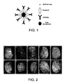
- the present inventors have devised a simple, robust method for the formation of a monolayer of antibody using a protein linker to coat the surface of gold nanoparticles - the construction of the antibody-nanoparticle conjugates is schematically shown in Figure 1 .
- Protein A is a cell wall component of Staphylococcus aureus which specifically binds the Fc portion of an antibody (stalk of the Y).
- the antibody By binding the Fc component, the antibody is positioned such that the recognition component, the F(ab') 2 binding region (the top of the Y), is directly available to bind to the specific targeted antigen of the biological fluid.
- the protein In order to form a monolayer of Protein A on the gold particle surface, the protein is first modified with N -succinimidyl 3-(2-pyridyldithio)-propionate (SPDP): - the succinimidyl ester binds the linker to a primary amine on the protein surface while the thiol moiety provides a linkage to the particle by displacing the citrate layer on the gold surface 2 .
- SPDP N -succinimidyl 3-(2-pyridyldithio)-propionate
- the Protein A monolayer provides a surface to which the antibody binds such that the F(ab') 2 recognition component can directly, and reproducibly, bind to the antigen (the substance of interest).
- Protein A may be replaced by Protein G or a chemical linker, such as a thiolated linkage.
- Protein A orientates the antibody for optimal presentation for antigen binding capability.
- a simple chemically linked antibody on the nanoparticle may suffice, i.e. an antibody directly linked to the surface of the metal or metal oxide surface of the nano particle.
- a chemical linker such as SPDP, may be attached to the antibody and allow direct attachment of the antibody to the metal, e.g. gold, or metal oxide. It is expected that an antibody bound to the particle using such linkers would be randomly orientated on the surface, potentially reducing binding affinity to the target.
- the advantage of the direct linkage is the ease of manufacture of these antibody-nanoparticle conjugates.
- Antibodies that could be used for the detection of blood, semen and saliva in the present invention include, but are not limited to, (i) the anti-human secretory IgA or anti-proline rich protein 1 or 2 for saliva, both of which are found to be highly concentrated; (ii) anti-cytokeratin 13, which can be used to stain for buccal epithelial cells as a source of DNA; (iii) the erythrocyte membrane protein glycophorin (anti-CD235a), which can be used to stain for red blood cells and anti-human serum albumin (iv) anti-CD45 or an anti-neutrophil cytoplasmic antibody (ANCA), optionally selected from (i) p-ANCA (myeloperoxidase) and (ii) c-ANCA (proteinase 3 (PR3)), which can be used to stain for white blood cells as a source of DNA; (v) Sperm surface protein (anti-SP17 or SP56) and/or as well as prostrate specific antigen, which can be
- Fluorescence microscopy data produced by the present inventors, using erythrocyte membrane protein glycophorin for the detection of dried blood is shown in Figure 7 .
- This Figure demonstrates that dried stains can be visualised using the types of immunofluorescence approaches that can be employed in the method of the present invention.
- the present inventors have found that one should select antibodies which target surface membranes or extracellular components as our work has shown that in biological stains the cell membranes remain in tact.
- Fluorophores may be selected with consideration of: excitation and emission spectra relative to light sources routinely used in forensic science; the requirement for colour-mapping; sensitivity and substrate fluorescence.
- Light sources used for evidence examination include: lasers; high intensity Xe and Hg arc lamps; and LED devices.
- a detection solution (the formulation of the present invention) containing the antibody-nanoparticle conjugates will be used with fluorophores which can be differentiated through non-overlapping emission bands when bound to their target biological matrix 3-5 .
- fluorophores which can be differentiated through non-overlapping emission bands when bound to their target biological matrix 3-5 .
- a fluorophore for the detection of blood will have different emission band from the fluorophore for the detection of semen, and likewise for saliva and DNA.
- Fluorophores with emission spectra in the range ca. 430-650 nm are preferred and are commercially available. There are nine dyes in the Alexa Fluor range (Molecular Probes) within this wavelength "window", all of which exhibit exceptional photostability. These fluorophores are all water-soluble and possess a succinimidyl ester moiety enabling simple chemistries for tagging the antibodies via primary amine residues. Selection and evaluation of the fluorophores will take into account light sources currently used in forensic laboratories (excitation and emission wavelengths and bandwidth in relation to the ability to enable multi-colour mapping) and excitation and emission spectra of fluorescent substrates commonly encountered in forensic science 6-8 .
- the fluorophore may be fluorescent molecule attached (or tagged) to an F(ab') 2 fragment of a monoclonal antibody, preferably F(ab') 2 fragment of goat antimouse IgG.
- Substrates that may be tested using the detection process of the present invention include, but are not limited to, paper, glass, plastic, wood, metal, cloth. Examples of such substrates include, but are not limited to, documents, wallpaper, sheets and clothing.
- the first approach employs the particles of the present invention in the methods as described herein, wherein the antibody can target DNA binding proteins such as Histone 1.
- Antibodies to Histone H1 are known in the art.
- the second approach employs the particles of the present invention in the methods as described herein, wherein the antibody is replaced with human specific peptide nucleic acid (PNA) oligomers.
- PNA human specific peptide nucleic acid
- the PNA oligomers will be custom synthesised (Eurogentec S.A.) to hybridise to Alu sequences (a family of interspersed nuclear repeat elements so called because they are recognised by the restriction endonuclease Alu 1, they are scattered throughout the genome and account for about 5-10% of the total genome).
- the bases in PNA molecules are capable of Watson and Crick base pairing with DNA bases, Consequently, they have been used in fluorescence in situ hybridisation experiments 9 and can target DNA in live cells 10 where DNA is in its native configuration packaged into chromatin.
- the PNA oligomers have also been found to bind in a sequence specific manner to the DNA found in biological stains.
- the PNA oligomers may be tagged with an appropriate fluorophore and then bound to nanoparticles and the resultant nanoparticle-DNA oligomer conjugates assessed for their binding to genomic DNA initially in cell suspensions and then in dried body fluids.
- One may lyse cells in body fluid stains to enable access of the antibody or PNA oligomers to DNA before or during contact with the particles of the present invention.
- nanoscale particles of iron oxide are superparamagnetic, i.e ., they are attracted to a magnetic field but retain no residual magnetism after the field has been removed. This property may be utilised knowing that when a suspension of nanoparticles of the present invention is applied to a forensic sample, the antibody conjugates would bind to the target species; however those particles not bound could be readily removed using a simple magnet. Once the field is removed the bound particles would no longer be magnetic.
- iron oxide nanoparticles are readily synthesised by the combination of Fe 2+ + Fe 3+ salts in an alkaline solution under an atmosphere of nitrogen.
- the particles can then be stabilised using a variety of functionalised ligands.
- the pH of such a solution is too alkaline for the particles of the present invention, as the antibody would denature. Therefore rather than directly synthesising iron oxide particles one can synthesize particles having an iron oxide core with a gold shell or coating (termed FeAU nanoparticles), preferably using methods previously reported 11,12 .
- the FeAu nanoparticles retain the superparamagnetic properties of iron oxide particles but have the added advantage of the gold surface which can be used to formulate a reproducible self-assembled monolayer.
- the FeAu nanoparticles are synthesised in reverse micelles and then stabilised with a preferred ligand.
- the particles of the present invention may comprise gold shell / iron oxide core nanoparticles, stabilised with a monolayer of antibody.
- the formulation of the present invention preferably contains the particles as a stable suspension and most preferably the particles are monodisperse i.e. of approximately the same size and, optionally, of the same kind.
- the FeAu particles may be stabilised in a liquid using decanoic acid.
- a place rearrangement reaction in which the carboxylic acid is preferentially replaced by a thiolated ligand enables deposition of the chosen entity (as the SH moiety has a greater affinity for the gold surface).
- Previous approaches have used this technique to deposit fluorescent macrocycles on the surface of FeAu nanoparticles (the macrocycles retain their fluorescence properties following deposition). While, to the present inventors' knowledge, biomolecules have not been formulated on such nanoparticles.
- the various fluorescently tagged antibodies (and PNA oligomers) may be attached to the FeAu surface via direct chemical or Protein A (thiolated) linkers.
- the formulation of the present invention may be applied by brushing or spraying the formulation onto the surface of the substrate.
- the brushes used may be magnetic brushes, e.g. those used for applying magnetic fingerprint powder.
- the method of the present invention may be used to detect any substance for which there is a known antibody.
- the antibody may selectively bind to a drug, drug metabolite, hormone or explosive.
- a drug, drug metabolite, hormone or explosive For instance, one can use nanoparticles conjugated with drug, drug metabolite, hormone and explosives-specific monoclonal antibodies (specific to e.g . cocaine; benzoyl ecgonine; nicotine; cotinine, testosterone, oestrogen, TNT and RDX).
- Such antibodies are commercially available.
- the nanoparticle-antibody conjugates may be applied to body fluid stains and/or fingerprints known to contain the target substance.
- the sweat may be deposited on a substrate by means such as a taking a fingerprint from the subject.
- the sweat/body fluid stains By testing for drugs or their metabolites in the sweat/body fluid stains, it is possible to determine whether a drug has been ingested by the subject, or simply handled. If a drug has been ingested by a subject, the drug and/or its metabolite will be present in a subject's excreted sweat/body fluid stains. However, if the drug had been handled, but not ingested, the drug may be present on a subject's skin, and may be transferred to, for example, the subject's fingerprint.
- Drugs that may be detected using the method of the present invention, if suitable antibodies are available include, but are not limited to:
- the method of the present invention may be used to detect metabolites of the drugs mentioned above, for which antibodies are available. If an antibody for a particular target substance, such as a drug or its metabolite, is not available commercially, the person skilled in the art could readily raise such an antibody using known techniques.
- the fluorescently tagged nanoparticle-antibody conjugates using antibodies which bind to hormones may be applied to substrates on which latent fingerprints are suspected to be present. Hormones are produced in the body by males and females and excreted in sweat. By testing for hormones fingerprints from males and females can be visualised.
- the present invention provides, in a second aspect, a method for making the particles of the present invention, the method comprising reacting particles comprising a metal or a metal oxide with an antibody to allow binding of the antibody to the particle.
- the particles may each comprise one or more Protein A's and/or Protein G's bound to the surface of the metal or metal oxide.
- Antibodies for many drug / drug metabolites are commercially available.
- the antibody used in the present invention may be an antibody for an explosive compound. Antibodies that have already been fully characterised for specificity to TNT and RDX and these may be used to detect these explosive residues.
- an antibody for cotinine the major metabolite of nicotine
- cotinine the major metabolite of nicotine
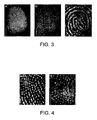
- Figure 1 These antibody-nanoparticle conjugates can be used to detect cotinine in the latent fingerprints of a smoker.
- the cotinine antibody was bound to the particles and then incubated on the surface of the fingerprint.
- Excess nanoparticle solution was washed from the fingerprint surface.
- a fluorescently tagged (alexa fluor 546) secondary F(ab') 2 fragment antibody was then bound to the cotinine-nanoparticle conjugate. Again excess reagent was washed from the surface.
- the nanoparticle bound fingerprints were imaged using a fluorescence stereomicroscope - the image obtained is shown in Figure 3 . No fluorescence image was obtained from fingerprints of non-smokers.
- the present invention provides nanoparticles of from 4 to 100 nm having a gold surface and, optionally, a core comprising iron oxide, wherein one or more Protein A's and/or Protein G's are attached to the gold surface via thiolate linkage and an antibody is attached via a non-binding portion to the Protein A and/or G.
- Protein A is a cell wall component of staphylococcus aureus that binds specifically to immunolobulin molecules.
- Protein A or Protein G is preferably attached to the gold surface by a thiolate linkage as shown between [Protein A/G] and [gold surface] as follows: [Protein A/G]-NH-CO-(CH 2 ) n -S-[gold surface], wherein n is 1 to 5, preferably 2, and NH is part of Protein A.
- particles having a metal/metal oxide core bound to protein A or G via a sulphur-containing linker preferably particles comprising a metal or a metal oxide, optionally capped with citrate, are contacted with SPDP modified Protein A or Protein G.
- the method of making the particles of the present invention preferably involves contacting particles having a metal/metal oxide core bound to protein A or G via a sulphur-containing linker with an antibody.
- the contacting is carried out in a liquid, preferably water.
- Protein A or Protein G is preferably attached to the thiolate-linkage precursor by reaction of Protein A or G with N-succinimidyl 1-3(2-pyridylthio) propionate (SPDP).
- SPDP N-succinimidyl 1-3(2-pyridylthio) propionate
- Protein A or Protein G may be attached to the gold surface of the nanoparticles by contacting in solution for a sufficient time gold nanoparticles capped with citrate and the reaction product of Protein A or G with SPDP. Protein A or G, on contact with an antibody in solution, will bind to the non-binding portion of an antibody, preferably the F c component.
- the present invention further provides a method for the fluorescent detection of a substance, the method comprising providing one or more fluorescently tagged antibodies for binding to a component of bodily fluid, preferably blood; contacting a substrate, which may or may not have the component on its surface, with the one or more antibodies for a time sufficient to allow the antibody to bind with the component; removing the antibodies which have not bound to the substrate; and illuminating the substrate with appropriate radiation to show the fluorophores on the substrate.
- the one or more antibodies are selected from erythrocyte membrane protein glycophorin (CD235a); human serum albumin; leucocyte common antigen (CD45); ain anti-neutrophil cytoplasmic antibody (ANCA).
- CD235a erythrocyte membrane protein glycophorin
- CD45 human serum albumin
- CD45 leucocyte common antigen
- ANCA ain anti-neutrophil cytoplasmic antibody
- the ANCA is preferably selected from (i) p-ANCA (myeloperoxidase); and (ii) c-ANCA (proteinase 3.(PR3)).
- the antibodies may be bound to one or more fluorophore via a succinimidyl ester moiety.
- a fluorescently tagged antibody for binding to a component of bodily fluid, preferably blood, is described herein, wherein the antibody is selected from erythrocyte membrane protein glycophorin (CD235a); human serum albumin; leucocyte common antigen (CD45) ; an anti-neutrophil cytoplasmic antibody (ANCA).
- the antibody is preferably bound to one or more fluorophores via a succinimidyl eater moiety.
- the fluorescently tagged antibodies can be synthesised by contacting a fluorophore for attachment to an antibody with the antibody, preferably in solution.
- a linking compound may be present for linking the antibody to the fluorophore moiety, the linking compound preferably comprising succinimidyl ester.
- the fluorophore may be selected from any suitable fluorophore available to the skilled person.
- the present inventors have devised a simple, yet robust, method for the formation of antibody-functionalised nanoparticles using a self-assembled monolayer (SAM) of a protein linker to coat the surface of gold nanoparticles.
- SAM self-assembled monolayer
- Gold nanoparticles of 16 nm diameter were formulated using the Turkevich approach ( B.V. Enüstün and J. Turkevich, J. Am. Chem. Soc., 1963, 85, 3317-3324 ) which uses citrate as both a reductant and a stabilizing agent.
- Protein A - a cell wall component of Staphylococcus aureus - was first modified with N-succinimidyl 3-(2-pyridyldithio)-propionate (SPDP) to enable the formation of a self-assembled monolayer (SAM) of the Protein A on the surface of gold nanoparticles (the succinimidyl ester binds the SPDP to a primary amine on the protein surface, while the thiol moiety provides a linkage to the gold surface (See: S. Ferretti, S. Paynter, D.A. Russell, K.E. Sapsford and D.J. Richardson, Trends Anal. Chem. , 2000, 19, 530-540 )).
- SPDP N-succinimidyl 3-(2-pyridyldithio)-propionate
- a SAM of Protein A is formed upon addition of the SPDP-modified Protein A to the nanoparticles with the thiolated ligand displacing the citrate layer on the gold particle surface.
- Protein A was used as a linker as the protein specifically binds the Fc portion of an antibody (See: E. Harlow and D. Lane in Antibodies: A laboratory Manual (Eds. E. Harlow and D. Lane) Cold Spring Harbour Laboratory, New York, 1988, pp 616-621 ).
- the Fc component By binding the Fc component, the Protein A orientates the antibody such that the recognition component, the F(ab') 2 binding region, is optimally presented for direct, and reproducible, antigen binding.
- Anti-cotinine was readily deposited onto the Protein A monolayer on the gold nanoparticles. However, it should be noted that this strategy can be applied to functionalise nanoparticles with numerous antibodies enabling detection of multiple specific antigens.
- the anti-cotinine functionalised nanoparticles were applied to the detection of cotinine in the fingerprints of smokers.
- the volunteer smoker would provide a fingerprint (such fingerprints are described 'as presented'), then asked to wash their hands and then sweating was induced by placing the volunteer's hand in a sealed glass beaker. Fingerprints were then taken at regular intervals between 10-40 mins.
- the cotinine-nanoparticle conjugates were pipetted onto the fingerprints and incubated for 30 min. Following incubation the fingerprint was washed to remove unbound nanoparticle conjugates.
- a fluorescently tagged secondary antibody fragment F(ab') 2 region
- was then incubated and excess reagent then removed by washing with water. Fluorescence images were then taken of the fingerprints.
- Figure 2 shows the fingerprint images obtained from a male smoker (reporting to smoke between 5-7 cigarettes p.d.) using 2 fluorescently labelled secondary antibody fragments.
- Reagents All reagents were purchased from Sigma-Aldrich (Gillingham, UK) unless stated, were of analytical grade and used without any further purification. Milli-Q water was used throughout for solution preparation unless otherwise stated.
- UV-Visible Absorption Measurements A Hitachi U3000 UV-visible spectrophotometer was used to record absorption spectra, at a temperature of 22 °C.
- TEM Transmission Electron Microscopy
- DTT DL-dithioreitol
- SPDP modified Protein A (1.6 M) was added to gold nanoparticles (3 nM) and stirred continuously for 48 hours to assist self assembly.
- Protein A stabilised gold nanoparticles were centrifuged (Beckman Coulter AvantiTM J-25 centrifuge) at 53343 xg and 4 °C for 30 minutes. The clear supernatant was pipetted off carefully, leaving a soft pellet ( ⁇ 0.5 ml) with a deep red colour and this was resuspended in phosphate buffer (10 mM, pH 7.4). The centrifugation process was repeated in triplicate to remove any unbound Protein A-SPDP. A UV-visible spectrum (800-300 nm) was taken of the final resuspended solution.
- IgG anti-cotinine 40 ⁇ l, 5.4 mg/ml (Europe Bioproducts Ltd., Ely, UK) was added to the Protein A modified gold nanoparticles (20 ml) and stirred for 2 1 ⁇ 2 hours. The centrifugation process was repeated, leaving a soft deep red pellet ( ⁇ 0.5 ml) after the clear supernatant was removed and on the last resuspension 5 ml of phosphate buffer was used (10 mM, pH 7.4). A UV-visible spectrum (800-300 nm) was taken of the final resuspended solution.
- Brightfield and Fluorescent Microscopy Brightfield and fluorescent images of latent fingerprints were acquired using a Zeiss M2 Bio Quad SV11 stereomicroscope. The fingerprints were illuminated either with a halogen lamp (brightfield) or a 100 W Hg arc lamp (fluorescence), and reflected-light images captured with an AxioCam HRc CCD camera and Axio Vision software (Carl Zeiss, Welwyn Garden City, UK). Alexa Fluor 488 ® (Invitrogen, Paisley, UK) was excited with light passed through a 470 nm filter (40 nm bandpass) and the emission collected through a 525 nm filter (50 nm bandpass). Alexa Fluor 546 ® (Invitrogen, Paisley, UK) was excited using a 560 nm excitation filter (40 nm bandpass) and a 630 nm emission filter (60 nm bandpass).
- a hydrophobic barrier was applied around the fingerprint using an ImmEdge hydrophobic barrier pen (Vector, Peterborough, UK) to contain a liquid on the fingerprint.
- a fresh batch (used within 1 day of production) of anti-cotinine-gold complex ( ⁇ 200 ⁇ l) was carefully applied to the fingerprint and the slide was incubated for 30 minutes at 37 °C in a wet chamber (moist tissue around the glass slide in a plastic petri dish). The fingerprint area was then washed with water to remove unbound anti-cotinine-gold complex.
- Anti-mouse secondary antibody F(ab') 2 Region
- Alexa Fluor 488 or 546 (Invitrogen, Paisley, UK) 20 ⁇ l of a 1 in 20 dilution of stock) was added to the fingerprint area and incubated as for the primary antibody. The slide was again washed with water to remove any unbound secondary antibody and fluorescent images taken.
- Imaging of Cotinine on Latent Fingerprints using Anti-Cotinine The procedure for imaging cotinine on latent fingerprints was then repeated for a control sample of anti-cotinine.
- the anti cotinine (40 ⁇ l) was added to phosphate buffer (10 mM, pH 7.4, 5 ml) to give a stock solution.
- Brightfield images were taken of smokers and non-smokers latent fingerprints prior to incubation. Fluorescent images were taken of the latent fingerprints after incubation as described for the anti-cotinine gold complex.
- Example 2 not within the scope of the invention.
- This example illustrates the use of certain antibodies in the detection of blood.
- Alexa Fluor dyes with the succinimidyl ester moiety for allowing the labelling of primary amine residues on the monoclonal antibodies, were selected due to their brightness, photostability, instrument compatibility, pH insensitivity and water solubility.
- Histone H1 is viewed to be more varied between species than the more conserved H2A, H2B, H3 and H4, so this was selected as a target.
- the H1 subtype H1.3 was also selected as this should be human specific. However, the staining of these histones will not show cell specificity, so it is viewed that these antibodies will have to be used in conjunction with leucocyte specific antibodies such as CD45 or ANCA labelled with a different fluorophore.
- Alexa fluor dyes (488, 568), carboxylic acid, succinimidyl ester 1 mg (Invitrogen) were dissolved in dimethylformamide (DMF) to a concentration of 1 mg / mL.
- the monoclonal antibodies were normally purchased in a solution of PBS and sodium azide without BSA or gelatin and at a concentration of at least 1 mg / mL.
- NaHCO 3 pH 8.5 was added to a final concentration of 100 mM.
- the dye was added at a ratio of 10:1 dye to protein and reacted for 1 hr (Alexa Fluor 488) or 2 hrs (Alexa Fluor 568) in the dark at room temperature with mixing every 15 minutes.
- the unbound dye was removed using 0.5 mL Zeba desalt spin columns (Pierce) which typically showed 95 % retention of salts and other molecules ⁇ 1000 MW and high protein recovery (c. 90-95%).
- the protein concentration and the dye: protein ratio was measured using a Nano Drop and calculated as follows:
- Sodium azide was added to a final concentration of 2 mM to act as a preservative.
- Brightfield and fluorescent images were acquired on a Zeiss Axioskop 2 MOT plus stereomicroscope. Blood smears were illuminted with either a halogen lamp for brightfield, or a 100 W Hg arc lamp for fluorescence. Images were captured with an AxioCam HRC CCD camera and processed using Axiovision 3.1 software.
- Figures 8 to 12 show human erythrocytes, leucocytes and albumin directly stained with specific labeled antibodies.
- Figure 8 shows human erythrocytes x 400 from a dried blood smear labeled with rat anti-human CD235a Alexa Fluor 568.
- Figure 9 shows human serum albumin from a human blood smear labeled with mouse anti-human albumin Alexa Fluor 488 (A) x 100 (B) x 400.
- Figure 10 shows two examples of dried human erythrocytes on black cotton x 200 labeled with rat anti-human CD235a Alexa Fluor 568.
- Figure 11 shows human neutrophils x 1000 from a dried blood smear labeled with (A) mouse anti-human CD45 Alexa Fluor 488 (B) mouse anti-human histone H1 Alexa Fluor 568 (C) overlay of images A & B.
- Figure 12 shows human lymphocyte or monocyte x 1000 from a dried blood smear labeled with rabbit anti-human histone H1.3 Alexa Fluor 568
- rat anti-human CD235a mouse anti-human CD45 and mouse anti-human albumin showed no cross reactivity with dog, rabbit or pig blood.
- Mouse anti-human histone H1 showed cross reactivity with these three different blood types. However, it could still be used in conjunction with CD45 to show cell specificity and human specificity. This would provide a useful tool in detecting a blood stain and DNA directly.
- Example 2 shows the effective use of certain antibodies in the detection of blood.
- the antibodies may be attached to metal or metal oxide-containing cores in an analogous manner to the method shown in Example 1.
Landscapes
- Engineering & Computer Science (AREA)
- Chemical & Material Sciences (AREA)
- Health & Medical Sciences (AREA)
- Immunology (AREA)
- Life Sciences & Earth Sciences (AREA)
- Nanotechnology (AREA)
- Biomedical Technology (AREA)
- Urology & Nephrology (AREA)
- Molecular Biology (AREA)
- General Physics & Mathematics (AREA)
- Hematology (AREA)
- Physics & Mathematics (AREA)
- Cell Biology (AREA)
- Food Science & Technology (AREA)
- Crystallography & Structural Chemistry (AREA)
- Materials Engineering (AREA)
- Microbiology (AREA)
- Condensed Matter Physics & Semiconductors (AREA)
- Composite Materials (AREA)
- Biotechnology (AREA)
- Medicinal Chemistry (AREA)
- Analytical Chemistry (AREA)
- Biochemistry (AREA)
- General Health & Medical Sciences (AREA)
- Pathology (AREA)
- Investigating, Analyzing Materials By Fluorescence Or Luminescence (AREA)
- Investigating Or Analysing Biological Materials (AREA)
- Investigating Or Analysing Materials By The Use Of Chemical Reactions (AREA)
Applications Claiming Priority (2)
| Application Number | Priority Date | Filing Date | Title |
|---|---|---|---|
| GBGB0605965.3A GB0605965D0 (en) | 2006-03-24 | 2006-03-24 | Fluorescence based detection of substances |
| PCT/GB2007/001059 WO2007110605A2 (en) | 2006-03-24 | 2007-03-23 | Fluorescence based detection of substances |
Publications (2)
| Publication Number | Publication Date |
|---|---|
| EP2005179A2 EP2005179A2 (en) | 2008-12-24 |
| EP2005179B1 true EP2005179B1 (en) | 2011-07-13 |
Family
ID=36384143
Family Applications (1)
| Application Number | Title | Priority Date | Filing Date |
|---|---|---|---|
| EP07732121A Not-in-force EP2005179B1 (en) | 2006-03-24 | 2007-03-23 | Fluorescence based detection of substances |
Country Status (9)
| Country | Link |
|---|---|
| US (1) | US8455264B2 (enExample) |
| EP (1) | EP2005179B1 (enExample) |
| JP (1) | JP5608853B2 (enExample) |
| AT (1) | ATE516501T1 (enExample) |
| AU (1) | AU2007231207B2 (enExample) |
| CA (1) | CA2647119C (enExample) |
| ES (1) | ES2366663T3 (enExample) |
| GB (1) | GB0605965D0 (enExample) |
| WO (1) | WO2007110605A2 (enExample) |
Cited By (2)
| Publication number | Priority date | Publication date | Assignee | Title |
|---|---|---|---|---|
| CN103536297A (zh) * | 2013-10-18 | 2014-01-29 | 浙江大学 | 基于免疫胶体金的潜在指纹成像的方法 |
| CN104678050A (zh) * | 2015-02-05 | 2015-06-03 | 北大先行科技产业有限公司 | 碘量法测定磷酸铁锂中三价铁含量的方法 |
Families Citing this family (15)
| Publication number | Priority date | Publication date | Assignee | Title |
|---|---|---|---|---|
| US20120164073A1 (en) * | 2007-11-30 | 2012-06-28 | Old Dominion University | Stable nanoparticles, nanoparticle-based imaging systems, nanoparticle-based assays, and in vivo assays for screening biocompatibility and toxicity of nanoparticles |
| JP2014531031A (ja) * | 2011-10-21 | 2014-11-20 | デシマドックス, エルエルシー | 小分析物の定量的検出のためのポイントオブケア免疫アッセイ |
| US10112988B2 (en) | 2012-01-09 | 2018-10-30 | Icb International, Inc. | Methods of assessing amyloid-beta peptides in the central nervous system by blood-brain barrier permeable peptide compositions comprising a vab domain of a camelid single domain heavy chain antibody against an anti-amyloid-beta peptide |
| US10112987B2 (en) * | 2012-01-09 | 2018-10-30 | Icb International, Inc. | Blood-brain barrier permeable peptide compositions comprising a vab domain of a camelid single domain heavy chain antibody against an amyloid-beta peptide |
| CN103226099A (zh) * | 2013-03-25 | 2013-07-31 | 常州大学 | 磷酸铁锂中三价铁含量的测定 |
| GB2520063B (en) * | 2013-11-08 | 2018-01-31 | Intelligent Fingerprinting Ltd | Skin-print fluorescence analysis method and apparatus |
| GB201403115D0 (en) * | 2014-02-21 | 2014-04-09 | Qbd Qs Ip Ltd | Red blood cell detection |
| CN104090050B (zh) * | 2014-07-08 | 2015-10-28 | 复旦大学 | 一种基于磁性材料的膜蛋白共价固定方法 |
| GB2535998A (en) * | 2015-02-27 | 2016-09-07 | Intelligent Fingerprinting Ltd | A device for receiving and analysing a sample |
| JP6769360B2 (ja) * | 2017-03-17 | 2020-10-14 | コニカミノルタ株式会社 | 蛍光体集積粒子複合体を用いた多段階蛍光染色方法および蛍光体集積粒子複合体 |
| WO2018211126A1 (en) | 2017-05-19 | 2018-11-22 | Philip Morris Products S.A. | Diagnostic test for distinguishing the smoking status of a subject |
| US10994993B2 (en) * | 2019-08-22 | 2021-05-04 | National Central University | Method of forming enhanced super-resolution image |
| CN113252593B (zh) * | 2021-06-29 | 2021-09-17 | 广州智汇生物科技有限公司 | 一种宠物食品中非法添加中枢神经兴奋类药物烟酸二乙胺的检测方法 |
| US20230104609A1 (en) * | 2021-10-04 | 2023-04-06 | Tintoria Piana, Inc | Diagnostic system and methods of using and manufacturing the same |
| CN119322175A (zh) * | 2024-10-18 | 2025-01-17 | 武汉塞力斯生物技术有限公司 | 一种血管炎抗体联检试剂盒 |
Family Cites Families (20)
| Publication number | Priority date | Publication date | Assignee | Title |
|---|---|---|---|---|
| JPS60222033A (ja) | 1984-04-18 | 1985-11-06 | 田岡化学工業株式会社 | 指紋の検出方法 |
| EP0324040B1 (en) | 1988-01-14 | 1992-07-08 | BASF Aktiengesellschaft | Process for controlling monomeric emissions |
| US5202267A (en) | 1988-04-04 | 1993-04-13 | Hygeia Sciences, Inc. | Sol capture immunoassay kit and procedure |
| EP0440350B1 (en) | 1990-01-19 | 1997-08-27 | La Mina Ltd. | Blood testing and fingerprint identification method and device |
| US5244815A (en) * | 1990-01-19 | 1993-09-14 | Lamina Ltd. | Fingerprint test pad and method for fingerprinting using particle based immunoassay |
| DE69332884T2 (de) * | 1992-10-21 | 2003-11-20 | Stefan Miltenyi | Direkte auswahl von zellen durch ein aussheidungsprodukt |
| US5494831A (en) | 1993-08-30 | 1996-02-27 | Hughes Aircraft Company | Electrochemical immunosensor system and methods |
| WO1996039186A1 (en) * | 1995-06-06 | 1996-12-12 | Cedars-Sinai Medical Center | Anti-neutrophil cytoplasmic antibody material associated with ulcerative colitis and related methods and kits |
| US6582921B2 (en) * | 1996-07-29 | 2003-06-24 | Nanosphere, Inc. | Nanoparticles having oligonucleotides attached thereto and uses thereof |
| US6165798A (en) * | 1996-10-10 | 2000-12-26 | University Of British Columbia | Optical quantification of analytes in membranes |
| DE10027776A1 (de) | 2000-06-07 | 2002-02-14 | Roche Diagnostics Gmbh | Neuartige core-shell Partikel |
| US20020192841A1 (en) * | 2001-04-27 | 2002-12-19 | Masayoshi Kojima | Measurement chip for biosensor |
| WO2002096262A2 (en) | 2001-05-25 | 2002-12-05 | Northwestern University | Non-alloying core shell nanoparticles |
| US20030059850A1 (en) * | 2001-09-26 | 2003-03-27 | Psychiatric Genomics, Inc. | Fluorescence proximity assay |
| JP2004149507A (ja) | 2002-09-05 | 2004-05-27 | Hisamitsu Pharmaceut Co Inc | 免疫抑制剤 |
| CN1245625C (zh) * | 2003-04-30 | 2006-03-15 | 陕西西大北美基因股份有限公司 | 一种核/壳型超顺磁性复合微粒及其制备方法与应用 |
| ES2385829T3 (es) * | 2003-08-20 | 2012-08-01 | Ucb Pharma, S.A. | Métodos para obtener anticuerpos |
| GB0400235D0 (en) | 2004-01-07 | 2004-02-11 | Univ Sunderland | Nanoparticles as agents for imaging finger prints |
| US7625712B2 (en) * | 2004-05-21 | 2009-12-01 | Beckman Coulter, Inc. | Method for a fully automated monoclonal antibody-based extended differential |
| CA3046259C (en) | 2007-12-21 | 2022-09-27 | Biomerieux Sa | Detection of methicillin-resistant staphylococcus aureus |
-
2006
- 2006-03-24 GB GBGB0605965.3A patent/GB0605965D0/en not_active Ceased
-
2007
- 2007-03-23 US US12/294,276 patent/US8455264B2/en not_active Expired - Fee Related
- 2007-03-23 AT AT07732121T patent/ATE516501T1/de not_active IP Right Cessation
- 2007-03-23 EP EP07732121A patent/EP2005179B1/en not_active Not-in-force
- 2007-03-23 ES ES07732121T patent/ES2366663T3/es active Active
- 2007-03-23 JP JP2009502191A patent/JP5608853B2/ja not_active Expired - Fee Related
- 2007-03-23 CA CA2647119A patent/CA2647119C/en active Active
- 2007-03-23 WO PCT/GB2007/001059 patent/WO2007110605A2/en not_active Ceased
- 2007-03-23 AU AU2007231207A patent/AU2007231207B2/en not_active Ceased
Cited By (3)
| Publication number | Priority date | Publication date | Assignee | Title |
|---|---|---|---|---|
| CN103536297A (zh) * | 2013-10-18 | 2014-01-29 | 浙江大学 | 基于免疫胶体金的潜在指纹成像的方法 |
| CN103536297B (zh) * | 2013-10-18 | 2015-06-17 | 浙江大学 | 基于免疫胶体金的潜在指纹成像的方法 |
| CN104678050A (zh) * | 2015-02-05 | 2015-06-03 | 北大先行科技产业有限公司 | 碘量法测定磷酸铁锂中三价铁含量的方法 |
Also Published As
| Publication number | Publication date |
|---|---|
| US8455264B2 (en) | 2013-06-04 |
| US20090230322A1 (en) | 2009-09-17 |
| JP2009531694A (ja) | 2009-09-03 |
| WO2007110605A3 (en) | 2008-01-24 |
| ES2366663T3 (es) | 2011-10-24 |
| ATE516501T1 (de) | 2011-07-15 |
| WO2007110605A2 (en) | 2007-10-04 |
| EP2005179A2 (en) | 2008-12-24 |
| CA2647119C (en) | 2014-05-27 |
| CA2647119A1 (en) | 2007-10-04 |
| JP5608853B2 (ja) | 2014-10-15 |
| GB0605965D0 (en) | 2006-05-03 |
| AU2007231207B2 (en) | 2012-09-06 |
| AU2007231207A1 (en) | 2007-10-04 |
Similar Documents
| Publication | Publication Date | Title |
|---|---|---|
| EP2005179B1 (en) | Fluorescence based detection of substances | |
| Han et al. | Label-free detection in biological applications of surface-enhanced Raman scattering | |
| Sun et al. | Microminiaturized immunoassays using quantum dots as fluorescent label by laser confocal scanning fluorescence detection | |
| KR101195957B1 (ko) | 표면증강 라만 산란 복합 프로브 및 이를 이용하여 표적 물질을 검출하는 방법 | |
| EP3172566B1 (en) | Sample analysing device | |
| JP2007506084A (ja) | ナノ粒子コンジュゲート及びその製造方法 | |
| Xia et al. | The simultaneous detection of the squamous cell carcinoma antigen and cancer antigen 125 in the cervical cancer serum using nano-Ag polydopamine nanospheres in an SERS-based lateral flow immunoassay | |
| CN104316698A (zh) | 固定有抗原的免疫荧光载片的制备方法和由该方法制备的免疫荧光载片 | |
| WO2012060456A1 (ja) | 免疫学的測定用青色金ナノ粒子、その製造方法およびそれを用いた測定方法 | |
| JP7284470B2 (ja) | 生体分子の検出のための温度応答性蛍光粒子 | |
| Oliveira et al. | Microfluidic SERS devices: brightening the future of bioanalysis | |
| Braz et al. | Gold-binding peptide as a selective layer for electrochemical detection of SARS-CoV-2 antibodies | |
| Gugoasa et al. | Myoglobin-silver reduced graphene oxide nanocomposite stochastic biosensor for the determination of luteinizing hormone and follicle-stimulating hormone from saliva samples | |
| McColman et al. | Serum proteins on nanoparticles: early stages of the “protein corona” | |
| EP2354146A1 (en) | Method for introducing functional group to surface of material | |
| Heinecke et al. | Preparation of gold nanocluster bioconjugates for electron microscopy | |
| Cháfer-Pericás et al. | Functionalized inorganic nanoparticles used as labels in solid-phase immunoassays | |
| CN105051539A (zh) | 用于使用发光检测生物分子的方法和试剂 | |
| CN216718452U (zh) | 一种三联毒品荧光免疫检测卡 | |
| Powell et al. | Giant platinum clusters: 2-nm covalent metal cluster labels | |
| EP1417027B1 (de) | Oberfläche mit einem muster aus zellen und verfahren zur herstellung derselben | |
| Chaloupkova et al. | Surface-Enhanced Raman Spectroscopy-Assisted Lateral Flow Test for Adenine and IgG Analysis | |
| CN111770955B (zh) | 包含非共价连接的有机纳米结构分子的硝酸纤维素膜 | |
| CN118725846A (zh) | 一种化学发光组合物及其制备方法和用途 | |
| TOMA et al. | Using a Surface Acoustic Wave |
Legal Events
| Date | Code | Title | Description |
|---|---|---|---|
| PUAI | Public reference made under article 153(3) epc to a published international application that has entered the european phase |
Free format text: ORIGINAL CODE: 0009012 |
|
| 17P | Request for examination filed |
Effective date: 20081016 |
|
| AK | Designated contracting states |
Kind code of ref document: A2 Designated state(s): AT BE BG CH CY CZ DE DK EE ES FI FR GB GR HU IE IS IT LI LT LU LV MC MT NL PL PT RO SE SI SK TR |
|
| RIN1 | Information on inventor provided before grant (corrected) |
Inventor name: LEE-SMITH, EMMA Inventor name: LEGGETT, RICHARD Inventor name: RUSSELL, DAVID Inventor name: JICKELLS, SUE,KING'S COLLEGE LONDON Inventor name: DANIEL, BARBARA,KING'S COLLEGE LONDON |
|
| 17Q | First examination report despatched |
Effective date: 20090506 |
|
| GRAP | Despatch of communication of intention to grant a patent |
Free format text: ORIGINAL CODE: EPIDOSNIGR1 |
|
| DAX | Request for extension of the european patent (deleted) | ||
| GRAS | Grant fee paid |
Free format text: ORIGINAL CODE: EPIDOSNIGR3 |
|
| GRAA | (expected) grant |
Free format text: ORIGINAL CODE: 0009210 |
|
| RAP1 | Party data changed (applicant data changed or rights of an application transferred) |
Owner name: UNIVERSITY OF EAST ANGLIA Owner name: KINGS COLLEGE LONDON |
|
| AK | Designated contracting states |
Kind code of ref document: B1 Designated state(s): AT BE BG CH CY CZ DE DK EE ES FI FR GB GR HU IE IS IT LI LT LU LV MC MT NL PL PT RO SE SI SK TR |
|
| REG | Reference to a national code |
Ref country code: GB Ref legal event code: FG4D |
|
| REG | Reference to a national code |
Ref country code: CH Ref legal event code: EP |
|
| REG | Reference to a national code |
Ref country code: NL Ref legal event code: T3 |
|
| REG | Reference to a national code |
Ref country code: IE Ref legal event code: FG4D |
|
| REG | Reference to a national code |
Ref country code: DE Ref legal event code: R096 Ref document number: 602007015783 Country of ref document: DE Effective date: 20110901 |
|
| REG | Reference to a national code |
Ref country code: SE Ref legal event code: TRGR |
|
| REG | Reference to a national code |
Ref country code: ES Ref legal event code: FG2A Ref document number: 2366663 Country of ref document: ES Kind code of ref document: T3 Effective date: 20111024 |
|
| REG | Reference to a national code |
Ref country code: AT Ref legal event code: MK05 Ref document number: 516501 Country of ref document: AT Kind code of ref document: T Effective date: 20110713 |
|
| PG25 | Lapsed in a contracting state [announced via postgrant information from national office to epo] |
Ref country code: LT Free format text: LAPSE BECAUSE OF FAILURE TO SUBMIT A TRANSLATION OF THE DESCRIPTION OR TO PAY THE FEE WITHIN THE PRESCRIBED TIME-LIMIT Effective date: 20110713 Ref country code: BE Free format text: LAPSE BECAUSE OF FAILURE TO SUBMIT A TRANSLATION OF THE DESCRIPTION OR TO PAY THE FEE WITHIN THE PRESCRIBED TIME-LIMIT Effective date: 20110713 Ref country code: PT Free format text: LAPSE BECAUSE OF FAILURE TO SUBMIT A TRANSLATION OF THE DESCRIPTION OR TO PAY THE FEE WITHIN THE PRESCRIBED TIME-LIMIT Effective date: 20111114 Ref country code: FI Free format text: LAPSE BECAUSE OF FAILURE TO SUBMIT A TRANSLATION OF THE DESCRIPTION OR TO PAY THE FEE WITHIN THE PRESCRIBED TIME-LIMIT Effective date: 20110713 Ref country code: IS Free format text: LAPSE BECAUSE OF FAILURE TO SUBMIT A TRANSLATION OF THE DESCRIPTION OR TO PAY THE FEE WITHIN THE PRESCRIBED TIME-LIMIT Effective date: 20111113 |
|
| PG25 | Lapsed in a contracting state [announced via postgrant information from national office to epo] |
Ref country code: SI Free format text: LAPSE BECAUSE OF FAILURE TO SUBMIT A TRANSLATION OF THE DESCRIPTION OR TO PAY THE FEE WITHIN THE PRESCRIBED TIME-LIMIT Effective date: 20110713 Ref country code: AT Free format text: LAPSE BECAUSE OF FAILURE TO SUBMIT A TRANSLATION OF THE DESCRIPTION OR TO PAY THE FEE WITHIN THE PRESCRIBED TIME-LIMIT Effective date: 20110713 Ref country code: PL Free format text: LAPSE BECAUSE OF FAILURE TO SUBMIT A TRANSLATION OF THE DESCRIPTION OR TO PAY THE FEE WITHIN THE PRESCRIBED TIME-LIMIT Effective date: 20110713 Ref country code: LV Free format text: LAPSE BECAUSE OF FAILURE TO SUBMIT A TRANSLATION OF THE DESCRIPTION OR TO PAY THE FEE WITHIN THE PRESCRIBED TIME-LIMIT Effective date: 20110713 Ref country code: GR Free format text: LAPSE BECAUSE OF FAILURE TO SUBMIT A TRANSLATION OF THE DESCRIPTION OR TO PAY THE FEE WITHIN THE PRESCRIBED TIME-LIMIT Effective date: 20111014 Ref country code: CY Free format text: LAPSE BECAUSE OF FAILURE TO SUBMIT A TRANSLATION OF THE DESCRIPTION OR TO PAY THE FEE WITHIN THE PRESCRIBED TIME-LIMIT Effective date: 20110713 |
|
| PG25 | Lapsed in a contracting state [announced via postgrant information from national office to epo] |
Ref country code: SK Free format text: LAPSE BECAUSE OF FAILURE TO SUBMIT A TRANSLATION OF THE DESCRIPTION OR TO PAY THE FEE WITHIN THE PRESCRIBED TIME-LIMIT Effective date: 20110713 Ref country code: CZ Free format text: LAPSE BECAUSE OF FAILURE TO SUBMIT A TRANSLATION OF THE DESCRIPTION OR TO PAY THE FEE WITHIN THE PRESCRIBED TIME-LIMIT Effective date: 20110713 |
|
| PLBE | No opposition filed within time limit |
Free format text: ORIGINAL CODE: 0009261 |
|
| STAA | Information on the status of an ep patent application or granted ep patent |
Free format text: STATUS: NO OPPOSITION FILED WITHIN TIME LIMIT |
|
| PG25 | Lapsed in a contracting state [announced via postgrant information from national office to epo] |
Ref country code: RO Free format text: LAPSE BECAUSE OF FAILURE TO SUBMIT A TRANSLATION OF THE DESCRIPTION OR TO PAY THE FEE WITHIN THE PRESCRIBED TIME-LIMIT Effective date: 20110713 Ref country code: EE Free format text: LAPSE BECAUSE OF FAILURE TO SUBMIT A TRANSLATION OF THE DESCRIPTION OR TO PAY THE FEE WITHIN THE PRESCRIBED TIME-LIMIT Effective date: 20110713 |
|
| 26N | No opposition filed |
Effective date: 20120416 |
|
| PG25 | Lapsed in a contracting state [announced via postgrant information from national office to epo] |
Ref country code: DK Free format text: LAPSE BECAUSE OF FAILURE TO SUBMIT A TRANSLATION OF THE DESCRIPTION OR TO PAY THE FEE WITHIN THE PRESCRIBED TIME-LIMIT Effective date: 20110713 |
|
| REG | Reference to a national code |
Ref country code: DE Ref legal event code: R097 Ref document number: 602007015783 Country of ref document: DE Effective date: 20120416 |
|
| PG25 | Lapsed in a contracting state [announced via postgrant information from national office to epo] |
Ref country code: MC Free format text: LAPSE BECAUSE OF NON-PAYMENT OF DUE FEES Effective date: 20120331 |
|
| REG | Reference to a national code |
Ref country code: CH Ref legal event code: PL |
|
| REG | Reference to a national code |
Ref country code: IE Ref legal event code: MM4A |
|
| PG25 | Lapsed in a contracting state [announced via postgrant information from national office to epo] |
Ref country code: IE Free format text: LAPSE BECAUSE OF NON-PAYMENT OF DUE FEES Effective date: 20120323 Ref country code: CH Free format text: LAPSE BECAUSE OF NON-PAYMENT OF DUE FEES Effective date: 20120331 Ref country code: LI Free format text: LAPSE BECAUSE OF NON-PAYMENT OF DUE FEES Effective date: 20120331 |
|
| REG | Reference to a national code |
Ref country code: GB Ref legal event code: 732E Free format text: REGISTERED BETWEEN 20130411 AND 20130417 |
|
| PG25 | Lapsed in a contracting state [announced via postgrant information from national office to epo] |
Ref country code: BG Free format text: LAPSE BECAUSE OF FAILURE TO SUBMIT A TRANSLATION OF THE DESCRIPTION OR TO PAY THE FEE WITHIN THE PRESCRIBED TIME-LIMIT Effective date: 20111013 |
|
| PG25 | Lapsed in a contracting state [announced via postgrant information from national office to epo] |
Ref country code: MT Free format text: LAPSE BECAUSE OF FAILURE TO SUBMIT A TRANSLATION OF THE DESCRIPTION OR TO PAY THE FEE WITHIN THE PRESCRIBED TIME-LIMIT Effective date: 20110713 |
|
| PG25 | Lapsed in a contracting state [announced via postgrant information from national office to epo] |
Ref country code: TR Free format text: LAPSE BECAUSE OF FAILURE TO SUBMIT A TRANSLATION OF THE DESCRIPTION OR TO PAY THE FEE WITHIN THE PRESCRIBED TIME-LIMIT Effective date: 20110713 |
|
| PG25 | Lapsed in a contracting state [announced via postgrant information from national office to epo] |
Ref country code: LU Free format text: LAPSE BECAUSE OF NON-PAYMENT OF DUE FEES Effective date: 20120323 |
|
| PG25 | Lapsed in a contracting state [announced via postgrant information from national office to epo] |
Ref country code: HU Free format text: LAPSE BECAUSE OF FAILURE TO SUBMIT A TRANSLATION OF THE DESCRIPTION OR TO PAY THE FEE WITHIN THE PRESCRIBED TIME-LIMIT Effective date: 20070323 |
|
| REG | Reference to a national code |
Ref country code: DE Ref legal event code: R082 Ref document number: 602007015783 Country of ref document: DE Representative=s name: PATENTANWAELTE WEICKMANN & WEICKMANN, DE Effective date: 20140710 Ref country code: DE Ref legal event code: R082 Ref document number: 602007015783 Country of ref document: DE Representative=s name: WEICKMANN & WEICKMANN PATENTANWAELTE - RECHTSA, DE Effective date: 20140710 Ref country code: DE Ref legal event code: R082 Ref document number: 602007015783 Country of ref document: DE Representative=s name: WEICKMANN & WEICKMANN PATENT- UND RECHTSANWAEL, DE Effective date: 20140710 Ref country code: DE Ref legal event code: R081 Ref document number: 602007015783 Country of ref document: DE Owner name: INTELLIGENT FINGERPRINTING LIMITED, GB Free format text: FORMER OWNER: KING'S COLLEGE LONDON, THE UNIVERSITY OF EAST ANGLIA, , GB Effective date: 20140710 Ref country code: DE Ref legal event code: R081 Ref document number: 602007015783 Country of ref document: DE Owner name: INTELLIGENT FINGERPRINTING LIMITED, GB Free format text: FORMER OWNERS: KING'S COLLEGE LONDON, LONDON, GB; THE UNIVERSITY OF EAST ANGLIA, NORWICH, GB Effective date: 20140710 |
|
| REG | Reference to a national code |
Ref country code: ES Ref legal event code: PC2A Owner name: INTELLIGENT FINGERPRINTING LIMMITED Effective date: 20140918 |
|
| REG | Reference to a national code |
Ref country code: NL Ref legal event code: SD Effective date: 20141002 |
|
| REG | Reference to a national code |
Ref country code: FR Ref legal event code: TQ Owner name: INTELLIGENT FINGERPRINTING LIMITED, GB Effective date: 20140923 Ref country code: FR Ref legal event code: TP Owner name: INTELLIGENT FINGERPRINTING LIMITED, GB Effective date: 20140923 |
|
| REG | Reference to a national code |
Ref country code: FR Ref legal event code: PLFP Year of fee payment: 10 |
|
| REG | Reference to a national code |
Ref country code: FR Ref legal event code: PLFP Year of fee payment: 11 |
|
| REG | Reference to a national code |
Ref country code: FR Ref legal event code: PLFP Year of fee payment: 12 |
|
| PGFP | Annual fee paid to national office [announced via postgrant information from national office to epo] |
Ref country code: IT Payment date: 20200319 Year of fee payment: 14 Ref country code: SE Payment date: 20200317 Year of fee payment: 14 |
|
| PGFP | Annual fee paid to national office [announced via postgrant information from national office to epo] |
Ref country code: ES Payment date: 20200401 Year of fee payment: 14 |
|
| PG25 | Lapsed in a contracting state [announced via postgrant information from national office to epo] |
Ref country code: SE Free format text: LAPSE BECAUSE OF NON-PAYMENT OF DUE FEES Effective date: 20210324 |
|
| PG25 | Lapsed in a contracting state [announced via postgrant information from national office to epo] |
Ref country code: IT Free format text: LAPSE BECAUSE OF NON-PAYMENT OF DUE FEES Effective date: 20210323 |
|
| REG | Reference to a national code |
Ref country code: ES Ref legal event code: FD2A Effective date: 20220524 |
|
| PG25 | Lapsed in a contracting state [announced via postgrant information from national office to epo] |
Ref country code: ES Free format text: LAPSE BECAUSE OF NON-PAYMENT OF DUE FEES Effective date: 20210324 |
|
| PGFP | Annual fee paid to national office [announced via postgrant information from national office to epo] |
Ref country code: FR Payment date: 20230314 Year of fee payment: 17 |
|
| PGFP | Annual fee paid to national office [announced via postgrant information from national office to epo] |
Ref country code: GB Payment date: 20230320 Year of fee payment: 17 Ref country code: DE Payment date: 20230323 Year of fee payment: 17 |
|
| PGFP | Annual fee paid to national office [announced via postgrant information from national office to epo] |
Ref country code: NL Payment date: 20230316 Year of fee payment: 17 |
|
| REG | Reference to a national code |
Ref country code: DE Ref legal event code: R119 Ref document number: 602007015783 Country of ref document: DE |
|
| REG | Reference to a national code |
Ref country code: NL Ref legal event code: MM Effective date: 20240401 |
|
| GBPC | Gb: european patent ceased through non-payment of renewal fee |
Effective date: 20240323 |
|
| PG25 | Lapsed in a contracting state [announced via postgrant information from national office to epo] |
Ref country code: NL Free format text: LAPSE BECAUSE OF NON-PAYMENT OF DUE FEES Effective date: 20240401 |
|
| PG25 | Lapsed in a contracting state [announced via postgrant information from national office to epo] |
Ref country code: NL Free format text: LAPSE BECAUSE OF NON-PAYMENT OF DUE FEES Effective date: 20240401 |
|
| PG25 | Lapsed in a contracting state [announced via postgrant information from national office to epo] |
Ref country code: DE Free format text: LAPSE BECAUSE OF NON-PAYMENT OF DUE FEES Effective date: 20241001 |
|
| PG25 | Lapsed in a contracting state [announced via postgrant information from national office to epo] |
Ref country code: GB Free format text: LAPSE BECAUSE OF NON-PAYMENT OF DUE FEES Effective date: 20240323 |
|
| PG25 | Lapsed in a contracting state [announced via postgrant information from national office to epo] |
Ref country code: FR Free format text: LAPSE BECAUSE OF NON-PAYMENT OF DUE FEES Effective date: 20240331 |
|
| PG25 | Lapsed in a contracting state [announced via postgrant information from national office to epo] |
Ref country code: GB Free format text: LAPSE BECAUSE OF NON-PAYMENT OF DUE FEES Effective date: 20240323 Ref country code: FR Free format text: LAPSE BECAUSE OF NON-PAYMENT OF DUE FEES Effective date: 20240331 Ref country code: DE Free format text: LAPSE BECAUSE OF NON-PAYMENT OF DUE FEES Effective date: 20241001 |