EP1830289A1 - Méthodes pour la classification et le pronostic du carcinome hépatocellulaire - Google Patents
Méthodes pour la classification et le pronostic du carcinome hépatocellulaire Download PDFInfo
- Publication number
- EP1830289A1 EP1830289A1 EP05292533A EP05292533A EP1830289A1 EP 1830289 A1 EP1830289 A1 EP 1830289A1 EP 05292533 A EP05292533 A EP 05292533A EP 05292533 A EP05292533 A EP 05292533A EP 1830289 A1 EP1830289 A1 EP 1830289A1
- Authority
- EP
- European Patent Office
- Prior art keywords
- gene
- hcc
- survival
- genes
- ramp3
- Prior art date
- Legal status (The legal status is an assumption and is not a legal conclusion. Google has not performed a legal analysis and makes no representation as to the accuracy of the status listed.)
- Withdrawn
Links
Images


Classifications
-
- C—CHEMISTRY; METALLURGY
- C12—BIOCHEMISTRY; BEER; SPIRITS; WINE; VINEGAR; MICROBIOLOGY; ENZYMOLOGY; MUTATION OR GENETIC ENGINEERING
- C12Q—MEASURING OR TESTING PROCESSES INVOLVING ENZYMES, NUCLEIC ACIDS OR MICROORGANISMS; COMPOSITIONS OR TEST PAPERS THEREFOR; PROCESSES OF PREPARING SUCH COMPOSITIONS; CONDITION-RESPONSIVE CONTROL IN MICROBIOLOGICAL OR ENZYMOLOGICAL PROCESSES
- C12Q1/00—Measuring or testing processes involving enzymes, nucleic acids or microorganisms; Compositions therefor; Processes of preparing such compositions
- C12Q1/68—Measuring or testing processes involving enzymes, nucleic acids or microorganisms; Compositions therefor; Processes of preparing such compositions involving nucleic acids
- C12Q1/6876—Nucleic acid products used in the analysis of nucleic acids, e.g. primers or probes
- C12Q1/6883—Nucleic acid products used in the analysis of nucleic acids, e.g. primers or probes for diseases caused by alterations of genetic material
- C12Q1/6886—Nucleic acid products used in the analysis of nucleic acids, e.g. primers or probes for diseases caused by alterations of genetic material for cancer
-
- G—PHYSICS
- G01—MEASURING; TESTING
- G01N—INVESTIGATING OR ANALYSING MATERIALS BY DETERMINING THEIR CHEMICAL OR PHYSICAL PROPERTIES
- G01N33/00—Investigating or analysing materials by specific methods not covered by groups G01N1/00 - G01N31/00
- G01N33/48—Biological material, e.g. blood, urine; Haemocytometers
- G01N33/50—Chemical analysis of biological material, e.g. blood, urine; Testing involving biospecific ligand binding methods; Immunological testing
- G01N33/53—Immunoassay; Biospecific binding assay; Materials therefor
- G01N33/574—Immunoassay; Biospecific binding assay; Materials therefor for cancer
- G01N33/57407—Specifically defined cancers
-
- G—PHYSICS
- G16—INFORMATION AND COMMUNICATION TECHNOLOGY [ICT] SPECIALLY ADAPTED FOR SPECIFIC APPLICATION FIELDS
- G16B—BIOINFORMATICS, i.e. INFORMATION AND COMMUNICATION TECHNOLOGY [ICT] SPECIALLY ADAPTED FOR GENETIC OR PROTEIN-RELATED DATA PROCESSING IN COMPUTATIONAL MOLECULAR BIOLOGY
- G16B25/00—ICT specially adapted for hybridisation; ICT specially adapted for gene or protein expression
- G16B25/10—Gene or protein expression profiling; Expression-ratio estimation or normalisation
-
- G—PHYSICS
- G16—INFORMATION AND COMMUNICATION TECHNOLOGY [ICT] SPECIALLY ADAPTED FOR SPECIFIC APPLICATION FIELDS
- G16B—BIOINFORMATICS, i.e. INFORMATION AND COMMUNICATION TECHNOLOGY [ICT] SPECIALLY ADAPTED FOR GENETIC OR PROTEIN-RELATED DATA PROCESSING IN COMPUTATIONAL MOLECULAR BIOLOGY
- G16B40/00—ICT specially adapted for biostatistics; ICT specially adapted for bioinformatics-related machine learning or data mining, e.g. knowledge discovery or pattern finding
- G16B40/10—Signal processing, e.g. from mass spectrometry [MS] or from PCR
-
- G—PHYSICS
- G16—INFORMATION AND COMMUNICATION TECHNOLOGY [ICT] SPECIALLY ADAPTED FOR SPECIFIC APPLICATION FIELDS
- G16B—BIOINFORMATICS, i.e. INFORMATION AND COMMUNICATION TECHNOLOGY [ICT] SPECIALLY ADAPTED FOR GENETIC OR PROTEIN-RELATED DATA PROCESSING IN COMPUTATIONAL MOLECULAR BIOLOGY
- G16B40/00—ICT specially adapted for biostatistics; ICT specially adapted for bioinformatics-related machine learning or data mining, e.g. knowledge discovery or pattern finding
- G16B40/20—Supervised data analysis
-
- G—PHYSICS
- G16—INFORMATION AND COMMUNICATION TECHNOLOGY [ICT] SPECIALLY ADAPTED FOR SPECIFIC APPLICATION FIELDS
- G16B—BIOINFORMATICS, i.e. INFORMATION AND COMMUNICATION TECHNOLOGY [ICT] SPECIALLY ADAPTED FOR GENETIC OR PROTEIN-RELATED DATA PROCESSING IN COMPUTATIONAL MOLECULAR BIOLOGY
- G16B40/00—ICT specially adapted for biostatistics; ICT specially adapted for bioinformatics-related machine learning or data mining, e.g. knowledge discovery or pattern finding
- G16B40/30—Unsupervised data analysis
-
- C—CHEMISTRY; METALLURGY
- C12—BIOCHEMISTRY; BEER; SPIRITS; WINE; VINEGAR; MICROBIOLOGY; ENZYMOLOGY; MUTATION OR GENETIC ENGINEERING
- C12Q—MEASURING OR TESTING PROCESSES INVOLVING ENZYMES, NUCLEIC ACIDS OR MICROORGANISMS; COMPOSITIONS OR TEST PAPERS THEREFOR; PROCESSES OF PREPARING SUCH COMPOSITIONS; CONDITION-RESPONSIVE CONTROL IN MICROBIOLOGICAL OR ENZYMOLOGICAL PROCESSES
- C12Q2600/00—Oligonucleotides characterized by their use
- C12Q2600/106—Pharmacogenomics, i.e. genetic variability in individual responses to drugs and drug metabolism
-
- C—CHEMISTRY; METALLURGY
- C12—BIOCHEMISTRY; BEER; SPIRITS; WINE; VINEGAR; MICROBIOLOGY; ENZYMOLOGY; MUTATION OR GENETIC ENGINEERING
- C12Q—MEASURING OR TESTING PROCESSES INVOLVING ENZYMES, NUCLEIC ACIDS OR MICROORGANISMS; COMPOSITIONS OR TEST PAPERS THEREFOR; PROCESSES OF PREPARING SUCH COMPOSITIONS; CONDITION-RESPONSIVE CONTROL IN MICROBIOLOGICAL OR ENZYMOLOGICAL PROCESSES
- C12Q2600/00—Oligonucleotides characterized by their use
- C12Q2600/112—Disease subtyping, staging or classification
-
- C—CHEMISTRY; METALLURGY
- C12—BIOCHEMISTRY; BEER; SPIRITS; WINE; VINEGAR; MICROBIOLOGY; ENZYMOLOGY; MUTATION OR GENETIC ENGINEERING
- C12Q—MEASURING OR TESTING PROCESSES INVOLVING ENZYMES, NUCLEIC ACIDS OR MICROORGANISMS; COMPOSITIONS OR TEST PAPERS THEREFOR; PROCESSES OF PREPARING SUCH COMPOSITIONS; CONDITION-RESPONSIVE CONTROL IN MICROBIOLOGICAL OR ENZYMOLOGICAL PROCESSES
- C12Q2600/00—Oligonucleotides characterized by their use
- C12Q2600/118—Prognosis of disease development
-
- C—CHEMISTRY; METALLURGY
- C12—BIOCHEMISTRY; BEER; SPIRITS; WINE; VINEGAR; MICROBIOLOGY; ENZYMOLOGY; MUTATION OR GENETIC ENGINEERING
- C12Q—MEASURING OR TESTING PROCESSES INVOLVING ENZYMES, NUCLEIC ACIDS OR MICROORGANISMS; COMPOSITIONS OR TEST PAPERS THEREFOR; PROCESSES OF PREPARING SUCH COMPOSITIONS; CONDITION-RESPONSIVE CONTROL IN MICROBIOLOGICAL OR ENZYMOLOGICAL PROCESSES
- C12Q2600/00—Oligonucleotides characterized by their use
- C12Q2600/136—Screening for pharmacological compounds
-
- C—CHEMISTRY; METALLURGY
- C12—BIOCHEMISTRY; BEER; SPIRITS; WINE; VINEGAR; MICROBIOLOGY; ENZYMOLOGY; MUTATION OR GENETIC ENGINEERING
- C12Q—MEASURING OR TESTING PROCESSES INVOLVING ENZYMES, NUCLEIC ACIDS OR MICROORGANISMS; COMPOSITIONS OR TEST PAPERS THEREFOR; PROCESSES OF PREPARING SUCH COMPOSITIONS; CONDITION-RESPONSIVE CONTROL IN MICROBIOLOGICAL OR ENZYMOLOGICAL PROCESSES
- C12Q2600/00—Oligonucleotides characterized by their use
- C12Q2600/154—Methylation markers
-
- G—PHYSICS
- G16—INFORMATION AND COMMUNICATION TECHNOLOGY [ICT] SPECIALLY ADAPTED FOR SPECIFIC APPLICATION FIELDS
- G16B—BIOINFORMATICS, i.e. INFORMATION AND COMMUNICATION TECHNOLOGY [ICT] SPECIALLY ADAPTED FOR GENETIC OR PROTEIN-RELATED DATA PROCESSING IN COMPUTATIONAL MOLECULAR BIOLOGY
- G16B25/00—ICT specially adapted for hybridisation; ICT specially adapted for gene or protein expression
-
- G—PHYSICS
- G16—INFORMATION AND COMMUNICATION TECHNOLOGY [ICT] SPECIALLY ADAPTED FOR SPECIFIC APPLICATION FIELDS
- G16B—BIOINFORMATICS, i.e. INFORMATION AND COMMUNICATION TECHNOLOGY [ICT] SPECIALLY ADAPTED FOR GENETIC OR PROTEIN-RELATED DATA PROCESSING IN COMPUTATIONAL MOLECULAR BIOLOGY
- G16B40/00—ICT specially adapted for biostatistics; ICT specially adapted for bioinformatics-related machine learning or data mining, e.g. knowledge discovery or pattern finding
Definitions
- the present invention concerns methods for the in vitro classification and/or prognosis of hepatocellular carcinoma (HCC).
- HCC hepatocellular carcinoma
- Hepatocellular carcinoma is one of the most frequent solid tumors worldwide and represents the third cause of mortality among deaths from cancer ( Bosch. F.X., et al. Semin Liver Dis 19, 271-85 (1999) ). Its frequency is particularly high in Asia and Africa due to the high frequency of viral hepatitis infections and to Aflatoxin B1 exposure (AFB1). Over the last 10 years the incidence of HCC has noticeably increased in United Kingdom, France and United States ( Taylor-Robinson, S.D. et al. Bmj 319, 640 (1999 ); Deuffic, S. et al. Lancet 351, 214-5 (1998) . EI-Serag, H.B. & Mason, A.C. N Engl J Med 340, 745-50 (1999) ). This increase is linked to the increase of viral hepatitis C infections.
- Liver cirrhosis of any origin and dysplastic regenerative nodules have long been considered to be the likely precursors of HCC because of their frequent association with the HCC occurrence ( Edmondson, H.A. & Peters, R.L. Semin Roentgenol 18, 75-83 (1983) ; Thorgeirsson, S.S. & Grisham, J.W. Nat Genet 31, 339-46 (2002) ).
- HCC etiological factors particularly HBV infection which can induce chromosome instability ( Aoki, H., et al.
- HCC is thus a very heterogeneous group of tumors that differ by risk factors and genetic alterations.
- the prognosis of HCC is also very heterogeneous.
- the main treatment of HCC is surgical removal of HCC tumor, which may or not be followed by adjuvant chemotherapy.
- Chemotherapy may be very tiresome and painful for patients but is necessary in case of HCC with poor prognosis.
- a classification and prognosis method of HCC tumors would thus also be very helpful to decide whether or not to administer an adjuvant therapy to an HCC patient.
- HCC heterogeneity is well known to those skilled in the art, and quite a lot of efforts have been made to better classify and/or prognose HCC tumors in the prior art.
- HCC tumors have recently tried to classify HCC tumors by global transcriptome analysis. Some of them describe significant expression profiles alterations between HBV and HCV derived HCC respectively ( Okabe et al. Cancer Res. 2001 Mar 1;61(5):2129-37 ; Iizuka et al. Cancer Res. 2002 Jul 15;62(14):3939-44 ).
- HCC classification into two or three subgroups based on various histological features ( Chung et al. Mol Cells. 2002 Dec 31;14(3):382-7 ; Chen et al. Mol Biol Cell. 2002 Jun;13(6):1929-39 ; WO 2004/090163 ) or on survival probability ( Lee et al. Hepatology. 2004 Sep;40(3):667-76 ).
- HCC human Crohn's disease .
- the inventors have previously shown that genetic alterations are indeed closely associated with clinical characteristics of HCC defining two groups of HCC ( Legoix, P. et al. Oncogene 18, 4044-6 (1999) ; Laurent-Puig, P. et al. Gastroenterology 120, 1763-73 (2001) ).
- the first type of HCC was associated with not only a high level of chromosome instability, frequent TP53 and AXIN1 mutations but also closely linked to HBV infection and a bad prognosis.
- the second subgroup of HCC tumors are chromosome stable, with a high incidence of activating ⁇ -catenin alterations and not associated with viral infection.
- HCC tumors actually clustered into 6 distinct subgroups, closely associated with various clinical and genetic alterations. They also determined a 16-gene diagnostic predictor of class membership as well as a 5-gene signature predicting patient prognosis irrespective of HCC subgroup and which outperforms common clinical prognostic markers.
- the invention thus concerns a method of in vitro classification of a HCC tumor between 6 subgroups from a liver HCC sample of a subject suffering from HCC, comprising:
- a "classification" of HCC tumors is intended to mean the determination for any HCC tumor of the HCC "subgroup” or “class” (these two words “subgroup” and “class” will be used indifferently for one another throughout the application) to which it belongs.
- the inventors have defined 6 distinct HCC subgroups or classes (hereafter named G1 to G6). These 6 subgroups were defined by a non-supervised analysis of global transcriptomic analysis of 57 HCC, 3 hepatocellular adenomas and 5 samples of pooled non-tumor tissue using Affymetrix HG-U133A GeneChip TM arrays. The 6 subgroups are highly associated with clinical and genetic factors, as displayed in the following Table 2, and summarized in Figure 1. Table 2.
- the methods of in vitro classification according to the invention allow to easily determine for any HCC liver sample to which of these 6 HCC subgroups it belongs.
- the expression profile comprises at least 8, at least 10, at least 12, at least 14, or at least 16 genes selected from the group consisting of: RAB1A, REG3A, NRAS, RAMP3, MERTK, PIR, EPHA1, LAMA3, GOS2, HN1, PAK2, AFP, CYP2C9, CDH2, HAMP, SAE1, ADH6, DCN, FLJ10159, ALDH1L1, IGF1, LECT2, SLC38A1, SPARCL1.
- the expression profile comprises at least 8, at least 10, at least 12, at least 14, or 16 genes selected from the group consisting of: RAB1A, REG3A, NRAS, RAMP3, MERTK, PIR, EPHA1, LAMA3, GOS2, HN1, PAK2, AFP, CYP2C9, CDH2, HAMP, and SAE1.
- the expression profile comprises the following 16 genes combination: RAB1A, REG3A, NRAS, RAMP3, MERTK, PIR, EPHA1, LAMA3, G0S2, HN1, PAK2, AFP, CYP2C9, CDH2, HAMP, and SAE1.
- a "liver HCC sample” is intended to mean any liver sample comprising HCC tumor tissue.
- the liver HCC sample is a liver HCC biopsy or a HCC tumor surgical resection.
- determining an expression profile is meant the measure of the expression level of a group a selected genes.
- the expression level of each gene may be determined either at the proteic or at the nucleic level, using any technology known in the art.
- the measure of the expression level of a particular protein may be performed by any dosage method known by a person skilled in the art, including but not limited to ELISA or mass spectrometry analysis.
- the expression profile is determined at the nucleic level.
- the measure of the expression level of a gene may be carried out either directly on messenger RNA (mRNA), or on retrotranscribed complementary DNA (cDNA). Any method to measure the expression level may be used, including but not limited to microarray analysis, quantitative PCR, southern analysis.
- the expression profile is determined using a microarray.
- the expression profile is determined using quantitative PCR.
- the expression level of any gene is preferably normalized in comparison to the expression level of an internal control gene, generally a household gene, including but not limited to ribosomal RNA (such as for instance 18S ribosomal RNA) or genes such as actin or HPRT.
- an internal control gene generally a household gene, including but not limited to ribosomal RNA (such as for instance 18S ribosomal RNA) or genes such as actin or HPRT.
- a "subgroup distance" is calculated, which represents a mathematical distance between said HCC tumor and each of the 6 subgroups.
- the set of all tumors can be defined as a n-dimensions set in which each tumor sample may be characterized by n coordinates corresponding to the expression levels of each selected gene in said tumor sample.
- Each subgroup or class is a subset of the n-dimensions set that can be defined by a center point and an acceptable variation percentage around each coordinate of the center point.
- appropriate mathematical functions permitting each to calculate the distance of any tumor sample to one of the 6 subgroups or classes may be chosen.
- the invention also concerns a method of in vitro prognosis of global survival and/or survival without relapse from a liver HCC sample of a subject suffering from HCC, comprising:
- a "prognosis" of HCC evolution means a prediction of the future evolution of a particular HCC tumor relative to the patient suffering of this particular HCC tumor.
- the methods according to the invention allow for both a global survival prognosis and a survival without relapse prognosis.
- global survival prognosis prognosis of survival, with or without relapse.
- the main current treatment against HCC is tumor surgical resection.
- a "bad global survival prognosis” is defined as the occurrence of death within the 3 years after liver resection, whereas a "good global survival prognosis” is defined as the lack of death during the 5 post-operative years.
- survival without relapse prognosis prognosis of survival in the absence of any relapse.
- a "bad survival without relapse prognosis” is defined as the presence of tumor-relapse within the two years after liver resection, whereas a “good survival without relapse prognosis” is defined as the lack of relapse during the 4 post-operative years.
- the expression profile comprises a genes combination selected from:
- the expression profile comprises the following genes combination:
- the expression profile comprises a genes combination selected from:
- the expression profile comprises the following genes combination:
- a particular combination of genes may be referred to as a predictor.
- the "liver HCC sample” may also be any liver sample comprising HCC tumor tissue.
- the liver HCC sample is a liver HCC biopsy or a HCC tumor surgical resection.
- the expression level of each gene may be determined either at the proteic or at the nucleic level, using any technology known in the art, in particular any technology described above.
- the expression profile is determined at the nucleic level.
- the expression profile is determined using a microarray.
- the expression profile is determined using quantitative PCR.
- the expression level of any gene is preferably normalized in comparison to the expression level of an internal control gene, generally a household gene, including but not limited to ribosomal RNA (such as for instance 18S ribosomal RNA) or genes such as actin or HPRT.
- a global survival and/or survival without relapse “score” is calculated.
- a "score” is a logistic function taking into account the n expression levels of each selected gene in said tumor sample, weighted by parameters that depend on the chosen combination of genes and may be calculated by optimization on a training group of HCC tumors, followed by validation on a test group of HCC tumors, as explained in more details in Example 3 of the present application.
- an appropriate score function with suitable parameters may be determined.
- the obtained score(s) of global survival and/or survival without relapse are then compared to at least one threshold value, which determines whether the prognosis is bad or good.
- such a threshold value may be determined using the same method as for ⁇ (gene t ) and ⁇ (gene t ) parameters, i.e. by optimization on a training group of HCC tumors, followed by validation on a test group of HCC tumors, as described in more details in Example 3 of the present application.
- the prognosis of a sample will be:
- the expression profile comprises the genes combination of the following Table 5 and is determined using quantitative PCR and the following formula:
- the threshold value used for the global survival prognosis is in an interval of 10% around that displayed in Table 5.
- the threshold value used for the prognosis is in an interval of 10% around that displayed in Table 6.
- ⁇ Ct(sample i , gene t ) values are calculated relative to ribosomal 18S RNA (R18S).
- the invention further concerns a method for the in vitro diagnosis of the advisability of adjuvant therapy from a liver HCC sample of a subject suffering from HCC, comprising:
- adjuvant therapy is meant an additional antitumoral therapy that may be administered to a subject suffering from HCC after surgical HCC tumor resection.
- adjuvant therapies may include, without being limited to, chemotherapy and radiotherapy.
- the present invention also concerns a kit for the in vitro classification of a HCC tumor between 6 subgroups from a liver HCC sample of a subject suffering from HCC, comprising reagents for the determination of an expression profile comprising a combination of at least 8, at least 10, at least 12, at least 14, or at least 16 genes selected from the group consisting of: RAB1A, REG3A, NRAS, RAMP3, MERTK, PIR, EPHA1, LAMA3, G0S2, HN1, PAK2, AFP, CYP2C9, CDH2, HAMP, SAE1, ADH6, DCN, FLJ10159, ALDH1L1, IGF1, LECT2, SLC38A1, SPARCL1, CTNNA2, GLUL, LEF1, MATN2, MME, PFN2, SPINT2, TBX3, and FGFR2.
- kits for the in vitro classification of a HCC tumor comprises reagents for the determination of an expression profile comprising a combination of at least 8, at least 10, at least 12, at least 14, or 16 genes selected from the group consisting of: RAB1A, REG3A, NRAS, RAMP3, MERTK, PIR, EPHA1, LAMA3, G0S2, HN1, PAK2, AFP, CYP2C9, CDH2, HAMP, SAE1, ADH6, DCN, FLJ10159, ALDH1L1, IGF1, LECT2, SLC38A1, SPARCL1.
- kits for the in vitro classification of a HCC tumor comprises reagents for the determination of an expression profile comprising a combination of at least 8, at least 10, at least 12, at least 14, or 16 genes selected from the group consisting of: RAB1A, REG3A, NRAS, RAMP3, MERTK, PIR, EPHA1, LAMA3, G0S2, HN1, PAK2, AFP, CYP2C9, CDH2, HAMP, and SAE1.
- kits for the in vitro classification of a HCC tumor comprises reagents for the determination of an expression profile comprising the following 16 genes combination: RAB1A, REG3A, NRAS, RAMP3, MERTK, PIR, EPHA1, LAMA3, G0S2, HN1, PAK2, AFP, CYP2C9, CDH2, HAMP, and SAE1.
- the present invention further concerns a kit for the in vitro prognosis of global survival and/or survival without relapse from a liver HCC sample of a subject suffering from HCC, comprising reagents for the determination of an expression profile comprising a combination of at least 2, at least 3, at least 4, or at least 5 genes selected from the group consisting of NRCAM, PIR, RAMP3, SLC21A2, TAF9, TNA, HN1, PSMD1, MRPS7, CDC20, ENO1, HLF, STRA13, RAGD, NRAS, ARFGEF2, RAB1A, G0S2, SMAD3, DNAJA3, HELO1, and RHOQ.
- reagents for the determination of an expression profile comprising a combination of at least 2, at least 3, at least 4, or at least 5 genes selected from the group consisting of NRCAM, PIR, RAMP3, SLC21A2, TAF9, TNA, HN1, PSMD1, MRPS7, CDC20, ENO1, HLF, STRA13
- the kit for the in vitro prognosis of global survival from a liver HCC sample of a subject suffering from HCC comprises reagents for the determination of an expression profile comprising one of the following genes combinations:
- the kit for the in vitro prognosis of global survival from a liver HCC sample of a subject suffering from HCC comprises reagents for the determination of an expression profile comprising the following genes combination:
- the kit for the in vitro prognosis of survival without relapse from a liver HCC sample of a subject suffering from HCC comprises reagents for the determination of an expression profile comprising one of the following genes combinations:
- the kit for the in vitro prognosis of survival without relapse from a liver HCC sample of a subject suffering from HCC comprises reagents for the determination of an expression profile comprising the following genes combination:
- reagents that are provided may allow for the prognosis of only global survival or survival without relapse, or may allow for the prognosis of both global survival and survival without relapse.
- reagents for the determination of an expression profile may include any reagent useful for the determination of a gene expression level. Said determination of the expression level may be carried out at the proteic or nucleic level.
- Reagents suitable for the determination of an expression profile at the proteic level include, without being limited to, antibodies and antibody fragments, reagents for mass spectrometry analysis, and protein microarrays.
- reagents suitable for the determination of an expression profile at the nucleic level include, without being limited to, amplification primers, nucleic probes and nucleic acid microarrays.
- said reagents may be provided with instructions for performing a method of in vitro classification or prognosis of global survival and/or survival without relapse according to the invention.
- the said instructions may either
- the reagents and instructions may be provided together in the same package or separately in two distinct packages.
- the invention further concerns a method of treatment of a subject suffering from HCC, comprising:
- the invention also concerns a method of in vitro screening of compounds useful for the treatment of one of the 6 HCC subgroups according to the invention, comprising:
- the invention further concerns a method of treatment of a subject suffering from HCC, comprising:
- Macroscopic and/or microscopic vascular invasion was recorded in 37% of the cases. Satellite tumors defined by nodule(s) found at less than 1 cm from the main tumor was recorded in 41% of the cases.
- Overall and disease-free survival was assessed in 99 patients with a R0 complete resection after eliminating patients treated by liver transplantation or died within a 3 months post-operative period.
- the mean follow-up in whole series was 38 months (range 3-60 months) and it was 49 months for patients still alive.
- Microarray analyses were performed using 5 ⁇ g of total RNA of each sample as starting material and 20 ⁇ g cRNA per hybridization (GeneChip Fluidics Station 400) of HG-U133A Affymetrix GeneChip TM arrays. Images from each array were generated using HP GeneArray 2500 and analyzed following the manufacturer's protocols ( http://www.affymetrix.com/support/technical/manual/expression manual.affx ). Except when indicated, all transcriptome analysis was carried out using either an assortment of R system software (v1.9.0) packages including those of Bioconductor (v1.1.1) or original R code. R packages and versions are indicated when appropriate.
- Raw feature data from 65 Affymetrix HG-U133A GeneChip TM microarrays were normalized and log 2 intensity expression summary values for each probe set were calculated using robust multi-array average (package affy V1.4.32). Probe sets corresponding to control genes or having a "_x_" annotation were masked yielding a total of 19,787 probe sets available for further analyses.
- RNA samples were dissected and stored at -80°C until DNA and RNA extraction using Qiaquick and Rneasy extraction kits, respectively (Qiagen).
- DNAs were quantified by fluorometry (Fluoroskan Thermo Lab-system), RNA were quantified using a spectrophotometer at 260nm (Nanodrop). The quality of DNA and RNA was controlled by gel electrophoresis followed by staining with ethidium bromide and Agilent 2100 bioanalyser. RNAs were qualified if the 28S/18S ratio was more than 1.5 for Affymetrix experiments and more than 1 for quantitative RT-PCR analyses.
- HBS and HBX copies of DNA were quantified in tumor and non-tumor DNAs using Syber green method (Applied Biosystems). Sequences of HBS and HBX DNA were determined in all tumors to ensure that primers used for quantification were chosen in regions outside viral polymorphisms or mutation. Quantification of viral DNA were related to a chromosome 22 PCR amplification. Efficacy of PCR amplification was measured at 95, 97 and 94% for HBS, HBX and chromosome 22 reference, respectively. Tumor and non-tumor DNA samples were also carefully quantified using fluorimetry with Hoechst and concentrations were checked by agarose gel electrophoresis.
- a low number of viral DNA copies in tumors was defined by a ratio HBX /reference and HBS /reference inferior to 0.5 (mean: 0.01, range: 0.002-0.5, standard error: 0.14).
- a high number of viral DNA copies in tumors was defined by a ratio HBX /reference and HBS /reference superior to 1.5 (mean: 25, range: 1.6-212, standard error: 46). No value was found between 0.5 and 1.6.
- DNA methylation at CDH1 and CDKN2A promoter was searched using bisulfite DNA and methylation specific amplification as previously described ( Lee, S. et al. Am J Pathol 163, 1371-8 (2003) ; Zochbauer-Muller, S. et al. Cancer Res 61, 249-55 (2001) ).
- RNA was reverse transcribed using the High capacity Archive kit and random hexamers (Applied Biosystems).
- Applied Biosystems For each sample and tested gene, 1 ⁇ l of cDNA corresponding to 2 ng of reverse transcribed RNA, were analyzed by TaqMan PCR analysis, in duplicate, using TaqMan® Low Density Array and the ABI PRISM® 7900HT System (Applied Biosystems). The quality of cDNAs was assessed using a R18S quantification by real time PCR (coefficient of variation 7% for the entire series).
- ⁇ CT (CT TESTED -CT R18S ) sample - (CT TESTED -CT R18S ) calibrator ( Livak, K.J. & Schmittgen, T.D. Methods 25, 402-8 (2001 )).
- ⁇ CT (CT TESTED -CT R18S ) sample - (CT TESTED -CT R18S ) calibrator ( Livak, K.J. & Schmittgen, T.D. Methods 25, 402-8 (2001 )).
- expression results of a gene were normalized to internal control ribosomal 18S and relatively to a calibrator, consisting in the mean expression level of the corresponding gene in non-tumor samples normalized to internal control ribosomal 18S.
- the values given in tables and graphs express the n-fold ratio of the gene expression in a tested sample compared to the mean of non tumor tissues.
- Frozen tissues were homogenized with a Dounce in 500 ⁇ l ice-cold RIPA Lysis buffer (Santa Cruz) and protein concentration was determined by BCA protein assay kit (Pierce). Immunoblot analysis was performed using 50 ⁇ g of proteins migrated on a SDS 6% polyacrilamide gel, a polyclonal E-cadherin antibody (SC-7870, 1:500, Santa Cruz), a peroxidase-conjugated secondary antibody (1:2000, Santa Cruz) and enhanced chemoluminescence (ECL, Pierce).
- Immunostaining was performed on 5 ⁇ m sections of formalin-fixed, paraffin-embedded liver samples, using monoclonal anti- ⁇ -catenin (1:400, BD Biosciences 610153), biotinyled anti-mouse (1:200, Vector Laboratories BA-2000), Vectastain ABC Elite standard kit (Vector Laboratories PK-6100), DaB kit (Vector Laboratories SK-4100). Prior to immunostaining endogenous peroxidase was blocked and antigen retrieval was performed with 0.001M citrate buffer pH 7 in a pressure cooker (Biogenex).
- the classification of the 65 samples was based on a series of 24 hierarchical cluster analyses, obtained from 8 data subsets and 3 different linkage methods (average, complete and Ward's), using 1-Pearson correlation as a distance metric (package class V 7.2-2).
- the 8 data subsets corresponded to 8 unsupervised selections of the most varying expression profiles. Criteria for this selection were the significant difference of the variance for a given probe set compared to the median variance (P ⁇ 0.01), as well as different "robust" coefficient of variation thresholds (rCVs, calculated by dividing the standard deviation by the mean of a probe set log2 intensity values for n-2 samples eliminating the lowest and highest values). Between 99 and 6,712 probe sets were selected (99.5th and 60th rCV percentiles).
- the stability of the initial 24 dendograms was assessed by comparing each one to cluster results obtained after perturbation/resampling (100 iterations for each, see supplemental information for details on the stability score).
- This predictor was obtained with DLDA algorithm, and predicted set S1 with 100% success rate and set S2 with 94% success rate.
- Non-supervised transcriptome analysis defines clusters of tumors closely associated with clinical and genetic alterations
- HCC human cancer
- 3 hepatocellular adenomas 5 samples of pooled non-tumor tissues were analyzed using Affymetrix HG-U133A GeneChip TM arrays.
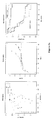
- Figure 2 each of which are highly associated with clinical and genetic factors based on Fisher exact tests (see above Table 2).
- the 60 tumor samples are sub-divided into 2 major groups each being further subdivided into 3 smaller subgroups (named here G1 to G6).
- the different subgroups were characterized by TP53 mutations (G2 and G3), an HBV infection (G1 and G2), with low number of HBV DNA copies (G1) and CTNNB1 gene mutations (G5 and G6).
- the inventors' aim was to identify a class-predictor more adapted to a clinical environment by using the more time and cost efficient technology quantitative reverse-transcriptase PCR (QRT-PCR).
- QRT-PCR quantitative reverse-transcriptase PCR
- a high correlation between the Affymetrix data and the QRT-PCR data was found with 135 out of the 140 selected genes (Spearman's rho median correlation coefficients of 0.84 using ⁇ Ct values).
- multiple sub-lists of a subset of 103 genes were tested genes in order to identify the best global predictor of the 6 HCC subgroups.
- the best success rate of predicting true class membership of the training set (100%) and test set (94.4%) was obtained with the Ct values of 16 genes ( RABIA, PAP, NRAS, RAMP3, MERTK, PIR, EPHA1, LAMA3, G0S2, HN1, PAK2, AFP, CYP2C9, CDH2, HAMP, SAE1 ) using the DLDA prediction algorithm.
- the new sample is affected to the closest class.
- genes specifically deregulated in one or more HCC subgroups were identified based on the results from an all group-wise t -test analysis combined with ANOVA. For all lists of genes specific of HCC subgroups, association of genes in known pathways was also searched for. An enrichment of cell cycle/proliferation/ DNA metabolism genes specifically over-expressed in subgroups G1 to G3 was observed, corresponding to chromosome instable samples ( P ⁇ 0.01). A high number of genes specifically over-expressed were observed for the G1 subgroup (related to HBV infection with a low number of viral DNA, AXIN1 mutations, a younger age, a high sera level of AFP and frequent origin from Africa, Table 2).
- genes encoding for proteins expressed during development were found: myosin heavy chain IIb, MYH4, the transcription factors SOX9 and SOX4, and parentally imprinted genes: insulin like growth factor 2 ( IGF2 ), paternally expressed gene 1, 3 and 10 ( PEG1, PEG3 and PEG10 ), alpha-fetoprotein ( AFP ) and sarcoglycan epsilon ( SGCE ).
- IGF2 insulin like growth factor 2
- PEG1, PEG3 and PEG10 paternally expressed gene 1, 3 and 10
- AFP alpha-fetoprotein
- SGCE sarcoglycan epsilon
- Subgroup G2 tumors (related to HBV infection with a high number of viral DNA copies, frequent local and vascular invasion and TP53 mutations) were significantly associated with over-expression cell cycle/proliferation/DNA metabolism genes ( P ⁇ 0.01), an enrichment that was equally observed in G3 (related to TP53 mutations and CDKN2A promoter methylation) and all chromosome instable samples ( P ⁇ 0.007).
- P ⁇ 0.009 A significant over-representation of over-expressed genes implicated in protein phosphorylation was also identified ( P ⁇ 0.009).
- G5 CTNNB1 mutated, no distant nodules
- under-expressed genes involved in stress and immune response such as IFI16 , IL4R, IF144 , STAT1, IL10RA , CTSS and HLA-DPA1 / B1 ( P ⁇ 0.002) was observed.
- HCC subgroups G5 and G6 contain 23 and 11 tumors CTNNB1 mutated in 70 and 100% of the cases, respectively.
- ⁇ -catenin targeted genes a list of 280 genes significantly over-expressed in G5 and G6 was found.
- two known ⁇ -catenin target genes in the liver Cadoret, A. et al.
- CDH1 encoding the E-cadherin
- G6 subgroup in Affymetrix and QRT-PCR experiments, Figure 4a
- the level of CDH1 mRNA down-regulation was showed to be highly related to the down-regulated expression of the E-cadherin protein in G6 consistent with the high level of promoter methylation of CDH1 in these tumors (data not shown).
- the inventors believe that the elucidation of the multifaceted classification of HCC was only possible in this application, compared to the previous published classifications into two subgroups, because (1) the studied series of tumors surgically treated in France included the main different risk factors of HCC, i.e. HBV and HCV infections, alcohol abuse and hemochromatosis and (2) the large number of clinical, histopathological and genetic annotations available for the studied sample population.
- the main clinical determinant of class membership is the HBV infection whereas the other main determinants are genetic and epigenetic alterations including chromosome instability, TP53 and CTNNB1 mutations, CDKN2A and CDH1 methylation and the parental imprinting (see Figure 1).
- HBV related tumors defining G1 and G2 subgroups are clearly molecularly distinct from the other etiologies. Tumors related to HCV infection and alcohol abuse are interspersed within the subgroups G3 to G6. The present transcriptomic classification has enabled the identification of new entities of tumors.
- Subgroup G1 includes HBV related tumors from younger patients (relative to the other HBV HCCs), frequently from Africa, with an equal sex ratio, a low number of viral DNA copies, frequent AXIN1 mutations, absence of TP53 mutation and an over-expression of genes normally parentally imprinted.
- HBV infection at the early age leads to a specific type of HCC demonstrating immature features with an abnormal parental gene imprinting possibly through the persistence of fetal hepatocytes or through the dedifferentiation of adult hepatocytes.
- Such diversity in tumors may be related to the high-risk populations found by epidemiological studies ( Brechot, C. Gastroenterology 127, S56-61 (2004) ; Yu, M.C. & Yuan, J.M. Gastroenterology 127, S72-8 (2004) ).
- EXAMPLE 3 Prognosis of HCC tumors
- each model was further assessed with the following resampling approach: we obtained 1,000 samplings by dividing 1,000 times, randomly, the whole series of 95 tumors in 2 groups of 47 and 48 samples each (equilibrating the number of death events between both groups); then, using each of the 42 lists of genes, for both groups of each sampling, multivariate Cox models were constructed and the logrank P value calculated from both models were stored. The combination of genes leading to the lowest median logrank P in both groups (among those 1,000 samplings) was kept and a predictor was then derived from this combination.
- a specific predictor of prognosis was constructed as described in the Material and Methods section.
- the top 16 genes associated with prognostic status of global survival were determined to be: NRCAM, PIR, RAMP3, SLC21A2, TAF9, TNA, HN1, PSMD1, MRPS7, CDC20, ENO1 , HLF, STRA13 , RAGD, NRAS, ARFGEF2.
- Global survival score sample i ⁇ t ⁇ gene t • 2 - ⁇ Ct sample i ⁇ gene t - ⁇ gene t , wherein the different ⁇ (gene t ) and ⁇ (gene t ) parameters are those listed in the following Table 7. Table 7. Parameters to be used in the above formula for the best out of top 5 overall survival predictors Global survival ⁇ ⁇ gene 1 ( TAF9 ) 7.28 0.129 gene 2 ( NRCAM ) 1.59 0.252 gene 3 ( RAMP3 ) 0.14 -6.133 gene 4 ( PSMD1 ) 4.66 0.024 gene 5 ( ARFGEF2 ) 3.66 -0.025
- Disease Free survival ⁇ ⁇ gene 1 TAF9
- 7.28 0.127 gene 2 ( NRCAM ) 1.59 0.196 gene 3 ( RAMP3 ) 0.14 -3.886
- HCC belonging to groups G1 to G3 were slightly related to early relapse and early death compared to HCC from G4 to G6, showing that classification and prognosis are somehow related.
- genes identified in the present applications as useful to predict survival had never been previously found associated to patient prognosis and they may be implicated in general cellular processes such as proteasome degradation of proteins (PSMD1, see Yokota, K. et al. Mol Biol Cell 7, 853-70 (1996 )), the initiation of RNA transcription (TAF9, see Michel, B., Komarnitsky, P. & Buratowski, S. Mol Cell 2, 663-73 (1998 )) and cellular proliferation (NRCAM, see Sehgal, A., et al. Anticancer Res 19, 4947-53 (1999 ); and ARFGEF2, see Sheen, V.L. et al. Nat Genet 36, 69-76 (2004 )).
- PSMD1 proteasome degradation of proteins
- TAF9 the initiation of RNA transcription
- NRCAM see Sehgal, A., et al. Anticancer Res 19, 4947-53 (1999 )
- ARFGEF2 see Sheen, V.L. et
- the present global transcriptomic analysis has been carried out and validated using a large series of highly annotated tumors.
- This analysis has established a robust classification reflecting the natural diversity of human HCCs, the structural gene alterations and epigenetic de-regulations accumulated during tumor progression.
- the high diversity of HCC tumor has clinical implications and the present classification has yielded prognostic tools not only for surgically treated patients but also to further identify patients that will benefit of targeted therapies.
Priority Applications (13)
| Application Number | Priority Date | Filing Date | Title |
|---|---|---|---|
| EP05292533A EP1830289A1 (fr) | 2005-11-30 | 2005-11-30 | Méthodes pour la classification et le pronostic du carcinome hépatocellulaire |
| SI200631913T SI2574679T1 (sl) | 2005-11-30 | 2006-11-30 | Postopki za in vitro prognozo hepatocelularnega karcinoma |
| SI200631919T SI1958109T1 (sl) | 2005-11-30 | 2006-11-30 | Postopek in set za klasificiranje hepatoceličnega karcinoma |
| ES06819878.7T ES2535049T3 (es) | 2005-11-30 | 2006-11-30 | Procedimientos para la clasificación in vitro de carcinoma hepatocelular |
| US12/095,604 US8935102B2 (en) | 2005-11-30 | 2006-11-30 | Method of hepatocellular carcinoma classification and prognosis |
| EP12182427.0A EP2574679B1 (fr) | 2005-11-30 | 2006-11-30 | Procédés de pronostic carninoma hépatocellulaire in vitro |
| ES12182427.0T ES2536898T3 (es) | 2005-11-30 | 2006-11-30 | Procedimientos para el pronóstico in vitro del carcinoma hepatocelular |
| CA2897986A CA2897986A1 (fr) | 2005-11-30 | 2006-11-30 | Methodes de classification et de prevision de l'evolution d'un carcinome hepatocellulaire |
| EP06819878.7A EP1958109B1 (fr) | 2005-11-30 | 2006-11-30 | Méthodes et trousse de réactifs pour la classification du carcinome hépatocellulaire |
| CA2631605A CA2631605C (fr) | 2005-11-30 | 2006-11-30 | Methodes de classification et de prevision de l'evolution d'un carcinome hepatocellulaire |
| PCT/EP2006/069175 WO2007063118A1 (fr) | 2005-11-30 | 2006-11-30 | Methodes de classification et de prevision de l'evolution d'un carcinome hepatocellulaire |
| JP2008542772A JP5420247B2 (ja) | 2005-11-30 | 2006-11-30 | 肝細胞癌腫分類および予後判定のための方法 |
| US14/580,484 US20150176080A1 (en) | 2005-11-30 | 2014-12-23 | Method of hepatocellular carninoma classification and prognosis |
Applications Claiming Priority (1)
| Application Number | Priority Date | Filing Date | Title |
|---|---|---|---|
| EP05292533A EP1830289A1 (fr) | 2005-11-30 | 2005-11-30 | Méthodes pour la classification et le pronostic du carcinome hépatocellulaire |
Publications (1)
| Publication Number | Publication Date |
|---|---|
| EP1830289A1 true EP1830289A1 (fr) | 2007-09-05 |
Family
ID=36021836
Family Applications (3)
| Application Number | Title | Priority Date | Filing Date |
|---|---|---|---|
| EP05292533A Withdrawn EP1830289A1 (fr) | 2005-11-30 | 2005-11-30 | Méthodes pour la classification et le pronostic du carcinome hépatocellulaire |
| EP12182427.0A Not-in-force EP2574679B1 (fr) | 2005-11-30 | 2006-11-30 | Procédés de pronostic carninoma hépatocellulaire in vitro |
| EP06819878.7A Not-in-force EP1958109B1 (fr) | 2005-11-30 | 2006-11-30 | Méthodes et trousse de réactifs pour la classification du carcinome hépatocellulaire |
Family Applications After (2)
| Application Number | Title | Priority Date | Filing Date |
|---|---|---|---|
| EP12182427.0A Not-in-force EP2574679B1 (fr) | 2005-11-30 | 2006-11-30 | Procédés de pronostic carninoma hépatocellulaire in vitro |
| EP06819878.7A Not-in-force EP1958109B1 (fr) | 2005-11-30 | 2006-11-30 | Méthodes et trousse de réactifs pour la classification du carcinome hépatocellulaire |
Country Status (7)
| Country | Link |
|---|---|
| US (2) | US8935102B2 (fr) |
| EP (3) | EP1830289A1 (fr) |
| JP (1) | JP5420247B2 (fr) |
| CA (2) | CA2631605C (fr) |
| ES (2) | ES2536898T3 (fr) |
| SI (2) | SI2574679T1 (fr) |
| WO (1) | WO2007063118A1 (fr) |
Cited By (1)
| Publication number | Priority date | Publication date | Assignee | Title |
|---|---|---|---|---|
| CN113913525A (zh) * | 2021-11-24 | 2022-01-11 | 广州医科大学 | Dnase1l3基因作为肝癌侵袭转移的检测和/或防治靶点的应用 |
Families Citing this family (37)
| Publication number | Priority date | Publication date | Assignee | Title |
|---|---|---|---|---|
| US8285719B1 (en) | 2008-08-08 | 2012-10-09 | The Research Foundation Of State University Of New York | System and method for probabilistic relational clustering |
| WO2010054379A2 (fr) * | 2008-11-10 | 2010-05-14 | The United States Of America, As Represensted By The Secretary, Department Of Health And Human Services | Signature génétique utilisée pour évaluer le pronostic chez des patients atteints de tumeurs solides |
| KR100964193B1 (ko) * | 2009-04-17 | 2010-06-16 | 씨비에스바이오사이언스 주식회사 | 간암 예후 마커 |
| WO2011122021A1 (fr) * | 2010-03-30 | 2011-10-06 | 国立大学法人山口大学 | Marqueur pour la prédiction de la récurrence d'un carcinome hépatocellulaire |
| CN103347520A (zh) | 2010-10-13 | 2013-10-09 | 波士顿大学管理委员会 | 用作癌症化学治疗的晚期sv40因子(lsf)的抑制剂 |
| EP2694963B1 (fr) * | 2011-04-01 | 2017-08-02 | Qiagen | SIGNATURE D'EXPRESSION GÉNIQUE POUR LA VOIE DE SIGNALISATION Wnt/CATÉNINE ET UTILISATION DE CELLE-CI |
| WO2013025322A2 (fr) * | 2011-08-15 | 2013-02-21 | Board Of Regents, The University Of Texas System | Score de risque de pronostic à base d'un marqueur dans le cancer du foie |
| CN104394855A (zh) * | 2012-05-31 | 2015-03-04 | 拜耳医药股份有限公司 | 用于测定肝细胞癌(hcc)患者的治疗的有效响应的生物标记 |
| JP2014027898A (ja) * | 2012-07-31 | 2014-02-13 | Yamaguchi Univ | 肝細胞がん発症リスクの判定方法 |
| AU2013320165A1 (en) | 2012-09-21 | 2015-04-02 | Institut National De La Sante Et De La Recherche Medicale (Inserm) | A new method for classification of liver samples and diagnosis of focal nodule dysplasia, hepatocellular adenoma, and hepatocellular carcinoma |
| AU2013320166A1 (en) * | 2012-09-21 | 2015-03-19 | Institut National De La Sante Et De La Recherche Medicale (Inserm) | A method for prognosis of global survival and survival without relapse in hepatocellular carcinoma |
| WO2014046623A1 (fr) * | 2012-09-21 | 2014-03-27 | Singapore Health Services Pte Ltd | Méthode de diagnostic du cancer du foie chez un sujet et kit permettant de diagnostiquer le cancer du foie |
| TWI500770B (zh) * | 2013-02-20 | 2015-09-21 | Univ Taipei Medical | Hoxa9基因作為檢測肝癌生物標記的用途 |
| US20150024517A1 (en) * | 2013-07-19 | 2015-01-22 | Texas Instruments Incorporated | Plasma etcher chuck band |
| GB201317609D0 (en) | 2013-10-04 | 2013-11-20 | Cancer Rec Tech Ltd | Inhibitor compounds |
| JP6008063B1 (ja) | 2015-03-24 | 2016-10-19 | 住友大阪セメント株式会社 | 静電チャック装置 |
| GB201505658D0 (en) | 2015-04-01 | 2015-05-13 | Cancer Rec Tech Ltd | Inhibitor compounds |
| GB201617103D0 (en) | 2016-10-07 | 2016-11-23 | Cancer Research Technology Limited | Compound |
| KR102014951B1 (ko) * | 2017-01-06 | 2019-08-27 | 강원대학교산학협력단 | P53의 전사적 활성 저해에 의한 암 검출용 바이오마커, 키트 또는 조성물, 이를 이용한 정보 제공 방법, 약물 스크리닝 방법, 및 악성 형질전환 모델 |
| JP6820621B2 (ja) * | 2017-03-31 | 2021-01-27 | 公立大学法人福島県立医科大学 | 相互依存性の特定方法 |
| WO2018189215A1 (fr) * | 2017-04-12 | 2018-10-18 | INSERM (Institut National de la Santé et de la Recherche Médicale) | Procédé de prédiction du temps de survie d'un patient souffrant d'un carcinome hépatocellulaire |
| CN108333366B (zh) * | 2018-01-26 | 2020-06-12 | 南通大学附属医院 | 一种实验性监测肝细胞恶性转化过程大鼠模型的建立方法 |
| CN112301125A (zh) * | 2019-07-30 | 2021-02-02 | 立森印迹诊断技术(无锡)有限公司 | 一种肿瘤标志物及其应用 |
| KR102055872B1 (ko) * | 2019-08-20 | 2019-12-13 | 강원대학교산학협력단 | P53의 전사적 활성 저해에 의한 암 검출용 바이오마커, 키트 또는 조성물, 이를 이용한 정보 제공 방법, 약물 스크리닝 방법, 및 악성 형질전환 모델 |
| KR102168498B1 (ko) * | 2019-12-09 | 2020-10-21 | 강원대학교산학협력단 | P53의 전사적 활성 저해에 의한 암 검출용 바이오마커, 키트 또는 조성물, 이를 이용한 정보 제공 방법, 약물 스크리닝 방법, 및 악성 형질전환 모델 |
| CN111676282B (zh) * | 2020-01-13 | 2022-05-06 | 中南大学湘雅医院 | G0s2基因在检测重症肌无力患者病情和治疗情况中的应用 |
| CN111458509B (zh) * | 2020-04-14 | 2023-09-22 | 中国人民解放军海军军医大学第三附属医院 | 肝细胞癌预后评估的生物标志物及其试剂盒和方法 |
| CN111537719B (zh) * | 2020-06-04 | 2022-10-04 | 复旦大学附属中山医院 | 血清Sparcl1蛋白在非酒精性脂肪性肝炎诊断中的应用 |
| CN111705132A (zh) * | 2020-07-03 | 2020-09-25 | 南方医科大学南方医院 | 一种ddPCR检测肝癌预后标志物TP53 R249S的引物探针组、试剂盒和方法 |
| KR102185037B1 (ko) * | 2020-10-14 | 2020-12-01 | 강원대학교산학협력단 | P53의 전사적 활성 저해에 의한 암 검출용 바이오마커, 키트 또는 조성물, 이를 이용한 정보 제공 방법, 약물 스크리닝 방법, 및 악성 형질전환 모델 |
| KR102205224B1 (ko) * | 2020-11-24 | 2021-01-20 | 강원대학교산학협력단 | P53의 전사적 활성 저해에 의한 암 검출용 바이오마커, 키트 또는 조성물, 이를 이용한 정보 제공 방법, 약물 스크리닝 방법, 및 악성 형질전환 모델 |
| KR102221671B1 (ko) * | 2021-01-13 | 2021-03-02 | 강원대학교산학협력단 | P53의 전사적 활성 저해에 의한 암 검출용 바이오마커, 키트 또는 조성물, 이를 이용한 정보 제공 방법, 약물 스크리닝 방법, 및 악성 형질전환 모델 |
| KR102260250B1 (ko) * | 2021-02-22 | 2021-06-03 | 강원대학교산학협력단 | P53의 전사적 활성 저해에 의한 암 검출용 바이오마커, 키트 또는 조성물, 이를 이용한 정보 제공 방법, 약물 스크리닝 방법, 및 악성 형질전환 모델 |
| KR102243705B1 (ko) * | 2021-02-22 | 2021-04-23 | 강원대학교산학협력단 | P53의 전사적 활성 저해에 의한 암 검출용 바이오마커, 키트 또는 조성물, 이를 이용한 정보 제공 방법, 약물 스크리닝 방법, 및 악성 형질전환 모델 |
| CN114106097B (zh) * | 2021-11-24 | 2023-12-22 | 中国科学院微生物研究所 | 用于治疗肝细胞癌的多肽及其应用 |
| WO2023196998A2 (fr) * | 2022-04-07 | 2023-10-12 | The Board Of Regents Of The University Of Texas System | Compositions et méthodes de traitement de maladies hépatiques par des arnsi ciblant tbx3 |
| CN117604108B (zh) * | 2024-01-23 | 2024-04-09 | 杭州华得森生物技术有限公司 | 用于肝癌诊断和预后判断的生物标志物及其应用 |
Citations (2)
| Publication number | Priority date | Publication date | Assignee | Title |
|---|---|---|---|---|
| WO2005005601A2 (fr) * | 2003-06-09 | 2005-01-20 | The Regents Of The University Of Michigan | Compositions et methodes de traitement et de diagnostic du cancer |
| US20050089895A1 (en) * | 2003-08-13 | 2005-04-28 | Cheung Siu T. | Compositions and methods for prognosis and therapy of liver cancer |
Family Cites Families (5)
| Publication number | Priority date | Publication date | Assignee | Title |
|---|---|---|---|---|
| AU3762699A (en) * | 1998-04-27 | 1999-11-16 | Pacific Northwest Cancer Foundation | (nr-cam) gene, nucleic acids and nucleic acid products for therapeutic and diagnostic uses for tumors |
| US20020009730A1 (en) * | 1999-11-17 | 2002-01-24 | Alex Chenchik | Human stress array |
| EP1620449A4 (fr) * | 2003-02-14 | 2008-10-01 | Smithkline Beecham Corp | Acides nucleiques a expression differentielle qui sont en correlation avec l'expression ksp |
| CN1764727B (zh) | 2003-04-08 | 2012-08-29 | 霍夫曼-拉罗奇有限公司 | 确定肿瘤分化等级的试剂盒 |
| EP1631682A1 (fr) * | 2003-06-04 | 2006-03-08 | Agency for Science, Technology and Research | Genes de carcinome hepatocellulaire a regulation differentielle et leurs utilisations |
-
2005
- 2005-11-30 EP EP05292533A patent/EP1830289A1/fr not_active Withdrawn
-
2006
- 2006-11-30 US US12/095,604 patent/US8935102B2/en not_active Expired - Fee Related
- 2006-11-30 EP EP12182427.0A patent/EP2574679B1/fr not_active Not-in-force
- 2006-11-30 EP EP06819878.7A patent/EP1958109B1/fr not_active Not-in-force
- 2006-11-30 SI SI200631913T patent/SI2574679T1/sl unknown
- 2006-11-30 SI SI200631919T patent/SI1958109T1/sl unknown
- 2006-11-30 CA CA2631605A patent/CA2631605C/fr not_active Expired - Fee Related
- 2006-11-30 CA CA2897986A patent/CA2897986A1/fr not_active Abandoned
- 2006-11-30 ES ES12182427.0T patent/ES2536898T3/es active Active
- 2006-11-30 JP JP2008542772A patent/JP5420247B2/ja not_active Expired - Fee Related
- 2006-11-30 WO PCT/EP2006/069175 patent/WO2007063118A1/fr active Application Filing
- 2006-11-30 ES ES06819878.7T patent/ES2535049T3/es active Active
-
2014
- 2014-12-23 US US14/580,484 patent/US20150176080A1/en not_active Abandoned
Patent Citations (2)
| Publication number | Priority date | Publication date | Assignee | Title |
|---|---|---|---|---|
| WO2005005601A2 (fr) * | 2003-06-09 | 2005-01-20 | The Regents Of The University Of Michigan | Compositions et methodes de traitement et de diagnostic du cancer |
| US20050089895A1 (en) * | 2003-08-13 | 2005-04-28 | Cheung Siu T. | Compositions and methods for prognosis and therapy of liver cancer |
Non-Patent Citations (4)
| Title |
|---|
| DUDOIT S ET AL: "COMPARISON OF DISCRIMINATION METHODS FOR THE CLASSIFICATION OF TUMORS USING GENE EXPRESSION DATA", JOURNAL OF THE AMERICAN STATISTICAL ASSOCIATION, AMERICAN STATISTICAL ASSOCIATION, NEW YORK, US, vol. 97, no. 457, 2002, pages 77 - 87, XP009054005, ISSN: 0162-1459 * |
| GE ET AL: "Interpreting expression profiles of cancers by genome-wide survey of breadth of expression in normal tissues", GENOMICS, ACADEMIC PRESS, SAN DIEGO, US, vol. 86, no. 2, August 2005 (2005-08-01), pages 127 - 141, XP005093481, ISSN: 0888-7543 * |
| LEE JU-SEOG ET AL: "Functional and genomic implications of global gene expression profiles in cell lines from human hepatocellular cancer.", HEPATOLOGY (BALTIMORE, MD.) MAY 2002, vol. 35, no. 5, May 2002 (2002-05-01), pages 1134 - 1143, XP002373697, ISSN: 0270-9139 * |
| SEGAL NEIL H ET AL: "Classification and subtype prediction of adult soft tissue sarcoma by functional genomics.", THE AMERICAN JOURNAL OF PATHOLOGY. AUG 2003, vol. 163, no. 2, August 2003 (2003-08-01), pages 691 - 700, XP002373698, ISSN: 0002-9440 * |
Cited By (1)
| Publication number | Priority date | Publication date | Assignee | Title |
|---|---|---|---|---|
| CN113913525A (zh) * | 2021-11-24 | 2022-01-11 | 广州医科大学 | Dnase1l3基因作为肝癌侵袭转移的检测和/或防治靶点的应用 |
Also Published As
| Publication number | Publication date |
|---|---|
| US20150176080A1 (en) | 2015-06-25 |
| EP1958109B1 (fr) | 2015-02-18 |
| JP2009517064A (ja) | 2009-04-30 |
| EP2574679A3 (fr) | 2013-07-31 |
| US20100015605A1 (en) | 2010-01-21 |
| WO2007063118A1 (fr) | 2007-06-07 |
| JP5420247B2 (ja) | 2014-02-19 |
| EP1958109A1 (fr) | 2008-08-20 |
| SI1958109T1 (sl) | 2015-05-29 |
| EP2574679B1 (fr) | 2015-02-25 |
| EP2574679A2 (fr) | 2013-04-03 |
| CA2897986A1 (fr) | 2007-06-07 |
| ES2535049T3 (es) | 2015-05-04 |
| CA2631605A1 (fr) | 2007-06-07 |
| US8935102B2 (en) | 2015-01-13 |
| CA2631605C (fr) | 2015-11-17 |
| SI2574679T1 (sl) | 2015-05-29 |
| ES2536898T3 (es) | 2015-05-29 |
Similar Documents
| Publication | Publication Date | Title |
|---|---|---|
| EP2574679B1 (fr) | Procédés de pronostic carninoma hépatocellulaire in vitro | |
| US11043304B2 (en) | Systems and methods for using sequencing data for pathogen detection | |
| JP4938672B2 (ja) | p53の状態と遺伝子発現プロファイルとの関連性に基づき、癌を分類し、予後を予測し、そして診断する方法、システム、およびアレイ | |
| Bianchini et al. | Comparative study of gene expression by cDNA microarray in human colorectal cancer tissues and normal mucosa | |
| US7998674B2 (en) | Gene expression profiling for identification of prognostic subclasses in nasopharyngeal carcinomas | |
| McArdle et al. | Oligonucleotide microarray analysis of gene expression in neuroblastoma displaying loss of chromosome 11q | |
| US20140094379A1 (en) | Colon Cancer Gene Expression Signatures and Methods of Use | |
| EP2469440A2 (fr) | Marqueurs moléculaires pour le pronostic du cancer | |
| WO2008045133A2 (fr) | Analyse moléculaire pour prévoir une récidive du cancer du côlon de stade dukes b | |
| US8728738B2 (en) | Method for predicting clinical outcome of patients with non-small cell lung carcinoma | |
| Du et al. | Discovery and validation of circulating EVL mRNA as a prognostic biomarker in pancreatic cancer | |
| Kapiteijn et al. | Diploid, microsatellite stable rectal carcinomas show different molecular phenotypes | |
| Ross-Adams et al. | Supplementary Figure 1-18 Supplementary Methods Supplementary Case Example Supplementary Tables 1, 3-12 (Supplementary Table 2 is a separate file) | |
| Walker et al. | A validated gene expression model of high-risk multiple myeloma is |
Legal Events
| Date | Code | Title | Description |
|---|---|---|---|
| PUAI | Public reference made under article 153(3) epc to a published international application that has entered the european phase |
Free format text: ORIGINAL CODE: 0009012 |
|
| AK | Designated contracting states |
Kind code of ref document: A1 Designated state(s): AT BE BG CH CY CZ DE DK EE ES FI FR GB GR HU IE IS IT LI LT LU LV MC NL PL PT RO SE SI SK TR |
|
| AX | Request for extension of the european patent |
Extension state: AL BA HR MK YU |
|
| AKX | Designation fees paid | ||
| REG | Reference to a national code |
Ref country code: DE Ref legal event code: 8566 |
|
| STAA | Information on the status of an ep patent application or granted ep patent |
Free format text: STATUS: THE APPLICATION IS DEEMED TO BE WITHDRAWN |
|
| 18D | Application deemed to be withdrawn |
Effective date: 20080306 |