EP1718977B1 - Phosphorylation de proteines dans les voies de signalisation du recepteur de lymphocytes t - Google Patents
Phosphorylation de proteines dans les voies de signalisation du recepteur de lymphocytes t Download PDFInfo
- Publication number
- EP1718977B1 EP1718977B1 EP04794025A EP04794025A EP1718977B1 EP 1718977 B1 EP1718977 B1 EP 1718977B1 EP 04794025 A EP04794025 A EP 04794025A EP 04794025 A EP04794025 A EP 04794025A EP 1718977 B1 EP1718977 B1 EP 1718977B1
- Authority
- EP
- European Patent Office
- Prior art keywords
- tyrosine
- protein
- phosphorylated
- peptide
- phosphorylation
- Prior art date
- Legal status (The legal status is an assumption and is not a legal conclusion. Google has not performed a legal analysis and makes no representation as to the accuracy of the status listed.)
- Not-in-force
Links
Images


Classifications
-
- G—PHYSICS
- G01—MEASURING; TESTING
- G01N—INVESTIGATING OR ANALYSING MATERIALS BY DETERMINING THEIR CHEMICAL OR PHYSICAL PROPERTIES
- G01N33/00—Investigating or analysing materials by specific methods not covered by groups G01N1/00 - G01N31/00
- G01N33/48—Biological material, e.g. blood, urine; Haemocytometers
- G01N33/50—Chemical analysis of biological material, e.g. blood, urine; Testing involving biospecific ligand binding methods; Immunological testing
- G01N33/68—Chemical analysis of biological material, e.g. blood, urine; Testing involving biospecific ligand binding methods; Immunological testing involving proteins, peptides or amino acids
- G01N33/6872—Intracellular protein regulatory factors and their receptors, e.g. including ion channels
-
- C—CHEMISTRY; METALLURGY
- C07—ORGANIC CHEMISTRY
- C07K—PEPTIDES
- C07K14/00—Peptides having more than 20 amino acids; Gastrins; Somatostatins; Melanotropins; Derivatives thereof
- C07K14/435—Peptides having more than 20 amino acids; Gastrins; Somatostatins; Melanotropins; Derivatives thereof from animals; from humans
- C07K14/46—Peptides having more than 20 amino acids; Gastrins; Somatostatins; Melanotropins; Derivatives thereof from animals; from humans from vertebrates
- C07K14/47—Peptides having more than 20 amino acids; Gastrins; Somatostatins; Melanotropins; Derivatives thereof from animals; from humans from vertebrates from mammals
-
- C—CHEMISTRY; METALLURGY
- C07—ORGANIC CHEMISTRY
- C07K—PEPTIDES
- C07K16/00—Immunoglobulins [IGs], e.g. monoclonal or polyclonal antibodies
- C07K16/44—Immunoglobulins [IGs], e.g. monoclonal or polyclonal antibodies against material not provided for elsewhere, e.g. haptens, metals, DNA, RNA, amino acids
-
- C—CHEMISTRY; METALLURGY
- C12—BIOCHEMISTRY; BEER; SPIRITS; WINE; VINEGAR; MICROBIOLOGY; ENZYMOLOGY; MUTATION OR GENETIC ENGINEERING
- C12Q—MEASURING OR TESTING PROCESSES INVOLVING ENZYMES, NUCLEIC ACIDS OR MICROORGANISMS; COMPOSITIONS OR TEST PAPERS THEREFOR; PROCESSES OF PREPARING SUCH COMPOSITIONS; CONDITION-RESPONSIVE CONTROL IN MICROBIOLOGICAL OR ENZYMOLOGICAL PROCESSES
- C12Q1/00—Measuring or testing processes involving enzymes, nucleic acids or microorganisms; Compositions therefor; Processes of preparing such compositions
- C12Q1/25—Measuring or testing processes involving enzymes, nucleic acids or microorganisms; Compositions therefor; Processes of preparing such compositions involving enzymes not classifiable in groups C12Q1/26 - C12Q1/66
-
- C—CHEMISTRY; METALLURGY
- C12—BIOCHEMISTRY; BEER; SPIRITS; WINE; VINEGAR; MICROBIOLOGY; ENZYMOLOGY; MUTATION OR GENETIC ENGINEERING
- C12Q—MEASURING OR TESTING PROCESSES INVOLVING ENZYMES, NUCLEIC ACIDS OR MICROORGANISMS; COMPOSITIONS OR TEST PAPERS THEREFOR; PROCESSES OF PREPARING SUCH COMPOSITIONS; CONDITION-RESPONSIVE CONTROL IN MICROBIOLOGICAL OR ENZYMOLOGICAL PROCESSES
- C12Q1/00—Measuring or testing processes involving enzymes, nucleic acids or microorganisms; Compositions therefor; Processes of preparing such compositions
- C12Q1/48—Measuring or testing processes involving enzymes, nucleic acids or microorganisms; Compositions therefor; Processes of preparing such compositions involving transferase
- C12Q1/485—Measuring or testing processes involving enzymes, nucleic acids or microorganisms; Compositions therefor; Processes of preparing such compositions involving transferase involving kinase
-
- G—PHYSICS
- G01—MEASURING; TESTING
- G01N—INVESTIGATING OR ANALYSING MATERIALS BY DETERMINING THEIR CHEMICAL OR PHYSICAL PROPERTIES
- G01N33/00—Investigating or analysing materials by specific methods not covered by groups G01N1/00 - G01N31/00
- G01N33/48—Biological material, e.g. blood, urine; Haemocytometers
- G01N33/50—Chemical analysis of biological material, e.g. blood, urine; Testing involving biospecific ligand binding methods; Immunological testing
- G01N33/68—Chemical analysis of biological material, e.g. blood, urine; Testing involving biospecific ligand binding methods; Immunological testing involving proteins, peptides or amino acids
- G01N33/6803—General methods of protein analysis not limited to specific proteins or families of proteins
- G01N33/6842—Proteomic analysis of subsets of protein mixtures with reduced complexity, e.g. membrane proteins, phosphoproteins, organelle proteins
Definitions
- the invention relates generally to antibodies and peptide reagents for the detection of protein phosphorylation, and to protein phosphorylation in cancer.
- protein phosphorylation plays a critical role in the etiology of many pathological conditions and diseases, including cancer, developmental disorders, autoimmune diseases, and diabetes, as well as in proper immune function.
- protein modification it is not yet well understood at the molecular level. The reasons for this lack of understanding are, first, that the cellular modification system is extraordinarily complex, and second, that the technology necessary to unravel its complexity has not yet been fully developed.
- the complexity of protein modification, including phosphorylation, on a proteorne-wide scale derives from three factors: the large number of modifying proteins, e.g. kinases, encoded in the genome, the much larger number of sites on substrate proteins that are modified by these enzymes, and the dynamic nature of protein expression during growth, development, disease states, and aging.
- the human genome encodes, for example, over 520 different protein kinases, making them the most abundant class of enzymes known. See Hunter, Nature 411: 355-65 (2001 ). Each of these kinases phosphorylates specific serine, threonine, or tyrosine residues located within distinct amino acid sequences, or motifs, contained within different protein substrates.
- kinases phosphorylate many different proteins: it is estimated that one-third of all proteins encoded by the human genome are phosphorylated, and many are phosphorylated at multiple sites by different kinases. See Graves et al., Pharmacol. Ther. 82: 111-21 (1999 ).
- motif-specific, context-independent antibodies which are capable of specifically binding short, recurring signaling motifs comprising one or more modified (e.g. phosphorylated) amino acids in many different proteins in which the motif recurs.
- modified (e.g. phosphorylated) amino acids in many different proteins in which the motif recurs.
- U.S. Patent No. 6,441,140, Comb et al Many of these powerful new antibodies are now available commercially. See CELL SIGNALING TECHNOLOGY, INC. 2003-04 Catalogue. More recently, a powerful new method for employing such motif-specific antibodies in immunoaffinity techniques coupled with mass spectrometric analysis to rapidly identify modified peptides from complex biological mixtures has been described.
- T-cell receptor T-lymphocyte receptor
- MHC major histocompatability complex
- APCs antigen presenting cells
- T-cell receptor-induced signaling is mediated through a variety of second messengers, protein kinases and phosphatases, and other enzymes and intermediates. It is now known that binding of the human T-cell receptor to specific antigen-MHC complex results in the activation and/or recruitment of the Src-family kinases, Lck and Fyn, which in turn phosphorylate two critical tyrosine residues within the immunoreceptor tyrosine-based activation motifs (ITAMs) in the TCR- ⁇ invariant chain of the TCR complex. See, e.g. Mustelin et al., Biochem J.
- ITAMs immunoreceptor tyrosine-based activation motifs
- This process may also involve the exclusion of protein tyrosine phosphatases that would down-regulate Lck and Fyn, as well as the exclusion of Csk kinase, which negatively regulates Lck and Fyn by phosphorylation at a conserved C-terminal tyrosine (Tyr505 in Lck and Try528 in Fyn). See Mustelin et al., supra.
- phosphorylated tyrosine sites in activated ZAP-70 provide key docking sites for SH-2 domain-containing effector proteins like Lck and Cbl, which participate in a complex cascade - involving Ca 2+ /InsP 3 , Ras/Raf/ERK and RhoA pathways, ultimately leading to gene regulation and cell proliferation. See Mustelin et al., Pitcher et al., supra.
- TGF-beta inhibits Cdk6 via phosphorylation of tyrosine 24, but neither suggests a corresponding site-specific anti-phosphotyrosine antibody nor establishes any link to a T-cell receptor signalling pathway.
- SHP1 phosphatase and Fyn kinase may be involved in the signaling cascade, but their precise role and substrates are unknown. See Mustelin et al., supra.
- Other Src-family protein tyrosine kinases including the Tec-related kinases, Itk/Emt and Txk/Rlk, appear to be involved as well, but their precise role and substrates remains to be determined.
- T-cell receptor signaling pathway-related phosphorylation sites that have been identified to date do not facilitate a complete and accurate understanding of how this important biological signal is propagated. Indeed, it has recently been concluded that a major remaining challenge in T-cell biology is more precisely define the contribution of particular signaling molecules involved in the T-cell signaling, and to better understand the interplay between signaling molecules and pathways involved. See Mustelin et al., supra.
- the specification discloses 95 novel phosphorylation sites identified in signal transduction proteins and pathways involved in T-cell receptor signaling, and provides new reagents, including phosphorylation-site specific antibodies and AQUA peptides, for the selective detection and quantification of these phosphorylated sites/proteins. Also provided are methods of using the reagents for the detection and quantification of the disclosed phosphorylation sites.
- an aspect of the invention provides a method for detecting or quantifying a signaling protein that is tyrosine-phopsphorylated in T-cell receptor signaling pathways, the method comprising the step of utilizing one or more of the following reagents to detect or quantify one or more T-cell receptor signaling protein(s) corresponding to one or more of Rows 61 and 62 of Column A of Table 1 only when phosphorylated at the tyrosine listed in corresponding Column F of Table 1: an isolated phosphorylation site-specific antibody that specifically binds the protein only when phosphorylated at the tyrosine listed in corresponding Column F of Table 1, comprised within the phosphorylation site sequence SEQ ID NO: 60 or SEQ ID NO: 61 listed in corresponding Column G of Table 1, wherein the antibody does not bind said protein when not phosphorylated at said tyrosine; and/or a heavy-isotope labeled peptide (AQUA peptide) for the quantification of the protein, the label
- Another aspect of the invention provides an isolated phosphorylation site-specific antibody that specifically binds a human T-cell receptor signaling protein corresponding to Row 61 or Row 62 of Column A of Table 1 only when phosphorylated at the tyrosine listed in corresponding Column F of Table 1, comprised within the phosphorylatable peptide sequence listed in corresponding Column G of Table 1 (SEQ ID NOs: 60 and 61), wherein the antibody does not bind said signaling protein when not phosphorylated at said tyrosine.
- a further aspect of the invention provides an immortalized cell line producing an antibody that specifically binds a human T-cell receptor signaling protein corresponding to Row 61 or Row 62 of Column A of Table 1 only when phosphorylated at the tyrosine listed in corresponding Column F of Table 1, comprised within the phosphorylatable peptide sequence listed in corresponding Column G of Table 1 (SEQ ID NOs: 60 and 61), wherein the antibody does not bind said signaling protein when not phosphorylated at said tyrosine.
- the immortalized cell line is a rabbit hybridoma or a mouse hybridoma.
- Another aspect of the invention provides a heavy-isotope labeled peptide (AQUA peptide) for the quantification of a human T-cell receptor signaling protein corresponding to one or more of Rows 61 and 62 of Column A of Table 1, the labeled peptide comprising the phosphorylatable peptide sequence SEQ ID NO: 60 or SEQ ID NO: 61 listed in corresponding Column G of Table 1, which sequence comprises the phosphorylatable tyrosine listed in corresponding Column F of Table 1.
- AQUA peptide a heavy-isotope labeled peptide
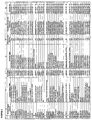
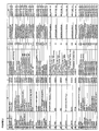
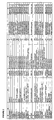
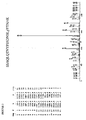
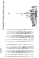
- phosphorylation sites correspond to numerous different parent proteins (the full sequences of which (human) are all publicly available in SwissProt database and their Accession numbers listed in Column C of Table 1/ Fig. 2 ), each of which fall into discrete protein type groups, for example Adaptor/Scaffold proteins, Chaperone proteins, Protein Kinases, and RNA Binding proteins, etc. (see Column D of Table 1), the phosphorylation of which is relevant to T-cell receptor signal transduction activity, as disclosed herein.
- the invention provides novel reagents -- phospho-specific antibodies and AQUA peptides -- for the specific detection and/or quantification of a T-cell receptor signaling protein/polypeptide only when phosphorylated (or only when not phosphorylated) at a particular phosphorylation site disclosed herein. Also provided are methods of detecting and/or quantifying one or more phosphorylated T-cell receptor signaling proteins using the phosphorylation-site specific antibodies and AQUA peptides.
- the specification provides an isolated phosphorylation site-specific antibody that specifically binds a given T-cell receptor signaling protein only when phosphorylated (or not phosphorylated, respectively) at a particular tyrosine enumerated in Column F of Table 1/ Figure 2 comprised within the phosphorylatable peptide site sequence enumerated in corresponding Column G.
- the specification provides a heavy-isotope labeled peptide (AQUA peptide) for the qua ntification of a given T-cell receptor signaling protein, the labeled peptide comprising a particular phosphorylatable peptide site/sequence enumerated in Column G of Table 1/ Figure 2 herein.
- the reagents provided by the invention is an isolated phosphorylation site-specific antibody that specifically binds the Cdk6 kinase (serine/threonine) only when phosphorylated (or only when not phosphorylated) at tyrosine 13 (see Row 61 (and Columns F and G) of Table 1/ Figure 2 ).
- the group of reagents provided by the invention is an AQUA peptide for the quantification of phosphorylated Cdk6 kinase, the AQUA peptide comprising the phosphorylatable peptide sequence listed in Column G, Row 61, of Table 1/ Figure 2 .
- the invention provides an isolated phosphorylation site-specific antibody that specifically binds a human T-cell receptor signaling protein corresponding to one or more of Rows 61 and 62 of Column A of Table 1 only when phosphorylated at the tyrosine listed in corresponding Column F of Table 1, comprised within the peptide sequence SEQ ID NO:60 or SEQ ID NO: 61 listed in corresponding Column G of Table 1, wherein said antibody does not bind said signaling protein when not phosphorylated at said tyrosine.
- the invention provides an isolated phosphorylation site-specific antibody that specifically binds a T-cell receptor signaling protein corresponding to one or more of Rows 61 and 62 of Column A of Table 1 only when not phosphorylated at the tyrosine listed in corresponding Column F of Table 1, comprised within the peptide sequence SEQ ID NO: 60 or SEQ ID NO: 61 listed In corresponding Column G of Table 1 , wherein said antibody does not bind said signaling protein when phosphorylated at said tyrosine.
- Such reagents enable the specific detection of phosphorylation (or non-phosphorylation) of a novel phosphorylatable site disclosed herein.
- the invention further provides immortalized cell lines producing such antibodies.
- the immortalized cell line is a rabbit or mouse hybridoma.
- the invention provides a heavy-isotope labeled peptide (AQUA peptide) for the quantification of a T-cell receptor signaling protein corresponding to one or more of Rows 61 and 62 of Column A of Table 1, said labeled peptide comprising the phosphorylatable peptide sequence SEQ ID NO: 60 or SEQ ID NO: 61 listed in corresponding Column G of Table 1, which sequence comprises the phosphorylatable tyrosine listed in corresponding Column F of Table 1.
- the phosphorylatable tyrosine within the labeled peptide is phosphorylated, while in other preferred embodiments, the phosphorylatable tyrosine within the labeled peptide is not phosphorylated.
- Reagents may conveniently be grouped by the type of T-cell receptor signaling protein in which a given phosphorylation site (for which reagents are provided) occurs.
- the protein types for each respective protein are provided in Column D of Table 1/ Figure 2 , and include: Actin Binding proteins, Adaptor/Scaffold proteins, Adhesion proteins, Calcium-binding proteins, Cell Cycle Regulation or Channel proteins, Chaperones, Cofactor proteins, Cytoskeletal proteins, DNA Binding proteins, G protein or GTPase Activating proteins, Ligases, Lipid Kinases and Binding proteins, Oxidoreductases, Protein Kinases, Protein Phosphatases, Receptor proteins, RNA Binding proteins, Transcription Factor/initiation Complex/Coactivator proteins, Translation Initiation Complex proteins, Ubitquitin Conjugating System proteins, and Vesicle proteins.
- the invention also provides, in part, an immortalized cell line producing an antibody of the Invention .
- the immortalized cell line is a rabbit hybridoma or a mouse hybridoma.
- a heavy-isotope labeled peptide (AQUA peptide) of the invention comprises a disclosed site sequence wherein the phosphorylatable tyrosine is phosphorylated. In certain other preferred embodiments, a heavy-isotope labeled peptide of the invention comprises a disclosed site sequence wherein the phosphorylatable tyrosine is not phosphorylated.
- Also provided by the invention are methods for detecting or quantifying a T-cell receptor signaling protein that is tyrosine-phosphorylated comprising the step of utilizing one or more of the above-described reagents of the invention to detect or quantify one or more T-cell receptor signaling protein(s) corresponding to one or more of Rows 61 and 62 of Column A of Table 1 only when phosphorylated at the tyrosine listed in corresponding Column F of Table 1.
- ELDSKLNyKPPPQKS SEQ ID NO: 46
- ARF GAP 3 Q9NP61 GTPase activating protein ARF 349 NDDSDDSyFTSSSSY SEQ ID NO: 47 49 centaurin-beta 2 Q15057 GTPase activating protein, ARF 750 GQPGDETyQDIFRDF SEQ ID NO: 48 50 GIT2 Q14161 GTPase activating protein, ARF 484 KQATTNVyQVQTGSE SEQ ID NO: 49 51 GIT2 Q14161 GTPase activating protein, ARF 492 QVQTGSEyTDTSNHS SEQ ID NO: 50 52 PPP1R11 060927 Inhibitor protein 64 SSKCCCIyEKPRAFG SEQ ID NO: 51 53 PIP5K Q9Y2I7 Kinase, lipid 1772 LRGADSAyYQVGQTG SEQ ID NO: 52 54 HYD 095071 Ligase, Ubiquitin conjug
- the 95 novel T-cell receptor signaling protein phosphorylation sites disclosed herein and listed in Table 1/ Figure 2 were discovered by employing the modified peptide isolation and characterization techniques described in described in "Immunoaffinity Isolation of Modified Peptides From Complex Mixtures," U.S. Patent Publication No. 20030044848, Rush et al . using cellular extracts from a Jurkat cell line in which the T-cell receptor signaling is constitutively activated.
- the isolation and identification of phosphopeptides from this T-cell line, using an immobilized general phosphotyrosine-specific antibody, is described in detail in Example 1 below.
- many known phosphorylation sites were also identified (not described herein).
- the immunoaffinity/mass spectrometric technique described in the '848 Patent Publication (the "IAP" method) -- and employed as described in detail in the Examples -- is briefly summarized below.
- the IAP method employed generally comprises the following steps: (a) a proteinaceous preparation (e.g. a digested cell extract) comprising phosphopeptides from two or more different proteins is obtained from an organism; (b) the preparation is contacted with at least one immobilized general phosphotyrosine-specific antibody; (c) at least one phosphopeptide specifically bound by the immobilized antibody in step (b) is isolated; and (d) the modified peptide isolated in step (c) is characterized by mass spectrometry (MS) and/or tandem mass spectrometry (MS-MS). Subsequently, (e) a search program (e.g.
- Sequest may be utilized to substantially match the spectra obtained for the isolated, modified peptide during the characterization of step (d) with the spectra for a known peptide sequence.
- a quantification step employing, e.g. SILAC or AQUA, may also be employed to quantify isolated peptides in order to compare peptide levels in a sample to a baseline.
- a general phosphotyrosine-specific monoclonal antibody (commercially available from Cell Signaling Technology, Inc., Beverly, MA, Cat #9411 (p-Tyr-100)) was used in the immunoaffinity step to isolate the widest possible number of phosphotyrosine containing peptides from the T-cell extracts.
- Extracts from a pervanadate-treated Jurkat cell line were employed. This established cell line is derived from patients with acute lymphoblastic leukemia and leukemic transformed non-Hodgkin lymphoma, in which T-cell receptor signaling pathways are constitutively activated.
- lysates were prepared from this cell line and digested with trypsin after treatment with DTT and iodoacetamide to alkylate cysteine residues.
- peptides were pre-fractionated by reversed-phase solid phase extraction using Sep-Pak C 18 columns to separate peptides from other cellular components.
- the solid phase extraction cartridges were eluted with varying steps of acetonitrile. Each lyophilized peptide fraction was redissolved in PBS and treated with phosphotyrosine antibody (P-Tyr-100, CST #9411) immobilized on protein G-Sepharose.
- Immunoaffinity-purified peptides were eluted with 0.1 % TFA and a portion of this fraction was concentrated with Stage tips and analyzed by LC-MS/MS, using a ThermoFinnigan LCQ Deca XP Plus ion trap mass spectrometer. Peptides were eluted from a 10 cm x 75 ⁇ m reversed-phase column with a 45-min linear gradient of acetonitrile. MS/MS spectra were evaluated using the program Sequest with the NCBI human protein database.
- Isolated phosphorylation site-specific antibodies that specifically bind a T-cell receptor signaling protein disclosed in Column A of Table 1 only when phosphorylated (or only when not phosphorylated) at the corresponding amino acid (tyrosine) and phosphorylation site listed in Columns F and G of Table 1 may now be produced by standard antibody production methods, such as anti-peptide antibody methods, using the phosphorylation site sequence information provided in Column G of Table 1. For example, two previously unknown Cdk6 kinase phosphorylation sites (tyrosines 13 and 24) (see Rows 61-62 of Table 1) are presently disclosed.
- antibodies that specifically bind any one of these novel Cdk6 sites can now be produced by using (all or part of) the amino acid sequence encompassing the respective phosphorylated residue as a peptide antigen used to immunize an animal (e.g. a peptide antigen comprising the sequence set forth in Row 61, Column G, of Table 1 (which encompasses the phosphorylated tyrosine at position 13 in Cdk6) may be employed to produce an antibody that only binds Cdk6 when phosphorylated at Tyr13).
- a peptide antigen comprising the sequence set forth in Row 61, Column G, of Table 1 (which encompasses the phosphorylated tyrosine at position 13 in Cdk6) may be employed to produce an antibody that only binds Cdk6 when phosphorylated at Tyr13).
- Polyclonal antibodies of the invention may be produced according to standard techniques by immunizing a suitable animal (e.g. , rabbit, goat, etc.) with a peptide antigen corresponding to the T-cell receptor protein phosphorylation site of interest (i.e. a phosphorylation site enumerated in Column G of Table 1, which comprises the corresponding phosphorylatable amino acid listed in Column F of Table 1), collecting immune serum from the animal, and separating the polyclonal antibodies from the immune serum, in accordance with known procedures.
- a suitable animal e.g. , rabbit, goat, etc.
- a peptide antigen corresponding to the T-cell receptor protein phosphorylation site of interest i.e. a phosphorylation site enumerated in Column G of Table 1, which comprises the corresponding phosphorylatable amino acid listed in Column F of Table 1
- a peptide comprising any of the phosphorylation site sequences provided in Column G of Table 1 may employed as an antigen to produce an antibody that only binds the corresponding protein listed in Column A of Table 1 when phosphorylated (or when not phosphorylated) at the corresponding residue listed in Column F. If an antibody that only binds the protein when phosphorylated at the disclosed site is desired, the peptide antigen includes the phosphorylated form of the amino acid. Conversely, if an antibody that only binds the protein when not phosphorylated at the disclosed site is desired, the peptide antigen includes the non-phosphorylated form of the amino acid.
- Peptide antigens suitable for producing antibodies of the invention may be designed, constructed and employed in accordance with well-known techniques. See, e.g. , ANTIBODIES: A LABORATORY MANUAL, Chapter 5, p. 75-76, Harlow & Lane Eds., Cold Spring Harbor Laboratory (1988 ); Czernik, Methods In Enzymology, 201: 264-283 (1991 ); Merrifield, J. Am. Chem. Soc. 85: 21-49 (1962 )).
- a peptide antigen may consist of the full sequence disclosed in Column G of Table 1, or it may comprise additional amino acids flanking such disclosed sequence, or may comprise of only a portion of the disclosed sequence immediately flanking the phosphorylatable amino acid (indicated in Column G by lowercase "y").
- Polyclonal antibodies produced as described herein may be screened as further described below.
- Monoclonal antibodies of the invention may be produced in a hybridoma cell line according to the well-known technique of Kohler and Milstein. Nature 265: 495-97 (1975 ); Kohler and Milstein, Eur. J. Immunol. 6: 511 (1976 ); see also, CURRENT PROTOCOLS IN MOLECULAR BIOLOGY, Ausubel et al. Eds. (1989 ). Monoclonal antibodies so produced are highly specific, and improve the selectivity and specificity of diagnostic assay methods provided by the invention. For example, a solution containing the appropriate antigen may be injected into a mouse or other species and, after a sufficient time (in keeping with conventional techniques), the animal is sacrificed and spleen cells obtained.
- the spleen cells are then immortalized by fusing them with myeloma cells, typically in the presence of polyethylene glycol, to produce hybridoma cells.
- Rabbit fusion hybridomas may be produced as described in U.S Patent No. 5,675,063, C. Knight, Issued October 7, 1997 .
- the hybridoma cells are then grown in a suitable selection media, such as hypoxanthine-aminopterin-thymidine (HAT), and the supernatant screened for monoclonal antibodies having the desired specificity, as described below.
- the secreted antibody may be recovered from tissue culture supernatant by conventional methods such as precipitation, ion exchange or affinity chromatography, or the like.
- Monoclonal Fab fragments may also be produced in Escherichia coli by recombinant techniques known to those skilled in the art. See, e.g., W. Huse, Science 246: 1275-81 (1989 ); Mullinax et al., Proc. Nat'l Acad. Sci. 87: 8095 (1990 ). If monoclonal antibodies of one isotype are preferred for a particular application, particular isotypes can be prepared directly, by selecting from the initial fusion, or prepared secondarily, from a parental hybridoma secreting a monoclonal antibody of different isotype by using the sib selection technique to isolate class-switch variants ( Steplewski, et al., Proc. Nat'l. Acad. Sci., 82: 8653 (1985 ); Spira et al., J. Immuno/. Methods, 74: 307 (1984 )).
- the preferred epitope of a phosphorylation-site specific antibody of the invention is a peptide fragment consisting essentially of about 8 to 17 amino acids including the phosphorylatable tyrosine, wherein about 3 to 8 amino acids are positioned on each side of the phosphorylatable tyrosine , and antibodies of the invention thus specifically bind a target T-cell receptor signaling polypeptide comprising such epitopic sequence.
- Particularly preferred epitopes bound by the antibodies of the invention comprise all or part of a phosphorylatable site sequence listed in Column G of Table 1, including the phosphorylatable amino acid (tyrosine).
- non-antibody molecules such as protein binding domains or nucleic acid aptamers, which bind, in a phospho-specific manner, to essentially the same phosphorylatable epitope to which the phospho-specific antibodies of the invention bind. See, e.g., Neuberger et al., Nature 312: 604 (1984 ). Such equivalent non-antibody reagents may be suitably employed in the methods further described below.
- Antibodies provided by the invention may be any type of immunoglobulins, including IgG, IgM, IgA, IgD, and IgE, including F ab or antigen-recognition fragments thereof.
- the antibodies may be monoclonal or polyclonal and may be of any species of origin, including (for example) mouse, rat, rabbit, horse, or human, or may be chimeric antibodies. See, e.g., M. Walker et al., Molec. Immuno/. 26: 403-11 (1989 ); Morrision et al., Proc. Nat'l. Acad. Sci. 81: 6851 (1984 ); Neuberger et al., Nature 312: 604 (1984 )).
- the antibodies may be recombinant monoclonal antibodies produced according to the methods disclosed in U.S. Pat. No. 4,474,893 (Reading ) or U.S. Pat. No. 4,816,567 (Cabilly et al. )
- the antibodies may also be chemically constructed by specific antibodies made according to the method disclosed in U.S. Pat. No. 4,676,980 (Segel et al .)
- the invention also provides immortalized cell lines that produce an antibody of the invention.
- hybridoma clones constructed as described above, that produce monoclonal antibodies to the T-cell receptor signaling protein phosphorylation sitess disclosed herein are also provided.
- the invention includes recombinant cells producing an antibody of the invention, which cells may be constructed by well known techniques; for example the antigen combining site of the monoclonal antibody can be cloned by PCR and single-chain antibodies produced as phage-displayed recombinant antibodies or soluble antibodies in E . coli ( see, e.g., ANTIBODY ENGINEERING PROTOCOLS, 1995, Humana Press, Sudhir Paul editor.)
- Phosphorylation site-specific antibodies of the invention may be screened for epitope and phospho-specificity according to standard techniques. See , e.g. Czernik et al., Methods in Enzymology, 201: 264-283 (1991 ).
- the antibodies may be screened against the phospho and non-phospho peptide library by ELISA to ensure specificity for both the desired antigen (i.e . that epitope including a phosphorylation site sequence enumerated in Column G of Table 1) and for reactivity only with the phosphorylated (or non-phosphorylated) form of the antigen.
- Peptide competition assays may be carried out to confirm lack of reactivity with other phosphoepitopes on the given T-cell receptor signaling protein.
- the antibodies may also be tested by Western blotting against cell preparations containing the signaling protein, e . g . cell lines over-expressing the target protein, to confirm reactivity with the desired phosphorylated epitope/target.
- Phosphorylation-site specific antibodies of the invention may exhibit some limited cross-reactivity related epitopes in non-target proteins. This is not unexpected as most antibodies exhibit some degree of cross-reactivity, and anti-peptide antibodies will often cross-react with epitopes having high homology to the immunizing peptide. See , e.g ., Czernik , supra . Cross-reactivity with non-target proteins is readily characterized by Western blotting alongside markers of known molecular weight.
- Amino acid sequences of cross-reacting proteins may be examined to identify sites highly homologous to the T-cell receptor signaling protein epitope for which the antibody of the invention is specific.
- polyclonal antisera may be exhibit some undesirable general cross-reactivity to phosphotyrosine, which may be removed by further purification of antisera, e . g . over a phosphotyramine column.
- Antibodies of the invention specifically bind their target protein ( i . e .
- Antibodies may be further characterized via immunohistochemical (IHC) staining using normal and diseased tissues to examine T-cell receptor phosphorylation and activation status in diseased tissue.
- IHC immunohistochemical
- IHC may be carried out according to well-known techniques. See , e.g ., ANTIBODIES: A LABORATORY MANUAL, Chapter 10, Harlow & Lane Eds., Cold Spring Harbor Laboratory (1988 ). Briefly, paraffin-embedded tissue ( e.g .
- tumor tissue is prepared for immunohistochemical staining by deparaffinizing tissue sections with xylene followed by ethanol; hydrating in water then PBS; unmasking antigen by heating slide in sodium citrate buffer; incubating sections in hydrogen peroxide; blocking in blocking solution; incubating slide in primary antibody and secondary antibody; and finally detecting using ABC avidin/biotin method according to manufacturer's instructions.
- Antibodies may be further characterized by flow cytometry carried out according to standard methods. See Chow et al., Cytometry (Communications in Clinical Cytometry) 46: 72-78 (2001 ). Briefly and by way of example, the following protocol for cytometric analysis may be employed: samples may be centrifuged on Ficoll gradients to remove erythrocytes, and cells may then be fixed with 2% paraformaldehyde for 10 minutes at 37°C followed by permeabilization in 90% methanol for 30 minutes on ice.
- Cells may then be stained with the primary phosphorylation-site specific antibody of the invention (which detects an T-cell receptor signal transduction protein enumerated in Table 1), washed and labeled with a fluorescent-labeled secondary antibody. Additional fluorochrome-conjugated marker antibodies (e . g . CD45, CD34) may also be added at this time to aid in the subsequent identification of specific hematopoietic cell types. The cells would then be analyzed on a flow cytometer ( e . g . a Beckman Coulter FC500) according to the specific protocols of the instrument used.
- a flow cytometer e . g . a Beckman Coulter FC500
- Antibodies of the invention may also be advantageously conjugated to fluorescent dyes (e . g . Alexa488, PE) for use in multiparametric analyses along with other signal transduction (phospho-CrkL, phospho-Erk 1/2) and/or cell marker (CD34) antibodies.
- fluorescent dyes e . g . Alexa488, PE
- phospho-CrkL, phospho-Erk 1/2 signal transduction
- CD34 cell marker
- Phosphorylation-site specific antibodies of the invention specifically bind to a human T-cell receptor signal transduction protein or polypeptide only when phosphorylated at a disclosed site, but are not limited only to binding the human species, per se .
- the invention includes antibodies that also bind conserved and highly-homologous or identical phosphorylation sites in respective T-cell receptor signaling proteins from other species ( e . g . mouse, rat, monkey, yeast), in addition to binding the human phosphorylation site. Highly-homologous sites conserved in other species can readily be identified by standard sequence comparisons, such as using BLAST, with the human T-cell receptor signal transduction protein phosphorylation sites disclosed herein.
- novel T-cell receptor signaling protein phosphorylation sites disclosed herein now enable the production of corresponding heavy-isotope labeled peptides for the absolute quantification of such signaling proteins (both phosphorylated and not phosphorylated at a disclosed site) in biological samples.
- the production and use of AQUA peptides for the absolute quantification of proteins (AQUA) in complex mixtures has been described. See WO/03016861 , "Absolute Quantification of Proteins and Modified Forms Thereof by Multistage Mass Spectrometry," Gygi et al . and also Gerber et al. Proc. Natl. Acad. Sci. U.S.A. 100: 6940-5 (2003 ).
- the AQUA methodology employs the introduction of a known quantity of at least one heavy-isotope labeled peptide standard (which has a unique signature detectable by LC-SRM chromatography) into a digested biological sample in order to determine, by comparison to the peptide standard, the absolute quantity of a peptide with the same sequence and protein modification in the biological sample.
- the AQUA methodology has two stages: peptide Internal standard selection and validation and method development; and implementation using validated peptide internal standards to detect and quantify a target protein in sample.
- the method is a powerful technique for detecting and quantifying a given peptide/protein within a complex biological mixture, such as a cell lysate, and may be employed, e . g ., to quantify change in protein phosphorylation as a result of drug treatment, or to quantify differences in the level of a protein in different biological states.
- a particular peptide (or modified peptide) within a target protein sequence is chosen based on its amino acid sequence and the particular protease to be used to digest.
- the peptide is then generated by solid-phase peptide synthesis such that one residue is replaced with that same residue containing stable isotopes ( 13 C, 15 N).
- the result is a peptide that is chemically identical to its native counterpart formed by proteolysis, but is easily distinguishable by MS via a 7-Da mass shift.
- the newly synthesized AQUA internal standard peptide is then evaluated by LC-MS/MS. This process provides qualitative information about peptide retention by reverse-phase chromatography, ionization efficiency, and fragmentation via collision-induced dissociation. Informative and abundant fragment ions for sets of native and internal standard peptides are chosen and then specifically monitored in rapid succession as a function of chromatographic retention to form a selected reaction monitoring (LC-SRM) method based on the unique profile of the peptide standard.
- the second stage of the AQUA strategy is its implementation to measure the amount of a protein or modified protein from complex mixtures.
- Whole cell lysates are typically fractionated by SDS-PAGE gel electrophoresis, and regions of the gel consistent with protein migration are excised. This process is followed by in-gel proteolysis in the presence of the AQUA peptides and LC-SRM analysis.
- AQUA peptides are spiked in to the complex peptide mixture obtained by digestion of the whole cell lysate with a proteolytic enzyme and subjected to immunoaffinity purification as described above.
- the retention time and fragmentation pattern of the native peptide formed by digestion e.g.
- trypsinization is identical to that of the AQUA internal standard peptide determined previously; thus, LC-MS/MS analysis using an SRM experiment results in the highly specific and sensitive measurement of both internal standard and analyte directly from extremely complex peptide mixtures. Because an absolute amount of the AQUA peptide is added ( e . g . 250 fmol), the ratio of the areas under the curve can be used to determine the precise expression levels of a protein or phosphorylated form of a protein in the original cell lysate.
- the internal standard is present du ring in-gel digestion as native peptides are formed, such that peptide extraction efficiency from gel pieces, absolute losses during sample handling (including vacuum centrifugation), and variability during introduction into the LC-MS system do not affect the determined ratio of native and AQUA peptide abundances.
- An AQUA peptide standard is developed for a known phosphorylation site sequence previously identified by the IAP-LC-MS/MS method within in a target protein.
- One AQUA peptide incorporating the phosphorylated form of the particular residue within the site may be developed, and a second AQUA peptide incorporating the non-phosphorylated form of the residue developed.
- the two standards may be used to detect and quantify both the phosphorylated and non-phosphorylated forms of the site in a biological sample.
- Peptide internal standards may also be generated by examining the primary amino acid sequence of a protein and determining the boundaries of peptides produced by protease cleavage. Alternatively, a protein may actually be digested with a protease and a particular peptide fragment produced can then sequenced. Suitable proteases include, but are not limited to, serine proteases (e . g . trypsin, hepsin), metallo proteases ( e . g . PUMP1), chymotrypsin, cathepsin, pepsin, thermolysin, carboxypeptidases, etc.
- serine proteases e . g . trypsin, hepsin
- metallo proteases e . g . PUMP1
- chymotrypsin cathepsin
- pepsin pepsin
- thermolysin carboxypeptidases
- a peptide sequence within a target protein is selected according to one or more criteria to optimize the use of the peptide as an internal standard.
- the size of the peptide is selected to minimize the chances that the peptide sequence will be repeated elsewhere in other non-target proteins.
- a peptide is preferably at least about 6 amino acids.
- the size of the peptide is also optimized to maximize ionization frequency.
- peptides longer than about 20 amino acids are not preferred.
- the preferred ranged is about 7 to 15 amino acids.
- a peptide sequence is also selected that is not likely to be chemically reactive during mass spectrometry, thus sequences comprising cysteine, tryptophan, or methionine are avoided.
- a peptide sequence that does not include a modified region of the target region may be selected so that the peptide internal standard can be used to determine the quantity of all forms of the protein.
- a peptide internal standard encompassing a modified amino acid may be desirable to detect and quantify only the modified form of the target protein.
- Peptide standards for both modified and unmodified regions can be used together, to determine the extent of a modification in a particular sample ( i . e . to determine what fraction of the total amount of protein is represented by the modified form).
- peptide standards for both the phosphorylated and unphosphorylated form of a protein known to be phosphorylated at a particular site can be used to quantify the amount of phosphorylated form in a sample.
- the peptide is labeled using one or more labeled amino acids ( i . e . the label is an actual part of the peptide) or less preferably, labels may be attached after synthesis according to standard methods.
- the label is a mass-altering label selected based on the following considerations: The mass should be unique to shift fragments masses produced by MS analysis to regions of the spectrum with low background; the ion mass signature component is the portion of the labeling moiety that preferably exhibits a unique ion mass signature in MS analysis; the sum of the masses of the constituent atoms of the label is preferably uniquely different than the fragments of all the possible amino acids.
- the labeled amino acids and peptides are readily distinguished from unlabeled ones by the ion/mass pattern in the resulting mass spectrum.
- the ion mass signature component imparts a mass to a protein fragment that does not match the residue mass for any of the 20 natural amino acids.
- the label should be robust under the fragmentation conditions of MS and not undergo unfavorable fragmentation. Labeling chemistry should be efficient under a range of conditions, particularly denaturing conditions, and the labeled tag preferably remains soluble in the MS buffer system of choice.
- the label preferably does not suppress the ionization efficiency of the protein and is not chemically reactive.
- the label may contain a mixture of two or more isotopically distinct species to generate a unique mass spectrometric pattern at each labeled fragment position. Stable isotopes, such as 2 H, 13 C, 15 N, 17 O, 18 O, or 34 S, are among preferred labels. Pairs of peptide internal standards that incorporate a different isotope label may also be prepared. Preferred amino acid residues into which a heavy isotope label may be incorporated include leucine, proline, valine, and phenylalanine.
- Peptide internal standards are characterized according to their mass-to-charge (m/z) ratio, and preferably, also according to their retention time on a chromatographic column (e.g. an HPLC column). Internal standards that co-elute with unlabeled peptides of identical sequence are selected as optimal internal standards.
- the internal standard is then analyzed by fragmenting the peptide by any suitable means, for example by collision-induced dissociation (CID) using, e.g., argon or helium as a collision gas.
- CID collision-induced dissociation
- the fragments are then analyzed, for example by multi-stage mass spectrometry (MS n ) to obtain a fragment ion spectrum, to obtain a peptide fragmentation signature.
- MS n multi-stage mass spectrometry
- peptide fragments have significant differences in m/z ratios to enable peaks corresponding to each fragment to be well separated, and a signature is that is unique for the target peptide is obtained. If a suitable fragment signature is not obtained at the first stage, additional stages of MS are performed until a unique signature is obtained.
- Fragment ions in the MS/MS and MS 3 spectra are typically highly specific for the peptide of interest, and, in conjunction with LC methods, allow a highly selective means of detecting and quantifying a target peptide/protein in a complex protein mixture, such as a cell lysate, containing many thousands or tens of thousands of proteins.
- a complex protein mixture such as a cell lysate, containing many thousands or tens of thousands of proteins.
- Any biological sample potentially containing a target protein/peptide of interest may be assayed. Crude or partially purified cell extracts are preferably employed.
- the sample has at least 0.01 mg of protein, typically a concentration of 0.1-10 mg/mL, and may be adjusted to a desired buffer concentration and pH.
- a known amount of a labeled peptide internal standard, preferably about 10 femtomoles, corresponding to a target protein to be detected/quantified is then added to a biological sample, such as a cell lysate.
- the spiked sample is then digested with one or more protease(s) for a suitable time period to allow digestion.
- a separation is then performed (e.g. by HPLC, reverse-phase HPLC, capillary electrophoresis, ion exchange chromatography, etc.) to isolate the labeled internal standard and its corresponding target peptide from other peptides in the sample.
- Microcapillary LC is a preferred method.
- Each isolated peptide is then examined by monitoring of a selected reaction in the MS. This involves using the prior knowledge gained by the characterization of the peptide internal standard and then requiring the MS to continuously monitor a specific ion in the MS/MS or MS n spectrum for both the peptide of interest and the internal standard. After elution, the area under the curve (AUC) for both peptide standard and target peptide peaks are calculated. The ratio, of the two areas provides the absolute quantification that can be normalized for the number of cells used in the analysis and the protein's molecular weight, to provide the precise number of copies of the protein per cell. Further details of the AQUA methodology are described in Gygi et al., and Gerber et al. supra .
- AQUA internal peptide standards may now be produced, as described above, for the novel T-cell receptor signaling protein phosphorylation sites disclosed herein (see Table 1/ Figure 2 ).
- Peptide standards for a given phosphorylation site may be produced for both the phosphorylated and non-phosphorylated forms of the site and such standards employed in the AQUA methodology to detect and quantify both forms of such phosphorylation site in a biological sample.
- the phosphorylation site peptide sequences disclosed herein are particularly well suited for development of corresponding AQUA peptides, since the IAP method by which they were identified (see Part A above and Example 1) inherently confirmed that such peptides are in fact produced by enzymatic digestion (trypsinization) and are in fact suitably fractionated/ionized in MS/MS.
- heavy-isotope labeled equivalents of these peptides can be readily synthesized and their unique MS and LC-SRM signature determined, so that the peptides are validated as AQUA peptides and ready for use in quantification experiments.
- the invention provides heavy-isotope labeled peptides (AQUA peptides) for the detection and/or quantification of any of the T-cell receptor signaling protein phosphorylation sites disclosed in rows 61 or 62 of Table 1/ Figure2 (see Column G) and/or their corresponding parent protein/polypeptide (see Column A).
- Each such phosphorylation sequence may be considered a preferred AQUA peptide of the invention.
- an AQUA peptide of the invention consists of a phosphorylation site sequence enumerated in Table 1.
- AQUA peptide comprising the disclosed phosphorylation site sequence (and additional residues downstream or upstream of it) may also be constructed.
- a smaller AQUA peptide comprising less than all of the residues of a disclosed phosphorylation site sequence (but still comprising the phosphorylatable residue enumerated in Column F of Table 1/ Figure 2 ) may alternatively be constructed.
- Such larger AQUA peptides are within the scope of the present invention, and the selection and production of preferred AQUA peptides may be carried out as described above (see Gygi et al., Gerber et al. supra .).
- AQUA peptides are described above (corresponding to particular protein types/groups in Table 1, for example, Adaptor/Scaffold proteins or RNA Binding Proteins).
- Example 4 is provided to further illustrate the construction and use, by standard methods described above, of exemplary AQUA peptides.
- AQUA peptides of the invention may also be employed within a kit that comprises one or multiple AQUA peptide(s) provided herein (for the quantification of an T-cell receptor signal transduction protein disclosed in Table 1), and, optionally, a second detecting reagent conjugated to a detectable group.
- a kit may include AQUA peptides for both the phosphorylation and non-phosphorylated form of a phosphorylation site disclosed herein.
- the reagents may also include ancillary agents such as buffering agents and protein stabilizing agents, e . g ., polysaccharides and the like.
- the kit may further include, where necessary, other members of the signal-producing system of which system the detectable group is a member ( e . g ., enzyme substrates), agents for reducing background interference in a test, control reagents, apparatus for conducting a test, and the like.
- the test kit may be packaged In any suitable manner, typically with all elements in a single container along with a sheet of printed instructions for carrying out the test.
- AQUA peptides provided by the invention will be highly useful in the further study of signal transduction anomalies underlying diseases, including lymphomas, involving altered T-cell receptor signaling, and in identifying diagnostic/bio-markers of these diseases, new potential drug targets, and/or in monitoring the effects of test compounds on T-cell receptor signal transduction proteins and pathways.
- Antibodies provided by the invention may be advantageously employed in a variety of standard immunological assays (the use of AQUA peptides provided by the invention is described separately above). Assays may be homogeneous assays or heterogeneous assays. In a homogeneous assay the immunological reaction usually involves a phosphorylation-site specific antibody of the invention, a labeled analyte, and the sample of interest. The signal arising from the label is modified, directly or indirectly, upon the binding of the antibody to the labeled analyte. Both the immunological reaction and detection of the extent thereof are carried out in a homogeneous solution. Immunochemical labels that may be employed include free radicals, radioisotopes, fluorescent dyes, enzymes, bacteriophages, coenzymes, and so forth.
- the reagents are usually the specimen, a phosphorylation-site specific antibody of the invention, and suitable means for producing a detectable signal. Similar specimens as described above may be used.
- the antibody is generally immobilized on a support, such as a bead, plate or slide, and contacted with the specimen suspected of containing the antigen in a liquid phase.
- the support is then separated from the liquid phase and either the support phase or the liquid phase is examined for a detectable signal employing means for producing such signal.
- the signal is related to the presence of the analyte in the specimen.
- Means for producing a detectable signal include the use of radioactive labels, fluorescent labels, enzyme labels, and so forth.
- an antibody which binds to that site can be conjugated to a detectable group and added to the liquid phase reaction solution before the separation step.
- the presence of the detectable group on the solid support indicates the presence of the antigen in the test sample.
- suitable immunoassays are the radioimmunoassay, immunofluorescence methods, enzyme-linked immunoassays, and the like.
- Immunoassay formats and variations thereof that may be useful for carrying out the methods disclosed herein are well known in the art. See generally E. Maggio, Enzyme-Immunoassay, (1980) (CRC Press, Inc., Boca Raton, F la.); see also, e.g ., U.S. Pat. No. 4,727,022 (Skold et al ., “Methods for Modulating Ligand-Receptor Interactions and their Application”); U.S. Pat. No. 4,659,678 (Forrest et al ., "Immunoassay of Antigens"); U.S. Pat. No.
- Phosphorylation site-specific antibodies disclosed herein may be conjugated to a solid support suitable for a diagnostic assay (e . g ., beads, plates, slides or wells formed from materials such as latex or polystyrene) in accordance with known techniques, such as precipitation.
- Antibodies, or other target protein or target site-binding reagents may likewise be conjugated to detectable groups such as radiolabels ( e . g ., 35 S, 125 I, 131 I), enzyme labels ( e . g ., horseradish peroxidase, alkaline phosphatase), and fluorescent labels ( e . g ., fluorescein) in accordance with known techniques.
- Antibodies of the invention may also be optimized for use in a flow cytometry assay to determine the activation/phosphorylation status of a target T-cell receptor signaling protein in patients before, during, and after treatment with a drug targeted at inhibiting phosphorylation at such a protein at the phosphorylation site disclosed herein.
- a drug targeted at inhibiting phosphorylation at such a protein at the phosphorylation site disclosed herein For example, bone marrow cells or peripheral blood cells from patients may be analyzed by flow cytometry for target T-cell receptor signaling protein phosphorylation, as well as for markers identifying various hematopoietic cell types. In this manner, activation status of the malignant cells may be specifically characterized.
- Flow cytometry may be carried out according to standard methods. See, e.g.
- antibodies of the invention may be employed in immunohistochemical (IHC) staining to detect differences in signal transduction or protein activity using normal and diseased tissues.
- IHC may be carried out according to well-known techniques. See, e.g., ANTIBODIES: A LABORATORY MANUAL, supra. Briefly, paraffin-embedded tissue ( e . g .
- tumor tissue is prepared for immunohistochemical staining by deparaffinizing tissue sections with xylene followed by ethanol; hydrating in water then PBS; unmasking antigen by heating slide in sodium citrate buffer; incubating sections in hydrogen peroxide; blocking in blocking solution; incubating slide in primary antibody and secondary antibody; and finally detecting using ABC avidin/biotin method according to manufacturer's instructions.
- Antibodies of the invention may be also be optimized for use in other clinically-suitable applications, for example bead-based multiplex-type assays, such as IGEN, Luminex TM and/or Bioplex TM assay formats, or otherwise optimized for antibody arrays formats, such as reversed-phase array applications ( see , e.g. Paweletz et al., Oncogene 20(16): 1981-89 (2001 )).
- bead-based multiplex-type assays such as IGEN, Luminex TM and/or Bioplex TM assay formats
- antibody arrays formats such as reversed-phase array applications
- a method for the multiplex detection of T-cell receptor signaling protein phosphorylation in a biological sample comprising utilizing at two antibodies or AQUA peptides to detect the presence of two phosphorylated T-cell receptor signaling proteins enumerated in Column A of Table 1/ Figure 2 .
- two to five antibodies or AQUA peptides are employed in the method.
- six to ten antibodies or AQUA peptides are employed, while in another preferred embodiment eleven to twenty such reagents are employed.
- Antibodies and/or AQUA peptides of the invention may also be employed within a kit that comprises at least one phosphorylation site-specific antibody or AQUA peptide of the invention (which binds to or detects an T-cell receptor signaling protein/site disclosed in Table 1), and, optionally, a second antibody conjugated to a detectable group.
- the kit is suitable for multiplex assays and comprises two or more antibodies or AQUA peptides, and in some embodiments, comprises two to five, six to ten, or eleven to twenty reagents.
- the kit may also include ancillary agents such as buffering agents and protein stabilizing agents, e.g ., polysaccharides and the like.
- the kit may further include, where necessary, other members of the signal-producing system of which system the detectable group is a member ( e . g ., enzyme substrates), agents for reducing background interference in a test, control reagents, apparatus for conducting a test, and the like.
- the test kit may be packaged in any suitable manner, typically with all elements in a single container along with a sheet of printed instructions for carrying out the test.
- IAP isolation techniques were employed to identify phosphotyrosine-containing peptides in cell extracts from Jurkat cells treated with pervanadate in order to stimulate tyrosine phosphorylation.
- Tryptic phosphotyrosine peptides were purified and analyzed from extracts of the Jurkat cell line as follows. Cells were cultured in RPMI medium supplemented with 10% bovine serum and penicillin/streptomycin. Cells were cultured to a density of 1.2 x 10 6 cells/ml and were washed in PBS at room temperature, then resuspended in PBS at 7 x 10 7 cells/ml. After preincubation at 37° C for 20 min, calyculin A and sodium pervanadate were added to final concentrations of 50 ng/ml and 1 mM, respectively, and cells were incubated for 20 min at 37° C.
- Sonicated cell lysates were cleared by centrifugation at 20,000 x g, and proteins were reduced with DTT at a final concentration of 4.1 mM and alkylated with iodoacetamide at 8.3 mM.
- protein extracts were diluted in 20 mM HEPES pH 8.0 to a final concentration of 2 M urea and immobilized TLCK-trypsin (Pierce) was added at 1-2.5 ml beads (200 TAME units trypsin/ml) per 10 9 cells. Digestion was performed for 1-2 days at room temperature.
- Trifluoroacetic acid was added to protein digests to a final concentration of 1%, precipitate was removed by centrifugation, and digests were loaded onto Sep-Pak C 18 columns (Waters) equilibrated with 0.1 % TFA. A column volume of 0.7-1.0 ml was used per 2 x 10 8 cells. Columns were washed with 15 volumes of 0.1 % TFA, followed by 4 volumes of 5% acetonitrile (MeCN) in 0.1% TFA. Peptide fraction I was obtained by eluting columns with 2 volumes each of 8, 12, and 15% MeCN in 0.1% TFA and combining the eluates. Fractions II and III were a combination of eluates after eluting columns with 18, 22, 25% MeCN in 0.1% TFA and with 30, 35, 40% MeCN in 0.1% TFA, respectively. All peptide fractions were lyophilized.
- MeCN acetonitrile
- Peptides from each fraction corresponding to 2 x 10 8 cells were dissolved in 1 ml of IAP buffer (20 mM Tris/HCl or 50 mM MOPS pH 7.2, 10 mM sodium phosphate, 50 mM NaCl) and insoluble matter (mainly in peptide fractions III) was removed by centrifugation. IAP was performed on each peptide fraction separately.
- the phosphotyrosine monoclonal antibody P-Tyr-100 (Cell Signaling Technology, Inc., catalog number 9411) was coupled at 4 mg/ml beads to protein G agarose (Roche).
- Immobilized antibody (15 ⁇ l, 60 ⁇ g) was added as 1:1 slurry in lAP buffer to 1 ml of each peptide fraction, and the mixture was incubated overnight at 4° C with gentle rotation.
- the immobilized antibody beads were washed three times with 1 ml IAP buffer and twice with 1 ml water, all at 4° C.
- Peptides were eluted from beads by incubation with 75 ⁇ l of 0.1 % TFA at room temperature for 10 min.
- a thin layer of a-cyano-4-hydroxy-cinnamic acid (ACHA) matrix was applied to a Bruker 384-spot MALDI target by spreading 5 ⁇ l of a saturated solution in MeCN/water (2/1, v/v) over an entire row of spots on the target; drying occurred in 2-5 sec.
- the lAP eluate (10 ⁇ l) was loaded onto an 0.2 ⁇ l C-18 ZipTip (Millipore), which then was washed with 5% formic acid.
- Peptide was eluted with 1 ⁇ l of 10 mg/ml ACHA in 60% methanol, 5% formic acid onto the MALDI target containing the thin layer of matrix. Samples were analyzed on a Bruker BiFlex III MALDI-TOF instrument in positive ion mode.
- MS/MS spectra were evaluated using TurboSequest in the Sequest Browser package (v. 27, rev. 12) supplied as part of BioWorks 3.0 (ThermoFinnigan). Individual MS/MS spectra were extracted from the raw data file using the Sequest Browser program CreateDta, with the following settings: bottom MW, 700; top MW, 4,500; minimum number of ions, 20; minimum TIC, 4x10 5 ; and precursor charge state, unspecified. Spectra were extracted from the beginning of the raw data file before sample injection to the end of the eluting gradient. The lonQuest and VuDta programs were not used to further select MS/MS spectra for Sequest analysis.
- MS/MS spectra were evaluated with the following TurboSequest parameters: peptide mass tolerance, 2.5; fragment ion tolerance, 0.0; maximum number of differential amino acids per modification, 4; mass type parent, average; mass type fragment, average; maximum number of internal cleavage sites, 10; neutral losses of water and ammonia from b and y ions were considered in the correlation analysis.
- Proteolytic enzyme was specified except for spectra collected from elastase digests.
- Assignments in this subset were rejected if any of the following criteria were satisfied: (i) the spectrum contained at least one major peak (at least 10% as intense as the most intense ion in the spectrum) that could not be mapped to the assigned sequence as an a , b , or y ion, as an ion arising from neutral-loss of water or ammonia from a b or y ion, or as a multiply protonated ion; (ii) the spectrum did not contain an series of b or y ions equivalent to at least six uninterrupted residues; or (iii) the sequence was not observed at least five times in all the studies we have conducted (except for overlapping sequences due to incomplete proteolysis or use of proteases oth er than trypsin).
- Polyclonal antibodies that specifically bind a T-cell receptor signal transduction protein only when phosphorylated at the respective phosphorylation site disclosed herein are produced according to standard methods by first constructing a synthetic peptide antigen comprising the phosphorylation site sequence and then immunizing an animal to raise antibodies against the antigen, as further described below. Production of exemplary polyclonal antibodies is provided below.
- a 15 amino acid phospho-peptide antigen, AEIGEGAy*GKVFKAR (SEQ ID NO: 61) (where y* phosphotyrosine), that corresponds to the tyrosine 24 phosphorylation site in human Cdk6 kinase (see Row 62 of Table 1), plus cysteine on the C-terminal for coupling, is constructed according to standard synthesis techniques using, e . g ., a Rainin/Protein Technologies, Inc., Symphony peptide synthesizer. See ANTIBODIES: A LABORATORY MANUAL, supra.; Merrifield, supra. This peptide is then coupled to KLH and used to immunize animals to produce (and subsequently screen) phospho-specific Cdk6 (Tyr24) polyclonal antibodies as described in Immunization/ Screening below.
- a 15 amino acid phospho-peptide antigen, LKADGLIy*CLKEACP (SEQ ID NO: 63) (where y* phosphotyrosine), that corresponds to the tyrosine 248 phosphorylation site in human ZAP70 kinase (see Row 64 of Table 1), plus cysteine on the C-terminal for coupling, is constructed according to standard synthesis techniques using, e . g ., a Rainin/Protein Technologies, Inc., Symphony peptide synthesizer. See ANTIBODIES: A LABORATORY MANUAL, supra.; Merrifield, supra. This peptide is then coupled to KLH and used to immunize animals to produce (and subsequently screen) phospho-specific ZAP70 (Tyr248) polyclonal antibodies as described in Immunization/Screening below.
- a 15 amino acid phospho-peptide antigen, PLYGNLHy*LQTGRLS (SEQ ID NO: 12) (where y* phosphotyrosine) that corresponds to the tyrosine 95 phosphorylation site in human SIT protein (see Row 13 of Table 1), plus cysteine on the C-terminal for coupling, is constructed according to standard synthesis techniques using, e . g ., a Rainin/Protein Technologies, Inc., Symphony peptide synthesizer. See ANTIBODIES: A LABORATORY MANUAL, supra.; Merrifield, supra. This peptide is then coupled to KLH and used to immunize animals to produce (and subsequently screen) phospho-specific SIT (Tyr95) antibodies as described in Immunization/Screening below.
- a synthetic phospho-peptide antigen as described in A-C above is coupled to KLH, and rabbits are injected intradermally (ID) on the back with antigen in complete Freunds adjuvant (500 ⁇ g antigen per rabbit). The rabbits are boosted with same antigen in incomplete Freund adjuvant (250 ⁇ g antigen per rabbit) every three weeks. After the fifth boost, bleeds are collected. The sera are purified by Protein A-affinity chromatography by standard methods (see ANTIBODIES: A LABORATORY MANUAL, Cold Spring Harbor, supra .).
- the eluted immunoglobulins are further loaded onto a non-phosphorylated synthetic peptide antigen-resin Knotes column to pull out antibodies that bind the non-phosphorylated form of the phosphorylation site.
- the flow through fraction is collected and applied onto a phospho-synthetic peptide antigen-resin column to isolate antibodies that bind the phosphorylated form of the site.
- the bound antibodies i . e . antibodies that bind a phosphorylated peptide described in A-C above, but do not bind the non-phosphorylated form of the peptide, are eluted and kept in antibody storage buffer.
- the isolated antibody is then tested for phospho-specificity using Western blot assay using an appropriate cell line the expresses (or overexpresses) target phospho-protein (i . e . phosphorylated Cdk6, ZAP70, or SIT), for example, Jurkat cells.
- Cells are cultured in RPMI medium supplemented with 10% FCS and penicillin/streptomycin. Before stimulation, the cells are starved in serum-free RPMI medium for 4 hours. The cells are then stimulated with ligand ( e . g . 50 ng/ml) for 5 minutes. Cell are collected, washed with PBS and directly lysed in cell lysis buffer. The protein concentration of cell lysates are then measured. The loading buffer is added into cell lysate and the mixture is boiled at 100°C for 5 minutes. 20 ⁇ l (10 ⁇ g protein) of sample is then added onto 7.5% SDS-PAGE gel.
- a standard Western blot may be performed according to the lmmunoblotting Protocol set out in the CELL SIGNALING TECHNOLOGY, INC. 2003-04 Catalogue, p. 390.
- the isolated phospho-specific antibody is used at dilution 1:1000. Phosphorylation-site specificity of the antibody will be shown by binding of only the phosphorylated form of the target protein.
- Isolated phospho-specific polyclonal antibody does not recognize the target protein when not phosphorylated at the appropriate phosphorylation site in the non-stimulated cells (e.g . ZAP70 is not bound when not phosphorylated at tyrosine 248).
- Monoclonal antibodies that specifically bind a T-cell receptor signal transduction protein only when phosphorylated at the respective phosphorylation site disclosed herein are produced according to standard methods by first constructing a synthetic peptide antigen comprising the phosphorylation site sequence and then immunizing an animal to raise antibodies against the antigen, and harvesting spleen cells from such animals to produce fusion hybridomas, as further described below. Production of exemplary monoclonal antibodies is provided below.
- This peptide is then coupled to KLH and used to immunize animals and harvest spleen cells for generation (and subsequent screening) of phospho-specific monoclonal Cdk6 (Tyr13) antibodies as described in Immunization/Fusion/Screening below.
- GQRAQENy*EGSEEVS SEQ ID NO: 59
- y* phosphotyrosine
- y* phosphotyrosine
- This peptide is then coupled to KLH and used to immunize animals and harvest spleen cells for generation (and subsequent screening) of phospho-specific monoclonal FAF-X (Tyr2533) antibodies as described in Immunization/Fusion/Screening below.
- This peptide is then coupled to KLH and used to immunize animals and harvest spleen cells for generation (and subsequent screening) of phospho-specific monoclonal Cortactin-a (Tyr453) antibodies as described in Immunization/ Fusion/Screening below.
- a synthetic phospho-peptide antigen as described in A-C above is coupled to KLH, and BALB/C mice are injected intradermally (ID) on the back with antigen in complete Freunds adjuvant (e . g . 50 ⁇ g antigen per mouse). The mice are boosted with same antigen in incomplete Freund adjuvant ( e . g . 25 ⁇ g antigen per mouse) every three weeks. After the fifth boost, the animals are sacrificed and spleens are harvested.
- Harvested spleen cells are fused to SP2/0 mouse myeloma fusion partner cells according to the standard protocol of Kohler and Milstein (1975). Colonies originating from the fusion are screened by ELISA for reactivity to the phospho-peptide and non-phospho-peptide forms of the antigen and by Western blot analysis (as described in Example 1 above). Colonies found to be positive by ELISA to the phospho-peptide while negative to the non-phospho-peptide are further characterized by Western blot analysis. Colonies found to be positive by Western blot analysis are subcloned by limited dilution.
- Mouse ascites are produced from a single clone obtained from subcloning, and tested for phospho-specificity (against the Cdk6, FAF-X, or Cortactin-a phospho-peptide antigen, as the case may be) on ELISA.
- Ascites fluid from isolated clones may be further tested by Western blot analysis.
- the ascites fluid should produce similar results on Western blot analysis as observed previously with the cell culture supernatant, indicating phospho-specificity against the phosphorylated target ( e . g . FAF-X phosphorylated at tyrosine 2533).
- Heavy-isotope labeled peptides for the detection and quantification of an T-cell receptor signal transduction protein only when phosphorylated at the respective phosphorylation site disclosed herein ( see Table 1) are produced according to the standard AQUA methodology ( see Gygi et al., Gerber et al., supra .) methods by first constructing a synthetic peptide standard corresponding to the phosphorylation site sequence and incorporating a heavy-isotope label. Subsequently, the MS n and LC-SRM signature of the peptide standard is validated, and the AQUA peptide is used to quantify native peptide in a biological sample, such as a digested cell extract. Production and use of exemplary AQUA peptides is provided below.
- An AQUA peptide having a sequence corresponding to the tyrosine 317 phosphorylation site in human Lipoma-preferred-partner (LPP) protein, RNDSDPTy*GQQGHPN (y* phosphotyrosine) (see Row 14 in Table 1 (SEQ ID NO: 13)) but incorporating 14 C/ 15 N-labeled proline (indicated by bold P ) is constructed according to standard synthesis techniques using, e . g ., a Rainin/Protein Technologies, Inc., Symphony peptide synthesizer ( see Merrifield, supra .) as further described below in Synthesis & MS/MS Signature.
- the LPP (Tyr317) AQUA peptide is then spiked into a biological sample to quantify the amount of phosphorylated LPP (Tyr317) in the sample, as further described below in Analysis & Quantification.
- An AQUA peptide having a sequence corresponding to the tyrosine 205 phosphorylation site in human Ets-1 transcription factor protein, SLKYENDy*PSVILRD (y* phosphotyrosine) (see Row 78 in Table 1 (SEQ ID NO: 77)) but incorporating 14 C/ 15 N-labeled leucine (indicated by bold L) is constructed according to standard synthesis techniques using, e . g ., a Rainin/Protein Technologies, Inc., Symphony peptide synthesizer (see Merrifield, supra.) as further described below in Synthesis & MS/MS Signature.
- the Ets-1 (Tyr205) AQUA peptide is then spiked into a biological sample to quantify the amount of phosphorylated Ets-1 (Tyr205) in the sample, as further described below in Analysis & Quantification.
- An AQUA peptide having a sequence corresponding to the tyrosine 54 phosphorylation site in human Bid protein, LAPQWEGy*DELQTDG (y* phosphotyrosine) (see Row 18 in Table 1 (SEQ ID NO: 17)) but incorporating 14 C/ 15 N-labeled leucine (indicated by bold L) is constructed according to standard synthesis techniques using, e . g ., a Rainin/Protein Technologies, Inc., Symphony peptide synthesizer ( see Merrifield, supra .) as further described below in Synthesis & MS/MS Signature.
- the Bid (Tyr54) AQUA peptide is then spiked into a biological sample to quantify the amount of phosphorylated Bid (Tyr54) in the sample, as further described below in Analysis & Quantification.
- An AQUA peptide having a sequence corresponding to the tyrosine 492 phosphorylation site in human GIT2 protein, QVQTGSEy*TDTSNHS (y* phosphotyrosine) (see Row 51 in Table 1 (SEQ ID NO: 50)) but incorporating 14 C/ 15 N-labeled valine (indicated by bold V ) is constructed according to standard synthesis techniques using, e . g ., a Rainin/Protein Technologies, Inc., Symphony peptide synthesizer ( see Merrifield, supra .) as further described below in Synthesis & MS/MS Signature.
- the GIT2 (Tyr492) AQUA peptide is then spiked into a biological sample to quantify the amount of phosphorylated GIT2 (Tyr492) in the sample, as further described below in Analysis & Quantification.
- Fluorenylmethoxycarbonyl (Fmoc)-derivatized amino acid monomers may be obtained from AnaSpec (San Jose, CA). Fmoc-derivatized stable-isotope monomers containing one 15 N and five to nine 13 C atoms may be obtained from Cambridge Isotope Laboratories (Andover, MA). Preloaded Wang resins may be obtained from Applied Biosystems. Synthesis scales may vary from 5 to 25 ⁇ mol.
- Amino acids are activated in situ with 1-H-benzotriazolium, 1-bis(dimethylamino) methylene]-hexafluorophosphate(1-),3-oxide:1-hydroxybenzotriazole hydrate and coupled at a 5-fold molar excess over peptide. Each coupling cycle is followed by capping with acetic anhydride to avoid accumulation of one-residue deletion peptide byproducts. After synthesis peptide-resins are treated with a standard scavenger-containing trifluoroacetic acid (TFA)-water cleavage solution, and the peptides are precipitated by addition to cold ether.
- TFA trifluoroacetic acid
- a desired AQUA peptide described in A-D above are purified by reversed-phase C18 HPLC using standard TFA/acetonitrile gradients and characterized by matrix-assisted laser desorption ionization-time of flight (Biflex III, Bruker Daltonics, Billerica, MA) and ion-trap (ThermoFinnigan, LCQ DecaXP) MS.
- MS/MS spectra for each AQUA peptide should exhibit a strong y-type ion peak as the most intense fragment ion that is suitable for use in an SRM monitoring/analysis.
- Reverse-phase microcapillary columns (0.1 A- 150-220 mm) are prepared according to standard methods.
- An Agilent 1100 liquid chromatograph may be used to develop and deliver a solvent gradient [0.4% acetic acid/0.005% heptafluorobutyric acid (HFBA)/7% methanol and 0.4% acetic acid/0.005% HFBA/65% methanol/35% acetonitrile] to the microcapillary column by means of a flow splitter.
- HFBA heptafluorobutyric acid
- Samples are then directly loaded onto the microcapillary column by using a FAMOS inert capillary autosampler (LC Packings, San Francisco) after the flow split. Peptides are reconstituted in 6% acetic acid/0.01% TFA before injection.
- Target protein e . g . a phosphorylated protein of A-D above
- AQUA peptide as described above.
- the IAP method is then applied to the complex mixture of peptides derived from proteolytic cleavage of crude cell extracts to which the AQUA peptides have been spiked in.
- LC-SRM of the entire sample is then carried out.
- MS/MS may be performed by using a ThermoFinnigan (San Jose, CA) mass spectrometer (LCQ DecaXP ion trap or TSQ Quantum triple quadrupole).
- LCQ DecaXP ion trap or TSQ Quantum triple quadrupole On the DecaXP, parent ions are isolated at 1.6 m/z width, the ion injection time being limited to 150 ms per microscan, with two microscans per peptide averaged, and with an AGC setting of 1 x 10 8 ; on the Quantum, Q1 is kept at 0.4 and Q3 at 0.8 m/z with a scan time of 200 ms per peptide.
- analyte and internal standard are analyzed in alternation within a previously known reverse-phase retention window; well-resolved pairs of internal standard and analyte are analyzed in separate retention segments to improve duty cycle.
- Data are processed by integrating the appropriate peaks in an extracted ion chromatogram (60.15 m/z from the fragment monitored) for the native and internal standard, followed by calculation of the ratio of peak areas multiplied by the absolute amount of internal standard ( e . g ., 500 fmol).
Landscapes
- Life Sciences & Earth Sciences (AREA)
- Health & Medical Sciences (AREA)
- Chemical & Material Sciences (AREA)
- Engineering & Computer Science (AREA)
- Molecular Biology (AREA)
- Organic Chemistry (AREA)
- Proteomics, Peptides & Aminoacids (AREA)
- Immunology (AREA)
- Biophysics (AREA)
- General Health & Medical Sciences (AREA)
- Physics & Mathematics (AREA)
- Biochemistry (AREA)
- Zoology (AREA)
- Urology & Nephrology (AREA)
- Bioinformatics & Cheminformatics (AREA)
- Biotechnology (AREA)
- Hematology (AREA)
- Microbiology (AREA)
- Biomedical Technology (AREA)
- Genetics & Genomics (AREA)
- Medicinal Chemistry (AREA)
- Analytical Chemistry (AREA)
- Wood Science & Technology (AREA)
- Cell Biology (AREA)
- Pathology (AREA)
- General Physics & Mathematics (AREA)
- Food Science & Technology (AREA)
- General Engineering & Computer Science (AREA)
- Bioinformatics & Computational Biology (AREA)
- Toxicology (AREA)
- Gastroenterology & Hepatology (AREA)
- Peptides Or Proteins (AREA)
- Other Investigation Or Analysis Of Materials By Electrical Means (AREA)
- Preparation Of Compounds By Using Micro-Organisms (AREA)
- Measuring Or Testing Involving Enzymes Or Micro-Organisms (AREA)
- Solid-Sorbent Or Filter-Aiding Compositions (AREA)
- Pharmaceuticals Containing Other Organic And Inorganic Compounds (AREA)
- Medicines Containing Antibodies Or Antigens For Use As Internal Diagnostic Agents (AREA)
- Investigating Or Analysing Biological Materials (AREA)
Claims (8)
- Procédé de detection ou de quantification d'une protéine de signalisation qui est phosphorylée au niveau de la tyrosine dans les voies de signalisation des récepteurs des cellules T, ledit procédé comprenant l'étape consistant à utiliser un ou plusieurs des réactifs suivants pour détecter ou quantifier la protéine de signalisation des récepteurs des cellules T CdK6 uniquement lorsqu'elle est phosphorylée au niveau de la tyrosine 13 ou 24 :(i) un anticorps isolé spécifique du site de phosphorylation qui se lie spécifiquement à ladite protéine uniquement lorsqu'elle est phospho-rylée au niveau de la tyrosine 13 ou 24, comprise à l'intérieur de la séquence du site de phosphorylation SEQ ID NO : 60 ou SEQ ID NO : 61, ledit anticorps ne se liant pas à ladite protéine lorsqu'elle n'est pas phosphorylée au niveau de ladite tyrosine ; et/ou(ii) un peptide marqué par un isotope lourd (peptide AQUA) pour la quantification de ladite protéine, ledit peptide marqué comprenant la séquence du site de phosphorylation SEQ ID NO : 60 ou SEQ ID NO : 61, comprenant la tyrosine phosphorylée 13 ou 24.
- Procédé selon la revendication 1, dans lequel :(i) ledit anticorps se lie spécifiquement à CdK6 uniquement lorsqu'elle est phosphorylée au niveau de la tyrosine 24, comprise à l'intérieur de la séquence du site de phosphorylation SEQ ID NO : 61; et(ii) ledit peptide marqué comprend la séquence du site de phosphorylation SEQ ID NO : 61, comprenant la tyrosine 24 phosphorylée.
- Anticorps isolé spécifique du site de phosphorylation qui se lie spécifiquement à la protéine humaine de signalisation des récepteurs des cellules T CdK6 uniquement lorsqu'elle est phosphorylée au niveau de la tyrosine 13 ou 24 comprise à l'intérieur des séquences du peptide phosphorylable SEQ ID NO : 60 et 61, ledit anticorps ne se liant pas à ladite protéine de signalisation lorsqu'elle n'est pas phosphorylée au niveau de ladite tyrosine.
- Anticorps selon la revendication 3, dans lequel ledit anticorps se lie spécifiquement à CdK6 uniquement lorsqu'elle est phosphorylée au niveau de la tyrosine 24, comprise à l'intérieur de la séquence du peptide phosphorylable SEQ ID NO : 61, ledit anticorps ne se liant pas à ladite protéine lorsqu'elle n'est pas phosphorylée au niveau de ladite tyrosine.
- Lignée cellulaire immortalisée produisant l'anticorps selon la revendication 3 ou la revendication 4.
- Lignée cellulaire selon la revendication 5, dans laquelle ladite lignée cellulaire immortalisée est un hybridome de lapin ou un hybridome de souris.
- Peptide marqué par un isotope lourd (peptide AQUA) pour la quantification de la protéine humaine de signalisation des récepteurs des cellules T CdK6, ledit peptide marqué comprenant la séquence du peptide phosphorylable SEQ ID NO : 60 ou SEQ ID NO : 61, laquelle séquence comprend la tyrosine 13 ou 24 phosphorylable.
- Peptide marqué par un isotope lourd selon la revendication 7, caractérisé en ce que peptide marqué comprend la séquence du peptide phosphorylable SEQ ID NO : 61, laquelle séquence comprend la tyrosine 24 phosphorylable.
Applications Claiming Priority (2)
| Application Number | Priority Date | Filing Date | Title |
|---|---|---|---|
| US10/777,893 US7300753B2 (en) | 1998-09-04 | 2004-02-12 | Immunoaffinity isolation of modified peptides from complex mixtures |
| PCT/US2004/032511 WO2005083444A1 (fr) | 2004-02-12 | 2004-10-04 | Phosphorylation de proteines dans les voies de signalisation du recepteur de lymphocytes t |
Publications (2)
| Publication Number | Publication Date |
|---|---|
| EP1718977A1 EP1718977A1 (fr) | 2006-11-08 |
| EP1718977B1 true EP1718977B1 (fr) | 2008-12-10 |
Family
ID=34911345
Family Applications (3)
| Application Number | Title | Priority Date | Filing Date |
|---|---|---|---|
| EP04777654A Expired - Lifetime EP1718976B1 (fr) | 2004-02-12 | 2004-07-07 | Phosphorylation proteique dans le lymphome cellulaire large anaplasique |
| EP04780956A Expired - Lifetime EP1718760B1 (fr) | 2004-02-12 | 2004-08-12 | Phosphorylation des proteines dans les voies de signalisation c-src |
| EP04794025A Not-in-force EP1718977B1 (fr) | 2004-02-12 | 2004-10-04 | Phosphorylation de proteines dans les voies de signalisation du recepteur de lymphocytes t |
Family Applications Before (2)
| Application Number | Title | Priority Date | Filing Date |
|---|---|---|---|
| EP04777654A Expired - Lifetime EP1718976B1 (fr) | 2004-02-12 | 2004-07-07 | Phosphorylation proteique dans le lymphome cellulaire large anaplasique |
| EP04780956A Expired - Lifetime EP1718760B1 (fr) | 2004-02-12 | 2004-08-12 | Phosphorylation des proteines dans les voies de signalisation c-src |
Country Status (8)
| Country | Link |
|---|---|
| US (1) | US7300753B2 (fr) |
| EP (3) | EP1718976B1 (fr) |
| JP (2) | JP2005225858A (fr) |
| AT (3) | ATE417273T1 (fr) |
| CA (3) | CA2556021A1 (fr) |
| DE (3) | DE602004018388D1 (fr) |
| ES (3) | ES2317044T3 (fr) |
| WO (3) | WO2005083439A1 (fr) |
Families Citing this family (61)
| Publication number | Priority date | Publication date | Assignee | Title |
|---|---|---|---|---|
| US9085609B2 (en) * | 1998-09-04 | 2015-07-21 | Cell Signaling Technology, Inc. | Production of motif-specific and context-independent antibodies using peptide libraries as antigens |
| US20120244594A9 (en) * | 1998-09-04 | 2012-09-27 | Cell Signaling Technology, Inc. | Immunoaffinity isolation of modified peptides from complex mixtures |
| US20110111424A1 (en) | 2001-06-21 | 2011-05-12 | Cell Signaling Technology, Inc. | Analysis of ubiquitinated polypeptides |
| US7563584B2 (en) | 2001-07-10 | 2009-07-21 | The Board Of Trustees Of The Leland Stanford Junior University | Methods and compositions for detecting the activation state of multiple proteins in single cells |
| US7381535B2 (en) | 2002-07-10 | 2008-06-03 | The Board Of Trustees Of The Leland Stanford Junior | Methods and compositions for detecting receptor-ligand interactions in single cells |
| US7393656B2 (en) | 2001-07-10 | 2008-07-01 | The Board Of Trustees Of The Leland Stanford Junior University | Methods and compositions for risk stratification |
| US7973134B2 (en) * | 2004-07-07 | 2011-07-05 | Cell Signaling Technology, Inc. | Reagents for the detection of protein phosphorylation in anaplastic large cell lymphoma signaling pathways |
| US7935790B2 (en) * | 2004-10-04 | 2011-05-03 | Cell Singaling Technology, Inc. | Reagents for the detection of protein phosphorylation in T-cell receptor signaling pathways |
| US7807789B2 (en) * | 2004-12-21 | 2010-10-05 | Cell Signaling Technology, Inc. | Reagents for the detection of protein phosphorylation in EGFR-signaling pathways |
| CA2596518A1 (fr) * | 2005-01-31 | 2006-08-10 | Insilicos, Llc | Procedes d'identification de biomarqueurs au moyen de techniques de spectrometrie de masse |
| US20090099340A1 (en) * | 2007-10-12 | 2009-04-16 | Cell Signaling Technology, Inc. | Reagents for the detection of protein phosphorylation in carcinoma signaling pathways |
| AU2006259153B2 (en) * | 2005-06-14 | 2012-04-12 | Cellzome Gmbh | Process for the identification of novel enzyme interacting compounds |
| US7815418B2 (en) | 2005-08-03 | 2010-10-19 | Mitsubishi Heavy Industries, Ltd. | Shroud and rotary vane wheel of propeller fan and propeller fan |
| EP1929305A2 (fr) * | 2005-08-31 | 2008-06-11 | Cell Signaling Technology, Inc. | Reactifs pour la detection de la phosphorylation des proteines dans des processus de signalisation de la leucemie |
| WO2007027906A2 (fr) * | 2005-08-31 | 2007-03-08 | Cell Signaling Technology, Inc. | Reactifs de detection de phosphorylation proteinique dans des voies de signalisation de leucemie |
| US20100151495A9 (en) * | 2005-08-31 | 2010-06-17 | Cell Signaling Technolgy, Inc. | Reagents for the detection of protein phosphorylation in carcinoma signaling pathways |
| US8148320B2 (en) | 2005-12-01 | 2012-04-03 | New York Blood Center, Inc. | Peptide inhibitors of ABL kinases |
| US20120208824A1 (en) | 2006-01-20 | 2012-08-16 | Cell Signaling Technology, Inc. | ROS Kinase in Lung Cancer |
| US8168383B2 (en) | 2006-04-14 | 2012-05-01 | Cell Signaling Technology, Inc. | Gene defects and mutant ALK kinase in human solid tumors |
| PL2450437T3 (pl) | 2006-04-14 | 2017-12-29 | Cell Signaling Technology Inc | Defekty genu i zmutowana kinaza ALK w ludzkich guzach litych |
| ES2458497T3 (es) * | 2006-04-14 | 2014-05-05 | Cell Signaling Technology, Inc. | Defectos de genes y quinasa ALK mutante en tumores sólidos humanos |
| WO2007127335A2 (fr) * | 2006-04-27 | 2007-11-08 | Cell Signaling Technology, Inc. | Réactifs pour la détection de la phosphorylation de protéines dans les voies de signalisation des kinases atm et atr |
| US20090203034A1 (en) * | 2006-05-12 | 2009-08-13 | Ailan Guo | Reagents for the detection of tyrosine phosphorylation in brain ischemia signaling pathways |
| US20150232540A1 (en) | 2006-07-11 | 2015-08-20 | Cell Signaling Technology, Inc. | Analysis of ubiquitnated polypeptides |
| US20100009463A1 (en) * | 2006-07-13 | 2010-01-14 | Peter Hornbeck | Reagents for the detection of protein phosphorylation in signaling pathways |
| WO2008008998A2 (fr) * | 2006-07-13 | 2008-01-17 | Cell Signaling Technology, Inc. | Réactifs pour la détection de phosphorylation de protéines dans les chemins de signalisation |
| US20100129929A1 (en) * | 2006-07-27 | 2010-05-27 | Roberto Polakewicz | Tyrosine Phosphorylation Sites |
| US7939636B2 (en) * | 2006-08-11 | 2011-05-10 | Cell Signaling Technology, Inc. | Reagents for the detection of protein phosphorylation in c-Src signaling pathways |
| US20090258442A1 (en) * | 2006-08-31 | 2009-10-15 | Cell Signaling Technology, Inc. | Reagents for the detection of protein phosphorylation in carcinoma signaling pathways |
| WO2008031020A2 (fr) * | 2006-09-08 | 2008-03-13 | Wyeth | Solution de lavage contenant de l'arginine utilisée dans la purification de protéines par chromatographie d'affinité |
| JP5194212B2 (ja) * | 2006-12-26 | 2013-05-08 | ブリガム・ヤング・ユニバーシティ | 血清プロテオミクスシステムと関連する方法 |
| US20090068684A1 (en) * | 2007-03-26 | 2009-03-12 | Cell Signaling Technology, Inc. | Serine and threoninephosphorylation sites |
| US20080238709A1 (en) * | 2007-03-28 | 2008-10-02 | Faramarz Vaziri | One-way communication apparatus with dynamic key generation |
| US7977462B2 (en) | 2007-04-19 | 2011-07-12 | Cell Signaling Technology, Inc. | Tyrosine phosphorylation sites |
| CA2687946A1 (fr) * | 2007-05-23 | 2008-11-27 | Nicholas A. Williamson | Procede d'analyse de masse de molecules cibles dans des melanges complexes |
| US20090220991A1 (en) * | 2008-02-29 | 2009-09-03 | Cell Signaling Technology, Inc. | Reagents for the detection of protein phosphorylation in leukemia signaling pathways |
| EP2124060A1 (fr) * | 2008-05-23 | 2009-11-25 | ETH Zurich | Procédé pour la génération d'une analyse de peptide/protéine à haut rendement et analyses générées selon ce procédé |
| JP2009288160A (ja) * | 2008-05-30 | 2009-12-10 | Hitachi High-Technologies Corp | 生体試料解析システム、生体試料解析手法、生体試料前処理装置及び生体試料前処理方法 |
| US8106351B2 (en) | 2008-08-08 | 2012-01-31 | Quest Diagnostics Investments Incorporated | Mass spectrometry assay for plasma-renin |
| US7834313B2 (en) | 2008-08-08 | 2010-11-16 | Quest Diagnostics Investments Incorporated | Mass spectrometry assay for plasma-renin |
| WO2010040024A2 (fr) * | 2008-10-03 | 2010-04-08 | President And Fellows Of Harvard College | Procédés, compositions et kits pour le criblage d’activité kinase à rendement élevé utilisant la spectrométrie de masse et des isotopes stables |
| US8907059B2 (en) * | 2008-11-14 | 2014-12-09 | Bio-Rad Laboratories, Inc. | Phosphopeptide enrichment of compositions by fractionation on ceramic hydroxyapatite |
| US9453845B2 (en) | 2010-02-01 | 2016-09-27 | Cell Signaling Technology, Inc. | Mass spectroscopy analysis of mutant polypeptides in biological samples |
| JP5356282B2 (ja) * | 2010-02-26 | 2013-12-04 | 大学共同利用機関法人自然科学研究機構 | 受容体型チロシンホスファターゼPtprzの活性測定用試薬及びその用途 |
| US20120101108A1 (en) | 2010-08-06 | 2012-04-26 | Cell Signaling Technology, Inc. | Anaplastic Lymphoma Kinase In Kidney Cancer |
| WO2012061732A1 (fr) | 2010-11-05 | 2012-05-10 | Cell Signaling Technology, Inc. | Anticorps spécifiques de motif et indépendants du contexte qui se lient à un motif caspase clivé ou à un motif contenant une lysine sumoylée |
| JPWO2012111249A1 (ja) * | 2011-02-14 | 2014-07-03 | 学校法人麻布獣医学園 | 質量分析法における質量変化を検出する方法及び安定同位体標識タンパク質の絶対量の定量方法 |
| HUE037095T2 (hu) | 2011-03-09 | 2018-08-28 | Cell Signaling Technology Inc | Eljárások és reagensek monoklonális ellenanyagok elõállítására |
| EP2573565A1 (fr) | 2011-09-23 | 2013-03-27 | Gerhard Matthias Kresbach | Procédé de détection immunitaire pour épitomes communs d'au moins deux analytes dans les échantillons de compositions complexes, dispositif et kit d'activation de ce procédé de détection immunitaire |
| WO2013055780A1 (fr) * | 2011-10-12 | 2013-04-18 | The University Of North Carolina At Chapel Hill | Billes d'inhibiteur de kinase multiplexées et leurs utilisations |
| US9592500B2 (en) * | 2012-05-09 | 2017-03-14 | Dalhousie University | Filtration and extraction assembly |
| CN103421792B (zh) * | 2013-06-21 | 2015-03-25 | 浙江理工大学 | 真核生物翻译起始因子4H(eIF4H)基因沉默抑制肿瘤细胞生长的方法及其应用 |
| US10281473B2 (en) * | 2014-02-04 | 2019-05-07 | University Of Virginia Patent Foundation | Compositions and methods for analysis of protein sequences and post-translational modifications |
| US20170168055A1 (en) * | 2015-12-11 | 2017-06-15 | Expression Pathology, Inc. | SRM/MRM Assays |
| US10927400B2 (en) * | 2016-11-09 | 2021-02-23 | Kendrick Laboratories, Inc. | Protein standard compositions and methods of making and using the same |
| EP3769083A1 (fr) | 2018-03-21 | 2021-01-27 | Waters Technologies Corporation | Préparation d'échantillon à base d'affinité élevée sans anticorps, sorbants, dispositifs et méthodes |
| EP3811081A1 (fr) * | 2018-06-21 | 2021-04-28 | Universiteit Utrecht Holding B.V. | Procédé pour la surveillance de l'activité kinase dans un échantillon |
| CN112812187B (zh) * | 2021-01-18 | 2021-12-17 | 南昌大学第一附属医院 | 一种磷酸化eif5b特异性抗体及其制备方法与应用 |
| CN114605497B (zh) * | 2021-02-10 | 2023-04-28 | 北京欣安诚科技有限公司 | 一种dapk1磷酸化底物的人工小分子干扰肽及其制药用途 |
| CN113061593B (zh) * | 2021-04-02 | 2023-11-10 | 洛阳华荣生物技术有限公司 | 一种l-苹果酸脱氢酶突变体及其应用 |
| CN113312056B (zh) * | 2021-06-16 | 2022-04-19 | 浪潮云信息技术股份公司 | angular大型集成项目的国际化实现方法、电子设备及存储介质 |
Family Cites Families (11)
| Publication number | Priority date | Publication date | Assignee | Title |
|---|---|---|---|---|
| US5962224A (en) | 1995-12-19 | 1999-10-05 | Dana-Farber Cancer Institute | Isolated DNA encoding p62 polypeptides and uses therefor |
| US5885841A (en) | 1996-09-11 | 1999-03-23 | Eli Lilly And Company | System and methods for qualitatively and quantitatively comparing complex admixtures using single ion chromatograms derived from spectroscopic analysis of such admixtures |
| US6207370B1 (en) * | 1997-09-02 | 2001-03-27 | Sequenom, Inc. | Diagnostics based on mass spectrometric detection of translated target polypeptides |
| US5965352A (en) * | 1998-05-08 | 1999-10-12 | Rosetta Inpharmatics, Inc. | Methods for identifying pathways of drug action |
| CA2243230A1 (fr) | 1998-07-15 | 2000-01-15 | Aled Edwards | Dispositif et methode pour la determination des limites du domaine des proteines |
| US6441140B1 (en) | 1998-09-04 | 2002-08-27 | Cell Signaling Technology, Inc. | Production of motif-specific and context-independent antibodies using peptide libraries as antigens |
| US7198896B2 (en) * | 1998-09-04 | 2007-04-03 | Cell Signaling Technology, Inc. | Immunoaffinity isolation of modified peptides from complex mixtures |
| US6379970B1 (en) | 1999-04-30 | 2002-04-30 | The Arizona Board Of Regents On Behalf Of The University Of Arizona | Analysis of differential protein expression |
| US6579720B1 (en) * | 1999-05-13 | 2003-06-17 | Bdc Pharma Llc | Method for activity profiling compound mixtures |
| US6818454B2 (en) * | 2001-02-16 | 2004-11-16 | Battelle Memorial Institute | Phosphoprotein binding agents and methods of their use |
| EP1472539B1 (fr) * | 2001-08-14 | 2011-05-04 | President and Fellows of Harvard College | Quantification absolue de proteines et de formes modifiees de proteine par spectrometrie de masse multistade |
-
2004
- 2004-02-12 US US10/777,893 patent/US7300753B2/en not_active Expired - Fee Related
- 2004-07-07 WO PCT/US2004/021670 patent/WO2005083439A1/fr active Application Filing
- 2004-07-07 AT AT04777654T patent/ATE417273T1/de not_active IP Right Cessation
- 2004-07-07 CA CA002556021A patent/CA2556021A1/fr not_active Abandoned
- 2004-07-07 ES ES04777654T patent/ES2317044T3/es not_active Expired - Lifetime
- 2004-07-07 DE DE602004018388T patent/DE602004018388D1/de not_active Expired - Fee Related
- 2004-07-07 EP EP04777654A patent/EP1718976B1/fr not_active Expired - Lifetime
- 2004-08-12 ES ES04780956T patent/ES2321197T3/es not_active Expired - Lifetime
- 2004-08-12 AT AT04780956T patent/ATE419379T1/de not_active IP Right Cessation
- 2004-08-12 EP EP04780956A patent/EP1718760B1/fr not_active Expired - Lifetime
- 2004-08-12 WO PCT/US2004/026199 patent/WO2005083113A1/fr active Application Filing
- 2004-08-12 DE DE602004018836T patent/DE602004018836D1/de not_active Expired - Fee Related
- 2004-08-12 CA CA002556019A patent/CA2556019A1/fr not_active Abandoned
- 2004-09-07 JP JP2004259574A patent/JP2005225858A/ja active Pending
- 2004-10-04 DE DE602004018390T patent/DE602004018390D1/de not_active Expired - Fee Related
- 2004-10-04 EP EP04794025A patent/EP1718977B1/fr not_active Not-in-force
- 2004-10-04 AT AT04794025T patent/ATE417276T1/de not_active IP Right Cessation
- 2004-10-04 ES ES04794025T patent/ES2321520T3/es active Active
- 2004-10-04 CA CA002556022A patent/CA2556022A1/fr not_active Abandoned
- 2004-10-04 WO PCT/US2004/032511 patent/WO2005083444A1/fr active Application Filing
-
2007
- 2007-06-27 JP JP2007169161A patent/JP2007308506A/ja not_active Withdrawn
Also Published As
| Publication number | Publication date |
|---|---|
| WO2005083113A1 (fr) | 2005-09-09 |
| CA2556021A1 (fr) | 2005-09-09 |
| WO2005083444A1 (fr) | 2005-09-09 |
| ES2321197T3 (es) | 2009-06-03 |
| WO2005083439A1 (fr) | 2005-09-09 |
| CA2556019A1 (fr) | 2005-09-09 |
| EP1718976B1 (fr) | 2008-12-10 |
| ATE417276T1 (de) | 2008-12-15 |
| JP2007308506A (ja) | 2007-11-29 |
| DE602004018388D1 (de) | 2009-01-22 |
| DE602004018390D1 (de) | 2009-01-22 |
| JP2005225858A (ja) | 2005-08-25 |
| EP1718977A1 (fr) | 2006-11-08 |
| EP1718760B1 (fr) | 2008-12-31 |
| ATE419379T1 (de) | 2009-01-15 |
| DE602004018836D1 (de) | 2009-02-12 |
| EP1718976A1 (fr) | 2006-11-08 |
| ATE417273T1 (de) | 2008-12-15 |
| ES2317044T3 (es) | 2009-04-16 |
| CA2556022A1 (fr) | 2005-09-09 |
| EP1718760A1 (fr) | 2006-11-08 |
| US7300753B2 (en) | 2007-11-27 |
| US20050003450A1 (en) | 2005-01-06 |
| ES2321520T3 (es) | 2009-06-08 |
Similar Documents
| Publication | Publication Date | Title |
|---|---|---|
| EP1718977B1 (fr) | Phosphorylation de proteines dans les voies de signalisation du recepteur de lymphocytes t | |
| EP2182057A1 (fr) | Anticorps contre tyrosine phosphorylée pour la détection de la phosphorylation de protéines dans des voies signalant un carcinome | |
| WO2007133702A2 (fr) | Réactifs pour la détection des chemins de signalisation de l'acétylation des protéines | |
| US20110130547A1 (en) | Reagents For The Detection Of Protein Phosphorylation In EGFR Signaling Pathways | |
| WO2008008998A2 (fr) | Réactifs pour la détection de phosphorylation de protéines dans les chemins de signalisation | |
| WO2007127335A2 (fr) | Réactifs pour la détection de la phosphorylation de protéines dans les voies de signalisation des kinases atm et atr | |
| EP2126580A2 (fr) | Reactifs permettant de detecter une phosphorylation de proteines dans des voies de signalisation de leucemie | |
| US20090258442A1 (en) | Reagents for the detection of protein phosphorylation in carcinoma signaling pathways | |
| WO2007015886A2 (fr) | Protéines marqueurs phosphorylées de leucémie myéloïde chronique résistante au gleevec | |
| US20100151495A9 (en) | Reagents for the detection of protein phosphorylation in carcinoma signaling pathways | |
| US20090263832A1 (en) | Reagents for the Detection of Protein Phosphorylation in Leukemia Signaling Pathways | |
| US7973134B2 (en) | Reagents for the detection of protein phosphorylation in anaplastic large cell lymphoma signaling pathways | |
| WO2007133689A2 (fr) | Réactifs pour la détection des chemins de signalisation de l'acétylation des protéines | |
| US20100173322A1 (en) | Reagents for the detection of protein phosphorylation in anaplastic large cell lymphoma signaling pathways | |
| US20090203034A1 (en) | Reagents for the detection of tyrosine phosphorylation in brain ischemia signaling pathways | |
| US7935790B2 (en) | Reagents for the detection of protein phosphorylation in T-cell receptor signaling pathways | |
| EP1841882A1 (fr) | Phosphorylation des proteines suivant des voies controlees par les egfr | |
| US7906297B2 (en) | Reagents for the detection of phosphorylated ATR kinase (Ser 428) and uses thereof | |
| US7939636B2 (en) | Reagents for the detection of protein phosphorylation in c-Src signaling pathways | |
| WO2007030792A2 (fr) | Reactifs pour la detection de la phosphorylation proteique dans des voies de signalisation de lymphome anaplasique a grandes cellules | |
| EP1934614A2 (fr) | Réactifs de détection de la phosphorylation protéinique dans la voie de signalisation de carcinome | |
| US20090142777A1 (en) | Reagents for the detection of protein phosphorylation in leukemia signaling pathways |
Legal Events
| Date | Code | Title | Description |
|---|---|---|---|
| PUAI | Public reference made under article 153(3) epc to a published international application that has entered the european phase |
Free format text: ORIGINAL CODE: 0009012 |
|
| 17P | Request for examination filed |
Effective date: 20060828 |
|
| AK | Designated contracting states |
Kind code of ref document: A1 Designated state(s): AT BE BG CH CY CZ DE DK EE ES FI FR GB GR HU IE IT LI LU MC NL PL PT RO SE SI SK TR |
|
| DAX | Request for extension of the european patent (deleted) | ||
| 17Q | First examination report despatched |
Effective date: 20070910 |
|
| RAP1 | Party data changed (applicant data changed or rights of an application transferred) |
Owner name: CELL SIGNALING TECHNOLOGY, INC. |
|
| GRAP | Despatch of communication of intention to grant a patent |
Free format text: ORIGINAL CODE: EPIDOSNIGR1 |
|
| GRAS | Grant fee paid |
Free format text: ORIGINAL CODE: EPIDOSNIGR3 |
|
| GRAA | (expected) grant |
Free format text: ORIGINAL CODE: 0009210 |
|
| AK | Designated contracting states |
Kind code of ref document: B1 Designated state(s): AT BE BG CH CY CZ DE DK EE ES FI FR GB GR HU IE IT LI LU MC NL PL PT RO SE SI SK TR |
|
| REG | Reference to a national code |
Ref country code: GB Ref legal event code: FG4D |
|
| REG | Reference to a national code |
Ref country code: CH Ref legal event code: EP |
|
| REG | Reference to a national code |
Ref country code: IE Ref legal event code: FG4D |
|
| REF | Corresponds to: |
Ref document number: 602004018390 Country of ref document: DE Date of ref document: 20090122 Kind code of ref document: P |
|
| PG25 | Lapsed in a contracting state [announced via postgrant information from national office to epo] |
Ref country code: SI Free format text: LAPSE BECAUSE OF FAILURE TO SUBMIT A TRANSLATION OF THE DESCRIPTION OR TO PAY THE FEE WITHIN THE PRESCRIBED TIME-LIMIT Effective date: 20081210 Ref country code: PL Free format text: LAPSE BECAUSE OF FAILURE TO SUBMIT A TRANSLATION OF THE DESCRIPTION OR TO PAY THE FEE WITHIN THE PRESCRIBED TIME-LIMIT Effective date: 20081210 Ref country code: FI Free format text: LAPSE BECAUSE OF FAILURE TO SUBMIT A TRANSLATION OF THE DESCRIPTION OR TO PAY THE FEE WITHIN THE PRESCRIBED TIME-LIMIT Effective date: 20081210 |
|
| REG | Reference to a national code |
Ref country code: ES Ref legal event code: FG2A Ref document number: 2321520 Country of ref document: ES Kind code of ref document: T3 |
|
| PG25 | Lapsed in a contracting state [announced via postgrant information from national office to epo] |
Ref country code: RO Free format text: LAPSE BECAUSE OF FAILURE TO SUBMIT A TRANSLATION OF THE DESCRIPTION OR TO PAY THE FEE WITHIN THE PRESCRIBED TIME-LIMIT Effective date: 20081210 Ref country code: BG Free format text: LAPSE BECAUSE OF FAILURE TO SUBMIT A TRANSLATION OF THE DESCRIPTION OR TO PAY THE FEE WITHIN THE PRESCRIBED TIME-LIMIT Effective date: 20090310 Ref country code: EE Free format text: LAPSE BECAUSE OF FAILURE TO SUBMIT A TRANSLATION OF THE DESCRIPTION OR TO PAY THE FEE WITHIN THE PRESCRIBED TIME-LIMIT Effective date: 20081210 |
|
| PG25 | Lapsed in a contracting state [announced via postgrant information from national office to epo] |
Ref country code: SE Free format text: LAPSE BECAUSE OF FAILURE TO SUBMIT A TRANSLATION OF THE DESCRIPTION OR TO PAY THE FEE WITHIN THE PRESCRIBED TIME-LIMIT Effective date: 20090310 Ref country code: PT Free format text: LAPSE BECAUSE OF FAILURE TO SUBMIT A TRANSLATION OF THE DESCRIPTION OR TO PAY THE FEE WITHIN THE PRESCRIBED TIME-LIMIT Effective date: 20090511 Ref country code: AT Free format text: LAPSE BECAUSE OF FAILURE TO SUBMIT A TRANSLATION OF THE DESCRIPTION OR TO PAY THE FEE WITHIN THE PRESCRIBED TIME-LIMIT Effective date: 20081210 Ref country code: CZ Free format text: LAPSE BECAUSE OF FAILURE TO SUBMIT A TRANSLATION OF THE DESCRIPTION OR TO PAY THE FEE WITHIN THE PRESCRIBED TIME-LIMIT Effective date: 20081210 |
|
| PG25 | Lapsed in a contracting state [announced via postgrant information from national office to epo] |
Ref country code: SK Free format text: LAPSE BECAUSE OF FAILURE TO SUBMIT A TRANSLATION OF THE DESCRIPTION OR TO PAY THE FEE WITHIN THE PRESCRIBED TIME-LIMIT Effective date: 20081210 |
|
| REG | Reference to a national code |
Ref country code: CH Ref legal event code: NV Representative=s name: E. BLUM & CO. AG PATENT- UND MARKENANWAELTE VSP |
|
| PLBE | No opposition filed within time limit |
Free format text: ORIGINAL CODE: 0009261 |
|
| STAA | Information on the status of an ep patent application or granted ep patent |
Free format text: STATUS: NO OPPOSITION FILED WITHIN TIME LIMIT |
|
| PG25 | Lapsed in a contracting state [announced via postgrant information from national office to epo] |
Ref country code: DK Free format text: LAPSE BECAUSE OF FAILURE TO SUBMIT A TRANSLATION OF THE DESCRIPTION OR TO PAY THE FEE WITHIN THE PRESCRIBED TIME-LIMIT Effective date: 20081210 |
|
| 26N | No opposition filed |
Effective date: 20090911 |
|
| BERE | Be: lapsed |
Owner name: CELL SIGNALING TECHNOLOGY, INC. Effective date: 20091031 |
|
| REG | Reference to a national code |
Ref country code: NL Ref legal event code: V1 Effective date: 20100501 |
|
| PG25 | Lapsed in a contracting state [announced via postgrant information from national office to epo] |
Ref country code: MC Free format text: LAPSE BECAUSE OF NON-PAYMENT OF DUE FEES Effective date: 20091031 |
|
| REG | Reference to a national code |
Ref country code: CH Ref legal event code: PL |
|
| REG | Reference to a national code |
Ref country code: FR Ref legal event code: ST Effective date: 20100630 |
|
| PG25 | Lapsed in a contracting state [announced via postgrant information from national office to epo] |
Ref country code: FR Free format text: LAPSE BECAUSE OF NON-PAYMENT OF DUE FEES Effective date: 20091102 Ref country code: DE Free format text: LAPSE BECAUSE OF NON-PAYMENT OF DUE FEES Effective date: 20100501 Ref country code: NL Free format text: LAPSE BECAUSE OF NON-PAYMENT OF DUE FEES Effective date: 20100501 |
|
| PG25 | Lapsed in a contracting state [announced via postgrant information from national office to epo] |
Ref country code: BE Free format text: LAPSE BECAUSE OF NON-PAYMENT OF DUE FEES Effective date: 20091031 Ref country code: LI Free format text: LAPSE BECAUSE OF NON-PAYMENT OF DUE FEES Effective date: 20091031 Ref country code: IE Free format text: LAPSE BECAUSE OF NON-PAYMENT OF DUE FEES Effective date: 20091004 Ref country code: GR Free format text: LAPSE BECAUSE OF FAILURE TO SUBMIT A TRANSLATION OF THE DESCRIPTION OR TO PAY THE FEE WITHIN THE PRESCRIBED TIME-LIMIT Effective date: 20090311 Ref country code: CH Free format text: LAPSE BECAUSE OF NON-PAYMENT OF DUE FEES Effective date: 20091031 |
|
| PG25 | Lapsed in a contracting state [announced via postgrant information from national office to epo] |
Ref country code: GB Free format text: LAPSE BECAUSE OF NON-PAYMENT OF DUE FEES Effective date: 20091004 |
|
| REG | Reference to a national code |
Ref country code: ES Ref legal event code: FD2A Effective date: 20110309 |
|
| PG25 | Lapsed in a contracting state [announced via postgrant information from national office to epo] |
Ref country code: IT Free format text: LAPSE BECAUSE OF NON-PAYMENT OF DUE FEES Effective date: 20091004 |
|
| PG25 | Lapsed in a contracting state [announced via postgrant information from national office to epo] |
Ref country code: LU Free format text: LAPSE BECAUSE OF NON-PAYMENT OF DUE FEES Effective date: 20091004 |
|
| PG25 | Lapsed in a contracting state [announced via postgrant information from national office to epo] |
Ref country code: HU Free format text: LAPSE BECAUSE OF FAILURE TO SUBMIT A TRANSLATION OF THE DESCRIPTION OR TO PAY THE FEE WITHIN THE PRESCRIBED TIME-LIMIT Effective date: 20090611 |
|
| PG25 | Lapsed in a contracting state [announced via postgrant information from national office to epo] |
Ref country code: ES Free format text: LAPSE BECAUSE OF NON-PAYMENT OF DUE FEES Effective date: 20110308 |
|
| PG25 | Lapsed in a contracting state [announced via postgrant information from national office to epo] |
Ref country code: TR Free format text: LAPSE BECAUSE OF FAILURE TO SUBMIT A TRANSLATION OF THE DESCRIPTION OR TO PAY THE FEE WITHIN THE PRESCRIBED TIME-LIMIT Effective date: 20081210 |
|
| PG25 | Lapsed in a contracting state [announced via postgrant information from national office to epo] |
Ref country code: CY Free format text: LAPSE BECAUSE OF FAILURE TO SUBMIT A TRANSLATION OF THE DESCRIPTION OR TO PAY THE FEE WITHIN THE PRESCRIBED TIME-LIMIT Effective date: 20081210 Ref country code: ES Free format text: LAPSE BECAUSE OF NON-PAYMENT OF DUE FEES Effective date: 20091005 |