EP1491093B1 - Cellules souches adultes multipotentes, sources de ces cellules, procédés d'obtention et de maintien de ces dernières, procédés de différentiation de ces cellules, procédés d'utilisation correspondants et cellules dérivées des cellules susmentionnées - Google Patents
Cellules souches adultes multipotentes, sources de ces cellules, procédés d'obtention et de maintien de ces dernières, procédés de différentiation de ces cellules, procédés d'utilisation correspondants et cellules dérivées des cellules susmentionnées Download PDFInfo
- Publication number
- EP1491093B1 EP1491093B1 EP04023157.3A EP04023157A EP1491093B1 EP 1491093 B1 EP1491093 B1 EP 1491093B1 EP 04023157 A EP04023157 A EP 04023157A EP 1491093 B1 EP1491093 B1 EP 1491093B1
- Authority
- EP
- European Patent Office
- Prior art keywords
- cells
- masc
- cell
- differentiation
- derived
- Prior art date
- Legal status (The legal status is an assumption and is not a legal conclusion. Google has not performed a legal analysis and makes no representation as to the accuracy of the status listed.)
- Expired - Lifetime
Links
- 210000004027 cell Anatomy 0.000 title claims description 553
- 230000004069 differentiation Effects 0.000 title claims description 108
- 238000000034 method Methods 0.000 title claims description 46
- 210000004504 adult stem cell Anatomy 0.000 title claims description 11
- 210000001519 tissue Anatomy 0.000 claims description 96
- 108090000381 Fibroblast growth factor 4 Proteins 0.000 claims description 49
- 108090000100 Hepatocyte Growth Factor Proteins 0.000 claims description 41
- 102000003745 Hepatocyte Growth Factor Human genes 0.000 claims description 41
- 102000003974 Fibroblast growth factor 2 Human genes 0.000 claims description 40
- 108090000379 Fibroblast growth factor 2 Proteins 0.000 claims description 40
- 108010073929 Vascular Endothelial Growth Factor A Proteins 0.000 claims description 36
- 102000005789 Vascular Endothelial Growth Factors Human genes 0.000 claims description 36
- 108010019530 Vascular Endothelial Growth Factors Proteins 0.000 claims description 36
- 230000003511 endothelial effect Effects 0.000 claims description 30
- 230000001537 neural effect Effects 0.000 claims description 26
- 101000738771 Homo sapiens Receptor-type tyrosine-protein phosphatase C Proteins 0.000 claims description 24
- 102100037422 Receptor-type tyrosine-protein phosphatase C Human genes 0.000 claims description 24
- 210000004248 oligodendroglia Anatomy 0.000 claims description 20
- 230000003394 haemopoietic effect Effects 0.000 claims description 19
- IAZDPXIOMUYVGZ-UHFFFAOYSA-N Dimethylsulphoxide Chemical compound CS(C)=O IAZDPXIOMUYVGZ-UHFFFAOYSA-N 0.000 claims description 15
- 210000002027 skeletal muscle Anatomy 0.000 claims description 15
- 238000004519 manufacturing process Methods 0.000 claims description 14
- 230000002440 hepatic effect Effects 0.000 claims description 12
- 239000003814 drug Substances 0.000 claims description 11
- 230000001464 adherent effect Effects 0.000 claims description 10
- 210000004165 myocardium Anatomy 0.000 claims description 9
- 108010017842 Telomerase Proteins 0.000 claims description 8
- 210000002950 fibroblast Anatomy 0.000 claims description 8
- 210000004602 germ cell Anatomy 0.000 claims description 7
- 210000001789 adipocyte Anatomy 0.000 claims description 6
- 210000001612 chondrocyte Anatomy 0.000 claims description 6
- 238000012258 culturing Methods 0.000 claims description 6
- 230000002518 glial effect Effects 0.000 claims description 6
- 210000000963 osteoblast Anatomy 0.000 claims description 6
- 206010043276 Teratoma Diseases 0.000 claims description 5
- 210000004700 fetal blood Anatomy 0.000 claims description 5
- 210000002460 smooth muscle Anatomy 0.000 claims description 5
- 239000001963 growth medium Substances 0.000 claims description 4
- JWZZKOKVBUJMES-UHFFFAOYSA-N (+-)-Isoprenaline Chemical compound CC(C)NCC(O)C1=CC=C(O)C(O)=C1 JWZZKOKVBUJMES-UHFFFAOYSA-N 0.000 claims description 2
- 230000000779 depleting effect Effects 0.000 claims description 2
- 239000003937 drug carrier Substances 0.000 claims description 2
- 210000002894 multi-fate stem cell Anatomy 0.000 claims description 2
- 210000002826 placenta Anatomy 0.000 claims description 2
- 102000003969 Fibroblast growth factor 4 Human genes 0.000 claims 2
- 210000002308 embryonic cell Anatomy 0.000 claims 1
- 210000003494 hepatocyte Anatomy 0.000 description 104
- 210000002889 endothelial cell Anatomy 0.000 description 82
- 210000001185 bone marrow Anatomy 0.000 description 72
- 241000282414 Homo sapiens Species 0.000 description 66
- 241000699666 Mus <mouse, genus> Species 0.000 description 61
- 241001465754 Metazoa Species 0.000 description 57
- 210000000056 organ Anatomy 0.000 description 54
- 210000000130 stem cell Anatomy 0.000 description 54
- 102000009027 Albumins Human genes 0.000 description 53
- 108010088751 Albumins Proteins 0.000 description 53
- 210000002569 neuron Anatomy 0.000 description 49
- 102100028072 Fibroblast growth factor 4 Human genes 0.000 description 47
- 230000014509 gene expression Effects 0.000 description 43
- 238000001727 in vivo Methods 0.000 description 42
- 238000010186 staining Methods 0.000 description 41
- 210000004185 liver Anatomy 0.000 description 39
- 108020004999 messenger RNA Proteins 0.000 description 39
- 241000700159 Rattus Species 0.000 description 37
- 210000003038 endothelium Anatomy 0.000 description 37
- XSQUKJJJFZCRTK-UHFFFAOYSA-N Urea Chemical compound NC(N)=O XSQUKJJJFZCRTK-UHFFFAOYSA-N 0.000 description 36
- 210000004556 brain Anatomy 0.000 description 35
- 238000000338 in vitro Methods 0.000 description 35
- 108010005774 beta-Galactosidase Proteins 0.000 description 34
- 108090000623 proteins and genes Proteins 0.000 description 34
- 210000003205 muscle Anatomy 0.000 description 33
- 206010028980 Neoplasm Diseases 0.000 description 30
- 210000004369 blood Anatomy 0.000 description 30
- 239000008280 blood Substances 0.000 description 30
- WQZGKKKJIJFFOK-FPRJBGLDSA-N beta-D-galactose Chemical compound OC[C@H]1O[C@@H](O)[C@H](O)[C@@H](O)[C@H]1O WQZGKKKJIJFFOK-FPRJBGLDSA-N 0.000 description 29
- VBEQCZHXXJYVRD-GACYYNSASA-N uroanthelone Chemical compound C([C@@H](C(=O)N[C@H](C(=O)N[C@@H](CS)C(=O)N[C@@H](CC(N)=O)C(=O)N[C@@H](CS)C(=O)N[C@H](C(=O)N[C@@H]([C@@H](C)CC)C(=O)NCC(=O)N[C@@H](CC=1C=CC(O)=CC=1)C(=O)N[C@@H](CO)C(=O)NCC(=O)N[C@@H](CC(O)=O)C(=O)N[C@@H](CCCNC(N)=N)C(=O)N[C@@H](CS)C(=O)N[C@@H](CCC(N)=O)C(=O)N[C@@H]([C@@H](C)O)C(=O)N[C@@H](CCCNC(N)=N)C(=O)N[C@@H](CC(O)=O)C(=O)N[C@@H](CC(C)C)C(=O)N[C@@H](CCCNC(N)=N)C(=O)N[C@@H](CC=1C2=CC=CC=C2NC=1)C(=O)N[C@@H](CC=1C2=CC=CC=C2NC=1)C(=O)N[C@@H](CCC(O)=O)C(=O)N[C@@H](CC(C)C)C(=O)N[C@@H](CCCNC(N)=N)C(O)=O)C(C)C)[C@@H](C)O)NC(=O)[C@H](CO)NC(=O)[C@H](CC(O)=O)NC(=O)[C@H](CC(C)C)NC(=O)[C@H](CO)NC(=O)[C@H](CCC(O)=O)NC(=O)[C@@H](NC(=O)[C@H](CC=1NC=NC=1)NC(=O)[C@H](CCSC)NC(=O)[C@H](CS)NC(=O)[C@@H](NC(=O)CNC(=O)CNC(=O)[C@H](CC(N)=O)NC(=O)[C@H](CC(C)C)NC(=O)[C@H](CS)NC(=O)[C@H](CC=1C=CC(O)=CC=1)NC(=O)CNC(=O)[C@H](CC(O)=O)NC(=O)[C@H](CC=1C=CC(O)=CC=1)NC(=O)[C@H](CO)NC(=O)[C@H](CO)NC(=O)[C@H]1N(CCC1)C(=O)[C@H](CS)NC(=O)CNC(=O)[C@H]1N(CCC1)C(=O)[C@H](CC=1C=CC(O)=CC=1)NC(=O)[C@H](CO)NC(=O)[C@@H](N)CC(N)=O)C(C)C)[C@@H](C)CC)C1=CC=C(O)C=C1 VBEQCZHXXJYVRD-GACYYNSASA-N 0.000 description 29
- 108091003079 Bovine Serum Albumin Proteins 0.000 description 28
- 241000699670 Mus sp. Species 0.000 description 27
- 101001027406 Danio rerio Fibroblast growth factor 8b Proteins 0.000 description 26
- 101800003838 Epidermal growth factor Proteins 0.000 description 26
- 102100033237 Pro-epidermal growth factor Human genes 0.000 description 26
- 229940116977 epidermal growth factor Drugs 0.000 description 26
- -1 ZO-1 Proteins 0.000 description 25
- 108010047303 von Willebrand Factor Proteins 0.000 description 25
- 102100036537 von Willebrand factor Human genes 0.000 description 25
- 229960001134 von willebrand factor Drugs 0.000 description 25
- 230000000694 effects Effects 0.000 description 24
- 210000003958 hematopoietic stem cell Anatomy 0.000 description 24
- 238000002054 transplantation Methods 0.000 description 23
- 230000001965 increasing effect Effects 0.000 description 21
- 210000001178 neural stem cell Anatomy 0.000 description 21
- 238000011282 treatment Methods 0.000 description 21
- 238000004458 analytical method Methods 0.000 description 20
- 210000001130 astrocyte Anatomy 0.000 description 20
- 230000001177 retroviral effect Effects 0.000 description 20
- 239000000427 antigen Substances 0.000 description 19
- 210000002459 blastocyst Anatomy 0.000 description 19
- 239000012894 fetal calf serum Substances 0.000 description 19
- 210000003716 mesoderm Anatomy 0.000 description 19
- 108091007433 antigens Proteins 0.000 description 18
- 102000036639 antigens Human genes 0.000 description 18
- 239000004202 carbamide Substances 0.000 description 18
- 210000004072 lung Anatomy 0.000 description 18
- 239000002609 medium Substances 0.000 description 18
- 210000000952 spleen Anatomy 0.000 description 18
- 101000998011 Homo sapiens Keratin, type I cytoskeletal 19 Proteins 0.000 description 17
- 102100033420 Keratin, type I cytoskeletal 19 Human genes 0.000 description 17
- 210000001900 endoderm Anatomy 0.000 description 16
- 102000004169 proteins and genes Human genes 0.000 description 16
- 102100039289 Glial fibrillary acidic protein Human genes 0.000 description 15
- 101710193519 Glial fibrillary acidic protein Proteins 0.000 description 15
- 210000005046 glial fibrillary acidic protein Anatomy 0.000 description 15
- 229930193140 Neomycin Natural products 0.000 description 14
- 238000001190 Q-PCR Methods 0.000 description 14
- 230000006870 function Effects 0.000 description 14
- BTCSSZJGUNDROE-UHFFFAOYSA-N gamma-aminobutyric acid Chemical compound NCCCC(O)=O BTCSSZJGUNDROE-UHFFFAOYSA-N 0.000 description 14
- 238000002347 injection Methods 0.000 description 14
- 239000007924 injection Substances 0.000 description 14
- 210000002901 mesenchymal stem cell Anatomy 0.000 description 14
- 229960004927 neomycin Drugs 0.000 description 14
- 101000819074 Homo sapiens Transcription factor GATA-4 Proteins 0.000 description 13
- 241001529936 Murinae Species 0.000 description 13
- 101710126211 POU domain, class 5, transcription factor 1 Proteins 0.000 description 13
- 102100021380 Transcription factor GATA-4 Human genes 0.000 description 13
- 210000000936 intestine Anatomy 0.000 description 13
- 210000001020 neural plate Anatomy 0.000 description 13
- 108010020076 Cytochrome P-450 CYP2B1 Proteins 0.000 description 12
- 102100031573 Hematopoietic progenitor cell antigen CD34 Human genes 0.000 description 12
- NTYJJOPFIAHURM-UHFFFAOYSA-N Histamine Chemical compound NCCC1=CN=CN1 NTYJJOPFIAHURM-UHFFFAOYSA-N 0.000 description 12
- 101000777663 Homo sapiens Hematopoietic progenitor cell antigen CD34 Proteins 0.000 description 12
- 101150088608 Kdr gene Proteins 0.000 description 12
- OKKJLVBELUTLKV-UHFFFAOYSA-N Methanol Chemical compound OC OKKJLVBELUTLKV-UHFFFAOYSA-N 0.000 description 12
- 102100035423 POU domain, class 5, transcription factor 1 Human genes 0.000 description 12
- 102000016549 Vascular Endothelial Growth Factor Receptor-2 Human genes 0.000 description 12
- 201000010099 disease Diseases 0.000 description 12
- 208000037265 diseases, disorders, signs and symptoms Diseases 0.000 description 12
- 210000002919 epithelial cell Anatomy 0.000 description 12
- 239000002243 precursor Substances 0.000 description 12
- 235000018102 proteins Nutrition 0.000 description 12
- 239000012679 serum free medium Substances 0.000 description 12
- 238000012421 spiking Methods 0.000 description 12
- 108010015742 Cytochrome P-450 Enzyme System Proteins 0.000 description 11
- 102000002004 Cytochrome P-450 Enzyme System Human genes 0.000 description 11
- 102000004058 Leukemia inhibitory factor Human genes 0.000 description 11
- 108090000581 Leukemia inhibitory factor Proteins 0.000 description 11
- 210000003237 epithelioid cell Anatomy 0.000 description 11
- 230000001225 therapeutic effect Effects 0.000 description 11
- 230000002792 vascular Effects 0.000 description 11
- 102100022057 Hepatocyte nuclear factor 1-alpha Human genes 0.000 description 10
- 101001045751 Homo sapiens Hepatocyte nuclear factor 1-alpha Proteins 0.000 description 10
- 241000124008 Mammalia Species 0.000 description 10
- 230000008901 benefit Effects 0.000 description 10
- LOKCTEFSRHRXRJ-UHFFFAOYSA-I dipotassium trisodium dihydrogen phosphate hydrogen phosphate dichloride Chemical compound P(=O)(O)(O)[O-].[K+].P(=O)(O)([O-])[O-].[Na+].[Na+].[Cl-].[K+].[Cl-].[Na+] LOKCTEFSRHRXRJ-UHFFFAOYSA-I 0.000 description 10
- 210000002216 heart Anatomy 0.000 description 10
- 238000003364 immunohistochemistry Methods 0.000 description 10
- 210000004498 neuroglial cell Anatomy 0.000 description 10
- 239000002953 phosphate buffered saline Substances 0.000 description 10
- 239000000047 product Substances 0.000 description 10
- QZAYGJVTTNCVMB-UHFFFAOYSA-N serotonin Chemical compound C1=C(O)C=C2C(CCN)=CNC2=C1 QZAYGJVTTNCVMB-UHFFFAOYSA-N 0.000 description 10
- CSCPPACGZOOCGX-UHFFFAOYSA-N Acetone Chemical compound CC(C)=O CSCPPACGZOOCGX-UHFFFAOYSA-N 0.000 description 9
- 108010081589 Becaplermin Proteins 0.000 description 9
- 241000282412 Homo Species 0.000 description 9
- 108091023040 Transcription factor Proteins 0.000 description 9
- 102000040945 Transcription factor Human genes 0.000 description 9
- 229940098773 bovine serum albumin Drugs 0.000 description 9
- 239000002299 complementary DNA Substances 0.000 description 9
- 230000009762 endothelial cell differentiation Effects 0.000 description 9
- 238000002474 experimental method Methods 0.000 description 9
- MHMNJMPURVTYEJ-UHFFFAOYSA-N fluorescein-5-isothiocyanate Chemical compound O1C(=O)C2=CC(N=C=S)=CC=C2C21C1=CC=C(O)C=C1OC1=CC(O)=CC=C21 MHMNJMPURVTYEJ-UHFFFAOYSA-N 0.000 description 9
- 238000000799 fluorescence microscopy Methods 0.000 description 9
- 230000037361 pathway Effects 0.000 description 9
- DDBREPKUVSBGFI-UHFFFAOYSA-N phenobarbital Chemical compound C=1C=CC=CC=1C1(CC)C(=O)NC(=O)NC1=O DDBREPKUVSBGFI-UHFFFAOYSA-N 0.000 description 9
- 229960002695 phenobarbital Drugs 0.000 description 9
- 239000000243 solution Substances 0.000 description 9
- 206010068051 Chimerism Diseases 0.000 description 8
- 206010021143 Hypoxia Diseases 0.000 description 8
- 108700018351 Major Histocompatibility Complex Proteins 0.000 description 8
- 108060008487 Myosin Proteins 0.000 description 8
- 102000003505 Myosin Human genes 0.000 description 8
- 206010029113 Neovascularisation Diseases 0.000 description 8
- 102000008730 Nestin Human genes 0.000 description 8
- 108010088225 Nestin Proteins 0.000 description 8
- 102100024616 Platelet endothelial cell adhesion molecule Human genes 0.000 description 8
- 108010038512 Platelet-Derived Growth Factor Proteins 0.000 description 8
- 102000010780 Platelet-Derived Growth Factor Human genes 0.000 description 8
- 230000033115 angiogenesis Effects 0.000 description 8
- 230000015572 biosynthetic process Effects 0.000 description 8
- 210000004204 blood vessel Anatomy 0.000 description 8
- 238000010790 dilution Methods 0.000 description 8
- 239000012895 dilution Substances 0.000 description 8
- 238000005516 engineering process Methods 0.000 description 8
- 210000005260 human cell Anatomy 0.000 description 8
- NOESYZHRGYRDHS-UHFFFAOYSA-N insulin Chemical compound N1C(=O)C(NC(=O)C(CCC(N)=O)NC(=O)C(CCC(O)=O)NC(=O)C(C(C)C)NC(=O)C(NC(=O)CN)C(C)CC)CSSCC(C(NC(CO)C(=O)NC(CC(C)C)C(=O)NC(CC=2C=CC(O)=CC=2)C(=O)NC(CCC(N)=O)C(=O)NC(CC(C)C)C(=O)NC(CCC(O)=O)C(=O)NC(CC(N)=O)C(=O)NC(CC=2C=CC(O)=CC=2)C(=O)NC(CSSCC(NC(=O)C(C(C)C)NC(=O)C(CC(C)C)NC(=O)C(CC=2C=CC(O)=CC=2)NC(=O)C(CC(C)C)NC(=O)C(C)NC(=O)C(CCC(O)=O)NC(=O)C(C(C)C)NC(=O)C(CC(C)C)NC(=O)C(CC=2NC=NC=2)NC(=O)C(CO)NC(=O)CNC2=O)C(=O)NCC(=O)NC(CCC(O)=O)C(=O)NC(CCCNC(N)=N)C(=O)NCC(=O)NC(CC=3C=CC=CC=3)C(=O)NC(CC=3C=CC=CC=3)C(=O)NC(CC=3C=CC(O)=CC=3)C(=O)NC(C(C)O)C(=O)N3C(CCC3)C(=O)NC(CCCCN)C(=O)NC(C)C(O)=O)C(=O)NC(CC(N)=O)C(O)=O)=O)NC(=O)C(C(C)CC)NC(=O)C(CO)NC(=O)C(C(C)O)NC(=O)C1CSSCC2NC(=O)C(CC(C)C)NC(=O)C(NC(=O)C(CCC(N)=O)NC(=O)C(CC(N)=O)NC(=O)C(NC(=O)C(N)CC=1C=CC=CC=1)C(C)C)CC1=CN=CN1 NOESYZHRGYRDHS-UHFFFAOYSA-N 0.000 description 8
- 210000003734 kidney Anatomy 0.000 description 8
- 210000001161 mammalian embryo Anatomy 0.000 description 8
- 210000005055 nestin Anatomy 0.000 description 8
- 102000005962 receptors Human genes 0.000 description 8
- 108020003175 receptors Proteins 0.000 description 8
- 239000000126 substance Substances 0.000 description 8
- 230000020382 suppression by virus of host antigen processing and presentation of peptide antigen via MHC class I Effects 0.000 description 8
- OGNSCSPNOLGXSM-UHFFFAOYSA-N (+/-)-DABA Natural products NCCC(N)C(O)=O OGNSCSPNOLGXSM-UHFFFAOYSA-N 0.000 description 7
- 108091032973 (ribonucleotides)n+m Proteins 0.000 description 7
- OPIFSICVWOWJMJ-AEOCFKNESA-N 5-bromo-4-chloro-3-indolyl beta-D-galactoside Chemical compound O[C@@H]1[C@@H](O)[C@@H](O)[C@@H](CO)O[C@H]1OC1=CNC2=CC=C(Br)C(Cl)=C12 OPIFSICVWOWJMJ-AEOCFKNESA-N 0.000 description 7
- 108091006146 Channels Proteins 0.000 description 7
- 102000004127 Cytokines Human genes 0.000 description 7
- 108090000695 Cytokines Proteins 0.000 description 7
- 108020004414 DNA Proteins 0.000 description 7
- 108010037362 Extracellular Matrix Proteins Proteins 0.000 description 7
- 102000010834 Extracellular Matrix Proteins Human genes 0.000 description 7
- 101150048336 Flt1 gene Proteins 0.000 description 7
- 229920002527 Glycogen Polymers 0.000 description 7
- 101100440874 Mus musculus Cyp2b9 gene Proteins 0.000 description 7
- 238000011789 NOD SCID mouse Methods 0.000 description 7
- 229930040373 Paraformaldehyde Natural products 0.000 description 7
- 108010000134 Vascular Cell Adhesion Molecule-1 Proteins 0.000 description 7
- 238000003556 assay Methods 0.000 description 7
- 230000006399 behavior Effects 0.000 description 7
- 108010081355 beta 2-Microglobulin Proteins 0.000 description 7
- 102000015736 beta 2-Microglobulin Human genes 0.000 description 7
- 210000000349 chromosome Anatomy 0.000 description 7
- 239000000306 component Substances 0.000 description 7
- 229940079593 drug Drugs 0.000 description 7
- 210000002744 extracellular matrix Anatomy 0.000 description 7
- 229960003692 gamma aminobutyric acid Drugs 0.000 description 7
- 229940096919 glycogen Drugs 0.000 description 7
- 239000003550 marker Substances 0.000 description 7
- 239000000203 mixture Substances 0.000 description 7
- 229920002866 paraformaldehyde Polymers 0.000 description 7
- 230000008569 process Effects 0.000 description 7
- 238000003757 reverse transcription PCR Methods 0.000 description 7
- 210000003491 skin Anatomy 0.000 description 7
- 108091035539 telomere Proteins 0.000 description 7
- 210000003411 telomere Anatomy 0.000 description 7
- 102000055501 telomere Human genes 0.000 description 7
- CFMYXEVWODSLAX-QOZOJKKESA-N tetrodotoxin Chemical compound O([C@@]([C@H]1O)(O)O[C@H]2[C@@]3(O)CO)[C@H]3[C@@H](O)[C@]11[C@H]2[C@@H](O)N=C(N)N1 CFMYXEVWODSLAX-QOZOJKKESA-N 0.000 description 7
- 229950010357 tetrodotoxin Drugs 0.000 description 7
- CFMYXEVWODSLAX-UHFFFAOYSA-N tetrodotoxin Natural products C12C(O)NC(=N)NC2(C2O)C(O)C3C(CO)(O)C1OC2(O)O3 CFMYXEVWODSLAX-UHFFFAOYSA-N 0.000 description 7
- 102000004219 Brain-derived neurotrophic factor Human genes 0.000 description 6
- 108090000715 Brain-derived neurotrophic factor Proteins 0.000 description 6
- 102100032912 CD44 antigen Human genes 0.000 description 6
- 108010020070 Cytochrome P-450 CYP2B6 Proteins 0.000 description 6
- 102000009666 Cytochrome P-450 CYP2B6 Human genes 0.000 description 6
- 238000002965 ELISA Methods 0.000 description 6
- 101000868273 Homo sapiens CD44 antigen Proteins 0.000 description 6
- 208000026350 Inborn Genetic disease Diseases 0.000 description 6
- WHUUTDBJXJRKMK-VKHMYHEASA-N L-glutamic acid Chemical compound OC(=O)[C@@H](N)CCC(O)=O WHUUTDBJXJRKMK-VKHMYHEASA-N 0.000 description 6
- 102000008790 VE-cadherin Human genes 0.000 description 6
- LEBBDRXHHNYZIA-LDUWYPJVSA-N [(2s,3r,4s,5r,6r)-3,4,5-trihydroxy-6-(hydroxymethyl)oxan-2-yl] n-[(z)-1,3-dihydroxyoctadec-4-en-2-yl]carbamate Chemical compound CCCCCCCCCCCCC\C=C/C(O)C(CO)NC(=O)O[C@@H]1O[C@H](CO)[C@H](O)[C@H](O)[C@H]1O LEBBDRXHHNYZIA-LDUWYPJVSA-N 0.000 description 6
- 210000000988 bone and bone Anatomy 0.000 description 6
- 229940077737 brain-derived neurotrophic factor Drugs 0.000 description 6
- 108010018828 cadherin 5 Proteins 0.000 description 6
- 201000011510 cancer Diseases 0.000 description 6
- 210000003169 central nervous system Anatomy 0.000 description 6
- 238000001218 confocal laser scanning microscopy Methods 0.000 description 6
- 206010012601 diabetes mellitus Diseases 0.000 description 6
- 210000005064 dopaminergic neuron Anatomy 0.000 description 6
- 230000003291 dopaminomimetic effect Effects 0.000 description 6
- 210000000981 epithelium Anatomy 0.000 description 6
- 208000016361 genetic disease Diseases 0.000 description 6
- 230000002068 genetic effect Effects 0.000 description 6
- 229930195712 glutamate Natural products 0.000 description 6
- 210000003566 hemangioblast Anatomy 0.000 description 6
- 210000000777 hematopoietic system Anatomy 0.000 description 6
- 229960001340 histamine Drugs 0.000 description 6
- 230000007954 hypoxia Effects 0.000 description 6
- 210000000987 immune system Anatomy 0.000 description 6
- 230000000877 morphologic effect Effects 0.000 description 6
- 238000011160 research Methods 0.000 description 6
- 230000004044 response Effects 0.000 description 6
- 241000894007 species Species 0.000 description 6
- UCSJYZPVAKXKNQ-HZYVHMACSA-N streptomycin Chemical compound CN[C@H]1[C@H](O)[C@@H](O)[C@H](CO)O[C@H]1O[C@@H]1[C@](C=O)(O)[C@H](C)O[C@H]1O[C@@H]1[C@@H](NC(N)=N)[C@H](O)[C@@H](NC(N)=N)[C@H](O)[C@H]1O UCSJYZPVAKXKNQ-HZYVHMACSA-N 0.000 description 6
- 230000001052 transient effect Effects 0.000 description 6
- NMUSYJAQQFHJEW-UHFFFAOYSA-N 5-Azacytidine Natural products O=C1N=C(N)N=CN1C1C(O)C(O)C(CO)O1 NMUSYJAQQFHJEW-UHFFFAOYSA-N 0.000 description 5
- NMUSYJAQQFHJEW-KVTDHHQDSA-N 5-azacytidine Chemical compound O=C1N=C(N)N=CN1[C@H]1[C@H](O)[C@H](O)[C@@H](CO)O1 NMUSYJAQQFHJEW-KVTDHHQDSA-N 0.000 description 5
- 102000049320 CD36 Human genes 0.000 description 5
- 108010045374 CD36 Antigens Proteins 0.000 description 5
- 108010035532 Collagen Proteins 0.000 description 5
- 102000008186 Collagen Human genes 0.000 description 5
- 102100027417 Cytochrome P450 1B1 Human genes 0.000 description 5
- 102000018233 Fibroblast Growth Factor Human genes 0.000 description 5
- 108050007372 Fibroblast Growth Factor Proteins 0.000 description 5
- 101000725164 Homo sapiens Cytochrome P450 1B1 Proteins 0.000 description 5
- 101000610551 Homo sapiens Prominin-1 Proteins 0.000 description 5
- 102000007330 LDL Lipoproteins Human genes 0.000 description 5
- 108010007622 LDL Lipoproteins Proteins 0.000 description 5
- 102000004140 Oncostatin M Human genes 0.000 description 5
- 108090000630 Oncostatin M Proteins 0.000 description 5
- 102100023472 P-selectin Human genes 0.000 description 5
- 101150053185 P450 gene Proteins 0.000 description 5
- 102100040120 Prominin-1 Human genes 0.000 description 5
- 208000027418 Wounds and injury Diseases 0.000 description 5
- 208000037919 acquired disease Diseases 0.000 description 5
- 230000004913 activation Effects 0.000 description 5
- 229960002756 azacitidine Drugs 0.000 description 5
- 238000012512 characterization method Methods 0.000 description 5
- 229920001436 collagen Polymers 0.000 description 5
- 238000004624 confocal microscopy Methods 0.000 description 5
- 238000011161 development Methods 0.000 description 5
- 230000018109 developmental process Effects 0.000 description 5
- UREBDLICKHMUKA-CXSFZGCWSA-N dexamethasone Chemical compound C1CC2=CC(=O)C=C[C@]2(C)[C@]2(F)[C@@H]1[C@@H]1C[C@@H](C)[C@@](C(=O)CO)(O)[C@@]1(C)C[C@@H]2O UREBDLICKHMUKA-CXSFZGCWSA-N 0.000 description 5
- 230000013020 embryo development Effects 0.000 description 5
- 230000001605 fetal effect Effects 0.000 description 5
- 229940126864 fibroblast growth factor Drugs 0.000 description 5
- 238000001943 fluorescence-activated cell sorting Methods 0.000 description 5
- 230000003371 gabaergic effect Effects 0.000 description 5
- 210000001035 gastrointestinal tract Anatomy 0.000 description 5
- 238000011534 incubation Methods 0.000 description 5
- 230000001939 inductive effect Effects 0.000 description 5
- 238000003780 insertion Methods 0.000 description 5
- 230000037431 insertion Effects 0.000 description 5
- 208000019423 liver disease Diseases 0.000 description 5
- 230000035800 maturation Effects 0.000 description 5
- 210000001259 mesencephalon Anatomy 0.000 description 5
- 210000000496 pancreas Anatomy 0.000 description 5
- 230000000862 serotonergic effect Effects 0.000 description 5
- 229940076279 serotonin Drugs 0.000 description 5
- 239000011734 sodium Substances 0.000 description 5
- 239000007787 solid Substances 0.000 description 5
- 230000009261 transgenic effect Effects 0.000 description 5
- 238000001262 western blot Methods 0.000 description 5
- FWBHETKCLVMNFS-UHFFFAOYSA-N 4',6-Diamino-2-phenylindol Chemical compound C1=CC(C(=N)N)=CC=C1C1=CC2=CC=C(C(N)=N)C=C2N1 FWBHETKCLVMNFS-UHFFFAOYSA-N 0.000 description 4
- 102100022749 Aminopeptidase N Human genes 0.000 description 4
- QGZKDVFQNNGYKY-UHFFFAOYSA-N Ammonia Chemical compound N QGZKDVFQNNGYKY-UHFFFAOYSA-N 0.000 description 4
- 101000981881 Brevibacillus parabrevis ATP-dependent glycine adenylase Proteins 0.000 description 4
- 101000981889 Brevibacillus parabrevis Linear gramicidin-PCP reductase Proteins 0.000 description 4
- 102100023126 Cell surface glycoprotein MUC18 Human genes 0.000 description 4
- 102100023471 E-selectin Human genes 0.000 description 4
- 108090000386 Fibroblast Growth Factor 1 Proteins 0.000 description 4
- 102100031706 Fibroblast growth factor 1 Human genes 0.000 description 4
- 238000012413 Fluorescence activated cell sorting analysis Methods 0.000 description 4
- WQZGKKKJIJFFOK-GASJEMHNSA-N Glucose Natural products OC[C@H]1OC(O)[C@H](O)[C@@H](O)[C@@H]1O WQZGKKKJIJFFOK-GASJEMHNSA-N 0.000 description 4
- 102000018899 Glutamate Receptors Human genes 0.000 description 4
- 108010027915 Glutamate Receptors Proteins 0.000 description 4
- 101000757160 Homo sapiens Aminopeptidase N Proteins 0.000 description 4
- 101000623903 Homo sapiens Cell surface glycoprotein MUC18 Proteins 0.000 description 4
- 108091006905 Human Serum Albumin Proteins 0.000 description 4
- 102000004877 Insulin Human genes 0.000 description 4
- 108090001061 Insulin Proteins 0.000 description 4
- 102000011782 Keratins Human genes 0.000 description 4
- 108010076876 Keratins Proteins 0.000 description 4
- 241000283973 Oryctolagus cuniculus Species 0.000 description 4
- 108060005874 Parvalbumin Proteins 0.000 description 4
- 102000001675 Parvalbumin Human genes 0.000 description 4
- FAPWRFPIFSIZLT-UHFFFAOYSA-M Sodium chloride Chemical compound [Na+].[Cl-] FAPWRFPIFSIZLT-UHFFFAOYSA-M 0.000 description 4
- 108090000631 Trypsin Proteins 0.000 description 4
- 102000004142 Trypsin Human genes 0.000 description 4
- 230000000735 allogeneic effect Effects 0.000 description 4
- 230000003321 amplification Effects 0.000 description 4
- 239000012503 blood component Substances 0.000 description 4
- 210000003995 blood forming stem cell Anatomy 0.000 description 4
- 230000000747 cardiac effect Effects 0.000 description 4
- 210000000805 cytoplasm Anatomy 0.000 description 4
- 230000006378 damage Effects 0.000 description 4
- 230000003247 decreasing effect Effects 0.000 description 4
- 230000002950 deficient Effects 0.000 description 4
- 229960003957 dexamethasone Drugs 0.000 description 4
- 238000007876 drug discovery Methods 0.000 description 4
- 239000011521 glass Substances 0.000 description 4
- 208000005017 glioblastoma Diseases 0.000 description 4
- 239000008103 glucose Substances 0.000 description 4
- 210000004524 haematopoietic cell Anatomy 0.000 description 4
- 208000015181 infectious disease Diseases 0.000 description 4
- 208000014674 injury Diseases 0.000 description 4
- 229940125396 insulin Drugs 0.000 description 4
- 230000003834 intracellular effect Effects 0.000 description 4
- 230000001404 mediated effect Effects 0.000 description 4
- 210000004379 membrane Anatomy 0.000 description 4
- 239000012528 membrane Substances 0.000 description 4
- 239000003068 molecular probe Substances 0.000 description 4
- 108010062847 neurofilament protein NF 68 Proteins 0.000 description 4
- 238000003199 nucleic acid amplification method Methods 0.000 description 4
- 230000000750 progressive effect Effects 0.000 description 4
- 238000003753 real-time PCR Methods 0.000 description 4
- 230000002441 reversible effect Effects 0.000 description 4
- 238000003860 storage Methods 0.000 description 4
- 238000012360 testing method Methods 0.000 description 4
- 238000002560 therapeutic procedure Methods 0.000 description 4
- 230000036962 time dependent Effects 0.000 description 4
- 238000010361 transduction Methods 0.000 description 4
- 230000026683 transduction Effects 0.000 description 4
- 239000012588 trypsin Substances 0.000 description 4
- 230000005747 tumor angiogenesis Effects 0.000 description 4
- 230000003827 upregulation Effects 0.000 description 4
- XLYOFNOQVPJJNP-UHFFFAOYSA-N water Chemical compound O XLYOFNOQVPJJNP-UHFFFAOYSA-N 0.000 description 4
- 230000029663 wound healing Effects 0.000 description 4
- 238000002689 xenotransplantation Methods 0.000 description 4
- 108010085238 Actins Proteins 0.000 description 3
- 102000007469 Actins Human genes 0.000 description 3
- 102100038238 Aromatic-L-amino-acid decarboxylase Human genes 0.000 description 3
- 208000023275 Autoimmune disease Diseases 0.000 description 3
- 238000011740 C57BL/6 mouse Methods 0.000 description 3
- VYZAMTAEIAYCRO-UHFFFAOYSA-N Chromium Chemical compound [Cr] VYZAMTAEIAYCRO-UHFFFAOYSA-N 0.000 description 3
- 101100481404 Danio rerio tie1 gene Proteins 0.000 description 3
- 102000003951 Erythropoietin Human genes 0.000 description 3
- 108090000394 Erythropoietin Proteins 0.000 description 3
- 102100028071 Fibroblast growth factor 7 Human genes 0.000 description 3
- DHMQDGOQFOQNFH-UHFFFAOYSA-N Glycine Chemical compound NCC(O)=O DHMQDGOQFOQNFH-UHFFFAOYSA-N 0.000 description 3
- 208000031220 Hemophilia Diseases 0.000 description 3
- 208000009292 Hemophilia A Diseases 0.000 description 3
- 208000005176 Hepatitis C Diseases 0.000 description 3
- 101000622123 Homo sapiens E-selectin Proteins 0.000 description 3
- 101001060261 Homo sapiens Fibroblast growth factor 7 Proteins 0.000 description 3
- 101000622137 Homo sapiens P-selectin Proteins 0.000 description 3
- DGAQECJNVWCQMB-PUAWFVPOSA-M Ilexoside XXIX Chemical compound C[C@@H]1CC[C@@]2(CC[C@@]3(C(=CC[C@H]4[C@]3(CC[C@@H]5[C@@]4(CC[C@@H](C5(C)C)OS(=O)(=O)[O-])C)C)[C@@H]2[C@]1(C)O)C)C(=O)O[C@H]6[C@@H]([C@H]([C@@H]([C@H](O6)CO)O)O)O.[Na+] DGAQECJNVWCQMB-PUAWFVPOSA-M 0.000 description 3
- WTDRDQBEARUVNC-UHFFFAOYSA-N L-Dopa Natural products OC(=O)C(N)CC1=CC=C(O)C(O)=C1 WTDRDQBEARUVNC-UHFFFAOYSA-N 0.000 description 3
- MIJPAVRNWPDMOR-ZAFYKAAXSA-N L-ascorbic acid 2-phosphate Chemical compound OC[C@H](O)[C@H]1OC(=O)C(OP(O)(O)=O)=C1O MIJPAVRNWPDMOR-ZAFYKAAXSA-N 0.000 description 3
- 108010085895 Laminin Proteins 0.000 description 3
- 101000930477 Mus musculus Albumin Proteins 0.000 description 3
- 101100481406 Mus musculus Tie1 gene Proteins 0.000 description 3
- HOKKHZGPKSLGJE-GSVOUGTGSA-N N-Methyl-D-aspartic acid Chemical compound CN[C@@H](C(O)=O)CC(O)=O HOKKHZGPKSLGJE-GSVOUGTGSA-N 0.000 description 3
- 229930182555 Penicillin Natural products 0.000 description 3
- JGSARLDLIJGVTE-MBNYWOFBSA-N Penicillin G Chemical compound N([C@H]1[C@H]2SC([C@@H](N2C1=O)C(O)=O)(C)C)C(=O)CC1=CC=CC=C1 JGSARLDLIJGVTE-MBNYWOFBSA-N 0.000 description 3
- 101710150336 Protein Rex Proteins 0.000 description 3
- 101100440868 Rattus norvegicus Cyp2b1 gene Proteins 0.000 description 3
- 102000004278 Receptor Protein-Tyrosine Kinases Human genes 0.000 description 3
- 108090000873 Receptor Protein-Tyrosine Kinases Proteins 0.000 description 3
- 241000283984 Rodentia Species 0.000 description 3
- HEMHJVSKTPXQMS-UHFFFAOYSA-M Sodium hydroxide Chemical compound [OH-].[Na+] HEMHJVSKTPXQMS-UHFFFAOYSA-M 0.000 description 3
- 241000282887 Suidae Species 0.000 description 3
- QHNORJFCVHUPNH-UHFFFAOYSA-L To-Pro-3 Chemical compound [I-].[I-].S1C2=CC=CC=C2[N+](C)=C1C=CC=C1C2=CC=CC=C2N(CCC[N+](C)(C)C)C=C1 QHNORJFCVHUPNH-UHFFFAOYSA-L 0.000 description 3
- 108700019146 Transgenes Proteins 0.000 description 3
- 206010052779 Transplant rejections Diseases 0.000 description 3
- 108010035075 Tyrosine decarboxylase Proteins 0.000 description 3
- 108091008605 VEGF receptors Proteins 0.000 description 3
- 108010073925 Vascular Endothelial Growth Factor B Proteins 0.000 description 3
- 102000009484 Vascular Endothelial Growth Factor Receptors Human genes 0.000 description 3
- 102100038217 Vascular endothelial growth factor B Human genes 0.000 description 3
- 210000000648 angioblast Anatomy 0.000 description 3
- 230000002491 angiogenic effect Effects 0.000 description 3
- 239000011324 bead Substances 0.000 description 3
- 230000000903 blocking effect Effects 0.000 description 3
- 210000000845 cartilage Anatomy 0.000 description 3
- 230000032823 cell division Effects 0.000 description 3
- 230000007812 deficiency Effects 0.000 description 3
- 230000003412 degenerative effect Effects 0.000 description 3
- 238000004925 denaturation Methods 0.000 description 3
- 230000036425 denaturation Effects 0.000 description 3
- 230000001419 dependent effect Effects 0.000 description 3
- MHUWZNTUIIFHAS-CLFAGFIQSA-N dioleoyl phosphatidic acid Chemical compound CCCCCCCC\C=C/CCCCCCCC(=O)OCC(COP(O)(O)=O)OC(=O)CCCCCCC\C=C/CCCCCCCC MHUWZNTUIIFHAS-CLFAGFIQSA-N 0.000 description 3
- 239000000975 dye Substances 0.000 description 3
- 229940105423 erythropoietin Drugs 0.000 description 3
- 238000000684 flow cytometry Methods 0.000 description 3
- 239000007850 fluorescent dye Substances 0.000 description 3
- 102000054078 gamma Catenin Human genes 0.000 description 3
- 108010084448 gamma Catenin Proteins 0.000 description 3
- 238000003306 harvesting Methods 0.000 description 3
- 210000001320 hippocampus Anatomy 0.000 description 3
- 230000000642 iatrogenic effect Effects 0.000 description 3
- 238000010166 immunofluorescence Methods 0.000 description 3
- 230000002055 immunohistochemical effect Effects 0.000 description 3
- 238000002513 implantation Methods 0.000 description 3
- 230000002401 inhibitory effect Effects 0.000 description 3
- 229960004502 levodopa Drugs 0.000 description 3
- 230000003908 liver function Effects 0.000 description 3
- 210000003061 neural cell Anatomy 0.000 description 3
- 230000001272 neurogenic effect Effects 0.000 description 3
- 210000000633 nuclear envelope Anatomy 0.000 description 3
- 210000004940 nucleus Anatomy 0.000 description 3
- 229920002113 octoxynol Polymers 0.000 description 3
- 230000005868 ontogenesis Effects 0.000 description 3
- 229940049954 penicillin Drugs 0.000 description 3
- 238000007747 plating Methods 0.000 description 3
- OXCMYAYHXIHQOA-UHFFFAOYSA-N potassium;[2-butyl-5-chloro-3-[[4-[2-(1,2,4-triaza-3-azanidacyclopenta-1,4-dien-5-yl)phenyl]phenyl]methyl]imidazol-4-yl]methanol Chemical compound [K+].CCCCC1=NC(Cl)=C(CO)N1CC1=CC=C(C=2C(=CC=CC=2)C2=N[N-]N=N2)C=C1 OXCMYAYHXIHQOA-UHFFFAOYSA-N 0.000 description 3
- 230000035755 proliferation Effects 0.000 description 3
- 230000005855 radiation Effects 0.000 description 3
- 239000011535 reaction buffer Substances 0.000 description 3
- 238000011084 recovery Methods 0.000 description 3
- 239000002356 single layer Substances 0.000 description 3
- 229910052708 sodium Inorganic materials 0.000 description 3
- 239000011780 sodium chloride Substances 0.000 description 3
- 229960005322 streptomycin Drugs 0.000 description 3
- 210000003518 stress fiber Anatomy 0.000 description 3
- 239000006228 supernatant Substances 0.000 description 3
- 230000000472 traumatic effect Effects 0.000 description 3
- 241001430294 unidentified retrovirus Species 0.000 description 3
- 230000004862 vasculogenesis Effects 0.000 description 3
- 210000003462 vein Anatomy 0.000 description 3
- 230000007998 vessel formation Effects 0.000 description 3
- YBJHBAHKTGYVGT-ZKWXMUAHSA-N (+)-Biotin Chemical compound N1C(=O)N[C@@H]2[C@H](CCCCC(=O)O)SC[C@@H]21 YBJHBAHKTGYVGT-ZKWXMUAHSA-N 0.000 description 2
- JKMHFZQWWAIEOD-UHFFFAOYSA-N 2-[4-(2-hydroxyethyl)piperazin-1-yl]ethanesulfonic acid Chemical compound OCC[NH+]1CCN(CCS([O-])(=O)=O)CC1 JKMHFZQWWAIEOD-UHFFFAOYSA-N 0.000 description 2
- NLXLAEXVIDQMFP-UHFFFAOYSA-N Ammonia chloride Chemical compound [NH4+].[Cl-] NLXLAEXVIDQMFP-UHFFFAOYSA-N 0.000 description 2
- IJGRMHOSHXDMSA-UHFFFAOYSA-N Atomic nitrogen Chemical compound N#N IJGRMHOSHXDMSA-UHFFFAOYSA-N 0.000 description 2
- 102000015735 Beta-catenin Human genes 0.000 description 2
- 108060000903 Beta-catenin Proteins 0.000 description 2
- 101100257359 Caenorhabditis elegans sox-2 gene Proteins 0.000 description 2
- OYPRJOBELJOOCE-UHFFFAOYSA-N Calcium Chemical compound [Ca] OYPRJOBELJOOCE-UHFFFAOYSA-N 0.000 description 2
- UXVMQQNJUSDDNG-UHFFFAOYSA-L Calcium chloride Chemical compound [Cl-].[Cl-].[Ca+2] UXVMQQNJUSDDNG-UHFFFAOYSA-L 0.000 description 2
- 241000283707 Capra Species 0.000 description 2
- 208000024172 Cardiovascular disease Diseases 0.000 description 2
- 208000035473 Communicable disease Diseases 0.000 description 2
- 102000018832 Cytochromes Human genes 0.000 description 2
- 108010052832 Cytochromes Proteins 0.000 description 2
- 239000006144 Dulbecco’s modified Eagle's medium Substances 0.000 description 2
- KCXVZYZYPLLWCC-UHFFFAOYSA-N EDTA Chemical compound OC(=O)CN(CC(O)=O)CCN(CC(O)=O)CC(O)=O KCXVZYZYPLLWCC-UHFFFAOYSA-N 0.000 description 2
- 241000283074 Equus asinus Species 0.000 description 2
- 108700039887 Essential Genes Proteins 0.000 description 2
- 108090000385 Fibroblast growth factor 7 Proteins 0.000 description 2
- 102000003972 Fibroblast growth factor 7 Human genes 0.000 description 2
- 208000032612 Glial tumor Diseases 0.000 description 2
- 206010018338 Glioma Diseases 0.000 description 2
- 108091005250 Glycophorins Proteins 0.000 description 2
- 239000007995 HEPES buffer Substances 0.000 description 2
- WZUVPPKBWHMQCE-UHFFFAOYSA-N Haematoxylin Chemical compound C12=CC(O)=C(O)C=C2CC2(O)C1C1=CC=C(O)C(O)=C1OC2 WZUVPPKBWHMQCE-UHFFFAOYSA-N 0.000 description 2
- 206010019663 Hepatic failure Diseases 0.000 description 2
- 102100022054 Hepatocyte nuclear factor 4-alpha Human genes 0.000 description 2
- 101001045740 Homo sapiens Hepatocyte nuclear factor 4-alpha Proteins 0.000 description 2
- 101000979001 Homo sapiens Methionine aminopeptidase 2 Proteins 0.000 description 2
- 101000969087 Homo sapiens Microtubule-associated protein 2 Proteins 0.000 description 2
- 101000800116 Homo sapiens Thy-1 membrane glycoprotein Proteins 0.000 description 2
- 102000008100 Human Serum Albumin Human genes 0.000 description 2
- 206010061218 Inflammation Diseases 0.000 description 2
- 102100033421 Keratin, type I cytoskeletal 18 Human genes 0.000 description 2
- 102000043129 MHC class I family Human genes 0.000 description 2
- 108091054437 MHC class I family Proteins 0.000 description 2
- 102000043131 MHC class II family Human genes 0.000 description 2
- 108091054438 MHC class II family Proteins 0.000 description 2
- TWRXJAOTZQYOKJ-UHFFFAOYSA-L Magnesium chloride Chemical compound [Mg+2].[Cl-].[Cl-] TWRXJAOTZQYOKJ-UHFFFAOYSA-L 0.000 description 2
- 102100023174 Methionine aminopeptidase 2 Human genes 0.000 description 2
- 101100440875 Mus musculus Cyp2b10 gene Proteins 0.000 description 2
- 101100257363 Mus musculus Sox2 gene Proteins 0.000 description 2
- 102100023057 Neurofilament light polypeptide Human genes 0.000 description 2
- 108091028043 Nucleic acid sequence Proteins 0.000 description 2
- 101150092239 OTX2 gene Proteins 0.000 description 2
- 108010035766 P-Selectin Proteins 0.000 description 2
- 101150044441 PECAM1 gene Proteins 0.000 description 2
- 102000000813 Proto-Oncogene Proteins c-ret Human genes 0.000 description 2
- 108010001648 Proto-Oncogene Proteins c-ret Proteins 0.000 description 2
- 102000019208 Serotonin Plasma Membrane Transport Proteins Human genes 0.000 description 2
- 108010012996 Serotonin Plasma Membrane Transport Proteins Proteins 0.000 description 2
- 210000001744 T-lymphocyte Anatomy 0.000 description 2
- 102000036693 Thrombopoietin Human genes 0.000 description 2
- 108010041111 Thrombopoietin Proteins 0.000 description 2
- 102100033523 Thy-1 membrane glycoprotein Human genes 0.000 description 2
- 241000700605 Viruses Species 0.000 description 2
- 230000007488 abnormal function Effects 0.000 description 2
- 230000036982 action potential Effects 0.000 description 2
- 102000019997 adhesion receptor Human genes 0.000 description 2
- 108010013985 adhesion receptor Proteins 0.000 description 2
- 230000032683 aging Effects 0.000 description 2
- 229910021529 ammonia Inorganic materials 0.000 description 2
- 208000007502 anemia Diseases 0.000 description 2
- 238000013459 approach Methods 0.000 description 2
- 108010083526 asialo-von Willebrand Factor Proteins 0.000 description 2
- 230000003190 augmentative effect Effects 0.000 description 2
- 210000003050 axon Anatomy 0.000 description 2
- 230000004888 barrier function Effects 0.000 description 2
- 230000008499 blood brain barrier function Effects 0.000 description 2
- 210000000601 blood cell Anatomy 0.000 description 2
- 210000001218 blood-brain barrier Anatomy 0.000 description 2
- 210000004271 bone marrow stromal cell Anatomy 0.000 description 2
- 210000005013 brain tissue Anatomy 0.000 description 2
- 239000011575 calcium Substances 0.000 description 2
- 229910052791 calcium Inorganic materials 0.000 description 2
- 239000001110 calcium chloride Substances 0.000 description 2
- 229910001628 calcium chloride Inorganic materials 0.000 description 2
- 230000003915 cell function Effects 0.000 description 2
- 230000010261 cell growth Effects 0.000 description 2
- 239000006285 cell suspension Substances 0.000 description 2
- 230000001413 cellular effect Effects 0.000 description 2
- 238000006243 chemical reaction Methods 0.000 description 2
- 239000003795 chemical substances by application Substances 0.000 description 2
- 150000001875 compounds Chemical class 0.000 description 2
- 210000002808 connective tissue Anatomy 0.000 description 2
- 208000018631 connective tissue disease Diseases 0.000 description 2
- 238000011109 contamination Methods 0.000 description 2
- 230000036461 convulsion Effects 0.000 description 2
- 239000012228 culture supernatant Substances 0.000 description 2
- 230000002559 cytogenic effect Effects 0.000 description 2
- 230000003436 cytoskeletal effect Effects 0.000 description 2
- 210000004292 cytoskeleton Anatomy 0.000 description 2
- 230000001086 cytosolic effect Effects 0.000 description 2
- 238000007405 data analysis Methods 0.000 description 2
- 238000009795 derivation Methods 0.000 description 2
- 238000001514 detection method Methods 0.000 description 2
- 229960001760 dimethyl sulfoxide Drugs 0.000 description 2
- 208000016097 disease of metabolism Diseases 0.000 description 2
- 210000003743 erythrocyte Anatomy 0.000 description 2
- 230000000925 erythroid effect Effects 0.000 description 2
- 238000011156 evaluation Methods 0.000 description 2
- 235000019197 fats Nutrition 0.000 description 2
- 210000003953 foreskin Anatomy 0.000 description 2
- 238000011990 functional testing Methods 0.000 description 2
- 210000003976 gap junction Anatomy 0.000 description 2
- 230000002496 gastric effect Effects 0.000 description 2
- 238000001415 gene therapy Methods 0.000 description 2
- 210000001654 germ layer Anatomy 0.000 description 2
- 210000004907 gland Anatomy 0.000 description 2
- RWSXRVCMGQZWBV-WDSKDSINSA-N glutathione Chemical compound OC(=O)[C@@H](N)CCC(=O)N[C@@H](CS)C(=O)NCC(O)=O RWSXRVCMGQZWBV-WDSKDSINSA-N 0.000 description 2
- 230000011132 hemopoiesis Effects 0.000 description 2
- 210000003209 hepatic oval cell Anatomy 0.000 description 2
- 210000003897 hepatic stem cell Anatomy 0.000 description 2
- 208000006454 hepatitis Diseases 0.000 description 2
- 231100000283 hepatitis Toxicity 0.000 description 2
- 230000001744 histochemical effect Effects 0.000 description 2
- 230000001146 hypoxic effect Effects 0.000 description 2
- 230000001900 immune effect Effects 0.000 description 2
- 238000010820 immunofluorescence microscopy Methods 0.000 description 2
- 230000001506 immunosuppresive effect Effects 0.000 description 2
- 238000011065 in-situ storage Methods 0.000 description 2
- 230000002757 inflammatory effect Effects 0.000 description 2
- 230000004054 inflammatory process Effects 0.000 description 2
- 238000001802 infusion Methods 0.000 description 2
- 230000000977 initiatory effect Effects 0.000 description 2
- 102000006495 integrins Human genes 0.000 description 2
- 108010044426 integrins Proteins 0.000 description 2
- 230000000968 intestinal effect Effects 0.000 description 2
- 238000002955 isolation Methods 0.000 description 2
- 239000010410 layer Substances 0.000 description 2
- 210000000265 leukocyte Anatomy 0.000 description 2
- 239000003446 ligand Substances 0.000 description 2
- 239000007788 liquid Substances 0.000 description 2
- 210000005229 liver cell Anatomy 0.000 description 2
- 208000007903 liver failure Diseases 0.000 description 2
- 231100000835 liver failure Toxicity 0.000 description 2
- 230000007774 longterm Effects 0.000 description 2
- 208000030159 metabolic disease Diseases 0.000 description 2
- 230000002503 metabolic effect Effects 0.000 description 2
- 230000004060 metabolic process Effects 0.000 description 2
- 229920000609 methyl cellulose Polymers 0.000 description 2
- 239000001923 methylcellulose Substances 0.000 description 2
- 238000004264 monolayer culture Methods 0.000 description 2
- 210000003098 myoblast Anatomy 0.000 description 2
- 210000002241 neurite Anatomy 0.000 description 2
- 230000000926 neurological effect Effects 0.000 description 2
- 229910052757 nitrogen Inorganic materials 0.000 description 2
- 230000003287 optical effect Effects 0.000 description 2
- 230000007170 pathology Effects 0.000 description 2
- KHIWWQKSHDUIBK-UHFFFAOYSA-N periodic acid Chemical compound OI(=O)(=O)=O KHIWWQKSHDUIBK-UHFFFAOYSA-N 0.000 description 2
- 210000005259 peripheral blood Anatomy 0.000 description 2
- 239000011886 peripheral blood Substances 0.000 description 2
- 230000008823 permeabilization Effects 0.000 description 2
- 102000013415 peroxidase activity proteins Human genes 0.000 description 2
- 108040007629 peroxidase activity proteins Proteins 0.000 description 2
- 238000002360 preparation method Methods 0.000 description 2
- 230000001902 propagating effect Effects 0.000 description 2
- 210000004129 prosencephalon Anatomy 0.000 description 2
- 238000011536 re-plating Methods 0.000 description 2
- HSSLDCABUXLXKM-UHFFFAOYSA-N resorufin Chemical compound C1=CC(=O)C=C2OC3=CC(O)=CC=C3N=C21 HSSLDCABUXLXKM-UHFFFAOYSA-N 0.000 description 2
- 210000004761 scalp Anatomy 0.000 description 2
- 229910052711 selenium Inorganic materials 0.000 description 2
- 239000011669 selenium Substances 0.000 description 2
- 230000002295 serotoninergic effect Effects 0.000 description 2
- 210000002966 serum Anatomy 0.000 description 2
- 210000001057 smooth muscle myoblast Anatomy 0.000 description 2
- MFBOGIVSZKQAPD-UHFFFAOYSA-M sodium butyrate Chemical compound [Na+].CCCC([O-])=O MFBOGIVSZKQAPD-UHFFFAOYSA-M 0.000 description 2
- 238000013222 sprague-dawley male rat Methods 0.000 description 2
- 238000012453 sprague-dawley rat model Methods 0.000 description 2
- 230000000638 stimulation Effects 0.000 description 2
- 230000003319 supportive effect Effects 0.000 description 2
- 238000001356 surgical procedure Methods 0.000 description 2
- 230000004083 survival effect Effects 0.000 description 2
- 239000000725 suspension Substances 0.000 description 2
- 230000009885 systemic effect Effects 0.000 description 2
- 229940124597 therapeutic agent Drugs 0.000 description 2
- 210000001541 thymus gland Anatomy 0.000 description 2
- 201000002341 thymus lymphoma Diseases 0.000 description 2
- 210000002303 tibia Anatomy 0.000 description 2
- 231100000419 toxicity Toxicity 0.000 description 2
- 230000001988 toxicity Effects 0.000 description 2
- 210000004881 tumor cell Anatomy 0.000 description 2
- 210000000689 upper leg Anatomy 0.000 description 2
- 210000003932 urinary bladder Anatomy 0.000 description 2
- 210000005166 vasculature Anatomy 0.000 description 2
- 230000035899 viability Effects 0.000 description 2
- 230000009278 visceral effect Effects 0.000 description 2
- 210000001325 yolk sac Anatomy 0.000 description 2
- NNJPGOLRFBJNIW-HNNXBMFYSA-N (-)-demecolcine Chemical compound C1=C(OC)C(=O)C=C2[C@@H](NC)CCC3=CC(OC)=C(OC)C(OC)=C3C2=C1 NNJPGOLRFBJNIW-HNNXBMFYSA-N 0.000 description 1
- OYHQOLUKZRVURQ-NTGFUMLPSA-N (9Z,12Z)-9,10,12,13-tetratritiooctadeca-9,12-dienoic acid Chemical compound C(CCCCCCC\C(=C(/C\C(=C(/CCCCC)\[3H])\[3H])\[3H])\[3H])(=O)O OYHQOLUKZRVURQ-NTGFUMLPSA-N 0.000 description 1
- 238000011714 129 mouse Methods 0.000 description 1
- LDCYZAJDBXYCGN-VIFPVBQESA-N 5-hydroxy-L-tryptophan Chemical compound C1=C(O)C=C2C(C[C@H](N)C(O)=O)=CNC2=C1 LDCYZAJDBXYCGN-VIFPVBQESA-N 0.000 description 1
- BZTDTCNHAFUJOG-UHFFFAOYSA-N 6-carboxyfluorescein Chemical compound C12=CC=C(O)C=C2OC2=CC(O)=CC=C2C11OC(=O)C2=CC=C(C(=O)O)C=C21 BZTDTCNHAFUJOG-UHFFFAOYSA-N 0.000 description 1
- ZPSOKQFFOYYPKC-UHFFFAOYSA-N 7-pentoxyphenoxazin-3-one Chemical compound C1=CC(=O)C=C2OC3=CC(OCCCCC)=CC=C3N=C21 ZPSOKQFFOYYPKC-UHFFFAOYSA-N 0.000 description 1
- 102100022142 Achaete-scute homolog 1 Human genes 0.000 description 1
- 101710165190 Achaete-scute homolog 1 Proteins 0.000 description 1
- 101100059544 Arabidopsis thaliana CDC5 gene Proteins 0.000 description 1
- 101100152731 Arabidopsis thaliana TH2 gene Proteins 0.000 description 1
- 101150010353 Ascl1 gene Proteins 0.000 description 1
- 208000003950 B-cell lymphoma Diseases 0.000 description 1
- 102100024222 B-lymphocyte antigen CD19 Human genes 0.000 description 1
- 208000035143 Bacterial infection Diseases 0.000 description 1
- 102100026189 Beta-galactosidase Human genes 0.000 description 1
- 241000283690 Bos taurus Species 0.000 description 1
- 208000014644 Brain disease Diseases 0.000 description 1
- 101100381481 Caenorhabditis elegans baz-2 gene Proteins 0.000 description 1
- CURLTUGMZLYLDI-UHFFFAOYSA-N Carbon dioxide Chemical compound O=C=O CURLTUGMZLYLDI-UHFFFAOYSA-N 0.000 description 1
- 208000005623 Carcinogenesis Diseases 0.000 description 1
- 201000009030 Carcinoma Diseases 0.000 description 1
- 208000018380 Chemical injury Diseases 0.000 description 1
- 108010009685 Cholinergic Receptors Proteins 0.000 description 1
- 241000252203 Clupea harengus Species 0.000 description 1
- 102000004266 Collagen Type IV Human genes 0.000 description 1
- 108010042086 Collagen Type IV Proteins 0.000 description 1
- 102000029816 Collagenase Human genes 0.000 description 1
- 108060005980 Collagenase Proteins 0.000 description 1
- 206010009944 Colon cancer Diseases 0.000 description 1
- 108010069241 Connexin 43 Proteins 0.000 description 1
- 102100038739 Cytochrome P450 2B6 Human genes 0.000 description 1
- 101710104798 Cytochrome P450 3A2 Proteins 0.000 description 1
- 101100350574 Danio rerio otx1b gene Proteins 0.000 description 1
- NNJPGOLRFBJNIW-UHFFFAOYSA-N Demecolcine Natural products C1=C(OC)C(=O)C=C2C(NC)CCC3=CC(OC)=C(OC)C(OC)=C3C2=C1 NNJPGOLRFBJNIW-UHFFFAOYSA-N 0.000 description 1
- 102000007260 Deoxyribonuclease I Human genes 0.000 description 1
- 108010008532 Deoxyribonuclease I Proteins 0.000 description 1
- 102000016911 Deoxyribonucleases Human genes 0.000 description 1
- 108010053770 Deoxyribonucleases Proteins 0.000 description 1
- 206010012689 Diabetic retinopathy Diseases 0.000 description 1
- 239000012983 Dulbecco’s minimal essential medium Substances 0.000 description 1
- 102000001039 Dystrophin Human genes 0.000 description 1
- 108010069091 Dystrophin Proteins 0.000 description 1
- 108010024212 E-Selectin Proteins 0.000 description 1
- 238000008157 ELISA kit Methods 0.000 description 1
- 102000004190 Enzymes Human genes 0.000 description 1
- 108090000790 Enzymes Proteins 0.000 description 1
- 108010043939 Ephrin-A5 Proteins 0.000 description 1
- 102100033941 Ephrin-A5 Human genes 0.000 description 1
- 101710100588 Erythroid transcription factor Proteins 0.000 description 1
- 102100031690 Erythroid transcription factor Human genes 0.000 description 1
- 101000799969 Escherichia coli (strain K12) Alpha-2-macroglobulin Proteins 0.000 description 1
- LFQSCWFLJHTTHZ-UHFFFAOYSA-N Ethanol Chemical compound CCO LFQSCWFLJHTTHZ-UHFFFAOYSA-N 0.000 description 1
- 206010016654 Fibrosis Diseases 0.000 description 1
- 102100020715 Fms-related tyrosine kinase 3 ligand protein Human genes 0.000 description 1
- 101710162577 Fms-related tyrosine kinase 3 ligand protein Proteins 0.000 description 1
- 206010017533 Fungal infection Diseases 0.000 description 1
- 102000005915 GABA Receptors Human genes 0.000 description 1
- 108010005551 GABA Receptors Proteins 0.000 description 1
- 102000004300 GABA-A Receptors Human genes 0.000 description 1
- 108090000839 GABA-A Receptors Proteins 0.000 description 1
- 101710082961 GATA-binding factor 2 Proteins 0.000 description 1
- 108010052320 GATA4 Transcription Factor Proteins 0.000 description 1
- 102000018715 GATA4 Transcription Factor Human genes 0.000 description 1
- 241000287828 Gallus gallus Species 0.000 description 1
- 108020004206 Gamma-glutamyltransferase Proteins 0.000 description 1
- 102100021337 Gap junction alpha-1 protein Human genes 0.000 description 1
- 102100030540 Gap junction alpha-5 protein Human genes 0.000 description 1
- 102000034615 Glial cell line-derived neurotrophic factor Human genes 0.000 description 1
- 108091010837 Glial cell line-derived neurotrophic factor Proteins 0.000 description 1
- 108010024636 Glutathione Proteins 0.000 description 1
- 102100031181 Glyceraldehyde-3-phosphate dehydrogenase Human genes 0.000 description 1
- 102000010726 Glycine Plasma Membrane Transport Proteins Human genes 0.000 description 1
- 108010063380 Glycine Plasma Membrane Transport Proteins Proteins 0.000 description 1
- 102000011714 Glycine Receptors Human genes 0.000 description 1
- 108010076533 Glycine Receptors Proteins 0.000 description 1
- 102100035716 Glycophorin-A Human genes 0.000 description 1
- 102000028180 Glycophorins Human genes 0.000 description 1
- 102100028967 HLA class I histocompatibility antigen, alpha chain G Human genes 0.000 description 1
- 101710197836 HLA class I histocompatibility antigen, alpha chain G Proteins 0.000 description 1
- 208000032843 Hemorrhage Diseases 0.000 description 1
- 241000893570 Hendra henipavirus Species 0.000 description 1
- 102000012046 Hepatocyte Nuclear Factor 1-beta Human genes 0.000 description 1
- 108010061414 Hepatocyte Nuclear Factor 1-beta Proteins 0.000 description 1
- 102000009094 Hepatocyte Nuclear Factor 3-beta Human genes 0.000 description 1
- 108010087745 Hepatocyte Nuclear Factor 3-beta Proteins 0.000 description 1
- 101000980825 Homo sapiens B-lymphocyte antigen CD19 Proteins 0.000 description 1
- 101000937544 Homo sapiens Beta-2-microglobulin Proteins 0.000 description 1
- 101000957383 Homo sapiens Cytochrome P450 2B6 Proteins 0.000 description 1
- 101001060274 Homo sapiens Fibroblast growth factor 4 Proteins 0.000 description 1
- 101000872458 Homo sapiens Huntingtin-interacting protein 1-related protein Proteins 0.000 description 1
- 101001078133 Homo sapiens Integrin alpha-2 Proteins 0.000 description 1
- 101000935043 Homo sapiens Integrin beta-1 Proteins 0.000 description 1
- 101000600434 Homo sapiens Putative uncharacterized protein encoded by MIR7-3HG Proteins 0.000 description 1
- 101001092197 Homo sapiens RNA binding protein fox-1 homolog 3 Proteins 0.000 description 1
- 101000835093 Homo sapiens Transferrin receptor protein 1 Proteins 0.000 description 1
- 101000713575 Homo sapiens Tubulin beta-3 chain Proteins 0.000 description 1
- 101000801228 Homo sapiens Tumor necrosis factor receptor superfamily member 1A Proteins 0.000 description 1
- 102000016252 Huntingtin Human genes 0.000 description 1
- 108050004784 Huntingtin Proteins 0.000 description 1
- 102100034773 Huntingtin-interacting protein 1-related protein Human genes 0.000 description 1
- 206010062016 Immunosuppression Diseases 0.000 description 1
- 102100025305 Integrin alpha-2 Human genes 0.000 description 1
- 102100025304 Integrin beta-1 Human genes 0.000 description 1
- 108010066327 Keratin-18 Proteins 0.000 description 1
- 101000844802 Lacticaseibacillus rhamnosus Teichoic acid D-alanyltransferase Proteins 0.000 description 1
- 208000006552 Lewis Lung Carcinoma Diseases 0.000 description 1
- OYHQOLUKZRVURQ-HZJYTTRNSA-N Linoleic acid Chemical compound CCCCC\C=C/C\C=C/CCCCCCCC(O)=O OYHQOLUKZRVURQ-HZJYTTRNSA-N 0.000 description 1
- 102000004895 Lipoproteins Human genes 0.000 description 1
- 108090001030 Lipoproteins Proteins 0.000 description 1
- 206010025323 Lymphomas Diseases 0.000 description 1
- 101150115300 MAC1 gene Proteins 0.000 description 1
- 102100025818 Major prion protein Human genes 0.000 description 1
- 101150094768 Mcam gene Proteins 0.000 description 1
- 101710167839 Morphogenetic protein Proteins 0.000 description 1
- 206010068052 Mosaicism Diseases 0.000 description 1
- 208000002678 Mucopolysaccharidoses Diseases 0.000 description 1
- 241000699660 Mus musculus Species 0.000 description 1
- 101001045752 Mus musculus Hepatocyte nuclear factor 1-alpha Proteins 0.000 description 1
- 101100350576 Mus musculus Otx1 gene Proteins 0.000 description 1
- 101000854961 Mus musculus WD repeat and HMG-box DNA-binding protein 1 Proteins 0.000 description 1
- 208000031888 Mycoses Diseases 0.000 description 1
- 102000055325 Myelin P0 Human genes 0.000 description 1
- 108050003852 Myelin protein P0 Proteins 0.000 description 1
- 108010000123 Myelin-Oligodendrocyte Glycoprotein Proteins 0.000 description 1
- 102100023302 Myelin-oligodendrocyte glycoprotein Human genes 0.000 description 1
- 239000007832 Na2SO4 Substances 0.000 description 1
- 241001045988 Neogene Species 0.000 description 1
- 108010025020 Nerve Growth Factor Proteins 0.000 description 1
- 102000007072 Nerve Growth Factors Human genes 0.000 description 1
- 108010069196 Neural Cell Adhesion Molecules Proteins 0.000 description 1
- 102100023616 Neural cell adhesion molecule L1-like protein Human genes 0.000 description 1
- 102100032063 Neurogenic differentiation factor 1 Human genes 0.000 description 1
- 108050000588 Neurogenic differentiation factor 1 Proteins 0.000 description 1
- 102000019040 Nuclear Antigens Human genes 0.000 description 1
- 108010051791 Nuclear Antigens Proteins 0.000 description 1
- 108091034117 Oligonucleotide Proteins 0.000 description 1
- 238000010222 PCR analysis Methods 0.000 description 1
- 241000609499 Palicourea Species 0.000 description 1
- 206010061902 Pancreatic neoplasm Diseases 0.000 description 1
- 208000018737 Parkinson disease Diseases 0.000 description 1
- 241001494479 Pecora Species 0.000 description 1
- 102000010752 Plasminogen Inactivators Human genes 0.000 description 1
- 108010077971 Plasminogen Inactivators Proteins 0.000 description 1
- 102000029797 Prion Human genes 0.000 description 1
- 108091000054 Prion Proteins 0.000 description 1
- 102000007327 Protamines Human genes 0.000 description 1
- 108010007568 Protamines Proteins 0.000 description 1
- 102100023088 Protein S100-A5 Human genes 0.000 description 1
- 102000052575 Proto-Oncogene Human genes 0.000 description 1
- 108700020978 Proto-Oncogene Proteins 0.000 description 1
- 102000016971 Proto-Oncogene Proteins c-kit Human genes 0.000 description 1
- 108010014608 Proto-Oncogene Proteins c-kit Proteins 0.000 description 1
- 102100037401 Putative uncharacterized protein encoded by MIR7-3HG Human genes 0.000 description 1
- 108010005730 R-SNARE Proteins Proteins 0.000 description 1
- 102100035530 RNA binding protein fox-1 homolog 3 Human genes 0.000 description 1
- 238000011530 RNeasy Mini Kit Methods 0.000 description 1
- 238000011529 RT qPCR Methods 0.000 description 1
- 238000010240 RT-PCR analysis Methods 0.000 description 1
- 241000700157 Rattus norvegicus Species 0.000 description 1
- 101000930457 Rattus norvegicus Albumin Proteins 0.000 description 1
- 101100372762 Rattus norvegicus Flt1 gene Proteins 0.000 description 1
- 101001045753 Rattus norvegicus Hepatocyte nuclear factor 1-alpha Proteins 0.000 description 1
- 235000011449 Rosa Nutrition 0.000 description 1
- 108090000184 Selectins Proteins 0.000 description 1
- 102000003800 Selectins Human genes 0.000 description 1
- BUGBHKTXTAQXES-UHFFFAOYSA-N Selenium Chemical compound [Se] BUGBHKTXTAQXES-UHFFFAOYSA-N 0.000 description 1
- 206010072170 Skin wound Diseases 0.000 description 1
- PMZURENOXWZQFD-UHFFFAOYSA-L Sodium Sulfate Chemical compound [Na+].[Na+].[O-]S([O-])(=O)=O PMZURENOXWZQFD-UHFFFAOYSA-L 0.000 description 1
- 238000002105 Southern blotting Methods 0.000 description 1
- 108010090804 Streptavidin Proteins 0.000 description 1
- ZSJLQEPLLKMAKR-UHFFFAOYSA-N Streptozotocin Natural products O=NN(C)C(=O)NC1C(O)OC(CO)C(O)C1O ZSJLQEPLLKMAKR-UHFFFAOYSA-N 0.000 description 1
- 102000002215 Synaptobrevin Human genes 0.000 description 1
- 101710137500 T7 RNA polymerase Proteins 0.000 description 1
- 101150052863 THY1 gene Proteins 0.000 description 1
- 108010000499 Thromboplastin Proteins 0.000 description 1
- 102000002262 Thromboplastin Human genes 0.000 description 1
- 208000037432 Thymic tumor Diseases 0.000 description 1
- 208000000728 Thymus Neoplasms Diseases 0.000 description 1
- 101001023030 Toxoplasma gondii Myosin-D Proteins 0.000 description 1
- 102000004338 Transferrin Human genes 0.000 description 1
- 108090000901 Transferrin Proteins 0.000 description 1
- 102100026144 Transferrin receptor protein 1 Human genes 0.000 description 1
- 102000005506 Tryptophan Hydroxylase Human genes 0.000 description 1
- 108010031944 Tryptophan Hydroxylase Proteins 0.000 description 1
- 102100036790 Tubulin beta-3 chain Human genes 0.000 description 1
- 102100033732 Tumor necrosis factor receptor superfamily member 1A Human genes 0.000 description 1
- 206010067584 Type 1 diabetes mellitus Diseases 0.000 description 1
- 108091000117 Tyrosine 3-Monooxygenase Proteins 0.000 description 1
- 102000048218 Tyrosine 3-monooxygenases Human genes 0.000 description 1
- 108010046334 Urease Proteins 0.000 description 1
- 108010053099 Vascular Endothelial Growth Factor Receptor-2 Proteins 0.000 description 1
- 208000036142 Viral infection Diseases 0.000 description 1
- 206010052428 Wound Diseases 0.000 description 1
- IZOBIZVXEKNCNN-ZNQIEUMMSA-N [(1r,2r,3's,4e,5s)-4-hexa-2,4-diynylidenespiro[3,6-dioxabicyclo[3.1.0]hexane-2,6'-oxane]-3'-yl] 3-methylbutanoate Chemical compound CC#CC#C\C=C([C@H]1O[C@H]11)\O[C@@]21CC[C@H](OC(=O)CC(C)C)CO2 IZOBIZVXEKNCNN-ZNQIEUMMSA-N 0.000 description 1
- JLCPHMBAVCMARE-UHFFFAOYSA-N [3-[[3-[[3-[[3-[[3-[[3-[[3-[[3-[[3-[[3-[[3-[[5-(2-amino-6-oxo-1H-purin-9-yl)-3-[[3-[[3-[[3-[[3-[[3-[[5-(2-amino-6-oxo-1H-purin-9-yl)-3-[[5-(2-amino-6-oxo-1H-purin-9-yl)-3-hydroxyoxolan-2-yl]methoxy-hydroxyphosphoryl]oxyoxolan-2-yl]methoxy-hydroxyphosphoryl]oxy-5-(5-methyl-2,4-dioxopyrimidin-1-yl)oxolan-2-yl]methoxy-hydroxyphosphoryl]oxy-5-(6-aminopurin-9-yl)oxolan-2-yl]methoxy-hydroxyphosphoryl]oxy-5-(6-aminopurin-9-yl)oxolan-2-yl]methoxy-hydroxyphosphoryl]oxy-5-(6-aminopurin-9-yl)oxolan-2-yl]methoxy-hydroxyphosphoryl]oxy-5-(6-aminopurin-9-yl)oxolan-2-yl]methoxy-hydroxyphosphoryl]oxyoxolan-2-yl]methoxy-hydroxyphosphoryl]oxy-5-(5-methyl-2,4-dioxopyrimidin-1-yl)oxolan-2-yl]methoxy-hydroxyphosphoryl]oxy-5-(4-amino-2-oxopyrimidin-1-yl)oxolan-2-yl]methoxy-hydroxyphosphoryl]oxy-5-(5-methyl-2,4-dioxopyrimidin-1-yl)oxolan-2-yl]methoxy-hydroxyphosphoryl]oxy-5-(5-methyl-2,4-dioxopyrimidin-1-yl)oxolan-2-yl]methoxy-hydroxyphosphoryl]oxy-5-(6-aminopurin-9-yl)oxolan-2-yl]methoxy-hydroxyphosphoryl]oxy-5-(6-aminopurin-9-yl)oxolan-2-yl]methoxy-hydroxyphosphoryl]oxy-5-(4-amino-2-oxopyrimidin-1-yl)oxolan-2-yl]methoxy-hydroxyphosphoryl]oxy-5-(4-amino-2-oxopyrimidin-1-yl)oxolan-2-yl]methoxy-hydroxyphosphoryl]oxy-5-(4-amino-2-oxopyrimidin-1-yl)oxolan-2-yl]methoxy-hydroxyphosphoryl]oxy-5-(6-aminopurin-9-yl)oxolan-2-yl]methoxy-hydroxyphosphoryl]oxy-5-(4-amino-2-oxopyrimidin-1-yl)oxolan-2-yl]methyl [5-(6-aminopurin-9-yl)-2-(hydroxymethyl)oxolan-3-yl] hydrogen phosphate Polymers Cc1cn(C2CC(OP(O)(=O)OCC3OC(CC3OP(O)(=O)OCC3OC(CC3O)n3cnc4c3nc(N)[nH]c4=O)n3cnc4c3nc(N)[nH]c4=O)C(COP(O)(=O)OC3CC(OC3COP(O)(=O)OC3CC(OC3COP(O)(=O)OC3CC(OC3COP(O)(=O)OC3CC(OC3COP(O)(=O)OC3CC(OC3COP(O)(=O)OC3CC(OC3COP(O)(=O)OC3CC(OC3COP(O)(=O)OC3CC(OC3COP(O)(=O)OC3CC(OC3COP(O)(=O)OC3CC(OC3COP(O)(=O)OC3CC(OC3COP(O)(=O)OC3CC(OC3COP(O)(=O)OC3CC(OC3COP(O)(=O)OC3CC(OC3COP(O)(=O)OC3CC(OC3COP(O)(=O)OC3CC(OC3COP(O)(=O)OC3CC(OC3CO)n3cnc4c(N)ncnc34)n3ccc(N)nc3=O)n3cnc4c(N)ncnc34)n3ccc(N)nc3=O)n3ccc(N)nc3=O)n3ccc(N)nc3=O)n3cnc4c(N)ncnc34)n3cnc4c(N)ncnc34)n3cc(C)c(=O)[nH]c3=O)n3cc(C)c(=O)[nH]c3=O)n3ccc(N)nc3=O)n3cc(C)c(=O)[nH]c3=O)n3cnc4c3nc(N)[nH]c4=O)n3cnc4c(N)ncnc34)n3cnc4c(N)ncnc34)n3cnc4c(N)ncnc34)n3cnc4c(N)ncnc34)O2)c(=O)[nH]c1=O JLCPHMBAVCMARE-UHFFFAOYSA-N 0.000 description 1
- PNNCWTXUWKENPE-UHFFFAOYSA-N [N].NC(N)=O Chemical compound [N].NC(N)=O PNNCWTXUWKENPE-UHFFFAOYSA-N 0.000 description 1
- 230000005856 abnormality Effects 0.000 description 1
- 238000009825 accumulation Methods 0.000 description 1
- 102000034337 acetylcholine receptors Human genes 0.000 description 1
- 239000002253 acid Substances 0.000 description 1
- 230000002378 acidificating effect Effects 0.000 description 1
- 210000002867 adherens junction Anatomy 0.000 description 1
- 210000002821 alveolar epithelial cell Anatomy 0.000 description 1
- 235000019270 ammonium chloride Nutrition 0.000 description 1
- 239000004037 angiogenesis inhibitor Substances 0.000 description 1
- 238000010171 animal model Methods 0.000 description 1
- 230000002482 anti-endothelial effect Effects 0.000 description 1
- 239000002246 antineoplastic agent Substances 0.000 description 1
- 238000003491 array Methods 0.000 description 1
- 210000001367 artery Anatomy 0.000 description 1
- 238000003149 assay kit Methods 0.000 description 1
- 230000001580 bacterial effect Effects 0.000 description 1
- 208000022362 bacterial infectious disease Diseases 0.000 description 1
- 210000000227 basophil cell of anterior lobe of hypophysis Anatomy 0.000 description 1
- 230000009286 beneficial effect Effects 0.000 description 1
- HFACYLZERDEVSX-UHFFFAOYSA-N benzidine Chemical compound C1=CC(N)=CC=C1C1=CC=C(N)C=C1 HFACYLZERDEVSX-UHFFFAOYSA-N 0.000 description 1
- 102000005936 beta-Galactosidase Human genes 0.000 description 1
- 210000000941 bile Anatomy 0.000 description 1
- 210000003445 biliary tract Anatomy 0.000 description 1
- 230000005540 biological transmission Effects 0.000 description 1
- 229960002685 biotin Drugs 0.000 description 1
- 235000020958 biotin Nutrition 0.000 description 1
- 239000011616 biotin Substances 0.000 description 1
- 210000002298 blastodisc Anatomy 0.000 description 1
- 230000000740 bleeding effect Effects 0.000 description 1
- 239000010836 blood and blood product Substances 0.000 description 1
- 210000001772 blood platelet Anatomy 0.000 description 1
- 239000005388 borosilicate glass Substances 0.000 description 1
- 238000009395 breeding Methods 0.000 description 1
- 230000001488 breeding effect Effects 0.000 description 1
- 210000000621 bronchi Anatomy 0.000 description 1
- 239000000872 buffer Substances 0.000 description 1
- 230000036952 cancer formation Effects 0.000 description 1
- 150000001720 carbohydrates Chemical class 0.000 description 1
- 235000014633 carbohydrates Nutrition 0.000 description 1
- 235000011089 carbon dioxide Nutrition 0.000 description 1
- 231100000504 carcinogenesis Toxicity 0.000 description 1
- 210000004413 cardiac myocyte Anatomy 0.000 description 1
- 239000000969 carrier Substances 0.000 description 1
- 230000015556 catabolic process Effects 0.000 description 1
- 230000021164 cell adhesion Effects 0.000 description 1
- 230000025084 cell cycle arrest Effects 0.000 description 1
- 230000024245 cell differentiation Effects 0.000 description 1
- 238000002659 cell therapy Methods 0.000 description 1
- 230000003822 cell turnover Effects 0.000 description 1
- 238000005119 centrifugation Methods 0.000 description 1
- 239000003153 chemical reaction reagent Substances 0.000 description 1
- 230000000973 chemotherapeutic effect Effects 0.000 description 1
- 238000002512 chemotherapy Methods 0.000 description 1
- 210000003837 chick embryo Anatomy 0.000 description 1
- RNFNDJAIBTYOQL-UHFFFAOYSA-N chloral hydrate Chemical compound OC(O)C(Cl)(Cl)Cl RNFNDJAIBTYOQL-UHFFFAOYSA-N 0.000 description 1
- 229960002327 chloral hydrate Drugs 0.000 description 1
- 210000002358 circulating endothelial cell Anatomy 0.000 description 1
- 230000007882 cirrhosis Effects 0.000 description 1
- 208000019425 cirrhosis of liver Diseases 0.000 description 1
- 238000003776 cleavage reaction Methods 0.000 description 1
- 238000011281 clinical therapy Methods 0.000 description 1
- 238000003501 co-culture Methods 0.000 description 1
- 230000004186 co-expression Effects 0.000 description 1
- 229960002424 collagenase Drugs 0.000 description 1
- 238000007398 colorimetric assay Methods 0.000 description 1
- 230000002860 competitive effect Effects 0.000 description 1
- 230000000295 complement effect Effects 0.000 description 1
- 239000002131 composite material Substances 0.000 description 1
- 238000012790 confirmation Methods 0.000 description 1
- 108010014510 connexin 40 Proteins 0.000 description 1
- 230000002596 correlated effect Effects 0.000 description 1
- 230000005574 cross-species transmission Effects 0.000 description 1
- 238000012136 culture method Methods 0.000 description 1
- 210000004748 cultured cell Anatomy 0.000 description 1
- 230000009089 cytolysis Effects 0.000 description 1
- 229940127089 cytotoxic agent Drugs 0.000 description 1
- SUYVUBYJARFZHO-RRKCRQDMSA-N dATP Chemical compound C1=NC=2C(N)=NC=NC=2N1[C@H]1C[C@H](O)[C@@H](COP(O)(=O)OP(O)(=O)OP(O)(O)=O)O1 SUYVUBYJARFZHO-RRKCRQDMSA-N 0.000 description 1
- SUYVUBYJARFZHO-UHFFFAOYSA-N dATP Natural products C1=NC=2C(N)=NC=NC=2N1C1CC(O)C(COP(O)(=O)OP(O)(=O)OP(O)(O)=O)O1 SUYVUBYJARFZHO-UHFFFAOYSA-N 0.000 description 1
- 230000002498 deadly effect Effects 0.000 description 1
- 230000007547 defect Effects 0.000 description 1
- 238000000432 density-gradient centrifugation Methods 0.000 description 1
- 230000002999 depolarising effect Effects 0.000 description 1
- 210000004207 dermis Anatomy 0.000 description 1
- 101150108726 dex gene Proteins 0.000 description 1
- 239000012153 distilled water Substances 0.000 description 1
- 238000009826 distribution Methods 0.000 description 1
- 238000012137 double-staining Methods 0.000 description 1
- 210000001755 duct epithelial cell Anatomy 0.000 description 1
- 210000005069 ears Anatomy 0.000 description 1
- 210000003981 ectoderm Anatomy 0.000 description 1
- 230000002526 effect on cardiovascular system Effects 0.000 description 1
- 239000012636 effector Substances 0.000 description 1
- 230000007831 electrophysiology Effects 0.000 description 1
- 238000002001 electrophysiology Methods 0.000 description 1
- 230000008030 elimination Effects 0.000 description 1
- 238000003379 elimination reaction Methods 0.000 description 1
- 210000002242 embryoid body Anatomy 0.000 description 1
- 230000002124 endocrine Effects 0.000 description 1
- 210000002472 endoplasmic reticulum Anatomy 0.000 description 1
- 230000002255 enzymatic effect Effects 0.000 description 1
- 229940088598 enzyme Drugs 0.000 description 1
- 210000003059 ependyma Anatomy 0.000 description 1
- 210000002514 epidermal stem cell Anatomy 0.000 description 1
- 230000010437 erythropoiesis Effects 0.000 description 1
- DEFVIWRASFVYLL-UHFFFAOYSA-N ethylene glycol bis(2-aminoethyl)tetraacetic acid Chemical compound OC(=O)CN(CC(O)=O)CCOCCOCCN(CC(O)=O)CC(O)=O DEFVIWRASFVYLL-UHFFFAOYSA-N 0.000 description 1
- 230000000763 evoking effect Effects 0.000 description 1
- 210000003414 extremity Anatomy 0.000 description 1
- 239000000835 fiber Substances 0.000 description 1
- 239000012997 ficoll-paque Substances 0.000 description 1
- 238000011010 flushing procedure Methods 0.000 description 1
- 235000013305 food Nutrition 0.000 description 1
- 230000005714 functional activity Effects 0.000 description 1
- 210000001222 gaba-ergic neuron Anatomy 0.000 description 1
- 102000006640 gamma-Glutamyltransferase Human genes 0.000 description 1
- 230000007045 gastrulation Effects 0.000 description 1
- 101150003286 gata4 gene Proteins 0.000 description 1
- 238000003500 gene array Methods 0.000 description 1
- 230000004110 gluconeogenesis Effects 0.000 description 1
- 229960003180 glutathione Drugs 0.000 description 1
- 108020004445 glyceraldehyde-3-phosphate dehydrogenase Proteins 0.000 description 1
- 239000008187 granular material Substances 0.000 description 1
- 230000012010 growth Effects 0.000 description 1
- 239000003102 growth factor Substances 0.000 description 1
- 238000012835 hanging drop method Methods 0.000 description 1
- 210000003128 head Anatomy 0.000 description 1
- 208000019622 heart disease Diseases 0.000 description 1
- 230000000004 hemodynamic effect Effects 0.000 description 1
- 230000002607 hemopoietic effect Effects 0.000 description 1
- 206010019692 hepatic necrosis Diseases 0.000 description 1
- 208000002672 hepatitis B Diseases 0.000 description 1
- 235000019514 herring Nutrition 0.000 description 1
- 102000047279 human B2M Human genes 0.000 description 1
- 230000028996 humoral immune response Effects 0.000 description 1
- 238000009396 hybridization Methods 0.000 description 1
- 238000003384 imaging method Methods 0.000 description 1
- 230000000984 immunochemical effect Effects 0.000 description 1
- 238000002991 immunohistochemical analysis Methods 0.000 description 1
- 238000013388 immunohistochemistry analysis Methods 0.000 description 1
- 239000003018 immunosuppressive agent Substances 0.000 description 1
- 229940125721 immunosuppressive agent Drugs 0.000 description 1
- 230000006698 induction Effects 0.000 description 1
- 239000012678 infectious agent Substances 0.000 description 1
- 230000010354 integration Effects 0.000 description 1
- 230000003993 interaction Effects 0.000 description 1
- 230000008611 intercellular interaction Effects 0.000 description 1
- 210000004692 intercellular junction Anatomy 0.000 description 1
- 210000002490 intestinal epithelial cell Anatomy 0.000 description 1
- 210000004347 intestinal mucosa Anatomy 0.000 description 1
- 238000010253 intravenous injection Methods 0.000 description 1
- 208000028867 ischemia Diseases 0.000 description 1
- 230000000302 ischemic effect Effects 0.000 description 1
- 210000004153 islets of langerhan Anatomy 0.000 description 1
- 210000003292 kidney cell Anatomy 0.000 description 1
- 208000017169 kidney disease Diseases 0.000 description 1
- 238000002372 labelling Methods 0.000 description 1
- 210000003140 lateral ventricle Anatomy 0.000 description 1
- 208000032839 leukemia Diseases 0.000 description 1
- 210000000982 limb bud Anatomy 0.000 description 1
- 238000012417 linear regression Methods 0.000 description 1
- 229960004232 linoleic acid Drugs 0.000 description 1
- OYHQOLUKZRVURQ-IXWMQOLASA-N linoleic acid Natural products CCCCC\C=C/C\C=C\CCCCCCCC(O)=O OYHQOLUKZRVURQ-IXWMQOLASA-N 0.000 description 1
- 235000020778 linoleic acid Nutrition 0.000 description 1
- 150000002632 lipids Chemical class 0.000 description 1
- 230000004636 liver morphogenesis Effects 0.000 description 1
- 231100000149 liver necrosis Toxicity 0.000 description 1
- 230000033001 locomotion Effects 0.000 description 1
- 210000003141 lower extremity Anatomy 0.000 description 1
- 208000002780 macular degeneration Diseases 0.000 description 1
- 229910001629 magnesium chloride Inorganic materials 0.000 description 1
- 159000000003 magnesium salts Chemical class 0.000 description 1
- 229910052943 magnesium sulfate Inorganic materials 0.000 description 1
- 239000006249 magnetic particle Substances 0.000 description 1
- 230000003211 malignant effect Effects 0.000 description 1
- 210000005171 mammalian brain Anatomy 0.000 description 1
- 239000011159 matrix material Substances 0.000 description 1
- 238000002483 medication Methods 0.000 description 1
- 210000003593 megakaryocyte Anatomy 0.000 description 1
- 230000007102 metabolic function Effects 0.000 description 1
- 238000010208 microarray analysis Methods 0.000 description 1
- 210000004925 microvascular endothelial cell Anatomy 0.000 description 1
- 230000022256 midbrain development Effects 0.000 description 1
- 230000005012 migration Effects 0.000 description 1
- 238000013508 migration Methods 0.000 description 1
- 230000004048 modification Effects 0.000 description 1
- 238000012986 modification Methods 0.000 description 1
- 238000000329 molecular dynamics simulation Methods 0.000 description 1
- 238000012544 monitoring process Methods 0.000 description 1
- 210000005087 mononuclear cell Anatomy 0.000 description 1
- 230000000921 morphogenic effect Effects 0.000 description 1
- 206010028093 mucopolysaccharidosis Diseases 0.000 description 1
- 230000035772 mutation Effects 0.000 description 1
- 210000003643 myeloid progenitor cell Anatomy 0.000 description 1
- 230000002107 myocardial effect Effects 0.000 description 1
- 239000013642 negative control Substances 0.000 description 1
- 101150091879 neo gene Proteins 0.000 description 1
- 238000007857 nested PCR Methods 0.000 description 1
- 210000005155 neural progenitor cell Anatomy 0.000 description 1
- 230000004770 neurodegeneration Effects 0.000 description 1
- 208000015122 neurodegenerative disease Diseases 0.000 description 1
- 108010091047 neurofilament protein H Proteins 0.000 description 1
- 230000004031 neuronal differentiation Effects 0.000 description 1
- 230000009207 neuronal maturation Effects 0.000 description 1
- 239000003900 neurotrophic factor Substances 0.000 description 1
- 239000002547 new drug Substances 0.000 description 1
- 238000010899 nucleation Methods 0.000 description 1
- 150000007523 nucleic acids Chemical group 0.000 description 1
- 210000000956 olfactory bulb Anatomy 0.000 description 1
- 238000005457 optimization Methods 0.000 description 1
- 201000002528 pancreatic cancer Diseases 0.000 description 1
- 238000007911 parenteral administration Methods 0.000 description 1
- 244000052769 pathogen Species 0.000 description 1
- 230000008506 pathogenesis Effects 0.000 description 1
- 230000001717 pathogenic effect Effects 0.000 description 1
- 210000001428 peripheral nervous system Anatomy 0.000 description 1
- 230000035699 permeability Effects 0.000 description 1
- 229910052698 phosphorus Inorganic materials 0.000 description 1
- 230000001766 physiological effect Effects 0.000 description 1
- 239000002797 plasminogen activator inhibitor Substances 0.000 description 1
- 229920000642 polymer Polymers 0.000 description 1
- 239000013641 positive control Substances 0.000 description 1
- 230000029279 positive regulation of transcription, DNA-dependent Effects 0.000 description 1
- 210000002996 primitive erythroblast Anatomy 0.000 description 1
- 238000012545 processing Methods 0.000 description 1
- 230000002062 proliferating effect Effects 0.000 description 1
- 230000000644 propagated effect Effects 0.000 description 1
- 229940048914 protamine Drugs 0.000 description 1
- 235000004252 protein component Nutrition 0.000 description 1
- 230000004853 protein function Effects 0.000 description 1
- 230000009145 protein modification Effects 0.000 description 1
- 230000003331 prothrombotic effect Effects 0.000 description 1
- 230000005180 public health Effects 0.000 description 1
- 230000002685 pulmonary effect Effects 0.000 description 1
- 230000002829 reductive effect Effects 0.000 description 1
- 230000008929 regeneration Effects 0.000 description 1
- 238000011069 regeneration method Methods 0.000 description 1
- 230000001105 regulatory effect Effects 0.000 description 1
- 230000008439 repair process Effects 0.000 description 1
- 238000009256 replacement therapy Methods 0.000 description 1
- 230000036390 resting membrane potential Effects 0.000 description 1
- 210000001525 retina Anatomy 0.000 description 1
- 210000001202 rhombencephalon Anatomy 0.000 description 1
- 239000000523 sample Substances 0.000 description 1
- 230000007017 scission Effects 0.000 description 1
- 230000028327 secretion Effects 0.000 description 1
- 230000035945 sensitivity Effects 0.000 description 1
- 238000000926 separation method Methods 0.000 description 1
- 238000012163 sequencing technique Methods 0.000 description 1
- 208000007056 sickle cell anemia Diseases 0.000 description 1
- 210000002363 skeletal muscle cell Anatomy 0.000 description 1
- 210000004683 skeletal myoblast Anatomy 0.000 description 1
- 210000003625 skull Anatomy 0.000 description 1
- 210000000329 smooth muscle myocyte Anatomy 0.000 description 1
- 150000003385 sodium Chemical class 0.000 description 1
- 159000000000 sodium salts Chemical class 0.000 description 1
- 229910052938 sodium sulfate Inorganic materials 0.000 description 1
- 210000001082 somatic cell Anatomy 0.000 description 1
- 230000000392 somatic effect Effects 0.000 description 1
- 210000001988 somatic stem cell Anatomy 0.000 description 1
- 238000010561 standard procedure Methods 0.000 description 1
- 230000004936 stimulating effect Effects 0.000 description 1
- ZSJLQEPLLKMAKR-GKHCUFPYSA-N streptozocin Chemical compound O=NN(C)C(=O)N[C@H]1[C@@H](O)O[C@H](CO)[C@@H](O)[C@@H]1O ZSJLQEPLLKMAKR-GKHCUFPYSA-N 0.000 description 1
- 229960001052 streptozocin Drugs 0.000 description 1
- 238000006467 substitution reaction Methods 0.000 description 1
- 229960005404 sulfamethoxazole Drugs 0.000 description 1
- JLKIGFTWXXRPMT-UHFFFAOYSA-N sulphamethoxazole Chemical compound O1C(C)=CC(NS(=O)(=O)C=2C=CC(N)=CC=2)=N1 JLKIGFTWXXRPMT-UHFFFAOYSA-N 0.000 description 1
- 230000000946 synaptic effect Effects 0.000 description 1
- 210000002435 tendon Anatomy 0.000 description 1
- 230000008719 thickening Effects 0.000 description 1
- 201000009377 thymus cancer Diseases 0.000 description 1
- 208000037816 tissue injury Diseases 0.000 description 1
- 239000003053 toxin Substances 0.000 description 1
- 231100000765 toxin Toxicity 0.000 description 1
- 108700012359 toxins Proteins 0.000 description 1
- 238000013518 transcription Methods 0.000 description 1
- 230000035897 transcription Effects 0.000 description 1
- 238000012546 transfer Methods 0.000 description 1
- 239000012581 transferrin Substances 0.000 description 1
- 230000009466 transformation Effects 0.000 description 1
- 238000011830 transgenic mouse model Methods 0.000 description 1
- 238000013519 translation Methods 0.000 description 1
- 230000032258 transport Effects 0.000 description 1
- IEDVJHCEMCRBQM-UHFFFAOYSA-N trimethoprim Chemical compound COC1=C(OC)C(OC)=CC(CC=2C(=NC(N)=NC=2)N)=C1 IEDVJHCEMCRBQM-UHFFFAOYSA-N 0.000 description 1
- 229960001082 trimethoprim Drugs 0.000 description 1
- RVCSYOQWLPPAOA-DHWZJIOFSA-M trospium chloride Chemical compound [Cl-].[N+]12([C@@H]3CC[C@H]2CC(C3)OC(=O)C(O)(C=2C=CC=CC=2)C=2C=CC=CC=2)CCCC1 RVCSYOQWLPPAOA-DHWZJIOFSA-M 0.000 description 1
- 230000007306 turnover Effects 0.000 description 1
- 210000001364 upper extremity Anatomy 0.000 description 1
- 210000002438 upper gastrointestinal tract Anatomy 0.000 description 1
- 210000004291 uterus Anatomy 0.000 description 1
- 230000006711 vascular endothelial growth factor production Effects 0.000 description 1
- 239000002550 vasoactive agent Substances 0.000 description 1
- 239000003981 vehicle Substances 0.000 description 1
- 230000003612 virological effect Effects 0.000 description 1
- 230000000007 visual effect Effects 0.000 description 1
- 230000003442 weekly effect Effects 0.000 description 1
Images

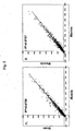


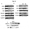

Classifications
-
- A—HUMAN NECESSITIES
- A61—MEDICAL OR VETERINARY SCIENCE; HYGIENE
- A61K—PREPARATIONS FOR MEDICAL, DENTAL OR TOILETRY PURPOSES
- A61K39/00—Medicinal preparations containing antigens or antibodies
- A61K39/0005—Vertebrate antigens
- A61K39/001—Preparations to induce tolerance to non-self, e.g. prior to transplantation
-
- A—HUMAN NECESSITIES
- A01—AGRICULTURE; FORESTRY; ANIMAL HUSBANDRY; HUNTING; TRAPPING; FISHING
- A01K—ANIMAL HUSBANDRY; AVICULTURE; APICULTURE; PISCICULTURE; FISHING; REARING OR BREEDING ANIMALS, NOT OTHERWISE PROVIDED FOR; NEW BREEDS OF ANIMALS
- A01K67/00—Rearing or breeding animals, not otherwise provided for; New or modified breeds of animals
- A01K67/027—New or modified breeds of vertebrates
- A01K67/0271—Chimeric vertebrates, e.g. comprising exogenous cells
-
- A—HUMAN NECESSITIES
- A61—MEDICAL OR VETERINARY SCIENCE; HYGIENE
- A61P—SPECIFIC THERAPEUTIC ACTIVITY OF CHEMICAL COMPOUNDS OR MEDICINAL PREPARATIONS
- A61P1/00—Drugs for disorders of the alimentary tract or the digestive system
-
- A—HUMAN NECESSITIES
- A61—MEDICAL OR VETERINARY SCIENCE; HYGIENE
- A61P—SPECIFIC THERAPEUTIC ACTIVITY OF CHEMICAL COMPOUNDS OR MEDICINAL PREPARATIONS
- A61P1/00—Drugs for disorders of the alimentary tract or the digestive system
- A61P1/16—Drugs for disorders of the alimentary tract or the digestive system for liver or gallbladder disorders, e.g. hepatoprotective agents, cholagogues, litholytics
-
- A—HUMAN NECESSITIES
- A61—MEDICAL OR VETERINARY SCIENCE; HYGIENE
- A61P—SPECIFIC THERAPEUTIC ACTIVITY OF CHEMICAL COMPOUNDS OR MEDICINAL PREPARATIONS
- A61P1/00—Drugs for disorders of the alimentary tract or the digestive system
- A61P1/18—Drugs for disorders of the alimentary tract or the digestive system for pancreatic disorders, e.g. pancreatic enzymes
-
- A—HUMAN NECESSITIES
- A61—MEDICAL OR VETERINARY SCIENCE; HYGIENE
- A61P—SPECIFIC THERAPEUTIC ACTIVITY OF CHEMICAL COMPOUNDS OR MEDICINAL PREPARATIONS
- A61P13/00—Drugs for disorders of the urinary system
- A61P13/10—Drugs for disorders of the urinary system of the bladder
-
- A—HUMAN NECESSITIES
- A61—MEDICAL OR VETERINARY SCIENCE; HYGIENE
- A61P—SPECIFIC THERAPEUTIC ACTIVITY OF CHEMICAL COMPOUNDS OR MEDICINAL PREPARATIONS
- A61P13/00—Drugs for disorders of the urinary system
- A61P13/12—Drugs for disorders of the urinary system of the kidneys
-
- A—HUMAN NECESSITIES
- A61—MEDICAL OR VETERINARY SCIENCE; HYGIENE
- A61P—SPECIFIC THERAPEUTIC ACTIVITY OF CHEMICAL COMPOUNDS OR MEDICINAL PREPARATIONS
- A61P15/00—Drugs for genital or sexual disorders; Contraceptives
-
- A—HUMAN NECESSITIES
- A61—MEDICAL OR VETERINARY SCIENCE; HYGIENE
- A61P—SPECIFIC THERAPEUTIC ACTIVITY OF CHEMICAL COMPOUNDS OR MEDICINAL PREPARATIONS
- A61P17/00—Drugs for dermatological disorders
-
- A—HUMAN NECESSITIES
- A61—MEDICAL OR VETERINARY SCIENCE; HYGIENE
- A61P—SPECIFIC THERAPEUTIC ACTIVITY OF CHEMICAL COMPOUNDS OR MEDICINAL PREPARATIONS
- A61P19/00—Drugs for skeletal disorders
- A61P19/04—Drugs for skeletal disorders for non-specific disorders of the connective tissue
-
- A—HUMAN NECESSITIES
- A61—MEDICAL OR VETERINARY SCIENCE; HYGIENE
- A61P—SPECIFIC THERAPEUTIC ACTIVITY OF CHEMICAL COMPOUNDS OR MEDICINAL PREPARATIONS
- A61P19/00—Drugs for skeletal disorders
- A61P19/08—Drugs for skeletal disorders for bone diseases, e.g. rachitism, Paget's disease
-
- A—HUMAN NECESSITIES
- A61—MEDICAL OR VETERINARY SCIENCE; HYGIENE
- A61P—SPECIFIC THERAPEUTIC ACTIVITY OF CHEMICAL COMPOUNDS OR MEDICINAL PREPARATIONS
- A61P21/00—Drugs for disorders of the muscular or neuromuscular system
-
- A—HUMAN NECESSITIES
- A61—MEDICAL OR VETERINARY SCIENCE; HYGIENE
- A61P—SPECIFIC THERAPEUTIC ACTIVITY OF CHEMICAL COMPOUNDS OR MEDICINAL PREPARATIONS
- A61P25/00—Drugs for disorders of the nervous system
-
- A—HUMAN NECESSITIES
- A61—MEDICAL OR VETERINARY SCIENCE; HYGIENE
- A61P—SPECIFIC THERAPEUTIC ACTIVITY OF CHEMICAL COMPOUNDS OR MEDICINAL PREPARATIONS
- A61P25/00—Drugs for disorders of the nervous system
- A61P25/02—Drugs for disorders of the nervous system for peripheral neuropathies
-
- A—HUMAN NECESSITIES
- A61—MEDICAL OR VETERINARY SCIENCE; HYGIENE
- A61P—SPECIFIC THERAPEUTIC ACTIVITY OF CHEMICAL COMPOUNDS OR MEDICINAL PREPARATIONS
- A61P27/00—Drugs for disorders of the senses
- A61P27/02—Ophthalmic agents
-
- A—HUMAN NECESSITIES
- A61—MEDICAL OR VETERINARY SCIENCE; HYGIENE
- A61P—SPECIFIC THERAPEUTIC ACTIVITY OF CHEMICAL COMPOUNDS OR MEDICINAL PREPARATIONS
- A61P3/00—Drugs for disorders of the metabolism
-
- A—HUMAN NECESSITIES
- A61—MEDICAL OR VETERINARY SCIENCE; HYGIENE
- A61P—SPECIFIC THERAPEUTIC ACTIVITY OF CHEMICAL COMPOUNDS OR MEDICINAL PREPARATIONS
- A61P3/00—Drugs for disorders of the metabolism
- A61P3/08—Drugs for disorders of the metabolism for glucose homeostasis
- A61P3/10—Drugs for disorders of the metabolism for glucose homeostasis for hyperglycaemia, e.g. antidiabetics
-
- A—HUMAN NECESSITIES
- A61—MEDICAL OR VETERINARY SCIENCE; HYGIENE
- A61P—SPECIFIC THERAPEUTIC ACTIVITY OF CHEMICAL COMPOUNDS OR MEDICINAL PREPARATIONS
- A61P31/00—Antiinfectives, i.e. antibiotics, antiseptics, chemotherapeutics
-
- A—HUMAN NECESSITIES
- A61—MEDICAL OR VETERINARY SCIENCE; HYGIENE
- A61P—SPECIFIC THERAPEUTIC ACTIVITY OF CHEMICAL COMPOUNDS OR MEDICINAL PREPARATIONS
- A61P31/00—Antiinfectives, i.e. antibiotics, antiseptics, chemotherapeutics
- A61P31/04—Antibacterial agents
-
- A—HUMAN NECESSITIES
- A61—MEDICAL OR VETERINARY SCIENCE; HYGIENE
- A61P—SPECIFIC THERAPEUTIC ACTIVITY OF CHEMICAL COMPOUNDS OR MEDICINAL PREPARATIONS
- A61P31/00—Antiinfectives, i.e. antibiotics, antiseptics, chemotherapeutics
- A61P31/10—Antimycotics
-
- A—HUMAN NECESSITIES
- A61—MEDICAL OR VETERINARY SCIENCE; HYGIENE
- A61P—SPECIFIC THERAPEUTIC ACTIVITY OF CHEMICAL COMPOUNDS OR MEDICINAL PREPARATIONS
- A61P31/00—Antiinfectives, i.e. antibiotics, antiseptics, chemotherapeutics
- A61P31/12—Antivirals
-
- A—HUMAN NECESSITIES
- A61—MEDICAL OR VETERINARY SCIENCE; HYGIENE
- A61P—SPECIFIC THERAPEUTIC ACTIVITY OF CHEMICAL COMPOUNDS OR MEDICINAL PREPARATIONS
- A61P35/00—Antineoplastic agents
-
- A—HUMAN NECESSITIES
- A61—MEDICAL OR VETERINARY SCIENCE; HYGIENE
- A61P—SPECIFIC THERAPEUTIC ACTIVITY OF CHEMICAL COMPOUNDS OR MEDICINAL PREPARATIONS
- A61P37/00—Drugs for immunological or allergic disorders
-
- A—HUMAN NECESSITIES
- A61—MEDICAL OR VETERINARY SCIENCE; HYGIENE
- A61P—SPECIFIC THERAPEUTIC ACTIVITY OF CHEMICAL COMPOUNDS OR MEDICINAL PREPARATIONS
- A61P37/00—Drugs for immunological or allergic disorders
- A61P37/02—Immunomodulators
- A61P37/06—Immunosuppressants, e.g. drugs for graft rejection
-
- A—HUMAN NECESSITIES
- A61—MEDICAL OR VETERINARY SCIENCE; HYGIENE
- A61P—SPECIFIC THERAPEUTIC ACTIVITY OF CHEMICAL COMPOUNDS OR MEDICINAL PREPARATIONS
- A61P39/00—General protective or antinoxious agents
- A61P39/02—Antidotes
-
- A—HUMAN NECESSITIES
- A61—MEDICAL OR VETERINARY SCIENCE; HYGIENE
- A61P—SPECIFIC THERAPEUTIC ACTIVITY OF CHEMICAL COMPOUNDS OR MEDICINAL PREPARATIONS
- A61P41/00—Drugs used in surgical methods, e.g. surgery adjuvants for preventing adhesion or for vitreum substitution
-
- A—HUMAN NECESSITIES
- A61—MEDICAL OR VETERINARY SCIENCE; HYGIENE
- A61P—SPECIFIC THERAPEUTIC ACTIVITY OF CHEMICAL COMPOUNDS OR MEDICINAL PREPARATIONS
- A61P43/00—Drugs for specific purposes, not provided for in groups A61P1/00-A61P41/00
-
- A—HUMAN NECESSITIES
- A61—MEDICAL OR VETERINARY SCIENCE; HYGIENE
- A61P—SPECIFIC THERAPEUTIC ACTIVITY OF CHEMICAL COMPOUNDS OR MEDICINAL PREPARATIONS
- A61P7/00—Drugs for disorders of the blood or the extracellular fluid
-
- A—HUMAN NECESSITIES
- A61—MEDICAL OR VETERINARY SCIENCE; HYGIENE
- A61P—SPECIFIC THERAPEUTIC ACTIVITY OF CHEMICAL COMPOUNDS OR MEDICINAL PREPARATIONS
- A61P7/00—Drugs for disorders of the blood or the extracellular fluid
- A61P7/04—Antihaemorrhagics; Procoagulants; Haemostatic agents; Antifibrinolytic agents
-
- A—HUMAN NECESSITIES
- A61—MEDICAL OR VETERINARY SCIENCE; HYGIENE
- A61P—SPECIFIC THERAPEUTIC ACTIVITY OF CHEMICAL COMPOUNDS OR MEDICINAL PREPARATIONS
- A61P7/00—Drugs for disorders of the blood or the extracellular fluid
- A61P7/06—Antianaemics
-
- A—HUMAN NECESSITIES
- A61—MEDICAL OR VETERINARY SCIENCE; HYGIENE
- A61P—SPECIFIC THERAPEUTIC ACTIVITY OF CHEMICAL COMPOUNDS OR MEDICINAL PREPARATIONS
- A61P7/00—Drugs for disorders of the blood or the extracellular fluid
- A61P7/08—Plasma substitutes; Perfusion solutions; Dialytics or haemodialytics; Drugs for electrolytic or acid-base disorders, e.g. hypovolemic shock
-
- A—HUMAN NECESSITIES
- A61—MEDICAL OR VETERINARY SCIENCE; HYGIENE
- A61P—SPECIFIC THERAPEUTIC ACTIVITY OF CHEMICAL COMPOUNDS OR MEDICINAL PREPARATIONS
- A61P9/00—Drugs for disorders of the cardiovascular system
-
- A—HUMAN NECESSITIES
- A61—MEDICAL OR VETERINARY SCIENCE; HYGIENE
- A61P—SPECIFIC THERAPEUTIC ACTIVITY OF CHEMICAL COMPOUNDS OR MEDICINAL PREPARATIONS
- A61P9/00—Drugs for disorders of the cardiovascular system
- A61P9/10—Drugs for disorders of the cardiovascular system for treating ischaemic or atherosclerotic diseases, e.g. antianginal drugs, coronary vasodilators, drugs for myocardial infarction, retinopathy, cerebrovascula insufficiency, renal arteriosclerosis
-
- C—CHEMISTRY; METALLURGY
- C12—BIOCHEMISTRY; BEER; SPIRITS; WINE; VINEGAR; MICROBIOLOGY; ENZYMOLOGY; MUTATION OR GENETIC ENGINEERING
- C12N—MICROORGANISMS OR ENZYMES; COMPOSITIONS THEREOF; PROPAGATING, PRESERVING, OR MAINTAINING MICROORGANISMS; MUTATION OR GENETIC ENGINEERING; CULTURE MEDIA
- C12N15/00—Mutation or genetic engineering; DNA or RNA concerning genetic engineering, vectors, e.g. plasmids, or their isolation, preparation or purification; Use of hosts therefor
- C12N15/09—Recombinant DNA-technology
- C12N15/87—Introduction of foreign genetic material using processes not otherwise provided for, e.g. co-transformation
- C12N15/873—Techniques for producing new embryos, e.g. nuclear transfer, manipulation of totipotent cells or production of chimeric embryos
-
- C—CHEMISTRY; METALLURGY
- C12—BIOCHEMISTRY; BEER; SPIRITS; WINE; VINEGAR; MICROBIOLOGY; ENZYMOLOGY; MUTATION OR GENETIC ENGINEERING
- C12N—MICROORGANISMS OR ENZYMES; COMPOSITIONS THEREOF; PROPAGATING, PRESERVING, OR MAINTAINING MICROORGANISMS; MUTATION OR GENETIC ENGINEERING; CULTURE MEDIA
- C12N5/00—Undifferentiated human, animal or plant cells, e.g. cell lines; Tissues; Cultivation or maintenance thereof; Culture media therefor
- C12N5/06—Animal cells or tissues; Human cells or tissues
- C12N5/0602—Vertebrate cells
- C12N5/0607—Non-embryonic pluripotent stem cells, e.g. MASC
-
- C—CHEMISTRY; METALLURGY
- C12—BIOCHEMISTRY; BEER; SPIRITS; WINE; VINEGAR; MICROBIOLOGY; ENZYMOLOGY; MUTATION OR GENETIC ENGINEERING
- C12N—MICROORGANISMS OR ENZYMES; COMPOSITIONS THEREOF; PROPAGATING, PRESERVING, OR MAINTAINING MICROORGANISMS; MUTATION OR GENETIC ENGINEERING; CULTURE MEDIA
- C12N5/00—Undifferentiated human, animal or plant cells, e.g. cell lines; Tissues; Cultivation or maintenance thereof; Culture media therefor
- C12N5/06—Animal cells or tissues; Human cells or tissues
- C12N5/0602—Vertebrate cells
- C12N5/0618—Cells of the nervous system
- C12N5/0619—Neurons
-
- C—CHEMISTRY; METALLURGY
- C12—BIOCHEMISTRY; BEER; SPIRITS; WINE; VINEGAR; MICROBIOLOGY; ENZYMOLOGY; MUTATION OR GENETIC ENGINEERING
- C12N—MICROORGANISMS OR ENZYMES; COMPOSITIONS THEREOF; PROPAGATING, PRESERVING, OR MAINTAINING MICROORGANISMS; MUTATION OR GENETIC ENGINEERING; CULTURE MEDIA
- C12N5/00—Undifferentiated human, animal or plant cells, e.g. cell lines; Tissues; Cultivation or maintenance thereof; Culture media therefor
- C12N5/06—Animal cells or tissues; Human cells or tissues
- C12N5/0602—Vertebrate cells
- C12N5/0618—Cells of the nervous system
- C12N5/0622—Glial cells, e.g. astrocytes, oligodendrocytes; Schwann cells
-
- C—CHEMISTRY; METALLURGY
- C12—BIOCHEMISTRY; BEER; SPIRITS; WINE; VINEGAR; MICROBIOLOGY; ENZYMOLOGY; MUTATION OR GENETIC ENGINEERING
- C12N—MICROORGANISMS OR ENZYMES; COMPOSITIONS THEREOF; PROPAGATING, PRESERVING, OR MAINTAINING MICROORGANISMS; MUTATION OR GENETIC ENGINEERING; CULTURE MEDIA
- C12N5/00—Undifferentiated human, animal or plant cells, e.g. cell lines; Tissues; Cultivation or maintenance thereof; Culture media therefor
- C12N5/06—Animal cells or tissues; Human cells or tissues
- C12N5/0602—Vertebrate cells
- C12N5/067—Hepatocytes
-
- A—HUMAN NECESSITIES
- A01—AGRICULTURE; FORESTRY; ANIMAL HUSBANDRY; HUNTING; TRAPPING; FISHING
- A01K—ANIMAL HUSBANDRY; AVICULTURE; APICULTURE; PISCICULTURE; FISHING; REARING OR BREEDING ANIMALS, NOT OTHERWISE PROVIDED FOR; NEW BREEDS OF ANIMALS
- A01K2217/00—Genetically modified animals
- A01K2217/05—Animals comprising random inserted nucleic acids (transgenic)
-
- A—HUMAN NECESSITIES
- A01—AGRICULTURE; FORESTRY; ANIMAL HUSBANDRY; HUNTING; TRAPPING; FISHING
- A01K—ANIMAL HUSBANDRY; AVICULTURE; APICULTURE; PISCICULTURE; FISHING; REARING OR BREEDING ANIMALS, NOT OTHERWISE PROVIDED FOR; NEW BREEDS OF ANIMALS
- A01K2217/00—Genetically modified animals
- A01K2217/07—Animals genetically altered by homologous recombination
- A01K2217/075—Animals genetically altered by homologous recombination inducing loss of function, i.e. knock out
-
- A—HUMAN NECESSITIES
- A01—AGRICULTURE; FORESTRY; ANIMAL HUSBANDRY; HUNTING; TRAPPING; FISHING
- A01K—ANIMAL HUSBANDRY; AVICULTURE; APICULTURE; PISCICULTURE; FISHING; REARING OR BREEDING ANIMALS, NOT OTHERWISE PROVIDED FOR; NEW BREEDS OF ANIMALS
- A01K2227/00—Animals characterised by species
- A01K2227/10—Mammal
- A01K2227/105—Murine
-
- A—HUMAN NECESSITIES
- A01—AGRICULTURE; FORESTRY; ANIMAL HUSBANDRY; HUNTING; TRAPPING; FISHING
- A01K—ANIMAL HUSBANDRY; AVICULTURE; APICULTURE; PISCICULTURE; FISHING; REARING OR BREEDING ANIMALS, NOT OTHERWISE PROVIDED FOR; NEW BREEDS OF ANIMALS
- A01K2227/00—Animals characterised by species
- A01K2227/10—Mammal
- A01K2227/106—Primate
-
- A—HUMAN NECESSITIES
- A61—MEDICAL OR VETERINARY SCIENCE; HYGIENE
- A61K—PREPARATIONS FOR MEDICAL, DENTAL OR TOILETRY PURPOSES
- A61K35/00—Medicinal preparations containing materials or reaction products thereof with undetermined constitution
- A61K35/12—Materials from mammals; Compositions comprising non-specified tissues or cells; Compositions comprising non-embryonic stem cells; Genetically modified cells
-
- A—HUMAN NECESSITIES
- A61—MEDICAL OR VETERINARY SCIENCE; HYGIENE
- A61K—PREPARATIONS FOR MEDICAL, DENTAL OR TOILETRY PURPOSES
- A61K48/00—Medicinal preparations containing genetic material which is inserted into cells of the living body to treat genetic diseases; Gene therapy
-
- C—CHEMISTRY; METALLURGY
- C12—BIOCHEMISTRY; BEER; SPIRITS; WINE; VINEGAR; MICROBIOLOGY; ENZYMOLOGY; MUTATION OR GENETIC ENGINEERING
- C12N—MICROORGANISMS OR ENZYMES; COMPOSITIONS THEREOF; PROPAGATING, PRESERVING, OR MAINTAINING MICROORGANISMS; MUTATION OR GENETIC ENGINEERING; CULTURE MEDIA
- C12N2501/00—Active agents used in cell culture processes, e.g. differentation
- C12N2501/10—Growth factors
- C12N2501/11—Epidermal growth factor [EGF]
-
- C—CHEMISTRY; METALLURGY
- C12—BIOCHEMISTRY; BEER; SPIRITS; WINE; VINEGAR; MICROBIOLOGY; ENZYMOLOGY; MUTATION OR GENETIC ENGINEERING
- C12N—MICROORGANISMS OR ENZYMES; COMPOSITIONS THEREOF; PROPAGATING, PRESERVING, OR MAINTAINING MICROORGANISMS; MUTATION OR GENETIC ENGINEERING; CULTURE MEDIA
- C12N2501/00—Active agents used in cell culture processes, e.g. differentation
- C12N2501/10—Growth factors
- C12N2501/113—Acidic fibroblast growth factor (aFGF, FGF-1)
-
- C—CHEMISTRY; METALLURGY
- C12—BIOCHEMISTRY; BEER; SPIRITS; WINE; VINEGAR; MICROBIOLOGY; ENZYMOLOGY; MUTATION OR GENETIC ENGINEERING
- C12N—MICROORGANISMS OR ENZYMES; COMPOSITIONS THEREOF; PROPAGATING, PRESERVING, OR MAINTAINING MICROORGANISMS; MUTATION OR GENETIC ENGINEERING; CULTURE MEDIA
- C12N2501/00—Active agents used in cell culture processes, e.g. differentation
- C12N2501/10—Growth factors
- C12N2501/115—Basic fibroblast growth factor (bFGF, FGF-2)
-
- C—CHEMISTRY; METALLURGY
- C12—BIOCHEMISTRY; BEER; SPIRITS; WINE; VINEGAR; MICROBIOLOGY; ENZYMOLOGY; MUTATION OR GENETIC ENGINEERING
- C12N—MICROORGANISMS OR ENZYMES; COMPOSITIONS THEREOF; PROPAGATING, PRESERVING, OR MAINTAINING MICROORGANISMS; MUTATION OR GENETIC ENGINEERING; CULTURE MEDIA
- C12N2501/00—Active agents used in cell culture processes, e.g. differentation
- C12N2501/10—Growth factors
- C12N2501/117—Keratinocyte growth factors (KGF-1, i.e. FGF-7; KGF-2, i.e. FGF-12)
-
- C—CHEMISTRY; METALLURGY
- C12—BIOCHEMISTRY; BEER; SPIRITS; WINE; VINEGAR; MICROBIOLOGY; ENZYMOLOGY; MUTATION OR GENETIC ENGINEERING
- C12N—MICROORGANISMS OR ENZYMES; COMPOSITIONS THEREOF; PROPAGATING, PRESERVING, OR MAINTAINING MICROORGANISMS; MUTATION OR GENETIC ENGINEERING; CULTURE MEDIA
- C12N2501/00—Active agents used in cell culture processes, e.g. differentation
- C12N2501/10—Growth factors
- C12N2501/119—Other fibroblast growth factors, e.g. FGF-4, FGF-8, FGF-10
-
- C—CHEMISTRY; METALLURGY
- C12—BIOCHEMISTRY; BEER; SPIRITS; WINE; VINEGAR; MICROBIOLOGY; ENZYMOLOGY; MUTATION OR GENETIC ENGINEERING
- C12N—MICROORGANISMS OR ENZYMES; COMPOSITIONS THEREOF; PROPAGATING, PRESERVING, OR MAINTAINING MICROORGANISMS; MUTATION OR GENETIC ENGINEERING; CULTURE MEDIA
- C12N2501/00—Active agents used in cell culture processes, e.g. differentation
- C12N2501/10—Growth factors
- C12N2501/12—Hepatocyte growth factor [HGF]
-
- C—CHEMISTRY; METALLURGY
- C12—BIOCHEMISTRY; BEER; SPIRITS; WINE; VINEGAR; MICROBIOLOGY; ENZYMOLOGY; MUTATION OR GENETIC ENGINEERING
- C12N—MICROORGANISMS OR ENZYMES; COMPOSITIONS THEREOF; PROPAGATING, PRESERVING, OR MAINTAINING MICROORGANISMS; MUTATION OR GENETIC ENGINEERING; CULTURE MEDIA
- C12N2501/00—Active agents used in cell culture processes, e.g. differentation
- C12N2501/10—Growth factors
- C12N2501/135—Platelet-derived growth factor [PDGF]
-
- C—CHEMISTRY; METALLURGY
- C12—BIOCHEMISTRY; BEER; SPIRITS; WINE; VINEGAR; MICROBIOLOGY; ENZYMOLOGY; MUTATION OR GENETIC ENGINEERING
- C12N—MICROORGANISMS OR ENZYMES; COMPOSITIONS THEREOF; PROPAGATING, PRESERVING, OR MAINTAINING MICROORGANISMS; MUTATION OR GENETIC ENGINEERING; CULTURE MEDIA
- C12N2501/00—Active agents used in cell culture processes, e.g. differentation
- C12N2501/20—Cytokines; Chemokines
- C12N2501/23—Interleukins [IL]
- C12N2501/235—Leukemia inhibitory factor [LIF]
-
- C—CHEMISTRY; METALLURGY
- C12—BIOCHEMISTRY; BEER; SPIRITS; WINE; VINEGAR; MICROBIOLOGY; ENZYMOLOGY; MUTATION OR GENETIC ENGINEERING
- C12N—MICROORGANISMS OR ENZYMES; COMPOSITIONS THEREOF; PROPAGATING, PRESERVING, OR MAINTAINING MICROORGANISMS; MUTATION OR GENETIC ENGINEERING; CULTURE MEDIA
- C12N2501/00—Active agents used in cell culture processes, e.g. differentation
- C12N2501/20—Cytokines; Chemokines
- C12N2501/237—Oncostatin M [OSM]
-
- C—CHEMISTRY; METALLURGY
- C12—BIOCHEMISTRY; BEER; SPIRITS; WINE; VINEGAR; MICROBIOLOGY; ENZYMOLOGY; MUTATION OR GENETIC ENGINEERING
- C12N—MICROORGANISMS OR ENZYMES; COMPOSITIONS THEREOF; PROPAGATING, PRESERVING, OR MAINTAINING MICROORGANISMS; MUTATION OR GENETIC ENGINEERING; CULTURE MEDIA
- C12N2502/00—Coculture with; Conditioned medium produced by
- C12N2502/08—Coculture with; Conditioned medium produced by cells of the nervous system
-
- C—CHEMISTRY; METALLURGY
- C12—BIOCHEMISTRY; BEER; SPIRITS; WINE; VINEGAR; MICROBIOLOGY; ENZYMOLOGY; MUTATION OR GENETIC ENGINEERING
- C12N—MICROORGANISMS OR ENZYMES; COMPOSITIONS THEREOF; PROPAGATING, PRESERVING, OR MAINTAINING MICROORGANISMS; MUTATION OR GENETIC ENGINEERING; CULTURE MEDIA
- C12N2502/00—Coculture with; Conditioned medium produced by
- C12N2502/30—Coculture with; Conditioned medium produced by tumour cells
-
- C—CHEMISTRY; METALLURGY
- C12—BIOCHEMISTRY; BEER; SPIRITS; WINE; VINEGAR; MICROBIOLOGY; ENZYMOLOGY; MUTATION OR GENETIC ENGINEERING
- C12N—MICROORGANISMS OR ENZYMES; COMPOSITIONS THEREOF; PROPAGATING, PRESERVING, OR MAINTAINING MICROORGANISMS; MUTATION OR GENETIC ENGINEERING; CULTURE MEDIA
- C12N2503/00—Use of cells in diagnostics
-
- C—CHEMISTRY; METALLURGY
- C12—BIOCHEMISTRY; BEER; SPIRITS; WINE; VINEGAR; MICROBIOLOGY; ENZYMOLOGY; MUTATION OR GENETIC ENGINEERING
- C12N—MICROORGANISMS OR ENZYMES; COMPOSITIONS THEREOF; PROPAGATING, PRESERVING, OR MAINTAINING MICROORGANISMS; MUTATION OR GENETIC ENGINEERING; CULTURE MEDIA
- C12N2506/00—Differentiation of animal cells from one lineage to another; Differentiation of pluripotent cells
- C12N2506/03—Differentiation of animal cells from one lineage to another; Differentiation of pluripotent cells from non-embryonic pluripotent stem cells
-
- C—CHEMISTRY; METALLURGY
- C12—BIOCHEMISTRY; BEER; SPIRITS; WINE; VINEGAR; MICROBIOLOGY; ENZYMOLOGY; MUTATION OR GENETIC ENGINEERING
- C12N—MICROORGANISMS OR ENZYMES; COMPOSITIONS THEREOF; PROPAGATING, PRESERVING, OR MAINTAINING MICROORGANISMS; MUTATION OR GENETIC ENGINEERING; CULTURE MEDIA
- C12N2517/00—Cells related to new breeds of animals
- C12N2517/02—Cells from transgenic animals
Definitions
- the present invention relates generally to mammalian multipotent adult stem cells (MASC), and more specifically to methods for obtaining, maintaining and differentiating MASC. Uses of MASC in the therapeutic treatment of disease are also provided.
- MASC mammalian multipotent adult stem cells
- Organ and tissue generation from stem cells, and their subsequent transplantation provide promising treatments for a number of pathologies, making stem cells a central focus of research in many fields.
- Stem cell technology provides a promising alternative therapy for diabetes, Parkinson's disease, liver disease, heart disease, and autoimmune disorders, to name a few.
- organ and tissue transplantation there are at least two major problems associated with organ and tissue transplantation.
- the second major problem is the potential incompatibility of the transplanted tissue with the immune system of the recipient. Because the donated organ or tissue is recognized by the host immune system as foreign, immunosuppressive medications must be provided to the patient at a significant cost-both financially and physically.
- Xenotransplantation or transplantation of tissue or organs from another species, could provide an alternative means to overcome the shortage of human organs and tissues.
- Xenotransplantation would offer the advantage of advanced planning.
- the organ could be harvested while still healthy and the patient could undergo any beneficial pretreatment prior to transplant surgery.
- xenotransplantation does not overcome the problem of tissue incompatibility, but instead exacerbates it.
- damaging viruses cross species barriers. Pigs have become likely candidates as organ and tissue donors, yet cross-species transmission of more than one virus from pigs to humans has been documented. For example, over a million pigs were recently slaughtered in Malaysia in an effort to contain an outbreak of Hendra virus, a disease that was transmitted to more than 70 humans with deadly results (Butler, D. 1999).
- stem cells can undergo self-renewing cell division to give rise to phenotypically and genotypically identical daughters for an indefinite time and ultimately can differentiate into at least one final cell type.
- transplant tissues can be generated to provide the advantages associated with xenotransplantation without the associated risk of infection or tissue rejection.
- Stem cells also provide promise for improving the results of gene therapy.
- a patient's own stem cells could be genetically altered in vitro , then reintroduced in vivo to produce a desired gene product.
- These genetically altered stem cells would have the potential to be induced to differentiate to form a multitude of cell types for implantation at specific sites in the body, or for systemic application.
- heterologous stem cells could be genetically altered to express the recipient's major histocompatibility complex (MHC) antigen, or no MHC antigen, allowing transplantion of cells from donor to recipient without the associated risk of rejection.
- MHC major histocompatibility complex
- Stem cells are defined as cells that have extensive proliferation potential, differentiate into several cell lineages, and repopulate tissues upon transplantation.
- the quintessential stem cell is the embryonic stem (ES) cell, as it has unlimited self-renewal and multipotent differentiation potential (Thomson, J. et al. 1995; Thomson, J.A. et al. 1998; Shamblott, M. et al. 1998; Williams, R.L. et al. 1988; Orkin, S. 1998; Reubinoff, B.E., et al. 2000).
- ES embryonic stem
- These cells are derived from the inner cell mass of the blastocyst (Thomson, J. et al. 1995; Thomson, J.A. et al. 1998; Martin, G.R.
- ES and EG cells can be derived from the primordial germ cells from a post-implantation embryo (embryonal germ cells or EG cells).
- ES and EG cells have been derived from mouse, and more recently also from non-human primates and humans. When introduced into mouse blastocysts, ES cells can contribute to all tissues of the mouse (animal) (Orkin, S. 1998). Murine ES cells are therefore pluripotent. When transplanted in post-natal animals, ES and EG cells generate teratomas, which again demonstrates their multipotency.
- ES (and EG) cells can be identified by positive staining with the antibodies to stage-specific embryonic antigens (SSEA) I and 4.
- SSEA stage-specific embryonic antigens
- ES and EG cells express a number of transcription factors highly specific for these undifferentiated cells. These include oct-4 and Rex-1, leukemia inhbitory factor receptor (LIF-R). The transcription factors sox-2 and Rox-1 are expressed in both ES and non-ES cells.
- Oct-4 is expressed in the pregastrulation embryo, early cleavage stage embryo, cells of the inner cell mass of the blastocyst, and embryonic carcinoma (EC) cells. In the adult animal, oct-4 is only found in germ cells.
- Oct-4 in combination with Rox-1, clauses transcriptional activation of the Zn-finger protein Rex-1, and is also required for maintaining ES in an undifferentiated state.
- the oct-4 gene is down-regulated when cells are induced to differentiate in vitro.
- Sox-2 is required with oct-4 to retain the undifferentiated state of ES/EC and to maintain murine, but not human, ES cells.
- Human or murine primordial germ cells require presence of LIF.
- Another hallmark of ES cells is presence of high levels of telomerase, which provides these cells with an unlimited self-renewal potential in vitro.
- HSC hematopoietic stem cell
- HSCs When transplanted into lethally irradiated animals or humans, HSCs can repopulate the erythroid, neutrophil-macrophage, megakaryocyte and lymphoid hemopoietic cell pool.
- hemopoietic stem cells can be induced to undergo at least some self-renewing cell divisions and can be induced to differentiate to the same lineages as is seen in vivo. Therefore; this cell fulfills the criteria of a stem cell.
- Stem cells which differentiate only to form cells of hematopoietic lineage, however, are unable to provide a source of cells for repair of other damaged tissues, for example, heart or lung tissue damaged by high-dose chemotherapeutic agents.
- NSC neural stem cell
- NSCs When cultured ex vivo, NSCs can be induced to proliferate, as well as to differentiate into different types of neurons and glial cells. When transplanted into the brain, NSCs can engraft and generate neural cells and glial cells. Therefore, this cell too fulfills the definition of a stem cell, albeit a hematopoetic stem cell.
- Clarke et al. reported that NSCs from Lac-Z transgenic mice injected into murine blastocysts or in chick embryos contribute to a number of tissues of the chimeric mouse or chicken embryo (Clarke, D. L. et al. 2000). LacZ-expressing cells were found with varying degree of mosaicism, not only in the central nervous system, but also in mesodermal derivatives as well as in epithelial cells of the liver and intestine but not in other tissues, including the hematopoietic system.
- adult NSCs may have significantly greater differentiation potential than previously realized but still do not have the pluripotent capability of ES or of the adult derived multipotent adult stem cells (MASC) described in Furcht et al. (WO 01/11011 ) and herein.
- MASC adult derived multipotent adult stem cells
- MAPC multipotent adult stem cells
- NSC sub-ventricular zone
- SVZ sub-ventricular zone
- hippocampus of the adult mammalian brain
- SVZ sub-ventricular zone
- hippocampus of the adult mammalian brain
- Reynolds and Weiss 1992
- Vescovi et al. 1999
- Fetal or adult brain-derived NSC can be expanded ex vivo and induced to differentiate into astrocytes, oligodendrocytes and functional neurons (Ciccolini et al. , 1998; Johansson et al. , 1999; Palmer et al. , 1999;- Reynolds et al. , 1996; Ryder et al. , 1990; Studer et al. , 1996; Vescovi et al. , 1993).
- undifferentiated NSC cultured for variable amounts of time differentiate into glial cells, GABAergic and dopaminergic neurons (Flax et al. , 1998; Gage et al ., 1995; Suhonen et al. , 1996).
- NSC neuronal and glial cells
- the ideal source of neurons and glia for replacement strategies would be cells harvestable from adult, autologous tissue different than the brain that was readily accessible and that can be expanded in vitro and differentiated ex vivo or in vivo to the cell type that is deficient in the patient.
- BM derived cells acquire phenotypic characteristics of neuroectodermal cells when cultured in vitro under NSC conditions, or when they enter the central nervous system (Sanchez-Ramos et al. , 2000; Woodbury et al. , 2000).
- the phenotype of the BM cells with this capability is not known.
- the capacity for differentiation of cells that acquire neuroectodermal features to other cell types is also unknown.
- MSC mesenchymal stem cell
- Fridenshtein (1982) A third tissue specific cell with stem cell properties is the mesenchymal stem cell (MSC), initially described by Fridenshtein (1982).
- MSC originally derived from the embryonal mesoderm and isolated from adult BM, can differentiate to form muscle, bone, cartilage, fat, marrow stroma, and tendon.
- the mesoderm develops into limb-bud mesoderm, tissue that generates bone, cartilage, fat, skeletal muscle and possibly endothelium.
- Mesoderm also differentiates to visceral mesoderm, which can give rise to cardiac muscle, smooth muscle, or blood islands consisting of endothelium and hematopoietic progenitor cells.
- MSCs Primitive mesodermal or MSCs, therefore, could provide a source for a number of cell and tissue types.
- a number of MSCs have been isolated.
- tissue-specific stem cells have been identified, including gastrointestinal stem cells (Potten, C. 1998), epidermal stem cells (Watt, F. 1997), and hepatic stem cells, also termed oval cells (Alison, M. et al . 1998). Most of these are less well characterized.
- tissue specific stem cells Compared with ES cells, tissue specific stem cells have less self-renewal ability and, although they differentiate into multiple lineages, they are not pluripotent. No studies have addressed whether tissue specific cells express the markers described above as seen in ES cells. In addition, the degree of telomerase activity in tissue specific or lineage comitted stem cells has not been fully explored, in part because large numbers of highly enriched populations of these cells are difficult to obtain.
- tissue specific stem cells could only differentiate into cells of the same tissue.
- a number of recent publications have suggested that adult organ specific stem cells may be capable of differentiation into cells of different tissues.
- the true nature of these types of cells has not been fully discerned.
- a number of studies have shown that cells transplanted at the time of a BM transplant can differentiate into skeletal muscle (Ferrari 1998; Gussoni 1999). This could be considered within the realm of possible differentiation potential of mesenchymal cells that are present in marrow.
- Jackson published that muscle satellite cells can differentiate into hemopoietic cells, again a switch in phenotype within the splanchnic mesoderm of the embryo (Jackson 1999).
- stem cells from one embryonic layer can differentiate into tissues thought to be derived during embryogenesis from a different embryonic layer.
- endothelial cells or their precursors detected in humans or animals that underwent marrow transplantation are at least in part derived from the marrow donor (Takahashi, 1999; Lin, 2000).
- visceral mesoderm and not splanchnic mesoderm, capabilities such as MSC, derived progeny are transferred with the infused marrow.
- ES cell research provides a promising alternative solution to the problem of a limited supply of organs for transplantation, the problems and risks associated with the need for immunosuppression to sustain transplantation of heterologous cells or tissue would remain.
- An estimated 20 immunologically different lines of ES cells would need to be established in order to provide immunocompatible cells for therapies directed to the majority of the population.
- stem cells with multiple differentiation potential have been isolated previously by others and by the present inventors, a progenitor cell with the potential to differentiate into a wide variety of cell types of different lineages, including fibroblasts, hepatic, osteoblasts, chondrocytes, adipocytes, skeletal muscle, endothelium, stroma, smooth muscle, cardiac muscle and hemopoietic cells, has not been described. If cell and tissue transplant and gene therapy are to provide the therapeutic advances expected, a stem cell or progenitor cell with the greatest or most extensive differentiation potential is needed. What is needed is the adult equivalent of an ES cell.
- BM muscle and brain are the three tissues in which cells with apparent greater plasticity than previously thought have been identified.
- BM contains cells that can contribute to a number of mesodermal (Ferrari G. et al. , 1998; Gussoni E. et al. , 1999; Rafii S. et al. , 1994; Asahara T. et al. , 1997; Lin Y. et al. , 2000; Orlic D. et al. , 2001; Jackson K. et al. , 2001) endodermal (Petersen B.E. et al. , 1999; Theise, N.D. et al. , 2000; Lagasse E. et al.
- WO95/03062 describes methods and compositions for preventing immune rejection of solid organ grafts.
- WO 02/36751 describes human cord blood derived unrestricted somatic stem cells.
- the present study demonstrates that cells with multipotent adult progenitor characteristics can be culture-isolated from multiple different organs, namely BM, muscle and the brain.
- the cells have the same morphology, phenotype, in vitro differentiation ability and have a highly similar expressed gene profile.
- the present invention relates to a method of obtaining a population of multipotent adult stem cells (MASCs) isolated from a mammal, preferably mouse, rat or human.
- the cell is derived from a non-embryonic organ or tissue and has the capacity to be induced to differentiate to form at least one differentiated cell type of mesodermal, ectodermal and endodermal origin.
- the organ or tissue from which the MASC are isolated is umbilical cord blood or placenta.
- the MASC produced by the method of the present invention is also summarized as a cell that constitutively expresses oct4 and high levels of telomerase and is negative for CD44, MHC class I and MHC class II expression.
- this cell administered to a patient in a therapeutically effective amount.
- a surprising benefit of this treatment is that no teratomas are formed in vivo .
- Normal, non-human animal comprising MASC are described.
- the animal is chimeric.
- a composition comprising a population of MASC and a culture medium that expands the MASC population is also described. It is advantageous in some cases for the medium to contain epidermal growth factor (EGF), platelet derived growth factor (PDGF) and leukemia inhibitory factor (LIF).
- EGF epidermal growth factor
- PDGF platelet derived growth factor
- LIF leukemia inhibitory factor
- a composition comprising a population of fully or partially purified MASC progeny is further described.
- the progeny can have the capacity to be further differentiated, or can be terminally differentiated.
- the progeny can be of the osteoblast, chondrocyte, adipocyte, fibroblast, marrow stroma, skeletal muscle, smooth muscle, cardiac muscle, occular, endothelial, epithelial, hepatic; pancreatic, hematopoietic, glial, neuronal or oligodendrocyte cell type.
- the present invention also provides a method for isolating and propagating MASC by obtaining tissue from a mammal, establishing a population of adherent cells, depleting the population of CD45 + cells, recovering CD45 - cells and culturing them under expansion conditions to produce an expanded cell population.
- An object of the present invention therefore, is to produce an expanded cell population obtained by this method.
- An aspect of the invention is a method for differentiating MASC ex vivo by isolating and propagating them, and then culturing the propagated cells in the presence of desired differentiation factors.
- the preferred differentiation factors are basic fibroblast growth factor (bFGF), vascular endothelial growth factor (VEGF), dimethylsulfoxide (DMSO) and isoproterenol; or fibroblast growth factor4 (FGF4) and hepatocyte growth factor (HGF).
- bFGF basic fibroblast growth factor
- VEGF vascular endothelial growth factor
- DMSO dimethylsulfoxide
- FGF4 fibroblast growth factor4
- HGF hepatocyte growth factor
- a method for differentiating MASC in vivo is described, by isolating and expanding them, and then administering the expanded cell population to a mammalian host, wherein said cell population is engrafted and differentiated in vivo in tissue specific cells, such that the function of a cell or organ, defective due to injury, genetic disease, acquired disease or iatrogenic treatments, is augmented, reconstituted or provided for the first time.
- the MASC can undergo self-renewal in vivo .
- a differentiated cell obtained by ex vivo or in vivo differentiation is also described.
- the differentiated cell can be ectoderm, mesoderm or endoderm.
- the differentiated cell can be of the osteoblast, chondrocyte, adipocyte, fibroblast, marrow stroma, skeletal muscle, smooth muscle, cardiac muscle, occular, endothelial, epithelial, hepatic, pancreatic, hematopoietic, glial, neuronal or oligodendrocyte cell type.
- An important application of this technology is the method of treating a patient by administering a therapeutically effective amount of MASC or their progeny.
- the progeny can either have the capacity to be further differentiated, or can be terminally differentiated.
- An unexpected benefit of this approach is that the need for pretreatment and/or post treatment of the patient with irradiation, chemotherapy, immunosuppressive agents or other drugs or treatments is reduced or eliminated. The induction of tolerance before or during treatment is also not required.
- Such treatment can treat a variety of diseases and conditions, including cancer, cardiovascular disease, metabolic disease; liver disease, diabetes, hepatitis, hemophilia, degenerative or traumatic neurological conditions, autoimmune disease, genetic deficiency, connective tissue disorders, anemia, infectious disease and transplant rejection.
- diseases and conditions including cancer, cardiovascular disease, metabolic disease; liver disease, diabetes, hepatitis, hemophilia, degenerative or traumatic neurological conditions, autoimmune disease, genetic deficiency, connective tissue disorders, anemia, infectious disease and transplant rejection.
- MASC or their progeny are administered via localized injection, including catheter administration, systemic injection, parenteral administration, oral administration, or intrauterine injection into an embryo.
- Administration can be in conjunction with a pharmaceutically acceptable matrix, which may be biodegradable.
- MASC or their progeny, administered to a patient, alter the immune system to resist viral, bacterial or fungal infection.
- teratomas are not formed when MASC or their progeny are adminstered to a patient.
- MASC or their progeny When administered to a patient, MASC or their progeny also are able to augment, reconstitute or provide for the first time the function of a cell or organ defective due to injury, genetic disease, acquired disease or iatrogenic treatments.
- the organ is any of bone marrow, blood, spleen, liver, lung, intestinal tract, brain, immune system, circulatory system, bone, connective tissue, muscle, heart, blood vessels, pancreas, central nervous system, peripheral nervous system, kidney, bladder, skin, epithelial appendages, breast-mammary glands, fat tissue, and mucosal surfaces including oral esophageal, vaginal and anal.
- diseases treatable by this method are cancer, cardiovascular disease, metabolic disease, liver disease, diabetes, hepatitis, hemophilia, degenerative or traumatic neurological conditions, autoimmune disease, genetic deficiency, connective tissue disorders, anemia, infectious disease and transplant rejection.
- the MASC or their progeny home to one or more organs in the patient and are engrafted therein such that the function of a cell or organ, defective due to injury, genetic disease, acquired disease or iatrogenic treatments, is augmented, reconstituted or provided for the first time, which is surprising and unexpected.
- the injury is ischemia or inflammation.
- the MASC or their progeny can enhance angiogenesis.
- MASC or their progeny can be genetically transformed to deliver a therapeutic agent, preferably an antiangiogenic agent.
- a therapeutic composition comprising MASC and a pharmaceutically acceptable carrier, wherein the MASC are present in an amount effective to produce tissue selected from the group consisting of bone marrow, blood, spleen, liver, lung, intestinal tract, brain, immune system, bone, connective tissue, muscle, heart, blood vessels, pancreas, central nervous system, kidney, bladder, skin, epithelial appendages, breast-mammary glands, fat tissue, and mucosal surfaces including oral esophageal, vaginal and anal, is described.
- a therapeutic method for restoring organ, tissue or cellular function to a patient comprising the steps of removing MASC from a mammalian donor, expanding MASC to form an expanded population of undifferentiatied cells, and adminstering the expanded cells to the patient, wherein organ, tissue or cellular function is restored, is also described.
- the restored function may be enzymatic or genetic.
- the mammalian donor can be the patient.
- a method of inhibiting the rejection of a heterologous MASC transplanted into a patient comprising the steps of introducing into the MASC, ex vivo, a nucleic acid sequence encoding the recipient's MHC antigen operably linked to a promotor, wherein the MHC antigen is expressed by the MASC and transplanting the MASC into the patient, wherein MHC antigen is expressed at a level sufficient to inhibit the rejection of the transplanted MASC.
- the patient is of the same species or another mammalian species as the donor of the MASC.
- An alternative method of inhibiting the rejection of a heterologous MASC transplanted into a patient comprises transgenically knocking out expression of MHC antigen in the MASC and transplanting the transgenic MASC into the patient MHC antigen is not expressed by the MASC and rejection of the transplanted cells is inhibited.
- a method of generating blood or individual blood components ex vivo by the process of isolating MASC and differentiating the MASC to form blood or blood components is also described.
- the individual blood components are red blood cells, white blood cells or platelets.
- a method of drug discovery comprising the steps of analyzing the genomic or proteomic makeup of MASC or their progeny, employing analysis thereof via bioinformatics and/or computer analysis using algorithms, and assembling and comparing new data with known databases to compare and contrast these is further described.
- Also described is a method of identifying the components of a differentiation pathway comprising the steps of analyzing the genomic or proteomic makeup of MASC, inducing differentiation of MASC in vitro or in vivo, analyzing the genomic or proteomic makeup of intermediary cells in the differentiation pathway, analyzing the genomic or proteomic makeup of terminally differentiated cells in the differentiation pathway, using bioinformatics and/or algorithms to characterize the genomic or proteomic makeup of MASC and their progeny, and comparing the data obtained in (e) to identify the components of the pathway.
- differentiation that occurs in vitro can be compared with differentiation that occurs in vivo such that fundamental differences between the two systems can be characterized.
- the products can be proteins, lipids, complex carbohydrates, DNA or RNA.
- a method of inducing, in a mammal, tolerance to an antigen administered to said mammal comprising the step of administering to said mammal, after or simultaneously with the administration of said antigen, an effective amount of MASC or their progeny so that said mammal's humoral immune response to a subsequent challenge with said antigen is suppressed.
- a method for removing toxins from the blood of a patient comprising contacting blood ex vivo with MASC derived cells, wherein said cells line a hollow, fiber based device.
- the cells can be kidney or liver cells.
- a method for delivering therapeutic products to a patient comprising contacting the blood of said patient ex vivo with MASC or their progeny, wherein said MASC or their progeny are genetically transformed to deliver a therapeutic agent is further described, along with a method for testing the toxicity of a drug comprising contacting MASC or their progeny ex vivo with said drug and monitoring cell survival.
- the progeny can be selected from the group consisting of hepatic, endothelial, epithelial and kidney.
- “Expansion” shall mean the propogation of a cell without differentiation.
- Intermediary cells are cells produced during differentiation of a MASC that have some, but not all, of the characteristics of MASC or their terminally differentiated progeny. Intermediary cells may be progenitor cells which are committed to a specific pathway, but not to a specific cell type.
- Normal shall mean an animal that is not diseased, mutated or malformed, i.e., healthy animals.
- Self-renewal shall mean the ability of cells to propagate without the addition of external stimulation.
- the presence of cytokines or other growth factors produced locally in the tissue or organ shall not constitute external stimulation.
- Home shall mean the ability of certain MASC or their progeny to migrate specifically to sites where additional cells may be needed.
- knocking out expression shall mean the elimination of the function of a particular gene.
- genomic or proteomic makeup shall mean the gene or protein components of a given cell.
- telomere activity can be correlated to the two-fold level observed in the immortal human cell line MCF7. Soule et al. (1973) J. Cancer Inst. 51:1409-1416 .
- MASC technology could be used to replace damaged, diseased, dysfunctional or dead cells in the body of a mammal. Furthermore these cells could be injected into the host using autologous or allogeneic cells with or without nature or artificial supports, matrices or polymers to correct for loss of cells, abnormal function or cells or organs e.g. genetic such as mutations of genes affecting a protein function such as sickle cell disease, hemophilia or "storage diseases" where products accumulate in the body because of faulty processing, e.g. Guacher's, Neiman Pick's, mucopolysaccharidosis etc. Examples of restitution of dying or dead cells would be the use of MASC or their differentiated progeny in the treatment of macular degeneration and other neurodegenerative diseases.
- liver failure patients hooked up to a bioartificial liver, which was sufficient to allow for the recovery of normal liver function, obviating the need for a liver transplant.
- This is a serious unmet medical need for example in one liver disease alone - hepatitis C.
- Hepatocytes derived from autologous or allogeneic MASC, can be transplanted in this or other liver diseases. Such transplants may either transiently provide liver function to allow recovery of the recipient's own liver cells or permanently repopulate a damaged liver to allow recovery of normal liver function via the donor cells.
- the progeny of the MASC could be differentiated ex vivo and then be administered as purified or even mixtures of cells to provide a therapeutic benefit.
- These MASC in the undifferentiated state could also be used as carriers or vehicles to deliver drugs or molecules of therapeutic benefit. This could be to treat any one of a number of diseases including but not limited to cancer, cardiovascular, inflammatory, immunologic, infections, etc.
- a cell perhaps an endothelial cell expressing a novel or high levels of an angiogenic molecule could be administered to a patient which would be incorporated into existing blood vessels to promote angiogenesis, for example in the heart; correspondingly one could have endothelial cells producing molecules that might suppress angiogenesis that would be incorporated into blood cells and inhibit their further formation for example in diabetic retinopathy or in cancer where new blood vessel formation is key to the pathogenesis, spread and extent of the disease.
- the ability to populate the BM and to form blood ex vivo has an untold use for important medical applications.
- ex vivo production of blood the transfusion of blood and blood products around the world is still performed with variable safety because of transmission of infectious agents.
- Blood transfusions have lead to HIV, hepatitis C and B, and now the impending threat of Mad Cow or CJD, Creuzfeldt-Jakob disease.
- the ability to produce blood in vitro, especially red blood cells, could provide a safe and reliable alternative to collection of blood from people. It might never fully replace blood collection from donors.
- hMASC or their hematopoietic progeny could be placed in animals in utero such as sheep which could form human hematopoietic cells and serve as a source for human blood components or proteins of therapeutic futility.
- animals in utero such as sheep which could form human hematopoietic cells and serve as a source for human blood components or proteins of therapeutic futility.
- the same could be true for hepatocytes, islets or many other cell types but would provide an alternative to producing human cells in vitro and use the animals as factories for the cells. It could also assist in blood shortages that are predicted to occur.
- hMASC could also conceivably be transplanted into a human embryo to correct any one of a number of defects.
- these MASC can give rise to clonal populations of specifically differentiated cells they are a rich platform for drug discovery. This would involve doing gene expression, analyzing gene expression, discovery of new genes activated patterns of activation, proteomics and patterns of protein expression and modification surrounding this. This would be analyzed with bioinformatics, using data bases and algorithms for analyzing these data compared to publicly available or proprietary data bases.
- the information of how known drugs or agents might act could be compared to information derived from MASC, their differentiated progeny and from a population of people which could be available. Pathways, targets, and receptors could be identified. New drugs, antibodies or other compounds could be found to produce a biologically desirable responses.
- the MASC and their differentiated progeny could be used as monitors for undesirable responses, coupled with databases, bioinformatics and algorithms.
- MASC derived from human, mouse, rat or other mammals appear to be the only normal, non-malignant, somatic cell (non germ cell) known to date to express very high levels of telomerase even in late passage cells.
- the telomeres are extended in MASC and they are karyotypically normal. Because MASC injected into a mammal, home to multiple organs, there is the likelihood that newly arrived MASC in a particular organ could be self renewing. As such, they have the potential to repopulate an organ not only with themselves but also with self renewing differentiated cell types that could have been damaged, died, or otherwise might have an abnormal function because of genetic or acquired disease.
- pancreatic islets For example in type I diabetes there is a progressive loss of insulin producing beta cells in the pancreatic islets. In various renal diseases there is progressive loss of function and in some cases obliteration of glomerulus. If in the case of diabetes, MASC or differentiated progeny might home to the pancreas and themselves or via interaction with endogenous cells within the pancreas, induce islets to be formed. This would have an ameliorating impact on diabetes. Ultimately conditions, agents or drugs might be found to in vivo control, i.e.
- MASC self renewing capability of the MASC and control, or enhance or inhibit the movement to differentiated progeny, e.g., islet precursors, hepatocyte precursors, blood precursors, neural and/or cardiac precursors using MASC one will likely find pathways, methods of activation and control that might induce endogenous precursor cells within an organ to proliferate and differentiation.
- differentiated progeny e.g., islet precursors, hepatocyte precursors, blood precursors, neural and/or cardiac precursors using MASC
- the present invention also provides methods for drug discovery, genomics, proteomics, and pathway identification; comprising analyzing the genomic or proteomic makeup of a MASC, coupled with analysis thereof via bioinformatics, computer analysis via algorithms, to assemble and compare new with known databases and compare and contract these. This will identify key components, pathways, new genes and/or new patterns of gene and protein expression and protein modification (proteomics) that could lead to the definition of targets for new compounds, antibodies, proteins, small molecule organic compounds, or other biologically active molecules that would have therapeutic benefit.
- BM mononuclear cells (BMMNC) were obtained by ficoll-hypaque separation of BM was obtained from 5-6 week old ROSA26 mice or C57/BL6 mice.
- muscle and brain tissue was obtained from 3-day old 129 mice. Muscles from the proximal parts of fore and hind limbs were excised from and thoroughly minced. The tissue was treated with 0.2% collagenase (Sigma Chemical Co, St Louis, MO) for I hour at 37°C, followed by 0.1% trypsin (Invitrogen, Grand Island, NY) for 45 minutes. Cells were then triturated vigorously and passed through a 70-um filter.
- Cell suspensions were collected and centrifuged for 10 minutes at 1600 rpm. Brain tissues was dissected and minced thoroughly. Cells were dissociated by incubation with 0.1% trypsin and 0.1% DNAse (Sigma) for 30 minutes at 37°C. Cells were then triturated vigorously and passed through a 70-um filter. Cell suspension was collected and centrifuged for 10 minutes at 1600 rpm.
- BMMNC or muscle or brain suspensions were plated at 1 x 10 5 /cm 2 in expansion medium [2% FCS in low glucose Dulbecco's minimal essential medium (LG-DMEM), 10 ng/mL each platelet derived growth factor (PDGF), epidermal growth factor (EGF) and leukemia inhibitory factor (LIF)] and maintained at 5x10 3 /cm 2 .
- LG-DMEM low glucose Dulbecco's minimal essential medium
- PDGF platelet derived growth factor
- EGF epidermal growth factor
- LIF leukemia inhibitory factor
- Resulting CD45 - /Gly-A - cells were replated at 10 cells/well in 96-well plates coated with FN and were expanded at cell densities between 0.5 and 1.5x 10 3 /cm 2 .
- the expansion potential of MASC was similar regardless of the tissue from which they were derived ( Fig. 1 ).
- mMASC derived from BM, muscle and brain and cultured on FN were CD13 + , CD44 - , CD45 - , class-I and class-II histocompatibility antigen - , Flkl low and cKit - , identical to the characteristics of hMASC, as described in Intemation Application No. PCT/US00/21387 .
- cell expansion during the initial 2-3 months was greater when cells were cultured on collagen type IV, laminin or MatrigelTM, cells had phenotypic characteristics of MSC, i.e., expressed CD44 and did not express CD13.
- CD45 - /GlyA - cells yielded continuous growing cultures. This suggests that the cells capable of initiating MASC cultures are rare and likely less that 1/1,000 of CD45 - /G1yA - cells.
- mMASC cultured on FN were 8-10 ⁇ m in diameter with a large nucleus and scant cytoplasm. Several populations have been cultured for > 100 PDs. The morphology and phenotype of cells remained unchanged throughout culture.
- telomere length was measured using the Telomere Length Assay Kit from Pharmingen (New Jersey, USA) according to the manufacturer's recommendations. Average telomere length (ATL) of mMASC cultured for 40 PDs was 27 Kb. When re-tested after 102 PDs, ATL remained unchanged.
- mMASC For karyotyping of mMASC, cells were subcultured at a 1:2 dilution 12h before harvesting, collected with trypsin-EDTA, and subjected to a 1.5h colcemid incubation followed by lysis with hypotonic KCl and fixation in acid/alcohol as previously described (Verfaillie et al. , 1992). Cytogenic analysis was conducted on a monthly basis and showed a normal karyotype, except for a single.population that became hyperdiploid after 45 PDs, which was no longer used for studies.
- Murine MASC obtained after 46 to >80 PDs were tested by Quantitative (Q)-RT-PCR for expression levels of Oct4 and Rexl, two transcription factors important in maintaining an undifferentiated status of ES cells.
- RNA was extracted from mouse MASC, neuroectodermal differentiated progeny (day 1- 7 after addition of bFGF) and mouse ES cells.
- mRNA levels were normalized using GAPDH as housekeeping gene, and compared with levels in mouse ES cells.
- Oct4 and Rex 1 mRNA were present at similar levels in BM, muscle and brain derived MASC.
- Rexl mRNA levels were similar in mMASC and mES cells, while Oct4 mRNA levels were about 1,000 fold lower in MASC than in ES cells.
- BM, muscle and brain derived MASC was examined using U74A Affimetrix gene array. Briefly, mRNA was extracted from 2-3x10 6 BM, muscle or brain derived-MASC, cultured for 45 population doublings. Preparation of cDNA, hybridization to the U74A array containing 6,000 murine genes and 6,000 EST clusters, and data acquisition were done per manufacturer's recommendations (all from Affimetrix, Santa Clara, CA). Data analysis was done using GeneChip® software (Affimetrix). Increased or decreased expression by a factor of 2.2 fold (Iyer V.R. et al. , 1999; Scherf U. et al. , 2000; Alizadeh A.A. et al. , 2000) was considered significant. r2 value was determined using linear regression analysis ( Fig.2 ).
- mMASC cultures have been maintained for more than 100 cell doublings. mMASC cultures have been initiated with marrow from C57B1/6 mice, ROSA26 mice and CS7BL/6 mice transgenic for the -HMG-LacZ.
- BM and MNC from Sprague Dawley or Wistar rats were obtained and plated under conditions similar for mMASC. After 21-28 days, cells were depleted of CD45 + cells, and the resulting CD45 - cells were subcultured at 10 cells/well.
- rMASC Similar to mMASC, rMASC have been culture expanded for >100 PDs. Expansion conditions of rat MASC culture required the addition of EGF, PDGF-BB and LIF and culture on FN, but not collagen type 1, laminin or MatrigelTM. rMASC were CD44, CD45 and MHC class I and II negative, and expressed high levels of telomerase. The ability of a normal cell to grow over 100 cell doublings is unprecedented, unexpected and goes against conventional dogma of more than two decades.
- BM was obtained from healthy volunteer donors (age 2-50 years) after informed consent using guidelines from the University of Minnesota Committee on the use of Human Subject in Research.
- BMMNC were obtained by Ficoll-Paque density gradient centrifugation and depleted of CD45 + and glycophorin-A + cells using micromagnetic beads (Miltenyii Biotec, Sunnyvale, CA).
- Undifferentiated MASC did not express CD31, CD34, CD36, CD44, CD45, CD62-E, CD62-L, CD62-P, HLA-class I and II, cKit, Tie, Tek, ⁇ v ⁇ 3 , VE-cadherin, vascular cell adhesion molecule (VCAM), intracellular adhesion molecule (ICAM)-1.
- MASC expressed low/very low levels of ⁇ 2-microglobulin, ⁇ v ⁇ 5 , CDw90, AC133, Flk1 and Flt1, and high levels of CD13 and CD49b ( Fig. 3 ).
- mMASC or rMASC The differentiation ability of mMASC or rMASC was tested by adding differentiation factors (cytokines) chosen based on what has been described for differentiation of hMASC or ES cells to mesoderm, neuroectoderm, and endoderm. Differentiation required that cells were replated at 1-2x10 4 cells/cm 2 in serum free medium, without EGF, PDGF-BB and LIF, but with lineage specific cytokines.
- differentiation factors cytokines
- tissue specific markers slow twitch myosin and MyoD (muscle), von-Willebrand factor (vWF) and Tek (endothelium), NF200 and MAP2 (neuroectodermal), and cytokeratin-18 and albumin (endodermal)
- RT-PCR RT-PCR
- GFAP + astrocytes
- oligodendrocytes galactocerebroside (GalC) +
- NF-200 neurofilament-200 +
- NF200, GFAP or GalC were never found in the same cell, suggesting that it is unlikely that neuron-like cells were hMASC or glial cells that inappropriately expressed neuronal markers.
- Neuron-like cells also expressed Tau, MAP2 and NSE.
- GABA gamma-amino-butyric-acid
- DDC dopa-decarboxylase
- serotonin and tryptophan hydroxylase Differentiation was similar when MASC had been expanded for 40 or >90 PDs.
- Q-RT-PCR performed as described in Example 1, confirmed expression of neuroectodermal markers: on day 2 MASC expressed otx1 and otx2 mRNA, and after 7 days nestin mRNA was detected.
- FGF-8b fibroblast growth factor-8b as a differentiation factor was tested next. This is important in vivo for midbrain development and used in vitro to induce dopaminergic and serotoninergic neurons from murine ES cells on hMASC.
- GABA + GABA + ; 40 ⁇ 4%
- DOPA dopaminergic
- TrH serotoninergic
- TrH serotonin and serotonin-transporter + , 34 ⁇ 6%
- DOPA + neurons stained with Abs against Nurrl suggesting differentiation to midbrain DA neurons.
- FGF-8b induced neurons did not have electrophysiological characteristics of mature neurons. Therefore, cocultured cells from 3-week old FGF-8b supported cultures with the glioblastoma cell line, U-87, and FGF-8b for an additional 2-3 weeks.
- TTX tetrodotoxin
- BDNF brain-derived neurotrophic factor
- mMASC or rMASC did not express the endothelial markers CD31, CD62E, Tek or vWF, but expressed low levels of Flk1.
- mMASC or rMASC were cultured in FN-coated wells with 10 ng/mL of the endothelial differentiation factor VEGF-B. Following treatment with VEGF for 14 days, >90% of MASC, irrespective of the number of PDs they had undergone, expressed Flt1, CD31, vWF or CD62, consistent with endothelial differentiation.
- MASC-derived endothelial cells formed vascular tubes within 6 hours after replating in MatrigelTM.
- hMASC express Flk1 and Flt1 but not CD34, Muc18 (P1H12), PECAM, E- and P-selectin, CD36, or Tie/Tek.
- VEGF vascular endothelial growth factor
- cells expressed CD34, VE-cadherin, VCAM and Muc-18 from day 7 on.
- they also expressed Tie, Tek, Flk1 and Flt1, PECAM, P-selectin and E-selectin, CD36, vWF, and connexion-40.
- LDL low-density lipoproteins
- results from the histochemical staining were confirmed by Western blot.
- MASC cultured for 14 days with VEGF were replated on MatrigelTM with 10 ng/mL VEGF-B for 6h. Endothelial differentiation was not seen when hMASC cultured in >2% FCS were used. In addition, when FCS was left in the media during differentiation, no endothelial cells were generated.
- hMASC derived endothelial cells were administered intravenously (I.V.) in NOD-SCI mice who have a human colon-carcinoma implanted under the skin, contribution of the human endothelial cells could be seen to the neovascularization in the tumors. It may therefore be possible to incorporate genetically modified endothelial cells to derive a therapeutic benefit, i.e., to inhibit angiogenesis in for example cancer or to promote it to enhance vascularization in limbs or other organs such as the heart. Further studies on MASC-derived endothelial cells are presented in Example 9.
- mMASC or rMASC could differentiate to endodermal cells was tested.
- a number of different culture conditions were tested including culture with the diffentiation factors keratinocyte growth factor (KGF), hepatocyte growth factor (HGF) and FGF-4, either on laminin, collagen, FN or MatrigelTM coated wells.
- KGF keratinocyte growth factor
- HGF hepatocyte growth factor
- FGF-4 hepatocyte growth factor
- MatrigelTM with 10 ng/mL FGF4 + 10 ng/mL HGF
- approximately 70% of MASC acquired morphologic and phenotypic characteristics of hepatocyte-like cells.
- Cells became epithelioid, approximately 10% of cells became binucleated, and about 70% of cells stained positive for albumin, cytokeratin (CK)-18, and HNF-1P.
- Endodermal-like cells generated in FGF4 and HGF containing cultures also had functional characteristics of hepatocytes, determined by measuring urea levels in supernatants of undifferentiated MASC and FGF4 and HGF-induced MASC using the Sigma Urea Nitrogen Kit 640 according to the manufacturer's recommendations.. No urea was detected in undifferentiated MASC cultures. Urea production was 10 ⁇ g/cell/hr 14 days after adding FGF4 and HGF and remained detectable at similar levels until day 25. This is comparable to primary rat hepatocytes grown in monolayer. Presence of albumin together with urea production supports the notion of hepatic differentiation from MASC in vitro. Further studies on MASC-derived hepatocytes are presented in Example 11.
- MASC likely give rise to a cell that forms various cells in the liver in the pancreas both exocrine and endocrine components and other endodermal derived cell tissue lineages.
- MASC derived from muscle or brain were induced to differentiate to mesoderm (endothelial cells), neuroectoderm (astrocytes and neurons) and endoderm (hepatocyte-like cells) using the methods described above for BM-derived MASC.
- hMASC For myoblast differentiation, hMASC were plated at 2x10 4 cells/cm 2 in 2% FCS, EGF and PDGF containing expansion medium and treated with 3 ⁇ M 5-azacytidine in the same medium for 24h. Afterwards, cells were maintained in expansion medium with 2% FCS, EGF and PDGF-BB. For endothelial differentiation, hMASC were replated at 2x10 4 cells/cm 2 in serum-free expansion medium without EGF and PDGF but with 10 ng/ml VEGF-B for 14 days.
- Immunofluorescence evaluation showed that 5-10% of cells in cultures induced to differentiate with 5-azacytidine stained positive for eGFP and skeletal actin, 5-10% of cells induced to differentiate to endothelium costained for eGFP and vWF, and 5-10% of cells induced to differentiate to neuroectoderm costained fort eGFP and either NF-200, GFAP or MBP.
- the retroviral insertion site the host genomic flanking region in MASC and differentiated progeny was sequenced. The number of retroviral inserts in the different populations was between one and seven.
- mMASC were tested to determine whether they had the ability to engraft and differentiate in vivo into tissue specific cells.
- mMASC were grown as described in Example 1 from a LacZ transgenic C57 Black 6, ROSA 26 mouse. 10 6 mMASC from the LacZ mouse were I.V. injected into NOD-SCID mice tail veins with or without 250 Rads of total body radiation 4-6 hrs prior to the injection. The animals were sacrificed by cervical dislocation at 4-24 weeks after the injections.
- Blood and bone marrow 0.5-1 ml of blood was obtained at the time animals were sacrificed.
- BM was collected by flushing femurs and tibias.
- red cells in blood and BM were depleted using ice cold ammonium chloride (Stem Cell Technologies Inc., Vancouver, Canada) and 10 5 cells used for cytospin centrifugation.
- serial transplantation 5x10 7 cells from 2 femurs and 2 tibias were transplanted into individual secondary recipients via tail vein injection. Secondary recipients were sacrificed after 7-10 weeks.
- Solid organs Lungs were inflated with 1 ml 1:4 dilution of OCT compound (Sakura-Finetek Inc, USA) in PBS. Specimens of spleen, liver, lung, intestine, skeletal muscle, myocardium, kidney and brain of the recipient animals were harvested and cryopreserved in OCT at -80°C and in RNA Later (Ambion Inc., Austin, TX, USA) at -20°C for quantitative PCR.
- OCT compound Sakura-Finetek Inc, USA
- Engraftment defined as detection of >1% anti- ⁇ -gal cells, was seen in hematopoietic tissues (blood, BM and spleen) as well as epithelium of lung, liver, and intestine of all recipient animals as shown in Table 4 and Fig. 4 .
- Table 4 Engraftment levels in NOD-SCID mice transplanted with ROSA26 MASC.
- ⁇ -gal + cells in BM Fig. 4B-F
- spleen Fig. 4H-I
- anti-CD45, anti-CD19, anti-Macl, anti-Gr1 and anti-TER119 Abs Similar results were seen for peripheral blood.
- no ⁇ -gal + CD3 + T cells were seen in either blood, BM or spleen even though ⁇ -gal + CD3 + T-cells were seen in chimeric mice. The reason for this is currently not known.
- Engraftment in the spleen occurred mostly as clusters of donor cells, consistent with the hypothesis that when MASC home to the spleen, they proliferate locally and differentiate to form a colony of donor cells, similar to CFU-S. It is not believed that differentiation of mMASC into hematopoietic cells in vivo can by attributed to contamination of the mMASC with HSC.
- BMMNC are depleted of CD45 cells by column selection before mMASC cultures are initiated.
- hematopoietic transcription factors including brachyury (Robertson et al., 2000), GATA-2 and GATA-1 (Weiss et al., 1995), are not expressed in undifferentiated mMASC, as shown by cDNA array analysis.
- the culture conditions used for mMASC are not supportive for HCS.
- Engraftment in the intestine was also consistent with what is known about intestinal epithelial stem cells.
- each crypt contains a population of 4-5 long-lived stem cells (Potten, 1998). Progeny of these stem cells undergo several rounds of division in the middle and upper portions of cypts and give rise to epithelial cells that migrate upwards, out of the crypt, onto adjacent villi..Donor derived, ⁇ -gal + panCK + CD45 - epithelial cells entirely covered several villi ( Fig. 4O-P ). In some villi, ⁇ -gal + panCK + CD45 - cells constituted only 50% of the circumference (solid arrows, Fig.
- ROSA26-MASC were infused and grown to confluence prior injection.
- MASC allowed to become confluent lose their ability to differentiate ex vivo in cells outside of the mesoderm, and behave like classical MSC (Reyes, M. et al. 2001).
- Infusion of 10 6 confluent mMASC did not yield significant levels of donor cell engraftment.
- few ⁇ -gal + cells were seen in BM, these cells did not co-label with anti-CD45 Abs, indicating that MSC may engraft in tissues, but are no longer able to differentiate into tissue specific cells in response to local cues.
- MASC derived cells in bone marrow of mice can be serially transferred
- BM from mouse engrafted with ROSA26 MASC was tested to determine whether they contained cells that would engraft in secondary recipients.
- secondary recipients were sacrificed, and blood, BM, spleen, liver, lung and intestines of the recipient animal were analyzed for engraftment of ROSA26 donor cells by immunohistochemistry and Q-PCR for the NEO gene. A similar pattern of engraftment was seen in secondary recipients as in the primary recipients.
- BM, spleen and PB cells were ⁇ -gal + CD45 + ; six and 8% of intestinal epithelial cells were ⁇ -gal + pan-CK + , and 4 and 5% of lung epithelial cells were ⁇ -gal + pan-CK + .
- Levels of engraftment in the liver of secondary recipients were lower than in the primary recipients (1 and 3% vs. 5 and 8% ⁇ -gal + CK18 + ). This suggests that mMASC may persist in the BM of the primary recipient and differentiate into hematopoietic cells as well as epithelial cells when transferred to a second recipient.
- MASC derived cells can produce insulin in vivo.
- MASC from ROSA26 mice were injected into irradiated NOD-SCID mice as described herein.
- the resulting MASC derived cells co-stain for LacZ and insulin in a model of streptozotocin-induced diabetes.
- stem cell plasticity is whether the engrafted and differentiated donor mMASC are functional.
- the results described herein show that one animal developed a lymphoma in thymus and spleen after 16 weeks, as is commonly see in aging NOD-SCID mice (Prochazka et al., 1992). Phenotypic analysis showed that this B-cell lymphoma was host-derived: CD 19 + cells were ⁇ -gal - . Approximately 40% of CD45 - vWF + cells in the vasculasture of the tumor stained with anti- ⁇ -gal Abs, indicating that neoangiogenesis in the tumor was in part derived from donor mMASC ( Fig. 4T ).
- mammalian MASC can be purified, expanded ex vivo, and infused I.V., homed to various sites in the body, engraft into numerous organs, and that the cells are alive in these various organs one month or longer.
- donor cells, undifferentiated, and differentiated progeny are found, by virtue of the fluorescent marker, in organs including, but not limited to, the BM, spleen, liver and lung. These cells can be used to repopulate one or more compartment(s) to augment or restore cell or organ function.
- MASC from, human BM differentiate at the single cell level into neuroectodermal, endodermal and many mesodermal lineages, including endothelial cells. Because endothelium and blood are very closely related in ontogeny, it can be hypothesized that MASC can differentiate into hematopoietic cells.
- Remaining cells were transferred to methylcellulose cultures containing 10% fetal calf serum supplemented with 10 ng/mL bone morphogenic protein (BMP)4, VEGF, bFGF, stem cell factor (SCF), Flt3L, hyper IL6, thrombopoietin (TPO), and erythropoietin (EPO) for 2 weeks.
- BMP bone morphogenic protein
- VEGF bFGF
- SCF stem cell factor
- Flt3L Flt3L
- TPO thrombopoietin
- EPO erythropoietin
- Adherent cells stained positive for vWF formed vascular tubes when plated on ECM, and were able to uptake a-LDL, indicating their endothelial nature. 5-50% of the non-adherent cells stained positive for human specific GlyA and HLA-class I by flow cytometry. Gly-A + /HLA-class-I + cells were selected by FACS. On Wright -Giemsa, these cells exhibited the characteristic morphology and staining pattern of primitive erythroblasts. Cells were benzidine + and human Hb + by immunoperoxidase. By RT-PCR these cells expressed human specific Hb-e, but not Hb-a.
- MASC mononuclear sarcoma
- ES cells ES cells that should be capable of contributing to the full constellation of tissues and organs in the body.
- MASC mononuclear sarcoma
- MASC were generated from marrow of ROSA26 mice that are transgenic for the ⁇ -galactosidase ( ⁇ -gal) gene ( Rafii, S., et al. 1994, Blood. 84:10-13 ) and expanded as described in Example 1.
- ⁇ -galactosidase ⁇ -gal
- One or 10-12 ROSA26 MASC obtained after 55-65 PDs were microinjected into 88 and 40 3.5-day C57BL/6 blastocysts, respectively. Blastocysts (8/mother) were transferred to 16 foster mothers and mice allowed to develop and be born as shown in Table 5.
- mice Seven litters were born, in line with the birth rate seen in other studies during this period. The number of mice per litter varied between 1 and 8, for a total of 37 mice. Animals born from microinjected blastocysts were of similar size as normal animals and did not display any overt abnormalities.
- mice were evaluated for chimerism by clipping their tails and assessing the contribution of ⁇ -gal/NEO transgene containing cells to the tails by Q-PCR for NEO. Percent chimerism was determined by comparing the number ofNEO copies in test samples with that in tissue from ROSA26 mice according to manufacturer's recommendations (7700 ABI PRISM Detector Software 1.6). Chimerism could be detected in 70% of mice derived from blastocysts in which 10 to 12 MASC had been injected and 50% of mice derived from blastocysts microinjected with 1 MASC (Table 5). The degree of chimerism ranged between 0.1 % to >45%. After 6 to 20 weeks, animals were sacrificed.
- mice were frozen in liquid nitrogen and thin sections were cut as described. Whole-mouse sections were stained with X-Gal. One thousand sets of digital images covering completely each section were then assembled to create a composite image of each whole-mouse section.
- a representative non-chimeric animal (by Q-PCR for NEO) derived from a blastocyst in which a single MASC was injected, no X-Gal staining was seen.
- the animal was 45% chimeric by R-PCR for NEO by tail clip analysis and had contribution of a single ROSA26-derived MASC to all somatic tissues.
- Chimerism was detected by X-Gal staining and anti- ⁇ -gal staining in the animals generated from blastocysts microinjected with ROSA26 MASC.
- ⁇ -gal + cells expressed markers typical for the tissue in which they had incorporated.
- ⁇ -gal + cells co-stained with anti- ⁇ -gal + FITC and anti-NF200 or GFAP and TOPRO3 (observed at 20X magnification)for NF200 and GFAP in the central nervous system and for dystrophin in the skeletal muscle.
- Lung tissue was stained for anti- ⁇ -gal-FITC and pan-CK in alveoli and bronchi (also TOPRO3) (observed at 20X magnification).
- Intestine was stained with anti- ⁇ -gal-FITC, pan-CK-PE, and TOPR03 was observed at 20X magnification.
- Marrow staining was observed with anti- ⁇ -gal-FITC and CD45-PE, GRI-PE and MACI-PE. Spleen staining was observed with anti- ⁇ -gal-FITC and CD45-PE, CD3-PE and CD 19-PE. Levels of engraftment estimated by Q-PCR for NEO were concordant with those estimated by X-GAL and anti- ⁇ -gal-FITC staining.
- Vasculogenesis the in situ differentiation of primitive endothelial progenitors, termed angioblasts, into endothelial cells that aggregate into a primary capillary plexus is responsible for the development of the vascular system during embryogenesis (Hirashima et al., 1999).
- angiogenesis defined as the formation of new blood vessels by a process of sprouting from preexisting vessels, occurs both during development and in postnatal life (Holash et al., 1999; Yang et al ., 2001). Until recently, it was thought that blood vessel formation in post-natal life was mediated by sprouting of endothelial cells from existing vessels.
- endothelial "stem cells” may persist into adult life, where they contribute to the formation of new blood vessels (Peichev et al., 2000; Lin et al., 2000; Gehling et al., 2000; Asahara et al., 1997; Shi et al ., 1998), suggesting that like during development neoangiogenesis in the adult may at least in part depend on a process of vasculogenesis.
- Precursors for endothelial cells have been isolated from BM and peripheral blood (Peichev et al., 2000; Watt et al., 1995). The ontogeny of these endothelial progenitors is unknown.
- endothelial cells are derived from mesoderm.
- the VEGF receptor 2, Flk1 characterizes the hemangioblasts, a bipotent stem cell found in the aorto-gonad-mesonephros region (Medvinsky et al., 1996; Fong et al., 1999; Peault, 1996) and in fetal liver (Fong et al., 1999), and commitment of embryoid bodies to hemangioblasts is accompanied with expression of Flkl (Choi et al., 1998; Choi, 1998). Whether hemangioblasts persist in adult life is not known, and only HSC and endothelial progenitors have,been documented.
- endothelial progenitors express Flkl (Peichev et al., 2000) and one report suggested that HSC in post-natal life express Flk1 (Ziegler et al., 1999).
- Flkl Flkl
- HSC HSC in post-natal life express Flk1
- CD31 VE-cadherin
- CD34 CD34
- endothelial progenitors In post-natal life, endothelial progenitors have been selected from BM and blood using Abs against AC133, Flk1, CD34, and the H1P12 Ab (Peichev et al ., 2000; Lin et al., 2000; Gehling et al., 2000). AC133 has also been found on other cells, including NSCs (Uchida et al., 2000) and gastrointestinal epithelial cells (Corbeil et al., 2000). Upon differentiation to mature endothelium, the AC133 receptor is quickly lost (Peichev et al., 2000; Gehling et al., 2000).
- MUC18 Another receptor found on circulating endothelial cells is a mucin, MUC18, recognized by the H1P12 Ab (Lin et al. , 2000). MUC18 is lost upon differentiation of endothelial progenitors to endothelium. CD34 is expressed on endothelial progenitors, as well as on hematopoietic progenitors (Peichev et al., 2000; Baumhueter et al., 1994) and hepatic oval cells (Crosby et al., 2001). This antigen is also lost upon differentiation of endothelial progenitors to endothelium. Most mature endothelial cells, but microvascular endothelial cells, no longer express CD34.
- MASC can be culture expanded for >80 PDs and endothelial cells generated from MASC can be expanded for at least and additional 20 PDs.
- MASC may therefore be an ideal source of endothelial cells for clinical therapies.
- this model should be useful to characterize endothelial commitment and differentiation. hMASC differentiate into cells with phenotypic characteristics of endothelium
- MASC were obtained and cultured as described in Example 3. To induce endothelial differentiation, MASC were replated at 2x10 4 cells/cm 2 in FN-coated wells in serum-free expansion medium without EGF and PDGF-BB but with 10 ng/mL VEGF. In some instances, FCS was added. Cultures were maintained by media exchange every 4-5 days. In some instances, cells were subcultured after day 9 at a 1:4 dilution under the same culture conditions for 20+ PDs.
- MASC expressed low levels of AC133, found on endothelial as well as hematopoietic progenitors (Peichev et al., 2000; Gehling et al., 2000) but was no longer detectable after day 3.
- CD34 present on endothelial and hematopoietic progenitors (Peichev et al., 2000; Asahara et al., 1997; Rafii et al., 1994), was not present on undifferentiated MASC ( Fig. 4A ) but was expressed from day 9 until day 18.
- the mucin, MUC18 recognized by the Ab H1P12 has been used to purify endothelial progenitors from blood (Lin et al., 2000). Although MASC did not stain with H1P12 MASC treated with VEGF for 9 days stained positive, but expression was lost by day 18.
- integrins The endothelium specific integrin, ⁇ v ⁇ 3, (Eliceiri et al ., 2000) was not present on undifferentiated MASC, whereas ⁇ v ⁇ 5 was expressed at very low levels. Expression of integrins increased progressively during differentiation and was maximal by day 14 ( Fig. 5 ).
- MASC also do not express vWF, but vWF was expressed from day 9 on (Rosenberg et al., 1998; Wagner et al., 1982). More mature endothelial markers, including CD31, CD36, CD62-P (Tedder et al., 1995) ( Fig. 7 ), and the adhesion junction proteins ZO-1, ⁇ -catenin, and ⁇ -catenin ( Fig. 5 ) were detected after day 14 (Li et al., 1990; Van Rijen et al., 1997; Petzelbauer et al., 2000).
- VCAM or CD62-E were not expressed at high level at any time point during differentiation, unless endothelium was activated with IL-1 ⁇ , as described below. Differentiation to endothelium was associated with acquisition of ⁇ 2-microglobulin and low levels of HLA-class I antigens, but not HLA-class II.
- endothelial differentiation can only be obtained from MASC expanded with 2% FCS or less, but not when expanded with 10% FCS (Reyes et al., 2001) as higher concentrations of FCS supports growth of classical MSC that differentiate only into osteoblasts, chondroblasts and adipocytes (Reyes et al., 2001; Pittenger et al., 1999).
- FCS was present during the initial 7 days of differentiation, endothelial differentiation could not be induced.
- non-confluent MASC ⁇ 1x10 4 cells/cm 2
- MASC-derived endothelial cells When MASC were subcultured 9-days after exposure to VEGF using serum free medium with 10 ng/mL VEGF, cells could undergo at least an additional 12 PDs. When 10% FCS and 10 ng/mL VEGF was added to the medium for subculturing, MASC-derived endothelial cells could undergo an additional 20+ PDs, irrespective of the number of PDs that MASC had undergone.
- VEGF-induced differentiated progeny of hMASC had functional characteristics of endothelial cells. Endothelial cells respond to hypoxia by upregulating expression of VEGF as well as the VEGF receptors Flkl and the angiogenesis receptors, Tie-1 and Tek (Kourembanas et al., 1998). hMASC and hMASC-derived endothelial cells were incubated at 37°C in 20% or 10% O 2 for 24h. Cells were stained with Abs against Flk1, Flt1, Tek and IgG control, fixed in 2% paraformaldehyde and analyzed by flow cytometry. In addition, VEGF concentrations in the culture supernatants was measured using an ELISA kit (AP biotech, Piscataway, NJ). MASC-derived endothelial cells and undifferentiated MASC were exposed to hypoxic conditions for 24h.
- MASC-derived endothelial cells would upregulate expression of HLA-antigens and cell adhesion ligands in response to inflammatory cytokines, such as IL-1 ⁇ (Meager, 1999; Steeber et al., 2001). 10 6 MASC and MASC-derived endothelial cells were incubated with 75 ng/ml IL-1 ⁇ (R&D Systems) in serum-free medium for 24h.
- IL-1 ⁇ IL-1 ⁇
- endothelial cells take up LDL (Steinberg et al., 1985). This was tested by incubating MASC induced to differentiate with VEGF for 2, 3, 5, 7, 9, 12 and 15 and 21 with LDL-dil-acil.
- the dil-Ac-LDL staining kit was purchased from Biomedical Technologies (Stoughton, MA). The assay was performed as per manufacture's recommendations. Cells were co-labeled either with anti-Tek, -Tie-1 or -vWF Abs. After 3 days, expression of Tek was detected but no uptake of a-LDL. After 7 days, cells expressed Tie-1, but did not take up significant amounts of a-LDL. However, acquisition of expression of vWF on day 9 was associated with uptake of aLDL ( Fig. 6B ).
- Endothelial cells contain vWF stored in Weibel Pallade bodies that is released in vivo when endothelium is activated (Wagner et al., 1982). This can be induced in vitro by stimulating cells with histamine (Rosenberg et al., 1998), which also results in activation of the cell cytoskeleton (Vischer et al., 2000).
- MASC-derived endothelial cells were plated at high confluency (10 4 cells/cm 2 ) in FN-coated chamber slides. After 24h, cells were treated with 10 ⁇ M histamine (Sigma) in serum free medium for 25min. and stained with Abs against vWF and myosin.
- vWF was present throughout the cytoplasm of untreated endothelial cells. Cytoplasm of endothelial cells treated with histamine contained significantly less vWF and vWF was only detectable in the perinuclear region, likely representing vWF present in the endoplasmic reticulum ( Fig. 6A ). Histamine treatment caused widening of gap junctions and cytoskeletal changes with increased numbers of myosin stress fibers ( Fig. 6A ).
- endothelial cells were tested to determine if they could form "vascular tubes" when plated on MatrigelTM or extracellular matrix (ECM) (Haralabopoulos et al., 1997).
- ECM extracellular matrix
- 0.5 ml extracellular matrix (Sigma) was added per well of a 24 well plate, incubated for 3h at 37°C.
- MASC and MASC-derived endothelial cells were added per well in 0.5 ml of serum free plus VEGF medium and incubated at 37°C.
- culture of MASC derived endothelial cells on ECM resulted in vascular tube formation within 6 hours.
- hMASC-derived endothelial cells contribute to tumor-angiogenesis in vivo
- NOD-SCID mice A breeding colony of NOD-SCID mice was established from mice obtained from the Jackson Laboratories (Bar Harbor, ME). Mice were kept in specific pathogen free conditions and maintained on acidified water and autoclaved food. Trimethoprim 60 mg and sulphamethoxazole 300 mg (Hoffmann-La Roche Inc., Nutley, NJ) per 100 ml water was given twice weekly.
- mice Three Lewis lung carcinoma spheroids were implanted subcutaneously in the shoulder. 3 and 5 days after implantation of the tumor, mice were injected with 0.25x10 6 human MASC-derived endothelial cells or human foreskin fibroblasts via tail vein injection. After 14 days, animals were sacrificed, tumors removed and cryopreserved using OTC compound (Santura Finetek USA Inc, Torrance, CA) at - 80°C. In addition, the ears that were clipped to tag the mouse were also removed and cryopreserved using OTC compound at -80°C. Five ⁇ m thick sections of the tissues were mounted on glass slides and were fixed and stained as described below.
- MASC-derived endothelial cells were also analyzed whether they contribute to wound healing angiogenesis. The area of the ear that had been clipped to tag the mouse was then examined. Neoangiogenesis in the ear wounds was in part derived from the MASC derived endothelial cells. Similar to blood vessels in the tumor the percent human endothelial cells present in the healed skin wound was 30-45% (Fig. 9H).
- Tumors or normal tissue The tissue was sliced using a cryostat in 5-10 ⁇ m thick slices. Slices were fixed with acetone for 10 min at room temperature and permeabilized with 0.1 Triton X for 5 min. Slides were incubated overnight for vWF, Tie or Tek, followed by secondary incubation with FITC, PE or Cy5 coupled anti-mouse-, goat- or rabbit-IgG Abs and sequential incubation with Abs against mouse CD45-PE or human CD45-FITC, human ⁇ 2-microglobulin-FITC, mouse CD31-FITC or TOPRO-3 for 60min. Between each step, slides were washed with PBS + 1% BSA.
- 3D-reconstruction consisted of the collection of sequential 0.5 ⁇ m confocal photos from 35 slides of 5 ⁇ m thick to a total of 350 photos. Slides were stained with vWF-Cy3 and alternately double stained with human ⁇ 2-microglobulin-FITC or mouse CD31-FITC. The photos from each slide were aligned with the next slide in Metamorph software (Universal Imaging Corp) and the 3D reconstruction was made in 3D Doctor Software (Able software Corp).
- volume of relative contribution of human (green) and murine endothelial cells (false colored as blue) to the 3D vessel was calculated as cubic pixels of each color.
- the area of each color was also calculated as square pixels in 4 vessels through the 35 slides to obtain an accurate percentage of contribution.
- the area of each color was also calculated in alternate slides of four different tumors.
- the central finding of this study is that cells that co-purify with MSC from BM have the ability to differentiate to endothelial cells that have in vitro functional characteristics indistinguishable from mature endothelial cells. It is also showv that such endothelial cells contribute to neoangiogenesis in vivo in the setting of wound healing and tumorigenesis, and that undifferentiated MASC can respond to local cues in vivo to differentiate into endothelial cells contributing to tumor angiogenesis. As the same cell that differentiates to endothelium also differentiates to other mesodermal cell types, as well as cells of non-mesodermal origin, the cell defined here precedes the angioblast, and even the hemangioblast in ontogeny.
- MASC differentiate into cells that express markers of endothelial cells, but proved that VEGF induced MASC function like endothelial cells.
- Endothelial cells modify lipoproteins during transport in the artery wall (Adams et al., 2000).
- Endothelial cells maintain a permeability barrier through intercellular junctions that widen when exposed to hemodynamic forces or vasoactive agents, such as histamine (Rosenberg et al., 1998; Li et al., 1990; Van Rijen et al., 1997; Vischer et al ., 2000).
- Endothelial cells release prothrombotic molecules such as vWF, tissue factor, and plasminogen activator inhibitor to prevent bleeding (Vischer et al., 2000), and regulate egress of leukocytes by changing expression levels of adhesion molecules in response to inflammation (Meager, 1999; Steeber et al., 2001). Endothelium also reacts to hypoxia by producing VEGF and expressing VEGF receptor aimed at increasing vascular density (Kourembanas et al ., 1998). Therefore it has been demonstrated that endothelial cells generated from MASC can perform all of these tasks when tested in vitro.
- prothrombotic molecules such as vWF, tissue factor, and plasminogen activator inhibitor to prevent bleeding (Vischer et al., 2000), and regulate egress of leukocytes by changing expression levels of adhesion molecules in response to inflammation (Meager, 1999; Steeber et al., 2001). Endothelium also reacts to hypoxia by producing
- hNPC Human neural progenitor cells
- Electrophysiology Standard whole-cell patch-clamp recording was used to examine the physiological properties of MASC-derived neurons. Voltage-clamp and current-clamp recordings were obtained using a Dagan 3900A patch-clamp amplifier (Dagan Corporation, Minneapolis) which was retrofitted with a Dagan 3911 expander unit. Patch pipettes, made from borosilicate glass, were pulled on a Narishige pipette puller (model PP-83), and polished using a Narishige microforge (model MF-83).
- Patch pipettes were filled with an intracellular saline consisting of (in mM) 142.0 KF, 7.0 Na 2 SO 4 , 3.0 MgS0 4 , 1.0 CaCl 2 , 5.0 HEPES, 11.0 EGTA, 1.0 glutathione, 2.0 glucose, 1.0 ATP (magnesium salt), 0.5 GTP (sodium salt).
- the fluorescent dye 5,6-carboxyfluorescein (0.5 mm; Eastman Kodak Chemicals) was also added to the pipette solution to confirm visually, using fluorescence microscopy, that the whole-cell patch recording configuration had been achieved. Pipette resistances ranged from 11 to 24 Mohm.
- the standard extracellular recording saline was comprised of the following (in mM): 155 NaCl, 5.0 KCl, CaCl 2 , 1.0 MgCl 2 , 10 HEPES, 5 glucose.
- 1 ⁇ M TTX was added to the standard control solution.
- the pH of the intracellular and extracellular recording solutions was adjusted to 7.4 and 7.8, respectively, using NaOH. All chemicals were from Sigma.
- PClamp 8.0 (Axon Instruments, Foster City) was used to run experiments, and to collect and store data. The data presented were corrected for a 8.4 mV liquid junctional potential, which was calculated using the JPCALC software package.
- Off-line analyses and graphical representations of the data were constructed using Clampfit 8.0 (Axon Instruments, Foster.City) and Prism (Graphpad, San Diego).
- Retroviral supernatant was produced by incubating MFG-eGFP-containing PG13 cells, provided by Dr. G.Wagemaker, U. of Rotterdam, Netherlands (Bierhuizen et al., 1997), with MASC expansion medium for 48h, filtered and frozen at -80°C. MASC were incubated with retroviral supernatants and 8 ⁇ g/ml protamine (Sigma) for 6h. This was repeated 24h later. Transduction efficiency was analyzed by FACS.
- the [ 32 P] dATP labeled cDNA probe was hybridzed to the Human Neurobiology Atlas Array (Clonetech # 7736-1, Clonetech Laboratories, Palo Alto, CA, USA) at 68°C for 18-20h, followed by 4 washes in 2x SSC, 1% SDS at 68°C for 30min each time, 0.1x SSC, 0.5% SDS at 68°C for 30 min, and once in 2x SSC at room temperature for 5 min.
- the arrays were read by a phosphorimager screen scanner (Molecular Dynamics, Storm 860) and analyzed using Atlas Image 1.0 (Clontech). Differences between undifferentiated and differentiated cells greater than 2-fold were considered significant.
- PCR analysis for retroviral insert PCR was used o amplify the flanking sequence 3' from the 3' LTR of the MFG vector in the human genomic DNA.
- DNA from 10 6 MASC or endothelial, myoblast or neuroectodermal differentiated progeny was prepared from cells by standard methods. 300 ng of genomic DNA was digested with AscI and a splinkerette linker was ligated to the 5' end (Devon R. S. et al ., 1995).
- the two oligonucleotides used for the splinkerette linker were as follows: aattTAGCGGCCGCITGAATItttttttgcaaaa, (the hairpin loop forming sequence is in lower case and the upper case is the reverse complement of the second splinkerette oligo), and agtgtgagtcacagtagtctcgcgttc gAATTAAGCGGCCGCTA, (the underlined sequence is also the sequence of the linker-specific primer (LS Primer) used for the PCR and RT steps).
- LS Primer linker-specific primer
- a 5'-biotin-T7 coupled primer was used that recognizes a sequence in the eGFP gene [Biotin-ggc-cag-tga-att- gta-ata-cga-ctc-act-ata-gg c-tgg-CAC-ATG-GTC-CTG-CTG-GAG-TTC-GTG-AC; underlined portion shows the minimum promoter sequence needed for efficient in vitro transcription and the upper case is the eGFP specific sequence] and LS primer to amplify the flanking regions for 10 rounds using Advantage 2 polymerase (Clontech).
- the biotin labeled amplified product was captured using streptavidin-magnetic beads (Streptavidin Magnetic Particles; Roche) and the resultant product was further amplified using the T7 RNA polymerase an approximately 1,000 fold and then DNAase 1 treated.
- the resultant product was reverse transcribed using the agtgtgagtcacagtagtctcgcgttc splinkerette primer according to the superscript II protocol (Gibco), and subsequently amplified by 30 rounds of nested PCR using the primer for the 3'LTR [ggc caa gaa cag atg gaa cag ctg aat atg].
- the flanking sequence in the human genome from endothelium, muscle, and neuroectodermal differentiated cells and undifferentiated MASC was sequenced.
- LTR primer CCA-ATA-AAC-CCT-CTT-GCA-GTT-G
- Flanking sequence chromosome 7 TCC-TGC-CAC-CAG-AAA-TAA-CC
- LTR primer GGA-GGG-TCT-CCT-CTG-AGT-GAT-T
- Flanking sequence CAG-TGG-CCA-GAT-CTC-ATC-TCA-C
- Flanking sequence GCA-GAC-GAG-GTA-GGC-ACT-TG.
- the relative amount of the flanking sequence was calculated in comparison with eGFP sequence according to manufacturer's recommendations using the 7700 ABI PRISM Detector Software 1.6.
- Neural transplantation Newborn (P1-P3) male Sprague Dawley rats (Charles River Laboratories) were used in this study. The rats were anaesthetized by cryoanesthesia. The cranium was immobilized using a modified stereotaxic head holder and the scalp reflected to expose the skull. hMASC were harvested with 0.25% trypsin/EDTA, washed twice, and resuspended in PBS. The viability of the hMASC was more than 85%.
- a 2 ⁇ l volume of hMASC suspended in phosphate buffered saline at a concentration of 0.7x10 4 cells/ ⁇ l was stereotaxically injected intracerebroventricularly with a tapered glass micropipette attached to a Hamilton syringe using the following coordinates (mm from bregma): AP -0.6, ML 0.8, DV 2.1, toothbar was set at -1. Following the injections, the scalp was sutured and the pups allowed to recover.
- the rats were anaesthetized with chloral hydrate (350mg/kg, i.p.), decapitated the brains removed, frozen in powered dry ice, and stored at -80°C.
- Fresh frozen brains were sectioned using a cryostat and fixed with 4% paraformaldehyde for 20 min immediately before staining. Sections were incubated for one hour at room temperature with blocking/permeabilization solution consisting of 2% normal donkey serum (Jackson Immuno Labs) and 0.3% triton X.
- Primary and secondary antibodies were diluted in the same blocking/permeabilization solution for subsequent steps.
- hMASC acquire a neuron, astrocyte and oligodendrocyte phenotype when cultured with bFGF.
- Neuroectodermal differentiation was done as described in Example 5. Briefly, cells were cultured in FN-coated chamberslides or culture plates with serum-free medium consisting of 60% DMEM-LG, 40% MCDB-201 (Sigma Chemical Co, St Louis, MO), supplemented with 1X ITS, 1X LA-BSA, 10 -8 M dexamethasone, 10 -4 M ascorbic acid 2-phosphate (AA) (all from Sigma), 100 U penicillin and 1,000 U streptomycin (Gibco). In some cultures, we added 100ng/mL bFGF whereas in other cultures 10 ng/mL EGF + 10 ng/mL FGF-8b were added (all from R&D Systems). Cells were not subcultured, but media was exchanged every 3-5 days.
- serum-free medium consisting of 60% DMEM-LG, 40% MCDB-201 (Sigma Chemical Co, St Louis, MO), supplemented with 1X ITS, 1X LA-BSA, 10 -8 M dexamethas
- astrocyte-, oligodendrocyte- and neuron-like cells did not differ when differentiation was induced with hMASC that had undergone 20 or 60 PDs.
- hMASC expanded for 20 PDs were cultured with bFGF, >50% of cells died while >70% of hMASC culture expanded for >30 PDs survived and acquired a neuron-, astrocyte- or oligodendrocyte-like phenotype.
- bFGF treated hMASC stained positive for NeuroD, Nestin, polysialated neural cell adhesion molecule (PSA-NCAM), and tubulin- ⁇ -III (TuJI) (Table 6).
- PSA-NCAM polysialated neural cell adhesion molecule
- TuJI tubulin- ⁇ -III
- bFGF-induced neurons did not express markers of GABAergic, serotonergic or dopaminergic neurons, but expressed glutamate as well as the glutamate-receptors-5, -6 and -7 and N-methyl-D-aspartate (NMDA)-receptor, and Na + -gated voltage channels.
- MASH 1 mammalian achaete-scute homolog 1
- mRNA a transcription factor found only in brain (Franco Del Amo et al., 1993) and ephrin-A5 mRNA (O'Leary and Wilkinson, 1999).
- the astrocyte specific markers GFAP and S100A5, and oligodendrocyte specific markers, myelin-oligodendrocyte glycoprotein precursor and myelin protein zero (PMZ), as well as Huntingtin, and major prion protein precursor mRNA were expressed >2-fold higher after exposure to bFGF.
- hMASC Like hMASC mMASC acquire a neuron, astrocyte and oligodendrocyte phenotype when cultured with bFGF.
- mMASC expanded for 40-90 PDs were replated at 10 4 cells/cm 2 in conditions identical to those used for hMASC. After 14 days, mMASC acquired morphologic and phenotypic characteristics of astrocytes (GFAP + ), oligodendrocytes (MBP + ) and neurons (NF-200 + , NSE + and Tau). NF200 and GFAP or MBP were never found in the same cell. In contrast to undifferentiated mMASC, mMASC treated with bFGF were significantly larger and extended processes for >40 ⁇ m.
- hMASC acquire a midbrain dopaminergic, serotonergic and GABAergic phenotype when cultured with EGF and FGF-8b.
- FGF-8b expressed at the mid-hindbrain boundary and by the rostral forebrain, induces differentiation of dopaminergic neurons in midbrain and forebrain and serotonergic neurons in the hindbrain (Ye et al., 1998). In vitro, FGF-8b has been used to induce dopaminergic and serotonergic neurons from murine ES cells (Lee et al., 2000).
- results from immunohistochemical studies were confirmed by cDNA array analysis on hMASC induced to differentiate for 14 days with FGF-8b+EGF. Consistent with the immunohistochemical characterization, a >2 fold increase in mRNA for TH, TrH, glutamate, several glutamate-receptors, and sodium-gated voltage channels was detected. As parvalbumin and GABA are not present on the array, their expression could not be confirmed by mRNA analysis. Consistent with the almost exclusive neural differentiation seen by immunhistochemnistry, there was no increase in expression of GFAP, S 100A5 mRNA nor mRNA for the oligodendrocyte specific marker, PMZ.
- FGF-8b+EGF induced cells expressed >2 fold more tyrosine kinase receptor (Trk)A, BDNF and GDNF, several glycine-, GABA-and hydroxytryptamine-receptors, and.several synaptic proteins.
- Trk tyrosine kinase receptor
- Coculture with the glioblastoma cell line U87 enhances neuron maturation.
- hMASC-derived neurons did not survive more than 3-4 weeks in culture. As neither culture contained glial cells after 3 weeks, it is possible that neuronal cells that express both glutamate and glutamate-receptors died due to glutamate toxicity (Anderson and Swanson, 2000). Alternatively, factors required for neural cell survival, differentiation and maturation provided by glial cells might not be present in the cultures (Blondel et al., 2000; Daadi and Weiss, 1999; Wagner et al., 1999). To test this hypothesis, cells from 3-week old FGF-8b+EGF cultures were coculture with the glioblastoma cell line, U-87, in serum-free medium supplemented with FGF-8b+EGF for an additional 2 weeks.
- U-87 cells were cultured in BSA-containing medium and 20 ⁇ l PKH26 dye for 7 days. No labeling of glioma cells was detected.
- hMASC derived neurons were labeled with the membrane dye, PKH26, prior to coculture with U-87 cells to allow us to identify the hMASC-derived cells by fluorescence microscopy.
- FGF-8b+EGF induced neurons cocultured after 3 weeks with U-87 cells and the same cytokines survived for at least 2 additional weeks. Neurons acquired a more mature morphology with increased cell size as well increased number, length and complexity of the neurites.
- PKH26 labeled neural cells derived from hMASC after coculture with U-87 cells by whole-cell current clamp and voltage-clamp after current-injection was evaluated ( Fig. 9B ).
- Current-clamp demonstrated spiking behavior in response to injected current in 4/8 of PKH26 labeled hMASC-derived cells present in FGF-8b+EGF/U-87 cultures.
- the resting membrane potential of spiking and non-spiking cells was -64.9 ⁇ 5.5mV and - 29.7 ⁇ 12.4mV, respectively.
- input resistance of spiking and non-spiking cells was 194.3 (37.3) and 216.3 (52.5) Mohm, respectively.
- FIG. 9B An example of one of the cells in which w observed spiking behavior is shown in Fig. 9B .
- the top panel shows a family of voltage traces which was elicited from a spiking cell under control conditions. A DC current was first injected in the cell to hold them in the range of -100 to -120 mV. A current injection protocol, as shown in the middle panel, was then used to drive the membrane potential to different levels. As shown in this example, depolarizing current steps that were sufficiently large to bring the cell to threshold for action potential, evoked a single spike. The lower panel shows that the spiking behavior of the cells could be blocked by 1 ⁇ M TTX, suggesting that the action potentials are mediated by Na-gated voltage channels.
- hMASC transplanted in ventricles of newborn rats differentiate in cells expressing astrocyte and neuronal markers
- 1.4x10 4 hMASC were stereotactically injected in the lateral ventricles of Pl-P3 Sprague Dawley rats. After 4 and 12 weeks, animals were sacrificed and analyzed for presence of human cells and evidence of differentiation of hMASC to neuroectoderm. Human cells, identified by staining with a antibodies against human nuclei or human nuclear membrane could be seen in the SVZ up to 400 ⁇ m away from the ventricle in animals analyzed after 4 weeks, and after 12 weeks, human cells could also be seen deeper in the brain parenchyma such as in the hippocampus and along the fornix.
- BM-derived MASC can be induced to differentiate not only into mesodermal cell types but also cells with mature neuronal characteristics, as well as astrocyte and oligodendrocyte characteristics.
- Time-dependent as well as culture-method-dependent maturation of MASC to cells with neuroectodermal features was shown.
- Double staining definitively demonstrated that neuronal or glial cells were authentic and results were not due to inappropriately expressed neuronal or glial markers. These results were confirmed at the mRNA level.
- Retroviral marking studies were used to demonstrate that the neurons, astrocytes and oligodendrocytes were derived from a single MASC that also differentiates into muscle and endothelium, as the sequence of the host cell genomic region flanking the retroviral vector was identical in all lineages.
- hMASC did not only acquire phenotypic but also electrophysiological characteristics of mature neurons, namely TTX-sensitive voltage-gated Na + currents.
- MASC can differentiate in vivo into cells expressing neuronal and astrocyte markers.
- hMASC transplanted in the ventricle of newborn rats can migrate in the neurogenic subventricular zone and into the hippocampus where they respond to local cues to differentiate into cells expressing astrocyte and neuronal markers.
- This model was chosen because migration and differentiation of NSC to specific neuronal phenotypes occurs to a much greater extent when transplantation is done in germinal areas of the brain than in non-neurogenic areas, and when transplants are done in newborn animals compared with adult animals (Bjorklund and Lindvall, 2000; Svendsen and Caldwell, 2000).
- hMASC are multipotent and differentiate into cells outside of the neuroectoderm, hMASC did not form teratomas. The number of cells that had migrated outside the subventricular area was low after 4 weeks, but increased after 12 weeks.
- MASC can be isolated from post-natal BM, expanded and induced to differentiate in vitro to astrocytes, oligodendrocytes or neuronal cell types may circumvent one of the key problems in NSC transplantation, namely the availability of suitable donor tissue.
- liver morphogenesis is a thickening of the ventral endodermal epithelium, which occurs between e7.5 and e8.5 in the mouse (Zaret K.S., 2001). Little is known about the signals involved in initial endoderm formation and subsequent endoderm specification. Early in gastrulation (e6-e7) endoderm is not specified, not even in an anterior/posterior direction (Melton D., 1997). However, recent studies showed that ex vivo exposure of endoderm to FGF4 posteriorizes the early endoderm, which is now competent to express hepatic markers (Wells J.M. et al., 1999).
- definitive endoderm has formed the gut tube and expresses HNF3 ⁇ (Zaret K.S., 2000).
- the foregut endoderm is induced to the hepatocyte lineage by acidic (a)FGF and bFGF, both produced by the adjacent cardiac mesoderm (Zaret K.S., 2001), which are required to induce a hepatic fate and not the default pancreatic fate (Zaret K.S., 2001).
- Basic morphogenetic proteins (BMP's) produced by the transversum mesenchyme are also required as they increase levels of the GATA4 transcription factor which promote the ability of endoderm to respond to FGF's (Zaret K.S., 2001).
- GATA4 and HNF3 ⁇ are required for hepatic specification and are important effectors of downstream events leading to hepatocyte differentiation, as they upregulate markers of hepatocyte specific expression such as albumin, among others.
- liver stem cell a population of smaller cells with oval shape, termed oval cells, emerges and proliferates (Petersen, B.E., 2001). These oval cells may constitute the "stem cell” compartment in the liver. Oval cells reside in the smallest units of the biliary tree, called the canals of Herring, from where they migrate into the liver parenchyma (Theise N.D., et al., 1999).
- Oval cells are bi-potential, giving rise in vitro and in vivo to both hepatocytes and bile ductular epithelium.
- Oval cells express several hematopoietic markers such as Thy1.1, CD34, Flt3-receptor, and c-Kit, and also express ⁇ FP, CK19, ⁇ -glutamyl-transferase, and OV-6.
- the origin of oval cells is not known (Petersen, B.E., 2001; Kim T.H. et al, 1997; Petersen, B.E., 2001).
- hepatocytes could only be derived from cells of endodermal origin and their progenitors.
- non-endodermal cells may also form hepatocytes in vivo and in vitro (Petersen, B.E., 2001; Pittenger M.F. et al., 1999).
- BM bone marrow
- oval cells are derived from the donor BM (Theise N.D., et al., 1999).
- HSC enriched hematopoietic stem cells
- Exocrine pancreatic tumor cells treated in vitro with dexamethasone (Dex) with or without oncostatin M (OSM) may acquire a hepatocyte phenotype (Shen C.N. et al., 2000).
- mouse embryonic stem (ES) cells spontaneously acquire a hepatocyte phenotype, a process that is enhanced by treatment with aFGF, HGF, OSM, and Dex (Hamazaki T. et al., 2001).
- MASC not only differentiate into mesodermal and neuroectodermal cells, but also into cells with morphological, phenotypic and functional characteristics of hepatocytes in vitro.
- mMASC, rMASC, and hMASC acquire a hepatocyte-like phenotype when cultured with FGF4 and/or HGF.
- mMASC, rMASC and hMASC were selected and cultured as described.
- ECM extracellular matrix
- cytokines known to induce hepatocyte differentiation in vivo or from ES cells Zaret K.S., 2001
- mMASC or rMASC differentiation As differentiation requires cell cycle arrest, the effect of cell density was also tested.
- cells were stained after 14 days with Abs against albumin, CK18, and HNF3 ⁇ .
- CK18 and HNF3 ⁇ positive epithelioid cells were seen when MASC were plated at 21.5x10 3 cells/cm 2 in the presence of 10 ng/ml FGF4 and 20 ng/ml HGF on MatrigelTM as shown in Table 7A. After 14 days, the percent albumin, CK18 and HNF3 ⁇ positive epithelioid cells was 61.4 ⁇ 4.7%, and 17.3% of cells were binucleated. When plated on FN, differentiation to CK18 and HNF3 ⁇ positive epithelioid cells was also seen, even though only 53.1 ⁇ 6.3% of cells stained and fewer (10.9%) binucleated cells were seen.
- hMASC were plated at 3-30x10 3 cells/cm 2 on 10ng/mL FN or 1% MatrigelTM with aFGF, bFGF, FGF7, 1% DMSO, HGF, and / or FGF4. Only cells treated with 10ng/ml FGF4 alone, 20ng/ml HGF alone, or a combination of both differentiated into epithelioid cells that expressed albumin, CK18 and HNF3 ⁇ . hMASC plated at 15-30x10 3 cell/cm 2 differentiated into epithelioid cells whereas hMASC plated at 3x10 3 cell/cm 2 died.
- the percent albumin, CK18 and HNF3 ⁇ positive epithelioid cells was higher when hMASC were cultured on Matrigel TM (91.3% ⁇ 4.4) than on FN (89.5% ⁇ 5.4), and the percent binucleated cells was higher on Matrigel TM (31.3%) than on FN (28.7%) as shown in Table 7B.
- Table 7 Optimization of MASC differentiation into hepatocyte like cells.
- Hepatocyte differentiation was further evaluated over time by immunofluorescence and confocal microscopy for early (HNF3 ⁇ , GATA4, CK19, ⁇ FP) and late (CK18, albumin, HNF1 ⁇ ) markers of hepatocyte differentiation.
- mMASC or rMASC plated on MatrigelTM with FGF4 and HGF enlarged from 8 ⁇ m to 15 ⁇ m diameter as shown in Table 8A.
- FGF4 and HGF enlarged from 8 ⁇ m to 15 ⁇ m diameter as shown in Table 8A.
- On d21-d28 approximately 60% of cells were epithelioid and surrounded by smaller round or spindle shaped cells. Undifferentiated mMASC or rMASC did not express any of the liver specific. transcription factors or cytoplasmic markers.
- hMASC was plated on MatrigelTM with FGF4 and HGF or FGF4 alone enlarged from 10-12 ⁇ m to 20-30 ⁇ m diameter by d21. After 7 days, cells expressed HNF3 ⁇ , GATA4 and low levels of CK19, while few cells stained positive for albumin or CK18. After 14 and 21 days, >90% of epithelioid cells stained positive for GATA4, HNF3 ⁇ , HNF1 ⁇ , HNF4, CK18 and albumin, while only rare cells stained positive for ⁇ FP or CK19 as shown in Figure 10B .
- Henatocyte-like cells are derived from the same single hMASC that differentiated into neuroectoderm and endoderm
- mMASC or rMASC differentiate into endothelium, neuroectoderm and CK18 and albumin positive endodermal cells. It has also been shown that single hMASC differentiate into mesoderm and neuroectoderm. The same single hMASC was tested to determine whether they can differentiate into hepatocyte-like cells. MASC were obtained, cultured and expanded as described. For differentiation, mMASC or rMASC were plated at 5-25x10 3 cells/cm 2 on 0.01-10 ⁇ g/ml fibropectin (FN), 0.01-8 ⁇ g/ml collagen (Sigma Chemical Co, St.
- DMEM-LG low glucose DMEM
- BSA bovine serum albumin
- 10 -8 M dexamethasone 10 4 M ascorbic acid 2-phosphate (all from Sigma), 100U/ml penicillin, 100/ml U streptomycin (Gibco)]
- FCS Hyclone, Logan Utah
- EGF epidermal growth factor
- LIF leukemia inhibitory factor
- PDGF-BB platelet derived growth factor
- hMASC were plated at 15-30x10 3 cells/cm 2 on 0.1 ⁇ g/ml FN, or 1% MatrigelTM in the same medium without LIF (Reyes M., 2002). After 8-12h, media were removed, cells washed twice with phosphate buffered saline (PBS) (Fischer) and cultured in serum-free medium supplemented with 5-50ng/ml HGF, aFGF, bFGF, FGF4, FGF7, or OSM; or 10 mg/ml dimethyl-sulphoxide (DMSO), or 0.1-1mM sodium butyrate.
- PBS phosphate buffered saline
- DMSO dimethyl-sulphoxide
- Transduction of hMASC with eGFP was performed using an eGFP-cDNA containing retrovirus and expanded to >5X10 6 cells. Twenty percent was induced to differentiate into muscle, endothelium, neuroectoderm and endoderm. For clone A16 a single retroviral insertion site was present in undifferentiated MASC as well as mesodermal and neuroectodermal differentiated cells and eGFP + clone A16 MASC differentiated into CK18 and albumin positive cells.
- the same insertion site was present in FGF4-treated MASC generated from the same cell population (5'-TAG CGGCCGCTTGAATTCGAACGCGAGACTACTGTGACT CACACT-3', Chromosome 7), proving that single hMASC differentiate into endoderm aside from mesoderm and neuroectoderm.
- Quantitative RT-PCR demonstrates that FGF4 and HGF induces hepatocyte specification and differentiation.
- mRNA levels were normalized using ⁇ -actin (mouse and human) or 18S (rat) as housekeeping genes and compared with mRNA levels in freshly isolated rat or mouse hepatocytes, rat hepatocytes cultured for 7 days, or mRNA from human adult liver RNA purchased from Clontech, Palo Alto, California.
- mMASC and rMASC expressed low levels of albumin ⁇ FP, CK18, CK19, TTR HNF3 ⁇ , HNF1 ⁇ and GATA4 mRNA, but no CYP2B9 and CYP2B13 (mouse) or CYP2B1 (rat) mRNA ( Fig 10 ).
- FGF4 and HGF expression of HNF3 ⁇ and GATA4 mRNA increased on d2, became maximal by d4, decreasing slightly and leveling off by d 14.
- mRNA for ⁇ FP, and CK19 increased after d2, and became maximal by d4 and decreased thereafter.
- TTR mRNA increased after d4, was maximal by d7 and leveled off.
- CK18, Albumin, HNF1 ⁇ and P450 enzyme mRNA increased after d7 and was maximal on d14.
- CYP1B1 is not a specific hepatocyte marker, its upregulation suggests hepatocyte commitment and maturation.
- mRNA levels of immature hepatocyte markers (CK19 and ⁇ FP) decreased as differentiation progressed and were higher in cultures induced with FGF4 alone, whereas mRNA levels for mature hepatocytes (CK18 and albumin) were higher in FGF4 and HGF-induced hMASC.
- hepatocyte-specific genes were also confirmed by Western Blot and performed as described by Reyes et al . (2000). Abs to ⁇ FP, human albumin, CK18 were diluted 1:1000 in blocking buffer. Goat anti- ⁇ -actin (1:1000) was from Santa Cruz. Secondary Abs were HRP-linked goat anti-mouse and HRP-linked donkey anti-goat (Amersham, Arlington Heights). ECL was performed according to manufacturers instructions (Amersham). Undifferentiated hMASC did not express CK18, albumin, or ⁇ FP protein ( Fig. 10B ). After treatment for 35 days with FGF4 alone or FGF4 and HGF, hMASC expressed albumin and CK18, but not ⁇ FP, consistent with the histochemical analysis.
- mMASC, rMASC and hMASC acquire hepatocyte functional activity
- Urea production and secretion by hepatocyte-like cells was measured at various time points throughout differentiation. Urea concentrations were determined by colorimetric assay (Sigma Cat. 640-1) per manufacturer's instructions. Rat hepatocytes grown in monolayer and fetal mouse liver buds were used as positive controls, and culture medium as negative control. The assay can detect urea concentrations as low as 10 mg/ml. As the assay also measures ammonia, samples were assessed before and after urease addition.
- urea or ammonia was detected in culture medium alone. Undifferentiated MASC did not produce urea. Following treatment with FGF4 and HGF, urea production by MASC increased in a time dependent manner. The time course for urea production in mouse and rat cultures was similar. For hMASC treated with FGF4 and HGF, urea was not detected on d4, was similar to mouse and rat cultures by d12, and exceeded that in mouse or rat cultures on d21. Levels of urea produced by MASC-derived hepatocytes were similar to that in monolayer cultures of primary rat hepatocytes. For all three species, significantly more urea was produced by cells differentiated on MatrigelTM compared to FN.
- Rat albumin production was measured at various time points throughout the differentiation. Rat albumin concentrations were determined by a competitive enzyme linked immunoassay (ELISA) described previously (Tzanakakis E.S., et al., 2001; Wells J.M. et al., 2000). Human and mouse albumin concentrations were determined using a similar ELISA method with substitution of human or mouse albumin and anti-human or anti-mouse albumin Abs for the rat components where appropriate. Peroxidase conjugated anti-human-albumin and reference human albumin were from Cappel. Peroxidase conjugated and affinity purified anti-mouse albumin and reference mouse albumin were from Bethyl Laboratories (Montgomery, Texas).
- ELISA enzyme linked immunoassay
- ELISA's had a sensitivity of at least I ng/ml.
- Cytochrome P450 activity was next assessed in aggregates of MASC-derived hepatocytes and primary rat liver hepatocyte spheroids using the PROD assay.
- mMASC-hepatocyte aggregates were formed by plating d14 FGF4 and HGF treated mMASC at 5x10 4 cells/cm 2 , on non-tissue culture plates, which were placed on a shaker at 10 revolutions per minute for 5h. Cell aggregates were transferred to PrimariaTM dishes and allowed to compact for 4 days in the presence or absence of 1mM phenobarbital.
- hMASC-hepatocyte aggregates were formed by hanging drop method.
- Pentoxyresorufin (PROD) (Molecular Probes, Eugene, Oregon) is O-dealkylated by Cytochrome P450, changing a non-fluorescent compound into a fluorescent compound, resorufin (Tzanakakis E.S. et al ., 2001). Fluorescence intensity caused by PROD metabolism consequently estimates cytochrome P450 (CYP) activity. Assessment and detection of resorufin in situ was performed using confocal microscopy as described (Tzanakakis E.S. et al., 2001).
- PROD activity in MASC-derived hepatocyte aggregates was similar to that of primary rat hepatocyte aggregates.
- a number of different cells have P450 activity, but P450 activity up-regulation by phenobarbital is only seen in hepatocytes. Therefore, P450 was also tested in the presence or absence of phenobarbital. Without phenobarbital, several P450 enzymes partially participate in PROD metabolism giving an inflated fluorescence value for those samples.
- MASC-derived hepatocytes were also assessed for their ability to take up LDL by incubating FGF4 treated hMASC with LDL-dil-acil. Cells were co-labeled either with anti-CK18 or anti-Pan-CK and HNF-3 ⁇ or GATA4 Abs. After 7 days, low level uptake of a-LDL was detected, which increased to become maximal on d21.
- glycogen production or gluconeogenesis Another metabolic function of hepatocytes is glycogen production or gluconeogenesis.
- the levels of glycogen storage were analyzed by periodic acid Schiff (PAS) staining of FGF4 and HGF induced mouse MASC and FGF induced hMASC at d3, d7, d14, and d21.
- PAS periodic acid Schiff
- slides were oxidized in 1% periodic acid for 5' and rinsed 3 times in dH 2 O. Afterwards slides were treated with Schiff's reagent for 15', rinsed in dH 2 O for 5-10', stained with Mayer's hematoxylin for 1' and rinsed in dH 2 O.
- Glycogen storage was first seen by d14 and maximum levels were seen after d21 ( Fig. 11 ).
- Hepatocytes were harvested from 4-6 week old male Sprague-Dawley rats as described (Seglen P.O., 1976). Hepatocyte viability after the harvest ranged from 90-95%. Hepatocytes were cultured as described (Tzanakakis E.S. et al., 2001; Tzanakakis E.S. et al., 2001). To form a monolayer, hepatocytes were cultured on 35 mM Fischerculture plates (Fischer Scientific, Pittsburgh, PA) coated with 8 ⁇ g/cm 2 collagen (Cohesion Technologies, Palo Alto, CA). To form spheroids, hepatocytes were cultured on 35-mm PrimariaTM dishes (Becton Dickinson). Under both conditions, seeding density was 5x10 4 cells/cm 2 . 12h after initial plating, medium was changed to remove dead and unattached cells. Medium was replaced every 48 hours thereafter.
- MASC multi-natal mouse, rat and human BM-derived MASC can differentiate in vitro into an endodermal cell type with hepatocyte phenotype and function.
- MASC cultured under hepatocyte differentiation conditions, expressed in a time-dependent fashion primitive and mature hepatocyte markers, shown by immunofluorescence microscopy of double and triple labeled cells.
- the protein expression profile was hepatocyte specific and not spurious, as non-hepatocyte markers were not co-expressed with hepatocyte antigens. Results from immunohistochemistry were confirmed by Western blot.
- Cytochrome P450 although found in hepatocytes, is also present in many other cell types (Jarukamjom K. et al., 1999). However, Cyp2b1 activity in rat (Tzanakakis E.S. et al., 2001), Cyp2b9 and Cyp2b13 in mouse (Li-Masters T. et al., 2001; Zelko I. Et al., 2000), and CYP2B6 in human is considered relatively hepatocyte specific. Presence of these forms of P450 was shown by RT-PCR.
- hepatocytes The specificity for hepatocytes is enhanced further when P450 activity is inducible by phenobarbital (Rader D.J. et al., 2000), as shown. Although LDL uptake is seen in hepatocytes (Oh S.H. et al., 2000), other cells such as endothelium have a similar capability (Avital I. et al ., 2001). Finally, only hepatocytes can generate and store glycogen. When taken together, these functional tests demonstrate that MASC from mouse, rat or humans treated in vitro with FGF4 and HGF not only express hepatocyte markers but also have functional characteristics consistent with hepatocyte metabolic activities.
- BM derived cells may differentiate into hepatocyte-like cells in vivo and in vitro (Petersen B.E. et al., 1999; Theise N.D. et al ., 2000; Krause D.S. et al ., 2001; Pittenger M.F. et al., 1999; Wang S. et al ., 2001; Lagasse E. et al ., 2000).
- most studies have not addressed the phenotype of the BM cell that differentiates into cells with hepatocyte phenotype.
- Krause et al showed that a single cell can repopulate the hematopoietic system and give rise to rare hepatocytes. However, no functional assessment of the hepatocytes was done (Krause D.S. et al., 2001). Avital et al recently published that ⁇ 2 m - , Thy-1 + cells in mouse BM express albumin, HNF4, C/EBP ⁇ , and Cytochrome P450 3A2 mRNA and protein (Wilmut I., et al ., 1997), a phenotype of hepatocyte progenitors usually found in the liver.
- hepatocyte progenitor cells in BM could explain the in vivo differentiation of bone marrow into hepatocytes noted in recent studies (Krause D.S. et al., 2001; Lagasse E. et al., 2000).
Landscapes
- Health & Medical Sciences (AREA)
- Life Sciences & Earth Sciences (AREA)
- Engineering & Computer Science (AREA)
- Chemical & Material Sciences (AREA)
- Organic Chemistry (AREA)
- Bioinformatics & Cheminformatics (AREA)
- General Health & Medical Sciences (AREA)
- Biomedical Technology (AREA)
- Animal Behavior & Ethology (AREA)
- Public Health (AREA)
- Medicinal Chemistry (AREA)
- Pharmacology & Pharmacy (AREA)
- Veterinary Medicine (AREA)
- General Chemical & Material Sciences (AREA)
- Nuclear Medicine, Radiotherapy & Molecular Imaging (AREA)
- Chemical Kinetics & Catalysis (AREA)
- Genetics & Genomics (AREA)
- Zoology (AREA)
- Wood Science & Technology (AREA)
- Biotechnology (AREA)
- General Engineering & Computer Science (AREA)
- Microbiology (AREA)
- Biochemistry (AREA)
- Cell Biology (AREA)
- Neurology (AREA)
- Diabetes (AREA)
- Immunology (AREA)
- Hematology (AREA)
- Neurosurgery (AREA)
- Oncology (AREA)
- Developmental Biology & Embryology (AREA)
- Communicable Diseases (AREA)
- Environmental Sciences (AREA)
- Physical Education & Sports Medicine (AREA)
- Gastroenterology & Hepatology (AREA)
- Urology & Nephrology (AREA)
- Transplantation (AREA)
- Biophysics (AREA)
- Animal Husbandry (AREA)
- Mycology (AREA)
Claims (7)
- Procédé d'obtention d'une population de cellules-souches adultes multipotentes de mammifères qui ne sont pas des cellules embryonnaires ou germinales et peuvent être induites à se différencier en au moins un type de cellule différenciée de chacune des lignées embryonnaires endodermiques, ectodermiques et mésodermiques, dans lequel les cellules expriment oct4 et la télomérase, le procédé comprenant :a) la fourniture d'un échantillon de placenta ou de sang de cordon ombilicalb) l'établissement d'une population de cellules adhérentesc) l'appauvrissement de la population de cellules de cellules CD45+ ;d) la récupération de cellules CD45- et la culture des cellules CD45- dans un milieu de culture comprenant EGF et PDGF :e) la sélection de cellules exprimant oct4 et la télomérase ;f) la culture des cellules pour former une population etg) l'isolement de la population de cellules-souches adultes multipotentes de mammifère.
- Procédé selon l'une quelconque des revendications précédentes, comprenant en outre la culture des cellules en présence d'un facteur de différenciation et en produisant ainsi une cellule différenciée.
- Procédé selon la revendication 2, dans lequel le facteur de différenciation est choisi dans le groupe constitué du facteur de croissance de fibroblaste basique (bFGF) ; du facteur de croissance endothéliale vasculaire (VEGF) ; du sulfoxyde de diméthyle (DMSO) et de l'isoprotérénol ; et, du facteur de croissance de fibroblaste 4 (FGF4) et du facteur de croissance d'hépatocyte (HGF).
- Procédé selon la revendication 1, comprenant l'étape supplémentaire de fabrication d'un médicament destiné à être utilisé pour la greffe de cellules multipotentes dans un tissu.
- Procédé selon l'une quelconque des revendications précédentes, comprenant en outre la fabrication d'un médicament pharmaceutique comprenant le mélange de cellules et un support acceptable d'un point de vue pharmaceutique.
- Procédé selon l'une quelconque des revendications précédentes, dans lequel la cellule a la capacité d'être induite à se différencier pour former des cellules choisies dans le groupe constitué d'un type de cellule ostéoblaste, chondrocyte, adipocyte, fibroblaste, stroma de moelle, muscle squelettique, muscle lisse, muscle cardiaque, oculaire, endothélial, épithélial, hépatique, pancréatique, hématopoïétique, glial, neuronal et oligodendrocyte.
- Procédé selon l'une quelconque des revendications précédentes, dans lequel les cellules ne forment pas de tératomes lorsqu'elles sont administrées à un patient.
Applications Claiming Priority (9)
| Application Number | Priority Date | Filing Date | Title |
|---|---|---|---|
| US26878601P | 2001-02-14 | 2001-02-14 | |
| US268786P | 2001-02-14 | ||
| US26906201P | 2001-02-15 | 2001-02-15 | |
| US269062P | 2001-02-15 | ||
| US31062501P | 2001-08-07 | 2001-08-07 | |
| US310625P | 2001-08-07 | ||
| US34338601P | 2001-10-25 | 2001-10-25 | |
| US343836P | 2001-10-25 | ||
| EP02718998A EP1367899A4 (fr) | 2001-02-14 | 2002-02-14 | Cellules souches adultes totipotentes, sources de ces cellules, procedes d'obtention et de maintien de ces dernieres, procedes de differentiation de ces cellules, procedes d'utilisation correspondants et cellules derivees des cellules susmentionnees |
Related Parent Applications (1)
| Application Number | Title | Priority Date | Filing Date |
|---|---|---|---|
| EP02718998.4 Division | 2002-02-14 |
Related Child Applications (1)
| Application Number | Title | Priority Date | Filing Date |
|---|---|---|---|
| EP10075637.8 Division-Into | 2010-10-01 |
Publications (3)
| Publication Number | Publication Date |
|---|---|
| EP1491093A2 EP1491093A2 (fr) | 2004-12-29 |
| EP1491093A3 EP1491093A3 (fr) | 2005-05-04 |
| EP1491093B1 true EP1491093B1 (fr) | 2013-07-31 |
Family
ID=27500956
Family Applications (2)
| Application Number | Title | Priority Date | Filing Date |
|---|---|---|---|
| EP02718998A Withdrawn EP1367899A4 (fr) | 2001-02-14 | 2002-02-14 | Cellules souches adultes totipotentes, sources de ces cellules, procedes d'obtention et de maintien de ces dernieres, procedes de differentiation de ces cellules, procedes d'utilisation correspondants et cellules derivees des cellules susmentionnees |
| EP04023157.3A Expired - Lifetime EP1491093B1 (fr) | 2001-02-14 | 2002-02-14 | Cellules souches adultes multipotentes, sources de ces cellules, procédés d'obtention et de maintien de ces dernières, procédés de différentiation de ces cellules, procédés d'utilisation correspondants et cellules dérivées des cellules susmentionnées |
Family Applications Before (1)
| Application Number | Title | Priority Date | Filing Date |
|---|---|---|---|
| EP02718998A Withdrawn EP1367899A4 (fr) | 2001-02-14 | 2002-02-14 | Cellules souches adultes totipotentes, sources de ces cellules, procedes d'obtention et de maintien de ces dernieres, procedes de differentiation de ces cellules, procedes d'utilisation correspondants et cellules derivees des cellules susmentionnees |
Country Status (6)
| Country | Link |
|---|---|
| US (3) | US7838289B2 (fr) |
| EP (2) | EP1367899A4 (fr) |
| JP (1) | JP2004529621A (fr) |
| CA (1) | CA2438501C (fr) |
| IL (4) | IL157332A0 (fr) |
| WO (1) | WO2002064748A2 (fr) |
Cited By (1)
| Publication number | Priority date | Publication date | Assignee | Title |
|---|---|---|---|---|
| US11274279B2 (en) | 2020-03-11 | 2022-03-15 | Bit Bio Limited | Method of generating hepatic cells |
Families Citing this family (162)
| Publication number | Priority date | Publication date | Assignee | Title |
|---|---|---|---|---|
| US20040242520A1 (en) * | 1997-09-06 | 2004-12-02 | Mayo Foundation For Medical Education And Research, A Minnesota Corporation | Expression of immonogenic substances |
| US20050158289A1 (en) * | 1999-07-07 | 2005-07-21 | Simmons Paul J. | Mesenchymal precursor cell and use thereof in the repair of bone defects and fractures in mammals |
| US8062675B2 (en) * | 1999-07-07 | 2011-11-22 | Angioblast Systems, Inc. | Mesenchymal precursor cell |
| AUPQ147799A0 (en) * | 1999-07-07 | 1999-07-29 | Medvet Science Pty. Ltd. | Mesenchymal precursor cell |
| US7670628B2 (en) * | 1999-07-07 | 2010-03-02 | Angioblast Systems, Inc. | Mesenchymal precursor cell |
| AU2003901668A0 (en) * | 2003-03-28 | 2003-05-01 | Medvet Science Pty. Ltd. | Non-haemopoietic precursor cells |
| US8609412B2 (en) | 1999-08-05 | 2013-12-17 | Regents Of The University Of Minnesota | Mapc generation of lung tissue |
| US8147824B2 (en) * | 1999-08-05 | 2012-04-03 | Athersys, Inc. | Immunomodulatory properties of multipotent adult progenitor cells and uses thereof |
| US8252280B1 (en) | 1999-08-05 | 2012-08-28 | Regents Of The University Of Minnesota | MAPC generation of muscle |
| US7015037B1 (en) | 1999-08-05 | 2006-03-21 | Regents Of The University Of Minnesota | Multiponent adult stem cells and methods for isolation |
| US10638734B2 (en) | 2004-01-05 | 2020-05-05 | Abt Holding Company | Multipotent adult stem cells, sources thereof, methods of obtaining and maintaining same, methods of differentiation thereof, methods of use thereof and cells derived thereof |
| US7291597B2 (en) * | 2000-04-06 | 2007-11-06 | Franco Wayne P | Growth factor therapy mobilization of stem cells into the peripheral blood |
| WO2002046373A1 (fr) | 2000-12-06 | 2002-06-13 | Hariri Robert J | Technique de recueil de cellules souches placentaires |
| US7311905B2 (en) * | 2002-02-13 | 2007-12-25 | Anthrogenesis Corporation | Embryonic-like stem cells derived from post-partum mammalian placenta, and uses and methods of treatment using said cells |
| EP1367899A4 (fr) | 2001-02-14 | 2004-07-28 | Leo T Furcht | Cellules souches adultes totipotentes, sources de ces cellules, procedes d'obtention et de maintien de ces dernieres, procedes de differentiation de ces cellules, procedes d'utilisation correspondants et cellules derivees des cellules susmentionnees |
| EP2316919B1 (fr) * | 2001-02-14 | 2015-10-07 | Anthrogenesis Corporation | Placenta post-gravidique de mammifère, son utilisation et céllules souches placentaires correspondantes |
| ES2558626T3 (es) * | 2001-02-14 | 2016-02-05 | Anthrogenesis Corporation | Placenta post-parto de mamíferos, su uso y células madre placentarias de la misma |
| DE10144326B4 (de) * | 2001-09-10 | 2005-09-22 | Siemens Ag | Verfahren und System zur Überwachung eines Reifenluftdrucks |
| US9969980B2 (en) * | 2001-09-21 | 2018-05-15 | Garnet Biotherapeutics | Cell populations which co-express CD49c and CD90 |
| CA2473503C (fr) | 2002-01-14 | 2010-01-05 | The Board Of Trustees Of The University Of Illinois | Utilisation de composes de pyrimidine modifies pour promouvoir la migration et la proliferation de cellules souches |
| US20030228287A1 (en) * | 2002-06-07 | 2003-12-11 | Regents Of The University Of California | Maintenance of islet cells |
| WO2004007698A1 (fr) * | 2002-07-12 | 2004-01-22 | National University Of Singapore | Cellules progenitrices hemangioblastes |
| US20040052771A1 (en) * | 2002-07-12 | 2004-03-18 | Lim Sai Kiang | Hemangioblast progenitor cells |
| US7422736B2 (en) * | 2002-07-26 | 2008-09-09 | Food Industry Research And Development Institute | Somatic pluripotent cells |
| US7527968B2 (en) | 2002-07-29 | 2009-05-05 | Robarts Research Institute | Regeneration initiating cells |
| CA2493532C (fr) * | 2002-07-31 | 2014-05-27 | Yves Saint-Laurent Parfums | Cellules souches issues de tissu adipeux et cellules differenciees issues de ces cellules |
| US9969977B2 (en) * | 2002-09-20 | 2018-05-15 | Garnet Biotherapeutics | Cell populations which co-express CD49c and CD90 |
| US7074561B2 (en) * | 2002-10-22 | 2006-07-11 | Biomerieux, Inc. | Isothermal amplification based assay for the detection and quantitation of alpha-fetoprotein mRNA |
| WO2004087896A2 (fr) * | 2003-03-31 | 2004-10-14 | Pfizer Products Inc. | Mise en croissance et differenciation de cellules souches |
| DE10362002B4 (de) | 2003-06-23 | 2006-10-12 | Fraunhofer-Gesellschaft zur Förderung der angewandten Forschung e.V. | Adulte pluripotente Stammzellen |
| US20050025838A1 (en) * | 2003-06-25 | 2005-02-03 | Badylak Stephen F. | Conditioned compositions for tissue restoration |
| US8790637B2 (en) | 2003-06-27 | 2014-07-29 | DePuy Synthes Products, LLC | Repair and regeneration of ocular tissue using postpartum-derived cells |
| US9572840B2 (en) | 2003-06-27 | 2017-02-21 | DePuy Synthes Products, Inc. | Regeneration and repair of neural tissue using postpartum-derived cells |
| EP1641913B1 (fr) | 2003-06-27 | 2016-01-06 | DePuy Synthes Products, Inc. | Cellules postnatales derivees de tissu de cordon ombilical, et leur procede de production et d'utilisation |
| US7875272B2 (en) | 2003-06-27 | 2011-01-25 | Ethicon, Incorporated | Treatment of stroke and other acute neuraldegenerative disorders using postpartum derived cells |
| US9592258B2 (en) | 2003-06-27 | 2017-03-14 | DePuy Synthes Products, Inc. | Treatment of neurological injury by administration of human umbilical cord tissue-derived cells |
| WO2005003317A2 (fr) * | 2003-07-01 | 2005-01-13 | Regents Of The University Of Minnesota | Vaisseaux sanguins ameliores |
| EP1678295B1 (fr) | 2003-10-08 | 2013-03-06 | Vet-Stem Inc | Procedes de preparation et d'utilisation de compositions de cellules souches, et kits contenant ces dernieres |
| GB0329449D0 (en) * | 2003-12-19 | 2004-01-28 | Omnicyte Ltd | Stem cells |
| WO2005095588A1 (fr) * | 2004-03-31 | 2005-10-13 | Kyoto University | Écorce glutamatergique précurseur de neurone produisant une écorce glutamatergique précurseuse de neurone seule in vivo. |
| CA2564652C (fr) * | 2004-04-21 | 2020-07-14 | Regents Of The University Of Minnesota | Generation de cellules mapc de tissu pulmonaire |
| US7622108B2 (en) * | 2004-04-23 | 2009-11-24 | Bioe, Inc. | Multi-lineage progenitor cells |
| EP1747265B1 (fr) * | 2004-04-23 | 2011-04-20 | BioE LLC | Cellules précurseurs de plusieurs lignages |
| WO2005112959A2 (fr) | 2004-05-17 | 2005-12-01 | Regents Of The University Of Minnesota | Utilisation de cellules souches de cordon ombilical pour traiter un evenement ischemique |
| EP1747459A1 (fr) * | 2004-05-21 | 2007-01-31 | Fraunhofer-Gesellschaft zur Förderung der angewandten Forschung e.V. | Systemes de test multicellulaires |
| US20090104158A1 (en) * | 2004-09-03 | 2009-04-23 | Moraga Biotechnology Corporation | Non-Embryonic Totipotent Blastomere-Like Stem Cells And Methods Therefor |
| CA2581237C (fr) * | 2004-09-24 | 2018-11-06 | Angioblast Systems, Inc. | Procede d'accroissement de la proliferation et/ou de la survie de cellules precurseurs mesenchymateuses (mpc) |
| CA2580798A1 (fr) * | 2004-09-29 | 2006-04-06 | Cellartis Ab | Generation de cellules hepatocytaires a partir de souches derivant de blastocytes humains (hbs) |
| US20090130064A1 (en) * | 2004-10-27 | 2009-05-21 | Vera Rogiers | Differentiation of stem cells and stabilization of phenotypical properties of primary cells |
| JP2008029201A (ja) * | 2004-11-05 | 2008-02-14 | Hidechika Okada | 幹細胞の調整方法と組織治療剤。 |
| CA2526327C (fr) | 2004-11-09 | 2014-01-07 | Institut National D'optique | Dispositif pour transmettre de multiples signaux de stimulation a codage optique a de multiples emplacements de cellule |
| WO2006071778A2 (fr) | 2004-12-23 | 2006-07-06 | Ethicon Incorporated | Traitement de la maladie de parkinson et de troubles associes au moyen de cellules derivees postnatales |
| CA2597757C (fr) | 2005-02-10 | 2016-06-28 | Regents Of The University Of Minnesota | Cellules endotheliales vasculaires/lymphatiques |
| EP1877540A1 (fr) * | 2005-05-05 | 2008-01-16 | Regents Of The University Of Minnesota | Utilisation de l'inhibition d'une cellule nk pour faciliter la persistance de cellules negatives mhc-1 greffees |
| JP2009509911A (ja) * | 2005-05-05 | 2009-03-12 | リージェンツ オブ ザ ユニバーシティ オブ ミネソタ | リンパ造血組織を定着させるためのmapcまたはそれらの子孫の使用 |
| AU2005331534B2 (en) * | 2005-05-05 | 2011-12-08 | Regents Of The University Of Minnesota | Use of MAPC or progeny therefrom to populate lymphohematopoietic tissues |
| WO2006134602A2 (fr) * | 2005-06-16 | 2006-12-21 | Ramot At Tel Aviv University Ltd. | Cellules isolees et populations comprenant ces cellules pour le traitement de maladies du snc |
| WO2007117262A2 (fr) * | 2005-07-29 | 2007-10-18 | Athersys, Inc. | Culture de cellules non embryonnaires à haute densité cellulaire |
| EP2267030A1 (fr) | 2005-08-25 | 2010-12-29 | Repair Technologies, Inc. | Dispositifs, compositions et méthodes de protection et de réparation de cellules et de tissus |
| NZ612132A (en) | 2005-10-13 | 2015-01-30 | Anthrogenesis Corp | Immunomodulation using placental stem cells |
| US11000546B2 (en) | 2005-11-09 | 2021-05-11 | Athersys, Inc. | Immunomodulatory properties of MAPCs and uses thereof |
| US10117900B2 (en) * | 2005-11-09 | 2018-11-06 | Athersys, Inc. | MAPC treatment of brain injuries and diseases |
| KR100697326B1 (ko) * | 2005-12-02 | 2007-03-20 | 재단법인서울대학교산학협력재단 | Oct4 발현능을 가지는 제대혈 유래 다분화능 성체줄기세포 및 그의 제조방법 |
| WO2007070870A1 (fr) | 2005-12-16 | 2007-06-21 | Ethicon, Inc. | Compositions et procedes d'inhibition d'une reponse immunitaire negative en transplantation avec defaut d'histocompatibilite |
| US9125906B2 (en) | 2005-12-28 | 2015-09-08 | DePuy Synthes Products, Inc. | Treatment of peripheral vascular disease using umbilical cord tissue-derived cells |
| CN108559725A (zh) | 2005-12-29 | 2018-09-21 | 人类起源公司 | 胎盘干细胞群 |
| EP1976978A2 (fr) | 2005-12-29 | 2008-10-08 | Anthrogenesis Corporation | Co-culture de cellules souches placentaires et cellules souches provenant d'une seconde source |
| AU2007208367B2 (en) | 2006-01-23 | 2013-05-16 | Abt Holding Company | MAPC treatment of brain injuries and diseases |
| CN101410126A (zh) * | 2006-01-23 | 2009-04-15 | 阿特西斯公司 | 无附加的免疫抑制治疗的mapc治疗法 |
| BRPI0708576A2 (pt) | 2006-03-07 | 2011-05-31 | Geeta Shroff | composição compreedendo células-tronco embriÈnicas humanas e seus derivados, métodos de uso e métodos de preparação |
| US20070269411A1 (en) * | 2006-03-10 | 2007-11-22 | Wei Sun | Porous silicon materials and devices |
| KR100871984B1 (ko) | 2006-04-12 | 2008-12-05 | 주식회사 알앤엘바이오 | 태반 조직 유래 다능성 줄기세포 및 이를 함유하는세포치료제 |
| EP2019858B1 (fr) * | 2006-04-17 | 2012-06-13 | BioE LLC | Différenciation des cellules progénitrices à lignées multiples en cellules épithéliales respiratoires |
| US7989204B2 (en) * | 2006-04-28 | 2011-08-02 | Viacyte, Inc. | Hepatocyte lineage cells |
| KR100792184B1 (ko) * | 2006-05-12 | 2008-01-07 | 재단법인서울대학교산학협력재단 | 개의 제대혈, 태반 및 개 태아의 심장 유래 다분화능 성체줄기세포, 그 제조방법 및 이를 함유하는 세포 치료제 |
| US20080064098A1 (en) * | 2006-06-05 | 2008-03-13 | Cryo-Cell International, Inc. | Procurement, isolation and cryopreservation of maternal placental cells |
| US20080050814A1 (en) * | 2006-06-05 | 2008-02-28 | Cryo-Cell International, Inc. | Procurement, isolation and cryopreservation of fetal placental cells |
| AU2007258514A1 (en) * | 2006-06-09 | 2007-12-21 | Anthrogenesis Corporation | Placental niche and use thereof to culture stem cells |
| US7875453B2 (en) * | 2006-06-14 | 2011-01-25 | Bioe Llc | Differentiation of multi-lineage progenitor cells to hepatocytes |
| US7993918B2 (en) * | 2006-08-04 | 2011-08-09 | Anthrogenesis Corporation | Tumor suppression using placental stem cells |
| KR100818213B1 (ko) | 2006-09-22 | 2008-04-01 | 재단법인서울대학교산학협력재단 | 파골세포 기반 적소 유사 구조에 의해 증식능이 증가된인간 제대혈 유래 다분화능 줄기 세포 및 그 제조방법 |
| KR100818214B1 (ko) | 2006-09-29 | 2008-04-01 | 재단법인서울대학교산학협력재단 | 인간 태반조직의 양막 또는 탈락막 유래 다분화능 줄기세포및 그 제조방법 |
| PT2578081E (pt) * | 2006-10-11 | 2016-06-17 | Massachusetts Inst Technology | Composições, métodos e dispositivos para o tratamento de doenças hepáticas |
| CN104099290A (zh) | 2006-10-23 | 2014-10-15 | 人类起源公司 | 用胎盘细胞群治疗骨缺损的方法和组合物 |
| EP2089042A2 (fr) * | 2006-11-08 | 2009-08-19 | Aldagen, Inc. | Méthodes de greffe améliorée après une transplantation de cellules souches |
| EP2087101A2 (fr) | 2006-11-24 | 2009-08-12 | Regents of the University of Minnesota | Cellules progénitrices endothermiques |
| EP3103462A1 (fr) | 2007-02-12 | 2016-12-14 | Anthrogenesis Corporation | Traitement de maladies inflammatoires au moyen de cellules souches placentaires |
| US20100143476A1 (en) * | 2007-02-14 | 2010-06-10 | March Keith L | Composition for stimulating formation of vascular structures |
| US8574567B2 (en) * | 2007-05-03 | 2013-11-05 | The Brigham And Women's Hospital, Inc. | Multipotent stem cells and uses thereof |
| WO2008137115A1 (fr) * | 2007-05-03 | 2008-11-13 | The Brigham And Women's Hospital, Inc. | Cellules souches multipotentes et leurs utilisations |
| TWM322542U (en) * | 2007-05-23 | 2007-11-21 | Universal Scient Ind Co Ltd | Testing machine |
| AU2008266885A1 (en) * | 2007-06-15 | 2008-12-24 | Garnet Biotherapeutics Inc. | Treatment of diseases and disorders using self-renewing colony forming cells cultured and expanded in vitro |
| CA2688504A1 (fr) | 2007-06-18 | 2008-12-24 | Children's Hospital & Research Center At Oakland | Procede permettant d'isoler du placenta des cellules souches et progenitrices |
| CA2693827A1 (fr) * | 2007-07-25 | 2009-01-29 | Bioe, Inc. | Differenciation de cellules de progeniteur a multiples lignees en chondrocytes |
| KR20190132556A (ko) | 2007-09-28 | 2019-11-27 | 안트로제네시스 코포레이션 | 인간 태반 관류액 및 인간 태반-유래 중간체 천연 킬러 세포를 사용한 종양 억제 방법 |
| US20110151447A1 (en) * | 2007-11-06 | 2011-06-23 | Children's Medical Center Corporation | Method to produce induced pluripotent stem (ips) cells from non-embryonic human cells |
| EP2245141B1 (fr) | 2008-01-18 | 2018-03-14 | Regents of the University of Minnesota | Agrégats de cellules souches et procédés de préparation et d'utilisation |
| US20090233993A1 (en) * | 2008-03-06 | 2009-09-17 | Burnham Institute For Medical Research | Compositions and methods for inhibiting gsk3 activity and uses thereof |
| WO2009136283A2 (fr) * | 2008-05-08 | 2009-11-12 | Coretherapix Slu | Population de cellules souches adultes multipotentes |
| CA2724839A1 (fr) * | 2008-05-21 | 2009-11-26 | Bioe Llc | Differenciation de cellules progenitrices de multiples lignees en cellules pancreatiques |
| EP3514229A1 (fr) | 2008-05-28 | 2019-07-24 | Ramot at Tel-Aviv University Ltd. | Cellules souches mésenchymateuses destinées au traitement de troubles du snc |
| NZ591294A (en) | 2008-08-20 | 2012-10-26 | Anthrogenesis Corp | Treatment of stroke using isolated placental cells |
| MX339624B (es) | 2008-08-20 | 2016-06-02 | Anthrogenesis Corp | Composiciones mejoradas de celulas y metodos para preparar las mismas. |
| AU2009283161A1 (en) | 2008-08-22 | 2010-02-25 | Anthrogenesis Corporation | Methods and compositions for treatment of bone defects with placental cell populations |
| GB0818725D0 (en) | 2008-10-13 | 2008-11-19 | Habib Nagy A | Pharmaceutical composition |
| JP5869342B2 (ja) | 2008-11-19 | 2016-02-24 | アンスロジェネシス コーポレーション | 羊膜由来接着細胞 |
| US10179900B2 (en) | 2008-12-19 | 2019-01-15 | DePuy Synthes Products, Inc. | Conditioned media and methods of making a conditioned media |
| CN102387807A (zh) | 2008-12-19 | 2012-03-21 | 先进科技及再生医学有限责任公司 | 肺部疾病和病症的治疗 |
| US8771677B2 (en) | 2008-12-29 | 2014-07-08 | Vladimir B Serikov | Colony-forming unit cell of human chorion and method to obtain and use thereof |
| AU2010217739B2 (en) * | 2009-02-27 | 2015-09-03 | FUJIFILM Cellular Dynamics, Inc. | Differentiation of pluripotent cells |
| KR100950195B1 (ko) * | 2009-03-20 | 2010-03-29 | 서울대학교산학협력단 | Znf281을 발현하는 제대혈 유래 만능 줄기세포의 분리방법 |
| US8722034B2 (en) | 2009-03-26 | 2014-05-13 | Depuy Synthes Products Llc | hUTC as therapy for Alzheimer's disease |
| CA2756833C (fr) | 2009-04-09 | 2019-11-19 | Sangamo Biosciences, Inc. | Integration ciblee dans des cellules souches |
| JP2011010653A (ja) * | 2009-06-05 | 2011-01-20 | Kumamoto Univ | 細胞の分化誘導方法 |
| US20110002897A1 (en) * | 2009-06-11 | 2011-01-06 | Burnham Institute For Medical Research | Directed differentiation of stem cells |
| EP2451941B1 (fr) * | 2009-07-09 | 2018-06-13 | Janssen Biotech, Inc. | Cellules dérivées de tissus cardiaques |
| SG10201404280XA (en) * | 2009-07-21 | 2014-10-30 | Abt Holding Co | Use of stem cells to reduce leukocyte extravasation |
| EP2456860B1 (fr) * | 2009-07-21 | 2018-09-05 | ABT Holding Company | Utilisation de cellules souches pour réduire l'extravasation des leucocytes |
| WO2011082339A2 (fr) * | 2009-12-31 | 2011-07-07 | Cedars-Sinai Medical Center | Régénération, rétablissement de la fonction et remplacement de la microvascularisation dans des organes et des tissus |
| CA2787992A1 (fr) | 2010-01-26 | 2011-08-04 | Anthrogenesis Corporation | Traitement de cancers associes aux os utilisant des cellules souches placentaires |
| SG10201913904PA (en) | 2010-02-25 | 2020-03-30 | Abt Holding Co | Modulation of angiogenesis |
| WO2011106521A1 (fr) | 2010-02-25 | 2011-09-01 | Abt Holding Company | Modulation d'activation de macrophages |
| TW201703777A (zh) | 2010-04-07 | 2017-02-01 | 安瑟吉納西斯公司 | 利用胎盤幹細胞之血管新生 |
| US8562973B2 (en) | 2010-04-08 | 2013-10-22 | Anthrogenesis Corporation | Treatment of sarcoidosis using placental stem cells |
| CA2798895A1 (fr) | 2010-05-12 | 2011-11-17 | Abt Holding Company | Modulation des splenocytes en therapie cellulaire |
| US9090878B2 (en) | 2010-06-17 | 2015-07-28 | Katholieke Universiteit Leuven | Methods for differentiating cells into hepatic stellate cells and hepatic sinusoidal endothelial cells, cells produced by the methods, and methods for using the cells |
| RU2428196C1 (ru) * | 2010-07-01 | 2011-09-10 | Закрытое Акционерное Общество "Фарм-Синтез" | Способ получения биологически активного комплекса и биологически активный белково-полипептидный комплекс |
| RU2445106C1 (ru) * | 2010-07-01 | 2012-03-20 | Закрытое Акционерное Общество "Фарм-Синтез" | Фармацевтическая композиция, предназначенная для лечения заболеваний центральной и периферической нервной системы сосудистого, травматического, токсического, гипоксического и аутоиммунного генеза |
| MX342995B (es) | 2010-07-13 | 2016-10-21 | Anthrogenesis Corp | Métodos de generar células asesinas naturales. |
| SG188280A1 (en) | 2010-08-24 | 2013-04-30 | Univ Minnesota | Non-static suspension culture of cell aggregates |
| WO2012048275A2 (fr) | 2010-10-08 | 2012-04-12 | Caridianbct, Inc. | Procédés et systèmes configurables pour la culture et la récolte de cellules dans un système de bioréacteur à fibres creuses |
| US8574899B2 (en) | 2010-12-22 | 2013-11-05 | Vladimir B Serikov | Methods for augmentation collection of placental hematopoietic stem cells and uses thereof |
| AU2011352036A1 (en) | 2010-12-31 | 2013-07-18 | Anthrogenesis Corporation | Enhancement of placental stem cell potency using modulatory RNA molecules |
| US8993320B2 (en) * | 2011-01-12 | 2015-03-31 | Tsuneo KIDO | Culture method to obtain and maintain a pure or enriched population of mammalian neural stem cells and/or neural/progenitor cells that are prone to differentiate into oligodendrocyte-lineage cells in vitro |
| EA201391154A3 (ru) | 2011-03-30 | 2016-01-29 | Борд Оф Реджентс Оф Зе Юниверсити Оф Тексас Систем | Способы и композиции для нацеливания на адипоциты у млекопитающих |
| CN113559126A (zh) | 2011-06-01 | 2021-10-29 | 人类起源公司 | 利用胎盘干细胞治疗疼痛 |
| JP6182135B2 (ja) | 2011-06-06 | 2017-08-16 | レゲネシス ベーフェーベーアー | 中空繊維バイオリアクター中での幹細胞の増幅 |
| US9925221B2 (en) | 2011-09-09 | 2018-03-27 | Celularity, Inc. | Treatment of amyotrophic lateral sclerosis using placental stem cells |
| JP6301263B2 (ja) | 2011-12-23 | 2018-03-28 | デピュイ・シンセス・プロダクツ・インコーポレイテッド | ヒト臍帯組織由来細胞の検出 |
| US20150064141A1 (en) | 2012-04-05 | 2015-03-05 | The Regents Of The University Of California | Regenerative sera cells and mesenchymal stem cells |
| US9763983B2 (en) | 2013-02-05 | 2017-09-19 | Anthrogenesis Corporation | Natural killer cells from placenta |
| PT2983680T (pt) | 2013-04-12 | 2020-10-29 | Abt Holding Co | Melhoria de órgãos para transplante |
| DK2992088T3 (da) | 2013-04-30 | 2019-11-11 | Univ Leuven Kath | Celleterapi for myelodysplastiske syndromer |
| JP6612227B2 (ja) | 2013-11-16 | 2019-11-27 | テルモ ビーシーティー、インコーポレーテッド | バイオリアクターにおける細胞増殖 |
| EP3122866B1 (fr) | 2014-03-25 | 2019-11-20 | Terumo BCT, Inc. | Remplacement passif de milieu |
| JP6830059B2 (ja) | 2014-09-26 | 2021-02-17 | テルモ ビーシーティー、インコーポレーテッド | スケジュール化された細胞フィーディング |
| WO2016200340A1 (fr) | 2015-06-12 | 2016-12-15 | Agency For Science, Technology And Research | Dérivation de cellules souches hépatiques et types de cellules hépatiques ainsi qu'utilisations associées |
| WO2017004592A1 (fr) | 2015-07-02 | 2017-01-05 | Terumo Bct, Inc. | Croissance cellulaire à l'aide de stimuli mécaniques |
| KR20180055901A (ko) | 2015-10-05 | 2018-05-25 | 오리그3엔, 인코포레이티드 | 간 기능장애의 확인 및 개선에 기반한 파킨슨병의 진단 및 치료 |
| JP6918003B2 (ja) | 2016-01-21 | 2021-08-11 | エイビーティー ホールディング カンパニー | 創傷治癒のための幹細胞 |
| JP7034949B2 (ja) | 2016-05-25 | 2022-03-14 | テルモ ビーシーティー、インコーポレーテッド | 細胞の増殖 |
| US11685883B2 (en) | 2016-06-07 | 2023-06-27 | Terumo Bct, Inc. | Methods and systems for coating a cell growth surface |
| US11104874B2 (en) | 2016-06-07 | 2021-08-31 | Terumo Bct, Inc. | Coating a bioreactor |
| KR20230110373A (ko) * | 2016-06-14 | 2023-07-21 | 리전츠 오브 더 유니버스티 오브 미네소타 | 질환을 치료하기 위한 유전적으로 변형된 세포, 조직,및 장기 |
| CN110612344B (zh) | 2017-03-31 | 2023-09-12 | 泰尔茂比司特公司 | 细胞扩增 |
| US11624046B2 (en) | 2017-03-31 | 2023-04-11 | Terumo Bct, Inc. | Cell expansion |
| WO2020097418A1 (fr) * | 2018-11-09 | 2020-05-14 | Figene, Llc | Effets abscopal régénératifs |
| US20200246390A1 (en) | 2019-02-01 | 2020-08-06 | Abt Holding Company | Multipotent adult projenitor cells for treatment of ich |
| US11697799B2 (en) * | 2019-04-15 | 2023-07-11 | Ossium Health, Inc. | System and method for extraction and cryopreservation of bone marrow |
| GB2619893A (en) | 2021-03-23 | 2023-12-20 | Terumo Bct Inc | Cell capture and expansion |
| WO2024116114A1 (fr) | 2022-11-30 | 2024-06-06 | Universidade Nova De Lisboa | Modèle cellulaire 3d de rétinopathie diabétique précoce |
| EP4379046A1 (fr) | 2022-11-30 | 2024-06-05 | Universidade Nova De Lisboa | Modèle cellulaire 3d de rétinopathie diabétique précoce |
Family Cites Families (82)
| Publication number | Priority date | Publication date | Assignee | Title |
|---|---|---|---|---|
| US5130144B1 (en) * | 1984-02-06 | 1995-08-15 | Univ Johns Hopkins | Human stem cells and monoclonal antibodies |
| US4965204A (en) * | 1984-02-06 | 1990-10-23 | The Johns Hopkins University | Human stem cells and monoclonal antibodies |
| US4714680B1 (en) * | 1984-02-06 | 1995-06-27 | Univ Johns Hopkins | Human stem cells |
| US5087570A (en) * | 1988-05-10 | 1992-02-11 | Weissman Irving L | Homogeneous mammalian hematopoietic stem cell composition |
| US5635386A (en) * | 1989-06-15 | 1997-06-03 | The Regents Of The University Of Michigan | Methods for regulating the specific lineages of cells produced in a human hematopoietic cell culture |
| US5061620A (en) * | 1990-03-30 | 1991-10-29 | Systemix, Inc. | Human hematopoietic stem cell |
| US5197985A (en) * | 1990-11-16 | 1993-03-30 | Caplan Arnold I | Method for enhancing the implantation and differentiation of marrow-derived mesenchymal cells |
| US5733542A (en) * | 1990-11-16 | 1998-03-31 | Haynesworth; Stephen E. | Enhancing bone marrow engraftment using MSCS |
| US5486359A (en) * | 1990-11-16 | 1996-01-23 | Osiris Therapeutics, Inc. | Human mesenchymal stem cells |
| AU4543193A (en) * | 1992-06-22 | 1994-01-24 | Henry E. Young | Scar inhibitory factor and use thereof |
| US5589376A (en) * | 1992-07-27 | 1996-12-31 | California Institute Of Technology | Mammalian neural crest stem cells |
| US5693482A (en) * | 1992-07-27 | 1997-12-02 | California Institute Of Technology | Neural chest stem cell assay |
| US5654183A (en) * | 1992-07-27 | 1997-08-05 | California Institute Of Technology | Genetically engineered mammalian neural crest stem cells |
| GB9308271D0 (en) * | 1993-04-21 | 1993-06-02 | Univ Edinburgh | Method of isolating and/or enriching and/or selectively propagating pluripotential animal cells and animals for use in said method |
| NZ314644A (en) | 1993-05-24 | 2000-11-24 | Immunex Corp | Use of flt3-ligands as a growth stimulator of stem cells in the transplantation of tissue |
| AU7404494A (en) | 1993-07-21 | 1995-02-20 | Cellpro, Incorporated | Methods and compositions for preventing immune rejection of solid organ grafts |
| WO1995010599A1 (fr) | 1993-10-15 | 1995-04-20 | The University Of Melbourne | Cellules analogues a des cellules souches embryonnaires |
| US6015671A (en) * | 1995-06-07 | 2000-01-18 | Indiana University Foundation | Myocardial grafts and cellular compositions |
| US5602301A (en) * | 1993-11-16 | 1997-02-11 | Indiana University Foundation | Non-human mammal having a graft and methods of delivering protein to myocardial tissue |
| US5486659A (en) * | 1994-03-08 | 1996-01-23 | Rosenbush; Stuart W. | Stethoscope protection device and method for using same |
| DE4441327C1 (de) * | 1994-11-22 | 1995-11-09 | Inst Pflanzengenetik & Kultur | Embryonale Herzmuskelzellen, ihre Herstellung und ihre Verwendung |
| US5648248A (en) * | 1994-12-30 | 1997-07-15 | Boehringer Ingelheim International Gmbh | Methods for producing differentiated cells from immature hematopoietic cells |
| US5843780A (en) * | 1995-01-20 | 1998-12-01 | Wisconsin Alumni Research Foundation | Primate embryonic stem cells |
| US5736396A (en) * | 1995-01-24 | 1998-04-07 | Case Western Reserve University | Lineage-directed induction of human mesenchymal stem cell differentiation |
| GB9502022D0 (en) * | 1995-02-02 | 1995-03-22 | Abuljadayel Ilham M S | A method for preparing lymphohaematopoietic progenitor cells |
| US5906934A (en) * | 1995-03-14 | 1999-05-25 | Morphogen Pharmaceuticals, Inc. | Mesenchymal stem cells for cartilage repair |
| US6653134B2 (en) * | 1995-03-28 | 2003-11-25 | Cp Hahnemann University | Isolated stromal cells for use in the treatment of diseases of the central nervous system |
| US6306575B1 (en) * | 1995-06-16 | 2001-10-23 | Stemcell Technologies, Inc. | Methods for preparing enriched human hematopoietic cell preparations |
| CA2191655C (fr) | 1995-12-01 | 2010-01-12 | Terry E. Thomas | Nouvelles compositions anticorps pour l'obtention de preparations hematopoietiques humaines enrichies et de preparations de cellules tumorales |
| US20010012513A1 (en) * | 1996-08-19 | 2001-08-09 | University Of Massachusetts | Embryonic or stem-like cell lines produced by cross species nuclear transplantation |
| US6787355B1 (en) | 1996-08-26 | 2004-09-07 | Mcgill University | Multipotent neural stem cells from peripheral tissues and uses thereof |
| AU4984697A (en) * | 1996-10-11 | 1998-05-11 | The Texas A & M University System | Methods for the generation of primordial germ cells and transgenic animal species |
| US6090622A (en) * | 1997-03-31 | 2000-07-18 | The Johns Hopkins School Of Medicine | Human embryonic pluripotent germ cells |
| US5968829A (en) | 1997-09-05 | 1999-10-19 | Cytotherapeutics, Inc. | Human CNS neural stem cells |
| CA2304650A1 (fr) | 1997-09-25 | 1999-04-01 | Cytomatrix, Llc | Procedes et dispositifs de culture a long terme de cellules souches hematopoietiques |
| US6093531A (en) | 1997-09-29 | 2000-07-25 | Neurospheres Holdings Ltd. | Generation of hematopoietic cells from multipotent neural stem cells |
| WO1999027076A1 (fr) | 1997-11-25 | 1999-06-03 | Arc Genomic Research | Cellules souches embryonnaires multipotentes et procedes permettant de les obtenir |
| AU759437B2 (en) | 1998-01-12 | 2003-04-17 | Cold Spring Harbor Laboratory | Extension of cellular lifespan, methods and reagents |
| CA2324591A1 (fr) | 1998-04-09 | 1999-10-21 | Bresagen Limited | Facteur de differentiation/de proliferation et de conservation des cellules et procedes d'utilisation correspondants |
| EP1078042A1 (fr) * | 1998-05-22 | 2001-02-28 | Osiris Therapeutics, Inc. | Production de megacaryocytes par co-culture de cellules souches mesenchymateuses humaines au moyen de cellules cd34+ |
| DE19833476B4 (de) * | 1998-07-24 | 2005-08-25 | Huss, Ralf, Dr. | Genetisch modifizierte CD34-Negative, adhärent wachsende hämatopoetische Stammzellen und deren Verwendung in der Gentherapie |
| EP1117764A1 (fr) | 1998-09-01 | 2001-07-25 | Wisconsin Alumni Research Foundation | Cellules souches embryonnaires de primates a genes d'histocompatibilite compatibles |
| TR200101538T2 (tr) | 1998-11-30 | 2001-12-21 | Ivf Sciences Colorado, Inc. | In vitro dölleme sistemi ve sıralı kültür ortamı. |
| US6777231B1 (en) * | 1999-03-10 | 2004-08-17 | The Regents Of The University Of California | Adipose-derived stem cells and lattices |
| US20050169896A1 (en) * | 1999-05-14 | 2005-08-04 | Henry Ford Health System | Bone marrow transplantation for treatment of stroke |
| AUPQ147799A0 (en) | 1999-07-07 | 1999-07-29 | Medvet Science Pty. Ltd. | Mesenchymal precursor cell |
| NZ516738A (en) | 1999-07-20 | 2004-01-30 | Univ Southern California | Identification of pluripotent pre-mesenchymal, pre-hematopoietic progenitor cells expressing a unique molecular marker |
| JP2003528577A (ja) | 1999-07-29 | 2003-09-30 | バラノヴィッツ,スティーブン | 身体組織の分化転換方法 |
| US8147824B2 (en) * | 1999-08-05 | 2012-04-03 | Athersys, Inc. | Immunomodulatory properties of multipotent adult progenitor cells and uses thereof |
| US8609412B2 (en) * | 1999-08-05 | 2013-12-17 | Regents Of The University Of Minnesota | Mapc generation of lung tissue |
| DK1226233T3 (da) * | 1999-08-05 | 2011-10-03 | Abt Holding Co | Multipotente voksne stamceller og fremgangsmåder til isolering heraf |
| US8075881B2 (en) * | 1999-08-05 | 2011-12-13 | Regents Of The University Of Minnesota | Use of multipotent adult stem cells in treatment of myocardial infarction and congestive heart failure |
| US7015037B1 (en) * | 1999-08-05 | 2006-03-21 | Regents Of The University Of Minnesota | Multiponent adult stem cells and methods for isolation |
| US7927587B2 (en) * | 1999-08-05 | 2011-04-19 | Regents Of The University Of Minnesota | MAPC administration for the treatment of lysosomal storage disorders |
| EP1218487A2 (fr) | 1999-09-23 | 2002-07-03 | Cell Science Therapeutics | Procedes et dispositifs permettant d'obtenir des cellules de lignee non hematopoietique a partir de cellules de progeniteurs hematopoietiques |
| EP1218489B1 (fr) | 1999-09-24 | 2009-03-18 | Cybios LLC | Cellules souches pluripotentes similaires aux cellules souches embryonnaires, compositions et usages |
| EP1224259A4 (fr) | 1999-09-27 | 2005-04-27 | Univ Florida | Inversion de diabetes dependant de l'insuline par des cellules souches insulaires, des cellules insulaires progenitrices et des structures de type insulaire |
| AU782385B2 (en) | 1999-10-15 | 2005-07-21 | Advanced Cell Technology, Inc. | Methods of producing differentiated progenitor cells and lineage-defective embryonic stem cells |
| US6280718B1 (en) * | 1999-11-08 | 2001-08-28 | Wisconsin Alumni Reasearch Foundation | Hematopoietic differentiation of human pluripotent embryonic stem cells |
| US6866843B2 (en) * | 1999-12-06 | 2005-03-15 | Viacell, Inc. | Method of transplanting in a mammal and treating diabetes mellitus by administering a pseudo-islet like aggregate differentiated from a nestin-positive pancreatic stem cell |
| WO2001051610A1 (fr) | 2000-01-14 | 2001-07-19 | Bresagen Limited | Production de cellules ectodermiques |
| US6602711B1 (en) | 2000-02-21 | 2003-08-05 | Wisconsin Alumni Research Foundation | Method of making embryoid bodies from primate embryonic stem cells |
| AU2001238695B2 (en) | 2000-02-26 | 2005-11-24 | Artecel Sciences, Inc. | Pleuripotent stem cells generated from adipose tissue-derived stromal cells and uses thereof |
| US7005252B1 (en) | 2000-03-09 | 2006-02-28 | Wisconsin Alumni Research Foundation | Serum free cultivation of primate embryonic stem cells |
| CA2403000C (fr) | 2000-03-14 | 2015-06-23 | Es Cell International Pte Ltd | Cellules embryonnaires et cellules souches neuronales derivees de celles-ci |
| EP1176189A1 (fr) | 2000-07-21 | 2002-01-30 | Fornix Biosciences N.V. | Cellules de type cellules souches |
| WO2002009650A2 (fr) * | 2000-07-31 | 2002-02-07 | New York Medical College | Compositions et procedes destines a la reparation et/ou la regeneration d'un myocarde endommage |
| GB0026252D0 (en) | 2000-10-26 | 2000-12-13 | Univ Edinburgh | Pluripotential stem cells |
| US7560280B2 (en) | 2000-11-03 | 2009-07-14 | Kourion Therapeutics Gmbh | Human cord blood derived unrestricted somatic stem cells (USSC) |
| US7311905B2 (en) * | 2002-02-13 | 2007-12-25 | Anthrogenesis Corporation | Embryonic-like stem cells derived from post-partum mammalian placenta, and uses and methods of treatment using said cells |
| WO2002046373A1 (fr) * | 2000-12-06 | 2002-06-13 | Hariri Robert J | Technique de recueil de cellules souches placentaires |
| EP2316919B1 (fr) * | 2001-02-14 | 2015-10-07 | Anthrogenesis Corporation | Placenta post-gravidique de mammifère, son utilisation et céllules souches placentaires correspondantes |
| EP1367899A4 (fr) | 2001-02-14 | 2004-07-28 | Leo T Furcht | Cellules souches adultes totipotentes, sources de ces cellules, procedes d'obtention et de maintien de ces dernieres, procedes de differentiation de ces cellules, procedes d'utilisation correspondants et cellules derivees des cellules susmentionnees |
| US7056738B2 (en) * | 2001-03-23 | 2006-06-06 | Tulane University | Early stage multipotential stem cells in colonies of bone marrow stromal cells |
| KR100449141B1 (ko) * | 2001-04-19 | 2004-09-21 | (주)라이프코드 | 간엽 간세포를 신경세포로 분화시키는 방법 |
| US20030003090A1 (en) * | 2001-05-31 | 2003-01-02 | Prockop Darwin J. | Directed in vitro differentiation of marrow stromal cells into neural cell progenitors |
| US9969980B2 (en) * | 2001-09-21 | 2018-05-15 | Garnet Biotherapeutics | Cell populations which co-express CD49c and CD90 |
| AU2003298016A1 (en) * | 2002-11-27 | 2004-06-23 | Regents Of The University Of Minnesota | Homologous recombination in multipotent adult progenitor cells |
| US20040235165A1 (en) * | 2003-05-19 | 2004-11-25 | Darwin Prockop | In vitro differentiation of adult stem cells |
| WO2005003317A2 (fr) * | 2003-07-01 | 2005-01-13 | Regents Of The University Of Minnesota | Vaisseaux sanguins ameliores |
| US8426200B2 (en) * | 2003-07-02 | 2013-04-23 | Regents Of The University Of Minnesota | Neuronal differentiation of stem cells |
| CA2536909A1 (fr) * | 2003-08-29 | 2005-03-10 | Regents Of The University Of Minnesota | Cellules souches renales et methodes d'isolement, de differenciation et d'utilisation des cellules souches |
-
2002
- 2002-02-14 EP EP02718998A patent/EP1367899A4/fr not_active Withdrawn
- 2002-02-14 CA CA2438501A patent/CA2438501C/fr not_active Expired - Lifetime
- 2002-02-14 US US10/467,963 patent/US7838289B2/en active Active
- 2002-02-14 WO PCT/US2002/004652 patent/WO2002064748A2/fr active Application Filing
- 2002-02-14 JP JP2002565063A patent/JP2004529621A/ja not_active Withdrawn
- 2002-02-14 IL IL15733202A patent/IL157332A0/xx unknown
- 2002-02-14 EP EP04023157.3A patent/EP1491093B1/fr not_active Expired - Lifetime
-
2003
- 2003-08-11 IL IL214623A patent/IL214623A/en active IP Right Grant
- 2003-08-11 IL IL157332A patent/IL157332A/en active IP Right Grant
-
2005
- 2005-03-21 US US11/084,809 patent/US20050283844A1/en not_active Abandoned
-
2011
- 2011-03-07 US US13/042,205 patent/US20110177595A1/en not_active Abandoned
- 2011-08-11 IL IL214623A patent/IL214623A0/en unknown
Cited By (1)
| Publication number | Priority date | Publication date | Assignee | Title |
|---|---|---|---|---|
| US11274279B2 (en) | 2020-03-11 | 2022-03-15 | Bit Bio Limited | Method of generating hepatic cells |
Also Published As
| Publication number | Publication date |
|---|---|
| IL214623A (en) | 2012-10-31 |
| IL214623A0 (en) | 2011-09-27 |
| EP1367899A4 (fr) | 2004-07-28 |
| CA2438501A1 (fr) | 2002-08-22 |
| JP2004529621A (ja) | 2004-09-30 |
| CA2438501C (fr) | 2014-09-16 |
| US20050283844A1 (en) | 2005-12-22 |
| US20110177595A1 (en) | 2011-07-21 |
| US20040107453A1 (en) | 2004-06-03 |
| WO2002064748A2 (fr) | 2002-08-22 |
| EP1491093A2 (fr) | 2004-12-29 |
| IL157332A0 (en) | 2004-02-19 |
| IL157332A (en) | 2011-11-30 |
| WO2002064748A8 (fr) | 2003-04-24 |
| WO2002064748A3 (fr) | 2003-08-21 |
| US7838289B2 (en) | 2010-11-23 |
| EP1367899A2 (fr) | 2003-12-10 |
| EP1491093A3 (fr) | 2005-05-04 |
Similar Documents
| Publication | Publication Date | Title |
|---|---|---|
| EP1491093B1 (fr) | Cellules souches adultes multipotentes, sources de ces cellules, procédés d'obtention et de maintien de ces dernières, procédés de différentiation de ces cellules, procédés d'utilisation correspondants et cellules dérivées des cellules susmentionnées | |
| US10638734B2 (en) | Multipotent adult stem cells, sources thereof, methods of obtaining and maintaining same, methods of differentiation thereof, methods of use thereof and cells derived thereof | |
| WO2002064748A9 (fr) | Cellules souches adultes totipotentes, sources de ces cellules, procedes d'obtention et de maintien de ces dernieres, procedes de differentiation de ces cellules, procedes d'utilisation correspondants et cellules derivees des cellules susmentionnees | |
| Stocum | Stem cells in regenerative biology and medicine | |
| US20180042968A1 (en) | Human cord blood as a source of neural tissue repair of the brain and spinal cord | |
| US10226485B2 (en) | Multipotent adult stem cells and methods for isolation | |
| JP2004529621A5 (fr) | ||
| Dabeva et al. | Hepatic stem cells and liver repopulation | |
| AU2001243464A1 (en) | Human cord blood as a source of neural tissue for repair of the brain and spinal cord | |
| JP2009017891A (ja) | 多能性成体幹細胞、その起源、それを得る方法および維持する方法、それを分化させる方法、その使用法、ならびにそれ由来の細胞 | |
| Bunnell et al. | Potential application for mesenchymal stem cells in the treatment of cardiovascular diseases | |
| AU2008201936B2 (en) | Multipotent adult stem cells, sources thereof, methods of obtaining and maintaining same, methods of differentiation thereof, methods of use thereof and cells derived thereof | |
| AU2011203281B2 (en) | Multipotent adult stem cells, sources thereof, methods of obtaining and maintaining same, methods of differentiation thereof, methods of use thereof and cells derived thereof | |
| Class et al. | Patent application title: Multipotent Adult Stem Cells, Sources Thereof, Methods of Obtaining and Maintaining Same, Methods of Differentiation Thereof, Methods of Use Thereof and Cells Derived Thereof Inventors: Leo T. Furcht (Minneapolis, MN, US) Catherine M. Verfaillie (Leuven, BE) Morayma Reyes (Minneapolis, MN, US) Assignees: ABT Holding Company Regents of the University of Minnesota | |
| Furcht | Multipotent adult stem cells, sources thereof, methods of obtaining same, methods of differentiation thereof, methods of use thereof and cells derived thereof | |
| AU2002250106A1 (en) | Multipotent adult stem cells, sources thereof, methods of obtaining and maintaining same, methods of differentiation thereof, methods of use thereof and cells derived thereof | |
| Taupin | Adult neurogenesis and neural stem cells in mammals | |
| Tweedell | Embryos, clones, and stem cells: a scientific primer | |
| YIJUN | Generation of hepatocyte-like cells from mouse embryonic stem (ES) and bone marrow (BM) cells | |
| ES2368809T3 (es) | Células madre adultas multipotentes y procedimientos para su aislamiento. |
Legal Events
| Date | Code | Title | Description |
|---|---|---|---|
| PUAI | Public reference made under article 153(3) epc to a published international application that has entered the european phase |
Free format text: ORIGINAL CODE: 0009012 |
|
| 17P | Request for examination filed |
Effective date: 20040929 |
|
| AC | Divisional application: reference to earlier application |
Ref document number: 1367899 Country of ref document: EP Kind code of ref document: P |
|
| AK | Designated contracting states |
Kind code of ref document: A2 Designated state(s): AT BE CH CY DE DK ES FI FR GB GR IE IT LI LU MC NL PT SE TR |
|
| PUAL | Search report despatched |
Free format text: ORIGINAL CODE: 0009013 |
|
| REG | Reference to a national code |
Ref country code: HK Ref legal event code: DE Ref document number: 1068227 Country of ref document: HK |
|
| AK | Designated contracting states |
Kind code of ref document: A3 Designated state(s): AT BE CH CY DE DK ES FI FR GB GR IE IT LI LU MC NL PT SE TR |
|
| AKX | Designation fees paid |
Designated state(s): AT BE CH CY DE DK ES FI FR GB GR IE IT LI LU MC NL PT SE TR |
|
| 17Q | First examination report despatched |
Effective date: 20070426 |
|
| GRAP | Despatch of communication of intention to grant a patent |
Free format text: ORIGINAL CODE: EPIDOSNIGR1 |
|
| RAP1 | Party data changed (applicant data changed or rights of an application transferred) |
Owner name: ABT HOLDING COMPANY |
|
| RIN1 | Information on inventor provided before grant (corrected) |
Inventor name: VERFAILLIE, CATHERINE, M. Inventor name: FURCHT, LEO T. Inventor name: REYES, MORAYAMA |
|
| RAP1 | Party data changed (applicant data changed or rights of an application transferred) |
Owner name: ABT HOLDING COMPANY Owner name: REGENTS OF THE UNIVERSITY OF MINNESOTA |
|
| GRAS | Grant fee paid |
Free format text: ORIGINAL CODE: EPIDOSNIGR3 |
|
| GRAA | (expected) grant |
Free format text: ORIGINAL CODE: 0009210 |
|
| AC | Divisional application: reference to earlier application |
Ref document number: 1367899 Country of ref document: EP Kind code of ref document: P |
|
| AK | Designated contracting states |
Kind code of ref document: B1 Designated state(s): AT BE CH CY DE DK ES FI FR GB GR IE IT LI LU MC NL PT SE TR |
|
| REG | Reference to a national code |
Ref country code: GB Ref legal event code: FG4D Ref country code: CH Ref legal event code: EP |
|
| REG | Reference to a national code |
Ref country code: AT Ref legal event code: REF Ref document number: 624080 Country of ref document: AT Kind code of ref document: T Effective date: 20130815 |
|
| REG | Reference to a national code |
Ref country code: IE Ref legal event code: FG4D |
|
| REG | Reference to a national code |
Ref country code: DE Ref legal event code: R096 Ref document number: 60245334 Country of ref document: DE Effective date: 20130926 |
|
| REG | Reference to a national code |
Ref country code: SE Ref legal event code: TRGR |
|
| REG | Reference to a national code |
Ref country code: DK Ref legal event code: T3 Effective date: 20131024 Ref country code: DK Ref legal event code: T3 |
|
| REG | Reference to a national code |
Ref country code: NL Ref legal event code: T3 |
|
| REG | Reference to a national code |
Ref country code: CH Ref legal event code: NV Representative=s name: FIAMMENGHI-FIAMMENGHI, CH |
|
| REG | Reference to a national code |
Ref country code: AT Ref legal event code: MK05 Ref document number: 624080 Country of ref document: AT Kind code of ref document: T Effective date: 20130731 |
|
| PG25 | Lapsed in a contracting state [announced via postgrant information from national office to epo] |
Ref country code: PT Free format text: LAPSE BECAUSE OF FAILURE TO SUBMIT A TRANSLATION OF THE DESCRIPTION OR TO PAY THE FEE WITHIN THE PRESCRIBED TIME-LIMIT Effective date: 20131202 Ref country code: AT Free format text: LAPSE BECAUSE OF FAILURE TO SUBMIT A TRANSLATION OF THE DESCRIPTION OR TO PAY THE FEE WITHIN THE PRESCRIBED TIME-LIMIT Effective date: 20130731 Ref country code: CY Free format text: LAPSE BECAUSE OF FAILURE TO SUBMIT A TRANSLATION OF THE DESCRIPTION OR TO PAY THE FEE WITHIN THE PRESCRIBED TIME-LIMIT Effective date: 20130703 |
|
| PG25 | Lapsed in a contracting state [announced via postgrant information from national office to epo] |
Ref country code: GR Free format text: LAPSE BECAUSE OF FAILURE TO SUBMIT A TRANSLATION OF THE DESCRIPTION OR TO PAY THE FEE WITHIN THE PRESCRIBED TIME-LIMIT Effective date: 20131101 Ref country code: FI Free format text: LAPSE BECAUSE OF FAILURE TO SUBMIT A TRANSLATION OF THE DESCRIPTION OR TO PAY THE FEE WITHIN THE PRESCRIBED TIME-LIMIT Effective date: 20130731 |
|
| PG25 | Lapsed in a contracting state [announced via postgrant information from national office to epo] |
Ref country code: CY Free format text: LAPSE BECAUSE OF FAILURE TO SUBMIT A TRANSLATION OF THE DESCRIPTION OR TO PAY THE FEE WITHIN THE PRESCRIBED TIME-LIMIT Effective date: 20130731 |
|
| REG | Reference to a national code |
Ref country code: HK Ref legal event code: GR Ref document number: 1068227 Country of ref document: HK |
|
| PLBE | No opposition filed within time limit |
Free format text: ORIGINAL CODE: 0009261 |
|
| STAA | Information on the status of an ep patent application or granted ep patent |
Free format text: STATUS: NO OPPOSITION FILED WITHIN TIME LIMIT |
|
| 26N | No opposition filed |
Effective date: 20140502 |
|
| REG | Reference to a national code |
Ref country code: DE Ref legal event code: R097 Ref document number: 60245334 Country of ref document: DE Effective date: 20140502 |
|
| PG25 | Lapsed in a contracting state [announced via postgrant information from national office to epo] |
Ref country code: MC Free format text: LAPSE BECAUSE OF FAILURE TO SUBMIT A TRANSLATION OF THE DESCRIPTION OR TO PAY THE FEE WITHIN THE PRESCRIBED TIME-LIMIT Effective date: 20130731 |
|
| REG | Reference to a national code |
Ref country code: FR Ref legal event code: PLFP Year of fee payment: 15 |
|
| PG25 | Lapsed in a contracting state [announced via postgrant information from national office to epo] |
Ref country code: TR Free format text: LAPSE BECAUSE OF FAILURE TO SUBMIT A TRANSLATION OF THE DESCRIPTION OR TO PAY THE FEE WITHIN THE PRESCRIBED TIME-LIMIT Effective date: 20130731 |
|
| REG | Reference to a national code |
Ref country code: FR Ref legal event code: PLFP Year of fee payment: 16 |
|
| REG | Reference to a national code |
Ref country code: FR Ref legal event code: PLFP Year of fee payment: 17 |
|
| PGFP | Annual fee paid to national office [announced via postgrant information from national office to epo] |
Ref country code: NL Payment date: 20210224 Year of fee payment: 20 Ref country code: LU Payment date: 20210225 Year of fee payment: 20 Ref country code: CH Payment date: 20210303 Year of fee payment: 20 Ref country code: FR Payment date: 20210223 Year of fee payment: 20 Ref country code: IE Payment date: 20210225 Year of fee payment: 20 Ref country code: IT Payment date: 20210219 Year of fee payment: 20 |
|
| PGFP | Annual fee paid to national office [announced via postgrant information from national office to epo] |
Ref country code: GB Payment date: 20210225 Year of fee payment: 20 Ref country code: DE Payment date: 20210225 Year of fee payment: 20 Ref country code: BE Payment date: 20210225 Year of fee payment: 20 Ref country code: SE Payment date: 20210225 Year of fee payment: 20 Ref country code: DK Payment date: 20210225 Year of fee payment: 20 Ref country code: ES Payment date: 20210301 Year of fee payment: 20 |
|
| REG | Reference to a national code |
Ref country code: DE Ref legal event code: R071 Ref document number: 60245334 Country of ref document: DE |
|
| REG | Reference to a national code |
Ref country code: CH Ref legal event code: PL |
|
| REG | Reference to a national code |
Ref country code: NL Ref legal event code: MK Effective date: 20220213 |
|
| REG | Reference to a national code |
Ref country code: DK Ref legal event code: EUP Expiry date: 20220214 |
|
| REG | Reference to a national code |
Ref country code: GB Ref legal event code: PE20 Expiry date: 20220213 |
|
| REG | Reference to a national code |
Ref country code: BE Ref legal event code: MK Effective date: 20220214 |
|
| REG | Reference to a national code |
Ref country code: SE Ref legal event code: EUG |
|
| REG | Reference to a national code |
Ref country code: IE Ref legal event code: MK9A |
|
| PG25 | Lapsed in a contracting state [announced via postgrant information from national office to epo] |
Ref country code: IE Free format text: LAPSE BECAUSE OF EXPIRATION OF PROTECTION Effective date: 20220214 Ref country code: GB Free format text: LAPSE BECAUSE OF EXPIRATION OF PROTECTION Effective date: 20220213 |
|
| REG | Reference to a national code |
Ref country code: ES Ref legal event code: FD2A Effective date: 20220526 |
|
| PG25 | Lapsed in a contracting state [announced via postgrant information from national office to epo] |
Ref country code: ES Free format text: LAPSE BECAUSE OF EXPIRATION OF PROTECTION Effective date: 20220215 |