EP1429793B1 - Nucleinsäure und entsprechendes protein, steap-1, bei der behandlung und zum nachweis von krebs - Google Patents
Nucleinsäure und entsprechendes protein, steap-1, bei der behandlung und zum nachweis von krebs Download PDFInfo
- Publication number
- EP1429793B1 EP1429793B1 EP02773295.7A EP02773295A EP1429793B1 EP 1429793 B1 EP1429793 B1 EP 1429793B1 EP 02773295 A EP02773295 A EP 02773295A EP 1429793 B1 EP1429793 B1 EP 1429793B1
- Authority
- EP
- European Patent Office
- Prior art keywords
- steap
- cancer
- protein
- peptide
- cells
- Prior art date
- Legal status (The legal status is an assumption and is not a legal conclusion. Google has not performed a legal analysis and makes no representation as to the accuracy of the status listed.)
- Expired - Lifetime
Links
- 108090000623 proteins and genes Proteins 0.000 title claims description 428
- 102000004169 proteins and genes Human genes 0.000 title claims description 339
- 206010028980 Neoplasm Diseases 0.000 title claims description 234
- 201000011510 cancer Diseases 0.000 title claims description 136
- 238000011282 treatment Methods 0.000 title claims description 71
- 150000007523 nucleic acids Chemical class 0.000 title claims description 67
- 102000039446 nucleic acids Human genes 0.000 title description 57
- 108020004707 nucleic acids Proteins 0.000 title description 57
- 238000001514 detection method Methods 0.000 title description 26
- 108090000765 processed proteins & peptides Proteins 0.000 claims description 334
- 210000004027 cell Anatomy 0.000 claims description 191
- 238000000034 method Methods 0.000 claims description 175
- 102000040430 polynucleotide Human genes 0.000 claims description 150
- 239000002157 polynucleotide Substances 0.000 claims description 149
- 108091033319 polynucleotide Proteins 0.000 claims description 149
- 150000001413 amino acids Chemical class 0.000 claims description 137
- 239000000203 mixture Substances 0.000 claims description 99
- 125000003275 alpha amino acid group Chemical group 0.000 claims description 92
- 230000027455 binding Effects 0.000 claims description 65
- 210000001151 cytotoxic T lymphocyte Anatomy 0.000 claims description 63
- 230000001225 therapeutic effect Effects 0.000 claims description 63
- 239000012634 fragment Substances 0.000 claims description 62
- 125000003729 nucleotide group Chemical group 0.000 claims description 62
- 208000000236 Prostatic Neoplasms Diseases 0.000 claims description 55
- 210000001519 tissue Anatomy 0.000 claims description 55
- 241000282414 Homo sapiens Species 0.000 claims description 53
- 206010060862 Prostate cancer Diseases 0.000 claims description 51
- 239000013598 vector Substances 0.000 claims description 43
- 230000028993 immune response Effects 0.000 claims description 32
- 241001465754 Metazoa Species 0.000 claims description 30
- 239000002773 nucleotide Substances 0.000 claims description 28
- 230000002401 inhibitory effect Effects 0.000 claims description 25
- 229940127089 cytotoxic agent Drugs 0.000 claims description 20
- 239000003814 drug Substances 0.000 claims description 20
- 230000000694 effects Effects 0.000 claims description 20
- 230000000295 complement effect Effects 0.000 claims description 18
- 108091028043 Nucleic acid sequence Proteins 0.000 claims description 17
- 208000026310 Breast neoplasm Diseases 0.000 claims description 15
- 206010006187 Breast cancer Diseases 0.000 claims description 14
- 238000004519 manufacturing process Methods 0.000 claims description 12
- 206010009944 Colon cancer Diseases 0.000 claims description 11
- 239000002254 cytotoxic agent Substances 0.000 claims description 11
- 230000009261 transgenic effect Effects 0.000 claims description 11
- 239000002246 antineoplastic agent Substances 0.000 claims description 10
- 239000008194 pharmaceutical composition Substances 0.000 claims description 10
- 229940124597 therapeutic agent Drugs 0.000 claims description 10
- 208000007097 Urinary Bladder Neoplasms Diseases 0.000 claims description 9
- 230000005907 cancer growth Effects 0.000 claims description 9
- 206010005003 Bladder cancer Diseases 0.000 claims description 8
- 230000004913 activation Effects 0.000 claims description 8
- 230000012010 growth Effects 0.000 claims description 8
- 239000003053 toxin Substances 0.000 claims description 8
- 231100000765 toxin Toxicity 0.000 claims description 8
- 201000005112 urinary bladder cancer Diseases 0.000 claims description 8
- 108091026890 Coding region Proteins 0.000 claims description 7
- 208000001333 Colorectal Neoplasms Diseases 0.000 claims description 6
- 231100000599 cytotoxic agent Toxicity 0.000 claims description 6
- 210000004408 hybridoma Anatomy 0.000 claims description 6
- 208000010658 metastatic prostate carcinoma Diseases 0.000 claims description 6
- 206010058467 Lung neoplasm malignant Diseases 0.000 claims description 5
- 208000032839 leukemia Diseases 0.000 claims description 5
- 201000005202 lung cancer Diseases 0.000 claims description 5
- 208000020816 lung neoplasm Diseases 0.000 claims description 5
- 206010061289 metastatic neoplasm Diseases 0.000 claims description 5
- 208000034578 Multiple myelomas Diseases 0.000 claims description 4
- 206010035226 Plasma cell myeloma Diseases 0.000 claims description 4
- 208000005718 Stomach Neoplasms Diseases 0.000 claims description 4
- 206010017758 gastric cancer Diseases 0.000 claims description 4
- 201000011549 stomach cancer Diseases 0.000 claims description 4
- 108010021625 Immunoglobulin Fragments Proteins 0.000 claims description 3
- 102000008394 Immunoglobulin Fragments Human genes 0.000 claims description 3
- 229930012538 Paclitaxel Natural products 0.000 claims description 3
- 230000004807 localization Effects 0.000 claims description 3
- 229960001592 paclitaxel Drugs 0.000 claims description 3
- RCINICONZNJXQF-MZXODVADSA-N taxol Chemical compound O([C@@H]1[C@@]2(C[C@@H](C(C)=C(C2(C)C)[C@H](C([C@]2(C)[C@@H](O)C[C@H]3OC[C@]3([C@H]21)OC(C)=O)=O)OC(=O)C)OC(=O)[C@H](O)[C@@H](NC(=O)C=1C=CC=CC=1)C=1C=CC=CC=1)O)C(=O)C1=CC=CC=C1 RCINICONZNJXQF-MZXODVADSA-N 0.000 claims description 3
- 239000013603 viral vector Substances 0.000 claims description 3
- 208000003950 B-cell lymphoma Diseases 0.000 claims description 2
- 230000009260 cross reactivity Effects 0.000 claims description 2
- 201000010982 kidney cancer Diseases 0.000 claims description 2
- 229960000575 trastuzumab Drugs 0.000 claims description 2
- 239000012830 cancer therapeutic Substances 0.000 claims 2
- 238000011321 prophylaxis Methods 0.000 claims 2
- 208000008839 Kidney Neoplasms Diseases 0.000 claims 1
- 206010038389 Renal cancer Diseases 0.000 claims 1
- 108010044540 auristatin Proteins 0.000 claims 1
- 239000000032 diagnostic agent Substances 0.000 claims 1
- 229940039227 diagnostic agent Drugs 0.000 claims 1
- 230000001394 metastastic effect Effects 0.000 claims 1
- 235000018102 proteins Nutrition 0.000 description 314
- 102000004196 processed proteins & peptides Human genes 0.000 description 177
- 235000001014 amino acid Nutrition 0.000 description 158
- 229940024606 amino acid Drugs 0.000 description 138
- 230000014509 gene expression Effects 0.000 description 109
- 229920001184 polypeptide Polymers 0.000 description 81
- 108020004705 Codon Proteins 0.000 description 66
- 108020004414 DNA Proteins 0.000 description 64
- 239000000523 sample Substances 0.000 description 63
- 108020004999 messenger RNA Proteins 0.000 description 57
- 229960005486 vaccine Drugs 0.000 description 56
- 238000003556 assay Methods 0.000 description 48
- 230000002163 immunogen Effects 0.000 description 45
- 108091007433 antigens Proteins 0.000 description 40
- 102000036639 antigens Human genes 0.000 description 40
- 210000002443 helper t lymphocyte Anatomy 0.000 description 40
- 239000000427 antigen Substances 0.000 description 39
- 201000010099 disease Diseases 0.000 description 39
- 208000037265 diseases, disorders, signs and symptoms Diseases 0.000 description 39
- 108010072866 Prostate-Specific Antigen Proteins 0.000 description 33
- 102000007066 Prostate-Specific Antigen Human genes 0.000 description 33
- 239000002299 complementary DNA Substances 0.000 description 33
- 239000000047 product Substances 0.000 description 29
- 210000002307 prostate Anatomy 0.000 description 29
- 210000001744 T-lymphocyte Anatomy 0.000 description 28
- 210000004443 dendritic cell Anatomy 0.000 description 28
- 239000012472 biological sample Substances 0.000 description 26
- 108091032973 (ribonucleotides)n+m Proteins 0.000 description 24
- 238000013459 approach Methods 0.000 description 22
- 230000004044 response Effects 0.000 description 21
- 206010027476 Metastases Diseases 0.000 description 20
- 230000000875 corresponding effect Effects 0.000 description 20
- 230000006870 function Effects 0.000 description 20
- FFEARJCKVFRZRR-BYPYZUCNSA-N L-methionine Chemical compound CSCC[C@H](N)C(O)=O FFEARJCKVFRZRR-BYPYZUCNSA-N 0.000 description 19
- 230000000692 anti-sense effect Effects 0.000 description 19
- 238000002512 chemotherapy Methods 0.000 description 19
- 230000005847 immunogenicity Effects 0.000 description 19
- 229930182817 methionine Natural products 0.000 description 19
- 108700026244 Open Reading Frames Proteins 0.000 description 18
- 239000003795 chemical substances by application Substances 0.000 description 18
- FAPWRFPIFSIZLT-UHFFFAOYSA-M Sodium chloride Chemical compound [Na+].[Cl-] FAPWRFPIFSIZLT-UHFFFAOYSA-M 0.000 description 17
- 230000034994 death Effects 0.000 description 17
- 231100000517 death Toxicity 0.000 description 17
- 238000004458 analytical method Methods 0.000 description 16
- 238000005516 engineering process Methods 0.000 description 16
- 230000003053 immunization Effects 0.000 description 16
- 239000003446 ligand Substances 0.000 description 16
- 238000011363 radioimmunotherapy Methods 0.000 description 16
- 238000006467 substitution reaction Methods 0.000 description 16
- 238000002560 therapeutic procedure Methods 0.000 description 16
- 102000037865 fusion proteins Human genes 0.000 description 15
- 108020001507 fusion proteins Proteins 0.000 description 15
- 238000002649 immunization Methods 0.000 description 15
- 230000001965 increasing effect Effects 0.000 description 15
- 238000001356 surgical procedure Methods 0.000 description 15
- 108091034117 Oligonucleotide Proteins 0.000 description 14
- 238000000338 in vitro Methods 0.000 description 14
- 239000002502 liposome Substances 0.000 description 14
- 230000001404 mediated effect Effects 0.000 description 14
- 230000009401 metastasis Effects 0.000 description 14
- 230000002018 overexpression Effects 0.000 description 14
- 150000003384 small molecules Chemical class 0.000 description 14
- 241000124008 Mammalia Species 0.000 description 13
- 210000000612 antigen-presenting cell Anatomy 0.000 description 13
- 238000004422 calculation algorithm Methods 0.000 description 13
- -1 cc1065 Chemical compound 0.000 description 13
- 238000010253 intravenous injection Methods 0.000 description 13
- 230000004048 modification Effects 0.000 description 13
- 238000012986 modification Methods 0.000 description 13
- 238000013518 transcription Methods 0.000 description 13
- 230000035897 transcription Effects 0.000 description 13
- 210000004881 tumor cell Anatomy 0.000 description 13
- ZHNUHDYFZUAESO-UHFFFAOYSA-N Formamide Chemical compound NC=O ZHNUHDYFZUAESO-UHFFFAOYSA-N 0.000 description 12
- 125000000539 amino acid group Chemical group 0.000 description 12
- 230000010261 cell growth Effects 0.000 description 12
- 238000003745 diagnosis Methods 0.000 description 12
- 238000003384 imaging method Methods 0.000 description 12
- 238000012544 monitoring process Methods 0.000 description 12
- 239000013612 plasmid Substances 0.000 description 12
- 238000002360 preparation method Methods 0.000 description 12
- 125000002924 primary amino group Chemical group [H]N([H])* 0.000 description 12
- 239000002671 adjuvant Substances 0.000 description 11
- 230000004075 alteration Effects 0.000 description 11
- 210000000481 breast Anatomy 0.000 description 11
- 239000003153 chemical reaction reagent Substances 0.000 description 11
- 238000001727 in vivo Methods 0.000 description 11
- 230000005764 inhibitory process Effects 0.000 description 11
- 210000004072 lung Anatomy 0.000 description 11
- 230000000069 prophylactic effect Effects 0.000 description 11
- 238000001959 radiotherapy Methods 0.000 description 11
- 235000002639 sodium chloride Nutrition 0.000 description 11
- 230000008685 targeting Effects 0.000 description 11
- 108700028369 Alleles Proteins 0.000 description 10
- 206010005949 Bone cancer Diseases 0.000 description 10
- 208000018084 Bone neoplasm Diseases 0.000 description 10
- 238000009175 antibody therapy Methods 0.000 description 10
- 238000009396 hybridization Methods 0.000 description 10
- 210000000987 immune system Anatomy 0.000 description 10
- 238000003780 insertion Methods 0.000 description 10
- 230000037431 insertion Effects 0.000 description 10
- 230000003834 intracellular effect Effects 0.000 description 10
- 210000001165 lymph node Anatomy 0.000 description 10
- 230000005855 radiation Effects 0.000 description 10
- 239000011780 sodium chloride Substances 0.000 description 10
- 239000000126 substance Substances 0.000 description 10
- JLCPHMBAVCMARE-UHFFFAOYSA-N [3-[[3-[[3-[[3-[[3-[[3-[[3-[[3-[[3-[[3-[[3-[[5-(2-amino-6-oxo-1H-purin-9-yl)-3-[[3-[[3-[[3-[[3-[[3-[[5-(2-amino-6-oxo-1H-purin-9-yl)-3-[[5-(2-amino-6-oxo-1H-purin-9-yl)-3-hydroxyoxolan-2-yl]methoxy-hydroxyphosphoryl]oxyoxolan-2-yl]methoxy-hydroxyphosphoryl]oxy-5-(5-methyl-2,4-dioxopyrimidin-1-yl)oxolan-2-yl]methoxy-hydroxyphosphoryl]oxy-5-(6-aminopurin-9-yl)oxolan-2-yl]methoxy-hydroxyphosphoryl]oxy-5-(6-aminopurin-9-yl)oxolan-2-yl]methoxy-hydroxyphosphoryl]oxy-5-(6-aminopurin-9-yl)oxolan-2-yl]methoxy-hydroxyphosphoryl]oxy-5-(6-aminopurin-9-yl)oxolan-2-yl]methoxy-hydroxyphosphoryl]oxyoxolan-2-yl]methoxy-hydroxyphosphoryl]oxy-5-(5-methyl-2,4-dioxopyrimidin-1-yl)oxolan-2-yl]methoxy-hydroxyphosphoryl]oxy-5-(4-amino-2-oxopyrimidin-1-yl)oxolan-2-yl]methoxy-hydroxyphosphoryl]oxy-5-(5-methyl-2,4-dioxopyrimidin-1-yl)oxolan-2-yl]methoxy-hydroxyphosphoryl]oxy-5-(5-methyl-2,4-dioxopyrimidin-1-yl)oxolan-2-yl]methoxy-hydroxyphosphoryl]oxy-5-(6-aminopurin-9-yl)oxolan-2-yl]methoxy-hydroxyphosphoryl]oxy-5-(6-aminopurin-9-yl)oxolan-2-yl]methoxy-hydroxyphosphoryl]oxy-5-(4-amino-2-oxopyrimidin-1-yl)oxolan-2-yl]methoxy-hydroxyphosphoryl]oxy-5-(4-amino-2-oxopyrimidin-1-yl)oxolan-2-yl]methoxy-hydroxyphosphoryl]oxy-5-(4-amino-2-oxopyrimidin-1-yl)oxolan-2-yl]methoxy-hydroxyphosphoryl]oxy-5-(6-aminopurin-9-yl)oxolan-2-yl]methoxy-hydroxyphosphoryl]oxy-5-(4-amino-2-oxopyrimidin-1-yl)oxolan-2-yl]methyl [5-(6-aminopurin-9-yl)-2-(hydroxymethyl)oxolan-3-yl] hydrogen phosphate Polymers Cc1cn(C2CC(OP(O)(=O)OCC3OC(CC3OP(O)(=O)OCC3OC(CC3O)n3cnc4c3nc(N)[nH]c4=O)n3cnc4c3nc(N)[nH]c4=O)C(COP(O)(=O)OC3CC(OC3COP(O)(=O)OC3CC(OC3COP(O)(=O)OC3CC(OC3COP(O)(=O)OC3CC(OC3COP(O)(=O)OC3CC(OC3COP(O)(=O)OC3CC(OC3COP(O)(=O)OC3CC(OC3COP(O)(=O)OC3CC(OC3COP(O)(=O)OC3CC(OC3COP(O)(=O)OC3CC(OC3COP(O)(=O)OC3CC(OC3COP(O)(=O)OC3CC(OC3COP(O)(=O)OC3CC(OC3COP(O)(=O)OC3CC(OC3COP(O)(=O)OC3CC(OC3COP(O)(=O)OC3CC(OC3COP(O)(=O)OC3CC(OC3CO)n3cnc4c(N)ncnc34)n3ccc(N)nc3=O)n3cnc4c(N)ncnc34)n3ccc(N)nc3=O)n3ccc(N)nc3=O)n3ccc(N)nc3=O)n3cnc4c(N)ncnc34)n3cnc4c(N)ncnc34)n3cc(C)c(=O)[nH]c3=O)n3cc(C)c(=O)[nH]c3=O)n3ccc(N)nc3=O)n3cc(C)c(=O)[nH]c3=O)n3cnc4c3nc(N)[nH]c4=O)n3cnc4c(N)ncnc34)n3cnc4c(N)ncnc34)n3cnc4c(N)ncnc34)n3cnc4c(N)ncnc34)O2)c(=O)[nH]c1=O JLCPHMBAVCMARE-UHFFFAOYSA-N 0.000 description 9
- 210000004369 blood Anatomy 0.000 description 9
- 239000008280 blood Substances 0.000 description 9
- 230000004927 fusion Effects 0.000 description 9
- 239000003550 marker Substances 0.000 description 9
- 238000012360 testing method Methods 0.000 description 9
- 238000012546 transfer Methods 0.000 description 9
- 230000014616 translation Effects 0.000 description 9
- 238000013519 translation Methods 0.000 description 9
- 230000003442 weekly effect Effects 0.000 description 9
- 238000001262 western blot Methods 0.000 description 9
- 102000053642 Catalytic RNA Human genes 0.000 description 8
- 108090000994 Catalytic RNA Proteins 0.000 description 8
- DHMQDGOQFOQNFH-UHFFFAOYSA-N Glycine Chemical compound NCC(O)=O DHMQDGOQFOQNFH-UHFFFAOYSA-N 0.000 description 8
- 238000000636 Northern blotting Methods 0.000 description 8
- 229960003767 alanine Drugs 0.000 description 8
- 230000003321 amplification Effects 0.000 description 8
- 230000004071 biological effect Effects 0.000 description 8
- 125000003178 carboxy group Chemical group [H]OC(*)=O 0.000 description 8
- 230000001413 cellular effect Effects 0.000 description 8
- 150000001875 compounds Chemical class 0.000 description 8
- 238000012217 deletion Methods 0.000 description 8
- 230000037430 deletion Effects 0.000 description 8
- 238000011161 development Methods 0.000 description 8
- 230000018109 developmental process Effects 0.000 description 8
- 230000003993 interaction Effects 0.000 description 8
- 230000036210 malignancy Effects 0.000 description 8
- 239000000463 material Substances 0.000 description 8
- 230000035772 mutation Effects 0.000 description 8
- 238000003199 nucleic acid amplification method Methods 0.000 description 8
- 239000002245 particle Substances 0.000 description 8
- 230000001575 pathological effect Effects 0.000 description 8
- 230000002829 reductive effect Effects 0.000 description 8
- 230000001105 regulatory effect Effects 0.000 description 8
- 238000003757 reverse transcription PCR Methods 0.000 description 8
- 108091092562 ribozyme Proteins 0.000 description 8
- 239000000243 solution Substances 0.000 description 8
- 125000006850 spacer group Chemical group 0.000 description 8
- XLYOFNOQVPJJNP-UHFFFAOYSA-N water Substances O XLYOFNOQVPJJNP-UHFFFAOYSA-N 0.000 description 8
- VYZAMTAEIAYCRO-BJUDXGSMSA-N Chromium-51 Chemical compound [51Cr] VYZAMTAEIAYCRO-BJUDXGSMSA-N 0.000 description 7
- QNAYBMKLOCPYGJ-REOHCLBHSA-N L-alanine Chemical compound C[C@H](N)C(O)=O QNAYBMKLOCPYGJ-REOHCLBHSA-N 0.000 description 7
- 241000699670 Mus sp. Species 0.000 description 7
- 206010061535 Ovarian neoplasm Diseases 0.000 description 7
- 208000002193 Pain Diseases 0.000 description 7
- 101710120463 Prostate stem cell antigen Proteins 0.000 description 7
- 102100036735 Prostate stem cell antigen Human genes 0.000 description 7
- 208000015634 Rectal Neoplasms Diseases 0.000 description 7
- 230000024932 T cell mediated immunity Effects 0.000 description 7
- 230000037396 body weight Effects 0.000 description 7
- 210000004556 brain Anatomy 0.000 description 7
- 229940022399 cancer vaccine Drugs 0.000 description 7
- 230000002759 chromosomal effect Effects 0.000 description 7
- 208000029742 colonic neoplasm Diseases 0.000 description 7
- 239000000562 conjugate Substances 0.000 description 7
- 230000002596 correlated effect Effects 0.000 description 7
- 231100000433 cytotoxic Toxicity 0.000 description 7
- 230000001472 cytotoxic effect Effects 0.000 description 7
- 239000013604 expression vector Substances 0.000 description 7
- 238000009472 formulation Methods 0.000 description 7
- 230000013595 glycosylation Effects 0.000 description 7
- 238000006206 glycosylation reaction Methods 0.000 description 7
- 238000007918 intramuscular administration Methods 0.000 description 7
- 150000002632 lipids Chemical class 0.000 description 7
- 210000004962 mammalian cell Anatomy 0.000 description 7
- 230000036407 pain Effects 0.000 description 7
- 238000006366 phosphorylation reaction Methods 0.000 description 7
- 206010038038 rectal cancer Diseases 0.000 description 7
- 201000001275 rectum cancer Diseases 0.000 description 7
- 238000011160 research Methods 0.000 description 7
- 238000012216 screening Methods 0.000 description 7
- 108700012359 toxins Proteins 0.000 description 7
- 208000031404 Chromosome Aberrations Diseases 0.000 description 6
- 108020004635 Complementary DNA Proteins 0.000 description 6
- 238000002965 ELISA Methods 0.000 description 6
- 102000004190 Enzymes Human genes 0.000 description 6
- 108090000790 Enzymes Proteins 0.000 description 6
- 108060003951 Immunoglobulin Proteins 0.000 description 6
- 241000699660 Mus musculus Species 0.000 description 6
- 206010033128 Ovarian cancer Diseases 0.000 description 6
- 206010061902 Pancreatic neoplasm Diseases 0.000 description 6
- 108010033276 Peptide Fragments Proteins 0.000 description 6
- 102000007079 Peptide Fragments Human genes 0.000 description 6
- 238000012300 Sequence Analysis Methods 0.000 description 6
- 108700019146 Transgenes Proteins 0.000 description 6
- ISAKRJDGNUQOIC-UHFFFAOYSA-N Uracil Chemical compound O=C1C=CNC(=O)N1 ISAKRJDGNUQOIC-UHFFFAOYSA-N 0.000 description 6
- 235000004279 alanine Nutrition 0.000 description 6
- 239000000074 antisense oligonucleotide Substances 0.000 description 6
- 238000012230 antisense oligonucleotides Methods 0.000 description 6
- 210000003719 b-lymphocyte Anatomy 0.000 description 6
- 230000000973 chemotherapeutic effect Effects 0.000 description 6
- 238000011156 evaluation Methods 0.000 description 6
- 230000002068 genetic effect Effects 0.000 description 6
- 102000018358 immunoglobulin Human genes 0.000 description 6
- 238000002991 immunohistochemical analysis Methods 0.000 description 6
- 238000002955 isolation Methods 0.000 description 6
- 238000011068 loading method Methods 0.000 description 6
- 230000003211 malignant effect Effects 0.000 description 6
- 208000015486 malignant pancreatic neoplasm Diseases 0.000 description 6
- 230000007246 mechanism Effects 0.000 description 6
- 230000011987 methylation Effects 0.000 description 6
- 238000007069 methylation reaction Methods 0.000 description 6
- 201000002528 pancreatic cancer Diseases 0.000 description 6
- 208000008443 pancreatic carcinoma Diseases 0.000 description 6
- 230000026731 phosphorylation Effects 0.000 description 6
- 238000012552 review Methods 0.000 description 6
- 210000002966 serum Anatomy 0.000 description 6
- 208000024891 symptom Diseases 0.000 description 6
- ZSLUVFAKFWKJRC-OIOBTWANSA-N thorium-229 Chemical compound [229Th] ZSLUVFAKFWKJRC-OIOBTWANSA-N 0.000 description 6
- 238000011830 transgenic mouse model Methods 0.000 description 6
- 238000005406 washing Methods 0.000 description 6
- 108020000948 Antisense Oligonucleotides Proteins 0.000 description 5
- 206010003210 Arteriosclerosis Diseases 0.000 description 5
- 102000053602 DNA Human genes 0.000 description 5
- 241000588724 Escherichia coli Species 0.000 description 5
- 108010017213 Granulocyte-Macrophage Colony-Stimulating Factor Proteins 0.000 description 5
- 102100039620 Granulocyte-macrophage colony-stimulating factor Human genes 0.000 description 5
- 101800000324 Immunoglobulin A1 protease translocator Proteins 0.000 description 5
- 241000699666 Mus <mouse, genus> Species 0.000 description 5
- 108020004511 Recombinant DNA Proteins 0.000 description 5
- 208000006265 Renal cell carcinoma Diseases 0.000 description 5
- DBMJMQXJHONAFJ-UHFFFAOYSA-M Sodium laurylsulphate Chemical compound [Na+].CCCCCCCCCCCCOS([O-])(=O)=O DBMJMQXJHONAFJ-UHFFFAOYSA-M 0.000 description 5
- 230000010056 antibody-dependent cellular cytotoxicity Effects 0.000 description 5
- 208000011775 arteriosclerosis disease Diseases 0.000 description 5
- 210000001367 artery Anatomy 0.000 description 5
- 230000008901 benefit Effects 0.000 description 5
- 230000015572 biosynthetic process Effects 0.000 description 5
- 210000000988 bone and bone Anatomy 0.000 description 5
- 238000009566 cancer vaccine Methods 0.000 description 5
- 231100000504 carcinogenesis Toxicity 0.000 description 5
- 238000006243 chemical reaction Methods 0.000 description 5
- 230000007423 decrease Effects 0.000 description 5
- 230000003247 decreasing effect Effects 0.000 description 5
- 230000008030 elimination Effects 0.000 description 5
- 238000003379 elimination reaction Methods 0.000 description 5
- 230000036039 immunity Effects 0.000 description 5
- 238000007901 in situ hybridization Methods 0.000 description 5
- 230000006698 induction Effects 0.000 description 5
- 238000002347 injection Methods 0.000 description 5
- 239000007924 injection Substances 0.000 description 5
- 238000010369 molecular cloning Methods 0.000 description 5
- 230000007935 neutral effect Effects 0.000 description 5
- 231100000252 nontoxic Toxicity 0.000 description 5
- 230000003000 nontoxic effect Effects 0.000 description 5
- 210000004940 nucleus Anatomy 0.000 description 5
- 210000000056 organ Anatomy 0.000 description 5
- IPCSVZSSVZVIGE-UHFFFAOYSA-N palmitic acid group Chemical group C(CCCCCCCCCCCCCCC)(=O)O IPCSVZSSVZVIGE-UHFFFAOYSA-N 0.000 description 5
- 230000037361 pathway Effects 0.000 description 5
- 229920000642 polymer Polymers 0.000 description 5
- 230000004853 protein function Effects 0.000 description 5
- 208000037803 restenosis Diseases 0.000 description 5
- 206010039073 rheumatoid arthritis Diseases 0.000 description 5
- 241000894007 species Species 0.000 description 5
- 238000010561 standard procedure Methods 0.000 description 5
- 230000003612 virological effect Effects 0.000 description 5
- WUAPFZMCVAUBPE-NJFSPNSNSA-N 188Re Chemical compound [188Re] WUAPFZMCVAUBPE-NJFSPNSNSA-N 0.000 description 4
- 108010007457 Extracellular Signal-Regulated MAP Kinases Proteins 0.000 description 4
- 241000282412 Homo Species 0.000 description 4
- 108091092195 Intron Proteins 0.000 description 4
- ROHFNLRQFUQHCH-YFKPBYRVSA-N L-leucine Chemical compound CC(C)C[C@H](N)C(O)=O ROHFNLRQFUQHCH-YFKPBYRVSA-N 0.000 description 4
- KZSNJWFQEVHDMF-BYPYZUCNSA-N L-valine Chemical compound CC(C)[C@H](N)C(O)=O KZSNJWFQEVHDMF-BYPYZUCNSA-N 0.000 description 4
- ROHFNLRQFUQHCH-UHFFFAOYSA-N Leucine Natural products CC(C)CC(N)C(O)=O ROHFNLRQFUQHCH-UHFFFAOYSA-N 0.000 description 4
- 206010025323 Lymphomas Diseases 0.000 description 4
- 102000043129 MHC class I family Human genes 0.000 description 4
- 108091054437 MHC class I family Proteins 0.000 description 4
- 108700018351 Major Histocompatibility Complex Proteins 0.000 description 4
- 102100024193 Mitogen-activated protein kinase 1 Human genes 0.000 description 4
- ZOKXTWBITQBERF-AKLPVKDBSA-N Molybdenum Mo-99 Chemical compound [99Mo] ZOKXTWBITQBERF-AKLPVKDBSA-N 0.000 description 4
- 108091061960 Naked DNA Proteins 0.000 description 4
- PXHVJJICTQNCMI-UHFFFAOYSA-N Nickel Chemical compound [Ni] PXHVJJICTQNCMI-UHFFFAOYSA-N 0.000 description 4
- 108010076504 Protein Sorting Signals Proteins 0.000 description 4
- 238000012228 RNA interference-mediated gene silencing Methods 0.000 description 4
- 238000010240 RT-PCR analysis Methods 0.000 description 4
- 241000700159 Rattus Species 0.000 description 4
- 108010008281 Recombinant Fusion Proteins Proteins 0.000 description 4
- 102000007056 Recombinant Fusion Proteins Human genes 0.000 description 4
- 108700008625 Reporter Genes Proteins 0.000 description 4
- MTCFGRXMJLQNBG-UHFFFAOYSA-N Serine Natural products OCC(N)C(O)=O MTCFGRXMJLQNBG-UHFFFAOYSA-N 0.000 description 4
- 230000005867 T cell response Effects 0.000 description 4
- IQFYYKKMVGJFEH-XLPZGREQSA-N Thymidine Chemical compound O=C1NC(=O)C(C)=CN1[C@@H]1O[C@H](CO)[C@@H](O)C1 IQFYYKKMVGJFEH-XLPZGREQSA-N 0.000 description 4
- KZSNJWFQEVHDMF-UHFFFAOYSA-N Valine Natural products CC(C)C(N)C(O)=O KZSNJWFQEVHDMF-UHFFFAOYSA-N 0.000 description 4
- VWQVUPCCIRVNHF-OUBTZVSYSA-N Yttrium-90 Chemical compound [90Y] VWQVUPCCIRVNHF-OUBTZVSYSA-N 0.000 description 4
- 230000001594 aberrant effect Effects 0.000 description 4
- 239000002253 acid Substances 0.000 description 4
- 229940125666 actinium-225 Drugs 0.000 description 4
- QQINRWTZWGJFDB-IGMARMGPSA-N actinium-227 Chemical compound [227Ac] QQINRWTZWGJFDB-IGMARMGPSA-N 0.000 description 4
- 230000001580 bacterial effect Effects 0.000 description 4
- JCXGWMGPZLAOME-AKLPVKDBSA-N bismuth-212 Chemical compound [212Bi] JCXGWMGPZLAOME-AKLPVKDBSA-N 0.000 description 4
- JCXGWMGPZLAOME-RNFDNDRNSA-N bismuth-213 Chemical compound [213Bi] JCXGWMGPZLAOME-RNFDNDRNSA-N 0.000 description 4
- 239000000969 carrier Substances 0.000 description 4
- 238000010367 cloning Methods 0.000 description 4
- 210000001072 colon Anatomy 0.000 description 4
- 230000009089 cytolysis Effects 0.000 description 4
- 230000001419 dependent effect Effects 0.000 description 4
- 208000028715 ductal breast carcinoma in situ Diseases 0.000 description 4
- 239000012636 effector Substances 0.000 description 4
- 210000002472 endoplasmic reticulum Anatomy 0.000 description 4
- 238000010195 expression analysis Methods 0.000 description 4
- 230000009368 gene silencing by RNA Effects 0.000 description 4
- 230000002998 immunogenetic effect Effects 0.000 description 4
- 238000009169 immunotherapy Methods 0.000 description 4
- 230000001976 improved effect Effects 0.000 description 4
- 238000007912 intraperitoneal administration Methods 0.000 description 4
- 208000014018 liver neoplasm Diseases 0.000 description 4
- 210000004698 lymphocyte Anatomy 0.000 description 4
- 238000007726 management method Methods 0.000 description 4
- 238000013507 mapping Methods 0.000 description 4
- 230000003278 mimic effect Effects 0.000 description 4
- 210000005105 peripheral blood lymphocyte Anatomy 0.000 description 4
- 238000002823 phage display Methods 0.000 description 4
- 239000000546 pharmaceutical excipient Substances 0.000 description 4
- 230000008569 process Effects 0.000 description 4
- 238000004393 prognosis Methods 0.000 description 4
- 208000023958 prostate neoplasm Diseases 0.000 description 4
- 238000000159 protein binding assay Methods 0.000 description 4
- 230000004850 protein–protein interaction Effects 0.000 description 4
- 238000000746 purification Methods 0.000 description 4
- 229960005562 radium-223 Drugs 0.000 description 4
- 102000005962 receptors Human genes 0.000 description 4
- 108020003175 receptors Proteins 0.000 description 4
- 239000001509 sodium citrate Substances 0.000 description 4
- 230000001954 sterilising effect Effects 0.000 description 4
- 238000004659 sterilization and disinfection Methods 0.000 description 4
- 230000020382 suppression by virus of host antigen processing and presentation of peptide antigen via MHC class I Effects 0.000 description 4
- 230000004083 survival effect Effects 0.000 description 4
- ZSLUVFAKFWKJRC-AHCXROLUSA-N thorium-228 Chemical compound [228Th] ZSLUVFAKFWKJRC-AHCXROLUSA-N 0.000 description 4
- 238000011277 treatment modality Methods 0.000 description 4
- 239000004474 valine Substances 0.000 description 4
- MTCFGRXMJLQNBG-REOHCLBHSA-N (2S)-2-Amino-3-hydroxypropansäure Chemical compound OC[C@H](N)C(O)=O MTCFGRXMJLQNBG-REOHCLBHSA-N 0.000 description 3
- WVDDGKGOMKODPV-UHFFFAOYSA-N Benzyl alcohol Chemical compound OCC1=CC=CC=C1 WVDDGKGOMKODPV-UHFFFAOYSA-N 0.000 description 3
- 208000003174 Brain Neoplasms Diseases 0.000 description 3
- 125000001433 C-terminal amino-acid group Chemical group 0.000 description 3
- 208000005623 Carcinogenesis Diseases 0.000 description 3
- 201000009030 Carcinoma Diseases 0.000 description 3
- 206010057248 Cell death Diseases 0.000 description 3
- 206010008805 Chromosomal abnormalities Diseases 0.000 description 3
- 108010047041 Complementarity Determining Regions Proteins 0.000 description 3
- 108010049894 Cyclic AMP-Dependent Protein Kinases Proteins 0.000 description 3
- 102000008130 Cyclic AMP-Dependent Protein Kinases Human genes 0.000 description 3
- 102000004127 Cytokines Human genes 0.000 description 3
- 108090000695 Cytokines Proteins 0.000 description 3
- 108010041986 DNA Vaccines Proteins 0.000 description 3
- 229940021995 DNA vaccine Drugs 0.000 description 3
- 241000702421 Dependoparvovirus Species 0.000 description 3
- 108700024394 Exon Proteins 0.000 description 3
- 102100041003 Glutamate carboxypeptidase 2 Human genes 0.000 description 3
- 239000004471 Glycine Substances 0.000 description 3
- 108010074032 HLA-A2 Antigen Proteins 0.000 description 3
- 102000025850 HLA-A2 Antigen Human genes 0.000 description 3
- 101000892862 Homo sapiens Glutamate carboxypeptidase 2 Proteins 0.000 description 3
- 101000628547 Homo sapiens Metalloreductase STEAP1 Proteins 0.000 description 3
- 102000013462 Interleukin-12 Human genes 0.000 description 3
- 108010065805 Interleukin-12 Proteins 0.000 description 3
- 108010002350 Interleukin-2 Proteins 0.000 description 3
- 102000000588 Interleukin-2 Human genes 0.000 description 3
- 208000037396 Intraductal Noninfiltrating Carcinoma Diseases 0.000 description 3
- 206010073094 Intraductal proliferative breast lesion Diseases 0.000 description 3
- 125000000729 N-terminal amino-acid group Chemical group 0.000 description 3
- 238000010222 PCR analysis Methods 0.000 description 3
- 108010029485 Protein Isoforms Proteins 0.000 description 3
- 102000001708 Protein Isoforms Human genes 0.000 description 3
- 238000002105 Southern blotting Methods 0.000 description 3
- 108091023040 Transcription factor Proteins 0.000 description 3
- 206010046865 Vaccinia virus infection Diseases 0.000 description 3
- 238000002679 ablation Methods 0.000 description 3
- 230000005856 abnormality Effects 0.000 description 3
- QQINRWTZWGJFDB-YPZZEJLDSA-N actinium-225 Chemical compound [225Ac] QQINRWTZWGJFDB-YPZZEJLDSA-N 0.000 description 3
- 239000003098 androgen Substances 0.000 description 3
- 230000000259 anti-tumor effect Effects 0.000 description 3
- 239000008365 aqueous carrier Substances 0.000 description 3
- 230000003385 bacteriostatic effect Effects 0.000 description 3
- 210000004899 c-terminal region Anatomy 0.000 description 3
- DEGAKNSWVGKMLS-UHFFFAOYSA-N calcein Chemical compound O1C(=O)C2=CC=CC=C2C21C1=CC(CN(CC(O)=O)CC(O)=O)=C(O)C=C1OC1=C2C=C(CN(CC(O)=O)CC(=O)O)C(O)=C1 DEGAKNSWVGKMLS-UHFFFAOYSA-N 0.000 description 3
- 229930195731 calicheamicin Natural products 0.000 description 3
- HXCHCVDVKSCDHU-LULTVBGHSA-N calicheamicin Chemical compound C1[C@H](OC)[C@@H](NCC)CO[C@H]1O[C@H]1[C@H](O[C@@H]2C\3=C(NC(=O)OC)C(=O)C[C@](C/3=C/CSSSC)(O)C#C\C=C/C#C2)O[C@H](C)[C@@H](NO[C@@H]2O[C@H](C)[C@@H](SC(=O)C=3C(=C(OC)C(O[C@H]4[C@@H]([C@H](OC)[C@@H](O)[C@H](C)O4)O)=C(I)C=3C)OC)[C@@H](O)C2)[C@@H]1O HXCHCVDVKSCDHU-LULTVBGHSA-N 0.000 description 3
- 230000036952 cancer formation Effects 0.000 description 3
- 150000001720 carbohydrates Chemical group 0.000 description 3
- 230000008568 cell cell communication Effects 0.000 description 3
- 230000001268 conjugating effect Effects 0.000 description 3
- 230000021615 conjugation Effects 0.000 description 3
- 230000001086 cytosolic effect Effects 0.000 description 3
- 230000006378 damage Effects 0.000 description 3
- 230000002950 deficient Effects 0.000 description 3
- 230000002939 deleterious effect Effects 0.000 description 3
- 238000002405 diagnostic procedure Methods 0.000 description 3
- 229940079593 drug Drugs 0.000 description 3
- 201000007273 ductal carcinoma in situ Diseases 0.000 description 3
- 239000003623 enhancer Substances 0.000 description 3
- 150000002148 esters Chemical class 0.000 description 3
- 229960005542 ethidium bromide Drugs 0.000 description 3
- ZMMJGEGLRURXTF-UHFFFAOYSA-N ethidium bromide Chemical compound [Br-].C12=CC(N)=CC=C2C2=CC=C(N)C=C2[N+](CC)=C1C1=CC=CC=C1 ZMMJGEGLRURXTF-UHFFFAOYSA-N 0.000 description 3
- 238000001943 fluorescence-activated cell sorting Methods 0.000 description 3
- 238000001415 gene therapy Methods 0.000 description 3
- 230000036541 health Effects 0.000 description 3
- 230000003862 health status Effects 0.000 description 3
- 239000005556 hormone Substances 0.000 description 3
- 229940088597 hormone Drugs 0.000 description 3
- 230000001900 immune effect Effects 0.000 description 3
- 238000010324 immunological assay Methods 0.000 description 3
- 238000001114 immunoprecipitation Methods 0.000 description 3
- 238000000099 in vitro assay Methods 0.000 description 3
- 238000011534 incubation Methods 0.000 description 3
- 230000001939 inductive effect Effects 0.000 description 3
- 230000002452 interceptive effect Effects 0.000 description 3
- 210000004185 liver Anatomy 0.000 description 3
- 239000012528 membrane Substances 0.000 description 3
- 210000004379 membrane Anatomy 0.000 description 3
- 208000037819 metastatic cancer Diseases 0.000 description 3
- 210000003205 muscle Anatomy 0.000 description 3
- 230000003472 neutralizing effect Effects 0.000 description 3
- 229960002378 oftasceine Drugs 0.000 description 3
- 238000011275 oncology therapy Methods 0.000 description 3
- 230000001582 osteoblastic effect Effects 0.000 description 3
- 210000001672 ovary Anatomy 0.000 description 3
- 102000002574 p38 Mitogen-Activated Protein Kinases Human genes 0.000 description 3
- 108010068338 p38 Mitogen-Activated Protein Kinases Proteins 0.000 description 3
- 230000007170 pathology Effects 0.000 description 3
- 229940124531 pharmaceutical excipient Drugs 0.000 description 3
- 150000003904 phospholipids Chemical class 0.000 description 3
- 230000008488 polyadenylation Effects 0.000 description 3
- 239000003755 preservative agent Substances 0.000 description 3
- 229940002612 prodrug Drugs 0.000 description 3
- 239000000651 prodrug Substances 0.000 description 3
- 239000003380 propellant Substances 0.000 description 3
- 210000005267 prostate cell Anatomy 0.000 description 3
- 238000011002 quantification Methods 0.000 description 3
- 238000000163 radioactive labelling Methods 0.000 description 3
- 208000015347 renal cell adenocarcinoma Diseases 0.000 description 3
- 230000001177 retroviral effect Effects 0.000 description 3
- 230000000391 smoking effect Effects 0.000 description 3
- NLJMYIDDQXHKNR-UHFFFAOYSA-K sodium citrate Chemical compound O.O.[Na+].[Na+].[Na+].[O-]C(=O)CC(O)(CC([O-])=O)C([O-])=O NLJMYIDDQXHKNR-UHFFFAOYSA-K 0.000 description 3
- 239000007787 solid Substances 0.000 description 3
- 230000004936 stimulating effect Effects 0.000 description 3
- 239000004094 surface-active agent Substances 0.000 description 3
- 230000001988 toxicity Effects 0.000 description 3
- 231100000419 toxicity Toxicity 0.000 description 3
- 238000001890 transfection Methods 0.000 description 3
- 229940035893 uracil Drugs 0.000 description 3
- 210000002700 urine Anatomy 0.000 description 3
- 208000007089 vaccinia Diseases 0.000 description 3
- IAKHMKGGTNLKSZ-INIZCTEOSA-N (S)-colchicine Chemical compound C1([C@@H](NC(C)=O)CC2)=CC(=O)C(OC)=CC=C1C1=C2C=C(OC)C(OC)=C1OC IAKHMKGGTNLKSZ-INIZCTEOSA-N 0.000 description 2
- 102000040650 (ribonucleotides)n+m Human genes 0.000 description 2
- PNDPGZBMCMUPRI-HVTJNCQCSA-N 10043-66-0 Chemical compound [131I][131I] PNDPGZBMCMUPRI-HVTJNCQCSA-N 0.000 description 2
- VPFUWHKTPYPNGT-UHFFFAOYSA-N 3-(3,4-dihydroxyphenyl)-1-(5-hydroxy-2,2-dimethylchromen-6-yl)propan-1-one Chemical compound OC1=C2C=CC(C)(C)OC2=CC=C1C(=O)CCC1=CC=C(O)C(O)=C1 VPFUWHKTPYPNGT-UHFFFAOYSA-N 0.000 description 2
- 108010066676 Abrin Proteins 0.000 description 2
- 102000007469 Actins Human genes 0.000 description 2
- 108010085238 Actins Proteins 0.000 description 2
- 241000972773 Aulopiformes Species 0.000 description 2
- DWRXFEITVBNRMK-UHFFFAOYSA-N Beta-D-1-Arabinofuranosylthymine Natural products O=C1NC(=O)C(C)=CN1C1C(O)C(O)C(CO)O1 DWRXFEITVBNRMK-UHFFFAOYSA-N 0.000 description 2
- OYPRJOBELJOOCE-UHFFFAOYSA-N Calcium Chemical compound [Ca] OYPRJOBELJOOCE-UHFFFAOYSA-N 0.000 description 2
- 102000014914 Carrier Proteins Human genes 0.000 description 2
- 102000052052 Casein Kinase II Human genes 0.000 description 2
- 108010010919 Casein Kinase II Proteins 0.000 description 2
- GUTLYIVDDKVIGB-OUBTZVSYSA-N Cobalt-60 Chemical compound [60Co] GUTLYIVDDKVIGB-OUBTZVSYSA-N 0.000 description 2
- 108700010070 Codon Usage Proteins 0.000 description 2
- 108091029523 CpG island Proteins 0.000 description 2
- 108091029430 CpG site Proteins 0.000 description 2
- 229920001076 Cutan Polymers 0.000 description 2
- QNAYBMKLOCPYGJ-UHFFFAOYSA-N D-alpha-Ala Natural products CC([NH3+])C([O-])=O QNAYBMKLOCPYGJ-UHFFFAOYSA-N 0.000 description 2
- 230000004544 DNA amplification Effects 0.000 description 2
- 206010051055 Deep vein thrombosis Diseases 0.000 description 2
- IAZDPXIOMUYVGZ-UHFFFAOYSA-N Dimethylsulphoxide Chemical compound CS(C)=O IAZDPXIOMUYVGZ-UHFFFAOYSA-N 0.000 description 2
- AOJJSUZBOXZQNB-TZSSRYMLSA-N Doxorubicin Chemical compound O([C@H]1C[C@@](O)(CC=2C(O)=C3C(=O)C=4C=CC=C(C=4C(=O)C3=C(O)C=21)OC)C(=O)CO)[C@H]1C[C@H](N)[C@H](O)[C@H](C)O1 AOJJSUZBOXZQNB-TZSSRYMLSA-N 0.000 description 2
- 241000196324 Embryophyta Species 0.000 description 2
- 102000005720 Glutathione transferase Human genes 0.000 description 2
- 108010070675 Glutathione transferase Proteins 0.000 description 2
- 108010043121 Green Fluorescent Proteins Proteins 0.000 description 2
- 102000004144 Green Fluorescent Proteins Human genes 0.000 description 2
- 108010058597 HLA-DR Antigens Proteins 0.000 description 2
- 102000006354 HLA-DR Antigens Human genes 0.000 description 2
- 102000018713 Histocompatibility Antigens Class II Human genes 0.000 description 2
- 241000701024 Human betaherpesvirus 5 Species 0.000 description 2
- 108700005091 Immunoglobulin Genes Proteins 0.000 description 2
- 102000004310 Ion Channels Human genes 0.000 description 2
- 108090000862 Ion Channels Proteins 0.000 description 2
- QNAYBMKLOCPYGJ-UWTATZPHSA-N L-Alanine Natural products C[C@@H](N)C(O)=O QNAYBMKLOCPYGJ-UWTATZPHSA-N 0.000 description 2
- AGPKZVBTJJNPAG-WHFBIAKZSA-N L-isoleucine Chemical compound CC[C@H](C)[C@H](N)C(O)=O AGPKZVBTJJNPAG-WHFBIAKZSA-N 0.000 description 2
- COLNVLDHVKWLRT-QMMMGPOBSA-N L-phenylalanine Chemical compound OC(=O)[C@@H](N)CC1=CC=CC=C1 COLNVLDHVKWLRT-QMMMGPOBSA-N 0.000 description 2
- 241000713666 Lentivirus Species 0.000 description 2
- 108010028921 Lipopeptides Proteins 0.000 description 2
- KDXKERNSBIXSRK-UHFFFAOYSA-N Lysine Natural products NCCCCC(N)C(O)=O KDXKERNSBIXSRK-UHFFFAOYSA-N 0.000 description 2
- 108091054438 MHC class II family Proteins 0.000 description 2
- 102100026712 Metalloreductase STEAP1 Human genes 0.000 description 2
- 206010027452 Metastases to bone Diseases 0.000 description 2
- 241001529936 Murinae Species 0.000 description 2
- 241001045988 Neogene Species 0.000 description 2
- 244000061176 Nicotiana tabacum Species 0.000 description 2
- 235000002637 Nicotiana tabacum Nutrition 0.000 description 2
- 208000001132 Osteoporosis Diseases 0.000 description 2
- 108010067902 Peptide Library Proteins 0.000 description 2
- 108091093037 Peptide nucleic acid Proteins 0.000 description 2
- OAICVXFJPJFONN-OUBTZVSYSA-N Phosphorus-32 Chemical compound [32P] OAICVXFJPJFONN-OUBTZVSYSA-N 0.000 description 2
- OAICVXFJPJFONN-NJFSPNSNSA-N Phosphorus-33 Chemical compound [33P] OAICVXFJPJFONN-NJFSPNSNSA-N 0.000 description 2
- 239000002202 Polyethylene glycol Substances 0.000 description 2
- 208000037062 Polyps Diseases 0.000 description 2
- WCUXLLCKKVVCTQ-UHFFFAOYSA-M Potassium chloride Chemical compound [Cl-].[K+] WCUXLLCKKVVCTQ-UHFFFAOYSA-M 0.000 description 2
- 206010071019 Prostatic dysplasia Diseases 0.000 description 2
- 101000762949 Pseudomonas aeruginosa (strain ATCC 15692 / DSM 22644 / CIP 104116 / JCM 14847 / LMG 12228 / 1C / PRS 101 / PAO1) Exotoxin A Proteins 0.000 description 2
- 239000013614 RNA sample Substances 0.000 description 2
- 108010039491 Ricin Proteins 0.000 description 2
- BUGBHKTXTAQXES-AHCXROLUSA-N Selenium-75 Chemical compound [75Se] BUGBHKTXTAQXES-AHCXROLUSA-N 0.000 description 2
- XZKQVQKUZMAADP-IMJSIDKUSA-N Ser-Ser Chemical compound OC[C@H](N)C(=O)N[C@@H](CO)C(O)=O XZKQVQKUZMAADP-IMJSIDKUSA-N 0.000 description 2
- 230000037453 T cell priming Effects 0.000 description 2
- NKANXQFJJICGDU-QPLCGJKRSA-N Tamoxifen Chemical compound C=1C=CC=CC=1C(/CC)=C(C=1C=CC(OCCN(C)C)=CC=1)/C1=CC=CC=C1 NKANXQFJJICGDU-QPLCGJKRSA-N 0.000 description 2
- GKLVYJBZJHMRIY-OUBTZVSYSA-N Technetium-99 Chemical compound [99Tc] GKLVYJBZJHMRIY-OUBTZVSYSA-N 0.000 description 2
- RYYWUUFWQRZTIU-UHFFFAOYSA-N Thiophosphoric acid Chemical class OP(O)(S)=O RYYWUUFWQRZTIU-UHFFFAOYSA-N 0.000 description 2
- 208000024799 Thyroid disease Diseases 0.000 description 2
- 102000040945 Transcription factor Human genes 0.000 description 2
- XSQUKJJJFZCRTK-UHFFFAOYSA-N Urea Chemical compound NC(N)=O XSQUKJJJFZCRTK-UHFFFAOYSA-N 0.000 description 2
- 241000700618 Vaccinia virus Species 0.000 description 2
- 206010047249 Venous thrombosis Diseases 0.000 description 2
- PNDPGZBMCMUPRI-XXSWNUTMSA-N [125I][125I] Chemical compound [125I][125I] PNDPGZBMCMUPRI-XXSWNUTMSA-N 0.000 description 2
- 150000007513 acids Chemical class 0.000 description 2
- 230000003213 activating effect Effects 0.000 description 2
- 239000000443 aerosol Substances 0.000 description 2
- 125000003295 alanine group Chemical group N[C@@H](C)C(=O)* 0.000 description 2
- 230000009435 amidation Effects 0.000 description 2
- 238000007112 amidation reaction Methods 0.000 description 2
- 239000002870 angiogenesis inducing agent Substances 0.000 description 2
- 238000011230 antibody-based therapy Methods 0.000 description 2
- 238000011319 anticancer therapy Methods 0.000 description 2
- 230000000890 antigenic effect Effects 0.000 description 2
- 230000001640 apoptogenic effect Effects 0.000 description 2
- 230000006907 apoptotic process Effects 0.000 description 2
- 230000009286 beneficial effect Effects 0.000 description 2
- IQFYYKKMVGJFEH-UHFFFAOYSA-N beta-L-thymidine Natural products O=C1NC(=O)C(C)=CN1C1OC(CO)C(O)C1 IQFYYKKMVGJFEH-UHFFFAOYSA-N 0.000 description 2
- 238000001574 biopsy Methods 0.000 description 2
- 238000002725 brachytherapy Methods 0.000 description 2
- 239000006172 buffering agent Substances 0.000 description 2
- BDOSMKKIYDKNTQ-OIOBTWANSA-N cadmium-109 Chemical compound [109Cd] BDOSMKKIYDKNTQ-OIOBTWANSA-N 0.000 description 2
- 239000011575 calcium Substances 0.000 description 2
- 229910052791 calcium Inorganic materials 0.000 description 2
- 238000002619 cancer immunotherapy Methods 0.000 description 2
- 208000035269 cancer or benign tumor Diseases 0.000 description 2
- 239000002775 capsule Substances 0.000 description 2
- 125000002091 cationic group Chemical group 0.000 description 2
- 238000004113 cell culture Methods 0.000 description 2
- 230000024245 cell differentiation Effects 0.000 description 2
- 230000006037 cell lysis Effects 0.000 description 2
- 230000008859 change Effects 0.000 description 2
- 239000002738 chelating agent Substances 0.000 description 2
- HVYWMOMLDIMFJA-DPAQBDIFSA-N cholesterol Chemical compound C1C=C2C[C@@H](O)CC[C@]2(C)[C@@H]2[C@@H]1[C@@H]1CC[C@H]([C@H](C)CCCC(C)C)[C@@]1(C)CC2 HVYWMOMLDIMFJA-DPAQBDIFSA-N 0.000 description 2
- DQLATGHUWYMOKM-UHFFFAOYSA-L cisplatin Chemical compound N[Pt](N)(Cl)Cl DQLATGHUWYMOKM-UHFFFAOYSA-L 0.000 description 2
- 229960004316 cisplatin Drugs 0.000 description 2
- 238000003776 cleavage reaction Methods 0.000 description 2
- 239000000470 constituent Substances 0.000 description 2
- RYGMFSIKBFXOCR-IGMARMGPSA-N copper-64 Chemical compound [64Cu] RYGMFSIKBFXOCR-IGMARMGPSA-N 0.000 description 2
- RYGMFSIKBFXOCR-AKLPVKDBSA-N copper-67 Chemical compound [67Cu] RYGMFSIKBFXOCR-AKLPVKDBSA-N 0.000 description 2
- 210000000172 cytosol Anatomy 0.000 description 2
- 230000017858 demethylation Effects 0.000 description 2
- 238000010520 demethylation reaction Methods 0.000 description 2
- 229960000633 dextran sulfate Drugs 0.000 description 2
- 238000002059 diagnostic imaging Methods 0.000 description 2
- SLPJGDQJLTYWCI-UHFFFAOYSA-N dimethyl-(4,5,6,7-tetrabromo-1h-benzoimidazol-2-yl)-amine Chemical compound BrC1=C(Br)C(Br)=C2NC(N(C)C)=NC2=C1Br SLPJGDQJLTYWCI-UHFFFAOYSA-N 0.000 description 2
- LOKCTEFSRHRXRJ-UHFFFAOYSA-I dipotassium trisodium dihydrogen phosphate hydrogen phosphate dichloride Chemical compound P(=O)(O)(O)[O-].[K+].P(=O)(O)([O-])[O-].[Na+].[Na+].[Cl-].[K+].[Cl-].[Na+] LOKCTEFSRHRXRJ-UHFFFAOYSA-I 0.000 description 2
- 239000006185 dispersion Substances 0.000 description 2
- KBQHZAAAGSGFKK-AKLPVKDBSA-N dysprosium-166 Chemical compound [166Dy] KBQHZAAAGSGFKK-AKLPVKDBSA-N 0.000 description 2
- 230000008482 dysregulation Effects 0.000 description 2
- 238000004520 electroporation Methods 0.000 description 2
- 210000001671 embryonic stem cell Anatomy 0.000 description 2
- 210000002889 endothelial cell Anatomy 0.000 description 2
- UYAHIZSMUZPPFV-NJFSPNSNSA-N erbium-169 Chemical compound [169Er] UYAHIZSMUZPPFV-NJFSPNSNSA-N 0.000 description 2
- OGPBJKLSAFTDLK-IGMARMGPSA-N europium-152 Chemical compound [152Eu] OGPBJKLSAFTDLK-IGMARMGPSA-N 0.000 description 2
- OGPBJKLSAFTDLK-NJFSPNSNSA-N europium-154 Chemical compound [154Eu] OGPBJKLSAFTDLK-NJFSPNSNSA-N 0.000 description 2
- 239000012530 fluid Substances 0.000 description 2
- 239000007850 fluorescent dye Substances 0.000 description 2
- 238000009930 food irradiation Methods 0.000 description 2
- UIWYJDYFSGRHKR-AHCXROLUSA-N gadolinium-153 Chemical compound [153Gd] UIWYJDYFSGRHKR-AHCXROLUSA-N 0.000 description 2
- 238000001476 gene delivery Methods 0.000 description 2
- PCHJSUWPFVWCPO-OUBTZVSYSA-N gold-198 Chemical compound [198Au] PCHJSUWPFVWCPO-OUBTZVSYSA-N 0.000 description 2
- 239000005090 green fluorescent protein Substances 0.000 description 2
- 239000001963 growth medium Substances 0.000 description 2
- 238000003306 harvesting Methods 0.000 description 2
- 210000002216 heart Anatomy 0.000 description 2
- KJZYNXUDTRRSPN-OUBTZVSYSA-N holmium-166 Chemical compound [166Ho] KJZYNXUDTRRSPN-OUBTZVSYSA-N 0.000 description 2
- 238000002744 homologous recombination Methods 0.000 description 2
- 230000006801 homologous recombination Effects 0.000 description 2
- 230000028996 humoral immune response Effects 0.000 description 2
- 230000002209 hydrophobic effect Effects 0.000 description 2
- 230000002055 immunohistochemical effect Effects 0.000 description 2
- 230000001024 immunotherapeutic effect Effects 0.000 description 2
- 239000007943 implant Substances 0.000 description 2
- 238000005462 in vivo assay Methods 0.000 description 2
- 206010022000 influenza Diseases 0.000 description 2
- 238000001802 infusion Methods 0.000 description 2
- 239000003112 inhibitor Substances 0.000 description 2
- 238000007913 intrathecal administration Methods 0.000 description 2
- 238000001990 intravenous administration Methods 0.000 description 2
- GKOZUEZYRPOHIO-IGMARMGPSA-N iridium-192 Chemical compound [192Ir] GKOZUEZYRPOHIO-IGMARMGPSA-N 0.000 description 2
- 229960000310 isoleucine Drugs 0.000 description 2
- AGPKZVBTJJNPAG-UHFFFAOYSA-N isoleucine Natural products CCC(C)C(N)C(O)=O AGPKZVBTJJNPAG-UHFFFAOYSA-N 0.000 description 2
- 230000002147 killing effect Effects 0.000 description 2
- 210000002414 leg Anatomy 0.000 description 2
- 230000000670 limiting effect Effects 0.000 description 2
- 201000007270 liver cancer Diseases 0.000 description 2
- OHSVLFRHMCKCQY-NJFSPNSNSA-N lutetium-177 Chemical compound [177Lu] OHSVLFRHMCKCQY-NJFSPNSNSA-N 0.000 description 2
- HQKMJHAJHXVSDF-UHFFFAOYSA-L magnesium stearate Chemical compound [Mg+2].CCCCCCCCCCCCCCCCCC([O-])=O.CCCCCCCCCCCCCCCCCC([O-])=O HQKMJHAJHXVSDF-UHFFFAOYSA-L 0.000 description 2
- 210000001161 mammalian embryo Anatomy 0.000 description 2
- 238000013411 master cell bank Methods 0.000 description 2
- 239000011159 matrix material Substances 0.000 description 2
- 229910052751 metal Inorganic materials 0.000 description 2
- 239000002184 metal Substances 0.000 description 2
- 208000011575 metastatic malignant neoplasm Diseases 0.000 description 2
- 238000007855 methylation-specific PCR Methods 0.000 description 2
- 239000000693 micelle Substances 0.000 description 2
- 229950009740 molybdenum mo-99 Drugs 0.000 description 2
- 238000002703 mutagenesis Methods 0.000 description 2
- 231100000350 mutagenesis Toxicity 0.000 description 2
- 101150091879 neo gene Proteins 0.000 description 2
- 229910052759 nickel Inorganic materials 0.000 description 2
- 238000005457 optimization Methods 0.000 description 2
- SYQBFIAQOQZEGI-RNFDNDRNSA-N osmium-194 Chemical compound [194Os] SYQBFIAQOQZEGI-RNFDNDRNSA-N 0.000 description 2
- 230000002611 ovarian Effects 0.000 description 2
- 239000003002 pH adjusting agent Substances 0.000 description 2
- KDLHZDBZIXYQEI-OIOBTWANSA-N palladium-103 Chemical compound [103Pd] KDLHZDBZIXYQEI-OIOBTWANSA-N 0.000 description 2
- 239000013610 patient sample Substances 0.000 description 2
- 230000000737 periodic effect Effects 0.000 description 2
- 210000005259 peripheral blood Anatomy 0.000 description 2
- 239000011886 peripheral blood Substances 0.000 description 2
- 239000002953 phosphate buffered saline Substances 0.000 description 2
- 230000004962 physiological condition Effects 0.000 description 2
- 230000035790 physiological processes and functions Effects 0.000 description 2
- 229920001223 polyethylene glycol Polymers 0.000 description 2
- 239000002243 precursor Substances 0.000 description 2
- 230000002335 preservative effect Effects 0.000 description 2
- 230000037452 priming Effects 0.000 description 2
- 201000001514 prostate carcinoma Diseases 0.000 description 2
- 230000009145 protein modification Effects 0.000 description 2
- 150000003254 radicals Chemical class 0.000 description 2
- 230000002285 radioactive effect Effects 0.000 description 2
- 239000000700 radioactive tracer Substances 0.000 description 2
- HCWPIIXVSYCSAN-OIOBTWANSA-N radium-223 Chemical compound [223Ra] HCWPIIXVSYCSAN-OIOBTWANSA-N 0.000 description 2
- 230000010076 replication Effects 0.000 description 2
- 230000002441 reversible effect Effects 0.000 description 2
- WUAPFZMCVAUBPE-IGMARMGPSA-N rhenium-186 Chemical compound [186Re] WUAPFZMCVAUBPE-IGMARMGPSA-N 0.000 description 2
- MHOVAHRLVXNVSD-NJFSPNSNSA-N rhodium-105 Chemical compound [105Rh] MHOVAHRLVXNVSD-NJFSPNSNSA-N 0.000 description 2
- 235000019515 salmon Nutrition 0.000 description 2
- KZUNJOHGWZRPMI-FTXFMUIASA-N samarium-145 Chemical compound [145Sm] KZUNJOHGWZRPMI-FTXFMUIASA-N 0.000 description 2
- KZUNJOHGWZRPMI-AKLPVKDBSA-N samarium-153 Chemical compound [153Sm] KZUNJOHGWZRPMI-AKLPVKDBSA-N 0.000 description 2
- SIXSYDAISGFNSX-NJFSPNSNSA-N scandium-47 Chemical compound [47Sc] SIXSYDAISGFNSX-NJFSPNSNSA-N 0.000 description 2
- 230000007017 scission Effects 0.000 description 2
- 238000010845 search algorithm Methods 0.000 description 2
- 230000003248 secreting effect Effects 0.000 description 2
- 210000000582 semen Anatomy 0.000 description 2
- 230000035945 sensitivity Effects 0.000 description 2
- 238000002864 sequence alignment Methods 0.000 description 2
- 230000019491 signal transduction Effects 0.000 description 2
- 238000002603 single-photon emission computed tomography Methods 0.000 description 2
- 238000002741 site-directed mutagenesis Methods 0.000 description 2
- 239000001488 sodium phosphate Substances 0.000 description 2
- 229910000162 sodium phosphate Inorganic materials 0.000 description 2
- 210000004988 splenocyte Anatomy 0.000 description 2
- 238000010186 staining Methods 0.000 description 2
- 230000000638 stimulation Effects 0.000 description 2
- CIOAGBVUUVVLOB-OUBTZVSYSA-N strontium-89 Chemical compound [89Sr] CIOAGBVUUVVLOB-OUBTZVSYSA-N 0.000 description 2
- 229910052717 sulfur Inorganic materials 0.000 description 2
- 238000003786 synthesis reaction Methods 0.000 description 2
- 238000009121 systemic therapy Methods 0.000 description 2
- 229940056501 technetium 99m Drugs 0.000 description 2
- 229960000814 tetanus toxoid Drugs 0.000 description 2
- FRNOGLGSGLTDKL-OUBTZVSYSA-N thulium-170 Chemical compound [170Tm] FRNOGLGSGLTDKL-OUBTZVSYSA-N 0.000 description 2
- 229940104230 thymidine Drugs 0.000 description 2
- 230000032258 transport Effects 0.000 description 2
- RYFMWSXOAZQYPI-UHFFFAOYSA-K trisodium phosphate Chemical compound [Na+].[Na+].[Na+].[O-]P([O-])([O-])=O RYFMWSXOAZQYPI-UHFFFAOYSA-K 0.000 description 2
- 230000005747 tumor angiogenesis Effects 0.000 description 2
- 230000004614 tumor growth Effects 0.000 description 2
- WFKWXMTUELFFGS-RNFDNDRNSA-N tungsten-188 Chemical compound [188W] WFKWXMTUELFFGS-RNFDNDRNSA-N 0.000 description 2
- 241000701161 unidentified adenovirus Species 0.000 description 2
- 241001515965 unidentified phage Species 0.000 description 2
- 230000002485 urinary effect Effects 0.000 description 2
- 239000000080 wetting agent Substances 0.000 description 2
- 229960003267 xenon (127xe) gas Drugs 0.000 description 2
- FHNFHKCVQCLJFQ-AHCXROLUSA-N xenon-127 Chemical compound [127Xe] FHNFHKCVQCLJFQ-AHCXROLUSA-N 0.000 description 2
- NAWDYIZEMPQZHO-NJFSPNSNSA-N ytterbium-175 Chemical compound [175Yb] NAWDYIZEMPQZHO-NJFSPNSNSA-N 0.000 description 2
- VWQVUPCCIRVNHF-IGMARMGPSA-N yttrium-89 atom Chemical compound [89Y] VWQVUPCCIRVNHF-IGMARMGPSA-N 0.000 description 2
- VWQVUPCCIRVNHF-NJFSPNSNSA-N yttrium-91 Chemical compound [91Y] VWQVUPCCIRVNHF-NJFSPNSNSA-N 0.000 description 2
- KIUKXJAPPMFGSW-DNGZLQJQSA-N (2S,3S,4S,5R,6R)-6-[(2S,3R,4R,5S,6R)-3-Acetamido-2-[(2S,3S,4R,5R,6R)-6-[(2R,3R,4R,5S,6R)-3-acetamido-2,5-dihydroxy-6-(hydroxymethyl)oxan-4-yl]oxy-2-carboxy-4,5-dihydroxyoxan-3-yl]oxy-5-hydroxy-6-(hydroxymethyl)oxan-4-yl]oxy-3,4,5-trihydroxyoxane-2-carboxylic acid Chemical compound CC(=O)N[C@H]1[C@H](O)O[C@H](CO)[C@@H](O)[C@@H]1O[C@H]1[C@H](O)[C@@H](O)[C@H](O[C@H]2[C@@H]([C@@H](O[C@H]3[C@@H]([C@@H](O)[C@H](O)[C@H](O3)C(O)=O)O)[C@H](O)[C@@H](CO)O2)NC(C)=O)[C@@H](C(O)=O)O1 KIUKXJAPPMFGSW-DNGZLQJQSA-N 0.000 description 1
- BVAUMRCGVHUWOZ-ZETCQYMHSA-N (2s)-2-(cyclohexylazaniumyl)propanoate Chemical compound OC(=O)[C@H](C)NC1CCCCC1 BVAUMRCGVHUWOZ-ZETCQYMHSA-N 0.000 description 1
- HSINOMROUCMIEA-FGVHQWLLSA-N (2s,4r)-4-[(3r,5s,6r,7r,8s,9s,10s,13r,14s,17r)-6-ethyl-3,7-dihydroxy-10,13-dimethyl-2,3,4,5,6,7,8,9,11,12,14,15,16,17-tetradecahydro-1h-cyclopenta[a]phenanthren-17-yl]-2-methylpentanoic acid Chemical compound C([C@@]12C)C[C@@H](O)C[C@H]1[C@@H](CC)[C@@H](O)[C@@H]1[C@@H]2CC[C@]2(C)[C@@H]([C@H](C)C[C@H](C)C(O)=O)CC[C@H]21 HSINOMROUCMIEA-FGVHQWLLSA-N 0.000 description 1
- ICLYJLBTOGPLMC-KVVVOXFISA-N (z)-octadec-9-enoate;tris(2-hydroxyethyl)azanium Chemical compound OCCN(CCO)CCO.CCCCCCCC\C=C/CCCCCCCC(O)=O ICLYJLBTOGPLMC-KVVVOXFISA-N 0.000 description 1
- JUDOLRSMWHVKGX-UHFFFAOYSA-N 1,1-dioxo-1$l^{6},2-benzodithiol-3-one Chemical compound C1=CC=C2C(=O)SS(=O)(=O)C2=C1 JUDOLRSMWHVKGX-UHFFFAOYSA-N 0.000 description 1
- IIZPXYDJLKNOIY-JXPKJXOSSA-N 1-palmitoyl-2-arachidonoyl-sn-glycero-3-phosphocholine Chemical compound CCCCCCCCCCCCCCCC(=O)OC[C@H](COP([O-])(=O)OCC[N+](C)(C)C)OC(=O)CCC\C=C/C\C=C/C\C=C/C\C=C/CCCCC IIZPXYDJLKNOIY-JXPKJXOSSA-N 0.000 description 1
- FDSYTWVNUJTPMA-UHFFFAOYSA-N 2-[3,9-bis(carboxymethyl)-3,6,9,15-tetrazabicyclo[9.3.1]pentadeca-1(15),11,13-trien-6-yl]acetic acid Chemical compound C1N(CC(O)=O)CCN(CC(=O)O)CCN(CC(O)=O)CC2=CC=CC1=N2 FDSYTWVNUJTPMA-UHFFFAOYSA-N 0.000 description 1
- RTQWWZBSTRGEAV-PKHIMPSTSA-N 2-[[(2s)-2-[bis(carboxymethyl)amino]-3-[4-(methylcarbamoylamino)phenyl]propyl]-[2-[bis(carboxymethyl)amino]propyl]amino]acetic acid Chemical compound CNC(=O)NC1=CC=C(C[C@@H](CN(CC(C)N(CC(O)=O)CC(O)=O)CC(O)=O)N(CC(O)=O)CC(O)=O)C=C1 RTQWWZBSTRGEAV-PKHIMPSTSA-N 0.000 description 1
- FBUTXZSKZCQABC-UHFFFAOYSA-N 2-amino-1-methyl-7h-purine-6-thione Chemical compound S=C1N(C)C(N)=NC2=C1NC=N2 FBUTXZSKZCQABC-UHFFFAOYSA-N 0.000 description 1
- CYDQOEWLBCCFJZ-UHFFFAOYSA-N 4-(4-fluorophenyl)oxane-4-carboxylic acid Chemical compound C=1C=C(F)C=CC=1C1(C(=O)O)CCOCC1 CYDQOEWLBCCFJZ-UHFFFAOYSA-N 0.000 description 1
- XZIIFPSPUDAGJM-UHFFFAOYSA-N 6-chloro-2-n,2-n-diethylpyrimidine-2,4-diamine Chemical compound CCN(CC)C1=NC(N)=CC(Cl)=N1 XZIIFPSPUDAGJM-UHFFFAOYSA-N 0.000 description 1
- STQGQHZAVUOBTE-UHFFFAOYSA-N 7-Cyan-hept-2t-en-4,6-diinsaeure Natural products C1=2C(O)=C3C(=O)C=4C(OC)=CC=CC=4C(=O)C3=C(O)C=2CC(O)(C(C)=O)CC1OC1CC(N)C(O)C(C)O1 STQGQHZAVUOBTE-UHFFFAOYSA-N 0.000 description 1
- 208000030507 AIDS Diseases 0.000 description 1
- 208000003200 Adenoma Diseases 0.000 description 1
- 229920001817 Agar Polymers 0.000 description 1
- 108010088751 Albumins Proteins 0.000 description 1
- 102000009027 Albumins Human genes 0.000 description 1
- GUBGYTABKSRVRQ-XLOQQCSPSA-N Alpha-Lactose Chemical compound O[C@@H]1[C@@H](O)[C@@H](O)[C@@H](CO)O[C@H]1O[C@@H]1[C@@H](CO)O[C@H](O)[C@H](O)[C@H]1O GUBGYTABKSRVRQ-XLOQQCSPSA-N 0.000 description 1
- 108020004491 Antisense DNA Proteins 0.000 description 1
- 208000019901 Anxiety disease Diseases 0.000 description 1
- 239000004475 Arginine Substances 0.000 description 1
- 208000002109 Argyria Diseases 0.000 description 1
- 206010003445 Ascites Diseases 0.000 description 1
- DCXYFEDJOCDNAF-UHFFFAOYSA-N Asparagine Natural products OC(=O)C(N)CC(N)=O DCXYFEDJOCDNAF-UHFFFAOYSA-N 0.000 description 1
- 241000193738 Bacillus anthracis Species 0.000 description 1
- 208000020084 Bone disease Diseases 0.000 description 1
- 206010061728 Bone lesion Diseases 0.000 description 1
- 241000283690 Bos taurus Species 0.000 description 1
- 108091003079 Bovine Serum Albumin Proteins 0.000 description 1
- 208000014644 Brain disease Diseases 0.000 description 1
- 206010055113 Breast cancer metastatic Diseases 0.000 description 1
- UXVMQQNJUSDDNG-UHFFFAOYSA-L Calcium chloride Chemical compound [Cl-].[Cl-].[Ca+2] UXVMQQNJUSDDNG-UHFFFAOYSA-L 0.000 description 1
- 102100039510 Cancer/testis antigen 2 Human genes 0.000 description 1
- 241000282472 Canis lupus familiaris Species 0.000 description 1
- 101710132601 Capsid protein Proteins 0.000 description 1
- 108010078791 Carrier Proteins Proteins 0.000 description 1
- 108010001857 Cell Surface Receptors Proteins 0.000 description 1
- 102000000844 Cell Surface Receptors Human genes 0.000 description 1
- 108010012236 Chemokines Proteins 0.000 description 1
- 102000019034 Chemokines Human genes 0.000 description 1
- VYZAMTAEIAYCRO-UHFFFAOYSA-N Chromium Chemical compound [Cr] VYZAMTAEIAYCRO-UHFFFAOYSA-N 0.000 description 1
- 208000017667 Chronic Disease Diseases 0.000 description 1
- KRKNYBCHXYNGOX-UHFFFAOYSA-K Citrate Chemical compound [O-]C(=O)CC(O)(CC([O-])=O)C([O-])=O KRKNYBCHXYNGOX-UHFFFAOYSA-K 0.000 description 1
- 101710094648 Coat protein Proteins 0.000 description 1
- 102000000989 Complement System Proteins Human genes 0.000 description 1
- 108010069112 Complement System Proteins Proteins 0.000 description 1
- 108091035707 Consensus sequence Proteins 0.000 description 1
- 108700032819 Croton tiglium crotin II Proteins 0.000 description 1
- 102000004654 Cyclic GMP-Dependent Protein Kinases Human genes 0.000 description 1
- 108010003591 Cyclic GMP-Dependent Protein Kinases Proteins 0.000 description 1
- 241000701022 Cytomegalovirus Species 0.000 description 1
- FBPFZTCFMRRESA-KVTDHHQDSA-N D-Mannitol Chemical compound OC[C@@H](O)[C@@H](O)[C@H](O)[C@H](O)CO FBPFZTCFMRRESA-KVTDHHQDSA-N 0.000 description 1
- 150000008574 D-amino acids Chemical class 0.000 description 1
- 102000012410 DNA Ligases Human genes 0.000 description 1
- 108010061982 DNA Ligases Proteins 0.000 description 1
- 239000003298 DNA probe Substances 0.000 description 1
- 238000001712 DNA sequencing Methods 0.000 description 1
- 102000052510 DNA-Binding Proteins Human genes 0.000 description 1
- 101710096438 DNA-binding protein Proteins 0.000 description 1
- 108010092160 Dactinomycin Proteins 0.000 description 1
- 102000016607 Diphtheria Toxin Human genes 0.000 description 1
- 108010053187 Diphtheria Toxin Proteins 0.000 description 1
- 206010061818 Disease progression Diseases 0.000 description 1
- 241000251948 Dolophilodes major Species 0.000 description 1
- KCXVZYZYPLLWCC-UHFFFAOYSA-N EDTA Chemical compound OC(=O)CN(CC(O)=O)CCN(CC(O)=O)CC(O)=O KCXVZYZYPLLWCC-UHFFFAOYSA-N 0.000 description 1
- 241000991587 Enterovirus C Species 0.000 description 1
- 241000283086 Equidae Species 0.000 description 1
- 241000701959 Escherichia virus Lambda Species 0.000 description 1
- 102000009109 Fc receptors Human genes 0.000 description 1
- 108010087819 Fc receptors Proteins 0.000 description 1
- 241000282326 Felis catus Species 0.000 description 1
- 229920001917 Ficoll Polymers 0.000 description 1
- 208000000666 Fowlpox Diseases 0.000 description 1
- 241000700662 Fowlpox virus Species 0.000 description 1
- 101710113436 GTPase KRas Proteins 0.000 description 1
- 102100039554 Galectin-8 Human genes 0.000 description 1
- 206010061968 Gastric neoplasm Diseases 0.000 description 1
- 108700004714 Gelonium multiflorum GEL Proteins 0.000 description 1
- 108700028146 Genetic Enhancer Elements Proteins 0.000 description 1
- WQZGKKKJIJFFOK-GASJEMHNSA-N Glucose Natural products OC[C@H]1OC(O)[C@H](O)[C@@H](O)[C@@H]1O WQZGKKKJIJFFOK-GASJEMHNSA-N 0.000 description 1
- WHUUTDBJXJRKMK-UHFFFAOYSA-N Glutamic acid Natural products OC(=O)C(N)CCC(O)=O WHUUTDBJXJRKMK-UHFFFAOYSA-N 0.000 description 1
- 102100031181 Glyceraldehyde-3-phosphate dehydrogenase Human genes 0.000 description 1
- 229930186217 Glycolipid Natural products 0.000 description 1
- 102100021181 Golgi phosphoprotein 3 Human genes 0.000 description 1
- 208000003807 Graves Disease Diseases 0.000 description 1
- 208000015023 Graves' disease Diseases 0.000 description 1
- 241000696272 Gull adenovirus Species 0.000 description 1
- 108010035452 HLA-A1 Antigen Proteins 0.000 description 1
- 241000700721 Hepatitis B virus Species 0.000 description 1
- 241000238631 Hexapoda Species 0.000 description 1
- 101000889345 Homo sapiens Cancer/testis antigen 2 Proteins 0.000 description 1
- 101000608769 Homo sapiens Galectin-8 Proteins 0.000 description 1
- 101000972485 Homo sapiens Lupus La protein Proteins 0.000 description 1
- 101100204224 Homo sapiens STEAP1 gene Proteins 0.000 description 1
- 108091006905 Human Serum Albumin Proteins 0.000 description 1
- 102000008100 Human Serum Albumin Human genes 0.000 description 1
- 206010020850 Hyperthyroidism Diseases 0.000 description 1
- DGAQECJNVWCQMB-PUAWFVPOSA-M Ilexoside XXIX Chemical compound C[C@@H]1CC[C@@]2(CC[C@@]3(C(=CC[C@H]4[C@]3(CC[C@@H]5[C@@]4(CC[C@@H](C5(C)C)OS(=O)(=O)[O-])C)C)[C@@H]2[C@]1(C)O)C)C(=O)O[C@H]6[C@@H]([C@H]([C@@H]([C@H](O6)CO)O)O)O.[Na+] DGAQECJNVWCQMB-PUAWFVPOSA-M 0.000 description 1
- 108090000978 Interleukin-4 Proteins 0.000 description 1
- XUJNEKJLAYXESH-REOHCLBHSA-N L-Cysteine Chemical compound SC[C@H](N)C(O)=O XUJNEKJLAYXESH-REOHCLBHSA-N 0.000 description 1
- DCXYFEDJOCDNAF-REOHCLBHSA-N L-asparagine Chemical compound OC(=O)[C@@H](N)CC(N)=O DCXYFEDJOCDNAF-REOHCLBHSA-N 0.000 description 1
- CKLJMWTZIZZHCS-REOHCLBHSA-N L-aspartic acid Chemical compound OC(=O)[C@@H](N)CC(O)=O CKLJMWTZIZZHCS-REOHCLBHSA-N 0.000 description 1
- WHUUTDBJXJRKMK-VKHMYHEASA-N L-glutamic acid Chemical compound OC(=O)[C@@H](N)CCC(O)=O WHUUTDBJXJRKMK-VKHMYHEASA-N 0.000 description 1
- ZDXPYRJPNDTMRX-VKHMYHEASA-N L-glutamine Chemical compound OC(=O)[C@@H](N)CCC(N)=O ZDXPYRJPNDTMRX-VKHMYHEASA-N 0.000 description 1
- OUYCCCASQSFEME-QMMMGPOBSA-N L-tyrosine Chemical compound OC(=O)[C@@H](N)CC1=CC=C(O)C=C1 OUYCCCASQSFEME-QMMMGPOBSA-N 0.000 description 1
- GUBGYTABKSRVRQ-QKKXKWKRSA-N Lactose Natural products OC[C@H]1O[C@@H](O[C@H]2[C@H](O)[C@@H](O)C(O)O[C@@H]2CO)[C@H](O)[C@@H](O)[C@H]1O GUBGYTABKSRVRQ-QKKXKWKRSA-N 0.000 description 1
- 108090001090 Lectins Proteins 0.000 description 1
- 102000004856 Lectins Human genes 0.000 description 1
- 108010013709 Leukocyte Common Antigens Proteins 0.000 description 1
- 102000017095 Leukocyte Common Antigens Human genes 0.000 description 1
- 108090001030 Lipoproteins Proteins 0.000 description 1
- 102000004895 Lipoproteins Human genes 0.000 description 1
- 208000007433 Lymphatic Metastasis Diseases 0.000 description 1
- 239000004472 Lysine Substances 0.000 description 1
- 102000043136 MAP kinase family Human genes 0.000 description 1
- 108091054455 MAP kinase family Proteins 0.000 description 1
- 101710125418 Major capsid protein Proteins 0.000 description 1
- 229930195725 Mannitol Natural products 0.000 description 1
- 208000035346 Margins of Excision Diseases 0.000 description 1
- 206010027459 Metastases to lymph nodes Diseases 0.000 description 1
- 206010050513 Metastatic renal cell carcinoma Diseases 0.000 description 1
- 229930192392 Mitomycin Natural products 0.000 description 1
- 101000648740 Mus musculus Tumor necrosis factor Proteins 0.000 description 1
- NWIBSHFKIJFRCO-WUDYKRTCSA-N Mytomycin Chemical compound C1N2C(C(C(C)=C(N)C3=O)=O)=C3[C@@H](COC(N)=O)[C@@]2(OC)[C@@H]2[C@H]1N2 NWIBSHFKIJFRCO-WUDYKRTCSA-N 0.000 description 1
- 230000004988 N-glycosylation Effects 0.000 description 1
- 108700019961 Neoplasm Genes Proteins 0.000 description 1
- 102000048850 Neoplasm Genes Human genes 0.000 description 1
- 208000003788 Neoplasm Micrometastasis Diseases 0.000 description 1
- 108090000189 Neuropeptides Proteins 0.000 description 1
- 108010077850 Nuclear Localization Signals Proteins 0.000 description 1
- 108700001237 Nucleic Acid-Based Vaccines Proteins 0.000 description 1
- 108091005461 Nucleic proteins Proteins 0.000 description 1
- 101710141454 Nucleoprotein Proteins 0.000 description 1
- 108700020796 Oncogene Proteins 0.000 description 1
- 241000283973 Oryctolagus cuniculus Species 0.000 description 1
- 235000021314 Palmitic acid Nutrition 0.000 description 1
- 102000035195 Peptidases Human genes 0.000 description 1
- 108091005804 Peptidases Proteins 0.000 description 1
- 241000223960 Plasmodium falciparum Species 0.000 description 1
- 241000276498 Pollachius virens Species 0.000 description 1
- 239000004698 Polyethylene Substances 0.000 description 1
- 101800001357 Potential peptide Proteins 0.000 description 1
- 102400000745 Potential peptide Human genes 0.000 description 1
- 101710083689 Probable capsid protein Proteins 0.000 description 1
- 208000004965 Prostatic Intraepithelial Neoplasia Diseases 0.000 description 1
- 239000004365 Protease Substances 0.000 description 1
- 102000001253 Protein Kinase Human genes 0.000 description 1
- 102000003923 Protein Kinase C Human genes 0.000 description 1
- 108090000315 Protein Kinase C Proteins 0.000 description 1
- 101100478653 Rattus norvegicus Steap3 gene Proteins 0.000 description 1
- 208000001647 Renal Insufficiency Diseases 0.000 description 1
- 108091028664 Ribonucleotide Proteins 0.000 description 1
- 241000283984 Rodentia Species 0.000 description 1
- 238000011579 SCID mouse model Methods 0.000 description 1
- 241000293871 Salmonella enterica subsp. enterica serovar Typhi Species 0.000 description 1
- 206010070834 Sensitisation Diseases 0.000 description 1
- BQCADISMDOOEFD-UHFFFAOYSA-N Silver Chemical compound [Ag] BQCADISMDOOEFD-UHFFFAOYSA-N 0.000 description 1
- 241000710960 Sindbis virus Species 0.000 description 1
- 206010041067 Small cell lung cancer Diseases 0.000 description 1
- VMHLLURERBWHNL-UHFFFAOYSA-M Sodium acetate Chemical compound [Na+].CC([O-])=O VMHLLURERBWHNL-UHFFFAOYSA-M 0.000 description 1
- DWAQJAXMDSEUJJ-UHFFFAOYSA-M Sodium bisulfite Chemical compound [Na+].OS([O-])=O DWAQJAXMDSEUJJ-UHFFFAOYSA-M 0.000 description 1
- 229920002472 Starch Polymers 0.000 description 1
- 229930182558 Sterol Natural products 0.000 description 1
- 241000194017 Streptococcus Species 0.000 description 1
- 229930006000 Sucrose Natural products 0.000 description 1
- CZMRCDWAGMRECN-UGDNZRGBSA-N Sucrose Chemical compound O[C@H]1[C@H](O)[C@@H](CO)O[C@@]1(CO)O[C@@H]1[C@H](O)[C@@H](O)[C@H](O)[C@@H](CO)O1 CZMRCDWAGMRECN-UGDNZRGBSA-N 0.000 description 1
- NINIDFKCEFEMDL-UHFFFAOYSA-N Sulfur Chemical compound [S] NINIDFKCEFEMDL-UHFFFAOYSA-N 0.000 description 1
- 230000006052 T cell proliferation Effects 0.000 description 1
- 229940123237 Taxane Drugs 0.000 description 1
- AYFVYJQAPQTCCC-UHFFFAOYSA-N Threonine Natural products CC(O)C(N)C(O)=O AYFVYJQAPQTCCC-UHFFFAOYSA-N 0.000 description 1
- 239000004473 Threonine Substances 0.000 description 1
- 108010034949 Thyroglobulin Proteins 0.000 description 1
- 102000009843 Thyroglobulin Human genes 0.000 description 1
- 208000024770 Thyroid neoplasm Diseases 0.000 description 1
- 101710195626 Transcriptional activator protein Proteins 0.000 description 1
- 108090001012 Transforming Growth Factor beta Proteins 0.000 description 1
- 102000004887 Transforming Growth Factor beta Human genes 0.000 description 1
- 206010066901 Treatment failure Diseases 0.000 description 1
- JXLYSJRDGCGARV-WWYNWVTFSA-N Vinblastine Natural products O=C(O[C@H]1[C@](O)(C(=O)OC)[C@@H]2N(C)c3c(cc(c(OC)c3)[C@]3(C(=O)OC)c4[nH]c5c(c4CCN4C[C@](O)(CC)C[C@H](C3)C4)cccc5)[C@@]32[C@H]2[C@@]1(CC)C=CCN2CC3)C JXLYSJRDGCGARV-WWYNWVTFSA-N 0.000 description 1
- 108010015780 Viral Core Proteins Proteins 0.000 description 1
- 108700005077 Viral Genes Proteins 0.000 description 1
- 241000700605 Viruses Species 0.000 description 1
- KJNGJIPPQOFCSK-WQEMXFENSA-N [85SrH2] Chemical compound [85SrH2] KJNGJIPPQOFCSK-WQEMXFENSA-N 0.000 description 1
- 230000003187 abdominal effect Effects 0.000 description 1
- 238000010317 ablation therapy Methods 0.000 description 1
- 208000017733 acquired polycythemia vera Diseases 0.000 description 1
- 229930183665 actinomycin Natural products 0.000 description 1
- 239000004480 active ingredient Substances 0.000 description 1
- 210000004404 adrenal cortex Anatomy 0.000 description 1
- 238000001261 affinity purification Methods 0.000 description 1
- 239000008272 agar Substances 0.000 description 1
- 230000002776 aggregation Effects 0.000 description 1
- 238000004220 aggregation Methods 0.000 description 1
- 239000000556 agonist Substances 0.000 description 1
- 238000012867 alanine scanning Methods 0.000 description 1
- 125000001931 aliphatic group Chemical group 0.000 description 1
- 125000000217 alkyl group Chemical group 0.000 description 1
- 108010001818 alpha-sarcin Proteins 0.000 description 1
- VREFGVBLTWBCJP-UHFFFAOYSA-N alprazolam Chemical compound C12=CC(Cl)=CC=C2N2C(C)=NN=C2CN=C1C1=CC=CC=C1 VREFGVBLTWBCJP-UHFFFAOYSA-N 0.000 description 1
- 229940037003 alum Drugs 0.000 description 1
- WNROFYMDJYEPJX-UHFFFAOYSA-K aluminium hydroxide Chemical compound [OH-].[OH-].[OH-].[Al+3] WNROFYMDJYEPJX-UHFFFAOYSA-K 0.000 description 1
- ILRRQNADMUWWFW-UHFFFAOYSA-K aluminium phosphate Chemical compound O1[Al]2OP1(=O)O2 ILRRQNADMUWWFW-UHFFFAOYSA-K 0.000 description 1
- DIZPMCHEQGEION-UHFFFAOYSA-H aluminium sulfate (anhydrous) Chemical compound [Al+3].[Al+3].[O-]S([O-])(=O)=O.[O-]S([O-])(=O)=O.[O-]S([O-])(=O)=O DIZPMCHEQGEION-UHFFFAOYSA-H 0.000 description 1
- 229960000723 ampicillin Drugs 0.000 description 1
- AVKUERGKIZMTKX-NJBDSQKTSA-N ampicillin Chemical compound C1([C@@H](N)C(=O)N[C@H]2[C@H]3SC([C@@H](N3C2=O)C(O)=O)(C)C)=CC=CC=C1 AVKUERGKIZMTKX-NJBDSQKTSA-N 0.000 description 1
- 210000004102 animal cell Anatomy 0.000 description 1
- 238000010171 animal model Methods 0.000 description 1
- 239000003957 anion exchange resin Substances 0.000 description 1
- 125000000129 anionic group Chemical group 0.000 description 1
- 238000000137 annealing Methods 0.000 description 1
- 239000005557 antagonist Substances 0.000 description 1
- MWPLVEDNUUSJAV-UHFFFAOYSA-N anthracene Chemical compound C1=CC=CC2=CC3=CC=CC=C3C=C21 MWPLVEDNUUSJAV-UHFFFAOYSA-N 0.000 description 1
- 230000001093 anti-cancer Effects 0.000 description 1
- 230000006023 anti-tumor response Effects 0.000 description 1
- 229940038444 antibody-based vaccine Drugs 0.000 description 1
- 230000030741 antigen processing and presentation Effects 0.000 description 1
- 239000003972 antineoplastic antibiotic Substances 0.000 description 1
- 239000003816 antisense DNA Substances 0.000 description 1
- 230000036506 anxiety Effects 0.000 description 1
- 239000007864 aqueous solution Substances 0.000 description 1
- ODKSFYDXXFIFQN-UHFFFAOYSA-N arginine Natural products OC(=O)C(N)CCCNC(N)=N ODKSFYDXXFIFQN-UHFFFAOYSA-N 0.000 description 1
- 210000004436 artificial bacterial chromosome Anatomy 0.000 description 1
- 238000013528 artificial neural network Methods 0.000 description 1
- 210000001106 artificial yeast chromosome Anatomy 0.000 description 1
- 235000009582 asparagine Nutrition 0.000 description 1
- 229960001230 asparagine Drugs 0.000 description 1
- 235000003704 aspartic acid Nutrition 0.000 description 1
- 238000002820 assay format Methods 0.000 description 1
- 229960002756 azacitidine Drugs 0.000 description 1
- 235000019445 benzyl alcohol Nutrition 0.000 description 1
- WQZGKKKJIJFFOK-VFUOTHLCSA-N beta-D-glucose Chemical compound OC[C@H]1O[C@@H](O)[C@H](O)[C@@H](O)[C@@H]1O WQZGKKKJIJFFOK-VFUOTHLCSA-N 0.000 description 1
- OQFSQFPPLPISGP-UHFFFAOYSA-N beta-carboxyaspartic acid Natural products OC(=O)C(N)C(C(O)=O)C(O)=O OQFSQFPPLPISGP-UHFFFAOYSA-N 0.000 description 1
- 239000003613 bile acid Substances 0.000 description 1
- 108091008324 binding proteins Proteins 0.000 description 1
- 238000003766 bioinformatics method Methods 0.000 description 1
- 239000003124 biologic agent Substances 0.000 description 1
- 239000000090 biomarker Substances 0.000 description 1
- 229910052797 bismuth Inorganic materials 0.000 description 1
- JCXGWMGPZLAOME-UHFFFAOYSA-N bismuth atom Chemical compound [Bi] JCXGWMGPZLAOME-UHFFFAOYSA-N 0.000 description 1
- 210000002459 blastocyst Anatomy 0.000 description 1
- 210000000601 blood cell Anatomy 0.000 description 1
- 210000001185 bone marrow Anatomy 0.000 description 1
- 229940098773 bovine serum albumin Drugs 0.000 description 1
- 210000005013 brain tissue Anatomy 0.000 description 1
- 239000006227 byproduct Substances 0.000 description 1
- 238000010804 cDNA synthesis Methods 0.000 description 1
- 239000001110 calcium chloride Substances 0.000 description 1
- 229910001628 calcium chloride Inorganic materials 0.000 description 1
- 229960002713 calcium chloride Drugs 0.000 description 1
- 235000011148 calcium chloride Nutrition 0.000 description 1
- 230000003185 calcium uptake Effects 0.000 description 1
- 238000004364 calculation method Methods 0.000 description 1
- 239000004202 carbamide Substances 0.000 description 1
- 150000001718 carbodiimides Chemical class 0.000 description 1
- 235000014633 carbohydrates Nutrition 0.000 description 1
- 229910052799 carbon Inorganic materials 0.000 description 1
- 125000004432 carbon atom Chemical group C* 0.000 description 1
- 238000012219 cassette mutagenesis Methods 0.000 description 1
- 230000023402 cell communication Effects 0.000 description 1
- 230000030833 cell death Effects 0.000 description 1
- 230000003915 cell function Effects 0.000 description 1
- 238000002701 cell growth assay Methods 0.000 description 1
- 210000000170 cell membrane Anatomy 0.000 description 1
- 230000004663 cell proliferation Effects 0.000 description 1
- 229940030156 cell vaccine Drugs 0.000 description 1
- 230000007969 cellular immunity Effects 0.000 description 1
- 230000036755 cellular response Effects 0.000 description 1
- 239000001913 cellulose Substances 0.000 description 1
- 229920002678 cellulose Polymers 0.000 description 1
- 235000010980 cellulose Nutrition 0.000 description 1
- 230000003727 cerebral blood flow Effects 0.000 description 1
- 229940044683 chemotherapy drug Drugs 0.000 description 1
- 210000000038 chest Anatomy 0.000 description 1
- 235000012000 cholesterol Nutrition 0.000 description 1
- 229910052804 chromium Inorganic materials 0.000 description 1
- 239000011651 chromium Substances 0.000 description 1
- 210000000349 chromosome Anatomy 0.000 description 1
- 230000004087 circulation Effects 0.000 description 1
- 230000004186 co-expression Effects 0.000 description 1
- 229960001338 colchicine Drugs 0.000 description 1
- 230000001010 compromised effect Effects 0.000 description 1
- 230000001143 conditioned effect Effects 0.000 description 1
- 239000000356 contaminant Substances 0.000 description 1
- 208000029078 coronary artery disease Diseases 0.000 description 1
- 230000000139 costimulatory effect Effects 0.000 description 1
- 239000006071 cream Substances 0.000 description 1
- 238000004132 cross linking Methods 0.000 description 1
- 238000012926 crystallographic analysis Methods 0.000 description 1
- 238000012258 culturing Methods 0.000 description 1
- 235000018417 cysteine Nutrition 0.000 description 1
- XUJNEKJLAYXESH-UHFFFAOYSA-N cysteine Natural products SCC(N)C(O)=O XUJNEKJLAYXESH-UHFFFAOYSA-N 0.000 description 1
- 230000002380 cytological effect Effects 0.000 description 1
- 230000003013 cytotoxicity Effects 0.000 description 1
- 231100000135 cytotoxicity Toxicity 0.000 description 1
- 229960000975 daunorubicin Drugs 0.000 description 1
- STQGQHZAVUOBTE-VGBVRHCVSA-N daunorubicin Chemical compound O([C@H]1C[C@@](O)(CC=2C(O)=C3C(=O)C=4C=CC=C(C=4C(=O)C3=C(O)C=21)OC)C(C)=O)[C@H]1C[C@H](N)[C@H](O)[C@H](C)O1 STQGQHZAVUOBTE-VGBVRHCVSA-N 0.000 description 1
- 238000002716 delivery method Methods 0.000 description 1
- 238000013461 design Methods 0.000 description 1
- 235000014113 dietary fatty acids Nutrition 0.000 description 1
- 239000003085 diluting agent Substances 0.000 description 1
- 230000003292 diminished effect Effects 0.000 description 1
- 230000005750 disease progression Effects 0.000 description 1
- 238000010494 dissociation reaction Methods 0.000 description 1
- 230000005593 dissociations Effects 0.000 description 1
- 229960004679 doxorubicin Drugs 0.000 description 1
- 239000003937 drug carrier Substances 0.000 description 1
- 238000009509 drug development Methods 0.000 description 1
- 238000002651 drug therapy Methods 0.000 description 1
- 229960005501 duocarmycin Drugs 0.000 description 1
- 229930184221 duocarmycin Natural products 0.000 description 1
- 210000002308 embryonic cell Anatomy 0.000 description 1
- 239000000839 emulsion Substances 0.000 description 1
- 108010028531 enomycin Proteins 0.000 description 1
- 230000002255 enzymatic effect Effects 0.000 description 1
- 230000008029 eradication Effects 0.000 description 1
- 239000000262 estrogen Substances 0.000 description 1
- VJJPUSNTGOMMGY-MRVIYFEKSA-N etoposide Chemical compound COC1=C(O)C(OC)=CC([C@@H]2C3=CC=4OCOC=4C=C3[C@@H](O[C@H]3[C@@H]([C@@H](O)[C@@H]4O[C@H](C)OC[C@H]4O3)O)[C@@H]3[C@@H]2C(OC3)=O)=C1 VJJPUSNTGOMMGY-MRVIYFEKSA-N 0.000 description 1
- 229960005420 etoposide Drugs 0.000 description 1
- 238000002474 experimental method Methods 0.000 description 1
- 239000013613 expression plasmid Substances 0.000 description 1
- 239000000284 extract Substances 0.000 description 1
- 229930195729 fatty acid Natural products 0.000 description 1
- 239000000194 fatty acid Substances 0.000 description 1
- 150000004665 fatty acids Chemical class 0.000 description 1
- 230000002349 favourable effect Effects 0.000 description 1
- 201000010255 female reproductive organ cancer Diseases 0.000 description 1
- 210000004996 female reproductive system Anatomy 0.000 description 1
- 238000000855 fermentation Methods 0.000 description 1
- 230000004151 fermentation Effects 0.000 description 1
- 230000001605 fetal effect Effects 0.000 description 1
- 238000011049 filling Methods 0.000 description 1
- 239000013020 final formulation Substances 0.000 description 1
- 238000009093 first-line therapy Methods 0.000 description 1
- 239000006260 foam Substances 0.000 description 1
- 235000013305 food Nutrition 0.000 description 1
- 238000004374 forensic analysis Methods 0.000 description 1
- 239000003205 fragrance Substances 0.000 description 1
- 230000037433 frameshift Effects 0.000 description 1
- 238000002825 functional assay Methods 0.000 description 1
- 230000002538 fungal effect Effects 0.000 description 1
- 230000000799 fusogenic effect Effects 0.000 description 1
- 239000000499 gel Substances 0.000 description 1
- 238000001502 gel electrophoresis Methods 0.000 description 1
- 229960003297 gemtuzumab ozogamicin Drugs 0.000 description 1
- 230000037442 genomic alteration Effects 0.000 description 1
- 210000004602 germ cell Anatomy 0.000 description 1
- 230000024924 glomerular filtration Effects 0.000 description 1
- 239000003862 glucocorticoid Substances 0.000 description 1
- 239000008103 glucose Substances 0.000 description 1
- 235000001727 glucose Nutrition 0.000 description 1
- 235000013922 glutamic acid Nutrition 0.000 description 1
- 229960002989 glutamic acid Drugs 0.000 description 1
- 239000004220 glutamic acid Substances 0.000 description 1
- ZDXPYRJPNDTMRX-UHFFFAOYSA-N glutamine Natural products OC(=O)C(N)CCC(N)=O ZDXPYRJPNDTMRX-UHFFFAOYSA-N 0.000 description 1
- 108020004445 glyceraldehyde-3-phosphate dehydrogenase Proteins 0.000 description 1
- 125000005456 glyceride group Chemical group 0.000 description 1
- 201000003872 goiter Diseases 0.000 description 1
- PCHJSUWPFVWCPO-UHFFFAOYSA-N gold Chemical compound [Au] PCHJSUWPFVWCPO-UHFFFAOYSA-N 0.000 description 1
- 229910052737 gold Inorganic materials 0.000 description 1
- 239000010931 gold Substances 0.000 description 1
- 239000003102 growth factor Substances 0.000 description 1
- 208000019622 heart disease Diseases 0.000 description 1
- 229920000140 heteropolymer Polymers 0.000 description 1
- 229920001519 homopolymer Polymers 0.000 description 1
- 230000003054 hormonal effect Effects 0.000 description 1
- 238000001794 hormone therapy Methods 0.000 description 1
- 230000005745 host immune response Effects 0.000 description 1
- 102000052972 human La Human genes 0.000 description 1
- 102000054725 human STEAP1 Human genes 0.000 description 1
- 210000005260 human cell Anatomy 0.000 description 1
- 210000002758 humerus Anatomy 0.000 description 1
- 230000004727 humoral immunity Effects 0.000 description 1
- 229920002674 hyaluronan Polymers 0.000 description 1
- 229960003160 hyaluronic acid Drugs 0.000 description 1
- 230000006607 hypermethylation Effects 0.000 description 1
- 230000014200 hypermethylation of CpG island Effects 0.000 description 1
- 206010020718 hyperplasia Diseases 0.000 description 1
- 238000009802 hysterectomy Methods 0.000 description 1
- 229960001001 ibritumomab tiuxetan Drugs 0.000 description 1
- 239000012642 immune effector Substances 0.000 description 1
- 230000008105 immune reaction Effects 0.000 description 1
- 229940124452 immunizing agent Drugs 0.000 description 1
- 238000003018 immunoassay Methods 0.000 description 1
- 238000003119 immunoblot Methods 0.000 description 1
- 238000012151 immunohistochemical method Methods 0.000 description 1
- 229940121354 immunomodulator Drugs 0.000 description 1
- 238000012744 immunostaining Methods 0.000 description 1
- 230000003308 immunostimulating effect Effects 0.000 description 1
- 230000001506 immunosuppresive effect Effects 0.000 description 1
- 230000006872 improvement Effects 0.000 description 1
- 239000012535 impurity Substances 0.000 description 1
- 238000011065 in-situ storage Methods 0.000 description 1
- 238000010348 incorporation Methods 0.000 description 1
- 230000006882 induction of apoptosis Effects 0.000 description 1
- 230000000977 initiatory effect Effects 0.000 description 1
- 239000013546 insoluble monolayer Substances 0.000 description 1
- 230000010354 integration Effects 0.000 description 1
- 230000037041 intracellular level Effects 0.000 description 1
- 230000010189 intracellular transport Effects 0.000 description 1
- 208000030776 invasive breast carcinoma Diseases 0.000 description 1
- 229940044173 iodine-125 Drugs 0.000 description 1
- 238000004969 ion scattering spectroscopy Methods 0.000 description 1
- 230000001678 irradiating effect Effects 0.000 description 1
- 229960000318 kanamycin Drugs 0.000 description 1
- 229930027917 kanamycin Natural products 0.000 description 1
- SBUJHOSQTJFQJX-NOAMYHISSA-N kanamycin Chemical compound O[C@@H]1[C@@H](O)[C@H](O)[C@@H](CN)O[C@@H]1O[C@H]1[C@H](O)[C@@H](O[C@@H]2[C@@H]([C@@H](N)[C@H](O)[C@@H](CO)O2)O)[C@H](N)C[C@@H]1N SBUJHOSQTJFQJX-NOAMYHISSA-N 0.000 description 1
- 229930182823 kanamycin A Natural products 0.000 description 1
- 108010045069 keyhole-limpet hemocyanin Proteins 0.000 description 1
- 210000003734 kidney Anatomy 0.000 description 1
- 201000006370 kidney failure Diseases 0.000 description 1
- 238000009533 lab test Methods 0.000 description 1
- 238000002372 labelling Methods 0.000 description 1
- 239000008101 lactose Substances 0.000 description 1
- 239000010410 layer Substances 0.000 description 1
- 239000000787 lecithin Substances 0.000 description 1
- 229940067606 lecithin Drugs 0.000 description 1
- 235000010445 lecithin Nutrition 0.000 description 1
- 239000002523 lectin Substances 0.000 description 1
- 231100000518 lethal Toxicity 0.000 description 1
- 230000001665 lethal effect Effects 0.000 description 1
- 210000000265 leukocyte Anatomy 0.000 description 1
- 230000021633 leukocyte mediated immunity Effects 0.000 description 1
- 108020001756 ligand binding domains Proteins 0.000 description 1
- 230000029226 lipidation Effects 0.000 description 1
- 239000004973 liquid crystal related substance Substances 0.000 description 1
- 230000033001 locomotion Effects 0.000 description 1
- 230000005923 long-lasting effect Effects 0.000 description 1
- 230000007774 longterm Effects 0.000 description 1
- 210000003563 lymphoid tissue Anatomy 0.000 description 1
- 239000006166 lysate Substances 0.000 description 1
- 235000018977 lysine Nutrition 0.000 description 1
- 125000003588 lysine group Chemical group [H]N([H])C([H])([H])C([H])([H])C([H])([H])C([H])([H])C([H])(N([H])[H])C(*)=O 0.000 description 1
- 230000012976 mRNA stabilization Effects 0.000 description 1
- ZLNQQNXFFQJAID-UHFFFAOYSA-L magnesium carbonate Chemical compound [Mg+2].[O-]C([O-])=O ZLNQQNXFFQJAID-UHFFFAOYSA-L 0.000 description 1
- 239000001095 magnesium carbonate Substances 0.000 description 1
- 229910000021 magnesium carbonate Inorganic materials 0.000 description 1
- 235000019359 magnesium stearate Nutrition 0.000 description 1
- 238000012423 maintenance Methods 0.000 description 1
- 230000014759 maintenance of location Effects 0.000 description 1
- 239000000594 mannitol Substances 0.000 description 1
- 235000010355 mannitol Nutrition 0.000 description 1
- 238000002844 melting Methods 0.000 description 1
- 230000008018 melting Effects 0.000 description 1
- 230000037353 metabolic pathway Effects 0.000 description 1
- 230000004060 metabolic process Effects 0.000 description 1
- 239000004005 microsphere Substances 0.000 description 1
- 230000002297 mitogenic effect Effects 0.000 description 1
- 229960004857 mitomycin Drugs 0.000 description 1
- 108010010621 modeccin Proteins 0.000 description 1
- 238000007479 molecular analysis Methods 0.000 description 1
- 230000009149 molecular binding Effects 0.000 description 1
- 125000004573 morpholin-4-yl group Chemical group N1(CCOCC1)* 0.000 description 1
- 230000007498 myristoylation Effects 0.000 description 1
- WQEPLUUGTLDZJY-UHFFFAOYSA-N n-Pentadecanoic acid Natural products CCCCCCCCCCCCCCC(O)=O WQEPLUUGTLDZJY-UHFFFAOYSA-N 0.000 description 1
- 230000017095 negative regulation of cell growth Effects 0.000 description 1
- 230000009826 neoplastic cell growth Effects 0.000 description 1
- 238000002610 neuroimaging Methods 0.000 description 1
- 239000012457 nonaqueous media Substances 0.000 description 1
- 238000010606 normalization Methods 0.000 description 1
- 238000007899 nucleic acid hybridization Methods 0.000 description 1
- 229940023146 nucleic acid vaccine Drugs 0.000 description 1
- 239000002777 nucleoside Substances 0.000 description 1
- 230000030648 nucleus localization Effects 0.000 description 1
- 238000011580 nude mouse model Methods 0.000 description 1
- 201000008106 ocular cancer Diseases 0.000 description 1
- 235000021313 oleic acid Nutrition 0.000 description 1
- 150000002889 oleic acids Chemical class 0.000 description 1
- 229940046166 oligodeoxynucleotide Drugs 0.000 description 1
- 230000000177 oncogenetic effect Effects 0.000 description 1
- 231100000590 oncogenic Toxicity 0.000 description 1
- 230000002246 oncogenic effect Effects 0.000 description 1
- 230000011164 ossification Effects 0.000 description 1
- 230000000010 osteolytic effect Effects 0.000 description 1
- 210000003101 oviduct Anatomy 0.000 description 1
- 125000004430 oxygen atom Chemical group O* 0.000 description 1
- 210000000496 pancreas Anatomy 0.000 description 1
- 210000002990 parathyroid gland Anatomy 0.000 description 1
- 238000007911 parenteral administration Methods 0.000 description 1
- 230000036961 partial effect Effects 0.000 description 1
- 244000052769 pathogen Species 0.000 description 1
- 210000004197 pelvis Anatomy 0.000 description 1
- 238000010647 peptide synthesis reaction Methods 0.000 description 1
- 210000003819 peripheral blood mononuclear cell Anatomy 0.000 description 1
- 238000009520 phase I clinical trial Methods 0.000 description 1
- 108010076042 phenomycin Proteins 0.000 description 1
- COLNVLDHVKWLRT-UHFFFAOYSA-N phenylalanine Natural products OC(=O)C(N)CC1=CC=CC=C1 COLNVLDHVKWLRT-UHFFFAOYSA-N 0.000 description 1
- 239000008363 phosphate buffer Substances 0.000 description 1
- 125000002467 phosphate group Chemical group [H]OP(=O)(O[H])O[*] 0.000 description 1
- 229940097886 phosphorus 32 Drugs 0.000 description 1
- 230000035479 physiological effects, processes and functions Effects 0.000 description 1
- 229920000729 poly(L-lysine) polymer Polymers 0.000 description 1
- 229920001308 poly(aminoacid) Polymers 0.000 description 1
- 108010054442 polyalanine Proteins 0.000 description 1
- 208000037244 polycythemia vera Diseases 0.000 description 1
- 229920000573 polyethylene Polymers 0.000 description 1
- 229920002643 polyglutamic acid Polymers 0.000 description 1
- 229920002704 polyhistidine Polymers 0.000 description 1
- 238000003752 polymerase chain reaction Methods 0.000 description 1
- 102000054765 polymorphisms of proteins Human genes 0.000 description 1
- 229920001451 polypropylene glycol Polymers 0.000 description 1
- 229920000915 polyvinyl chloride Polymers 0.000 description 1
- 239000004800 polyvinyl chloride Substances 0.000 description 1
- 239000001267 polyvinylpyrrolidone Substances 0.000 description 1
- 229920000036 polyvinylpyrrolidone Polymers 0.000 description 1
- 235000013855 polyvinylpyrrolidone Nutrition 0.000 description 1
- 230000029279 positive regulation of transcription, DNA-dependent Effects 0.000 description 1
- 239000001103 potassium chloride Substances 0.000 description 1
- 235000011164 potassium chloride Nutrition 0.000 description 1
- 229960002816 potassium chloride Drugs 0.000 description 1
- 230000003389 potentiating effect Effects 0.000 description 1
- 239000000843 powder Substances 0.000 description 1
- 230000003449 preventive effect Effects 0.000 description 1
- 210000004986 primary T-cell Anatomy 0.000 description 1
- 238000012545 processing Methods 0.000 description 1
- 230000002035 prolonged effect Effects 0.000 description 1
- 208000021046 prostate intraepithelial neoplasia Diseases 0.000 description 1
- 238000011471 prostatectomy Methods 0.000 description 1
- 230000001681 protective effect Effects 0.000 description 1
- 238000002331 protein detection Methods 0.000 description 1
- 108060006633 protein kinase Proteins 0.000 description 1
- 230000000722 protumoral effect Effects 0.000 description 1
- 238000009613 pulmonary function test Methods 0.000 description 1
- 238000000275 quality assurance Methods 0.000 description 1
- 238000009801 radical cystectomy Methods 0.000 description 1
- 238000011472 radical prostatectomy Methods 0.000 description 1
- 238000002601 radiography Methods 0.000 description 1
- 238000003127 radioimmunoassay Methods 0.000 description 1
- 230000009257 reactivity Effects 0.000 description 1
- 230000008707 rearrangement Effects 0.000 description 1
- 238000010188 recombinant method Methods 0.000 description 1
- 210000000664 rectum Anatomy 0.000 description 1
- 230000000306 recurrent effect Effects 0.000 description 1
- 230000009467 reduction Effects 0.000 description 1
- 239000013074 reference sample Substances 0.000 description 1
- 230000022983 regulation of cell cycle Effects 0.000 description 1
- 208000010639 renal pelvis urothelial carcinoma Diseases 0.000 description 1
- 230000000754 repressing effect Effects 0.000 description 1
- 238000002271 resection Methods 0.000 description 1
- 230000000284 resting effect Effects 0.000 description 1
- 108091008146 restriction endonucleases Proteins 0.000 description 1
- 238000010839 reverse transcription Methods 0.000 description 1
- 239000002336 ribonucleotide Substances 0.000 description 1
- 125000002652 ribonucleotide group Chemical group 0.000 description 1
- 210000003705 ribosome Anatomy 0.000 description 1
- CVHZOJJKTDOEJC-UHFFFAOYSA-N saccharin Chemical compound C1=CC=C2C(=O)NS(=O)(=O)C2=C1 CVHZOJJKTDOEJC-UHFFFAOYSA-N 0.000 description 1
- 150000003839 salts Chemical class 0.000 description 1
- 229930182490 saponin Natural products 0.000 description 1
- 150000007949 saponins Chemical class 0.000 description 1
- 235000017709 saponins Nutrition 0.000 description 1
- 238000007423 screening assay Methods 0.000 description 1
- 230000009991 second messenger activation Effects 0.000 description 1
- 230000028327 secretion Effects 0.000 description 1
- 210000001625 seminal vesicle Anatomy 0.000 description 1
- 230000008313 sensitization Effects 0.000 description 1
- 238000012163 sequencing technique Methods 0.000 description 1
- 230000036299 sexual function Effects 0.000 description 1
- 229910052709 silver Inorganic materials 0.000 description 1
- 239000004332 silver Substances 0.000 description 1
- 210000003625 skull Anatomy 0.000 description 1
- 208000000587 small cell lung carcinoma Diseases 0.000 description 1
- 239000011734 sodium Substances 0.000 description 1
- 229910052708 sodium Inorganic materials 0.000 description 1
- 239000001632 sodium acetate Substances 0.000 description 1
- 235000017281 sodium acetate Nutrition 0.000 description 1
- 229960004249 sodium acetate Drugs 0.000 description 1
- 229960002668 sodium chloride Drugs 0.000 description 1
- FQENQNTWSFEDLI-UHFFFAOYSA-J sodium diphosphate Chemical compound [Na+].[Na+].[Na+].[Na+].[O-]P([O-])(=O)OP([O-])([O-])=O FQENQNTWSFEDLI-UHFFFAOYSA-J 0.000 description 1
- 235000010267 sodium hydrogen sulphite Nutrition 0.000 description 1
- 239000001540 sodium lactate Substances 0.000 description 1
- 229940005581 sodium lactate Drugs 0.000 description 1
- 235000011088 sodium lactate Nutrition 0.000 description 1
- 239000012064 sodium phosphate buffer Substances 0.000 description 1
- 229940048086 sodium pyrophosphate Drugs 0.000 description 1
- 239000008247 solid mixture Substances 0.000 description 1
- 239000007790 solid phase Substances 0.000 description 1
- 229940035044 sorbitan monolaurate Drugs 0.000 description 1
- 230000009870 specific binding Effects 0.000 description 1
- 206010062261 spinal cord neoplasm Diseases 0.000 description 1
- 230000006641 stabilisation Effects 0.000 description 1
- 238000011105 stabilization Methods 0.000 description 1
- 239000008107 starch Substances 0.000 description 1
- 235000019698 starch Nutrition 0.000 description 1
- 238000010972 statistical evaluation Methods 0.000 description 1
- 239000008174 sterile solution Substances 0.000 description 1
- 239000008223 sterile water Substances 0.000 description 1
- 150000003431 steroids Chemical class 0.000 description 1
- 150000003432 sterols Chemical class 0.000 description 1
- 235000003702 sterols Nutrition 0.000 description 1
- 210000002784 stomach Anatomy 0.000 description 1
- CIOAGBVUUVVLOB-OIOBTWANSA-N strontium-85 Chemical compound [85Sr] CIOAGBVUUVVLOB-OIOBTWANSA-N 0.000 description 1
- 229940006509 strontium-89 Drugs 0.000 description 1
- WPLOVIFNBMNBPD-ATHMIXSHSA-N subtilin Chemical compound CC1SCC(NC2=O)C(=O)NC(CC(N)=O)C(=O)NC(C(=O)NC(CCCCN)C(=O)NC(C(C)CC)C(=O)NC(=C)C(=O)NC(CCCCN)C(O)=O)CSC(C)C2NC(=O)C(CC(C)C)NC(=O)C1NC(=O)C(CCC(N)=O)NC(=O)C(CC(C)C)NC(=O)C(NC(=O)C1NC(=O)C(=C/C)/NC(=O)C(CCC(N)=O)NC(=O)C(CC(C)C)NC(=O)C(C)NC(=O)CNC(=O)C(NC(=O)C(NC(=O)C2NC(=O)CNC(=O)C3CCCN3C(=O)C(NC(=O)C3NC(=O)C(CC(C)C)NC(=O)C(=C)NC(=O)C(CCC(O)=O)NC(=O)C(NC(=O)C(CCCCN)NC(=O)C(N)CC=4C5=CC=CC=C5NC=4)CSC3)C(C)SC2)C(C)C)C(C)SC1)CC1=CC=CC=C1 WPLOVIFNBMNBPD-ATHMIXSHSA-N 0.000 description 1
- 239000005720 sucrose Substances 0.000 description 1
- 150000005846 sugar alcohols Polymers 0.000 description 1
- 239000011593 sulfur Substances 0.000 description 1
- 125000004434 sulfur atom Chemical group 0.000 description 1
- 239000000725 suspension Substances 0.000 description 1
- 230000002195 synergetic effect Effects 0.000 description 1
- 230000002194 synthesizing effect Effects 0.000 description 1
- 238000007910 systemic administration Methods 0.000 description 1
- 239000000454 talc Substances 0.000 description 1
- 235000012222 talc Nutrition 0.000 description 1
- 229910052623 talc Inorganic materials 0.000 description 1
- 229960001603 tamoxifen Drugs 0.000 description 1
- DKPFODGZWDEEBT-QFIAKTPHSA-N taxane Chemical class C([C@]1(C)CCC[C@@H](C)[C@H]1C1)C[C@H]2[C@H](C)CC[C@@H]1C2(C)C DKPFODGZWDEEBT-QFIAKTPHSA-N 0.000 description 1
- NRUKOCRGYNPUPR-QBPJDGROSA-N teniposide Chemical compound COC1=C(O)C(OC)=CC([C@@H]2C3=CC=4OCOC=4C=C3[C@@H](O[C@H]3[C@@H]([C@@H](O)[C@@H]4O[C@@H](OC[C@H]4O3)C=3SC=CC=3)O)[C@@H]3[C@@H]2C(OC3)=O)=C1 NRUKOCRGYNPUPR-QBPJDGROSA-N 0.000 description 1
- 229960001278 teniposide Drugs 0.000 description 1
- 208000001608 teratocarcinoma Diseases 0.000 description 1
- 235000019818 tetrasodium diphosphate Nutrition 0.000 description 1
- 239000001577 tetrasodium phosphonato phosphate Substances 0.000 description 1
- ZRKFYGHZFMAOKI-QMGMOQQFSA-N tgfbeta Chemical compound C([C@H](NC(=O)[C@H](C(C)C)NC(=O)CNC(=O)[C@H](CCC(O)=O)NC(=O)[C@H](CCCNC(N)=N)NC(=O)[C@H](CC(N)=O)NC(=O)[C@H](CC(C)C)NC(=O)[C@H]([C@@H](C)O)NC(=O)[C@H](CCC(O)=O)NC(=O)[C@H]([C@@H](C)O)NC(=O)[C@H](CC(C)C)NC(=O)CNC(=O)[C@H](C)NC(=O)[C@H](CO)NC(=O)[C@H](CCC(N)=O)NC(=O)[C@@H](NC(=O)[C@H](C)NC(=O)[C@H](C)NC(=O)[C@@H](NC(=O)[C@H](CC(C)C)NC(=O)[C@@H](N)CCSC)C(C)C)[C@@H](C)CC)C(=O)N[C@@H]([C@@H](C)O)C(=O)N[C@@H](C(C)C)C(=O)N[C@@H](CC=1C=CC=CC=1)C(=O)N[C@@H](C)C(=O)N1[C@@H](CCC1)C(=O)N[C@@H]([C@@H](C)O)C(=O)N[C@@H](CC(N)=O)C(=O)N[C@@H](CCC(O)=O)C(=O)N[C@@H](C)C(=O)N[C@@H](CC=1C=CC=CC=1)C(=O)N[C@@H](CCCNC(N)=N)C(=O)N[C@@H](C)C(=O)N[C@@H](CC(C)C)C(=O)N1[C@@H](CCC1)C(=O)N1[C@@H](CCC1)C(=O)N[C@@H](CCCNC(N)=N)C(=O)N[C@@H](CCC(O)=O)C(=O)N[C@@H](CCCNC(N)=N)C(=O)N[C@@H](CO)C(=O)N[C@@H](CCCNC(N)=N)C(=O)N[C@@H](CC(C)C)C(=O)N[C@@H](CC(C)C)C(O)=O)C1=CC=C(O)C=C1 ZRKFYGHZFMAOKI-QMGMOQQFSA-N 0.000 description 1
- 229940021747 therapeutic vaccine Drugs 0.000 description 1
- 229960002175 thyroglobulin Drugs 0.000 description 1
- 201000002510 thyroid cancer Diseases 0.000 description 1
- 210000001685 thyroid gland Anatomy 0.000 description 1
- 208000021510 thyroid gland disease Diseases 0.000 description 1
- 230000036962 time dependent Effects 0.000 description 1
- 210000003371 toe Anatomy 0.000 description 1
- 230000000699 topical effect Effects 0.000 description 1
- 229940100615 topical ointment Drugs 0.000 description 1
- 229960005267 tositumomab Drugs 0.000 description 1
- 231100000331 toxic Toxicity 0.000 description 1
- 230000002588 toxic effect Effects 0.000 description 1
- 230000005030 transcription termination Effects 0.000 description 1
- 108091006106 transcriptional activators Proteins 0.000 description 1
- 238000003151 transfection method Methods 0.000 description 1
- 230000007704 transition Effects 0.000 description 1
- 206010044412 transitional cell carcinoma Diseases 0.000 description 1
- 230000014621 translational initiation Effects 0.000 description 1
- 229940117013 triethanolamine oleate Drugs 0.000 description 1
- HRXKRNGNAMMEHJ-UHFFFAOYSA-K trisodium citrate Chemical compound [Na+].[Na+].[Na+].[O-]C(=O)CC(O)(CC([O-])=O)C([O-])=O HRXKRNGNAMMEHJ-UHFFFAOYSA-K 0.000 description 1
- 229940038773 trisodium citrate Drugs 0.000 description 1
- 230000005740 tumor formation Effects 0.000 description 1
- 239000000439 tumor marker Substances 0.000 description 1
- 230000001173 tumoral effect Effects 0.000 description 1
- 238000000539 two dimensional gel electrophoresis Methods 0.000 description 1
- 238000003160 two-hybrid assay Methods 0.000 description 1
- OUYCCCASQSFEME-UHFFFAOYSA-N tyrosine Natural products OC(=O)C(N)CC1=CC=C(O)C=C1 OUYCCCASQSFEME-UHFFFAOYSA-N 0.000 description 1
- 238000010798 ubiquitination Methods 0.000 description 1
- 230000034512 ubiquitination Effects 0.000 description 1
- 210000003606 umbilical vein Anatomy 0.000 description 1
- 241001430294 unidentified retrovirus Species 0.000 description 1
- 238000009808 unilateral salpingo-oophorectomy Methods 0.000 description 1
- 210000000689 upper leg Anatomy 0.000 description 1
- 210000000626 ureter Anatomy 0.000 description 1
- 201000000334 ureter transitional cell carcinoma Diseases 0.000 description 1
- 210000004291 uterus Anatomy 0.000 description 1
- 229940125575 vaccine candidate Drugs 0.000 description 1
- 239000003981 vehicle Substances 0.000 description 1
- 201000010653 vesiculitis Diseases 0.000 description 1
- 108700026220 vif Genes Proteins 0.000 description 1
- 229960003048 vinblastine Drugs 0.000 description 1
- JXLYSJRDGCGARV-XQKSVPLYSA-N vincaleukoblastine Chemical compound C([C@@H](C[C@]1(C(=O)OC)C=2C(=CC3=C([C@]45[C@H]([C@@]([C@H](OC(C)=O)[C@]6(CC)C=CCN([C@H]56)CC4)(O)C(=O)OC)N3C)C=2)OC)C[C@@](C2)(O)CC)N2CCC2=C1NC1=CC=CC=C21 JXLYSJRDGCGARV-XQKSVPLYSA-N 0.000 description 1
- 229960004528 vincristine Drugs 0.000 description 1
- OGWKCGZFUXNPDA-XQKSVPLYSA-N vincristine Chemical compound C([N@]1C[C@@H](C[C@]2(C(=O)OC)C=3C(=CC4=C([C@]56[C@H]([C@@]([C@H](OC(C)=O)[C@]7(CC)C=CCN([C@H]67)CC5)(O)C(=O)OC)N4C=O)C=3)OC)C[C@@](C1)(O)CC)CC1=C2NC2=CC=CC=C12 OGWKCGZFUXNPDA-XQKSVPLYSA-N 0.000 description 1
- OGWKCGZFUXNPDA-UHFFFAOYSA-N vincristine Natural products C1C(CC)(O)CC(CC2(C(=O)OC)C=3C(=CC4=C(C56C(C(C(OC(C)=O)C7(CC)C=CCN(C67)CC5)(O)C(=O)OC)N4C=O)C=3)OC)CN1CCC1=C2NC2=CC=CC=C12 OGWKCGZFUXNPDA-UHFFFAOYSA-N 0.000 description 1
- 239000002699 waste material Substances 0.000 description 1
- 210000005253 yeast cell Anatomy 0.000 description 1
- 229910052727 yttrium Inorganic materials 0.000 description 1
- VWQVUPCCIRVNHF-UHFFFAOYSA-N yttrium atom Chemical compound [Y] VWQVUPCCIRVNHF-UHFFFAOYSA-N 0.000 description 1
Images


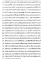


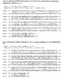
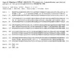
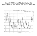


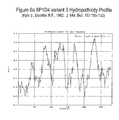
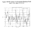

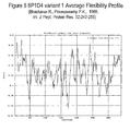

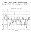
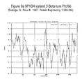
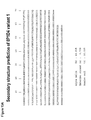
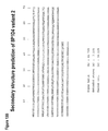

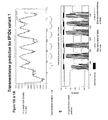
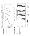

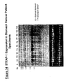
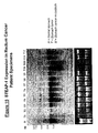


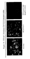
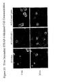
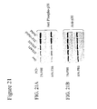
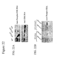


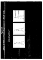
Classifications
-
- C—CHEMISTRY; METALLURGY
- C07—ORGANIC CHEMISTRY
- C07K—PEPTIDES
- C07K14/00—Peptides having more than 20 amino acids; Gastrins; Somatostatins; Melanotropins; Derivatives thereof
- C07K14/435—Peptides having more than 20 amino acids; Gastrins; Somatostatins; Melanotropins; Derivatives thereof from animals; from humans
- C07K14/46—Peptides having more than 20 amino acids; Gastrins; Somatostatins; Melanotropins; Derivatives thereof from animals; from humans from vertebrates
- C07K14/47—Peptides having more than 20 amino acids; Gastrins; Somatostatins; Melanotropins; Derivatives thereof from animals; from humans from vertebrates from mammals
- C07K14/4701—Peptides having more than 20 amino acids; Gastrins; Somatostatins; Melanotropins; Derivatives thereof from animals; from humans from vertebrates from mammals not used
- C07K14/4748—Tumour specific antigens; Tumour rejection antigen precursors [TRAP], e.g. MAGE
-
- A—HUMAN NECESSITIES
- A61—MEDICAL OR VETERINARY SCIENCE; HYGIENE
- A61K—PREPARATIONS FOR MEDICAL, DENTAL OR TOILETRY PURPOSES
- A61K39/00—Medicinal preparations containing antigens or antibodies
- A61K39/46—Cellular immunotherapy
- A61K39/461—Cellular immunotherapy characterised by the cell type used
- A61K39/4615—Dendritic cells
-
- A—HUMAN NECESSITIES
- A61—MEDICAL OR VETERINARY SCIENCE; HYGIENE
- A61K—PREPARATIONS FOR MEDICAL, DENTAL OR TOILETRY PURPOSES
- A61K39/00—Medicinal preparations containing antigens or antibodies
- A61K39/46—Cellular immunotherapy
- A61K39/462—Cellular immunotherapy characterized by the effect or the function of the cells
- A61K39/4622—Antigen presenting cells
-
- A—HUMAN NECESSITIES
- A61—MEDICAL OR VETERINARY SCIENCE; HYGIENE
- A61K—PREPARATIONS FOR MEDICAL, DENTAL OR TOILETRY PURPOSES
- A61K39/00—Medicinal preparations containing antigens or antibodies
- A61K39/46—Cellular immunotherapy
- A61K39/464—Cellular immunotherapy characterised by the antigen targeted or presented
- A61K39/4643—Vertebrate antigens
- A61K39/4644—Cancer antigens
- A61K39/464402—Receptors, cell surface antigens or cell surface determinants
-
- A—HUMAN NECESSITIES
- A61—MEDICAL OR VETERINARY SCIENCE; HYGIENE
- A61K—PREPARATIONS FOR MEDICAL, DENTAL OR TOILETRY PURPOSES
- A61K51/00—Preparations containing radioactive substances for use in therapy or testing in vivo
- A61K51/02—Preparations containing radioactive substances for use in therapy or testing in vivo characterised by the carrier, i.e. characterised by the agent or material covalently linked or complexing the radioactive nucleus
- A61K51/04—Organic compounds
- A61K51/08—Peptides, e.g. proteins, carriers being peptides, polyamino acids, proteins
- A61K51/10—Antibodies or immunoglobulins; Fragments thereof, the carrier being an antibody, an immunoglobulin or a fragment thereof, e.g. a camelised human single domain antibody or the Fc fragment of an antibody
- A61K51/1045—Antibodies or immunoglobulins; Fragments thereof, the carrier being an antibody, an immunoglobulin or a fragment thereof, e.g. a camelised human single domain antibody or the Fc fragment of an antibody against animal or human tumor cells or tumor cell determinants
-
- A—HUMAN NECESSITIES
- A61—MEDICAL OR VETERINARY SCIENCE; HYGIENE
- A61P—SPECIFIC THERAPEUTIC ACTIVITY OF CHEMICAL COMPOUNDS OR MEDICINAL PREPARATIONS
- A61P35/00—Antineoplastic agents
-
- A—HUMAN NECESSITIES
- A61—MEDICAL OR VETERINARY SCIENCE; HYGIENE
- A61P—SPECIFIC THERAPEUTIC ACTIVITY OF CHEMICAL COMPOUNDS OR MEDICINAL PREPARATIONS
- A61P37/00—Drugs for immunological or allergic disorders
- A61P37/02—Immunomodulators
- A61P37/04—Immunostimulants
-
- A—HUMAN NECESSITIES
- A61—MEDICAL OR VETERINARY SCIENCE; HYGIENE
- A61P—SPECIFIC THERAPEUTIC ACTIVITY OF CHEMICAL COMPOUNDS OR MEDICINAL PREPARATIONS
- A61P43/00—Drugs for specific purposes, not provided for in groups A61P1/00-A61P41/00
-
- C—CHEMISTRY; METALLURGY
- C07—ORGANIC CHEMISTRY
- C07K—PEPTIDES
- C07K14/00—Peptides having more than 20 amino acids; Gastrins; Somatostatins; Melanotropins; Derivatives thereof
- C07K14/435—Peptides having more than 20 amino acids; Gastrins; Somatostatins; Melanotropins; Derivatives thereof from animals; from humans
- C07K14/705—Receptors; Cell surface antigens; Cell surface determinants
- C07K14/72—Receptors; Cell surface antigens; Cell surface determinants for hormones
- C07K14/723—G protein coupled receptor, e.g. TSHR-thyrotropin-receptor, LH/hCG receptor, FSH receptor
-
- C—CHEMISTRY; METALLURGY
- C07—ORGANIC CHEMISTRY
- C07K—PEPTIDES
- C07K16/00—Immunoglobulins [IGs], e.g. monoclonal or polyclonal antibodies
- C07K16/18—Immunoglobulins [IGs], e.g. monoclonal or polyclonal antibodies against material from animals or humans
- C07K16/28—Immunoglobulins [IGs], e.g. monoclonal or polyclonal antibodies against material from animals or humans against receptors, cell surface antigens or cell surface determinants
- C07K16/30—Immunoglobulins [IGs], e.g. monoclonal or polyclonal antibodies against material from animals or humans against receptors, cell surface antigens or cell surface determinants from tumour cells
-
- G—PHYSICS
- G01—MEASURING; TESTING
- G01N—INVESTIGATING OR ANALYSING MATERIALS BY DETERMINING THEIR CHEMICAL OR PHYSICAL PROPERTIES
- G01N33/00—Investigating or analysing materials by specific methods not covered by groups G01N1/00 - G01N31/00
- G01N33/48—Biological material, e.g. blood, urine; Haemocytometers
- G01N33/50—Chemical analysis of biological material, e.g. blood, urine; Testing involving biospecific ligand binding methods; Immunological testing
- G01N33/53—Immunoassay; Biospecific binding assay; Materials therefor
- G01N33/574—Immunoassay; Biospecific binding assay; Materials therefor for cancer
- G01N33/57484—Immunoassay; Biospecific binding assay; Materials therefor for cancer involving compounds serving as markers for tumor, cancer, neoplasia, e.g. cellular determinants, receptors, heat shock/stress proteins, A-protein, oligosaccharides, metabolites
-
- A—HUMAN NECESSITIES
- A61—MEDICAL OR VETERINARY SCIENCE; HYGIENE
- A61K—PREPARATIONS FOR MEDICAL, DENTAL OR TOILETRY PURPOSES
- A61K39/00—Medicinal preparations containing antigens or antibodies
- A61K2039/505—Medicinal preparations containing antigens or antibodies comprising antibodies
-
- A—HUMAN NECESSITIES
- A61—MEDICAL OR VETERINARY SCIENCE; HYGIENE
- A61K—PREPARATIONS FOR MEDICAL, DENTAL OR TOILETRY PURPOSES
- A61K2239/00—Indexing codes associated with cellular immunotherapy of group A61K39/46
- A61K2239/38—Indexing codes associated with cellular immunotherapy of group A61K39/46 characterised by the dose, timing or administration schedule
-
- A—HUMAN NECESSITIES
- A61—MEDICAL OR VETERINARY SCIENCE; HYGIENE
- A61K—PREPARATIONS FOR MEDICAL, DENTAL OR TOILETRY PURPOSES
- A61K38/00—Medicinal preparations containing peptides
-
- A—HUMAN NECESSITIES
- A61—MEDICAL OR VETERINARY SCIENCE; HYGIENE
- A61K—PREPARATIONS FOR MEDICAL, DENTAL OR TOILETRY PURPOSES
- A61K39/00—Medicinal preparations containing antigens or antibodies
-
- C—CHEMISTRY; METALLURGY
- C07—ORGANIC CHEMISTRY
- C07K—PEPTIDES
- C07K2319/00—Fusion polypeptide
Definitions
- the invention described herein relates to a gene and its encoded protein, termed 08P1D4 or STEAP-1, expressed in certain cancers, and to diagnostic and therapeutic methods and compositions useful in the management of cancers that express STEAP-1.
- Cancer is the second leading cause of human death next to coronary disease. Worldwide, millions of people die from cancer every year. In the United States alone, as reported by the American Cancer Society, cancer causes the death of well over a half-million people annually, with over 1.2 million new cases diagnosed per year. While deaths from heart disease have been declining significantly, those resulting from cancer generally are on the rise. In the early part of the next century, cancer is predicted to become the leading cause of death.
- carcinomas of the lung, prostate, breast, colon, pancreas, and ovary represent the primary causes of cancer death. These and virtually all other carcinomas share a common lethal feature. With very few exceptions, metastatic disease from a carcinoma is fatal. Moreover, even for those cancer patients who initially survive their primary cancers, common experience has shown that their lives are dramatically altered. Many cancer patients experience strong anxieties driven by the awareness of the potential for recurrence or treatment failure. Many cancer patients experience physical debilitations following treatment. Furthermore, many cancer patients experience a recurrence.
- prostate cancer is the fourth most prevalent cancer in men. In North America and Northern Europe, it is by far the most common cancer in males and is the second leading cause of cancer death in men. In the United States alone, well over 30,000 men die annually of this disease - second only to lung cancer. Despite the magnitude of these figures, there is still no effective treatment for metastatic prostate cancer. Surgical prostatectomy, radiation therapy, hormone ablation therapy, surgical castration and chemotherapy continue to be the main treatment modalities. Unfortunately, these treatments are ineffective for many and are often associated with undesirable consequences.
- PSA serum prostate specific antigen
- the LAPC L os A ngeles P rostate Cancer
- the LAPC xenografts are prostate cancer xenografts that have survived passage in severe combined immune deficient (SCID) mice and have exhibited the capacity to mimic the transition from androgen dependence to androgen independence (Klein et al., 1997, Nat. Med. 3:402).
- More recently identified prostate cancer markers include PCTA-1 ( Su et al., 1996, Proc. Natl. Acad. Sci.
- PSM prostate-specific membrane
- PSCA prostate stem cell antigen
- Renal cell carcinoma accounts for approximately 3 percent of adult malignancies. Once adenomas reach a diameter of 2 to 3 cm, malignant potential exists. In the adult, the two principal malignant renal tumors are renal cell adenocarcinoma and transitional cell carcinoma of the renal pelvis or ureter. The incidence of renal cell adenocarcinoma is estimated at more than 29,000 cases in the United States, and more than 11,600 patients died of this disease in 1998. Transitional cell carcinoma is less frequent, with an incidence of approximately 500 cases per year in the United States.
- bladder cancer represents approximately 5 percent in men (fifth most common neoplasm) and 3 percent in women (eighth most common neoplasm). The incidence is increasing slowly, concurrent with an increasing older population. In 1998, there was an estimated 54,500 cases, including 39,500 in men and 15,000 in women. The age-adjusted incidence in the United States is 32 per 100,000 for men and eight per 100,000 in women. The historic male/female ratio of 3:1 may be decreasing related to smoking patterns in women. There were an estimated 11,000 deaths from bladder cancer in 1998 (7,800 in men and 3,900 in women). Bladder cancer incidence and mortality strongly increase with age and will be an increasing problem as the population becomes more elderly.
- bladder cancers recur in the bladder.
- Bladder cancer is managed with a combination of transurethral resection of the bladder (TUR) and intravesical chemotherapy or immunotherapy.
- TUR transurethral resection of the bladder
- the multifocal and recurrent nature of bladder cancer points out the limitations of TUR.
- Most muscle-invasive cancers are not cured by TUR alone. Radical cystectomy and urinary diversion is the most effective means to eliminate the cancer but carry an undeniable impact on urinary and sexual function. There continues to be a significant need for treatment modalities that are beneficial for bladder cancer patients.
- Treatment options for lung and bronchial cancer are determined by the type and stage of the cancer and include surgery, radiation therapy, and chemotherapy. For many localized cancers, surgery is usually the treatment of choice. Because the disease has usually spread by the time it is discovered, radiation therapy and chemotherapy are often needed in combination with surgery. Chemotherapy alone or combined with radiation is the treatment of choice for small cell lung cancer; on this regimen, a large percentage of patients experience remission, which in some cases is long lasting. There is however, an ongoing need for effective treatment and diagnostic approaches for lung and bronchial cancers.
- treatment of breast cancer may involve lumpectomy (local removal of the tumor) and removal of the lymph nodes under the arm; mastectomy (surgical removal of the breast) and removal of the lymph nodes under the arm; radiation therapy; chemotherapy; or hormone therapy.
- lumpectomy local removal of the tumor
- mastectomy surgical removal of the breast
- radiation therapy chemotherapy
- hormone therapy chemotherapy
- two or more methods are used in combination.
- Numerous studies have shown that, for early stage disease, long-term survival rates after lumpectomy plus radiotherapy are similar to survival rates after modified radical mastectomy.
- Significant advances in reconstruction techniques provide several options for breast reconstruction after mastectomy. Recently, such reconstruction has been done at the same time as the mastectomy.
- DCIS ductal carcinoma in situ
- Surgery, radiation therapy, and chemotherapy are treatment options for ovarian cancer.
- Surgery usually includes the removal of one or both ovaries, the fallopian tubes (salpingo-oophorectomy), and the uterus (hysterectomy).
- the fallopian tubes salivary-oophorectomy
- the uterus hematoma-oophorectomy
- pancreatic cancer There were an estimated 28,300 new cases of pancreatic cancer in the United States in 2000. Over the past 20 years, rates of pancreatic cancer have declined in men. Rates among women have remained approximately constant but may be beginning to decline. Pancreatic cancer caused an estimated 28,200 deaths in 2000 in the United States. Over the past 20 years, there has been a slight but significant decrease in mortality rates among men (about-0.9% per year) while rates have increased slightly among women.
- pancreatic cancer Surgery, radiation therapy, and chemotherapy are treatment options for pancreatic cancer. These treatment options can extend survival and/or relieve symptoms in many patients but are not likely to produce a cure for most. There is a significant need for additional therapeutic and diagnostic options for pancreatic cancer.
- the present invention relates to a gene, designated STEAP-1, that has now been found to be over-expressed in the cancer(s) listed in Table I.
- Northern blot expression analysis of STEAP-1 gene expression in normal tissues shows a restricted expression pattern in adult tissues.
- the nucleotide ( Figure 2 ) and amino acid ( Figure 2 , and Figure 3 ) sequences of STEAP-1 are provided.
- the tissue-related profile of STEAP-1 in normal adult tissues, combined with the over-expression observed in the tissues listed in Table I, shows that STEAP-1 is aberrantly over-expressed in at least some cancers, and thus serves as a useful diagnostic, prophylactic, prognostic, and/or therapeutic target for cancers of the tissue(s) such as those listed in Table I.
- the invention provides compositions, pharmaceutical compositions, antibodies and fragments thereof, non-human transgenic animals, and hybridomas, as defined in the claims.
- the invention also provides compositions, polynucleotides and vectors for use in inhibiting growth of cancer cells, as defined in the claims.
- the invention further provides peptides and polynucleotides for use in the treatment of cancer, as defined in the claims.
- the invention also provides uses of compositions, polynucleotides and vectors in the manufacture of a medicament for inhibiting the growth of cancer cells, as defined in the claims. Additionally, the invention provides uses of peptides and nucleotide sequences in the treatment of cancer, as defined in the claims. The invention also provides methods for detecting the presence of a protein in a sample, as defined in the claims.
- compositions of the invention may comprise peptides consisting of an amino acid sequence selected from the group consisting of amino acids 32-40, 80-88, 99-107, 122-130, 153-161, 180-188, 21-30, 79-88, 121-130, 173-182, 243-252, 244-252, 277-286, 108-116, 140-148, 307-315, 140-149, 307-316, 118-126, 150-158, 294-302, 310-318, 109-118 and 150-159 of SEQ ID NO:3, or an analogue of the above peptides which a residue in a primary and/or secondary anchor position shown in Table IV is substituted with another amino acid specified for that position in Table IV in order to increase the binding affinity, binding half-life and/or cross-reactivity of that peptide.
- prostate cancer and “locally advanced disease” mean prostate cancers that have extended through the prostate capsule, and are meant to include stage C disease under the American Urological Association (AUA) system, stage C1 - C2 disease under the Whitmore-Jewett system, and stage T3 - T4 and N+ disease under the TNM (tumor, node, metastasis) system.
- AUA American Urological Association
- stage C1 - C2 disease under the Whitmore-Jewett system
- TNM tumor, node, metastasis
- surgery is not recommended for patients with locally advanced disease, and these patients have substantially less favorable outcomes compared to patients having clinically localized (organ-confined) prostate cancer.
- Locally advanced disease is clinically identified by palpable evidence of induration beyond the lateral border of the prostate, or asymmetry or induration above the prostate base.
- Locally advanced prostate cancer is presently diagnosed pathologically following radical prostatectomy if the tumor invades or penetrates the prostatic capsule, extends into the surgical margin, or invades the seminal
- “Altering the native glycosylation pattern” is intended for purposes herein to mean deleting one or more carbohydrate moieties found in native sequence STEAP-1 (either by removing the underlying glycosylation site or by deleting the glycosylation by chemical and/or enzymatic means), and/or adding one or more glycosylation sites that are not present in the native sequence STEAP-1.
- the phrase includes qualitative changes in the glycosylation of the native proteins, involving a change in the nature and proportions of the various carbohydrate moieties present.
- an analog refers to a molecule which is structurally similar or shares similar or corresponding attributes with another molecule (e.g. a STEAP-1-related protein).
- a STEAP-1-related protein e.g. an analog of a STEAP-1 protein can be specifically bound by an antibody or T cell that specifically binds to STEAP-1.
- Antibody is used in the broadest sense. Therefore, an “antibody” can be naturally occurring or man-made such as monoclonal antibodies produced by conventional hybridoma technology.
- Anti-STEAP-1 antibodies comprise monoclonal and polyclonal antibodies as well as fragments containing the antigen-binding domain and/or one or more complementarity determining regions of these antibodies.
- an “antibody fragment” is defined as at least a portion of the variable region of the immunoglobulin molecule that binds to its target, i.e., the antigen-binding region. In one embodiment it specifically covers single anti-STEAP-1 antibodies and clones thereof (including agonist, antagonist and neutralizing antibodies) and anti-STEAP-1 antibody compositions with polyepitopic specificity.
- codon optimized sequences refers to nucleotide sequences that have been optimized for a particular host species by replacing any codons having a usage frequency of less than about 20%. Nucleotide sequences that have been optimized for expression in a given host species by elimination of spurious polyadenylation sequences, elimination of exon/intron splicing signals, elimination of transposon-like repeats and/or optimization of GC content in addition to codon optimization are referred to herein as an "expression enhanced sequences.”
- cytotoxic agent refers to a substance that inhibits or prevents the expression activity of cells, function of cells and/or causes destruction of cells.
- the term is intended to include radioactive isotopes chemotherapeutic agents, and toxins such as small molecule toxins or enzymatically active toxins of bacterial, fungal, plant or animal origin, including fragments and/or variants thereof.
- cytotoxic agents include, but are not limited to auristatins, auromycins, maytansinoids, yttrium, bismuth, ricin, ricin A-chain, combrestatin, duocarmycins, dolostatins, doxorubicin, daunorubicin, taxol, cisplatin, cc1065, ethidium bromide, mitomycin, etoposide, tenoposide, vincristine, vinblastine, colchicine, dihydroxy anthracin dione, actinomycin, diphtheria toxin, Pseudomonas exotoxin (PE) A, PE40, abrin, abrin A chain, modeccin A chain, alpha-sarcin, gelonin, mitogellin, retstrictocin, phenomycin, enomycin, curicin, crotin, calicheamicin, Sapaonaria offcinalis inhibitor
- homolog refers to a molecule which exhibits homology to another molecule, by for example, having sequences of chemical residues that are the same or similar at corresponding positions.
- HLA Human Leukocyte Antigen
- HLA Human Leukocyte Antigen
- MHC Major Histocompatibility Complex
- hybridize used in the context of polynucleotides, are meant to refer to conventional hybridization conditions, preferably such as hybridization in 50% formamide/6XSSC/0.1% SDS/100 ⁇ g/ml ssDNA, in which temperatures for hybridization are above 37 degrees C and temperatures for washing in 0.1XSSC/0.1% SDS are above 55 degrees C.
- isolated or “biologically pure” refer to material which is substantially or essentially free from components which normally accompany the material as it is found in its native state.
- isolated peptides in accordance with the invention preferably do not contain materials normally associated with the peptides in their in situ environment.
- a polynucleotide is said to be “isolated” when it is substantially separated from contaminant polynucleotides that correspond or are complementary to genes other than the STEAP-1 genes or that encode polypeptides other than STEAP-1 gene product or fragments thereof.
- a skilled artisan can readily employ nucleic acid isolation procedures to obtain an isolated STEAP-1 polynucleotide.
- a protein is said to be "isolated,” for example, when physical, mechanical or chemical methods are employed to remove the STEAP-1 proteins from cellular constituents that are normally associated with the protein.
- a skilled artisan can readily employ standard purification methods to obtain an isolated STEAP-1 protein.
- an isolated protein can be prepared by chemical means.
- mammal refers to any organism classified as a mammal, including mice, rats, rabbits, dogs, cats, cows, horses and humans. In one embodiment of the invention, the mammal is a mouse. The mammal may be a human.
- metastatic prostate cancer and “metastatic disease” mean prostate cancers that have spread to regional lymph nodes or to distant sites, and are meant to include stage D disease under the AUA system and stage TxNxM+ under the TNM system.
- surgery is generally not indicated for patients with metastatic disease, and hormonal (androgen ablation) therapy is a preferred treatment modality.
- Patients with metastatic prostate cancer eventually develop an androgen-refractory state within 12 to 18 months of treatment initiation. Approximately half of these androgen-refractory patients die within 6 months after developing that status.
- the most common site for prostate cancer metastasis is bone. Prostate cancer bone metastases are often osteoblastic rather than osteolytic (i.e., resulting in net bone formation).
- Bone metastases are found most frequently in the spine, followed by the femur, pelvis, rib cage, skull and humerus. Other common sites for metastasis include lymph nodes, lung, liver and brain. Metastatic prostate cancer is typically diagnosed by open or laparoscopic pelvic lymphadenectomy, whole body radionuclide scans, skeletal radiography, and/or bone lesion biopsy.
- the term "monoclonal antibody” refers to an antibody obtained from a population of substantially homogeneous antibodies, i.e., the antibodies comprising the population are identical except for possible naturally occurring mutations that are present in minor amounts.
- a “motif”, as in biological motif of a STEAP-1-related protein, refers to any pattern of amino acids forming part of the primary sequence of a protein, that is associated with a particular function (e.g. protein-protein interaction, protein-DNA interaction, etc) or modification (e.g. that is phosphorylated, glycosylated or amidated), or localization (e.g. secretory sequence, nuclear localization sequence, etc.) or a sequence that is correlated with being immunogenic, either humorally or cellularly.
- a motif can be either contiguous or capable of being aligned to certain positions that are generally correlated with a certain function or property.
- motif refers to the pattern of residues in a peptide of defined length, usually a peptide of from about 8 to about 13 amino acids for a class I HLA motif and from about 6 to about 25 amino acids for a class II HLA motif, which is recognized by a particular HLA molecule.
- Peptide motifs for HLA binding are typically different for each protein encoded by each human HLA allele and differ in the pattern of the primary and secondary anchor residues.
- a “pharmaceutical excipient” comprises a material such as an adjuvant, a carrier, pH-adjusting and buffering agents, tonicity adjusting agents, wetting agents, preservative, and the like.
- “Pharmaceutically acceptable” refers to a non-toxic, inert, and/or composition that is physiologically compatible with humans or other mammals.
- polynucleotide means a polymeric form of nucleotides of at least 10 bases or base pairs in length, either ribonucleotides or deoxynucleotides or a modified form of either type of nucleotide, and is meant to include single and double stranded forms of DNA and/or RNA. In the art, this term if often used interchangeably with “oligonucleotide”.
- a polynucleotide can comprise a nucleotide sequence disclosed herein wherein thymidine (T), as shown for example in Figure 2 , can also be uracil (U); this definition pertains to the differences between the chemical structures of DNA and RNA, in particular the observation that one of the four major bases in RNA is uracil (U) instead of thymidine (T).
- T thymidine
- U uracil
- polypeptide means a polymer of at least about 4, 5, 6, 7, or 8 amino acids. Throughout the specification, standard three letter or single letter designations for amino acids are used. In the art, this term is often used interchangeably with “peptide” or "protein”.
- HLA "primary anchor residue” is an amino acid at a specific position along a peptide sequence which is understood to provide a contact point between the immunogenic peptide and the HLA molecule.
- One to three, usually two, primary anchor residues within a peptide of defined length generally defines a "motif" for an immunogenic peptide. These residues are understood to fit in close contact with peptide binding groove of an HLA molecule, with their side chains buried in specific pockets of the binding groove.
- the primary anchor residues for an HLA class I molecule are located at position 2 (from the amino terminal position) and at the carboxyl terminal position of a 8, 9, 10, 11, or 12 residue peptide epitope in accordance with the invention.
- the primary anchor residues of a peptide that will bind an HLA class II molecule are spaced relative to each other, rather than to the termini of a peptide, where the peptide is generally of at least 9 amino acids in length.
- the primary anchor positions for each motif and supermotif are set forth in Table IV.
- analog peptides can be created by altering the presence or absence of particular residues in the primary and/or secondary anchor positions shown in Table IV. Such analogs are used to modulate the binding affinity and/or population coverage of a peptide comprising a particular HLA motif or supermotif.
- Radioisotopes include, but are not limited to the following (non-limiting exemplary uses are also set forth): Examples of Medical Isotopes: Isotope Description of use Actinium-225 (AC-225) See Thorium-229 (Th-229) Actinium-227 (AC-227) Parent of Radium-223 (Ra-223) which is an alpha emitter used to treat metastases in the skeleton resulting from cancer (i.e., breast and prostate cancers), and cancer radioimmunotherapy Bismuth-212 (Bi-212) See Thorium-228 (Th-228) Bismuth-213 (Bi-213) See Thorium-229 (Th-229) Cadmium-109 (Cd-109) Cancer detection Cobalt-60 (Co-60) Radiation source for radiotherapy of cancer, for food irradiators, and for sterilization of medical supplies Copper-64 (Cu-64) A positron emitter used for cancer therapy and SPECT imaging Copper-67 (Cu-67) Beta/gamma emitter used in cancer radioimmun
- Tc-99m is the most widely used radioisotope used for diagnostic imaging of various cancers and diseases involving the brain, heart, liver, lungs; also used in detection of deep vein thrombosis of the legs Osmium-194 (Os-194) Cancer radioimmunotherapy Palladium-103 (Pd-103) Prostate cancer treatment Platinum-195m (Pt-195m)
- Phosphorus-32 P-32
- Polycythemia rubra vera blood cell disease
- leukemia treatment bone cancer diagnosis/treatment
- colon, pancreatic, and liver cancer treatment radiolabeling nucleic acids for in vitro research, diagnosis of superficial tumors, treatment of blocked arteries (i.e., arteriosclerosis and restenosis), and intracavity therapy Phosphorus-33 (P-33)
- Leukemia treatment, bone disease diagnosis/treatment, radiolabeling, and treatment of blocked arteries i.e.,
- a "recombinant" DNA or RNA molecule is a DNA or RNA molecule that has been subjected to molecular manipulation in vitro,
- Non-limiting examples of small molecules include compounds that bind or interact with STEAP-1, ligands including hormones, neuropeptides, chemokines, odorants, phospholipids, and functional equivalents thereof that bind and preferably inhibit STEAP-1 protein function.
- Such non-limiting small molecules preferably have a molecular weight of less than about 10 kDa, more preferably below about 9, about 8, about 7, about 6, about 5 or about 4 kDa. Small molecules may physically associate with, or bind, STEAP-1 protein; are not found in naturally occurring metabolic pathways; and/or are more soluble in aqueous than non-aqueous solutions
- “Stringency” of hybridization reactions is readily determinable by one of ordinary skill in the art, and generally is an empirical calculation dependent upon probe length, washing temperature, and salt concentration. In general, longer probes require higher temperatures for proper annealing, while shorter probes need lower temperatures. Hybridization generally depends on the ability of denatured nucleic acid sequences to reanneal when complementary strands are present in an environment below their melting temperature. The higher the degree of desired homology between the probe and hybridizable sequence, the higher the relative temperature that can be used. As a result, it follows that higher relative temperatures would tend to make the reaction conditions more stringent, while lower temperatures less so. For additional details and explanation of stringency of hybridization reactions, see Ausubel et al., Current Protocols in Molecular Biology, Wiley Interscience Publishers, (1995 ).
- “Stringent conditions” or “high stringency conditions”, as defined herein, are identified by, but not limited to, those that: (1) employ low ionic strength and high temperature for washing, for example 0.015 M sodium chloride/0.0015 M sodium citrate/0.1% sodium dodecyl sulfate at 50°C; (2) employ during hybridization a denaturing agent, such as formamide, for example, 50% (v/v) formamide with 0.1% bovine serum albumin/0.1% Ficoll/0.1 % polyvinylpyrrolidone/50 mM sodium phosphate buffer at pH 6.5 with 750 mM sodium chloride, 75 mM sodium citrate at 42 °C; or (3) employ 50% formamide, 5 x SSC (0.75 M NaCl, 0.075 M sodium citrate), 50 mM sodium phosphate (pH 6.8), 0.1% sodium pyrophosphate, 5 x Denhardt's solution, sonicated salmon sperm DNA (50 ⁇ g/ml), 0.1% SDS,
- Modely stringent conditions are described by, but not limited to, those in Sambrook et al., Molecular Cloning: A Laboratory Manual, New York: Cold Spring Harbor Press, 1989 , and include the use of washing solution and hybridization conditions (e.g., temperature, ionic strength and %SDS) less stringent than those described above.
- moderately stringent conditions is overnight incubation at 37°C in a solution comprising: 20% formamide, 5 x SSC (150 mM NaCl, 15 mM trisodium citrate), 50 mM sodium phosphate (pH 7.6), 5 x Denhardt's solution, 10% dextran sulfate, and 20 mg/mL denatured sheared salmon sperm DNA, followed by washing the filters in 1 x SSC at about 37-50°C.
- 5 x SSC 150 mM NaCl, 15 mM trisodium citrate
- 50 mM sodium phosphate pH 7.6
- 5 x Denhardt's solution 10% dextran sulfate
- 20 mg/mL denatured sheared salmon sperm DNA followed by washing the filters in 1 x SSC at about 37-50°C.
- the skilled artisan will recognize how to adjust the temperature, ionic strength, etc. as necessary to accommodate factors such as probe length and the like.
- HLA "supermotif” is a peptide binding specificity shared by HLA molecules encoded by two or more HLA alleles.
- to treat or "therapeutic” and grammatically related terms, refer to any improvement of any consequence of disease, such as prolonged survival, less morbidity, and/or a lessening of side effects which are the byproducts of an alternative therapeutic modality; full eradication of disease is not required.
- transgenic animal e.g., a mouse or rat
- transgene is an animal having cells that contain a transgene, which transgene was introduced into the animal or an ancestor of the animal at a prenatal, e.g., an embryonic stage.
- transgene is a DNA that is integrated into the genome of a cell from which a transgenic animal develops.
- an HLA or cellular immune response "vaccine” is a composition that contains or encodes one or more peptides of the invention.
- vaccines such as a cocktail of one or more individual peptides; one or more peptides of the invention comprised by a polyepitopic peptide; or nucleic acids that encode such individual peptides or polypeptides, e.g., a minigene that encodes a polyepitopic peptide.
- the "one or more peptides” can include any whole unit integer from 1-150 or more, e.g., at least 2, 3, 4, 5, 6, 7, 8, 9, 10, 11, 12, 13, 14, 15, 16, 17, 18, 19, 20, 21, 22, 23, 24, 25, 26, 27, 28, 29, 30, 31, 32, 33, 34, 35, 36, 37, 38, 39, 40, 41, 42, 43, 44, 45, 46, 47, 48, 49, 50, 55, 60, 65, 70, 75, 80, 85, 90, 95, 100, 105, 110, 115, 120, 125, 130,135, 140, 145, or 150 or more peptides of the invention.
- the peptides or polypeptides can optionally be modified, such as by lipidation, addition of targeting or other sequences.
- HLA class I peptides of the invention can be admixed with, or linked to, HLA class II peptides, to facilitate activation of both cytotoxic T lymphocytes and helper T lymphocytes.
- HLA vaccines can also comprise peptide-pulsed antigen presenting cells, e.g., dendritic cells.
- variant refers to a molecule that exhibits a variation from a described type or norm, such as a protein that has one or more different amino acid residues in the corresponding position(s) of a specifically described protein (e.g. the STEAP-1 protein shown in Figure 2 or Figure 3 .
- An analog is an example of a variant protein.
- Splice isoforms and single nucleotides polymorphisms (SNPs) are further examples of variants.
- the "STEAP-1-related proteins" of the invention include those specifically identified herein, as well as allelic variants, conservative substitution variants, analogs and homologs that can be isolated/generated and characterized without undue experimentation following the methods outlined herein or readily available in the art. Fusion proteins that combine parts of different STEAP-1 proteins or fragments thereof, as well as fusion proteins of a STEAP-1 protein and a heterologous polypeptide are also included. Such STEAP-1 proteins are collectively referred to as the STEAP-1-related proteins, the proteins of the invention, or STEAP-1.
- STEAP-1-related protein refers to a polypeptide fragment or a STEAP-1 protein sequence of 4, 5, 6, 7, 8, 9, 10, 11, 12, 13, 14, 15, 16, 17, 18, 19, 20, 21, 22, 23, 24, 25, or more than 25 amino acids; or, at least 30, 35, 40, 45, 50, 55, 60, 65, 70, 80, 85, 90, 95, 100, 105, 110, 115, 120, 125, 130, 135, 140, 145, 150, 155, 160, 165, 170, 175, 180, 185, 190, 195, 200, 225, 250, 275, 300, 325, 350, 375, 400, 425, 450, 475, 500, 525, 550, 575, 600, 625, 650, or 664 or more amino acids.
- polynucleotides corresponding or complementary to all or part of a STEAP-1 gene, mRNA, and/or coding sequence preferably in isolated form, including polynucleotides encoding a STEAP-1-related protein and fragments thereof, DNA, RNA, DNA/RNA hybrid, and related molecules, polynucleotides or oligonucleotides complementary to a STEAP-1 gene or mRNA sequence or apart thereof, and polynucleotides or oligonucleotides that hybridize to a STEAP-1 gene, mRNA, or to a STEAP-1 encoding polynucleotide (collectively, "STEAP-1 polynucleotides").
- T can also be U in Figure 2 .
- Examples of a STEAP-1 polynucleotide include: a STEAP-1 polynucleotide having the sequence shown in Figure 2 , the nucleotide sequence of STEAP-1 as shown in Figure 2 wherein T is U; at least 10 contiguous nucleotides of a polynucleotide having the sequence as shown in Figure 2 ; or, at least 10 contiguous nucleotides of a polynucleotide having the sequence as shown in Figure 2 where T is U.
- Examples of STEAP-1 nucleotides provided herein include:
- STEAP-1 polynucleotides that encode specific portions of STEAP-1 mRNA sequences (and those which are complementary to such sequences) such as those that encode the proteins and/or fragments thereof, for example: .
- polynucleotides and their encoded peptides themselves encoding about amino acid 1 to about amino acid 10 of the STEAP-1 protein shown in Figure 2 or Figure 3 polynucleotides encoding about amino acid 10 to about amino acid 20 of the STEAP-1 protein shown in Figure 2 or Figure 3
- polynucleotides encoding about amino acid 20 to about amino acid 30 of the STEAP-1 protein shown in Figure 2 or Figure 3 polynucleotides encoding about amino acid 30 to about amino acid 40 of the STEAP-1 protein shown in Figure 2 or Figure 3
- polynucleotides encoding about amino acid 40 to about amino acid 50 of the STEAP-1 protein shown in Figure 2 or Figure 3 polynucleotides encoding about amino acid 50 to about amino acid 60 of the STEAP-1 protein shown in Figure 2 or Figure 3
- polynucleotides encoding portions of the amino acid sequence (of about 10 amino acids), of amino acids, 100 through the disclosed herein carboxyl terminal amino acid of the STEAP-1 protein are disclosed herein. Wherein it is understood that each particular amino acid position discloses that position plus or minus five amino acid residues.
- Polynucleotides encoding relatively long portions of a STEAP-1 protein are also disclosed.
- polynucleotides encoding from about amino acid 1 (or 20 or 30 or 40 etc.) to about amino acid 20, (or 30, or 40 or 50 etc.) of the STEAP-1 protein "or variant" shown in Figure 2 or Figure 3 can be generated by a variety of techniques well known in the art.
- These polynucleotide fragments can include any portion of the STEAP-1 sequence as shown in Figure 2 .
- STEAP-1 polynucleotide fragments encoding one or more of the biological motifs contained within a STEAP-1 protein "or variant" sequence, including one or more of the motif-bearing subsequences of a STEAP-1 protein "or variant” set forth in Tables V-XVIII and XXII-LI.
- Typical polynucleotide fragments may encode one or more of the regions of STEAP-1 protein or variant that exhibit homology to a known molecule.
- typical polynucleotide fragments can encode one or more of the STEAP-1 protein or variant N-glycosylation sites, cAMP and cGMP-dependent protein kinase phosphorylation sites, casein kinase II phosphorylation sites or N-myristoylation site and amidation sites.
- HLA Peptide Tables respective to its parental protein, e.g., variant 1, variant 2, etc.
- HLA Peptide Tables respective to its parental protein, e.g., variant 1, variant 2, etc.
- search Peptides listed in Table LVII Generally, a unique Search Peptide is used to obtain HLA peptides for a particular variant. The position of each Search Peptide relative to its respective parent molecule is listed in Table LXI.
- a Search Peptide begins at position "X"
- a particular Search Peptide begins at position 150 of its parental molecule, one must add 150 - 1, i.e., 149 to each HLA peptide amino acid position to calculate the position of that amino acid in the parent molecule.
- the polynucleotides of the preceding paragraphs have a number of different specific uses.
- the human STEAP-1 gene maps to the chromosomal location set forth in the Example entitled "Chromosomal Mapping of STEAP-1.”
- polynucleotides that encode different regions of the STEAP-1 proteins are used to characterize cytogenetic abnormalities of this chromosomal locale, such as abnormalities that are identified as being associated with various cancers.
- cytogenetic abnormalities of this chromosomal locale such as abnormalities that are identified as being associated with various cancers.
- a variety of chromosomal abnormalities including rearrangements have been identified as frequent cytogenetic abnormalities in a number of different cancers (see e.g. Krajinovic et al., Mutat. Res.
- polynucleotides encoding specific regions of the STEAP-1 proteins provide new tools that can be used to delineate, with greater precision than previously possible, cytogenetic abnormalities in the chromosomal region that encodes STEAP-1 that may contribute to the malignant phenotype.
- these polynucleotides satisfy a need in the art for expanding the sensitivity of chromosomal screening in order to identify more subtle and less common chromosomal abnormalities (see e.g. Evans et al., Am. J. Obstet. Gynecol 171(4): 1055-1057 (1994 )).
- STEAP-1 polynucleotides are used in methods assessing the status of STEAP-1 gene products in normal versus cancerous tissues.
- polynucleotides that encode specific regions of the STEAP-1 proteins are used to assess the presence of perturbations (such as deletions, insertions, point mutations, or alterations resulting in a loss of an antigen etc.) in specific regions of the STEAP-1 gene, such as regions containing one or more motifs.
- Exemplary assays include both RT-PCR assays as well as single-strand conformation polymorphism (SSCP) analysis (see, e.g., Marrogi et al., J. Cutan. Pathol. 26(8): 369-378 (1999 ), both of which utilize polynucleotides encoding specific regions of a protein to examine these regions within the protein.
- SSCP single-strand conformation polymorphism
- genomic DNA can be RNAs or other molecules, including peptide nucleic acids (PNAs) or non-nucleic acid molecules such as phosphorothioate derivatives that specifically bind DNA or RNA in a base pair-dependent manner.
- PNAs peptide nucleic acids
- non-nucleic acid molecules such as phosphorothioate derivatives that specifically bind DNA or RNA in a base pair-dependent manner.
- Antisense technology entails the administration of exogenous oligonucleotides that bind to a target polynucleotide located within the cells.
- the term "antisense” refers to the fact that such oligonucleotides are complementary to their intracellular targets, e.g., STEAP-1. See for example, Jack Cohen, Oligodeoxynucleotides, Antisense Inhibitors of Gene Expression, CRC Press, 1989 ; and Synthesis 1:1-5 (1988 ).
- the STEAP-1 antisense oligonucleotides of the present invention include derivatives such as S-oligonucleotides (phosphorothioate derivatives or S-oligos, see, Jack Cohen, supra), which exhibit enhanced cancer cell growth inhibitory action.
- S-oligos are isoelectronic analogs of an oligonucleotide (O-oligo) in which a nonbridging oxygen atom of the phosphate group is replaced by a sulfur atom.
- the S-oligos of the present invention can be prepared by treatment of the corresponding O-oligos with 3H-1,2-benzodithiol-3-one-1,1-dioxide, which is a sulfur transfer reagent. See, e.g., Iyer, R. P. et al., J. Org. Chem. 55:4693-4698 (1990 ); and Iyer, R. P. et al., J. Am. Chem. Soc. 112:1253-1254 (1990 ).
- Additional STEAP-1 antisense oligonucleotides of the present invention include morpholino antisense oligonucleotides known in the art (see, e.g., Partridge et al., 1996, Antisense & Nucleic Acid Drug Development 6: 169-175 ).
- the STEAP-1 antisense oligonucleotides of the present invention typically can be RNA or DNA that is complementary to and stably hybridizes with the first 100 5' codons or last 100 3' codons of a STEAP-1 genomic sequence or the corresponding mRNA. Absolute complementarity is not required, although high degrees of complementarity are preferred. Use of an oligonucleotide complementary to this region allows for the selective hybridization to STEAP-1 mRNA and not to mRNA specifying other regulatory subunits of protein kinase.
- STEAP-1 antisense oligonucleotides of the present invention are 15 to 30-mer fragments of the antisense DNA molecule that have a sequence that hybridizes to STEAP-1 mRNA.
- STEAP-1 antisense oligonucleotide is a 30-mer oligonucleotide that is complementary to a region in the first 10 5' codons or last 10 3' codons of STEAP-1.
- the antisense molecules are modified to employ ribozymes in the inhibition of STEAP-1 expression, see, e.g., L. A. Couture & D. T. Stinchcomb; Trends Genet 12: 510-515 (1996 ).
- probes and primer pairs which allow the specific amplification of polynucleotides described herein or of any specific parts thereof, and probes that selectively or specifically hybridize to nucleic acid molecules described herein or to any part thereof.
- Probes can be labeled with a detectable marker, such as, for example, a radioisotope, fluorescent compound, bioluminescent compound, a chemiluminescent compound, metal chelator or enzyme.
- a detectable marker such as, for example, a radioisotope, fluorescent compound, bioluminescent compound, a chemiluminescent compound, metal chelator or enzyme.
- Such probes and primers are used to detect the presence of a STEAP-1 polynucleotide in a sample and as a means for detecting a cell expressing a STEAP-1 protein.
- probes include polypeptides comprising all or part of the human STEAP-1 cDNA sequence shown in Figure 2 .
- primer pairs capable of specifically amplifying STEAP-1 mRNAs are also described in the Examples.
- primers and probes can be prepared based on the sequences provided herein and used effectively to amplify and/or detect a STEAP-1 mRNA.
- the STEAP-1 polynucleotides described herein are useful for a variety of purposes, including but not limited to their use as probes and primers for the amplification and/or detection of the STEAP-1 gene(s), mRNA(s), or fragments thereof; as reagents for the diagnosis and/or prognosis of prostate cancer and other cancers; as coding sequences capable of directing the expression of STEAP-1 polypeptides; as tools for modulating or inhibiting the expression of the STEAP-1 gene(s) and/or translation of the STEAP-1 transcript(s); and as therapeutic agents.
- Probes as described herein may be used to identify and isolate a STEAP-1 or STEAP-1 related nucleic acid sequence from a naturally occurring source, such as humans or other mammals, as well as the isolated nucleic acid sequence per se, which would comprise all or most of the sequences found in the probe used.
- the STEAP-1 cDNA sequences described herein enable the isolation of other polynucleotides encoding STEAP-1 gene product(s), as well as the isolation of polynucleotides encoding STEAP-1 gene product homologs, alternatively spliced isoforms, allelic variants, and mutant forms of a STEAP-1 gene product as well as polynucleotides that encode analogs of STEAP-1-related proteins.
- Various molecular cloning methods that can be employed to isolate full length cDNAs encoding a STEAP-1 gene are well known (see, for example, Sambrook, J. et.
- a STEAP-1 cDNA (e.g., Figure 2 ) or a portion thereof can be synthesized and used as a probe to retrieve overlapping and full-length cDNAs corresponding to a STEAP-1 gene.
- a STEAP-1 gene itself can be isolated by screening genomic DNA libraries, bacterial artificial chromosome libraries (BACs), yeast artificial chromosome libraries (YACs), and the like, with STEAP-1 DNA probes or primers.
- recombinant DNA or RNA molecules containing a STEAP-1 polynucleotide, a fragment, analog or homologue thereof including but not limited to phages, plasmids, phagemids, cosmids, YACs, BACs, as well as various viral and non-viral vectors well known in the art, and cells transformed or transfected with such recombinant DNA or RNA molecules. Methods for generating such molecules are well known (see, for example, Sambrook et al., 1989, supra).
- the invention further provides a host-vector system comprising a recombinant DNA molecule containing a STEAP-1 polynucleotide, fragment, analog or homologue thereof within a suitable prokaryotic or eukaryotic host cell.
- suitable eukaryotic host cells include a yeast cell, a plant cell, or an animal cell, such as a mammalian cell or an insect cell (e.g., a baculovirus-infectible cell such as an Sf9 or HighFive cell).
- suitable mammalian cells include various prostate cancer cell lines such as DU145 and TsuPr1, other transfectable or transducible prostate cancer cell lines, primary cells (PrEC), as well as a number of mammalian cells routinely used for the expression of recombinant proteins (e.g., COS, CHO, 293, 293T cells). More particularly, a polynucleotide comprising the coding sequence of STEAP-1 or a fragment, analog or homolog thereof can be used to generate STEAP-1 proteins or fragments thereof using any number of host-vector systems routinely used and widely known in the art.
- STEAP-1 can be expressed in several prostate cancer and non-prostate cell lines, including for example 293, 293T, rat-1, NIH 3T3 and TsuPr1.
- the host-vector systems of the invention are useful for the production of a STEAP-1 protein or fragment thereof. Such host-vector systems can be employed to study the functional properties of STEAP-1 and STEAP-1 mutations or analogs.
- Recombinant human STEAP-1 protein or an analog or homolog or fragment thereof can be produced by mammalian cells transfected with a construct encoding a STEAP-1-related nucleotide.
- mammalian cells transfected with a construct encoding a STEAP-1-related nucleotide.
- 293T cells can be transfected with an expression plasmid encoding STEAP-1 or fragment, analog or homolog thereof, a STEAP-1-related protein is expressed in the 293T cells, and the recombinant STEAP-1 protein is isolated using standard purification methods (e.g., affinity purification using anti-STEAP-1 antibodies).
- a STEAP-1 coding sequence may be subcloned into the retroviral vector pSR ⁇ MSVtkneo and used to infect various mammalian cell lines, such as NIH 3T3, TsuPr1, 293 and rat-1 in order to establish STEAP-1 expressing cell lines.
- mammalian cell lines such as NIH 3T3, TsuPr1, 293 and rat-1
- Various other expression systems well known in the art can also be employed.
- Expression constructs encoding a leader peptide joined in frame to a STEAP-1 coding sequence can be used for the generation of a secreted form of recombinant STEAP-1 protein.
- codon preferences for a specific species are calculated, for example, by utilizing codon usage tables available on the INTERNET such as at URL dna.affrc.go.jp/-nakamura/codon.html.
- Additional sequence modifications are known to enhance protein expression in a cellular host. These include elimination of sequences encoding spurious polyadenylation signals, exon/intron splice site signals, transposon-like repeats, and/or other such well-characterized sequences that are deleterious to gene expression.
- the GC content of the sequence is adjusted to levels average for a given cellular host, as calculated by reference to known genes expressed in the host cell. Where possible, the sequence is modified to avoid predicted hairpin secondary mRNA structures.
- Other useful modifications include the addition of a translational initiation consensus sequence at the start of the open reading frame, as described in Kozak, Mol. Cell Biol., 9:5073-5080 (1989 ).
- STEAP-1-related proteins comprise a polypeptide having all or part of the amino acid sequence of human STEAP-1 as shown in Figure 2 or Figure 3 .
- STEAP-1 proteins may comprise variant, homolog or analog polypeptides that have alterations in the amino acid sequence of STEAP-1 shown in Figure 2 or Figure 3 .
- STEAP-1 polypeptides described herein include: a STEAP-1 polypeptide having a sequence shown in Figure 2 , a peptide sequence of a STEAP-1 as shown in Figure 2 wherein T is U; at least 10 contiguous nucleotides of a polypeptide having the sequence as shown in Figure 2 ; or, at least 10 contiguous peptides of a polypeptide having the sequence as shown in Figure 2 where T is U.
- STEAP-1 peptides described herein include:
- 101P3A11 polynucleotides that encode specific portions of 101P3A11 mRNA sequences (and those which are complementary to such sequences) such as those that encode the proteins and/or fragments thereof, for example:
- allelic variants of human STEAP-1 share a high degree of structural identity and homology (e.g., 90% or more homology).
- allelic variants of a STEAP-1 protein contain conservative amino acid substitutions within the STEAP-1 sequences described herein or contain a substitution of an amino acid from a corresponding position in a homologue of STEAP-1.
- One class of STEAP-1 allelic variants are proteins that share a high degree of homology with at least a small region of a particular STEAP-1 amino acid sequence, but further contain a radical departure from the sequence, such as a non-conservative substitution, truncation, insertion or frame shift.
- Proteins of the invention can comprise 1, 2, 3, 4, 5, 6, 7, 8, 9, 10, 11, 12, 13, 14, 15 conservative substitutions. Such changes include substituting any of isoleucine (I), valine (V), and leucine (L) for any other of these hydrophobic amino acids; aspartic acid (D) for glutamic acid (E) and vice versa; glutamine (Q) for asparagine (N) and vice versa; and serine (S) for threonine (T) and vice versa.
- isoleucine I
- V valine
- L leucine
- Such changes include substituting any of isoleucine (I), valine (V), and leucine (L) for any other of these hydrophobic amino acids; aspartic acid (D) for glutamic acid (E) and vice versa; glutamine (Q) for asparagine (N) and vice versa; and serine (S) for threonine (T) and vice versa.
- substitutions can also be considered conservative, depending on the environment of the particular amino acid and its role in the three-dimensional structure of the protein.
- G glycine
- A alanine
- V valine
- M Methionine
- L Lysine
- K arginine
- R arginine
- STEAP-1 variants can be made using methods known in the art such as site-directed mutagenesis, alanine scanning, and PCR mutagenesis.
- Site-directed mutagenesis Carter et al., Nucl. Acids Res., 13:4331 (1986 ); Zoller et al., Nucl. Acids Res., 10:6487 (1987 )
- cassette mutagenesis Wells et al., Gene, 34:315 (1985 )
- restriction selection mutagenesis Wells et al., Philos. Trans. R. Soc. London SerA, 317:415 (1986 )
- other known techniques can be performed on the cloned DNA to produce the STEAP-1 variant DNA.
- Scanning amino acid analysis can also be employed to identify one or more amino acids along a contiguous sequence that is involved in a specific biological activity such as a protein-protein interaction.
- preferred scanning amino acids are relatively small, neutral amino acids.
- amino acids include alanine, glycine, serine, and cysteine.
- Alanine is typically a preferred scanning amino acid among this group because it eliminates the side-chain beyond the beta-carbon and is less likely to alter the main-chain conformation of the variant. Alanine is also typically preferred because it is the most common amino acid. Further, it is frequently found in both buried and exposed positions ( Creighton, The Proteins, (W.H. Freeman & Co., N.Y .); Chothia, J. Mol. Biol., 150:1 (1976 )). If alanine substitution does not yield adequate amounts of variant, an isosteric amino acid can be used.
- STEAP-1 variants, analogs or homologs have the distinguishing attribute of having at least one epitope that is "cross reactive" with a STEAP-1 protein having an amino acid sequence of Figure 3 .
- cross reactive means that an antibody or T cell that specifically binds to a STEAP-1 variant also specifically binds to a STEAP-1 protein having an amino acid sequence set forth in Figure 3 .
- a polypeptide ceases to be a variant of a protein shown in Figure 3 , when it no longer contains any epitope capable of being recognized by an antibody or T cell that specifically binds to the starting STEAP-1 protein.
- STEAP-1-related protein variants share 70%, 75%, 80%, 85% or 90% or more similarity with an amino acid sequence of Figure 3 , or a fragment thereof.
- Another specific class of STEAP-1 protein variants or analogs comprises one or more of the STEAP-1 biological motifs described herein or presently known in the art.
- analogs of STEAP-1 fragments that have altered functional (e.g. immunogenic) properties relative to the starting fragment. It is to be appreciated that motifs now or which become part of the art are to be applied to the nucleic or amino acid sequences of Figure 2 or Figure 3 .
- the present disclosure includes polypeptides containing less than the full amino acid sequence of a STEAP-1 protein shown in Figure 2 or Figure 3 .
- polypeptides/proteins having any 4, 5, 6, 7, 8, 9, 10, 11, 12, 13, 14, 15 or more contiguous amino acids of a STEAP-1 protein shown in Figure 2 or Figure 3 .
- polypeptides consisting of about amino acid 1 to about amino acid 10 of a STEAP-1 protein shown in Figure 2 or Figure 3 , polypeptides consisting of about amino acid 10 to about amino acid 20 of a STEAP-1 protein shown in Figure 2 or Figure 3 , polypeptides consisting of about amino acid 20 to about amino acid 30 of a STEAP-1 protein shown in Figure 2 or Figure 3 , polypeptides consisting of about amino acid 30 to about amino acid 40 of a STEAP-1 protein shown in Figure 2 or Figure 3 , polypeptides consisting of about amino acid 40 to about amino acid 50 of a STEAP-1 protein shown in Figure 2 or Figure 3 , polypeptides consisting of about amino acid 50 to about amino acid 60 of a STEAP-1 protein shown in Figure 2 or Figure 3 , polypeptides consisting of about amino acid 60 to about amino acid 70 of a STEAP-1 protein shown in Figure 2 or Figure 3 , polypeptides consisting of about amino acid 80 of
- polypeptides consisting of about amino acid 1 (or 20 or 30 or 40 etc.) to about amino acid 20, (or 130, or 140 or 150 etc.) of a STEAP-1 protein shown in Figure 2 or Figure 3 are embodiments of the invention. It is to be appreciated that the starting and stopping positions in this paragraph refer to the specified position as well as that position plus or minus 5 residues.
- STEAP-1-related proteins are generated using standard peptide synthesis technology or using chemical cleavage methods well known in the art. Alternatively, recombinant methods can be used to generate nucleic acid molecules that encode a STEAP-1-related protein. In one embodiment, nucleic acid molecules provide a means to generate defined fragments of a STEAP-1 protein (or variants, homologs or analogs thereof).
- STEAP-1 polypeptides comprising the amino acid residues of one or more of the biological motifs contained within a STEAP-1 polypeptide sequence set forth in Figure 2 or Figure 3 .
- Various motifs are known in the art, and a protein can be evaluated for the presence of such motifs by a number of publicly available Internet sites (see, e.g., URL addresses: pfam.wustl.edu/; http://searchlauncher.bcm.tmc.edu/seq-search/struc-predict.html; psort.ims.u-tokyo.ac.jp/; cbs.dtu.dk/; ebi.ac.uk/interpro/scan.html; expasy.ch/tools/scnpsit1.html; EpimatrixTM and EpimerTM, Brown University, brown.edu/Research/TB-HIV_Lab/epimatrix/epimatrix.html;
- Motif bearing subsequences of all STEAP-1 variant proteins are set forth and identified in Tables V-XVIII and XXII-LI.
- Table XIX sets forth several frequently occurring motifs based on pfam searches (see URL address pfam.wustl.edu/). The columns of Table XIX list (1) motif name abbreviation, (2) percent identity found amongst the different member of the motif family, (3) motif name or description and (4) most common function; location information is included if the motif is relevant for location.
- Polypeptides comprising one or more of the STEAP-1 motifs discussed above are useful in elucidating the specific characteristics of a malignant phenotype in view of the observation that the STEAP-1 motifs discussed above are associated with growth dysregulation and because STEAP-1 is overexpressed in certain cancers (See, e.g., Table I).
- Casein kinase II, cAMP and camp-dependent protein kinase, and Protein Kinase C are enzymes known to be associated with the development of the malignant phenotype (see e.g.
- Proteins provided herein may comprise one or more of the immunoreactive epitopes identified in accordance with art-accepted methods, such as the peptides set forth in Tables V-XVIII and XXII-LI.
- CTL epitopes can be determined using specific algorithms to identify peptides within a STEAP-1 protein that are capable of optimally binding to specified HLA alleles (e.g., Table IV; EpimatrixTM and EpimerTM, Brown University, URL brown.edu/Research/TB-HIV_Lab/epimatrix/epimatrix.html; and BIMAS, URL bimas.dcrt.nih.gov/.)
- processes for identifying peptides that have sufficient binding affinity for HLA molecules and which are correlated with being immunogenic epitopes are well known in the art, and are carried out without undue experimentation.
- processes for identifying peptides that are immunogenic epitopes are well known in the art, and are carried out
- epitopes in order to modulate immunogenicity. For example, one begins with an epitope that bears a CTL or HTL motif (see, e.g., the HLA Class I and HLA Class II motifs/supermotifs of Table IV).
- the epitope is analoged by substituting out an amino acid at one of the specified positions, and replacing it with another amino acid specified for that position. For example, one can substitute out a deleterious residue in favor of any other residue, such as a preferred residue as defined in Table IV; substitute a less-preferred residue with a preferred residue as defined in Table IV; or substitute an originally-occurring preferred residue with another preferred residue as defined in Table IV. Substitutions can occur at primary anchor positions or at other positions in a peptide; see, e.g., Table IV.
- polypeptides comprising combinations of the different motifs set forth in Table XX, and/or, one or more of the predicted CTL epitopes of Tables V-XVII and XXII-XLVII, and/or, one or more of the predicted HTL epitopes of Tables XLVIII-LI, and/or, one or more of the T cell binding motifs known in the art.
- Preferred embodiments contain no insertions, deletions or substitutions either within the motifs or within the intervening sequences of the polypeptides.
- examples which include a number of either N-terminal and/or C-terminal amino acid residues on either side of these motifs may be desirable (to, for example, include a greater portion of the polypeptide architecture in which the motif is located).
- the number of N-terminal and/or C-terminal amino acid residues on either side of a motif is between about 1 to about 100 amino acid residues, preferably 5 to about 50 amino acid residues.
- STEAP-1-related proteins are embodied in many forms, preferably in isolated form.
- a purified STEAP-1 protein molecule will be substantially free of other proteins or molecules that impair the binding of STEAP-1 to antibody, T cell or other ligand. The nature and degree of isolation and purification will depend on the intended use.
- Embodiments of a STEAP-1-related proteins include purified STEAP-1-related proteins and functional, soluble STEAP-1-related proteins.
- a functional, soluble STEAP-1 protein or fragment thereof retains the ability to be bound by antibody, T cell or other ligand.
- STEAP-1 proteins comprising biologically active fragments of a STEAP-1 amino acid sequence shown in Figure 2 or Figure 3 .
- Such proteins exhibit properties of the starting STEAP-1 protein, such as the ability to elicit the generation of antibodies that specifically bind an epitope associated with the starting STEAP-1 protein; to be bound by such antibodies; to elicit the activation of HTL or CTL; and/or, to be recognized by HTL or CTL that also specifically bind to the starting protein.
- STEAP-1-related polypeptides that contain particularly interesting structures can be predicted and/or identified using various analytical techniques well known in the art, including, for example, the methods of Chou-Fasman, Garnier-Robson, Kyte-Doolittle, Eisenberg, Karplus-Schultz or Jameson-Wolf analysis, or based on immunogenicity. Fragments that contain such structures are particularly useful in generating subunit-specific anti-STEAP-1 antibodies or T cells or in identifying cellular factors that bind to STEAP-1. For example, hydrophilicity profiles can be generated, and immunogenic peptide fragments identified, using the method of Hopp, T.P. and Woods, K.R, 1981, Proc. Natl. Acad. Sci.
- CTL epitopes can be determined using specific algorithms to identify peptides within a STEAP-1 protein that are capable of optimally binding to specified HLA alleles (e.g., by using the SYFPEITHI site at World Wide Web URL syfpeithi.bmi-heidelberg.com/; the listings in Table IV(A)-(E); EpimatrixTM and EpimerTM, Brown University, URL (brown.edu/Research/TB-HIV_Lab/epimatrix/epimatrix.html); and BIMAS, URL bimas.dcrt.nih.gov/).
- peptide epitopes from STEAP-1 that are presented in the context of human MHC Class I molecules, e.g., HLA-A1, A2, A3, A11, A24, B7 and B35 were predicted (see, e.g., Tables V-XVIII, XXII-LI).
- the complete amino acid sequence of the STEAP-1 protein and relevant portions of other variants i.e., for HLA Class I predictions 9 flanking residues on either side of a point mutation, and for HLA Class II predictions 14 flanking residues on either side of a point mutation, were entered into the HLA Peptide Motif Search algorithm found in the Bioinformatics and Molecular Analysis Section (BIMAS) web site listed above; in addition to the site SYFPEITHI, at URL syfpeithi.bmi-heidelberg.com/.
- BIMAS Bioinformatics and Molecular Analysis Section
- HLA peptide motif search algorithm was developed by Dr. Ken Parker based on binding of specific peptide sequences in the groove of HLA Class I molecules, in particular HLA-A2 (see, e.g., Falk et al., Nature 351: 290-6 (1991 ); Hunt et al., Science 255:1261-3 (1992 ); Parker et al., J. Immunol. 149:3580-7 (1992 ); Parker et al., J. Immunol. 152:163-75 (1994 )).
- This algorithm allows location and ranking of 8-mer, 9-mer, and 10-mer peptides from a complete protein sequence for predicted binding to HLA-A2 as well as numerous other HLA Class I molecules.
- HLA class I binding peptides are 8-, 9-, 10 or 11-mers.
- the epitopes preferably contain a leucine (L) or methionine (M) at position 2 and a valine (V) or leucine (L) at the C-terminus (see, e.g., Parker et al., J. Immunol. 149:3580-7 (1992 )).
- Selected results of STEAP-1 predicted binding peptides are shown in Tables V-XVIII and XXII-LI herein.
- Tables V-XVIII and XXII-XLVII selected candidates, 9-mers and 10-mers, for each family member are shown along with their location, the amino acid sequence of each specific peptide, and an estimated binding score.
- Tables XLVIII-LI selected candidates, 15-mers, for each family member are shown along with their location, the amino acid sequence of each specific peptide, and an estimated binding score.
- the binding score corresponds to the estimated half time of dissociation of complexes containing the peptide at 37°C at pH 6.5. Peptides with the highest binding score are predicted to be the most tightly bound to HLA Class I on the cell surface for the greatest period of time and thus represent the best immunogenic targets for T-cell recognition.
- every epitope predicted by the BIMAS site, EpimerTM and EpimatrixTM sites, or specified by the HLA class I or class II motifs available in the art or which become part of the art such as set forth in Table IV (or determined using World Wide Web site URL syfpeithi.bmi-heidelberg.com/, or BIMAS, bimas.dcrt.nih.gov/) are to be "applied” to a STEAP-1 protein in accordance with the invention.
- “applied” means that a STEAP-1 protein is evaluated, e.g., visually or by computer-based patterns finding methods, as appreciated by those of skill in the relevant art. Every subsequence of a STEAP-1 protein of 8, 9, 10, or 11 amino acid residues that bears an HLA Class I motif, or a subsequence of 9 or more amino acid residues that bear an HLA Class II motif is provided.
- STEAP-1 can be conveniently expressed in cells (such as 293T cells) transfected with a commercially available expression vector such as a CMV-driven expression vector encoding STEAP-1 with a C-terminal 6XHis and MYC tag (pcDNA3.1/mycHIS, Invitrogen or Tag5, GenHunter Corporation, Nashville TN).
- the Tag5 vector provides an IgGK secretion signal that can be used to facilitate the production of a secreted STEAP-1 protein in transfected cells.
- the secreted HIS-tagged STEAP-1 in the culture media can be purified, e.g., using a nickel column using standard techniques.
- Modifications of STEAP-1-related proteins include covalent modifications.
- One type of covalent modification includes reacting targeted amino acid residues of a STEAP-1 polypeptide with an organic derivatizing agent that is capable of reacting with selected side chains or the N- or C- terminal residues of a STEAP-1 protein.
- Another type of covalent modification of a STEAP-1 polypeptide comprises altering the native glycosylation pattern of a protein of the invention.
- Another type of covalent modification of STEAP-1 comprises linking a STEAP-1 polypeptide to one of a variety of nonproteinaceous polymers, e.g., polyethylene glycol (PEG), polypropylene glycol, or polyoxyalkylenes, in the manner set forth in U.S. Patent Nos. 4,640,835 ; 4,496,689 ; 4,301,144 ; 4,670,417 ; 4,791,192 or 4,179,337 .
- the STEAP-1-related proteins can also be modified to form a chimeric molecule comprising STEAP-1 fused to another, heterologous polypeptide or amino acid sequence.
- a chimeric molecule can be synthesized chemically or recombinantly.
- a chimeric molecule can have a protein of the invention fused to another tumor-associated antigen or fragment thereof.
- a protein in accordance with the invention can comprise a fusion of fragments of a STEAP-1 sequence (amino or nucleic acid) such that a molecule is created that is not, through its length, directly homologous to the amino or nucleic acid sequences shown in Figure 2 or Figure 3 .
- Such a chimeric molecule can comprise multiples of the same subsequence of STEAP-1.
- a chimeric molecule can comprise a fusion of a STEAP-1-related protein with a polyhistidine epitope tag, which provides an epitope to which immobilized nickel can selectively bind, with cytokines or with growth factors.
- the epitope tag is generally placed at the amino- or carboxyl- terminus of a STEAP-1 protein.
- the chimeric molecule can comprise a fusion of a STEAP-1-related protein with an immunoglobulin or a particular region of an immunoglobulin.
- the chimeric molecule also referred to as an "immunoadhesin"
- a fusion could be to the Fc region of an IgG molecule.
- the Ig fusions preferably include the substitution of a soluble (transmembrane domain deleted or inactivated) form of a STEAP-1 polypeptide in place of at least one variable region within an Ig molecule.
- the immunoglobulin fusion includes the hinge, CH2 and CH3, or the hinge, CHI, CH2 and CH3 regions of an IgGI molecule.
- the proteins of the invention have a number of different specific uses. As STEAP-1 is highly expressed in prostate and other cancers, STEAP-1-related proteins are used in methods that assess the status of STEAP-1 gene products in normal versus cancerous tissues, thereby elucidating the malignant phenotype. Typically, polypeptides from specific regions of a STEAP-1 protein are used to assess the presence of perturbations (such as deletions, insertions, point mutations etc.) in those regions (such as regions containing one or more motifs).
- perturbations such as deletions, insertions, point mutations etc.
- Exemplary assays utilize antibodies or T cells targeting STEAP-1-related proteins comprising the amino acid residues of one or more of the biological motifs contained within a STEAP-1 polypeptide sequence in order to evaluate the characteristics of this region in normal versus cancerous tissues or to elicit an immune response to the epitope.
- STEAP-1-related proteins that contain the amino acid residues of one or more of the biological motifs in a STEAP-1 protein are used to screen for factors that interact with that region of STEAP-1.
- STEAP-1 protein fragments/subsequences are particularly useful in generating and characterizing domain-specific antibodies (e.g., antibodies recognizing an extracellular or intracellular epitope of a STEAP-1 protein), for identifying agents or cellular factors that bind to STEAP-1 or a particular structural domain thereof, and in various therapeutic and diagnostic contexts, including but not limited to diagnostic assays, cancer vaccines and methods of preparing such vaccines.
- domain-specific antibodies e.g., antibodies recognizing an extracellular or intracellular epitope of a STEAP-1 protein
- Proteins encoded by the STEAP-1 genes, or by analogs, homologs or fragments thereof, have a variety of uses, including but not limited to generating antibodies and in methods for identifying ligands and other agents and cellular constituents that bind to a STEAP-1 gene product
- Antibodies raised against a STEAP-1 protein or fragment thereof are useful in diagnostic and prognostic assays, and imaging methodologies in the management of human cancers characterized by expression of STEAP-1 protein, such as those listed in Table I.
- Such antibodies can be expressed intracellularly and used in methods of treating patients with such cancers.
- STEAP-1-related nucleic acids or proteins are also used in generating HTL or CTL responses.
- Various immunological assays useful for the detection of STEAP-1 proteins are used, including but not limited to various types ofradioimmunoassays, enzyme-linked immunosorbent assays (ELISA), enzyme-linked immunofluorescent assays (ELIFA), immunocytochemical methods, and the like.
- Antibodies can be labeled and used as immunological imaging reagents capable of detecting STEAP-1-expressing cells (e.g., in radioscintigraphic imaging methods).
- STEAP-1 proteins are also particularly useful in generating cancer vaccines, as further described herein.
- Another aspect of the invention provides antibodies that bind to STEAP-1-related proteins.
- Preferred antibodies specifically bind to a STEAP-1-related protein and do not bind (or bind weakly) to peptides or proteins that are not STEAP-1-related proteins.
- antibodies that bind STEAP-1 can bind STEAP-1-related proteins such as the homologs or analogs thereof.
- STEAP-1 antibodies of the invention are particularly useful in cancer (see, e.g., Table I) diagnostic and prognostic assays, and imaging methodologies. Similarly, such antibodies are useful in the treatment, diagnosis, and/or prognosis of other cancers, to the extent STEAP-1 is also expressed or overexpressed in these other cancers. Moreover, intracellularly expressed antibodies (e.g., single chain antibodies) are therapeutically useful in treating cancers in which the expression of STEAP-1 is involved, such as advanced or metastatic prostate cancers.
- immunological assays are useful for the detection and quantification of STEAP-1 and mutant STEAP-1-related proteins.
- Such assays can comprise one or more STEAP-1 antibodies capable of recognizing and binding a STEAP-1-related protein, as appropriate.
- These assays are performed within various immunological assay formats well known in the art, including but not limited to various types of radioimmunoassays, enzyme-linked immunosorbent assays (ELISA), enzyme-linked immunofluorescent assays (ELIFA), and the like.
- Immunological non-antibody assays of the invention also comprise T cell immunogenicity assays (inhibitory or stimulatory) as well as major histocompatibility complex (MHC) binding assays.
- T cell immunogenicity assays inhibitory or stimulatory
- MHC major histocompatibility complex
- immunological imaging methods capable of detecting prostate cancer and other cancers expressing STEAP-1 are also provided including but not limited to radioscintigraphic imaging methods using labeled STEAP-1 antibodies. Such assays are clinically useful in the detection, monitoring, and prognosis of STEAP-1 expressing cancers such as prostate cancer.
- STEAP-1 antibodies are also used in methods for purifying a STEAP-1-related protein and for isolating STEAP-1 homologues and related molecules.
- a method of purifying a STEAP-1-related protein comprises incubating a STEAP-1 antibody, which has been coupled to a solid matrix, with a lysate or other solution containing a STEAP-1-related protein under conditions thafpermit the STEAP-1 antibody to bind to the STEAP-1-related protein; washing the solid matrix to eliminate impurities; and eluting the STEAP-1-related protein from the coupled antibody.
- Other uses of STEAP-1 antibodies include generating anti-idiotypic antibodies that mimic a STEAP-1 protein.
- antibodies can be prepared by immunizing a suitable mammalian host using a STEAP-1-related protein, peptide, or fragment, in isolated or immunoconjugated form ( Antibodies: A Laboratory Manual, CSH Press, Eds., Harlow, and Lane (1988 ); Harlow, Antibodies, Cold Spring Harbor Press, NY (1989 )).
- fusion proteins of STEAP-1 can also be used, such as a STEAP-1 GST-fusion protein.
- a GST fusion protein comprising all or can be most of the amino acid sequence of Figure 2 or Figure 3 can be produced, then used as an immunogen to generate appropriate antibodies.
- a STEAP-1-related protein can be synthesized and used as an immunogen.
- naked DNA immunization techniques known in the art are used (with or without purified STEAP-1-related protein or STEAP-1 expressing cells) to generate an immune response to the encoded immunogen (for review, see Donnelly et al., 1997, Ann. Rev. Immunol. 15: 617-648 ).
- the amino acid sequence of a STEAP-1 protein as shown in Figure 2 or Figure 3 can be analyzed to select specific regions of the STEAP-1 protein for generating antibodies.
- hydrophobicity and hydrophilicity analyses of a STEAP-1 amino acid sequence are used to identify hydrophilic regions in the STEAP-1 structure. Regions of a STEAP-1 protein that show immunogenic structure, as well as other regions and domains, can readily be identified using various other methods known in the art, such as Chou-Fasman, Gamier-Robson, Kyte-Doolittle, Eisenberg, Karplus-Schultz or Jameson-Wolf analysis. Hydrophilicity profiles can be generated using the method of Hopp, T.P.
- Hydropathicity profiles can be generated using the method of Kyte, J. and Doolittle, R.F., 1982, J. Mol. Biol. 157:105-132 . Percent (%) Accessible Residues profiles can be generated using the method of Janin J., 1979, Nature 277:491-492 . Average Flexibility profiles can be generated using the method of Bhaskaran R., Ponnuswamy P.K., 1988, Int. J. Pept. Protein Res. 32:242-255 .
- Beta-turn profiles can be generated using the method of Deleage, G., Roux B., 1987, Protein Engineering 1:289-294 . Thus, each region identified by any of these programs or methods is contemplated. Methods for the generation of STEAP-1 antibodies are further illustrated by way of the examples provided herein. Methods for preparing a protein or polypeptide for use as an immunogen are well known in the art. Also well known in the art are methods for preparing immunogenic conjugates of a protein with a carrier, such as BSA, KLH or other carrier protein. In some circumstances, direct conjugation using, for example, carbodiimide reagents are used; in other instances linking reagents such as those supplied by Pierce Chemical Co., Rockford, IL, are effective.
- STEAP-1 immunogen is often conducted by injection over a suitable time period and with use of a suitable adjuvant, as is understood in the art.
- titers of antibodies can be taken to determine adequacy of antibody formation.
- STEAP-1 monoclonal antibodies can be produced by various means well known in the art, For example, immortalized cell lines that secrete a desired monoclonal antibody are prepared using the standard hybridoma technology of Kohler and Milstein or modifications that immortalize antibody-producing B cells, as is generally known. Immortalized cell lines that secrete the desired antibodies are screened by immunoassay in which the antigen is a STEAP-1-related protein. When the appropriate immortalized cell culture is identified, the cells can be expanded and antibodies produced either from in vitro cultures or from ascites fluid.
- the antibodies or fragments can also be produced, by recombinant means. Regions that bind specifically to the desired regions of a STEAP-1 protein can also be produced in the context of chimeric or complementarity-determining region (CDR) grafted antibodies of multiple species origin. Humanized or human STEAP-1 antibodies can also be produced, and are preferred for use in therapeutic contexts.
- CDR complementarity-determining region
- Fully human STEAP-1 monoclonal antibodies can be generated using cloning technologies employing large human Ig gene combinatorial libraries (i.e., phage display) ( Griffiths and Hoogenboom, Building an in vitro immune system: human antibodies from phage display libraries. In: Protein Engineering of Antibody Molecules for Prophylactic and Therapeutic Applications in Man, Clark, M. (Ed.), Nottingham Academic, pp 45-64 (1993 ); Burton and Barbas, Human Antibodies from combinatorial libraries. Id., pp 65-82 ).
- Fully human STEAP-1 monoclonal antibodies can also be produced using transgenic mice engineered to contain human immunoglobulin gene loci as described in PCT Patent Application WO98/24893, Kucherlapati and Jakobovits et al., published December 3, 1997 (see also, Jakobovits, 1998, Exp. Opin. Invest. Drugs 7(4): 607-614 ; U.S. patents 6,162,963 issued 19 December 2000 ; 6,150,584 issued 12 November 2000 ; and, 6,114598 issued 5 September 2000 ). This method avoids the in vitro manipulation required with phage display technology and efficiently produces high affinity authentic human antibodies.
- Reactivity of STEAP-1 antibodies with a STEAP-1-related protein can be established by a number of well known means, including Western blot, immunoprecipitation, ELISA, and FACS analyses using, as appropriate, STEAP-1-related proteins, STEAP-1-expressing cells or extracts thereof.
- a STEAP-1 antibody or fragment thereof can be labeled with a detectable marker or conjugated to a second molecule. Suitable detectable markers include, but are not limited to, a radioisotope, a fluorescent compound, a bioluminescent compound, chemiluminescent compound, a metal chelator or an enzyme.
- bi-specific antibodies specific for two or more STEAP-1 epitopes are generated using methods generally known in the art. Homodimeric antibodies can also be generated by cross-linking techniques known in the art (e.g., Wolff et al., Cancer Res. 53: 2560-2565 ).
- compositions of the invention induce a therapeutic or prophylactic immune responses in very broad segments of the world-wide population.
- immunology-related technology For an understanding of the value and efficacy of compositions of the invention that induce cellular immune responses, a brief review of immunology-related technology is provided.
- a complex of an HLA molecule and a peptidic antigen acts as the ligand recognized by HLA-restricted T cells ( Buus, S. et al., Cell 47:1071, 1986 ; Babbitt, B. P. et al., Nature 317:359, 1985 ; Townsend, A. and Bodmer, H., Annu. Rev. Immunol. 7:601, 1989 ; Germain, R. N., Annu. Rev. Immunol. 11:403, 1993 ).
- class I and class II allele-specific HLA binding motifs allows identification of regions within a protein that are correlated with binding to particular HLA antigen(s).
- candidates for epitope-based vaccines have been identified; such candidates can be further evaluated by HLA-peptide binding assays to determine binding affinity and/or the time period of association of the epitope and its corresponding HLA molecule. Additional confirmatory work can be performed to select, amongst these vaccine candidates, epitopes with preferred characteristics in terms of population coverage, and/or immunogenicity.
- Nucleic acids that encode a STEAP-1-related protein can also be used to generate either transgenic animals or "knock out" animals that, in turn, are useful in the development and screening of therapeutically useful reagents.
- cDNA encoding STEAP-1 can be used to clone genomic DNA that encodes STEAP-1. The cloned genomic sequences can then be used to generate transgenic animals containing cells that express DNA that encode STEAP-1. Methods for generating transgenic animals, particularly animals such as mice or rats, have become conventional in the art and are described, for example, in U.S. Patent Nos. 4,736,866 issued 12 April 1988 , and 4,870,009 issued 26 September 1989 . Typically, particular cells would be targeted for STEAP-1 transgene incorporation with tissue-specific enhancers.
- Transgenic animals that include a copy of a transgene encoding STEAP-1 can be used to examine the effect of increased expression of DNA that encodes STEAP-1. Such animals can be used as tester animals for reagents thought to confer protection from, for example, pathological conditions associated with its overexpression. In accordance with this aspect of the invention, an animal is treated with a reagent and a reduced incidence of a pathological condition, compared to untreated animals that bear the transgene, would indicate a potential therapeutic intervention for the pathological condition.
- non-human homologues of STEAP-1 can be used to construct a STEAP-1 "knock out" animal that has a defective or altered gene encoding STEAP-1 as a result of homologous recombination between the endogenous gene encoding STEAP-1 and altered genomic DNA encoding STEAP-1 introduced into an embryonic cell of the animal.
- cDNA that encodes STEAP-1 can be used to clone genomic DNA encoding STEAP-1 in accordance with established techniques. A portion of the genomic DNA encoding STEAP-1 can be deleted or replaced with another gene, such as a gene encoding a selectable marker that can be used to monitor integration.
- flanking DNA typically, several kilobases of unaltered flanking DNA (both at the 5' and 3' ends) are included in the vector (see, e.g., Thomas and Capecchi, Cell, 51:503 (1987 ) for a description of homologous recombination vectors).
- the vector is introduced into an embryonic stem cell line (e.g., by electroporation) and cells in which the introduced DNA has homologously recombined with the endogenous DNA are selected (see, e.g., Li et al., Cell, 69:915 (1992 )).
- the selected cells are then injected into a blastocyst of an animal (e.g., a mouse or rat) to form aggregation chimeras (see, e.g., Bradley, in Teratocarcinomas and Embryonic Stem Cells: A Practical Approach, E. J. Robertson, ed. (IRL, Oxford, 1987), pp. 113-152 ).
- a chimeric embryo can then be implanted into a suitable pseudopregnant female foster animal, and the embryo brought to term to create a "knock out" animal.
- Progeny harboring the homologously recombined DNA in their germ cells can be identified by standard techniques and used to breed animals in which all cells of the animal contain the homologously recombined DNA. Knock out animals can be characterized, for example, for their ability to defend against certain pathological conditions or for their development of pathological conditions due to absence of a STEAP-1 polypeptide.
- the expression profile of STEAP-1 makes it a diagnostic marker for metastasized disease. Accordingly, the status of STEAP-1 gene products provides information useful for predicting a variety of factors including susceptibility to advanced stage disease, rate of progression, and/or tumor aggressiveness.
- the status of STEAP-1 gene products in patient samples can be analyzed by a variety protocols that are well known in the art including immunohistochemical analysis, the variety of Northern blotting techniques including in situ hybridization, RT-PCR analysis (for example on laser capture micro-dissected samples), Western blot analysis and tissue array analysis.
- Detectable STEAP-1 polynucleotides include, for example, a STEAP-1 gene or fragment thereof, STEAP-1 mRNA, alternative splice variant STEAP-1 mRNAs, and recombinant DNA or RNA molecules that contain a STEAP-1 polynucleotide.
- a number of methods for amplifying and/or detecting the presence of STEAP-1 polynucleotides are well known in the art.
- a method for detecting a STEAP-1 mRNA in a biological sample may comprises producing cDNA from the sample by reverse transcription using at least one primer; amplifying the cDNA so produced using a STEAP-1 polynucleotides as sense and antisense primers to amplify STEAP-1 cDNAs therein; and detecting the presence of the amplified STEAP-1 cDNA.
- the sequence of the amplified STEAP-1 cDNA can be determined.
- a method of detecting a STEAP-1 gene in a biological sample may comprises first isolating genomic DNA from the sample; amplifying the isolated genomic DNA using STEAP-1 polynucleotides as sense and antisense primers; and detecting the presence of the amplified STEAP-1 gene.
- Any number of appropriate sense and antisense probe combinations can be designed from a STEAP-1 nucleotide sequence (see, e.g., Figure 2 ) and used for this purpose.
- the invention provides assays for detecting the presence of a STEAP-1 protein in a tissue or other biological sample such as serum, semen, bone, prostate, urine, cell preparations, and the like.
- Methods for detecting a STEAP-1-related protein are also well known and include, for example, immunoprecipitation, immunohistochemical analysis, Western blot analysis, molecular binding assays, ELISA, ELIFA and the like.
- a method of detecting the presence of a STEAP-1-related protein in a biological sample comprises first contacting the sample with a STEAP-1 antibody, a STEAP-1-reactive fragment thereof, or a recombinant protein containing an antigen-binding region of a STEAP-1 antibody; and then detecting the binding of STEAP-1-related protein in the sample.
- An assay for identifying a cell that expresses a STEAP-1 gene may comprises detecting the presence of STEAP-1 mRNA in the cell.
- Methods for the detection of particular mRNAs in cells are well known and include, for example, hybridization assays using complementary DNA probes (such as in situ hybridization using labeled STEAP-1 riboprobes, Northern blot and related techniques) and various nucleic acid amplification assays (such as RT-PCR using complementary primers specific for STEAP-1, and other amplification type detection methods, such as, for example, branched DNA, SISBA, TMA and the like).
- an assay for identifying a cell that expresses a STEAP-1 gene comprises detecting the presence of STEAP-1-related protein in the cell or secreted by the cell.
- Various methods for the detection of proteins are well known in the art and are employed for the detection of STEAP-1-related proteins and cells that express STEAP-1-related proteins.
- STEAP-1 expression analysis is also useful as a tool for identifying and evaluating agents that modulate STEAP-1 gene expression.
- STEAP-1 expression is significantly upregulated in prostate cancer, and is expressed in cancers of the tissues listed in Table I.
- Identification of a molecule or biological agent that inhibits STEAP-1 expression or over-expression in cancer cells is of therapeutic value.
- such an agent can be identified by using a screen that quantifies STEAP-1 expression by RT-PCR, nucleic acid hybridization or antibody binding.
- Oncogenesis is known to be a multistep process where cellular growth becomes progressively dysregulated and cells progress from a normal physiological state to precancerous and then cancerous states (see, e.g., Alers et al., Lab Invest. 77(5): 437-438 (1997 ) and Isaacs et al., Cancer Surv. 23: 19-32 (1995 )).
- examining a biological sample for evidence of dysregulated cell growth allows for early detection of such aberrant physiology, before a pathologic state such as cancer has progressed to a stage that therapeutic options are more limited and or the prognosis is worse.
- the status of STEAP-1 in a biological sample.of interest can be compared, for example, to the status of STEAP-1 in a corresponding normal sample (e.g. a sample from that individual or alternatively another individual that is not affected by a pathology).
- a corresponding normal sample e.g. a sample from that individual or alternatively another individual that is not affected by a pathology.
- An alteration in the status of STEAP-1 in the biological sample provides evidence of dysregulated cellular growth.
- a predetermined normative value such as a predetermined normal level of mRNA expression (see, e.g., Grever et al., J. Comp. Neurol. 1996 Dec 9; 376(2): 306-14 and U.S. Patent No. 5,837,501 ) to compare STEAP-1 status in a sample.
- status in this context is used according to its art accepted meaning and refers to the condition or state of a gene and its products.
- skilled artisans use a number of parameters to evaluate the condition or state of a gene and its products. These include, but are not limited to the location of expressed gene products (including the location of STEAP-1 expressing cells) as well as the level, and biological activity of expressed gene products (such as STEAP-1 mRNA, polynucleotides and polypeptides).
- an alteration in the status of STEAP-1 comprises a change in the location of STEAP-1 and/or STEAP-1 expressing cells and/or an increase in STEAP-1 mRNA and/or protein expression.
- STEAP-1 status in a sample can be analyzed by a number of means well known in the art, including without limitation, immunohistochemical analysis, in situ hybridization, RT-PCR analysis on laser capture micro-dissected samples, Western blot analysis, and tissue array analysis.
- Typical protocols for evaluating the status of a STEAP-1 gene and gene products are found, for example in Ausubel et al. eds., 1995, Current Protocols In Molecular Biology, Units 2 (Northern Blotting), 4 (Southern Blotting), 15 (Immunoblotting) and 18 (PCR Analysis ).
- the status of STEAP-1 in a biological sample is evaluated by various methods utilized by skilled artisans including, but not limited to genomic Southern analysis (to examine, for example perturbations in a STEAP-1 gene), Northern analysis and/or PCR analysis of STEAP-1 mRNA (to examine, for example alterations in the polynucleotide sequences or expression levels of STEAP-1 mRNAs), and, Western and/or immunohistochemical analysis (to examine, for example alterations in polypeptide sequences, alterations in polypeptide localization within a sample, alterations in expression levels of STEAP-1 proteins and/or associations of STEAP-1 proteins with polypeptide binding partners).
- genomic Southern analysis to examine, for example perturbations in a STEAP-1 gene
- Northern analysis and/or PCR analysis of STEAP-1 mRNA to examine, for example alterations in the polynucleotide sequences or expression levels of STEAP-1 mRNAs
- Western and/or immunohistochemical analysis to examine, for example alterations in polypeptide sequences
- Detectable STEAP-1 polynucleotides include, for example, a STEAP-1 gene or fragment thereof, STEAP-1 mRNA, alternative splice variants, STEAP-1 mRNAs, and recombinant DNA or RNA molecules containing a STEAP-1 polynucleotide.
- the expression profile of STEAP-1 makes it a diagnostic marker for local and/or metastasized disease, and provides information on the growth or oncogenic potential of a biological sample.
- the status of STEAP-1 provides information useful for predicting susceptibility to particular disease stages, progression, and/or tumor aggressiveness.
- the invention provides methods and assays for determining STEAP-1 status and diagnosing cancers that express STEAP-1, such as cancers of the tissues listed in Table I.
- assays that evaluate the levels of STEAP-1 mRNA transcripts or proteins in a biological sample can be used to diagnose a disease associated with STEAP-1 dysregulation, and can provide prognostic information useful in defining appropriate therapeutic options.
- the expression status of STEAP-1 provides information including the presence, stage and location of dysplastic, precancerous and cancerous cells, predicting susceptibility to various stages of disease, and/or for gauging tumor aggressiveness. Moreover, the expression profile makes it useful as an imaging reagent for metastasized disease. Consequently, an aspect of the invention is directed to the various molecular prognostic and diagnostic methods for examining the status of STEAP-1 in biological samples such as those from individuals suffering from, or suspected of suffering from a pathology characterized by dysregulated cellular growth, such as cancer.
- the status of STEAP-1 in a biological sample can be examined by a number of well-known procedures in the art.
- the status of STEAP-1 in a biological sample taken from a specific location in the body can be examined by evaluating the sample for the presence or absence of STEAP-1 expressing cells (e.g. those that express STEAP-1 mRNAs or proteins).
- This examination can provide evidence of dysregulated cellular growth, for example, when STEAP-1-expressing cells are found in a biological sample that does not normally contain such cells (such as a lymph node), because such alterations in the status of STEAP-1 in a biological sample are often associated with dysregulated cellular growth.
- one indicator of dysregulated cellular growth is the metastases of cancer cells from an organ of origin (such as the prostate) to a different area of the body (such as a lymph node).
- evidence of dysregulated cellular growth is important for example because occult lymph node metastases can be detected in a substantial proportion of patients with prostate cancer, and such metastases are associated with known predictors of disease progression (see, e.g., Murphy et al., Prostate 42(4): 315-317 (2000 ); Su et al., Semin. Surg. Oncol. 18(1): 17-28 (2000 ) and Freeman et al., J Urol 1995 Aug 154(2 Pt 1):474-8 ).
- Methods for monitoring STEAP-1 gene products by determining the status of STEAP-1 gene products expressed by cells from an individual suspected of having a disease associated with dysregulated cell growth (such as hyperplasia or cancer) and then comparing the status so determined to the status of STEAP-1 gene products in a corresponding normal sample are discribed herein.
- the presence of aberrant STEAP-1 gene products in the test sample relative to the normal sample provides an indication of the presence of dysregulated cell growth within the cells of the individual.
- assays useful in determining the presence of cancer in an individual comprising detecting a significant increase in STEAP-1 mRNA or protein expression in a test cell or tissue sample relative to expression levels in the corresponding normal cell or tissue.
- the presence of STEAP-1 mRNA can, for example, be evaluated in tissues including but not limited to those listed in Table I.
- the presence of significant STEAP-1 expression in any of these tissues is useful to indicate the emergence, presence and/or severity of a cancer, since the corresponding normal tissues do not express STEAP-1 mRNA or express it at lower levels.
- STEAP-1 status can be determined at the protein level rather than at the nucleic acid level.
- a method comprises determining the level of STEAP-1 protein expressed by cells in a test tissue sample and comparing the level so determined to the level of STEAP-1 expressed in a corresponding normal sample.
- the presence of STEAP-1 protein is evaluated, for example, using immunohistochemical methods.
- STEAP-1 antibodies or binding partners capable of detecting STEAP-1 protein expression are used in a variety of assay formats well known in the art for this purpose.
- a wide variety of assays for observing perturbations in nucleotide and amino acid sequences are well known in the art. For example, the size and structure of nucleic acid or amino acid sequences of STEAP-1 gene products are observed by the Northern, Southern, Western, PCR and DNA sequencing protocols discussed herein.
- other methods for observing perturbations in nucleotide and amino acid sequences such as single strand conformation polymorphism analysis are well known in the art (see, e.g., U.S. Patent Nos. 5,382,510 issued 7 September 1999 , and 5,952,170 issued 17 January 1995 ).
- a STEAP-1 gene Aberrant demethylation and/or hypermethylation of CpG islands in gene 5' regulatory regions frequently occurs in immortalized and transformed cells, and can result in altered expression of various genes.
- promoter hypermethylation of the pi-class glutathione S-transferase (a protein expressed in normal prostate but not expressed in >90% of prostate carcinomas) appears to permanently silence transcription of this gene and is the most frequently detected genomic alteration in prostate carcinomas ( De Marzo et al., Am. J. Pathol. 155(6): 1985-1992 (1999 )).
- methylation-sensitive restriction enzymes that cannot cleave sequences that contain methylated CpG sites to assess the methylation status of CpG islands.
- MSP methylation specific PCR
- MSP methylation specific PCR
- This procedure involves initial modification of DNA by sodium bisulfite (which will convert all unmethylated cytosines to uracil) followed by amplification using primers specific for methylated versus unmethylated DNA. Protocols involving methylation interference can also be found for example in Current Protocols In Molecular Biology, Unit 12, Frederick M. Ausubel et al. eds., 1995 .
- Gene amplification is an additional method for assessing the status of STEAP-1.
- Gene amplification is measured in a sample directly, for example, by conventional Southern blotting or Northern blotting to quantitate the transcription of mRNA ( Thomas, 1980, Proc. Natl. Acad. Sci. USA, 77:5201-5205 ), dot blotting (DNA analysis), or in situ hybridization, using an appropriately labeled probe, based on the sequences provided herein.
- antibodies are employed that recognize specific duplexes, including DNA duplexes, RNA duplexes, and DNA-RNA hybrid duplexes or DNA-protein duplexes. The antibodies in turn are labeled and the assay carried out where the duplex is bound to a surface, so that upon the formation of duplex on the surface, the presence of antibody bound to the duplex can be detected.
- Biopsied tissue or peripheral blood can be conveniently assayed for the presence of cancer cells using for example, Northern, dot blot or RT-PCR analysis to detect STEAP-1 expression.
- the presence of RT-PCR amplifiable STEAP-1 mRNA provides an indication of the presence of cancer.
- RT-PCR assays are well known in the art.
- RT-PCR detection assays for tumor cells in peripheral blood are currently being evaluated for use in the diagnosis and management of a number of human solid tumors. In the prostate cancer field, these include RT-PCR assays for the detection of cells expressing PSA and PSM ( Verkaik et al., 1997, Urol. Res. 25:373-384 ; Ghossein et al., 1995, J. Clin. Oncol. 13:1195-2000 ; Heston et al., 1995, Clin. Chem. 41:1687-1688 ).
- a method for predicting susceptibility to cancer may comprises-detecting STEAP-1 mRNA or STEAP-1 protein in a tissue sample, its presence indicating susceptibility to cancer, wherein the degree of STEAP-1 mRNA expression correlates to the degree of susceptibility.
- the presence of STEAP-1 in prostate or other tissue may be examined, with the presence of STEAP-1 in the sample providing an indication of prostate cancer susceptibility (or the emergence or existence of a prostate tumor).
- the presence of one or more perturbations in STEAP-1 gene products in the sample is an indication of cancer susceptibility (or the emergence or existence of a tumor).
- a method, for gauging aggressiveness of a tumor may comprises determining the level of STEAP-1 mRNA or STEAP-1 protein expressed by tumor cells, comparing the level so determined to the level of STEAP-1 mRNA or STEAP-1 protein expressed in a corresponding normal tissue taken from the same individual or a normal tissue reference sample, wherein the degree of STEAP-1 mRNA or STEAP-1 protein expression in the tumor sample relative to the normal sample indicates the degree of aggressiveness. Aggressiveness of a tumor may be evaluated by determining the extent to which STEAP-1 is expressed in the tumor cells, with higher expression levels indicating The more aggressive tumors.
- the evaluation of the integrity of STEAP-1 nucleotide and amino can be done acid sequences in a biological sample can be done in order to identify perturbations in the structure of these molecules such as insertions, deletions, substitutions and the like.
- the presence of one or more perturbations indicates more aggressive tumors.
- Methods for observing the progression of a malignancy in an individual over time may comprise determining the level of STEAP-1 mRNA or STEAP-1 protein expressed by cells in a sample of the tumor, comparing the level so determined to the level of STEAP-1 mRNA or STEAP-1 protein expressed in an equivalent tissue sample taken from the same individual at a different time, wherein the degree of STEAP-1 mRNA or STEAP-1 protein expression in the tumor sample over time provides information on the progression of the cancer.
- the progression of a cancer may be evaluated by determining STEAP-1 expression in the tumor cells over time, where increased expression over time indicates a progression of the cancer.
- diagnostic approaches can be combined with any one of a wide variety of prognostic and there are diagnostic protocols known in the art. For example, there are methods for observing a coincidence between the expression of STEAP-1 gene and STEAP-1 gene products (or perturbations in STEAP-1 gene and STEAP-1 gene products) and a factor that is associated with malignancy, as a means for diagnosing and prognosticating the status of a tissue sample.
- a wide variety of factors associated with malignancy can be utilized, such as the expression of genes associated with malignancy (e.g. PSA, PSCA and PSM expression for prostate cancer etc.) as well as gross cytological observations (see, e.g., Bocking et al., 1984, Anal. Quant. Cytol.
- Methods for observing a coincidence between the expression of STEAP-1 gene and STEAP-1 gene products (or perturbations in STEAP-1 gene and STEAP-1 gene products) and another factor associated with malignancy entails detecting the overexpression of STEAP-1 mRNA or protein in a tissue sample, detecting the overexpression of PSA mRNA or protein in a tissue sample (or PSCA or PSM expression), and observing a coincidence of STEAP-1 mRNA or protein and PSA mRNA or protein overexpression (or PSCA or The may be PSM expression).
- the expression of STEAP-1 and PSA mRNA in prostate tissue may be examined, where the coincidence of STEAP-1 and PSA mRNA overexpression in the sample indicates the existence of prostate cancer, prostate cancer susceptibility or the emergence or status of a prostate tumor.
- Standard methods for the detection and quantification of STEAP-1 mRNA include in situ hybridization using labeled STEAP-1 riboprobes, Northern blot and related techniques using STEAP-1 polynucleotide probes, RT-PCR analysis using primers specific for STEAP-1, and other amplification type detection methods, such as, for example, branched DNA, SISBA, TMA and the like.
- Semi quantitave RT-PCR may be used to detect and quantify STEAP-1 mRNA expression.
- primers capable of amplifying STEAP-1 can be used for this purpose, including but not limited to the various primer sets specifically described herein.
- Polyclonal or monoclonal antibodies specifically reactive with the wild-type STEAP-1 protein can be used in an immunohistochemical assay of biopsied tissue.
- STEAP-1 protein and nucleic acid sequences disclosed herein allow a skilled artisan to identify proteins, small molecules and other agents that interact with STEAP-1, as well as pathways activated by STEAP-1 via any one of a variety of art accepted protocols.
- interaction trap systems also referred to as the "two-hybrid assay”
- molecules interact and reconstitute a transcription factor which directs expression of a reporter gene, whereupon the expression of the reporter gene is assayed.
- Other systems identify protein-protein interactions in vivo through reconstitution of a eukaryotic transcriptional activator, see, e.g., U.S. Patent Nos.
- peptide libraries can be screen peptide libraries to identify molecules that interact with STEAP-1 protein sequences.
- peptides that bind to STEAP-1 are identified by screening libraries that encode a random or controlled collection of amino acids.
- Peptides encoded by the libraries are expressed as fusion proteins of bacteriophage coat proteins, the bacteriophage particles are then screened against the STEAP-1 protein(s).
- peptides having a wide variety of uses are thus identified without any prior information on the structure of the expected ligand or receptor molecule.
- Typical peptide libraries and screening methods that can be used to identify molecules that interact with STEAP-1 protein sequences are disclosed for example in U.S. Patent Nos. 5,723,286 issued 3 March 1998 and 5,733,731 issued 31 March 1998 .
- cell lines that express STEAP-1 are used to identify protein-protein interactions mediated by STEAP-1. Such interactions can be examined using immunoprecipitation techniques (see, e.g., Hamilton B.J., et al. Biochem. Biophys. Res. Commun. 1999, 261:646-51 ).
- STEAP-1 protein can be immunoprecipitated from STEAP-1-expressing cell lines using anti-STEAP-1 antibodies.
- antibodies against His-tag can be used in a cell line engineered to express fusions of STEAP-1 and a His-tag (vectors mentioned above).
- the immunoprecipitated complex can be examined for protein association by procedures such as Western blotting, 35 S-methionine labeling of proteins, protein microsequencing, silver staining and two-dimensional gel electrophoresis.
- Small molecules and ligands that interact with STEAP-1 can be identified for example through related such screening assays. For example, small molecules can be identified that interfere with protein function, including molecules that interfere with STEAP-1's ability to mediate phosphorylation and de-phosphorylation, interaction with DNA or RNA molecules as an indication of regulation of cell cycles, second messenger signaling or tumorigenesis. Similarly, small molecules that modulate STEAP-1-related ion channel, protein pump, or cell communication functions are identified and used to treat patients that have a cancer that expresses STEAP-1 (see, e.g., Hille, B., Ionic Channels of Excitable Membranes 2nd Ed., Sinauer Assoc., Sunderland, MA, 1992 ).
- ligands that regulate STEAP-1 function can be identified based on their ability to bind STEAP-1 and activate a reporter construct. Typical methods are discussed for example in U.S. Patent No. 5,928,868 issued 27 July 1999 , and include methods for forming hybrid ligands in which at least one ligand is a small molecule. For example cells engineered to express a fusion protein of STEAP-1 and a DNA-binding protein may be used to co-express a fusion protein of a hybrid ligand/small molecule and a cDNA library transcriptional activator protein.
- the cells further contain a reporter gene, the expression of which is conditioned on the proximity of the first and second fusion proteins to each other, an event that occurs only if the hybrid ligand binds to target sites on both hybrid proteins. Those cells that express the reporter gene are selected and the unknown small molecule or the unknown ligand is identified. This method provides a means of identifying modulators, which activate or inhibit STEAP-1.
- Described herein is a method of screening for a molecule that interacts with a STEAP-1 amino acid sequence shown in Figure 2 or Figure 3 , comprising the steps of contacting a population of molecules with a STEAP-1 amino acid sequence, allowing the population of molecules and the STEAP-1 amino acid sequence to interact under conditions that facilitate an interaction, determining the presence of a molecule that interacts with the STEAP-1 amino acid sequence, and then separating molecules that do not interact with the STEAP-1 amino acid sequence from molecules that do.
- the method further may comprises purifying, characterizing and identifying a molecule that interacts with the STEAP-1 amino acid sequence.
- the identified molecule can be used to modulate a function performed by STEAP-1.
- the STEAP-1 amino acid sequence may be contacted with a library of peptides.
- STEAP-1 functions as a transcription factor involved in activating tumor-promoting genes or repressing genes that block tumorigenesis.
- therapeutic approaches that inhibit the activity of a STEAP-1 protein are useful for patients suffering from a cancer that expresses STEAP-1.
- These therapeutic approaches generally fall into two classes.
- One class comprises various methods for inhibiting the binding or association of a STEAP-1 protein with its binding partner or with other proteins.
- Another class comprises a variety of methods for inhibiting the transcription of a STEAP-1 gene or translation of STEAP-1 mRNA.
- cancer vaccines comprising a STEAP-1-related protein or STEAP-1-related nucleic acid.
- cancer vaccines prevent and/or treat STEAP-1-expressing cancers with minimal or no effects on non-target tissues.
- the use of a tumor antigen in a vaccine that generates humoral and/or cell-mediated immune responses as anti-cancer therapy is well known in the art and has been employed in prostate cancer using human PSMA and rodent PAP immunogens ( Hodge et al., 1995, Int. J. Cancer 63:231-237 ; Fong et al., 1997, J. Immunol. 159:3113-3117 ).
- Such methods can be readily practiced by employing a STEAP-1-related protein, or a STEAP-1-encoding nucleic acid molecule and recombinant vectors capable of expressing and presenting the STEAP-1 immunogen (which typically comprises a number of antibody or T cell epitopes).
- Skilled artisans understand that a wide variety of vaccine systems for delivery of immunoreactive epitopes are known in the art (see, e.g., Heryln et al., Ann Med 1999 Feb 31(1):66-78 ; Maruyama et al., Cancer Immunol Immunother 2000 Jun 49(3):123-32 ) Briefly, such methods of generating an immune response (e.g.
- a STEAP-1 immunogen contains a biological motif, see e.g., Tables V-XVIII and XXII-LI, or a peptide of a size range from STEAP-1 indicated in Figure 5 , Figure 6 , Figure 7 , Figure 8 , and Figure 9 .
- Such vaccine compositions can include, for example, lipopeptides (e.g ., Vitiello, A. et al., J. Clin. Invest. 95:341, 1995 ), peptide compositions encapsulated in poly(DL-lactide-co-glycolide) ("PLG”) microspheres (see, e.g., Eldridge, et al., Molec. Immunol.
- lipopeptides e.g ., Vitiello, A. et al., J. Clin. Invest. 95:341, 1995
- PLG poly(DL-lactide-co-glycolide)
- Toxin-targeted delivery technologies also known as receptor mediated targeting, such as those of Avant Immunotherapeutics, Inc. (Needham, Massachusetts) may also be used.
- the vaccine compositions can also be used in conjunction with other treatments used for cancer, e.g ., surgery, chemotherapy, drug therapies, radiation therapies, etc. including use in combination with immune adjuvants such as IL-2, IL-12, GM-CSF, and the like.
- CTL epitopes can be determined using specific algorithms to identify peptides within STEAP-1 protein that bind corresponding HLA alleles (see e.g., Table IV; EpimerTM and EpimatrixTM, Brown University (URL brown.edu/Research/TB-HIV_Lab/epimatrix/epimatrix.html); and, BIMAS, (URL bimas.dcrt.nih.gov/; SYFPEITHI at URL syfpeithi.bmi-heidelberg.com/).
- a STEAP-1 immunogen may contain one or more amino acid sequences identified using techniques well known in the art, such as the sequences shown in Tables V-XVIII and XXII-LI or a peptide of 8, 9, 10 or 11 amino acids specified by an HLA Class I motif/supermotif (e.g., Table IV (A), Table IV (D), or Table IV (E)) and/or a peptide of at least 9 amino acids that comprises an HLA Class II motif/supermotif (e.g., Table IV (B) or Table IV (C)).
- HLA Class I motif/supermotif e.g., Table IV (A), Table IV (D), or Table IV (E)
- HLA Class II motif/supermotif e.g., Table IV (B) or Table IV (C)
- the HLA Class I binding groove is essentially closed ended so that peptides of only a particular size range can fit into the groove and be bound, generally HLA Class I epitopes are 8, 9, 10, or 11 amino acids long.
- the HLA Class II binding groove is essentially open ended; therefore a peptide of about 9 or more amino acids can be bound by an HLA Class II molecule. Due to the binding groove differences between HLA Class I and II, HLA Class I motifs are length specific, i.e., position two of a Class I motif is the second amino acid in an amino to carboxyl direction of the peptide.
- HLA Class II epitopes are often 9, 10, 11, 12, 13, 14, 15, 16,17, 18, 19, 20, 21, 22, 23, 24, or 25 amino acids long, or longer than 25 amino acids.
- Methods of generating an immune response in a mammal comprise exposing the mammal's immune system to an immunogenic epitope on a protein (e.g. a STEAP-1 protein) so that an immune response is generated.
- a method for generating an immune response to STEAP-1 in a host by contacting the host with a sufficient amount of at least one STEAP-1 B cell or cytotoxic T-cell epitope or analog thereof; and at least one periodic interval thereafter re-contacting the host with the STEAP-1 B cell or cytotoxic T-cell epitope or analog thereof is provided.
- the method may consist of generating an immune response against a STEAP-1-related protein or a man-made multiepitopic peptide comprising: administering STEAP-1 immunogen (e.g. a STEAP-1 protein or a peptide fragment thereof, a STEAP-1 fusion protein or analog etc.) in a vaccine preparation to a human or another mammal.
- STEAP-1 immunogen e.g. a STEAP-1 protein or a peptide fragment thereof, a STEAP-1 fusion protein or analog etc.
- vaccine preparations further contain a suitable adjuvant (see, e.g., U.S. Patent No. 6,146,635 ) or a universal helper epitope such as a PADRETM peptide (Epimmune Inc., San Diego, CA; see, e.g., Alexander et al., J. Immunol.
- An alternative method comprises generating an immune response in an individual against a STEAP-1 immunogen by: administering in vivo to muscle or skin of the individual's body a DNA molecule that comprises a DNA sequence that encodes a STEAP-1 immunogen, the DNA sequence operatively linked to regulatory sequences which control the expression of the DNA sequence; wherein the DNA molecule is taken up by cells, the DNA sequence is expressed in the cells and an immune response is generated against the immunogen (see, e.g., U.S. Patent No. 5,962,428 ).
- a genetic vaccine facilitator such as anionic lipids; saponins; lectins; estrogenic compounds; hydroxylated lower alkyls; dimethyl sulfoxide; and urea is also administered.
- an antiidiotypic antibody can be administered that mimics STEAP-1, in order to generate a response to the target antigen.
- Vaccine compositions include nucleic acid-mediated modalities.
- DNA or RNA that encode protein(s) of the invention can be administered to a patient.
- Genetic immunization methods can be employed to generate prophylactic or therapeutic humoral and cellular immune responses directed against cancer cells expressing STEAP-1.
- Constructs comprising DNA encoding a STEAP-1-related protein/immunogen and appropriate regulatory sequences can be injected directly into muscle or skin of an individual, such that the cells of the muscle or skin take-up the construct and express the encoded STEAP-1 protein/immunogen.
- a vaccine comprises a STEAP-1-related protein.
- STEAP-1-related protein immunogen results in the generation of prophylactic or therapeutic humoral and cellular immunity against cells that bear a STEAP-1 protein.
- Various prophylactic and therapeutic genetic immunization techniques known in the art can be used (for review, see information and references published at Internet address genweb.com). Nucleic acid-based delivery is described, for instance, in Wolff et. al., Science 247:1465 (1990 ) as well as U.S. Patent Nos. 5,580,859 ; 5,589,466 ; 5,804,566 ; 5,739,118 ; 5,736,524 ; 5,679,647 ; WO 98/04720 .
- DNA-based delivery technologies include "naked DNA”, facilitated (bupivicaine, polymers, peptide-mediated) delivery, cationic lipid complexes, and particle-mediated (“gene gun”) or pressure-mediated delivery (see, e.g., U.S. Patent No. 5,922,687 ).
- proteins of the invention can be expressed via viral or bacterial vectors.
- viral gene delivery systems that can be used in the practice of the invention include, but are not limited to, vaccinia, fowlpox, canarypox, adenovirus, influenza, poliovirus, adeno-associated virus, lentivirus, and Sindbis virus (see, e.g., Restifo, 1996, Curr. Opin. Immunol. 8:658-663 ; Tsang et al. J. Natl. Cancer Inst. 87:982-990 (1995 )).
- Non-viral delivery systems can also be employed by introducing naked DNA encoding a STEAP-1-related protein into the patient (e.g., intramuscularly or intradermally) to induce an anti-tumor response.
- Vaccinia virus is used, for example, as a vector to express nucleotide sequences that encode the peptides of the invention. Upon introduction into a host, the recombinant vaccinia virus expresses the protein immunogenic peptide, and thereby elicits a host immune response.
- Vaccinia vectors and methods useful in immunization protocols are described in, e.g., U.S. Patent No. 4,722,848 .
- Another vector is BCG (Bacille Calmette Guerin). BCG vectors are described in Stover et al., Nature 351:456-460 (1991 ).
- BCG vectors are described in Stover et al., Nature 351:456-460 (1991 ).
- a wide variety of other vectors useful for therapeutic administration or immunization of the peptides of the invention e.g . adeno and adeno-associated virus vectors, retroviral vectors, Salmonella typhi vectors,
- gene delivery systems are used to deliver a STEAP-1-related nucleic acid molecule.
- the full-length human STEAP-1 cDNA may be employed.
- STEAP-1 nucleic acid molecules encoding specific cytotoxic T lymphocyte (CTL) and/or antibody epitopes may be employed.
- APCs antigen presenting cells
- DC dendritic cells
- DRCs antigen presenting cells
- DRCs dendritic cells
- MHC class I and II molecules express MHC class I and II molecules, B7 co-stimulator, and IL-12, and are thus highly specialized antigen presenting cells.
- PSMA prostate-specific membrane antigen
- dendritic cells can be used to present STEAP-1 peptides to T cells in the context of MHC class I or II molecules.
- Autologous dendritic cells may be pulsed with STEAP-1 peptides capable of binding to MHC class I and/or class II molecules.
- dendritic cells can be pulsed with the complete STEAP-1 protein.
- Yet another alternative involves engineering the overexpression of a STEAP-1 gene in dendritic cells using various implementing vectors known in the art, such as adenovirus ( Arthur et al., 1997, Cancer Gene Ther. 4:17-25 ), retrovirus ( Henderson et al., 1996, Cancer Res.
- STEAP-1 can also be engineered to express immune modulators, such as GM-CSF, and used as immunizing agents.
- STEAP-1 is an attractive target for antibody-based therapeutic strategies.
- a number of antibody strategies are known in the art for targeting both extracellular and intracellular molecules (see, e.g., complement and ADCC mediated killing as well as the use of intrabodies).
- STEAP-1 is expressed by cancer cells of various lineages relative to corresponding normal cells, systemic administration of STEAP-1-immunoreactive compositions are prepared that exhibit excellent sensitivity without toxic, non-specific and/or non-target effects caused by binding of the immunoreactive composition to non-target organs and tissues.
- Antibodies specifically reactive with domains of STEAP-1 are useful to treat STEAP-1-expressing cancers systemically, either as conjugates with a toxin or therapeutic agent, or as naked antibodies capable of inhibiting cell proliferation or function.
- STEAP-1 antibodies can be introduced into a patient such that the antibody binds to STEAP-1 and modulates a function, such as an interaction with a binding partner, and consequently mediates destruction of the tumor cells and/or inhibits the growth of the tumor cells.
- Mechanisms by which such antibodies exert a therapeutic effect can include complement-mediated cytolysis, antibody-dependent cellular cytotoxicity, modulation of the physiological function of STEAP-1, inhibition of ligand binding or signal transduction pathways, modulation of tumor cell differentiation, alteration of tumor angiogenesis factor profiles, and/or apoptosis.
- antibodies can be used to specifically target and bind immunogenic molecules such as an immunogenic region of a STEAP-1 sequence shown in Figure 2 or Figure 3 .
- skilled artisans understand that it is routine to conjugate antibodies to cytotoxic agents (see, e.g., Slevers et al. Blood 93:11 3678-3684 (June 1, 1999 )).
- cytotoxic agents see, e.g., Slevers et al. Blood 93:11 3678-3684 (June 1, 1999 )
- the cytotoxic agent When cytotoxic and/or therapeutic agents are delivered directly to cells, such as by conjugating them to antibodies specific for a molecule expressed by that cell (e.g. STEAP-1), the cytotoxic agent will exert its known biological effect (i.e. cytotoxicity) on those cells.
- compositions and methods for using antibody-cytotoxic agent conjugates to kill cells are known in the art.
- typical methods entail administering to an animal having a tumor a biologically effective amount of a conjugate comprising a selected cytotoxic and/or therapeutic agent linked to a targeting agent (e.g. an anti-STEAP-1 antibody) that binds to a marker (e.g. STEAP-1) expressed, accessible to binding or localized on the cell surfaces.
- a targeting agent e.g. an anti-STEAP-1 antibody
- a marker e.g. STEAP-1
- a method of delivering a cytotoxic and/or therapeutic agent to a cell expressing STEAP-1 may comprise conjugating the cytotoxic agent to an antibody that immunospecifically binds to a STEAP-1 epitope, and, exposing the cell to the antibody-agent conjugate.
- Another illustrative example is a method of treating an individual suspected of suffering from metastasized cancer, comprising a step of administering parenterally to said individual a pharmaceutical composition comprising a therapeutically effective amount of an antibody conjugated to a cytotoxic and/or therapeutic agent.
- Cancer immunotherapy using anti-STEAP-1 antibodies can be done in accordance with various approaches that have been successfully employed in the treatment of other types of cancer, including but not limited to colon cancer ( Arlen et al., 1998, Crit. Rev. Immunol. 18:133-138 ), multiple myeloma ( Ozaki et al., 1997, Blood 90:3179-3186 , Tsunenari et al., 1997, Blood 90:2437-2444 ), gastric cancer ( Kasprzyk et al., 1992, Cancer Res. 52:2771-2776 ), B-cell lymphoma ( Funakoshi et al., 1996, J. Immunother. Emphasis Tumor Immunol.
- Some therapeutic approaches involve conjugation of naked antibody to a toxin or radioisotope, such as the conjugation of Y 91 or I 131 to anti-CD20 antibodies (e.g., ZevalinTM, IDEC Pharmaceuticals Corp.
- STEAP-1 antibodies can be administered in conjunction with radiation, chemotherapy or hormone ablation.
- antibodies can be conjugated to a toxin such as calicheamicin (e.g., MylotargTM, Wyeth-Ayerst, Madison, NJ, a recombinant humanized IgG 4 kappa antibody conjugated to antitumor antibiotic calicheamicin) or a maytansinoid (e.g., taxane-based Tumor-Activated Prodrug, TAP, platform, ImmunoGen, Cambridge, MA, also see e.g., US Patent 5,416,064 ).
- a toxin such as calicheamicin (e.g., MylotargTM, Wyeth-Ayerst, Madison, NJ, a recombinant humanized IgG 4 kappa antibody conjugated to antitumor antibiotic calicheamicin) or a maytansinoid (e.g., taxane-based Tumor-Activated Prodrug, TAP, platform, ImmunoGen, Cambridge, MA, also see e.g., US
- STEAP-1 antibody therapy is useful for all stages of cancer, antibody therapy can be particularly appropriate in advanced or metastatic cancers. Treatment with the antibody therapy is indicated for patients who have received one or more rounds of chemotherapy. Alternatively, antibody therapy is combined with a chemotherapeutic or radiation regimen for patients who have not received chemotherapeutic treatment. Additionally, antibody therapy can enable the use of reduced dosages of concomitant chemotherapy, particularly for patients who do not tolerate the toxicity of the chemotherapeutic agent very well. Fan et al. (Cancer Res. 53:4637-4642, 1993 ), Prewett et al. (International J. of Onco. 9:217-224, 1996 ), and Hancock et al. (Cancer Res. 51:4575-4580, 1991 ) describe the use of various antibodies together with chemotherapeutic agents.
- antibody therapy can be particularly appropriate in advanced or metastatic cancers. Treatment with the antibody therapy is indicated for patients who have received one or more rounds of chemotherapy. Alternatively, antibody therapy of the invention is combined with a chemotherapeutic or radiation regimen for patients who have not received chemotherapeutic treatment. Additionally, antibody therapy can enable the use of reduced dosages of concomitant chemotherapy, particularly for patients who do not tolerate the toxicity of the chemotherapeutic agent very well.
- Cancer patients can be evaluated for the presence and level of STEAP-1 expression, preferably using immunohistochemical assessments of tumor tissue, quantitative STEAP-1 imaging, or other techniques that reliably indicate the presence and degree of STEAP-1 expression. Immunohistochemical analysis of tumor biopsies or surgical specimens is preferred for this purpose. Methods for immunohistochemical analysis of tumor tissues are well known in the art.
- Anti-STEAP-1 monoclonal antibodies that treat prostate and other cancers include those that initiate a potent immune response against the tumor or those that are directly cytotoxic.
- anti-STEAP-1 monoclonal antibodies mAbs
- ADCC antibody-dependent cell cytotoxicity
- anti-STEAP-1 mAbs that exert a direct biological effect on tumor growth are useful to treat cancers that express STEAP-1.
- Mechanisms by which directly cytotoxic mAbs act include: inhibition of cell growth, modulation of cellular differentiation, modulation of tumor angiogenesis factor profiles, and the induction of apoptosis.
- the mechanism(s) by which a particular anti-STEAP-1 mAb exerts an anti-tumor effect is evaluated using any number of in vitro assays that evaluate cell death such as ADCC, ADMMC, complement-mediated cell lysis, and so forth, as is generally known in the art.
- preferred monoclonal antibodies used in the therapeutic methods of the invention are those that are either fully human or humanized and that bind specifically to the target STEAP-1 antigen with high affinity but exhibit low or no antigenicity in the patient.
- Therapeutic methods provided herein contemplate the administration of single anti-STEAP-1 mAbs as well as combinations, or cocktails, of different mAbs.
- Such mAb cocktails can have certain advantages inasmuch as they contain mAbs that target different epitopes, exploit different effector mechanisms or combine directly cytotoxic mAbs with mAbs that rely on immune effector functionality. Such mAbs in combination can exhibit synergistic therapeutic effects.
- anti-STEAP-1 mAbs can be administered concomitantly with other therapeutic modalities, including but not limited to various chemotherapeutic agents, androgen-blockers, immune modulators (e.g., IL-2, GM-CSF), surgery or radiation.
- the anti-STEAP-1 mAbs are administered in their "naked” or unconjugated form, or can have a therapeutic agent(s) conjugated to them.
- Anti-STEAP-1 antibody formulations are administered via any route capable of delivering the antibodies to a tumor cell.
- Routes of administration include, but are not limited to, intravenous, intraperitoneal, intramuscular, intratumor, intradermal, and the like.
- Treatment generally involves repeated administration of the anti-STEAP-1 antibody preparation, via an acceptable route of administration such as intravenous injection (IV), typically at a dose in the range of about 0.1, .2, .3, .4, .5, .6, .7, .8, .9., 1, 2, 3, 4, 5, 6, 7, 8, 9, 10, 15, 20, or 25 mg/kg body weight.
- IV intravenous injection
- doses in the range of 10-1000 mg mAb per week are effective and well tolerated.
- an initial loading dose of approximately 4 mg/kg patient body weight IV, followed by weekly doses of about 2 mg/kg IV of the anti-STEAP-1 mAb preparation represents an acceptable dosing regimen.
- the initial loading dose is administered as a 90-minute or longer infusion.
- the periodic maintenance dose is administered as a 30 minute or longer infusion, provided the initial dose was well tolerated.
- various factors can influence the ideal dose regimen in a particular case.
- Such factors include, for example, the binding affinity and half life of the Ab or mAbs used, the degree of STEAP-1 expression in the patient, the extent of circulating shed STEAP-1 antigen, the desired steady-state antibody concentration level, frequency of treatment, and the influence of chemotherapeutic or other agents used in combination with the treatment method, as well as the health status of a particular patient.
- patients should be evaluated for the levels of STEAP-1 in a given sample (e.g. the levels of circulating STEAP-1 antigen and/or STEAP-1 expressing cells) in order to assist in the determination of the most effective dosing regimen, etc.
- levels of STEAP-1 in a given sample e.g. the levels of circulating STEAP-1 antigen and/or STEAP-1 expressing cells
- Such evaluations are also used for monitoring purposes throughout therapy, and are useful to gauge therapeutic success in combination with the evaluation of other parameters (for example, urine cytology and/or ImmunoCyt levels in bladder cancer therapy, or by analogy, serum PSA levels in prostate cancer therapy).
- Anti-idiotypic anti-STEAP-1 antibodies can also be used in anti-cancer therapy as a vaccine for inducing an immune response to cells expressing a STEAP-1-related protein.
- the generation of anti-idiotypic antibodies is well known in the art; this methodology can readily be adapted to generate anti-idiotypic anti-STEAP-1 antibodies that mimic an epitope on a STEAP-1-related protein (see, for example, Wagner et al., 1997, Hybridoma 16: 33-40 ; Foon et al., 1995, J. Clin. Invest. 96:334-342 ; Herlyn et al., 1996, Cancer Immunol. Immunother. 43:65-76 ).
- Such an anti-idiotypic antibody can be used in cancer vaccine strategies.
- vaccines in accordance with the invention encompass compositions of one or more of the claimed peptides.
- a peptide can be present in a vaccine individually.
- the peptide can exist as a homopolymer comprising multiple copies of the same peptide, or as a heteropolymer of various peptides.
- Polymers have the advantage of increased immunological reaction and, where different peptide epitopes are used to make up the polymer, the additional ability to induce antibodies and/or CTLs that react with different antigenic determinants of the pathogenic organism or tumor-related peptide targeted for an immune response.
- the composition can be a naturally occurring region of an antigen or can be prepared, e.g ., recombinantly or by chemical synthesis.
- Carriers that can be used with vaccines of the invention are well known in the art, and include, e.g., thyroglobulin, albumins such as human serum albumin, tetanus toxoid, polyamino acids such as poly L-lysine, poly L-glutamic acid, influenza, hepatitis B virus core protein, and the like.
- the vaccines can contain a physiologically tolerable (i.e., acceptable) diluent such as water, or saline, preferably phosphate buffered saline.
- the vaccines also typically include an adjuvant.
- Adjuvants such as incomplete Freund's adjuvant, aluminum phosphate, aluminum hydroxide, or alum are examples of materials well known in the art. Additionally, as disclosed herein, CTL responses can be primed by conjugating peptides of the invention to lipids, such as tripalmitoyl-S-glycerylcysteinlyseryl- serine (P 3 CSS). Moreover, an adjuvant such as a synthetic cytosine-phosphorothiolated-guanine-containing (CpG) oligonucleotides has been found to increase CTL responses 10- to 100-fold. (see, e.g. Davila and Celis, J. Immunol. 165:539-547 (2000 ))
- the immune system of the host Upon immunization with a peptide composition, via injection, aerosol, oral, transdermal, transmucosal, intrapleural, intrathecal, or other suitable routes, the immune system of the host responds to the vaccine by producing large amounts of CTLs and/or HTLs specific for the desired antigen. Consequently, the host becomes at least partially immune to later development of cells that express or overexpress STEAP-1 antigen, or derives at least some therapeutic benefit when the antigen was tumor-associated.
- class I peptide components may be desirable to combine with components that induce or facilitate neutralizing antibody and or helper T cell responses directed to the target antigen.
- a composition comprises class I and class II epitopes.
- such a composition comprises a class I and/or class II epitope, along with a cross reactive HTL epitope such as PADRETM (Epimmune, San Diego, CA) molecule (described e.g ., in U.S. Patent Number 5,736,142 ).
- a vaccine can also include antigen-presenting cells (APC), such as dendritic cells (DC), as a vehicle to present peptides of the invention.
- APC antigen-presenting cells
- DC dendritic cells
- Vaccine compositions can be created in vitro, following dendritic cell mobilization and harvesting, whereby loading of dendritic cells occurs in vitro.
- dendritic cells are transfected, e.g., with a minigene in accordance with the invention, or are pulsed with peptides.
- the dendritic cell can then be administered to a patient to elicit immune responses in vivo.
- Vaccine compositions either DNA- or peptide-based, can also be administered in vivo in combination with dendritic cell mobilization whereby loading of dendritic cells occurs in vivo.
- the following principles are utilized when selecting an array of epitopes for inclusion in a polyepitopic composition for use in a vaccine, or for selecting discrete epitopes to be included in a vaccine and/or to be encoded by nucleic acids such as a minigene. It is preferred that each of the following principles be balanced in order to make the selection.
- the multiple epitopes to be incorporated in a given vaccine composition may be, but need not be, contiguous in sequence in the native antigen from which the epitopes are derived.
- Nucleic acids encoding the peptides of the invention are a particularly useful. Epitopes for inclusion in a minigene are preferably selected according to the guidelines set forth in the previous section. A preferred means of administering nucleic acids encoding the peptides uses minigene constructs encoding a peptide comprising one or multiple epitopes described herein.
- a multi-epitope DNA plasmid encoding supermotif- and/or motif-bearing epitopes derived STEAP-1, the PADRE® universal helper T cell epitope or multiple HTL epitopes from STEAP-1 (see e.g., Tables V-XVIII and XXII to LI), and an endoplasmic reticulum-translocating signal sequence can be engineered.
- a vaccine may also comprise epitopes that are derived from other TAAs.
- the immunogenicity of a multi-epitopic minigene can be confirmed in transgenic mice to evaluate the magnitude of CTL induction responses against the epitopes tested. Further, the immunogenicity of DNA-encoded epitopes in vivo can be correlated with the in vitro responses of specific CTL lines against target cells transfected with the DNA plasmid. Thus, these experiments can show that the minigene serves to both: 1.) generate a CTL response and 2.) that the induced CTLs recognized cells expressing the encoded epitopes.
- the amino acid sequences of the epitopes may be reverse translated.
- a human codon usage table can be used to guide the codon choice for each amino acid.
- These epitope-encoding DNA sequences may be directly adjoined, so that when translated, a continuous polypeptide sequence is created.
- additional elements can be incorporated into the minigene design.
- amino acid sequences that can be reverse translated and included in the minigene sequence include: HLA class I epitopes, HLA class II epitopes, antibody epitopes, a ubiquitination signal sequence, and/or an endoplasmic reticulum targeting signal.
- HLA presentation of CTL and HTL epitopes may be improved by including synthetic ( e.g. poly-alanine) or naturally-occurring flanking sequences adjacent to the CTL or HTL epitopes.
- the minigene sequence may be converted to DNA by assembling oligonucleotides that encode the plus and minus strands of the minigene. Overlapping oligonucleotides (30-100 bases long) may be synthesized, phosphorylated, purified and annealed under appropriate conditions using well known techniques. The ends of the oligonucleotides can be joined, for example, using T4 DNA ligase. This synthetic minigene, encoding the epitope polypeptide, can then be cloned into a desired expression vector.
- Standard regulatory sequences well known to those of skill in the art are preferably included in the vector to ensure expression in the target cells.
- a promoter with a downstream cloning site for minigene insertion a polyadenylation signal for efficient transcription termination
- an E . coli origin of replication a polyadenylation signal for efficient transcription termination
- an E. coli selectable marker e.g. ampicillin or kanamycin resistance
- Numerous promoters can be used for this purpose, e.g., the human cytomegalovirus (hCMV) promoter. See, e.g., U.S. Patent Nos. 5,580,859 and 5,589,466 for other suitable promoter sequences.
- introns are required for efficient gene expression, and one or more synthetic or naturally-occurring introns could be incorporated into the transcribed region of the minigene.
- mRNA stabilization sequences and sequences for replication in mammalian cells may also be considered for increasing minigene expression.
- the minigene is cloned into the polylinker region downstream of the promoter.
- This plasmid is transformed into an appropriate E. coli strain, and DNA is prepared using standard techniques. The orientation and DNA sequence of the minigene, as well as all other elements included in the vector, are confirmed using restriction mapping and DNA sequence analysis. Bacterial cells harboring the correct plasmid can be stored as a master cell bank and a working cell bank.
- immunostimulatory sequences appear to play a role in the immunogenicity of DNA vaccines. These sequences may be included in the vector, outside the minigene coding sequence, if desired to enhance immunogenicity.
- a bi-cistronic expression vector which allows production of both the minigene-encoded epitopes and a second protein can be used.
- proteins or polypeptides that could beneficially enhance the immune response if co-expressed include cytokines (e.g., IL-2, IL-12, GM-CSF), cytokine-inducing molecules (e.g., LeIF), costimulatory molecules, or for HTL responses, pan-DR binding proteins (PADRETM, Epimmune, San Diego, CA).
- Helper (HTL) epitopes can be joined to intracellular targeting signals and expressed separately from expressed CTL epitopes; this allows direction of the HTL epitopes to a cell compartment different than that of the CTL epitopes. If required, this could facilitate more efficient entry of HTL epitopes into the HLA class II pathway, thereby improving HTL induction.
- immunosuppressive molecules e.g . TGF- ⁇
- TGF- ⁇ immunosuppressive molecules
- Therapeutic quantities of plasmid DNA can be produced for example, by fermentation in E. coli, followed by purification. Aliquots from the working cell bank are used to inoculate growth medium, and grown to saturation in shaker flasks or a bioreactor according to well-known techniques. Plasmid DNA can be purified using standard bioseparation technologies such as solid phase anion-exchange resins supplied by QIAGEN, Inc. (Valencia, California). If required, supercoiled DNA can be isolated from the open circular and linear forms using gel electrophoresis or other methods.
- Purified plasmid DNA can be prepared for injection using a variety of formulations. The simplest of these is reconstitution of lyophilized DNA in sterile phosphate-buffer saline (PBS). This approach, known as "naked DNA,” is currently being used for intramuscular (IM) administration in clinical trials. To maximize the immunotherapeutic effects of minigene DNA vaccines, an alternative method for formulating purified plasmid DNA may be desirable. A variety of methods have been described, and new techniques may become available.
- Cationic lipids, glycolipids, and fusogenic liposomes can also be used in the formulation (see, e.g ., as described by WO 93/24640 ; Mannino & Gould-Fogerite, BioTechniques 6(7): 682 (1988 ); U.S. Pat No. 5,279,833 ; WO 91/06309 ; and Felgner, et al., Proc. Nat'l Acad Sci. USA 84:7413 (1987 ).
- peptides and compounds referred to collectively as protective, interactive, non-condensing compounds could also be complexed to purified plasmid DNA to influence variables such as stability, intramuscular dispersion, or trafficking to specific organs or cell types.
- Target cell sensitization can be used as a functional assay for expression and HLA class I presentation of minigene-encoded CTL epitopes.
- the plasmid DNA is introduced into a mammalian cell line that is suitable as a target for standard CTL chromium release assays.
- the transfection method used will be dependent on the final formulation. Electroporation can be used for "naked" DNA, whereas cationic lipids allow direct in vitro transfection.
- a plasmid expressing green fluorescent protein (GFP) can be co-transfected to allow enrichment of transfected cells using fluorescence activated cell sorting (FACS).
- FACS fluorescence activated cell sorting
- HTL epitopes are then chromium-51 ( 51 Cr) labeled and used as target cells for epitope-specific CTL lines; cytolysis, detected by 51 Cr release, indicates both production of, and HLA presentation of, minigene-encoded CTL epitopes. Expression of HTL epitopes may be evaluated in an analogous manner using assays to assess HTL activity.
- In vivo immunogenicity is a second approach for functional testing of minigene DNA formulations.
- Transgenic mice expressing appropriate human HLA proteins are immunized with the DNA product.
- the dose and route of administration are formulation dependent (e.g ., IM for DNA in PBS, intraperitoneal (i.p.) for lipid-complexed DNA).
- Twenty-one days after immunization splenocytes are harvested and restimulated for one week in the presence of peptides encoding each epitope being tested. Thereafter, for CTL effector cells, assays are conducted for cytolysis of peptide-loaded, 51 Cr-labeled target cells using standard techniques.
- Lysis of target cells that were sensitized by HLA loaded with peptide epitopes, corresponding to minigene-encoded epitopes, demonstrates DNA vaccine function for in vivo induction ofCTLs. Immunogenicity of HTL epitopes is confirmed in transgenic mice in an analogous manner.
- the nucleic acids can be administered using ballistic delivery as described, for instance, in U.S. Patent No. 5,204,253 .
- particles comprised solely of DNA are administered.
- DNA can be adhered to particles, such as gold particles.
- Minigenes can also be delivered using other bacterial or viral delivery systems well known in the art, e.g., an expression construct encoding epitopes of the invention can be incorporated into a viral vector such as vaccinia.
- Vaccine compositions comprising CTL peptides of the invention can be modified, e.g., analoged, to provide desired attributes, such as improved serum half life, broadened population coverage or enhanced immunogenicity.
- the ability of a peptide to induce CTL activity can be enhanced by linking the peptide to a sequence which contains at least one epitope that is capable of inducing a T helper cell response.
- a CTL peptide can be directly linked to a T helper peptide, often CTL epitope/HTL epitope conjugates are linked by a spacer molecule.
- the spacer is typically comprised of relatively small, neutral molecules, such as amino acids or amino acid mimetics, which are substantially uncharged under physiological conditions.
- the spacers are typically selected from, e.g ., Ala, Gly, or other neutral spacers of nonpolar amino acids or neutral polar amino acids.
- the optionally present spacer need not be comprised of the same residues and thus may be a hetero- or homo-oligomer. When present, the spacer will usually be at least one or two residues, more usually three to six residues and sometimes 10 or more residues.
- the CTL peptide epitope can be linked to the T helper peptide epitope either directly or via a spacer either at the amino or carboxy terminus of the CTL peptide.
- the amino terminus of either the immunogenic peptide or the T helper peptide may be acylated.
- the T helper peptide may be one that is recognized by T helper cells present in a majority of a genetically diverse population. This can be accomplished by selecting peptides that bind to many, most, or all of the HLA class II molecules.
- Examples of such amino acid bind many HLA Class II molecules include sequences from antigens such as tetanus toxoid at positions 830-843 (QYIKANSKFIGITE; SEQ ID NO: 44), Plasmodium falciparum circumsporozoite (CS) protein at positions 378-398 (DIEKKIAKMEKASSVFNVVNS; SEQ ID NO: 45), and Streptococcus 18kD protein at positions 116-131 (GAVDSILGGVATYGAA; SEQ ID NO: 46).
- Other examples include peptides bearing a DR 1-4-7 supermotif, or either of the DR3 motifs.
- An alternative of a pan-DR binding epitope comprises all "L” natural amino acids and can be provided in the form of nucleic acids that encode the epitope.
- HTL peptide epitopes can also be modified to alter their biological properties. For example, they can be modified to include D-amino acids to increase their resistance to proteases and thus extend their serum half life, or they can be conjugated to other molecules such as lipids, proteins, carbohydrates, and the like to increase their biological activity.
- a T helper peptide can be conjugated to one or more palmitic acid chains at either the amino or carboxyl termini.
- compositions of the invention may be desirable to include in the pharmaceutical compositions of the invention at least one component which primes B lymphocytes or T lymphocytes.
- Lipids have been identified as agents capable of priming CTL in vivo.
- palmitic acid residues can be attached to the ⁇ -and ⁇ -amino groups of a lysine residue and then linked, e.g ., via one or more linking residues such as Gly, Gly-Gly-, Ser, Ser-Ser, or the like, to an immunogenic peptide.
- the lipidated peptide can then be administered either directly in a micelle or particle, incorporated into a liposome, or emulsified in an adjuvant, e.g ., incomplete Freund's adjuvant.
- a particularly effective immunogenic composition comprises palmitic acid attached to ⁇ - and ⁇ - amino groups of Lys, which is attached via linkage, e.g ., Ser-Ser, to the amino terminus of the immunogenic peptid
- E. coli lipoproteins such as tripalmitoyl-S-glycerylcysteinlyseryl- serine (P 3 CSS) can be used to prime virus specific CTL when covalently attached to an appropriate peptide (see, e.g., Deres, et al., Nature 342:561, 1989 ).
- Peptides of the invention can be coupled to P 3 CSS, for example, and the lipopeptide administered to an individual to prime specifically an immune response to the target antigen.
- P 3 CSS-conjugated epitopes two such compositions can be combined to more effectively elicit both humoral and cell-mediated responses.
- a vaccine composition may comprise ex vivo administration of a cocktail of epitope-bearing peptides to PBMC, or isolated DC therefrom, from the patient's blood.
- a pharmaceutical to facilitate harvesting of DC can be used, such as ProgenipoietinTM (Pharmacia-Monsanto, St. Louis, MO) or GM-CSF/IL-4. After pulsing the DC with peptides and prior to reinfusion into patients, the DC are washed to remove unbound peptides.
- This vaccine comprises peptide-pulsed DCs which present the pulsed peptide epitopes complexed with HLA molecules on their surfaces.
- the DC can be pulsed ex vivo with a cocktail of peptides, some of which stimulate CTL responses to STEAP-1.
- a helper T cell (HTL) peptide such as a natural or artificial loosely restricted HLA Class II peptide, can be included to facilitate the CTL response.
- HTL helper T cell
- a vaccine is used to treat a cancer which expresses or overexpresses STEAP-1.
- Antigenic STEAP-1-related peptides are used to elicit a CTL and/or HTL response ex vivo, as well.
- the resulting CTL or HTL cells can be used to treat tumors in patients that do not respond to other conventional forms of therapy, or will not respond to a therapeutic vaccine peptide or nucleic acid in accordance with the invention.
- Ex vivo CTL or HTL responses to a particular antigen are induced by incubating in tissue culture the patient's, or genetically compatible, CTL or HTL precursor cells together with a source of antigen-presenting cells (APC), such as dendritic cells, and the appropriate immunogenic peptide.
- APC antigen-presenting cells
- the cells After an appropriate incubation time (typically about 7-28 days), in which the precursor cells are activated and expanded into effector cells, the cells are infused back into the patient, where they will destroy (CTL) or facilitate destruction (HTL) of their specific target cell (e.g., a tumor cell).
- CTL destroy
- HTL facilitate destruction
- Transfected dendritic cells may also be used as antigen presenting cells.
- compositions described herein may be used to treat and/or prevent a cancer that expresses or overexpresses STEAP-1.
- peptide and/or nucleic acid compositions are administered to a patient in an amount sufficient to elicit an effective B cell, CTL and/or HTL response to the antigen and to cure or at least partially arrest or slow symptoms and/or complications.
- An amount adequate to accomplish this is defined as "therapeutically effective dose.” Amounts effective for this use will depend on, e.g., the particular composition administered, the manner of administration, the stage and severity of the disease being treated, the weight and general state of health of the patient, and the judgment of the prescribing physician.
- the immunogenic peptides of the invention are generally administered to an individual already bearing a tumor that expresses STEAP-1.
- the peptides or DNA encoding them can be administered individually or as fusions of one or more peptide sequences.
- Patients can be treated with the immunogenic peptides separately or in conjunction with other treatments, such as surgery, as appropriate.
- administration should generally begin at the first diagnosis of STEAP-1-associated cancer. This is followed by boosting doses until at least symptoms are substantially abated and for a period thereafter.
- the embodiment of the vaccine composition i.e., including, but not limited to embodiments such as peptide cocktails, polyepitopic polypeptides, minigenes, or TAA-specific CTLs or pulsed dendritic cells
- delivered to the patient may vary according to the stage of the disease or the patient's health status. For example, in a patient with a tumor that expresses STEAP-1, a vaccine comprising STEAP-1-specific CTL may be more efficacious in killing tumor cells in patient with advanced disease.
- compositions which stimulate helper T cell responses can also be given.
- the dosage for an initial therapeutic immunization generally occurs in a unit dosage range where the lower value is about 1, 5, 50, 500, or 1,000 ⁇ g and the higher value is about 10,000; 20,000; 30,000; or 50,000 ⁇ g.
- Dosage values for a human typically range from about 500 ⁇ g to about 50,000 ⁇ g per 70 kilogram patient.
- Boosting dosages of between about 1.0 ⁇ g to about 50,000 ⁇ g of peptide pursuant to a boosting regimen over weeks to months may be administered depending upon the patient's response and condition as determined by measuring the specific activity of CTL and HTL obtained from the patient's blood. Administration should continue until at least clinical symptoms or laboratory tests indicate that the neoplasia, has been eliminated or reduced and for a period thereafter.
- the dosages, routes of administration, and dose schedules are adjusted in accordance with methodologies known in the art.
- the peptides and compositions described may be are employed in serious disease states, that is, life-threatening or potentially life threatening situations. In such cases, as a result of the minimal amounts of extraneous substances and the relative nontoxic nature of the peptides , it is possible and may be felt desirable by the treating physician to administer substantial excesses of these peptide compositions relative to these stated dosage amounts.
- the vaccine compositions can also be used purely as prophylactic agents.
- the dosage for an initial prophylactic immunization generally occurs in a unit dosage range where the lower value is about 1, 5, 50, 500, or 1000 ⁇ g and the higher value is about 10,000; 20,000; 30,000; or 50,000 ⁇ g.
- Dosage values for a human typically range from about 500 ⁇ g to about 50,000 ⁇ g per 70 kilogram patient. This is followed by boosting dosages of between about 1.0 ⁇ g to about 50,000 ⁇ g of peptide administered at defined intervals from about four weeks to six months after the initial administration of vaccine.
- the immunogenicity of the vaccine can be assessed by measuring the specific activity of CTL and HTL obtained from a sample of the patient's blood.
- compositions for therapeutic treatment are intended for parenteral, topical, oral, nasal, intrathecal, or local (e.g . as a cream or topical ointment) administration.
- the pharmaceutical compositions are administered parentally, e.g ., intravenously, subcutaneously, intradermally, or intramuscularly.
- compositions for parenteral administration which comprise a solution of the immunogenic peptides dissolved or suspended in an acceptable carrier, preferably an aqueous carrier.
- aqueous carriers may be used, e.g ., water, buffered water, 0.8% saline, 0.3% glycine, hyaluronic acid and the like. These compositions may be sterilized by conventional, well-known sterilization techniques, or may be sterile filtered. The resulting aqueous solutions may be packaged for use as is, or lyophilized, the lyophilized preparation being combined with a sterile solution prior to administration.
- compositions may contain pharmaceutically acceptable auxiliary substances as required to approximate physiological conditions, such as pH-adjusting and buffering agents, tonicity adjusting agents, wetting agents, preservatives, and the like, for example, sodium acetate, sodium lactate, sodium chloride, potassium chloride, calcium chloride, sorbitan monolaurate, triethanolamine oleate, etc.
- auxiliary substances such as pH-adjusting and buffering agents, tonicity adjusting agents, wetting agents, preservatives, and the like, for example, sodium acetate, sodium lactate, sodium chloride, potassium chloride, calcium chloride, sorbitan monolaurate, triethanolamine oleate, etc.
- concentration of peptides of the invention in the pharmaceutical formulations can vary widely, i.e., from less than about 0.1 %, usually at or at least about 2% to as much as 20% to 50% or more by weight, and will be selected primarily by fluid volumes, viscosities, etc., in accordance with the particular mode of administration selected.
- a human unit dose form of a composition is typically included in a pharmaceutical composition that comprises a human unit dose of an acceptable carrier, in one embodiment an aqueous carrier, and is administered in a volume/quantity that is known by those of skill in the art to be used for administration of such compositions to humans (see, e.g., Remington's Pharmaceutical Sciences, 17th Edition, A. Gennaro, Editor, Mack Publishing Co., Easton, Pennsylvania, 1985 ).
- a peptide dose for initial immunization can be from about 1 to about 50,000 ⁇ g, generally 100-5,000 ⁇ g, for a 70 kg patient.
- an initial immunization may be performed using an expression vector in the form of naked nucleic acid administered IM (or SC or ID) in the amounts of 0.5-5 mg at multiple sites.
- the nucleic acid (0.1 to 1000 ⁇ g) can also be administered using a gene gun.
- a booster dose is then administered.
- the booster can be recombinant fowlpox virus administered at a dose of 5-10 7 to 5x10 9 pfu.
- a treatment generally involves repeated administration of the anti-STEAP-1 antibody preparation, via an acceptable route of administration such as intravenous injection (IV), typically at a dose in the range of about 0.1 to about 10 mg/kg body weight.
- IV intravenous injection
- doses in the range of 10-500 mg mAb per week are effective and well tolerated.
- an initial loading dose of approximately 4 mg/kg patient body weight IV, followed by weekly doses of about 2 mg/kg IV of the anti- STEAP-1 mAb preparation represents an acceptable dosing regimen.
- various factors can influence the ideal dose in a particular case.
- Such factors include, for example, half life of a composition, the binding affinity of an Ab, the immunogenicity of a substance, the degree of STEAP-1 expression in the patient, the extent of circulating shed STEAP-1 antigen, the desired steady-state concentration level, frequency of treatment, and the influence of chemotherapeutic or other agents used in combination with the treatment method of the invention, as well as the health status of a particular patient.
- Non-limiting preferred human unit doses are, for example, 500 ⁇ g - 1mg, 1mg - 50mg, 50mg - 100mg, 100mg - 200mg, 200mg - 300mg, 400mg - 500mg, 500mg - 600mg, 600mg - 700mg, 700mg - 800mg, 800mg - 900mg, 900mg - 1g, or 1mg - 700mg.
- the dose is in a range of 2-5 mg/kg body weight, e.g., with follow on weekly doses of 1-3 mg/kg; 0.5mg, 1, 2, 3, 4, 5, 6, 7, 8, 9, 10mg/kg body weight followed, e.g., in two, three or four weeks by weekly doses; 0.5 - 10mg/kg body weight, e.g., followed in two, three or four weeks by weekly doses; 225, 250,275, 300, 325, 350, 375, 400mg m 2 of body area weekly; 1-600mg m 2 of body area weekly; 225-400mg m 2 of body area weekly; these does can be followed by weekly doses for 2, 3, 4, 5, 6, 7, 8, 9, 19, 11, 12 or more weeks.
- Human unit dose forms of polynucleotides may comprise a suitable dosage range or effective amount that provides any therapeutic effect As appreciated by one of ordinary skill in the art a therapeutic effect depends on a number of factors, including the sequence of the polynucleotide, molecular weight of the polynucleotide and route of administration. Dosages are generally selected by the physician or other health care professional in accordance with a variety of parameters known in the art, such as severity of symptoms, history of the patient and the like.
- a dosage range may be selected from, for example, an independently selected lower limit such as about 0.1, 0.25, 0.5, 1, 2, 5, 10, 20, 30, 40, 50, 60, 70, 80, 90, 100, 200, 300, 400 or 500 mg/kg up to an independently selected upper limit, greater than the lower limit, of about 60, 80, 100, 200, 300, 400, 500, 750, 1000, 1500, 2000, 3000, 4000, 5000, 6000, 7000, 8000, 9000 or 10,000 mg/kg.
- an independently selected lower limit such as about 0.1, 0.25, 0.5, 1, 2, 5, 10, 20, 30, 40, 50, 60, 70, 80, 90, 100, 200, 300, 400 or 500 mg/kg up to an independently selected upper limit, greater than the lower limit, of about 60, 80, 100, 200, 300, 400, 500, 750, 1000, 1500, 2000, 3000, 4000, 5000, 6000, 7000, 8000, 9000 or 10,000 mg/kg.
- a dose may be about any of the following: 0.1 to 100 mg/kg, 0.1 to 50 mg/kg, 0.1 to 25 mg/kg, 0.1 to 10 mg/kg, 1 to 500 mg/kg, 100 to 400 mg/kg, 200 to 300 mg/kg, 1 to 100 mg/kg, 100 to 200 mg/kg, 300 to 400 mg/kg, 400 to 500 mg/kg, 500 to 1000 mg/kg, 500 to 5000 mg/kg, or 500 to 10,000 mg/kg.
- parenteral routes of administration may require higher doses of polynucleotide compared to more direct application to the nucleotide to diseased tissue, as do polynucleotides of increasing length.
- Unit dose forms of T-cells may comprise a suitable dosage range or effective amount that provides any therapeutic effect.
- a therapeutic effect depends on a number of factors. Dosages are generally selected by the physician or other health care professional in accordance with a variety of parameters known in the art, such as severity of symptoms, history of the patient and the like.
- a dose may be about 10 4 cells to about 10 6 cells, about 10 6 cells to about 10 8 cells, about 10 8 to about 10 11 cells, or about 10 8 to about 5 x 10 10 cells.
- a dose may also about 10 6 cells/m 2 to about 10 10 cells/m 2 , or about 10 6 cells/m 2 to about 10 8 cells/m 2 .
- Proteins(s) of the invention, and/or nucleic acids encoding the protein(s), can also be administered via liposomes, which may also serve to: 1) target the proteins(s) to a particular tissue, such as lymphoid tissue; 2) to target selectively to diseases cells; or, 3) to increase the half-life of the peptide composition.
- liposomes include emulsions, foams, micelles, insoluble monolayers, liquid crystals, phospholipid dispersions, lamellar layers and the like.
- the peptide to be delivered is incorporated as part of a liposome, alone or in conjunction with a molecule which binds to a receptor prevalent among lymphoid cells, such as monoclonal antibodies which bind to the CD45 antigen, or with other therapeutic or immunogenic compositions.
- liposomes either filled or decorated with a desired peptide of the invention can be directed to the site of lymphoid cells, where the liposomes then deliver the peptide compositions.
- Liposomes for use in accordance with the invention are formed from standard vesicle-forming lipids, which generally include neutral and negatively charged phospholipids and a sterol, such as cholesterol.
- lipids are generally guided by consideration of, e.g., liposome size, acid lability and stability of the liposomes in the blood stream.
- a variety of methods are available for preparing liposomes, as described in, e.g., Szoka, et al., Ann. Rev. Biophys. Bioeng. 9:467 (1980 ), and U.S. Patent Nos. 4,235,871 , 4,501,728 , 4,837,028 , and 5,019,369 .
- a ligand to be incorporated into the liposome can include, e.g., antibodies or fragments thereof specific for cell surface determinants of the desired immune system cells.
- a liposome suspension containing a peptide may be administered intravenously, locally, topically, etc. in a dose which varies according to, inter alia, the manner of administration, the peptide being delivered, and the stage of the disease being treated.
- nontoxic solid carriers include, for example, pharmaceutical grades of mannitol, lactose, starch, magnesium stearate, sodium saccharin, talcum, cellulose, glucose, sucrose, magnesium carbonate, and the like.
- a pharmaceutically acceptable nontoxic composition is formed by incorporating any of the normally employed excipients, such as those carriers previously listed, and generally 10-95% of active ingredient, that is, one or more peptides of the invention, and more preferably at a concentration of 25%-75%.
- immunogenic peptides are preferably supplied in finely divided form along with a surfactant and propellant. Typical percentages of peptides are about 0.01%-20% by weight, preferably about 1%-10%.
- the surfactant must, of course, be nontoxic, and preferably soluble in the propellant.
- Representative of such agents are the esters or partial esters of fatty acids containing from about 6 to 22 carbon atoms, such as caproic, octanoic, lauric, palmitic, stearic, linoleic, linolenic, olesteric and oleic acids with an aliphatic polyhydric alcohol or its cyclic anhydride.
- the surfactant may constitute about 0.1%-20% by weight of the composition, preferably about 0.25-5%.
- the balance of the composition is ordinarily propellant.
- a carrier can also be included, as desired, as with, e.g., lecithin for intranasal delivery.
- STEAP-1 polynucleotides, polypeptides, reactive cytotoxic T cells (CTL), reactive helper T cells (HTL) and anti-polypeptide antibodies are used in well known diagnostic, prognostic and therapeutic assays that examine conditions associated with dysregulated cell growth such as cancer, in particular the cancers listed in Table I (see, e.g., both its specific pattern of tissue expression as well as its overexpression in certain cancers as described for example in the Example entitled "Expression analysis of STEAP-1 in normal tissues, and patient specimens").
- STEAP-1 can be analogized to a prostate associated antigen PSA, the archetypal marker that has been used by medical practitioners for years to identify and monitor the presence of prostate cancer (see, e.g., Merrill et al., J. Urol. 163(2): 503-5120 (2000 ); Polascik et al., J. Urol. Aug; 162(2):293-306 (1999 ) and Fortier et al., J. Nat. Cancer Inst. 91(19): 1635-1640(1999 )).
- PSA prostate associated antigen PSA
- Typical diagnostic methods which utilize the STEAP-1 polynucleotides, polypeptides, reactive T cells and antibodies are analogous to those methods from well-established diagnostic assays, which employ, e.g., PSA polynucleotides, polypeptides, reactive T cells and antibodies.
- PSA polynucleotides are used as probes (for example in Northern analysis, see, e.g., Sharief et al., Biochem. Mol. Biol. Int. 33(3):567-74(1994 )) and primers (for example in PCR analysis, see, e.g., Okegawa et al., J. Urol.
- the STEAP-1 polynucleotides described herein can be utilized in the same way to detect STEAP-1 overexpression or the metastasis of prostate and other cancers expressing this gene.
- PSA polypeptides are used to generate antibodies specific for PSA which can then be used to observe the presence and/or the level of PSA proteins in methods to monitor PSA protein overexpression (see, e.g., Stephan et al., Urology 55(4):560-3 (2000 )) or the metastasis of prostate cells (see, e.g., Alanen et al., Pathol. Res. Pract 192(3):233-7 (1996 )), the STEAP-1 polypeptides described herein can be utilized to generate antibodies for use in detecting STEAP-1 overexpression or the metastasis of prostate cells and cells of other cancers expressing this gene.
- metastases involves the movement of cancer cells from an organ of origin (such as the lung or prostate gland etc.) to a different area of the body (such as a lymph node)
- assays which examine a biological sample for the presence of cells expressing STEAP-1 polynucleotides and/or polypeptides can be used to provide evidence of metastasis.
- tissue that does not normally contain STEAP-1-expressing cells lymph node
- STEAP-1-expressing cells such as the STEAP-1 expression seen in LAPC4 and LAPC9
- STEAP-1 polynucleotides and/or polypeptides can be used to provide evidence of cancer, for example, when cells in a biological sample that do not normally express STEAP-1 or express STEAP-1 at a different level are found to express STEAP-1 or have an increased expression of STEAP-1 (see, e.g., the STEAP-1 expression in the cancers listed in Table I and in patient samples etc. shown in the accompanying Figures).
- artisans may further wish to generate supplementary evidence of metastasis by testing the biological sample for the presence of a second tissue restricted marker (in addition to STEAP-1) such as PSA, PSCA etc. (see, e.g., Alanen et al., Pathol. Res. Pract. 192(3): 233-237 (1996 )).
- PSA polynucleotide fragments and polynucleotide variants are employed by skilled artisans for use in methods of monitoring PSA
- STEAP-1 polynucleotide fragments and polynucleotide variants are used in an analogous manner.
- typical PSA polynucleotides used in methods of monitoring PSA are probes or primers which consist of fragments of the PSA cDNA sequence.
- primers used to PCR amplify a PSA polynucleotide must include less than the whole PSA sequence to function in the polymerase chain reaction.
- PCR reactions In the context of such PCR reactions, skilled artisans generally create a variety of different polynucleotide fragments that can be used as primers in order to amplify different portions of a polynucleotide of interest or to optimize amplification reactions (see, e.g., Caetano-Anolles, G. Biotechniques 25(3): 472-476, 478-480 (1998 ); Robertson et al., Methods Mol. Biol. 98:121-154 (1998 )).
- variant polynucleotide sequences are typically used as primers and probes for the corresponding mRNAs in PCR and Northern analyses (see, e.g., Sawai et al., Fetal Diagn. Ther. 1996 Nov-Dec 11(6):407-13 and Current Protocols In Molecular Biology, Volume 2, Unit 2, Frederick M. Ausubel et al. eds., 1995 )).
- Polynucleotide fragments and variants are useful in this context where they are capable of binding to a target polynucleotide sequence (e.g., a STEAP-1 polynucleotide shown in Figure 2 or variant thereof) under conditions of high stringency.
- a target polynucleotide sequence e.g., a STEAP-1 polynucleotide shown in Figure 2 or variant thereof.
- PSA polypeptides which contain an epitope that can be recognized by an antibody or T cell that specifically binds to that epitope are used in methods of monitoring PSA.
- STEAP-1 polypeptide fragments and polypeptide analogs or variants can also be used in an analogous manner.
- This practice of using polypeptide fragments or polypeptide variants to generate antibodies is typical in the art with a wide variety of systems such as fusion proteins being used by practitioners (see, e.g., Current Protocols In Molecular Biology, Volume 2, Unit 16, Frederick M. Ausubel et al. eds., 1995 ).
- each epitope(s) functions to provide the architecture with which an antibody or T cell is reactive.
- polypeptide fragments that can be used in order to generate immune responses specific for different portions of a polypeptide of interest (see, e.g., U.S. Patent No. 5,840,501 and U.S. Patent No. 5,939,533 ).
- a polypeptide comprising one of the STEAP-1 biological motifs discussed herein or a motif-bearing subsequence which is readily identified by one of skill in the art based on motifs available in the art.
- Polypeptide fragments, variants or analogs are typically useful in this context as long as they comprise an epitope capable of generating an antibody or T cell specific for a target polypeptide sequence (e.g. a STEAP-1 polypeptide shown in Figure 3 ).
- the STEAP-1 polynucleotides and polypeptides exhibit specific properties that make them useful in diagnosing cancers such as those listed in Table I. Diagnostic assays that measure the presence of STEAP-1 gene products, in order to evaluate the presence or onset of a disease condition described herein, such as prostate cancer, are used to identify patients for preventive measures or further monitoring, as has been done so successfully with PSA.
- these materials satisfy a need in the art for molecules having similar or complementary characteristics to PSA in situations where, for example, a definite diagnosis of metastasis of prostatic origin cannot be made on the basis of a test for PSA alone (see, e.g., Alanen et al., Pathol. Res. Pract. 192(3): 233-237 (1996 )), and consequently, materials such as STEAP-1 polynucleotides and polypeptides (as well as the STEAP-1 polynucleotide probes and anti-STEAP-1 antibodies used to identify the presence of these molecules) need to be employed to confirm a metastases of prostatic origin.
- the STEAP-1 polynucleotides disclosed herein have a number of other utilities such as their use in the identification of oncogenetic associated chromosomal abnormalities in the chromosomal region to which the STEAP-1 gene maps (see the Example entitled "Chromosomal Mapping of STEAP-1" below).
- the STEAP-1-related proteins and polynucleotides disclosed herein have other utilities such as their use in the forensic analysis of tissues of unknown origin (see, e.g., Takahama K Forensic Sci Int 1996 Jun 28;80(1-2): 63-9 ).
- STEAP-1-related proteins or polynucleotides of the invention can be used to treat a pathologic condition characterized by the over-expression of STEAP-1.
- the amino acid or nucleic acid sequence of Figure 2 or Figure 3 , or fragments of either can be used to generate an immune response to a STEAP-1 antigen.
- Antibodies or other molecules that react with STEAP-1 can be used to modulate the function of this molecule, and thereby provide a therapeutic benefit
- a recombinant vector that encodes single chain antibodies that specifically bind to STEAP-1 are introduced into STEAP-1 expressing cells via gene transfer technologies. Accordingly, the encoded single chain anti-STEAP-1 antibody is expressed intracellularly, binds to STEAP-1 protein, and thereby inhibits its function.
- Methods for engineering such intracellular single chain antibodies are well known.
- intracellular antibodies also known as "intrabodies”, are specifically targeted to a particular compartment within the cell, providing control over where the inhibitory activity of the treatment is focused. This technology has been successfully applied in the art (for review, see Richardson and Marasco, 1995, TIBTECH vol. 13 ).
- Intrabodies have been shown to virtually eliminate the expression of otherwise abundant cell surface receptors (see, e.g., Richardson et al., 1995, Proc. Natl. Acad. Sci. USA 92: 3137-3141 ; Beerli et al., 1994, J. Biol. Chem. 289: 23931-23936 ; Deshane et al., 1994, Gene Ther. 1: 332-337 ).
- Single chain antibodies comprise the variable domains of the heavy and light chain joined by a flexible linker polypeptide, and are expressed as a single polypeptide.
- single chain antibodies are expressed as a single chain variable region fragment joined to the light chain constant region.
- Well-known intracellular trafficking signals are engineered into recombinant polynucleotide vectors encoding such single chain antibodies in order to target precisely the intrabody to the desired intracellular compartment.
- intrabodies targeted to the endoplasmic reticulum (ER) are engineered to incorporate a leader peptide and, optionally, a C-terminal ER retention signal, such as the KDEL amino acid motif.
- Intrabodies intended to exert activity in the nucleus are engineered to include a nuclear localization signal.
- Lipid moieties are joined to intrabodies in order to tether the intrabody to the cytosolic side of the plasma membrane.
- Intrabodies can also be targeted to exert function in the cytosol.
- cytosolic intrabodies are used to sequester factors within the cytosol, thereby preventing them from being transported to their natural cellular destination.
- Intrabodies may be used to capture STEAP-1 in the nucleus, thereby preventing its activity within the nucleus.
- Nuclear targeting signals are engineered into such STEAP-1 intrabodies in order to achieve the desired targeting.
- Such STEAP-1 intrabodies are designed to bind specifically to a particular STEAP-1 domain.
- Cytosolic intrabodies that specifically bind to a STEAP-1 protein may be used to prevent STEAP-1 from gaining access to the nucleus, thereby preventing it from exerting any biological activity within the nucleus (e.g., preventing STEAP-1 from forming transcription complexes with other factors).
- the transcription of the intrabody is placed under the regulatory control of an appropriate tumor-specific promoter and/or enhancer.
- an appropriate tumor-specific promoter and/or enhancer In order to target intrabody expression specifically to prostate, for example, the PSA promoter and/or promoter/enhancer can be utilized (See, for example, U.S. Patent No. 5,919,652 issued 6 July 1999 ).
- recombinant molecules bind to STEAP-1 and thereby inhibit STEAP-1 function.
- these recombinant molecules prevent or inhibit STEAP-1 from accessing/binding to its binding partner(s) or associating with other protein(s).
- Such recombinant molecules can, for example, contain the reactive part(s) of a STEAP-1 specific antibody molecule.
- the STEAP-1 binding domain of a STEAP-1 binding partner can be engineered into a dimeric fusion protein, whereby the fusion protein comprises two STEAP-1 ligand binding domains linked to the Fc portion of a human IgG, such as human IgG 1.
- Such IgG portion can contain, for example, the C H 2 and C H 3 domains and the hinge region, but not the C H 1 domain.
- Such dimeric fusion proteins are administered in soluble form to patients suffering from a cancer associated with the expression of STEAP-1, whereby the dimeric fusion protein specifically binds to STEAP-1 and blocks STEAP-1 interaction with a binding partner.
- Such dimeric fusion proteins are further combined into multimeric proteins using known antibody linking technologies.
- a method of inhibiting the transcription of the STEAP-1 gene comprises contacting the STEAP-1 gene with a STEAP-1 antisense polynucleotide.
- a method of inhibiting STEAP-1 mRNA translation comprises contacting a STEAP-1 mRNA with an antisense polynucleotide.
- a STEAP-1 specific ribozyme is used to cleave a STEAP-1 message, thereby inhibiting translation.
- Such antisense and ribozyme based methods can also be directed to the regulatory regions of the STEAP-1 gene, such as STEAP-1 promoter and/or enhancer elements.
- proteins capable of inhibiting a STEAP-1 gene transcription factor are used to inhibit STEAP-1 mRNA transcription.
- the various polynucleotides and compositions useful in the aforementioned methods have been described above.
- the use of antisense and ribozyme molecules to inhibit transcription and translation is well known in the art.
- Gene transfer and gene therapy technologies can be used to deliver therapeutic polynucleotide molecules to tumor cells synthesizing STEAP-1 (i.e., antisense, ribozyme, polynucleotides encoding intrabodies and other STEAP-1 inhibitory molecules).
- STEAP-1 i.e., antisense, ribozyme, polynucleotides encoding intrabodies and other STEAP-1 inhibitory molecules.
- a number of gene therapy approaches are known in the art.
- Recombinant vectors encoding STEAP-1 antisense polynucleotides, ribozymes, factors capable of interfering with STEAP-1 transcription, and so forth, can be delivered to target tumor cells using such gene therapy approaches.
- the above therapeutic approaches can be combined with any one of a wide variety of surgical, chemotherapy or radiation therapy regimens.
- the therapeutic approaches of the invention can enable the use of reduced dosages of chemotherapy (or other therapies) and/or less frequent administration, an advantage for all patients and particularly for those that do not tolerate the toxicity of the chemotherapeutic agent well.
- the anti-tumor activity of a particular composition can be evaluated using various in vitro and in vivo assay systems.
- In vitro assays that evaluate therapeutic activity include cell growth assays, soft agar assays and other assays indicative of tumor promoting activity, binding assays capable of determining the extent to which a therapeutic composition will inhibit the binding of STEAP-1 to a binding partner, etc.
- a STEAP-1 therapeutic composition can be evaluated in a suitable animal model.
- xenogenic prostate cancer models can be used, wherein human prostate cancer explants or passaged xenograft tissues are introduced into immune compromised animals, such as nude or SCID mice ( Klein et al., 1997, Nature Medicine 3: 402-408 ).
- PCT Patent Application WO98/1662 and U.S. Patent 6,107,540 describe various xenograft models of human prostate cancer capable of recapitulating the development of primary tumors, micrometastasis, and the formation of osteoblastic metastases characteristic of late stage disease. Efficacy can be predicted using assays that measure inhibition of tumor formation, tumor regression or metastasis, and the like.
- xenografts from tumor bearing mice treated with the therapeutic composition can be examined for the presence of apoptotic foci and compared to untreated control xenograft-bearing mice. The extent to which apoptotic foci are found in the tumors of the treated mice provides an indication of the therapeutic efficacy of the composition.
- Suitable carriers include any material that when combined with the therapeutic composition retains the anti-tumor function of the therapeutic composition and is generally non-reactive with the patient's immune system. Examples include, but are not limited to, any of a number of standard pharmaceutical carriers such as sterile phosphate buffered saline solutions, bacteriostatic water, and the like (see, generally, Remington's Pharmaceutical Sciences 16th Edition, A. Osal., Ed., 1980 ).
- Therapeutic formulations can be solubilized and administered via any route capable of delivering the therapeutic composition to the tumor site.
- Potentially effective routes of administration include, but are not limited to, intravenous, parenteral, intraperitoneal, intramuscular, intratumor, intradermal, intraorgan, orthotopic, and the like.
- a preferred formulation for intravenous injection comprises the therapeutic composition in a solution of preserved bacteriostatic water, sterile unpreserved water, and/or diluted in polyvinylchloride or polyethylene bags containing 0.9% sterile Sodium Chloride for Injection, USP.
- Therapeutic protein preparations can be lyophilized and stored as sterile powders, preferably under vacuum, and then reconstituted in bacteriostatic water (containing for example, benzyl alcohol preservative) or in sterile water prior to injection.
- Dosages and administration protocols for the treatment of cancers using the foregoing methods will vary with the method and the target cancer, and will generally depend on a number of other factors appreciated in the art.
- kits are provided.
- Such kits can comprise a carrier, package or container that is compartmentalized to receive one or more containers such as vials, tubes, and the like, each of the container(s) comprising one of the separate elements to be used in the method.
- the container(s) can comprise a probe that is or can be detectably labeled.
- probe can be an antibody or polynucleotide specific for a Figure 2 -related protein or a Figure 2 gene or message, respectively.
- the kit can also have containers containing nucleotide(s) for amplification of the target nucleic acid sequence and/or a container comprising a reporter-means, such as a biotin-binding protein, such as avidin or streptavidin, bound to a reporter molecule, such as an enzymatic, florescent, or radioisotope label.
- a reporter-means such as a biotin-binding protein, such as avidin or streptavidin
- a reporter molecule such as an enzymatic, florescent, or radioisotope label.
- the kit can include all or part of the amino acid sequences in Figure 2 or Figure 3 or analogs thereof, or a nucleic acid molecules that encodes such amino acid sequences.
- a kit may typically comprise the container described above and one or more other containers comprising materials desirable from a commercial and user standpoint, including buffers, diluents, filters, needles, syringes; carrier, package, container, vial and/or tube labels listing contents and/or instructions for use, and package inserts with instructions for use.
- a label can be present on the container to indicate that the composition is used for a specific therapy or non-therapeutic application, such as a diagnostic or laboratory application, and can also indicate directions for either in vivo or in vitro use, such as those described herein. Directions and or other information can also be included on an insert(s) or label(s) which is included with or on the kit.
- compositions such as amino acid sequence(s), small molecule(s), nucleic acid sequence(s), and/or antibody(s), e.g., materials useful for the diagnosis, prognosis, prophylaxis and/or treatment of neoplasias of tissues such as those set forth in Table I.
- the article of manufacture typically comprises at least one container and at least one label. Suitable containers include, for example, bottles, vials, syringes, and test tubes.
- the containers can be formed from a variety of materials such as glass or plastic.
- the container can hold amino acid sequence(s), small molecule(s), nucleic acid sequence(s), and/or antibody(s).
- the container may hold a polynucleotide for use in examining the mRNA expression profile of a cell,. together with reagents used for this purpose.
- the container can alternatively hold a composition which is effective for treating, diagnosis, prognosing or prophylaxing a condition and can have a sterile access port (for example the container can be an intravenous solution bag or a vial having a stopper pierceable by a hypodermic injection needle).
- the active agents in the composition can be an antibody capable of specifically binding STEAP-1 and modulating the function of STEAP-1.
- the label can be on or associated with the container.
- a label a can be on a container when letters, numbers or other characters forming the label are molded or etched into the container itself; a label can be associated with a container when it is present within a receptacle or carrier that also holds the container, e.g., as a package insert.
- the label can indicate that the composition is used for diagnosing, treating, prophylaxing or prognosing a condition, such as a neoplasia of a tissue set forth in Table I.
- the article of manufacture can further comprise a second container comprising a pharmaceutically-acceptable buffer, such as phosphate-buffered saline, Ringer's solution and/ordextrose solution. It can further include other materials desirable from a commercial and user standpoint, including other buffers, diluents, filters, stirrers, needles, syringes, and/or package inserts with indications and/or instructions for use.
- LAPC xenografts were obtained from Dr. Charles Sawyers (UCLA) and generated as described ( Klein et al, 1997, Nature Med. 3: 402-408 ; Craft et al., 1999, Cancer Res. 59: 5030-5036 ). Androgen dependent and independent LAPC-4 xenografts (LAPC-4 AD and AI, respectively) and LAPC-9 xenografts (LAPC-9 AD and AI, respectively) were grown in intact male SCID mice or in castrated males, respectively, and were passaged as small tissue chunks in recipient males.
- LAPC-4 AI xenografts were derived from LAPC-4 AD tumors and LAPC-9 AI xenografts were derived from LAPC-9 AD tumors. To generate the AI xenografts, male mice bearing LAPC AD tumors were castrated and maintained for 2-3 months. After the LAPC tumors re-grew, the tumors were harvested and passaged in castrated males or in female SCID mice.
- LAPC-4 AD xenografts were grown intratibially as follows.
- LAPC-4 AD xenograft tumor tissue grown subcutaneously was minced into 1-2 mm 3 sections while the tissue was bathed in 1X Iscoves medium, minced tissue was then centrifuged at 1.3K rpm for 4 minutes, the supernatant was resuspended in 10 ml ice cold 1X Iscoves medium and centrifuged at 1.3K rpm for 4 minutes. The pellet was then resuspended in 1X Iscoves with 1% pronase E and incubated for 20 minutes at room temperature with mild rocking agitation followed by incubation on ice for 2-4 minutes.
- Human cell lines e.g., HeLa
- Human tissue resources Center HTRC
- UCLA Lis Angeles, CA
- QualTek, Inc. Santa Barbara, CA
- Tumor tissue and cell lines were homogenized in Trizol reagent (Life Technologies, Gibco BRL) using 10 ml/ g tissue or 10 ml/ 10 8 cells to isolate total RNA.
- Poly A RNA was purified from total RNA using Qiagen's Oligotex mRNA Mini and Midi kits. Total and mRNA were quantified by spectrophotometric analysis (O.D. 260/280 nm) and analyzed by gel electrophoresis.
- SSH Suppression Subtractive Hybridization
- Double stranded cDNAs corresponding to the LAPC-4 AD xenograft (tester) and the BPH tissue (driver) were synthesized from 2 ⁇ g of poly(A)+ RNA isolated from xenograft and BPH tissue, as described above, using CLONTECH's PCR-Select cDNA Subtraction Kit and 1 ng of oligonucleotide RSACDN as primer.
- First- and second-strand synthesis were carried out as described in the Kit's user manual protocol (CLONTECH Protocol No. PT1117-1, Catalog No. K1804-1).
- the resulting cDNA was digested with Rsa I for 3 hrs. at 37°C. Digested cDNA was extracted with phenol/chloroform (1:1) and ethanol precipitated.
- BPH Driver cDNA
- Tester cDNA (LAPC-4 AD) was generated by diluting 1 ⁇ l of Rsa I digested LAPC-4 AD cDNA (400 ng) in 5 ⁇ l of water. The diluted cDNA (2 ⁇ l, 160 ng) was then ligated to 2 ⁇ l of adaptor 1 and adaptor 2 (10 ⁇ M), in separate ligation reactions, in a total volume of 10 ⁇ l at 16°C overnight, using 400 u of T4 DNA ligase (CLONTECH). Ligation was terminated with 1 ⁇ l of 0.2 M EDTA and heating at 72°C for 5 min.
- the first hybridization was performed by adding 1.5 ⁇ l (600 ng) of driver cDNA to each of two tubes containing 1.5 ⁇ l (20 ng) adaptor 1- and adaptor 2- ligated tester cDNA. In a final volume of 4 ⁇ l, the samples were overlayed with mineral oil, denatured in an MJ Research thermal cycler at 98°C for 1.5 minutes, and then were allowed to hybridize for 8 hrs at 68°C. The two hybridizations were then mixed together with an additional 1 ⁇ l of fresh denatured driver cDNA and were allowed to hybridize overnight at 68°C. The second hybridization was then diluted in 200 ⁇ l of 20 mM Hepes, pH 8.3, 50 mM NaCl, 0.2 mM EDTA, heated at 70°C for 7 min. and stored at -20°C.
- PCR 1 was conducted using the following conditions: 75°C for 5 min., 94°C for 25 sec., then 27 cycles of 94°C for 10 sec, 66°C for 30 sec, 72°C for 1.5 min. Five separate primary PCR reactions were performed for each experiment.
- PCR 2 was performed using 10-12 cycles of 94°C for 10 sec, 68°C for 30 sec, 72°C for 1.5 minutes. The PCR products were analyzed using 2% agarose gel electrophoresis.
- PCR products were inserted into pCR2.1 using the T/A vector cloning kit (Invitrogen). Transformed E. coli were subjected to blue/white and ampicillin selection. White colonies were picked and arrayed into 96 well plates and were grown in liquid culture overnight. To identify inserts, PCR amplification was performed on 1 ml of bacterial culture using the conditions of PCR1 and NP1 and NP2 as primers. PCR products were analyzed using 2% agarose gel electrophoresis.
- Bacterial clones were stored in 20% glycerol in a 96 well format. Plasmid DNA was prepared, sequenced, and subjected to nucleic acid homology searches of the GenBank, dbEST, and NCI-CGAP databases.
- First strand cDNAs were generated from 1 ⁇ g of mRNA with oligo (dT)12-18 priming using the Gibco-BRL Superscript Preamplification system. The manufacturer's protocol was used and included an incubation for 50 min at 42°C with reverse transcriptase followed by RNase H treatment at 37°C for 20 min. After completing the reaction, the volume was increased to 200 ⁇ l with water prior to normalization. First strand cDNAs from 16 different normal human tissues were obtained from Clontech.
- Normalization of the first strand cDNAs from multiple tissues was performed by using the primers 5'atatcgccgcgctcgtcgtcgacaa3' (SEQ ID NO: 56) and 5'agccacacgcagctcattgtagaagg 3' (SEQ ID NO: 57) to amplify ⁇ -actin.
- First strand cDNA (5 ⁇ l) was amplified in a total volume of 50 ⁇ l containing 0.4 ⁇ M primers, 0.2 ⁇ M each dNTPs, 1XPCR buffer (Clontech, 10 mM Tris-HCL, 1.5 mM MgCl 2 , 50 mM KCl, pH8.3) and 1X Klentaq DNA polymerase (Clontech). Five ⁇ l of the PCR reaction was removed at 18, 20, and 22 cycles and used for agarose gel electrophoresis.
- PCR was performed using an MJ Research thermal cycler under the following conditions: initial denaturation was at 94°C for 15 sec, followed by a 18, 20, and 22 cycles of 94°C for 15, 65°C for 2 min, 72°C for 5 sec. A final extension at 72°C was carried out for 2 min. After agarose gel electrophoresis, the band intensities of the 283 bp ⁇ -actin bands from multiple tissues were compared by visual inspection. Dilution factors for the first strand cDNAs were calculated to result in equal ⁇ -actin band intensities in all tissues after 22 cycles of PCR. Three rounds of normalization were required to achieve equal band intensities in all tissues after 22 cycles of PCR.
- One of the cDNA clones was 436 bp in length and showed homology to an EST sequence in the NCI-CGAP tumor gene database.
- the full length cDNA encoding the 8P1D4 gene was subsequently isolated using this cDNA and re-named STEAP-1.
- the 8P1D4 cDNA nucleotide sequence corresponds to nucleotide residues 150 through 585 in the STEAP-1 cDNA sequence as shown in FIG. 1A-B .
- Another clone, designated 28P3E1, 561 bp in length showed homology to a number of EST sequences in the NCI-CGAP tumor gene database or in other databases.
- Part of the 28P3E1 sequence (356 bp) is identical to an EST derived from human fetal tissue. After the full-length STEAP-1 cDNA was obtained and sequenced, it became apparent that this clone also corresponds to STEAP-1 (more specifically, to residues 622 through the 3' end of the STEAP-1 nucleotide sequence as shown in FIG. 1 ).
- the 436 bp 8P1D4 gene fragment (Example 1) was used to isolate additional cDNAs encoding the 8P1D4/STEAP-1 gene. Briefly, a normal human prostate cDNA library (Clontech) was screened with a labeled probe generated from the 436 bp 8P1D4 cDNA.
- One of the positive clones, clone 10, is 1195 bp in length and encodes a 339 amino acid protein having nucleotide and encoded amino acid sequences bearing no significant homology to any known human genes or proteins (homology to a rat Kidney Injury Protein recently described in International Application WO98/53071 ).
- the encoded protein contains at least 6 predicted transmembrane motifs implying a cell surface orientation (see FIG. 1A-B , predicted transmembrane motifs underlined). These structural features led to the designation "STEAP”, for " S ix T ransmembrane E pithelial A ntigen of the P rostate”.
- STEAP-1 The STEAP-1 cDNA and encoded amino acid sequences are shown in FIG. 2A-Q .
- STEAP-1 cDNA clone 10 was deposited with the American Type Culture Collection ("ATCC") (10801 University Boulevard., Manassas, VA 20110-2209 USA) as plasmid 8P1D4 clone 10.1 on August 26, 1998 as ATCC Accession Number 98849.
- ATCC American Type Culture Collection
- the STEAP-1 cDNA clone can be excised therefrom using EcoRI/XbaI double digest (EcoRI at the 5'end, XbaI at the 3'end).
- Chromosomal localization can implicate genes in disease pathogenesis.
- chromosome mapping approaches include fluorescent in situ hybridization (FISH), human/hamster radiation hybrid (RH) panels ( Walter et al., 1994; Nature Genetics 7:22 ; Research Genetics, Huntsville A1), human-rodent somatic cell hybrid panels such as is available from the Coriell Institute (Camden, New Jersey), and genomic viewers utilizing BLAST homologies to sequenced and mapped genomic clones (NCBI, Bethesda, Maryland).
- Figure 15 shows that STEAP-1 was expressed in rectum cancer patient tissues.
- RNA was extracted from normal rectum (N), rectum cancer patient tumors (T), and rectum cancer metastasis (M).
- Northern blots with 10 ⁇ g of total RNA were probed with the STEAP-1 sequence.
- Results show strong expression of STEAP-1 in the rectum cancer patient tissues.
- the lower panel represents ethidium bromide staining of the blot showing quality of the RNA samples.
- STEAP-1 is strongly expressed in human umbilical vein endothelial cells (HUVEC) ( Figure 16 ).
- First strand cDNA was prepared from HUVEC cells, LAPC-4AD and LAPC-9AD prostate cancer xenografts, as well as from human brain tissues. Normalization was performed by PCR using primers to actin and GAPDH. Semi-quantitative PCR, using primers to STEAP-1, was performed at 27 and 30 cycles of amplification ( Figure 16A ). As a control, PCR using primers to actin is shown in figure 16B . Results show strong expression of STEAP-1 in HUVEC cells similar to the expression detected in prostate cancer xenograft tissues. Expression of STEAP-1 in HUVEC cells indicates that targeting STEAP-1 may also target endothelial cells of the neovasculature of the tumors.
- Transcript variants are variants of mature mRNA from the same gene which arise by alternative transcription or alternative splicing.
- Alternative transcripts are transcripts from the same gene but start transcription at different points.
- Splice variants are mRNA variants spliced differently from the same transcript.
- a given gene can have zero to many alternative transcripts and each transcript can have zero to many splice variants.
- Each transcript variant has a unique exon makeup, and can have different coding and/or non-coding (5' or 3' end) portions, from the original transcript.
- Transcript variants can code for similar or different proteins with the same or a similar function or can encode proteins with different functions, and can be expressed in the same tissue at the same time, or in different tissues at the same time, or in the same tissue at different times, or in different tissues at different times. Proteins encoded by transcript variants can have similar or different cellular or extracellular localizations, e.g., secreted versus intracellular.
- Transcript variants are identified by a variety of art-accepted methods. For example, alternative transcripts and splice variants are identified by full-length cloning experiment, or by use of full-length transcript and EST sequences. First, all human ESTs were grouped into clusters which show direct or indirect identity with each other. Second, ESTs in the same cluster were further grouped into sub-clusters and assembled into a consensus sequence. The original gene sequence is compared to the consensus sequence(s) or other full-length sequences.
- Each consensus sequence is a potential splice variant for that gene (see, e.g., Kan, Z., et al., Gene structure prediction and alternative splicing analysis using genomically aligned ESTs, Genome Research, 2001 May, 11(5):889-900 .) Even when a variant is identified that is not a full-length clone, that portion of the variant is very useful for antigen generation and for further cloning of the full-length splice variant, using techniques known in the art.
- Genomic-based transcript variant identification programs include FgenesH ( A. Salamov and V. Solovyev, "Ab initio gene finding in Drosophila genomic DNA,” Genome Research. 2000 April;10(4):516-22 ); Grail (URL compbio.oml.gov/Grail-bin/EmptyGrailForm) and GenScan (URL genes.mit.edu/GENSCAN.html).
- FgenesH A. Salamov and V. Solovyev, "Ab initio gene finding in Drosophila genomic DNA," Genome Research. 2000 April;10(4):516-22
- Grail URL compbio.oml.gov/Grail-bin/EmptyGrailForm
- GenScan URL genes.mit.edu/GENSCAN.html
- PCR-based Validation Wellmann S, et al., Specific reverse transcription-PCR quantification of vascular endothelial growth factor (VEGF) splice variants by LightCycler technology, Clin Chem. 2001 Apr;47(4):654-60 ; Jia, H.P., et al., Discovery of new human beta-defensins using a genomics-based approach, Gene. 2001 Jan 24; 263(1-2):211-8 .
- VEGF vascular endothelial growth factor
- genomic regions are modulated in cancers.
- the alternative transcripts or splice variants of the gene are modulated as well.
- 8P1D4 has a particular expression profile related to cancer.
- Alternative transcripts and splice variants of 8P1D4 may also be involved in cancers in the same or different tissues, thus serving as tumor-associated markers/antigens.
- the exon composition of the original transcript is shown in Table LXI.
- 8P1D4 v.2 The exon composition of the original transcript, designated as 8P1D4 v.1, is shown in Table LXI.
- two transcript variants were identified, designated as 8P1D4 v.2 and v.3.
- transcript variant 8P1D4 v.2 did not splice out intron 4 of 8P1D4 v.1
- variant 8P1D4 v.3 spliced out one additional exon from intron 4 of 8P1D4 v.1, as shown in Figure 11 .
- each different combination of exons in spatial order e.g. exons 2 and 3
- Figure 11 shows the schematic alignment of exons of the transcript variants.
- Tables LIII through LX are set forth on a variant by variant bases.
- Tables LIII and LVII show the nucleotide sequence of the transcript variants.
- Table LIV shows the alignment of the transcript variant with the nucleic acid sequence of 8P1D4 v.1.
- Table LVIII shows the alignment of the transcript variant with the nucleic acid sequence of 8P1D4 v.2.
- Tables LV and LIX lays out amino acid translation of the transcript variant for the identified reading frame orientation.
- Table LVI displays the alignment of the amino acid sequence encoded by the splice variant with that of 8P1D4 v.1.
- Table LX displays the alignment of the amino acid sequence encoded by the splice variant with that of 8P1D4 v.2.
- a Single Nucleotide Polymorphism is a single base pair variation in a nucleotide sequence at a specific location.
- SNP Single Nucleotide Polymorphism
- Genotype refers to the specific base pair sequence of one or more locations in the genome of an individual.
- Haplotype refers to the base pair sequence of more than one location on the same DNA molecule (or the same chromosome in higher organisms), often in the context of one gene or in the context of several tightly linked genes.
- SNPs that occur on a cDNA are called cSNPs. These cSNPs may change amino acids of the protein encoded by the gene and thus change the functions of the protein.
- SNPs and/or combinations of alleles have many applications, including diagnosis of inherited diseases, determination of drug reactions and dosage, identification of genes responsible for diseases, and analysis of the genetic relationship between individuals ( P. Nowotny, J. M. Kwon and A. M. Goate, " SNP analysis to dissect human traits," Curr. Opin. Neurobiol. 2001 Oct; 11(5):637-641 ; M. Pirmohamed and B. K. Park, "Genetic susceptibility to adverse drug reactions," Trends Pharmacol. Sci. 2001 Jun; 22(6):298-305 ; J. H.
- SNPs are identified by a variety of art-accepted methods ( P. Bean, "The promising voyage of SNP target discovery," Am. Clin. Lab. 2001 Oct-Nov; 20(9):18-20 ; K. M. Weiss, "In search of human variation,” Genome Res. 1998 Jul; 8(7):691-697 ; M. M. She, “Enabling large-scale pharmacogenetic studies by high-throughput mutation detection and genotyping technologies,” Clin. Chem. 2001 Feb; 47(2):164-172 ).
- SNPs are identified by sequencing DNA fragments that show polymorphism by gel-based methods such as restriction fragment length polymorphism (RFLP) and denaturing gradient gel electrophoresis (DGGE).
- RFLP restriction fragment length polymorphism
- DGGE denaturing gradient gel electrophoresis
- SNPs can also be discovered by direct sequencing of DNA samples pooled from different individuals or by comparing sequences from different DNA samples. With the rapid accumulation of sequence data in public and private databases, one can discover SNPs by comparing sequences using computer programs ( Z. Gu, L. Hillier and P. Y. Kwok, "Single nucleotide polymorphism hunting in cyberspace,” Hum. Mutat. 1998; 12(4):221-225 ). SNPs can be verified and genotype or haplotype of an individual can be determined by a variety of methods including direct sequencing and high throughput microarrays ( P. Y. Kwok, "Methods for genotyping single nucleotide polymorphisms," Annu. Rev. Genomics Hum. Genet.
- SNPs were identified in the transcript from clone GTH9, designated as 8P1D4 v.2, at positions 602 (C/G), 386 (C/T), 1087 (T/G), 1447 (T/C), 1621 (A/T), 1625 (G/T), 1716 (C/A), 2358 (C/T), 2646 (T/G), 2859 (T/G), 2908 (A/T), 3006 (G/C), 3107 (C/T), and 3180 (A/T).
- the transcripts or proteins with alternative alleles were designated as variants 8P1D4 v.4, v.5, v.6, v.7, v.8, v.9, v.10, v.11, v.12, v.13, v.14, v.15, v.16 and v.17, respectively.
- Figure 10 shows the schematic alignment of the SNP variants.
- Figure 12 shows the schematic alignment of protein variants, corresponding to nucleotide variants.
- These alleles of the SNPs can occur in different combinations (haplotypes) and in any one of the transcript variants (such as 8P1D4 v.1 and v.3) that contains the sequence context of the SNPs.
- the first two SNP were also on 8P1D4 v.3 at the same positions, but at 572 and 356, respectively, on 8P1D4 v.1.
- the full or partial length 8P1D4 and 8P1D4 variant cDNA sequences are cloned into any one of a variety of expression vectors known in the art.
- the full length cDNA, or any 8, 9, 10, 11, 12, 13, 14, 15, 16, 17, 18, 19, 20, 21, 22, 23, 24, 25, 26, 27, 28, 29, 30 or more contiguous amino acids from 8P1D4, variants, or analogs thereof are used.
- pCRII constructs (Invitrogen, Carlsbad CA) are generated encoding either all or fragments of the 8P1D4 cDNA.
- the pCRII vector has Sp6 and T7 promoters flanking the insert to drive the transcription of 8P1D4 RNA for use as probes in RNA in situ hybridization experiments. These probes are used to analyze the cell and tissue expression of 8P1D4 at the RNA level.
- Transcribed 8P1D4 RNA representing the cDNA amino acid coding region of the 8P1D4 gene is used in in vitro translation systems such as the TnTTM Coupled Reticulolysate System (Promega, Corp., Madison, WI) to synthesize 8P1D4 protein.
- TnTTM Coupled Reticulolysate System Promega, Corp., Madison, WI
- pGEX Constructs To generate recombinant 8P1D4 proteins in bacteria that are fused to the Glutathione S-transferase (GST) protein, all or parts of the 8P1D4 cDNA or variants are cloned into the GST-fusion vector of the pGEX family (Amersham Pharmacia Biotech, Piscataway, NJ). These constructs allow controlled expression of recombinant 8P1D4 protein sequences with GST fused at the amino-terminus and a six histidine epitope (6X His) at the carboxyl-terminus.
- GST Glutathione S-transferase
- the GST and 6X His tags permit purification of the recombinant fusion protein from induced bacteria with the appropriate affinity matrix and allow recognition of the fusion protein with anti-GST and anti-His antibodies.
- the 6X His tag is generated by adding 6 histidine codons to the cloning primer at the 3' end, e.g., of the open reading frame (ORF).
- a proteolytic cleavage site such as the PreScissionTM recognition site in pGEX-6P-1, may be employed such that it permits cleavage of the GST tag from 8P1D4-related protein.
- the ampicillin resistance gene and pBR322 origin permits selection and maintenance of the pGEX plasmids in E. coli.
- pMAL Constructs To generate, in bacteria, recombinant 8P1D4 proteins that are fused to maltose-binding protein (MBP), all or parts of the 8P1D4 cDNA protein coding sequence are fused to the MBP gene by cloning into the pMAL-c2X and pMAL-p2X vectors (New England Biolabs, Beverly, MA). These constructs allow controlled expression of recombinant 8P1D4 protein sequences with MBP fused at the amino-terminus and a 6X His epitope tag at the carboxyl-terminus.
- MBP maltose-binding protein
- the MBP and 6X His tags permit purification of the recombinant protein from induced bacteria with the appropriate affinity matrix and allow recognition of the fusion protein with anti-MBP and anti-His antibodies.
- the 6X His epitope tag is generated by adding 6 histidine codons to the 3' cloning primer.
- a Factor Xa recognition site permits cleavage of the pMAL tag from 8P1D4.
- the pMAL-c2X and pMAL-p2X vectors are optimized to express the recombinant protein in the cytoplasm or periplasm respectively. Periplasm expression enhances folding of proteins with disulfide bonds.
- pET Constructs To express 8P1D4 in bacterial cells, all or parts of the 8P1D4 cDNA protein coding sequence are cloned into the pET family of vectors (Novagen, Madison, WI). These vectors allow tightly controlled expression of recombinant 8P1D4 protein in bacteria with and without fusion to proteins that enhance solubility, such as NusA and thioredoxin (Trx), and epitope tags, such as 6X His and S-TagTM that aid purification and detection of the recombinant protein. For example, constructs are made utilizing pET NusA fusion system 43.1 such that regions of the 8P1D4 protein are expressed as amino-terminal fusions to NusA.
- pESC Constructs To express 8P1D4 in the yeast species Saccharomyces cerevisiae for generation of recombinant protein and functional studies, all or parts of the 8P1D4 cDNA protein coding sequence are cloned into the pESC family of vectors each of which contain 1 of 4 selectable markers, HIS3, TRP1, LEU2, and URA3 (Stratagene, La Jolla, CA). These vectors allow controlled expression from the same plasmid of up to 2 different genes or cloned sequences containing either FlagTM or Myc epitope tags in the same yeast cell. This system is useful to confirm protein-protein interactions of 8P1D4. In addition, expression in yeast yields similar post-translational modifications, such as glycosylations and phosphorylations, that are found when expressed in eukaryotic cells.
- pESP Constructs To express 8P1D4 in the yeast species Saccharomyces pombe, all or parts of the 8P1D4 cDNA protein coding sequence are cloned into the pESP family of vectors. These vectors allow controlled high level of expression of a 8P1D4 protein sequence that is fused at either the amino terminus or at the carboxyl terminus to GST which aids purification of the recombinant protein.
- a FlagTM epitope tag allows detection of the recombinant protein with anti- FlagTM antibody.
- the full or partial length STEAP-1 cDNA sequences can be cloned into any one of a variety of expression vectors known in the art.
- One or more of the following regions of STEAP-1 are expressed in these constructs, amino acids 1 to 339 of STEAP-1 v.1, v.4, amino acids 1 to 258 of v.2, amino acids 1 to 282 of v.3, or any 8, 9, 10, 11, 12, 13, 14, 15, 16, 17, 18, 19, 20, 21, 22, 23, 24, 25, 26, 27, 28, 29, 30, 31, 32, 33, 34, 35, 36, 37, 38, 39, 40, 41, 42, 43, 44, 45, 46, 47, 48, 49, 50 or more contiguous amino acids from STEAP-1, variants, or analogs thereof.
- a region of a specific variant of STEAP-1 can be expressed that encodes an amino acid at a specific position which differs from the amino acid of any other variant found at that position.
- a region of a variant of STEAP-1 can be expressed that lies partly or entirely within a sequence that is unique to that variant.
- constructs can be transfected into any one of a wide variety of mammalian cells such as 293T cells.
- Transfected 293T cell lysates can be probed with the anti-STEAP-1 polyclonal serum, described herein.
- pcDNA4/HisMax Constructs To express STEAP-1 in mammalian cells, a STEAP-1 ORF, or portions thereof, of STEAP-1 are cloned into pcDNA4/HisMax Version A (Invitrogen, Carlsbad, CA). Protein expression is driven from the cytomegalovirus (CMV) promoter and the SP16 translational enhancer. The recombinant protein has XpressTM and six histidine (6X His) epitopes fused to the amino-terminus.
- CMV cytomegalovirus
- 6X His six histidine
- the pcDNA4/HisMax vector also contains the bovine growth hormone (BGH) polyadenylation signal and transcription termination sequence to enhance mRNA stability along with the SV40 origin for episomal replication and simple vector rescue in cell lines expressing the large T antigen.
- BGH bovine growth hormone
- the Zeocin resistance gene allows for selection of mammalian cells expressing the protein and the ampicillin resistance gene and ColE1 origin permits selection and maintenance of the plasmid in E. coli.
- pcDNA3.1/MycHis Constructs To express STEAP-1 in mammalian cells, a STEAP-1 ORF, or portions thereof, of STEAP-1 with a consensus Kozak translation initiation site was cloned into pcDNA3.1/MycHis Version A (Invitrogen, Carlsbad, CA). Protein expression is driven from the cytomegalovirus (CMV) promoter. The recombinant proteins have the myc epitope and 6X His epitope fused to the carboxyl-terminus.
- CMV cytomegalovirus
- the pcDNA3.1/MycHis vector also contains the bovine growth hormone (BGH) polyadenylation signal and transcription termination sequence to enhance mRNA stability, along with the SV40 origin for episomal replication and simple vector rescue in cell lines expressing the large T antigen.
- BGH bovine growth hormone
- the Neomycin resistance gene was used, as it allows for selection of mammalian cells expressing the protein and the ampicillin resistance gene and ColE1 origin permits selection and maintenance of the plasmid in E. coli.
- pcDNA3.1/CT-GFP-TOPO Construct To express STEAP-1 in mammalian cells and to allow detection of the recombinant proteins using fluorescence, a STEAP-1 ORF, or portions thereof, with a consensus Kozak translation initiation site are cloned into pcDNA3.1/CT-GFP-TOPO (Invitrogen, CA). Protein expression is driven from the cytomegalovirus (CMV) promoter. The recombinant proteins have the Green Fluorescent Protein (GFP) fused to the carboxyl-terminus facilitating non-invasive, in vivo detection and cell biology studies.
- CMV cytomegalovirus
- the pcDNA3.1CT-GFP-TOPO vector also contains the bovine growth hormone (BGH) polyadenylation signal and transcription termination sequence to enhance mRNA stability along with the SV40 origin for episomal replication and simple vector rescue in cell lines expressing the large T antigen.
- BGH bovine growth hormone
- the Neomycin resistance gene allows for selection of mammalian cells that express the protein, and the ampicillin resistance gene and ColE1 origin permits selection and maintenance of the plasmid in E. coli. Additional constructs with an amino-terminal GFP fusion are made in pcDNA3.1/NT-GFP-TOPO spanning the entire length of a STEAP-1 protein.
- PAPtag A STEAP-1 ORF, or portions thereof, is cloned into pAPtag-5 (GenHunter Corp. Nashville, TN). This construct generates an alkaline phosphatase fusion at the carboxyl-terminus of a STEAP-1 protein while fusing the IgG ⁇ signal sequence to the amino-terminus. Constructs are also generated in which alkaline phosphatase with an amino-terminal IgG ⁇ signal sequence is fused to the amino-terminus of a STEAP-1 protein. The resulting recombinant STEAP-1 proteins are optimized for secretion into the media of transfected mammalian cells and can be used to identify proteins such as ligands or receptors that interact with STEAP-1 proteins.
- Protein expression is driven from the CMV promoter and the recombinant proteins also contain myc and 6X His epitopes fused at the carboxyl-terminus that facilitates detection and purification.
- the Zeocin resistance gene present in the vector allows for selection of mammalian cells expressing the recombinant protein and the ampicillin resistance gene permits selection of the plasmid in E. coli.
- ptag5 A STEAP-1 ORF, or portions thereof, was cloned into pTag-5. This vector is similar to pAPtag but without the alkaline phosphatase fusion. This construct generated STEAP-1 protein with an amino-terminal IgG ⁇ signal sequence and myc and 6X His epitope tags at the carboxyl-terminus that facilitate detection and affinity purification. The resulting recombinant STEAP-1 protein was optimized for secretion into the media of transfected mammalian cells, and is used as immunogen or ligand to identify proteins such as ligands or receptors that interact with the STEAP-1 proteins. Protein expression was driven from the CMV promoter. The Zeocin resistance gene present in the vector allowed for selection of mammalian cells expressing the protein, and the ampicillin resistance gene permits selection of the plasmid in E. coli.
- PsecFc A STEAP-1 ORF, or portions thereof, was also cloned into psecFc.
- the psecFc vector was assembled by cloning the human immunoglobulin G1 (IgG) Fc (hinge, CH2, CH3 regions) into pSecTag2 (Invitrogen, California). This construct generated an IgG1 Fc fusion at the carboxyl-terminus of the STEAP-1 proteins, while fusing the IgGK signal sequence to N-terminus.
- STEAP-1 fusions utilizing the murine IgG1 Fc region are also used.
- the resulting recombinant STEAP-1 proteins were optimized for secretion into the media of transfected mammalian cells, and can were used as immunogens or to identify proteins such as ligands or receptors that interact with STEAP-1 protein. Protein expression is driven from the CMV promoter.
- the hygromycin resistance gene present in the vector allowed for selection of mammalian cells that express the recombinant protein, and the ampicillin resistance gene permits selection of the plasmid in E. coli.
- pSR ⁇ Constructs To generate mammalian cell lines that express STEAP-1 constitutively, STEAP-1 ORF, or portions thereof, of STEAP-1 were cloned into pSR ⁇ constructs. Amphotropic and ecotropic retroviruses were generated by transfection of pSR ⁇ constructs into the 293T-10A1 packaging line or co-transfection of pSR ⁇ and a helper plasmid (containing deleted packaging sequences) into the 293 cells, respectively. The retrovirus was used to infect a variety of mammalian cell lines, resulting in the integration of the cloned gene, STEAP-1, into the host cell-lines. Protein expression was driven from a long terminal repeat (LTR).
- LTR long terminal repeat
- Neomycin resistance gene present in the vector allowed for selection of mammalian cells that express the protein, and the ampicillin resistance gene and ColE1 origin permit selection and maintenance of the plasmid in E. coli.
- the retroviral vectors were thereafter be used for infection and generation of various cell lines using, for example, PC3, NIH 3T3, TsuPr1, 293 or rat-1 cells.
- Additional pSR ⁇ constructs are made that fuse an epitope tag such as the FLAGTM tag to the carboxyl-terminus of STEAP-1 sequences to allow detection using anti-Flag antibodies.
- the FLAGTM sequence 5' gat tac aag gat gac gac gat aag 3' (SEQ ID NO: 60) is added to cloning primer at the 3' end of the ORF.
- Additional pSR ⁇ constructs were made to produce both amino-terminal and carboxyl-terminal GFP and myc/6X His fusion proteins of the full-length STEAP-1 proteins.
- Additional Viral Vectors Additional constructs are made for viral-mediated delivery and expression of STEAP-1.
- High virus titer leading to high level expression of STEAP-1 is achieved in viral delivery systems such as adenoviral vectors and herpes amplicon vectors.
- a STEAP-1 coding sequences or fragments thereof are amplified by PCR and subcloned into the AdEasy shuttle vector (Stratagene). Recombination and virus packaging are performed according to the manufacturer's instructions to generate adenoviral vectors.
- STEAP-1 coding sequences or fragments thereof are cloned into the HSV-1 vector (Imgenex) to generate herpes viral vectors.
- the viral vectors are thereafter used for infection of various cell lines such as PC3, NIH 3T3, 293 or rat-1 cells.
- coding sequences of STEAP-1, or portions thereof are cloned into regulated mammalian expression systems such as the T-Rex System (Invitrogen), the GeneSwitch System (Invitrogen) and the tightly-regulated Ecdysone System (Sratagene). These systems allow the study of the temporal and concentration dependent effects of recombinant STEAP-1. These vectors are thereafter used to control expression of STEAP-1 in various cell lines such as PC3, NIH 3T3, 293 or rat-1 cells.
- STEAP-1 ORF To generate recombinant STEAP-1 proteins in a baculovirus expression system, STEAP-1 ORF, or portions thereof, are cloned into the baculovirus transfer vector pBlueBac 4.5 (Invitrogen), which provides a His-tag at the N-terminus.
- pBlueBac-STEAP-1 is co-transfected with helper plasmid pBac-N-Blue (Invitrogen) into SF9 ( Spodoptera frugiperda ) insect cells to generate recombinant baculovirus (see Invitrogen instruction manual for details). Baculovirus is then collected from cell supernatant and purified by plaque assay.
- Recombinant STEAP-1 protein is then generated by infection of HighFive insect cells (Invitrogen) with purified baculovirus.
- Recombinant STEAP-1 protein can be detected using anti-STEAP-1 or anti-His-tag antibody.
- STEAP-1 protein can be purified and used in various cell-based assays or as immunogen to generate polyclonal and monoclonal antibodies specific for STEAP-1.
- Figures 5-9 and Figures 5a-9a depict graphically five amino acid profiles of the 8P1D4 variants 1 and 3 respectively, each assessment available by accessing the ProtScale website (URL www.expasy.ch/cgi-bin/protscale.pl) on the ExPasy molecular biology server.
- Figure 5 Hydrophilicity, ( Hopp T.P., Woods K.R., 1981. Proc. Natl. Acad. Sci. U.S.A. 78:3824-3828 );
- Figure 6 Hydropathicity, ( Kyte J., Doolittle R.F., 1982. J. Mol. Biol. 157:105-132 );
- Figure 7 Percentage Accessible Residues ( Janin J., 1979 Nature 277:491-492 );
- Figure 8 Average Flexibility, ( Bhaskaran R., and Ponnuswamy P.K., 1988. Int. J. Pept. Protein Res.
- Hydrophilicity ( Figure 5 ), Hydropathicity ( Figure 6 ) and Percentage Accessible Residues ( Figure 7 ) profiles were used to determine stretches of hydrophilic amino acids (i.e., values greater than 0.5 on the Hydrophilicity and Percentage Accessible Residues profile, and values less than 0.5 on the Hydropathicity profile). Such regions are likely to be exposed to the aqueous environment, be present on the surface of the protein, and thus available for immune recognition, such as by antibodies.
- Average Flexibility ( Figure 8 ) and Beta-turn ( Figure 9 ) profiles determine stretches of amino acids (i.e., values greater than 0.5 on the Beta-turn profile and the Average Flexibility profile) that are not constrained in secondary structures such as beta sheets and alpha helices. Such regions are also more likely to be exposed on the protein and thus accessible to immune recognition, such as by antibodies.
- Antigenic sequences of the 8P 1D4 protein and of the variant proteins indicated, e.g., by the profiles set forth in Figure 5 , Figure 6 , Figure 7 , Figure 8 , and/or Figure 9 are used to prepare immunogens, either peptides or nucleic acids that encode them, to generate therapeutic and diagnostic anti-8P1D4 antibodies.
- the immunogen can be any 5, 6, 7, 8, 9, 10, 11, 12, 13,14, 15, 16, 17, 18, 19, 20, 21, 22, 23, 24, 25, 30, 35, 40, 45, 50 or more than 50 contiguous amino acids, or the corresponding nucleic acids that encode them, from the 8P1D4 protein variants listed in Figures 2 and 3 .
- peptide immunogens of the invention can comprise, a peptide region of at least 5 amino acids of Figures 2 and 3 in any whole number increment that includes an amino acid position having a value greater than 0.5 in the Hydrophilicity profile of Figure 5 ; a peptide region of at least 5 amino acids of Figures 2 and 3 in any whole number increment that includes an amino acid position having a value less than 0.5 in the Hydropathicity profile of Figure 6 ; a peptide region of at least 5 amino acids of Figures 2 and 3 in any whole number increment that includes an amino acid position having a value greater than 0.5 in the Percent Accessible Residues profile of Figure 7 ; a peptide region of at least 5 amino acids of Figures 2 and 3 in any whole number increment that includes an amino acid position having a value greater than 0.5 in the Average Flexibility profile on Figure 8 ; and, a peptide region of at least 5 amino acids of Figures 2 and 3 in any whole number increment that includes an amino acid position having a value greater than 0.5 in the Hydrophil
- All immunogens of the invention, peptide or nucleic acid can be embodied in human unit dose form, or comprised by a composition that includes a pharmaceutical excipient compatible with human physiology.
- the analysis indicates that 8P1D4 variant 1 is composed of 64.60% alpha helix, 4.72% extended strand, and 30.68% random coil ( Figure 13a ).
- 8P1D4 variant 2 is composed of 62.79% alpha helix, 3.10% extended strand, and 34.11% random coil ( Figure 13b ).
- 8P1D4 variant 3 is composed of 58.87% alpha helix, 5.32% extended strand, and 35.82% random coil ( Figure 13c ).
- Polyclonal antibodies can be raised in a mammal, for example, by one or more injections of an immunizing agent and, if desired, an adjuvant.
- the immunizing agent and/or adjuvant will be injected in the mammal by multiple subcutaneous or intraperitoneal injections.
- computer algorithms are employed in design of immunogens that, based on amino acid sequence analysis contain characteristics of being antigenic and available for recognition by the immune system of the immunized host (see the Example entitled "Antigenicity Profiles").
- Such regions would be predicted to be hydrophilic, flexible, in beta-turn conformations, and be exposed on the surface of the protein (see, e.g., Figure 5 , Figure 6 , Figure 7 , Figure 8 , or Figure 9 for amino acid profiles that indicate such regions of 8P1D4 and variants).
- 8P1D4 recombinant bacterial fusion proteins or peptides containing hydrophilic, flexible, beta-turn regions of 8P1D4 variant proteins are used as antigens to generate polyclonal antibodies in New Zealand White rabbits.
- such regions include, but are not limited to, amino acids 1-40, amino acids 143-165, amino acids 180-220, of 8P1D4 variants 1, 2, and 3, amino acids 312-339 of 8P1D4 variant 1, and amino acids 250-282 of 8P1D4 variant 3. It is useful to conjugate the immunizing agent to a protein known to be immunogenic in the mammal being immunized.
- immunogenic proteins examples include, but are not limited to, keyhole limpet hemocyanin (KLH), serum albumin, bovine thyroglobulin, and soybean trypsin inhibitor.
- KLH keyhole limpet hemocyanin
- a peptide encoding amino acids 250-282 of 8P1D4 variant 3 may be conjugated to KLH and used to immunize a rabbit.
- the immunizing agent may include all or portions of the 8P1D4 variant proteins, analogs or fusion proteins thereof.
- the 8P1D4 variant 1 amino acid sequence can be fused using recombinant DNA techniques to any one of a variety of fusion protein partners that are well known in the art, such as glutathione-S-transferase (GST) and HIS tagged fusion proteins.
- GST glutathione-S-transferase
- HIS HIS tagged fusion proteins
- a GST-fusion protein encoding amino acids 250-282, encompassing the second predicted extracellular loop, can be produced, purified, and used as immunogen.
- Other recombinant bacterial fusion proteins that may be employed include maltose binding protein, LacZ, thioredoxin, NusA, or an immunoglobulin constant region (see the section entitled "Production of 8P1D4 in Prokaryotic Systems” and Current Protocols In Molecular Biology, Volume 2, Unit 16, Frederick M. Ausubul et al. eds., 1995 ; Linsley, P.S., Brady, W., Urnes, M., Grosmaire, L., Damle, N., and Ledbetter, L.(1991) J.Exp. Med. 174, 561-566 ).
- mammalian expressed protein antigens are also used. These antigens are expressed from mammalian expression vectors such as the Tag5 and Fc-fusion vectors (see the Example entitled "Production of Recombinant 8P1D4 in Eukaryotic Systems"), and retain post-translational modifications such as glycosylations found in native protein.
- the predicted extracellular loop of variant 1, amino acids 185-218 can be cloned into the Tag5 mammalian secretion vector.
- the recombinant protein is purified by metal chelate chromatography from tissue culture supernatants of 293T cells stably expressing the recombinant vector.
- the purified Tag5 8P1D4 protein is then used as immunogen.
- adjuvants include, but are not limited to, complete Freund's adjuvant (CFA) and MPL-TDM adjuvant (monophosphoryl Lipid A, synthetic trehalose dicorynomycolate).
- CFA complete Freund's adjuvant
- MPL-TDM adjuvant monophosphoryl Lipid A, synthetic trehalose dicorynomycolate
- rabbits are initially immunized subcutaneously with up to 200 ⁇ g, typically 100-200 ⁇ g, of fusion protein or peptide conjugated to KLH mixed in complete Freund's adjuvant (CFA). Rabbits are then injected subcutaneously every two weeks with up to 200 ⁇ g, typically 100-200 ⁇ g, of the immunogen in incomplete Freund's adjuvant (IFA). Test bleeds are taken approximately 7-10 days following each immunization and used to monitor the titer of the antiserum by ELISA.
- CFA complete Freund's adjuvant
- the full-length 8P1D4 variant 1 cDNA is cloned into pCDNA 3.1 myc-his expression vector (Invitrogen, see the Example entitled "Production of Recombinant 8P1D4 in Eukaryotic Systems").
- cell lysates are probed with the anti-8P1D4 serum and with anti-His antibody (Santa Cruz Biotechnologies, Santa Cruz, CA) to determine specific reactivity to denatured 8P1D4 protein using the Western blot technique.
- the immune serum is then tested by the Western blot technique against 293T-8P1D4 cells.
- the immune serum is tested by fluorescence microscopy, flow cytometry and immunoprecipitation against 293T and other recombinant 8P1D4-expressing cells to determine specific recognition of native protein.
- Western blot, immunoprecipitation, fluorescent microscopy, and flow cytometric techniques using cells that endogenously express 8P1D4 are also carried out to test reactivity and specificity.
- Anti-serum from rabbits immunized with 8P1D4 variant fusion proteins are purified by depletion of antibodies reactive to the fusion partner sequence by passage over an affinity column containing the fusion partner either alone or in the context of an irrelevant fusion protein.
- 8P1D4 variant fusion proteins such as GST and MBP fusion proteins
- antiserum derived from a GST-8P1D4 fusion protein encoding amino acids 250-282 of variant 3 is first purified by passage over a column of GST protein covalently coupled to AffiGel matrix (BioRad, Hercules, Calif.).
- the antiserum is then affinity purified by passage over a column composed of a MBP-fusion protein also encoding amino acids 250-282 covalently coupled to AffiGel matrix.
- the serum is then further purified by protein G affinity chromatography to isolate the IgG fraction.
- Sera from other His-tagged antigens and peptide immunized rabbits as well as fusion partner depleted sera are affinity purified by passage over a column matrix composed of the original protein immunogen or free peptide.
- Therapeutic mAbs to 8P1D4 variants may comprise those that react with epitopes specific for each variant protein or specific to sequences in common between the variants that would disrupt or modulate the biological function of the 8P1D4 variants, for example those that would disrupt the interaction with ligands and substrates or disrupt its biological activity.
- Immunogens for generation of such mAbs include those designed to encode or contain the entire 8P1D4 protein variant sequence, regions of the 8P1D4 protein variants predicted to be antigenic from computer analysis of the amino acid sequence (see, e.g., Figure 5 , Figure 6 , Figure 7 , Figure 8 , or Figure 9 , and the Example entitled "Antigenicity Profiles").
- Immunogens include peptides, recombinant bacterial proteins, and mammalian expressed Tag 5 proteins and human and murine IgG FC fusion proteins.
- cells engineered to express high levels of a respective 8P1D4 variant such as 293T-8P1D4 variant 1 or 300.19-8P1D4 variant 1murine Pre-B cells, are used to immunize mice.
- mice are first immunized intraperitoneally (IP) with, typically, 10-50 ⁇ g of protein immunogen or 10 7 8P1D4-expressing cells mixed in complete Freund's adjuvant. Mice are then subsequently immunized IP every 2-4 weeks with, typically, 10-50 ⁇ g of protein immunogen or 10 7 cells mixed in incomplete Freund's adjuvant.
- IP intraperitoneally
- MPL-TDM adjuvant is used in immunizations.
- a DNA-based immunization protocol is employed in which a mammalian expression vector encoding an 8P1D4 variant sequence is used to immunize mice by direct injection of the plasmid DNA.
- amino acids 185-218 is cloned into the Tag5 mammalian secretion vector and the recombinant vector is used as immunogen.
- the same amino acids are cloned into an Fc-fusion secretion vector in which the 8P1D4 variant 1 sequence is fused at the amino-terminus to an IgK leader sequence and at the carboxyl-terminus to the coding sequence of the human or murine IgG Fc region.
- This recombinant vector is then used as immunogen.
- the plasmid immunization protocols are used in combination with purified proteins expressed from the same vector and with cells expressing the respective 8P1D4 variant.
- test bleeds are taken 7-10 days following an injection to monitor titer and specificity of the immune response. Once appropriate reactivity and specificity is obtained as determined by ELISA, Western blotting, immunoprecipitation, fluorescence microscopy, and flow cytometric analyses, fusion and hybridoma generation is then carried out with established procedures well known in the art (see, e.g., Harlow and Lane, 1988).
- a GST-8P1D4 variant 3 antigen encoding amino acids 250-282 is expressed and purified from bacteria.
- Balb C mice are initially immunized intraperitoneally with 25 ⁇ g of the GST-8P1D4 variant 3 protein mixed in complete Freund's adjuvant. Mice are subsequently immunized every two weeks with 25 ⁇ g of the antigen mixed in incomplete Freund's adjuvant for a total of three immunizations.
- ELISA using an MBP-fusion protein encoding amino acids 250-282 determines the titer of serum from immunized mice.
- Reactivity and specificity of serum to full length 8P1D4 variant 3 protein is monitored by Western blotting, immunoprecipitation and flow cytometry using 293T cells transfected with an expression vector encoding the 8P1D4 variant 3 cDNA compared to cells transfected with variant 1 and 2 cDNA (see e.g., the Example entitled "Production of Recombinant 8P1D4 in Eukaryotic Systems").
- Other recombinant 8P1D4 variant 3-expressing cells or cells endogenously expressing 8P1D4 variant 3 are also used. Mice showing the strongest specific reactivity to 8P1D4 variant 3 are rested and given a final injection of GST-antigen in PBS and then sacrificed four days later.
- the spleens of the sacrificed mice are harvested and fused to SPO/2 myeloma cells using standard procedures (Harlow and Lane, 1988). Supernatants from HAT selected growth wells are screened by ELISA, Western blot, immunoprecipitation, fluorescent microscopy, and flow cytometry to identify 8P1D4 specific antibody-producing clones.
- the binding affinity of a 8P1D4 monoclonal antibody is determined using standard technologies. Affinity measurements quantify the strength of antibody to epitope binding and are used to help define which 8P1D4 monoclonal antibodies preferred for diagnostic or therapeutic use, as appreciated by one of skill in the art.
- the BIAcore system (Uppsala, Sweden) is a preferred method for determining binding affinity.
- the BIAcore system uses surface plasmon resonance (SPR, Welford K. 1991, Opt. Quant. Elect. 23:1 ; Morton and Myszka, 1998, Methods in Enzymology 295: 268 ) to monitor biomolecular interactions in real time.
- BIAcore analysis conveniently generates association rate constants, dissociation rate constants, equilibrium dissociation constants, and affinity constants.
- HLA class I and class II binding assays using purified HLA molecules are performed in accordance with disclosed protocols (e.g., PCT publications WO 94/20127 and WO 94/03205 ; Sidney et al., Current Protocols in Immunology 18.3.1 (1998 ); Sidney, et al., J. Immunol. 154:247 (1995 ); Sette, et al., Mol. Immunol. 31:813 (1994 )). Briefly, purified MHC molecules (5 to 500 nM) are incubated with various unlabeled peptide inhibitors and 1-10 nM 125 I-radiolabeled probe peptides as described.
- MHC-peptide complexes are separated from free peptide by gel filtration and the fraction of peptide bound is determined.
- each MHC preparation is titered in the presence of fixed amounts of radiolabeled peptides to determine the concentration of HLA molecules necessary to bind 10-20% of the total radioactivity. All subsequent inhibition and direct binding assays are performed using these HLA concentrations.
- Binding assays as outlined above may be used to analyze HLA supermotif and/or HLA motif-bearing peptides (see Table IV).
- HLA vaccine compositions of the invention can include multiple epitopes.
- the multiple epitopes can comprise multiple HLA supermotifs or motifs to achieve broad population coverage. This example illustrates the identification and confirmation of supermotif- and motif-bearing epitopes for the inclusion in such a vaccine composition. Calculation of population coverage is performed using the strategy described below.
- the ARB values corresponding to the sequence of the peptide are multiplied. If this product exceeds a chosen threshold, the peptide is predicted to bind. Appropriate thresholds are chosen as a function of the degree of stringency of prediction desired.
- Protein sequences from STEAP-1 are scanned utilizing motif identification software, to identify 8-, 9-10- and 11-mer sequences containing the HLA-A2-supermotif main anchor specificity. Typically, these sequences are then scored using the protocol described above and the peptides corresponding to the positive-scoring sequences are synthesized and tested for their capacity to bind purified HLA-A*0201 molecules in vitro (HLA-A*0201 is considered a prototype A2 supertype molecule).
- A2-supertype molecules A*0202, A*0203, A*0206, and A*6802.
- Peptides that bind to at least three of the five A2-supertype alleles tested are typically deemed A2-supertype cross-reactive binders.
- Preferred peptides bind at an affinity equal to or less than 500 nM to three or more HLA-A2 supertype molecules.
- the STEAP-1 protein sequence(s) scanned above is also examined for the presence of peptides with the HLA-A3-supermotif primary anchors. Peptides corresponding to the HLA A3 supermotif-bearing sequences are then synthesized and tested for binding to HLA-A*0301 and HLA-A*1101 molecules, the molecules encoded by the two most prevalent A3-supertype alleles.
- the peptides that bind at least one of the two alleles with binding affinities of ⁇ 500 nM, often ⁇ 200 nM, are then tested for binding cross-reactivity to the other common A3-supertype alleles (e.g., A*3101, A*3301, and A*6801) to identify those that can bind at least three of the five HLA-A3-supertype molecules tested.
- A3-supertype alleles e.g., A*3101, A*3301, and A*6801
- the STEAP-1 protein(s) scanned above is also analyzed for the presence of 8-, 9- 10-, or 11-mer peptides with the HLA-B7-supermotif.
- Corresponding peptides are synthesized and tested for binding to HLA-B*0702, the molecule encoded by the most common B7-supertype allele (i.e., the prototype B7 supertype allele).
- Peptides binding B*0702 with IC 50 of ⁇ 500 nM are identified using standard methods. These peptides are then tested for binding to other common B7-supertype molecules (e.g., B*3501, B*5101, B*5301, and B*5401). Peptides capable of binding to three or more of the five B7-supertype alleles tested are thereby identified.
- HLA-A1 and -A24 epitopes can also be incorporated into vaccine compositions.
- An analysis of the STEAP-1 protein can also be performed to identify HLA-A1- and A24-motif-containing sequences.
- Cross-reactive candidate CTL A2-supermotif-bearing peptides that are identified as described herein are selected to confirm in vitro immunogenicity. Confirmation is performed using the following methodology:
- the .221A2.1 cell line produced by transferring the HLA-A2.1 gene into the HLA-A, -B, -C null mutant human B-lymphoblastoid cell line 721.221, is used as the peptide-loaded target to measure activity of HLA-A2.1-restricted CTL.
- This cell line is grown in RPMI-1640 medium supplemented with antibiotics, sodium pyruvate, nonessential amino acids and 10% (v/v) heat inactivated FCS.
- Cells that express an antigen of interest, or transfectants comprising the gene encoding the antigen of interest can be used as target cells to confirm the ability of peptide-specific CTLs to recognize endogenous antigen.
- DC Dendritic Cells
- the wells are washed a total of three times with 3 ml RPMI to remove most of the non-adherent and loosely adherent cells.
- Three ml of complete medium containing 50 ng/ml of GM-CSF and 1,000 U/ml of IL-4 are then added to each well.
- TNF ⁇ is added to the DCs on day 6 at 75 ng/ml and the cells are used for CTL induction cultures on day 7.
- CD8+ T-cells are isolated by positive selection with Dynal immunomagnetic beads (Dynabeads® M-450) and the detacha-bead® reagent. Typically about 200-250x10 6 PBMC are processed to obtain 24x10 6 CD8 + T-cells (enough for a 48-well plate culture). Briefly, the PBMCs are thawed in RPMI with 30 ⁇ g/ml DNAse, washed once with PBS containing 1% human AB serum and resuspended in PBS/1% AB serum at a concentration of 20x10 6 cells/ml.
- the magnetic beads are washed 3 times with PBS/AB serum, added to the cells (140 ⁇ l beads/20x10 6 cells) and incubated for 1 hour at 4°C with continuous mixing.
- the beads and cells are washed 4x with PBS/AB serum to remove the nonadherent cells and resuspended at 100x10 6 cells/ml (based on the original cell number) in PBS/AB serum containing 100 ⁇ l/ml detacha-bead® reagent and 30 ⁇ g/ml DNAse.
- the mixture is incubated for 1 hour at room temperature with continuous mixing.
- the beads are washed again with PBS/AB/DNAse to collect the CD8+ T-cells.
- the DC are collected and centrifuged at 1300 rpm for 5-7 minutes, washed once with PBS with 1% BSA, counted and pulsed with 40 ⁇ g/ml of peptide at a cell concentration of 1-2x10 6 /ml in the presence of 3 ⁇ g/ml ß 2 - microglobulin for 4 hours at 20°C.
- the DC are then irradiated (4,200 rads), washed 1 time with medium and counted again.
- cytokine-generated DC at 1x10 5 cells/ml
- CD8+ T-cells at 2x10 6 cell/ml
- Recombinant human IL-10 is added the next day at a final concentration of 10 ng/ml and rhuman IL-2 is added 48 hours later at 10 IU/ml.
- peptide_pulsed adherent cells Seven and fourteen days after the primary induction, the cells are restimulated with peptide-pulsed adherent cells.
- the PBMCs are thawed and washed twice with RPMI and DNAse. The cells are resuspended at 5x10 6 cells/ml and irradiated at ⁇ 4200 rads.
- the PBMCs are plated at 2x10 6 in 0.5 ml complete medium per well and incubated for 2 hours at 37°C.
- the plates are washed twice with RPMI by tapping the plate gently to remove the nonadherent cells and the adherent cells pulsed with 10 ⁇ g/ml of peptide in the presence of 3 ⁇ g/ml ß 2 microglobulin in 0.25ml RPMI/5%AB per well for 2 hours at 37°C.
- Peptide solution from each well is aspirated and the wells are washed once with RPMI.
- Most of the media is aspirated from the induction cultures (CD8+ cells) and brought to 0.5 ml with fresh media.
- the cells are then transferred to the wells containing the peptide-pulsed adherent cells.
- recombinant human IL-10 is added at a final concentration of 10 ng/ml and recombinant human IL2 is added the next day and again 2-3 days later at 50IU/ml ( Tsai et al., Critical Reviews in Immunology 18(1-2):65-75, 1998 ). Seven days later, the cultures are assayed for CTL activity in a 51 Cr release assay. In some experiments the cultures are assayed for peptide-specific recognition in the in situ IFN ⁇ ELISA at the time of the second restimulation followed by assay of endogenous recognition 7 days later. After expansion, activity is measured in both assays for a side-by-side comparison.
- cytotoxicity is determined in a standard (5 hr) 51 Cr release assay by assaying individual wells at a single E:T.
- Peptide-pulsed targets are prepared by incubating the cells with 10 ⁇ g/ml peptide overnight at 37°C.
- Adherent target cells are removed from culture flasks with trypsin-EDTA.
- Target cells are labeled with 200 ⁇ Ci of 51 Cr sodium chromate (Dupont, Wilmington, DE) for 1 hour at 37°C.
- Labeled target cells are resuspended at 10 6 per ml and diluted 1:10 with K562 cells at a concentration of 3.3x10 6 /ml (an NK-sensitive erythroblastoma cell line used to reduce non-specific lysis).
- Target cells (100 ⁇ l) and effectors (100 ⁇ l) are plated in 96 well round-bottom plates and incubated for 5 hours at 37°C.
- a positive culture is defined as one in which the specific lysis (sample-background) is 10% or higher in the case of individual wells and is 15% or more at the two highest E:T ratios when expanded cultures are assayed.
- Immulon 2 plates are coated with mouse anti-human IFN ⁇ monoclonal antibody (4 ⁇ g/ml 0.1M NaHCO 3 , pH8.2) overnight at 4°C.
- the plates are washed with Ca 2+ , Mg 2+ -free PBS/0.05% Tween 20 and blocked with PBS/10% FCS for two hours, after which the CTLs (100 ⁇ l/well) and targets (100 ⁇ l/well) are added to each well, leaving empty wells for the standards and blanks (which received media only).
- the target cells either peptide-pulsed or endogenous targets, are used at a concentration of 1x10 6 cells/ml.
- the plates are incubated for 48 hours at 37°C with 5% CO 2 .
- Recombinant human IFN-gamma is added to the standard wells starting at 400 pg or 1200pg/100 microliter/well and the plate incubated for two hours at 37°C.
- the plates are washed and 100 ⁇ l of biotinylated mouse anti-human IFN-gamma monoclonal antibody (2 microgram/ml in PBS/3%FCS/0.05% Tween 20) are added and incubated for 2 hours at room temperature. After washing again, 100 microliter HRP-streptavidin (1:4000) are added and the plates incubated for one hour at room temperature.
- the plates are then washed 6x with wash buffer, 100 microliter/well developing solution (TMB 1:1) are added, and the plates allowed to develop for 5-15 minutes.
- the reaction is stopped with 50 microliter/well 1M H 3 PO 4 and read at OD450.
- a culture is considered positive if it measured at least 50 pg of IFN-gamma/well above background and is twice the background level
- Recombinant human IL2 is added 24 hours later at a final concentration of 200IU/ml and every three days thereafter with fresh media at 50IU/ml.
- the cells are split if the cell concentration exceeds 1x10 6 /ml and the cultures are assayed between days 13 and 15 at E:T ratios of 30, 10, 3 and 1:1 in the 51 Cr release assay or at 1x10 6 /ml in the in situ IFN ⁇ assay using the same targets as before the expansion.
- Cultures are expanded in the absence of anti-CD3 + as follows. Those cultures that demonstrate specific lytic activity against peptide and endogenous targets are selected and 5x10 4 CD8 + cells are added to a T25 flask containing the following: 1x10 6 autologous PBMC per ml which have been peptide-pulsed with 10 ⁇ g/ml peptide for two hours at 37°C and irradiated (4,200 rad); 2x10 5 irradiated (8,000 rad) EBV-transformed cells per ml RPMI-1640 containing 10%(v/v) human AB serum, non-essential AA, sodium pyruvate, 25mM 2-ME, L-glutamine and gentamicin.
- A2-supermotif cross-reactive binding peptides are tested in the cellular assay for the ability to induce peptide-specific CTL in normal individuals.
- a peptide is typically considered to be an epitope if it induces peptide-specific CTLs in at least individuals, and preferably, also recognizes the endogenously expressed peptide.
- PBMCs isolated from patients bearing a tumor that expresses STEAP-1. Briefly, PBMCs are isolated from patients, re-stimulated with peptide-pulsed monocytes and assayed for the ability to recognize peptide-pulsed target cells as well as transfected cells endogenously expressing the antigen.
- HLA-A3 supermotif-bearing cross-reactive binding peptides are also evaluated for immunogenicity using methodology analogous for that used to evaluate the immunogenicity of the HLA-A2 supermotif peptides.
- Example 15 Implementation of the Extended Supermotif to Improve the Binding Capacity of Native Epitopes by Creating Analogs
- HLA motifs and supermotifs are useful in the identification and preparation of highly cross-reactive native peptides, as demonstrated herein.
- the definition of HLA motifs and supermotifs also allows one to engineer highly cross-reactive epitopes by identifying residues within a native peptide sequence which can be analoged to confer upon the peptide certain characteristics, e.g. greater cross-reactivity within the group of HLA molecules that comprise a supertype, and/or greater binding affinity for some or all of those HLA molecules. Examples of analoging peptides to exhibit modulated binding affinity are set forth in this example.
- Peptide engineering strategies are implemented to further increase the cross-reactivity of the epitopes.
- the main anchors of A2-supermotif-bearing peptides are altered, for example, to introduce a preferred L, I, V, or M at position 2, and I or V at the C-terminus.
- each engineered analog is initially tested for binding to the prototype A2 supertype allele A*0201, then, if A*0201 binding capacity is maintained, for A2-supertype cross-reactivity.
- a peptide is confirmed as binding one or all supertype members and then analoged to modulate binding affinity to any one (or more) of the supertype members to add population coverage.
- the selection of analogs for immunogenicity in a cellular screening analysis is typically further restricted by the capacity of the parent wild type (WT) peptide to bind at least weakly, i . e ., bind at an IC 50 of 5000nM or less, to three of more A2 supertype alleles.
- WT wild type
- the rationale for this requirement is that the WT peptides must be present endogenously in sufficient quantity to be biologically relevant.
- Analoged peptides have been shown to have increased immunogenicity and cross-reactivity by T cells specific for the parent epitope ( see, e.g., Parkhurst et al., J. Immunol. 157:2539, 1996 ; and Pogue et al., Proc. Natl. Acad. Sci. USA 92:8166, 1995 ).
- analog-specific CTLs are also able to recognize the wild-type peptide and, when possible, target cells that endogenously express the epitope.
- Analogs of HLA-A3 supermotif-bearing epitopes are generated using strategies similar to those employed in analoging HLA-A2 supermotif-bearing peptides. For example, peptides binding to 3/5 of the A3-supertype molecules are engineered at primary anchor residues to possess a preferred residue (V, S, M, or A) at position 2.
- analog peptides are then tested for the ability to bind A*03 and A*11 (prototype A3 supertype alleles). Those peptides that demonstrate ⁇ 500 nM binding capacity are then confirmed as having A3-supertype cross-reactivity.
- B7 supermotif-bearing peptides are, for example, engineered to possess a preferred residue (V, I, L, or F) at the C-terminal primary anchor position, as demonstrated by Sidney et al. (J. Immunol. 157:3480-3490,1996 ).
- analog-specific CTLs are also able to recognize the wild-type peptide and, when possible, targets that endogenously express the epitope.
- HLA supermotifs are of value in engineering highly cross-reactive peptides and/or peptides that bind HLA molecules with increased affinity by identifying particular residues at secondary anchor positions that are associated with such properties. For example, the binding capacity of a B7 supermotif-bearing peptide with an F residue at position 1 is analyzed. The peptide is then analoged to, for example, substitute L for F at position 1. The analoged peptide is evaluated for increased binding affinity, binding half life and/or increased cross-reactivity. Such a procedure identifies analoged peptides with enhanced properties.
- Engineered analogs with sufficiently improved binding capacity or cross-reactivity can also be tested for immunogenicity in HLA-B7-transgenic mice, following for example, IFA immunization or lipopeptide immunization.
- Analoged peptides are additionally tested for the ability to stimulate a recall response using PBMC from patients with STEAP-1-expressing tumors.
- cysteine Another form of peptide analoging, unrelated to anchor positions, involves the substitution of a cysteine with ⁇ -amino butyric acid. Due to its chemical nature, cysteine has the propensity to form disulfide bridges and sufficiently alter the peptide structurally so as to reduce binding capacity. Substitution of ⁇ -amino butyric acid for cysteine not only alleviates this problem, but has been shown to improve binding and crossbinding capabilities in some instances ( see, e.g., the review by Sette et al., In: Persistent Viral Infections, Eds. R. Ahmed and I. Chen, John Wiley & Sons, England, 1999 ).
- the binding properties and/or cross-reactivity of peptide ligands for HLA supertype molecules can be modulated.
- Example 16 Identification and confirmation of STEAP-1-derived sequences with HLA-DR binding motifs
- Peptide epitopes bearing an HLA class II supermotif or motif are identified and confirmed as outlined below using methodology similar to that described for HLA Class I peptides.
- a STEAP-1 antigen is analyzed for the presence of sequences bearing an HLA-DR-motif or supermotif. Specifically, 15-mer sequences are selected comprising a DR-supermotif, comprising a 9-mer core, and three-residue N- and C-terminal flanking regions (15 amino acids total).
- Protocols for predicting peptide binding to DR molecules have been developed ( Southwood et al., J. Immunol. 160:3363-3373, 1998 ). These protocols, specific for individual DR molecules, allow the scoring, and ranking, of 9-mer core regions. Each protocol not only scores peptide sequences for the presence of DR-supermotif primary anchors (i.e., at position 1 and position 6) within a 9-mer core, but additionally evaluates sequences for the presence of secondary anchors. Using allele-specific selection tables (see, e.g., Southwood et al., ibid. ), it has been found that these protocols efficiently select peptide sequences with a high probability of binding a particular DR molecule. Additionally, it has been found that performing these protocols in tandem, specifically those for DR1, DR4w4, and DR7, can efficiently select DR cross-reactive peptides.
- the STEAP-1-derived peptides identified above are tested for their binding capacity for various common HLA-DR molecules. All peptides are initially tested for binding to the DR molecules in the primary panel: DR1, DR4w4, and DR7. Peptides binding at least two of these three DR molecules are then tested for binding to DR2w2 ⁇ 1, DR2w2 ⁇ 2, DR6w19, and DR9 molecules in secondary assays. Finally, peptides binding at least two of the four secondary panel DR molecules, and thus cumulatively at least four of seven different DR molecules, are screened for binding to DR4w15, DR5w11, and DR8w2 molecules in tertiary assays.
- Peptides binding at least seven of the ten DR molecules comprising the primary, secondary, and tertiary screening assays are considered cross-reactive DR binders.
- STEAP-1-derived peptides found to bind common HLA-DR alleles are of particular interest.
- DR3 binding capacity is a relevant criterion in the selection of HTL epitopes.
- peptides shown to be candidates may also be assayed for their DR3 binding capacity.
- peptides binding only to DR3 can also be considered as candidates for inclusion in a vaccine formulation.
- target STEAP-1 antigens are analyzed for sequences carrying one of the two DR3-specific binding motifs reported by Geluk et al. (J. Immunol. 152:5742-5748, 1994 ).
- the corresponding peptides are then synthesized and confirmed as having the ability to bind DR3 with an affinity of 1 ⁇ M or better, i.e., less than 1 ⁇ M.
- Peptides are found that meet this binding criterion and qualify as HLA class II high affinity binders.
- DR3 binding epitopes identified in this manner are included in vaccine compositions with DR supermotif-bearing peptide epitopes.
- the class II motif-bearing peptides are analoged to improve affinity or cross-reactivity.
- aspartic acid at position 4 of the 9-mer core sequence is an optimal residue for DR3 binding, and substitution for that residue often improves DR 3 binding.
- This example determines immunogenic DR supermotif- and DR3 motif-bearing epitopes among those identified using the methodology set forth herein.
- Immunogenicity of HTL epitopes are confirmed in a manner analogous to the determination of immunogenicity of CTL epitopes, by assessing the ability to stimulate HTL responses and/or by using appropriate transgenic mouse models. Immunogenicity is determined by screening for: 1.) in vitro primary induction using normal PBMC or 2.) recall responses from patients who have STEAP-1-expressing tumors.
- This example illustrates the assessment of the breadth of population coverage of a vaccine composition comprised of multiple epitopes comprising multiple supermotifs and/or motifs.
- the A3-like supertype may also include A34, A66, and A*7401, these alleles were not included in overall frequency calculations.
- confirmed members of the A2-like supertype family are A*0201, A*0202, A*0203, A*0204, A*0205, A*0206, A*0207, A*6802, and A*6901.
- the B7-like supertype-confirmed alleles are: B7, B*3501-03, B51, B*5301, B*5401, B*5501-2, B*5601, B*6701, and B*7801 (potentially also B*1401, B*3504-06, B*4201, and B*5602).
- Population coverage achieved by combining the A2-, A3- and B7-supertypes is approximately 86% in five major ethnic groups. Coverage may be extended by including peptides bearing the A1 and A24 motifs. On average, A1 is present in 12% and A24 in 29% of the population across five different major ethnic groups (Caucasian, North American Black, Chinese, Japanese, and Hispanic). Together, these alleles are represented with an average frequency of 39% in these same ethnic populations. The total coverage across the major ethnicities when A1 and A24 are combined with the coverage of the A2-, A3- and B7-supertype alleles is >95%. An analogous approach can be used to estimate population coverage achieved with combinations of class II motif-bearing epitopes.
- an average population coverage is predicted to be greater than 95% in each of five major ethnic populations.
- the game theory Monte Carlo simulation analysis which is known in the art (see e.g., Osborne, M.J. and Rubinstein, A. "A course in game theory” MIT Press, 1994 ), can be used to estimate what percentage of the individuals in a population comprised of the Caucasian, North American Black, Japanese, Chinese, and Hispanic ethnic groups would recognize the vaccine epitopes described herein. A preferred percentage is 90%. A more preferred percentage is 95%.
- Effector cells isolated from transgenic mice that are immunized with peptide epitopes are re-stimulated in vitro using peptide-coated stimulator cells. Six days later, effector cells are assayed for cytotoxicity and the cell lines that contain peptide-specific cytotoxic activity are further re-stimulated. An additional six days later, these cell lines are tested for cytotoxic activity on 51 Cr labeled Jurkat-A2.1/K b target cells in the absence or presence of peptide, and also tested on 51 Cr labeled target cells bearing the endogenously synthesized antigen, i.e. cells that are stably transfected with STEAP-1 expression vectors.
- transgenic mouse model The results demonstrate that CTL lines obtained from animals primed with peptide epitope recognize endogenously synthesized STEAP-1 antigen.
- the choice of transgenic mouse model to be used for such an analysis depends upon the epitope(s) that are being evaluated.
- HLA-A*0201/K b transgenic mice several other transgenic mouse models including mice with human A11, which may also be used to evaluate A3 epitopes, and B7 alleles have been characterized and others (e.g., transgenic mice for HLA-A1 and A24) are being developed.
- HLA-DR1 and HLA-DR3 mouse models have also been developed, which may be used to evaluate HTL epitopes.
- the vaccine composition used herein comprise peptides to be administered to a patient with a STEAP-1-expressing tumor.
- the peptide composition can comprise multiple CTL and/or HTL epitopes.
- the epitopes are identified using methodology as described herein.
- This example also illustrates that enhanced immunogenicity can be achieved by inclusion of one or more HTL epitopes in a CTL vaccine composition; such a peptide composition can comprise an HTL epitope conjugated to a CTL epitope.
- the CTL epitope can be one that binds to multiple HLA family members at an affinity of 500 nM or less, or analogs of that epitope.
- the peptides may be lipidated, if desired.
- mice which are transgenic for the human HLA A2.1 allele and are used to confirm the immunogenicity of HLA-A*0201 motif- or HLA-A2 supermotif-bearing epitopes, and are primed subcutaneously (base of the tail) with a 0.1 ml of peptide in Incomplete Freund's Adjuvant, or if the peptide composition is a lipidated CTL/HTL conjugate, in DMSO/saline, or if the peptide composition is a polypeptide, in PBS or Incomplete Freund's Adjuvant. Seven days after priming, splenocytes obtained from these animals are restimulated with syngenic irradiated LPS-activated lymphoblasts coated with peptide.
- Target cells for peptide-specific cytotoxicity assays are Jurkat cells transfected with the HLA-A2.1/K b chimeric gene ( e.g ., Vitiello et al., J. Exp. Med. 173:1007, 1991 )
- spleen cells (30x10 6 cells/flask) are co-cultured at 37°C with syngeneic, irradiated (3000 rads), peptide coated lymphoblasts (10x10 6 cells/flask) in 10 ml of culture medium/T25 flask. After six days, effector cells are harvested and assayed for cytotoxic activity.
- Target cells 1.0 to 1.5x10 6
- Peptide is added where required at a concentration of 1 ⁇ g/ml.
- 10 4 51 Cr-labeled target cells are added to different concentrations of effector cells (final volume of 200 ⁇ l) in U-bottom 96-well plates. After a six hour incubation period at 37°C, a 0.1 ml aliquot of supernatant is removed from each well and radioactivity is determined in a Micromedic automatic gamma counter.
- % 51 Cr release data is expressed as lytic units/10 6 cells.
- One lytic unit is arbitrarily defined as the number of effector cells required to achieve 30% lysis of 10,000 target cells in a six hour 51 Cr release assay.
- the lytic units/10 6 obtained in the absence of peptide is subtracted from the lytic units/10 6 obtained in the presence of peptide.
- results are analyzed to assess the magnitude of the CTL responses of animals injected with the immunogenic CTL/HTL conjugate vaccine preparation and are compared to the magnitude of the CTL response achieved using, for example, CTL epitopes as outlined above in the Example entitled "Confirmation of Immunogenicity.” Analyses similar to this may be performed to confirm the immunogenicity of peptide conjugates containing multiple CTL epitopes and/or multiple HTL epitopes. In accordance with these procedures, it is found that a CTL response is induced, and concomitantly that an HTL response is induced upon administration of such compositions.
- Example 21 Selection of CTL and HTL epitopes for inclusion in a STEAP-1-specifc vaccine.
- the peptides in the composition can be in the form of a nucleic acid sequence, either single or one or more sequences ( i.e ., minigene) that encodes peptide(s), or can be single and/or polyepitopic peptides.
- the following principles are utilized when selecting a plurality of epitopes for inclusion in a vaccine composition. Each of the following principles is balanced in order to make the selection.
- Epitopes are selected which, upon administration, mimic immune responses that are correlated with STEAP-1 clearance.
- the number of epitopes used depends on observations of patients who spontaneously clear STEAP-1. For example, if it has been observed that patients who spontaneously clear STEAP-1-expressing cells generate an immune response to at least three (3) epitopes from STEAP-1 antigen, then at least three epitopes should be included for HLA class I. A similar rationale is used to determine HLA class II epitopes.
- Epitopes are often selected that have a binding affinity of an IC 50 of 500 nM or less for an HLA class I molecule, or for class II, an IC 50 of 1000 nM or less; or HLA Class I peptides with high binding scores from the BIMAS web site, atURL bimas.dcrt.nih.gov/.
- a protein sequence for the vaccine composition is selected because it has maximal number of epitopes contained within the sequence, i.e., it has a high concentration of epitopes.
- Epitopes may be nested or overlapping (i.e., frame shifted relative to one another). For example, with overlapping epitopes, two 9-mer epitopes and one 10-mer epitope can be present in a 10 amino acid peptide.
- Each epitope can be exposed and bound by an HLA molecule upon administration of such a peptide.
- a multi-epitopic, peptide can be generated synthetically, recombinantly, or via cleavage from the native source.
- an analog can be made of this native sequence, whereby one or more of the epitopes comprise substitutions that alter the cross-reactivity and/or binding affinity properties of the polyepitopic peptide.
- Such a vaccine composition is administered for therapeutic or prophylactic purposes. This example provides for the possibility that an as yet undiscovered aspect of immune system processing will apply to the native nested sequence and thereby facilitate the production of therapeutic or prophylactic immune response-inducing vaccine compositions.
- a vaccine composition comprised of selected peptides, when administered, is safe, efficacious, and elicits an immune response similar in magnitude to an immune response that controls or clears cells that bear or overexpress STEAP-1.
- Minigene plasmids may, of course, contain various configurations of B cell, CTL and/or HTL epitopes or epitope analogs as described herein.
- a minigene expression plasmid typically includes multiple CTL and HTL peptide epitopes.
- HLA-A2, -A3, -B7 supermotif-bearing peptide epitopes and HLA-A1 and -A24 motif-bearing peptide epitopes are used in conjunction with DR supermotif-bearing epitopes and/or DR3 epitopes.
- HLA class I supermotif or motif-bearing peptide epitopes derived STEAP-1 are selected such that multiple supermotifs/motifs are represented to ensure broad population coverage.
- HLA class II epitopes are selected from STEAP-1 to provide broad population coverage, i.e.
- both HLA DR-1-4-7 supermotif-bearing epitopes and HLA DR-3 motif-bearing epitopes are selected for inclusion in the minigene construct.
- the selected CTL and HTL epitopes are then incorporated into a minigene for expression in an expression vector.
- Such a construct may additionally include sequences that direct the HTL epitopes to the endoplasmic reticulum.
- the Ii protein may be fused to one or more HTL epitopes as described in the art, wherein the CLIP sequence of the Ii protein is removed and replaced with an HLA class II epitope sequence so that HLA class II epitope is directed to the endoplasmic reticulum, where the epitope binds to an HLA class II molecules.
- This example illustrates the methods to be used for construction of a minigene-bearing expression plasmid.
- Other expression vectors that may be used for minigene compositions are available and known to those of skill in the art.
- the minigene DNA plasmid of this example contains a consensus Kozak sequence and a consensus murine kappa Ig-light chain signal sequence followed by CTL and/or HTL epitopes selected in accordance with principles disclosed herein.
- the sequence encodes an open reading frame fused to the Myc and His antibody epitope tag coded for by the pcDNA 3.1 Myc-His vector.
- Overlapping oligonucleotides that can, for example, average about 70 nucleotides in length with 15 nucleotide overlaps, are synthesized and HPLC-purified.
- the oligonucleotides encode the selected peptide epitopes as well as appropriate linker nucleotides, Kozak sequence, and signal sequence.
- the final multiepitope minigene is assembled by extending the overlapping oligonucleotides in three sets of reactions using PCR.
- a Perkin/Elmer 9600 PCR machine is used and a total of 30 cycles are performed using the following conditions: 95°C for 15 sec, annealing temperature (5° below the lowest calculated Tm of each primer pair) for 30 sec, and 72°C for 1 min.
- the full-length dimer products are gel-purified, and two reactions containing the product of 1+2 and 3+4, and the product of 5+6 and 7+8 are mixed, annealed, and extended for 10 cycles. Half of the two reactions are then mixed, and 5 cycles of annealing and extension carried out before flanking primers are added to amplify the full length product.
- the full-length product is gel-purified and cloned into pCR-blunt (Invitrogen) and individual clones are screened by sequencing.
- Example 23 The Plasmid Construct and the Degree to Which It Induces Immunogenicity.
- a plasmid construct for example a plasmid constructed in accordance with the previous Example, is able to induce immunogenicity is confirmed in vitro by determining epitope presentation by APC following transduction or transfection of the APC with an epitope-expressing nucleic acid construct. Such a study determines "antigenicity" and allows the use of human APC.
- the assay determines the ability of the epitope to be presented by the APC in a context that is recognized by a T cell by quantifying the density of epitope-HLA class I complexes on the cell surface. Quantitation can be performed by directly measuring the amount of peptide eluted from the APC ( see, e.g., Sijts et al., J.
- immunogenicity is confirmed through in vivo injections into mice and subsequent in vitro assessment of CTL and HTL activity, which are analyzed using cytotoxicity and proliferation assays, respectively, as detailed e.g., in Alexander et al., Immunity 1:751-761, 1994 .
- HLA-A2.1/K b transgenic mice for example, are immunized intramuscularly with 100 ⁇ g of naked cDNA.
- a control group of animals is also immunized with an actual peptide composition that comprises multiple epitopes synthesized as a single polypeptide as they would be encoded by the minigene.
- Splenocytes from immunized animals are stimulated twice with each of the respective compositions (peptide epitopes encoded in the minigene or the polyepitopic peptide), then assayed for peptide-specific cytotoxic activity in a 51 Cr release assay.
- the results indicate the magnitude of the CTL response directed against the A2-restricted epitope, thus indicating the in vivo immunogenicity of the minigene vaccine and polyepitopic vaccine.
- the minigene elicits immune responses directed toward the HLA-A2 supermotif peptide epitopes as does the polyepitopic peptide vaccine.
- a similar analysis is also performed using other HLA-A3 and HLA-B7 transgenic mouse models to assess CTL induction by HLA-A3 and HLA-B7 motif or supermotif epitopes, whereby it is also found that the minigene elicits appropriate immune responses directed toward the provided epitopes.
- I-A b -restricted mice are immunized intramuscularly with 100 ⁇ g of plasmid DNA.
- a group of control animals is also immunized with an actual peptide composition emulsified in complete Freund's adjuvant.
- CD4+ T cells i . e .
- HTLs are purified from splenocytes of immunized animals and stimulated with each of the respective compositions (peptides encoded in the minigene).
- the HTL response is measured using a 3 H-thymidine incorporation proliferation assay, (see, e.g., Alexander et al. Immunity 1:751-761, 1994 ). The results indicate the magnitude of the HTL response, thus demonstrating the in vivo immunogenicity of the minigene.
- DNA minigenes constructed as described in the previous Example, can also be confirmed as a vaccine in combination with a boosting agent using a prime boost protocol.
- the boosting agent can consist of recombinant protein (e.g ., Barnett et al., Aids Res. and Human Retroviruses 14, Supplement 3:S299-S309, 1998 ) or recombinant vaccinia, for example, expressing a minigene or DNA encoding the complete protein of interest ( see, e.g., Hanke et al., Vaccine 16:439-445, 1998 ; Sedegah et al., Proc. Natl. Acad. Sci USA 95:7648-53, 1998 ; Hanke and McMichael, Immunol. Letters 66:177-181, 1999 ; and Robinson et al., Nature Med. 5:526-34, 1999 ).
- the efficacy of the DNA minigene used in a prime boost protocol is initially evaluated in transgenic mice.
- A2.1/K b transgenic mice are immunized IM with 100 ⁇ g of a DNA minigene encoding the immunogenic peptides including at least one HLA-A2 supermotif-bearing peptide.
- the mice are boosted IP with 10 7 pfu/mouse of a recombinant vaccinia virus expressing the same sequence encoded by the DNA minigene.
- mice are immunized with 100 ⁇ g of DNA or recombinant vaccinia without the minigene sequence, or with DNA encoding the minigene, but without the vaccinia boost. After an additional incubation period of two weeks, splenocytes from the mice are immediately assayed for peptide-specific activity in an ELISPOT assay. Additionally, splenocytes are stimulated in vitro with the A2-restricted peptide epitopes encoded in the minigene and recombinant vaccinia, then assayed for peptide-specific activity in an alpha, beta and/or gamma IFN ELISA.
- Vaccine compositions of the present invention can be used to prevent STEAP-1 expression in persons who are at risk for tumors that bear this antigen.
- a polyepitopic peptide epitope composition (or a nucleic acid comprising the same) containing multiple CTL and HTL epitopes such as those selected in the above Examples, which are also selected to target greater than 80% of the population, is administered to individuals at risk for a STEAP-1-associated tumor.
- a peptide-based composition is provided as a single polypeptide that encompasses multiple epitopes.
- the vaccine is typically administered in a physiological solution that comprises an adjuvant, such as Incomplete Freunds Adjuvant.
- the dose of peptide for the initial immunization is from about 1 to about 50,000 ⁇ g, generally 100-5,000 ⁇ g, for a 70 kg patient.
- the initial administration of vaccine is followed by booster dosages at 4 weeks followed by evaluation of the magnitude of the immune response in the patient, by techniques that determine the presence of epitope-specific CTL populations in a PBMC sample. Additional booster doses are administered as required.
- the composition is found to be both safe and efficacious as a prophylaxis against STEAP-1-associated disease.
- composition typically comprising transfecting agents is used for the administration of a nucleic acid-based vaccine in accordance with methodologies known in the art and disclosed herein.
- Example 25 Polyepitopic Vaccine Compositions Derived from Native STEAP-1 Sequences
- a native STEAP-1 polyprotein sequence is analyzed, preferably using computer algorithms defined for each class I and/or class II supermotif or motif, to identify "relatively short” regions of the polyprotein that comprise multiple epitopes.
- the "relatively short” regions are preferably less in length than an entire native antigen.
- This relatively short sequence that contains multiple distinct or overlapping, "nested” epitopes can be used to generate a minigene construct.
- the construct is engineered to express the peptide, which corresponds to the native protein sequence.
- the "relatively short” peptide is generally less than 250 amino acids in length, often less than 100 amino acids in length, preferably less than 75 amino acids in length, and more preferably less than 50 amino acids in length.
- the protein sequence of the vaccine composition is selected because it has maximal number of epitopes contained within the sequence, i.e., it has a high concentration of epitopes.
- epitope motifs may be nested or overlapping (i. e., frame shifted relative to one another). For example, with overlapping epitopes, two 9-mer epitopes and one 10-mer epitope can be present in a 10 amino acid peptide. Such a vaccine composition is administered for therapeutic or prophylactic purposes.
- the vaccine composition will include, for example, multiple CTL epitopes from STEAP-1 antigen and at least one HTL epitope.
- This polyepitopic native sequence is administered either as a peptide or as a nucleic acid sequence which encodes the peptide.
- an analog can be made of this native sequence, whereby one or more of the epitopes comprise substitutions that alter the cross-reactivity and/or binding affinity properties of the polyepitopic peptide.
- This example provides for the possibility that an as yet undiscovered aspect of immune system processing will apply to the native nested sequence and thereby facilitate the production of therapeutic or prophylactic immune response-inducing vaccine compositions. Additionally, it provides for the possibility of motif-bearing epitopes for an HLA makeup(s) that is presently unknown. Furthermore, this example (excluding an analoged example) directs the immune response to multiple peptide sequences that are actually present in native STEAP-1, thus avoiding the need to evaluate any junctional epitopes. Lastly, the example provides an economy of scale when producing peptide or nucleic acid vaccine compositions.
- Example 26 Polyepitopic Vaccine Compositions from Multiple Antigens
- STEAP-1 peptide epitopes of the present invention are used in conjunction with epitopes from other target tumor-associated antigens, to create a vaccine composition that is useful for the prevention or treatment of cancer that expresses STEAP-1 and such other antigens.
- a vaccine composition can be provided as a single polypeptide that incorporates multiple epitopes from STEAP-1 as well as tumor-associated antigens that are often expressed with a target cancer associated with STEAP-1 expression, or can be administered as a composition comprising a cocktail of one or more discrete epitopes.
- the vaccine can be administered as a minigene construct or as dendritic cells which have been loaded with the peptide epitopes in vitro.
- Example 27 Use of peptides to evaluate an immune response
- Peptides of the invention may be used to analyze an immune response for the presence of specific antibodies, CTL or HTL directed to STEAP-1. Such an analysis can be performed in a manner described by Ogg et al., Science 279:2103-2106, 1998 .
- peptides in accordance with the invention are used as a reagent for diagnostic or prognostic purposes, not as an immunogen.
- tetramers highly sensitive human leukocyte antigen tetrameric complexes
- tetramers highly sensitive human leukocyte antigen tetrameric complexes
- Tetrameric complexes are synthesized as described ( Musey et al., N. Engl. J. Med. 337:1267, 1997 ). Briefly, purified HLA heavy chain (A*0201 in this example) and ⁇ 2-microglobulin are synthesized by means of a prokaryotic expression system.
- the heavy chain is modified by deletion of the transmembrane-cytosolic tail and COOH-terminal addition of a sequence containing a BirA enzymatic biotinylation site.
- the heavy chain, ⁇ 2-microglobulin, and peptide are refolded by dilution.
- the 45-kD refolded product is isolated by fast protein liquid chromatography and then biotinylated by BirA in the presence of biotin (Sigma, St. Louis, Missouri), adenosine 5' triphosphate and magnesium.
- Streptavidin-phycoerythrin conjugate is added in a 1:4 molar ratio, and the tetrameric product is concentrated to 1 mg/ml. The resulting product is referred to as tetramer-phycoerythrin.
- PBMCs For the analysis of patient blood samples, approximately one million PBMCs are centrifuged at 300g for 5 minutes and resuspended in 50 ⁇ l of cold phosphate-buffered saline. Tri-color analysis is performed with the tetramer-phycoerythrin, along with anti-CD8-Tricolor, and anti-CD38. The PBMCs are incubated with tetramer and antibodies on ice for 30 to 60 min and then washed twice before formaldehyde fixation. Gates are applied to contain >99.98% of control samples. Controls for the tetramers include both A*0201-negative individuals and A*0201-positive non-diseased donors.
- the percentage of cells stained with the tetramer is then determined by flow cytometry.
- the results indicate the number of cells in the PBMC sample that contain epitope-restricted CTLs, thereby readily indicating the extent of immune response to the STEAP-1 epitope, and thus the status of exposure to STEAP-1, or exposure to a vaccine that elicits a protective or therapeutic response.
- the peptide epitopes of the invention are used as reagents to evaluate T cell responses, such as acute or recall responses, in patients. Such an analysis may be performed on patients who have recovered from STEAP-1-associated disease or who have been vaccinated with a STEAP-1 vaccine.
- the class I restricted CTL response of persons who have been vaccinated may be analyzed.
- the vaccine may be any STEAP-1 vaccine.
- PBMC are collected from vaccinated individuals and HLA typed.
- Appropriate peptide epitopes of the invention that, optimally, bear supermotifs to provide cross-reactivity with multiple HLA supertype family members, are then used for analysis of samples derived from individuals who bear that HLA type.
- PBMC from vaccinated individuals are separated on Ficoll-Histopaque density gradients (Sigma Chemical Co., St. Louis, MO), washed three times in HBSS (GIBCO Laboratories), resuspended in RPMI-1640 (GIBCO Laboratories) supplemented with L-glutamine (2mM), penicillin (50U/ml), streptomycin (50 ⁇ g/ml), and Hepes (10mM) containing 10% heat-inactivated human AB serum (complete RPMI) and plated using microculture formats.
- a synthetic peptide comprising an epitope of the invention is added at 10 ⁇ g/ml to each well and HBV core 128-140 epitope is added at 1 ⁇ g/ml to each well as a source of T cell help during the first week of stimulation.
- a positive CTL response requires two or more of the eight replicate cultures to display greater than 10% specific 51 Cr release, based on comparison with non-diseased control subjects as previously described ( Rehermann, et al., Nature Med. 2:1104,1108, 1996 ; Rehermann et al., J. Clin. Invest. 97:1655-1665, 1996 ; and Rehermann et al. J. Clin. Invest. 98:1432-1440, 1996 ).
- Target cell lines are autologous and allogeneic EBV-transformed B-LCL that are either purchased from the American Society for Histocompatibility and Immunogenetics (ASHI, Boston, MA) or established from the pool of patients as described ( Guilhot, et al. J. Virol. 66:2670-2678, 1992 ).
- Target cells consist of either allogeneic HLA-matched or autologous EBV-transformed B lymphoblastoid cell line that are incubated overnight with the synthetic peptide epitope of the invention at 10 ⁇ M, and labeled with 100 ⁇ Ci of 51 Cr (Amersham Corp., Arlington Heights, IL) for 1 hour after which they are washed four times with HBSS.
- Cytolytic activity is determined in a standard 4-h, split well 51 Cr release assay using U-bottomed 96 well plates containing 3,000 targets/well. Stimulated PBMC are tested at effector/target (E/T) ratios of 20-50:1 on day 14. Percent cytotoxicity is determined from the formula: 100 x [(experimental release-spontaneous release)/maximum release-spontaneous release)]. Maximum release is determined by lysis of targets by detergent (2% Triton X-100; Sigma Chemical Co., St. Louis, MO). Spontaneous release is ⁇ 25% of maximum release for all experiments.
- results of such an analysis indicate the extent to which HLA-restricted CTL populations have been stimulated by previous exposure to STEAP-1 or a STEAP-1 vaccine.
- Class II restricted HTL responses may also be analyzed.
- Purified PBMC are cultured in a 96-well flat bottom plate at a density of 1.5x10 5 cells/well and are stimulated with 10 ⁇ g/ml synthetic peptide of the invention, whole STEAP-1 antigen, or PHA. Cells are routinely plated in replicates of 4-6 wells for each condition. After seven days of culture, the medium is removed and replaced with fresh medium containing 10U/ml IL-2. Two days later, 1 ⁇ Ci 3 H-thymidine is added to each well and incubation is continued for an additional 18 hours. Cellular DNA is then harvested on glass fiber mats and analyzed for 3 H-thymidine incorporation. Antigen-specific T cell proliferation is calculated as the ratio of 3 H-thymidine incorporation in the presence of antigen divided by the 3 H-thymidine incorporation in the absence of antigen.
- a human clinical trial for an immunogenic composition comprising CTL and HTL epitopes of the invention is set up as an IND Phase I, dose escalation study and carried out as a randomized, double-blind, placebo-controlled trial.
- Such a trial is designed, for example, as follows:
- the endpoints measured in this study relate to the safety and tolerability of the peptide composition as well as its immunogenicity.
- Cellular immune responses to the peptide composition are an index of the intrinsic activity of this the peptide composition, and can therefore be viewed as a measure of biological efficacy.
- Peripheral blood mononuclear cells are isolated from fresh heparinized blood by Ficoll-Hypaque density gradient centrifugation, aliquoted in freezing media and stored frozen. Samples are assayed for CTL and HTL activity.
- the vaccine is found to be both safe and efficacious.
- Example 30 Phase II Trials In Patients Expressing STEAP-1
- Phase II trials are performed to study the effect of administering the CTL-HTL peptide compositions to patients having cancer that expresses STEAP-1.
- the main objectives of the trial are to determine an effective dose and regimen for inducing CTLs in cancer patients that express STEAP-1, to establish the safety of inducing a CTL and HTL response in these patients, and to see to what extent activation of CTLs improves the clinical picture of these patients, as manifested, e.g., by the reduction and/or shrinking of lesions.
- Such a study is designed, for example, as follows:
- the first group is injected with 50 micrograms of the peptide composition and the second and third groups with 500 and 5,000 micrograms of peptide composition, respectively.
- the patients within each group range in age from 21-65 and represent diverse ethnic backgrounds. All of them have a tumor that expresses STEAP-1.
- the vaccine composition is found to be both safe and efficacious in the treatment of STEAP-1-associated disease.
- a prime boost protocol similar in its underlying principle to that used to confirm the efficacy of a DNA vaccine in transgenic mice, such as described above in the Example entitled "The Plasmid Construct and the Degree to Which It Induces Immunogenicity,” can also be used for the administration of the vaccine to humans.
- Such a vaccine regimen can include an initial administration of, for example, naked DNA followed by a boost using recombinant virus encoding the vaccine, or recombinant protein/polypeptide or a peptide mixture administered in an adjuvant.
- the initial immunization may be performed using an expression vector, such as that constructed in the Example entitled "Construction of "Minigene” Multi-Epitope DNA Plasmids” in the form of naked nucleic acid administered IM (or SC or ID) in the amounts of 0.5-5 mg at multiple sites.
- the nucleic acid (0.1 to 1000 ⁇ g) can also be administered using a gene gun.
- a booster dose is then administered.
- the booster can be recombinant fowlpox virus administered at a dose of 5-10 7 to 5x10 9 pfu.
- An alternative recombinant virus such as an MVA, canarypox, adenovirus, or adeno-associated virus, can also be used for the booster, or the polyepitopic protein or a mixture of the peptides can be administered.
- an MVA, canarypox, adenovirus, or adeno-associated virus can also be used for the booster, or the polyepitopic protein or a mixture of the peptides can be administered.
- patient blood samples are obtained before immunization as well as at intervals following administration of the initial vaccine and booster doses of the vaccine.
- Peripheral blood mononuclear cells are isolated from fresh heparinized blood by Ficoll-Hypaque density gradient centrifugation, aliquoted in freezing media and stored frozen. Samples are assayed for CTL and HTL activity.
- Vaccines comprising peptide epitopes of the invention can be administered using APCs, or "professional" APCs such as DC.
- peptide-pulsed DC are administered to a patient to stimulate a CTL response in vivo.
- dendritic cells are isolated, expanded, and pulsed with a vaccine comprising peptide CTL and HTL epitopes of the invention.
- the dendritic cells are infused back into the patient to elicit CTL and HTL responses in vivo.
- the induced CTL and HTL then destroy or facilitate destruction, respectively, of the target cells that bear the STEAP-1 protein from which the epitopes in the vaccine are derived.
- a cocktail of epitope-comprising peptides is administered ex vivo to PBMC, or isolated DC therefrom.
- a pharmaceutical to facilitate harvesting of DC can be used, such as ProgenipoietinTM (Monsanto, St. Louis, MO) or GM-CSF/IL-4. After pulsing the DC with peptides, and prior to reinfusion into patients, the DC are washed to remove unbound peptides.
- the number of DC reinfused into the patient can vary (see, e.g., Nature Med. 4:328, 1998 ; Nature Med. 2:52, 1996 and Prostate 32:272, 1997 ). Although 2-50 x 10 6 DC per patient are typically administered, larger number of DC, such as 10 7 or 10 8 can also be provided. Such cell populations typically contain between 50-90% DC.
- Peptide-loaded PBMC may be injected into patients without purification of the DC.
- PBMC generated after treatment with an agent such as ProgenipoietinTM are injected into patients without purification of the DC.
- the total number of PBMC that are administered often ranges from 10 8 to 10 10 .
- the cell doses injected into patients is based on the percentage of DC in the blood of each patient, as determined, for example, by immunofluorescence analysis with specific anti-DC antibodies.
- ProgenipoietinTM mobilizes 2% DC in the peripheral blood of a given patient, and that patient is to receive 5 x 10 6 DC, then the patient will be injected with a total of 2.5 x 10 8 peptide-loaded PBMC.
- the percent DC mobilized by an agent such as ProgenipoietinTM is typically estimated to be between 2-10%, but can vary as appreciated by one of skill in the art.
- ex vivo CTL or HTL responses to STEAP-1 antigens can be induced by incubating, in tissue culture, the patient's, or genetically compatible, CTL or HTL precursor cells together with a source of APC, such as DC, and immunogenic peptides. After an appropriate incubation time (typically about 7-28 days), in which the precursor cells are activated and expanded into effector cells, the cells are infused into the patient, where they will destroy (CTL) or facilitate destruction (HTL) of their specific target cells, i.e., tumor cells.
- CTL destroy
- HTL facilitate destruction
- Example 33 An Alternative Method of Identifying and Confirming Motif-Bearing Peptides
- Another method of identifying and confirming motif-bearing peptides is to elute them from cells bearing defined MHC molecules.
- EBV transformed B cell lines used for tissue typing have been extensively characterized to determine which HLA molecules they express. In certain cases these cells express only a single type of HLA molecule.
- These cells can be transfected with nucleic acids that express the antigen of interest, e.g. STEAP-1. Peptides produced by endogenous antigen processing of peptides produced as a result of transfection will then bind to HLA molecules within the cell and be transported and displayed on the cell's surface.
- Peptides are then eluted from the HLA molecules by exposure to mild acid conditions and their amino acid sequence determined, e.g., by mass spectral analysis (e.g., Kubo et al., J. Immunol. 152:3913, 1994 ). Because the majority of peptides that bind a particular HLA molecule are motif-bearing, this is an alternative modality for obtaining the motif-bearing peptides correlated with the particular HLA molecule expressed on the cell.
- cell lines that do not express endogenous HLA molecules can be transfected with an expression construct encoding a single HLA allele. These cells can then be used as described, i.e ., they can then be transfected with nucleic acids that encode STEAP-1 to isolate peptides corresponding to STEAP-1 that have been presented on the cell surface. Peptides obtained from such an analysis will bear motif(s) that correspond to binding to the single HLA allele that is expressed in the cell.
- Sequences complementary to the STEAP-1-encoding sequences, or any parts thereof, are used to detect, decrease, or inhibit expression of naturally occurring STEAP-1.
- oligonucleotides comprising from about 15 to 30 base pairs is described, essentially the same procedure is used with smaller or with larger sequence fragments.
- Appropriate oligonucleotides are designed using, e.g., OLIGO 4.06 software (National Biosciences) and the coding sequence of STEAP-1.
- a complementary oligonucleotide is designed from the most unique 5' sequence and used to prevent promoter binding to the coding sequence.
- a complementary oligonucleotide is designed to prevent ribosomal binding to a STEAP-1-encoding transcript.
- Example 35 Purification of Naturally-occurring or Recombinant STEAP-1 Using STEAP-1-Specific Antibodies
- Naturally occurring or recombinant STEAP-1 is substantially purified by immunoaffinity chromatography using antibodies specific for STEAP-1.
- An immunoaffinity column is constructed by covalently coupling anti-STEAP-1 antibody to an activated chromatographic resin, such as CNBr-activated SEPHAROSE (Amersham Pharmacia Biotech). After the coupling, the resin is blocked and washed according to the manufacturer's instructions.
- Media containing STEAP-1 are passed over the immunoaffinity column, and the column is washed under conditions that allow the preferential absorbance of STEAP-1 (e.g., high ionic strength buffers in the presence of detergent).
- the column is eluted under conditions that disrupt antibody/STEAP-1 binding (e.g., a buffer of pH 2 to pH 3, or a high concentration of a chaotrope, such as urea or thiocyanate ion), and GCR.P is collected.
- STEAP-1 or biologically active fragments thereof, are labeled with 121 1 Bolton-Hunter reagent.
- Bolton-Hunter reagent See, e.g., Bolton et al. (1973) Biochem. J. 133:529 .
- Candidate molecules previously arrayed in the wells of a multi-well plate are incubated with the labeled STEAP-1, washed, and any wells with labeled STEAP-1 complex are assayed. Data obtained using different concentrations of STEAP-1 are used to calculate values for the number, affinity, and association of STEAP-1 with the candidate molecules.
- Example 37 In Vivo Assay for STEAP-1 Tumor Growth Promotion
- the effect of the STEAP-1 protein on tumor cell growth is evaluated in vivo by evaluating tumor development and growth of cells expressing or lacking STEAP-1.
- SCID mice are injected subcutaneously on each flank with 1 x 10 6 of either 3T3, or prostate cancer cell lines (e.g. PC3 cells) containing tkNeo empty vector or STEAP-1.
- At least two strategies may be used: (1) Constitutive STEAP-1 expression under regulation of a promoter such as a constitutive promoter obtained from the genomes of viruses such as polyoma virus, fowlpox virus ( UK 2,211,504 published 5 July 1989 ), adenovirus (such as Adenovirus 2), bovine papilloma virus, avian sarcoma virus, cytomegalovirus, a retrovirus, hepatitis-B virus and Simian Virus 40 (SV40), or from heterologous mammalian promoters, e.g ., the actin promoter or an immunoglobulin promoter, provided such promoters are compatible with the host cell systems, and (2) Regulated expression under control of an inducible vector system, such as ecdysone, tetracycline, etc., provided such promoters are compatible with the host cell systems.
- a promoter such as a constitutive promoter obtained from the genomes of viruses such as polyoma
- Tumor volume is then monitored by caliper measurement at the appearance of palpable tumors and followed over time to determine if STEAP-1-expressing cells grow at a faster rate and whether tumors produced by STEAP-1-expressing cells demonstrate characteristics of altered aggressiveness (e.g. enhanced metastasis, vascularization, reduced responsiveness to chemotherapeutic drugs).
- mice can be implanted with 1 x 10 5 of the same cells orthotopically to determine if STEAP-1 has an effect on local growth in the prostate, and whether STEAP-1 affects the ability of the cells to metastasize, specifically to lymph nodes, and bone ( Mild T et al, Oncol Res. 2001;12:209 ; Fu X et al, Int J Cancer. 1991, 49:938 ).
- the effect of STEAP on bone tumor formation and growth may be assessed by injecting prostate tumor cells intratibially.
- the assay is also useful to determine the STEAP-1 inhibitory effect of candidate therapeutic compositions, such as for example, STEAP-1 intrabodies, STEAP-1 antisense molecules and ribozymes.
- Example 38 STEAP-1 Monoclonal Antibody-mediated Inhibition of Prostate Tumors In Vivo
- STEAP-1 is a target for T cell-based immunotherapy.
- the therapeutic efficacy of anti-STEAP-1 mAbs in human prostate cancer xenograft mouse models is evaluated by using recombinant cell lines such as PC3-STEAP-1, and 3T3-STEAP-1 (see, e.g., Kaighn, M.E., et al., Invest Urol, 1979. 17(1): 16-23 ), as well as human prostate xenograft models such as LAPC 9AD ( Saffran et al PNAS 1999, 10:1073-1078 ).
- Antibody efficacy on tumor growth and metastasis formation is studied, e.g., in a mouse orthotopic prostate cancer xenograft models.
- the antibodies can be unconjugated, as discussed in this Example, or can be conjugated to a therapeutic modality, as appreciated in the art.
- Anti-STEAP-1 mAbs inhibit formation of both lung and prostate xenografts.
- Anti-STEAP-1 mAbs also retard the growth of established orthotopic tumors and prolonged survival of tumor-bearing mice.
- Monoclonal antibodies are raised against STEAP-1 as described in the Example entitled "Generation of STEAP-1 Monoclonal Antibodies (mAbs)."
- the antibodies are characterized by ELISA, Western blot, FACS, and immunoprecipitation for their capacity to bind STEAP-1.
- Epitope mapping data for the anti-STEAP-1 mAbs as determined by ELISA and Western analysis, recognize epitopes on the STEAP-1 protein. Immunohistochemical analysis of prostate cancer tissues and cells with these antibodies is performed.
- the monoclonal antibodies are purified from ascites or hybridoma tissue culture supernatants by Protein-G Sepharose chromatography, dialyzed against PBS, filter sterilized, and stored at -20°C. Protein determinations are performed by a Bradford assay (Bio-Rad, Hercules, CA).
- a therapeutic monoclonal antibody or a cocktail comprising a mixture of individual monoclonal antibodies is prepared and used for the treatment of mice receiving subcutaneous or orthotopic injections of UM-UC3 and CaLu1 tumor xenografts.
- the prostate cancer cell lines, PC3 and LNCaP cell line as well as the fibroblast line NIH 3T3 are maintained in RPMI and DMEM respectively, supplemented with L-glutamine and 10% FBS.
- PC3-STEAP-1 and 3T3-STEAP-1 cell populations are generated by retroviral gene transfer as described in Hubert, R.S., et al., Proc Natl Acad Sci USA, 1999. 96(25): 14523 .
- the LAPC-9 xenograft which expresses a wild-type androgen receptor and produces prostate-specific antigen (PSA), is passaged in 6- to 8-week-old male ICR-severe combined immunodeficient (SCID) mice (Taconic Farms) by s.c. trocar implant ( Craft, N., et al., Nat Med. 1999, 5:280 ). Single-cell suspensions of LAPC-9 tumor cells are prepared as described in Craft, et al.
- Subcutaneous (s.c.) tumors are generated by injection of 1 x 10 6 cancer cells mixed at a 1:1 dilution with Matrigel (Collaborative Research) in the right flank of male SCID mice.
- To test antibody efficacy on tumor formation i.e. antibody injections are started on the same day as tumor-cell injections.
- mice are injected with either purified mouse IgG (ICN) or PBS; or a purified monoclonal antibody that recognizes an irrelevant antigen not expressed in human cells.
- ICN purified mouse IgG
- PBS purified monoclonal antibody that recognizes an irrelevant antigen not expressed in human cells.
- no difference is found between mouse IgG or PBS on tumor growth.
- Tumor sizes are determined by caliper measurements, and the tumor volume is calculated as length x width x height. Mice with Subcutaneous tumors greater than 1.5 cm in diameter are sacrificed.
- Orthotopic injections are performed under anesthesia by using ketamine/xylazine.
- an incision is made through the abdomen to expose the prostate and LAPC or PC3 tumor cells (5 x 10 5 ) mixed with Matrigel are injected into the prostate capsule in a 10- ⁇ l volume.
- LAPC or PC3 tumor cells (5 x 10 5 ) mixed with Matrigel are injected into the prostate capsule in a 10- ⁇ l volume.
- mice are palpated and blood is collected on a weekly basis to measure PSA levels. The mice are segregated into groups for the appropriate treatments, with anti-STEAP-1 or control mAbs being injected i.p.
- the effect of anti-STEAP-1 mAbs on tumor formation is tested by using LNCaP and LAPC9 orthotopic models.
- the orthotopic model which requires injection of tumor cells directly in the mouse prostate, respectively, results in a local tumor growth, development of metastasis in distal sites, deterioration of mouse health, and subsequent death ( Saffran, D., et al., PNAS supra).
- the features make the orthotopic model more representative of human disease progression and allowed us to follow the therapeutic effect of mAbs on clinically relevant end points.
- tumor cells are injected into the mouse prostate, and 2 days later, the mice are segregated into two groups and treated with either: a) 200-500 ⁇ g, of anti-STEAP-1 Ab, or b) PBS three times per week for two to five weeks.
- a major advantage of the orthotopic cancer models is the ability to study the development of metastases. Formation of metastasis in mice bearing established orthotopic tumors is studies by IHC analysis on lung sections using an antibody against a tumor-specific cell-surface protein such as anti-CK20 for prostate cancer ( Lin S et al, Cancer Detect Prev. 2001;25:202 ).
- xenograft cancer models Another advantage of xenograft cancer models is the ability to study neovascularization and angiogenesis. Tumor growth is partly dependent on new blood vessel development. Although the capillary system and developing blood network is of host origin, the initiation and architecture of the neovasculature is regulated by the xenograft tumor ( Davidoff AM et al, Clin Cancer Res. 2001;7:2870 ; Solesvik O et al,, Eur J Cancer Clin Oncol. 1984, 20:1295 ). The effect of antibody and small molecule on neovascularization is studied in accordance with procedures known in the art, such as by IHC analysis of tumor tissues and their surrounding microenvironment.
- mice bearing established orthotopic tumors are administered 1000 ⁇ g injections of either anti-STEAP-1 mAb or PBS over a 4-week period. Mice in both groups are allowed to establish a high tumor burden, to ensure a high frequency of metastasis formation in mouse lungs. Mice then are killed and their bladders, livers, bone and lungs are analyzed for the presence of tumor cells by IHC analysis. These studies demonstrate a broad antitumor efficacy of anti-STEAP-1 antibodies on initiation and progression of prostate cancer in xenograft mouse models. Anti-STEAP-1 antibodies inhibit tumor formation of tumors as well as retarding the growth of already established tumors and prolong the survival of treated mice.
- anti-STEAP-1 mAbs demonstrate a dramatic inhibitory effect on the spread of local prostate tumor to distal sites, even in the presence of a large tumor burden.
- anti-STEAP-1 mAbs are efficacious on major clinically relevant end points (tumor growth), prolongation of survival, and health.
- Example 39 Therapeutic and Diagnostic use of Anti-STEAP-1 Antibodies in Humans.
- Anti-STEAP-1 monoclonal antibodies are safely and effectively used for diagnostic, prophylactic, prognostic and/or therapeutic purposes in humans.
- Western blot and immunohistochemical analysis of cancer tissues and cancer xenografts with anti-STEAP-1 mAb show strong extensive staining in carcinoma but significantly lower or undetectable levels in normal tissues. Detection of STEAP-1 in carcinoma and in metastatic disease demonstrates the usefulness of the mAb as a diagnostic and/or prognostic indicator.
- Anti-STEAP-1 antibodies are therefore used in diagnostic applications such as immunohistochemistry of kidney biopsy specimens to detect cancer from suspect patients.
- anti-STEAP-1 mAb specifically binds to carcinoma cells.
- anti-STEAP-1 antibodies are used in diagnostic whole body imaging applications, such as radioimmunoscintigraphy and radioimmunotherapy, (see, e.g., Potamianos S., et. al. Anticancer Res 20(2A):925-948 (2000 )) for the detection of localized and metastatic cancers that exhibit expression of STEAP-1.
- Shedding or release of an extracellular domain of STEAP-1 into the extracellular milieu such as that seen for alkaline phosphodiesterase B10 ( Meerson, N. R., Hepatology 27:563-568 (1998 )), allows diagnostic detection of STEAP-1 by anti-STEAP-1 antibodies in serum and/or urine samples from suspect patients.
- Anti-STEAP-1 antibodies that specifically bind STEAP-1 are used in therapeutic applications for the treatment of cancers that express STEAP-1.
- Anti-STEAP-1 antibodies are used as an unconjugated modality and as conjugated form in which the antibodies are attached to one of various therapeutic or imaging modalities well known in the art, such as a prodrugs, enzymes or radioisotopes.
- Example 40 Human Clinical Trials for the Treatment and Diagnosis of Human Carcinomas through use of Human Anti-STEAP-1 Antibodies In vivo
- Antibodies are used in accordance with the present invention which recognize an epitope on STEAP-1, and are used in the treatment of certain tumors such as those listed in Table I. Based upon a number of factors, including STEAP-1 expression levels, tumors such as those listed in Table I are presently preferred indications. In connection with each of these indications, three clinical approaches are successfully pursued.
- anti-STEAP-1 antibodies can be administered with doses in the range of 5 to 400 mg/m 2 , with the lower doses used, e.g., in connection with safety studies.
- the affinity of anti-STEAP-1 antibodies relative to the affinity of a known antibody for its target is one parameter used by those of skill in the art for determining analogous dose regimens.
- anti-STEAP-1 antibodies that are fully human antibodies, as compared to the chimeric antibody have slower clearance; accordingly, dosing in patients with such fully human anti-STEAP-1 antibodies can be lower, perhaps in the range of 50 to 300 mg/m 2 , and still remain efficacious.
- Dosing in mg/m 2 is a measurement based on surface area and is a convenient dosing measurement that is designed to include patients of all sizes from infants to adults.
- intravenous delivery is one standard delivery technique for many tumors.
- tumors in the peritoneal cavity such as tumors of the ovaries, biliary duct, other ducts, and the like
- intraperitoneal administration may prove favorable for obtaining high dose of antibody at the tumor and to also minimize antibody clearance.
- certain solid tumors possess vasculature that is appropriate for regional perfusion. Regional perfusion allows for a high dose of antibody at the site of a tumor and minimizes short term clearance of the antibody.
- the CDP follows and develops treatments of anti-STEAP-1 antibodies in connection with adjunctive therapy, monotherapy, and as an imaging agent. Trials initially demonstrate safety and thereafter confirm efficacy in repeat doses. Trails are open label comparing standard chemotherapy with standard therapy plus anti-STEAP-1 antibodies. As will be appreciated, one criteria that can be utilized in connection with enrollment of patients is STEAP-1 expression levels in their tumors as determined by biopsy.
- safety concerns are related primarily to (i) cytokine release syndrome, i.e., hypotension, fever, shaking, chills; (ii) the development of an immunogenic response to the material (i.e., development of human antibodies by the patient to the antibody therapeutic, or HAHA response); and, (iii) toxicity to normal cells that express STEAP-1. Standard tests and follow-up are utilized to monitor each of these safety concerns. Anti-STEAP-1 antibodies are found to be safe upon human administration.
- a phase I human clinical trial is initiated to assess the safety of six intravenous doses of a human anti-STEAP-1 antibody in connection with the treatment of a solid tumor, e.g., a cancer of a tissue listed in Table I.
- a solid tumor e.g., a cancer of a tissue listed in Table I.
- an antineoplastic or chemotherapeutic agent as defined herein, such as, without limitation: cisplatin, topotecan, doxorubicin, adriamycin, taxol, or the like, is assessed.
- the trial design includes delivery of six single doses of an anti-STEAP-1 antibody with dosage of antibody escalating from approximately about 25 mg/m 2 to about 275 mg/m 2 over the course of the treatment in accordance with the following schedule: Day 0 Day 7 Day 14 Day 21 Day 28 Day 35 mAb Dose 25 mg/m 2 75 mg/m 2 125 mg/m 2 175 mg/m 2 225 mg/m 2 275 mg/m 2 Chemotherapy (standard dose) + + + + + + + + + + + + + + + +
- Patients are closely followed for one-week following each administration of antibody and chemotherapy.
- patients are assessed for the safety concerns mentioned above: (i) cytokine release syndrome, i.e., hypotension, fever, shaking, chills; (ii) the development of an immunogenic response to the material (i.e., development of human antibodies by the patient to the human antibody therapeutic, or HAHA response); and, (iii) toxicity to normal cells that express STEAP-1. Standard tests and follow-up are utilized to monitor each of these safety concerns.
- Patients are also assessed for clinical outcome, and particularly reduction in tumor mass as evidenced by MRI or other imaging.
- the anti-STEAP-1 antibodies are demonstrated to be safe and efficacious, Phase II trials confirm the efficacy and refine optimum dosing.
- Anti-STEAP-1 antibodies are safe in connection with the above-discussed adjunctive trial, a Phase II human clinical trial confirms the efficacy and optimum dosing for monotherapy. Such trial is accomplished, and entails the same safety and outcome analyses, to the above-described adjunctive trial with the exception being that patients do not receive chemotherapy concurrently with the receipt of doses of anti-STEAP-1 antibodies.
- the human STEAP protein does not exhibit a high degree of homology to any known human proteins.
- the prototype member of the STEAP family, STEAP-1v.1 is a type IIIa membrane protein consisting of 339 amino acids.
- STEAP-1 is a six transmembrane domain protein, with intracellular N- and C- termini.
- the STEAP-1 gene is somewhat homologous to a previously cloned gene, namely mouse TNFa-induced adipose-related protein (gi
- STEAP-1v.1 shares 40% identities and 63% homology with TIARP.
- STEAP-1 shows homology to rat pHyde protein, with 49% identity and 71% homology to that protein.
- the TIARP protein is a 6 transmembrane protein of unknown function which localizes to the cell surface ( Moldes M et al, J. Biol. Chem 2001, 276:33938 ).
- Rat pHyde is a protein expressed in Dunning rats prostate cancer lines.
- STEAP1 shows significant homology to mouse six transmembrane epithelial antigen of the prostate (gi 20820492).
- the motifs can participate in tumor growth and progression by enhancing the initial stages of tumorigenesis, such as tumor take or establishment of a tumor, by allowing adhesion to basement membranes and surrounding cells, by mediating cell communication and transport of ions.
- STEAP-1 when STEAP-1 functions as a regulator of tumor establishment, tumor formation, tumor growth, cell signaling or as a modulator of transcription involved in activating genes associated with survival, invasion, tumorigenesis or proliferation, STEAP-1 is used for therapeutic, diagnostic, prognostic and/or preventative purposes.
- a molecule, such as a variant or SNP of STEAP-1 when expressed in cancerous tissues, such as those listed in Table I, they are used for therapeutic, diagnostic, prognostic and/or preventative purposes.
- STEAP-1 modulates the transcriptional regulation of eukaryotic genes. Regulation of gene expression is confirmed, e.g., by studying gene expression in cells expressing or lacking STEAP-1. For this purpose, two types of experiments are performed.
- RNA from parental and STEAP-1-expressing cells are extracted and hybridized to commercially available gene arrays (Clontech) ( Smid-Koopman E et al. Br J Cancer. 2000. 83:246 ). Resting cells as well as cells treated with FBS, androgen or growth factors are compared. Differentially expressed genes are identified in accordance with procedures known in the art. The differentially expressed genes are then mapped to biological pathways ( Chen K et al. Thyroid. 2001. 11:41 .).
- luciferase reporter constructs including: NFkB-luc, SRE-luc, ELK1-luc, ARE-luc, p53-luc, and CRE-luc. These transcriptional reporters contain consensus binding sites for known transcription factors that lie downstream of well-characterized signal transduction pathways, and represent a good tool to ascertain pathway activation and screen for positive and negative modulators of pathway activation.
- STEAP-1 plays a role in gene regulation, and it is used as a target for diagnostic, prognostic, preventative and/or therapeutic purposes.
- STEAP-1 phospholipid pathways such as PI3K, AKT, etc, adhesion and migration pathways, including FAK, Rho, Rac-1, ⁇ -catenin, etc, as well as mitogenic/survival cascades such as ERK, p38, etc ( Cell Growth Differ. 2000,11:279 ; J Biol Chem. 1999, 274:801 ; Oncogene. 2000, 19:3003 , J. Cell Biol. 1997, 138:913 .). ).
- PC3 cells stably expressing STEAP-1 neo were grown overnight in either 1% or 10% FBS.
- Whole cell lysates were analyzed by western blotting.
- PC3 cells treated with the known p38 activators, NaSaI or TNF, were used as a positive control.
- the results show that while expression of the control neo gene has no effect on p38 phosphorylation, expression of STEAP-1 in PC3 cells is sufficient to induce the activation of the p38 pathway ( Figure 21A ).
- the results were verified using western blotting with an anti-p38 Ab, which shows equal protein loading on the gels ( Figure 21B ).
- FBS contains several components that may contribute to receptor-mediated ERK activation
- PC3 cells expressing neo or STEAP-1 were grown in either 0.1% or 10% FBS overnight. The cells were analyzed by anti-Phospho-ERK western blotting. This experiment shows that STEAP-1 induces the phosphorylation of ERK in 0.1% FBS, and confirms that expression of STEAP-1 is sufficient to induce activation of the ERK signaling cascade in the absence of additional stimuli.
- luciferase (luc) based transcriptional reporter assays are carried out in cells expressing individual genes. These transcriptional reporters contain consensus-binding sites for known transcription factors that lie downstream of well-characterized signal transduction pathways. The reporters and examples of these associated transcription factors, signal transduction pathways, and activation stimuli are listed below.
- Luciferase reporter plasmids can be introduced by lipid-mediated transfection (TFX-50, Promega). Luciferase activity, an indicator of relative transcriptional activity, is measured by incubation of cell extracts with luciferin substrate and luminescence of the reaction is monitored in a luminometer.
- STEAP-1 Signaling pathways activated by STEAP-1 are mapped and used for the identification and validation of therapeutic targets. When STEAP-1 is involved in cell signaling, it is used as target for diagnostic, prognostic, preventative and/or therapeutic purposes.
- the STEAP-1 gene can contribute to the growth and transformation of cancer cells.
- the role of STEAP-1 in tumor growth is confirmed in a variety of primary and transfected cell lines including prostate cell lines, as well as NIH 3T3 cells engineered to stably express STEAP-1.
- Parental cells lacking STEAP-1 and cells expressing STEAP-1 are evaluated for cell growth using a well-documented proliferation assay ( Fraser SP, Grimes JA, Djamgoz MB. Prostate. 2000;44:61 , Johnson DE, Ochieng J, Evans SL. Anticancer Drugs. 1996,7:288 ).
- a well-established assay is used, e.g., a Transwell Insert System assay (Becton Dickinson) ( Cancer Res. 1999; 59:6010 ).
- Control cells including prostate, breast and kidney cell lines lacking STEAP-1 are compared to cells expressing STEAP-1.
- Cells are loaded with the fluorescent dye, calcein, and plated in the top well of the Transwell insert coated with a basement membrane analog. Invasion is determined by fluorescence of cells in the lower chamber relative to the fluorescence of the entire cell population.
- STEAP-1 can also play a role in cell cycle and apoptosis.
- Parental cells and cells expressing STEAP-1 are compared for differences in cell cycle regulation using a well-established BrdU assay ( Abdel-Malek ZA. J Cell Physiol. 1988, 136:247 ).
- BrdU BrdU assay
- cells are grown under both optimal (full serum) and limiting (low serum) conditions are labeled with BrdU and stained with anti-BrdU Ab and propidium iodide.
- Cells are analyzed for entry into the G1, S, and G2M phases of the cell cycle.
- the effect of stress on apoptosis is evaluated in control parental cells and cells expressing STEAP-1, including normal and tumor prostate cells.
- Engineered and parental cells are treated with various chemotherapeutic agents, such as etoposide, taxol, etc, and protein synthesis inhibitors, such as cycloheximide.
- Cells are stained with annexin V-FITC and cell death is measured by FACS analysis.
- the modulation of cell death by STEAP-1 can play a critical role in regulating tumor progression and tumor load.
- STEAP-1 plays a role in cell growth, transformation, invasion or apoptosis, it is used as a target for diagnostic, prognostic, preventative and/or therapeutic purposes.
- STEAP-1 plays a role in angiogenesis ( DeFouw L et al, Microvasc Res 2001, 62:263 ).
- assays have been developed to measure angiogenesis in vitro and in vivo, such as the tissue culture assays endothelial cell tube formation and endothelial cell proliferation. Using these assays as well as in vitro neo-vascularization, the role of STEAP-1 in angiogenesis, enhancement or inhibition, is confirmed.
- endothelial cells engineered to express STEAP-1 are evaluated using tube formation and proliferation assays.
- the effect of STEAP-1 is also confirmed in animal models in vivo.
- cells either expressing or lacking STEAP-1 are implanted subcutaneously in immunocompromised mice. Endothelial cell migration and angiogenesis are evaluated 5-15 days later using immunohistochemistry techniques.
- STEAP-1 affects angiogenesis, and it is used as a target for diagnostic, prognostic, preventative and/or therapeutic purposes
- Fibronectin motifs have been shown to mediate interaction with other proteins, including cell surface protein. Using immunoprecipitation techniques as well as two yeast hybrid systems, proteins are identified that associate with STEAP-1. Immunoprecipitates from cells expressing STEAP-1 and cells lacking STEAP-1 are compared for specific protein-protein associations.
- STEAP-1 associates with proteins and small molecules. Accordingly, STEAP-1 and these proteins and small molecules are used for diagnostic, prognostic, preventative and/or therapeutic purposes.
- Example 50 Involvement of STEAP-1 in small molecule transport and cell-cell communication.
- Cell-cell communication is essential in maintaining organ integrity and homeostasis, both of which become deregulated during tumor formation and progression. Intercellular communications can be measured using two types of assays ( J. Biol. Chem. 2000, 275:25207 ).
- first assay cells loaded with a fluorescent dye are incubated in the presence of unlabeled recipient cells and the cell populations are examined under fluorescent microscopy. This qualitative assay measures the exchange of dye between adjacent cells.
- donor and recipient cell populations are treated as above and quantitative measurements of the recipient cell population are performed by FACS analysis. Using these two assay systems, cells expressing STEAP-1 are compared to controls that do not express STEAP-1, and it is found that STEAP-1 enhances cell communications.
- Figure 19 and Figure 20 demonstrate that STEAP-1 mediates the transfer of the small molecule calcein between adjacent cells, and thereby regulates cell-cell communication in prostate cancer cells.
- recipient PC3 cells were labeled with dextran-Texas Red and donor PC3 cells were labeled with calcein AM (green).
- the donor (green) and recipient (red) cells were co-cultured at 37°C and analyzed by microscopy for the co-localization of Texas red and calcein.
- donor cells labeled with dextran red were incubated with recipient cells labeled with calcein (green).
- the donor and recipient populations were alternated, such that a PC3 donor population was incubated with PC3 recipient, or with PC3-STEAP-1 recipient populations.
- a PC3-STEAP-1 donor population was co-cultured with a PC3 or a PC3-STEAP-1 recipient population.
- the results show that co-culturing of control PC3 and PC3 cells fail to mediate calcein transfer. Similarly, co-incubation of control PC3 and PC3-STEAP-1 does not allow the transfer of calcein.
- STEAP-1 functions in cell-cell communication and small molecule transport, it is used as a target or marker for diagnostic, prognostic, preventative and/or therapeutic purposes.
- Example 51 Involvement of STEAP-1 in ion transport.
- STEAP-1 The localization and topology of STEAP-1 supports its function as a transporter.
- FACS analysis and fluorescent microscopy techniques were used ( Gergely L, et al., Clin Diagn Lab Immunol. 1997; 4:70 ; Skryma R, et al., J Physiol. 2000, 527: 71 ).
- parental cells and cells expressing STEAP-1 were compared for their ability to transport calcium; prostate cancer PC3 cell lines were used in these studies.
- PC3 and PC3-STEAP-1 cells were loaded with calcium responsive indicators Fluo4 and Fura red, incubated in the presence or absence of calcium and lipophosphatidic acid (LPA), and analyzed by flow cytometry.
- Figure 17 shows that STEAP-1 increased calcium flux in response to LPA. This data validates an important mechanism by which cancer cells are regulated. This is particularly true in the case of calcium, as calcium channel inhibitors have been reported to induce the death of certain cancer cells, including prostate cancer cell lines ( Batra S, Popper LD, Hartley-Asp B. Prostate. 1991,19: 299 ).
- Figure 18 shows that STEAP-1-mediated calcium transport regulates prostate cancer growth by regulating intracellular levels of calcium.
- control PC3 and PC3-STEAP-1 cells were grown in the presence or absence of ion channel inhibitors, namely amiloride, NDGA and TEA, which inhibit the transport of sodium, calcium and potassium, respectively.
- STEAP-1 expression rendered PC3 cells sensitive to treatment with the Ca++ channel inhibitor NDGA.
- PC3 control cells demonstrated 100% growth in the presence of the inhibitor, NDGA, at 50 ⁇ M inhibited the proliferation of PC3-STEAP-1 cells by 42% and 10 example 51 ⁇ M NDGA inhibited proliferation 20%.
- STEAP-1 is used in therapeutic modalities that control the proliferation of prostate cancer cells. Because STEAP-1 functions in ion transport, it is used as a target or marker for diagnostic, prognostic, preventative and therapeutic purposes.
- RNA interference RNA interference
- RNA interference makes use of sequence specific double stranded RNA to prevent gene expression.
- Small interfering RNA small interfering RNA (siRNA) are transfected into mammalian cells and thereby mediate sequence specific mRNA degradation.
- siRNA small interfering RNA
- the sense strand of STEAP-1 (1) is labeled at 3' with fluorescein, 6-FAM (ABS 494nm, EMM 525 nm, green
- the siRNA is dissolved in RNA-free sterile buffer (100mM KOAc, 30 mM HEPES KOH, 2mM MOAc, at pH 7.4) to make 20 ⁇ M stock (200x).
- the siRNa is transfected into LNCaP, 3T3-STEAP-1, and Rat-1-STEAP-1 cells seeded on well plates with oligofectamine reagent (Invitrogen). The final concentration of siRNA was 100x nM.
- the following oligonucleotide was used as a control to rule out any non-specific effect of STEAP-1 siRNA oligonucleotides:
- RNA oligonucleotide sequences are used in therapeutic and prophylactic applications. Moreover, the RNA oligonucleotide sequences are used to assess how modulating the expression of a STEAP-1 gene affects function of cancer cells and/or tissues.
- Example 53 Modulation of STEAP-1 function.
- Ion transport plays an important role regulating cell growth intracellular permeability, molecular trafficking and signal transduction ( Minke B. Cell Mol Neurobiol. 2001, 21:629 ; Golovina et al, Am J Physiol Heart Circ Physiol. 2001, 280:H746 ) these are functions that are especially relevant to the neoplastic condition.
- Cell-cell communication regulates homeostasis, cell proliferation and cell death ( Evans WH, Martin PE. Mol Membr Biol. 2002 19:121 ; Carruba G, et al, AnnN Y Acad Sci. 2002, 963:156 ) these functions too are especially relevant to the neoplastic condition.
- control cell lines and cell lines expressing STEAP-1 Using control cell lines and cell lines expressing STEAP-1, inhibitors of STEAP-1 function are identified.
- PC3 and PC3-STEAP-1 cells can be incubated in the presence and absence of mAb or small molecule inhibitors.
- the effect of these mAb or small molecule inhibitors are investigated using the ion flux, cell communication, proliferation and signaling assays described above.
- Signal transduction and biological output mediated by transporters can be modulated through various mechanisms, including inhibition of receptor and ligand binding, ion antagonists, protein interactions, regulation of ion and small molecule transport, etc ( Tang W et al, Front Biosci 2002, 7:1583 ).
- modulators inhibitors or enhancers
- PC3 and PC3-STEAP-1 cells are incubated in the presence and absence of mAb or small molecule modulators.
- modulators that are ion channel blockers used in the context of the present invention include such compounds as amlodipine, azulene, dihydropyridines, thianines, nifedine, verapamil and their derivatives ( Tanaka Y, Shigenobu K. Cardiovasc Drug Rev. 2001, 19:297 ; Djuric D, Mitrovic V, Jakovljevic V. Arzneistoffforschung. 2002, 52:365 ; Kourie JI, Wood HB. Prog Biophys Mol Biol.
- modulators that are inhibitors of cell communication used in the context of the present invention include such compounds as beta-glycyrrhetinic acid, retinoids, TPA ( Krutovskikh VA et al, Oncogene. 2002, 21:1989 ; Rudkin et al, J Surg Res. 2002, 103:183 ; Ruch J et al, J Cell Biochem. 2001, 83:163 ). Accordingly, the effect(s) of mAb or small molecule inhibitors are investigated using the ion flux, cell communication, proliferation and signaling assays described Examples above.
- mAb and small molecules modulate, e.g., inhibit, the transport and tumorigenic function of STEAP-1, they are used for preventative, prognostic, diagnostic and/or therapeutic purposes.
- Table XIX Frequently Occurring Motifs Name avrg. % identity Description Potential Function zf-C2H2 34% Zinc finger, C2H2 type Nucleic acid-binding protein functions as transcription factor, nuclear location probable cytochrome b N 68% Cytochrome b(N-terminal)/b6/petB membrane bound oxidase, generate superoxide Ig 19% Immunoglobulin domain domains are one hundred amino acids long and include a conserved intradomain disulfide bond. WD40 18% WD domain, G-beta repeat tandem repeats of about 40 residues, each containing a Trp-Asp motif.
- PDZ 23% PDZ domain may function in targeting signaling molecules to sub-membranous sites LRR 28% Leucine Rich Repeat short sequence motifs involved in protein-protein interactions Pkinase 23% Protein kinase domain conserved catalytic core common to both serine/threonine and tyrosine protein kinases containing an ATP binding site and a catalytic site PH 16% PH domain pleckstrin homology involved in intracellular signaling or as constituents of the cytoskeleton EGF 34% EGF-like domain 30-40 amino-acid long found in the extracellular domain of membrane-bound proteins or in secreted proteins Rvt 49% Reverse transcriptase (RNA-dependent DNA polymerase) Ank 25% Ank repeat Cytoplasmic protein, associates integral membrane proteins to the cytoskeleton Oxidored_q1 32% NADH-Ubiquinone/plastoquinone (complex I), various chains membrane associated.
- Fibronectin type III domain Located in the extracellular ligand-binding region of receptors and is about 200 amino acid residues long with two pairs of cysteines involved in disulfide bonds 7tm_1 19% 7 transmembrane receptor (rhodopsin family) seven hydrophobic transmembrane regions, with the N-terminus located extracellularly while the C-terminus is cytoplasmic.
Landscapes
- Health & Medical Sciences (AREA)
- Life Sciences & Earth Sciences (AREA)
- Chemical & Material Sciences (AREA)
- Immunology (AREA)
- Organic Chemistry (AREA)
- General Health & Medical Sciences (AREA)
- Medicinal Chemistry (AREA)
- Cell Biology (AREA)
- Proteomics, Peptides & Aminoacids (AREA)
- Molecular Biology (AREA)
- Biochemistry (AREA)
- Veterinary Medicine (AREA)
- Animal Behavior & Ethology (AREA)
- Public Health (AREA)
- Engineering & Computer Science (AREA)
- Biophysics (AREA)
- Genetics & Genomics (AREA)
- Pharmacology & Pharmacy (AREA)
- Toxicology (AREA)
- Zoology (AREA)
- Gastroenterology & Hepatology (AREA)
- Microbiology (AREA)
- Epidemiology (AREA)
- Oncology (AREA)
- Hematology (AREA)
- Physics & Mathematics (AREA)
- Biomedical Technology (AREA)
- Urology & Nephrology (AREA)
- Mycology (AREA)
- General Chemical & Material Sciences (AREA)
- Nuclear Medicine, Radiotherapy & Molecular Imaging (AREA)
- Chemical Kinetics & Catalysis (AREA)
- Optics & Photonics (AREA)
- Food Science & Technology (AREA)
- Analytical Chemistry (AREA)
- General Physics & Mathematics (AREA)
- Pathology (AREA)
- Hospice & Palliative Care (AREA)
- Biotechnology (AREA)
- Endocrinology (AREA)
Claims (28)
- Zusammensetzung, die ein Peptid, das aus einer Aminosäuresequenz besteht, die aus der aus den Aminosäuren 32-40, 80-88, 99-107, 122-130, 153-161, 180-188, 21-30, 79-88, 121-130, 173-182, 243-252, 244-252, 277-286, 108-116, 140-148, 307-315, 140-149, 307-316, 188-126, 150-158, 294-302, 310-318, 109-118 und 150-159 von Seq.-ID Nr. 3 bestehenden Gruppe ausgewählt ist, oder
ein Analogon des Peptids umfasst, in dem ein Rest an einer primären und/oder sekundären Ankerposition, die in Tabelle IV dargestellt ist, durch eine andere Aminosäure substituiert ist, die in Tabelle IV für diese Position angeführt ist, um Bindungsaffinität, Bindungshalbwertszeit und/oder Kreuzreaktivität des Peptids zu verstärken. - Zusammensetzung, die einen Antikörper oder ein Fragment davon umfasst, der/das spezifisch an ein Peptid nach Anspruch 1 bindet.
- Zusammensetzung, die Folgendes umfasst:einen Antikörper oder ein Fragment nach Anspruch 2, der/das die Lokalisierung, den Spiegel oder die Aktivität eines Proteins mit Seq.-ID Nr. 3, 5, 7 oder 9 moduliert, worin der Antikörper oder das Fragment an ein zytotoxisches Mittel oder ein diagnostisches Mittel konjugiert ist.
- Zusammensetzung nach einem der vorangegangenen Ansprüche, die weiters einen physiologisch annehmbaren Träger umfasst.
- Pharmazeutische Zusammensetzung, die eine Zusammensetzung nach einem der vorangegangenen Ansprüche in einer menschlichen Einheitsdosisform umfasst.
- Antikörper oder Fragment davon nach Anspruch 2, wobei es sich um einen monoklonalen Antikörper, einen menschlichen Antikörper, einen humanisierten Antikörper oder einen chimären Antikörper handelt.
- Nicht-menschliches transgenes Tier, das einen Antikörper nach Anspruch 2 produziert.
- Hybridom, das einen Antikörper nach Anspruch 6 produziert.
- Zusammensetzung nach einem der Ansprüche 1 bis 3 zur Verwendung zur Hemmung der Vermehrung von Krebszellen, die ein Protein mit Seq.-ID Nr. 3, 5, 7 oder 9 exprimieren, worin die Vermehrung der Krebszellen durch die Verabreichung der Zusammensetzung an die Zellen gehemmt wird.
- Polynucleotid, umfassend eine Kodierungssequenz für ein Peptid nach Anspruch 1 oder umfassend ein Polynucleotid, das komplementär in Bezug auf eine Kodierungssequenz für ein Peptid nach Anspruch 1 ist, zur Verwendung zur Hemmung der Vermehrung von Krebszellen, die ein Protein mit Seq.-ID Nr. 3, 5, 7 oder 9 exprimieren, worin die Vermehrung der Krebszellen durch die Verabreichung des Polynucleotids an die Zellen gehemmt wird.
- Vektor zur Verwendung zur Hemmung der Vermehrung von Krebszellen, die ein Protein mit Seq.-ID Nr. 3, 5, 7 oder 9 exprimieren, worin der Vektor ein Nucleotid zuführt, das für einen einkettigen monoklonalen Antikörper kodiert, der die Spezifität eines Antikörpers nach Anspruch 2 aufweist, wodurch der kodierte einkettige monoklonale Antikörper in den Krebszellen, die ein Protein mit Seq.-ID Nr. 3, 5, 7 oder 9 exprimieren, intrazellulär exprimiert wird.
- Vektor zur Verwendung nach Anspruch 11, worin der Vektor ein viraler Vektor ist.
- Zusammensetzung zur Verwendung nach Anspruch 9, worin der Krebs in einem in Tabelle I angeführten Gewebe auftritt.
- Zusammensetzung zur Verwendung nach Anspruch 9, worin die Zusammensetzung einen Antikörper nach Anspruch 2 umfasst.
- Zusammensetzung zur Verwendung nach Anspruch 14, worin der Krebs aus der aus Prostatakrebs, Magenkrebs, Kolorektalkrebs, multiplen Myelomen, B-ZellLymphomen, Leukämie, Brustkrebs, Nierenkrebs, Blasenkrebs und Lungenkrebs bestehenden Gruppe ausgewählt ist.
- Zusammensetzung zur Verwendung nach Anspruch 15, worin der Krebs metastasiert.
- Zusammensetzung zur Verwendung nach Anspruch 16, worin der Krebs metastasierender Prostatakrebs ist.
- Zusammensetzung zur Verwendung nach Anspruch 14, worin der Antikörper oder das Antikörperfragment an ein Toxin, ein Radioisotop oder ein chemotherapeutisches Mittel konjugiert ist.
- Zusammensetzung zur Verwendung nach Anspruch 18, worin das chemotherapeutische Mittel Auristatin oder Maytansinoid ist.
- Zusammensetzung zur Verwendung nach Anspruch 14, worin der Antikörper oder das Antikörperfragment gemeinsam mit einem weiteren Krebstherapeutikum verabreicht wird.
- Zusammensetzung zur Verwendung nach Anspruch 20, worin das weitere Krebstherapeutikum Trastuzumab oder Paclitaxel ist.
- Peptid nach Anspruch 1 und/oder Nucleotidsequenz, die für das Peptid kodiert, zur Verwendung in der therapeutischen und/oder prophylaktischen Behandlung von Krebs, wobei die Behandlung das Hervorrufen einer Immunantwort umfasst, die auf ein Protein mit Seq.-ID Nr. 3, 5, 7 oder 9 abzielt.
- Peptid und/oder Nucleotidsequenz zur Verwendung nach Anspruch 22, worin die Immunantwort durch ein Verfahren hervorgerufen wird, das Folgendes umfasst:die Bereitstellung des Peptids und/oder der Nucleinsäuresequenz unddas Kontaktieren des Peptids mit einer zytotoxischen T-Zelle (CTL), die eine autologe Zelle tötet, die bei Aktivierung ein Protein mit Seq.-ID Nr. 3, 5, 7 oder 9 exprimiert.
- Verwendung einer Zusammensetzung nach einem der Ansprüche 1 bis 3 zur Herstellung eines Medikaments zur Hemmung der Vermehrung von Krebszellen, die ein Protein mit Seq.-ID Nr. 3, 5, 7 oder 9 exprimieren, worin die Vermehrung der Krebszellen durch die Verabreichung der Zusammensetzung an die Zellen gehemmt wird und worin der Krebs bzw. die Zusammensetzung fakultativ wie in einem der Ansprüche 9 und 13 bis 21 definiert sind.
- Verwendung eines Polynucleotids nach Anspruch 10 zur Herstellung eines Medikaments zur Hemmung der Vermehrung von Krebszellen, die ein Protein mit Seq.-ID Nr. 3, 5, 7 oder 9 exprimieren, worin die Vermehrung der Krebszellen durch die Verabreichung des Polynucleotids an die Zellen gehemmt wird.
- Verwendung eines Vektors nach Anspruch 11 oder 12 zur Herstellung eines Medikaments zur Hemmung der Vermehrung von Krebszellen, die ein Protein mit Seq.-ID Nr. 3, 5, 7 oder 9 exprimieren.
- Verwendung eines Peptids nach Anspruch 1 und/oder einer Nucleotidsequenz, die für das Peptid kodiert, zur Herstellung eines Medikaments zur prophylaktischen Behandlung von Krebs, wobei die Behandlung das Hervorrufen einer Immunantwort umfasst, die auf ein Protein mit Seq.-ID Nr. 3, 5, 7 oder 9 abzielt, worin die Immunantwort fakultativ durch ein Verfahren hervorgerufen wird, das Folgendes umfasst:die Bereitstellung des Peptids und/oder der Nucleinsäuresequenz unddas Kontaktieren des Peptids mit einer zytotoxischen T-Zelle (CTL), die eine autologe Zelle tötet, die bei Aktivierung ein Protein mit Seq.-ID Nr. 3, 5, 7 oder 9 exprimiert.
- Verfahren zur Detektion des Vorliegens eines Proteins mit Seq.-ID Nr. 3, 5, 7 oder 9 in einer Probe, wobei das Verfahren folgende Schritte umfasst:das Kontaktieren der Probe mit einem Antikörper oder einem Fragment davon nach Anspruch 2 unddas Bestimmen des Vorliegens eines Komplexes aus Antikörper oder Fragment davon und einem Protein mit Seq.-ID Nr. 3, 5, 7 oder 9.
Priority Applications (1)
| Application Number | Priority Date | Filing Date | Title |
|---|---|---|---|
| EP10008771.7A EP2287186B1 (de) | 2001-09-06 | 2002-09-06 | Nucleinsäure und entsprechendes Protein, STEAP-1, bei der Behandlung und zum Nachweis von Krebs |
Applications Claiming Priority (5)
| Application Number | Priority Date | Filing Date | Title |
|---|---|---|---|
| US31784001P | 2001-09-06 | 2001-09-06 | |
| US317840P | 2001-09-06 | ||
| US37038702P | 2002-04-05 | 2002-04-05 | |
| US370387P | 2002-04-05 | ||
| PCT/US2002/028371 WO2003022995A2 (en) | 2001-09-06 | 2002-09-06 | Nucleic acid and corresponding protein entitled steap-1 useful in treatment and detection of cancer |
Related Child Applications (2)
| Application Number | Title | Priority Date | Filing Date |
|---|---|---|---|
| EP10008771.7A Division EP2287186B1 (de) | 2001-09-06 | 2002-09-06 | Nucleinsäure und entsprechendes Protein, STEAP-1, bei der Behandlung und zum Nachweis von Krebs |
| EP10008771.7A Division-Into EP2287186B1 (de) | 2001-09-06 | 2002-09-06 | Nucleinsäure und entsprechendes Protein, STEAP-1, bei der Behandlung und zum Nachweis von Krebs |
Publications (3)
| Publication Number | Publication Date |
|---|---|
| EP1429793A2 EP1429793A2 (de) | 2004-06-23 |
| EP1429793A4 EP1429793A4 (de) | 2005-07-27 |
| EP1429793B1 true EP1429793B1 (de) | 2015-02-25 |
Family
ID=26981182
Family Applications (2)
| Application Number | Title | Priority Date | Filing Date |
|---|---|---|---|
| EP02773295.7A Expired - Lifetime EP1429793B1 (de) | 2001-09-06 | 2002-09-06 | Nucleinsäure und entsprechendes protein, steap-1, bei der behandlung und zum nachweis von krebs |
| EP10008771.7A Expired - Lifetime EP2287186B1 (de) | 2001-09-06 | 2002-09-06 | Nucleinsäure und entsprechendes Protein, STEAP-1, bei der Behandlung und zum Nachweis von Krebs |
Family Applications After (1)
| Application Number | Title | Priority Date | Filing Date |
|---|---|---|---|
| EP10008771.7A Expired - Lifetime EP2287186B1 (de) | 2001-09-06 | 2002-09-06 | Nucleinsäure und entsprechendes Protein, STEAP-1, bei der Behandlung und zum Nachweis von Krebs |
Country Status (8)
| Country | Link |
|---|---|
| US (2) | US20060073150A1 (de) |
| EP (2) | EP1429793B1 (de) |
| JP (3) | JP2005505271A (de) |
| AU (2) | AU2002336446B2 (de) |
| CA (1) | CA2459318C (de) |
| ES (2) | ES2532757T3 (de) |
| HK (1) | HK1152714A1 (de) |
| WO (1) | WO2003022995A2 (de) |
Families Citing this family (111)
| Publication number | Priority date | Publication date | Assignee | Title |
|---|---|---|---|---|
| US6136311A (en) | 1996-05-06 | 2000-10-24 | Cornell Research Foundation, Inc. | Treatment and diagnosis of cancer |
| US20060052321A1 (en) | 2002-04-05 | 2006-03-09 | Raitano Arthur B | Nucleic acid and corresponding protein entitled 98P4B6 useful in treatment and detection of cancer |
| US20030149531A1 (en) | 2000-12-06 | 2003-08-07 | Hubert Rene S. | Serpentine transmembrane antigens expressed in human cancers and uses thereof |
| US6833438B1 (en) | 1999-06-01 | 2004-12-21 | Agensys, Inc. | Serpentine transmembrane antigens expressed in human cancers and uses thereof |
| JP4855577B2 (ja) | 1998-06-01 | 2012-01-18 | アジェンシス,インコーポレイテッド | ヒト癌で発現される新規蛇行性膜貫通抗原およびその使用 |
| US20040141975A1 (en) | 1998-06-01 | 2004-07-22 | Raitano Arthur B. | Nucleic acid and corresponding protein entitled 98P4B6 useful in treatment and detection of cancer |
| US7494646B2 (en) | 2001-09-06 | 2009-02-24 | Agensys, Inc. | Antibodies and molecules derived therefrom that bind to STEAP-1 proteins |
| ES2532757T3 (es) * | 2001-09-06 | 2015-03-31 | Agensys, Inc. | Ácido nucleico y proteína correspondiente denominados STEAP-1 útiles en el tratamiento y la detección de cáncer |
| EP2181595A1 (de) * | 2002-08-16 | 2010-05-05 | Yeda Research And Development Company Ltd. | Tumorassoziiertes Antigen, Peptide davon, und deren Verwendung als Antitumorimpfstoffe |
| JP5840351B2 (ja) | 2002-09-06 | 2016-01-06 | アジェンシス,インコーポレイテッド | 癌の処置および検出において有用な98p4b6と称される、核酸および対応タンパク質 |
| SG10201701737XA (en) | 2003-11-06 | 2017-04-27 | Seattle Genetics Inc | Monomethylvaline compounds capable of conjugation to ligands |
| US8008442B2 (en) * | 2004-04-22 | 2011-08-30 | Agensys, Inc. | Antibodies and molecules derived therefrom that bind to STEAP-1 proteins |
| BRPI0510883B8 (pt) | 2004-06-01 | 2021-05-25 | Genentech Inc | composto conjugado de droga e anticorpo, composição farmacêutica, método de fabricação de composto conjugado de droga e anticorpo e usos de uma formulação, de um conjugado de droga e anticorpo e um agente quimioterapêutico e de uma combinação |
| US20100111856A1 (en) | 2004-09-23 | 2010-05-06 | Herman Gill | Zirconium-radiolabeled, cysteine engineered antibody conjugates |
| ES2579805T3 (es) | 2004-09-23 | 2016-08-16 | Genentech, Inc. | Anticuerpos y conjugados modificados por ingeniería genética con cisteína |
| JP5129122B2 (ja) | 2005-04-26 | 2013-01-23 | トリオン ファーマ ゲーエムベーハー | 癌治療のための抗体およびグルココルチコイドの組み合わせ |
| EP1996716B1 (de) | 2006-03-20 | 2011-05-11 | The Regents of the University of California | Manipulierte anti-prostatastammzellenantigen (psca)-antikörper für krebs-targeting |
| FR2907681B1 (fr) * | 2006-10-26 | 2012-10-19 | Vincience | Composition pharmaceutique et/ou cosmetique contenant des polypeptides ou des peptides. |
| HUE034263T2 (en) | 2006-10-27 | 2018-02-28 | Genentech Inc | Antibodies and immunoconjugates and their applications |
| JP2008289380A (ja) * | 2007-05-22 | 2008-12-04 | Sapporo Medical Univ | 新規癌抗原ペプチドおよびその利用 |
| SG183023A1 (en) | 2007-07-16 | 2012-08-30 | Genentech Inc | Anti-cd79b antibodies and immunoconjugates and methods of use |
| RU2557319C2 (ru) | 2007-07-16 | 2015-07-20 | Дженентек, Инк. | ГУМАНИЗИРОВАННЫЕ АНТИТЕЛА ПРОТИВ CD79b И ИММУНОКОНЪЮГАТЫ И СПОСОБЫ ПРИМЕНЕНИЯ |
| CA2698343C (en) * | 2007-09-04 | 2018-06-12 | The Regents Of The University Of California | High affinity anti-prostate stem cell antigen (psca) antibodies for cancer targeting and detection |
| US20100291113A1 (en) * | 2007-10-03 | 2010-11-18 | Cornell University | Treatment of Proliferative Disorders Using Antibodies to PSMA |
| WO2009099728A1 (en) | 2008-01-31 | 2009-08-13 | Genentech, Inc. | Anti-cd79b antibodies and immunoconjugates and methods of use |
| US20100069616A1 (en) * | 2008-08-06 | 2010-03-18 | The Regents Of The University Of California | Engineered antibody-nanoparticle conjugates |
| ES2712732T3 (es) * | 2009-02-17 | 2019-05-14 | Cornell Res Foundation Inc | Métodos y kits para el diagnóstico de cáncer y la predicción de valor terapéutico |
| AU2010292172A1 (en) | 2009-09-09 | 2012-05-03 | Centrose, Llc | Extracellular targeted drug conjugates |
| RU2673908C2 (ru) | 2009-12-02 | 2018-12-03 | Имэджинэб, Инк. | Мини-антитела j591 и цис-диатела для направленной доставки простата-специфичного мембранного антигена (psma) человека и способы их применения |
| US20110256157A1 (en) | 2010-04-15 | 2011-10-20 | Spirogen Limited | Pyrrolobenzodiazepines and conjugates thereof |
| CA2797868C (en) * | 2010-05-14 | 2023-06-20 | The General Hospital Corporation | Compositions and methods of identifying tumor specific neoantigens |
| CA3220104A1 (en) | 2010-06-08 | 2011-12-15 | Genentech, Inc. | Cysteine engineered antibodies and conjugates |
| MX346784B (es) * | 2010-11-12 | 2017-03-31 | Inovio Pharmaceuticals Inc | Antigenos prostaticos de consenso, molecula de acido nucleico que los codifica y vacuna y usos que los comprenden. |
| US20120121615A1 (en) | 2010-11-17 | 2012-05-17 | Flygare John A | Alaninyl maytansinol antibody conjugates |
| JPWO2012141201A1 (ja) * | 2011-04-11 | 2014-07-28 | 公立大学法人大阪市立大学 | 癌免疫療法のためのペプチド及びその利用 |
| MX2013013054A (es) | 2011-05-12 | 2014-02-20 | Genentech Inc | Metodo de monitoreo de lc-ms/ms de reaccion multiple para detectar anticuerpos terapeuticos en muestras de animales utilizando peptidos de firma de estructura. |
| SI2750713T1 (sl) | 2011-10-14 | 2016-01-29 | Medimmune Limited | Pirolobenzodiazepini in njihovi konjugati |
| WO2013130093A1 (en) | 2012-03-02 | 2013-09-06 | Genentech, Inc. | Biomarkers for treatment with anti-tubulin chemotherapeutic compounds |
| DK2906298T3 (en) | 2012-10-12 | 2018-12-17 | Adc Therapeutics Sa | Pyrrolobenzodiazepine-antibody conjugates |
| WO2014057122A1 (en) | 2012-10-12 | 2014-04-17 | Adc Therapeutics Sàrl | Pyrrolobenzodiazepine-anti-cd22 antibody conjugates |
| CN105102068B (zh) | 2012-10-12 | 2018-06-01 | Adc疗法责任有限公司 | 吡咯并苯并二氮杂卓-抗体结合物 |
| CN104837502B (zh) | 2012-10-12 | 2018-08-10 | 麦迪穆有限责任公司 | 吡咯并苯并二氮杂卓及其结合物 |
| MX364326B (es) | 2012-10-12 | 2019-04-23 | Medimmune Ltd | Conjugados del anticuerpo pirrolobenzodiazepina - anti-psma. |
| ES2660029T3 (es) | 2012-10-12 | 2018-03-20 | Medimmune Limited | Conjugados de anticuerpo-pirrolobenzodiazepinas |
| EP2906250B1 (de) | 2012-10-12 | 2018-05-30 | ADC Therapeutics SA | Pyrrolobenzodiazepin-anti-psma-antikörperkonjugate |
| CN105246894A (zh) | 2012-12-21 | 2016-01-13 | 斯皮罗根有限公司 | 用于治疗增殖性和自身免疫疾病的非对称吡咯并苯并二氮杂卓二聚物 |
| ES2658888T5 (es) | 2012-12-21 | 2021-10-19 | Medimmune Ltd | Pirrolobenzodiazepinas y conjugados de las mismas |
| JP6444902B2 (ja) | 2013-03-13 | 2018-12-26 | メドイミューン・リミテッドMedImmune Limited | ピロロベンゾジアゼピン及びその結合体 |
| US9649390B2 (en) | 2013-03-13 | 2017-05-16 | Medimmune Limited | Pyrrolobenzodiazepines and conjugates thereof |
| CA2901941C (en) | 2013-03-13 | 2020-04-07 | Spirogen Sarl | Pyrrolobenzodiazepines and conjugates thereof |
| SG11201601005XA (en) | 2013-08-12 | 2016-03-30 | Genentech Inc | 1-(chloromethyl)-2,3-dihydro-1h-benzo[e]indole dimer antibody-drug conjugate compounds, and methods of use and treatment |
| WO2015052534A1 (en) | 2013-10-11 | 2015-04-16 | Spirogen Sàrl | Pyrrolobenzodiazepine-antibody conjugates |
| GB201317982D0 (en) | 2013-10-11 | 2013-11-27 | Spirogen Sarl | Pyrrolobenzodiazepines and conjugates thereof |
| WO2015052535A1 (en) | 2013-10-11 | 2015-04-16 | Spirogen Sàrl | Pyrrolobenzodiazepine-antibody conjugates |
| WO2015052532A1 (en) | 2013-10-11 | 2015-04-16 | Spirogen Sàrl | Pyrrolobenzodiazepine-antibody conjugates |
| US10801070B2 (en) | 2013-11-25 | 2020-10-13 | The Broad Institute, Inc. | Compositions and methods for diagnosing, evaluating and treating cancer |
| US11725237B2 (en) | 2013-12-05 | 2023-08-15 | The Broad Institute Inc. | Polymorphic gene typing and somatic change detection using sequencing data |
| EP3082874A2 (de) | 2013-12-16 | 2016-10-26 | Genentech, Inc. | Peptidomimetische verbindungen und antikörper-wirkstoff-konjugate davon |
| BR112016013861A2 (pt) | 2013-12-16 | 2017-10-10 | Genentech Inc | conjugados de droga e anticorpo, compostos, método de tratamento e composição farmacêutica |
| CA2929565A1 (en) | 2013-12-16 | 2015-06-25 | Genentech, Inc. | 1-(chloromethyl)-2,3-dihydro-1h-benzo[e]indole dimer antibody-drug conjugate compounds, and methods of use and treatment |
| CN106456724A (zh) | 2013-12-20 | 2017-02-22 | 博德研究所 | 使用新抗原疫苗的联合疗法 |
| US10188746B2 (en) | 2014-09-10 | 2019-01-29 | Medimmune Limited | Pyrrolobenzodiazepines and conjugates thereof |
| GB201416112D0 (en) | 2014-09-12 | 2014-10-29 | Medimmune Ltd | Pyrrolobenzodiazepines and conjugates thereof |
| WO2016040825A1 (en) | 2014-09-12 | 2016-03-17 | Genentech, Inc. | Anthracycline disulfide intermediates, antibody-drug conjugates and methods |
| CN107108724A (zh) | 2014-09-12 | 2017-08-29 | 豪夫迈·罗氏有限公司 | 半胱氨酸改造抗体和缀合物 |
| BR112017005393A2 (pt) | 2014-09-17 | 2017-12-05 | Genentech Inc | composto de fórmula i, método de preparação de um conjugado de fórmula a, conjugado de fórmula a1, composição compreendendo uma mistura dos compostos de conjugado de anticorpo-fármaco, composição farmacêutica, e uso de um conjugado ou de uma composição |
| AU2015320678B2 (en) | 2014-09-23 | 2021-07-22 | Genentech, Inc. | Method of using anti-CD79b immunoconjugates |
| US10780096B2 (en) | 2014-11-25 | 2020-09-22 | Adc Therapeutics Sa | Pyrrolobenzodiazepine-antibody conjugates |
| JP6752204B2 (ja) | 2014-12-03 | 2020-09-09 | ジェネンテック, インコーポレイテッド | 四級アミン化合物及びその抗体−薬物コンジュゲート |
| EP3234193B1 (de) | 2014-12-19 | 2020-07-15 | Massachusetts Institute of Technology | Molekulare biomarker für krebsimmuntherapie |
| EP3234130B1 (de) | 2014-12-19 | 2020-11-25 | The Broad Institute, Inc. | Verfahren zum profiling vom t-zell-rezeptor-repertoire |
| GB201506411D0 (en) | 2015-04-15 | 2015-05-27 | Bergenbio As | Humanized anti-axl antibodies |
| GB201506402D0 (en) | 2015-04-15 | 2015-05-27 | Berkel Patricius H C Van And Howard Philip W | Site-specific antibody-drug conjugates |
| TWI806815B (zh) | 2015-05-20 | 2023-07-01 | 美商博德研究所有限公司 | 共有之gata3相關之腫瘤特異性新抗原 |
| CA2994951A1 (en) | 2015-08-07 | 2017-02-16 | Imaginab, Inc. | Antigen binding constructs to target molecules |
| MA43345A (fr) | 2015-10-02 | 2018-08-08 | Hoffmann La Roche | Conjugués anticorps-médicaments de pyrrolobenzodiazépine et méthodes d'utilisation |
| MA43354A (fr) | 2015-10-16 | 2018-08-22 | Genentech Inc | Conjugués médicamenteux à pont disulfure encombré |
| MA45326A (fr) | 2015-10-20 | 2018-08-29 | Genentech Inc | Conjugués calichéamicine-anticorps-médicament et procédés d'utilisation |
| GB201601431D0 (en) | 2016-01-26 | 2016-03-09 | Medimmune Ltd | Pyrrolobenzodiazepines |
| GB201602356D0 (en) | 2016-02-10 | 2016-03-23 | Medimmune Ltd | Pyrrolobenzodiazepine Conjugates |
| GB201602359D0 (en) | 2016-02-10 | 2016-03-23 | Medimmune Ltd | Pyrrolobenzodiazepine Conjugates |
| EP3433621A1 (de) | 2016-03-25 | 2019-01-30 | H. Hoffnabb-La Roche Ag | Multiplex-gesamtantikörper- und antikörper-konjugierter arzneimittelquantifizierungstest |
| GB201607478D0 (en) | 2016-04-29 | 2016-06-15 | Medimmune Ltd | Pyrrolobenzodiazepine Conjugates |
| EP3458101B1 (de) | 2016-05-20 | 2020-12-30 | H. Hoffnabb-La Roche Ag | Protac-antikörper-konjugate und verfahren zur verwendung |
| JP7022080B2 (ja) | 2016-05-27 | 2022-02-17 | ジェネンテック, インコーポレイテッド | 部位特異的抗体-薬物複合体の特徴付けのための生化学分析的方法 |
| WO2017214024A1 (en) | 2016-06-06 | 2017-12-14 | Genentech, Inc. | Silvestrol antibody-drug conjugates and methods of use |
| JP7093767B2 (ja) | 2016-08-11 | 2022-06-30 | ジェネンテック, インコーポレイテッド | ピロロベンゾジアゼピンプロドラッグ及びその抗体コンジュゲート |
| SG10202102617QA (en) * | 2016-09-23 | 2021-04-29 | Regeneron Pharma | Anti-steap2 antibodies, antibody-drug conjugates, and bispecific antigen-binding molecules that bind steap2 and cd3, and uses thereof |
| JP7050770B2 (ja) | 2016-10-05 | 2022-04-08 | エフ・ホフマン-ラ・ロシュ・アクチェンゲゼルシャフト | 抗体薬物コンジュゲートの調製方法 |
| GB201617466D0 (en) | 2016-10-14 | 2016-11-30 | Medimmune Ltd | Pyrrolobenzodiazepine conjugates |
| US11549149B2 (en) | 2017-01-24 | 2023-01-10 | The Broad Institute, Inc. | Compositions and methods for detecting a mutant variant of a polynucleotide |
| RS61795B1 (sr) | 2017-02-08 | 2021-06-30 | Adc Therapeutics Sa | Konjugati pirolobenzodiazepin antitela |
| GB201702031D0 (en) | 2017-02-08 | 2017-03-22 | Medlmmune Ltd | Pyrrolobenzodiazepine-antibody conjugates |
| WO2018147960A1 (en) | 2017-02-08 | 2018-08-16 | Imaginab, Inc. | Extension sequences for diabodies |
| CA3055132A1 (en) | 2017-04-03 | 2018-10-11 | F. Hoffmann-La Roche Ag | Antibodies binding to steap-1 |
| JP2020517609A (ja) | 2017-04-18 | 2020-06-18 | メディミューン リミテッド | ピロロベンゾジアゼピン複合体 |
| BR112019021880A2 (pt) | 2017-04-20 | 2020-06-02 | Adc Therapeutics Sa | Terapia de combinação com conjugado anticorpo anti-axl-droga |
| WO2018229222A1 (en) | 2017-06-14 | 2018-12-20 | Adc Therapeutics Sa | Dosage regimes for the administration of an anti-cd19 adc |
| WO2019034764A1 (en) | 2017-08-18 | 2019-02-21 | Medimmune Limited | CONJUGATES OF PYRROLOBENZODIAZEPINE |
| CA3074688A1 (en) | 2017-09-20 | 2019-03-28 | Ph Pharma Co., Ltd. | Thailanstatin analogs |
| GB201803342D0 (en) | 2018-03-01 | 2018-04-18 | Medimmune Ltd | Methods |
| GB201806022D0 (en) | 2018-04-12 | 2018-05-30 | Medimmune Ltd | Pyrrolobenzodiazepines and conjugates thereof |
| WO2020010079A2 (en) | 2018-07-02 | 2020-01-09 | Amgen Inc. | Anti-steap1 antigen-binding protein |
| GB201814281D0 (en) | 2018-09-03 | 2018-10-17 | Femtogenix Ltd | Cytotoxic agents |
| CA3115110A1 (en) | 2018-10-24 | 2020-04-30 | F. Hoffmann-La Roche Ag | Conjugated chemical inducers of degradation and methods of use |
| EP3894427A1 (de) | 2018-12-10 | 2021-10-20 | Genentech, Inc. | Photovernetzende peptide zur stellenspezifischen konjugation an fc-haltige proteine |
| GB201901197D0 (en) | 2019-01-29 | 2019-03-20 | Femtogenix Ltd | G-A Crosslinking cytotoxic agents |
| US10659173B1 (en) | 2019-07-18 | 2020-05-19 | Amazing Microelectronic Corp. | Digital isolator module for high level common mode transient immunity |
| GB2597532A (en) | 2020-07-28 | 2022-02-02 | Femtogenix Ltd | Cytotoxic compounds |
| AR128331A1 (es) | 2022-01-26 | 2024-04-17 | Genentech Inc | Inductores químicos de degradación conjugados con anticuerpos y métodos de estos |
| AR128330A1 (es) | 2022-01-26 | 2024-04-17 | Genentech Inc | Inductores químicos de degradación conjugados con anticuerpo y métodos de estos |
Family Cites Families (96)
| Publication number | Priority date | Publication date | Assignee | Title |
|---|---|---|---|---|
| US4179337A (en) | 1973-07-20 | 1979-12-18 | Davis Frank F | Non-immunogenic polypeptides |
| US4235871A (en) | 1978-02-24 | 1980-11-25 | Papahadjopoulos Demetrios P | Method of encapsulating biologically active materials in lipid vesicles |
| JPS6023084B2 (ja) | 1979-07-11 | 1985-06-05 | 味の素株式会社 | 代用血液 |
| US4640835A (en) | 1981-10-30 | 1987-02-03 | Nippon Chemiphar Company, Ltd. | Plasminogen activator derivatives |
| US4722848A (en) | 1982-12-08 | 1988-02-02 | Health Research, Incorporated | Method for immunizing animals with synthetically modified vaccinia virus |
| US4870009A (en) | 1982-11-22 | 1989-09-26 | The Salk Institute For Biological Studies | Method of obtaining gene product through the generation of transgenic animals |
| US4501728A (en) | 1983-01-06 | 1985-02-26 | Technology Unlimited, Inc. | Masking of liposomes from RES recognition |
| US4496689A (en) | 1983-12-27 | 1985-01-29 | Miles Laboratories, Inc. | Covalently attached complex of alpha-1-proteinase inhibitor with a water soluble polymer |
| US4736866B1 (en) | 1984-06-22 | 1988-04-12 | Transgenic non-human mammals | |
| US5019369A (en) | 1984-10-22 | 1991-05-28 | Vestar, Inc. | Method of targeting tumors in humans |
| DE3675588D1 (de) | 1985-06-19 | 1990-12-20 | Ajinomoto Kk | Haemoglobin, das an ein poly(alkenylenoxid) gebunden ist. |
| US4791192A (en) | 1986-06-26 | 1988-12-13 | Takeda Chemical Industries, Ltd. | Chemically modified protein with polyethyleneglycol |
| US4837028A (en) | 1986-12-24 | 1989-06-06 | Liposome Technology, Inc. | Liposomes with enhanced circulation time |
| GB8724885D0 (en) | 1987-10-23 | 1987-11-25 | Binns M M | Fowlpox virus promotors |
| US5225538A (en) | 1989-02-23 | 1993-07-06 | Genentech, Inc. | Lymphocyte homing receptor/immunoglobulin fusion proteins |
| US5703055A (en) | 1989-03-21 | 1997-12-30 | Wisconsin Alumni Research Foundation | Generation of antibodies through lipid mediated DNA delivery |
| US6020145A (en) | 1989-06-30 | 2000-02-01 | Bristol-Myers Squibb Company | Methods for determining the presence of carcinoma using the antigen binding region of monoclonal antibody BR96 |
| US5208020A (en) | 1989-10-25 | 1993-05-04 | Immunogen Inc. | Cytotoxic agents comprising maytansinoids and their therapeutic use |
| EP0800830A3 (de) | 1989-11-03 | 1999-03-17 | Vanderbilt University | Verfahren zur in Vivo-Verabreichung von funktionellen fremden Genen |
| US6673986B1 (en) | 1990-01-12 | 2004-01-06 | Abgenix, Inc. | Generation of xenogeneic antibodies |
| SG48759A1 (en) | 1990-01-12 | 2002-07-23 | Abgenix Inc | Generation of xenogenic antibodies |
| US6150584A (en) | 1990-01-12 | 2000-11-21 | Abgenix, Inc. | Human antibodies derived from immunized xenomice |
| US5279833A (en) | 1990-04-04 | 1994-01-18 | Yale University | Liposomal transfection of nucleic acids into animal cells |
| US5204253A (en) | 1990-05-29 | 1993-04-20 | E. I. Du Pont De Nemours And Company | Method and apparatus for introducing biological substances into living cells |
| US5723286A (en) | 1990-06-20 | 1998-03-03 | Affymax Technologies N.V. | Peptide library and screening systems |
| DE69133528T2 (de) | 1990-06-27 | 2006-09-07 | Princeton University | Proteinkomplex p53/p90 |
| SE9002480D0 (sv) | 1990-07-23 | 1990-07-23 | Hans Lilja | Assay of free and complexed prostate-specific antigen |
| US5733731A (en) | 1991-10-16 | 1998-03-31 | Affymax Technologies N.V. | Peptide library and screening method |
| AU4528493A (en) | 1992-06-04 | 1994-01-04 | Regents Of The University Of California, The | In vivo gene therapy with intron-free sequence of interest |
| EP0656788B1 (de) | 1992-08-07 | 2006-10-18 | Pharmexa Inc. | Hla bindepeptide und ihre verwendungen |
| US5489525A (en) | 1992-10-08 | 1996-02-06 | The United States Of America As Represented By The Department Of Health And Human Services | Monoclonal antibodies to prostate cells |
| KR960700739A (ko) | 1993-03-05 | 1996-02-24 | 카린 이스텀 | Hla-a2. 1 결합 펩티드 및 그의 용도(hla-a2. 1 binding peptides and their uses) |
| US5837501A (en) | 1993-07-09 | 1998-11-17 | Akzo Nobel N.V. | Nucleic acid quantitation by co-amplification of target with multiple internal controls |
| US5804566A (en) | 1993-08-26 | 1998-09-08 | The Regents Of The University Of California | Methods and devices for immunizing a host through administration of naked polynucleotides with encode allergenic peptides |
| US5679647A (en) | 1993-08-26 | 1997-10-21 | The Regents Of The University Of California | Methods and devices for immunizing a host against tumor-associated antigens through administration of naked polynucleotides which encode tumor-associated antigenic peptides |
| CN1135181A (zh) | 1993-09-14 | 1996-11-06 | Cytel有限公司 | 使用泛dr结合肽改变免疫应答 |
| CA2153480A1 (en) | 1993-11-12 | 1995-06-01 | Kenichi Matsubara | Gene signature |
| CH686982A5 (fr) | 1993-12-16 | 1996-08-15 | Maurice Stroun | Méthode pour le diagnostic de cancers. |
| US5739118A (en) | 1994-04-01 | 1998-04-14 | Apollon, Inc. | Compositions and methods for delivery of genetic material |
| WO1996002561A1 (en) | 1994-07-20 | 1996-02-01 | The General Hospital Corporation | Interaction trap systems for detecting protein interactions |
| US5919652A (en) | 1994-11-09 | 1999-07-06 | The Regents Of The University Of California | Nucleic acid molecules comprising the prostate specific antigen (PSA) promoter and uses thereof |
| US5736524A (en) | 1994-11-14 | 1998-04-07 | Merck & Co.,. Inc. | Polynucleotide tuberculosis vaccine |
| US5962428A (en) | 1995-03-30 | 1999-10-05 | Apollon, Inc. | Compositions and methods for delivery of genetic material |
| EP0830459B1 (de) | 1995-04-11 | 2007-01-24 | The General Hospital Corporation | Reverse "two-hybrid"-systeme |
| US5922687A (en) | 1995-05-04 | 1999-07-13 | Board Of Trustees Of The Leland Stanford Junior University | Intracellular delivery of nucleic acids using pressure |
| CU22559A1 (es) | 1996-01-17 | 1999-05-03 | Ct Ingenieria Genetica Biotech | Sistema de expresión de antígenos heterologos en e. coli como proteínas de fusión |
| CN1225590A (zh) | 1996-03-11 | 1999-08-11 | 埃皮曼尼公司 | 对hla分子结合亲和力提高的肽 |
| US5928868A (en) | 1996-04-26 | 1999-07-27 | Massachusetts Institute Of Technology | Three hybrid screening assay |
| US5859045A (en) * | 1996-05-08 | 1999-01-12 | Novo Nordisk A/S Novo Alle | Crystalline -! 3R 4R-trans-7 methoxy 2,2-dimethyl1-3-phenyl 1-4 4-12 pyrrolidin-1 -Y1!ethoxyl 1!chromane hydrogen fumarate |
| WO1998004720A1 (en) | 1996-07-26 | 1998-02-05 | Sloan-Kettering Institute For Cancer Research | Method and reagents for genetic immunization |
| US5925523A (en) | 1996-08-23 | 1999-07-20 | President & Fellows Of Harvard College | Intraction trap assay, reagents and uses thereof |
| US5824504A (en) | 1996-09-26 | 1998-10-20 | Elshourbagy; Nabil A. | Human 7-transmembrane receptor and DNA |
| EP0953039B1 (de) | 1996-10-15 | 2007-08-29 | The Regents Of The University Of California | Tiermodelle für die menschliche prostatakrebsfortschreitung |
| US5846722A (en) | 1996-10-16 | 1998-12-08 | Terrapin Technologies, Inc. | System to detect small molecule/peptide interaction |
| US5840501A (en) | 1996-10-25 | 1998-11-24 | Bayer Corporation | Determination of cPSA |
| WO1998018489A1 (en) | 1996-10-30 | 1998-05-07 | The Uab Research Foundation | Enhancement of tumor cell chemosensitivity and radiosensitivity using single chain intracellular antibodies |
| ES2301183T3 (es) | 1996-12-03 | 2008-06-16 | Amgen Fremont Inc. | Anticuerpo completamente humano que se une al receptor del egfr. |
| WO1998037418A2 (en) | 1997-02-25 | 1998-08-27 | Corixa Corporation | Compounds for immunodiagnosis of prostate cancer and methods for their use |
| NZ337446A (en) | 1997-02-25 | 2001-02-23 | Corixa Corp | Immunogenic portions of a prostate protein and their use in immunotherapy of prostate cancer and methods for their use |
| US6800746B2 (en) * | 1997-02-25 | 2004-10-05 | Corixa Corporation | Compositions and methods for the therapy and diagnosis of prostate cancer |
| CA2290783A1 (en) | 1997-05-23 | 1998-11-26 | Biogen, Inc. | Modulators of tissue regeneration |
| DK1000146T3 (da) | 1997-08-01 | 2007-01-08 | Serono Genetics Inst Sa | 5' EST'er for non-vævsspecifikke secernerede proteiner |
| JP2001512013A (ja) | 1997-08-01 | 2001-08-21 | ジェンセット | 前立腺に発現される分泌タンパク質の5’est |
| JPH11164691A (ja) | 1997-10-03 | 1999-06-22 | Rikagaku Kenkyusho | 胚盤胞cDNA |
| US20030060612A1 (en) * | 1997-10-28 | 2003-03-27 | Genentech, Inc. | Compositions and methods for the diagnosis and treatment of tumor |
| US20030064397A1 (en) * | 1998-05-22 | 2003-04-03 | Incyte Genomics, Inc. | Transmembrane protein differentially expressed in prostate and lung tumors |
| US6048970A (en) * | 1998-05-22 | 2000-04-11 | Incyte Pharmaceuticals, Inc. | Prostate growth-associated membrane proteins |
| JP4855577B2 (ja) * | 1998-06-01 | 2012-01-18 | アジェンシス,インコーポレイテッド | ヒト癌で発現される新規蛇行性膜貫通抗原およびその使用 |
| KR20010083111A (ko) | 1998-07-14 | 2001-08-31 | 길리스 스티브 | 전립선암의 치료 및 진단을 위한 조성물 및 방법 |
| AU3124000A (en) | 1998-12-17 | 2000-07-03 | Human Genome Sciences, Inc. | 47 human secreted proteins |
| WO2000077021A1 (en) | 1999-06-11 | 2000-12-21 | Human Genome Sciences, Inc. | 48 human secreted proteins |
| NZ514918A (en) * | 1999-04-28 | 2003-11-28 | Univ Texas | Compositions and methods for cancer treatment by selectively inhibiting VEGF |
| JP2003527089A (ja) | 1999-08-17 | 2003-09-16 | インサイト・ゲノミックス・インコーポレイテッド | 膜関連タンパク質 |
| WO2001025272A2 (en) | 1999-10-04 | 2001-04-12 | Corixa Corporation | Compositions and methods for therapy and diagnosis of prostate cancer |
| AU1656501A (en) | 1999-11-12 | 2001-06-06 | Corixa Corporation | Compositions and methods for the therapy and diagnosis of prostate cancer |
| DE60043784D1 (de) * | 1999-12-06 | 2010-03-18 | Agensys Inc | Serpentinetransmembranrezeptoren exprimiert in menschlichem prostatakrebs und ihre verwendungen |
| EP1261708A2 (de) * | 2000-01-14 | 2002-12-04 | Corixa Corporation | Zusammensetzungen und verfahren zur behandlung und diagnose von prostatkrebs |
| AU2001234944A1 (en) * | 2000-02-03 | 2001-08-14 | Hyseq, Inc. | Novel nucleic acids and polypeptides |
| AU2001236589A1 (en) | 2000-02-04 | 2001-08-14 | Aeomica, Inc. | Methods and apparatus for high-throughput detection and characterization of alternatively spliced genes |
| AU2001241541A1 (en) | 2000-02-17 | 2001-08-27 | Millennium Predictive Medicine, Inc. | Novel genes, compositions, kits, and methods for identification, assessment, prevention, and therapy of human prostate cancer |
| JP2004521602A (ja) * | 2000-03-24 | 2004-07-22 | ファハリ サッチオグリュ | 前立腺特異的または精巣特異的な新規の核酸分子、ポリペプチド、ならびに診断法および治療法 |
| EP1311673A2 (de) | 2000-03-27 | 2003-05-21 | Corixa Corporation | Zusammensetzungen un verfahren zur behandlung und diagnose von prostatakrebs |
| US6436703B1 (en) | 2000-03-31 | 2002-08-20 | Hyseq, Inc. | Nucleic acids and polypeptides |
| WO2001094629A2 (en) | 2000-06-05 | 2001-12-13 | Avalon Pharmaceuticals | Cancer gene determination and therapeutic screening using signature gene sets |
| JP2004512022A (ja) | 2000-06-09 | 2004-04-22 | コリクサ コーポレイション | 結腸癌の治療および診断のための組成物および方法 |
| US20030165843A1 (en) | 2000-07-28 | 2003-09-04 | Avi Shoshan | Oligonucleotide library for detecting RNA transcripts and splice variants that populate a transcriptome |
| CA2420140A1 (en) | 2000-08-24 | 2002-02-28 | Genentech, Inc. | Compositions and methods for the diagnosis and treatment of tumor |
| WO2002030268A2 (en) | 2000-10-13 | 2002-04-18 | Eos Biotechnology, Inc. | Methods of diagnosis of prostate cancer, compositions and methods of screening for modulators of prostate cancer |
| EP1341804A4 (de) | 2000-11-17 | 2005-08-24 | Nuvelo Inc | Neue nukleinsäuren und polypeptide |
| US20030055220A1 (en) | 2001-01-12 | 2003-03-20 | Pierre Legrain | Protein-protein interactions between Shigella flexneri polypeptides and mammalian polypeptides |
| CA2441417A1 (en) | 2001-03-21 | 2002-11-14 | Human Genome Sciences, Inc. | Human secreted proteins |
| US20030108963A1 (en) | 2001-07-25 | 2003-06-12 | Millennium Pharmaceuticals, Inc. | Novel genes, compositions, kit, and methods for identification, assessment, prevention and therapy of prostate cancer |
| ES2532757T3 (es) | 2001-09-06 | 2015-03-31 | Agensys, Inc. | Ácido nucleico y proteína correspondiente denominados STEAP-1 útiles en el tratamiento y la detección de cáncer |
| EP1308459A3 (de) | 2001-11-05 | 2003-07-09 | Research Association for Biotechnology | Vollständige cDNA-Sequenzen |
| US8008442B2 (en) | 2004-04-22 | 2011-08-30 | Agensys, Inc. | Antibodies and molecules derived therefrom that bind to STEAP-1 proteins |
| ES2579805T3 (es) | 2004-09-23 | 2016-08-16 | Genentech, Inc. | Anticuerpos y conjugados modificados por ingeniería genética con cisteína |
-
2002
- 2002-09-06 ES ES10008771.7T patent/ES2532757T3/es not_active Expired - Lifetime
- 2002-09-06 WO PCT/US2002/028371 patent/WO2003022995A2/en active Application Filing
- 2002-09-06 EP EP02773295.7A patent/EP1429793B1/de not_active Expired - Lifetime
- 2002-09-06 JP JP2003527060A patent/JP2005505271A/ja not_active Withdrawn
- 2002-09-06 CA CA2459318A patent/CA2459318C/en not_active Expired - Lifetime
- 2002-09-06 US US10/236,878 patent/US20060073150A1/en not_active Abandoned
- 2002-09-06 ES ES02773295.7T patent/ES2537074T3/es not_active Expired - Lifetime
- 2002-09-06 AU AU2002336446A patent/AU2002336446B2/en not_active Expired
- 2002-09-06 EP EP10008771.7A patent/EP2287186B1/de not_active Expired - Lifetime
-
2006
- 2006-10-12 US US11/581,227 patent/US7884179B2/en active Active
-
2008
- 2008-04-21 JP JP2008110791A patent/JP2008285483A/ja not_active Withdrawn
- 2008-06-06 AU AU2008202540A patent/AU2008202540A1/en not_active Abandoned
-
2010
- 2010-05-11 JP JP2010109716A patent/JP5787487B2/ja not_active Expired - Lifetime
-
2011
- 2011-07-01 HK HK11106758.6A patent/HK1152714A1/xx not_active IP Right Cessation
Also Published As
| Publication number | Publication date |
|---|---|
| WO2003022995A3 (en) | 2003-08-14 |
| US20090169476A1 (en) | 2009-07-02 |
| EP1429793A4 (de) | 2005-07-27 |
| CA2459318C (en) | 2017-09-26 |
| EP2287186B1 (de) | 2014-12-31 |
| ES2532757T3 (es) | 2015-03-31 |
| JP2010215639A (ja) | 2010-09-30 |
| AU2008202540A1 (en) | 2008-06-26 |
| HK1152714A1 (en) | 2012-03-09 |
| EP1429793A2 (de) | 2004-06-23 |
| JP5787487B2 (ja) | 2015-09-30 |
| JP2008285483A (ja) | 2008-11-27 |
| ES2537074T3 (es) | 2015-06-02 |
| AU2002336446B2 (en) | 2008-03-06 |
| US7884179B2 (en) | 2011-02-08 |
| JP2005505271A (ja) | 2005-02-24 |
| CA2459318A1 (en) | 2003-03-20 |
| EP2287186A1 (de) | 2011-02-23 |
| US20060073150A1 (en) | 2006-04-06 |
| WO2003022995A2 (en) | 2003-03-20 |
Similar Documents
| Publication | Publication Date | Title |
|---|---|---|
| EP1429793B1 (de) | Nucleinsäure und entsprechendes protein, steap-1, bei der behandlung und zum nachweis von krebs | |
| US8426571B2 (en) | Nucleic acids and corresponding proteins entitled 202P5A5 useful in treatment and detection of cancer | |
| EP2319524B1 (de) | Prostatstammzellenantigen-Varianten und Untersequenzen davon | |
| EP1537140B1 (de) | Zur behandlung und zum nachweis von krebs geeignete nukleinsäure und zugehöriges protein mit der bezeichnung 161p2f10b | |
| US7968090B2 (en) | Nucleic acids and corresponding proteins entitled 191P4D12(b) useful in treatment and detection of cancer | |
| CA2481503A1 (en) | Nucleic acid and corresponding protein entitled 98p4b6 useful in treatment and detection of cancer | |
| EP2302041A1 (de) | Nukleinsäure und korrespondierendes Protein 161P2F10B nützlich für Behandlung und Nachweis von Krebs | |
| EP1573022B1 (de) | Nukleinsäure und dieser entsprechendes, 184p1e2 genanntes, für die behandlung und den nachweis von krebs geeignetes protein | |
| US20040003418A1 (en) | Nucleic acid and corresponding protein entitled 158P3D2 useful in treatment and detection of cancer | |
| US20100086985A1 (en) | Nucleic acid and corresponding protein entitled 205p1b5 useful in treatment and detection of cancer | |
| US8647826B2 (en) | Nucleic acid and corresponding protein entitled 125P5C8 useful in treatment and detection of cancer | |
| CA2496566A1 (en) | Nucleic acid and corresponding protein entitled 98p4b6 useful in treatment and detection of cancer | |
| EP1790662A1 (de) | Nukleinsäure und entsprechendes Protein mit der Bezeichnung 121P2A3 für die Behandlung und den Nachweis von Krebs |
Legal Events
| Date | Code | Title | Description |
|---|---|---|---|
| PUAI | Public reference made under article 153(3) epc to a published international application that has entered the european phase |
Free format text: ORIGINAL CODE: 0009012 |
|
| 17P | Request for examination filed |
Effective date: 20040323 |
|
| AK | Designated contracting states |
Kind code of ref document: A2 Designated state(s): AT BE BG CH CY CZ DE DK EE ES FI FR GB GR IE IT LI LU MC NL PT SE SK TR |
|
| AX | Request for extension of the european patent |
Extension state: AL LT LV MK RO SI |
|
| A4 | Supplementary search report drawn up and despatched |
Effective date: 20050609 |
|
| 17Q | First examination report despatched |
Effective date: 20050919 |
|
| RIN1 | Information on inventor provided before grant (corrected) |
Inventor name: JAKOBOVITS, AYA Inventor name: FARIS, MARY Inventor name: RAITANO, ARTHUR, B. Inventor name: CHALLITA-EID, PIA, M. Inventor name: GE, WANGMAO |
|
| RAP1 | Party data changed (applicant data changed or rights of an application transferred) |
Owner name: AGENSYS, INC. |
|
| GRAP | Despatch of communication of intention to grant a patent |
Free format text: ORIGINAL CODE: EPIDOSNIGR1 |
|
| INTG | Intention to grant announced |
Effective date: 20140924 |
|
| GRAS | Grant fee paid |
Free format text: ORIGINAL CODE: EPIDOSNIGR3 |
|
| GRAA | (expected) grant |
Free format text: ORIGINAL CODE: 0009210 |
|
| AK | Designated contracting states |
Kind code of ref document: B1 Designated state(s): AT BE BG CH CY CZ DE DK EE ES FI FR GB GR IE IT LI LU MC NL PT SE SK TR |
|
| REG | Reference to a national code |
Ref country code: GB Ref legal event code: FG4D |
|
| REG | Reference to a national code |
Ref country code: CH Ref legal event code: EP |
|
| REG | Reference to a national code |
Ref country code: IE Ref legal event code: FG4D |
|
| REG | Reference to a national code |
Ref country code: DE Ref legal event code: R096 Ref document number: 60246989 Country of ref document: DE Effective date: 20150409 |
|
| REG | Reference to a national code |
Ref country code: AT Ref legal event code: REF Ref document number: 711343 Country of ref document: AT Kind code of ref document: T Effective date: 20150415 |
|
| REG | Reference to a national code |
Ref country code: ES Ref legal event code: FG2A Ref document number: 2537074 Country of ref document: ES Kind code of ref document: T3 Effective date: 20150602 |
|
| REG | Reference to a national code |
Ref country code: NL Ref legal event code: VDEP Effective date: 20150225 |
|
| REG | Reference to a national code |
Ref country code: AT Ref legal event code: MK05 Ref document number: 711343 Country of ref document: AT Kind code of ref document: T Effective date: 20150225 |
|
| PG25 | Lapsed in a contracting state [announced via postgrant information from national office to epo] |
Ref country code: FI Free format text: LAPSE BECAUSE OF FAILURE TO SUBMIT A TRANSLATION OF THE DESCRIPTION OR TO PAY THE FEE WITHIN THE PRESCRIBED TIME-LIMIT Effective date: 20150225 Ref country code: SE Free format text: LAPSE BECAUSE OF FAILURE TO SUBMIT A TRANSLATION OF THE DESCRIPTION OR TO PAY THE FEE WITHIN THE PRESCRIBED TIME-LIMIT Effective date: 20150225 |
|
| PG25 | Lapsed in a contracting state [announced via postgrant information from national office to epo] |
Ref country code: AT Free format text: LAPSE BECAUSE OF FAILURE TO SUBMIT A TRANSLATION OF THE DESCRIPTION OR TO PAY THE FEE WITHIN THE PRESCRIBED TIME-LIMIT Effective date: 20150225 Ref country code: GR Free format text: LAPSE BECAUSE OF FAILURE TO SUBMIT A TRANSLATION OF THE DESCRIPTION OR TO PAY THE FEE WITHIN THE PRESCRIBED TIME-LIMIT Effective date: 20150526 |
|
| PG25 | Lapsed in a contracting state [announced via postgrant information from national office to epo] |
Ref country code: NL Free format text: LAPSE BECAUSE OF FAILURE TO SUBMIT A TRANSLATION OF THE DESCRIPTION OR TO PAY THE FEE WITHIN THE PRESCRIBED TIME-LIMIT Effective date: 20150225 |
|
| PG25 | Lapsed in a contracting state [announced via postgrant information from national office to epo] |
Ref country code: SK Free format text: LAPSE BECAUSE OF FAILURE TO SUBMIT A TRANSLATION OF THE DESCRIPTION OR TO PAY THE FEE WITHIN THE PRESCRIBED TIME-LIMIT Effective date: 20150225 Ref country code: EE Free format text: LAPSE BECAUSE OF FAILURE TO SUBMIT A TRANSLATION OF THE DESCRIPTION OR TO PAY THE FEE WITHIN THE PRESCRIBED TIME-LIMIT Effective date: 20150225 Ref country code: CZ Free format text: LAPSE BECAUSE OF FAILURE TO SUBMIT A TRANSLATION OF THE DESCRIPTION OR TO PAY THE FEE WITHIN THE PRESCRIBED TIME-LIMIT Effective date: 20150225 Ref country code: DK Free format text: LAPSE BECAUSE OF FAILURE TO SUBMIT A TRANSLATION OF THE DESCRIPTION OR TO PAY THE FEE WITHIN THE PRESCRIBED TIME-LIMIT Effective date: 20150225 |
|
| REG | Reference to a national code |
Ref country code: DE Ref legal event code: R097 Ref document number: 60246989 Country of ref document: DE |
|
| PLBE | No opposition filed within time limit |
Free format text: ORIGINAL CODE: 0009261 |
|
| STAA | Information on the status of an ep patent application or granted ep patent |
Free format text: STATUS: NO OPPOSITION FILED WITHIN TIME LIMIT |
|
| 26N | No opposition filed |
Effective date: 20151126 |
|
| PG25 | Lapsed in a contracting state [announced via postgrant information from national office to epo] |
Ref country code: LU Free format text: LAPSE BECAUSE OF FAILURE TO SUBMIT A TRANSLATION OF THE DESCRIPTION OR TO PAY THE FEE WITHIN THE PRESCRIBED TIME-LIMIT Effective date: 20150906 Ref country code: MC Free format text: LAPSE BECAUSE OF FAILURE TO SUBMIT A TRANSLATION OF THE DESCRIPTION OR TO PAY THE FEE WITHIN THE PRESCRIBED TIME-LIMIT Effective date: 20150225 |
|
| PG25 | Lapsed in a contracting state [announced via postgrant information from national office to epo] |
Ref country code: BE Free format text: LAPSE BECAUSE OF FAILURE TO SUBMIT A TRANSLATION OF THE DESCRIPTION OR TO PAY THE FEE WITHIN THE PRESCRIBED TIME-LIMIT Effective date: 20150225 |
|
| REG | Reference to a national code |
Ref country code: IE Ref legal event code: MM4A |
|
| PG25 | Lapsed in a contracting state [announced via postgrant information from national office to epo] |
Ref country code: IE Free format text: LAPSE BECAUSE OF NON-PAYMENT OF DUE FEES Effective date: 20150906 |
|
| REG | Reference to a national code |
Ref country code: FR Ref legal event code: PLFP Year of fee payment: 15 |
|
| PG25 | Lapsed in a contracting state [announced via postgrant information from national office to epo] |
Ref country code: BG Free format text: LAPSE BECAUSE OF FAILURE TO SUBMIT A TRANSLATION OF THE DESCRIPTION OR TO PAY THE FEE WITHIN THE PRESCRIBED TIME-LIMIT Effective date: 20150225 |
|
| PG25 | Lapsed in a contracting state [announced via postgrant information from national office to epo] |
Ref country code: CY Free format text: LAPSE BECAUSE OF FAILURE TO SUBMIT A TRANSLATION OF THE DESCRIPTION OR TO PAY THE FEE WITHIN THE PRESCRIBED TIME-LIMIT Effective date: 20150225 |
|
| REG | Reference to a national code |
Ref country code: FR Ref legal event code: PLFP Year of fee payment: 16 |
|
| PG25 | Lapsed in a contracting state [announced via postgrant information from national office to epo] |
Ref country code: PT Free format text: LAPSE BECAUSE OF FAILURE TO SUBMIT A TRANSLATION OF THE DESCRIPTION OR TO PAY THE FEE WITHIN THE PRESCRIBED TIME-LIMIT Effective date: 20150225 |
|
| REG | Reference to a national code |
Ref country code: FR Ref legal event code: PLFP Year of fee payment: 17 |
|
| PGFP | Annual fee paid to national office [announced via postgrant information from national office to epo] |
Ref country code: IT Payment date: 20210915 Year of fee payment: 20 Ref country code: FR Payment date: 20210831 Year of fee payment: 20 |
|
| PGFP | Annual fee paid to national office [announced via postgrant information from national office to epo] |
Ref country code: GB Payment date: 20210831 Year of fee payment: 20 Ref country code: TR Payment date: 20210902 Year of fee payment: 20 Ref country code: DE Payment date: 20210812 Year of fee payment: 20 Ref country code: CH Payment date: 20210816 Year of fee payment: 20 |
|
| PGFP | Annual fee paid to national office [announced via postgrant information from national office to epo] |
Ref country code: ES Payment date: 20211007 Year of fee payment: 20 |
|
| REG | Reference to a national code |
Ref country code: DE Ref legal event code: R071 Ref document number: 60246989 Country of ref document: DE |
|
| REG | Reference to a national code |
Ref country code: CH Ref legal event code: PL |
|
| REG | Reference to a national code |
Ref country code: GB Ref legal event code: PE20 Expiry date: 20220905 |
|
| REG | Reference to a national code |
Ref country code: ES Ref legal event code: FD2A Effective date: 20220929 |
|
| PG25 | Lapsed in a contracting state [announced via postgrant information from national office to epo] |
Ref country code: GB Free format text: LAPSE BECAUSE OF EXPIRATION OF PROTECTION Effective date: 20220905 Ref country code: ES Free format text: LAPSE BECAUSE OF EXPIRATION OF PROTECTION Effective date: 20220907 |