EP1416280A2 - Antikörper spezifisch für natives PrPsc - Google Patents
Antikörper spezifisch für natives PrPsc Download PDFInfo
- Publication number
- EP1416280A2 EP1416280A2 EP20040000804 EP04000804A EP1416280A2 EP 1416280 A2 EP1416280 A2 EP 1416280A2 EP 20040000804 EP20040000804 EP 20040000804 EP 04000804 A EP04000804 A EP 04000804A EP 1416280 A2 EP1416280 A2 EP 1416280A2
- Authority
- EP
- European Patent Office
- Prior art keywords
- prp
- antibodies
- antibody
- prion
- phage
- Prior art date
- Legal status (The legal status is an assumption and is not a legal conclusion. Google has not performed a legal analysis and makes no representation as to the accuracy of the status listed.)
- Withdrawn
Links
- 238000000034 method Methods 0.000 claims abstract description 73
- 230000027455 binding Effects 0.000 claims description 71
- 241000282414 Homo sapiens Species 0.000 claims description 58
- 241000124008 Mammalia Species 0.000 claims description 51
- 238000003556 assay Methods 0.000 claims description 31
- 239000000463 material Substances 0.000 claims description 25
- 241001494479 Pecora Species 0.000 claims description 21
- 238000011065 in-situ storage Methods 0.000 claims description 13
- 241000287828 Gallus gallus Species 0.000 claims description 5
- 241000283073 Equus caballus Species 0.000 claims description 4
- 238000001514 detection method Methods 0.000 claims description 4
- 230000009969 flowable effect Effects 0.000 claims 1
- 239000007788 liquid Substances 0.000 claims 1
- 108091000054 Prion Proteins 0.000 abstract description 242
- 102000029797 Prion Human genes 0.000 abstract description 227
- 241001465754 Metazoa Species 0.000 abstract description 113
- 208000015181 infectious disease Diseases 0.000 abstract description 54
- 230000002458 infectious effect Effects 0.000 abstract description 31
- 208000024777 Prion disease Diseases 0.000 abstract description 14
- 210000003169 central nervous system Anatomy 0.000 abstract description 10
- 244000052769 pathogen Species 0.000 abstract description 2
- 101710138751 Major prion protein Proteins 0.000 description 165
- 108090000623 proteins and genes Proteins 0.000 description 113
- 102100025818 Major prion protein Human genes 0.000 description 102
- 241000699670 Mus sp. Species 0.000 description 89
- 239000000427 antigen Substances 0.000 description 81
- 108091007433 antigens Proteins 0.000 description 80
- 102000036639 antigens Human genes 0.000 description 80
- 241000699666 Mus <mouse, genus> Species 0.000 description 64
- 102000004169 proteins and genes Human genes 0.000 description 62
- 239000013598 vector Substances 0.000 description 51
- 241000699673 Mesocricetus auratus Species 0.000 description 44
- 239000002299 complementary DNA Substances 0.000 description 41
- 108020004705 Codon Proteins 0.000 description 37
- 208000037265 diseases, disorders, signs and symptoms Diseases 0.000 description 35
- 241000894007 species Species 0.000 description 35
- 201000010099 disease Diseases 0.000 description 34
- 238000002965 ELISA Methods 0.000 description 32
- 238000004091 panning Methods 0.000 description 32
- 210000004027 cell Anatomy 0.000 description 30
- 238000002360 preparation method Methods 0.000 description 30
- 230000009257 reactivity Effects 0.000 description 30
- 239000000523 sample Substances 0.000 description 30
- 210000002966 serum Anatomy 0.000 description 30
- 241000724791 Filamentous phage Species 0.000 description 29
- 238000002823 phage display Methods 0.000 description 27
- 108010029485 Protein Isoforms Proteins 0.000 description 26
- 102000001708 Protein Isoforms Human genes 0.000 description 26
- 238000012360 testing method Methods 0.000 description 26
- 108020004414 DNA Proteins 0.000 description 25
- 239000012528 membrane Substances 0.000 description 25
- 208000020406 Creutzfeldt Jacob disease Diseases 0.000 description 23
- 208000003407 Creutzfeldt-Jakob Syndrome Diseases 0.000 description 23
- 208000010859 Creutzfeldt-Jakob disease Diseases 0.000 description 23
- 208000008864 scrapie Diseases 0.000 description 23
- 102000004196 processed proteins & peptides Human genes 0.000 description 21
- 108090000765 processed proteins & peptides Proteins 0.000 description 21
- 101000573901 Homo sapiens Major prion protein Proteins 0.000 description 20
- 241000283690 Bos taurus Species 0.000 description 19
- 239000002245 particle Substances 0.000 description 19
- 101001090203 Mus musculus Major prion protein Proteins 0.000 description 18
- 210000004556 brain Anatomy 0.000 description 17
- 238000004519 manufacturing process Methods 0.000 description 17
- 239000000203 mixture Substances 0.000 description 17
- 229920001184 polypeptide Polymers 0.000 description 17
- 230000009261 transgenic effect Effects 0.000 description 17
- 108700021402 PrP 27-30 Proteins 0.000 description 16
- 239000012634 fragment Substances 0.000 description 16
- 108091032973 (ribonucleotides)n+m Proteins 0.000 description 15
- 125000003275 alpha amino acid group Chemical group 0.000 description 15
- 102000054765 polymorphisms of proteins Human genes 0.000 description 14
- 238000011830 transgenic mouse model Methods 0.000 description 14
- 101710132601 Capsid protein Proteins 0.000 description 13
- 101710094648 Coat protein Proteins 0.000 description 13
- 102100021181 Golgi phosphoprotein 3 Human genes 0.000 description 13
- 241000282412 Homo Species 0.000 description 13
- 101710125418 Major capsid protein Proteins 0.000 description 13
- 101710141454 Nucleoprotein Proteins 0.000 description 13
- 101710083689 Probable capsid protein Proteins 0.000 description 13
- 230000001580 bacterial effect Effects 0.000 description 13
- 230000003053 immunization Effects 0.000 description 13
- 230000035772 mutation Effects 0.000 description 13
- 239000000047 product Substances 0.000 description 13
- 238000002649 immunization Methods 0.000 description 12
- 238000001114 immunoprecipitation Methods 0.000 description 12
- 108020004999 messenger RNA Proteins 0.000 description 12
- 108010067770 Endopeptidase K Proteins 0.000 description 11
- 210000005013 brain tissue Anatomy 0.000 description 11
- LOKCTEFSRHRXRJ-UHFFFAOYSA-I dipotassium trisodium dihydrogen phosphate hydrogen phosphate dichloride Chemical compound P(=O)(O)(O)[O-].[K+].P(=O)(O)([O-])[O-].[Na+].[Na+].[Cl-].[K+].[Cl-].[Na+] LOKCTEFSRHRXRJ-UHFFFAOYSA-I 0.000 description 11
- 239000002953 phosphate buffered saline Substances 0.000 description 11
- 210000004180 plasmocyte Anatomy 0.000 description 11
- 230000010076 replication Effects 0.000 description 11
- 241000588724 Escherichia coli Species 0.000 description 10
- 241000699660 Mus musculus Species 0.000 description 10
- 239000007924 injection Substances 0.000 description 10
- 238000002347 injection Methods 0.000 description 10
- 210000004698 lymphocyte Anatomy 0.000 description 10
- 230000008901 benefit Effects 0.000 description 9
- 238000000502 dialysis Methods 0.000 description 9
- 238000005516 engineering process Methods 0.000 description 9
- 239000013604 expression vector Substances 0.000 description 9
- 238000011081 inoculation Methods 0.000 description 9
- 239000002502 liposome Substances 0.000 description 9
- 239000002773 nucleotide Substances 0.000 description 9
- 125000003729 nucleotide group Chemical group 0.000 description 9
- 108700028369 Alleles Proteins 0.000 description 8
- 230000000875 corresponding effect Effects 0.000 description 8
- 230000000694 effects Effects 0.000 description 8
- 238000011534 incubation Methods 0.000 description 8
- 238000002955 isolation Methods 0.000 description 8
- 206010023497 kuru Diseases 0.000 description 8
- 108020004635 Complementary DNA Proteins 0.000 description 7
- 241000700159 Rattus Species 0.000 description 7
- 241000283984 Rodentia Species 0.000 description 7
- 208000018756 Variant Creutzfeldt-Jakob disease Diseases 0.000 description 7
- 239000002671 adjuvant Substances 0.000 description 7
- 125000000539 amino acid group Chemical group 0.000 description 7
- 230000003321 amplification Effects 0.000 description 7
- 208000005881 bovine spongiform encephalopathy Diseases 0.000 description 7
- 201000006061 fatal familial insomnia Diseases 0.000 description 7
- 238000003199 nucleic acid amplification method Methods 0.000 description 7
- 239000000758 substrate Substances 0.000 description 7
- 241000894006 Bacteria Species 0.000 description 6
- 241000699800 Cricetinae Species 0.000 description 6
- 238000009825 accumulation Methods 0.000 description 6
- 230000005540 biological transmission Effects 0.000 description 6
- 238000004925 denaturation Methods 0.000 description 6
- 230000036425 denaturation Effects 0.000 description 6
- 238000011161 development Methods 0.000 description 6
- 230000004064 dysfunction Effects 0.000 description 6
- 238000002474 experimental method Methods 0.000 description 6
- 239000000284 extract Substances 0.000 description 6
- 238000003780 insertion Methods 0.000 description 6
- 230000037431 insertion Effects 0.000 description 6
- 230000001717 pathogenic effect Effects 0.000 description 6
- 238000003752 polymerase chain reaction Methods 0.000 description 6
- XLYOFNOQVPJJNP-UHFFFAOYSA-N water Substances O XLYOFNOQVPJJNP-UHFFFAOYSA-N 0.000 description 6
- 238000001262 western blot Methods 0.000 description 6
- 101001068592 Bos taurus Major prion protein Proteins 0.000 description 5
- 241000283707 Capra Species 0.000 description 5
- 108091028043 Nucleic acid sequence Proteins 0.000 description 5
- 101001095054 Ovis aries Major prion protein Proteins 0.000 description 5
- 150000001413 amino acids Chemical class 0.000 description 5
- 238000012512 characterization method Methods 0.000 description 5
- 238000010790 dilution Methods 0.000 description 5
- 239000012895 dilution Substances 0.000 description 5
- 230000004927 fusion Effects 0.000 description 5
- 239000012133 immunoprecipitate Substances 0.000 description 5
- 108010021625 Immunoglobulin Fragments Proteins 0.000 description 4
- 102000008394 Immunoglobulin Fragments Human genes 0.000 description 4
- 101150044568 PRNP gene Proteins 0.000 description 4
- UIIMBOGNXHQVGW-UHFFFAOYSA-M Sodium bicarbonate Chemical compound [Na+].OC([O-])=O UIIMBOGNXHQVGW-UHFFFAOYSA-M 0.000 description 4
- 229930006000 Sucrose Natural products 0.000 description 4
- CZMRCDWAGMRECN-UGDNZRGBSA-N Sucrose Chemical compound O[C@H]1[C@H](O)[C@@H](CO)O[C@@]1(CO)O[C@@H]1[C@H](O)[C@@H](O)[C@H](O)[C@@H](CO)O1 CZMRCDWAGMRECN-UGDNZRGBSA-N 0.000 description 4
- 238000004458 analytical method Methods 0.000 description 4
- 230000005875 antibody response Effects 0.000 description 4
- 230000000890 antigenic effect Effects 0.000 description 4
- 230000004888 barrier function Effects 0.000 description 4
- 230000015572 biosynthetic process Effects 0.000 description 4
- 150000001875 compounds Chemical class 0.000 description 4
- 230000002068 genetic effect Effects 0.000 description 4
- 210000004408 hybridoma Anatomy 0.000 description 4
- 229910052739 hydrogen Inorganic materials 0.000 description 4
- 230000000642 iatrogenic effect Effects 0.000 description 4
- 230000028993 immune response Effects 0.000 description 4
- 230000007774 longterm Effects 0.000 description 4
- 210000004962 mammalian cell Anatomy 0.000 description 4
- 230000000873 masking effect Effects 0.000 description 4
- 108020004707 nucleic acids Proteins 0.000 description 4
- 102000039446 nucleic acids Human genes 0.000 description 4
- 150000007523 nucleic acids Chemical class 0.000 description 4
- 230000001817 pituitary effect Effects 0.000 description 4
- 238000001556 precipitation Methods 0.000 description 4
- 238000000746 purification Methods 0.000 description 4
- 230000028327 secretion Effects 0.000 description 4
- 238000012163 sequencing technique Methods 0.000 description 4
- 239000002904 solvent Substances 0.000 description 4
- 239000005720 sucrose Substances 0.000 description 4
- 206010069754 Acquired gene mutation Diseases 0.000 description 3
- 241000700198 Cavia Species 0.000 description 3
- 241000282326 Felis catus Species 0.000 description 3
- 206010062767 Hypophysitis Diseases 0.000 description 3
- 238000012408 PCR amplification Methods 0.000 description 3
- 108700005078 Synthetic Genes Proteins 0.000 description 3
- KZSNJWFQEVHDMF-UHFFFAOYSA-N Valine Natural products CC(C)C(N)C(O)=O KZSNJWFQEVHDMF-UHFFFAOYSA-N 0.000 description 3
- 210000004369 blood Anatomy 0.000 description 3
- 239000008280 blood Substances 0.000 description 3
- 210000001185 bone marrow Anatomy 0.000 description 3
- 238000004113 cell culture Methods 0.000 description 3
- 230000001413 cellular effect Effects 0.000 description 3
- 238000006243 chemical reaction Methods 0.000 description 3
- 238000010276 construction Methods 0.000 description 3
- 239000002537 cosmetic Substances 0.000 description 3
- 239000003814 drug Substances 0.000 description 3
- 210000003527 eukaryotic cell Anatomy 0.000 description 3
- 238000009472 formulation Methods 0.000 description 3
- ZJYYHGLJYGJLLN-UHFFFAOYSA-N guanidinium thiocyanate Chemical compound SC#N.NC(N)=N ZJYYHGLJYGJLLN-UHFFFAOYSA-N 0.000 description 3
- 208000010544 human prion disease Diseases 0.000 description 3
- 239000001257 hydrogen Substances 0.000 description 3
- 230000002209 hydrophobic effect Effects 0.000 description 3
- 230000016784 immunoglobulin production Effects 0.000 description 3
- 238000001727 in vivo Methods 0.000 description 3
- 230000003993 interaction Effects 0.000 description 3
- 238000005259 measurement Methods 0.000 description 3
- 210000000653 nervous system Anatomy 0.000 description 3
- 210000000056 organ Anatomy 0.000 description 3
- 238000002264 polyacrylamide gel electrophoresis Methods 0.000 description 3
- 125000002924 primary amino group Chemical group [H]N([H])* 0.000 description 3
- 210000001236 prokaryotic cell Anatomy 0.000 description 3
- 108091008146 restriction endonucleases Proteins 0.000 description 3
- 230000037439 somatic mutation Effects 0.000 description 3
- 210000000952 spleen Anatomy 0.000 description 3
- 208000024891 symptom Diseases 0.000 description 3
- 210000001519 tissue Anatomy 0.000 description 3
- 229920000936 Agarose Polymers 0.000 description 2
- 241000282693 Cercopithecidae Species 0.000 description 2
- 238000012286 ELISA Assay Methods 0.000 description 2
- 102100031780 Endonuclease Human genes 0.000 description 2
- 238000011771 FVB mouse Methods 0.000 description 2
- 108010001336 Horseradish Peroxidase Proteins 0.000 description 2
- 102000002265 Human Growth Hormone Human genes 0.000 description 2
- 108010000521 Human Growth Hormone Proteins 0.000 description 2
- 239000000854 Human Growth Hormone Substances 0.000 description 2
- 125000000899 L-alpha-glutamyl group Chemical group [H]N([H])[C@]([H])(C(=O)[*])C([H])([H])C([H])([H])C(O[H])=O 0.000 description 2
- 241001529936 Murinae Species 0.000 description 2
- 101100298534 Mus musculus Prnp gene Proteins 0.000 description 2
- 241000283973 Oryctolagus cuniculus Species 0.000 description 2
- 241000282579 Pan Species 0.000 description 2
- 108091005804 Peptidases Proteins 0.000 description 2
- 241000009328 Perro Species 0.000 description 2
- 229920001213 Polysorbate 20 Polymers 0.000 description 2
- ZKQOUHVVXABNDG-IUCAKERBSA-N Pro-Leu Chemical compound CC(C)C[C@@H](C(O)=O)NC(=O)[C@@H]1CCCN1 ZKQOUHVVXABNDG-IUCAKERBSA-N 0.000 description 2
- 239000004365 Protease Substances 0.000 description 2
- 108010092799 RNA-directed DNA polymerase Proteins 0.000 description 2
- 102100037486 Reverse transcriptase/ribonuclease H Human genes 0.000 description 2
- 241000282898 Sus scrofa Species 0.000 description 2
- 108700019146 Transgenes Proteins 0.000 description 2
- PNVLWFYAPWAQMU-CIUDSAMLSA-N Val-Ile Chemical compound CC[C@H](C)[C@@H](C(O)=O)NC(=O)[C@@H](N)C(C)C PNVLWFYAPWAQMU-CIUDSAMLSA-N 0.000 description 2
- 241000726445 Viroids Species 0.000 description 2
- 241000700605 Viruses Species 0.000 description 2
- 239000002253 acid Substances 0.000 description 2
- VREFGVBLTWBCJP-UHFFFAOYSA-N alprazolam Chemical compound C12=CC(Cl)=CC=C2N2C(C)=NN=C2CN=C1C1=CC=CC=C1 VREFGVBLTWBCJP-UHFFFAOYSA-N 0.000 description 2
- 238000013459 approach Methods 0.000 description 2
- 210000000988 bone and bone Anatomy 0.000 description 2
- 210000004899 c-terminal region Anatomy 0.000 description 2
- 125000003178 carboxy group Chemical group [H]OC(*)=O 0.000 description 2
- 230000006037 cell lysis Effects 0.000 description 2
- 238000004587 chromatography analysis Methods 0.000 description 2
- 238000003776 cleavage reaction Methods 0.000 description 2
- 238000010367 cloning Methods 0.000 description 2
- 230000000295 complement effect Effects 0.000 description 2
- 230000009260 cross reactivity Effects 0.000 description 2
- 238000009402 cross-breeding Methods 0.000 description 2
- 230000029087 digestion Effects 0.000 description 2
- 210000001951 dura mater Anatomy 0.000 description 2
- 235000013305 food Nutrition 0.000 description 2
- 238000010353 genetic engineering Methods 0.000 description 2
- 239000000122 growth hormone Substances 0.000 description 2
- 238000000338 in vitro Methods 0.000 description 2
- 230000000977 initiatory effect Effects 0.000 description 2
- 239000007928 intraperitoneal injection Substances 0.000 description 2
- 239000006166 lysate Substances 0.000 description 2
- 230000004048 modification Effects 0.000 description 2
- 238000012986 modification Methods 0.000 description 2
- 230000001537 neural effect Effects 0.000 description 2
- 208000015122 neurodegenerative disease Diseases 0.000 description 2
- 238000004806 packaging method and process Methods 0.000 description 2
- 239000008188 pellet Substances 0.000 description 2
- 235000010486 polyoxyethylene sorbitan monolaurate Nutrition 0.000 description 2
- 239000000256 polyoxyethylene sorbitan monolaurate Substances 0.000 description 2
- 230000002035 prolonged effect Effects 0.000 description 2
- 108010090894 prolylleucine Proteins 0.000 description 2
- 235000019419 proteases Nutrition 0.000 description 2
- 230000003362 replicative effect Effects 0.000 description 2
- 230000007017 scission Effects 0.000 description 2
- 238000012216 screening Methods 0.000 description 2
- 238000002741 site-directed mutagenesis Methods 0.000 description 2
- 229910000030 sodium bicarbonate Inorganic materials 0.000 description 2
- 235000017557 sodium bicarbonate Nutrition 0.000 description 2
- 239000000243 solution Substances 0.000 description 2
- 238000010186 staining Methods 0.000 description 2
- 238000010561 standard procedure Methods 0.000 description 2
- 239000000126 substance Substances 0.000 description 2
- 239000006228 supernatant Substances 0.000 description 2
- 230000002195 synergetic effect Effects 0.000 description 2
- 238000002560 therapeutic procedure Methods 0.000 description 2
- 238000011144 upstream manufacturing Methods 0.000 description 2
- 238000005406 washing Methods 0.000 description 2
- CNNSWSHYGANWBM-UHFFFAOYSA-N 6-chloro-2,3-dimethylquinoxaline Chemical compound C1=C(Cl)C=C2N=C(C)C(C)=NC2=C1 CNNSWSHYGANWBM-UHFFFAOYSA-N 0.000 description 1
- 101100295756 Acinetobacter baumannii (strain ATCC 19606 / DSM 30007 / JCM 6841 / CCUG 19606 / CIP 70.34 / NBRC 109757 / NCIMB 12457 / NCTC 12156 / 81) omp38 gene Proteins 0.000 description 1
- LIWMQSWFLXEGMA-WDSKDSINSA-N Ala-Val Chemical compound CC(C)[C@@H](C(O)=O)NC(=O)[C@H](C)N LIWMQSWFLXEGMA-WDSKDSINSA-N 0.000 description 1
- 238000006412 Alper carbonylation reaction Methods 0.000 description 1
- 208000037259 Amyloid Plaque Diseases 0.000 description 1
- 102000009091 Amyloidogenic Proteins Human genes 0.000 description 1
- 108010048112 Amyloidogenic Proteins Proteins 0.000 description 1
- 208000002109 Argyria Diseases 0.000 description 1
- NPDLYUOYAGBHFB-WDSKDSINSA-N Asn-Arg Chemical compound NC(=O)C[C@H](N)C(=O)N[C@H](C(O)=O)CCCN=C(N)N NPDLYUOYAGBHFB-WDSKDSINSA-N 0.000 description 1
- VGRHZPNRCLAHQA-IMJSIDKUSA-N Asp-Asn Chemical compound OC(=O)C[C@H](N)C(=O)N[C@@H](CC(N)=O)C(O)=O VGRHZPNRCLAHQA-IMJSIDKUSA-N 0.000 description 1
- 108050001427 Avidin/streptavidin Proteins 0.000 description 1
- 108091003079 Bovine Serum Albumin Proteins 0.000 description 1
- 102000014914 Carrier Proteins Human genes 0.000 description 1
- 241000700199 Cavia porcellus Species 0.000 description 1
- 208000035473 Communicable disease Diseases 0.000 description 1
- 241000136406 Comones Species 0.000 description 1
- 108010047041 Complementarity Determining Regions Proteins 0.000 description 1
- 108091035707 Consensus sequence Proteins 0.000 description 1
- 241000699802 Cricetulus griseus Species 0.000 description 1
- IGXWBGJHJZYPQS-SSDOTTSWSA-N D-Luciferin Chemical compound OC(=O)[C@H]1CSC(C=2SC3=CC=C(O)C=C3N=2)=N1 IGXWBGJHJZYPQS-SSDOTTSWSA-N 0.000 description 1
- 238000001712 DNA sequencing Methods 0.000 description 1
- CYCGRDQQIOGCKX-UHFFFAOYSA-N Dehydro-luciferin Natural products OC(=O)C1=CSC(C=2SC3=CC(O)=CC=C3N=2)=N1 CYCGRDQQIOGCKX-UHFFFAOYSA-N 0.000 description 1
- 206010059866 Drug resistance Diseases 0.000 description 1
- 241000702371 Enterobacteria phage f1 Species 0.000 description 1
- 241000702374 Enterobacteria phage fd Species 0.000 description 1
- 102000004190 Enzymes Human genes 0.000 description 1
- 108090000790 Enzymes Proteins 0.000 description 1
- 108091029865 Exogenous DNA Proteins 0.000 description 1
- BJGNCJDXODQBOB-UHFFFAOYSA-N Fivefly Luciferin Natural products OC(=O)C1CSC(C=2SC3=CC(O)=CC=C3N=2)=N1 BJGNCJDXODQBOB-UHFFFAOYSA-N 0.000 description 1
- 206010018341 Gliosis Diseases 0.000 description 1
- BBBXWRGITSUJPB-YUMQZZPRSA-N Glu-Lys Chemical compound NCCCC[C@@H](C(O)=O)NC(=O)[C@@H](N)CCC(O)=O BBBXWRGITSUJPB-YUMQZZPRSA-N 0.000 description 1
- 102000018997 Growth Hormone Human genes 0.000 description 1
- 108010051696 Growth Hormone Proteins 0.000 description 1
- 241001272567 Hominoidea Species 0.000 description 1
- 101000766306 Homo sapiens Serotransferrin Proteins 0.000 description 1
- 108091006905 Human Serum Albumin Proteins 0.000 description 1
- 102000008100 Human Serum Albumin Human genes 0.000 description 1
- 108060003951 Immunoglobulin Proteins 0.000 description 1
- 108010054477 Immunoglobulin Fab Fragments Proteins 0.000 description 1
- 102000001706 Immunoglobulin Fab Fragments Human genes 0.000 description 1
- 108010067060 Immunoglobulin Variable Region Proteins 0.000 description 1
- 102000017727 Immunoglobulin Variable Region Human genes 0.000 description 1
- 208000031430 Inherited human prion disease Diseases 0.000 description 1
- 125000002059 L-arginyl group Chemical group O=C([*])[C@](N([H])[H])([H])C([H])([H])C([H])([H])C([H])([H])N([H])C(=N[H])N([H])[H] 0.000 description 1
- ROHFNLRQFUQHCH-YFKPBYRVSA-N L-leucine Chemical compound CC(C)C[C@H](N)C(O)=O ROHFNLRQFUQHCH-YFKPBYRVSA-N 0.000 description 1
- QOOWRKBDDXQRHC-BQBZGAKWSA-N L-lysyl-L-alanine Chemical compound OC(=O)[C@H](C)NC(=O)[C@@H](N)CCCCN QOOWRKBDDXQRHC-BQBZGAKWSA-N 0.000 description 1
- COLNVLDHVKWLRT-QMMMGPOBSA-N L-phenylalanine Chemical compound OC(=O)[C@@H](N)CC1=CC=CC=C1 COLNVLDHVKWLRT-QMMMGPOBSA-N 0.000 description 1
- KZSNJWFQEVHDMF-BYPYZUCNSA-N L-valine Chemical compound CC(C)[C@H](N)C(O)=O KZSNJWFQEVHDMF-BYPYZUCNSA-N 0.000 description 1
- ROHFNLRQFUQHCH-UHFFFAOYSA-N Leucine Natural products CC(C)CC(N)C(O)=O ROHFNLRQFUQHCH-UHFFFAOYSA-N 0.000 description 1
- 239000000232 Lipid Bilayer Substances 0.000 description 1
- 108060001084 Luciferase Proteins 0.000 description 1
- 239000005089 Luciferase Substances 0.000 description 1
- DDWFXDSYGUXRAY-UHFFFAOYSA-N Luciferin Natural products CCc1c(C)c(CC2NC(=O)C(=C2C=C)C)[nH]c1Cc3[nH]c4C(=C5/NC(CC(=O)O)C(C)C5CC(=O)O)CC(=O)c4c3C DDWFXDSYGUXRAY-UHFFFAOYSA-N 0.000 description 1
- 241000699678 Mesocricetus Species 0.000 description 1
- JHKXZYLNVJRAAJ-WDSKDSINSA-N Met-Ala Chemical compound CSCC[C@H](N)C(=O)N[C@@H](C)C(O)=O JHKXZYLNVJRAAJ-WDSKDSINSA-N 0.000 description 1
- UASDAHIAHBRZQV-YUMQZZPRSA-N Met-Arg Chemical compound CSCC[C@H](N)C(=O)N[C@H](C(O)=O)CCCNC(N)=N UASDAHIAHBRZQV-YUMQZZPRSA-N 0.000 description 1
- 101100521055 Mus musculus Prl7d1 gene Proteins 0.000 description 1
- 101000800132 Mus musculus Thyroglobulin Proteins 0.000 description 1
- 241000772415 Neovison vison Species 0.000 description 1
- 239000000020 Nitrocellulose Substances 0.000 description 1
- 108091034117 Oligonucleotide Proteins 0.000 description 1
- 241000283977 Oryctolagus Species 0.000 description 1
- ROHDXJUFQVRDAV-UWVGGRQHSA-N Phe-Ser Chemical compound OC[C@@H](C(O)=O)NC(=O)[C@@H](N)CC1=CC=CC=C1 ROHDXJUFQVRDAV-UWVGGRQHSA-N 0.000 description 1
- 108010007288 PrPSc Proteins Proteins 0.000 description 1
- 241000288906 Primates Species 0.000 description 1
- 108010076504 Protein Sorting Signals Proteins 0.000 description 1
- 238000002123 RNA extraction Methods 0.000 description 1
- 241000700157 Rattus norvegicus Species 0.000 description 1
- 101001095052 Rattus norvegicus Major prion protein Proteins 0.000 description 1
- 241000293869 Salmonella enterica subsp. enterica serovar Typhimurium Species 0.000 description 1
- 108010090804 Streptavidin Proteins 0.000 description 1
- SXEHKFHPFVVDIR-UHFFFAOYSA-N [4-(4-hydrazinylphenyl)phenyl]hydrazine Chemical compound C1=CC(NN)=CC=C1C1=CC=C(NN)C=C1 SXEHKFHPFVVDIR-UHFFFAOYSA-N 0.000 description 1
- 238000002679 ablation Methods 0.000 description 1
- 239000000980 acid dye Substances 0.000 description 1
- 150000007513 acids Chemical class 0.000 description 1
- 230000002411 adverse Effects 0.000 description 1
- 230000000389 anti-prion effect Effects 0.000 description 1
- 101150042295 arfA gene Proteins 0.000 description 1
- 238000013096 assay test Methods 0.000 description 1
- 230000003140 astrocytic effect Effects 0.000 description 1
- 108091008324 binding proteins Proteins 0.000 description 1
- 238000004166 bioassay Methods 0.000 description 1
- 230000008827 biological function Effects 0.000 description 1
- 210000001124 body fluid Anatomy 0.000 description 1
- 239000010839 body fluid Substances 0.000 description 1
- 230000037396 body weight Effects 0.000 description 1
- 210000002798 bone marrow cell Anatomy 0.000 description 1
- 229940098773 bovine serum albumin Drugs 0.000 description 1
- 238000004364 calculation method Methods 0.000 description 1
- 150000001720 carbohydrates Chemical class 0.000 description 1
- 235000014633 carbohydrates Nutrition 0.000 description 1
- 208000015114 central nervous system disease Diseases 0.000 description 1
- 238000007385 chemical modification Methods 0.000 description 1
- 230000021615 conjugation Effects 0.000 description 1
- 230000008602 contraction Effects 0.000 description 1
- 238000007796 conventional method Methods 0.000 description 1
- 239000013256 coordination polymer Substances 0.000 description 1
- 230000002596 correlated effect Effects 0.000 description 1
- 239000006071 cream Substances 0.000 description 1
- 230000001086 cytosolic effect Effects 0.000 description 1
- 230000007423 decrease Effects 0.000 description 1
- 230000002950 deficient Effects 0.000 description 1
- 239000003398 denaturant Substances 0.000 description 1
- 230000001419 dependent effect Effects 0.000 description 1
- 239000005546 dideoxynucleotide Substances 0.000 description 1
- 208000035475 disorder Diseases 0.000 description 1
- 239000012153 distilled water Substances 0.000 description 1
- 238000009826 distribution Methods 0.000 description 1
- 235000013601 eggs Nutrition 0.000 description 1
- 238000010828 elution Methods 0.000 description 1
- 230000002255 enzymatic effect Effects 0.000 description 1
- 230000001747 exhibiting effect Effects 0.000 description 1
- 238000000605 extraction Methods 0.000 description 1
- 208000037957 feline spongiform encephalopathy Diseases 0.000 description 1
- 210000002082 fibula Anatomy 0.000 description 1
- 239000012530 fluid Substances 0.000 description 1
- 230000006870 function Effects 0.000 description 1
- 238000012239 gene modification Methods 0.000 description 1
- 230000005017 genetic modification Effects 0.000 description 1
- 235000013617 genetically modified food Nutrition 0.000 description 1
- 210000004602 germ cell Anatomy 0.000 description 1
- 230000007387 gliosis Effects 0.000 description 1
- 210000005256 gram-negative cell Anatomy 0.000 description 1
- 230000036541 health Effects 0.000 description 1
- 230000001744 histochemical effect Effects 0.000 description 1
- 238000002744 homologous recombination Methods 0.000 description 1
- 230000006801 homologous recombination Effects 0.000 description 1
- 239000005556 hormone Substances 0.000 description 1
- 229940088597 hormone Drugs 0.000 description 1
- 238000009396 hybridization Methods 0.000 description 1
- 125000001165 hydrophobic group Chemical group 0.000 description 1
- 230000001900 immune effect Effects 0.000 description 1
- 238000003119 immunoblot Methods 0.000 description 1
- 102000018358 immunoglobulin Human genes 0.000 description 1
- 238000010324 immunological assay Methods 0.000 description 1
- 230000002621 immunoprecipitating effect Effects 0.000 description 1
- 208000021267 infertility disease Diseases 0.000 description 1
- 230000002401 inhibitory effect Effects 0.000 description 1
- 238000007912 intraperitoneal administration Methods 0.000 description 1
- 238000010253 intravenous injection Methods 0.000 description 1
- 238000011835 investigation Methods 0.000 description 1
- 125000003010 ionic group Chemical group 0.000 description 1
- BPHPUYQFMNQIOC-NXRLNHOXSA-N isopropyl beta-D-thiogalactopyranoside Chemical compound CC(C)S[C@@H]1O[C@H](CO)[C@H](O)[C@H](O)[C@H]1O BPHPUYQFMNQIOC-NXRLNHOXSA-N 0.000 description 1
- 238000002372 labelling Methods 0.000 description 1
- 210000002414 leg Anatomy 0.000 description 1
- 238000012917 library technology Methods 0.000 description 1
- 230000000670 limiting effect Effects 0.000 description 1
- 150000002632 lipids Chemical class 0.000 description 1
- 229920002521 macromolecule Polymers 0.000 description 1
- 238000012423 maintenance Methods 0.000 description 1
- 210000001161 mammalian embryo Anatomy 0.000 description 1
- 239000011159 matrix material Substances 0.000 description 1
- 235000013372 meat Nutrition 0.000 description 1
- 230000007246 mechanism Effects 0.000 description 1
- 238000000520 microinjection Methods 0.000 description 1
- 238000010369 molecular cloning Methods 0.000 description 1
- 230000004770 neurodegeneration Effects 0.000 description 1
- 210000002569 neuron Anatomy 0.000 description 1
- 230000007121 neuropathological change Effects 0.000 description 1
- 230000007171 neuropathology Effects 0.000 description 1
- 238000006386 neutralization reaction Methods 0.000 description 1
- 229920001220 nitrocellulos Polymers 0.000 description 1
- 101150087557 omcB gene Proteins 0.000 description 1
- 101150115693 ompA gene Proteins 0.000 description 1
- 230000003287 optical effect Effects 0.000 description 1
- 210000001672 ovary Anatomy 0.000 description 1
- 230000005298 paramagnetic effect Effects 0.000 description 1
- 230000036961 partial effect Effects 0.000 description 1
- 230000008506 pathogenesis Effects 0.000 description 1
- 238000010647 peptide synthesis reaction Methods 0.000 description 1
- 210000001322 periplasm Anatomy 0.000 description 1
- 239000008194 pharmaceutical composition Substances 0.000 description 1
- 230000000144 pharmacologic effect Effects 0.000 description 1
- COLNVLDHVKWLRT-UHFFFAOYSA-N phenylalanine Natural products OC(=O)C(N)CC1=CC=CC=C1 COLNVLDHVKWLRT-UHFFFAOYSA-N 0.000 description 1
- 108010051242 phenylalanylserine Proteins 0.000 description 1
- 230000035790 physiological processes and functions Effects 0.000 description 1
- 239000013612 plasmid Substances 0.000 description 1
- 229920000642 polymer Polymers 0.000 description 1
- 238000006116 polymerization reaction Methods 0.000 description 1
- 239000013641 positive control Substances 0.000 description 1
- 230000003389 potentiating effect Effects 0.000 description 1
- 238000007639 printing Methods 0.000 description 1
- 230000008569 process Effects 0.000 description 1
- 230000000069 prophylactic effect Effects 0.000 description 1
- XNSAINXGIQZQOO-SRVKXCTJSA-N protirelin Chemical compound NC(=O)[C@@H]1CCCN1C(=O)[C@@H](NC(=O)[C@H]1NC(=O)CC1)CC1=CN=CN1 XNSAINXGIQZQOO-SRVKXCTJSA-N 0.000 description 1
- 238000004445 quantitative analysis Methods 0.000 description 1
- 238000002708 random mutagenesis Methods 0.000 description 1
- 230000009467 reduction Effects 0.000 description 1
- 238000010839 reverse transcription Methods 0.000 description 1
- 230000002441 reversible effect Effects 0.000 description 1
- 238000012552 review Methods 0.000 description 1
- 239000012723 sample buffer Substances 0.000 description 1
- 239000013049 sediment Substances 0.000 description 1
- 230000035945 sensitivity Effects 0.000 description 1
- 238000002415 sodium dodecyl sulfate polyacrylamide gel electrophoresis Methods 0.000 description 1
- 239000007787 solid Substances 0.000 description 1
- 230000000392 somatic effect Effects 0.000 description 1
- 230000009870 specific binding Effects 0.000 description 1
- 238000004611 spectroscopical analysis Methods 0.000 description 1
- 210000004989 spleen cell Anatomy 0.000 description 1
- 230000003393 splenic effect Effects 0.000 description 1
- 239000000829 suppository Substances 0.000 description 1
- 230000002194 synthesizing effect Effects 0.000 description 1
- 230000001225 therapeutic effect Effects 0.000 description 1
- 210000002303 tibia Anatomy 0.000 description 1
- 238000004448 titration Methods 0.000 description 1
- 238000013518 transcription Methods 0.000 description 1
- 230000035897 transcription Effects 0.000 description 1
- 230000007704 transition Effects 0.000 description 1
- 230000032258 transport Effects 0.000 description 1
- 230000001960 triggered effect Effects 0.000 description 1
- 238000010396 two-hybrid screening Methods 0.000 description 1
- 241001515965 unidentified phage Species 0.000 description 1
- 230000002477 vacuolizing effect Effects 0.000 description 1
- 239000004474 valine Substances 0.000 description 1
- 210000005253 yeast cell Anatomy 0.000 description 1
Images


Classifications
-
- C—CHEMISTRY; METALLURGY
- C07—ORGANIC CHEMISTRY
- C07K—PEPTIDES
- C07K16/00—Immunoglobulins [IGs], e.g. monoclonal or polyclonal antibodies
- C07K16/18—Immunoglobulins [IGs], e.g. monoclonal or polyclonal antibodies against material from animals or humans
-
- G—PHYSICS
- G01—MEASURING; TESTING
- G01N—INVESTIGATING OR ANALYSING MATERIALS BY DETERMINING THEIR CHEMICAL OR PHYSICAL PROPERTIES
- G01N33/00—Investigating or analysing materials by specific methods not covered by groups G01N1/00 - G01N31/00
- G01N33/48—Biological material, e.g. blood, urine; Haemocytometers
- G01N33/50—Chemical analysis of biological material, e.g. blood, urine; Testing involving biospecific ligand binding methods; Immunological testing
- G01N33/53—Immunoassay; Biospecific binding assay; Materials therefor
- G01N33/569—Immunoassay; Biospecific binding assay; Materials therefor for microorganisms, e.g. protozoa, bacteria, viruses
- G01N33/56983—Viruses
-
- G—PHYSICS
- G01—MEASURING; TESTING
- G01N—INVESTIGATING OR ANALYSING MATERIALS BY DETERMINING THEIR CHEMICAL OR PHYSICAL PROPERTIES
- G01N33/00—Investigating or analysing materials by specific methods not covered by groups G01N1/00 - G01N31/00
- G01N33/48—Biological material, e.g. blood, urine; Haemocytometers
- G01N33/50—Chemical analysis of biological material, e.g. blood, urine; Testing involving biospecific ligand binding methods; Immunological testing
- G01N33/68—Chemical analysis of biological material, e.g. blood, urine; Testing involving biospecific ligand binding methods; Immunological testing involving proteins, peptides or amino acids
- G01N33/6893—Chemical analysis of biological material, e.g. blood, urine; Testing involving biospecific ligand binding methods; Immunological testing involving proteins, peptides or amino acids related to diseases not provided for elsewhere
- G01N33/6896—Neurological disorders, e.g. Alzheimer's disease
-
- C—CHEMISTRY; METALLURGY
- C07—ORGANIC CHEMISTRY
- C07K—PEPTIDES
- C07K2317/00—Immunoglobulins specific features
- C07K2317/50—Immunoglobulins specific features characterized by immunoglobulin fragments
- C07K2317/55—Fab or Fab'
-
- G—PHYSICS
- G01—MEASURING; TESTING
- G01N—INVESTIGATING OR ANALYSING MATERIALS BY DETERMINING THEIR CHEMICAL OR PHYSICAL PROPERTIES
- G01N2800/00—Detection or diagnosis of diseases
- G01N2800/28—Neurological disorders
- G01N2800/2814—Dementia; Cognitive disorders
- G01N2800/2828—Prion diseases
Definitions
- This invention relates to methods for obtaining antibodies and assays for using such antibodies. More specifically, the invention relates to methods of obtaining antibodies which specifically bind to naturally occurring forms of PrP Sc .
- Prions are infectious pathogens that cause central nervous system spongiform encephalopathies in humans and animals. Prions are distinct from bacteria, viruses and viroids. The predominant hypothesis at present is that no nucleic acid component is necessary for infectivity of prion protein. Further, a prion which infects one species of animal (e.g., a human) will not infect another (e.g., a mouse).
- PrP prion protein
- the scrapie isoform of the prion protein (PrP Sc ) is necessary for both the transmission and pathogenesis of the transmissible neurodegenerative diseases of animals and humans. See Prusiner, S.B., "Molecular biology of prion disease," Science 252 :1515-1522 (1991). The most common prion diseases of animals are scrapie of sheep and goats and bovine spongiform encephalopathy (BSE) of cattle [Wilesmith, J. and Wells, Microbiol. Immunol. 172 :21-38 (1991)].
- CJD cases are sporadic, but about 10-15% are inherited as autosomal dominant disorders that are caused by mutations in the human PrP gene [Hsiao et al., Neurology 40 :1820-1827 (1990); Goldfarb et al., Science 258 :806-808 (1992); Kitamoto et al., Proc. R. Soc. Lond. (In press) (1994)].
- Iatrogenic CJD has been caused by human growth hormone derived from cadaveric pituitaries as well as dura mater grafts [Brown et al., Lancet 340 :24-27 (1992)].
- mice expressing the human, or ovine PrP transgenes When similar studies were performed with mice expressing the human, or ovine PrP transgenes, the species barrier was not abrogated, i.e., the percentage of animals which became infected were unacceptably low and the incubation times were unacceptably long. Thus, it has not been possible, for example in the case of human prions, to use transgenic animals (such as mice containing a PrP gene of another species) to reliably test a sample to determine if that sample is infected with prions. The seriousness of the health risk resulting from the lack of such a test is exemplified below.
- Antibodies of the invention will specifically bind to a native prion protein (i.e., native PrP Sc ) in situ with a high degree of binding affinity.
- the antibodies can be placed on a substrate and used for assaying a sample to determine if the sample contains a pathogenic form of a prion protein.
- the antibodies are characterized by one or more of the following features (1) an ability to neutralize infectious prions, (2) will bind to prion proteins (PrP Sc ) in situ i.e., will bind to naturally occurring forms of a prion protein in a cell culture or in vivo and without the need to treat (e.g., denature) the prion protein, and (3) will bind to a high percentage of the PrP Sc form (i.e.
- prion protein in a composition e.g., will bind to 50% or more of the PrP Sc form of the prion proteins.
- Preferred antibodies are further characterized by an ability to (4) bind to a prion protein of only a specific species of mammals e.g., bind to human prion protein and not prion protein of other mammals.
- An important object is to provide antibodies which bind to native prion protein (PrP Sc ).
- Another object is to provide antibodies which specifically bind to epitopes of prion proteins (PrP Sc ) of a specific species of animal and not to the prion protein (PrP Sc ) of other species of animals.
- Another object is to provide monoclonal antibodies which specifically bind to prion proteins (PrPsc) associated with disease, (e.g., human PrP Sc ) which antibodies do not bind to denatured PrP proteins not associated with disease (e.g., human PrP C ).
- PrPsc prion proteins associated with disease
- human PrP Sc prion proteins associated with disease
- denatured PrP proteins not associated with disease e.g., human PrP C
- Still another object is to provide specific methodology to allow others to generate a wide range of specific antibodies characterized by their ability to bind one or more types of prion proteins from one or more species of animals.
- Another object of the invention is to provide an assay for the detection of PrP Sc forms of PrP proteins.
- Another object of the invention is to provide an assay which can specifically differentiate prion protein (PrP Sc ) associated with disease from PrP Sc not associated with disease.
- Another object is to detect prions which specifically bind to native PrP Sc of a specific species such as a human, cow, sheep, pig, dog, cat or chicken.
- An advantage of the invention is that it provides a fast, efficient cost effective assay for detecting the presence of native PrP Sc in a sample.
- the assay can be used as a screen for the presence of prions (i.e., PrP Sc ) in products such as pharmaceuticals (derived from natural sources) food, cosmetics or any material which might contain such prions and thereby provide further assurances as to the safety of such products.
- prions i.e., PrP Sc
- products such as pharmaceuticals (derived from natural sources) food, cosmetics or any material which might contain such prions and thereby provide further assurances as to the safety of such products.
- Another advantage is that the antibodies which can be used with a protease which denatures PrP c thereby providing for a means of differentiating between infectious (PrP Sc ) and non-infectious forms (PrP Sc ) of prions.
- antibodies of the invention are characterized by their ability to neutralize the infectivity of naturally occurring prions e.g., neutralize PrP Sc .
- antibodies of the invention will bind to (PrP Sc ) prion proteins in situ , i.e., will bind to naturally occurring (PrP Sc ) prions in their natural state in a cell culture or in vivo without requiring that the prion proteins be particularly treated, isolated or denatured.
- the prion proteins of the invention will bind to a relatively high percentage of the infectious form of the prion protein (e.g., PrP Sc ) -- for example bind to 50% or more of the PrP Sc form of prion proteins in a composition.
- PrP Sc infectious form of the prion protein
- An important feature of the invention is that the methodology makes it possible to create a wide variety of different prion protein antibodies with the same or individually engineered features which features may make the antibody particularly suitable for uses such as (1) prion neutralization to purify a product, (2) the extraction of prion proteins and (3) therapies.
- a feature of the invention is that it uses phage display libraries in the creation of the antibodies.
- the phage are genetically engineered to express a specific binding protein of an antibody on their surface.
- PrP protein protein
- PrP protein
- PrP infectious particle form
- prion refers to the infectious PrP Sc form of a PrP protein and is a contraction of the words “protein” and “infection” and the particles are comprised largely if not exclusively of PrP Sc molecules encoded by a PrP gene.
- Prions are distinct from bacteria, viruses and viroids. Known prions include those which infect animals to cause scrapie, a transmissible, degenerative disease of the nervous system of sheep and goats as well as bovine spongiform encephalopathies (BSE) or mad cow disease and feline spongiform encephalopathies of cats.
- BSE bovine spongiform encephalopathies
- prion diseases known to affect humans are (1) kuru, (2) Creutzfeldt-Jakob Disease (CJD), (3) Gerstmann-Strassler-Scheinker Disease (GSS), and (4) fatal familial insomnia (FFI).
- CJD Creutzfeldt-Jakob Disease
- GSS Gerstmann-Strassler-Scheinker Disease
- FFI fatal familial insomnia
- prion includes all forms of prions causing all or any of these diseases or others in any animals used - and in particular in humans and in domesticated farm animals.
- PrP gene is used herein to describe genetic material which expresses proteins as shown in Figures 2-4 and polymorphisms and mutations such as those listed herein under the subheading "Pathogenic Mutations and Polymorphisms.”
- PrP gene refers generally to any gene of any species which encodes any form of a prion protein.
- the PrP gene can be from any animal including the "host” and “test” animals described herein and any and all polymorphisms and mutations thereof, it being recognized that the terms include other such PrP genes that are yet to be discovered.
- the protein expressed by such a gene can assume either a PrP C (non-disease) of PrP Sc (disease) form.
- standardized prion preparation which composition is obtained from brain tissue of mammals which contain substantially the same genetic material as relates to prions, e.g., brain tissue from a set of mammals which exhibit signs of prion disease which mammals (1) include a transgene as described herein; (2) have an ablated endogenous prion protein gene; (3) have a high copy number of prion protein gene from a genetically diverse species; or (4) are hybrids with an ablated endogenous prion protein gene and a prion protein gene from a genetically diverse species.
- the mammals from which standardized prion preparations are obtained exhibit clinical signs of CNS dysfunction as a result of inoculation with prions and/or due to developing the disease due to their genetically modified make up, e.g., high copy number of prion protein genes.
- an artificial PrP gene is used herein to encompass the term “chimeric PrP gene” as well as other recombinantly constructed genes which when included in the genome of a host animal (e.g., a mouse) will render the mammal susceptible to infection from prions which naturally only infect a genetically diverse test mammal, e.g., human, bovine or ovine.
- an artificial gene will include the codon sequence of the PrP gene of the mammal being genetically altered with one or more (but not all, and generally less than 40) codons of the natural sequence being replaced with a different codon - preferably a corresponding codon of a genetically diverse mammal (such as a human).
- the genetically altered mammal being used to assay samples for prions which only infect the genetically diverse mammal.
- artificial genes are mouse PrP genes encoding the sequence as shown in Figures 2, 3 and 4 with one or more different replacement codons selected from the codons shown in these Figures for humans, cows and sheep replacing mouse codons at the same relative position, with the proviso that not all the mouse codons are replaced with differing human, cow or sheep codons.
- Artificial PrP genes can include not only codons of genetically diverse animals but may include codons and codon sequences not associated with any native PrP gene but which, when inserted into an animal render the animal susceptible to infection with prions which would normally only infect a genetically diverse animal.
- chimeric gene means an artificially constructed gene containing the codons of a host animal such as a mouse with one or more of the codons being replaced with corresponding codons from a genetically diverse test animal such as a human, cow or sheep.
- the chimeric gene is comprised of the starting and terminating sequence (i.e., N- and C-terminal codons) of a PrP gene of a mammal of a host species (e.g.
- a mouse and also containing a nucleotide sequence of a corresponding portion of a PrP gene of a test mammal of a second species (e.g. a human).
- a chimeric gene will, when inserted into the genome of a mammal of the host species, render the mammal susceptible to infection with prions which normally infect only mammals of the second species.
- the preferred chimeric gene disclosed herein is MHu2M which contains the starting and terminating sequence of a mouse PrP gene and a non-terminal sequence region which is replaced with a corresponding human sequence which differs from a mouse PrP gene in a manner such that the protein expressed thereby differs at nine residues.
- the term "genetic material related to prions” is intended to cover any genetic material which effects the ability of an animal to become infected with prions.
- the term encompasses any "PrP gene”, “artificial PrP gene”, “chimeric PrP gene” or “ablated PrP gene” which terms are defined herein as well as modification of such which effect the ability of an animal to become infected with prions.
- Standardized prion preparations are produced using animals which all have substantially the same genetic material related to prions so that all of the animals will become infected with the same type of prions and will exhibit signs of infection at about the same time.
- host animal and "host mammal” are used to describe animals which will have their genome genetically and artificially manipulated so as to include genetic material which is not naturally present within the animal.
- host animals include mice, hamsters and rats which have their PrP gene ablated i.e., rendered inoperative.
- the host is inoculated with prion proteins to generate antibodies.
- the cells producing the antibodies are a source of genetic material for making a phage library.
- Other host animals may have a natural (PrP) gene or one which is altered by the insertion of an artificial gene or by the insertion of a native PrP gene of a genetically diverse test animal.
- test animal and “test mammal” are used to describe the animal which is genetically diverse from the host animal in terms of differences between the PrP gene of the host animal and the PrP gene of the test animal.
- the test animal may be any animal for which one wishes to run an assay test to determine whether a given sample contains prions with which the test animal would generally be susceptible to infection.
- the test animal may be a human, cow, sheep, pig, horse, cat, dog or chicken, and one may wish to determine whether a particular sample includes prions which would normally only infect the test animal.
- a mouse PrP gene is genetically diverse with respect to the PrP gene of a human, cow or sheep, but is not genetically diverse with respect to the PrP gene of a hamster.
- ablated PrP protein gene means an endogenous PrP gene which has been altered (e.g., add and/or remove nucleotides) in a manner so as to render the gene inoperative.
- non-functional PrP genes and methods of making such are disclosed in Büeler, H., et al "Normal development of mice lacking the neuronal cell-surface PrP protein” Nature 356, 577-582 (1992) and Weisman (WO 93/10227).
- the methodology for ablating a gene is taught in Capecchi, Cell 51:503-512 (1987) all of which are incorporated herein by reference. Preferably both alleles of the genes are disrupted.
- hybrid animal transgenic hybrid animal
- transgenic hybrid animal and the like are used interchangeably herein to mean an animal obtained from the cross-breeding of a first animal having an ablated endogenous prion protein gene with a second animal which includes either (1) a chimeric gene or artificial PrP gene or (2) a PrP gene from a genetically diverse animal.
- a hybrid mouse is obtained by cross-breeding a mouse with an ablated mouse gene with a mouse containing (1) human PrP genes (which may be present in high copy numbers) or (2) chimeric genes.
- the term hybrid includes any offspring of a hybrid including inbred offspring of two hybrids provided the resulting offspring is susceptible to infection with prions with normal infect only a genetically diverse species.
- a hybrid animal can be inoculated with prions and serve as a source of cells for the creation of hybridomas to make monoclonal antibodies of the invention.
- transgenic or hybrid test animal which develops a disease if inoculated with prions which would normally only infect a genetically diverse test animal.
- the terms are used to describe a transgenic or hybrid animal such as a transgenic mouse Tg(MHu2M) which, without the chimeric PrP gene, would not become infected with a human prion but with the chimeric gene is susceptible to infection with human prions.
- antibody an immunoglobulin protein which is capable of binding an antigen.
- Antibody as used herein is meant to include the entire antibody as well as any antibody fragments (e.g. F(ab') 2' Fab', Fab, Fv) capable of binding the epitope, antigen or antigenic fragment of interest.
- Antibodies of the invention are immunoreactive or immunospecific for and therefore specifically and selectively bind to a PrP Sc protein. Antibodies which are immunoreactive and immunospecific for natural or native PrP Sc are preferred. Antibodies for PrP Sc are preferably immunospecific -- i.e., not substantially cross-reactive with related materials. Although the term "antibody” encompasses all types of antibodies (e.g., monoclonal) the antibodies of the invention are preferably produced using the phage display methodology described herein.
- purified antibody is meant one which is sufficiently free of other proteins, carbohydrates, and lipids with which it is naturally associated. Such an antibody “preferentially binds" to a native PrP Sc protein (or an antigenic fragment thereof), i.e., does not substantially recognize and bind to other antigenically-unrelated molecules.
- a purified antibody of the invention is preferably immunoreactive with and immunospecific for a PrP Sc protein of specific species and more preferably immunospecific for native human Prp Sc .
- antigenic fragment of a PrP protein is meant a portion of such a protein which is capable of binding an antibody of the invention.
- binds specifically is meant high avidity and/or high affinity binding of an antibody to a specific polypeptide i.e., epitope of a PrP Sc protein.
- Antibody binding to its epitope on this specific polypeptide is preferably stronger than binding of the same antibody to any other epitope, particularly those which may be present in molecules in association with, or in the same sample, as the specific polypeptide of interest e.g., binds more strongly to PrP Sc than denatured fragments of PrP C so that by adjusting binding conditions the antibody binds almost exclusively to PrP Sc and not denatured fragments of PrP C .
- Antibodies which bind specifically to a polypeptide of interest may be capable of binding other polypeptides at a weak, yet detectable, level (e.g., 10% or less of the binding shown to the polypeptide of interest). Such weak binding, or background binding, is readily discernible from the specific antibody binding to the compound or polypeptide of interest, e.g. by use of appropriate controls.
- antibodoies of the invention which bind to native PrP Sc in situ with a binding affinity of 10 7 mole/l or more, preferably 10 8 mole/liters or more are said to bind specifically to PrP Sc .
- an antibody with a binding affinity of 10 6 mole/liters or less is not useful in that it will not bind an antigen at a detectable level using conventional methodology currently used.
- detectably labeled antibody By “detectably labeled antibody”, “detectably labeled anti-PrP” or “detectably labeled anti-PrP fragment” is meant an antibody (or antibody fragment which retains binding specificity), having an attached detectable label.
- the detectable label is normally attached by chemical conjugation, but where the label is a polypeptide, it could alternatively be attached by genetic engineering techniques. Methods for production of detectably labeled proteins are well known in the art.
- Detectable labels may be selected from a variety of such labels known in the art, but normally are radioisotopes, fluorophores, paramagnetic labels, enzymes (e.g., horseradish peroxidase), or other moieties or compounds which either emit a detectable signal (e.g., radioactivity, fluorescence, color) or emit a detectable signal after exposure of the label to its substrate.
- radioisotopes e.g., fluorophores, paramagnetic labels, enzymes (e.g., horseradish peroxidase), or other moieties or compounds which either emit a detectable signal (e.g., radioactivity, fluorescence, color) or emit a detectable signal after exposure of the label to its substrate.
- detectable label/substrate pairs e.g., horseradish peroxidase/diaminobenzidine, avidin/streptavidin, luciferase/luciferin
- methods for labelling antibodies, and methods for using labeled antibodies are well known in the art (see, for example, Harlow and Lane, eds. ( Antibodies: A Laboratory Manual (1988) Cold Spring Harbor Laboratory Press, Cold Spring Harbor, NY)).
- treatment means obtaining a desired pharmacologic and/or physiologic effect.
- the effect may be prophylactic in terms of completely or partially preventing a disease or symptom thereof and/or may be therapeutic in terms of a partial or complete cure for a disease and/or adverse effect attributable to the disease.
- Treatment covers any treatment of a disease in a mammal, particularly a human, and includes:
- FVB for a standard inbred strain of mice often used in the production of transgenic mice since eggs of FVB mice are relatively large and tolerate microinjection of exogenous DNA relatively well.
- the core of the invention is an antibody which specifically binds to a PrP Sc protein and preferably binds to a native non-denatured PrP Sc protein in situ with an affinity of 10 7 moles/liter or more, preferable 10 8 moles/liter or more of a single species (e.g., human) and more preferably binds only to human PrP Sc and not denatured fragments of human PrP C ).
- the antibody may bind to all proteins coded by the different mutations and/or polymorphisms of the PrP protein gene.
- a battery of antibodies (2 or more different antibodies) are provided wherein each antibody of the battery specifically binds to protein coded by a different mutation or polymorphism of the PrP gene.
- the antibody can be bound to support surface and used to assay a sample in vitro for the presence of a particular type of human PrP Sc .
- the antibody can also be bound to a detectable label and injected into an animal to assay in vivo for the presence of a particular type of native PrP Sc .
- the host mammal may be any mammal and is preferably a host mammal of the type defined herein such as a mouse, rat, guinea pig or hamster and is most preferably a mouse.
- the host animal is inoculated with prion proteins which are endogenous to a different species which is preferably a genetically diverse species.
- a mouse is inoculated with human prion proteins.
- the host mammal is inoculated with infectious prion proteins of a genetically diverse mammal.
- a mouse is inoculated with human PrP Sc .
- Using a normal host mammal in this manner it is possible to elicit the generation of some antibodies.
- antibodies will, if at all, only be generated for epitopes which differ between epitopes of the prion protein of the host animal and epitopes of the genetically diverse species. This substantially limits the amount of antibodies which might be generated and decreases the ability to find an antibody which selectively binds to an infectious form of a prion protein and does not bind to denatured fragments of a non-infectious form.
- Antibodies are produced by first producing a host animal (e.g., a mouse) which has its endogenous PrP gene ablated, i.e., the PrP gene rendered inoperative.
- a host animal e.g., a mouse
- a mouse with an ablated PrP gene is referred to as a "null mouse”.
- a null mouse can be created by inserting a segment of DNA into a normal mouse PrP gene and/or removing a portion of the gene to provide a disrupted PrP gene. The disrupted gene is injected into a mouse embryo and via homologous recombination replaces the endogenous PrP gene.
- the null mouse is injected with prions in order to stimulate the formation of antibodies. Further, injections of adjuvants and prions are generally used to maximize the generation of antibodies.
- the mouse is then sacrificed and bone marrow and spleen cells are removed.
- the cells are lysed, RNA is extracted and reversed transcribed to cDNA.
- the amplified cDNA library may be used as is or after manipulation to create a range of variants and thereby increase the size of the library.
- An IgG phage display library is then constructed by inserting the amplified cDNA encoding IgG heavy chain and the amplified cDNA encoding a light chain into a phage display vector (e.g., a pComb3 vector) such that one vector contains a cDNA insert encoding a heavy chain fragment in a first expression cassette of the vector, and a cDNA insert encoding a light chain fragment in a second expression cassette of the vector.
- a phage display vector e.g., a pComb3 vector
- Ligated vectors are then packaged by filamentous phage M13 using methods well known in the art.
- the packaged library is then used to infect a culture of E. coli, so as to amplify the number of phage particles. After bacterial cell lysis, the phage particles are isolated and used in a panning procedure.
- the library created is panned against a composition containing prions.
- Antibody fragments which selectively bind to PrP Sc e.g., human PrP Sc are then isolated.
- PrP 27-30 The major component of purified infectious prions, designated PrP 27-30, is the proteinase K resistant core of a larger native protein Prp Sc which is the disease causing form of the ubiquitous cellular protein PrP C .
- PrP Sc Properties distinguishing PrP Sc from PrP C include low solubility ( Meyer, et al 1986 PNAS ), poor antigenicity ( Kascack, J. Virol 1987; Serban D. 1990 ) protease resistance ( Oesch, et al 1985 Cell ) and polymerization of PrP 27-30 into rod-shaped aggregates which are very similar, on the ultrastructural and histochemical levels, to the PrP amyloid plaques seen in scrapie diseased brains ( Prusiner, et al Cell 1983 ). By using proteinase K it is possible to denature PrP C but not PrP Sc .
- PrP C and PrP Sc are conformationally distinct.
- Theoretical calculations based upon the amino acid sequences of PrPs from several species have predicted four putative helical motifs in the molecule.
- Experimental spectroscopic data would indicate that in PrP C these regions adopt ⁇ -helical arrangements, with virtually no ⁇ - sheet (Pan, et al PNAS 1993).
- PrP Sc and PrP 27-30 possess significant ⁇ -sheet content, which is typical of amyloid proteins.
- binding will be recognized as existing when the K a is at 10 7 l/mole or greater preferably 10 8 l/mole or greater.
- the binding affinity of 10 7 l/mole or more may be due to (1) a single monoclonal antibody (i.e., large numbers of one kind of antibodies) (2) a plurality of different monoclonal antibodies (e.g., large numbers of each of five different monoclonal antibodies) or (3) large numbers of polyclonal antibodies. It is also possible to use combinations or (1)-(3).
- Preferred antibodies will bind 50% or more of the PrP Sc in a sample. However, this may be accomplished by using several different antibodies as per (1)-(3) above. It has been found that an increased number of different antibodies is more effective in binding a larger percentage of the PrP Sc in a sample as compared to the use of a single antibody. For example, the use of six copies of a single antibody "Q” might bind 40% of the PrP Sc in a sample. Similar results might be obtained with six copies of antibody "R” and "S”. However, by using two copies each of "Q", "R” and “S” the six antibodies will bind over 50% of the PrP Sc in a sample.
- a synergistic effect can be obtained by combining combinations of two or more antibodies which bind PrP Sc i.e., by combining two or more antibodies which have a binding affinity K a for PrP Sc of 10 7 l/mole or more.
- combination of D4, R2, 6D2, D14, R1 and R10 and/or related antibodies can provide synergistic results.
- the forces which hold an antigen and antibody together are in essence no different from non-specific interactions which occur between any two unrelated proteins i.e., other macromolecules such as human serum albumin and human transferrin. These intermolecular forces may be classified into four general areas which are (1) electrostatic; (2) hydrogen bonding; (3) hydrophobic; and (4) Van der Waals. Electrostatic forces are due to the attraction between oppositely charged ionic groups on two protein side-chains. The force of attraction (F) is inversely proportional to the square of the distance (d) between the charges. Hydrogen bonding forces are provided by the formation of reversible hydrogen bridges between hydrophilic groups such as -OH, -NH 2 and -COOH. These forces are largely dependent upon close positioning of two molecules carrying these groups.
- Hydrophobic forces operate in the same way that oil droplets in water merge to form a single large drop. Accordingly, non-polar, hydrophobic groups such as the side-chains on valine, leucine and phenylalanine tend to associate in an aqueous environment. Lastly, Van der Waals are forces created between molecules which depend on interaction between the external electron clouds.
- the binding affinity between an antibody and an antigen can be measured which measurement is an accumulation of a measurement of all of the forces described above. Standard procedures for carrying out such measurements exist and can be directly applied to measure the affinity of antibodies of the invention for PrP proteins including native PrP Sc in situ .
- a dialysis sac which is a container comprised of a material which is permeable to the antigen but impermeable to the antibody.
- Antigens which are bound completely or partially to antibodies are placed within the dialysis sac in a solvent such as in water.
- the sac is then placed within a larger container which does not contain antibodies or antigen but contains only the solvent e.g., the water. Since only the antigen can diffuse through the dialysis membrane the concentration of the antigen within the dialysis sac and the concentration of the antigen within the outer larger container will attempt to reach an equilibrium.
- the equilibrium constant (K) is calculated as an amount equal to the concentration of antibody bound to antigen within the dialysis sac divided by the concentration of free antibody combining sites times the concentration of free antigen.
- the equilibrium constant or "K” value is generally measured in terms of liters per mole.
- the K value is a measure of the difference in free energy (deta g) between the antigen and antibody in the free state as compared with the complexed form of the antigen and antibody.
- the term "affinity” describes the binding of an antibody to a single antigen determinate.
- the term “avidity” is used to express this binding.
- Factors which contribute to avidity are complex and include the heterogeneity of the antibodies in a given serum which are directed against each determinate on the antigen and the heterogeneity of the determinants themselves.
- the multivalence of most antigens leads to an interesting "bonus” effect in which the binding of two antigen molecules by an antibody is always greater, usually many fold greater, than the arithmetic sum of the individual antibody links.
- the measured avidity between an antiserum and a multivalent antigen will be somewhat greater than the affinity between an antibody and a single antigen determinate.
- the present invention circumvents problems of tolerance and more efficiently generates panels of monoclonal antibodies capable of recognizing diverse epitopes on Mo and other PrPs in part using mice with both alleles of the PrP gene (Prnp) are ablated (Prnp 0/0 ) ( Bueler, et al, 1992 ). These PrP-deficient mice (or null mice), are indistinguishable from normal mice in their development and behavior. These null mice are resistant to scrapie following intracerebral inoculation of infectious MpPrP Sc ( Bueler, et al, 1993 Cell ; Prusiner, et al, PNAS 1993 ).
- Prnp 0/0 mice will develop IgG serum titers against Mo-, SHa and human PrP following immunization with relatively small quantities of purified SHaPrP 27-30 in adjuvant ( Prusiner, et al, PNAS 1993 ). After allowing sufficient time to generate antibodies the immunized Prnp 0/0 mice were sacrificed for hybridoma production in the conventional manner. Fusions derived from these mice did secrete PrP specific antibody. However, these hybridomas would not secrete PrP specific antibodies for more than a few hours. In view of the somewhat limited success a different approach was taken.
- Combinatorial antibody library technology i.e., antigen based selection from antibody libraries expressed on the surface of M13 filamentous phage, offers a new approach to the generation of monoclonal antibodies and possesses a number of advantages relative to hybridoma methodologies which are particularly pertinent to the prion problem ( Huse, et al. 1989; Barbas, et al, 1991; Clackson, et al, 1991; Burton and Barbas, 1994 ).
- the present invention uses such technology to provide PrP-specific monoclonal antibodies from phage antibody libraries prepared from MoPrP-immunized Prnp 0/0 mice.
- the invention provides the first monoclonal antibodies recognizing MoPrP in situ and demonstrates the application of combinatorial libraries for cloning specific antibodies from null mice.
- the general methodologies involved in creating large combinatorial libraries using phage display technology is described and disclosed in U.S. Patent 5,223,409 issued June 29, 1993 which patent is incorporated herein by reference to disclose and describe phage display methodology.
- mice i.e., FVB mice with both alleles of the PrP gene ablated.
- other host animals can be used and preferred host animals are mice and hamsters, with mice being most preferred in that there exists considerable knowledge on the production of transgenic animals.
- Possible host animals include those belonging to a genus selected from Mus (e.g. mice), Rattus (e.g. rats), Oryctolagus (e.g. rabbits), and Mesocricetus (e.g. hamsters) and Cavia (e.g., guinea pigs). In general mammals with a normal full grown adult body weight of less than 1 kg which are easy to breed and maintain can be used.
- the genetic material which makes up the PrP gene is known for a number of different species of animals (see Gabriel et al., Proc. Natl. Acad. Sci. USA 89 :9097-9101 (1992)). Further, there is considerable homology between the PrP genes in different mammals. For example, see the amino acid sequence of mouse PrP compared to human, cow and sheep PrP in Figures 2, 3 and 4 wherein only the differences are shown. Although there is considerable genetic homology with respect to PrP genes, the differences are significant in some instances. More specifically, due to small differences in the protein encoded by the PrP gene of different mammals, a prion which will infect one mammal (e.g. a human) will not normally infect a different mammal (e.g.
- mice Due to this "species barrier", it is not generally possible to use normal animals, (i.e., animal which have not had their genetic material related to PrP proteins manipulated) such as mice to determine whether a particular sample contains prions which would normally infect a different species of animal such as a human.
- the present invention solves this problem by providing antibodies which bind to native PrP Sc proteins of any species of animal for which the antibody is designed.
- the DNA sequence of the human, sheep and cow PrP genes have been determined allowing, in each case, the prediction of the complete amino acid sequence of their respective PrP proteins.
- the normal amino acid sequence which occurs in the vast majority of individuals is referred to as the wild-type PrP sequence.
- This wild-type sequence is subject to certain characteristic polymorphic variations.
- two polymorphic amino acids occur at residues 129 (Met/Val) and 219 (Glu/Lys).
- Sheep PrP has two amino acid polymorphisms at residues 171 and 136, while bovine PrP has either five or six repeats of an eight amino acid motif sequence in the amino terminal region of the mature prion protein.
- PrP genes For example, a chicken, bovine, sheep, rat and mouse PrP gene are disclosed and published within Gabriel et al., Proc. Natl. Acad. Sci. USA 89 :9097-9101 (1992). The sequence for the Syrian hamster is published in Basler et al., Cell 46 :417-428 (1986). The PrP gene of sheep is published by Goldmann et al., Proc. Natl. Acad. Sci. USA 87 :2476-2480 (1990). The PrP gene sequence for bovine is published in Goldmann et al., J. Gen. Virol.
- PrP gene sequence for chicken PrP gene is published in Harris et al., Proc. Natl. Acad. Sci. USA 88 :7664-7668 (1991).
- the PrP gene sequence for mink is published in Kretzschmar et al., J. Gen. Virol. 73 :2757-2761 (1992).
- the human PrP gene sequence is published in Kretzschmar et al., DNA 5 :315-324 (1986).
- the PrP gene sequence for mouse is published in Locht et al., Proc. Natl. Acad. Sci. USA 83 :6372-6376 (1986).
- the PrP gene sequence for sheep is published in Westaway et al., Genes Dev. 8 :959-969 (1994).
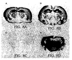
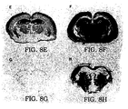
- PrP Sc accumulation in the brains of inoculated Tg(MHu2M) mice were markedly different for RML prions and Hu prions.
- RML prion inocula containing MoPrP Sc stimulated the formation of more MoPrP Sc while Hu prion inocula containing HuPrP CJD triggered production of MHu2MPrP Sc .
- the distribution of neuropathological changes characterized by neuronal vacuolation and astrocytic gliosis is similar to the patterns of PrP Sc accumulation in the brains of Tg(MHu2M) mice inoculated with RML prions or Hu prions.
- Standardized prion preparations may be produced in order to test assays of the invention and thereby improve the reliability of the assay.
- the preparation can be obtained from any animal it is preferably obtained from a host animal which has brain material containing prions of a test animal.
- a transgenic mouse containing a human prion protein gene can produce human prions and the brain of such a mouse can be used to create a standardized human prion preparation.
- the preparation is to be a "standard” it is preferably obtained from a battery (e.g., 100; 1,000, or more animals) of substantial identical animals. For example, 100 mice all containing a very high copy number of human PrP genes (all polymorphisms and mutations) would spontaneously develop disease and the brain tissue from each could be combined to make a useful standardized prion preparation.
- Standardized prion preparations can be produced using any of modified host mammals of the type described above.
- standardized prion preparations could be produced using mice, rats, hamsters, or guinea pigs which are genetically modified so that they are susceptible to infection with prions which prions would generally only infect genetically diverse species such as a human, cow, sheep or horse and which modified host mammals will develop clinical signs of CNS dysfunction within a period of time of 350 days or less after inoculation with prions.
- the most preferred host mammal is a mouse in part because they are inexpensive to use and because a greater amount of experience has been obtained with respect to production of transgenic mice than with respect to the production of other types of host animals. Details regarding making standardized prion preparation are described in U.S.
- mice may be mice which are genetically modified by the insertion of a chimeric gene of the invention.
- the mice might be modified by including high copy numbers of the chimeric gene and/or by the inclusion of multiple promoters in order to increase the level of expression of the chimeric gene.
- hybrid mice of the invention could be used wherein mice which have the endogenous PrP gene ablated are crossed with mice which have a human PrP gene inserted into their genome. There are, of course, various subcategories of such hybrid mice.
- the human PrP gene may be inserted in a high copy number an/or used with multiple promoters to enhance expression.
- the mice could be produced by inserting multiple different PrP genes into the genome so as to create mice which are susceptible to infection with a variety of different prions, i.e., which generally infect two or more types of test animals.
- a mouse could be created which included a chimeric gene including part of the sequence of a human, a separate chimeric gene which included part of the sequence of a cow and still another chimeric gene which included part of the sequence of a sheep. If all three different types of chimeric genes were inserted into the genome of the mouse the mouse would be susceptible to infection with prions which generally only infect a human, cow and sheep.
- the next step is to produce a large number of such mammals which are substantially identical in terms of genetic material related to prions. More specifically, each of the mice produced will include an identical chimeric gene present in the genome in substantially the same copy number.
- the mice should be sufficiently identical genetically in terms of genetic material related to prions that 95% or more of the mice will develop clinical signs of CNS dysfunction within 350 days or less after inoculation and all of the mice will develop such CNS dysfunction at approximately the same time e.g., within ⁇ 30 days of each other.
- mice Once a large group e.g., 50 or more, more preferably 100 or more, still more preferably 500 or more of such mice are produced.
- the next step is to inoculate the mice with prions which generally only infect a genetically diverse mammal e.g., prions from a human, sheep, cow or horse.
- the amounts given to different groups of mammals could be varied.
- the mammals After inoculating the mammals with the prions the mammals are observed until the mammals exhibit symptoms of prion infection e.g., clinical signs of CNS dysfunction.
- the brain or at least a portion of the brain tissue of each of the mammals is extracted. The extracted brain tissue is homogenized which provides the standardized prion preparation.
- mice which spontaneously develop prion related diseases can be done, for example, by including extremely high copy numbers of a human PrP gene into a mouse genome. When the copy number is raised to, for example, 100 or more copies; the mouse will spontaneously develop clinical signs of CNS dysfunction and have, within its brain tissue, prions which are capable of infecting humans.
- the brains of these animals or portions of the brain tissue of these animals can be extracted and homogenized to produce a standardized prion preparation.
- the standardized prion preparations can be used directly or can be diluted and tittered in a manner so as to provide for a variety of different positive controls. More specifically, various known amounts of such standardized preparation can be used to inoculate a first set of transgenic control mice.
- a second set of substantially identical mice are inoculated with a material to be tested i.e., a material which may contain prions.
- a third group of substantially identical mice are not injected with any material. The three groups are then observed. The third group, should, of course not become ill in that the mice are not injected with any material. If such mice do become ill the assay is not accurate probably due to the result of producing mice which spontaneously develop disease.
- the assay is also inaccurate probably because the mice have not been correctly created so as to become ill when inoculated with prions which generally only infect a genetically diverse mammal.
- the assay can be presumed to be accurate.
- the second group does not become ill the test material does not contain prions and if the second group does become ill the test material does contain prions.
- compositions containing the prions By using standardized prion preparations of the invention it is possible to create extremely dilute compositions containing the prions. For example, a composition containing one part per million or less or even one part per billion or less can be created. Such a composition can be used to test the sensitivity of the antibodies, assays and methods of the invention in detecting the presence of prions.
- Prion preparations are desirable in that they will include a constant amount of prions and are extracted from an isogeneic background. Accordingly, contaminates in the preparations will be constant and controllable.
- Standardized prion preparations will be useful in the carrying out of bioassays in order to determine the presence, if any, of prions in various pharmaceuticals, whole blood, blood fractions, foods, cosmetics, organs and in particular any material which is derived from an animal (living or dead) such as organs, blood and products thereof derived from living or dead humans.
- standardized prion preparations will be valuable in validating purification protocols where preparations are spiked and reductions in teeter measured for a particular process.
- antibodies having different specific features can be created which bind only to a prion protein naturally occurring within a single species and not bind to a prion protein naturally occurring within other species.
- the antibody can be designed so as to bind only to an infectious form of a prion protein (e.g., PrP Sc ) and not bind to a non-infectious form (e.g., PrP C ).
- PrP Sc an infectious form of a prion protein
- PrP C a non-infectious form
- a single antibody or a battery of different antibodies can then be used to create an assay device.
- Such an assay device can be prepared using conventional technology known to those skilled in the art.
- the antibody can be purified and isolated using known techniques and bound to a support surface using known procedures.
- the resulting surface having antibody bound thereon can be used to assay a sample in vitro to determine if the sample contains one or more types of antibodies.
- antibodies which bind only to human PrP Sc can be attached to the surface of a material and a sample can be denatured via proteinase K. The denatured sample is brought into contact with the antibodies bound to the surface of material. If no binding occurs it can be deduced that the sample does not contain human PrP Sc .
- Antibodies of the invention are also characterized by their ability to neutralize prions. Specifically, when antibodies of the invention are allowed to bind to prions the infectivity of the prion is eliminated. Accordingly, antibody compositions of the invention can be added to any given product in order to neutralize any infectious prion protein within the product. Thus, if a product is produced from a natural source which might contain infectious prion proteins the antibodies of the invention could be added as a precaution thereby eliminating any potential infection resulting from infectious prion proteins.
- the antibodies of the invention can be used in connection with immunoaffinity chromatography technology. More specifically, the antibodies can be placed on the surface of a material within a chromatography column. Thereafter, a composition to be purified can be passed through the column. If the sample to be purified includes any prion protein which binds to the antibodies those prion proteins (PrP Sc ) will be removed from the sample and thereby purified.
- a composition to be purified can be passed through the column. If the sample to be purified includes any prion protein which binds to the antibodies those prion proteins (PrP Sc ) will be removed from the sample and thereby purified.
- the antibodies of the invention can be used to treat a mammal.
- the antibodies can be given prophylactically or be administered to an individual already infected with infectious prion proteins such infection having been determined by the use of the assay described above.
- the exact amount of antibody to be administered will vary depending on a number of factors such as the age, sex, weight and condition of the patient. Those skilled in the art can determine the precise amount by administering antibodies in small amounts and determining the effect and thereafter adjusting the dosage. It is suggested that the dosage can vary from 0.01 mg/kg to about 300 mg/kg, preferably about 0.1 mg/kg to about 200 mg/kg, more preferably about 0.2 mg/kg to about 20 mg/kg in one or more dose administrations daily, for one or several days. Preferred is administration of the antibody for 2 to 5 or more consecutive days in order to avoid "rebound" of prion infectivity occurring.
- phage display libraries for expression of antibodies are well known in the art.
- the phage display antibody libraries that express antibodies are prepared according to the methods described in U.S. Patent 5,223,409, issued June 29, 1993 and U.S. Patent Application Serial No. 07/945,515, filed September 16, 1992, incorporated herein by reference. Procedures of the general methodology can be adapted using the present disclosure to produce antibodies of the present invention.
- the phage display anti-PrP antibody libraries are prepared by first isolating a pool of RNA that contains RNA encoding anti-PrP antibodies.
- an animal e.g., a mouse, rat, or hamster
- an animal is immunized with prion of interest.
- normal animals do not produce antibodies to prions at detectable or satisfactorily high levels.
- This problem is avoided by immunizing animals in which the (PrP) gene has been ablated on both alleles.
- Such mice are designated Prnp 0/0 and methods for making such mice are disclosed in Büeler, Nature (1992) and in Weismann Publication WO 93/10227, published May 27, 1993.
- the animal selected for immunization is a Prnp 0/0 mouse described by Büeler and Weismann.
- the amount of prion necessary to elicit a serum antibody response in a "null" animal is from about 0.01 mg/kg to about 500 mg/kg.
- the prion protein is generally administered to the animal by injection, preferably by intraperitoneal or intravenous injection, more preferably by intraperitoneal injection.
- the animals are injected once, with at least 1 to 4 subsequent booster injections, preferably at least 3 booster injections.
- the reactivity of the animal's antisera with the prion can be tested using standard immunological assays, such as ELISA or Western blot, according to methods well known in the art (see, for example, Harlow and Lane, 1988, Antibodies: A Laboratory Manual , Cold Spring Harbor Laboratory Press, Cold Spring Harbor, NY). Animals having prion-binding antisera may be boosted with an additional injection of prion.
- Serum antibody levels are predictive of antibody secretion, and therefore of levels of specific mRNA in lymphocytes, particularly plasma cells. Detection of serum antibodies, particularly relatively high levels of serum antibodies, is thus correlated to a high level of lymphocytes such as plasma cells producing mRNA encoding those serum antibodies.
- lymphocytes such as plasma cells producing mRNA encoding those serum antibodies.
- plasma cells isolated from the prion protein-immunized mice will contain a high proportion of lymphocytes (e.g., plasma cells) producing prion-specific antibody, particularly when the plasma cells are isolated from the mice within a short time period after the final injection boost (e.g., about 2 to 5 days, preferably 3 days).
- Immunization of the mice and the subsequent injection boosters thus serve to increase the total percentage of anti-PrP antibody-producing plasma cells present in the total population of the mouse's plasma cells. Moreover, because the anti-PrP antibodies are being produced at or near peak serum levels, then anti-PrP antibody-producing plasma cells are producing anti-PrP antibodies, and thus mRNA encoding these antibodies at or near peak levels.
- Lymphocytes including plasma cells are isolated from spleen and/or bone marrow from the prion-immunized animals according to methods well known in the art (see, for example, Huse et al. Science 1989) .
- the lymphocytes are isolated about 2 to 5 days, preferably about 3 days after the final immunization boost.
- the total RNA is then extracted from these cells. Methods for RNA isolation from mammalian cells are well known in the art (see, for example, Sambrook et al., 1989, Molecular Cloning: A Laboratory Manual , 2nd Ed., Cold Spring Harbor Laboratory Press, Cold Spring Harbor, NY).
- cDNA is produced from the isolated RNA using reverse transcriptase according to methods well known in the art (see, for example, Sambrook et al., supra), and cDNA encoding antibody heavy chains or light chains is amplified using the polymerase chain reaction (PCR).
- PCR polymerase chain reaction
- the 3' primers used to amplify heavy chain or light chain-encoding cDNAs are based upon the known nucleotide sequences common to heavy chain or light chain antibodies of a specific antibody subclass.
- one set of primers based upon the constant region of the IgG1 heavy chain-encoding gene can be used to amplify heavy chains of the IgG1 subclass, while another set of primers based upon the constant portion of the IgG1 light chain-encoding gene is used to amplify the light chains of the IgG1 subclass.
- the '5 primers are consensus sequences based upon examination of a large number of variable sequences in the data base. In this manner, DNA encoding all antibodies of a specific antibody class or subclass are amplified regardless of antigen-specificity of the antibodies encoded by the amplified DNA. The entire gene encoding the heavy chain or the light chain can be amplified.
- the heavy or light chain encoding gene may be amplified, with the proviso that the product of PCR amplification encodes a heavy or light chain gene product that can associate with its corresponding heavy or light chain and function in antigen binding i.e., bind selectively to a prion protein.
- the phage display product is a Fab or Fv antibody fragment.
- the antibody encoding cDNA selected for amplification may encode any isotope and preferably encode a subclass of IgG.
- Exemplary mouse IgG subclasses include IgG1, IgG2a, IgG2b, and IgG3.
- the selection of the specific antibody subclass-encoding cDNA for amplification will vary according to a variety of factors, including, for example, the animal's serum antibody response to the antigen.
- the antibody subclass-encoding cDNA selected for PCR amplification is that antibody subclass for which the animal produced the highest titer of antibody. For example, if the titers of serum IgG1 are higher than any other subclass of IgG detected in the serum antibody response, then cDNA encoding IgG1 is amplified from the cDNA pool.
- the heavy and light chains are amplified from the plasma cell cDNA to produce two separate amplified cDNA pools: 1) a cDNA pool containing heavy chain cDNA amplimer products, where the heavy chain is of a specific antibody subclass; and 2) a cDNA pool containing light chain cDNA amplimer products, where the light chain is of a specific antibody subclass.
- the heavy chain-encoding cDNAs and the light chain-encoding cDNAs are then each inserted into separate expression cassettes of an appropriate vector.
- the vector contains a nucleotide sequence encoding and capable of expressing a fusion polypeptide containing, in the direction of amino- to carboxy-terminus, 1) a prokaryotic secretion signal domain, 2) an insertion site for DNA encoding a heterologous polypeptide (e.g., either the heavy or light chain-encoding cDNA), and in the expression cassette for the heavy chain cDNA 3) a filamentous phage membrane anchor domain.
- a heterologous polypeptide e.g., either the heavy or light chain-encoding cDNA
- the vector includes prokaryotic or mammalian DNA expression control sequences for expressing the fusion polypeptide, preferably prokaryotic control sequences.
- the DNA expression control sequences can include any expression signal for expressing a structural gene product, and can include 5' and 3' elements operatively linked to the expression cassette for expression of the heterologous polypeptide.
- the 5' control sequence defines a promoter for initiating transcription, and a ribosome binding site operatively linked at the 5' terminus of the upstream translatable sequence.
- the vector additionally includes an origin of replication for maintenance and replication in a prokaryotic cell,. preferably a gram negative cell such as E. coli.
- the vector can also include genes whose expression confers a selective advantage, such as drug resistance, to a prokaryotic or eukaryotic cell transformed with the vector.
- the filamentous phage membrane anchor is preferably a domain of the cpIII or cpVIII coat protein capable of associating with the matrix of a filamentous phage particle, thereby incorporating the fusion polypeptide onto the phage surface.
- the secretion signal is a leader peptide domain of a protein that targets the protein to the periplasmic membrane of gram negative bacteria.
- leader sequences for gram negative bacteria such as E. coli
- are well known in the art see, for example, Oliver, In Neidhard, F.C. (ed.), Escherichia coli and Salmonella typhimurium , American Society for Microbiology, Washington, D.C., 1 :56-69, 1987).
- Preferred membrane anchors for the vector are obtainable from filamentous phage M13, f1, fd, and equivalent filamentous phage.
- Preferred membrane anchor domains are found in the coat proteins encoded by gene III and gene VIII.
- the membrane anchor domain of a filamentous phage coat protein is a portion of the carboxy terminal region of the coat protein and includes a region of hydrophobic amino acid residues for spanning a lipid bilayer membrane, and a region of charged amino acid residues normally found at the cytoplasmic face of the membrane and extending away from the membrane.
- gene VIII coat protein's membrane spanning region comprises the carboxy-terminal 11 residues from 41 to 52 (Ohkawa et al., J. Biol. Chem.
- An exemplary membrane anchor would consist of residues 26 to 40 to cpVIII.
- the amino acid residue sequence of a preferred membrane anchor domain is derived from the M13 filamentous phage gene VIII coat protein (also designated cpVIII or CP 8).
- Gene VIII coat protein is present on a mature filamentous phage over the majority of the phage particle with typically about 2500 to 3000 copies of the coat protein.
- the amino acid residue sequence of another preferred membrane anchor domain is derived from the M13 filamentous phage gene III coat protein (also designate cpIII).
- Gene III coat protein is present on a mature filamentous phage at one end of the phage particle with typically about 4 to 6 copies of the coat protein.
- Detailed descriptions of the structure of filamentous phage particles, their coat proteins, and particles assembly are found in the reviews by Rached et al., ( Microbiol. Rev. , 50 :401-427, 1986) and Model et al. (In: The Bacteriophages: Vol. 2 , R. Calendar, ed., Plenum Publishing Co., pgs. 375-456, 1988).
- the filamentous phage membrane anchor-encoding DNA is inserted 3' of the cDNA insert in the library vector such that the phage membrane anchor-encoding DNA can be easily excised and the vector relegated without disrupting the rest of the expression cassettes of the vector.
- the soluble Fab fragments retain the antigenicity of the phage-bound Fab, and thus can be used in assays and therapies in the manner that whole (non-fragmented) antibodies are used.
- the vector for use with the present invention must be capable of expressing a heterodimeric receptor (such as an antibody or antibody Fab). That is, the vector must be capable of independently containing and expressing two separate cDNA inserts (e.g., the heavy chain cDNA and the light chain cDNA).
- Each expression cassette can include the elements described above, except that the filamentous phage anchor membrane-encoding DNA is present only in the expression cassette for the heavy chain cDNA.
- the antibody or Fab is expressed on the surface of the phage, only the heavy chain polypeptide is anchored to the phage surface.
- the light chain is not directly bound to the phage surface, but is indirectly bound to the phage via its association with the free portion of the heavy chain polypeptide (i.e., the portion of the heavy chain that is not bound to the phage surface).
- the vector contains a sequence of nucleotides that allow for directional ligation, i.e., a polylinker.
- the polylinker is a region of the DNA expression vector that operatively links the upstream and downstream translatable DNA sequence for replication and transport, and provides a site or means for directional ligation of a DNA sequence into the vector.
- a directional polylinker is a sequence of nucleotides that defines two or more restriction endonuclease recognition sequence, or restriction sites. Upon restriction enzyme cleavage, the two sites yield cohesive termini to which a translatable DNA sequence can be ligated to the DNA expression vector.
- the two cohesive termini are non-complementary and thereby permit directional insertion of the cDNA into the cassette.
- Polylinkers can provide one or multiple directional cloning sites, and may or may not be translated during expression of the inserted cDNA.
- the expression vector is capable of manipulating in the form of a filamentous phage particle.
- DNA expression vectors additionally contain a nucleotide sequence that defines a filamentous phage origin of replication such that the vector, upon presentation of the appropriate genetic complement, can replicate as a filamentous phage in single stranded replicative form, and can be packaged into filamentous phage particles. This feature provides the ability of the DNA expression vector to be packaged into phage particles for subsequent isolation of individual phage particles (e.g., by infection of and replication in isolated bacterial colonies).
- a filamentous phage origin of replication is a region of the phage genome that defines sites for initiation of replication, termination of replication, and packaging of the replicative form produced by replications (see, for example, Rasched et al., Microbiol. Rev., 50 :401-427, 1986; Horiuchi, J. Mol. Biol ., 188 :215-223, 1986).
- a preferred filamentous phage origin of replication for use in the present invention is an M13, f1, or fd phage origin of replication (Short et al., Nucl. Acids Res., 16 :7583-7600, 1988).
- Preferred DNA expression vectors are the expression vectors pCOMB8, pCKAB8, pCOMB2-8, pCOMB3, pCKAB3, pCOMB2-3, pCOMB2-3' and pCOMB3H.
- the pComb3H vector is a modified form of pComb3 in which (i) heavy and light chains are expressed from a single Lac promoter as opposed to individual promoters and (ii) heavy and light chains have two different leader sequences (pg1B and ompA) as opposed to the same leader sequence (pHB).
- pComb3H Wang, et al (1995) J. Mol. Biol., Inpress.
- the principles of pComb3H are basically the same as for pComb3.
- the entire library is packaged using an appropriate filamentous phage.
- the phage are then used to infect a phage-susceptible bacterial culture (such as a strain of E. coli ), the phage allowed to replicate and lyse the cells, and the lysate isolated from the bacterial cell debris.
- the phage lysate contains the filamentous phage expressing on its surface the cloned heavy and light chains isolated from the immunized animal.
- the heavy and light chains are present on the phage surface as Fab antibody fragments, with the heavy chain of the Fab being anchored to the phage surface via the filamentous phage membrane anchor portion of the fusion polypeptide.
- the light chain is associated with the heavy chain so as to form an antigen binding site.
- Phage expressing an antibody or Fab that specifically binds a prion antigen can be isolated using any of a variety of protocols for identification and isolation of monoclonal and/or polyclonal antibodies. Such methods include, immunoaffinity purification (e.g., binding of the phage to a columna having bound antigen) and antibody panning methods (e.g., repeated rounds of phage binding to antigen bound to a solid support for selection of phage of high binding affinity to the antigen).
- the phage is selected by panning using techniques that are well known in the art.
- the phage can be used to infect a bacterial culture, and single phage isolates identified. Each separate phage isolate can be again screened using one or more of the methods described above.
- the DNA encoding the antibodies or Fabs can be isolated from the phage, and the nucleotide sequence of the heavy and light chains contained in the vector determined using methods well known in the art (see, for example, Sambrook et al., supra).
- Soluble antibodies or Fabs can be produced from a modified display the same dicistronic vector by excising the DNA encoding the filamentous phage anchor membrane that is associated with the expression cassette for the heavy chain of the antibody.
- the DNA encoding the anchor membrane is flanked by convenient restriction sites that allow excision of the anchor membrane sequence without disruption of the remainder of the heavy chain expression cassette or disruption of any other portion of the expression vector.
- the modified vector without the anchor membrane sequence then allows for production of soluble heavy chain as well as soluble light chain following packaging and infection of bacterial cells with the modified vector.
- the modified vector can be used to transform a eukaryotic cell (e.g., a mammalian or yeast cell, preferably a mammalian cell (e.g., Chinese hamster ovary (CHO) cells)) for expression of the Fab.
- a eukaryotic cell e.g., a mammalian or yeast cell, preferably a mammalian cell (e.g., Chinese hamster ovary (CHO) cells)
- the vector allows for excision of both the heavy and light chain expression cassettes as a single DNA fragments for subcloning into an appropriate vector.
- Numerous vectors for expression of proteins in prokaryotic and/or eukaryotic cells are commercially available and/or well known in the art (see, for example Sambrook et al., supra).
- Example 17 show the isolation of an antibody which specifically binds to PrP Sc without any denaturation.
- a sample containing PrP proteins i.e., PrP C and PrP Sc
- PrP C and PrP Sc can be subjected to denaturation by the use of protease K (PK) digestion. The use of such will digest PrP C but not PrP Sc .
- the sample is contacted with the antibody (e.g., R2) as per Example 17 under suitable binding conditions.
- the antibody is bound to a substrate and can be positioned such that the sample can be easily contacted with the substrate material having the antibody bound thereon. If material binds to the antibodies on the substrate the presence of infectious PrP Sc is confirmed.
- the antibody of the invention may be bound to a substrate support surface.
- the sample to be tested is contacted with the support surface under conditions which allow for binding. Thereafter, unreacted sites are blocked and the surface is contacted with a generalized antibody which will bind to any protein thereon.
- the generalized antibody is linked to a detectable label.
- the generalized antibody with detectable label is allowed to bind to any PrP Sc bound to the antibodies on the support surface. If binding occurs the label can be made to become detectable such as by generating a color thereby indicating the presence of the label which indirectly indicates the presence of PrP Sc within the sample.
- the assay can detect prions (PrP Sc ) present in an amount of 1 part per million or less, even one part per billion or less.
- the PrP Sc may be present in a source selected from the group consisting of (a) a pharmaceutical formulation containing a therapeutically active component extracted from an animal source, (b) a component extracted from a human source, (c) an organ, tissue, body fluid or cells extracted from a human source, (d) a formulation selected form the group consisting of injectables, orals, creams, suppositories, and intrapulmonary delivery formulations, (e) a cosmetic, and (f) a pharmaceutically active compound extracted from a mammalian cell culture.
- Such source materials can also be treated to remove or neutralize PrP Sc protein by adding an antibody of the invention.
- the invention also includes a method of treating, comprising administering to a mammal in need thereof a therapeutically effective amount of an antibody which selectively binds Prp Sc protein which antibody is characterized by its ability to neutralize PrP Sc protein infectivity.
- Antibodies of the invention could be obtained by a variety of techniques. However, the general procedure involves synthesizing a library of proteins (i.e., antibodies or portions thereof) on the surface of phage. The library is then brought into contact with a composition which includes PrP proteins and in particular is a naturally occurring composition which includes PrP Sc . The phage which bind to PrP protein are then isolated and the antibody or portion thereof which binds the PrP protein is isolated. It is desirable to determine the sequence of the genetic material encoding the antibody or portion thereof. Further, the sequence can be amplified and inserted, by itself, or with other genetic material into an appropriate vector and cell line for the production of other antibodies.
- proteins i.e., antibodies or portions thereof
- a sequence encoding a variable region which binds PrP Sc can be fused with a sequence which encodes a human constant region of an antibody producing a constant/variable construct.
- This construct can be amplified and inserted within a suitable vector which can be inserted within a suitable cell line for the production of humanized antibodies. Procedures such as this are described within U.S. Patent 4,816,567, issued March 28, 1989 to Cabilly, et al which is incorporated herein by reference to disclose and describe such procedures.
- the genetic material encoding an antibody or portion thereof which binds a PrP protein is isolated it is possible to use that genetic material to produce other antibodies or portions thereof which have a greater affinity for binding PrP proteins. This is done by site directed mutagenesis technology or by random mutagenesis and selection. Specifically, individual codons or groups of codons within the sequence are removed or replaced with codons which encode different amino acids. Large numbers of different sequences can be generated, amplified and used to express variations of the antibody or portions thereof on the surface of additional phage. These phage can then be used to test for the binding affinity of the antibody to PrP proteins.
- the phage library can be created in a variety of different ways.
- a host animal such as a mouse or rat is immunized with PrP protein and preferably immunized with PrP Sc .
- the immunization may be carried out along with an adjuvant for the formation of larger amounts and types of antibodies.
- cells responsible for antibody production are extracted from the inoculated host mammal.
- RNA is isolated from the extracted cells and subjected to reverse transcription in order to produce a cDNA library.
- the extracted cDNA is amplified by the use of primers and inserted into an appropriate phage display vector.
- the vector allows the expression of antibodies or portions thereof on the phage surface.
- cDNA it is also possible to subject the cDNA to site directed mutagenesis prior to insertion into the display vector. Specifically, codons are removed or replaced with codons expressing different amino acids in order to create a larger library (i.e., a library of many variants) which is then expressed on the surface of the phage. Thereafter, as described above, the phage are brought into contact with the sample and phage which bind to PrP protein are isolated.
- Purified MoPrP 27-30 rods were prepared from the brains of clinically ill CD-1 mice inoculated with RML prions (Chandler scrapie isolate (Chandler R.L. 1961 Lancet, 1378-1379)). Prion rods were recovered from sucrose gradient fractions as previously described (Prusiner, McKinley 1983 Cell). Briefly, the fractions containing prion rods, which sediment in 48-60% (wt/vol) sucrose, were diluted 2:1 in distilled water and centrifuged at 100,000 x g for 6 h at 4°C.
- PrP 27-30 was the major protein as determined by SDS-PAGE and silver staining analysis. Protein quantitation was performed by bicinchonic acid dye binding, with a known amount of bovine serum albumin as the protein concentration standard.
- Prnp 0/0 mice in which both alleles of the PrP gene (Prnp) is ablated, were immunized with the purified MoPrP 27-30 rods, which were isolated as described in Example 1.
- Prnp 0/0 mice and methods for making this strain are well known in the art (Büeler, et al. 1992).
- Prnp 0/0 mice which are indistinguishable from normal mice in their development and behavior, are resistant to scrapie following intracerebral inoculation of infectious MoPrP Sc (Büeler, et al. 1993 Cell; Prusiner et al. PNAS 1993), and will develop IgG serum titers against Mo-, SHa, and human PrP following immunization with relatively small quantities of purified SHaPrP 27-30 in adjuvant (Prusiner et al. PNAS 1993).
- mice Three (3) six week old Prnp 0/0 mice were immunized by intraperitoneal injection of 100 ⁇ g of MoPrP 27-30 rods fully emulsified in complete Freund's adjuvant. Subsequently mice were boosted 2 times at 2-week intervals with incomplete Freund's adjuvant containing in the first instance 100 ⁇ g, then 50 ⁇ g of rods. Four days after the second boost, the reactivity of each mouse's serum against prion proteins was analyzed as described below in Example 3. Those mice having anti-PrP reactive antisera received a third injection boost of 50 ⁇ g prion rods in incomplete Freund's adjuvant 14 days after the second boost.
- a primary prognostic indicator for success in isolating a specific antibody from combinatorial libraries is serum antibody reactivity with the antigen(s) to be studied (Burton and Barbas, Adv. Immunol. 1994). Serum antibody levels are predictive of antibody secretion and therefore predictive of the levels of specific mRNA in plasma cells. It is this latter factor that ultimately dictates the composition of the antibody-encoding cDNA library.
- the Prnp 0/0 mice immunized with MoPrP 27-30 as described in Example 2 were bled from the tail, and the antisera stored at -20°C for subsequent immunological analysis.
- the reactivity of the immunized mouse serum (IgG1, IgG2a, IgG2b and IgG3 antibody subclasses) was measured against denatured and non-denatured Mo- and SHaPrP 27-30 in ELISA.
- ELISA wells were coated overnight at 4°C with 50 ⁇ l of PrP rods at 40 ⁇ g/ml in 100 mM sodium bicarbonate pH 8.6.
- mice produced anti-PrP IgG antibodies.
- the highest serum titers against Mo- and SHaPrP antigens were of the IgG1 and IgG2b subclasses.
- the IgG2a and IgG3 anti-PrP titers were close to the background levels of reactivity seen for all IgG subclasses in the serum of non-immunized Prnp 0/0 mice.
- Antibody titers were greater against denatured rods than non-denatured rods.
- RNA prepared from bone marrow and splenic tissues.
- Total RNA from mouse spleen was prepared according to methods well known in the art (Huse, et al Science 1989).
- RNA was prepared from bone marrow tissues by first removing the tibia and fibula from both rear legs of the mice. The bones were then cut through close to each end, and their contents flushed out by injection of guanidinium isothiocyanate into the bone cavity using a 27 gauge needle. RNA preparation was then continued as described for the mouse spleen.
- RNA preparations were then pooled, and cDNA generated from the mRNA using reverse transcriptase according to methods well known in the art.
- Two cDNA libraries were independently constructed from the D7282 mouse mRNA: 1) an IgG1 library; and 2) a IgG2b library. For each of these libraries, cDNAs encoding heavy chains and cDNA light chains were separately amplified by PCR from separate fractions of the pooled cDNA.
- oligonucleotide 5' and 3' primers employed for PCR amplification of DNA fragments encoding murine light ( ⁇ ) chains and heavy ( ⁇ 1 or ⁇ 2b ) chains of the IgG1 subclass wee those used by Huse, et al (Science 1989) and additional heavy chain primers as presented in Table 1 and heavy chain polymers which are presented in Table 1.
- the resulting amplified cDNAs encoding heavy chains of the IgG1 and IgG2b subclasses and light chains were cloned into the vector pComb3.
- the preparation of Fab antibody libraries displayed on the surface of a filamentous phage using the pComb3 vector have been described (Williamson et al. PNAS, 1993; Barbas et al. PNAS 1991).
- the IgG1 or IgG2b phage display library is constructed by inserting the amplified cDNA encoding IgG1 or IgG2b heavy chain and the amplified cDNA encoding light chain into the pComb3H vector such that each vector contains a cDNA insert encoding a heavy chain fragment in one expression cassette of the vector, and a cDNA insert encoding a light chain fragment into the other expression cassette of the vector.
- the resulting IgG1 library contained approximately 9 x 10 6 individual clones, while the resulting IgG2b library contained approximately 7 x 10 6 individual clones.
- the ligated vectors were then packaged by the filamentous phage M13 using methods well known in the art (see, for example, Sambrook et al, supra).
- the packaged library is then used to infect a culture of E. coli , so as to amplify the number of phage particles.
- the phage particles are isolated and used in the panning procedure that follows. Aliquots of the phage library are stored for future amplification and use. Separate aliquots of the phage libraries are isolated and stored for future amplification and use.
- Antigen binding phage were selected for binding to denatured MoPrP 27-30 rods against PrP antigen bound to ELISA wells through a panning procedure described in (Burton, et al PNAS 1991, Barbas Lerner Methods in Enzymol 1991). Briefly, ELISA wells were coated overnight at 4°C with 50 ⁇ l of MoPrP 27-30 rods at 40 ⁇ g/ml in 100 mM sodium bicarbonate pH 8.6. The PrP rods were then denatured by incubation with 50 ⁇ l of 6M guanidinium isothiocyanate for 15 min at room temperature, after which the wells were washed 6 times with Ca/Mg-free PBS. The wells were then blocked with Ca/Mg-free PBS containing 3% BSA.
- the phage were incubated with the well-bound MoPrP antigen for 2 hrs at 37°C. Unbound phage were removed by washing 10 times with PBS 0.5% TWEEN 20. Bound phage were then removed from the wells by acid elution, pooled, reamplified and subjected to a second round of panning.
- the IgG1 library was selected through 5 rounds of panning. A 40-fold amplification of PrP-specific antibody phage, as determined by the number of phage eluted from PrP-coated ELISA wells, was measured from the first to the fifth round.
- Soluble Fabs were produced from phage clones eluted from the fourth and fifth rounds of panning. DNA from the selected phage clones was isolated, and the phage coat protein III (the filamentous phage membrane anchor) was removed from the pComb3H vector using the appropriate restriction enzymes. The DNA was self-ligated to yield a vector capable of expressing soluble Fab (the procedure for production of soluble Fabs is detailed in (Barbas et al. PNAS 1991)). The vectors were then separately used to transform bacteria for expression of the Fabs, and isolated transformants were selected.
- Fab expression was induced in an overnight bacterial culture using isopropyl ⁇ -D-thiogalactopyranoside. The bacteria were centrifuged, and the resulting bacterial pellet was either sonicated or frozen and thawed three times to release Fab from the bacterial periplasmic space. The bacterial Fab supernatants were then tested for reactivity against PrP in ELISA.
- the reactivity of the antibodies identified by panning of the phage display antibody library was tested by SDS/PAGE of prion-infected rodent brain tissue and Western blot analysis using the selected Fabs. Protein from brain tissues of prion-infected and uninfected mice was used as the antigen against which immunoreactivity was tested.
- the antigen was prepared by disrupting rodent brain tissue in Ca/Mg-free PBS by passage 5 times through a 20 gauge needle, followed by passage 10 times through a 22 gauge needle. The 10% (wt/vol) homogenate was then centrifuged at 1600 x g for 5 min at 4°C.
- nucleotide and amino acid sequences of the variable domains of the antibody light and heavy chains were determined for several of the PrP specific clones. Nucleic acid sequencing was performed with a model 373A automated DNA sequencer (Applied Biosystems) using a Taq fluorescent dideoxynucleotide terminator cycle sequencing kit (Applied Biosystems).
- Primers for the elucidation of antibody light-chain sequence were primers MoSeqKb [5'-CAC GAC TGA GGC ACC TCC-3'] and OmpSeq [5'-AAG ACA GCT ATC GCG ATT GCA G-3'] hybridizing to the (-)-strand and for the heavy chain MOIgGGzSeq [5'-ATA GCC CTT GAC CAG GCA TCC CAG GGT CAC-3'] binding to the (+)-strand and PelSeq [5'-ACC TAT TGC CTA CGG CAG CCG-3'] binding to the (-)-strand.
- Figure 6 shows the amino acid sequences of selected (A) heavy chain and (B) light chain variable regions generated by panning an IgG1 library from mouse D7282 against denatured MoPrP 27-30 rods.
- the sequences are very similar but contain a number of heterogeneities which are likely the result of somatic mutation following repeated exposure of the mouse to PrP antigen.
- All of the heavy chain sequences examined in these clones contained very similar sequences.
- the heavy chain complementarity determining region 3(HCDR3) was identical at the nucleotide level in all the Fab clones examined.
- Panning of the IgG1 library against denatured PrP produced a series of related antibodies, presumably somatic variants of a clone directed to a single epitope (Example 9).
- a' prototype antibody from the above series was added to denatured PrP in ELISA wells prior to panning in the normal way.
- the masking antibody was used in all subsequent panning steps. Using this procedure, antibodies were derived of different sequence which reacted with denatured PrP in ELISA. These antibodies are likely directed to different epitopes on PrP.
- the masking procedure was carried out as described in Ditzel, et al (1995) J. Immunol. Masking could also be carried out with molecules other than antibodies which interacted with PrP.
- a phage display antibody library similar to that described in the Examples above is subjected to panning experiments to identify phage clones that bind to PrP Sc , but not to PrP C .
- PrP Sc antigen and PrP C antigen are bound to separate wells of a microtiter dish as described above for the ELISA assay.
- the phage display antibody library is first panned over the PrP C ELISA wells. Unbound phage are retrieved from the wells and pooled. Phage that binds to the PrP C antigen are removed from the wells and either discarded or pooled for later analyses.
- the pooled unbound phage are then again added to PrP C ELISA wells, with selection again being based upon lack of binding to the PrP C .
- the phage are pooled and panned on the ELISA wells containing the PrP Sc antigen.
- the panning is repeated for several rounds, with the phage that binds to the PrP Sc antigen being the phage that is selected for further rounds of panning.
- the phage are isolated one from another.
- the ability of the PrP Sc -specific phage or isolated Fab to bind PrP C antigen can be double-checked by ELISA with the PrP C antigen.
- the resulting selected phage are those that bind PrP Sc , but do not bind PrP C .
- a phage display antibody library is prepared as described above from lymphocyte RNA from a mouse immunized with several PrP Sc isoforms, or from a pool of lymphocyte RNA from several mice immunized with different PrP Sc isoforms.
- the phage are then panned with several different wells containing antigens from different isoforms of PrP Sc .
- the phage are panned over each PrP Sc isoform with the selection being for phage that bind the isoform at each stage.
- the phage are panned for a total of about 5 to 10 rounds on each PrP Sc isoform.
- the phage that remain after all stages of panning against all the isoforms tested are then isolated.
- the immunoreactivity of each selected phage or isolated Fab is tested by ELISA or Western blot or histochemistry against each of the various PrP Sc isoforms, as well as for cross-reactivity with PrP C .
- a phage display antibody library prepared from lymphocyte RNA of a mouse immunized with a specific PrP Sc isoform is prepared according to the Examples above.
- the resulting phage are then selected for their ability to bind only one specific PrP Sc isoform by panning.
- the panning uses several different wells containing antigens from different isoforms of PrP Sc , including one set of wells containing antigens from the specific PrP Sc isoform against which specific antibodies are desired.
- the phage are first panned over the undesirable PrP Sc isoforms, with the selection being for phage that do not bind the antigen. Panning continues for a total of about 5 to 10 rounds on each of the PrP Sc isoforms.
- the phage that did not bind the undesirable PrP Sc isoforms are then panned for about 5 to 10 rounds against the desirable PrP Sc isoform, with selection for antigen binding.
- the phage that remain after all rounds of panning are isolated. These selected phage are those that express antibodies with binding specificity for only the specific PrP Sc isoform desired.
- the immunoreactivity of each selected phage or isolated Fab is tested by ELISA or Western blot against each of the various PrP Sc isoforms, as well as for cross-reactivity with PrP C .
- mice were immunized with liposomes containing SHaPrP 27-30.
- mice were immunized using both short and long term protocols.
- immunization with liposomes containing SHaPrP 27-30 resulted in antiserum titer which includes all four IgG subclasses.
- Histoblots were treated with proteinase K to remove PrP C from the brain of normal, uninoculated control SHa and SHa showing clinical signs of scrapie following inoculation with Sc237 prions.
- SHaPrP 27-30 histoblots were treated with 3M GdnSCN for 10 minutes. Blots were incubated overnight at 4°C with sera diluted 1/200 from the short and the long term immunized mice. The results described here show clear positive reactivity of an antiserum with non-denatured infectious prions i.e., native PrP Sc .
- FIG. 8 shows eight different stained histoblots of scrapie infected SHa brain.
- the histoblots were treated with proteinase K to remove PrP C from the brain of normal, non-inoculated control SHa(A, C, E and G) and SHa showing clinical signs of scrapie following inoculation with Sc 237 prions (B, D, F and H).
- SHaPrP 27-30 the histoblots were treated with 3M GdnSCN for 10 minutes (C, D, G and H).
- the blots were incubated overnight at 4°C with sera diluted 1/200 from the short (A-D) and the long (E-H) term immunized mice.
- the results clearly show the ability of the antibodies of the invention to bind to native, non-denatured infectious prions i.e., bind to native PrP Sc .
- phage Fab display libraries were constructed: IgG1k, IgG2ak, IgG2bk and IgG3k from mRNA extracted from the short and long term immunized mice.
- a panning system was used where libraries are panned against biotinylated SHa 27-30, dispersed into liposomes, and bound to streptavidin-coated microtiter plates. After five rounds of panning, E.
- Coli extracts from more than 50 clones reacted with biotinylated SHa 27-30, SHa 27-30 rods and 90-231 recombinant SHa in ELISA. Since these clones also react with recombinant rPrP corresponding to SHaPrP residues 90-231, Melhorn,I., et al, High-level Expression and Characterization of a Purified 142-residue Polypeptide of the Prion Protein. Biochemistry 35, 5528-2237 (1996), all eight libraries were panned against this antigen to successfully isolate more distinct clones from virtually all the libraries. Upon DNA sequencing of the plasmid region coding for the IgG heavy chain, 30 Fabs were identified as distinct clones.
- the newly isolated Fabs reacted against prion rods without any denaturation step.
- the half-maximal binding to non-denatured prion rods occurs at a Fab concentration of approximately 0.5 pg/ml, indicating that the antibody has an apparent binding affinity of approximately 10 8 moles/liter.
- Figure 9 is a graph showing the ELISA reactivity of purified Fabs against prion protein SHa 27-30.
- the antibody 3F4 and recombinant antibodies were examined at different concentrations for binding to ELISA wells which were coated with 0.2 ⁇ /g of sucrose purified infectious SHa prion rods. The results clearly show that all of the recombinant antibodies of the invention have substantially higher degrees of binding to prions as compared to the antibody-3F4.
- Purified 3F4 Fab and recombinant Fabs were examined at different concentrations for binding to ELISA wells coated with 0.2 ⁇ g of sucrose purified SHa prion rods either native or denatured in the ELISA well with 3M GdnSCN for 10 min.
- Figure 10 is a graph showing the results of ELISA reactivity purified Fabs against denatured prion protein SHa 27-30.
- Figure 10 is interesting as compared to Figure 9 in that the recombinant antibodies of the invention as per Figure 9 show a higher degree of affinity for the prion rods as compared to 3F4 whereas all of the recombinant antibodies but for R1 show a lower degree of affinity against denatured antigen.
- the antibody 3F4 was obtained as an ascetic fluid and is expected to have a concentration of approximately 1 ⁇ g/ml at the dilution used in the immunoprecipitation experiment.
- the ability of the new Fabs to immunoprecipitate SHaPrP 27-30 in comparison to 3F4 was determined quantitatively with purified Fab mAbs D4 and R2.
- Fab 2R immunoprecipitated SHaPrP 27-30 strongly at concentrations as low as 0.1 pg/ml (50 ng in 500 pl) indicating an affinity on the order of greater than 10 8 M -1 (i.e., 10 8 mole/liter).
- Fab 2R was less potent but clearly immune precipitated antigen more efficiently than 3F4. Note that D4, R2, 6D2, D14, R1, and R10 all refer to antibodies of the invention.
- Figure 12 is a photo showing the immunoprecipitation of SHaPrP 27-30 with purified Fabs of the invention (2R and 4D) as well as 3H4. The ability to immunoprecipitate the antigen is monitored by western blotting. All of the lanes shown in Figure 12 but for lane 14 are immunoprecipitations containing goat anti-mouse IgG Fab and protein Agarose. To obtain the results 10 ⁇ l of liposomes containing SHaPrP 27-30 were added to all lanes except for lanes 5, 9 and 13. Each of the lanes are marked with the indicated amounts of purified Fabs (nanograms) which were added to lanes 2-13. Lane 14 was loaded with one-half volume of liposomes used for the immunoprecipitation. The results clearly show a dramatically higher degree of precipitation when using the antibodies 2R and 4D of the invention as compared to 3F4.
- the immunoprecipitation data are confirmatory of the ELISA data.
- Low concentrations of recombinant Fabs as found in crude bacterial supernates are highly effective at immunoprecipitating PrP 27-30 (Figure 11). This implies an affinity on the order of 10 7 -10 8 M -1 .
- 3F4 does not produce significant precipitation.
- Figure 12 shows that Fab R2 immunoprecipitates PrP 27-30 highly effectively with some titration in the range 0.1-0.2 ⁇ g/ml implying a binding affinity on the order of 10 8 M -1 .
- Fab 4D has a lower affinity and 3F4 immunoprecipitates very weakly indeed. From this particular experiment one could argue that the affinity of 3F4 is considerably less than 5 x 10 7 M -1 and probably less than10 7 M -1 .
Landscapes
- Health & Medical Sciences (AREA)
- Life Sciences & Earth Sciences (AREA)
- Engineering & Computer Science (AREA)
- Chemical & Material Sciences (AREA)
- Immunology (AREA)
- Molecular Biology (AREA)
- Biomedical Technology (AREA)
- Hematology (AREA)
- Urology & Nephrology (AREA)
- General Health & Medical Sciences (AREA)
- Biochemistry (AREA)
- Medicinal Chemistry (AREA)
- Cell Biology (AREA)
- Microbiology (AREA)
- Biotechnology (AREA)
- Food Science & Technology (AREA)
- Proteomics, Peptides & Aminoacids (AREA)
- Physics & Mathematics (AREA)
- Analytical Chemistry (AREA)
- Virology (AREA)
- Organic Chemistry (AREA)
- General Physics & Mathematics (AREA)
- Pathology (AREA)
- Biophysics (AREA)
- Genetics & Genomics (AREA)
- Neurology (AREA)
- Neurosurgery (AREA)
- Tropical Medicine & Parasitology (AREA)
- Peptides Or Proteins (AREA)
- Medicines Containing Antibodies Or Antigens For Use As Internal Diagnostic Agents (AREA)
- Investigating Or Analysing Biological Materials (AREA)
- Preparation Of Compounds By Using Micro-Organisms (AREA)
- Micro-Organisms Or Cultivation Processes Thereof (AREA)
- Measuring Or Testing Involving Enzymes Or Micro-Organisms (AREA)
Applications Claiming Priority (3)
| Application Number | Priority Date | Filing Date | Title |
|---|---|---|---|
| US52810495A | 1995-09-14 | 1995-09-14 | |
| US528104 | 1995-09-14 | ||
| EP96931599A EP0852011B1 (de) | 1995-09-14 | 1996-09-13 | Für natives prp-sc spezifische antikörper |
Related Parent Applications (2)
| Application Number | Title | Priority Date | Filing Date |
|---|---|---|---|
| EP96931599A Division EP0852011B1 (de) | 1995-09-14 | 1996-09-13 | Für natives prp-sc spezifische antikörper |
| EP96931599A Division-Into EP0852011B1 (de) | 1995-09-14 | 1996-09-13 | Für natives prp-sc spezifische antikörper |
Publications (2)
| Publication Number | Publication Date |
|---|---|
| EP1416280A2 true EP1416280A2 (de) | 2004-05-06 |
| EP1416280A3 EP1416280A3 (de) | 2005-08-10 |
Family
ID=24104278
Family Applications (2)
| Application Number | Title | Priority Date | Filing Date |
|---|---|---|---|
| EP96931599A Revoked EP0852011B1 (de) | 1995-09-14 | 1996-09-13 | Für natives prp-sc spezifische antikörper |
| EP04000804A Withdrawn EP1416280A3 (de) | 1995-09-14 | 1996-09-13 | Antikörper spezifisch für natives PrPsc |
Family Applications Before (1)
| Application Number | Title | Priority Date | Filing Date |
|---|---|---|---|
| EP96931599A Revoked EP0852011B1 (de) | 1995-09-14 | 1996-09-13 | Für natives prp-sc spezifische antikörper |
Country Status (12)
| Country | Link |
|---|---|
| US (6) | US5846533A (de) |
| EP (2) | EP0852011B1 (de) |
| JP (3) | JP3495738B2 (de) |
| AT (1) | ATE263374T1 (de) |
| AU (1) | AU707484B2 (de) |
| BR (1) | BR9610580A (de) |
| CA (1) | CA2231409C (de) |
| DE (2) | DE69632056T2 (de) |
| ES (1) | ES2218598T3 (de) |
| MX (1) | MX9802019A (de) |
| NZ (1) | NZ318689A (de) |
| WO (1) | WO1997010505A1 (de) |
Families Citing this family (130)
| Publication number | Priority date | Publication date | Assignee | Title |
|---|---|---|---|---|
| EP0852011B1 (de) * | 1995-09-14 | 2004-03-31 | The Regents of the University of California | Für natives prp-sc spezifische antikörper |
| JP3333213B2 (ja) | 1996-04-03 | 2002-10-15 | スティヒティング インスティチュート フォール ディールハウデレイ エン ディールゲゾントヘイト | プリオン病の検出方法 |
| DE934531T1 (de) * | 1996-10-15 | 2000-04-06 | Imperial College | Diagnose spongiformer encephalopathie |
| GB2319607A (en) * | 1996-11-23 | 1998-05-27 | Electrophoretics International | Detection of prion proteins in mononuclear cells |
| CA2286259A1 (en) * | 1997-02-06 | 1998-08-13 | Enfer Technology Limited | Immunological assay for spongiform encephalopathies |
| US20060008494A1 (en) * | 1997-02-21 | 2006-01-12 | The Regents Of The University Of California | Complete inactivation of infectious proteins |
| US6720355B2 (en) * | 1997-02-21 | 2004-04-13 | The Regents Of The University Of California | Sodium dodecyl sulfate compositions for inactivating prions |
| US6719988B2 (en) | 1997-02-21 | 2004-04-13 | The Regents Of The University Of California | Antiseptic compositions for inactivating prions |
| US6617119B2 (en) | 1997-02-21 | 2003-09-09 | The Regents Of The University Of California | Assay for specific strains of multiple disease related conformations of a protein |
| US6221614B1 (en) * | 1997-02-21 | 2001-04-24 | The Regents Of The University Of California | Removal of prions from blood, plasma and other liquids |
| US6620629B1 (en) | 1997-02-21 | 2003-09-16 | The Regents Of The University Of California | Method for detecting prions |
| EP0861900A1 (de) * | 1997-02-21 | 1998-09-02 | Erziehungsdirektion Of The Canton Zurich | Immunologischer Nachweis von Prionen |
| TW585921B (en) * | 1997-07-10 | 2004-05-01 | Res Dev Foundation | Species specific monoclonal antibodies and phage displayed Fab and scFV fragments for fragments for targeted destruction of imported fire ants |
| EP1015888B1 (de) * | 1997-09-19 | 2002-08-28 | Evotec OAI AG | Verfahren zur messung der assoziation von teilstrukturen pathologischer proteinablagerungen |
| US7588766B1 (en) | 2000-05-26 | 2009-09-15 | Elan Pharma International Limited | Treatment of amyloidogenic disease |
| US6761888B1 (en) | 2000-05-26 | 2004-07-13 | Neuralab Limited | Passive immunization treatment of Alzheimer's disease |
| US7790856B2 (en) | 1998-04-07 | 2010-09-07 | Janssen Alzheimer Immunotherapy | Humanized antibodies that recognize beta amyloid peptide |
| US20080050367A1 (en) * | 1998-04-07 | 2008-02-28 | Guriq Basi | Humanized antibodies that recognize beta amyloid peptide |
| US6905686B1 (en) | 1997-12-02 | 2005-06-14 | Neuralab Limited | Active immunization for treatment of alzheimer's disease |
| US6750324B1 (en) | 1997-12-02 | 2004-06-15 | Neuralab Limited | Humanized and chimeric N-terminal amyloid beta-antibodies |
| US6787523B1 (en) | 1997-12-02 | 2004-09-07 | Neuralab Limited | Prevention and treatment of amyloidogenic disease |
| US6923964B1 (en) * | 1997-12-02 | 2005-08-02 | Neuralab Limited | Active immunization of AScr for prion disorders |
| US6913745B1 (en) * | 1997-12-02 | 2005-07-05 | Neuralab Limited | Passive immunization of Alzheimer's disease |
| US7179892B2 (en) * | 2000-12-06 | 2007-02-20 | Neuralab Limited | Humanized antibodies that recognize beta amyloid peptide |
| TWI239847B (en) * | 1997-12-02 | 2005-09-21 | Elan Pharm Inc | N-terminal fragment of Abeta peptide and an adjuvant for preventing and treating amyloidogenic disease |
| US7964192B1 (en) | 1997-12-02 | 2011-06-21 | Janssen Alzheimer Immunotherapy | Prevention and treatment of amyloidgenic disease |
| EP1215497B1 (de) * | 1997-12-16 | 2009-01-07 | University Of Zurich | Diagnostik für übertragbare spongiforme Enzephalopathie |
| US20030147882A1 (en) | 1998-05-21 | 2003-08-07 | Alan Solomon | Methods for amyloid removal using anti-amyloid antibodies |
| US6528269B1 (en) * | 1998-06-22 | 2003-03-04 | Case Western Reserve University | Immunological agents specific for prion protein (PRP) |
| GB2362385B (en) * | 1998-11-04 | 2002-06-19 | Imp College Innovations Ltd | Non-aggregated ß-forms of prion proteins |
| GB2348203B (en) | 1998-11-04 | 2002-06-19 | Imp College Innovations Ltd | Solube beta-forms of prion proteins, methods of preparation and use |
| GB2371049B (en) * | 1998-11-04 | 2003-06-04 | Imp College Innovations Ltd | Methods of making antibodies against ß-forms of prion proteins and their uses |
| FI982480A0 (fi) * | 1998-11-17 | 1998-11-17 | Wallac Oy | Immunomääritys nisäkkäiden tarttuvan spongiomuotoisen aivotaudin määrittämiseksi |
| FI982481A0 (fi) * | 1998-11-17 | 1998-11-17 | Wallac Oy | Immunomääritys nautaeläinten tarttuvan spongiomuotoisen aivotaudin määrittämiseksi |
| JP5122705B2 (ja) * | 1999-01-25 | 2013-01-16 | ミナーヴァ・バイオテクノロジーズ・コーポレーション | 神経変性疾患における異常型タンパク質凝集の迅速かつ高感度の検出 |
| GB9902659D0 (en) * | 1999-02-05 | 1999-03-31 | Microbiological Res Authority | Assay with reduced background |
| EP1151305A1 (de) | 1999-02-11 | 2001-11-07 | Stichting Dienst Landbouwkundig Onderzoek | Prion test |
| GB2346971B (en) * | 1999-02-19 | 2004-07-21 | Audrey Ann Coop | Blood test for infectious prion protein using antibodies raised to eye lens crystallin proteins |
| US6605445B1 (en) * | 1999-02-22 | 2003-08-12 | Bayer Corporation | Rapid method of determining clearance of prion protein |
| US6166187A (en) * | 1999-03-05 | 2000-12-26 | The Regents Of The University Of California | Method of concentrating prion proteins in blood samples |
| DE19918141A1 (de) * | 1999-04-21 | 2000-10-26 | Boehringer Ingelheim Vetmed | Verfahren zur Diagnose von übertragbaren Spongiformen Enzephalopathien |
| US6787637B1 (en) | 1999-05-28 | 2004-09-07 | Neuralab Limited | N-Terminal amyloid-β antibodies |
| UA81216C2 (en) * | 1999-06-01 | 2007-12-25 | Prevention and treatment of amyloid disease | |
| DE19925073C2 (de) * | 1999-06-01 | 2001-07-19 | Stefan Weiss | Nucleinsäuremoleküle mit spezifischer Erkennung von nativem PrP·S··c·, Herstellung und Verwendung |
| ATE348632T1 (de) * | 1999-06-23 | 2007-01-15 | Caprion Pharmaceuticals Inc | Prion protein peptide und deren verwendung |
| US7041807B1 (en) | 1999-06-23 | 2006-05-09 | Caprion Pharmaceuticals, Inc. | Antibodies to a YYX epitope of a mammalian prion protein |
| US6261790B1 (en) * | 1999-07-15 | 2001-07-17 | The United States Of America As Represented By The Secretary Of Agriculture | Monoclonal antibodies and antibody cocktail for detection of prion protein as an indication of transmissible spongiform encephalopathies |
| CA2349434A1 (en) * | 1999-09-03 | 2001-03-15 | Ramot University Authority For Applied Research & Industrial Development Ltd. | Agents and compositions and methods utilizing same useful in diagnosing and/or treating or preventing plaque forming diseases |
| US20020052311A1 (en) * | 1999-09-03 | 2002-05-02 | Beka Solomon | Methods and compostions for the treatment and/or diagnosis of neurological diseases and disorders |
| US20040013647A1 (en) * | 1999-09-03 | 2004-01-22 | Ramot At Tel-Aviv University Ltd. | Methods and compositions for treating a plaque-forming disease |
| WO2001019867A1 (de) * | 1999-09-14 | 2001-03-22 | Memorec Stoffel Gmbh | Prionen-verwandtes protein |
| JP2003514773A (ja) * | 1999-09-28 | 2003-04-22 | ウニヴェルジテート チューリッヒ | 血清及び血漿中のプリオン結合活性を有する因子並びに伝染性海綿状脳障害を検出するための薬剤 |
| US6437102B1 (en) | 1999-11-24 | 2002-08-20 | Bayer Corporation | Method of separating prions from biological materials |
| DK1158003T4 (da) * | 2000-05-23 | 2013-04-08 | Blood Transfusion Ct Of Slovenia | Antistoffer, som er i stand til selektivt at detektere prion PrP Sc-isoformer |
| US7098317B1 (en) * | 2000-05-23 | 2006-08-29 | Blood Transfusion Center Of Slovenia | Antibodies capable to selectively detect prion PrPSc isoforms |
| US6537548B1 (en) * | 2000-07-27 | 2003-03-25 | The Regents Of The University Of California | Antibodies specific for ungulate PrP |
| WO2002039120A1 (en) * | 2000-11-09 | 2002-05-16 | Bionova Pharmaceutials, Inc. | A method for identifying the proteome of cells using an antibody library microarray |
| US7700751B2 (en) | 2000-12-06 | 2010-04-20 | Janssen Alzheimer Immunotherapy | Humanized antibodies that recognize β-amyloid peptide |
| PE20020574A1 (es) | 2000-12-06 | 2002-07-02 | Wyeth Corp | Anticuerpos humanizados que reconocen el peptido amiloideo beta |
| DE10061200A1 (de) * | 2000-12-08 | 2002-06-27 | Niels Wedemeyer | Verfahren und Kit zur Diagnose spongiformer Encephalopathien |
| US20030148374A1 (en) * | 2000-12-08 | 2003-08-07 | Yoshihiro Kurano | Anti-abnormal type prion monoclonal antibody, process for producing the same, and immunoassay using the same |
| AU2002258646A1 (en) * | 2001-03-30 | 2002-10-15 | Texas A And M University System | Transgenic animals resistant to transmissible spongiform encephalopathies |
| DE10120562C2 (de) * | 2001-04-26 | 2003-04-10 | Horst Messer | Verfahren zur Diagnose von übertragbaren spongiformen Enzephalopathien |
| US20050026165A1 (en) | 2001-05-31 | 2005-02-03 | Cindy Orser | Detection of conformationally altered proteins and prions |
| WO2002097444A2 (en) * | 2001-05-31 | 2002-12-05 | Arete Associates | Misfolded protein sensor method |
| GB0125606D0 (en) * | 2001-10-25 | 2001-12-19 | Sec Dep For Environment Food & | Diagnostic method |
| ES2564293T5 (es) * | 2002-02-28 | 2019-04-04 | Microsens Biophage Ltd | Enlazamiento de formas patológicas de proteínas priónicas |
| US8658374B2 (en) | 2002-02-28 | 2014-02-25 | Microsens Biphage Limited | Binding of aggregated forms of proteins |
| BR0200690A (pt) * | 2002-03-04 | 2004-03-23 | Embrapa Pesquisa Agropecuaria | Método para detecção de proteinas de origem em misturas complexas |
| MY139983A (en) | 2002-03-12 | 2009-11-30 | Janssen Alzheimer Immunotherap | Humanized antibodies that recognize beta amyloid peptide |
| NZ536054A (en) | 2002-04-09 | 2009-10-30 | Scripps Research Inst | Motif-grafted hybrid polypeptides and uses thereof |
| US20060057636A1 (en) * | 2002-08-21 | 2006-03-16 | Peter Heegaard | Composite peptide compounds for diagnosis and treatment of diseases caused by prion proteins |
| ATE386111T1 (de) * | 2002-09-18 | 2008-03-15 | Univ Pennsylvania | Zusammensetzungen, verfahren und kits zum nachweis eines antigens auf einer zelle und in einem biologischen gemisch |
| US20040072236A1 (en) * | 2002-09-27 | 2004-04-15 | Neil Cashman | PrPSc -interacting molecules and uses thereof |
| US20040067496A1 (en) * | 2002-10-07 | 2004-04-08 | Robert Pytela | System for production and screening of monoclonal antibodies |
| WO2004033628A2 (en) | 2002-10-10 | 2004-04-22 | Regents Of The University Of California | Use of monoclonal antibodies to distinguish protein conformational isoforms |
| US20040175775A1 (en) * | 2002-11-08 | 2004-09-09 | Neil Cashman | Method of detecting PrPsc in eye fluid |
| GB0227886D0 (en) * | 2002-11-29 | 2003-01-08 | Medical Res Council | Prion inhibition |
| DE10303974A1 (de) | 2003-01-31 | 2004-08-05 | Abbott Gmbh & Co. Kg | Amyloid-β(1-42)-Oligomere, Verfahren zu deren Herstellung und deren Verwendung |
| US8685718B2 (en) * | 2003-05-20 | 2014-04-01 | New York University | Mucosal immunization to prevent prion infection |
| WO2005019412A2 (en) | 2003-05-20 | 2005-03-03 | New York University | Mucosal immunization to prevent prion infection |
| TWI306458B (en) | 2003-05-30 | 2009-02-21 | Elan Pharma Int Ltd | Humanized antibodies that recognize beta amyloid peptide |
| KR101166012B1 (ko) * | 2003-08-13 | 2012-07-23 | 노바티스 백신즈 앤드 다이아그노스틱스 인코포레이티드 | 프리온-특이적 펩티드 시약 |
| US20060035242A1 (en) * | 2004-08-13 | 2006-02-16 | Michelitsch Melissa D | Prion-specific peptide reagents |
| WO2005034995A1 (en) * | 2003-09-10 | 2005-04-21 | Chiron Corporation | Prion chimeras and uses thereof |
| TWI333977B (en) * | 2003-09-18 | 2010-12-01 | Symphogen As | Method for linking sequences of interest |
| WO2005111609A2 (en) * | 2004-04-19 | 2005-11-24 | Department Of Veterans Affairs | Methods for rapid screening of mad cow disease and other transmissible spongiform encephalopathies |
| US20060057671A1 (en) * | 2004-09-10 | 2006-03-16 | Orser Cindy S | Immobilized probes and methods of detecting conformationally altered prion proteins |
| WO2006041157A1 (ja) * | 2004-10-15 | 2006-04-20 | Sekisui Chemical Co., Ltd. | 動物の免疫方法、免疫用組成物、抗体の製造方法、ハイブリドーマの製造方法、及びモノクローナル抗体の製造方法 |
| WO2006046344A1 (ja) * | 2004-10-29 | 2006-05-04 | Kagoshima University | ヒト抗プリオン抗体及び該抗体フラグメント |
| AR052051A1 (es) | 2004-12-15 | 2007-02-28 | Neuralab Ltd | Anticuerpos ab humanizados usados en mejorar la cognicion |
| WO2006063437A1 (en) * | 2004-12-15 | 2006-06-22 | University Of Guelph | Prion sensors for diagnosis of transmissible spongiform encephalopathy or for detection of prions, and use thereof |
| CA2589017A1 (en) * | 2004-12-15 | 2006-06-22 | Neuralab Limited | Amyloid beta antibodies for use in improving cognition |
| WO2006066049A2 (en) | 2004-12-15 | 2006-06-22 | Neuralab Limited | Humanized antibodies that recognize beta amyloid peptide |
| US20090130774A1 (en) * | 2005-01-13 | 2009-05-21 | David Peretz | Elisa assays using prion-specific peptide reagents |
| EP1848830A4 (de) * | 2005-01-13 | 2009-05-06 | Novartis Vaccines & Diagnostic | Isolierung pathogener prione |
| WO2006076683A2 (en) * | 2005-01-13 | 2006-07-20 | Novartis Vaccines And Diagnostics Inc. | Isolation and detection of pathogenic prions |
| AU2006214463B2 (en) | 2005-02-15 | 2012-08-30 | Presympto, Inc. | Method for detecting misfolded proteins and prions |
| US20080206795A1 (en) * | 2005-04-04 | 2008-08-28 | Ernst Heinen | Novel Supports, in Particular for Immunodetection of Molecules of Interest |
| US20090209867A1 (en) * | 2005-04-18 | 2009-08-20 | Cha Min Tang | Method for rapid screening of mad cow disease and other transmissible spongiform encephalopathies |
| BRPI0615618A2 (pt) | 2005-09-09 | 2011-05-24 | Novartis Ag | reagentes peptóides especìficos para prìon |
| PT2289909E (pt) | 2005-11-30 | 2015-02-10 | Abbvie Inc | Método de rastreio, processo de purificação de globulómeros a-beta não difundíveis, anticorpos selectivos contra os referidos globulómeros a-beta não difundíveis e processo para o fabrico dos referidos anticorpos |
| PL1976877T5 (pl) | 2005-11-30 | 2017-09-29 | Abbvie Inc | Przeciwciała monoklonalne przeciwko białku amyloidu beta oraz ich zastosowania |
| JP5000667B2 (ja) * | 2006-01-30 | 2012-08-15 | ザ・リージエンツ・オブ・ザ・ユニバーシテイー・オブ・カリフオルニア | 腹膜透析方法および装置 |
| US8876753B2 (en) | 2006-01-30 | 2014-11-04 | The Regents Of The University Of California | Peritoneal dialysis methods and apparatus |
| US20070196863A1 (en) * | 2006-02-17 | 2007-08-23 | Hanson Technologies, Inc. | Prion protein detection |
| US8784810B2 (en) | 2006-04-18 | 2014-07-22 | Janssen Alzheimer Immunotherapy | Treatment of amyloidogenic diseases |
| CL2007002070A1 (es) | 2006-07-14 | 2008-02-08 | Ac Immune S A Genentech Inc | Anticuerpo quimerico o humanizado, o fragmentos de ellos, que se adhieren especificamente a por lo menos un epitopo en la proteina beta-amiloide; molecula de acido nucleico que lo codifica; composicion que lo comprende; su uso para tratar enfermedade |
| US8455626B2 (en) | 2006-11-30 | 2013-06-04 | Abbott Laboratories | Aβ conformer selective anti-aβ globulomer monoclonal antibodies |
| US20080131500A1 (en) * | 2006-12-04 | 2008-06-05 | The Board Of Regents Of The University Of Texas System | Methods and compositions for rapid inactivation of proteins |
| WO2008104386A2 (en) | 2007-02-27 | 2008-09-04 | Abbott Gmbh & Co. Kg | Method for the treatment of amyloidoses |
| JP2011526240A (ja) * | 2007-04-18 | 2011-10-06 | ヤンセン アルツハイマー イミュノセラピー | 脳アミロイド血管症の予防および治療 |
| US8003097B2 (en) | 2007-04-18 | 2011-08-23 | Janssen Alzheimer Immunotherapy | Treatment of cerebral amyloid angiopathy |
| TWI516500B (zh) * | 2007-06-12 | 2016-01-11 | Ac免疫公司 | 抗β類澱粉蛋白單株抗體 |
| US8613923B2 (en) | 2007-06-12 | 2013-12-24 | Ac Immune S.A. | Monoclonal antibody |
| US8048420B2 (en) | 2007-06-12 | 2011-11-01 | Ac Immune S.A. | Monoclonal antibody |
| SI2182983T1 (sl) | 2007-07-27 | 2014-09-30 | Janssen Alzheimer Immunotherapy | Zdravljenje amiloidogenih bolezni s humaniziranimi protitelesi proti abeta |
| RU2604181C2 (ru) | 2007-10-05 | 2016-12-10 | Дженентек, Инк. | Применение антитела против амилоида бета при глазных заболеваниях |
| JO3076B1 (ar) | 2007-10-17 | 2017-03-15 | Janssen Alzheimer Immunotherap | نظم العلاج المناعي المعتمد على حالة apoe |
| US20110189692A1 (en) * | 2008-04-30 | 2011-08-04 | Novartis Ag | Assay for pathogenic conformers |
| MX2011001930A (es) * | 2008-08-29 | 2011-04-21 | Symphogen As | Metodo para clonar anticuerpos derivados de aves. |
| US9067981B1 (en) | 2008-10-30 | 2015-06-30 | Janssen Sciences Ireland Uc | Hybrid amyloid-beta antibodies |
| US9079942B2 (en) | 2009-02-09 | 2015-07-14 | Epitomics, Inc. | CDR-anchored amplification method |
| US20100317539A1 (en) * | 2009-06-12 | 2010-12-16 | Guo-Liang Yu | Library of Engineered-Antibody Producing Cells |
| KR101149402B1 (ko) * | 2009-07-23 | 2012-06-27 | 대한민국 | 소해면상뇌증의 변형프리온을 감별 및 검출할 수 있는 단클론항체, 이를 생산하는 융합세포주 및 그 용도 |
| US8293483B2 (en) | 2009-09-11 | 2012-10-23 | Epitomics, Inc. | Method for identifying lineage-related antibodies |
| CA2796339C (en) | 2010-04-15 | 2020-03-31 | Abbott Laboratories | Amyloid-beta binding proteins |
| WO2012016173A2 (en) | 2010-07-30 | 2012-02-02 | Ac Immune S.A. | Safe and functional humanized antibodies |
| MX358739B (es) | 2010-08-14 | 2018-09-03 | Abbvie Inc Star | Proteinas de union a amiloide beta. |
| EP2732269B1 (de) | 2011-07-12 | 2017-10-18 | Epitomics, Inc. | Facs-basiertes verfahren zur erzeugung einer antikörpersequenz |
Family Cites Families (11)
| Publication number | Priority date | Publication date | Assignee | Title |
|---|---|---|---|---|
| US4816567A (en) * | 1983-04-08 | 1989-03-28 | Genentech, Inc. | Recombinant immunoglobin preparations |
| US4806627A (en) * | 1987-05-29 | 1989-02-21 | Research Foundation Of Mental Hygiene Inc. | Hybrid cell lines producing monoclonal antibodies dircted against scrapie-associated fibril proteins |
| US5223409A (en) * | 1988-09-02 | 1993-06-29 | Protein Engineering Corp. | Directed evolution of novel binding proteins |
| GB8823869D0 (en) * | 1988-10-12 | 1988-11-16 | Medical Res Council | Production of antibodies |
| WO1993010227A1 (en) * | 1991-11-14 | 1993-05-27 | Charles Weissmann | Transgenic animals lacking prion proteins |
| WO1993023432A1 (en) * | 1992-05-15 | 1993-11-25 | New York University | Soluble prion polypeptides, and methods for detecting and purifying thereof |
| EP0852011B1 (de) * | 1995-09-14 | 2004-03-31 | The Regents of the University of California | Für natives prp-sc spezifische antikörper |
| US6528269B1 (en) | 1998-06-22 | 2003-03-04 | Case Western Reserve University | Immunological agents specific for prion protein (PRP) |
| GB2348203B (en) | 1998-11-04 | 2002-06-19 | Imp College Innovations Ltd | Solube beta-forms of prion proteins, methods of preparation and use |
| US6261790B1 (en) | 1999-07-15 | 2001-07-17 | The United States Of America As Represented By The Secretary Of Agriculture | Monoclonal antibodies and antibody cocktail for detection of prion protein as an indication of transmissible spongiform encephalopathies |
| JP2003506327A (ja) | 1999-07-27 | 2003-02-18 | インペリアル・カレッジ・イノベイションズ・リミテッド | 疾患の診断および治療に使用できる生物学的物質および方法 |
-
1996
- 1996-09-13 EP EP96931599A patent/EP0852011B1/de not_active Revoked
- 1996-09-13 NZ NZ318689A patent/NZ318689A/xx unknown
- 1996-09-13 ES ES96931599T patent/ES2218598T3/es not_active Expired - Lifetime
- 1996-09-13 DE DE69632056T patent/DE69632056T2/de not_active Revoked
- 1996-09-13 WO PCT/US1996/014840 patent/WO1997010505A1/en not_active Ceased
- 1996-09-13 AT AT96931599T patent/ATE263374T1/de not_active IP Right Cessation
- 1996-09-13 US US08/713,939 patent/US5846533A/en not_active Expired - Fee Related
- 1996-09-13 JP JP51217597A patent/JP3495738B2/ja not_active Expired - Fee Related
- 1996-09-13 AU AU70735/96A patent/AU707484B2/en not_active Ceased
- 1996-09-13 DE DE0852011T patent/DE852011T1/de active Pending
- 1996-09-13 EP EP04000804A patent/EP1416280A3/de not_active Withdrawn
- 1996-09-13 BR BR9610580-1A patent/BR9610580A/pt not_active Application Discontinuation
- 1996-09-13 CA CA002231409A patent/CA2231409C/en not_active Expired - Fee Related
-
1998
- 1998-03-06 US US09/036,579 patent/US6290954B1/en not_active Expired - Fee Related
- 1998-03-13 MX MX9802019A patent/MX9802019A/es not_active IP Right Cessation
-
2000
- 2000-04-13 US US09/550,374 patent/US6372214B1/en not_active Expired - Fee Related
-
2001
- 2001-08-30 US US09/943,906 patent/US6562341B2/en not_active Expired - Fee Related
-
2003
- 2003-04-18 JP JP2003114921A patent/JP3586273B2/ja not_active Expired - Fee Related
- 2003-05-09 US US10/435,602 patent/US6858397B2/en not_active Expired - Fee Related
- 2003-06-05 JP JP2003161107A patent/JP3586275B2/ja not_active Expired - Fee Related
-
2004
- 2004-12-29 US US11/027,139 patent/US7052675B2/en not_active Expired - Fee Related
Non-Patent Citations (8)
| Title |
|---|
| BARBAS S.M. ET AL.: "HUMAN AUTOANTIBODY RECOGNITION OF DNA", PNAS, vol. 92, 1995, pages 2529 - 2533, XP002927212 |
| CANN M.J. ET AL.: "ANTIBODY FRAGMENT TO PRP GENERATED USING PHAGE DISPLAY TECHNOLOGY", JOURNAL OF CELLULAR BIOCHEMISTRY, 1994, pages 198, XP001013549 |
| GABIZON R. ET AL.: "IMMUNOAFFINITY PURIFICATION AND NEUTRALIZATION OF SCRAPIE PRION INFECTIVITY", PNAS, vol. 85, no. 18, pages 6617 - 6621, XP001018207 |
| MACKENZIE C.R. ET AL.: "Analysis by surface plasmon resonance of the influence of valence on the ligand binding affinity and kinetics of an anti-carbohydrate antibody", JOURNAL OF BIOLOGICAL CHEMISTRY, vol. 271, no. 3, 1996, pages 1527 - 1533, XP002381353 |
| PRUSINER S.B. ET AL.: "ABLATION OF THE PRION PROTEIN (PRP) GENE IN MICE PREVENTS SCRAPIE AND FACILITATES PRODUCTION OF ANTI-PRP ANTIBODIES", PNAS, vol. 90, 1993, pages 10608 - 10612, XP000826565 |
| SILVERMAN G.J. ET AL.: "Superantigen properties of a human sialoprotein involved in gut-associated immunity", J. CLIN. INVEST., vol. 96, July 1995 (1995-07-01), pages 417 - 426, XP003022831 |
| WILLIAMSON R.A. ET AL.: "CIRCUMVENTING TOLERANCE TO GENERATE AUTOLOGOUS MONOCLONAL ANTIBODIES TO THE PRION PROTEIN", PNAS, vol. 93, 1996, pages 7279 - 7282, XP002924866 |
| YOKOYAMA T. ET AL.: "Preparation and characterization of antibodies against mouse prion protein (PrP) peptides", CLIN. DIAGNOSTIC LABORATORY IMMUNOLOGY, vol. 2, no. 2, March 1995 (1995-03-01), pages 172 - 176, XP003022830 |
Also Published As
| Publication number | Publication date |
|---|---|
| US5846533A (en) | 1998-12-08 |
| US7052675B2 (en) | 2006-05-30 |
| US6372214B1 (en) | 2002-04-16 |
| US6290954B1 (en) | 2001-09-18 |
| AU707484B2 (en) | 1999-07-08 |
| US6562341B2 (en) | 2003-05-13 |
| CA2231409C (en) | 2003-02-11 |
| CA2231409A1 (en) | 1997-03-20 |
| US20020150571A1 (en) | 2002-10-17 |
| ES2218598T3 (es) | 2004-11-16 |
| DE69632056D1 (de) | 2004-05-06 |
| EP1416280A3 (de) | 2005-08-10 |
| DE852011T1 (de) | 2001-10-11 |
| US20050158803A1 (en) | 2005-07-21 |
| US6858397B2 (en) | 2005-02-22 |
| NZ318689A (en) | 1999-02-25 |
| DE69632056T2 (de) | 2004-12-30 |
| BR9610580A (pt) | 2000-10-24 |
| ATE263374T1 (de) | 2004-04-15 |
| JP2004002396A (ja) | 2004-01-08 |
| EP0852011A1 (de) | 1998-07-08 |
| US20030228303A1 (en) | 2003-12-11 |
| EP0852011B1 (de) | 2004-03-31 |
| JP3586273B2 (ja) | 2004-11-10 |
| MX9802019A (es) | 1998-10-31 |
| JP2004002439A (ja) | 2004-01-08 |
| AU7073596A (en) | 1997-04-01 |
| EP0852011A4 (de) | 2001-04-18 |
| JP3586275B2 (ja) | 2004-11-10 |
| WO1997010505A1 (en) | 1997-03-20 |
| JP3495738B2 (ja) | 2004-02-09 |
| JP2000500005A (ja) | 2000-01-11 |
Similar Documents
| Publication | Publication Date | Title |
|---|---|---|
| US6372214B1 (en) | Antibodies specific for native PrPSc | |
| AU768654B2 (en) | Blood serum sample prepared for isolation of prions and PrPsc | |
| KR20010041099A (ko) | 질병-관련된 입체구조를 가진 단백질을 농축하는 방법 | |
| US6537548B1 (en) | Antibodies specific for ungulate PrP | |
| AU2001275977A1 (en) | Antibodies specific for ungulate PrP | |
| AU720533B2 (en) | Soluble proteins | |
| La Burton et al. | Prusiner et al. | |
| AU9295398A (en) | Detecting cow, sheep and pig prions in a sample and transgenic animal used for same | |
| MXPA99004171A (en) | Soluble proteins | |
| AU2003235047A1 (en) | Process for concentrating protein with disease-related conformation |
Legal Events
| Date | Code | Title | Description |
|---|---|---|---|
| PUAI | Public reference made under article 153(3) epc to a published international application that has entered the european phase |
Free format text: ORIGINAL CODE: 0009012 |
|
| AC | Divisional application: reference to earlier application |
Ref document number: 0852011 Country of ref document: EP Kind code of ref document: P |
|
| AK | Designated contracting states |
Kind code of ref document: A2 Designated state(s): AT BE CH DE DK ES FI FR GB GR IE IT LI LU MC NL PT SE |
|
| RIN1 | Information on inventor provided before grant (corrected) |
Inventor name: WILLIAMSON, ANTHONY R. Inventor name: BURTON, DENNIS R. Inventor name: PRUSINER, STANLEY B. |
|
| PUAL | Search report despatched |
Free format text: ORIGINAL CODE: 0009013 |
|
| AK | Designated contracting states |
Kind code of ref document: A3 Designated state(s): AT BE CH DE DK ES FI FR GB GR IE IT LI LU MC NL PT SE |
|
| RIC1 | Information provided on ipc code assigned before grant |
Ipc: 7C 07K 16/00 B Ipc: 7G 01N 33/563 B Ipc: 7G 01N 33/577 B Ipc: 7G 01N 33/68 B Ipc: 7G 01N 33/543 A |
|
| 17P | Request for examination filed |
Effective date: 20060203 |
|
| AKX | Designation fees paid |
Designated state(s): AT BE CH DE DK ES FI FR GB GR IE IT LI LU MC NL PT SE |
|
| TPAC | Observations filed by third parties |
Free format text: ORIGINAL CODE: EPIDOSNTIPA |
|
| STAA | Information on the status of an ep patent application or granted ep patent |
Free format text: STATUS: THE APPLICATION IS DEEMED TO BE WITHDRAWN |
|
| 18D | Application deemed to be withdrawn |
Effective date: 20080916 |