EP1001717B1 - Maille de support mince chirurgicale pour tissus mous - Google Patents
Maille de support mince chirurgicale pour tissus mous Download PDFInfo
- Publication number
- EP1001717B1 EP1001717B1 EP98938300A EP98938300A EP1001717B1 EP 1001717 B1 EP1001717 B1 EP 1001717B1 EP 98938300 A EP98938300 A EP 98938300A EP 98938300 A EP98938300 A EP 98938300A EP 1001717 B1 EP1001717 B1 EP 1001717B1
- Authority
- EP
- European Patent Office
- Prior art keywords
- surgical mesh
- mesh
- filaments
- yarns
- multifilament yarns
- Prior art date
- Legal status (The legal status is an assumption and is not a legal conclusion. Google has not performed a legal analysis and makes no representation as to the accuracy of the status listed.)
- Expired - Lifetime
Links
Images
Classifications
-
- A—HUMAN NECESSITIES
- A61—MEDICAL OR VETERINARY SCIENCE; HYGIENE
- A61F—FILTERS IMPLANTABLE INTO BLOOD VESSELS; PROSTHESES; DEVICES PROVIDING PATENCY TO, OR PREVENTING COLLAPSING OF, TUBULAR STRUCTURES OF THE BODY, e.g. STENTS; ORTHOPAEDIC, NURSING OR CONTRACEPTIVE DEVICES; FOMENTATION; TREATMENT OR PROTECTION OF EYES OR EARS; BANDAGES, DRESSINGS OR ABSORBENT PADS; FIRST-AID KITS
- A61F2/00—Filters implantable into blood vessels; Prostheses, i.e. artificial substitutes or replacements for parts of the body; Appliances for connecting them with the body; Devices providing patency to, or preventing collapsing of, tubular structures of the body, e.g. stents
- A61F2/0063—Implantable repair or support meshes, e.g. hernia meshes
- A61F2002/0068—Implantable repair or support meshes, e.g. hernia meshes having a special mesh pattern
-
- A—HUMAN NECESSITIES
- A61—MEDICAL OR VETERINARY SCIENCE; HYGIENE
- A61F—FILTERS IMPLANTABLE INTO BLOOD VESSELS; PROSTHESES; DEVICES PROVIDING PATENCY TO, OR PREVENTING COLLAPSING OF, TUBULAR STRUCTURES OF THE BODY, e.g. STENTS; ORTHOPAEDIC, NURSING OR CONTRACEPTIVE DEVICES; FOMENTATION; TREATMENT OR PROTECTION OF EYES OR EARS; BANDAGES, DRESSINGS OR ABSORBENT PADS; FIRST-AID KITS
- A61F2250/00—Special features of prostheses classified in groups A61F2/00 - A61F2/26 or A61F2/82 or A61F9/00 or A61F11/00 or subgroups thereof
- A61F2250/0058—Additional features; Implant or prostheses properties not otherwise provided for
- A61F2250/0067—Means for introducing or releasing pharmaceutical products into the body
Definitions
- the present invention relates to a surgical mesh and, more particularly, to a soft and pliable multifilament surgical support mesh exhibiting improved resistance to inhabitation of bacteria and other infectious matter.
- surgical mesh may be used to support and/or reinforce a damaged or weakened portion of the body.
- the mesh must additionally be sufficiently porous to allow for growth of tissue through the graft after implantation. The healing tissue grows through porous openings in the implanted mesh, thereby assimilating the mesh and adding structural integrity to the tissue.
- Surgical mesh may be produced by knitting, weaving, braiding, or otherwise forming a plurality of yarns into a support trellis. Moreover, such mesh may be produced with monofilament or multifilament yarns made of materials such as polypropylene and polyester. Surgical mesh formed of monofilament yarn provides satisfactory reinforcement ability, but is generally stiff and has limited pliability. In contrast, surgical mesh formed of multifilament yarn is soft and pliable in comparison to mesh formed of monofilament yarn.
- mesh formed of multifilament yarn may tend to harbor infectious matter such as bacteria.
- infectious matter such as bacteria
- the small void areas or interstitial spaces between the filaments of a multifilament yarn may promote the breeding of such bacteria.
- surgeons typically prefer the monofilament design because of its improved resistance to harboring of infectious matter. As a result of this choice, surgeons must forego the advantages associated with multifilament yarns.
- U.S. Patent No. 3,054,406 discloses another example of a surgical mesh used for repair and restoration of living tissue.
- the surgical mesh described therein may be woven from either monofilament or multifilament polyethylene yarns.
- the mesh has limited pliability when formed of monofilament yarns, and may be prone to harboring of infectious matter when formed of multifilament yarns.
- U.S. Patent No. 4,452,245 discloses still another example of a surgical mesh.
- the surgical mesh described therein is formed with monofilament polypropylene yarns which are knitted into a continuous tubular shape.
- the knitted mesh is porous and exhibits infection-resistant characteristics because of its monofilament construction.
- the monofilament mesh tends to be stiff and relatively non-pliable, which detracts from the body's ability to incorporate the mesh.
- EP-A-0 692 225 discloses a surgical support mesh made from a support trellis having multi-filament yarns. The interstitial voids heated between the filaments of said yarns are enclosed within an infection-impervious matrix.
- EP-A-0 625 417 discloses a flexible composite thermoplastic filament which contains endless fibres and a process for preparing such filaments.
- GB-A-725 343 discloses methods of treating textile fabrics.
- Surgical support mesh has been extremely useful in the field of repairing soft tissue such as during a hernia repair operation.
- Groin herniorhaphy is among the oldest and most common surgical procedures performed
- the average operative result is beset by a period of discomfort with resultant disability.
- Techniques have been developed, such as laparoscopic hemiorrhaphy, with the intent to reduce morbidity and recurrence rates.
- Most trials, however, have noted only a moderate improvement in the pain and disability associated with the procedure.
- the added cost of equipment, the need for general anesthesia, and the additional operating room time required for laparoscopic herniorrhaphy indicates that this procedure is less than ideal.
- a prosthetic mesh While the placement of a prosthetic mesh in the properitoneal space is currently performed with either a laparoscopic or an open technique, it is desirable to perform the procedure through even less invasive means.
- One such means contemplated involves the use of needles to deliver the mesh into the peritoneal cavity. Delivery of mesh by means of a needle, however, has heretofore not been possible in part due to the unavailability of mesh which is thin enough to be passed through the cannula of a needle, yet of sufficient strength and flexibility to adequately serve its intended purpose.
- a soft tissue surgical mesh which can be made having a thickness that allows the mesh to be rolled or folded and thereafter inserted into the cannula of a needle for deployment in the body and which exhibits both the soft and pliable characteristics of a mesh produced from multifilament yarns and the infection resistance of a mesh produced from monofilament yarns.
- the mesh should also be non-linting, fray resistant, and ravel resistant.
- the present invention which addresses the needs of the art, provides a soft and pliable surgical mesh as claimed in claims 1 to 21.
- the mesh includes a support trellis formed of multifilament yarns encapsulated within an infection-impervious matrix whereby the interstitial voids located between the filaments of the yarns are enclosed within the matrix.
- the matrix also imparts a requisite degree of resistance to the mesh wherein the yarns will be prevented from shifting or separating.
- the mesh may be composed of fine multifilament yarns in a knitted or woven construction that would possess the desired mechanical strength and porosity for use in tissue repair or reinforcement
- the present invention also provides a method of producing a soft and pliable surgical mesh exhibiting increased resistance to inhabitation of infectious matter from a support trellis formed of multifilament yarns as claimed in claims 22 to 38.
- the method includes the step of encapsulating the multifilament yarns within an infection-impervious matrix whereby the interstitial voids located between the filaments of the yarns are enclosed within the matrix.
- the fabric is made very thin to facilitate delivery through a minimally invasive device.
- the thickness of the fabric is tailored to the specific application and delivery apparatus. Such techniques as pressing or calendaring of the yarns in the finishing-off operation using beat and/or pressure to compress the fabric to the desired thickness may also be employed.
- the present invention provides a mesh fabric that is designed to be particularly useful in minimally invasive surgical procedures for repairing and/or reinforcing tissue, such as during hernia repair. Due to its thin profile, the mesh fabric may be rolled or folded to occupy a sufficiently small volume to facilitate its introduction and delivery into the body using such devices as trocars, cannulas and the like.
- the mesh may also have an elastic memory imparted thereto so as to return to the desired configuration once removed from the delivery device. For example, the mesh may be designed to unfurl and assume a relatively planar configuration once deployed from the delivery device.
- the surgical mesh of the present invention may be used in a method of repairing a damaged portion of a patient's body.
- the mesh may further be rolled, folded or otherwise compressed in size to fit within the cannula of a laparoscopic delivery device.
- the mesh of the present invention includes a support trellis formed of multifilament yarns encapsulated within an infection-impervious matrix whereby the mesh is soft and pliable while simultaneously exhibiting a resistance to inhabitation of infectious matter.
- the mesh may be used in a method which includes the further steps of accessing the damaged portion of the body, implanting the surgical mesh in the body to reinforce the damaged portion, and allowing the mesh to assimilate into the body.
- the present invention provides a surgical support mesh which exhibits both the soft and pliable characteristics of a mesh produced from multifilament yarns and the infection resistance of a mesh produced from monofilament yarns. Moreover, the present invention provides a surgical support mesh which is non-linting, fray resistant, and ravel resistant. Further still, the mesh of the present invention may be formed with an appropriate thickness so as to be delivered into a peritoneal cavity via a laparoscopic device.
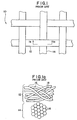
- Prior art surgical support mesh 10 may be manufactured from monofilament or multifilament yarns.
- Prior art mesh 10, as shown, includes multifilament horizontally-extending yarns 12 and multifilament vertically-extending yarns 14 woven together to form a support trellis.
- multifilament yarns such as yarns 12 and 14
- the flexibility of a filament generally increases as its diameter decreases. Because the solid cross-sectional area of the filaments of a multifilament yarn is less than the cross-sectional area of a monofilament yarn of equivalent diameter, the multifilament yarn will have a greater degree of flexibility and pliability than that of the monofilament yarn.
- each of multifilament yarns 12 and 14 is composed of a plurality of filaments 16 that are intermingled or bundled together to form the yarn.
- Interstitial spaces 18, which are pockets of air, are formed between adjacent filaments of the yarn.
- Surgical mesh is, of course, thoroughly sterilized prior to implantation. Nevertheless, surgeons typically prefer the use of monofilament-designed mesh to minimize any risk of infection. As a result, the advantages associated with multifilament-designed mesh (i.e., softness and pliability which result in better assimilation of the mesh into the body) are typically sacrificed.
- a surgical support mesh having both the softness and pliability of a multifilament-designed mesh and the infection resistance of a monofilament-designed mesh may be produced.
- a support trellis formed of multifilament yarn wherein the interstitial voids located between adjacent filaments are enclosed within an infection-impervious matrix exhibits the desired resistance to harboring of infectious matter without significant loss of flexibility.
- the matrix which completely encloses the interstitial voids between the filaments of the yarn, provides an effective barrier to the passage of infectious matter between the interior and exterior of the yarn. Accordingly, any voids remaining in the yarn after encapsulation of such yarn are enclosed (and thereby sealed) within the resultant matrix.
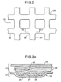
- FIG. 2 A first embodiment of the present invention is shown in Fig. 2.
- this first embodiment includes a support trellis 20 formed of multifilament yarns 22 and 24 which overlap at cross-over junctions 25.
- a support trellis 20 formed of multifilament yarns 22 and 24 which overlap at cross-over junctions 25.
- trellis is encapsulated within a matrix 26, which is preferably a flexible material that continuously surrounds the exterior of the yarns thereby enclosing interstitial voids 27 located between filaments 28 (see Fig. 2a).
- the matrix is formed from a polymeric resin.
- the resin can be applied to the yarn in such a manner as to not allow the resin to substantially penetrate into the yarn.
- the penetration of the resin can be controlled through the application procedure, e.g., quantity of resin applied and/or encapsulating time.
- the interstitial spaces are enclosed (rather than filled) within the continuous matrix.
- the resin can be allowed to penetrate into the yarn, thereby substantially filling the void space located therein.
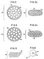
- individual yarns 29, as shown in Fig. 3 are encapsulated within matrix 30 prior to forming of the support trellis.
- Fig. 3a shows a compressed yarn 29 which provides a trellis having a reduced thickness.
- interstitial voids 32 remaining in the yarn are enclosed (and thereby sealed) within the matrix. This then prevents infectious matter from traveling between the interior and exterior of the yarn.
- the matrix provides an infection-impervious barrier between any interstitial voids remaining in the yarn after encapsulation and the exterior of such yarn, while simultaneously maintaining the desired flexibility.
- the depth of penetration of the matrix can be controlled by regulating the quantity of resin applied to the yarn and/or by regulating the coating time.
- matrix 36 penetrates into the interstitial spaces of the yarn, thereby substantially filling the bulk of air space located therein.
- the resin employed to encapsulate the trellis preferably has a melting temperature lower than the melting temperature of the individual filaments such that the resin may be applied to the trellis without damage thereto (i.e., melting of the filaments).
- the resin should exhibit a high degree of flexibility to ensure that the formed mesh retains its desired pliability.
- the resin has a Young's Modulus lower than that of the filament material. Resins formed from polyester, polypropylene, polyethylene, polyurethane, poly(meth) acrylate, or copolymers thereof are contemplated for use herein.
- a resin solution is applied to the formed trellis.
- the solvent carrying the resin is then caused to be evaporated, whereby the solute impregnates and thereby fills the voids within the yarn.
- the encapsulation of the multifilament yarns permanently encloses the interstitial spaces formed between the individual filament of the yarns.
- a continuous infection-impervious matrix is formed around the exterior of the yarn, thereby encapsulating the filaments and filling and/or sealing the interstitial spaces formed therebetween.
- the resultant surgical mesh therefore exhibits more softness and pliability than a monofilament yarn, while simultaneously providing a barrier to the passage of - infectious matter, which infectious matter is common in conventional multifilament yarns.
- the encapsulation of the yarns if done subsequent to forming of the support trellis, i.e. to the fabric per se, fuses the yarns of the trellis together at the crossover junctions 25 shown in Fig. 2. This improves the ravel resistance of the formed mesh. It also improves the linting and fraying characteristics of the mesh (i.e., the mesh is less prone to both tinting and fraying). If, however, the individual yarns are encapsulated prior to forming of the trellis, the trellis may still be heated after formation to fuse the yarn coatings together, thereby rendering such trellis ravel resistant.
- the multifilament yarns employed in the present invention may be formed of bi-component filaments 42.
- Each of these bi-component filaments includes a sheath 44 and a core 46.
- the sheath is preferably formed of a material having a melting or fusing point lower than that of the material forming the core of the filament.
- a means other than heat for example, a liquid based coating material, once the solvent is evaporated the coating remains to encapsulate the filaments and/or filling the interstitial spaces formed therebetween.
- the sheath 44 is preferably a polyethylene terephthalate/isophthalate co-polyester, while the core 46 is preferably a polyethylene terephthalate polyester.
- the core 46 is preferably a polyethylene terephthalate polyester.
- other suitable materials may be used to manufacture the bi-component filaments.
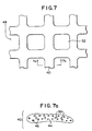
- FIG. 7 and 7a Another embodiment of the present invention is shown in Figs. 7 and 7a.
- this embodiment includes a support trellis 48 formed ofbi-component multifilament yarns, such as yarns 40. Subsequent to forming of the trellis, the trellis is heated to a predetermined temperature, i.e., the fusing temperature of the bi-component filaments.
- sheaths 44 of the bi-component filament begin to melt and flow together, thereby at least substantially filling the voids between filaments and also encapsulating cores 46 within a continuous polymeric matrix.
- the melting sheaths also enclose any voids 50 which are not filled by the flowing polymer. Because the polymer of sheath 44 is softer and more flexible than the polymer of core 46, the formed trellis exhibits the flexibility of a surgical support mesh which is closer to that formed of a conventional multifilament yarn than of a monofilament yarn.
- the bicomponent untwisted filament yarns also tend to flatten out. This flattening is emphasized even more due to transverse forces on the yarns during heat compression of the fabric. Additionally, the sheath polymer flowing into the interstitial voids between the multifilaments causes the reduction of air between the fibers, further contributing to the consolidation of the yarn bundle.
- the surgical mesh of the present invention may be formed by weaving, knitting, braiding or otherwise forming a plurality of multifilament yarns into a support trellis structure.
- the multifilament yarns may be either traditional multifilament yarns or bi-component multifilament yarns.
- the support mesh may thereafter be subjected to thermal or light energy to fuse the filaments together.
- the mesh may be placed in an oven and heated to a temperature of, for example, 180-210°C, and preferably 200-210°C such that the sheaths of the individual filaments fuse together.
- the encapsulation of the yarns provides the trellis with a "membrane-like" feel, while also eliminating the fibrous properties of a warp-knitted structure.
- the present invention allows the size of the trellis pores (e.g., pores 52 shown in Fig. 7) to be regulated.
- the pore size preferably about 50 microns or larger
- the pore size can be regulated by controlling the quantity of resin applied to the exterior of the trellis. It is believed that regulation of pore size may facilitate assimilation of the trellis into the body.
- a medicinal substance e.g., an antibiotic
- the drug may be dispersed directly throughout the encapsulating resin or, alternatively, added to a plurality of separate carriers which in turn are dispersed throughout the encapsulating resin.
- a thin mesh is formed having a thickness in the range of about 0.05 millimeters to about 0.50 millimeters.
- the thin mesh of the present invention will have a thickness of about 0.10 millimeters to about 0.20 millimeters.
- the mesh width and length dimensions can vary within those ranges conventionally used for a specific application and delivery device. For example, such ranges include dimensions of about 12 centimeters x 15 centimeters to about 14 centimeters x 16 centimeters. These ranges are generally sufficient to cover the area of repair of, for example, the myopectineal orifice of an adult. As mentioned above, such a thin surgical mesh could then be rolled or otherwise folded so as to fit within the cannula of a needle having a small diameter of, for example, 5 millimeters or less.
- the individual filaments or fibers are capable of spreading out or flattening to provide a reduced thickness and a low profile. To maximize the yarn's ability to splay it is necessary to minimize or prevent the introduction of twisting or other such orientations of the yarn which might impede its ability to adopt a low profile.
- a bicomponent yarn allows for one of the components to be a meltable resin which, when heated, subsequently can fuse together with other yarns in the fabric to weld at the interlacing points to seal the structure.
- the welding of the interlaces stabilizes the yarns in the woven structure while the sealing of the yarns eliminates the porosity with the yarn bundle and thereby reduces the risk of harboring infections.
- the treatment also imparts a resiliency to the mesh to resume a planar shape once released from the cannula.
- Multifilament yarns also can provide a lower profile to the mesh as they can flatten out.
- the flattening characteristic of multifilament yarn is maximized when the yarn is not impeded by twisting or other yarn conditioning.
- the fabric is heat set which causes the yarn to assume an elliptical cross-section, rather than the round cross-section retained by a monofilament or a highly twisted multifilament yarn.
- One embodiment of a thinly woven mesh of the present invention includes use of a 75 denier bicomponent polyester yarn having a circular diameter of, for example, about 0.09 millimeters.
- the multifilament yarns contemplated for use in the present invention can be flattened preferably about 50 percent of the original thickness. When flattened, the yarn assumes an elliptical or race-track (rectangular with rounded corners) cross-section having a minor diameter, or yarn thickness, of about 0.04 millimeters to about 0.06 millimeters, and a major diameter, or yarn width, of about 0.22 millimeters to about 0.28 millimeters.
- the rectangular pore size would be about 0.05 to 0.07 millimeters on either or both sides, or 50 to 70 microns, respectively.
- a woven construction provides a greater strength to thickness ratio than a knitted construction. It also allows the mesh to be engineered, for example, for isotropic or anisotropic properties more effectively, more predictably, and more thinly than a knitted fabric.
- the estimated burst strength of the fabric would be about 21kg/cm 2 , or 300 psi, or 2068 kPa. Such burst strengths are in the range of conventional surgical repair fabrics, including those for hernia repairs and which have a greater thickness than those contemplated by the present invention.
- the mesh may have a shape memory imparted thereto.
- the mesh could, for example, be heat conditioned for a sufficient time at a sufficient temperature while the mesh is in an unfurled configuration.
- the mesh could also include individual threads having a shape-memory imparted thereto, such as nitinol threads.
- the mesh could therefore be designed to assume an unfurled configuration when the mesh reaches body temperature subsequent to deployment. Imparting shape-memory to the mesh would allow the mesh, even after having been stored within the delivery device, e.g. the cannula of a needle, to assume an unfurled configuration once deployed into the peritoneal cavity. Designing shape-memory into the soft tissue mesh in this manner facilitates orientation in the body.
- the mesh due to the encapsulating matrix, the mesh possesses a structural stability and ravel resistance which permits pulling and aligning using laparoscopic gripping devices.
- the soft tissue meshes of the present invention may be implanted using conventional surgical or laparoscopic techniques.
- a new minimally invasive technique employing a needle delivery is used.
- the mesh is rolled, folded, or otherwise compressed to a reduced volume such that it can be contained within a needle delivery system.
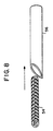
- rolled mesh 54 is shown being insertable into cannula 56.
- the mesh must have a sufficiently low volume and profile to pass through a needle cannula and be deployed into the affected area.
- the mesh has been memory heat-set to return to a relatively planar configuration once deployed. Other shapes, depending on the specific body application may of course also be used.
- thin surgical support mesh could then be delivered into the properitoneal cavity of a patient via the needle.
- a second small needle cannula could be inserted into the properitoneal cavity to insufflate the region with carbon dioxide.
- the hernia sac would be dissected free and ligated.
- Visualization during the needle herniorrhaphy would be obtained with a 2-5 millimeter laparoscope placed through one of the cannulas.
- the mesh upon being expelled from the needle over the transversalis fascia, could then be manipulated to cover the myopectineal cavity.
- the mesh could further be provided with an elastic memory causing the mesh to unfurl once ejected from the cannula.
- the mesh may then be optionally sutured or stapled over the herniated region for assimilation by the body tissue so as to provide added support to the tissue of the properitoneal cavity.
- a needle herniorrhaphy technique could therefore be performed which would obviate the need for open surgery.
- the needle herniorrhaphy technique could be performed without the need for general anesthesia and would reduce the pain and disability currently associated with open or laparoscopic techniques.
- a support trellis is woven of bi-component yarns, particularly 250 denier/16 filament/type LHCV Kanebo Bellcouple® polyester yarns.
- the Kanebo Bellcouple® yarn includes a polyethylene terephthalate polyester core and a polyethylene terephthalate/isophthalate co-polyester sheath, the sheath having a lower melting temperature than the core.
- the trellis is placed in a convection oven and heated to about 210°C, thus fusing the individual sheaths together.
- the yarns are thereby encapsulated and, further, are fused to each other at the junctions where the yarns overlap.
- the resultant mesh is thereafter sealed in a sterile package.
- a support trellis is warp-knitted of bicomponent yarns, particularly 75 denier/24 filament/type LHCV Kanebo Bellcouple® polyester yarns. Following construction of the support trellis, the trellis is placed in a convection oven and heated to about 210°C, thus fusing the individual sheaths together.
- the resultant mesh is thereafter sealed in a sterile package.
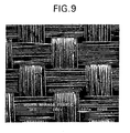
- a support trellis is woven in a plain weave of bicomponent yarns, particularly 75 denier/24 filament/type LHCV Kanebo Bellcouple® polyester yarns.
- the threadcount is 70 ends per inch by 70 picks per inch.
- the rectangular pore size is in the range of 50 to 70 micrometers on each side.
- the trellis is placed in a forced hot air convection oven and heated to about 210°C for 15 minutes, thus fusing the individual sheaths together.
- the thickness of the construction is on the order of 0.10 millimeters or less.
- Figure 9 is a photomicrograph showing a portion of a construction according to this Example.
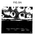
- Figure 9a is a photomicrograph showing a cross-sectional view of this construction.
- the resultant mesh is thereafter sealed in a sterile package.
Landscapes
- Health & Medical Sciences (AREA)
- Life Sciences & Earth Sciences (AREA)
- Animal Behavior & Ethology (AREA)
- Transplantation (AREA)
- Engineering & Computer Science (AREA)
- Biomedical Technology (AREA)
- Heart & Thoracic Surgery (AREA)
- Oral & Maxillofacial Surgery (AREA)
- Cardiology (AREA)
- Vascular Medicine (AREA)
- General Health & Medical Sciences (AREA)
- Public Health (AREA)
- Veterinary Medicine (AREA)
- Materials For Medical Uses (AREA)
- Prostheses (AREA)
- Infusion, Injection, And Reservoir Apparatuses (AREA)
Abstract
Claims (38)
- Maille chirurgicale (54) souple et pliable, ayant un treillis de support comprenant des fils multifilaments (22, 24, 29, 34, 40), caractérisée en ce que :les fils multifilaments (22, 24, 29, 34, 40) comprennent des filaments évasés (42), et/ou une forme transversale sensiblement elliptique, les fils multifilaments (22, 24, 29, 34, 40) définissant une pluralité de vides interstitiels (27, 32, 50) situés entre les fils multifilaments (22, 24, 29, 34, 40) ; etcomprenant une matrice imperméable aux infections (26, 30, 36) encapsulant les fils multifilaments (22, 24, 29, 34, 40) et enfermant la pluralité de vides interstitiels (27, 32, 50).
- Maille chirurgicale selon la revendication 1, dans laquelle la matrice (26, 30, 36) entoure les filaments (42) des fils multifilaments (22, 24, 29, 34, 40), la matrice (26, 30, 36) étant formée avec un matériau comprenant un module d'élasticité de Young inférieur au module d'élasticité de Young du matériau formant les filaments (42).
- Maille chirurgicale selon la revendication 2, dans laquelle la matrice (26, 30, 36) pénètre dans les vides situés entre les filaments (42).
- Maille chirurgicale selon la revendication 3, dans laquelle la matrice (26, 30, 36) est formée à partir d'un polymère choisi dans le groupe comprenant le polyester, le polypropylène, le polyuréthane, le poly(méth)acrylate et leurs copolymères.
- Maille chirurgicale selon la revendication 1, dans laquelle les filaments (42) comprennent une âme flexible et une gaine fusible.
- Maille chirurgicale selon la revendication 5, dans laquelle l'âme flexible est formée avec un polyester de polyéthylène téréphthalate et la gaine fusible est formée avec une copolyester composé de polyéthylène téréphthalate / isophthalate.
- Maille chirurgicale selon la revendication 1, dans laquelle les fils multifilaments (22, 24, 29, 34, 40) se chevauchent les uns les autres au niveau de jonctions pour former le treillis de support et les fils qui se chevauchent adhèrent les uns aux autres au niveau des jonctions.
- Maille chirurgicale selon la revendication 1, dans laquelle la matrice (26, 30, 36) comprend en outre un médicament.
- Maille chirurgicale selon la revendication 1, dans laquelle la maille chirurgicale a une épaisseur inférieure à environ 0,20 millimètres.
- Maille chirurgicale selon la revendication 1, dans laquelle les fils (22, 24, 29, 34, 40) comprennent un profil bas.
- Maille chirurgicale selon la revendication 1, dans laquelle la maille définit des pores comprenant une taille de l'ordre d'environ 50 microns à environ 400 microns.
- Maille chirurgicale selon la revendication 1, dans laquelle la maille a une épaisseur de l'ordre d'environ 0,05 millimètres à environ 0,50 millimètres.
- Maille chirurgicale selon la revendication 1, dans laquelle le treillis de support est suffisamment mince pour permettre l'insertion dans une canule (56).
- Maille chirurgicale selon la revendication 13, dans laquelle la canule a un diamètre d'environ 5 millimètres.
- Maille chirurgicale selon la revendication 1, dans laquelle le treillis de support est enroulé.
- Maille chirurgicale selon la revendication 1, dans laquelle le treillis de support est plié.
- Maille chirurgicale selon la revendication 13, dans laquelle la maille chirurgicale est traitée à chaud pendant un temps suffisant à une température suffisante pour effectuer une mémoire de forme dans un état déployé.
- Maille chirurgicale selon la revendication 13, dans laquelle les fils multifilaments (22, 24, 29, 34, 40) comprennent un matériau à mémoire de forme.
- Maille chirurgicale selon la revendication 13, dans laquelle la maille chirurgicale comprend en outre au moins un fil comprenant une mémoire de forme.
- Maille chirurgicale selon la revendication 19, dans laquelle le au moins un fil comprend du Nitinol.
- Maille chirurgicale selon la revendication 1, dans laquelle la maille chirurgicale a une résistance de rupture d'environ 300 psi.
- Procédé permettant de produire une maille chirurgicale (54) souple et pliable en formant un treillis de support à partir de fils multifilaments (22, 24, 29, 34, 40), caractérisé en ce qu'il comprend les étapes consistant à :utiliser des filaments évasés et/ou des filaments ayant une forme transversale généralement elliptique en tant que fils multifilaments, dans lequel les fils multifilaments (22, 24, 29, 54, 60) forment une pluralité de vides interstitiels ; etencapsuler les fils multifilaments (22, 24, 29, 34, 40) dans une matrice imperméable aux infections (26, 30, 36), moyennant quoi les vides interstitiels sont enfermés dans la matrice (26, 30, 36) pour améliorer la résistance à l'invasion des matières infectieuses.
- Procédé selon la revendication 22, dans lequel l'étape d'encapsulation a lieu avant l'étape de formation.
- Procédé selon la revendication 22, dans lequel l'étape de formation a lieu avant l'étape d'encapsulation.
- Procédé selon la revendication 24, dans lequel l'étape d'encapsulation comprend l'étape consistant à appliquer une résine sur les fils multifilaments (22, 24, 29, 34, 40) avant l'étape de formation.
- Procédé selon la revendication 25, dans lequel la résine est appliquée sur le treillis de support en quantité suffisante et pendant une période de temps suffisante pour permettre à la résine de pénétrer dans les vides (27, 32, 50).
- Procédé selon la revendication 25, dans lequel l'étape d'encapsulation comprend l'étape consistant à appliquer une résine sur les fils multifilaments (22, 24, 29, 34, 40) suite à la formation du treillis de support.
- Procédé selon la revendication 27, dans lequel la résine est appliquée sur l'extérieur du treillis de support en quantité suffisante et pendant une période de temps suffisante pour permettre à la résine de pénétrer dans les vides (27, 32, 50).
- Procédé selon la revendication 25, dans lequel l'étape de formation comprend l'étape consistant à prévoir des filaments comprenant une âme flexible et une gaine fusible.
- Procédé selon la revendication 29, dans lequel l'application d'énergie thermique ou d'énergie à base d'onde lumineuse se traduit par la fusion de la gaine fusible.
- Procédé selon la revendication 29, dans lequel les gaines fusibles sont fondues en chauffant la maille chirurgicale à une température de l'ordre d'approximativement 180°C à approximativement 210°C.
- Procédé selon la revendication 22 comprenant en outre l'étape consistant à :incorporer un médicament dans la matrice (26, 30, 36).
- Procédé selon la revendication 22 comprenant en outre l'étape consistant à :appliquer une force de compression sur la maille chirurgicale (54) dans une épaisseur prédéterminée.
- Procédé selon la revendication 22, comprenant en outre l'étape consistant à :traiter à la chaleur la maille chirurgicale pendant un temps suffisant à une température suffisante pour effectuer une mémoire de forme dans un état déployé.
- Procédé selon la revendication 22, dans lequel l'étape de formation comprend l'étape consistant à prévoir des filaments réalisés à partir d'un matériau à mémoire de forme.
- Procédé selon la revendication 22, dans lequel l'étape de formation comprend l'étape consistant à prévoir des fils multifilaments (22, 24, 29, 34, 40) sans leur transmettre de torsion.
- Procédé selon la revendication 22, comprenant en outre l'étape consistant à enrouler le treillis de support.
- Procédé selon la revendication 22, comprenant en outre l'étape consistant à plier le treillis de support.
Applications Claiming Priority (3)
| Application Number | Priority Date | Filing Date | Title |
|---|---|---|---|
| US905529 | 1997-08-04 | ||
| US08/905,529 US6042592A (en) | 1997-08-04 | 1997-08-04 | Thin soft tissue support mesh |
| PCT/US1998/016126 WO1999005992A1 (fr) | 1997-08-04 | 1998-08-04 | Maille de support mince chirurgicale pour tissus mous |
Publications (2)
| Publication Number | Publication Date |
|---|---|
| EP1001717A1 EP1001717A1 (fr) | 2000-05-24 |
| EP1001717B1 true EP1001717B1 (fr) | 2005-11-30 |
Family
ID=25420993
Family Applications (1)
| Application Number | Title | Priority Date | Filing Date |
|---|---|---|---|
| EP98938300A Expired - Lifetime EP1001717B1 (fr) | 1997-08-04 | 1998-08-04 | Maille de support mince chirurgicale pour tissus mous |
Country Status (7)
| Country | Link |
|---|---|
| US (3) | US6042592A (fr) |
| EP (1) | EP1001717B1 (fr) |
| JP (2) | JP5116190B2 (fr) |
| AU (1) | AU742882B2 (fr) |
| CA (1) | CA2299241C (fr) |
| DE (1) | DE69832618T2 (fr) |
| WO (1) | WO1999005992A1 (fr) |
Cited By (10)
| Publication number | Priority date | Publication date | Assignee | Title |
|---|---|---|---|---|
| US8317808B2 (en) | 2008-02-18 | 2012-11-27 | Covidien Lp | Device and method for rolling and inserting a prosthetic patch into a body cavity |
| US8758373B2 (en) | 2008-02-18 | 2014-06-24 | Covidien Lp | Means and method for reversibly connecting a patch to a patch deployment device |
| US8808314B2 (en) | 2008-02-18 | 2014-08-19 | Covidien Lp | Device and method for deploying and attaching an implant to a biological tissue |
| US8906045B2 (en) | 2009-08-17 | 2014-12-09 | Covidien Lp | Articulating patch deployment device and method of use |
| US9034002B2 (en) | 2008-02-18 | 2015-05-19 | Covidien Lp | Lock bar spring and clip for implant deployment device |
| US9044235B2 (en) | 2008-02-18 | 2015-06-02 | Covidien Lp | Magnetic clip for implant deployment device |
| US9301826B2 (en) | 2008-02-18 | 2016-04-05 | Covidien Lp | Lock bar spring and clip for implant deployment device |
| US9393093B2 (en) | 2008-02-18 | 2016-07-19 | Covidien Lp | Clip for implant deployment device |
| US9393002B2 (en) | 2008-02-18 | 2016-07-19 | Covidien Lp | Clip for implant deployment device |
| US9999424B2 (en) | 2009-08-17 | 2018-06-19 | Covidien Lp | Means and method for reversibly connecting an implant to a deployment device |
Families Citing this family (188)
| Publication number | Priority date | Publication date | Assignee | Title |
|---|---|---|---|---|
| US6740122B1 (en) | 1998-09-11 | 2004-05-25 | C. R. Bard, Inc. | Preformed curved prosthesis that is adapted to the external iliac vessels |
| US6723133B1 (en) | 1998-09-11 | 2004-04-20 | C. R. Bard, Inc. | Performed curved prosthesis having a reduced incidence of developing wrinkles or folds |
| US6551241B1 (en) * | 1999-12-17 | 2003-04-22 | Leonard S. Schultz | Instruments and methods for performing percutaneous surgery |
| FR2791883B1 (fr) * | 1999-04-08 | 2001-08-10 | Ethicon Inc | Prothese souple notamment pour la cure des hernies par voie coelioscopique |
| FR2792824B1 (fr) * | 1999-04-27 | 2001-06-22 | Sofradim Production | Dispositif de traitement de prolapsus par suspension vaginale |
| US7273497B2 (en) | 1999-05-28 | 2007-09-25 | Anova Corp. | Methods for treating a defect in the annulus fibrosis |
| US20070038231A1 (en) | 1999-05-28 | 2007-02-15 | Ferree Bret A | Methods and apparatus for treating disc herniation and preventing the extrusion of interbody bone graft |
| US20060247665A1 (en) | 1999-05-28 | 2006-11-02 | Ferree Bret A | Methods and apparatus for treating disc herniation and preventing the extrusion of interbody bone graft |
| US6344052B1 (en) | 1999-09-27 | 2002-02-05 | World Medical Manufacturing Corporation | Tubular graft with monofilament fibers |
| FR2811218B1 (fr) | 2000-07-05 | 2003-02-28 | Patrice Suslian | Dispositif implantable destine a corriger l'incontinence urinaire |
| US7025063B2 (en) | 2000-09-07 | 2006-04-11 | Ams Research Corporation | Coated sling material |
| US6746458B1 (en) | 2000-09-07 | 2004-06-08 | William G. Cloud | Mesh material to repair hernias |
| US20060205995A1 (en) | 2000-10-12 | 2006-09-14 | Gyne Ideas Limited | Apparatus and method for treating female urinary incontinence |
| GB0025068D0 (en) | 2000-10-12 | 2000-11-29 | Browning Healthcare Ltd | Apparatus and method for treating female urinary incontinence |
| US20060058578A1 (en) * | 2002-04-11 | 2006-03-16 | Gyne Ideas Limited | Apparatus and method for treating female urinary incontinence |
| US8167785B2 (en) | 2000-10-12 | 2012-05-01 | Coloplast A/S | Urethral support system |
| CA2365376C (fr) | 2000-12-21 | 2006-03-28 | Ethicon, Inc. | Utilisation d'implants en mousse renforces ayant une meilleure integrite pour la reparation et la regeneration de tissus mous |
| US8162816B2 (en) | 2001-03-09 | 2012-04-24 | Boston Scientific Scimed, Inc. | System for implanting an implant and method thereof |
| GB0108088D0 (en) | 2001-03-30 | 2001-05-23 | Browning Healthcare Ltd | Surgical implant |
| US8961541B2 (en) | 2007-12-03 | 2015-02-24 | Cardio Vascular Technologies Inc. | Vascular closure devices, systems, and methods of use |
| US20080109030A1 (en) | 2001-04-24 | 2008-05-08 | Houser Russell A | Arteriotomy closure devices and techniques |
| US8992567B1 (en) | 2001-04-24 | 2015-03-31 | Cardiovascular Technologies Inc. | Compressible, deformable, or deflectable tissue closure devices and method of manufacture |
| US6726696B1 (en) * | 2001-04-24 | 2004-04-27 | Advanced Catheter Engineering, Inc. | Patches and collars for medical applications and methods of use |
| US7622129B1 (en) | 2002-08-05 | 2009-11-24 | Purdue Research Foundation | Nano-structured polymers for use as implants |
| WO2003015836A1 (fr) * | 2001-08-16 | 2003-02-27 | Purdue Research Foundation | Materiau et procede permettant de stimuler la croissance des tissus |
| US6666817B2 (en) * | 2001-10-05 | 2003-12-23 | Scimed Life Systems, Inc. | Expandable surgical implants and methods of using them |
| US6902932B2 (en) * | 2001-11-16 | 2005-06-07 | Tissue Regeneration, Inc. | Helically organized silk fibroin fiber bundles for matrices in tissue engineering |
| US20110009960A1 (en) * | 2001-11-16 | 2011-01-13 | Allergan, Inc. | Prosthetic fabric structure |
| EP1539044B1 (fr) * | 2002-07-17 | 2011-04-06 | Proxy Biomedical Limited | Membrane pour implantation medicale |
| CA2492630C (fr) | 2002-08-02 | 2009-01-13 | C.R. Bard, Inc. | Fronde a auto-ancrage et systeme d'introduction |
| US20040078090A1 (en) | 2002-10-18 | 2004-04-22 | Francois Binette | Biocompatible scaffolds with tissue fragments |
| US8142515B2 (en) * | 2002-11-04 | 2012-03-27 | Sofradim Production | Prosthesis for reinforcement of tissue structures |
| AU2003295254A1 (en) | 2002-12-11 | 2004-06-30 | Dsm Ip Assets B.V. | Surgical soft tissue mesh |
| US8197837B2 (en) | 2003-03-07 | 2012-06-12 | Depuy Mitek, Inc. | Method of preparation of bioabsorbable porous reinforced tissue implants and implants thereof |
| WO2004085098A2 (fr) * | 2003-03-27 | 2004-10-07 | Purdue Research Foundation | Nanoparticules metalliques en tant que materiau biologique orthopediques |
| WO2004096085A2 (fr) * | 2003-03-27 | 2004-11-11 | Purdue Research Foundation | Nanofibres servant de biomatiere neurale |
| GB0307082D0 (en) | 2003-03-27 | 2003-04-30 | Gyne Ideas Ltd | Drug delivery device and method |
| CA2525792C (fr) | 2003-05-15 | 2015-10-13 | Biomerix Corporation | Matrices elastomeres reticulees, leur production et leur utilisation dans des dispositifs implantables |
| US8226715B2 (en) | 2003-06-30 | 2012-07-24 | Depuy Mitek, Inc. | Scaffold for connective tissue repair |
| US7361138B2 (en) * | 2003-07-31 | 2008-04-22 | Scimed Life Systems, Inc. | Bioabsorbable casing for surgical sling assembly |
| US10583220B2 (en) | 2003-08-11 | 2020-03-10 | DePuy Synthes Products, Inc. | Method and apparatus for resurfacing an articular surface |
| US20050038452A1 (en) * | 2003-08-14 | 2005-02-17 | Scimed Life Systems, Inc. | Medical slings |
| US8337386B2 (en) | 2003-08-14 | 2012-12-25 | Boston Scientific Scimed, Inc. | Surgical slings |
| US8545386B2 (en) * | 2003-08-14 | 2013-10-01 | Boston Scientific Scimed, Inc. | Surgical slings |
| EP1682009B1 (fr) | 2003-10-03 | 2014-03-05 | Boston Scientific Limited, an Irish company | Systemes et procedes de pose d'implant medical dans une zone anatomique d'un patient |
| EP1691852A2 (fr) * | 2003-11-10 | 2006-08-23 | Angiotech International AG | Implants medicaux et agents inducteurs de fibrose |
| US7524281B2 (en) | 2003-11-17 | 2009-04-28 | Boston Scientific Scimed, Inc. | Systems and methods relating to associating a medical implant with a delivery device |
| US7316822B2 (en) | 2003-11-26 | 2008-01-08 | Ethicon, Inc. | Conformable tissue repair implant capable of injection delivery |
| US7901461B2 (en) | 2003-12-05 | 2011-03-08 | Ethicon, Inc. | Viable tissue repair implants and methods of use |
| US7763077B2 (en) | 2003-12-24 | 2010-07-27 | Biomerix Corporation | Repair of spinal annular defects and annulo-nucleoplasty regeneration |
| US20050165480A1 (en) * | 2004-01-23 | 2005-07-28 | Maybelle Jordan | Endovascular treatment devices and methods |
| US11395865B2 (en) | 2004-02-09 | 2022-07-26 | DePuy Synthes Products, Inc. | Scaffolds with viable tissue |
| US8313505B2 (en) | 2004-03-19 | 2012-11-20 | Aga Medical Corporation | Device for occluding vascular defects |
| US9039724B2 (en) * | 2004-03-19 | 2015-05-26 | Aga Medical Corporation | Device for occluding vascular defects |
| US8777974B2 (en) | 2004-03-19 | 2014-07-15 | Aga Medical Corporation | Multi-layer braided structures for occluding vascular defects |
| US8398670B2 (en) * | 2004-03-19 | 2013-03-19 | Aga Medical Corporation | Multi-layer braided structures for occluding vascular defects and for occluding fluid flow through portions of the vasculature of the body |
| US8747453B2 (en) * | 2008-02-18 | 2014-06-10 | Aga Medical Corporation | Stent/stent graft for reinforcement of vascular abnormalities and associated method |
| US20050234291A1 (en) * | 2004-03-30 | 2005-10-20 | Peter Gingras | Medical device |
| DE602005025637D1 (de) * | 2004-03-30 | 2011-02-10 | Proxy Biomedical Ltd | Behandeltes chirurgisches mesh aus monofilen fasern |
| US8221780B2 (en) | 2004-04-20 | 2012-07-17 | Depuy Mitek, Inc. | Nonwoven tissue scaffold |
| US8137686B2 (en) | 2004-04-20 | 2012-03-20 | Depuy Mitek, Inc. | Nonwoven tissue scaffold |
| US8439820B2 (en) | 2004-05-06 | 2013-05-14 | Boston Scientific Scimed, Inc. | Systems and methods for sling delivery and placement |
| GB0411360D0 (en) | 2004-05-21 | 2004-06-23 | Mpathy Medical Devices Ltd | Implant |
| EP2543341B1 (fr) | 2004-06-14 | 2016-07-20 | Boston Scientific Limited | Ancre de tissu mou |
| US20050283040A1 (en) * | 2004-06-16 | 2005-12-22 | Secant Medical, Llc | Incontinence sling |
| WO2006002340A2 (fr) * | 2004-06-23 | 2006-01-05 | Warwick Mills, Inc. | Materiau de biogreffe a absorption controlee pour support de tissus autologue |
| US7481808B2 (en) * | 2004-06-30 | 2009-01-27 | Ethicon, Inc. | Flexible electrode device and surgical apparatus equipped with same |
| WO2006055052A2 (fr) * | 2004-07-19 | 2006-05-26 | Michael Gertner | Procedes et dispositifs de protection embolique chronique |
| DE102004044569A1 (de) * | 2004-09-15 | 2006-03-30 | Voith Fabrics Patent Gmbh | Papiermaschinenbespannungen |
| WO2006046950A1 (fr) | 2004-10-25 | 2006-05-04 | Scimed Life Systems, Inc | Systemes et methodes d'introduction et de mise en place de bandelette |
| US20060089672A1 (en) * | 2004-10-25 | 2006-04-27 | Jonathan Martinek | Yarns containing filaments made from shape memory alloys |
| US8329202B2 (en) | 2004-11-12 | 2012-12-11 | Depuy Products, Inc. | System and method for attaching soft tissue to an implant |
| US20060116713A1 (en) * | 2004-11-26 | 2006-06-01 | Ivan Sepetka | Aneurysm treatment devices and methods |
| US8771294B2 (en) * | 2004-11-26 | 2014-07-08 | Biomerix Corporation | Aneurysm treatment devices and methods |
| CA2601449A1 (fr) * | 2005-03-22 | 2006-09-28 | Tyco Healthcare Group, Lp | Implant a mailles |
| US7556598B2 (en) * | 2005-04-04 | 2009-07-07 | Boston Scientific Scimed, Inc. | Dissolvable protective treatment for an implantable supportive sling |
| EP1871281B1 (fr) * | 2005-04-06 | 2014-01-08 | Boston Scientific Limited | Assemblé de soutenement sous-uretral |
| EP1909687A1 (fr) * | 2005-07-13 | 2008-04-16 | Boston Scientific Scimed, Inc. | Système d ancrage en fronde à ajustement serré et procédés apparentés |
| WO2007014241A1 (fr) | 2005-07-25 | 2007-02-01 | Boston Scientific Scimed, Inc. | Systeme de reparation du plancher pelvien |
| US7981023B2 (en) | 2005-07-25 | 2011-07-19 | Boston Scientific Scimed, Inc. | Elastic sling system and related methods |
| US20070038290A1 (en) * | 2005-08-15 | 2007-02-15 | Bin Huang | Fiber reinforced composite stents |
| EP1937183B1 (fr) * | 2005-09-12 | 2018-11-28 | Proxy Biomedical Limited | Implants de partie molle |
| US20080249607A1 (en) * | 2005-09-20 | 2008-10-09 | Thomas Jay Webster | Biocompatable Nanophase Materials |
| US7878970B2 (en) * | 2005-09-28 | 2011-02-01 | Boston Scientific Scimed, Inc. | Apparatus and method for suspending a uterus |
| US7513865B2 (en) | 2005-12-20 | 2009-04-07 | Boston Scientific Scimed, Inc. | Flattened tubular mesh sling and related methods |
| US9144483B2 (en) * | 2006-01-13 | 2015-09-29 | Boston Scientific Scimed, Inc. | Placing fixation devices |
| US8170686B2 (en) * | 2006-03-14 | 2012-05-01 | Boston Scientific Scimed, Inc. | Heatable sling support for an anatomical location |
| CA2644983C (fr) * | 2006-03-16 | 2015-09-29 | Boston Scientific Limited | Systeme et procede pour traiter un prolapsus de paroi tissulaire |
| US7737060B2 (en) | 2006-03-31 | 2010-06-15 | Boston Scientific Scimed, Inc. | Medical devices containing multi-component fibers |
| US20090035572A1 (en) * | 2006-04-06 | 2009-02-05 | Tyco Healthcare Group Lp | Yarns containing thermoplastic elastomer copolymer and polyolefin filaments |
| US20070293878A1 (en) * | 2006-06-16 | 2007-12-20 | Butsch John L | Magnetic mesh support for tissue walls |
| US20110021868A1 (en) * | 2006-07-11 | 2011-01-27 | James Browning | Tissue Repair Device |
| GB0613653D0 (en) * | 2006-07-11 | 2006-08-16 | Mpathy Medical Devices Ltd | Tissue regeneration repair device |
| US7614258B2 (en) * | 2006-10-19 | 2009-11-10 | C.R. Bard, Inc. | Prosthetic repair fabric |
| US8388679B2 (en) | 2007-01-19 | 2013-03-05 | Maquet Cardiovascular Llc | Single continuous piece prosthetic tubular aortic conduit and method for manufacturing the same |
| US20090192530A1 (en) * | 2008-01-29 | 2009-07-30 | Insightra Medical, Inc. | Fortified mesh for tissue repair |
| US20090004455A1 (en) * | 2007-06-27 | 2009-01-01 | Philippe Gravagna | Reinforced composite implant |
| US8932619B2 (en) * | 2007-06-27 | 2015-01-13 | Sofradim Production | Dural repair material |
| WO2009003726A1 (fr) * | 2007-07-02 | 2009-01-08 | Loehde Eckhard | Maillage tridimensionnel pour hernie |
| US20090099579A1 (en) | 2007-10-16 | 2009-04-16 | Tyco Healthcare Group Lp | Self-adherent implants and methods of preparation |
| US9308068B2 (en) | 2007-12-03 | 2016-04-12 | Sofradim Production | Implant for parastomal hernia |
| US20090171141A1 (en) * | 2007-12-27 | 2009-07-02 | Chu Michael S H | Anterior Repair - Needle Path and Incision Sites |
| US8430807B2 (en) | 2007-12-28 | 2013-04-30 | Boston Scientific Scimed, Inc. | Devices and methods for treating pelvic floor dysfunctions |
| US9078728B2 (en) * | 2007-12-28 | 2015-07-14 | Boston Scientific Scimed, Inc. | Devices and methods for delivering female pelvic floor implants |
| US9282958B2 (en) * | 2007-12-28 | 2016-03-15 | Boston Scientific Scimed, Inc. | Devices and method for treating pelvic dysfunctions |
| US9398944B2 (en) | 2008-02-18 | 2016-07-26 | Covidien Lp | Lock bar spring and clip for implant deployment device |
| WO2009104182A2 (fr) | 2008-02-18 | 2009-08-27 | Polytouch Medical Ltd | Dispositif et procédé pour déployer et faire tenir une pièce sur un tissu biologique |
| US9833240B2 (en) | 2008-02-18 | 2017-12-05 | Covidien Lp | Lock bar spring and clip for implant deployment device |
| US8870743B2 (en) * | 2008-04-08 | 2014-10-28 | Boston Scientific Scimed, Inc. | Pelvic floor mesh and incontinence sling |
| US9242026B2 (en) | 2008-06-27 | 2016-01-26 | Sofradim Production | Biosynthetic implant for soft tissue repair |
| EP2344045A1 (fr) * | 2008-09-16 | 2011-07-20 | Ventralfix, Inc. | Procédé et appareil d'administration, de déploiement en tension et de fixation mini-invasifs de dispositifs prothétiques en matériau secondaire dans un tissu corporel d'un patient, et notamment réparation de hernie à l'intérieur du site de la hernie chez le patient |
| US9820842B2 (en) | 2008-09-30 | 2017-11-21 | The Regents Of The University Of Colorado, A Body Corporate | Medical fabric with integrated shape memory polymer |
| WO2014085827A1 (fr) | 2012-11-30 | 2014-06-05 | The Regents Of The University Of Colorado, A Body Corporate | Étoffe médicale avec polymère à mémoire de forme intégré |
| GB2476912B (en) * | 2008-10-03 | 2012-12-26 | Replication Medical Inc | Vessel protection device |
| EP2792307B1 (fr) | 2008-10-20 | 2017-10-04 | Covidien LP | Dispositif pour fixer une pièce sur un tissu biologique |
| US9204953B2 (en) * | 2008-12-15 | 2015-12-08 | Allergan, Inc. | Biocompatible surgical scaffold with varying stretch |
| US20120150204A1 (en) * | 2008-12-15 | 2012-06-14 | Allergan, Inc. | Implantable silk prosthetic device and uses thereof |
| US9326840B2 (en) * | 2008-12-15 | 2016-05-03 | Allergan, Inc. | Prosthetic device and method of manufacturing the same |
| US9204954B2 (en) * | 2008-12-15 | 2015-12-08 | Allergan, Inc. | Knitted scaffold with diagonal yarn |
| CN102271620B (zh) | 2008-12-15 | 2015-04-08 | 阿勒根公司 | 假体装置以及制造假体装置的方法 |
| US9308070B2 (en) * | 2008-12-15 | 2016-04-12 | Allergan, Inc. | Pliable silk medical device |
| EP2391395A4 (fr) * | 2009-02-02 | 2014-04-09 | Biomerix Corp | Dispositifs maillés composites et procédés de réparation des tissus mous |
| US8308814B2 (en) | 2009-03-27 | 2012-11-13 | Depuy Mitek, Inc. | Methods and devices for preparing and implanting tissue scaffolds |
| US8241298B2 (en) | 2009-03-27 | 2012-08-14 | Depuy Mitek, Inc. | Methods and devices for delivering and affixing tissue scaffolds |
| AU2010233217A1 (en) | 2009-04-09 | 2011-10-27 | Cardiovascular Systems, Inc. | Tissue closure devices, device and systems for delivery, kits and methods therefor |
| US9125716B2 (en) | 2009-04-17 | 2015-09-08 | Boston Scientific Scimed, Inc. | Delivery sleeve for pelvic floor implants |
| US8202301B2 (en) * | 2009-04-24 | 2012-06-19 | Warsaw Orthopedic, Inc. | Dynamic spinal rod and implantation method |
| FR2949688B1 (fr) | 2009-09-04 | 2012-08-24 | Sofradim Production | Tissu avec picots revetu d'une couche microporeuse bioresorbable |
| FR2949687B1 (fr) | 2009-09-04 | 2011-09-23 | Sofradim Production | Tissu avec picots revetus d'un materiau hydrosoluble |
| WO2011031854A1 (fr) * | 2009-09-11 | 2011-03-17 | Allergan, Inc. | Dispositif prothétique et son procédé de fabrication |
| US8690960B2 (en) | 2009-11-24 | 2014-04-08 | Covidien Lp | Reinforced tissue patch |
| US9398943B2 (en) * | 2009-11-30 | 2016-07-26 | Covidien Lp | Ventral hernia repair with barbed suture |
| US9510925B2 (en) * | 2010-02-02 | 2016-12-06 | Covidien Lp | Surgical meshes |
| US9757132B2 (en) * | 2010-03-24 | 2017-09-12 | Biorez, Inc. | Mechanically competent scaffold for rotator cuff and tendon augmentation |
| US20110238094A1 (en) * | 2010-03-25 | 2011-09-29 | Thomas Jonathan D | Hernia Patch |
| JP5729111B2 (ja) * | 2010-04-28 | 2015-06-03 | 東レ株式会社 | ステントグラフト用基布およびステントグラフト |
| US8911348B2 (en) | 2010-09-02 | 2014-12-16 | Boston Scientific Scimed, Inc. | Pelvic implants and methods of implanting the same |
| KR20130056320A (ko) * | 2010-09-09 | 2013-05-29 | 더블유.엘. 고어 앤드 어소시에이트스, 인코포레이티드 | 수술용 메쉬 |
| CA2810672C (fr) | 2010-09-09 | 2017-07-18 | W. L. Gore & Associates, Inc. | Procede d'augmentation de la resistance au dechirement d'un film |
| WO2012034126A1 (fr) * | 2010-09-10 | 2012-03-15 | The Regents Of The University Of Colorado, A Body Corporate | Tissu médical avec polymère à mémoire de forme intégré |
| FR2968533B1 (fr) * | 2010-12-08 | 2013-09-06 | Brothier Lab | Dispositif medical de renfort |
| US8696741B2 (en) | 2010-12-23 | 2014-04-15 | Maquet Cardiovascular Llc | Woven prosthesis and method for manufacturing the same |
| FR2972626B1 (fr) | 2011-03-16 | 2014-04-11 | Sofradim Production | Prothese comprenant un tricot tridimensionnel et ajoure |
| US9113991B2 (en) | 2011-05-12 | 2015-08-25 | Boston Scientific Scimed, Inc. | Anchors for bodily implants and methods for anchoring bodily implants into a patient's body |
| US9636201B2 (en) | 2011-05-12 | 2017-05-02 | Boston Scientific Scimed, Inc. | Delivery members for delivering an implant into a body of a patient |
| FR2977790B1 (fr) | 2011-07-13 | 2013-07-19 | Sofradim Production | Prothese pour hernie ombilicale |
| FR2977789B1 (fr) | 2011-07-13 | 2013-07-19 | Sofradim Production | Prothese pour hernie ombilicale |
| US9198746B2 (en) | 2011-08-30 | 2015-12-01 | Boston Scientific Scimed, Inc. | Materials and methods for securing bodily implants |
| US9168120B2 (en) | 2011-09-09 | 2015-10-27 | Boston Scientific Scimed, Inc. | Medical device and methods of delivering the medical device |
| US9526603B2 (en) | 2011-09-30 | 2016-12-27 | Covidien Lp | Reversible stiffening of light weight mesh |
| FR2985271B1 (fr) | 2011-12-29 | 2014-01-24 | Sofradim Production | Tricot a picots |
| FR2985170B1 (fr) | 2011-12-29 | 2014-01-24 | Sofradim Production | Prothese pour hernie inguinale |
| FR2994185B1 (fr) | 2012-08-02 | 2015-07-31 | Sofradim Production | Procede de preparation d’une couche poreuse a base de chitosane |
| US10058409B2 (en) * | 2012-09-18 | 2018-08-28 | Arthrex, Inc. | Spacer fabric mesh for use in tissue engineering applications |
| FR2995779B1 (fr) | 2012-09-25 | 2015-09-25 | Sofradim Production | Prothese comprenant un treillis et un moyen de consolidation |
| FR2995788B1 (fr) | 2012-09-25 | 2014-09-26 | Sofradim Production | Patch hemostatique et procede de preparation |
| FR2995778B1 (fr) | 2012-09-25 | 2015-06-26 | Sofradim Production | Prothese de renfort de la paroi abdominale et procede de fabrication |
| US10159555B2 (en) | 2012-09-28 | 2018-12-25 | Sofradim Production | Packaging for a hernia repair device |
| CA2884764A1 (fr) * | 2012-10-01 | 2014-04-10 | Ams Research Corporation | Implant conducteur et degradable destine au traitement des tissus pelviens |
| US9433489B2 (en) * | 2013-03-12 | 2016-09-06 | Soft Tissue Regeneration, Inc. | Absorbable synthetic braided matrix for breast reconstruction and hernia repair |
| US9814555B2 (en) | 2013-03-12 | 2017-11-14 | Boston Scientific Scimed, Inc. | Medical device for pelvic floor repair and method of delivering the medical device |
| EP2968696B1 (fr) * | 2013-03-15 | 2017-07-26 | Boston Scientific Scimed, Inc. | Mailles chirurgicales à base de filaments à composants multiples |
| US9585695B2 (en) | 2013-03-15 | 2017-03-07 | Woven Orthopedic Technologies, Llc | Surgical screw hole liner devices and related methods |
| FR3006578B1 (fr) | 2013-06-07 | 2015-05-29 | Sofradim Production | Prothese a base d’un textile pour voie laparoscopique |
| FR3006581B1 (fr) | 2013-06-07 | 2016-07-22 | Sofradim Production | Prothese a base d’un textile pour voie laparoscopique |
| US9962251B2 (en) | 2013-10-17 | 2018-05-08 | Boston Scientific Scimed, Inc. | Devices and methods for delivering implants |
| US10028731B2 (en) | 2013-11-12 | 2018-07-24 | Genzyme Corporation | Barrier application device |
| US9642638B1 (en) | 2014-04-02 | 2017-05-09 | Vicki J. Carrier | Morcellated tissue collection pouch |
| US9907593B2 (en) | 2014-08-05 | 2018-03-06 | Woven Orthopedic Technologies, Llc | Woven retention devices, systems and methods |
| US8956394B1 (en) | 2014-08-05 | 2015-02-17 | Woven Orthopedic Technologies, Llc | Woven retention devices, systems and methods |
| US9943351B2 (en) | 2014-09-16 | 2018-04-17 | Woven Orthopedic Technologies, Llc | Woven retention devices, systems, packaging, and related methods |
| EP3000489B1 (fr) | 2014-09-24 | 2017-04-05 | Sofradim Production | Procédé de préparation d'un film barrière anti-adhésion |
| EP3000432B1 (fr) | 2014-09-29 | 2022-05-04 | Sofradim Production | Prothèse à base textile pour le traitement d'une hernie inguinale |
| EP3000433B1 (fr) | 2014-09-29 | 2022-09-21 | Sofradim Production | Dispositif pour introduire une prothèse pour le traitement de la hernie dans une incision et prothèse textile flexible |
| USD740427S1 (en) | 2014-10-17 | 2015-10-06 | Woven Orthopedic Technologies, Llc | Orthopedic woven retention device |
| EP3029189B1 (fr) | 2014-12-05 | 2021-08-11 | Sofradim Production | Tricot poreux prothétique, procédé pour son obtention et prothèse à hernie |
| EP3059255B1 (fr) | 2015-02-17 | 2020-05-13 | Sofradim Production | Méthode pour préparer une matrice de chitosane comprenant un élément de renfort fibreux |
| EP3085337B1 (fr) | 2015-04-24 | 2022-09-14 | Sofradim Production | Prothèse pour supporter une structure mammaire |
| EP3106185B1 (fr) | 2015-06-19 | 2018-04-25 | Sofradim Production | Prothèse synthétique comprenant un tricot et un film non poreux et méthode pour la former |
| WO2017024277A1 (fr) | 2015-08-05 | 2017-02-09 | Woven Orthopedic Technologies, Llc | Dispositifs, systèmes et procédés de prélèvement à utiliser dans un tissu osseux |
| EP3195830B1 (fr) | 2016-01-25 | 2020-11-18 | Sofradim Production | Prothèse de réparation de hernie |
| EP3312325B1 (fr) | 2016-10-21 | 2021-09-22 | Sofradim Production | Méthode pour la fabrication un treillis avec suture crantée attachée et treillis ainsi obtenu |
| WO2018107114A1 (fr) | 2016-12-09 | 2018-06-14 | Woven Orthopedic Technologies, LLC. | Dispositifs de retenue, treillis et systèmes et procédés associés |
| EP3398554A1 (fr) | 2017-05-02 | 2018-11-07 | Sofradim Production | Prothèse pour réparation de hernie inguinale |
| EP3400901A1 (fr) * | 2017-05-12 | 2018-11-14 | Keystone Heart Ltd. | Dispositif de filtrage de matériau embolique dans un système vasculaire |
| EP3653171B1 (fr) | 2018-11-16 | 2024-08-21 | Sofradim Production | Implants conçus pour la réparation de tissus mous |
| US12064330B2 (en) | 2020-04-28 | 2024-08-20 | Covidien Lp | Implantable prothesis for minimally invasive hernia repair |
| US11413129B2 (en) | 2020-06-19 | 2022-08-16 | Davol Inc. | Implantable prosthesis |
Family Cites Families (64)
| Publication number | Priority date | Publication date | Assignee | Title |
|---|---|---|---|---|
| US3124136A (en) * | 1964-03-10 | Method of repairing body tissue | ||
| US2671444A (en) * | 1951-12-08 | 1954-03-09 | Jr Benjamin F Pease | Nonmetallic mesh surgical insert for hernia repair |
| GB725343A (en) * | 1952-09-06 | 1955-03-02 | Wingfoot Corp | Treatment of textile fabrics |
| US3054406A (en) * | 1958-10-17 | 1962-09-18 | Phillips Petroleum Co | Surgical mesh |
| US3063454A (en) * | 1959-02-26 | 1962-11-13 | Cleanese Corp Of America | Non-woven products |
| GB1095552A (fr) * | 1964-02-21 | |||
| US3372696A (en) * | 1965-01-22 | 1968-03-12 | Peter S. Rudie | Abdominal pad used in surgery |
| US3272204A (en) * | 1965-09-22 | 1966-09-13 | Ethicon Inc | Absorbable collagen prosthetic implant with non-absorbable reinforcing strands |
| DE1492434A1 (de) * | 1965-10-15 | 1969-12-04 | Ludwig Povel & Co Kg | Verbandstoff |
| US3416524A (en) * | 1966-12-16 | 1968-12-17 | Parke Davis & Co | Surgical dressing |
| NL6802563A (fr) * | 1967-02-25 | 1968-08-26 | ||
| US3642565A (en) * | 1968-03-21 | 1972-02-15 | Kanegafuchi Spinning Co Ltd | Composite filaments having an elastic crimping property |
| US3526228A (en) * | 1969-03-24 | 1970-09-01 | Ethicon Inc | Collagen lamina dural prosthesis |
| US4175557A (en) * | 1975-11-24 | 1979-11-27 | International Paper Company | Polymeric sheets |
| CA1070584A (fr) * | 1978-05-24 | 1980-01-29 | Her Majesty The Queen, In Right Of Canada, As Represented By The Minister Of National Defence | Champ chirurgical muni d'un diffuseur eliptique |
| US4347847A (en) * | 1980-06-06 | 1982-09-07 | Usher Francis C | Method of hernia repair |
| US4452245A (en) * | 1980-06-06 | 1984-06-05 | Usher Francis C | Surgical mesh and method |
| DE3213673A1 (de) * | 1982-04-14 | 1983-10-27 | Karl Otto Braun Kg, 6759 Wolfstein | Wundtextil |
| US4520821A (en) * | 1982-04-30 | 1985-06-04 | The Regents Of The University Of California | Growing of long-term biological tissue correction structures in vivo |
| US4633873A (en) * | 1984-04-26 | 1987-01-06 | American Cyanamid Company | Surgical repair mesh |
| US4626253A (en) * | 1984-10-05 | 1986-12-02 | Johnson & Johnson Products, Inc. | Surgical hemostat comprising oxidized cellulose |
| US4655221A (en) | 1985-05-06 | 1987-04-07 | American Cyanamid Company | Method of using a surgical repair mesh |
| US4693720A (en) * | 1985-09-23 | 1987-09-15 | Katecho, Incorporated | Device for surgically repairing soft tissues and method for making the same |
| SE455466C (sv) * | 1986-03-10 | 1993-07-05 | Moelnlycke Ab | Foerband foer vaetskande saar |
| US4769038A (en) * | 1986-03-18 | 1988-09-06 | C. R. Bard, Inc. | Prostheses and techniques and repair of inguinal and femoral hernias |
| US4854316A (en) * | 1986-10-03 | 1989-08-08 | Davis Emsley A | Apparatus and method for repairing and preventing para-stomal hernias |
| US5019096A (en) * | 1988-02-11 | 1991-05-28 | Trustees Of Columbia University In The City Of New York | Infection-resistant compositions, medical devices and surfaces and methods for preparing and using same |
| US5201745A (en) * | 1988-03-15 | 1993-04-13 | Imedex | Visceral surgery patch |
| US4986831A (en) | 1988-04-25 | 1991-01-22 | Angeion Corporation | Medical implant |
| FR2646343B1 (fr) * | 1989-04-27 | 1991-12-20 | Gazielly Dominique | Dispositif de renfort et de soutien de la coiffe des rotateurs d'une articulation d'epaule d'individu |
| US5100422A (en) * | 1989-05-26 | 1992-03-31 | Impra, Inc. | Blood vessel patch |
| JP3366946B2 (ja) * | 1990-07-31 | 2003-01-14 | グンゼ株式会社 | 手術用縫合糸 |
| JP2890063B2 (ja) * | 1990-08-09 | 1999-05-10 | グンゼ株式会社 | 手術用縫合糸の製造法 |
| US5178630A (en) * | 1990-08-28 | 1993-01-12 | Meadox Medicals, Inc. | Ravel-resistant, self-supporting woven graft |
| US5122155A (en) * | 1990-10-11 | 1992-06-16 | Eberbach Mark A | Hernia repair apparatus and method of use |
| US5141515A (en) * | 1990-10-11 | 1992-08-25 | Eberbach Mark A | Apparatus and methods for repairing hernias |
| US5116357A (en) * | 1990-10-11 | 1992-05-26 | Eberbach Mark A | Hernia plug and introducer apparatus |
| US5254133A (en) * | 1991-04-24 | 1993-10-19 | Seid Arnold S | Surgical implantation device and related method of use |
| US5358492A (en) * | 1991-05-02 | 1994-10-25 | Feibus Miriam H | Woven surgical drain and method of making |
| CA2075080A1 (fr) * | 1991-08-02 | 1993-02-03 | Ralph A. Dematteis | Methode et appareil de reparation des hernies par la parascopie |
| US5234457A (en) * | 1991-10-09 | 1993-08-10 | Boston Scientific Corporation | Impregnated stent |
| US5290217A (en) * | 1991-10-10 | 1994-03-01 | Earl K. Sipes | Method and apparatus for hernia repair |
| US5292328A (en) * | 1991-10-18 | 1994-03-08 | United States Surgical Corporation | Polypropylene multifilament warp knitted mesh and its use in surgery |
| US5147374A (en) * | 1991-12-05 | 1992-09-15 | Alfredo Fernandez | Prosthetic mesh patch for hernia repair |
| US5176692A (en) * | 1991-12-09 | 1993-01-05 | Wilk Peter J | Method and surgical instrument for repairing hernia |
| CA2090000A1 (fr) * | 1992-02-24 | 1993-08-25 | H. Jonathan Tovey | Dispositif articule servant a positionner des implants chirurgicaux |
| US5263969A (en) * | 1992-04-17 | 1993-11-23 | Phillips Edward H | Tool for the laparoscopic introduction of a mesh prosthesis |
| WO1994027535A1 (fr) * | 1992-05-20 | 1994-12-08 | C.R. Bard, Inc. | Prothese implantable, et procede et appareil permettant de loger, transporter et livrer une prothese implantable |
| US5368602A (en) * | 1993-02-11 | 1994-11-29 | De La Torre; Roger A. | Surgical mesh with semi-rigid border members |
| IT1265070B1 (it) * | 1993-05-18 | 1996-10-30 | Eniricerche Spa | Filamento composito termoplastico flessibile contenente fibre continue e procedimento per la sua preparazione |
| DE69433939T2 (de) | 1993-11-03 | 2005-08-11 | Clarion Pharmaceuticals, Inc., Madison | Hämostatisches pflaster |
| FI952751A (fi) * | 1994-06-15 | 1995-12-16 | Meadox Medicals Inc | Kirurginen tukiverkko |
| DE69419265T2 (de) | 1994-07-27 | 2000-01-05 | W.L. Gore & Associates, Inc. | Höchfeste poröse folie aus ptfe |
| US5769864A (en) | 1994-09-29 | 1998-06-23 | Surgical Sense, Inc. | Hernia mesh patch |
| US5634931A (en) * | 1994-09-29 | 1997-06-03 | Surgical Sense, Inc. | Hernia mesh patches and methods of their use |
| US5916225A (en) | 1994-09-29 | 1999-06-29 | Surgical Sense, Inc. | Hernia mesh patch |
| IT1275080B (it) | 1994-11-09 | 1997-07-30 | Gabriele Valenti | Protesi dinamica in duplice strato per il trattamento chirurgico dell'ernia inguinale |
| US5549967A (en) * | 1995-05-04 | 1996-08-27 | Huyck Licensco, Inc. | Papermakers' press fabric with increased contact area |
| US5641502A (en) | 1995-06-07 | 1997-06-24 | United States Surgical Corporation | Biodegradable moldable surgical material |
| US5569273A (en) | 1995-07-13 | 1996-10-29 | C. R. Bard, Inc. | Surgical mesh fabric |
| JPH09103477A (ja) * | 1995-10-12 | 1997-04-22 | Unitika Ltd | 縫合糸 |
| WO1997022310A2 (fr) * | 1995-12-15 | 1997-06-26 | Rangel Gonzalez Neftali | Hernio-prothese myopectineale et abdominale avec dispositif auto-depliant |
| US5922026A (en) | 1997-05-01 | 1999-07-13 | Origin Medsystems, Inc. | Surgical method and prosthetic strip therefor |
| US5824082A (en) | 1997-07-14 | 1998-10-20 | Brown; Roderick B. | Patch for endoscopic repair of hernias |
-
1997
- 1997-08-04 US US08/905,529 patent/US6042592A/en not_active Expired - Lifetime
-
1998
- 1998-08-04 CA CA002299241A patent/CA2299241C/fr not_active Expired - Fee Related
- 1998-08-04 EP EP98938300A patent/EP1001717B1/fr not_active Expired - Lifetime
- 1998-08-04 DE DE69832618T patent/DE69832618T2/de not_active Expired - Lifetime
- 1998-08-04 JP JP2000504813A patent/JP5116190B2/ja not_active Expired - Fee Related
- 1998-08-04 WO PCT/US1998/016126 patent/WO1999005992A1/fr active IP Right Grant
- 1998-08-04 AU AU86852/98A patent/AU742882B2/en not_active Ceased
-
1999
- 1999-11-23 US US09/447,971 patent/US6375662B1/en not_active Expired - Lifetime
-
2001
- 2001-12-18 US US10/024,440 patent/US6669706B2/en not_active Expired - Lifetime
-
2009
- 2009-08-25 JP JP2009194879A patent/JP2009273926A/ja not_active Withdrawn
Cited By (11)
| Publication number | Priority date | Publication date | Assignee | Title |
|---|---|---|---|---|
| US8317808B2 (en) | 2008-02-18 | 2012-11-27 | Covidien Lp | Device and method for rolling and inserting a prosthetic patch into a body cavity |
| US8758373B2 (en) | 2008-02-18 | 2014-06-24 | Covidien Lp | Means and method for reversibly connecting a patch to a patch deployment device |
| US8808314B2 (en) | 2008-02-18 | 2014-08-19 | Covidien Lp | Device and method for deploying and attaching an implant to a biological tissue |
| US9034002B2 (en) | 2008-02-18 | 2015-05-19 | Covidien Lp | Lock bar spring and clip for implant deployment device |
| US9044235B2 (en) | 2008-02-18 | 2015-06-02 | Covidien Lp | Magnetic clip for implant deployment device |
| US9301826B2 (en) | 2008-02-18 | 2016-04-05 | Covidien Lp | Lock bar spring and clip for implant deployment device |
| US9393093B2 (en) | 2008-02-18 | 2016-07-19 | Covidien Lp | Clip for implant deployment device |
| US9393002B2 (en) | 2008-02-18 | 2016-07-19 | Covidien Lp | Clip for implant deployment device |
| US8734473B2 (en) | 2009-02-18 | 2014-05-27 | Covidien Lp | Device and method for rolling and inserting a prosthetic patch into a body cavity |
| US8906045B2 (en) | 2009-08-17 | 2014-12-09 | Covidien Lp | Articulating patch deployment device and method of use |
| US9999424B2 (en) | 2009-08-17 | 2018-06-19 | Covidien Lp | Means and method for reversibly connecting an implant to a deployment device |
Also Published As
| Publication number | Publication date |
|---|---|
| US6042592A (en) | 2000-03-28 |
| CA2299241C (fr) | 2005-09-13 |
| DE69832618D1 (de) | 2006-01-05 |
| EP1001717A1 (fr) | 2000-05-24 |
| US20020052612A1 (en) | 2002-05-02 |
| JP2001511390A (ja) | 2001-08-14 |
| WO1999005992A1 (fr) | 1999-02-11 |
| US6669706B2 (en) | 2003-12-30 |
| AU742882B2 (en) | 2002-01-17 |
| US6375662B1 (en) | 2002-04-23 |
| AU8685298A (en) | 1999-02-22 |
| JP5116190B2 (ja) | 2013-01-09 |
| JP2009273926A (ja) | 2009-11-26 |
| DE69832618T2 (de) | 2006-08-17 |
| CA2299241A1 (fr) | 1999-02-11 |
Similar Documents
| Publication | Publication Date | Title |
|---|---|---|
| EP1001717B1 (fr) | Maille de support mince chirurgicale pour tissus mous | |
| CA1335527C (fr) | Element chirurgical bioabsorbable pour le traitement des lesions nerveuses | |
| JP3078716B2 (ja) | 植え込み式の管状人工器官 | |
| US5147399A (en) | Method of treating nerve defects through use of a bioabsorbable surgical device | |
| US6113640A (en) | Reconstructive bioabsorbable joint prosthesis | |
| AU734235B2 (en) | Bioabsorbable suture anchor | |
| EP1060714B1 (fr) | Tissu maille chirurgical | |
| JP2960586B2 (ja) | ほつれ防止自己保持型編織製人工血管およびその製造方法 | |
| JP2763801B2 (ja) | 外科用移植材料及びその製造方法 | |
| JP2001511390A5 (fr) | ||
| WO1996003091A1 (fr) | Produit chirurgical et son utilisation | |
| JP2909116B2 (ja) | 補強型生体機能材料用重合体フィルム | |
| EP0692225A2 (fr) | Feuille de support réticulée à usage chirurgical | |
| WO2013128434A1 (fr) | Structure de support de tissu | |
| CS265167B1 (en) | Knitted smooth or wrapped vessel prosthese in warp weave | |
| US7582576B2 (en) | Surgical soft tissue mesh | |
| US10792142B2 (en) | Implantable areal device for supporting tissue | |
| US20230255740A1 (en) | Three-dimensional mesh arrangements for use in tissue engineering and surgical applications |
Legal Events
| Date | Code | Title | Description |
|---|---|---|---|
| PUAI | Public reference made under article 153(3) epc to a published international application that has entered the european phase |
Free format text: ORIGINAL CODE: 0009012 |
|
| 17P | Request for examination filed |
Effective date: 20000222 |
|
| AK | Designated contracting states |
Kind code of ref document: A1 Designated state(s): DE FR GB IE NL |
|
| RAP1 | Party data changed (applicant data changed or rights of an application transferred) |
Owner name: BOSTON SCIENTIFIC LIMITED |
|
| 17Q | First examination report despatched |
Effective date: 20030610 |
|
| GRAP | Despatch of communication of intention to grant a patent |
Free format text: ORIGINAL CODE: EPIDOSNIGR1 |
|
| GRAS | Grant fee paid |
Free format text: ORIGINAL CODE: EPIDOSNIGR3 |
|
| GRAA | (expected) grant |
Free format text: ORIGINAL CODE: 0009210 |
|
| AK | Designated contracting states |
Kind code of ref document: B1 Designated state(s): DE FR GB IE NL |
|
| REG | Reference to a national code |
Ref country code: GB Ref legal event code: FG4D |
|
| REG | Reference to a national code |
Ref country code: IE Ref legal event code: FG4D |
|
| REF | Corresponds to: |
Ref document number: 69832618 Country of ref document: DE Date of ref document: 20060105 Kind code of ref document: P |
|
| ET | Fr: translation filed | ||
| PLBE | No opposition filed within time limit |
Free format text: ORIGINAL CODE: 0009261 |
|
| STAA | Information on the status of an ep patent application or granted ep patent |
Free format text: STATUS: NO OPPOSITION FILED WITHIN TIME LIMIT |
|
| 26N | No opposition filed |
Effective date: 20060831 |
|
| PGFP | Annual fee paid to national office [announced via postgrant information from national office to epo] |
Ref country code: NL Payment date: 20070710 Year of fee payment: 10 |
|
| NLV4 | Nl: lapsed or anulled due to non-payment of the annual fee |
Effective date: 20090301 |
|
| PG25 | Lapsed in a contracting state [announced via postgrant information from national office to epo] |
Ref country code: NL Free format text: LAPSE BECAUSE OF NON-PAYMENT OF DUE FEES Effective date: 20090301 |
|
| PGFP | Annual fee paid to national office [announced via postgrant information from national office to epo] |
Ref country code: FR Payment date: 20090806 Year of fee payment: 12 |
|
| PGFP | Annual fee paid to national office [announced via postgrant information from national office to epo] |
Ref country code: GB Payment date: 20090708 Year of fee payment: 12 |
|
| GBPC | Gb: european patent ceased through non-payment of renewal fee |
Effective date: 20100804 |
|
| REG | Reference to a national code |
Ref country code: FR Ref legal event code: ST Effective date: 20110502 |
|
| PG25 | Lapsed in a contracting state [announced via postgrant information from national office to epo] |
Ref country code: FR Free format text: LAPSE BECAUSE OF NON-PAYMENT OF DUE FEES Effective date: 20100831 |
|
| PG25 | Lapsed in a contracting state [announced via postgrant information from national office to epo] |
Ref country code: GB Free format text: LAPSE BECAUSE OF NON-PAYMENT OF DUE FEES Effective date: 20100804 |
|
| PGFP | Annual fee paid to national office [announced via postgrant information from national office to epo] |
Ref country code: IE Payment date: 20110725 Year of fee payment: 14 |
|
| PGFP | Annual fee paid to national office [announced via postgrant information from national office to epo] |
Ref country code: DE Payment date: 20110831 Year of fee payment: 14 |
|
| REG | Reference to a national code |
Ref country code: IE Ref legal event code: MM4A |
|
| PG25 | Lapsed in a contracting state [announced via postgrant information from national office to epo] |
Ref country code: IE Free format text: LAPSE BECAUSE OF NON-PAYMENT OF DUE FEES Effective date: 20120804 Ref country code: DE Free format text: LAPSE BECAUSE OF NON-PAYMENT OF DUE FEES Effective date: 20130301 |
|
| REG | Reference to a national code |
Ref country code: DE Ref legal event code: R119 Ref document number: 69832618 Country of ref document: DE Effective date: 20130301 |