EP0866967B2 - Verwendung von kernspinresonanz zur auffindung von liganden für bindungsmoleküle - Google Patents
Verwendung von kernspinresonanz zur auffindung von liganden für bindungsmoleküle Download PDFInfo
- Publication number
- EP0866967B2 EP0866967B2 EP96940448A EP96940448A EP0866967B2 EP 0866967 B2 EP0866967 B2 EP 0866967B2 EP 96940448 A EP96940448 A EP 96940448A EP 96940448 A EP96940448 A EP 96940448A EP 0866967 B2 EP0866967 B2 EP 0866967B2
- Authority
- EP
- European Patent Office
- Prior art keywords
- target molecule
- compounds
- binding
- nmr
- dimensional
- Prior art date
- Legal status (The legal status is an assumption and is not a legal conclusion. Google has not performed a legal analysis and makes no representation as to the accuracy of the status listed.)
- Expired - Lifetime
Links
Images


Classifications
-
- G—PHYSICS
- G01—MEASURING; TESTING
- G01N—INVESTIGATING OR ANALYSING MATERIALS BY DETERMINING THEIR CHEMICAL OR PHYSICAL PROPERTIES
- G01N33/00—Investigating or analysing materials by specific methods not covered by groups G01N1/00 - G01N31/00
- G01N33/48—Biological material, e.g. blood, urine; Haemocytometers
- G01N33/50—Chemical analysis of biological material, e.g. blood, urine; Testing involving biospecific ligand binding methods; Immunological testing
- G01N33/5005—Chemical analysis of biological material, e.g. blood, urine; Testing involving biospecific ligand binding methods; Immunological testing involving human or animal cells
- G01N33/5008—Chemical analysis of biological material, e.g. blood, urine; Testing involving biospecific ligand binding methods; Immunological testing involving human or animal cells for testing or evaluating the effect of chemical or biological compounds, e.g. drugs, cosmetics
- G01N33/502—Chemical analysis of biological material, e.g. blood, urine; Testing involving biospecific ligand binding methods; Immunological testing involving human or animal cells for testing or evaluating the effect of chemical or biological compounds, e.g. drugs, cosmetics for testing non-proliferative effects
-
- G—PHYSICS
- G01—MEASURING; TESTING
- G01N—INVESTIGATING OR ANALYSING MATERIALS BY DETERMINING THEIR CHEMICAL OR PHYSICAL PROPERTIES
- G01N33/00—Investigating or analysing materials by specific methods not covered by groups G01N1/00 - G01N31/00
- G01N33/48—Biological material, e.g. blood, urine; Haemocytometers
- G01N33/50—Chemical analysis of biological material, e.g. blood, urine; Testing involving biospecific ligand binding methods; Immunological testing
- G01N33/5005—Chemical analysis of biological material, e.g. blood, urine; Testing involving biospecific ligand binding methods; Immunological testing involving human or animal cells
- G01N33/5008—Chemical analysis of biological material, e.g. blood, urine; Testing involving biospecific ligand binding methods; Immunological testing involving human or animal cells for testing or evaluating the effect of chemical or biological compounds, e.g. drugs, cosmetics
-
- G—PHYSICS
- G01—MEASURING; TESTING
- G01N—INVESTIGATING OR ANALYSING MATERIALS BY DETERMINING THEIR CHEMICAL OR PHYSICAL PROPERTIES
- G01N33/00—Investigating or analysing materials by specific methods not covered by groups G01N1/00 - G01N31/00
- G01N33/48—Biological material, e.g. blood, urine; Haemocytometers
- G01N33/50—Chemical analysis of biological material, e.g. blood, urine; Testing involving biospecific ligand binding methods; Immunological testing
- G01N33/53—Immunoassay; Biospecific binding assay; Materials therefor
- G01N33/536—Immunoassay; Biospecific binding assay; Materials therefor with immune complex formed in liquid phase
- G01N33/542—Immunoassay; Biospecific binding assay; Materials therefor with immune complex formed in liquid phase with steric inhibition or signal modification, e.g. fluorescent quenching
-
- G—PHYSICS
- G01—MEASURING; TESTING
- G01N—INVESTIGATING OR ANALYSING MATERIALS BY DETERMINING THEIR CHEMICAL OR PHYSICAL PROPERTIES
- G01N33/00—Investigating or analysing materials by specific methods not covered by groups G01N1/00 - G01N31/00
- G01N33/48—Biological material, e.g. blood, urine; Haemocytometers
- G01N33/50—Chemical analysis of biological material, e.g. blood, urine; Testing involving biospecific ligand binding methods; Immunological testing
- G01N33/68—Chemical analysis of biological material, e.g. blood, urine; Testing involving biospecific ligand binding methods; Immunological testing involving proteins, peptides or amino acids
- G01N33/6803—General methods of protein analysis not limited to specific proteins or families of proteins
-
- G—PHYSICS
- G01—MEASURING; TESTING
- G01N—INVESTIGATING OR ANALYSING MATERIALS BY DETERMINING THEIR CHEMICAL OR PHYSICAL PROPERTIES
- G01N24/00—Investigating or analyzing materials by the use of nuclear magnetic resonance, electron paramagnetic resonance or other spin effects
- G01N24/08—Investigating or analyzing materials by the use of nuclear magnetic resonance, electron paramagnetic resonance or other spin effects by using nuclear magnetic resonance
-
- G—PHYSICS
- G01—MEASURING; TESTING
- G01N—INVESTIGATING OR ANALYSING MATERIALS BY DETERMINING THEIR CHEMICAL OR PHYSICAL PROPERTIES
- G01N2500/00—Screening for compounds of potential therapeutic value
- G01N2500/20—Screening for compounds of potential therapeutic value cell-free systems
-
- Y—GENERAL TAGGING OF NEW TECHNOLOGICAL DEVELOPMENTS; GENERAL TAGGING OF CROSS-SECTIONAL TECHNOLOGIES SPANNING OVER SEVERAL SECTIONS OF THE IPC; TECHNICAL SUBJECTS COVERED BY FORMER USPC CROSS-REFERENCE ART COLLECTIONS [XRACs] AND DIGESTS
- Y10—TECHNICAL SUBJECTS COVERED BY FORMER USPC
- Y10T—TECHNICAL SUBJECTS COVERED BY FORMER US CLASSIFICATION
- Y10T436/00—Chemistry: analytical and immunological testing
- Y10T436/24—Nuclear magnetic resonance, electron spin resonance or other spin effects or mass spectrometry
Definitions
- the present invention pertains to a method for the screening of compounds for biological activity using two-dimensional 15 N/ 1 H NMR correlation spectral analysis to identify and design ligands that bind to a target biomolecule.
- ligands may be identified by their ability to form a physical association with a target molecule or by their ability to alter a function of a target molecule.
- a target molecule When physical binding is sought, a target molecule is typically exposed to one or more compounds suspected of being ligands and assays are performed to determine if complexes between the target molecule and one or more of those compounds are formed.
- assays as is well known in the art, test for gross changes in the target molecule (e.g., changes in size, charge, mobility) that indicate complex formation.
- assay conditions are established that allow for measurement of a biological or chemical event related to the target molecule (e.g., enzyme catalyzed reaction, receptor-mediated enzyme activation).
- a biological or chemical event related to the target molecule e.g., enzyme catalyzed reaction, receptor-mediated enzyme activation.
- the function of the target molecule is determined before and after exposure to the test compounds.
- a "false positive” is a compound that triggers the assay but which compound is not effective in eliciting the desired physiological response.
- a "false positive” is a compound that, for example, attaches itself to the target but in a non-specific manner (e.g., non-specific binding). False positives are particularly prevalent and problematic when screening higher concentrations of putative ligands because many compounds have non-specific effects at those concentrations.
- the present invention provides a process of screening compounds for biological activity to identify ligands that bind to a specific target molecule. That process comprises the steps of: a) generating a first two-dimensional 15 N/ 1 H NMR correlation spectrum of a 15 N-labeled target molecule; b) exposing the labeled target molecule to one or a mixture of chemical compounds; c) generating a second two-dimensional 15 N/ 1 H NMR correlation spectrum of the labeled target molecule that has been exposed to one or a mixture of compounds in step (b); and d) comparing said first and second two-dimensional 15 N/ 1 H NMR correlation spectra to determine differences between said first and said second spectra, the differences identifying the presence of one or more compounds that are ligands which have bound to the target molecule.
- step (b) screens more than one compound in step (b), that is, a mixture of compounds, and where a difference between the first spectrum generated from the target molecule alone and that generated from the target molecule in the presence of the mixture, additional steps are performed to identify which specific compound or compounds contained in the mixture is binding to the target molecule.
- Those additional steps comprise the steps of e) exposing the 15 N-labeled target molecule individually to each compound of the mixture, f) generating a two-dimensional 15 N/ 1 H NMR correlation spectrum of the labeled target molecule that has been individually exposed to each compound; and g) comparing each spectrum generated in step f) to the first spectrum generated from the target molecule alone to determine differences in any of those compared spectra, the differences identifying the presence of a compound that is a ligand which has bound to the target molecule.
- the target molecule used in a screening process is a polypeptide.
- the polypeptide target is preferably produced in recombinant form from a host cell transformed with an expression vector that contains a polynucleotide that encodes the polypeptide, by culturing the transformed host cell in a medium that contains an assimilable source of 15 N such that the recombinantly produced polypeptide is labeled with 15 N,
- the present invention provides a rapid and efficient screening method for identifying ligands that bind to therapeutic target molecules.
- Ligands are identified by testing the binding of molecules to a target molecule (e.g., protein, nucleic acid, etc.) by following, with nuclear magnetic resonance (NMR) spectroscopy, the changes in chemical shifts of the target molecule upon the addition of the ligand compounds in the database.
- a target molecule e.g., protein, nucleic acid, etc.
- NMR nuclear magnetic resonance
- the problem of false negatives is significantly reduced because the present process can identify compounds that specifically bind to the target molecule with a wide range of dissociation constants.
- the binding of a second ligand can be measured in the presence of a first ligand that is already bound to the target.
- NOEs nuclear Overhauser effects
- the relative affinity of individual binding moieties for the different binding sites can be measured from an analysis of the chemical shift changes of the target molecule as a function of the added concentration of the ligand.
- the present invention provides a process of screening compounds to identify ligands that bind to a specific target molecule. That process comprises the steps of: a) generating a first two-dimensional 15 N/ 1 H NMR correlation spectrum of a 15 N-labeled target molecule; b) exposing the labeled target molecule to one or more compounds; c) generating a second two-dimensional 15 N/ 1 H NMR correlation spectrum of the labeled target molecule that has been exposed to the compounds of step (b); and d) comparing the first and second spectra to determine whether differences in those two spectra exist, which differences indicate the presence of one or more ligands that have bound to the target molecule.
- step (b) screens more than one compound in step (b) and where a difference between spectra is observed
- additional steps are performed to identify which specific compound is binding to the target molecules.
- Those additional steps comprise generating a two-dimensional 15 N/ 1 H NMR correlation spectrum for each individual compound and comparing each spectrum to the first spectrum to determine whether differences in any of those compared spectra exist, which differences indicate the presence of a ligand that has bound to the target molecule.
- any 15 N-labeled target molecule can be used in a process of the present invention. Because of the importance of proteins in medicinal chemistry, a preferred target molecule is a polypeptide.
- the target molecule can be labeled with 15 N using any means well known in the art.
- the target molecule is prepared in recombinant form using transformed host cells.
- the target molecule is a polypeptide. Any polypeptide that gives a high resolultion NMR spectrum and can be partially or uniformly labeled with 15 N can be used.
- the preparation of uniformly 15 N-labeled exemplary polypeptide target molecules is set forth hereinafter in the Examples.
- a preferred means of preparing adequate quantities of uniformly 15 N-labeled polypeptides is to transform a host cell with an expression vector that contains a polynucleotide that encodes that polypeptide and culture the transformed cell in a culture medium that contains assimilable sources of 15 N.
- Assimilable sources of 15 N are well known in the art.
- a preferred such source is 15 NH 4 Cl.
- Means for preparing expression vectors that contain polynucleotides encoding specific polypeptides are well known in the art.
- means for transforming host cells with those vectors and means for culturing those transformed cells so that the polypeptide is expressed are also well known in the art.
- the screening process of the present invention begins with the generation or acquisition of a two-dimensional 15 N/ 1 H correlation spectrum of the labeled target molecule.
- Means for generating two-dimensional 15 N/ 1 H correlation spectra are well known in the art (see, e.g., D. A. Egan et al., Biochemistry, 32(8): 1920-1927 (1993) ; Bax, A., Grzesiek, S., Acc. Chem. Res., 26(4): 131-138 (1993) ).
- the NMR spectra that are typically recorded in the screening procedure of the present invention are two-dimensional 15 N/ 1 H heteronuclear single quantum correlation (HSQC) spectra. Because the 15 N/ 1 H signals corresponding to the backbone amides of the proteins are usually well-resolved, the chemical shift changes for the individual amides are readily monitored.
- HSQC heteronuclear single quantum correlation
- the large water signal is suppressed by spoiling gradients.
- a sample changer is employed. Using the sample changer, a total of 60 samples can be run unattended. Thus, using the typical acquisition parameters (4 scans per free induction decay (fid), 100-120 HSQC spectra can be acquired in a 24 hour period.
- computer programs are used to transfer and automatically process the multiple two-dimensional NMR data sets, including a routine to automatically phase the two-dimensional NMR data.
- the analysis of the data can be facilitated by formatting the data so that the individual HSQC spectra are rapidly viewed and compared to the HSQC spectrum of the control sample containing only the vehicle for the added compound (DMSO), but no added compound.
- DMSO vehicle for the added compound
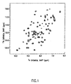
- FIG. 1 A representative two-dimensional 15 N/ 1 H NMR correlation spectrum of an 15 N-labeled target molecule (polypeptide) is shown in FIG. 1 (the DNA-binding domain of the E2 protein).
- the labeled target molecule is exposed to one or more test compounds.
- a database of compounds such as a plurality of small molecules. Such molecules are typically dissolved in perdeuterated dimethylsulfoxide.
- the compounds in the database can be purchased from vendors or created according to desired needs.
- Compounds in the collection can have different shapes (e.g., flat aromatic rings(s), puckered aliphatic rings(s), straight and branched chain aliphatics with single, double, or triple bonds) and diverse functional groups (e.g., carboxylic acids, esters, ethers, amines, aldehydes, ketones, and various heterocyclic rings) for maximizing the possibility of discovering compounds that interact with widely diverse binding sites.
- the NMR screening process of the present invention utilizes ligand concentrations ranging from about 0.1 to about 10.0 mM. At these concentrations, compounds which are acidic or basic can significantly change the pH of buffered protein solutions. Chemical shifts are sensitive to pH changes as well as direct binding interactions, and "false positive" chemical shift changes, which are not the result of ligand binding but of changes in pH, can therefore be observed. It is thus necessary to ensure that the pH of the buffered solution does not change upon addition of the ligand.
- One means of controlling pH is set forth below.
- the 1.0 M stock solutions in DMSO are diluted 1:10 in 50 mM phosphate, pH 7.0.
- the pH of that diluted aliquot solution is measured. If the pH of the aliquot is unchanged (i.e., remains at 7.0), a working solution is made by diluting the DMSO stock solution 1:10 to make a 0.1 M solution and that solution is stored.
- ethanolamine is added to the 1.0 M stock DMSO solution, that stock solution is then diluted 1:10 with phosphate buffer to make another aliquot, and the pH of the aliquot rechecked.
- acetic acid is added to the 1.0 M stock DMSO solution, that stock solution is then diluted 1:10 with phosphate buffer to make another aliquot, and the pH of the aliquot rechecked.
- Ethanolamine and acetic acid are soluble in DMSO, and the proper equivalents are added to ensure that upon transfer to aqueous buffer, the pH is unchanged. Adjusting the pH is an interactive process, repeated until the desired result is obtained.
- a second two-dimensional 15 N/ 1 H NMR correlation spectrum is generated. That second spectrum is generated in the same manner as set forth above.
- the first and second spectra are then compared to determine whether there are any differences between the two spectra. Differences in the two-dimensional 15 N/ 1 H NMR correlation spectra that indicate the presence of a ligand correspond to 15 N-labeled sites in the target molecule. Those differences are determined using standard procedures well known in the art.
- FIGS. 2, 3, 4, 5 and 6 show comparisons of correlation spectra before and after exposure of various target molecules to various test compounds. A detailed description of how these studies were performed can be found hereinafter in Examples 2 and 3.
- Particular signals in a two-dimensional 15 N/ 1 H correlation spectrum correspond to specific nitrogen and proton atoms in the target molecule (e.g., particular amides of the amino acid residues in the protein).
- the target molecule e.g., particular amides of the amino acid residues in the protein.
- the region of the protein that is responsible for binding to the individual compounds can be identified from the particular amide signals that change upon the addition of the compounds. These signals are assigned to the individual amide groups of the protein by standard procedures using a variety of well-established heteronuclear multi-dimensional NMR experiments.
- Example 2 an initial screening assay for binding to the catalytic domain of stromelysin identified two biaryl compounds as ligands.
- a preferred target molecule for use in such a process is a polypeptide.
- Binding or dissociation constants can be measured by following the 15 N/ 1 H chemical shifts of the protein as a function of ligand concentration.
- a known concentration ([P] 0 ) of the target moleule is mixed with a known concentration ([L] 0 ) of a previously identified ligand and the two-dimensional 15 N/ 1 H correlation spectrum was acquired. From this spectrum, observed chemical shift values ( ⁇ obs ) are obtained.
- the process is repeated for varying concentrations of the ligand to the point of saturation of the target molecule, when possible, in which case the limiting chemical shift value for saturation ( ⁇ sat ) is measured.
- Human stromelysin is a 447-amino acid protein believed to be involved in proteolytic degradation of cartilage. Cartilage proteolysis is believed to result in degradative loss of joint cartilage and the resulting impairment of joint function observed in both osteoarthritis and rheumatoid arthritis.
- the protein possesses a series of domains including N-terminal latent and propetide domains, a C-terminal domain homologous with homopexin, and an internal catalytic domain.
- the method of the present invention it was necessary to prepare the 81-256 fragment (SEQ ID NO: 1) of stromelysin in which the peptide backbone was isotopically enriched with and 15 N. This was done by inserting a plasmid which coded for the production of the protein fragment into an E. coli strain and growing the genetically-modified bacterial strain in a limiting culture medium enriched with 15 NH 4 Cl and 13 C-glucose.
- the isotopically enriched protein fragment was isolated from the culture medium, purified, and subsequently used as the basis for evaluating the binding of test compounds. The procedures for these processes are described below.
- RNA was heat-denatured at 80°C for five minutes and then subjected to reverse transcriptase PCR using a GeneAmp® RNA PCR kit (Cat.# N808-0017, Applied Biosystems/Perkin-Elmer, 761 Main Avenue, Norwalk, CT 06859-0156) following the manufacturer's instructions.
- GeneAmp® RNA PCR kit Cat.# N808-0017, Applied Biosystems/Perkin-Elmer, 761 Main Avenue, Norwalk, CT 06859-0156
- Nested PCR was performed using first primers (A) GAAATGAAGAGTC TTCAA (SEQ ID NO:3) and (B) GCGTCCCAGGTTCTGGAG (SEQ ID NO:4) and thirty-five cycles of 94°C, two minutes; 45°C, two minutes; and 72°C three minutes. This was followed by reamplification with internal primers (C) ATACCATGGCCTATCCAT TGGATGGAGC (SEQ ID NO:5) and (D) ATAGGATCCTTAGGTCTCAGGGGA GTCAGG (SEQ ID NO:6) using thirty cycles under the same conditions described immediately above to generate a DNA coding for amino acid residues 1-256 of human stromelysin.
- A GAAATGAAGAGTC TTCAA
- B GCGTCCCAGGTTCTGGAG
- PCR fragment was then cloned into PCR cloning vector pT7Blue(R) (Novagen, Inc., 597 Science Drive, Madison, WI 53711) according to the manufacturer's instructions.
- the resulting plasmid was cut with Ncol and BamHI and the stromelysin fragment was subcloned into the Novagen expression vector pET3d (Novagen, Inc., 597 Science Drive, Madison, WI 53711), again using the manufacturer's instructions.
- a mature stromelysin expression construct coding for amino acid residues 81-256 plus an initiating methionine was generated from the 1-256 expression construct by PCR amplification.
- the resulting PCR fragment was first cloned into the Novagen pT7Blue(R) vector and then subcloned into the Novagen pET3d vector, using the manufacturer's instructions in the manner described above, to produce plasmid (pETST-83-256).
- This final plasmid is identical to that described by Qi-Zhuang et al., Biochemistry, 31: 11231-11235 (1992) with the exception that the present codes for a peptide sequence beginning two amino acids earlier, at position 81 in the sequence of human stromelysin.
- Plasmid pETST-83-256 was transformed into E . coli strain BL21(DE3)/pLysS (Novagen, Inc., 597 Science Drive, Madison, WI 53711) in accordance with the manufacturer's instructions to generate an expression strain, BL21(DE3 )/pLysS/pETST-255- 1.
- a preculture medium was prepared by dissolving 1.698 g of Na 2 HP 4 •7H 2 O, 0.45 g of KH 2 PO 4 , 0.075 g NaCl, 0.150 g 15 NH 4 Cl, 0.300 13 C-glucose, 300 ⁇ L of 1M aqueous MgSO 4 solution and 15 ⁇ L of aqueous CaCl 2 solution in 150 mL of deionized water.
- the resulting solution of preculture medium was sterilized and transferred to a sterile 500 mL baffle flask. Immediately prior to inoculation of the preculture medium with the bacterial strain, 150 ⁇ L of a solution containing 34 mg/mL of chloramphenicol in 100% ethanol and 1.5 mL of a solution containing 20 mg/mL of ampicillin were added to the flask contents.
- the flask contents were then inoculated with 1 mL of glycerol stock of genetically-modified E. Coli, strain BL21(DE3)/pLysS/pETST-255-1.
- the flask contents were shaken (225 rpm) at 37°C until an optical density of 0.65 was observed.
- a fermentation nutrient medium was prepared by dissolving 113.28 g of Na 2 HP 4 •7H 2 O, 30 g of KH 2 PO 4 , 5 g NaCl and 10 mL of 1% DF-60 antifoam agent in 9604 mL of deionized water. This solution was placed in a New Brunswick Scientific Micros Fermenter (Edison, NJ) and sterilized at 121°C for 40 minutes.
- the following pre-sterilized components were added to the fermentation vessel contents: 100 mL of a 10% aqueous solution of 15 NH 4 Cl, solution of a 10% aqueous solution of 13 C-glucose, 20 mL of an aqueous 1M solution of MgSO 4 , 1 mL of an aqueous 1M CaCl 2 solution, 5 mL of an aqueous solution of thiamin hydrochloride (10 mg/mL), 10 mL of a solution containing 34 mg/mL of chloramphenicol in 100% ethanol and 1.9 g of ampicillin dissolved in the chloramphenicol solution.
- the pH of the resulting solution was adjusted to pH 7.00 by the addition of an aqueous solution of 4N H 2 SO 4 .
- the preculture of E. Coli, strain BL21(DE3)/pLysS/pETST-255-1, from the shake-flask scale procedure described above was added to the fermentor contents and cell growth was allowed to proceed until an optical density of 0.48 was achieved.
- the fermenter contents were automatically maintained at pH 7.0 by the addition of 4N H 2 SO 4 or 4N KOH as needed.
- the dissolved oxygen content of the fermenter contents was maintained above 55% air saturation through a cascaded loop which increased agitation speed when the dissolved oxygen content dropped below 55%.
- Air was fed to the fermenter contents at 7 standard liters per minute (SLPM) and the culture temperature was maintained at 37°C throughout the process.
- the cells were harvested by centrifugation at 17,000 x g for 10 minutes at 4°C and the resulting cell pellets were collected and stored at -85°C.
- the wet cell yield was 3.5 g/L.
- the isotopically-labeled stromelysin fragment prepared as described above was purified employing a modification of the technique described by Ye et al., Biochemistry, 31: 11231-11235 (1992) .
- the harvested cells were suspended in 20 mM Tris-HCl buffer (pH 8.0) sodium azide solution containing 1 mM MgCl 2 , 0.5 mM ZnCl 2 , 25 units/mL of Benzonase® enzyme, and an inhibitor mixture made up of 4-(2-aminoethyl)benzenesulfonyl fluoride ("AEBSF'), Leupeptin®, Aprotinin®, and Pepstatin® (all at concentrations of 1 ⁇ g/mL.
- AEBSF, Leupeptin®, Aprotinin®, and Pepstatin® are available from American International Chemical, 17 Strathmore Road, Natick, MA 01760.
- the resulting mixture was gently stirred for one hour and then cooled to 4°C.
- the cells were then sonically disrupted using a 50% duty cycle.
- the resulting lysate was centrifuged at 14,000 rpm for 30 minutes and the pellet of insoluble fraction frozen at -80°C for subsequent processing (see below).
- Solid ammonium sulfate was added to the supernatant to the point of 20% of saturation and the resulting solution loaded onto a 700 mL phenyl sepharose fast flow ("Q-Sepharose FF") column (Pharmacia Biotech., 800 Centennial Ave., P. O. Box 1327, Piscataway, NJ 08855). Prior to loading, the sepharose column was equilibrated with 50 mM Tris-HCl buffer (pH 7.6 at 4°C), 5 mM CaCl 2 , and 1 M (NH 4 ) 2 SO 4 .
- Q-Sepharose FF phenyl sepharose fast flow
- the loaded column was eluted with a linear gradient of decreasing concentrations of aqueous (NH 4 ) 2 SO 4 (from 1 down to 0 M) and increasing concentrations of aqueous CaCl 2 (from 5 to 20 mM) in Tris-HCl buffer at pH 7.6.
- the active fractions of eluate were collected and concentrated in an Amicon stirred cell (Amicon, Inc., 72 Cherry Hill Drive, Beverly, MA 01915).
- the concentrated sample was dialyzed overnight in the starting buffer used with the Q-Sepharose FF column, 50 mM Tris-HCl (pH 8.2 at 4°C) with 10 mM CaCl 2 .
- the dialyzed sample was then loaded on the Q-Sepharose FF column and eluted with a linear gradient comprising the starting buffer and 200 mM NaCl.
- the purified soluble fraction of the isotopically-labeled stromelysin fragment was concentrated and stored at 4°C.
- the pellet was solubilized in 8M guanidine-HCl.
- the solution was centrifuged for 20 minutes at 20,000 rpm and the supernatant was added dropwise to a folding buffer comprising 50 mM Tris-HCl (pH 7.6), 10 mM CaCl 2 0.5 mM ZnCl 2 and the inhibitor cocktail of AEBSF, Leupeptin®, Aprotinin®, and Pepstatin® (all at concentrations of 1 ⁇ g/mL).
- the volume of folding buffer was ten times that of the supernatant.
- the mixture of supernatant and folding buffer was centrifuged at 20,000 rpm for 30 minutes.
- the supernatant from this centrifugation was stored at 4°C and the pellet was subjected twice to the steps described above of solubilization in guanidine-HCl, refolding in buffer, and centrifugation.
- the final supernatants from each of the three centrifugations were combined and solid ammonium sulfate was added to the point of 20% saturation.
- the resulting solution thus derived from the insoluble fraction was subjected to purification on phenyl Sepharose and Q-Sepharose as described above for the soluble fraction.
- the purified soluble and insoluble fractions were combined to produce about 1.8 mg of purified isotopically-labeled stromelysin 81-256 fragment per gram of original cell paste.
- the papillomaviruses are a family of small DNA viruses that cause genital warts and cervical carcinomas.
- the E2 protein of HPV regulates viral transcription and is required for viral replication.
- molecules that block the binding of E2 to DNA may be useful therapeutic agents against HPV.
- the protein rather than the DNA was chosen as a target, because it is expected that agents with greater selectivity would be found that bind to the protein rather than the DNA.
- the DNA-binding domain of human papillomavirus E2 was cloned from the full length DNA that codes for E2 using PCR and overexpressed in bacteria using the T7 expression system.
- Uniformly 15 N-labeled protein was isolated from bacteria grown on a minimal medium containing 15 N-labeled protein was isolated from bacteria grown on a minimal medium containing 15 N-labeled ammonium chloride.
- FKBP N-labeled recombinant human FK binding protein
- the catalytic domain of stromelysin was prepared in accordance with the procedures of Example 1.
- Two-dimensional 15 N/ 1 H NMR spectra were generated at 29°C on a Bruker AMX500 NMR spectrometer equipped with a triple resonance probe and Bruker sample changer.
- the 15 N/ 1 H HSQC spectra were acquired as 80 x 1024 complex points using sweep widths of 2000 Hz ( 15 N, t 1 ) and 8333 Hz ( 1 H, t2).
- a delay of 1 second between scans and 8 scans per free induction decay(fid) were employed in the data collection. All NMR spectra were processed and analyzed on Silicon Graphics computers using in-house-written software.
- a first two-dimensional 15 N/ 1 H NMR correlation spectrum was acquired for the 15 N-labeled stromelysin target molecule as described above.
- the stromelysin target was then exposed to a database of test compounds.
- Stock solutions of the compounds were made at 100 mM and 1 M.
- a combination library was prepared that contained 8-10 compounds per sample at a concentration of 100 mM for each compound.
- the molecules in the collection had different shapes (e.g., flat aromatic rings(s), puckered aliphatic rings(s), straight and branched chain aliphatics with single, double, or triple bonds) and diverse functional groups (e.g., carboxylic acids, esters, ethers, amines, aldehydes, ketones, and various heterocyclic rings) for maximizing the possibility of discovering compound that interact with widely diverse binding sites.
- the NMR samples were prepared by adding 4 ⁇ l of the DMSO stock solution of the compound mixtures that contained each compound at a concentration of 100 mM to 0.4 ml H 2 O/D 2 O (9/1) buffered solution of the uniformly 15 N-labeled protein. The final concentration of each of the compounds in the NMR sample was about 1 mM.
- binding was assayed both in the absence and in the presence of saturating amounts of acetohydroxamic acid (500 mM).
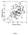
- FIG. 4 shows a representative two-dimensional 15 N/ 1 H NMR correlation spectrum before and after exposure of stromelysin to a biaryl test compound. It can be seen from FIG. 4 that the compound caused chemical shifts of 15 N-sites such as those designated W124, T187, A199 and G204.
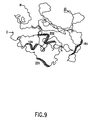
- FIG. 9 shows the correlation between the NMR binding data and a view of the NMR-derived three-dimensional structure of the catalytic domain of stromelysin. The ability to locate the specific binding site of a particular ligand is an advantage of the present invention.
- An advantage of an NMR screening assay of the present invention is the ability to correlate observed chemical shifts from the two-dimensional 15 N/ 1 H NMR correlation spectra with other spectra or projections of target molecule configuration.
- the results of a representative such correlation are shown in FIG. 9, which depicts regions within the polypeptide at which binding with the substrate molecule is most likely occurring.
- the apparent binding regions in stromelysin are shown for Compound 1 (from Table 1).
- NMR experiments were performed at 29°C on a Bruker AMXSOO NMR spectrometer equipped with a triple resonance probe and Bruker sample changer.
- the 15 N-/ 1 H HSQC spectra were acquired as 80 x 1024 complex points using sweep widths of 2000 Hz ( 15 N,t 1 ) and 8333 Hz ( 1 H, t 2 ).
- a delay of 1 second between scans and 4 scans per free induction decay were employed in the data collection. All NMR spectra were processed and analyzed on Silicon Graphics computers.
- FIGS. 2 and 3 show representative two-dimensional 15 N/ 1 H NMR correlation spectra before and after exposure of the DNA-binding domain of E2 to a first and second test compound, respectively.
- the first test compound caused chemical shifts of 15 N-sites such as those designated I15, Y21, R22 and L23. Those sites correspond to an isoleucine (Ile) residue at position 15, a tyrosine residue (Tyr) at position 21, an arginine (Arg residue at position 22 and a leucine (Leu) residue at position 23 of SEQ ID NO. 6.
- the second test compound caused chemical shifts in the particular 15 N-sites designated 16, G11, H38, and T52. Those sites correspond to an isoleucine (Ile) residue at position 6, a glycine (Gly) residue at position 11, a histidine (His) residue at position 38 and a threonine (Thr) at position 52 of SEQ ID NO. 6.
- FIGs. 7 and 8 show the correlation between those NMR binding data and a view of the NMR-derived three-dimensional structure of the DNA-binding domain of E2.
- binding constants were determined, both by the NMR method of the present invention, and by a prior art enzymatic assay.
- the target molecule was the catalytic domain of stromelysin prepared in accordance with the procedures of Example 1.
- the NMR binding constants, KD were derived using two-dimensional 15 N/ 1 H NMR correlation spectroscopy as described in Example 2. The K D values so obtained were compared to an inhibition constant K I as determined in an enzymatic assay.
- the enzymatic assay measured the rate of cleavage of a fluorogenic substrate by following the fluorescence increase upon peptide cleavage which causes a separation between the fluorophore and quencher. Enzymatic activity was measured using a matrix of different concentrations of acetohydroxamic acid and biaryl compounds.
- the assay is a modification of the method described by H. Weingarten, et al. in Anal. Biochem., 147: 437-440 (1985) employing the fluorogenic substrate properties described by E. Matayoshi, et al. in Science: 247: 954-958 (1990) .
- the binding constant from the NMR process was compared to the results of a physical, filter binding assay that measured binding of DNA to the target.
- the high-throughput filter binding assay was performed using E2, prepared according to Example 2 above.
- the 33 P-labeled DNA construct comprised a 10,329 base pair plasmid formed by inserting the HPV-11 genome, containing three high affinity and one low affinity E2 binding sites, into the PSP-65 plasmid (Promega, Madison, WI).
- the binding affinities at the different sites as determined by NMR were compared for a subset of the compounds to the inhibition of E2 binding to DNA as measured in the filter binding assay. As shown in Table 2 above, the activities determined in the filter binding assay correlated closely with the binding affinities calculated from the amides of the DNA-binding site but not to the affinities measured for the ⁇ -barrel site. This is consistent with the relative locations of each site.
- the method identified numerous compounds which gave positive results in the gel-shift assay. Some of these positive results, however, were believed to be due to binding to the DNA, since in these cases, no binding to the E2 protein was observed using the NMR method of this invention. These compounds were shown to indeed bind to DNA rather than to E2, as evidenced by changes in the chemical shifts of the DNA rather than the protein upon the addition of the compounds. These data show that yet another advantage of the present invention is the ability to minimize the occurrence of false positives.
- the criteria used in selecting compounds in the screening for other binding sites was based primarily on the size of the ligand.
- the smallest ligand was sought that had enough solubility to saturate (>98% occupancy of enzyme) and inhibit the enzyme.
- the cloning, expression, and purification of the catalytic domain of stromelysin was accomplished using the procedures set forth in Example 1.
- An initial step in the design of the new ligand was the identification of a first ligand that bound to the stromelysin target. Such identification was carried out in accordance with a two-dimensional 15 N/ 1 H NMR correlation screening process as disclosed above.
- hydroxarnic acids of the general formula R-(CO)NHOH were screened for binding to stromelysin using the procedures set forth in Example 2.
- acetohydroxamic acid [CH3(CO)NHOH] best satisfied the selection criteria: it had a binding affinity for stromelysin of 17 mM and had good water solubility.
- acetohydroxamic acid inhibited the degradation of the enzyme, allowing the screening of other potential ligands.
- the second step in the design process was the identification of a second ligand that bound to the target stromelysin at a site different from the binding site of acetohydroxamic acid. This was accomplished by screening compounds for their ability to bind stromelysin in the presence of saturating amounts of acetohydroxamic acid. Details of procedures and results of this second identification step are set forth above in Example 2.
- the next step in the design process was to construct a ternary complex of the target stromelysin, the first ligand and the second ligand. This was accomplished by exposing the stromelysin target to the two ligands under conditions that resulted in complex formation. The three-dimensional structure of the ternary complex was then determined using NMR spectroscopy as described below.
- the 1 H, 13 C, and 15 N backbone resonances of stromelysin in the ternary complex were assigned from an analysis of several 3D double- and triple-resonance NMR spectra ( A. Bax. et al.. Acc. Chem. Res., 26: 131-138 (1993) ).
- the C ⁇ resonances of adjacent spin systems were identified from an analysis of three-dimensional (3D) HNCA ( L. Kay et al., J. Magn. Reson., 89: 496-514 (1990) ) and HN(CO)CA ( A. Bax, et al., J. Bio.
- the data matrix was 38(t 1 ) x 48(t 2 ) x 1024(t 3 ) complex points for the HNCA spectrum, and 32(t 1 ) x 40(t 2 ) x 1024(t 3 ) complex points for the HN(CO)CA spectrum. Both spectra were acquired with 16 scans per increment.
- a 3D CBCA(CO)NH spectrum S. Grzesiek, et al., J. Am. Chem. Soc., 114: 6261-6293 (1992) ) was collected with 32(t 1 , 15 N) x 48(t 2 , 13 C) x 1024(t 3 , 1 H) complex points and 32 scans per increment.
- Spectral widths were 1773 Hz (35.0 ppm), 7575.8 Hz (60.2 ppm), and 8333 Hz (16.67 ppm) in the 15 N, 13 C and 1 H dimensions, respectively.
- the 1 H carrier frequency was set on the water resonance and the 15 N carrier frequency was at 119.1 ppm.
- the 13 C carrier frequency was set to 55.0 ppm in HNCA and HN(CO)CA experiments, and 46.0 ppm in the CBCA(CO)NH experiment.
- the backbone assignments were confirmed from an analysis of the crosspeaks observed in an 15 N-separated 3D NOESY-HSQC spectrum and a 3D HNHA-J spectrum.
- the 15 N-separated 3D NOESY-HSQC spectrum S. Fesik, et al., J. Magn. Reson., 87: 588-593 (1988) ); D. Marion, et al., J. Am. Chem. Soc., 111: 1515-1517 (1989) ) was collected with a mixing time of 80 ms.
- the sweep widths were the same as in the 15 N-separated NOESY-HSQC spectrum that was acquired with 32(t 1 , 15 N) x 96(t 2 , 1 H) x 1024 (t 3 , 1 H) complex points.
- a total of 96 (t 1 , 13 C) x 96(t 2 , 1 H) x 1024(t 3 , 1 H) complex data points were collected with 16 scans per increment using a spectral width of 10638 Hz (70.8 ppm, w 1 ), 4000 Hz (6.67 ppm, w 2 ), and 4844 (8.07 ppm, w 3 ).
- Carrier positions were 40 ppm, 2.5 ppm, and at the water frequency for the 13 C, indirectly detected 1 H, and observed 1 H dimensions, respectively.
- a total of 80 (t 1 , 13 C) x 72 (t 2 , 1 H) x 1024 (t 3 , 1 H) complex data points with 16 scans per increment were collected over spectral widths of 10638 Hz (70.49 ppm, w 1 ), 6666.6 Hz (13.3 ppm, w 2 ), and 8333.3 Hz (16.67 ppm, w 3 ).
- the 1 H carrier frequencies were set to the water resonance, and the 13 C carrier frequency was placed at 40.0 ppm.
- the spectrum was acquired with 200( 13 C, t 1 ) x 2048( 1 H, t 2 ) complex points over spectral widths of 5000 Hz (39.8 ppm, 13 C) and 8333 Hz (16.7 ppm, 1 H).
- Carrier positions were 20.0 ppm for the 13 C dimension, and at the water frequency for the 1 H dimension.
- the pulse scheme consisted of a double 13 C-filter sequence ( A. Gemmeker, et al., J. Magn. Reson., 96: 199-204 (1992) ) concatenated with a NOESY-HMQC sequence ( S. Fesik, et al., J. Magn. Reson., 87: 588-593 (1988) ); D. Marion, et al., J. Am. Chem. Soc., 111: 1515-1517 (1989) ) .
- the spectrum was recorded with a mixing time of 80 ms, and a total of 80 (tl, 13 C) x 80 (t 2 , 1 H) x 1024 (t 3 , 1 H) complex points with 16 scans per increment.
- Spectral widths were 8865 Hz (17.73 ppm, w 1 ), 6667 Hz (13.33 ppm, w 2 ), and 8333 Hz (16.67 ppm, w 3 ), and the carrier positions were 40.0 ppm for the carbon dimension and at the water frequency for both proton dimensions.
- the derived three-dimensional structure of the ternary complex was then used to define the spatial orientation of the first and second ligands to each other as well as to the target stromelysin molecule.
- Hydrogen bonds identified for slowly exchanging amides based on initial structures, were defined by two restraints: 1.8-2.5 ⁇ for the H-O distance and 1.8-3.3 ⁇ for the N-O distance. Structures were calculated with the X-PLOR 3.1 program ( A. Brünger, "XPLOR 3.1 Manual,” Yale University Press, New Haven, 1992 ) on Silicon Graphics computers using a hybrid distance geometry-simulated annealing approach ( M. Nilges, et al., FEBS Lett., 229: 317-324 (1988) ).
- a total of 1032 approximate interproton distance restraints were derived from the NOE data.
- 21 unambiguous intermolecular distance restraints were derived from a 3D 12C-filtered, 13C-edited NOESY spectrum.
- 341 were intra-residue
- 410 were sequential or short-range between residues separated in the primary sequence by less than five amino acids
- 281 were long-range involving residues separated by at least five residues.
- the structures were in excellent agreement with the NMR experimental restraints. There were no distance violations greater than 0.4 ⁇ , and no dihedral angle violations greater than 5 degrees. In addition, the simulated energy for the van der Waals repulsion term was small, indicating that the structures were devoid of bad inter-atomic contacts.
- the NMR structures also exhibited good covalent bond geometry, as indicated by small bond-length and bond-angle deviations from the corresponding idealized parameters.
- the average atomic root mean square deviation of the 8 structures for residues 93-247 from the mean coordinates was 0.93 ⁇ for backbone atoms (C a , N, and C'), and 1.43 ⁇ for all non-hydrogen atoms.
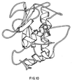
- FIG 10. A ribbon plot of the ternary complex involving stromelysin, acetohydroxamic acid (the first ligand), and the second ligand is shown in Fig 10.
- the structure is very similar to the global fold of other matrix metalloproteinases and consists of a five-stranded ⁇ -sheet and three ⁇ -helices.
- the catalytic zinc was located in the binding pocket. It was coordinated to three histidines and the two oxygen atom of acetohydroxamic acid.
- a biaryl group of the second ligand was located in the S1' pocket between the second helix and the loop formed from residues 218-223. This deep and narrow pocket is lined with hydrophobic residues which make favorable contacts with the ligand.
- the initial biaryls chosen contained an oxygen linker and the absence or presence of CN para to the biaryl linkage.
- Initial linkers contained varying lengths of methylene units. Means for linking compounds with linkers having varying lengths of methylene units are well known in the art.
- the CN derivatives exhibited better stromelysin inhibition.
- the compound that exhibited the best inhibition of stromelysin contained a linker with two methylene units.
Landscapes
- Health & Medical Sciences (AREA)
- Life Sciences & Earth Sciences (AREA)
- Engineering & Computer Science (AREA)
- Immunology (AREA)
- Biomedical Technology (AREA)
- Molecular Biology (AREA)
- Urology & Nephrology (AREA)
- Hematology (AREA)
- Chemical & Material Sciences (AREA)
- Physics & Mathematics (AREA)
- Biochemistry (AREA)
- Microbiology (AREA)
- Food Science & Technology (AREA)
- Medicinal Chemistry (AREA)
- Cell Biology (AREA)
- Analytical Chemistry (AREA)
- Biotechnology (AREA)
- General Health & Medical Sciences (AREA)
- General Physics & Mathematics (AREA)
- Pathology (AREA)
- Bioinformatics & Cheminformatics (AREA)
- Toxicology (AREA)
- Tropical Medicine & Parasitology (AREA)
- Bioinformatics & Computational Biology (AREA)
- Biophysics (AREA)
- Proteomics, Peptides & Aminoacids (AREA)
- Peptides Or Proteins (AREA)
- Magnetic Resonance Imaging Apparatus (AREA)
- Investigating Or Analysing Biological Materials (AREA)
- Organic Low-Molecular-Weight Compounds And Preparation Thereof (AREA)
- Compounds Of Iron (AREA)
- Medicines Containing Antibodies Or Antigens For Use As Internal Diagnostic Agents (AREA)
- Nitrogen Condensed Heterocyclic Rings (AREA)
- Heterocyclic Compounds That Contain Two Or More Ring Oxygen Atoms (AREA)
Claims (4)
- Verfahren zum Screening von Verbindungen zur Identifikation von Verbindungen, die an spezifische Zielmoleküle bindende Liganden sind, wobei das Verfahren folgende Schritte umfaßt:a) Erzeugen eines ersten zweidimensionalen 15N/1H NMR Korrelationsspektrums eines 15N-markierten Zielmoleküls;b) Aussetzen des markierten Zielmoleküls mit einer chemischen Verbindung oder einer Mischung von chemischen Verbindungen;c) Erzeugen eines zweiten zweidimensionalen 15N/1H NMR Korrelationsspektrums des markierten Zielmoleküls, das in Schritt b) einer oder einer Mischung von Verbindungen ausgesetzt worden ist; undd) Vergleich der ersten und zweiten zweidimensionalen 15N/1H NMR Korrelationsspektren, um Unterschiede zwischen den ersten und zweiten Spektren zu ermitteln, wobei diese Unterschiede die Anwesenheit einer oder mehrerer Verbindungen anzeigen, die Liganden sind, die an das Zielmolekül gebunden haben.
- Verfahren nach Anspruch 1, worin das 15N-markierte Zielmolekül in Schritt (b) einer Mischung von chemischen Verbindungen ausgesetzt wird, wobei das Verfahren noch folgende, nach Schritt d) erfolgende Schritte umfaßt:e) einzelnes Aussetzen des 15N-markierten Zielmoleküls mit jeder Verbindung der Mischung,f) Erzeugen eines zweidimensionalen 15N/1H NMR Korrelationsspektrums des markierten Zielmoleküls, das jeder Verbindung einzeln ausgesetzt worden ist; undg) Vergleich eines jeden in Schritt f) erzeugten Spektrums mit dem ersten Spektrum, um Unterschiede in irgendeinem der verglichenen Spektren zu ermitteln, wobei die Unterschiede die Anwesenheit einer Verbindung identifizieren, die ein Ligand ist, der an das Zielmolekül gebunden hat.
- Verfahren nach Anspruch 1, worin die Unterschiede in den zweidimensionalen 15N/1H NMR Korrelationsspektren chemische Verschiebungen an besonderen, 15N-markierten Stellen des Zielmoleküls und chemische Verschiebungen von Protonen sind, die an diese 15N-markierten Stellen gebunden sind.
- Verfahren nach Anspruch 1, worin das Zielmolekül ein Polypeptid ist.
Priority Applications (1)
| Application Number | Priority Date | Filing Date | Title |
|---|---|---|---|
| EP99119066A EP0981049B1 (de) | 1995-11-14 | 1996-11-13 | Verfahren zum Entwerfen und Herstellung von zielmolekulbindenden Liganden |
Applications Claiming Priority (3)
| Application Number | Priority Date | Filing Date | Title |
|---|---|---|---|
| US555691 | 1995-11-14 | ||
| US08/555,691 US5698401A (en) | 1995-11-14 | 1995-11-14 | Use of nuclear magnetic resonance to identify ligands to target biomolecules |
| PCT/US1996/018270 WO1997018471A1 (en) | 1995-11-14 | 1996-11-13 | Use of nuclear magnetic resonance to identify ligands to target biomolecules |
Related Child Applications (1)
| Application Number | Title | Priority Date | Filing Date |
|---|---|---|---|
| EP99119066A Division EP0981049B1 (de) | 1995-11-14 | 1996-11-13 | Verfahren zum Entwerfen und Herstellung von zielmolekulbindenden Liganden |
Publications (3)
| Publication Number | Publication Date |
|---|---|
| EP0866967A1 EP0866967A1 (de) | 1998-09-30 |
| EP0866967B1 EP0866967B1 (de) | 2000-01-19 |
| EP0866967B2 true EP0866967B2 (de) | 2007-08-08 |
Family
ID=24218250
Family Applications (2)
| Application Number | Title | Priority Date | Filing Date |
|---|---|---|---|
| EP96940448A Expired - Lifetime EP0866967B2 (de) | 1995-11-14 | 1996-11-13 | Verwendung von kernspinresonanz zur auffindung von liganden für bindungsmoleküle |
| EP99119066A Expired - Lifetime EP0981049B1 (de) | 1995-11-14 | 1996-11-13 | Verfahren zum Entwerfen und Herstellung von zielmolekulbindenden Liganden |
Family Applications After (1)
| Application Number | Title | Priority Date | Filing Date |
|---|---|---|---|
| EP99119066A Expired - Lifetime EP0981049B1 (de) | 1995-11-14 | 1996-11-13 | Verfahren zum Entwerfen und Herstellung von zielmolekulbindenden Liganden |
Country Status (13)
| Country | Link |
|---|---|
| US (2) | US5698401A (de) |
| EP (2) | EP0866967B2 (de) |
| JP (1) | JP3032301B2 (de) |
| AT (2) | ATE189063T1 (de) |
| AU (1) | AU701810B2 (de) |
| CA (1) | CA2237343C (de) |
| DE (2) | DE69606324T3 (de) |
| DK (1) | DK0866967T4 (de) |
| ES (1) | ES2143793T5 (de) |
| GR (1) | GR3032361T3 (de) |
| IL (2) | IL123923A (de) |
| PT (1) | PT866967E (de) |
| WO (1) | WO1997018471A1 (de) |
Families Citing this family (78)
| Publication number | Priority date | Publication date | Assignee | Title |
|---|---|---|---|---|
| US5989827A (en) * | 1995-11-14 | 1999-11-23 | Abbott Laboratories | Use of nuclear magnetic resonance to design ligands to target biomolecules |
| US20010004528A1 (en) * | 1995-11-14 | 2001-06-21 | Stephen W. Fesik | Use of 13c nuclear magnetic resonance to detect binding to target molecules |
| IL126347A (en) * | 1996-03-29 | 2003-11-23 | Lawrence Berkeley National Lab | Enhancement of nmr and mri in the presence of hyperpolarized noble gases |
| DE19649359C1 (de) * | 1996-11-28 | 1998-02-12 | Thomas Prof Dr Peters | Verfahren zum Nachweis biologisch aktiver Substanzen in Substanzbibliotheken |
| AU772620B2 (en) * | 1997-04-18 | 2004-05-06 | Abbvie Inc. | Use of one-dimensional nuclear magnetic resonance to identify ligands to target biomolecules |
| US6043024A (en) * | 1997-04-18 | 2000-03-28 | Abbott Laboratories | Use of one-dimensional nuclear magnetic resonance to identify ligands to target biomolecules |
| EP0988528A1 (de) * | 1997-06-13 | 2000-03-29 | Vertex Pharmaceuticals Incorporated | Methoden zur identifikation von medikamentenkernen |
| US20030143757A1 (en) * | 1997-06-13 | 2003-07-31 | Jonathan Moore | Methods for identifying drug cores |
| US6111066A (en) * | 1997-09-02 | 2000-08-29 | Martek Biosciences Corporation | Peptidic molecules which have been isotopically substituted with 13 C, 15 N and 2 H in the backbone but not in the sidechains |
| US6344330B1 (en) | 1998-03-27 | 2002-02-05 | The Regents Of The University Of California | Pharmacophore recombination for the identification of small molecule drug lead compounds |
| US20020131972A1 (en) * | 1998-05-21 | 2002-09-19 | Daniel Sem | Multi-partite ligands and methods of identifying and using same |
| WO1999063929A2 (en) * | 1998-06-08 | 1999-12-16 | Advanced Medicine, Inc. | Multibinding inhibitors of microsomal triglyceride transferase protein |
| IL143927A0 (en) * | 1998-12-30 | 2002-04-21 | Nycomed Amersham Plc | Nmr spectroscopic in vitro assay using hyperpolarization |
| TWI283659B (en) * | 1999-04-09 | 2007-07-11 | Abbott Lab | Site-specific isotopically-labeled proteins, amino acids, and biochemical precursors therefor |
| US6897337B2 (en) * | 1999-04-09 | 2005-05-24 | Abbott Laboratories | Site-specific isotopically-labeled proteins, amino acids, and biochemical precursors therefor |
| US6333149B1 (en) | 1999-06-04 | 2001-12-25 | Triad Biotechnology, Inc. | NMR-solve method for rapid identification of bi-ligand drug candidates |
| US6171804B1 (en) | 1999-07-12 | 2001-01-09 | The Rockefeller University | Method of determining interdomain orientation and changes of interdomain orientation on ligation |
| US7105310B1 (en) * | 2000-07-19 | 2006-09-12 | California Institute Of Technology | Detection of biomolecules by sensitizer-linked substrates |
| AU6115300A (en) * | 1999-07-19 | 2001-02-05 | California Institute Of Technology | Detection of biomolecules by sensitizer-linked substrates |
| JP2003522325A (ja) * | 1999-08-23 | 2003-07-22 | ポラリス ファーマシューティカルズ,インコーポレイテッド | タンパク質と巨大分子リガンドとの結合阻害剤 |
| US6677160B1 (en) | 1999-09-29 | 2004-01-13 | Pharmacia & Upjohn Company | Methods for creating a compound library and identifying lead chemical templates and ligands for target molecules |
| CA2400094A1 (en) * | 1999-09-29 | 2001-04-05 | Pharmacia & Upjohn Company | Methods for creating a compound library and identifying lead chemical templates and ligands for target molecules |
| US6764858B2 (en) | 1999-09-29 | 2004-07-20 | Pharmacia & Upjohn Company | Methods for creating a compound library |
| WO2001023889A1 (en) * | 1999-09-29 | 2001-04-05 | Smithkline Beecham Corporation | Method of using one-dimensional and multi-dimensional nuclear magnetic resonance to identify compounds that interact with target biomolecules |
| GB9925677D0 (en) * | 1999-10-29 | 1999-12-29 | Pharmacia & Upjohn Spa | Use of double-quantum 1H-NMR spectroscopy for the identification of ligands interacting with target biomolecules |
| JP2003524167A (ja) * | 2000-02-25 | 2003-08-12 | ワイス | Ms/nmrを用いた構造に基づく薬物設計の方法 |
| US20010051333A1 (en) * | 2000-04-17 | 2001-12-13 | Pharmacia & Upjohn Company | Nuclear magnetic resonance methods for identifying sites in papillomavirus E2 protein |
| GB0013115D0 (en) * | 2000-05-31 | 2000-07-19 | Imp College Innovations Ltd | Biological materials and methods for use in the prevention or treatment of infections |
| US7146277B2 (en) * | 2000-06-13 | 2006-12-05 | James H. Prestegard | NMR assisted design of high affinity ligands for structurally uncharacterized proteins |
| US7061237B2 (en) * | 2000-07-13 | 2006-06-13 | The Regents Of The University Of California | Remote NMR/MRI detection of laser polarized gases |
| US6652833B2 (en) | 2000-07-13 | 2003-11-25 | The Regents Of The University Of California | Functionalized active-nucleus complex sensor |
| US20030165431A1 (en) * | 2000-07-13 | 2003-09-04 | The Regents Of The University Of California | Method for detecting macromolecular conformational change and binding information |
| JP5129424B2 (ja) | 2000-09-27 | 2013-01-30 | ユニヴェルシテイト レイデン | リガンドの発見または薬物スクリーニング手段としてのnmr利用法 |
| SE0003811D0 (sv) * | 2000-10-20 | 2000-10-20 | Pharmacia Ab | Screening methods |
| CA2432924A1 (en) * | 2000-12-21 | 2002-06-27 | Triad Therapeutics, Inc. | Sea-trosy and related methods |
| DE60120494T2 (de) * | 2000-12-22 | 2006-12-21 | Ortho-Mcneil Pharmaceutical, Inc. | Substituieten triazoldiamin derivaten und ihre verwendung als kinase inhibitoren |
| US6656694B2 (en) | 2001-01-11 | 2003-12-02 | Theravance, Inc. | Method for identifying a ligand for a biological substrate |
| US7645569B2 (en) * | 2001-01-26 | 2010-01-12 | Board Of Regents, The University Of Texas System | NMR detection of foreign PAS domain ligands |
| US20020172967A1 (en) * | 2001-02-13 | 2002-11-21 | Gadek Thomas R. | Identification of non-covalent complexes by mass spectrometry |
| DE10119455B4 (de) * | 2001-04-20 | 2010-12-16 | Siemens Ag | Verfahren zum Auswerten von Daten, die mittels der Magnetresonanztechnik erzeugt werden und spektroskopische Information beinhalten |
| US7300922B2 (en) | 2001-05-25 | 2007-11-27 | Duke University | Modulators of pharmacological agents |
| AU2002312146A1 (en) * | 2001-05-30 | 2002-12-09 | Triad Therapeutics, Inc. | Nuclear magnetic resonance-docking of compounds |
| US20030129672A1 (en) * | 2001-08-29 | 2003-07-10 | Dyer Richard Dennis | Method for identifying metalloenzyme inhibitors |
| US7653490B2 (en) * | 2001-09-10 | 2010-01-26 | Triad Liquidating Company LLC | Nuclear magnetic resonance assembly of chemical entities |
| DE10144661C2 (de) * | 2001-09-11 | 2003-08-14 | Forschungsverbund Berlin Ev | Vorrichtung und Verfahren zur Zuordnung der NMR-Signale von Polypeptiden |
| US7179450B2 (en) * | 2001-09-20 | 2007-02-20 | Medi-Physics, Inc. | Methods for in vivo evaluation of pulmonary physiology and/or function using NMR signals of polarized Xe |
| US7037660B2 (en) * | 2001-11-06 | 2006-05-02 | Message Pharmaceuticals | Methods for detecting and quantifying binding and inhibition of binding of species to nucleic acids |
| DE10160177A1 (de) * | 2001-12-07 | 2003-06-26 | Novaspin Biotech Gmbh | Verfahren zur Auffindung von Liganden die an ein drug target binden mittels 1H,1H-Kernresonanzspektroskopie |
| US20040171062A1 (en) * | 2002-02-28 | 2004-09-02 | Plexxikon, Inc. | Methods for the design of molecular scaffolds and ligands |
| US20030180797A1 (en) * | 2002-03-20 | 2003-09-25 | Lin Yu | Identification of ligands for a receptor family and related methods |
| WO2003091700A2 (en) * | 2002-03-26 | 2003-11-06 | Centocor, Inc. | Epitope mapping using nuclear magnetic resonance |
| DE10221158A1 (de) * | 2002-05-13 | 2004-02-19 | Novaspin Biotech Gmbh | Verfahren zur Auffindung von Liganden, die an ein drug target binden, mittels heteronuklearer Kernresonanzspektroskopie |
| EP1551291A4 (de) * | 2002-06-10 | 2007-11-28 | Prospect Pharma | Verfahren zur gewinnung von dynamischen und strukturellen daten in bezug auf proteine und protein/liganden-komplexe |
| AU2003272548A1 (en) * | 2002-09-16 | 2004-04-30 | Plexxikon, Inc. | Crystal structure of pim-1 kinase |
| WO2006078228A1 (en) * | 2002-09-16 | 2006-07-27 | Plexxikon, Inc. | Methods for the design of molecular scaffolds and ligands |
| US20040132102A1 (en) * | 2002-10-29 | 2004-07-08 | Maurizio Pellecchia | Use of selective labeling to detect and characterize molecular interactions by nuclear magnetic resonance spectroscopy |
| NO20025738D0 (no) | 2002-11-29 | 2002-11-29 | Amersham Health As | Metode |
| WO2004078923A2 (en) * | 2003-02-28 | 2004-09-16 | Plexxikon, Inc. | Pyk2 crystal structure and uses |
| US7964534B2 (en) * | 2003-03-13 | 2011-06-21 | Triad Liquidating Company, Llc | Nuclear magnetic resonance assembly of chemical entities using advanced antenna probes |
| EP1623216A2 (de) * | 2003-04-11 | 2006-02-08 | SGX Pharmaceuticals, Inc. | Verbindungsbibliotheken und verfahren zur arzneimittelentdeckung |
| EP1617221A4 (de) * | 2003-04-23 | 2006-08-02 | Olympus Corp | Screening-verfahren für eine zur bindung an einen rezeptor fähige substanz |
| JP2004331832A (ja) * | 2003-05-08 | 2004-11-25 | Ricoh Co Ltd | Dnaインク組成物とそれを用いた識別画像印刷体、及びインクジェット記録装置 |
| US20050079548A1 (en) * | 2003-07-07 | 2005-04-14 | Plexxikon, Inc. | Ligand development using PDE4B crystal structures |
| US7255985B2 (en) * | 2003-07-10 | 2007-08-14 | Pfizer Italia S.R.L. | Fluorine NMR spectroscopy for biochemical screening |
| US20050164300A1 (en) * | 2003-09-15 | 2005-07-28 | Plexxikon, Inc. | Molecular scaffolds for kinase ligand development |
| US20070275442A1 (en) * | 2003-10-07 | 2007-11-29 | Prospect Pharma | Side Chain Deuterated Amino Acids Methods Of Use |
| JP4192078B2 (ja) * | 2003-11-26 | 2008-12-03 | 株式会社日立製作所 | 核磁気共鳴方法 |
| US7904252B2 (en) | 2003-11-28 | 2011-03-08 | Hitachi, Ltd. | Method for measuring a structural change in a protein |
| GB0405330D0 (en) * | 2004-03-10 | 2004-04-21 | Astrazeneca Ab | Enzyme and preparation method |
| EP1794324A4 (de) | 2004-09-20 | 2010-04-14 | Wisconsin Alumni Res Found | Nichtlineare spektroskopische verfahren zur identifizierung und beschreibung molekularer wechselwirkungen |
| WO2006082963A1 (ja) * | 2005-02-07 | 2006-08-10 | Mitsubishi Chemical Corporation | 標的分子とリガンドあるいはリガンド候補化合物との結合検出方法 |
| ATE519114T1 (de) | 2006-03-14 | 2011-08-15 | Univ Basel | Verfahren zur identifizierung neuer leitverbindungen für arzneistoffkandidaten |
| US8933209B2 (en) | 2006-04-26 | 2015-01-13 | Abbvie Inc. | Dep2 and its uses in major depressive disorder and other related disorders |
| US7760342B2 (en) | 2007-12-21 | 2010-07-20 | Wisconsin Alumni Research Foundation | Multidimensional spectrometer |
| JPWO2012108297A1 (ja) * | 2011-02-07 | 2014-07-03 | 国立大学法人神戸大学 | Ras部分ポリペプチド含有水溶液及びRas機能阻害剤のスクリーニング方法 |
| CA2861392C (en) | 2012-01-26 | 2021-08-17 | Christopher J. Soares | Peptide antagonists of the calcitonin cgrp family of peptide hormones and their use |
| ES3057215T3 (en) | 2015-08-11 | 2026-02-26 | Arch Biopartners Inc | Antagonist binding to dpep-1 for use in treating or preventing acute kidney injury or ischemia-reperfusion injury |
| EP4316595A3 (de) | 2016-09-02 | 2024-04-17 | Christopher J. Soares | Verwendung von cgrp-rezeptor-antagonisten bei der behandlung des glaukoms |
Family Cites Families (5)
| Publication number | Priority date | Publication date | Assignee | Title |
|---|---|---|---|---|
| CA2072363A1 (en) * | 1989-12-29 | 1991-06-30 | Graham J. Moore | Methods for modelling tertiary structures of biologically active ligands including agonists and antagonists thereto and novel synthetic antagonists based on angiotensin |
| US5164297A (en) * | 1990-05-03 | 1992-11-17 | Advanced Magnetics Inc. | Solvent mediated relaxation assay system |
| US5270163A (en) * | 1990-06-11 | 1993-12-14 | University Research Corporation | Methods for identifying nucleic acid ligands |
| WO1993000446A1 (en) * | 1991-06-27 | 1993-01-07 | Genelabs Technologies, Inc. | Screening assay for the detection of dna-binding molecules |
| US5726014A (en) * | 1991-06-27 | 1998-03-10 | Genelabs Technologies, Inc. | Screening assay for the detection of DNA-binding molecules |
-
1995
- 1995-11-14 US US08/555,691 patent/US5698401A/en not_active Expired - Lifetime
-
1996
- 1996-11-13 EP EP96940448A patent/EP0866967B2/de not_active Expired - Lifetime
- 1996-11-13 AU AU77328/96A patent/AU701810B2/en not_active Expired
- 1996-11-13 EP EP99119066A patent/EP0981049B1/de not_active Expired - Lifetime
- 1996-11-13 DE DE69606324T patent/DE69606324T3/de not_active Expired - Lifetime
- 1996-11-13 DE DE69631842T patent/DE69631842D1/de not_active Expired - Lifetime
- 1996-11-13 JP JP9519074A patent/JP3032301B2/ja not_active Expired - Lifetime
- 1996-11-13 WO PCT/US1996/018270 patent/WO1997018471A1/en not_active Ceased
- 1996-11-13 DK DK96940448T patent/DK0866967T4/da active
- 1996-11-13 AT AT96940448T patent/ATE189063T1/de active
- 1996-11-13 ES ES96940448T patent/ES2143793T5/es not_active Expired - Lifetime
- 1996-11-13 IL IL12392396A patent/IL123923A/xx not_active IP Right Cessation
- 1996-11-13 CA CA002237343A patent/CA2237343C/en not_active Expired - Lifetime
- 1996-11-13 PT PT96940448T patent/PT866967E/pt unknown
- 1996-11-13 AT AT99119066T patent/ATE261581T1/de not_active IP Right Cessation
- 1996-11-13 IL IL15598896A patent/IL155988A/xx not_active IP Right Cessation
-
1997
- 1997-02-24 US US08/804,777 patent/US5804390A/en not_active Expired - Lifetime
-
2000
- 2000-01-14 GR GR20000400014T patent/GR3032361T3/el unknown
Non-Patent Citations (10)
| Title |
|---|
| Carell et al. (1995) Chemistry & Biology 2, p.171-183 † |
| G.Otting (1993) Current Opinion in Structural Biology, Vol.3, p.760-768 † |
| H.Baumann et al.(1995) J Mol.Biol.Vol.247, p.840-846 † |
| K S Koblan et al. (1995) Protein Science, Vol.4, p.681-688 † |
| M R Rejanic et al. (1991) Biochemistry, Vol.30. p.11081-11092 † |
| T.Ohkubo et al. (1991) J. Biochem vol.110, p.1022-1029 † |
| T.Thewes et al. (1990) The Journal of Biological Chemistry, Vol.265,No. 7, p.3906-3915 † |
| V Ramesh et al. (1994) Eur.J.Biochem., Vol.225, p 601-608 † |
| X.Cheng et al.(1995) J.Am.Chem.Soc., Vol.117, No34, p.8859-8860 † |
| Y.Oda et al. (1991) Journal of Biomolecular NNR, Vol.1, p247-255 † |
Also Published As
| Publication number | Publication date |
|---|---|
| AU701810B2 (en) | 1999-02-04 |
| JPH11513498A (ja) | 1999-11-16 |
| EP0866967B1 (de) | 2000-01-19 |
| DE69606324T3 (de) | 2008-02-07 |
| CA2237343C (en) | 2009-01-20 |
| ES2143793T5 (es) | 2008-03-01 |
| WO1997018471A1 (en) | 1997-05-22 |
| DE69606324D1 (de) | 2000-02-24 |
| DE69606324T2 (de) | 2000-07-06 |
| IL123923A (en) | 2005-07-25 |
| IL155988A (en) | 2005-07-25 |
| JP3032301B2 (ja) | 2000-04-17 |
| ES2143793T3 (es) | 2000-05-16 |
| EP0866967A1 (de) | 1998-09-30 |
| GR3032361T3 (en) | 2000-04-27 |
| IL155988A0 (en) | 2003-12-23 |
| US5804390A (en) | 1998-09-08 |
| ATE189063T1 (de) | 2000-02-15 |
| DK0866967T4 (da) | 2007-12-10 |
| AU7732896A (en) | 1997-06-05 |
| CA2237343A1 (en) | 1997-05-22 |
| US5698401A (en) | 1997-12-16 |
| DK0866967T3 (da) | 2000-06-26 |
| MX9803810A (es) | 1998-09-30 |
| ATE261581T1 (de) | 2004-03-15 |
| EP0981049B1 (de) | 2004-03-10 |
| PT866967E (pt) | 2000-04-28 |
| EP0981049A1 (de) | 2000-02-23 |
| DE69631842D1 (de) | 2004-04-15 |
Similar Documents
| Publication | Publication Date | Title |
|---|---|---|
| EP0866967B2 (de) | Verwendung von kernspinresonanz zur auffindung von liganden für bindungsmoleküle | |
| US5891643A (en) | Use of nuclear magnetic resonance to design ligands to target biomolecules | |
| US5989827A (en) | Use of nuclear magnetic resonance to design ligands to target biomolecules | |
| WO1997018471A9 (en) | Use of nuclear magnetic resonance to identify ligands to target biomolecules | |
| EP0870197B2 (de) | Verwendung von kernspinresonanz zur bestimmung von liganden für zielbiomoleküle | |
| JP4723094B2 (ja) | 結合を検出するための13c−nmrの使用 | |
| WO2003038396A2 (en) | Method for detecting macromolecular conformational change and binding information | |
| IL149512A (en) | Use of two-dimensional <15>n/<1>h nmr correlation spectroscopy in a process for designing high-affinity ligands to target molecules | |
| CA2237336C (en) | Use of nuclear magnetic resonance to design ligands to target biomolecules | |
| MXPA98003810A (en) | Use of nuclear magnetic resonance to identify ligands directed to biomolecu | |
| KR100888805B1 (ko) | 특정 아미노산이 표지된 단백질과 1d nmr 기법을이용하여 단백질의 활성 부위에 결합하는 화합물을검색하는 방법 | |
| KR100456054B1 (ko) | 특정 아미노산이 표지된 단백질과 2d nmr 기법을이용하여 단백질의 활성 부위에 결합하는 화합물을검색하는 방법 |
Legal Events
| Date | Code | Title | Description |
|---|---|---|---|
| PUAI | Public reference made under article 153(3) epc to a published international application that has entered the european phase |
Free format text: ORIGINAL CODE: 0009012 |
|
| 17P | Request for examination filed |
Effective date: 19980513 |
|
| AK | Designated contracting states |
Kind code of ref document: A1 Designated state(s): AT BE CH DE DK ES FI FR GB GR IE IT LI LU NL PT SE |
|
| GRAG | Despatch of communication of intention to grant |
Free format text: ORIGINAL CODE: EPIDOS AGRA |
|
| 17Q | First examination report despatched |
Effective date: 19990810 |
|
| GRAG | Despatch of communication of intention to grant |
Free format text: ORIGINAL CODE: EPIDOS AGRA |
|
| GRAH | Despatch of communication of intention to grant a patent |
Free format text: ORIGINAL CODE: EPIDOS IGRA |
|
| GRAG | Despatch of communication of intention to grant |
Free format text: ORIGINAL CODE: EPIDOS AGRA |
|
| GRAH | Despatch of communication of intention to grant a patent |
Free format text: ORIGINAL CODE: EPIDOS IGRA |
|
| GRAA | (expected) grant |
Free format text: ORIGINAL CODE: 0009210 |
|
| AK | Designated contracting states |
Kind code of ref document: B1 Designated state(s): AT BE CH DE DK ES FI FR GB GR IE IT LI LU NL PT SE |
|
| REF | Corresponds to: |
Ref document number: 189063 Country of ref document: AT Date of ref document: 20000215 Kind code of ref document: T |
|
| ITF | It: translation for a ep patent filed | ||
| REG | Reference to a national code |
Ref country code: CH Ref legal event code: EP |
|
| REF | Corresponds to: |
Ref document number: 69606324 Country of ref document: DE Date of ref document: 20000224 |
|
| REG | Reference to a national code |
Ref country code: IE Ref legal event code: FG4D |
|
| REG | Reference to a national code |
Ref country code: CH Ref legal event code: NV Representative=s name: A. BRAUN, BRAUN, HERITIER, ESCHMANN AG PATENTANWAE |
|
| REG | Reference to a national code |
Ref country code: PT Ref legal event code: SC4A Free format text: AVAILABILITY OF NATIONAL TRANSLATION Effective date: 20000119 |
|
| ET | Fr: translation filed | ||
| REG | Reference to a national code |
Ref country code: ES Ref legal event code: FG2A Ref document number: 2143793 Country of ref document: ES Kind code of ref document: T3 |
|
| REG | Reference to a national code |
Ref country code: DK Ref legal event code: T3 |
|
| PLBI | Opposition filed |
Free format text: ORIGINAL CODE: 0009260 |
|
| PLBQ | Unpublished change to opponent data |
Free format text: ORIGINAL CODE: EPIDOS OPPO |
|
| PLBQ | Unpublished change to opponent data |
Free format text: ORIGINAL CODE: EPIDOS OPPO |
|
| PLBI | Opposition filed |
Free format text: ORIGINAL CODE: 0009260 |
|
| PLBF | Reply of patent proprietor to notice(s) of opposition |
Free format text: ORIGINAL CODE: EPIDOS OBSO |
|
| 26 | Opposition filed |
Opponent name: BOEHRINGER INGELHEIM PHARMA KG Effective date: 20001018 Opponent name: BAYER AG KONZERNBEREICH RP PATENTE UND LIZENZEN Effective date: 20001017 Opponent name: AVENTIS RESEARCH & TECHNOLOGIES GMBH Effective date: 20001017 |
|
| 26 | Opposition filed |
Opponent name: COMBINATURE BIOPHARM AG Effective date: 20001019 Opponent name: PHARMACIA AB (REG.NUMBER 556131-9608) Effective date: 20001019 Opponent name: F. HOFFMANN-LA ROCHE & CO. AKTIENGESELLSCHAFT Effective date: 20001019 Opponent name: ASTRAZENECA UK LIMITED Effective date: 20001019 Opponent name: BOEHRINGER INGELHEIM PHARMA KG Effective date: 20001018 Opponent name: BAYER AG KONZERNBEREICH RP PATENTE UND LIZENZEN Effective date: 20001017 Opponent name: AVENTIS RESEARCH & TECHNOLOGIES GMBH Effective date: 20001017 |
|
| NLR1 | Nl: opposition has been filed with the epo |
Opponent name: F. HOFFMANN-LA ROCHE & CO. AKTIENGESELLSCHAFT Opponent name: ASTRAZENECA UK LIMITED Opponent name: BOEHRINGER INGELHEIM PHARMA KG Opponent name: BAYER AG KONZERNBEREICH RP PATENTE UND LIZENZEN Opponent name: AVENTIS RESEARCH & TECHNOLOGIES GMBH |
|
| PLBF | Reply of patent proprietor to notice(s) of opposition |
Free format text: ORIGINAL CODE: EPIDOS OBSO |
|
| PLBF | Reply of patent proprietor to notice(s) of opposition |
Free format text: ORIGINAL CODE: EPIDOS OBSO |
|
| REG | Reference to a national code |
Ref country code: GB Ref legal event code: IF02 |
|
| PLBQ | Unpublished change to opponent data |
Free format text: ORIGINAL CODE: EPIDOS OPPO |
|
| PLAB | Opposition data, opponent's data or that of the opponent's representative modified |
Free format text: ORIGINAL CODE: 0009299OPPO |
|
| PLBP | Opposition withdrawn |
Free format text: ORIGINAL CODE: 0009264 |
|
| R26 | Opposition filed (corrected) |
Opponent name: COMBINATURE BIOPHARM AG Effective date: 20001019 Opponent name: PHARMACIA AB (REG.NUMBER 556131-9608) Effective date: 20001019 Opponent name: F. HOFFMANN-LA ROCHE & CO.AKTIENGESELLSCHAFT Effective date: 20001019 Opponent name: ASTRAZENECA UK LIMITED Effective date: 20001019 Opponent name: BOEHRINGER INGELHEIM PHARMA KG Effective date: 20001018 Opponent name: BAYER AGKONZERNBEREICH RPPATENTE UND LIZENZEN Effective date: 20001017 Opponent name: AVENTIS RESEARCH & TECHNOLOGIES GMBH Effective date: 20001017 |
|
| PLAB | Opposition data, opponent's data or that of the opponent's representative modified |
Free format text: ORIGINAL CODE: 0009299OPPO |
|
| R26 | Opposition filed (corrected) |
Opponent name: COMBINATURE BIOPHARM AG Effective date: 20001019 Opponent name: PHARMACIA AB (REG.NUMBER 556131-9608) Effective date: 20001019 Opponent name: F. HOFFMANN-LA ROCHE & CO.AKTIENGESELLSCHAFT Effective date: 20001019 Opponent name: BOEHRINGER INGELHEIM PHARMA GMBH & CO. KG Effective date: 20001018 Opponent name: BAYER AGKONZERNBEREICH RPPATENTE UND LIZENZEN Effective date: 20001017 Opponent name: AVENTIS RESEARCH & TECHNOLOGIES GMBH Effective date: 20001017 |
|
| NLR1 | Nl: opposition has been filed with the epo |
Opponent name: COMBINATURE BIOPHARM AG Opponent name: PHARMACIA AB (REG.NUMBER 556131-9608) Opponent name: F. HOFFMANN-LA ROCHE & CO. AKTIENGESELLSCHAFT Opponent name: ASTRAZENECA UK LIMITED Opponent name: BOEHRINGER INGELHEIM PHARMA KG Opponent name: BAYER AG KONZERNBEREICH RP Opponent name: AVENTIS RESEARCH & TECHNOLOGIES GMBH |
|
| NLR1 | Nl: opposition has been filed with the epo |
Opponent name: COMBINATURE BIOPHARM AG Opponent name: PHARMACIA AB (REG.NUMBER 556131-9608) Opponent name: F. HOFFMANN-LA ROCHE & CO. AKTIENGESELLSCHAFT Opponent name: BOEHRINGER INGELHEIM PHARMA GMBH & CO. KG Opponent name: BAYER AG KONZERNBEREICH RP Opponent name: AVENTIS RESEARCH & TECHNOLOGIES GMBH |
|
| APBP | Date of receipt of notice of appeal recorded |
Free format text: ORIGINAL CODE: EPIDOSNNOA2O |
|
| APBP | Date of receipt of notice of appeal recorded |
Free format text: ORIGINAL CODE: EPIDOSNNOA2O |
|
| APBQ | Date of receipt of statement of grounds of appeal recorded |
Free format text: ORIGINAL CODE: EPIDOSNNOA3O |
|
| APBY | Invitation to file observations in appeal sent |
Free format text: ORIGINAL CODE: EPIDOSNOBA2O |
|
| PLBP | Opposition withdrawn |
Free format text: ORIGINAL CODE: 0009264 |
|
| APCA | Receipt of observations in appeal recorded |
Free format text: ORIGINAL CODE: EPIDOSNOBA4O |
|
| PLBQ | Unpublished change to opponent data |
Free format text: ORIGINAL CODE: EPIDOS OPPO |
|
| PLAB | Opposition data, opponent's data or that of the opponent's representative modified |
Free format text: ORIGINAL CODE: 0009299OPPO |
|
| APAA | Appeal reference recorded |
Free format text: ORIGINAL CODE: EPIDOS REFN |
|
| NLR1 | Nl: opposition has been filed with the epo |
Opponent name: PHARMACIA AB (REG.NUMBER 556131-9608) Opponent name: F. HOFFMANN-LA ROCHE & CO. AKTIENGESELLSCHAFT Opponent name: BOEHRINGER INGELHEIM PHARMA GMBH & CO. KG Opponent name: BAYER AG KONZERNBEREICH RP Opponent name: AVENTIS RESEARCH & TECHNOLOGIES GMBH |
|
| APAH | Appeal reference modified |
Free format text: ORIGINAL CODE: EPIDOSCREFNO |
|
| PLAB | Opposition data, opponent's data or that of the opponent's representative modified |
Free format text: ORIGINAL CODE: 0009299OPPO |
|
| R26 | Opposition filed (corrected) |
Opponent name: PFIZER HEALTH AB Effective date: 20001019 Opponent name: F. HOFFMANN-LA ROCHE & CO. AKTIENGESELLSCHAFT Effective date: 20001019 Opponent name: BOEHRINGER INGELHEIM PHARMA GMBH & CO. KG Effective date: 20001018 Opponent name: BAYER AG K Effective date: 20001017 Opponent name: AVENTIS RESEARCH & TECHNOLOGIES GMBH Effective date: 20001017 |
|
| NLR1 | Nl: opposition has been filed with the epo |
Opponent name: PFIZER HEALTH AB Opponent name: F. HOFFMANN-LA ROCHE & CO. AKTIENGESELLSCHAFT Opponent name: BOEHRINGER INGELHEIM PHARMA GMBH & CO. KG Opponent name: BAYER AG KONZERNBEREICH RP Opponent name: AVENTIS RESEARCH & TECHNOLOGIES GMBH |
|
| APBU | Appeal procedure closed |
Free format text: ORIGINAL CODE: EPIDOSNNOA9O |
|
| PLAB | Opposition data, opponent's data or that of the opponent's representative modified |
Free format text: ORIGINAL CODE: 0009299OPPO |
|
| R26 | Opposition filed (corrected) |
Opponent name: PFIZER HEALTH AB Effective date: 20001019 Opponent name: F. HOFFMANN-LA ROCHE & CO. AKTIENGESELLSCHAFT Effective date: 20001019 Opponent name: BOEHRINGER INGELHEIM PHARMA GMBH & CO. KG Effective date: 20001018 Opponent name: BAYER AG K Effective date: 20001017 Opponent name: SANOFI-AVENTIS DEUTSCHLAND GMBH Effective date: 20001017 |
|
| NLR1 | Nl: opposition has been filed with the epo |
Opponent name: PFIZER HEALTH AB Opponent name: F. HOFFMANN-LA ROCHE & CO. AKTIENGESELLSCHAFT Opponent name: BOEHRINGER INGELHEIM PHARMA GMBH & CO. KG Opponent name: BAYER AG KONZERNBEREICH RP Opponent name: SANOFI-AVENTIS DEUTSCHLAND GMBH |
|
| PUAH | Patent maintained in amended form |
Free format text: ORIGINAL CODE: 0009272 |
|
| STAA | Information on the status of an ep patent application or granted ep patent |
Free format text: STATUS: PATENT MAINTAINED AS AMENDED |
|
| 27A | Patent maintained in amended form |
Effective date: 20070808 |
|
| AK | Designated contracting states |
Kind code of ref document: B2 Designated state(s): AT BE CH DE DK ES FI FR GB GR IE IT LI LU NL PT SE |
|
| REG | Reference to a national code |
Ref country code: CH Ref legal event code: AEN Free format text: AUFRECHTERHALTUNG DES PATENTES IN GEAENDERTER FORM |
|
| REG | Reference to a national code |
Ref country code: SE Ref legal event code: RPEO |
|
| NLR2 | Nl: decision of opposition |
Effective date: 20070808 |
|
| REG | Reference to a national code |
Ref country code: GR Ref legal event code: EP Ref document number: 20070402937 Country of ref document: GR |
|
| REG | Reference to a national code |
Ref country code: DK Ref legal event code: T4 |
|
| ET3 | Fr: translation filed ** decision concerning opposition | ||
| NLR3 | Nl: receipt of modified translations in the netherlands language after an opposition procedure | ||
| REG | Reference to a national code |
Ref country code: ES Ref legal event code: DC2A Date of ref document: 20071024 Kind code of ref document: T5 |
|
| REG | Reference to a national code |
Ref country code: CH Ref legal event code: PFA Owner name: ABBOTT LABORATORIES Free format text: ABBOTT LABORATORIES#CHAD 0377/AP6D-2, 100 ABBOTT PARK ROAD#ABBOTT PARK, ILLINOIS 60064-3500 (US) -TRANSFER TO- ABBOTT LABORATORIES#CHAD 0377/AP6D-2, 100 ABBOTT PARK ROAD#ABBOTT PARK, ILLINOIS 60064-3500 (US) |
|
| PLAB | Opposition data, opponent's data or that of the opponent's representative modified |
Free format text: ORIGINAL CODE: 0009299OPPO |
|
| PLAB | Opposition data, opponent's data or that of the opponent's representative modified |
Free format text: ORIGINAL CODE: 0009299OPPO |
|
| REG | Reference to a national code |
Ref country code: PT Ref legal event code: PC4A Owner name: ABBVIE INC., US Effective date: 20130410 |
|
| REG | Reference to a national code |
Ref country code: ES Ref legal event code: PC2A Owner name: ABBVIE INC. Effective date: 20130618 Ref country code: ES Ref legal event code: PC2A Owner name: ABBVIE INC. Effective date: 20130624 |
|
| REG | Reference to a national code |
Ref country code: NL Ref legal event code: SD Effective date: 20130819 |
|
| REG | Reference to a national code |
Ref country code: DE Ref legal event code: R082 Ref document number: 69606324 Country of ref document: DE Representative=s name: SCHIEBER - FARAGO, DE |
|
| REG | Reference to a national code |
Ref country code: GB Ref legal event code: 732E Free format text: REGISTERED BETWEEN 20130815 AND 20130821 |
|
| REG | Reference to a national code |
Ref country code: CH Ref legal event code: PUE Owner name: ABBVIE INC., US Free format text: FORMER OWNER: ABBOTT LABORATORIES, US |
|
| REG | Reference to a national code |
Ref country code: DE Ref legal event code: R082 Ref document number: 69606324 Country of ref document: DE Representative=s name: SCHIEBER - FARAGO, DE Effective date: 20130906 Ref country code: DE Ref legal event code: R081 Ref document number: 69606324 Country of ref document: DE Owner name: ABBVIE INC. (N.D.GES.D. STAATES DELAWARE), NOR, US Free format text: FORMER OWNER: ABBOTT LABORATORIES, ABBOTT PARK, ILL., US Effective date: 20130906 Ref country code: DE Ref legal event code: R081 Ref document number: 69606324 Country of ref document: DE Owner name: ABBVIE INC. (N.D.GES.D. STAATES DELAWARE), US Free format text: FORMER OWNER: ABBOTT LABORATORIES, ABBOTT PARK, US Effective date: 20130906 |
|
| REG | Reference to a national code |
Ref country code: FR Ref legal event code: TP Owner name: ABBVIE INC., US Effective date: 20130930 |
|
| REG | Reference to a national code |
Ref country code: CH Ref legal event code: PCAR Free format text: NEW ADDRESS: HOLBEINSTRASSE 36-38, 4051 BASEL (CH) |
|
| REG | Reference to a national code |
Ref country code: FR Ref legal event code: PLFP Year of fee payment: 20 |
|
| PGFP | Annual fee paid to national office [announced via postgrant information from national office to epo] |
Ref country code: DK Payment date: 20151026 Year of fee payment: 20 |
|
| PGFP | Annual fee paid to national office [announced via postgrant information from national office to epo] |
Ref country code: GR Payment date: 20151026 Year of fee payment: 20 Ref country code: CH Payment date: 20151026 Year of fee payment: 20 Ref country code: IE Payment date: 20151027 Year of fee payment: 20 Ref country code: DE Payment date: 20151130 Year of fee payment: 20 Ref country code: LU Payment date: 20151119 Year of fee payment: 20 Ref country code: IT Payment date: 20151112 Year of fee payment: 20 Ref country code: GB Payment date: 20151027 Year of fee payment: 20 Ref country code: FI Payment date: 20151111 Year of fee payment: 20 |
|
| PGFP | Annual fee paid to national office [announced via postgrant information from national office to epo] |
Ref country code: NL Payment date: 20151106 Year of fee payment: 20 Ref country code: BE Payment date: 20151110 Year of fee payment: 20 Ref country code: PT Payment date: 20151027 Year of fee payment: 20 Ref country code: ES Payment date: 20151116 Year of fee payment: 20 Ref country code: FR Payment date: 20151027 Year of fee payment: 20 Ref country code: AT Payment date: 20151026 Year of fee payment: 20 Ref country code: SE Payment date: 20151106 Year of fee payment: 20 |
|
| REG | Reference to a national code |
Ref country code: DE Ref legal event code: R071 Ref document number: 69606324 Country of ref document: DE |
|
| REG | Reference to a national code |
Ref country code: DK Ref legal event code: EUP Effective date: 20161113 |
|
| REG | Reference to a national code |
Ref country code: CH Ref legal event code: PL |
|
| REG | Reference to a national code |
Ref country code: NL Ref legal event code: MK Effective date: 20161112 |
|
| REG | Reference to a national code |
Ref country code: GB Ref legal event code: PE20 Expiry date: 20161112 |
|
| REG | Reference to a national code |
Ref country code: IE Ref legal event code: MK9A |
|
| REG | Reference to a national code |
Ref country code: AT Ref legal event code: MK07 Ref document number: 189063 Country of ref document: AT Kind code of ref document: T Effective date: 20161113 |
|
| PG25 | Lapsed in a contracting state [announced via postgrant information from national office to epo] |
Ref country code: IE Free format text: LAPSE BECAUSE OF EXPIRATION OF PROTECTION Effective date: 20161113 Ref country code: GB Free format text: LAPSE BECAUSE OF EXPIRATION OF PROTECTION Effective date: 20161112 |
|
| REG | Reference to a national code |
Ref country code: GR Ref legal event code: MA Ref document number: 20070402937 Country of ref document: GR Effective date: 20161114 |
|
| REG | Reference to a national code |
Ref country code: ES Ref legal event code: FD2A Effective date: 20170224 |
|
| PG25 | Lapsed in a contracting state [announced via postgrant information from national office to epo] |
Ref country code: PT Free format text: LAPSE BECAUSE OF EXPIRATION OF PROTECTION Effective date: 20161122 |
|
| PG25 | Lapsed in a contracting state [announced via postgrant information from national office to epo] |
Ref country code: ES Free format text: LAPSE BECAUSE OF EXPIRATION OF PROTECTION Effective date: 20161114 |