EP0809997A2 - Medizinisches Gerät mit einer an der Oberflächen gegerbten und mit Biomolekülen beschichteten Matrix - Google Patents
Medizinisches Gerät mit einer an der Oberflächen gegerbten und mit Biomolekülen beschichteten Matrix Download PDFInfo
- Publication number
- EP0809997A2 EP0809997A2 EP97303222A EP97303222A EP0809997A2 EP 0809997 A2 EP0809997 A2 EP 0809997A2 EP 97303222 A EP97303222 A EP 97303222A EP 97303222 A EP97303222 A EP 97303222A EP 0809997 A2 EP0809997 A2 EP 0809997A2
- Authority
- EP
- European Patent Office
- Prior art keywords
- graft matrix
- surface graft
- matrix
- biomolecules
- acid
- Prior art date
- Legal status (The legal status is an assumption and is not a legal conclusion. Google has not performed a legal analysis and makes no representation as to the accuracy of the status listed.)
- Granted
Links
Images

Classifications
-
- A—HUMAN NECESSITIES
- A61—MEDICAL OR VETERINARY SCIENCE; HYGIENE
- A61L—METHODS OR APPARATUS FOR STERILISING MATERIALS OR OBJECTS IN GENERAL; DISINFECTION, STERILISATION OR DEODORISATION OF AIR; CHEMICAL ASPECTS OF BANDAGES, DRESSINGS, ABSORBENT PADS OR SURGICAL ARTICLES; MATERIALS FOR BANDAGES, DRESSINGS, ABSORBENT PADS OR SURGICAL ARTICLES
- A61L33/00—Antithrombogenic treatment of surgical articles, e.g. sutures, catheters, prostheses, or of articles for the manipulation or conditioning of blood; Materials for such treatment
- A61L33/06—Use of macromolecular materials
- A61L33/12—Polypeptides, proteins or derivatives thereof, e.g. degradation products thereof
- A61L33/128—Other specific proteins or polypeptides not covered by A61L33/122 - A61L33/126
-
- A—HUMAN NECESSITIES
- A61—MEDICAL OR VETERINARY SCIENCE; HYGIENE
- A61L—METHODS OR APPARATUS FOR STERILISING MATERIALS OR OBJECTS IN GENERAL; DISINFECTION, STERILISATION OR DEODORISATION OF AIR; CHEMICAL ASPECTS OF BANDAGES, DRESSINGS, ABSORBENT PADS OR SURGICAL ARTICLES; MATERIALS FOR BANDAGES, DRESSINGS, ABSORBENT PADS OR SURGICAL ARTICLES
- A61L27/00—Materials for grafts or prostheses or for coating grafts or prostheses
- A61L27/14—Macromolecular materials
- A61L27/22—Polypeptides or derivatives thereof, e.g. degradation products
-
- A—HUMAN NECESSITIES
- A61—MEDICAL OR VETERINARY SCIENCE; HYGIENE
- A61L—METHODS OR APPARATUS FOR STERILISING MATERIALS OR OBJECTS IN GENERAL; DISINFECTION, STERILISATION OR DEODORISATION OF AIR; CHEMICAL ASPECTS OF BANDAGES, DRESSINGS, ABSORBENT PADS OR SURGICAL ARTICLES; MATERIALS FOR BANDAGES, DRESSINGS, ABSORBENT PADS OR SURGICAL ARTICLES
- A61L27/00—Materials for grafts or prostheses or for coating grafts or prostheses
- A61L27/28—Materials for coating prostheses
- A61L27/34—Macromolecular materials
-
- A—HUMAN NECESSITIES
- A61—MEDICAL OR VETERINARY SCIENCE; HYGIENE
- A61L—METHODS OR APPARATUS FOR STERILISING MATERIALS OR OBJECTS IN GENERAL; DISINFECTION, STERILISATION OR DEODORISATION OF AIR; CHEMICAL ASPECTS OF BANDAGES, DRESSINGS, ABSORBENT PADS OR SURGICAL ARTICLES; MATERIALS FOR BANDAGES, DRESSINGS, ABSORBENT PADS OR SURGICAL ARTICLES
- A61L27/00—Materials for grafts or prostheses or for coating grafts or prostheses
- A61L27/50—Materials characterised by their function or physical properties, e.g. injectable or lubricating compositions, shape-memory materials, surface modified materials
- A61L27/54—Biologically active materials, e.g. therapeutic substances
-
- A—HUMAN NECESSITIES
- A61—MEDICAL OR VETERINARY SCIENCE; HYGIENE
- A61L—METHODS OR APPARATUS FOR STERILISING MATERIALS OR OBJECTS IN GENERAL; DISINFECTION, STERILISATION OR DEODORISATION OF AIR; CHEMICAL ASPECTS OF BANDAGES, DRESSINGS, ABSORBENT PADS OR SURGICAL ARTICLES; MATERIALS FOR BANDAGES, DRESSINGS, ABSORBENT PADS OR SURGICAL ARTICLES
- A61L29/00—Materials for catheters, medical tubing, cannulae, or endoscopes or for coating catheters
- A61L29/04—Macromolecular materials
- A61L29/044—Proteins; Polypeptides; Degradation products thereof
-
- A—HUMAN NECESSITIES
- A61—MEDICAL OR VETERINARY SCIENCE; HYGIENE
- A61L—METHODS OR APPARATUS FOR STERILISING MATERIALS OR OBJECTS IN GENERAL; DISINFECTION, STERILISATION OR DEODORISATION OF AIR; CHEMICAL ASPECTS OF BANDAGES, DRESSINGS, ABSORBENT PADS OR SURGICAL ARTICLES; MATERIALS FOR BANDAGES, DRESSINGS, ABSORBENT PADS OR SURGICAL ARTICLES
- A61L29/00—Materials for catheters, medical tubing, cannulae, or endoscopes or for coating catheters
- A61L29/08—Materials for coatings
- A61L29/085—Macromolecular materials
-
- A—HUMAN NECESSITIES
- A61—MEDICAL OR VETERINARY SCIENCE; HYGIENE
- A61L—METHODS OR APPARATUS FOR STERILISING MATERIALS OR OBJECTS IN GENERAL; DISINFECTION, STERILISATION OR DEODORISATION OF AIR; CHEMICAL ASPECTS OF BANDAGES, DRESSINGS, ABSORBENT PADS OR SURGICAL ARTICLES; MATERIALS FOR BANDAGES, DRESSINGS, ABSORBENT PADS OR SURGICAL ARTICLES
- A61L29/00—Materials for catheters, medical tubing, cannulae, or endoscopes or for coating catheters
- A61L29/14—Materials characterised by their function or physical properties, e.g. lubricating compositions
- A61L29/16—Biologically active materials, e.g. therapeutic substances
-
- A—HUMAN NECESSITIES
- A61—MEDICAL OR VETERINARY SCIENCE; HYGIENE
- A61L—METHODS OR APPARATUS FOR STERILISING MATERIALS OR OBJECTS IN GENERAL; DISINFECTION, STERILISATION OR DEODORISATION OF AIR; CHEMICAL ASPECTS OF BANDAGES, DRESSINGS, ABSORBENT PADS OR SURGICAL ARTICLES; MATERIALS FOR BANDAGES, DRESSINGS, ABSORBENT PADS OR SURGICAL ARTICLES
- A61L31/00—Materials for other surgical articles, e.g. stents, stent-grafts, shunts, surgical drapes, guide wires, materials for adhesion prevention, occluding devices, surgical gloves, tissue fixation devices
- A61L31/04—Macromolecular materials
- A61L31/043—Proteins; Polypeptides; Degradation products thereof
-
- A—HUMAN NECESSITIES
- A61—MEDICAL OR VETERINARY SCIENCE; HYGIENE
- A61L—METHODS OR APPARATUS FOR STERILISING MATERIALS OR OBJECTS IN GENERAL; DISINFECTION, STERILISATION OR DEODORISATION OF AIR; CHEMICAL ASPECTS OF BANDAGES, DRESSINGS, ABSORBENT PADS OR SURGICAL ARTICLES; MATERIALS FOR BANDAGES, DRESSINGS, ABSORBENT PADS OR SURGICAL ARTICLES
- A61L31/00—Materials for other surgical articles, e.g. stents, stent-grafts, shunts, surgical drapes, guide wires, materials for adhesion prevention, occluding devices, surgical gloves, tissue fixation devices
- A61L31/08—Materials for coatings
- A61L31/10—Macromolecular materials
-
- A—HUMAN NECESSITIES
- A61—MEDICAL OR VETERINARY SCIENCE; HYGIENE
- A61L—METHODS OR APPARATUS FOR STERILISING MATERIALS OR OBJECTS IN GENERAL; DISINFECTION, STERILISATION OR DEODORISATION OF AIR; CHEMICAL ASPECTS OF BANDAGES, DRESSINGS, ABSORBENT PADS OR SURGICAL ARTICLES; MATERIALS FOR BANDAGES, DRESSINGS, ABSORBENT PADS OR SURGICAL ARTICLES
- A61L31/00—Materials for other surgical articles, e.g. stents, stent-grafts, shunts, surgical drapes, guide wires, materials for adhesion prevention, occluding devices, surgical gloves, tissue fixation devices
- A61L31/14—Materials characterised by their function or physical properties, e.g. injectable or lubricating compositions, shape-memory materials, surface modified materials
- A61L31/16—Biologically active materials, e.g. therapeutic substances
-
- A—HUMAN NECESSITIES
- A61—MEDICAL OR VETERINARY SCIENCE; HYGIENE
- A61L—METHODS OR APPARATUS FOR STERILISING MATERIALS OR OBJECTS IN GENERAL; DISINFECTION, STERILISATION OR DEODORISATION OF AIR; CHEMICAL ASPECTS OF BANDAGES, DRESSINGS, ABSORBENT PADS OR SURGICAL ARTICLES; MATERIALS FOR BANDAGES, DRESSINGS, ABSORBENT PADS OR SURGICAL ARTICLES
- A61L33/00—Antithrombogenic treatment of surgical articles, e.g. sutures, catheters, prostheses, or of articles for the manipulation or conditioning of blood; Materials for such treatment
- A61L33/0005—Use of materials characterised by their function or physical properties
- A61L33/0011—Anticoagulant, e.g. heparin, platelet aggregation inhibitor, fibrinolytic agent, other than enzymes, attached to the substrate
- A61L33/0029—Anticoagulant, e.g. heparin, platelet aggregation inhibitor, fibrinolytic agent, other than enzymes, attached to the substrate using an intermediate layer of polymer
-
- A—HUMAN NECESSITIES
- A61—MEDICAL OR VETERINARY SCIENCE; HYGIENE
- A61L—METHODS OR APPARATUS FOR STERILISING MATERIALS OR OBJECTS IN GENERAL; DISINFECTION, STERILISATION OR DEODORISATION OF AIR; CHEMICAL ASPECTS OF BANDAGES, DRESSINGS, ABSORBENT PADS OR SURGICAL ARTICLES; MATERIALS FOR BANDAGES, DRESSINGS, ABSORBENT PADS OR SURGICAL ARTICLES
- A61L33/00—Antithrombogenic treatment of surgical articles, e.g. sutures, catheters, prostheses, or of articles for the manipulation or conditioning of blood; Materials for such treatment
- A61L33/0005—Use of materials characterised by their function or physical properties
- A61L33/0011—Anticoagulant, e.g. heparin, platelet aggregation inhibitor, fibrinolytic agent, other than enzymes, attached to the substrate
- A61L33/0041—Anticoagulant, e.g. heparin, platelet aggregation inhibitor, fibrinolytic agent, other than enzymes, attached to the substrate characterised by the choice of an antithrombatic agent other than heparin
-
- A—HUMAN NECESSITIES
- A61—MEDICAL OR VETERINARY SCIENCE; HYGIENE
- A61L—METHODS OR APPARATUS FOR STERILISING MATERIALS OR OBJECTS IN GENERAL; DISINFECTION, STERILISATION OR DEODORISATION OF AIR; CHEMICAL ASPECTS OF BANDAGES, DRESSINGS, ABSORBENT PADS OR SURGICAL ARTICLES; MATERIALS FOR BANDAGES, DRESSINGS, ABSORBENT PADS OR SURGICAL ARTICLES
- A61L33/00—Antithrombogenic treatment of surgical articles, e.g. sutures, catheters, prostheses, or of articles for the manipulation or conditioning of blood; Materials for such treatment
- A61L33/0076—Chemical modification of the substrate
- A61L33/0088—Chemical modification of the substrate by grafting of a monomer onto the substrate
-
- A—HUMAN NECESSITIES
- A61—MEDICAL OR VETERINARY SCIENCE; HYGIENE
- A61L—METHODS OR APPARATUS FOR STERILISING MATERIALS OR OBJECTS IN GENERAL; DISINFECTION, STERILISATION OR DEODORISATION OF AIR; CHEMICAL ASPECTS OF BANDAGES, DRESSINGS, ABSORBENT PADS OR SURGICAL ARTICLES; MATERIALS FOR BANDAGES, DRESSINGS, ABSORBENT PADS OR SURGICAL ARTICLES
- A61L2300/00—Biologically active materials used in bandages, wound dressings, absorbent pads or medical devices
-
- Y—GENERAL TAGGING OF NEW TECHNOLOGICAL DEVELOPMENTS; GENERAL TAGGING OF CROSS-SECTIONAL TECHNOLOGIES SPANNING OVER SEVERAL SECTIONS OF THE IPC; TECHNICAL SUBJECTS COVERED BY FORMER USPC CROSS-REFERENCE ART COLLECTIONS [XRACs] AND DIGESTS
- Y10—TECHNICAL SUBJECTS COVERED BY FORMER USPC
- Y10S—TECHNICAL SUBJECTS COVERED BY FORMER USPC CROSS-REFERENCE ART COLLECTIONS [XRACs] AND DIGESTS
- Y10S530/00—Chemistry: natural resins or derivatives; peptides or proteins; lignins or reaction products thereof
- Y10S530/81—Carrier - bound or immobilized peptides or proteins and the preparation thereof, e.g. biological cell or cell fragment as carrier
- Y10S530/812—Peptides or proteins is immobilized on, or in, an organic carrier
- Y10S530/815—Carrier is a synthetic polymer
- Y10S530/816—Attached to the carrier via a bridging agent
Definitions
- the present invention relates to the field of medical devices. More particularly, the present invention relates to medical devices incorporating a surface graft matrix and methods of manufacturing the same.
- An alternative approach is to focus on the implant itself, and consequently on modification of the device to enhance infection-resistance by providing surfaces on the device that promote appropriate integration of the surrounding tissue(s) with the device surface.
- the underlying concept is that when rapid colonization and integration of the device surface with tissue cells is encouraged, the implant surface will be protected from bacterial colonization.
- One method of promoting tissue integration is through the use of collagen immobilized on the surface of the device because collagen materials promote a favorable tissue response. They provide a more physiological, isotropic environment that has been shown to promote the organization of different cell types into three-dimensional tissue-like structure. See, for example, Akita et al., Cell Tissue Res., 274 , 91-95 (1993); and Berthod et al., Biomaterials, 14 , 749-754 (1993). Implant studies have demonstrated that collagen-immobilization promotes favourable integration of tissue(s) with the implanted material. See, for example, Shimizu et al., Biomat.. Med. Dev.. Artif. Org. , 6, 375-391 (1978); Kinoshita et al., Biomaterials, 14, 209-215 (1993); and Okada et al., J. Biomed. Mater. Res., 27, 1509-1518 (1993).
- One method of coating synthetic polymers with collagen involves a physical deposition of collagen, such that a laminar material results, as disclosed by Shimizu et al., Biomat.. Med. Dev.. Artif. Org. , 5 , 49-66 (1977).
- One drawback to this method is that the collagen materials are prone to delamination in a moisture-abundant environment such as that typically experienced by implanted medical devices.
- Another method of providing a collagen-coated device involves covalently coupling collagen to a synthetic polymer substrate, as disclosed by Okada et al., Biomaterials and Clinical Applications, Elsevier Science Publishers B.V., Amsterdam, The Netherlands, pp. 465-470, 1987.
- the method includes graft copolymerization of acrylic acid, after which collagen is covalently coupled to the grafted poly(acrylic acid) chains, resulting in a blend-like matrix of collagen and poly(acrylic acid) chains.
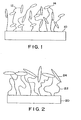
- This construction is schematically depicted in Figure 1, where the poly(acrylic acid) chains 12 are grafted to the surface 10 of a device.
- Collagen 14 is contained within the matrix formed by the chains 12.
- biological activity may be reduced as proper expression and accessibility are hampered.
- the present invention provides a medical device comprising:
- a medical device comprising:
- a method of modifying the surface of a medical device comprising:
- a method of modifying the surface of a medical device comprising:
- a method of delivering a pharmaceutical agent comprising contacting a body with a medical device comprising a surface graft matrix comprising carboxyl-functional groups located on the device, the surface graft matrix comprising an outer portion in which a majority of one or more biomolecules are coupled, wherein the pharmaceutical agent is located within the surface graft matrix when the device is initially contacted with the body.
- Figure 1 is a schematic cross-sectional diagram of a prior art surface graft matrix incorporating collagen.
- Figure 2 is a schematic cross-sectional diagram of a surface graft matrix coated with a biomolecule sheath.
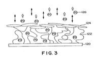
- Figure 3 is a schematic cross-sectional diagram of a surface graft matrix coated with a biomolecule sheath, where the surface graft matrix is loaded with a pharmaceutical agent.
- Figure 4 is a line drawing of the surface appearance of a surface graft matrix coated with collagen in a process according to the present invention.
- the present invention provides methods of covalently coupling a majority of one or more biomolecules in the outer portion of a surface graft matrix on a medical device.
- the surface graft matrix is preferably formed by surface grafting carboxyl-functional monomers, optionally in combination with vinyl monomers having no carboxyl functionality (COOH).
- COOH carboxyl functionality
- the surface graft matrix can result from the surface grafting of acrylic acid and acrylamide monomers, as disclosed in U.S. Patent Application Serial No. 08/553,206, filed November 7, 1995, entitled "Intramuscular Stimulation Lead With Enhanced Infection Resistance.”
- the surface graft matrix typically has an anionic character.
- a surface graft matrix that exhibits reduced permeability to medium-sized to large molecules is formed on the surface of a medical device, thereby providing the ability to isolate a majority of the subsequently coupled biomolecules to the outer portion of the surface graft matrix.
- the permeability of the surface graft matrix is reduced by treatment of the surface graft matrix at reduced pH levels, preferably where the pH of the solution is less than the pKa of the surface graft matrix.
- the surface graft matrix can be loaded with a pharmaceutical agent after the biomolecules are in place in the outer portion of the surface graft matrix.
- this loading occurs as a result of ionic interaction of the surface graft matrix with the pharmaceutical agent.
- a surface graft matrix exhibiting reduced permeability to medium-sized to large molecules is formed on the surface of a medical device.
- the permeability of the graft matrix is reduced by treatment of the surface graft matrix at reduced pH levels, preferably where the pH of the solution is less than the pKa of the surface graft matrix.
- linker molecules which may be biomolecules, referred to herein as intermediate biomolecules
- the relative impermeability of the graft matrix is maintained, restricting a majority of the linker molecules to the outer portion of the graft matrix.
- linker molecules e.g., intermediate biomolecules
- the linker molecules can then be used to covalently couple a majority of second biomolecules (referred to herein as primary biomolecules) in the outer portion of the surface graft matrix.
- This second coupling step is also preferably performed in solutions having pH values less than the pKa of the underlying surface graft matrix, thereby maintaining its relative impermeability to the biomolecules located in the outer portion of the surface graft matrix.
- the term "medical device” may be defined as a device that has surfaces that contact tissue, blood, or other bodily fluids in the course of their operation, which fluids are later used in patients. This can include, for example, extracorporeal devices for use in surgery such as blood oxygenators, blood pumps, blood sensors, tubing used to carry blood, and the like which contact blood that is returned to the patient.
- the term can also include endoprostheses implanted in blood contact in a human or animal body such as vascular grafts, stents, pacemaker leads, heart valves, and the like that are implanted in blood vessels or in the heart.
- the term can further include devices for temporary intravascular use such as catheters, guide wires, and the like that are placed in blood vessels or the heart for purposes of monitoring or repair.
- the term can also include nerve electrodes, muscle electrodes, implantable pulse generators, implantable drug pumps, and defibrillators.
- biomolecule includes any biocompatible and/or biologically active molecule (i.e., "primary biomolecule") or an intermediate biomolecule (linker molecule) to which one or more primary biomolecules can be coupled. Unless otherwise indicated, the term “biomolecule” as used herein will be understood to include both primary and intermediate biomolecules.
- biomolecule is collagen, a biocompatible molecule that exists in many types.
- Types of biomolecules that can be coupled to the surface graft matrix in accordance with the present invention include, but are not limited to, antithrombotic agents, antibacterial agents, anti-inflammatory agents, growth factors, cytokines, naturally occurring or synthetically prepared proteins, peptides, amino acids, and mixtures thereof.
- biomolecules that can be coupled to the surface graft matrix include, but are not limited to, albumin, fibrinogen, laminin, vitronectin, fibronectin, RGD-containing peptides, heparin, heparin sulfate, fibroblast growth factors (FGF), insulin-like growth factor, nerve growth factor, interferons (IFN), tumor necrosis factors (TNF), interleukins, gelatin, elastin, fibrin, von Willebrand factor, dermatan sulfate, hyaluronic acid, dextran sulfate, and mixtures thereof.
- FGF fibroblast growth factors
- IFN interferons
- TNF tumor necrosis factors
- biomolecules may be neutral or charged at the conditions employed during covalent coupling.
- biomolecules may be coupled to the surface graft matrix directly (i.e., through the carboxyl groups), or through well-known coupling chemistries, such as, for example, esterification, amidation, and acylation.
- These biomolecules are typically the primary biomolecules, although certain of them can be used as the intermediate biomolecules.
- the outer sheath of biomolecules typically includes a plurality of biomolecules, although it could include polymerized biomolecules that technically form one macromolecule.
- linker molecules which may or may not be biomolecules, in connection with the present invention typically involves covalently coupling a majority of the linker molecules in the outer portion of the surface graft matrix.
- the linker molecules can provide the surface graft matrix with a number of functionally active groups that can be used to covalently couple one or more primary biomolecules.
- the linker molecules may be coupled to the surface graft matrix directly (i.e., through the carboxyl groups), or through well-known coupling chemistries, such as, for example, esterification, amidation, and acylation.
- the linker molecule is at least a di- or tri-amine functional compound that is coupled to the surface graft matrix through the direct formation of amide bonds, and provides amine-functional groups that are available for reaction with the primary biomolecule.
- the linker molecule is a polyamine functional polymer such as polyethyleneimine (PEI) or polyallylamine (PALLA). Mixtures of these polymers can also be used. These molecules contain a plurality of pendant amine-functional groups that can be used to surface-immobilize one or more primary biomolecules.
- PEI polyethyleneimine
- PALLA polyallylamine
- Figure 2 is a schematic cross-sectional view of a portion of the surface of a medical device 20, depicting that the biomolecules 24 are covalently coupled to a surface graft matrix 22 in a manner such that a majority of the biomolecules 24 are located or immobilized in the outer portion of the surface graft matrix 22 located on the medical device 20.
- the immobilization of the biomolecules 24 as depicted in Figure 2 differs from the prior art depicted in Figure 1 in that a majority of the biomolecules 24 are located on or near the outer surface of the surface graft matrix 22, not generally dispersed throughout the structure of the matrix as depicted in Figure 1. This surface isolation of a majority of the biomolecules is advantageous because it allows a less disturbed expression of the biomolecules, so that biological activity is retained at a significantly higher level.
- the depth of the outer portion of the surface graft matrix in which a majority of the biomolecules are immobilized is primarily dependent on the type of biomolecule immobilized and the reaction conditions employed. Typically, a majority of the biomolecules will be immobilized in the surface graft matrix within a depth of about 10 nm or less. For example, if collagen is the biomolecule immobilized in the outer portion of the surface graft matrix, the depth at which a majority of the collagen molecules are immobilized is about 7 nm or less.
- the immobilization approach of the present invention may prohibit movement of the coupled biomolecules into the graft matrix. This will especially be the case with immobilization of anionic biomolecules, such as the anti-coagulant heparin which will be repelled by the underlying anionic surface graft matrix.
- the matrix can be loaded with a pharmaceutical agent for subsequent release to effect a desired response in the patient.
- the pharmaceutical agent capacity of the matrix can be increased as compared to those surface graft matrix materials that allow complete penetration of biomolecules. This provides yet another advantage of the present invention.
- Pharmaceutical agents that can be used in connection with the present invention include, but are not limited to, antimicrobial agents, antibacterial agents, anticoagulant agents, antithrombotic agents, platelet agents, and anti-inflammatory agents.
- Other useful pharmaceutical agents can include, but are not limited to, dyes which act as biological ligands, steroids, enzymes, catalysts, hormones, growth factors, drugs, vitamins, antibodies, antigens, nucleic acids, peptides, DNA & RNA segments, and mixtures thereof.
- these pharmaceutical agents are hydrophilic, positively charged compounds.
- a medical device 120 incorporating a biomolecule 124 on a surface graft matrix 122 loaded with a desired pharmaceutical agent 126
- duplicate biological activities can be provided to improve the in vivo performance of the medical device.
- the biomolecules 124 can be, for example, collagen which will interact with the surrounding tissue to provide a favorable tissue integration.
- specific desired body mechanisms may be activated, or, in the case of antimicrobials, a protective mode of action is exhibited during the initial vulnerable period before the medical device/tissue interface is stabilized and when random colonization by bacteria might occur.
- the surface exhibits "bi-biofunctional" characteristics, i.e., two biofunctional activities including: a) promoting rapid tissue integration into the surface of the device, and b) releasing a pharmaceutical agent, such as an antimicrobial agent to reduce the risk of infection around an implanted device.
- a pharmaceutical agent such as an antimicrobial agent
- Processes according to the present invention typically begin with the formation of a surface graft matrix on the surface of a medical device.
- the surface grafting method involves the covalent surface grafting of a polymer, preferably water soluble polymer, based on carboxyl-functional monomers, including, but not limited to, acrylic acid, methacrylic acid, itaconic acid, trans-cinnamic acid, crotonic acid, linoleic acid, linolenic acid, maleic acid, sorbic acid, and mixtures thereof onto a substrate material.
- the carboxyl-functional surface graft matrix also may be obtained through chemical modification of non-carboxyl-functional monomers.
- ceric ion initiation is a preferred method to graft monomers to substrate surfaces, other grafting techniques may be used as well.
- Known examples of other initiation methods include corona discharge, UV irradiation, ozonization and ionizing radiation (e.g., 60 Co, X-rays, high energy electrons, plasma gas discharge, etc.).
- the substrates that can be modified by the method of the present invention include metals such as titanium/titanium alloys, TiNi (shape memory/super elastic), aluminum oxide, platinum/platinum alloys, stainless steels, MP35N, elgiloy, haynes 25, stellite, pyrolytic carbon, silver or glassy carbon; polymers such as polyamides, polycarbonates, polyethers, polyesters, polyolefins including polyethylenes or polypropylenes, polystyrenes, polyurethanes, polyvinyl chlorides, polyvinylpyrrolidones, silicone elastomers, fluoropolymers, polyacrylates, polyisoprenes, polytetrafluoroethylenes, and rubber; minerals or ceramics such as hydroxapatite; human or animal protein or tissue such as bone, skin, teeth, collagen, laminin, elastin or fibrin; organic materials such as wood, cellulose, or compressed carbon; and other materials such as glass, or
- Substrates made using these materials can be coated or uncoated, and derivatized (e.g., modified to include reactive functional groups) or underivatized.
- the substrate is polyurethane, to which the carboxyl-functional surface graft matrix can be directly coupled without any preactivation of the substrate surface.
- the substrate is a biomaterial for use in a number of medical devices such as vascular grafts, aortic grafts, arterial, venous, or vascular tubing, vascular stents, dialysis membranes, tubing, or connectors, blood oxygenator tubing or membranes, ultrafiltration membranes, intra-aortic balloons, blood bags, catheters, sutures, soft or hard tissue prostheses, synthetic prostheses, prosthetic heart valves, tissue adhesives, cardiac pacemaker leads, artificial organs, endotracheal tubes, lenses for the eye such as contact or intraocular lenses, blood handling equipment, apheresis equipment, diagnostic and monitoring catheters and sensors, biosensors, dental devices, drug delivery systems, or bodily implants of any kind.
- medical devices such as vascular grafts, aortic grafts, arterial, venous, or vascular tubing, vascular stents, dialysis membranes, tubing, or connectors, blood oxygenator tubing or membranes, ultrafiltration
- a polymer surface graft of acrylic acid is one preferred embodiment to be used for subsequent covalent coupling of one or more biomolecules to enclose the surface graft matrix.
- the surface graft matrix is preferably formed by surface grafting of the monomers acrylic acid and acrylamide in ratios that allow for later manipulation of the graft matrix.
- sufficient acrylic acid (or other carboxylic-functional monomer) should be present so as not to interfere with the mechanism of reducing the permeability of the surface graft matrix to provide for immobilization of a majority of the biomolecules in the outer portion of the surface graft matrix.
- acrylic acid is used to prepare the surface graft matrix in an amount of about 20-100 wt-%, based on the total weight of the monomers used to prepare the surface graft matrix. More preferably, acrylic acid is used in an amount of about 50-90 wt-%, and most preferably, in an amount of about 65-75 wt-%. These weight percentages are also applicable to other carboxyl-functional monomers. Incorporation of other vinyl-functional monomers that do not include carboxyl groups (COOH) into the surface graft is possible, but is limited to the extent that they interfere with the mechanism of reducing the permeability of the surface graft matrix to provide for isolation of a majority of the biomolecules in the outer portion of the surface graft matrix.
- COOH carboxyl groups
- Various vinyl-functional monomers can be incorporated to form a copolymer surface graft, such as acrylamide (Aam), N-(3-aminopropyl) methacrylamide (APMA), 2-hydroxyethyl methacrylate (HEMA), and 2-acrylamido-2-methylpropane sulfonic acid (AMPS).
- Acrylamide is the most preferred monomeric compound to be incorporated in addition to acrylic acid monomer as the structure and molecular weight of acrylamide are close to those of acrylic acid.
- a low pH immersion process is used to produce the surface graft matrix with the desired impermeability to provide for immobilization of a majority of the biomolecules in the outer portion of the surface graft matrix.
- a low pH solution By immersing the surface graft matrix in a low pH solution, the formation of carboxylic acid dimers and intra-polymer crosslinking in the surface graft matrix is provided.
- the bond strength of acetic acid dimers is approximately equal to 55-60 kJ/mole, as disclosed by Potter, Jr., et al., J. Phys. Chem.
- intrapolymer crosslinking within a poly(carboxylic acid) graft matrix will be of significant strength.
- the formation of intrapolymer crosslinks is characterized by an obvious sticky feel of the surface grafted material.
- This sticky feel is generally indicative of the cohesive forces of the surface graft matrix.
- the intrapolymer crosslinking reduces the permeability/accessibility of medium-sized to large, even polycationic, compounds into the surface graft matrix.
- a subsequent process of covalently coupling a majority of biomolecules in the outer portion of the surface graft matrix is also carried out at a pH that is less than the pKa of the surface graft matrix. This results in immobilization of the biomolecules such that a surface layer of primarily biomolecules, i.e., a sheath, is formed that substantially encloses the surface graft matrix. This additionally allows for more complete loading of the surface graft matrix with a pharmaceutical agent for subsequent release in vivo. Although it is preferred that the immersion process and the biomolecule coupling process be carried out sequentially, they could be carried out simultaneously.
- the pKa value of the surface graft matrix can be determined through FT-IR analysis, according to the method of Azeez et al., J. Appl. Polym. Sci., 58 , 1741-1749 (1995). Using this method, the pKa of a 100% acrylic acid graft matrix is 6.3, which is in accordance with the findings of 4.9-6.7 disclosed by Park et al., Pharm. Res., 4 , 457-464 (1987) on acrylic acid/acrylamide copolymer hydrogels.
- the pKa values are generally dependent on the ionic strength of the environment and the fraction of acrylic acid in the copolymer. Typically, an increase in ionic strength decreases the pKa, whereas an increase in acrylic acid fraction increases the pKa.
- the pH of the solutions in which the surface graft matrix is treated to reduce permeability and in which biomolecules are attached are typically no greater than about 5.5.
- the pH is no greater than about 5, and more preferably, no greater than about 4.5.
- the pH is at least about 2, and more preferably, at least about 3.
- the methods of the present invention preferably involve surface grafting of carboxyl-functional monomers through a covalent interaction to a substrate at an acidic pH, preferably at a pH of less than about 5.5; washing the substrate with the surface graft matrix thereon in an aqueous solution having a pH greater than the pKa of the surface graft matrix (typically at a neutral pH) to allow for the removal of free monomers, oligomers, or polymers; immersing the substrate with the surface graft matrix thereon in a solution having a pH that is less than the pKa of the surface graft matrix; and covalently coupling a majority of the biomolecules in the outer portion of the surface graft matrix in a solution having a pH that is less than the pKa of the surface graft matrix.
- Polyurethane (PU) film material was made from 2363-55D PELLETHANE resin (Dow Chemical, Midland, MI, USA) by Medtronic Promeon (Minneapolis, MN, USA). Ceric(IV)ammonium nitrate, nitric acid (65%), sodium phosphate monobasic monohydrate, sodium phosphate dibasic, sodium chloride, and sodium azide were all obtained from Merck-Schuchardt (Darmstadt, Germany). Acrylic acid, MES monohydrate, di-sodium tartrate, N-hydroxysuccimide (NHS), 3-ethyl-1- (diaminopropyl)-carbodiimide (EDC), and sodium hydrogencarbonate, were obtained from Aldrich Chemie (Bornem, Belgium).
- Acrylamide (99+%; electrophoresis grade) was obtained from Acros Chimica (Geel, Belgium).
- Collagen type I; from calf skin
- TNBS 2,4,6-trinitrobenzenesulfonic acid
- Coomassie Blue was obtained from Pierce Europe BV (Oud Beijerland, The Netherlands).
- Collagenase (EC 3.4.24.3; from Clostridium histolyticum; type IA, 550 units/mg solid), and Tris-HCl were obtained from Sigma Chemie (Bornem, Belgium); di-sodium tetraborate decahydrate from Sigma Chemie (Borneum, Belgium); Toluidine Blue O dye from Sigma Chemie; Ponceau S dye from Sigma Chemie; SDS from Sigma Chemie; and gentamicin sulfate from Sigma Chemie.
- Acrylic acid was purified by conventional distillation. All other reagents were of reagent grade or higher and used without further purification.
- XPS X-Ray Photoelectron Spectroscopy
- Time of Flight Secondary Ion Mass Spectometry (ToF-SIMS) spectra were acquired using a VG IX23S instrument based on the Poschenreider design and equipped with a pulsed liquid metal ion source. A 30 keV Ga + primary ion beam was used at an incident angle of 38° to the surface normal. The secondary ions were accelerated to 5 keV for the analysis by applying a sample bias. For each sample, both positive and negative secondary ion spectra were collected using a total primary ion dose that did not exceed 2 x 10 11 ions cm -2 for static SIMS, such that the analyzed surfaces were effectively undamaged as a result of the ToFSIMS studies.
- FEG-SEM tests were carried out on a JEOL JSM 6301-F Field-Emission-Gun SEM operated at 2 kV after the samples were sputter coated with gold (2-4 nm) using an Edwards 5150B Sputter Coater.
- Extruded PELLETHANE 55D polyurethane films were ultrasonically cleaned in isopropyl alcohol (IPA) for 15 minutes prior to ceric ion initiated surface grafting. Immediately after the IPA-cleaning samples were dried in a forced air oven at 50-60°C for approximately 5 minutes. FT-IR investigation has demonstrated that 15 minutes IPA-treatment is sufficient to remove any surface contamination that originates from processing aides, such as bis-stearamide waxes, that may interfere with the grafting process.
- processing aides such as bis-stearamide waxes
- an aqueous grafting solution was prepared that was composed of 40% by weight acrylic acid monomer concentration (100 wt-% acrylic acid), 6 mM of ceric ammonium nitrate (CAN) and 0.06 M nitric acid (HNO 3 ). Prior to grafting, the grafting solution was treated to remove excess air by exposure to reduced pressure (18 mm Hg ⁇ 5 mm Hg) for a maximum of 2 minutes.
- Grafted samples (10 x 1cm strips) were prepared by placing the cleaned and dried samples in an appropriate volume (25-30 ml) of the grafting solution. Grafting was allowed to continue for 15-20 minutes at 30°C, while stirring the solution.
- the samples were rinsed in deionized (DI) water to stop the grafting process as well as to clean the surface graft matrix formed.
- DI deionized
- a 0.1 M tartrate solution di-sodium tartrate
- pH 3.0 (which is significantly below the pKa of the graft matrix, typically about pH 6) for four hours at room temperature.
- XPS X-Ray Photoelectron Spectroscopy
- the carboxylic acid groups are mainly ionized. This is confirmed by the presence of sodium (Table 1) and the prevalence of the COOX chemical state (Table 2). In the ionized state, carboxylic acids will not be capable of forming the dimer, i.e., the group that is essential for physically crosslinking the graft matrix. In contrast, at pH ⁇ pKa the carboxylic acid groups are hydrogenated and thus capable of forming that dimer-group. The hydrogenated state is confirmed by the absence of sodium (Table 1) and the prevalence of the COOH chemical state (Table 2).
- the surface graft matrix can be made impermeable for medium-sized to large (even polycationic) molecules by formation of carboxylic acid dimers to induce physical crosslinking. This was confirmed in an experiment that studied the effect of pH on the amount of the (polycationic) antimicrobial drug gentamicin that could be (ionically) immobilized.
- the surface grafted samples were gentamicin loaded and the amount of gentamicin loaded was determined. The difference in the gentamicin content before and after sample immersion was determined and used as a measure for the amount of gentamicin loaded into the samples.
- the TNBS derivatization reaction was allowed to proceed for 25-30 minutes at room temperature, after which the absorbance at 415 nm was measured, while 595 nm was used as the reference wavelength (BioRad Model 3550, 96 wells microplate reader, Veenendaal, The Netherlands) .
- the pH falls below the pKa of the surface graft matrix, the amount of gentamicin that could be immobilized was drastically reduced to become zero in the pH range from 3 to 4.
- MES 4-morpholine-ethanesulfonic acid monohydrate, Aldrich
- the samples were immersed in a buffered solution containing 0.5 mg/ml collagen (type I).
- the solution was buffered in the pH range 4.0-4.5 with 0.02 M MES.
- the collagen immobilization reaction was continued for at least 20 hours.
- the collagen-immobilized samples were rinsed in DI water, an aqueous 0.15M NaCl solution in DI water, and DI water again. Samples were dried at ambient conditions by air exposure. The immobilization of collagen was identified by two separate staining techniques. TNBS-staining confirmed the presence of amine-functional groups.
- TNBS staining was performed by immersing a 4 mm disc in 1 ml of aqueous 4% by weight NaHCO 3 . To this solution 1 ml of aqueous 0.5% by weight TNBS was added, after which the reaction was allowed to continue for 2 hours at 40°C. Finally, the sample was extensively rinsed in DI water and allowed to dry. A similar surface grafted disc, but not used for collagen immobilization, was used as the control. The difference in dye uptake was obvious visually. Considering the surface modification chemistry, these groups could only be derived from immobilized collagen. Coomassie Blue protein dye also was used as a analytical tool to verify the presence of immobilized collagen in the outer portion of the surface graft matrix.
- Coomassie Blue staining was performed by immersing a 4 mm disc in 1 ml of Coomassie Blue for 30 minutes. Thereafter, the sample was extensively rinsed in DI water and allowed to dry. A similar surface grafted disc, but not used for collagen immobilization, was used as the control. The difference in dye uptake was obvious visually.
- the processed surfaces were examined by FEG-SEM (Field Emission Gun Scanning Electron Microscope) operated at 2 kV; prior to SEM-analysis surfaces were sputter-coated with gold (2-4 nm).
- FEG-SEM Field Emission Gun Scanning Electron Microscope
- the extruded 55D PELLETHANE polyurethane material is a flat material.
- the acrylic acid grafted material exhibits a permeable matrix-like structure. Subsequent immobilization of collagen seems to have covered this surface matrix with a superimposed surface-layer.
- the velvet-like appearance of this surface layer is depicted in Figure 4.
- FT-IR spectroscopy Another analytical technique that was used to confirm the immobilization of collagen was FT-IR spectroscopy. This technique allows for analysis of the top 0.2-1 :m surface layer of processed samples. The FT-IR spectra of collagen raw material, acrylic acid grafted 55D, and collagen-immobilized samples were compared. The spectrum of the collagen-immobilized sample obviously contained features of both the acrylic acid graft and the collagen raw material. Most characteristic was the rise of the collagen-related peaks at approximately 1635 nm and 1660 nm. This confirmed the presence of collagen in the surface top layer.
- Table 4 Surface composition (in atom%) according to XPS. Sample carbon oxygen nitrogen silicone collagen reference 69.1 17.5 11.7 1.8 collagen immobilized 67.9 18.9 11.8 1.4 Table 5: Carbon chemical states derived from XPS analysis (in %).
- Table 10 Oxygen chemical states derived from XPS analysis (in %).
- Table 11 Nitrogen chemical states derived from XPS analysis (in %).
- analysis depth ( ⁇ ) NH-C O -C-NH-C- 15 77 23 30 77 23 70 92 8 collagen reference 70 30
- ToF-SIMS Time-of-Flight Secondary Ion Mass Spectrometry
- the results of the ToF-SIMS testing also confirmed the presence of a collagen top layer, as the sample spectra displayed a rich array of N-containing signals. In addition to nonspecific peptide/protein characteristics, the spectra exhibit rich arrays of N-containing signals which are more diagnostic of particular amino acid residues.
- the collagen top layer displayed an outermost surface chemistry which differs to that of the collagen reference. While N-containing species are clearly present, there appears to be a higher relative proportion of species containing C/H and C/H/O for the collagen coating compared to the collagen reference. This indicates either an incomplete collagen top layer or a very thin collagen top layer, or some reorientation of the collagen molecules due to the immobilization.
- Collagen-immobilized samples were prepared as previously described in Example 2 above (100 wt-% acrylic acid monomer used to prepare the surface graft matrix). The in vivo performance was investigated and compared to that of acrylic acid grafted 55D (55D-AA) and plain 55D samples (55D) as discussed below.
- 55D-CC had a similar or thinner tissue capsule compared to 55D and 55D-AA. These results may indicate that 55D-CC promotes a faster wound healing response as a result from its biological interaction with the body's responses. It appears that fibrin formation at the surface of 55D-CC plays a major role, which indicates that 55D-CC may promote coagulation.
- linker molecules covalently coupled to both the surface graft matrix and the biomolecules.
- linker molecules include, e.g., the amine-functional polymers polyethyleneimine (PEI) or polyallylamine (PALLA). These compounds contain pendant amine-functional groups that can be used to surface-immobilize a majority of the biomolecules in the outer portion of a surface graft matrix.
- PEI was surface-immobilized to substantially enclose the surface graft matrix.
- the PEI was subsequently used to surface-immobilize the anti-coagulant drug heparin.
- a 0.05 M tartrate solution di-sodium tartrate
- heparin coupling After heparin coupling, representative samples were stained with the cationic dye Toluidine Blue O (TB). The presence of heparin is denoted by a metachromatic shift from blue to violet. While the primary graft alone demonstrated a blueish, dark violet color after TB-exposure, the heparinized surface demonstrated an obvious shift to light violet. This shift in color suggests successful surface-coupling of heparin. PS-uptake obviously decreased as a consequence of the heparin-coupling; this confirms presence of heparin as well as coupling of heparin reduces the cationic nature of the surface.
- Toluidine Blue O Toluidine Blue O
- peaks are characteristic of the S-O stretch of the sulfur acid groups, in this case originating from the immobilized heparin molecules.
- the observed change in the 1530-1730 cm -1 region can be assigned to the surface-immobilized PEI; for example, the 1560 cm -1 peak is characteristic for N-H (in plane) bend of amides. Additional surface analysis was performed by XPS. The heparin-immobilized sample exhibited significant heparin concentrations at the graft surface.
- the gentamicin solution used for loading the surface grafted sample was analyzed to determine its gentamicin content before and after loading. This was done using a TNBS Assay in which the gentamicin-containing solution was adjusted to pH 9 by addition of 0.1 M borate, after which 25 ml 0.03 M aqueous TNBS was added per ml of sample-solution. The difference in gentamicin content before and after sample immersion was determined and used as a measure for the amount of gentamicin loaded. The amount of gentamicin loaded was expressed as ⁇ g/cm 2 and is reported below in Table 12.
- the gentamicin loading suggests that a majority of the immobilized PEI can be found in the outer portion of the surface graft matrix, i.e., the PEI has formed a relatively thin outer sheath on the matrix. If the PEI was dispersed throughout the surface graft matrix, it would be expected to diminish the capacity of the surface graft matrix to load gentamicin, as it would neutralize much of the negative charge of the surface graft matrix due to penetration of the PEI molecules into the graft matrix.
- Gentamicin release was performed by immersion of gentamicin loaded samples in phosphate buffered saline (PBS, pH 7.4) at 37°C; a volume-to-surface ratio of 1:1 (ml:cm 2 ) typically was used throughout the experiment. At desired time points, the samples were withdrawn from the solution and immersed in fresh PBS. Solution samples were analyzed for their gentamicin content by means of the TNBS Assay discussed above.
- PBS phosphate buffered saline
- Heparin coupling did not significantly influence the gentamicin release profile. After an initial burst, gentamicin progressively released until completion in an approximate 2 weeks.
Landscapes
- Health & Medical Sciences (AREA)
- Life Sciences & Earth Sciences (AREA)
- Epidemiology (AREA)
- Animal Behavior & Ethology (AREA)
- General Health & Medical Sciences (AREA)
- Public Health (AREA)
- Veterinary Medicine (AREA)
- Chemical & Material Sciences (AREA)
- Surgery (AREA)
- Engineering & Computer Science (AREA)
- Medicinal Chemistry (AREA)
- Hematology (AREA)
- Biomedical Technology (AREA)
- Molecular Biology (AREA)
- Heart & Thoracic Surgery (AREA)
- Vascular Medicine (AREA)
- Dermatology (AREA)
- Oral & Maxillofacial Surgery (AREA)
- Transplantation (AREA)
- Materials Engineering (AREA)
- Materials For Medical Uses (AREA)
- Treatments Of Macromolecular Shaped Articles (AREA)
Applications Claiming Priority (2)
| Application Number | Priority Date | Filing Date | Title |
|---|---|---|---|
| US08/656,614 US5811151A (en) | 1996-05-31 | 1996-05-31 | Method of modifying the surface of a medical device |
| US656614 | 1996-05-31 |
Publications (3)
| Publication Number | Publication Date |
|---|---|
| EP0809997A2 true EP0809997A2 (de) | 1997-12-03 |
| EP0809997A3 EP0809997A3 (de) | 1999-12-22 |
| EP0809997B1 EP0809997B1 (de) | 2003-01-15 |
Family
ID=24633806
Family Applications (1)
| Application Number | Title | Priority Date | Filing Date |
|---|---|---|---|
| EP97303222A Expired - Lifetime EP0809997B1 (de) | 1996-05-31 | 1997-05-12 | Medizinisches Gerät mit einer an der Oberfläche gegerbten und mit Biomolekülen beschichteten Matrix |
Country Status (6)
| Country | Link |
|---|---|
| US (2) | US5811151A (de) |
| EP (1) | EP0809997B1 (de) |
| JP (1) | JPH1052488A (de) |
| AU (1) | AU713357B2 (de) |
| CA (1) | CA2206147A1 (de) |
| DE (1) | DE69718377T2 (de) |
Cited By (8)
| Publication number | Priority date | Publication date | Assignee | Title |
|---|---|---|---|---|
| WO1999038546A1 (en) * | 1998-01-30 | 1999-08-05 | Advanced Cardiovascular Systems, Inc. | Hydrophilic coating for an intracorporeal medical device |
| WO2003045461A1 (de) * | 2001-11-23 | 2003-06-05 | Feg Textiltechnik Forschungs- Und Entwicklungsgesellschaft Mbh | Textiles erzeugnis mit oberflächenmodifikation und entsprechendes verfahren zur oberflächenmodifikation |
| WO2006056984A3 (en) * | 2004-11-26 | 2006-10-05 | Novik Shai | Chelating and binding chemicals to a medical implant |
| DE102005032691A1 (de) * | 2005-07-06 | 2007-01-18 | Biotronik Vi Patent Ag | Implantat mit immobilisierten Biokatalysatoren |
| WO2008106477A1 (en) * | 2007-02-28 | 2008-09-04 | Alcon, Inc. | Coated medical implants and lenses |
| DE102008040787A1 (de) * | 2008-07-28 | 2010-02-04 | Biotronik Vi Patent Ag | Biokorrodierbares Implantat mit einer Beschichtung enthaltend ein Hydrogel |
| DE102008040786A1 (de) * | 2008-07-28 | 2010-02-04 | Biotronik Vi Patent Ag | Biokorrodierbares Implantat mit einer Beschichtung enthaltend eine wirkstofftragende Polymermatrix |
| US9867727B2 (en) | 1998-02-09 | 2018-01-16 | Trivascular, Inc. | Endovascular graft |
Families Citing this family (235)
| Publication number | Priority date | Publication date | Assignee | Title |
|---|---|---|---|---|
| US6007843A (en) * | 1995-09-29 | 1999-12-28 | Lam Pharmaceuticals Corp. | Sustained release delivery system |
| US6562781B1 (en) | 1995-11-30 | 2003-05-13 | Hamilton Civic Hospitals Research Development Inc. | Glycosaminoglycan-antithrombin III/heparin cofactor II conjugates |
| US7045585B2 (en) * | 1995-11-30 | 2006-05-16 | Hamilton Civic Hospital Research Development Inc. | Methods of coating a device using anti-thrombin heparin |
| US6491965B1 (en) * | 1995-11-30 | 2002-12-10 | Hamilton Civic Hospitals Research Development, Inc. | Medical device comprising glycosaminoglycan-antithrombin III/heparin cofactor II conjugates |
| US6033719A (en) * | 1996-04-25 | 2000-03-07 | Medtronic, Inc. | Method for covalent attachment of biomolecules to surfaces of medical devices |
| FR2758990B1 (fr) | 1996-09-19 | 1999-05-28 | Hospal Ind | Appareil pour le traitement du sang par circulation extracorporelle et procede de fabrication |
| EP0860213A3 (de) * | 1997-01-03 | 2002-10-16 | Therapol SA | Bioaktive Beschichtung von Oberflächen |
| US6776792B1 (en) * | 1997-04-24 | 2004-08-17 | Advanced Cardiovascular Systems Inc. | Coated endovascular stent |
| US6146771A (en) | 1997-07-01 | 2000-11-14 | Terumo Cardiovascular Systems Corporation | Process for modifying surfaces using the reaction product of a water-insoluble polymer and a polyalkylene imine |
| JP3936058B2 (ja) * | 1998-03-12 | 2007-06-27 | 株式会社メニコン | コンタクトレンズ用液剤 |
| US6258371B1 (en) * | 1998-04-03 | 2001-07-10 | Medtronic Inc | Method for making biocompatible medical article |
| US6248127B1 (en) | 1998-08-21 | 2001-06-19 | Medtronic Ave, Inc. | Thromboresistant coated medical device |
| US6596401B1 (en) | 1998-11-10 | 2003-07-22 | C. R. Bard Inc. | Silane copolymer compositions containing active agents |
| CN1335757A (zh) * | 1998-11-12 | 2002-02-13 | 聚合体生物科学公司 | 可用于快速凝血和止血的止血聚合物 |
| US6340465B1 (en) | 1999-04-12 | 2002-01-22 | Edwards Lifesciences Corp. | Lubricious coatings for medical devices |
| US6328762B1 (en) * | 1999-04-27 | 2001-12-11 | Sulzer Biologics, Inc. | Prosthetic grafts |
| US6309660B1 (en) | 1999-07-28 | 2001-10-30 | Edwards Lifesciences Corp. | Universal biocompatible coating platform for medical devices |
| US6159531A (en) * | 1999-08-30 | 2000-12-12 | Cardiovasc, Inc. | Coating having biological activity and medical implant having surface carrying the same and method |
| US6974478B2 (en) * | 1999-10-22 | 2005-12-13 | Archus Orthopedics, Inc. | Prostheses, systems and methods for replacement of natural facet joints with artificial facet joint surfaces |
| US6673116B2 (en) * | 1999-10-22 | 2004-01-06 | Mark A. Reiley | Intramedullary guidance systems and methods for installing ankle replacement prostheses |
| US7674293B2 (en) * | 2004-04-22 | 2010-03-09 | Facet Solutions, Inc. | Crossbar spinal prosthesis having a modular design and related implantation methods |
| US6811567B2 (en) * | 1999-10-22 | 2004-11-02 | Archus Orthopedics Inc. | Facet arthroplasty devices and methods |
| US7691145B2 (en) * | 1999-10-22 | 2010-04-06 | Facet Solutions, Inc. | Prostheses, systems and methods for replacement of natural facet joints with artificial facet joint surfaces |
| WO2001030248A1 (en) * | 1999-10-22 | 2001-05-03 | Reiley Mark A | Facet arthroplasty devices and methods |
| WO2001030264A2 (en) | 1999-10-22 | 2001-05-03 | Reiley Mark A | Ankle replacement system |
| US8496712B2 (en) | 1999-10-22 | 2013-07-30 | Inbone Technologies, Inc. | Systems and methods for installing ankle replacement prostheses |
| US8187303B2 (en) | 2004-04-22 | 2012-05-29 | Gmedelaware 2 Llc | Anti-rotation fixation element for spinal prostheses |
| US6733513B2 (en) | 1999-11-04 | 2004-05-11 | Advanced Bioprosthetic Surfaces, Ltd. | Balloon catheter having metal balloon and method of making same |
| US6849085B2 (en) | 1999-11-19 | 2005-02-01 | Advanced Bio Prosthetic Surfaces, Ltd. | Self-supporting laminated films, structural materials and medical devices manufactured therefrom and method of making same |
| US7195641B2 (en) | 1999-11-19 | 2007-03-27 | Advanced Bio Prosthetic Surfaces, Ltd. | Valvular prostheses having metal or pseudometallic construction and methods of manufacture |
| US7736687B2 (en) | 2006-01-31 | 2010-06-15 | Advance Bio Prosthetic Surfaces, Ltd. | Methods of making medical devices |
| US6537310B1 (en) | 1999-11-19 | 2003-03-25 | Advanced Bio Prosthetic Surfaces, Ltd. | Endoluminal implantable devices and method of making same |
| US6936066B2 (en) * | 1999-11-19 | 2005-08-30 | Advanced Bio Prosthetic Surfaces, Ltd. | Complaint implantable medical devices and methods of making same |
| US6379383B1 (en) | 1999-11-19 | 2002-04-30 | Advanced Bio Prosthetic Surfaces, Ltd. | Endoluminal device exhibiting improved endothelialization and method of manufacture thereof |
| US7300457B2 (en) | 1999-11-19 | 2007-11-27 | Advanced Bio Prosthetic Surfaces, Ltd. | Self-supporting metallic implantable grafts, compliant implantable medical devices and methods of making same |
| US8458879B2 (en) | 2001-07-03 | 2013-06-11 | Advanced Bio Prosthetic Surfaces, Ltd., A Wholly Owned Subsidiary Of Palmaz Scientific, Inc. | Method of fabricating an implantable medical device |
| US7235092B2 (en) * | 1999-11-19 | 2007-06-26 | Advanced Bio Prosthetic Surfaces, Ltd. | Guidewires and thin film catheter-sheaths and method of making same |
| US10172730B2 (en) | 1999-11-19 | 2019-01-08 | Vactronix Scientific, Llc | Stents with metallic covers and methods of making same |
| US6726718B1 (en) * | 1999-12-13 | 2004-04-27 | St. Jude Medical, Inc. | Medical articles prepared for cell adhesion |
| US6291428B1 (en) | 1999-12-20 | 2001-09-18 | The Hospital For Special Surgery | Peptides which promote bone-forming cell attraction and adhesion |
| US6579539B2 (en) | 1999-12-22 | 2003-06-17 | C. R. Bard, Inc. | Dual mode antimicrobial compositions |
| US7604663B1 (en) | 1999-12-30 | 2009-10-20 | St. Jude Medical, Inc. | Medical devices with polymer/inorganic substrate composites |
| US6814756B1 (en) * | 2000-02-04 | 2004-11-09 | Gary K. Michelson | Expandable threaded arcuate interbody spinal fusion implant with lordotic configuration during insertion |
| EP1645248B8 (de) * | 2000-02-04 | 2010-06-16 | Warsaw Orthopedic, Inc. | Ausdehnbares interspinales Fusionsimplantat mit schwenkbarem Blocker |
| US6743253B2 (en) * | 2000-02-29 | 2004-06-01 | Biomod Surfaces | Polyurethane-sealed biocompatible device and method for its preparation |
| US6444254B1 (en) * | 2000-03-03 | 2002-09-03 | Duke University | Microstamping activated polymer surfaces |
| US7163712B2 (en) | 2000-03-03 | 2007-01-16 | Duke University | Microstamping activated polymer surfaces |
| US6695865B2 (en) | 2000-03-20 | 2004-02-24 | Advanced Bio Prosthetic Surfaces, Ltd. | Embolic protection device |
| US6444217B1 (en) * | 2000-04-25 | 2002-09-03 | University Of Washington | Drug delivery devices, and methods of use |
| US20030114918A1 (en) * | 2000-04-28 | 2003-06-19 | Garrison Michi E. | Stent graft assembly and method |
| US6451050B1 (en) | 2000-04-28 | 2002-09-17 | Cardiovasc, Inc. | Stent graft and method |
| US6520984B1 (en) | 2000-04-28 | 2003-02-18 | Cardiovasc, Inc. | Stent graft assembly and method |
| US8845713B2 (en) | 2000-05-12 | 2014-09-30 | Advanced Bio Prosthetic Surfaces, Ltd., A Wholly Owned Subsidiary Of Palmaz Scientific, Inc. | Self-supporting laminated films, structural materials and medical devices manufactured therefrom and methods of making same |
| US20080177310A1 (en) * | 2000-10-20 | 2008-07-24 | Archus Orthopedics, Inc. | Facet arthroplasty devices and methods |
| US6783793B1 (en) | 2000-10-26 | 2004-08-31 | Advanced Cardiovascular Systems, Inc. | Selective coating of medical devices |
| AU2002233936A1 (en) | 2000-11-07 | 2002-05-21 | Advanced Bio Prosthetic Surfaces, Ltd. | Endoluminal stent, self-fupporting endoluminal graft and methods of making same |
| US8372139B2 (en) * | 2001-02-14 | 2013-02-12 | Advanced Bio Prosthetic Surfaces, Ltd. | In vivo sensor and method of making same |
| US7083642B2 (en) * | 2000-12-22 | 2006-08-01 | Avantec Vascular Corporation | Delivery of therapeutic capable agents |
| US7118579B2 (en) * | 2001-02-04 | 2006-10-10 | Sdgi Holdings, Inc. | Instrumentation for inserting an expandable interbody spinal fusion implant |
| US6986772B2 (en) * | 2001-03-01 | 2006-01-17 | Michelson Gary K | Dynamic lordotic guard with movable extensions for creating an implantation space posteriorly in the lumbar spine |
| WO2002069891A2 (en) | 2001-03-01 | 2002-09-12 | Michelson Gary K | Dynamic lordotic guard with movable extensions for creating an implantation space posteriorly in the lumbar spine and method for use thereof |
| US6896680B2 (en) | 2001-03-01 | 2005-05-24 | Gary K. Michelson | Arcuate dynamic lordotic guard with movable extensions for creating an implantation space posteriorly in the lumbar spine |
| US6764505B1 (en) | 2001-04-12 | 2004-07-20 | Advanced Cardiovascular Systems, Inc. | Variable surface area stent |
| US6660034B1 (en) * | 2001-04-30 | 2003-12-09 | Advanced Cardiovascular Systems, Inc. | Stent for increasing blood flow to ischemic tissues and a method of using the same |
| US7862495B2 (en) | 2001-05-31 | 2011-01-04 | Advanced Cardiovascular Systems, Inc. | Radiation or drug delivery source with activity gradient to minimize edge effects |
| US7186256B2 (en) * | 2001-06-04 | 2007-03-06 | Warsaw Orthopedic, Inc. | Dynamic, modular, single-lock anterior cervical plate system having assembleable and movable segments |
| EP1404225A4 (de) * | 2001-06-04 | 2009-09-16 | Warsaw Orthopedic Inc | Anteriores zervikales plattensystem mit ankern zum eingriff in wirbelkörper, verbindungsplatte und verfahren zu seiner anbringung |
| JP4283665B2 (ja) * | 2001-06-04 | 2009-06-24 | ウォーソー・オーソペディック・インコーポレーテッド | 可動セグメントを有する前方頸椎用動的平板 |
| US7097645B2 (en) * | 2001-06-04 | 2006-08-29 | Sdgi Holdings, Inc. | Dynamic single-lock anterior cervical plate system having non-detachably fastened and moveable segments |
| US7041105B2 (en) * | 2001-06-06 | 2006-05-09 | Sdgi Holdings, Inc. | Dynamic, modular, multilock anterior cervical plate system having detachably fastened assembleable and moveable segments |
| US7044952B2 (en) * | 2001-06-06 | 2006-05-16 | Sdgi Holdings, Inc. | Dynamic multilock anterior cervical plate system having non-detachably fastened and moveable segments |
| US6565659B1 (en) | 2001-06-28 | 2003-05-20 | Advanced Cardiovascular Systems, Inc. | Stent mounting assembly and a method of using the same to coat a stent |
| US6787179B2 (en) * | 2001-06-29 | 2004-09-07 | Ethicon, Inc. | Sterilization of bioactive coatings |
| US6656216B1 (en) | 2001-06-29 | 2003-12-02 | Advanced Cardiovascular Systems, Inc. | Composite stent with regioselective material |
| WO2003008006A1 (en) * | 2001-07-19 | 2003-01-30 | Dempsey, Donald, J. | Bioactive surface for titanium implants |
| JP4347044B2 (ja) * | 2001-07-26 | 2009-10-21 | アバンテク バスキュラー コーポレーション | 可変放出プロフィールを有する治療用薬剤を送達するための装置 |
| US20030055493A1 (en) * | 2001-09-19 | 2003-03-20 | Erin Carpenter | Enhancement of stent radiopacity using anchors and tags |
| US7371258B2 (en) * | 2001-10-26 | 2008-05-13 | St. Jude Medical, Inc. | Valved prosthesis with porous substrate |
| NL1019316C2 (nl) * | 2001-11-06 | 2003-05-07 | Tno | Een vasculaire prothese. |
| EP1469865A4 (de) * | 2001-12-31 | 2010-04-14 | Crosslink D Inc | Hämostatische zusammensetzungen und verfahren zur kontrolle von blutungen |
| US20030161938A1 (en) * | 2002-02-22 | 2003-08-28 | Bo Johnson | Composition and method for coating medical devices |
| US6837903B2 (en) * | 2002-03-22 | 2005-01-04 | Clemson University | Vascular biomaterial devices and methods |
| US20030195610A1 (en) * | 2002-04-04 | 2003-10-16 | Herrmann Robert A. | Processes for producing polymer coatings through surface polymerization |
| US20030205538A1 (en) | 2002-05-03 | 2003-11-06 | Randel Dorian | Methods and apparatus for isolating platelets from blood |
| US7374678B2 (en) | 2002-05-24 | 2008-05-20 | Biomet Biologics, Inc. | Apparatus and method for separating and concentrating fluids containing multiple components |
| US7832566B2 (en) | 2002-05-24 | 2010-11-16 | Biomet Biologics, Llc | Method and apparatus for separating and concentrating a component from a multi-component material including macroparticles |
| US7992725B2 (en) | 2002-05-03 | 2011-08-09 | Biomet Biologics, Llc | Buoy suspension fractionation system |
| US7845499B2 (en) | 2002-05-24 | 2010-12-07 | Biomet Biologics, Llc | Apparatus and method for separating and concentrating fluids containing multiple components |
| US20060278588A1 (en) | 2002-05-24 | 2006-12-14 | Woodell-May Jennifer E | Apparatus and method for separating and concentrating fluids containing multiple components |
| WO2003099412A1 (en) | 2002-05-24 | 2003-12-04 | Biomet Manufacturing Corp. | Apparatus and method for separating and concentrating fluids containing multiple components |
| JP2005531616A (ja) * | 2002-06-29 | 2005-10-20 | アクヴァノヴァ・ジャーマン・ソリュービリセイト・テクノロジーズ・(アーゲーテー)・ゲゼルシャフト・ミット・ベシュレンクテル・ハフツング | イソフラボン濃縮物およびその製造法 |
| US7598224B2 (en) | 2002-08-20 | 2009-10-06 | Biosurface Engineering Technologies, Inc. | Dual chain synthetic heparin-binding growth factor analogs |
| US8227411B2 (en) | 2002-08-20 | 2012-07-24 | BioSurface Engineering Technologies, Incle | FGF growth factor analogs |
| US7166574B2 (en) | 2002-08-20 | 2007-01-23 | Biosurface Engineering Technologies, Inc. | Synthetic heparin-binding growth factor analogs |
| EP1549248A4 (de) | 2002-09-26 | 2015-11-25 | Advanced Bio Prosthetic Surfac | In vakuum abgeschiedene nitinol-legierungsfilme hoher festigkeit, medizinische dünnfilm-implantatmaterialien und herstellungsverfahren dafür |
| US20060147332A1 (en) | 2004-12-30 | 2006-07-06 | Howmedica Osteonics Corp. | Laser-produced porous structure |
| EP1418013B1 (de) | 2002-11-08 | 2005-01-19 | Howmedica Osteonics Corp. | Lasererzeugte poröse Oberfläche |
| US7169178B1 (en) | 2002-11-12 | 2007-01-30 | Advanced Cardiovascular Systems, Inc. | Stent with drug coating |
| US20040220534A1 (en) * | 2003-04-29 | 2004-11-04 | Martens Paul W. | Medical device with antimicrobial layer |
| US7608104B2 (en) * | 2003-05-14 | 2009-10-27 | Archus Orthopedics, Inc. | Prostheses, tools and methods for replacement of natural facet joints with artifical facet joint surfaces |
| US20040230304A1 (en) * | 2003-05-14 | 2004-11-18 | Archus Orthopedics Inc. | Prostheses, tools and methods for replacement of natural facet joints with artifical facet joint surfaces |
| US20040230201A1 (en) * | 2003-05-14 | 2004-11-18 | Archus Orthopedics Inc. | Prostheses, tools and methods for replacement of natural facet joints with artifical facet joint surfaces |
| DE10328816A1 (de) * | 2003-06-21 | 2005-01-05 | Biotronik Meß- und Therapiegeräte GmbH & Co. Ingenieurbüro Berlin | Implantierbare Stimulationselektrode mit einer Beschichtung zur Erhöhung der Gewebsverträglichkeit |
| US7074238B2 (en) * | 2003-07-08 | 2006-07-11 | Archus Orthopedics, Inc. | Prostheses, tools and methods for replacement of natural facet joints with artificial facet joint surfaces |
| US7198675B2 (en) | 2003-09-30 | 2007-04-03 | Advanced Cardiovascular Systems | Stent mandrel fixture and method for selectively coating surfaces of a stent |
| US20050131406A1 (en) * | 2003-12-15 | 2005-06-16 | Archus Orthopedics, Inc. | Polyaxial adjustment of facet joint prostheses |
| JP2007516054A (ja) * | 2003-12-23 | 2007-06-21 | アドバンスト メディカル オプティクス, インコーポレーテッド | 医療用デバイスのための滑性生体適合性被覆剤 |
| US7563324B1 (en) | 2003-12-29 | 2009-07-21 | Advanced Cardiovascular Systems Inc. | System and method for coating an implantable medical device |
| US7414028B1 (en) * | 2004-02-04 | 2008-08-19 | Biosurface Engineering Technologies, Inc. | Growth factor analogs |
| US7528105B1 (en) | 2004-02-10 | 2009-05-05 | Biosurface Engineering Technologies | Heterodimeric chain synthetic heparin-binding growth factor analogs |
| US20080227696A1 (en) * | 2005-02-22 | 2008-09-18 | Biosurface Engineering Technologies, Inc. | Single branch heparin-binding growth factor analogs |
| US7671012B2 (en) | 2004-02-10 | 2010-03-02 | Biosurface Engineering Technologies, Inc. | Formulations and methods for delivery of growth factor analogs |
| US20060024347A1 (en) * | 2004-02-10 | 2006-02-02 | Biosurface Engineering Technologies, Inc. | Bioactive peptide coatings |
| JP4895826B2 (ja) * | 2004-02-20 | 2012-03-14 | バイオサーフェス エンジニアリング テクノロジーズ,インク. | 骨形成蛋白−2の正のモジュレーター |
| CA2558141C (en) * | 2004-02-28 | 2012-03-06 | Hemoteq Gmbh | Biocompatible coating, method and use of medical surfaces |
| US7914556B2 (en) * | 2005-03-02 | 2011-03-29 | Gmedelaware 2 Llc | Arthroplasty revision system and method |
| US7051451B2 (en) * | 2004-04-22 | 2006-05-30 | Archus Orthopedics, Inc. | Facet joint prosthesis measurement and implant tools |
| US20080082171A1 (en) * | 2004-04-22 | 2008-04-03 | Kuiper Mark K | Crossbar spinal prosthesis having a modular design and systems for treating spinal pathologies |
| US7406775B2 (en) * | 2004-04-22 | 2008-08-05 | Archus Orthopedics, Inc. | Implantable orthopedic device component selection instrument and methods |
| WO2006055186A2 (en) | 2004-10-25 | 2006-05-26 | Archus Orthopedics, Inc. | Spinal prosthesis having a modular design |
| US7553377B1 (en) | 2004-04-27 | 2009-06-30 | Advanced Cardiovascular Systems, Inc. | Apparatus and method for electrostatic coating of an abluminal stent surface |
| US20070093833A1 (en) * | 2004-05-03 | 2007-04-26 | Kuiper Mark K | Crossbar spinal prosthesis having a modular design and related implantation methods |
| US20060041311A1 (en) * | 2004-08-18 | 2006-02-23 | Mcleer Thomas J | Devices and methods for treating facet joints |
| WO2006023671A1 (en) * | 2004-08-18 | 2006-03-02 | Archus Orthopedics, Inc. | Adjacent level facet arthroplasty devices, spine stabilization systems, and methods |
| KR20070101226A (ko) * | 2004-09-07 | 2007-10-16 | 아케믹스 코포레이션 | 앱타머의 약화학 |
| US7566701B2 (en) * | 2004-09-07 | 2009-07-28 | Archemix Corp. | Aptamers to von Willebrand Factor and their use as thrombotic disease therapeutics |
| EP1789096A4 (de) | 2004-09-07 | 2009-07-08 | Archemix Corp | Aptamere für den von-willebrand-faktor und ihre verwendung als therapeutika für thrombotische erkrankungen |
| US20060079895A1 (en) * | 2004-09-30 | 2006-04-13 | Mcleer Thomas J | Methods and devices for improved bonding of devices to bone |
| US20060085075A1 (en) * | 2004-10-04 | 2006-04-20 | Archus Orthopedics, Inc. | Polymeric joint complex and methods of use |
| JP4570445B2 (ja) * | 2004-11-04 | 2010-10-27 | 独立行政法人科学技術振興機構 | ハイブリット複合体を表面に備える体内留置型医療用デバイスの製造方法 |
| US7632307B2 (en) | 2004-12-16 | 2009-12-15 | Advanced Cardiovascular Systems, Inc. | Abluminal, multilayer coating constructs for drug-delivery stents |
| CA2602255A1 (en) * | 2005-03-22 | 2006-09-28 | Archus Orthopedics, Inc. | Minimally invasive spine restoration systems, devices, methods and kits |
| US8496686B2 (en) | 2005-03-22 | 2013-07-30 | Gmedelaware 2 Llc | Minimally invasive spine restoration systems, devices, methods and kits |
| US8053078B2 (en) | 2005-04-11 | 2011-11-08 | Abbott Medical Optics Inc. | Medical devices having soft, flexible lubricious coatings |
| US8257438B2 (en) * | 2005-04-12 | 2012-09-04 | Warsaw Orthopedic, Inc. | Methods and devices for preserving motion in an articulating prosthetic disc |
| US7182783B2 (en) * | 2005-04-25 | 2007-02-27 | Sdgi Holdings, Inc. | Selectively expandable composite structures for spinal arthroplasty |
| JP4613867B2 (ja) * | 2005-05-26 | 2011-01-19 | ソニー株式会社 | コンテンツ処理装置及びコンテンツ処理方法、並びにコンピュータ・プログラム |
| US8048297B2 (en) | 2005-08-23 | 2011-11-01 | Biomet Biologics, Llc | Method and apparatus for collecting biological materials |
| US7771590B2 (en) * | 2005-08-23 | 2010-08-10 | Biomet Manufacturing Corp. | Method and apparatus for collecting biological materials |
| CN101340935B (zh) | 2005-11-14 | 2013-05-08 | 拜奥美特3i有限责任公司 | 于植入物表面上淀积纳米粒子 |
| US8728387B2 (en) | 2005-12-06 | 2014-05-20 | Howmedica Osteonics Corp. | Laser-produced porous surface |
| US7867547B2 (en) | 2005-12-19 | 2011-01-11 | Advanced Cardiovascular Systems, Inc. | Selectively coating luminal surfaces of stents |
| US8690957B2 (en) | 2005-12-21 | 2014-04-08 | Warsaw Orthopedic, Inc. | Bone graft composition, method and implant |
| US20070254006A1 (en) * | 2006-02-15 | 2007-11-01 | Massachusetts Institute Of Technology | Medical Devices and Coatings with Non-Leaching Antimicrobial Peptides |
| WO2007106573A2 (en) * | 2006-03-15 | 2007-09-20 | Archus Orthopedics, Inc. | Facet and disc arthroplasty systems and methods |
| US20070248653A1 (en) * | 2006-04-20 | 2007-10-25 | Cochrum Kent C | Hemostatic compositions and methods for controlling bleeding |
| US20070254002A1 (en) * | 2006-04-26 | 2007-11-01 | Sheng-Qian Wu | Biocompatible devices coated with activated protein C |
| US8003156B2 (en) | 2006-05-04 | 2011-08-23 | Advanced Cardiovascular Systems, Inc. | Rotatable support elements for stents |
| US8567609B2 (en) | 2006-05-25 | 2013-10-29 | Biomet Biologics, Llc | Apparatus and method for separating and concentrating fluids containing multiple components |
| US7820172B1 (en) | 2006-06-01 | 2010-10-26 | Biosurface Engineering Technologies, Inc. | Laminin-derived multi-domain peptides |
| US20070288021A1 (en) * | 2006-06-07 | 2007-12-13 | Howmedica Osteonics Corp. | Flexible joint implant |
| US8603530B2 (en) | 2006-06-14 | 2013-12-10 | Abbott Cardiovascular Systems Inc. | Nanoshell therapy |
| US8048448B2 (en) | 2006-06-15 | 2011-11-01 | Abbott Cardiovascular Systems Inc. | Nanoshells for drug delivery |
| JP2009541358A (ja) | 2006-06-22 | 2009-11-26 | バイオサーフェス エンジニアリング テクノロジーズ,インク. | 骨形成を強化するためにbmp−2増幅因子/共活性化因子を送達するための組成物および方法 |
| US8017237B2 (en) | 2006-06-23 | 2011-09-13 | Abbott Cardiovascular Systems, Inc. | Nanoshells on polymers |
| WO2008019397A2 (en) * | 2006-08-11 | 2008-02-14 | Archus Orthopedics, Inc. | Angled washer polyaxial connection for dynamic spine prosthesis |
| AU2008213973A1 (en) * | 2007-02-02 | 2008-08-14 | Tornier, Inc. | System and method for repairing tendons and ligaments |
| US9693841B2 (en) | 2007-04-02 | 2017-07-04 | Ension, Inc. | Surface treated staples, sutures and dental floss and methods of manufacturing the same |
| US8114465B2 (en) * | 2007-04-02 | 2012-02-14 | Ension, Inc. | Process for preparing a substrate coated with a biomolecule |
| US8328024B2 (en) | 2007-04-12 | 2012-12-11 | Hanuman, Llc | Buoy suspension fractionation system |
| JP5479319B2 (ja) | 2007-04-12 | 2014-04-23 | バイオメット・バイオロジックス・リミテッド・ライアビリティ・カンパニー | ブイ式懸濁液分画システム |
| WO2008150495A2 (en) * | 2007-06-01 | 2008-12-11 | Archemix Corp. | Vwf aptamer formulations and methods for use |
| US8133553B2 (en) | 2007-06-18 | 2012-03-13 | Zimmer, Inc. | Process for forming a ceramic layer |
| US8309521B2 (en) | 2007-06-19 | 2012-11-13 | Zimmer, Inc. | Spacer with a coating thereon for use with an implant device |
| US8048441B2 (en) | 2007-06-25 | 2011-11-01 | Abbott Cardiovascular Systems, Inc. | Nanobead releasing medical devices |
| WO2009014718A1 (en) | 2007-07-24 | 2009-01-29 | Porex Corporation | Porous laser sintered articles |
| WO2009023276A1 (en) * | 2007-08-15 | 2009-02-19 | The Board Of Trustees Of The Leland Stanford Junior University | Sequential coupling of biomolecule layers to polymers |
| US9247973B2 (en) | 2007-09-28 | 2016-02-02 | DePuy Synthes Products, Inc. | Anti-microbial implant |
| US8282982B2 (en) * | 2007-10-01 | 2012-10-09 | Exogenesis Corporation | Method and system for coating a surface of a medical device with a therapeutic agent and drug eluting medical devices made thereby |
| US20110230973A1 (en) * | 2007-10-10 | 2011-09-22 | Zimmer, Inc. | Method for bonding a tantalum structure to a cobalt-alloy substrate |
| US8608049B2 (en) * | 2007-10-10 | 2013-12-17 | Zimmer, Inc. | Method for bonding a tantalum structure to a cobalt-alloy substrate |
| TWI391485B (zh) * | 2007-10-31 | 2013-04-01 | Forward Electronics Co Ltd | 生物分子固定化之方法 |
| US20090155335A1 (en) * | 2007-12-05 | 2009-06-18 | Semprus Biosciences Corp. | Non-leaching non-fouling antimicrobial coatings |
| US20090149673A1 (en) * | 2007-12-05 | 2009-06-11 | Semprus Biosciences Corp. | Synthetic non-fouling amino acids |
| US20090187256A1 (en) * | 2008-01-21 | 2009-07-23 | Zimmer, Inc. | Method for forming an integral porous region in a cast implant |
| EP2240116B1 (de) | 2008-01-28 | 2015-07-01 | Biomet 3I, LLC | Implantatoberfläche mit erhöhter hydrophilie |
| US20090198286A1 (en) * | 2008-02-05 | 2009-08-06 | Zimmer, Inc. | Bone fracture fixation system |
| EP2567692B1 (de) | 2008-02-27 | 2016-04-06 | Biomet Biologics, LLC | Verwendung einer Vorrichtung zur Erzeugung Interleukin-1-rezeptorantagonistreicher Lösungen |
| WO2009111338A1 (en) | 2008-02-29 | 2009-09-11 | Biomet Manufacturing Corp. | A system and process for separating a material |
| US9095569B2 (en) | 2008-04-18 | 2015-08-04 | Collplant Ltd. | Methods of generating and using procollagen |
| EP2310035B1 (de) | 2008-06-16 | 2015-08-26 | University Of Rochester | Fibroblasten-wachstumsfaktor (fgf) analoga und ihre verwendungen |
| WO2010033807A1 (en) * | 2008-09-19 | 2010-03-25 | 3M Innovative Properties Company | Ligand graft functionalized substrates |
| WO2010065960A2 (en) * | 2008-12-05 | 2010-06-10 | Semprus Biosciences Corp. | Non-fouling, anti-microbial, anti-thrombogenic graft-from compositions |
| EP2352796A1 (de) | 2008-12-05 | 2011-08-10 | Semprus Biociences Corporation | Schichtförmige überzüge mit antifouling-, antimikrobiellen und antithrombogenen eigenschaften |
| US8187475B2 (en) | 2009-03-06 | 2012-05-29 | Biomet Biologics, Llc | Method and apparatus for producing autologous thrombin |
| US8313954B2 (en) | 2009-04-03 | 2012-11-20 | Biomet Biologics, Llc | All-in-one means of separating blood components |
| US9011800B2 (en) | 2009-07-16 | 2015-04-21 | Biomet Biologics, Llc | Method and apparatus for separating biological materials |
| JP5464424B2 (ja) * | 2010-01-15 | 2014-04-09 | 国立大学法人信州大学 | 防炎性繊維、防炎性繊維製品及び防炎性膜、並びにその製造方法 |
| US8641418B2 (en) | 2010-03-29 | 2014-02-04 | Biomet 3I, Llc | Titanium nano-scale etching on an implant surface |
| US8591391B2 (en) | 2010-04-12 | 2013-11-26 | Biomet Biologics, Llc | Method and apparatus for separating a material |
| AU2011264834B2 (en) | 2010-06-09 | 2015-03-12 | Semprus Biosciences Corp. | Non-fouling, anti-microbial, anti-thrombogenic graft-from compositions |
| WO2011156596A2 (en) | 2010-06-09 | 2011-12-15 | Semprus Biosciences Corp. | Articles having non-fouling surfaces and processes for preparing the same including pretreatment of substrates |
| CA2799786A1 (en) | 2010-06-09 | 2011-12-15 | Semprus Biosciences Corp. | Non-fouling, anti-microbial, anti-thrombogenic graft compositions |
| US8480227B2 (en) | 2010-07-30 | 2013-07-09 | Novartis Ag | Silicone hydrogel lenses with water-rich surfaces |
| US9861814B2 (en) | 2010-12-23 | 2018-01-09 | Medtronic, Inc. | Medical electrical lead having biological surface and methods of making and using same |
| US9295531B2 (en) * | 2011-06-13 | 2016-03-29 | Dentsply International Inc. | Collagen coated article |
| EP2535062A1 (de) * | 2011-06-13 | 2012-12-19 | Dentsply IH AB | Mit Kollagen beschichteter Artikel |
| HUE029018T2 (en) | 2011-10-12 | 2017-02-28 | Novartis Ag | A method for producing UV absorbing contact lenses by coating |
| US9000063B2 (en) | 2011-12-14 | 2015-04-07 | Semprus Biosciences Corporation | Multistep UV process to create surface modified contact lenses |
| CA2858730C (en) | 2011-12-14 | 2017-07-18 | Semprus Biosciences Corp. | Surface modified contact lenses |
| WO2013090790A1 (en) | 2011-12-14 | 2013-06-20 | Semprus Biosciences Corp. | Silicone hydrogel contact lens modified using lanthanide or transition metal oxidants |
| US9120119B2 (en) | 2011-12-14 | 2015-09-01 | Semprus Biosciences Corporation | Redox processes for contact lens modification |
| CA2859195C (en) | 2011-12-14 | 2016-09-27 | Semprus Biosciences Corp. | Imbibing process for contact lens surface modification |
| US9364896B2 (en) | 2012-02-07 | 2016-06-14 | Medical Modeling Inc. | Fabrication of hybrid solid-porous medical implantable devices with electron beam melting technology |
| ES2671740T3 (es) | 2012-03-20 | 2018-06-08 | Biomet 3I, Llc | Superficie de tratamiento para una superficie de implante |
| US9180010B2 (en) | 2012-04-06 | 2015-11-10 | Howmedica Osteonics Corp. | Surface modified unit cell lattice structures for optimized secure freeform fabrication |
| US9135374B2 (en) | 2012-04-06 | 2015-09-15 | Howmedica Osteonics Corp. | Surface modified unit cell lattice structures for optimized secure freeform fabrication |
| US9642956B2 (en) | 2012-08-27 | 2017-05-09 | Biomet Biologics, Llc | Apparatus and method for separating and concentrating fluids containing multiple components |
| WO2014035639A1 (en) | 2012-08-29 | 2014-03-06 | Cardiac Pacemakers, Inc. | Enhanced low friction coating for medical leads and methods of making |
| JP6189447B2 (ja) | 2012-11-21 | 2017-08-30 | カーディアック ペースメイカーズ, インコーポレイテッド | 医療装置、医療装置の電極およびその製造方法 |
| HUE031702T2 (en) | 2012-12-17 | 2017-07-28 | Novartis Ag | A method for producing improved UV absorbing ophthalmic lenses |
| US10208095B2 (en) | 2013-03-15 | 2019-02-19 | Biomet Manufacturing, Llc | Methods for making cytokine compositions from tissues using non-centrifugal methods |
| US9320592B2 (en) | 2013-03-15 | 2016-04-26 | Covidien Lp | Coated medical devices and methods of making and using same |
| US10143725B2 (en) | 2013-03-15 | 2018-12-04 | Biomet Biologics, Llc | Treatment of pain using protein solutions |
| US9895418B2 (en) | 2013-03-15 | 2018-02-20 | Biomet Biologics, Llc | Treatment of peripheral vascular disease using protein solutions |
| US20140271589A1 (en) | 2013-03-15 | 2014-09-18 | Biomet Biologics, Llc | Treatment of collagen defects using protein solutions |
| US9950035B2 (en) | 2013-03-15 | 2018-04-24 | Biomet Biologics, Llc | Methods and non-immunogenic compositions for treating inflammatory disorders |
| US9545301B2 (en) | 2013-03-15 | 2017-01-17 | Covidien Lp | Coated medical devices and methods of making and using same |
| US20150024427A1 (en) * | 2013-07-19 | 2015-01-22 | Rarecyte, Inc. | Solution and method for adhering suspension components to a substrate |
| US9668890B2 (en) | 2013-11-22 | 2017-06-06 | Covidien Lp | Anti-thrombogenic medical devices and methods |
| EP3078389B1 (de) * | 2013-11-28 | 2020-04-01 | Toray Industries, Inc. | Antithrombotisches material |
| HUE038809T2 (hu) | 2013-12-17 | 2018-11-28 | Novartis Ag | Térhálósított hidrofíl bevonattal ellátott szilikon hidrogél lencse |
| CA2948613A1 (en) * | 2014-05-09 | 2015-11-12 | Dci Donor Services, Inc. | Tissue graft storage solutions and systems |
| EP2985043A1 (de) * | 2014-08-14 | 2016-02-17 | Institut d'Investigació Biomèdica de Bellvitge (IDIBELL) | Verfahren zur Herstellung eines medizinischen Geräts und hieraus hergestelltes Gerät |
| MY183678A (en) | 2014-08-26 | 2021-03-08 | Alcon Inc | Method for applying stable coating on silicone hydrogel contact lenses |
| WO2016089966A1 (en) * | 2014-12-02 | 2016-06-09 | Rarecyte, Inc. | Solution and method for adhering suspension components to a substrate |
| US9789228B2 (en) | 2014-12-11 | 2017-10-17 | Covidien Lp | Antimicrobial coatings for medical devices and processes for preparing such coatings |
| CA2979779C (en) * | 2015-03-31 | 2023-06-20 | Toray Industries, Inc. | Antithrombotic metallic material |
| KR102604468B1 (ko) | 2015-12-15 | 2023-11-22 | 알콘 인코포레이티드 | 실리콘 하이드로겔 콘택트 렌즈 상에 안정한 코팅을 적용하기 위한 방법 |
| AU2018241528A1 (en) * | 2017-03-29 | 2019-10-17 | Covalon Technologies Ltd. | Coating compositions, polymeric coatings, and methods |
| KR102101936B1 (ko) * | 2017-10-19 | 2020-04-17 | 서울대학교병원 | 의료용 임플란트 표면 개질용 조성물 및 그에 의해 표면 개질된 의료용 임플란트 |
| CN117492228A (zh) | 2017-12-13 | 2024-02-02 | 爱尔康公司 | 周抛和月抛水梯度接触镜片 |
| CN111569160B (zh) * | 2020-05-26 | 2021-05-11 | 四川大学 | 基于重组人源胶原蛋白的多功能支架涂层及其制备方法 |
| US12527669B2 (en) | 2021-03-04 | 2026-01-20 | Wright Medical Technology, Inc. | Multiaxial modular tibia stems |
| US20250387201A1 (en) * | 2022-07-01 | 2025-12-25 | New York University | Synthetic supracrestal dental implant fibers |
Family Cites Families (6)
| Publication number | Priority date | Publication date | Assignee | Title |
|---|---|---|---|---|
| NL8701337A (nl) * | 1987-06-09 | 1989-01-02 | Sentron V O F | Substraat voorzien van een bloedcompatibel oppervlak, verkregen door koppeling aan het oppervlak van een fysiologisch aktieve stof met remmende invloed op de vorming van bloedstolsels en/of in staat om gevormde bloedstolsels af te breken, alsmede werkwijze ter vervaardiging van het substraat. |
| US5080924A (en) * | 1989-04-24 | 1992-01-14 | Drexel University | Method of making biocompatible, surface modified materials |
| US5344455A (en) * | 1992-10-30 | 1994-09-06 | Medtronic, Inc. | Graft polymer articles having bioactive surfaces |
| US5229172A (en) * | 1993-01-19 | 1993-07-20 | Medtronic, Inc. | Modification of polymeric surface by graft polymerization |
| US5350800A (en) * | 1993-01-19 | 1994-09-27 | Medtronic, Inc. | Method for improving the biocompatibility of solid surfaces |
| US5308641A (en) * | 1993-01-19 | 1994-05-03 | Medtronic, Inc. | Biocompatibility of solid surfaces |
-
1996
- 1996-05-31 US US08/656,614 patent/US5811151A/en not_active Expired - Lifetime
-
1997
- 1997-03-07 US US08/813,470 patent/US5866113A/en not_active Expired - Lifetime
- 1997-05-12 DE DE69718377T patent/DE69718377T2/de not_active Expired - Lifetime
- 1997-05-12 EP EP97303222A patent/EP0809997B1/de not_active Expired - Lifetime
- 1997-05-21 JP JP9130612A patent/JPH1052488A/ja active Pending
- 1997-05-26 AU AU23619/97A patent/AU713357B2/en not_active Ceased
- 1997-05-26 CA CA002206147A patent/CA2206147A1/en not_active Abandoned
Cited By (11)
| Publication number | Priority date | Publication date | Assignee | Title |
|---|---|---|---|---|
| WO1999038546A1 (en) * | 1998-01-30 | 1999-08-05 | Advanced Cardiovascular Systems, Inc. | Hydrophilic coating for an intracorporeal medical device |
| US6221425B1 (en) | 1998-01-30 | 2001-04-24 | Advanced Cardiovascular Systems, Inc. | Lubricious hydrophilic coating for an intracorporeal medical device |
| US9867727B2 (en) | 1998-02-09 | 2018-01-16 | Trivascular, Inc. | Endovascular graft |
| US10548750B2 (en) | 1998-02-09 | 2020-02-04 | Trivascular, Inc. | Endovascular graft |
| WO2003045461A1 (de) * | 2001-11-23 | 2003-06-05 | Feg Textiltechnik Forschungs- Und Entwicklungsgesellschaft Mbh | Textiles erzeugnis mit oberflächenmodifikation und entsprechendes verfahren zur oberflächenmodifikation |
| DE10295443B4 (de) * | 2001-11-23 | 2018-10-18 | Feg Textiltechnik Forschungs- Und Entwicklungsgesellschaft Mbh | Textiles Implantat mit Oberflächenmodifikation, und entsprechendes Verfahren zur Oberflächenmodifikation |
| WO2006056984A3 (en) * | 2004-11-26 | 2006-10-05 | Novik Shai | Chelating and binding chemicals to a medical implant |
| DE102005032691A1 (de) * | 2005-07-06 | 2007-01-18 | Biotronik Vi Patent Ag | Implantat mit immobilisierten Biokatalysatoren |
| WO2008106477A1 (en) * | 2007-02-28 | 2008-09-04 | Alcon, Inc. | Coated medical implants and lenses |
| DE102008040787A1 (de) * | 2008-07-28 | 2010-02-04 | Biotronik Vi Patent Ag | Biokorrodierbares Implantat mit einer Beschichtung enthaltend ein Hydrogel |
| DE102008040786A1 (de) * | 2008-07-28 | 2010-02-04 | Biotronik Vi Patent Ag | Biokorrodierbares Implantat mit einer Beschichtung enthaltend eine wirkstofftragende Polymermatrix |
Also Published As
| Publication number | Publication date |
|---|---|
| US5811151A (en) | 1998-09-22 |
| AU2361997A (en) | 1997-12-04 |
| DE69718377D1 (de) | 2003-02-20 |
| CA2206147A1 (en) | 1997-11-30 |
| JPH1052488A (ja) | 1998-02-24 |
| US5866113A (en) | 1999-02-02 |
| DE69718377T2 (de) | 2003-10-23 |
| EP0809997A3 (de) | 1999-12-22 |
| EP0809997B1 (de) | 2003-01-15 |
| AU713357B2 (en) | 1999-12-02 |
Similar Documents
| Publication | Publication Date | Title |
|---|---|---|
| EP0809997B1 (de) | Medizinisches Gerät mit einer an der Oberfläche gegerbten und mit Biomolekülen beschichteten Matrix | |
| US5821343A (en) | Oxidative method for attachment of biomolecules to surfaces of medical devices | |
| US5728420A (en) | Oxidative method for attachment of glycoproteins to surfaces of medical devices | |
| US5945319A (en) | Periodate oxidative method for attachment of biomolecules to medical device surfaces | |
| US5891506A (en) | Oxidative method for attachment of glycoproteins or glycopeptides to surfaces of medical devices | |
| JP2691854B2 (ja) | 生体活性表面を有する物、抗血栓性かつ感染抵抗性のもの及び皮下埋設可能な物に生体活性表面を与える方法。 | |
| US6143354A (en) | One-step method for attachment of biomolecules to substrate surfaces | |
| US5607475A (en) | Biocompatible medical article and method | |
| JP2828433B2 (ja) | 強化された感染抵抗を有する筋肉内刺激リード | |
| US5925552A (en) | Method for attachment of biomolecules to medical devices surfaces | |
| US5308641A (en) | Biocompatibility of solid surfaces | |
| US20020120333A1 (en) | Method for coating medical device surfaces | |
| JP3694322B2 (ja) | 生体適合性医療用製品及び方法 |
Legal Events
| Date | Code | Title | Description |
|---|---|---|---|
| PUAI | Public reference made under article 153(3) epc to a published international application that has entered the european phase |
Free format text: ORIGINAL CODE: 0009012 |
|
| AK | Designated contracting states |
Kind code of ref document: A2 Designated state(s): DE FR GB IT NL SE |
|
| RIN1 | Information on inventor provided before grant (corrected) |
Inventor name: FOUACHE, BENEDICTE Inventor name: CAHALAN, PATRICK T. Inventor name: CAHALAN, LINDA L. Inventor name: VERHOEVEN, MICHEL Inventor name: HENDRIKS, MARC |
|
| PUAL | Search report despatched |
Free format text: ORIGINAL CODE: 0009013 |
|
| AK | Designated contracting states |
Kind code of ref document: A3 Designated state(s): DE FR GB IT NL SE |
|
| RIC1 | Information provided on ipc code assigned before grant |
Free format text: 6A 61L 27/00 A, 6A 61L 29/00 B, 6A 61L 31/00 B, 6A 61L 33/00 B |
|
| 17P | Request for examination filed |
Effective date: 20000131 |
|
| 17Q | First examination report despatched |
Effective date: 20010319 |
|
| GRAG | Despatch of communication of intention to grant |
Free format text: ORIGINAL CODE: EPIDOS AGRA |
|
| GRAG | Despatch of communication of intention to grant |
Free format text: ORIGINAL CODE: EPIDOS AGRA |
|
| GRAH | Despatch of communication of intention to grant a patent |
Free format text: ORIGINAL CODE: EPIDOS IGRA |
|
| GRAH | Despatch of communication of intention to grant a patent |
Free format text: ORIGINAL CODE: EPIDOS IGRA |
|
| GRAA | (expected) grant |
Free format text: ORIGINAL CODE: 0009210 |
|
| AK | Designated contracting states |
Kind code of ref document: B1 Designated state(s): DE FR GB IT NL SE |
|
| REG | Reference to a national code |
Ref country code: GB Ref legal event code: FG4D |
|
| REF | Corresponds to: |
Ref document number: 69718377 Country of ref document: DE Date of ref document: 20030220 Kind code of ref document: P |
|
| REG | Reference to a national code |
Ref country code: SE Ref legal event code: TRGR |
|
| PGFP | Annual fee paid to national office [announced via postgrant information from national office to epo] |
Ref country code: SE Payment date: 20030506 Year of fee payment: 7 |
|
| ET | Fr: translation filed | ||
| PLBE | No opposition filed within time limit |
Free format text: ORIGINAL CODE: 0009261 |
|
| STAA | Information on the status of an ep patent application or granted ep patent |
Free format text: STATUS: NO OPPOSITION FILED WITHIN TIME LIMIT |
|
| 26N | No opposition filed |
Effective date: 20031016 |
|
| PG25 | Lapsed in a contracting state [announced via postgrant information from national office to epo] |
Ref country code: SE Free format text: LAPSE BECAUSE OF NON-PAYMENT OF DUE FEES Effective date: 20040513 |
|
| EUG | Se: european patent has lapsed | ||
| PG25 | Lapsed in a contracting state [announced via postgrant information from national office to epo] |
Ref country code: IT Free format text: LAPSE BECAUSE OF NON-PAYMENT OF DUE FEES;WARNING: LAPSES OF ITALIAN PATENTS WITH EFFECTIVE DATE BEFORE 2007 MAY HAVE OCCURRED AT ANY TIME BEFORE 2007. THE CORRECT EFFECTIVE DATE MAY BE DIFFERENT FROM THE ONE RECORDED. Effective date: 20050512 |
|
| PGFP | Annual fee paid to national office [announced via postgrant information from national office to epo] |
Ref country code: NL Payment date: 20120530 Year of fee payment: 16 Ref country code: DE Payment date: 20120529 Year of fee payment: 16 |
|
| PGFP | Annual fee paid to national office [announced via postgrant information from national office to epo] |
Ref country code: GB Payment date: 20120525 Year of fee payment: 16 Ref country code: FR Payment date: 20120607 Year of fee payment: 16 |
|
| REG | Reference to a national code |
Ref country code: NL Ref legal event code: V1 Effective date: 20131201 |
|
| GBPC | Gb: european patent ceased through non-payment of renewal fee |
Effective date: 20130512 |
|
| PG25 | Lapsed in a contracting state [announced via postgrant information from national office to epo] |
Ref country code: DE Free format text: LAPSE BECAUSE OF NON-PAYMENT OF DUE FEES Effective date: 20131203 |
|
| REG | Reference to a national code |
Ref country code: DE Ref legal event code: R119 Ref document number: 69718377 Country of ref document: DE Effective date: 20131203 |
|
| PG25 | Lapsed in a contracting state [announced via postgrant information from national office to epo] |
Ref country code: NL Free format text: LAPSE BECAUSE OF NON-PAYMENT OF DUE FEES Effective date: 20131201 |
|
| REG | Reference to a national code |
Ref country code: FR Ref legal event code: ST Effective date: 20140131 |
|
| PG25 | Lapsed in a contracting state [announced via postgrant information from national office to epo] |
Ref country code: GB Free format text: LAPSE BECAUSE OF NON-PAYMENT OF DUE FEES Effective date: 20130512 |
|
| PG25 | Lapsed in a contracting state [announced via postgrant information from national office to epo] |
Ref country code: FR Free format text: LAPSE BECAUSE OF NON-PAYMENT OF DUE FEES Effective date: 20130531 |