EP0731806B2 - SPEZIFISCHE FUSIONSNUKLEINSÄUREN UND PROTEINE IN HUMANEN t(2;5)LYMPHOMEN, VERFAHREN ZU IHREM NACHWEIS UND IHRE VERWENDUNG - Google Patents
SPEZIFISCHE FUSIONSNUKLEINSÄUREN UND PROTEINE IN HUMANEN t(2;5)LYMPHOMEN, VERFAHREN ZU IHREM NACHWEIS UND IHRE VERWENDUNG Download PDFInfo
- Publication number
- EP0731806B2 EP0731806B2 EP95906601A EP95906601A EP0731806B2 EP 0731806 B2 EP0731806 B2 EP 0731806B2 EP 95906601 A EP95906601 A EP 95906601A EP 95906601 A EP95906601 A EP 95906601A EP 0731806 B2 EP0731806 B2 EP 0731806B2
- Authority
- EP
- European Patent Office
- Prior art keywords
- npm
- alk
- nucleic acid
- seq
- antibody
- Prior art date
- Legal status (The legal status is an assumption and is not a legal conclusion. Google has not performed a legal analysis and makes no representation as to the accuracy of the status listed.)
- Expired - Lifetime
Links
Images



Classifications
-
- C—CHEMISTRY; METALLURGY
- C07—ORGANIC CHEMISTRY
- C07K—PEPTIDES
- C07K14/00—Peptides having more than 20 amino acids; Gastrins; Somatostatins; Melanotropins; Derivatives thereof
- C07K14/435—Peptides having more than 20 amino acids; Gastrins; Somatostatins; Melanotropins; Derivatives thereof from animals; from humans
- C07K14/46—Peptides having more than 20 amino acids; Gastrins; Somatostatins; Melanotropins; Derivatives thereof from animals; from humans from vertebrates
- C07K14/47—Peptides having more than 20 amino acids; Gastrins; Somatostatins; Melanotropins; Derivatives thereof from animals; from humans from vertebrates from mammals
-
- C—CHEMISTRY; METALLURGY
- C07—ORGANIC CHEMISTRY
- C07K—PEPTIDES
- C07K16/00—Immunoglobulins [IG], e.g. monoclonal or polyclonal antibodies
- C07K16/18—Immunoglobulins [IG], e.g. monoclonal or polyclonal antibodies against material from animals or humans
-
- C—CHEMISTRY; METALLURGY
- C07—ORGANIC CHEMISTRY
- C07K—PEPTIDES
- C07K16/00—Immunoglobulins [IG], e.g. monoclonal or polyclonal antibodies
- C07K16/18—Immunoglobulins [IG], e.g. monoclonal or polyclonal antibodies against material from animals or humans
- C07K16/28—Immunoglobulins [IG], e.g. monoclonal or polyclonal antibodies against material from animals or humans against receptors, cell surface antigens or cell surface determinants
- C07K16/30—Immunoglobulins [IG], e.g. monoclonal or polyclonal antibodies against material from animals or humans against receptors, cell surface antigens or cell surface determinants from tumour cells
-
- C—CHEMISTRY; METALLURGY
- C12—BIOCHEMISTRY; BEER; SPIRITS; WINE; VINEGAR; MICROBIOLOGY; ENZYMOLOGY; MUTATION OR GENETIC ENGINEERING
- C12N—MICROORGANISMS OR ENZYMES; COMPOSITIONS THEREOF; PROPAGATING, PRESERVING, OR MAINTAINING MICROORGANISMS; MUTATION OR GENETIC ENGINEERING; CULTURE MEDIA
- C12N9/00—Enzymes; Proenzymes; Compositions thereof; Processes for preparing, activating, inhibiting, separating or purifying enzymes
- C12N9/10—Transferases (2.)
- C12N9/12—Transferases (2.) transferring phosphorus containing groups, e.g. kinases (2.7)
-
- C—CHEMISTRY; METALLURGY
- C07—ORGANIC CHEMISTRY
- C07K—PEPTIDES
- C07K2319/00—Fusion polypeptide
-
- Y—GENERAL TAGGING OF NEW TECHNOLOGICAL DEVELOPMENTS; GENERAL TAGGING OF CROSS-SECTIONAL TECHNOLOGIES SPANNING OVER SEVERAL SECTIONS OF THE IPC; TECHNICAL SUBJECTS COVERED BY FORMER USPC CROSS-REFERENCE ART COLLECTIONS [XRACs] AND DIGESTS
- Y10—TECHNICAL SUBJECTS COVERED BY FORMER USPC
- Y10S—TECHNICAL SUBJECTS COVERED BY FORMER USPC CROSS-REFERENCE ART COLLECTIONS [XRACs] AND DIGESTS
- Y10S435/00—Chemistry: molecular biology and microbiology
- Y10S435/81—Packaged device or kit
Definitions
- the present invention is directed to the field of cancer diagnosis and therapy. Specifically, the present invention relates to methods of detecting and treating human t(2;5) positive lymphoma.
- lymphomas comprise approximately one fourth of all non-Hodgkin's lymphomas in children and young adults. Approximately one third of these tumors have a t(2;5)(p23;q35) chromosomal translocation ( H. Stein and F. Dallenbach, in Neoplastic Hematopathology, D.M. Knowles, Ed. (Williams & Wilkins, Baltimore pp. 675-714 (1992 )), suggesting that rearrangement of cellular proto-oncogenes on these chromosomes contributes to lymphomagenesis. Lymphomas with the t(2;5) typically involve lymph nodes, skin, lung, soft tissue, bone and the gastrointestinal tract, and arise predominantly from activated T lymphocytes ( Y.
- the malignant cells express IL-2 receptors and CD30 (Ki-1) antigen, a receptor for a newly described member of the tumor necrosis factor ligand family ( H. Durkop et al., Cell 68:421 (1992 ); C.A. Smith et al., Cell 73:1349 (1993 )).
- IL-2 receptors and CD30 (Ki-1) antigen a receptor for a newly described member of the tumor necrosis factor ligand family ( H. Durkop et al., Cell 68:421 (1992 ); C.A. Smith et al., Cell 73:1349 (1993 )).
- Chromosomal abnormalities are frequently associated with malignant diseases.
- specific chromosomal translocations have been characterized, which generate fusion genes encoding proteins with oncogenic properties ( Sawyers et al., Cell 64:337-350 (1991) ).
- a specific t(2;5) translocation is the hallmark of human anaplastic large cell non-Hodgkin's lymphoma.
- telomere sequences which are rearranged in the t(2;5)(p23;q35) chromosomal translocation event which occurs in human t(2;5) lymphoma.
- the rearrangement was found to bring sequences from the nucleolar phosphoprotein gene (the NPM gene) on chromosome 5q35 to those from a previously unidentified protein tyrosine kinase gene (hereinafter the ALK gene) on chromosome 2q23.
- the sequence of the fusion gene and fusion protein are also disclosed (hereinafter the NPM/ALK fusion gene or protein, respectively).
- the present invention provides methods of identifying the presence of nucleic acid sequence in a sample which contain the NPM/ALK fusion sequence which comprises the steps of:
- the present invention provides alternative methods for identifying the presence of a nucleic acid sequence in a sample which contains the NPM/ALK fusion which comprises the steps of:
- a single nucleic acid probe which spans the NPM/ALK fusion junction can be used in place of the two separate probes.
- the present invention further provides methods of detecting the presence of the NPM/ALK fusion which are based on antibody detection systems. Specifically, since a NPM/ALK fusion protein is expressed in t(2:5) lymphoma cells, antibodies which identify the fusion protein can be used to detect the presence of the NPM/ALK fusion protein. For example, the NPM/ALK fusion protein can be detected by:
- a single antibody which binds selectively the fusion protein can be generated and used to identify NPM/ALK fusion.
- the invention further provides compartmentalized kits to receive in close confinement one or more containers containing the reagents used in the above described detection methods.
- the present invention is based on the identification of the nucleic acid sequence which is present as a result of the translocation event associated with human t(2;5) lymphoma (hereinafter the NPM/ALK fusion gene), the identification of a novel protein tyrosine kinase gene on chromosome 2p23 (hereinafter the ALK gene and protein), the identification of mRNA containing a fusion of the nucleolar phosphoprotein gene (hereinafter NPM) and the ALK gene which is present in t(2;5) lymphoma cells, and the identification of an open reading frame within the NPM/ALK fusion mRNA which encodes a novel fusion protein product (hereinafter the NPM/ALK fusion protein).
- one embodiment of the present invention provides an isolated nucleic acid sequence which encodes the partial ALK protein (Seq. ID. No. 1), an isolated nucleic acid sequence which encodes the NPM/ALK fusion protein (Seq. ID. No. 2), an isolated partial ALK protein (Seq ID No. 3), and an isolated ALK/NPM fusion protein (Seq. ID No. 4).
- the partial amino acid sequence of the ALK protein is presented in Figure 2 b and c (Seq. ID No. 3), the partial nucleic acid sequence encoding ALK is presented in Seq. ID No. 1, the amino acid sequence of NPM/ALK fusion protein is presented in Figure 2a (Seq. ID No. 2), the amino acid sequence of NPM/ALK fusion junction is presented in Figure 3b (Seq. ID No. 7), and the nucleic acid sequence encoding the NPM/ALK fusion junction is presented in Figure 3b (Seq. ID No. 4).
- a clone containing the ALK cDNA has been deposited under the terms of the Budapest Treaty at the ATCC and designated ATCC 69497.
- nucleic acid sequences of the present invention By inserting any of the nucleic acid sequences of the present invention into an appropriate vector, one skilled in the art can readily produce large quantities of the specific sequence. Alternatively, the nucleic acid sequences of the present invention can be inserted into an expression vector in order to produce the amino acid sequences of the present invention.
- host/vectors systems available for the propagation of nucleic acid sequences and/or the production of expressed proteins. These include, but are not limited to, plasmid and viral vectors, and prokaryotic and eukaryotic host. One skilled in the art can readily adapt any host/vector system which is capable of propagating or expressing heterologous DNA to produce or express the sequences of the present invention.
- Example 1 the present invention provides evidence that the nucleic acid sequences containing the NPM/ALK fusion sequence are present in patients with t(2;5) lymphoma. Based on this observation, the present invention provides methods of assaying for the presence of nucleic acid sequences containing the NPM/ALK fusion in a sample and thus provides an assay for the detection of t(2;5) lymphoma.
- One example of the assay methods of the present invention which are used to detect NPM/ALK fusions are based on the preferential amplification of sequences within a sample which contain the nucleic acid sequence encoding the NPM/ALK fusion protein.
- an amplification reaction such as the polymerase chain reaction (PCR) is used to amplify either the mRNA encoding the NPM/ALK fusion protein, or the genomic DNA which contains the t(2;5) translocation.
- PCR polymerase chain reaction
- the present invention provides methods of identifying the presence of a nucleic acid sequence in a sample which contains the NPM/ALK fusion sequence comprising the steps of:
- an amplification primer is any short DNA sequence which can hybridize to a target sequence and allow the target sequence to be amplified when incubated with the appropriate reagents under the appropriate condition.
- Amplification requires the use of two primers which flank the region which is to be amplified. One primer hybridizes to the target sequence while the other primer hybridizes to a sequence complementary to the target sequence.
- one of the amplification primers is derived from the sequence of NPM gene while the second primer is derived from the sequence of the ALK gene.
- Any fragment of the NPM or ALK gene sequences can be used to generate the appropriate amplification primers so long as the fragments of the sequence which are chosen are present in the NPM/ALK fusion gene.
- Seq ID No. 5 and the reverse complementary sequence of Seq. ID No. 6 were chosen as primers.
- One skilled in the art will readily recognize that other fragments of the NPM and ALK genes can be used as primers in order to obtain similar results.
- the target sequence which is to be amplified can either be the mRNA which encodes the NPM/ALK fusion protein or can be genomic DNA which contains the t(2;5) translocation.
- a skilled artisan can readily employ techniques known in the art to prepare a sample containing the appropriate target molecule.
- an amplification primer is said to be capable of hybridizing to a nucleic acid sequence if the primer is capable of forming hydrogen bonds with the target sequence under appropriate condition.
- the preferred condition are characterized as being high stringency condition.
- amplification refers to the process of generating multiple copies of a target sequence.
- Various methods and enzymes are available to accomplish this goal.
- Taq-1 DNA polymerase is used in the method known as PCR to amplify the target sequence (see Example 1).
- PCR PCR to amplify the target sequence
- a skilled artisan can substitute other enzymes for the Taq-1 polymerase so long as the amplification goal is achieved.
- detecting the amplified target sequence refers to any method which can be employed to determine the presence or absence of an amplified nucleic acid sequence of a given size or a particular sequence.
- the amplification product is subjected to agarose or acrylamide gel electrophoresis to resolve the various sizes of nucleic acids which are present in the amplified sample.
- the resulting gel can then be analyzed visually using a nucleic acid stain, for example ethidium bromide, to determine if an appropriately sized nucleic acid molecule is present in the amplified sample.
- a detectably labeled probe can be employed to determine if the sample contains the amplified sequence (See Example 1). Such a probe can be used following the above described electrophoresis, or can be used in a dot blot or in situ assay method. The generation of a detection probe base on the NPM/ALK fusion gene is described in detail below.
- the present invention further provides methods for identifying nucleic acids containing the NPM/ALK fusions which do not require sequence amplification.
- the known methods of Southern and Northern blot hybridization can be employed to determine if a sample contains the NPM/ALK nucleic acid fusion sequence (Sambrook et al., Molecular Cloning ed . Spring Harbor Press (1989)). In detail, such fusions can be detected by:
- the nucleic acid probes of the present invention include DNA as well as RNA probes, such probes being generated using techniques known in the art (Sambrook et al., Molecular Cloning ed. Spring Harbor Press (1989)). A skilled artisan can employ such known techniques using the NPM and ALK gene sequences herein described, or fragments thereof, as probes.
- a single nucleic acid probe as opposed to two separate probes, spanning the fusion region of the NPM/ALK fusion, is employed in the Southern or Northern assay system.
- such a method comprises the steps of:
- a single probe can be designed which is based on either the ALK, NPM or NPM/ALK fusion sequence.
- Such a probe will correspond to a restriction enzyme fragment of NPM or ALK whose size is altered as a result of the rearrangement (restriction fragment length polymorphism, RFLP analysis).
- the first and the second probe can be labeled with different radioisotopes, enzymes or chromophores. Using the differently labeled probes, one can identify DNA sequences which bind one or both of the probes. In another application, the first and the second probe can be labeled in such a fashion that a signal is produced when the probes hybridize to the same nucleic acid fragment. Such a procedure is described in U.S. Patent No. 4,820,630 .
- one of the nucleic acid probes is immobilized on a solid support.
- solid supports include, but are not limited to, plastics such as polycarbonate, complex carbohydrates such as agarose and sepharose, and acrylic resins, such as polyacrylamide and latex beads. Techniques for coupling nucleic acid probes to such solid supports are well known in the art.
- the samples used in the detection methods of the present invention include, but are not limited to, cells or tissues, protein, membrane, or nucleic acid extracts of the cells or tissues, and biological fluids such as blood, serum, and plasma.
- the sample used in the above-described method will vary based on the assay format, nature of the detection method, and the tissues, cells or extracts which are used as the sample. Methods for preparing protein extracts, membrane extracts or nucleic acid extracts of cells are well known in the art and can be readily be adapted in order to obtain a sample which is compatible with the method utilized.
- sample which can be utilized in the present invention is derived from isolated lymphoma cells. Such cells can be used to prepare a suitable extract or can be used in procedures based on in situ analysis.
- An example of in situ analysis is referred to as fluorescence in situ hybridization (FISH) and is described in detail in Example 1 and by Selleri et al. PNAS 88:887-891 (1991) and Tkachuk et al. Science 250:559-562 (1990) .
- FISH fluorescence in situ hybridization
- the present invention further provides methods of detecting NPM/ALK fusions which rely on the ability of an antibody to selectively bind to a specific antigen.
- a NPM/ALK fusion protein is detected using two sets of antibodies, one set comprising an antibody capable of binding to the NPM protein and the other set comprising an antibody capable of binding to the ALK protein.
- such a method comprises the steps of:
- the antibodies utilized in the above methods can be monoclonal or polyclonal antibodies, as well fragments of these antibodies.
- techniques for preparing monoclonal antibodies are well known in the art ( Campbell, A.M., "Monoclonal Antibody Technology: Laboratory Techniques in Biochemistry and Molecular Biology,” Elsevier Science Publishers, Amsterdam, The Netherlands (1984 ); St. Groth et al., J. Immunol. Methods 35:1-21 (1980) .
- an antibody capable of binding the NPM or ALK protein can be generated by immunizing an animal with a polypeptide whose sequence is obtained from a region of the NPM or ALK proteins which are present in the NPM/ALK fusion protein.
- Any animal which is known to produce antibodies can be utilized to produce antibodies with the desired specificity.
- Methods for immunization are well known in the art. Such methods include subcutaneous or intraperitoneal injection of the polypeptide.
- the polypeptide may be modified or administered in an adjuvant in order to increase the peptide antigenicity.
- Methods of increasing the antigenicity of a polypeptide are well known in the art. Such procedures include coupling the antigen with a heterologous protein (such as globulin or ⁇ -galactosidase) or through the inclusion of an adjuvant during immunization.
- spleen cells from the immunized animals are removed, fused with myeloma cells, such as SP2/0-Ag14 myeloma cells, and allowed to become monoclonal antibody producing hybridoma cells.
- myeloma cells such as SP2/0-Ag14 myeloma cells
- any one of a number of methods well known in the art can be used to identify the hybridoma cell which produces an antibody with the desired characteristics. These include screening the hybridomas with an ELISA assay, western blot analysis, or radioimmunoassay ( Lutz et al., Exp. Cell Res. 175:109-124 (1988) ).
- Hybridomas secreting the desired antibodies are cloned and the class and subclass are determined using procedures known in the art ( Campbell, A.M., Monoclonal Antibody Technology: Laboratory Techniques in Biochemistry and Molecular Biology, Elsevier Science Publishers, Amsterdam, The Netherlands (1984 )).
- antibody containing antisera is isolated from the immunized animal and is screened for the presence of antibodies with the desired specificity using one of the above-described procedures.
- Incubating conditions vary. Incubating conditions depend on the format employed for the assay, the detection methods employed, the nature of the test sample, and the type and nature of the antibody used in the assay.
- immunological assay formats such as radioimmunoassays, enzyme-linked immunosorbent assays, diffusion based ouchterlony, or rocket immunofluorescent assays
- Examples of such assays can be found in Chard, T. "An Introduction to Radioimmunoassay and Related Techniques" Elsevier Science Publishers, Amsterdam, The Netherlands (1986 ); Bullock, G.R.
- either the anti-NPM antibody or the anti-ALK antibody is immobilized on a solid support.
- solid supports include, but are not limited to, plastics such as polycarbonate, complex carbohydrates such as agarose and sepharose, and acrylic resins, such as polyacrylamide and latex beads.
- Techniques for coupling antibodies to such solid supports are well known in the art ( Weir, D.M. et al., "Handbook of Experimental Immunology” 4th Ed., Blackwell Scientific Publications, Oxford, England, Chapter 10 (1986 ), Jacoby, W.D. et al., Meth. Enzym. 34 Academic Press, N.Y. (1974 ).
- Antibodies can be detectably labelled prior to use.
- Antibodies can be detectably labelled through the use of radioisotopes, affinity labels (such as biotin, avidin, etc.), enzymatic labels (such as horse radish peroxidase, alkaline phosphatase, etc.) fluorescent labels (such as FITC or rhodamine, etc.), paramagnetic atoms, etc.
- Procedures for accomplishing such labelling are well-known in the art, for example see Sternberger, L.A. et al., J. Histochem. Cytochem. 18:315 (1970 ), Bayer, E.A. et al., Meth. Enzym. 62:308 (1979 ), Engval, E. et al., Immunol. 109:129 (1972 ), Goding, J.W., J. Immunol. Meth. 13:215 (1976 ).
- the antibodies are labeled such that a signal is produced when the two antibodies bind to the same molecule.
- a signal is produced when the two antibodies bind to the same molecule.
- a single antibody is employed which is capable of binding to an epitope which is present at the fusion junction of the NPM/ALK fusion protein but which is not present in the non-fusion NPM or ALK proteins.
- the fusion junction of the NPM/ALK fusion protein is described in Figure 3b .
- a skilled artisan can readily employ the amino acid sequence of the fusion junction to generate peptide antigens for use in the above described methods of generating antibodies.
- the invention provides a compartmentalized kit to receive in close confinement, one or more containers which comprises:
- the present invention provides a compartmentalized kit to receive in close confinement, one or more containers which comprises:
- the invention further provides a kit compartmentalized to received in close confinement one or more containers which comprises:
- a compartmentalized kit includes any kit in which reagents are contained in separate containers.
- Such containers include small glass containers, plastic containers or strips of plastic or paper.
- Such containers allows one to efficiently transfer reagents from one compartment to another compartment such that the samples and reagents are not cross-contaminated, and the agents or solutions of each container can be added in a quantitative fashion from one compartment to another.
- Such containers will include a container which will accept the test sample, a container which contains the antibodies or probes used in the assay, containers which contain wash reagents (such as phosphate buffered saline, Tris-buffers, etc.), and containers which contain the reagents used to detect the bound antibody or the hybridized probe.
- detection reagents include, but are not limited to radiolabeled probes, enzymatic labeled probes (horse radish peroxidase, alkaline phosphatase), and affinity labeled probes (biotin, avidin, or steptavidin).
- detection reagents include, but are not limited to, labelled secondary antibodies, or in the alternative, if the primary antibody is labelled, the chromophoric, enzymatic, or antibody binding reagents which are capable of reacting with the labelled antibody.
- the present invention further includes methods for selectively killing cells expressing the NPM/ALK fusion protein.
- a method comprises contacting a cell expressing the NPM/ALK fusion protein with a toxin derivatized antibody, wherein the antibody is capable of binding to the fusion protein but is incapable of binding to non-fusion NPM or ALK protein.
- toxin derivatized antibodies which bind to fusion junction of Seq. ID No. 2.
- an antibody is said to be "toxin-derivatized" when the antibody is covalently attached to a toxin moiety. Procedures for coupling such moieties to a molecule are well known in the art.
- the binding of a toxin derivatized antibody to a cell brings the toxin moiety into close proximity to the cell and thereby promotes cell death.
- the cell expressing the fusion protein can be preferentially killed.
- any suitable toxin moiety may be employed; however, it is preferable to employ toxins such as, for example, the ricin toxin, the cholera toxin, the diphtheria toxin, radioisotopic toxins, or membrane-channel-forming toxins.
- toxins such as, for example, the ricin toxin, the cholera toxin, the diphtheria toxin, radioisotopic toxins, or membrane-channel-forming toxins.
- the antibodies of the present invention may be administered to a mammal intravenously, intramuscularly, subcutaneously, enterally, topically or parenterally.
- the administration may be by continuous injections, or by single or multiple injections.
- the antibodies of the present invention are intended to be provided to recipient mammal in a "pharmaceutically acceptable form" in an amount sufficient to "therapeutically effective.”
- An amount is said to be therapeutically effective if the dosage, route of administration, etc. of the agent are sufficient to preferentially kill a portion of the cells expressing the NPM/ALK fusion protein.
- An antibody is said to be "pharmacologically acceptable form” if its administration can be tolerated by a recipient patient.
- the antibodies of the present invention can be formulated according to known methods of preparing pharmaceutically useful compositions, whereby these materials, or their functional derivatives, are combined with a pharmaceutically acceptable carrier vehicle.
- compositions which are suitable for effective administration, such compositions will contain an effective amount of an antibody of the present invention together with a suitable amount of carrier.
- the antibodies of the present invention may be supplied in humanized form.
- Humanized antibodies may be produced, for example by replacing an immunogenic portion of an antibody with a corresponding, but non-immunogenic portion (i.e. chimeric antibodies) (Robinson, R.R. et al ., International Patent Publication PCT/US86/02269 ; Akira, K. et al ., European Patent Application 184,187 ; Taniguchi, M., European Patent Application 171,496 ; Morrison, S.L. et al., European Patent Application 173,494 ; Neuberger, M.S. et al ., PCT Application WO 86/01533 ; Cabilly, S. et al ., European Patent Application 125,023 ; Better, M.
- the dosage of administered agent will vary depending upon such factors as the patient's age, weight, height, sex, general medical condition, previous medical history, etc. In general, it is desirable to provide the recipient with a dosage of the antibody which is in the range of from about 1 pg/kg to 10 mg/kg (body weight of patient), although a lower or higher dosage may be administered.
- methods for modulating the translation of RNA encoding the NPM/ALK fusion protein in the cell.
- such method comprise introducing into a cell a DNA sequence which is capable of transcribing RNA which is complimentary to the mRNA encoding the NPM/ALK fusion protein.
- antisense RNA will be produced which will hybridize and block the translation of the NPM/ALK fusion protein.
- Antisense cloning has been described elsewhere in more detail by Methis et al., Blood 82:1395-1401 (1993) ; Stein et al., Science 261:1004-1012 (1993) ; Mirabella et al., Ann-Cancer Drug Design 6:647-661 (1991) ; Rosenberg et al., Nature 313:703-706 (1985) , Preiss et al., Nature 313:27-32 (1985) , Melton, Proc. Natl. Acad. Sci. USA 82:144-148 (1985 ) and Kim et al., Cell 42:129-138 (1985) .
- Transcription of the introduced DNA will result in multiple copies of the antisense RNA being generated.
- level of transcription of antisense RNA, and the tissue specificity of expression via promoter selection or gene targeting of the antisense expression sequence, one skilled in the art can regulate the level of translation of the NPM/ALK fusion protein in specific cells within a patient.
- the level of expression of the NPM/ALK fusion protein can also be controlled through the use of ribozyme technology (for example, see Shore et al., Oncogen 8:3183-3188 (1993) ; Sarver et al., Science 247:1222-1225 (1990) ; and Cech, T., JAMA 260:303-3034 (1988) ).
- ribozymes specific for the NPM/ALK fusion mRNA can be generated and either supplied to or expressed within a cell. The supplying or expression of the ribozyme results in the cleavage of the mRNA encoding the NPM/ALK fusion.
- methods are provided for identifying agents which are capable of binding to the NPM/ALK fusion protein herein described.
- such methods comprise:
- agent which can be used to modulate the activity of the NPM/ALK fusion protein can be identified.
- methods are provided for identifying agents which are capable of binding to the ALK fusion herein described.
- such methods comprise:
- agent which can be used to modulate the activity of the ALK protein can be identified.
- this method can be used to identify the ligand of the ALK protein.
- ALK protein or a fragment containing the active site of ALK, can be used to screen an expression library for genes encoding proteins which bind ALK. Further, cells expressing ALK on their surface can be used as an alternative to using isolated ALK protein.
- the agents screen in the above assay can be, but is not limited to, peptides, carbohydrates, or vitamin derivatives.
- the agents can be selected and screened at random or rationally selected or designed using protein modeling techniques. For random screening, agents such as peptides or carbohydrates are selected at random and are assayed for there ability to bind to the pseudogene peptide. Alternatively, agents may be rationally selected or designed. As used herein, an agent is said to be "rationally selected or designed" when the agent is chosen based on the configuration of the pseudogene peptide.
- the present invention provides agents capable of binding to the NPM/ALK fusion protein, produced by the steps of:
- the present invention provides agents capable of binding to the ALK protein, produced by the steps of:
- the present invention further provides methods of generating transgenic animals which contain the NPM/ALK gene fusion and/or the ALK gene. Such animals are useful as animal models for human t(2;5) lymphoma and for studying ALK function and activity.
- transgenic animals In general, methods of generating transgenic animals are well known in the art (for example, see Grosveld et al., Transgenic Animals, Academic Press Ltd., San Diego, CA (1992 )). Using the sequences disclosed herein for the NPM/ALK fusion or the ALK protein, a skilled artisan can readily generate a transgenic animal which contains and expresses the NPM/ALK fusion protein and/or the ALK protein. Transgenic animals (such as mice and pigs) which express the NPM/ALK fusion can be used as an animal model for human t(2;5) lymphoma. Transgenic animals which express the ALK protein are useful for studying ALK function and activity. Such animals serve as models for the development of alternative therapies for t(2;5) lymphoma.
- the present invention further provides non-human transgenic mammals which have been altered to "knock-out" the expression of the normal non-human mammalian homologue of the ALK gene.
- a skilled artisan can employ the ALK gene of the present invention to inactivate (knock out) a homologous gene in a non-human mammal ( Mansour et al., Nature 336:348-352 (1988) ).
- the "knock out” procedure has been successfully employed in a number of mammalian systems, for example see Lui et al., Cell 75:59-72 (1993) . Because of the high degree of conservation of the ALK gene, the human Alk sequence can be employed in non-human mammals in the standard knock out procedures.
- FISH fluorescence in situ hybridization
- cos47C12 centromeric
- cos191E7 telomeric
- Bidirectional chromosome walks were performed from cosmids, approximately 290 kb apart, that flanked the breakpoint on chromosome 5; each walk spanned a genomic region of 150 kb.
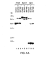
- genomic probes the isolated 70 kb from the telomeric cosmid, we detected rearranged restriction fragments in DNAs of two cell lines containing the t(2;5) ( Fig. 1A ) (Approximately 70 kb toward the breakpoint from the telomeric flanking clone, we isolated chromosome 5-specific genomic probes (p16-3/1.2S and p21-3/3E) that identified rearranged fragments with multiple enzymes in Southern blot analysis of DNAs from t(2;5)-positive cell lines.
- the genomic fragment p16-3/1.2S is located immediately centromeric to the chromosome 5 breakpoint, whereas p21-3/3E lies just telomeric to the break. Both probes identified a 1.6 kb transcript in Northern analysis of RNAs prepared from t(2;5)-positive and negative cell lines; in addition, p16-3/1.2S hybridized to a 2.4 kb transcript found only in t(2;5)-positive RNAs).
- One of the probes was hybridized to a cDNA library prepared from the polyadenylated RNA of the UOC-B1 pro-B leukemia cell line, which lacks the t(2;5).
- Multiple cDNA clones were isolated that hybridized to a ubiquitously expressed 1.6 kb mRNA, which was predicted by sequence analysis to encode nucleophosmin (NPM; also known as B23 or numatrin) - a highly conserved nucleolar phosphoprotein that shuttles ribosomal components between the nucleolus and the cytoplasm in the later stages of ribosome assembly ( W.Y. Chan et al., Biochemistry 28:1033 (1989 ); R.A.
- NPM nucleophosmin
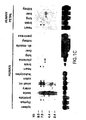
- RNAs prepared from cell lines with or without the t(2;5) identified both the normal NPM transcript and a 2.4 kb transcript restricted to t(2;5)-positive cell lines ( Fig. 1B , top).
- a subclone containing 3' untranslated sequences detected only the normal 1.6 kb NPM transcript (not shown).
- a probe prepared from the 3' end of the fusion cDNA identified the same 2.4 kb transcript that had been detected with the 5' NPM probe in RNAs from t(2;5)-positive cells ( Fig. 1B , bottom). This fragment was localized to band p23 of chromosome 2 by hybridization to DNAs of human x rodent somatic cell hybrids and by metaphase FISH analysis (not shown), indicating that the 2.4 kb mRNA is encoded by a fused gene created by the t(2;5).
- the 3' portion of the chimeric t(2;5) cDNA encodes conserved residues characteristic of the catalytic domain of members of the protein-tyrosine kinase (PTK) gene family ( S.K. Hanks et al., Science 241:42 (1988 ); S.S. Taylor, et al., Annu. Rev. Cell Biol. 8:429 (1992 )). ( Fig. 2 , A and C). Comparison of this newly identified a naplastic l ymphoma k inase (ALK) (NPM and ALK are approved HGM gene symbols.
- ALK naplastic l ymphoma k inase
- the structure of normal ALK proteins was determined by screening a cDNA library prepared from a rhabdomyosarcoma cell line (Rh30), using the pS1.2 ALK probe. Analysis of the inserts of the two largest clones, pRMS4 and pRMS17-2, revealed 3' ALK sequences identical to those in the fusion gene cDNA, indicating that mutations had not occurred in the chimeric protein. Sequences of ALK immediately upstream of the NPM-ALK junction encoded 23 hydrophobic amino acids typical of a transmembrane domain ( Fig.
- IBP-1 insulin-like growth factor binding protein-1
- IGF1 insulin-like growth factor-1
- ALK mRNAs of 6.5 kb and 8.0 kb were readily identified in small intestine and rhabdomyosarcoma cell lines, and were weakly expressed in brain (fetal and adult), colon and prostate ( Fig. 1 , B [bottom] and C). Abundant amounts of 4.4 kb and 6.0 kb mRNAs were detected in testis, whereas placenta and fetal liver each expressed a single 6.0 kb transcript. All four mRNAs were also detected with a probe that contains only 3' untranslated ALK sequences, suggesting that they represent differentially spliced ALK mRNAs, not cross-hybridizing transcripts of other PTK genes.
- ALK transcripts were not detected in normal spleen, thymus, peripheral blood leukocytes, B-lymphoblastoid cell lines, phytohemagglutinin-stimulated T lymphocytes or t(2;5)-negative leukemia/lymphoma cell lines of myeloid or B-or T-lymphoid derivation, implying that they are not normally expressed in hematopoietic cells.
- FISH mapping indicated that NPM and ALK are transcribed in centromeric to telomeric orientations on chromosomes 5 and 2, respectively, with the 2.4 kb fusion transcript arising from the derivative 5 translocated chromosome.
- Northern blot analysis provided no evidence for expression of a reciprocal ALK-NPM chimeric transcript, which could have been generated from the derivative 2 chromosome.
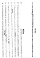
- RNA-based polymerase chain reaction confirmed the specificity of the fusion junctions in chimeric transcripts expressed in lymphomas harboring the t(2;5) ( Fig. 3 , A and B) (RNA-PCR was performed as previously described in E. Privitera et al ., Blood 79: 1781 (1992). Reactions were performed simultaneously with oligonucleotide primers specific for the chimeric NPM-ALK transcript (see Fig. 3B ) and with a primer pair derived from the ubiquitously expressed NPM gene as a control for reverse transcription and amplification.
- a 3' NPM primer (5'-GCTACCACCTCC AGGGGCAGA-3' (Seq. ID No.
- fusion transcripts were not detected in cell lines lacking the t(2;5), including several rhabdomyosarcoma lines that expressed ALK transcripts.
- NPM-ALK junction sequences were found in the RNAs of all seven t(2;5)-positive samples, including the SU-DHL-1, SUP-M2 and UCONN-L2 cell lines and diagnostic samples from four patients with anaplastic large cell lymphomas (The patient samples (three lymph node biopsies, one pleural effusion) were each shown by cytogenetic analysis to contain lymphoma cells bearing the t(2;5).
- RNA-PCR products from cells of patients 2 and 4 was determined and found to be identical to the cDNA sequence obtained from the SU-DHL-1 cell line ( Fig. 3B ).
- Written informed consent was obtained from the patients or their parents, and investigations were approved by the clinical trials review committee of St. Jude Children's Research Hospital).
- the breakpoints of the 2;5 transiocation therefore appear to consistently involve the same introns of the NPM and ALK genes, leading to identical junctions in spliced mRNAs arising from the fused gene. Because of the difficulties in cytogenetic analysis of lymphoma biopsy samples, molecular detection of NPM-ALK fusion mRNAs by RNA-PCR should markedly improve the identification of these tumors.
- the frequency of the t(2;5) in anaplastic large cell lymphomas indicates that the NPM-ALK product has a major role in the pathogenesis of these neoplasms.
- the normal NPM protein is a nonribosomal nucleolar phosphoprotein involved in the assembly of preribosomal particles into both small and large ribosomal subunits ( W.Y. Chan et al., Biochemistry 28:1033 (1989 ); R.A. Borer et al., Cell 56:379 (1989 ); M.S. Schmidt-Zachmann et al., EMBO J. 6:1881 (1987 ); M.S. Schmidt-Zachmann et al., Chromosoma.
- NPM transcripts and protein The relative abundance of NPM transcripts and protein is cell cycle regulated. NPM transcription and translation peak just prior to the entry of cells into S phase, with a decline to baseline just before the onset of G2 phase ( N. Feuerstein et al., J. Immunol. 139:1818 (1987 ); N.
- NPM-ALK amino-terminal NPM coding sequences incorporated into NPM-ALK, including those encoding potential protein kinase C phosphorylation sites (Ser 43 and Thr 78 ) and a potential C-X 5 -H-X- 4 H metal binding motif (residues 104-115), remains to be established.
- TRKA receptor tyrosine kinases
- NPM-ALK may phosphorylate substrates that are normally phosphorylated in response to IL-2 receptor-mediated signals ( E.M. Saltzman et al., J. Biol. Chem. 263:6956 (1988 ); D.K. Ferris et al., J. Immunol. 143:870 (1989 ); I.D.
- these enhancers which are highly active in T-cell progenitors, cause dysregulated expression of developmentally regulated transcription factor genes (e.g., TAL / SCL, LYL1, RHOMB / TTG and HOX11 ) located at breakpoints on the reciprocal chromosomes.
- developmentally regulated transcription factor genes e.g., TAL / SCL, LYL1, RHOMB / TTG and HOX11
Landscapes
- Chemical & Material Sciences (AREA)
- Health & Medical Sciences (AREA)
- Life Sciences & Earth Sciences (AREA)
- Organic Chemistry (AREA)
- Genetics & Genomics (AREA)
- Biochemistry (AREA)
- General Health & Medical Sciences (AREA)
- Medicinal Chemistry (AREA)
- Molecular Biology (AREA)
- Biophysics (AREA)
- Proteomics, Peptides & Aminoacids (AREA)
- Immunology (AREA)
- Zoology (AREA)
- Engineering & Computer Science (AREA)
- Wood Science & Technology (AREA)
- Bioinformatics & Cheminformatics (AREA)
- Toxicology (AREA)
- Cell Biology (AREA)
- Biomedical Technology (AREA)
- Biotechnology (AREA)
- Microbiology (AREA)
- Gastroenterology & Hepatology (AREA)
- General Engineering & Computer Science (AREA)
- Measuring Or Testing Involving Enzymes Or Micro-Organisms (AREA)
- Peptides Or Proteins (AREA)
- Investigating Or Analysing Biological Materials (AREA)
- Medicines That Contain Protein Lipid Enzymes And Other Medicines (AREA)
Claims (27)
- Verfahren zum Nachweisen eines menschlichen t(2;5) Lymphoms, welches die Schritte umfasst:(a) Inkontaktbringen einer Nukleinsäure enthaltenden Probe mit einem ersten Amplifikationsprimer und einem zweiten Amplifikationsprimer, wobei der erste Amplifikationsprimer unter stringenten Bedingungen mit NPM-Sequenzen, die in der NPM/ALK-Fusion vorliegen, oder einer hierzu komplementären Sequenz hybridisieren kann, und der zweite Amplifikationsprimer unter stringenten Bedingungen mit ALK-Sequenzen, die in der NPM/ALK-Fusion vorliegen, oder einer hierzu komplementären Sequenz hybridisieren kann;(b) Amplifizieren der mit Primerunterstützung erhaltenen Sequenzen mit dem ersten oder zweiten Amplifikationsprimer in der Probe;(c) Nachweisen amplifizierter Sequenzen, welche die NPM/ALK-Fusion enthalten.
- Verfahren nach Anspruch 1, wobei der erste oder zweite Amplifikationsprimer ausgewählt sind aus der Gruppe, bestehend aus SEQ ID NO 5 und der revers-komplementären Form zu SEQ ID NO 6.
- Verfahren nach Anspruch 1 oder Anspruch 2, wobei die Probe mRNA enthält.
- Verfahren zum Identifizieren eines menschlichen t(2;5) Lymphoms, welches die Schritte umfasst:(a) Inkontaktbringen einer Probe mit zwei Antikörpern, wobei ein erster Antikörper an NPM binden kann, und ein zweiter Antikörper an ALK binden kann; und(b) Nachweisen der Gegenwart von Proteinen in der Probe, die sowohl an den ersten, als auch an den zweiten Antikörper binden;
oder welches die Schritte umfasst:(i) Inkontaktbringen einer Probe mit einem Antikörper, wobei der Antikörper an die Fusionsverbindung des NPM/ALK-Fusionsproteins binden kann, das in der Aminosäuresequenz von SEQ ID NO 4 vorliegt, jedoch nicht mit dem Nicht-Fusions-NPM- oder ALK-Protein binden kann; und(ii) Nachweisen der Gegenwart von Proteinen in der Probe, die mit dem Antikörper binden. - Verfahren nach Schritt (i) und (ii) von Anspruch 4, wobei der Antikörper an ein Epitop binden kann, das in der Aminosäuresequenz von SEQ ID NO 4 vorliegt.
- Verfahren zum Nachweisen eines menschlichen t(2;5) Lymphoms, welches die Schritte umfasst:(a) Inkontaktbringen einer Probe mit zwei Nukleinsäure-Sonden (DNA oder RNA), wobei eine erste Nukleinsäuresonde unter stringenten Bedingungen mit für NPM kodierenden Nukleinsäuresequenzen hybridisieren kann, und eine zweite Nukleinsäuresonde unter stringenten Bedingungen mit für ALK kodierenden Nukleinsäuresequenzen hybridisieren kann; und(b) Nachweisen der Gegenwart von Nukleinsäure (DNA oder RNA) - Sequenzen in der Probe, die unter stringenten Bedingungen sowohl mit der ersten, als auch der zweiten Nukleinsäuresonde hybridisieren;
oder welches die Schritte umfasst:(i) Inkontaktbringen einer Probe mit einer Nukleinsäure (DNA oder RNA) - Sonde, wobei die Nukleinsäuresonde unter stringenten Bedingungen mit einer für die Fusionsverbindung des NPM/ALK-Fusionsproteins kodierenden Nukleinsäuresequenz hybridisieren kann; und(ii) Nachweisen der Gegenwart von Nukleinsäuresequenzen (DNA oder RNA) in der Probe, die unter stringenten Bedingungen mit der Nukleinsäuresonde hybridisieren. - Verfahren nach Schritt (i) und (ii) von Anspruch 6, wobei die Nukleinsäuresonde die Nukleinsäuresequenzen von SEQ ID NO 2 oder ein Fragment hiervon umfasst.
- Kit mit Unterteilungen zur Aufnahme von einem oder mehreren Behältern in strenger Eingrenzung, welcher umfasst:(a) einen ersten Behälter, welcher umfasst, einen ersten Amplifikationsprimer, der unter stringenten Bedingungen mit der für NPM kodierenden Nukleinsäuresequenz oder einer hierzu komplementären Nukleinsäuresequenz hybridisieren kann;(b) einen zweiten Behälter, welcher umfasst, einen zweiten Amplifikationsprimer, der unter stringenten Bedingungen mit der für ALK kodierenden Nukleinsäuresequenz oder einer hierzu komplementären Nukleinsäuresequenz hybridisieren kann;(c) einen oder mehrere Behälter, welche eines oder mehreres des Nachstehenden umfassen: Waschreagenzien, Amplifikationsreagenzien, und Reagenzien, welche
die Gegenwart von amplifizierten Nukleinsäuresequenzen nachweisen können; oder welcher umfasst:(i) einen ersten Behälter, welcher umfasst, einen Antikörper, der an ein Epitop binden kann, das in der Fusionsverbindung des NPM/ALK-Fusionsproteins vorliegt, jedoch nicht im Nicht-Fusions-NPM- oder ALK-Protein vorliegt; und(ii) einen oder mehrere andere Behälter, welche eines oder mehreres des Nachstehenden umfassen: Waschreagens und Reagenzien, welche die Gegenwart von gebundenem Antikörper nachweisen können;
oder welcher umfasst:(1) einen ersten Behälter, welcher umfasst, eine Nukleinsäuresonde, die unter stringenten Bedingungen mit einer für die Fusionsverbindung des NPM/ALK-Fusionsproteins kodierenden Nukleinsäuresequenz hybridisieren kann; und(2) einen oder mehrere andere Behälter, welche eines oder mehreres des Nachstehenden umfassen: Waschreagenzien und Reagenzien, welche die Gegenwart von gebundenen Nukleinsäuresonden des ersten Behälters nachweisen können. - Antikörper, der an ein Epitop binden kann, das in der Fusionsverbindung des NPM/ALK-Fusionsproteins vorliegt, jedoch nicht an ein Nicht-Fusions-NPM- oder ALK-Protein binden kann.
- Antikörper nach Anspruch 9, wobei der Antikörper an ein Epitop binden kann, das in der Aminosäuresequenz von SEQ ID NO 4 vorliegt.
- Antikörper, der an das ALK-Protein binden kann.
- Isoliertes Peptid, dessen Aminosäuresequenz ausgewählt ist aus der Gruppe, bestehend aus SEQ ID NO 3 (das partielle ALK-Protein), oder ein Fragment hiervon, und SEQ ID NO 4 (das NPM/ALK-Fusionsprotein), oder ein Fragment hiervon.
- Isoliertes Fusionsprotein, welches umfasst, einen Abschnitt des NPM und ALK-Proteins.
- Isoliertes Nukleinsäuremolekül:(i) welches aufweist, die Nukleotidsequenz von SEQ ID NO 1, oder ein Fragment hiervon mit der Nukleotidsequenz von 31 bis 2608 angrenzenden Basenpaaren von SEQ ID NO 1;(ii) welches aufweist, die Nukleotidsequenz von SEQ ID NO 2, oder ein Fragment hiervon mit der Nukleotidsequenz von 31 bis 2440 angrenzenden Basenpaaren von SEQ ID NO 2;(iii) welches ein Polypeptid kodiert, das die Sequenz von SEQ ID NO 3 aufweist; oder(iv) welches ein Polypeptid kodiert, das die Sequenz von SEQ ID NO 4 aufweist.
- Isoliertes, in Teil (ii) von Anspruch 14 beschriebenes Nukleinsäuremolekül, worin das isolierte Nukleinsäuremolekül eine NPM/ALK-Fusionsverbindung umfasst.
- Isoliertes Nukleinsäuremolekül nach Anspruch 14, worin das isolierte Nukleinsäuremolekül eine Sequenz aufweist, die ausgewählt ist aus der Gruppe, bestehend aus SEQ ID NO 9, SEQ. ID NO 10, der revers-komplementären Form zu SEQ ID NO 9 und der revers-komplementären Form zu SEQ ID NO 10.
- Amplifikationsprimer, der hybridisieren kann, mit einer Nukleinsäuresequenz, die ein Rearrangement bei dem t(2;5)(p23;q35) chromosomalen Translokationsereignis aufweist, das in einem menschlichen t(2;5) Lymphom auftritt, oder einer komplementären Sequenz hiervon.
- Amplifikationsprimer nach Anspruch 17, worin der Amplifikationsprimer aus 18 bis 30 angrenzenden Nukleotiden von SEQ ID NO 1 oder aus 18 bis 30 angrenzenden Nukleotiden der revers-komplementären Form zu SEQ ID NO 1 besteht.
- Amplifikationsprimer nach Anspruch 17, worin der Amplifikationsprimer eine Sequenz aufweist, die ausgewählt ist aus der Gruppe, bestehend aus SEQ ID NQ 5, SEQ ID NO 6, der revers-komplementären Form zu SEQ ID NO 5 und der revers-komplementären Form zu SEQ ID NO 6.
- Amplifikationsprimer, der hybridisieren kann, mit der Nukleinsäuresequenz, die das menschliche ALK-Gen auf dem Chromosom 2p23 kodiert, oder einer komplementären Sequenz hiervon.
- Amplifikationsprimer nach Anspruch 20, worin der Amplifikationsprimer aus 18 bis 30 angrenzenden Nukleotiden von SEQ ID NO 2 oder aus 18 bis 30 angrenzenden Nukleotiden der revers-komplementären Form zu SEQ ID NO 2 besteht.
- Vektorkonstrukt, welches einen Vektor umfasst, worin das Nukleinsäuremolekül nach einem der Ansprüchen 14 bis 16 inseriert worden ist.
- Wirt, der mit dem Vektor-Konstrukt nach Anspruch 22 transformiert ist.
- Antisense-mRNA-Sequenz, die mit einem NPM/ALK-Fusionsgen oder einem Fragment hiervon hybrisisieren kann.
- Nicht-menschliches, transgenes Säugetier, das verändert wurde, um ein menschliches NPM/ALK-Fusionsprotein oder Fragment hiervon zu exprimieren.
- Verfahren zum Identifizieren von Agenzien, die an das NPM/ALK-Fusionsprotein binden können, welches die Schritte umfasst:(a) Inkontaktbringen eines Agens mit dem NPM/ALK-Fusionsprotein; und(b) Bestimmen, ob das Agens mit dem ALK-Protein oder dem NPM/ALK-Fusionsprotein bindet.
- Ribozym, welches die das ALK-Protein kodierende mRNA spalten kann oder die das NPM/ALK-Fusionsprotein kodierende mRNA spalten kann.
Applications Claiming Priority (3)
| Application Number | Priority Date | Filing Date | Title |
|---|---|---|---|
| US160861 | 1993-12-03 | ||
| US08/160,861 US5529925A (en) | 1993-12-03 | 1993-12-03 | Nucleic acid sequences and fusion proteins present in human t(2;5) lymphoma |
| PCT/US1994/013947 WO1995015331A1 (en) | 1993-12-03 | 1994-12-05 | SPECIFIC FUSION NUCLEIC ACIDS AND PROTEINS PRESENT IN HUMAN t(2;5) LYMPHOMA, METHODS OF DETECTION AND USES THEREOF |
Publications (4)
| Publication Number | Publication Date |
|---|---|
| EP0731806A1 EP0731806A1 (de) | 1996-09-18 |
| EP0731806A4 EP0731806A4 (de) | 1998-12-16 |
| EP0731806B1 EP0731806B1 (de) | 2004-03-31 |
| EP0731806B2 true EP0731806B2 (de) | 2008-02-20 |
Family
ID=22578775
Family Applications (1)
| Application Number | Title | Priority Date | Filing Date |
|---|---|---|---|
| EP95906601A Expired - Lifetime EP0731806B2 (de) | 1993-12-03 | 1994-12-05 | SPEZIFISCHE FUSIONSNUKLEINSÄUREN UND PROTEINE IN HUMANEN t(2;5)LYMPHOMEN, VERFAHREN ZU IHREM NACHWEIS UND IHRE VERWENDUNG |
Country Status (7)
| Country | Link |
|---|---|
| US (4) | US5529925A (de) |
| EP (1) | EP0731806B2 (de) |
| JP (1) | JPH09512161A (de) |
| AU (1) | AU679833B2 (de) |
| CA (1) | CA2177957A1 (de) |
| DE (1) | DE69433665T3 (de) |
| WO (1) | WO1995015331A1 (de) |
Families Citing this family (72)
| Publication number | Priority date | Publication date | Assignee | Title |
|---|---|---|---|---|
| US6696548B2 (en) | 1993-12-03 | 2004-02-24 | St. Jude Children's Research Hospital | Antibodies for recognition of alk protein tyrosine/kinase receptor |
| US5529925A (en) | 1993-12-03 | 1996-06-25 | St. Jude Children's Research Hospital | Nucleic acid sequences and fusion proteins present in human t(2;5) lymphoma |
| US5635351A (en) * | 1995-03-14 | 1997-06-03 | The Regents Of The University Of California | Genetic gain and loss in gliomas |
| US20030165931A1 (en) * | 1998-03-11 | 2003-09-04 | Bruno Tocque | Qualitative differential screening |
| US6881571B1 (en) * | 1998-03-11 | 2005-04-19 | Exonhit Therapeutics S.A. | Qualitative differential screening |
| DE19610255B4 (de) * | 1996-03-15 | 2004-11-04 | Universität Heidelberg | Verfahren zur Herstellung von Nukleinsäuresequenzen und Verfahren zum Nachweis von Translokationen zwischen Chromosomen |
| US8048629B2 (en) * | 1996-03-15 | 2011-11-01 | The Penn State Research Foundation | Detection of extracellular tumor-associated nucleic acid in blood plasma or serum |
| JP2001505428A (ja) * | 1996-12-06 | 2001-04-24 | ニールズ パリスガード | 染色体異常の検出 |
| US5994522A (en) * | 1997-01-24 | 1999-11-30 | Washington University | BLNK proteins |
| US20030096255A1 (en) * | 1997-02-19 | 2003-05-22 | Felix Carolyn A. | Methods and kits for analysis of chromosomal rearrangements associated with cancer |
| EP0878552A1 (de) * | 1997-05-13 | 1998-11-18 | Erasmus Universiteit Rotterdam | Molekularer Nachweis von chromosomalen Veränderungen |
| US6849400B1 (en) * | 1997-07-23 | 2005-02-01 | Gen-Probe Incorporated | Methods for detecting and measuring spliced nucleic acids |
| AU3516899A (en) | 1998-05-04 | 1999-11-23 | Dako A/S | Method and probes for the detection of chromosome aberrations |
| FR2798392B1 (fr) * | 1999-09-13 | 2005-07-15 | Exonhit Therapeutics Sa | Marqueurs genetiques de la toxicite, preparation et utilisations |
| FR2798673B1 (fr) * | 1999-09-16 | 2004-05-28 | Exonhit Therapeutics Sa | Methodes et compositions pour la detection d'evenements pathologiques |
| AU6687601A (en) * | 2000-06-14 | 2001-12-24 | Univ Georgetown Med Center | Pleiotrophin growth factor receptor for the treatment of proliferative, vascularand neurological disorders |
| WO2002088309A2 (en) * | 2001-04-27 | 2002-11-07 | St. Jude Children's Research Hospital | Fusion genes associated with acute megakaryoblastic leukemias |
| US7214488B2 (en) * | 2001-07-03 | 2007-05-08 | United States Of America, Represented By The Secretary, Department Of Health And Human Services | Detection of MECT1-MAML2 fusion products |
| US20030157101A1 (en) * | 2001-11-15 | 2003-08-21 | Carlo Gambacorti-Passerini | Immunogenic ALK peptides |
| US7211240B2 (en) * | 2002-03-01 | 2007-05-01 | Bracco International B.V. | Multivalent constructs for therapeutic and diagnostic applications |
| EP1587944A4 (de) * | 2002-03-01 | 2007-03-21 | Dyax Corp | Kdr und vegf/kdr-bindende peptide und ihre diagnostische und therapeutische veranwendung bei der diagnose und therapie |
| US7261876B2 (en) | 2002-03-01 | 2007-08-28 | Bracco International Bv | Multivalent constructs for therapeutic and diagnostic applications |
| US8623822B2 (en) * | 2002-03-01 | 2014-01-07 | Bracco Suisse Sa | KDR and VEGF/KDR binding peptides and their use in diagnosis and therapy |
| US7794693B2 (en) * | 2002-03-01 | 2010-09-14 | Bracco International B.V. | Targeting vector-phospholipid conjugates |
| US7461843B1 (en) * | 2002-08-23 | 2008-12-09 | Elixir Gaming Technologies, Inc. | Automatic card shuffler |
| CA2509902A1 (en) * | 2002-12-13 | 2004-07-01 | Aurelium Biopharma Inc. | Nucleophosmin directed diagnostics and therapeutics for multidrug resistant neoplastic disease |
| PT1454992E (pt) * | 2003-03-07 | 2006-09-29 | Ist Naz Stud Cura Dei Tumori | Ensaio de quinase do linfoma anaplastico, reagentes e composicoes |
| US7888485B2 (en) * | 2003-03-26 | 2011-02-15 | Georgetown University | Anti-pleiotrophin antibodies and methods of use thereof |
| ITRM20040534A1 (it) * | 2004-10-29 | 2005-01-29 | Brunangelo Falini | Mutanti della proteina nucleofosmina (npm), sequenze geniche corrispondenti e relativi usi. |
| WO2007061684A1 (en) | 2005-11-18 | 2007-05-31 | Board Of Regents, The University Of Texas System | Quantification of fusion proteins and their activity from chromosomal translocation |
| US20120208824A1 (en) | 2006-01-20 | 2012-08-16 | Cell Signaling Technology, Inc. | ROS Kinase in Lung Cancer |
| EP2447359B1 (de) * | 2006-04-14 | 2015-11-04 | Cell Signaling Technology, Inc. | Gendefekte und mutierte ALK-Kinase in festen menschlichen Tumoren |
| US8168383B2 (en) | 2006-04-14 | 2012-05-01 | Cell Signaling Technology, Inc. | Gene defects and mutant ALK kinase in human solid tumors |
| AU2012201154B2 (en) * | 2006-04-14 | 2012-09-13 | Cell Signaling Technology, Inc. | Gene defects and mutant ALK kinase in human solid tumors |
| EP2013236B1 (de) * | 2006-04-28 | 2015-06-17 | Delenex Therapeutics AG | An die extrazelluläre domäne der rezeptortyrosinkinase alk bindende antikörper |
| JP5562640B2 (ja) * | 2007-04-13 | 2014-07-30 | セル・シグナリング・テクノロジー・インコーポレイテツド | ヒト固形腫瘍における遺伝子欠失及び突然変異alkキナーゼ |
| WO2009045965A2 (en) | 2007-10-01 | 2009-04-09 | The Trustees Of The University Of Pennsylvania | Stat5a and its functional tumor suppressor analogs for treatment of malignancies expressing npm/alk and other oncogenic kinases |
| JP2011511949A (ja) * | 2008-02-12 | 2011-04-14 | ザ・ブリガーム・アンド・ウーメンズ・ホスピタル・インコーポレーテッド | 肺癌におけるeml4とalkの融合のためのfishアッセイ |
| US8352194B1 (en) | 2008-06-17 | 2013-01-08 | University Of South Florida | Method to identify cancer fusion genes |
| US10236078B2 (en) | 2008-11-17 | 2019-03-19 | Veracyte, Inc. | Methods for processing or analyzing a sample of thyroid tissue |
| US9495515B1 (en) | 2009-12-09 | 2016-11-15 | Veracyte, Inc. | Algorithms for disease diagnostics |
| US9074258B2 (en) | 2009-03-04 | 2015-07-07 | Genomedx Biosciences Inc. | Compositions and methods for classifying thyroid nodule disease |
| US8669057B2 (en) | 2009-05-07 | 2014-03-11 | Veracyte, Inc. | Methods and compositions for diagnosis of thyroid conditions |
| MX338297B (es) * | 2009-05-15 | 2016-04-11 | Insight Genetics Inc | Metodos y composiciones relacionados con fusiones de alk para el diagnostico y tratamiento de cancer. |
| WO2010138460A1 (en) | 2009-05-26 | 2010-12-02 | Quest Diagnostics Investments Incorporated | Methods for detecting gene dysregulations |
| US20120202214A1 (en) * | 2009-10-06 | 2012-08-09 | Kazuya Omi | Method for detecting fusion gene |
| CN102713606A (zh) * | 2009-11-13 | 2012-10-03 | 无限制药股份有限公司 | 用于鉴定、评估、预防和治疗癌症的组合物、试剂盒和方法 |
| US10446272B2 (en) | 2009-12-09 | 2019-10-15 | Veracyte, Inc. | Methods and compositions for classification of samples |
| WO2011079191A1 (en) | 2009-12-23 | 2011-06-30 | Quest Diagnostics Investments Incorporated | Tmprss2 for the diagnosis of prostate disease |
| JP5822913B2 (ja) * | 2010-04-20 | 2015-11-25 | ヴェンタナ メディカル システムズ, インク. | 2色発色性インサイツハイブリダイゼーション |
| AU2015201135B2 (en) * | 2010-04-20 | 2017-02-02 | Ventana Medical Systems, Inc. | Two-color chromogenic in situ hybridization |
| US20120101108A1 (en) | 2010-08-06 | 2012-04-26 | Cell Signaling Technology, Inc. | Anaplastic Lymphoma Kinase In Kidney Cancer |
| US20140315943A1 (en) | 2011-05-24 | 2014-10-23 | Synta Pharmaceuticals Corp. | Combination therapy of hsp90 inhibitory compounds with mtor/p13k inhibitors |
| JP2014520808A (ja) | 2011-07-07 | 2014-08-25 | シンタ ファーマシューティカルズ コーポレーション | Hsp90阻害化合物を用いた癌の治療 |
| JP2014534228A (ja) | 2011-11-02 | 2014-12-18 | シンタ ファーマシューティカルズ コーポレーション | 白金含有剤とhsp90阻害剤の組合せ療法 |
| WO2013067162A1 (en) | 2011-11-02 | 2013-05-10 | Synta Pharmaceuticals Corp. | Cancer therapy using a combination of hsp90 inhibitors with topoisomerase i inhibitors |
| EP2780010A1 (de) | 2011-11-14 | 2014-09-24 | Synta Pharmaceuticals Corp. | Kombinationstherapie aus hsp90-inhibitoren mit braf-inhibitoren |
| ES2655842T3 (es) | 2012-04-18 | 2018-02-21 | Cell Signaling Technology, Inc. | EGFR y ROS1 en el cáncer |
| WO2013170182A1 (en) | 2012-05-11 | 2013-11-14 | Synta Pharmaceuticals Corp. | Treating cancer with an hsp90 inhibitory compound |
| KR20150118159A (ko) | 2013-02-22 | 2015-10-21 | 에프. 호프만-라 로슈 아게 | 암의 치료 방법 및 약물 내성의 예방 방법 |
| US11976329B2 (en) | 2013-03-15 | 2024-05-07 | Veracyte, Inc. | Methods and systems for detecting usual interstitial pneumonia |
| US9284594B2 (en) * | 2013-07-23 | 2016-03-15 | The Regents Of The University Of Michigan | Compositions and methods relating to fusion protein biomarkers |
| US10160993B2 (en) * | 2014-03-21 | 2018-12-25 | Purdue Research Foundation | Tyrosine kinase biosensors and methods of use |
| CA2943636C (en) | 2014-03-25 | 2020-06-09 | Quest Diagnostics Investments Incorporated | Detection of gene fusions by intragenic differential expression (ide) using average cycle thresholds |
| MY186570A (en) | 2014-05-13 | 2021-07-27 | Novartis Ag | Compounds and compositions for inducing chondrogenesis |
| US10246750B2 (en) * | 2014-06-10 | 2019-04-02 | Blueprint Medicines Corporation | Method for detection of a TECR:PKN1 or an ANXA4:PKN1 gene fusion |
| US12297505B2 (en) | 2014-07-14 | 2025-05-13 | Veracyte, Inc. | Algorithms for disease diagnostics |
| CN114606309A (zh) | 2014-11-05 | 2022-06-10 | 威拉赛特公司 | 使用机器学习和高维转录数据的诊断系统和方法 |
| BR112017016973A2 (pt) | 2015-02-09 | 2018-04-03 | Synta Pharmaceuticals Corp. | terapia de combinação de inibidores de hsp90 e inibidores de pd-1 para tratamento de câncer |
| WO2017065959A2 (en) * | 2015-09-25 | 2017-04-20 | Veracyte, Inc. | Methods and compositions that utilize transcriptome sequencing data in machine learning-based classification |
| CA3048420C (en) * | 2016-12-09 | 2025-05-20 | Ncan Genomics, Inc. | Bound ligature |
| US11217329B1 (en) | 2017-06-23 | 2022-01-04 | Veracyte, Inc. | Methods and systems for determining biological sample integrity |
Family Cites Families (4)
| Publication number | Priority date | Publication date | Assignee | Title |
|---|---|---|---|---|
| US4873191A (en) * | 1981-06-12 | 1989-10-10 | Ohio University | Genetic transformation of zygotes |
| US5015568A (en) * | 1986-07-09 | 1991-05-14 | The Wistar Institute | Diagnostic methods for detecting lymphomas in humans |
| AU630001B2 (en) * | 1989-10-02 | 1992-10-15 | United States of America, as represented by the Secretary, U.S. Department of Commerce, The | A sensitive method for measurement of chimeric transcripts of dna containing translocations |
| US5529925A (en) | 1993-12-03 | 1996-06-25 | St. Jude Children's Research Hospital | Nucleic acid sequences and fusion proteins present in human t(2;5) lymphoma |
-
1993
- 1993-12-03 US US08/160,861 patent/US5529925A/en not_active Expired - Lifetime
-
1994
- 1994-12-05 AU AU15116/95A patent/AU679833B2/en not_active Expired
- 1994-12-05 EP EP95906601A patent/EP0731806B2/de not_active Expired - Lifetime
- 1994-12-05 WO PCT/US1994/013947 patent/WO1995015331A1/en not_active Ceased
- 1994-12-05 DE DE69433665T patent/DE69433665T3/de not_active Expired - Lifetime
- 1994-12-05 JP JP7515804A patent/JPH09512161A/ja active Pending
- 1994-12-05 CA CA002177957A patent/CA2177957A1/en not_active Abandoned
-
1995
- 1995-10-12 US US08/542,363 patent/US5770421A/en not_active Expired - Lifetime
-
1998
- 1998-06-19 US US09/100,089 patent/US6174674B1/en not_active Expired - Lifetime
-
2000
- 2000-09-28 US US09/670,827 patent/US6451997B1/en not_active Expired - Lifetime
Non-Patent Citations (7)
| Title |
|---|
| A M Mes-Masson et a. Heamatology and Blood Transfusion, vol. 21, 163-166 † |
| ATCC Klon Datenblatt, ATCC Nummer 69497, 1-3 † |
| E Harlow et al. Antibodies a Laboratory Manual (1988), chapter 5 and 6 † |
| E S Kawasaki et al. PNAS USA, VOL.85, 5698-5702 † |
| J van Denderen et al. J Exp Med, vo. 169, 87-98 † |
| K I Mills et al. Blood, vol 78, 1155-1161 † |
| T H Rabbits et al. Cell, vol.67, 641-644 † |
Also Published As
| Publication number | Publication date |
|---|---|
| EP0731806A1 (de) | 1996-09-18 |
| EP0731806B1 (de) | 2004-03-31 |
| US5529925A (en) | 1996-06-25 |
| CA2177957A1 (en) | 1995-06-08 |
| WO1995015331A1 (en) | 1995-06-08 |
| DE69433665T2 (de) | 2005-02-10 |
| DE69433665D1 (de) | 2004-05-06 |
| EP0731806A4 (de) | 1998-12-16 |
| US6451997B1 (en) | 2002-09-17 |
| US6174674B1 (en) | 2001-01-16 |
| US5770421A (en) | 1998-06-23 |
| JPH09512161A (ja) | 1997-12-09 |
| DE69433665T3 (de) | 2008-06-26 |
| AU1511695A (en) | 1995-06-19 |
| AU679833B2 (en) | 1997-07-10 |
Similar Documents
| Publication | Publication Date | Title |
|---|---|---|
| EP0731806B1 (de) | SPEZIFISCHE FUSIONSNUKLEINSÄUREN UND PROTEINE IN HUMANEN t(2;5)LYMPHOMEN, VERFAHREN ZU IHREM NACHWEIS UND IHRE VERWENDUNG | |
| US5589579A (en) | Gene sequence and probe for a marker of non-small cell lung carinoma | |
| Coussens et al. | Tyrosine kinase receptor with extensive homology to EGF receptor shares chromosomal location with neu oncogene | |
| JPH06502176A (ja) | 新しいオンコフェータル遺伝子、そのための遺伝子生成物およびその利用法 | |
| WO1996024603A9 (en) | InK4c-p18 AND InK4D-p19, INHIBITORS OF CYCLIN-DEPEDENT KINASES CDK4 AND CDK6, AND USES THEREOF | |
| FI108037B (fi) | Menetelmä T-lymfasoluproteiinin valmistamiseksi, menetelmässä käytettävä ilmentämisväline, vektori ja isäntäsolu | |
| US20010021505A1 (en) | ALK protein tyrosine kinase/receptor and ligands thereof | |
| HK1006726B (en) | T-CELL LYMPHOMA cDNA CLONES | |
| US6429302B1 (en) | Polynucleotides related to pancreatic disease | |
| Terrinoni et al. | Cyclin D1 gene contains a cryptic promoter that is functional in human cancer cells | |
| Kuss et al. | ARA, a novel ABC transporter, is located at 16p13. 1, is deleted in inv (16) leukemias, and is shown to be expressed in primitive hematopoietic precursors | |
| US5693778A (en) | Arg a human gene related to but distinct from abl proto-oncogene | |
| CA2167858A1 (en) | Sequence of af1q cdna | |
| US6908750B2 (en) | Gene encoding HM1.24 antigen protein and promoter thereof | |
| WO1999035159A1 (en) | Lymphoma/leukemia oncogene, oncoprotein and methods of use | |
| US6982317B2 (en) | C21 polypeptide that modulates the stability of transcriptional regulatory complexes regulating nuclear hormone receptor activity | |
| US20030040029A1 (en) | Detection of tumor marker transcript and protein recognized by naive natural killer cells | |
| US7108994B2 (en) | Receptor-type phosphotyrosine phosphatase-alpha | |
| CA2183900A1 (en) | Agents for the pre-symptomatic detection, prevention and treatment of breast cancer in humans | |
| EP1356040B1 (de) | 14189, eine humane kinase und deren verwendungen | |
| AU751385B2 (en) | Compositions for the diagnosis, prevention, and treatment of tumor progression | |
| Rettig et al. | Somatic cell genetic analysis of human cell surface antigens 5.1 H11 and F359 (gp45) | |
| WO1995015386A1 (en) | Novel receptor tyrosine kinase |
Legal Events
| Date | Code | Title | Description |
|---|---|---|---|
| PUAI | Public reference made under article 153(3) epc to a published international application that has entered the european phase |
Free format text: ORIGINAL CODE: 0009012 |
|
| 17P | Request for examination filed |
Effective date: 19960628 |
|
| AK | Designated contracting states |
Kind code of ref document: A1 Designated state(s): DE FR GB IT |
|
| RHK1 | Main classification (correction) |
Ipc: C12N 15/62 |
|
| A4 | Supplementary search report drawn up and despatched |
Effective date: 19981102 |
|
| AK | Designated contracting states |
Kind code of ref document: A4 Designated state(s): DE FR GB IT |
|
| 17Q | First examination report despatched |
Effective date: 20020201 |
|
| GRAP | Despatch of communication of intention to grant a patent |
Free format text: ORIGINAL CODE: EPIDOSNIGR1 |
|
| GRAS | Grant fee paid |
Free format text: ORIGINAL CODE: EPIDOSNIGR3 |
|
| GRAA | (expected) grant |
Free format text: ORIGINAL CODE: 0009210 |
|
| AK | Designated contracting states |
Kind code of ref document: B1 Designated state(s): DE FR GB IT |
|
| REG | Reference to a national code |
Ref country code: GB Ref legal event code: FG4D |
|
| REF | Corresponds to: |
Ref document number: 69433665 Country of ref document: DE Date of ref document: 20040506 Kind code of ref document: P |
|
| ET | Fr: translation filed | ||
| PLBI | Opposition filed |
Free format text: ORIGINAL CODE: 0009260 |
|
| PLAX | Notice of opposition and request to file observation + time limit sent |
Free format text: ORIGINAL CODE: EPIDOSNOBS2 |
|
| 26 | Opposition filed |
Opponent name: MERCK PATENT GMBH Effective date: 20041230 |
|
| PLBB | Reply of patent proprietor to notice(s) of opposition received |
Free format text: ORIGINAL CODE: EPIDOSNOBS3 |
|
| PUAH | Patent maintained in amended form |
Free format text: ORIGINAL CODE: 0009272 |
|
| STAA | Information on the status of an ep patent application or granted ep patent |
Free format text: STATUS: PATENT MAINTAINED AS AMENDED |
|
| 27A | Patent maintained in amended form |
Effective date: 20080220 |
|
| AK | Designated contracting states |
Kind code of ref document: B2 Designated state(s): DE FR GB IT |
|
| ET3 | Fr: translation filed ** decision concerning opposition | ||
| PGFP | Annual fee paid to national office [announced via postgrant information from national office to epo] |
Ref country code: DE Payment date: 20131230 Year of fee payment: 20 Ref country code: GB Payment date: 20131227 Year of fee payment: 20 |
|
| PGFP | Annual fee paid to national office [announced via postgrant information from national office to epo] |
Ref country code: IT Payment date: 20131223 Year of fee payment: 20 Ref country code: FR Payment date: 20131217 Year of fee payment: 20 |
|
| REG | Reference to a national code |
Ref country code: DE Ref legal event code: R071 Ref document number: 69433665 Country of ref document: DE |
|
| REG | Reference to a national code |
Ref country code: DE Ref legal event code: R071 Ref document number: 69433665 Country of ref document: DE |
|
| REG | Reference to a national code |
Ref country code: GB Ref legal event code: PE20 Expiry date: 20141204 |
|
| PG25 | Lapsed in a contracting state [announced via postgrant information from national office to epo] |
Ref country code: GB Free format text: LAPSE BECAUSE OF EXPIRATION OF PROTECTION Effective date: 20141204 |