EP0699768B1 - Procédés pour la extinction de la fluorescence en phase solution de sondes oligonucléotidiques marqués avec un fluorophore - Google Patents
Procédés pour la extinction de la fluorescence en phase solution de sondes oligonucléotidiques marqués avec un fluorophore Download PDFInfo
- Publication number
- EP0699768B1 EP0699768B1 EP95113012A EP95113012A EP0699768B1 EP 0699768 B1 EP0699768 B1 EP 0699768B1 EP 95113012 A EP95113012 A EP 95113012A EP 95113012 A EP95113012 A EP 95113012A EP 0699768 B1 EP0699768 B1 EP 0699768B1
- Authority
- EP
- European Patent Office
- Prior art keywords
- oligonucleotide
- label
- light emission
- probe
- nucleic acid
- Prior art date
- Legal status (The legal status is an assumption and is not a legal conclusion. Google has not performed a legal analysis and makes no representation as to the accuracy of the status listed.)
- Expired - Lifetime
Links
Images

Classifications
-
- C—CHEMISTRY; METALLURGY
- C12—BIOCHEMISTRY; BEER; SPIRITS; WINE; VINEGAR; MICROBIOLOGY; ENZYMOLOGY; MUTATION OR GENETIC ENGINEERING
- C12Q—MEASURING OR TESTING PROCESSES INVOLVING ENZYMES, NUCLEIC ACIDS OR MICROORGANISMS; COMPOSITIONS OR TEST PAPERS THEREFOR; PROCESSES OF PREPARING SUCH COMPOSITIONS; CONDITION-RESPONSIVE CONTROL IN MICROBIOLOGICAL OR ENZYMOLOGICAL PROCESSES
- C12Q1/00—Measuring or testing processes involving enzymes, nucleic acids or microorganisms; Compositions therefor; Processes of preparing such compositions
- C12Q1/68—Measuring or testing processes involving enzymes, nucleic acids or microorganisms; Compositions therefor; Processes of preparing such compositions involving nucleic acids
- C12Q1/6813—Hybridisation assays
- C12Q1/6816—Hybridisation assays characterised by the detection means
- C12Q1/6818—Hybridisation assays characterised by the detection means involving interaction of two or more labels, e.g. resonant energy transfer
-
- C—CHEMISTRY; METALLURGY
- C12—BIOCHEMISTRY; BEER; SPIRITS; WINE; VINEGAR; MICROBIOLOGY; ENZYMOLOGY; MUTATION OR GENETIC ENGINEERING
- C12Q—MEASURING OR TESTING PROCESSES INVOLVING ENZYMES, NUCLEIC ACIDS OR MICROORGANISMS; COMPOSITIONS OR TEST PAPERS THEREFOR; PROCESSES OF PREPARING SUCH COMPOSITIONS; CONDITION-RESPONSIVE CONTROL IN MICROBIOLOGICAL OR ENZYMOLOGICAL PROCESSES
- C12Q1/00—Measuring or testing processes involving enzymes, nucleic acids or microorganisms; Compositions therefor; Processes of preparing such compositions
- C12Q1/68—Measuring or testing processes involving enzymes, nucleic acids or microorganisms; Compositions therefor; Processes of preparing such compositions involving nucleic acids
- C12Q1/6813—Hybridisation assays
- C12Q1/6816—Hybridisation assays characterised by the detection means
- C12Q1/6823—Release of bound markers
Definitions
- This invention relates to methods for modifying the light emission of oligonucleotides labeled with a light-emitting label in solution using a DNA binding chromophore. Additionally, it relates to methods for detecting degradation of single-stranded oligonucleotides labeled with a light-emitting label in solution. Additionally, the invention relates to methods for detecting nucleic acid sequences by hybridization with a complementary oligonucleotide probe.
- Nucleic acid detection using oligonucleotide probes has become a standard method for specific target detection. Numerous modifications of the method have been described. Generally, a DNA sample is immobilized on a solid support and then hybridized to a labeled target-specific probe (see, for example, Falkow et al., U.S. Patent No. 4,358,535).
- RNA probes are used to detect DNA target sequences.
- RNA probes hybridized to DNA target are cleaved using RNaseH, which selectively cleaves RNA in RNA-DNA hybrid duplexes.
- RNaseH selectively cleaves RNA in RNA-DNA hybrid duplexes.
- U.S. Patent No. 5,210,015 describes methods which use the 5' to 3' exonuclease activity of a nucleic acid polymerase to cleave probes hybridized to target sequences and thereby release labeled oligonucleotide fragments for detection. These methods require an additional oligonucleotide hybridized upstream of the probe hybridization site to act as a primer for the polymerase-mediated extension reaction. Probe cleavage occurs concomitant with primer extension.
- PCR polymerase chain reaction
- segments of single copy genomic DNA can be selectively amplified to an easily detectable level prior to detection.
- PCR methods are disclosed in U.S. Patent No. 4,683,202.
- PCR and methods for detecting PCR products using an oligonucleotide probe capable of hybridizing with the amplified target nucleic acid are described in U.S. Patent No. 4,683,195, and European Patent Publication No. 237,362.
- Probes that hybridize to a region of the target nucleic acid bounded by the amplification primers are incorporated into the amplification reaction mixture.
- Hybridized probes are cleaved by the 5' to 3' nuclease activity of the polymerase during primer extension. Detection of labeled fragments indicates the occurrence of both primer extension and probe hybridization, and, therefore, amplification of the specific target sequence.
- a number of agents have been described for labeling nucleic acids, whether probe or target, for facilitating detection of target nucleic acid.
- Labels have been described that provide signals detectable by fluorescence, radioactivity, colorimetry, X-ray diffraction or absorption, magnetism, and enzymatic activity and include, for example, fluorophores, chromophores, radioactive isotopes (particularly 32 P and 125 I), electron-dense reagents, enzymes, and ligands having specific binding partners.
- Labeling can be achieved by a number of means, such as chemical modification of a primer or probe to incorporate a label or the use of polymerizing agents to incorporate a modified nucleoside triphosphate into an extension product.
- Ethidium bromide is an intercalating compound that displays increased fluorescence when bound to double-stranded DNA rather than when in free solution (Sharp et al., 1973, Biochemistry 12 :3055).
- EtBr can be used to detect both single- and double-stranded nucleic acids, the affinity of EtBr for single-stranded nucleic acid is relatively low.
- EtBr is routinely used to non-specifically detect nucleic acids following gel electrophoresis. Following size fractionation on an appropriate gel matrix, for example, agarose or acrylamide, the gel is soaked in a dilute solution of EtBr. The DNA is then visualized by examining the gel under UV light (see Maniatis et al., 1982 eds., Molecular Cloning: A Laboratory Manual, New York, Cold Spring Harbor Laboratory).
- a homogeneous assay for PCR and concurrent PCR product detection based on the increased fluorescence that EtBr and other DNA binding labels exhibit when bound to double-stranded DNA is described in Higuchi et al., 1992, Bio/Techniques 10 :413-417; Higuchi et al., 1993, Bio/Techniques 11 :1026-1030; and European Patent Publication Nos. 487,218 and 512,334.
- the methods allow direct detection of the increase of double-stranded DNA during an amplification reaction, most significantly from the increase in amplified target. However, these methods detect only the total amount of double-stranded DNA in the reaction and do not distinguish specific nucleic acid sequences; assay specificity depends on the specificity of the amplification reaction.
- oligonucleotide probes labeled with interacting fluorescent labels in nucleic acid hybridization assays is described in Morrison, 1992, in Nonisotopic DNA Probe Techniques, Kricka, ed., Academic Press, Inc., San Diego, CA, chapter 13; and Heller and Morrison, 1985, in Rapid Detection and Identification of Infections Agents, Academic Press, Inc., San Diego, CA, pages 245-256.
- the methods rely on the change in fluorescence that occurs when suitable fluorescent labels are brought into close proximity, described in the literature as fluorescence energy transfer (FET), fluorescence resonance energy transfer, nonradiative energy transfer, long-range energy transfer, dipole-coupled energy transfer, or Förster energy transfer.
- FET fluorescence energy transfer
- fluorescence resonance energy transfer fluorescence resonance energy transfer
- nonradiative energy transfer nonradiative energy transfer
- long-range energy transfer long-range energy transfer
- dipole-coupled energy transfer dipole-coupled energy transfer
- Förster energy transfer A number
- a probe is used which is labeled with interacting fluorescent labels in close proximity.
- the labels are attached to the probe separated by one or more nucleotides such that probe degradation during amplification separates the labels, thereby producing a detectable change in fluorescence.
- Such multiply-labeled probes are difficult and costly to synthesize.
- the present invention provides methods for modifying the light emission of a oligonucleotide probe labeled with a light-emitting label in solution using a DNA binding chromophore which can interact with the label to modify the light emission of the label and wherein said single-stranded oligonucleotide is not hybridized to its complementary strand.
- the present invention also provides methods for detecting degradation of oligonucleotides in solution.
- the oligonucleotides are labeled with a light-emitting label.
- Oligonucleotide cleavage is carried out in the presence of a DNA binding chromophore that can interact with the label to modify the light emission of the label.
- Oligonucleotide degradation is detected by measuring the resulting change in light emission of the label.
- the present invention provides conditions under which significant in-solution quenching by a DNA binding chromophore of a light-emitting bound to a oligonucleotide occurs. This quenching occurs without hybridization of the labeled oligonucleotide to its complementary sequence.
- the methods of the present invention utilize the dependence of this quenching on the length of the labeled oligonucleotide.
- the quenching of a light-emitting label bound to a short oligonucleotide is detectably less than the quenching of the light-emitting label bound to a longer oligonucleotide.
- quenching by fluorescence energy transfer requires that the interacting labels be in close proximity, and that the two molecules in solution are not maintained in close enough proximity to cause significant quenching.
- the intercalating quenchers described in the prior art do not bind single-stranded DNA significantly, and, therefore, no appreciable quenching of a label bound to a single-stranded DNA in solution was expected.
- the present invention relies on the quenching of a fluorescent label bound to a single-stranded nucleic acid by a DNA binding chromophore that occurs in solution.
- the present invention further provides improved methods for detecting a target nucleic acid in a sample by hybridization to an oligonucleotide probe.
- the methods rely on the selective cleavage of probes hybridized to target nucleic acid. Detection of cleaved probes using the methods of the present invention indicates the presence of target nucleic acid.
- the present invention provides a method for detecting a target nucleic acid in a sample, wherein the method comprises:
- the selective cleavage of probes hybridized to target nucleic acid can be achieved by any of a number of known methods. Examples of suitable reactions that selectively cleave probes hybridized to a target sequence are described above in Saiki et al., 1985, supra; Patent Publication No. WO 89/09284; and U.S. Patent No. 5,210,015.
- the methods of the present invention for detecting nucleic acids are particularly suited for use in conjunction with amplification processes.
- the target sequence is amplified prior to step (c).
- the present invention provides improvements to the homogeneous PCR amplification and PCR product detection assay described in U.S. Patent No. 5,210,015, that use a single nucleic acid polymerase both for primer extension and for cleavage of hybridized labeled probes.
- the improvements provided by the present invention allow the use of a probe labeled with a single light-emitting label without requiring post-reaction manipulations to separate cleaved and uncleaved probes.
- the present invention provides a method for detecting a target nucleic acid sequence in a sample using PCR, wherein the method comprises:
- the DNA binding chromophore provided in the reaction mixture is characterized as providing a detectable signal when bound to double-stranded DNA, which signal is greater than the amount of said signal provided by said compound when it is unbound, and the signal of the DNA binding chromophore is monitored in order to measure the total increase in double-stranded DNA resulting from the amplification process.
- the DNA binding chromophore functions both as a quencher of unbound probe light emission and as a signal-generating compound as used in the methods described in Higuchi et al ., 1992, supra .
- the change in signal generated by the DNA binding compound indicates that amplification has taken place, and the change in light emission of the probe label indicates amplification of the specific target sequence.
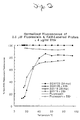
- Figure 1 relates to the dependence of in-solution fluorescent quenching on the length of the oligonucleotide to which the fluorophore is bound.
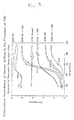
- Figure 2 relates to the dependence of in-solution fluorescent quenching on temperature.
- Figure 3 relates to the dependence of in-solution fluorescent quenching on temperature and the augmentation of quenching observed resulting from the presence of a hairpin secondary structure within the labeled single-stranded oligonucleotide.
- nucleic acid and oligonucleotide refer to probes and oligomer fragments to be detected, and shall be generic to polydeoxyribonucleotides (containing 2-deoxy-D-ribose), to polyribonucleotides (containing D-ribose), and to any other type of polynucleotide which is an N glycoside of a purine or pyrimidine base, or modified purine or pyrimidine base.
- nucleic acid and oligonucleotide will be used interchangeably. These terms refer only to the primary structure of the molecule. Thus, these terms include double- and single-stranded DNA, was well as double- and single-stranded RNA.
- target region refers to a region of a nucleic acid which is to be detected.
- probe refers to an oligonucleotide, typically labeled, that forms a duplex structure with a sequence of a target nucleic acid due to complementary base pairing.
- the probe will comprise a "hybridizing region", preferably consisting of 10 to 50 nucleotides, more preferably 20 to 30 nucleotides, corresponding to a region of the target sequence. "Corresponding” means identical to or complementary to the designated nucleic acid.
- probe oligonucleotides are labeled with, i.e., bound to, a fluorescent label to enable detection.
- hybridization refers the formation of a duplex structure by two single-stranded nucleic acids due to complementary base pairing. Hybridization can occur between fully complementary nucleic acid strands or between nucleic acid strands that contain minor regions of mismatch. Conditions under which only fully complementary nucleic acid strands will hybridize are referred to as "stringent hybridization conditions". Two single-stranded nucleic acids that are complementary except for minor regions of mismatch are referred to as “substantially complementary”. Stable duplexes of substantially complementary sequences can be achieved under less stringent hybridization conditions. Those skilled in the art of nucleic acid technology can determine duplex stability empirically considering a number of variables including, for example, the length and base pair concentration of the oligonucleotides, ionic strength, and incidence of mismatched base pairs.
- sequence-specific oligonucleotide and “SSO” refer to oligonucleotide probes wherein the hybridizing region is exactly complementary to the sequence to be detected.
- stringent hybridization conditions under which the probe will hybridize only to that exactly complementary target sequence allows the detection of the specific target sequence.
- Stringent hybridization conditions are well known in the art (see, e.g., Sambrook et al., 1985, Molecular Cloning - A Laboratory Manual, Cold Spring Harbor Laboratory, Cold Spring Harbor, New York). Stringent conditions are sequence dependent and will be different in different circumstances. Generally, stringent conditions are selected to be about 5°C lower than the thermal melting point (Tm) for the specific sequence at a defined ionic strength and pH.
- the Tm is the temperature (under defined ionic strength and pH) at which 50% of the base pairs have dissociated. Relaxing the stringency of the hybridizing conditions will allow sequence mismatches to be tolerated; the degree of mismatch tolerated can be controlled by suitable adjustment of the hybridization conditions.
- sequence refers herein to a nucleotide sequence contained within another sequence.
- label refers to any atom or molecule which can be attached to a nucleic acid, and which can be used either to provide a detectable signal or to interact with a second label to modify the detectable signal provided by the second label.
- Preferred labels are light-emitting compounds which generate a detectable signal by fluorescence, chemiluminescence, or bioluminescence.
- chromophore refers to a non-radioactive compound that absorbs energy in the form of light. Some chromophores can be excited to emit light either by a chemical reaction, producing chemiluminescence, or by the absorption of light, producing fluorescence.
- fluorophore refers to a compound which is capable of fluorescing, i.e. absorbing light at one frequency and emitting light at another, generally lower, frequency.
- bioluminescence refers to a form of chemiluminescence in which the light-emitting compound is one that is found in living organisms.
- bioluminescent compounds include bacterial luciferase and firefly luciferase.
- quenching refers to a decrease in fluorescence of a first compound caused by a second compound, regardless of the mechanism. Quenching typically requires that the compounds be in close proximity. As used herein, either the compound or the fluorescence of the compound is said to be quenched, and it is understood that both usages refer to the same phenomenon.
- intercalator refers to an agent or moiety capable of non-covalent insertion between stacked base pairs in a nucleic acid double helix.
- homogeneous refers to methods for carrying out the steps of the process, wherein the need for sample handling and manipulation between steps is minimized or eliminated.
- a “homogeneous” amplification/detection assay refers to a coupled amplification and detection assay wherein the need for sample handling and manipulation between the amplification and detection is minimized or eliminated.
- reaction mixture refers to a solution containing reagents necessary to carry out the reaction.
- An “amplification reaction mixture” which refers to a solution containing reagents necessary to carry out an amplification reaction, typically contains oligonucleotide primers and a DNA polymerase in a suitable buffer. Reaction mixtures for specific reactions are well-known in the literature.
- the present invention provides methods for modifying the light emission of an oligonucleotide label with a light-emitting label in solution.
- the methods of the invention are applicable to the detection of cleavage of single-stranded oligonucleotides labeled with a single light-emitting label. Detection of the cleaved oligonucleotide is carried out in a solution containing a DNA binding chromophore that can interact with the label to decrease the light emission of the label.
- the change in the length of the labeled oligonucleotide from cleavage results in a detectable increase in the light emission of the attached label.
- Suitable light-emitting labels and DNA binding compounds that can interact to modify the light emission of the label are described below.
- FET fluorescence energy transfer
- the emission of fluorescent label bound to the single-stranded oligonucleotide is detected.
- a DNA binding chromophore quenches the label fluorescence to a degree that depends on the length of the attached oligonucleotide.
- FET quenching is well known, both the occurrence of in-solution quenching by a DNA binding chromophore of a fluorescent label bound to a single-stranded oligonucleotide and the dependence of the quenching on the length of the oligonucleotide are unexpected in view of the prior art. Because of the extremely rapid decrease in interaction of fluorescent labels with increasing distance, it was believed that labels in solution do not significantly interact.
- the prior art does not report significant in-solution quenching of the unhybridized probe.
- the present invention provides conditions under which significant quenching of a fluorescent label bound to a single-stranded nucleic acid by a DNA binding chromophore occurs in solution.
- fluorophores and DNA-binding chromophores described in the art are suitable for use in the methods of the present invention.
- Suitable fluorophore and DNA-binding chromophore pairs are chosen such that the emission spectrum of the fluorophore overlaps with the absorption spectrum of the chromophore.
- the fluorophore should have a high Stokes shift (a large difference between the wavelength for maximum absorption and the wavelength for maximum emission) to minimize interference by scattered excitation light.
- Suitable labels which are well known in the art include, but are not limited to, fluoroscein and derivatives such as FAMTM, HEXTM, TETTM, and JOETM; rhodamine and derivatives such as Texas Red, ROXTM, and TAMRATM; Lucifer Yellow, and coumarin derivatives such as 7-Me 2 N-coumarin-4-acetate, 7-OH-4-CH 3 -coumarin-3-acetate, and 7-NH 2 -4-CH 3 -coumarin-3-acetate (AMCA).
- FAMTM, HEXTM, TETTM, JOETM, ROXTM, and TAMRATM are marketed by Perkin Elmer, Applied Biosystems Division (Foster City, CA).
- Texas Red and many other suitable compounds are marketed by Molecular Probes (Eugene, OR).
- chemiluminescent and bioluminescent compounds that may be suitable for use as the energy donor include luminol (aminophthalhydrazide) and derivatives, and Luciferases.
- the DNA binding agent is an intercalating agent.
- Suitable well-known intercalating agents include ethidium bromide and acridine orange.
- Non-intercalating DNA binding agents are also suitable.
- members of a class of DNA-binding compounds commonly referred to as "groove binders” are suitable. These compounds recognize and bind the minor groove of duplex DNA. Malachite Green is an example of this class of compounds that was demonstrated to function in the present methods.
- the DNA binding chromophore also provides a signal which is detectably altered upon intercalation into double-stranded DNA.
- Ethidium bromide like other DNA binding labels, such as acridines, proflavine, acridine orange, acriflavine, fluorcoumarin, ellipticine, daunomycin, chloroquine, distamycin D, chromomycin, homidium, mithramycin, ruthenium polypyridyls, and anthramycin, exhibits altered fluorescence emissions when bound to double-stranded DNA.
- a DNA binding compound which does not inhibit an amplification reaction is used to allow monitoring of the accumulation of amplified sequences.
- An oligonucleotide can be prepared by any suitable method, including, for example, cloning and isolation of appropriate sequences using restriction enzymes and direct chemical synthesis by a method such as the phosphotriester method of Narang et al., 1979, Meth. Enzymol. 68 :90-99; the phosphodiester method of Brown et al, 1979, Meth. Enzymol. 68 :109-151; the diethylphosphoramidite method of Beaucage et al., 1981, Tetrahedron Lett. 22 :1859-1862; and the solid support method of U.S. Patent No. 4,458,066.
- Suitable amplification methods in addition to the PCR include, but are not limited to, the following: Ligase Chain Reaction (LCR, Wu and Wallace, 1989, Genomics 4 :560-569 and Barany, 1991, Proc. Natl. Acad. Sci. USA 88 :189-193); Polymerase Ligase Chain Reaction (Barany, 1991, PCR Methods and Applic. 1 :5-16); Gap-LCR (Patent Publication No. WO 90/01069); Repair Chain Reaction (European Patent Publication No.
- a preferred embodiment of the invention provides improvements to the process described in U.S. Patent No. 5,210,015, and Holland et al., 1991, Proc. Natl. Acad. Sci. USA 88 :7276-7280.
- the process uses the 5' to 3' exonuclease activity of a thermostable DNA polymerase to cleave annealed labeled oligonucleotide probes from hybridization duplexes and release labeled fragments for detection. Cleavage of the labeled probes of the present invention by the 5' to 3' exonuclease activity of the DNA polymerase frees the labels into the reaction mixture.
- the in-solution signal quenching by the DNA binding chromophore is significantly greater when the fluorophore is bound to the full-length uncleaved oligonucleotide probe than when bound to the shortened cleaved fragment.
- the resulting increase in observed fluorescence indicates probe cleavage, which necessarily indicates both the presence of target sequences and the occurrence of probe/target hybridization.
- the present homogeneous PCR/detection assay is suitable for use in conjunction with the methods described in Higuchi et al, 1992, supra .
- the fluorescence of the DNA binding chromophore is also measured.
- the fluorescence of the DNA binding agent enables detection that amplification has occurred, and the fluorescence of the cleaved hybridized probe indicates target specific amplification.
- the detection methods of the present invention are applicable to a number of assays.
- Each assay requires a target sample in a buffer that is compatible with the assay reagents. If the target is amplified either before or simultaneously with detection of probe cleavage, the target nucleic acid must be in a buffer compatible with the enzymes used to amplify the target.
- the target nucleic acid can be isolated from a variety of biological materials including tissues, body fluids, feces, sputum, saliva, plant cells, bacterial cultures, and the like. Sample preparation methods suitable for each assay are described in the art.
- the nucleic acid in the sample will be a sequence of DNA, most usually genomic DNA.
- the present invention can also be practiced with other nucleic acids, such as messenger RNA, ribosomal RNA, viral RNA, or cloned DNA.
- Suitable nucleic acid samples include single or double-stranded DNA or RNA for use in the present invention.
- Sample preparation will vary depending on the source of the sample, the target to be detected, and the reaction used. Suitable sample preparation protocols are known in the art and described in the literature cited above (e.g., see Sambrook et al., supra). Simple and rapid methods of preparing samples for the PCR amplification of target sequences are described in Higuchi, 1989, in PCR Technology (Erlich ed., Stockton Press, New York), and in PCR Protocols, Chapters 18-20 (Innis et al., ed., Academic Press, 1990), both incorporated herein by reference. One of skill in the art would be able to select and empirically optimize a suitable protocol.
- Fluorescence of labels in solutions is measured in a spectrofluorometer, such as a Hitachi/Perkin Elmer Model 650-40 (Perkin Elmer, Norwalk, CT) or a PTI LS-100 Luminescence Spectrophotometer (Photon Technology International, London, Ontario, Canada).
- a spectrofluorometer depending on the features of the particular machine utilized, offers the opportunity to set the excitation and emission wavelength, as well as bandwidth. It will be obvious to one of ordinary skill in the art how to determine the wavelength and bandwidth settings for detecting the fluorescence from a particular fluorescent label. General guidance is found in, for example, The Merck Index, (eds. Budavari et al., 1989, Merck Co. Inc. Rahway, NJ) and the Molecular Probes, Inc. (Eugene, Oregon) Catalog, 1990, by Haugland. Although each label has a discrete fluorescence spectrum, a broad range of detection wavelengths are suitable for practicing the invention.
- Fluorescent measurements are carried out before and after the reaction that results in probe cleavage, and the change in fluorescence is calculated relative to the pre-reaction value. Equivalently, a portion of the reaction mixture is not subject to the reaction conditions. In this manner, the pre-reaction fluorescence can be measured, together with the post-reaction fluorescence, after completion of the reaction.
- reaction vessels which are also suitable for use in measuring fluorescence allows direct measurements of both pre- and post-reaction fluorescence without opening the reaction vessel or other post-reaction manipulations.
- the amplification reaction is carried out as an automated process.
- Thermal cyclers are currently available from Perkin Elmer (Norwalk, CT) that uses a heat block capable of holding up to 48 or 96 reaction tubes. Consequently, up to 96 amplification reactions can be carried out simultaneously.
- the present invention enables the automatic detection of PCR product in all samples, without the need to handle the samples, open the tubes, or interrupt the cycling reaction.
- Suitable optical systems for example, are described in Higuchi et al., 1992, supra , Higuchi et al., 1993, supra, and European Patent Publication No. 512,334.
- multiple fiber optic leads are used to transmit the excitation light from the source to the reaction tube and measures the emission light from each tube. Only a single fluorometer is needed to read fluorescence from the reaction tubes, as each fiber optic can be read rapidly one at a time.
- An alternative optical system uses a video camera to measure the fluorescence of multiple reaction vessels simultaneously. The use of transparent reaction vessel tops allows the measurement of fluorescence without opening the vessel.
- An alternative suitable detection scheme is described that uses a 96-well microtiter format.
- This type of format is frequently desirable in clinical laboratories for large scale sample screening, for example, for genetic analysis such as screening for sickle-cell anemia or the AIDS virus in blood bank screening procedures.
- the present invention is suitable for this type of analysis and eliminates the need for the numerous washing and extraction procedures that are required with known "in-well” assay procedures such as ELISA type formats or other optical density-based methods.
- ELISA type formats or other optical density-based methods.
- the present detection methods also allow direct fluorescence measurement using an apparatus similar to ELISA plate reader, but designed to excite and measure fluorescence.
- an apparatus similar to ELISA plate reader for example, the CytoFluorTM 2300 machine manufactured by Millipore (Bedford, MA) is suitable in such a method.
- an apparatus providing a continuous determination of fluorescence is useful for monitoring the increase in PCR product during the amplification reaction.
- the methods of the present invention can be used to simultaneously detect multiple target sequences. Probes specific to each target are present in the reaction mixture. For each target nucleic acid present in the sample, the corresponding probe will hybridize and be cleaved. In order to detect the cleaved probes separately, each species of probe is labeled with a label that fluoresces at a distinct wavelength. Each species of probe is then detected separately by suitable selections of the measured wavelength.
- the methods of the present invention are useful for detecting the amplification products in PCR co-amplification methods for detecting several targets in one sample without ever opening the reaction vessel once the amplification reaction is initiated.
- the invention is particularly useful for quantitative comparisons of two different nucleic acid targets in the same sample.
- Methods for quantitating nucleic acids are described in U.S. Patent No. 5,219,727.
- the quantitation methods described are PCR-based methods using an internal standard to determine either the relative amount of a target or accurately quantitate the amount of target present prior to amplification, respectively.
- the nucleotide sequence of the single-stranded oligonucleotide probes is complementary to the target sequence, in order that the probe hybridize to the target.
- An oligonucleotide probe may form secondary structure at low temperatures, depending on the nucleotide sequence, which results in regions of double-stranded DNA.
- An intercalating DNA binding compound can intercalate into the double-stranded region in close proximity to the fluorescent label, thereby increasing the efficiency of energy transfer.
- the methods of the present invention do not require the formation of double-stranded regions within the probe by secondary structure, such regions can improve the quenching of the label.
- Secondary structure can be introduced into a single-stranded probe which does not form secondary structures by the addition of a terminal sequence complementary to the other terminus.
- the secondary structure formed involves the hybridization of the 5' and 3' ends of the probes to form a "hairpin" structure.
- the length of the complementary sequences at each end of the probe must be sufficient to form a stable hairpin secondary structure at the assay temperature and conditions, typically room temperature, yet not long enough so as to stabilize the hairpin secondary structure so that probe self-hybridization outcompetes probe-target hybridization, rendering the probe incapable of hybridizing to the target sequence.
- the exact sequence of the probe will depend on the target sequence to be detected and on the assay conditions.
- complementary terminal regions about 6-9 nucleotides in length are sufficient to cause the formation of a stable hairpin structure, although more or less may be desired depending on the reaction conditions.
- the stability of the hairpin secondary structure of the probe and the stability of the probe-target hybridization duplex can be determined empirically.
- Oligonucleotide probes labeled with a fluorophore at one end were synthesized on an ABI 394 DNA synthesizer (Perkin Elmer ABD, Foster City, CA) at a 1 micromole scale. Amidites of the fluorescent label were used during oligonucleotide synthesis to provide a 5'-labeled oligonucleotide directly. This obviated the need for any post-synthesis modification of the oligonucleotide.
- the 5'-terminus of the oligonucleotide was synthesized by the addition of a phosphoramidite derivative of fluorescein (FAMTM, Perkin Elmer ABD, Foster City, CA).
- FAMTM fluorescein
- the phosphoramidite contains a linker separating the label from the nucleotide.
- CPG controlled pore glass
- ammonium hydroxide at 55°C for 4 hours to separate the labeled oligonucleotide from the CPG
- the oligonucleotide was filtered off, and dried down in a stream of air, resuspended, filtered and purified by reverse phase HPLC. Fractions containing the pure 5'-labeled oligonucleotide were then evaporated to dryness.
- the fluorescence of labeled oligonucleotides from 2-34 nucleotides in length was measured in solutions both with and without EtBr. Additionally, measurements of the fluorescence of the free label were made for comparison.
- a series of probes labeled with FAMTM at the 5' end was synthesized as described in Example 1.
- the nucleic acid sequences of the probes are provided below, oriented 5' to 3'.
- Oligonucleotides were measured in 400 ⁇ l solutions containing a PCR reaction buffer (50 mM KCl, 10 mM Tris [pH8.3], and 3 mM MgCl 2 ), and EtBr at a concentration of 0, 2, or 4 ⁇ g/ml (0, 5, or 10 ⁇ M).
- the oligonucleotide was present at a concentration of 1 ⁇ M.
- the wavelength of the excitation light was chosen to be 495 nanometers and the fluorescence was measured at a wavelength of 518 nanometers. Readings were taken at 20°C. Measurements taken at an EtBr concentration of 4 ⁇ g/ml were repeated the following day to assess the reproducibility of the measurements.
- Solutions were prepared containing 0.5 ⁇ M of either BW118 (Seq ID No. 4) or SGW128 (Seq ID No. 6) in the buffer described in Example 2, above, both with and without 4 ⁇ g/ml EtBr. Similar solutions were prepared containing the unbound fluorescein label. Solutions not being used immediately were stored in the refrigerator in the dark until needed. To control the temperature of the solutions while measuring fluorescence, jacketed cuvettes connected to a heating water bath were used. Solution temperature was maintained by circulating water from the water bath around the solution through the jacketed cuvettes. The temperature of the circulating water was measured close to the jacketed cuvette to determine accurately the temperature of the solution being measured. Mineral oil was placed over the sample to prevent evaporation. The solution was exposed to the exciting light only while a measurement was being taken to prevent photo bleaching. Excitation and emission wavelengths were chosen as described above.
- This example describes the use of EtBr to quench uncleaved labeled probes in a PCR reaction mixture.
- a PCR amplification was performed in the presence of a FAM-labeled probe which had been modified at the 3' end to prevent synthesis of an extension product.
- the exonuclease activity of the DNA polymerase cleaved probe hybridized to the target sequence downstream from the primer, thereby releasing small labeled oligonucleotide fragments of the probe.
- the increase in fluorescence of the label bound to the cleaved fragments was detected, indicating amplification of the target sequence.
- Amplifications were carried out in the presence of one of the two FAM-labeled probes shown below.
- the nucleic acid sequences of the probes are shown in the 5' to 3' orientation.
- the probes were synthesized bound to FAM at the 5' end, as described above.
- Each probe was synthesized to have a 3'-PO 4 instead of a 3'-OH to block any extension by Taq polymerase.
- the amplified region was a 142 base pair product from the HIV gag region directed by primers SK431 and SK145, developed and manufactured by Hoffmann-La Roche and marketed by Perkin Elmer (Norwalk, CT). Amplifications were carried out from a plasmid containing a cloned fragment of the HIV gag gene.
- reaction mixtures were subjected to the following amplification scheme in a GeneAmp 9600 Thermal Cycler (Perkin Elmer, Norwalk, CT): 35 cycles, each consisting of a denaturation step (95°C, 15 seconds) followed by an anneal/extension step (55°C for 30 seconds), and then a final incubation to insure complete extension of products (72°C, 10 minutes). Following amplification, reactions are held at 4°C until analyzed.
- GeneAmp 9600 Thermal Cycler Perkin Elmer, Norwalk, CT
- amplification product was analyzed by agarose gel electrophoresis. Probe degradation was analyzed as described below.
- the collected data were analyzed using the 362 Gene ScannerTM Data Analysis program (v.1.2d1) (Perkin Elmer, Norwalk, CT).
- the fraction of probe cleaved was estimated by comparing the sum of the peak areas that correspond to released probe and the sum of the peak areas for the entire lane.
- the total amount of probe released was calculated as the estimated fraction of the total amount of probe included in the reaction mixture.
- Probe Fraction of probe degraded Picomoles probe released 0.5 ⁇ M SGW127 .20 15 .21 15 0.5 ⁇ M SGW128 .20 15 .24 18
- the fraction of probe degraded can be calculated by dividing the change in fluorescence occurring during the reaction by the maximum possible change in fluorescence, i.e., the change in fluorescence that would have occurred if all the probe were degraded.
- the change in fluorescence occurring during the reaction is measured as the difference in fluorescence between the cycled and uncycled samples, both in the presence of EtBr, which is equivalent to measuring the reaction mixture before and after the PCR thermal cycling. No direct measurement of the maximum possible change in fluorescence was made. Instead, the maximum possible change in fluorescence was estimated as the difference between an estimate of the fluorescence of the fully degraded probe in the presence of EtBr and the fluorescence of the uncycled sample with EtBr. The estimate of the fluorescence of the fully degraded probe in the presence of EtBr was obtained as described below.
- the fluorescence of the uncycled sample without EtBr was used to approximate the fluorescence of a sample in the presence of EtBr in which all the probe has been degraded. Without EtBr, the fluorescence of the probe is less affected by the length of the oligonucleotide. Hence, the fluorescence of a sample containing undegraded (full-length) probes is approximately the same as a sample containing fully-degraded (short) probe. Hence, the fluorescence of the uncycled sample without EtBr is approximately the same as the fluorescence of a sample containing fully-degraded fragments without EtBr.
- the fluorescence of a sample containing fully-degraded fragments with EtBr is obtained after accounting for the residual quenching of the fully-degraded probe by the EtBr.
- the residual quenching by EtBr of the fully-degraded probe fragments is approximately 6%. Therefore, multiplying the fluorescence of the uncycled sample without EtBr by a factor of 0.94 provides an estimate of the fluorescence of a sample containing fully-degraded probe in the presence of EtBr. Subtracting the fluorescence of the uncycled sample with EtBr provides the desired estimate of the maximum possible change in fluorescence.
- Probe Fraction of probe degraded Picomoles probe released 0.5 ⁇ M SGW127 .69 52 .72 54 0.5 ⁇ M SGW128 .33 25 .32 24
- Probe Fraction of probe degraded Picomoles probe released 0.5 ⁇ M SGW127 .49 37 .48 36 0.5 ⁇ M SGW128 .25 19 .24 18
- the presence of secondary structure in a single-stranded oligonucleotide probe can augment the quenching by an intercalating DNA binding quencher by providing regions of double-stranded DNA into which the quencher can intercalate. This was demonstrated by comparing the quenching of probes which differ in that one probe is expected to form a hairpin structure when not hybridized to a target sequence.
- Example 3 Experiments were carried out essentially as described in Example 3, above, except that the temperature and fluorescence were measured continuously.
- N represents here a modified thymidine to which TAMRA is bound.
- SGW140 (Seq ID No. 9) and SGW146 (Seq ID No. 10) have self-complementary terminal regions which can hybridize to form a hairpin secondary structure.
- SGW146 (Seq ID No. 10) additionally contains a second quenching compound (TAMRATM) bound near the 3' terminus.
- TAMRATM second quenching compound
- Measurements were carried out using a jacketed quartz cuvette in a Hitachi/Perkin Elmer Model 650-40 Fluorescence Spectrofluorometer, as described above.
- the excitation and emission monochrometers were set to 495 and 522 nm respectively.
- the slit widths of the monochrometers were set at 3 and 7 nm, respectively.
- Measurements were carried out using 400 ⁇ l solutions containing 0.5 ⁇ M labeled oligonucleotide with and without 4 ⁇ g/ml EtBr in PCR buffer (50 mM KCl, 10 mM Tris [pH8.3], 3 mM MgCl 2 ). Solutions were stored in the refrigerator in the dark until needed.
- Each sample was placed in the jacketed cuvette in the spectrofluorometer and mineral oil placed over the sample to prevent evaporation.
- the temperature of the solution was raised and lowered at about 1°C per minute between about 20 and 95°C.
- EtBr was observed to stabilize the double-stranded region of a hairpin probe, thereby raising the melting temperature. This can be seen in Figure 3 comparing the shift in melting midpoints that occurred with the addition of EtBr. The presence of a hairpin secondary structure was seen to augment the fluorescent quenching by EtBr.
- An alternative probe for use in the methods described in Example 4, above contains a second label that acts as a quencher and is bound to the probe oligonucleotide within a few bases of the 5' terminal fluorescent label.
- the fluorescent label on the uncleaved probe is quenched by the second attached label by fluorescent energy transfer.
- Probe degradation which occurs during primer extension, separates the label and quencher, thereby increasing the detectable signal.
- the use of such a probe is described in U.S. Patent No. 5,210,015.
- the present methods of quenching uncleaved probes can be used in conjunction with doubly-labeled probes to further quench the uncleaved probe, thereby increasing the change in signal produced by probe degradation.
- Quenching either by EtBr in the PCR reagent mixture or by the additional label is sufficient to produce a detectable change in the signal.
- the combination of quenching methods provides a significant increase in quenching efficiency, allowing more sensitive discrimination of the cleaved and uncleaved probe.
- the use of a multiply-labeled probe, as described herein, in the methods described in Example 4, above, would further enhance the ability to distinguish between the cleaved and uncleaved probes.
- Oligonucleotides of lengths 33 and 2 were synthesized as described above. The sequences of the two oligonucleotides are shown below, oriented 5' to 3'. The oligonucleotide of length 2 corresponds to a degraded form of the full-length oligonucleotide.
- the fluorescence of the labeled oligonucleotides in solution with and without one of the above DNA binding compounds was measured essentially as in Example 2, above, using a PTI LS-100 Luminescence Spectrophotometer (Photon Technology International, London, Ontario, Canada). Measurements were carried out in 400 ⁇ l solutions containing 0.5 ⁇ M oligonucleotide, 1x PCR buffer (50 mM KCl, 10 mM Tris [pH 8.3], and 3 mM MgCl2) and with or without 5 ⁇ M of one of the DNA binding compounds. The amount of quenching, expressed as a per cent of the unquenched signal, for each oligonucleotide and DNA binding compound, is presented below. Quenching Quencher ST535FS ST4F PO-PRO-1 6.0 1.1 BO-PRO-1 20.4 2.1 YO-PRO-1 20.7 19.0 TO-PRO-1 95.9 29.6
- the present methods rely on differential quenching of long and short oligonucleotides.
- the fluorescence quenching of fluorescein by TO-PRO-1 was observed to be the greatest, and the fluorescence quenching of fluorescein by PO-PRO-1 was observed to be the least.
- TO-PRO-1 also exhibited greatly reduced quenching of the labeled 2-nucleotide fragment. The over 3-fold difference in quenching (from ⁇ 96% to ⁇ 30%) is sufficient to allow the sensitive detection of oligonucleotide cleavage in a reaction using the present methods.
- the predominant mechanism of quenching may be FET and that other suitable label/quencher pairs can be predictably selected by a comparison of label emission and quencher excitation maxima.
- optimal quencher concentration which maximizes the difference between the quenching of long and short labeled oligonucleotides can be determined by routine screening, as described below.
- the optimal concentration of TO-PRO-1 for use in the present methods was determined as follows. The fluorescence quenching of each of the above probes was measured in solutions containing TO-PRO-1 at concentrations of from 0 to 10 ⁇ M. The measurements were carried out essentially as described above. The results are presented below. Residual Fluorescence TO-PRO-1 ( ⁇ M) ST4F ST535FS 0.0 1.0 1.0 0.1 1.0 1.0 0.5 0.97 0.87 0.75 0.95 0.74 1.0 0.91 0.65 5.5 0.81 0.21 5.0 0.70 0.06 10.0 0.45 0.01
Claims (7)
- Méthode pour modifier l'émission de lumière d'un oligonucléotide simple brin marqué par un marqueur émettant de la lumière dans une solution, comprenant l'incorporation dans ladite solution d'un chromophore se liant à l'ADN, où ledit chromophore se liant à l'ADN interagit avec ledit marqueur pour modifer l'émission lumineuse dudit marqueur et où ledit oligonucléotide simple brin n'est pas hybridé à son brin complémentaire.
- Méthode pour la détection de la dégradation, par clivage, d'un oligonucléotide simple brin marqué par un marqueur émettant de la lumière, ledit clivage étant catalysé par une réaction et ladite méthode comprenant les étapes consistant à :(a) fournir un mélange réactionnel approprié à la réalisation de ladite réaction, ledit mélange réactionnel comprenant ledit oligonucléotide et un chromophore se liant à l'ADN, où ledit chromophore se liant à l'ADN est capable d'interagir avec ledit marqueur pour modifier l'émission lumineuse dudit marqueur ;(b) mesurer l'émission lumineuse dudit oligonucléotide dans ledit mélange réactionnel ;(c) réaliser ladite réaction dans des conditions qui aboutissent au clivage dudit oligonucléotide ;(d) mesurer l'émission lumineuse dudit oligonucléotide dans ledit mélange réactionnel ;(e) détecter le clivage dudit oligonucléotide par la différence entre les émissions lumineuses mesurées dans l'étape (b) et l'étape (d).
- Méthode pour la détection d'un acide nucléique cible dans un échantillon, ladite méthode comprenant les étapes consistant à :(a) fournir un mélange réactionnel pour une réaction, ledit mélange réactionnel comprenant ledit échantillon, un chromophore se liant à l'ADN et une sonde oligonucléotidique simple brin marquée par un marqueur émettant de la lumière, où ladite sonde contient une séquence capable de s'hybrider avec ledit acide nucléique cible, ledit chromophore se liant à l'ADN est capable de modifier l'émission lumineuse dudit marqueur et où ladite réaction catalyse le clivage dudit oligonucléotide uniquement si ledit oligonucléotide s'hybride avec ledit acide nucléique cible ;(b) mesurer l'émission lumineuse dudit oligonucléotide dans ledit mélange réactionnel ;(c) traiter ledit mélange dans des conditions dans lesquelles ladite sonde oligonucléotidique s'hybride avec ladite séquence cible et est clivée ;(d) mesurer l'émission lumineuse dudit oligonucléotide dans ledit mélange réactionnel et(e) déterminer la présence de la séquence cible à partir de la différence entre les émissions lumineuses mesurées dans l'étape (b) et l'étape (d).
- Méthode selon la revendication 3, dans laquelle ladite séquence cible est amplifiée avant l'étape (c).
- Méthode pour la détection d'une séquence d'acide nucléique cible dans un échantillon en utilisant une réaction d'amplification en chaíne par polymérase (PCR), ladite méthode comprenant les étapes consistant à :(a) fournir un mélange réactionnel pour PCR comprenant ledit échantillon, une paire d'amorces oligonucléotidiques, une polymérase d'acide nucléique ayant une activité nucléase de 5' à 3', un chromophore se liant à l'ADN et une sonde oligonucléotidique capable de s'hybrider avec une région dudit acide nucléique cible, où ladite sonde oligonucléotidique s'hybride avec ladite séquence d'acide nucléique cible limitée par lesdites amorces oligonucléotidiques, et où ladite sonde oligonucléotidique est marquée par un marqueur émettant de la lumière et où ledit chromophore se liant à l'ADN est capable de modifier l'émission lumineuse dudit marqueur ;(b) mesurer l'émission lumineuse dudit marqueur dans ledit mélange réactionnel ;(c) traiter le mélange réactionnel de PCR dans des conditions de PCR, l'activité nucléase de 5' à 3' de la polymérase d'acide nucléique clivant les sondes hybridées à la séquence cible ;(d) mesurer l'émission lumineuse dudit marqueur dans ledit mélange réactionnel ;(e) déterminer la présence de ladite séquence cible à partir de la différence entre l'émission lumineuse mesurée dans l'étape (b) et l'émission lumineuse mesurée dans l'étape (d).
- Méthode selon l'une quelconque des revendications 1 à 5, dans laquelle ledit chromophore se liant à l'ADN est un agent intercalant pour l'ADN.
- Méthode selon l'une quelconque des revendications 1 à 6, dans laquelle ledit marqueur est un dérivé de la fluorescéine et ledit chromophore se liant à l'ADN est le bromure d'éthidium.
Applications Claiming Priority (2)
| Application Number | Priority Date | Filing Date | Title |
|---|---|---|---|
| US08/299,682 US5491063A (en) | 1994-09-01 | 1994-09-01 | Methods for in-solution quenching of fluorescently labeled oligonucleotide probes |
| US299682 | 2002-11-19 |
Publications (2)
| Publication Number | Publication Date |
|---|---|
| EP0699768A1 EP0699768A1 (fr) | 1996-03-06 |
| EP0699768B1 true EP0699768B1 (fr) | 2003-06-25 |
Family
ID=23155803
Family Applications (1)
| Application Number | Title | Priority Date | Filing Date |
|---|---|---|---|
| EP95113012A Expired - Lifetime EP0699768B1 (fr) | 1994-09-01 | 1995-08-18 | Procédés pour la extinction de la fluorescence en phase solution de sondes oligonucléotidiques marqués avec un fluorophore |
Country Status (9)
| Country | Link |
|---|---|
| US (1) | US5491063A (fr) |
| EP (1) | EP0699768B1 (fr) |
| JP (1) | JP3795557B2 (fr) |
| AT (1) | ATE243759T1 (fr) |
| CA (1) | CA2157200C (fr) |
| DE (1) | DE69531133T2 (fr) |
| DK (1) | DK0699768T3 (fr) |
| ES (1) | ES2202333T3 (fr) |
| PT (1) | PT699768E (fr) |
Cited By (1)
| Publication number | Priority date | Publication date | Assignee | Title |
|---|---|---|---|---|
| US7919242B2 (en) | 2005-06-30 | 2011-04-05 | Roche Molecular Systems, Inc. | Light emission modifiers and their uses in nucleic acid detection, amplification and analysis |
Families Citing this family (157)
| Publication number | Priority date | Publication date | Assignee | Title |
|---|---|---|---|---|
| US5846717A (en) * | 1996-01-24 | 1998-12-08 | Third Wave Technologies, Inc. | Detection of nucleic acid sequences by invader-directed cleavage |
| US5837453A (en) * | 1992-05-13 | 1998-11-17 | Geron Corporation | Telomerase activity assays |
| US5804380A (en) * | 1993-11-12 | 1998-09-08 | Geron Corporation | Telomerase activity assays |
| US5925517A (en) * | 1993-11-12 | 1999-07-20 | The Public Health Research Institute Of The City Of New York, Inc. | Detectably labeled dual conformation oligonucleotide probes, assays and kits |
| US5863726A (en) * | 1993-11-12 | 1999-01-26 | Geron Corporation | Telomerase activity assays |
| US6821727B1 (en) | 1993-11-15 | 2004-11-23 | Applera Corporation | Hybridization assay using self-quenching fluorescence probe |
| US5538848A (en) * | 1994-11-16 | 1996-07-23 | Applied Biosystems Division, Perkin-Elmer Corp. | Method for detecting nucleic acid amplification using self-quenching fluorescence probe |
| CA2159044A1 (fr) * | 1994-09-26 | 1996-03-27 | Falko-Guenter Falkner | Methode de dosage des acides nucleiques |
| US5571673A (en) * | 1994-11-23 | 1996-11-05 | Hoffmann-La Roche Inc. | Methods for in-solution quenching of fluorescently labeled oligonucleotide probes |
| WO1997015687A1 (fr) * | 1995-06-07 | 1997-05-01 | Geron Corporation | Dosage de l'activite de la telomerase |
| US5994063A (en) * | 1995-06-23 | 1999-11-30 | Metzker; Michael L. | Substituted 4,4-difluoro-4-bora-3A,4A-diaza-s-indacene compounds for homogenous amplification/detection assays |
| US6027879A (en) * | 1995-08-09 | 2000-02-22 | The Regents Of The University Of California | Detection and isolation of nucleic acid sequences using a bifunctional hybridization probe |
| US5837442A (en) | 1995-11-29 | 1998-11-17 | Roche Molecular Systems, Inc. | Oligonucleotide primers for amplifying HCV nucleic acid |
| DE19548680A1 (de) * | 1995-12-23 | 1997-06-26 | Boehringer Mannheim Gmbh | Verfahren zum quantitativen Nachweis von spezifischen Nukleinsäuresequenzen |
| US7527928B2 (en) * | 1996-11-29 | 2009-05-05 | Third Wave Technologies, Inc. | Reactions on a solid surface |
| US5763184A (en) | 1996-01-30 | 1998-06-09 | Roche Molecular Systems, Inc. | Nucleotide sequence variation in the ABO glycosyltransferase gene |
| EP0892808B1 (fr) * | 1996-04-12 | 2008-05-14 | PHRI Properties, Inc. | Sondes, trousses et dosages de detection |
| EP0910666B1 (fr) * | 1996-05-02 | 2006-08-09 | Applera Corporation | Quantification des transcrits d'arn utilisant l'adn genomique comme competiteur d'amplification interne |
| US7803528B1 (en) | 1997-02-28 | 2010-09-28 | Quest Diagnostics Incorporated | Fluorescence energy transfer by competitive hybridization |
| US6503709B1 (en) | 1997-07-03 | 2003-01-07 | Id Biomedical Corporation | Methods for rapidly detecting methicillin resistant staphylococci |
| US6485901B1 (en) | 1997-10-27 | 2002-11-26 | Boston Probes, Inc. | Methods, kits and compositions pertaining to linear beacons |
| AU1366299A (en) * | 1997-10-27 | 1999-05-17 | Boston Probes, Inc. | Methods, kits and compositions pertaining to pna molecular beacons |
| GB9725197D0 (en) * | 1997-11-29 | 1998-01-28 | Secr Defence | Detection system |
| JP2002508935A (ja) * | 1998-01-09 | 2002-03-26 | ミネソタ マイニング アンド マニュファクチャリング カンパニー | 酵素特異的切断可能ポリヌクレオチド基質およびアッセイ方法 |
| US6289229B1 (en) | 1998-01-20 | 2001-09-11 | Scimed Life Systems, Inc. | Readable probe array for in vivo use |
| US6361942B1 (en) | 1998-03-24 | 2002-03-26 | Boston Probes, Inc. | Method, kits and compositions pertaining to detection complexes |
| CA2333253C (fr) * | 1998-07-02 | 2010-09-07 | Gen-Probe Incorporated | Torches moleculaires |
| US6037130A (en) * | 1998-07-28 | 2000-03-14 | The Public Health Institute Of The City Of New York, Inc. | Wavelength-shifting probes and primers and their use in assays and kits |
| JP2000316587A (ja) * | 1999-03-05 | 2000-11-21 | Tosoh Corp | 核酸プローブ |
| NZ516278A (en) * | 1999-06-22 | 2004-03-26 | Invitrogen Corp | Improved primers and methods for the detection and discrimination of nucleic acids |
| US6323337B1 (en) * | 2000-05-12 | 2001-11-27 | Molecular Probes, Inc. | Quenching oligonucleotides |
| US7846733B2 (en) * | 2000-06-26 | 2010-12-07 | Nugen Technologies, Inc. | Methods and compositions for transcription-based nucleic acid amplification |
| US7125660B2 (en) * | 2000-09-13 | 2006-10-24 | Archemix Corp. | Nucleic acid sensor molecules and methods of using same |
| US6815164B2 (en) | 2000-10-06 | 2004-11-09 | Nugen Technologies, Inc. | Methods and probes for detection and/or quantification of nucleic acid sequences |
| EP1334113A4 (fr) * | 2000-10-20 | 2007-08-08 | Expression Diagnostics Inc | Evaluation du niveau d'expression leucocytaire |
| US7635571B2 (en) * | 2000-12-07 | 2009-12-22 | Siemens Healthcare Diagnostics Products Gmbh | Amplified signal in binding assays |
| CA2430329A1 (fr) * | 2000-12-13 | 2002-06-20 | Nugen Technologies, Inc. | Methodes et compositions de generation de copies multiples de sequences d'acides nucleiques et methodes de detection correspondantes |
| BR0205268A (pt) * | 2001-03-09 | 2004-11-30 | Nugen Technologies Inc | Processos e composições para a mplificação de sequências de rna |
| GB0110501D0 (en) * | 2001-04-30 | 2001-06-20 | Secr Defence Brit | Amplification process |
| US20020177157A1 (en) * | 2001-05-24 | 2002-11-28 | Yuling Luo | Pairs of nucleic acid probes with interactive signaling moieties and nucleic acid probes with enhanced hybridization efficiency and specificity |
| GB0112868D0 (en) * | 2001-05-25 | 2001-07-18 | Secr Defence | Detection system |
| US7235358B2 (en) | 2001-06-08 | 2007-06-26 | Expression Diagnostics, Inc. | Methods and compositions for diagnosing and monitoring transplant rejection |
| US6905827B2 (en) * | 2001-06-08 | 2005-06-14 | Expression Diagnostics, Inc. | Methods and compositions for diagnosing or monitoring auto immune and chronic inflammatory diseases |
| US7026121B1 (en) | 2001-06-08 | 2006-04-11 | Expression Diagnostics, Inc. | Methods and compositions for diagnosing and monitoring transplant rejection |
| US20040161741A1 (en) * | 2001-06-30 | 2004-08-19 | Elazar Rabani | Novel compositions and processes for analyte detection, quantification and amplification |
| US9261460B2 (en) | 2002-03-12 | 2016-02-16 | Enzo Life Sciences, Inc. | Real-time nucleic acid detection processes and compositions |
| US9777312B2 (en) * | 2001-06-30 | 2017-10-03 | Enzo Life Sciences, Inc. | Dual polarity analysis of nucleic acids |
| US20030165859A1 (en) | 2001-10-23 | 2003-09-04 | Invitrogen Corporation | Primers and methods for the detection and discrimination of nucleic acids |
| US20110151438A9 (en) | 2001-11-19 | 2011-06-23 | Affymetrix, Inc. | Methods of Analysis of Methylation |
| CA2467460A1 (fr) * | 2001-11-19 | 2003-05-30 | Parallele Bioscience, Inc. | Amplification multiplex cible par attache d'oligonucleotides |
| US9353405B2 (en) | 2002-03-12 | 2016-05-31 | Enzo Life Sciences, Inc. | Optimized real time nucleic acid detection processes |
| US7745180B2 (en) | 2002-04-24 | 2010-06-29 | Hitachi Chemical Co., Ltd. | Device and method for high-throughput quantification of mRNA from whole blood |
| US7393950B2 (en) * | 2002-08-29 | 2008-07-01 | Hong Kong University Of Science & Technology | Antisense oligonucleotides targeted to human CDC45 |
| US7659060B2 (en) | 2002-09-02 | 2010-02-09 | Toyo Boseki Kabushiki Kaisha | Method for identifying nucleotide polymorphism |
| GB0223563D0 (en) * | 2002-10-10 | 2002-11-20 | Secr Defence | Detection system |
| ES2438967T3 (es) | 2002-10-23 | 2014-01-21 | University Of Utah Research Foundation | Análisis de fusión de amplicones con colorantes de saturación |
| WO2004048397A2 (fr) * | 2002-11-22 | 2004-06-10 | Roche Diagnostics Gmbh | Analogues de nucleoside marques detectables et procedes d'utilisation de ceux-ci |
| KR20070121853A (ko) | 2003-03-31 | 2007-12-27 | 에프. 호프만-라 로슈 아게 | 일본 뇌염 바이러스 혈청군의 구성원을 포함하는, 특정플라비바이러스 검출용 조성물 및 방법 |
| WO2004092418A2 (fr) | 2003-04-14 | 2004-10-28 | Nugen Technologies, Inc. | Amplification globale effectuee avec une amorce composite amorcee de maniere aleatoire |
| WO2004094986A2 (fr) * | 2003-04-16 | 2004-11-04 | Handylab, Inc. | Systeme et procede de detection electrochimique de composes biologiques |
| US7892745B2 (en) * | 2003-04-24 | 2011-02-22 | Xdx, Inc. | Methods and compositions for diagnosing and monitoring transplant rejection |
| GB2422196B (en) * | 2003-05-20 | 2008-05-07 | Investigen Inc | System for detecting polynucleotides |
| EP1716249A2 (fr) * | 2003-12-31 | 2006-11-02 | Applera Corporation, Applied Biosystems Group | Amplification et detection quantitatives de petits nombres de polynucleotides cibles |
| EP1713925B9 (fr) * | 2004-01-07 | 2012-09-05 | Hitachi Chemical Research Center, Inc. | Amorces et sondes servant a la detection du vih |
| US9657347B2 (en) | 2004-04-20 | 2017-05-23 | University of Utah Research Foundation and BioFire Defense, LLC | Nucleic acid melting analysis with saturation dyes |
| US7939251B2 (en) | 2004-05-06 | 2011-05-10 | Roche Molecular Systems, Inc. | SENP1 as a marker for cancer |
| JP2008503206A (ja) * | 2004-05-25 | 2008-02-07 | 日立化成工業株式会社 | 癌感受性を測定する方法 |
| WO2006029184A2 (fr) * | 2004-09-08 | 2006-03-16 | Expression Diagnostics, Inc. | Genes permettant de diagnostiquer et de surveiller les troubles associes a l'inflammation |
| US20090011410A1 (en) * | 2004-10-20 | 2009-01-08 | Masato Mitsuhashi | Method for tailoring administration of drugs by quantitation of mrna |
| CA3204588A1 (fr) * | 2004-12-08 | 2006-06-15 | Cedars-Sinai Medical Center | Methodes de diagnostic et de traitement de la maladie de crohn |
| ATE512234T1 (de) * | 2005-04-28 | 2011-06-15 | Hitachi Chemical Res Ct Inc | Ex-vivo-genexpression in vollblut als modell für die beurteilung von individueller variation gegenüber nahrungszusätzen |
| WO2006122295A2 (fr) * | 2005-05-11 | 2006-11-16 | Expression Diagnostics, Inc. | Procedes de surveillance de l'etat fonctionnel de transplants a l'aide de panels de genes |
| CN101194027B (zh) * | 2005-06-08 | 2012-10-24 | 日立化成研究中心公司 | 用于基于瘤细胞及受刺激的白细胞内mRNA表达谱而预测对肿瘤疾病的免疫应答的方法 |
| US7939258B2 (en) | 2005-09-07 | 2011-05-10 | Nugen Technologies, Inc. | Nucleic acid amplification procedure using RNA and DNA composite primers |
| GB0603190D0 (en) * | 2006-02-16 | 2006-03-29 | Enigma Diagnostics Ltd | Detection system |
| WO2007100711A2 (fr) * | 2006-02-24 | 2007-09-07 | Investigen, Inc. | Procédés et compositions pour la détection de polynucléotides |
| CA2648580A1 (fr) * | 2006-04-07 | 2008-05-02 | Xdx, Inc. | Expression d'acide nucleique en reponse aux steroides et prevision de l'activite d'une maladie |
| WO2007117589A2 (fr) * | 2006-04-07 | 2007-10-18 | Hitachi Chemical Co., Ltd. | Expression augmentée de la superfamille des facteurs de nécrose tumorale et des arnm de chimiokines induite par le récepteur de fc dans des leucocytes du sang périphérique chez des patients atteints de polyarthrite rhumatoïde |
| CN101410714A (zh) * | 2006-04-07 | 2009-04-15 | 日立化成工业株式会社 | 在克罗恩病患者的外周血白细胞中T细胞受体介导的肿瘤坏死因子超家族与趋化因子mRNA增强的表达 |
| EP2015783A4 (fr) * | 2006-05-08 | 2010-07-21 | Hitachi Chemical Co Ltd | PROCÉDÉ DESTINÉ À TESTER LA SENSIBILITÉ D'UN MÉDICAMENT DANS DES TUMEURS SOLIDES PAR LA QUANTIFICATION DE L'EXPRESSION D'ARNm DANS DES COUPES MINCES DE TISSU TUMORAL |
| US9133504B2 (en) * | 2006-06-05 | 2015-09-15 | California Institute Of Technology | Real time microarrays |
| US11001881B2 (en) | 2006-08-24 | 2021-05-11 | California Institute Of Technology | Methods for detecting analytes |
| US8048626B2 (en) * | 2006-07-28 | 2011-11-01 | California Institute Of Technology | Multiplex Q-PCR arrays |
| US11525156B2 (en) | 2006-07-28 | 2022-12-13 | California Institute Of Technology | Multiplex Q-PCR arrays |
| US7993832B2 (en) * | 2006-08-14 | 2011-08-09 | Xdx, Inc. | Methods and compositions for diagnosing and monitoring the status of transplant rejection and immune disorders |
| US11560588B2 (en) | 2006-08-24 | 2023-01-24 | California Institute Of Technology | Multiplex Q-PCR arrays |
| WO2008140484A2 (fr) * | 2006-11-09 | 2008-11-20 | Xdx, Inc. | Procédés pour diagnostiquer et surveiller l'état d'un lupus érythémateux systémique |
| WO2008090759A1 (fr) * | 2007-01-26 | 2008-07-31 | Konica Minolta Medical & Graphic, Inc. | Système général de microanalyse |
| JP2008180677A (ja) * | 2007-01-26 | 2008-08-07 | Konica Minolta Medical & Graphic Inc | マイクロチップ検査システム、マイクロチップ検査装置及びプログラム |
| JPWO2008096562A1 (ja) * | 2007-02-06 | 2010-05-20 | コニカミノルタエムジー株式会社 | マイクロチップ検査システム、マイクロチップ、マイクロチップ検査装置及びプログラム |
| US20100021917A1 (en) * | 2007-02-14 | 2010-01-28 | Cedars-Sinai Medical Center | Methods of using genes and genetic variants to predict or diagnose inflammatory bowel disease |
| WO2008103702A2 (fr) * | 2007-02-23 | 2008-08-28 | Investigen, Inc. | Procédés et compositions pour isolement activé par la lumière rapide et détection d'analytes |
| WO2008106451A2 (fr) * | 2007-02-26 | 2008-09-04 | Cedars-Sinai Medical Center | Procédés d'utilisation de polymorphismes nucléotidiques uniques dans le gène tl1a pour prédire ou diagnostiquer une affection intestinale inflammatoire |
| US20100015156A1 (en) * | 2007-03-06 | 2010-01-21 | Cedars-Sinai Medical Center | Diagnosis of inflammatory bowel disease in children |
| US8486640B2 (en) | 2007-03-21 | 2013-07-16 | Cedars-Sinai Medical Center | Ileal pouch-anal anastomosis (IPAA) factors in the treatment of inflammatory bowel disease |
| EP2574681B1 (fr) | 2007-03-28 | 2016-03-23 | Signal Diagnostics | Système et procédé d'analyse à haute résolution d'acides nucléiques pour détecter des variations de séquence |
| WO2008134569A2 (fr) * | 2007-04-26 | 2008-11-06 | Cedars-Sinai Medical Center | Diagnostic et traitement de la rectocolite hémorragique chez la population portoricaine |
| US20100144903A1 (en) * | 2007-05-04 | 2010-06-10 | Cedars-Sinai Medical Center | Methods of diagnosis and treatment of crohn's disease |
| WO2009015359A2 (fr) * | 2007-07-26 | 2009-01-29 | Cellay, Llc | Sondes spécifiques des chromosomes hautement visibles et procédés associés |
| WO2009021054A2 (fr) | 2007-08-06 | 2009-02-12 | Orion Genomics Llc | Nouveaux polymorphismes nucléotidiques uniques et combinaisons de nouveaux polymorphismes connus permettant de déterminer l'expression spécifique d'allèle du gène igf2 |
| CA2639416C (fr) | 2007-09-11 | 2019-12-31 | F. Hoffmann-La Roche Ag | Test de diagnostic pour la susceptibilite aux inhibiteurs de la b-raf kinase |
| US8034568B2 (en) * | 2008-02-12 | 2011-10-11 | Nugen Technologies, Inc. | Isothermal nucleic acid amplification methods and compositions |
| US20090215064A1 (en) * | 2008-02-27 | 2009-08-27 | Hitachi Chemical Co., Ltd. | Quantitative assessment of individual cancer susceptibility by measuring dna damage-induced mrna in whole blood |
| KR101419980B1 (ko) * | 2008-03-15 | 2014-07-15 | 홀로직, 인크. | 증폭 반응 도중에 핵산 분자를 분석하기 위한 조성물 및 방법 |
| WO2009117698A2 (fr) | 2008-03-21 | 2009-09-24 | Nugen Technologies, Inc. | Procédés d'amplification d'arn en présence d'adn |
| US20110177969A1 (en) * | 2008-10-01 | 2011-07-21 | Cedars-Sinai Medical Center | The role of il17rd and the il23-1l17 pathway in crohn's disease |
| US20110189685A1 (en) * | 2008-10-22 | 2011-08-04 | Cedars-Sinai Medical Center | Methods of using jak3 genetic variants to diagnose and predict crohn's disease |
| US10669574B2 (en) | 2008-11-18 | 2020-06-02 | XCR Diagnostics, Inc. | DNA amplification technology |
| WO2010062960A2 (fr) * | 2008-11-26 | 2010-06-03 | Cedars-Sinai Medical Center | Méthodes de détermination d'une réceptivité à une thérapie par anti-tnfα lors d’une maladie intestinale inflammatoire |
| US9580752B2 (en) | 2008-12-24 | 2017-02-28 | Cedars-Sinai Medical Center | Methods of predicting medically refractive ulcerative colitis (MR-UC) requiring colectomy |
| CA2751758A1 (fr) | 2009-02-11 | 2010-08-19 | Orion Genomics Llc | Combinaisons de polymorphismes destinees a determiner l'expression specifique des alleles d'igf2 |
| US20160348158A1 (en) * | 2009-08-14 | 2016-12-01 | Roche Molecular System, Inc. | Format of Probes to Detect Nucleic Acid Differences |
| WO2011128096A1 (fr) | 2010-04-16 | 2011-10-20 | Roche Diagnostics Gmbh | Marqueurs de polymorphisme destinés à prédire la réponse à un traitement par un médicament anticorps monoclonal inhibant les récepteurs de l'interleukine-6 |
| CA2845386A1 (fr) | 2011-08-31 | 2013-03-07 | F. Hoffmann-La Roche Ag | Procede de prediction du risque d'hypertension associee a une therapie anti-angiogenese |
| CN104024431A (zh) | 2011-08-31 | 2014-09-03 | 霍夫曼-拉罗奇有限公司 | 对血管发生抑制剂的响应 |
| SG11201400724SA (en) | 2011-09-19 | 2014-04-28 | Genentech Inc | Combination treatments comprising c-met antagonists and b-raf antagonists |
| JP2014533956A (ja) | 2011-11-23 | 2014-12-18 | エフ・ホフマン−ラ・ロシュ・アクチェンゲゼルシャフト | 血管新生阻害剤に対する応答性 |
| US10633707B2 (en) | 2012-04-10 | 2020-04-28 | Vib Vzw | Markers for detecting microsatellite instability in cancer and determining synthetic lethality with inhibition of the DNA base excision repair pathway |
| US10294529B2 (en) | 2012-04-10 | 2019-05-21 | Life Sciences Research Partners Vzw | Microsatellite instability markers in detection of cancer |
| US9828629B2 (en) | 2013-03-15 | 2017-11-28 | Roche Molecular Systems, Inc. | Nucleic acid target identification by structure based probe cleavage |
| CN105246501B (zh) | 2013-03-27 | 2021-01-12 | 雪松-西奈医学中心 | 通过抑制tl1a的功能和相关信号传导途径来减轻并逆转纤维化和炎症 |
| EP3022295A4 (fr) | 2013-07-19 | 2017-03-01 | Cedars-Sinai Medical Center | Signature de la voie de signalisation de tl1a (tnfsf15) |
| EP3696277B1 (fr) | 2013-07-30 | 2023-01-25 | President and Fellows of Harvard College | Imagerie quantitative basée sur l'adn et imagerie super-résolution |
| RU2016117929A (ru) | 2013-10-09 | 2017-11-15 | Флюоресентрик, Инк. | Мультиплексные зонды |
| US20150176060A1 (en) | 2013-12-20 | 2015-06-25 | Roche Molecular Systems, Inc. | Method For Coding Of Multiple PCR Reactions For Assay Recognition |
| US9637791B2 (en) | 2013-12-20 | 2017-05-02 | Roche Molecular Systems, Inc. | Multiplexed nucleic acid target identification by strucutre based probe cleavage |
| US9243289B2 (en) | 2013-12-23 | 2016-01-26 | Roche Molecular Systems, Inc. | Method for screening reagents used in PCR assays |
| CA2932943C (fr) * | 2014-03-11 | 2023-09-26 | President And Fellows Of Harvard College | Imagerie a debit et a multiplexage eleves au moyen de sondes nucleiques programmables |
| US20160053302A1 (en) | 2014-04-22 | 2016-02-25 | Roche Molecular Systems, Inc. | Method for visual identification of pcr solutions for accurate reaction setup |
| EP3167060B1 (fr) | 2014-07-10 | 2021-11-24 | Fluoresentric, Inc. | Technologie d'amplification d'adn |
| US20160168640A1 (en) | 2014-11-10 | 2016-06-16 | Genentech, Inc. | Therapeutic and diagnostic methods for il-33-mediated disorders |
| US9708647B2 (en) | 2015-03-23 | 2017-07-18 | Insilixa, Inc. | Multiplexed analysis of nucleic acid hybridization thermodynamics using integrated arrays |
| PT3277842T (pt) | 2015-08-17 | 2019-09-05 | Kura Oncology Inc | Métodos de tratamento de doentes com cancro usando inibidores da farnesiltransferase |
| JP6624704B2 (ja) | 2015-08-31 | 2019-12-25 | 日立化成株式会社 | 尿路上皮疾患の評価のための分子法 |
| US9499861B1 (en) | 2015-09-10 | 2016-11-22 | Insilixa, Inc. | Methods and systems for multiplex quantitative nucleic acid amplification |
| WO2017155858A1 (fr) | 2016-03-07 | 2017-09-14 | Insilixa, Inc. | Identification de séquence d'acide nucléique à l'aide d'une extension de base unique cyclique en phase solide |
| EP3430172A4 (fr) | 2016-03-17 | 2019-08-21 | Cedars-Sinai Medical Center | Méthode de diagnostic d'une maladie inflammatoire chronique de l'intestin par l'intermédiaire de rnase t2 |
| US20190119758A1 (en) | 2016-04-22 | 2019-04-25 | Kura Oncology, Inc. | Methods of selecting cancer patients for treatment with farnesyltransferase inhibitors |
| WO2017201315A1 (fr) | 2016-05-18 | 2017-11-23 | Roche Sequencing Solutions, Inc. | Amplification pcr quantitative en temps réel à l'aide d'un dispositif basé sur l'électromouillage |
| JP7063886B2 (ja) | 2016-09-15 | 2022-05-09 | エフ.ホフマン-ラ ロシュ アーゲー | マルチプレックスリアルタイムpcrを実施する方法 |
| EP3838275A1 (fr) | 2016-11-03 | 2021-06-23 | Kura Oncology, Inc. | Inhibiteurs de farnésyltransférase pour utilisation dans le traitement de cancer |
| US11031099B2 (en) | 2016-11-09 | 2021-06-08 | Roche Molecular Systems, Inc. | Detection of sequence variants |
| US9956215B1 (en) | 2017-02-21 | 2018-05-01 | Kura Oncology, Inc. | Methods of treating cancer with farnesyltransferase inhibitors |
| TWI693074B (zh) | 2017-02-21 | 2020-05-11 | 美商庫拉腫瘤技術股份有限公司 | 以法呢基轉移酶(farnesyltransferase)抑制劑治療癌症之方法 |
| JP6994513B2 (ja) | 2017-03-24 | 2022-03-04 | ジェン-プローブ・インコーポレーテッド | 試料中のウイルス病原体を検出するための組成物および方法 |
| US10806730B2 (en) | 2017-08-07 | 2020-10-20 | Kura Oncology, Inc. | Methods of treating cancer with farnesyltransferase inhibitors |
| CN111182899A (zh) | 2017-08-07 | 2020-05-19 | 库拉肿瘤学公司 | 使用法尼基转移酶抑制剂治疗癌症的方法 |
| CA3088557C (fr) | 2018-02-09 | 2024-02-27 | Genentech, Inc. | Procedes therapeutiques et de diagnostic pour des maladies inflammatoires mediees par des mastocytes |
| WO2020092720A2 (fr) | 2018-11-01 | 2020-05-07 | Kura Oncology, Inc. | Méthodes de traitement du cancer avec des inhibiteurs de la farnésyltransférase |
| US20220142983A1 (en) | 2019-03-01 | 2022-05-12 | Kura Oncology, Inc. | Methods of treating cancer with farnesyltransferase inhibitors |
| EP3937780A4 (fr) | 2019-03-14 | 2022-12-07 | InSilixa, Inc. | Procédés et systèmes pour une détection à base de fluorescence résolue en temps |
| CA3134825A1 (fr) | 2019-03-29 | 2020-10-08 | Kura Oncology, Inc. | Procedes de traitement de carcinomes squameux par des inhibiteurs de farnesyltransferase |
| WO2020205387A1 (fr) | 2019-04-01 | 2020-10-08 | Kura Oncology, Inc. | Méthodes de traitement du cancer à l'aide d'inhibiteurs de la farnésyltransférase |
| WO2020223583A1 (fr) | 2019-05-02 | 2020-11-05 | Kura Oncology, Inc. | Méthodes de traitement de la leucémie myéloïde aiguë avec des inhibiteurs de la farnésyltransférase |
| WO2021180858A1 (fr) | 2020-03-13 | 2021-09-16 | Medimmune Limited | Méthodes thérapeutiques pour le traitement de sujets présentant des allèles à risque dans l'il33 |
| CN116601308A (zh) | 2020-12-22 | 2023-08-15 | 豪夫迈·罗氏有限公司 | 使用大斯托克斯位移荧光染料进行多重实时pcr的方法 |
| WO2023104812A1 (fr) | 2021-12-09 | 2023-06-15 | F. Hoffmann-La Roche Ag | Détection basée sur la masse d'amplicons de pcr |
Family Cites Families (6)
| Publication number | Priority date | Publication date | Assignee | Title |
|---|---|---|---|---|
| US4257774A (en) * | 1979-07-16 | 1981-03-24 | Meloy Laboratories, Inc. | Intercalation inhibition assay for compounds that interact with DNA or RNA |
| US5102784A (en) * | 1990-05-04 | 1992-04-07 | Oncor, Inc. | Restriction amplification assay |
| US5210015A (en) * | 1990-08-06 | 1993-05-11 | Hoffman-La Roche Inc. | Homogeneous assay system using the nuclease activity of a nucleic acid polymerase |
| DE69128520T2 (de) * | 1990-10-31 | 1998-07-09 | Tosoh Corp | Verfahren zum Nachweis oder Quantifizierung von Zielnukleinsäuren |
| US5994056A (en) * | 1991-05-02 | 1999-11-30 | Roche Molecular Systems, Inc. | Homogeneous methods for nucleic acid amplification and detection |
| JPH06153997A (ja) * | 1992-11-27 | 1994-06-03 | Canon Inc | 検出信号増幅による標的核酸の検出方法 |
-
1994
- 1994-09-01 US US08/299,682 patent/US5491063A/en not_active Expired - Lifetime
-
1995
- 1995-08-18 AT AT95113012T patent/ATE243759T1/de active
- 1995-08-18 DE DE69531133T patent/DE69531133T2/de not_active Expired - Lifetime
- 1995-08-18 DK DK95113012T patent/DK0699768T3/da active
- 1995-08-18 PT PT95113012T patent/PT699768E/pt unknown
- 1995-08-18 ES ES95113012T patent/ES2202333T3/es not_active Expired - Lifetime
- 1995-08-18 EP EP95113012A patent/EP0699768B1/fr not_active Expired - Lifetime
- 1995-08-28 JP JP21902695A patent/JP3795557B2/ja not_active Expired - Lifetime
- 1995-08-30 CA CA002157200A patent/CA2157200C/fr not_active Expired - Lifetime
Non-Patent Citations (1)
| Title |
|---|
| KRICKA, L.J.: "Nonisotopic DNA Probe Techniques- Chapter 13, Morrison, L.E.", 1992, ACADEMIC PRESS, INC., SAN DIEGO, CA * |
Cited By (4)
| Publication number | Priority date | Publication date | Assignee | Title |
|---|---|---|---|---|
| US7919242B2 (en) | 2005-06-30 | 2011-04-05 | Roche Molecular Systems, Inc. | Light emission modifiers and their uses in nucleic acid detection, amplification and analysis |
| US8114595B2 (en) | 2005-06-30 | 2012-02-14 | Roche Molecular Systems, Inc. | Light emission modifiers and their uses in nucleic acid detection, amplification and analysis |
| US8119346B2 (en) | 2005-06-30 | 2012-02-21 | Roche Molecular Systems, Inc. | Light emission modifiers and their uses in nucleic acid detection, amplification and analysis |
| US8163484B2 (en) | 2005-06-30 | 2012-04-24 | Roche Molecular Systems, Inc. | Light emission modifiers and their uses in nucleic acid detection, amplification and analysis |
Also Published As
| Publication number | Publication date |
|---|---|
| DE69531133D1 (de) | 2003-07-31 |
| EP0699768A1 (fr) | 1996-03-06 |
| JPH0870876A (ja) | 1996-03-19 |
| CA2157200A1 (fr) | 1996-03-02 |
| PT699768E (pt) | 2003-10-31 |
| ES2202333T3 (es) | 2004-04-01 |
| ATE243759T1 (de) | 2003-07-15 |
| JP3795557B2 (ja) | 2006-07-12 |
| DE69531133T2 (de) | 2004-05-06 |
| DK0699768T3 (da) | 2003-10-20 |
| US5491063A (en) | 1996-02-13 |
| CA2157200C (fr) | 2008-10-14 |
Similar Documents
| Publication | Publication Date | Title |
|---|---|---|
| EP0699768B1 (fr) | Procédés pour la extinction de la fluorescence en phase solution de sondes oligonucléotidiques marqués avec un fluorophore | |
| EP0713921B1 (fr) | Procédé pour la extinction de la fluorescence en phase solution de sondes oligonucléotidiques marquées avec un fluorophore | |
| EP0512334B1 (fr) | Procédé pour la détection d'ADN dans un échantillon | |
| CA2417986C (fr) | Sondes oligonucleotidiques simples, marquees | |
| CA2336489C (fr) | Sondes et amorces a decalage spectral | |
| US6818420B2 (en) | Methods of using FET labeled oligonucleotides that include a 3′-5′ exonuclease resistant quencher domain and compositions for practicing the same | |
| EP1230387B1 (fr) | Méthode de détection de variants de séquences courtes | |
| EP1509624A2 (fr) | Procede de detection d'un acide nucleique cible par une technique met/fret dans lequel le donneur et l'accepteur sont sur des brins complementaires | |
| WO2006045009A2 (fr) | Compositions de sonde triplex et procedes de detection de polynucleotide | |
| EP4355908A1 (fr) | Procédé d'amplification et de détection d'acides nucléiques cibles avec une spécificité et une sensibilité très élevées | |
| EP1086245B1 (fr) | Oligonucleotides de transfert d'energie en epingle a cheveux a multifluorescence | |
| US20160273036A1 (en) | Photoinduced electron transfer (pet) primer for nucleic acid amplification |
Legal Events
| Date | Code | Title | Description |
|---|---|---|---|
| PUAI | Public reference made under article 153(3) epc to a published international application that has entered the european phase |
Free format text: ORIGINAL CODE: 0009012 |
|
| 17P | Request for examination filed |
Effective date: 19950818 |
|
| AK | Designated contracting states |
Kind code of ref document: A1 Designated state(s): AT BE CH DE DK ES FR GB GR IE IT LI LU NL PT |
|
| 17Q | First examination report despatched |
Effective date: 19990816 |
|
| GRAH | Despatch of communication of intention to grant a patent |
Free format text: ORIGINAL CODE: EPIDOS IGRA |
|
| GRAH | Despatch of communication of intention to grant a patent |
Free format text: ORIGINAL CODE: EPIDOS IGRA |
|
| GRAA | (expected) grant |
Free format text: ORIGINAL CODE: 0009210 |
|
| AK | Designated contracting states |
Designated state(s): AT BE CH DE DK ES FR GB GR IE IT LI LU NL PT |
|
| REG | Reference to a national code |
Ref country code: GB Ref legal event code: FG4D |
|
| REG | Reference to a national code |
Ref country code: CH Ref legal event code: EP |
|
| REG | Reference to a national code |
Ref country code: IE Ref legal event code: FG4D |
|
| REF | Corresponds to: |
Ref document number: 69531133 Country of ref document: DE Date of ref document: 20030731 Kind code of ref document: P |
|
| REG | Reference to a national code |
Ref country code: GR Ref legal event code: EP Ref document number: 20030403863 Country of ref document: GR |
|
| REG | Reference to a national code |
Ref country code: ES Ref legal event code: FG2A Ref document number: 2202333 Country of ref document: ES Kind code of ref document: T3 |
|
| PLBE | No opposition filed within time limit |
Free format text: ORIGINAL CODE: 0009261 |
|
| STAA | Information on the status of an ep patent application or granted ep patent |
Free format text: STATUS: NO OPPOSITION FILED WITHIN TIME LIMIT |
|
| ET | Fr: translation filed | ||
| 26N | No opposition filed |
Effective date: 20040326 |
|
| PGFP | Annual fee paid to national office [announced via postgrant information from national office to epo] |
Ref country code: LU Payment date: 20140821 Year of fee payment: 20 |
|
| PGFP | Annual fee paid to national office [announced via postgrant information from national office to epo] |
Ref country code: CH Payment date: 20140725 Year of fee payment: 20 Ref country code: NL Payment date: 20140812 Year of fee payment: 20 Ref country code: DK Payment date: 20140725 Year of fee payment: 20 Ref country code: GR Payment date: 20140729 Year of fee payment: 20 Ref country code: DE Payment date: 20140901 Year of fee payment: 20 Ref country code: IE Payment date: 20140728 Year of fee payment: 20 |
|
| PGFP | Annual fee paid to national office [announced via postgrant information from national office to epo] |
Ref country code: AT Payment date: 20140725 Year of fee payment: 20 Ref country code: FR Payment date: 20140725 Year of fee payment: 20 Ref country code: GB Payment date: 20140725 Year of fee payment: 20 Ref country code: ES Payment date: 20140818 Year of fee payment: 20 |
|
| PGFP | Annual fee paid to national office [announced via postgrant information from national office to epo] |
Ref country code: PT Payment date: 20140218 Year of fee payment: 20 Ref country code: IT Payment date: 20140820 Year of fee payment: 20 |
|
| PGFP | Annual fee paid to national office [announced via postgrant information from national office to epo] |
Ref country code: BE Payment date: 20140814 Year of fee payment: 20 |
|
| REG | Reference to a national code |
Ref country code: DE Ref legal event code: R071 Ref document number: 69531133 Country of ref document: DE |
|
| REG | Reference to a national code |
Ref country code: DK Ref legal event code: EUP Effective date: 20150818 |
|
| REG | Reference to a national code |
Ref country code: NL Ref legal event code: V4 Effective date: 20150818 Ref country code: PT Ref legal event code: MM4A Free format text: MAXIMUM VALIDITY LIMIT REACHED Effective date: 20150818 |
|
| REG | Reference to a national code |
Ref country code: CH Ref legal event code: PL |
|
| REG | Reference to a national code |
Ref country code: GB Ref legal event code: PE20 Expiry date: 20150817 |
|
| REG | Reference to a national code |
Ref country code: IE Ref legal event code: MK9A |
|
| REG | Reference to a national code |
Ref country code: AT Ref legal event code: MK07 Ref document number: 243759 Country of ref document: AT Kind code of ref document: T Effective date: 20150818 |
|
| PG25 | Lapsed in a contracting state [announced via postgrant information from national office to epo] |
Ref country code: PT Free format text: LAPSE BECAUSE OF EXPIRATION OF PROTECTION Effective date: 20150826 Ref country code: GB Free format text: LAPSE BECAUSE OF EXPIRATION OF PROTECTION Effective date: 20150817 Ref country code: IE Free format text: LAPSE BECAUSE OF EXPIRATION OF PROTECTION Effective date: 20150818 |
|
| REG | Reference to a national code |
Ref country code: GR Ref legal event code: MA Ref document number: 20030403863 Country of ref document: GR Effective date: 20150819 |
|
| REG | Reference to a national code |
Ref country code: ES Ref legal event code: FD2A Effective date: 20151126 |
|
| PG25 | Lapsed in a contracting state [announced via postgrant information from national office to epo] |
Ref country code: ES Free format text: LAPSE BECAUSE OF EXPIRATION OF PROTECTION Effective date: 20150819 |