EP0679187B1 - Verfahren zur gewinnung nativer, oligomerer, glykosylierter ektodomänen viraler membranproteine, deren verwendung, insbesondere als impfstoff gegen hiv - Google Patents
Verfahren zur gewinnung nativer, oligomerer, glykosylierter ektodomänen viraler membranproteine, deren verwendung, insbesondere als impfstoff gegen hiv Download PDFInfo
- Publication number
- EP0679187B1 EP0679187B1 EP94904568A EP94904568A EP0679187B1 EP 0679187 B1 EP0679187 B1 EP 0679187B1 EP 94904568 A EP94904568 A EP 94904568A EP 94904568 A EP94904568 A EP 94904568A EP 0679187 B1 EP0679187 B1 EP 0679187B1
- Authority
- EP
- European Patent Office
- Prior art keywords
- protein
- hiv
- ectodomains
- native
- recognition sequence
- Prior art date
- Legal status (The legal status is an assumption and is not a legal conclusion. Google has not performed a legal analysis and makes no representation as to the accuracy of the status listed.)
- Expired - Lifetime
Links
- 238000000034 method Methods 0.000 title claims abstract description 43
- 108010065667 Viral Matrix Proteins Proteins 0.000 title claims abstract description 20
- 229960005486 vaccine Drugs 0.000 title claims abstract description 16
- 230000008569 process Effects 0.000 title description 4
- 108090000623 proteins and genes Proteins 0.000 claims abstract description 90
- 102000004169 proteins and genes Human genes 0.000 claims abstract description 70
- 108010078428 env Gene Products Proteins 0.000 claims abstract description 43
- 108010052285 Membrane Proteins Proteins 0.000 claims abstract description 21
- 102000018697 Membrane Proteins Human genes 0.000 claims abstract description 19
- 108020001580 protein domains Proteins 0.000 claims abstract description 17
- 102000004190 Enzymes Human genes 0.000 claims abstract description 16
- 108090000790 Enzymes Proteins 0.000 claims abstract description 16
- 239000002243 precursor Substances 0.000 claims abstract description 12
- 210000003527 eukaryotic cell Anatomy 0.000 claims abstract description 11
- 125000003729 nucleotide group Chemical group 0.000 claims abstract description 8
- 239000002773 nucleotide Substances 0.000 claims abstract description 7
- 241000700605 Viruses Species 0.000 claims description 43
- 108060005980 Collagenase Proteins 0.000 claims description 41
- 102000029816 Collagenase Human genes 0.000 claims description 41
- 229960002424 collagenase Drugs 0.000 claims description 36
- 150000001413 amino acids Chemical class 0.000 claims description 35
- 101800001690 Transmembrane protein gp41 Proteins 0.000 claims description 25
- 108090000765 processed proteins & peptides Proteins 0.000 claims description 19
- 102000004196 processed proteins & peptides Human genes 0.000 claims description 17
- 125000003275 alpha amino acid group Chemical group 0.000 claims description 15
- 229940088598 enzyme Drugs 0.000 claims description 14
- 230000001580 bacterial effect Effects 0.000 claims description 13
- 102000035195 Peptidases Human genes 0.000 claims description 9
- 108091005804 Peptidases Proteins 0.000 claims description 9
- 239000004365 Protease Substances 0.000 claims description 9
- 238000012545 processing Methods 0.000 claims description 8
- 239000000872 buffer Substances 0.000 claims description 7
- 210000002966 serum Anatomy 0.000 claims description 7
- 230000002255 enzymatic effect Effects 0.000 claims description 6
- 239000000203 mixture Substances 0.000 claims description 5
- 238000000746 purification Methods 0.000 claims description 5
- HVCOBJNICQPDBP-UHFFFAOYSA-N 3-[3-[3,5-dihydroxy-6-methyl-4-(3,4,5-trihydroxy-6-methyloxan-2-yl)oxyoxan-2-yl]oxydecanoyloxy]decanoic acid;hydrate Chemical compound O.OC1C(OC(CC(=O)OC(CCCCCCC)CC(O)=O)CCCCCCC)OC(C)C(O)C1OC1C(O)C(O)C(O)C(C)O1 HVCOBJNICQPDBP-UHFFFAOYSA-N 0.000 claims description 4
- 229930186217 Glycolipid Natural products 0.000 claims description 4
- 208000015181 infectious disease Diseases 0.000 claims description 4
- 238000003780 insertion Methods 0.000 claims description 4
- 230000037431 insertion Effects 0.000 claims description 4
- BTBUEUYNUDRHOZ-UHFFFAOYSA-N Borate Chemical compound [O-]B([O-])[O-] BTBUEUYNUDRHOZ-UHFFFAOYSA-N 0.000 claims description 3
- 238000002425 crystallisation Methods 0.000 claims description 3
- 230000008025 crystallization Effects 0.000 claims description 3
- 238000009169 immunotherapy Methods 0.000 claims description 3
- 102000004882 Lipase Human genes 0.000 claims description 2
- 108090001060 Lipase Proteins 0.000 claims description 2
- 239000004367 Lipase Substances 0.000 claims description 2
- 241000124008 Mammalia Species 0.000 claims description 2
- 230000002068 genetic effect Effects 0.000 claims description 2
- 235000019421 lipase Nutrition 0.000 claims description 2
- 229940039227 diagnostic agent Drugs 0.000 claims 2
- 239000000032 diagnostic agent Substances 0.000 claims 2
- 239000008194 pharmaceutical composition Substances 0.000 claims 2
- 208000036142 Viral infection Diseases 0.000 claims 1
- 230000009385 viral infection Effects 0.000 claims 1
- 241000725303 Human immunodeficiency virus Species 0.000 abstract description 27
- 230000029087 digestion Effects 0.000 abstract description 8
- 239000000178 monomer Substances 0.000 abstract description 5
- 231100000676 disease causative agent Toxicity 0.000 abstract description 2
- 208000011580 syndromic disease Diseases 0.000 abstract description 2
- 208000030507 AIDS Diseases 0.000 abstract 2
- 235000018102 proteins Nutrition 0.000 description 67
- 210000004027 cell Anatomy 0.000 description 31
- 238000003776 cleavage reaction Methods 0.000 description 27
- 108700004025 env Genes Proteins 0.000 description 27
- 230000007017 scission Effects 0.000 description 27
- FAPWRFPIFSIZLT-UHFFFAOYSA-M Sodium chloride Chemical compound [Na+].[Cl-] FAPWRFPIFSIZLT-UHFFFAOYSA-M 0.000 description 24
- 235000001014 amino acid Nutrition 0.000 description 24
- 101150030339 env gene Proteins 0.000 description 23
- 239000000047 product Substances 0.000 description 19
- 241000700618 Vaccinia virus Species 0.000 description 16
- 239000012528 membrane Substances 0.000 description 14
- 239000013612 plasmid Substances 0.000 description 14
- 108090000288 Glycoproteins Proteins 0.000 description 13
- 102000003886 Glycoproteins Human genes 0.000 description 13
- JKMHFZQWWAIEOD-UHFFFAOYSA-N 2-[4-(2-hydroxyethyl)piperazin-1-yl]ethanesulfonic acid Chemical compound OCC[NH+]1CCN(CCS([O-])(=O)=O)CC1 JKMHFZQWWAIEOD-UHFFFAOYSA-N 0.000 description 12
- 239000007995 HEPES buffer Substances 0.000 description 12
- 239000003599 detergent Substances 0.000 description 12
- 239000011780 sodium chloride Substances 0.000 description 12
- 239000000427 antigen Substances 0.000 description 11
- 108091007433 antigens Proteins 0.000 description 11
- 102000036639 antigens Human genes 0.000 description 11
- 108091034117 Oligonucleotide Proteins 0.000 description 9
- 101000702488 Rattus norvegicus High affinity cationic amino acid transporter 1 Proteins 0.000 description 8
- 239000012634 fragment Substances 0.000 description 8
- 102100034353 Integrase Human genes 0.000 description 7
- 238000003119 immunoblot Methods 0.000 description 7
- 238000010348 incorporation Methods 0.000 description 7
- 238000002360 preparation method Methods 0.000 description 7
- 230000003612 virological effect Effects 0.000 description 7
- 241000713772 Human immunodeficiency virus 1 Species 0.000 description 6
- 229930006000 Sucrose Natural products 0.000 description 6
- CZMRCDWAGMRECN-UGDNZRGBSA-N Sucrose Chemical compound O[C@H]1[C@H](O)[C@@H](CO)O[C@@]1(CO)O[C@@H]1[C@H](O)[C@@H](O)[C@H](O)[C@@H](CO)O1 CZMRCDWAGMRECN-UGDNZRGBSA-N 0.000 description 6
- 230000015572 biosynthetic process Effects 0.000 description 6
- 238000010367 cloning Methods 0.000 description 6
- 239000013604 expression vector Substances 0.000 description 6
- 210000000987 immune system Anatomy 0.000 description 6
- 239000005720 sucrose Substances 0.000 description 6
- 102100031780 Endonuclease Human genes 0.000 description 5
- 108010042407 Endonucleases Proteins 0.000 description 5
- 108020004440 Thymidine kinase Proteins 0.000 description 5
- 238000004458 analytical method Methods 0.000 description 5
- 238000005119 centrifugation Methods 0.000 description 5
- 238000004520 electroporation Methods 0.000 description 5
- 230000003053 immunization Effects 0.000 description 5
- 238000004062 sedimentation Methods 0.000 description 5
- 238000002255 vaccination Methods 0.000 description 5
- 239000013598 vector Substances 0.000 description 5
- JLCPHMBAVCMARE-UHFFFAOYSA-N [3-[[3-[[3-[[3-[[3-[[3-[[3-[[3-[[3-[[3-[[3-[[5-(2-amino-6-oxo-1H-purin-9-yl)-3-[[3-[[3-[[3-[[3-[[3-[[5-(2-amino-6-oxo-1H-purin-9-yl)-3-[[5-(2-amino-6-oxo-1H-purin-9-yl)-3-hydroxyoxolan-2-yl]methoxy-hydroxyphosphoryl]oxyoxolan-2-yl]methoxy-hydroxyphosphoryl]oxy-5-(5-methyl-2,4-dioxopyrimidin-1-yl)oxolan-2-yl]methoxy-hydroxyphosphoryl]oxy-5-(6-aminopurin-9-yl)oxolan-2-yl]methoxy-hydroxyphosphoryl]oxy-5-(6-aminopurin-9-yl)oxolan-2-yl]methoxy-hydroxyphosphoryl]oxy-5-(6-aminopurin-9-yl)oxolan-2-yl]methoxy-hydroxyphosphoryl]oxy-5-(6-aminopurin-9-yl)oxolan-2-yl]methoxy-hydroxyphosphoryl]oxyoxolan-2-yl]methoxy-hydroxyphosphoryl]oxy-5-(5-methyl-2,4-dioxopyrimidin-1-yl)oxolan-2-yl]methoxy-hydroxyphosphoryl]oxy-5-(4-amino-2-oxopyrimidin-1-yl)oxolan-2-yl]methoxy-hydroxyphosphoryl]oxy-5-(5-methyl-2,4-dioxopyrimidin-1-yl)oxolan-2-yl]methoxy-hydroxyphosphoryl]oxy-5-(5-methyl-2,4-dioxopyrimidin-1-yl)oxolan-2-yl]methoxy-hydroxyphosphoryl]oxy-5-(6-aminopurin-9-yl)oxolan-2-yl]methoxy-hydroxyphosphoryl]oxy-5-(6-aminopurin-9-yl)oxolan-2-yl]methoxy-hydroxyphosphoryl]oxy-5-(4-amino-2-oxopyrimidin-1-yl)oxolan-2-yl]methoxy-hydroxyphosphoryl]oxy-5-(4-amino-2-oxopyrimidin-1-yl)oxolan-2-yl]methoxy-hydroxyphosphoryl]oxy-5-(4-amino-2-oxopyrimidin-1-yl)oxolan-2-yl]methoxy-hydroxyphosphoryl]oxy-5-(6-aminopurin-9-yl)oxolan-2-yl]methoxy-hydroxyphosphoryl]oxy-5-(4-amino-2-oxopyrimidin-1-yl)oxolan-2-yl]methyl [5-(6-aminopurin-9-yl)-2-(hydroxymethyl)oxolan-3-yl] hydrogen phosphate Polymers Cc1cn(C2CC(OP(O)(=O)OCC3OC(CC3OP(O)(=O)OCC3OC(CC3O)n3cnc4c3nc(N)[nH]c4=O)n3cnc4c3nc(N)[nH]c4=O)C(COP(O)(=O)OC3CC(OC3COP(O)(=O)OC3CC(OC3COP(O)(=O)OC3CC(OC3COP(O)(=O)OC3CC(OC3COP(O)(=O)OC3CC(OC3COP(O)(=O)OC3CC(OC3COP(O)(=O)OC3CC(OC3COP(O)(=O)OC3CC(OC3COP(O)(=O)OC3CC(OC3COP(O)(=O)OC3CC(OC3COP(O)(=O)OC3CC(OC3COP(O)(=O)OC3CC(OC3COP(O)(=O)OC3CC(OC3COP(O)(=O)OC3CC(OC3COP(O)(=O)OC3CC(OC3COP(O)(=O)OC3CC(OC3COP(O)(=O)OC3CC(OC3CO)n3cnc4c(N)ncnc34)n3ccc(N)nc3=O)n3cnc4c(N)ncnc34)n3ccc(N)nc3=O)n3ccc(N)nc3=O)n3ccc(N)nc3=O)n3cnc4c(N)ncnc34)n3cnc4c(N)ncnc34)n3cc(C)c(=O)[nH]c3=O)n3cc(C)c(=O)[nH]c3=O)n3ccc(N)nc3=O)n3cc(C)c(=O)[nH]c3=O)n3cnc4c3nc(N)[nH]c4=O)n3cnc4c(N)ncnc34)n3cnc4c(N)ncnc34)n3cnc4c(N)ncnc34)n3cnc4c(N)ncnc34)O2)c(=O)[nH]c1=O JLCPHMBAVCMARE-UHFFFAOYSA-N 0.000 description 4
- 230000008859 change Effects 0.000 description 4
- 238000001514 detection method Methods 0.000 description 4
- 201000010099 disease Diseases 0.000 description 4
- 208000037265 diseases, disorders, signs and symptoms Diseases 0.000 description 4
- 238000002474 experimental method Methods 0.000 description 4
- 230000028996 humoral immune response Effects 0.000 description 4
- 238000002649 immunization Methods 0.000 description 4
- 238000004519 manufacturing process Methods 0.000 description 4
- HEGSGKPQLMEBJL-RKQHYHRCSA-N octyl beta-D-glucopyranoside Chemical compound CCCCCCCCO[C@@H]1O[C@H](CO)[C@@H](O)[C@H](O)[C@H]1O HEGSGKPQLMEBJL-RKQHYHRCSA-N 0.000 description 4
- 229920001184 polypeptide Polymers 0.000 description 4
- 125000002924 primary amino group Chemical group [H]N([H])* 0.000 description 4
- 238000001262 western blot Methods 0.000 description 4
- 241000282414 Homo sapiens Species 0.000 description 3
- 241000283973 Oryctolagus cuniculus Species 0.000 description 3
- 108010067390 Viral Proteins Proteins 0.000 description 3
- 238000001042 affinity chromatography Methods 0.000 description 3
- 238000013459 approach Methods 0.000 description 3
- 238000004140 cleaning Methods 0.000 description 3
- 230000000295 complement effect Effects 0.000 description 3
- 238000010276 construction Methods 0.000 description 3
- 201000003740 cowpox Diseases 0.000 description 3
- 230000000694 effects Effects 0.000 description 3
- 230000013595 glycosylation Effects 0.000 description 3
- 238000006206 glycosylation reaction Methods 0.000 description 3
- 210000002288 golgi apparatus Anatomy 0.000 description 3
- 230000002209 hydrophobic effect Effects 0.000 description 3
- 230000002163 immunogen Effects 0.000 description 3
- 108010034897 lentil lectin Proteins 0.000 description 3
- 238000006386 neutralization reaction Methods 0.000 description 3
- 238000006384 oligomerization reaction Methods 0.000 description 3
- 230000017854 proteolysis Effects 0.000 description 3
- 210000003935 rough endoplasmic reticulum Anatomy 0.000 description 3
- 238000012163 sequencing technique Methods 0.000 description 3
- 238000003786 synthesis reaction Methods 0.000 description 3
- PGOHTUIFYSHAQG-LJSDBVFPSA-N (2S)-6-amino-2-[[(2S)-5-amino-2-[[(2S)-2-[[(2S)-2-[[(2S)-2-[[(2S)-4-amino-2-[[(2S)-2-[[(2S)-2-[[(2S)-2-[[(2S)-2-[[(2S)-5-amino-2-[[(2S)-5-amino-2-[[(2S)-2-[[(2S)-2-[[(2S)-2-[[(2S,3R)-2-[[(2S)-5-amino-2-[[(2S)-2-[[(2S)-2-[[(2S,3R)-2-[[(2S)-2-[[(2S)-2-[[(2S)-2-[[(2S)-2-[[(2S)-5-amino-2-[[(2S)-1-[(2S,3R)-2-[[(2S)-2-[[(2S)-2-[[(2R)-2-[[(2S)-2-[[(2S)-2-[[2-[[(2S)-2-[[(2S)-2-[[(2S)-2-[[(2S)-1-[(2S)-2-[[(2S)-2-[[(2S)-2-[[(2S)-2-amino-4-methylsulfanylbutanoyl]amino]-3-(1H-indol-3-yl)propanoyl]amino]-5-carbamimidamidopentanoyl]amino]propanoyl]pyrrolidine-2-carbonyl]amino]-3-methylbutanoyl]amino]-4-methylpentanoyl]amino]-4-methylpentanoyl]amino]acetyl]amino]-3-hydroxypropanoyl]amino]-4-methylpentanoyl]amino]-3-sulfanylpropanoyl]amino]-4-methylsulfanylbutanoyl]amino]-5-carbamimidamidopentanoyl]amino]-3-hydroxybutanoyl]pyrrolidine-2-carbonyl]amino]-5-oxopentanoyl]amino]-3-hydroxypropanoyl]amino]-3-hydroxypropanoyl]amino]-3-(1H-imidazol-5-yl)propanoyl]amino]-4-methylpentanoyl]amino]-3-hydroxybutanoyl]amino]-3-(1H-indol-3-yl)propanoyl]amino]-5-carbamimidamidopentanoyl]amino]-5-oxopentanoyl]amino]-3-hydroxybutanoyl]amino]-3-hydroxypropanoyl]amino]-3-carboxypropanoyl]amino]-3-hydroxypropanoyl]amino]-5-oxopentanoyl]amino]-5-oxopentanoyl]amino]-3-phenylpropanoyl]amino]-5-carbamimidamidopentanoyl]amino]-3-methylbutanoyl]amino]-4-methylpentanoyl]amino]-4-oxobutanoyl]amino]-5-carbamimidamidopentanoyl]amino]-3-(1H-indol-3-yl)propanoyl]amino]-4-carboxybutanoyl]amino]-5-oxopentanoyl]amino]hexanoic acid Chemical compound CSCC[C@H](N)C(=O)N[C@@H](Cc1c[nH]c2ccccc12)C(=O)N[C@@H](CCCNC(N)=N)C(=O)N[C@@H](C)C(=O)N1CCC[C@H]1C(=O)N[C@@H](C(C)C)C(=O)N[C@@H](CC(C)C)C(=O)N[C@@H](CC(C)C)C(=O)NCC(=O)N[C@@H](CO)C(=O)N[C@@H](CC(C)C)C(=O)N[C@@H](CS)C(=O)N[C@@H](CCSC)C(=O)N[C@@H](CCCNC(N)=N)C(=O)N[C@@H]([C@@H](C)O)C(=O)N1CCC[C@H]1C(=O)N[C@@H](CCC(N)=O)C(=O)N[C@@H](CO)C(=O)N[C@@H](CO)C(=O)N[C@@H](Cc1cnc[nH]1)C(=O)N[C@@H](CC(C)C)C(=O)N[C@@H]([C@@H](C)O)C(=O)N[C@@H](Cc1c[nH]c2ccccc12)C(=O)N[C@@H](CCCNC(N)=N)C(=O)N[C@@H](CCC(N)=O)C(=O)N[C@@H]([C@@H](C)O)C(=O)N[C@@H](CO)C(=O)N[C@@H](CC(O)=O)C(=O)N[C@@H](CO)C(=O)N[C@@H](CCC(N)=O)C(=O)N[C@@H](CCC(N)=O)C(=O)N[C@@H](Cc1ccccc1)C(=O)N[C@@H](CCCNC(N)=N)C(=O)N[C@@H](C(C)C)C(=O)N[C@@H](CC(C)C)C(=O)N[C@@H](CC(N)=O)C(=O)N[C@@H](CCCNC(N)=N)C(=O)N[C@@H](Cc1c[nH]c2ccccc12)C(=O)N[C@@H](CCC(O)=O)C(=O)N[C@@H](CCC(N)=O)C(=O)N[C@@H](CCCCN)C(O)=O PGOHTUIFYSHAQG-LJSDBVFPSA-N 0.000 description 2
- PZTWBYCZTVLVPV-UHFFFAOYSA-N 2-[2-(2-hydroxyethyl)piperazin-1-yl]ethanesulfonic acid Chemical compound OCCC1CNCCN1CCS(O)(=O)=O PZTWBYCZTVLVPV-UHFFFAOYSA-N 0.000 description 2
- 239000004475 Arginine Substances 0.000 description 2
- 239000007989 BIS-Tris Propane buffer Substances 0.000 description 2
- 108010041397 CD4 Antigens Proteins 0.000 description 2
- 108020004705 Codon Proteins 0.000 description 2
- 102000004594 DNA Polymerase I Human genes 0.000 description 2
- 108010017826 DNA Polymerase I Proteins 0.000 description 2
- 241001295925 Gegenes Species 0.000 description 2
- 229940033330 HIV vaccine Drugs 0.000 description 2
- 241000238631 Hexapoda Species 0.000 description 2
- QNAYBMKLOCPYGJ-REOHCLBHSA-N L-alanine Chemical compound C[C@H](N)C(O)=O QNAYBMKLOCPYGJ-REOHCLBHSA-N 0.000 description 2
- 241000713311 Simian immunodeficiency virus Species 0.000 description 2
- 102100036011 T-cell surface glycoprotein CD4 Human genes 0.000 description 2
- 206010046865 Vaccinia virus infection Diseases 0.000 description 2
- OIRDTQYFTABQOQ-KQYNXXCUSA-N adenosine Chemical compound C1=NC=2C(N)=NC=NC=2N1[C@@H]1O[C@H](CO)[C@@H](O)[C@H]1O OIRDTQYFTABQOQ-KQYNXXCUSA-N 0.000 description 2
- 230000002776 aggregation Effects 0.000 description 2
- 238000004220 aggregation Methods 0.000 description 2
- 235000004279 alanine Nutrition 0.000 description 2
- ODKSFYDXXFIFQN-UHFFFAOYSA-N arginine Natural products OC(=O)C(N)CCCNC(N)=N ODKSFYDXXFIFQN-UHFFFAOYSA-N 0.000 description 2
- 230000002238 attenuated effect Effects 0.000 description 2
- 210000003719 b-lymphocyte Anatomy 0.000 description 2
- HHKZCCWKTZRCCL-UHFFFAOYSA-N bis-tris propane Chemical compound OCC(CO)(CO)NCCCNC(CO)(CO)CO HHKZCCWKTZRCCL-UHFFFAOYSA-N 0.000 description 2
- 230000001413 cellular effect Effects 0.000 description 2
- OPTASPLRGRRNAP-UHFFFAOYSA-N cytosine Chemical compound NC=1C=CNC(=O)N=1 OPTASPLRGRRNAP-UHFFFAOYSA-N 0.000 description 2
- 229940009976 deoxycholate Drugs 0.000 description 2
- KXGVEGMKQFWNSR-LLQZFEROSA-N deoxycholic acid Chemical compound C([C@H]1CC2)[C@H](O)CC[C@]1(C)[C@@H]1[C@@H]2[C@@H]2CC[C@H]([C@@H](CCC(O)=O)C)[C@@]2(C)[C@@H](O)C1 KXGVEGMKQFWNSR-LLQZFEROSA-N 0.000 description 2
- 230000001419 dependent effect Effects 0.000 description 2
- 230000004927 fusion Effects 0.000 description 2
- 108020001507 fusion proteins Proteins 0.000 description 2
- 102000037865 fusion proteins Human genes 0.000 description 2
- 238000009434 installation Methods 0.000 description 2
- 238000005259 measurement Methods 0.000 description 2
- 230000003505 mutagenic effect Effects 0.000 description 2
- 239000008363 phosphate buffer Substances 0.000 description 2
- 238000003752 polymerase chain reaction Methods 0.000 description 2
- 108020003175 receptors Proteins 0.000 description 2
- 230000006798 recombination Effects 0.000 description 2
- 238000005215 recombination Methods 0.000 description 2
- 238000011160 research Methods 0.000 description 2
- 238000002415 sodium dodecyl sulfate polyacrylamide gel electrophoresis Methods 0.000 description 2
- 238000012360 testing method Methods 0.000 description 2
- 241001430294 unidentified retrovirus Species 0.000 description 2
- 208000007089 vaccinia Diseases 0.000 description 2
- 235000013311 vegetables Nutrition 0.000 description 2
- UMCMPZBLKLEWAF-BCTGSCMUSA-N 3-[(3-cholamidopropyl)dimethylammonio]propane-1-sulfonate Chemical compound C([C@H]1C[C@H]2O)[C@H](O)CC[C@]1(C)[C@@H]1[C@@H]2[C@@H]2CC[C@H]([C@@H](CCC(=O)NCCC[N+](C)(C)CCCS([O-])(=O)=O)C)[C@@]2(C)[C@@H](O)C1 UMCMPZBLKLEWAF-BCTGSCMUSA-N 0.000 description 1
- WOVKYSAHUYNSMH-RRKCRQDMSA-N 5-bromodeoxyuridine Chemical compound C1[C@H](O)[C@@H](CO)O[C@H]1N1C(=O)NC(=O)C(Br)=C1 WOVKYSAHUYNSMH-RRKCRQDMSA-N 0.000 description 1
- 101710110426 Aspartyl protease inhibitor Proteins 0.000 description 1
- WOVKYSAHUYNSMH-UHFFFAOYSA-N BROMODEOXYURIDINE Natural products C1C(O)C(CO)OC1N1C(=O)NC(=O)C(Br)=C1 WOVKYSAHUYNSMH-UHFFFAOYSA-N 0.000 description 1
- 239000002126 C01EB10 - Adenosine Substances 0.000 description 1
- UXVMQQNJUSDDNG-UHFFFAOYSA-L Calcium chloride Chemical compound [Cl-].[Cl-].[Ca+2] UXVMQQNJUSDDNG-UHFFFAOYSA-L 0.000 description 1
- 241000282693 Cercopithecidae Species 0.000 description 1
- 101000749287 Clitocybe nebularis Clitocypin Proteins 0.000 description 1
- 101000767029 Clitocybe nebularis Clitocypin-1 Proteins 0.000 description 1
- 229940094664 Cysteine protease inhibitor Drugs 0.000 description 1
- 230000007023 DNA restriction-modification system Effects 0.000 description 1
- KCXVZYZYPLLWCC-UHFFFAOYSA-N EDTA Chemical compound OC(=O)CN(CC(O)=O)CCN(CC(O)=O)CC(O)=O KCXVZYZYPLLWCC-UHFFFAOYSA-N 0.000 description 1
- 238000002965 ELISA Methods 0.000 description 1
- 241000588724 Escherichia coli Species 0.000 description 1
- 101710114816 Gene 41 protein Proteins 0.000 description 1
- 108700028146 Genetic Enhancer Elements Proteins 0.000 description 1
- 241000193159 Hathewaya histolytica Species 0.000 description 1
- 241000282412 Homo Species 0.000 description 1
- 108060003951 Immunoglobulin Proteins 0.000 description 1
- 102000003960 Ligases Human genes 0.000 description 1
- 108090000364 Ligases Proteins 0.000 description 1
- 239000000232 Lipid Bilayer Substances 0.000 description 1
- 208000000474 Poliomyelitis Diseases 0.000 description 1
- ONIBWKKTOPOVIA-UHFFFAOYSA-N Proline Natural products OC(=O)C1CCCN1 ONIBWKKTOPOVIA-UHFFFAOYSA-N 0.000 description 1
- 101710118538 Protease Proteins 0.000 description 1
- 208000035415 Reinfection Diseases 0.000 description 1
- 229920002684 Sepharose Polymers 0.000 description 1
- MTCFGRXMJLQNBG-UHFFFAOYSA-N Serine Natural products OCC(N)C(O)=O MTCFGRXMJLQNBG-UHFFFAOYSA-N 0.000 description 1
- 229940122055 Serine protease inhibitor Drugs 0.000 description 1
- 101710102218 Serine protease inhibitor Proteins 0.000 description 1
- 230000024932 T cell mediated immunity Effects 0.000 description 1
- 210000001744 T-lymphocyte Anatomy 0.000 description 1
- 239000007983 Tris buffer Substances 0.000 description 1
- 229920004890 Triton X-100 Polymers 0.000 description 1
- 241000700647 Variola virus Species 0.000 description 1
- 108010003533 Viral Envelope Proteins Proteins 0.000 description 1
- 229960005305 adenosine Drugs 0.000 description 1
- 239000002671 adjuvant Substances 0.000 description 1
- 239000011543 agarose gel Substances 0.000 description 1
- 230000003698 anagen phase Effects 0.000 description 1
- 230000000890 antigenic effect Effects 0.000 description 1
- 239000003696 aspartic proteinase inhibitor Substances 0.000 description 1
- 238000000376 autoradiography Methods 0.000 description 1
- 230000008901 benefit Effects 0.000 description 1
- 229950004398 broxuridine Drugs 0.000 description 1
- 239000007975 buffered saline Substances 0.000 description 1
- 210000004899 c-terminal region Anatomy 0.000 description 1
- 239000001110 calcium chloride Substances 0.000 description 1
- 229910001628 calcium chloride Inorganic materials 0.000 description 1
- 229910001424 calcium ion Inorganic materials 0.000 description 1
- 238000004113 cell culture Methods 0.000 description 1
- 230000006037 cell lysis Effects 0.000 description 1
- 210000000170 cell membrane Anatomy 0.000 description 1
- 230000003196 chaotropic effect Effects 0.000 description 1
- 238000002983 circular dichroism Methods 0.000 description 1
- 238000010668 complexation reaction Methods 0.000 description 1
- 238000004132 cross linking Methods 0.000 description 1
- 210000000805 cytoplasm Anatomy 0.000 description 1
- 229940104302 cytosine Drugs 0.000 description 1
- 230000006378 damage Effects 0.000 description 1
- 230000008260 defense mechanism Effects 0.000 description 1
- 230000007812 deficiency Effects 0.000 description 1
- 238000011161 development Methods 0.000 description 1
- 230000018109 developmental process Effects 0.000 description 1
- 238000003745 diagnosis Methods 0.000 description 1
- 238000000502 dialysis Methods 0.000 description 1
- 229940042399 direct acting antivirals protease inhibitors Drugs 0.000 description 1
- 239000012153 distilled water Substances 0.000 description 1
- 239000003623 enhancer Substances 0.000 description 1
- 238000006911 enzymatic reaction Methods 0.000 description 1
- 238000000855 fermentation Methods 0.000 description 1
- 230000004151 fermentation Effects 0.000 description 1
- 210000002950 fibroblast Anatomy 0.000 description 1
- 238000011049 filling Methods 0.000 description 1
- 230000006870 function Effects 0.000 description 1
- 239000000499 gel Substances 0.000 description 1
- 238000001502 gel electrophoresis Methods 0.000 description 1
- 238000010353 genetic engineering Methods 0.000 description 1
- 239000001963 growth medium Substances 0.000 description 1
- 238000002744 homologous recombination Methods 0.000 description 1
- 230000006801 homologous recombination Effects 0.000 description 1
- 125000002887 hydroxy group Chemical group [H]O* 0.000 description 1
- 238000003018 immunoassay Methods 0.000 description 1
- 102000018358 immunoglobulin Human genes 0.000 description 1
- 229940072221 immunoglobulins Drugs 0.000 description 1
- 230000001024 immunotherapeutic effect Effects 0.000 description 1
- 230000002779 inactivation Effects 0.000 description 1
- 239000003112 inhibitor Substances 0.000 description 1
- 230000003993 interaction Effects 0.000 description 1
- 238000004255 ion exchange chromatography Methods 0.000 description 1
- 238000007169 ligase reaction Methods 0.000 description 1
- 150000002632 lipids Chemical class 0.000 description 1
- 230000004807 localization Effects 0.000 description 1
- 210000004698 lymphocyte Anatomy 0.000 description 1
- 230000001404 mediated effect Effects 0.000 description 1
- 239000002609 medium Substances 0.000 description 1
- MYWUZJCMWCOHBA-VIFPVBQESA-N methamphetamine Chemical compound CN[C@@H](C)CC1=CC=CC=C1 MYWUZJCMWCOHBA-VIFPVBQESA-N 0.000 description 1
- HOVAGTYPODGVJG-VEIUFWFVSA-N methyl alpha-D-mannoside Chemical compound CO[C@H]1O[C@H](CO)[C@@H](O)[C@H](O)[C@@H]1O HOVAGTYPODGVJG-VEIUFWFVSA-N 0.000 description 1
- 238000010369 molecular cloning Methods 0.000 description 1
- 238000002703 mutagenesis Methods 0.000 description 1
- 231100000350 mutagenesis Toxicity 0.000 description 1
- 231100000219 mutagenic Toxicity 0.000 description 1
- 239000003471 mutagenic agent Substances 0.000 description 1
- 231100000707 mutagenic chemical Toxicity 0.000 description 1
- 230000035772 mutation Effects 0.000 description 1
- 230000035515 penetration Effects 0.000 description 1
- 108010091212 pepstatin Proteins 0.000 description 1
- FAXGPCHRFPCXOO-LXTPJMTPSA-N pepstatin A Chemical compound OC(=O)C[C@H](O)[C@H](CC(C)C)NC(=O)[C@H](C)NC(=O)C[C@H](O)[C@H](CC(C)C)NC(=O)[C@H](C(C)C)NC(=O)[C@H](C(C)C)NC(=O)CC(C)C FAXGPCHRFPCXOO-LXTPJMTPSA-N 0.000 description 1
- 239000000137 peptide hydrolase inhibitor Substances 0.000 description 1
- YBYRMVIVWMBXKQ-UHFFFAOYSA-N phenylmethanesulfonyl fluoride Chemical compound FS(=O)(=O)CC1=CC=CC=C1 YBYRMVIVWMBXKQ-UHFFFAOYSA-N 0.000 description 1
- 239000002504 physiological saline solution Substances 0.000 description 1
- 229920002401 polyacrylamide Polymers 0.000 description 1
- 238000002264 polyacrylamide gel electrophoresis Methods 0.000 description 1
- ZNNZYHKDIALBAK-UHFFFAOYSA-M potassium thiocyanate Chemical compound [K+].[S-]C#N ZNNZYHKDIALBAK-UHFFFAOYSA-M 0.000 description 1
- 229940116357 potassium thiocyanate Drugs 0.000 description 1
- 230000002265 prevention Effects 0.000 description 1
- 230000002797 proteolythic effect Effects 0.000 description 1
- 230000006337 proteolytic cleavage Effects 0.000 description 1
- 238000011084 recovery Methods 0.000 description 1
- 238000009877 rendering Methods 0.000 description 1
- 108091008146 restriction endonucleases Proteins 0.000 description 1
- 239000003001 serine protease inhibitor Substances 0.000 description 1
- 238000002741 site-directed mutagenesis Methods 0.000 description 1
- 229910052708 sodium Inorganic materials 0.000 description 1
- 239000011734 sodium Substances 0.000 description 1
- 239000000243 solution Substances 0.000 description 1
- 229940043517 specific immunoglobulins Drugs 0.000 description 1
- 238000001228 spectrum Methods 0.000 description 1
- 230000000087 stabilizing effect Effects 0.000 description 1
- 239000007858 starting material Substances 0.000 description 1
- 239000000126 substance Substances 0.000 description 1
- 239000000758 substrate Substances 0.000 description 1
- 239000006228 supernatant Substances 0.000 description 1
- 230000002194 synthesizing effect Effects 0.000 description 1
- 238000013518 transcription Methods 0.000 description 1
- 230000035897 transcription Effects 0.000 description 1
- 230000009466 transformation Effects 0.000 description 1
- 239000013638 trimer Substances 0.000 description 1
- LENZDBCJOHFCAS-UHFFFAOYSA-N tris Chemical compound OCC(N)(CO)CO LENZDBCJOHFCAS-UHFFFAOYSA-N 0.000 description 1
- 241000701447 unidentified baculovirus Species 0.000 description 1
- 238000012795 verification Methods 0.000 description 1
- 238000010792 warming Methods 0.000 description 1
- XLYOFNOQVPJJNP-UHFFFAOYSA-N water Chemical compound O XLYOFNOQVPJJNP-UHFFFAOYSA-N 0.000 description 1
- 210000005253 yeast cell Anatomy 0.000 description 1
- 239000011592 zinc chloride Substances 0.000 description 1
- JIAARYAFYJHUJI-UHFFFAOYSA-L zinc dichloride Chemical compound [Cl-].[Cl-].[Zn+2] JIAARYAFYJHUJI-UHFFFAOYSA-L 0.000 description 1
Images
Classifications
-
- C—CHEMISTRY; METALLURGY
- C07—ORGANIC CHEMISTRY
- C07K—PEPTIDES
- C07K14/00—Peptides having more than 20 amino acids; Gastrins; Somatostatins; Melanotropins; Derivatives thereof
- C07K14/005—Peptides having more than 20 amino acids; Gastrins; Somatostatins; Melanotropins; Derivatives thereof from viruses
-
- A—HUMAN NECESSITIES
- A61—MEDICAL OR VETERINARY SCIENCE; HYGIENE
- A61P—SPECIFIC THERAPEUTIC ACTIVITY OF CHEMICAL COMPOUNDS OR MEDICINAL PREPARATIONS
- A61P31/00—Antiinfectives, i.e. antibiotics, antiseptics, chemotherapeutics
- A61P31/12—Antivirals
-
- C—CHEMISTRY; METALLURGY
- C12—BIOCHEMISTRY; BEER; SPIRITS; WINE; VINEGAR; MICROBIOLOGY; ENZYMOLOGY; MUTATION OR GENETIC ENGINEERING
- C12N—MICROORGANISMS OR ENZYMES; COMPOSITIONS THEREOF; PROPAGATING, PRESERVING, OR MAINTAINING MICROORGANISMS; MUTATION OR GENETIC ENGINEERING; CULTURE MEDIA
- C12N15/00—Mutation or genetic engineering; DNA or RNA concerning genetic engineering, vectors, e.g. plasmids, or their isolation, preparation or purification; Use of hosts therefor
- C12N15/09—Recombinant DNA-technology
- C12N15/11—DNA or RNA fragments; Modified forms thereof; Non-coding nucleic acids having a biological activity
- C12N15/62—DNA sequences coding for fusion proteins
-
- A—HUMAN NECESSITIES
- A61—MEDICAL OR VETERINARY SCIENCE; HYGIENE
- A61K—PREPARATIONS FOR MEDICAL, DENTAL OR TOILETRY PURPOSES
- A61K39/00—Medicinal preparations containing antigens or antibodies
-
- C—CHEMISTRY; METALLURGY
- C07—ORGANIC CHEMISTRY
- C07K—PEPTIDES
- C07K2319/00—Fusion polypeptide
-
- C—CHEMISTRY; METALLURGY
- C07—ORGANIC CHEMISTRY
- C07K—PEPTIDES
- C07K2319/00—Fusion polypeptide
- C07K2319/40—Fusion polypeptide containing a tag for immunodetection, or an epitope for immunisation
-
- C—CHEMISTRY; METALLURGY
- C12—BIOCHEMISTRY; BEER; SPIRITS; WINE; VINEGAR; MICROBIOLOGY; ENZYMOLOGY; MUTATION OR GENETIC ENGINEERING
- C12N—MICROORGANISMS OR ENZYMES; COMPOSITIONS THEREOF; PROPAGATING, PRESERVING, OR MAINTAINING MICROORGANISMS; MUTATION OR GENETIC ENGINEERING; CULTURE MEDIA
- C12N2740/00—Reverse transcribing RNA viruses
- C12N2740/00011—Details
- C12N2740/10011—Retroviridae
- C12N2740/16011—Human Immunodeficiency Virus, HIV
- C12N2740/16111—Human Immunodeficiency Virus, HIV concerning HIV env
- C12N2740/16122—New viral proteins or individual genes, new structural or functional aspects of known viral proteins or genes
-
- Y—GENERAL TAGGING OF NEW TECHNOLOGICAL DEVELOPMENTS; GENERAL TAGGING OF CROSS-SECTIONAL TECHNOLOGIES SPANNING OVER SEVERAL SECTIONS OF THE IPC; TECHNICAL SUBJECTS COVERED BY FORMER USPC CROSS-REFERENCE ART COLLECTIONS [XRACs] AND DIGESTS
- Y02—TECHNOLOGIES OR APPLICATIONS FOR MITIGATION OR ADAPTATION AGAINST CLIMATE CHANGE
- Y02A—TECHNOLOGIES FOR ADAPTATION TO CLIMATE CHANGE
- Y02A50/00—TECHNOLOGIES FOR ADAPTATION TO CLIMATE CHANGE in human health protection, e.g. against extreme weather
- Y02A50/30—Against vector-borne diseases, e.g. mosquito-borne, fly-borne, tick-borne or waterborne diseases whose impact is exacerbated by climate change
Definitions
- the present invention relates to a method for obtaining isolated ectodomains of viral membrane proteins in native, oligomeric and glycosylated form, in particular ectodomains of the surface protein gp160 of the human immunodeficiency virus HIV (Human Immunodeficiency Virus), the causative agent of AIDS (Aquired Immune Deficiency Syndrome), furthermore the ectodomains themselves obtained by this method and their use as a vaccine, in particular the use of native ectodomains of the surface protein gp160 of the human immunodeficiency virus HIV as a vaccine against HIV.
- HIV Human Immunodeficiency virus
- AIDS Abred Immune Deficiency Syndrome
- Part of this method for obtaining isolated ectodomains from viral membrane proteins is also a method for determining suitable sites for the incorporation of recognition sequences into the protein domains to be obtained, which serve as enzymatic cleavage sites, in particular as collagenase cleavage sites, as well as recognition sequences for collagenases themselves.
- a proven means of preventing viral diseases is immunization, the vaccination of the body against the viruses.
- Vaccination was able to contain many viral diseases, such as smallpox and polio, which in the past were often epidernial.
- the body or more precisely the immune system, is exposed to a viral antigen. This can be done, for example, by injecting inactivated or attenuated viruses or else parts, for example proteins of the virus.
- the human immune system is made up of two different subsystems. The cellular immune response (T cells) and the humoral immune response (B cells, antibodies). When reinfection with a virus, the humoral immune response mediated by B cells is the most important defense mechanism.
- the body's immune system responds to the antigen with the formation of specific antibodies that recognize and bind the antigen and thus the virus and thereby initiate its inactivation. Beyond that So-called “memory cells” are formed, which are special lymphocytes that are activated in the event of a later infection with "their” virus or “their” antigen and which stimulate the immune system very quickly to synthesize large amounts of the antigen-specific antibodies. This means that the immune system can react to a virus attack much faster than if it had not previously been confronted with the corresponding antigen.
- the use of attenuated viruses for vaccination can be problematic insofar as it cannot be ruled out that the viruses used in the body become virulent again and the virus disease breaks out.
- an isolated protein of the virus it is preferable to use an isolated protein of the virus as an immunogen.
- a surface protein of the virus it is best to use a surface protein of the virus for this, as this is normally easily accessible to the humoral immune response.
- only a part of a surface protein is used, namely the part that is on the outside of the virus surface, the so-called ectodomain, since only this part is accessible to antibodies in the intact virus.
- native means that the spatial structure of the protein domain used is identical or almost identical to the structure of the corresponding protein region which occurs naturally on or in the virus. This may include the presence of oligomerism and / or glycosylation of the protein domain.
- An important criterion for this is that antibodies which have been raised against an isolated native viral protein domain also recognize and bind the virus or the corresponding native protein in the virus.
- the human immunodeficiency virus HIV, a retrovirus poses great problems today. Since it was identified as the cause of AIDS in 1984, great efforts have been made to vaccine this virus as well to manufacture.
- the gp120 domain which is located on the outside of the virus membrane, is responsible for the binding to the cellular receptor CD4 and both gp120 and the precursor protein gp160 bind with high affinity to this CD4 receptor.
- Gp41 which has a transmembrane domain, is involved in the fusion of the viral and cellular membranes during the penetration of the virus into the cell, probably by interaction of a "fusion peptide", which is located at the amino terminus of gp41, with the lipid bilayer of the host cell membrane.
- the ectodomain of the HIV surface protein can be obtained in different ways. For example, it can be "cut off” from the virus envelope by mechanical, chemical or enzymatic methods. There is a high risk that the protein will be denatured and to form Antibodies that recognize the native surface protein and thus the virus only poorly or not at all.
- Robey et al. ["Proc. Natl. Acad. Sci. USA", 83 , 7023-7027 (1986)] obtain gp120, for example, by detaching the protein from the surface of HIV-infected cells using the rather strong detergent Triton X100. Even if the authors call their preparation "native gp120", given the detergent and cleaning methods used, it is doubtful that the gp120 obtained in this way is actually in its native and oligomeric form.
- European patent application EP 0 328 403 proposes using only peptides from HIV-gp120 with a length between 28 and 33 amino acids as a vaccine against HIV. However, these peptides are not in native, in particular oligomeric, form.
- a genetic engineering approach consists in installing a stop codon as a "predetermined breaking point" just before the start of the gp41 gene.
- the position of the stop codon determines the position of the carboxy-terminal end of the ectodomain of the surface protein, that is to say that only the outer part of the surface protein, the domain gp120, is synthesized. Synthesized in this way, gp120 is present as a monomer, which does not correspond to the natural conditions of the virus, since the env glycoprotein, like most viral glycoproteins, is in its native form as an oligomer.
- gp41 The domain of gp41 that is responsible for this oligomerization is probably within the 129 amino terminal amino acids of gp41 [Earl, PL et al. "Proc. Natl. Acad. Sci. USA", 87 , 648-652 (1990)].
- gp160 Since gp41 obviously plays an important role in the synthesis of native gp160, attempts have also been made to produce the complete gp160 in a stable form. Barret et al. ["AIDS Research and Human Retroviruses" 5 (2), 159-170 (1989)] obtain gp160, for example, by expression of the env gene in eukaryotic cells and subsequent purification, although in addition to detergents, they also use the strongly chaotropic potassium thiocyanate, which denatures proteins. The author's own work has shown that rendering gp160 pure presents great difficulties, for the following reasons: As described above, Gp160 is a membrane protein. Detergents are necessary to remove this protein from the membrane.
- gp160 molecules aggregate in the absence of detergents.
- detergents lead to a change in the molecule. It was found that gp160 with detergents shows a change in the protease digestion pattern compared to the untreated protein.
- the detergent is removed from the solution, which becomes necessary at the latest when the protein is to be injected into a patient, for example for the purpose of vaccination, the oligomer breaks down into the monomer. For the reasons mentioned, the chances of being able to produce pure gp160 in native form appear to be very small.
- native proteins or protein domains are also used as an immunotherapeutic or for diagnostic purposes. Both antigens and the antibodies directed against them are used for diagnosis.
- This task also includes the provision of a method for identifying a suitable site for incorporating a recognition sequence for protein-cleaving enzymes, in particular collagenases, into viral membrane proteins, for example into the HIV env glycoprotein gp160, and the provision of a suitable collagenase cleavage site.
- the present invention further relates to isolated ectodomains of viral membrane proteins in native, oligomeric and glycosylated form.
- gp160 When using the HIV env glycoproteins gp160 as a precursor protein result from this process oligomeric, especially tetrameric glycoproteins, the monomers of which have an electrophoretic mobility of approximately 140 kD and which are called gp140 below.
- the invention is therefore based on the idea of first synthesizing the complete protein, since it can only be synthesized in this way in native and oligomeric form and, after synthesis, the desired protein domain is cleaved off enzymatically at a specific point.
- the corresponding gene is previously manipulated in such a way that the finished protein contains an additional amino acid sequence which serves as a recognition sequence for protein-cleaving enzymes or for glycolipid anchors. This additional sequence does not change the native protein structure.
- the desired protein domain is obtained in native and oligomeric structure by enzymatic cleavage of the entire protein at this recognition sequence.
- this recognition sequence is an amino acid sequence that is recognized and cleaved by proteases, but it can also be a recognition sequence for lipid anchors, wherein targeted cleavage is also possible using lipases [see: Ferguson, MAJ; On. Rev. Biochem. 57 , 285-320 (1988)]
- the region in the protein which is suitable for the incorporation of a recognition sequence must first be identified. This is done using a method called "Pepscan". A recognition sequence for an endonuclease, for example EcoR I, is then generated in the gene sequence corresponding to the identified protein region, into which the sequence coding for the desired amino acid sequence - the recognition sequence in the protein - is then incorporated.
- EcoR I any other endonuclease can also be used, but there should not be any other similar recognition sequences in the immediate vicinity of the recognition sequence of this endonuclease, since otherwise the cloning efficiency may decrease.
- the gene thus obtained is inserted into a vector, for example a plasmid, which is transfected into suitable eukaryotic cells, which then synthesize the entire protein including the inserted recognition sequence.
- a vector for example a plasmid
- suitable eukaryotic cells which then synthesize the entire protein including the inserted recognition sequence.
- the desired domain is then separated from the rest of the protein using a suitable enzyme which cuts the protein on the built-in recognition sequence.
- suitable purification methods such as affinity chromatography, ion exchange chromatography or gradient centrifugation, the desired Protein domain then cleaned up.
- the cleaving enzyme used the essential criterion for its suitability is that it recognizes the cleavage site by its amino acid sequence.
- Bacterial collagenases also occasionally vegetable collagenases, as well as blood coagulation factor Xa meet this requirement.
- the natural processing sequence of the precursor protein is destroyed to increase the yield. This is done, for example, by exchanging one or more amino acids in the processing sequence.
- the precursor protein in the case of HIV the gp160, is not broken down into the individual domains (in the case of HIV, therefore, gp41 and gp120).
- the precursor protein is synthesized in the rough endoplasmic reticulum (RER) of the cell.
- RER rough endoplasmic reticulum
- the protein is only processed in the Golgi apparatus, i.e. during transport to the cell surface, ie it is broken down into the individual domains. If the processing sequence of the precursor protein is destroyed, not only is the proportion of precursor protein that is still present in the RER at the time of membrane preparation, but also the unprocessed precursor protein that has already been transported to the cell surface.
- the mutant gene is introduced into the eukaryotic cell not by means of a plasmid, but by means of a recombinant virus, in particular by means of a recombinant vaccinia virus.
- a recombinant vaccinia virus as a vector is particularly advantageous for the production of larger amounts of the desired viral protein domains.
- the respective mutant gene can, for example, be inserted into the thymidine kinase gene of the vaccinia virus. Since the probability of such a recombination is approximately 1/1000 to 1/10000, it is advantageous to select for the recombinant virus after the recombination.
- the invention which is outlined in general terms above, was made when examining the structure of the env glycoprotein of HIV, but it can in principle be applied to all viruses which have an envelope membrane.
- the ectodomain of the env glycoprotein it was necessary to produce this ectodomain in the native oligomeric form without the hydrophobic transmembrane domain.
- the oligomerization process of type I viral glycoproteins is, however, likely dependent on the presence of this hydrophobic transmembrane domain during biosynthesis.
- the folding of the polypeptide chain appears to be influenced by the fact that the polypeptide chain is membrane-bound during synthesis.
- the entire mutant env glycoprotein was first synthesized in COS cells, then the desired ectodomain using a bacterial collagenase cleaved from the total synthesized protein and then purified.
- the numbering of the amino acids of the HIV env glycoprotein or of the bases of the HIV env gene used below refers to the numbering used by Ratner [Ratner et al., "Nature” 313 , 277-284 (1985)].
- the region between the last glycosylation site at amino acid 637 and the beginning of the transmembrane domain was selected for the incorporation of the recognition sequence, a piece of 40 to 53 amino acids long, depending on where one assumes the beginning of the transmembrane region. It is essential for the functionality of the recognition sequence that it must be easily accessible for the corresponding cleaving enzyme.
- peptides overlapping in accordance with the protein sequence and having a length of 5 to 30, in particular 12 to 16, amino acids are synthesized. These peptides are treated with serum from HIV-1 positive patients. The antibodies present in this serum react with those peptides which are accessible on the surface of the native protein and which thus represent potential installation sites for recognition sequences.
- the most suitable section in the protein for the incorporation of a recognition sequence can be identified, depending on the length of the peptides used, down to a range of a few amino acids. In order to find the exact, optimal location, as many protein mutants are produced and tested as are necessary to exactly locate the optimal location for the recognition sequence in the previously identified area.
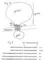
- the most suitable site for the incorporation of a collagenase cleavage site is between the amino acids Leu 659 and Leu 660 (see Fig. 2). Due to the colinearity of the gene and protein sequence, this corresponds to nucleotides A 7778 and T 7779 of the env gene.
- endoproteases unlike, for example, DNA restriction endonucleases, rely not only on the sequence for the recognition of the cleavage site, but also on certain secondary or tertiary structures of the substrate, which are not known, however. Furthermore, the recognition sequence must not be too large to be able to rule out the fact that the structure of the overall protein is significantly influenced by its size.
- a short collagen-like sequence of the general form has best suited as a recognition sequence: - (G - P - X) n - where n> 1, in particular 3 and X is one of the 20 amino acids defined by the genetic code (claim 4).
- German patent application DE 37 31 874 A similar sequence is proposed in German patent application DE 37 31 874, but as part of a fusion protein.
- the aim pursued in patent application DE 37 31 874 is to produce particularly small proteins or peptides in fermentation processes.
- FIG. 2 The construction of the env gene mutants E3CA, E3CB, E3CC is illustrated in FIG. 2, in which the sequence of the wild-type env gene (E1) in the transmembrane domain and the region located amino terminal to the transmembrane domain is shown.
- E1 wild-type env gene
- FIG. 2 A base sequence which codes for the following amino acid sequence was inserted into this new EcoR1 interface (for the abbreviation of amino acids, see, for example, AL Lehninger, Biochemie, VCH-Verlag Weinheim, 1987):
- This 13 amino acid sequence represents a collagenase recognition and cleavage site.
- the product with this insert was named E3CA.
- the 39 base pair long oligonucleotide coding for this sequence was synthesized with a DNA synthesizer from Applied Biosystems.
- a DNA synthesizer from Applied Biosystems.
- adenosine 7334 was exchanged for cytosine (A 7334 C), which in the protein meant an exchange of arginine 511 for Serine (Arg 511 Ser) causes.
- the plasmid pVB4 modified in this way was then combined with the E3CA plasmid containing the protease cleavage site (see under c :).
- the 5 'end of pVB4 (SalI / HindIII fragment) was ligated to the 3' end of the E3CA plasmid (HindIII / XhoI fragment) and cloned into the p-Bluescript.
- BH10 env gene (SalI / XhoI fragment) was cloned into an M13 vector.
- Ratner et al. "Nature” 313 , 277-284 (1985).
- the mutagenesis for generating the EcoR I recognition sequence was carried out according to T. Kunkel et al. ["Meth. Enzymol.” 154, 367-382 (1987)] with the oligonucleotide MAN 59:
- the mutagenized base A 7781C is marked with an asterisk, the EcoRI detection site is marked in bold.
- the numbers indicate the position of the corresponding amino acids in the env gene product.
- the env gene mutant thus obtained is referred to as E3 (BH10, A7781C).
- the EcoR I recognition sequence contained in the pks + blues script was destroyed by opening using EcoR I, filling up using DNA polymerase I (Klenow fragment) and connecting the ends using ligase. Then env E3 was subcloned into the pks + blues script. Then E3 was linearized with EcoR I and purified on an agarose gel. Now the 39 nucleotide long sequence coding for the collagenase cleavage site was inserted into this EcoR I recognition site of E3:
- E3CA The mutant thus obtained is called E3CA.
- E3CB oligonucleotide sequences Man83 and Man84
- E3CC Man85 and Man86
- the oligonucleotides were mixed (1: 1, mol: mol) in 150 mM NaCl, TE (10 mM Tris, 0.1 mm EDTA. PH 8). The mixture was then heated at 95 ° C. for 2 h and then allowed to cool to room temperature over 30 min. The ligase reaction then took place together with linearized E3 (pks +) with a large excess of the respective oligonucleotide pair. The plasmids thus produced were transformed into Echerichia coli DH5 and analyzed by sequencing. The yield of newly recombinant plasmids is over 90%.
- env genes were cloned into the expression vector pSVL (Pharmacia, Uppsala, Sweden) (the unique BamHI recognition site was previously deleted in pSVL).
- pSVL was linearized with XhoI and the env genes subcloned as SalI / XhoI fragment (BH 10 clone).
- the env genes were placed under the control of the SV40 late promoter.
- PCR polymerase chain reaction
- the env gene mutants E3CB and E3CC were produced for the following reason: There was a possibility that the alignment of the collagenase cleavage site within the ⁇ -helix could have an effect on the functionality. Since in an ⁇ -helix three amino acids make up approximately one full turn of the helix, inserting one amino acid alanine (E3CB) or two amino acids alanine (E3CC) made the collagenase cleavage site within the ⁇ -helix approximately 120 ° or around shifted approx. 240 °. The mutants E3CB and E3CC obtained thereby led to the same positive result as the mutant E3CA.
- E3CB amino acid alanine
- E3CC two amino acids alanine
- COS 7 cells are fibroblasts, but in principle all eukaryotic cells can be used instead, such as CV1 cells, yeast cells, insect cells [Summers, MD and Smith, GE: A Manual of Methods for Baculovirus Vectors and Insect Cell Culture Procedures. Texas Agricultural Experiment Station; Bulletin No. 1555, (1988)] etc.
- the COS 7 cells were transformed with the pSVL plasmids by electroporation and then for 48 h in complete growth medium [Gluzman, Y. et al .; "Cell” 23 , 175-182 (1981)] incubated. The cells were then harvested and the env proteins analyzed. All common methods can be used for the transformation of the cells with the expression vector, but in the present case electroporation gives particularly good success rates between 10 and 50%.
- COS 7 cells in logarithmic growth phase were trypsinized and washed with DMEM (Dulbecco Modified Eagles ⁇ Medium). For electroporation, the cells were mixed with 5-30 ⁇ g pSVL in DMEM and electroporated in a BioRad Gene Pulser (room temperature; 800 ⁇ l; 250V; 500 ⁇ F; 0.4 cm cuvette).
- DMEM Dulbecco Modified Eagles ⁇ Medium
- HEPES buffered saline HBS: 10 mM HEPES 7.8; 150 mM NaCl
- treatment with Potter. Incubate for several hours at 37 ° C. Then centrifuge for 30 min. at 100,000 xg and 37 ° C.
- the supernatant obtained was then subjected to protease digestion with purified collagenase (EC 3.4.24.3.) From Clostridium histolyticum (Sigma C0773, Deisenhofen) in concentrations between 1.2 and 333 mU / ml.
- protease inhibitors can be added to this buffer to minimize non-specific proteolysis: 1 ⁇ M E64 (Cysteine protease inhibitor) 1 ⁇ M pepstatin A (Aspartyl protease inhibitor) 1mM PMSF (phenylmethylsulfonyl fluoride) (Serine protease inhibitor)
- ⁇ -D-octylglucoside instead of ⁇ -D-octylglucoside, other mild detergents such as CHAPS or DOC (deoxycholate) can be used.
- CHAPS deoxycholate
- DOC deoxycholate
- the optimal collagenase concentration is between 50 and 250 mU / ml (3h, 37 ° C). at higher concentrations, unspecific digestion products increasingly occur and the yield of the desired product, a 140 kD glycoprotein, is reduced.
- Lentil lectin affinity chromatography The proteolyzed fraction was diluted 1: 1 with distilled water and applied to a Lentil Lectin Sepharose 4B column (Pharmacia 17-0444-0 ⁇ ). It was then washed with 5 column volumes of HBS, 2% ⁇ -D-octylglucoside, then with 5 column volumes of HBS and then with 5 column volumes of 10 mM HEPES (pH 7.8), 50 mM NaCl. The mixture was then eluted with 500-1000 mM ⁇ -D-methyl-mannoside in 10 mM HEPES, 50 mM NaCl (possibly warming to 37 ° C.).
- H 3 BO 4 borate
- the addition of H 3 BO 4 (borate) to the buffer has the advantage that the aggregation of the glycoproteins is prevented. This prevention of the aggregation of the glycoproteins by H 3 BO 4 is probably due to a complexation of the vincinal hydroxyl groups which are common in glycoproteins (claim 8).
- Sucrose density centrifugation On a preformed sucrose gradient 5-20% in HBS (10 mM HEPES, pH 7.8; 150 mM NaCl), about 1 ml of eluate from b: was charged at the top and centrifuged in a SW41 rotor (Beckmann) 35,000 rpm, 16h, 5 ° C. Then 10 fractions are removed (from bottom to top). In fraction 5, gp140 has a purity of 90-95%. Analysis was carried out by SDS PAGE and / or Western blot. Since no Ca ions are contained in the buffer during centrifugation, it is also possible, for example, to use phosphate buffer instead of HEPES buffer.
- proteolysis products were analyzed by SDS-PAGE (sodium lauryl sulfate-polyacrylamide gel electrophoresis) and then immunoblotting using a polyclonal rabbit serum directed against gp120, which recognizes both gp120 and gp160.
- SDS-PAGE sodium lauryl sulfate-polyacrylamide gel electrophoresis
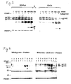
- the immunoblot (Western blot) in FIG. 3 shows that the wild-type env gene (clone BH10) and the mutant E3CA express approximately equal amounts of gp160.
- the proteolytic processing from gp160 to gp120 and gp41 which takes place in the Golgi apparatus, is the same for wild-type env and mutant env protein if the processing sequence is intact.
- the E3CA -env protein resulted in a new digest product, the monomeric form of which has an electrophoretic mobility of approximately 140 kD, which was not found when the wild-type env protein was digested.
- This digest product is the desired native ectodomain of the env glycoprotein.
- the somewhat lower electrophoretic mobility of the new digest product than that of gp120 results from the fact that the collagenase cleavage site at amino acid 659 of the total protein gp160 was incorporated in the region of the domain gp41.
- the domain gp120 ends with amino acid arginine 511 (see also FIG. 1).
- the newly produced native ectodomain of the HIV-1 env glycoprotein, gp140 consists of the entire domain gp120, a 148 amino acid part of the domain gp41 and the part of the collagen-like sequence that remains after collagenase digestion.
- This last-mentioned piece, which forms the C-terminal end of the glycoprotein, is a kind of foreign body in the glycoprotein according to the invention, but does not interfere with its function. It can, but does not necessarily have to be removed.
- the electrophoretic mobility of gp120 from the wild-type env and from the E3CA env protein is not changed by the collagenase treatment. This shows that the collagenase cleavage site inserted in the mutant E3CA is specifically recognized and cleaved by the bacterial collagenase.
- the digest products obtained were centrifuged by sucrose density centrifugation over a preformed 5% - 20% sucrose gradient in 10 mM HEPES, pH 7.8, 150 mM NaCl in the absence of detergents for 16 h at 35,000 rpm in a SW41 rotor (Beckmann). A total of ten fractions were then removed and examined for their content of env proteins by SDS-PAGE and immunoblotting. In both cases, gp120 was found in fractions 7 and 8, which corresponds to the monomeric gp120 with a sedimentation coefficient of 7S. The 140kD digest, which only occurred in the E3CA mutant and not in the wild type, was found in fraction 5, which corresponds to a sedimentation coefficient of 11S.
- a recombinant vaccinia virus (VV) is used as the expression vector.
- VV vaccinia virus
- the construction of this recombinant vaccinia virus is described in more detail in the following embodiment.
- a Lac O sequence 24 bp palindrome, was inserted 3 'to the promoter and the MCS was expanded by several unique endonuclease recognition sites.
- the Lac O sequence is necessary to minimize transcription in E. coli.
- the plasmids were analyzed by restriction analysis and partial sequencing.
- the plasmids were transfected into CV 1 cells by electroporation (conditions as for COS-7 cells, see first exemplary embodiment) and infected with wild-type VV (Copenhagen strain) (Multiplicity of Infection (MOI) ⁇ 1).
- MOI Multiplicity of Infection
- the native domains of viral membrane proteins according to the invention can be used as a vaccine against viruses.
- they are preferably administered to the patient in combination with suitable adjuvants.
- suitable adjuvants such as HEPES, must be removed beforehand. This can be done e.g. by dialysis against physiological saline in phosphate buffer.
- the native domains of the viral membrane proteins can be used for immunotherapy.
- the native viral protein domains are also suitable for diagnostic purposes. On the one hand, they can serve as an antigen, for example in immunoassays for the detection of specific antibodies, and on the other hand they can also be used to obtain antibodies, both monoclonal and polyclonal antibodies, which in turn can then be used for the direct detection of the virus.
- the purified native domains of viral membrane proteins is their use for crystallization. If the protein is in crystalline form, the three-dimensional structure can be determined. This is particularly interesting in that it enables the precise structure of the binding pocket to be elucidated. Knowing this structure, it may be possible to construct inhibitors, that is, smaller molecules that fit exactly into this binding pocket and thus block the binding of the protein, i.e. the virus, to the CD4 molecule, the receptor of the host cell .
Landscapes
- Health & Medical Sciences (AREA)
- Life Sciences & Earth Sciences (AREA)
- Genetics & Genomics (AREA)
- Chemical & Material Sciences (AREA)
- Organic Chemistry (AREA)
- Engineering & Computer Science (AREA)
- Molecular Biology (AREA)
- Biomedical Technology (AREA)
- General Health & Medical Sciences (AREA)
- Wood Science & Technology (AREA)
- Zoology (AREA)
- Biochemistry (AREA)
- Bioinformatics & Cheminformatics (AREA)
- General Engineering & Computer Science (AREA)
- Biotechnology (AREA)
- Biophysics (AREA)
- Medicinal Chemistry (AREA)
- Virology (AREA)
- Proteomics, Peptides & Aminoacids (AREA)
- Plant Pathology (AREA)
- Physics & Mathematics (AREA)
- Gastroenterology & Hepatology (AREA)
- Microbiology (AREA)
- Chemical Kinetics & Catalysis (AREA)
- Veterinary Medicine (AREA)
- Animal Behavior & Ethology (AREA)
- Public Health (AREA)
- Communicable Diseases (AREA)
- Nuclear Medicine, Radiotherapy & Molecular Imaging (AREA)
- Pharmacology & Pharmacy (AREA)
- Oncology (AREA)
- General Chemical & Material Sciences (AREA)
- Peptides Or Proteins (AREA)
- Medicines Containing Antibodies Or Antigens For Use As Internal Diagnostic Agents (AREA)
- Preparation Of Compounds By Using Micro-Organisms (AREA)
- Medicines Containing Plant Substances (AREA)
- Micro-Organisms Or Cultivation Processes Thereof (AREA)
Description
- Die vorliegende Erfindung betrifft ein Verfahren zur Gewinnung von isolierten Ektodomänen viraler Membranproteine in nativer, oligomerer und glykosylierter Form, insbesondere von Ektodomänen des Oberflächenproteins gp160 des menschlichen Immunschwächevirus HIV (Human Immunodeficiency Virus), dem Erreger von AIDS (Aquired Immune Deficiency Syndrome), weiterhin die durch dieses Verfahren gewonnenen Ektodomänen selbst sowie deren Verwendung als Impfstoff, insbesondere die Verwendung nativer Ektodomänen des Oberflächenproteins gp160 des menschlichen Immunschwächevirus HIV als Impfstoff gegen HIV. Bestandteil dieses Verfahrens zur Gewinnung von isolierten Ektodomänen viraler Membranproteine ist auch ein Verfahren zur Ermittlung geeigneter Stellen für den Einbau von Erkennungssequenzen in die zu gewinnende Proteindomänen, die als enzymatische Spaltstellen dienen, insbesondere als Collagenasespaltstellen, sowie Erkennungssequenzen für Collagenasen selbst.
- Ein probates Mittel zur Prophylaxe von Viruserkrankungen ist die Immunisierung, die Impfung des Körpers gegen die Viren. Durch Impfung konnten viele in früheren Zeiten oft epidernieartig verlaufende Viruserkrankungen wie z.B. die Pocken und Polio eingedämmt werden. Zur Erlangung einer solchen aktiven Immunisierung gegen ein Virus setzt man den Körper, genauer gesagt das Immunsystem, einem viralen Antigen aus. Dies kann geschehen beispielsweise durch Injektion von inaktivierten bzw. attenuierten Viren oder auch von Teilen, z.B. von Proteinen des Virus. Grundsätzlich setzt sich das Immunsystem des Menschen aus zwei verschiedenen Teilsystemen zusammen. Die zelluläre Immunantwort (T-Zellen) und die humorale Immunantwort (B-Zellen, Antikörper). Bei der Neuinfektion mit einem Virus stellt die humorale Immunantwort, durch B-Zellen vermittelt, den wichtigsten Abwehrmechanismus dar.
Das Immunsystem des Körpers reagiert auf das Antigen mit der Bildung von spezifischen Antikörpern, die das Antigen und damit das Virus erkennen und binden und dadurch dessen Inaktivierung in die Wege leiten. Darüberhinaus werden sogenannte "Gedächtniszellen" gebildet, das sind spezielle Lymphozyten, die bei einer späteren Infektion mit "ihrem" Virus bzw. "ihrem" Antigen aktiviert werden und das Immunsystem sehr rasch zur Synthese von großen Mengen der antigenspezifischen Antikörper stimulieren. Dadurch kann das Immunsystem wesentlich schneller auf einen Virusbefall reagieren, als wenn es vorher noch nicht mit dem entsprechenden Antigen konfrontiert worden ist. - Die Verwendung von attenuierten Viren zur Impfung kann allerdings insofern problematisch sein, als nicht ausgeschlossen werden kann, daß die verwendeten Viren im Körper wieder virulent werden und es zum Ausbruch der Viruserkrankung kommt. Aus diesem Grund ist es vorzuziehen, ein isoliertes Protein des Virus als Immunogen zu verwenden. Hierzu verwendet man am besten ein Oberflächenprotein des Virus, da dieses normalerweise der humoralen Immunantwort gut zugänglich ist. Idealerweise verwendet man nur einen Teil eines Oberflächenproteins, und zwar denjenigen Teil, der sich außen auf der Virusoberfläche befindet, die sogenannte Ektodomäne, da beim intakten Virus nur dieser Teil für Antikörper zugänglich ist. Bei der Herstellung einer Proteindomäne, die zur Impfung gegen ein Virus eingesetzt werden soll, ist allerdings zu beachten, daß sie möglichst die native Form haben sollte, da anderenfalls die Gefahr besteht, daß Antikörper gebildet werden, die das native Protein, und damit das Virus nicht erkennen und somit bei der Neutralisation wirkungslos sind. Solche Moleküle oder Molekülteile, die aufgrund Ihrer dreidimensionalen Struktur von Antikörpern erkannt werden, nennt man konformationelle bzw. strukturelle Epitope, diejenigen, die an ihrer Aminosäuresequenz erkannt werden, sequentielle Epitope. Es zeigt sich immer mehr, daß es nur sehr wenige sequentielle Epitope gibt, die meisten antigenen Epitope sind strukturell, d.h. sie müssen die korrekte dreidimensionale Faltung der Polypeptidkette aufweisen, um von entsprechenden Antikörpern erkannt zu werden. Nativ bedeutet in diesem Zusammenhang, daß die räumliche Struktur der verwendeten Proteindomäne identisch oder nahezu identisch ist mit der Struktur des entsprechenden, natürlicherweise auf oder in dem Virus vorkommenden Proteinbereiches. Dies schließt gegebenenfalls das Vorhandensein von Oligomerie und / oder Glykosylierung der Proteindomäne ein. Ein wichtiges Kriterium hierfür ist, daß Antikörper, die gegen eine isolierte native, virale Proteindomäne gebildet worden sind, auch den Virus bzw. das entsprechende native Protein im Virus erkennen und binden.
Große Probleme bereitet heutzutage das menschliche Immunschwächevirus HIV, ein Retrovirus. Seit es im Jahre 1984 als Ursache von AIDS identifiziert worden ist, werden große Anstrengungen unternommen, auch gegen dieses Virus einen Impfstoff herzustellen. In jüngerer Zeit ist es gelungen, Affen mit abgeschwächten SIV-Viren (Simian Immunodeficiency Virus), nahen Verwandten des HIV, erfolgreich zu immunisieren, was vermuten läßt, daß auch Menschen erfolgreich gegen HIV immunisiert werden könnten mittels abgeschwächten HIV. Die Problematik, die abgeschwächte Viren als Impfstoffe mit sich bringen, wurde schon angesprochen. Bei einer tödlich verlaufenden Krankheit wie AIDS kann dieses Risiko, daß der abgeschwächte Virus wieder virulent wird, nicht in Kauf genommen werden, auch wenn es sehr klein ist. Siehe hierzu auch: Koff, W.C. und Hoth, D.F. in der Zeitschrift "Science", 241, 426-432 (1988).
Aus diesem Grund konzentriert sich ein großer Teil der Forschung über HIV-Impfstoffe darauf, einzelne Proteine, insbesondere das Oberflächenprotein des Virus, das sogenannte gp160, auch env-Glykoprotein genannt, oder Fragmente davon als Antigen zu verwenden. [Eine allgemeine Übersicht gibt der Artikel "AIDS-Impfstoffe" von T.H. Matthews et al. in der Zeitschrift "Spektrum der Wissenschaft" 12, 134-142 (1988)].
Das env-Glykoprotein von HIV, dessen Aminosäuresequenz bekannt ist [Ratner et al., "Nature" 313, 277-284 (1985)] wird in der Wirtszelle zunächst als stark glykosyliertes Vorläuferprotein gp160 synthetisiert und anschließend im Golgi-Apparat zu den beiden Domänen gp120 und gp41 prozessiert. Die gp120-Domäne, die sich außen auf der Virusmembran befindet, ist für die Bindung an den zellulären Rezeptor CD4 verantwortlich und sowohl gp120 als auch das Vorläuferprotein gp160 binden mit hoher Affinität an diesen CD4-Rezeptor. Gp41, das eine Transmembrandomäne aufweist, ist beteiligt an der Fusion von viraler und zellulärer Membran während des Eindringens des Virus in die Zelle, wahrscheinlich durch Interaktion eines "Fusionspeptids", das am Aminoterminus von gp41 liegt, mit der Lipiddoppelschicht der Wirtszellmembran. - Vielversprechend erscheint der Ansatz, für die Entwicklung eines HIV-Impfstoffs lediglich den äußeren Teil des env-Glykoproteins zu verwenden. Dem liegt die bereits erwähnte Überlegung zugrunde, daß der nach außen exponierte Teil des Glykoproteins am leichtesten der humoralen Immunantwort zugänglich ist und daher eine Immunisierung gegen diese Ektodomäne am erfolgversprechendsten ist.
- Die Gewinnung der Ektodomäne des HIV-Oberflächenproteins kann auf unterschiedliche Art und Weise erfolgen. Sie kann beispielsweise durch mechanische, chemische oder enzymatische Methoden von der Virushülle "abgeschnitten" werden. Dabei ist die Gefahr groß, daß das Protein denaturiert wird und zur Bildung von Antikörpern führt, die das native Oberflächenprotein und damit den Virus nur schlecht oder gar nicht erkennen.
Robey et al. ["Proc. Natl. Acad. Sci. USA", 83, 7023-7027 (1986)] gewinnen gp120 beispielsweise durch Ablösen des Proteins von der Oberfläche HIV-infizierter Zellen mittels des recht starken Detergenz Triton X100. Auch wenn die Autoren ihre Präparation "natives gp120" nennen, ist in Anbetracht des verwendeten Detergenz und der verwendeten Reinigungsmethoden zu bezweifeln, daß das so gewonnene gp120 tatsächlich in nativer und oligomerer Form vorliegt. - In der europäischen Patentanmeldung EP 0 328 403 ist vorgeschlagen, lediglich Peptide aus HIV-gp120 mit einer Länge zwischen 28 und 33 Aminosäuren als Impfstoff gegen HIV zu verwenden. Diese Peptide liegen aber nicht in nativer, insbesondere oligomerer Form vor.
- Ein gentechnologischer Ansatz besteht darin, ein Stopcodon quasi als "Sollbruchstelle" knapp vor Beginn des gp41-Gens einzubauen. Bei der Expression des Gens in geeigneten Zellen bestimmt die Lage des Stopcodons dann die Lage des carboxyterminalen Endes der Ektodomäne des Oberflächenproteins, das heißt, es wird nur der äußere Teil des Oberflächenproteins, die Domäne gp120 synthetisiert. Auf diese Art synthetisiert, liegt gp120 aber als Monomer vor, was nicht den natürlichen Verhältnissen am Virus entspricht, da das env-Glykoprotein wie die meisten viralen Glykoproteine in nativer Form als Oligomer vorliegt. Häufig sind diese Proteine Trimere, für das HIV-1 env-Glykoprotein konnte kürzlich gezeigt werden, daß es in nativer Form ein Tetramer ist [Schawaller M. et al., "Virology", 172, 367-369 (1989)]. Dies erklärt, weshalb monomeres gp120 nicht zu geeigneten Antikörpern führt und somit als Impfstoff wenig brauchbar ist. Auch eigene Experimente des Erfinders, bei denen gp120 von Zellen synthetisiert wurde, die mit einem recombinanten Vaccinia-Virus, durch den env exprimiert wird, infiziert worden waren, zeigten, daß gp120 nicht stabil in oligomerer Form gebildet wird. Die Versuchsergebnisse deuten darauf hin, daß gp41 während der Biosynthese des env-Glykoproteins anwesend sein muß, um die Oligomerisierung zu gewährleisten. Wahrscheinlich liegt diejenige Domäne von gp41, die für diese Oligomerisierung verantwortlich ist, innerhalb der 129 aminoterminalen Aminosäuren von gp41 [Earl, P.L. et al. "Proc. Natl. Acad. Sci. USA", 87, 648-652 (1990)].
- Da gp41 offensichtlich eine wichtige Bedeutung bei der Synthese von nativem gp160 hat, wurden auch Versuche unternommen, das komplette gp160 in stabiler Form herzustellen. Barret et al. ["AIDS Research and Human Retroviruses" 5 (2), 159-170 (1989)] gewinnen gp160 beispielsweise durch Expression des env-Gens in eukaryontischen Zellen und anschließender Reinigung, wobei sie allerdings neben Detergentien auch das stark chaotrop wirkende Kaliumthiocyanat verwenden, welches Proteine denaturiert. Eigene Arbeiten des Autors haben gezeigt, daß die Reindarstellung von gp160 große Schwierigkeiten bereitet, und zwar aus folgenden Gründen:
Gp160 ist, wie oben beschrieben, ein Membranprotein. Zur Herauslösung dieses Proteins aus der Membran sind Detergentien notwendig. Diese sind auch deshalb nötig, da isolierte gp160 Moleküle bei Abwesenheit von Detergentien aggregieren. Detergentien führen aber zu einer Veränderung des Moleküls. Es wurde festgestellt, daß gp160 mit Detergentien eine Veränderung des Proteaseverdaumusters im Vergleich zum unbehandelten Protein zeigt. Beim Entfernen des Detergenz aus der Lösung, was spätestens dann nötig wird, wenn das Protein beispielsweise zum Zwecke der Impfung einem Patienten injiziert werden soll, zerfällt das Oligomer zum Monomer. Aus den genannten Gründen erscheinen die Chancen, reines gp160 in nativer Form herstellen zu können, sehr gering zu sein. - Der wesentliche Nachteil, den alle bisherigen Ansätze zur Entwicklung eines Impfstoffes gegen HIV auf der Basis der isolierten Ektodomäne des env-Glykoproteins gemeinsam haben, ist also höchstwahrscheinlich der, daß das als Immunogen verwendete Protein nicht in nativer und oligomerer Form vorhanden war. Dies wird dadurch deutlich, daß die von den Autoren angegebenen Titer, die mittels ELISA ermittelt wurden, sehr hoch (um 1/100 000), die im Neutralisierungstest gemessenen Titer dagegen extrem niedrig sind (oft weit weniger als 1/1000). Dies deutet darauf hin, daß die jeweils verwendeten Antigene zwar starke immunogene Wirkung besitzen, also eine große Menge Antikörper dagegen gebildet werden, daß diese Antikörper aber das native Protein, und damit auch den Virus kaum oder gar nicht erkennen.
- Die niedrigen Titer im Neutralisierungstest zeigen, daß das native Antigen erkennende Antikörper nur in sehr niedrigen Titern vorhanden sind. Niedertitrige Antikörper aber führen häufig zu einer Erhöhung der Virusinfektivität [Prabhakar et al., Nature 290, 590 (1981)]. Mit anderen Worten, anstatt den Wirt vor der Infektion zu schützen, wird durch virusspezifische Immunglobuline die Infektivität erhöht. In diesem Fall hat die Vaccine das Gegenteil des gewünschten Effektes zur Folge. Die geschilderte Problematik ergibt sich auch bei allen anderen Viren, die eine Oberflächenmembran mit membrangebundenen Proteinen besitzen.
- Neben der Verwendung als Impfstoff werden native Proteine bzw. Proteindomänen auch als Immuntherapeutikum oder für diagnostische Zwecke verwendet. Für die Diagnose werden sowohl Antigene, als auch die dagegen gerichteten Antikörper eingesetzt.
- Aufgabe der vorliegenden Erfindung ist es, ein Verfahren zur Gewinnung von isolierten Ektodomänen viraler Membranproteine, insbesondere Ektodomänen des env-Glykoproteins gp160 des menschlichen Immunschwächevirus HIV zur Verfügung zu stellen, die beispielsweise als Impfstoff, zur Immuntherapie oder für diagnostische Zwecke verwendet werden können. Diese Aufgabe umfaßt auch die Zurverfügungstellung eines Verfahrens zur Identifikation einer geeigneten Stelle zum Einbau einer Erkennungssequenz für proteinspaltende Enzyme, insbesondere Collagenasen, in virale Membranproteine, beispielsweise in das HIV-env Glykoprotein gp160, sowie die Zurverfügungstellung einer geeigneten Collagenasespaltstelle.
- Diese Aufgabe wird durch Bereitstellung eines Verfahrens zur Gewinnung von isolierten Ektodomänen viraler Membranproteine in nativer, oligomerer und glykosylierter Form gelöst, dadurch gekennzeichnet, daß
- in das Gen, das für ein virales, die gewünschte Ektodomäne enthaltendes Membranprotein kodiert, eine Nukleotidsequenz insertiert wird, die für eine Aminosäuresequenz kodiert, welche eine Erkennungssequenz für proteinspaltende Enzyme darstellt,
- die so erhaltene Gen-Mutante in eukaryontischen Zellen zum Erhalt des Membranproteins in nativer, oligomerer und glykosylierter Form einschließlich der insertierten Erkennungssequenz exprimiert wird,
- die Ektodomäne in nativer, oligomerer und glykosylierter Form durch enzymatische Spaltung des Membranproteins mit für die Erkennungssequenz spezifischen proteinspaltenden Enzymen von den eukaryontischen Zellen oder Teilen hiervon abgetrennt wird, und
- die Ektodomäne in nativer, oligomerer und glykosylierter Form aus dem Gemisch isoliert wird.
- Ein weiterer Gegenstand der vorliegenden Erfindung sind isolierte Ektodomänen viraler Membranproteine in nativer, oligomerer und glykosylierter Form.
- Ganz allgemein gesprochen bedeutet dies, daß zunächst an geeigneter Stelle in das Gen, das für die Ausgangsform (z.B. gp160), das Vorläuferprotein der zu gewinnenden Proteindomäne, codiert, eine für eine Aminosäuresequenz codierende Nukleotidsequenz eingebaut wird, welche eine Erkennungssequenz für proteinspaltende Enzyme, insbesondere Proteasen, oder Glykolipidanker darstellt. Nach erfolgter Synthese des Proteins durch geeignete eukaryontische Zellen wird es an der insertierten Erkennungssequenz mit den entsprechenden Enzymen gespalten und anschließend die gewünschte Proteindomäne aufgereinigt. Bei Verwendung des HIV env-Glykoproteins gp160 als Vorläuferprotein resultieren aus diesem Verfahren oligomere, insbesondere tetramere Glykoproteine, deren Monomere eine elektrophoretische Mobilität von ca. 140 kD aufweisen und die im folgenden gp140 genannt werden.
- Der Erfindung liegt also die Idee zugrunde, zunächst das komplette Protein zu synthetisieren, da es nur auf diese Weise in nativer und oligomerer Form synthetisierbar ist, und nach erfolgter Synthese die gewünschte Proteindomäne an spezifischer Stelle enzymatisch abzuspalten. Zuvor wird das korrespondierende Gen derart manipuliert, daß das fertige Protein eine zusätzliche Aminosäuresequenz beinhaltet, die als Erkennungssequenz für proteinspaltende Enzyme oder für Glycolipidanker dient. Diese zusätzliche Sequenz ändert nicht die native Proteinstruktur. Durch enzymatische Spaltung des Gesamtproteins an dieser Erkennungssequenz erhält man die gewünschte Proteindomäne in nativer und oligomerer Struktur. Ihrer Natur nach ist diese Erkennungssequenz eine Aminosäuresequenz, die von Proteasen erkannt und gespalten wird, es kann aber auch eine Erkennungssequenz für Lipidanker sein, wobei mittels Lipasen ebenfalls eine gezielte Spaltung möglich ist [siehe hierzu: Ferguson, M.A.J.; An. Rev. Biochem. 57, 285-320 (1988)]
- Im ersten Schritt des erfindungsgemäßen Verfahrens muß zunächst der für den Einbau einer Erkennungssequenz geeignete Bereich im Protein identifiziert werden. Dies erfolgt mittels einer "Pepscan" genannten Methode. Dann wird in der dem identifizierten Proteinbereich entsprechenden Gensequenz eine Erkennungssequenz für eine Endonuklease, z.B. EcoR I erzeugt, in die hinein dann die Sequenz, die für die gewünschte Aminosäuresequenz - die Erkennungssequenz im Protein - codiert, eingebaut wird. Es kann anstatt EcoR I auch jede andere Endonuklease verwendet werden, es sollten allerdings in der näheren Umgebung der Erkennungssequenz dieser Endonuklease keine weiteren gleichartigen Erkennungssequenzen vorhanden sein, da ansonsten die Klonierungseffizienz abnehmen kann. Das so erhaltene Gen wird in einen Vektor, beispielsweise ein Plasmid, eingebaut, der in geeignete eukaryontische Zellen transfiziert wird, welche dann das gesamte Protein inclusive der insertierten Erkennungssequenz synthetisieren. Auf diese Weise erhält man das gesamte Protein in nativer, oligomerer und glycosilierter Form. Daraufhin wird mit einem geeigneten Enzym, das das Protein an der eingebauten Erkennungssequenz schneidet, die gewünschte Domäne vom Rest des Proteins abgetrennt. Mittels geeigneter Reinigungsmethoden wie z.B. Affinitätschromatographie, Ionenaustauschchromatographie oder Gradientenzentrifugation wird die gewünschte Proteindomäne anschließend aufgereinigt. Was das verwendete spaltende Enzym betrifft, so ist das wesentliche Kriterium für dessen Eignung, daß es die Spaltstelle an deren Aminosäuresequenz erkennt. Diese Bedingung erfüllen bakterielle Collagenasen, auch vereinzelt pflanzliche Collagenasen sowie z.B. der Blutgerinnungsfaktor Xa.
- In einer bevorzugten Ausführungsform der Erfindung wird zur Erhöhung der Ausbeute die natürliche Prozessierungssequenz des Vorläuferproteins zerstört. Dies erfolgt beispielsweise durch Austausch einer oder mehrerer Aminosäuren in der Prozessierungssequenz. Dies hat zur Folge, daß das Vorläuferprotein, im Falle von HIV das gp160, nicht in die einzelnen Domänen (bei HIV also gp41 und gp120) aufgespalten wird.
Das Vorläuferprotein wird im rauhen endoplasmatischen Retikulum (RER) der Zelle synthetisiert. Erst im Golgi-Apparat, also während des Transports zur Zelloberfläche, wird das Protein prozessiert, d.h. in die einzelnen Domänen aufgespalten. Wird die Prozessierungssequenz des Vorläuferproteins zerstört, gewinnt man nicht nur den Anteil an Vorläuferprotein, der zum Zeitpunkt der Membranpräparation noch im RER vorhanden ist, sondern zusätzlich auch das bereits zur Zelloberfläche transportierte unprozessierte Vorläuferprotein. - In einer weiteren bevorzugten Ausführungsform der Erfindung erfolgt die Einschleusung des mutanten Gens in die eukaryontische Zelle nicht mittels eines Plasmids, sondern mittels eines rekombinanten Virus, insbesondere mittels eines rekombinanten Vaccinia Virus. Besonders für die Herstellung größerer Mengen der gewünschten viralen Proteindomänen ist die Verwendung eines rekombinanten Vaccinia Virus als Vektor vorteilhaft. Die jeweilige Genmutante kann beispielsweise in das Thymidinkinase-Gen des Vaccinia Virus insertiert werden. Da die Wahrscheinlichkeit für eine solche Rekombination bei ca. 1/1000 bis 1/10 000 liegt, ist es vorteilhaft, nach der Rekombination auf den rekombinaten Virus zu selektionieren. Dies kann beispielsweise durch Einsatz des Mutagens Bromodesoxyuridin in HTK--Zellen erfolgen. Nur der Wildtyp-Vaccinia Virus (TK+) baut dieses mutagene Nukleotidderivat in die DNA ein und schon nach zweimaliger Selektion liegt der Anteil der rekombinanten Viren (TK-) bei ca. 90%. Hohe Proteinausbeuten erhält man bei Verwendung eines Late-Promotors, beispielsweise des Cowpox-160K-Promotors, der ein starker Vaccinia-Promotor ist [Patel, D. D. et al., "Proc. Natl. Acad. Sci. USA" 85, 9431-9435 (1988)]. Eine Übersicht über die Verwendung des Vaccinia Virus als Vektor für die Expression von Genen im Cytoplasma eukaryotischer Zellen gibt Moss, B., ["Science" 252, 1662-1667 (1988)].
- Weitere Spezifikationen im Hinblick auf das erfindungsgemäße Verfahren, eine erfindungsgemäße Erkennungssequenz sowie die erfindungsgemäßen nativen Proteindomänen und deren Verwendung sind Gegenstand der Unteransprüche.
- Die Erfindung, die oben ganz allgemein skizziert ist, wurde gemacht bei der Untersuchung der Struktur des env-Glykoproteins von HIV, sie kann aber prinzipiell bei allen Viren angewandt werden, die eine Envelope-Membran besitzen. Für Kristallisationsversuche mit der Ektodomäne des env-Glykoproteins war es nötig, diese Ektodomäne in der nativen oligomeren Form ohne die hydrophobe transmembrane Domäne herzustellen. Der Oligomerisierungsprozeß von viralen Glykoproteinen vom Typ I ist aber wahrscheinlich abhängig von der Anwesenheit dieser hydrophoben transmembranen Domäne während der Biosynthese. Darüberhinaus scheint die Faltung der Polypeptidkette dadurch beeinflußt zu werden, daß die Polypeptidkette während der Synthese membrangebunden ist. Um die Ektodomäne des env-Glykoproteins in nativer Form zu erhalten, wurde nach dem Einbau einer für eine Collagenasespaltstelle codierenden Sequenz in das env-Gen zunächst das gesamte, mutante env-Glykoprotein in COS-Zellen synthetisiert, danach die gewünschte Ektodomäne mittels einer bakteriellen Collagenase vom synthetisierten Gesamtprotein abgespalten und anschließend aufgereinigt.
- Als großes Problem erwies sich dabei, eine geeignete Stelle im Protein für den Einbau der Collagenasespaltstelle - der Erkennungssequenz - zu identifizieren. Es hat sich nämlich gezeigt, daß es nur einen sehr kleinen Bereich im Protein gibt, an der die Erkennungssequenz eingebaut und das Protein dann auch erfolgreich gespalten werden kann. Bei rein empirischer Ermittlung einer solchen geeigneten Stelle mußten mehr als 30 verschiedene env-Gen-Mutanten hergestellt werden, bei denen an unterschiedlichen Stellen in der gewünschten Region von gp41 die Erkennungssequenz eingebaut wurde, um eine Stelle zu finden, an der die gewünschte proteolytische Spaltung möglich war.
- Die im folgenden verwendete Numerierung der Aminosäuren des HIV env-Glykoproteins bzw. der Basen des HIV env-Gens bezieht sich auf die von Ratner verwendete Numerierung [Ratner et al., "Nature" 313, 277-284 (1985)].
- Die genaue räumliche Struktur des env-Glykoproteins ist nicht bekannt, die Struktur des Bereichs des Proteins, in dem die Proteasespaltstelle sinnvollerweise eingebaut werden kann, stellt sich folgendermaßen dar (siehe auch Fig. 1):
Geht man von Hydrophobizitätsprofilen von gp41 aus, ist die erste Aminosäure des transmembranen Bereichs nach Ansicht des Erfinders Trp 678. Über die genaue Lage des transmembranen Bereichs von gp41 ist sich die Fachwelt allerdings nicht einig. Bolognesi, P.D. ["Mol. Biol. Med." 7, 1-15 (1990)] nimmt beispielsweise an, daß der transmembrane Bereich des gp160 von Aminosäure 691 bis Aminosäure 712 reicht. Klar ist, daß die vier konservierten Glykosylierungsstellen von gp41, die an den Positionen 611, 616, 624 und 637 lokalisiert sind, aminoterminal zur transmembranen Domäne, also in der Ektodomäne liegen müssen. Weiterhin kann man aus UV-Circulardichroismus-Messungen an gereinigten gp120- und gp160-Präparationen auf die Sekundärstruktur von gp41 schließen. Diese Messungen haben ergeben, daß gp120 fast ausschließlich aus β-Faltblattstrukturen besteht, während gp160 ca. 30% α-helicale Bereiche aufweist. Dies deutet darauf hin, daß die Sekundärstruktur von gp41 im wesentlichen eine α-Helix ist. Darüberhinaus kann man aus Sequenzanalysen des env-Gens verschiedener HIV-Stämme schließen, daß die Region in gp41 zwischen Pro 609 und dem Transmembranbereich (Trp 678) ebenfalls α-helicale Struktur besitzt. Diese Annahme wird noch dadurch unterstützt, daß keiner der untersuchten HIV-Stämme zwischen Pro 609 und Trp 678 ein Prolin besitzt, welches wahrscheinlich die α-helicale Struktur unterbrechen würde. Unter weiterer Einbeziehung der Ergebnisse des Erfinders und anderer, daß es sich bei dem HIV-env-Glykoprotein um ein Tetramer handelt, kann man sich vorstellen, daß vier lange amphiphatische α-Helices eine umeinandergewundene Schraubenstruktur (coiled-coil) bilden, wobei jeweils die hydrophoben Regionen der Untereinheiten miteinander interagieren und dabei die oligomere Struktur von gp41 stabilisieren. In diesem Modell liegen die hydrophilen Aminosäureseitenketten außen an einer tetrameren Schraube, wobei bei verschiedenen Stämmen verschiedene Aminosäuren vorliegen können. - Aufbauend auf den vorgenannten Hypothesen wurde für den Einbau der Erkennungssequenz die Region zwischen der letzten Glykosylierungsstelle bei der Aminosäure 637 und dem Beginn der transmembranen Domäne ausgewählt, ein immerhin 40 bis 53 Aminosäuren langes Stück, je nachdem, wo man den Beginn des transmembranen Bereichs annimmt. Wesentlich für die Funktionalität der Erkennungssequenz ist, daß sie gut zugänglich sein muß für das entsprechende spaltende Enzym.
- Auf der Basis dieser Erkenntnis, daß die Lokalisierung einer geeigneten Stelle für den Einbau einer Erkennungssequenz im wesentlichen von der Zugänglichkeit des entsprechenden Proteinbereichs für das spaltende Enzym abhängig ist, wurde ein neues Verfahren entwickelt zur Identifizierung einer geeigneten Einbaustelle für eine Erkennungssequenz, welches Gegenstand der Patentansprüche 4-7 ist.
- Da Antikörper gegen ein Protein nur gegen diejenigen Strukturen gebildet werden können, die außen an dem Protein liegen, die also den Zellen des Immunsystems zugänglich sind, und die Zugänglichkeit auch ein wesentliches Kriterium für eine funktionsfähige Erkennungssequenz ist, können geeignete Bereiche im Protein für den Einbau von Erkennungssequenzen dadurch identifiziert werden, daß man diejenigen Epitope identifiziert, gegen die nach Immunisierung mit dem jeweiligen Protein Antikörper gebildet werden. Horal P. und Mitarbeiter haben zur Untersuchung, gegen welche Bereiche des HIV-1 env-Glykoproteins gp41 überhaupt Antikörper gebildet werden, die sogenannte "Pepscan"-Methode angewandt [siehe Horal P. et al. in der Zeitschrift "J. Virol.", 65, 2718-2723 (1991)]. Diese Methode wird bei der vorliegenden Erfindung, anders als bei Horal et al. zur Identifikation des geeigneten Bereichs im Protein für die Erkennungssequenz eingesetzt. Hierzu werden im Prinzip kleine Bruchstücke des Proteins mit Antikörpern gemischt, die durch Immunisierung eines Säugetieres mit dem gesamten Protein gewonnen wurden. In der Praxis wird nicht das Protein in Bruchstücke zerlegt, sondern der Einfachheit halber entsprechend der Proteinsequenz überlappende Peptide mit einer Länge von 5 bis 30, insbesondere von 12 bis 16 Aminosäuren synthetisiert. Diese Peptide werden mit Serum von HIV-1-positiven Patienten versetzt. Die in diesem Serum vorhandenen Antikörper reagieren mit denjenigen Peptiden, die beim nativen Protein zugänglich an der Oberfläche liegen und die damit potentielle Einbaustellen für Erkennungssequenzen darstellen. Mit dieser Methode kann der am besten geeignete Abschnitt im Protein für den Einbau einer Erkennungssequenz je nach Lange der verwendeten Peptide bis auf einen Bereich weniger Aminosäuren identifiziert werden. Um die genaue, optimale Stelle zu finden, werden soviele Proteinmutanten hergestellt und getestet, wie nötig sind, um in dem zuvor identifizierten Bereich exakt die optimale Stelle für die Erkennungssequenz zu lokalisieren. Beim env-Glykoprotein von HIV-1 liegt die am besten geeignete Stelle für den Einbau einer Collagenasespaltstelle zwischen den Aminosäuren Leu 659 und Leu 660 (siehe Fig. 2). Aufgrund der Kolinearität von Gen- und Proteinsequenz entspricht dies den Nukleotiden A 7778 und T 7779 des env-Gens.
Ausgehend von dieser Stelle ist in einem Bereich von jeweils 17 Aminosäuren Richtung C-Terminus bzw. N-Terminus der Einbau einer Collagenasespaltstelle ebenfalls möglich, wobei mit zunehmendem Abstand mit geringeren Ausbeuten an Spaltprodukten und damit der gewünschten Proteindomäne zu rechnen ist. Eine weitere Schwierigkeit stellte die Ermittlung der Struktur einer geeigneten Spaltstelle dar. Es gibt recht wenige Proteasen, die Polypeptide sequenzspezifisch spalten. Beispiele für hochspezifische Proteasen sind bakterielle, vereinzelt auch pflanzliche Collagenasen, oder der Blutgerinnungsfaktor Xa. Auch Erkennungssequenzen für Glycolipidanker sind prinzipiell geeignet. Erschwerend kommt hinzu, daß die meisten Endoproteasen, anders als beispielsweise DNA Restriktionsendonukleasen, für die Erkennung der Spaltstelle nicht nur auf die Sequenz, sondern auch auf bestimmte Sekundär- oder Tertiärstrukturen des Substrates angewiesen sind, welche aber nicht bekannt sind. Weiterhin darf die Erkennungssequenz nicht zu groß sein, um ausschließen zu können, daß durch ihre Größe die Struktur des Gesamtproteins wesentlich beeinflußt wird. - Am besten geeignet als Erkennungssequenz hat sich eine kurze collagenähnliche Sequenz der allgemeinen Form:

- Eine ähnliche Sequenz wird in der deutschen Patentanmeldung DE 37 31 874 vorgeschlagen, allerdings als Teil eines Fusionsproteins. Im Gegensatz zur vorliegenden Erfindung ist das Ziel, das in der Patentanmeldung DE 37 31 874 verfolgt wird, besonders kleine Proteine bzw. Peptide in Fermentationsverfahren herzustellen. Da aber gerade derartige Peptide verstärkt einem proteolytischen Abbau in der Zelle ausgesetzt sind, wird vorgeschlagen, das Peptid als Teil eines größeren Fusionsproteins zusammen mit einer bakteriellen Aminosäuresequenz und einer collagenähnlichen Sequenz in einer bakteriellen Wirtszelle zu synthetisieren und anschließend mit einer bakteriellen Collagenase zu spalten.
- Unter Verwendung der genannten collagenähnlichen Sequenz wurden drei verschiedene HIV-1 env-Gen Mutanten (E3CA, E3CB, E3CC) hergestellt, deren Expressionsprodukt mittels einer bakteriellen Collagenase in der gewünschten Weise spezifisch gespalten werden kann. Unter Verwendung dieser Mutanten lassen sich Ektodomänen des env-Glykoproteins als wasserlösliche oligomere, insbesondere tetramere Glykoproteine gewinnen, deren Monomere eine elektrische Mobilität von ca. 140 kD aufweisen und daher gp140 genannt werden. Diese Proteine bestehen aus der Domäne gp120 und einem Teil der Domäne gp41 (Ansprüche 11 bis 14). Da sie keine Transmembrandomäne mehr aufweisen, sind anders als bei der Reinigung von komplettem gp160 keine starken Detergentien, welche die native Struktur der Glykoproteine verändern könnten, zur Aufreinigung dieser Glykoproteine notwendig. Klare Evidenzen dafür, daß gp140 oligomere Struktur besitzt, ergeben sich zum einen aus Sedimentationsanalysen [Fig. 4; siehe hierzu auch Earl, P.L. et al. "Proc. Natl. Acad. Sci. USA", 87, 648-652 (1990)], zum anderen aus Crosslinking-Experimenten [zu dieser Methodik siehe Schawaller, M. et al., "Virology" 172, 367-369 (1989)]. Die Gewinnung des oligomeren gp140 unter Verwendung der env-Gen Mutante E3CA wird im folgenden Ausführungsbeispiel in Verbindung mit den Figuren 1 bis 4 näher erläutert. Es zeigen:
- Fig.1:
- Schema des HIV-env-Glykoproteins mit der Transmembrandomäne in der Oberflächenmembran des Virus. Die Zahlen geben die Position der Aminosäuren an nach der Nomenklatur von Ratner et al., "Nature" 313, 277-284 (1985). Die Domäne gp41 beginnt bei Aminosäure 512, die aber in einem solchen Schema schlecht lokalisiert werden kann.
- Fig.2:
- Ausschnitte aus den Aminosäuresequenzen der Produkte von Wildtyp-env-Gen (E1(WT) des BH10-Klons und verschiedenen Mutanten. Die Veränderungen bei den Mutanten gegenüber dem Wildtyp-env-Produkt sind jeweils unterstrichen.
- Fig.3:
- Spezifische Spaltung des Wildtyp-env- und des E3CA-env- Proteins durch unterschiedliche Konzentrationen an bakterieller Collagenase. Die erhaltenen Produkte wurden analysiert durch Immunoblotting nach gelelektrophoretischer Trennung auf 6% SDS Polyacrylamidgel und anschließende Detektion mit polyklonalem Kaninchenserum gegen gp120. Danach erfolgte eine Autoradiographie unter Einsatz eines radioaktiv markierten sekundären Antikörpers, der gegen Kaninchen-Immunoglobuline gerichtet ist (Amersham IM 134).
- Fig.4:
- Sedimentationsanalyse der Produkte des Collagenaseverdaus von Wildtyp- und E3CA-Mutanten-Membranen. Collagenasekonzentration: 200 mU/ml, 37°C, 3h. Die erhaltenen Produkte wurden geschichtet auf einen vorgeformten 5 bis 20 % Sucrosegradienten in 10 mM HEPES, pH 7.8, 150 mM NaCl und in einem SW41 Rotor (Beckmann) für 16 h bei 35 000 UpM und 5°C zentrifugiert. Zehn Fraktionen wurden, vom Boden des Gefäßes angefangen, geerntet und durch Immunoblotting analysiert.
- Die Konstruktion der env-Gen-Mutanten E3CA, E3CB, E3CC ist in Fig.2 illustriert, in der die Sequenz des Wildtyp env-Gens (E1) in der Transmembrandomäne und der aminoterminal zur Transmembrandomäne gelegenen Region dargestellt ist. An der für Leu 659 codierenden Position wurde durch eine Punktmutation eine einzelne Schnittstelle für die Endonuklease EcoR1 erzeugt, wodurch im Protein Leu gegen Phe ausgetauscht wird. Das daraus entstehende Produkt ist die env-Gen-Mutante E3. In diese neue EcoR1 Schnittstelle wurde eine Basensequenz insertiert, die für folgende Aminosäuresequenz codiert (zur Abkürzung von Aminosäuren siehe z.B. A.L. Lehninger, Biochemie, VCH-Verlag Weinheim, 1987):

- Diese 13 Aminosäuren lange Sequenz stellt eine Collagenase Erkennungs- und Spaltstelle dar. Das Produkt mit diesem Insert wurde E3CA genannt. Das für diese Sequenz kodierende 39 Basenpaare lange Oligonukleotid wurde mit einem DNA - Synthesegerät der Firma Applied Biosystems synthetisiert. Zur Herstellung der env-Gen Mutanten sowie deren Klonierung in einen Expressionsvektor siehe auch T. Maniatis et al., Molecular Cloning, Cold Spring Harbor, N.Y., USA (1982).
- Zur Ausbeutensteigerung wurde zunächst die natürliche Prozessierungssequenz des env-Gen Produktes zerstört. In dem Plasmid pVB4 [Bosch, V. und Pawlita, M., "J. Virol. 64, 2337-2344 (1990)] wurde Adenosin 7334 gegen Cytosin ausgetauscht (A 7334 C), was beim Protein einen Austausch von Arginin 511 gegen Serin (Arg 511 Ser) bewirkt.
Das so abgeänderte Plasmid pVB4 wurde danach kombiniert mit dem die Proteasespaltstelle enthaltenden E3CA-Plasmid (siehe unter c:). Hierzu wurde das 5`-Ende von pVB4 (SalI/HindIII-Fragment) mit dem 3`-Ende des E3CA-Plasmids (HindIII/XhoI-Fragment) ligiert und in den p-Bluescript Kloniert. - Gen von BH10 env (SalI/XhoI Fragment) wurde in einen M13 Vektor kloniert. Zum BH10-Klon siehe Ratner et al. "Nature" 313, 277-284 (1985). Die Mutagenese zur Erzeugung der EcoR I - Erkennungssequenz erfolgte nach T. Kunkel et al. ["Meth. Enzymol."154, 367-382 (1987)] mit dem Oligonukleotid MAN 59:

- Die mutagenisierte Base A 7781C ist mit einem Stern markiert, die EcoR I-Erkennungsstelle ist fett markiert. Die Zahlen geben die Position der entsprechenden Aminosäuren im env-Gen Produkt an.
- Die Verifizierung erfolgte mittels Sequenzierung. Die so erhaltene env-Gen Mutante wird bezeichnet als E3 (BH10, A7781C).
- Die im Blueskript pks+ enthaltene EcoR I Erkennungssequenz wurde zerstört durch Öffnen mittels EcoR I, Auffüllen mittels DNA Polymerase I (Klenow Fragment) und Verbinden der Enden mittels Ligase. Danach wurde env E3 in Blueskript pks+ subkloniert. Dann wurde E3 mit EcoR I linearisiert und über ein Agarosegel gereinigt.
Nun erfolgte die Insertion der 39 Nukleotide langen für die Collagenasespaltstelle codierenden Sequenz in diese EcoR I Erkennungsstelle von E3:
- Die so erhaltene Mutante wird E3CA genannt. Zwei weitere Mutanten wurden erzeugt unter Verwendung der komplementären Oligonukleotidsequenzen Man83 und Man84 (E3CB) sowie Man85 und Man86 (E3CC). Diese Oligonukleotidsequenzen sind im folgenden dargestellt:


- Zur Insertion der komplementären Oligonukleotide in die EcoR I-Erkennungsstelle in E3 wurden die Oligonukleotide in 150 mM NaCl, TE (10mM Tris, 0,1mm EDTA. pH 8) gemischt (1:1, mol:mol). Anschließend wurde 2 h bei 95°C erhitzt und dann über 30 min auf Raumtemperatur abkühlen gelassen. Danach erfolgte die Ligase-Reaktion zusammen mit linearisiertem E3 (pks+) bei hohem Überschuß an dem jeweiligen Oligonukleotidpaar. Die so hergestellten Plasmide wurden in Echerichia coli DH5 transformiert und durch Sequenzierung analysiert. Die Ausbeute an neu rekombinanten Plasmiden beträgt über 90%.
- Zur Expression der env-Gene wurden diese in den Expressionsvektor pSVL (Pharmacia, Uppsala, Schweden) kloniert (in pSVL wurde vorher die unique BamHI Erkennungsstelle deletiert). pSVL wurde mit XhoI linearisiert und die env-Gene als SalI/XhoI-Fragment (BH 10 Klon) subkloniert. Die env-Gene wurden unter die Kontrolle des SV40 Late Promotors gestellt.
- Eine weitere Möglichkeit, einen die verschiedenen Gene enthaltenden Expressionsvektor herzustellen, besteht im sogenannten PCR (Polymerase Chain Reaction) -constructing. Eine Übersicht über diese Methodik ist zu finden bei Jones, D.H. et al, "Nature", 344, 793-794 (1990).
- Die env-Gen Mutanten E3CB und E3CC wurden aus folgendem Grund hergestellt: Es bestand durchaus die Möglichkeit, daß die Ausrichtung der Collagenasespaltstelle innerhalb der α-Helix Einfluß auf die Funktionsfähigkeit haben könnte. Da in einer α-Helix drei Aminosäuren ungefähr eine volle Umdrehung der Helix ausmachen, wurde durch Einfügen einer Aminosäure Alanin (E3CB) bzw. zweier Aminosäuren Alanin (E3CC) die Ausrichtung der Collagenasespaltstelle innerhalb der α-Helix um ca. 120° bzw. um ca. 240° verschoben. Die dadurch erhaltenen Mutanten E3CB und E3CC führten aber zum gleichen positiven Ergebnis wie die Mutante E3CA.
- Die Expression der verschiedenen env-Gene wurde in COS 7-Zellen durchgeführt. COS 7-Zellen sind Fibroblasten, es können stattdessen aber im Prinzip alle eukaryontischen Zellen verwendet werden wie z.B. CV1-Zellen, Hefezellen, Insektenzellen [Summers, M.D. und Smith, G.E. : A Manual of Methods for Baculovirus Vectors and Insect Cell Culture Procedures. Texas Agricultural Experiment Station; Bulletin No. 1555, (1988)] etc. Die COS 7-Zellen wurden mit den pSVL Plasmiden durch Elektroporation transformiert und anschließend für 48h in vollständigem Wachstumsmedium [Gluzman,Y. et al.; "Cell" 23, 175-182 (1981)] inkubiert. Danach wurden die Zellen geerntet und die env-Proteine analysiert. Für die Transformation der Zellen mit dem Expressionsvektor können alle gängigen Verfahren angewandt werden, die Elektroporation ergibt aber im vorliegenden Fall besonders gute Erfolgsraten zwischen 10 und 50%.
- Ca. 107 (2 x 10⁶ - 5 x 10⁷) COS 7 Zellen in logarithmischer Wachstumsphase wurden trypsiniert und mit DMEM (Dulbecco Modified Eagles` Medium) gewaschen. Zur Elektroporation wurden die Zellen mit 5 - 30 µg pSVL in DMEM versetzt und in einem BioRad Gene Pulser elektroporiert (Raumtemperatur; 800 µl; 250V; 500 µF; 0,4 cm Küvette).
- Zur Analyse, ob die Collagenasespaltung zum gewünschten Erfolg führt, also ob nach Collagenasebehandlung der Mutante E3CA eine native Ektodomäne des env-Glykoproteins (gp140) extrahiert werden kann, wurden nach hypotonischer Zellyse mittels Sucrose-Dichtezentrifugation Membranpräparationen hergestellt [Schawaller, M. et al., "Virology" 172, 367-369 (1989)].
- Membranpräparation mehrfach waschen, in HEPES buffered saline (HBS: 10 mM HEPES 7,8; 150 mM NaCl) aufnehmen: + 1/10 Vol. 10 x HBS, + 1/100 Vol. 1 M CaCl2, + 1/5 Vol. 20 % β-D-Octylglucosid bei 37°C. Danach Behandlung mit Potter. Mehrere Stunden bei 37°C inkubieren. Anschließend zentrifugieren für 30 min. bei 100 000 x g und 37°C. Der erhaltene Überstand wurde dann einem Proteaseverdau unterzogen mit gereinigter Collagenase (EC 3.4.24.3.) von Clostridium histolyticum (Sigma C0773, Deisenhofen) in Konzentrationen zwischen 1,2 und 333 mU/ml.
- Der Collagenaseverdau wurde in folgendem Puffer für 3 h bei 37°C durchgeführt:
- 300 mM NaCl
- 20 mM HEPES (Hydroxyethylpiperazinethansulfonsäure), pH 7,8
- 10 mM CaCl2
- 50µM ZnCl2
- 4% β-D-Oktylglucosid
- Diesem Puffer können folgende Proteaseinhibitoren zugefügt werden, um unspezifische Proteolyse zu minimieren:
1 µM E64 (Cystein-Proteaseinhibitor) 1 µM Pepstatin A (Aspartyl-Proteaseinhibitor) 1mM PMSF (Phenylmethylsulfonylfluorid) (Serin-Proteaseinhibitor) - Statt β-D-Oktylglucosid können auch andere milde Detergentien wie CHAPS oder DOC (Desoxycholat) verwendet werden. Zur Wirkung verschiedener Detergentien auf Membranproteine siehe auch Womack, M.D. et al. "Biochim. Biophys. Acta" 733, 210-215 (1983).
Die optimale Collagenasekonzentration liegt zwischen 50 und 250 mU/ml (3h, 37°C). bei höheren Konzentrationen treten verstärkt unspezifische Verdauprodukte auf und die Ausbeute an gewünschtem Produkt, einem 140 kD Glykoprotein, wird geringer. - a: Lentil lectin-Affinitätschromatographie: Die proteolysierte Fraktion wurde 1:1 verdünnt mit destilliertem Wasser und aufgegeben auf eine Lentil Lectin Sepharose 4B - Säule (Pharmacia 17-0444-0`). Anschließend wurde gewaschen mit 5 Säulenvolumen HBS, 2% β-D-Oktylglucosid, danach mit 5 Säulenvolumen HBS und dann mit 5 Säulenvolumen 10 mM HEPES (pH 7,8), 50 mM NaCl. Anschließend wurde eluiert mit 500 - 1000 mM α-D-Methyl-Mannosid in 10 mM HEPES, 50 mM NaCl (evtl. erwärmen auf 37°C). Zur Lentil lectin-Affinitätschromatographie siehe auch Hayman, MJ. et al., "FEBS letters" 29 (2), 185-188 (1973).
Das Eluat wurde anschließend aufkonzentriert. Gp140 liegt jetzt in einer Reinheit von ca. 50 - 70% vor. - b: Mono Q FPLC: Das Eluat aus a: wurde aufgenommen in 10 mM BTP (Bis-Trispropane), 50mM H3BO4, pH 8,2 und auf eine Mono Q HR 5/5 Säule (Pharmacia) aufgegeben, wobei die Proteinkonzentration bei etwa 1 mg/ml lag. Die Säule wurde eluiert mit einem NaCl-Gradienten von 30 - 600 mM über 40 min, 0,5 - 1 ml/min, UV-Monitor bei 280 nm. Analyse durch SDS PAGE Coomassie und/oder Western Blot. Das gp140 env-Protein eluiert bei ca. 400 mM NaCl. Gp140 liegt jetzt in einer Reinheit von ca. 80-90% vor. Die Zugabe von H3BO4 (Borat) zum Puffer bringt den Vorteil, daß dadurch das Aggregieren der Glykoproteine verhindert wird. Diese Verhinderung der Aggregation der Glykoproteine durch H3BO4 beruht wahrscheinlich auf einer Komplexierung der in Glykoproteinen häufigen vincinalen Hydroxylgruppen (Anspruch 8).
- c: Sucrosedichtezentrifugation: Auf einen vorgeformten Sucrosegradienten 5-20% in HBS (10 mM HEPES, pH 7,8; 150 mM NaCl) wurde oben ca. 1 ml Eluat aus b: aufgeladen und in einem SW41-Rotor (Beckmann) zentrifugiert bei 35 000 UpM, 16h, 5°C. Anschließend Entnahme von 10 Fraktionen (von unten nach oben). In Fraktion 5 hat gp140 eine Reinheit von 90 - 95%. Analyse erfolgte durch SDS PAGE und/oder Western Blot. Da während der Zentrifugation keine Ca-Ionen im Puffer enthalten sind, kann statt HEPES-Puffer beispielsweise auch Phosphatpuffer verwendet werden.
- Die Proteolyseprodukte wurden analysiert mittels SDS-PAGE (Natriumlaurylsulfat-Polyacrylamidgelelektrophorese) und anschließendem Immunoblotting unter Verwendung eines polyklonalen Kaninchenserums, gerichtet gegen gp120, das sowohl gp120, als auch gp160 erkennt.
- Der Immunoblot (Western-Blot) in Fig. 3 zeigt, daß das Wildtyp env-Gen (Klon BH10) und die Mutante E3CA etwa gleiche Mengen an gp160 exprimieren. Offensichtlich verläuft auch die proteolytische Prozessierung von gp160 zu gp120 und gp41, welche im Golgi-Apparat abläuft, bei Wildtyp-env- und Mutanten-env-Protein gleich, wenn die Prozessierungssequenz intakt ist. Nach Behandlung mit bakterieller Collagenase ergab das E3CA -env-Protein ein neues Verdauprodukt, dessen monomere Form eine elektrophoretische Mobilität von ca. 140 kD aufweist, welches beim Verdau des Wildtyp-env-Proteins nicht gefunden wurde. Dieses Verdauprodukt ist die gewünschte native Ektodomäne des env-Glykoproteins. Die mit 140 kD etwas geringere elektrophoretische Mobilität des neuen Verdauprodukts als die von gp120 ergibt sich daraus, daß die Collagenasespaltstelle bei Aminosäure 659 des Gesamtproteins gp160 im Bereich der Domäne gp41 eingebaut wurde. Die Domäne gp120 endet bei Aminosäure Arginin 511 (siehe auch Fig. 1). Die neu hergestellte native Ektodomäne des HIV-1 env-Glykoproteins, gp140, besteht also aus der gesamten Domäne gp120, einem 148 Aminosäuren langen Teilstück der Domäne gp41 sowie dem nach Collagenaseverdau verbleibenden Stück der collagenähnlichen Sequenz. Dieses letztgenannte Stück, das das C-terminale Ende des Glykoproteins bildet, ist zwar eine Art Fremdkörper in dem erfindungsgemäßen Glykoprotein, stört aber dessen Funktion nicht. Es kann, braucht aber nicht unbedingt entfernt zu werden.
Die elektrophoretische Mobilität von gp120 vom Wildtyp-env- und vom E3CA-env-Protein wird durch die Collagenasebehandlung nicht verändert. Das zeigt, daß die in der Mutante E3CA insertierte Collagenasespaltstelle von der bakteriellen Collagenase spezifisch erkannt und gespalten wird. - Ein weiterer Nachweis dafür, daß die in der Mutante E3CA insertierte Collagenasespaltstelle von der bakteriellen Collagenase spezifisch erkannt und gespalten wird, ergibt sich aus den Ergebnissen der Sedimentationsanalyse der Collagenasespaltprodukte (Fig. 4). Membranpräparationen von COS 7-Zellen, die das Wildtyp- oder das E3CA-env-Gen exprimieren, wurden mit 4% β-D-Oktylglycosid extrahiert und wie oben beschrieben mit Collagenase verdaut. Die erhaltenen Verdauprodukte wurden mittels Sucrose-Dichtezentrifugation über einen vorgeformten 5% - 20% Sucrosegradienten in 10 mM HEPES, pH 7.8, 150 mM NaCl in Abwesenheit von Detergentien zentrifugiert für 16 h bei 35 000 UpM in einem SW41 Rotor (Beckmann). Danach wurden insgesamt zehn Fraktionen entnommen und mittels SDS-PAGE und Immunoblotting auf ihren Gehalt an env-Proteinen untersucht. gp120 wurde in beiden Fällen in den Fraktionen 7 und 8 wiedergefunden, was dem monomeren gp120 mit einem Sedimentationskoeffizienten von 7S entspricht. Das Verdauprodukt von 140kD, das nur bei der E3CA-Mutante und nicht beim Wildtyp auftrat, wurde dagegen in Fraktion 5 wiedergefunden, was einem Sedimentatiortskoeffizienten von 11S entspricht.
- In einer bevorzugten Ausführungsform wird ein rekombinantes Vaccinia Virus (VV) als Expressionsvektor verwendet. Die Konstruktion dieses rekombinanten Vaccinia Virus wird im folgenden Ausführungsbeispiel näher beschrieben.
- Als Ausgangsmaterial wurde das Vaccinia shuttle plasmid p2100 verwendet [Patel, D. D. et al., "Proc. Natl. Acad. Sci. USA" 85, 9431-9435 (1988)]. Dieses enthält neben bakteriellem Origin und Resistenzgen folgendes Genfragment:

- Tk-L = Linker Arm des Thymidinkinase-Gens
- Tk-R = Rechter Arm des Thymidinkinase-Gens
- Cowpox 160K = Promotor/Enhancer Sequenzen
- MCS = Multiple cloning site
- Durch Klonieren von doppelsträngigen Oligonukleotiden wurde 3`zum Promotor eine Lac O Sequenz, 24 bp palindrom, insertiert und die MCS um mehrere unique Endonuklease-Erkennungsstellen erweitert. Die Lac O Sequenz ist nötig, um die Transkription in E. coli auf ein Minimum zu unterdrücken.
- In die neuen Restriktionsstellen wurde das jeweilige env-Gen (Wildtyp oder Mutante: aus pks Plasmid) cloniert (z.B. E3CA) [zur Methodik siehe Chakrabarti, S. et al., "Molec. Cell Biol." 5, 3403-3409 (1985)].
- Man erhält folgendes Konstrukt:

- Für die Herstellung rekombinanter Vaccinia Viren wurden die Plasmide analysiert durch Restriktionsanalyse und partielle Sequenzierung. Die Plasmide wurden in CV 1 Zellen transfiziert durch Elektroporation (Bedingungen wie bei COS-7 Zellen, siehe erstes Ausführungsbeispiel) und mit Wildtyp VV (Stamm Copenhagen) infiziert ( Multiplicity of Infection (M O I) < 1). Es entstehen durch homologe Rekombination im Thymidinkinase-Gen des Plasmids mit dem Thymidinkinase-Gen des VV mit einer Wahrscheinlichkeit von etwa 1/1000 - 1/10000 rekombinante VV, durch die das env-Gen exprimiert werden kann. Da diese phänotypisch TK- sind, können sie durch 5'-Bromodeoxyuddin (BdUR)- Selektion in HTK- Zellen positiv selektioniert werden. Zwei Runden BdUR/HTK- Selektion und anschließendes "Plaquen" (identifizieren von Plaques und entnehmen von rekombinanten VV) aus CV 1 Zellen ergibt ca. 50-90% env exprimierende Plaques (VV env), die durch Western Blot einer 1 ml Kultur identifiziert werden können. Nochmaliges Plaquen führt zu dem gewünschten rekombinanten Vaccinia Virus, durch den env exprimiert werden kann.
- Die erfindungsgemäßen nativen Domänen viraler Membranproteine können als Impfstoff gegen Viren eingesetzt werden. Hierzu werden sie dem Patienten vorzugsweise in Kombination mit geeigneten Adjuvantien verabreicht. Vorher müssen allerdings eventuell noch enthaltene Pufferbestandteile, wie beispielsweise HEPES, entfernt werden. Dies kann erfolgen z.B. durch Dialyse gegen physiologische Kochsalzlösung in Phosphatpuffer.
- Weiterhin können die nativen Domänen der viralen Membranproteine zur Immuntherapie verwendet werden.
- Auch für diagnostische Zwecke sind die nativen viralen Proteindomänen geeignet. Zum einen können sie als Antigen beispielsweise in Immunoassays zum Nachweis spezifischer Antikörper dienen, zum anderen können sie auch eingesetzt werden zur Gewinnung von Antikörpern, sowohl monoklonalen als auch polyklonalen Antikörpern, die dann wiederum zum direkten Nachweis des Virus dienen können.
- Ein weiterer wichtiger Verwendungszweck der aufgereinigten nativen Domänen viraler Membranproteine ist ihre Verwendung zur Kristallisation. Ist das Protein in kristalliner Form vorhanden, kann die dreidimensionale Struktur ermittelt werden. Dies ist insbesondere dahingehend interessant, als dadurch möglich wird, die genaue Struktur der Bindungstasche aufzuklären. Kennt man diese Struktur, ist es eventuell möglich, Inhibitoren zu konstruieren, das heißt, kleinere Moleküle, die genau in diese Bindungstasche hineinpassen und damit die Bindung des Proteins, also des Virus, an das CD4-Molekül, den Rezeptor der Wirtszelle, zu blockieren.
Claims (28)
- Verfahren zur Gewinnung von isolierten Ektodomänen viraler Membranproteine in nativer, oligomerer und glykosylierter Form, dadurch gekennzeichnet, daß- in das Gen, das für ein virales, die gewünschte Ektodomäne enthaltendes Membranprotein kodiert, eine Nukleotidsequenz insertiert wird, die für eine Aminosäuresequenz kodiert, welche eine Erkennungssequenz für proteinspaltende Enzyme darstellt,- die so erhaltene Gen-Mutante in eukaryontischen Zellen zum Erhalt des Membranproteins in nativer, oligomerer und glykosylierter Form einschließlich der insertierten Erkennungssequenz exprimiert wird,- die Ektodomäne in nativer, oligomerer und glykosylierter Form durch enzymatische Spaltung des Membranproteins mit für die Erkennungssequenz spezifischen proteinspaltenden Enzymen von den eukaryontischen Zellen oder Teilen hiervon abgetrennt wird, und- die Ektodomäne in nativer, oligomerer und glykosylierter Form aus dem Gemisch isoliert wird.
- Verfahren nach Anspruch 1, wobei eine natürliche Prozessierungssequenz des Vorläuferproteins des Membranproteins durch Austausch mindestens einer Aminosäure zerstört wird.
- Verfahren nach Anspruch 1 oder 2, wobei die Erkennungssequenz von Proteasen oder von Lipasen über Glycolipidankern erkannt und gespalten wird.
- Verfahren nach einem der Ansprüche 1 bis 3, wobei die zu gewinnnenden Ektodomänen die Ektodomänen des env-Glykoproteins von HIV sind.
- Verfahren nach Anspruch 4, wobei die Nukleotidsequenz in einen Bereich des für das env-Glykoprotein kodierenden Gens insertiert wird, der bei dem daraus exprimierten env-Glykoprotein dem Bereich entspricht, der begrenzt wird durch die erste und fünfzigste Aminosäure vor Beginn des transmembranen Bereichs des env-Glykoproteins.
- Verfahren nach Anspruch 4 oder 5, wobei die Erkennungssequenz in einen Bereich des env-Glykoproteins von HIV eingebaut wird, der durch die Aminosäuren 645 und 673 begrenzt wird.
- Verfahren nach Anspruch 6, wobei der Bereich durch die Aminosäuren 656 und 663 begrenzt wird.
- Verfahren nach einem der Ansprüche 4 bis 7, wobei die Erkennungssequenz zwischen den Aminosäuren 659 und 660 in das env-Glykoprotein von HIV eingebaut wird.
- Verfahren nach einem der Ansprüch 1 bis 8, wobei- die Erkennungssequenz die folgende allgemeine Form besitzt:
 - das für die Spaltung verwendete Enzym eine Collagenase ist.
 - Verfahren nach Anspruch 9, wobei die Collagenase eine bakterielle Collagenase ist.
- Verfahren nach einem der Ansprüche 1 bis 10, worin der Puffer, der bei der Reinigung der gewünschten Ektodomäne aus dem Gemisch nach der enzymatischen Spaltung verwendet wird, Borat in einer Konzentration zwischen 1 und 200 mM enthält.
- Verfahren nach Anspruch 11, wobei der Puffer Borat in einer Konzentration zwischen 30 und 100 mM, enthält.
- Verfahren nach einem der Ansprüche 1 bis 12, worin ein für den Einbau einer Erkennungssequenz in Membranproteine geeigneter Bereich dadurch identifiziert wird, daß entsprechend der Sequenz des untersuchten Membranproteins synthetisierte, kurze, überlappende Peptide mit Serum eines gegen dieses Membranprotein immunisierten Säugetieres gemischt werden, wobei diejenigen Peptide, die mit den in diesem Serum enthaltenen Antikörpern reagieren, für den Einbau einer Erkennungssequenz geeignete Bereiche in dem untersuchten Membranprotein darstellen.
- Verfahren nach Anspruch 13, wobei die synthetisierten, kurzen, überlappenden Peptide mit Serum HIV positiver Menschen gemischt werden.
- Verfahren nach Anspruch 13 oder 14, wobei die synthetisierten Peptide eine Länge zwischen 5 und 30 Aminosäuren, insbesondere zwischen 12 und 16 Aminosäuren, aufweisen.
- Verfahren nach Anspruch 15, wobei die synthetisierten Peptide eine Länge zwischen 12 und 16 Aminosäuren aufweisen.
- Isolierte Ektodomänen viraler Membranproteine, dadurch gekennzeichnet daß, sie in nativer, oligomerer und glykosylierter Form vorliegen.
- Isolierte Ektodomänen nach Anspruch 17, wobei sie Ektodomänen des env-Glykoproteins von HIV sind.
- Isolierte Ektodomänen nach Anspruch 18, wobei sie in tetramerer Form vorliegen.
- Isolierte Ektodomänen nach einem der Ansprüche 18 oder 19, wobei ihre monomere Form eine elektrophoretische Mobilität von etwa 140 kD besitzt.
- Isolierte Ektodomänen nach einem der Ansprüche 17 bis 20, wobei sie aus der Domäne gp120 des HIV env-Glykoproteins, dem 131 bis 165 Aminosäuren langen aminoterminalen Stück der Domäne gp41 des HIV env-Glykoproteins sowie gegebenenfalls dem nach der enzymatischem Spaltung verbleibenden Stück der Erkennungssequenz gemäß Anspruch 5 zusammengesetzt sind.
- Isolierte Ektodomänen nach Anspruch 21, wobei das aminoterminalen Stück der Domäne gp41 des HIV env-Glykoproteins 148 Aminosäuren lang ist.
- Pharmazeutische Zusammensetzung, enthaltend isolierte Ektodomänen viraler Membranproteine nach einem der Ansprüche 17 bis 22.
- Pharmazeutische Zusammensetzung nach Anspruch 23 als Impstoff gegen Viren, insbesondere HIV, oder zur Immuntherapie, insbesondere bei HIV-positiven Patienten.
- Diagnostisches Mittel, enthaltend isolierte Ektodomänen viraler Membranproteine nach einem der Ansprüche 17 bis 22.
- Diagnostisches Mittel nach Anspruch 25 zur Diagnose entsprechender viraler Infektionen, insbesondere Infektionen mit HIV.
- Verwendung isolierter Ektodomänen viraler Membranproteine nach einem der Ansprüche 17 bis 22 für die Gewinnung von polyklonalen oder monoklonalen Antikörpern, die gegen diese Proteindomänen gerichtet sind.
- Verwendung isolierter Ektodomänen viraler Membranproteine nach einem der Ansprüche 17 bis 22 für die Kristallisation dieser Proteindomänen.
Applications Claiming Priority (3)
| Application Number | Priority Date | Filing Date | Title |
|---|---|---|---|
| DE4301017 | 1993-01-16 | ||
| DE4301017 | 1993-01-16 | ||
| PCT/DE1994/000022 WO1994016081A1 (de) | 1993-01-16 | 1994-01-12 | Verfahren zur gewinnung nativer domänen viraler membranproteine, deren verwendung, insbesondere als impfstoff gegen hiv |
Publications (3)
| Publication Number | Publication Date |
|---|---|
| EP0679187A1 EP0679187A1 (de) | 1995-11-02 |
| EP0679187B1 true EP0679187B1 (de) | 1997-05-21 |
| EP0679187B2 EP0679187B2 (de) | 2001-07-04 |
Family
ID=6478309
Family Applications (1)
| Application Number | Title | Priority Date | Filing Date |
|---|---|---|---|
| EP94904568A Expired - Lifetime EP0679187B2 (de) | 1993-01-16 | 1994-01-12 | Verfahren zur gewinnung nativer, oligomerer, glykosylierter ektodomänen viraler membranproteine, deren verwendung, insbesondere als impfstoff gegen hiv |
Country Status (9)
| Country | Link |
|---|---|
| US (1) | US6140059A (de) |
| EP (1) | EP0679187B2 (de) |
| JP (1) | JP3658690B2 (de) |
| AT (1) | ATE153380T1 (de) |
| DE (2) | DE59402827D1 (de) |
| DK (1) | DK0679187T4 (de) |
| ES (1) | ES2102815T5 (de) |
| GR (1) | GR3023717T3 (de) |
| WO (1) | WO1994016081A1 (de) |
Families Citing this family (30)
| Publication number | Priority date | Publication date | Assignee | Title |
|---|---|---|---|---|
| FR2781676B1 (fr) * | 1998-07-31 | 2004-04-02 | Pasteur Merieux Serums Vacc | Trimere du produit d'expression du gene env de hiv |
| US7935805B1 (en) | 1998-12-31 | 2011-05-03 | Novartis Vaccines & Diagnostics, Inc | Polynucleotides encoding antigenic HIV Type C polypeptides, polypeptides and uses thereof |
| EP1980617A1 (de) * | 1998-12-31 | 2008-10-15 | Novartis Vaccines and Diagnostics, Inc. | Verbesserte Expression von HIV-Polypeptiden und Herstellung virusähnlicher Partikel |
| AU2596600A (en) | 1998-12-31 | 2000-07-31 | Chiron Corporation | Modified hiv env polypeptides |
| WO2000039304A2 (en) | 1998-12-31 | 2000-07-06 | Chiron Corporation | Polynucleotides encoding antigenic hiv type c polypeptides, polypeptides and uses thereof |
| CA2384271A1 (en) * | 1999-09-17 | 2001-03-22 | Joseph G. Sodroski | Stabilized soluble glycoprotein trimers |
| US7179468B1 (en) | 2000-06-08 | 2007-02-20 | Cornell Research Foundation, Inc. | Antigen for developing neutralizing antibodies to human immunodeficiency virus |
| CA2452015C (en) | 2001-07-05 | 2012-07-03 | Chiron Corporation | Polynucleotides encoding antigenic hiv type c polypeptides, polypeptides and uses thereof |
| US20030198621A1 (en) * | 2001-07-05 | 2003-10-23 | Megede Jan Zur | Polynucleotides encoding antigenic HIV type B and/or type C polypeptides, polypeptides and uses thereof |
| US20030170614A1 (en) * | 2001-08-31 | 2003-09-11 | Megede Jan Zur | Polynucleotides encoding antigenic HIV type B polypeptides, polypeptides and uses thereof |
| EP1427806A4 (de) * | 2001-08-31 | 2006-04-26 | Chiron Corp | Antigene hiv-typ-b-polypeptide codierende polynukleotide, polypeptide und verwendungen davon |
| WO2004101598A2 (en) * | 2003-05-09 | 2004-11-25 | Research Development Foundation | Insertion of furin protease cleavage sites in membrane proteins and uses thereof |
| US7358334B1 (en) | 2003-11-14 | 2008-04-15 | Jay W Chaplin | B cell-targeted toxins for humoral immune response reduction and methods of use thereof |
| US7951384B2 (en) * | 2005-08-05 | 2011-05-31 | University Of Massachusetts | Virus-like particles as vaccines for paramyxovirus |
| US9216212B2 (en) | 2005-08-05 | 2015-12-22 | University Of Massachusetts | Virus-like particles as vaccines for paramyxovirus |
| US8518410B2 (en) | 2009-03-10 | 2013-08-27 | Baylor Research Institute | Fusion protein with HIV antigen |
| US8580270B2 (en) | 2008-09-30 | 2013-11-12 | University Of Massachusetts | Respiratory synctial virus (RSV) sequences for protein expression and vaccines |
| AU2010222942B2 (en) * | 2009-03-10 | 2013-01-10 | Baylor Research Institute | Anti-CD40 antibodies and uses thereof |
| EP2406288B1 (de) | 2009-03-10 | 2016-12-14 | Baylor Research Institute | Auf antigenpräsentierende zellen gerichtete impfstoffe |
| WO2011119920A2 (en) | 2010-03-25 | 2011-09-29 | Oregon Health & Science University | Cmv glycoproteins and recombinant vectors |
| TW201300418A (zh) | 2011-03-25 | 2013-01-01 | Baylor Res Inst | 用於抗c型肝炎病毒免疫之組合物及方法 |
| SMT201800284T1 (it) | 2011-06-10 | 2018-07-17 | Univ Oregon Health & Science | Glicoproteine cmv e vettori ricombinanti |
| EP2586461A1 (de) | 2011-10-27 | 2013-05-01 | Christopher L. Parks | Von einem eingehüllten Virus abgeleitete Virenpartikel |
| ES2631608T3 (es) | 2012-06-27 | 2017-09-01 | International Aids Vaccine Initiative | Variante de la glicoproteína Env del VIH-1 |
| EP2848937A1 (de) | 2013-09-05 | 2015-03-18 | International Aids Vaccine Initiative | Verfahren zur Identifizierung neuartiger HIV-1-Immunogene |
| EP2873423B1 (de) | 2013-10-07 | 2017-05-31 | International Aids Vaccine Initiative | Lösliche hiv-1-hüllglykoproteintrimere |
| EP3094652B1 (de) | 2014-01-13 | 2021-09-29 | Baylor Research Institute | Neuartige impfstoffe gegen hpv und hpv-assoziierte erkrankungen |
| US10174292B2 (en) | 2015-03-20 | 2019-01-08 | International Aids Vaccine Initiative | Soluble HIV-1 envelope glycoprotein trimers |
| US9931394B2 (en) | 2015-03-23 | 2018-04-03 | International Aids Vaccine Initiative | Soluble HIV-1 envelope glycoprotein trimers |
| US10610585B2 (en) | 2017-09-26 | 2020-04-07 | Inserm (Institut National De La Sante Et De La Recherche Medicale) | Methods and compositions for treating and preventing HIV |
Family Cites Families (4)
| Publication number | Priority date | Publication date | Assignee | Title |
|---|---|---|---|---|
| DE3731874A1 (de) * | 1987-09-18 | 1989-03-30 | Schering Ag | Verfahren zur herstellung von peptiden durch spezifische spaltung von gentechnisch gewonnenen fusionsproteinen mit collagenasen |
| IL89567A0 (en) * | 1988-03-28 | 1989-09-10 | Univ Leland Stanford Junior | Mutated hiv envelope protein |
| CA2077753C (en) † | 1990-03-09 | 2000-02-29 | Nancy L. Haigwood | Purified gp120 composition retaining natural conformation |
| FR2676071B1 (fr) * | 1991-05-02 | 1994-11-18 | Transgene Sa | Nouveau variant gp160 non-clivable, soluble, de forme hybride. |
-
1994
- 1994-01-12 AT AT94904568T patent/ATE153380T1/de not_active IP Right Cessation
- 1994-01-12 ES ES94904568T patent/ES2102815T5/es not_active Expired - Lifetime
- 1994-01-12 JP JP51558794A patent/JP3658690B2/ja not_active Expired - Fee Related
- 1994-01-12 WO PCT/DE1994/000022 patent/WO1994016081A1/de not_active Ceased
- 1994-01-12 US US08/448,619 patent/US6140059A/en not_active Expired - Fee Related
- 1994-01-12 DK DK94904568T patent/DK0679187T4/da active
- 1994-01-12 EP EP94904568A patent/EP0679187B2/de not_active Expired - Lifetime
- 1994-01-12 DE DE59402827T patent/DE59402827D1/de not_active Expired - Fee Related
- 1994-01-12 DE DE4400659A patent/DE4400659C2/de not_active Expired - Fee Related
-
1997
- 1997-06-10 GR GR970401358T patent/GR3023717T3/el unknown
Also Published As
| Publication number | Publication date |
|---|---|
| DE59402827D1 (de) | 1997-06-26 |
| WO1994016081A1 (de) | 1994-07-21 |
| JPH08505769A (ja) | 1996-06-25 |
| DK0679187T4 (da) | 2001-09-17 |
| GR3023717T3 (en) | 1997-09-30 |
| JP3658690B2 (ja) | 2005-06-08 |
| ATE153380T1 (de) | 1997-06-15 |
| DE4400659C2 (de) | 1995-07-27 |
| ES2102815T5 (es) | 2001-11-16 |
| DK0679187T3 (da) | 1997-12-08 |
| DE4400659A1 (de) | 1994-07-21 |
| EP0679187A1 (de) | 1995-11-02 |
| EP0679187B2 (de) | 2001-07-04 |
| ES2102815T3 (es) | 1997-08-01 |
| US6140059A (en) | 2000-10-31 |
Similar Documents
| Publication | Publication Date | Title |
|---|---|---|
| EP0679187B2 (de) | Verfahren zur gewinnung nativer, oligomerer, glykosylierter ektodomänen viraler membranproteine, deren verwendung, insbesondere als impfstoff gegen hiv | |
| DE68918867T2 (de) | Mutanten des hiv-1 Hüllproteins mit fehlenden hypervariabelen domänen. | |
| DE3787002T2 (de) | Synthetische Peptide, die zelluläre Immunität gegen den AIDS-Virus und dessen Proteine erzeugen. | |
| DE69738329T2 (de) | Zielrichten von antigenen auf den mhc-klasse i prozessierungsweg mit hilfe eines anthraxtoxin-fusionsproteins | |
| DE69332485T2 (de) | Immunmodulierende peptide | |
| DE69835756T3 (de) | Hiv-1 tat und/oder nef enthaltende fusionsproteine | |
| DE69429625T2 (de) | Dämpfung eines immundominanten epitops eines antigens zur verwendung für pflanzen, tierische und menschliche impstoffe und immuntherapien | |
| DE3588251T2 (de) | DNS-Fragmente des GAG-Gens von LAV | |
| DE69133228T2 (de) | Dna-expressionssysteme basierend auf alphaviren | |
| DE69836206T2 (de) | Synthetische hiv gag gene | |
| DE69123012T2 (de) | Rekombinantes Milben-Allergen | |
| DE68925138T2 (de) | Verfahren zur Herstellung von Proteinen durch Verwendung von CKS-Fusionsproteinen | |
| DE69434413T2 (de) | Fusionsproteine zwischen antigene aminosäuresequenzen und beta - 2- mikroglobulin | |
| DE3751169T2 (de) | Zusammensetzungen und Verfahren zur Synthese und Analyse von Enkephalinase. | |
| DD269629A5 (de) | Verfahren zur Herstellung von Proteinen des menschlichen Immundefizienz -Virus in mit rekombinanten Baculoviren infizierten Insekten-Zellen | |
| DE69938062T2 (de) | Modifizierte hiv env polypeptide | |
| DE69131200T2 (de) | Herpes simplex virus vp16 impfstoffe | |
| DE3789400T2 (de) | Für ein Glykoprotein des AIDS-Virus kodierende Virus-Vektoren, Vakzin und Antikörper. | |
| DE3788806T2 (de) | Kugelförmige hybrid-hiv-antigene. | |
| DE69132795T2 (de) | Gereinigtes gp120, in dem seine natürliche konformation erhalten bleibt | |
| DE3641040A1 (de) | Flavivirus-antigen und verfahren zu seiner herstellung | |
| DE3889625T2 (de) | Proteolytisches enzym, spezifisch für menschlichen immunmangel-virus, verfahren zu seiner herstellung und renaturierung. | |
| DE3875043T2 (de) | Immunogenes produkt, erhalten durch expression in einer rekombinanten hefe, und verfahren zu seiner reinigung. | |
| DE69231705T2 (de) | Gag-Env Fusion-Antigen aus HIV | |
| JPS63102699A (ja) | 新規なdafの製造のための核酸 |
Legal Events
| Date | Code | Title | Description |
|---|---|---|---|
| PUAI | Public reference made under article 153(3) epc to a published international application that has entered the european phase |
Free format text: ORIGINAL CODE: 0009012 |
|
| 17P | Request for examination filed |
Effective date: 19950814 |
|
| AK | Designated contracting states |
Kind code of ref document: A1 Designated state(s): AT BE CH DE DK ES FR GB GR IE IT LI LU MC NL PT SE |
|
| 17Q | First examination report despatched |
Effective date: 19960118 |
|
| GRAG | Despatch of communication of intention to grant |
Free format text: ORIGINAL CODE: EPIDOS AGRA |
|
| GRAH | Despatch of communication of intention to grant a patent |
Free format text: ORIGINAL CODE: EPIDOS IGRA |
|
| GRAH | Despatch of communication of intention to grant a patent |
Free format text: ORIGINAL CODE: EPIDOS IGRA |
|
| GRAA | (expected) grant |
Free format text: ORIGINAL CODE: 0009210 |
|
| AK | Designated contracting states |
Kind code of ref document: B1 Designated state(s): AT BE CH DE DK ES FR GB GR IE IT LI LU MC NL PT SE |
|
| REF | Corresponds to: |
Ref document number: 153380 Country of ref document: AT Date of ref document: 19970615 Kind code of ref document: T |
|
| REG | Reference to a national code |
Ref country code: CH Ref legal event code: NV Representative=s name: PATENTANWAELTE SCHAAD, BALASS, MENZL & PARTNER AG Ref country code: CH Ref legal event code: EP |
|
| GBT | Gb: translation of ep patent filed (gb section 77(6)(a)/1977) |
Effective date: 19970522 |
|
| REF | Corresponds to: |
Ref document number: 59402827 Country of ref document: DE Date of ref document: 19970626 |
|
| ET | Fr: translation filed | ||
| REG | Reference to a national code |
Ref country code: ES Ref legal event code: FG2A Ref document number: 2102815 Country of ref document: ES Kind code of ref document: T3 |
|
| PG25 | Lapsed in a contracting state [announced via postgrant information from national office to epo] |
Ref country code: SE Effective date: 19970821 Ref country code: PT Effective date: 19970821 |
|
| REG | Reference to a national code |
Ref country code: GR Ref legal event code: FG4A Free format text: 3023717 |
|
| REG | Reference to a national code |
Ref country code: DK Ref legal event code: T3 |
|
| PLBI | Opposition filed |
Free format text: ORIGINAL CODE: 0009260 |
|
| PLBF | Reply of patent proprietor to notice(s) of opposition |
Free format text: ORIGINAL CODE: EPIDOS OBSO |
|
| 26 | Opposition filed |
Opponent name: PASTEUR MERIEUX SERUMS ET VACCINS Effective date: 19980223 |
|
| NLR1 | Nl: opposition has been filed with the epo |
Opponent name: PASTEUR MERIEUX SERUMS ET VACCINS |
|
| PLBF | Reply of patent proprietor to notice(s) of opposition |
Free format text: ORIGINAL CODE: EPIDOS OBSO |
|
| PLBF | Reply of patent proprietor to notice(s) of opposition |
Free format text: ORIGINAL CODE: EPIDOS OBSO |
|
| PLAW | Interlocutory decision in opposition |
Free format text: ORIGINAL CODE: EPIDOS IDOP |
|
| PLAW | Interlocutory decision in opposition |
Free format text: ORIGINAL CODE: EPIDOS IDOP |
|
| PUAH | Patent maintained in amended form |
Free format text: ORIGINAL CODE: 0009272 |
|
| STAA | Information on the status of an ep patent application or granted ep patent |
Free format text: STATUS: PATENT MAINTAINED AS AMENDED |
|
| 27A | Patent maintained in amended form |
Effective date: 20010704 |
|
| AK | Designated contracting states |
Kind code of ref document: B2 Designated state(s): AT BE CH DE DK ES FR GB GR IE IT LI LU MC NL PT SE |
|
| ITF | It: translation for a ep patent filed | ||
| REG | Reference to a national code |
Ref country code: CH Ref legal event code: AEN Free format text: AUFRECHTERHALTUNG DES PATENTES IN GEAENDERTER FORM |
|
| NLR2 | Nl: decision of opposition | ||
| GBTA | Gb: translation of amended ep patent filed (gb section 77(6)(b)/1977) | ||
| REG | Reference to a national code |
Ref country code: DK Ref legal event code: T4 |
|
| ET3 | Fr: translation filed ** decision concerning opposition | ||
| NLR3 | Nl: receipt of modified translations in the netherlands language after an opposition procedure | ||
| REG | Reference to a national code |
Ref country code: ES Ref legal event code: DC2A Kind code of ref document: T5 Effective date: 20010926 |
|
| REG | Reference to a national code |
Ref country code: GB Ref legal event code: IF02 |
|
| PG25 | Lapsed in a contracting state [announced via postgrant information from national office to epo] |
Ref country code: GR Free format text: THE PATENT HAS BEEN ANNULLED BY A DECISION OF A NATIONAL AUTHORITY Effective date: 20020201 |
|
| PGFP | Annual fee paid to national office [announced via postgrant information from national office to epo] |
Ref country code: NL Payment date: 20040130 Year of fee payment: 11 Ref country code: IE Payment date: 20040130 Year of fee payment: 11 Ref country code: GR Payment date: 20040130 Year of fee payment: 11 Ref country code: AT Payment date: 20040130 Year of fee payment: 11 |
|
| PGFP | Annual fee paid to national office [announced via postgrant information from national office to epo] |
Ref country code: DK Payment date: 20040131 Year of fee payment: 11 |
|
| PGFP | Annual fee paid to national office [announced via postgrant information from national office to epo] |
Ref country code: MC Payment date: 20040202 Year of fee payment: 11 Ref country code: CH Payment date: 20040202 Year of fee payment: 11 |
|
| PGFP | Annual fee paid to national office [announced via postgrant information from national office to epo] |
Ref country code: LU Payment date: 20040203 Year of fee payment: 11 |
|
| PGFP | Annual fee paid to national office [announced via postgrant information from national office to epo] |
Ref country code: BE Payment date: 20040217 Year of fee payment: 11 |
|
| PGFP | Annual fee paid to national office [announced via postgrant information from national office to epo] |
Ref country code: ES Payment date: 20040226 Year of fee payment: 11 |
|
| PGFP | Annual fee paid to national office [announced via postgrant information from national office to epo] |
Ref country code: DE Payment date: 20040329 Year of fee payment: 11 |
|
| PG25 | Lapsed in a contracting state [announced via postgrant information from national office to epo] |
Ref country code: LU Free format text: LAPSE BECAUSE OF NON-PAYMENT OF DUE FEES Effective date: 20050112 Ref country code: IT Free format text: LAPSE BECAUSE OF NON-PAYMENT OF DUE FEES;WARNING: LAPSES OF ITALIAN PATENTS WITH EFFECTIVE DATE BEFORE 2007 MAY HAVE OCCURRED AT ANY TIME BEFORE 2007. THE CORRECT EFFECTIVE DATE MAY BE DIFFERENT FROM THE ONE RECORDED. Effective date: 20050112 Ref country code: IE Free format text: LAPSE BECAUSE OF NON-PAYMENT OF DUE FEES Effective date: 20050112 Ref country code: AT Free format text: LAPSE BECAUSE OF NON-PAYMENT OF DUE FEES Effective date: 20050112 |
|
| PG25 | Lapsed in a contracting state [announced via postgrant information from national office to epo] |
Ref country code: ES Free format text: LAPSE BECAUSE OF NON-PAYMENT OF DUE FEES Effective date: 20050113 |
|
| PG25 | Lapsed in a contracting state [announced via postgrant information from national office to epo] |
Ref country code: LI Free format text: LAPSE BECAUSE OF NON-PAYMENT OF DUE FEES Effective date: 20050131 Ref country code: DK Free format text: LAPSE BECAUSE OF NON-PAYMENT OF DUE FEES Effective date: 20050131 Ref country code: CH Free format text: LAPSE BECAUSE OF NON-PAYMENT OF DUE FEES Effective date: 20050131 Ref country code: BE Free format text: LAPSE BECAUSE OF NON-PAYMENT OF DUE FEES Effective date: 20050131 |
|
| BERE | Be: lapsed |
Owner name: *SCHAWALLER MANFRED Effective date: 20050131 |
|
| PG25 | Lapsed in a contracting state [announced via postgrant information from national office to epo] |
Ref country code: NL Free format text: LAPSE BECAUSE OF NON-PAYMENT OF DUE FEES Effective date: 20050801 |
|
| PG25 | Lapsed in a contracting state [announced via postgrant information from national office to epo] |
Ref country code: DE Free format text: LAPSE BECAUSE OF NON-PAYMENT OF DUE FEES Effective date: 20050802 |
|
| REG | Reference to a national code |
Ref country code: DK Ref legal event code: EBP |
|
| REG | Reference to a national code |
Ref country code: CH Ref legal event code: PL |
|
| NLV4 | Nl: lapsed or anulled due to non-payment of the annual fee |
Effective date: 20050801 |
|
| REG | Reference to a national code |
Ref country code: IE Ref legal event code: MM4A |
|
| REG | Reference to a national code |
Ref country code: ES Ref legal event code: FD2A Effective date: 20050113 |
|
| PG25 | Lapsed in a contracting state [announced via postgrant information from national office to epo] |
Ref country code: MC Free format text: LAPSE BECAUSE OF NON-PAYMENT OF DUE FEES Effective date: 20050131 |
|
| BERE | Be: lapsed |
Owner name: *SCHAWALLER MANFRED Effective date: 20050131 |
|
| PGFP | Annual fee paid to national office [announced via postgrant information from national office to epo] |
Ref country code: FR Payment date: 20110211 Year of fee payment: 18 |
|
| PGFP | Annual fee paid to national office [announced via postgrant information from national office to epo] |
Ref country code: GB Payment date: 20110202 Year of fee payment: 18 |
|
| GBPC | Gb: european patent ceased through non-payment of renewal fee |
Effective date: 20120112 |
|
| REG | Reference to a national code |
Ref country code: FR Ref legal event code: ST Effective date: 20120928 |
|
| PG25 | Lapsed in a contracting state [announced via postgrant information from national office to epo] |
Ref country code: GB Free format text: LAPSE BECAUSE OF NON-PAYMENT OF DUE FEES Effective date: 20120112 |
|
| PG25 | Lapsed in a contracting state [announced via postgrant information from national office to epo] |
Ref country code: FR Free format text: LAPSE BECAUSE OF NON-PAYMENT OF DUE FEES Effective date: 20120131 |