EP0495398B1 - Verfahren zur Herstellung von rekombinanter IgA-protease. - Google Patents
Verfahren zur Herstellung von rekombinanter IgA-protease. Download PDFInfo
- Publication number
- EP0495398B1 EP0495398B1 EP92100288A EP92100288A EP0495398B1 EP 0495398 B1 EP0495398 B1 EP 0495398B1 EP 92100288 A EP92100288 A EP 92100288A EP 92100288 A EP92100288 A EP 92100288A EP 0495398 B1 EP0495398 B1 EP 0495398B1
- Authority
- EP
- European Patent Office
- Prior art keywords
- iga protease
- iga
- modified
- gene
- protease
- Prior art date
- Legal status (The legal status is an assumption and is not a legal conclusion. Google has not performed a legal analysis and makes no representation as to the accuracy of the status listed.)
- Expired - Lifetime
Links
Classifications
-
- C—CHEMISTRY; METALLURGY
- C12—BIOCHEMISTRY; BEER; SPIRITS; WINE; VINEGAR; MICROBIOLOGY; ENZYMOLOGY; MUTATION OR GENETIC ENGINEERING
- C12N—MICROORGANISMS OR ENZYMES; COMPOSITIONS THEREOF; PROPAGATING, PRESERVING, OR MAINTAINING MICROORGANISMS; MUTATION OR GENETIC ENGINEERING; CULTURE MEDIA
- C12N9/00—Enzymes; Proenzymes; Compositions thereof; Processes for preparing, activating, inhibiting, separating or purifying enzymes
- C12N9/14—Hydrolases (3)
- C12N9/48—Hydrolases (3) acting on peptide bonds (3.4)
- C12N9/50—Proteinases, e.g. Endopeptidases (3.4.21-3.4.25)
- C12N9/52—Proteinases, e.g. Endopeptidases (3.4.21-3.4.25) derived from bacteria or Archaea
Definitions
- the present invention relates to a method for manufacturing of IgA protease from E. coli inclusion bodies.
- ⁇ means the interface through the IgA protease.
- the IgA proteases mentioned above are secretory proteins, which is an N-terminal signal sequence for transport into the periplasm and have a C-terminal helper protein portion, which then secretes the periplasm into the medium enables.
- IgA protease from Neisseria in E. coli is, for example, in PNAS USA 79 (1982) 7881-7885 and EMBO J.3 (1984) 1595-1601.
- IgA protease acts as a proteolytic enzyme for cleavage a high level of genetically engineered fusion proteins Has meaning (cf. W091 / 11520), there is a big one Need for an enrichment process for IgA protease, that the disadvantages of the prior art at least partially eliminated.
- IgA protease which has a helper part that is no longer functionally active (and therefore no longer from the host cell into the medium can be secreted), in the form of inactive inclusion Bodies arise within the host cell, and that after Activation of these inactive inclusion bodies very high volume yields of IgA protease.
- This inactive inclusion Bodies can then be obtained from the Cells isolated and then activated in vitro be converted into the active form.
- the coding for the C-terminal helper portion of the IgA protease DNA area is no longer functionally active.
- partial or complete deletion of the for the helper portion coding DNA area happen.
- the Deletion of DNA fragments can depend on a person skilled in the art known way, e.g. by in vitro mutagenesis on double-stranded or single-stranded DNA or by cleavage with suitable restriction enzymes and removal of restriction fragments from the area of the helper share.
- Another possibility for such a modification of the IgA protease gene is to carry out an in vitro mutagenesis in the DNA region coding for the helper portion, whereby one or more translation stop codons there which are then introduced when the IgA protease gene is expressed prevent full translation of the helper portion.
- the IgA protease gene modified so that the helper portion in the IgA protease fully encoded by this modified gene is deleted. This can be done, for example, by introducing one or more translation stop codons in the IgA protease gene right at the beginning of the C-terminal part of the helper can be achieved.
- Another way to delete the Helper portion is a PCR reaction to IgA protease cDNA using suitable primers as described in Example 1.
- the IgA protease gene is additionally modified such that also for the N-terminal signal portion of the DNA region encoding IgA protease no longer functional is active. This will also transport the IgA protease prevented in the periplasm, so that in the cytosol the transformed host cell the inactive inclusion bodies form.
- the signal component is preferably deactivated in that the entire DNA region is complete deleted according to common techniques. Then you can possibly lost DNA sequences from the for the mature protein coding DNA area by genetic engineering Introduction of a synthetic oligonucleotide be refilled. The deletion of the signal component can but also by a PCR reaction using suitable ones Primers, as described in Example 1, take place.
- a prokaryotic cell is favorably used, preferably one E.coli cell. It is further preferred that the host cell with a DNA sequence coding for an IgA protease transformed, which is under the control of an inducible Promoter located.
- suitable inducible Promoters are the tac, lac or trp promoter or other similar promoters known to those skilled in the art are known in molecular biology.
- the IgA protease produced in the method according to the invention arises in the host cell in the form of inclusion Bodies. Isolation of the inclusion bodies and their transfer into active protein by in vitro activation any manner known to those skilled in the art. Examples for such processes are, for example, in EP-A 0 361 475, DE-A 36 11 817, DE-A 35 37 708, WO 87/02673; Jaenicke, R. & Rudolph, R. (1989) Protein structure - a practical approach, Ed .: Creighton, T.E. Oxford University Press, 191; Rudolph, R. (1990) Modern methods in protein and nucleic acid analysis, Ed .: Tschesche, H. Walter deGruyter Verlag, 149-171; Jaenicke, R. (1987) Prog.Biophys.Molec.Biol. 49, 117.
- the in vitro activation of the IgA protease comprises a solubilization and a naturation step.
- the Naturation step can be continuous or discontinuous supply of the denatured protein in the Bachation buffer.
- the naturation step preferably in the form of a discontinuous pulse naturalization carried out.
- the naturation step for activation is particularly preferred the IgA protease in the presence of 0.2 to 1 mol / l arginine, most preferably from 0.4 to 0.8 mol / l arginine carried out. It is further preferred if the reactivation at a pH of 5 to 9, particularly preferably at a pH of 6 to 8 is carried out.
- a recombinant DNA is used for the method according to the invention used, which codes for an IgA protease and so modified is that when the recombinant DNA is expressed, an IgA protease results, whose C-terminal helper portion is no longer functional active and preferably even completely deleted.
- the recombinant DNA is additionally modified so that at Expression of the recombinant DNA results in an IgA protease, whose N-terminal signal component is no longer functionally active and is particularly preferably completely deleted. Genetic engineering Process for the modification or deletion of DNA regions which leading to the desired results have already been mentioned or are so good to those skilled in the field of molecular biology common that they do not have to be explained explicitly.
- a recombinant can also be used for the method according to the invention
- Vector can be used of at least one copy of the recombinant Contains DNA.
- the recombinant is preferably located DNA on this vector under the control of an inducible Promoter.
- the vector can be outside the chromosome of the host cell are present (e.g. a plasmid) or integrated in the genome of the host cell be (e.g. bacteriophage ⁇ in an E. coli cell).
- the vector is preferably a plasmid.
- SEQ. ID. N0. 1 shows the primer A used in Example 1
- SEQ. ID. N0. 2nd shows the primer B
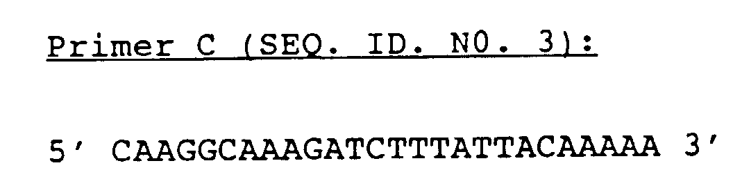
- SEQ. ID. N0. 3rd shows the primer C
- SEQ. ID. N0. 4th shows the primer D
- the plasmid pMAC1 was published by Deutsche Sammlung für Mikroorganismen (DSM), Griesebach No 8, D-3400 Göttingen under the Number DSM 6261 deposited.
- IgA protease As inclusion bodies, the coding region of the protein without signal sequence and helper sequence as described below behind a strong Promoter cloned (amino acid position +1 to position 959, Pohlner J., Halter R., Beyreuther K., Meyer T.F., Nature 325, (1987), 458-462).
- chromosomal DNA is isolated from N. gonorrhoeae (eg MS 11) and used to carry out a polymerase chain reaction (PCR, method see EP-A 0 200 362, EP-A 0 201 184).
- PCR polymerase chain reaction
- the ATG start codon and an EcoRI recognition sequence contains the first 5 codons of the IgA protease optimized for efficient epxression in E. coli, without changing the amino acid sequence.
- Primer B contains sequences flanked by the BglII recognition sequence the IgA protease (approx. amino acid position 553-561).
- a second PCR with the following primers is carried out to provide the 3 ′ region of the IgA protease gene:
- Primer C corresponds to the coding region of primer B (BglII interface) and primer D contains sequences of Amino acid position 952-959 followed by a stop codon and a SalI recognition sequence.
- a three-fragment ligation is then carried out: with fragments A / B, fragment C / D and the vector pKK223-3 (DSM 3694P), previously with the enzymes EcoRI and SalI was digested and purified.
- the vector thus obtained is called IgA-Prot III and transformed into E. coli K12.
- E.coli K12 cells were transformed as starting material used with plasmid pMAC1 (8878 bp). On this plasmid is the full coding region for IgA protease under the control of the lambda promoter. The plasmid carries an ampicillin resistance.
- the cells were cultured in LB medium overnight at 28 ° C and then diluted 1: 100 with LB medium. The culture was then incubated for a further 4 hours at 37 ° C. The cells were separated by a centrifugation step. Of the Culture supernatant was sterile filtered through cellulose acetate filters, against 20 mmol / l Tris / HCl, pH 7.5, 10 mmol / l EDTA, 10 % Glycerin (Buffer A) dialyzed and to 1/10 volume concentrated using a SALVIA capillary dialyzer E-15U.
- a negative elution was carried out on DEAE-Sephadex A-50 in 20 mmol / l Tris / HCl, pH 7.5, 10 mmol / l EDTA and 10% glycerol.
- the column loading was 0.5 mg protein per 1 ml gel matrix. With this separation, the IgA protease is in the passage and most E. coli proteins are bound to the carrier. By washing the column matrix with buffer A plus 1 mol / l NaCl, it was shown that less than 10% of the IgA protease binds to the column material.
- Elution is carried out with a linearly increasing NaCl gradient, the IgA protease in a buffer of pH 8.0 with a salt concentration of 0.1 mol / l NaCl and in a buffer of pH 7.0 with 0.2 mol / l NaCl eluted.
- Balance Concentrate before DEAE-Sephadex 3.5 mg protease (70% pure) Eluate according to DEAE-Sephadex 2.4 mg protease (90% pure) Eluate according to Fractogel®-EMD-SO 3 - 650M 1 mg protease ( ⁇ 95% purity)
- the starting material was the construct IgA-Prot III (Example 1) in E.coli cells DSM 3689, which additionally contain a lacI q plasmid for the expression of the Lac repressor.
- the cells are harvested by centrifugation, taken up in 10 ml of Tris-magnesium buffer (10 mmol / l Tris, pH 8.0, 1 mmol / l MgCl 2 ) and digested with lysozyme (0.3 mg / ml).
- the suspension is centrifuged for 10 minutes at 15,000 rpm and 4 ° C.
- the pellet is dissolved in 30 ml 50 mmol / l Tris, pH 8.0, 50 mmol / l EDTA and 0.5% Triton X 100 added and with Treated with ultrasound. It is centrifuged again, resuspended and treated with ultrasound. This procedure will repeated twice more. Then it is centrifuged and the pellets thus obtained are used as IBs in Example 3.
- Table 1 shows the results for the fermentation in a 1 liter shake culture and in a 10 liter fermenter.
- fermentation E.coli strain Total protein from IB material (g) IgA protease (%) (G) 1 l HB 101 0.125 50-70 0.06-0.09 10 l K12 C600 20.8 30-50 6.2-10.4
- the inventive method at least an increase in yield by a factor of 2 to 3 (1 1 culture) or 20 to 30 (10 1 fermenter) is found.
- the inclusion bodies were first solubilized, then dialyzed and then naturalized in the respective buffers.
- the denatured protein was in 5 portions in the renaturation buffer given, the interval between the Individual additions were 30 minutes and the protein concentration per pulse in the batch increased by 20 ⁇ g / ml.
- the final protein concentration was 100 ⁇ g / ml.
- cleavage buffer 50 mmol / l Tris / HCl pH 8, 1 mmol / l CaCl 2 .
- Human IgA was used as the fission substrate.
- Table 2 shows the result of the cleavage experiments (incubation: 6 h at 37 ° C.) of the renaturates obtained by using the above-mentioned naturation buffers (1-5).
- Cleavage of human IgA with the IgA protease isolated according to the present example incubation: 6 h at 37 ° C.
- the isolate obtained after dialysis contains about 50% IgA protease.
- Renaturate Protease / substrate ratio ( ⁇ g) Cleavage (%) 1 1:20 10th 2nd 1:20 30th 3rd 1:20 10th 4/5 1: 100 100 1: 500 50 1: 1000 30th 1: 2000 10th 1: 5000 5 soluble protease (100% pure) 1: 500 100
- the dialysate contains about 50 to 70% IgA protease.
- 50 ug substrate with 1 ug renatured IgA protease were incubated for 6 h at 37 ° C. pH % Cleavage Arginine (mol / l) % Cleavage 4th 10th 0.2 85 6 95 0.4 90 8th 100 0.6 95 0.8 95
Landscapes
- Health & Medical Sciences (AREA)
- Life Sciences & Earth Sciences (AREA)
- Chemical & Material Sciences (AREA)
- Zoology (AREA)
- Genetics & Genomics (AREA)
- Engineering & Computer Science (AREA)
- Bioinformatics & Cheminformatics (AREA)
- Organic Chemistry (AREA)
- Wood Science & Technology (AREA)
- General Engineering & Computer Science (AREA)
- Biomedical Technology (AREA)
- Biotechnology (AREA)
- Medicinal Chemistry (AREA)
- Biochemistry (AREA)
- Molecular Biology (AREA)
- General Health & Medical Sciences (AREA)
- Microbiology (AREA)
- Enzymes And Modification Thereof (AREA)
- Micro-Organisms Or Cultivation Processes Thereof (AREA)
- Preparation Of Compounds By Using Micro-Organisms (AREA)
- Detergent Compositions (AREA)
- Measuring Or Testing Involving Enzymes Or Micro-Organisms (AREA)
- Peptides Or Proteins (AREA)
- Medicines That Contain Protein Lipid Enzymes And Other Medicines (AREA)
Description
| SEQ. ID. N0. 1 | zeigt den in Beispiel 1 verwendeten Primer A |
| SEQ. ID. N0. 2 | zeigt den Primer B |
| SEQ. ID. N0. 3 | zeigt den Primer C |
| SEQ. ID. N0. 4 | zeigt den Primer D |



Durch Nachwaschen der Säulenmatrix mit Puffer A plus 1 mol/l NaCl wurde gezeigt, daß weniger als 10 % der IgA-Protease an dem Säulenmaterial binden.
| Bilanz: | |
| Konzentrat vor DEAE-Sephadex | 3,5 mg Protease (70 % rein) |
| Eluat nach DEAE-Sephadex | 2,4 mg Protease (90 % rein) |
| Eluat nach Fractogel®-EMD-SO3 -650M | 1 mg Protease (≥ 95 % Reinheit) |
| Bilanz: | |
| Konzentrat vor DEAE-Sephadex | 50 mg IgA-Protease (50 % Reinheit) |
| Eluat nach DEAE-Sephadex | 30 mg IgA-Protease (60 % Reinheit) |
| Eluat nach Fractogel®-EMD-SO3 -650M | 12 mg IgA-Protease (≥ 95 % Reinheit) |
| Fermentation | E.coli Stamm | Gesamtprotein aus IB-Material (g) | IgA-Protease | |
| (%) | (g) | |||
| 1 l | HB 101 | 0,125 | 50-70 | 0,06-0,09 |
| 10 l | K12 C600 | 20,8 | 30-50 | 6,2-10,4 |
| Spaltung von humanem IgA mit der nach vorliegendem Beispiel isolierten IgA-Protease (Inkubation: 6 h bei 37°C). Das nach der Dialyse erhaltene Isolat enthält zu etwa 50 % IgA-Protease. | ||
| Renaturat | Verhältnis Protease/Substrat (µg) | Spaltung (%) |
| 1 | 1:20 | 10 |
| 2 | 1:20 | 30 |
| 3 | 1:20 | 10 |
| 4/5 | 1:100 | 100 |
| 1:500 | 50 | |
| 1:1000 | 30 | |
| 1:2000 | 10 | |
| 1:5000 | 5 | |
| lösliche Protease (100 % rein) | 1:500 | 100 |
0,6 mol/l Arg/HCl
1 mmol/l EDTA
pH 4, 6, 8
1 mmol/l EDTA, pH 8
1 mmol/l DTE
Arg/HCl: 0,2; 0,4; 0,6 und 0,8 mol/l
| pH | % Spaltung | Arginin (mol/l) | % Spaltung |
| 4 | 10 | 0,2 | 85 |
| 6 | 95 | 0,4 | 90 |
| 8 | 100 | 0,6 | 95 |
| 0,8 | 95 |
- ART DER SEQUENZ:
- Nukleinsäure-Einzelstrang
- SEQUENZLÄNGE:
- 56 Nukleotide

- ART DER SEQUENZ:
- Nukleinsäure-Einzelstrang
- SEQUENZLÄNGE:
- 26 Nukleotide

- ART DER SEQUENZ:
- Nukleinsäure-Einzelstrang
- SEQUENZLÄNGE:
- 26 Nukleotide

- ART DER SEQUENZ:
- Nukleinsäure-Einzelstrang
- SEQUENZLÄNGE:
- 44 Nukleotide

Claims (7)
- Verfahren zur Herstellung von rekominanter IgA-Protease,
dadurch gekennzeichnet,
daß man(1) ein IgA-Protease-Gen so modifiziert, daß der für den C-terminalen Helferanteil des IgA-Protease-Gens kodierende DNA-Bereich nicht mehr funktionell aktiv ist,(2) eine Wirtszelle mit dem in Schritt (1) modifizierten IgA-Protease-Gen oder einem dieses modifizierte Gen enthaltenden Vektor transformiert,(3) das modifizierte IgA-Protease-Gen in der transformierten Wirtszelle zur Expression bringt,(4) die in Form von Inclusion Bodies entstehende IgA-Protease aus der Wirtszelle isoliert, und(5) die IgA-Protease durch in vitro Aktivierung in aktives Protein überführt. - Verfahren nach Anspruch 1,
dadurch gekennzeichnet,
daß man das IgA-Protease-Gen so modifiziert, daß der C-terminale Helferanteil der durch Expression des modifizierten Gens resultierenden IgA-Protease vollständig deletiert ist. - Verfahren nach Anspruch 1 oder 2,
dadurch gekennzeichnet,
daß man in den für den Helferanteil kodierenden DNA-Bereich einen oder mehrere Translationsstopcodons einführt oder den für den Helferanteil kodierenden DNA-Bereich teilweise oder vollständig deletiert. - Verfahren nach einem der Ansprüche 1 bis 3,
dadurch gekennzeichnet,
daß man das IgA-Protease-Gen zusätzlich so modifiziert, daß der für den N-terminalen Signalanteil der IgA-Protease-Gens kodierende DNA-Bereich nicht mehr funktionell aktiv ist. - Verfahren nach Anspruch 4,
dadurch gekennzeichnet,
daß man das IgA-Protease-Gen so modifiziert, daß der N-terminale Signalanteil der durch Expression des modifizierten Gens resultierende IgA-Protease vollständig deletiert ist. - Verfahren nach einem der vorhergehenden Ansprüche,
dadurch gekennzeichnet,
daß man als Wirtszelle eine E.coli-Zelle verwendet. - Verfahren nach einem der vorhergehenden Ansprüche,
dadurch gekennzeichnet,
daß man den Naturierungsschritt in Gegenwart von 0,2 bis 1 mol/l Arginin durchführt.
Applications Claiming Priority (4)
| Application Number | Priority Date | Filing Date | Title |
|---|---|---|---|
| DE4100704 | 1991-01-11 | ||
| DE4100704 | 1991-01-11 | ||
| DE4140699 | 1991-12-10 | ||
| DE4140699A DE4140699A1 (de) | 1991-01-11 | 1991-12-10 | Rekombinante iga-protease |
Publications (2)
| Publication Number | Publication Date |
|---|---|
| EP0495398A1 EP0495398A1 (de) | 1992-07-22 |
| EP0495398B1 true EP0495398B1 (de) | 1998-12-16 |
Family
ID=25900194
Family Applications (1)
| Application Number | Title | Priority Date | Filing Date |
|---|---|---|---|
| EP92100288A Expired - Lifetime EP0495398B1 (de) | 1991-01-11 | 1992-01-09 | Verfahren zur Herstellung von rekombinanter IgA-protease. |
Country Status (14)
| Country | Link |
|---|---|
| EP (1) | EP0495398B1 (de) |
| JP (1) | JP2509410B2 (de) |
| AT (1) | ATE174627T1 (de) |
| AU (1) | AU636261B2 (de) |
| CA (1) | CA2058872C (de) |
| DE (2) | DE4140699A1 (de) |
| DK (1) | DK0495398T3 (de) |
| ES (1) | ES2127732T3 (de) |
| FI (1) | FI103132B (de) |
| HU (1) | HU215188B (de) |
| IE (1) | IE914559A1 (de) |
| IL (1) | IL100623A (de) |
| NZ (1) | NZ241280A (de) |
| TW (1) | TW197470B (de) |
Families Citing this family (7)
| Publication number | Priority date | Publication date | Assignee | Title |
|---|---|---|---|---|
| WO1996009395A2 (de) * | 1994-09-21 | 1996-03-28 | MAX-PLANCK-Gesellschaft zur Förderung der Wissenschaften e.V. | Arzneimittel zur prophylaxe und behandlung von und diagnostika zur erkennung von autoimmunkrankheiten und viralen erkrankungen |
| GB9618960D0 (en) | 1996-09-11 | 1996-10-23 | Medical Science Sys Inc | Proteases |
| AU9341398A (en) * | 1997-08-22 | 1999-03-16 | Boehringer Mannheim Gmbh | Protease precursors that can be autocatalytically activated and their use |
| ATE294869T1 (de) * | 1997-08-22 | 2005-05-15 | Roche Diagnostics Gmbh | Autokatalytisch aktivierbare zymogene vorstufen von proteasen und deren verwendung |
| AU7053500A (en) * | 1999-09-15 | 2001-04-17 | Eli Lilly And Company | Chymotrypsin-free trypsin |
| US20100261252A1 (en) * | 2009-04-10 | 2010-10-14 | Biomarin Pharmaceutical Inc. | Methods of enhancing yield of active iga protease |
| CN121548585A (zh) | 2023-07-19 | 2026-02-17 | 豪夫迈·罗氏有限公司 | 作为IgG片段形式的dsFv及其产生和标记方法 |
Family Cites Families (3)
| Publication number | Priority date | Publication date | Assignee | Title |
|---|---|---|---|---|
| DE3622221A1 (de) * | 1986-07-02 | 1988-01-14 | Max Planck Gesellschaft | Verfahren zur gentechnologischen gewinnung von proteinen unter verwendung gramnegativer wirtszellen |
| DE3832898A1 (de) * | 1988-09-28 | 1990-04-12 | Boehringer Mannheim Gmbh | Praeparat von in prokaryonten exprimiertem plasminogenaktivator |
| DK130889A (da) * | 1989-03-17 | 1990-09-18 | Mogens Kilian | Immunoglobulin a1-proteaser (iga1-proteaser), fremgangsmaade til genteknologisk fremstilling af saadanne enzymer samt vaccine indeholdende enzymerne og fragmenter deraf til immunisering mod bakteriel meningitis og andre sygdomme fremkaldt af iga1-protease-producerende bakterier |
-
1991
- 1991-12-10 DE DE4140699A patent/DE4140699A1/de not_active Withdrawn
- 1991-12-23 IE IE455991A patent/IE914559A1/en not_active IP Right Cessation
- 1991-12-31 TW TW080110300A patent/TW197470B/zh active
- 1991-12-31 AU AU90116/91A patent/AU636261B2/en not_active Ceased
-
1992
- 1992-01-07 CA CA002058872A patent/CA2058872C/en not_active Expired - Lifetime
- 1992-01-09 IL IL10062392A patent/IL100623A/en not_active IP Right Cessation
- 1992-01-09 ES ES92100288T patent/ES2127732T3/es not_active Expired - Lifetime
- 1992-01-09 DK DK92100288T patent/DK0495398T3/da active
- 1992-01-09 EP EP92100288A patent/EP0495398B1/de not_active Expired - Lifetime
- 1992-01-09 AT AT92100288T patent/ATE174627T1/de not_active IP Right Cessation
- 1992-01-09 DE DE59209590T patent/DE59209590D1/de not_active Expired - Lifetime
- 1992-01-10 FI FI920117A patent/FI103132B/fi active
- 1992-01-10 NZ NZ241280A patent/NZ241280A/xx not_active IP Right Cessation
- 1992-01-10 HU HU9200092A patent/HU215188B/hu not_active IP Right Cessation
- 1992-01-10 JP JP4002851A patent/JP2509410B2/ja not_active Expired - Lifetime
Non-Patent Citations (2)
| Title |
|---|
| EMBO, Vol.3(7), p.1595-1601 (1984). * |
| Nature, Vol.321(6103), p.458-462 (1987). * |
Also Published As
| Publication number | Publication date |
|---|---|
| FI920117L (fi) | 1992-07-12 |
| AU636261B2 (en) | 1993-04-22 |
| FI920117A0 (fi) | 1992-01-10 |
| NZ241280A (en) | 1993-10-26 |
| JPH05168478A (ja) | 1993-07-02 |
| EP0495398A1 (de) | 1992-07-22 |
| HU215188B (hu) | 1998-10-28 |
| ES2127732T3 (es) | 1999-05-01 |
| JP2509410B2 (ja) | 1996-06-19 |
| ATE174627T1 (de) | 1999-01-15 |
| IL100623A (en) | 2002-07-25 |
| DE4140699A1 (de) | 1992-07-16 |
| CA2058872C (en) | 1999-12-21 |
| IE914559A1 (en) | 1992-07-15 |
| HU9200092D0 (en) | 1992-03-30 |
| IL100623A0 (en) | 1992-09-06 |
| AU9011691A (en) | 1992-09-10 |
| DE59209590D1 (de) | 1999-01-28 |
| FI103132B1 (fi) | 1999-04-30 |
| DK0495398T3 (da) | 1999-08-23 |
| FI103132B (fi) | 1999-04-30 |
| CA2058872A1 (en) | 1992-07-12 |
| HUT63654A (en) | 1993-09-28 |
| TW197470B (de) | 1993-01-01 |
Similar Documents
| Publication | Publication Date | Title |
|---|---|---|
| EP0602688B1 (de) | Methioninfreier G-CSF aus Prokaryonten | |
| DE69233008T2 (de) | Fusionierung von Peptiden und Proteinen mit Thioredoxin und thioredoxin-ähnlichen Molekülen | |
| DD229427A5 (de) | Verfahren zur gewinnung eines polypeptids | |
| JPH06296491A (ja) | シグナル配列をコードしているdnaと機能的に結合しているヒト成長ホルモンをコードしているdna | |
| DD210071A5 (de) | Verfahren zur herstellung einer lebensfaehigen kultur | |
| EP0316933B1 (de) | Verfahren zur Herstellung von Apoaequorin | |
| EP0587427B1 (de) | Verfahren zur Herstellung von menschlichem Wachstumshormon | |
| DE60029968T2 (de) | Proteinherstellung | |
| KR100218853B1 (ko) | 인체 혈청 알부민의 제조방법 | |
| EP0495398A1 (de) | Rekombinante IgA-Protease | |
| DE3687112T2 (de) | Verfahren zur ausscheidung von genprodukten in das wachstumsmedium von gram-negativen bakterien. | |
| EP0643133A2 (de) | Rekombinante D-Hydantoinase, Verfahren zur Herstellung und Verwendung | |
| IE921888A1 (en) | Production and recovery of recombinant neurotrophins | |
| EP1169461B1 (de) | Verwendung von pankreatischer Procarboxypeptidase B zur Herstellung von Insulin | |
| DE3783305T2 (de) | Expressionsvektor fuer menschlichen insulinaehnlichen wachstumsfaktor-i. | |
| EP0300425B1 (de) | Verfahren zur Herstellung von Proteinen in löslicher Form | |
| DE3877197T2 (de) | Verfahren zur herstellung von hirudin. | |
| DE68915204T2 (de) | Biosynthetisches Verfahren zur Herstellung von chemischen Verbindungen. | |
| US5527691A (en) | Expression vectors containing lambdapl promoter and T1 T2 rRNA termination sequence, plasmids containing the vectors, hosts containing the plasmids and related methods | |
| EP0393039A1 (de) | Verfahren zur herstellung von peptiden durch spezifische spaltung von gentechnisch gewonnenen fusionsproteinen mit collagenasen | |
| JPH01273591A (ja) | ヒト成長ホルモン分泌プラスミド、ならびにそれを用いた形質転換体および蛋白質分泌生産法 | |
| WO1999010483A2 (de) | Autokatalytisch aktivierbare vorstufen von proteasen und deren verwendung | |
| EP0750667B1 (de) | Verwendung von rekombinantem inhibitor aus erythrina caffra zur reinigung von serinproteasen | |
| DE4015922A1 (de) | Verfahren zur enzymatischen prozessierung von proteinen unter verwendung von iga-proteasen (igase) | |
| DE4424171A1 (de) | Verwendung von rekombinantem Inhibitor aus Erythrina caffra zur Reinigung von Serinproteasen |
Legal Events
| Date | Code | Title | Description |
|---|---|---|---|
| PUAI | Public reference made under article 153(3) epc to a published international application that has entered the european phase |
Free format text: ORIGINAL CODE: 0009012 |
|
| 17P | Request for examination filed |
Effective date: 19920109 |
|
| AK | Designated contracting states |
Kind code of ref document: A1 Designated state(s): AT BE CH DE DK ES FR GB GR IT LI LU NL PT SE |
|
| 17Q | First examination report despatched |
Effective date: 19970307 |
|
| GRAG | Despatch of communication of intention to grant |
Free format text: ORIGINAL CODE: EPIDOS AGRA |
|
| GRAG | Despatch of communication of intention to grant |
Free format text: ORIGINAL CODE: EPIDOS AGRA |
|
| GRAH | Despatch of communication of intention to grant a patent |
Free format text: ORIGINAL CODE: EPIDOS IGRA |
|
| GRAH | Despatch of communication of intention to grant a patent |
Free format text: ORIGINAL CODE: EPIDOS IGRA |
|
| GRAA | (expected) grant |
Free format text: ORIGINAL CODE: 0009210 |
|
| AK | Designated contracting states |
Kind code of ref document: B1 Designated state(s): AT BE CH DE DK ES FR GB GR IT LI LU NL PT SE |
|
| PG25 | Lapsed in a contracting state [announced via postgrant information from national office to epo] |
Ref country code: GR Free format text: LAPSE BECAUSE OF NON-PAYMENT OF DUE FEES Effective date: 19981216 |
|
| REF | Corresponds to: |
Ref document number: 174627 Country of ref document: AT Date of ref document: 19990115 Kind code of ref document: T |
|
| REG | Reference to a national code |
Ref country code: CH Ref legal event code: NV Representative=s name: A. BRAUN, BRAUN, HERITIER, ESCHMANN AG PATENTANWAE Ref country code: CH Ref legal event code: EP |
|
| PGFP | Annual fee paid to national office [announced via postgrant information from national office to epo] |
Ref country code: SE Payment date: 19990107 Year of fee payment: 8 |
|
| GBT | Gb: translation of ep patent filed (gb section 77(6)(a)/1977) |
Effective date: 19981221 |
|
| PGFP | Annual fee paid to national office [announced via postgrant information from national office to epo] |
Ref country code: LU Payment date: 19990118 Year of fee payment: 8 |
|
| REF | Corresponds to: |
Ref document number: 59209590 Country of ref document: DE Date of ref document: 19990128 |
|
| ITF | It: translation for a ep patent filed | ||
| PG25 | Lapsed in a contracting state [announced via postgrant information from national office to epo] |
Ref country code: PT Free format text: LAPSE BECAUSE OF FAILURE TO SUBMIT A TRANSLATION OF THE DESCRIPTION OR TO PAY THE FEE WITHIN THE PRESCRIBED TIME-LIMIT Effective date: 19990316 Ref country code: SE Free format text: LAPSE BECAUSE OF FAILURE TO SUBMIT A TRANSLATION OF THE DESCRIPTION OR TO PAY THE FEE WITHIN THE PRESCRIBED TIME-LIMIT Effective date: 19990316 |
|
| RAP4 | Party data changed (patent owner data changed or rights of a patent transferred) |
Owner name: ROCHE DIAGNOSTICS GMBH |
|
| ET | Fr: translation filed | ||
| REG | Reference to a national code |
Ref country code: ES Ref legal event code: FG2A Ref document number: 2127732 Country of ref document: ES Kind code of ref document: T3 |
|
| REG | Reference to a national code |
Ref country code: DK Ref legal event code: T3 |
|
| PLBE | No opposition filed within time limit |
Free format text: ORIGINAL CODE: 0009261 |
|
| STAA | Information on the status of an ep patent application or granted ep patent |
Free format text: STATUS: NO OPPOSITION FILED WITHIN TIME LIMIT |
|
| 26N | No opposition filed | ||
| PG25 | Lapsed in a contracting state [announced via postgrant information from national office to epo] |
Ref country code: LU Free format text: LAPSE BECAUSE OF NON-PAYMENT OF DUE FEES Effective date: 20000109 |
|
| REG | Reference to a national code |
Ref country code: GB Ref legal event code: IF02 |
|
| PGFP | Annual fee paid to national office [announced via postgrant information from national office to epo] |
Ref country code: AT Payment date: 20061212 Year of fee payment: 16 |
|
| PGFP | Annual fee paid to national office [announced via postgrant information from national office to epo] |
Ref country code: DK Payment date: 20061213 Year of fee payment: 16 |
|
| PGFP | Annual fee paid to national office [announced via postgrant information from national office to epo] |
Ref country code: NL Payment date: 20061222 Year of fee payment: 16 |
|
| PGFP | Annual fee paid to national office [announced via postgrant information from national office to epo] |
Ref country code: BE Payment date: 20070208 Year of fee payment: 16 |
|
| REG | Reference to a national code |
Ref country code: CH Ref legal event code: PFA Owner name: ROCHE DIAGNOSTICS GMBH Free format text: ROCHE DIAGNOSTICS GMBH# #68298 MANNHEIM (DE) -TRANSFER TO- ROCHE DIAGNOSTICS GMBH# #68298 MANNHEIM (DE) |
|
| BERE | Be: lapsed |
Owner name: *BOEHRINGER MANNHEIM G.M.B.H. Effective date: 20080131 |
|
| REG | Reference to a national code |
Ref country code: DK Ref legal event code: EBP |
|
| NLV4 | Nl: lapsed or anulled due to non-payment of the annual fee |
Effective date: 20080801 |
|
| PG25 | Lapsed in a contracting state [announced via postgrant information from national office to epo] |
Ref country code: NL Free format text: LAPSE BECAUSE OF NON-PAYMENT OF DUE FEES Effective date: 20080801 |
|
| PG25 | Lapsed in a contracting state [announced via postgrant information from national office to epo] |
Ref country code: AT Free format text: LAPSE BECAUSE OF NON-PAYMENT OF DUE FEES Effective date: 20080109 |
|
| PG25 | Lapsed in a contracting state [announced via postgrant information from national office to epo] |
Ref country code: DK Free format text: LAPSE BECAUSE OF NON-PAYMENT OF DUE FEES Effective date: 20080131 |
|
| PG25 | Lapsed in a contracting state [announced via postgrant information from national office to epo] |
Ref country code: BE Free format text: LAPSE BECAUSE OF NON-PAYMENT OF DUE FEES Effective date: 20080131 |
|
| PGFP | Annual fee paid to national office [announced via postgrant information from national office to epo] |
Ref country code: FR Payment date: 20101221 Year of fee payment: 20 |
|
| PGFP | Annual fee paid to national office [announced via postgrant information from national office to epo] |
Ref country code: GB Payment date: 20101215 Year of fee payment: 20 |
|
| PGFP | Annual fee paid to national office [announced via postgrant information from national office to epo] |
Ref country code: DE Payment date: 20110131 Year of fee payment: 20 Ref country code: IT Payment date: 20110117 Year of fee payment: 20 Ref country code: CH Payment date: 20110103 Year of fee payment: 20 |
|
| PGFP | Annual fee paid to national office [announced via postgrant information from national office to epo] |
Ref country code: ES Payment date: 20110117 Year of fee payment: 20 |
|
| REG | Reference to a national code |
Ref country code: DE Ref legal event code: R071 Ref document number: 59209590 Country of ref document: DE |
|
| REG | Reference to a national code |
Ref country code: DE Ref legal event code: R071 Ref document number: 59209590 Country of ref document: DE |
|
| REG | Reference to a national code |
Ref country code: CH Ref legal event code: PL |
|
| REG | Reference to a national code |
Ref country code: GB Ref legal event code: PE20 Expiry date: 20120108 |
|
| PG25 | Lapsed in a contracting state [announced via postgrant information from national office to epo] |
Ref country code: DE Free format text: LAPSE BECAUSE OF EXPIRATION OF PROTECTION Effective date: 20120110 |
|
| REG | Reference to a national code |
Ref country code: ES Ref legal event code: FD2A Effective date: 20120510 |
|
| PG25 | Lapsed in a contracting state [announced via postgrant information from national office to epo] |
Ref country code: GB Free format text: LAPSE BECAUSE OF EXPIRATION OF PROTECTION Effective date: 20120108 |
|
| PG25 | Lapsed in a contracting state [announced via postgrant information from national office to epo] |
Ref country code: ES Free format text: LAPSE BECAUSE OF EXPIRATION OF PROTECTION Effective date: 20120110 |