EP0333344B1 - Corneal implants and manufacture and use thereof - Google Patents
Corneal implants and manufacture and use thereof Download PDFInfo
- Publication number
- EP0333344B1 EP0333344B1 EP89302053A EP89302053A EP0333344B1 EP 0333344 B1 EP0333344 B1 EP 0333344B1 EP 89302053 A EP89302053 A EP 89302053A EP 89302053 A EP89302053 A EP 89302053A EP 0333344 B1 EP0333344 B1 EP 0333344B1
- Authority
- EP
- European Patent Office
- Prior art keywords
- prosthesis
- optical element
- skirt
- fibers
- porous
- Prior art date
- Legal status (The legal status is an assumption and is not a legal conclusion. Google has not performed a legal analysis and makes no representation as to the accuracy of the status listed.)
- Expired - Lifetime
Links
- 238000004519 manufacturing process Methods 0.000 title claims description 8
- 239000007943 implant Substances 0.000 title description 11
- 239000000463 material Substances 0.000 claims description 56
- 230000003287 optical effect Effects 0.000 claims description 51
- 210000004027 cell Anatomy 0.000 claims description 47
- 239000000835 fiber Substances 0.000 claims description 47
- 210000002919 epithelial cell Anatomy 0.000 claims description 42
- 239000000017 hydrogel Substances 0.000 claims description 30
- 238000000034 method Methods 0.000 claims description 29
- 239000011148 porous material Substances 0.000 claims description 16
- XLYOFNOQVPJJNP-UHFFFAOYSA-N water Substances O XLYOFNOQVPJJNP-UHFFFAOYSA-N 0.000 claims description 15
- 230000001427 coherent effect Effects 0.000 claims description 8
- 229920000036 polyvinylpyrrolidone Polymers 0.000 claims description 7
- 239000001267 polyvinylpyrrolidone Substances 0.000 claims description 7
- 235000013855 polyvinylpyrrolidone Nutrition 0.000 claims description 7
- 239000004372 Polyvinyl alcohol Substances 0.000 claims description 5
- 229920002451 polyvinyl alcohol Polymers 0.000 claims description 5
- 239000011800 void material Substances 0.000 claims description 4
- 229920006240 drawn fiber Polymers 0.000 claims description 3
- 229920001169 thermoplastic Polymers 0.000 claims description 2
- 239000004416 thermosoftening plastic Substances 0.000 claims description 2
- 210000001519 tissue Anatomy 0.000 description 29
- 210000004087 cornea Anatomy 0.000 description 22
- 238000010899 nucleation Methods 0.000 description 22
- 239000000203 mixture Substances 0.000 description 15
- -1 polybutylene Polymers 0.000 description 15
- 238000001125 extrusion Methods 0.000 description 11
- 239000000155 melt Substances 0.000 description 11
- 102000004169 proteins and genes Human genes 0.000 description 11
- 108090000623 proteins and genes Proteins 0.000 description 11
- 210000005081 epithelial layer Anatomy 0.000 description 10
- OKKJLVBELUTLKV-UHFFFAOYSA-N Methanol Chemical compound OC OKKJLVBELUTLKV-UHFFFAOYSA-N 0.000 description 9
- 238000000338 in vitro Methods 0.000 description 9
- 239000002609 medium Substances 0.000 description 9
- 230000015572 biosynthetic process Effects 0.000 description 8
- 229920001748 polybutylene Polymers 0.000 description 8
- 229920000642 polymer Polymers 0.000 description 8
- 239000002904 solvent Substances 0.000 description 8
- 238000003797 solvolysis reaction Methods 0.000 description 8
- 102000008186 Collagen Human genes 0.000 description 7
- 108010035532 Collagen Proteins 0.000 description 7
- 108010080379 Fibrin Tissue Adhesive Proteins 0.000 description 7
- 239000004743 Polypropylene Substances 0.000 description 7
- 229920001436 collagen Polymers 0.000 description 7
- 235000015097 nutrients Nutrition 0.000 description 7
- 230000002093 peripheral effect Effects 0.000 description 7
- 229920001155 polypropylene Polymers 0.000 description 7
- CSCPPACGZOOCGX-UHFFFAOYSA-N Acetone Chemical compound CC(C)=O CSCPPACGZOOCGX-UHFFFAOYSA-N 0.000 description 6
- 102000012422 Collagen Type I Human genes 0.000 description 6
- 108010022452 Collagen Type I Proteins 0.000 description 6
- 229920001410 Microfiber Polymers 0.000 description 6
- 238000002513 implantation Methods 0.000 description 6
- 239000003658 microfiber Substances 0.000 description 6
- 239000002243 precursor Substances 0.000 description 6
- 210000002469 basement membrane Anatomy 0.000 description 5
- 210000000981 epithelium Anatomy 0.000 description 5
- 238000001727 in vivo Methods 0.000 description 5
- 238000013508 migration Methods 0.000 description 5
- 238000002360 preparation method Methods 0.000 description 5
- 108010067306 Fibronectins Proteins 0.000 description 4
- 102000016359 Fibronectins Human genes 0.000 description 4
- 102000007547 Laminin Human genes 0.000 description 4
- 108010085895 Laminin Proteins 0.000 description 4
- XTXRWKRVRITETP-UHFFFAOYSA-N Vinyl acetate Chemical compound CC(=O)OC=C XTXRWKRVRITETP-UHFFFAOYSA-N 0.000 description 4
- 239000000853 adhesive Substances 0.000 description 4
- 230000001070 adhesive effect Effects 0.000 description 4
- 230000008901 benefit Effects 0.000 description 4
- 230000010261 cell growth Effects 0.000 description 4
- 230000012010 growth Effects 0.000 description 4
- FPYJFEHAWHCUMM-UHFFFAOYSA-N maleic anhydride Chemical compound O=C1OC(=O)C=C1 FPYJFEHAWHCUMM-UHFFFAOYSA-N 0.000 description 4
- 230000005012 migration Effects 0.000 description 4
- 229920003023 plastic Polymers 0.000 description 4
- 239000004033 plastic Substances 0.000 description 4
- 238000006116 polymerization reaction Methods 0.000 description 4
- 230000008569 process Effects 0.000 description 4
- 238000002054 transplantation Methods 0.000 description 4
- QTBSBXVTEAMEQO-UHFFFAOYSA-N Acetic acid Chemical compound CC(O)=O QTBSBXVTEAMEQO-UHFFFAOYSA-N 0.000 description 3
- 102000004266 Collagen Type IV Human genes 0.000 description 3
- 108010042086 Collagen Type IV Proteins 0.000 description 3
- HTTJABKRGRZYRN-UHFFFAOYSA-N Heparin Chemical compound OC1C(NC(=O)C)C(O)OC(COS(O)(=O)=O)C1OC1C(OS(O)(=O)=O)C(O)C(OC2C(C(OS(O)(=O)=O)C(OC3C(C(O)C(O)C(O3)C(O)=O)OS(O)(=O)=O)C(CO)O2)NS(O)(=O)=O)C(C(O)=O)O1 HTTJABKRGRZYRN-UHFFFAOYSA-N 0.000 description 3
- 241000283973 Oryctolagus cuniculus Species 0.000 description 3
- FAPWRFPIFSIZLT-UHFFFAOYSA-M Sodium chloride Chemical compound [Na+].[Cl-] FAPWRFPIFSIZLT-UHFFFAOYSA-M 0.000 description 3
- IQFYYKKMVGJFEH-XLPZGREQSA-N Thymidine Chemical compound O=C1NC(=O)C(C)=CN1[C@@H]1O[C@H](CO)[C@@H](O)C1 IQFYYKKMVGJFEH-XLPZGREQSA-N 0.000 description 3
- 230000004888 barrier function Effects 0.000 description 3
- 230000008859 change Effects 0.000 description 3
- 239000011248 coating agent Substances 0.000 description 3
- 238000000576 coating method Methods 0.000 description 3
- 229920001577 copolymer Polymers 0.000 description 3
- 230000008472 epithelial growth Effects 0.000 description 3
- ZBGRMWIREQJHPK-UHFFFAOYSA-N ethenyl 2,2,2-trifluoroacetate Chemical compound FC(F)(F)C(=O)OC=C ZBGRMWIREQJHPK-UHFFFAOYSA-N 0.000 description 3
- 239000001963 growth medium Substances 0.000 description 3
- 229960002897 heparin Drugs 0.000 description 3
- 229920000669 heparin Polymers 0.000 description 3
- 238000000465 moulding Methods 0.000 description 3
- 229920000098 polyolefin Polymers 0.000 description 3
- 238000001356 surgical procedure Methods 0.000 description 3
- 238000003786 synthesis reaction Methods 0.000 description 3
- 238000003466 welding Methods 0.000 description 3
- 102000010834 Extracellular Matrix Proteins Human genes 0.000 description 2
- 108010037362 Extracellular Matrix Proteins Proteins 0.000 description 2
- YQEZLKZALYSWHR-UHFFFAOYSA-N Ketamine Chemical compound C=1C=CC=C(Cl)C=1C1(NC)CCCCC1=O YQEZLKZALYSWHR-UHFFFAOYSA-N 0.000 description 2
- IMNFDUFMRHMDMM-UHFFFAOYSA-N N-Heptane Chemical compound CCCCCCC IMNFDUFMRHMDMM-UHFFFAOYSA-N 0.000 description 2
- 239000004677 Nylon Substances 0.000 description 2
- 239000004698 Polyethylene Substances 0.000 description 2
- 238000009825 accumulation Methods 0.000 description 2
- 150000001413 amino acids Chemical class 0.000 description 2
- 210000002159 anterior chamber Anatomy 0.000 description 2
- 239000003364 biologic glue Substances 0.000 description 2
- 238000007664 blowing Methods 0.000 description 2
- 125000002843 carboxylic acid group Chemical group 0.000 description 2
- 230000004663 cell proliferation Effects 0.000 description 2
- 229920002301 cellulose acetate Polymers 0.000 description 2
- 238000006243 chemical reaction Methods 0.000 description 2
- 238000010276 construction Methods 0.000 description 2
- 238000007598 dipping method Methods 0.000 description 2
- 230000005684 electric field Effects 0.000 description 2
- 210000003038 endothelium Anatomy 0.000 description 2
- 230000007515 enzymatic degradation Effects 0.000 description 2
- 210000003560 epithelium corneal Anatomy 0.000 description 2
- 210000002744 extracellular matrix Anatomy 0.000 description 2
- 238000000605 extraction Methods 0.000 description 2
- 125000000524 functional group Chemical group 0.000 description 2
- 230000010354 integration Effects 0.000 description 2
- 229960003299 ketamine Drugs 0.000 description 2
- 238000005259 measurement Methods 0.000 description 2
- 210000000826 nictitating membrane Anatomy 0.000 description 2
- 229920001778 nylon Polymers 0.000 description 2
- 229920002492 poly(sulfone) Polymers 0.000 description 2
- 229920000573 polyethylene Polymers 0.000 description 2
- 238000003825 pressing Methods 0.000 description 2
- 238000012545 processing Methods 0.000 description 2
- 230000002285 radioactive effect Effects 0.000 description 2
- 210000001525 retina Anatomy 0.000 description 2
- 238000001878 scanning electron micrograph Methods 0.000 description 2
- DAEPDZWVDSPTHF-UHFFFAOYSA-M sodium pyruvate Chemical compound [Na+].CC(=O)C([O-])=O DAEPDZWVDSPTHF-UHFFFAOYSA-M 0.000 description 2
- 238000001179 sorption measurement Methods 0.000 description 2
- UCSJYZPVAKXKNQ-HZYVHMACSA-N streptomycin Chemical compound CN[C@H]1[C@H](O)[C@@H](O)[C@H](CO)O[C@H]1O[C@@H]1[C@](C=O)(O)[C@H](C)O[C@H]1O[C@@H]1[C@@H](NC(N)=N)[C@H](O)[C@@H](NC(N)=N)[C@H](O)[C@H]1O UCSJYZPVAKXKNQ-HZYVHMACSA-N 0.000 description 2
- 239000000126 substance Substances 0.000 description 2
- 229920002554 vinyl polymer Polymers 0.000 description 2
- BFUUJUGQJUTPAF-UHFFFAOYSA-N 2-(3-amino-4-propoxybenzoyl)oxyethyl-diethylazanium;chloride Chemical compound [Cl-].CCCOC1=CC=C(C(=O)OCC[NH+](CC)CC)C=C1N BFUUJUGQJUTPAF-UHFFFAOYSA-N 0.000 description 1
- XMLYCEVDHLAQEL-UHFFFAOYSA-N 2-hydroxy-2-methyl-1-phenylpropan-1-one Chemical compound CC(C)(O)C(=O)C1=CC=CC=C1 XMLYCEVDHLAQEL-UHFFFAOYSA-N 0.000 description 1
- VHUUQVKOLVNVRT-UHFFFAOYSA-N Ammonium hydroxide Chemical compound [NH4+].[OH-] VHUUQVKOLVNVRT-UHFFFAOYSA-N 0.000 description 1
- 108091003079 Bovine Serum Albumin Proteins 0.000 description 1
- OYPRJOBELJOOCE-UHFFFAOYSA-N Calcium Chemical compound [Ca] OYPRJOBELJOOCE-UHFFFAOYSA-N 0.000 description 1
- 206010053567 Coagulopathies Diseases 0.000 description 1
- 229920004934 Dacron® Polymers 0.000 description 1
- PIICEJLVQHRZGT-UHFFFAOYSA-N Ethylenediamine Chemical compound NCCN PIICEJLVQHRZGT-UHFFFAOYSA-N 0.000 description 1
- 108010073385 Fibrin Proteins 0.000 description 1
- 102000009123 Fibrin Human genes 0.000 description 1
- BWGVNKXGVNDBDI-UHFFFAOYSA-N Fibrin monomer Chemical compound CNC(=O)CNC(=O)CN BWGVNKXGVNDBDI-UHFFFAOYSA-N 0.000 description 1
- 206010061218 Inflammation Diseases 0.000 description 1
- 108010052285 Membrane Proteins Proteins 0.000 description 1
- 241001465754 Metazoa Species 0.000 description 1
- VVQNEPGJFQJSBK-UHFFFAOYSA-N Methyl methacrylate Chemical compound COC(=O)C(C)=C VVQNEPGJFQJSBK-UHFFFAOYSA-N 0.000 description 1
- 102220480283 Nicotinate phosphoribosyltransferase_D19A_mutation Human genes 0.000 description 1
- 241000283977 Oryctolagus Species 0.000 description 1
- 229930182555 Penicillin Natural products 0.000 description 1
- JGSARLDLIJGVTE-MBNYWOFBSA-N Penicillin G Chemical compound N([C@H]1[C@H]2SC([C@@H](N2C1=O)C(O)=O)(C)C)C(=O)CC1=CC=CC=C1 JGSARLDLIJGVTE-MBNYWOFBSA-N 0.000 description 1
- 102000057297 Pepsin A Human genes 0.000 description 1
- 108090000284 Pepsin A Proteins 0.000 description 1
- 241001255741 Vanna Species 0.000 description 1
- 239000002253 acid Substances 0.000 description 1
- 238000007792 addition Methods 0.000 description 1
- 239000005030 aluminium foil Substances 0.000 description 1
- 125000003368 amide group Chemical group 0.000 description 1
- 150000001408 amides Chemical class 0.000 description 1
- 235000011114 ammonium hydroxide Nutrition 0.000 description 1
- 238000004458 analytical method Methods 0.000 description 1
- 239000003242 anti bacterial agent Substances 0.000 description 1
- 229940088710 antibiotic agent Drugs 0.000 description 1
- 210000001742 aqueous humor Anatomy 0.000 description 1
- 238000003556 assay Methods 0.000 description 1
- QVGXLLKOCUKJST-UHFFFAOYSA-N atomic oxygen Chemical compound [O] QVGXLLKOCUKJST-UHFFFAOYSA-N 0.000 description 1
- 239000011230 binding agent Substances 0.000 description 1
- 239000000227 bioadhesive Substances 0.000 description 1
- 230000005540 biological transmission Effects 0.000 description 1
- 229920001222 biopolymer Polymers 0.000 description 1
- 239000008280 blood Substances 0.000 description 1
- 210000004369 blood Anatomy 0.000 description 1
- 229940082649 blood substitutes and perfusion irrigating solutions Drugs 0.000 description 1
- 230000037396 body weight Effects 0.000 description 1
- 239000007767 bonding agent Substances 0.000 description 1
- 230000005587 bubbling Effects 0.000 description 1
- 239000011575 calcium Substances 0.000 description 1
- 229910052791 calcium Inorganic materials 0.000 description 1
- 238000004113 cell culture Methods 0.000 description 1
- 230000012292 cell migration Effects 0.000 description 1
- 238000012512 characterization method Methods 0.000 description 1
- 230000035602 clotting Effects 0.000 description 1
- 150000001875 compounds Chemical class 0.000 description 1
- 210000002808 connective tissue Anatomy 0.000 description 1
- 238000012864 cross contamination Methods 0.000 description 1
- 238000012258 culturing Methods 0.000 description 1
- 238000005520 cutting process Methods 0.000 description 1
- 230000032798 delamination Effects 0.000 description 1
- 230000002939 deleterious effect Effects 0.000 description 1
- 210000002555 descemet membrane Anatomy 0.000 description 1
- 238000013461 design Methods 0.000 description 1
- 238000011161 development Methods 0.000 description 1
- 238000010586 diagram Methods 0.000 description 1
- 238000009792 diffusion process Methods 0.000 description 1
- 108010007093 dispase Proteins 0.000 description 1
- 238000009826 distribution Methods 0.000 description 1
- 230000000694 effects Effects 0.000 description 1
- 238000000635 electron micrograph Methods 0.000 description 1
- 238000005538 encapsulation Methods 0.000 description 1
- 230000008556 epithelial cell proliferation Effects 0.000 description 1
- 238000002474 experimental method Methods 0.000 description 1
- 239000003000 extruded plastic Substances 0.000 description 1
- 210000000744 eyelid Anatomy 0.000 description 1
- 239000012091 fetal bovine serum Substances 0.000 description 1
- 229950003499 fibrin Drugs 0.000 description 1
- 239000002657 fibrous material Substances 0.000 description 1
- 239000007789 gas Substances 0.000 description 1
- 229940003091 gentamicin sulfate (usp) 3 mg/ml ophthalmic solution Drugs 0.000 description 1
- 230000035876 healing Effects 0.000 description 1
- 210000000301 hemidesmosome Anatomy 0.000 description 1
- 239000005457 ice water Substances 0.000 description 1
- 239000012535 impurity Substances 0.000 description 1
- 238000011065 in-situ storage Methods 0.000 description 1
- 238000010348 incorporation Methods 0.000 description 1
- 230000004054 inflammatory process Effects 0.000 description 1
- 239000007924 injection Substances 0.000 description 1
- 238000002347 injection Methods 0.000 description 1
- 230000009545 invasion Effects 0.000 description 1
- 239000013010 irrigating solution Substances 0.000 description 1
- 238000002955 isolation Methods 0.000 description 1
- 206010023332 keratitis Diseases 0.000 description 1
- 238000009533 lab test Methods 0.000 description 1
- 239000007788 liquid Substances 0.000 description 1
- 230000007774 longterm Effects 0.000 description 1
- 239000003550 marker Substances 0.000 description 1
- 238000002844 melting Methods 0.000 description 1
- 230000008018 melting Effects 0.000 description 1
- 239000002207 metabolite Substances 0.000 description 1
- 230000000813 microbial effect Effects 0.000 description 1
- 244000005700 microbiome Species 0.000 description 1
- 238000000386 microscopy Methods 0.000 description 1
- 239000000178 monomer Substances 0.000 description 1
- 210000003205 muscle Anatomy 0.000 description 1
- 230000017074 necrotic cell death Effects 0.000 description 1
- 239000001301 oxygen Substances 0.000 description 1
- 229910052760 oxygen Inorganic materials 0.000 description 1
- 239000008188 pellet Substances 0.000 description 1
- 230000000149 penetrating effect Effects 0.000 description 1
- 229940049954 penicillin Drugs 0.000 description 1
- WEXRUCMBJFQVBZ-UHFFFAOYSA-N pentobarbital Chemical compound CCCC(C)C1(CC)C(=O)NC(=O)NC1=O WEXRUCMBJFQVBZ-UHFFFAOYSA-N 0.000 description 1
- 229960001412 pentobarbital Drugs 0.000 description 1
- 229940111202 pepsin Drugs 0.000 description 1
- 230000003239 periodontal effect Effects 0.000 description 1
- 230000035699 permeability Effects 0.000 description 1
- 230000000704 physical effect Effects 0.000 description 1
- 210000002826 placenta Anatomy 0.000 description 1
- 210000005059 placental tissue Anatomy 0.000 description 1
- 238000007747 plating Methods 0.000 description 1
- 239000005020 polyethylene terephthalate Substances 0.000 description 1
- 229920002959 polymer blend Polymers 0.000 description 1
- 229920006254 polymer film Polymers 0.000 description 1
- 229920002635 polyurethane Polymers 0.000 description 1
- 239000004814 polyurethane Substances 0.000 description 1
- 229960001371 proparacaine hydrochloride Drugs 0.000 description 1
- 239000012460 protein solution Substances 0.000 description 1
- 238000001243 protein synthesis Methods 0.000 description 1
- 238000000163 radioactive labelling Methods 0.000 description 1
- 238000011160 research Methods 0.000 description 1
- 230000004043 responsiveness Effects 0.000 description 1
- 238000012552 review Methods 0.000 description 1
- 229940069575 rompun Drugs 0.000 description 1
- 238000007493 shaping process Methods 0.000 description 1
- 229940054269 sodium pyruvate Drugs 0.000 description 1
- 239000000243 solution Substances 0.000 description 1
- 238000003892 spreading Methods 0.000 description 1
- 230000007480 spreading Effects 0.000 description 1
- 229960005322 streptomycin Drugs 0.000 description 1
- 230000008961 swelling Effects 0.000 description 1
- 229920002994 synthetic fiber Polymers 0.000 description 1
- 210000002435 tendon Anatomy 0.000 description 1
- 238000012546 transfer Methods 0.000 description 1
- 230000014616 translation Effects 0.000 description 1
- 238000011282 treatment Methods 0.000 description 1
- 230000035899 viability Effects 0.000 description 1
- 238000012800 visualization Methods 0.000 description 1
- BPICBUSOMSTKRF-UHFFFAOYSA-N xylazine Chemical compound CC1=CC=CC(C)=C1NC1=NCCCS1 BPICBUSOMSTKRF-UHFFFAOYSA-N 0.000 description 1
- 229960001600 xylazine Drugs 0.000 description 1
- QYEFBJRXKKSABU-UHFFFAOYSA-N xylazine hydrochloride Chemical compound Cl.CC1=CC=CC(C)=C1NC1=NCCCS1 QYEFBJRXKKSABU-UHFFFAOYSA-N 0.000 description 1
Images
Classifications
-
- A—HUMAN NECESSITIES
- A61—MEDICAL OR VETERINARY SCIENCE; HYGIENE
- A61F—FILTERS IMPLANTABLE INTO BLOOD VESSELS; PROSTHESES; DEVICES PROVIDING PATENCY TO, OR PREVENTING COLLAPSING OF, TUBULAR STRUCTURES OF THE BODY, e.g. STENTS; ORTHOPAEDIC, NURSING OR CONTRACEPTIVE DEVICES; FOMENTATION; TREATMENT OR PROTECTION OF EYES OR EARS; BANDAGES, DRESSINGS OR ABSORBENT PADS; FIRST-AID KITS
- A61F2/00—Filters implantable into blood vessels; Prostheses, i.e. artificial substitutes or replacements for parts of the body; Appliances for connecting them with the body; Devices providing patency to, or preventing collapsing of, tubular structures of the body, e.g. stents
- A61F2/02—Prostheses implantable into the body
- A61F2/14—Eye parts, e.g. lenses or corneal implants; Artificial eyes
- A61F2/142—Cornea, e.g. artificial corneae, keratoprostheses or corneal implants for repair of defective corneal tissue
-
- A—HUMAN NECESSITIES
- A61—MEDICAL OR VETERINARY SCIENCE; HYGIENE
- A61F—FILTERS IMPLANTABLE INTO BLOOD VESSELS; PROSTHESES; DEVICES PROVIDING PATENCY TO, OR PREVENTING COLLAPSING OF, TUBULAR STRUCTURES OF THE BODY, e.g. STENTS; ORTHOPAEDIC, NURSING OR CONTRACEPTIVE DEVICES; FOMENTATION; TREATMENT OR PROTECTION OF EYES OR EARS; BANDAGES, DRESSINGS OR ABSORBENT PADS; FIRST-AID KITS
- A61F2/00—Filters implantable into blood vessels; Prostheses, i.e. artificial substitutes or replacements for parts of the body; Appliances for connecting them with the body; Devices providing patency to, or preventing collapsing of, tubular structures of the body, e.g. stents
- A61F2/02—Prostheses implantable into the body
- A61F2/14—Eye parts, e.g. lenses or corneal implants; Artificial eyes
- A61F2/15—Implant having one or more holes, e.g. for nutrient transport, for facilitating handling
-
- A—HUMAN NECESSITIES
- A61—MEDICAL OR VETERINARY SCIENCE; HYGIENE
- A61L—METHODS OR APPARATUS FOR STERILISING MATERIALS OR OBJECTS IN GENERAL; DISINFECTION, STERILISATION OR DEODORISATION OF AIR; CHEMICAL ASPECTS OF BANDAGES, DRESSINGS, ABSORBENT PADS OR SURGICAL ARTICLES; MATERIALS FOR BANDAGES, DRESSINGS, ABSORBENT PADS OR SURGICAL ARTICLES
- A61L27/00—Materials for grafts or prostheses or for coating grafts or prostheses
- A61L27/14—Macromolecular materials
- A61L27/16—Macromolecular materials obtained by reactions only involving carbon-to-carbon unsaturated bonds
-
- A—HUMAN NECESSITIES
- A61—MEDICAL OR VETERINARY SCIENCE; HYGIENE
- A61L—METHODS OR APPARATUS FOR STERILISING MATERIALS OR OBJECTS IN GENERAL; DISINFECTION, STERILISATION OR DEODORISATION OF AIR; CHEMICAL ASPECTS OF BANDAGES, DRESSINGS, ABSORBENT PADS OR SURGICAL ARTICLES; MATERIALS FOR BANDAGES, DRESSINGS, ABSORBENT PADS OR SURGICAL ARTICLES
- A61L2430/00—Materials or treatment for tissue regeneration
- A61L2430/16—Materials or treatment for tissue regeneration for reconstruction of eye parts, e.g. intraocular lens, cornea
-
- Y—GENERAL TAGGING OF NEW TECHNOLOGICAL DEVELOPMENTS; GENERAL TAGGING OF CROSS-SECTIONAL TECHNOLOGIES SPANNING OVER SEVERAL SECTIONS OF THE IPC; TECHNICAL SUBJECTS COVERED BY FORMER USPC CROSS-REFERENCE ART COLLECTIONS [XRACs] AND DIGESTS
- Y10—TECHNICAL SUBJECTS COVERED BY FORMER USPC
- Y10S—TECHNICAL SUBJECTS COVERED BY FORMER USPC CROSS-REFERENCE ART COLLECTIONS [XRACs] AND DIGESTS
- Y10S623/00—Prosthesis, i.e. artificial body members, parts thereof, or aids and accessories therefor
- Y10S623/902—Method of implanting
- Y10S623/905—Eye
- Y10S623/906—Corneal
Definitions
- the invention relates to the structure, manufacture and use of synthetic implants, e.g., a corneal prosthesis.
- a synthetic implant is a synthetic material member incorporated into a living body, typically replacing or assisting a failing living component. To be successful it must serve its intended purpose and not be rejected by the receiving body or otherwise have unacceptable side effects.
- a corneal prosthesis often referred to as a keratoprosthesis, replaces part or all of the cornea, typically when the cornea has been damaged so as to cease serving the function of an optically transparent window to the retina.
- a complication to be avoided with a corneal prosthesis is extrusion of the prosthesis from the eye when epithelial tissue grows in behind the prosthesis.
- Two types of corneal prostheses employ a threaded optical element (e.g., methyl methacrylate).
- a threaded optical element e.g., methyl methacrylate
- a threaded ring portion is embedded in the cornea, and a threaded optical shaft passes through it.
- a threaded optical shaft has a mushroom-shaped cap, and a threaded "nut” is placed behind the cornea. Examples are described in Barnham, J.J., et al., "Keratoprosthesis: a long-term review", British J. of Ophthalmology , 1983, Vol. 67, pp.
- Cardona H., "Prosthokeratoplasty", Cornea , 1983, Vol. 2, pp. 179-183; Peyman U.S. Patent No. 4,470,159; and Binder U.S. Patent No. 4,586,929.
- Cardona describes securing Dacron mesh covered with autologous tissue to the ring to promote tissue ingrowth to retain the prosthesis.
- Binder discloses using hydrogel material having a water content between 30% and 79% for the ring and possibly also the optical cylinder, reporting avoidance of extrusion of the hydrogel material from the eyes.
- Some other corneal prostheses are disclosed in White U.S. Patent No. 4,612,012; Kelman U.S. Patent No. 4,563,779 and Kern U.S. Patent No. 4,676,790.
- the White prosthesis employs a centrally lenticular disc adhesively secured to a ring having an outer tissue contacting surface of a biologically compatible material.
- Kelman discloses removing a corneal plug from an eye, boring from the posterior surface toward the anterior surface but leaving the Bowman's lamina and epithelial layer intact, providing an optically clear plug in the bore, and returning the corneal plug to the eye.
- Kern discloses making a recess partially into the cornea, and bonding in the recess a shaped lens of cross-linked collagen or collagen-like material; the anterior surface has a protein condensate (formed, e.g., during laser shaping of a lens blank) that simulates a Bowman's lamina and permits growth of epithelial cells over the top of the lens implant.
- a protein condensate formed, e.g., during laser shaping of a lens blank
- the invention features in general a corneal prosthesis that includes an optical element and a porous outer skirt secured to the periphery of the element.
- the optical element has an optically transparent central portion and an anterior surface that is capable of supporting a layer of epithelial cells and is made of optical material having a water content between 50% and 90% in order to provide diffusion of nutrients through the prosthesis to the epithelial cells and a tensile strength greater than 20 gk/cm2 in order to provide sufficient prosthesis strength during surgical implantation and use.
- the outer skirt is used to secure the optical element to the patient's surrounding tissue and is sufficiently porous to permit cell ingrowth and tissue attachment.
- the optical element is made of a polyvinyl alcohol copolymer hydrogel system (most preferably one prepared from a polyvinyltrifluoroacetate precursor copolymer including vinyl acetate or maleic anhydride as the comonomer) or a polyvinylpyrrolidone blend (most preferably with polysulfone or cellulose acetate);
- the optical material has between 65% and 80% water content, a tensile strength greater than 40 kg/cm2, an elongation greater than 100%, and a modulus of elasticity greater than 3 kg/cm2;
- the outer skirt is made of a coherent mass of melt drawn fibers having an interconnecting network of pores; and the fibers are made of a polyolefin material.
- the invention features an implantable ophthalmic prosthesis including an optically transparent portion and a layer of epithelial cells covering the anterior surface of the optical element.
- the optical element is made of material having a water content between 50% and 90% and a tensile strength greater than 20 kg/cm2.
- the invention features in general a method of making a corneal prosthesis by bonding a porous outer skirt to the periphery of an optical element having an optically transparent central portion and an anterior surface capable of supporting a layer of epithelial cells.
- the optical material has water content between 50% and 90% and a tensile strength greater than 20 kg/cm2;
- the skirt is made by melt-blowing thermoplastic fibers to obtain a coherent mass of fibers having an interconnected network of pores;
- the optical element and porous skirt are bonded together by solvent welding involving dipping the skirt in a solvent (one which is a solvent for the optical element polymer and a nonsolvent for the porous skirt polymer) and applying pressure between it and the optical element; and the optical element is obtained by solvolysis of a precursor of the element, and the bonding involves solvent welding the precursor prior to solvolysis.
- our invention features in general a corneal prosthesis implant method in which the optical element can be implanted with or without prior seeding of corneal epithelial cells on the anterior surface.
- the anterior surface can be coated or laminated prior to or after implanting with a basement membrane component(s) (most likely protein) to facilitate growth of epithelial cells on it.
- the surgical implanting can involve suturing, application of a biological adhesive, or both.
- cells are seeded onto the anterior surface prior to implanting, they preferably are autologous cells obtained from the patient and cultured prior to seeding; likewise, there is migration of seeded epithelial cells out from the anterior surface of the optical element to the surrounding host tissue and of the patient's epithelial cells from the host surrounding tissue to the anterior surface.
- the prosthesis preferably has a porous outer skirt which is sutured to the surrounding tissues; there may or may not be seeding of autologous stromal keratocytes on and into the porous skirt before implanting; there is migration of the stromal keratocytes from surrounding tissue into the pores after implanting; and, prior to implanting, a compound which selectively retards epithelial downgrowth and extrusion, thus preventing future rejection while permitting stromal keratocyte ingrowth is applied to the skirt.
- our invention features, in general, preparing a corneal prosthesis for implanting by seeding epithelial cells on the anterior surface of an optical element.
- the anterior surface is coated either by adsorption or chemical attachment or integration during polymerization with basement membrane component(s) (most likely protein) prior to seeding; the seeding involves providing a nutrient medium to the cells while the cells increase in number; there also is a porous outer skirt attached to the optical element and it is optionally seeded with stromal keratocytes simultaneously with seeding of the epithelial cells so that both the epithelial cells and stromal keratocytes are provided with a nutrient medium simultaneously and are increasing in number.
- our invention features an implant made of a coherent mass of melt blown fibers.
- the majority of fibers are between 2 and 20 microns in diameter.
- the mass has an interconnected network of pores, and the void volume is greater than 40% in order to facilitate tissue ingrowth.
- the majority of pores are between 10 and 100 microns; the fibers are made of polyolefin material; and the implant includes a member of different material secured to said mass.
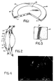
- corneal prosthesis 10 including optical element 12 and porous outer skirt 14.
- Optical element 12 includes a full-depth optically-transparent central portion 16 and a thinner outer portion 18 that overlies skirt 14, which extends around the periphery of prosthesis 10.
- Optical element 16 is made of an optically transparent hydrogel material having a water content between 50% and 90%, a tensile strength of greater than 20 kg/cm2 and an anterior surface capable of supporting a layer of epithelial cells.
- the water content of the material is related to the permeability of the material to nutrients and metabolites and thus is related to the ability of the material to permit transfer to the epithelial layer living thereon in use.
- the tensile strength is related to the ability of the material to resist stresses to which the material is subjected at the anterior segment of the eye and stresses to which the material is subjected during surgical implantation.
- the tensile strength of the human cornea typically is in the 30-40 kg/cm2 range, and it is desirable to have the material strength not too much below that of the human cornea and preferably above the strength of the human cornea (e.g., above 40 kg/cm2).
- the anterior surface must be able support a layer of epithelial cells thereon so as to completely cover the anterior surface in order to decrease the potential for epithelial downgrowth and resulting extrusion; the continuous epithelial layer also provides an effective barrier to microorganisms and permits development of a normal tear film when implanted.
- the material as is preferred for implant materials generally, also does not induce inflammation and resists enzymatic degradation.
- Related to optical transparency of the material is the ability to process the material into desired shape (preferably using conventional plastic processing techniques including molding and lathe cutting) to provide appropriate optical power to form a high quality image at or near the retina.
- the material of optical element 16 preferably is a hydrogel, e.g., made of a polyvinyl alcohol copolymer system, as described in Ofstead U.S. Patent No. 4,618,649 ("Ofstead ′649") and Ofstead U.S. Patent No. 4,528,325 (Ofsetad ′325), or of a polyvinylpyrrolidone blend, as described in Ofstead European Published Patent Application No. 0137686A ("Ofstead ′686A").
- the presently most preferred material is polyvinyl-alcohol-co-vinyl acetate 97.6:2.4 mole percent (prepared from a copolymer of vinyltrifluoroacetate and vinyl acetate) made according to the methods described in Ofstead ′649 (Example 8).
- This material is a semi-crystalline hydrogel, has an equilibrium water content of 73% in normal saline, tensile strength of 110 kg/cm2, initial modulus of elasticity of 10-15 kg/cm, and elongation of 436%. Tensile strength, elongation and modulus were all measured as decribed at col. 6, line 57 to col. 7, line 4 of Ofstead ′649.
- polyvinylpyrrolidone blends meeting the water content and strength criteria mentioned above are polyvinylpyrrolidone/polysulfone (an 80/20 blend having an equilibrium water content of 57% in normal saline, a tensile strength of 30 kg/cm2 and elongation of 170%) and polyvinylpyrrolidone/cellulose acetate (a 50/50 blend having an equilibrium water content of 51% in normal saline, a tensile strength of 51 kg/cm2, and elongation of 152%), both made according to methods described in Ofstead ′686.
- Other materials can be used so long as the water content is greater than 50% (preferably between 65% and 80%) and the tensile strength is greater than 20 kg/cm2 (preferably greater than 40 kg/cm2). Further strength-related criteria that preferably are met are a modulus of elasticity between 3 and 115 kg/cm2 (preferably 5-15 kg/cm2), and elongation between 100-1000% (preferably greater than 150%).
- Other candidate materials for optical element 12 include: other polyvinyl alcohol copolymers, e.g., containing vinyl acetate (0-5%) or maleic anhydride (0-3%) (mole percent) as the comonomer, other hydrogel materials described in Ofstead ′649 (see discussion at col.
- polyvinyl alcohol-co-vinyl acetate material described above is made by molding of solvent cast discs of the precursor polymer as described in Ofstead ′649, although lathe processing of thermally-molded buttons or UV polymerized buttons can also be used. Some initial studies indicate possible better epithelial growth (as described below) when using materials made by bulk UV-polymerization, also described in Ofstead ′649.
- Porous outer skirt 14 is preferably made of a coherent mass of melt blown fibres having an interconnected network of pores.
- the majority of fibers are preferably between 2 and 20 microns in diameter.
- the void volume (related to both size and spacing of fibers) of the preferred skirt material ranges between 70% and 94% (% of total volume not occupied by fibers); it could be less than 70%, but should be above 40% (most preferably above 55%) to get adequate bonding to both the optical element and the surrounding tissue and to permit tissue ingrowth.
- the majority of pores are preferably between 10 and 100 microns, as is shown in Fig. 4. Pore size can be determined directly by measurements of scanning electron micrographs, taking measurements between fibers that appear to be in the same or in nearby planes.
- the size and spacing of fibers are important in that the spacing between fibers must be sufficient for the cells to migrate into the material and occupy the voids, there must be sufficient room for capillary ingrowth, the fibers must have a minimum diameter for the cells to travel along them, the fibers should have a minimum size to provide desired strength to the skirt without reducing pore size, and the fibers should not be so large as to take up too much volume.
- the melt blown fiber construction permits obtaining fibers that have the desired small diameters, spacing, and sufficient bonding to each other to provide sufficient strength to resist stresses to which the skirt is subjected during suturing and implantation.
- the bonding between fibers also avoids creation of large holes or tears at sutures.
- the melt blown fiber construction also provides good interconnection of the pores throughout the web.
- the porous material encourages stromal keratocyte ingrowth, resulting in permanent stromal wound apposition so that prosthesis 10 heals securely in place.
- porous skirt 14 The presently most preferred materials for porous skirt 14 are polybutylene (e.g., available under the trade designation Shell 8010) or a polybutylene/polypropylene blend (80%/20%). Other materials that can be used include other polyolefins such as polyethylene, polyethylene/polybutylene blends, polypropylene (e.g., available from Himont under the Profax 973 or D19A trade designations) polybutylene/ polypropylene blends (e.g., an 80/20 blend) and other biocompatible plastics. Polyurethane should be useful if components deleterious to ingrowing tissue are removed.
- porous outer skirt could be made of other porous structures permitting tissue ingrowth, particularly coherent masses of fibers made by other melt and draw procedures such as procedures employing electrical field to draw molten extruded fibers so as to greatly reduce their diameters before collecting them on a belt.
- melt drawn fibers herein we mean fibers prepared by melting, extruding through orifices, and reducing the diameters of the fiber by drawing them, e.g., by blowing air past them or by an electrical field.
- the plastic selected, like all implant material, should not induce corneal inflammation and should resist enzymatic degradation.
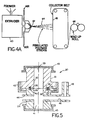
- the microfiber web is made by extruding the plastic at extruder 60 through a series of small apertures 62 formed by clamped saw-tooth edges or drill holes, subjecting the extruded plastic to converging air jets at slight angles to the direction of extrusion on both sides to cause attenuation and elongation of extruded molten fibers 64, collecting fibers 64 on a moving belt 66, and removing the web at wind-up roll 68, according to techniques well known in the art, as generally described, e.g., in Hauser U.S. Patent No. 4,118,531, which is hereby incorporated by reference.
- a chamber including a second set of air jets at its entrance can also be placed to direct the fibers to the moving collecting belt, as described in Example 11 herein.
- the fibers become entangled and also become bonded to each other during the process so as to form a coherent mass, as shown in the electron micrograph of Fig. 4.
- the microfiber web is cut into the desired ring shape of porous outer skirt 14.
- the melt-blown fiber method is useful because porosity is controllable by factors which control web density and fiber size (e.g., air temperature, air flow rate, polymer extrusion rate, collector speed, distance of collector from the extruder). Porosity can thus be varied to obtain optimum tissue ingrowth and viability along with desired strength and flexibility of the material.
- the speed of the belt can also be varied to control the thickness of the web.
- Other advantages of the melt blow fiber technique are that the nonwoven web is directly formed, a variety of polymers can be processed, and good fiber-fiber bonding can be achieved without the use of a bonding agent.
- Example 11 hereto describes preparation of a melt blown fiber web according to the invention.
- the precursor of optical element 12 (prior to the solvolysis step, described in Ofstead ′649, which step causes water-swelling) is formed to shape (either by molding or by lathing of a preformed button) and solvent welded to the porous outer skirt 14 by dipping skirt 14 in acetone and pressing the two together 12.
- Acetone is a solvent for the polymer of the optical element and a nonsolvent for the polybutylene skirt material. Just enough pressure is applied to make good contact between the two components.
- the precursor of element 12 is then taken through the solvolysis stage attached to skirt 14, optical element 12 retaining its shape and optical clarity.
- Skirt 14 is sufficiently elastic to accommodate changes in the shape of optical element 12 during the solvolysis stage.
- a benefit of solvent welding is obtaining mechanical interlocking of the fibers in the hydrogel, avoiding the problems of delamination at an adhesive interface. This is especially important with a material which is going to change in size on solvolysis and swelling. Also, by avoiding the use of adhesive to bond the two together, the different materials interfacing with the human eye are limited in number. This attachment method also avoids subjecting the hydrogel to processes that might potentially change its properties.
- FIG. 5 there is shown holding device 20 used for holding prosthesis 10 while seeding different cells on different surfaces of corneal prosthesis 10.
- Upper and lower housings 22, 24 mate at flat surfaces 26,28, in which are provided opposing annular recesses 30, 32, retaining opposing O-rings 34, 36 therein.
- Housing 22 has a cylindrical bore 38 to provide a first seeding chamber 39
- housing 24 similarly has cylindrical bore 40 to provide a second seeding chamber 41.
- Housings 22, 24 are secured together by threaded collar 42, and are sealed closed at their respective ends by snap-on caps 43, 45.
- Lower housing 24 has ports 44, 46, the latter communicating with an upwardly directed tube 48.
- holding device 20 can be used to seed the anterior surface of optical element 12 with epithelial cells and porous skirt 14 with stromal keratocytes.
- the completed prosthesis 10, shown in Fig. 1 has an outer diameter equal to that of bores 38, 40.
- prosthesis 10 Prior to seeding, prosthesis 10 initially has a skirt 14 and overlying portion 18 that extend beyond the bore diameters to slightly more than the diameter of O-rings 34, 36 so that the upper surface of completed prosthesis 10 is sealed from the lower surface.
- the oversized prosthesis is sealed between O-rings 34, 36, with the anterior surface forming second seeding chamber 41 with bore 40 and the posterior surface forming first seeding chamber 39 with bore 38.
- Device 20 is filled with culture medium in chamber 39 in bore 38, sealed closed, and placed in a position inverted from that of Fig. 5, and culture medium with epithelial cells is added to chamber 41. After epithelial cell attachment to the anterior surface of optical element 12, chamber 40 is sealed; device 20 is inverted to the position of Fig. 5; and stromal keratocytes are added to chamber 38.
- the cell culturing and seeding methodologies described in the examples below are applicable to seeding in holder 20.
- both optical element 12 and porous skirt 14 can optionally be coated with a basement membrane component to facilitate attachment and healing when implanted; one or more of the following could be used: laminin, fibronectin, Type I collagen, Type IV collagen or a cell-free extract prepared from the extracellular matrix of corneal epithelial cells.
- the coating can involve adsorption, chemical attachment or integration during polymerization.
- culture medium can be flushed through ports 44, 46 to maintain the epithelial cells.
- Tube 48 maintains the liquid level in chamber 41 so that the nutrient medium is in contact with the prosthesis.
- the first seeding chamber 39 in bore 38 might additionally be provided with a cylindrical barrier (not shown) to keep the stromal keratocytes at the porous outer skirt only and away from the posterior surface of central portion 16.
- caps 43, 45 are removed, and prosthesis 10 and holding device 20 are transferred under sterile conditions to a sterile punch in which prosthesis 10 is cut from the seeded, oversized prosthesis.
- Device 20 advantageously prevents cross-contamination of the cell types, keeping the epithelium from populating the porous skirt, something which would lead to epithelial growth under the prosthesis and extrusion.
- prosthesis is surgically implanted in the eye employing known keratectomy procedures, for example, as described in the above-mentioned references. (See also the procedure described at the end of this section.)
- the peripheral portion of prosthesis 10 including skirt 14 is sutured to cornea 50.
- Posterior surface 52 of prosthesis 10 seals anterior chamber 54, and the edge of overlying portion 18 is aligned with the epithelial layer of cornea 50, and skirt 14 is aligned with the stroma of the eye.
- Stromal tissue grows into the interconnected pores of porous skirt 14, serving to anchor prosthesis 10 in the patient's eye.
- Epithelial cells migrate from the epithelial layer of the surrounding tissue of cornea 50 to the anterior surface of optical element 12 and vice versa, forming a continuous epithelial layer 56 (see Fig. 3) on anterior surface 58 of element 12.
- Layer 56 desirably includes at least three cell layers and is about 50 to 100 ⁇ m thick.
- the preseeding of the anterior surface with epithelial cells and the preseeding of the skirt with stromal keratocytes facilitates re-epithelialization and tissue ingrowth.
- the complete covering of the anterior surface provides a normal precorneal tear film and a barrier against microbial invasion.
- porous outer skirt 14 Prior to surgically implanting prosthesis 10 in the human eye, porous outer skirt 14 can be treated with fibrin adhesive (available under the "Tissucol” trade designation from Immuno GMBH, Vienna, Austria). (Another adhesive that can be used is polyphenolic protein bioadhesive available from Biopolymers, Inc., Farmington, Connecticut).
- fibrin adhesive retards epithelial cell growth into the region of the adhesive compared to the rate for the stromal keratocytes, permitting the stromal keratocytes to establish themselves in porous outer skirt 14 before the epithelial cells have a chance to migrate down and under the prosthesis and cause extrusion.
- the materials employed have adequate strength to allow for initial suturing and to withstand the intraoccular pressure without rupturing or leaking.
- the materials are compliant and generally have physical properties similar to those of the natural tissue surrounding the prosthesis, thereby avoiding such problems as pressure necrosis and fibrous encapsulation associated with stress concentration at the interface of two dissimilar materials.
- a description of a penetrating keratoplasty procedure that is directed to laboratory experiments but includes applicable information follows.
- Recipient animals are anesthesized with intramuscularly administered ketamine/Rompun and topically administered proparacaine hydrochloride 0.5%. Heparin is also given intravenously in a dose of 1,000 units/kg body weight.
- a wire lid speculum is used to maintain the lids widely open and the nictitating membrane is excised.
- Superior and inferior rectus bridle sutures are placed and affixed to the drapes to stabilize the globe.
- a horizontal rectus muscle is grasped with toothed forceps, and, under direct visualization through an operating microscope, a partially perforating circular corneal incision is made with a trephine.
- aqueous heparin 1000 units/ml is slowly dripped into the anterior chamber through the trephine opening, using a 30 gauge irrigating cannula, while proceeding with surgery.
- the keratoprosthetic material is initially anchored with four, cardinal, 10-0 nylon, interrupted sutures and then firmly secured with either a single running or multiple interrupted 10-0 nylon sutures and/or a biological adhesive. Gentamicin sulfate 0.3% ophthalmic solution is instilled at the completion of surgery.
- the optical element could be formed to its desired shape when in the hydrogel state.
- the melt blow material also has application in other implant devices and can be used to anchor members made of material that is different from the melt blown material.
- fibrin adhesive to inhibit epithelial downgrowth has application in other percutaneous type implants where epithelial downgrowth is a potential complication, e.g., peritoneal access devices, blood access devices, and periodontal surgery where there is a need to prevent the epithelium from migrating down the tooth-gingival interface.
- the surface properties of the hydrogels can be altered by introducing ionic functional groups and by chemically changing the distribution of these groups on the surface in order to influence the attachment and architecture of cells.
- Examples 9 and 10 herein describe a method of providing formation of amide acid and amide-amino acid groups in the hydrogel, some of which will thus appear at the surface. Initial studies indicate that there may be improved cell attachment and growth with these materials.
- Corneal epithelial cells were removed and cultured using a modified version of Trinkaus-Randall, V. and Gipson, I. K., "A technique for obtaining corneal epithelial cells", Inv. Ophthal. and Vis. Sci. , 1985, Vo. 26, p. 233. New Zealand rabbits were sacrificed with 5 ml of sodium pentobarbital (325 mg) administered intravenously.
- the corneas were excised, the endothelium and Descemet's membrane were removed with forceps, and the remaining cornea (epithelium and stroma) was incubated in modified Dulbecco's Eagles medium containing 10 uM calcium (Gibco Lab., Grand Island, NY) and 1.2 U/ml Dispase II (Boehringer Mannheim Lab, Indianapolis, IN) for one hour.
- the epithelial sheets were carefully teased from the stroma. (Trinkaus-Randall and Gipson (1985); Gipson, I.K., et al., "Hemidesmosome formation in vitro", J. Cell. Biol. , 1983, Vol. 97, p.
- Corneal epithelial cells cultured according to Example 1 were seeded in vitro onto 1 cm diameter hydrogel discs of polyvinyl alcohol-co-vinyl acetate 97.6:2.4 mole percent (prepared from the copolymer of vinyltrifluoroacetate and vinyl acetate) as described above to study the ability to grow a continuous epithelial layer on the hydrogel that synthesized connective tissue proteins.
- the hydrogel discs were placed on tissue culture plates that were manufactured by a procedure that omitted usual treatments designed to promote cell growth, and discs were sterilized with an ultraviolet light for 2 hours.
- the cells were seeded onto plain or protein coated coated hydrogel discs at a density of 2 x 10⁴ cells/disc.
- the protein coating involved adding one of the following protein solutions to the hydrogels for 45 min. at room temperature: laminin (0.5 mg/ml), fibronectin (0.2 mg/ml), Type I collagen (0.8 mg/ml), Type IV collagen (0.8 mg/ml) or a cell-free extract prepared from the extracellular matrix of corneal epithelial cells.
- laminin 0.5 mg/ml
- fibronectin 0.2 mg/ml
- Type I collagen 0.8 mg/ml
- Type IV collagen 0.8 mg/ml
- a cell-free extract prepared from the extracellular matrix of corneal epithelial cells.
- the discs were then washed extensively with Puck's Saline G.
- the amount of adsorbed protein was determined by quantifying the protein before and after application. Only small amounts of protein actually adhered to the hydrogel as evidenced by the lack of significant change in concentration as determined by amino acid analysis. Laminin and fibronectin were obtained from Collaborative Research (Bedford, MA).
- Type I collagen was prepared from rat tail tendon and ultimately was fibrillar in structure.
- Type IV collagen was obtained using a pepsin/acetic acid extraction of human placenta and exists in a degraded form (Kresina, T.F., and Miller,E.J., "Isolation and characterization of basement membrane collagen from human placental tissue: Evidence for the presence of two genetically distinct collagen chains," Biochemistry , 1979, Vol. 18, p. 3089).
- corneal epithelial cells were cultured for a minimum of 6 weeks, the cells removed, and the cell layer washed and scraped, homogenized, and lyophilized. All assays were conducted on cells in first passage.
- Cell Growth Cell numbers were determined at days 2, 4, 6 and 8 using well known techniques. The number of corneal epithelial cells increased with time in culture for all surfaces. While there was a variation in plating efficiency among the different surfaces, the rate of cell growth was greater on hydrogels that hs been coated with Types I or IV collagen than on hydrogels coated with fibronectin and laminin (seven population doublings versus six and four respectively).
- Morphology Cell morphology was monitored daily using a Nikon Diaphot phase contrast inverted microscope. After day 8, the tissue was fixed and processed for transmission election microscopy, and sections were examined using a Philips 300 microscope. Cell spreading occurred within the first 1.5 hr. on all surfaces. By day 8, confluency was achieved on all the coated surfaces, and the corneal epithelium cultured on the hydrogel discs was multilayered.
- Protein Synthesis Collagen synthesis and accumulation was monitored by known techniques as a function of culture time to study the ability of the epithelial cell layer to synthesize basement membrane proteins, a requirement of a healthy, living cell. Collagen synthesis and accumulation was demonstrated on all surfaces and was found to be enhanced by coating with proteins.
- Hydrogel discs coated with Type I collagen and seeded with 2 x 10⁴ epithelial cells in vitro as described in Example 2 were tested in vivo to study the ability to obtain a continuous corneal epithelial layer.
- Rabbits were anesthetized with 5 mg/kg xylazine and 35 mg/kg ketamine administered intramuscularly. When needed, an additional half dose was administered.
- the rabbit corneal epithelium was removed with a scalpel, the nictitating membrane was excised, and a deep lamellar keratectomy 5mm in diameter was performed.
- Discs which had been cultured until near confluency were placed into the keratectomy bed and secured with both interrupted and continuous sutures (10-0) (Alcon Surgical, Ft. Worth, Texas). The eyelids were not sutured shut or covered, and antibiotics were administered daily.
- Discs with or without cells were labeled for 10 hr. with 3H-thymidine on days 2, 4 and 6 after transplantation. This was accomplished by manipulating so that the cornea was bathed in the topically applied isotope.
- Discs with a confluent layer of cells were labeled in vitro with 3H-thymidine for 18 hr. prior to transplantation and incorporation was examined on day 2, 4 and 6.
- Discs with a confluent layer of cells were labeled in vitro with 3H-proline for 18 hr. prior to transplantation while the cornea was labeled in situ with 3H-thymidine for 18 hr. using an intrastromal injection prior to implantation of the disc. Migration of epithelial cells was examined on days 2 and 4.
- Stromal keratocytes were cultured for use in seeding melt blown fiber samples.
- the corneas were excised, and the epithelium and endothelium were removed as described in Example 1 above.
- the stroma were cut into pieces (0.5 mm x 0.5 mm) and placed into T25 Falcon Tissue Culture flasks.
- the flasks contained Dulbecco's medium and Ham's nutrient medium 1:1 with the following additions: 1% nonessential amino acids, 1% penicillin/streptomycin, 1% sodium pyruvate, 10% fetal bovine serum.
- the explants were not disturbed for one week and thereafter fed twice a week. Cells for experiments were removed once the explants had grown out.
- Stromal keratocyte cells cultured as in Example 4 were seeded onto 6 mm diameter blown microfiber discs (made of polybutylene or polypropylene or a blend of polypropylene and polybutylene and made as described above) that had previously been sterilized under ultraviolet light for 2 hrs., and coated with Type 1 collagen as in Example 2. This was done by placing the discs on sterile filters to which the cells did not adhere (MILLICELL HA, 0.45 um; from Millipore, Bedford, MA.). 5 x 10⁴ cells/per disc were seeded onto each disc in a volume of 50 ⁇ l. Medium (as described in Example 4) was added to the well of 24 well tissue culture dish and to the melt blown fiber disc.
- Blown microfiber discs coated with Type I Collagen and seeded with stromal keratocyte cells as in Example 5 were tested in vivo to study ingrowth of host tissue to anchor the material to the host tissue.
- the host keratocytes were labeled with a radioactive marker, and the melt blown fiber discs were implanted, using the methodology of Example 3. It was observed that numbers of labeled host cells counted in the discs increased with time, indicating ingrowth.
- Example 7 Combined Melt Blown Fiber and Hydrogel In Vitro
- tissue culture plates were divided into two regions by a strip of human fibrin adhesive (available under the TissucolTM trade designation from Immuno GMBH, Vienna, Austria) and either epithelial cells (Example 1) or stromal keratocytes (Example 4) were placed on one side of the strip while the other side was left vacant. It was observed that stromal keratocytes were able to easily migrate across the strip while the epithelial cells were severly inhibited.
- human fibrin adhesive available under the TissucolTM trade designation from Immuno GMBH, Vienna, Austria
- a mixture of 79.23 g. vinyltrifluoroacetate and 0.0245 g. maleic anhydride was placed in a polypropylene bag and 0.04 g. Darocure 1173 (E. Merck) added.
- the solution was purged of oxygen by bubbling N2 gas through for several minutes and the bag was then sealed.
- the reaction was cooled in an ice water bath and polymerization was initiated by placing the mixture under a UV light source (RUL 3500 A fluorescent bulb, Southern New England Ultraviolet Co.) at a distance of four inches for 0.5 hours.
- the polymer was dissolved in acetone and precipitated from heptane to remove residual monomer and low molecular weight impurities.
- Polymer films or shaped articles such as those derived from Example 9 of polyvinyltrifluoroacetate-co-maleic anhydride were converted to hydrogels by solvolyzing them either in 10% (v/v) NH4OH in MeOH or in 2% (v/v) ethylenediamine in MeOH.
- the solvolysis was run for two hours at room temperature followed by extraction in MeOH for two hours, also at room temperature.
- the former procedure results in the formation of an amide group and carboxylic acid group from each maleic anhydride group.
- the latter procedure results in the formation of a carboxylic acid group and an amide-amino group.
- a blend of 80% Shell 8010 polybutylene and Exxon 3145 polypropylene pellets were fed to a 3/4" Brabender extruder heated at 230°C at the feed zone and increasing to 246°C at the extrusion point.
- the polymer blend was extruded from a 10" wide die containing 0.017" holes (5 per inch).
- the extruded fibers were drawn by an air jet created by two air knives (pressure 22 psi, air temperature 250°C) placed on either side of the die tip.
- the fibers were further drawn by passing the fiber stream through a 12" x 12" x 3/4" chamber containing a second set of air knives (40 psi, ambient temperature) placed 1" in front of the extrusion die.
Landscapes
- Health & Medical Sciences (AREA)
- Transplantation (AREA)
- Life Sciences & Earth Sciences (AREA)
- Veterinary Medicine (AREA)
- Public Health (AREA)
- Oral & Maxillofacial Surgery (AREA)
- General Health & Medical Sciences (AREA)
- Animal Behavior & Ethology (AREA)
- Engineering & Computer Science (AREA)
- Biomedical Technology (AREA)
- Ophthalmology & Optometry (AREA)
- Chemical & Material Sciences (AREA)
- Vascular Medicine (AREA)
- Heart & Thoracic Surgery (AREA)
- Cardiology (AREA)
- Dermatology (AREA)
- Medicinal Chemistry (AREA)
- Epidemiology (AREA)
- Chemical Kinetics & Catalysis (AREA)
- Materials For Medical Uses (AREA)
- Prostheses (AREA)
Applications Claiming Priority (2)
| Application Number | Priority Date | Filing Date | Title |
|---|---|---|---|
| US163383 | 1988-03-02 | ||
| US07/163,383 US5108428A (en) | 1988-03-02 | 1988-03-02 | Corneal implants and manufacture and use thereof |
Publications (3)
| Publication Number | Publication Date |
|---|---|
| EP0333344A2 EP0333344A2 (en) | 1989-09-20 |
| EP0333344A3 EP0333344A3 (en) | 1989-10-18 |
| EP0333344B1 true EP0333344B1 (en) | 1992-02-19 |
Family
ID=22589804
Family Applications (1)
| Application Number | Title | Priority Date | Filing Date |
|---|---|---|---|
| EP89302053A Expired - Lifetime EP0333344B1 (en) | 1988-03-02 | 1989-03-01 | Corneal implants and manufacture and use thereof |
Country Status (4)
| Country | Link |
|---|---|
| US (2) | US5108428A (OSRAM) |
| EP (1) | EP0333344B1 (OSRAM) |
| JP (1) | JPH02104364A (OSRAM) |
| DE (1) | DE68900828D1 (OSRAM) |
Families Citing this family (97)
| Publication number | Priority date | Publication date | Assignee | Title |
|---|---|---|---|---|
| US5292514A (en) * | 1992-06-24 | 1994-03-08 | Minnesota Mining And Manufacturing Company | Azlactone-functional substrates, corneal prostheses, and manufacture and use thereof |
| AU650156B2 (en) * | 1992-08-05 | 1994-06-09 | Lions Eye Institute Limited | Keratoprosthesis and method of producing the same |
| US5300115A (en) * | 1992-11-19 | 1994-04-05 | Keratos, Inc. | Intraocular prosthesis |
| US5433745A (en) * | 1993-10-13 | 1995-07-18 | Allergan, Inc. | Corneal implants and methods for producing same |
| AU682907B2 (en) * | 1993-11-19 | 1997-10-23 | Commonwealth Scientific And Industrial Research Organisation | Corneal onlays |
| TW257671B (OSRAM) * | 1993-11-19 | 1995-09-21 | Ciba Geigy | |
| DE19508922C2 (de) * | 1994-03-14 | 1999-10-14 | Norbert Schrage | Hornhautprothese |
| US7892226B2 (en) * | 1995-03-20 | 2011-02-22 | Amo Development, Llc. | Method of corneal surgery by laser incising a contoured corneal flap |
| US5693094A (en) | 1995-05-09 | 1997-12-02 | Allergan | IOL for reducing secondary opacification |
| FR2743715B1 (fr) * | 1996-01-18 | 1998-02-27 | Fci France Chirurgie Instrumen | Keratoprothese |
| US6005160A (en) * | 1996-02-22 | 1999-12-21 | National Science Council | Heterobifunctional membrane in application of artificial cornea |
| WO1997036621A1 (en) * | 1996-03-29 | 1997-10-09 | Desmos, Inc. | Cellular attachment to laminin 5-coated trans-epithelial appliances |
| US5843185A (en) * | 1996-10-23 | 1998-12-01 | Leon Rolden; Carlos R. | Keratoprosthesis and method of corneal replacement |
| KR19990077175A (ko) * | 1996-11-13 | 1999-10-25 | 다나까 쿄이찌 | 인공각막 |
| JP3922486B2 (ja) * | 1997-07-10 | 2007-05-30 | 株式会社コーナン・メディカル | 移植用角膜の観察装置 |
| US20020086423A1 (en) * | 1997-09-09 | 2002-07-04 | Toshiaki Takezawa | Hydrogel thin film containing extracellular matrix components |
| US6468306B1 (en) | 1998-05-29 | 2002-10-22 | Advanced Medical Optics, Inc | IOL for inhibiting cell growth and reducing glare |
| US6245345B1 (en) | 1998-07-07 | 2001-06-12 | Atrix Laboratories, Inc. | Filamentous porous films and methods for producing the same |
| US6626941B2 (en) * | 1998-12-23 | 2003-09-30 | Anamed, Inc. | Corneal implant and method of manufacture |
| US6102946A (en) * | 1998-12-23 | 2000-08-15 | Anamed, Inc. | Corneal implant and method of manufacture |
| US6361560B1 (en) * | 1998-12-23 | 2002-03-26 | Anamed, Inc. | Corneal implant and method of manufacture |
| WO2000052516A2 (en) * | 1999-03-01 | 2000-09-08 | Boston Innovative Optics, Inc. | System and method for increasing the depth of focus of the human eye |
| US20030055497A1 (en) * | 1999-07-28 | 2003-03-20 | The Lions Instutute Of Western Australia Incorporated | Method of insertion of keratoprostheses |
| US6423093B1 (en) | 1999-09-14 | 2002-07-23 | The Lions Eye Institute Of Western Australia Incorporated | Method of insertion of keratoprostheses |
| US6543610B1 (en) * | 2000-09-12 | 2003-04-08 | Alok Nigam | System for packaging and handling an implant and method of use |
| US8668735B2 (en) | 2000-09-12 | 2014-03-11 | Revision Optics, Inc. | Corneal implant storage and delivery devices |
| AU2001289038B2 (en) | 2000-09-12 | 2006-05-18 | Revision Optics, Inc. | System for packaging and handling an implant and method of use |
| US6881198B2 (en) * | 2001-01-09 | 2005-04-19 | J. David Brown | Glaucoma treatment device and method |
| US8119599B2 (en) * | 2001-08-31 | 2012-02-21 | Orthopeutics, L.P. | Direct application of non-toxic crosslinking reagents to resist progressive spinal degeneration and deformity |
| JP2006508709A (ja) * | 2002-09-13 | 2006-03-16 | オキュラー サイエンシス インコーポレイテッド | 視力を向上させる器具及び方法 |
| US7628810B2 (en) * | 2003-05-28 | 2009-12-08 | Acufocus, Inc. | Mask configured to maintain nutrient transport without producing visible diffraction patterns |
| US20050046794A1 (en) | 2003-06-17 | 2005-03-03 | Silvestrini Thomas A. | Method and apparatus for aligning a mask with the visual axis of an eye |
| US7381180B2 (en) * | 2003-10-31 | 2008-06-03 | Medtronic, Inc. | Implantable devices and methods for treating fecal incontinence |
| US7585271B2 (en) * | 2003-11-01 | 2009-09-08 | Thd Spa | Implantable devices and methods for treating urinary incontinence |
| EP1786485A4 (en) | 2004-02-06 | 2012-05-30 | Georgia Tech Res Inst | CELLULAR IMPLANTATION WITH SURFACE ADHESION |
| CA2558661C (en) | 2004-02-06 | 2012-09-04 | Georgia Tech Research Corporation | Load bearing biocompatible device |
| CN1314461C (zh) * | 2004-02-27 | 2007-05-09 | 四川大学 | 生物活性人工角膜及其制备方法 |
| US20110218623A1 (en) * | 2004-04-30 | 2011-09-08 | Jon Dishler | Small Diameter Inlays |
| US8057541B2 (en) * | 2006-02-24 | 2011-11-15 | Revision Optics, Inc. | Method of using small diameter intracorneal inlays to treat visual impairment |
| US20050246016A1 (en) * | 2004-04-30 | 2005-11-03 | Intralens Vision, Inc. | Implantable lenses with modified edge regions |
| US7776086B2 (en) * | 2004-04-30 | 2010-08-17 | Revision Optics, Inc. | Aspherical corneal implant |
| US10835371B2 (en) | 2004-04-30 | 2020-11-17 | Rvo 2.0, Inc. | Small diameter corneal inlay methods |
| US20080262610A1 (en) * | 2007-04-20 | 2008-10-23 | Alan Lang | Biomechanical design of intracorneal inlays |
| CA2566961A1 (en) | 2004-05-20 | 2005-12-08 | Coopervision, Inc. | Corneal onlays and wavefront aberration correction to enhance vision |
| EP1790316A4 (de) * | 2004-06-17 | 2012-05-16 | Zakrytoe Akcionernoe Obschestvo N Proizv Complex Ecoflon | Implantat zur verstärkung der hornhaut und keratoprothese mit einer platte aus porösem polytetrafluorethylen |
| KR101298442B1 (ko) * | 2004-08-13 | 2013-08-22 | 내셔날 리서치 카운실 오브 캐나다 | 안과용 장치 및 이와 관련된 방법 및 조성물 |
| US9999497B2 (en) | 2005-01-31 | 2018-06-19 | Yichieh Shiuey | Corneal implants and methods and systems for placement |
| US8029515B2 (en) * | 2005-01-31 | 2011-10-04 | Yichieh Shiuey | Corneal implants and methods and systems for placement |
| US7976577B2 (en) | 2005-04-14 | 2011-07-12 | Acufocus, Inc. | Corneal optic formed of degradation resistant polymer |
| US20070083087A1 (en) * | 2005-10-12 | 2007-04-12 | Sismed, Llc | Fixator with membrane |
| US20070083221A1 (en) * | 2005-10-12 | 2007-04-12 | Sismed, Llc | Precision trephine |
| US20070129797A1 (en) * | 2005-12-01 | 2007-06-07 | Revision Optics, Inc. | Intracorneal inlays |
| US20070182920A1 (en) * | 2006-02-08 | 2007-08-09 | Coopervision, Inc. | Corneal Onlays and Related Methods |
| US10555805B2 (en) | 2006-02-24 | 2020-02-11 | Rvo 2.0, Inc. | Anterior corneal shapes and methods of providing the shapes |
| US20080065207A1 (en) * | 2006-03-13 | 2008-03-13 | University Of South Florida | Self-Assembling, Collagen-Based Material for Corneal Replacement |
| US20070238167A1 (en) | 2006-04-04 | 2007-10-11 | 3M Innovative Properties Company | Flat microfibers as matrices for cell growth |
| US20070231362A1 (en) * | 2006-04-04 | 2007-10-04 | 3M Innovative Properties Company | Schistose microfibrillated article for cell growth |
| US7883520B2 (en) * | 2006-04-10 | 2011-02-08 | Forsight Labs, Llc | Corneal epithelial pocket formation systems, components and methods |
| US20070255401A1 (en) * | 2006-05-01 | 2007-11-01 | Revision Optics, Inc. | Design of Inlays With Intrinsic Diopter Power |
| JP2009540936A (ja) | 2006-06-22 | 2009-11-26 | ユニバーシティー オブ サウス フロリダ | コラーゲン足場、それを伴う医療用埋植物、およびその使用方法 |
| WO2008020087A1 (es) * | 2006-08-08 | 2008-02-21 | Fundacion Inasmet | Sistema óptico implantable, procedimiento para su desarrollo y aplicaciones |
| WO2008033505A1 (en) * | 2006-09-13 | 2008-03-20 | University Of South Florida | Biocomposite for artificial tissue design |
| US20080081323A1 (en) | 2006-09-29 | 2008-04-03 | Daniel Keeley | Regenerative Medicine Devices and Melt-Blown Methods of Manufacture |
| US8490977B2 (en) | 2007-03-20 | 2013-07-23 | Cfph, Llc | Game broker |
| US9549848B2 (en) | 2007-03-28 | 2017-01-24 | Revision Optics, Inc. | Corneal implant inserters and methods of use |
| US9271828B2 (en) | 2007-03-28 | 2016-03-01 | Revision Optics, Inc. | Corneal implant retaining devices and methods of use |
| US8162953B2 (en) | 2007-03-28 | 2012-04-24 | Revision Optics, Inc. | Insertion system for corneal implants |
| EP2276420B1 (en) | 2008-04-04 | 2021-10-06 | Journey1, Inc. | Device to treat an eye having an epithelium with a defect |
| EP2265217A4 (en) * | 2008-04-04 | 2018-04-04 | Revision Optics, Inc. | Corneal inlay design and methods of correcting vision |
| US9539143B2 (en) | 2008-04-04 | 2017-01-10 | Revision Optics, Inc. | Methods of correcting vision |
| CN101658445B (zh) * | 2008-08-29 | 2011-08-17 | 四川大学 | 一体式人工角膜及其制备方法 |
| US20100087920A1 (en) * | 2008-10-07 | 2010-04-08 | Forsight Labs, Llc | Corneal Onlay Lenses and Related Methods for Improving Vision of Presbyopic Patients |
| CN102448404B (zh) | 2009-08-13 | 2015-06-10 | 阿库福库斯公司 | 掩盖型眼内植入物和透镜 |
| JP5709015B2 (ja) * | 2009-08-19 | 2015-04-30 | 国立大学法人大阪大学 | 角膜移植用シート |
| ES2649890T3 (es) | 2009-10-23 | 2018-01-16 | Nexisvision, Inc. | Enervación corneal para el tratamiento de dolor ocular |
| US9498385B2 (en) | 2009-10-23 | 2016-11-22 | Nexisvision, Inc. | Conformable therapeutic shield for vision and pain |
| USD656526S1 (en) | 2009-11-10 | 2012-03-27 | Acufocus, Inc. | Ocular mask |
| US8469948B2 (en) | 2010-08-23 | 2013-06-25 | Revision Optics, Inc. | Methods and devices for forming corneal channels |
| EP2621405B8 (en) * | 2010-09-30 | 2019-01-09 | KeraMed, Inc. | Reversibly deformable artificial cornea |
| US12044905B2 (en) | 2011-04-28 | 2024-07-23 | Journey1 Inc | Contact lenses for refractive correction |
| EP2757964B1 (en) | 2011-05-26 | 2016-05-04 | Cartiva, Inc. | Tapered joint implant and related tools |
| CA2853116A1 (en) | 2011-10-21 | 2013-04-25 | Revision Optics, Inc. | Corneal implant storage and delivery devices |
| WO2013082545A1 (en) | 2011-12-02 | 2013-06-06 | Acufocus, Inc. | Ocular mask having selective spectral transmission |
| US10350072B2 (en) | 2012-05-24 | 2019-07-16 | Cartiva, Inc. | Tooling for creating tapered opening in tissue and related methods |
| US9974646B2 (en) | 2012-09-05 | 2018-05-22 | University Of Miami | Keratoprosthesis, and system and method of corneal repair using same |
| US9204962B2 (en) | 2013-03-13 | 2015-12-08 | Acufocus, Inc. | In situ adjustable optical mask |
| US9427922B2 (en) | 2013-03-14 | 2016-08-30 | Acufocus, Inc. | Process for manufacturing an intraocular lens with an embedded mask |
| JP6310072B2 (ja) | 2013-06-26 | 2018-04-11 | ネクシスビジョン, インコーポレイテッド | 屈折矯正のためのコンタクトレンズ |
| TWI601817B (zh) * | 2013-10-02 | 2017-10-11 | 國立中央大學 | 細胞培養製品及其製造方法 |
| US10736778B2 (en) | 2014-12-31 | 2020-08-11 | Microoptx Inc. | Glaucoma treatment devices and methods |
| WO2016144404A1 (en) | 2015-03-12 | 2016-09-15 | Revision Optics, Inc. | Methods of correcting vision |
| AU2016243659B2 (en) | 2015-03-31 | 2020-04-23 | Cartiva, Inc. | Hydrogel implants with porous materials and methods |
| WO2016161026A1 (en) | 2015-03-31 | 2016-10-06 | Cartiva, Inc. | Carpometacarpal (cmc) implants and methods |
| AU2016331925B2 (en) | 2015-09-30 | 2021-04-22 | Microoptx Inc. | Dry eye treatment devices and methods |
| WO2018131491A1 (ja) * | 2017-01-13 | 2018-07-19 | 国立大学法人大阪大学 | 角膜上皮細胞集団の製造方法 |
| EP3806916A1 (en) | 2018-06-14 | 2021-04-21 | W.L. Gore & Associates, Inc. | Epitheliazing microporous biomaterial for use in avascular environments and in corneal implants |
| US11571496B2 (en) | 2019-06-03 | 2023-02-07 | Advanced Solutions Life Sciences, Llc | System and method for fabricating a cornea |
Family Cites Families (26)
| Publication number | Priority date | Publication date | Assignee | Title |
|---|---|---|---|---|
| US2714721A (en) * | 1953-01-23 | 1955-08-09 | Jr William Stone | Artificial corneal implants |
| GB772569A (en) * | 1955-05-10 | 1957-04-17 | Stone Jr William | Artificial corneal implant |
| CA1073648A (en) * | 1976-08-02 | 1980-03-18 | Edward R. Hauser | Web of blended microfibers and crimped bulking fibers |
| DE2705234A1 (de) * | 1977-02-08 | 1978-08-17 | Hennig Gerhard | Keratoprothese |
| US4565784A (en) * | 1981-01-26 | 1986-01-21 | Trustees Of Boston University | Hydrogels capable of supporting cell growth |
| US4546500A (en) * | 1981-05-08 | 1985-10-15 | Massachusetts Institute Of Technology | Fabrication of living blood vessels and glandular tissues |
| US4496349A (en) * | 1981-05-08 | 1985-01-29 | Renal Systems, Inc. | Percutaneous implant |
| US4475972A (en) * | 1981-10-01 | 1984-10-09 | Ontario Research Foundation | Implantable material |
| US4418691A (en) * | 1981-10-26 | 1983-12-06 | Massachusetts Institute Of Technology | Method of promoting the regeneration of tissue at a wound |
| US4485096A (en) * | 1982-02-26 | 1984-11-27 | Massachusetts Institute Of Technology | Tissue-equivalent and method for preparation thereof |
| US4470159A (en) * | 1982-03-03 | 1984-09-11 | Peyman Gholam A | Keratoprosthesis |
| US4612012A (en) * | 1982-07-28 | 1986-09-16 | White Thomas C | Corneal implant |
| US4911720A (en) * | 1983-03-10 | 1990-03-27 | Collier John P | Particular surface replacement prosthesis |
| US4618649A (en) * | 1983-06-03 | 1986-10-21 | Minnesota Mining And Manufacturing Company | Copolymers of poly(vinyl trifluoroacetate) or poly(vinyl alcohol) |
| US4528325A (en) * | 1983-06-03 | 1985-07-09 | Minnesota Mining And Manufacturing Co. | Copolymers of poly(vinyl trifluoroacetate) or poly(vinyl alcohol) |
| US4673539A (en) * | 1983-06-03 | 1987-06-16 | Minnesota Mining And Manufacturing Company | Process for thermoformed articles |
| IE56908B1 (en) * | 1983-09-08 | 1992-01-29 | Minnesota Mining & Mfg | Polymer blends with high water absorption |
| US4563779A (en) * | 1984-01-27 | 1986-01-14 | Kelman Charles D | Corneal implant and method of making the same |
| US4586929A (en) * | 1984-04-06 | 1986-05-06 | Binder Perry S | Hydrogel keratoprosthesis |
| US4604346A (en) * | 1984-10-09 | 1986-08-05 | Massachusetts Institute Of Technology | Skin-equivalent prepared by the use of punch biopsy |
| US4676790A (en) * | 1985-09-25 | 1987-06-30 | Kern Seymour P | Method of manufacture and implantation of corneal inlays |
| US4772283A (en) * | 1986-05-16 | 1988-09-20 | White Thomas C | Corneal implant |
| US4715858A (en) * | 1986-07-25 | 1987-12-29 | Lindstrom Richard L | Epicorneal lens |
| WO1988002622A1 (en) * | 1986-10-16 | 1988-04-21 | Cbs Lens | Collagen-hydrogel lens for promoting epithelial cell growth |
| US4994081A (en) * | 1986-10-16 | 1991-02-19 | Cbs Lens | Method for locating on a cornea an artificial lens fabricated from a collagen-hydrogel for promoting epithelial cell growth |
| US4865601A (en) * | 1987-07-07 | 1989-09-12 | Caldwell Delmar R | Intraocular prostheses |
-
1988
- 1988-03-02 US US07/163,383 patent/US5108428A/en not_active Expired - Fee Related
-
1989
- 1989-03-01 EP EP89302053A patent/EP0333344B1/en not_active Expired - Lifetime
- 1989-03-01 DE DE8989302053T patent/DE68900828D1/de not_active Expired - Lifetime
- 1989-03-02 JP JP1050958A patent/JPH02104364A/ja active Granted
-
1993
- 1993-06-24 US US08/082,261 patent/US5489300A/en not_active Expired - Fee Related
Also Published As
| Publication number | Publication date |
|---|---|
| EP0333344A3 (en) | 1989-10-18 |
| US5108428A (en) | 1992-04-28 |
| DE68900828D1 (de) | 1992-03-26 |
| JPH02104364A (ja) | 1990-04-17 |
| US5489300A (en) | 1996-02-06 |
| EP0333344A2 (en) | 1989-09-20 |
| JPH0553138B2 (OSRAM) | 1993-08-09 |
Similar Documents
| Publication | Publication Date | Title |
|---|---|---|
| EP0333344B1 (en) | Corneal implants and manufacture and use thereof | |
| US5192316A (en) | Ocular device | |
| US5489301A (en) | Corneal prosthesis | |
| US5713957A (en) | Corneal onlays | |
| US6391055B1 (en) | Artificial cornea | |
| Jumblatt et al. | Transplantation of tissue-cultured corneal endothelium. | |
| FI108918B (fi) | Biologisesti yhteen sopivat membraanit | |
| Chirila et al. | Artificial cornea | |
| EP1981558B1 (en) | Suturable dural and meningeal repair product comprising collagen matrix | |
| EP0647143B1 (en) | Azlactone-functional substrates, corneal prostheses, and manufacture and use thereof | |
| KR101319227B1 (ko) | 각막 내피세포 및 관련세포를 생체고분자 위에서성장시키고, 인공의 이식용 각막을 제조하는 방법 및조성물 | |
| Lange et al. | Corneal endothelial cell transplantation using Descemet's membrane as a carrier | |
| GB2250442A (en) | Ocular implant | |
| JP2011020001A (ja) | 眼用器具及び関連した方法及び組成物 | |
| JPH09504968A (ja) | 可溶性因子による上皮細胞の付着刺激及びヘミデスモソーム構成刺激 | |
| Caldwell | The soft keratoprosthesis | |
| CA2174967C (en) | Corneal onlays | |
| Leibowitz et al. | Progress in the development of a synthetic cornea | |
| Trinkaus-Randall et al. | In vitro evaluation of fibroplasia in a porous polymer. | |
| JP2004261533A (ja) | 高生着性角膜上皮代替細胞シート、製造方法及びその利用方法 | |
| Bahn et al. | Complications associated with bovine corneal endothelial cell-lined homografts in the cat. | |
| Xie et al. | Artificial cornea: towards a synthetic onlay for correction of refractive error | |
| Cuperus et al. | Glass-metal keratoprosthesis: light and electron microscopical evaluation of experimental surgery on rabbit eyes | |
| AU682907B2 (en) | Corneal onlays | |
| CN108570415A (zh) | 一种用于制备上皮细胞片的培养装置及其应用 |
Legal Events
| Date | Code | Title | Description |
|---|---|---|---|
| PUAI | Public reference made under article 153(3) epc to a published international application that has entered the european phase |
Free format text: ORIGINAL CODE: 0009012 |
|
| PUAL | Search report despatched |
Free format text: ORIGINAL CODE: 0009013 |
|
| AK | Designated contracting states |
Kind code of ref document: A2 Designated state(s): DE FR GB IT SE |
|
| AK | Designated contracting states |
Kind code of ref document: A3 Designated state(s): DE FR GB IT SE |
|
| 17P | Request for examination filed |
Effective date: 19891204 |
|
| 17Q | First examination report despatched |
Effective date: 19910429 |
|
| GRAA | (expected) grant |
Free format text: ORIGINAL CODE: 0009210 |
|
| AK | Designated contracting states |
Kind code of ref document: B1 Designated state(s): DE FR GB IT SE |
|
| ITF | It: translation for a ep patent filed | ||
| REF | Corresponds to: |
Ref document number: 68900828 Country of ref document: DE Date of ref document: 19920326 |
|
| ET | Fr: translation filed | ||
| PLBE | No opposition filed within time limit |
Free format text: ORIGINAL CODE: 0009261 |
|
| STAA | Information on the status of an ep patent application or granted ep patent |
Free format text: STATUS: NO OPPOSITION FILED WITHIN TIME LIMIT |
|
| 26N | No opposition filed | ||
| EAL | Se: european patent in force in sweden |
Ref document number: 89302053.7 |
|
| PGFP | Annual fee paid to national office [announced via postgrant information from national office to epo] |
Ref country code: FR Payment date: 20010219 Year of fee payment: 13 |
|
| PGFP | Annual fee paid to national office [announced via postgrant information from national office to epo] |
Ref country code: DE Payment date: 20010220 Year of fee payment: 13 |
|
| PGFP | Annual fee paid to national office [announced via postgrant information from national office to epo] |
Ref country code: GB Payment date: 20010221 Year of fee payment: 13 |
|
| PGFP | Annual fee paid to national office [announced via postgrant information from national office to epo] |
Ref country code: SE Payment date: 20010222 Year of fee payment: 13 |
|
| REG | Reference to a national code |
Ref country code: GB Ref legal event code: IF02 |
|
| PG25 | Lapsed in a contracting state [announced via postgrant information from national office to epo] |
Ref country code: GB Free format text: LAPSE BECAUSE OF NON-PAYMENT OF DUE FEES Effective date: 20020301 |
|
| PG25 | Lapsed in a contracting state [announced via postgrant information from national office to epo] |
Ref country code: SE Free format text: LAPSE BECAUSE OF NON-PAYMENT OF DUE FEES Effective date: 20020302 |
|
| PG25 | Lapsed in a contracting state [announced via postgrant information from national office to epo] |
Ref country code: DE Free format text: LAPSE BECAUSE OF NON-PAYMENT OF DUE FEES Effective date: 20021001 |
|
| EUG | Se: european patent has lapsed |
Ref document number: 89302053.7 |
|
| GBPC | Gb: european patent ceased through non-payment of renewal fee |
Effective date: 20020301 |
|
| PG25 | Lapsed in a contracting state [announced via postgrant information from national office to epo] |
Ref country code: FR Free format text: LAPSE BECAUSE OF NON-PAYMENT OF DUE FEES Effective date: 20021129 |
|
| REG | Reference to a national code |
Ref country code: FR Ref legal event code: ST |
|
| PG25 | Lapsed in a contracting state [announced via postgrant information from national office to epo] |
Ref country code: IT Free format text: LAPSE BECAUSE OF NON-PAYMENT OF DUE FEES;WARNING: LAPSES OF ITALIAN PATENTS WITH EFFECTIVE DATE BEFORE 2007 MAY HAVE OCCURRED AT ANY TIME BEFORE 2007. THE CORRECT EFFECTIVE DATE MAY BE DIFFERENT FROM THE ONE RECORDED. Effective date: 20050301 |