EP0279273A2 - DNA-Sequenzen, Plasmide und Mikroorganismen sowie Verfahren zur Herstellung von Chinolinsäure - Google Patents
DNA-Sequenzen, Plasmide und Mikroorganismen sowie Verfahren zur Herstellung von Chinolinsäure Download PDFInfo
- Publication number
- EP0279273A2 EP0279273A2 EP88101442A EP88101442A EP0279273A2 EP 0279273 A2 EP0279273 A2 EP 0279273A2 EP 88101442 A EP88101442 A EP 88101442A EP 88101442 A EP88101442 A EP 88101442A EP 0279273 A2 EP0279273 A2 EP 0279273A2
- Authority
- EP
- European Patent Office
- Prior art keywords
- dna
- sequences
- plasmid
- quinolinic acid
- dna sequences
- Prior art date
- Legal status (The legal status is an assumption and is not a legal conclusion. Google has not performed a legal analysis and makes no representation as to the accuracy of the status listed.)
- Granted
Links
- 239000013612 plasmid Substances 0.000 title claims abstract description 65
- 108091028043 Nucleic acid sequence Proteins 0.000 title claims abstract description 34
- 244000005700 microbiome Species 0.000 title claims abstract description 26
- 238000000034 method Methods 0.000 title claims abstract description 18
- SMWDFEZZVXVKRB-UHFFFAOYSA-N Quinoline Chemical compound N1=CC=CC2=CC=CC=C21 SMWDFEZZVXVKRB-UHFFFAOYSA-N 0.000 title claims description 9
- 239000002253 acid Substances 0.000 title description 2
- GJAWHXHKYYXBSV-UHFFFAOYSA-N quinolinic acid Chemical compound OC(=O)C1=CC=CN=C1C(O)=O GJAWHXHKYYXBSV-UHFFFAOYSA-N 0.000 claims abstract description 35
- 230000015572 biosynthetic process Effects 0.000 claims abstract description 14
- 238000003786 synthesis reaction Methods 0.000 claims abstract description 11
- 101150116541 nadB gene Proteins 0.000 claims description 44
- 108090000623 proteins and genes Proteins 0.000 claims description 36
- 241000588724 Escherichia coli Species 0.000 claims description 26
- 102000004190 Enzymes Human genes 0.000 claims description 21
- 108090000790 Enzymes Proteins 0.000 claims description 21
- 108030000910 L-aspartate oxidases Proteins 0.000 claims description 18
- 230000014509 gene expression Effects 0.000 claims description 15
- 235000015097 nutrients Nutrition 0.000 claims description 11
- 238000004519 manufacturing process Methods 0.000 claims description 9
- 230000004071 biological effect Effects 0.000 claims description 5
- 230000004060 metabolic process Effects 0.000 claims description 4
- 229920001184 polypeptide Polymers 0.000 claims description 4
- 108090000765 processed proteins & peptides Proteins 0.000 claims description 4
- 102000004196 processed proteins & peptides Human genes 0.000 claims description 4
- OKTJSMMVPCPJKN-UHFFFAOYSA-N Carbon Chemical compound [C] OKTJSMMVPCPJKN-UHFFFAOYSA-N 0.000 claims description 3
- 229910052799 carbon Inorganic materials 0.000 claims description 3
- 230000002068 genetic effect Effects 0.000 claims description 3
- 150000001413 amino acids Chemical group 0.000 claims description 2
- 125000001477 organic nitrogen group Chemical group 0.000 claims description 2
- 230000001105 regulatory effect Effects 0.000 claims description 2
- 108090000854 Oxidoreductases Proteins 0.000 claims 1
- 102000004316 Oxidoreductases Human genes 0.000 claims 1
- 229940009098 aspartate Drugs 0.000 claims 1
- 238000002360 preparation method Methods 0.000 abstract description 2
- PUAGMZJCVWMYIV-UHFFFAOYSA-N pyridine-2,3-dicarboxylic acid Chemical compound OC(=O)C1=CC=CN=C1C(O)=O.OC(=O)C1=CC=CN=C1C(O)=O PUAGMZJCVWMYIV-UHFFFAOYSA-N 0.000 abstract description 2
- 238000010353 genetic engineering Methods 0.000 abstract 1
- 101150052523 nadA gene Proteins 0.000 description 39
- 101100001013 Emericella nidulans (strain FGSC A4 / ATCC 38163 / CBS 112.46 / NRRL 194 / M139) aah1 gene Proteins 0.000 description 31
- 239000002609 medium Substances 0.000 description 27
- 108020004414 DNA Proteins 0.000 description 25
- 239000012634 fragment Substances 0.000 description 24
- PEDCQBHIVMGVHV-UHFFFAOYSA-N Glycerine Chemical compound OCC(O)CO PEDCQBHIVMGVHV-UHFFFAOYSA-N 0.000 description 22
- 229940088598 enzyme Drugs 0.000 description 19
- 238000012360 testing method Methods 0.000 description 19
- 235000018102 proteins Nutrition 0.000 description 16
- 102000004169 proteins and genes Human genes 0.000 description 16
- LFQSCWFLJHTTHZ-UHFFFAOYSA-N Ethanol Chemical compound CCO LFQSCWFLJHTTHZ-UHFFFAOYSA-N 0.000 description 15
- 239000000872 buffer Substances 0.000 description 13
- 239000000243 solution Substances 0.000 description 13
- 230000000694 effects Effects 0.000 description 11
- 239000002773 nucleotide Substances 0.000 description 10
- 125000003729 nucleotide group Chemical group 0.000 description 10
- 108091008146 restriction endonucleases Proteins 0.000 description 10
- LWIHDJKSTIGBAC-UHFFFAOYSA-K tripotassium phosphate Chemical compound [K+].[K+].[K+].[O-]P([O-])([O-])=O LWIHDJKSTIGBAC-UHFFFAOYSA-K 0.000 description 10
- CKLJMWTZIZZHCS-REOHCLBHSA-N L-aspartic acid Chemical compound OC(=O)[C@@H](N)CC(O)=O CKLJMWTZIZZHCS-REOHCLBHSA-N 0.000 description 9
- 238000002955 isolation Methods 0.000 description 9
- 239000006228 supernatant Substances 0.000 description 9
- 230000026683 transduction Effects 0.000 description 9
- 238000010361 transduction Methods 0.000 description 9
- HEDRZPFGACZZDS-UHFFFAOYSA-N Chloroform Chemical compound ClC(Cl)Cl HEDRZPFGACZZDS-UHFFFAOYSA-N 0.000 description 8
- KCXVZYZYPLLWCC-UHFFFAOYSA-N EDTA Chemical compound OC(=O)CN(CC(O)=O)CCN(CC(O)=O)CC(O)=O KCXVZYZYPLLWCC-UHFFFAOYSA-N 0.000 description 8
- 230000007812 deficiency Effects 0.000 description 8
- GNGACRATGGDKBX-UHFFFAOYSA-N dihydroxyacetone phosphate Chemical compound OCC(=O)COP(O)(O)=O GNGACRATGGDKBX-UHFFFAOYSA-N 0.000 description 8
- 235000011187 glycerol Nutrition 0.000 description 8
- 230000009466 transformation Effects 0.000 description 8
- 241001646716 Escherichia coli K-12 Species 0.000 description 7
- 239000000203 mixture Substances 0.000 description 7
- 238000012163 sequencing technique Methods 0.000 description 7
- 229920001817 Agar Polymers 0.000 description 6
- 108010067770 Endopeptidase K Proteins 0.000 description 6
- PVNIIMVLHYAWGP-UHFFFAOYSA-N Niacin Chemical compound OC(=O)C1=CC=CN=C1 PVNIIMVLHYAWGP-UHFFFAOYSA-N 0.000 description 6
- 239000008272 agar Substances 0.000 description 6
- 238000005119 centrifugation Methods 0.000 description 6
- PHTQWCKDNZKARW-UHFFFAOYSA-N isoamylol Chemical compound CC(C)CCO PHTQWCKDNZKARW-UHFFFAOYSA-N 0.000 description 6
- KHPXUQMNIQBQEV-UHFFFAOYSA-N oxaloacetic acid Chemical compound OC(=O)CC(=O)C(O)=O KHPXUQMNIQBQEV-UHFFFAOYSA-N 0.000 description 6
- 102000016943 Muramidase Human genes 0.000 description 5
- 108010014251 Muramidase Proteins 0.000 description 5
- 108010062010 N-Acetylmuramoyl-L-alanine Amidase Proteins 0.000 description 5
- DBMJMQXJHONAFJ-UHFFFAOYSA-M Sodium laurylsulphate Chemical compound [Na+].CCCCCCCCCCCCOS([O-])(=O)=O DBMJMQXJHONAFJ-UHFFFAOYSA-M 0.000 description 5
- 239000004098 Tetracycline Substances 0.000 description 5
- 239000013611 chromosomal DNA Substances 0.000 description 5
- 239000000499 gel Substances 0.000 description 5
- 239000006166 lysate Substances 0.000 description 5
- 229960000274 lysozyme Drugs 0.000 description 5
- 239000004325 lysozyme Substances 0.000 description 5
- 235000010335 lysozyme Nutrition 0.000 description 5
- 101150047250 nadC gene Proteins 0.000 description 5
- 238000002264 polyacrylamide gel electrophoresis Methods 0.000 description 5
- 229910000160 potassium phosphate Inorganic materials 0.000 description 5
- 235000011009 potassium phosphates Nutrition 0.000 description 5
- 229960002180 tetracycline Drugs 0.000 description 5
- 229930101283 tetracycline Natural products 0.000 description 5
- 235000019364 tetracycline Nutrition 0.000 description 5
- 150000003522 tetracyclines Chemical class 0.000 description 5
- VMHLLURERBWHNL-UHFFFAOYSA-M Sodium acetate Chemical compound [Na+].CC([O-])=O VMHLLURERBWHNL-UHFFFAOYSA-M 0.000 description 4
- FAPWRFPIFSIZLT-UHFFFAOYSA-M Sodium chloride Chemical compound [Na+].[Cl-] FAPWRFPIFSIZLT-UHFFFAOYSA-M 0.000 description 4
- 239000007983 Tris buffer Substances 0.000 description 4
- AIYUHDOJVYHVIT-UHFFFAOYSA-M caesium chloride Chemical compound [Cl-].[Cs+] AIYUHDOJVYHVIT-UHFFFAOYSA-M 0.000 description 4
- 238000006243 chemical reaction Methods 0.000 description 4
- 238000010367 cloning Methods 0.000 description 4
- 238000000605 extraction Methods 0.000 description 4
- 235000019162 flavin adenine dinucleotide Nutrition 0.000 description 4
- 239000011714 flavin adenine dinucleotide Substances 0.000 description 4
- 238000004128 high performance liquid chromatography Methods 0.000 description 4
- 238000003780 insertion Methods 0.000 description 4
- 230000037431 insertion Effects 0.000 description 4
- 230000003647 oxidation Effects 0.000 description 4
- 238000007254 oxidation reaction Methods 0.000 description 4
- 239000002244 precipitate Substances 0.000 description 4
- 101150079601 recA gene Proteins 0.000 description 4
- 239000001632 sodium acetate Substances 0.000 description 4
- 235000017281 sodium acetate Nutrition 0.000 description 4
- LENZDBCJOHFCAS-UHFFFAOYSA-N tris Chemical compound OCC(N)(CO)CO LENZDBCJOHFCAS-UHFFFAOYSA-N 0.000 description 4
- QKNYBSVHEMOAJP-UHFFFAOYSA-N 2-amino-2-(hydroxymethyl)propane-1,3-diol;hydron;chloride Chemical compound Cl.OCC(N)(CO)CO QKNYBSVHEMOAJP-UHFFFAOYSA-N 0.000 description 3
- RTZKZFJDLAIYFH-UHFFFAOYSA-N Diethyl ether Chemical compound CCOCC RTZKZFJDLAIYFH-UHFFFAOYSA-N 0.000 description 3
- 102000003960 Ligases Human genes 0.000 description 3
- 108090000364 Ligases Proteins 0.000 description 3
- FSVCELGFZIQNCK-UHFFFAOYSA-N N,N-bis(2-hydroxyethyl)glycine Chemical compound OCCN(CCO)CC(O)=O FSVCELGFZIQNCK-UHFFFAOYSA-N 0.000 description 3
- LRHPLDYGYMQRHN-UHFFFAOYSA-N N-Butanol Chemical class CCCCO LRHPLDYGYMQRHN-UHFFFAOYSA-N 0.000 description 3
- 238000010521 absorption reaction Methods 0.000 description 3
- AVKUERGKIZMTKX-NJBDSQKTSA-N ampicillin Chemical compound C1([C@@H](N)C(=O)N[C@H]2[C@H]3SC([C@@H](N3C2=O)C(O)=O)(C)C)=CC=CC=C1 AVKUERGKIZMTKX-NJBDSQKTSA-N 0.000 description 3
- 239000007998 bicine buffer Substances 0.000 description 3
- 230000000295 complement effect Effects 0.000 description 3
- 238000010276 construction Methods 0.000 description 3
- 230000007062 hydrolysis Effects 0.000 description 3
- 238000006460 hydrolysis reaction Methods 0.000 description 3
- SBUJHOSQTJFQJX-NOAMYHISSA-N kanamycin Chemical compound O[C@@H]1[C@@H](O)[C@H](O)[C@@H](CN)O[C@@H]1O[C@H]1[C@H](O)[C@@H](O[C@@H]2[C@@H]([C@@H](N)[C@H](O)[C@@H](CO)O2)O)[C@H](N)C[C@@H]1N SBUJHOSQTJFQJX-NOAMYHISSA-N 0.000 description 3
- 229930027945 nicotinamide-adenine dinucleotide Natural products 0.000 description 3
- BOPGDPNILDQYTO-NNYOXOHSSA-N nicotinamide-adenine dinucleotide Chemical compound C1=CCC(C(=O)N)=CN1[C@H]1[C@H](O)[C@H](O)[C@@H](COP(O)(=O)OP(O)(=O)OC[C@@H]2[C@H]([C@@H](O)[C@@H](O2)N2C3=NC=NC(N)=C3N=C2)O)O1 BOPGDPNILDQYTO-NNYOXOHSSA-N 0.000 description 3
- 235000001968 nicotinic acid Nutrition 0.000 description 3
- 239000011664 nicotinic acid Substances 0.000 description 3
- 229960003512 nicotinic acid Drugs 0.000 description 3
- 150000002989 phenols Chemical class 0.000 description 3
- 229920002401 polyacrylamide Polymers 0.000 description 3
- 238000011451 sequencing strategy Methods 0.000 description 3
- 239000013598 vector Substances 0.000 description 3
- 208000002109 Argyria Diseases 0.000 description 2
- IJGRMHOSHXDMSA-UHFFFAOYSA-N Atomic nitrogen Chemical compound N#N IJGRMHOSHXDMSA-UHFFFAOYSA-N 0.000 description 2
- CKLJMWTZIZZHCS-UHFFFAOYSA-N D-OH-Asp Natural products OC(=O)C(N)CC(O)=O CKLJMWTZIZZHCS-UHFFFAOYSA-N 0.000 description 2
- 102000012410 DNA Ligases Human genes 0.000 description 2
- 108010061982 DNA Ligases Proteins 0.000 description 2
- 208000034454 F12-related hereditary angioedema with normal C1Inh Diseases 0.000 description 2
- WQZGKKKJIJFFOK-GASJEMHNSA-N Glucose Natural products OC[C@H]1OC(O)[C@H](O)[C@@H](O)[C@@H]1O WQZGKKKJIJFFOK-GASJEMHNSA-N 0.000 description 2
- 239000007836 KH2PO4 Substances 0.000 description 2
- CKLJMWTZIZZHCS-UWTATZPHSA-N L-Aspartic acid Natural products OC(=O)[C@H](N)CC(O)=O CKLJMWTZIZZHCS-UWTATZPHSA-N 0.000 description 2
- CSNNHWWHGAXBCP-UHFFFAOYSA-L Magnesium sulfate Chemical compound [Mg+2].[O-][S+2]([O-])([O-])[O-] CSNNHWWHGAXBCP-UHFFFAOYSA-L 0.000 description 2
- 102000013460 Malate Dehydrogenase Human genes 0.000 description 2
- 108010026217 Malate Dehydrogenase Proteins 0.000 description 2
- 125000001429 N-terminal alpha-amino-acid group Chemical group 0.000 description 2
- 108020002230 Pancreatic Ribonuclease Proteins 0.000 description 2
- 102000005891 Pancreatic ribonuclease Human genes 0.000 description 2
- ISWSIDIOOBJBQZ-UHFFFAOYSA-N Phenol Chemical compound OC1=CC=CC=C1 ISWSIDIOOBJBQZ-UHFFFAOYSA-N 0.000 description 2
- 240000004808 Saccharomyces cerevisiae Species 0.000 description 2
- 235000014680 Saccharomyces cerevisiae Nutrition 0.000 description 2
- 229960000723 ampicillin Drugs 0.000 description 2
- 238000004458 analytical method Methods 0.000 description 2
- 238000005349 anion exchange Methods 0.000 description 2
- 239000003242 anti bacterial agent Substances 0.000 description 2
- 229940088710 antibiotic agent Drugs 0.000 description 2
- 239000008346 aqueous phase Substances 0.000 description 2
- 239000007864 aqueous solution Substances 0.000 description 2
- 229960005261 aspartic acid Drugs 0.000 description 2
- 230000008033 biological extinction Effects 0.000 description 2
- 239000005018 casein Substances 0.000 description 2
- BECPQYXYKAMYBN-UHFFFAOYSA-N casein, tech. Chemical compound NCCCCC(C(O)=O)N=C(O)C(CC(O)=O)N=C(O)C(CCC(O)=N)N=C(O)C(CC(C)C)N=C(O)C(CCC(O)=O)N=C(O)C(CC(O)=O)N=C(O)C(CCC(O)=O)N=C(O)C(C(C)O)N=C(O)C(CCC(O)=N)N=C(O)C(CCC(O)=N)N=C(O)C(CCC(O)=N)N=C(O)C(CCC(O)=O)N=C(O)C(CCC(O)=O)N=C(O)C(COP(O)(O)=O)N=C(O)C(CCC(O)=N)N=C(O)C(N)CC1=CC=CC=C1 BECPQYXYKAMYBN-UHFFFAOYSA-N 0.000 description 2
- 235000021240 caseins Nutrition 0.000 description 2
- 230000015556 catabolic process Effects 0.000 description 2
- 239000006285 cell suspension Substances 0.000 description 2
- 238000004140 cleaning Methods 0.000 description 2
- 238000006731 degradation reaction Methods 0.000 description 2
- 238000001514 detection method Methods 0.000 description 2
- ZPWVASYFFYYZEW-UHFFFAOYSA-L dipotassium hydrogen phosphate Chemical compound [K+].[K+].OP([O-])([O-])=O ZPWVASYFFYYZEW-UHFFFAOYSA-L 0.000 description 2
- 235000019797 dipotassium phosphate Nutrition 0.000 description 2
- 229910000396 dipotassium phosphate Inorganic materials 0.000 description 2
- ZMMJGEGLRURXTF-UHFFFAOYSA-N ethidium bromide Chemical compound [Br-].C12=CC(N)=CC=C2C2=CC=C(N)C=C2[N+](CC)=C1C1=CC=CC=C1 ZMMJGEGLRURXTF-UHFFFAOYSA-N 0.000 description 2
- 229960005542 ethidium bromide Drugs 0.000 description 2
- 239000008103 glucose Substances 0.000 description 2
- 208000016861 hereditary angioedema type 3 Diseases 0.000 description 2
- NMUOATVLLQEYHI-UHFFFAOYSA-N iminoaspartic acid Chemical compound OC(=O)CC(=N)C(O)=O NMUOATVLLQEYHI-UHFFFAOYSA-N 0.000 description 2
- 238000011534 incubation Methods 0.000 description 2
- 238000009434 installation Methods 0.000 description 2
- 229930027917 kanamycin Natural products 0.000 description 2
- 229960000318 kanamycin Drugs 0.000 description 2
- 229930182823 kanamycin A Natural products 0.000 description 2
- 238000005259 measurement Methods 0.000 description 2
- 229910000402 monopotassium phosphate Inorganic materials 0.000 description 2
- 235000019796 monopotassium phosphate Nutrition 0.000 description 2
- GNSKLFRGEWLPPA-UHFFFAOYSA-M potassium dihydrogen phosphate Chemical compound [K+].OP(O)([O-])=O GNSKLFRGEWLPPA-UHFFFAOYSA-M 0.000 description 2
- 238000001556 precipitation Methods 0.000 description 2
- 239000000047 product Substances 0.000 description 2
- 238000000746 purification Methods 0.000 description 2
- 239000002994 raw material Substances 0.000 description 2
- 239000011780 sodium chloride Substances 0.000 description 2
- PFNFFQXMRSDOHW-UHFFFAOYSA-N spermine Chemical compound NCCCNCCCCNCCCN PFNFFQXMRSDOHW-UHFFFAOYSA-N 0.000 description 2
- 239000000725 suspension Substances 0.000 description 2
- 108091032973 (ribonucleotides)n+m Proteins 0.000 description 1
- JVIPLYCGEZUBIO-UHFFFAOYSA-N 2-(4-fluorophenyl)-1,3-dioxoisoindole-5-carboxylic acid Chemical compound O=C1C2=CC(C(=O)O)=CC=C2C(=O)N1C1=CC=C(F)C=C1 JVIPLYCGEZUBIO-UHFFFAOYSA-N 0.000 description 1
- NLMKTBGFQGKQEV-UHFFFAOYSA-N 2-[2-[2-[2-[2-[2-[2-[2-[2-[2-[2-[2-[2-[2-[2-[2-[2-[2-[2-(2-hexadecoxyethoxy)ethoxy]ethoxy]ethoxy]ethoxy]ethoxy]ethoxy]ethoxy]ethoxy]ethoxy]ethoxy]ethoxy]ethoxy]ethoxy]ethoxy]ethoxy]ethoxy]ethoxy]ethoxy]ethanol Chemical compound CCCCCCCCCCCCCCCCOCCOCCOCCOCCOCCOCCOCCOCCOCCOCCOCCOCCOCCOCCOCCOCCOCCOCCOCCOCCO NLMKTBGFQGKQEV-UHFFFAOYSA-N 0.000 description 1
- OSBLTNPMIGYQGY-UHFFFAOYSA-N 2-amino-2-(hydroxymethyl)propane-1,3-diol;2-[2-[bis(carboxymethyl)amino]ethyl-(carboxymethyl)amino]acetic acid;boric acid Chemical compound OB(O)O.OCC(N)(CO)CO.OC(=O)CN(CC(O)=O)CCN(CC(O)=O)CC(O)=O OSBLTNPMIGYQGY-UHFFFAOYSA-N 0.000 description 1
- LJGHYPLBDBRCRZ-UHFFFAOYSA-N 3-(3-aminophenyl)sulfonylaniline Chemical compound NC1=CC=CC(S(=O)(=O)C=2C=C(N)C=CC=2)=C1 LJGHYPLBDBRCRZ-UHFFFAOYSA-N 0.000 description 1
- 102000002260 Alkaline Phosphatase Human genes 0.000 description 1
- 108020004774 Alkaline Phosphatase Proteins 0.000 description 1
- 241000380131 Ammophila arenaria Species 0.000 description 1
- 244000063299 Bacillus subtilis Species 0.000 description 1
- 235000014469 Bacillus subtilis Nutrition 0.000 description 1
- 108091003079 Bovine Serum Albumin Proteins 0.000 description 1
- KMNGOEYQZVNGGR-OFXQDACPSA-N C1=CN(C(C(=C1)C(=O)O)C2[C@@H]([C@@H]([C@H](O2)CO)O)O)P(=O)(O)O Chemical compound C1=CN(C(C(=C1)C(=O)O)C2[C@@H]([C@@H]([C@H](O2)CO)O)O)P(=O)(O)O KMNGOEYQZVNGGR-OFXQDACPSA-N 0.000 description 1
- UXVMQQNJUSDDNG-UHFFFAOYSA-L Calcium chloride Chemical compound [Cl-].[Cl-].[Ca+2] UXVMQQNJUSDDNG-UHFFFAOYSA-L 0.000 description 1
- 102100035882 Catalase Human genes 0.000 description 1
- 108010053835 Catalase Proteins 0.000 description 1
- 108020004705 Codon Proteins 0.000 description 1
- 229920002271 DEAE-Sepharose Polymers 0.000 description 1
- 108010014303 DNA-directed DNA polymerase Proteins 0.000 description 1
- 102000016928 DNA-directed DNA polymerase Human genes 0.000 description 1
- 108010053770 Deoxyribonucleases Proteins 0.000 description 1
- 102000016911 Deoxyribonucleases Human genes 0.000 description 1
- 229920001425 Diethylaminoethyl cellulose Polymers 0.000 description 1
- 108090000204 Dipeptidase 1 Proteins 0.000 description 1
- 241000701533 Escherichia virus T4 Species 0.000 description 1
- 108700039691 Genetic Promoter Regions Proteins 0.000 description 1
- WKEABZIITNXXQZ-CIUDSAMLSA-N His-Ser-Cys Chemical compound C1=C(NC=N1)C[C@@H](C(=O)N[C@@H](CO)C(=O)N[C@@H](CS)C(=O)O)N WKEABZIITNXXQZ-CIUDSAMLSA-N 0.000 description 1
- LBRCLQMZAHRTLV-ZKWXMUAHSA-N Ile-Gly-Ser Chemical compound CC[C@H](C)[C@H](N)C(=O)NCC(=O)N[C@@H](CO)C(O)=O LBRCLQMZAHRTLV-ZKWXMUAHSA-N 0.000 description 1
- VULJUQZPSOASBZ-SRVKXCTJSA-N Leu-Pro-Glu Chemical compound [H]N[C@@H](CC(C)C)C(=O)N1CCC[C@H]1C(=O)N[C@@H](CCC(O)=O)C(O)=O VULJUQZPSOASBZ-SRVKXCTJSA-N 0.000 description 1
- 108090001030 Lipoproteins Proteins 0.000 description 1
- 102000004895 Lipoproteins Human genes 0.000 description 1
- 239000006142 Luria-Bertani Agar Substances 0.000 description 1
- HDNOQCZWJGGHSS-VEVYYDQMSA-N Met-Asn-Thr Chemical compound CSCC[C@H](N)C(=O)N[C@@H](CC(N)=O)C(=O)N[C@@H]([C@@H](C)O)C(O)=O HDNOQCZWJGGHSS-VEVYYDQMSA-N 0.000 description 1
- 241000713869 Moloney murine leukemia virus Species 0.000 description 1
- 241000186366 Mycobacterium bovis Species 0.000 description 1
- 241000187479 Mycobacterium tuberculosis Species 0.000 description 1
- 108010079364 N-glycylalanine Proteins 0.000 description 1
- 101800000135 N-terminal protein Proteins 0.000 description 1
- BAWFJGJZGIEFAR-NNYOXOHSSA-N NAD zwitterion Chemical compound NC(=O)C1=CC=C[N+]([C@H]2[C@@H]([C@H](O)[C@@H](COP([O-])(=O)OP(O)(=O)OC[C@@H]3[C@H]([C@@H](O)[C@@H](O3)N3C4=NC=NC(N)=C4N=C3)O)O2)O)=C1 BAWFJGJZGIEFAR-NNYOXOHSSA-N 0.000 description 1
- 101710163270 Nuclease Proteins 0.000 description 1
- 101800001452 P1 proteinase Proteins 0.000 description 1
- 239000001888 Peptone Substances 0.000 description 1
- 108010080698 Peptones Proteins 0.000 description 1
- 102000007327 Protamines Human genes 0.000 description 1
- 108010007568 Protamines Proteins 0.000 description 1
- 102000006382 Ribonucleases Human genes 0.000 description 1
- 108010083644 Ribonucleases Proteins 0.000 description 1
- 241000293869 Salmonella enterica subsp. enterica serovar Typhimurium Species 0.000 description 1
- 238000012300 Sequence Analysis Methods 0.000 description 1
- 108091081024 Start codon Proteins 0.000 description 1
- 229930006000 Sucrose Natural products 0.000 description 1
- CZMRCDWAGMRECN-UGDNZRGBSA-N Sucrose Chemical compound O[C@H]1[C@H](O)[C@@H](CO)O[C@@]1(CO)O[C@@H]1[C@H](O)[C@@H](O)[C@H](O)[C@@H](CO)O1 CZMRCDWAGMRECN-UGDNZRGBSA-N 0.000 description 1
- 239000008051 TBE buffer Substances 0.000 description 1
- JZRWCGZRTZMZEH-UHFFFAOYSA-N Thiamine Natural products CC1=C(CCO)SC=[N+]1CC1=CN=C(C)N=C1N JZRWCGZRTZMZEH-UHFFFAOYSA-N 0.000 description 1
- 239000007984 Tris EDTA buffer Substances 0.000 description 1
- DAVNYIUELQBTAP-XUXIUFHCSA-N Val-Leu-Ile Chemical compound CC[C@H](C)[C@@H](C(=O)O)NC(=O)[C@H](CC(C)C)NC(=O)[C@H](C(C)C)N DAVNYIUELQBTAP-XUXIUFHCSA-N 0.000 description 1
- 235000012538 ammonium bicarbonate Nutrition 0.000 description 1
- 229940027991 antiseptic and disinfectant quinoline derivative Drugs 0.000 description 1
- QVGXLLKOCUKJST-UHFFFAOYSA-N atomic oxygen Chemical compound [O] QVGXLLKOCUKJST-UHFFFAOYSA-N 0.000 description 1
- 102000006635 beta-lactamase Human genes 0.000 description 1
- 238000002306 biochemical method Methods 0.000 description 1
- 229940098773 bovine serum albumin Drugs 0.000 description 1
- 239000001110 calcium chloride Substances 0.000 description 1
- 235000011148 calcium chloride Nutrition 0.000 description 1
- 229910001628 calcium chloride Inorganic materials 0.000 description 1
- 229940041514 candida albicans extract Drugs 0.000 description 1
- 230000002759 chromosomal effect Effects 0.000 description 1
- 150000001875 compounds Chemical class 0.000 description 1
- 239000012141 concentrate Substances 0.000 description 1
- 238000010411 cooking Methods 0.000 description 1
- 235000018417 cysteine Nutrition 0.000 description 1
- XUJNEKJLAYXESH-UHFFFAOYSA-N cysteine Natural products SCC(N)C(O)=O XUJNEKJLAYXESH-UHFFFAOYSA-N 0.000 description 1
- 125000000151 cysteine group Chemical group N[C@@H](CS)C(=O)* 0.000 description 1
- 230000009089 cytolysis Effects 0.000 description 1
- 238000006114 decarboxylation reaction Methods 0.000 description 1
- 229940009976 deoxycholate Drugs 0.000 description 1
- KXGVEGMKQFWNSR-LLQZFEROSA-N deoxycholic acid Chemical compound C([C@H]1CC2)[C@H](O)CC[C@]1(C)[C@@H]1[C@@H]2[C@@H]2CC[C@H]([C@@H](CCC(O)=O)C)[C@@]2(C)[C@@H](O)C1 KXGVEGMKQFWNSR-LLQZFEROSA-N 0.000 description 1
- 230000001419 dependent effect Effects 0.000 description 1
- 238000000502 dialysis Methods 0.000 description 1
- VHJLVAABSRFDPM-QWWZWVQMSA-N dithiothreitol Chemical compound SC[C@@H](O)[C@H](O)CS VHJLVAABSRFDPM-QWWZWVQMSA-N 0.000 description 1
- CETRZFQIITUQQL-UHFFFAOYSA-N dmso dimethylsulfoxide Chemical compound CS(C)=O.CS(C)=O CETRZFQIITUQQL-UHFFFAOYSA-N 0.000 description 1
- 239000003814 drug Substances 0.000 description 1
- 238000010828 elution Methods 0.000 description 1
- 230000002255 enzymatic effect Effects 0.000 description 1
- 230000007071 enzymatic hydrolysis Effects 0.000 description 1
- 238000006047 enzymatic hydrolysis reaction Methods 0.000 description 1
- 230000029142 excretion Effects 0.000 description 1
- 238000002474 experimental method Methods 0.000 description 1
- 239000013613 expression plasmid Substances 0.000 description 1
- 239000013604 expression vector Substances 0.000 description 1
- 239000000284 extract Substances 0.000 description 1
- 238000000855 fermentation Methods 0.000 description 1
- 230000004151 fermentation Effects 0.000 description 1
- VWWQXMAJTJZDQX-UYBVJOGSSA-N flavin adenine dinucleotide Chemical compound C1=NC2=C(N)N=CN=C2N1[C@@H]([C@H](O)[C@@H]1O)O[C@@H]1CO[P@](O)(=O)O[P@@](O)(=O)OC[C@@H](O)[C@@H](O)[C@@H](O)CN1C2=NC(=O)NC(=O)C2=NC2=C1C=C(C)C(C)=C2 VWWQXMAJTJZDQX-UYBVJOGSSA-N 0.000 description 1
- 229940093632 flavin-adenine dinucleotide Drugs 0.000 description 1
- 239000007789 gas Substances 0.000 description 1
- VPZXBVLAVMBEQI-UHFFFAOYSA-N glycyl-DL-alpha-alanine Natural products OC(=O)C(C)NC(=O)CN VPZXBVLAVMBEQI-UHFFFAOYSA-N 0.000 description 1
- 230000002779 inactivation Effects 0.000 description 1
- 238000010348 incorporation Methods 0.000 description 1
- 238000009776 industrial production Methods 0.000 description 1
- 208000015181 infectious disease Diseases 0.000 description 1
- 239000013067 intermediate product Substances 0.000 description 1
- 231100000518 lethal Toxicity 0.000 description 1
- 230000001665 lethal effect Effects 0.000 description 1
- 238000004811 liquid chromatography Methods 0.000 description 1
- 229910052943 magnesium sulfate Inorganic materials 0.000 description 1
- 235000019341 magnesium sulphate Nutrition 0.000 description 1
- 229940049920 malate Drugs 0.000 description 1
- BJEPYKJPYRNKOW-UHFFFAOYSA-N malic acid Chemical compound OC(=O)C(O)CC(O)=O BJEPYKJPYRNKOW-UHFFFAOYSA-N 0.000 description 1
- 239000000463 material Substances 0.000 description 1
- 239000002207 metabolite Substances 0.000 description 1
- 230000000813 microbial effect Effects 0.000 description 1
- 238000010369 molecular cloning Methods 0.000 description 1
- 229940101270 nicotinamide adenine dinucleotide (nad) Drugs 0.000 description 1
- 229910052757 nitrogen Inorganic materials 0.000 description 1
- 230000003287 optical effect Effects 0.000 description 1
- 239000005416 organic matter Substances 0.000 description 1
- 239000001301 oxygen Substances 0.000 description 1
- 229910052760 oxygen Inorganic materials 0.000 description 1
- KLAKIAVEMQMVBT-UHFFFAOYSA-N p-hydroxy-phenacyl alcohol Natural products OCC(=O)C1=CC=C(O)C=C1 KLAKIAVEMQMVBT-UHFFFAOYSA-N 0.000 description 1
- 235000019319 peptone Nutrition 0.000 description 1
- VLTRZXGMWDSKGL-UHFFFAOYSA-N perchloric acid Chemical compound OCl(=O)(=O)=O VLTRZXGMWDSKGL-UHFFFAOYSA-N 0.000 description 1
- 239000000575 pesticide Substances 0.000 description 1
- 239000012071 phase Substances 0.000 description 1
- -1 polyoxyethylene monocetyl ether Polymers 0.000 description 1
- 229910001487 potassium perchlorate Inorganic materials 0.000 description 1
- 239000002243 precursor Substances 0.000 description 1
- 230000009465 prokaryotic expression Effects 0.000 description 1
- 230000000644 propagated effect Effects 0.000 description 1
- 229950008679 protamine sulfate Drugs 0.000 description 1
- 239000012460 protein solution Substances 0.000 description 1
- 150000003248 quinolines Chemical class 0.000 description 1
- 238000003259 recombinant expression Methods 0.000 description 1
- 238000005215 recombination Methods 0.000 description 1
- 230000006798 recombination Effects 0.000 description 1
- 238000011084 recovery Methods 0.000 description 1
- 230000000284 resting effect Effects 0.000 description 1
- 239000007320 rich medium Substances 0.000 description 1
- 229920002477 rna polymer Polymers 0.000 description 1
- 230000003248 secreting effect Effects 0.000 description 1
- 239000001509 sodium citrate Substances 0.000 description 1
- NLJMYIDDQXHKNR-UHFFFAOYSA-K sodium citrate Chemical compound O.O.[Na+].[Na+].[Na+].[O-]C(=O)CC(O)(CC([O-])=O)C([O-])=O NLJMYIDDQXHKNR-UHFFFAOYSA-K 0.000 description 1
- 238000002415 sodium dodecyl sulfate polyacrylamide gel electrophoresis Methods 0.000 description 1
- 229940063675 spermine Drugs 0.000 description 1
- 238000010561 standard procedure Methods 0.000 description 1
- 238000003756 stirring Methods 0.000 description 1
- 239000005720 sucrose Substances 0.000 description 1
- 238000009210 therapy by ultrasound Methods 0.000 description 1
- 235000019157 thiamine Nutrition 0.000 description 1
- KYMBYSLLVAOCFI-UHFFFAOYSA-N thiamine Chemical compound CC1=C(CCO)SCN1CC1=CN=C(C)N=C1N KYMBYSLLVAOCFI-UHFFFAOYSA-N 0.000 description 1
- 229960003495 thiamine Drugs 0.000 description 1
- 239000011721 thiamine Substances 0.000 description 1
- 239000012138 yeast extract Substances 0.000 description 1
Images


Classifications
-
- C—CHEMISTRY; METALLURGY
- C12—BIOCHEMISTRY; BEER; SPIRITS; WINE; VINEGAR; MICROBIOLOGY; ENZYMOLOGY; MUTATION OR GENETIC ENGINEERING
- C12P—FERMENTATION OR ENZYME-USING PROCESSES TO SYNTHESISE A DESIRED CHEMICAL COMPOUND OR COMPOSITION OR TO SEPARATE OPTICAL ISOMERS FROM A RACEMIC MIXTURE
- C12P17/00—Preparation of heterocyclic carbon compounds with only O, N, S, Se or Te as ring hetero atoms
- C12P17/10—Nitrogen as only ring hetero atom
- C12P17/12—Nitrogen as only ring hetero atom containing a six-membered hetero ring
-
- C—CHEMISTRY; METALLURGY
- C12—BIOCHEMISTRY; BEER; SPIRITS; WINE; VINEGAR; MICROBIOLOGY; ENZYMOLOGY; MUTATION OR GENETIC ENGINEERING
- C12N—MICROORGANISMS OR ENZYMES; COMPOSITIONS THEREOF; PROPAGATING, PRESERVING, OR MAINTAINING MICROORGANISMS; MUTATION OR GENETIC ENGINEERING; CULTURE MEDIA
- C12N15/00—Mutation or genetic engineering; DNA or RNA concerning genetic engineering, vectors, e.g. plasmids, or their isolation, preparation or purification; Use of hosts therefor
- C12N15/09—Recombinant DNA-technology
- C12N15/11—DNA or RNA fragments; Modified forms thereof; Non-coding nucleic acids having a biological activity
- C12N15/52—Genes encoding for enzymes or proenzymes
Definitions
- the invention relates to a process for the production of quinolinic acid (pyridine-2,3-dicarboxylic acid) with the aid of genetically modified microorganisms.
- Quinolinic acid is an important intermediate for numerous pharmaceuticals and pesticides. It is produced on an industrial scale by oxidation processes of quinoline or quinoline derivatives according to e.g. B. EP-B 82 542 or EP-A 149 857. A disadvantage of this process is that the amount of raw material available is not sufficient to meet the constantly growing need for quinolinic acid.
- the solution to the problem consists of a production process with the aid of genetically modified microorganisms, the provision and production of such microorganisms by isolating and determining DNA sequences which code for the synthesis of the enzymes quinolinic acid synthase and L-aspartate oxidase, their combination with plasmid DNA Sequences and combination of the recombinant plasmids thus produced with any microorganism according to claims 1 to 15.
- Quinolinic acid is a natural intermediate metabolite of many organisms in the biosynthesis of nicotinamide adenine dinucleotide (NAD). As an intermediate product, however, it occurs in such low concentrations that naturally existing organisms cannot be used for industrial production.
- NAD nicotinamide adenine dinucleotide
- DNA sequences which code for the enzymes quinolinic acid synthase (nadA) and L-aspartate oxidase (nadB) are obtained from microorganisms which carry out the quinoline acid synthesis as part of the metabolism.
- L-aspartate oxidase stands for an enzyme that catalyzes the following reaction:
- Quinoline acid synthase is an enzyme that catalyzes the following reaction:
- DNA sequences that code for the enzymes mentioned are present in the genomic information of many microorganisms, such as. B. in Escherichia coli Salmonella typhimurium Mycobacterium tuberculosis Mycobacterium bovis Bacillus subtilis Saccharomyces cerevisiae.
- microorganisms are suitable for the isolation of nadA and nadB.
- E. coli K12 nadA and nadB mutants are described in the literature.
- plasmids which contain both DNA sequences nadA and nadB have to be constructed in order to achieve the object according to the invention.
- plasmids Since these two genes are mapped genomically in the microorganisms, plasmids had to be constructed which contain the two DNA sequences, which consist of genomically separate genes and which each code for an enzyme of the metabolism, and allow them to take effect together.
- the new microorganisms produced in this way produce significantly more quinolinic acid in a nutrient medium with an organic C source and an inorganic or organic N source than corresponds to the prior art.
- the quinolinic acid produced is no longer converted according to the original metabolism, but is preferentially excreted. A technical extraction of quinolinic acid is thus possible.
- chromosomal DNA is isolated from a suitable microorganism. This DNA is hydrolyzed with the restriction endonuclease HindIII. The resulting fragments are separated electrophoretically on a polyacrylamide gel.
- nadB mutant contains no L-aspartate oxidase (nadB enzyme) and is therefore not able to carry out the oxidation of L-aspartate to iminoaspartate.
- a minimal medium e.g. BRA Yates and AB Pardee, J.Biol.Chem. 221 (1956) 643-756.
- the restriction analysis shows u. a. a NruI interface on the insert, which is used for subcloning.
- An approximately 3.2 kbp NruI-HindIII fragment is inserted into pBR322 hydrolyzed with HindIII and NruI to give a new plasmid pCH101.
- the plasmids pCH100 and pCH101 in repeated retransformation attempts into a nadB mutant complement the nadB deficiency. Further subclonings resulted in a 1.6 kbp fragment (SspI-AccI) which complements the nadB deficiency in a nadB mutant.
- the nucleotide sequence is determined from this fragment.
- the nadB enzyme In parallel to the isolation of the nadB gene, the nadB enzyme, the L-aspartate oxidase from E. coli mutants, is represented purely using protein-biochemical techniques (an enzyme activity test is newly created for this) and automated Edman degradation N- terminally sequenced.
- chromosomal DNA is also isolated from a suitable microorganism and hydrolyzed with the restriction endonuclease Sau3A; the resulting DNA fragments are inserted into the BamHI site of the plasmid pBR322 and transformed into a nadA mutant.
- insertion into the commonly used high copy plasmid pBR322 is lethal for the transformed host cell. Only the use of a low copy plasmid allowed the isolation of a nadA DNA sequence; The plasmid carrying the nadA gene is selected by complementation after transformation into a nadA mutant on minimal medium according to methods known per se.
- the isolated plasmid pCH200 contains an approximately 12 kbp insert.
- the retransformation into nadA mutant 431 results in complementation of the nadA deficiency.
- the nucleotide sequence of this 1.4 kbp insert is determined in a completely double-stranded manner and, in addition to the structural gene with its start and stop sequence, also gives the promoter region.
- the DNA sequences nadA and nadB are each linked to an expression control sequence. They have the genomic regulatory sequences as the expression control sequence. However, they can also be linked to expression control sequences which are already known per se. (Eg: H. Bujard, U. Deuschle, W. Kammerer, R. Creutz, W. Bannwarth, D. Stueber, UCLA Symp. Mol. Cell. Biol., New Ser. 1985, 30 , 21-29; E. Remant, P. Stanssens, F. Fiers, Gene 15 (1981) 81-93).
- the two DNA sequences nadA and nadB are connected in series and together have an expression control sequence.
- plasmids are obtained in a manner known per se by ligation of the individual DNA fragments and insertion of the DNA fragments into plasmids. In contrast to the findings in the isolation of the nadA sequence, the insertion into a high copy plasmid can surprisingly be carried out.
- the plasmids obtained are in the usual way in a transformable host organism, for. B. an E. coli bacterium. Subsequently, the microorganisms transformed with one or more plasmids are cultivated in a manner known per se in a suitable nutrient medium and the polypeptide (s) formed in the expression with the biological activity of the enzyme (s) L-aspartate oxidase and / or quinolinic acid synthase can be isolated from it using standard methods and detected using the developed tests.
- quinolinic acid takes place in a manner known per se by the microorganisms transformed with the DNA according to the invention under suitable conditions, such as batch, fed-batch or continuous fermentation in stirred or airlift or similar bioreactors, a microorganism according to the invention in one Nutrient medium an organic carbon source and an inorganic or organic nitrogen source are provided under growth conditions.
- nutrient solution 100 ml of nutrient solution (LB medium) are inoculated with 1 ml of a preculture of E.coli K12 C600 in the same nutrient solution and incubated at 37 ° C for 18 h.
- the cell suspension is distributed into centrifuge tubes placed on ice and centrifuged for 10 min at 4 ° C. and 2600 ⁇ g.
- the sedimented cells are suspended in 10 ml lysozyme buffer and incubated with 40 mg lysozyme for 30 min at 37 ° C.
- the suspension is adjusted to 1% SDS and incubated at 37 ° C for 30 min.
- After adding 5 mg of Proteinase K (Merck), the mixture is incubated at 50 ° C. for 3 h. Then it is carefully extracted four times with TE-saturated phenol (TE) and then three times with chloroform / isoamyl alcohol (24: 1).
- TE TE-saturated phenol
- the aqueous phase is then adjusted to a concentration of 0.3 mol / l of sodium acetate and the DNA is precipitated by adding twice the volume of ethanol.
- the DNA is centrifuged off, dried in a desiccator and then dissolved in 5 ml of TE buffer.
- 50 micro g of DNase-free ribonuclease A (Sigma) are added (inactivation of the DNase by prior cooking of the RNase in 100 mM sodium acetate at pH 5.5 for 10 min).
- SDS SDS
- a final concentration of 1% and adding 250 microg of Proteinase K are incubated at 37 ° C. for a further 30 minutes.
- Enzymes restriction endonucleases, DNA ligase from phage T4, Klenov fragment from E. coli DNA polymerase Pol I, reverse transscriptase (MMLV) were obtained from Gibco-BRL, alkaline phosphatase, ribonuclease A and lysozyme from Boehringer (Mannheim) and Proteinase K bought from Merck and used according to the manufacturer's instructions.
- E. coli strains PA2-18 (CGSC No. 5176), NK6033 (CGSC No. 6180), NK6042 (CGSC No. 6184), W4546 (CGSC No. 5179), C600 (CGSC No. 3004) were from E. coli Genetic Stock Center (CGSC), strains JM101, JM103, JM109, DH5 from Gibco BRL, strain 431 from Dr. B. Rak (Freiburg), the strain GE 1806 from Dr. W. Schumann (Darmstadt).
- CGSC E. coli Genetic Stock Center
- Plasmids (pBR322, pUC18, pUC19, pLG339) were obtained from Pharmacia, the plasmid pLG339 (NG Stoker et al., Gene 18 (1982) 335-341) from the authors. propagated in suitable host cells and prepared according to the following procedure: 1000 ml of LB medium are inoculated with 3 ml of an overnight culture of the plasmid-bearing host strain and incubated at 37 ° C. for 18 h.
- the cell suspension is centrifuged at 2600 ⁇ g and 4 ° C. for 15 min.
- the sedimented cells are suspended in a total of 20 ml of 20% sucrose, 10 mM Tris-HCl, pH 8.0 and incubated on ice for 10 min.
- 3 ml of 0.5 M EDTA, pH 8.0 and 3 ml of 20 mg / ml lysozyme solution are added and incubated with it for 45 min on ice.
- the solution is divided into 2 centrifuge tubes.
- the DNA is centrifuged at 10,000 ⁇ g and 4 ° C. for 20 min, dried in a vacuum and dissolved in 10 ml of Proteinase K buffer. After adding 1 mg of Proteinase K, the mixture is incubated at 50 ° C. for 60 min. The mixture is then extracted three times with TE-saturated phenol and chloroform / isoamyl alcohol (24: 1) and once with diethyl ether.
- the DNA is precipitated with twice the volume of ethanol at -70 ° C., centrifuged at 10,000 ⁇ g and 4 ° C. for 30 minutes and dried in vacuo.
- the precipitate is dissolved in 29 ml of TE, which contain 28.14 g of CsCl.
- the solution is mixed with 2.9 ml of ethidium bromide solution (10 mg / ml), placed in a 39 ml Quick-seal centrifuge tube (Beckman Instruments), and centrifuged in a vertical rotor at 160000 ⁇ g and 15 ° C. for 16 h.
- the "covalently closed circular" DNA band is drawn off under UV light; Ethidium bromide is extracted with TE-saturated butanol, butanol and CsCl are removed by dialysis (24 h, against 2 ⁇ 1 l TE).
- Plasmid DNA to be used for sequencing is additionally purified by precipitation with spermine (B.C. Hoopes, W.R. McClure, Nucl. Acid. Res. 9 (1981) 5493-5504).
- DNA fragments obtained by restriction enzymatic hydrolysis are separated on polyacrylamide gel (T. Maniatis, F. Fritsch, J. Sambrock; Molecular Cloning, A Laboratory Manual; Cold Spring Harbor Laboratory 1982, p. 150 ff.); Fragment elution is carried out by electroelution (Maniatis p. 164).
- E. coli strains takes place according to the CaCl2 method (M. Mandel, A. Higa, Mol. Biol. 53 (1970) 154).
- Phage T4GT7 (G. Wilson, KKV Young, GJ Edlin, Nature 280 (1979) 80-81) was obtained from the authors cited in the reference.
- Transduction The principle of "generalized” transduction (RE Glass, Gene Function, Croom Helm London (1982), p. 210 ff.) Is used with the help of phage T4GT7 to convert genomic DNA from a donor strain into a receptor strain.
- the extraction of phage lysate, the titer determination and infection of the host cells is carried out according to the known method (Maniatis).
- T4GT7 lysate 100 microliters of T4GT7 lysate are mixed and incubated for 15 min at 37 ° C.
- the cells of the donor strain are infected by the phage; During the multiplication and lysis cycle, fragments of genomic DNA are less likely to be encapsulated in phage shells instead of the phage genomes.
- the soft agar layer is pipetted off the agar plate after the addition of 1 ml of LB medium.
- the suspension thus obtained is shaken with 0.5 ml of CHCl3 and centrifuged (15 min, 8000 ⁇ g, 20 ° C).
- the phage-containing aqueous supernatant is referred to as phage lysate.
- a titer is determined using methods known per se using the strain C600.
- 100 microliters of an overnight culture of the receptor strain are mixed with 5, 10, 50 and 100 microliters of phage lysate and 195, 190, 150 and 100 microliters of LB medium and after 15 min incubation at 37 ° C with 2.5 ml LB soft agar (0.7% agar) distributed on agar plates and incubated at 37 ° C for 16 h.
- the cell colonies containing the desired transduced gene are identified using a suitable selection medium that is used in the agar plates (e.g. addition of tetracycline when identifying the successful transduction of a gene carrying Tn10 transposon; Tn10 causes resistance to tetracycline).
- a suitable selection medium e.g. addition of tetracycline when identifying the successful transduction of a gene carrying Tn10 transposon; Tn10 causes resistance to tetracycline.
- DNA fragments of suitable length are produced with the aid of restriction endonucleases and cloned into suitable restriction sites of the plasmids pUC18 or pUC19.
- Sequencing is carried out according to the chain termination method ("dideoxy method") according to Sanger (F. Sanger, S. Micklen, AR Coulson, Proc. Natl. Acad. Sci. USA 74 (1977) 5463-5467) using the working method “Plasmid Sequencing "(P. Heinrich, Institute for Biochemistry at the Ludwig Maximilian University, 8000 Kunststoff, 1985).
- the DNA fragments with sizes between 6 and 8 kbp were electroeluted from the gel, purified on an anion exchange column (DE52 diethylaminoethyl cellulose, Whatman) and precipitated with ethanol.
- 1 micro g of fragment DNA and 1 micro g of HindIII-hydrolyzed and dephosphorylated pBR322-DNA are used in 20 micro l of ligase buffer (from Gibco-BRL) with 10 units of T4 DNA ligase for ligation.
- Transformation into the nadB mutant E.coli NK6042 (nadB :: Tn10) and selection on YP / Ap medium to complement the nadB deficiency yields 7 clones, all of which contain a plasmid of the approximate size 13 kbp. Repeated retransformation of this plasmid in NK6042 confirms the complementation of the nadB deficiency.
- the plasmid is designated pCH100 and analyzed for restriction sites as described in Maniatis.
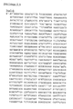
- the nucleotide sequence of the nadB gene is shown in Fig. 2 b.
- L-aspartate oxidase activity is based on the fact that the intermediate iminoaspartate formed by oxidation of L-aspartic acid with L-aspartate oxidase in the presence of oxygen and FAD is spontaneously hydrolyzed to oxaloacetate and NH4+ in the absence of quinolinic acid synthase and dihydroxyacetone phosphate.
- the amount of oxaloacetate formed is therefore proportional to the activity of the L-aspartate oxidase.
- Oxaloacetate is modified by Bergmeyer (HU Bergmeyer, Methods of Enzymatic Analysis, Vol. 9, 1985) and determined photometrically by NADH-dependent reduction to malate with malate dehydrogenase.
- nadA mutant E. coli K12 PA2-18 transformed with the plasmid pCH101 is used as the starting strain for isolating the nadB gene product, hereinafter referred to as nadB enzyme or L-aspartate oxidase.
- 1 l LB medium is inoculated with 5 ml preculture (PA2-18 / pCH101) and shaken at 37 ° C for 2 days.
- a total of 35 g of BFM are obtained by centrifugation at 4 ° C., which are taken up in 200 ml of 50 mM K2HPO4 buffer (pH 8.0, 0.1 mM DTT).
- a Bronson Sonifier B15 set to level 10
- the cells are disrupted by ultrasound treatment for 30 min at 10-15 ° C.
- After centrifugation at 10,000 ⁇ g and 4 ° C. for 35 min about 220 ml of supernatant with about 4.3 g of protein are obtained (the protein is determined by absorption measurements at 280 nm); this supernatant is adjusted to 25% glycerol.
- the precipitate is dissolved in 40 ml of 50 mM potassium phosphate pH 8.0, 25% glycerol, 0.1 mM DTT and against 2 ⁇ 1 l (50 mM potassium phosphate pH 8.0, 25% Glycerin, 0.1 mM DTT) dialyzed for 16 h.
- the activity fractions 46 to 69 (about 205 ml) are combined, adjusted to 50% (NH4) 2SO4, stirred for 16 h at 4 ° C and centrifuged at 10000 ⁇ g and 4 ° C for 40 min. The precipitate is taken up in 11 ml of buffer (50 mM potassium phosphate, pH 8.0, 25% glycerol) and dialyzed against 1 l of the same buffer for 16 h (protein yield 45 mg).
- buffer 50 mM potassium phosphate, pH 8.0, 25% glycerol
- a total of 5 mg of protein are separated in 4 runs of micro l each on a Superose 12 column by FPLC (Pharmacia) by molecular weight and eluted at a flow rate of 0.3 ml / min with 0.05 M NH4HCO3 buffer in fractions of 1 ml.
- the dried protein is taken up in 2 ml of buffer (50 mM KH2PO4, pH 8.0, 25% glycerol) and dialyzed against 1 l of the same buffer for 16 h.
- PAGE are according to Lämmli (U.K. Lämmli, Nature 227 (1970) 680), silver staining according to B.R. Oakley, D.R. Kirsch, N.R. Morris, Anal. Biochem. 105 (1980) 361.
- the molecular weight determined from FPLC and PAGE is 60,000 +/- 2,000 daltons.
- Table 1 summarizes the results of the cleaning steps. *) experimental values converted to total **) The decrease in specific activity is due to the fact that FPLC purification was carried out for the purpose of sample sequencing with the aim of a high-purity protein fraction and not with the aim of maximum activity.
- the sample is oxidized with performic acid to prove the existence of the suspected cysteine in position 9 after the 1st run.
- the N-terminal amino acid sequence of the nadB enzyme determined is: Met Asn Thr Leu Pro Glu His Ser Cys Asp Val Leu Ile Ile Gly Ser Gly Ala ...
- the nucleotide sequence derived from the amino acid sequence on the basis of the known codon assignment is used to identify the start codon of the nadB structural gene and corresponds exactly to the nucleotide sequence determined according to Sanger (Fig. 2 b).
- a mutant E. coli K12 C600 (nadA50 :: Tn10) is obtained by transduction of the nadA50 :: Tn10 gene locus from strain NK6033 using phage T4GT7 into strain C600 and selection on LB / Tc medium.
- the transposon Tn10 coding for tetracycline resistance is removed according to Bochner (BR Bochner, H.-C. Huang, GL Schieven, BN Ames, J. Bacteriol, 143 (1980) 926-933), and a mutant E.coli Obtained K12 C600 (nadA).
- recA56 gene locus from strain LA5708 (srl300 :: Tn10, recA56) is then introduced into the mutant C600 (nadA50 :: Tn10) by transduction using phage T4GT7 according to Example 2: Selection for recA is carried out on LB agar plates by exposure to UV light (UV hand lamp Fluotest, Hanau, type 204AC; 254 nm, 0-30 s, 15 cm distance between lamp and plate). Colonies that did not survive UV exposure for longer than 15 s were identified as recA mutants.
- UV light UV hand lamp Fluotest, Hanau, type 204AC; 254 nm, 0-30 s, 15 cm distance between lamp and plate.
- the mutant E.coli K12 C600 (nadA50, srl300 :: Tn10, recA56) obtained is designated RF1.
- 75 micro g of chromosomal DNA from E. coli are hydrolyzed with 300 units of the restriction endonuclease Sau3A at 37 ° C. for 2 h and used with 10 micro g of the plasmid pLG339 hydrolyzed with 10 units of BamHI for ligation.
- a clone is isolated which contains a plasmid (pCH200) with an approximately 12 kbp insert. Retransformation into the nadA mutant E. coli 431 confirms nadA complementation.
- the plasmid pCH200 is subjected to a partial hydrolysis with the restriction endonuclease AluI.
- the hydrolyzate is ligated into the HincII hydrolyzed plasmid pUC18. This is followed by transformation into strain 431 and selection on YP / Ap medium.
- a plasmid (pCH201) with a 1.4 kbp insert is isolated (restriction map see Fig. 3).
- the 1.4 kbp insert is obtained from pCH201 by hydrolysis with the restriction endonucleases BamHI and PstI and inserted into the BamHI / PstI sites of the vector pKT235 to the plasmid pCH202.
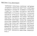
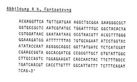
- nadA gene follows the strategy shown in Fig. 4 a. All fragments are cloned into the corresponding sites in the polylinker sequence of pUC18.
- Fig. 4 b shows the (double-stranded) determined nucleotide sequence.
- 1 micro g of the multi-copy plasmid pBR322 and 1 micro g of the plasmid pCH201 are hydrolyzed quantitatively with the restriction endonucleases EcoRI and HindIII; after extraction of the enzymes with phenol and precipitation of the DNA with ethanol, the hydrolysates are used for ligation with 10 units of T4 ligase in 20 microliter ligase buffer and transformed into the nadA mutant 431; Selection for incorporation of the 1.4 kbp fragment containing nadA is made on YP / Ap medium.
- the nadB-containing 3.2 kbp HindIII / NruI fragment from pCH101 is inserted in an analogous manner between the HindIII and NruI sites of the plasmid pCH203 thus obtained.
- the plasmid pCH400 obtained contains both genes, nadA and nadB genes, under the expression control of the genomic promoters (FIG. 5).
- nadA50 Tn10, nadC mutant is constructed starting from the nadC mutant W4546 which excretes quinolinic acid.
- the nadA Tn10 gene locus from strain NK6033 is introduced into strain W4546 by transduction using phage T4GT7.
- nadA Tn10-nadC mutant must not release quinolinic acid into the medium.
- the mutant E. coli K12 (nadA / nadC) obtained is designated RF2.
- Microorganisms of the RF2 strain from Example 10 are transformed with the plasmids pCH101 and pCH202 and with the plasmid pCH400.
- the resulting strains RF2 (pCH101, pCH202) and RF2 (pCH400) as well as the starting strain RF2 are incubated in YP medium with the addition of nicotinic acid (10 / mol / l) at 37 ° C for 48 h (e.g. 5 ml Shake cultures in test tubes; e.g. 1000 ml shake culture in Erlenmeyer flasks).
- the quinolinic acid-secreting nadC mutant W4546 is cultivated under the same conditions as a reference. The results are compared with literature values.
- the necessary antibiotics are added for the cultivation of plasmid-containing strains in YP or A medium, Ap for pCW400; Ap + Km for pCH101 + pCH202, pCH203 + pCH103 and pCH104 + pCH204.
- Tc is additionally added for the cultivation of the RF2 strain and its transformed derivatives.
- the cells After determining the cell density by measuring the optical density at 550 nm, the cells are centrifuged off. The quinolinic acid dissolved in the supernatant is quantitated by HPLC on an anion exchange column Zorbax-NH2 (Dupont).
- strains RF2 (pCH400) and RW1 (pCH400) are deposited as microorganisms according to the invention at the German Collection of Microorganisms (DSM), Braunschweig.
Landscapes
- Genetics & Genomics (AREA)
- Life Sciences & Earth Sciences (AREA)
- Engineering & Computer Science (AREA)
- Health & Medical Sciences (AREA)
- Organic Chemistry (AREA)
- Chemical & Material Sciences (AREA)
- Wood Science & Technology (AREA)
- Zoology (AREA)
- Biomedical Technology (AREA)
- Biotechnology (AREA)
- General Engineering & Computer Science (AREA)
- Bioinformatics & Cheminformatics (AREA)
- Molecular Biology (AREA)
- Microbiology (AREA)
- Biochemistry (AREA)
- General Health & Medical Sciences (AREA)
- Plant Pathology (AREA)
- Biophysics (AREA)
- Physics & Mathematics (AREA)
- Chemical Kinetics & Catalysis (AREA)
- General Chemical & Material Sciences (AREA)
- Micro-Organisms Or Cultivation Processes Thereof (AREA)
Abstract
Description
- Die Erfindung betrifft ein Verfahren zur Herstellung von Chinolinsäure (Pyridin-2,3-dicarbonsäure) mit Hilfe gentechnisch modifizierter Mikroorganismen.
- Chinolinsäure ist ein wichtiges Zwischenprodukt für zahlreiche Pharmazeutika und Pflanzenschutzmittel. Sie wird großtechnisch hergestellt durch Oxidationsverfahren von Chinolin oder Chinolinderivaten gemäß z. B. EP-B 82 542 oder EP-A 149 857. Ein Nachteil dieser Verfahren ist es, daß die verfügbare Rohstoffmenge nicht ausreicht, um den stetig wachsenden Bedarf an Chinolinsäure zu decken.
- Es ist daher Aufgabe vorliegender Erfindung, ein Verfahren zur Herstellung von Chinolinsäure bereitzustellen, bei dem diese aus völlig anderen, unbegrenzt verfügbaren Rohstoffen in einem einfachen, kostengünstigen und umweltfreundlichen Verfahren gewonnen wird.
- Die Lösung der Aufgabe besteht aus einem Herstellungsverfahren mit Hilfe gentechnisch modifizierter Mikroorganismen, aus der Bereitstellung und Herstellung solcher Mikroorganismen durch Isolierung und Bestimmung von DNA-Sequenzen, die für die Synthese der Enzyme Chinolinsäuresynthase und L-Aspartatoxidase codieren, deren Kombination mit Plasmid-DNA-Sequenzen und Vereinigung der so hergestellten rekombinanten Plasmide mit einem beliebigen Mikroorganismus gemäß der Ansprüche 1 bis 15.
- Chinolinsäure ist ein natürliches intermediäres Stoffwechselprodukt vieler Organismen bei der Biosynthese des Nikotinamidadenindinukleotids (NAD). Als Zwischenprodukt tritt es allerdings in so geringen Konzentrationen auf, daß natürlich vorhandene Organismen für eine technische Herstellung keine Anwendung finden können.
- Ist dagegen in einer sogenannten nadC-Mutante die Decarboxylierung der Chinolinsäure und die gleichzeitig ablaufende N-Phosphoribosylierung zur N-Phosphoribosylnikotinsäure blockiert, so lassen sich in einem nährstoffreichen Nährmedium bis zu 73 mg/l Chinolinsäure nachweisen (J.L.R. Chandler, R.K. Gholson, J. Bacteriol. 111 (1972) 96-102). Diese Konzentrationen sind für eine technische Gewinnung der Chinolinsäure aus Nährmedium zu gering.
- Es ist daher notwendig, Organismen, insbesondere Mikroorganismen, so zu modifizieren, daß sie Chinolinsäure in einem Ausmaß produzieren, das für ein technisches Verfahren ausreichend ist.
- Zur Modifizierung der Mikroorganismen werden erfindungsgemäß aus Mikroorganismen, die die Chinolinsäuresynthese als Teil des Stoffwechsels durchführen, DNA-Sequenzen gewonnen, die für die Enzyme Chinolinsäuresynthase (nadA) und L-Aspartatoxidase (nadB) codieren.
- Hierbei steht die Enzymbezeichnung L-Aspartatoxidase für ein Enzym, das die folgende Reaktion katalysiert:

- Mit Chinolinsäuresynthase wird ein Enzym bezeichnet, das die folgende Reaktion katalysiert:

- DNA-Sequenzen, die für die genannten Enzyme codieren, sind in der genomischen Information vieler Mikroorganismen vorhanden, wie z. B. in
Escherichia coli
Salmonella typhimurium
Mycobacterium tuberculosis
Mycobacterium bovis
Bacillus subtilis
Saccharomyces cerevisiae.
- Diese Mikroorganismen sind für die Isolierung von nadA und nadB geeignet. In der Literatur sind nadA- und nadB-Mutanten von E.coli K12 beschrieben.
- Die Isolierung und Insertion eines Gens für die de novo Chinolinsäure-Biosynthese (vermutlich nadB) aus chromosomaler E.coli DNA in das Plasmid pBR322 zu einem Plasmid pNADH1 wird bei Kuwahara et al. (M. Kuwahara, M. Yonehana, T. Kimura und Y. Ishida, Agric. Biol. Chem. 47(1983) 2405-8) beschrieben. Zellfreie Extrakte aus pNADH1-Plasmid-haltigen E.coli C600-Zellen sollen eine um das ca. fünffache erhöhte Chinolinsäuresynthese aufweisen, wenn man von Dihydroxyacetonphosphat und L-Asparaginsäure als Vorstufen ausgeht.
- Hingegen wurden aber weder Chinolinsäuresynthese noch Chinolinsäureausscheidung in ruhenden oder wachsenden Zellen, die durch plasmidgebundene Gene bewirkt wird, gezeigt. Nur so ist jedoch die Verwendung billiger Kohlenstoff- und Stickstoffquellen möglich.
- In der Literatur wird verschiedentlich diskutiert, daß für die Chinolinsäuresynthese aus L-Aspartat bis zu sechs Enzyme notwendig sind. Es wurde gefunden, daß lediglich die beiden Enzyme L-Aspartatoxidase und Chinolinsäuresynthase für eine gesteigerte Chinolinsäuresynthese ausreichen.
- Es wurde weiterhin gefunden, daß zur Lösung der erfindungsgemäßen Aufgabe Plasmide konstruiert werden müssen, die beide DNA-Sequenzen nadA und nadB enthalten.
- Da diese beiden Gene in den Mikroorganismen genomisch getrennt kartiert sind, mußten Plasmide konstruiert werden, die die zwei DNA-Sequenzen, die aus genomisch getrennten Genen bestehen und die jeweils für ein Enzym des Stoffwechsels codieren, enthalten und gemeinsam wirksam werden lassen.
- Dazu wurden folgende Schritte durchgeführt:
- Identifizierung und Isolierung der DNA-Sequenz nadB, die für das Enzym L-Aspartatoxidase codiert.
- Identifizierung und Isolierung der DNA-Sequenz nadA, die für das Enzym Chinolinsäuresynthase codiert.
- Einbau der DNA-Sequenzen nadA und nadB in Plasmide, so daß jede DNA-Sequenz mit einer Expressions-Kontroll-Sequenz verbunden ist.
- Einbau der Strukturgen-Sequenzen der DNA-Sequenzen nadA und nadB hintereinander unter gemeinsamer Kontrolle einer einzigen Expressions-Kontroll-Sequenz.
- Einbringen der so hergestellten modifizierten Plasmide in einen transformierbaren Wirtsorganismus. - Die so hergestellten neuen Mikroorganismen produzieren in einem Nährmedium mit organischer C-Quelle und anorganischer oder organischer N-Quelle wesentlich mehr Chinolinsäure als es dem Stand der Technik entspricht. Die produzierte Chinolinsäure wird nicht mehr dem ursprünglichen Metabolismus gemäß umgesetzt, sondern bevorzugt ausgeschieden. Somit ist eine technische Gewinnung von Chinolinsäure möglich.
- Zur Isolierung der DNA-Sequenz nadB wird aus einem geeigneten Mikroorganismus chromosomale DNA isoliert. Diese DNA wird mit der Restriktionsendonuclease HindIII hydrolysiert. Die entstandenen Fragmente werden auf einem Polyacrylamidgel elektrophoretisch aufgetrennt.
- Aus dem Gel werden die zwischen 6 und 8 kbp liegenden DNA-Fragmente elektroeluiert und mit dem ebenfalls mit HindIII linearisierten Vektor pBR322 ligiert. Die Selektion auf nadB-Gen erfolgt durch Transformation in eine nadB-Mutante (Eine nadB-Mutante enthält keine L-Aspartatoxidase (nadB-Enzym) und ist daher nicht in der Lage, die Oxidation von L-Aspartat zu Iminoaspartat durchzuführen.) und Test auf Komplementation auf einem Minimalmedium (z. B. R.A. Yates und A.B. Pardee, J.Biol.Chem. 221 (1956) 643-756). Es wird ein ca. 13 kbp großes, nadB-Mangel-komplementierendes Plasmid pCH100 isoliert.
- Die Restriktionsanalyse ergibt u. a. eine NruI-Schnittstelle auf dem Insert, die zur Subklonierung verwendet wird. Ein ca. 3.2 kbp großes NruI-HindIII-Fragment wird in mit HindIII und NruI hydrolisierten pBR322 zu einem neuen Plasmid pCH101 inseriert. Die Plasmide pCH100 und pCH101 ergeben in wiederholten Retransformationsversuchen in eine nadB-Mutante Komplementation des nadB-Mangels. Weitere Subklonierungen ergaben ein 1,6 kbp großes Fragment (SspI-AccI) das in in einer nadB-Mutante den nadB-Mangel komplementiert.
- Von diesem Fragment wird die Nukleotidsequenz ermittelt.
- Parallel zu der Isolierung des nadB-Gens wird mit Hilfe protein-biochemischer Techniken das nadB-Enzym, die L-Aspartatoxidase aus E.coli-Mutanten rein dargestellt (hierfür wird ein Enzym-Aktivitätstest neu erstellt) und mittels automatisiertem Edman-Abbau N-terminal sequenziert.
- Ein Vergleich der N-terminalen Proteinsequenz und der Nukleotidsequenz ergibt den Startpunkt des nadB-Strukturgens, ca. 450 bp von der HindIII-Schnittstelle entfernt.
- Zur Isolierung der DNA-Sequenz nadA wird ebenfalls aus einem geeigneten Mikroorganismus chromosomale DNA isoliert und mit der Restriktionsendonuclease Sau3A hydrolysiert; die entstandenen DNA-Fragmente werden in die BamHI-Schnittstelle des Plasmids pBR322 inseriert und in eine nadA-Mutante transformiert.
- Dabei wurde überraschenderweise gefunden, daß Insertion in das üblicherweise verwendete high copy Plasmid pBR322 für die transformierte Wirtszelle lethal ist. Erst die Verwendung eines low copy Plasmids erlaubte die Isolierung einer nadA-DNA-Sequenz; die Selektion des nadA-Gen tragenden Plasmids erfolgt durch Komplementation nach Transformation in eine nadA-Mutante auf Minimalmedium nach an sich bekannten Verfahren.
- Das isolierte Plasmid pCH200 enthält ein ca. 12 kbp großes Insert. Die Retransformation in nadA-Mutante 431 ergibt Komplementation des nadA-Mangels.
- Partielle Hydrolyse mit der Restriktionsendonuclease HaeIII und Klonierung in die HincII-Schnittstelle des Vektors pUC18 ergibt das Plasmid pCH201 mit einem ca. 1.4 kbp großen Insert. Die Nukleotidsequenz dieses 1.4 kbp Inserts wird vollständig beidsträngig ermittelt und ergibt neben dem Strukturgen mit seiner Start- und Stop-Sequenz auch die Promotorregion.
- Erfindungsgemäß sind die DNA-Sequenzen nadA und nadB bereits mit je einer Expressions-Kontroll-Sequenz verbunden. Sie haben als Expressions-Kontroll-Sequenz die genomischen Regulationssequenzen. Sie können jedoch auch mit Expressions-Kontroll-Sequenzen verbunden werden, die an sich bereits bekannt sind. (Z. B.: H. Bujard, U. Deuschle, W. Kammerer, R. Creutz, W. Bannwarth, D. Stueber, UCLA Symp. Mol. Cell. Biol., New Ser. 1985, 30, 21-29; E. Remant, P. Stanssens, F. Fiers, Gene 15(1981) 81-93).
- Dazu gehören:
- der E.coli trp-Promotor,
- der E.coli tac-Promotor,
- der E.coli beta-Lactamasepromotor,
- der E.coli Lipoproteinpromotor,
- eine Hefe-Expressions-Kontroll-Sequenz,
- eine Pseudomonaden-Expressions-Kontroll-Sequenz
oder eine andere prokaryontische Expressions-Kontroll-Sequenz. - Wichtig ist jeweils die funktionelle Verknüpfung des Gens mit der Expressions-Kontroll-Sequenz sowie die Auswahl einer geeigneten Expressions-Kontroll-Sequenz für einen bestimmten Wirtsorganismus. In einer weiteren Ausführung der Erfindung sind die beiden DNA-Sequenzen nadA und nadB hintereinandergeschaltet und haben gemeinsam eine Expressions-Kontroll-Sequenz.
- Diese mitunter als rekombinante Expressionsplasmide benannten Expressionsvektoren werden erfindungsgemäß als Plasmide bezeichnet. Sie werden in an sich bekannter Weise erhalten durch Ligation der einzelnen DNA-Fragmente und Insertion der DNA-Fragmente in Plasmide. Im Gegensatz zu den Erkenntnissen bei der Isolierung der nadA-Sequenz läßt sich nun überraschenderweise die Insertion in ein high copy Plasmid durchführen.
- Die erhaltenen Plasmide werden in üblicher Weise in einen transformierbaren Wirtsorganismus, z. B. ein E.coli-Bakterium, eingebracht. Anschließend werden die mit einem oder mehreren Plasmiden transformierten Mikroorganismen in an sich bekannter Weise in einem geeigneten Nährmedium kultiviert und das oder die bei der Expression gebildete(n) Polypeptid(e) mit der biologischen Aktivität des/der Enzyms/Enzyme L-Aspartatoxidase und/oder Chinolinsäuresynthase können daraus nach Standardmethoden isoliert und nach den entwickelten Tests nachgewiesen werden.
- Die Produktion und Gewinnung von Chinolinsäure erfolgt in an sich bekannter Weise durch die mit der erfindungsgemäßen DNA transformierten Mikroorganismen unter geeigneten Bedingungen, wie Batch-, Fed-batch- oder kontinuierliche Fermentation in Rühr- oder Airlift oder ähnlichen Bioreaktoren, wobei einem erfindungsgemäßen Mikroorganismus in einem Nährmedium eine organische Kohlenstoffquelle und eine anorganische oder organische Stickstoffquelle unter Wachstumsbedingungen zur Verfügung gestellt werden.
- Die Erfindung wird durch die folgenden Abbildungen und Beispiele näher erläutert.
-
- Abb. 1 Restriktionskarte des 3.2 kbp-HindIII/NruI-Inserts in pCH101
- Abb. 2
- a. Sequenzierungsstrategie nadB-Gen
- b. Nukleotidsequenz des nadB-Gens aus E.coli K12
- Abb. 3 Restriktionskarte des 1.4 kbp-HaeIII-Inserts in pCH201
- Abb. 4
- a. Sequenzierungsstrategie nadA-Gen
- b. Nukleotidsequenz des nadA-Gens aus E.coli K12
- Abb. 5 Restriktionskarte des Plasmids pCH400
- Ap Ampicillin
Bicine Bishydroxymethylglycin
BFM Biofeuchtmasse
BTM Biotrockenmasse
BSA Rinderserumalbumin
bp Basenpaare
DHAP Dihydroxyacetonphosphat
DMSO Dimethylsulfoxid
DNA Desoxyribonukleinsäure
DTT Dithiothreitol
E.coli Escherichia coli
EDTA Ethylendiamintetraessigsäure
FAD Flavin-Adenin-Dinukleotid
FPLC Fast Protein Liquid Chromatography
HPLC High Performance Liquid Chromatography
kbp Kilobasenpaare
Km Kanamycin
LB Luria-Bertani
PAGE Poly-Acrylamid-Gel-Elektrophorese
RNA Ribonukleinsäure
SDS Sodium-Dodecyl-Sulfat
Tc Tetracyclin
Tris Trishydroxymethylglycin
UV Ultraviolett
YP Yates-Pardee
- TE
10 mmol/l Tris, pH 8.0, 1 mmol/l EDTA
Lysozym-Puffer
0.1 ml NaCl (5 mol/l)
1 ml EDTA (0.5 mol/l, pH 8.0)
0.3 ml Tris-Pufferkonzentrat (Tris-HCl) (1 mol/l) pH 8.0
8.6 ml H₂O
Proteinase K Puffer
0.01 mol/l Tris (Tris-HCl) pH 8.0
0.005 mol/l EDTA
0.5 % SDS
YP-Medium
7 g K₂HPO₄
2 g KH₂PO₄
0.5 g Natriumcitrat × 5 H₂O
0.1 g MgSO₄ × 7 H₂O
1 g (NH₄)₂SO₄
2.5 g Glucose
2 mg Thiamin
ad 1000 ml H₂O
A-Medium
YP-Medium
+ Nikotinsäure (10⁻⁶ mol/l)
+ L-Aspartat (5 × 10⁻⁶ mol/l)
+ Caseinhydrolysat (5 g/l)
5 g/l Glycerin statt2.5 g Glucose
LB-Medium
10 g Caseinhydrolysat oder Pepton
5 g Hefeextrakt
5 g NaCl
ad 1000 ml H₂O, pH 7.4
- Bei Einsatz von Antibiotika wurden folgende Konzentrationen angewandt:
Ampicillin (Ap): 100 mg/l
Tetracyclin (Tc): 2 mg/l
Kanamycin (Km): 25 mg/l - 100 ml Nährlösung (LB-Medium) werden mit 1 ml einer Vorkultur von E.coli K12 C600 in der gleichen Nährlösung beimpft und 18 h bei 37 °C inkubiert. Die Zellsuspension wird in auf Eis gestellte Zentrifugenröhrchen verteilt und 10 min bei 4 °C und 2600 × g zentrifugiert.
- Die sedimentierten Zellen werden in 10 ml Lysozym-Puffer suspendiert und mit 40 mg Lysozym 30 min bei 37 °C inkubiert. Die Suspension wird auf 1 % SDS eingestellt und 30 min bei 37 °C inkubiert. Nach Zusatz von 5 mg Proteinase K (Merck) wird 3 h bei 50 °C inkubiert. Danach wird vorsichtig viermal mit TE-gesättigtem Phenol (TE) und anschließend dreimal mit Chloroform/Isoamylalkohol (24 : 1) extrahiert.
- Anschließend wird die wäßrige Phase auf eine Konzentration von 0.3 mol/l an Natriumacetat eingestellt und die DNA durch Zugabe des doppelten Volumens an Ethanol gefällt. Die DNA wird abzentrifugiert, im Exsikkator getrocknet und danach in 5 ml TE-Puffer gelöst. Dann werden 50 mikro g DNase-freie Ribonuclease A (Sigma) zugegeben (Inaktivierung der DNase durch vorheriges Kochen der RNase in 100 mM Natriumacetat bei pH 5.5 für 10 min). Nach 30minütiger Inkubation Zugabe von SDS auf 1 % Endkonzentration und Zugabe von 250 mikro g Proteinase K werden weitere 30 min bei 37 °C inkubiert.
- Danach wird vorsichtig viermal mit TE-gesättigtem Phenol und zehnmal mit Chloroform/Isoamylalkohol (24:1) extrahiert. Die Lösung wird zweimal 12 h gegen 1000 ml TE dialysiert und bei 4 °C aufbewahrt.
- Enzyme: Restriktionsendonucleasen, DNA-Ligase des Phagen T4, Klenov-Fragment der E.coli DNA-Polymerase Pol I, Reverse Transscriptase (MMLV) wurden bei Gibco-BRL, alkalische Phosphatase, Ribonuclease A und Lysozym bei Boehringer (Mannheim) und Proteinase K bei Merck gekauft und nach den Angaben der Hersteller verwendet.
- E.coli-Stämme PA2-18 (CGSC Nr. 5176), NK6033 (CGSC Nr. 6180), NK6042 (CGSC Nr. 6184), W4546 (CGSC Nr. 5179), C600 (CGSC Nr. 3004) wurden vom E.coli Genetic Stock Center (CGSC), die Stämme JM101, JM103, JM109, DH5 von Gibco BRL, der Stamm 431 von Dr. B. Rak (Freiburg), der Stamm GE 1806 von Dr. W. Schumann (Darmstadt), bezogen.
- Plasmide (pBR322, pUC18, pUC19, pLG339) wurden bei Pharmacia, das Plasmid pLG339 (N.G. Stoker et al., Gene 18 (1982) 335-341) von den Autoren bezogen, in geeigneten Wirtszellen propagiert und nach folgendem Verfahren präpariert:
1000 ml LB-Medium werden mit 3 ml einer Übernachtkultur des plasmidtragenden Wirtsstammes beimpft und 18 h bei 37 °C inkubiert. - Die Zellsuspension wird 15 min bei 2600 × g und 4 °C zentrifugiert. Die sedimentierten Zellen werden in insgesamt 20 ml 20 % Saccharose, 10 mM Tris-HCl, pH 8.0, suspendiert und 10 min auf Eis inkubiert. Danach werden 3 ml 0.5 M EDTA, pH 8.0, und 3 ml 20 mg/ml Lysozymlösung zugesetzt und damit 45 min auf Eis inkubiert. Die Lösung wird auf 2 Zentrifugenröhrchen aufgeteilt.
- Danach werden 2.6 ml einer Lösung, die 5 % Brij58 (Polyoxyethylenemonocetylether) und 2 % Desoxycholat enthält, zugesetzt, und es wird exakt 5 min auf Eis inkubiert. Anschließend wird bei 60000 × g und 4 °C für 25 min zentrifugiert. Nach Zugabe von Natriumacetat auf Endkonzentration von 0.3 mol/l wird die DNA mit dem doppelten Volumen Ethanol bei - 70 °C gefällt.
- Die DNA wird bei 10000 × g und 4 °C 20 min abzentrifugiert, im Vacuum getrocknet und in 10 ml Proteinase K Puffer gelöst. Nach Zugabe von 1 mg Proteinase K wird bei 50 °C 60 min inkubiert. Anschließend wird je dreimal mit TE-gesättigtem Phenol und Chloroform/Isoamylalkohol (24 : 1) und einmal mit Diethylether extrahiert.
- Nach Einstellen der wäßrigen Phase auf 0.3 mol/l Natriumacetat wird die DNA mit dem doppelten Volumen Ethanol bei - 70 °C gefällt, bei 10000 × g und 4 °C 30 min abzentrifugiert und im Vakuum getrocknet.
- Der Niederschlag wird in 29 ml TE, die 28.14 g CsCl enthalten, gelöst. Die Lösung wird mit 2.9 ml Ethidiumbromid-Lösung versetzt (10 mg/ml), in ein 39 ml fassendes Quick-seal-Zentrifugenröhrchen (Beckman Instruments) gefüllt, und es wird im Vertikalrotor bei 160000 × g und 15 °C 16 h zentrifugiert. Unter UV-Licht wird die "Covalently Closed Circular"-DNA-Bande abgezogen; Ethidiumbromid wird mit TE-gesättigtem Butanol extrahiert, Butanol und CsCl werden durch Dialyse (24 h, gegen 2 × 1 l TE) entfernt.
- Die DNA(= Plasmid)-Konzentration in der wäßrigen Lösung wird durch Messung der UV-Absorption bei 260 nm bestimmt.
- Plasmid-DNA, die zur Sequenzierung eingesetzt werden soll, wird durch Fällung mit Spermin (B.C. Hoopes, W.R. McClure, Nucl. Acid. Res. 9 (1981) 5493-5504) zusätzlich gereinigt.
- Durch restriktionsenzymatische Hydrolyse erhaltene DNA-Fragmente werden auf Polyacrylamid-Gel aufgetrennt (T. Maniatis, F. Fritsch, J. Sambrock; Molecular Cloning, A Laboratory Manual; Cold Spring Harbor Laboratory 1982, S. 150 ff.); Fragmentelution erfolgt durch Elektroelution (Maniatis S. 164).
- Die Transformation von E.coli-Stämmen erfolgt nach der CaCl₂-Methode (M. Mandel, A. Higa, Mol. Biol. 53 (1970) 154).
- Der Phage T4GT7 (G. Wilson, K.K.V. Young, G.J. Edlin, Nature 280(1979) 80-81) wurde von den in der Literaturstelle angegebenen Autoren bezogen.
- Transduktion: Es wird das Prinzip der "generalisierten" Transduktion (R.E. Glass, Gene Function, Croom Helm London (1982), S. 210 ff.) mit Hilfe des Phagen T4GT7 zur Überführung genomischer DNA eines Donorstammes in einen Rezeptorstamm angewandt.
- Die Gewinnung von Phagenlysat, die Titerbestimmung und Infektion der Wirtszellen erfolgt nach der an sich bekannten Methode (Maniatis).
- Im Einzelnen werden 100 mikro l T4GT7-Lysat vermischt und 15 min bei 37 °C inkubiert. Hierbei werden die Zellen des Donorstammes vom Phagen infiziert; während des Vermehrungs- und Lysezyklus werden mit geringer Wahrscheinlichkeit Fragmente genomischer DNA anstelle der Phagengenome in Phagenhüllen eingekapselt.
- Zur Gewinnung des Phagenlysats, das nun auch eingekapselte genomische DNA-Fragmente enthält, wird die Weichagarschicht nach Zugabe von 1 ml LB-Medium von der Agarplatte abpipettiert. Die so gewonnene Suspension wird mit 0,5 ml CHCl₃ ausgeschüttelt und zentrifugiert (15 min, 8000 × g, 20 °C).
- Der phagenhaltige wäßrige Überstand wird als Phagenlysat bezeichnet. Es wird eine Titerbestimmung nach an sich bekannten Methoden mit dem Stamm C600 durchgeführt.
- Zur Transduktion der Donor-DNA in den Rezeptorstamm werden je 100 mikro l einer Übernachtkultur des Rezeptorstammes mit 5, 10, 50 und 100 mikro l Phagenlysat und 195, 190, 150 und 100 mikro l LB-Medium vermischt und nach 15 min Inkubation bei 37 °C mit je 2,5 ml LB-Weichagar (0,7 % Agar) auf Agarplatten verteilt und 16 h bei 37 °C inkubiert.
- Die Identifikation der Zellkolonien, die das gewünschte transduzierte Gen enthalten, erfolgt durch ein geeignetes Selektionsmedium, das in den Agarplatten eingesetzt wird (z. B. Zusatz von Tetracyclin bei der Identifikation der erfolgreichen Transduktion eines Transposon Tn10-tragenden Genes; Tn10 bewirkt Tetracyclinresistenz).
- Zur Bestimmung der Nukleotidsequenzen werden mit Hilfe von Restriktionsendonucleasen DNA-Fragmente geeigneter Länge (zwischen 100 und 400 bp) hergestellt und in passende Restriktionsschnittstellen der Plasmide pUC18 oder pUC19 kloniert. Die Sequenzierung erfolgt nach dem Kettenabbruchverfahren ("Dideoxy-Methode") nach Sanger (F. Sanger, S. Micklen, A. R. Coulson, Proc. Natl. Acad. Sci. USA 74 (1977) 5463-5467) unter Verwendung der Arbeitsvorschrift "Plasmid Sequenzieren" (P. Heinrich, Inst. f. Biochemie der Ludwig-Maximilian-Universität, 8000 München, 1985).
- 30 mikro g nach Beispiel 1 isolierter chromosomaler DNA werden 18 h bei 50 °C mit der Restriktionsendonuclease HindIII hydrolysiert und anschließend auf einem 3.5 % Polyacrylamidgel in TBE-Puffer 20 h bei 30 mA elektrophoretisch aufgetrennt.
- Die DNA-Fragmente mit Größen zwischen 6 und 8 kbp wurden aus dem Gel elektroeluiert, über eine Anionenaustauschersäule (DE52 Diethylaminoethyl-Cellulose, Whatman) gereinigt und mit Ethanol gefällt. 1 mikro g Fragment-DNA und 1 mikro g HindIII-hydrolysierte und dephosphorylierte pBR322-DNA werden in 20 mikro l Ligasepuffer (von Gibco-BRL) mit 10 Units T4-DNA-Ligase zur Ligation eingesetzt.
- Transformation in die nadB-Mutante E.coli NK6042 (nadB::Tn10) und Selektion auf YP/Ap-Medium auf Komplementation des nadB-Mangels ergibt 7 Klone, die alle ein Plasmid der ungefähren Größe 13 kbp enthalten. Wiederholte Retransformation dieses Plasmids in NK6042 bestätigen die Komplementation des nadB-Mangels. Das Plasmid wird mit pCH100 bezeichnet und wie bei Maniatis beschrieben auf Restriktionsschnittstellen analysiert.
- Subklonierung eines ca. 3.2 kbp großen HindIII-NruI-Fragments in mit HindIII und NruI hydrolysierten pBR322 ergibt ein weiteres Plasmid pCH101, welches nadB-Mangel in NK6042 komplementiert; von dem 3.2 kbp-Insert in pCH101 wird eine Restriktionskarte erstellt (Abb. 1).
- Ausgehend von dem Plasmid pCH101 wurden Subklone in geeignete Restriktionsnucleaseschnittstellen der Polylinkersequenz der Plasmide pUC18 und pUC19 hergestellt. Es wurde gefunden, daß das ca. 1,6 kbp große SspI-AccI-Fragment (Abb. 2 a) das kleinste Fragment ist, welches nadB Mangel in NK6042 komplementiert. Dieses Fragment wurde nach Sanger vollständig und beidstrangig sequenziert.
- Die Sequenzierungsstrategie ist in Abb. 2 a zusammengefaßt.
- Die Nukleotidsequenz des nadB-Gens ist in Abb. 2 b gezeigt.
- Dem Nachweis der L-Aspartatoxidase-Aktivität liegt zugrunde, daß das durch Oxidation von L-Asparaginsäure mit L-Aspartatoxidase in Gegenwart von Sauerstoff und FAD intermediär entstehende Iminoaspartat in Abwesenheit von Chinolinsäuresynthase und Dihydroxyacetonphosphat spontan zu Oxalacetat und NH₄⁺ hydrolysiert wird.
- Die Menge gebildeten Oxalacetats ist damit der Aktivität der L-Aspartatoxidase proportional.
- Oxalacetat wird modifiziert nach Bergmeyer (H. U. Bergmeyer, Methods of Enzymatic Analysis, Vol. 9, 1985) durch NADH-abhängige Reduktion zu Malat mit Malatdehydrogenase photometrisch bestimmt.
-
- 1. Umsetzung von L-Aspartat zu Oxalacetat
Testansatz 285 mikro l H₂O
Testansatz 50 mikro l Bicine 1 mol/l, pH 8.0
Testansatz 50 mikro l BSA 10 mg/ml in wäßriger Lösung
Testansatz 5 mikro l EDTA 0.5 mol/l, pH 8.0
Testansatz 5 mikro l FAD 2 mmol/l
Testansatz 5 mikro l Katalase 5000 Units/ml
Testansatz 50 mikro l L-Aspartat 0.1 mol/l, pH 8.0
Testansatz 50 mikro l Proteinlösung (Probe)
Der Testansatz wird bei 37 °C für 30 min inkubiert; danach wird die Reaktion durch Zugabe von 200 mikro l HClO₄ (1.7 mol/l) beendet; die ausgefällten Proteine werden abzentrifugiert, der Überstand wird durch Zugabe von 75 mikro l KOH (5 mol/l) neutralisiert; das hier ausfallende KClO₄ wird durch Zentrifugation abgetrennt. Die Oxalacetatkonzentration im Überstand wird wie folgt bestimmt: - 2. Bestimmung der Oxalacetatkonzentration
Testansatz 200 mikro l Bicine 1 mol/l, pH 8.0
Testansatz 5 mikro l EDTA 0.5 mol/l
Testansatz 585 mikro l Überstand aus 1.
Testansatz 100 mikro l NADH 1 mmol/l
Der Testansatz wird in einer Küvette durchgeführt; nachdem die Absorption bei 340 nm konstant ist, werden 10 mikro l Malatdehydrogenase (1 Unit/mikro l) zugegeben, und es wird die Extinktionsdifferenz bei 340 nm bestimmt. Der molare Extinktionskoeffizient von NADH bei 340 nm beträgt 6220 dm²/mol. Eine Unit L-Aspartatoxidase ist diejenige Menge Enzym, die die Oxidation von einem mikro mol L-Aspartat in einer Minute katalysiert. Ergebnisse sind in Tabelle 1 niedergelegt. - Als Ausgangsstamm zur Isolierung des nadB-Genprodukts, im folgenden nadB-Enzym oder L-Aspartatoxidase genannt, wird eine mit dem Plasmid pCH101 transformierte nadA-Mutante (E.coli K12 PA2-18) verwendet.
- Je 1 l LB-Medium werden mit je 5 ml Vorkultur (PA2-18/pCH101) beimpft und bei 37 °C 2 Tage geschüttelt. Durch Zentrifugation bei 4 °C werden insgesamt 35 g BFM gewonnen, die in 200 ml 50 mM K₂HPO₄-Puffer (pH 8.0, 0,1 mM DTT) aufgenommen werden. Mit einem Bronson Sonifier B15, eingestellt auf Stufe 10, werden die Zellen durch Ultraschallbehandlung für 30 min bei 10 - 15 °C aufgeschlossen. Nach Zentrifugation bei 10000 × g und 4 °C für 35 min werden ca. 220 ml Überstand mit ca. 4,3 g Protein erhalten (die Proteinbestimmung erfolgt durch Absorptionsmessungen bei 280 nm); dieser Überstand wird auf 25 % an Glycerin eingestellt.
- Anschließend werden 43 ml einer 2%igen Protaminsulfatlösung bei 4 °C unter Rühren während 1 h langsam zugesetzt. Zentrifugation bei 28000 × g und 4 °C für 30 min ergibt einen Überstand (320 ml) mit ca. 1,7 g Protein. Nach Einstellen der Lösung auf 50 % an (NH₄)₂SO₄ wird bei 4 °C 4 h lang gerührt.
- Nach Zentrifugation bei 16000 × g und 4 °C für 40 min wird der Niederschlag in 40 ml 50 mM Kaliumphosphat pH 8.0, 25 % Glycerin, 0,1 mM DTT gelöst und gegen 2 × 1 l (50 mM Kaliumphosphat pH 8.0, 25 % Glycerin, 0.1 mM DTT) 16 h dialysiert.
- Zentrifugation bei 17500 × g und 4 °C für 30 min ergibt 34 ml eines proteinhaltigen Überstandes (282 mg Protein), der auf eine DEAE-Sepharose CL6B (Pharmacia) (Maße: 25 × 3 cm) -Säule aufgetragen wird. Es wird mit ml Puffer (50 mM Kaliumphosphat, pH 8.0, 25 % Glycerin) gewaschen und mit 400 ml eines linearen Gradienten von 50 bis 300 mM Kaliumphosphat, pH 8.0, 25 % Glycerin eluiert. Die Fließgeschwindigkeit beträgt ca. 50 ml/h. Es werden Fraktionen von je 10 ml aufgefangen, von denen je 50 mikro l zum Aktivitätstest eingesetzt werden.
- Die Aktivität aufweisende Fraktionen 46 bis 69 (ca. 205 ml) werden zusammengefaßt, auf 50 % (NH₄)₂SO₄ eingestellt, 16 h bei 4 °C gerührt und bei 10000 × g und 4 °C für 40 min zentrifugiert. Der Niederschlag wird in 11 ml Puffer (50 mM Kaliumphosphat, pH 8.0, 25 % Glycerin) aufgenommen und 16 h gegen 1 l desselben Puffers dialysiert (Proteinausbeute 45 mg).
- Insgesamt 5 mg Protein werden in 4 Läufen zu je mikro l auf einer Superose 12-Säule mittels FPLC (Pharmacia) nach Molekulargewicht aufgetrennt und bei einer Fließgeschwindigkeit von 0.3 ml/min mit 0.05 M NH₄HCO₃-Puffer in Fraktionen von 1 ml eluiert.
- Die nadB-Enzymaktivität enthaltenden Fraktionen werden zusammengefaßt (15 ml = 2.3 mg Protein) und gefriergetrocknet. Das getrocknete Protein wird in 2 ml Puffer (50 mM KH₂PO₄, pH 8.0, 25 % Glycerin) aufgenommen und 16 h gegen 1 l des gleichen Puffers dialysiert.
- 250 mikro l (= 87 mikro g Protein) werden einer nochmaligen FPLC (wie oben) unterworfen; 2 - 5 mikro g Protein jeder proteinhaltigen Eluatfraktion werden einer SDS-PAGE mit anschließender Silberfärbung unterworfen. PAGE werden nach Lämmli (U.K. Lämmli, Nature 227 (1970) 680), Silberfärbung nach B.R. Oakley, D.R. Kirsch, N.R. Morris, Anal. Biochem. 105 (1980) 361, durchgeführt.
- Das aus FPLC und PAGE bestimmte Molekulargewicht beträgt 60000 +/- 2000 Dalton.
- Tabelle 1 faßt die Ergebnisse der Reinigungsschritte zusammen.

**) Der Rückgang der spezifischen Aktivität ist darauf zurückzuführen, daß die FPLC-Reinigung zum Zwecke der Probensequenzierung mit dem Ziel einer hochreinen Proteinfraktion und nicht dem Ziel maximaler Aktivität durchgeführt wurde.
- 350 bzw. 250 pmol des über FPLC gereinigten Proteins (nur eine einzige Bande auf PAGE) werden je einer vollautomatischen Sequenzanalyse durch Edmann-Abbau im Gasphasen-Sequenator 470A der Firma Applied Biosystems unterzogen.
- Für den zweiten Sequenzierlauf wird die Probe mit Perameisensäure oxidiert, um die Existenz des nach dem 1. Lauf in Position 9 vermuteten Cysteins nachzuweisen. Die ermittelte N-terminale Aminosäuresequenz des nadB-Enzyms lautet:
Met Asn Thr Leu Pro Glu His Ser Cys Asp Val Leu Ile Ile Gly Ser Gly Ala ...
Die aus der Aminosäuresequenz anhand der bekannten Codon-Zuordnung abgeleitete Nukleotidsequenz wird zur Identifizierung des Startcodons des nadB-Strukturgens verwendet und stimmt exakt mit der nach Sanger bestimmten Nukleotidsequenz (Abb. 2 b) überein. - Für die Identifizierung des nadA-Gens wird durch Transduktion, ausgehend von E.coli K12 C600, eine Mutante konstruiert, die
- 1. eine hohe Transformationseffizienz besitzt
- 2. rekombinante Plasmide nicht durch Rekombination mit genomischen Sequenzen verändert (recA)
- 3. im nadA-Genlocus mutiert ist (nadA).
- Durch Transduktion des nadA50::Tn10-Genlocus aus dem Stamm NK6033 mittels Phagen T4GT7 in den Stamm C600 und Selektion auf LB/Tc-Medium wird eine Mutante E.coli K12 C600 (nadA50::Tn10) erhalten. Das für Tetracyclin-Resistenz codierende Transposon Tn10 wird nach Bochner (B.R. Bochner, H.-C. Huang, G.L. Schieven, B.N. Ames, J. Bacteriol, 143 (1980) 926-933) entfernt, und es wird eine Mutante E.coli K12 C600 (nadA) erhalten. Anschließend wird durch Transduktion mittels Phagen T4GT7 nach Beispiel 2 der recA56-Genlocus aus dem Stamm LA5708 (srl300::Tn10, recA56) in die Mutante C600 (nadA50::Tn10) eingeführt:
Selektion auf recA erfolgt auf LB-Agarplatten durch Belichten mit UV-Licht (UV-Handlampe Fluotest, Hanau, Typ 204AC; 254 nm, 0-30 s, 15 cm Abstand zwischen Lampe und Platte). Kolonien, die eine UV-Belichtung von länger als 15 s nicht überlebten, wurden als recA-Mutanten identifiziert. - Die erhaltene Mutante E.coli K12 C600 (nadA50,srl300::Tn10, recA56) wird mit RF1 bezeichnet.
- 75 mikro g chromosomaler DNA aus E.coli (Beispiel 1) werden mit 300 Units der Restriktionsendonuclease Sau3A bei 37 °C 2 h hydrolysiert und mit 10 mikro g des mit 10 Units BamHI hydrolysierten Plasmids pLG339 zur Ligation eingesetzt. Es erfolgt Transformation in den neu konstruierten Stamm E.coli RF1 und Selektion auf YP/Ap-Medium auf Komplementation des nadA-Mangels. Es wird ein Klon isoliert, der ein Plasmid (pCH200) mit einem ca. 12 kbp großen Insert enthält. Retransformation in die nadA-Mutante E.coli 431 bestätigt nadA-Komplementation.
- Das Plasmid pCH200 wird einer partiellen Hydrolyse mit der Restriktionsendonuclease AluI unterworfen. Das Hydrolysat wird in das mit HincII hydrolysierte Plasmid pUC18 ligiert. Es folgt Transformation in den Stamm 431 und Selektion auf YP/Ap-Medium. Es wird ein Plasmid (pCH201) mit einem 1.4 kbp großen Insert isoliert (Restriktionskarte vergleiche Abb. 3). Durch Hydrolyse mit den Restriktionsendonukleasen BamHI und PstI wird das 1.4 kbp-Insert aus pCH201 gewonnen und in die BamHI/PstI-Schnittstellen des Vektors pKT235 zu dem Plasmid pCH202 inseriert.
- Die Sequenzierung des nadA-Gens folgt der in Abb. 4 a dargestellten Strategie. Alle Fragmente werden in die entsprechenden Schnittstellen der Polylinkersequenz von pUC18 kloniert.
- Abb. 4 b gibt die (doppelsträngig) bestimmte Nukleotidsequenz wieder.
- Zunächst wird das 1.4 kbp EcoRI/HindIII-Fragment aus pCH201 zwischen die EcoRI- und HindIII-Schnittstellen des Plasmids pBR322 inseriert.
- Hierzu werden 1 mikro g des multi-copy Plasmids pBR322 sowie 1 mikro g des Plasmids pCH201 mit den Restriktionsendonucleasen EcoRI und HindIII quantitativ hydrolysiert; die Hydrolysate werden nach Extraktion der Enzyme mit Phenol und Fällung der DNA mit Ethanol zur Ligation mit 10 Units T4-Ligase in 20 mikro l Ligasepuffer eingesetzt und in die nadA-Mutante 431 transformiert; Selektion auf Einbau des 1.4 kbp Fragments, das nadA enthält, erfolgt auf YP/Ap-Medium.
- Zwischen die HindIII- und NruI-Schnittstellen des so erhaltenen Plasmids pCH203 wird in analoger Vorgehensweise das nadB-enthaltende 3.2 kbp HindIII/NruI-Fragment aus pCH101 inseriert. Das erhaltene Plasmid pCH400 enthält beide Gene, nadA- und nadB-Gen, unter der Expressionskontrolle der genomischen Promotoren (Abb. 5).
- Für den experimentellen Nachweis, daß ein nadA-Gen enthaltendes rekombinantes Plasmid die Chinolinsäuresynthese bewirkt, wird, ausgehend von der Chinolinsäureausscheidenden nadC-Mutante W4546, eine nadA50::Tn10, nadC-Mutante konstruiert.
- Der nadA::Tn10-Genlocus aus dem Stamm NK6033 wird durch Transduktion mittels Phagen T4GT7 in den Stamm W4546 eingeführt.
- Der Nachweis einer erfolgreichen Transduktion erfolgt durch Selektion auf YP/Tc-Medium unter Zusatz von Nikotinsäure (10⁻⁶ mol/l) sowie durch HPLC-Analyse (vergleiche Beispiel 11) der Nährlösung, in der die auf YP/Tc-Medium isolierten Mutanten kultiviert worden waren. Eine nadA::Tn10-nadC-Mutante darf keine Chinolinsäure ins Medium ausscheiden. Die erhaltene Mutante E.coli K12 (nadA/nadC) wird mit RF2 bezeichnet.
- Mikroorganismen des Stammes RF2 aus Beispiel 10 werden mit den Plasmiden pCH101 und pCH202 sowie mit dem Plasmid pCH400 transformiert. Die resultierenden Stämme RF2 (pCH101, pCH202) und RF2 (pCH400) sowie der Ausgangsstamm RF2 werden in YP-Medium unter Zusatz von Nicotinsäure (10⁻⁶ mol/l) bei 37 °C für 48 h inkubiert (z. B. 5 ml Schüttelkulturen in Reagenzgläsern; z. B. 1000 ml Schüttelkultur in Erlenmeyerkolben). Unter gleichen Bedingungen wird als Referenz die Chinolinsäure-ausscheidende nadC-Mutante W4546 kultiviert. Die Ergebnisse werden mit Literaturwerten verglichen.
- Weiterhin wurden alle genannten Stämme sowie ein weiterer transformierter Stamm RW1 (pCH400) in einem nährstoffreichen Medium A kultiviert.
- Für die Kultivierung plasmidhaltiger Stämme in YP- bzw. A-Medium werden die benötigten Antibiotika zugesetzt, Ap für pCW400; Ap + Km für pCH101 + pCH202, pCH203 + pCH103 und pCH104 + pCH204. Für die Kultivierung des Stammes RF2 und seiner transformierten Derivate wird zusätzlich Tc zugesetzt.
- Nach Bestimmung der Zelldichte durch Messung der optischen Dichte bei 550 nm werden die Zellen abzentrifugiert. Die im Überstand gelöste Chinolinsäure wird durch HPLC auf einer Anionenaustauschersäule Zorbax-NH₂ (Dupont) quantitativ bestimmt.
- Die Ergebnisse der Messungen sind in Tabelle 2 zusammengestellt.

- Die Stämme RF2 (pCH400) und RW1 (pCH400) sind als erfindungsgemäße Mikroorganismen bei der Deutschen Sammlung von Mikroorganismen (DSM), Braunschweig, hinterlegt.
- Hinterlegungsdatum : 21.01.1987
Hinterlegungsnummer: DSM 3955
- Hinterlegungsdatum : 14.01.1988
Hinterlegungsnummer: DSM 4362
Claims (15)


Applications Claiming Priority (2)
| Application Number | Priority Date | Filing Date | Title |
|---|---|---|---|
| DE3703255 | 1987-02-04 | ||
| DE19873703255 DE3703255A1 (de) | 1987-02-04 | 1987-02-04 | Dna-sequenzen, plasmide und mikroorganismen sowie verfahren zur herstellung von chinolinsaeure |
Publications (3)
| Publication Number | Publication Date |
|---|---|
| EP0279273A2 true EP0279273A2 (de) | 1988-08-24 |
| EP0279273A3 EP0279273A3 (en) | 1990-03-21 |
| EP0279273B1 EP0279273B1 (de) | 1993-12-15 |
Family
ID=6320153
Family Applications (1)
| Application Number | Title | Priority Date | Filing Date |
|---|---|---|---|
| EP88101442A Expired - Lifetime EP0279273B1 (de) | 1987-02-04 | 1988-02-02 | DNA-Sequenzen, Plasmide und Mikroorganismen sowie Verfahren zur Herstellung von Chinolinsäure |
Country Status (2)
| Country | Link |
|---|---|
| EP (1) | EP0279273B1 (de) |
| DE (2) | DE3703255A1 (de) |
Cited By (3)
| Publication number | Priority date | Publication date | Assignee | Title |
|---|---|---|---|---|
| WO1999049888A3 (en) * | 1998-03-31 | 1999-11-18 | Anthony T Maurelli | Identification of bacterial avirulence genes and their products; vaccines and pharmaceutical compositions containing them |
| WO2012096553A2 (en) | 2011-01-14 | 2012-07-19 | Cj Cheiljedang Corporation | Method for the preparation of nicotinic acid |
| EP2801612A4 (de) * | 2012-01-06 | 2015-07-29 | Cj Cheiljedang Corp | Chinolinsäure-produzierende rekombinante mikroorganismen und verfahren zur herstellung von chinolinsäure damit |
Families Citing this family (1)
| Publication number | Priority date | Publication date | Assignee | Title |
|---|---|---|---|---|
| DE3826041A1 (de) * | 1988-07-30 | 1990-02-08 | Ruetgerswerke Ag | Verfahren zur herstellung von chinolinsaeure |
-
1987
- 1987-02-04 DE DE19873703255 patent/DE3703255A1/de not_active Withdrawn
-
1988
- 1988-02-02 DE DE88101442T patent/DE3886234D1/de not_active Expired - Fee Related
- 1988-02-02 EP EP88101442A patent/EP0279273B1/de not_active Expired - Lifetime
Non-Patent Citations (4)
| Title |
|---|
| AGRIC. BIOL. CHEM., Band 47, Nr. 10, 1983, Seiten 2405-2408; M. KUWAHARA et al.: "Synthesis of quinolinic acid by the enzyme preparation of Escherichia coli which contains a plasmid carrying the nad gene for the de novo synthesis of NAD" * |
| CHEMICAL ABSTRACTS, Band 79, Nr. 3, 23. Juli 1973, Seite 208, Zusammenfassung Nr. 15638u, Columbus, Ohio, US; N. SUZUKI et al.: "De novo biosynthesis of NAD in Escherichia coli. V. Properties of the quinolinic acid synthetase system", & BIOCHIM. BIOPHYS. ACTA 1973, 304(2), 309-15 * |
| CHEMICAL ABSTRACTS, Band 83, Nr. 3, 21. Juli 1975, Seite 251, Zusammenfassung Nr. 24848x, Columbus, Ohio, US; G.R. GRIFFITH et al.: "De novo biosynthesis of NAD in Escherichia coli. Separation of the nadB gene product from the nadA gene product and its purification", & EUR. J. BIOCHEM. 1975, 54(1), 239-45 * |
| EUR. J. BIOCHEM, Band 175, 1988, Seiten 221-228, FEBS; R. FLACHMANN et al.: "Molecular biology of pyridine nucleotide biosynthesis in Escherichia coli" * |
Cited By (7)
| Publication number | Priority date | Publication date | Assignee | Title |
|---|---|---|---|---|
| WO1999049888A3 (en) * | 1998-03-31 | 1999-11-18 | Anthony T Maurelli | Identification of bacterial avirulence genes and their products; vaccines and pharmaceutical compositions containing them |
| US6344201B1 (en) | 1998-03-31 | 2002-02-05 | Anthony T. Maurelli | Methods of identifying bacterial genes that are incompatible with bacterial pathogenicity, and the use of such genes, such as cadA, to reduce pathogenicity in a bacteria or to combat pathogenic bacterial infections |
| US6780414B2 (en) | 1998-03-31 | 2004-08-24 | Henry M. Jackson Foundation For The Advancement Of Military Medicine, Inc. | Methods of identifying bacterial genes that are incompatible with bacterial pathogenicity, and the use of such genes, such as cada, to reduce pathogenicity in a bacteria or to combat pathogenic bacterial infections |
| WO2012096553A2 (en) | 2011-01-14 | 2012-07-19 | Cj Cheiljedang Corporation | Method for the preparation of nicotinic acid |
| EP2663646A4 (de) * | 2011-01-14 | 2015-05-06 | Cj Cheiljedang Corp | Verfahren zur herstellung von nikotinsäure |
| US9297030B2 (en) | 2011-01-14 | 2016-03-29 | Cj Cheiljedang Corporation | Method for the preparation of nicotinic acid |
| EP2801612A4 (de) * | 2012-01-06 | 2015-07-29 | Cj Cheiljedang Corp | Chinolinsäure-produzierende rekombinante mikroorganismen und verfahren zur herstellung von chinolinsäure damit |
Also Published As
| Publication number | Publication date |
|---|---|
| DE3886234D1 (de) | 1994-01-27 |
| DE3703255A1 (de) | 1988-08-18 |
| EP0279273A3 (en) | 1990-03-21 |
| EP0279273B1 (de) | 1993-12-15 |
Similar Documents
| Publication | Publication Date | Title |
|---|---|---|
| EP0796914B1 (de) | Alkohol-Dehydrogenase und deren Verwendung zur enzymatischen Herstellung chiraler Hydroxy-verbindungen | |
| EP0334841B1 (de) | Microorganismen and plasmide für die 2,4-dichlorphenoxyessigsäure (2,4-d)-monooxigenase - bildung und verfahren zur herstellung dieser plasmide und stämme | |
| EP0387527B1 (de) | Verfahren zur fermentativen Herstellung von L-Lysin | |
| DE69333546T2 (de) | Verbesserte enzyme zur herstellung von 2-keto-l-gulonsäure | |
| DE69535704T2 (de) | EXPRESSIONSPLASMIDE, DURCH EINEN osmB PROMOTER REGULIERT | |
| DE68924761T2 (de) | Neue coenzym unabhängige l-sorbosone dehydrogenase. | |
| EP0798384A1 (de) | Biotechnologisches Verfahren zur Herstellung von Biotin | |
| EP0290768B1 (de) | Verfahren zur Herstellung von Glucosedehydrogenase aus Bacillus megaterium | |
| DE3931716C2 (de) | Rekombinante DNA, eine sie enthaltende Transformante und ein Verfahren zur Herstellung von wärmestabiler Glucosedehydrogenase und ihre Verwendung | |
| DE3789833T2 (de) | Isoliertes Phenylalanindehydrogenase-Gen und Verfahren zur Herstellung von Phenylalanindeshydrogenase. | |
| EP0722500B1 (de) | Gene für den butyrobetain/crotonobetain-l-carnitin-stoffwechsel und ihre verwendung zur mikrobiologischen herstellung von l-carnitin | |
| EP0279273B1 (de) | DNA-Sequenzen, Plasmide und Mikroorganismen sowie Verfahren zur Herstellung von Chinolinsäure | |
| DE69120315T2 (de) | Rekombinante DNA-Expressionsvektoren und DNA-Zusammensetzungen, die Isopenizillin N-Epimerase-Aktivität kodieren | |
| EP0206757A1 (de) | Stabilisierung von unfesten geerbten Plasmiden | |
| DE19753350A1 (de) | Neue Mutanten der Formiatdehydrogenase aus Candida boidinii, neue Gensequenzen diese codierend sowie Verwendung der neuen Formiatdehydrogenasen | |
| EP0469523B1 (de) | Klonierung und Überexpression von Glucose-6-Phosphat-Dehydrogenase aus Leuconostoc dextranicus | |
| JPS62155081A (ja) | 新規微生物およびそれを用いる醗酵法によるビオチンの製造法 | |
| DE3750240T2 (de) | Gen, das ein Insektizid-Protein kodiert, und Herstellung des Proteins durch Verwendung dieses Gens. | |
| DE3586410T2 (de) | Verfahren zur herstellung von neutraler protease. | |
| DE69937086T2 (de) | Genetisch veränderte, L-Sorbose Reduktase defiziente Mutanten | |
| EP0285949B1 (de) | Genetische Kontrolleinheit und Klonierungs- und Expressionssystem | |
| DE69923127T2 (de) | Methanoldehydrogenase-Aktivator und dafür kodierendes Gen | |
| DE19957470A1 (de) | Tetrahydropyrimidin-Dioxygenase-Gen sowie Verfahren zur enzymatischen in vivo und in vitro Produktion von hydroxylierten Tetrahydropyrimidinen | |
| EP0502524B1 (de) | Mikrobiologisches Verfahren zur terminalen Hydroxylierung von Ethylgruppen an aromatischen 5- oder 6-Ring-Heterocyclen | |
| DE3826041C2 (de) |
Legal Events
| Date | Code | Title | Description |
|---|---|---|---|
| PUAI | Public reference made under article 153(3) epc to a published international application that has entered the european phase |
Free format text: ORIGINAL CODE: 0009012 |
|
| AK | Designated contracting states |
Kind code of ref document: A2 Designated state(s): BE DE FR GB IT NL |
|
| GBC | Gb: translation of claims filed (gb section 78(7)/1977) | ||
| ITCL | It: translation for ep claims filed |
Representative=s name: BARZANO' E ZANARDO ROMA S.P.A. |
|
| EL | Fr: translation of claims filed | ||
| TCNL | Nl: translation of patent claims filed | ||
| PUAL | Search report despatched |
Free format text: ORIGINAL CODE: 0009013 |
|
| AK | Designated contracting states |
Kind code of ref document: A3 Designated state(s): BE DE FR GB IT NL |
|
| 17P | Request for examination filed |
Effective date: 19900207 |
|
| 17Q | First examination report despatched |
Effective date: 19920928 |
|
| GRAA | (expected) grant |
Free format text: ORIGINAL CODE: 0009210 |
|
| AK | Designated contracting states |
Kind code of ref document: B1 Designated state(s): BE DE FR GB IT NL |
|
| PG25 | Lapsed in a contracting state [announced via postgrant information from national office to epo] |
Ref country code: IT Free format text: LAPSE BECAUSE OF FAILURE TO SUBMIT A TRANSLATION OF THE DESCRIPTION OR TO PAY THE FEE WITHIN THE PRE;WARNING: LAPSES OF ITALIAN PATENTS WITH EFFECTIVE DATE BEFORE 2007 MAY HAVE OCCURRED AT ANY TIME BEFORE 2007. THE CORRECT EFFECTIVE DATE MAY BE DIFFERENT FROM THE ONE RECORDED.SCRIBED TIME-LIMIT Effective date: 19931215 Ref country code: GB Effective date: 19931215 Ref country code: FR Effective date: 19931215 Ref country code: BE Effective date: 19931215 Ref country code: NL Effective date: 19931215 |
|
| REF | Corresponds to: |
Ref document number: 3886234 Country of ref document: DE Date of ref document: 19940127 |
|
| EN | Fr: translation not filed | ||
| NLV1 | Nl: lapsed or annulled due to failure to fulfill the requirements of art. 29p and 29m of the patents act | ||
| GBV | Gb: ep patent (uk) treated as always having been void in accordance with gb section 77(7)/1977 [no translation filed] |
Effective date: 19931215 |
|
| PLBE | No opposition filed within time limit |
Free format text: ORIGINAL CODE: 0009261 |
|
| STAA | Information on the status of an ep patent application or granted ep patent |
Free format text: STATUS: NO OPPOSITION FILED WITHIN TIME LIMIT |
|
| PG25 | Lapsed in a contracting state [announced via postgrant information from national office to epo] |
Ref country code: DE Effective date: 19941101 |
|
| 26N | No opposition filed |