EP0334841B1 - Microorganismen and plasmide für die 2,4-dichlorphenoxyessigsäure (2,4-d)-monooxigenase - bildung und verfahren zur herstellung dieser plasmide und stämme - Google Patents
Microorganismen and plasmide für die 2,4-dichlorphenoxyessigsäure (2,4-d)-monooxigenase - bildung und verfahren zur herstellung dieser plasmide und stämme Download PDFInfo
- Publication number
- EP0334841B1 EP0334841B1 EP87905713A EP87905713A EP0334841B1 EP 0334841 B1 EP0334841 B1 EP 0334841B1 EP 87905713 A EP87905713 A EP 87905713A EP 87905713 A EP87905713 A EP 87905713A EP 0334841 B1 EP0334841 B1 EP 0334841B1
- Authority
- EP
- European Patent Office
- Prior art keywords
- plasmids
- plasmid
- tfda
- prepared
- dna
- Prior art date
- Legal status (The legal status is an assumption and is not a legal conclusion. Google has not performed a legal analysis and makes no representation as to the accuracy of the status listed.)
- Expired - Lifetime
Links
Images


Classifications
-
- C—CHEMISTRY; METALLURGY
- C12—BIOCHEMISTRY; BEER; SPIRITS; WINE; VINEGAR; MICROBIOLOGY; ENZYMOLOGY; MUTATION OR GENETIC ENGINEERING
- C12N—MICROORGANISMS OR ENZYMES; COMPOSITIONS THEREOF; PROPAGATING, PRESERVING, OR MAINTAINING MICROORGANISMS; MUTATION OR GENETIC ENGINEERING; CULTURE MEDIA
- C12N9/00—Enzymes; Proenzymes; Compositions thereof; Processes for preparing, activating, inhibiting, separating or purifying enzymes
- C12N9/0004—Oxidoreductases (1.)
-
- C—CHEMISTRY; METALLURGY
- C12—BIOCHEMISTRY; BEER; SPIRITS; WINE; VINEGAR; MICROBIOLOGY; ENZYMOLOGY; MUTATION OR GENETIC ENGINEERING
- C12N—MICROORGANISMS OR ENZYMES; COMPOSITIONS THEREOF; PROPAGATING, PRESERVING, OR MAINTAINING MICROORGANISMS; MUTATION OR GENETIC ENGINEERING; CULTURE MEDIA
- C12N1/00—Microorganisms; Compositions thereof; Processes of propagating, maintaining or preserving microorganisms or compositions thereof; Processes of preparing or isolating a composition containing a microorganism; Culture media therefor
- C12N1/20—Bacteria; Culture media therefor
- C12N1/205—Bacterial isolates
-
- C—CHEMISTRY; METALLURGY
- C12—BIOCHEMISTRY; BEER; SPIRITS; WINE; VINEGAR; MICROBIOLOGY; ENZYMOLOGY; MUTATION OR GENETIC ENGINEERING
- C12N—MICROORGANISMS OR ENZYMES; COMPOSITIONS THEREOF; PROPAGATING, PRESERVING, OR MAINTAINING MICROORGANISMS; MUTATION OR GENETIC ENGINEERING; CULTURE MEDIA
- C12N15/00—Mutation or genetic engineering; DNA or RNA concerning genetic engineering, vectors, e.g. plasmids, or their isolation, preparation or purification; Use of hosts therefor
- C12N15/09—Recombinant DNA-technology
- C12N15/63—Introduction of foreign genetic material using vectors; Vectors; Use of hosts therefor; Regulation of expression
- C12N15/79—Vectors or expression systems specially adapted for eukaryotic hosts
- C12N15/82—Vectors or expression systems specially adapted for eukaryotic hosts for plant cells, e.g. plant artificial chromosomes (PACs)
- C12N15/8201—Methods for introducing genetic material into plant cells, e.g. DNA, RNA, stable or transient incorporation, tissue culture methods adapted for transformation
- C12N15/8209—Selection, visualisation of transformants, reporter constructs, e.g. antibiotic resistance markers
-
- C—CHEMISTRY; METALLURGY
- C12—BIOCHEMISTRY; BEER; SPIRITS; WINE; VINEGAR; MICROBIOLOGY; ENZYMOLOGY; MUTATION OR GENETIC ENGINEERING
- C12N—MICROORGANISMS OR ENZYMES; COMPOSITIONS THEREOF; PROPAGATING, PRESERVING, OR MAINTAINING MICROORGANISMS; MUTATION OR GENETIC ENGINEERING; CULTURE MEDIA
- C12N15/00—Mutation or genetic engineering; DNA or RNA concerning genetic engineering, vectors, e.g. plasmids, or their isolation, preparation or purification; Use of hosts therefor
- C12N15/09—Recombinant DNA-technology
- C12N15/63—Introduction of foreign genetic material using vectors; Vectors; Use of hosts therefor; Regulation of expression
- C12N15/79—Vectors or expression systems specially adapted for eukaryotic hosts
- C12N15/82—Vectors or expression systems specially adapted for eukaryotic hosts for plant cells, e.g. plant artificial chromosomes (PACs)
- C12N15/8241—Phenotypically and genetically modified plants via recombinant DNA technology
- C12N15/8261—Phenotypically and genetically modified plants via recombinant DNA technology with agronomic (input) traits, e.g. crop yield
- C12N15/8271—Phenotypically and genetically modified plants via recombinant DNA technology with agronomic (input) traits, e.g. crop yield for stress resistance, e.g. heavy metal resistance
- C12N15/8274—Phenotypically and genetically modified plants via recombinant DNA technology with agronomic (input) traits, e.g. crop yield for stress resistance, e.g. heavy metal resistance for herbicide resistance
-
- C—CHEMISTRY; METALLURGY
- C12—BIOCHEMISTRY; BEER; SPIRITS; WINE; VINEGAR; MICROBIOLOGY; ENZYMOLOGY; MUTATION OR GENETIC ENGINEERING
- C12N—MICROORGANISMS OR ENZYMES; COMPOSITIONS THEREOF; PROPAGATING, PRESERVING, OR MAINTAINING MICROORGANISMS; MUTATION OR GENETIC ENGINEERING; CULTURE MEDIA
- C12N9/00—Enzymes; Proenzymes; Compositions thereof; Processes for preparing, activating, inhibiting, separating or purifying enzymes
- C12N9/0004—Oxidoreductases (1.)
- C12N9/0071—Oxidoreductases (1.) acting on paired donors with incorporation of molecular oxygen (1.14)
-
- C—CHEMISTRY; METALLURGY
- C12—BIOCHEMISTRY; BEER; SPIRITS; WINE; VINEGAR; MICROBIOLOGY; ENZYMOLOGY; MUTATION OR GENETIC ENGINEERING
- C12R—INDEXING SCHEME ASSOCIATED WITH SUBCLASSES C12C - C12Q, RELATING TO MICROORGANISMS
- C12R2001/00—Microorganisms ; Processes using microorganisms
- C12R2001/01—Bacteria or Actinomycetales ; using bacteria or Actinomycetales
- C12R2001/05—Alcaligenes
Definitions
- the present invention relates to the genetic engineering production of plasmids and bacterial strains which contain the gene tfdA or a gene essentially identical to tfdA on a short, precisely characterizable DNA section.
- the new plasmids and microorganisms are particularly suitable for the production of 2,4-D-monooxygenase and as a starting product for the genetic engineering transfer of the 2,4-D-degrading properties of this enzyme to various organisms (including the 2,4-D that can be achieved with them) Tolerance in genetically transformed plants).
- the 2,4-D-monooxygenase is an enzyme that catalyzes the first step in the metabolism of 2,4-dichlorophenoxyacetic acid (2,4-D) in many 2,4-D-degrading organisms.
- 2, 4-D-degrading organisms include soil bacteria such as Acinetobacter, Alcaligenes, Arthrobacter, Corynebacterium and Pseudomonas [(see also GR Bell, Can. J. Microbiol. 3: 821 (1957); JM Bollag et al., J. Agric. Food Chem. 16: 826 (1968); RH Don et al., J. Bacteriol. 145: 681 (1981); WC Evans et al., Proc. Biochem.
- the Alcaligenes eutrophus JMP134 strain is the best known and best characterized in the group of 2,4-D-degrading wild-type bacteria. It is home to the approximately 80 kilobase plasmid pJP4, which contains all the genes important for the degradation of 2,4-D (R.H. Don et al., 1.c.). The plasmid pJP4 has also recently been isolated and characterized [R.H. Don et al., J. Bacteriol. 161: 466 (1985)].
- tfdB Five genes involved in the degradation of 2,4-D, designated tfdB, tfdC " tfdD, tfdE and tfdF, were localized by transposon mutagenesis and cloned in E. coli. Four genes were found by RH Don et al. [J. Bacteriol. 161: 85 (1985)] can be assigned a single enzyme function by biochemical studies.
- the present invention has now succeeded for the first time in isolating, cloning and characterizing the tfdA gene. It is thus possible to make this gene available for transfer to other organisms with the aim of expressing the 2,4-D-monooxygenase encoded by tfdA in these organisms. These can be microorganisms, but also higher organisms, e.g. Plants are possible.
- this substance acts as an auxin-analogous plant hormone and is therefore used in cell culture technology, in higher concentrations it acts on the plant cell as well as on the entire plant as a growth inhibitor, which justifies its use as a herbicide.
- a haploid cell suspension culture of Nicotiana silvestris an adaptation to increasing concentrations of 2,4-D made it possible to achieve a tolerance to this synthetic growth substance, which is based on an increased metabolization rate [M.H. Zenk, 1974. Haploids in physiologicai and biochemical research. In Haploids in higher plants. Advances and potential. Proceedings of the First International Symposium, Guelph, Ontario, Canada. p339].
- the present invention consists first of all in that, using a mutation method known per se, from a bacterial strain which can use 2,4-D as a growth substrate, e.g. Alcaligenes eutrophus JMP134, a hitherto unknown mutant, in which the structural gene for the 2,4-D-monooxygenase is inactivated.
- This mutant is used using the known methods of DNA recombination in vitro, the transformation with recombinant DNA and the conjugative transfer of DNA for the selection of recombinant DNA which contains tfdA or genes essentially identical to tfdA.
- the invention relates to plasmids which contain the gene tfdA or genes which are essentially identical to tfdA, and to plasmids which contain parts of these genes, including the promoter region.
- the plasmids according to the invention can be prepared by digesting DNA from wild-type bacteria which have genes for the metabolism of 2,4-D or 2,4-D-like compounds with restriction endonucleases and the DNA fragments formed with the aid of a DNA ligase linked to a plasmid vector which has previously been converted to its linear form by the same restriction enzyme.
- the tfdA-containing plasmids according to the invention can be identified from the resulting recombinant placids by placing the plasmids in bacterial strains from which the tfdA-containing clones can then be selected directly on the basis of an ability mediated by tfdA.
- Such bacterial strains have the property of being able to use the products formed by the 2,4-D-monooxygenase in vivo in an enzymatic reaction, but not their substrates as sources of carbon and energy.
- strains with the mentioned property are the tfdA mutants according to the invention, in which all genes for the degradation of 2,4-dichlorophenol are active, furthermore the strain Alcaligenes eutrophus JMP222, which has degradation pathways for phenol, and Pseudomonas sp. B13, which can utilize 4-chlorophenol.
- a pure number of other bacterial strains which can degrade differently substituted phenols and are mainly found in the group of gram-negative bacterial bacteria, are conceivable as recipients for the selection and identification of cloned tfdA genes.
- Plasmids with a broad host range are preferably used as cloning vectors, which are capable of replicatingly replicating in bacterial strains other than E. coli. However, plasmids that can multiply their DNA or parts of their DNA by integration into the genome of the host cell can also be used.
- Mobilizable plasmid vectors are preferably used for the conjugative transfer of cloned DNA.
- the genes required for the transfer are provided either by so-called helper plasmids or by specially constructed mobilizing strains.
- the plasmids containing tfdA can be identified as unique by known methods Isolate definable chemical substances.
- the advantage of the direct selection of tfdA-containing strains by growth test is primarily that, in contrast to the previously known methods, which are all based on relatively complex enzyme tests, it enables the identification of a tfdA-containing plasmid from a very large number of different plasmids, as they arise in the production of gene banks, ie the randomized cloning of DNA fragments of genomic DNA.
- the plasmids according to the invention are obtained by e.g. the plasmid pJP4, derived from Alcaligenes eutrophus JMP134, on which the genes for the degradation of 2,4-D lie, is cut with the restriction endonuclease Hindlll, the resulting DNA fragments are separated electrophoretically and the individual fragments isolated with the Hindlll-cut and dephosphorylated vector pVK101 ligated.
- the mobilizing strain E. coli S17-1 is transformed with the recombinant DNA and plasmid-containing strains are selected on medium containing tetracycline.
- the plasmid DNA is transferred by conjugation to the above-mentioned tfdA mutant JMP134: Tn5-2 and the expression of the cloned tfdA gene by growth of the host strain to 2,4-D- containing minimal medium.
- the plasmid pVJH21 can be identified, which contains the gene tfdA on a 21 kilobase HindIII fragment, derived from pJP4.
- the identity of the cloned fragment can be confirmed by isolating the plasmid and subsequent restriction analysis.
- the plasmids according to the invention can also be produced by subcloning from recombinant plasmids which contain the gene tfdA.
- these plasmids are digested with one or more restriction endonucleases and the resulting fragments are linked with the aid of a DNA ligase to a vector plasmid which has been converted into its linear form with the same restriction endonucleases. From the resulting plasmids, those containing tfdA can be selected in the manner described above.
- the plasmid pGJS3 can be produced by linking DNA fragments of the plasmid pVJH21 generated with Sacl with the Sacl-cut vector plasmid pGSS33. From the number of recombined plasmids, those can be selected which contain an intact tfdA gene by converting the recombinant plasmid DNA first into the mobilizing strain E. coli S17-1 and then from there by conjugation into the tfdA mutant JMP134 : Tn5-2 is transferred.
- plasmids which contain a 3 kilobase Sacl insert, the origin of which can clearly be traced back to the 21 kilobase HindIII fragment from pVJH21 and thus to pJP4 by restriction analysis.
- the 3 kilobase Sacl fragment from pGJS3 is cloned into the vector pKT231, whereby the plasmids pKJS31 and pKJS32 are formed, which are more favorable to pGJS3 by a more favorable restriction map and the kanamycin resistance expressed in many gram-negative bacteria for the further characterization of offer tfdA.
- the conjugative transfer from E. coli S17-1 into the strain Alcaligenes eutrophus JMP222 followed by a test for the utilization of phenoxyacetic acid as a growth substrate is preferred as a method for the detection of tfdA expression by plasmids derived from pKT231, as described below.
- This system offers the advantages over the tfdA mutants that the conjugative transfer takes place with higher efficiency with this recipient strain and that another selectable marker is available with the kanamycin resistance.
- tfdA-containing DNA fragments can also be incorporated into expression vectors on which tfdA genes are expressed with the help of a foreign promoter.
- the 2.8 kilobase SaCI / Sall fragment, the 2.0 kilobase BamHI / Sall fragment and the 1.4 kilobase Xbal / Sall fragment are inserted into the expression vectors pT7-5 and pT7-6 directly following a phage promoter by which gene expression can be switched on with the help of a promoter-specific RNA polymerase.
- the tfdA gene product expressed by the newly combined plasmids pTJSS'035, pTJS'B435 and pTJS'X535, the production of which is described in Examples 8, 9 and 10, can be identified by specific labeling with radioactive methionine and subsequent gel electrophoresis.
- the system can also be used to generate large amounts of 2,4-D-monooxygenase in an E. coli strain, which greatly facilitates the purification and subsequent protein-chemical and enzymatic characterization of the gene product compared to its isolation from the wild-type strain , which means that other biotechnological applications can only be researched.
- tfdA-containing DNA fragments in phage vectors of the M13 phage type enables the production of single-stranded DNA. It can be used to determine the base sequence according to the method of Sanger [F 'Sanger, S. Nicklen, and AR Coulson. 1977. DNA sequencing with chain-terminating inhibitors. Proc. Natl. Acad. Sci. USA 75: 5463] can be used, it can also be used for the targeted mutagenesis of individual bases by means of oligonucleotides, a known method which allows the generation of restriction sites in a gene or the modification of the amino acid sequence.
- restriction sites in genes expands their possible uses because it creates interfaces for the incorporation of foreign promoters and the linking of gene fragments to form fusion proteins, as are necessary, for example, for the expression of prokaryotic genes in eukaryotes.
- the subcloning of the 2.8 kilobase SaCI / SalI fragment from pKJS32 into the phage vectors M13tg130 and M13tg131, as described in Example 16, is one way of making the gene tfdA accessible for sequencing.
- the plasmids according to the invention can furthermore be obtained by modification from plasmids containing tfdA.
- a series of phages can be generated in which different regions of the cloned DNA are the starting point for sequencing is closest.
- the sequence of a BamHl / SalI fragment with a size of 2 kilos can then be composed of the overlapping sequenced partial areas.
- Knowledge of the base sequence can then be used to derive further properties of the DNA containing tfdA, such as. B. the location and length of the coding region and the location of restriction sites, which can be of considerable benefit for the further use of tfdA.
- plasmids By deleting DA fragments from plasmids containing tfdA or by subcloning DNA fragments, it is also possible to obtain further plasmids according to the invention which only contain parts of a tfdA gene. For example, by subcloning the 1.5 kilobase EcoRI / BamHI fragment from pKJS32 into pKT231, the plasmid pKJEAB130, which only has the 5 'end of tfdA, i.e. contains the promoter and the sequence coding for the N-terminus and which is no longer capable of expressing an enzymatically active gene product.
- Parts of genes can be used to construct plasmids in which the 5 'end of a coding DNA, including its promoter, is linked in the same reading frame to the 3' end of another coding DNA and thus for the expression of a so-called fusion protein in those organisms lead who recognize the respective promoter.
- Another possibility of modifying the plasmids according to the invention is to insert a foreign DNA segment.
- the insertion can be used to insert new functional DNA sequences, e.g. of restriction sites, promoters, resistance genes, translation and transcription stop signals.
- the insertion of part of a foreign gene sequence can also lead to the formation of fusion proteins.
- An example of the insertion of a foreign DNA is the insertion of the omega fragment into the BglII site of the plasmid pTJS'X535, which is located in the middle of the coding region of tfdA (Example 11). With this fragment, translation stop codons are inserted in all reading frames and transcription stop signals together with a selectable antibiotic resistance. As can be demonstrated by specific radioactive labeling of the gene product and by in vivo enzyme test for 2,4-D-monooxygenase (Example 15), this leads to the expression of a shortened, no longer enzymatically active protein by the recombinant plasmid pTJS'X535omega.
- Cloned fragments that contain tfdA or parts of tfdA can be used to detect homologous DNA sequences and thus to find tfdA genes in other organisms.
- these fragments are cut out of the plasmids according to the invention with the aid of suitable restriction enzymes, isolated and labeled using methods known from the literature, for example by incorporating radionuclides.
- fragments which have homology with tfdA can be identified in the entire DNA of an organism or in a gene bank produced therefrom. In this way, those containing a sequence homologous with tfdA can also be recognized among a population of different organisms.
- the process can be used to track down new 2,4-D degrading Organisms as well as to find tfdA genes can be used in these organisms.
- tfdA genes can either be transferred directly to the other organisms using the plasmids according to the invention, insofar as they are suitable, or after modification using known methods of genetic engineering.
- the transmission can serve the purpose of expressing the 2,4-D-monooxygenase encoded by tfdA in these organisms and thus giving them the property of being able to degrade 2,4-D or 2,4-D-like substances.
- the Pseudomonas B13 strain can metabolize both phenol and 4-chlorophenol and is given the ability to grow on phenoxyacetic acid and 4-chlorophenoxyacetic acid in an analogous manner by tfdA.
- phenoxyacetic acids such as 4-chloro-2-methylphenoxyacetic acid, 2,4,5-trichlorophenoxyacetic acid and similar compounds by organisms which contain tfdA and can react the corresponding phenols is conceivable.
- tfdA gene Another possible use for the tfdA gene is its transfer to plants. Methods for transforming plants with foreign DNA are known and tried in the literature. So far, two processes have essentially been distinguished: one uses the functions of the Ti plasmid from Agrobacterium tumefaciens, the other is based on the transformation of plant protoplasts with the aid of physical and chemical agents such as electroporation, heat shock, calcium chloride or polyethylene glycol treatment. While the transformation of plant cells by one or the other method is always possible in principle, the regeneration of whole plants from individual cells or cell tissues does not always succeed. This is currently the main obstacle to genetic engineering manipulation of monocotyledonous plants.
- the preferred goal of the transformation of plants with tfdA is the expression of the genetic trait, i.e. the 2,4-D-monooxygenase activity in the transforming plant.
- the plant would thus be able to degrade the phenoxyacetic acid derivatives, which act as auxin-analogous plant hormone in low concentrations and in high concentrations as growth inhibitors, to give the phytochemically inactive phenols. This property would be of importance as a selectable marker for genetically transformed plants.
- prokaryotic gene such as tfdA
- tfdA In order for a prokaryotic gene such as tfdA to be expressed in plant cells, it must be coupled with the plant-specific signal sequences necessary for transcription and translation. There are known indications that this coupling can take place through the randomly controlled integration of a gene into the genome of a plant transformed with it.
- prokaryotic genes are preferably combined in vitro with the appropriate plant-specific expression signals.
- a generally applicable method is, for example, that the 5 'end of a structural gene, including the associated plant promoter, is linked to the coding sequence of the prokaryotic gene in such a way that a fusion protein which is synthesized from the N- terminal amino acids of the vegetable protein and a predominant portion of the prokaryotic protein and which is similar in its essential properties to the prokaryotic protein [RT Fraley et al. 1983. Expression of bacterial genes in plant cells. Proc. Natl. Acad Sci. USA 80: 4803, J. Paszkowski, and MW Saul, 1986. Direct gene transfer to plants. In SP Colowick and NA Kaplan (ed.), Methods in Enzymology 118: 668].
- a similar method uses only the plant promoter including part of the 5'-untranslated region of the transcribed m-RNA and does not use the 5 'end of the coding region of a plant gene.
- A then serves for expression in plants ATG codon of the prokaryotic gene part as the start for translation.
- the expression signals of plant viruses can be used in an analogous manner [N. Brisson, and T. Hohn. 1986, Plant virus vectors; cauliflower mosaic virus. In SP Colowick and NO Kaplan (ed.), Methods in Enzymology 118: 659].
- Vectors for the expression of prokaryotic genes in plants are known from the literature and are generally available.
- a prerequisite for the construction of a gene that can be expressed in plants from a prokaryotic gene with the aid of the vector plasmids mentioned is knowledge of the base sequence as it exists for the tfdA gene. It allows the exact localization of restriction sites and thus enables the base-specific linking of the different DNA segments. Furthermore, it enables targeted mutagenesis to create new functionally important DNA sequences, e.g. the introduction of new restriction interfaces.
- Alcaligenes eutrophus JMP134 is grown in 250 ml PYF medium (peptone, 3 g / I; yeast extract, 3 g / I; fructose, 2 g / I) for 16 hours at 30 ° C.
- the plasmid pJP4 is removed from the centrifuged cells by the method from J. Bacteriol. 135: 227 (1978) isolated.
- the plasmid pVK101 [VCKnauf et al., Plasmid 8:45 (1982)] is made from E. coli HB101 according to the generally known method of alkaline lysis by T. Maniatis et al. (Molecular cloning: a laboratory manual. Cold Spring Harbor Laboratory, Cold Spring Harbor, New York, 1982).
- a reaction mixture consisting of 20 ⁇ l (500 ng) pJP4, 2.5 ⁇ l TAS buffer (0.33 M Tris acetate, 0.65 M potassium acetate, 0.1 M magnesium acetate, 5 mM dithiothreitol (DTT), 30 mM spermidine, pH 7.9) and 2.5 ⁇ l (1 unit) of a diluted HindIII solution are incubated at 37 ° C. for 120 min.
- Another reaction mixture consisting of 50 ⁇ l (14 ⁇ g) pVK101, 5.5 ⁇ l TAS buffer and 1 ⁇ l (20 units) undiluted Hindlll solution, is incubated for 90 min at 37 ° C, then 1 ⁇ l (30 units) Calf Intestinal Alcaline phosphatase (DIP, Boehringer Mannheim) added and incubated for a further 60 min. 9 ⁇ l of each of the two reaction mixtures are separated electrophoretically on a 0.7% low-melting agarose gel. The gel is then stained in an ethidium bromide solution (5 ⁇ g / ml) for 15 min and the DNA bands are visualized in the UV 300 nm.
- the single 20 kb band of the pVK101 restriction and the second largest band (21 kb) of the pJP4 restriction are excised from the gel, both bands are combined and the DNA is extracted from the agarose according to the method of T. Maniatis et al. (Molecular cloning: a laboratory manual. Cold Spring Harbor Laboratory, Cold Spring Harbor, New York, 1982).
- ligation buffer 66 mM Tris-HCl, 6.6 mM MgCl 2 , 10 mM DTT, 1 mM ATP, pH 7.5
- a strain obtained in this way which contains the plasmid pVJH21, is grown in 400 ml LB medium containing 20 ⁇ g / ml tetracycline at 37 ° C. for 16 hours.
- the plasmid DNA is extracted from the centrifuged cells by the alkaline lysis method according to T. Maniatis et al. (see above) isolated.
- the isolated plasmid DNA from pVJH21 is digested in different batches with the enzymes EcoRl, BamHl and Sacl individually, as well as with the following 9 combinations of several enzymes: EcoRl / BamHl (1), EcoRl / Sacl (2), BamHI / Sacl (3) , Hindlll / EcoRl (4), Hindlll / BamHl (5), Hindlll / Sacl (6), HindIII / EcoRI / BamHI (7), Hindlll / EcoRl / Sacl (8) and Hindlll / BamHI / Sacl (9).
- the DNA fragments are in a 0.7% agarose gel according to T. Mahiatis et al.
- the recombinant strain E. coli S17-1 which contains the plasmid pVJH21 produced in Example 1, and E. coli JA221 with the vector plasmid pGSS33 [G.S. Sharpe Gene 29:93 (1984)] are grown for 16 hours at 37 ° C. in 300 ml LB medium containing 20 ⁇ g / ml tetracycline.
- the two plasmids are extracted from the centrifuged cells by the alkaline lysis method according to T. Maniatis et al. (see above) isolated.
- a reaction mixture consisting of 10 ⁇ l (5 ⁇ g) pVJH21, 10 ⁇ l (1 ⁇ g) pGSS33, 4 ⁇ l TAS buffer, 16 ⁇ l water and 0.5 ⁇ l (10 units) Sacl is incubated at 37 ° C for 90 min.
- the restriction enzyme is then inactivated by heating at 68 ° C. for 15 minutes.
- the DNA is precipitated with ethanol according to T. Maniatis et al. (see above) and the dried precipitate is taken up in 40 ul ligation buffer.
- T4 DNA ligase After adding 1 ⁇ l (1 unit) of T4 DNA ligase, the ligation mixture is incubated at 14 ° C. for 16 hours.
- E. coli LE392 [Murray NE et al.
- Mol. Gen. Genet. 150: 53 (1977)] is carried out according to the method of TJ Silhavy et al. (Experiments with gene fusions. Cold Spring Harbor Laboratory. Cold Spring Harbor, New York, 1984) made competent for DNA uptake, 0.2 ml of competent cells are mixed with 5 ⁇ l of ligation mixture, left on ice for 40 minutes and heated to 42 for 3 minutes ° C transformed. After dilution with 2 ml LB medium and incubation at 30 ° C., portions from 50 ⁇ l to 200 ⁇ l are spread on LB agar plates containing 20 ⁇ g / ml chloramphenicol. The plates are incubated at 37 ° C for 16 hours.
- the chloramphenicol-resistant cones obtained in this way are spread in parallel on two LB agar plates, one of which contains 25 ⁇ g / ml streptomycin and the other 20 ⁇ g / ml chloramphenicol. From clones that are found to be streptomycin sensitive in this test, the plasmid DNA is extracted using the rapid method of alkaline lysis according to T. Maniatis et al. (see above) isolated.
- the plasmid pGJS3 is a recombinant plasmid consisting of pGSS33 and a 3 kilobase DNA fragment. Since the plasmid pVK101 itself does not have an interface for Sacl, the cloned DNA fragment clearly comes from the 21-kilobase HindIII fragment of PJP4.
- E. coli SK1592 with the vector plasmid pKT231 [M. Bagdasarian et al., Current Topics in Microbiology and Immmnology 96:47 (1982)] is grown in 400 ml LB medium with the addition of 50 ⁇ g / ml kanamycin under the same conditions.
- the two plasmids are removed from the centrifuged cells by the alkaline lysis method according to T. Maniatis et al. (see above) isolated.
- a reaction mixture consisting of 40 ⁇ l (400 ng) pGJS3, 4 ⁇ l (100 ng) pKT231, 6 ⁇ l TAS buffer, 9 ⁇ l water and 1 ⁇ l (1 unit) diluted Sacl solution are incubated at 37 ° C for 120 min .
- the restriction enzymes are inactivated by subsequently heating the reaction mixture to 68 ° C. for 15 min.
- To the mixture 39 ul of water, 10 ul of 10-fold concentrated ligation buffer and 1 ul (0.1 unit) of a dilute T4 DNA ligase are added and the ligation mixture inkkbiert 16 hours at 14 ° C. 0.2 ml competent cells from E. coli LE392, produced by the method of TJ Silhavy et al.
- the plasmid pKJS32 is the form of the two possibilities in which the EcoRI site of the insert is closest to the EcoRI site of the vector portion. This plasmid yields two EcoRI fragments of the sizes 0.9 and 15 kilobases.
- the plasmid pKJS31 represents the construction with the opposite orientation of the insert, it delivers two EcoRI fragments of the sizes 2.3 and 13.5 kilobases.
- Example 3 The clone of E. coli LE392 obtained in Example 3, which contains the recombined plasmid pKJS31, is grown in 400 ml LB medium with the addition of 50 ⁇ g / ml kanamycin for 16 hours at 37 ° C. and the plasmid is removed from the centrifuged cells -DNA using alkaline lysis according to T. Maniatis et al. (see above) isolated.
- a reaction mixture consisting of 10 ⁇ l (500 ng) pKJS31, 7 ⁇ l water, 2 ⁇ l TAS buffer and 1 ⁇ l (1 unit) of a diluted BamHI solution is incubated for 60 min at 37 ° C and then the enzymatic reaction by heating stopped at 68 ° C for 15 min. The restriction mixture is then mixed with 50 ul water, 8 ul 10-fold ligation buffer and 2 ul (2 units) of T4 DNA ligase and incubated at 14 ° C for 16 hours.
- the plasmid DNA is obtained from the kanamycin-resistant clones thus obtained by means of the known rapid process of alkaline lysis according to T. Maniatis et al. (see above) isolated. Restriction of the plasmids with BglII and electrophoretic separation of the DNA fragments in a 0.7% agarose gel identify plasmids which have lost the small BaaHI fragment from pKJS31. These plasmids have only a single Bglll site and therefore provide a 13.2 kilobase Bglll fragment. They can thus be clearly distinguished from their original plasmid pKJS31 and are referred to as pKJSB330.
- E. coli LE392 obtained in Example 3 which contains the recombined plasmid pKJS32, and E. coli SBC107 (pRME1) are each in 400 ml LB medium with the addition of 50 ⁇ g / ml kanamycin at 37 ° C. for 16 hours dressed.
- the plasmids are removed from the centrifuged cells by means of alkaline lysis according to T. Maniatis et al. (see above) isolated.
- a reaction mixture consisting of 10 ⁇ l (500 ng) pKJS32, 10 ⁇ l (1 ⁇ g) pRME1, 15 ⁇ l water, 4 ⁇ .l TAS buffer, 0.5 ⁇ l (1 unit) diluted Hindlll solution and 0.5 ⁇ l (1 unit) diluted Sall solution, are incubated for 120 min at 37 ° C and the enzymatic reaction is stopped by heating at 68 ° C for 15 min. Then 10 ⁇ l of the restriction mixture are mixed with 25 ⁇ l water, 4 ⁇ l 10-fold ligation buffer and 1 ⁇ l (1 unit) T4-DNA ligase and incubated at 14 ° C for 16 hours. According to a known method T.J. Silhavy et al.
- the plates are incubated at 37 ° C for 16 hours.
- the plasmid DNA is obtained from the kanamycin-resistant clones thus obtained by means of the known rapid process of alkaline lysis according to T. Maniatis et al. (see above) isolated. By restriction with BamHI and electrophoretic separation of the DNA fragments obtained, plasmids are identified in which, starting from pKJS32, the smaller DNA section between the HindIII site located on the vector portion in the middle of the kanamycin resistance gene and the SalI site located on the insert is removed and has been replaced by the HindIII / SalI fragment from pRME1 which carries the 3 'end of the coding region of the kanamycin resistance gene.
- plasmids provide two BanHI fragments with lengths of 13.3 and 2.0 kilobases and are therefore clearly different from the starting products and other possible by-products of the ligation. They represent constructions in which a 0.2 kilobase DNA section of the 3.0 kilobase insert from pKJS32 has been removed and in which part of the kanamycin resistance gene has been replaced by another gene fragment of the same genetic origin. These plasmids are called pKJS32RHAS '.
- Two reaction mixtures one consisting of 10 ⁇ l (500 ng) pKJS32, 5 ⁇ l water, 2 ⁇ l TAS buffer and 1 ⁇ l (1 unit) each of dilute solutions of the enzymes EcoRl, BamHl and Pstl, the other consisting of 10 ⁇ l ( 250 ng) pKT231, 6 ul water, 2 ul TAS buffer and 1 ul each (1 unit) of dilute solutions of the enzymes EcoRI and BamHl, are incubated for 120 min at 37 ° C and to stop the reaction for 15 min at 68 ° C. heated.
- the kanamycin-resistant clones obtained in this way are streaked in parallel on two LB agar plates, one of which contains 25 ⁇ g / ml streptomycin and the other 50 ⁇ g / ml kanamycin.
- the plasmid DNA was rapidly processed according to T. Maniatis et al. (see above) isolated. Restriction of the DNA with Xhol and subsequent electrophoretic separation of the DNA fragments in a 0.7% agarose gel allows the identification of recombined plasmids which give two Xhol fragments with the sizes 3.0 and 9.7 kilobases.
- the electrophoretic analysis shows two fragments of the sizes 1.5 and 11.2 kilobases.
- the plasmids identified in this way are recombinants from the large EcoRI / BamHI fragment from pKT231 and the 1.5 kilobase EcoRI / BamHI fragment which comes from the insert DNA of the plasmid pKJS32. They are referred to as pKJEAB130.
- the clone of E. coli S17-1 obtained in Example 4, which contains the recombined plasmid pKJSB330, is grown in 400 ml of LB medium with the addition of 50 ⁇ g / ml of kanamycin at 37 ° C. for 16 hours and is removed from the centrifuged cells the plasmid DNA by means of alkaline lysis according to T. Maniatis et al. (see above) isolated.
- a reaction mixture consisting of 20 ⁇ l (1 ⁇ g) pKJSB330, 15 ⁇ l water, 4 ⁇ l TAS buffer, 0.5 ⁇ l (2 units) diluted Xbal solution and 0.5 ⁇ l (2 units) diluted BamHI solution, is 120 min at 37 ° C incubated. Then 20 ul of phanol buffered according to T. Maniatis et al. (see above) added, mixed, after the addition of 20 ul chloroform: isoamyl alcohol (24: 1) is mixed again and centrifuged. The upper aqueous phase is removed and mixed with 120 ul -20 ° C cold ethanol. The mixture is kept at -20 ° C. for 30 minutes and then centrifuged off.
- the mixture is then mixed with 80 ⁇ l of ligation buffer which contains 25 U / ml of T4 DNA ligase and left at room temperature for 6 hours. The mixture is then incubated at 14 ° C. for 16 hours.
- the plasmid DNA is obtained from the kanamycin-resistant clones thus obtained by means of the known rapid process of alkaline lysis according to T. Maniatis et al. (see above), isolated.
- plasmids are identified which have lost the DNA section between the BamHI interface and the Xbal cleavage site on the insert from pKJSB330.
- these plasmids provide two Smal fragments with a length of 2.6 and 9.9 kilobases and are referred to as pKJS- (X) 630. They can thus be clearly distinguished from the starting product pKJSB330.
- Example 8 Preparation of plasmids pTJSS'035 and pTJSS'036
- E. coli HMS174 Two strains of E. coli HMS174, one of which contains the plasmid pT7-5, the other of the plasmid pT7-6, are each in 400 ml LB medium according to T. Maniatis et al. (see above) with an addition of 50 ⁇ g / ml ampicillin for 16 hours at 37 ° C.
- the plasmid DNA is isolated from the centrifuged cells by the known method of alkaline lysis according to T. Maniatis. The isolation of pKJS32 is described in Example 5.
- a reaction mixture (A) consisting of 20 ⁇ l (1 ⁇ g) pKJS32, 15 ⁇ l water, 4 ⁇ l TAS buffer and 0.5 ⁇ l (2 units) each of the enzymes Sacl and Sal I are incubated at 37 ° C for 1 20 min.
- Two further reaction mixtures consisting of either 2 ⁇ l (1 ⁇ g) pT7-5 (B) or 2 ⁇ l (1 ⁇ g) pT7-6 (C), 15 ⁇ l water, 2 ⁇ l TAS buffer and 0.5 ⁇ l (2 units) each of the enzymes Sacl and Sall are incubated under the same conditions. All three reactions are stopped by heating at 68 ° C for 15 min.
- the plasmid DNA is obtained from the ampicillin-resistant clones thus obtained by the known rapid method of alkaline lysis according to T. Maniatis et al. (see above), isolated.
- restriction of the plasmids with BglII and BamHl, as well as by double restriction with BamHl and Hindlll and subsequent electrophoretic separation of the fragments in a 0.7% agarose gel plasmids are identified which consist of one of the two vector plasmids pT7-5 or pT7-6 and the small Sacl / Sall fragment of pKJS32 exist.
- these recombined plasmids provide two BglII fragments of the sizes 3.0 kilobases and 2.2 kilobases, a BamHI fragment of the sizes 5.2 kilobases and two BamHI / Hindlll fragments of the sizes 3.2 and 2.0 kilobases and are used as pTJSS Denoted '035.
- pT7-6 as the starting product, there are two BglII fragments of the sizes 2.8 kilobases and 2.2 kilobases, a BanHI fragment of the size 5.0 kilobases and two BamHI / Hindlll fragments of the sizes 3.0 kilobases and 2.0 kilobases; they are referred to as pTJSS'036.
- the recombinant plasmids thus clearly differ from their starting products pKJS32 and pT7-5 or pT7-6.
- Example 7 The isolation of pT7-5 and pT7-6 is already described in Example 8, the isolation of pKJSB330 is described in Example 7.
- Two further reaction mixtures, consisting of 2 ⁇ l (1 ⁇ g) pT7-5 (B) or 2 ⁇ l (1 ⁇ g) pT7-6 (C), 20 ⁇ l water, 3 ⁇ l TAS buffer and 2.5 ⁇ l (4 units) each of dilute solutions of the enzymes Sal I and BamHl are incubated under the same conditions. The reactions are stopped by heating at 68 ° C for 15 min.
- the entire restriction mixture A (pKJSB330) is in an agarose gel [0.7% agarose in TBE buffer according to T. Maniatis et al. (see above), which contains 0.5 ⁇ g / ml ethidium bromide, according to T. Maniatis et al. (see above)] electrophoretically separated. From the two DNA bands visible under UV light (300 nm), the smaller, 2.0 kilobase band is isolated from the gel by the DEAE membrane elution method in the following way: A suitably dimensioned piece of a DEAE membrane (Schleicher and Schuell S&S NA-45) is placed 1 to 2 mm below the band to be eluted.
- a DEAE membrane Schoell S&S NA-45
- the membrane is then removed from the gel and washed for 10 min with 10 ml of NET buffer (150 mM NaCl, 0.1 mM EDTA, 20 mM Tris-HCl, pH 8.0).
- the washed membrane is incubated in 150 ⁇ l HNET buffer (1 M NaCl, 0.1 mM EDTA, 20 mM Tris-HCl, pH 8.0) at 68 ° C. for 30 min.
- the solution is removed and the membrane is incubated for a further 10 min with 50 ⁇ l HNET buffer.
- the combined elution solutions (200 ⁇ l) are diluted with 200 ⁇ l water, mixed with 800 ⁇ l cold (-20 ° C) ethanol and stored at -20 ° C for 30 min.
- the precipitated DNA is centrifuged off and the precipitate is taken up in 90 ⁇ l of water.
- Maniatis et al. (cf. above) is precipitated again with 200 ⁇ l of cold ethanol (30 min, -20 ° C.), centrifuged off, the precipitate is washed with cold 70% ethanol, dried in vacuo and taken up in 80 ⁇ l of ligation buffer.
- 40 ⁇ l are mixed with 3 ⁇ l (100 ng) of restriction B (pT7-5) or 3 ⁇ l (100 ng) of restriction C (pT7-6) and 2 ⁇ l (1 unit) T4 DNA ligase and Incubated for 16 hours at 14 ° C.
- 0.2 ml competent cells from E. coli LE392, produced by the method of TJ Silhavy et al. (Experiments with gene fusions. Cold Spring Harbor Laboratory. Cold Spring Harbor, New York, 1984), are mixed with 15 ⁇ l of the ligation mixture, left on ice for 40 min, heated to 42 ° C. for 3 min, mixed with 2 ml of LB medium and incubated at 37 ° C for 60 min.
- Portions of 50 ⁇ l to 200 ⁇ l are spread on LB agar plates containing 50 ⁇ g / ml ampicillin. The plates are incubated at 37 ° C for 16 hours.
- the DNA is prepared using the known rapid method of alkaline lysis according to T. Maniatis et al. (see above) isolated.
- plasmids are identified which contain the small Sall / BamHI fragment from pKJSB330 in one of the two vector plasmids.
- Recombinant plasmids with pT7-5 as the starting product give two EcoRI fragments of the sizes 3.0 kilobases and 1.5 kilobases and are referred to as pTJS'B435, those with pT7-6 as the starting product provide two EcoRI fragments of the sizes 2.8 kilobases and 1.5 kilobases and are included named pTJS'B436.
- E. coli LE392 which contains the plasmid pTJSS'035 produced in Example 8, is grown in 400 ml LB medium with the addition of 50 ⁇ g / ml ampicillin at 37 ° C. for 16 hours.
- the plasmid DNA according to T. Maniatis et al. (see above) isolated. The isolation of pT7-5 and pT7-6 is described in Example 8.
- a reaction mixture (A) consisting of 6 ⁇ l (3 ⁇ g) pTJSS'035, 15 ⁇ l water, 3 ⁇ l TAS buffer and 2 ⁇ l (5 units) each of dilute solutions of the enzymes Xbal, Sall and BamHl is added for 120 min Incubated at 37 ° C.
- Two further reaction mixtures consisting of either 2 ⁇ l (1 ⁇ g) pT7-5 (B) or 2 ⁇ l (1 ⁇ g) pT7-6 (C), 23 ⁇ l water, 3 ⁇ l TAS buffer and 1 ⁇ l (2.5 units) each of diluted solutions of the enzymes Xbal and Sall are incubated in the same way. The reactions are stopped by heating at 68 ° C for 15 min.
- Recombinant plasmids with pT7-5 as the starting product yield two EcoRI fragments of the sizes 3.0 and 0.8 kilobases, two Smal fragments of the sizes 2.4 and 1.4 kilobases and three EcoRI / Sall fragments of the sizes 2.4, 0.8 and 0.6 kilobases; they are referred to as pTJS'X535.
- Recombinant plasmids with pT7-6 as the starting product deliver two EcoRI fragments of sizes 2.8 and 0.8 kilobases, two Smal fragments of sizes 2.2 and 1.4 kilobases and three EcoRI / Sal fragments of sizes 2.2, 0.8 and 0.6 kilobases; they are named pTJS'X536.
- the recombinant plasmids thus clearly differ from their starting products.
- the plasmid pDOG37 contains the omega fragment between two EcoRI restriction sites.
- the plasmid pHP45omega described by Prentki and Krisch, 1984, in Gene 29: 103 can likewise serve as a source for the omega fragment.
- Two reaction mixtures one consisting of 2 ⁇ l (1 ⁇ g) pTJS'X535, 14 ⁇ l water, 2 ⁇ l TAS buffer and 2 ⁇ l (3 units) Bgl II, the other consisting of 2 ⁇ l (2 ⁇ g) pDOC37, 14 ⁇ l Water, 2 ul TAS buffer and 2 ul (4 units) of a diluted BamHI solution are incubated at 37 ° C for 120 min. The reactions are stopped by heating at 68 ° C for 15 min. 10 ⁇ l from each of the two restriction mixtures are combined and 80 ⁇ l ligation buffer and 1 ⁇ l (1 unit) T4 DNA ligase are added. The ligation mixture is incubated for 16 hours at 14 ° C.
- the plasmid DNA according to T. Maniatis et al. (see above) isolated.
- the recombined plasmids are identified by restriction with HindIII. If there are three Hindlll fragments of sizes 0.6, 2.0 and 3.2 kilobases, these are derivatives of pTJS'X535 that have incorporated the omega fragment at the former Bgl II interface. By ligating the compatible overlapping ends of the Bgl II and BamHI fragments, the restriction sites Bgl II and BamHl at the linkage point are lost. These recombined plasmids are called pTJS'X535omega.
- Example 12 Preparation of phages MJSS'030 and MJSS'031
- the strain E. coli JM101 [Messing et al. Nucl. Acids Res. 9: 309 (1981)] and double-stranded DNA of the vectors M13tg130 and M13tg131 [M.P. Kieny et al. Gene 26:91 (1983)] can be obtained from Amersham Buchler GmbH & Co. KG, Braunschweig. Double-stranded DNA can also be obtained by the following procedure: 0.2 ml competent cells from E. coli JM101, produced according to T.J. Silhavy et al. (Experiments with gene fusions. Cold Spring Harbor Laboratory.
- H-Top agar (10 g / I tryptone, 8 g / I NaCI, 8 g / I agar) mixed and on H agar plates (10 g / I tryptone, 8 g / I NaCI, 12 g / I agar ) poured out. The cooled plates are incubated for 16 hours at 37 ° C.
- 2 ml of TY medium (16 g / I trypton, 10 g / I yeast extract, 5 g / I NaCI) are mixed with 20 ⁇ l of an overnight culture (16 hours, 37 ° C) of E. coli JM101 in TY- Medium and a single plaque were inoculated from the H agar plate. After 16 hours of growth at 37 ° C., the phage-infected cells are inoculated together with 2 ml of a fresh overnight culture of E. coli JM101 in 400 ml of TY medium and grown at 37 ° C. for 16 hours. The double-stranded DNA according to T. Maniatis et al. (see above) isolated. The isolation of plasmid pKJS32 is already described in Example 5.
- a reaction mixture (A) consisting of 30 ⁇ l (1.5 ⁇ g) pKJS32, 20 ⁇ l water, 6 ⁇ l TAS buffer and 2 ⁇ l (4 units) each of dilute solutions of the enzymes Sacl and Sall is incubated at 37 ° C for 120 min .
- Two further reaction mixtures consisting of 10 ⁇ l (300 ng) double-stranded DNA from M13tg130 (B) or M13tg131 (C), 25 ⁇ l water, 4 ⁇ l TAS buffer and 0.5 ⁇ l (1 unit) each of dilute solutions of the enzymes Sacl and Sall, are incubated in the same way and heated to 68 ° C. for 15 min to stop the reactions.
- the entire restriction mixture A is separated electrophoretically in an agarose gel (0.7% agarose, 0.5 ⁇ g / ml ethidium bromide in TBE buffer according to T. Maniatis et al. (See above)). From the two DNA bands visible in UV light, the smaller 2.8 kilobase Sacl / Sall band is isolated from the gel in the same way by means of a DEAE membrane, as is described for the 2.0 kilobase band in Example 9.
- the DNA is finally taken up in 80 ⁇ l water, of which 40 ⁇ l are mixed with 20 ⁇ l of restriction mixtures A (M13tg130) or B (M13tg131) and 40 ⁇ l water, with 100 ⁇ l buffered phenol / chloroform according to T. Maniatis et al. (see above) mixed and centrifuged. The upper aqueous phases are removed and after adding 10 .mu.l of 3 M sodium acetate solution according to T. Maniatis et al. (see above) mixed with 200 ⁇ l cold (-20 ° C) ethanol. The mixtures are left at -20 ° C.
- plaques thus obtained, those come from recombinant phages which have no blue color.
- the double-stranded and the single-stranded DNA are isolated from the colorless plaques by methods known from the literature, summarized in the "M13 Cloning and Sequencing Handbook" from Amersham.
- BglII restriction of the double-stranded DNA with BglII and subsequent electrophoretic separation of the DNA fragments, derivatives of M13tg130 or M13tg131 are identified that contain the small SacI / Sall fragment from PKJS32.
- Recombinant phages with M13tg130 as a vector give three Bglll fragments of sizes 0.7, 2.2 and 7.1 kilobases and are called MJSS'030, those with M13tg131 give four Bglll fragments of sizes 0.6, 0.7, 2.2 and 6.6 kilobases and are called MJSS ' Designated 031. This clearly distinguishes them from their original products.
- Example 13 Preparation of the tfdA mutants Alcaligenes eutrophus JMP134: Tn5-2 and JMP134: Tn5-4
- 10 ml of PYE medium (3 g / l peptone, 3 g / l yeast extract) are mixed with 0.5 ml of an overnight culture (16 hours, 30 ° C.) from Alcaligenes eutrophus JMP134 [R.H. Don et al. J. Bacteriol. 145: 681 (1981)] and shaken at 30 ° C for 8 hours.
- 10 ml LB medium according to T. Maniatis et al. are inoculated with 0.1 ml of an overnight culture of E. coli S17-1 [Biotechnology 1: 784 1983)], which contains the plasmid pSUP2021, and shaken at 37 ° C. for 5 hours.
- the optical density of both cultures is then determined at 600 nm in the photometer.
- the cultures are mixed in a 1: 1 ratio with regard to their optical density and 1.5 ml of the mixture are placed on a PYE agar plate [R.H. Don et al. J. Bacteriol. 145: 681 (1981)].
- the plate is incubated at 30 ° C for 16 hours.
- the bacteria are then rinsed off the plate with 1.5 ml of sterile 0.7% NaCI solution and the bacterial suspension is diluted 1:10 (0.1 ml + 0.9 ml) with 0.7% NaCI solution in two steps.
- Portions of 50 ⁇ l, 100 ⁇ l, 200 ⁇ l and 400 ⁇ l of each dilution are placed on minimal agar plates (1.6 g / I dipotassium hydrogen phosphate, 0.4 g / I potassium dihydrogen sulfate, 1 g / I ammonium sulfate, 0.05 g / I magnesium sulfate x 7 water, 0.01 g / I iron (II) sulfate x 7 water, 15 g / I agar), which additionally contain 2 g / I fructose and 380 ⁇ g / ml kanamycin.
- the plates are incubated at 30 ° C for 3 to 7 days.
- the kanamycin-resistant clones obtained in this way represent descendants of the JMP134 strain, which have stably integrated the transposon Tn5 in their genome. They are spread in parallel on two minimal agar plates, one of which contains 1 mM 2,4-dichlorophenoxyacetic acid, sodium salt (2,4-D), the other 2 g / l fructose and 380 ⁇ g / ml kanamycin. The plates are incubated for 3 days at 30 ° C. Strains that have a mutation in one of the genes responsible for 2,4-D degradation cannot use 2,4-D as a growth substrate (2,4-D negative). They occur with a frequency of 0.1 to 1%, based on the total of the transposon-containing, kanamycin-resistant clones.
- the 2,4-D-negative mutants thus obtained are spread in parallel on two minimal agar plates, one of which 2 mM 3-chlorobenzoic acid, sodium salt (3-CB), the other 2 g / l fructose and 380 ⁇ g / ml Contains kanamycin.
- the plates are incubated for 3 days at 30 ° C.
- 2,4-D-negative transposon mutants from strain JMP134 which have a mutation in the genes tfdC, tfdD and tfdE of the 2,4-D degradation, cannot use 3-CB as a growth substrate (3-CB-negative), such as Den et al. [J. Bacteriol. 161: 85 (1985)].
- Mutants in the tfdA and tfdB genes can grow on 3-CB as the only carbon source (3-CB positive).
- Transposon mutants that prove to be 3-CB positive in this test are therefore still examined for defects in the tfdA or tfdB genes by means of an enzyme test.
- the strains are grown in 250 ml minimal medium (as above, without agar) with the addition of 15 mM sodium pyruvate and 1 mM 3-CB at 30 ° C. for 16 hours.
- the cells are harvested by centrifugation at 4 ° C., washed three times in 10 ml of cold (4 ° C.) minimal medium (without carbon source) and recentrifuged, and finally suspended in so much minimal medium that an optical density at 420 nm of 30 results.
- 1 part by volume of the cell suspension is mixed with 9 parts by volume of an oxygen-saturated minimal medium, so that an optical density at 420 nm of 3 results.
- the oxygen uptake is then monitored over 10 min without the addition of substrate.
- 2,4-D is added to a final concentration of 1 mM or 2,4-dichlorophenol to a final concentration of 0.2 mM and the decrease in the oxygen concentration is monitored over 20 min.
- This test distinguishes tfdA mutants from all other mutation types and from the wild-type strain JmP134: after addition of 2,4-D, they show no increase in oxygen consumption, whereas after addition of 2,4-dichlorophenol, there is a significant increase in oxygen uptake.
- the wild-type strain and tfdB mutants show an increased oxygen consumption after the addition of 2,4-D as substrate and thus an intact 2,4-D-monooxygenase.
- the two transposon mutants JMP134: Tn5-2 and JMP134: Tn5-4 are two 2,4-D-negative, 3-CB-positive mutants with a detectable 2,4-dichlorophenol hydroxylase but without detectable 2,4-D -monooxygenase.
- Example 14 Expression of cloned tfdA genes in gram-negative bacteria other than E. coli
- E. coli S17-1 unless they have already been cloned in this strain.
- the clones of E. coli LE392, which contain the plasmid in question, the plasmid DNA according to T. Maniatis et al. (see above) isolated.
- 0.2 ml competent cells from E. coli LE392, manufactured according to TJ Silhavy et al. are mixed with 1 ⁇ l of the isolated plasmid DNA, left on ice for 40 min, heated to 42 ° C. for 3 min, with 2 ml LB. -Medium mixed and incubated at 37 ° C for 60 min. Portions of 50 ⁇ l to 200 ⁇ l are spread on LB agar plates containing the appropriate antibiotic suitable for selection. The plates are incubated at 37 ° C for 16 hours.
- One of the two tfdA mutants isolated in Example 13 (Case A), the pJP4-free strain Alcaligenes eutrophus JMP222 [RH Don et al. J. Bacteriol. 145: 681 (1981)] (case B) or Pseudomonas spec. B13 [E. Dorn et al. Arch. Microbiol.
- the cells are washed off the filter with 1 ml of 0.7% NaCI solution and the cell suspension is diluted 1:10 (0.1 ml + 0.9 ml) with 0.7% NaCI solution in 7 steps.
- 100 ⁇ l of each dilution are spread on a minimal agar plate which contains one of the following growth substrates: 1 mM 2,4-D in case A (tfdA mutant), 4 mM phenoxyacetic acid Na in case B (Alcaligenes eutrophus JMP222 ) or 1 mM 4-chlorophenoxyacetic acid Na in case C (Pseudomonas B13).
- the plates are incubated for 14 days in case A, for 4 days at 30 ° C.
- tfdA mutant this leads to the complementation of the mutation and thus to the restoration of the wild-type property with regard to the utilization of 2,4-D, i.e. 2,4-D positive strains are formed which grow on 2,4-D minimal agar plates.
- the 2,4-D-monooxygenase encoded by tfdA is also able, in addition to 2,4-D, to enzymatically convert the compounds phenoxyacetic acid and 4-chlorophenoxyacetic acid chemically related to 2,4-D, the products being phenol and 4 -Chlorphenol are formed. Since the Alcaligenes eutrophus JMP222 strain now has genes for the complete metabolism of phenol, after conjugative transfer of tfdA-containing plasmids, strains result in case B with the new property of using phenoxyacetic acid as a growth substrate.
- a plasmid containing tfdA enables a Pseudomonas B13 equipped with it, which has complete degradation pathways for phenol and 4-chlorophenol, to grow on the substrates phenoxyacetic acid and 4-chlorophenoxyacetic acid (case C).
- the test for the utilization of a specific substrate, as described above, is particularly suitable for the detection of the expression of the tfdA gene by DNA fragments cloned in mobilizable broad host range vectors and their truncated derivatives, their preparation in Examples 1 to 7 is described.
- plasmids pVJH21, pGJS3, pKJS31, pKJS32, pKJSE330, pKJS32RHAS 'and PKJS (X) 630 all contain an intact tfdA gene, while the plasmid pKJEAB130 does not encode a functional gene product.
- Example 15 Enrichment of tfdA-encoded 2,4-D-monooxygenase in E. coli
- the recombinant plasmids prepared on the basis of the vectors pT7-5 and pT7-6 constructed by Tabor and described in Examples 8, 9, 10 and 11 contain cloned DNA fragments following a strong promoter of the phage T7. Plasmids from this group, which contain an intact tfdA gene in the correct orientation to the promoter, can under the influence of the plasmid PGP1-2 [S. Tabor et al. Proc. Natl. Acad. Sci. USA 82: 1074 (1985)] produce expressed T7 RNA polymerase in E. coli K38 in large amounts of 2,4-D-monooxygenase.
- the promoter is specifically recognized only by the T7 RNA polymerase, and since this is on the plasmid pGP1-2 under the control of a heat-sensitive lambda repressor, the synthesis of the gene product can be induced by heat. Bacterial RNA polymerases can also be inhibited by adding rifampicin.
- the plasmid DNA from the plasmid-containing strains of E. coli LE392 obtained in Examples 8 to 11 according to T. Maniatis et al. (see above) isolated.
- E. coli K38 which contains the plasmid pGP1-2, is grown for 16 hours at 30 ° C.
- the ampicillin and kanamycin-resistant clones thus obtained contain the plasmid pGP1-2 and one of the recombinant pT7 derivatives. They are grown in 10 ml LB medium with the addition of 50 ⁇ g / ml ampicillin and 50 ⁇ g / ml kanamycin at 30 ° C. At an optical density of 0.5, measured at 590 nm, 0.2 ml of culture are centrifuged off. The cells are in 5 ml M9 medium according to T. Maniatis et al.
- the cells are then centrifuged off, suspended in 120 ⁇ l application buffer (60 mM Tris-HCl, pH 6.8, 1% sodium lauryl sulfate, 1% 2-mercaptoethanol, 10% glycerol, 0.01% bromophenol blue) and heated to 95 ° C. for 3 min.
- application buffer 60 mM Tris-HCl, pH 6.8, 1% sodium lauryl sulfate, 1% 2-mercaptoethanol, 10% glycerol, 0.01% bromophenol blue
- the samples thus obtained are placed on a 12.5% polyacrylamide gel [U.K. Laemmli, Nature 227: 680 (1970)] and the entire bacterial proteins are separated electrophoretically using the method known from the literature. After electrophoresis, the gel is stained for 30 min in a mixture of 2.5 g SERVA Blau G, 454 ml methanol, 92 ml acetic acid and 454 ml water and then decolorized for 24 hours in a mixture of 50 ml methanol, 75 ml acetic acid and 875 ml water . The gel is dried on a filter paper (Whatman 3MM) with the aid of a commercially available gel dryer, then T. Maniatis et al.
- a filter paper Whatman 3MM
- the strains of E. coli K38 obtained above which contain pGP1-2 and one of the plasmids pTJSS'035, pTJS'B435, pTJS'X535 or pTJS'X535omega, are at 30 ° C. in 20 ml of LB medium with the addition of 50 ⁇ g / ml ampicillin and 50 ⁇ g / ml kanamycin attracted.
- the temperature is raised to 42 ° C. and the culture is shaken for 25 min.
- 100 ⁇ l of rifampicin solution (20 mg / ml in methanol) are then added and the culture is shaken at 37 ° C. for 2 hours.
- the cell suspension is in a 250 ml Warburg flask containing 10 ml transformation buffer mixed with 200 ug unlabeled 2,4-D and 0.1 uCi 2,4-dichlorophenoxy (2- 14 C) acetic acid (Amersham, 55 mCi / mmol) and shaken in a sealed flask for 4 hours at 21 ° C. Then 0.5 ml of beta-phenylethylamine are pipetted into the central vessel of the Warburg flask and 2 ml of 1 M sulfuric acid are injected into the cell suspension.
- E. coli K38 which, in addition to pGP1-2, contain one of the plasmids pTJSS'035, pTJS'B435 or pTJS'X535, have high enzyme activity in this test, whereas those with the plasmid pTJS'X535omega express no enzyme activity.
- coli JM101 which contain the phages MJSS'030 and MJSS'031 produced in Example 12 using the method described there for the phage vectors M13tg130 and M13tg131.
- 10 ⁇ g double-stranded DNA of the phage MJSS'030 are cut with the enzymes Pstl and Sall
- 10 ⁇ g of double-stranded DNA of the phage MJSS'031 are cut with the enzymes BamHl and Sacl.
- the restriction batches are extracted with phenol and the cut DNA is precipitated with ethanol.
- E. coli JM101 is grown in 2 ml of TY medium (example 12) at 37 ° C. for 16 hours. 1 ml of this culture is diluted with 100 ml of TY medium and 2 ml of the diluted cell suspension are infected with one of the plaques obtained above. The cultures are shaken at 37 ° C for 5 hours and then centrifuged. The single-stranded DNA is obtained from the supernatants of the centrifugation according to the method specified by Amersham [M13 Cloning and Sequencing Handbook, 1984]. The double-stranded DNA according to T. Maniatis et al. (see above) isolated.
- the double-stranded DNA of the truncated phages resulting from MJSS'030 is digested with BglII, that of the phages resulting from MJSS'031 with EcoRI and Sall. Subsequent electrophoretic separation of the fragments on a 2% agarose gel according to T. Maniatis et al. (see above) the size of the respective deletion is determined.
- the base sequence is obtained from the single-stranded DNA of the truncated phages obtained above and from the single-stranded DNA of MJSS'030 and MJSS'031 prepared in Example 12 by the method known from the literature of dideoxynucleotide sequencing [Proc. Natl. Acad. Sci. USA 74: 5463 (1977)].
- the regulation given by Amersham [M13 Cloning and Sequencing Handbook, 1094] is followed.
- a 55 cm long gel with a thickness increasing from 0.1 mm to 0.4 mm is used to separate the DNA fragments.
- the identified DNA sequences of the shortened phages are determined with the aid of a computer program [C. Queen et al. Nucl. Acids Res.
Landscapes
- Health & Medical Sciences (AREA)
- Life Sciences & Earth Sciences (AREA)
- Genetics & Genomics (AREA)
- Engineering & Computer Science (AREA)
- Chemical & Material Sciences (AREA)
- Bioinformatics & Cheminformatics (AREA)
- Organic Chemistry (AREA)
- Wood Science & Technology (AREA)
- Zoology (AREA)
- Biotechnology (AREA)
- Biomedical Technology (AREA)
- General Engineering & Computer Science (AREA)
- Biochemistry (AREA)
- Molecular Biology (AREA)
- General Health & Medical Sciences (AREA)
- Microbiology (AREA)
- Medicinal Chemistry (AREA)
- Plant Pathology (AREA)
- Cell Biology (AREA)
- Biophysics (AREA)
- Physics & Mathematics (AREA)
- Tropical Medicine & Parasitology (AREA)
- Virology (AREA)
- Micro-Organisms Or Cultivation Processes Thereof (AREA)
- Measuring Or Testing Involving Enzymes Or Micro-Organisms (AREA)
- Enzymes And Modification Thereof (AREA)
Description
- Die vorliegende Erfindung betrifft die gentechnische Herstellung von Plasmiden und Bakterienstämmen, die das Gen tfdA oder ein im Wesentlichen mit tfdA identisches Gen auf einem kurzen genau charakterisierbaren DNA-Abschnitt enthalten. Die neuen Plasmide und Mikroorganismen eignen sich besonders gut zur Produktion von 2,4-D-monooxigenase sowie als Ausgangsprodukt für die gentechnische Übertragung der 2,4-D-abbauenden Eigenschaften dieses Enzyms auf verschiedene Organismen (einschließlich der damit erreichbaren 2,4-D-Toleranz bei genetisch transformierten Pflanzen).
- Die 2,4-D-monooxigenase ist ein Enzym, das in vielen 2,4-D-abbauenden Organismen den ersten Schritt bei der Metabo lisierung von 2, 4-Dichlorphenoxyessigsäure (2,4-D) katalysiert. Zu 2, 4-D-abbauenden Organismen gehören besonders Bodenbakterien wie z.B. Acinetobacter, Alcaligenes, Arthrobacter, Corynebacterium und Pseudomonas[(vgl. hierzu G.R. Bell, Can. J. Microbiol. 3:821 (1957); J.M. Bollag et al., J. Agric. Food Chem. 16:826 (1968); R.H. Don et al., J. Bacteriol. 145:681 (1981); W.C. Evans et al., Proc. Biochem. Soc. Biochem. J. 57:4 (1954); W.C. Evans et al., Biochem. J. 122:543 (1971); R.P. Fisher et al., J. Bacteriol. 135:798 (1978); T.I. Steenson et al., J. Gen. Microbiol. 16:146 (1957); J.M. Tiedje et al., J. Agric. Food Chem. 17:1080 (1969); J.E. Tyler et al., Appl. Microbiol. 28:181 (1974); J.M. Tiedje et al., J. Agric Food Chem. 17:1021 (1969)]. Sie spielen bei der Entgiftung des Erdbodens und der Abwässer von halogenierten aromatischen Verbindungen, wie sie gerade unter den landwirtschaftlich genützten Pestiziden und Herbiziden vorkommen, eine bedeutende Rolle.
- In der Gruppe der 2,4-D-abbauenden Wildtypbakterien ist der Stamm Alcaligenes eutrophus JMP134 der bekannteste und am besten charakterisierte. Er beherbergt das etwa 80 Kilobasen große Plasmid pJP4, das sämtliche für den Abbau von 2,4-D wichtigen Gene enthält (R.H. Don et al., 1.c.). Das Plasmid pJP4 wurde kürzlich auch isoliert und charakterisiert [R.H. Don et al., J. Bacteriol. 161:466 (1985)].
- Fünf am Abbau von 2,4-D beteiligte Gene, bezeichnet als tfdB, tfdC" tfdD, tfdE und tfdF, wurden durch Transposonmutagenese lokalisiert und in E. coli kloniert. Vier Genen konnte dabei von R.H. Don et al. [J. Bacteriol. 161:85 (1985)] durch biochemische Studien eine Einzymfunktion zugeordnet werden.
- Es ist jedoch bisher noch nicht gelungen, das Gen tfdA auf dem Plasmid pJP4 oder auf einem Subfragment von diesem zu lokalisieren, zu isolieren und seine Struktur aufzuklären.
- Durch die vorliegende Erfindung ist es nun erstmals gelungen, das Gen tfdA zu isolieren, klonieren und charakterisieren. Es ist damit möglich, dieses Gen für eine Übertragung auf andere Organismen verfügbar zu machen mit dem Ziel, die von tfdA kodierte 2,4-D-monooxigenase in diesen Organismen zur Expression zu bringen. Dabei kann es sich um Mikroorganismen handeln, aber auch höhere Organismen, wie z.B. Pflanzen, kommen dafür in Frage.
- Die gezielte Übertragung von tfdA auf andere Mikroorganismen bietet die Möglichkeit, das Spektrum der von diesen Organismen abbaubaren Substanzen zu erweitern. Für Bakterien ist die Übertragung und Expression von Genen des gesamten 2,4-D-Abbaus bereits in J. Bacteriol. 145:681 (1981) und in Arch. Microbiol. 134:92 (1983) beschrieben worden. Es wurde jedoch noch kein Versuch unternommen, ein isoliertes tfdA-Gen in gramnegativen Bakterien wie Pseudonomas oder Alkaligenes zur Expression zu bringen. Ebensowenig ist ein solcher Versuch für Pflanzen bekannt. Die Metabolisierung von 2,4-D durch einige Pflanzenarten wurde verschiedentlich berichtet [Weed Science 24:557 (1976) und Z. Pflanzenphysiol. 110:395 (1983)]. In niederen Konzentrationen wirkt diese Substanz als auxinanaloges Pflanzenhormon und wird deshalb in der Zellkulturtechnik verwendet, in höheren Konzentrationen wirkt es auf die Pflanzenzelle wie auch auf die Gesamtpflanze als Wuchshemmstoff, was seinen Einsatz als Herbizid begründet. In einer haploiden Zellsuspensionzkultur von Nicotiana silvestris konnte durch Adaption an steigende Konzentrationen von 2,4-D eine Toleranz gegenüber diesem sythetischen Wuchsstoff erreicht werden, die auf einer erhöhten Metabolisierungsrate beruht [M.H. Zenk, 1974. Haploids in physiologicai and biochemical research. In Haploids in higher plants. Advances and potential. Proceedings of the First International Symposium, Guelph, Ontario, Canada. p339]. Da das Gen tfdA die Abspaltung der Seitenkette von 2,4-D vermittelt, könnte die Fähigkeit, 2,4-D zu inaktivieren, mit Hilfe des aus Bakterien gewonnenen Gens auf all diejenigen Pflanzen übertragen werden, für die eine derartige gentechnische Manipulation möglich ist. Verfahren zur Übertragung fremder Gene auf Pflanzen und deren Nachkommenschaft sind heute bereits für einige Pflanzenarten erprobt [M. De Block, L. Herrera-Estrella, M. Van Montagu, J. Schell, and P. Zambryski. 1984. Expression of foreign genes in regenerated plants and in their progeny. EMBO J. 3:1681 sowie R.D. Shillito, M.W. Saul, J. Paszkowski, M. Müller, and J. Potrykus, 1985. High efficiency direct gene transfer to plants. Bio/technology 3:1099] und werden in nächster Zeit eine breitere Anwendbarkeit erlangen.
- In die genetische Transformation von Pflanzen werden große Hoffnungen gesetzt, insbesondere was die Erzeugung neuer, für den Menschen wichtiger Nutzpflanzen betrifft [J.L. Marx, 1985. Plant gene transfer becomes a fertile field. Science 230:1148]. Mit dem Gen tfdA stünde dafür eine neue genetisch übertragbare und in vivo selektierbare Eigenschaft in Form einer Toleranz gegenüber einem Wachstumshemmstoff zur Verfügung, wie sie bisher nur für wenige Antibiotika- bzw. Herbizidtoleranzen bekannt ist [R.T. Fraley et al. 1983. Expression of bacterial genes in plant cells. Proc. Natl. Acad. Sci. USA 80:4803 und Nature 317:741 (1985)].
- Die vorliegende Erfindung besteht zunächst darin, daß unter Anwendung eines an sich bekannten Mutationsverfahrens von einem Bakterienstamm, der 2,4-D als Wachstumssubstrat verwenden kann, wie z.B. Alcaligenes eutrophus JMP134, eine bisher nicht bekannte Mutante hergestellt wird, in welcher das Strukturgen für die 2,4-D-monooxigenase inaktiviert ist. Diese Mutante wird unter Anwendung der bekannten Verfahren der DNA-Rekombination in vitro, der Transformation mit rekombinanter DNA und des konjugativen Transfers von DNA zur Selektion von rekombinanter DNA eingesetzt, welche tfdA oder mit tfdA im Wesentlichen identische Gene enthält.
- Gegenstand der Erfindung sind Plasmide, die das Gen tfdA oder mit tfdA im Wesentlichen identische Gene enthalten, sowie Plasmide, weiche Teile dieser Gene einschließlich der Promotorregion enthalten.
- Die erfindungsgemäßen Plasmide können hergestellt werden, indem man DNA aus Wildtypbakterien, welche Gene für die Metabolisierung von 2,4-D oder 2,4-D-ähnlichen Verbindungen besitzen, mit Restriktionsendonukleasen verdaut und die entstandenen DNA-Fragmente mit Hilfe einer DNA-Ligase mit einem Plasmidvektor verknüpft, welcher zuvor durch das gleiche Restriktionsenzym in seine lineare Form umgewandelt worden ist.
- Unter den so entstandenen rekombinanten Plazmiden können die erfindungsgemäßen tfdA-haltigen Plasmide dadurch identifiziert werden, daß man die Plasmide in Bakterienstämme bringt, aus denen dann die tfdA-haltigen Klone direkt aufgrund einer von tfdA vermittelten Fähigkeit selektiert werden können.
- Solche Bakterienstämme besitzen die Eigenschaft, die in einer enzymatischen Reaktion in vivo von der 2,4-D-monooxigenase gebildeten Produkte, nicht jedoch deren Substrate als Kohlenstoff- und Energiequellen verwenden zu können.
- Beispiele für Stämme mit der genannten Eigenschaft sind die erfindungsgemäßen tfdA-Mutanten, in denen alle Gene für den Abbau von 2, 4-Dichlorphenol aktiv sind, ferner der Stamm Alcaligenes eutrophus JMP222, welcher Abbauwege für Phenol besitzt, sowie Pseudomonas sp. B13, der 4-Chlorphenol verwerten kann. Aber auch eine Reine weiterer Bakterienztämme, welche verschieden substituierte Phenole abbauen können und vorwiegend in der Gruppe der gram-negativen Baktreien angetroffen werden, sind als Rezipienten zur Selektion und Identifizierung von klonierten tfdA-Genen vorstellbar.
- Als Klonierungsvektoren werden bevorzugt Plasmide mit breitem Wirtsbereich verwendet, die in der Lage sind, sich auch in anderen Bakterienstämmen als E. coli replikativ zu vermehren. Aber auch Plasmide, die ihre DNA oder Teile ihrer DNA durch Integration in das Genom der Wirtszelle weitervermehren können, kommen dafür in Frage.
- Als Verfahren zur Einführung von DNA in die lebenden Zellen besagter Bakterienstämme eignet sich einmal die direkte Transformation dieser Stämme, sofern Methoden dafür bekannt sind. So gibt es z.B. Transformationsverfahren für Pseudomonas und verwandte gramnegative Bakterien [A.A. Mercer and J.S. Loutit. 1979. Transformation and transfection of Pseudomonas aeruginosa: effects of metal ions. J Bacteriol. 140:37-42; A.M. Chakrabarty, J.R. Mylroie, D.A. Friello, and J.G. Vacca. 1975. Transformation of Pseudomonas putida and Escherichia coli with plasmid-linked drug-resistance factor DNA. Proc. Natl. Acad. Sci. USA 72:3647-3651]. Für alle gramnegativen Bakterien anwendbar und wesentlich effektiver ist hingegen die Transformation eines E. coli Stammes nach einer literaturbekannten Methode mit anschließendem konjugativen Transfer der Plasmide in die betreffenden Bakterienstämme.
- Für den konjugativen Transfer klonierter DNA werden bevorzugt mobilisierbare Plasmidvektoren verwendet. Die für den Transfer erforderlichen Gene werden dabei entweder von sogenannten Helferplasmiden oder von speziell dafür konstruierten mobilisierenden Stämmen zur Verfügung gestellt.
- Aus den besagten Bakterienstämmen, welche direkt durch Selektion auf geeigneten Wachstumssubstraten gewonnen werden, oder aus Klonen von E. coli, welche durch das geschilderte Verfahren oder andere bekannte Testsysteme auf Expression von tfdA identifiziert werden, lassen sich die tfdA enthaltenden Plasmide nach bekannten Verfahren als eindeutig definierbare chemische Substanzen isolieren.
- Der Vorteil der direkten Selektion tfdA-haltiger Stämme durch Wachztumstest besteht vor allem darin, daß er im Gegensatz zu den bisher bekannten Verfahren, die alle auf relativ aufwendigen Enzymtests beruhen, die Identifizierung eines tfdA-haltigen Plasmids unter einer sehr großen Anzahl verschiedener Plasmide ermöglicht, wie sie bevorzugt bei der Herstellung von Genbanken, d.h. der randomisierten Klonierung von DNA-Bruchstücken genomischer DNA entstehen. Der Vorteil besteht damit in der breiten Anwendbarkeit des Verfahrens, denn Klonierung von tfdA-Genen ist damit nicht mehr auf Wildtypstämme beschränkt, die das Gen tfdA auf einem gut isolierbaren Plasmid tragen, sondern ist aus beinahe allen Wildtypstämmen möglich, auch solchen, die das Gen auf einem schlecht zugänglichen Plasmid oder dem Chromosom enthalten.
- Die erfindungsgemäßen Plasmide werden erhalten, indem man z.B. das aus Alcaligenes eutrophus JMP134 stammende Plasmid pJP4, auf dem die Gene für den Abbau von 2,4-D liegen, mit der Restriktionsendonuklease Hindlll schneidet, die entstandenen DNA-Fragmente elektrophoretisch trennt und die einzelnen isolierten Fragmente mit dem Hindlll-geschnittenen und dephosphorylierten Vektor pVK101 ligiert. Der mobilisierende Stamm E. coli S17-1 wird mit der rekombinanten DNA transformiert und plasmidhaltige Stämme werden auf tetracyclin-haltigem Medium selektiert. Aus Stämmen, welche durch Restriktionsanalyse identifizierte rekombinante Plasmide enthalten, wird die Plasmid-DNA durch Kunjugation auf die oben erwähnte tfdA-Mutante JMP134:Tn5-2 übertragen und die Expression des klonierten tfdA-Gens durch Wachstum des Wirtsstammes auf 2,4-D-haltigem Minimalmedium nachgewiesen. Auf diese Weise kann das Plasmid pVJH21 identifiziert werden, welches das Gen tfdA auf einem 21 Kilobasen großen Hindlll-Fragment, stammend aus pJP4, enthält. Durch Isolierung des Plasmids und anschließende Restriktionsanalyse kann die Identität des klonierten Fragments bestätigt werden.
- Die erfindungsgemäßen Plasmide können außerdem durch Subklonieren aus rekombinanten Plasmiden hergestellt werden, die das Gen tfdA enthalten. Dazu werden diese Plasmide mit einer oder mehreren Restriktionsendonukleasen verdaut und die entstandenen Bruchstücke werden mit Hilfe einer DNA-Ligase mit einem Vektorplasmid verknüpft, das mit den gleichen Restriktionsendonukleasen in seine lineare Form überführt worden ist. Aus den entstandenen Plasmiden können tfdA-haltige auf die oben geschilderte Weise selektiert werden.
- So kann z.B. das Plasmid pGJS3 dadurch hergestellt werden, däß mit Sacl erzeugte DNA-Fragmente des Plasmids pVJH21 mit dem Sacl-geschnittenen Vektorplasmid pGSS33 verknüpft werden. Aus der Anzahl der neukombinierten Plasmide können solche selektiert werden, die ein intaktes tfdA-Gen enthalten, indem die rekombinante Plasmid-DNA zuerst durch Transformation in den mobilisierenden Stamm E. coli S17-1 und dann von dort durch Konjugation in die tfdA-Mutante JMP134:Tn5-2 überführt wird. Durch Selektion auf 2,4-D-haltigem Medium werden Plasmide identifiziert, welche ein 3 Kilobasen großes Sacl-Insert enthalten, dessen Herkunft durch Restriktionsanalyse eindeutig auf das 21 Kilobasen große Hindlll-Fragment aus pVJH21 und damit auf pJP4 zurückgeführt werden kann.
- Durch Subklonieren von tfdA enthaltenden DNA-Abschnitten können auch solche erfindungsgemäßen Plasmide hergestellt werden, die sich je nach dem verfolgten Anwendungsziel in der Art der verwendeten Plasmidvektoren unterscheiden.
- So kann z.B. das 3 Kilobasen große Sacl-Fragment aus pGJS3 in den Vektor pKT231 umkloniert werden, wobei die Plasmide pKJS31 und pKJS32 entstehen, die gegenüer pGJS3 durch eine günstigere Restriktionskarte und die in vielen gram-negativen Bakterien gut exprimierte Kanamycin-Resistenz Vorteile für die weitere Charakterisierung von tfdA bieten. Als Verfahren zum Nachweis der tfdA-Expression durch von pKT231 abgeleitete Plasmide, wie sie im Folgenden beschrieben werden, wird der konjugative Transfer aus E. coli S17-1 in den Stamm Alcaligenes eutrophus JMP222 mit anschließendem Test auf Verwertung von Phenoxyessigsäure als Wachstumssubstrat bevorzugt. Dieses System bietet gegenüber den tfdA-Mutanten die Vorteile, daß bei diesem Rezipientenstamm der konjugative Transfer mit höherer Effizienz stattfindet und mit der Kanamycin-Resistenz ein weiterer selektierbarer Marker zur Verfügung steht.
- Durch Subklonieren können tfdA enthaltende DNA-Fragmente auch in Expresssionsvektoren eingebaut werden, auf denen tfdA-Gene mit Hilfe eines fremden Promotors exprimiert werden.
- So können z.B. das 2.8 Kilobasen große SaCI/Sall-Fragment, das 2.0 Kilobasen große BamHI/Sall-Fragment und das 1.4 Kilobasen große Xbal/Sall-Fragment in die Expressionsvektoren pT7-5 und pT7-6 direkt im Anschluß an einen Phagenpromotor eingebaut werden, durch den die Genexpression mit Hilfe einer Promotor-spezifischen RNA-Polymerase gezielt angeschaltet werden kann. Das von den neu kombinierten Plasmiden pTJSS'035, pTJS'B435 und pTJS'X535, deren Herstellung in den Beispielen 8, 9 und 10 beschrieben wird, exprimierte tfdA-Genprodukt kann durch spzifische Markierung mit radioaktivem Methionin und anschließende Gelelektrophorese identifiziert werden. Das System kann ferner dazu benutzt werden, in einem E. coli-Stamm große Mengen an 2,4-D-monooxigenase zu erzeugen, was die Reinigung und die anschließende proteinchemische und enzymatische Charakterisierung des Genprodukts im Vergleich zu seiner Isolierung aus dem Wildtypstamm wesentlich erleichtert, wodurch weitere biotechnologische Anwendungsmöglichkeiten erst erforschbar werden.
- Die Subklonierung von tfdA enthaltenden DNA-Fragmenten in Phagenvektoren vom Typ des M13-Phagen ermöglicht die Herstellung von Einzelstrang-DNA. Sie kann zur Bestimmung der Basensequenz nach dem Verfahren von Sanger [F' Sanger, S. Nicklen, and A.R. Coulson. 1977. DNA sequencing with chain-terminating inhibitors. Proc. Natl. Acad. Sci. USA 75:5463] eingesetzt werden, sie kann ferner zur gezielten Mutagenese einzelner Basen mittels Oligonukleotiden dienen, ein bekanntes Verfahren, das die Erzeugung von Restriktionsschnittstellen in einem Gen oder die Änderung der Aminosäureabfolge erlaubt. Die Einfügung von Restriktionsstellen in Genen erweitert deren Anwendungsmöglichkeiten, weil dadurch Schnittstellen für den Einbau von fremden Promotoren und die Verknüpfung von Genfragmenten zur Bildung von Fusionsproteinen geschaffen werden, wie sie z.B. für die Expression prokaryotischer Gene in Eukaryonten nötig sind.
- Die Subklonierung des 2.8 Kilobasen großen SaCI/Sall-Fragments aus pKJS32 in die Phagenvektoren M13tg130 und M13tg131, wie sie im Beispiel 16 beschrieben wird, stellt eine Möglichkeit dar, um das Gen tfdA für eine Sequenzierung zugänglich zu machen. Durch Modifikation der DoppelstrangDNA der rekombinanten Phagen MJSS'030 und MJSS'031 können weitere Veränderungen an der Insert-DNA vorgenommen werden.
- Die erfindungsgemäßen Plasmide können ferner durch Modifikation aus tfdA-haltigen Plasmiden hervorgehen. Durch Behandlung solcher Plasmide mit einer oder mehreren Restriktionsendonukleasen, eventueller Behandlung mit Exonukleasen und anschließender Wiederverknüpfung der DNA-Enden zu einem ringförmigen Molekül, erhält man deletierte, d.h. um einen bestimmten DNA-Abschnitt verkleinerte Plasmide, welche ein weiterhin intaktes tfdA-Gen enthalten können.
- So ist es z.B. möglich, durch Entfernen von definierten DNA-Fragmenten aus den Plasmiden pKJS31 und pKJS32 das darin enthaltene 3 Kilobasen große Insert bis zu der Xbal-Schnittstelle auf der einen Seite und bis zu der Sall-Schnittstelle auf der anderen Seite zu verkleinern, ohne daß die Aktivität von tfdA dabei verloren geht. Die so erzeugten, in den Beispielen 4, 5 und 7 beschriebenen Plasmide pKJSB330, pKJS(X)-630 und pKJS32RHAS' sind in der Lage, tfdA-Genaktivität zu exprimieren. Auf diese Weise kann die Lage des Gens auf ein 1.4 Kilobasen großes Xbal/Sall-Fragment eingegrenzt werden.
- Durch die in Beispiel 16 beschriebene Deletion von verschieden langen DNA-Abschnitten aus den doppelsträngigen Formen der rekombinanten Phagen MJSS' 030 und MJSS'031 mittels Exonuklease 111, kann eine Reihe von Phagen erzeugt werden, bei denen jeweils verschiedene Bereiche der klonierten DNA dem Startpunkt für die Sequenzierung am nächsten liegen. Die Sequenz eines 2 Kilobesen großen BamHl/Sall-Fragments kann dann aus den überlappend sequenzierten Teilbereichen zusammengesetzt werden. Aus der Kenntnis der Basensequenz lassen sich dann weitere Eigenschaften der tfdA enthaltenden DNA ableiten, wie z. B. die Lage und Länge der kodierenden Region und die Lage von Restriktionsschnittstellen, was für die weitere Verwendung von tfdA von erheblichem Nutzen sein kann.
- Durch Deletion von DA-Fragmenten aus tfdA enthaltenden Plasmiden bzw. durch Subklonieren von DNA-Fragmenten können auch weitere erfindungsgemäße Plasmide erhalten werden, die nur noch Teile eines tfdA-Gens enthalten. So entsteht z.B. durch Subklonieren des 1.5 Kilobasen großen EcoRl/BamHl-Fragments aus pKJS32 in pKT231 das Plasmid pKJEAB130, das nur noch das 5'-Ende von tfdA, d.h. den Promotor und die für den N-Terminus kodierende Sequenz, enthält und das nicht mehr zur Expression eines enzymatisch aktiven Genprodukts befähigt ist. Teile von Genen können zur Konstruktion von Plasmiden Verwendung finden, bei denen das 5'-Ende einer kodierenden DNA einschließlich deren Promotor im gleichen Leseraster mit dem 3'-Ende einer anderen kodierenden DNA verknüpft sind und die somit zur Expression eines sogenannten Fusionsproteins in denjenigen Organismen führen, die den jeweiligen Promotor erkennen.
- Eine weitere Möglichkeit der Modifikation der erfindungsgemäßen Plasmide besteht in der Insertion eines fremden DNA-Segments. Die Insertion kann zur Einfügung neuer funktioneller DNA-Sequenzen dienen, z.B. von Restriktionsschnittstellen, Promotoren, Resistenzgenen, Translations- und Transkriptionsstopsignalen. Die Insertion eines Teils einer fremden Gensequenz kann auch zur Bildung von Fusionsproteinen führen.
- Ein Beispiel für die Insertion einer fremden DNA ist die Insertion des Omega-Fragments in die Bglll-Schnittstelle des Plasmids pTJS'X535, die inmitten der kodierenden Region von tfdA gelegen ist (Beispiel 11). Mit diesem Fragment werden Translations-Stopkodons in allen Leserastern und Transkriptions-Stopsignale zusammen mit einer selektierbaren Antibiotikaresistenz eingebaut. Wie durch spezifische radioaktive Markierung des Genprodukts sowie durch in vivo Enzymetest auf 2.4-D-monooxigenase (Beispiel 15) nachgewiesen werden kann, führt dies zur Expression eines verkürzten, nicht mehr enzymatisch aktiven Proteins durch das rekombinante Plasmid pTJS'X535omega.
- Klonierte Fragmente, die tfdA oder Teile von tfdA enthalten, können zur Detektion homologer DNA-Sequenzen und damit zum Auffinden von tfdA-Genen in anderen Organismen dienen. Dazu werden diese Fragmente aus den erfindungsgemäßen Plasmiden mit Hilfe geeigneter Restriktionsenzyme herausgeschnitten, isoliert und nach literaturbekannten Methoden z.B. durch Radionuklideinbau markiert. Durch das bekannte Verfahren der Hybridisierung können in der gesamten DNA eines Organismus oder in einer daraus hergestellten Genbank Fragmente identifiziert werden, die eine Homologie mit tfdA aufweisen. Auf diese Weise können auch unter einer Population verschiedener Organismen jene erkannt werden, die eine solche mit tfdA homologe Sequenz enthalten. Das Verfahren kann zum Aufspüren neuer 2,4-D abbauender Organismen sowie zum Auffinden von tfdA-Genen in diesen Organismen verwendet werden.
- tfdA-Gene können entweder direkt mit Hilfe der erfindungsgemäßen Plasmide, soweit sie dafür geeignet sind, oder nach Veränderung mittels bekannter Verfahren der Gentechnik auf andere Organismen übertragen werden. Die Übertragung kann den Zweck verfolgen, die von tfdA kodierte 2,4-D-monooxigenase in diesen Organismen zu exprimieren und diesen damit die Eigenschaft zu verschaffen, 2,4-D oder 2,4-D-ähnliche Substanzen abbauen zu können.
- So ist es z.B. möglich, durch den in Beispiel 14 beschriebenen konjugativen Transfer tfdA-haltiger Plasmide mit breitem Wirtsbereich in verschiedene gram-negative Bakterien diesen die Fähigkeit zu verleihen, 2,4-D oder 2,4-D-ähnliche Substanzen zu dem korrespondierenden Phenol umzusetzen. In Verbindung mit einer dem jeweiligen Stamm eigenen Abbauleistung kann dies zu einer Erweiterung des Spektrums der von diesem Stamm abbaubaren Verbindungen führen. Der Stamm Alcaligenes eutrophuz JMP222 besitzt z.B. Gene für die Metabolisierung von Phenol. Die Expression von tfdA vermittelt ihm somit die völlig neue Möglichkeit, Phenoxyessigsäure als Wachstumssubstrat zu nutzen. Der Stamm Pseudomonas B13 kann sowohl Phenol als auch 4-Chlorphenol metabolisieren, und erhält durch tfdA auf analoge Weise die Fähigkeit zum Wachstum auf Phenoxyessigsäure und 4-Chlorphenoxyessigsäure. Ebenso ist der Abbau verschieden substituierter Phenoxyessigsäuren wie 4-Chlor-2-methylphenoxyessigsäure, 2,4,5-Trichlorphenoxyessigsäure und ähnlicher Verbindungen durch Organismen denkbar, welche tfdA enthalten und die korrespondierenden Phenole umsetzen können. Wenn die damit erreichbare Erweiterung des Spektrums der Abbauleistungen eines Organismus auch nicht in jedem Falle zur Schaffung einer neuen, in der Natur bisher nicht verwirklichten Eigenschaft führt, so kann sie doch zu einer qualitativen Verbesserung dienen, insofern als damit Stämme mit einer im Vergleich zu Wildtypstämmen höheren Abbauleistung konstruiert werden können, oder solche mit einer höheren Toleranz gegenüber möglicherweise toxischen Substraten oder Produkten eines Abbauweges. Solche Stämme würden sich speziell unter Bedingungen als nützlich erweisen, wie sie nicht in natürlicher Umgebung, sondern in künstlichen Systemen wie z.B. in Kläranlagen zur Reinigung industrieller Abwässer vorherrschen. Eine Übersicht über die bisher auf dem Gebiet des mikrobiellen Abbaus halogenierter aromatischer Verbindungen mit Hilfe neukonstruierter Stämme erreichbaren Leistungen wurde kürzlich veröffentlicht [D. Ghosal, 1.-S. You, D.K. Chatterjee, A.M. Chakrabarty. 1985. Microbial degradation of halogented compounds. Science 228:135-142].
- Eine weitere Verwendungsmöglichkeit für das Gen tfdA stellt seine Übertragung auf Pflanzen dar. Methoden zur Transformation von Pflanzen mit fremder DNA sind in der Literatur bekannt und erprobt. Bisher sind im wesentlichen zwei Verfahren zu unterscheiden: Die eine bedient sich der Funktionen des Ti-Plasmids aus Agrobacterium tumefaciens, die andere basiert auf der Transformation pflanzlicher Protoplasten unter Zuhilfenahme physikalischer und chemischer Agentien wie Elektroporation, Hitzeschock, Kalziumchlorid- oder Polyethylenglykolbehandlung. Während die Transformation pflanzlicher Zellen durch die eine oder andere Methode prinzipiell immer möglich ist, gelingt nicht in jedem Fall die Regeneration ganzer Pflanzen aus einzelnen Zellen oder Zellgeweben. Dies stellt im Moment das Haupthindernis für die gentechnische Manipulation von monokotylen Pflanzen dar.
- Bevorzugtes Ziel der Transformation von Pflanzen mit tfdA ist die Expression der Geneigenschaft, d.h. der 2,4-D-monooxigenase-Aktivität in der transformierfen Pflanze. Die Pflanze wäre damit in der Lage, die in niederen Konzentrationen als auxinanaloges Pflanzenhormon und in hohen Konzentrationen als Wuchshemmstoff wirkenden Derivate der Phenoxyessigsäure zu den phytochemisch unwirksamen Phenolen abzubauen. Somit käme dieser Eigenschaft eine Bedeutung als selektierbarer Marker für genetisch transformierte Pflanzen zu.
- Damit ein prokaryotisches Gen wie tfdA in Pflanzenzellen exprimiert werden kann, muß es mit den für die Transkription und die Translation notwendigen pflanzenspezifischen Signalsequenzen gekoppelt sein. Es sind Hinweise bekannt, daß diese Koppelung durch die zufallsgesteuerte Integration eines Gens in das Genom einer damit transformierten Pflanze stattfinden kann. Prokaryotische Gene werden jedoch bevorzugt in vitro mit den geeigneten pflanzenspezifischen Expressionssignalen kombiniert. Ein allgemein anwendbares Verfahren besteht z.B. darin, däß das 5'-Ende eines Strukturgens einschließlich des dazugehörigen pflanzlichen Promotors mit der kodierenden Sequenz des prokaryotischen Gens in der Weise verknüpft wird, daß in einer damit transformierten Pflanzenzelle ein Fusionsprotein sythetisiert wird, das aus den N-terminalen Aminosäuren des Pflanzenproteins und einem überwiegenden Anteil des prokaryotischen Proteins besteht und das in seinen wesentlichen Eigenschaften dem prokaryotischen Protein gleicht [R.T. Fraley et al. 1983. Expression of bacterial genes in plant cells. Proc. Natl. Acad Sci. USA 80:4803, J. Paszkowski, and M.W. Saul, 1986. Direct gene transfer to plants. In S.P. Colowick and N.A. Kaplan (ed.), Methods in Enzymology 118:668]. Eine ähnliche Methode verwendet nur den pflanzlichen Promotor einschließlich eines Teils der 5'-untran sla tierten Region der transkribierten m-RNA und verzichtet auf das 5'-Ende der kodierenden Region eines pflanzlichen Gens. Bei der Expression in Pflanzen dient dann ein ATG-Kodon des prokaryotischen Genteils als Start für die Translation. In analoger Weise lassen sich die Expressionssignale von Pflanzenviren verwenden [N. Brisson, and T. Hohn. 1986, Plant virus vectors; cauliflower mosaic virus. In S. P. Colowick and N. O. Kaplan (ed.), Methods in Enzymology 118:659]. Vektoren für die Expression prokaryotischer Gene in Pflanzen sind literaturbekannt und stehen allgemein zur Verfügung.
- Voraussetzung für die Konstruktion eines in Pflanzen exprimierbaren Gens aus einem prokaryotischen Gen mit Hilfe der genannten Vektorplasmide ist die Kenntnis der Basensequenz, wie sie für das Gen tfdA vorliegt. Sie erlaubt die exakte Lokalisation von Restriktionsschnittstellen und ermöglicht damit die basengenaue Verknüpfung der verschiedenen DNA-Segmente. Ferner ermöglicht sie erst die gezielte Mutagenese zur Schaffung neuer funktionell wichtiger DNA-Sequenzen, wie z.B. die Einführung von neuen Restriktionsschnittstellen.
- Im Nachstehenden wird die Erfindung an Hand von Zeichnungen und Beispielen naher erläutert. In den Beispielen wurden in der Fachwelt allgemein bekannte und erprobte Methoden der Gentechnik benutzt. Die Klonierung von Genen und die Analyse von Genstrukturen wurden nach den gentrechnischen Standardverfahren durchgeführt, die bereits Eingang in die Lehrbücher von E.-L. Winnacker (Gene und Klone, Verlag Chemie, Weinheim, 1985) und von R. W. Old und S.B. Primrose (Principles of gene manipulation, Blackwell Scientific Publ., Oxford, 1985) gefunden haben. In 1.K. Setlow und A. Hollaender (Genetic engineering: principles and methods. Plenum Publ. Corp., N.Y.) z.B. werden die Methoden und Techniken jeweils auf den neuesten Stand gebracht. Neben den speziellen Veröffentlichungen wurden vielfach auch allgemeine Arbeitsvorschriften in Form von Labörhandbüchern [R.W. Davis et al., Advanced bacterial genetics: a manual for genetic engineering; T. Maniatis et al., Molecular cloning; T.J. Silhavy et al., Experiments with gene fusions, alle aus Cold Spring Harbour Lab., N.Y.; A. Pühler and K.N. Timmis, Advanced molecular genetics, Springer, Berlin, 1984; D.M. Glover, DNA cloning: a practical approach, JRL Press, Oxford, Washington, D.C., 1985] herangezogen.
- Am 28. August 1986 wurden bei der Deutschen Sammlung von Mikroorganismen (DSM) in Göttingen, Bundesrepublik Deutschland, folgende Mikroorganismen hinterlegt (Hinterlegungsnummer):

- Abkürzungen:
- Ap - Ampicillinresistenz
- Km - Kanamycinresistenz
- kb - Kilobasen
- M - Molekulargewichtsmarker
- kD - Kilodalton
- Abbildungen:
- Abb. 1 - zeigt die Herkunft und die Restriktionskarte des 21 kb großen, aus pJP4 klonierten Hindlll-Fragments (Beispiel 1) und der davon abgeleiteten Subfragmente (Beispiel 3 bis 7), sowie die Expression des Phenoxyessigsäureabbaus in Alcaligenes eutrophus JMP222 durch die entsprechenden Plasmide mit breitem Wirtsbereich (Beispiel 14). Der Pfeil markiert die genaue Lage von tfdA im Plasmid pJP4.
- Abb. 2, 3, 4 - zeigen die von pKT231 abgeleiteten Plasmide, die tfdA oder Teile von tfdA enthalten (Beispiele 3 bis 7). Die dünnen Linien bezeichnen den vom Vektorplasmid stammenden Anteil, die dicken Linien bezeichnen das aus pJP4 stammende Insert.
- Abb. 5, 6, 7 a - zeigen die von den T7-Promotorplasmiden pT7-5 und pT7-6 abgeleiteten Plasmide mit tfdA enthaltenden Insertfragmenten (Beispiele 8 bis 11). Die dünnen Linien bezeichnen den vom Vektorplasmid stamenden Anteil, die dicken Linien bezeichnen das aus pJP4 stammende Insert.
- Abb. 7 b - zeigt die Insertion des Omegafragments in das Plasmid pTJS'X535 (Beispiel 11).
- Abb. 8 - zeigt spezifisch radioaktiv markierte Proteine auf dem Autoradiogramm eines Polyacrylamid gels, die von den tfdA enthaltenden Plasmiden mit Hilfe des T7-Promotors in hoher Ausbeute exprimiert werden (Beispiel 15). 1: pTJSS'035, 2: pTJS'B435, 3: pTJS'X535, 4: pTJSS'036, 5: pTJS'B436, 6: pTJS'X536. a bezeichnet vollständig markiertes Gesamtzellprotein, b bezeichnet nach Induktion des T7-Promotors spezifisch markiertes Protein.
- Abb. 9 - zeigt das von Plasmid pTJS'X535omega mit Hilfe des T7-Promotors exprimierte, verkürzte tfdA-Genprodukt (2b) im Vergleich zum unverkürzten, von pTJS'X535 exprimierten Protein (3b) und zum insertfreien Vektorplasmid pT7-5 (1 b). a bezeichnet vollständig markiertes Gesamtzellprotein, b bezeichnet nach Induktion des T7-Promotors spezifisch markiertes Protein.
- Abb. 10 a/ 10 b - zeigt die Basensequenz des 2058 Basen langen BamHl/Sall-Fragments, auf dem das Gen tfdA vom 5'-Ende zum 3'-Ende hin transkribiert wird. Die Pfeile bezeichnen den Anfang und das Ende der kodierenden Region von tfdA (Beispiel 16).
- Alcaligenes eutrophus JMP134 wird in 250 ml PYF-Medium (Pepton, 3 g/I; Hefeextrakt, 3 g/I; Fructose, 2 g/I) 16 Stunden bei 30 ° C angezogen. Aus den abzentrifugierten Zellen wird das Plasmid pJP4 nach dem Verfahren aus J. Bacteriol. 135:227 (1978) isoliert. Das Plasmid pVK101 [V.C.Knauf et al., Plasmid 8:45 (1982)] wird aus E. coli HB101 nach dem allgemein bekannten Verfahren der alkalischen Lyse von T. Maniatis et al. (Molecular cloning: a laboratory manual. Cold Spring Harbour Laboratory, Cold Spring Harbour, New York, 1982) isoliert.
- Eine Reaktionsmischung, bestehend aus 20 µl (500 ng) pJP4, 2.5 µl TAS-Puffer (0.33 M Tris-acetat, 0.65 M Kaliumacetat, 0.1 M Magnesiumacetat, 5 mM Dithiothreitol (DTT), 30 mM Spermidin, pH 7.9) und 2.5 µl (1 Unit) einer verdünnten Hindlll-Lösung, werden 120 min bei 37 °C inkubiert. Eine weitere Reaktionsmischung, bestehend aus 50 µl (14 µg) pVK101, 5.5 µl TAS-Puffer und 1 µl (20 Units) unverdünnter Hindlll-Lösung, werden 90 min bei 37 °C inkubiert, danach wird 1 µl (30 Units) Calf Intestinal Alcaline Phospnatase (DIP, Boehringer Mannheim) zugesetzt und weitere 60 min inkubiert. 9 µl von jeder der beiden Reaktionsmischungen werden auf einem 0.7%igen Low-Melting-Agarose-Gel elektrophoretisch aufgetrennt. Das Gel wird anschließend 15 min in einer Ethidiumbromidlösung (5 ug/ml) gefärbt und die DNA-Banden im UV 300nm sichtbar gemacht. Die einzelne 20 kb Bande der pVK101-Restriktion sowie die zweitgrößte Bande (21 kb) der pJP4-Restriktion werden aus dem Gel ausgeschnitten, beide Banden werden vereinigt, und die DNA wird aus der Agarose nach dem Verfahren von T. Maniatis et al. (Molecular cloning: a laboratory manual. Cold Spring Harbour Laboratory, Cold Spring Harbour, New York, 1982) isoliert. Die ethanolgefällte DNA wird in 10 ul Ligationspuffer (66 mM Tris-HCI, 6.6 mM MgCl2, 10 mM DTT, 1 mM ATP, pH 7.5) aufgenommen, mit 1 µl (0.1 Unit) verdünnter T4-DNA-Ligase versetzt und 16 Stunden bei 14 °C inkubiert (=Ligationsansatz). Kompetente Zellen von E. coli S17-1 [Biotechnology 1:784 (1983)] werden nach dem Verfahren von T.J. Silhavy et al. (Experiments with gene fusions. Cold Spring Harbour Laboratory, Cold Spring Harbour, New York, 1984) hergestellt 0.2 ml der so gewonnenen kompetenten Zellen werden mit 5 µl Ligationsansatz gemischt, 40 min auf Eis belassen, dann 3 min auf 42 °C erhitzt (= Transformation), anschließend mit 2 ml LB-Medium nach T. Maniatis et al. (vgl. oben) verdünnt und 60 min bei 30 °C inkubiert (= phänotypische Expression). Danach werden Portionen von je 0.2 ml auf LB-Agar-Platten, enthaltend 10 µl/ml Tetracyclin, ausgestrichen. Die Platten werden 16 Stunden bei 37 °C bebrütet. Es zeigen sich mehrere Tetracyclin-resistente Kolonien.
- Aus den so gewonnenen Tetracyclin-resistenten Kolonien wird nach dem Verfahren der alkalischen Lyse eine Schnellpräparation der Plasmid-DNA durchgeführt, wie sie von T. Maniatis et al. beschrieben wird. Durch das oben beschriebene Verfahren der Behandlung mit alkalischer Phosphatase wird gewährleistet, daß jeder der Tetracyclin-resistenten Klone das gewünschte Insert enthält. Durch Restriktionsenzymverdauung mit EcoRI und anschließender elektrophoretischer Trennung der DNA-Fragmente in einem 0.7%igen Agarosegel nach T. Maniatis et al. (vgl. oben) werden die rekombinanten Plasmide identifiziert. Durch Vergleich der Größen der erhaltenen EcoRI-Fragmente mit den Größenverhältnissen, die bezüglich EcoRI und Hindlll für pJP4 und pVK101 aus der Literatur bekannt sind [R.H. Don, J. Bacteriol. 161:466 (1985) und V.C.Knauf et al. Plasmid 8:45 (1982)], gelingt eine genaue Identifizierung der Insert-DNA sowie seiner Orientierung im Vektorplasmid pVK101.
- Ein so gewonnener Stamm, der das Plasmid pVJH21 enthält, wird in 400 ml LB-Medium, das 20 µg/ml Tetracyclin enthält, 16 Stunden bei 37 °C angezogen. Aus den abzentrifugierten Zellen wird die Plasmid-DNA nach der Methode der alkalischen Lyse nach T. Maniatis et al. (vgl. oben) isoliert. Die isolierte Plasmid-DNA von pVJH21 wird in verschiedenen Ansätzen mit den Enzymen EcoRl, BamHl und Sacl einzeln, sowie mit folgenden 9 Kombinationen mehrerer Enzyme verdaut: EcoRl/BamHl (1), EcoRl/Sacl (2), BamHI/Sacl (3), Hindlll/EcoRl (4), Hindlll/BamHl (5), Hindlll/Sacl (6), HindIII/EcoRI/BamHI (7), Hindlll/EcoRl/Sacl (8) und Hindlll/BamHI/Sacl (9). Die DNA-Fragmente werden in einem 0,7%igen Agarose- gel nach T. Mahiatis et al. (vgl. oben) aufgetrennt und ihre Größen werden durch Vergleich mit Hindlllgeschnittener lambda-DNA ermittelt. Mit den daraus gewonnen Daten wird zusammen mit den für die Plasmide pJP4 [R.H. Don, J. Bacteriol. 161:466 (1985)] und pVK101 [V.C.Knauf et al. Plasmid 8:45 (1982)] bezüglich EcoRl, BamHl und Hindlll bekannten Größenverhältnissen die Restriktionskarte erstellt.
- Der rekombinante Stamm E. coli S17-1, der das in Beispiel 1 hergestellte Plasmid pVJH21 enthält, und E. coli JA221 mit dem Vektorplasmid pGSS33 [G.S. Sharpe Gene 29:93 (1984)] werden in je 300 ml LB-Medium, das 20 µg/ml Tetracyclin enthält, 16 Stunden bei 37 °C angezogen. Aus den abzentrifugierten Zellen werden die beiden Plasmide nach der Methode der alkalischen Lyse nach T. Maniatis et al. (vgl. oben) isoliert.
- Eine Reaktionsmischung, bestehend aus 10 µl (5 µg) pVJH21, 10 µl (1 µg) pGSS33, 4 µl TAS-Puffer, 16 µl Wasser und 0.5 µl (10 Units) Sacl, wird 90 min bei 37 ° C inkubiert. Das Restriktionsenzym wird anschließend durch 15 minütiges Erhitzen auf 68 °C inaktiviert. Die DNA wird mit Ethanol gefällt nach T. Maniatis et al. (vgl. oben) und der getrocknete Niederschlag in 40 µl Ligationspuffer aufgenommen. Nach Zusatz von 1 µl (1 Unit) T4-DNA-Ligase wird der Ligationsansatz 16 Stunden bei 14 °C inkubiert. E. coli LE392 [N.E. Murray et al. Mol. Gen. Genet. 150:53 (1977)] wird nach dem Verfahren von T.J. Silhavy et al. (Experiments with gene fusions. Cold Spring Harbour Laboratory. Cold Spring Harbour, New York, 1984) für die DNA-Aufnahme kompetent gemacht, 0.2 ml kompetente Zellen werden mit 5 µl Ligationsansatz gemischt, 40 min auf Eis belassen und durch 3minütiges Erhitzen auf 42 °C transformiert. Nach Verdünnen mit 2 ml LB-Medium und Inkubation bei 30 °C werden Portionen von 50 µl bis 200 µl auf LB-Agar-Platten, die 20 µg/ml Chloramphenicol enthalten, ausgestrichen. Die Platten werden 16 Stunden bei 37 °C inkubiert.
- Die so erhaltenen Chloramphenicol-resistenten Kone werden parallel auf zwei LB-Agar-Platten ausgestrichen, wovon eine 25 ug/ml Streptomycin, die andere 20 ug/ml Chloramphenicol enthält. Aus Klonen, die sich bei diesem Test als Streptomycin-sensitiv erweisen, wird die Plasmid-DNA mit Hilfe des Schnellverfahrens der alkalischen Lyse nach T. Maniatis et al. (vgl. oben) isoliert. Durch Restriktion der isolierten Plasmide mit Sacl, elektrophoretischer Auftrennung der DNA-Fragmente auf einem 0.7%igen Agarosegel und Vergleich der im UV 300 nm sichtbaren Banden mit bekannten Fragmenten einer Hindlll-Restriktion der lambda-Phagen-DNA wird die Größe der im Vektor pVK101 enthaltenen Inserts ermittelt. Bei dem Plasmid pGJS3 handelt es sich um ein rekombinantes Plasmid aus pGSS33 und einem 3 Kilobasen großen DNA-Fragment. Da das Plasmid pVK101 selbst keine Schnittstelle für Sacl besitzt, stammt das klonierte DNA-Fragment eindeutig aus dem 21 Kilobasen großen Hindlll-Fragment von PJP4.
- Der in Beispiel 2 hergestellte Stamm E. coli LE392, der das rekombinante Plasmid pGJS3 enthält, wird in 400 ml LB-Medium mit einem Zusatz von 20 ug/ml Chloramphenicol 16 Stunden bei 37 °C angezogen. E. coli SK1592 mit dem Vektorplasmid pKT231 [M. Bagdasarian et al., Current Topics in Microbiology and Immmnology 96:47 (1982)] wird in 400 ml LB-Medium mit einem Zusatz von 50 µg/ml Kanamycin unter den gleichen Bedingungen angezogen. Aus den abzentrifugierten Zellen werden die beiden Plasmide nach dem Verfahren der alkalischen Lyse nach T. Maniatis et al. (vgl. oben) isoliert.
- Eine Reaktionsmischung, bestehend aus 40 µl (400 ng) pGJS3, 4 µl (100 ng) pKT231, 6 µl TAS-Puffer, 9 µl Wasser und 1 µl (1 Unit) verdünnte Sacl-Lösung, werden 120 min bei 37 °C inkubiert. Durch anschließendes Erhitzen der Reaktionsmischung auf 68 °C für 15 min werden die Restriktionsenzyme inaktiviert. Zu dem Ansatz werden 39 µl Wasser, 10 µl 10-fach konzentrierter Ligationspuffer und 1 µl (0.1 Unit) einer verdünnten T4-DNA-Ligase hinzugefügt und der Ligationsansatz 16 Stunden bei 14 ° C inkkbiert. 0.2 ml kompetente Zellen von E. coli LE392, hergestellt nach dem Verfahren von T.J. Silhavy et al. (Experiments with gene fusions. Cold Spring Harbour Laboratory, Cold Spring Harbour, New York, 1984), werden mit 10 µl des Ligationsansatzes gemischt, 40 min auf Eis belassen, 3 min auf 42 °C erhitzt, mit 2 ml LB-Medium gemischt und 60 min bei 37 ° C inkubiert. Portionen von 50 µl bis 200 µl werden auf LB-Agar-Platten ausgestrichen, die 50 µg/ml Kanamycin enthalten. Die Platten werden 16 Stunden bei 37 °C inkubiert.
- Die so erhaltenen Kanamcyin-resistenten Klone werden parallel auf drei verschiedenen LB-Agar-Platten ausgestrichen, wovon eine 25 µg/ml Streptomycin, die zweite 20 µg/ml Chloramphenicol und die dritte 50 µg/ml Kanamycin enthält. Klone, die sich in diesem Test als Streptomycin- und Chloramphenicol-sensitiv, aber als Kanamycin-resistent erweisen, enthalten im Regelfalle ein rekombinantes Plasmid aus dem Vektor pKT231 (Kanamycinresistenz) und dem 3 Kilobasen großen Sacl-Fragment aus pGJS3. Durch Isolierung der Plasmid-DNA nach dem bekannten Schnellverfahren nach T. Maniatis et al. (vgl. oben), Restriktion der DNA mit Sacl und anschließender Analyse der Fragmente durch Elektrophorese in einem 0.7%igen Agarosegel wird die Größe der inserierten DNA bestimmt. Durch eine in gleicher Weise ausgeführte Restriktionsanalyse mit EcoRI werden Plasmide mit unterschiedlicher Orientierung des Inserts in Relation zum Vektorplasmid identifiziert. Bei dem Plasmid pKJS32 handelt es sich um diejenige Form der beiden Möglichkeiten, bei der die EcoRl-Stelle des Inserts am nahesten bei der EcoRl-Stelle des Vektoranteils liegt. Dieses Plasmid ergibt zwei EcoRI-Fragmente der Größen 0.9 und 15 Kilobasen. Das Plasmid pKJS31 stellt die Konstruktion mit der entgegengesetzten Orientierung des Inserts dar, es liefert zwei EcoRI-Fragmente der Größen 2.3 und 13.5 Kilobasen.
- Der in Beispiel 3 erhaltene Klon von E. coli LE392, der das neukombinierte Plasmid pKJS31 enthält, wird in 400 ml LB-Medium mit einem Zusatz von 50 µg/ml Kanamycin 16 Stunden bei 37 ° C angezogen und aus den abzentrifugierten Zellen wird die Plasmid-DNA mittels alkalischer Lyse nach T. Maniatis et al. (vgl. oben) isoliert.
- Eine Reaktionsmischung, bestehend aus 10 µl (500 ng) pKJS31, 7 µl Wasser, 2 µl TAS-Puffer ind 1 µl (1 Unit) einer verdünnten BamHI-Lösung, wird 60 min bei 37 ° C inkubiert und anschließend die enzymatische Reaktion durch Erhitzen auf 68 °C für 15 min gestoppt. Der Restriktionsansatz wird dann mit 50 µl Wasser, 8 µl 10-fach konzentriertem Ligationspuffer und 2 µl (2Units) T4-DNA-Ligase versetzt und 16 Stunden bei 14 °C inkubiert. 0.2 ml kompetente Zellen von E. coli S17-1, hergestellt nach dem bekannten Verfahren von T.J. Silhavy et al. (Experiments with gene fusions. Cold Spring Harbour Laboratory, Cold Spring Harbour, New York, 1984), werden mit 10 µl des Ligationsansatzes gemischt, 40 min auf Eis belassen, 3 min auf 42 ° C erhitzt, mit 2 ml LB-Medium gemischt und 60 min bei 37 ° C inkubiert. Portionen von 50 µl bis 200 µl werden auf LB-Agar-Platten ausgestrichen, die 50 µg/ml Kanamycin enthalten. Die Platten werden 16 Stunden bei 37 ° C inkubiert.
- Aus den so erhaltenen Kanamycin-resistenten Klonen wird die Plasmid-DNA mittels des bekannten Schnellverfahrens der alkalischen Lyse nach T. Maniatis et al. (vgl. oben) isoliert. Durch Restriktion der Plasmide mit Bglll und elektrophoretischer Auftrennung der DNA-Fragmente in einem 0.7%igen Agarosegel werden Plasmide identifiziert, die das kleine BaaHI-Fragment aus pKJS31 verloren haben. Diese Plasmide besitzen nur eine einzige Bglll-Schnittstelle und liefern daher ein Bglll-Fragment von 13.2 Kilobasen Größe. Sie lassen sich damit eindeutig von ihrem Ursprungsplasmid pKJS31 unterscheiden und werden als pKJSB330 bezeichnet.
- Der in Beispiel 3 erhaltene Klon von E. coli LE392, der das neukombinierte Plasmid pKJS32 enthält, sowie E. coli SBC107 (pRME1) werden in je 400 ml LB-Medium mit einem Zusatz von 50 µg/ml Kanamycin 16 Stunden bei 37 ° C angezogen. Aus den abzentrifugierten Zellen werden die Plasmide mittels alkalischer Lyse nach T. Maniatis et al. (vgl. oben) isoliert.
- Eine Reaktionsmischung, bestehend aus 10 µl (500 ng) pKJS32, 10 µl (1 µg) pRME1, 15 µl Wasser, 4 µ.l TAS-Puffer, 0.5 µl (1 Unit) verdünnter Hindlll-Lösung und 0.5 u.l (1 Unit) verdünnter Sall-Lösung, werden 120 min bei 37 °C inkubiert und die enzymatische Reaktion durch Erhitzen auf 68 °C für 15 min gestoppt. Anschließend werden 10 µl des Restriktionsansatzes mit 25 µl Wasser, 4 µl 10-fach konzentriertem Ligationspuffer und 1 µl (1 Unit) T4-DNA-Ligase versetzt und 16 Stunden bei 14 °C inkubiert. Nach einem bekannten Verfahren T.J. Silhavy et al. (Experiments with gene fusions. Cold Spring Harbour Laboratory, Cold Spring Harbour, New York, 1984), kompetent gemachte Zellen von E. coli S17-1 werden mit 10 µl des Ligationsansatzes gemischt, 40 min auf Eis belassen, 3 min auf 42 °C erhitzt und nach Vermischen mit 2 ml LB-Medium 60 min bei LB-Agar-Platten ausgestrichen, die 50 µg/ml Kanamycin enthalten.
- Aus den so erhaltenen Kanamycin-resistenten Klonen wird die Plasmid-DNA mittels des bekannten Schnellverfahrens der alkalischen Lyse nach T. Maniatis et al. (vgl. oben) isoliert. Durch Restriktion mit BamHl und elektrophoretischer Auftrennung der erhaltenen DNA-Fragmente werden Plasmide identifiziert, bei denen ausgehend von pKJS32 der kleinere DNA-Abschnitt zwischen der auf dem Vektoranteil inmitten des Kanamycin-Resistenzgens gelegenen Hindlll-Schnittstelle und der auf dem Insert gelegenen Sall-Schnittstelle entfernt worden ist und durch dasjenige Hindlll/Sall-Fragment aus pRME1 ersetzt worden ist, welches das 3'-Ende der kodierenden Region des Kanamycin-Resistenzgens trägt. Diese Plasmide liefern zwei BanHI-Fragmente mit den Längen 13.3 und 2.0 Kilobasen und unterscheiden sich damit eindeutig von den Ausgangsprodukten und anderen möglichen Nebenprodukten der Ligation. Sie stellen Konstruktionen dar, bei denen ein 0,2 Kilobasen großer DNA-Abschnitt des 3.0 Kilobasen großen Inserts von pKJS32 entfernt wurde und bei denen ein Teil des Kanamycinresistenzgens gegen ein anderes Genfragment des gleichen genetischen Ursprungs ausgetauscht wurde. Diese Plasmide werden als pKJS32RHAS' bezeichnet.
- Die Isolierung von pKT231 wurde in Beispiel 3, die von pKJS32 in Beispiel 5 beschrieben.
- Zwei Reaktionsmischungen, die eine bestehend aus 10 µl (500 ng) pKJS32, 5 µl Wasser, 2 µl TAS-Puffer und je 1 µl (1 Unit) von verdünnten Lösungen der Enzyme EcoRl, BamHl und Pstl, die andere bestehend aus 10 µl (250 ng) pKT231, 6 µl Wasser, 2 µl TAS-Puffer und je 1 µl (1 Unit) von verdünnten Lösungen der Enzyme EcoRI und BamHl, werden 120 min bei 37 ° C inkubiert und zum Abstoppen der Reaktion für 15 min auf 68 ° C erhitzt. Anschließend werden je 10 µl der beiden Restriktionsansätze gemischt, mit 50 µl Wasser, 8 µl 10-fach konzentriertem Ligationspuffer und 2 µl (2 Units) T4-DNA-Ligase versetzt und 16 Stunden bei 14 ° C inkubiert. 0.2 ml kompetente Zellen von E. coli LE392, hergestellt nach dem literaturbekannten Verfahren von T.J. Silhavy et al. (Experiments with gene fusions. Cold Spring Harbour Laboratory, Cold Spring Harbour, New York, 1984), werden mit 10 µl des Ligationsansatzes gemischt, 40 min auf Eis belassen, 3 min auf 42 ° C erhitzt, mit 2 ml LB-Medium gemischt und 60 min bei 37 ° C inkubiert. Portionen von 50 µl bis 200 µl werden auf LB-Agar-Platten ausgestrichen, die 50 µg/ml Kanamycin enthalten. Die Platten werden 16 Stunden bei 37 ° C inkubiert.
- Die so erhaltenen Kanamycin-resistenten Klone werden parallel auf zwei LB-Agar-Platten ausgestrichen, wovon eine 25 µg/ml Streptomycin, die andere 50 µg/ml Kanamycin enthält. Aus Klonen, die sich in diesem Test als Streptomycin-resistent erweisen, wird die Plasmid-DNA nach dem Schnellverfahren nach T. Maniatis et al. (vgl. oben) isoliert. Restriktion der DNA mit Xhol und anschließende elektrophoretische Auftrennung der DNA-Bruchstücke in einem 0.7%igen Agarosegel erlaubt die Identifizierung von neukombinierten Plasmiden, die zwei Xhol-Fragmente mit den Größen 3.0 und 9.7 Kilobasen ergeben. Werden die gleichen Plasmide gleichzeitig mit EcoRI und BamHl geschnitten, so ergibt die elektrophoretische Analyse zwei Bruchstücke der Größen 1.5 und 11.2 Kilobasen. Bei den so identifizierten Plasmiden handelt es sich um Rekombinanten aus dem großen EcoRI/BamHI-Fragment von pKT231 und dem 1.5 Kilobasen großen EcoRI/BamHI-Fragment, das aus der Insert-DNA des Plasmids pKJS32 stammt. Sie werden als pKJEAB130 bezeichnet.
- Der in Beispiel 4 erhaltene Klon von E. coli S17-1, der das neukombinierte Plasmid pKJSB330 enthält, wird in 400 ml LB-Medium mit einem Zusatz von 50 µg/ml Kanamycin 16 Stunden bei 37 ° C angezogen und aus den abzentrifugierten Zellen wird die Plasmid-DNA mittels alkalischer Lyse nach T. Maniatis et al. (vgl. oben) isoliert.
- Eine Reaktionsmischung, bestehend aus 20 µl (1 µg) pKJSB330, 15 µl Wasser, 4 µl TAS-Puffer, 0.5 µl (2 Units) verdünnter Xbal-Lösung und 0.5 µl (2 Units) verdünnter BamHI-Lösung, wird 120 min bei 37 ° C inkubiert. Anschließend werden 20 µl gepuffertes Phanol nach T. Maniatis et al. (vgl. oben) zugesetzt, gemischt, nach Zusatz von 20 µl Chloroform: Isoamylalkohol (24:1) wird nochmals gemischt und abzentrifugiert. Die obere wäßrige Phase wird abgenommen und mit 120 µl -20 ° C kaltem Ethanol gemischt. Die Mischung wird 30 min bei -20 ° C aufbewahrt und dann abzentrifugiert. Die gefällte DNA wird mit kaltem 70%igem Ethanol gewaschen, im Vakuum getrocknet und in 20 µl Klenow-Reaktionslösung (20 mM Tris-HCI, pH 8.0, 7 mM MgCI2, 10 U/ml DNA-Polymerase I (=Klenowenzym)) aufgenommen. Nach 5 minütiger Vorinkubation bei 37 °C werden 2 µl einer Lösung aller 4 Desoxynukleotidtriphosphate (0.125 mM ATP, 0.125 mM CTP, 0.125 mM GTP, 0.125 mM TTP, Pharmacia) hinzugefügt und weitere 5 min inkubiert. Danach wird der Ansatz mit 80 µl Ligationspuffer, der 25 U/ml T4-DNA-Ligase enthält, gemischt und 6 Stunden bei Raumtemperatur belassen. Anschließend wird 16 Stunden bei 14 ° C inkubiert. 0.2 ml einer Suspension kompetenter Zellen von E. coli S17-1, hergestellt nach dem Verfahren von T.J. Silhavy et al. (Experiments with gene fusions. Cold Spring Harbour Laboratory. Cold Spring Harbour, New York, 1984), werden mit 10 µl des Ligationsansatzes gemischt, nach 40 min Inkubation auf Eis durch Hitzebehandlung (3 min, 42 ° C) transformiert, mit 2 ml LB-Medium gemischt und 60 min bei 37 ° C inkubiert. Von der Zellsuspension werden dann Portionen von 50 bis 200 µl auf LB-Agar-Platten ausgestrichen, die 50 µg/ml Kanamycin enthalten. Die Platten werden 16 Stunden bei 37 ° C inkubiert.
- Aus den so erhaltenen Kanamycin-resistenten Klonen wird die Plasmid-DNA mittels des bekannten Schnellverfahrens der alkalischen Lyse nach T. Maniatis et al. (vgl. oben), isoliert. Durch Restriktion der Plasmide mit Smal und anschließender elektrophoretischer Trennung der DNA-Bruchstücke in einem 0.7 %igen Agarosegel werden Plasmide identifiziert, die den DNA-Abschnitt zwischen der BamHl-Schittstelle und der Xbal-Schnittstelle auf dem Insert von pKJSB330 verloren haben. Diese Plasmide liefern in der Restriktionsanalyse zwei Smal-Fragmente mit einer Länge von 2.6 und 9.9 Kilobasen und werden als pKJS-(X)630 bezeichnet. Sie sind damit eindeutig von dem Ausgangsproddkt pKJSB330 zu unterscheiden.
- Zwei Stämme von E. coli HMS174, von denen einer das Plasmid pT7-5, der andere das Plasmid pT7-6 enthält, werden in je 400 ml LB-Medium nach T. Maniatis et al. (vgl. oben) mit einem Zusatz von 50 µg/ml Ampicillin 16 Stunden bei 37 ° C angezogen. Aus den abzentrifugierten Zellen wird die Plasmid-DNA durch das bekannte Verfahren der alkalischen Lyse nach T. Maniatis isoliert. Die Isolierung von pKJS32 wird in Beispiel 5 beschrieben.
- Ein Reaktionsgemisch (A), bestehend aus 20 µl (1 µg) pKJS32, 15 µl Wasser, 4 µl TAS-Puffer und je 0.5 µl (2 Units) der Enzyme Sacl und Sal I, werden 1 20 min bei 37 ° C inkubiert. Zwei weitere Reaktionsgemische, bestehend aus entweder 2 µl (1 µg) pT7-5 (B) oder 2 µl (1 µg) pT7-6 (C), 15 µl Wasser, 2 µl TAS-Puffer und je 0.5 µl (2 Units) der Enzyme Sacl und Sall, werden unter den gleichen Bedingungen inkubiert. Alle drei Reaktionen werden durch Erhitzen auf 68 ° C für 15 min abgestoppt. Je 13 µl des Restriktionsansatzes A (pKJS32) werden mit 7 µl des Restriktionsansatzes B (pT7-5) bzw. 7 µl des Restriktionsansatzes C (pT7-6) gemischt. Zu den beiden Mischungen werden je 50 µl Wasser, 8 µl 10-fach konzentrierter Ligationspuffer und 2 µl (2 Units) T4-DNA-Ligase gegeben. Beide Ligationsansätze werden 16 Stunden bei 14 ° C inkubiert. Je 0.2 ml kompetente Zellen von E. Coli LE392, hergestellt nach dem Verfahren T.J. Silhavy et al. (Experiments with gene fusions. Cold Spring Harbour Labbratory, Cold Spring Harbour, New York, 1984), werden mit je 10 µl der Ligationsansätze gemischt, 40 min auf Eis belassen, 3 min auf 42 ° C erhitzt, mit 2 ml LB-Medium gemischt und 60 min bei 37 ° C inkubiert. Portionen von 50 µl bis 200 µl werden auf LB-Agar-Platten ausgestrichen, die 50 µg/ml Ampicillin enthalten. Die Platten werden 16 Stunden bei 37 ° C inkubiert.
- Aus den so erhaltenen Ampicillin-resistenten Klonen wird die Plasmid-DNA nach dem Bekannten Schnellverfahren der alkalischen Lyse nach T. Maniatis et al. (vgl. oben), isoliert. Durch Restriktion der Plasmide mit Bglll und BamHl, sowie durch doppelte Restriktion mit BamHl und Hindlll und anschließende elektrophoretische Auftrennung der Fragmente in einem 0.7%igen Agarosegel werden Plasmide identifiziert, die aus einem der beiden Vektorplasmide pT7-5 oder pT7-6 und dem kleinen Sacl/Sall-Fragment von pKJS32 bestehen. Diese neukombinierten Plasmide liefern dabei im Falle von pT7-5 als Ausgangsprodukt zwei Bglll-Fragmente der Größen 3.0 Kilobasen und 2.2 Kilobasen, ein BamHI-Fragment der Größe 5.2 Kilobasen und zwei BamHI/Hindlll-Fragmente der Größen 3.2 und 2.0 Kilobasen und werden als pTJSS'035 bezeichnet. Im Falle von pT7-6 als Ausgangsprodukt ergeben sich zwei Bglll-Fragmente der Größen 2.8 Kilobasen und 2.2 Kilobasen, ein BanHI-Fragment der Größe 5.0 Kilobasen und zwei BamHl/Hindlll-Fragmente der Größen 3.0 Kilobasen und 2.0 Kilobasen; sie werden als pTJSS'036 bezeichnet. Die rekombinanten Plasmide unterscheiden sich damit eindeutig von ihren Ausgangsprodukten pKJS32 und pT7-5 bzw. pT7-6.
- Die Isolierung von pT7-5 und pT7-6 wird bereits in Beispiel 8 beschrieben, die Isolierung von pKJSB330 wird in Beispiel 7 beschrieben.
- Eine Reaktionsmischung (A), beschehend aus 40 µl (2 µg) pKJSB330, 5 µl TAS-Puffer und je 2.5 µl (4 Units) von verdünnten Lösungen der Enzyme Sall und BamHl, wird 120 min bei 37 ° C inkubiert. Zwei weitere Reaktionsmischungen, bestehend aus 2 µl (1 µg) pT7-5 (B) bzw. 2 µl (1 µg) pT7-6 (C), 20 µl Wasser, 3 µl TAS-Puffer und je 2.5 µl (4 Units) von verdünnten Lösungen der Enzyme Sal I und BamHl, werden unter den gleichen Bedingungen inkubiert. Die Reaktionen werden durch Erhitzen auf 68 ° C für 15 min abgestoppt. Der gesamte Restriktionsansatz A (pKJSB330) wird in einem Agarosegel [0.7% Agarose in TBE-Puffer nach T. Maniatis et al. (vgl. oben), das 0.5 µg/ml Ethidiumbromid enthält, nach T. Maniatis et al. (vgl. oben)] elektrophoretisch aufgetrennt. Von den beiden unter UV-Licht (300 nm) sichtbaren DNA-Banden wird die kleinere, 2.0 Kilobasen große Bande durch das Verfahren der DEAE-Membranelution auf folgende Weise aus dem Gel isoliert: Ein geeignet dimensioniertes Stück einer DEAE-Membran (Schleicher und Schuell S&S NA-45) wird in einem Einschnitt 1 bis 2 mm unterhalb der zu eluierenden Bande plaziert. Die Elektrophorese wird fortgesetzt, bis die betreffende Bande ganz von der Membran absorbiert worden ist. Die Membran wird anschließend aus dem Gel herausgenommen und 10 min mit 10 ml NET-Puffer (150 mM NaCI, 0.1 mM EDTA, 20 mM Tris-HCI, pH 8.0) gewaschen. Die gewaschene Membran wird in 150 µl HNET-Puffer (1 M NaCI, 0.1 mM EDTA, 20 mM Tris-HCI, pH 8.0) 30 min bei 68 ° C inkubiert. Die Lösung wird abgenommen und die Membran weitere 10 min mit 50 µl HNET-Puffer inkubiert. Die vereinigten Elutionslösungen (200 µl) werden mit 200 µl Wasser verdünnt, mit 800 µl kaltem (-20 ° C) Ethanol gemischt und 30 min bei -20 ° C aufbewahrt. Die gefällte DNA wird abzentrifugiert und der Niederschlag in 90 µl Wasser aufgenommen. Nach Zusatz von 10 µl 3 M Natriumacetatlösung nach T. Maniatis et al. (vgl. oben) wird mit 200 µl kaltem Ethanol erneut gefällt (30 min, -20 ° C), abzentrifugiert, der Niederschlag mit kaltem 70%igen Ethanol gewaschen, im Vakuum getrocknet und in 80 µl Ligationspuffer aufgenommen. Davon werden je 40 µl mit 3 µl (100 ng) des Restriktionsansatzes B (pT7-5) bzw. 3 µl (100 ng) des Restriktionsansatzes C (pT7-6) und 2 µl (1 Unit) T4-DNA-Ligase versetzt und 16 Stunden bei 14 ° C inkubiert. 0.2 ml kompetente Zellen von E. coli LE392, hergestellt nach dem Verfahren von T.J. Silhavy et al. (Experiments with gene fusions. Cold Spring Harbour Laboratory. Cold Spring Harbour, New York, 1984), werden mit 15 µl des Ligationsansatzes gemischt, 40 min auf Eis belassen, 3 min auf 42 ° C erhitzt, mit 2 ml LB-Medium gemischt und 60 min bei 37 ° C inkubiert. Portionen von 50 µl bis 200 µl werden auf LB-Agar-Platten ausgestrichen, die 50 µg/ml Ampicillin enthalten. Die Platten werden 16 Stunden bei 37 ° C inkubiert.
- Aus den so erhaltenen Ampicillin-resistenen Klonen wird die DNA nach dem bekannten Schnellverfahren der alkalischen Lyse nach T. Maniatis et al. (vgl. oben) isoliert. Durch Restriktion der Plasmide mit EcoRI und anschließender elektrophoretischer Auftrennung der Fragmente in einem 0.7%igen Agarosegel werden Plasmide identifiziert, die das kleine Sall/BamHI-Fragment aus pKJSB330 in einem der beiden Vektorplasmide enthalten. Rekombinante Plasmide mit pT7-5 als Ausgangsprodukt ergeben zwei EcoRl-Fragmente der Größen 3.0 Kilobasen und 1.5 Kilobasen und werden als pTJS'B435 bezeichnet, solche mit pT7-6 als Ausgangsprodukt liefern zwei EcoRI-Fragmente der Größen 2.8 Kilobasen und 1.5 Kilobasen und werden mit pTJS'B436 benannt.
- E. coli LE392, der das in Beispiel 8 hergestellte Plasmid pTJSS'035 enthält, wird in 400 ml LB-Medium mit einem Zusatz von 50 µg/ml Ampicillin 16 Stunden bei 37 ° C angezogen. Aus den abzentrifugierten Zellen wird die Plasmid-DNA nach T. Maniatis et al. (vgl. oben) isoliert. Die Isolierung von pT7-5 und pT7-6 wird in Beispiel 8 beschrieben.
- Ein Reaktionsgemisch (A), bestehend aus 6 µl (3 µg) pTJSS'035, 15 µl Wasser, 3 µl TAS-Puffer und je 2 µl (5 Units) von verdünnten Lösungen der Enzyme Xbal, Sall und BamHl, wird 120 min bei 37 ° C inkubiert. Zwei weitere Reaktionsgemische, bestehend aus entweder 2 µl (1 µg) pT7-5 (B) oder 2 µl (1 µg) pT7-6 (C), 23 µl Wasser, 3 µl TAS-Puffer und je 1 µl (2.5 Units) von verdünnten Lösungen der Enzyme Xbal und Sall, werden in gleicher Weise inkubiert. Die Reaktionen werden durch Erhitzen auf 68 ° C für 15 min gestoppt. In zwei verschiedenen Ligationsansätzen werden je 5 µl (500 ng) des Restriktionsansatzes A (pTJSS'035) mit entweder 15 µl des Restriktionsansatzes B (pT7-5) oder 15 µl des Restriktionsansatzes C (pT7-6), 50 µl Wasser, 8 µl Ligatlonspuffer und 2 µl (2 Units) T4-DNA-Ligase gemischt und 16 Stunden bei 14 ° C inkubiert. 0.2 ml kompetente Zellen von E. coli LE392, hergestellt nach dem Verfahren von T.J. Silhavy et al. (Experiments with gene fusions. Cold Spring Harbour Laboratory. Cold Spring Harbour, New York, 1984), werden mit 15 µl des Ligationsansatzes gemischt, 40 min auf Eis belassen, 3 min auf 42 ° C erhitzt, mit 2 ml LB-Medium gemischt und 60 min bei 37 ° C inkubiert. Portionen von 50 µl bis 200 µl werden auf LB-Agar-Platten ausgestrichen, die 50 µg/ml Ampicillin enthalten.Die Platten werden 16 Stunden bei 37 ° C inkubiert.
- Aus den so erhaltenen Ampicillin-resistenten Klonen wird die Plasmid-DNA nach T. Maniatis et al. (vgl. oben) isoliert. Durch Restriktion mit den Enzymen Smal und EcoRl, sowie durch doppelte Restriktion mit EcoRI und Sall werden Plasmide identifiziert, die das kleine, 1.4 Kilobasen große Sall/Xbal-Fragment aus pTJSS'035 in einem der beiden Vektorplasmide pT7-5 bzw. pT7-6 enthalten. Rekombinante Plasmide mit pT7-5 als Ausgangsprodukt ergeben zwei EcoRI-Fragmente der Größen 3.0 und 0.8 Kilobasen, zwei Smal-Fragmente der Größen 2,4 und 1.4 Kilobasen und drei EcoRl/Sall-Fragmente der Größen 2.4, 0.8 und 0.6 Kilobasen; sie werden als pTJS'X535 bezeichnet. Rekombinante Plasmide mit pT7-6 als Ausgangsprodukt liefern zwei EcoRI-Fragmente der Größen 2.8 und 0.8 Kilobasen, zwei Smal-Fragmente der Größen 2.2 und 1.4 Kilobasen und drei EcoRl/Sall-Fragmente der Größen 2.2, 0.8 und 0.6 Kilobasen; sie werden mit pTJS'X536 benannt. Die rekombinanten Plasmide unterscheiden sich damit eindeutig von ihren Ausgangsprodukten.
- Zwei E. coli Stämme LE392, wovon einer das in Beispiel 10 hergestellte Plasmid pTJS'X535, der andere das Plasmid pDOC37 enthält, werden in je 400 ml LB-Medium mit einem Zusatz von 50 µg/ml Ampicillin 16 Stunden bei 37 ° C angezogen. Aus den abzentrifugierten Zellen wird die Plasmid-DNA nach T. Maniatis et al. (vgl. oben) isoliert. Das Plasmid pDOG37 enthält das Omega-Fragment zwischen zwei EcoRl-Restriktionsstellen. Anstelle von pDOC37 kann in gleicher Weise das von Prentki und Krisch, 1984, in Gene 29:103, beschriebene Plasmid pHP45omega als Quelle für das Omega-Fragment dienen.
- Zwei Reaktionsmischungen, davon eine bestehend aus 2 µl (1 µg) pTJS'X535, 14 µl Wasser, 2 µl TAS-Puffer und 2 µl (3 Units) Bgl II, die andere bestehend aus 2 µl (2 µg) pDOC37, 14 µl Wasser, 2 µl TAS-Puffer und 2 µl (4 Units) einer verdünnten BamHI-Lösung, werden 120 min bei 37 ° C inkubiert. Die Reaktionen werden durch Erhitzen auf 68 ° C für 15 min gestoppt. Je 10 µl aus beiden Restriktionsansätzen werden vereinigt und mit 80 µl Ligationspuffer und 1 µl (1 Unit) T4-DNA-Ligase versetzt. Der Ligationsansatz wird 16 Stunden bei 14 ° C inkubiert. 0.2 ml kompetente Zellen von E. coli LE 392, hergestellt nach T.J. Silhavy et al. (Experiments with gene fusions. Cold Spring Harbour Laboratory. Cold Spring Harbour, New York, 1984), werden mit 10 µl des Ligationsansatzes gemischt, 40 min auf Eis belassen, 3 min auf 42 ° C erhitzt, mit 2 ml LB-Medium gemischt und 60 min bei 37 ° C inkubiert. Portionen von 50 µl bis 200 µl werden auf LB-Agar-Platten ausgestrichen, die 50 µg/ml Ampicillin und 80 µg/ml Spectinomycin enthalten. Die Platten werden 16 Stunden bei 37 ° C inkubiert. Durch Selektion mittels Spectinomycin wird sichergestellt, daß alle wachsenden Klone ein Plasmid mit dem Omega-Fragment enthalten, da dieses das Gen für die Expression der Spectinomycinresistenz enthält.
- Aus den so erhaltenen Ampicillin- und Spectinomycin-resistenen Klonen wird die Plasmid-DNA nach T. Maniatis et al. (vgl. oben) isoliert. Durch Restriktion mit Hindlll werden die neukombinierten Plasmide identifiziert. Ergeben sich dabei drei Hindlll-Fragmente der Größen 0.6, 2.0 und 3.2 Kilobasen, so handelt es sich dabei um Derivate von pTJS'X535, die das Omegafragment an der ehemaligen Bgl II-Schnittstelle eingebaut haben. Durch Ligation der kompatiblen überlappenden Enden der Bgl II- und BamHI-Fragmente gehen dabei die Restriktionsstellen Bgl II und BamHl an der Verknüpfungsstelle verloren. Diese neukombinierten Plasmide werden als pTJS'X535omega bezeichnet.
- Der Stamm E. coli JM101 [J.Messing et al. Nucl. Acids Res. 9:309 (1981)] sowie Doppelstrang-DNA der Vektoren M13tg130 und M13tg131 [M.P. Kieny et al. Gene 26:91 (1983)] können von Amersham Buchler GmbH & Co. KG, Braunschweig, bezogen werden. Doppelstrang-DNA kann auch nach folgendem Verfahren gewonnen werden: 0.2 ml kompetente Zellen von E. coli JM101, hergestellt nach T.J. Silhavy et al. (Experiments with gene fusions. Cold Spring Harbour Laboratory. Cold Spring Harbour, New York, 1984), werden mit Einzelstrang- oder Doppel-Strang-DNA der M13-Vektoren nach einem literaturbekannten Verfahren transformiert, mit 3 ml geschmolzenem (45 ° C) H-Top-Agar (10 g/I Trypton, 8 g/I NaCI, 8 g/I Agar) gemischt und auf H-Agar-Platten (10 g/I Trypton, 8 g/I NaCI, 12 g/I Agar) ausgegossen. Die erkalteten Platten werden 16 Stunden bei 37 ° C inkubiert. 2 ml TY-Medium (16 g/I Trypton, 10 g/I Hefeextrakt, 5 g/I NaCI) werden mit 20 µl einer über-Nacht-Kultur (16 Stunden, 37 ° C) von E. coli JM101 in TY-Medium und einem einzelnen Plaque von der H-Agar-Platte angeimpft. Nach 16 Stunden Wachstum bei 37 ° C werden die phageninfizierten Zellen zusammen mit 2 ml einer frischen über-Nacht-Kultur von E. coli JM101 in 400 ml TY-Medium überimpft und 16 Stunden bei 37 ° C angezogen. Aus den abzentrifugierten Zellen wird die Doppelstrang-DNA nach T. Maniatis et al. (vgl. oben) isoliert. Die Isolierung von Plasmid pKJS32 wird bereits in Beispiel 5 beschrieben.
- Eine Reaktionsmischung (A), bestehend aus 30 µl (1.5 µg) pKJS32, 20 µl Wasser, 6 µl TAS-Puffer und je 2 µl (4 Units) von verdünnten Lösungen der Enzyme Sacl und Sall, wird 120 min bei 37 ° C inkubiert. Zwei weitere Reaktionsmischungen, bestehend aus 10 µl (300 ng) Doppelstrang-DNA von M13tg130 (B) oder M13tg131 (C), 25 µl Wasser, 4 µl TAS-Puffer und je 0.5 µl (1 Unit) von verdünnten Lösungen der Enzyme Sacl und Sall, werden in gleicher Weise inkubiert und zum Abstoppen der Reaktionen für 15 min auf 68 ° C erhitzt. Der gesamte Restriktionsansatz A wird in einem Agarosegel (0,7% Agarose, 0.5 µg/ml Ethidiumbromid in TBE-Puffer nach T. Maniatis et al. (vgl. oben)) elektrophoretisch aufgetrennt. Von den beiden im UV-Licht sichtbaren DNA-Banden wird die kleinere, 2.8 Kilobasen große Sacl/Sall-Bande in gleicher Weise mittels einer DEAE-Membran aus dem Gel isoliert, wie dies für die 2.0 Kilobasen große Bande in Beispiel 9 beschrieben wird. Die DNA wird zuletzt in 80 µl Wasser aufgenommen, davon werden je 40 µl mit 20 µl der Restriktionsansätze A (M13tg130) bzw. B (M13tg131) und 40 µl Wasser versetzt, mit 100 µl gepuffertem Phenol/Chloroform nach T. Maniatis et al. (vgl. oben) gemischt und zentrifugiert. Die oberen wäßrigen Phasen werden abgenommen und nach Zusatz von 10 µl 3 M Natiumacetatlösung nach T. Maniatis et al. (vgl. oben) mit je 200 µl kaltem (-20 ° C) Ethanol gemischt. Die Mischungen werden 30 min bei -20 ° C belassen, anschließend zentrifugiert, die Niederschläge mit kaltem 70%igen Ethanol gewaschen, im Vakuum getrocknet und in 40 µl Ligationspuffer aufgenommen. Die beiden Ansätze werden mit je 1 µl (1 Unit) T4-DNA-Ligase versetzt und 16 Stunden bei 14 ° C inkubiert. Je 0.2 ml kompetente Zellen von E. coli JM101, hergestellt nach T .J. Silhavy et al. (Experiments with gene fusions. Cold Spring Harbour Laboratory. Cold Spring Harbour, New York, 1984), werden mit 10 µl der Ligationsansätze gemischt, 40 min auf Eis belassen, 3 min auf 42 ° C erhitzt und mit je 3 ml geschmolzenem (45 ° C) H-Top-Agar gemischt. Die Mischungen werden mit je 40 µl IPTG (100 mM) und 40 µl X-Gal (2% in Dimethylformamid) versetzt und auf H-Agar-Platten ausgegossen. Die erkalteten Platten werden 16 Stunden bei 37 ° C inkubiert.
- Von den so erhaltenen Plaques stammen diejenigen von rekombinanten Phagen, welche keine blaue Färbung aufweisen. Zur weiteren Identifizierung wird die Doppelstrang- und die Einzelstrang-DNA aus den farblosen Plaques nach literaturbekannten Verfahren, zusammengefaßt im "M13 Cloning and Sequencing Handbook" von Amersham, isoliert. Durch Restriktion der Doppel-Strang-DNA mit Bglll und anschließende elektrophoretische Auftrennung der DNA-Fragmente werden Derivate von M13tg130 bzw. M13tg131 identifiziert, die das kleine Sacl/Sall-Fragment aus PKJS32 enthalten. Rekombinante Phagen mit M13tg130 als Vektor ergeben drei Bglll-Fragmente der Größen 0.7, 2.2 und 7.1 Kilobasen und werden als MJSS'030 bezeichnet, solche mit M13tg131 ergeben vier Bglll-Fragmente der Größen 0.6, 0.7, 2.2 und 6.6 Kilobasen und werden als MJSS'031 bezeichnet. Sie unterscheiden sich damit eindeutig von ihren Ausangsprodukten.
- 10 ml PYE-Medium (3 g/I Pepton, 3 g/I Hefeextrakt) werden mit 0.5 ml einer über-Nacht-Kultur (16 Stunden, 30 ° C) von Alcaligenes eutrophus JMP134 [R.H. Don et al. J. Bacteriol. 145:681 (1981)] angeimpft und 8 Stunden bei 30 ° C geschüttelt. 10 ml LB-Medium nach T. Maniatis et al. (vgl. oben) werden mit 0.1 ml einer über-Nacht-Kultur von E. coli S17-1 [Biotechnology 1:784 1983)], die das Plasmid pSUP2021 enthält, angeimpft und 5 Stunden bei 37 ° C geschüttelt. Von beiden Kulturen wird anschließend im Photometer die optische Dichte bei 600 nm Wellenlänge bestimmt. Die Kulturen werden im Verhältnis 1:1 bezüglich ihrer optischen Dichte gemischt und 1.5 ml der Mischung werden auf einer PYE-Agar-Platte [R.H. Don et al. J. Bacteriol. 145:681 (1981)] verteilt. Die Platte wird 16 Stunden bei 30 ° C inkubiert. Anschließend werden die Bakterien mit 1.5 ml steriler 0.7%iger NaCI-Lösung von der Platte gespült und die Bakteriensuspension wird in zwei Schritten mit 0.7%iger NaCI-Lösung jeweils 1:10 (0.1 ml + 0.9 ml) verdünnt. Von jeder Verdünnung werden Portionen von 50 µl, 100 µl, 200 µl und 400 µl auf Minimal-Agar-Platten (1.6 g/I Dikaliumhydrogenphosphat, 0.4 g/I Kaliumdihydrogensulfat, 1 g/I Ammoniussulfat, 0.05 g/I Magnesiumsulfatx7Wasser, 0.01 g/I Eisen(II)sulfatx7Wasser, 15 g/I Agar) ausgestrichen, die zusätzlich 2 g/I Fructose und 380 µg/ml Kanamycin enthalten. Die Platten werden 3 bis 7 Tage bei 30 ° C inkubiert.
- Die so erhaltenen Kanamycin-resistenten Klone stellen Abkömmlinge des Stammes JMP134 dar, die das Transposon Tn5 stabil in ihrem Genom integriert haben. Sie werden parallel auf zwei Minimal-Agar-Platten auzgestrichen, von denen eine 1 mM 2,4-Dichlorphenoxyessigsäure, Natriumsalz (2,4-D), die andere 2 g/I Fructose und 380 ug/ml Kanamycin enthält. Die Platten werden 3 Tage bei 30 ° C inkubiert. Stämme, die eine Mutation in einem der für den 2,4-D-Abbau verantwortlichen Gene aufweisen, können 2,4-D nicht als Wachstumssubstrat verwenden (2,4-D-negativ). Sie treten mit einer Häufigkeit von 0,1 bis 1 % auf, bezogen auf die Gesamtheit der transposonhaltigen, Kanamycin-resistenten Klone.
- Die so erhaltenen 2,4-D-negativen Mutanten werden parallel auf zwei Minimal-Agar-Platten ausgestrichen, von denen eine 2 mM 3-Chlorbenzoesäure, Natriumsalz (3-CB), die andere 2 g/I Fructose und 380 ug/ml Kanamycin enthält. Die Platten werden 3 Tage bei 30 ° C inkubiert. 2,4-D-negative Transposonmutanten vom Stamm JMP134, die eine Mutation in den Genen tfdC, tfdD und tfdE des 2,4-D-Abbaues aufweisen, können 3-CB nicht als Wachstumssubstrat verwenden (3-CB-negative), wie Den et al. [J. Bacteriol. 161:85 (1985)] zeigen konnten. Mutanten in den Genen tfdA und tfdB hingegen können auf 3-CB als einziger Kohlenstoffquelle wachsen (3-CB-positiv). Transposonmutanten, die sich in diesem Test als 3-CB-positiv erweisen, werden demnach weiterhin mittels eines Enzymtests auf Defekte in den Genen tfdA oder tfdB untersucht. Dazu werden die Stämme in 250 ml Minimal-Medium (wie oben, ohne Agar) mit einem Zusatz von 15 mM Natriumpyruvat und 1 mM 3-CB 16 Stunden bei 30 ° C angezogen. Die Zellen werden durch Zentrifugation bei 4 ° C geerntet, dreimal in 10 ml kaltem (4 °C) Minimal-Medium (ohne Kohlenstoffquelle) gewaschen und rezentrifugiert, und zuletzt in soviel Minimal-Medium suspendiert, daß sich eine optische Dichte bei 420 nm von 30 ergibt. Für den Enzymtest der 2,4-D-monooxigenase bzw. der 2,4-Dichlorphenolhydroxylase wird 1 Volumenanteil der Zellsuspension mit 9 Volumenteilen eines sauerstoffgesättigten Minimal-Mediums gemischt, so daß sich eine optische Dichte bei 420 nm von 3 ergibt. Mit Hilfe einer handelsüblichen Sauerstoffelektrode wird dann über 10 min die Sauerstoffaufnahme ohne Substratzusatz verfolgt. Anschließend wird 2,4-D zu einer Endkonzentration von 1 mM oder 2,4-Dichlorphenol zu einer Endkonzentration von 0.2 mM zugegeben und die Abnahme der Sauerstoffkonzentration über 20 min verfolgt. Durch diesen Test lassen sich tfdA-Mutanten von allen anderen Mutationstypen sowie vom Wildtypstamm JmP134 unterscheiden: Sie zeigen nach Zusatz von 2,4-D keinerlei Erhöhung des Sauerstoffverbrauchs, während nach Zusatz von 2,4-Dichlorphenol eine signifikante Steigerung der Sauerstoffaufnahme eintritt. Der Wildtypstamm sowie tfdB-Mutanten zeigen nach Zusatz von 2,4-D als Substrat einen gesteigerten Sauerstoffverbrauch und damit eine intakte 2,4-D-monooxigenase. Bei den beiden Transposonmutanten JMP134:Tn5-2 und JMP134:Tn5-4 handelt es sich um zwei 2,4-D-negative, 3-CB-positive Mutanten mit einer nachweisbaren 2,4-Dichlorphenolhydroxylase aber ohne nachweisbare 2,4-D-monooxigenase.
- Sie können damit als tfdA-Mutanten identifiziert werden. Diese Zuordnung wird durch weitere in den folgenden Beispielen beschriebene Versuche bestätigt.
- Die auf der Grundlage von mobilisierbaren Vektoren mit breitem Wirtsbereich wie pVK101 [V.C.Knauf et al. Plazmid 8:45 (1982)], pGSS33 [G.S. Sharpe, Gene 29:93 (1984)] und pKT231 [M. Bagdasarian et al. Current Topics in Microbiology and Immunology 96:47 (1982)] konstruierten Plasmide, deren Herstellung in den Beispielen 1 bis 7 beschrieben ist, können aus dem mobilisierenden Stamm E. coli S17-1 [Biotechnology 1:784 (1983)] durch Konjugation auf andere gram-negative Bakterien wie z.B. Alcaligenes eutrophus und Pseudomonas putida übertragen werden. Dazu müssen sie erst durch Transformation in E. coli S17-1 gebracht werden, sofern sie nicht bereits in diesem Stamm kloniert wurden. Zu diesem Zweck wird aus den Klonen von E. coli LE392, die das betreffende Plasmid enthalten, die Plasmid-DNA nach T. Maniatis et al. (vgl . oben) isoliert. 0.2 ml kompetente Zellen von E. coli LE392, hergestellt nach T.J. Silhavy et al. (Experiments with gene fusions. Cold Spring Harbour Laboratory. Cold Spring Harbour, New York, 1984), werden mit 1 µl der isolierten Plasmid-DNA gemischt, 40 min auf Eis belassen, 3 min auf 42 ° C erhitzt, mit 2 ml LB-Medium gemischt und 60 min bei 37 ° C inkubiert. Portionen von 50 µl bis 200 µl werden auf LB-Agar-Platten ausgestrichen, die das entsprechende zur Selektion geeignete Antibiotikum enthalten. Die Platten werden 16 Stunden bei 37 ° C inkubiert.
- Ein so erhaltener Stamm von E. coli S17-1, der eines der in den Beispielen 1 bis 7 beschriebenen Plasmide enthält, wird 16 Stunden bei 37 °C in 5 ml LB-Medium angezogen, das ein zur Selektion geeignetes Antibiotikum enthält. Als Rezipient wird einer der beiden in Beispiel 13 isolierten tfdA-Mutanten (Fall A), der pJP4-freie Stamm Alcaligenes eutrophus JMP222 [R.H. Don et al. J. Bacteriol. 145:681 (1981)] (Fall B) oder Pseudomonas spec. B13 [E. Dorn et al. Arch. Microbiol. 99:61 (1974)] (Fall C) in 5 ml PYE-Medium (3 g/1 Pepton, 3 g/1 Hefeextrakt) 16 Stunden bei 30 ° C angezogen. Die Zellen des Donorstammes werden abzentrifugiert und in dem doppelten Volumen PYE-Medium suspendiert. Die Suspension der Donorzellen und die Kultur des Rezipienten werden zu gleichen Teilen gemischt. 100 ul der Mischung werden auf einen Membranfilter (Millipore HA 0.45 um) pipettiert, der sich auf der Oberfläche einer PYE-Agar-Platte befindet. Anschließend wird die Platte 6 Stunden bei 30 ° C inkubiert. Dann werden die Zellen mit 1 ml 0.7%iger NaCI-Lösung vom Filter abgewaschen und die Zellsuspension wird in 7 Schritten jeweils 1:10 (0.1 ml + 0.9 ml) mit 0.7%iger NaCI-Lösung verdünnt. Von jeder Verdünnung werden 100 µl auf einer Minimal-Agar-Platte ausgestrichen, die eines der folgenden Wachstumssubstrate enthält: 1 mM 2,4-D im Fall A (tfdA-Mutante), 4 mM Phenoxyessigsäure-Na im Fall B (Alcaligenes eutrophus JMP222) oder 1 mM 4-Chlorphenoxyessigsäure-Na im Fall C (Pseudomonas B13). Die Platten werden im Fall A 14 Tage, im Fall B und C 4 Tage bei 30 ° C inkubiert.
- Mobilisierbare Plasmide mit breitem Wirtsbereich aus der oben genannten Gruppe, welche ein intaktes tfdA-Gen enthalten, ermöglichen nach konjugativem Transfer dem jeweiligen Rezipienten-Stamm, eine funktionsfähige 2,4-D-monooxigenase zu synthetisieren. Dies führt im Fall A (tfdA-Mutante) zur Komplementation der Mutation und damit zur Wiederherstellung der Wildtypeigenschaft in Bezug auf die Verwertung von 2,4-D, d.h. es entstehen 2,4-D-positive Stämme, die auf 2,4-D-Minimal-Agar-Platten wachsen. Die von tfdA kodierte 2,4-D-monooxigenase ist außerdem in der Lage, neben 2,4-D auch die mit 2,4-D chemisch verwandten Verbindungen Phenox yessigsäure und 4-Chlorphenoxyessigsäure enzymatisch umzusetzen, wobei als Produkte Phenol bzw. 4-Chlorphenol gebildet werden. Da nun der Stamm Alcaligenes eutrophus JMP222 Gene für die vollständige Metabolisierung von Phenol besitzt, ergeben sich nach konjugativem Transfer tfdA-haltiger Plasmide im Fall B Stämme mit der neuen Eigenschaft, Phenoxyessigsäure als Wachstumssubstrat zu nutzen. In gleicher Weise ermöglicht es ein tfdA-haltiges Plasmid einen damit ausgestatteten Pseudomonas B13, welcher komplette Abbauwege für Phenol und 4-Chlorphenol besitzt, auf den Substraten Phenoxyessigsäure und 4-Chlorphenoxyessigsäure zu wachsen (Fall C). Der Test auf Verwertung eines bestimmten Substrates, wie er oben beschrieben wird, eignet sich besonders zum Nachweis der Expression des tfdA-Gens durch in mobilisierbaren Broad-Host-Range-Vektoren klonierte DNA-Fragmente und deren verkürzte Derivate, deren Herstellung in den Beispielen 1 bis 7 beschrieben wird. Dabei zeigt sich, daß die Plasmide pVJH21, pGJS3, pKJS31, pKJS32, pKJSE330, pKJS32RHAS' und PKJS(X)630 alle ein intaktes tfdA-Gen enthalten, während das Plasmid pKJEAB130 kein funktionsfähiges Genproddkt kodiert.
- Die auf der Grundlage der von Tabor konstruierten Vektoren pT7-5 und pT7-6 hergestellten rekombinanten Plasmide, die in den Beispielen 8, 9, 10 und 11 beschrieben werden, enthalten klonierte DNA-Fragmente im Anschluß an einen starken Promotor des Phagen T7. Plasmide aus dieser Gruppe, welche ein intaktes tfdA-Gen in der richtigen Orientierung zum Promotor enthalten, können unter dem Einfluß der von dem Plasmid PGP1-2 [S. Tabor et al. Proc. Natl. Acad. Sci. USA 82:1074 (1985)] edprimierten T7-RNA-Polymerase in E. coli K38 in großen Mengen 2,4-D-monooxigenase produzieren. Da der Promotor spezifisch nur von der T7-RNA-Polymerase erkannt wird, und da diese auf dem Plasmid pGP1-2 unter der Kontrolle eines hitzesensitiven lambda-Repressors steht, kann die Synthese des Genproduktes durch Hitze induziert werden. Durch Zugabe von Rifampicin können außerdem die bakteriellen RNA-Polymerasen gehemmt werden. Zur Herstellung der hochproduzierenden Stämme wird die Plasmid-DNA aus den in den Beispielen 8 bis 11 erhaltenen plasmidhaltigen Stämmen von E. coli LE392 nach T. Maniatis et al. (vgl. oben) isoliert. E. coli K38, der das Plasmid pGP1-2 enthält, wird 16 Stunden bei 30 ° C in LB-Medium angezogen, das zur Selektion auf pGP1-2 mit 50 ug/ml Kanamycin versetzt worden ist. Aus der gewachsenen Kultur werden kompetente Zellen nach dem Verfahren von T.J. Silhavy et al. (Experiments with gene fusions. Cold Spring Harbour Laboratory. Cold Spring Harbour, New York, 1984), hergestellt. 0.2 ml kompetente Zellen werden mit 1 µl der isolierten DNA gemischt, 40 min auf Eis belassen, 5 min auf 37 ° C erhitzt, mit 2 ml LB-Medium gemischt und 60 min bei 30 ° C inkubiert. Portionen von 50 µl bis 200 µl werden auf LB-Agar-Platten ausgestrichen, die 50 µg/ml Ampicillin und 50 ug/ml Kanamycin enthalten. Die Platten werden 16 Stunden bei 30 ° C inkubiert.
- Die so erhaltenen Ampicillin- und Kanamycin-resistenten Klone enthalten das Plasmid pGP1-2 und eines der rekombinanten pT7-Derivate. Sie werden in 10 ml LB-Medium mit einem Zusatz von 50 µg/ml Ampicillin und 50 µg/ml Kanamycin bei 30 ° C angezogen. Bei einer optischen Dichte von 0.5, gemessen bei 590 nm, werden 0.2 ml Kultur abzentrifugiert. Die Zellen werden in 5 ml M9-Medium nach T. Maniatis et al. (vgl. oben) gewaschen, wieder abzentrifugiert und schließlich in 1 ml M9-Medium suspendiert, das 20 µg/ml Thiamin und je 0.01 % aller proteinogenen Aminosäuren außer Methionin und Cystein (insgesamt 18) enthält. Die Zellsuspension wird 60 min bei 30 ° C geschüttelt, dann wird die Temperatur auf 42 ° C erhöht. Nach 15 min werden 10 µl Rifampicin-Lösung (20 mg/ml in Methanol) zugegeben und der Ansatz wird weitere 10 min bei 42 ° C belassen. Danach wird die Temperatur wieder auf 30 ° C erniedrigt, nach 20 min wird der Ansatz mit 10 µCi L-(35 S) Methionin (Amersham, cell labeling grade) versetzt und 5 min bei Raumtemperatur belassen. Die Zellen werden anschließend abzentrifugiert, in 120 µl Auftragspuffer (60 mM Tris-HCI, PH 6.8, 1 % Natriumlaurylsulfat, 1 % 2-Mercaptoethanol, 10 % Glycerin, 0.01 % Bromphenolblau) suspendiert und 3 min auf 95 ° C erhitzt.
- Die so erhaltenen Proben werden auf ein 12.5%iges Polyacrylamid-Gel [U.K. Laemmli, Nature 227:680 (1970)] geladen und die gesamten bakteriellen Proteine werden nach dem dort beschriebenen literaturbekannten Verfahren elektrophoretisch aufgetrennt. Nach beendeter Elektrophorese wird das Gel 30 min in einer Mischung aus 2.5 g SERVA Blau G, 454 ml Methanol, 92 ml Essigsäure und 454 ml Wasser gefärbt und anschließend 24 Stunden in einer Mischung aus 50 ml Methanol, 75 ml Essigsäure und 875 ml Wasser entfärbt. Das Gel wird auf einem Filterpapier (Whatman 3MM) mit Hilfe eines handelsüblichen Geltrockners getrocknet, anschließend wird nach T. Maniatis et al. (vgl. oben) die Autoradiographie mit einem Röntgenfilm (Kodak Industrex AX) durchgeführt und der Film entwickelt. Auf den Autoradiogrammen wird ein einzelnes markiertes Protein bei denjenigen Stämmen identifiziert, welche neben pGP1-2 eines der Plasmide pTJSS'035, pTJS'B435, PTJS'X535 oder PTJS' X535omega enthalten. Allen diesen homologen Plasmiden ist gemeinsam, daß das klonierte Fragment von der T7-RNA-Polymerase zum Sall-Ende hin transkribiert wird. Bei Plasmiden mit der umgekehrten Orientierung des Inserts (pTJSS'036, pTJS'B435 un d pTJS'X535) wird kein spezifisch markiertes Protein gefunden. Durch Vergleich mit Standardproteinen bekannter Größe wird das Molekulargewicht der markierten Proteine bestimmt. Dabei ergibt sich für die von pTJSS'035, pTJS'B435 und pTJS'X535 exprimierten Genprodukte ein Molekulargewicht von 32 000, für das von pTJS'X535omega exprimierte Genprodukt ein Molekulargewicht von 29 000.
- Die oben erhaltenen Stämme von E. coli K38, welche pGP1-2 und eines der Plasmide pTJSS'035, pTJS'B435, pTJS'X535 oder pTJS'X535omega enthalten, werden bei 30 ° C in 20 ml LB-Medium mit einem Zusatz von 50 µg/ml Ampicillin und 50 µg/ml Kanamycin angezogen. Bei einer optischen Dichte von 1.0 bei 590 nm wird die Temperatur auf 42 ° C erhöht und die Kultur 25 min geschüttelt. Danach werden 100 µl Rifampicin-Lösung (20 mg/ml in Methanol) zugegeben und die Kultur 2 Stunden bei 37 ° C geschüttelt. Der Nachweis der enzymatischen Aktivität der 2,4-D-monooxigenase in E. coli folgt dem von Amy et al. [Appl. Env. Microbiol. 49:1237 (1985)] beschriebenen Verfahren des radioaktiven Enzymtests mit folgenden Änderungen: Die Zellen einer induzierten 20 ml-Kultur werden durch Zentrifugieren geerntet, mit 10 ml EM-Medium gewaschen, wieder abzentrifugiert und schließlich in 10 ml EM-Medium suspendiert. Die Zellsuspension wird in einem 250 ml Warburg-Kolben mit 10 ml Transformationspuffer gemischt, mit 200 µg unmarkiertem 2,4-D und 0.1 µCi 2,4-Dichlorphenoxy(2-14C)essigsäure (Amersham, 55 mCi/mmol) versetzt und im verschlossenen Kolben 4 Stunden bei 21 ° C geschüttelt. Anschließend werden 0.5 ml beta-Phenylethylamin in das zentrale Gefäß des Warburg-Kolbens pipettiert und 2 ml 1 M Schwefelsäure werden in die Zellsuspension injiziert. Nach 1 Stunde Schütteln bei 21 ° C wird das beta-Phenylethylamin in 5 ml Zählflüssigkeit (rotiszint 22, Roth) aufgenommen und die Radioaktivität der Probe im Szintillationszähler gemessen. Stämme von E. coli K38, welche neben pGP1-2 eines der Plasmide pTJSS'035, pTJS'B435 oder pTJS'X535 enthalten, weisen in diesem Test hohe Enzymaktivität auf, während solche mit dem Plasmid pTJS'X535omega keine Enzymaktivität exprimieren.
- Zur Sequenzierung des 2 Kilobasen großen BamHl/Sall-Fragments der rekombin anten Phagen nach der Methode von Sanger [Proc. Natl. Acad. Sci. USA 74:5463 (1977)] wird zunächst durch gerichtete Deletion nach einem literaturbekannten Verfahren [G. Henikoff, Gene 28:351 (1984)] eine Serie von unterschiedlich verkürzten Phagen hergestellt, die es erlauben, durch überlappende Sequenzierung von jeweils 200 bis 300 Basen die gesamte Basensequenz zu ermitteln. Dazu wird aus Stämmen von E. coli JM101, welche die in Beispiel 12 hergestellten Phagen MJSS'030 bzw. MJSS'031 enthalten, nach dem dort für die Phagenvektoren M13tg130 und M13tg131 beschriebenen Verfahren die Doppelstrang-DNA isoliert. 10 µg Doppelstrang-DNA des Phagen MJSS'030 werden mit den Ehzymen Pstl und Sall geschnitten, 10 µg Doppelstrang-DNA des Phagen MJSS'031 werden mit den Enzymen BamHl und Sacl geschnitten. Die Restriktionsansätze werden mit Phenol extrahiert und die geschnittene DNA wird mit Ethanol gefällt. Die anschließende Verdauung der DNA mit Exonuclease 111 und S1-Nuclease, die Klenow-Füllung der überstehenden DNA-Enden und die Ligation erfolgen genau nach dem von Henikoff beschriebenen Verfahren mit folgender Änderung: Anstelle von S1-Nuclease wird Mung Bean Nuclease (Pharmacia) verwendet. Portionen von 0.2 ml kompetenter Zellen von E. coli JM101 [J. Messing et al. Nucl. Acids Res. 9:309 (1981)], hergestellt nach dem Verfahren von T.J. Silhavy et al. (Experiments with gene fusions. Cold Spring Harbour Laboratory. Cold Spring Harbour, New York, 1984), werden mit je 20 µl der Ligationsansätze versetzt, 40 min auf Eis belassen, 3 min auf 42 ° C erhitzt, mit je 3 ml geschmolzenem (45 ° C) H-Top-Agar (Beispiel 12) gemischt und auf H-Agar-Platten (Beispiel 12) ausgegossen. Die Platten werden 16 Stunden bei 37 ° C inkubiert.
- E. coli JM101 wird in 2 ml TY-Medium (Beispiel 12) 16 Stunden bei 37 ° C angezogen. 1 ml dieser Kultur wird mit 100 ml TY-Medium verdünnt und je 2 ml der verdünnten Zellsuspension werden mit einem der oben erhaltenen Plaques infiziert. Die Kulturen werden 5 Stunden bei 37 ° C geschüttelt und dann zentrifugiert. Aus den Überständen der Zentrifugation wird die Einzelstrang-DNA nach dem von Amersham [M13 Cloning and Sequencing Handbook, 1984] angegebenen Verfahren gewonnen. Aus den abzentrifugierten Zellen wird die Doppelstrang-DNA nach T. Maniatis et al. (vgl. oben) isoliert. Die Doppelstrang-DNA der aus MJSS'030 hervorgegangenen verkürzten Phagen wird mit Bglll, diejenige der aus MJSS'031 hervorgegangen Phagen mit EcoRI und Sall verdaut. Durch anschließende elektrophoretische Auftrennung der Bruchstücke auf einem 2%igen Agarosegel nach T. Maniatis et al. (vgl. oben) wird die Größe der jeweiligen Deletion bestimmt.
- Von der oben erhaltenen Einzelstrang-DNA der verürzten Phagen sowie von der in Beispiel 12 hergestellten Einzelstrang-DNA von MJSS'030 und MJSS'031 wird die Basensequenz nach dem literaturbekannten Verfahren der Didesoxynukleotid-Sequenzierung [Proc. Natl. Acad. Sci. USA 74:5463 (1977)] bestimmt. Dabei wird die von Amersham gegebene Vorschrift [M13 Cloning and Sequencing Handbook, 1094] befolgt. Zur Auftrennung der DNA-Fragmente wird ein 55 cm langes Gel mit einer von 0.1 mm bis 0.4 mm ansteigenden Dicke verwendet. Die identifizierten DNA-Sequenzen der verkürzten Phagen werden mit Hilfe eines Computer-Programns [C. Queen et al. Nucl. Acids Res. 12:581 (1984)] an überlappenden Regionen zu einer DNA-Sequenz zusammengefügt,die den gesamten Abschnitt zwischen der BamHl-Schnittstelle und der Sall-Schnittstelle des in M13tg130 bzw. M13tg131 klonierten Inserts umfaßt. Durch Vergleich der beiden so erhaltenen komplementären DNA-Stränge wird die Basensequenz bestätigt.
- Mit Hilfe des obengenannten Computerprogramms werden alle Eigenschaften der DNA ermittelt, die sich unmittelbar aus der Basenfolge ergeben. Darunter sind als wichtigste zu nennen: die Lage der Erkennungsstellen für Restriktionsendonukleasen, die Lage und Länge der offenen Leserahmen als mögliche ködierende Regionen eines Gens, die Häufigkeit bestimmter Basen und deren Verteilung, und das Vorkommen bestimmter funktioneller DNA-Seguenzen. Die Analyse der DNA-Sequenz zeigt einen offenen Leserahmen, dessen Lage, Länge und Transkriptionsrichtung mit den in den Beispielen 14 und 15 beschriebenen Eigenschaften des tfdA-Gens übereinstimmt. Er beginnt an einem GTG-Startkodon mit der Base Nr. 748 und endet vor einem TAG-Stopkodon mit der Base Nr. 1608 und hat somit eine Länge von 861 Basen, welche mit der Länge des von tfdA kodierten Proteins (Beispiel 15) korrespondiert. Die Insertion von Transkriptions- und Translationsstopsignalen in die einzige Bglll-Schnittstelle des sequenzierten Fragments, wie es die Klonierung des Omega-Fragments in pTJS'X535 darstellt (Beispiel 11), verkürzt diesen offenen Leserahmen rechnerisch auf 768 Basen, was mit der Expression eines verkürzten und enzymatisch inaktiven Genprodukts durch das Plasmid pTJS'X535omega (Beispiel 15) übereinstimmt.
-


Claims (26)
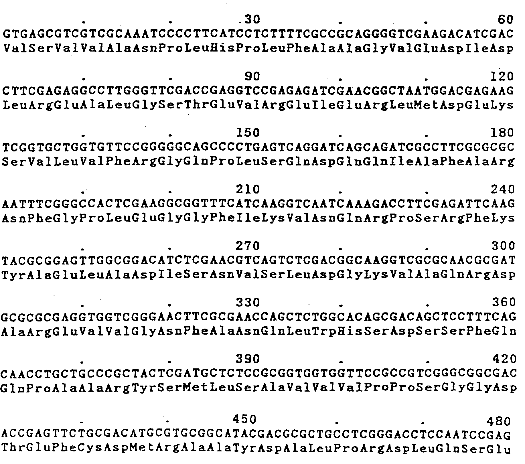

Priority Applications (1)
| Application Number | Priority Date | Filing Date | Title |
|---|---|---|---|
| AT87905713T ATE95230T1 (de) | 1986-08-29 | 1987-08-28 | Microorganismen and plasmide fuer die 2,4dichlorphenoxyessigsaeure (2,4-d)-monooxigenase - bildung und verfahren zur herstellung dieser plasmide und staemme. |
Applications Claiming Priority (2)
| Application Number | Priority Date | Filing Date | Title |
|---|---|---|---|
| DE19863629890 DE3629890A1 (de) | 1986-08-29 | 1986-08-29 | Mikroorganismen und plasmide fuer die 2,4-dichlorphenoxyessigsaeure (2,4-d)-monooxigenase - bildung und verfahren zur herstellung dieser plasmide und staemme |
| DE3629890 | 1986-08-29 |
Publications (2)
| Publication Number | Publication Date |
|---|---|
| EP0334841A1 EP0334841A1 (de) | 1989-10-04 |
| EP0334841B1 true EP0334841B1 (de) | 1993-09-29 |
Family
ID=6308773
Family Applications (1)
| Application Number | Title | Priority Date | Filing Date |
|---|---|---|---|
| EP87905713A Expired - Lifetime EP0334841B1 (de) | 1986-08-29 | 1987-08-28 | Microorganismen and plasmide für die 2,4-dichlorphenoxyessigsäure (2,4-d)-monooxigenase - bildung und verfahren zur herstellung dieser plasmide und stämme |
Country Status (5)
| Country | Link |
|---|---|
| US (2) | US6153401A (de) |
| EP (1) | EP0334841B1 (de) |
| JP (2) | JP2588917B2 (de) |
| DE (2) | DE3629890A1 (de) |
| WO (1) | WO1988001641A1 (de) |
Families Citing this family (77)
| Publication number | Priority date | Publication date | Assignee | Title |
|---|---|---|---|---|
| CA2018884A1 (en) * | 1989-06-22 | 1990-12-22 | Irving Gordon | Plant transformation construct |
| GB9825421D0 (en) | 1998-11-19 | 1999-01-13 | Isis Innovation | Process for oxidising terpenes |
| GB9914373D0 (en) | 1999-06-18 | 1999-08-18 | Isis Innovation | Process for oxidising aromatic compounds |
| US7214520B2 (en) * | 2000-07-18 | 2007-05-08 | National Research Council Of Canada | Cloning, sequencing and expression of a comamonas cyclopentanone 1,2-monooxygenase-encoding gene in Escherichia coli |
| CN102094032B (zh) | 2004-04-30 | 2014-02-26 | 美国陶氏益农公司 | 新除草剂抗性基因 |
| USPP20428P3 (en) * | 2005-08-17 | 2009-10-20 | The Board of Trustees of the Unviersity of Illinois | Grape plant named ‘Improved Chancellor’ |
| ES2637948T3 (es) | 2005-10-28 | 2017-10-18 | Dow Agrosciences Llc | Nuevos genes de resistencia a herbicidas |
| MX2009012120A (es) | 2007-05-09 | 2010-02-12 | Dow Agrosciences Llc | Genes novedosos con resistencia a herbicidas. |
| GB0719620D0 (en) | 2007-10-08 | 2007-11-14 | Isis Innovation | Mutant Enzymes |
| AR075466A1 (es) | 2008-10-22 | 2011-04-06 | Basf Se | Uso de herbicidas tipo auxina en plantas cultivadas |
| WO2010046423A2 (en) | 2008-10-22 | 2010-04-29 | Basf Se | Use of sulfonylurea herbicides on cultivated plants |
| EP3213635A1 (de) | 2009-06-25 | 2017-09-06 | Basf Se | Verwendung von agrochemischen mischungen zur verbesserung der gesundheit einer pflanze |
| AU2010326672A1 (en) | 2009-07-10 | 2011-08-04 | Syngenta Participations Ag | Novel hydroxyphenylpyruvate dioxygenase polypeptides and methods of use |
| CN102595879B (zh) | 2009-11-06 | 2014-08-06 | 巴斯夫欧洲公司 | 4-羟基苯甲酸和选定农药的结晶配合物 |
| US8748342B2 (en) | 2009-12-08 | 2014-06-10 | Basf Se | Pesticidal mixtures |
| NZ600886A (en) | 2009-12-08 | 2013-06-28 | Basf Se | An agrochemical pesticidal mixture for increasing the yield and health of a plant |
| WO2011076877A1 (en) | 2009-12-23 | 2011-06-30 | Bayer Cropscience Ag | Plants tolerant to hppd inhibitor herbicides |
| EA201290559A1 (ru) | 2009-12-23 | 2013-01-30 | Байер Интеллектуэль Проперти Гмбх | Растения, устойчивые к гербицидам - ингибиторам hppd |
| AR079883A1 (es) | 2009-12-23 | 2012-02-29 | Bayer Cropscience Ag | Plantas tolerantes a herbicidas inhibidores de las hppd |
| ES2659085T3 (es) | 2009-12-23 | 2018-03-13 | Bayer Intellectual Property Gmbh | Plantas tolerantes a herbicidas inhibidores de HPPD |
| ES2659086T3 (es) | 2009-12-23 | 2018-03-13 | Bayer Intellectual Property Gmbh | Plantas tolerantes a herbicidas inhibidores de HPPD |
| CN103037696A (zh) | 2010-05-31 | 2013-04-10 | 巴斯夫欧洲公司 | 增加植物健康的方法 |
| EP2576623B1 (de) | 2010-06-04 | 2015-01-21 | Dow AgroSciences LLC | Verfahren zur erkennung monoklonaler antikörper für enzyme zur aktivierung einer 2,4-dichlorphenoxyessigsäure-resistenz in pflanzen |
| WO2011161131A1 (en) | 2010-06-25 | 2011-12-29 | Basf Se | Herbicidal mixtures |
| WO2011161132A1 (en) | 2010-06-25 | 2011-12-29 | Basf Se | Pesticidal mixtures |
| WO2012022729A2 (en) | 2010-08-20 | 2012-02-23 | Basf Se | Method for improving the health of a plant |
| BR112013003029A2 (pt) | 2010-08-24 | 2016-09-13 | Basf Se | mistura agroquímica, composição agroquímica, método para aumentar sinergicamente a saúde de uma planta e usos de uma mistura |
| US8673583B2 (en) | 2010-09-15 | 2014-03-18 | Dow Agrosciences, Llc. | Monoclonal antibodies and detection methods for enzymes that confer resistance to 2,4-dichlorophenoxyacetic acid |
| BR112013008319A2 (pt) | 2010-10-07 | 2016-06-14 | Basf Se | uso de uma estrobilurina (composto a) e método para aumentar a resistência do glúten em cereais de inverno |
| EP2669371A1 (de) | 2010-11-10 | 2013-12-04 | Bayer CropScience AG | HPPD-Varianten und Anwendungsverfahren |
| UA115766C2 (uk) | 2010-12-03 | 2017-12-26 | ДАУ АГРОСАЙЄНСІЗ ЕлЕлСі | Трансгенна стійка до гербіцидів рослина сої, яка містить пакетовану подію 8264.44.06.1 |
| CN103298346A (zh) | 2010-12-22 | 2013-09-11 | 巴斯夫欧洲公司 | 用于增加植物健康的农用化学品混合物 |
| GB201101743D0 (en) | 2011-02-01 | 2011-03-16 | Syngenta Ltd | Herbicidal compositions |
| CA2830802A1 (en) | 2011-03-25 | 2012-10-04 | Bayer Intellectual Property Gmbh | Use of n-(1,2,5-oxadiazol-3-yl)benzamides for controlling unwanted plants in areas of transgenic crop plants being tolerant to hppd inhibitor herbicides |
| MX2013010908A (es) | 2011-03-25 | 2013-10-07 | Bayer Ip Gmbh | Uso de n-(tetrazol-4-il)- o n-(triazol-3-il)arilcarboxamidas o de sus sales para combatir plantas no deseadas en areas de plantas de cultivo trangenicas tolerantes a los herbicidas inhibidores de la hppd. |
| US20150065347A1 (en) | 2012-03-29 | 2015-03-05 | Basf Se | Co-crystals of dicamba and a co-crystal former b |
| DK2859104T3 (en) | 2012-06-07 | 2017-09-18 | Dow Agrosciences Llc | CONSTRUCT AND PROCEDURE FOR EXPRESSING TRANSGENES USING A BRASSICA ROAD CONSTITUTIVE PROMOTER |
| WO2014043435A1 (en) | 2012-09-14 | 2014-03-20 | Bayer Cropscience Lp | Hppd variants and methods of use |
| JP2015532274A (ja) | 2012-10-01 | 2015-11-09 | ビーエーエスエフ ソシエタス・ヨーロピアBasf Se | 栽培植物へのn−チオ−アントラニルアミド化合物の使用 |
| WO2014079820A1 (en) | 2012-11-22 | 2014-05-30 | Basf Se | Use of anthranilamide compounds for reducing insect-vectored viral infections |
| BR112015020194A2 (pt) | 2013-03-07 | 2017-07-18 | Basf Se | co-cristais, processo para a preparação dos co-cristais, formulação agrícola, métodos para controle de pragas, para melhoria da saúde das plantas e para a proteção de material de propagação de planta contra pragas e material de propagação de planta |
| RU2723717C2 (ru) | 2013-03-07 | 2020-06-17 | Атеникс Корп. | Гены токсинов и способы их применения |
| BR112015026235A2 (pt) | 2013-04-19 | 2017-10-10 | Bayer Cropscience Ag | método para melhorar a utilização do potencial de produção de plantas transgênicas envolvendo a aplicação de um derivado de ftaldiamida |
| CA2931264C (en) | 2013-12-10 | 2022-04-05 | Dow Agrosciences Llc | Synergistic herbicidal weed control and improved crop tolerance from combinations of 2,4-d-choline and glufosinate in 2,4-d- and glufosinate-tolerant soybeans, corn, cotton |
| US20150173370A1 (en) | 2013-12-20 | 2015-06-25 | Dow Agrosciences Llc | Synergistic herbicidal weed control from combinations of 2,4-d-choline, glyphosate, and glufosinate |
| MX2016011745A (es) | 2014-03-11 | 2017-09-01 | Bayer Cropscience Lp | Variantes de hppd y metodos de uso. |
| AR100785A1 (es) | 2014-06-09 | 2016-11-02 | Dow Agrosciences Llc | Control herbicida de maleza a partir de combinaciones de fluroxipir e inhibidores de als |
| EP2979549A1 (de) | 2014-07-31 | 2016-02-03 | Basf Se | Verfahren zur verbesserung der gesundheit einer pflanze |
| PT3837970T (pt) | 2014-10-15 | 2023-09-12 | Monsanto Technology Llc | Genes de tolerância a herbicidas e métodos de uso dos mesmos |
| EP3028573A1 (de) | 2014-12-05 | 2016-06-08 | Basf Se | Verwendung eines triazolfungizids auf transgenen pflanzen |
| WO2016091674A1 (en) | 2014-12-12 | 2016-06-16 | Basf Se | Use of cyclaniliprole on cultivated plants |
| WO2016091675A1 (en) | 2014-12-12 | 2016-06-16 | Basf Se | Method for improving the health of a plant |
| BR112017021450B1 (pt) | 2015-04-07 | 2021-12-28 | Basf Agrochemical Products B.V. | Métodos de controle de pragas, método de melhoria da saúde vegetal e semente revestida |
| EA201890696A1 (ru) | 2015-09-11 | 2018-09-28 | Байер Кропсайенс Акциенгезельшафт | Варианты гфпд и способы применения |
| WO2017083092A1 (en) | 2015-11-10 | 2017-05-18 | Dow Agrosciences Llc | Methods and systems for predicting the risk of transgene silencing |
| EP3054014A3 (de) | 2016-05-10 | 2016-11-23 | BASF Plant Science Company GmbH | Benutzung einer fungizids auf transgenen pflanzen |
| BR112019010476A2 (pt) | 2016-11-23 | 2019-09-10 | BASF Agricultural Solutions Seed US LLC | molécula de ácido nucleico recombinante, vetor, célula hospedeira, planta transgênica, semente transgênica, polipeptídeo recombinante, composição, método para controlar uma população de pragas, para matar pragas, para produzir um polipeptídeo, planta ou célula vegetal, método para proteger uma planta contra uma praga, para aumentar o rendimento em uma planta, uso e produto primário |
| EP3338552A1 (de) | 2016-12-21 | 2018-06-27 | Basf Se | Verwendung eines tetrazolinon fungizids bei transgenen pflanzen |
| KR20190095411A (ko) | 2016-12-22 | 2019-08-14 | 바스프 아그리컬쳐럴 솔루션즈 시드 유에스 엘엘씨 | 선충 해충의 방제를 위한 cry14의 용도 |
| CN110431234B (zh) | 2017-01-18 | 2024-04-16 | 巴斯夫农业种子解决方案美国有限责任公司 | Bp005毒素基因及其使用方法 |
| US11286498B2 (en) | 2017-01-18 | 2022-03-29 | BASF Agricultural Solutions Seed US LLC | Use of BP005 for the control of plant pathogens |
| BR112019018056A2 (pt) | 2017-03-07 | 2020-08-11 | BASF Agricultural Solutions Seed US LLC | molécula de ácido nucleico recombinante, cassete de expressão, célula hospedeira, plantas, sementes transgênicas, polipeptídeo recombinante, métodos para conferir tolerância e para controlar ervas daninhas, produto de utilidade e uso da sequência de nucleotídeos |
| US20210032651A1 (en) | 2017-10-24 | 2021-02-04 | Basf Se | Improvement of herbicide tolerance to hppd inhibitors by down-regulation of putative 4-hydroxyphenylpyruvate reductases in soybean |
| WO2019083810A1 (en) | 2017-10-24 | 2019-05-02 | Basf Se | IMPROVING HERBICIDE TOLERANCE FOR 4-HYDROXYPHENYLPYRUVATE DIOXYGENASE (HPPD) INHIBITORS BY NEGATIVE REGULATION OF HPPD EXPRESSION IN SOYBEANS |
| EP3802521A1 (de) | 2018-06-04 | 2021-04-14 | Bayer Aktiengesellschaft | Herbizid wirksame bizyklische benzoylpyrazole |
| US12241075B2 (en) | 2019-10-14 | 2025-03-04 | Basf Agricultural Solutions Us Llc | Insect resistant genes and methods of use |
| EP4461128A3 (de) | 2019-10-14 | 2025-03-26 | BASF Agricultural Solutions US LLC | Neue insektenresistente gene und verfahren zur verwendung |
| WO2023044364A1 (en) | 2021-09-15 | 2023-03-23 | Enko Chem, Inc. | Protoporphyrinogen oxidase inhibitors |
| KR20240136380A (ko) | 2022-01-14 | 2024-09-13 | 엔코 켐 인코포레이티드 | 프로토포르피리노겐 산화효소 억제제 |
| CN116926096A (zh) * | 2022-04-12 | 2023-10-24 | 上海市农业科学院 | 一种在大肠杆菌中表达并降解2,4-二氯苯氧基乙酸的基因簇及其应用 |
| WO2023222226A1 (en) | 2022-05-19 | 2023-11-23 | Evonik Operations Gmbh | Enzymatic method for producing l-glufosinate |
| WO2023222227A1 (en) | 2022-05-19 | 2023-11-23 | Evonik Operations Gmbh | Enzymatic method for producing l-glufosinate |
| CN115557636B (zh) * | 2022-10-09 | 2024-09-10 | 湖北泰盛化工有限公司 | 一种2,4-滴缩合母液含酚废水的处理方法 |
| AU2023408197A1 (en) | 2022-12-19 | 2025-06-26 | Basf Agricultural Solutions Us Llc | Insect toxin genes and methods for their use |
| AU2024386241A1 (en) | 2023-07-07 | 2026-01-08 | Basf Agricultural Solutions Us Llc | Use of cry genes for the control of nematode pests |
| WO2025090606A1 (en) | 2023-10-27 | 2025-05-01 | Basf Agricultural Solutions Us Llc | Use of novel genes for the control of nematode pests |
| WO2026010930A1 (en) | 2024-07-05 | 2026-01-08 | BASF Agricultural Solutions Seed US LLC | Use of axmi277 for the control of rotylenchulus reniformis nematode pests |
Family Cites Families (1)
| Publication number | Priority date | Publication date | Assignee | Title |
|---|---|---|---|---|
| DK159976C (da) * | 1979-06-01 | 1991-05-27 | Searle & Co | Plasmidvektorer, fremgangsmaade til fremstilling deraf, bakterieceller transformeret med plasmidvektorer samt udtrykkelse af protein i transformerede bakterieceller |
-
1986
- 1986-08-29 DE DE19863629890 patent/DE3629890A1/de not_active Withdrawn
-
1987
- 1987-08-28 JP JP62505314A patent/JP2588917B2/ja not_active Expired - Lifetime
- 1987-08-28 EP EP87905713A patent/EP0334841B1/de not_active Expired - Lifetime
- 1987-08-28 WO PCT/DE1987/000392 patent/WO1988001641A1/de not_active Ceased
- 1987-08-28 DE DE87905713T patent/DE3787655D1/de not_active Expired - Lifetime
-
1989
- 1989-03-10 US US07/322,604 patent/US6153401A/en not_active Expired - Lifetime
-
1995
- 1995-06-07 US US08/470,588 patent/US6100446A/en not_active Expired - Lifetime
- 1995-08-18 JP JP7210862A patent/JP2653398B2/ja not_active Expired - Lifetime
Non-Patent Citations (4)
| Title |
|---|
| Chemical Abstracts, vol. 102, no.13, April 1985 (Columbus, Ohio, US) R.H. Don et al "Genetic and physical map of the 2, 4-dichlorophenoxyacetic acid-degradative plasmid pJP4", see page 162, abstract 107177q * |
| Chemical Abstracts, vol. 102, no.19, 13 May 1985 (Columbus, Ohio, US), R.H. Don et al "Transposon mutagenesis and cloning analysis of the pathways for degradation of 2, 4-dichlorophenoxyacetic acid and 3-chlorobenzoate in alcaligenes eutrophus JMP134 (pJP4), see page 157, abstract 161446q * |
| Chemical Abstracts, vol. 103, no.3, 22 July 1985, (Columbus, Ohio, US), P.S. Amy et al "Characterization of aquatic bacteria and cloning of genes specifying partial degradation of 2, 4-dichlorophenoxyacetic acid", see page 154, abstract 17761s * |
| Chemical Abstracts, vol. 99, no.9, 29 August 1983 (Columbus, Ohio, US), B. Friedrich et al "Transfer and expression of the herbicide-degrading plasmid pJP4 in aerobic autotrophic bacteria", see page 159, abstract 65163u * |
Also Published As
| Publication number | Publication date |
|---|---|
| DE3787655D1 (de) | 1993-11-04 |
| JP2653398B2 (ja) | 1997-09-17 |
| JP2588917B2 (ja) | 1997-03-12 |
| US6153401A (en) | 2000-11-28 |
| DE3629890A1 (de) | 1988-03-10 |
| JPH01503674A (ja) | 1989-12-14 |
| US6100446A (en) | 2000-08-08 |
| JPH0838162A (ja) | 1996-02-13 |
| EP0334841A1 (de) | 1989-10-04 |
| WO1988001641A1 (fr) | 1988-03-10 |
Similar Documents
| Publication | Publication Date | Title |
|---|---|---|
| EP0334841B1 (de) | Microorganismen and plasmide für die 2,4-dichlorphenoxyessigsäure (2,4-d)-monooxigenase - bildung und verfahren zur herstellung dieser plasmide und stämme | |
| DE69629576T2 (de) | Verfahren zur herstellung von rekombinanten plasmiden | |
| DE68927486T2 (de) | Verfahren zur Integration eines bestimmten Gens ins bakterielle Chromosom und durch dieses Verfahren erhaltenes Bakterium | |
| DE19610984A1 (de) | Alkohol-Dehydrogenase und deren Verwendung zur enzymatischen Herstellung chiraler Hydroxyverbindungen | |
| EP0276832A2 (de) | Verfahren zur Herstellung von Ketogulonsäure | |
| CH642680A5 (en) | DNA transfer vector, process for its preparation and its use | |
| DE4018441A1 (de) | Rekombinantes restriktionsenzym sau3ai | |
| DE68924761T2 (de) | Neue coenzym unabhängige l-sorbosone dehydrogenase. | |
| EP0798384A1 (de) | Biotechnologisches Verfahren zur Herstellung von Biotin | |
| DE69333954T2 (de) | An der streptogramin biosynthese beteiligte polypeptide, ihre kodierenden nukleotidsequenzen und ihre verwendung | |
| DE69627787T2 (de) | Actinomyceten-Promotor | |
| EP0219034B1 (de) | Verfahren zur Herstellung von mesophilen Mikroorganismen, die eine bei höherer Temperatur aktive D-Hydantoinase enthalten | |
| DE69132628T2 (de) | Biokonversionen katalysiert durch die toluol-monooxygenase aus -1(pseudomonas mendocina) kr-1 | |
| EP0722500B1 (de) | Gene für den butyrobetain/crotonobetain-l-carnitin-stoffwechsel und ihre verwendung zur mikrobiologischen herstellung von l-carnitin | |
| EP0477828B1 (de) | Hydroxylierung von Methylgruppen in aromatischen Heterocyclen mittels Mikroorganismen | |
| EP0938584B1 (de) | Verfahren zur herstellung von (s)- oder (r)-3,3,3-trifluor-2-hydroxy-2-methylpropionsaüre | |
| EP0469523B1 (de) | Klonierung und Überexpression von Glucose-6-Phosphat-Dehydrogenase aus Leuconostoc dextranicus | |
| DE69228037T2 (de) | Farnesylpyrophosphatsynthetase und dafür kodierende DNA-Sequenz | |
| DE3750240T2 (de) | Gen, das ein Insektizid-Protein kodiert, und Herstellung des Proteins durch Verwendung dieses Gens. | |
| EP0502524B1 (de) | Mikrobiologisches Verfahren zur terminalen Hydroxylierung von Ethylgruppen an aromatischen 5- oder 6-Ring-Heterocyclen | |
| EP0501921A2 (de) | Verfahren zur genetischen Manipulation von Myxobakterien | |
| EP0187138B1 (de) | Mikroorganismus und Plasmid für konstitutive Creatinamidino-hydrolase-Bildung sowie Verfahren zur Herstellung derselben | |
| DE69931176T2 (de) | Neue bacillus subtilis stämme für lebensmittelfermentierung | |
| DE10309557A1 (de) | Ein Translokationsenzym als Selektionsmarker | |
| EP0524604B1 (de) | Gentechnologisches Verfahren zur Herstellung von S-(+)-2,2-Dimethylcyclopropancarboxamid mittels Mikroorganismen |
Legal Events
| Date | Code | Title | Description |
|---|---|---|---|
| PUAI | Public reference made under article 153(3) epc to a published international application that has entered the european phase |
Free format text: ORIGINAL CODE: 0009012 |
|
| 17P | Request for examination filed |
Effective date: 19890224 |
|
| AK | Designated contracting states |
Kind code of ref document: A1 Designated state(s): AT BE CH DE FR GB IT LI LU NL SE |
|
| 17Q | First examination report despatched |
Effective date: 19920709 |
|
| GRAA | (expected) grant |
Free format text: ORIGINAL CODE: 0009210 |
|
| AK | Designated contracting states |
Kind code of ref document: B1 Designated state(s): AT BE CH DE FR GB IT LI LU NL SE |
|
| REF | Corresponds to: |
Ref document number: 95230 Country of ref document: AT Date of ref document: 19931015 Kind code of ref document: T |
|
| REF | Corresponds to: |
Ref document number: 3787655 Country of ref document: DE Date of ref document: 19931104 |
|
| ITF | It: translation for a ep patent filed | ||
| GBT | Gb: translation of ep patent filed (gb section 77(6)(a)/1977) |
Effective date: 19940106 |
|
| ET | Fr: translation filed | ||
| PLBE | No opposition filed within time limit |
Free format text: ORIGINAL CODE: 0009261 |
|
| STAA | Information on the status of an ep patent application or granted ep patent |
Free format text: STATUS: NO OPPOSITION FILED WITHIN TIME LIMIT |
|
| 26N | No opposition filed | ||
| EAL | Se: european patent in force in sweden |
Ref document number: 87905713.1 |
|
| REG | Reference to a national code |
Ref country code: CH Ref legal event code: PUE Owner name: SCHERING AKTIENGESELLSCHAFT, BERLIN & BERGKAMEN -D |
|
| REG | Reference to a national code |
Ref country code: GB Ref legal event code: 732E |
|
| NLS | Nl: assignments of ep-patents |
Owner name: HOECHST SCHERING AGREVO G.M.B.H. |
|
| REG | Reference to a national code |
Ref country code: FR Ref legal event code: TP |
|
| BECA | Be: change of holder's address |
Free format text: 970610 *HOECHST SCHRING AGREVO G.M.B.H.:MIRAUSTRASSE 54, D-13509 BERLIN |
|
| BECH | Be: change of holder |
Free format text: 970610 *HOECHST SCHRING AGREVO G.M.B.H. |
|
| REG | Reference to a national code |
Ref country code: GB Ref legal event code: IF02 |
|
| PGFP | Annual fee paid to national office [announced via postgrant information from national office to epo] |
Ref country code: DE Payment date: 20060603 Year of fee payment: 20 |
|
| PGFP | Annual fee paid to national office [announced via postgrant information from national office to epo] |
Ref country code: NL Payment date: 20060803 Year of fee payment: 20 |
|
| PGFP | Annual fee paid to national office [announced via postgrant information from national office to epo] |
Ref country code: FR Payment date: 20060808 Year of fee payment: 20 |
|
| PGFP | Annual fee paid to national office [announced via postgrant information from national office to epo] |
Ref country code: AT Payment date: 20060811 Year of fee payment: 20 |
|
| PGFP | Annual fee paid to national office [announced via postgrant information from national office to epo] |
Ref country code: GB Payment date: 20060823 Year of fee payment: 20 |
|
| PGFP | Annual fee paid to national office [announced via postgrant information from national office to epo] |
Ref country code: CH Payment date: 20060830 Year of fee payment: 20 Ref country code: LU Payment date: 20060830 Year of fee payment: 20 |
|
| PGFP | Annual fee paid to national office [announced via postgrant information from national office to epo] |
Ref country code: IT Payment date: 20060831 Year of fee payment: 20 |
|
| PGFP | Annual fee paid to national office [announced via postgrant information from national office to epo] |
Ref country code: BE Payment date: 20061013 Year of fee payment: 20 |
|
| REG | Reference to a national code |
Ref country code: GB Ref legal event code: PE20 |
|
| REG | Reference to a national code |
Ref country code: CH Ref legal event code: PL |
|
| EUG | Se: european patent has lapsed | ||
| NLV7 | Nl: ceased due to reaching the maximum lifetime of a patent |
Effective date: 20070828 |
|
| PG25 | Lapsed in a contracting state [announced via postgrant information from national office to epo] |
Ref country code: GB Free format text: LAPSE BECAUSE OF EXPIRATION OF PROTECTION Effective date: 20070827 |
|
| BE20 | Be: patent expired |
Owner name: *HOECHST SCHRING AGREVO G.M.B.H. Effective date: 20070828 |
|
| PG25 | Lapsed in a contracting state [announced via postgrant information from national office to epo] |
Ref country code: NL Free format text: LAPSE BECAUSE OF EXPIRATION OF PROTECTION Effective date: 20070828 |
|
| PGFP | Annual fee paid to national office [announced via postgrant information from national office to epo] |
Ref country code: SE Payment date: 20060804 Year of fee payment: 20 |