EP0078314B1 - Herstellung lebender blutgefässe sowie drüsengewebe - Google Patents
Herstellung lebender blutgefässe sowie drüsengewebe Download PDFInfo
- Publication number
- EP0078314B1 EP0078314B1 EP82901886A EP82901886A EP0078314B1 EP 0078314 B1 EP0078314 B1 EP 0078314B1 EP 82901886 A EP82901886 A EP 82901886A EP 82901886 A EP82901886 A EP 82901886A EP 0078314 B1 EP0078314 B1 EP 0078314B1
- Authority
- EP
- European Patent Office
- Prior art keywords
- mixture
- cells
- lattice
- collagen
- layer
- Prior art date
- Legal status (The legal status is an assumption and is not a legal conclusion. Google has not performed a legal analysis and makes no representation as to the accuracy of the status listed.)
- Expired
Links
Images
Classifications
-
- A—HUMAN NECESSITIES
- A61—MEDICAL OR VETERINARY SCIENCE; HYGIENE
- A61F—FILTERS IMPLANTABLE INTO BLOOD VESSELS; PROSTHESES; DEVICES PROVIDING PATENCY TO, OR PREVENTING COLLAPSING OF, TUBULAR STRUCTURES OF THE BODY, e.g. STENTS; ORTHOPAEDIC, NURSING OR CONTRACEPTIVE DEVICES; FOMENTATION; TREATMENT OR PROTECTION OF EYES OR EARS; BANDAGES, DRESSINGS OR ABSORBENT PADS; FIRST-AID KITS
- A61F2/00—Filters implantable into blood vessels; Prostheses, i.e. artificial substitutes or replacements for parts of the body; Appliances for connecting them with the body; Devices providing patency to, or preventing collapsing of, tubular structures of the body, e.g. stents
- A61F2/02—Prostheses implantable into the body
- A61F2/022—Artificial gland structures using bioreactors
-
- A—HUMAN NECESSITIES
- A61—MEDICAL OR VETERINARY SCIENCE; HYGIENE
- A61F—FILTERS IMPLANTABLE INTO BLOOD VESSELS; PROSTHESES; DEVICES PROVIDING PATENCY TO, OR PREVENTING COLLAPSING OF, TUBULAR STRUCTURES OF THE BODY, e.g. STENTS; ORTHOPAEDIC, NURSING OR CONTRACEPTIVE DEVICES; FOMENTATION; TREATMENT OR PROTECTION OF EYES OR EARS; BANDAGES, DRESSINGS OR ABSORBENT PADS; FIRST-AID KITS
- A61F2/00—Filters implantable into blood vessels; Prostheses, i.e. artificial substitutes or replacements for parts of the body; Appliances for connecting them with the body; Devices providing patency to, or preventing collapsing of, tubular structures of the body, e.g. stents
- A61F2/02—Prostheses implantable into the body
- A61F2/04—Hollow or tubular parts of organs, e.g. bladders, tracheae, bronchi or bile ducts
- A61F2/06—Blood vessels
- A61F2/062—Apparatus for the production of blood vessels made from natural tissue or with layers of living cells
-
- A—HUMAN NECESSITIES
- A61—MEDICAL OR VETERINARY SCIENCE; HYGIENE
- A61L—METHODS OR APPARATUS FOR STERILISING MATERIALS OR OBJECTS IN GENERAL; DISINFECTION, STERILISATION OR DEODORISATION OF AIR; CHEMICAL ASPECTS OF BANDAGES, DRESSINGS, ABSORBENT PADS OR SURGICAL ARTICLES; MATERIALS FOR BANDAGES, DRESSINGS, ABSORBENT PADS OR SURGICAL ARTICLES
- A61L27/00—Materials for grafts or prostheses or for coating grafts or prostheses
- A61L27/14—Macromolecular materials
- A61L27/22—Polypeptides or derivatives thereof, e.g. degradation products
- A61L27/24—Collagen
-
- A—HUMAN NECESSITIES
- A61—MEDICAL OR VETERINARY SCIENCE; HYGIENE
- A61L—METHODS OR APPARATUS FOR STERILISING MATERIALS OR OBJECTS IN GENERAL; DISINFECTION, STERILISATION OR DEODORISATION OF AIR; CHEMICAL ASPECTS OF BANDAGES, DRESSINGS, ABSORBENT PADS OR SURGICAL ARTICLES; MATERIALS FOR BANDAGES, DRESSINGS, ABSORBENT PADS OR SURGICAL ARTICLES
- A61L27/00—Materials for grafts or prostheses or for coating grafts or prostheses
- A61L27/50—Materials characterised by their function or physical properties, e.g. injectable or lubricating compositions, shape-memory materials, surface modified materials
- A61L27/507—Materials characterised by their function or physical properties, e.g. injectable or lubricating compositions, shape-memory materials, surface modified materials for artificial blood vessels
-
- A—HUMAN NECESSITIES
- A61—MEDICAL OR VETERINARY SCIENCE; HYGIENE
- A61F—FILTERS IMPLANTABLE INTO BLOOD VESSELS; PROSTHESES; DEVICES PROVIDING PATENCY TO, OR PREVENTING COLLAPSING OF, TUBULAR STRUCTURES OF THE BODY, e.g. STENTS; ORTHOPAEDIC, NURSING OR CONTRACEPTIVE DEVICES; FOMENTATION; TREATMENT OR PROTECTION OF EYES OR EARS; BANDAGES, DRESSINGS OR ABSORBENT PADS; FIRST-AID KITS
- A61F2310/00—Prostheses classified in A61F2/28 or A61F2/30 - A61F2/44 being constructed from or coated with a particular material
- A61F2310/00005—The prosthesis being constructed from a particular material
- A61F2310/00365—Proteins; Polypeptides; Degradation products thereof
-
- Y—GENERAL TAGGING OF NEW TECHNOLOGICAL DEVELOPMENTS; GENERAL TAGGING OF CROSS-SECTIONAL TECHNOLOGIES SPANNING OVER SEVERAL SECTIONS OF THE IPC; TECHNICAL SUBJECTS COVERED BY FORMER USPC CROSS-REFERENCE ART COLLECTIONS [XRACs] AND DIGESTS
- Y10—TECHNICAL SUBJECTS COVERED BY FORMER USPC
- Y10S—TECHNICAL SUBJECTS COVERED BY FORMER USPC CROSS-REFERENCE ART COLLECTIONS [XRACs] AND DIGESTS
- Y10S623/00—Prosthesis, i.e. artificial body members, parts thereof, or aids and accessories therefor
- Y10S623/901—Method of manufacturing prosthetic device
-
- Y—GENERAL TAGGING OF NEW TECHNOLOGICAL DEVELOPMENTS; GENERAL TAGGING OF CROSS-SECTIONAL TECHNOLOGIES SPANNING OVER SEVERAL SECTIONS OF THE IPC; TECHNICAL SUBJECTS COVERED BY FORMER USPC CROSS-REFERENCE ART COLLECTIONS [XRACs] AND DIGESTS
- Y10—TECHNICAL SUBJECTS COVERED BY FORMER USPC
- Y10S—TECHNICAL SUBJECTS COVERED BY FORMER USPC CROSS-REFERENCE ART COLLECTIONS [XRACs] AND DIGESTS
- Y10S623/00—Prosthesis, i.e. artificial body members, parts thereof, or aids and accessories therefor
- Y10S623/92—Method or apparatus for preparing or treating prosthetic
- Y10S623/921—Blood vessel
Definitions
- This invention is in the field of biology and particularly relates to the fabrication of living tissue in tubular form for various applications such as capillaries, larger blood vessels and glandular prosthesis.
- the lattice contracts in all dimensions; in its presence as the lattice sets it becomes anchored to the mesh and contracts in the thickness dimension only.
- the mesh resembling a picture frame, holds the lattice of living tissue within it.
- the contracted lattice, with or without the stainless steel mesh frame, can be seeded with epidermal cells from the potential graft recipient. When a sheet of epidermal cells forms, the two layered skin equivalent is grafted.
- the resultant graft is unique as compared to any other graft obtained from artificial skin since its basic organization is like that of skin and its living constituent cells are donated by potential graft recipients.
- This invention extends and applies the teaching of the aforesaid article in Proc. Natl. Acad. Sci. USA to the preparation of tubular prostheses suitable-for in vivo use as blood vessels and capillary beds, for instance.
- US-A-3,425,418 from which this invention commences discloses the in vitro preparation of artificial blood vessels by casting hydrated collagen about a core and thereafter dehydrating the collagen.
- the core comprises a woven tube which is embedded in the collagen matrix. Dehydration is achieved, contracting the collagen, by immersing the casting in a solvent mixture.
- Other methods of dehydration include air drying and the use of tanning agents. There is no disclosure of the use of cellular contractile agents to contract the collagen.
- a method of producing, in vitro a laminated collagenic tubular structure, suitable for in vivo use as artificial vessels and glandular prostheses, by casting collagen about a core characterised by:
- a method of producing, in vitro a laminated collagenic tubular prosthesis, suitable for in vivo use, by casting collagen about a core characterised by:
- the invention further provides a method of producing, in vitro a collagenic structure by casting hydrated collagen about core means to form a pierced collagenic structure, suitable for in vitro use as a capillary bed, characterised by the steps of:
- the cast collagen lattices contracted by living cells such as fibroblasts, smooth muscle cells, or elements of cells such as blood platelets are cast into shapes which provide internal surface areas and tubular shaped terminals, or end structures, particularly effective for making connections, in vivo, with existing tubular structures, such as capillaries, blood vessels and glandular tissues.
- the internal surface of the case structure can be lined with specialized cells, depending on the function of the structure.
- endothelial cells are used for the internal surface of an artery, vein, or other structures with internal surfaces.
- the inner surfaces of a capillary bed may be lined with pancreatic f3 cells to boost the insulin supply in the blood.
- Pancreatic islets islets of Langerhans
- hepatocytes or other types of glandular cells may also be used for lining the inner surface of the vessel-equivalent structures.
- the central core for forming the tube consists of polyethelene or glass tubing. This core is axially centered within a cylindrical mold. Suitable tissue forming constituents are poured into the cylindrical mold. After a suitable period of time, the tissue forming constituents contract the lattice and close in around the central core. This procedure can be repeated as many times as desired with the same or different cell types in the same or different proportions to yield a multilayer tube. After each layer contracts the fluid expressed from the contracting lattice is poured off to accommodate the tissue forming constituents of the next layer. The central core may then be removed and suitable cells, predicated on the function of the cast structure, may then be cultured on the inner surface of the hollow tissue cylinders, to form, for example, a vessel-equivalent structure.
- the vessel-equivalent structure thus far described is devoid of elastin, the fibrous muco- protein which is the major connective tissue protein - of elastic structures (e.g. large blood vessels). Without this elastic property it is possible that the vessel could burst under pressure. Since elastin is an extremely insoluble substance it is difficult to directly incorporate elastin into the molded tissue forming constituents previously described. Accordingly, a plastic mesh may be optionally provided between two layers or within a layer of the tissue forming constituents during the molding process, as will be described in detail.
- This mesh serves to reinforce the resultant vessel and at the same time provide a degree of elasticity to the structure so that it may expand and contract in the manner of a natural blood vessel having elastin.
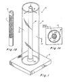
- Fig. 1 shows a preferred form of casting chamber for fabricating a blood vessel-equivalent of living matter.
- the casting chamber 10 comprises a central rod or mandrel 12 disposed in a cylinder 16.
- the central rod and cylinder are mounted on a base or stand 14.
- the rod 12 is provided with three arms or spokes 18 at the top of the rod for centering the rod within the cylinder 16.
- the base is provided with an appropriate collar 20 to accept the central rod 12.
- the outer cylinder has an internal diameter such that when the arms 18 are disposed as shown and the central rod is located in the collar 20, the rod 12 will be centered within cylinder 16.
- the outer diameter of the rod 12 determines the inner diameter of the cast vessel and for many applications would be in the range of from 2-10 mm.
- the inner diameter of cylinder 16 will determine the final thickness of the cast layer, and typically may range from 1-4 cm to produce a final thickness of about 0.5-2 mm, the final thickness being proportional to the diameter.
- the height of the chamber determines the length of the vessel and would typically be between 10-30 cm in height.
- the casting chamber parts should be made from material which may be readily cleaned and is autoclavable.
- the cylinder 16 should be made from material which is clear and which will permit diffusion of carbon dioxide and other gases.
- the rod 12 may be made of glass or metal and the cylinder 16 should preferably be made of autoclavable plastic, such as polycarbonate.
- the stand 14 may be made of glass, plastic or metal, such as stainless steel.
- Blood vessels may be generally characterized by their cellular composition and the composition of the matrix or collagen lattice with which other extracellular elements, such as elastin fibers and proteoglycans are associated.
- the collagen, elastin, and proteoglycans are the biosynthetic products of the cells in each of the layers.
- the cell types are endothelial, smooth muscle, and fibroblasts (called pericytes) and are found respectively in successive layers from the lumen outward.
- pericytes fibroblasts
- the respective layers may be laid down in order. Alternatively, several can be laid down concurrently. All vessels contain an inner endothelial lining.
- smooth muscle surrounds the endothelium and the final outside layer is made up of fibroblasts.
- the smooth muscle layer is fabricated.
- a mixture of nutrient medium e.g. McCoy's medium containing fetal bovine serum
- the ingredients are mixed in the following ratio: 9.2 ml of 1.76 x concentrate of McCoy's medium and 1.8 ml of fetal bovine serum.
- the pH is raised by addition of 1.0 ml of 0.1N NaOH.
- the foregoing mixture of medium and serum is poured onto a dish in which 1.5 ml of native collagen in a 1-1000 acidic acid solution has been prepared.
- About 250,000 cultured aorta smooth muscle cells suspended in a 0.5 ml of McCoy's medium supplemented with a 10% fetal bovine serum is quickly added.
- the above constituents are mixed by swirling the dish and quickly pouring the mixture into the casting chamber.
- the chamber is then placed in a humidified 5% CO 2 , 95% air incubator at 37°C for 3 days.
- a collagen lattice or gel forms immediately on casting the mixture.
- the collagen fibrils are gradually compacted by the cells so that fluid is squeezed out of the lattice. The result is contraction of the collagen lattice around the central core or rod 12.
- the smooth muscle layer will have set in a cylindrical structure having sufficient structural integrity to simulate, or replicate, the smooth muscle layer of a typical blood vessel. If a second layer is to be applied, the fluid expressed during contraction of the first lattice is poured off and a second complete mixture of all ingredients is added to replace the said fluid, and another contracted layer is deposited on the first formed layer.
- the process may be repeated as many times as desired to give a multilayered structure.
- the layers may be poured simultaneously with a removable separation or sleeve (not shown) between them. As soon as gelation begins the sleeve is removed.
- the smooth muscle layer cylinder (SMC) has been cast, it may be desirable to provide a plastic mesh sleeve 11 about the outer surface of the smooth muscle layer cylinder or the mesh may be embedded in the smooth muscle layer.
- This mesh will serve to reinforce the resulting structure and provide some degree of elasticity so that the resulting structure will be better able to withstand the pressures it will be subjected to in use.
- Meadox Medicals, Inc. 103 Bauer Drive, Oakland, New Jersey 07436, supplies a Dacron® mesh sleeve, Part No. 01H183, which has proved particularly suitable for this purpose.
- Other suitable meshes are readily available in various inert plastics, such as Teflon@, nylon, etc.
- the invention is not to be limited to a particular plastic material.
- the mesh should be treated to render it more electronegative by, for example, subjecting it to plasma. This results in better cell attachment to the plastic sleeve and hence an increase in the strength of the resultant structure.
- the sleeve 11 should be placed on the smooth muscle cell cylinder by first disposing the sleeve 11 on metal tube 15 (as shown in Fig. 1B) which has an inner diameter larger than the outer diameter of the smooth muscle cell cylinder.
- the tube 15, with the sleeve on the exterior, is then slipped over the smooth muscle cell cylinder, a portion of the sleeve is then pulled off the tube 15 and onto the smooth muscle cell cylinder and held there while the tube 15 is slipped off the smooth muscle cell cylinder. This procedure minimizes damage to the exterior surfaces of the smooth muscle cell cylinder while attaching the sleeve.
- a fibroblast layer may be cast around the inner smooth muscle layer(s) (SMC) and sleeve 11 so as to completely enclose the sleeve 11, as shown in Fig. 1A.
- the ingredients described above in connection with the fabrication of a smooth muscle layer are used to constitute a fibroblast layer, except that cultured aorta fibroblasts are substituted for the smooth muscle cells.
- the incubation period for the fibroblast layer may be 2 days to a week.
- the resultant multi-layered structure consisting of inner smooth muscle layer(s) and an outer fibroblast layer with a mesh sleeve sandwiched between the two layers is now ready to be cultured with an inner endothelial lining of living endothelial cells.
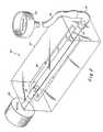
- the cylindrical tissue tube of several layers is slipped off the casting rod 12 to receive the endothelial cells as a suspension. It is supported in the culturing apparatus shown in Fig. 2.
- the apparatus of Fig. 2 comprises a transparent chamber 24, within which a rotatable rod 26 is inserted at one end and a rotatable tube 36 is inserted at the opposite end.
- the tube 36 and rod 26 are tied together by wire frame member 30 such that when the rod 26 is rotated, the tube 36 will rotate in unison in the same direction.
- Rod 26 is coupled to motor 28 such that when motor 28 is energized the rod 26 will rotate in the direction shown by the arrow.
- the rod is attached to the motor in such a way that the length of the rod inserted into the chamber 24 may be adjusted in accordance with the length of the vessel-equivalent 44 being supported within the culture chamber 24. This may accomplished by a rack and pinion device or other such variable length means (not shown).
- Rod 26 is provided at one end with a nipple 32 to which a vessel 44 (such as the structure previously described in connection with Figs. 1, 1A and 1 comprising an inner cylinder smooth muscle cell layer, and an outer cylinder of fibroblast cells with a mesh sleeve sandwiched between) may be attached.
- tube 36 is provided with a complementary nipple 34 to which the opposite end of the vessel 44 may be attached.
- the vessel 44 is suspended between the rod 26 and tube 36 and a culture medium may be introduced from reservoir 42 through tubing 40 and fixed connecting tube 38, through tube 36 and into the interior lining of blood vessel-equivalent 44.
- watertight seal bearings (not shown) are provided at both ends of chamber 24 to permit the rod and tube to be inserted into the chamber.
- Reservoir 42 is supplied with a suspension of about 200,000 cultured aorta or other endothelial cells in McCoy's medium supplemented with a 20% fetal bovine serum. This mixture is fed by hydrostatic pressure from the reservoir into the vessel 44 as previously mentioned.
- the vessel 44 is slowly rotated by means of motor 28 which preferably runs at a speed of between .1 and 1 r.p.m. Rotation of the vessel 44 enables distribution of the endothelial cells evenly on the inner lining of the vessel and the hydrostatic pressure head from the reservoir enables the lumen, or inner opening, of the vessel-equivalent to remain open. It should be emphasized that the above procedures are intended to be carried out asceptically.
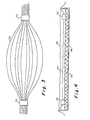
- Figs. 3 and 4 may be preferred since several capillaries can be fabricated in one casting procedure.
- the mold in Figs. 3 and 4 takes the form of a plurality of fine tubing or threads of nylon or stainless steel 54 suspended between a pair of plastic tubes or rings of dehydrated collagen 50 and 52.
- the threads 54 are inserted through the rings 50 and 52 and held in spaced-apart relationship by the rings.
- a collagen lattice with appropriate cells is cast in a pan 56 in a two-step procedure.
- a first layer 66 is laid down and allowed to contract. This layer is of sufficient height to receive the threads 54 and prevent the threads from touching the bottom of the pan 56.
- a second layer 58 is then poured covering the threads 54. After this lattice layer has contracted, in accordance with the invention, the threads may be pulled out one at a time from either end.
- the plastic tube or ring 50 or 52 of dehydrated collagen, which is now free of the threads 54, is now ready to receive a pipette within which a suspension of appropriate cells is disposed. These cells are introduced into the capillaries formed in the lattice by removal of the threads and allowed to attach to the inner surfaces and culture. Fluid under slight pressure is allowed to flow through the capillaries at a slow rate to keep the channels open.
- the sheet of living lattice material comprising lower layer 56 and upper layer 58, may be transferred to recipient and connection made at the points of confluency of the small capillary channels left when the thread has been removed.
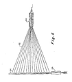
- FIG. 5 A further apparatus for casting capillaries in a slab lattice is shown in Fig. 5.
- nylon or other threads are threaded through a threading cylinder 60, a threading tube 64 and an exit tube 66.
- Threading tube 64 may be formed of suitably dimensioned autoclavable plastic or glass.
- Cylinder 60 and exit tube 66 may be formed of dried collagen.
- the assembly shown in Fig. 5 is disposed in a pan just above the bottom, so that lattice material will flow below and around it when poured. Alternatively, it may be laid into or on a freshly poured lattice. If the latter procedure is used, a second layer of lattice material may be poured over the assembly.
- each thread 62 is pulled out through cylinder 66 leaving capillary channels in the lattice.
- a set of channels connecting, or anastomosing, at cylinder 60 will constitute a bed of capillary vessels.
- threading tube 64 may be withdrawn from the lattice and a suspension of endothelial cells may be injected via the cylindrical opening at 60 into the channel.
- flow under pressure is allowed to flow through the capillaries at a slow rate to keep them open.
- the bed is ready for implementation, since by that time, the endothelial cells will have lined the inner channel surfaces.
- Connecting tubes of dried collagen may be sewn to the severed ends of the blood vessel of the host organism from which the cells used to populate the fabricated capillary bed were taken.
- the connecting tubes are then inserted into the recesses of tube 66 and tube 60 and are secured by sutures. This capillary-equivalent is then allowed to form "in vivo".
- connecting tubes may be formed of vessel-equivalent structures produced by using the cylindrical ends of the capillary bed as the core for molding a vessel-equivalent structure on each end to serve as a connecting tube between the capillary bed-equivalent and the severed ends of the blood vessel of the host organism.
- vessel-equivalent structure would be formed substantially as previously described in connection with Figs. 1-2.
- glandular cells such as pancreatic P cells (to boost insulin supply in the blood) or hepatocytes (liver) cells.
- pancreatic P cells to boost insulin supply in the blood
- hepatocytes liver cells.
- the vessels of the capillary beds provide a large surface area through which the blood may flow. Glandular cells lining the interior surface of these vessels can provide a source of secretory products of therapeutic value.
- bovine cells have been used in the process since such cells were readily available for experimentation. It is contemplated, however, that for most applications, the cells will be donated by the potential recipient of the prosthesis.
- lattice or matrix constituents as proteoglycans, glycos- aminoglycans or elastin may be added to the mixture with the collagen.
Landscapes
- Health & Medical Sciences (AREA)
- Life Sciences & Earth Sciences (AREA)
- Transplantation (AREA)
- Animal Behavior & Ethology (AREA)
- Veterinary Medicine (AREA)
- Public Health (AREA)
- General Health & Medical Sciences (AREA)
- Oral & Maxillofacial Surgery (AREA)
- Biomedical Technology (AREA)
- Vascular Medicine (AREA)
- Engineering & Computer Science (AREA)
- Cardiology (AREA)
- Heart & Thoracic Surgery (AREA)
- Medicinal Chemistry (AREA)
- Dermatology (AREA)
- Chemical & Material Sciences (AREA)
- Epidemiology (AREA)
- Gastroenterology & Hepatology (AREA)
- Pulmonology (AREA)
- Biophysics (AREA)
- Prostheses (AREA)
- Materials For Medical Uses (AREA)
Claims (16)
Priority Applications (1)
| Application Number | Priority Date | Filing Date | Title |
|---|---|---|---|
| AT82901886T ATE24829T1 (de) | 1981-05-08 | 1982-05-05 | Herstellung lebender blutgefaesse sowie druesengewebe. |
Applications Claiming Priority (4)
| Application Number | Priority Date | Filing Date | Title |
|---|---|---|---|
| US261928 | 1981-05-08 | ||
| US06/261,928 US4539716A (en) | 1981-03-19 | 1981-05-08 | Fabrication of living blood vessels and glandular tissues |
| US352585 | 1982-02-26 | ||
| US06/352,585 US4546500A (en) | 1981-05-08 | 1982-02-26 | Fabrication of living blood vessels and glandular tissues |
Publications (3)
| Publication Number | Publication Date |
|---|---|
| EP0078314A1 EP0078314A1 (de) | 1983-05-11 |
| EP0078314A4 EP0078314A4 (de) | 1984-03-01 |
| EP0078314B1 true EP0078314B1 (de) | 1987-01-14 |
Family
ID=26948924
Family Applications (1)
| Application Number | Title | Priority Date | Filing Date |
|---|---|---|---|
| EP82901886A Expired EP0078314B1 (de) | 1981-05-08 | 1982-05-05 | Herstellung lebender blutgefässe sowie drüsengewebe |
Country Status (5)
| Country | Link |
|---|---|
| US (1) | US4546500A (de) |
| EP (1) | EP0078314B1 (de) |
| JP (1) | JPS58500695A (de) |
| DE (1) | DE3275057D1 (de) |
| WO (1) | WO1982003764A1 (de) |
Families Citing this family (119)
| Publication number | Priority date | Publication date | Assignee | Title |
|---|---|---|---|---|
| US4485096A (en) * | 1982-02-26 | 1984-11-27 | Massachusetts Institute Of Technology | Tissue-equivalent and method for preparation thereof |
| US4479796A (en) * | 1982-11-15 | 1984-10-30 | Medtronic, Inc. | Self-regenerating drug administration device |
| US4911720A (en) * | 1983-03-10 | 1990-03-27 | Collier John P | Particular surface replacement prosthesis |
| US4695281A (en) * | 1983-03-25 | 1987-09-22 | Koken Co., Ltd. | Medical material |
| FR2556210B1 (fr) * | 1983-12-08 | 1988-04-15 | Barra Jean Aubert | Prothese veineuse et son procede d'obtention |
| IL74180A (en) * | 1984-01-30 | 1992-06-21 | Meadox Medicals Inc | Drug delivery collagen-impregnated synthetic vascular graft |
| IL74179A (en) * | 1984-01-30 | 1992-05-25 | Meadox Medicals Inc | Collagen synthetic vascular graft |
| US4634422A (en) * | 1984-05-31 | 1987-01-06 | Adrian Kantrowitz | Percutaneous access device and method for implanting same |
| US4895574A (en) * | 1984-06-06 | 1990-01-23 | Larry Rosenberg | Piezoelectric motivator for prosthetic devices |
| US5401832A (en) * | 1984-12-24 | 1995-03-28 | Merck & Co., Inc. | Brain derived and recombinant acidic fibroblast growth factor |
| US5035708A (en) * | 1985-06-06 | 1991-07-30 | Thomas Jefferson University | Endothelial cell procurement and deposition kit |
| US5441539A (en) * | 1985-06-06 | 1995-08-15 | Thomas Jefferson University | Endothelial cell deposition device |
| US4690684A (en) * | 1985-07-12 | 1987-09-01 | C. R. Bard, Inc. | Meltable stent for anastomosis |
| US5552528A (en) | 1986-03-03 | 1996-09-03 | Rhone-Poulenc Rorer Pharmaceuticals Inc. | Bovine b-endothelial cell growth factor |
| FR2597501B1 (fr) * | 1986-04-18 | 1990-01-19 | Merieux Inst | Procede de fabrication de nappes de collagene, nappes obtenues et leurs applications |
| US5266480A (en) * | 1986-04-18 | 1993-11-30 | Advanced Tissue Sciences, Inc. | Three-dimensional skin culture system |
| US4963489A (en) * | 1987-04-14 | 1990-10-16 | Marrow-Tech, Inc. | Three-dimensional cell and tissue culture system |
| US5863531A (en) * | 1986-04-18 | 1999-01-26 | Advanced Tissue Sciences, Inc. | In vitro preparation of tubular tissue structures by stromal cell culture on a three-dimensional framework |
| US5160490A (en) * | 1986-04-18 | 1992-11-03 | Marrow-Tech Incorporated | Three-dimensional cell and tissue culture apparatus |
| US5510254A (en) * | 1986-04-18 | 1996-04-23 | Advanced Tissue Sciences, Inc. | Three dimensional cell and tissue culture system |
| US5032508A (en) * | 1988-09-08 | 1991-07-16 | Marrow-Tech, Inc. | Three-dimensional cell and tissue culture system |
| CH670759A5 (de) * | 1986-06-02 | 1989-07-14 | Sulzer Ag | |
| CH670760A5 (de) * | 1986-06-02 | 1989-07-14 | Sulzer Ag | |
| US5567612A (en) * | 1986-11-20 | 1996-10-22 | Massachusetts Institute Of Technology | Genitourinary cell-matrix structure for implantation into a human and a method of making |
| US6309635B1 (en) | 1986-11-20 | 2001-10-30 | Children's Medical Center Corp. | Seeding parenchymal cells into compression resistant porous scaffold after vascularizing in vivo |
| US5759830A (en) * | 1986-11-20 | 1998-06-02 | Massachusetts Institute Of Technology | Three-dimensional fibrous scaffold containing attached cells for producing vascularized tissue in vivo |
| US4835102A (en) * | 1987-03-31 | 1989-05-30 | Eugene Bell | Tissue equivalent test systems |
| ATE86115T1 (de) * | 1987-04-28 | 1993-03-15 | Univ California | Vorrichtung und verfahren zur herstellung eines komposithautersatzes. |
| US5273900A (en) * | 1987-04-28 | 1993-12-28 | The Regents Of The University Of California | Method and apparatus for preparing composite skin replacement |
| US4795459A (en) * | 1987-05-18 | 1989-01-03 | Rhode Island Hospital | Implantable prosthetic device with lectin linked endothelial cells |
| US4846835A (en) * | 1987-06-15 | 1989-07-11 | Grande Daniel A | Technique for healing lesions in cartilage |
| CH675679A5 (de) * | 1987-12-07 | 1990-10-31 | Sulzer Ag | |
| US5108428A (en) * | 1988-03-02 | 1992-04-28 | Minnesota Mining And Manufacturing Company | Corneal implants and manufacture and use thereof |
| US4837379A (en) * | 1988-06-02 | 1989-06-06 | Organogenesis Inc. | Fibrin-collagen tissue equivalents and methods for preparation thereof |
| CH676195A5 (de) * | 1988-10-07 | 1990-12-28 | Sulzer Ag | |
| US4969896A (en) * | 1989-02-01 | 1990-11-13 | Interpore International | Vascular graft prosthesis and method of making the same |
| US5521087A (en) * | 1989-05-10 | 1996-05-28 | Massachusetts Institute Of Technology | Method for producing oriented connective tissue cells in a ligament configuration |
| EP0396809A1 (de) * | 1989-05-12 | 1990-11-14 | Sedlarik, Karel-Maria, Dr. med. | Künstliche kleinlumige Gefässprothese und Verfahren zu deren Herstellung |
| IT1230047B (it) * | 1989-07-04 | 1991-09-27 | Giovanni Brotzu | Protesi vascolare contenente nella parete microcapsule inglobanti cellule produttrici di ormoni. |
| US5106949A (en) * | 1989-09-15 | 1992-04-21 | Organogenesis, Inc. | Collagen compositions and methods for preparation thereof |
| IL95429A (en) * | 1989-09-15 | 1997-09-30 | Organogenesis | Living tissue equivalents comprising hydrated collagen lattice and a collagen gel and their production |
| NZ237832A (en) * | 1990-04-17 | 1994-05-26 | Curative Tech Inc | Coating a prosthetic surface with mammalian cells |
| USRE35399E (en) * | 1990-04-24 | 1996-12-10 | Eisenberg; Mark | Composite living skin equivalents |
| ATE117527T1 (de) * | 1990-06-15 | 1995-02-15 | Sulzer Medizinaltechnik Ag | Verfahren zur herstellung von mit lebenden zellen beladenen, porösen, schlauchförmigen prothesen aus kunststoff. |
| IT1244808B (it) * | 1990-11-29 | 1994-09-05 | Giovanni Brotzu | Protesi vascolare sintetica biocompatibile a doppia parete contenente cellule ormonosecernenti |
| US5192312A (en) * | 1991-03-05 | 1993-03-09 | Colorado State University Research Foundation | Treated tissue for implantation and methods of treatment and use |
| DE4108772A1 (de) * | 1991-03-18 | 1992-09-24 | Inst Textil & Faserforschung | Implantierbares biohybrides organ |
| ZA923086B (en) * | 1991-04-29 | 1993-10-28 | South African Medical Research | A delivery system for biologicaly active growth or morphogenetic factors and a method for preparing such delivery system |
| GB9116036D0 (en) * | 1991-07-25 | 1991-09-11 | Univ Leicester | Preparing grafts for implantation |
| US5282860A (en) * | 1991-10-16 | 1994-02-01 | Olympus Optical Co., Ltd. | Stent tube for medical use |
| WO1993007913A1 (en) * | 1991-10-24 | 1993-04-29 | Children's Medical Center Corporation | Neomorphogenesis of urological structures in vivo from cell culture |
| US5702446A (en) * | 1992-11-09 | 1997-12-30 | Board Of Regents, The University Of Texas System | Bone prosthesis |
| US5827641A (en) * | 1992-11-13 | 1998-10-27 | Parenteau; Nancy L. | In vitro cornea equivalent model |
| US5374515A (en) * | 1992-11-13 | 1994-12-20 | Organogenesis, Inc. | In vitro cornea equivalent model |
| GB9306449D0 (en) * | 1993-03-29 | 1993-05-19 | Nat Heart Research Fund | Tissue equivalents |
| US5709854A (en) * | 1993-04-30 | 1998-01-20 | Massachusetts Institute Of Technology | Tissue formation by injecting a cell-polymeric solution that gels in vivo |
| US5518878A (en) * | 1993-09-15 | 1996-05-21 | Organogenesis Inc. | Cryopreservation of cultured skin or cornea equivalents with agitation |
| US5891617A (en) * | 1993-09-15 | 1999-04-06 | Organogenesis Inc. | Cryopreservation of harvested skin and cultured skin or cornea equivalents by slow freezing |
| US5492826A (en) * | 1993-12-10 | 1996-02-20 | William Beaumont Hospital | Apparatus and method for seeding endothelial cells |
| DK0871414T3 (da) * | 1994-03-14 | 2004-08-30 | Cryolife Inc | Fremgangsmåder til fremstilling af væv til implantering |
| US6001123A (en) * | 1994-04-01 | 1999-12-14 | Gore Enterprise Holdings Inc. | Folding self-expandable intravascular stent-graft |
| US6165210A (en) * | 1994-04-01 | 2000-12-26 | Gore Enterprise Holdings, Inc. | Self-expandable helical intravascular stent and stent-graft |
| US5947893A (en) * | 1994-04-27 | 1999-09-07 | Board Of Regents, The University Of Texas System | Method of making a porous prothesis with biodegradable coatings |
| US5716394A (en) * | 1994-04-29 | 1998-02-10 | W. L. Gore & Associates, Inc. | Blood contact surfaces using extracellular matrix synthesized in vitro |
| JPH09512463A (ja) * | 1994-04-29 | 1997-12-16 | ダブリュ.エル.ゴア アンド アソシエイツ,インコーポレイティド | 天然内皮下基質を利用した改善された血液接触表面並びにその製造及び利用方法 |
| EP1217101B8 (de) | 1994-04-29 | 2006-02-01 | Boston Scientific Scimed, Inc. | Stent mit Kollagen |
| WO1995029713A1 (en) * | 1994-04-29 | 1995-11-09 | W.L. Gore & Associates, Inc. | Improved blood contact surfaces using endothelium on a subendothelial extracellular matrix |
| FR2722974B1 (fr) * | 1994-07-29 | 1997-04-25 | Marie Therese Zabot | Procede de modification de la surface interne des protheses synthetiques utilisees en chirurgie vasculaire |
| EP0698396B1 (de) * | 1994-08-12 | 2001-12-12 | Meadox Medicals, Inc. | Mit einen Heparin enhaltendes Kollagendichtmittel imprägniertes Gefässtransplantat |
| US5665114A (en) * | 1994-08-12 | 1997-09-09 | Meadox Medicals, Inc. | Tubular expanded polytetrafluoroethylene implantable prostheses |
| US6331188B1 (en) | 1994-08-31 | 2001-12-18 | Gore Enterprise Holdings, Inc. | Exterior supported self-expanding stent-graft |
| US6015429A (en) | 1994-09-08 | 2000-01-18 | Gore Enterprise Holdings, Inc. | Procedures for introducing stents and stent-grafts |
| US6057137A (en) | 1994-10-06 | 2000-05-02 | Regents Of The University Of Minnesota | Tissue-equivalent rods containing aligned collagen fibrils and schwann cells |
| US5948654A (en) | 1996-08-28 | 1999-09-07 | Univ Minnesota | Magnetically oriented tissue-equivalent and biopolymer tubes comprising collagen |
| US5618718A (en) * | 1994-12-30 | 1997-04-08 | Universite Laval | Production of a contractile smooth muscle |
| US5681345A (en) * | 1995-03-01 | 1997-10-28 | Scimed Life Systems, Inc. | Sleeve carrying stent |
| US5556414A (en) * | 1995-03-08 | 1996-09-17 | Wayne State University | Composite intraluminal graft |
| GB2298577B (en) * | 1995-03-09 | 1999-02-17 | Univ Bristol | Arteriovenous bypass grafting |
| CA2215027C (en) * | 1995-03-10 | 2007-04-10 | Impra, Inc. | Endoluminal encapsulated stent and methods of manufacture and endoluminal delivery |
| US6129761A (en) * | 1995-06-07 | 2000-10-10 | Reprogenesis, Inc. | Injectable hydrogel compositions |
| US5741685A (en) * | 1995-06-07 | 1998-04-21 | Children's Medical Center Corporation | Parenchymal cells packaged in immunoprotective tissue for implantation |
| ES2224132T3 (es) | 1995-08-24 | 2005-03-01 | Bard Peripheral Vascular, Inc. | Metodo de montaje de un stent endoluminal cubierto. |
| US6042605A (en) | 1995-12-14 | 2000-03-28 | Gore Enterprose Holdings, Inc. | Kink resistant stent-graft |
| AU1413797A (en) | 1995-12-14 | 1997-07-03 | Prograft Medical, Inc. | Stent-graft deployment apparatus and method |
| US5689961A (en) | 1996-01-30 | 1997-11-25 | Organogenesis Inc. | Ice seeding apparatus for cryopreservation systems |
| US6352561B1 (en) | 1996-12-23 | 2002-03-05 | W. L. Gore & Associates | Implant deployment apparatus |
| US6551350B1 (en) | 1996-12-23 | 2003-04-22 | Gore Enterprise Holdings, Inc. | Kink resistant bifurcated prosthesis |
| US5925061A (en) * | 1997-01-13 | 1999-07-20 | Gore Enterprise Holdings, Inc. | Low profile vascular stent |
| US5814328A (en) * | 1997-01-13 | 1998-09-29 | Gunasekaran; Subramanian | Preparation of collagen using papain and a reducing agent |
| US6225118B1 (en) | 1997-10-01 | 2001-05-01 | Biocure Limited | Multicellular in vitro assay of angiogenesis |
| GB9720987D0 (en) * | 1997-10-01 | 1997-12-03 | Biocure Ltd | A multicellular in vitro assay of angiogenesis |
| AU3077299A (en) | 1998-03-11 | 1999-09-27 | University Of Southern California | Method of promoting production of living tissue equivalents |
| US6197575B1 (en) | 1998-03-18 | 2001-03-06 | Massachusetts Institute Of Technology | Vascularized perfused microtissue/micro-organ arrays |
| US6293970B1 (en) * | 1998-06-30 | 2001-09-25 | Lifenet | Plasticized bone and soft tissue grafts and methods of making and using same |
| US8563232B2 (en) | 2000-09-12 | 2013-10-22 | Lifenet Health | Process for devitalizing soft-tissue engineered medical implants, and devitalized soft-tissue medical implants produced |
| US7063726B2 (en) * | 1998-06-30 | 2006-06-20 | Lifenet | Plasticized bone grafts and methods of making and using same |
| US6303355B1 (en) | 1999-03-22 | 2001-10-16 | Duke University | Method of culturing, cryopreserving and encapsulating pancreatic islet cells |
| US6365385B1 (en) | 1999-03-22 | 2002-04-02 | Duke University | Methods of culturing and encapsulating pancreatic islet cells |
| JP3603179B2 (ja) * | 1999-09-09 | 2004-12-22 | グンゼ株式会社 | 心血管系組織培養用基材および組織再生法 |
| US6432712B1 (en) * | 1999-11-22 | 2002-08-13 | Bioscience Consultants, Llc | Transplantable recellularized and reendothelialized vascular tissue graft |
| US6503273B1 (en) * | 1999-11-22 | 2003-01-07 | Cyograft Tissue Engineering, Inc. | Tissue engineered blood vessels and methods and apparatus for their manufacture |
| DE10021627B4 (de) * | 2000-05-04 | 2009-11-19 | Corlife Gbr (Vertretungsberechtigte Gesellschafter: Prof. Dr. Alex Haverich | Verfahren zur Herstellung eines vaskularisierten bioartifiziellen Gewebes und zugehöriger Versuchsreaktor |
| JP2003126125A (ja) * | 2001-10-24 | 2003-05-07 | Katsuko Sakai | 人工血管及びその製造方法 |
| US20030166274A1 (en) * | 2001-11-15 | 2003-09-04 | Hewitt Charles W. | Three-dimensional matrix for producing living tissue equivalents |
| AU2002364558A1 (en) * | 2001-12-11 | 2003-06-23 | Cytograft Tissue Engineering, Inc. | Tissue engineered cellular sheets, methods of making and use thereof |
| US20030181371A1 (en) * | 2001-12-28 | 2003-09-25 | Angiotech Pharmaceuticals, Inc. | Compositions and methods of using collajolie |
| AUPS242702A0 (en) * | 2002-05-21 | 2002-06-13 | Colltech Australia Limited | Improved method for the extraction and purification of collagen |
| US7408014B2 (en) * | 2003-07-08 | 2008-08-05 | The Children's Hospital Of Philadelphia | Steroid lipid-modified polyurethane as an implantable biomaterial, the preparation and uses thereof |
| GB0410177D0 (en) * | 2004-05-07 | 2004-06-09 | Univ Wales Medicine | Engineered tubular tissue structures |
| CN101061213B (zh) * | 2004-05-19 | 2012-12-19 | 麻省理工学院 | 灌注的三维细胞/组织疾病模型 |
| EP1693025A1 (de) | 2005-02-17 | 2006-08-23 | Universität Zürich | Verfahren zur Herstellung einer Prothese aus Gewebe |
| FR2902661B1 (fr) * | 2006-06-22 | 2011-05-13 | Orthomed | Tubes de collagene |
| EP2097513A4 (de) * | 2006-11-17 | 2012-11-28 | Cytograft Tissue Engineering Inc | Herstellung und verwendung von aus zellen synthetisierten fäden |
| US20090024224A1 (en) | 2007-07-16 | 2009-01-22 | Chen Silvia S | Implantation of cartilage |
| WO2010005753A1 (en) * | 2008-06-16 | 2010-01-14 | Cytograft Tissue Engineering, Inc. | Arterial implants |
| JP2011130995A (ja) * | 2009-12-25 | 2011-07-07 | Japan Health Science Foundation | 管腔構造体及び管腔構造体の製造方法 |
| US10457905B2 (en) * | 2013-03-15 | 2019-10-29 | Biostage, Inc. | Bioreactor connectors |
| CN107361880B (zh) * | 2017-06-20 | 2019-01-08 | 西安交通大学 | 一种仿生颈动脉血管的制备方法 |
| GB201905040D0 (en) * | 2019-04-09 | 2019-05-22 | Cambridge Entpr Ltd | Tissue equivalent scaffold structure, and methods of procution thereof |
Family Cites Families (12)
| Publication number | Priority date | Publication date | Assignee | Title |
|---|---|---|---|---|
| CH472219A (de) * | 1963-06-15 | 1969-05-15 | Spofa Vereinigte Pharma Werke | Hochporöse Kollagen-Gewebe-Blutgefässprothese und Verfahren zur Herstellung derselben |
| US3625198A (en) * | 1969-05-09 | 1971-12-07 | Charles H Sparks | Die and holder for implanting in a living body to grow tissue grafts |
| DE2017330A1 (en) * | 1970-04-10 | 1971-12-09 | BIO-CAL Instrument GmbH, 8032 Gräfelfing | Blood vessel connector - for artificial kidneys or lungs |
| US3883393A (en) * | 1972-05-18 | 1975-05-13 | Us Health Education & Welfare | Cell culture on semi-permeable tubular membranes |
| US3910819A (en) * | 1974-02-19 | 1975-10-07 | California Inst Of Techn | Treatment of surfaces to stimulate biological cell adhesion and growth |
| US3949073A (en) * | 1974-11-18 | 1976-04-06 | The Board Of Trustees Of Leland Stanford Junior University | Process for augmenting connective mammalian tissue with in situ polymerizable native collagen solution |
| GB1510163A (en) * | 1975-07-08 | 1978-05-10 | Hancock Laboratories Inc | Preparing natural tissue for implantation |
| US4060081A (en) * | 1975-07-15 | 1977-11-29 | Massachusetts Institute Of Technology | Multilayer membrane useful as synthetic skin |
| CA1147087A (en) * | 1977-12-21 | 1983-05-24 | David Goldfarb | Graphite impregnated prosthetic vascular graft materials |
| AU516741B2 (en) * | 1978-05-23 | 1981-06-18 | Bio Nova Neo Technics Pty. Ltd. | Vascular prostheses |
| US4254226A (en) * | 1979-09-13 | 1981-03-03 | Sloan Kettering Institute For Cancer Research | Process for growing human epidermal cells in tissue culture |
| US4317886A (en) * | 1980-08-11 | 1982-03-02 | Becton, Dickinson And Company | Multiple interior surface roller bottle |
-
1982
- 1982-02-26 US US06/352,585 patent/US4546500A/en not_active Expired - Lifetime
- 1982-05-05 DE DE8282901886T patent/DE3275057D1/de not_active Expired
- 1982-05-05 EP EP82901886A patent/EP0078314B1/de not_active Expired
- 1982-05-05 WO PCT/US1982/000594 patent/WO1982003764A1/en active IP Right Grant
- 1982-05-05 JP JP57501868A patent/JPS58500695A/ja active Granted
Also Published As
| Publication number | Publication date |
|---|---|
| US4546500A (en) | 1985-10-15 |
| DE3275057D1 (en) | 1987-02-19 |
| EP0078314A4 (de) | 1984-03-01 |
| JPS6110136B2 (de) | 1986-03-28 |
| WO1982003764A1 (en) | 1982-11-11 |
| JPS58500695A (ja) | 1983-05-06 |
| EP0078314A1 (de) | 1983-05-11 |
Similar Documents
| Publication | Publication Date | Title |
|---|---|---|
| EP0078314B1 (de) | Herstellung lebender blutgefässe sowie drüsengewebe | |
| US4539716A (en) | Fabrication of living blood vessels and glandular tissues | |
| US20200188082A1 (en) | Multilayered Vascular Tubes | |
| US5116494A (en) | Artificial pancreatic perfusion device with temperature sensitive matrix | |
| US20200164109A1 (en) | Methods of producing multi-layered tubular tissue constructs | |
| EP0344924A2 (de) | Synthetisches Fibrin-Kollagen-Gewebe und Verfahren zu seiner Herstellung | |
| JPH04227264A (ja) | コラーゲン構造体 | |
| CN110327134A (zh) | 可拆卸式专用模具及制备多分支通道复杂器官前体的方法 | |
| CN110403731A (zh) | 基于活细胞3d打印的组织工程仿生肝叶结构及制备方法 | |
| CN110408539A (zh) | 大体积组织工程组织器官内部仿生血管网的构筑方法 | |
| CN104984405A (zh) | 复合工艺制备血管支架的方法 | |
| JPH06506131A (ja) | 再播種可能なマトリックスを有する人工膵臓潅流装置 | |
| US5380589A (en) | Biotextured surfaces | |
| CN116218760A (zh) | 基于多材料悬浮生物3d打印的动脉器官芯片及制备方法 | |
| RU2128024C1 (ru) | Имплантируемый полый протез и способ его изготовления | |
| CN215162786U (zh) | 一种制备活体器官的专用模具 | |
| CN112472358A (zh) | 一种专用模具及其制备活体器官的方法 | |
| CN113229993A (zh) | 可拆卸式组合模具及其制备带多分支通道复杂器官的方法 | |
| CN108272532B (zh) | 一种双锥形圆管腔结构的水凝胶芯片的制备方法 | |
| CN116536246B (zh) | 三维人工管状组织及其制备方法与应用 | |
| EP1446070B1 (de) | Bioartifizielles trachea-implantat und verfahren zu seiner herstellung | |
| PACI | A simple bioreactor for tunable pulsatile conditioning of vascular constructs | |
| Tien et al. | Engineering of blood vessels | |
| CN117445389A (zh) | 一种同轴核-壳水凝胶细胞线的打印方法 | |
| CN116271240A (zh) | 一种全生物性小口径组织工程血管的构建方法 |
Legal Events
| Date | Code | Title | Description |
|---|---|---|---|
| PUAI | Public reference made under article 153(3) epc to a published international application that has entered the european phase |
Free format text: ORIGINAL CODE: 0009012 |
|
| AK | Designated contracting states |
Designated state(s): AT BE CH DE FR GB LI NL SE |
|
| 17P | Request for examination filed |
Effective date: 19830414 |
|
| GRAA | (expected) grant |
Free format text: ORIGINAL CODE: 0009210 |
|
| AK | Designated contracting states |
Kind code of ref document: B1 Designated state(s): AT BE CH DE FR GB LI NL SE |
|
| REF | Corresponds to: |
Ref document number: 24829 Country of ref document: AT Date of ref document: 19870115 Kind code of ref document: T |
|
| ET | Fr: translation filed | ||
| REF | Corresponds to: |
Ref document number: 3275057 Country of ref document: DE Date of ref document: 19870219 |
|
| PLBE | No opposition filed within time limit |
Free format text: ORIGINAL CODE: 0009261 |
|
| STAA | Information on the status of an ep patent application or granted ep patent |
Free format text: STATUS: NO OPPOSITION FILED WITHIN TIME LIMIT |
|
| 26N | No opposition filed | ||
| PGFP | Annual fee paid to national office [announced via postgrant information from national office to epo] |
Ref country code: NL Payment date: 19900531 Year of fee payment: 9 |
|
| PGFP | Annual fee paid to national office [announced via postgrant information from national office to epo] |
Ref country code: AT Payment date: 19911105 Year of fee payment: 10 |
|
| PGFP | Annual fee paid to national office [announced via postgrant information from national office to epo] |
Ref country code: SE Payment date: 19911106 Year of fee payment: 10 |
|
| PGFP | Annual fee paid to national office [announced via postgrant information from national office to epo] |
Ref country code: BE Payment date: 19911120 Year of fee payment: 10 |
|
| PGFP | Annual fee paid to national office [announced via postgrant information from national office to epo] |
Ref country code: CH Payment date: 19911127 Year of fee payment: 10 |
|
| PG25 | Lapsed in a contracting state [announced via postgrant information from national office to epo] |
Ref country code: NL Effective date: 19911201 |
|
| NLV4 | Nl: lapsed or anulled due to non-payment of the annual fee | ||
| PG25 | Lapsed in a contracting state [announced via postgrant information from national office to epo] |
Ref country code: AT Effective date: 19920505 |
|
| PG25 | Lapsed in a contracting state [announced via postgrant information from national office to epo] |
Ref country code: SE Effective date: 19920506 |
|
| PG25 | Lapsed in a contracting state [announced via postgrant information from national office to epo] |
Ref country code: LI Effective date: 19920531 Ref country code: CH Effective date: 19920531 Ref country code: BE Effective date: 19920531 |
|
| BERE | Be: lapsed |
Owner name: MASSACHUSETTS INSTITUTE OF TECHNOLOGY Effective date: 19920531 |
|
| REG | Reference to a national code |
Ref country code: CH Ref legal event code: PL |
|
| EUG | Se: european patent has lapsed |
Ref document number: 82901886.0 Effective date: 19921204 |
|
| PGFP | Annual fee paid to national office [announced via postgrant information from national office to epo] |
Ref country code: GB Payment date: 20010423 Year of fee payment: 20 Ref country code: DE Payment date: 20010423 Year of fee payment: 20 |
|
| PGFP | Annual fee paid to national office [announced via postgrant information from national office to epo] |
Ref country code: FR Payment date: 20010530 Year of fee payment: 20 |
|
| REG | Reference to a national code |
Ref country code: GB Ref legal event code: IF02 |
|
| PG25 | Lapsed in a contracting state [announced via postgrant information from national office to epo] |
Ref country code: GB Free format text: LAPSE BECAUSE OF EXPIRATION OF PROTECTION Effective date: 20020504 |
|
| REG | Reference to a national code |
Ref country code: GB Ref legal event code: PE20 Effective date: 20020504 |