CN114891871A - Microcapsule compositions and methods - Google Patents
Microcapsule compositions and methods Download PDFInfo
- Publication number
- CN114891871A CN114891871A CN202210540740.9A CN202210540740A CN114891871A CN 114891871 A CN114891871 A CN 114891871A CN 202210540740 A CN202210540740 A CN 202210540740A CN 114891871 A CN114891871 A CN 114891871A
- Authority
- CN
- China
- Prior art keywords
- microcapsules
- cases
- dna
- microcapsule
- analyte
- Prior art date
- Legal status (The legal status is an assumption and is not a legal conclusion. Google has not performed a legal analysis and makes no representation as to the accuracy of the status listed.)
- Pending
Links
Images


Classifications
-
- C—CHEMISTRY; METALLURGY
- C12—BIOCHEMISTRY; BEER; SPIRITS; WINE; VINEGAR; MICROBIOLOGY; ENZYMOLOGY; MUTATION OR GENETIC ENGINEERING
- C12Q—MEASURING OR TESTING PROCESSES INVOLVING ENZYMES, NUCLEIC ACIDS OR MICROORGANISMS; COMPOSITIONS OR TEST PAPERS THEREFOR; PROCESSES OF PREPARING SUCH COMPOSITIONS; CONDITION-RESPONSIVE CONTROL IN MICROBIOLOGICAL OR ENZYMOLOGICAL PROCESSES
- C12Q1/00—Measuring or testing processes involving enzymes, nucleic acids or microorganisms; Compositions therefor; Processes of preparing such compositions
- C12Q1/68—Measuring or testing processes involving enzymes, nucleic acids or microorganisms; Compositions therefor; Processes of preparing such compositions involving nucleic acids
- C12Q1/6806—Preparing nucleic acids for analysis, e.g. for polymerase chain reaction [PCR] assay
-
- B—PERFORMING OPERATIONS; TRANSPORTING
- B01—PHYSICAL OR CHEMICAL PROCESSES OR APPARATUS IN GENERAL
- B01J—CHEMICAL OR PHYSICAL PROCESSES, e.g. CATALYSIS OR COLLOID CHEMISTRY; THEIR RELEVANT APPARATUS
- B01J19/00—Chemical, physical or physico-chemical processes in general; Their relevant apparatus
- B01J19/0046—Sequential or parallel reactions, e.g. for the synthesis of polypeptides or polynucleotides; Apparatus and devices for combinatorial chemistry or for making molecular arrays
-
- B—PERFORMING OPERATIONS; TRANSPORTING
- B01—PHYSICAL OR CHEMICAL PROCESSES OR APPARATUS IN GENERAL
- B01L—CHEMICAL OR PHYSICAL LABORATORY APPARATUS FOR GENERAL USE
- B01L3/00—Containers or dishes for laboratory use, e.g. laboratory glassware; Droppers
- B01L3/50—Containers for the purpose of retaining a material to be analysed, e.g. test tubes
- B01L3/502—Containers for the purpose of retaining a material to be analysed, e.g. test tubes with fluid transport, e.g. in multi-compartment structures
- B01L3/5027—Containers for the purpose of retaining a material to be analysed, e.g. test tubes with fluid transport, e.g. in multi-compartment structures by integrated microfluidic structures, i.e. dimensions of channels and chambers are such that surface tension forces are important, e.g. lab-on-a-chip
- B01L3/502715—Containers for the purpose of retaining a material to be analysed, e.g. test tubes with fluid transport, e.g. in multi-compartment structures by integrated microfluidic structures, i.e. dimensions of channels and chambers are such that surface tension forces are important, e.g. lab-on-a-chip characterised by interfacing components, e.g. fluidic, electrical, optical or mechanical interfaces
-
- B—PERFORMING OPERATIONS; TRANSPORTING
- B01—PHYSICAL OR CHEMICAL PROCESSES OR APPARATUS IN GENERAL
- B01L—CHEMICAL OR PHYSICAL LABORATORY APPARATUS FOR GENERAL USE
- B01L3/00—Containers or dishes for laboratory use, e.g. laboratory glassware; Droppers
- B01L3/50—Containers for the purpose of retaining a material to be analysed, e.g. test tubes
- B01L3/508—Containers for the purpose of retaining a material to be analysed, e.g. test tubes rigid containers not provided for above
-
- B—PERFORMING OPERATIONS; TRANSPORTING
- B01—PHYSICAL OR CHEMICAL PROCESSES OR APPARATUS IN GENERAL
- B01L—CHEMICAL OR PHYSICAL LABORATORY APPARATUS FOR GENERAL USE
- B01L3/00—Containers or dishes for laboratory use, e.g. laboratory glassware; Droppers
- B01L3/52—Containers specially adapted for storing or dispensing a reagent
- B01L3/523—Containers specially adapted for storing or dispensing a reagent with means for closing or opening
-
- C—CHEMISTRY; METALLURGY
- C12—BIOCHEMISTRY; BEER; SPIRITS; WINE; VINEGAR; MICROBIOLOGY; ENZYMOLOGY; MUTATION OR GENETIC ENGINEERING
- C12N—MICROORGANISMS OR ENZYMES; COMPOSITIONS THEREOF; PROPAGATING, PRESERVING, OR MAINTAINING MICROORGANISMS; MUTATION OR GENETIC ENGINEERING; CULTURE MEDIA
- C12N15/00—Mutation or genetic engineering; DNA or RNA concerning genetic engineering, vectors, e.g. plasmids, or their isolation, preparation or purification; Use of hosts therefor
- C12N15/09—Recombinant DNA-technology
- C12N15/10—Processes for the isolation, preparation or purification of DNA or RNA
- C12N15/1034—Isolating an individual clone by screening libraries
- C12N15/1065—Preparation or screening of tagged libraries, e.g. tagged microorganisms by STM-mutagenesis, tagged polynucleotides, gene tags
-
- B—PERFORMING OPERATIONS; TRANSPORTING
- B01—PHYSICAL OR CHEMICAL PROCESSES OR APPARATUS IN GENERAL
- B01L—CHEMICAL OR PHYSICAL LABORATORY APPARATUS FOR GENERAL USE
- B01L2200/00—Solutions for specific problems relating to chemical or physical laboratory apparatus
- B01L2200/06—Fluid handling related problems
- B01L2200/0647—Handling flowable solids, e.g. microscopic beads, cells, particles
-
- B—PERFORMING OPERATIONS; TRANSPORTING
- B01—PHYSICAL OR CHEMICAL PROCESSES OR APPARATUS IN GENERAL
- B01L—CHEMICAL OR PHYSICAL LABORATORY APPARATUS FOR GENERAL USE
- B01L2400/00—Moving or stopping fluids
- B01L2400/06—Valves, specific forms thereof
- B01L2400/0677—Valves, specific forms thereof phase change valves; Meltable, freezing, dissolvable plugs; Destructible barriers
-
- C—CHEMISTRY; METALLURGY
- C12—BIOCHEMISTRY; BEER; SPIRITS; WINE; VINEGAR; MICROBIOLOGY; ENZYMOLOGY; MUTATION OR GENETIC ENGINEERING
- C12Q—MEASURING OR TESTING PROCESSES INVOLVING ENZYMES, NUCLEIC ACIDS OR MICROORGANISMS; COMPOSITIONS OR TEST PAPERS THEREFOR; PROCESSES OF PREPARING SUCH COMPOSITIONS; CONDITION-RESPONSIVE CONTROL IN MICROBIOLOGICAL OR ENZYMOLOGICAL PROCESSES
- C12Q2535/00—Reactions characterised by the assay type for determining the identity of a nucleotide base or a sequence of oligonucleotides
- C12Q2535/122—Massive parallel sequencing
-
- C—CHEMISTRY; METALLURGY
- C12—BIOCHEMISTRY; BEER; SPIRITS; WINE; VINEGAR; MICROBIOLOGY; ENZYMOLOGY; MUTATION OR GENETIC ENGINEERING
- C12Q—MEASURING OR TESTING PROCESSES INVOLVING ENZYMES, NUCLEIC ACIDS OR MICROORGANISMS; COMPOSITIONS OR TEST PAPERS THEREFOR; PROCESSES OF PREPARING SUCH COMPOSITIONS; CONDITION-RESPONSIVE CONTROL IN MICROBIOLOGICAL OR ENZYMOLOGICAL PROCESSES
- C12Q2563/00—Nucleic acid detection characterized by the use of physical, structural and functional properties
- C12Q2563/159—Microreactors, e.g. emulsion PCR or sequencing, droplet PCR, microcapsules, i.e. non-liquid containers with a range of different permeability's for different reaction components
Abstract
The present disclosure provides a microwell capsule array device. The microwell capsule array device is generally capable of performing one or more sample preparation operations. Such sample preparation operations may be used as a prelude to one or more analytical operations. For example, the device of the present disclosure may enable physical partitioning of a sample and discrete mixing of the sample with a unique molecular identifier within a single unit in preparation for a variety of analytical operations. The device can be used in a variety of applications, most notably nucleic acid-based sequencing, detection and quantification of gene expression, and single cell analysis.
Description
The application is a divisional application of Chinese patent applications with application dates of 2013, 08 and 13 months and application numbers of 201380053555.6 and the name of 'microcapsule composition and method' (the application dates of the corresponding PCT applications are 2013, 08 and 13 months and the application numbers of PCT/US 2013/054797).
Cross-referencing
The present application claims the benefits of U.S. provisional patent application No. 61/683,192 filed on month 14 of 2012, U.S. provisional patent application No. 61/737,374 filed on month 14 of 2012, U.S. provisional patent application No. 61/762,435 filed on month 8 of 2013, U.S. provisional patent application No. 61/800,223 filed on month 15 of 2013, U.S. provisional patent application No. 61/840,403 filed on month 27 of 2013, and U.S. provisional patent application No. 61/844,804 filed on month 10 of 2013, which are hereby incorporated by reference in their entireties for all purposes.
Background
Detection and quantification of analytes is of crucial importance for molecular biology and medical applications such as diagnostics. Genetic testing is particularly useful for many diagnostic methods. For example, disorders caused by mutations, such as cancer, can be detected or more accurately characterized using DNA sequence information.
Proper sample preparation is often required prior to performing molecular reactions, such as sequencing reactions. The starting sample may be a biological sample, such as a collection of cells, tissues, or nucleic acids. Where the starting material is a cell or tissue, the sample may need to be lysed or otherwise manipulated to allow extraction of molecules such as DNA. Sample preparation may also include fragmenting molecules, isolating molecules, and/or attaching unique identifiers (identifiers) to specific fragments of molecules, among other operations. There is a need in the art for improved methods and apparatus for preparing samples prior to downstream use.
Disclosure of Invention
The present disclosure provides compositions and methods for microcapsule array devices.
One aspect of the present disclosure provides a composition comprising a first microcapsule, wherein: the first microcapsule is degradable when a stimulus is applied to the first microcapsule; and the first microcapsule comprises an oligonucleotide barcode. In some cases, the first microcapsule may comprise a chemical cross-linker. The chemical cross-linker may be, for example, a disulfide bond. In some cases, the composition may comprise a polymer gel, for example, a polyacrylamide gel. The first microcapsule may comprise a bead. In some cases, the bead may be a gel bead.
Further, the stimulus may be selected from the group consisting of biological, chemical, thermal, electrical, magnetic or optical stimuli and combinations thereof. In some cases, the chemical stimulus may be selected from a change in pH, a change in ion concentration, and a reducing agent. The reducing agent may be, for example, Dithiothreitol (DTT) or tris (2-carboxyethyl) phosphine (TCEP).
The second microcapsule may comprise the first microcapsule. Also, the second microcapsule may be a droplet. In some cases, the composition can further comprise a nucleic acid comprising an oligonucleotide barcode, wherein the nucleic acid comprises deoxyuridine triphosphate (dUTP). In some cases, the composition may comprise a polymerase that is unable to accept deoxyuridine triphosphate (dUTP). Moreover, the composition can comprise a target analyte, such as a nucleic acid. The nucleic acid may be selected from the group consisting of DNA, RNA, dNTP, ddNTP, amplicon, synthetic nucleotide, synthetic polynucleotide, oligonucleotide, peptide nucleic acid, cDNA, dsDNA, ssDNA, plasmid DNA, cosmid DNA, high Molecular Weight (MW) DNA, chromosomal DNA, genomic DNA, viral DNA, bacterial DNA, mtDNA (mitochondrial DNA), mRNA, rRNA, tRNA, nRNA, siRNA, snRNA, snoRNA, scaRNA, microRNA, dsRNA, ribozyme, riboswitch (riboswitch), and viral RNA. In some cases, the nucleic acid can be genomic dna (gdna).
In addition, the density of oligonucleotide barcodes may be at least about 1,000,000 oligonucleotide barcodes per first capsule. The oligonucleotide barcodes may be coupled to the microcapsules by chemical cross-linkers (e.g., disulfide bonds).
Another aspect of the present disclosure includes an apparatus comprising a plurality of partitions, wherein: at least one partition of the plurality of partitions comprises microcapsules containing oligonucleotide barcodes; and the microcapsules are degradable when a stimulus is applied to the microcapsules. The partition may be, for example, a well or a droplet. In some cases, the microcapsules comprise chemical cross-linkers, such as disulfide bonds. Furthermore, the microcapsules may comprise a polymer gel, such as a polyacrylamide gel. Additionally, the microcapsules may comprise beads. In some cases, the bead may be a gel bead.
The stimulus may be selected from the group consisting of biological, chemical, thermal, electrical, magnetic or optical stimuli and combinations thereof. In some cases, the chemical stimulus may be selected from a change in pH, a change in ion concentration, and a reducing agent. The reducing agent may be, for example, Dithiothreitol (DTT) or tris (2-carboxyethyl) phosphine (TCEP).
In addition, the nucleic acid may comprise an oligonucleotide barcode and the nucleic acid may comprise deoxyuridine triphosphate (dUTP). In some cases, the partition may comprise a polymerase that is unable to accept deoxyuridine triphosphate (dUTP). In addition, the partition can comprise a target analyte, such as a nucleic acid. The nucleic acid may be selected from the group consisting of DNA, RNA, dNTP, ddNTP, amplicon, synthetic nucleotide, synthetic polynucleotide, oligonucleotide, peptide nucleic acid, cDNA, dsDNA, ssDNA, plasmid DNA, cosmid DNA, high Molecular Weight (MW) DNA, chromosomal DNA, genomic DNA, viral DNA, bacterial DNA, mtDNA (mitochondrial DNA), mRNA, rRNA, tRNA, nRNA, siRNA, snRNA, snoRNA, scaRNA, microRNA, dsRNA, ribozyme, riboswitch, and viral RNA. In some cases, the nucleic acid can be genomic dna (gdna). The oligonucleotide barcodes may be coupled to the microcapsules by chemical cross-linkers. In some cases, the chemical cross-linker may be a disulfide bond.
Yet another aspect of the present disclosure provides a method for sample preparation, the method comprising incorporating microcapsules comprising an oligonucleotide barcode and a target analyte into a partition, wherein the microcapsules are degradable when a stimulus is applied to the microcapsules; and applying a stimulus to the microcapsules to release the oligonucleotide barcodes to the target analyte. The partition may be, for example, a well or a droplet. In some cases, the microcapsules may comprise a polymeric gel, such as polyacrylamide. Additionally, the microcapsules may comprise beads. In some cases, the bead may be a gel bead. In addition, the microcapsules may comprise chemical cross-linkers, such as disulfide bonds.
The stimulus may be selected from the group consisting of biological, chemical, thermal, electrical, magnetic or optical stimuli and combinations thereof. In some cases, the chemical stimulus may be selected from a change in pH, a change in ion concentration, and a reducing agent. The reducing agent can be, for example, Dithiothreitol (DTT) or tris (2-carboxyethyl) phosphine (TCEP).
In addition, the nucleic acid may comprise an oligonucleotide barcode and the nucleic acid may comprise deoxyuridine triphosphate (dUTP). In some cases, the partition may comprise a polymerase that is unable to accept deoxyuridine triphosphate (dUTP). Moreover, the method can further comprise attaching an oligonucleotide barcode to the target analyte. The attachment may be accomplished by, for example, a nucleic acid amplification reaction. Furthermore, the analyte may be a nucleic acid. In some cases, the nucleic acid can be selected from the group consisting of DNA, RNA, dNTP, ddNTP, amplicon, synthetic nucleotide, synthetic polynucleotide, oligonucleotide, peptide nucleic acid, cDNA, dsDNA, ssDNA, plasmid DNA, cosmid DNA, high Molecular Weight (MW) DNA, chromosomal DNA, genomic DNA, viral DNA, bacterial DNA, mtDNA (mitochondrial DNA), mRNA, rRNA, tRNA, nRNA, siRNA, snRNA, snoRNA, scaRNA, microRNA, dsRNA, ribozyme, riboswitch, and viral RNA. In some cases, the nucleic acid can be genomic dna (gdna). Furthermore, oligonucleotide barcodes may be coupled to microcapsules by chemical cross-linkers. In some cases, the chemical cross-linker may be a disulfide bond.
Yet another aspect of the disclosure provides a composition comprising degradable gel beads, wherein the gel beads comprise at least about 1,000,000 oligonucleotide barcodes. In some cases, the 1,000,000 oligonucleotide barcodes are identical.
Is incorporated by reference
All publications, patents and patent applications mentioned in this specification are herein incorporated in their entirety by reference into the specification, to the same extent as if each individual publication, patent or patent application was specifically and individually indicated to be incorporated herein by reference.
Drawings
The novel features believed characteristic of the device of the disclosure are set forth with particularity in the appended claims. A better understanding of the features and advantages of the present disclosure will be obtained by reference to the following detailed description that sets forth illustrative embodiments, in which the principles of the inventive apparatus are utilized, and the accompanying drawings of which:
FIG. 1A is a schematic of a microcapsule or internal reagent droplet.
FIG. 1B is a schematic of a microcapsule containing a plurality of inner reagent droplets.
Fig. 2A is an illustration of a top view of an exemplary microcapsule array.
Fig. 2B is an illustration of an exemplary side view of a microcapsule array.
Figure 3 is a schematic of a multiple microcapsule array configuration on a 96-well plate support.
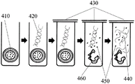
FIG. 4A is a schematic flow diagram of a reaction sequence in one microwell of a microwell capsule array.
Fig. 4B is similar to fig. 4A, except that there is annotated therein an example of the method that can be performed in each step.
Detailed Description
While various embodiments of the present invention have been shown and described herein, it will be obvious to those skilled in the art that such embodiments are provided by way of example only. Numerous variations, changes, and substitutions will now occur to those skilled in the art without departing from the invention. It should be understood that various alternatives to the embodiments of the invention described herein may be employed.
I. General overview
The present disclosure provides microwell or other compartmentalized capsule array devices and methods of using such devices. Typically, the device is an assembly of partitions (e.g., microwells, droplets) loaded with microcapsules, typically at a concentration of specific microcapsules/partitions.
The apparatus may be particularly suitable for performing sample preparation operations. In some cases, the device subdivides a sample (e.g., a heterogeneous mixture of nucleic acids, a mixture of cells, etc.) into multiple partitions such that only a portion of the sample is present in each partition. For example, a nucleic acid sample comprising a mixture of nucleic acids can be partitioned such that no more than one strand (or molecule) of nucleic acid is present in each partition. In other examples, the cell sample may be partitioned such that there is no more than one cell in each partition.
After the step of dividing, the subdivided sample may be subjected to any of a number of different operations within the apparatus. The partitions may comprise one or more capsules containing one or more reagents (e.g., enzymes, unique identifiers (e.g., barcodes), antibodies, etc.). In some cases, the device, companion device, or user provides a trigger (trigger) that causes the microcapsules to release one or more reagents into the respective partitions. The release of the reagent may enable the reagent to contact the subdivided sample. For example, if the reagent is a unique identifier, such as a barcode, the sample may be labeled with the unique identifier. The labeled sample can then be used in downstream applications, such as sequencing reactions.
A variety of different reactions and/or operations may be performed within the devices disclosed herein, including but not limited to: sample partitioning, sample separation, binding reactions, fragmentation (e.g., before partitioning or after partitioning), ligation reactions, and other enzymatic reactions.
The device may also be used in a variety of different molecular biology applications, including but not limited to: nucleic acid sequencing, protein sequencing, nucleic acid quantification, sequencing optimization, detection of gene expression, quantification of gene expression, and single cell analysis of genomic or expressed markers. Furthermore, the device has many medical applications. For example, it can be used for the identification, detection, diagnosis, treatment, staging or risk prediction of a variety of genetic and non-genetic diseases and disorders, including cancer.
Microcapsules II
Fig. 1A is a schematic illustration of an exemplary microcapsule comprising an internal compartment 120 encapsulated by a second layer 130, the second layer 130 being encapsulated by a solid or semi-permeable shell or membrane 110. Typically, the shell separates the interior compartments from their immediate environment (e.g., the interior of the microwells). The interior compartments, e.g., 120, 130, may contain materials, e.g., reagents. As shown in fig. 1A, reagent 100 may be present in interior compartment 120. However, in some cases, the reagent is located in the encapsulation layer 130 or in both compartments. Generally, the microcapsules may release the inner material or a portion thereof upon introduction of a particular trigger. This triggering may result in the destruction of the shell layer 110 and/or the inner encapsulation layer 130, thereby bringing the interior compartment 100, 120 into contact with the external environment, such as the cavity of a microwell.
The microcapsules may comprise several fluid phases and may comprise emulsions (e.g., water-in-oil emulsions, oil-in-water emulsions). The microcapsules may comprise an inner layer 120 that is immiscible with a second layer 130 encapsulating the inner layer. For example, inner layer 120 may comprise an aqueous fluid, while encapsulating layer 130 may be a non-aqueous fluid, such as an oil. Conversely, inner layer 120 may comprise a non-aqueous fluid (e.g., oil) and encapsulating layer 130 may comprise an aqueous fluid. In some cases, the microcapsules do not comprise a second encapsulant layer. Typically, the microcapsules are further encapsulated by a shell layer 110, which shell layer 110 may comprise a polymeric material. In some cases, the microcapsules may comprise droplets. In some cases, the microcapsules may be microdroplets.
Droplets and droplet generation methods are described, for example, in U.S. patent RE41,780, which is incorporated herein by reference in its entirety for all purposes. The device may also contain a microfluidic element that enables the flow of samples and/or microcapsules through the device and the distribution of the samples and/or microcapsules within the partitions.
The microcapsule may comprise a plurality of compartments. The microcapsule may comprise at least 1, 2, 3, 4, 5, 6, 7, 8, 9, 10, 11, 12, 13, 14, 15, 16, 17, 18, 19, 20, 50, 100, 500, 1000, 1500, 2000, 2500, 3000, 3500, 4000, 4500, 5000, 5500, 6000, 6500, 7000, 7500, 8000, 8500, 9000, 9500, 10000, or 50000 compartments. In other cases, the microcapsule comprises less than 3, 4, 5, 6, 7, 8, 9, 10, 11, 12, 13, 14, 15, 16, 17, 18, 19, 20, 50, 100, 500, 1000, 1500, 2000, 2500, 3000, 3500, 4000, 4500, 5000, 5500, 6000, 6500, 7000, 7500, 8000, 8500, 9000, 9500, 10000, or 50000 compartments. Similarly, each compartment or a subset thereof may also be subdivided into a plurality of additional compartments. In some cases, each compartment, or a subset thereof, is subdivided into at least 1, 2, 3, 4, 5, 6, 7, 8, 9, 10, 11, 12, 13, 14, 15, 16, 17, 18, 19, 20, 50, 100, 500, 1000, 1500, 2000, 2500, 3000, 3500, 4000, 4500, 5000, 5500, 6000, 6500, 7000, 7500, 8000, 8500, 9000, 9500, 10000, or 50000 compartments. In other cases, each compartment, or a subset thereof, is further subdivided into less than 3, 4, 5, 6, 7, 8, 9, 10, 11, 12, 13, 14, 15, 16, 17, 18, 19, 20, 50, 100, 500, 1000, 1500, 2000, 2500, 3000, 3500, 4000, 4500, 5000, 5500, 6000, 6500, 7000, 7500, 8000, 8500, 9000, 9500, 10000, or 50000 compartments.
There are a number of possible distributions of reagents in the various compartments. For example, each compartment (or a certain percentage of the total number of compartments) may contain the same reagent or the same combination of reagents. In some cases, each compartment (or some percentage of the total number of compartments) contains a different reagent or a different combination of reagents.
The compartments may be configured in a variety of ways. In some cases, the microcapsules may comprise a plurality of concentric compartments (repeating units of compartments comprising the preceding compartments) typically separated by immiscible layers. In such microcapsules, the reagent may be present in alternating compartments, every third compartment, or every fourth compartment.
In some cases, most of the compartments with microcapsules are not concentric; rather, they exist as separate, self-contained entities within the microcapsules. Fig. 1B shows an example of a microcapsule comprising a plurality of smaller microcapsules 140, each smaller microcapsule 140 comprising a reagent. As with many of the other microcapsules described herein, the microcapsules may be encapsulated by an outer shell, which typically comprises a polymeric material 150. A plurality of smaller microcapsules encapsulated within a larger microcapsule may be physically separated by immiscible fluid 160, thereby preventing mixing of the agents prior to application of a stimulus and release of the agents into solution. In some cases, the immiscible fluid is loaded with additional materials or agents. In some cases, the plurality of smaller microcapsules are surrounded by a layer of immiscible fluid (e.g., 170) that is further surrounded by a fluid 160 that is miscible with the inner fluid of the microcapsules. For example, the inner microcapsules 180 may comprise an aqueous inner portion encapsulated by an immiscible (e.g., oil) layer that is further surrounded by the aqueous layer 160. The miscible compartments (e.g., 160 and 180) may each contain a reagent. They may comprise the same agent (or the same combination of agents) or different agents (or different combinations of agents). Alternatively, one or some of these miscible compartments may not contain reagents.
The microcapsules may comprise one polymeric shell (see, e.g., fig. 1 and 2) or multiple polymeric shells. For example, the microcapsules may comprise multiple polymeric shells layered on top of each other. In other cases, a single compartment within the microcapsule comprises a polymeric shell, or a portion of the compartments may comprise a polymeric shell. For example, all or a portion of the smaller compartments 140 in fig. 1B may comprise a polymeric shell that separates them from the fluid interior 160. The microcapsules may be designed such that a particular agent is contained within a compartment having a polymeric shell, while a different agent is located within a compartment that is only encapsulated by the immiscible liquid. For example, an agent intended to be released upon thermal triggering may be contained within a compartment having a heat-sensitive or heat-activatable polymeric shell, while an agent designed to be released upon different triggers may be present within different types of compartments. In another example, paramagnetic particles may be incorporated into the capsule shell wall. The capsule can then be positioned to the desired location using a magnetic or electric field. In some cases, a magnetic field (e.g., a high frequency alternating magnetic field) may be applied to such a capsule; the incorporated paramagnetic particles can then convert the energy of the magnetic field into heat, triggering the rupture of the capsule.
The microcapsule component of the devices of the present disclosure may provide controlled and/or timed release of reagents for sample preparation of analytes. Microcapsules are particularly useful for the controlled release and Delivery of different types of chemicals, ingredients, pharmaceuticals, fragrances and the like, these including in particular sensitive agents such as enzymes and proteins (see, e.g., d.d. lewis, "biodegradeable Polymers and Drug Delivery Systems", m.chamin and r.langer eds (Marcel Decker, New York, 1990); j.p.mcgee et al, j.control.release 34(1995), 77).
Microcapsules may also provide a means (means) to deliver the agent in discrete and definable amounts. Microcapsules can be used to prevent premature mixing of the reagent with the sample by separating the reagent from the sample. Microcapsules may also facilitate handling and limit contact with particularly sensitive reagents such as enzymes, nucleic acids, and other chemicals used in sample preparation.
A. Preparation of microcapsules
The microcapsules of the devices of the present disclosure can be prepared by a variety of methods and processes. The preparation technology can comprise the following steps: pan coating, spray drying, centrifugal extrusion, emulsion-based methods, and/or microfluidics. In general, the method of preparation is selected based on the desired properties of the microcapsules. For example, shell wall thickness, permeability, chemical composition of the shell wall, mechanical integrity of the shell wall, and capsule size may be considered when selecting a method. The choice of preparation method may also be based on the ability to introduce specific materials into the capsule, such as whether the core material (e.g., fluid, reagent, etc.) is aqueous, organic, or inorganic. In addition, the method of preparation may affect the shape and size of the microcapsules. For example, the shape of the capsules (e.g., spherical, ellipsoidal, etc.) may depend on the shape of the droplets in the precursor liquid, which may depend on the viscosity and surface tension of the core liquid, the flow direction of the emulsion, the choice of surfactant used in droplet stabilization, and physical limitations, such as preparation in microchannels or capillaries having a particular size (e.g., a size that requires deformation of the microcapsules so that the microcapsules fit in the microchannel or capillary).
The microcapsules may be prepared by emulsion polymerization, which is a method of: wherein the monomer units polymerize at the water/organic interface in the emulsion to form the shell. The reagents are mixed with the aqueous phase of the biphasic mixture. Vigorous shaking or sonication of the mixture produces droplets containing the reagents, which are surrounded by a polymeric shell.
In some cases, microcapsules can be prepared by layer-by-layer assembly, which is a process that: wherein negatively and positively charged polyelectrolytes are deposited onto particles, such as metal oxide cores. Electrostatic interactions between polyelectrolytes produce a polymeric shell around the core. Subsequently, the core can be removed by the addition of an acid, resulting in semi-permeable hollow spheres that can be loaded with multiple agents.
In yet a further case, the microcapsules may be prepared by coacervation, coacervation being one such method: wherein two oppositely charged polymers are entangled in an aqueous solution to form a neutralized polymeric shell wall. One polymer may be contained in the oil phase and another polymer of opposite charge contained in the water phase. This aqueous phase may contain the reagent to be encapsulated. Attraction of one polymer to another can lead to the formation of agglomerates. In some embodiments, gelatin and gum arabic are ingredients of such a preparation method.
Microcapsules can also be prepared by internal phase separation, which is a process that: wherein the polymer is dissolved in a solvent mixture comprising volatile and non-volatile solvents. Droplets of the resulting solution are suspended in an aqueous layer, which is stabilized by continuous stirring and the use of surfactants. This phase may contain the agent to be encapsulated. As the volatile solvent evaporates, the polymer coalesces to form the shell wall. In some cases, polymers such as polystyrene, poly (methyl methacrylate), and poly (tetrahydrofuran) are used to form the shell wall.
Microcapsules can also be prepared by flow focusing methods, which is one such method: wherein a microcapillary device is used to generate a double emulsion comprising a single inner droplet enclosed in an intermediate fluid which is then dispersed to an outer fluid. The inner droplet may contain the reagent to be encapsulated. The intermediate fluid becomes a shell wall that can be formed by a cross-linking reaction.
B. Microcapsule composition
Microcapsules may comprise a variety of materials having a wide range of chemical properties. Typically, the microcapsules comprise a material having the ability to form microcapsules of a desired shape and size, and which is compatible with the reagents to be stored in the microcapsules.
The microcapsules may comprise a wide variety of different polymers including, but not limited to: a polymer, a thermosensitive polymer, a photosensitive polymer, a magnetic polymer, a pH-sensitive polymer, a salt-sensitive polymer, a chemosensitive polymer, a polyelectrolyte, a polysaccharide, a peptide, a protein, and/or a plastic. The polymer may include, but is not limited to, materials such as: poly (N-isopropylacrylamide) (PNIPAAm), poly (styrene sulfonate) (PSS), poly (allylamine) (PAAm), poly (acrylic acid) (PAA), poly (ethylenimine) (PEI), poly (diallyldimethyl-ammonium chloride) (PDADMAC), poly (pyrrole) (poly (pyrolle)) (PPy), poly (vinylpyrrolidone) (PVPON), poly (vinylpyridine) (PVP), poly (methacrylic acid) (PMAA), poly (methyl methacrylate) (PMMA), Polystyrene (PS), poly (tetrahydrofuran) (PTHF), poly (phthalaldehyde) (PTHF), poly (hexylviologen) (PHV), poly (L-lysine) (PLL), poly (L-arginine) (PARG), poly (lactic-glycolic acid) copolymer (PLGA).
In general, the materials used for the microcapsules, particularly the shell of the microcapsules, may enable the microcapsules to rupture upon application of a stimulus. For example, microcapsules may be prepared from heat-sensitive polymers and/or may comprise one or more shells containing such heat-sensitive polymers. The thermosensitive polymer may be stable under conditions for storage or loading. When exposed to heat, the heat-sensitive polymer component may undergo depolymerization, resulting in a disruption of the shell integrity and release of the interior material of the microcapsule (and/or interior microcapsule) to the external environment (e.g., the interior of the micropores). Exemplary thermosensitive polymers may include, but are not limited to, NIPAAm or PNIPAM hydrogels. The microcapsules may also contain one or more types of oils. Exemplary oils include, but are not limited to, hydrocarbon oils, fluorinated oils, fluorocarbon oils, silicone oils, mineral oils, vegetable oils, and any other suitable oils.
The microcapsules may also contain a surfactant, such as an emulsifying surfactant. Exemplary surfactants include, but are not limited to, cationic surfactants, nonionic surfactants, anionic surfactants, hydrocarbon surfactants, or fluorosurfactants. The surfactant may improve the stability of one or more components of the microcapsule, such as the oil-containing internal compartment.
Additionally, the microcapsules may comprise an inner material that is miscible with the material outside the capsule. For example, the inner material may be an aqueous fluid and the sample within the microwells may also be in the aqueous fluid. In other examples, the microcapsules may comprise powders or nanoparticles that are miscible with the aqueous fluid. For example, the microcapsules may contain such powders or nanoparticles within the internal compartment. When the microcapsules are ruptured, such powders or nanoparticles are released to the external environment (e.g., the interior of the micropores) and may be mixed with an aqueous fluid (e.g., an aqueous sample fluid).
Additionally, the microcapsules may contain materials that are immiscible with the surrounding environment (e.g., the interior of the micropores, the sample fluid). In such cases, phase separation between the inner and outer components may facilitate mixing, such as mixing of the inner component with the surrounding fluid, when the inner emulsion is released to the surrounding environment. In some cases, when the microcapsules are triggered to release their contents, pressure or force is also released, which facilitates mixing of the inner component with the outer component.
The microcapsules may also contain a polymer within the interior of the capsule. In some cases, such polymers may be porous polymer beads that can capture a reagent or combination of reagents. In other cases, this polymer may be beads that have previously swelled to form a gel. Examples of polymer-based gels that can be used as the internal emulsion of the capsule can include, but are not limited to, sodium alginate gels or polyacrylamide gels swollen with oligonucleotide barcodes or the like.
In some cases, the microcapsules can be gel beads comprising any of the polymer-based gels described herein. For example, gel bead microcapsules may be formed by encapsulating one or more polymer precursors in microdroplets. Gel beads can be produced when the polymer precursor is exposed to an accelerator, such as Tetramethylethylenediamine (TEMED).
Analytes and/or reagents, such as oligonucleotide barcodes, for example, may be coupled/immobilized to the inner surface of a gel bead (e.g., the interior accessible by diffusion of the oligonucleotide barcode and/or the material used to generate the oligonucleotide barcode) and/or the outer surface of a gel bead or any other microcapsule described herein. The coupling/immobilization may be by any form of chemical bonding (e.g., covalent, ionic) or physical phenomenon (e.g., van der waals forces, dipole-dipole interactions, etc.). In some cases, the coupling/immobilization of the agent onto the gel bead or any other microcapsule described herein may be reversible, e.g., by an unstable moiety (e.g., by a chemical cross-linker, including chemical cross-linkers described herein). Upon application of a stimulus, the labile moiety can cleave and allow release of the immobilized reagent. In some cases, the labile moiety is a disulfide bond. For example, where the oligonucleotide barcodes are immobilized to the gel beads by disulfide bonds, exposure of the disulfide bonds to a reducing agent may cleave the disulfide bonds and release the oligonucleotide barcodes from the beads. The labile moiety may be included as part of the gel bead or microcapsule, as part of a chemical linker attaching the reagent or analyte to the gel bead or microcapsule, and/or as part of the reagent or analyte.
The gel beads or any other type of microcapsules described herein may contain different amounts of reagents. The reagent density of each microcapsule may vary depending on the particular microcapsule and the particular reagent used. For example, the microcapsule or gel bead may comprise at least about 1, 10, 100, 1,000, 10,000, 100,000, 1,000,000, 5,000,000, 10,000,000, 50,000,000, 100,000,000, 500,000,000, or 1,000,000,000 oligonucleotide barcodes per microcapsule or gel bead. The gel beads may comprise the same oligonucleotide barcode or may comprise different oligonucleotide barcodes.
In other examples, the microcapsules may contain one or more materials that produce a net neutral, negative, or positive charge on the shell walls of the capsule. In some cases, the charge of the capsule may help prevent or promote aggregation or clustering of particles, or attachment to or repulsion from a portion of the device.
Further, the microcapsules may comprise one or more materials that render the shell wall of the capsule hydrophilic or hydrophobic. A hydrophilic material that may be used for the capsule wall may be poly (N-isopropylacrylamide). A hydrophobic material that may be used for the capsule wall may be polystyrene. In some cases, the hydrophilic shell wall can facilitate wicking of the capsule into the pores containing the aqueous fluid.
C. Size and shape of microcapsules
The microcapsules can have any of a number of sizes or shapes. In some cases, the shape of the microcapsules may be spherical, ellipsoidal, cylindrical, hexagonal, or any other symmetrical or asymmetrical shape. Any cross-section of the microcapsules may also have any suitable shape, including but not limited to: circular, oval, square, rectangular, hexagonal, or other symmetrical or asymmetrical shapes. In some cases, the microcapsules may have a specific shape that is complementary to the opening (e.g., the surface of the micropores) of the device. For example, the microcapsules may be spherical and the openings of the micropores of the device may be circular.
The microcapsules may be of uniform size (e.g., all of the microcapsules are of the same size) or of non-uniform size (e.g., some of the microcapsules are of different sizes). The microcapsules can have a size (e.g., diameter, cross-section, sides, etc.) of at least about 0.001 μm, 0.01 μm, 0.1 μm, 0.5 μm, 1 μm, 5 μm, 10 μm, 50 μm, 100 μm, 200 μm, 300 μm, 400 μm, 500 μm, 600 μm, 700 μm, 800 μm, 900 μm, or 1 nm. In some cases, the microcapsules comprise micropores of up to about 0.001 μm, 0.01 μm, 0.1 μm, 0.5 μm, 1 μm, 5 μm, 10 μm, 50 μm, 100 μm, 200 μm, 300 μm, 400 μm, 500 μm, 600 μm, 700 μm, 800 μm, 900 μm, or 1 nm.
In some cases, the microcapsules have a size and/or shape such that a limited number of microcapsules are deposited in a single partition (e.g., microwell, microdroplet) of the microcapsule array. The microcapsules may be of a particular size and/or shape such that exactly or no more than 1, 2, 3, 4, 5, 6, 7, 8, 9, or 10 capsules fit into a single microwell; in some cases, an average of 1, 2, 3, 4, 5, 6, 7, 8, 9, or 10 capsules fit into a single microwell. In still further cases, at least 1, 2, 3, 4, 5, 6, 7, 8, 9, 10, 100, 500, or 1000 capsules fit into a single microwell.
D. Reagent and reagent loading
The devices provided herein can comprise free reagents and/or reagents encapsulated in microcapsules. The reagents may be various molecules, chemicals, particles and elements suitable for sample preparation reactions of analytes. For example, microcapsules used in a sample preparation reaction for DNA sequencing of a target may comprise one or more of the following reagents: enzymes, restriction enzymes (e.g., multiple cutters), ligases, polymerases (e.g., polymerases that recognize and do not recognize dUTP and/or uracil), fluorophores, oligonucleotide barcodes, buffers, deoxynucleotide triphosphates (dntps) (e.g., deoxyadenosine triphosphate (dATP), deoxycytidine triphosphate (dCTP), deoxyguanosine triphosphate (dGTP), deoxythymidine triphosphate (dTTP), deoxyuridine triphosphate (dUTP)), deoxynucleotide triphosphates (ddntps), and the like. In another example, microcapsules used in a sample preparation reaction for single cell analysis may contain reagents, such as one or more of the following: lysis buffers, detergents, fluorophores, oligonucleotide barcodes, ligases, proteases, heat-activatable proteases, protease or nuclease inhibitors, buffers, enzymes, antibodies, nanoparticles, and the like.
Exemplary agents include, but are not limited to: buffer, acidic solution, basic solution, temperature-sensitive enzyme, pH-sensitive enzyme, photosensitive enzyme, metal ion, magnesium chloride, sodium chloride, manganese, aqueous buffer, mild buffer, ionic buffer, inhibitor, enzyme, protein, nucleic acid, antibody, saccharide, lipid, oil, salt, ion, detergent, ionic detergent, non-ionic detergent, oligonucleotide, nucleotide, dNTP, ddNTP, deoxyribonucleic acid (DNA), ribonucleic acid (RNA), peptide nucleic acid, circular DNA (cdna), double-stranded DNA (dsdna), single-stranded DNA (ssdna), plasmid DNA, cosmid DNA, chromosomal DNA, genomic DNA (gdna), viral DNA, bacterial DNA, mtDNA (mitochondrial DNA), messenger RNA (mrna), ribosomal RNA (rrna), transfer RNA (trna), nRNA, short interfering RNA (sirna), small nuclear RNA (snrna), small nuclear RNA (snohna), small Cajul-specific RNA (scaarna, or RNA, micrornas, double-stranded RNAs (dsrna), ribozymes, riboswitches and viral RNAs, polymerases (e.g., polymerases that recognize and do not recognize dUTP and/or uracil), ligases, restriction enzymes, proteases, nucleases, protease inhibitors, nuclease inhibitors, chelators, reducing agents (e.g., Dithiothreitol (DTT), 2-tris (2-carboxyethyl) phosphine (TCEP)), oxidizing agents, fluorophores, probes, chromophores, dyes, organics, emulsifiers, surfactants, stabilizers, polymers, water, small molecules, drugs, radioactive molecules, preservatives, antibiotics, aptamers, and pharmaceutical compounds.
In some cases, the microcapsules contain a set of reagents (e.g., a set of enzymes, a set of minerals, a set of oligonucleotides, a mixture of different barcodes, a mixture of the same barcodes) with similar properties. In other cases, the microcapsules contain a heterogeneous mixture of reagents. In some cases, the heterogeneous mixture of reagents contains all the components necessary to carry out the reaction. In some cases, such mixtures contain all of the components necessary to carry out the reaction, except 1, 2, 3, 4, 5 or more of the components necessary to carry out the reaction. In some cases, such additional components are contained in solutions within different microcapsules or within partitions (e.g., micropores) of the device.
The reagents may be preloaded into the device (e.g., prior to introduction of the analyte) or post-loaded into the device. They can be loaded directly into the device; alternatively, in some cases, the reagents are encapsulated within microcapsules loaded into the device. In some cases, only microcapsules containing the agent are introduced. In other cases, both the free reagent and the reagent encapsulated in the microcapsule are loaded into the device either sequentially or simultaneously. In some cases, the reagents are introduced to the device before or after a particular step. For example, lysis buffer reagents can be introduced into the device after the cell sample is partitioned into multiple partitions (e.g., microwells, microdroplets) within the device. In some cases, the reagents and/or microcapsules containing the reagents are introduced sequentially, such that different reactions or operations occur at different steps. The reagents (or microcapsules) may also be loaded during the step interspersed with the reaction or manipulation step. For example, microcapsules comprising reagents for fragmenting molecules (e.g., nucleic acids) can be loaded into a device, followed by a fragmentation step, after which microcapsules comprising reagents for attaching barcodes (or other unique identifiers, such as antibodies) can be loaded, followed by attaching barcodes to the fragmented molecules. Other methods of loading reagents are further described elsewhere herein.
E. Molecular "Bar code"
It may be desirable to retain the option of identifying and tracking individual molecules or analytes after or during sample preparation. In some cases, one or more unique molecular identifiers (sometimes referred to in the art as "molecular barcodes") are used as sample preparation reagents. These molecules may comprise a variety of different forms, such as oligonucleotide barcodes, antibodies or antibody fragments, fluorophores, nanoparticles, and other elements, or combinations thereof. Depending on the particular application, the molecular barcode may bind reversibly or irreversibly to the target analyte and allow identification and/or quantification of individual analytes after sample preparation and recovery from the device.
The devices of the present disclosure may be adapted for use in nucleic acid sequencing, protein detection, single molecule analysis, and other methods that require: a) precise measurement of the presence and amount of a particular analyte, b) multiplex reactions in which multiple analytes are combined for analysis. The devices of the present disclosure may employ microwells or other types of partitions (e.g., microdroplets) of a microwell array to physically partition a target analyte. This physical segmentation allows a single analyte to acquire one or more molecular barcodes. After sample preparation, individual analytes can be combined or combined and extracted from the device for multiplex analysis. For most applications, multiplex analysis greatly reduces the cost of the analysis and increases the throughput of the process, for example in the case of nucleic acid sequencing. Molecular barcodes may allow identification and quantification of individual molecules even after multiple analytes are combined. For example, for nucleic acid sequencing, molecular barcodes may allow sequencing of a single nucleic acid even after multiple different nucleic acids are pooled.
In some cases, oligonucleotide barcodes may be particularly useful for nucleic acid sequencing. In general, an oligonucleotide barcode may comprise a unique sequence (e.g., a barcode sequence) that confers its identifying function to the oligonucleotide barcode. The unique sequence may be random or non-random. Attachment of a barcode sequence to a nucleic acid of interest can correlate the barcode sequence to the nucleic acid of interest. The barcode can then be used to identify the nucleic acid of interest during sequencing, even when other nucleic acids of interest (e.g., comprising different barcodes) are present. In the case of fragmenting the nucleic acid of interest prior to sequencing, the attached barcode can be used to identify the fragment as belonging to the nucleic acid of interest during the sequencing process.
The oligonucleotide barcode may consist of only unique barcode sequences, or may be included as part of an oligonucleotide of longer sequence length. Such oligonucleotides may be adapters (adaptors) required for specific sequencing chemistries and/or methods. For example, such adapters may include, in addition to oligonucleotide barcodes: immobilization of the adapter (e.g., by hybridization) to a desired region of an immobilized sequence on a solid surface (e.g., a solid surface in a flow cell channel of a sequencer); sequencing the region of sequence required for binding of the primer; and/or random sequences (e.g., random N-mers) that can be used, for example, in random amplification schemes. For example, the adapter can be attached to the nucleic acid to be sequenced by amplification, ligation, or any other method described herein.
In addition, the oligonucleotide barcode and/or the larger oligonucleotide comprising the oligonucleotide barcode may comprise a natural nucleobase and/or may comprise a non-natural nucleobase. For example, where the oligonucleotide barcode or a larger oligonucleotide comprising the oligonucleotide barcode is DNA, the oligonucleotide may comprise the natural DNA bases adenine, guanine, cytosine, and thymine, and/or may comprise non-natural bases, such as uracil.
F. Microcapsule preparation for micropore loading
After preparation, the reagent-loaded microcapsules can be loaded into the device using a variety of methods. In some cases, the microcapsules may be loaded as "dry capsules. After preparation, the capsules can be separated from the liquid phase using a variety of techniques including, but not limited to, differential centrifugation, liquid phase evaporation, chromatography, filtration, and the like. The "dry capsules" may be collected as a powder or particulate matter and then deposited into the microwells of a microwell array. Loading "dry capsules" may be the preferred method in cases where loading of "wet capsules" results in loading inefficiencies, such as voids and poor distribution of microcapsules throughout the microwell array.
The reagent-loaded microcapsules may also be loaded into the device when the microcapsules are in the liquid phase, and thus loaded as "wet capsules". In some cases, the microcapsules may be suspended in a volatile oil so that the oil can be removed or evaporated, leaving only dry capsules in the pores. In some cases where loading of dry capsules results in loading inefficiencies, such as clustering, aggregation of microcapsules, and poor distribution of microcapsules throughout the microwell array, loading "wet capsules" may be the preferred method. Other methods of loading reagents and microcapsules are described elsewhere in this disclosure.
The microcapsules may also have a specific density. In some cases, the microcapsules are less dense than the aqueous fluid (e.g., water); in some cases, the microcapsules are denser than the aqueous fluid (e.g., water). In some cases, the microcapsules are less dense than the non-aqueous fluid (e.g., oil); in some cases, the microcapsules are denser than the non-aqueous fluid (e.g., oil). The microcapsules may have at least about 0.05g/cm 3 、0.1cm 3 、0.2g/cm 3 、0.3g/cm 3 、0.4g/cm 3 、0.5g/cm 3 、0.6g/cm 3 、0.7g/cm 3 、0.8g/cm 3 、0.81g/cm 3 、0.82g/cm 3 、0.83g/cm 3 、0.84g/cm 3 、0.85g/cm 3 、0.86g/cm 3 、0.87g/cm 3 、0.88g/cm 3 、0.89g/cm 3 、0.90g/cm 3 、0.91g/cm 3 、0.92g/cm 3 、0.93g/cm 3 、0.94g/cm 3 、0.95g/cm 3 、0.96g/cm 3 、0.97g/cm 3 、0.98g/cm 3 、0.99g/cm 3 、1.00g/cm 3 、1.05g/cm 3 、1.1g/cm 3 、1.2g/cm 3 、1.3g/cm 3 、1.4g/cm 3 、1.5g/cm 3 、1.6g/cm 3 、1.7g/cm 3 、1.8g/cm 3 、1.9g/cm 3 、2.0g/cm 3 、2.1g/cm 3 、2.2g/cm 3 、2.3g/cm 3 、2.4g/cm 3 Or 2.5g/cm 3 The density of (c). In other cases, the density of the microcapsules may be up to about 0.7g/cm 3 、0.8g/cm 3 、0.81g/cm 3 、0.82g/cm 3 、0.83g/cm 3 、0.84g/cm 3 、0.85g/cm 3 、0.86g/cm 3 、0.87g/cm 3 、0.88g/cm 3 、0.89g/cm 3 、0.90g/cm 3 、0.91g/cm 3 、0.92g/cm 3 、0.93g/cm 3 、0.94g/cm 3 、0.95g/cm 3 、0.96g/cm 3 、0.97g/cm 3 、0.98g/cm 3 、0.99g/cm 3 、1.00g/cm 3 、1.05g/cm 3 、1.1g/cm 3 、1.2g/cm 3 、1.3g/cm 3 、1.4g/cm 3 、1.5g/cm 3 、1.6g/cm 3 、1.7g/cm 3 、1.8g/cm 3 、1.9g/cm 3 、2.0g/cm 3 、2.1g/cm 3 、2.2g/cm 3 、2.3g/cm 3 、2.4g/cm 3 Or 2.5g/cm 3 . Such density may reflect the density of the microcapsules in any particular fluid (e.g., aqueous, water, oil, etc.).
Microwell array
A. Structure/feature
The device of the present disclosure may be a microwell array comprising a solid plate comprising a plurality of wells, cavities, or microwells having microcapsules and/or analytes deposited therein. Typically, a fluid sample (or analyte) is introduced into the device (e.g., through an inlet) and then travels through a flow channel that distributes the sample into a plurality of microwells. In some cases, additional fluid is also introduced into the device. The microwells may contain microcapsules when the sample is introduced; alternatively, in some cases, the microcapsules are introduced into the microwells after the sample is introduced.
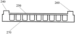
FIG. 2A shows a prototype of a microwell array; fig. 2B shows a side view. The microwell array may comprise a plate 220, and the plate 220 may be made of any suitable material commonly used in chemical laboratories, including fused silica, soda lima glass, borosilicate glass, PMMA, sapphire, silicon, germanium, cyclic olefin copolymers and cyclic polymers, polyethylene, polypropylene, polyacrylates, polycarbonates, plastics, Topas, and other suitable substrates known in the art. The plate 220 may initially be a flat solid plate comprising a regular pattern of micro-wells 270. The micropores may be formed by drilling or chemical dissolution or any other suitable machining method; however, the plate with the desired hole pattern is preferably molded, for example by injection molding, embossing or using a suitable polymer such as cyclic olefin copolymer.
The microwell array may comprise inlets (200 and 240) and/or outlets (210 and 260); in some cases, the microwell array comprises a plurality of inlets and/or outlets. The sample (or analyte) or microcapsules may be introduced into the device via the inlet. Solutions containing analytes, reagents and/or microcapsules can be added manually to inlet ports 200 and 240 (or to a catheter attached to the inlet port) by pipette. In some cases, the analytes, reagents, and/or microcapsules are introduced into the device using a liquid handling device. Exemplary liquid handling devices may rely on pipetting robots, capillary action, or immersion into fluids. In some cases, the inlet port is connected to a reservoir containing microcapsules or analytes. The inlet port may be connected to a flow channel 250, the flow channel 250 allowing for dispensing of analytes, samples or microcapsules to the microwells in the device. In some cases, the inlet port may be used to introduce a fluid (e.g., oil, aqueous) that does not contain microcapsules or analytes into the device, such as a carrier fluid. The carrier fluid may be introduced via the inlet port before, during, or after introduction of the analyte and/or the microcapsules. Where the device has multiple inlets, the same sample may be introduced via the multiple inlets, or each inlet may carry a different sample. In some cases, one inlet may deliver a sample or analyte to the microwells, while a different inlet delivers free reagent and/or reagent encapsulated in microcapsules to the device. The device may have at least 1, 2, 3, 4, 5, 6, 7, 8, 9 or 10 inlets and/or outlets.
In some cases, the solution containing the microcapsules and/or analyte may be pulled through the device via a vacuum manifold attached to the outlet ports 210 and 260. Such a manifold may apply negative pressure to the device. In other cases, positive pressure is used to move the sample, analyte, and/or microcapsules through the device. The area, length and width of the surface 230 according to the present disclosure may vary depending on the requirements of the assay to be performed. Considerations may include: for example, the simplicity of processing, the limitations of the materials forming the surface, the requirements of the detection or processing system, the requirements of the deposition system (e.g., microfluidic system), etc. The thickness may comprise a thickness of at least about 0.001mm, 0.005mm, 0.01mm, 0.05mm, 0.1mm, 0.2mm, 0.3mm, 0.4mm, 0.5mm, 0.6mm, 0.7mm, 0.8mm, 0.9mm, 1.0mm, 2.0mm, 3.0mm, 4.0mm, 5.0mm, 6.0mm, 7.0mm, 8.0mm, 9.0mm, 10.0mm, 11mm, 12mm, 13mm, 14mm, or 15 mm. In other cases, the thickness of the microcapsules may be up to 0.001mm, 0.005mm, 0.01mm, 0.05mm, 0.1mm, 0.2mm, 0.3mm, 0.4mm, 0.5mm, 0.6mm, 0.7mm, 0.8mm, 0.9mm, 1.0mm, 2.0mm, 3.0mm, 4.0mm, 5.0mm, 6.0mm, 7.0mm, 8.0mm, 9.0mm, 10.0mm, 11mm, 12mm, 13mm, 14mm, or 15 mm.
The volume of each hole may be a measure of the height of the hole (thickness of the plate) and the effective diameter of each hole. The capacity of a single well may be selected from a plurality of volumes. In some cases, the device can comprise a well (or microwell) having a capacity of at least 0.001fL, 0.01fL, 0.1fL, 0.5fL, 1fL, 5fL, 10fL, 50fL, 100fL, 200fL, 300fL, 400fL, 500fL, 600fL, 700fL, 800fL, 900fL, 1pL, 5pL, 10pL, 50pL, 100pL, 200pL, 300pL, 400pL, 500pL, 600pL, 700pL, 800pL, 900pL, 1nL, 5nL, 10nL, 50nL, 100nL, 200nL, 300nL, 400nL, 500nL, 600 nL, 700pL, 800 nL, 900 nL, 1nL, 5nL, 10nL, 50nL, 100nL, 200nL, 300nL, 400nL, 500nL, 1uL, 50uL, or 100 uL. In other cases, the microcapsule comprises pores of less than 0.001fL, 0.01fL, 0.1fL, 0.5L, 5fL, 10fL, 50fL, 100fL, 200fL, 300fL, 400fL, 500fL, 600fL, 700fL, 800fL, 900fL, 1pL, 5pL, 10pL, 50pL, 100pL, 200pL, 300pL, 400pL, 500pL, 600pL, 700pL, 800pL, 900pL, 1nL, 5nL, 10nL, 50nL, 100nL, 200nL, 300nL, 400nL, 500nL, 1uL, 50uL, or 100 uL.
There may be variability in the volume of fluid in different microwells in the array. More specifically, the volume of different microwells may vary by at least (or at most) + or-1%, 2%, 3%, 4%, 5%, 10%, 20%, 30%, 40%, 50%, 60%, 70%, 80%, 90%, 100%, 200%, 300%, 400%, 500%, or 1000% among a group of microwells. For example, the microwell may contain a fluid volume that is at most 80% of the fluid volume within the second microwell.
The microwell array may comprise a range of pore densities based on the size of the individual microwells and the size of the plate. In some examples, the plurality of micropores can have at least about 2,500 pores/cm 2 At least about 1,000 pores/cm 2 The density of (c). In some cases, the plurality of wells can have at least 10 wells/cm 2 The density of (c). In other cases, the pore density can include at least 10 pores/cm 2 50 wells/cm 2 100 wells/cm 2 500 wells/cm 2 1000 wells/cm 2 5000 holes/cm 2 10000 wells/cm 2 50000 holes/cm 2 Or 100000 holes/cm 2 . In other cases, the pore density can be less than 100000 pores/cm 2 10000 wells/cm 2 5000 holes/cm 2 1000 wells/cm 2 500 wells/cm 2 Or 100 holes/cm 2 。
In some cases, the interior surfaces of the microwells comprise a hydrophilic material that preferably holds an aqueous sample; in some cases, the regions between the microwells are comprised of a hydrophobic material that can preferentially attract the hydrophobic sealing fluid described herein.
Multiple microwell arrays, such as fig. 2B, may be arranged within a single device. Fig. 3, 300. For example, discrete microwell array slides may be arranged in parallel on a plate support. In some cases, at least 1, 2, 3, 4, 5, 6, 7, 8, 9, 10, 25, 50, or 100 microwell arrays are arranged in parallel. In other cases, up to 100, 50, 25, 10, 9, 8, 7, 6, 5, 4, 3, 2, or 1 devices are arranged in parallel.
The array of microwells in a common device can be operated simultaneously or sequentially. For example, the arrayed devices may be loaded with samples or capsules simultaneously or sequentially.
B. Microwell array fluids
The microwell array can comprise any number of different fluids, including aqueous, non-aqueous, oil, and organic solvents, such as alcohols. In some cases, the fluid is used to carry components such as reagents, microcapsules, or analytes to a target location, such as a microwell, an outlet port, or the like. In other cases, the fluid is used to flush the system. In other cases, the fluid may be used to seal the pores.
Any fluid or buffer that is physiologically compatible with the analyte (e.g., cells, molecules) or reagents used in the device may be used. In some cases, the fluid is aqueous (buffered or unbuffered). For example, a sample comprising a population of cells suspended in a buffered aqueous solution can be introduced into a microwell array, flowed through the device, and distributed to microwells. In other cases, the fluid flowing through the device is non-aqueous (e.g., oil). Exemplary non-aqueous fluids include, but are not limited to: oils, non-polar solvents, hydrocarbon oils, decanes (e.g., tetradecane or hexadecane), fluorocarbon oils, fluorinated oils, silicone oils, mineral oils, or other oils.
Typically, the microcapsules are suspended in a fluid that is compatible with the components of the shell of the microcapsules. Fluids including, but not limited to, water, alcohols, hydrocarbon oils, or fluorocarbon oils are particularly useful fluids for suspending and flowing microcapsules through a microarray device.
C. Further segmentation and sealing
After the analytes, free reagents, and/or microcapsules are loaded into the device and distributed to the microwells, they may be further partitioned or separated within the microwells using a sealing fluid. Sealing fluids may also be used to seal individual apertures. The sealing fluid may be introduced through the same inlet port used for introducing the analyte, reagent and/or microcapsules. In some cases, however, the sealing fluid is introduced into the device through a single inlet port or through multiple separate inlet ports.
Typically, the sealing fluid is a non-aqueous fluid (e.g., oil). As the sealing fluid flows through the microwell array device, it can displace excess aqueous solution (e.g., a solution containing analyte, free reagents, and/or microcapsules) from individual microwells, thereby potentially removing aqueous bridging between adjacent microwells. The pores described herein may themselves comprise a hydrophilic material capable of wicking aqueous fluid (e.g., sample fluid, microcapsule fluid) into the individual pores. In some cases, the region outside the pores comprises a hydrophobic material, again to facilitate positioning of the aqueous fluid to the interior of the micropores.
The sealing fluid may remain in the device or be removed. The sealing fluid may be removed, for example, by flowing through the outlet port. In other cases, the sealing oil may comprise a volatile oil that can be removed by the application of heat. Once the sealing fluid is removed, the analytes, free reagents, and/or microcapsules may be physically separated from each other in the microwells.
The fluid may be selected such that its density is equal to, greater than, or less than the density of the microcapsules. For example, the microcapsules may have a density greater than the aqueous fluid encapsulating the oil and/or sample and reagents, thereby enabling the microcapsules to remain in the pores as the encapsulating oil flows through the device. In another example, the density of the capsules may be less than the density of the aqueous fluid of the sample or the fluid in which the microcapsules are suspended as described herein, thereby facilitating movement and distribution of the capsules in the plurality of micropores of the device.
In the case of microcapsules containing paramagnetic materials, a magnetic field may be used to load or guide the capsules into the micropores. The magnetic field may also be used to retain such microcapsules within the pores while filling the pores with sample, reagents, and/or sealing fluids. The magnetic field may also be used to remove the capsule shell from the well, particularly after the capsule is ruptured.
In some cases, the sealing fluid may remain therein when operating or reacting within the microwells. The presence of the sealing fluid may be used to further divide, separate or seal individual microwells. In other cases, the encapsulating fluid may serve as a carrier for the microcapsules. For example, an encapsulating fluid containing microcapsules may be introduced into the device to facilitate distribution of the microcapsules to individual micropores. For such applications, the encapsulating fluid may be denser than the microcapsules to promote more uniform distribution of the microcapsules into the micropores. Upon application of a stimulus, microcapsules within the encapsulated fluid may release the agent to the micropores. In some cases, the sealing fluid may contain a chemical or other agent capable of traveling from the sealing fluid to the pores (e.g., by leaching or other mechanism) and triggering rupture of the capsules, where the capsules are present within the pores or within the sealing fluid.
Methods other than those involving sealing fluids may also be used to seal the microwells after loading with analytes, free reagents, and/or microcapsules. For example, the microwells may be sealed with a laminate, tape, plastic lid, oil, wax, or other suitable material to create an enclosed reaction chamber. The sealants described herein may prevent evaporation of the contents of the microwells or prevent other unintended consequences of the reaction or operation. It may be particularly desirable to prevent evaporation when heat is applied to the device, for example, when heat is applied to stimulate microcapsule release.
In some cases, the laminate seal may also allow for the retrieval of contents from a single hole. In this case, one well of interest may be unsealed (e.g., sealed by removing the laminate) at a given time to enable further analysis of the analyte, for example by MALDI mass spectrometry. Such applications are useful in many settings, including high throughput drug screening.
IV. Loading step
As described herein, analytes, free reagents, and/or microcapsules may be loaded into the devices of the present invention in any suitable manner or order. The loading may be random or non-random. In some cases, the exact number of analytes and/or microcapsules are loaded into each individual microwell. In some cases, the exact number of analytes and/or microcapsules are loaded into specific portions of the microwells in the plate. In still other cases, an average number of analytes and/or microcapsules are loaded into each individual microwell. Furthermore, as described herein, in some cases, "dry" microcapsules are loaded into the device, while in other cases, "wet" microcapsules are loaded into the device. In some cases, a combination of "dry" and "wet" microcapsules and/or reagents are loaded into the device simultaneously or sequentially.
As mentioned herein, the loading of the device may be performed in any order and may be performed in multiple stages. In some cases, the microcapsules are preloaded into the device prior to loading the analyte. In other cases, the microcapsules and analyte are loaded simultaneously. In still other cases, the analyte is loaded prior to loading the microcapsules.
The microcapsules and/or analytes may be loaded in multiple stages or multiple times. For example, the microcapsules may be loaded into the device both before and after the analyte is loaded into the device. The microcapsules that are preloaded (e.g., loaded prior to introduction of the analyte) can contain the same reagents as the microcapsules loaded after introduction of the analyte. In other cases, the preloaded microcapsules contain a different reagent than the reagent within the microcapsules loaded upon introduction of the analyte. In some cases, at least 1, 2, 3, 4, 5, 6, 7, 8, 9, 10, 15, or 20 different sets of microcapsules are loaded onto the device. In some cases, the different sets of microcapsules are loaded sequentially; alternatively, different sets of microcapsules may be loaded simultaneously. Similarly, multiple sets of analytes may be loaded into the device. In some cases, at least 1, 2, 3, 4, 5, 6, 7, 8, 9, 10, 15, or 20 different sets of analytes are loaded onto the device. In some cases, different sets of analytes are sequentially loaded; alternatively, different sets of analytes may be loaded simultaneously.
The present disclosure provides devices that contain a number of microcapsules and/or analytes loaded per well. In some cases, up to 1, 2, 3, 4, 5, 6, 7, 8, 9, 10, 15, 20, 30, 40, 50, 75, or 100 microcapsules and/or analytes are loaded into each individual microwell. In some cases, at least 1, 2, 3, 4, 5, 6, 7, 8, 9, 10, 15, 20, 30, 40, 50, 75, or 100 microcapsules and/or analytes are loaded into each individual microwell. In some cases, on average, up to 1, 2, 3, 4, 5, 6, 7, 8, 9, 10, 15, 20, 30, 40, 50, 75, or 100 microcapsules and/or analytes are loaded into each individual microwell. In other cases, on average, at least 1, 2, 3, 4, 5, 6, 7, 8, 9, 10, 15, 20, 30, 40, 50, 75, or 100 microcapsules and/or analytes are loaded into each individual microwell. In some cases, about 1, 2, 3, 4, 5, 6, 7, 8, 9, 10, 15, 20, 30, 40, 50, 75, or 100 microcapsules and/or analytes are loaded into each individual microwell.
The analyte and/or microcapsules may be applied in an amount that allows the desired number of analytes to be deposited into a single microwell. For example, the final dilution of the analyte (such as a cell) may achieve loading of one cell per microwell or any desired number of analytes per microwell. In some cases, the poisson distribution is used to guide or predict the final concentration of analyte or microcapsules in each well.
The microcapsules may be loaded into the microarray device in a specific pattern. For example, some portions of the device may comprise microcapsules containing a particular reagent (e.g., a unique barcode, an enzyme, an antibody subclass, etc.), while other portions of the device may comprise microcapsules containing a different reagent (e.g., a different barcode, a different enzyme, a different antibody subclass, etc.). In some cases, the microcapsules in a portion of the array may contain a control reagent. For example, they may contain a positive control comprising a control analyte and the necessary materials for the reaction. Alternatively, in some cases, the microcapsules contain a negative control reagent, such as an inactivated enzyme or a synthetic oligonucleotide sequence that is resistant to fragmentation. In some cases, the negative control reagent may control the specificity of the sample preparation reaction, etc. In other cases, a negative control microcapsule may comprise the same reagents present in other microcapsules, except that the negative control microcapsule may lack certain reagents (e.g., lysis buffer, polymerase, etc.).
The analytes/samples may also be loaded into the microarray device in a specific pattern. For example, certain portions of the device may contain a particular analyte, such as a control analyte or an analyte derived from a particular source. This can be used in combination with the specific loading of the barcode to known well locations. This feature may allow for the location-specific (mapping) on the array to be mapped to the sequencing data, thereby reducing the number of barcodes to be used for labeling reactions.
In the case of a partition into droplets, the analyte and reagent may be combined within the droplet with the aid of a microfluidic device. For example, microdroplets can be generated that comprise gel beads (e.g., gel beads comprising oligonucleotide barcodes), nucleic acid analytes, and any other desired reagents. The gel beads, nucleic acid analyte, and reagents in the aqueous phase may be combined at the intersection of two or more channels of the microfluidic device. At a second intersection of two or more channels of the microfluidic device, droplets comprising the resulting mixture may be generated by contacting an aqueous mixture of reagents, gel beads, and nucleic acid analyte with an oil continuum.
Stimulation by microcapsules
A variety of different stimuli can be used to trigger the release of the agent from the microcapsule or from the internal compartment therein. In some cases, the microcapsules are degradable. In general, the triggering may result in the rupture or degradation of the shell or membrane surrounding the microcapsule, the rupture or degradation of the interior of the microcapsule, and/or the rupture or degradation of any chemical bond that secures the agent to the microcapsule. Exemplary triggers include, but are not limited to: chemical triggers, bulk changes (bulk changes), biological triggers, optical triggers, thermal triggers, magnetic triggers, and any combination thereof. See, for example, Eser-Kahn et al (2011) Macromolecules 44: 5539-; wang et al (2009) ChemPhys Chem 10: 2405-2409.
A. Chemical stimulation and host changes
A number of chemical triggers can be used to trigger the rupture or degradation of the microcapsules. Examples of such chemical changes may include, but are not limited to, pH-mediated changes to the shell wall, disintegration of the shell wall by chemical cleavage of crosslinks, triggered depolymerization of the shell wall, and shell wall switching reactions. The host change may also be used to trigger the rupture of the microcapsules.
Changes in the pH of the solution, particularly a decrease in pH, can trigger the rupture by a number of different mechanisms. The addition of acid can cause degradation or decomposition of the shell wall by a variety of mechanisms. The addition of protons may break down cross-links of polymers in the shell wall, break ionic or hydrogen bonds in the shell wall, or create nanopores in the shell wall to allow internal contents to permeate through to the outside. In some examples, the microcapsules comprise an acid degradable chemical cross-linker, such as a ketal. A decrease in pH, particularly to a pH below 5, can induce the conversion of the ketal into a ketone and two alcohols and promote the rupture of the microcapsules. In other examples, the microcapsules may comprise one or more pH sensitive polyelectrolytes (e.g., PAA, PAAm, PSS, etc.). The lowering of pH may disrupt the ionic or hydrogen bonding interactions of such microcapsules, or create nanopores therein. In some cases, microcapsules comprising polyelectrolytes contain a charged, gel-based core that expands and contracts when pH changes.
Removal of cross-linkers (e.g., disulfide bonds) within the microcapsules can also be accomplished by a variety of mechanisms. In some examples, a variety of chemicals may be added to the solution of microcapsules that induce oxidation, reduction, or other chemical changes to the polymer component of the shell wall. In some cases, a reducing agent, such as β -mercaptoethanol, Dithiothreitol (DTT), or 2-tris (2-carboxyethyl) phosphine (TCEP), is added to break the disulfide bonds in the microcapsule shell wall. Additionally, enzymes may be added to cleave peptide bonds within the microcapsules, resulting in cleavage of the shell wall crosslinks.
Depolymerization may also be used to rupture the microcapsules. Chemical triggers can be added to facilitate removal of the protective head groups. For example, triggering may result in the removal of carbonate or carbamate head groups within the polymer, which in turn causes depolymerization and release of the agent from the interior of the capsule.
The shell wall switching reaction may be due to any structural change in the shell wall porosity. The porosity of the shell wall can be modified, for example by the addition of azo dyes or viologen derivatives. The addition of energy (e.g., electricity, light) can also be used to stimulate a change in porosity.
In yet another example, the chemical trigger may comprise an osmotic trigger, whereby a change in ion or solute concentration of the microcapsule solution induces swelling of the capsule. Swelling can cause the build-up of internal pressure, causing the capsule to rupture to release its contents.
It is also known in the art that the bulk or physical changes of the microcapsules by various stimuli also provide many advantages in designing the capsules to release the agent. The bulk or physical change occurs on a macroscopic scale, where the capsule rupture is the result of mechanical-physical forces induced by the stimulus. These methods may include, but are not limited to, pressure induced cracking, shell wall melting, or changes in shell wall porosity.
B. Biological stimulation
Biostimulation can also be used to trigger the rupture or degradation of the microcapsules. Generally, biological triggers are similar to chemical triggers, but many examples use biomolecules or molecules commonly found in living systems, such as enzymes, peptides, carbohydrates, fatty acids, nucleic acids, and the like. For example, the microcapsules may comprise a polymer with peptide cross-links that are susceptible to cleavage by a specific protease. More specifically, one example may include microcapsules comprising GFLGK peptide cross-links. Upon addition of a biological trigger such as the protease cathepsin B, the peptide cross-links of the shell wall are cleaved and the contents of the capsule are released. In other cases, the protease may be heat activated. In another example, the microcapsule comprises a shell wall comprising cellulose. The addition of the hydrolase chitosan serves as a biological trigger for the cleavage of the cellulose bonds, the depolymerization of the shell wall and the release of its internal contents.
C. Thermal stimulation
The microcapsules may also be induced to release their contents when a thermal stimulus is applied. Variations in temperature can cause multiple variations in the microcapsules. The change in heat may cause melting of the microcapsules, resulting in disintegration of the shell wall. In other cases, the heat may increase the internal pressure of the internal components of the capsule, causing the capsule to rupture or burst. In other cases, the heat may transform the capsules into a collapsed, dehydrated state. Heat may also act on the heat-sensitive polymer in the shell of the microcapsules to cause rupture of the microcapsules.
In one example, the microcapsules comprise a thermosensitive hydrogel shell encapsulating one or more emulsified reagent particles. Upon application of heat, e.g., above 35 ℃, the hydrogel material of the shell wall shrinks. The sudden contraction of the shell causes the capsule to rupture and the reagents inside the capsule to be sprayed into the sample preparation solution in the microwells.
In some cases, the shell wall may comprise a diblock polymer or a mixture of two polymers having different thermal sensitivities. One polymer may be particularly likely to shrink upon application of heat, while the other is more thermally stable. When heat is applied to such a shell wall, the heat sensitive polymer may shrink while the other remains intact, causing the formation of pores. In other cases, the shell wall may comprise magnetic nanoparticles. Exposure to the magnetic field may result in the generation of heat, resulting in the rupture of the microcapsules.
D. Magnetic stimulation
The inclusion of magnetic nanoparticles in the shell wall of the microcapsules may allow triggered rupture of the capsules as well as directing the particles in the array. The device of the present disclosure may contain magnetic particles for either purpose. In one example, Fe 3 O 4 The nanoparticles are introduced into a polyelectrolyte-containing capsule and triggered to rupture in the presence of an oscillating magnetic field stimulus.
E. Electrical and optical stimulation
The microcapsules may also be broken or degraded by electrical stimulation. Like the magnetic particles described in the previous section, the electrically susceptible particles may allow triggered rupture of the capsule as well as other functions such as alignment in an electric field, electrical conductivity, or redox reactions. In one example, microcapsules containing electrically susceptible materials are aligned in an electric field so that the release of internal agents can be controlled. In other examples, the electric field may induce a redox reaction within the shell wall itself, which may increase porosity.
Light stimulation may also be used to rupture the microcapsules. Many light triggers are possible and may includeSystems using a variety of molecules, such as nanoparticles and chromophores capable of absorbing photons of a particular range of wavelengths. For example, a metal oxide coating may be used as a capsule trigger. To the SiO coated 2 /TiO 2 Ultraviolet irradiation of the polyelectrolyte capsules of (a) can result in disintegration of the capsule wall. In yet another example, photo-switchable materials, such as azobenzene groups, may be incorporated in the shell wall. Chemical species such as these undergo reversible cis-to trans-isomerization upon photon absorption upon application of ultraviolet or visible light. In this regard, the introduction of optical switches forms shell walls that can disintegrate or become more porous upon application of a light trigger.
F. Application of stimulation
The apparatus of the present disclosure may be used in combination with any device or apparatus that provides such triggering or stimulation. For example, if the stimulus is a thermal stimulus, the device may be used in combination with a heated or thermally controlled plate, which allows for heating of the microwells and may induce rupture of the capsules. Any of a number of heat transfers may be used for the thermal stimulus, including but not limited to applying heat by radiative, convective, or conductive heat transfer. In other cases, if the stimulus is a biological enzyme, the enzyme may be injected into the device such that it is deposited into individual microwells. On the other hand, if the stimulus is a magnetic or electric field, the device may be used in combination with a magnetic or electric plate.
The chemical stimulus may be added to the partition and may exert its function at different times after the chemical stimulus is contacted with the microcapsules. The rate at which the chemical stimulus exerts its effect may vary depending on, for example, the amount/concentration of chemical stimulus in contact with the microcapsule and/or the particular chemical stimulus used. For example, the microdroplets may comprise degradable gel beads (e.g., gel beads comprising chemical cross-linkers such as disulfide bonds). Upon droplet formation, a chemical stimulus (e.g., a reducing agent) may be included in the droplet with the gel beads. The chemical stimulus may degrade the gel bead immediately upon contact with the gel bead, shortly after contact with the gel bead (e.g., about 0, 0.5, 1, 2, 3, 4, 5, 6, 7, 8, 9, or 10min), or at a later time. In some cases, degradation of the gel beads may occur before, during, or after further processing steps, such as the thermal cycling steps described herein.
Sample preparation, reaction and recovery
After application of the stimulus, capsule rupture and reagent release, the sample preparation reaction can be performed in the device. The reaction within the device may be incubated for different periods of time depending on the reagents used in the sample reaction. The device may also be used in combination with other devices that assist in sample preparation reactions. For example, if PCR amplification is desired, the device may be used in combination with a PCR thermal cycler. In some cases, the thermal cycler can include a plurality of wells. In the case of a partition into droplets, the droplets may enter the wells of a thermal cycler. In some cases, each well may contain multiple droplets, such that when thermal cycling is initiated, multiple droplets are thermally cycled in each well. In another example, if the reaction requires agitation, the apparatus may be used in combination with a shaking device.
After the sample preparation reaction is completed, the analytes and products of the sample reaction can be recovered. In some cases, the device may employ a method that includes applying a liquid or gas to flush out the contents of a single microwell. In one example, the liquid comprises an immiscible carrier fluid that preferentially wets the microwell array material. It may also be immiscible with water to flush the reaction product out of the pores. In another example, the liquid may be an aqueous fluid that may be used to flush the sample out of the well. After rinsing the contents of the microwells, the contents of the microwells are combined for a variety of downstream analyses and applications.
Application of
FIG. 4A provides a general flow of various methods of the present disclosure; fig. 4B provides a generalized annotated version of 4A. One or more microcapsules containing reagents 410 may be preloaded into the microwells prior to addition of an analyte, which in this particular figure is a nucleic acid analyte 420. The microwells can then be sealed 430 by any method, such as by applying a sealing fluid. The inlet and outlet ports may also be sealed, for example, to prevent evaporation. Following these steps, a stimulus (e.g., heat, chemical, biological, etc.) may be applied to the micropores to rupture the microcapsules 460 and trigger the release of the agent 450 into the interior of the micropores. Subsequently, an incubation step 440 may occur to enable the reagent to perform a specific function, such as lysis of cells, digestion of proteins, fragmentation of high molecular weight nucleic acids, or ligation of oligonucleotide barcodes. Following the incubation step (which is optional), the contents of the microwells can be recovered individually or in bulk.
A. Analyte
The devices of the present disclosure may be widely applied to the manipulation, preparation, identification and/or quantification of analytes. In some cases, the analyte is a cell or a population of cells. The cell population can be homogeneous (e.g., cell lines from the same cell type, from the same type of tissue, from the same organ, etc.) or heterogeneous (a mixture of different types of cells). The cell may be a primary cell, a cell line, a recombinant cell, a primary cell, an encapsulated cell, an episomal cell, and the like.
The analyte may also be a molecule, including but not limited to: polypeptides, proteins, antibodies, enzymes, nucleic acids, carbohydrates, small molecules, drugs, and the like. Examples of nucleic acids include, but are not limited to: DNA, RNA, dNTP, ddNTP, amplicon, synthetic nucleotide, synthetic polynucleotide, oligonucleotide, peptide nucleic acid, cDNA, dsDNA, ssDNA, plasmid DNA, cosmid DNA, high Molecular Weight (MW) DNA, chromosomal DNA, genomic DNA, viral DNA, bacterial DNA, mtDNA (mitochondrial DNA), mRNA, rRNA, tRNA, nRNA, siRNA, snRNA, snoRNA, scaRNA, microRNA, dsRNA, ribozyme, riboswitch, and viral RNA (e.g., retroviral RNA).
In some cases, the analyte is pre-mixed with one or more additional materials, such as one or more reagents (e.g., ligase, protease, polymerase) prior to loading into the device. In some cases, the analyte is premixed with the microcapsules containing one or more reagents prior to loading onto the device.
The sample may be derived from a variety of sources, including human, mammalian, non-human mammalian, simian, monkey, chimpanzee, plant, reptile, amphibian, avian, fungal, viral or bacterial sources. Samples such as cells, nucleic acids and proteins may also be obtained from a variety of clinical sources such as biopsies, aspirates, blood draws, urine samples, formalin-fixed embedded tissues, and the like.
The devices of the present disclosure may also enable analytes to be labeled or tracked to allow for subsequent identification of the source of the analyte. This feature is in contrast to other methods that use pooled or multiplexed reactions and provide only measurements or analyses as an average of multiple samples. Here, the physical segmentation and assignment of unique identifiers to individual analytes allows data to be acquired from individual samples and is not limited to the average of the samples.
In some examples, nucleic acids or other molecules derived from a single cell may share a common tag or identifier and thus may be later identified as being derived from that cell. Similarly, all fragments from one strand of a nucleic acid may be labeled with the same identifier or tag, thereby allowing subsequent identification of fragments with similar phasing or ligation on the same strand. In other cases, gene expression products (e.g., mRNA, protein) from a single cell can be labeled to quantify expression. In other cases, the device can be used as a PCR amplification control. In such cases, multiple amplification products from a PCR reaction may be labeled with the same tag or identifier. If the products are subsequently sequenced and sequence differences are demonstrated, differences between products with the same identifier can be attributed to PCR errors.
The analyte may be loaded onto the device before, after, or during the loading of the microcapsules and/or free reagents. In some cases, the analyte is encapsulated within the microcapsules prior to loading into the microcapsule array. For example, the nucleic acid analyte may be encapsulated into microcapsules, which are then loaded onto the device and later triggered to release the analyte into the appropriate microwells.
Any analyte, such as DNA or cells, may be loaded in solution or as an analyte encapsulated in a capsule. In some cases, a homogeneous or heterogeneous population of molecules (e.g., nucleic acids, proteins, etc.) is encapsulated into microcapsules and loaded onto a device. In some cases, a homogeneous or heterogeneous population of cells is encapsulated into microcapsules and loaded onto a device. The microcapsules may contain a random or specific number of cells and/or molecules. For example, the microcapsule may comprise no more than 1, 2, 3, 4, 5, 6, 7, 8, 9, 10, 20, 30, 40, 50, 60, 70, 80, 90, 100, 500, 1000, 5000, or 10000 cells and/or molecules per microcapsule. In other examples, the microcapsule comprises at least 1, 2, 3, 4, 5, 6, 7, 8, 9, 10, 20, 30, 40, 50, 60, 70, 80, 90, 100, 500, 1000, 5000, or 10000 cells and/or molecules/microcapsule. Fluidic techniques and any other technique may be used to encapsulate cells and/or molecules into microcapsules.
In general, the methods and compositions provided herein can be used to prepare analytes prior to downstream applications such as sequencing reactions. Typically, the sequencing method is classical Sanger sequencing. Sequencing methods may include, but are not limited to: high throughput sequencing, pyrosequencing, sequencing-by-synthesis, single molecule sequencing, nanopore sequencing, ligation sequencing, sequencing-by-hybridization, RNA sequencing (Illumina), digital gene expression (Helicos), next generation sequencing, single molecule sequencing-by-synthesis (SMSS) (Helicos), massively parallel sequencing, clonal single molecule arrays (Solexa), shotgun sequencing, Maxim-Gilbert sequencing, primer walking, and any other sequencing method known in the art.
There are many examples of applications that can be performed in place of or in conjunction with sequencing reactions, including but not limited to: biochemical analysis, proteomics, immunoassays, profiling/fingerprinting of specific cell types, drug screening, bait-capture experiments, protein-protein interaction screening, etc.
B. Assignment of unique identifiers to analytes
The devices disclosed herein may be used in applications involving the assignment of unique identifiers or molecular barcodes to analytes. Typically, the unique identifier is a barcode oligonucleotide used to label the analyte; however, in some cases, a different unique identifier is used. For example, in some cases, the unique identifier is an antibody, in which case the attachment can include a binding reaction between the antibody and the analyte (e.g., antibody and cell, antibody and protein, antibody and nucleic acid). In other cases, the unique identifier is a dye, in which case attachment may include intercalation of the dye into the analyte molecule (e.g., into DNA or RNA) or binding to a dye-labeled probe. In other cases, the unique identifier can be a nucleic acid probe, in which case, attaching to the analyte can include a hybridization reaction between the nucleic acid and the analyte. In some cases, the reaction may include a chemical linkage between the identifier and the analyte. In other cases, the reaction may include the addition of the metal isotope directly to the analyte or through a probe labeled with the isotope.
Typically, the method comprises attaching an oligonucleotide barcode to a nucleic acid analyte by an enzymatic reaction, such as a ligation reaction. For example, a ligase can covalently attach a DNA barcode to fragmented DNA (e.g., high molecular weight DNA). After barcode attachment, the molecule can be subjected to a sequencing reaction.
However, other reactions may also be used. For example, oligonucleotide primers comprising a barcode sequence can be used in an amplification reaction (e.g., PCR, qPCR, reverse transcriptase PCR, digital PCR, etc.) of a DNA template analyte, thereby producing a labeled analyte. After the barcode is dispensed to a single analyte, the contents of the single microwell can be recovered via an outlet port in the device for further analysis.
Unique identifiers (e.g., oligonucleotide barcodes, antibodies, probes, etc.) can be introduced to the device randomly or non-randomly. In some cases, they are introduced in the desired ratio between the unique identifier and the microwell. For example, the unique identifiers can be loaded such that each microwell is loaded with more than about 1, 2, 3, 4, 5, 6, 7, 8, 9, 10, 20, 50, 100, 500, 1000, 5000, 10000, or 200000 unique identifiers. In some cases, the unique identifiers can be loaded such that each microwell is loaded with less than about 1, 2, 3, 4, 5, 6, 7, 8, 9, 10, 20, 50, 100, 500, 1000, 5000, 10000, or 200000 unique identifiers. In some cases, the average number of unique identifiers loaded per microwell is less than or greater than about 0.0001, 0.001, 0.01, 0.1, 1, 2, 3, 4, 5, 6, 7, 8, 9, 10, 20, 50, 100, 500, 1000, 5000, 10000, or 200000 unique identifiers per microwell.
Unique identifiers may also be loaded so that a set of one or more identical identifiers is introduced into a particular well. Such sets may also be loaded such that each microwell contains an identifier of a different set. For example, a population of microcapsules can be prepared such that a first microcapsule in the population comprises multiple copies of the same unique identifier (e.g., a nucleic acid barcode, etc.) and a second microcapsule in the population comprises multiple copies of a different unique identifier than in the first microcapsule. In some cases, a population of microcapsules may comprise a plurality of microcapsules (e.g., more than 2, 3, 4, 5, 6, 7, 8, 9, 10, 15, 20, 25, 30, 35, 40, 45, 50, 100, 500, 1000, 5000, 10000, 100000, 1000000, 10000000, 100000000, or 1000000000 microcapsules), each comprising multiple copies of a distinct unique identifier that is contained in other microcapsules. In some cases, the population may comprise more than 2, 3, 4, 5, 6, 7, 8, 9, 10, 15, 20, 25, 30, 35, 40, 45, 50, 100, 500, 1000, 5000, 10000, 100000, 1000000, 10000000, 100000000, or 1000000000 microcapsules having the same set of unique identifiers. In some cases, the population may comprise more than 2, 3, 4, 5, 6, 7, 8, 9, 10, 15, 20, 25, 30, 35, 40, 45, 50, 100, 500, 1000, 5000, 10000, 100000, 1000000, 10000000, 100000000, or 1000000000 microcapsules, wherein the microcapsules each comprise a different combination of unique identifiers. For example, in some cases, different combinations overlap such that a first microcapsule can include unique identifiers A, B and C, for example, while a second microcapsule can include unique identifiers A, B and D. In another example, the different combinations do not overlap such that the first microcapsule can include unique identifiers A, B and C, for example, and the second microcapsule can include unique identifiers D, E and F.
The unique identifier can be loaded into the device at an expected or predicted unique identifier/analyte (e.g., nucleic acid strand, nucleic acid fragment, protein, cell, etc.) ratio. In some cases, the unique identifiers are loaded into the microwells such that each individual analyte in the microwells is loaded with more than about 1, 2, 3, 4, 5, 6, 7, 8, 9, 10, 20, 50, 100, 500, 1000, 5000, 10000, or 200000 unique identifiers. In some cases, the unique identifiers are loaded into the microwells such that each individual analyte in the microwell is loaded with less than about 1, 2, 3, 4, 5, 6, 7, 8, 9, 10, 20, 50, 100, 500, 1000, 5000, 10000, or 200000 unique identifiers. In some cases, the average number of unique identifiers loaded per analyte is less than or greater than about 0.0001, 0.001, 0.01, 0.1, 1, 2, 3, 4, 5, 6, 7, 8, 9, 10, 20, 50, 100, 500, 1000, 5000, 10000, or 200000 unique identifiers per analyte. When more than one identifier is present per analyte, such identifiers may be copies of the same identifier or multiple different identifiers. For example, the attachment process may be designed to attach multiple identical identifiers to a single analyte, or multiple different identifiers to an analyte.
The unique identifier can be used to label a wide variety of analytes, including cells or molecules. For example, a unique identifier (e.g., a barcode oligonucleotide) can be attached to the entire strand of a nucleic acid or to a fragment of a nucleic acid (e.g., fragmented genomic DNA, fragmented RNA). The unique identifier (e.g., antibody, oligonucleotide) can also bind to a cell, including the outer surface of the cell, a marker expressed on the cell, or a component within the cell, such as an organelle, a gene expression product, genomic DNA, mitochondrial DNA, RNA, mRNA, or a protein. The unique identifier may also be designed to bind to or hybridize to nucleic acids (e.g., DNA, RNA) present in permeabilized cells, which may or may not otherwise be intact.
The unique identifier may be loaded onto the device alone or in combination with other elements (e.g., reagents, analytes). In some cases, the free unique identifier is combined with the analyte and the mixture is loaded into the device. In some cases, the unique identifier encapsulated in the microcapsule is combined with the analyte prior to loading the mixture onto the device. In other cases, the free unique identifier is loaded into the microwell before, during (e.g., through a separate inlet port), or after the analyte is loaded. In other cases, the unique identifier encapsulated in the microcapsule is loaded into the microwell before, simultaneously with (e.g., through a separate inlet port), or after the analyte is loaded.
In many applications, it may be important to determine whether individual analytes each receive a different unique identifier (e.g., an oligonucleotide barcode). If the population of unique identifiers introduced into the device is not significantly diverse, different analytes may be labeled with the same identifier. The devices disclosed herein may enable detection of analytes labeled with the same identifier. In some cases, a reference analyte may be included with the population of analytes introduced into the device. The reference analyte can be, for example, a nucleic acid having a known sequence and a known amount. After the population of analytes is loaded into the device and partitioned in the device, the unique identifier can be attached to the analytes as described herein. If the unique identifier is an oligonucleotide barcode and the analyte is a nucleic acid, the labeled analyte can then be sequenced and quantified. These methods may indicate whether one or more fragments and/or analytes may have been assigned the same barcode.
The methods disclosed herein may include loading into the device the reagents necessary for dispensing the barcode to the analyte. In the case of a ligation reaction, reagents including, but not limited to, ligase, buffer, adaptor oligonucleotides, multiple unique identifier DNA barcodes, and the like may be loaded into the device. In the case of enrichment, reagents including, but not limited to, a plurality of PCR primers, oligonucleotides comprising unique identification sequences or barcode sequences, DNA polymerase, DNTPs, and buffers, etc. may be loaded into the device. The reagent may be loaded as a free reagent or as a reagent encapsulated in a microcapsule.
C. Nucleic acid sequencing
Nucleic acid sequencing can begin by physically partitioning sample analytes into microwells at a particular density (e.g., about 1 analyte per microwell or other densities described herein). When assigning nucleic acid barcodes to individual analytes, it is then possible to track individual molecules during subsequent steps, such as subsequent amplification and/or sequencing steps, even if the analytes are later brought together and processed together.
a. Nucleic acid phasing
The devices provided herein can be used to prepare analytes (e.g., nucleic acid analytes) in a manner that enables phasing or ligation information to be subsequently obtained. Such information may allow detection of linked genetic variations in a sequence, including genetic variations separated by long nucleic acid segments (e.g., SNPs, mutations, indels, copy number variations, transversions, translocations, inversions, etc.). These variations may exist in cis or trans relationships. In a cis relationship, two or more genetic variations may be present in the same polynucleic acid molecule or strand. In a trans relationship, two or more genetic variations may be present on multiple nucleic acid molecules or strands.
Methods of determining nucleic acid phasing can include: loading a nucleic acid sample (e.g., a nucleic acid sample spanning one or more given loci) into a device disclosed herein, dispensing the sample such that no more than one molecule of nucleic acid is present in each microwell, and fragmenting the sample in the microwells. The method may further comprise: attaching a unique identifier (e.g., a barcode) to the fragmented nucleic acids described herein, recovering the nucleic acids in bulk, and performing subsequent sequencing reactions on the sample to detect genetic variations, e.g., two different genetic variations. Detection of genetic variation tagged with two different barcodes may indicate that the two genetic variations originate from two separate strands of DNA, reflecting a trans-relationship. Conversely, detection of two different genetic variations labeled with the same barcode may indicate that the two genetic variations are from the same strand of DNA, reflecting a cis relationship.
Information is believed to be critical to the characterization of the analyte, particularly when the analyte originates from a subject having or suspected of having a particular disease or disorder (e.g., an inherited recessive disease, such as cystic fibrosis, cancer, etc.) or at risk thereof. This information may be able to distinguish between the following possibilities: (1) two genetic variations are located within the same gene on the same strand of DNA, and (2) two genetic variations are located within the same gene but on separate strands of DNA. Likelihood (1) may indicate that a copy of the gene is normal and that the individual is free of the disease, while likelihood (2) may indicate that the individual has or will develop the disease, particularly when the two genetic variations impair gene function when present within the same gene copy. Similarly, it is believed that information may also be able to distinguish between the following possibilities: (1) two genetic variations, each located in a different gene on the same strand of DNA, and (2) two genetic variations, each located in a different gene but on a separate strand of DNA.
b. Cell-specific information
The devices provided herein can be used to prepare cellular analytes in a manner that enables cell-specific information to be subsequently obtained. Such information may allow for detection of genetic variation (e.g., SNPs, mutations, indels, copy number variations, transversions, translocations, inversions, etc.) on a cell-by-cell basis, thereby enabling determination of whether the genetic variation is present in the same cell or in two different cells.
Methods of determining nucleic acid cell-specific information can include: loading a cell sample (e.g., a cell sample from a subject) into the device disclosed herein, partitioning the sample so that no more than one cell is present per microwell, lysing the cells, and then labeling nucleic acids within the cells with a unique identifier using the methods described herein. In some cases, microcapsules containing a unique identifier are loaded into a microwell array device (before, during, or after cellular analyte loading) in such a way that each cell is in contact with a different microcapsule. The resulting labeled nucleic acids can then be pooled, sequenced, and used to track the source of the nucleic acids. It can be determined that nucleic acids having the same unique identifier originate from the same cell, while nucleic acids having different unique identifiers originate from different cells.
In a more specific example, the methods herein can be used to detect the distribution of oncogenic mutations throughout a population of cancer tumor cells. In this example, some cells may have a mutation or amplification (homozygous) of an oncogene (e.g., HER2, BRAF, EGFR, KRAS) on both strands of DNA, while other cells may be heterozygous for the mutation, while still some cells may be wild-type and contain no mutations or other variations in the oncogene. The methods described herein may be capable of detecting these differences, and may also enable quantification of the relative numbers of homozygous, heterozygous, and wild-type cells. Such information can be used to stage a particular cancer or to monitor the progression of a cancer over time.
In some examples, the disclosure provides methods of identifying mutations in two different oncogenes (e.g., KRAS and EGFR). If the same cell contains a gene with two mutations, this may indicate a more aggressive form of cancer. In contrast, if the mutation is located in two different cells, this may indicate that the cancer is more benign or less advanced.
Another specific example of cell-specific sequence determination is as follows. In this example, a plurality of cells (such as from a tumor biopsy) are loaded into the device. Individual cells from the sample are deposited into individual wells and labeled with DNA barcodes.
The loading of cells into the device may be achieved by non-random loading. The parameters for the non-random loading of analytes, such as cells, can be understood using the following interference functions, such that:

probability that a particular cell will try but not fit into a well (measure of interference)
N is the number of holes
L-mark-number of bar codes
C-the number of cells.
As part of the sample preparation reaction, the cells may be lysed and a number of subsequent reactions may be performed, including RNA amplification, DNA amplification, or antibody screening against different target proteins and genes in individual cells. After the reaction, the contents of the cells can be pooled together and can be further analyzed, for example by DNA sequencing. Since each cell is assigned a unique barcode, further analysis is possible, including but not limited to quantification of different gene levels or nucleic acid sequencing of individual cells. In this example, it can be determined whether a tumor contains cells with different genetic backgrounds (e.g., cancer clones and subclones). The relative number of each cell type can also be calculated.
c. Amplification control
As disclosed herein, the device can be used for the purpose of controlling amplification errors, such as PCR errors. For example, a nucleic acid sample can be dispensed into a microwell of the device. After dispensing, the sample may undergo a PCR amplification reaction within the microwells. Using the methods described herein, PCR products within a microwell can be labeled with the same unique identifier. If the products are later sequenced and show sequence differences, differences between products with the same identifier can be attributed to PCR errors.
d. Analysis of Gene expression products
In other applications, the device may be used to detect (typically cell-by-cell) the level of expression of a gene product (e.g., protein, mRNA) in a sample. The sample may comprise a single cell, a pool of mRNA extracts extracted from cells, or other collection of gene products. In some cases, a single cell may be loaded into a microwell. In other cases, a pool of mRNA or other gene products can be loaded such that a desired amount of mRNA molecules are loaded into a single microwell.
The methods provided herein may be particularly useful for RNA analysis. For example, using the methods provided herein, a unique identifier can be assigned directly to an mRNA analyte or to a cDNA product of a reverse transcription reaction performed on an mRNA analyte. After the analyte is loaded, a reverse transcription reaction may be performed within the microwells of the device. Reagents for this reaction may include, but are not limited to, reverse transcriptase, DNA polymerase, buffers, dntps, oligonucleotide primers containing barcode sequences, and the like. One or more reagents may be loaded into the microcapsules or loaded into the device free in solution, or a combination thereof. Sample preparation can then be performed, for example, by fragmenting the cDNA and attaching a unique identifier to the fragment. After sample preparation and recovery, the nucleic acid products of the reaction can be further analyzed, for example, by sequencing.
Furthermore, similar to flow cytometry, the device can be used to characterize a plurality of cellular markers. Any cellular marker can be characterized, including cell surface markers (e.g., extracellular proteins, transmembrane markers) and markers located inside the cell (e.g., RNA, mRNA, microRNA, multiple copies of a gene, proteins, alternative splice products, etc.). For example, as described herein, cells may be partitioned within the device such that at most one cell is present within a microwell. Cellular markers such as nucleic acids (e.g., RNA) can be extracted and/or fragmented prior to labeling with a unique identifier (e.g., a molecular barcode). Alternatively, the nucleic acid may be tagged with a unique identifier without extraction or fragmentation. The nucleic acid may then undergo further analysis, such as a sequencing reaction designed to detect the expression products of various genes. Such analysis can be used in a number of fields. For example, if the starting cell is an immune cell (e.g., T cell, B cell, macrophage, etc.), the assay can provide information about multiple expressed markers and enable immunophenotypic analysis of the cell, e.g., by identifying different CD markers (e.g., CD3, CD4, CD8, CD19, CD20, CD56, etc.) of the cell. Such markers may provide insight into the function, nature, class or relative maturity of the cell. Such markers may also be used in combination with markers that are not necessarily immunophenotypic assay markers, such as markers of pathogenic infection (e.g., viral or bacterial proteins, DNA, or RNA). In some cases, the device can be used to identify at least 1, 2, 3, 4, 5, 6, 7, 8, 9, 10, 11, 12, 13, 14, 15, 16, 17, 18, 19, 20, 21, 22, 23, 24, 25, 26, 27, 28, 29, 30, 35, 40, 45, 50, 55, 60, 65, 70, 75, 80, 85, 90, 95, 100, 200, 500, 700, 1000, 5000, 10000, 50000, or 100000 different gene expression products or other forms of cellular markers on a single cell basis. Typically, such methods do not involve the use of dyes or probes (e.g., fluorescent probes or dyes).
Gene expression product analysis can be used in many fields, including immunology, cancer biology (e.g., to characterize the presence, type, stage, aggressiveness, or other characteristics of cancer tissue), stem cell biology (e.g., to characterize the differentiation state of stem cells, the potency of stem cells, the cell type of stem cells, or other characteristics of stem cells), microbiology, and the like. Gene expression analysis can also be used in drug screening applications, for example to assess the effect of a particular drug or agent on the gene expression profile of a particular cell.
Term of
The terminology used herein is for the purpose of describing particular embodiments only and is not intended to be limiting of the devices of the present disclosure. As used herein, the singular forms "a", "an" and "the" are intended to include the plural forms as well, unless the context clearly indicates otherwise. Furthermore, to the extent that the terms "includes," including, "" has, "" with, "or variants thereof are used in either the detailed description and/or the claims, such terms are intended to be inclusive in a manner similar to the term" comprising.
Aspects of the apparatus of the present disclosure are described above with reference to example applications for illustration. It should be understood that numerous specific details, relationships, and methods are set forth to provide a full understanding of the apparatus. However, one of ordinary skill in the relevant art will readily recognize that: the apparatus may be practiced without one or more of these specific details or with other methods. The present disclosure is not limited by the illustrated ordering of acts or events, as some acts may occur in different orders and/or concurrently with other acts or events. Moreover, not all illustrated acts or events are required to implement a methodology in accordance with the present disclosure.
Ranges may be expressed herein as from "about" one particular value, and/or to "about" another particular value. When such a range is expressed, another embodiment includes from the one particular value and/or to the other particular value. Similarly, when values are expressed as approximations, by use of the antecedent "about," it will be understood that the particular value forms another embodiment. It will be further understood that the endpoints of each of the ranges are significant both in relation to the other endpoint, and independently of the other endpoint. The term "about" as used herein refers to a range of specified values ± 15% in the context of a particular application. For example, about 10 would include a range from 8.5 to 11.5.
The term microwell array as used herein generally refers to a predetermined spatial arrangement of microwells. The microwell array device containing microcapsules may also be referred to as a "microwell capsule array". Further, the term "array" may be used herein to refer to multiple arrays arranged on a surface, such as where the surface has multiple copies of an array. Such a surface carrying multiple arrays may also be referred to as a "multiple array" or a "repeating array".
The present invention provides embodiments including, but not limited to:
1. a composition comprising a first microcapsule, wherein:
a. the microcapsules are degradable when a stimulus is applied to the first microcapsules; and is
b. The first microcapsule comprises an oligonucleotide barcode.
2. The composition of embodiment 1, wherein the first microcapsule comprises a chemical cross-linker.
3. The composition of embodiment 2, wherein said chemical cross-linker is a disulfide bond.
4. The composition of embodiment 1, further comprising a polymer gel.
5. The composition of embodiment 4, wherein the polymer gel is a polyacrylamide gel.
6. The composition of embodiment 1, wherein the first microcapsule comprises a bead.
7. The composition of embodiment 6, wherein the bead is a gel bead.
8. The composition of embodiment 1, wherein the stimulus is selected from the group consisting of a biological stimulus, a chemical stimulus, a thermal stimulus, an electrical stimulus, a magnetic stimulus, or a light stimulus, and combinations thereof.
9. The composition of embodiment 1, wherein the chemical stimulus is selected from the group consisting of a change in pH, a change in ionic concentration, and a reducing agent.
10. The composition of embodiment 9, wherein the reducing agent is Dithiothreitol (DTT) or tris (2-carboxyethyl) phosphine (TCEP).
11. The composition of embodiment 1, wherein a second microcapsule comprises said first microcapsule.
12. The composition of embodiment 11, wherein said second microcapsule is a droplet.
13. The composition of embodiment 1, further comprising a nucleic acid comprising the oligonucleotide barcode, wherein the nucleic acid comprises a deoxyuridine triphosphate (dUTP).
14. The composition of embodiment 1, further comprising a polymerase incapable of accepting deoxyuridine triphosphate (dUTP).
15. The composition of embodiment 1, further comprising a target analyte.
16. The composition of embodiment 15, wherein said target analyte is a nucleic acid.
17. The composition of embodiment 16, wherein said nucleic acid is selected from the group consisting of DNA, RNA, dntps, ddntps, amplicons, synthetic nucleotides, synthetic polynucleotides, oligonucleotides, peptide nucleic acids, cDNA, dsDNA, ssDNA, plasmid DNA, cosmid DNA, high Molecular Weight (MW) DNA, chromosomal DNA, genomic DNA, viral DNA, bacterial DNA, mtDNA (mitochondrial DNA), mRNA, rRNA, tRNA, nRNA, siRNA, snRNA, snoRNA, scaRNA, microRNA, dsRNA, ribozymes, riboswitches, and viral RNA.
18. The composition of embodiment 16, wherein the nucleic acid is genomic dna (gdna).
19. The composition of embodiment 1, wherein the density of oligonucleotide barcodes is at least about 1,000,000 oligonucleotide barcodes per said first microcapsule.
20. The composition of embodiment 1, wherein the oligonucleotide barcode is coupled to the microcapsule by a chemical cross-linker.
21. The composition of embodiment 20, wherein the chemical cross-linker is a disulfide bond.
22. An apparatus comprising a plurality of partitions, wherein:
a. at least one partition of the plurality of partitions comprises microcapsules containing oligonucleotide barcodes; and is
b. The microcapsules are degradable when a stimulus is applied to the microcapsules.
23. The device of embodiment 22, wherein said partition is a well.
24. The device of embodiment 22, wherein the partitions are droplets.
25. The device of embodiment 22, wherein said microcapsules comprise a chemical cross-linker.
26. The device of embodiment 25, wherein said chemical cross-linker is a disulfide bond.
27. The device of embodiment 22, wherein said microcapsules further comprise a polymeric gel.
28. The device of embodiment 27, wherein said polymer gel is a polyacrylamide gel.
29. The device of embodiment 22, wherein the microcapsules comprise beads.
30. The device of embodiment 29, wherein said beads are gel beads.
31. The device of embodiment 22, wherein the stimulus is selected from the group consisting of a biological stimulus, a chemical stimulus, a thermal stimulus, an electrical stimulus, a magnetic stimulus, or a light stimulus, and combinations thereof.
32. The device of embodiment 31, wherein the chemical stimulus is selected from the group consisting of a change in pH, a change in ionic concentration, and a reducing agent.
33. The device of embodiment 32, wherein the reducing agent is Dithiothreitol (DTT) or tris (2-carboxyethyl) phosphine (TCEP).
34. The device of embodiment 22, wherein nucleic acids comprise the oligonucleotide barcodes, and wherein the nucleic acids comprise deoxyuridine triphosphate (dUTP).
35. The device of embodiment 22, wherein said partition comprises a polymerase incapable of accepting deoxyuridine triphosphate (dUTP).
36. The device of embodiment 22, wherein said partition comprises a target analyte.
37. The device of embodiment 36, wherein the target analyte is a nucleic acid.
38. The device of embodiment 37, wherein said nucleic acid is selected from the group consisting of DNA, RNA, dNTP, ddNTP, amplicon, synthetic nucleotide, synthetic polynucleotide, oligonucleotide, peptide nucleic acid, cDNA, dsDNA, ssDNA, plasmid DNA, cosmid DNA, high Molecular Weight (MW) DNA, chromosomal DNA, genomic DNA, viral DNA, bacterial DNA, mtDNA (mitochondrial DNA), mRNA, rRNA, tRNA, nRNA, siRNA, snRNA, snoRNA, scaRNA, microRNA, dsRNA, ribozyme, riboswitch, and viral RNA.
39. The device of embodiment 37, wherein the nucleic acid is genomic dna (gdna).
40. The device of embodiment 22, wherein the oligonucleotide barcodes are coupled to the microcapsules by chemical cross-linkers.
41. The device of embodiment 40, wherein the chemical cross-linker is a disulfide bond.
42. A method for sample preparation, the method comprising:
a. incorporating into a partition a microcapsule comprising an oligonucleotide barcode and a target analyte, wherein the microcapsule is degradable upon application of a stimulus to the microcapsule; and
b. applying the stimulus to the microcapsules to release the oligonucleotide barcodes to the target analyte.
43. The method of embodiment 42, wherein the partitions are wells.
44. The method of embodiment 42, wherein the partitions are microdroplets.
45. The method of embodiment 42, wherein the microcapsule comprises a polymer gel.
46. The method of embodiment 45, wherein the polymer gel is polyacrylamide.
47. The method of embodiment 42, wherein the microcapsule comprises a bead.
48. The method of embodiment 47, wherein said beads are gel beads.
49. The method of embodiment 42, wherein the microcapsule comprises a chemical cross-linker.
50. The method of embodiment 49, wherein the chemical cross-linker is a disulfide bond.
51. The method of embodiment 42, wherein said stimulation is selected from the group consisting of biological, chemical, thermal, electrical, magnetic or optical stimulation and combinations thereof.
52. The method of embodiment 51, wherein the chemical stimulus is selected from the group consisting of a change in pH, a change in ionic concentration, and a reducing agent.
53. The method of embodiment 52, wherein the reducing agent is Dithiothreitol (DTT) or tris (2-carboxyethyl) phosphine (TCEP).
54. The method of embodiment 42, wherein nucleic acid comprises the oligonucleotide barcode, and wherein the nucleic acid comprises deoxyuridine triphosphate (dUTP).
55. The method of embodiment 42, wherein said partition comprises a polymerase incapable of accepting deoxyuridine triphosphate (dUTP).
56. The method of embodiment 42, further comprising attaching said oligonucleotide barcode to said target analyte.
57. The method of embodiment 56, wherein said attaching is accomplished using a nucleic acid amplification reaction.
58. The method of embodiment 42, wherein said target analyte is a nucleic acid.
59. The method of embodiment 58, wherein the nucleic acid is selected from the group consisting of DNA, RNA, dNTP, ddNTP, amplicon, synthetic nucleotide, synthetic polynucleotide, oligonucleotide, peptide nucleic acid, cDNA, dsDNA, ssDNA, plasmid DNA, cosmid DNA, high Molecular Weight (MW) DNA, chromosomal DNA, genomic DNA, viral DNA, bacterial DNA, mtDNA (mitochondrial DNA), mRNA, rRNA, tRNA, nRNA, siRNA, snRNA, snoRNA, scaRNA, microRNA, dsRNA, ribozyme, riboswitch, and viral RNA.
60. The method of embodiment 58, wherein the nucleic acid is genomic DNA (gDNA).
61. The method of embodiment 42, wherein the oligonucleotide barcodes are coupled to the microcapsules by chemical cross-linkers.
62. The method of embodiment 61, wherein the chemical cross-linker is a disulfide bond.
63. A composition comprising degradable gel beads, wherein the degradable gel beads comprise at least about 1,000,000 oligonucleotide barcodes.
64. The composition of embodiment 63, wherein the 1,000,000 oligonucleotide barcodes are identical.
Example 1 Single cell DNA sequencing
A microwell capsule array was prepared to perform nucleic acid sequencing on individual human B cells obtained from a blood sample. Approximately 15,000 cells were harvested and used to load into the device. A device of the present disclosure and containing 150,000 microwells was used. The individual wells were cylindrical in shape with a diameter of 125 μm and a height of 125 μm, which allowed loading up to 1 capsule per well. Microcapsules with PNIPAM hydrogel shell walls made by emulsion polymerization were produced such that the microcapsules had a diameter of 100 μm for loading into the device. The microcapsules are created such that the PNIPAM shell contains magnetic iron particles. The outer surface of the shell is then chemically coupled to antibodies specific for transmembrane B cell receptors on the exterior of the B cell.
During the preparation of the capsules, the reagents are loaded into the capsules simultaneously. The capsules are loaded with the reagents necessary for cell lysis and labeling of the individual DNA strands of the cells with DNA barcodes. Reagents for cell lysis include mild non-ionic detergents, buffers and salts. The capsules were loaded with reagents for the addition of DNA barcodes to genomic DNA, including restriction enzymes, ligase, and >10,000,000 unique DNA oligonucleotides. The capsules are designed to be sensitive to rupture above 65 ℃.
Capsules are prepared for application to an array of microcapsules. The array is placed on a magnetic temperature controlled hot plate. Microcapsules are added to a sample of B cells so that one B cell can bind to one capsule. The capsule-cell conjugate is applied in an aqueous carrier solution in an amount that exceeds the relative number of pores. The capsule-cells were gently pipetted into the inlet port, followed by application of a vacuum manifold to the outlet port to dispense the capsule throughout the device. A magnetic field is applied through the plate. Excess capsule-cell solution was removed via pipetting through the outlet port. Individual capsule-cell conjugates were captured by magnetic field and positioned in individual wells.
After loading the cells and capsules into the device, a carrier oil (or sealing fluid) is applied to the device to remove any excess aqueous solution bridging adjacent microwells. Carrier oil is applied to the inlet and excess oil is recovered at the outlet with a vacuum manifold. After the carrier oil is applied, the inlet and outlet ports are sealed with tape.
The device was then heated to a temperature of 70 ℃ by a magneto-temperature controlled hot plate for 10 minutes to rupture the capsules and lyse the cells. The hotplate was then switched to 37 ℃ for restriction digestion and ligation, held for up to 1 hour.
After the sample preparation reaction was complete, the contents of the wells were recovered. The inlet and outlet ports of the device were unsealed and nitrogen gas was applied to the device to flush out the individual components of the micropores. Samples were collected in batches via pipette at the outlet port while the magnetic field retained the ruptured capsule shells in the individual microwells.
The sample is then sequenced using multiplex sequencing strategies known in the art. Barcode encoding of individual cells allows sequencing information to be obtained for individual cells, rather than as an average over multiple cells. SNP cell-specific information was obtained based on the number of sequenced cells and assigned barcodes. Furthermore, the number of reads of a single barcode can be counted to provide insight into the distribution of different cell types with different genetic backgrounds in the original population of B cells.
Example 2 DNA Single Strand sequencing
Microwell capsule arrays were prepared for nucleic acid sequencing of individual strands of DNA isolated from a population of human skin cells. Cells were lysed using detergent and heated, and approximately 15,000 copies of diploid DNA were precipitated by chloroform/ethanol extraction. A resuspension of DNA is loaded into the device, which has approximately 10,000 copies of haploid DNA. A device of the present disclosure having 300,000 microwells was used. The individual wells were cylindrical in shape with a diameter of 125 μm and a height of 125 μm, which allowed loading up to 1 capsule per well. Microcapsules with PNIPAM hydrogel shell walls made by emulsion polymerization were made to the specification of 100 μm diameter spheres for loading into the device.
During the preparation of the microcapsules, the reagents are loaded into the capsules simultaneously. The reagents include those necessary for labeling individual DNA strands with DNA barcodes, including restriction enzymes, ligases and >10,000,000 unique DNA oligonucleotides. Capsules designed to be susceptible to rupture above 65 ℃ are used for encapsulation.
The capsules in the aqueous carrier solution are applied in excess relative to the relative number of pores. The capsules were gently pipetted into the inlet and then a vacuum manifold was applied to the outlet to distribute the capsules throughout the device. After removal of excess capsule solution, a resuspension of DNA in buffer is applied to the device in a capsule-like manner.
After loading the DNA strands and capsules into the device, a carrier oil is applied to the device to remove any excess aqueous solution bridging adjacent microwells. Carrier oil is applied to the inlet port and excess oil is recovered at the outlet port with a vacuum manifold. After the carrier oil is applied, the inlet and outlet ports are sealed with tape.
The device was then placed on a temperature controlled hot plate and heated to a temperature of 70 ℃ for 10 minutes to rupture the capsules. The reagents are released into the sample preparation reaction. The hotplate was then switched to 37 ℃ for restriction digestion and ligation, held for up to 1 hour.
After the sample preparation reaction was completed, the inlet and outlet ports of the device were unsealed and nitrogen gas was applied to the device to flush out the individual components of the microwells. Sample product was collected in bulk via pipette at the outlet port.
The samples are then sequenced with sufficient coverage (e.g., 500) using multiplex sequencing strategies known in the art. Barcode encoding of individual DNA strands allows sequencing information to be obtained from individual strands, rather than as an average of the entire sample of DNA. Based on the sequenced DNA strands and the number of assigned barcodes, SNP phasing/haplotyping information is obtained and many repetitive regions of DNA can be resolved. In addition, by discarding mutations that occur randomly relative to the haplotype, a large improvement in accuracy can be obtained, as those may be sequencing errors.
From the foregoing it will be appreciated that, although specific embodiments have been shown and described, various modifications may be made thereto and are contemplated by the present invention. The specific embodiments provided in this specification are not intended to limit the invention. While the invention has been described with reference to the above description, the illustrations and drawings of the preferred embodiments herein are not meant to be construed in a limiting sense. Further, it is to be understood that all aspects of the present invention are not limited to the specific depictions, configurations or relative proportions set forth herein which depend upon a variety of conditions and variables. Various modifications in form and detail of the embodiments of the invention will be apparent to those skilled in the art. It is therefore contemplated that the present invention shall also cover any such modifications, variations and equivalents. It is intended that the following claims define the scope of the invention and that methods and structures within the scope of these claims and their equivalents be covered thereby.
Claims (10)
1. A composition comprising a first microcapsule, wherein:
a. the microcapsules are degradable when a stimulus is applied to the first microcapsules; and is
b. The first microcapsule comprises an oligonucleotide barcode.
2. The composition of claim 1, wherein the first microcapsule comprises a chemical cross-linker.
3. The composition of claim 2, wherein the chemical cross-linker is a disulfide bond.
4. The composition of claim 1, further comprising a polymer gel.
5. The composition of claim 4, wherein the polymer gel is a polyacrylamide gel.
6. The composition of claim 1, wherein the first microcapsule comprises a bead.
7. The composition of claim 6, wherein the bead is a gel bead.
8. An apparatus comprising a plurality of partitions, wherein:
a. at least one partition of the plurality of partitions comprises microcapsules containing oligonucleotide barcodes; and is
b. The microcapsules are degradable when a stimulus is applied to the microcapsules.
9. A method for sample preparation, the method comprising:
a. incorporating into a partition a microcapsule comprising an oligonucleotide barcode and a target analyte, wherein the microcapsule is degradable upon application of a stimulus to the microcapsule; and
b. applying the stimulus to the microcapsules to release the oligonucleotide barcodes to the target analyte.
10. A composition comprising degradable gel beads, wherein the degradable gel beads comprise at least about 1,000,000 oligonucleotide barcodes.
Applications Claiming Priority (14)
| Application Number | Priority Date | Filing Date | Title |
|---|---|---|---|
| US201261683192P | 2012-08-14 | 2012-08-14 | |
| US61/683,192 | 2012-08-14 | ||
| US201261737374P | 2012-12-14 | 2012-12-14 | |
| US61/737,374 | 2012-12-14 | ||
| US201361762435P | 2013-02-08 | 2013-02-08 | |
| US61/762,435 | 2013-02-08 | ||
| US201361800223P | 2013-03-15 | 2013-03-15 | |
| US61/800,223 | 2013-03-15 | ||
| US201361840403P | 2013-06-27 | 2013-06-27 | |
| US61/840,403 | 2013-06-27 | ||
| US201361844804P | 2013-07-10 | 2013-07-10 | |
| US61/844,804 | 2013-07-10 | ||
| CN201380053555.6A CN104769127A (en) | 2012-08-14 | 2013-08-13 | Microcapsule compositions and methods |
| PCT/US2013/054797 WO2014028537A1 (en) | 2012-08-14 | 2013-08-13 | Microcapsule compositions and methods |
Related Parent Applications (1)
| Application Number | Title | Priority Date | Filing Date |
|---|---|---|---|
| CN201380053555.6A Division CN104769127A (en) | 2012-08-14 | 2013-08-13 | Microcapsule compositions and methods |
Publications (1)
| Publication Number | Publication Date |
|---|---|
| CN114891871A true CN114891871A (en) | 2022-08-12 |
Family
ID=50101458
Family Applications (3)
| Application Number | Title | Priority Date | Filing Date |
|---|---|---|---|
| CN202210540740.9A Pending CN114891871A (en) | 2012-08-14 | 2013-08-13 | Microcapsule compositions and methods |
| CN201380053555.6A Pending CN104769127A (en) | 2012-08-14 | 2013-08-13 | Microcapsule compositions and methods |
| CN202110738748.1A Pending CN113528634A (en) | 2012-08-14 | 2013-08-13 | Microcapsule compositions and methods |
Family Applications After (2)
| Application Number | Title | Priority Date | Filing Date |
|---|---|---|---|
| CN201380053555.6A Pending CN104769127A (en) | 2012-08-14 | 2013-08-13 | Microcapsule compositions and methods |
| CN202110738748.1A Pending CN113528634A (en) | 2012-08-14 | 2013-08-13 | Microcapsule compositions and methods |
Country Status (12)
| Country | Link |
|---|---|
| US (12) | US20140155295A1 (en) |
| EP (2) | EP2885418A4 (en) |
| JP (2) | JP6367196B2 (en) |
| KR (1) | KR102090851B1 (en) |
| CN (3) | CN114891871A (en) |
| AU (4) | AU2013302756C1 (en) |
| BR (1) | BR112015003354A8 (en) |
| CA (2) | CA2881685C (en) |
| IL (1) | IL237156B (en) |
| IN (1) | IN2015DN01126A (en) |
| MX (1) | MX364957B (en) |
| WO (1) | WO2014028537A1 (en) |
Families Citing this family (327)
| Publication number | Priority date | Publication date | Assignee | Title |
|---|---|---|---|---|
| WO2008109176A2 (en) | 2007-03-07 | 2008-09-12 | President And Fellows Of Harvard College | Assays and other reactions involving droplets |
| CN101946010B (en) | 2007-12-21 | 2014-08-20 | 哈佛大学 | Systems and methods for nucleic acid sequencing |
| WO2010033200A2 (en) | 2008-09-19 | 2010-03-25 | President And Fellows Of Harvard College | Creation of libraries of droplets and related species |
| WO2010080134A1 (en) | 2008-12-19 | 2010-07-15 | President And Fellows Of Harvard College | Particle-assisted nucleic acid sequencing |
| KR101793744B1 (en) | 2009-03-13 | 2017-11-03 | 프레지던트 앤드 펠로우즈 오브 하바드 칼리지 | Scale-up of flow-focusing microfluidic devices |
| US9814657B2 (en) | 2009-04-27 | 2017-11-14 | Premier Dental Products Company | Buffered microencapsulated compositions and methods |
| US10688026B2 (en) | 2009-04-27 | 2020-06-23 | Premier Dental Products Company | Buffered microencapsulated compositions and methods |
| BR112012004719A2 (en) | 2009-09-02 | 2016-04-05 | Harvard College | multiple emulsions created by blasting and other techniques |
| AU2010315580B2 (en) | 2009-10-27 | 2014-11-06 | President And Fellows Of Harvard College | Droplet creation techniques |
| US9315857B2 (en) | 2009-12-15 | 2016-04-19 | Cellular Research, Inc. | Digital counting of individual molecules by stochastic attachment of diverse label-tags |
| US8835358B2 (en) | 2009-12-15 | 2014-09-16 | Cellular Research, Inc. | Digital counting of individual molecules by stochastic attachment of diverse labels |
| US10787701B2 (en) | 2010-04-05 | 2020-09-29 | Prognosys Biosciences, Inc. | Spatially encoded biological assays |
| US20190300945A1 (en) | 2010-04-05 | 2019-10-03 | Prognosys Biosciences, Inc. | Spatially Encoded Biological Assays |
| GB201106254D0 (en) | 2011-04-13 | 2011-05-25 | Frisen Jonas | Method and product |
| CA2834291A1 (en) | 2011-04-25 | 2012-11-01 | Biorad Laboratories, Inc. | Methods and compositions for nucleic acid analysis |
| BR112013029729A2 (en) | 2011-05-23 | 2017-01-24 | Basf Se | emulsion control including multiple emulsions |
| US10466160B2 (en) | 2011-08-01 | 2019-11-05 | Celsee Diagnostics, Inc. | System and method for retrieving and analyzing particles |
| US9404864B2 (en) | 2013-03-13 | 2016-08-02 | Denovo Sciences, Inc. | System for imaging captured cells |
| CA2842359A1 (en) | 2011-08-01 | 2013-02-07 | Denovo Sciences | Cell capture system and method of use |
| EP2820174B1 (en) | 2012-02-27 | 2019-12-25 | The University of North Carolina at Chapel Hill | Methods and uses for molecular tags |
| GB2504240B (en) | 2012-02-27 | 2015-05-27 | Cellular Res Inc | Compositions and kits for molecular counting of nucleic acids |
| LT3305918T (en) | 2012-03-05 | 2020-09-25 | President And Fellows Of Harvard College | Methods for epigenetic sequencing |
| EP2882872B1 (en) | 2012-08-13 | 2021-10-06 | The Regents of The University of California | Methods and systems for detecting biological components |
| US10584381B2 (en) | 2012-08-14 | 2020-03-10 | 10X Genomics, Inc. | Methods and systems for processing polynucleotides |
| US10752949B2 (en) | 2012-08-14 | 2020-08-25 | 10X Genomics, Inc. | Methods and systems for processing polynucleotides |
| US11591637B2 (en) | 2012-08-14 | 2023-02-28 | 10X Genomics, Inc. | Compositions and methods for sample processing |
| US9951386B2 (en) | 2014-06-26 | 2018-04-24 | 10X Genomics, Inc. | Methods and systems for processing polynucleotides |
| US9388465B2 (en) | 2013-02-08 | 2016-07-12 | 10X Genomics, Inc. | Polynucleotide barcode generation |
| US10323279B2 (en) | 2012-08-14 | 2019-06-18 | 10X Genomics, Inc. | Methods and systems for processing polynucleotides |
| US9567631B2 (en) | 2012-12-14 | 2017-02-14 | 10X Genomics, Inc. | Methods and systems for processing polynucleotides |
| US9701998B2 (en) | 2012-12-14 | 2017-07-11 | 10X Genomics, Inc. | Methods and systems for processing polynucleotides |
| US10221442B2 (en) | 2012-08-14 | 2019-03-05 | 10X Genomics, Inc. | Compositions and methods for sample processing |
| CA2881685C (en) | 2012-08-14 | 2023-12-05 | 10X Genomics, Inc. | Microcapsule compositions and methods |
| US10273541B2 (en) | 2012-08-14 | 2019-04-30 | 10X Genomics, Inc. | Methods and systems for processing polynucleotides |
| US10533221B2 (en) | 2012-12-14 | 2020-01-14 | 10X Genomics, Inc. | Methods and systems for processing polynucleotides |
| US9606102B2 (en) | 2013-01-26 | 2017-03-28 | Denovo Sciences, Inc. | System and method for capturing and analyzing cells |
| US9707562B2 (en) | 2013-03-13 | 2017-07-18 | Denovo Sciences, Inc. | System for capturing and analyzing cells |
| US10391490B2 (en) | 2013-05-31 | 2019-08-27 | Celsee Diagnostics, Inc. | System and method for isolating and analyzing cells |
| US9856535B2 (en) | 2013-05-31 | 2018-01-02 | Denovo Sciences, Inc. | System for isolating cells |
| US9879313B2 (en) | 2013-06-25 | 2018-01-30 | Prognosys Biosciences, Inc. | Methods and systems for determining spatial patterns of biological targets in a sample |
| CN117568449A (en) | 2013-06-27 | 2024-02-20 | 10X基因组学有限公司 | Compositions and methods for sample processing |
| KR102536833B1 (en) | 2013-08-28 | 2023-05-26 | 벡톤 디킨슨 앤드 컴퍼니 | Massively parallel single cell analysis |
| US10395758B2 (en) | 2013-08-30 | 2019-08-27 | 10X Genomics, Inc. | Sequencing methods |
| CN105745528A (en) | 2013-10-07 | 2016-07-06 | 赛卢拉研究公司 | Methods and systems for digitally counting features on arrays |
| WO2015069798A1 (en) * | 2013-11-05 | 2015-05-14 | The Regents Of The University Of California | Single-cell forensic short tandem repeat typing within microfluidic droplets |
| US20160279068A1 (en) | 2013-11-08 | 2016-09-29 | President And Fellows Of Harvard College | Microparticles, methods for their preparation and use |
| US9824068B2 (en) | 2013-12-16 | 2017-11-21 | 10X Genomics, Inc. | Methods and apparatus for sorting data |
| AU2015243445B2 (en) * | 2014-04-10 | 2020-05-28 | 10X Genomics, Inc. | Fluidic devices, systems, and methods for encapsulating and partitioning reagents, and applications of same |
| EP3131665A4 (en) * | 2014-04-15 | 2017-12-06 | Agilent Technologies, Inc. | Creating and harvesting surface-bound emulsion |
| JP2017513525A (en) * | 2014-04-17 | 2017-06-01 | プレジデント アンド フェローズ オブ ハーバード カレッジ | System and method for droplet tagging |
| US20150298091A1 (en) | 2014-04-21 | 2015-10-22 | President And Fellows Of Harvard College | Systems and methods for barcoding nucleic acids |
| WO2015200541A1 (en) * | 2014-06-24 | 2015-12-30 | Bio-Rad Laboratories, Inc. | Digital pcr barcoding |
| JP2017522866A (en) * | 2014-06-26 | 2017-08-17 | 10エックス ジェノミクス, インコーポレイテッド | Nucleic acid sequence analysis |
| CA2953473A1 (en) * | 2014-06-26 | 2015-12-30 | 10X Genomics, Inc. | Methods and compositions for sample analysis |
| EP4053292A1 (en) * | 2014-06-26 | 2022-09-07 | 10X Genomics, Inc. | Methods of analyzing nucleic acids from individual cells or cell populations |
| KR20170023979A (en) | 2014-06-26 | 2017-03-06 | 10엑스 제노믹스, 인크. | Processes and systems for nucleic acid sequence assembly |
| EP3160654A4 (en) | 2014-06-27 | 2017-11-15 | The Regents of The University of California | Pcr-activated sorting (pas) |
| JP6851305B2 (en) * | 2014-07-02 | 2021-03-31 | ライフ テクノロジーズ コーポレーション | Method for loading sensor substrate |
| JP2017532024A (en) | 2014-09-09 | 2017-11-02 | ザ・ブロード・インスティテュート・インコーポレイテッド | Droplet-based methods and instruments for composite single cell nucleic acid analysis |
| US10434507B2 (en) | 2014-10-22 | 2019-10-08 | The Regents Of The University Of California | High definition microdroplet printer |
| WO2016069939A1 (en) | 2014-10-29 | 2016-05-06 | 10X Genomics, Inc. | Methods and compositions for targeted nucleic acid sequencing |
| US9975122B2 (en) | 2014-11-05 | 2018-05-22 | 10X Genomics, Inc. | Instrument systems for integrated sample processing |
| CA2972969A1 (en) | 2015-01-12 | 2016-07-21 | 10X Genomics, Inc. | Processes and systems for preparing nucleic acid sequencing libraries and libraries prepared using same |
| EP3245605B1 (en) | 2015-01-13 | 2022-04-20 | 10X Genomics, Inc. | Systems and methods for visualizing structural variation and phasing information |
| FR3031914B1 (en) * | 2015-01-27 | 2019-06-07 | Calyxia | ENCAPSULATION METHOD |
| EP3253479B1 (en) | 2015-02-04 | 2022-09-21 | The Regents of The University of California | Sequencing of nucleic acids via barcoding in discrete entities |
| WO2016130578A1 (en) | 2015-02-09 | 2016-08-18 | 10X Genomics, Inc. | Systems and methods for determining structural variation and phasing using variant call data |
| EP3259371B1 (en) | 2015-02-19 | 2020-09-02 | Becton, Dickinson and Company | High-throughput single-cell analysis combining proteomic and genomic information |
| WO2016138148A1 (en) | 2015-02-24 | 2016-09-01 | 10X Genomics, Inc. | Methods for targeted nucleic acid sequence coverage |
| EP3262407B1 (en) | 2015-02-24 | 2023-08-30 | 10X Genomics, Inc. | Partition processing methods and systems |
| EP3262192B1 (en) | 2015-02-27 | 2020-09-16 | Becton, Dickinson and Company | Spatially addressable molecular barcoding |
| EP3268462B1 (en) | 2015-03-11 | 2021-08-11 | The Broad Institute, Inc. | Genotype and phenotype coupling |
| US10066256B2 (en) | 2015-03-17 | 2018-09-04 | Bio-Rad Laboratories, Inc. | Detection of genome editing |
| EP3271713B1 (en) * | 2015-03-18 | 2021-05-05 | The Broad Institute, Inc. | Massively parallel on-chip coalescence of microemulsions |
| EP4180535A1 (en) | 2015-03-30 | 2023-05-17 | Becton, Dickinson and Company | Methods and compositions for combinatorial barcoding |
| EP4321627A3 (en) | 2015-04-10 | 2024-04-17 | 10x Genomics Sweden AB | Spatially distinguished, multiplex nucleic acid analysis of biological specimens |
| AU2016248995B2 (en) | 2015-04-17 | 2022-04-28 | President And Fellows Of Harvard College | Barcoding systems and methods for gene sequencing and other applications |
| EP4070887A1 (en) | 2015-04-21 | 2022-10-12 | Isolation Bio Inc. | Methods for high throughput microbiology applications |
| EP3286326A1 (en) | 2015-04-23 | 2018-02-28 | Cellular Research, Inc. | Methods and compositions for whole transcriptome amplification |
| EP3760737B1 (en) | 2015-05-11 | 2023-02-15 | Illumina, Inc. | Platform for discovery and analysis of therapeutic agents |
| US11124823B2 (en) | 2015-06-01 | 2021-09-21 | Becton, Dickinson And Company | Methods for RNA quantification |
| US9771575B2 (en) | 2015-06-19 | 2017-09-26 | Agilent Technologies, Inc. | Methods for on-array fragmentation and barcoding of DNA samples |
| US10640816B2 (en) | 2015-07-17 | 2020-05-05 | Nanostring Technologies, Inc. | Simultaneous quantification of gene expression in a user-defined region of a cross-sectioned tissue |
| US9981240B1 (en) * | 2015-09-01 | 2018-05-29 | Hrl Laboratories, Llc | Devices for charge-titrating particle assembly, and methods of using the devices |
| US10974959B1 (en) * | 2015-09-01 | 2021-04-13 | Hrl Laboratories, Llc | Methods for charge-titrating assembly of partially metallized nanoparticles, and metamaterials produced therefrom |
| US10619186B2 (en) | 2015-09-11 | 2020-04-14 | Cellular Research, Inc. | Methods and compositions for library normalization |
| CN106552296B (en) * | 2015-09-29 | 2020-08-14 | 上海氪励铵勤科技发展有限公司 | Nano particles, preparation method thereof, calculus removing device and application |
| US11123297B2 (en) | 2015-10-13 | 2021-09-21 | President And Fellows Of Harvard College | Systems and methods for making and using gel microspheres |
| EP3365454A1 (en) | 2015-10-20 | 2018-08-29 | 10X Genomics, Inc. | Methods and systems for high throughput single cell genetic manipulation |
| US11092607B2 (en) | 2015-10-28 | 2021-08-17 | The Board Institute, Inc. | Multiplex analysis of single cell constituents |
| WO2017075297A1 (en) | 2015-10-28 | 2017-05-04 | The Broad Institute Inc. | High-throughput dynamic reagent delivery system |
| DE102015221850A1 (en) * | 2015-11-06 | 2017-05-11 | Carl Zeiss Ag | Method for the preparation of reference markings on a sample carrier |
| US11371094B2 (en) | 2015-11-19 | 2022-06-28 | 10X Genomics, Inc. | Systems and methods for nucleic acid processing using degenerate nucleotides |
| CN110343753B (en) * | 2015-11-19 | 2022-06-21 | 赛纳生物科技(北京)有限公司 | Nucleotide molecule sequencing method of phosphate modified fluorophore |
| CN115369161A (en) * | 2015-12-04 | 2022-11-22 | 10X 基因组学有限公司 | Methods and compositions for nucleic acid analysis |
| JP6735348B2 (en) | 2016-02-11 | 2020-08-05 | 10エックス ジェノミクス, インコーポレイテッド | Systems, methods and media for de novo assembly of whole genome sequence data |
| EP3432854B1 (en) * | 2016-03-24 | 2021-02-17 | Bio-Rad Laboratories, Inc. | Use of gel beads to control droplet dispersion |
| MX2018013341A (en) * | 2016-05-02 | 2019-09-18 | Encodia Inc | Macromolecule analysis employing nucleic acid encoding. |
| WO2017192387A1 (en) | 2016-05-02 | 2017-11-09 | Cellular Research, Inc. | Accurate molecular barcoding |
| JP2017203665A (en) * | 2016-05-10 | 2017-11-16 | 国立大学法人信州大学 | Photocleavable microcapsules, sensor using the same and a measuring method of substance to be measured using the same |
| WO2017197338A1 (en) | 2016-05-13 | 2017-11-16 | 10X Genomics, Inc. | Microfluidic systems and methods of use |
| US10301677B2 (en) | 2016-05-25 | 2019-05-28 | Cellular Research, Inc. | Normalization of nucleic acid libraries |
| US11397882B2 (en) | 2016-05-26 | 2022-07-26 | Becton, Dickinson And Company | Molecular label counting adjustment methods |
| US10202641B2 (en) | 2016-05-31 | 2019-02-12 | Cellular Research, Inc. | Error correction in amplification of samples |
| US10640763B2 (en) | 2016-05-31 | 2020-05-05 | Cellular Research, Inc. | Molecular indexing of internal sequences |
| EP3494214A4 (en) | 2016-08-05 | 2020-03-04 | Bio-Rad Laboratories, Inc. | Second strand direct |
| EP3497228A4 (en) | 2016-08-10 | 2020-05-27 | The Regents of The University of California | Combined multiple-displacement amplification and pcr in an emulsion microdroplet |
| US11219919B1 (en) | 2016-08-19 | 2022-01-11 | Hrl Laboratories, Llc | Electrostatically driven assembly of nanoparticle materials into dense films |
| WO2018039338A1 (en) | 2016-08-23 | 2018-03-01 | 10X Genomics, Inc. | Microfluidic surface-mediated emulsion stability control |
| SG11201901733PA (en) | 2016-09-26 | 2019-04-29 | Cellular Res Inc | Measurement of protein expression using reagents with barcoded oligonucleotide sequences |
| CN109983126A (en) | 2016-10-19 | 2019-07-05 | 10X基因组学有限公司 | For bar coded individual cells or the method and system of the nucleic acid molecules of cell mass |
| EP3529400B1 (en) | 2016-10-24 | 2021-02-17 | Geneinfosec, Inc. | Concealing information present within nucleic acids |
| KR20190077061A (en) | 2016-11-08 | 2019-07-02 | 셀룰러 리서치, 인크. | Cell labeling method |
| CN117594126A (en) | 2016-11-08 | 2024-02-23 | 贝克顿迪金森公司 | Method for classifying expression profiles |
| CN110139932A (en) | 2016-12-19 | 2019-08-16 | 生物辐射实验室股份有限公司 | The labeling DNA that the adjacency of drop mark-on retains |
| EP3571308A4 (en) | 2016-12-21 | 2020-08-19 | The Regents of The University of California | Single cell genomic sequencing using hydrogel based droplets |
| US10550429B2 (en) | 2016-12-22 | 2020-02-04 | 10X Genomics, Inc. | Methods and systems for processing polynucleotides |
| US10815525B2 (en) | 2016-12-22 | 2020-10-27 | 10X Genomics, Inc. | Methods and systems for processing polynucleotides |
| US10011872B1 (en) | 2016-12-22 | 2018-07-03 | 10X Genomics, Inc. | Methods and systems for processing polynucleotides |
| ES2961580T3 (en) | 2017-01-13 | 2024-03-12 | Cellular Res Inc | Hydrophilic coating of fluid channels |
| EP4029939B1 (en) | 2017-01-30 | 2023-06-28 | 10X Genomics, Inc. | Methods and systems for droplet-based single cell barcoding |
| US11319583B2 (en) | 2017-02-01 | 2022-05-03 | Becton, Dickinson And Company | Selective amplification using blocking oligonucleotides |
| US10995333B2 (en) | 2017-02-06 | 2021-05-04 | 10X Genomics, Inc. | Systems and methods for nucleic acid preparation |
| WO2018156935A1 (en) * | 2017-02-24 | 2018-08-30 | The Regents Of The University Of California | Particle-drop structures and methods for making and using the same |
| JP6911466B2 (en) * | 2017-03-31 | 2021-07-28 | ソニーグループ株式会社 | Microchips for measuring cell metabolism, devices for measuring cell metabolism, methods for measuring cell metabolism and systems for measuring cell metabolism |
| CN110637084A (en) | 2017-04-26 | 2019-12-31 | 10X基因组学有限公司 | MMLV reverse transcriptase variants |
| US11072816B2 (en) | 2017-05-03 | 2021-07-27 | The Broad Institute, Inc. | Single-cell proteomic assay using aptamers |
| SG11201910195WA (en) * | 2017-05-05 | 2019-11-28 | Scipio Bioscience | Methods for trapping and barcoding discrete biological units in hydrogel |
| US10544413B2 (en) | 2017-05-18 | 2020-01-28 | 10X Genomics, Inc. | Methods and systems for sorting droplets and beads |
| EP3403724A1 (en) * | 2017-05-18 | 2018-11-21 | Hifibio | Process for manufacturing an array with microchannels |
| CN117143960A (en) | 2017-05-18 | 2023-12-01 | 10X基因组学有限公司 | Method and system for sorting droplets and beads |
| EP3625715A4 (en) | 2017-05-19 | 2021-03-17 | 10X Genomics, Inc. | Systems and methods for analyzing datasets |
| SG11201901822QA (en) | 2017-05-26 | 2019-03-28 | 10X Genomics Inc | Single cell analysis of transposase accessible chromatin |
| US20180340169A1 (en) | 2017-05-26 | 2018-11-29 | 10X Genomics, Inc. | Single cell analysis of transposase accessible chromatin |
| AU2018281745B2 (en) | 2017-06-05 | 2022-05-19 | Becton, Dickinson And Company | Sample indexing for single cells |
| WO2018226546A1 (en) | 2017-06-05 | 2018-12-13 | 10X Genomics, Inc. | Gaskets for the distribution of pressures in a microfluidic system |
| WO2018236615A1 (en) | 2017-06-20 | 2018-12-27 | 10X Genomics, Inc. | Methods and systems for improved droplet stabilization |
| US10498001B2 (en) | 2017-08-21 | 2019-12-03 | Texas Instruments Incorporated | Launch structures for a hermetically sealed cavity |
| US10549279B2 (en) | 2017-08-22 | 2020-02-04 | 10X Genomics, Inc. | Devices having a plurality of droplet formation regions |
| EP3651903A4 (en) | 2017-08-29 | 2021-06-16 | Bio-Rad Laboratories, Inc. | System and method for isolating and analyzing cells |
| US10748643B2 (en) | 2017-08-31 | 2020-08-18 | 10X Genomics, Inc. | Systems and methods for determining the integrity of test strings with respect to a ground truth string |
| US10775422B2 (en) | 2017-09-05 | 2020-09-15 | Texas Instruments Incorporated | Molecular spectroscopy cell with resonant cavity |
| US10589986B2 (en) | 2017-09-06 | 2020-03-17 | Texas Instruments Incorporated | Packaging a sealed cavity in an electronic device |
| US10131115B1 (en) | 2017-09-07 | 2018-11-20 | Texas Instruments Incorporated | Hermetically sealed molecular spectroscopy cell with dual wafer bonding |
| US10424523B2 (en) | 2017-09-07 | 2019-09-24 | Texas Instruments Incorporated | Hermetically sealed molecular spectroscopy cell with buried ground plane |
| US10444102B2 (en) | 2017-09-07 | 2019-10-15 | Texas Instruments Incorporated | Pressure measurement based on electromagnetic signal output of a cavity |
| US10549986B2 (en) | 2017-09-07 | 2020-02-04 | Texas Instruments Incorporated | Hermetically sealed molecular spectroscopy cell |
| US10551265B2 (en) | 2017-09-07 | 2020-02-04 | Texas Instruments Incorporated | Pressure sensing using quantum molecular rotational state transitions |
| US10544039B2 (en) * | 2017-09-08 | 2020-01-28 | Texas Instruments Incorporated | Methods for depositing a measured amount of a species in a sealed cavity |
| WO2019071039A1 (en) | 2017-10-04 | 2019-04-11 | 10X Genomics, Inc. | Compositions, methods, and systems for bead formation using improved polymers |
| US10590244B2 (en) | 2017-10-04 | 2020-03-17 | 10X Genomics, Inc. | Compositions, methods, and systems for bead formation using improved polymers |
| US10837047B2 (en) | 2017-10-04 | 2020-11-17 | 10X Genomics, Inc. | Compositions, methods, and systems for bead formation using improved polymers |
| US10501739B2 (en) | 2017-10-18 | 2019-12-10 | Mission Bio, Inc. | Method, systems and apparatus for single cell analysis |
| WO2019084328A1 (en) | 2017-10-26 | 2019-05-02 | 10X Genomics, Inc. | Methods for preparing nucleic acid molecules |
| WO2019083852A1 (en) | 2017-10-26 | 2019-05-02 | 10X Genomics, Inc. | Microfluidic channel networks for partitioning |
| WO2019084043A1 (en) | 2017-10-26 | 2019-05-02 | 10X Genomics, Inc. | Methods and systems for nuclecic acid preparation and chromatin analysis |
| CN114525273A (en) | 2017-10-27 | 2022-05-24 | 10X基因组学有限公司 | Methods and systems for sample preparation and analysis |
| AU2018358057B2 (en) | 2017-10-31 | 2023-03-02 | Encodia, Inc. | Kits for analysis using nucleic acid encoding and/or label |
| EP3704247B1 (en) | 2017-11-02 | 2023-01-04 | Bio-Rad Laboratories, Inc. | Transposase-based genomic analysis |
| SG11201913654QA (en) | 2017-11-15 | 2020-01-30 | 10X Genomics Inc | Functionalized gel beads |
| US10829815B2 (en) | 2017-11-17 | 2020-11-10 | 10X Genomics, Inc. | Methods and systems for associating physical and genetic properties of biological particles |
| EP4212629A1 (en) | 2017-11-17 | 2023-07-19 | 10X Genomics, Inc. | Methods and systems for associating physical and genetic properties of biological particles |
| WO2019108851A1 (en) | 2017-11-30 | 2019-06-06 | 10X Genomics, Inc. | Systems and methods for nucleic acid preparation and analysis |
| WO2019113235A1 (en) | 2017-12-06 | 2019-06-13 | 10X Genomics, Inc. | Methods and systems for processing nucleic acid molecules |
| SG11201907566TA (en) | 2017-12-08 | 2019-09-27 | 10X Genomics Inc | Methods and compositions for labeling cells |
| EP3724658A1 (en) | 2017-12-12 | 2020-10-21 | 10X Genomics, Inc. | Systems and methods for single cell processing |
| WO2019126209A1 (en) | 2017-12-19 | 2019-06-27 | Cellular Research, Inc. | Particles associated with oligonucleotides |
| WO2019126789A1 (en) | 2017-12-22 | 2019-06-27 | 10X Genomics, Inc. | Systems and methods for processing nucleic acid molecules from one or more cells |
| WO2019126466A1 (en) * | 2017-12-22 | 2019-06-27 | Rootpath Genomics, Inc. | Compositions and methods for barcoding |
| EP3505933A1 (en) * | 2017-12-29 | 2019-07-03 | Blink AG | A microcapsule for detecting and/or quantitating an analyte in a sample |
| WO2019148042A1 (en) | 2018-01-26 | 2019-08-01 | 10X Genomics, Inc. | Compositions and methods for sample processing |
| CN111699253A (en) | 2018-01-31 | 2020-09-22 | 生物辐射实验室股份有限公司 | Methods and compositions for deconvolving a partitioned barcode |
| EP4299755A3 (en) | 2018-02-05 | 2024-04-10 | The Board of Trustees of the Leland Stanford Junior University | Systems and methods for multiplexed measurements in single and ensemble cells |
| JP7372927B6 (en) | 2018-02-12 | 2023-11-27 | ナノストリング テクノロジーズ,インコーポレイティド | Biomolecular probes and detection methods for detecting gene and protein expression |
| WO2019157529A1 (en) | 2018-02-12 | 2019-08-15 | 10X Genomics, Inc. | Methods characterizing multiple analytes from individual cells or cell populations |
| US11639928B2 (en) | 2018-02-22 | 2023-05-02 | 10X Genomics, Inc. | Methods and systems for characterizing analytes from individual cells or cell populations |
| CN112074610A (en) | 2018-02-22 | 2020-12-11 | 10X基因组学有限公司 | Conjugation-mediated nucleic acid analysis |
| WO2019169028A1 (en) | 2018-02-28 | 2019-09-06 | 10X Genomics, Inc. | Transcriptome sequencing through random ligation |
| JP2021518346A (en) * | 2018-03-14 | 2021-08-02 | プレミア デンタル プロダクツ カンパニー | Buffered microencapsulation compositions and methods |
| WO2019191321A1 (en) | 2018-03-28 | 2019-10-03 | 10X Genomics, Inc. | Nucleic acid enrichment within partitions |
| EP3775271A1 (en) | 2018-04-06 | 2021-02-17 | 10X Genomics, Inc. | Systems and methods for quality control in single cell processing |
| WO2019209290A1 (en) | 2018-04-25 | 2019-10-31 | Hewlett-Packard Development Company, L.P. | Selective release of material in thermally degradable capsule |
| WO2019209308A1 (en) * | 2018-04-27 | 2019-10-31 | Hewlett-Packard Development Company, L.P. | Thermally controlled reagent release |
| US11673107B2 (en) | 2018-05-01 | 2023-06-13 | Hewlett-Packard Development Company, L.P. | Sequential encapsulation of reagents |
| WO2019213237A1 (en) | 2018-05-03 | 2019-11-07 | Becton, Dickinson And Company | Molecular barcoding on opposite transcript ends |
| JP7407128B2 (en) | 2018-05-03 | 2023-12-28 | ベクトン・ディキンソン・アンド・カンパニー | High-throughput multi-omics sample analysis |
| WO2019217758A1 (en) | 2018-05-10 | 2019-11-14 | 10X Genomics, Inc. | Methods and systems for molecular library generation |
| CN112218708B (en) * | 2018-05-15 | 2023-06-16 | 生物复制有限公司 | Microarray transducer |
| US11932899B2 (en) | 2018-06-07 | 2024-03-19 | 10X Genomics, Inc. | Methods and systems for characterizing nucleic acid molecules |
| US11703427B2 (en) | 2018-06-25 | 2023-07-18 | 10X Genomics, Inc. | Methods and systems for cell and bead processing |
| US20210260587A1 (en) * | 2018-07-11 | 2021-08-26 | The University Of Hong Kong | Automatic microfluidic system for continuous and quantitive collection of droplets |
| US20200032335A1 (en) | 2018-07-27 | 2020-01-30 | 10X Genomics, Inc. | Systems and methods for metabolome analysis |
| US20200263232A1 (en) | 2018-08-03 | 2020-08-20 | 10X Genomics, Inc. | Methods and systems to minimize barcode exchange |
| IL294471B1 (en) | 2018-08-13 | 2024-03-01 | Rootpath Genomics Inc | High throughput cloning of paired bipartite immunoreceptor polynucleotides and applications thereof |
| CA3109426A1 (en) * | 2018-08-17 | 2020-02-20 | The Regents Of The University Of California | Particle-containing droplet systems with monodisperse fluid volumes |
| WO2020041148A1 (en) | 2018-08-20 | 2020-02-27 | 10X Genomics, Inc. | Methods and systems for detection of protein-dna interactions using proximity ligation |
| CN113166807A (en) | 2018-08-20 | 2021-07-23 | 生物辐射实验室股份有限公司 | Nucleotide sequence generation by barcode bead co-localization in partitions |
| CN113366117A (en) | 2018-08-28 | 2021-09-07 | 10X基因组学股份有限公司 | Methods for transposase-mediated spatial labeling and analysis of genomic DNA in biological samples |
| US11519033B2 (en) | 2018-08-28 | 2022-12-06 | 10X Genomics, Inc. | Method for transposase-mediated spatial tagging and analyzing genomic DNA in a biological sample |
| US20210317524A1 (en) | 2018-08-28 | 2021-10-14 | 10X Genomics, Inc. | Resolving spatial arrays |
| CN112805389A (en) | 2018-10-01 | 2021-05-14 | 贝克顿迪金森公司 | Determination of 5' transcript sequences |
| JP2022506546A (en) | 2018-11-08 | 2022-01-17 | ベクトン・ディキンソン・アンド・カンパニー | Single-cell whole transcriptome analysis using random priming |
| WO2020123309A1 (en) | 2018-12-10 | 2020-06-18 | 10X Genomics, Inc. | Resolving spatial arrays by proximity-based deconvolution |
| US11459607B1 (en) | 2018-12-10 | 2022-10-04 | 10X Genomics, Inc. | Systems and methods for processing-nucleic acid molecules from a single cell using sequential co-partitioning and composite barcodes |
| WO2020123657A2 (en) | 2018-12-11 | 2020-06-18 | 10X Genomics, Inc. | Methods and devices for detecting and sorting droplets or particles |
| EP3894552A1 (en) | 2018-12-13 | 2021-10-20 | Becton, Dickinson and Company | Selective extension in single cell whole transcriptome analysis |
| DE102019130235A1 (en) * | 2018-12-20 | 2020-06-25 | Endress+Hauser Conducta Gmbh+Co. Kg | Method and system for making a solution |
| WO2020139844A1 (en) | 2018-12-24 | 2020-07-02 | 10X Genomics, Inc. | Devices, systems, and methods for controlling liquid flow |
| TWI725686B (en) | 2018-12-26 | 2021-04-21 | 財團法人工業技術研究院 | Tubular structure for producing droplets and method for producing droplets |
| US11926867B2 (en) | 2019-01-06 | 2024-03-12 | 10X Genomics, Inc. | Generating capture probes for spatial analysis |
| US11649485B2 (en) | 2019-01-06 | 2023-05-16 | 10X Genomics, Inc. | Generating capture probes for spatial analysis |
| SG11202107418SA (en) | 2019-01-06 | 2021-08-30 | 10X Genomics Inc | Methods and systems for enrichment of barcodes |
| US11845983B1 (en) | 2019-01-09 | 2023-12-19 | 10X Genomics, Inc. | Methods and systems for multiplexing of droplet based assays |
| WO2020150356A1 (en) | 2019-01-16 | 2020-07-23 | Becton, Dickinson And Company | Polymerase chain reaction normalization through primer titration |
| US20200232979A1 (en) * | 2019-01-22 | 2020-07-23 | Mayo Foundation For Medical Education And Research | Microcapsules and Methods for Analyte Detection |
| ES2945227T3 (en) | 2019-01-23 | 2023-06-29 | Becton Dickinson Co | Antibody Associated Oligonucleotides |
| SG11202108788TA (en) | 2019-02-12 | 2021-09-29 | 10X Genomics Inc | Methods for processing nucleic acid molecules |
| WO2020167862A1 (en) | 2019-02-12 | 2020-08-20 | 10X Genomics, Inc. | Systems and methods for transfer of reagents between droplets |
| US11851683B1 (en) | 2019-02-12 | 2023-12-26 | 10X Genomics, Inc. | Methods and systems for selective analysis of cellular samples |
| WO2020165283A1 (en) | 2019-02-12 | 2020-08-20 | Therycell Gmbh | Reverse immunosuppression |
| WO2020167866A1 (en) | 2019-02-12 | 2020-08-20 | 10X Genomics, Inc. | Systems and methods for transposon loading |
| US11467153B2 (en) | 2019-02-12 | 2022-10-11 | 10X Genomics, Inc. | Methods for processing nucleic acid molecules |
| US11655499B1 (en) | 2019-02-25 | 2023-05-23 | 10X Genomics, Inc. | Detection of sequence elements in nucleic acid molecules |
| CN113747974A (en) | 2019-02-28 | 2021-12-03 | 10X基因组学有限公司 | Apparatus, system, and method for improving droplet formation efficiency |
| EP3931354A1 (en) | 2019-02-28 | 2022-01-05 | 10X Genomics, Inc. | Profiling of biological analytes with spatially barcoded oligonucleotide arrays |
| CN113767178A (en) | 2019-03-11 | 2021-12-07 | 10X基因组学有限公司 | Systems and methods for processing optically labeled beads |
| CN114127309A (en) | 2019-03-15 | 2022-03-01 | 10X基因组学有限公司 | Method for single cell sequencing using spatial arrays |
| WO2020198071A1 (en) | 2019-03-22 | 2020-10-01 | 10X Genomics, Inc. | Three-dimensional spatial analysis |
| CN113811619A (en) | 2019-03-27 | 2021-12-17 | 10X基因组学有限公司 | Systems and methods for processing RNA from cells |
| CN113874423A (en) | 2019-04-03 | 2021-12-31 | 10X基因组学有限公司 | Compositions, methods, and systems for forming beads using improved polymers |
| JP2022527364A (en) | 2019-04-05 | 2022-06-01 | ルートパス・ジェノミクス,インコーポレーテッド | Compositions and Methods for T Cell Receptor Gene Assembly |
| WO2020207963A1 (en) | 2019-04-12 | 2020-10-15 | Miltenyi Biotec B.V. & Co. KG | Conjugates having an enzymatically releasable detection moiety and a barcode moiety |
| US10633693B1 (en) | 2019-04-16 | 2020-04-28 | Celsee Diagnostics, Inc. | System and method for leakage control in a particle capture system |
| WO2020218551A1 (en) * | 2019-04-26 | 2020-10-29 | bitBiome株式会社 | Sequence-screening method from single-cell genome library using gel encapsulation technique |
| CN114072490A (en) | 2019-05-07 | 2022-02-18 | 伯乐实验室有限公司 | System and method for automated single cell processing |
| US11273439B2 (en) | 2019-05-07 | 2022-03-15 | Bio-Rad Laboratories, Inc. | System and method for target material retrieval from microwells |
| CA3138806A1 (en) | 2019-05-22 | 2020-11-26 | Dalia Dhingra | Method and apparatus for simultaneous targeted sequencing of dna, rna and protein |
| WO2020243579A1 (en) | 2019-05-30 | 2020-12-03 | 10X Genomics, Inc. | Methods of detecting spatial heterogeneity of a biological sample |
| WO2020251802A1 (en) | 2019-06-14 | 2020-12-17 | Bio-Rad Laboratories, Inc. | System and method for automated single cell processing and analyses |
| WO2021003255A1 (en) | 2019-07-01 | 2021-01-07 | Mission Bio | Method and apparatus to normalize quantitative readouts in single-cell experiments |
| US11939622B2 (en) | 2019-07-22 | 2024-03-26 | Becton, Dickinson And Company | Single cell chromatin immunoprecipitation sequencing assay |
| US11919002B2 (en) | 2019-08-20 | 2024-03-05 | 10X Genomics, Inc. | Devices and methods for generating and recovering droplets |
| CN114729392A (en) | 2019-09-06 | 2022-07-08 | 10X基因组学有限公司 | Systems and methods for barcoding cells and cell beads |
| CA3157359A1 (en) | 2019-10-10 | 2021-04-15 | 1859, Inc. | Methods and systems for microfluidic screening |
| EP4194855A1 (en) | 2019-10-11 | 2023-06-14 | 10X Genomics, Inc. | Methods for analyte detection and analysis |
| US11773436B2 (en) | 2019-11-08 | 2023-10-03 | Becton, Dickinson And Company | Using random priming to obtain full-length V(D)J information for immune repertoire sequencing |
| EP4055185A1 (en) | 2019-11-08 | 2022-09-14 | 10X Genomics, Inc. | Spatially-tagged analyte capture agents for analyte multiplexing |
| CN114729393A (en) | 2019-11-15 | 2022-07-08 | 美天施生物科技有限两合公司 | Color and barcoded beads for single cell indexing |
| WO2021108526A1 (en) | 2019-11-27 | 2021-06-03 | 10X Genomics, Inc. | Devices, systems, and methods for generating droplets |
| EP4073241A2 (en) | 2019-12-11 | 2022-10-19 | 10X Genomics, Inc. | Reverse transcriptase variants |
| CN115244184A (en) | 2020-01-13 | 2022-10-25 | 贝克顿迪金森公司 | Methods and compositions for quantifying protein and RNA |
| JP2023511279A (en) | 2020-01-13 | 2023-03-17 | フルーエント バイオサイエンシーズ インコーポレイテッド | Single cell sequencing |
| EP4090466A4 (en) | 2020-01-13 | 2024-01-17 | Fluent Biosciences Inc | Methods and systems for single cell gene profiling |
| EP4090328A4 (en) | 2020-01-13 | 2024-02-14 | Fluent Biosciences Inc | Emulsion based drug screening |
| US20210214769A1 (en) * | 2020-01-13 | 2021-07-15 | Fluent Biosciences Inc. | Methods and systems for amplifying low concentrations of nucleic acids |
| US11702693B2 (en) | 2020-01-21 | 2023-07-18 | 10X Genomics, Inc. | Methods for printing cells and generating arrays of barcoded cells |
| US11732299B2 (en) | 2020-01-21 | 2023-08-22 | 10X Genomics, Inc. | Spatial assays with perturbed cells |
| EP3862090B1 (en) | 2020-02-10 | 2024-03-27 | Roche Diagnostics GmbH | Liquid dispensing system for a microfluidic sample carrier, microfluidic sample carrier sealing system including such liquid dispensing system, and method for dispensing sealing liquid using the same |
| US11835462B2 (en) | 2020-02-11 | 2023-12-05 | 10X Genomics, Inc. | Methods and compositions for partitioning a biological sample |
| EP4104177A1 (en) | 2020-02-13 | 2022-12-21 | 10X Genomics, Inc. | Methods for characterizing cells using gene expression and chromatin accessibility |
| US11926863B1 (en) | 2020-02-27 | 2024-03-12 | 10X Genomics, Inc. | Solid state single cell method for analyzing fixed biological cells |
| CN115461470A (en) | 2020-02-28 | 2022-12-09 | 10X基因组学有限公司 | Method for separating nuclei and cells from tissue |
| US11504719B2 (en) | 2020-03-12 | 2022-11-22 | Bio-Rad Laboratories, Inc. | System and method for receiving and delivering a fluid for sample processing |
| WO2021188500A1 (en) | 2020-03-16 | 2021-09-23 | Fluent Biosciences Inc. | Multi-omic analysis in monodisperse droplets |
| WO2021185682A1 (en) | 2020-03-17 | 2021-09-23 | F. Hoffmann-La Roche Ag | Method of improving cell recovery in single-cell analysis |
| WO2021212042A1 (en) | 2020-04-16 | 2021-10-21 | 10X Genomics, Inc. | Compositions and methods for use with fixed samples |
| WO2021222301A1 (en) | 2020-04-27 | 2021-11-04 | 10X Genomics, Inc. | Methods and systems for analysis and identification of barcode multiplets |
| WO2021222302A1 (en) | 2020-04-27 | 2021-11-04 | 10X Genomics, Inc. | Methods and systems for increasing cell recovery efficiency |
| WO2021226290A1 (en) | 2020-05-05 | 2021-11-11 | 10X Genomics, Inc. | Methods for identification of antigen-binding molecules |
| US11851700B1 (en) | 2020-05-13 | 2023-12-26 | 10X Genomics, Inc. | Methods, kits, and compositions for processing extracellular molecules |
| WO2021231779A1 (en) | 2020-05-14 | 2021-11-18 | Becton, Dickinson And Company | Primers for immune repertoire profiling |
| EP4153775A1 (en) | 2020-05-22 | 2023-03-29 | 10X Genomics, Inc. | Simultaneous spatio-temporal measurement of gene expression and cellular activity |
| WO2021247618A1 (en) | 2020-06-02 | 2021-12-09 | 10X Genomics, Inc. | Enrichment of nucleic acid sequences |
| US11932901B2 (en) | 2020-07-13 | 2024-03-19 | Becton, Dickinson And Company | Target enrichment using nucleic acid probes for scRNAseq |
| EP4121557A2 (en) | 2020-08-06 | 2023-01-25 | Singular Genomics Systems, Inc. | Spatial sequencing |
| US11492662B2 (en) | 2020-08-06 | 2022-11-08 | Singular Genomics Systems, Inc. | Methods for in situ transcriptomics and proteomics |
| WO2022051529A1 (en) | 2020-09-02 | 2022-03-10 | 10X Genomics, Inc. | Devices, systems, and methods for high throughput droplet formation |
| CN116367913A (en) | 2020-09-02 | 2023-06-30 | 10X基因组学有限公司 | Flow focusing apparatus, systems, and methods for high throughput drop formation |
| WO2022048780A1 (en) | 2020-09-07 | 2022-03-10 | Miltenyi Biotec B.V. & Co. KG | Conjugates having an enzymatically releasable detection moiety and a barcode moiety |
| WO2022066760A1 (en) | 2020-09-23 | 2022-03-31 | 10X Genomics, Inc. | Selective enzymatic gelation |
| WO2022076914A1 (en) | 2020-10-09 | 2022-04-14 | 10X Genomics, Inc. | Methods and compositions for profiling immune repertoire |
| CN116964219A (en) | 2020-10-09 | 2023-10-27 | 10X基因组学有限公司 | Methods and compositions for assaying antigen binding molecules |
| WO2022081643A2 (en) | 2020-10-13 | 2022-04-21 | 10X Genomics, Inc. | Compositions and methods for generating recombinant antigen binding molecules from single cells |
| WO2022096294A2 (en) | 2020-11-03 | 2022-05-12 | Droplet Genomics, Uab | Integrated platform for selective microfluidic particle processing |
| EP4001432A1 (en) | 2020-11-13 | 2022-05-25 | Miltenyi Biotec B.V. & Co. KG | Algorithmic method for efficient indexing of genetic sequences using associative arrays |
| EP4247967A1 (en) | 2020-11-20 | 2023-09-27 | Becton, Dickinson and Company | Profiling of highly expressed and lowly expressed proteins |
| EP4275051A1 (en) | 2021-01-08 | 2023-11-15 | 10X Genomics, Inc. | Methods for generating antigen-binding molecules from single cells |
| WO2022178304A1 (en) | 2021-02-19 | 2022-08-25 | 10X Genomics, Inc. | High-throughput methods for analyzing and affinity-maturing an antigen-binding molecule |
| EP4298244A1 (en) | 2021-02-23 | 2024-01-03 | 10X Genomics, Inc. | Probe-based analysis of nucleic acids and proteins |
| EP4298239A1 (en) | 2021-02-23 | 2024-01-03 | 10X Genomics, Inc. | Drug screening methods |
| EP4298237A1 (en) | 2021-02-23 | 2024-01-03 | 10X Genomics, Inc. | Single cell glycan profiling |
| WO2022182664A1 (en) | 2021-02-23 | 2022-09-01 | 10X Genomics, Inc. | A method for epitope binning of novel monoclonal antibodies |
| WO2022182662A1 (en) | 2021-02-23 | 2022-09-01 | 10X Genomics, Inc. | Compositions and methods for mapping antigen-binding molecule affinity to antigen regions of interest |
| CN117098607A (en) | 2021-02-24 | 2023-11-21 | 10X基因组学有限公司 | Method for concentrating droplets in an emulsion |
| CN117098606A (en) | 2021-03-26 | 2023-11-21 | 10X基因组学有限公司 | Apparatus, method and system for improved droplet recovery |
| WO2022221428A1 (en) | 2021-04-14 | 2022-10-20 | 10X Genomics, Inc. | Compositions and methods for single cell analyte detection and analysis |
| JP2024516375A (en) * | 2021-04-16 | 2024-04-15 | プレクシアム・インコーポレイテッド | Combinatorial library analysis device |
| KR20240012371A (en) * | 2021-04-16 | 2024-01-29 | 플렉시움 인코포레이티드 | Caps for analytical devices |
| CA3220479A1 (en) * | 2021-05-18 | 2022-11-24 | Fluent Biosciences Inc. | Template particles with micropores and nanopores |
| WO2022256313A1 (en) | 2021-06-01 | 2022-12-08 | 10X Genomics, Inc. | Validation of a unique molecular identifier associated with a nucleic acid sequence of interest |
| WO2022256345A1 (en) | 2021-06-01 | 2022-12-08 | 10X Genomics, Inc. | Methods and systems for engineering antibodies, and antigen-binding fragments thereof, to have altered characteristics |
| WO2023009988A1 (en) | 2021-07-26 | 2023-02-02 | 10X Genomics, Inc. | Nucleic acid processing via circularization |
| CN113736850A (en) * | 2021-08-13 | 2021-12-03 | 纳昂达(南京)生物科技有限公司 | Library construction method based on double-stranded cyclization and application of library construction method in sequencing |
| WO2023022925A1 (en) | 2021-08-17 | 2023-02-23 | 10X Genomics, Inc. | Compositions, systems and methods for enzyme optimization |
| WO2023060110A1 (en) | 2021-10-05 | 2023-04-13 | 10X Genomics, Inc. | Methods of immune cell analysis |
| WO2023114203A1 (en) | 2021-12-13 | 2023-06-22 | Cornell University | Genotyping of targeted loci with single-cell chromatin accessibility |
| WO2023114310A1 (en) | 2021-12-15 | 2023-06-22 | 10X Genomics, Inc. | Methods for improving sensitivity of immune profiling using oligo-tagged antigens |
| WO2023168423A1 (en) | 2022-03-04 | 2023-09-07 | 10X Genomics, Inc. | Droplet forming devices and methods having fluoropolymer silane coating agents |
| WO2023215320A1 (en) * | 2022-05-02 | 2023-11-09 | The Brigham And Women’S Hospital, Inc. | Microfluidic platform for the study of molecular and/or cellular interactions |
| WO2023215861A1 (en) | 2022-05-06 | 2023-11-09 | 10X Genomics, Inc. | Reagents for characterizing antigen-binding molecules from immune cells |
| WO2023225201A1 (en) | 2022-05-20 | 2023-11-23 | 10X Genomics, Inc. | Compositions and methods for characterizing t cell, or t cell-like, receptors from single cells |
| WO2023225259A1 (en) | 2022-05-20 | 2023-11-23 | 10X Genomics, Inc. | Compositions and methods for characterizing antigen binding molecules from single cells |
| WO2023225294A1 (en) | 2022-05-20 | 2023-11-23 | 10X Genomics, Inc. | Improved major histocompatibility complex molecules |
| WO2023235685A1 (en) * | 2022-06-01 | 2023-12-07 | Bio-Rad Laboratories, Inc. | Method and system for detecting reverse transcriptase activity by digital assay |
| WO2023235570A1 (en) | 2022-06-03 | 2023-12-07 | 10X Genomics, Inc. | Methods and compositions for the identification of antigen binding molecules using lipoparticle-based antigen mapping |
| WO2023235596A1 (en) | 2022-06-03 | 2023-12-07 | 10X Genomics, Inc. | Systems and methods for determining antigen binding specificity of antigen binding molecules |
| WO2023240213A2 (en) * | 2022-06-10 | 2023-12-14 | Ohio State Innovation Foundation | Sustained adjunct therapy to improve chemotherapy efficacy in glioblastoma in a cerebrovascular-tumor-on-a-chip model |
| WO2023250422A1 (en) | 2022-06-23 | 2023-12-28 | 10X Genomics, Inc. | Compositions and methods for characterizing multispecific antigen binding molecules from single cells |
| WO2024006734A1 (en) | 2022-06-27 | 2024-01-04 | 10X Genomics, Inc. | Methods for preparing and using mhc multimer reagents compositions |
| WO2024006392A1 (en) | 2022-06-29 | 2024-01-04 | 10X Genomics, Inc. | Probe-based analysis of nucleic acids and proteins |
| WO2024015733A1 (en) | 2022-07-13 | 2024-01-18 | 10X Genomics, Inc. | Improved methods and systems for identification and characterization of antigen-binding molecules from single cells |
| WO2024015378A1 (en) | 2022-07-13 | 2024-01-18 | 10X Genomics, Inc. | Methods and systems for characterizing antigen-binding molecules expressed by immune cells |
| WO2024015856A1 (en) | 2022-07-13 | 2024-01-18 | 10X Genomics, Inc. | Compositions and methods for characterizing binding characteristics of antigen binding molecules from single cells |
| WO2024039763A2 (en) | 2022-08-18 | 2024-02-22 | 10X Genomics, Inc. | Droplet forming devices and methods having flourous diol additives |
| WO2024050299A1 (en) | 2022-08-29 | 2024-03-07 | 10X Genomics, Inc. | Improved methods and compositions for characterization of antigen-binding molecules from single cells |
| WO2024076908A1 (en) | 2022-10-03 | 2024-04-11 | 10X Genomics, Inc. | Compositions and methods for analyzing genomic insertion sites of exogenous nucleic acids |
| CN116905093A (en) * | 2023-06-07 | 2023-10-20 | 祥符实验室 | Long-term preservation method of DNA database and application thereof |
Family Cites Families (656)
| Publication number | Priority date | Publication date | Assignee | Title |
|---|---|---|---|---|
| US2797149A (en) | 1953-01-08 | 1957-06-25 | Technicon International Ltd | Methods of and apparatus for analyzing liquids containing crystalloid and non-crystalloid constituents |
| US3047367A (en) | 1959-12-01 | 1962-07-31 | Technicon Instr | Automatic analysis with fluid segmentation |
| US3479141A (en) | 1967-05-17 | 1969-11-18 | Technicon Corp | Method and apparatus for analysis |
| US4124638A (en) | 1977-09-12 | 1978-11-07 | Hansen John N | Solubilizable polyacrylamide gels containing disulfide cross-linkages |
| US4253846A (en) | 1979-11-21 | 1981-03-03 | Technicon Instruments Corporation | Method and apparatus for automated analysis of fluid samples |
| GB2097692B (en) | 1981-01-10 | 1985-05-22 | Shaw Stewart P D | Combining chemical reagents |
| DE3230289A1 (en) | 1982-08-14 | 1984-02-16 | Bayer Ag, 5090 Leverkusen | PRODUCTION OF PHARMACEUTICAL OR COSMETIC DISPERSIONS |
| US4582802A (en) | 1983-09-30 | 1986-04-15 | The United States Of America As Represented By The Department Of Health And Human Services | Stimulation of enzymatic ligation of DNA by high concentrations of nonspecific polymers |
| JPS60227826A (en) * | 1984-04-27 | 1985-11-13 | Sogo Yatsukou Kk | Graft capsule responding to ph |
| KR890003947B1 (en) | 1985-12-11 | 1989-10-13 | 가부시기가이샤 시마즈세이사구쇼 | Apparatus for cell fusion |
| US4916070A (en) | 1986-04-14 | 1990-04-10 | The General Hospital Corporation | Fibrin-specific antibodies and method of screening for the antibodies |
| US5618711A (en) | 1986-08-22 | 1997-04-08 | Hoffmann-La Roche Inc. | Recombinant expression vectors and purification methods for Thermus thermophilus DNA polymerase |
| US4872895A (en) | 1986-12-11 | 1989-10-10 | American Telephone And Telegraph Company, At&T Bell Laboratories | Method for fabricating articles which include high silica glass bodies |
| US5525464A (en) | 1987-04-01 | 1996-06-11 | Hyseq, Inc. | Method of sequencing by hybridization of oligonucleotide probes |
| US5202231A (en) | 1987-04-01 | 1993-04-13 | Drmanac Radoje T | Method of sequencing of genomes by hybridization of oligonucleotide probes |
| US5149625A (en) | 1987-08-11 | 1992-09-22 | President And Fellows Of Harvard College | Multiplex analysis of DNA |
| US5137829A (en) | 1987-10-05 | 1992-08-11 | Washington University | DNA transposon TN5SEQ1 |
| US5185099A (en) | 1988-04-20 | 1993-02-09 | Institut National De Recherche Chimique Appliquee | Visco-elastic, isotropic materials based on water, fluorinate sufactants and fluorinated oils, process for their preparation, and their use in various fields, such as optics, pharmacology and electrodynamics |
| US5237016A (en) | 1989-01-05 | 1993-08-17 | Siska Diagnostics, Inc. | End-attachment of oligonucleotides to polyacrylamide solid supports for capture and detection of nucleic acids |
| US6176962B1 (en) | 1990-02-28 | 2001-01-23 | Aclara Biosciences, Inc. | Methods for fabricating enclosed microchannel structures |
| US5756334A (en) | 1990-04-26 | 1998-05-26 | New England Biolabs, Inc. | Thermostable DNA polymerase from 9°N-7 and methods for producing the same |
| DK0540693T3 (en) | 1990-07-24 | 1999-09-13 | Hoffmann La Roche | Reduction of non-specific amplification during in vitro nucleic acid amplification using modified nucleic acid |
| US5489523A (en) | 1990-12-03 | 1996-02-06 | Stratagene | Exonuclease-deficient thermostable Pyrococcus furiosus DNA polymerase I |
| US6582908B2 (en) | 1990-12-06 | 2003-06-24 | Affymetrix, Inc. | Oligonucleotides |
| US5270183A (en) | 1991-02-08 | 1993-12-14 | Beckman Research Institute Of The City Of Hope | Device and method for the automated cycling of solutions between two or more temperatures |
| US5994056A (en) | 1991-05-02 | 1999-11-30 | Roche Molecular Systems, Inc. | Homogeneous methods for nucleic acid amplification and detection |
| JP3340434B2 (en) | 1991-07-04 | 2002-11-05 | ダコ アクティーゼルスカブ | Water-soluble polymer-based reagents and conjugates comprising components derived from divinyl sulfone |
| EP0597960B1 (en) * | 1991-08-10 | 1999-01-20 | Medical Research Council | Treatment of cell populations |
| DK0604552T3 (en) | 1991-09-18 | 1997-08-04 | Affymax Tech Nv | Process for the synthesis of different assemblies of oligomers |
| US5413924A (en) | 1992-02-13 | 1995-05-09 | Kosak; Kenneth M. | Preparation of wax beads containing a reagent for release by heating |
| AU3816993A (en) | 1992-03-19 | 1993-10-21 | Regents Of The University Of California, The | Multiple tag labeling method for DNA sequencing |
| US5587128A (en) | 1992-05-01 | 1996-12-24 | The Trustees Of The University Of Pennsylvania | Mesoscale polynucleotide amplification devices |
| ATE174813T1 (en) | 1992-05-01 | 1999-01-15 | Univ Pennsylvania | POLYNUCLEOTIDE AMPLIFICATION ANALYSIS USING A MICROFABRICATED DEVICE |
| US5840865A (en) | 1992-09-14 | 1998-11-24 | Institute Of Molecular Biology And Biotechnology/Forth | Eukaryotic transposable element |
| US5897783A (en) | 1992-09-24 | 1999-04-27 | Amersham International Plc | Magnetic separation method |
| US5569364A (en) | 1992-11-05 | 1996-10-29 | Soane Biosciences, Inc. | Separation media for electrophoresis |
| IL108497A0 (en) | 1993-02-01 | 1994-05-30 | Seq Ltd | Methods and apparatus for dna sequencing |
| AU6175594A (en) | 1993-02-16 | 1994-09-14 | Alliance Pharmaceutical Corporation | Method of microemulsifying fluorinated oils |
| AU6637394A (en) | 1993-04-19 | 1994-11-08 | Medisorb Technologies International L.P. | Long-acting treatment by slow-release delivery of antisense oligodeoxyribonucleotides from biodegradable microparticles |
| US5456986A (en) | 1993-06-30 | 1995-10-10 | Carnegie Mellon University | Magnetic metal or metal carbide nanoparticles and a process for forming same |
| DE69429038T2 (en) | 1993-07-28 | 2002-03-21 | Pe Corp Ny Norwalk | Device and method for nucleic acid amplification |
| EP0711303B2 (en) | 1993-07-30 | 2009-06-10 | Affymax, Inc. | Biotinylation of proteins |
| US5512131A (en) | 1993-10-04 | 1996-04-30 | President And Fellows Of Harvard College | Formation of microstamped patterns on surfaces and derivative articles |
| US20030044777A1 (en) | 1993-10-28 | 2003-03-06 | Kenneth L. Beattie | Flowthrough devices for multiple discrete binding reactions |
| US5605793A (en) | 1994-02-17 | 1997-02-25 | Affymax Technologies N.V. | Methods for in vitro recombination |
| US5558071A (en) | 1994-03-07 | 1996-09-24 | Combustion Electromagnetics, Inc. | Ignition system power converter and controller |
| US5648211A (en) | 1994-04-18 | 1997-07-15 | Becton, Dickinson And Company | Strand displacement amplification using thermophilic enzymes |
| CA2189958C (en) | 1994-05-11 | 2005-04-19 | Adrian Parton | Methods of capturing species from liquids and assay procedures |
| US5705628A (en) | 1994-09-20 | 1998-01-06 | Whitehead Institute For Biomedical Research | DNA purification and isolation using magnetic particles |
| US5846719A (en) | 1994-10-13 | 1998-12-08 | Lynx Therapeutics, Inc. | Oligonucleotide tags for sorting and identification |
| US5585069A (en) | 1994-11-10 | 1996-12-17 | David Sarnoff Research Center, Inc. | Partitioned microelectronic and fluidic device array for clinical diagnostics and chemical synthesis |
| WO1996029629A2 (en) | 1995-03-01 | 1996-09-26 | President And Fellows Of Harvard College | Microcontact printing on surfaces and derivative articles |
| US5700642A (en) | 1995-05-22 | 1997-12-23 | Sri International | Oligonucleotide sizing using immobilized cleavable primers |
| EP0832287B1 (en) | 1995-06-07 | 2007-10-10 | Solexa, Inc | Oligonucleotide tags for sorting and identification |
| CA2332731A1 (en) | 1995-06-07 | 1996-12-19 | Sydney Brenner | Oligonucleotide tags for sorting and identification |
| US5856174A (en) | 1995-06-29 | 1999-01-05 | Affymetrix, Inc. | Integrated nucleic acid diagnostic device |
| US6866760B2 (en) | 1998-08-27 | 2005-03-15 | E Ink Corporation | Electrophoretic medium and process for the production thereof |
| US5872010A (en) | 1995-07-21 | 1999-02-16 | Northeastern University | Microscale fluid handling system |
| US6057149A (en) | 1995-09-15 | 2000-05-02 | The University Of Michigan | Microscale devices and reactions in microscale devices |
| US5851769A (en) | 1995-09-27 | 1998-12-22 | The Regents Of The University Of California | Quantitative DNA fiber mapping |
| US5736330A (en) | 1995-10-11 | 1998-04-07 | Luminex Corporation | Method and compositions for flow cytometric determination of DNA sequences |
| US5736332A (en) | 1995-11-30 | 1998-04-07 | Mandecki; Wlodek | Method of determining the sequence of nucleic acids employing solid-phase particles carrying transponders |
| US6051377A (en) | 1995-11-30 | 2000-04-18 | Pharmaseq, Inc. | Multiplex assay for nucleic acids employing transponders |
| US6001571A (en) | 1995-11-30 | 1999-12-14 | Mandecki; Wlodek | Multiplex assay for nucleic acids employing transponders |
| US6355198B1 (en) | 1996-03-15 | 2002-03-12 | President And Fellows Of Harvard College | Method of forming articles including waveguides via capillary micromolding and microtransfer molding |
| WO1997039359A1 (en) | 1996-04-15 | 1997-10-23 | Dade International Inc. | Apparatus and method for analysis |
| WO1997045559A1 (en) | 1996-05-29 | 1997-12-04 | Cornell Research Foundation, Inc. | Detection of nucleic acid sequence differences using coupled ligase detection and polymerase chain reactions |
| US5846727A (en) | 1996-06-06 | 1998-12-08 | Board Of Supervisors Of Louisiana State University And Agricultural & Mechanical College | Microsystem for rapid DNA sequencing |
| DE69707288T2 (en) | 1996-07-15 | 2002-07-18 | Calcitech Ltd | PRODUCTION OF POWDERS |
| US5965443A (en) | 1996-09-09 | 1999-10-12 | Wisconsin Alumni Research Foundation | System for in vitro transposition |
| US5900481A (en) | 1996-11-06 | 1999-05-04 | Sequenom, Inc. | Bead linkers for immobilizing nucleic acids to solid supports |
| US6133436A (en) | 1996-11-06 | 2000-10-17 | Sequenom, Inc. | Beads bound to a solid support and to nucleic acids |
| WO1998022625A1 (en) | 1996-11-20 | 1998-05-28 | The Regents Of The University Of Michigan | Microfabricated isothermal nucleic acid amplification devices and methods |
| US5958703A (en) | 1996-12-03 | 1999-09-28 | Glaxo Group Limited | Use of modified tethers in screening compound libraries |
| US20020172965A1 (en) | 1996-12-13 | 2002-11-21 | Arcaris, Inc. | Methods for measuring relative amounts of nucleic acids in a complex mixture and retrieval of specific sequences therefrom |
| US20050042625A1 (en) | 1997-01-15 | 2005-02-24 | Xzillion Gmbh & Co. | Mass label linked hybridisation probes |
| US6297006B1 (en) | 1997-01-16 | 2001-10-02 | Hyseq, Inc. | Methods for sequencing repetitive sequences and for determining the order of sequence subfragments |
| US20020034737A1 (en) | 1997-03-04 | 2002-03-21 | Hyseq, Inc. | Methods and compositions for detection or quantification of nucleic acid species |
| IL131332A (en) | 1997-02-12 | 2003-07-31 | Eugene Y Chan | Methods and products for analyzing polymers |
| US6327410B1 (en) | 1997-03-14 | 2001-12-04 | The Trustees Of Tufts College | Target analyte sensors utilizing Microspheres |
| US7622294B2 (en) | 1997-03-14 | 2009-11-24 | Trustees Of Tufts College | Methods for detecting target analytes and enzymatic reactions |
| US6391622B1 (en) | 1997-04-04 | 2002-05-21 | Caliper Technologies Corp. | Closed-loop biochemical analyzers |
| US6143496A (en) | 1997-04-17 | 2000-11-07 | Cytonix Corporation | Method of sampling, amplifying and quantifying segment of nucleic acid, polymerase chain reaction assembly having nanoliter-sized sample chambers, and method of filling assembly |
| CA2287570C (en) | 1997-05-02 | 2008-10-28 | Gen-Probe Incorporated | Two-step hybridization and capture of a polynucleotide |
| EP0981408B1 (en) | 1997-05-16 | 2004-04-21 | Alberta Research Council | Microfluidic system and methods of use |
| US6969488B2 (en) | 1998-05-22 | 2005-11-29 | Solexa, Inc. | System and apparatus for sequential processing of analytes |
| JP4294740B2 (en) | 1997-05-23 | 2009-07-15 | ソレクサ・インコーポレイテッド | System and apparatus for serial processing of analytes |
| US20040241759A1 (en) | 1997-06-16 | 2004-12-02 | Eileen Tozer | High throughput screening of libraries |
| CA2792122C (en) | 1997-07-07 | 2015-09-08 | Medical Research Council | In vitro sorting method |
| GB9714716D0 (en) | 1997-07-11 | 1997-09-17 | Brax Genomics Ltd | Characterising nucleic acids |
| FI103809B1 (en) | 1997-07-14 | 1999-09-30 | Finnzymes Oy | In vitro method for producing templates for DNA sequencing |
| US6974669B2 (en) | 2000-03-28 | 2005-12-13 | Nanosphere, Inc. | Bio-barcodes based on oligonucleotide-modified nanoparticles |
| US6368871B1 (en) | 1997-08-13 | 2002-04-09 | Cepheid | Non-planar microstructures for manipulation of fluid samples |
| CA2300940A1 (en) | 1997-08-15 | 1999-02-25 | Hyseq, Inc. | Methods and compositions for detection or quantification of nucleic acid species |
| WO1999014368A2 (en) | 1997-09-15 | 1999-03-25 | Whitehead Institute For Biomedical Research | Methods and apparatus for processing a sample of biomolecular analyte using a microfabricated device |
| US20020092767A1 (en) | 1997-09-19 | 2002-07-18 | Aclara Biosciences, Inc. | Multiple array microfluidic device units |
| US7214298B2 (en) | 1997-09-23 | 2007-05-08 | California Institute Of Technology | Microfabricated cell sorter |
| WO1999018438A1 (en) | 1997-10-02 | 1999-04-15 | Aclara Biosciences, Inc. | Capillary assays involving separation of free and bound species |
| US5842787A (en) | 1997-10-09 | 1998-12-01 | Caliper Technologies Corporation | Microfluidic systems incorporating varied channel dimensions |
| US6485944B1 (en) | 1997-10-10 | 2002-11-26 | President And Fellows Of Harvard College | Replica amplification of nucleic acid arrays |
| US6511803B1 (en) | 1997-10-10 | 2003-01-28 | President And Fellows Of Harvard College | Replica amplification of nucleic acid arrays |
| JP2001519538A (en) | 1997-10-10 | 2001-10-23 | プレジデント・アンド・フェローズ・オブ・ハーバード・カレッジ | Replica amplification of nucleic acid arrays |
| CA2306501C (en) | 1997-10-14 | 2011-03-29 | Luminex Corporation | Precision fluorescently dyed particles and methods of making and using same |
| CA2313047A1 (en) | 1997-12-04 | 1999-12-16 | Nicholas Thomas | Multiple assay method |
| WO1999033963A1 (en) | 1997-12-31 | 1999-07-08 | Chiron Corporation | Metastatic cancer regulated gene |
| AU765378B2 (en) | 1998-02-19 | 2003-09-18 | President And Fellows Of Harvard College | Monovalent, multivalent, and multimeric MHC binding domain fusion proteins and conjugates, and uses therefor |
| AU3196099A (en) | 1998-03-27 | 1999-10-18 | President And Fellows Of Harvard College | Systematic identification of essential genes by (in vitro) transposon mutagenesis |
| US6022716A (en) | 1998-04-10 | 2000-02-08 | Genset Sa | High throughput DNA sequencing vector |
| AU3555599A (en) | 1998-04-13 | 1999-11-01 | Luminex Corporation | Liquid labeling with fluorescent microparticles |
| US5997636A (en) | 1998-05-01 | 1999-12-07 | Instrumentation Technology Associates, Inc. | Method and apparatus for growing crystals |
| US6780591B2 (en) | 1998-05-01 | 2004-08-24 | Arizona Board Of Regents | Method of determining the nucleotide sequence of oligonucleotides and DNA molecules |
| US6123798A (en) | 1998-05-06 | 2000-09-26 | Caliper Technologies Corp. | Methods of fabricating polymeric structures incorporating microscale fluidic elements |
| US6306590B1 (en) | 1998-06-08 | 2001-10-23 | Caliper Technologies Corp. | Microfluidic matrix localization apparatus and methods |
| JP3662850B2 (en) | 1998-06-24 | 2005-06-22 | イルミナ インコーポレイテッド | Decoding array sensors with microspheres |
| WO2000008212A1 (en) | 1998-08-07 | 2000-02-17 | Cellay, Llc | Gel microdrops in genetic analysis |
| US6159736A (en) | 1998-09-23 | 2000-12-12 | Wisconsin Alumni Research Foundation | Method for making insertional mutations using a Tn5 synaptic complex |
| AR021833A1 (en) | 1998-09-30 | 2002-08-07 | Applied Research Systems | METHODS OF AMPLIFICATION AND SEQUENCING OF NUCLEIC ACID |
| WO2000022436A1 (en) | 1998-10-13 | 2000-04-20 | Biomicro Systems, Inc. | Fluid circuit components based upon passive fluid dynamics |
| US6489096B1 (en) | 1998-10-15 | 2002-12-03 | Princeton University | Quantitative analysis of hybridization patterns and intensities in oligonucleotide arrays |
| SE9803614L (en) | 1998-10-19 | 2000-04-20 | Muhammed Mamoun | Method and apparatus for producing nanoparticles |
| WO2000026412A1 (en) | 1998-11-02 | 2000-05-11 | Kenneth Loren Beattie | Nucleic acid analysis using sequence-targeted tandem hybridization |
| US6569631B1 (en) | 1998-11-12 | 2003-05-27 | 3-Dimensional Pharmaceuticals, Inc. | Microplate thermal shift assay for ligand development using 5-(4″dimethylaminophenyl)-2-(4′-phenyl)oxazole derivative fluorescent dyes |
| US5942609A (en) | 1998-11-12 | 1999-08-24 | The Porkin-Elmer Corporation | Ligation assembly and detection of polynucleotides on solid-support |
| US6465193B2 (en) | 1998-12-11 | 2002-10-15 | The Regents Of The University Of California | Targeted molecular bar codes and methods for using the same |
| NO986133D0 (en) | 1998-12-23 | 1998-12-23 | Preben Lexow | Method of DNA Sequencing |
| GB9900298D0 (en) | 1999-01-07 | 1999-02-24 | Medical Res Council | Optical sorting method |
| US6416642B1 (en) | 1999-01-21 | 2002-07-09 | Caliper Technologies Corp. | Method and apparatus for continuous liquid flow in microscale channels using pressure injection, wicking, and electrokinetic injection |
| US6635419B1 (en) | 1999-02-16 | 2003-10-21 | Applera Corporation | Polynucleotide sequencing method |
| US20030027214A1 (en) | 1999-02-17 | 2003-02-06 | Kamb Carl Alexander | Methods for substrate-ligand interaction screening |
| DE10008880A1 (en) * | 1999-02-19 | 2000-08-24 | Bioserv Ag | Biodegradable polymer conjugates for microencapsulating solid or dissolved substances in organic solvents or aqueous emulsions include an enzymatic cleavage site |
| EP1163052B1 (en) | 1999-02-23 | 2010-06-02 | Caliper Life Sciences, Inc. | Manipulation of microparticles in microfluidic systems |
| US7615373B2 (en) | 1999-02-25 | 2009-11-10 | Virginia Commonwealth University Intellectual Property Foundation | Electroprocessed collagen and tissue engineering |
| US6171850B1 (en) | 1999-03-08 | 2001-01-09 | Caliper Technologies Corp. | Integrated devices and systems for performing temperature controlled reactions and analyses |
| US6303343B1 (en) | 1999-04-06 | 2001-10-16 | Caliper Technologies Corp. | Inefficient fast PCR |
| US6908737B2 (en) | 1999-04-15 | 2005-06-21 | Vitra Bioscience, Inc. | Systems and methods of conducting multiplexed experiments |
| EP1196630B2 (en) | 1999-04-20 | 2018-10-17 | Illumina, Inc. | Detection of nucleic acid reactions on bead arrays |
| US20060275782A1 (en) | 1999-04-20 | 2006-12-07 | Illumina, Inc. | Detection of nucleic acid reactions on bead arrays |
| WO2000065042A1 (en) | 1999-04-28 | 2000-11-02 | The Board Of Trustees Of The Leland Stanford Junior University | P element derived vector and methods for its use |
| JP3815969B2 (en) | 1999-05-12 | 2006-08-30 | アクララ バイオサイエンシーズ, インコーポレイテッド | Multiplex fluorescence detection in microfluidic devices |
| WO2000070095A2 (en) | 1999-05-17 | 2000-11-23 | Dade Behring Inc. | Homogeneous isothermal amplification and detection of nucleic acids using a template switch oligonucleotide |
| US20020051971A1 (en) | 1999-05-21 | 2002-05-02 | John R. Stuelpnagel | Use of microfluidic systems in the detection of target analytes using microsphere arrays |
| US6846622B1 (en) | 1999-05-26 | 2005-01-25 | Oregon Health & Science University | Tagged epitope protein transposable element |
| US20030124509A1 (en) | 1999-06-03 | 2003-07-03 | Kenis Paul J.A. | Laminar flow patterning and articles made thereby |
| US6372813B1 (en) | 1999-06-25 | 2002-04-16 | Motorola | Methods and compositions for attachment of biomolecules to solid supports, hydrogels, and hydrogel arrays |
| AU6068300A (en) | 1999-07-06 | 2001-01-22 | Caliper Technologies Corporation | Microfluidic systems and methods for determining modulator kinetics |
| US6977145B2 (en) | 1999-07-28 | 2005-12-20 | Serono Genetics Institute S.A. | Method for carrying out a biochemical protocol in continuous flow in a microreactor |
| US6524456B1 (en) | 1999-08-12 | 2003-02-25 | Ut-Battelle, Llc | Microfluidic devices for the controlled manipulation of small volumes |
| EP1210358A4 (en) | 1999-08-13 | 2005-01-05 | Univ Brandeis | Detection of nucleic acids |
| AU6788100A (en) | 1999-08-20 | 2001-03-19 | Luminex Corporation | Liquid array technology |
| US6492118B1 (en) | 1999-08-27 | 2002-12-10 | Matrix Technologies Corporation | Methods of immobilizing ligands on solid supports |
| US6982146B1 (en) | 1999-08-30 | 2006-01-03 | The United States Of America As Represented By The Department Of Health And Human Services | High speed parallel molecular nucleic acid sequencing |
| CA2386193A1 (en) | 1999-10-13 | 2001-04-19 | Signature Bioscience, Inc. | System and method for detecting and identifying molecular events in a test sample |
| US6958225B2 (en) | 1999-10-27 | 2005-10-25 | Affymetrix, Inc. | Complexity management of genomic DNA |
| AU1100201A (en) | 1999-10-28 | 2001-05-08 | Board Of Trustees Of The Leland Stanford Junior University | Methods of in vivo gene transfer using a sleeping beauty transposon system |
| JP4721603B2 (en) | 1999-11-08 | 2011-07-13 | 栄研化学株式会社 | Mutation and / or polymorphism detection method |
| US6432290B1 (en) | 1999-11-26 | 2002-08-13 | The Governors Of The University Of Alberta | Apparatus and method for trapping bead based reagents within microfluidic analysis systems |
| KR20020089357A (en) | 2000-02-23 | 2002-11-29 | 자이오믹스, 인코포레이티드 | Chips having elevated sample surfaces |
| AU2001249071B2 (en) | 2000-02-23 | 2005-09-08 | Caliper Life Sciences, Inc. | Multi-reservoir pressure control system |
| IL134830A0 (en) | 2000-03-01 | 2001-05-20 | Chay 13 Medical Res Group N V | Peptides and immunostimulatory and anti-bacterial pharmaceutical compositions containing them |
| CN1487836A (en) | 2000-03-14 | 2004-04-07 | 埃米林药品公司 | Effects of glucagon-like peptide-1 (7-36) on antro-pyloro-duodenal motility |
| JP4927287B2 (en) | 2000-03-31 | 2012-05-09 | マイクロニックス、インコーポレーテッド | Microfluidic device for protein crystallization |
| US20020001856A1 (en) | 2000-04-06 | 2002-01-03 | Chow Andrea W. | Methods and devices for achieving long incubation times in high-throughput systems |
| DK2206791T3 (en) | 2000-04-10 | 2016-10-24 | Taxon Biosciences Inc | Methods of study and genetic analysis of populations |
| US6481453B1 (en) | 2000-04-14 | 2002-11-19 | Nanostream, Inc. | Microfluidic branch metering systems and methods |
| US6800298B1 (en) | 2000-05-11 | 2004-10-05 | Clemson University | Biological lubricant composition and method of applying lubricant composition |
| US20060008799A1 (en) | 2000-05-22 | 2006-01-12 | Hong Cai | Rapid haplotyping by single molecule detection |
| CA2408574A1 (en) | 2000-05-24 | 2001-11-29 | Micronics, Inc. | Microfluidic concentration gradient loop |
| US6645432B1 (en) | 2000-05-25 | 2003-11-11 | President & Fellows Of Harvard College | Microfluidic systems including three-dimensionally arrayed channel networks |
| US20060263888A1 (en) | 2000-06-02 | 2006-11-23 | Honeywell International Inc. | Differential white blood count on a disposable card |
| US6632606B1 (en) | 2000-06-12 | 2003-10-14 | Aclara Biosciences, Inc. | Methods for single nucleotide polymorphism detection |
| DE60117556T2 (en) | 2000-06-21 | 2006-11-02 | Bioarray Solutions Ltd. | MULTI-ANALYTIC MOLECULAR ANALYSIS THROUGH THE USE OF APPLICATION SPECIFIC RAPID PARTICLE ARRAYS |
| WO2002011888A2 (en) | 2000-08-07 | 2002-02-14 | Nanostream, Inc. | Fluidic mixer in microfluidic system |
| US6610499B1 (en) | 2000-08-31 | 2003-08-26 | The Regents Of The University Of California | Capillary array and related methods |
| US6773566B2 (en) | 2000-08-31 | 2004-08-10 | Nanolytics, Inc. | Electrostatic actuators for microfluidics and methods for using same |
| DE60140553D1 (en) | 2000-09-14 | 2009-12-31 | Caliper Life Sciences Inc | MICROFLUIDIC DEVICES AND METHODS FOR CARRYING OUT TEMPERATURE-MEDIATED REACTIONS |
| EP1334347A1 (en) | 2000-09-15 | 2003-08-13 | California Institute Of Technology | Microfabricated crossflow devices and methods |
| AU2002211389A1 (en) | 2000-10-03 | 2002-04-15 | California Institute Of Technology | Microfluidic devices and methods of use |
| JP2005501217A (en) | 2000-10-10 | 2005-01-13 | ディベルサ コーポレーション | High-throughput or capillary-based screening for bioactivity or biomolecules |
| JP2002155305A (en) | 2000-11-14 | 2002-05-31 | Akira Kawasaki | Equipment and method for manufacturing monodispersed particle, and monodispersed particle manufactured by the manufacturing method |
| CA2332186A1 (en) | 2001-02-08 | 2002-08-08 | Her Majesty In Right Of Canada As Represented By The Minister Of Agricul Ture And Agri-Food Canada | Replicative in vivo gene targeting |
| US7670559B2 (en) | 2001-02-15 | 2010-03-02 | Caliper Life Sciences, Inc. | Microfluidic systems with enhanced detection systems |
| WO2002068383A2 (en) * | 2001-02-22 | 2002-09-06 | Anika Therapeutics, Inc. | Thiol-modified hyaluronan |
| EP1447127B1 (en) | 2001-02-23 | 2007-07-04 | Japan Science and Technology Agency | Apparatus for producing emulsion |
| US7211654B2 (en) | 2001-03-14 | 2007-05-01 | Regents Of The University Of Michigan | Linkers and co-coupling agents for optimization of oligonucleotide synthesis and purification on solid supports |
| US20150329617A1 (en) | 2001-03-14 | 2015-11-19 | Dynal Biotech Asa | Novel MHC molecule constructs, and methods of employing these constructs for diagnosis and therapy, and uses of MHC molecules |
| US20020160518A1 (en) | 2001-04-03 | 2002-10-31 | Hayenga Jon W. | Microfluidic sedimentation |
| US7138267B1 (en) | 2001-04-04 | 2006-11-21 | Epicentre Technologies Corporation | Methods and compositions for amplifying DNA clone copy number |
| US20030027221A1 (en) | 2001-04-06 | 2003-02-06 | Scott Melissa E. | High-throughput screening assays by encapsulation |
| US7572642B2 (en) | 2001-04-18 | 2009-08-11 | Ambrigen, Llc | Assay based on particles, which specifically bind with targets in spatially distributed characteristic patterns |
| DE60229246D1 (en) | 2001-05-26 | 2008-11-20 | One Cell Systems Inc | |
| US6880576B2 (en) | 2001-06-07 | 2005-04-19 | Nanostream, Inc. | Microfluidic devices for methods development |
| US6524259B2 (en) * | 2001-06-08 | 2003-02-25 | Cervilenz, Inc. | Devices and methods for cervix measurement |
| US7179423B2 (en) | 2001-06-20 | 2007-02-20 | Cytonome, Inc. | Microfluidic system including a virtual wall fluid interface port for interfacing fluids with the microfluidic system |
| US6613523B2 (en) | 2001-06-29 | 2003-09-02 | Agilent Technologies, Inc. | Method of DNA sequencing using cleavable tags |
| US7682353B2 (en) | 2001-06-29 | 2010-03-23 | Coloplast A/S | Catheter device |
| US7077152B2 (en) | 2001-07-07 | 2006-07-18 | Nanostream, Inc. | Microfluidic metering systems and methods |
| US7799552B2 (en) | 2001-07-20 | 2010-09-21 | California Institute Of Technology | Protein and nucleic acid expression systems |
| US6767731B2 (en) | 2001-08-27 | 2004-07-27 | Intel Corporation | Electron induced fluorescent method for nucleic acid sequencing |
| US7297485B2 (en) | 2001-10-15 | 2007-11-20 | Qiagen Gmbh | Method for nucleic acid amplification that results in low amplification bias |
| EP1442131A4 (en) | 2001-10-19 | 2006-06-07 | Univ West Virginia | Microfluidic system for proteome analysis |
| US6783647B2 (en) | 2001-10-19 | 2004-08-31 | Ut-Battelle, Llc | Microfluidic systems and methods of transport and lysis of cells and analysis of cell lysate |
| US20030149307A1 (en) | 2001-10-24 | 2003-08-07 | Baxter International Inc. | Process for the preparation of polyethylene glycol bis amine |
| JP2005514224A (en) | 2001-10-26 | 2005-05-19 | アクララ バイオサイエンシーズ, インコーポレイテッド | Systems and methods for injection micro-replication of microfluidic substrates |
| CA2466164A1 (en) | 2001-10-30 | 2003-05-08 | Nanomics Biosystems Pty, Ltd. | Device and methods for directed synthesis of chemical libraries |
| GB0127564D0 (en) | 2001-11-16 | 2002-01-09 | Medical Res Council | Emulsion compositions |
| US7335153B2 (en) | 2001-12-28 | 2008-02-26 | Bio Array Solutions Ltd. | Arrays of microparticles and methods of preparation thereof |
| AU2003210438A1 (en) | 2002-01-04 | 2003-07-24 | Board Of Regents, The University Of Texas System | Droplet-based microfluidic oligonucleotide synthesis engine |
| WO2003062462A2 (en) | 2002-01-16 | 2003-07-31 | Dynal Biotech Asa | Method for isolating nucleic acids and protein from a single sample |
| KR100459870B1 (en) | 2002-02-22 | 2004-12-04 | 한국과학기술원 | CONSTRUCTION OF NOVEL STRAINS CONTAINING MINIMIZING GENOME BY Tn5-COUPLED Cre/loxP EXCISION SYSTEM |
| ATE397096T1 (en) | 2002-03-20 | 2008-06-15 | Innovativebio Biz | CONTROLLED PERMEABILITY MICROCapsules CONTAINING A NUCLEIC ACID AMPLIFICATION REACTION MIXTURE AND THEIR USE AS A REACTION VESSEL FOR PARALLEL REACTIONS |
| US7901939B2 (en) | 2002-05-09 | 2011-03-08 | University Of Chicago | Method for performing crystallization and reactions in pressure-driven fluid plugs |
| EP2283918B1 (en) | 2002-05-09 | 2022-10-05 | The University of Chicago | Device and method for pressure-driven plug transport and reaction |
| US7527966B2 (en) | 2002-06-26 | 2009-05-05 | Transgenrx, Inc. | Gene regulation in transgenic animals using a transposon-based vector |
| JP2006507921A (en) | 2002-06-28 | 2006-03-09 | プレジデント・アンド・フェロウズ・オブ・ハーバード・カレッジ | Method and apparatus for fluid dispersion |
| CA2493808A1 (en) * | 2002-07-24 | 2004-01-29 | Ptc Therapeutics, Inc. | Methods for identifying small molecules that modulate premature translation termination and nonsense mediated mrna decay |
| IL151660A0 (en) | 2002-09-09 | 2003-04-10 | Univ Ben Gurion | Method for isolating and culturing unculturable microorganisms |
| WO2004031408A1 (en) | 2002-09-30 | 2004-04-15 | F.Hoffmann-La Roche Ag | Oligonucleotides for genotyping thymidylate synthase gene |
| US20040081962A1 (en) | 2002-10-23 | 2004-04-29 | Caifu Chen | Methods for synthesizing complementary DNA |
| US6979713B2 (en) | 2002-11-25 | 2005-12-27 | 3M Innovative Properties Company | Curable compositions and abrasive articles therefrom |
| US20050266582A1 (en) | 2002-12-16 | 2005-12-01 | Modlin Douglas N | Microfluidic system with integrated permeable membrane |
| US20040248299A1 (en) | 2002-12-27 | 2004-12-09 | Sumedha Jayasena | RNA interference |
| US7700325B2 (en) | 2003-01-17 | 2010-04-20 | Trustees Of Boston University | Haplotype analysis |
| EP2261372B1 (en) * | 2003-01-29 | 2012-08-22 | 454 Life Sciences Corporation | Methods of amplifying and sequencing nucleic acids |
| EP1594973B1 (en) | 2003-02-10 | 2011-12-07 | Max-Delbrück-Centrum für Molekulare Medizin (MDC) | Transposon-based targeting system |
| US7041481B2 (en) | 2003-03-14 | 2006-05-09 | The Regents Of The University Of California | Chemical amplification based on fluid partitioning |
| US7316903B2 (en) | 2003-03-28 | 2008-01-08 | United States Of America As Represented By The Department Of Health And Human Services | Detection of nucleic acid sequence variations using phase Mu transposase |
| GB0307428D0 (en) | 2003-03-31 | 2003-05-07 | Medical Res Council | Compartmentalised combinatorial chemistry |
| GB0307403D0 (en) | 2003-03-31 | 2003-05-07 | Medical Res Council | Selection by compartmentalised screening |
| US20060078893A1 (en) | 2004-10-12 | 2006-04-13 | Medical Research Council | Compartmentalised combinatorial chemistry by microfluidic control |
| NZ542576A (en) | 2003-04-04 | 2008-11-28 | Pfizer Prod Inc | Submicron oil-in-water emulsions composed of a non-metabolizable oil, at least one surfactant, and an aqueous component, where the oil is dispersed in the aqueous component with al droplet size in the submicron range |
| US20100035254A1 (en) | 2003-04-08 | 2010-02-11 | Pacific Biosciences Of California, Inc. | Composition and method for nucleic acid sequencing |
| AU2004229440B2 (en) | 2003-04-10 | 2010-08-12 | President And Fellows Of Harvard College | Formation and control of fluidic species |
| US20060275915A1 (en) | 2003-05-16 | 2006-12-07 | Global Technologies (Nz) Ltd. | Method and apparatus for mixing sample and reagent in a suspension fluid |
| WO2004103565A2 (en) | 2003-05-19 | 2004-12-02 | Hans-Knöll-Institut für Naturstoff-Forschung e.V. | Device and method for structuring liquids and for dosing reaction liquids into liquid compartments immersed in a separation medium |
| WO2004105734A1 (en) | 2003-05-28 | 2004-12-09 | Valorisation Recherche, Societe En Commandite | Method of preparing microcapsules |
| GB0313170D0 (en) | 2003-06-09 | 2003-07-16 | Qinetiq Ltd | Method and apparatus for spore disruption and/or detection |
| EP1636564A1 (en) | 2003-06-13 | 2006-03-22 | The General Hospital Corporation | Microfluidic systems for size based removal of red blood cells and platelets from blood |
| GB2403475B (en) | 2003-07-01 | 2008-02-06 | Oxitec Ltd | Stable integrands |
| GB0315438D0 (en) | 2003-07-02 | 2003-08-06 | Univ Manchester | Analysis of mixed cell populations |
| WO2005010145A2 (en) | 2003-07-05 | 2005-02-03 | The Johns Hopkins University | Method and compositions for detection and enumeration of genetic variations |
| EP1658133A1 (en) | 2003-08-27 | 2006-05-24 | President And Fellows Of Harvard College | Electronic control of fluidic species |
| CA2542512A1 (en) | 2003-09-04 | 2005-03-17 | Nathan Ravi | Hydrogel nanocompsites for ophthalmic applications |
| WO2005023427A1 (en) | 2003-09-05 | 2005-03-17 | Stokes Bio Limited | A microfluidic analysis system |
| US7354706B2 (en) | 2003-09-09 | 2008-04-08 | The Regents Of The University Of Colorado, A Body Corporate | Use of photopolymerization for amplification and detection of a molecular recognition event |
| JP4353945B2 (en) | 2003-09-22 | 2009-10-28 | 独立行政法人理化学研究所 | Efficient DNA inverted repeat structure preparation method |
| CN102928584B (en) | 2003-09-25 | 2017-06-13 | 富山县政府 | Microwell array chip and its manufacture method |
| JP2007512811A (en) | 2003-11-10 | 2007-05-24 | インベスチゲン, インコーポレイテッド | Methods for preparing nucleic acids for detection |
| EP1691792A4 (en) | 2003-11-24 | 2008-05-28 | Yeda Res & Dev | Compositions and methods for in vitro sorting of molecular and cellular libraries |
| WO2005062881A2 (en) | 2003-12-24 | 2005-07-14 | Transgenrx, Inc. | Gene therapy using transposon-based vectors |
| ES2432040T3 (en) | 2004-01-28 | 2013-11-29 | 454 Life Sciences Corporation | Nucleic acid amplification with continuous flow emulsion |
| US20050181379A1 (en) | 2004-02-18 | 2005-08-18 | Intel Corporation | Method and device for isolating and positioning single nucleic acid molecules |
| JP2007526772A (en) | 2004-02-27 | 2007-09-20 | プレジデント・アンド・フェロウズ・オブ・ハーバード・カレッジ | Polony fluorescent beads for in situ sequencing |
| KR100552706B1 (en) | 2004-03-12 | 2006-02-20 | 삼성전자주식회사 | Method and apparatus for nucleic acid amplification |
| JP4777238B2 (en) | 2004-03-23 | 2011-09-21 | 独立行政法人科学技術振興機構 | Method and apparatus for generating microdroplets |
| US20050221339A1 (en) | 2004-03-31 | 2005-10-06 | Medical Research Council Harvard University | Compartmentalised screening by microfluidic control |
| US20060020371A1 (en) | 2004-04-13 | 2006-01-26 | President And Fellows Of Harvard College | Methods and apparatus for manipulation and/or detection of biological samples and other objects |
| US20050250147A1 (en) | 2004-05-10 | 2005-11-10 | Macevicz Stephen C | Digital profiling of polynucleotide populations |
| US7799553B2 (en) | 2004-06-01 | 2010-09-21 | The Regents Of The University Of California | Microfabricated integrated DNA analysis system |
| US7700281B2 (en) | 2004-06-30 | 2010-04-20 | Usb Corporation | Hot start nucleic acid amplification |
| US7968085B2 (en) | 2004-07-05 | 2011-06-28 | Ascendis Pharma A/S | Hydrogel formulations |
| CN1648671B (en) | 2005-02-06 | 2012-09-26 | 成都夸常医学工业有限公司 | Detecting method for multiple reactor analytic chip and analytic chip and detector |
| JP4189760B2 (en) * | 2004-07-28 | 2008-12-03 | アイシン精機株式会社 | Reclining device |
| US7608434B2 (en) | 2004-08-04 | 2009-10-27 | Wisconsin Alumni Research Foundation | Mutated Tn5 transposase proteins and the use thereof |
| WO2006030993A1 (en) * | 2004-09-14 | 2006-03-23 | Jin-Ho Choy | Information code system using dna sequences |
| US7892731B2 (en) | 2004-10-01 | 2011-02-22 | Radix Biosolutions, Ltd. | System and method for inhibiting the decryption of a nucleic acid probe sequence used for the detection of a specific nucleic acid |
| US7968287B2 (en) | 2004-10-08 | 2011-06-28 | Medical Research Council Harvard University | In vitro evolution in microfluidic systems |
| US9492400B2 (en) | 2004-11-04 | 2016-11-15 | Massachusetts Institute Of Technology | Coated controlled release polymer particles as efficient oral delivery vehicles for biopharmaceuticals |
| WO2006051552A2 (en) | 2004-11-15 | 2006-05-18 | Yeda Research And Development Co. Ltd. At The Weizmann Institute Of Science | Directed evolution and selection using in vitro compartmentalization |
| US7329493B2 (en) | 2004-12-22 | 2008-02-12 | Asiagen Corporation | One-tube nested PCR for detecting Mycobacterium tuberculosis |
| JP5165383B2 (en) | 2004-12-23 | 2013-03-21 | アイ−スタツト・コーポレイシヨン | Molecular diagnostic system and method |
| WO2006078841A1 (en) | 2005-01-21 | 2006-07-27 | President And Fellows Of Harvard College | Systems and methods for forming fluidic droplets encapsulated in particles such as colloidal particles |
| US7579153B2 (en) | 2005-01-25 | 2009-08-25 | Population Genetics Technologies, Ltd. | Isothermal DNA amplification |
| US7393665B2 (en) | 2005-02-10 | 2008-07-01 | Population Genetics Technologies Ltd | Methods and compositions for tagging and identifying polynucleotides |
| US7407757B2 (en) | 2005-02-10 | 2008-08-05 | Population Genetics Technologies | Genetic analysis by sequence-specific sorting |
| EP1871903B1 (en) | 2005-02-18 | 2011-12-21 | Canon U.S. Life Sciences, Inc. | Devices and methods for identifying genomic dna of organisms |
| JP4649621B2 (en) | 2005-02-21 | 2011-03-16 | 国立大学法人 鹿児島大学 | Purification method of biodiesel fuel |
| US9040237B2 (en) | 2005-03-04 | 2015-05-26 | Intel Corporation | Sensor arrays and nucleic acid sequencing applications |
| JP2008535644A (en) | 2005-03-04 | 2008-09-04 | プレジデント・アンド・フエローズ・オブ・ハーバード・カレツジ | Method and apparatus for the formation of multiple emulsions |
| US20070054119A1 (en) | 2005-03-04 | 2007-03-08 | Piotr Garstecki | Systems and methods of forming particles |
| JP2006289250A (en) | 2005-04-08 | 2006-10-26 | Kao Corp | Micro mixer and fluid mixing method using the same |
| US8407013B2 (en) | 2005-06-07 | 2013-03-26 | Peter K. Rogan | AB initio generation of single copy genomic probes |
| CA2611743C (en) | 2005-06-15 | 2019-12-31 | Callida Genomics, Inc. | Nucleic acid analysis by forming and tracking aliquoted fragments of a target polynucleotide |
| JP2006349060A (en) | 2005-06-16 | 2006-12-28 | Ntn Corp | Ball screw |
| WO2007002490A2 (en) | 2005-06-22 | 2007-01-04 | The Research Foundation Of State University Of New York | Massively parallel 2-dimensional capillary electrophoresis |
| US20070154903A1 (en) | 2005-06-23 | 2007-07-05 | Nanosphere, Inc. | Selective isolation and concentration of nucleic acids from complex samples |
| US9175295B2 (en) | 2005-07-05 | 2015-11-03 | The Chemo-Sero-Therapeutic Research Institute | Modified transposon vector and its use |
| JP5051490B2 (en) * | 2005-07-08 | 2012-10-17 | 独立行政法人産業技術総合研究所 | Inorganic microcapsule encapsulating macro-biomaterial and method for producing the same |
| US20070020640A1 (en) | 2005-07-21 | 2007-01-25 | Mccloskey Megan L | Molecular encoding of nucleic acid templates for PCR and other forms of sequence analysis |
| FR2888912B1 (en) | 2005-07-25 | 2007-08-24 | Commissariat Energie Atomique | METHOD FOR CONTROLLING COMMUNICATION BETWEEN TWO ZONES BY ELECTROWRINKING, DEVICE COMPRISING ISOLABLE ZONES AND OTHERS AND METHOD FOR PRODUCING SUCH DEVICE |
| ATE510930T1 (en) | 2005-08-02 | 2011-06-15 | Rubicon Genomics Inc | COMPOSITIONS AND METHODS FOR EDITING AND AMPLIFICATION OF DNA USING MULTIPLE ENZYMES IN A SINGLE REACTION |
| WO2007024840A2 (en) | 2005-08-22 | 2007-03-01 | Critical Therapeutics, Inc. | Method of quantitating nucleic acids by flow cytometry microparticle-based array |
| US7556776B2 (en) | 2005-09-08 | 2009-07-07 | President And Fellows Of Harvard College | Microfluidic manipulation of fluids and reactions |
| JP2007074967A (en) | 2005-09-13 | 2007-03-29 | Canon Inc | Identifier probe and method for amplifying nucleic acid by using the same |
| CN101523156A (en) * | 2005-09-16 | 2009-09-02 | 加利福尼亚大学董事会 | A colorimetric bio-barcode amplification assay for analyte detection |
| AU2007249635B2 (en) | 2005-10-07 | 2012-05-31 | Complete Genomics, Inc. | High throughput genome sequencing on DNA arrays |
| US7960104B2 (en) | 2005-10-07 | 2011-06-14 | Callida Genomics, Inc. | Self-assembled single molecule arrays and uses thereof |
| US7709544B2 (en) | 2005-10-25 | 2010-05-04 | Massachusetts Institute Of Technology | Microstructure synthesis by flow lithography and polymerization |
| EP1954838B1 (en) | 2005-11-14 | 2014-02-26 | Life Technologies Corporation | Coded molecules for detecting target analytes |
| US20070134277A1 (en) | 2005-12-09 | 2007-06-14 | Children's Medical Center Corporation | Pharmaceutical formulation for sulfur-containing drugs in liquid dosage forms |
| US7932037B2 (en) | 2007-12-05 | 2011-04-26 | Perkinelmer Health Sciences, Inc. | DNA assays using amplicon probes on encoded particles |
| WO2007081385A2 (en) | 2006-01-11 | 2007-07-19 | Raindance Technologies, Inc. | Microfluidic devices and methods of use in the formation and control of nanoreactors |
| EP1987162A4 (en) | 2006-01-23 | 2009-11-25 | Population Genetics Technologi | Nucleic acid analysis using sequence tokens |
| US7537897B2 (en) | 2006-01-23 | 2009-05-26 | Population Genetics Technologies, Ltd. | Molecular counting |
| CA2640024A1 (en) | 2006-01-27 | 2007-08-09 | President And Fellows Of Harvard College | Fluidic droplet coalescence |
| PT2385143T (en) | 2006-02-02 | 2016-10-18 | Univ Leland Stanford Junior | Non-invasive fetal genetic screening by digital analysis |
| WO2007092538A2 (en) | 2006-02-07 | 2007-08-16 | President And Fellows Of Harvard College | Methods for making nucleotide probes for sequencing and synthesis |
| GB0603251D0 (en) | 2006-02-17 | 2006-03-29 | Isis Innovation | DNA conformation |
| SG170028A1 (en) | 2006-02-24 | 2011-04-29 | Callida Genomics Inc | High throughput genome sequencing on dna arrays |
| WO2007111937A1 (en) | 2006-03-23 | 2007-10-04 | Applera Corporation | Directed enrichment of genomic dna for high-throughput sequencing |
| JP4921829B2 (en) | 2006-03-30 | 2012-04-25 | 株式会社東芝 | Fine particle production apparatus, emulsifier holding part, fine particle production method, and molecular film production method |
| WO2007114794A1 (en) | 2006-03-31 | 2007-10-11 | Nam Trung Nguyen | Active control for droplet-based microfluidics |
| JP2009538123A (en) | 2006-04-19 | 2009-11-05 | アプライド バイオシステムズ, エルエルシー | Reagents, methods and libraries for gel-free bead-based sequencing |
| US7811603B2 (en) | 2006-05-09 | 2010-10-12 | The Regents Of The University Of California | Microfluidic device for forming monodisperse lipoplexes |
| EP2530168B1 (en) | 2006-05-11 | 2015-09-16 | Raindance Technologies, Inc. | Microfluidic Devices |
| US9074242B2 (en) | 2010-02-12 | 2015-07-07 | Raindance Technologies, Inc. | Digital analyte analysis |
| EP2030011B1 (en) | 2006-05-22 | 2017-02-01 | Nanostring Technologies, Inc. | Systems and methods for analyzing nanoreporters |
| CA2653321A1 (en) | 2006-05-26 | 2007-12-06 | Althea Technologies, Inc. | Biochemical analysis of partitioned cells |
| FR2901717A1 (en) | 2006-05-30 | 2007-12-07 | Centre Nat Rech Scient | METHOD FOR TREATING DROPS IN A MICROFLUIDIC CIRCUIT |
| EP4108780A1 (en) | 2006-06-14 | 2022-12-28 | Verinata Health, Inc. | Rare cell analysis using sample splitting and dna tags |
| CA2656022C (en) | 2006-06-19 | 2017-10-17 | The Johns Hopkins University | Single-molecule pcr on microparticles in water-in-oil emulsions |
| US20080076909A1 (en) | 2006-06-30 | 2008-03-27 | Applera Corporation | Emulsion pcr and amplicon capture |
| EP1878501A1 (en) | 2006-07-14 | 2008-01-16 | Roche Diagnostics GmbH | Instrument for heating and cooling |
| US9012390B2 (en) | 2006-08-07 | 2015-04-21 | Raindance Technologies, Inc. | Fluorocarbon emulsion stabilizing surfactants |
| WO2008030433A2 (en) | 2006-09-06 | 2008-03-13 | Canon U.S. Life Sciences, Inc. | Chip and cartridge design configuration for performing micro-fluidic assays |
| AU2007302586B2 (en) | 2006-09-25 | 2013-06-27 | Archer-Daniels-Midland Company | Superabsorbent surface-treated carboxyalkylated polysaccharides and process for producing same |
| US7935518B2 (en) | 2006-09-27 | 2011-05-03 | Alessandra Luchini | Smart hydrogel particles for biomarker harvesting |
| US20080166720A1 (en) | 2006-10-06 | 2008-07-10 | The Regents Of The University Of California | Method and apparatus for rapid nucleic acid analysis |
| US8841116B2 (en) | 2006-10-25 | 2014-09-23 | The Regents Of The University Of California | Inline-injection microdevice and microfabricated integrated DNA analysis system using same |
| WO2008070352A2 (en) | 2006-10-27 | 2008-06-12 | Complete Genomics, Inc. | Efficient arrays of amplified polynucleotides |
| WO2008061165A2 (en) | 2006-11-14 | 2008-05-22 | Handylab, Inc. | Microfluidic cartridge and method of making same |
| DK2518162T3 (en) | 2006-11-15 | 2018-06-18 | Biospherex Llc | Multi-tag sequencing and ecogenomic analysis |
| CN101583879A (en) | 2006-11-20 | 2009-11-18 | 纳提维斯公司 | Apparatus and method for transducing an in vitro or mammalian system with a low-frequency signal |
| US20080242560A1 (en) | 2006-11-21 | 2008-10-02 | Gunderson Kevin L | Methods for generating amplified nucleic acid arrays |
| JP5320546B2 (en) | 2006-12-13 | 2013-10-23 | 国立大学法人名古屋大学 | Tol1 element transposase and DNA introduction system using the same |
| CN101669026B (en) | 2006-12-14 | 2014-05-07 | 生命技术公司 | Methods and apparatus for measuring analytes using large scale FET arrays |
| US8338166B2 (en) | 2007-01-04 | 2012-12-25 | Lawrence Livermore National Security, Llc | Sorting, amplification, detection, and identification of nucleic acid subsequences in a complex mixture |
| JP2008167722A (en) | 2007-01-15 | 2008-07-24 | Konica Minolta Medical & Graphic Inc | Nucleic acid isolation method by heating on magnetic support |
| US7844658B2 (en) | 2007-01-22 | 2010-11-30 | Comcast Cable Holdings, Llc | System and method for providing an application to a device |
| US20080176768A1 (en) | 2007-01-23 | 2008-07-24 | Honeywell Honeywell International | Hydrogel microarray with embedded metal nanoparticles |
| EP2121983A2 (en) | 2007-02-02 | 2009-11-25 | Illumina Cambridge Limited | Methods for indexing samples and sequencing multiple nucleotide templates |
| US8003312B2 (en) | 2007-02-16 | 2011-08-23 | The Board Of Trustees Of The Leland Stanford Junior University | Multiplex cellular assays using detectable cell barcodes |
| FI20075124A0 (en) | 2007-02-21 | 2007-02-21 | Valtion Teknillinen | Method and test kit for detection of nucleotide variations |
| WO2008109176A2 (en) | 2007-03-07 | 2008-09-12 | President And Fellows Of Harvard College | Assays and other reactions involving droplets |
| US20080228268A1 (en) | 2007-03-15 | 2008-09-18 | Uluru, Inc. | Method of Formation of Viscous, Shape Conforming Gels and Their Uses as Medical Prosthesis |
| WO2008121342A2 (en) * | 2007-03-28 | 2008-10-09 | President And Fellows Of Harvard College | Emulsions and techniques for formation |
| US20100130369A1 (en) | 2007-04-23 | 2010-05-27 | Advanced Liquid Logic, Inc. | Bead-Based Multiplexed Analytical Methods and Instrumentation |
| CN101293191B (en) | 2007-04-25 | 2011-11-09 | 中国科学院过程工程研究所 | Agarose gelatin microsphere preparation method |
| US20080295909A1 (en) | 2007-05-24 | 2008-12-04 | Locascio Laurie E | Microfluidic Device for Passive Sorting and Storage of Liquid Plugs Using Capillary Force |
| CN101720359A (en) | 2007-06-01 | 2010-06-02 | 454生命科学公司 | System and meth0d for identification of individual samples from a multiplex mixture |
| US8476382B2 (en) | 2007-06-05 | 2013-07-02 | Eugenia Kumacheva | Multiple continuous microfluidic reactors for the scaled up synthesis of gel or polymer particles |
| WO2009005680A1 (en) | 2007-06-29 | 2009-01-08 | President And Fellows Of Harvard College | Methods and apparatus for manipulation of fluidic species |
| WO2009011808A1 (en) | 2007-07-13 | 2009-01-22 | President And Fellows Of Harvard College | Droplet-based selection |
| WO2009015296A1 (en) | 2007-07-24 | 2009-01-29 | The Regents Of The University Of California | Microfabricated dropley generator |
| US20130084243A1 (en) | 2010-01-27 | 2013-04-04 | Liliane Goetsch | Igf-1r specific antibodies useful in the detection and diagnosis of cellular proliferative disorders |
| US8563527B2 (en) * | 2007-08-20 | 2013-10-22 | Pharmain Corporation | Oligonucleotide core carrier compositions for delivery of nucleic acid-containing therapeutic agents, methods of making and using the same |
| US8268564B2 (en) | 2007-09-26 | 2012-09-18 | President And Fellows Of Harvard College | Methods and applications for stitched DNA barcodes |
| WO2009048532A2 (en) | 2007-10-05 | 2009-04-16 | President And Fellows Of Harvard College | Formation of particles for ultrasound application, drug release, and other uses, and microfluidic methods of preparation |
| US20090099040A1 (en) | 2007-10-15 | 2009-04-16 | Sigma Aldrich Company | Degenerate oligonucleotides and their uses |
| US20100086914A1 (en) | 2008-10-03 | 2010-04-08 | Roche Molecular Systems, Inc. | High resolution, high throughput hla genotyping by clonal sequencing |
| WO2009061372A1 (en) | 2007-11-02 | 2009-05-14 | President And Fellows Of Harvard College | Systems and methods for creating multi-phase entities, including particles and/or fluids |
| US8334013B2 (en) | 2007-11-02 | 2012-12-18 | Stc.Unm | Mesoporous metal oxide microspheres and method for forming same |
| US8592150B2 (en) * | 2007-12-05 | 2013-11-26 | Complete Genomics, Inc. | Methods and compositions for long fragment read sequencing |
| WO2009076485A2 (en) | 2007-12-10 | 2009-06-18 | Xiaolian Gao | Sequencing of nucleic acids |
| US7771944B2 (en) | 2007-12-14 | 2010-08-10 | The Board Of Trustees Of The University Of Illinois | Methods for determining genetic haplotypes and DNA mapping |
| CN101946010B (en) | 2007-12-21 | 2014-08-20 | 哈佛大学 | Systems and methods for nucleic acid sequencing |
| EP3360972B1 (en) | 2008-01-17 | 2019-12-11 | Sequenom, Inc. | Single molecule nucleic acid sequence analysis processes |
| KR20090081260A (en) | 2008-01-23 | 2009-07-28 | 삼성전자주식회사 | Assay method of microarray hybridization |
| WO2009100382A1 (en) | 2008-02-07 | 2009-08-13 | Pacific Biosciences Of California, Inc. | Cis reactive oxygen quenchers integrated into linkers |
| JP5468271B2 (en) | 2008-02-08 | 2014-04-09 | 花王株式会社 | Method for producing fine particle dispersion |
| US20090203531A1 (en) | 2008-02-12 | 2009-08-13 | Nurith Kurn | Method for Archiving and Clonal Expansion |
| US9011777B2 (en) | 2008-03-21 | 2015-04-21 | Lawrence Livermore National Security, Llc | Monodisperse microdroplet generation and stopping without coalescence |
| US8961902B2 (en) | 2008-04-23 | 2015-02-24 | Bioscale, Inc. | Method and apparatus for analyte processing |
| US9068181B2 (en) | 2008-05-23 | 2015-06-30 | The General Hospital Corporation | Microfluidic droplet encapsulation |
| DE102008025656B4 (en) | 2008-05-28 | 2016-07-28 | Genxpro Gmbh | Method for the quantitative analysis of nucleic acids, markers therefor and their use |
| GB0810051D0 (en) | 2008-06-02 | 2008-07-09 | Oxford Biodynamics Ltd | Method of diagnosis |
| WO2009148598A1 (en) | 2008-06-05 | 2009-12-10 | President And Fellows Of Harvard College | Polymersomes, colloidosomes, liposomes, and other species associated with fluidic droplets |
| WO2010003132A1 (en) | 2008-07-02 | 2010-01-07 | Illumina Cambridge Ltd. | Using populations of beads for the fabrication of arrays on surfaces |
| WO2010004018A2 (en) * | 2008-07-11 | 2010-01-14 | Eth Zurich | Degradable microcapsules |
| EP4047367A1 (en) | 2008-07-18 | 2022-08-24 | Bio-Rad Laboratories, Inc. | Method for detecting target analytes with droplet libraries |
| WO2010009735A2 (en) | 2008-07-23 | 2010-01-28 | Dako Denmark A/S | Combinatorial analysis and repair |
| US20100062494A1 (en) | 2008-08-08 | 2010-03-11 | President And Fellows Of Harvard College | Enzymatic oligonucleotide pre-adenylation |
| US8383345B2 (en) | 2008-09-12 | 2013-02-26 | University Of Washington | Sequence tag directed subassembly of short sequencing reads into long sequencing reads |
| WO2010033200A2 (en) | 2008-09-19 | 2010-03-25 | President And Fellows Of Harvard College | Creation of libraries of droplets and related species |
| US20110159499A1 (en) * | 2009-11-25 | 2011-06-30 | Quantalife, Inc. | Methods and compositions for detecting genetic material |
| US20120252015A1 (en) | 2011-02-18 | 2012-10-04 | Bio-Rad Laboratories | Methods and compositions for detecting genetic material |
| US9156010B2 (en) | 2008-09-23 | 2015-10-13 | Bio-Rad Laboratories, Inc. | Droplet-based assay system |
| US9764322B2 (en) | 2008-09-23 | 2017-09-19 | Bio-Rad Laboratories, Inc. | System for generating droplets with pressure monitoring |
| US9417190B2 (en) | 2008-09-23 | 2016-08-16 | Bio-Rad Laboratories, Inc. | Calibrations and controls for droplet-based assays |
| US8709762B2 (en) * | 2010-03-02 | 2014-04-29 | Bio-Rad Laboratories, Inc. | System for hot-start amplification via a multiple emulsion |
| CN104224703A (en) | 2008-09-25 | 2014-12-24 | 赛福伦公司 | Liquid Formulations Of Bendamustine |
| US8361299B2 (en) | 2008-10-08 | 2013-01-29 | Sage Science, Inc. | Multichannel preparative electrophoresis system |
| US9080211B2 (en) | 2008-10-24 | 2015-07-14 | Epicentre Technologies Corporation | Transposon end compositions and methods for modifying nucleic acids |
| EP3272879B1 (en) | 2008-10-24 | 2019-08-07 | Epicentre Technologies Corporation | Transposon end compositions and methods for modifying nucleic acids |
| US20100113296A1 (en) | 2008-11-05 | 2010-05-06 | Joel Myerson | Methods And Kits For Nucleic Acid Analysis |
| US8748103B2 (en) | 2008-11-07 | 2014-06-10 | Sequenta, Inc. | Monitoring health and disease status using clonotype profiles |
| EP2352852A4 (en) | 2008-12-02 | 2012-10-24 | Bio Rad Laboratories | Chromatin structure detection |
| WO2010080134A1 (en) | 2008-12-19 | 2010-07-15 | President And Fellows Of Harvard College | Particle-assisted nucleic acid sequencing |
| KR101065807B1 (en) | 2009-01-23 | 2011-09-19 | 충남대학교산학협력단 | Preparation method for micro-capsule using a microfluidic chip system |
| US9347092B2 (en) | 2009-02-25 | 2016-05-24 | Roche Molecular System, Inc. | Solid support for high-throughput nucleic acid analysis |
| JP5457222B2 (en) | 2009-02-25 | 2014-04-02 | エフ.ホフマン−ラ ロシュ アーゲー | Miniaturized high-throughput nucleic acid analysis |
| WO2010104604A1 (en) | 2009-03-13 | 2010-09-16 | President And Fellows Of Harvard College | Method for the controlled creation of emulsions, including multiple emulsions |
| KR101793744B1 (en) | 2009-03-13 | 2017-11-03 | 프레지던트 앤드 펠로우즈 오브 하바드 칼리지 | Scale-up of flow-focusing microfluidic devices |
| EP2230312A1 (en) | 2009-03-19 | 2010-09-22 | Helmholtz-Zentrum für Infektionsforschung GmbH | Probe compound for detecting and isolating enzymes and means and methods using the same |
| EP3415235A1 (en) | 2009-03-23 | 2018-12-19 | Raindance Technologies Inc. | Manipulation of microfluidic droplets |
| DK3495498T3 (en) | 2009-03-30 | 2022-01-17 | Illumina Inc | GENE EXPRESSION ANALYSIS IN SINGLE CELLS |
| CN102439177B (en) * | 2009-04-02 | 2014-10-01 | 弗卢伊蒂格姆公司 | Multi-primer amplification method for barcoding of target nucleic acids |
| WO2010127186A1 (en) | 2009-04-30 | 2010-11-04 | Prognosys Biosciences, Inc. | Nucleic acid constructs and methods of use |
| EP2427572B1 (en) | 2009-05-01 | 2013-08-28 | Illumina, Inc. | Sequencing methods |
| FR2945545B1 (en) | 2009-05-14 | 2011-08-05 | Univ Aix Marseille Ii | METHOD FOR DETECTION OF PROCARYOTE DNA EXTRACTED FROM A SAMPLE SAMPLE |
| FR2945819B1 (en) | 2009-05-19 | 2011-06-17 | Commissariat Energie Atomique | DEVICE AND METHOD FOR ISOLATING BIOLOGICAL OR CHEMICAL TARGETS |
| US8574835B2 (en) | 2009-05-29 | 2013-11-05 | Life Technologies Corporation | Scaffolded nucleic acid polymer particles and methods of making and using |
| EP2443236B1 (en) | 2009-06-15 | 2015-05-13 | Complete Genomics, Inc. | Methods and compositions for long fragment read sequencing |
| US9524369B2 (en) | 2009-06-15 | 2016-12-20 | Complete Genomics, Inc. | Processing and analysis of complex nucleic acid sequence data |
| WO2010151776A2 (en) | 2009-06-26 | 2010-12-29 | President And Fellows Of Harvard College | Fluid injection |
| US20110028412A1 (en) | 2009-08-03 | 2011-02-03 | Cappellos, Inc. | Herbal enhanced analgesic formulations |
| US20110033548A1 (en) * | 2009-08-05 | 2011-02-10 | E.I. Du Pont De Nemours And Company | Degradable crosslinked aminated dextran microspheres and methods of use |
| WO2011021102A2 (en) | 2009-08-20 | 2011-02-24 | Population Genetics Technologies Ltd | Compositions and methods for intramolecular nucleic acid rearrangement |
| JP2013503605A (en) | 2009-09-01 | 2013-02-04 | コーニンクレッカ フィリップス エレクトロニクス エヌ ヴィ | Devices and methods for microarray selection |
| BR112012004719A2 (en) | 2009-09-02 | 2016-04-05 | Harvard College | multiple emulsions created by blasting and other techniques |
| EP2473618B1 (en) | 2009-09-02 | 2015-03-04 | Bio-Rad Laboratories, Inc. | System for mixing fluids by coalescence of multiple emulsions |
| US9625454B2 (en) | 2009-09-04 | 2017-04-18 | The Research Foundation For The State University Of New York | Rapid and continuous analyte processing in droplet microfluidic devices |
| GB0918564D0 (en) * | 2009-10-22 | 2009-12-09 | Plasticell Ltd | Nested cell encapsulation |
| AU2010315580B2 (en) | 2009-10-27 | 2014-11-06 | President And Fellows Of Harvard College | Droplet creation techniques |
| WO2011056872A2 (en) | 2009-11-03 | 2011-05-12 | Gen9, Inc. | Methods and microfluidic devices for the manipulation of droplets in high fidelity polynucleotide assembly |
| EP2496700B1 (en) | 2009-11-04 | 2017-03-01 | The University Of British Columbia | Nucleic acid-containing lipid particles and related methods |
| CN102985552B (en) | 2009-11-25 | 2016-02-17 | 伯乐生命医学产品有限公司 | For detecting the method and composition of genetic material |
| US9023769B2 (en) | 2009-11-30 | 2015-05-05 | Complete Genomics, Inc. | cDNA library for nucleic acid sequencing |
| US8835358B2 (en) | 2009-12-15 | 2014-09-16 | Cellular Research, Inc. | Digital counting of individual molecules by stochastic attachment of diverse labels |
| US8932812B2 (en) | 2009-12-17 | 2015-01-13 | Keygene N.V. | Restriction enzyme based whole genome sequencing |
| EP2517025B1 (en) | 2009-12-23 | 2019-11-27 | Bio-Rad Laboratories, Inc. | Methods for reducing the exchange of molecules between droplets |
| JP5901046B2 (en) | 2010-02-19 | 2016-04-06 | 国立大学法人 千葉大学 | Novel alternative splicing variant of OATP1B3 mRNA |
| WO2011119881A1 (en) | 2010-03-24 | 2011-09-29 | Northeastern University | Multi-compartmental macrophage delivery |
| CA2767182C (en) | 2010-03-25 | 2020-03-24 | Bio-Rad Laboratories, Inc. | Droplet generation for droplet-based assays |
| US9255291B2 (en) | 2010-05-06 | 2016-02-09 | Bioo Scientific Corporation | Oligonucleotide ligation methods for improving data quality and throughput using massively parallel sequencing |
| US20110287947A1 (en) | 2010-05-18 | 2011-11-24 | University Of Southern California | Tethered Conformation Capture |
| US20120000777A1 (en) | 2010-06-04 | 2012-01-05 | The Regents Of The University Of California | Devices and methods for forming double emulsion droplet compositions and polymer particles |
| US8703493B2 (en) | 2010-06-15 | 2014-04-22 | Src, Inc. | Location analysis using fire retardant-protected nucleic acid-labeled tags |
| US20120003657A1 (en) | 2010-07-02 | 2012-01-05 | Samuel Myllykangas | Targeted sequencing library preparation by genomic dna circularization |
| WO2012012037A1 (en) | 2010-07-19 | 2012-01-26 | New England Biolabs, Inc. | Oligonucleotide adaptors: compositions and methods of use |
| US20120034603A1 (en) | 2010-08-06 | 2012-02-09 | Tandem Diagnostics, Inc. | Ligation-based detection of genetic variants |
| CN103202812B (en) | 2010-08-09 | 2015-10-28 | 南京大学 | A kind of method of protein nano grain for the preparation of sending pharmacological active substance in body |
| WO2012019765A1 (en) | 2010-08-10 | 2012-02-16 | European Molecular Biology Laboratory (Embl) | Methods and systems for tracking samples and sample combinations |
| WO2012037358A1 (en) | 2010-09-16 | 2012-03-22 | The University Of North Carolina At Chapel Hill | Asymmetric bifunctional silyl monomers and particles thereof as prodrugs and delivery vehicles for pharmaceutical, chemical and biological agents |
| CN110878345A (en) | 2010-09-21 | 2020-03-13 | 安捷伦科技有限公司 | Increasing confidence in allele calls by molecular counting |
| EP2625526B1 (en) | 2010-10-04 | 2017-03-15 | Genapsys Inc. | Systems and methods for automated reusable parallel biological reactions |
| US9999886B2 (en) | 2010-10-07 | 2018-06-19 | The Regents Of The University Of California | Methods and systems for on demand droplet generation and impedance based detection |
| US9902950B2 (en) | 2010-10-08 | 2018-02-27 | President And Fellows Of Harvard College | High-throughput single cell barcoding |
| EP3561073A1 (en) | 2010-10-08 | 2019-10-30 | President and Fellows of Harvard College | High-throughput immune sequencing |
| EP2633069B1 (en) | 2010-10-26 | 2015-07-01 | Illumina, Inc. | Sequencing methods |
| US20130225623A1 (en) | 2010-10-27 | 2013-08-29 | Mount Sinai School Of Medicine | Methods of Treating Psychiatric or Neurological Disorders with MGLUR Antagonists |
| DE202011110979U1 (en) | 2010-11-01 | 2017-12-04 | Bio-Rad Laboratories, Inc. | System for forming emulsions |
| US8829171B2 (en) | 2011-02-10 | 2014-09-09 | Illumina, Inc. | Linking sequence reads using paired code tags |
| CA2821299C (en) | 2010-11-05 | 2019-02-12 | Frank J. Steemers | Linking sequence reads using paired code tags |
| US9074251B2 (en) | 2011-02-10 | 2015-07-07 | Illumina, Inc. | Linking sequence reads using paired code tags |
| US20140057799A1 (en) | 2010-12-16 | 2014-02-27 | Gigagen | System and Methods for Massively Parallel Analysis of Nucleic Acids in Single Cells |
| CA2822439A1 (en) | 2010-12-23 | 2012-06-28 | Sequenom, Inc. | Fetal genetic variation detection |
| US9163281B2 (en) | 2010-12-23 | 2015-10-20 | Good Start Genetics, Inc. | Methods for maintaining the integrity and identification of a nucleic acid template in a multiplex sequencing reaction |
| GB201101429D0 (en) | 2011-01-27 | 2011-03-16 | Biocompatibles Uk Ltd | Drug delivery system |
| US8765455B2 (en) | 2011-01-27 | 2014-07-01 | Lawrence Livermore National Security, Llc | Chip-based droplet sorting |
| ES2762866T3 (en) | 2011-01-28 | 2020-05-26 | Illumina Inc | Nucleotide replacement by doubly-labeled and directional libraries |
| WO2012106546A2 (en) * | 2011-02-02 | 2012-08-09 | University Of Washington Through Its Center For Commercialization | Massively parallel continguity mapping |
| CA2826748C (en) | 2011-02-09 | 2020-08-04 | Bio-Rad Laboratories, Inc. | Method of detecting variations in copy number of a target nucleic acid |
| EP3412778A1 (en) | 2011-02-11 | 2018-12-12 | Raindance Technologies, Inc. | Methods for forming mixed droplets |
| US9266104B2 (en) | 2011-02-11 | 2016-02-23 | Raindance Technologies, Inc. | Thermocycling device for nucleic acid amplification and methods of use |
| WO2012112804A1 (en) | 2011-02-18 | 2012-08-23 | Raindance Technoligies, Inc. | Compositions and methods for molecular labeling |
| CA2830443C (en) | 2011-03-18 | 2021-11-16 | Bio-Rad Laboratories, Inc. | Multiplexed digital assays with combinatorial use of signals |
| US9260753B2 (en) | 2011-03-24 | 2016-02-16 | President And Fellows Of Harvard College | Single cell nucleic acid detection and analysis |
| GB2489714B (en) * | 2011-04-05 | 2013-11-06 | Tracesa Ltd | Fluid Identification Method |
| CN107368705B (en) | 2011-04-14 | 2021-07-13 | 完整基因有限公司 | Method and computer system for analyzing genomic DNA of organism |
| CA2834291A1 (en) * | 2011-04-25 | 2012-11-01 | Biorad Laboratories, Inc. | Methods and compositions for nucleic acid analysis |
| CN106912197B (en) | 2011-04-28 | 2022-01-25 | 生命技术公司 | Methods and compositions for multiplex PCR |
| CA3078215A1 (en) | 2011-04-28 | 2012-11-01 | Department Of Veterans Affairs | Identification of polynucleotides associated with a sample |
| US20140227706A1 (en) | 2011-05-16 | 2014-08-14 | Dna Chip Research Inc. | Method for assessing progression of clinical state of malignant neoplasm by quantitative detection of DNA in blood |
| BR112013029729A2 (en) | 2011-05-23 | 2017-01-24 | Basf Se | emulsion control including multiple emulsions |
| US9005935B2 (en) | 2011-05-23 | 2015-04-14 | Agilent Technologies, Inc. | Methods and compositions for DNA fragmentation and tagging by transposases |
| CN103890191B (en) | 2011-05-27 | 2018-12-04 | 哈佛大学校长及研究员协会 | Single cell whole genome amplification method |
| WO2012167142A2 (en) | 2011-06-02 | 2012-12-06 | Raindance Technolgies, Inc. | Enzyme quantification |
| US8841071B2 (en) | 2011-06-02 | 2014-09-23 | Raindance Technologies, Inc. | Sample multiplexing |
| US9150916B2 (en) | 2011-06-24 | 2015-10-06 | Beat Christen | Compositions and methods for identifying the essential genome of an organism |
| US8927218B2 (en) | 2011-06-27 | 2015-01-06 | Flir Systems, Inc. | Methods and compositions for segregating target nucleic acid from mixed nucleic acid samples |
| WO2013006824A2 (en) | 2011-07-07 | 2013-01-10 | Life Technologies Corporation | Polymer particles, nucleic acid polymer particles and methods of making and using the same |
| US20130017978A1 (en) | 2011-07-11 | 2013-01-17 | Finnzymes Oy | Methods and transposon nucleic acids for generating a dna library |
| US8658430B2 (en) | 2011-07-20 | 2014-02-25 | Raindance Technologies, Inc. | Manipulating droplet size |
| US9605304B2 (en) | 2011-07-20 | 2017-03-28 | The Hong Kong Polytechnic University | Ultra-stable oligonucleotide-gold and-silver nanoparticle conjugates and method of their preparation |
| US20130189700A1 (en) | 2011-07-25 | 2013-07-25 | Bio-Rad Laboratories, Inc. | Breakage of an emulsion containing nucleic acid |
| EP2737089B1 (en) | 2011-07-29 | 2017-09-06 | Bio-rad Laboratories, Inc. | Library characterization by digital assay |
| US20120048340A1 (en) * | 2011-08-09 | 2012-03-01 | General Electric Company | Solar panel tracking system and associated tracking sensor |
| US8846883B2 (en) | 2011-08-16 | 2014-09-30 | University Of Southhampton | Oligonucleotide ligation |
| WO2013035114A1 (en) | 2011-09-08 | 2013-03-14 | Decode Genetics Ehf | Tp53 genetic variants predictive of cancer |
| US9249460B2 (en) | 2011-09-09 | 2016-02-02 | The Board Of Trustees Of The Leland Stanford Junior University | Methods for obtaining a sequence |
| SG11201401810QA (en) | 2011-09-28 | 2014-10-30 | Harvard College | Systems and methods for droplet production and/or fluidic manipulation |
| US9514272B2 (en) | 2011-10-12 | 2016-12-06 | Complete Genomics, Inc. | Identification of DNA fragments and structural variations |
| US9469874B2 (en) | 2011-10-18 | 2016-10-18 | The Regents Of The University Of California | Long-range barcode labeling-sequencing |
| US20130109576A1 (en) | 2011-10-28 | 2013-05-02 | Anthony P. Shuber | Methods for detecting mutations |
| WO2013066416A1 (en) | 2011-11-04 | 2013-05-10 | Intel Corporation | Signaling for configuration of downlink coordinated multipoint communications |
| CN103946548B (en) | 2011-11-16 | 2016-10-05 | 国际商业机器公司 | There is the microfluidic device of deformable valve |
| US10689643B2 (en) | 2011-11-22 | 2020-06-23 | Active Motif, Inc. | Targeted transposition for use in epigenetic studies |
| US9938524B2 (en) | 2011-11-22 | 2018-04-10 | Active Motif, Inc. | Multiplex isolation of protein-associated nucleic acids |
| WO2013096851A1 (en) | 2011-12-22 | 2013-06-27 | President And Fellows Of Harvard College | Compositions and methods for analyte detection |
| WO2013096643A1 (en) | 2011-12-23 | 2013-06-27 | Gigagen | Methods and apparatuses for droplet mixing |
| KR102019297B1 (en) | 2012-02-09 | 2019-09-06 | 라이프 테크놀로지스 코포레이션 | Hydrophilic polymeric particles and methods for making same |
| JP2015507928A (en) | 2012-02-14 | 2015-03-16 | ザ・ジョンズ・ホプキンス・ユニバーシティ | MIRNA analysis method |
| US9090662B2 (en) | 2012-02-15 | 2015-07-28 | Wisconsin Alumni Research Foundation | Dithioamine reducing agents |
| WO2013123125A1 (en) | 2012-02-17 | 2013-08-22 | President And Fellows Of Harvard College | Assembly of nucleic acid sequences in emulsions |
| EP2817418B1 (en) | 2012-02-24 | 2017-10-11 | Raindance Technologies, Inc. | Labeling and sample preparation for sequencing |
| LT3305918T (en) | 2012-03-05 | 2020-09-25 | President And Fellows Of Harvard College | Methods for epigenetic sequencing |
| NO2694769T3 (en) | 2012-03-06 | 2018-03-03 | ||
| EP2647426A1 (en) | 2012-04-03 | 2013-10-09 | Max-Planck-Gesellschaft zur Förderung der Wissenschaften e.V. | Replication of distributed nucleic acid molecules with preservation of their relative distribution through hybridization-based binding |
| US8209130B1 (en) | 2012-04-04 | 2012-06-26 | Good Start Genetics, Inc. | Sequence assembly |
| US20130296173A1 (en) | 2012-04-23 | 2013-11-07 | Complete Genomics, Inc. | Pre-anchor wash |
| WO2013177220A1 (en) | 2012-05-21 | 2013-11-28 | The Scripps Research Institute | Methods of sample preparation |
| JP2015519909A (en) | 2012-06-15 | 2015-07-16 | アダプティブ バイオテクノロジーズ コーポレイション | Uniquely tagged rearranged adaptive immune receptor genes in a complex gene set |
| CA2875695C (en) | 2012-06-15 | 2022-11-15 | The Board Of Regents Of The University Of Texas System | High throughput sequencing of multiple transcripts of a single cell |
| AU2013293240A1 (en) | 2012-07-24 | 2015-03-05 | Adaptive Biotechnologies Corp. | Single cell analysis using sequence tags |
| US10323279B2 (en) | 2012-08-14 | 2019-06-18 | 10X Genomics, Inc. | Methods and systems for processing polynucleotides |
| US20140378322A1 (en) | 2012-08-14 | 2014-12-25 | 10X Technologies, Inc. | Compositions and methods for sample processing |
| US20140378345A1 (en) | 2012-08-14 | 2014-12-25 | 10X Technologies, Inc. | Compositions and methods for sample processing |
| US9388465B2 (en) | 2013-02-08 | 2016-07-12 | 10X Genomics, Inc. | Polynucleotide barcode generation |
| US10584381B2 (en) | 2012-08-14 | 2020-03-10 | 10X Genomics, Inc. | Methods and systems for processing polynucleotides |
| US20150005199A1 (en) | 2012-08-14 | 2015-01-01 | 10X Technologies, Inc. | Compositions and methods for sample processing |
| US9701998B2 (en) | 2012-12-14 | 2017-07-11 | 10X Genomics, Inc. | Methods and systems for processing polynucleotides |
| US20140378349A1 (en) | 2012-08-14 | 2014-12-25 | 10X Technologies, Inc. | Compositions and methods for sample processing |
| US11591637B2 (en) | 2012-08-14 | 2023-02-28 | 10X Genomics, Inc. | Compositions and methods for sample processing |
| US10221442B2 (en) | 2012-08-14 | 2019-03-05 | 10X Genomics, Inc. | Compositions and methods for sample processing |
| US10273541B2 (en) | 2012-08-14 | 2019-04-30 | 10X Genomics, Inc. | Methods and systems for processing polynucleotides |
| CA2881685C (en) | 2012-08-14 | 2023-12-05 | 10X Genomics, Inc. | Microcapsule compositions and methods |
| US9951386B2 (en) | 2014-06-26 | 2018-04-24 | 10X Genomics, Inc. | Methods and systems for processing polynucleotides |
| US20150005200A1 (en) | 2012-08-14 | 2015-01-01 | 10X Technologies, Inc. | Compositions and methods for sample processing |
| US9567631B2 (en) | 2012-12-14 | 2017-02-14 | 10X Genomics, Inc. | Methods and systems for processing polynucleotides |
| US10752949B2 (en) | 2012-08-14 | 2020-08-25 | 10X Genomics, Inc. | Methods and systems for processing polynucleotides |
| WO2014047561A1 (en) | 2012-09-21 | 2014-03-27 | The Broad Institute Inc. | Compositions and methods for labeling of agents |
| US9644199B2 (en) | 2012-10-01 | 2017-05-09 | Agilent Technologies, Inc. | Immobilized transposase complexes for DNA fragmentation and tagging |
| GB201217772D0 (en) | 2012-10-04 | 2012-11-14 | Base4 Innovation Ltd | Sequencing method |
| US9133510B2 (en) | 2012-10-15 | 2015-09-15 | Life Technologies Corporation | Compositions, methods, systems and kits for target nucleic acid enrichment |
| WO2014071361A1 (en) | 2012-11-05 | 2014-05-08 | Rubicon Genomics | Barcoding nucleic acids |
| US9995728B2 (en) | 2012-11-06 | 2018-06-12 | Oxford Nanopore Technologies Ltd. | Quadruplex method |
| EP2917368A1 (en) | 2012-11-07 | 2015-09-16 | Good Start Genetics, Inc. | Methods and systems for identifying contamination in samples |
| WO2014109845A1 (en) | 2012-12-03 | 2014-07-17 | Yilin Zhang | Single-stranded polynucleotide amplification methods |
| PT2896697E (en) | 2012-12-12 | 2015-12-31 | Massachusetts Inst Technology | Engineering of systems, methods and optimized guide compositions for sequence manipulation |
| US10533221B2 (en) | 2012-12-14 | 2020-01-14 | 10X Genomics, Inc. | Methods and systems for processing polynucleotides |
| EP2749653A1 (en) | 2012-12-28 | 2014-07-02 | Max-Planck-Gesellschaft zur Förderung der Wissenschaften e.V. | Molecular coding for analysis of composition of macromolecules and molecular complexes |
| EP2752664A1 (en) | 2013-01-07 | 2014-07-09 | Max-Planck-Gesellschaft zur Förderung der Wissenschaften e.V. | Label-free method for the detection of analytes |
| US9683230B2 (en) | 2013-01-09 | 2017-06-20 | Illumina Cambridge Limited | Sample preparation on a solid support |
| EP3473905B1 (en) | 2013-01-25 | 2020-07-29 | Bio-rad Laboratories, Inc. | System and method for performing droplet inflation |
| WO2014121241A1 (en) | 2013-02-01 | 2014-08-07 | Bio-Rad Laboratories, Inc. | System for detection of spaced droplets |
| EP3418398B1 (en) | 2013-03-08 | 2020-05-13 | Bio-Rad Laboratories, Inc. | Compositions for polymerase chain reaction assays |
| ES2887177T3 (en) | 2013-03-13 | 2021-12-22 | Illumina Inc | Nucleic Acid Sequencing Library Preparation Method |
| US10612088B2 (en) | 2013-03-14 | 2020-04-07 | The Broad Institute, Inc. | Massively multiplexed RNA sequencing |
| US9273349B2 (en) | 2013-03-14 | 2016-03-01 | Affymetrix, Inc. | Detection of nucleic acids |
| US9328382B2 (en) | 2013-03-15 | 2016-05-03 | Complete Genomics, Inc. | Multiple tagging of individual long DNA fragments |
| CA3156663A1 (en) | 2013-03-15 | 2014-09-18 | Verinata Health, Inc. | Generating cell-free dna libraries directly from blood |
| CN105283558B (en) | 2013-03-15 | 2019-05-10 | 西格尼斯生物技术有限责任公司 | The method for expanding and being sequenced using heat-staple TthPrimPol |
| US10119134B2 (en) | 2013-03-15 | 2018-11-06 | Abvitro Llc | Single cell bar-coding for antibody discovery |
| CN105358984B (en) | 2013-03-15 | 2020-02-18 | 普罗格诺西斯生物科学公司 | Methods for detecting peptide/MHC/TCR binding |
| US20140274729A1 (en) | 2013-03-15 | 2014-09-18 | Nugen Technologies, Inc. | Methods, compositions and kits for generation of stranded rna or dna libraries |
| US20140272996A1 (en) | 2013-03-15 | 2014-09-18 | Bio-Rad Laboratories, Inc. | Droplet generator with collection tube |
| EP2981349A4 (en) | 2013-04-02 | 2016-11-16 | Raindance Technologies Inc | Systems and methods for handling microfluidic droplets |
| WO2014182835A1 (en) | 2013-05-09 | 2014-11-13 | Bio-Rad Laboratories, Inc. | Magnetic immuno digital pcr assay |
| AU2014268710B2 (en) | 2013-05-23 | 2018-10-18 | The Board Of Trustees Of The Leland Stanford Junior University | Transposition into native chromatin for personal epigenomics |
| WO2014200767A1 (en) | 2013-06-12 | 2014-12-18 | The General Hospital Corporation | Methods, kits, and systems for multiplexed detection of target molecules and uses thereof |
| US20160122753A1 (en) | 2013-06-12 | 2016-05-05 | Tarjei Mikkelsen | High-throughput rna-seq |
| RU2716420C2 (en) | 2013-06-17 | 2020-03-11 | Те Брод Инститьют Инк. | Delivery and use of systems of crispr-cas, vectors and compositions for targeted action and therapy in liver |
| US20160208323A1 (en) | 2013-06-21 | 2016-07-21 | The Broad Institute, Inc. | Methods for Shearing and Tagging DNA for Chromatin Immunoprecipitation and Sequencing |
| CN117568449A (en) | 2013-06-27 | 2024-02-20 | 10X基因组学有限公司 | Compositions and methods for sample processing |
| WO2015006700A1 (en) | 2013-07-12 | 2015-01-15 | University Of South Alabama | Minimal piggybac vectors for genome integration |
| CN103394410B (en) | 2013-07-25 | 2016-04-20 | 博奥生物集团有限公司 | A kind of intelligent magnetic frame of position-adjustable |
| GB2516684A (en) | 2013-07-30 | 2015-02-04 | Sphere Fluidics Ltd | Microfluidic devices and systems |
| KR102536833B1 (en) | 2013-08-28 | 2023-05-26 | 벡톤 디킨슨 앤드 컴퍼니 | Massively parallel single cell analysis |
| US10395758B2 (en) | 2013-08-30 | 2019-08-27 | 10X Genomics, Inc. | Sequencing methods |
| US20160231324A1 (en) | 2013-09-24 | 2016-08-11 | The Regents Of The University Of California | Encapsulated sensors and sensing systems for bioassays and diagnostics and methods for making and using them |
| GB201317301D0 (en) | 2013-09-30 | 2013-11-13 | Linnarsson Sten | Method for capturing and encoding nucleic acid from a plurality of single cells |
| US9937495B2 (en) | 2013-10-28 | 2018-04-10 | Massachusetts Institute Of Technology | Hydrogel microstructures with immiscible fluid isolation for small reaction volumes |
| US20140315755A1 (en) | 2013-12-26 | 2014-10-23 | Tao Chen | Genome-wide Antisense Oligonucleotide and RNAi |
| JP6608368B2 (en) | 2013-12-30 | 2019-11-20 | アトレカ インコーポレイテッド | Method for analyzing nucleic acids associated with single cells using nucleic acid barcodes |
| KR101464100B1 (en) | 2014-01-29 | 2014-11-21 | 성균관대학교산학협력단 | Fusion nano liposome-fluorescence labeled nucleic acid for in vivo application, uses thereof and preparation method thereof |
| CN106103713B (en) | 2014-02-03 | 2021-05-28 | 赛默飞世尔科技波罗的海封闭股份公司 | Method for controlled DNA fragmentation |
| JP6996844B2 (en) | 2014-02-27 | 2022-02-03 | ジャンプコード ゲノミクス,インク. | Methods for the analysis of somatic mobile elements, and their use |
| AU2015243445B2 (en) | 2014-04-10 | 2020-05-28 | 10X Genomics, Inc. | Fluidic devices, systems, and methods for encapsulating and partitioning reagents, and applications of same |
| CN106507677B (en) | 2014-04-15 | 2021-03-12 | 伊鲁米那股份有限公司 | Modified transposases for improved insert sequence bias and increased DNA import tolerance |
| EP3456846B1 (en) | 2014-04-21 | 2022-06-22 | President and Fellows of Harvard College | Systems and methods for barcoding nucleic acid |
| US20150298091A1 (en) | 2014-04-21 | 2015-10-22 | President And Fellows Of Harvard College | Systems and methods for barcoding nucleic acids |
| US10975371B2 (en) | 2014-04-29 | 2021-04-13 | Illumina, Inc. | Nucleic acid sequence analysis from single cells |
| WO2015179706A1 (en) | 2014-05-23 | 2015-11-26 | Fluidigm Corporation | Haploidome determination by digitized transposons |
| SG10202005892SA (en) | 2014-06-06 | 2020-07-29 | Herlev Hospital | Determining antigen recognition through barcoding of mhc multimers |
| WO2015191877A1 (en) | 2014-06-11 | 2015-12-17 | Life Technologies Corporation | Systems and methods for substrate enrichment |
| WO2015188839A2 (en) | 2014-06-13 | 2015-12-17 | Immudex Aps | General detection and isolation of specific cells by binding of labeled molecules |
| WO2015200541A1 (en) | 2014-06-24 | 2015-12-30 | Bio-Rad Laboratories, Inc. | Digital pcr barcoding |
| CA2953473A1 (en) | 2014-06-26 | 2015-12-30 | 10X Genomics, Inc. | Methods and compositions for sample analysis |
| JP2017522866A (en) | 2014-06-26 | 2017-08-17 | 10エックス ジェノミクス, インコーポレイテッド | Nucleic acid sequence analysis |
| KR20170023979A (en) | 2014-06-26 | 2017-03-06 | 10엑스 제노믹스, 인크. | Processes and systems for nucleic acid sequence assembly |
| US10017759B2 (en) | 2014-06-26 | 2018-07-10 | Illumina, Inc. | Library preparation of tagged nucleic acid |
| EP4053292A1 (en) | 2014-06-26 | 2022-09-07 | 10X Genomics, Inc. | Methods of analyzing nucleic acids from individual cells or cell populations |
| US20160024558A1 (en) | 2014-07-23 | 2016-01-28 | 10X Genomics, Inc. | Nucleic acid binding proteins and uses thereof |
| JP2017532024A (en) | 2014-09-09 | 2017-11-02 | ザ・ブロード・インスティテュート・インコーポレイテッド | Droplet-based methods and instruments for composite single cell nucleic acid analysis |
| KR102541849B1 (en) | 2014-09-15 | 2023-06-09 | 에이비비트로, 엘엘씨 | High-throughput nucleotide library sequencing |
| WO2016061517A2 (en) | 2014-10-17 | 2016-04-21 | Illumina Cambridge Limited | Contiguity preserving transposition |
| WO2016069939A1 (en) | 2014-10-29 | 2016-05-06 | 10X Genomics, Inc. | Methods and compositions for targeted nucleic acid sequencing |
| CA2972969A1 (en) | 2015-01-12 | 2016-07-21 | 10X Genomics, Inc. | Processes and systems for preparing nucleic acid sequencing libraries and libraries prepared using same |
| EP3253479B1 (en) | 2015-02-04 | 2022-09-21 | The Regents of The University of California | Sequencing of nucleic acids via barcoding in discrete entities |
| US10669304B2 (en) | 2015-02-04 | 2020-06-02 | Twist Bioscience Corporation | Methods and devices for de novo oligonucleic acid assembly |
| EP3262407B1 (en) | 2015-02-24 | 2023-08-30 | 10X Genomics, Inc. | Partition processing methods and systems |
| WO2016138148A1 (en) | 2015-02-24 | 2016-09-01 | 10X Genomics, Inc. | Methods for targeted nucleic acid sequence coverage |
| EP3268462B1 (en) | 2015-03-11 | 2021-08-11 | The Broad Institute, Inc. | Genotype and phenotype coupling |
| EP3271713B1 (en) | 2015-03-18 | 2021-05-05 | The Broad Institute, Inc. | Massively parallel on-chip coalescence of microemulsions |
| WO2016160908A1 (en) | 2015-03-30 | 2016-10-06 | Verily Life Sciences Llc | Methods for combining single cell profiling with combinatorial nanoparticle conjugate library screening |
| WO2016169431A1 (en) | 2015-04-20 | 2016-10-27 | 深圳华大基因研究院 | Method for constructing long fragment dna library |
| EP3298168A4 (en) | 2015-05-18 | 2019-02-20 | 10X Genomics, Inc. | Stabilized reducing agents and methods using same |
| CN107580627A (en) | 2015-05-18 | 2018-01-12 | 10X基因组学有限公司 | For the flowing solid compositions in biochemical reaction and analysis |
| WO2016187717A1 (en) | 2015-05-26 | 2016-12-01 | Exerkine Corporation | Exosomes useful for genome editing |
| US20180087050A1 (en) | 2015-05-27 | 2018-03-29 | Jianbiao Zheng | Methods of inserting molecular barcodes |
| WO2016207647A1 (en) | 2015-06-24 | 2016-12-29 | Oxford Biodynamics Limited | Epigenetic chromosome interactions |
| AU2016297510B2 (en) | 2015-07-17 | 2021-09-09 | President And Fellows Of Harvard College | Methods of amplifying nucleic acid sequences |
| US10934636B2 (en) | 2015-08-12 | 2021-03-02 | CeMM—FORSCHUNGSZENTRUM FÜR MOLEKULARE MEDIZIN GmbH | Methods for studying nucleic acids |
| US11479805B2 (en) | 2015-08-21 | 2022-10-25 | The General Hospital Corporation | Combinatorial single molecule analysis of chromatin |
| CA2999888C (en) | 2015-09-24 | 2024-04-09 | Abvitro Llc | Affinity-oligonucleotide conjugates and uses thereof |
| EP3366818B1 (en) | 2015-10-19 | 2021-04-14 | Zhejiang Annoroad Bio-Technology Co., Ltd. | Method for constructing high-resolution single cell hi-c library with a lot of information |
| WO2017075294A1 (en) | 2015-10-28 | 2017-05-04 | The Board Institute Inc. | Assays for massively combinatorial perturbation profiling and cellular circuit reconstruction |
| US11092607B2 (en) | 2015-10-28 | 2021-08-17 | The Board Institute, Inc. | Multiplex analysis of single cell constituents |
| EP3371309B1 (en) | 2015-11-04 | 2023-07-05 | Atreca, Inc. | Combinatorial sets of nucleic acid barcodes for analysis of nucleic acids associated with single cells |
| SG11201803983UA (en) | 2015-11-19 | 2018-06-28 | 10X Genomics Inc | Transformable tagging compositions, methods, and processes incorporating same |
| CN115369161A (en) | 2015-12-04 | 2022-11-22 | 10X 基因组学有限公司 | Methods and compositions for nucleic acid analysis |
| WO2017117358A1 (en) | 2015-12-30 | 2017-07-06 | Bio-Rad Laboratories, Inc. | Digital protein quantification |
| WO2017139690A1 (en) | 2016-02-11 | 2017-08-17 | 10X Genomics, Inc. | Cell population analysis using single nucleotide polymorphisms from single cell transcriptomes |
| CN109072206A (en) | 2016-03-10 | 2018-12-21 | 斯坦福大学托管董事会 | The imaging to accessible genome that transposase mediates |
| WO2017197338A1 (en) | 2016-05-13 | 2017-11-16 | 10X Genomics, Inc. | Microfluidic systems and methods of use |
| WO2018044831A1 (en) | 2016-08-30 | 2018-03-08 | Integrated Dna Technologies, Inc. | Cleavable hairpin primers |
| GB2569252A (en) | 2016-08-31 | 2019-06-12 | Harvard College | Methods of combining the detection of biomolecules into a single assay using fluorescent in situ sequencing |
| SG11201901733PA (en) | 2016-09-26 | 2019-04-29 | Cellular Res Inc | Measurement of protein expression using reagents with barcoded oligonucleotide sequences |
| CN109983126A (en) | 2016-10-19 | 2019-07-05 | 10X基因组学有限公司 | For bar coded individual cells or the method and system of the nucleic acid molecules of cell mass |
| CN110139932A (en) | 2016-12-19 | 2019-08-16 | 生物辐射实验室股份有限公司 | The labeling DNA that the adjacency of drop mark-on retains |
| EP3571308A4 (en) | 2016-12-21 | 2020-08-19 | The Regents of The University of California | Single cell genomic sequencing using hydrogel based droplets |
| US10011872B1 (en) | 2016-12-22 | 2018-07-03 | 10X Genomics, Inc. | Methods and systems for processing polynucleotides |
| US10815525B2 (en) | 2016-12-22 | 2020-10-27 | 10X Genomics, Inc. | Methods and systems for processing polynucleotides |
| US10550429B2 (en) | 2016-12-22 | 2020-02-04 | 10X Genomics, Inc. | Methods and systems for processing polynucleotides |
| EP4029939B1 (en) | 2017-01-30 | 2023-06-28 | 10X Genomics, Inc. | Methods and systems for droplet-based single cell barcoding |
| US10544413B2 (en) | 2017-05-18 | 2020-01-28 | 10X Genomics, Inc. | Methods and systems for sorting droplets and beads |
| US20180340169A1 (en) | 2017-05-26 | 2018-11-29 | 10X Genomics, Inc. | Single cell analysis of transposase accessible chromatin |
| US20200131506A1 (en) | 2017-06-21 | 2020-04-30 | Bluedot Llc | Systems and methods for identification of nucleic acids in a sample |
| US10549279B2 (en) | 2017-08-22 | 2020-02-04 | 10X Genomics, Inc. | Devices having a plurality of droplet formation regions |
| US10590244B2 (en) | 2017-10-04 | 2020-03-17 | 10X Genomics, Inc. | Compositions, methods, and systems for bead formation using improved polymers |
| US20190127731A1 (en) | 2017-10-26 | 2019-05-02 | 10X Genomics, Inc. | Methods for preparing nucleic acid molecules |
| SG11201913654QA (en) | 2017-11-15 | 2020-01-30 | 10X Genomics Inc | Functionalized gel beads |
| US10829815B2 (en) | 2017-11-17 | 2020-11-10 | 10X Genomics, Inc. | Methods and systems for associating physical and genetic properties of biological particles |
| CN108173158B (en) | 2018-01-05 | 2023-09-08 | 浙江正泰电器股份有限公司 | Drawer type circuit breaker |
| WO2019217758A1 (en) | 2018-05-10 | 2019-11-14 | 10X Genomics, Inc. | Methods and systems for molecular library generation |
| US11703427B2 (en) | 2018-06-25 | 2023-07-18 | 10X Genomics, Inc. | Methods and systems for cell and bead processing |
| US11574706B2 (en) | 2018-06-28 | 2023-02-07 | 10X Genomics, Inc. | Systems and methods for visualization of single-cell resolution characteristics |
| US20200263232A1 (en) | 2018-08-03 | 2020-08-20 | 10X Genomics, Inc. | Methods and systems to minimize barcode exchange |
| WO2020041148A1 (en) | 2018-08-20 | 2020-02-27 | 10X Genomics, Inc. | Methods and systems for detection of protein-dna interactions using proximity ligation |
| US20200056223A1 (en) | 2018-08-20 | 2020-02-20 | 10X Genomics, Inc. | Compositions and methods for cellular processing |
| US20200105373A1 (en) | 2018-09-28 | 2020-04-02 | 10X Genomics, Inc. | Systems and methods for cellular analysis using nucleic acid sequencing |
| SG11202107418SA (en) | 2019-01-06 | 2021-08-30 | 10X Genomics Inc | Methods and systems for enrichment of barcodes |
| WO2020167866A1 (en) | 2019-02-12 | 2020-08-20 | 10X Genomics, Inc. | Systems and methods for transposon loading |
| WO2020167862A1 (en) | 2019-02-12 | 2020-08-20 | 10X Genomics, Inc. | Systems and methods for transfer of reagents between droplets |
| SG11202108788TA (en) | 2019-02-12 | 2021-09-29 | 10X Genomics Inc | Methods for processing nucleic acid molecules |
| CN113767178A (en) | 2019-03-11 | 2021-12-07 | 10X基因组学有限公司 | Systems and methods for processing optically labeled beads |
-
2013
- 2013-08-13 CA CA2881685A patent/CA2881685C/en active Active
- 2013-08-13 CN CN202210540740.9A patent/CN114891871A/en active Pending
- 2013-08-13 CN CN201380053555.6A patent/CN104769127A/en active Pending
- 2013-08-13 AU AU2013302756A patent/AU2013302756C1/en active Active
- 2013-08-13 BR BR112015003354A patent/BR112015003354A8/en not_active IP Right Cessation
- 2013-08-13 EP EP13829413.7A patent/EP2885418A4/en not_active Withdrawn
- 2013-08-13 US US13/966,150 patent/US20140155295A1/en not_active Abandoned
- 2013-08-13 CA CA3216609A patent/CA3216609A1/en active Pending
- 2013-08-13 CN CN202110738748.1A patent/CN113528634A/en active Pending
- 2013-08-13 EP EP21159422.1A patent/EP3901273A1/en active Pending
- 2013-08-13 MX MX2015001939A patent/MX364957B/en active IP Right Grant
- 2013-08-13 JP JP2015527549A patent/JP6367196B2/en active Active
- 2013-08-13 IN IN1126DEN2015 patent/IN2015DN01126A/en unknown
- 2013-08-13 WO PCT/US2013/054797 patent/WO2014028537A1/en active Application Filing
- 2013-08-13 KR KR1020157006549A patent/KR102090851B1/en active IP Right Grant
-
2014
- 2014-04-10 US US14/249,673 patent/US20140287963A1/en not_active Abandoned
-
2015
- 2015-02-09 IL IL237156A patent/IL237156B/en active IP Right Grant
- 2015-02-17 US US14/624,468 patent/US9689024B2/en active Active
- 2015-02-17 US US14/624,473 patent/US9695468B2/en active Active
- 2015-02-17 US US14/624,484 patent/US20150224466A1/en not_active Abandoned
-
2017
- 2017-05-18 US US15/598,898 patent/US20170321252A1/en not_active Abandoned
- 2017-08-25 US US15/687,357 patent/US20170356027A1/en not_active Abandoned
- 2017-09-28 US US15/719,459 patent/US10053723B2/en active Active
- 2017-12-19 US US15/847,659 patent/US20180112253A1/en not_active Abandoned
-
2018
- 2018-05-01 AU AU2018203012A patent/AU2018203012B2/en active Active
- 2018-05-09 US US15/975,468 patent/US20180258466A1/en not_active Abandoned
- 2018-07-04 JP JP2018127432A patent/JP2018157832A/en active Pending
-
2019
- 2019-07-23 US US16/519,863 patent/US11078522B2/en active Active
-
2020
- 2020-06-01 AU AU2020203602A patent/AU2020203602C1/en active Active
-
2021
- 2021-06-21 US US17/353,202 patent/US20220081704A1/en active Pending
-
2022
- 2022-02-03 AU AU2022200718A patent/AU2022200718B2/en active Active
Also Published As
Similar Documents
| Publication | Publication Date | Title |
|---|---|---|
| AU2022200718B2 (en) | Microcapsule compositions and methods | |
| US11441179B2 (en) | Methods and systems for processing polynucleotides | |
| KR20160032723A (en) | Compositions and methods for sample processing | |
| CN111748607B (en) | Microcapsule compositions and methods |
Legal Events
| Date | Code | Title | Description |
|---|---|---|---|
| PB01 | Publication | ||
| PB01 | Publication | ||
| SE01 | Entry into force of request for substantive examination | ||
| SE01 | Entry into force of request for substantive examination |