EP4029939B1 - Methods and systems for droplet-based single cell barcoding - Google Patents
Methods and systems for droplet-based single cell barcoding Download PDFInfo
- Publication number
- EP4029939B1 EP4029939B1 EP22151565.3A EP22151565A EP4029939B1 EP 4029939 B1 EP4029939 B1 EP 4029939B1 EP 22151565 A EP22151565 A EP 22151565A EP 4029939 B1 EP4029939 B1 EP 4029939B1
- Authority
- EP
- European Patent Office
- Prior art keywords
- cell
- nucleic acid
- bead
- beads
- barcode
- Prior art date
- Legal status (The legal status is an assumption and is not a legal conclusion. Google has not performed a legal analysis and makes no representation as to the accuracy of the status listed.)
- Active
Links
Images


Classifications
-
- C—CHEMISTRY; METALLURGY
- C12—BIOCHEMISTRY; BEER; SPIRITS; WINE; VINEGAR; MICROBIOLOGY; ENZYMOLOGY; MUTATION OR GENETIC ENGINEERING
- C12N—MICROORGANISMS OR ENZYMES; COMPOSITIONS THEREOF; PROPAGATING, PRESERVING, OR MAINTAINING MICROORGANISMS; MUTATION OR GENETIC ENGINEERING; CULTURE MEDIA
- C12N15/00—Mutation or genetic engineering; DNA or RNA concerning genetic engineering, vectors, e.g. plasmids, or their isolation, preparation or purification; Use of hosts therefor
- C12N15/09—Recombinant DNA-technology
- C12N15/10—Processes for the isolation, preparation or purification of DNA or RNA
- C12N15/1034—Isolating an individual clone by screening libraries
- C12N15/1065—Preparation or screening of tagged libraries, e.g. tagged microorganisms by STM-mutagenesis, tagged polynucleotides, gene tags
-
- C—CHEMISTRY; METALLURGY
- C12—BIOCHEMISTRY; BEER; SPIRITS; WINE; VINEGAR; MICROBIOLOGY; ENZYMOLOGY; MUTATION OR GENETIC ENGINEERING
- C12N—MICROORGANISMS OR ENZYMES; COMPOSITIONS THEREOF; PROPAGATING, PRESERVING, OR MAINTAINING MICROORGANISMS; MUTATION OR GENETIC ENGINEERING; CULTURE MEDIA
- C12N15/00—Mutation or genetic engineering; DNA or RNA concerning genetic engineering, vectors, e.g. plasmids, or their isolation, preparation or purification; Use of hosts therefor
- C12N15/09—Recombinant DNA-technology
- C12N15/10—Processes for the isolation, preparation or purification of DNA or RNA
- C12N15/1003—Extracting or separating nucleic acids from biological samples, e.g. pure separation or isolation methods; Conditions, buffers or apparatuses therefor
- C12N15/1006—Extracting or separating nucleic acids from biological samples, e.g. pure separation or isolation methods; Conditions, buffers or apparatuses therefor by means of a solid support carrier, e.g. particles, polymers
-
- C—CHEMISTRY; METALLURGY
- C12—BIOCHEMISTRY; BEER; SPIRITS; WINE; VINEGAR; MICROBIOLOGY; ENZYMOLOGY; MUTATION OR GENETIC ENGINEERING
- C12Q—MEASURING OR TESTING PROCESSES INVOLVING ENZYMES, NUCLEIC ACIDS OR MICROORGANISMS; COMPOSITIONS OR TEST PAPERS THEREFOR; PROCESSES OF PREPARING SUCH COMPOSITIONS; CONDITION-RESPONSIVE CONTROL IN MICROBIOLOGICAL OR ENZYMOLOGICAL PROCESSES
- C12Q1/00—Measuring or testing processes involving enzymes, nucleic acids or microorganisms; Compositions therefor; Processes of preparing such compositions
- C12Q1/68—Measuring or testing processes involving enzymes, nucleic acids or microorganisms; Compositions therefor; Processes of preparing such compositions involving nucleic acids
- C12Q1/6806—Preparing nucleic acids for analysis, e.g. for polymerase chain reaction [PCR] assay
-
- C—CHEMISTRY; METALLURGY
- C12—BIOCHEMISTRY; BEER; SPIRITS; WINE; VINEGAR; MICROBIOLOGY; ENZYMOLOGY; MUTATION OR GENETIC ENGINEERING
- C12Q—MEASURING OR TESTING PROCESSES INVOLVING ENZYMES, NUCLEIC ACIDS OR MICROORGANISMS; COMPOSITIONS OR TEST PAPERS THEREFOR; PROCESSES OF PREPARING SUCH COMPOSITIONS; CONDITION-RESPONSIVE CONTROL IN MICROBIOLOGICAL OR ENZYMOLOGICAL PROCESSES
- C12Q1/00—Measuring or testing processes involving enzymes, nucleic acids or microorganisms; Compositions therefor; Processes of preparing such compositions
- C12Q1/68—Measuring or testing processes involving enzymes, nucleic acids or microorganisms; Compositions therefor; Processes of preparing such compositions involving nucleic acids
- C12Q1/6869—Methods for sequencing
-
- B—PERFORMING OPERATIONS; TRANSPORTING
- B01—PHYSICAL OR CHEMICAL PROCESSES OR APPARATUS IN GENERAL
- B01L—CHEMICAL OR PHYSICAL LABORATORY APPARATUS FOR GENERAL USE
- B01L2200/00—Solutions for specific problems relating to chemical or physical laboratory apparatus
- B01L2200/06—Fluid handling related problems
- B01L2200/0647—Handling flowable solids, e.g. microscopic beads, cells, particles
- B01L2200/0652—Sorting or classification of particles or molecules
-
- B—PERFORMING OPERATIONS; TRANSPORTING
- B01—PHYSICAL OR CHEMICAL PROCESSES OR APPARATUS IN GENERAL
- B01L—CHEMICAL OR PHYSICAL LABORATORY APPARATUS FOR GENERAL USE
- B01L2200/00—Solutions for specific problems relating to chemical or physical laboratory apparatus
- B01L2200/06—Fluid handling related problems
- B01L2200/0647—Handling flowable solids, e.g. microscopic beads, cells, particles
- B01L2200/0668—Trapping microscopic beads
-
- B—PERFORMING OPERATIONS; TRANSPORTING
- B01—PHYSICAL OR CHEMICAL PROCESSES OR APPARATUS IN GENERAL
- B01L—CHEMICAL OR PHYSICAL LABORATORY APPARATUS FOR GENERAL USE
- B01L2200/00—Solutions for specific problems relating to chemical or physical laboratory apparatus
- B01L2200/06—Fluid handling related problems
- B01L2200/0673—Handling of plugs of fluid surrounded by immiscible fluid
-
- B—PERFORMING OPERATIONS; TRANSPORTING
- B01—PHYSICAL OR CHEMICAL PROCESSES OR APPARATUS IN GENERAL
- B01L—CHEMICAL OR PHYSICAL LABORATORY APPARATUS FOR GENERAL USE
- B01L2300/00—Additional constructional details
- B01L2300/02—Identification, exchange or storage of information
- B01L2300/021—Identification, e.g. bar codes
-
- B—PERFORMING OPERATIONS; TRANSPORTING
- B01—PHYSICAL OR CHEMICAL PROCESSES OR APPARATUS IN GENERAL
- B01L—CHEMICAL OR PHYSICAL LABORATORY APPARATUS FOR GENERAL USE
- B01L3/00—Containers or dishes for laboratory use, e.g. laboratory glassware; Droppers
- B01L3/50—Containers for the purpose of retaining a material to be analysed, e.g. test tubes
- B01L3/502—Containers for the purpose of retaining a material to be analysed, e.g. test tubes with fluid transport, e.g. in multi-compartment structures
- B01L3/5027—Containers for the purpose of retaining a material to be analysed, e.g. test tubes with fluid transport, e.g. in multi-compartment structures by integrated microfluidic structures, i.e. dimensions of channels and chambers are such that surface tension forces are important, e.g. lab-on-a-chip
- B01L3/502761—Containers for the purpose of retaining a material to be analysed, e.g. test tubes with fluid transport, e.g. in multi-compartment structures by integrated microfluidic structures, i.e. dimensions of channels and chambers are such that surface tension forces are important, e.g. lab-on-a-chip specially adapted for handling suspended solids or molecules independently from the bulk fluid flow, e.g. for trapping or sorting beads, for physically stretching molecules
-
- B—PERFORMING OPERATIONS; TRANSPORTING
- B01—PHYSICAL OR CHEMICAL PROCESSES OR APPARATUS IN GENERAL
- B01L—CHEMICAL OR PHYSICAL LABORATORY APPARATUS FOR GENERAL USE
- B01L3/00—Containers or dishes for laboratory use, e.g. laboratory glassware; Droppers
- B01L3/50—Containers for the purpose of retaining a material to be analysed, e.g. test tubes
- B01L3/502—Containers for the purpose of retaining a material to be analysed, e.g. test tubes with fluid transport, e.g. in multi-compartment structures
- B01L3/5027—Containers for the purpose of retaining a material to be analysed, e.g. test tubes with fluid transport, e.g. in multi-compartment structures by integrated microfluidic structures, i.e. dimensions of channels and chambers are such that surface tension forces are important, e.g. lab-on-a-chip
- B01L3/502769—Containers for the purpose of retaining a material to be analysed, e.g. test tubes with fluid transport, e.g. in multi-compartment structures by integrated microfluidic structures, i.e. dimensions of channels and chambers are such that surface tension forces are important, e.g. lab-on-a-chip characterised by multiphase flow arrangements
- B01L3/502784—Containers for the purpose of retaining a material to be analysed, e.g. test tubes with fluid transport, e.g. in multi-compartment structures by integrated microfluidic structures, i.e. dimensions of channels and chambers are such that surface tension forces are important, e.g. lab-on-a-chip characterised by multiphase flow arrangements specially adapted for droplet or plug flow, e.g. digital microfluidics
Definitions
- Described herein are methods and systems for sample preparation techniques that allow amplification (e.g., whole genome amplification, reverse transcription, amplification of cellular nucleic acids, etc.) and sequencing of single cells, which may be of interest.
- the methods and systems generally operate by bringing together a first liquid phase comprising a plurality of biological particles (e.g., particles comprising a cell or a cell component(s)), a second liquid phase comprising gel beads, and a third immiscible phase.
- the liquid phases may interact to form partitions (e.g., droplets). Some of the partitions may contain a single biological particle or a plurality of biological particles and one or more gel beads.
- the methods and systems may be configured to allow the implementation of a single operation or multi-operation chemical and/or biochemical processing within the partitions.
- Methods and systems of the present disclosure may allow particular biochemical operations to occur in a droplet prior to allowing other biochemical operations to occur in the droplet.
- the droplet may contain a gel bead which may contain a tag (such as a barcode) that may be used to barcode macromolecular constituents (e.g., nucleic acid molecules) of a single cell or virus.
- a tag such as a barcode
- Methods and systems of the present disclosure may be used to generate target sequence or sequencing reads ("reads") specific to macromolecular constituents of interest at a higher rate than non-target specific reads.
- reads target sequence or sequencing reads
- the methods and systems are characterized by their suppression of no template control (NTC) effects.
- NTC no template control
- the invention provides a method for processing or analyzing one or more components from a sample, comprising:
- the method further comprises performing one or more reactions on the nucleic acid molecule.
- the one or more reactions comprise nucleic acid modification, nucleic acid amplification, nucleic acid insertion, nucleic acid cleavage, reverse transcription, or any combination thereof.
- the nucleic acid modification comprises ligation, digestion, methylation, random mutagenesis, bisulfite conversion, uracil hydrolysis, nucleic acid repair, capping, decapping, or any combination thereof.
- the nucleic acid amplification comprises isothermal amplification or polymerase chain reaction.
- the nucleic acid insertion comprises transposon-mediated insertion, CRISPR/Cas9-mediated insertion, or any combination thereof.
- the nucleic acid cleavage comprises transposon-mediated cleavage, CRISPR/Cas9-mediated cleavage, or any combination thereof.
- the one or more reactions are performed in the partition. In some embodiments, the one or more reactions are performed outside the partition. In some embodiments, the one or more reactions are performed prior to (a). In some embodiments, the one or more reactions are performed subsequent to (a).
- generating the barcoded nucleic acid molecule comprises nucleic acid amplification. In some embodiments, generating the barcoded nucleic acid molecule comprises ligation. In some embodiments, the method further comprises releasing the barcoded nucleic acid molecule from the partition. In some embodiments, the method further comprises subjecting the barcoded nucleic acid molecule or derivative thereof to sequencing. In some embodiments, the method further comprises, prior to the sequencing, subjecting the barcoded nucleic acid molecule or derivative thereof to nucleic acid amplification. In some embodiments, the nucleic acid amplification is isothermal amplification or polymerase chain reaction. In some embodiments, the polymerase chain reaction is digital polymerase chain reaction.
- the cell bead includes or is enclosed within a gel or polymer matrix within the partition.
- the barcode bead includes or is enclosed within a gel or polymer matrix within the partition.
- the polymer or gel matrix includes one or more members selected from the group consisting of disulfide crosslinked polyacrylamide, agarose, alginate, polyvinyl alcohol, PEG-diacrylate, PEG-acrylate/thiol, PEG-azide/alkyne, other acrylates, chitosan, hyaluronic acid, collagen, fibrin, gelatin, and elastin.
- the plurality of partitions is a plurality of droplets. In some embodiments, the plurality of partitions is a plurality of wells. In some embodiments, one or more nucleic acid barcode molecules of the plurality of nucleic acid barcode molecules are coupled to a surface of the barcode bead and/or enclosed within the barcode bead.
- the cell bead further comprises additional reagents.
- the partition further comprises additional reagents.
- the additional reagents comprise primers, reverse transcriptase enzymes, polymerases, nucleotides, proteases, transposons, endonucleases, switch oligonucleotides, lysis reagents, or any combination thereof.
- the nucleic acid molecule is a deoxyribonucleic acid molecule.
- the deoxyribonucleic acid molecule is genomic deoxyribonucleic acid.
- the deoxyribonucleic acid molecule is complementary deoxyribonucleic acid.
- the nucleic acid molecule is a ribonucleic acid molecule. In some embodiments, the ribonucleic acid molecule is messenger ribonucleic acid. In some embodiments, the method further comprises recovering the nucleic acid molecule or a derivative thereof from the partition.
- the barcode bead is degradable upon application of a stimulus.
- the method further comprises releasing the plurality of nucleic acid barcode molecules upon application of the stimulus.
- the stimulus is a chemical stimulus, a biological stimulus, a temperature change, exposure to light, a pH change, or any combination thereof.
- the chemical stimulus is a reducing agent.
- the reducing agent is dithiothreitol, ⁇ -mercaptoethanol, (2S)-2-amino-1,4-dimercaptobutane, tris(2-carboxyethyl) phosphine, or any combination thereof.
- the stimulus is a chemical or biological stimulus
- the partition comprises the stimulus.
- the cell bead is degradable upon application of a stimulus.
- the stimulus is a chemical stimulus, a biological stimulus, a temperature change, exposure to light, a pH change, or any combination thereof.
- the chemical stimulus is a reducing agent.
- the reducing agent is dithiothreitol, ⁇ -mercaptoethanol, (2S)-2-amino-1,4-dimercaptobutane, tris(2-carboxyethyl) phosphine, or any combination thereof.
- the stimulus is a chemical or biological stimulus
- the partition comprises the stimulus.
- the plurality of partitions is part of a population of partitions that includes one or more partitions that are unoccupied by a cell bead and/or a barcode bead.
- the method may further comprise: prior to (a), partitioning a plurality of macromolecular constituents and polymer or gel precursors into a plurality of first partitions; and subjecting said plurality of first partitions to conditions sufficient to polymerize or crosslink said polymer or gel precursors in said plurality of first partitions to generate said plurality of cell beads.
- said plurality of nucleic acid barcode molecules is a plurality of double-stranded nucleic acid barcode molecules.
- the method may further comprise (i) performing one or more nucleic acid extension reactions on said denatured nucleic acid molecule to generate a double-stranded nucleic acid molecule comprising a sequence of said denatured nucleic acid molecule and (ii) ligating said nucleic acid barcode molecule to said double-stranded nucleic acid molecule to generate said barcoded nucleic acid molecule.
- said one or more nucleic acid extension reactions comprise:
- said first primer or said second primer comprises a random primer sequence or a targeted primer sequence.
- said second nucleic acid extension reaction is completed with use of a polymerase having strand displacement activity, wherein the polymerase engages the first nucleic acid extension product at the nick, and wherein said plurality of single stranded nucleic acid fragments are displaced from the first nucleic acid extension product.
- said bulk processing comprises contacting said plurality of cell beads with a chemical agent e.g., sodium hydroxide.
- a chemical agent e.g., sodium hydroxide.
- said plurality of cell beads comprise a plurality of magnetic particles.
- the method further comprises prior to (c), using a magnetic source to purify said plurality of cell beads subsequent to said bulk processing.
- barcode generally refers to a label, or identifier, that conveys or is capable of conveying information about the analyte.
- a barcode can be part of an analyte.
- a barcode can be a tag attached to an analyte (e.g., nucleic acid molecule) or a combination of the tag in addition to an endogenous characteristic of the analyte (e.g., size of the analyte or end sequence(s)).
- a barcode may be unique.
- Barcodes can have a variety of different formats, for example, barcodes can include: polynucleotide barcodes; random nucleic acid and/or amino acid sequences; and synthetic nucleic acid and/or amino acid sequences.
- a barcode can be attached to an analyte in a reversible or irreversible manner.
- a barcode can be added to, for example, a fragment of a deoxyribonucleic acid (DNA) or ribonucleic acid (RNA) sample before, during, and/or after sequencing of the sample. Barcodes can allow for identification and/or quantification of individual sequencing-reads in real time.
- subject generally refers to an animal, such as a mammalian species (e.g., human) or avian (e.g., bird) species, or other organism, such as a plant.
- the subject can be a vertebrate, a mammal, a mouse, a primate, a simian or a human. Animals may include, but are not limited to, farm animals, sport animals, and pets.
- a subject can be a healthy or asymptomatic individual, an individual that has or is suspected of having a disease (e.g., cancer) or a pre-disposition to the disease, or an individual that is in need of therapy or suspected of needing therapy.
- a subject can be a patient.
- a genome generally refers to an entirety of a subject's hereditary information.
- a genome can be encoded either in DNA or in RNA.
- a genome can comprise coding regions that code for proteins as well as non-coding regions.
- a genome can include the sequence of all chromosomes together in an organism. For example, the human genome has a total of 46 chromosomes. The sequence of all of these together may constitute a human genome.
- adaptor(s) can be used synonymously.
- An adaptor or tag can be coupled to a polynucleotide sequence to be "tagged” by any approach including ligation, hybridization, or other approaches.
- sequence of nucleotide bases in one or more polynucleotides generally refers to methods and technologies for determining the sequence of nucleotide bases in one or more polynucleotides.
- the polynucleotides can be, for example, deoxyribonucleic acid (DNA) or ribonucleic acid (RNA), including variants or derivatives thereof (e.g., single stranded DNA).
- Sequencing can be performed by various systems currently available, such as, with limitation, a sequencing system by Illumina, Pacific Biosciences, Oxford Nanopore, or Life Technologies (Ion Torrent).
- Such devices may provide a plurality of raw genetic data corresponding to the genetic information of a subject (e.g., human), as generated by the device from a sample provided by the subject. In some situations, systems and methods provided herein may be used with proteomic information.
- variant generally refers to a genetic variant, such as a nucleic acid molecule comprising a polymorphism.
- a variant can be a structural variant or copy number variant, which can be genomic variants that are larger than single nucleotide variants or short indels.
- a variant can be an alteration or polymorphism in a nucleic acid sample or genome of a subject.
- Single nucleotide polymorphisms SNPs
- Polymorphisms can include single nucleotide variations (SNVs), insertions, deletions, repeats, small insertions, small deletions, small repeats, structural variant junctions, variable length tandem repeats, and/or flanking sequences.
- Copy number variants (CNVs) transversions and other rearrangements are also forms of genetic variation.
- a genomic alternation may be a base change, insertion, deletion, repeat, copy number variation, or transversion.
- the term "bead,” as used herein, generally refers to a particle.
- the bead may be a solid or semi-solid particle.
- the bead may be a gel.
- the bead may be formed of a polymeric material.
- the bead may be magnetic or non-magnetic.
- sample generally refers to a biological sample of a subject.
- the biological sample may be a nucleic acid sample or protein sample.
- the biological sample may be derived from another sample.
- the sample may be a tissue sample, such as a biopsy, core biopsy, needle aspirate, or fine needle aspirate.
- the sample may be a fluid sample, such as a blood sample, urine sample, or saliva sample.
- the sample may be a skin sample.
- the sample may be a cheek swap.
- the sample may be a plasma or serum sample.
- the sample may be a cell-free or cell free sample.
- a cell-free sample may include extracellular polynucleotides. Extracellular polynucleotides may be isolated from a bodily sample that may be selected from the group consisting of blood, plasma, serum, urine, saliva, mucosal excretions, sputum, stool and tears.
- cell bead generally refers to a particulate material that comprises macromolecular constituents derived from a cell or virus encapsulated within a polymer or gel matrix.
- Example cells include without limitation prokaryotic cells, eukaryotic cells, bacterial, fungal, plant, mammalian, or other animal cell types, mycoplasmas, normal tissue cells, tumor cells, a T-cell (e.g., CD4 T-cell, CD4 T-cell that comprises a dormant copy of human immunodeficiency virus (HIV)), a fixed cell, a cross-linked cell, a rare cell from a population of cells, or any other cell type, whether derived from single cell or multicellular organisms.
- T-cell e.g., CD4 T-cell, CD4 T-cell that comprises a dormant copy of human immunodeficiency virus (HIV)
- HAV human immunodeficiency virus
- a cell bead may comprise material obtained from a biological tissue, such as, for example, obtained from a subject.
- macromolecular constituents derived from a cell or virus are encapsulated within a cell bead. Encapsulation can be within a polymer or gel matrix that forms a structural component of the cell bead.
- the rare cell may be a cancerous cell.
- the cancerous cell may be a circulating tumor cell.
- the rare cell may be obtained from an in vitro fertilization (IVF) procedure.
- the rare cell may be obtained from an individual displaying genetic mosaicism.
- the rare cell may be obtained from an organism produced using synthetic biology techniques.
- the rare cell may be present at a concentration of at most about 1 in 10 2 , 1 in 10 3 , 1 in 10 4 , 1 in 10 5 , 1 in 10 6 , 1 in 10 7 , 1 in 10 8 , 1 in 10 9 , 1 in 10 10 , 1 in 10 11 , 1 in 10 12 , 1 in 10 13 , 1 in 10 14 , or 1 in 10 15 cells of the population of cells.
- the rare cell may be present at a concentration lying in a range defined by any two of the preceding values.
- the term "macromolecular constituent,” as used herein, generally refers to a macromolecule that is a component of or is derived from a cell or a virus). Such a macromolecule can be encapsulated within a cell bead.
- the macromolecular constituent described herein comprises a nucleic acid.
- the macromolecular constituent may comprise deoxyribonucleic acid (DNA) or a variant or derivative thereof.
- the macromolecular constituent may comprise ribonucleic acid (RNA) or a variant or derivative thereof.
- the RNA may be coding or non-coding.
- the RNA may be messenger RNA (mRNA), ribosomal RNA (rRNA) or transfer RNA (tRNA), for example.
- the RNA may be a transcript.
- the RNA may be small RNA that are less than 200 nucleic acid bases in length, or large RNA that are greater than 200 nucleic acid bases in length.
- Small RNAs may include 5.8S ribosomal RNA (rRNA), 5S rRNA, transfer RNA (tRNA), microRNA (miRNA), small interfering RNA (siRNA), small nucleolar RNA (snoRNAs), Piwi-interacting RNA (piRNA), tRNA-derived small RNA (tsRNA) and small rDNA-derived RNA (srRNA).
- the RNA may be double-stranded RNA or single-stranded RNA.
- the RNA may be circular RNA.
- the macromolecular constituent may also comprise a protein or a variant or derivative thereof.
- the macromolecular constituent may comprise a polynucleotide.
- the macromolecular constituent may comprise multiple polynucleotides.
- the macromolecular constituent may compromise chromatin or functional equivalents.
- the macromolecular constituent may also comprise a peptide.
- the macromolecular constituent may also comprise a polypeptide.
- the macromolecular constituent may comprise a polynucleotide/polypeptide complex.
- the term "tag,” as used herein, generally refers to a material capable of binding to a macromolecular constituent (e.g., DNA, RNA or protein).
- the tag may bind to the macromolecular constituent with high affinity.
- the tag may bind to the macromolecular constituent with high specificity.
- the tag may comprise a nucleotide sequence.
- the tag may comprise an oligonucleotide or polypeptide sequence.
- the tag may comprise a DNA aptamer.
- the tag may be or comprise a primer.
- the tag may be or comprise a protein.
- the tag may comprise a polypeptide.
- the tag may be or include a barcode, such as a barcode sequence.
- the tag may be a molecular species or atomic species (e.g., atomic particle, collection of atomic particles, or quantum dot).
- microfluidic device generally refers to a device configured for fluid transport and having a fluidic channel through which fluid can flow with at least one dimension of no greater than about 10 millimeters (mm).
- the dimension can be any of length, width or height.
- a microfluidic device comprises a fluidic channel having multiple dimensions of no greater than about 10 mm.
- a microfluidic device can also include a plurality of fluidic channels each having a dimension of no greater than about 10 mm.
- the dimension(s) of a given fluidic channel of a microfluidic device may vary depending, for example, on the particular configuration of the channel and/or channels and other features also included in the device.
- a dimension of a fluidic channel of a microfluidic device may be at most about 10 mm, at most about 9 mm, at most about 8 mm, at most about 7 mm, at most about 6 mm, at most about 5 mm, at most about 4 mm, at most about 3 mm, at most about 2 mm, at most about 1 mm, at most about 900 micrometers ( ⁇ m), at most about 800 ⁇ m, at most 700 ⁇ m, at most about 600 ⁇ m, at most about 500 ⁇ m, at most about 400 ⁇ m, at most about 300 ⁇ m, at most about 200 ⁇ m, at most about 100 ⁇ m, at most about 90 ⁇ m, at most about 70 ⁇ m, at most about 60 ⁇ m, at most about 50 ⁇ m, at most about 40 ⁇ m, at most about 30 ⁇ m, at most about 20 ⁇ m, at most about 10 ⁇ m, at most about 8 ⁇ m, at most about 6 ⁇ m, at most about 4 ⁇ m, at most
- a dimension of a fluidic channel of a microfluidic device may be at least about 1 ⁇ m, at least about 2 ⁇ m, at least about 4 ⁇ m, at least about 6 ⁇ m, at least about 8 ⁇ m, at least about 10 ⁇ m, at least about 20 ⁇ m, at least about 30 ⁇ m, at least about 40 ⁇ m, at least about 50 ⁇ m, at least about 60 ⁇ m, at least about 70 ⁇ m, at least about 80 ⁇ m, at least about 90 ⁇ m, at least about 100 ⁇ m, at least about 200 ⁇ m, at least about 300 ⁇ m, at least about 400 ⁇ m, at least about 500 ⁇ m, at least about 600 ⁇ m, at least about 700 ⁇ m, at least about 800 ⁇ m, at least about 900 ⁇ m, at least about 1 mm, at least about 2 mm, at least about 3 mm, at least about 4 mm, at least about 5 mm, at least about 6 mm, at least about 7 mm
- Microfluidic devices described herein can also include any additional components that can, for example, aid in regulating fluid flow, such as a fluid flow regulator (e.g., a pump, a source of pressure, etc.), features that aid in preventing clogging of fluidic channels (e.g., funnel features in channels; reservoirs positioned between channels, reservoirs that provide fluids to fluidic channels, etc.) and/or removing debris from fluid streams, such as, for example, filters. Additional microfluidic features are described in U.S. Patent Publication No. 2015/0292988 .
- microfluidic devices may be configured as a fluidic chip that includes one or more reservoirs that supply fluids to an arrangement of microfluidic channels and also includes one or more reservoirs that receive fluids that have passed through the microfluidic device.
- microfluidic devices may be constructed of any suitable material(s), including polymer species and glass.
- Nucleic acid sequencing technologies have yielded substantial results in sequencing biological materials, including providing substantial sequence information on individual organisms, and relatively pure biological samples. However, these systems have traditionally not been effective at being able to identify and characterize cells at the single cell level.
- nucleic acid sequencing technologies derive the nucleic acids that they sequence from collections of cells obtained from tissue or other samples, such as biological fluids (e.g., blood, plasma, etc).
- the cells can be processed (e.g., all together) to extract the genetic material that represents an average of the population of cells, which can then be processed into sequencing ready DNA libraries that are configured for a given sequencing technology.
- the nucleic acids derived from the cells may include DNA, or RNA, including, e.g., mRNA, total RNA, or the like, that may be processed to produce cDNA for sequencing. Following processing, absent a cell specific marker, attribution of genetic material as being contributed by a subset of cells or an individual cell may not be possible in such an ensemble approach.
- ensemble sample preparation methods can be, from the outset, predisposed to primarily identifying and characterizing the majority constituents in the sample of cells, and may not be designed to pick out the minority constituents, e.g., genetic material contributed by one cell, a few cells, or a small percentage of total cells in the sample.
- an ensemble approach can be predisposed to presenting potentially inaccurate data from cell populations that are non-homogeneous in terms of expression levels. In some cases, where expression is high in a small minority of the cells in an analyzed population, and absent in the majority of the cells of the population, an ensemble method may indicate low level expression for the entire population.
- next generation sequencing technologies may rely upon the geometric amplification of nucleic acid fragments, such as via polymerase chain reaction, in order to produce sufficient DNA for the sequencing library.
- amplification can be biased toward amplification of majority constituents in a sample, and may not preserve the starting ratios of such minority and majority components.
- the single molecule systems can also have large input DNA requirements.
- Some single molecule sequencing systems can have sample input DNA requirements of from 500 nanograms (ng) to upwards of 10 micrograms ( ⁇ g), which may not be obtainable from individual cells or even small subpopulations of cells.
- other NGS systems can be optimized for starting amounts of sample DNA in the sample of from approximately 50 ng to about 1 ⁇ g, for example.
- the methods described herein may compartmentalize the analysis macromolecular constituents derived from a cell e.g., nucleic acids from individual cells, and then allow that analysis to be attributed back to an individual cell or from which the nucleic acids were derived. This can be accomplished regardless of whether the cell population represents a 50/50 mix of cell types, a 90/10 mix of cell types, or virtually any ratio of cell types, as well as a complete heterogeneous mix of different cell types, or any mixture between these.
- Differing cell types may include cells from different tissue types of an individual or the same tissue type from different individuals, or biological organisms such as microorganisms from differing genera, species, strains, variants, or any combination of any or all of the foregoing.
- differing cell types may include normal and tumor tissue from an individual, various cell types obtained from a human subject such as a variety of immune cells (e.g., B cells, T cells, and the like), multiple different bacterial species, strains and/or variants from environmental, forensic, microbiome or other samples, or any of a variety of other mixtures of cell types.
- the methods described herein provide for the compartmentalization, depositing or partitioning of macromolecular constituent(s) of a cell or virus from a sample into discrete compartments or partitions (referred to interchangeably herein as partitions), where each partition maintains separation of its own contents from the contents of other partitions.
- partitions may themselves be partitioned into additional partitions, such as, for example, droplets or wells.
- Unique identifiers e.g., barcodes
- Barcodes may be delivered, for example on an oligonucleotide, to a partition via any suitable mechanism.
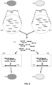
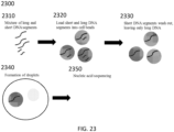
- Method 100 comprises three different phases 110, 120 and 130 that correspond to generation of cell beads comprising a cell or virus or its macromolecular constituent(s) (110); solvent exchange to bring generated partitions into an aqueous phase, cell or virus lysis and denaturation of the cell or virus or macromolecular constituent(s) of the cell or virus (120); and generation of partitions comprising the generated cell beads and barcodes and subsequent tagging (e.g., barcoding) (130).
- a cell e.g., a fixed cell, a cross-linked cell
- Method 100 comprises three different phases 110, 120 and 130 that correspond to generation of cell beads comprising a cell or virus or its macromolecular constituent(s) (110); solvent exchange to bring generated partitions into an aqueous phase, cell or virus lysis and denaturation of the cell or virus or macromolecular constituent(s) of the cell or virus (120); and generation of partitions comprising the generated cell beads and barcodes and subsequent tagging (e.g., barcoding) (
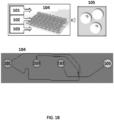
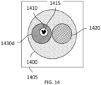
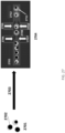
- an oil 101, polymeric or gel precursors 102 and cells 103 are provided to a microfluidic chip 104.
- a photograph of an example microfluidic chip 104 is shown in FIG. 1B .
- the microfluidic chip 104 comprises a plurality of reservoirs for the oil 101, polymeric or gel precursors 102 and cell or virus reagents 103.
- Polymeric or gel precursors 102 and cell or virus reagents 103 are flowed (e.g., via the action of an applied force, such as negative pressure via a vacuum or positive pressure via a pump) from their reservoirs to a first channel junction at which point they combine to form an aqueous stream.
- This aqueous stream is then flowed to a second channel junction, to which oil 101 is also provided.
- the aqueous stream provided from the first channel junction is immiscible with the oil 101 resulting in the generation of a suspension of aqueous droplets in the oil which then flow to reservoir 105 and represent the product 105 from the microfluidic process.
- Flow can be controlled within the microfluidic chip 104 via any suitable strategy, including the use of one or more flow regulators in a channel or various channels, dimensioning of microfluidic channels, etc.
- the product comprises droplets 105 comprising a cell from the cells 103 and polymeric or gel precursors 102.
- the droplets 105 are then subjected to conditions suitable to polymerize or gel the polymeric or gel precursors 102 in the droplets 105, which generates cell beads 106 that encapsulate the cell or virus reagents 103 (e.g., a cell, a fixed cell, a cross-linked cell, component(s) or a cell) in the droplets 105.
- the cell or virus reagents 103 e.g., a cell, a fixed cell, a cross-linked cell, component(s) or a cell
- phase 120 is initiated which includes solvent exchange 111 to resuspend the cell beads 106 in an aqueous phase. Additional details and examples regarding solvent exchange are provided elsewhere herein.
- the resuspended cell beads 106 can then, in bulk, be subjected conditions suitable to lyse cells or viruses associated with the cell beads 106 and, separately or contemporaneously, also subjected, in bulk, to conditions to denature nucleic acids derived from the cells or viruses associated with the cell beads 106.
- the polymeric matrix of the cell beads 106 effectively hinders or prohibits diffusion of larger molecules, such as nucleic acids, from the cell beads 106.
- the cell beads 106 are sufficiently porous to denaturation agents that permit denaturation of trapped nucleic acids within the cell beads 106.
- the cell beads can then be subjected, in bulk, to conditions suitable for performing one or more reactions on nucleic acids derived from the cells or viruses associated with the cell beads 106. Additional details and examples regarding reactions on nucleic acids are provided elsewhere herein.
- the resulting cell beads 113 are then collected 114 and can be stored prior to initiation of phase 130.
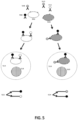
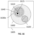
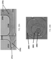
- droplets comprising the cell beads 113 and barcode beads (e.g., gel beads) 122 comprising barcode sequences are generated.
- an oil 121, the cell beads 113 and barcode beads 122 each comprising a barcode sequence (e.g., each bead comprising a unique barcode sequence) are provided to a microfluidic chip 123.
- a photograph of an example microfluidic chip 123 is shown in FIG. 1C .
- the microfluidic chip 123 comprises a plurality of reservoirs for the oil 121, cell beads 113 and barcode beads 122.
- the chip also includes additional reservoirs 127 and 128 that may be used to supply additional reagents (e.g., reagents for nucleic acid amplification, reagents that can degrade or dissolve cell beads 113 and/or barcode beads 122, reagents that degrade linkages between barcodes and barcode beads 122, etc.) to phase 130.
- Cell beads 113 and barcode beads 122 are flowed (e.g., via the action of an applied force, such as negative pressure via a vacuum or positive pressure via a pump) from their reservoirs to a first channel junction at which point they combine to form an aqueous mixture. Materials from reservoirs 127 and 128 can also be provided to the mixture at the first channel junction.
- cell beads and barcode beads can be mixed before introduction into the microfluidic chip.
- a single reservoir of the microfluidic chip 123 comprises a mixture of cell beads and barcode beads.
- the ratio of cell beads to barcode beads in the mixture can be varied to alter the number of droplets generated that comprise a single cell bead and a single barcode bead.
- the mixture of cell beads and barcode beads may be flowed (e.g., via the action of an applied force, such as negative pressure via a vacuum or positive pressure via a pump) from the reservoir to a first channel junction, in some cases together with materials from reservoirs 127 and/or 128.
- cells may be mixed with barcode beads.
- a collection of cells and cell beads may be mixed with barcode beads, or a collection of cells may be mixed with barcode beads.
- the mixture comprising cell beads (or cells), barcode beads, and in some cases additional reagents is then flowed to a second channel junction, to which oil 121 is also provided.
- the aqueous mixture provided from the first channel junction is immiscible with the oil 121 resulting in the generation of a suspension of aqueous droplets 125 in the oil which then flow to reservoir 125 and represent the product from the microfluidic process.
- the microfluidic chip can also include a reservoir 129 that can accept excess oil from the stream emerging from the second channel. Flow can be controlled within the microfluidic chip 123 via any suitable strategy, including the use of one or more flow regulators (see FIGS.
- the product comprises droplets 125 comprising a cell bead 113 and a barcode bead 122, in addition to any other reagents provided by reservoirs 127 and 128.
- a given droplet of the droplets 125 comprises a single cell bead and a single barcode bead.
- reagents that degrade or dissolve the cell beads 113, barcoded beads 122 and/or linkages between barcodes and barcode beads 122 are present in droplets, these reagents can release the nucleic acids trapped in the cell beads 113 from the cell beads 113 and release the barcodes from the barcode beads 122.
- the released barcodes can then interact with the released nucleic acids to generate barcoded constructs for nucleic acid sequencing as described elsewhere herein.
- a given droplet comprises a single cell bead and a single barcode bead comprising oligonucleotides having a common barcode sequence
- a given sequencing construct generated from the given droplet 125 can be associated with the cell or virus of the given cell bead via its barcode sequence.
- FIG. 1D photographically depicts two example runs demonstrating the generation of droplets 125 comprising cell beads and barcode beads using the example method shown in FIG. 1A and microfluidic devices depicted in FIGS. 1B and 1C .
- FIG. 1D panel A
- droplets comprising cell beads and barcode beads are shown
- FIG. 1D panel B
- droplets comprising cell beads comprising magnetic materials and barcode beads are shown.
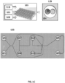
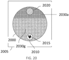
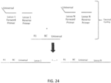
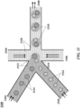
- FIG. 31 shows an example of a microfluidic channel structure 3100 for delivering barcode carrying beads to droplets.
- the channel structure 3100 can include channel segments 3101, 3102, 3104, 3106 and 3108 communicating at a channel junction 3110.
- the channel segment 3101 may transport an aqueous fluid 3112 that includes a plurality of beads 3114 (e.g., with nucleic acid molecules, oligonucleotides, molecular tags) along the channel segment 3101 into junction 3110.
- the plurality of beads 3114 may be sourced from a suspension of beads.
- the channel segment 3101 may be connected to a reservoir comprising an aqueous suspension of beads 3114.
- the channel segment 3102 may transport the aqueous fluid 3112 that includes a plurality of cell beads 3116 along the channel segment 3102 into junction 3110.
- the plurality of cell beads 3116 may be sourced from a suspension of cell beads.
- the channel segment 3102 may be connected to a reservoir comprising an aqueous suspension of cell beads 3116.
- the aqueous fluid 3112 in either the first channel segment 3101 or the second channel segment 3102, or in both segments can include one or more reagents, as further described below.
- a second fluid 3118 that is immiscible with the aqueous fluid 3112 e.g., oil
- the aqueous fluid 3112 Upon meeting of the aqueous fluid 3112 from each of channel segments 3101 and 3102 and the second fluid 3118 from each of channel segments 3104 and 3106 at the channel junction 3110, the aqueous fluid 3112 can be partitioned as discrete droplets 3120 in the second fluid 3118 and flow away from the junction 3110 along channel segment 3108.
- the channel segment 3108 may deliver the discrete droplets to an outlet reservoir fluidly coupled to the channel segment 3108, where they may be harvested.
- the channel segments 3101 and 3102 may meet at another junction upstream of the junction 3110.
- beads and cell beads may form a mixture that is directed along another channel to the junction 3110 to yield droplets 3120.
- the mixture may provide the beads and cell beads in an alternating fashion, such that, for example, a droplet comprises a single bead and a single cell bead.
- Beads, cell beads and droplets may flow along channels at substantially regular flow profiles (e.g., at regular flow rates). Such regular flow profiles may permit a droplet to include a single bead and a single cell bead. Such regular flow profiles may permit the droplets to have an occupancy (e.g., droplets having beads and cell beads) greater than 5%, 10%, 20%, 30%, 40%, 50%, 60%, 70%, 80%, 90%, or 95%.
- occupancy e.g., droplets having beads and cell beads
- the second fluid 3118 can comprise an oil, such as a fluorinated oil, that includes a fluorosurfactant for stabilizing the resulting droplets, for example, inhibiting subsequent coalescence of the resulting droplets 3120.
- an oil such as a fluorinated oil, that includes a fluorosurfactant for stabilizing the resulting droplets, for example, inhibiting subsequent coalescence of the resulting droplets 3120.
- a discrete droplet that is generated may include an individual cell bead 3116.
- a discrete droplet that is generated may include a barcode or other reagent carrying bead 3114.
- a discrete droplet generated may include both an individual cell bead and a barcode carrying bead, such as droplets 3120.
- a discrete droplet may include more than one individual cell bead or no cell bead.
- a discrete droplet may include more than one bead or no bead.
- a discrete droplet may be unoccupied (e.g., no beads, no cell beads).
- a discrete droplet partitioning a cell bead and a barcode carrying bead may effectively allow the attribution of the barcode to macromolecular constituents of the cell bead within the partition.
- the contents of a partition may remain discrete from the contents of other partitions.
- the channel segments described herein may be coupled to any of a variety of different fluid sources or receiving components, including reservoirs, tubing, manifolds, or fluidic components of other systems.
- the microfluidic channel structure 3100 may have other geometries.
- a microfluidic channel structure can have more than one channel junctions.
- a microfluidic channel structure can have 2, 3, 4, or 5 channel segments each carrying beads that meet at a channel junction.
- Fluid may be directed flow along one or more channels or reservoirs via one or more fluid flow units.
- a fluid flow unit can comprise compressors (e.g., providing positive pressure), pumps (e.g., providing negative pressure), actuators, and the like to control flow of the fluid. Fluid may also or otherwise be controlled via applied pressure differentials, centrifugal force, electrokinetic pumping, vacuum, capillary or gravity flow, or the like.
- a partition may be a droplet.
- the droplet may be formed by bringing a first phase in contact with a second phase that is immiscible with the first phase.
- the partition may be a well as part of a plurality of wells.
- the partition may be a chamber as part of a plurality of chambers. Partitions may be fluidically isolated from one another.
- barcoded oligonucleotides are delivered to a partition via a microcapsule, such as a bead (e.g., gel bead) or a droplet.
- a microcapsule such as a bead (e.g., gel bead) or a droplet.
- barcoded oligonucleotides are initially associated with the microcapsule and then released from the microcapsule upon application of a stimulus which allows the oligonucleotides to dissociate or to be released from the microcapsule.
- a microcapsule in some embodiments, comprises a bead, such as a droplet comprising the bead.
- the microcapsule can be a bead (e.g., gel bead).
- a bead may be porous, non-porous, solid, semi-solid, semi-fluidic, or fluidic.
- a bead may be dissolvable, disruptable, or degradable. In some cases, a bead may not be degradable.
- the bead may be a solid or semi-solid particle.
- the bead may be a gel bead.
- a gel bead may be a hydrogel bead.
- a gel bead may be formed from molecular precursors, such as a polymeric or monomeric species.
- a semi-solid bead may be a liposomal bead.
- Solid beads may comprise metals including iron oxide, gold, and silver.
- the beads are silica beads.
- the beads are rigid.
- the beads may be flexible and/or compressible.
- the bead may contain molecular precursors (e.g., monomers or polymers), which may form a polymer network via polymerization of the precursors.
- a precursor may be an already polymerized species capable of undergoing further polymerization via, for example, a chemical cross-linkage.
- a precursor comprises one or more of an acrylamide or a methacrylamide monomer, oligomer, or polymer.
- the bead may comprise prepolymers, which are oligomers capable of further polymerization.
- polyurethane beads may be prepared using prepolymers.
- the bead may contain individual polymers that may be further polymerized together.
- beads may be generated via polymerization of different precursors, such that they comprise mixed polymers, co-polymers, and/or block co-polymers.
- a bead may comprise natural and/or synthetic materials.
- a polymer can be a natural polymer or a synthetic polymer.
- a bead comprises both natural and synthetic polymers.
- natural polymers include proteins and sugars such as deoxyribonucleic acid, rubber, cellulose, starch (e.g., amylose, amylopectin), proteins, enzymes, polysaccharides, silks, polyhydroxyalkanoates, chitosan, dextran, collagen, carrageenan, ispaghula, acacia, agar, gelatin, shellac, sterculia gum, xanthan gum, Corn sugar gum, guar gum, gum karaya, agarose, alginic acid, alginate, or natural polymers thereof.
- proteins and sugars such as deoxyribonucleic acid, rubber, cellulose, starch (e.g., amylose, amylopectin), proteins, enzymes, polysaccharides, silk
- Examples of synthetic polymers include acrylics, nylons, silicones, spandex, viscose rayon, polycarboxylic acids, polyvinyl acetate, polyacrylamide, polyacrylate, polyethylene glycol, polyurethanes, polylactic acid, silica, polystyrene, polyacrylonitrile, polybutadiene, polycarbonate, polyethylene, polyethylene terephthalate, poly(chlorotrifluoroethylene), poly(ethylene oxide), polyethylene terephthalate), polyethylene, polyisobutylene, poly(methyl methacrylate), poly(oxymethylene), polyformaldehyde, polypropylene, polystyrene, poly(tetrafluoroethylene), poly(vinyl acetate), poly(vinyl alcohol), poly(vinyl chloride), poly(vinylidene dichloride), poly(vinylidene difluoride), poly(vinyl fluoride) and combinations (e.g., co-polymers) thereof. Bead
- a chemical cross-linker may be a precursor used to cross-link monomers during polymerization of the monomers and/or may be used to attach oligonucleotides (e.g., barcoded oligonucleotides) to the bead.
- polymers may be further polymerized with a cross-linker species or other type of monomer to generate a further polymeric network.
- Non-limiting examples of chemical cross-linkers include cystamine, gluteraldehyde, dimethyl suberimidate, N-Hydroxysuccinimide crosslinker BS3, formaldehyde, carbodiimide (EDC), SMCC, Sulfo-SMCC, vinylsilane, N,N'diallyltartardiamide (DATD), N,N'-Bis(acryloyl)cystamine (BAC), or homologs thereof.
- the crosslinker used in the present disclosure contains cystamine.
- Crosslinking may be permanent or reversible, depending upon the particular crosslinker used. Reversible crosslinking may allow for the polymer to linearize or dissociate under appropriate conditions. In some cases, reversible cross-linking may also allow for reversible attachment of a material bound to the surface of a bead. In some cases, a cross-linker may form disulfide linkages. In some cases, the chemical cross-linker forming disulfide linkages may be cystamine or a modified cystamine.
- disulfide linkages can be formed between molecular precursor units (e.g., monomers, oligomers, or linear polymers) or precursors incorporated into a bead and oligonucleotides.
- Cystamine (including modified cystamines), for example, is an organic agent comprising a disulfide bond that may be used as a crosslinker agent between individual monomeric or polymeric precursors of a bead.
- Polyacrylamide may be polymerized in the presence of cystamine or a species comprising cystamine (e.g., a modified cystamine) to generate polyacrylamide gel beads comprising disulfide linkages (e.g., chemically degradable beads comprising chemically-reducible cross-linkers).
- the disulfide linkages may permit the bead to be degraded (or dissolved) upon exposure of the bead to a reducing agent.
- chitosan a linear polysaccharide polymer
- glutaraldehyde via hydrophilic chains to form a bead.
- Crosslinking of chitosan polymers may be achieved by chemical reactions that are initiated by heat, pressure, change in pH, and/or radiation.
- the bead may comprise covalent or ionic bonds between polymeric precursors (e.g., monomers, oligomers, linear polymers), oligonucleotides, primers, and other entities.
- polymeric precursors e.g., monomers, oligomers, linear polymers
- oligonucleotides e.g., primers, and other entities.
- the covalent bonds comprise carbon-carbon bonds or thioether bonds.
- a bead may comprise an acrydite moiety, which in certain aspects may be used to attach one or more oligonucleotides (e.g., barcode sequence, barcoded oligonucleotide, primer, or other oligonucleotide) to the bead.
- an acrydite moiety can refer to an acrydite analogue generated from the reaction of acrydite with one or more species, such as, the reaction of acrydite with other monomers and cross-linkers during a polymerization reaction.
- Acrydite moieties may be modified to form chemical bonds with a species to be attached, such as an oligonucleotide (e.g., barcode sequence, barcoded oligonucleotide, primer, or other oligonucleotide).
- Acrydite moieties may be modified with thiol groups capable of forming a disulfide bond or may be modified with groups already comprising a disulfide bond.
- the thiol or disulfide via disulfide exchange) may be used as an anchor point for a species to be attached or another part of the acrydite moiety may be used for attachment.
- attachment is reversible, such that when the disulfide bond is broken (e.g., in the presence of a reducing agent), the attached species is released from the bead.
- an acrydite moiety comprises a reactive hydroxyl group that may be used for attachment.
- Functionalization of beads for attachment of oligonucleotides may be achieved through a wide range of different approaches, including activation of chemical groups within a polymer, incorporation of active or activatable functional groups in the polymer structure, or attachment at the pre-polymer or monomer stage in bead production.
- precursors e.g., monomers, cross-linkers
- precursors that are polymerized to form a bead may comprise acrydite moieties, such that when a bead is generated, the bead also comprises acrydite moieties.
- the acrydite moieties can be attached to an oligonucleotide, such as a primer (e.g., a primer for amplifying target nucleic acids, barcoded oligonucleotide, etc) to be incorporated into the bead.
- the primer comprises a P5 sequence for attachment to a sequencing flow cell for Illumina sequencing.
- the primer comprises a P7 sequence for attachment to a sequencing flow cell for Illumina sequencing.
- the primer comprises a barcode sequence. In some cases, the primer further comprises a unique molecular identifier (UMI). In some cases, the primer comprises an R1 primer sequence for Illumina sequencing. In some cases, the primer comprises an R2 primer sequence for Illumina sequencing.
- UMI unique molecular identifier
- precursors comprising a functional group that is reactive or capable of being activated such that it becomes reactive can be polymerized with other precursors to generate gel beads comprising the activated or activatable functional group.
- the functional group may then be used to attach additional species (e.g., disulfide linkers, primers, other oligonucleotides, etc.) to the gel beads.
- additional species e.g., disulfide linkers, primers, other oligonucleotides, etc.
- some precursors comprising a carboxylic acid (COOH) group can co-polymerize with other precursors to form a gel bead that also comprises a COOH functional group.
- acrylic acid a species comprising free COOH groups
- acrylamide acrylamide
- bis(acryloyl)cystamine can be co-polymerized together to generate a gel bead comprising free COOH groups.
- the COOH groups of the gel bead can be activated (e.g., via 1-Ethyl-3-(3-dimethylaminopropyl)carbodiimide (EDC) and N-Hydroxysuccinimide (NHS) or 4-(4,6-Dimethoxy-1,3,5-triazin-2-yl)-4-methylmorpholinium chloride (DMTMM)) such that they are reactive (e.g., reactive to amine functional groups where EDC/NHS or DMTMM are used for activation).
- EDC 1-Ethyl-3-(3-dimethylaminopropyl)carbodiimide
- NHS N-Hydroxysuccinimide
- DTMM 4-(4,6-Dimethoxy
- the activated COOH groups can then react with an appropriate species (e.g., a species comprising an amine functional group where the carboxylic acid groups are activated to be reactive with an amine functional group) comprising a moiety to be linked to the bead.
- an appropriate species e.g., a species comprising an amine functional group where the carboxylic acid groups are activated to be reactive with an amine functional group
- Beads comprising disulfide linkages in their polymeric network may be functionalized with additional species via reduction of some of the disulfide linkages to free thiols.
- the disulfide linkages may be reduced via, for example, the action of a reducing agent (e.g., DTT, TCEP, etc.) to generate free thiol groups, without dissolution of the bead.
- Free thiols of the beads can then react with free thiols of a species or a species comprising another disulfide bond (e.g., via thiol-disulfide exchange) such that the species can be linked to the beads (e.g., via a generated disulfide bond).
- free thiols of the beads may react with any other suitable group.
- free thiols of the beads may react with species comprising an acrydite moiety.
- the free thiol groups of the beads can react with the acrydite via Michael addition chemistry, such that the species comprising the acrydite is linked to the bead.
- uncontrolled reactions can be prevented by inclusion of a thiol capping agent such as N-ethylmalieamide or iodoacetate.
- Activation of disulfide linkages within a bead can be controlled such that a small number of disulfide linkages are activated. Control may be exerted, for example, by controlling the concentration of a reducing agent used to generate free thiol groups and/or concentration of reagents used to form disulfide bonds in bead polymerization. In some cases, a low concentration (e.g., molecules of reducing agent:gel bead ratios of less than about 10,000, less than about 100,000, less than about 1,000,000, less than about 10,000,000, less than about 100,000,000, less than about 1,000,000,000, less than about 10,000,000,000, or less than about 100,000,000,000) of reducing agent may be used for reduction.
- a low concentration e.g., molecules of reducing agent:gel bead ratios of less than about 10,000, less than about 100,000, less than about 1,000,000, less than about 10,000,000, less than about 100,000,000, less than about 1,000,000,000, less than about 10,000,000,000, or less than about 100,000,000,000
- Controlling the number of disulfide linkages that are reduced to free thiols may be useful in ensuring bead structural integrity during functionalization.
- optically-active agents such as fluorescent dyes may be may be coupled to beads via free thiol groups of the beads and used to quantify the number of free thiols present in a bead and/or track a bead.
- addition of moieties to a gel bead after gel bead formation may be advantageous.
- addition of an oligonucleotide (e.g., barcoded oligonucleotide) after gel bead formation may avoid loss of the species during chain transfer termination that can occur during polymerization.
- smaller precursors e.g., monomers or cross linkers that do not comprise side chain groups and linked moieties
- functionalization after gel bead synthesis can minimize exposure of species (e.g., oligonucleotides) to be loaded with potentially damaging agents (e.g., free radicals) and/or chemical environments.
- the generated gel may possess an upper critical solution temperature (UCST) that can permit temperature driven swelling and collapse of a bead.
- UCT upper critical solution temperature
- Such functionality may aid in oligonucleotide (e.g., a primer) infiltration into the bead during subsequent functionalization of the bead with the oligonucleotide.
- Post-production functionalization may also be useful in controlling loading ratios of species in beads, such that, for example, the variability in loading ratio is minimized.
- Species loading may also be performed in a batch process such that a plurality of beads can be functionalized with the species in a single batch.
- beads can be non-covalently loaded with one or more reagents.
- the beads can be non-covalently loaded by, for instance, subjecting the beads to conditions sufficient to swell the beads, allowing sufficient time for the reagents to diffuse into the interiors of the beads, and subjecting the beads to conditions sufficient to de-swell the beads.
- the swelling of the beads may be accomplished, for instance, by placing the beads in a thermodynamically favorable solvent, subjecting the beads to a higher or lower temperature or temperature change, subjecting the beads to a higher or lower ion concentration, and/or subjecting the beads to an electric field.
- the de-swelling of the beads may be accomplished, for instance, by transferring the beads in a thermodynamically unfavorable solvent, subjecting the beads to a lower or high temperature or temperature change different from that use to swell the beads, subjecting the beads to a lower or higher ion concentration different from that used to swell the beads, and/or removing the electric field.
- Transferring the beads may cause pores in the beads to shrink. Such shrinking may then hinder reagents within the beads from diffusing out of the interiors of the beads. The hindrance may be due to steric interactions between the reagents and the interiors of the beads.
- the transfer may be accomplished microfluidically. For instance, the transfer may be achieved by moving the beads from one co-flowing solvent stream to a different co-flowing solvent stream. The swellability and/or pore size of the beads may be adjusted by changing the polymer composition of the bead.
- an acrydite moiety linked to precursor, another species linked to a precursor, or a precursor itself comprises a labile bond, such as chemically, thermally, or photosensitive bonds e.g., disulfide bonds, UV sensitive bonds, or the like.
- a labile bond such as chemically, thermally, or photosensitive bonds e.g., disulfide bonds, UV sensitive bonds, or the like.
- the bead may also comprise the labile bond.
- the labile bond may be, for example, useful in reversibly linking (e.g., covalently linking) species (e.g., barcodes, primers, etc.) to a bead.
- a thermally labile bond may include a nucleic acid hybridization based attachment, e.g., where an oligonucleotide is hybridized to a complementary sequence that is attached to the bead, such that thermal melting of the hybrid releases the oligonucleotide, e.g., a barcode containing sequence, from the bead or microcapsule.
- a nucleic acid hybridization based attachment e.g., where an oligonucleotide is hybridized to a complementary sequence that is attached to the bead, such that thermal melting of the hybrid releases the oligonucleotide, e.g., a barcode containing sequence, from the bead or microcapsule.
- labile bonds may result in the generation of a bead capable of responding to varied stimuli.
- Each type of labile bond may be sensitive to an associated stimulus (e.g., chemical stimulus, light, temperature, etc.) such that release of species attached to a bead via each labile bond may be controlled by the application of the appropriate stimulus.
- Such functionality may be useful in controlled release of species from a gel bead.
- another species comprising a labile bond may be linked to a gel bead after gel bead formation via, for example, an activated functional group of the gel bead as described above.
- Barcodes that are releasably, cleavably or reversibly attached to the beads described herein include barcodes that are released or releasable through cleavage of a linkage between the barcode molecule and the bead, or that are released through degradation of the underlying bead itself, allowing the barcodes to be accessed or accessible by other reagents, or both.
- the barcodes that are releasable as described herein may sometimes be referred to as being activatable, in that they are available for reaction once released.
- an activatable barcode may be activated by releasing the barcode from a bead (or other suitable type of partition described herein).
- Other activatable configurations are also envisioned in the context of the described methods and systems.
- labile bonds that may be coupled to a precursor or bead include an ester linkage (e.g., cleavable with an acid, a base, or hydroxylamine), a vicinal diol linkage (e.g., cleavable via sodium periodate), a Diels-Alder linkage (e.g., cleavable via heat), a sulfone linkage (e.g., cleavable via a base), a silyl ether linkage (e.g., cleavable via an acid), a glycosidic linkage (e.g., cleavable via an amylase), a peptide linkage (e.g., cleavable via a protease), or a phosphodiester linkage (e.g., cleavable via a nuclease (
- Species that do not participate in polymerization may also be encapsulated in beads during bead generation (e.g., during polymerization of precursors). Such species may be entered into polymerization reaction mixtures such that generated beads comprise the species upon bead formation. In some cases, such species may be added to the gel beads after formation.
- species may include, for example, oligonucleotides, reagents for a nucleic acid amplification reaction (e.g., primers (e.g. random primers, primers specific for a given DNA loci), polymerases, nucleotides (e.g.
- co-factors e.g., ionic co-factors
- reagents for enzymatic reactions e.g., enzymes, co-factors, substrates
- reagents for reverse transcription e.g. oligonucleotide primers or reverse transcriptase
- nucleic acid modification reactions such as polymerization, ligation, digestion, methylation, random mutagenesis, bisulfite conversion, uracil hydrolysis, nucleic acid repair, nucleic acid insertion or cleavage (e.g.
- CRISPR/Cas9-mediated or transposon-mediated insertion or cleavage via CRISPR/Cas9-mediated or transposon-mediated insertion or cleavage), capping, or decapping.
- Trapping of such species may be controlled by the polymer network density generated during polymerization of precursors, control of ionic charge within the gel bead (e.g., via ionic species linked to polymerized species), or by the release of other species.
- Encapsulated species may be released from a bead upon bead degradation and/or by application of a stimulus capable of releasing the species from the bead.
- barcode sequences may also be encapsulated within a bead and, in some cases, can be released from a bead via bead degradation and/or by application of a stimulus capable of releasing the species from the bead.
- Beads may be of uniform size or heterogeneous size.
- the diameter of a bead may be about 1 ⁇ m, 5 ⁇ m, 10 ⁇ m, 20 ⁇ m, 30 ⁇ m, 40 ⁇ m, 50 ⁇ m, 60 ⁇ m, 70 ⁇ m, 80 ⁇ m, 90 ⁇ m, 100 ⁇ m, 250 ⁇ m, 500 ⁇ m, or 1mm.
- a bead may have a diameter of at least about 1 ⁇ m, 5 ⁇ m, 10 ⁇ m, 20 ⁇ m, 30 ⁇ m, 40 ⁇ m, 50 ⁇ m, 60 ⁇ m, 70 ⁇ m, 80 ⁇ m, 90 ⁇ m, 100 ⁇ m, 250 ⁇ m, 500 ⁇ m, 1mm, or more.
- a bead may have a diameter of less than about 1 ⁇ m, 5 ⁇ m, 10 ⁇ m, 20 ⁇ m, 30 ⁇ m, 40 ⁇ m, 50 ⁇ m, 60 ⁇ m, 70 ⁇ m, 80 ⁇ m, 90 ⁇ m, 100 ⁇ m, 250 ⁇ m, 500 ⁇ m, or 1mm. In some cases, a bead may have a diameter in the range of about 40-75 ⁇ m, 30-75 ⁇ m, 20-75 ⁇ m, 40-85 ⁇ m, 40-95 ⁇ m, 20-100 ⁇ m, 10-100 ⁇ m, 1-100 ⁇ m, 20-250 ⁇ m, or 20-500 ⁇ m.
- beads are provided as a population or plurality of beads having a relatively monodisperse size distribution.
- maintaining relatively consistent bead characteristics, such as size can contribute to the overall consistency.
- the beads described herein may have size distributions that have a coefficient of variation in their cross-sectional dimensions of less than 50%, less than 40%, less than 30%, less than 20%, and in some cases less than 15%, less than 10%, or less than 5%.
- Beads may be of any suitable shape. Examples of bead shapes include, but are not limited to, spherical, non-spherical, oval, oblong, amorphous, circular, cylindrical, and variations thereof.
- the beads may be degradable, disruptable, or dissolvable spontaneously or upon exposure to one or more stimuli (e.g., temperature changes, pH changes, exposure to particular chemical species or phase, exposure to light, reducing agent, etc.).
- a bead may be dissolvable, such that material components of the beads are solubilized when exposed to a particular chemical species or an environmental change, such as a change temperature or a change in pH.
- a gel bead is degraded or dissolved at elevated temperature and/or in basic conditions.
- a bead may be thermally degradable such that when the bead is exposed to an appropriate change in temperature (e.g., heat), the bead degrades.
- Degradation or dissolution of a bead bound to a species e.g., a oligonucleotide, e.g., barcoded oligonucleotide
- a species e.g., a oligonucleotide, e.g., barcoded oligonucleotide
- a degradable bead may comprise one or more species with a labile bond such that, when the bead/species is exposed to the appropriate stimuli, the bond is broken and the bead degrades.
- the labile bond may be a chemical bond (e.g., covalent bond, ionic bond) or may be another type of physical interaction (e.g., van der Waals interactions, dipole-dipole interactions, etc.).
- a crosslinker used to generate a bead may comprise a labile bond.
- the labile bond can be broken and the bead degraded. For example, upon exposure of a polyacrylamide gel bead comprising cystamine crosslinkers to a reducing agent, the disulfide bonds of the cystamine can be broken and the bead degraded.
- a degradable bead may be useful in more quickly releasing an attached species (e.g., an oligonucleotide, a barcode sequence, a primer, etc) from the bead when the appropriate stimulus is applied to the bead as compared to a bead that does not degrade.
- an attached species e.g., an oligonucleotide, a barcode sequence, a primer, etc
- the species may have greater mobility and accessibility to other species in solution upon degradation of the bead.
- a species may also be attached to a degradable bead via a degradable linker (e.g., disulfide linker).
- the degradable linker may respond to the same stimuli as the degradable bead or the two degradable species may respond to different stimuli.
- a barcode sequence may be attached, via a disulfide bond, to a polyacrylamide bead comprising cystamine.
- the bead Upon exposure of the barcoded-bead to a reducing agent, the bead degrades and the barcode sequence is released upon breakage of both the disulfide linkage between the barcode sequence and the bead and the disulfide linkages of the cystamine in the bead.
- a degradable bead may be introduced into a partition, such as a droplet of an emulsion or a well, such that the bead degrades within the partition and any associated species (e.g., oligonucleotides) are released within the droplet when the appropriate stimulus is applied.
- the free species e.g., oligonucleotides
- a polyacrylamide bead comprising cystamine and linked, via a disulfide bond, to a barcode sequence, may be combined with a reducing agent within a droplet of a water-in-oil emulsion.
- the reducing agent breaks the various disulfide bonds resulting in bead degradation and release of the barcode sequence into the aqueous, inner environment of the droplet.
- heating of a droplet comprising a bead-bound barcode sequence in basic solution may also result in bead degradation and release of the attached barcode sequence into the aqueous, inner environment of the droplet.
- degradation may refer to the disassociation of a bound or entrained species from a bead, both with and without structurally degrading the physical bead itself.
- entrained species may be released from beads through osmotic pressure differences due to, for example, changing chemical environments.
- alteration of bead pore sizes due to osmotic pressure differences can generally occur without structural degradation of the bead itself.
- an increase in pore size due to osmotic swelling of a bead can permit the release of entrained species within the bead.
- osmotic shrinking of a bead may cause a bead to better retain an entrained species due to pore size contraction.
- degradable beads are provided, it may helpful to avoid exposing such beads to the stimulus or stimuli that cause such degradation prior to the requisite time, in order to avoid premature bead degradation and issues that arise from such degradation, including for example poor flow characteristics and aggregation.
- beads comprise reducible cross-linking groups, such as disulfide groups
- reducing agents e.g., DTT or other disulfide cleaving reagents.
- treatment to the beads described herein will, in some cases be provided free of reducing agents, such as DTT.
- reducing agent free (or DTT free) enzyme preparations in treating the beads described herein.
- examples of such enzymes include, e.g., polymerase enzyme preparations, reverse transcriptase enzyme preparations, ligase enzyme preparations, as well as many other enzyme preparations that may be used to treat the beads described herein.
- the terms "reducing agent free” or "DTT free” preparations can refer to a preparation having less than 1/10th, less than 1/50th, and even less than 1/100th of the lower ranges for such materials used in degrading the beads.
- the reducing agent free preparation will typically have less than 0.01 mM, 0.005 mM, 0.001 mM DTT, 0.0005 mM DTT, or even less than 0.0001 mM DTT. In many cases, the amount of DTT will be undetectable.
- Numerous chemical triggers may be used to trigger the degradation of beads. Examples of these chemical changes may include, but are not limited to pH-mediated changes to the integrity of a component within the bead, degradation of a component of a bead via cleavage of cross-linked bonds, and depolymerization of a component of a bead.
- a bead may be formed from materials that comprise degradable chemical crosslinkers, such as BAC or cystamine. Degradation of such degradable crosslinkers may be accomplished through a number of mechanisms.
- a bead may be contacted with a chemical degrading agent that may induce oxidation, reduction or other chemical changes.
- a chemical degrading agent may be a reducing agent, such as dithiothreitol (DTT).
- reducing agents may include ⁇ -mercaptoethanol, (2S)-2-amino-1,4-dimercaptobutane (dithiobutylamine or DTBA), tris(2-carboxyethyl) phosphine (TCEP), or combinations thereof.
- a reducing agent may degrade the disulfide bonds formed between gel precursors forming the bead, and thus, degrade the bead.
- a change in pH of a solution such as an increase in pH, may trigger degradation of a bead.
- exposure to an aqueous solution, such as water may trigger hydrolytic degradation, and thus degradation of the bead.
- Beads may also be induced to release their contents upon the application of a thermal stimulus.
- a change in temperature can cause a variety of changes to a bead. For example, heat can cause a solid bead to liquefy. A change in heat may cause melting of a bead such that a portion of the bead degrades. In other cases, heat may increase the internal pressure of the bead components such that the bead ruptures or explodes. Heat may also act upon heat-sensitive polymers used as materials to construct beads.
- a chemical degrading agent may be a reducing agent, such as DTT, wherein DTT may degrade the disulfide bonds formed between a crosslinker and gel precursors, thus degrading the bead.
- a reducing agent may be added to degrade the bead, which may or may not cause the bead to release its contents.
- reducing agents may include dithiothreitol (DTT), ⁇ -mercaptoethanol, (2S)-2-amino-1,4-dimercaptobutane (dithiobutylamine or DTBA), tris(2-carboxyethyl) phosphine (TCEP), or combinations thereof.
- the reducing agent may be present at a concentration of about 0.1mM, 0.5mM, 1mM, 5mM, or 10mM.
- the reducing agent may be present at a concentration of at least about 0.1mM, 0.5mM, 1mM, 5mM, 10mM, or greater than 10 mM.
- the reducing agent may be present at a concentration of at most about 0.1mM, 0.5mM, 1mM, 5mM, or 10mM.
- barcode molecules e.g., primer, e.g., barcoded oligonucleotide
- primer e.g., barcoded oligonucleotide
- the barcode molecules e.g., primer, e.g., barcoded oligonucleotide
- pre-defined concentration may be selected to facilitate certain reactions for generating a sequencing library, e.g., amplification, within the partition.
- the pre-defined concentration of the primer is limited by the process of producing oligonucleotide bearing beads.
- the compartments or partitions can comprise partitions that are flowable within fluid streams. These partitions may comprise, e.g., micro-vesicles that have an outer barrier surrounding an inner fluid center or core, or, in some cases, they may comprise a porous matrix that is capable of entraining and/or retaining materials within its matrix. Partitions can comprise droplets of aqueous fluid within a non-aqueous continuous phase, e.g., an oil phase. A variety of different vessels are described in, for example, U.S. Patent Application Publication No. 2014/0155295 . Emulsion systems for creating stable droplets in non-aqueous or oil continuous phases are described in detail in, e.g., U.S. Patent Application Publication No. 2010/0105112 .
- allocating individual cell beads to discrete partitions may generally be accomplished by introducing a flowing stream of cell beads in an aqueous fluid into a flowing stream of a non-aqueous fluid, such that droplets are generated at the junction of the two streams.
- the occupancy of the resulting partitions e.g., number of cell beads per partition
- the relative flow rates of the fluids can be selected such that, on average, the partitions contain less than one cell bead per partition, in order to ensure that those partitions that are occupied, are primarily singly occupied.
- the relative flow rates of the fluids can be selected such that a majority of partitions are occupied, e.g., allowing for a small percentage of unoccupied partitions.
- the flows and channel architectures can be controlled as to ensure a requisite number of singly occupied partitions, less than a certain level of unoccupied partitions and less than a certain level of multiply occupied partitions.
- the systems and methods described herein can be operated such that a majority of occupied partitions include no more than one cell bead per occupied partition.
- the partitioning process is conducted such that fewer than 40% of the occupied partitions contain more than one cell bead, fewer than 35% of the occupied partitions contain more than one cell bead, fewer than 30% of the occupied partitions contain more than one cell bead, fewer than 25% of the occupied partitions contain more than one cell bead, fewer than 20% of the occupied partitions contain more than one cell bead, fewer than 15% of the occupied partitions contain more than one cell bead, fewer than 10% of the occupied partitions contain more than one cell bead, or fewer than 5% of the occupied partitions include more than one cell bead per partition.
- the Poissonian distribution may expectedly increase the number of partitions that may include multiple cell beads.
- the flow of one or more of the cell beads, or other fluids directed into a partitioning zone can be manipulated to control occupancy of partitions with cell beads such that no more than 60% of the generated partitions are unoccupied, no more than 50% of the generated partitions are unoccupied, no more than 45% of the generated partitions are unoccupied, no more than 40% of the generated partitions are unoccupied, no more than 35% of the generated partitions are unoccupied, no more than 30% of the generated partitions are unoccupied, no more than 25% of the generated partitions are unoccupied, no more than 20% of the generated partitions are unoccupied, or no more than 10% of the generated partitions are unoccupied.
- These flows can be controlled so as to present non-Poissonian distribution of single occupied partitions while providing lower levels of unoccupied partitions.
- unoccupied partitions can be achieved while still providing any of the single occupancy rates described above.
- the use of the systems and methods described herein creates resulting partitions (e.g., droplets comprising cell beads) that have multiple occupancy rates of less than 50%, less than 45%, less than 40%, less than 35%, less than 30%, less than 25%, less than 20%, less than 15%, less than 10%, less than 5% or less than 1%.
- the above-described occupancy rates are also applicable to partitions that include both cell beads and additional reagents, including, but not limited to, microcapsules or particles (e.g., beads, gel beads) carrying barcoded oligonucleotides.
- the occupied partitions e.g., at least about 10%, 20%, 30%, 40%, 50%, 60%, 70%, 80%, 90%, 95%, or 99% of the occupied partitions
- multiply occupied partitions e.g., containing two, three, four or more cell beads and/or microcapsules (e.g., beads, gel beads) comprising barcoded oligonucleotides within a single partition. Accordingly, as noted above, the flow characteristics of the cell bead and/or bead containing fluids and partitioning fluids may be controlled to provide for such multiply occupied partitions.
- the flow parameters may be controlled to provide a requisite occupancy rate at greater than 50% of the partitions, greater than 55% of the partitions, greater than 60% of the partitions, greater than 65% of the partitions, greater than 70% of the partitions, greater than 75% of the partitions, greater than 80% of the partitions, greater than 85% of the partitions, greater than 90% of the partitions, greater than 95% of the partitions, or higher.
- additional microcapsules are used to deliver additional reagents to a partition.
- the flow and frequency of the different beads into the channel or junction may be controlled to provide for the requisite ratio of microcapsules from each source, while ensuring the requisite pairing or combination of such beads into a partition with the requisite number of cell beads.
- the partitions described herein may comprise small volumes, e.g., less than 10 ⁇ L, less than 5 ⁇ L, less than 1 ⁇ L, less than 900 picoliters (pL), less than 800 pL, less than 700 pL, less than 600 pL, less than 500 pL, less than 400pL, less than 300 pL, less than 200 pL, less than 100pL, less than 50 pL, less than 20 pL, less than 10 pL, less than 1 pL, less than 500 nanoliters (nL), or even less than 100 nL, 50 nL, or even less.
- small volumes e.g., less than 10 ⁇ L, less than 5 ⁇ L, less than 1 ⁇ L, less than 900 picoliters (pL), less than 800 pL, less than 700 pL, less than 600 pL, less than 500 pL, less than 400pL, less than 300 pL, less than 200 pL, less than 100pL, less
- the droplets may have overall volumes that are less than 1000 pL, less than 900 pL, less than 800 pL, less than 700 pL, less than 600 pL, less than 500 pL, less than 400pL, less than 300 pL, less than 200 pL, less than 100pL, less than 50 pL, less than 20 pL, less than 10 pL, or even less than 1 pL.
- the sample fluid volume, e.g., including co-partitioned cell beads, within the partitions may be less than 90% of the above described volumes, less than 80%, less than 70%, less than 60%, less than 50%, less than 40%, less than 30%, less than 20%, or even less than 10% the above described volumes.
- partitioning species may generate a population or plurality of partitions.
- any suitable number of partitions can be generated to generate the plurality of partitions.
- a plurality of partitions may be generated that comprises at least about 1,000 partitions, at least about 5,000 partitions, at least about 10,000 partitions, at least about 50,000 partitions, at least about 100,000 partitions, at least about 500,000 partitions, at least about 1,000,000 partitions, at least about 5,000,000 partitions at least about 10,000,000 partitions, at least about 50,000,000 partitions, at least about 100,000,000 partitions, at least about 500,000,000 partitions or at least about 1,000,000,000 partitions.
- the plurality of partitions may comprise both unoccupied partitions (e.g., empty partitions) and occupied partitions.
- FIG. 2 shows an example of a simplified microfluidic channel structure for partitioning individual cell beads.
- the majority of occupied partitions include no more than one cell bead per occupied partition and, in some cases, some of the generated partitions are unoccupied. In some cases, though, some of the occupied partitions may include more than one cell bead.
- the partitioning process may be controlled such that fewer than 25% of the occupied partitions contain more than one cell bead, fewer than 20% of the occupied partitions have more than one cell bead, while in some cases, fewer than 10% or even fewer than 5% of the occupied partitions include more than one cell bead per partition.
- the channel structure can include channel segments 232, 234, 236 and 238 communicating at a channel junction 240.
- a first aqueous fluid 242 that includes suspended cell bead 244 may be transported along channel segment 232 into junction 240, while a second fluid 246 that is immiscible with the aqueous fluid 242 is delivered to the junction 240 from channel segments 234 and 236 to create discrete droplets 118 of the aqueous fluid including individual cell bead 244, flowing into channel segment 238.
- This second fluid 246 can comprise an oil, such as a fluorinated oil, that includes a fluorosurfactant for stabilizing the resulting droplets, e.g., inhibiting subsequent coalescence of the resulting droplets.
- an oil such as a fluorinated oil
- fluorosurfactant for stabilizing the resulting droplets, e.g., inhibiting subsequent coalescence of the resulting droplets.
- particularly useful partitioning fluids and fluorosurfactants are described for example, in U.S. Patent Application Publication No. 2010/0105112 .
- Macromolecular constituents derived from a cell or virus may be encapsulated within a cell bead. Encapsulation of macromolecular constituents derived from a cell or virus may be performed by a variety of processes. Such processes combine an aqueous fluid containing the macromolecular constituents to be analyzed with a polymeric precursor material that may be capable of being formed into a gel or other solid or semi-solid matrix upon application of a particular stimulus to the polymer precursor.
- Such stimuli include, e.g., thermal stimuli (either heating or cooling), photo-stimuli (e.g., through photo-curing), chemical stimuli (e.g., through crosslinking, polymerization initiation of the precursor (e.g., through added initiators), or the like.
- Preparation of cell beads comprising macromolecular constituents derived from a cell or virus may be performed by a variety of methods.
- air knife droplet or aerosol generators may be used to dispense droplets of precursor fluids into gelling solutions in order to form cell beads that include macromolecular constituents.
- membrane based encapsulation systems may be used to generate cell beads comprising macromolecular constituents as described herein.
- Microfluidic systems of the present disclosure such as that shown in FIG. 2 , may be readily used in encapsulating cells as described herein.
- FIG. 2 may be readily used in encapsulating cells as described herein.
- non-aqueous fluid 246 may also include an initiator to cause polymerization and/or crosslinking of the polymer precursor to form the microcapsule that includes the entrained cells.
- initiator to cause polymerization and/or crosslinking of the polymer precursor to form the microcapsule that includes the entrained cells. Examples of polymer precursor/initiator pairs include those described in U.S. Patent Application Publication No. 2014/0378345 .
- the activation agent may comprise a cross-linking agent, or a chemical that activates a cross-linking agent within the formed droplets.
- the activation agent may comprise a polymerization initiator.
- the polymer precursor comprises a mixture of acrylamide monomer with a N,N'-bis-(acryloyl)cystamine (BAC) comonomer
- an agent such as tetraethylmethylenediamine (TEMED) may be provided within the second fluid streams in channel segments 234 and 236, which initiates the copolymerization of the acrylamide and BAC into a cross-linked polymer network or, hydrogel.
- TEMED tetraethylmethylenediamine
- the TEMED may diffuse from the second fluid 246 into the aqueous first fluid 242 comprising the linear polyacrylamide, which will activate the crosslinking of the polyacrylamide within the droplets, resulting in the formation of the gel, e.g., hydrogel, microcapsules 248, as solid or semi-solid beads or particles entraining the cells 244.
- the gel e.g., hydrogel, microcapsules 248, as solid or semi-solid beads or particles entraining the cells 244.
- alginate droplets followed by exposure to divalent metal ions, e.g., Ca2+, can be used as an encapsulation process using the described processes.
- divalent metal ions e.g., Ca2+
- agarose droplets may also be transformed into capsules through temperature based gelling, e.g., upon cooling, or the like.
- encapsulated macromolecular constituents derived from a cell or virus can be selectively releasable from the microcapsule, e.g., through passage of time, or upon application of a particular stimulus, that degrades the microcapsule sufficiently to allow the macromolecular constituents to be released from the microcapsule, e.g., into a partition, such as a droplet.
- a partition such as a droplet.
- degradation of the microcapsule may be accomplished through the introduction of an appropriate reducing agent, such as DTT or the like, to cleave disulfide bonds that cross link the polymer matrix (see, e.g., U.S. Patent Application Publication No. 2014/0378345 .
- the cell beads may be exposed to an appropriate stimulus to release the cell beads or their contents from a co-partitioned microcapsule.
- a chemical stimulus may be co-partitioned along with an encapsulated cell bead to allow for the degradation of the microcapsule and release of the macromolecular constituents into the larger partition.
- this stimulus may be the same as the stimulus described elsewhere herein for release of oligonucleotides from their respective microcapsule (e.g., bead).
- this may be a different and nonoverlapping stimulus, in order to allow an encapsulated cell bead release its contents into a partition at a different time from the release of oligonucleotides into the same partition.
- Additional reagents may also be co-partitioned with the cell beads.
- reagents may be encapsulated within the cell beads.
- reagents may be outside the cell beads.
- Reagents may be those useful in modification of a cell bead's nucleic acid (e.g., DNA, RNA, etc.), where such modification may include ligation, digestion, methylation, random mutagenesis, bisulfite conversion, uracil hydrolysis, nucleic acid repair, capping, or decapping.
- Additional reagents may also include reagents useful in amplification of a cell bead's nucleic acid, including primers (e.g.
- Additional reagents may also include proteases to remove proteins bound to a cell bead's nucleic acids and transposons to fragment or insert a known sequence into a cell bead's DNA. Additional reagents may also include a nucleic acid, a Cas9 nuclease and a guide RNA to mediate editing of a cell bead's DNA.
- Additional reagents may also include endonucleases to fragment a cell bead's DNA, DNA polymerase enzymes and nucleotides used to amplify the cell bead's nucleic acid fragments and to attach the barcodes to the amplified fragments. Additional reagents may also include reverse transcriptase enzymes, including enzymes with terminal transferase activity, primers and oligonucleotides, and switch oligonucleotides (also referred to herein as "switch oligos" or "template switching oligonucleotides”) which can be used for template switching. In some cases, template switching can be used to increase the length of a cDNA.
- template switching can be used to append a predefined nucleic acid sequence to the cDNA.
- cDNA can be generated from reverse transcription of a template, e.g., cellular mRNA, where a reverse transcriptase with terminal transferase activity can add additional nucleotides, e.g., polyC, to the cDNA in a template independent manner.
- Switch oligos can include sequences complementary to the additional nucleotides, e.g., polyG.
- the additional nucleotides (e.g., polyC) on the cDNA can hybridize to the additional nucleotides (e.g., polyG) on the switch oligo, whereby the switch oligo can be used by the reverse transcriptase as template to further extend the cDNA.
- Template switching oligonucleotides may comprise a hybridization region and a template region.
- the hybridization region can comprise any sequence capable of hybridizing to the target.
- the hybridization region comprises a series of G bases to complement the overhanging C bases at the 3' end of a cDNA molecule.
- the series of G bases may comprise 1 G base, 2 G bases, 3 G bases, 4 G bases, 5 G bases or more than 5 G bases.
- the template sequence can comprise any sequence to be incorporated into the cDNA.
- the template region comprises at least 1 (e.g., at least 2, 3, 4, 5 or more) tag sequences and/or functional sequences.
- Switch oligos may comprise deoxyribonucleic acids; ribonucleic acids; modified nucleic acids including 2-Aminopurine, 2,6-Diaminopurine (2-Amino-dA), inverted dT, 5-Methyl dC, 2'-deoxyInosine, Super T (5-hydroxybutynl-2'-deoxyuridine), Super G (8-aza-7-deazaguanosine), locked nucleic acids (LNAs), unlocked nucleic acids (UNAs, e.g., UNA-A, UNA-U, UNA-C, UNA-G), Iso-dG, Iso-dC, 2' Fluoro bases (e.g., Fluoro C, Fluoro U, Fluoro A, and Fluoro
- Macromolecular components may be processed (e.g., subjected to nucleic acid amplification) prior to generation of cell beads.
- macromolecular components contained within the cell beads may be further processed. Further processing may, in some instances, occur prior to partitioning of the cell beads into discrete partitions. Further processing may also occur following partitioning of the cell beads into discrete partitions and prior to release of the contents of the cell beads into their respective partitions. Alternatively or additionally, further processing may occur once the contents of the cell beads are released into their respective partitions.
- Further processing may include, for example, nucleic acid modification, where such modification may include ligation, digestion, methylation, random mutagenesis, bisulfite conversion, uracil hydrolysis, nucleic acid repair, capping, or decapping. Further processing may also include nucleic acid amplification, including isothermal amplification (e.g., loop mediated isothermal amplification or multiple displacement amplification) or PCR (e.g., DOP-PCR), where amplification may incorporate unmodified bases, modified bases, or non-canonical bases. Additional processing may also include nucleic acid insertion or cleavage (e.g., via CRISPR/Cas9-mediated or transposon-mediated insertion or cleavage). Additional processing may also include reverse transcription, where reverse transcription may incorporate unmodified bases, modified bases, or non-canonical bases.
- nucleic acid modification may include ligation, digestion, methylation, random mutagenesis, bisulfite conversion, uracil hydrolysis,
- Nucleic acid amplification may include performing one or more extension reactions. Such one or more extension reactions may be performed using a primer or multiple primers. Nucleic acid amplification may generate one or more copies of a starting molecule. In some examples, nucleic acid amplification includes a single extension reaction without any additional extension reactions. In such a case, for example, nucleic acid amplification may generate a larger molecule from a smaller starting molecule without generating a copy of the smaller starting molecule or the larger molecule. However, in some cases, nucleic acid amplification may include generating the larger molecule and subsequently generating one or more copies of the larger molecule. Nucleic acid amplification may be exponential amplification. Alternatively, nucleic acid amplification may not be exponential amplification (e.g., may be linear amplification).
- nucleic acid amplification examples include isothermal amplification, PCR (e.g., DOP-PCR) or PHASE, for example. In some cases, nucleic acid amplification may not be PCR.
- a cell bead comprising a nucleic acid molecule may be provided in a partition (e.g., droplet), the nucleic acid molecule may be released from the cell bead in the partition, and the nucleic acid molecule may be recovered from the partition without any processing. The nucleic acid molecule may then be processed once recovered from the partition. For example, the nucleic acid molecule may be subjected to nucleic acid amplification and/or sequencing.
- the macromolecular component contents of individual cell beads can be provided with unique identifiers such that, upon characterization of those macromolecular components they may be attributed as having been derived from the same cell bead or particles (and, thus, cell or virus originally associated with the cell bead).
- unique identifiers such that, upon characterization of those macromolecular components they may be attributed as having been derived from the same cell bead or particles (and, thus, cell or virus originally associated with the cell bead).
- the ability to attribute characteristics to macromolecular constituents of individual cell beads or groups of cell beads is provided by the assignment of unique identifiers specifically to an individual cell bead or groups of cell beads.
- Unique identifiers e.g., in the form of nucleic acid barcodes can be assigned or associated with individual cell beads or populations of cell bead, in order to tag or label the cell bead's macromolecular components (and as a result, its characteristics) with the unique identifiers. These unique identifiers can then be used to attribute the cell bead's components and characteristics to the original cell or virus(s) associated with the cell bead. In some aspects, this is performed by co-partitioning the individual cell bead or groups of cell beads with the unique identifiers.
- the unique identifiers are provided in the form of oligonucleotides that comprise nucleic acid barcode sequences that may be attached to or otherwise associated with the nucleic acid contents of individual cell bead, or to other components of the cell bead, and particularly to fragments of those nucleic acids.
- the oligonucleotides are partitioned such that as between oligonucleotides in a given partition, the nucleic acid barcode sequences contained therein are the same, but as between different partitions, the oligonucleotides can, and do have differing barcode sequences, or at least represent a large number of different barcode sequences across all of the partitions in a given analysis.
- only one nucleic acid barcode sequence can be associated with a given partition, although in some cases, two or more different barcode sequences may be present.
- the nucleic acid barcode sequences can include from 6 to about 20 or more nucleotides within the sequence of the oligonucleotides.
- the length of a barcode sequence may be 6, 7, 8, 9, 10, 11, 12, 13, 14, 15, 16, 17, 18, 19, 20 nucleotides or longer.
- the length of a barcode sequence may be at least 6, 7, 8, 9, 10, 11, 12, 13, 14, 15, 16, 17, 18, 19, 20 nucleotides or longer.
- the length of a barcode sequence may be at most 6, 7, 8, 9, 10, 11, 12, 13, 14, 15, 16, 17, 18, 19, 20 nucleotides or shorter.
- nucleotides may be completely contiguous, i.e., in a single stretch of adjacent nucleotides, or they may be separated into two or more separate subsequences that are separated by 1 or more nucleotides.
- separated barcode subsequences can be from about 4 to about 16 nucleotides in length.
- the barcode subsequence may be 4, 5, 6, 7, 8, 9, 10, 11, 12, 13, 14, 15, 16 nucleotides or longer.
- the barcode subsequence may be at least 4, 5, 6, 7, 8, 9, 10, 11, 12, 13, 14, 15, 16 nucleotides or longer.
- the barcode subsequence may be at most 4, 5, 6, 7, 8, 9, 10, 11, 12, 13, 14, 15, 16 nucleotides or shorter.
- the co-partitioned oligonucleotides can also comprise other functional sequences useful in the processing of the nucleic acids from the co-partitioned cell beads.
- These sequences include, e.g., targeted or random/universal amplification primer sequences for amplifying the genomic DNA from the individual cell beads within the partitions while attaching the associated barcode sequences, sequencing primers or primer recognition sites, hybridization or probing sequences, e.g., for identification of presence of the sequences or for pulling down barcoded nucleic acids, or any of a number of other potential functional sequences.
- oligonucleotides may also be employed, including, e.g., coalescence of two or more droplets, where one droplet contains oligonucleotides, or microdispensing of oligonucleotides into partitions, e.g., droplets within microfluidic systems.
- microcapsules such as beads
- each includes large numbers of the above described barcoded oligonucleotides releasably attached to the beads, where all of the oligonucleotides attached to a particular bead will include the same nucleic acid barcode sequence, but where a large number of diverse barcode sequences are represented across the population of beads used.
- hydrogel beads e.g., comprising polyacrylamide polymer matrices
- hydrogel beads are used as a solid support and delivery vehicle for the oligonucleotides into the partitions, as they are capable of carrying large numbers of oligonucleotide molecules, and may be configured to release those oligonucleotides upon exposure to a particular stimulus, as described elsewhere herein.
- the population of beads will provide a diverse barcode sequence library that includes at least 100 different barcode sequences, at least 500 different barcode sequences, at least 1,000 different barcode sequences, at least 5,000 different barcode sequences, at least 10,000 different barcode sequences, at least at least 50,000 different barcode sequences, at least 100,000 different barcode sequences, at least 1,000,000 different barcode sequences, at least 5,000,000 different barcode sequences, or at least 10,000,000 different barcode sequences. Additionally, each bead can be provided with large numbers of oligonucleotide molecules attached.
- the number of molecules of oligonucleotides including the barcode sequence on an individual bead can be at least 100 oligonucleotide molecules, at least 500 oligonucleotide molecules, at least 1,000 oligonucleotide molecules, at least 5,000 oligonucleotide molecules, at least 10,000 oligonucleotide molecules, at least 50,000 oligonucleotide molecules, at least 100,000 oligonucleotide molecules, at least 500,000 oligonucleotides, at least 1,000,000 oligonucleotide molecules, at least 5,000,000 oligonucleotide molecules, at least 10,000,000 oligonucleotide molecules, at least 50,000,000 oligonucleotide molecules, at least 100,000,000 oligonucleotide molecules, and in some cases at least 1 billion oligonucleotide molecules.
- the resulting population of partitions can also include a diverse barcode library that includes at least 100 different barcode sequences, at least 500 different barcode sequences, at least 1,000 different barcode sequences, at least 5,000 different barcode sequences, at least 10,000 different barcode sequences, at least at least 50,000 different barcode sequences, at least 100,000 different barcode sequences, at least 1,000,000 different barcode sequences, at least 5,000,000 different barcode sequences, or at least 10,000,000 different barcode sequences.
- each partition of the population can include at least 100 oligonucleotide molecules, at least 500 oligonucleotide molecules, at least 1,000 oligonucleotide molecules, at least 5,000 oligonucleotide molecules, at least 10,000 oligonucleotide molecules, at least 50,000 oligonucleotide molecules, at least 100,000 oligonucleotide molecules, at least 500,000 oligonucleotides, at least 1,000,000 oligonucleotide molecules, at least 5,000,000 oligonucleotide molecules, at least 10,000,000 oligonucleotide molecules, at least 50,000,000 oligonucleotide molecules, at least 100,000,000 oligonucleotide molecules, and in some cases at least 1 billion oligonucleotide molecules.
- a mixed, but known barcode sequences set may provide greater assurance of identification in the subsequent processing, e.g., by providing a stronger address or attribution of the barcodes to a given partition, as a duplicate or independent confirmation of the output from a given partition.
- the oligonucleotides are releasable from the beads upon the application of a particular stimulus to the beads.
- the stimulus may be a photo-stimulus, e.g., through cleavage of a photo-labile linkage that releases the oligonucleotides.
- a thermal stimulus may be used, where elevation of the temperature of the beads environment will result in cleavage of a linkage or other release of the oligonucleotides form the beads.
- a chemical stimulus is used that cleaves a linkage of the oligonucleotides to the beads, or otherwise results in release of the oligonucleotides from the beads.
- compositions include the polyacrylamide matrices described above for encapsulation of a cell, virus, components thereof, or macromolecular constituents thereof, and may be degraded for release of the attached oligonucleotides through exposure to a reducing agent, such as DTT.
- a reducing agent such as DTT
- the macromolecular constituents of a cell or virus of a cell bead may include any nucleic acids within including, for example, the cell or virus's DNA, e.g., genomic DNA, RNA, e.g., messenger RNA, and the like.
- the methods and systems described herein are used in characterizing expressed mRNA, including, e.g., the presence and quantification of such mRNA, and may include RNA sequencing processes as the characterization process.
- the reagents partitioned along with the cell bead may include reagents for the conversion of mRNA into cDNA, e.g., reverse transcriptase enzymes and reagents, to facilitate sequencing processes where DNA sequencing is employed.
- Reagents may be comprised in the cell bead. Reagents may be used (e.g., used for the conversion of mRNA into cDNA) prior to partitioning. Alternatively or additionally, reagents may be used following partitioning.
- the nucleic acids to be characterized comprise DNA, e.g., gDNA
- FIG. 3 a schematic illustration of an example of this is shown in FIG. 3 .
- oligonucleotides that include a barcode sequence are co-partitioned in, e.g., a droplet 302 in an emulsion, along with a sample nucleic acid 304.
- a sample nucleic acid may be from a cell bead.
- the oligonucleotides 308 may be provided on a bead 306 that is co-partitioned with the sample nucleic acid 304, which oligonucleotides are releasable from the bead 306, as shown in panel A.
- the oligonucleotides 308 include a barcode sequence 312, in addition to one or more functional sequences, e.g., sequences 310, 314 and 316.
- oligonucleotide 308 is shown as comprising barcode sequence 312, as well as sequence 310 that may function as an attachment or immobilization sequence for a given sequencing system, e.g., a P5 sequence used for attachment in flow cells of an Illumina Hiseq ® or Miseq ® system.
- the oligonucleotides also include a primer sequence 316, which may include a random or targeted N-mer for priming replication of portions of the sample nucleic acid 304.
- oligonucleotide 308 is also included within oligonucleotide 308 .
- sequence 314 may provide a sequencing priming region, such as a "read1" or R1 priming region, that is used to prime polymerase mediated, template directed sequencing by synthesis reactions in sequencing systems.
- the functional sequences may be selected to be compatible with a variety of different sequencing systems, e.g., 454 Sequencing, Ion Torrent Proton or PGM, Illumina X10, etc., and the requirements thereof.
- the barcode sequence 312, immobilization sequence 310 and R1 sequence 314 may be common to all of the oligonucleotides attached to a given bead.
- the primer sequence 316 may vary for random N-mer primers, or may be common to the oligonucleotides on a given bead for certain targeted applications.
- the functional sequences may include primer sequences useful for RNA-seq applications.
- the oligonucleotides may include poly-T primers for priming reverse transcription of RNA for RNA-seq.
- oligonucleotides in a given partition e.g., included on an individual bead, may include multiple types of primer sequences in addition to the common barcode sequences, such as DNA-sequencing or RNA sequencing primers, e.g., poly-T primer sequences included within the oligonucleotides coupled to the bead.
- materials derived from a single partitioned cell bead may be subjected to DNA or RNA sequencing processes.
- the oligonucleotides can prime the sample nucleic acid as shown in panel B, which allows for extension of the oligonucleotides 308 and 308a using polymerase enzymes and other extension reagents also co-partitioned with the bead 306 and sample nucleic acid 304.
- panel C following extension of the oligonucleotides that, for random N-mer primers, may anneal to multiple different regions of the sample nucleic acid 304; multiple overlapping complements or fragments of the nucleic acid are created, e.g., fragments 318 and 320.
- sequence portions that are complementary to portions of sample nucleic acid e.g., sequences 322 and 324, these constructs are generally referred to herein as comprising fragments of the sample nucleic acid 304, having the attached barcode sequences.
- the barcoded nucleic acid fragments may then be subjected to characterization, e.g., through sequence analysis, or they may be further amplified in the process, as shown in panel D.
- additional oligonucleotides e.g., oligonucleotide 308b, also released from bead 306, may prime the fragments 318 and 320. This is shown for fragment 318.
- the oligonucleotide anneals with the fragment 318, and is extended to create a complement 326 to at least a portion of fragment 318 which includes sequence 328, that comprises a duplicate of a portion of the sample nucleic acid sequence. Extension of the oligonucleotide 308b continues until it has replicated through the oligonucleotide portion 308 of fragment 318.
- the oligonucleotides may be configured to prompt a stop in the replication by the polymerase at a particular point, e.g., after replicating through sequences 316 and 314 of oligonucleotide 308 that is included within fragment 318.
- this may be accomplished by different methods, including, for example, the incorporation of different nucleotides and/or nucleotide analogues that are not capable of being processed by the polymerase enzyme used.
- this may include the inclusion of uracil containing nucleotides within the sequence region 312 to prevent a non-uracil tolerant polymerase to cease replication of that region.
- a fragment 326 is created that includes the full-length oligonucleotide 308b at one end, including the barcode sequence 312, the attachment sequence 310, the R1 primer region 314, and the random N-mer sequence 316b.
- the R1 sequence 314 and its complement 314' are then able to hybridize together to form a partial hairpin structure 328.
- sequence 316' which is the complement to random N-mer 316
- sequence 316b may not be expected to be complementary to random N-mer sequence 316b. This may not be the case for other applications, e.g., targeted primers, where the N-mers may be common among oligonucleotides within a given partition.
- partial hairpin structures By forming these partial hairpin structures, it allows for the removal of first level duplicates of the sample sequence from further replication, e.g., preventing iterative copying of copies.
- the partial hairpin structure also provides a useful structure for subsequent processing of the created fragments, e.g., fragment 326.
- the amplification of the nucleic acids of the cell bead may be performed until the barcoded overlapping fragments within the partition constitute at least 1X coverage of the particular portion or all of the associated cell or virus' genome, at least 2X, at least 3X, at least 4X, at least 5X, at least 10X, at least 20X, at least 40X or more coverage of the genome or its relevant portion of interest.
- the barcoded fragments may be directly sequenced on an appropriate sequencing system, e.g., an Illumina Hiseq ® , Miseq ® or X10 system, or they may be subjected to additional processing, such as further amplification, attachment of other functional sequences, e.g., second sequencing primers, for reverse reads, sample index sequences, and the like.
- an appropriate sequencing system e.g., an Illumina Hiseq ® , Miseq ® or X10 system
- additional processing such as further amplification, attachment of other functional sequences, e.g., second sequencing primers, for reverse reads, sample index sequences, and the like.
- All of the fragments from multiple different partitions may then be pooled for sequencing on high throughput sequencers as described herein, where the pooled fragments comprise a large number of fragments derived from the nucleic acids of different cell beads or small cell bead populations, but where the fragments from the nucleic acids of a given cell bead will share the same barcode sequence.
- each fragment is coded as to its partition of origin, and consequently its single cell bead or small population of cell beads
- the sequence of that fragment may be attributed back to that cell bead or those cell beads (and, thus, the original cell or population of cells or viruses) based upon the presence of the barcode, which will also aid in applying the various sequence fragments from multiple partitions to assembly of individual genomes for different cell beads.
- FIG. 4 This is schematically illustrated in FIG. 4 .
- a first nucleic acid 404 from a first cell bead 400, and a second nucleic acid 406 from a second cell bead 402 are each partitioned along with their own sets of barcode oligonucleotides as described above.
- the nucleic acids may comprise a chromosome, entire genome, transcript or other nucleic acid from the cell bead.
- each cell bead's nucleic acids 404 and 406 is then processed to separately provide overlapping set of second fragments of the first fragment(s), e.g., second fragment sets 408 and 410.
- This processing also provides the second fragments with a barcode sequence that is the same for each of the second fragments derived from a particular first fragment.
- the barcode sequence for second fragment set 408 is denoted by "1" while the barcode sequence for fragment set 410 is denoted by "2".
- a diverse library of barcodes may be used to differentially barcode large numbers of different fragment sets. However, it is not necessary for every second fragment set from a different first fragment to be barcoded with different barcode sequences. In fact, in many cases, multiple different first fragments may be processed concurrently to include the same barcode sequence. Diverse barcode libraries are described in detail elsewhere herein.
- the barcoded fragments may then be pooled for sequencing using, for example, sequence by synthesis technologies available from Illumina or Ion Torrent division of Thermo-Fisher, Inc.
- sequence reads 412 can be attributed to their respective fragment set, e.g., as shown in aggregated reads 414 and 416, at least in part based upon the included barcodes, and in some cases, in part based upon the sequence of the fragment itself.
- the attributed sequence reads for each fragment set are then assembled to provide the assembled sequence for each cell bead's nucleic acids, e.g., sequences 418 and 420, which in turn, may be attributed to individual cell beads and cell or virus (e.g., cells) encapsulated within the cell beads.
- a particularly valuable application of the cell bead processes described herein is in the sequencing and characterization of the macromolecular constituents of a diseased cell that is associated with the cell bead.
- a diseased cell can have altered metabolic properties, gene expression, and/or morphologic features.
- Exemplary diseases include inflammatory disorders, metabolic disorders, nervous system disorders, and cancer.
- cancer cells Of particular interest are cancer cells.
- conventional analytical techniques including the ensemble sequencing processes alluded to above, are not highly adept at picking small variations in genomic make-up of cancer cells, particularly where those exist in a sea of normal tissue cells.
- wide variations can exist and can be masked by the ensemble approaches to sequencing (See, e.g., Patel, et al., Single-cell RNA-seq highlights intratumoral heterogeneity in primary glioblastoma, Science DOI: 10.1126/science.1254257 (Published online June 12, 2014 ).
- Cancer cells may be derived from solid tumors, hematological malignancies, cell lines, or obtained as circulating tumor cells, and subjected to the partitioning processes described above. Upon analysis, one can identify individual cell sequences as deriving from a single cell or small group of cells, and distinguish those over normal tissue cell sequences.
- Non-limiting examples of cancer cells include cells of cancers such as Acanthoma, Acinic cell carcinoma, Acoustic neuroma, Acral lentiginous melanoma, Acrospiroma, Acute eosinophilic leukemia, Acute lymphoblastic leukemia, Acute megakaryoblastic leukemia, Acute monocytic leukemia, Acute myeloblastic leukemia with maturation, Acute myeloid dendritic cell leukemia, Acute myeloid leukemia, Acute promyelocytic leukemia, Adamantinoma, Adenocarcinoma, Adenoid cystic carcinoma, Adenoma, Adenomatoid odontogenic tumor, Adrenocortical carcinoma, Adult T-cell leukemia, Aggressive NK-cell leukemia, AIDS-Related Cancers, AIDS-related lymphoma, Alveolar soft part sarcoma, Ameloblastic fibroma, Anal cancer, Anaplastic large cell
- fetal health or abnormality through the analysis of fetal cells is a difficult task using conventional techniques.
- relatively invasive procedures such as amniocentesis obtaining fetal cell samples can employ harvesting those cells from the maternal circulation.
- Such circulating fetal cells make up an extremely small fraction of the overall cellular population of that circulation.
- complex analyses are performed in order to characterize what of the obtained data is likely derived from fetal cells as opposed to maternal cells.
- the single cell characterization methods and systems described herein one can attribute genetic make up to individual cells, and categorize those cells as maternal or fetal based upon their respective genetic make-up.
- the genetic sequence of fetal cells may be used to identify any of a number of genetic disorders, including, e.g., aneuploidy such as Down syndrome, Edwards syndrome, and Patau syndrome.
- immune cells Also of interest are immune cells. Methods and compositions disclosed herein can be utilized for sequence analysis of the immune repertoire. Analysis of sequence information underlying the immune repertoire can provide a significant improvement in understanding the status and function of the immune system.
- Non-limiting examples of immune cells which can be analyzed utilizing the methods described herein include B cells, T cells (e.g., cytotoxic T cells, natural killer T cells, regulatory T cells, and T helper cells), natural killer cells, cytokine induced killer (CIK) cells; myeloid cells, such as granulocytes (basophil granulocytes, eosinophil granulocytes, neutrophil granulocytes/hypersegmented neutrophils), monocytes/macrophages, mast cell, thrombocytes/megakaryocytes, and dendritic cells.
- T cells e.g., cytotoxic T cells, natural killer T cells, regulatory T cells, and T helper cells
- CIK cytokine induced killer
- myeloid cells such as granulocytes (basophil granulocytes, eosinophil granulocytes, neutrophil granulocytes/hypersegmented neutrophils), monocytes/macrophages,
- a single immune cell can be analyzed together with any associated pathogen (e.g., microbe) which may be adhered to the immune cell (e.g., via an immune receptor).
- individual T cells are analyzed using the methods disclosed herein.
- individual B cells are analyzed using the methods disclosed herein.
- Immune cells express various adaptive immunological receptors relating to immune function, such as T cell receptors and B cell receptors.
- T cell receptors and B cells receptors play a part in the immune response by specifically recognizing and binding to antigens and aiding in their destruction.
- the T cell receptor is a molecule found on the surface of T cells that is generally responsible for recognizing fragments of antigen as peptides bound to major histocompatibility complex (MHC) molecules.
- MHC major histocompatibility complex
- the TCR is generally a heterodimer of two chains, each of which is a member of the immunoglobulin superfamily, possessing an N-terminal variable (V) domain, and a C terminal constant domain.
- V N-terminal variable
- V N-terminal variable
- C terminal constant domain In humans, in 95% of T cells the TCR consists of an alpha ( ⁇ ) and beta ( ⁇ ) chain, whereas in 5% of T cells the TCR consists of gamma and delta ( ⁇ / ⁇ ) chains. This ratio can change during ontogeny and in diseased states as well as in different species.
- Each of the two chains of a TCR contains multiple copies of gene segments - a variable 'V' gene segment, a diversity 'D' gene segment, and a joining 'J' gene segment.
- the TCR alpha chain is generated by recombination of V and J segments, while the beta chain is generated by recombination of V, D, and J segments.
- generation of the TCR gamma chain involves recombination of V and J gene segments, while generation of the TCR delta chain occurs by recombination of V, D, and J gene segments.
- Complementarity determining regions e.g., CDR1, CDR2, and CDR3, or hypervariable regions, are sequences in the variable domains of antigen receptors (e.g., T cell receptor and immunoglobulin) that can complement an antigen.
- antigen receptors e.g., T cell receptor and immunoglobulin
- Most of the diversity of CDRs is found in CDR3, with the diversity being generated by somatic recombination events during the development of T lymphocytes.
- a unique nucleotide sequence that arises during the gene arrangement process can be referred to as a clonotype.
- the B cell receptor is a molecule found on the surface of B cells.
- the antigen binding portion of a BCR is composed of a membrane-bound antibody that, like most antibodies (e.g., immunoglobulins), has a unique and randomly determined antigen-binding site.
- the antigen binding portion of a BCR includes membrane-bound immunoglobulin molecule of one isotype (e.g., IgD, IgM, IgA, IgG, or IgE).
- the BCR is composed of two genes IgH and IgK (or IgL) coding for antibody heavy and light chains. Immunoglobulins are formed by recombination among gene segments, sequence diversification at the junctions of these segments, and point mutations throughout the gene. Each heavy chain gene contains multiple copies of three different gene segments - a variable 'V' gene segment, a diversity 'D' gene segment, and a joining 'J' gene segment. Each light chain gene contains multiple copies of two different gene segments for the variable region of the protein - a variable 'V' gene segment and a joining 'J' gene segment. The recombination can generate a molecule with one of each of the V, D, and J segments.
- N and P nucleotides may be deleted and others added (called N and P nucleotides) at each of the two junctions, thereby generating further diversity.
- B cell activation a process of affinity maturation through somatic hypermutation occurs. In this process progeny cells of the activated B cells accumulate distinct somatic mutations throughout the gene with higher mutation concentration in the CDR regions leading to the generation of antibodies with higher affinity to the antigens.
- activated B cells undergo the process of isotype switching.
- Antibodies with the same variable segments can have different forms (isotypes) depending on the constant segment. Whereas all na ⁇ ve B cells express IgM (or IgD), activated B cells mostly express IgG but also IgM, IgA and IgE.
- IgM and/or IgD
- IgG IgG
- IgA IgA
- IgE IgE
- a unique nucleotide sequence that arises during the gene arrangement process can similarly be referred to as a clonotype.
- the methods, compositions and systems disclosed herein are utilized to analyze the various sequences of TCRs and BCRs from immune cells, for example various clonotypes.
- methods, compositions and systems disclosed herein are used to analyze the sequence of a TCR alpha chain, a TCR beta chain, a TCR delta chain, a TCR gamma chain, or any fragment thereof (e.g., variable regions including VDJ or VJ regions, constant regions, transmembrane regions, fragments thereof, combinations thereof, and combinations of fragments thereof).
- methods, compositions and systems disclosed herein are used to analyze the sequence of a B cell receptor heavy chain, B cell receptor light chain, or any fragment thereof (e.g., variable regions including VDJ or VJ regions, constant regions, transmembrane regions, fragments thereof, combinations thereof, and combinations of fragments thereof).
- primer sequences useful in any of the various operations for attaching barcode sequences and/or amplification reactions may comprise gene specific sequences which target genes or regions of genes of immune cell proteins, for example immune receptors.
- gene sequences include, but are not limited to, sequences of various T cell receptor alpha variable genes (TRAV genes), T cell receptor alpha joining genes (TRAJ genes), T cell receptor alpha constant genes (TRAC genes), T cell receptor beta variable genes (TRBV genes), T cell receptor beta diversity genes (TRBD genes), T cell receptor beta joining genes (TRBJ genes), T cell receptor beta constant genes (TRBC genes), T cell receptor gamma variable genes (TRGV genes), T cell receptor gamma joining genes (TRGJ genes), T cell receptor gamma constant genes (TRGC genes), T cell receptor delta variable genes (TRDV genes), T cell receptor delta diversity genes (TRDD genes), T cell receptor delta joining genes (TRDJ genes), and T cell receptor delta constant genes (TRDC genes).
- TRAV genes T cell receptor alpha variable genes
- TRAJ genes
- samples may, by their nature, be made up of diverse populations of cells or viruses and other material that "contaminate" the sample, relative to the cell(s) or virus(es) for which the sample is being tested, e.g., environmental indicator organisms, toxic organisms, and the like for, e.g., environmental and food safety testing, victim and/or perpetrator cells in forensic analysis for sexual assault, and other violent crimes, and the like.
- neural cells can include long interspersed nuclear elements (LINEs), or 'jumping' genes that can move around the genome, which cause each neuron to differ from its neighbor cells.
- LINEs long interspersed nuclear elements
- Research has shown that the number of LINEs in human brain exceeds that of other tissues, e.g., heart and liver tissue, with between 80 and 300 unique insertions (See, e.g., Coufal, N. G. et al. Nature 460, 1127-1131 (2009 )). These differences have been postulated as being related to a person's susceptibility to neuro-logical disorders (see, e.g., Muotri, A. R. et al. Nature 468, 443-446 (2010 )), or provide the brain with a diversity with which to respond to challenges.
- the methods described herein may be used in the sequencing and characterization of macromolecular constituents of individual neural cells.
- the cell bead analysis methods described herein are also useful in the analysis of gene expression, as noted above, both in terms of identification of RNA transcripts and their quantitation.
- using the single cell level analysis methods described herein one can isolate and analyze the RNA transcripts present in individual cells or viruses, populations of cells or viruses, or subsets of populations of cells or viruses.
- the barcode oligonucleotides may be configured to prime, replicate and consequently yield barcoded fragments of RNA from individual cells or viruses.
- the barcode oligonucleotides may include mRNA specific priming sequences, e.g., poly-T primer segments that allow priming and replication of mRNA in a reverse transcription reaction or other targeted priming sequences.
- mRNA specific priming sequences e.g., poly-T primer segments that allow priming and replication of mRNA in a reverse transcription reaction or other targeted priming sequences.
- random RNA priming may be performed using random N-mer primer segments of the barcode oligonucleotides.
- amplification may be performed using the Partial Hairpin Amplification for Sequencing (PHASE) method.
- PHASE Partial Hairpin Amplification for Sequencing
- a random N-mer sequence may be used to randomly prime a sample, such as genomic DNA (gDNA).
- the random N-mer may comprise a primer.
- the random N-mer may prime a sample.
- the random N-mer may prime genomic DNA.
- the random N-mer may prime DNA fragments.
- An example PHASE method is shown schematically in Figure 3 . Additional examples of PHASE are provided in U.S. Patent Publication No. 2014/0378345 .
- a random N-mer sequence may also be attached to another oligonucleotide.
- This oligonucleotide may be a universal sequence and/or may contain one or more primer read sequences that may be compatible with a sequencing device (e.g. Read 1 primer site, Read 2 primer site, Index primer site), one or more barcode sequences, and one or more adaptor segments that may be compatible with a sequencing device (e.g. P5, P7).
- the oligonucleotide may comprise none of these and may include another sequence.
- priming of a sample nucleic acid with a random N-mer may be used to attach an oligonucleotide sequence (e.g., an oligonucleotide sequence comprising a barcode sequence) linked to a random N-mer to the sample nucleic acid, including a sample nucleic acid to be sequenced.
- an oligonucleotide sequence e.g., an oligonucleotide sequence comprising a barcode sequence
- At least a subsection of an oligonucleotide sequence (e.g., an oligonucleotide comprising a primer) used for PHASE amplification may be substituted with uracil-containing nucleotides in place of thymine containing nucleotides, respectively.
- substitution may be complete (e.g., all thymine containing nucleotides are substituted with uracil containing nucleotides), or may be partial such that a portion of an oligonucleotide's thymine containing nucleotides are substituted with uracil containing nucleotides.
- thymine containing nucleotides in all but the last about 10 to 20, last about 10 to 30, last about 10 to 40, or last about 5 to 40 nucleotides of an oligonucleotide sequence adjacent to a random N-mer sequence are substituted with uracil containing nucleotides, or functional equivalents thereof.
- a polymerase that does not accept or process uracil-containing templates may be used for amplification of the sample nucleic acid.
- the non-uracil containing portion of about 10 to about 20 nucleotides may be amplified and the remaining portion containing uracil containing nucleotides may not be amplified.
- the portion of an oligonucleotide sequence comprising uracil containing nucleotides may be adjacent to the N-mer sequence. In some cases, the portion of an oligonucleotide sequence comprising uracil containing nucleotides may be adjacent to the barcode sequence. Any portion of an oligonucleotide sequence, including an adaptor segment, barcode, or read primer sequence may comprise uracil containing nucleotides (e.g., substituted for thymine containing nucleotides), depending upon the configuration of the oligonucleotide sequence.
- uracil containing nucleotides can be introduced to oligonucleotides during PHASE amplification with the inclusion of dUTP nucleotides in place of or in combination with dTTPs in amplification reactions.
- the dUTP concentration may be increased over time.
- the dUTP concentration may be increased at a controlled rate by the inclusion of dCTP deaminase in an amplification reaction mixture.
- the dUTP concentration may be increased over time by the dCTP-mediated conversion of dCTP into dUTP. This may result in an increased incorporation of dUTP into daughter DNA fragments.
- the uracil bases may be excised. As the dUTP concentration increases over the course of a reaction, the reaction products may become shorter and thus available for barcoding.
- the dCTP aminase activity may be modified by adjusting the reaction parameters
- the dCTP aminase activity may be modified by altering the reaction temperature, pH, dCTP concentration, inorganic phosphate concentration, and/or dTTP concentration.
- the dUTP concentration may also be modified by the production of dUTP in the reaction mixture.
- the reaction may be supplied with deoxycytidine monophosphate (dCMP) or deoxycytidine diphosphate (dCDP).
- dCMP deoxycytidine monophosphate
- dCDP deoxycytidine diphosphate
- a deaminase and/or kinase may then act upon the dCMP or dCDP to produce dUTP.
- a plurality of targeted constructs comprising a barcode sequence and a targeted N-mer comprising a poly-T sequence may be coupled to a bead (e.g., a gel bead).
- the plurality of constructs may comprise an identical barcode sequence.
- the beads may be partitioned (e.g., in fluidic droplets) with sample nucleic acid comprising RNA, the bead(s) in each partition degraded to release the coupled constructs into the partition, and the sample RNA captured via the targeted N-mer of the constructs.
- Partitions may also comprise barcode constructs (e.g., with barcode sequences identical to the targeted constructs) that comprise a random N-mer.
- extension of the targeted constructs can occur via reverse transcription within each partition, to generate extension products comprising the targeted construct.
- the extension products in each partition can then be primed with the barcode constructs comprising the random N-mer to generate partial hairpin amplicons as described above.
- Post processing e.g., addition of additional sequences (e.g., P7, R2), addition of a sample index, etc.
- post processing e.g., addition of additional sequences (e.g., P7, R2), addition of a sample index, etc.) of the generated amplicons may be achieved with any method described herein, including bulk amplification methods (e.g., bulk PCR) and bulk ligation.
- reverse transcription of RNA in a sample may also be used without the use of a targeted barcode construct.
- sample nucleic acid comprising RNA may be first subject to a reverse transcription reaction with other types of reverse transcription primers such that cDNA is generated from the RNA.
- the cDNA that is generated may then undergo targeted or non-targeted amplification as described herein.
- sample nucleic acid comprising RNA may be subject to a reverse transcription reaction such that cDNA is generated from the RNA.
- the cDNA may then enter a PHASE amplification reaction, using a barcode construct with a random N-mer as described above, to generate partial hairpin amplicons comprising the construct's barcode sequence.
- Post processing e.g., addition of additional sequences (e.g., P7, R2), addition of a sample index, etc.
- Post processing e.g., addition of additional sequences (e.g., P7, R2), addition of a sample index, etc.) of the generated partial hairpin amplicons may be achieved with any method described herein, including bulk amplification methods (e.g., bulk PCR) and bulk ligation.
- Targeted barcode constructs may also be generated toward specific sequences (e.g., gene sequences) on specific strands of a nucleic acid such that strandedness information is retained for sequencer-ready products generated for each strand.
- a sample nucleic may comprise double stranded nucleic acid (e.g., double-stranded DNA), such that each strand of nucleic acid comprises one or more different target gene sequences.
- Complementary DNA strands can comprise different gene sequences due to the opposite 5' to 3' directionalities and/or base composition of each strand.
- Targeted barcode constructs can be generated for each strand (based on 5' to 3' directionality of the strand) based on the targeted N-mer and configuration of the barcode construct.
- a first and second set of targeted barcode constructs may be targeted to either of a forward strand and reverse strand of a double-stranded sample nucleic acid.
- the first set can comprise targeted barcode constructs comprising a P5 sequence, a barcode sequence, and a targeted N-mer to either of a first target sequence or a second target sequence.
- the second set can comprise targeted barcode constructs comprising a P5 sequence, a barcode sequence, and a targeted N-mer to either of the first target sequence and the second target sequence.
- Each construct can also comprise any additional sequences between the barcode and the targeted N-mer.
- the barcode constructs in the first set can be configured to prime their respective target sequences on the forward strand of the double-stranded sample nucleic acid.
- the barcode constructs of the second set can be configured to prime their respective target sequences on the reverse strand of the double-stranded sample nucleic acid.
- the targeted barcode constructs in each set can be configured in opposite directionality corresponding to the opposite directionality of forward and reverse strands of the double-stranded sample nucleic acid.
- Each barcode construct can prime its respective target sequence on its respective strand of sample nucleic acid to generate barcoded amplicons via an amplification reaction, such as any amplification reaction described herein.
- Additional sequences can be added to barcoded amplicons using amplification methods described herein, including bulk amplification, bulk ligation, or a combination thereof.
- a first primer set corresponds to the first targeted barcode construct set and a second primer set corresponds to the second targeted barcode construct set.
- Each primer can prime its respective target sequence on its respective strand and bulk amplification (e.g., bulk PCR) initiated to generate sequencer-ready constructs that include the P7 and sample index sequences in analogous fashion to bulk amplification methods described elsewhere herein.
- bulk amplification e.g., bulk PCR
- a random primer e.g., a random N-mer sequence
- the random primer can be a component of a larger oligonucleotide that may also include a universal nucleic acid sequence (including any type of universal nucleic acid sequence described herein) and a nucleic acid barcode sequence.
- the universal nucleic acid sequence may comprise one or more uracil containing nucleotides.
- the universal nucleic acid sequence may comprise a segment of at least 1, 2, 3, 4, 5, 6, 7, 8, 9, 10, 11, 12, 13, 14, 15, 16, 17, 18, 19, 20, or more nucleotides that do not comprise uracil.
- the random primer can be extended (e.g., in a primer extension reaction or any other suitable type of nucleic acid amplification reaction) to form an amplified product.
- a genomic component e.g., a chromosome, genomic nucleic acid such as genomic DNA, a whole genome of an organism, or any other type of genomic component described herein
- the first fragments can be co-partitioned into a plurality of partitions with a plurality of oligonucleotides.
- the oligonucleotides in each of the partitions may comprise a primer sequence (including a type of primer sequence described elsewhere herein) and a common sequence (e.g., a barcode sequence). Primer sequences in each partition can then be annealed to a plurality of different regions of the first fragments within each partition.
- the primer sequences can then be extended along the first fragments to produce amplified first fragments within each partition of the plurality of partitions.
- the amplified first fragments within the partitions may comprise any suitable coverage (as described elsewhere herein) of the genomic component. In some cases, the amplified first fragments within the partitions may comprise at least 1X coverage, at least 2Xcoverage, at least 5X coverage, at least 10X coverage, at least 20X coverage, at least 40X coverage, or greater coverage of the genomic component.
- amplification is performed using methods disclosed in U.S. Patent Application Publication No. 2016/0257984 .
- amplification may be performed using a priming free amplification by polymerization at nick sites (such as the priming free polymerization methods disclosed in U.S. Patent Application Publication No. 2016/0257984 ).
- Sequencing libraries produced via priming free amplification may provide superior sequencing results when compared to conventional primer-based amplification library preparation approaches.
- the priming free amplification approach may result in more even sequencing coverage across a broad range of GC base content when compared to primer-based amplification results.
- Improved sequencing coverage evenness may be achieved in priming free amplification, resulting in a more Poissonian distribution when compared to the distributions achieved using primer-based amplification.
- FIG. 21 illustrates the process of library preparation using priming free amplification of templates. Although illustrated as a series of panels in FIG. 21 , the reaction processes illustrated may be performed simultaneously with all the reagents present together in the reaction mixture during the priming free amplification by polymerization process. This process may be contrasted with a standard primed amplification process for preparing a sequencing library.
- a DNA polymerase such as phi29 DNA Polymerase (New England Biolabs ® Inc. (NEB), Ipswich, MA) may be used to perform isothermal amplification.
- the isothermal amplification may comprise initiation using a hexamer (short arrow) and phi29 DNA polymerase (oval) which has very high processivity and fidelity that may result in even coverage and low error rates.
- a copied DNA template is produced.
- the polymerase based incorporation of dUTP results in a growing template strand (long arrow) at (ii) in FIG. 21 .
- the reaction may include an enzyme (oval with bolt) capable of excising dUTP and creating nicks in the copied template DNA strand, but not in the original target sequence.
- the nicking by the enzyme capable of excising dUTP may result in the production of a plurality of amplified strands (short arrows), each of which may be shorter than the original template strand.
- phi29 DNA polymerase may engage at the nick sites for additional amplification in a priming independent amplification process.
- the original target sequence may be recycled as a template upon strand displacement of released amplified fragments owing to the highly processive nature of the phi29 DNA polymerase. Subsequent amplifications may mirror the previously described process to produce additional released amplified fragments.
- the priming free amplification methods may be extended to provide a barcoding capability, for instance as shown in FIGS. 22A-C .
- FIG. 22A shows a method of barcoding amplified templates generated by the priming free amplification using an extension barcoding approach.
- Strand displacement and the high processivity of phi29 DNA polymerase may allow the release of amplified fragments, thereby enabling recycling of the template for further amplification.
- the single strand fragments that are generated during stand displacement may be converted to dsDNA by the hexamer or by the Nmer part of the same polymerase.
- FIG. 22B shows a method of barcoding amplified templates generated by the priming free amplification using a single stranded or double stranded template to barcode ligation approach.
- the template DNA molecules may be converted to either single stranded (using, for instance, changes in temperature or an enzyme) or double stranded (using, for instance, an enzyme).
- the molecular barcodes, (such as oligonucleotides) may be attached through a ligation process using a ssDNA ligase, dsDNA ligase, or another nucleic acid modifying enzyme. Additional oligonucleotides serving as molecular handles may be added to the first barcode tag in subsequent ligations.
- FIG. 22C shows a method of barcoding amplified templates generated by the priming free amplification by attaching a single strand DNA molecule (with barcode or primer sequence) to a bead from the 3' end.
- the 5' end of the oligo may be pre-adenylated (either chemically or enzymatically).
- the oligo may be sequestered using Hotstart-IT binding protein which may be released using heat.
- APP DNA/RNA ligase may ligate 5' pre-adenylated oligo with 3' end of the library molecule.
- the APP DNA/RNA ligase may be a thermostable 5' App DNA/RNA Ligase including a point mutant of catalytic lysine of RNA ligase from Methanobacterium thermoautotrophicum. This enzyme may be ATP independent. It may require a 5' pre-adenylated linker for ligation to the 3 -OH end of either RNA or single stranded DNA (ssDNA).
- topoisomerase I from Vaccinia virus may bind to duplex DNA at specific sites and cleave the phosphodiester backbone after 5'-CCCTT in one strand.
- Molecular barcoding may be achieved when an adapter sequence (such as an oligonucleotide) is pre-bound to a topoisomerase enzyme.
- the amplified templates may be prepared for blunt end ligation using, for instance, the Klenow fragment of DNA polymerase.
- amplification may be performed using the degenerate oligonucleotide primed-polymerase chain reaction (DOP-PCR) method.
- DOP-PCR uses a partially degenerate sequence in a PCR protocol with two different annealing temperatures. The first PCR cycles are performed using a low annealing temperature. These cycles are then followed by a large number of PCR cycles with a higher annealing temperature. The use of the lower first annealing temperature may ensure that fragments that are specifically tagged in the first PCR cycles are amplified at the higher second annealing temperature.
- the DOP-PCR method may allow random amplification of DNA from any source.
- DOP-PCR is characterized by the use of modified PCR primers.
- the DOP-PCR primer consists of three regions.
- the 5'-end carries a recognition sequence for Xho I (C ⁇ TCGAG), a restriction endonuclease that cuts rarely within the human genome.
- the six specific nucleotides included in the 3'-end of the degenerate oligonucleotide will anneal to the genomic strand allowing the primer to initiate PCR.
- the PCR fragments are then generated which contain the full length of the oligoprimer at one end and its complementary sequence at the other end. Subsequently, the temperature is increased to the level required for the full length of the degenerate primer to anneal.
- DOP-PCR comprises two different cycling stages. In the first low stringency phase, low-temperature annealing and extension in the first five to eight cycles occurs at many binding sites in the genome. The 3'-end of the primer binds at sites in the genome complementary to the 6-bp well-defined sequence at the 3'-end of the primer ( ⁇ 10(6) sites in the human genome).
- the adjacent random hexamer sequence (displaying all possible combinations of the nucleotides A, G, C, and T) can then anneal and tags these sequences with the DOP primer.
- the PCR annealing temperature is raised, which increases priming specificity during amplification of the tagged sequence.
- DOP-PCR Whole-genome amplification by degenerate oligonucleotide primed PCR (DOP-PCR), CSH Protoc DOI: 10.1101/pdb.prot4919 (Published January 1, 2008 ).
- the processes and systems described herein may also be used to characterize individual cells or viruses as a way to provide an overall profile of a cellular, or other organismal population.
- a variety of applications require the evaluation of the presence and quantification of different cells or viruses or organism types within a population of cells or viruses, including, for example, microbiome analysis and characterization, environmental testing, food safety testing, epidemiological analysis, e.g., in tracing contamination or the like.
- the analysis processes described above may be used to individually characterize, sequence and/or identify large numbers of individual cells or viruses within a population. This characterization may then be used to assemble an overall profile of the originating population, which can provide important prognostic and diagnostic information.
- shifts in human microbiomes including, e.g., gut, buccal, epidermal microbiomes, etc.
- Using the cell bead analysis methods and systems described herein one can again, characterize, sequence and identify individual cells in an overall population, and identify shifts within that population that may be indicative of diagnostic ally relevant factors.
- sequencing of bacterial 16S ribosomal RNA genes has been used as a highly accurate method for taxonomic classification of bacteria.
- Using the targeted amplification and sequencing processes described above can provide identification of individual cells within a population of cells. One may further quantify the numbers of different cells within a population to identify current states or shifts in states over time.
- identification and diagnosis of infection or potential infection may also benefit from the cell bead analyses described herein, e.g., to identify microbial species present in large mixes of other cells and/or nucleic acids, from any diagnostically relevant environment, e.g., cerebrospinal fluid, blood, fecal or intestinal samples, or the like.
- the foregoing analyses may also be particularly useful in the characterization of potential drug resistance of different cells or pathogens, e.g., cancer cells, bacterial pathogens, etc., through the analysis of distribution and profiling of different resistance markers/mutations across cell populations in a given sample. Additionally, characterization of shifts in these markers/mutations across populations of cells over time can provide valuable insight into the progression, alteration, prevention, and treatment of a variety of diseases characterized by such drug resistance issues.
- pathogens e.g., cancer cells, bacterial pathogens, etc.
- analysis of different environmental samples to profile the microbial organisms, viruses, or other biological contaminants that are present within such samples can provide important information about disease epidemiology, and potentially aid in forecasting disease outbreaks, epidemics and pandemics.
- FIG. 5 shows a method of characterizing cell surface proteins.
- a population of cells represented by cells or cell components 502 and 504 are incubated with a library of cell surface associated reagents, e.g., antibodies, cell surface binding proteins, ligands or the like, where each different type of binding group includes an associated nucleic acid reporter molecule associated with it, shown as ligands and associated reporter molecules 506, 508, 510 and 512 (with the reporter molecules being indicated by the differently shaded circles).
- ligands and associated reporter molecules can become associated or coupled with the cell surface.
- Individual cells are encapsulated into cell beads, in some cases subject to lysis and/or denaturing conditions, and the resulting cell beads are then partitioned into separate partitions, e.g., droplets 514 and 516, along with their associated ligand/reporter molecules, as well as an individual barcode oligonucleotide bead as described elsewhere herein, e.g., beads 522 and 524, respectively.
- the cellular material is released from the cell beads and the barcoded oligonucleotides are released from the beads and used to attach the barcode sequence the reporter molecules present within each partition with a barcode that is common to a given partition, but which varies widely among different partitions. For example, as shown in FIG.
- the reporter molecules that associate with cell or cell components 502 in partition 514 are barcoded with barcode sequence 518, while the reporter molecules associated with cell or cell components 504 in partition 516 are barcoded with barcode 520.
- a library of oligonucleotides that reflects the surface ligands of the cell, as reflected by the reporter molecule, but which is substantially attributable to an individual cell by virtue of a common barcode sequence, allowing a single cell level profiling of the surface characteristics of the cell.
- This process is not limited to cell surface receptors but may be used to identify the presence of a wide variety of specific cell structures, chemistries or other characteristics.
- Cell bead processing and analysis methods and systems described herein can be utilized for a wide variety of applications, including analysis of specific individual cells, analysis of different cell types within populations of differing cell types, analysis and characterization of large populations of cells for environmental, human health, epidemiological forensic, or any of a wide variety of different applications.
- Cells may be treated with cell surface associated reagents prior to being processed such that the cells or components of the cells are encapsulated within cell beads.
- barcodes from the barcode beads can be used to generate barcoded constructs derived from reporter molecules associated with cell surface associated reagents.
- kits for analyzing macromolecular constituents of individual cells or viruses or small populations of cells or viruses may include one, two, three, four, five or more, up to all of partitioning fluids, including both aqueous buffers and nonaqueous partitioning fluids or oils, nucleic acid barcode libraries that are releasably associated with beads, as described herein, microfluidic devices, reagents for disrupting cells amplifying nucleic acids, and providing additional functional sequences on fragments of cellular nucleic acids or replicates thereof, as well as instructions for using any of the foregoing in the methods described herein.
- encapsulating single cell beads and single barcode beads within a droplet it may be useful to utilize methods and systems which allow one or more chemical or biochemical operations enacted on the encapsulated material of the single cell bead to proceed to completion prior to allowing the encapsulated material to interact with the barcodes of the barcode bead.
- chemicals used in preparing a cell for barcoding may be chemically incompatible with the beads or barcodes themselves.
- lysis agents which, may, for example, degrade barcodes
- sodium hydroxide (NaOH) sodium hydroxide
- reagents may be used to perform one or more additional chemical or biochemical operation following lysis of a cell encapsulated in a cell bead.
- Reagents may include any reagents useful in performing an operation (e.g., a reaction), such as, for example, nucleic acid modification (e.g., ligation, digestion, methylation, random mutagenesis, bisulfite conversion, uracil hydrolysis, nucleic acid repair, capping, or decapping), nucleic acid amplification (e.g., isothermal amplification or PCR), nucleic acid insertion or cleavage (e.g., via CRISPR/Cas9-mediated or transposon-mediated insertion or cleavage), or reverse transcription.
- nucleic acid modification e.g., ligation, digestion, methylation, random mutagenesis, bisulfite conversion, uracil hydrolysis, nucleic acid repair, capping, or decapping
- NTC no template control
- the systems and methods described herein may allow for the production of one or more droplets containing a single cell bead and a single barcode bead.
- the systems and methods described herein may also allow for the production of one or more droplets containing a single cell bead and more than barcode one bead, one or more droplets containing more than one cell bead and a single barcode bead, or one or more droplets containing more than one cell bead and more than one barcode bead.
- FIG. 7 shows a flowchart for a method 700 of producing droplets containing a cell bead and a barcode bead (e.g., gel bead) comprising a barcode sequence and generating sequence reads from macromolecular components of the cell bead.
- a barcode bead e.g., gel bead
- a first liquid phase comprising a plurality of cell beads.
- the first liquid phase may be aqueous.
- the first liquid phase may comprise a cellular growth medium.
- the first liquid phase may comprise a minimal growth medium.
- a second liquid phase comprising a plurality of barcode beads
- the second liquid phase may be aqueous.
- the second liquid phase may comprise a cellular growth medium.
- the second liquid phase may comprise a minimal growth medium.
- the barcode beads each contain a barcode to barcode one or more macromolecular constituents of the plurality of cell beads.
- the first liquid phase and the second liquid phase are the same phase.
- the first liquid phase and the second liquid phase are mixed to provide a mixed phase.
- the first liquid phase and the second liquid phase can be brought together with a third liquid phase that is immiscible with the first and second liquid phase.
- the third liquid phase may interact with the first and second liquid phases in such a manner as to partition each of the plurality of cell beads and the plurality of barcode beads into a plurality of droplets.
- the third liquid phase may comprise an oil.
- the third liquid phase may comprise a fluorinated hydrocarbon.
- a given droplet may include a single cell bead and a single barcode bead.
- At least 80%, at least 90%, at least 95%, at least 99%, at least 99.5%, at least 99.9%, at least 99.95%, or at least 99.99% of the droplets may contain a single cell bead.
- other types of partitions can be implemented at operation 730, including those described elsewhere herein, such as a well.
- the barcode can be used to barcode one or more macromolecular constituents of a given cell bead in a given droplet.
- the macromolecular constituents of the cell bead are subjected to conditions sufficient for nucleic acid amplification for barcoding.
- a barcode can function as a primer in such amplification.
- ligation can be used for barcoding.
- the macromolecular constituents are released from the cell bead prior to amplification.
- the barcode is used to identify one or more macromolecular constituents of the cell bead.
- a barcoded macromolecule is subjected to nucleic acid sequencing to identify one or more macromolecular components.
- the sequencing is untargeted sequencing. In some cases, the sequencing is targeted sequencing. In some cases, droplets comprise an agent that can release the macromolecular constituents from the cell bead during or prior to barcoding. In some cases, a given barcoded sequencing read can be used to identify the cell (which may have been encapsulated in a cell bead) from which the barcoded sequencing read was generated. Such capability can link particular sequences to particular cells.
- the barcoded macromolecules (or derivatives thereof) can be subjected to sequencing to generate reads.
- the sequencing may be performed within a droplet (or partition).
- the sequencing may be performed outside of a droplet.
- the sequencing may be performed by releasing the barcoded macromolecules from a droplet (e.g., by breaking an emulsion comprising the droplets) and sequencing the barcoded macromolecules using a sequencer, such as an Illumina sequencer or any other sequencer described herein.
- the barcoded macromolecules may be further processed prior to sequencing.
- the barcoded macromolecules are subjected to nucleic acid amplification (e.g., PCR) prior to sequencing.
- additional sequences are ligated to barcoded macromolecules.
- Such further processing may be performed in a droplet or external to the droplet, such as by releasing the barcoded macromolecules from the droplets.
- the sequencing is nucleic acid sequencing. In some cases, the nucleic acid sequencing is massively parallel sequencing. In some cases, the nucleic acid sequencing is digital polymerase chain reaction (PCR) sequencing.
- the sequencing may produce target specific reads from macromolecular constituents of interest from a cell bead and non-target specific reads of other macromolecular sequences.
- the target specific reads may correspond to one or more nucleic acid sequences from a cell bead.
- the non-target specific reads may arise from macromolecules external to the cell bead. For instance, the non-target specific reads may correspond to one or more exogenous nucleic acid sequences. As another example, the non-target specific reads may arise from no-template control effects.
- the reads may be characterized by a target specific read to non-target specific read ratio.
- the target specific read to non-target specific read ratio may be greater than 5, greater than 10, greater than 100, greater than 1,000, greater than 10,000, greater than greater than 1,000,000, greater than greater than 10,000,000, greater than 100,000,000, or greater than 1,000,000,000.
- FIG. 8 shows a droplet containing a cell bead and a barcode bead produced using the method 700.
- a droplet 800 of aqueous liquid is formed inside a volume 805 of a liquid that is immiscible with the aqueous liquid.
- the droplet contains a barcode bead 820.
- the droplet also contains a cell bead 810 containing one or more macromolecular constituents 815.
- FIG. 9 shows a flowchart depicting a method 900 of producing droplets containing a cell and a barcode bead (e.g., gel bead) comprising a barcode sequence and generating sequence reads from macromolecular components of the cell using the PHASE amplification technique described elsewhere herein.
- the method 900 comprises the following operations.
- a first liquid phase comprising a plurality of cells.
- the first liquid phase may be aqueous.
- the first liquid phase may comprise a cellular growth medium.
- the first liquid phase may comprise a minimal growth medium.
- a second liquid phase comprising a plurality of barcode beads
- the second liquid phase may be aqueous.
- the second liquid phase may comprise a cellular growth medium.
- the second liquid phase may comprise a minimal growth medium.
- the barcode beads each contain a barcode to barcode one or more macromolecular constituents of the plurality of cells.
- the first liquid phase and the second liquid phase are the same phase.
- the first liquid phase and the second liquid phase are mixed to provide a mixed phase.
- the first liquid phase and the second liquid phase can be brought together with a third liquid phase that is immiscible with the first and second liquid phase.
- the third liquid phase may interact with the first and second liquid phases in such a manner as to partition each of the plurality of cells and the plurality of barcode beads into a plurality of droplets.
- the third liquid phase may comprise an oil and may also comprise a surfactant.
- the third liquid phase may comprise a fluorinated hydrocarbon.
- a given droplet may include a single cell and a single barcode bead.
- the droplets may contain a single cell.
- the first liquid phase and the second liquid phase are brought together with a third liquid phase that is immiscible with the first and second liquid phase.
- the third liquid phase may interact with the first and second liquid phases in such a manner as to partition each of the plurality of cells and the plurality of barcode beads into a plurality of droplets.
- the third liquid phase may comprise an oil.
- the third liquid phase may comprise a fluorinated hydrocarbon.
- a given droplet may include a single cell and a single barcode bead.
- At least 80%, at least 90%, at least 95%, at least 99%, at least 99.5%, at least 99.9%, at least 99.95%, or at least 99.99% of the droplets may contain a single cell.
- other types of partitions can be implemented at operation 930, including those described elsewhere herein, such as a well.
- the cell can be subject to lysis. Lysis may be completed as described elsewhere herein, including with a lysis agent.
- a lysis agent may be included within a droplet such that lysis occurs within the droplet. Lysis of the cell within the droplet can release macromolecular constituents from the cell for additional processing, such as barcoding.
- the barcode can be used to barcode one or more macromolecular constituents of a given cell in a given droplet. Barcoding can be completed via PHASE amplification as described elsewhere herein. Barcode beads can comprise oligonucleotides having a barcode sequence and a primer sequence that hybridizes with macromolecular constituents released from cells. These oligonucleotides may be released from barcode beads, including within droplets.
- the cell is subjected to conditions sufficient for nucleic acid amplification.
- the barcode is used to identify one or more macromolecular constituents of the cell.
- the barcode is subjected to nucleic acid sequencing to identify one or more macromolecular components. In some cases, the sequencing is untargeted sequencing. In some cases, the sequencing is targeted sequencing.
- the barcoded macromolecules (or derivatives thereof) can be subjected to sequencing to generate reads.
- the sequencing may be performed within a droplet.
- the sequencing may be performed outside of a droplet.
- the sequencing may be performed by releasing the barcoded macromolecules from a droplet and sequencing the barcoded macromolecules using a sequencer, such as an Illumina sequencer or any other sequencer described herein.
- a given barcoded sequencing read can be used to identify the cell from which the barcoded sequencing read was generated. Such capability can link particular sequences to particular cells. Additional details and examples regarding nucleic acid sequencing methods and the use of barcodes for identification are described elsewhere herein.
- the barcoded macromolecules may be further processed prior to sequencing.
- the barcoded macromolecules are subjected to nucleic acid amplification (e.g., PCR) prior to sequencing.
- additional sequences are ligated to barcoded macromolecules.
- Such further processing may be performed in a droplet or external to the droplet, such as by releasing the barcoded macromolecules from the droplets.
- FIG. 10 shows a flowchart depicting a method 1000 of producing droplets containing a cell and a barcode bead (e.g., gel bead) comprising a barcode sequence and generating sequence reads from macromolecular components of the cell using the degenerateoligonucleotide-primed PCR (DOP-PCR) amplification technique described elsewhere herein.
- the method 1000 comprises the following operations.
- a first liquid phase comprising a plurality of cells.
- the first liquid phase may be aqueous.
- the first liquid phase may comprise a cellular growth medium.
- the first liquid phase may comprise a minimal growth medium.
- a second liquid phase comprising a plurality of barcode beads
- the second liquid phase may be aqueous.
- the second liquid phase may comprise a cellular growth medium.
- the second liquid phase may comprise a minimal growth medium.
- the barcode beads each contain a barcode to barcode one or more macromolecular constituents of the plurality of cells.
- the first liquid phase and the second liquid phase are the same phase.
- the first liquid phase and the second liquid phase are mixed to provide a mixed phase.
- the first liquid phase and the second liquid phase can be brought together with a third liquid phase that is immiscible with the first and second liquid phase.
- the third liquid phase may interact with the first and second liquid phases in such a manner as to partition each of the plurality of cells and the plurality of barcode beads into a plurality of droplets.
- the third liquid phase may comprise an oil and may also comprise a surfactant.
- the third liquid phase may comprise a fluorinated hydrocarbon.
- a given droplet may include a single cell and a single barcode bead.
- the droplets may contain a single cell.
- the first liquid phase and the second liquid phase are brought together with a third liquid phase that is immiscible with the first and second liquid phase.
- the third liquid phase may interact with the first and second liquid phases in such a manner as to partition each of the plurality of cells and the plurality of barcode beads into a plurality of droplets.
- the third liquid phase may comprise an oil.
- the third liquid phase may comprise a fluorinated hydrocarbon.
- a given droplet may include a single cell and a single barcode bead.
- At least 80%, at least 90%, at least 95%, at least 99%, at least 99.5%, at least 99.9%, at least 99.95%, or at least 99.99% of the droplets may contain a single cell.
- the cells are partitioned into droplets in this example, other types of partitions can be implemented at operation 1030, including those described elsewhere herein, such as a well.
- the cell can be subjected to lysis. Lysis may be completed as described elsewhere herein, including with a lysis agent.
- a lysis agent may be included within a droplet such that lysis occurs within the droplet. Lysis of the cell within the droplet can release macromolecular constituents from the cell for additional process, such as barcoding.
- the barcode can be used to barcode one or more macromolecular constituents of a given cell in a given droplet.
- Barcoding can be completed via DOP-PCR amplification.
- Barcode beads can comprise oligonucleotides having a barcode sequence and a primer sequence that hybridizes with macromolecular constituents released from cells. These oligonucleotides may be released from beads, including within droplets.
- the macromolecular constituents of the cell are subjected to conditions sufficient for nucleic acid amplification.
- the barcode is used to identify one or more macromolecular constituents of the cell.
- the barcode is subjected to nucleic acid sequencing to identify one or more macromolecular components.
- the sequencing is untargeted sequencing.
- the sequencing is targeted sequencing.
- the barcoded macromolecules (or derivatives thereof) can be subjected to sequencing to generate reads.
- the sequencing may be performed within a droplet.
- the sequencing may be performed outside of a droplet.
- the sequencing may be performed by releasing the barcoded macromolecules from a droplet and sequencing the barcoded macromolecules using a sequencer, such as an Illumina sequencer or any other sequencer described herein.
- a given barcoded sequencing read can be used to identify the cell from which the barcoded sequencing read was generated. Such capability can link particular sequences to particular cells. Additional details and examples regarding nucleic acid sequencing methods and the use of barcodes for identification are described elsewhere herein.
- the barcoded macromolecules may be further processed prior to sequencing.
- the barcoded macromolecules are subjected to nucleic acid amplification (e.g., PCR) prior to sequencing.
- additional sequences are ligated to barcoded macromolecules.
- Such further processing may be performed in a droplet or external to the droplet, such as by releasing the barcoded macromolecules from the droplets.
- FIG. 11 shows a flowchart depicting a method 1100 of producing droplets containing a cell bead and a barcode bead (e.g., a gel bead) comprising a barcode sequence and generating sequence reads from macromolecular components of a the cell bead.
- the cell bead is generated by cross-linking of at least a portion of a cell.
- the method 1100 may comprise the following operations.
- a first liquid phase comprising a plurality of cells.
- the first liquid phase may be aqueous.
- the first liquid phase may comprise a cellular growth medium.
- the first liquid phase may comprise a minimal growth medium.
- the cells can be subjected to conditions sufficient to cross-link at least a portion of the cells. In some cases, the cells are subjected to conditions sufficient to crosslink at least a portion of a membrane. In some cases, the cells are subjected to conditions sufficient to cross-link the entirety of a membrane.
- the cross-linking may be achieved by exposing the cells to diothiobis(succinimidylpropionate) (DSP). The cross-linking may be achieved by exposing the cells to any cross-linking agent.
- the cross-linked portion of the cells may be diffusively permeable to chemical or biochemical reagents.
- the cross-linked portion may be diffusively impermeable to macromolecular constituents of the cells. In this manner, the cross-linked portion may act to allow the cells to be subjected to chemical or biochemical operations while spatially confining the macromolecular constituents to a region of the droplet defined by the cross-linked portion.
- the cross-linked cells can be subjected to conditions sufficient to lyse the cross-linked cells.
- lysis may be completed in a droplet, such as, for example, via a lysis agent in a droplet.
- the lysis of the cross-linked cells may occur subsequent to subjecting the cross-linked cells to conditions sufficient to cross-link the cells.
- the lysis of the cross-linked cells may occur contemporaneously with subjecting the cells to conditions sufficient to cross-link the cells.
- lysis may be completed in bulk with multiple cross-linked cells treated in one pot. The lysis may disrupt components of the crosslinked cell that aid in containing macromolecular constituents of the cells.
- the crosslinking of the cell may provide a barrier such that the "released" materials are still retained within the cross-linked cell.
- the lysis may be achieved by exposing the cross-linked cells to sodium hydroxide (NaOH), potassium hydroxide (KOH), or any other alkaline agent.
- the lysis may be achieved by exposing the cross-linked cells to a detergent, such as sodium dodecyl sulfate (SDS), 4-(1,1,3,3-Tetramethylbutyl)phenyl-polyethylene glycol (Triton X-100) or any non-ionic surfactant, or a saponin.
- SDS sodium dodecyl sulfate
- Triton X-100 4-(1,1,3,3-Tetramethylbutyl)phenyl-polyethylene glycol
- any non-ionic surfactant or a saponin.
- the lysis may be achieved by exposing the cross-linked cells to an enzyme, such as a proteinase or a lytic enzyme (such as a lysozyme, cellulose, or zymolase).
- an enzyme such as a proteinase or a lytic enzyme (such as a lysozyme, cellulose, or zymolase).
- the lysis may be achieved by exposing the cross-linked cells to freeze thawing.
- the lysis may be achieved by exposing the cross-linked cells to electromagnetic radiation, such as ultraviolet (UV) light.
- UV ultraviolet
- the lysis may be achieved by exposing the cross-linked cells to heat.
- the lysis may be achieved by exposing the cross-linked cells to any other lysis agent.
- the lysed, cross-linked cells can be subjected to conditions sufficient to denature one or more macromolecular constituents of the lysed, cross-linked cells.
- denaturation is achieved in bulk, where more than one cross-linked cell is subjected to denaturation conditions in a single pot.
- the denaturing may be achieved by exposing the cross-linked cells to sodium hydroxide (NaOH).
- NaOH sodium hydroxide
- the denaturing may be achieved by exposing the cross-linked cells to any other denaturing agent.
- operation 1140 is completed contemporaneous to operation 1130.
- a denaturing agent can both denature macromolecular constituents and lyse the cross-linked cells.
- a second liquid phase comprising a plurality of barcode beads
- the second liquid phase may be aqueous.
- the second liquid phase may comprise a cellular growth medium.
- the second liquid phase may comprise a minimal growth medium.
- the barcode beads each contain a barcode to barcode one or more macromolecular constituents of the plurality of cross-linked cells.
- the first liquid phase and the second liquid phase are the same phase.
- the first liquid phase and the second liquid phase are mixed to provide a mixed phase.
- the first liquid phase and the second liquid phase can be brought together with a third liquid phase that is immiscible with the first and second liquid phase.
- the third liquid phase may interact with the first and second liquid phases in such a manner as to partition each of the plurality of cross-linked cells and the plurality of barcode beads into a plurality of droplets.
- the third liquid phase may comprise an oil and may also comprise a surfactant.
- the third liquid phase may comprise a fluorinated hydrocarbon.
- a given droplet may include a single cross-linked cell and a single barcode bead.
- At least 80%, at least 90%, at least 95%, at least 99%, at least 99.5%, at least 99.9%, at least 99.95%, or at least 99.99% of the droplets may contain a single cross-linked cell.
- cross-linked cells are partitioned into droplets in this example, other types of partitions can be implemented at operation 1160, including those described elsewhere herein, such as a well.
- the cross-linked cells can be subjected to conditions sufficient to reverse the cross-linking.
- the reversal of the cross-linking may be achieved by exposing the cross-linked cells to a reducing agent (e.g., dithiothreitol (DTT)), which may be present in a droplet.
- DTT dithiothreitol
- the reversal of the cross-linking may be achieved by exposing the cross-linked cells to any substance capable of reversing cross-linking. Reversal of cross-linking can release the macromolecular constituents of the cross-linked cells to the interiors of the droplets.
- operation 1170 also includes releasing barcodes from the barcode beads, which may be achieved with the same stimulus, such as, for example used to reverse cross-linking of the cells. In some cases, the stimuli are different. Released barcodes can then participate in barcoding as in operation 1180.
- the barcode can be used to barcode one or more macromolecular constituents of a given cross-linked cell in a given droplet.
- the macromolecular constituents are subjected to conditions sufficient for nucleic acid amplification for barcoding.
- the barcodes released from the barcode beads can function as primers in such amplification.
- ligation is used for barcoding.
- the barcode is used to identify one or more macromolecular constituents of the cross-linked cell.
- the barcode is subjected to nucleic acid sequencing to identify one or more macromolecular components.
- the sequencing is untargeted sequencing.
- the sequencing is targeted sequencing.
- the barcoded macromolecules (or derivatives thereof) can be subjected to sequencing to generate reads.
- the sequencing may be performed within a droplet.
- the sequencing may be performed outside of a droplet.
- the sequencing may be performed by releasing the barcoded macromolecules from a droplet and sequencing the barcoded macromolecules using a sequencer, such as an Illumina sequencer or any other sequencer described herein.
- a given barcoded sequencing read can be used to identify the cell (which may have been a cross-linked cell) from which the barcoded sequencing read was generated. Such capability can link particular sequences to particular cells. Additional details and examples regarding nucleic acid sequencing methods and the use of barcodes for identification are described elsewhere herein.
- the barcoded macromolecules may be further processed prior to sequencing.
- the barcoded macromolecules are subjected to nucleic acid amplification (e.g., PCR) prior to sequencing.
- additional sequences are ligated to barcoded macromolecules.
- Such further processing may be performed in a droplet or external to the droplet, such as by releasing the barcoded macromolecules from the droplets.
- FIG. 12 shows a droplet containing a cross-linked cell and a barcode bead produced using the method 1100.
- a droplet 1200 of aqueous liquid is formed inside a volume 1205 of a liquid that is immiscible with the aqueous liquid.
- the droplet contains a single gel bead 1220.
- the droplet also contains a single cross-linked cell 1210 containing one or more macromolecular constituents 1215.
- a portion of the cross-linked cell is crosslinked to form a crosslinked outer portion 1230c.
- FIG. 13 shows a flowchart that depicts an example method 1300 of producing droplets containing a cell bead (e.g., comprising a cell or components of a cell) and a barcode bead (e.g., gel bead) comprising barcode sequences and generating sequence reads from macromolecular components of a cell of which cell or components have been encapsulated by a polymer or gel.
- the method 1300 may comprise the following operations.
- a first liquid phase comprising a plurality of cells.
- the first liquid phase may be aqueous.
- the first liquid phase may comprise a cellular growth medium.
- the first liquid phase may comprise a minimal growth medium.
- the first liquid phase may further comprise precursors that are capable of being polymerized or gelled.
- the precursors that are capable of being polymerized or gelled may comprise poly(acrylamide-co-acrylic acid).
- the first liquid phase may further comprise a first agent that is completely or partially capable of polymerizing or gelling the precursors, such as an acylating agent.
- the acylating agent may comprise 4-(4,6-dimethoxy-1,3,5-triazin-2-yl)-4-methylmorpholinium chloride (DMTMM).
- the first liquid phase may comprise other salts of 4-(4,6-dimethoxy-1,3,5-triazin-2-yl)-4-methylmorpholinium.
- precursors include polyacrylamide, species comprising a disulfide bond (e.g., cystamine (2,2'-dithiobis(ethylamine), disulfide cross-linked polyacrylamide, agarose, alginate, polyvinyl alcohol, polyethylene glycol (PEG)-diacrylate, PEG-acrylate, PEG-thiol, PEG-azide, PEG-alkyne, other acrylates, chitosan, hyaluronic acid, collagen, fibrin, gelatin, or elastin.
- precursors are pre-formed polymer chains that can be crosslinked (e.g., via gelation) to form larger structures such as beads.
- precursors may be monomeric species that are polymerized to form larger structures such as beads.
- the first liquid phase may further comprise one or more of a magnetic particle, reagents for reverse transcription (e.g., oligonucleotide primers or reverse transcriptase), reagents for nucleic acid amplification (e.g., primers (e.g. random primers, primers specific for given DNA loci), polymerases, nucleotides (e.g.
- nucleic acid modification including ligation, digestion, methylation, random mutagenesis, bisulfite conversion, uracil hydrolysis, nucleic acid repair, nucleic acid insertion or cleavage (e.g. via CRISPR/Cas9-mediated or transposon-mediated insertion or cleavage), capping and decapping.
- co-factors e.g., ionic co-factors
- reagents for nucleic acid modification including ligation, digestion, methylation, random mutagenesis, bisulfite conversion, uracil hydrolysis, nucleic acid repair, nucleic acid insertion or cleavage (e.g. via CRISPR/Cas9-mediated or transposon-mediated insertion or cleavage), capping and decapping.
- the first liquid phase can be brought into contact with an immiscible second liquid phase to form a plurality of droplets.
- the third liquid phase may comprise an oil and may also comprise a surfactant.
- the third liquid phase may comprise a fluorinated hydrocarbon.
- a given droplet may include a single cell and precursors that are capable of being polymerized or gelled. In some cases, at least 80%, at least 90%, at least 95%, at least 99%, at least 99.5%, at least 99.9%, at least 99.95%, or at least 99.99% of the droplets may contain a single cell.
- the droplets can be subjected to conditions sufficient to polymerize or gel the precursors.
- the conditions sufficient to polymerize or gel the precursors may comprise exposure to heating, cooling, electromagnetic radiation, or light.
- the conditions sufficient to polymerize or gel the precursors may comprise any conditions sufficient to polymerize or gel the precursors.
- a polymer or gel may be formed around the cells or cell components, such that they are encapsulated in cell beads.
- the polymer or gel may be diffusively permeable to chemical or biochemical reagents.
- the polymer or gel may be diffusively impermeable to macromolecular constituents of the cells or cell components. In this manner, the polymer or gel may act to allow the cell beads to be subjected to chemical or biochemical operations while spatially confining the contents of the cells beads to a region defined by the polymer or gel.
- the cell beads may be functionalized to bind to targeted analytes, such as nucleic acids, proteins, or other analytes.
- the polymer or gel of the cell beads may be polymerized or gelled via a passive mechanism.
- the polymer or gel may be stable in alkaline conditions or at elevated temperature.
- the polymer or gel may have mechanical properties similar to the mechanical properties (e.g., tensile strength) of a bead.
- the polymer or gel may be of a lower density than an oil.
- the cell beads may be of a density that is roughly similar to that of a buffer.
- the cell beads may have a tunable pore size.
- the pore size may be chosen to, for instance, retain denatured nucleic acids.
- the pore size may be chosen to maintain diffusive permeability to exogenous chemicals such as sodium hydroxide (NaOH) and/or endogenous chemicals such as inhibitors.
- the cell beads may be biocompatible.
- the polymer or gel of the cell beads may maintain or enhance cell viability.
- the cell beads may be biochemically compatible.
- the polymer or gel of the cell beads may be polymerized and/or depolymerized thermally, chemically, enzymatically, and/or optically.
- the resulting cell beads may comprise poly(acrylamide-co-acrylic acid) crosslinked with disulfide linkages.
- the preparation of these cell beads may comprise a two-operation reaction.
- poly(acrylamide-co-acrylic acid) may be exposed to an acylating agent to convert carboxylic acids to esters.
- the poly(acrylamide-co-acrylic acid) may be exposed to 4-(4,6-dimethoxy-1,3,5-triazin-2-yl)-4-methylmorpholinium chloride (DMTMM).
- DTMM 4-(4,6-dimethoxy-1,3,5-triazin-2-yl)-4-methylmorpholinium chloride
- the polyacrylamide-co-acrylic acid may be exposed to other salts of 4-(4,6-dimethoxy-1,3,5-triazin-2-yl)-4-methylmorpholinium.
- the ester formed in the first operation may be exposed to a disulfide crosslinking agent.
- the ester may be exposed to cystamine (2,2'-dithiobis(ethylamine)).
- cystamine 2,2'-dithiobis(ethylamine)
- an encapsulated cell or its components are surrounded by polymeric strands, such as polyacrylamide strands linked together by disulfide bridges thereby resulting in a cell bead.
- the cell may be encased inside of the cell bead.
- one or more magnetic (e.g., paramagnetic) particles may be encapsulated within a cell bead such, as for example, by also including such particles within a droplet along with polymeric precursors.
- cell beads generated from precursors in droplets are suspended in the second liquid phase may be resuspended into an aqueous environment by a solvent exchange process.
- the solvent exchange process may comprise the operations of collecting cell beads in droplets (for instance, in an Eppendorf tube or other collection vessel), removing excess oil (for instance, by pipetting), adding a ligation buffer (such as a 3x ligation buffer), vortexing, adding a buffer (such as a 1x 1H,1H,2H,2H-perfluoro-1-octanol (PFO) buffer), vortexing, centrifugation, and separation.
- a ligation buffer such as a 3x ligation buffer
- vortexing such as a 1x 1H,1H,2H,2H-perfluoro-1-octanol (PFO) buffer
- PFO perfluoro-1-octanol
- the separation operation may comprise magnetic separation via attraction of encapsulated magnetic particles.
- the magnetic separation may be accomplished by using a magnetic separating apparatus to pull cell beads containing magnetic particles away from unwanted remaining oil and solvents.
- the magnetic separation apparatus may be used to pull cell beads containing magnetic particles away from the ligation buffer and PFO to allow removal of the ligation buffer and PFO (for instance by pipetting).
- the cell beads containing magnetic particles may then be suspended in a ligation buffer and vortexed.
- the cell beads containing paramagnetic particles may again be separated magnetically and the ligation buffer may be removed.
- This cycle of re-suspension, vortexing, and magnetic separation may be repeated until the cell beads are free or substantially free of oil phase and suspended in aqueous medium. For instance, the cycle may be repeated 1, 2, 3, 4, 5, 6, 7, 8, 9, 10, or more than 10 times.
- the cell beads may then be processed in aqueous phases and with additional materials.
- the cell beads may be further treated. For instance, the cell beads in aqueous solution may be filtered (for instance, using a 70 ⁇ m filter) to remove clumps and/or large cell beads from the solution. In some cases, additional reagents may be added to and/or removed from the aqueous medium to further process the cell beads. Further processing can include, without limitation, reverse transcription, nucleic acid amplification, and nucleic acid modification of macromolecular constituents within the cell beads.
- the cell beads can be subjected to conditions sufficient to lyse the cells encapsulated in the cell beads.
- lysis is completed via a lysis agent present in a droplet.
- lysis is completed in bulk, for example with the aid of a lysis agent that contacts a plurality of cell beads in one pot.
- the lysis of the cells occurs subsequent to subjecting the cells to conditions sufficient to encapsulate the cells in the polymer or gel.
- the lysis may release macromolecular constituents of the lysed cells.
- the lysis may be achieved by exposing the cell beads to sodium hydroxide (NaOH), potassium hydroxide (KOH), or any other alkaline agent.
- the lysis may be achieved by exposing the cell beads to a detergent, such as sodium dodecyl sulfate (SDS), 4-(1,1,3,3-Tetramethylbutyl)phenyl-polyethylene glycol (Triton X-100) or any non-ionic surfactant, or a saponin.
- a detergent such as sodium dodecyl sulfate (SDS), 4-(1,1,3,3-Tetramethylbutyl)phenyl-polyethylene glycol (Triton X-100) or any non-ionic surfactant, or a saponin.
- the lysis may be achieved by exposing the cell beads to an enzyme, such as a proteinase or a lytic enzyme (such as a lysozyme, cellulose, or zymolase).
- the lysis may be achieved by exposing the cell beads to freeze thawing.
- the lysis may be achieved by exposing the cell beads to electromagnetic radiation, such as ultraviolet (
- the lysis may be achieved by exposing the cell beads to heat.
- the lysis may be achieved by exposing the cell beads to any other lysis agent.
- a cell bead may retain species released from lysed cells within the cell bead, such as, for example, via its polymeric or gel structure.
- the cell beads can be subjected to conditions sufficient to denature one or more macromolecular constituents released by the lysed cells.
- denaturation occurs in bulk where more than one cell bead is subjected to denaturation conditions in a single pot.
- denaturation is achieved via a denaturation agent present in a droplet.
- the denaturing may be achieved by exposing the cell beads to sodium hydroxide (NaOH).
- NaOH sodium hydroxide
- the denaturing may be achieved by exposing the cell beads to any other denaturing agent.
- operation 1360 is completed contemporaneously with operation 1350.
- a denaturing agent can both denature macromolecular constituents and lyse the cells within the cell beads.
- a fourth liquid phase comprising a plurality of barcode beads
- the fourth liquid phase may be aqueous.
- the fourth liquid phase may comprise a cellular growth medium.
- the fourth liquid phase may comprise a minimal growth medium.
- the barcode beads each contain a barcode to barcode one or more macromolecular constituents of the plurality of cell beads.
- the third liquid phase and the fourth liquid phase are the same phase.
- the third liquid phase and the fourth liquid phase are mixed to provide a mixed phase.
- the third liquid phase and the fourth liquid phase can be brought together with a fifth liquid phase that is immiscible with the third and fourth liquid phases.
- the fifth liquid phase may interact with the third and fourth liquid phases in such a manner as to partition cells beads encapsulating cellular material and the plurality of barcode beads into a plurality of droplets.
- the fifth liquid phase may comprise an oil and may also comprise a surfactant.
- the fifth liquid phase may comprise a fluorinated hydrocarbon.
- a given droplet may include a single cell bead and a single barcode bead.
- At least 80%, at least 90%, at least 95%, at least 99%, at least 99.5%, at least 99.9%, at least 99.95%, or at least 99.99% of the droplets may contain a single cell bead.
- the cell beads and barcode beads are partitioned into droplets in this example, other types of partitions can be implemented in operation 1380, including those described elsewhere herein, such as a well.
- the cell beads are subjected to conditions sufficient to release the macromolecular constituents from cell beads.
- the release of the macromolecular constituents may be achieved by exposing cell beads to a reducing agent (e.g., dithiothreitol (DTT)), which may be present in a droplet.
- DTT dithiothreitol
- the release of the macromolecular constituents may be achieved by exposing the cell beads to any substance capable of releasing the macromolecular constituents.
- operation 1390 also includes releasing barcodes from the barcode beads, which may be achieved with the same stimulus, such as, for example, that used to release macromolecular constituents from cell beads. In some cases, the stimuli are different. Released barcodes can then participate in barcoding as in operation 1392.
- the barcode is used to barcode one or more macromolecular constituents of a given cell bead in a given droplet.
- the macromolecular constituents of the cell bead are subjected to conditions sufficient for nucleic acid amplification for barcoding.
- the barcode may function as a primer during such amplification.
- ligation can be used for barcoding.
- the barcode is used to identify one or more macromolecular constituents of the cell bead.
- the barcode is subjected to nucleic acid sequencing to identify one or more macromolecular components.
- the sequencing is untargeted sequencing.
- the sequencing is targeted sequencing.
- barcoded macromolecules are subjected to sequencing to generate reads.
- the sequencing may be performed within a droplet.
- the sequencing may be performed outside of a droplet.
- the sequencing may be performed by releasing the barcoded macromolecules from a droplet and sequencing the barcoded macromolecules using a sequencer, such as an Illumina sequencer or any other sequencer described herein.
- a given barcoded sequencing read can be used to identify the cell (which may have been encapsulated in a cell bead) from which the barcoded sequencing read was generated. Such capability can link particular sequences to particular cells. Additional details and examples regarding nucleic acid sequencing methods are described elsewhere herein.
- the barcoded macromolecules may be further processed prior to sequencing.
- the barcoded macromolecules are subjected to nucleic acid amplification (e.g., PCR) prior to sequencing.
- additional sequences are ligated to barcoded macromolecules.
- Such further processing may be performed in a droplet or external to the droplet, such as by releasing the barcoded macromolecules from the droplets.
- FIG. 14 shows a droplet containing a single cell bead and a single barcode bead produced using the method 1300.
- a droplet 1400 of aqueous liquid is formed inside a volume 1405 of a liquid that is immiscible with the aqueous liquid.
- the droplet contains a single barcode bead 1420.
- the droplet also contains a cell 1410 containing one or more macromolecular constituents 1415.
- the cell may be surrounded by a gel or polymer 1430d and is encapsulated within a cell bead 1430d.
- FIG. 25 shows a flowchart that depicts a method 2500 of producing droplets containing a cell bead comprising a cell and a barcode bead (e.g., gel bead) comprising barcode sequences and generating sequence reads from macromolecular components of the cell.
- the method 2500 comprises the following operations.
- a first liquid phase comprising a plurality of cells.
- the first liquid phase may be aqueous.
- the first liquid phase may comprise a cellular growth medium.
- the first liquid phase may comprise a minimal growth medium.
- the first liquid phase may further comprise precursors that are capable of being polymerized or gelled.
- the precursors that are capable of being polymerized or gelled may comprise poly(acrylamide-co-acrylic acid).
- the first liquid phase may further comprise a first agent that is completely or partially capable of polymerizing or gelling the precursors, such as an acylating agent.
- the acylating agent may comprise 4-(4,6-dimethoxy-1,3,5-triazin-2-yl)-4-methylmorpholinium chloride (DMTMM).
- the first liquid phase may comprise other salts of 4-(4,6-dimethoxy-1,3,5-triazin-2-yl)-4-methylmorpholinium.
- precursors include polyacrylamide, species comprising a disulfide bond (e.g., cystamine (2,2'-dithiobis(ethylamine), disulfide cross-linked polyacrylamide, agarose, alginate, polyvinyl alcohol, polyethylene glycol (PEG)-diacrylate, PEG-acrylate, PEG-thiol, PEG-azide, PEG-alkyne, other acrylates, chitosan, hyaluronic acid, collagen, fibrin, gelatin, or elastin.
- precursors are pre-formed polymer chains that can be crosslinked (e.g., via gelation) to form larger structures such as beads.
- precursors may be monomeric species that are polymerized to form larger structures such as beads.
- the first liquid phase may further comprise one or more of a magnetic particle, reagents for reverse transcription (e.g., oligonucleotide primers or reverse transcriptase), reagents for nucleic acid amplification (e.g., primers (e.g. random primers, primers specific for given DNA loci), polymerases, nucleotides (e.g.
- nucleic acid modification including ligation, digestion, methylation, random mutagenesis, bisulfite conversion, uracil hydrolysis, nucleic acid repair, nucleic acid insertion or cleavage (e.g. via CRISPR/Cas9-mediated or transposon-mediated insertion or cleavage), capping and decapping.
- co-factors e.g., ionic co-factors
- reagents for nucleic acid modification including ligation, digestion, methylation, random mutagenesis, bisulfite conversion, uracil hydrolysis, nucleic acid repair, nucleic acid insertion or cleavage (e.g. via CRISPR/Cas9-mediated or transposon-mediated insertion or cleavage), capping and decapping.
- a second liquid phase comprising a plurality of barcode beads
- the second liquid phase may be aqueous.
- the second liquid phase may comprise a cellular growth medium.
- the second liquid phase may comprise a minimal growth medium.
- the barcode beads each contain a barcode to barcode one or more macromolecular constituents of the plurality of cell beads.
- the first liquid phase and the third liquid phase are the same phase.
- the first liquid phase and the second liquid phase are mixed to provide a mixed phase.
- the first liquid phase and second liquid phase can be brought together, if not already mixed, and two are brought into contact with an immiscible second liquid phase to form a plurality of droplets.
- the third liquid phase may comprise an oil and may also comprise a surfactant.
- the third liquid phase may comprise a fluorinated hydrocarbon.
- a given droplet may include a single cell and precursors that are capable of being polymerized or gelled. In some cases, at least 80%, at least 90%, at least 95%, at least 99%, at least 99.5%, at least 99.9%, at least 99.95%, or at least 99.99% of the droplets may contain a single cell.
- the droplets are subjected to conditions sufficient to polymerize or gel the precursors.
- the conditions sufficient to polymerize or gel the precursors may comprise exposure to heating, cooling, electromagnetic radiation, or light.
- the conditions sufficient to polymerize or gel the precursors may comprise any conditions sufficient to polymerize or gel the precursors.
- a polymer or gel may be formed around the cells and barcode beads, such that the cells and barcode beads are encapsulated in cell beads.
- the polymer or gel of the cell beads may be diffusively permeable to chemical or biochemical reagents.
- the polymer or gel of the cell beads may be diffusively impermeable to macromolecular constituents of the cells. In this manner, the polymer or gel may act to allow the cells to be subjected to chemical or biochemical operations while spatially confining the macromolecular constituents to a region of the droplet defined by the polymer or gel.
- the polymer or gel of the cell beads may be functionalized to bind to targeted analytes, such as nucleic acids, proteins, or other analytes.
- the polymer or gel of the cell beads may be polymerized or gelled via a passive mechanism.
- the polymer or gel of the cell beads may be stable in alkaline conditions or at elevated temperature.
- the polymer or gel of the cell beads may be of a lower density than an oil.
- the polymer or gel of the cell beads may be of a density that is roughly similar to that of a buffer.
- the polymer or gel of the cell beads may have a tunable pore size.
- the pore size may be chosen to, for instance, retain denatured nucleic acids.
- the pore size may be chosen to maintain diffusive permeability to exogenous chemicals such as sodium hydroxide (NaOH) and/or endogenous chemicals such as inhibitors.
- the polymer or gel of the cell beads may be biocompatible.
- the polymer or gel of the cell beads may maintain or enhance cell viability.
- the polymer or gel of the cell beads may be biochemically compatible.
- the polymer or gel of the cell beads may be polymerized and/or depolymerized thermally, chemically, enzymatically, and/or optically.
- the resulting cell beads may comprise poly(acrylamide-co-acrylic acid) crosslinked with disulfide linkages.
- the preparation of this polymer may comprise a two-operation reaction.
- poly(acrylamide-co-acrylic acid) may be exposed to an acylating agent to convert carboxylic acids to esters.
- the poly(acrylamide-co-acrylic acid) may be exposed to 4-(4,6-dimethoxy-1,3,5-triazin-2-yl)-4-methylmorpholinium chloride (DMTMM).
- DTMM 4-(4,6-dimethoxy-1,3,5-triazin-2-yl)-4-methylmorpholinium chloride
- the polyacrylamide-co-acrylic acid may be exposed to other salts of 4-(4,6-dimethoxy-1,3,5-triazin-2-yl)-4-methylmorpholinium.
- the ester formed in the first operation may be exposed to a disulfide crosslinking agent.
- the ester may be exposed to cystamine (2,2'-dithiobis(ethylamine)).
- cystamine 2,2'-dithiobis(ethylamine)
- an encapsulated cell and barcode bead is surrounded by polymeric strands, such as polyacrylamide strands linked together by disulfide bridges thereby resulting in a cell bead comprising the cell and barcode bead.
- the cell and barcode bead may be encased inside of the cell bead.
- one or more magnetic (e.g., paramagnetic) particles may be encapsulated within the cell bead such, as for example, by also including such particles within a droplet along with polymeric precursors.
- cell beads generated from precursors, cells and barcode beads in droplets are suspended in the third liquid phase and may be resuspended into a fourth liquid phase (e.g., an aqueous phase) by a solvent exchange process.
- a fourth liquid phase e.g., an aqueous phase
- Such processing can promote the processing of cell beads with additional aqueous phase materials.
- the solvent exchange process may comprise the operations of collecting cell beads in droplets (for instance, in an Eppendorf tube or other collection vessel), removing excess oil (for instance, by pipetting), adding a ligation buffer (such as a 3x ligation buffer), vortexing, adding a buffer (such as a 1x 1H,1H,2H,2H-perfluoro-1-octanol (PFO) buffer), vortexing, centrifugation, and separation.
- the separation operation may comprise magnetic separation via attraction of encapsulated magnetic particles. The magnetic separation may be accomplished by using a magnetic separating apparatus to pull cell beads containing magnetic particles away from unwanted remaining oil and solvents.
- the magnetic separation apparatus may be used to pull cell beads containing magnetic particles away from the ligation buffer and PFO to allow removal of the ligation buffer and PFO (for instance by pipetting).
- the cell beads containing magnetic particles may then be suspended in a ligation buffer and vortexed.
- the cell beads containing paramagnetic particles may again be separated magnetically and the ligation buffer may be removed.
- This cycle of re-suspension, vortexing, and magnetic separation may be repeated until the cell beads are free or substantially free of oil phase and suspended in aqueous medium. For instance, the cycle may be repeated 1, 2, 3, 4, 5, 6, 7, 8, 9, 10, or more than 10 times.
- the cell beads may then be processed in aqueous phases and with additional materials in aqueous phases.
- the cell beads may be further treated. For instance, the cell beads in aqueous solution may be filtered (for instance, using a 70 ⁇ m filter) to remove clumps and/or large cell beads from the solution. In some cases, additional reagents may be added to and/or removed from the aqueous medium to further process the cell beads. Further processing can include, without limitation, reverse transcription, nucleic acid amplification, and nucleic acid modification of macromolecular constituents within the cell beads.
- the cell beads can be subjected to conditions sufficient to lyse the cells encapsulated in the cell beads.
- lysis is completed via a lysis agent present in a droplet.
- lysis is completed in bulk, for example with the aid of a lysis agent that contacts a plurality of cell beads in one pot.
- the lysis of cells of the cell beads occurs subsequent to subjecting the cells to conditions sufficient to encapsulate the cells in the polymer or gel.
- the lysis may release macromolecular constituents of the lysed cells of the cell beads.
- the lysis may be achieved by exposing the cell beads to sodium hydroxide (NaOH), potassium hydroxide (KOH), or any other alkaline agent.
- the lysis may be achieved by exposing the cell beads to a detergent, such as sodium dodecyl sulfate (SDS), 4-(1,1,3,3-Tetramethylbutyl)phenyl-polyethylene glycol (Triton X-100) or any non-ionic surfactant, or a saponin.
- a detergent such as sodium dodecyl sulfate (SDS), 4-(1,1,3,3-Tetramethylbutyl)phenyl-polyethylene glycol (Triton X-100) or any non-ionic surfactant, or a saponin.
- the lysis may be achieved by exposing the cell beads to an enzyme, such as a proteinase or a lytic enzyme (such as a lysozyme, cellulose, or zymolase).
- the lysis may be achieved by exposing the cell beads to freeze thawing.
- the lysis may be achieved by exposing the cell beads to electromagnetic radiation, such as ultraviolet (
- the lysis may be achieved by exposing the cell beads to heat.
- the lysis may be achieved by exposing the cell beads to any other lysis agent.
- a cell bead may retain species released from lysed cells within the cell bead, such as, for example, via its polymeric or gel structure.
- the cell beads can be subjected to conditions sufficient to denature one or more macromolecular constituents released by the lysed cells within the cell beads.
- denaturation occurs in bulk where more than one cell bead is subjected to denaturation conditions in a single pot.
- denaturation is achieved via a denaturation agent present in a droplet.
- the denaturing may be achieved by exposing the cell beads to sodium hydroxide (NaOH).
- NaOH sodium hydroxide
- the denaturing may be achieved by exposing the cell beads to any other denaturing agent.
- operation 2570 is completed contemporaneously with operation 2560.
- a denaturing agent can both denature macromolecular constituents and lyse the cells of the cell beads.
- the fourth liquid phase having the cell beads, is brought into contact with a fifth liquid phase that is immiscible with the fourth liquid phase.
- the fifth liquid phase may interact with the fourth liquid phase in such a manner as to partition cell beads into a plurality of droplets.
- the fifth liquid phase may comprise an oil and may also comprise a surfactant.
- the fifth liquid phase may comprise a fluorinated hydrocarbon.
- a given droplet may include a single cell bead. In some cases, at least 80%, at least 90%, at least 95%, at least 99%, at least 99.5%, at least 99.9%, at least 99.95%, or at least 99.99% of the droplets may contain a single cell bead.
- additional precursors are added to the fourth liquid phase, droplets generated and the precursors polymerized or gelled (including as described herein) to generate even larger cell beads comprising the cell bead.
- the larger cell beads can be stored for future use.
- the cell beads are partitioned into droplets in this example, other types of partitions can be implemented at operation 2580, including those described elsewhere herein, such as a well.
- the cell beads can then be subjected to conditions sufficient to release the barcode beads and macromolecular constituents of cells from the cell beads.
- the release of the macromolecular constituents may be achieved by exposing the cell beads to a reducing agent (e.g., dithiothreitol (DTT)), which may be present in the droplet.
- DTT dithiothreitol
- the release of the macromolecular constituents may be achieved by exposing the cell beads to any substance capable of releasing the macromolecular constituents.
- operation 2590 also includes releasing barcodes from the barcode beads, which may be achieved with the same stimulus, such as, for example, used to reverse cross-linking of the cell bead. In some cases, the stimuli are different. Released barcodes can then participate in barcoding as in operation 2592.
- the barcodes is used to barcode one or more macromolecular constituents of a given cell bead in a given droplet.
- the macromolecular constituents of the cell bead are subjected to conditions sufficient for nucleic acid amplification for barcoding.
- a barcode may function a primer during such amplification.
- ligation is used for barcoding.
- the barcode is used to identify one or more macromolecular constituents of the cell bead.
- the barcode is subjected to nucleic acid sequencing to identify one or more macromolecular components.
- the sequencing is untargeted sequencing.
- the sequencing is targeted sequencing.
- barcoded macromolecules are subjected to sequencing to generate reads.
- the sequencing may be performed within a droplet.
- the sequencing may be performed outside of a droplet.
- the sequencing may be performed by releasing the barcoded macromolecules from a droplet and sequencing the barcoded macromolecules using a sequencer, such as an Illumina sequencer or any other sequencer described herein.
- a given barcoded sequencing read can be used to identify the cell from which the barcoded sequencing read was generated. Such capability can link particular sequences to particular cells. Additional details and examples regarding nucleic acid sequencing methods are described elsewhere herein.
- the barcoded macromolecules may be further processed prior to sequencing.
- the barcoded macromolecules are subjected to nucleic acid amplification (e.g., PCR) prior to sequencing.
- additional sequences are ligated to barcoded macromolecules.
- Such further processing may be performed in a droplet or external to the droplet, such as by releasing the barcoded macromolecules from the droplets.
- FIG. 26A shows a droplet 2610 containing a cell bead 2620 that encapsulates a cell 2630 and a single gel bead 2640 comprising a barcode sequence produced using the method 2500.
- FIG. 26B shows a larger cell bead 2650 comprising the elements of droplet 2610 in FIG. 26A , where the larger cell bead 2650 has been generated from precursors present in a droplet and subsequently polymerized or gelled.
- FIG. 15 shows a flowchart depicting a method 1500 of producing droplets containing a droplet containing a cell bead, a barcode bead (e.g., a gel bead), and generating sequence reads from macromolecular components a cell associated with the cell bead.
- the method 1500 may comprise the following operations.
- a first liquid phase comprising a plurality of cells, precursors capable of being polymerized or gelled and a denaturant.
- the first liquid phase may be aqueous.
- the first liquid phase may comprise a cellular growth medium.
- the first liquid phase may comprise a minimal growth medium.
- the first liquid phase can be brought together with a second liquid phase that is immiscible with the first liquid phase.
- the first liquid phase may interact with the second liquid phase in such a manner as to partition each of the plurality of cells into a plurality of first droplets that also include polymer or gel precursors and denaturant.
- the second liquid phase may comprise an oil.
- the second liquid phase may comprise a fluorinated hydrocarbon.
- a given first droplet may include a single cell. In some cases, at least 80%, at least 90%, at least 95%, at least 99%, at least 99.5%, at least 99.9%, at least 99.95%, or at least 99.99% of the first droplets may contain a single cell.
- the second liquid phase can be brought into contact with an immiscible third phase comprising a plurality of barcode beads comprising barcodes and a denaturant neutralization agent.
- the third liquid phase may be aqueous.
- the barcode beads each contain a barcode to barcode one or more macromolecular constituents of the plurality of cells.
- the first liquid phase and the third liquid phase are the same phase. The bringing together of the second liquid phase and the third liquid phase can generate a mixture comprising the barcode beads and the first droplets.
- the mixture generated in operation 1530 can be brought into contact with an immiscible fourth liquid phase to form second droplets having the first droplets and beads (e.g., a droplet within a droplet configuration).
- the fourth liquid phase may interact with the mixture in such a manner as to partition each of the first droplets and the plurality of barcode beads into a plurality of second droplets.
- the fourth liquid phase may comprise an oil and may also include a surfactant.
- the fourth liquid phase may comprise a fluorinated hydrocarbon.
- a given second droplet may include a single first droplet and a single barcode bead.
- at least 80%, at least 90%, at least 95%, at least 99%, at least 99.5%, at least 99.9%, at least 99.95%, or at least 99.99% of the second droplets may contain a single first droplet.
- the cells in the first droplets can be subjected to conditions sufficient to lyse the cells.
- lysis is completed with the aid of a lysis agent in a droplet.
- the lysis may release macromolecular constituents of the lysed cell bead into the first droplet.
- the lysis may be achieved via the action of the denaturant (e.g., sodium hydroxide (NaOH), potassium hydroxide (KOH), or any other alkaline agent) also present in the first droplet.
- the denaturant e.g., sodium hydroxide (NaOH), potassium hydroxide (KOH), or any other alkaline agent
- the lysis may be achieved with a detergent present in the first droplet, such as sodium dodecyl sulfate (SDS), 4-(1,1,3,3-Tetramethylbutyl)phenyl-polyethylene glycol (Triton X-100) or any non-ionic surfactant, or a saponin.
- the lysis may be achieved with an enzyme in the first droplet, such as a proteinase or a lytic enzyme (such as a lysozyme, cellulose, or zymolase).
- the lysis may be achieved by exposing first droplet to freeze thawing.
- the lysis may be achieved by exposing the first droplet to electromagnetic radiation, such as ultraviolet (UV) light.
- the lysis may be achieved by exposing the first droplet to heat.
- the lysis may be achieved by exposing the contents of the first droplet to any other lysis agent.
- the lysed cells can then be subjected to conditions sufficient to denature one or more macromolecular constituents released by the lysed cells.
- lysis is completed with the aid of a denaturation agent present in the first droplet, such as, for example sodium hydroxide (NaOH).
- a denaturation agent present in the first droplet, such as, for example sodium hydroxide (NaOH).
- NaOH sodium hydroxide
- the denaturation agent is present outside the first droplet.
- the same denaturing agent can both denature macromolecular constituents and lyse the cells.
- the droplets generated in operation 1540 can be subjected to conditions sufficient to polymerize or gel the precursors within first droplets. Polymerization or gelling of the precursors can generate cell beads that encapsulate released/denatured macromolecular components from the lysed cells. In cases where a first droplet generated in operation 1520 comprises a single cell, the resulting generated from that droplet will also comprise macromolecular constituents of the single cell.
- the conditions sufficient to polymerize or gel the precursors may comprise exposing the first droplets to heating, cooling, electromagnetic radiation, or light.
- the conditions sufficient to polymerize or gel the precursors may comprise any exposing the first droplets conditions sufficient to polymerize or gel the precursors.
- a polymer or gel may be formed around the material released from cell lysis to generate a cell bead.
- the cell bead may be diffusively permeable to chemical or biochemical reagents.
- the cell bead may be diffusively impermeable to macromolecular constituents of the cell bead.
- the polymer or gel may act to allow the cell bead to be subjected to chemical or biochemical operations while spatially confining the macromolecular constituents to a region of the droplet defined by the polymer or gel.
- the polymer or gel of the cell bead may include one or more of disulfide cross-linked polyacrylamide, agarose, alginate, polyvinyl alcohol, polyethylene glycol (PEG)-diacrylate, PEG-acrylate, PEG-thiol, PEG-azide, PEG-alkyne, other acrylates, chitosan, hyaluronic acid, collagen, fibrin, gelatin, or elastin.
- the polymer or gel of the cell bead may comprise any other polymer or gel.
- polymerization of the precursors in the first droplets generates cell beads comprising the macromolecular constituents of cells and also releases the cells beads from the first droplets and into the interiors of the second droplets.
- denaturant neutralization agent present in interiors of the second droplets neutralizes the denaturant that is also released with the cell beads.
- Polymerization may also be coupled to or precede a solvent exchange process that aids in releasing cell beads from the first droplets and into the interiors of the second droplets.
- the cell beads can be subjected to conditions sufficient to release the macromolecular constituents from cell beads.
- the release of the macromolecular constituents may be achieved by exposing the cell beads to a reducing agent (e.g., dithiothreitol (DTT)), which may be present in a droplet.
- DTT dithiothreitol
- the release of the macromolecular constituents may be achieved by exposing the cell beads to any substance capable of releasing the macromolecular constituents.
- operation 1580 also includes releasing barcodes from the barcode beads in the second droplets which may be achieved with the same stimulus, such as, for example, used to reverse cross-linking of the cell bead. In some cases, the stimuli are different. Released barcodes can then participate in barcoding as in operation 1590.
- barcodes can be used to barcode one or more macromolecular constituents of a given single cell bead in a given second droplet.
- the macromolecular constituents of the cell bead are subjected to conditions sufficient for nucleic acid amplification for barcoding.
- a barcode can function as a primer in such amplification.
- ligation may be used for barcoding.
- the barcode is used to identify one or more macromolecular constituents of the cell bead.
- the barcode is subjected to nucleic acid sequencing to identify one or more macromolecular components.
- the sequencing is untargeted sequencing.
- the sequencing is targeted sequencing.
- the barcoded macromolecules are subjected to sequencing to generate reads.
- the sequencing may be performed within a second droplet.
- the sequencing may be performed outside of a second droplet.
- the sequencing may be performed by releasing the barcoded macromolecules from a second droplet and sequencing the barcoded macromolecules using a sequencer, such as an Illumina sequencer or any other sequencer described herein.
- a given barcoded sequencing read can be used to identify the cell from which the barcoded sequencing read was generated. Such capability can link particular sequences to particular cells. Additional details and examples regarding nucleic acid sequencing methods are described elsewhere herein.
- the barcoded macromolecules may be further processed prior to sequencing.
- the barcoded macromolecules are subjected to nucleic acid amplification (e.g., PCR) prior to sequencing.
- additional sequences are ligated to barcoded macromolecules.
- Such further processing may be performed in a droplet or external to the droplet, such as by releasing the barcoded macromolecules from the droplets.
- FIG. 16 shows a droplet containing a single cell bead and a single barcode bead produced using the method 1500.
- An external droplet 1600 of aqueous liquid is formed inside a first volume 1605 of a liquid that is immiscible with the aqueous liquid.
- the external droplet contains a single barcode bead 1620.
- Within the external droplet is an internal droplet 1640 of aqueous liquid that comprises a cell bead.
- the internal droplet is partitioned from the external droplet by a second volume 1630e of a liquid that is immiscible with the external droplet and internal droplet.
- the internal droplet contains a single cell 1610, encapsulated within the cell bead, containing one or more macromolecular constituents 1615.
- FIG. 17 shows a flowchart depicting an example method 1700 of producing droplets containing a cell bead, in the form of a polymer-coated cell, and a barcode bead (e.g., a gel bead) and generating sequence reads from macromolecular components of a cell associated with the cell bead.
- the method 1700 may comprise the following operations.
- a first liquid phase comprising a plurality of cells.
- the first liquid phase may be aqueous.
- the first liquid phase may comprise a cellular growth medium.
- the first liquid phase may comprise a minimal growth medium.
- the cells can be exposed to a polymer that selectively associates with the cells to form a coating on the cells.
- the polymer may be electrically charged.
- the polymer may comprise a cation.
- the polymer may comprise a polycation.
- the coating may be formed by electrostatic interactions between the cells and the charged polymer.
- the polymer may be cholesterol.
- the polymer may be a lipid-modified copolymer.
- the coating may be formed by hydrophobic interactions between the cells and the polymer.
- the polymer may be a protein-modified copolymer.
- the coating may be formed by protein interactions between surface antigens of the cells and the protein-modified copolymer.
- the coating may comprise one or more layers of coating.
- the coating may be diffusively permeable to chemical or biochemical reagents.
- the coating may be diffusively impermeable to macromolecular constituents of the cells. In this manner, the coating may act to allow the coated cells to be subjected to chemical or biochemical operations while spatially confining the macromolecular constituents to a region encapsulated by the coating.
- the coating may comprise any other polymer capable of interacting with the cells.
- the coated cells can be subjected to conditions sufficient to lyse the cells.
- lysis is completed with the aid of a lysis agent in a droplet.
- lysis of coated cells is completed in bulk.
- the lysis of the cells may occur subsequent to subjecting the cells to conditions sufficient to encapsulate the cells in the polymer coating.
- the lysis may release macromolecular constituents of the lysed coated cells.
- the coating of the cells may retain the macromolecular constituents released from the cells within the confines of the coating.
- the lysis may be achieved by exposing the coated cells to sodium hydroxide (NaOH), potassium hydroxide (KOH), or any other alkaline agent.
- the lysis may be achieved by exposing the coated cells to a detergent, such as sodium dodecyl sulfate (SDS), 4-(1,1,3,3-Tetramethylbutyl)phenyl-polyethylene glycol (Triton X-100) or any non-ionic surfactant, or a saponin.
- the lysis may be achieved by exposing the coated cells to an enzyme, such as a proteinase or a lytic enzyme (such as a lysozyme, cellulose, or zymolase).
- the lysis may be achieved by exposing the coated cells to freeze thawing.
- the lysis may be achieved by exposing the coated cells to electromagnetic radiation, such as ultraviolet (UV) light.
- the lysis may be achieved by exposing the coated cells to heat.
- the lysis may be achieved by exposing the coated cells to any other lysis agent. The lysis may occur subsequent to forming a coating of the coated cells.
- the lysed coated cells can be subjected to conditions sufficient to denature one or more macromolecular constituents released by the lysed coated cells.
- denaturation is completed with the aid of a denaturation agent in a droplet.
- denaturation is completed in bulk.
- the denaturing may be achieved by exposing the coated cells to sodium hydroxide (NaOH).
- NaOH sodium hydroxide
- the denaturing may be achieved by exposing the coated cells to any other denaturing agent.
- operation 1740 is completed contemporaneous to operation 1730.
- a denaturing agent can both denature macromolecular constituents and lyse the coated cells.
- a second liquid phase comprising a plurality of barcode beads
- the second liquid phase may be aqueous.
- the second liquid phase may comprise a cellular growth medium.
- the second liquid phase may comprise a minimal growth medium.
- the barcode beads each contain a barcode to barcode one or more macromolecular constituents of the plurality of coated cells.
- the first liquid phase and the second liquid phase are the same phase.
- the first liquid phase and the second liquid phase are mixed to provide a mixed phase.
- the first liquid phase and the second liquid phase are brought together with a third liquid phase that is immiscible with the first and second liquid phase.
- the third liquid phase may interact with the first and second liquid phases in such a manner as to partition each of the plurality of coated cells and the plurality of barcode beads into a plurality of droplets.
- the third liquid phase may comprise an oil and may also comprise a surfactant.
- the third liquid phase may comprise a fluorinated hydrocarbon.
- a given droplet may include a single coated cell and a single barcode bead. In some cases, at least 80%, at least 90%, at least 95%, at least 99%, at least 99.5%, at least 99.9%, at least 99.95%, or at least 99.99% of the droplets may contain a single coated cell.
- the coated cells are subjected to conditions sufficient to depolymerize the coating.
- the depolymerization of the coating may be achieved by exposing the coated cells to a reducing agent (e.g., dithiothreitol (DTT)), which may be in a partition.
- DTT dithiothreitol
- the depolymerization of the coating may be achieved by exposing the coated cells to any substance capable of depolymerizing the coating.
- operation 1770 also includes releasing barcodes from the barcode beads, which may be achieved with the same stimulus, such as, for example, used to reverse cross-linking of the coated cell. In some cases, the stimuli are different. Released barcodes can then participate in barcoding as in operation 1780.
- the barcode is used to barcode one or more macromolecular constituents of a given cell in a given droplet.
- the macromolecular constituents of the cell is subjected to conditions sufficient for nucleic acid amplification for barcoding.
- a barcode can function as a primer in such amplification.
- ligation can be used for barcoding.
- the barcode is used to identify one or more macromolecular constituents of the cell.
- the barcode is subjected to nucleic acid sequencing to identify one or more macromolecular components.
- the sequencing is untargeted sequencing.
- the sequencing is targeted sequencing.
- the barcoded macromolecules are subjected to sequencing to generate reads.
- the sequencing may be performed within a droplet.
- the sequencing may be performed outside of a droplet.
- the sequencing may be performed by releasing the barcoded macromolecules from a droplet and sequencing the barcoded macromolecules using a sequencer, such as an Illumina sequencer or any other sequencer described herein.
- a given barcoded sequencing read can be used to identify the cell from which the barcoded sequencing read was generated. Such capability can link particular sequences to particular cells. Additional details and examples regarding nucleic acid sequencing methods are described elsewhere herein.
- the barcoded macromolecules may be further processed prior to sequencing.
- the barcoded macromolecules are subjected to nucleic acid amplification (e.g., PCR) prior to sequencing.
- additional sequences are ligated to barcoded macromolecules.
- Such further processing may be performed in a droplet or external to the droplet, such as by releasing the barcoded macromolecules from the droplets.
- FIG. 18 shows a droplet containing a single coated cell and a single barcode bead produced using the method 1700.
- a droplet 1800 of aqueous liquid is formed inside a volume 1805 of a liquid that is immiscible with the aqueous liquid.
- the droplet contains a single barcode bead 1820.
- the droplet also contains a single coated cell 1810 containing one or more macromolecular constituents 1815.
- the coated cell is surrounded by a coating 1830f.
- FIG. 19 shows a flowchart that depicts a method 1900 of producing droplets containing cell and a single barcode bead and generating sequence reads from macromolecular components of the cell.
- a droplet comprising aqueous fluids having different viscosities can segregate an included cell to particular regions within the droplet.
- two miscible phases are provided in the droplet, but which two miscible phases are of sufficiently different physical properties (e.g., have substantially different viscosities) that diffusion between the two phases is limited.
- the two miscible phases are phases of an aqueous two phase system (ATPS).
- miscible phases examples include an aqueous phase and an aqueous phase comprising one or more of glycerol, ficoll, dextran and polyethylene glycol (PEG).
- aqueous phase examples include glycerol, ficoll, dextran and polyethylene glycol (PEG).
- PEG polyethylene glycol
- incompatible chemical or biochemical reagents may be sequestered into the different phases.
- the slowed diffusion may allow for the timed exposure of the cell in the droplet or barcode bead to chemical or biochemical reagents.
- the method 1900 may comprise the following operations.
- a first liquid phase comprising a plurality of cells.
- the first liquid phase may be aqueous.
- the first liquid phase may comprise a cellular growth medium.
- the first liquid phase may comprise a minimal growth medium.
- the first liquid phase may comprise one of two miscible liquid phases between which two liquid phases diffusion of molecules from one phase to the other is limited.
- the first liquid phase may comprise one component of an ATPS or may comprise one or more viscosity enhancing agents, such as glycerol, ficoll, dextran or polyethylene glycol (PEG).
- a second liquid phase comprising a plurality of barcode beads
- the second liquid phase may be aqueous.
- the second liquid phase may be the other component of the ATPS described above or may not include a viscosity enhancing agent, such as glycerol, ficoll, dextran and polyethylene glycol (PEG).
- the second liquid phase may comprise a cellular growth medium.
- the second liquid phase may comprise a minimal growth medium.
- the barcode beads each contain a barcode to barcode one or more macromolecular constituents of the plurality of cells.
- the first liquid phase and the second liquid phase are brought together with a third liquid phase that is immiscible with the first and second liquid phase.
- the third liquid phase may interact with the first and second liquid phases in such a manner as to partition each of the plurality of cells and the plurality of barcode beads into a plurality of droplets.
- the third liquid phase may comprise an oil and may comprise a surfactant.
- the third liquid phase may comprise a fluorinated hydrocarbon.
- a given droplet may include a single cells and a single barcode bead. In some cases, at least 80%, at least 90%, at least 95%, at least 99%, at least 99.5%, at least 99.9%, at least 99.95%, or at least 99.99% of the droplets may contain a single cell.
- the cells are subjected to conditions sufficient to lyse the cells.
- lysis is achieved via the aid of a lysis agent present in the first liquid phase within a droplet.
- the lysis may release macromolecular constituents of the lysed cells.
- diffusion of these macromolecular constituents may be limited.
- the lysis may be achieved by exposing the cells to sodium hydroxide (NaOH), potassium hydroxide (KOH), or any other alkaline agent, which may be in the droplet.
- the lysis may be achieved by exposing the cells to a detergent, such as sodium dodecyl sulfate (SDS), 4-(1,1,3,3-Tetramethylbutyl)phenyl-polyethylene glycol (Triton X-100) or any non-ionic surfactant, or a saponin, which may be in the droplet.
- a detergent such as sodium dodecyl sulfate (SDS), 4-(1,1,3,3-Tetramethylbutyl)phenyl-polyethylene glycol (Triton X-100) or any non-ionic surfactant, or a saponin, which may be in the droplet.
- the lysis may be achieved by exposing the cells to an enzyme, such as a proteinase or a lytic enzyme (such as a lysozyme, cellulose, or zymolase), which may be in the droplet.
- the lysis may be achieved by exposing the cells to freeze thawing.
- the lysed cells can be subjected to conditions sufficient to denature one or more macromolecular constituents released by the lysed cells.
- denaturation is completed via the aid of a denaturation agent present in the first liquid phase within a droplet.
- the denaturing may be achieved by exposing the cells to sodium hydroxide (NaOH), which may be in the droplet.
- NaOH sodium hydroxide
- the denaturing may be achieved by exposing the cells to any other denaturing agent, which may be in the droplet.
- operation 1950 is completed contemporaneous to operation 1940.
- a denaturing agent can both denature macromolecular constituents and lyse the cells.
- the barcodes are used to barcode one or more macromolecular constituents of a given cell in a given droplet. Barcoding can be timed by the limited diffusion of the macromolecular constituents between the two phases within the droplet. After sufficient time has passed for mixing of the macromolecular constituents with barcode beads, barcoding can proceed. In some cases, the macromolecular constituents are subjected to conditions sufficient for nucleic acid amplification for barcoding. In such cases, a barcode can function as a primer in such amplification. In other cases, ligation may be used for barcoding. In some cases, barcodes are used to identify one or more macromolecular constituents of the cells.
- barcodes are subjected to nucleic acid sequencing to identify one or more macromolecular components.
- the sequencing is untargeted sequencing.
- the sequencing is targeted sequencing.
- operation 1960 also includes releasing barcodes from the barcode beads, which may be achieved with a stimulus such as a reducing agent (e.g. DTT). Released barcodes can then participate in barcoding.
- a stimulus such as a reducing agent (e.g. DTT). Released barcodes can then participate in barcoding.
- the barcoded macromolecules are subjected to sequencing to generate reads.
- the sequencing may be performed within a droplet.
- the sequencing may be performed outside of a droplet.
- the sequencing may be performed by releasing the barcoded macromolecules from a droplet and sequencing the barcoded macromolecules using a sequencer, such as an Illumina sequencer or any other sequencer described herein.
- a given barcoded sequencing read can be used to identify the cell from which the barcoded sequencing read was generated. Such capability can link particular sequences to particular cells. Additional details and examples regarding nucleic acid sequencing methods are described elsewhere herein.
- the barcoded macromolecules may be further processed prior to sequencing.
- the barcoded macromolecules are subjected to nucleic acid amplification (e.g., PCR) prior to sequencing.
- additional sequences are ligated to barcoded macromolecules.
- Such further processing may be performed in a droplet or external to the droplet, such as by releasing the barcoded macromolecules from the droplets.
- FIG. 20 shows a droplet containing a single cell and a single barcode bead produced using the method 2000.
- a droplet 2000 of aqueous liquid is formed inside a volume 2005 of a liquid that is immiscible with the aqueous liquid.
- the droplet contains a single gel bead 2020.
- the droplet also contains a single cell 2010 containing one or more macromolecular constituents 2015.
- the droplet also contains two different aqueous phases that separately confine the barcode bead 2020 and the cell 2010.
- Phase 2030g comprises the cell 2010
- phase 2030a comprises the barcode bead 2020.
- compositions, systems and methods for generating cell beads in cell beads can be useful for positioning cells encapsulated in cell beads at the center or substantially at the center of the cells beads.
- centering of a cell can prevent the contents of the cell beads (e.g., cells, components of cells, biomolecules derived from cells, nucleic acids from cells) from diffusing or leaking out of the cell bead. Loss of these materials can lead to partial or complete loss of the sequencing information for the contents of a given cell bead. For example, leakage of nucleic acids from cells at the edges of cell beads can lead to noisy profiles derived from sequencing and/or potential false positive calls.
- cell beads in cell beads can be generated by a similar process used to generate single gel beads, as described elsewhere herein. First order cell beads can be generated as described herein, and then subjected to the same process for cell bead generation again to generate cell beads in cell beads.
- FIG. 27 An example method and microfluidic device architecture for generating cell beads in cell beads are schematically depicted in FIG. 27 .
- cell beads 2701 which contain cells 2702 may be generated in any suitable manner, including in a manner described herein, are provided in an aqueous phase.
- the cell beads 2701 are then provided 2703 to a microfluidic device 2704.
- the device comprises microfluidic channels arranged in a doublecross configuration.
- the cell beads 2701 are provided to the microfluidic device where they flow in a first channel 2705 of the microfluidic device 2704 to a first channel intersection with second and third channels 2706 and 2707.
- the second and third channels 2706 and 2707 provide polymeric or gel precursors that come together with the stream of cell beads 2701 from the first microfluidic channel 2705.
- the stream comprising the cell beads 2701 and polymeric or gel precursors then flows through a fourth microfluidic channel 2708 to a second channel intersection with fifth and sixth channels 2709 and 2710.
- the fifth and sixth channels provide a phase immiscible with the aqueous phase of cell beads 2701 and polymeric or gel precursors flowing in channel 2708.
- the stream comprising the cell beads 2701 and polymeric or gel precursors from the fourth channel 2708 flows into the immiscible stream such that droplets 2711 comprising cell beads and polymeric or gel precursors are generated and flow away from the second intersection in a seventh channel 2712.
- the droplets 2711 can then be subject to conditions suitable for polymerizing or gelling the precursors in the droplets 2711 and subject to solvent exchange as is described elsewhere herein and the resulting cell beads in cell beads recovered.
- FIG. 28A A photograph showing generation of droplets comprising cell beads and polymeric or gel precursors using a microfluidic device, similar to that shown schematically in FIG. 27 , is shown in FIG. 28A .
- an aqueous phase comprising cell beads 2701 provided from channel 2705 is provided to a first channel junction, into which aqueous phase polymeric or gel precursors flow from channel 2706.
- the resulting aqueous mixture comprising both cell beads 2701 and polymeric or gel precursors, flows through channel 2708 into a second channel junction, into which oil provided by channel 2709 flows.
- the interaction between oil and aqueous phases generates droplets 2711 that comprise a cell bead 2701 and polymeric or gel precursors that flow away from the second channel junction in channel 2712.
- FIG. 28B shows a photograph of a cell bead in cell bead generated from droplets generated in FIG. 28A .
- the cell bead in cell bead comprises a larger cell bead 2800 that encapsulates a smaller cell bead 2801.
- the smaller cell bead 2801 encapsulates a cell 2802.
- the cell 2802 is substantially centered within the larger cell bead 2800.
- FIG. 29 shows a histogram of Depth Positional Coefficient of Variation (DPCV) values across individual cells, whose nucleic acids were sequenced using a cell bead in cell bead sample preparation approach.
- DPCV Depth Positional Coefficient of Variation
- cells may be centered in droplets without the generation of a cell bead comprising a cell bead.
- droplets comprising polymeric or gel precursors and cells may be subjected to shearing prior to cell bead generation. Shearing may be achieved, for example, via orbital shaking or in a microfluidic channel.
- the kinetics of polymerization or gelation of the precursors can be controlled such that polymerization or gelation is sufficiently slow or delayed.
- Slower or delayed polymerization or gelling can permit internal circulation of droplet contents that can center a cell within a droplet, such that it can then be fixed in place at the center of a cell bead upon precursor polymerization or gelling.
- FIG. 30 depicts a bar plot showing different categories of cell beads as a function of example conditions used to make the cell beads.
- the cell beads were classified into three categories (edge, off-center, center) depending on the location of a cells with respect to either the edge or the center of a given cell bead.
- the cell beads were generated with varying time and speed of shaking on an orbital shaker, as is discussed above.
- cells may also be centered in droplets by forming core-shell beads, with cells suspended in the solution that forms the core.
- Cells may be formed by viscositymismatched flowing streams such that cells are suspended in a core fluid having a different viscosity than a shell fluid.
- the shell fluid may be liquid and/or formed from a cross-linked matrix such as a cross-linked polymer. Examples of such core-shell beads are described in Rossow et al., J. Am. Chem. Soc. 2012, 134, 4983-4989 .
- Core-shell beads having cells suspended in the cores may also be formed through the generation of aqueous-in-aqueous droplets made from aqueous two-phase systems.
- the cells are suspended in a core solution (e.g., a polymer core solution, a polyethylene glycol (PEG) core solution) that is then surrounded by a cross-linked shell (e.g., cross-linked dextran shell).
- a core solution e.g., a polymer core solution, a polyethylene glycol (PEG) core solution
- PEG polyethylene glycol
- This bead may be generated from aqueous-in-aqueous droplets with one aqueous phase comprising cross-link precursors and another aqueous phase comprising cells. Additional details regarding the formation of core-shell beads from aqueous two-phase systems are provided in Mytnyk et al., RSC Adv., 2017, 7, 11331-11337 .
- the order of the operations of one or more of the example methods 700, 900, 1000, 1100, 1300, 1500, 1700, 1900, and 2500 discussed above can be changed, some of the operations removed, some of the operations duplicated, and additional operations added as appropriate.
- Some of the operations can be performed in succession. Some of the operations can be performed in parallel. Some of the operations can be performed once. Some of the operations can be performed more than once. Some of the operations can comprise sub-operations. Some of the operations can be automated and some of the operations can be manual.
- the processor as described herein can comprise one or more instructions to perform at least a portion of one or more operations of one or more of the methods.
- these examples are described above with respect to cell analysis, the same procedures can be extended to other biological species containing macromolecular constituents that can be barcoded, including viruses.
- the systems may utilize a droplet generator (e.g., a microfluidic device, droplet generators having a T-junction, droplet generators that generate droplets with cross-channel flow focusing, droplet generators that generate droplets with step/edge emulsification, droplet generations that generate droplets with gradient generation, droplet generators that use piezo/acoustics for droplet generation).
- a droplet generator e.g., a microfluidic device, droplet generators having a T-junction, droplet generators that generate droplets with cross-channel flow focusing, droplet generators that generate droplets with step/edge emulsification, droplet generations that generate droplets with gradient generation, droplet generators that use piezo/acoustics for droplet generation.
- a droplet generator is a microfluidic device which includes mixing of immiscible fluids at channel junctions of one or more channels to form droplets.
- the channels may be microchannels.
- the microchannels may be implemented on microfluidic devices. Examples of such microfluidic devices and their operation are provided in FIG. 1B , FIG. 1C , FIG. 1D , and FIG. 31 and are described elsewhere herein.
- Such systems may also include a controller programmed to implement a method described herein, including one of the example methods 100, 700, 900, 1000, 1100, 1300, 1500, 1700, 1900, and 2500 described herein.
- FIG. 6 shows a computer system 601 that is programmed or otherwise configured to implement methods or parts of methods described herein, including example methods 100, 700, 900, 1000, 1100, 1300, 1500, 1700, 1900, and 2500.
- the computer system 601 can regulate various aspects of the present disclosure, such as, for example, sample preparation of cellular materials in cell beads, barcoding of these materials and/or analysis of barcoded molecules.
- the computer system 601 can be an electronic device of a user or a computer system that is remotely located with respect to the electronic device.
- the electronic device can be a mobile electronic device.
- the computer system 601 includes a central processing unit (CPU, also "processor” and “computer processor” herein) 605, which can be a single core or multi core processor, or a plurality of processors for parallel processing.
- the computer system 601 also includes memory or memory location 610 (e.g., random-access memory, read-only memory, flash memory), electronic storage unit 615 (e.g., hard disk), communication interface 620 (e.g., network adapter) for communicating with one or more other systems, and peripheral devices 625, such as cache, other memory, data storage and/or electronic display adapters.
- the memory 610, storage unit 615, interface 620 and peripheral devices 625 are in communication with the CPU 605 through a communication bus (solid lines), such as a motherboard.
- the storage unit 615 can be a data storage unit (or data repository) for storing data.
- the computer system 601 can be operatively coupled to a computer network ("network") 630 with the aid of the communication interface 620.
- the network 630 can be the Internet, an internet and/or extranet, or an intranet and/or extranet that is in communication with the Internet.
- the network 630 in some cases is a telecommunication and/or data network.
- the network 630 can include one or more computer servers, which can enable distributed computing, such as cloud computing.
- the network 630 in some cases with the aid of the computer system 601, can implement a peer-to-peer network, which may enable devices coupled to the computer system 601 to behave as a client or a server.
- the CPU 605 can execute a sequence of machine-readable instructions, which can be embodied in a program or software.
- the instructions may be stored in a memory location, such as the memory 610.
- the instructions can be directed to the CPU 605, which can subsequently program or otherwise configure the CPU 605 to implement methods of the present disclosure. Examples of operations performed by the CPU 605 can include fetch, decode, execute, and writeback.
- the CPU 605 can be part of a circuit, such as an integrated circuit.
- a circuit such as an integrated circuit.
- One or more other components of the system 601 can be included in the circuit.
- the circuit is an application specific integrated circuit (ASIC).
- the storage unit 615 can store files, such as drivers, libraries and saved programs.
- the storage unit 615 can store user data, e.g., user preferences and user programs.
- the computer system 601 in some cases can include one or more additional data storage units that are external to the computer system 601, such as located on a remote server that is in communication with the computer system 601 through an intranet or the Internet.
- the computer system 601 can communicate with one or more remote computer systems through the network 630.
- the computer system 601 can communicate with a remote computer system of a user.
- remote computer systems include personal computers (e.g., portable PC), slate or tablet PC's (e.g., Applet iPad, Samsung ® Galaxy Tab), telephones, Smart phones (e.g., Applet iPhone, Android-enabled device, Blackberry ® ), or personal digital assistants.
- the user can access the computer system 601 via the network 630.
- Methods as described herein can be implemented by way of machine (e.g., computer processor) executable code stored on an electronic storage location of the computer system 601, such as, for example, on the memory 610 or electronic storage unit 615.
- the machine executable or machine readable code can be provided in the form of software.
- the code can be executed by the processor 605.
- the code can be retrieved from the storage unit 615 and stored on the memory 610 for ready access by the processor 605.
- the electronic storage unit 615 can be precluded, and machine-executable instructions are stored on memory 610.
- the code can be pre-compiled and configured for use with a machine having a processer adapted to execute the code, or can be compiled during runtime.
- the code can be supplied in a programming language that can be selected to enable the code to execute in a pre-compiled or as-compiled fashion.
- aspects of the systems and methods provided herein can be embodied in programming.
- Various aspects of the technology may be thought of as “products” or “articles of manufacture” typically in the form of machine (or processor) executable code and/or associated data that is carried on or embodied in a type of machine readable medium.
- Machine-executable code can be stored on an electronic storage unit, such as memory (e.g., read-only memory, random-access memory, flash memory) or a hard disk.
- “Storage” type media can include any or all of the tangible memory of the computers, processors or the like, or associated modules thereof, such as various semiconductor memories, tape drives, disk drives and the like, which may provide non-transitory storage at any time for the software programming.
- All or portions of the software may at times be communicated through the Internet or various other telecommunication networks. Such communications, for example, may enable loading of the software from one computer or processor into another, for example, from a management server or host computer into the computer platform of an application server.
- another type of media that may bear the software elements includes optical, electrical and electromagnetic waves, such as used across physical interfaces between local devices, through wired and optical landline networks and over various air-links.
- the physical elements that carry such waves, such as wired or wireless links, optical links or the like, also may be considered as media bearing the software.
- terms such as computer or machine "readable medium” refer to any medium that participates in providing instructions to a processor for execution.
- a machine readable medium such as computer-executable code
- a tangible storage medium such as computer-executable code
- Non-volatile storage media include, for example, optical or magnetic disks, such as any of the storage devices in any computer(s) or the like, such as may be used to implement the databases, etc. shown in the drawings.
- Volatile storage media include dynamic memory, such as main memory of such a computer platform.
- Tangible transmission media include coaxial cables; copper wire and fiber optics, including the wires that comprise a bus within a computer system.
- Carrier-wave transmission media may take the form of electric or electromagnetic signals, or acoustic or light waves such as those generated during radio frequency (RF) and infrared (IR) data communications.
- RF radio frequency
- IR infrared
- Common forms of computer-readable media therefore include for example: a floppy disk, a flexible disk, hard disk, magnetic tape, any other magnetic medium, a CD-ROM, DVD or DVD-ROM, any other optical medium, punch cards paper tape, any other physical storage medium with patterns of holes, a RAM, a ROM, a PROM and EPROM, a FLASH-EPROM, any other memory chip or cartridge, a carrier wave transporting data or instructions, cables or links transporting such a carrier wave, or any other medium from which a computer may read programming code and/or data.
- Many of these forms of computer readable media may be involved in carrying one or more sequences of one or more instructions to a processor for execution.
- the computer system 601 can include or be in communication with an electronic display 635 that comprises a user interface (UI) 640.
- UI user interface
- Examples of UI's include, without limitation, a graphical user interface (GUI) and web-based user interface.
- Methods and systems of the present disclosure can be implemented by way of one or more algorithms.
- An algorithm can be implemented by way of software upon execution by the central processing unit 605.
- the algorithm can, for example, implement methods or parts of methods described herein, including example methods 100, 700, 900, 1000, 1100, 1300, 1500, 1700, 1900, and 2500.
- the systems and methods described herein may be used to detect infectious agents in cells.
- the systems and methods may be used to collect CD4 T-cells in droplets and subject the collected CD4 T-cells to nucleic acid sequencing.
- the nucleic acid sequencing may reveal the presence of HIV-derived nucleic acids in the cells.
- the extent to which the HIV infection has spread in an HIV-infected subject may be measured by collecting and performing nucleic acid sequencing on all CD4 T-cells obtained from the HIV-infected subject.
- the systems and methods may be used to detect any infections agents in cells.
- the systems and methods may be used to detect co-infections by two or more infectious agents in cells.
- a subject's cells may be collected in droplets and subjected to nucleic acid sequencing.
- the nucleic acid sequencing may reveal the presence of two or more infectious agent-derived nucleic acids in the cells.
- the cells collected in droplets may be subjected to an antibody-based multiple assay.
- the multiple assay may reveal the presence of two or more infectious agents.
- the systems and methods described herein may be utilized to retain long nucleic acid segments for producing long sequencing reads while removing short nucleic acid segments.
- the retention of long nucleic acid segments and removal of short nucleic acid segments may enhance the accuracy or speed of nucleic acid sequencing technologies, such as those nucleic acid sequencing technologies described herein.
- FIG. 23 shows a schematic depicting an example method 2300 for retaining long nucleic acid segments and removing short nucleic acid segments.
- a mixture of short and long DNA segments is collected.
- the mixture of long and short DNA segments is loaded into cell beads.
- the mixture may be loaded into the cell beads by any of the systems and methods described herein.
- the mixture may be loaded into the cell beads such that some cell beads enclose a mixture of short nucleic acid segments and long nucleic acid segments.
- the cell beads are washed. During washing, the short nucleic acid segments are washed out of the cell beads, such that the cell beads retain the long nucleic acid segments.
- Cell beads can be tailored to have porosity that traps longer nucleic acid segments within cell beads but allows shorter nucleic acid segments to diffuse or flow out of the cell beads.
- the cell beads containing long nucleic acid segments are combined with gel beads to form droplets containing one or more cell beads and one or more gel beads.
- a fifth operation 2350 the DNA segments are subjected to nucleic acid sequencing, as described herein.
- the method 2300 may be used to generate droplets containing long segments of any macromolecules described herein.
- the method 2300 may be used to generate droplets containing long protein segments.
- nucleic acid loci may be derived from nucleic acids that are associated with or encapsulated within a cell bead.
- amplification may be performed in an individual partition among a plurality of partitions, such as a droplet among a plurality of droplets. Where partitions are implemented, an individual partition may comprise a cell bead having the nucleic acid(s) to be amplified. In some cases, amplification of nucleic acid(s) of a cell bead may be completed prior to partitioning.
- FIG. 24 shows an example process for amplifying and barcoding targeted nucleic acid sequences.
- the forward primers hybridize with their respective loci where present and are extended via the action of the polymerase and, in some cases, with the aid of thermal cycling.
- the resulting constructs (not shown in FIG. 24 ) comprise both the universal nucleic acid sequence and complementary sequences of target loci present.
- the reverse primers hybridize to the complementary sequences of the target loci generated in the first stage and are extended to generate constructs (not shown in FIG. 24 ) comprising the original loci sequences and a complementary sequence of the universal nucleic acid sequence.
- the constructs generated in the second stage are shorter in length than those generated in the first round, such that the sequences derived from the nucleic acids analyzed that are present in these constructs are the target loci sequences.
- barcoded nucleic acid molecules shown in FIG. 24 , comprising an R1 primer sequence (e.g., primer for sequencing), a barcode sequence (BC), and the universal nucleic acid sequence are provided.
- the barcoded nucleic acid molecules may be coupled to beads and/or may be releasable from the beads. In some cases, these beads are partitioned with cell beads in which amplification of target loci has already been completed prior to partitioning. In other cases, these beads are partitioned with cell beads prior to such amplification. Moreover, where releasable from beads, the barcoded nucleic acid molecules can be released from the beads prior to participating in further downstream reactions.
- the barcoded nucleic acid molecules can be contacted with the amplified nucleic acids generated above and corresponding to the various loci present.
- the universal nucleic acid sequence of the barcoded nucleic acid molecules can hybridize with complementary sequences generated in second-stage constructs discussed above.
- the hybridized barcoded nucleic acid molecules are then extended via the action of a polymerase, such as with the aid of thermal cycling, to generate barcoded constructs comprising the sequences of the barcoded nucleic acid molecules and sequences complementary to the second stage constructs discussed above and corresponding to the original loci sequences analyzed.
- the resulting barcoded constructs can then be further processed to add additional sequences and then subject to sequencing.
- amplification schemes described above can generate barcoded, target-specific constructs for sequencing analysis.
Landscapes
- Chemical & Material Sciences (AREA)
- Life Sciences & Earth Sciences (AREA)
- Health & Medical Sciences (AREA)
- Organic Chemistry (AREA)
- Engineering & Computer Science (AREA)
- Zoology (AREA)
- Wood Science & Technology (AREA)
- Genetics & Genomics (AREA)
- Proteomics, Peptides & Aminoacids (AREA)
- Bioinformatics & Cheminformatics (AREA)
- General Engineering & Computer Science (AREA)
- Biotechnology (AREA)
- Molecular Biology (AREA)
- Analytical Chemistry (AREA)
- Microbiology (AREA)
- General Health & Medical Sciences (AREA)
- Biochemistry (AREA)
- Biophysics (AREA)
- Physics & Mathematics (AREA)
- Biomedical Technology (AREA)
- Immunology (AREA)
- Plant Pathology (AREA)
- Chemical Kinetics & Catalysis (AREA)
- Crystallography & Structural Chemistry (AREA)
- Bioinformatics & Computational Biology (AREA)
- Measuring Or Testing Involving Enzymes Or Micro-Organisms (AREA)
- Virology (AREA)
Description
- Whole genome amplification and sequencing technologies are beginning to find broader adoption. These technologies may not consider the heterogeneity of a sample; instead, they may assume that all species to be amplified or sequenced come from a homogeneous population of cells or other biological materials (such as viruses). However, certain applications may benefit from the amplification or sequencing of species obtained from single cells obtained from a much larger population. In some cases, the single cells of interest may be quite rare. For instance, cancerous cells may undergo continuous mutations in their deoxyribonucleic acid (DNA) sequences. Cancer researchers or oncologists may wish to amplify and sequence the genomes of such cells or of other individual cells. They may find, however, that sequencing data attributable to the single cells of interest is obscured by that arising from far more prevalent cells. Thus, there is a need for sample preparation techniques that allow partial or whole genome amplification and sequencing of single cells of interest.
- Described herein are methods and systems for sample preparation techniques that allow amplification (e.g., whole genome amplification, reverse transcription, amplification of cellular nucleic acids, etc.) and sequencing of single cells, which may be of interest. The methods and systems generally operate by bringing together a first liquid phase comprising a plurality of biological particles (e.g., particles comprising a cell or a cell component(s)), a second liquid phase comprising gel beads, and a third immiscible phase. The liquid phases may interact to form partitions (e.g., droplets). Some of the partitions may contain a single biological particle or a plurality of biological particles and one or more gel beads. The methods and systems may be configured to allow the implementation of a single operation or multi-operation chemical and/or biochemical processing within the partitions.
- Methods and systems of the present disclosure may allow particular biochemical operations to occur in a droplet prior to allowing other biochemical operations to occur in the droplet. The droplet may contain a gel bead which may contain a tag (such as a barcode) that may be used to barcode macromolecular constituents (e.g., nucleic acid molecules) of a single cell or virus.
- Methods and systems of the present disclosure may be used to generate target sequence or sequencing reads ("reads") specific to macromolecular constituents of interest at a higher rate than non-target specific reads. For instance, the methods and systems are characterized by their suppression of no template control (NTC) effects.
- The invention provides a method for processing or analyzing one or more components from a sample, comprising:
- (a) subjecting a plurality of cell beads to bulk denaturation, wherein a cell bead of said plurality of cell beads comprises macromolecular constituents derived from a cell or virus encapsulated within a polymer or gel matrix, wherein said macromolecule constituents comprise a nucleic acid molecule, and wherein which bulk denaturation comprises denaturing said nucleic acid molecule to generate a denatured nucleic acid molecule in said cell bead;
- (b) partitioning said plurality of cell beads and a plurality of barcode beads into a plurality of partitions, wherein a barcode bead of said plurality of barcode beads comprises a plurality of nucleic acid barcode molecules for barcoding said nucleic acid molecule, and wherein upon partitioning, a partition of the plurality of partitions comprises the cell bead and the barcode bead; and
- (c) using a nucleic acid barcode molecule of said plurality of nucleic acid barcode molecules and said denatured nucleic acid molecule or a derivative thereof to generate a barcoded nucleic acid molecule.
- In some embodiments, the method further comprises performing one or more reactions on the nucleic acid molecule. In some embodiments, the one or more reactions comprise nucleic acid modification, nucleic acid amplification, nucleic acid insertion, nucleic acid cleavage, reverse transcription, or any combination thereof. In some embodiments, the nucleic acid modification comprises ligation, digestion, methylation, random mutagenesis, bisulfite conversion, uracil hydrolysis, nucleic acid repair, capping, decapping, or any combination thereof. In some embodiments, the nucleic acid amplification comprises isothermal amplification or polymerase chain reaction. In some embodiments, the nucleic acid insertion comprises transposon-mediated insertion, CRISPR/Cas9-mediated insertion, or any combination thereof. In some embodiments, the nucleic acid cleavage comprises transposon-mediated cleavage, CRISPR/Cas9-mediated cleavage, or any combination thereof. In some embodiments, the one or more reactions are performed in the partition. In some embodiments, the one or more reactions are performed outside the partition. In some embodiments, the one or more reactions are performed prior to (a). In some embodiments, the one or more reactions are performed subsequent to (a).
- In some embodiments, generating the barcoded nucleic acid molecule comprises nucleic acid amplification. In some embodiments, generating the barcoded nucleic acid molecule comprises ligation. In some embodiments, the method further comprises releasing the barcoded nucleic acid molecule from the partition. In some embodiments, the method further comprises subjecting the barcoded nucleic acid molecule or derivative thereof to sequencing. In some embodiments, the method further comprises, prior to the sequencing, subjecting the barcoded nucleic acid molecule or derivative thereof to nucleic acid amplification. In some embodiments, the nucleic acid amplification is isothermal amplification or polymerase chain reaction. In some embodiments, the polymerase chain reaction is digital polymerase chain reaction.
- In some embodiments, the cell bead includes or is enclosed within a gel or polymer matrix within the partition. In some embodiments, the barcode bead includes or is enclosed within a gel or polymer matrix within the partition. In some embodiments, the polymer or gel matrix includes one or more members selected from the group consisting of disulfide crosslinked polyacrylamide, agarose, alginate, polyvinyl alcohol, PEG-diacrylate, PEG-acrylate/thiol, PEG-azide/alkyne, other acrylates, chitosan, hyaluronic acid, collagen, fibrin, gelatin, and elastin.
- In some embodiments, the plurality of partitions is a plurality of droplets. In some embodiments, the plurality of partitions is a plurality of wells. In some embodiments, one or more nucleic acid barcode molecules of the plurality of nucleic acid barcode molecules are coupled to a surface of the barcode bead and/or enclosed within the barcode bead.
- In some embodiments, the cell bead further comprises additional reagents. In some embodiments, the partition further comprises additional reagents. In some embodiments, the additional reagents comprise primers, reverse transcriptase enzymes, polymerases, nucleotides, proteases, transposons, endonucleases, switch oligonucleotides, lysis reagents, or any combination thereof. In some embodiments, the nucleic acid molecule is a deoxyribonucleic acid molecule. In some embodiments, the deoxyribonucleic acid molecule is genomic deoxyribonucleic acid. In some embodiments, the deoxyribonucleic acid molecule is complementary deoxyribonucleic acid. In some embodiments, the nucleic acid molecule is a ribonucleic acid molecule. In some embodiments, the ribonucleic acid molecule is messenger ribonucleic acid. In some embodiments, the method further comprises recovering the nucleic acid molecule or a derivative thereof from the partition.
- In some embodiments, the barcode bead is degradable upon application of a stimulus. In some embodiments, the method further comprises releasing the plurality of nucleic acid barcode molecules upon application of the stimulus. In some embodiments, the stimulus is a chemical stimulus, a biological stimulus, a temperature change, exposure to light, a pH change, or any combination thereof. In some embodiments, the chemical stimulus is a reducing agent. In some embodiments, the reducing agent is dithiothreitol, β-mercaptoethanol, (2S)-2-amino-1,4-dimercaptobutane, tris(2-carboxyethyl) phosphine, or any combination thereof. In some embodiments, the stimulus is a chemical or biological stimulus, and the partition comprises the stimulus. In some embodiments, the cell bead is degradable upon application of a stimulus. In some embodiments, the stimulus is a chemical stimulus, a biological stimulus, a temperature change, exposure to light, a pH change, or any combination thereof. In some embodiments, the chemical stimulus is a reducing agent. In some embodiments, the reducing agent is dithiothreitol, β-mercaptoethanol, (2S)-2-amino-1,4-dimercaptobutane, tris(2-carboxyethyl) phosphine, or any combination thereof. In some embodiments, the stimulus is a chemical or biological stimulus, and the partition comprises the stimulus.
- In some embodiments, the plurality of partitions is part of a population of partitions that includes one or more partitions that are unoccupied by a cell bead and/or a barcode bead.
- The method may further comprise: prior to (a), partitioning a plurality of macromolecular constituents and polymer or gel precursors into a plurality of first partitions; and subjecting said plurality of first partitions to conditions sufficient to polymerize or crosslink said polymer or gel precursors in said plurality of first partitions to generate said plurality of cell beads.
- In some embodiments, said plurality of nucleic acid barcode molecules is a plurality of double-stranded nucleic acid barcode molecules.
- The method may further comprise (i) performing one or more nucleic acid extension reactions on said denatured nucleic acid molecule to generate a double-stranded nucleic acid molecule comprising a sequence of said denatured nucleic acid molecule and (ii) ligating said nucleic acid barcode molecule to said double-stranded nucleic acid molecule to generate said barcoded nucleic acid molecule.
- In some embodiments, said one or more nucleic acid extension reactions comprise:
- (i) annealing a first primer to said denatured nucleic acid molecule and performing a first nucleic acid extension reaction in the presence of uracil to generate a first nucleic acid extension product comprising a uracil-containing moiety;
- (ii) excising said uracil-containing moiety to generate a nick in said first nucleic acid extension product;
- (iii) performing a second nucleic acid extension reaction on said first nucleic acid extension product comprising said nick to generate a plurality of single-stranded nucleic acid fragments;
- (iv) annealing a second primer to a single-stranded nucleic acid fragment of said plurality of single-stranded nucleic acid fragments; and
- (v) performing a third nucleic acid extension reaction to generate a second nucleic acid extension product.
- In some embodiments, said first primer or said second primer comprises a random primer sequence or a targeted primer sequence.
- In some embodiments, said second nucleic acid extension reaction is completed with use of a polymerase having strand displacement activity, wherein the polymerase engages the first nucleic acid extension product at the nick, and wherein said plurality of single stranded nucleic acid fragments are displaced from the first nucleic acid extension product.
- In some embodiments, said bulk processing comprises contacting said plurality of cell beads with a chemical agent e.g., sodium hydroxide.
- In some embodiments, said plurality of cell beads comprise a plurality of magnetic particles.
- In some embodiments, the method further comprises prior to (c), using a magnetic source to purify said plurality of cell beads subsequent to said bulk processing.
- Additional aspects and advantages of the present disclosure will become readily apparent to those skilled in this art from the following detailed description, wherein only illustrative embodiments of the present disclosure are shown and described. As will be realized, the present disclosure is capable of other and different embodiments, and its several details are capable of modifications in various obvious respects, all without departing from the disclosure. Accordingly, the drawings and description are to be regarded as illustrative in nature, and not as restrictive.
- To the extent publications and patents or patent applications contradict the disclosure contained in the specification, the specification is intended to supersede and/or take precedence over any such contradictory material.
- The novel features of the invention are set forth with particularity in the appended claims. A better understanding of the features and advantages of the present invention will be obtained by reference to the following detailed description that sets forth illustrative embodiments, in which the principles of the invention are utilized, and the accompanying drawings (also "Figure" and "FIG." herein), of which:
-
FIG. 1A schematically illustrates an example method for generating droplets comprising a barcoded bead and a cell bead; -
FIG. 1B photographically illustrates an example microfluidic architecture for generating cell beads; -
FIG. 1C photographically illustrates an example microfluidic architecture for generating droplets comprising barcoded beads and cell beads; -
FIG. 1D photographically illustrates droplets comprising barcoded beads and cell beads generated with the architecture shown inFIG. 1C ; -
FIG. 2 schematically illustrates a microfluidic channel structure for partitioning individual or small groups of cells or cell beads; -
FIG. 3 schematically illustrates an example process for amplification and barcoding of cell's nucleic acids; -
FIG. 4 provides a schematic illustration of use of barcoding of a cell's nucleic acids in attributing sequence data to individual cells or groups of cells for use in their characterization; -
FIG. 5 provides a schematic illustration of cells associated with labeled cell-binding ligands; -
FIG. 6 shows an example computer control system that is programmed or otherwise configured to implement methods provided herein; -
FIG. 7 shows a flowchart for a method of producing droplets containing a cell bead and a barcode bead and generating sequence reads from macromolecular components of the cell bead; -
FIG. 8 shows a droplet containing a cell bead and a barcode bead produced using the method ofFIG. 7 ; -
FIG. 9 shows a flowchart for a method of producing droplets containing a cell and a barcode bead and generating sequence reads from macromolecular components of the cell; -
FIG. 10 shows a flowchart for a method of producing droplets containing a cell and a barcode bead and generating sequence reads from macromolecular components of the cell; -
FIG. 11 shows a flowchart for a method of producing droplets containing a cross-linked cell and a barcode bead and generating sequence reads from macromolecular components the cross-linked cell; -
FIG. 12 shows a droplet containing a cross-linked cell and a barcode bead produced using the method ofFIG. 11 ; -
FIG. 13 shows a flowchart for a method of producing droplets containing a cell bead and a barcode bead and generating sequence reads from macromolecular components of the cell bead; -
FIG. 14 shows a droplet containing a cell bead and a barcode bead produced using the method ofFIG. 13 ; -
FIG. 15 shows a flowchart for a method of producing droplets containing a cell bead, a barcode bead and generating sequence reads from macromolecular components of the cell bead; -
FIG. 16 shows a droplet containing a cell bead in its own droplet and a barcode bead produced using the method ofFIG. 15 ; -
FIG. 17 shows a flowchart for a method of producing droplets containing a coated cell and a barcode bead and generating sequence reads from macromolecular components the coated cell; -
FIG. 18 shows a droplet containing a coated cell and a barcode bead produced using the method ofFIG. 17 ; -
FIG. 19 shows a flowchart for a method of producing droplets containing a cell and barcode bead and generating sequence reads from macromolecular components of the cell; -
FIG. 20 shows a droplet containing a cell and a barcode bead produced using the method ofFIG. 19 ; -
FIG. 21 illustrates an example process of library preparation using priming free amplification of templates; -
FIG. 22A shows an example method of barcoding amplified templates generated by priming free amplification using an extension barcoding approach; -
FIG. 22B shows an example method of barcoding amplified templates generated by priming free amplification using a single stranded or double stranded template to barcode ligation approach; -
FIG. 22C shows an example method of barcoding amplified templates generated by the priming free amplification by attaching a single strand DNA molecule (with barcode or primer sequence) to a bead from the 3' end; -
FIG. 23 shows a schematic of an example method for retaining long nucleic acid segments and removing short nucleic acid segments; -
FIG. 24 shows a schematic of an example method for the amplification and barcoding of nucleic acid loci from a cell bead; -
FIG. 25 shows a flowchart for an example method of producing droplets containing cell beads; -
FIG. 26A schematically depicts an example droplet comprising a cell bead; -
FIG. 26B schematically depicts an example first cell bead comprising a second cell bead; -
FIG. 27 schematically depicts an example method for generating a cell bead in cell bead; -
FIGS. 28A and 28B are photographs showing example generation of a cell bead in cell bead; -
FIG. 29 depicts example sequencing data obtained from samples prepared in a cell bead in cell bead approach; -
FIG. 30 depicts example data depicting centering of a cell in a cell bead in cell bead using different orbital shaking conditions; and -
FIG. 31 shows an example of a microfluidic channel structure for delivering cell beads and barcoded beads to droplets. - While various embodiments of the invention have been shown and described herein, it will be obvious to those skilled in the art that such embodiments are provided by way of example only. Numerous variations, changes, and substitutions may occur to those skilled in the art without departing from the invention. It should be understood that various alternatives to the embodiments of the invention described herein may be employed.
- Where values are described as ranges, it will be understood that such disclosure includes the disclosure of all possible sub-ranges within such ranges, as well as specific numerical values that fall within such ranges irrespective of whether a specific numerical value or specific sub-range is expressly stated.
- The term "barcode," as used herein, generally refers to a label, or identifier, that conveys or is capable of conveying information about the analyte. A barcode can be part of an analyte. A barcode can be a tag attached to an analyte (e.g., nucleic acid molecule) or a combination of the tag in addition to an endogenous characteristic of the analyte (e.g., size of the analyte or end sequence(s)). A barcode may be unique. Barcodes can have a variety of different formats, for example, barcodes can include: polynucleotide barcodes; random nucleic acid and/or amino acid sequences; and synthetic nucleic acid and/or amino acid sequences. A barcode can be attached to an analyte in a reversible or irreversible manner. A barcode can be added to, for example, a fragment of a deoxyribonucleic acid (DNA) or ribonucleic acid (RNA) sample before, during, and/or after sequencing of the sample. Barcodes can allow for identification and/or quantification of individual sequencing-reads in real time.
- The term "subject," as used herein, generally refers to an animal, such as a mammalian species (e.g., human) or avian (e.g., bird) species, or other organism, such as a plant. The subject can be a vertebrate, a mammal, a mouse, a primate, a simian or a human. Animals may include, but are not limited to, farm animals, sport animals, and pets. A subject can be a healthy or asymptomatic individual, an individual that has or is suspected of having a disease (e.g., cancer) or a pre-disposition to the disease, or an individual that is in need of therapy or suspected of needing therapy. A subject can be a patient.
- The term "genome," as used herein, generally refers to an entirety of a subject's hereditary information. A genome can be encoded either in DNA or in RNA. A genome can comprise coding regions that code for proteins as well as non-coding regions. A genome can include the sequence of all chromosomes together in an organism. For example, the human genome has a total of 46 chromosomes. The sequence of all of these together may constitute a human genome.
- The terms "adaptor(s)", "adapter(s)" and "tag(s)" may be used synonymously. An adaptor or tag can be coupled to a polynucleotide sequence to be "tagged" by any approach including ligation, hybridization, or other approaches.
- The term "sequencing," as used herein, generally refers to methods and technologies for determining the sequence of nucleotide bases in one or more polynucleotides. The polynucleotides can be, for example, deoxyribonucleic acid (DNA) or ribonucleic acid (RNA), including variants or derivatives thereof (e.g., single stranded DNA). Sequencing can be performed by various systems currently available, such as, with limitation, a sequencing system by Illumina, Pacific Biosciences, Oxford Nanopore, or Life Technologies (Ion Torrent). Such devices may provide a plurality of raw genetic data corresponding to the genetic information of a subject (e.g., human), as generated by the device from a sample provided by the subject. In some situations, systems and methods provided herein may be used with proteomic information.
- The term "variant," as used herein, generally refers to a genetic variant, such as a nucleic acid molecule comprising a polymorphism. A variant can be a structural variant or copy number variant, which can be genomic variants that are larger than single nucleotide variants or short indels. A variant can be an alteration or polymorphism in a nucleic acid sample or genome of a subject. Single nucleotide polymorphisms (SNPs) are a form of polymorphisms. Polymorphisms can include single nucleotide variations (SNVs), insertions, deletions, repeats, small insertions, small deletions, small repeats, structural variant junctions, variable length tandem repeats, and/or flanking sequences. Copy number variants (CNVs), transversions and other rearrangements are also forms of genetic variation. A genomic alternation may be a base change, insertion, deletion, repeat, copy number variation, or transversion.
- The term "bead," as used herein, generally refers to a particle. The bead may be a solid or semi-solid particle. The bead may be a gel. The bead may be formed of a polymeric material. The bead may be magnetic or non-magnetic.
- The term "sample," as used herein, generally refers to a biological sample of a subject. The biological sample may be a nucleic acid sample or protein sample. The biological sample may be derived from another sample. The sample may be a tissue sample, such as a biopsy, core biopsy, needle aspirate, or fine needle aspirate. The sample may be a fluid sample, such as a blood sample, urine sample, or saliva sample. The sample may be a skin sample. The sample may be a cheek swap. The sample may be a plasma or serum sample. The sample may be a cell-free or cell free sample. A cell-free sample may include extracellular polynucleotides. Extracellular polynucleotides may be isolated from a bodily sample that may be selected from the group consisting of blood, plasma, serum, urine, saliva, mucosal excretions, sputum, stool and tears.
- The term "cell bead," as used herein, generally refers to a particulate material that comprises macromolecular constituents derived from a cell or virus encapsulated within a polymer or gel matrix. Example cells include without limitation prokaryotic cells, eukaryotic cells, bacterial, fungal, plant, mammalian, or other animal cell types, mycoplasmas, normal tissue cells, tumor cells, a T-cell (e.g., CD4 T-cell, CD4 T-cell that comprises a dormant copy of human immunodeficiency virus (HIV)), a fixed cell, a cross-linked cell, a rare cell from a population of cells, or any other cell type, whether derived from single cell or multicellular organisms. In some examples, a cell bead may comprise material obtained from a biological tissue, such as, for example, obtained from a subject. In some cases, macromolecular constituents derived from a cell or virus are encapsulated within a cell bead. Encapsulation can be within a polymer or gel matrix that forms a structural component of the cell bead.
- The term "rare cell," as used herein, generally refers to a cell which is present in a sample at a relatively low concentration. The rare cell may be a cancerous cell. The cancerous cell may be a circulating tumor cell. The rare cell may be obtained from an in vitro fertilization (IVF) procedure. The rare cell may be obtained from an individual displaying genetic mosaicism. The rare cell may be obtained from an organism produced using synthetic biology techniques. The rare cell may be present at a concentration of at most about 1 in 102, 1 in 103, 1 in 104, 1 in 105 , 1 in 106 , 1 in 107, 1 in 108, 1 in 109, 1 in 1010, 1 in 1011, 1 in 1012, 1 in 1013, 1 in 1014, or 1 in 1015 cells of the population of cells. The rare cell may be present at a concentration lying in a range defined by any two of the preceding values.
- The term "macromolecular constituent," as used herein, generally refers to a macromolecule that is a component of or is derived from a cell or a virus). Such a macromolecule can be encapsulated within a cell bead. The macromolecular constituent described herein comprises a nucleic acid. The macromolecular constituent may comprise deoxyribonucleic acid (DNA) or a variant or derivative thereof. The macromolecular constituent may comprise ribonucleic acid (RNA) or a variant or derivative thereof. The RNA may be coding or non-coding. The RNA may be messenger RNA (mRNA), ribosomal RNA (rRNA) or transfer RNA (tRNA), for example. The RNA may be a transcript. The RNA may be small RNA that are less than 200 nucleic acid bases in length, or large RNA that are greater than 200 nucleic acid bases in length. Small RNAs may include 5.8S ribosomal RNA (rRNA), 5S rRNA, transfer RNA (tRNA), microRNA (miRNA), small interfering RNA (siRNA), small nucleolar RNA (snoRNAs), Piwi-interacting RNA (piRNA), tRNA-derived small RNA (tsRNA) and small rDNA-derived RNA (srRNA). The RNA may be double-stranded RNA or single-stranded RNA. The RNA may be circular RNA. The macromolecular constituent may also comprise a protein or a variant or derivative thereof. The macromolecular constituent may comprise a polynucleotide. The macromolecular constituent may comprise multiple polynucleotides. The macromolecular constituent may compromise chromatin or functional equivalents. The macromolecular constituent may also comprise a peptide. The macromolecular constituent may also comprise a polypeptide. The macromolecular constituent may comprise a polynucleotide/polypeptide complex.
- The term "tag," as used herein, generally refers to a material capable of binding to a macromolecular constituent (e.g., DNA, RNA or protein). The tag may bind to the macromolecular constituent with high affinity. The tag may bind to the macromolecular constituent with high specificity. The tag may comprise a nucleotide sequence. The tag may comprise an oligonucleotide or polypeptide sequence. The tag may comprise a DNA aptamer. The tag may be or comprise a primer. The tag may be or comprise a protein. The tag may comprise a polypeptide. The tag may be or include a barcode, such as a barcode sequence. The tag may be a molecular species or atomic species (e.g., atomic particle, collection of atomic particles, or quantum dot).
- The term "microfluidic device," as used herein generally refers to a device configured for fluid transport and having a fluidic channel through which fluid can flow with at least one dimension of no greater than about 10 millimeters (mm). The dimension can be any of length, width or height. In some cases, a microfluidic device comprises a fluidic channel having multiple dimensions of no greater than about 10 mm. A microfluidic device can also include a plurality of fluidic channels each having a dimension of no greater than about 10 mm. The dimension(s) of a given fluidic channel of a microfluidic device may vary depending, for example, on the particular configuration of the channel and/or channels and other features also included in the device.
- In some examples, a dimension of a fluidic channel of a microfluidic device may be at most about 10 mm, at most about 9 mm, at most about 8 mm, at most about 7 mm, at most about 6 mm, at most about 5 mm, at most about 4 mm, at most about 3 mm, at most about 2 mm, at most about 1 mm, at most about 900 micrometers (µm), at most about 800 µm, at most 700 µm, at most about 600 µm, at most about 500 µm, at most about 400 µm, at most about 300 µm, at most about 200 µm, at most about 100 µm, at most about 90 µm, at most about 70 µm, at most about 60 µm, at most about 50 µm, at most about 40 µm, at most about 30 µm, at most about 20 µm, at most about 10 µm, at most about 8 µm, at most about 6 µm, at most about 4 µm, at most about 2 µm, at most about 1 µm or less. In some examples a dimension of a fluidic channel of a microfluidic device may be at least about 1 µm, at least about 2 µm, at least about 4 µm, at least about 6 µm, at least about 8 µm, at least about 10 µm, at least about 20 µm, at least about 30 µm, at least about 40 µm, at least about 50 µm, at least about 60 µm, at least about 70 µm, at least about 80 µm, at least about 90 µm, at least about 100 µm, at least about 200 µm, at least about 300 µm, at least about 400 µm, at least about 500 µm, at least about 600 µm, at least about 700 µm, at least about 800 µm, at least about 900 µm, at least about 1 mm, at least about 2 mm, at least about 3 mm, at least about 4 mm, at least about 5 mm, at least about 6 mm, at least about 7 mm, at least about 8 mm, at least about 9 mm, at least about 10 mm or more.
- Microfluidic devices described herein can also include any additional components that can, for example, aid in regulating fluid flow, such as a fluid flow regulator (e.g., a pump, a source of pressure, etc.), features that aid in preventing clogging of fluidic channels (e.g., funnel features in channels; reservoirs positioned between channels, reservoirs that provide fluids to fluidic channels, etc.) and/or removing debris from fluid streams, such as, for example, filters. Additional microfluidic features are described in
U.S. Patent Publication No. 2015/0292988 . Moreover, microfluidic devices may be configured as a fluidic chip that includes one or more reservoirs that supply fluids to an arrangement of microfluidic channels and also includes one or more reservoirs that receive fluids that have passed through the microfluidic device. In addition, microfluidic devices may be constructed of any suitable material(s), including polymer species and glass. - Nucleic acid sequencing technologies have yielded substantial results in sequencing biological materials, including providing substantial sequence information on individual organisms, and relatively pure biological samples. However, these systems have traditionally not been effective at being able to identify and characterize cells at the single cell level.
- Many nucleic acid sequencing technologies derive the nucleic acids that they sequence from collections of cells obtained from tissue or other samples, such as biological fluids (e.g., blood, plasma, etc). The cells can be processed (e.g., all together) to extract the genetic material that represents an average of the population of cells, which can then be processed into sequencing ready DNA libraries that are configured for a given sequencing technology. Although often discussed in terms of DNA or nucleic acids, the nucleic acids derived from the cells may include DNA, or RNA, including, e.g., mRNA, total RNA, or the like, that may be processed to produce cDNA for sequencing. Following processing, absent a cell specific marker, attribution of genetic material as being contributed by a subset of cells or an individual cell may not be possible in such an ensemble approach.
- In addition to the inability to attribute characteristics to particular subsets of cells or individual cells, such ensemble sample preparation methods can be, from the outset, predisposed to primarily identifying and characterizing the majority constituents in the sample of cells, and may not be designed to pick out the minority constituents, e.g., genetic material contributed by one cell, a few cells, or a small percentage of total cells in the sample. Likewise, where analyzing expression levels, e.g., of mRNA, an ensemble approach can be predisposed to presenting potentially inaccurate data from cell populations that are non-homogeneous in terms of expression levels. In some cases, where expression is high in a small minority of the cells in an analyzed population, and absent in the majority of the cells of the population, an ensemble method may indicate low level expression for the entire population.
- These inaccuracies can be further magnified through processing operations used in generating the sequencing libraries from these samples. In particular, many next generation sequencing technologies (e.g., massively parallel sequencing) may rely upon the geometric amplification of nucleic acid fragments, such as via polymerase chain reaction, in order to produce sufficient DNA for the sequencing library. However, such amplification can be biased toward amplification of majority constituents in a sample, and may not preserve the starting ratios of such minority and majority components.
- While some of these difficulties may be addressed by utilizing different sequencing systems, such as single molecule systems that do not require amplification, the single molecule systems, as well as the ensemble sequencing methods of other next generation sequencing systems, can also have large input DNA requirements. Some single molecule sequencing systems, for example, can have sample input DNA requirements of from 500 nanograms (ng) to upwards of 10 micrograms (µg), which may not be obtainable from individual cells or even small subpopulations of cells. Likewise, other NGS systems can be optimized for starting amounts of sample DNA in the sample of from approximately 50 ng to about 1 µg, for example.
- Disclosed herein are methods for characterizing macromolecular constituents derived from cells or viruses, and in some cases, for characterizing macromolecular constituents from single cells. The methods described herein may compartmentalize the analysis macromolecular constituents derived from a cell e.g., nucleic acids from individual cells, and then allow that analysis to be attributed back to an individual cell or from which the nucleic acids were derived. This can be accomplished regardless of whether the cell population represents a 50/50 mix of cell types, a 90/10 mix of cell types, or virtually any ratio of cell types, as well as a complete heterogeneous mix of different cell types, or any mixture between these. Differing cell types may include cells from different tissue types of an individual or the same tissue type from different individuals, or biological organisms such as microorganisms from differing genera, species, strains, variants, or any combination of any or all of the foregoing. For example, differing cell types may include normal and tumor tissue from an individual, various cell types obtained from a human subject such as a variety of immune cells (e.g., B cells, T cells, and the like), multiple different bacterial species, strains and/or variants from environmental, forensic, microbiome or other samples, or any of a variety of other mixtures of cell types.
- In an aspect, the methods described herein, provide for the compartmentalization, depositing or partitioning of macromolecular constituent(s) of a cell or virus from a sample into discrete compartments or partitions (referred to interchangeably herein as partitions), where each partition maintains separation of its own contents from the contents of other partitions. These partitions may themselves be partitioned into additional partitions, such as, for example, droplets or wells. Unique identifiers, e.g., barcodes, may be previously, subsequently or concurrently delivered to the macromolecular constituent(s) of the cell or virus, in order to allow for the later attribution of the characteristics of the cell or virus to the particular compartment. Barcodes may be delivered, for example on an oligonucleotide, to a partition via any suitable mechanism.
- An overview of an
example method 100 for generating partitions comprising partitions encapsulating a cell (e.g., a fixed cell, a cross-linked cell) or virus or its macromolecular constituent(s) and barcodes is schematically depicted inFIG. 1A .Method 100 comprises threedifferent phases phase 110, anoil 101, polymeric orgel precursors 102 andcells 103 are provided to amicrofluidic chip 104. A photograph of anexample microfluidic chip 104 is shown inFIG. 1B . As shown inFIG. 1B , themicrofluidic chip 104 comprises a plurality of reservoirs for theoil 101, polymeric orgel precursors 102 and cell orvirus reagents 103. Polymeric orgel precursors 102 and cell orvirus reagents 103 are flowed (e.g., via the action of an applied force, such as negative pressure via a vacuum or positive pressure via a pump) from their reservoirs to a first channel junction at which point they combine to form an aqueous stream. This aqueous stream is then flowed to a second channel junction, to whichoil 101 is also provided. The aqueous stream provided from the first channel junction is immiscible with theoil 101 resulting in the generation of a suspension of aqueous droplets in the oil which then flow toreservoir 105 and represent theproduct 105 from the microfluidic process. Flow can be controlled within themicrofluidic chip 104 via any suitable strategy, including the use of one or more flow regulators in a channel or various channels, dimensioning of microfluidic channels, etc. As shown in bothFIG. 1A andFIG. 1B , the product comprisesdroplets 105 comprising a cell from thecells 103 and polymeric orgel precursors 102. - Continuing with
FIG. 1A , thedroplets 105 are then subjected to conditions suitable to polymerize or gel the polymeric orgel precursors 102 in thedroplets 105, which generatescell beads 106 that encapsulate the cell or virus reagents 103 (e.g., a cell, a fixed cell, a cross-linked cell, component(s) or a cell) in thedroplets 105. As the resultingcell beads 106 are suspended in oil,phase 120 is initiated which includessolvent exchange 111 to resuspend thecell beads 106 in an aqueous phase. Additional details and examples regarding solvent exchange are provided elsewhere herein. - The resuspended
cell beads 106 can then, in bulk, be subjected conditions suitable to lyse cells or viruses associated with thecell beads 106 and, separately or contemporaneously, also subjected, in bulk, to conditions to denature nucleic acids derived from the cells or viruses associated with thecell beads 106. The polymeric matrix of thecell beads 106 effectively hinders or prohibits diffusion of larger molecules, such as nucleic acids, from thecell beads 106. Thecell beads 106 are sufficiently porous to denaturation agents that permit denaturation of trapped nucleic acids within thecell beads 106. In some cases, the cell beads can then be subjected, in bulk, to conditions suitable for performing one or more reactions on nucleic acids derived from the cells or viruses associated with thecell beads 106. Additional details and examples regarding reactions on nucleic acids are provided elsewhere herein. The resultingcell beads 113 are then collected 114 and can be stored prior to initiation ofphase 130. - In
phase 130, droplets comprising thecell beads 113 and barcode beads (e.g., gel beads) 122 comprising barcode sequences are generated. As shown inFIG. 1A , anoil 121, thecell beads 113 andbarcode beads 122 each comprising a barcode sequence (e.g., each bead comprising a unique barcode sequence) are provided to amicrofluidic chip 123. A photograph of anexample microfluidic chip 123 is shown inFIG. 1C . As shown inFIG. 1C , themicrofluidic chip 123 comprises a plurality of reservoirs for theoil 121,cell beads 113 andbarcode beads 122. The chip also includesadditional reservoirs cell beads 113 and/orbarcode beads 122, reagents that degrade linkages between barcodes andbarcode beads 122, etc.) tophase 130.Cell beads 113 andbarcode beads 122 are flowed (e.g., via the action of an applied force, such as negative pressure via a vacuum or positive pressure via a pump) from their reservoirs to a first channel junction at which point they combine to form an aqueous mixture. Materials fromreservoirs - Alternatively, cell beads and barcode beads can be mixed before introduction into the microfluidic chip. In this case, a single reservoir of the
microfluidic chip 123 comprises a mixture of cell beads and barcode beads. The ratio of cell beads to barcode beads in the mixture can be varied to alter the number of droplets generated that comprise a single cell bead and a single barcode bead. The mixture of cell beads and barcode beads may be flowed (e.g., via the action of an applied force, such as negative pressure via a vacuum or positive pressure via a pump) from the reservoir to a first channel junction, in some cases together with materials fromreservoirs 127 and/or 128. As an alternative or in addition to the method described herein, cells may be mixed with barcode beads. For example, a collection of cells and cell beads may be mixed with barcode beads, or a collection of cells may be mixed with barcode beads. - In some examples, the mixture comprising cell beads (or cells), barcode beads, and in some cases additional reagents is then flowed to a second channel junction, to which
oil 121 is also provided. The aqueous mixture provided from the first channel junction is immiscible with theoil 121 resulting in the generation of a suspension ofaqueous droplets 125 in the oil which then flow toreservoir 125 and represent the product from the microfluidic process. The microfluidic chip can also include areservoir 129 that can accept excess oil from the stream emerging from the second channel. Flow can be controlled within themicrofluidic chip 123 via any suitable strategy, including the use of one or more flow regulators (seeFIGS. 1C and1D ) in a channel or that connect channels, use of various channels, dimensioning of channels, etc. As shown in bothFIG. 1A andFIG. 1C , the product comprisesdroplets 125 comprising acell bead 113 and abarcode bead 122, in addition to any other reagents provided byreservoirs droplets 125 comprises a single cell bead and a single barcode bead. - Where reagents that degrade or dissolve the
cell beads 113,barcoded beads 122 and/or linkages between barcodes andbarcode beads 122 are present in droplets, these reagents can release the nucleic acids trapped in thecell beads 113 from thecell beads 113 and release the barcodes from thebarcode beads 122. The released barcodes can then interact with the released nucleic acids to generate barcoded constructs for nucleic acid sequencing as described elsewhere herein. Where a given droplet comprises a single cell bead and a single barcode bead comprising oligonucleotides having a common barcode sequence, a given sequencing construct generated from the givendroplet 125 can be associated with the cell or virus of the given cell bead via its barcode sequence. -
FIG. 1D photographically depicts two example runs demonstrating the generation ofdroplets 125 comprising cell beads and barcode beads using the example method shown inFIG. 1A and microfluidic devices depicted inFIGS. 1B and1C . InFIG. 1D (panel A), droplets comprising cell beads and barcode beads are shown and inFIG. 1D (panel B) droplets comprising cell beads comprising magnetic materials and barcode beads are shown. -
FIG. 31 shows an example of amicrofluidic channel structure 3100 for delivering barcode carrying beads to droplets. Thechannel structure 3100 can includechannel segments channel junction 3110. In operation, thechannel segment 3101 may transport anaqueous fluid 3112 that includes a plurality of beads 3114 (e.g., with nucleic acid molecules, oligonucleotides, molecular tags) along thechannel segment 3101 intojunction 3110. The plurality ofbeads 3114 may be sourced from a suspension of beads. For example, thechannel segment 3101 may be connected to a reservoir comprising an aqueous suspension ofbeads 3114. Thechannel segment 3102 may transport theaqueous fluid 3112 that includes a plurality ofcell beads 3116 along thechannel segment 3102 intojunction 3110. The plurality ofcell beads 3116 may be sourced from a suspension of cell beads. For example, thechannel segment 3102 may be connected to a reservoir comprising an aqueous suspension ofcell beads 3116. In some instances, theaqueous fluid 3112 in either thefirst channel segment 3101 or thesecond channel segment 3102, or in both segments, can include one or more reagents, as further described below. Asecond fluid 3118 that is immiscible with the aqueous fluid 3112 (e.g., oil) can be delivered to thejunction 3110 from each ofchannel segments channel segments channel segments channel junction 3110, theaqueous fluid 3112 can be partitioned asdiscrete droplets 3120 in thesecond fluid 3118 and flow away from thejunction 3110 alongchannel segment 3108. Thechannel segment 3108 may deliver the discrete droplets to an outlet reservoir fluidly coupled to thechannel segment 3108, where they may be harvested. - As an alternative, the
channel segments junction 3110. At such junction, beads and cell beads may form a mixture that is directed along another channel to thejunction 3110 to yielddroplets 3120. The mixture may provide the beads and cell beads in an alternating fashion, such that, for example, a droplet comprises a single bead and a single cell bead. - Beads, cell beads and droplets may flow along channels at substantially regular flow profiles (e.g., at regular flow rates). Such regular flow profiles may permit a droplet to include a single bead and a single cell bead. Such regular flow profiles may permit the droplets to have an occupancy (e.g., droplets having beads and cell beads) greater than 5%, 10%, 20%, 30%, 40%, 50%, 60%, 70%, 80%, 90%, or 95%. Such regular flow profiles and devices that may be used to provide such regular flow profiles are provided in, for example,
U.S. Patent Publication No. 2015/0292988 . - The
second fluid 3118 can comprise an oil, such as a fluorinated oil, that includes a fluorosurfactant for stabilizing the resulting droplets, for example, inhibiting subsequent coalescence of the resultingdroplets 3120. - A discrete droplet that is generated may include an
individual cell bead 3116. A discrete droplet that is generated may include a barcode or otherreagent carrying bead 3114. A discrete droplet generated may include both an individual cell bead and a barcode carrying bead, such asdroplets 3120. In some instances, a discrete droplet may include more than one individual cell bead or no cell bead. In some instances, a discrete droplet may include more than one bead or no bead. A discrete droplet may be unoccupied (e.g., no beads, no cell beads). - Beneficially, a discrete droplet partitioning a cell bead and a barcode carrying bead may effectively allow the attribution of the barcode to macromolecular constituents of the cell bead within the partition. The contents of a partition may remain discrete from the contents of other partitions.
- As will be appreciated, the channel segments described herein may be coupled to any of a variety of different fluid sources or receiving components, including reservoirs, tubing, manifolds, or fluidic components of other systems. As will be appreciated, the
microfluidic channel structure 3100 may have other geometries. For example, a microfluidic channel structure can have more than one channel junctions. For example, a microfluidic channel structure can have 2, 3, 4, or 5 channel segments each carrying beads that meet at a channel junction. Fluid may be directed flow along one or more channels or reservoirs via one or more fluid flow units. A fluid flow unit can comprise compressors (e.g., providing positive pressure), pumps (e.g., providing negative pressure), actuators, and the like to control flow of the fluid. Fluid may also or otherwise be controlled via applied pressure differentials, centrifugal force, electrokinetic pumping, vacuum, capillary or gravity flow, or the like. - A partition may be a droplet. The droplet may be formed by bringing a first phase in contact with a second phase that is immiscible with the first phase. As an alternative, the partition may be a well as part of a plurality of wells. As another alternative, the partition may be a chamber as part of a plurality of chambers. Partitions may be fluidically isolated from one another.
- In some embodiments, barcoded oligonucleotides are delivered to a partition via a microcapsule, such as a bead (e.g., gel bead) or a droplet. In some cases, barcoded oligonucleotides are initially associated with the microcapsule and then released from the microcapsule upon application of a stimulus which allows the oligonucleotides to dissociate or to be released from the microcapsule.
- A microcapsule, in some embodiments, comprises a bead, such as a droplet comprising the bead. As an alternative, the microcapsule can be a bead (e.g., gel bead). In some embodiments, a bead may be porous, non-porous, solid, semi-solid, semi-fluidic, or fluidic. In some embodiments, a bead may be dissolvable, disruptable, or degradable. In some cases, a bead may not be degradable. The bead may be a solid or semi-solid particle. In some embodiments, the bead may be a gel bead. A gel bead may be a hydrogel bead. A gel bead may be formed from molecular precursors, such as a polymeric or monomeric species. A semi-solid bead may be a liposomal bead. Solid beads may comprise metals including iron oxide, gold, and silver. In some cases, the beads are silica beads. In some cases, the beads are rigid. In some cases, the beads may be flexible and/or compressible.
- In some embodiments, the bead may contain molecular precursors (e.g., monomers or polymers), which may form a polymer network via polymerization of the precursors. In some cases, a precursor may be an already polymerized species capable of undergoing further polymerization via, for example, a chemical cross-linkage. In some cases, a precursor comprises one or more of an acrylamide or a methacrylamide monomer, oligomer, or polymer. In some cases, the bead may comprise prepolymers, which are oligomers capable of further polymerization. For example, polyurethane beads may be prepared using prepolymers. In some cases, the bead may contain individual polymers that may be further polymerized together. In some cases, beads may be generated via polymerization of different precursors, such that they comprise mixed polymers, co-polymers, and/or block co-polymers.
- A bead may comprise natural and/or synthetic materials. For example, a polymer can be a natural polymer or a synthetic polymer. In some cases, a bead comprises both natural and synthetic polymers. Examples of natural polymers include proteins and sugars such as deoxyribonucleic acid, rubber, cellulose, starch (e.g., amylose, amylopectin), proteins, enzymes, polysaccharides, silks, polyhydroxyalkanoates, chitosan, dextran, collagen, carrageenan, ispaghula, acacia, agar, gelatin, shellac, sterculia gum, xanthan gum, Corn sugar gum, guar gum, gum karaya, agarose, alginic acid, alginate, or natural polymers thereof. Examples of synthetic polymers include acrylics, nylons, silicones, spandex, viscose rayon, polycarboxylic acids, polyvinyl acetate, polyacrylamide, polyacrylate, polyethylene glycol, polyurethanes, polylactic acid, silica, polystyrene, polyacrylonitrile, polybutadiene, polycarbonate, polyethylene, polyethylene terephthalate, poly(chlorotrifluoroethylene), poly(ethylene oxide), polyethylene terephthalate), polyethylene, polyisobutylene, poly(methyl methacrylate), poly(oxymethylene), polyformaldehyde, polypropylene, polystyrene, poly(tetrafluoroethylene), poly(vinyl acetate), poly(vinyl alcohol), poly(vinyl chloride), poly(vinylidene dichloride), poly(vinylidene difluoride), poly(vinyl fluoride) and combinations (e.g., co-polymers) thereof. Beads may also be formed from materials other than polymers, including lipids, micelles, ceramics, glass-ceramics, material composites, metals, other inorganic materials, and others.
- In some cases, a chemical cross-linker may be a precursor used to cross-link monomers during polymerization of the monomers and/or may be used to attach oligonucleotides (e.g., barcoded oligonucleotides) to the bead. In some cases, polymers may be further polymerized with a cross-linker species or other type of monomer to generate a further polymeric network. Non-limiting examples of chemical cross-linkers (also referred to as a "crosslinker" or a "crosslinker agent" herein) include cystamine, gluteraldehyde, dimethyl suberimidate, N-Hydroxysuccinimide crosslinker BS3, formaldehyde, carbodiimide (EDC), SMCC, Sulfo-SMCC, vinylsilane, N,N'diallyltartardiamide (DATD), N,N'-Bis(acryloyl)cystamine (BAC), or homologs thereof. In some cases, the crosslinker used in the present disclosure contains cystamine.
- Crosslinking may be permanent or reversible, depending upon the particular crosslinker used. Reversible crosslinking may allow for the polymer to linearize or dissociate under appropriate conditions. In some cases, reversible cross-linking may also allow for reversible attachment of a material bound to the surface of a bead. In some cases, a cross-linker may form disulfide linkages. In some cases, the chemical cross-linker forming disulfide linkages may be cystamine or a modified cystamine.
- In some embodiments, disulfide linkages can be formed between molecular precursor units (e.g., monomers, oligomers, or linear polymers) or precursors incorporated into a bead and oligonucleotides. Cystamine (including modified cystamines), for example, is an organic agent comprising a disulfide bond that may be used as a crosslinker agent between individual monomeric or polymeric precursors of a bead. Polyacrylamide may be polymerized in the presence of cystamine or a species comprising cystamine (e.g., a modified cystamine) to generate polyacrylamide gel beads comprising disulfide linkages (e.g., chemically degradable beads comprising chemically-reducible cross-linkers). The disulfide linkages may permit the bead to be degraded (or dissolved) upon exposure of the bead to a reducing agent.
- In some embodiments, chitosan, a linear polysaccharide polymer, may be crosslinked with glutaraldehyde via hydrophilic chains to form a bead. Crosslinking of chitosan polymers may be achieved by chemical reactions that are initiated by heat, pressure, change in pH, and/or radiation.
- In some embodiments, the bead may comprise covalent or ionic bonds between polymeric precursors (e.g., monomers, oligomers, linear polymers), oligonucleotides, primers, and other entities. In some cases, the covalent bonds comprise carbon-carbon bonds or thioether bonds.
- In some cases, a bead may comprise an acrydite moiety, which in certain aspects may be used to attach one or more oligonucleotides (e.g., barcode sequence, barcoded oligonucleotide, primer, or other oligonucleotide) to the bead. In some cases, an acrydite moiety can refer to an acrydite analogue generated from the reaction of acrydite with one or more species, such as, the reaction of acrydite with other monomers and cross-linkers during a polymerization reaction. Acrydite moieties may be modified to form chemical bonds with a species to be attached, such as an oligonucleotide (e.g., barcode sequence, barcoded oligonucleotide, primer, or other oligonucleotide). Acrydite moieties may be modified with thiol groups capable of forming a disulfide bond or may be modified with groups already comprising a disulfide bond. The thiol or disulfide (via disulfide exchange) may be used as an anchor point for a species to be attached or another part of the acrydite moiety may be used for attachment. In some cases, attachment is reversible, such that when the disulfide bond is broken (e.g., in the presence of a reducing agent), the attached species is released from the bead. In other cases, an acrydite moiety comprises a reactive hydroxyl group that may be used for attachment.
- Functionalization of beads for attachment of oligonucleotides may be achieved through a wide range of different approaches, including activation of chemical groups within a polymer, incorporation of active or activatable functional groups in the polymer structure, or attachment at the pre-polymer or monomer stage in bead production.
- For example, precursors (e.g., monomers, cross-linkers) that are polymerized to form a bead may comprise acrydite moieties, such that when a bead is generated, the bead also comprises acrydite moieties. The acrydite moieties can be attached to an oligonucleotide, such as a primer (e.g., a primer for amplifying target nucleic acids, barcoded oligonucleotide, etc) to be incorporated into the bead. In some cases, the primer comprises a P5 sequence for attachment to a sequencing flow cell for Illumina sequencing. In some cases, the primer comprises a P7 sequence for attachment to a sequencing flow cell for Illumina sequencing. In some cases, the primer comprises a barcode sequence. In some cases, the primer further comprises a unique molecular identifier (UMI). In some cases, the primer comprises an R1 primer sequence for Illumina sequencing. In some cases, the primer comprises an R2 primer sequence for Illumina sequencing.
- In some cases, precursors comprising a functional group that is reactive or capable of being activated such that it becomes reactive can be polymerized with other precursors to generate gel beads comprising the activated or activatable functional group. The functional group may then be used to attach additional species (e.g., disulfide linkers, primers, other oligonucleotides, etc.) to the gel beads. For example, some precursors comprising a carboxylic acid (COOH) group can co-polymerize with other precursors to form a gel bead that also comprises a COOH functional group. In some cases, acrylic acid (a species comprising free COOH groups), acrylamide, and bis(acryloyl)cystamine can be co-polymerized together to generate a gel bead comprising free COOH groups. The COOH groups of the gel bead can be activated (e.g., via 1-Ethyl-3-(3-dimethylaminopropyl)carbodiimide (EDC) and N-Hydroxysuccinimide (NHS) or 4-(4,6-Dimethoxy-1,3,5-triazin-2-yl)-4-methylmorpholinium chloride (DMTMM)) such that they are reactive (e.g., reactive to amine functional groups where EDC/NHS or DMTMM are used for activation). The activated COOH groups can then react with an appropriate species (e.g., a species comprising an amine functional group where the carboxylic acid groups are activated to be reactive with an amine functional group) comprising a moiety to be linked to the bead.
- Beads comprising disulfide linkages in their polymeric network may be functionalized with additional species via reduction of some of the disulfide linkages to free thiols. The disulfide linkages may be reduced via, for example, the action of a reducing agent (e.g., DTT, TCEP, etc.) to generate free thiol groups, without dissolution of the bead. Free thiols of the beads can then react with free thiols of a species or a species comprising another disulfide bond (e.g., via thiol-disulfide exchange) such that the species can be linked to the beads (e.g., via a generated disulfide bond). In some cases, free thiols of the beads may react with any other suitable group. For example, free thiols of the beads may react with species comprising an acrydite moiety. The free thiol groups of the beads can react with the acrydite via Michael addition chemistry, such that the species comprising the acrydite is linked to the bead. In some cases, uncontrolled reactions can be prevented by inclusion of a thiol capping agent such as N-ethylmalieamide or iodoacetate.
- Activation of disulfide linkages within a bead can be controlled such that a small number of disulfide linkages are activated. Control may be exerted, for example, by controlling the concentration of a reducing agent used to generate free thiol groups and/or concentration of reagents used to form disulfide bonds in bead polymerization. In some cases, a low concentration (e.g., molecules of reducing agent:gel bead ratios of less than about 10,000, less than about 100,000, less than about 1,000,000, less than about 10,000,000, less than about 100,000,000, less than about 1,000,000,000, less than about 10,000,000,000, or less than about 100,000,000,000) of reducing agent may be used for reduction. Controlling the number of disulfide linkages that are reduced to free thiols may be useful in ensuring bead structural integrity during functionalization. In some cases, optically-active agents, such as fluorescent dyes may be may be coupled to beads via free thiol groups of the beads and used to quantify the number of free thiols present in a bead and/or track a bead.
- In some cases, addition of moieties to a gel bead after gel bead formation may be advantageous. For example, addition of an oligonucleotide (e.g., barcoded oligonucleotide) after gel bead formation may avoid loss of the species during chain transfer termination that can occur during polymerization. Moreover, smaller precursors (e.g., monomers or cross linkers that do not comprise side chain groups and linked moieties) may be used for polymerization and can be minimally hindered from growing chain ends due to viscous effects. In some cases, functionalization after gel bead synthesis can minimize exposure of species (e.g., oligonucleotides) to be loaded with potentially damaging agents (e.g., free radicals) and/or chemical environments. In some cases, the generated gel may possess an upper critical solution temperature (UCST) that can permit temperature driven swelling and collapse of a bead. Such functionality may aid in oligonucleotide (e.g., a primer) infiltration into the bead during subsequent functionalization of the bead with the oligonucleotide. Post-production functionalization may also be useful in controlling loading ratios of species in beads, such that, for example, the variability in loading ratio is minimized. Species loading may also be performed in a batch process such that a plurality of beads can be functionalized with the species in a single batch.
- In some cases, beads can be non-covalently loaded with one or more reagents. The beads can be non-covalently loaded by, for instance, subjecting the beads to conditions sufficient to swell the beads, allowing sufficient time for the reagents to diffuse into the interiors of the beads, and subjecting the beads to conditions sufficient to de-swell the beads. The swelling of the beads may be accomplished, for instance, by placing the beads in a thermodynamically favorable solvent, subjecting the beads to a higher or lower temperature or temperature change, subjecting the beads to a higher or lower ion concentration, and/or subjecting the beads to an electric field. The de-swelling of the beads may be accomplished, for instance, by transferring the beads in a thermodynamically unfavorable solvent, subjecting the beads to a lower or high temperature or temperature change different from that use to swell the beads, subjecting the beads to a lower or higher ion concentration different from that used to swell the beads, and/or removing the electric field.
- Transferring the beads may cause pores in the beads to shrink. Such shrinking may then hinder reagents within the beads from diffusing out of the interiors of the beads. The hindrance may be due to steric interactions between the reagents and the interiors of the beads. The transfer may be accomplished microfluidically. For instance, the transfer may be achieved by moving the beads from one co-flowing solvent stream to a different co-flowing solvent stream. The swellability and/or pore size of the beads may be adjusted by changing the polymer composition of the bead.
- In some cases, an acrydite moiety linked to precursor, another species linked to a precursor, or a precursor itself comprises a labile bond, such as chemically, thermally, or photosensitive bonds e.g., disulfide bonds, UV sensitive bonds, or the like. Once acrydite moieties or other moieties comprising a labile bond are incorporated into a bead, the bead may also comprise the labile bond. The labile bond may be, for example, useful in reversibly linking (e.g., covalently linking) species (e.g., barcodes, primers, etc.) to a bead. In some cases, a thermally labile bond may include a nucleic acid hybridization based attachment, e.g., where an oligonucleotide is hybridized to a complementary sequence that is attached to the bead, such that thermal melting of the hybrid releases the oligonucleotide, e.g., a barcode containing sequence, from the bead or microcapsule.
- The addition of multiple types of labile bonds to a gel bead may result in the generation of a bead capable of responding to varied stimuli. Each type of labile bond may be sensitive to an associated stimulus (e.g., chemical stimulus, light, temperature, etc.) such that release of species attached to a bead via each labile bond may be controlled by the application of the appropriate stimulus. Such functionality may be useful in controlled release of species from a gel bead. In some cases, another species comprising a labile bond may be linked to a gel bead after gel bead formation via, for example, an activated functional group of the gel bead as described above. Barcodes that are releasably, cleavably or reversibly attached to the beads described herein include barcodes that are released or releasable through cleavage of a linkage between the barcode molecule and the bead, or that are released through degradation of the underlying bead itself, allowing the barcodes to be accessed or accessible by other reagents, or both.
- The barcodes that are releasable as described herein may sometimes be referred to as being activatable, in that they are available for reaction once released. Thus, for example, an activatable barcode may be activated by releasing the barcode from a bead (or other suitable type of partition described herein). Other activatable configurations are also envisioned in the context of the described methods and systems.
- In addition to thermally cleavable bonds, disulfide bonds and UV sensitive bonds, other non-limiting examples of labile bonds that may be coupled to a precursor or bead include an ester linkage (e.g., cleavable with an acid, a base, or hydroxylamine), a vicinal diol linkage (e.g., cleavable via sodium periodate), a Diels-Alder linkage (e.g., cleavable via heat), a sulfone linkage (e.g., cleavable via a base), a silyl ether linkage (e.g., cleavable via an acid), a glycosidic linkage (e.g., cleavable via an amylase), a peptide linkage (e.g., cleavable via a protease), or a phosphodiester linkage (e.g., cleavable via a nuclease (e.g., DNAase)).
- Species that do not participate in polymerization may also be encapsulated in beads during bead generation (e.g., during polymerization of precursors). Such species may be entered into polymerization reaction mixtures such that generated beads comprise the species upon bead formation. In some cases, such species may be added to the gel beads after formation. Such species may include, for example, oligonucleotides, reagents for a nucleic acid amplification reaction (e.g., primers (e.g. random primers, primers specific for a given DNA loci), polymerases, nucleotides (e.g. unmodified nucleotides, modified nucleotides, or non-canonical nucleotides), co-factors (e.g., ionic co-factors)) including those described herein, reagents for enzymatic reactions (e.g., enzymes, co-factors, substrates), reagents for reverse transcription (e.g. oligonucleotide primers or reverse transcriptase), or reagents for nucleic acid modification reactions such as polymerization, ligation, digestion, methylation, random mutagenesis, bisulfite conversion, uracil hydrolysis, nucleic acid repair, nucleic acid insertion or cleavage (e.g. via CRISPR/Cas9-mediated or transposon-mediated insertion or cleavage), capping, or decapping. Trapping of such species may be controlled by the polymer network density generated during polymerization of precursors, control of ionic charge within the gel bead (e.g., via ionic species linked to polymerized species), or by the release of other species. Encapsulated species may be released from a bead upon bead degradation and/or by application of a stimulus capable of releasing the species from the bead. In some cases, barcode sequences (e.g., oligonucleotides comprising barcode sequences) may also be encapsulated within a bead and, in some cases, can be released from a bead via bead degradation and/or by application of a stimulus capable of releasing the species from the bead.
- Beads may be of uniform size or heterogeneous size. In some cases, the diameter of a bead may be about 1µm, 5µm, 10µm, 20µm, 30µm, 40µm, 50µm, 60µm, 70µm, 80µm, 90µm, 100µm, 250µm, 500µm, or 1mm. In some cases, a bead may have a diameter of at least about 1µm, 5µm, 10µm, 20µm, 30µm, 40µm, 50µm, 60µm, 70µm, 80µm, 90µm, 100µm, 250µm, 500µm, 1mm, or more. In some cases, a bead may have a diameter of less than about 1µm, 5µm, 10µm, 20µm, 30µm, 40µm, 50µm, 60µm, 70µm, 80µm, 90µm, 100µm, 250µm, 500µm, or 1mm. In some cases, a bead may have a diameter in the range of about 40-75µm, 30-75µm, 20-75µm, 40-85µm, 40-95µm, 20-100µm, 10-100µm, 1-100µm, 20-250µm, or 20-500µm.
- In certain aspects, beads are provided as a population or plurality of beads having a relatively monodisperse size distribution. To provide relatively consistent amounts of reagents within partitions, maintaining relatively consistent bead characteristics, such as size, can contribute to the overall consistency. In particular, the beads described herein may have size distributions that have a coefficient of variation in their cross-sectional dimensions of less than 50%, less than 40%, less than 30%, less than 20%, and in some cases less than 15%, less than 10%, or less than 5%.
- Beads may be of any suitable shape. Examples of bead shapes include, but are not limited to, spherical, non-spherical, oval, oblong, amorphous, circular, cylindrical, and variations thereof.
- In addition to, or as an alternative to the cleavable linkages between the beads and the associated molecules, e.g., barcode containing oligonucleotides, described above, the beads may be degradable, disruptable, or dissolvable spontaneously or upon exposure to one or more stimuli (e.g., temperature changes, pH changes, exposure to particular chemical species or phase, exposure to light, reducing agent, etc.). In some cases, a bead may be dissolvable, such that material components of the beads are solubilized when exposed to a particular chemical species or an environmental change, such as a change temperature or a change in pH. In some cases, a gel bead is degraded or dissolved at elevated temperature and/or in basic conditions. In some cases, a bead may be thermally degradable such that when the bead is exposed to an appropriate change in temperature (e.g., heat), the bead degrades. Degradation or dissolution of a bead bound to a species (e.g., a oligonucleotide, e.g., barcoded oligonucleotide) may result in release of the species from the bead.
- A degradable bead may comprise one or more species with a labile bond such that, when the bead/species is exposed to the appropriate stimuli, the bond is broken and the bead degrades. The labile bond may be a chemical bond (e.g., covalent bond, ionic bond) or may be another type of physical interaction (e.g., van der Waals interactions, dipole-dipole interactions, etc.). In some cases, a crosslinker used to generate a bead may comprise a labile bond. Upon exposure to the appropriate conditions, the labile bond can be broken and the bead degraded. For example, upon exposure of a polyacrylamide gel bead comprising cystamine crosslinkers to a reducing agent, the disulfide bonds of the cystamine can be broken and the bead degraded.
- A degradable bead may be useful in more quickly releasing an attached species (e.g., an oligonucleotide, a barcode sequence, a primer, etc) from the bead when the appropriate stimulus is applied to the bead as compared to a bead that does not degrade. For example, for a species bound to an inner surface of a porous bead or in the case of an encapsulated species, the species may have greater mobility and accessibility to other species in solution upon degradation of the bead. In some cases, a species may also be attached to a degradable bead via a degradable linker (e.g., disulfide linker). The degradable linker may respond to the same stimuli as the degradable bead or the two degradable species may respond to different stimuli. For example, a barcode sequence may be attached, via a disulfide bond, to a polyacrylamide bead comprising cystamine. Upon exposure of the barcoded-bead to a reducing agent, the bead degrades and the barcode sequence is released upon breakage of both the disulfide linkage between the barcode sequence and the bead and the disulfide linkages of the cystamine in the bead.
- A degradable bead may be introduced into a partition, such as a droplet of an emulsion or a well, such that the bead degrades within the partition and any associated species (e.g., oligonucleotides) are released within the droplet when the appropriate stimulus is applied. The free species (e.g., oligonucleotides) may interact with other reagents contained in the partition. For example, a polyacrylamide bead comprising cystamine and linked, via a disulfide bond, to a barcode sequence, may be combined with a reducing agent within a droplet of a water-in-oil emulsion. Within the droplet, the reducing agent breaks the various disulfide bonds resulting in bead degradation and release of the barcode sequence into the aqueous, inner environment of the droplet. In another example, heating of a droplet comprising a bead-bound barcode sequence in basic solution may also result in bead degradation and release of the attached barcode sequence into the aqueous, inner environment of the droplet.
- While referred to as degradation of a bead, in many instances as noted above, that degradation may refer to the disassociation of a bound or entrained species from a bead, both with and without structurally degrading the physical bead itself. For example, entrained species may be released from beads through osmotic pressure differences due to, for example, changing chemical environments. By way of example, alteration of bead pore sizes due to osmotic pressure differences can generally occur without structural degradation of the bead itself. In some cases, an increase in pore size due to osmotic swelling of a bead can permit the release of entrained species within the bead. In other cases, osmotic shrinking of a bead may cause a bead to better retain an entrained species due to pore size contraction.
- Where degradable beads are provided, it may helpful to avoid exposing such beads to the stimulus or stimuli that cause such degradation prior to the requisite time, in order to avoid premature bead degradation and issues that arise from such degradation, including for example poor flow characteristics and aggregation. By way of example, where beads comprise reducible cross-linking groups, such as disulfide groups, it can be helpful to avoid contacting such beads with reducing agents, e.g., DTT or other disulfide cleaving reagents. In such cases, treatment to the beads described herein will, in some cases be provided free of reducing agents, such as DTT. Because reducing agents are often provided in commercial enzyme preparations, it may be helpful to provide reducing agent free (or DTT free) enzyme preparations in treating the beads described herein. Examples of such enzymes include, e.g., polymerase enzyme preparations, reverse transcriptase enzyme preparations, ligase enzyme preparations, as well as many other enzyme preparations that may be used to treat the beads described herein. The terms "reducing agent free" or "DTT free" preparations can refer to a preparation having less than 1/10th, less than 1/50th, and even less than 1/100th of the lower ranges for such materials used in degrading the beads. For example, for DTT, the reducing agent free preparation will typically have less than 0.01 mM, 0.005 mM, 0.001 mM DTT, 0.0005 mM DTT, or even less than 0.0001 mM DTT. In many cases, the amount of DTT will be undetectable.
- Numerous chemical triggers may be used to trigger the degradation of beads. Examples of these chemical changes may include, but are not limited to pH-mediated changes to the integrity of a component within the bead, degradation of a component of a bead via cleavage of cross-linked bonds, and depolymerization of a component of a bead.
- In some embodiments, a bead may be formed from materials that comprise degradable chemical crosslinkers, such as BAC or cystamine. Degradation of such degradable crosslinkers may be accomplished through a number of mechanisms. In some examples, a bead may be contacted with a chemical degrading agent that may induce oxidation, reduction or other chemical changes. For example, a chemical degrading agent may be a reducing agent, such as dithiothreitol (DTT). Additional examples of reducing agents may include β-mercaptoethanol, (2S)-2-amino-1,4-dimercaptobutane (dithiobutylamine or DTBA), tris(2-carboxyethyl) phosphine (TCEP), or combinations thereof. A reducing agent may degrade the disulfide bonds formed between gel precursors forming the bead, and thus, degrade the bead. In other cases, a change in pH of a solution, such as an increase in pH, may trigger degradation of a bead. In other cases, exposure to an aqueous solution, such as water, may trigger hydrolytic degradation, and thus degradation of the bead.
- Beads may also be induced to release their contents upon the application of a thermal stimulus. A change in temperature can cause a variety of changes to a bead. For example, heat can cause a solid bead to liquefy. A change in heat may cause melting of a bead such that a portion of the bead degrades. In other cases, heat may increase the internal pressure of the bead components such that the bead ruptures or explodes. Heat may also act upon heat-sensitive polymers used as materials to construct beads.
- The methods, compositions, devices, and kits of this disclosure may be used with any suitable agent to degrade beads. In some embodiments, changes in temperature or pH may be used to degrade thermo-sensitive or pH-sensitive bonds within beads. In some embodiments, chemical degrading agents may be used to degrade chemical bonds within beads by oxidation, reduction or other chemical changes. For example, a chemical degrading agent may be a reducing agent, such as DTT, wherein DTT may degrade the disulfide bonds formed between a crosslinker and gel precursors, thus degrading the bead. In some embodiments, a reducing agent may be added to degrade the bead, which may or may not cause the bead to release its contents. Examples of reducing agents may include dithiothreitol (DTT), β-mercaptoethanol, (2S)-2-amino-1,4-dimercaptobutane (dithiobutylamine or DTBA), tris(2-carboxyethyl) phosphine (TCEP), or combinations thereof. The reducing agent may be present at a concentration of about 0.1mM, 0.5mM, 1mM, 5mM, or 10mM. The reducing agent may be present at a concentration of at least about 0.1mM, 0.5mM, 1mM, 5mM, 10mM, or greater than 10 mM. The reducing agent may be present at a concentration of at most about 0.1mM, 0.5mM, 1mM, 5mM, or 10mM.
- Any suitable number of barcode molecules (e.g., primer, e.g., barcoded oligonucleotide) can be associated with a bead such that, upon release from the bead, the barcode molecules (e.g., primer, e.g., barcoded oligonucleotide) are present in the partition at a pre-defined concentration. Such pre-defined concentration may be selected to facilitate certain reactions for generating a sequencing library, e.g., amplification, within the partition. In some cases, the pre-defined concentration of the primer is limited by the process of producing oligonucleotide bearing beads.
- The compartments or partitions can comprise partitions that are flowable within fluid streams. These partitions may comprise, e.g., micro-vesicles that have an outer barrier surrounding an inner fluid center or core, or, in some cases, they may comprise a porous matrix that is capable of entraining and/or retaining materials within its matrix. Partitions can comprise droplets of aqueous fluid within a non-aqueous continuous phase, e.g., an oil phase. A variety of different vessels are described in, for example,
U.S. Patent Application Publication No. 2014/0155295 . Emulsion systems for creating stable droplets in non-aqueous or oil continuous phases are described in detail in, e.g.,U.S. Patent Application Publication No. 2010/0105112 . - In the case of droplets in an emulsion, allocating individual cell beads to discrete partitions may generally be accomplished by introducing a flowing stream of cell beads in an aqueous fluid into a flowing stream of a non-aqueous fluid, such that droplets are generated at the junction of the two streams. By providing the aqueous stream at a certain concentration of cell beads, the occupancy of the resulting partitions (e.g., number of cell beads per partition) can be controlled. Where single cell bead partitions are implemented, the relative flow rates of the fluids can be selected such that, on average, the partitions contain less than one cell bead per partition, in order to ensure that those partitions that are occupied, are primarily singly occupied. In some embodiments, the relative flow rates of the fluids can be selected such that a majority of partitions are occupied, e.g., allowing for a small percentage of unoccupied partitions. The flows and channel architectures can be controlled as to ensure a requisite number of singly occupied partitions, less than a certain level of unoccupied partitions and less than a certain level of multiply occupied partitions.
- The systems and methods described herein can be operated such that a majority of occupied partitions include no more than one cell bead per occupied partition. In some cases, the partitioning process is conducted such that fewer than 40% of the occupied partitions contain more than one cell bead, fewer than 35% of the occupied partitions contain more than one cell bead, fewer than 30% of the occupied partitions contain more than one cell bead, fewer than 25% of the occupied partitions contain more than one cell bead, fewer than 20% of the occupied partitions contain more than one cell bead, fewer than 15% of the occupied partitions contain more than one cell bead, fewer than 10% of the occupied partitions contain more than one cell bead, or fewer than 5% of the occupied partitions include more than one cell bead per partition.
- In some cases, it can be helpful to avoid the creation of excessive numbers of empty partitions or partitions that do not include a cell bead. For example, from a cost perspective and/or efficiency perspective, it may helpful to minimize the number of empty partitions. While this may be accomplished by providing sufficient numbers of cell beads into the partitioning zone, the Poissonian distribution may expectedly increase the number of partitions that may include multiple cell beads. As such, in accordance with aspects described herein, the flow of one or more of the cell beads, or other fluids directed into a partitioning zone can be manipulated to control occupancy of partitions with cell beads such that no more than 60% of the generated partitions are unoccupied, no more than 50% of the generated partitions are unoccupied, no more than 45% of the generated partitions are unoccupied, no more than 40% of the generated partitions are unoccupied, no more than 35% of the generated partitions are unoccupied, no more than 30% of the generated partitions are unoccupied, no more than 25% of the generated partitions are unoccupied, no more than 20% of the generated partitions are unoccupied, or no more than 10% of the generated partitions are unoccupied. These flows can be controlled so as to present non-Poissonian distribution of single occupied partitions while providing lower levels of unoccupied partitions.
- The above noted ranges of unoccupied partitions can be achieved while still providing any of the single occupancy rates described above. For example, in many cases, the use of the systems and methods described herein creates resulting partitions (e.g., droplets comprising cell beads) that have multiple occupancy rates of less than 50%, less than 45%, less than 40%, less than 35%, less than 30%, less than 25%, less than 20%, less than 15%, less than 10%, less than 5% or less than 1%.
- The above-described occupancy rates are also applicable to partitions that include both cell beads and additional reagents, including, but not limited to, microcapsules or particles (e.g., beads, gel beads) carrying barcoded oligonucleotides. The occupied partitions (e.g., at least about 10%, 20%, 30%, 40%, 50%, 60%, 70%, 80%, 90%, 95%, or 99% of the occupied partitions) can include both a microcapsule (e.g., bead) comprising barcoded oligonucleotides and a cell bead.
- Although described in terms of providing substantially singly occupied partitions, above, in certain cases, it is helpful to provide multiply occupied partitions, e.g., containing two, three, four or more cell beads and/or microcapsules (e.g., beads, gel beads) comprising barcoded oligonucleotides within a single partition. Accordingly, as noted above, the flow characteristics of the cell bead and/or bead containing fluids and partitioning fluids may be controlled to provide for such multiply occupied partitions. In particular, the flow parameters may be controlled to provide a requisite occupancy rate at greater than 50% of the partitions, greater than 55% of the partitions, greater than 60% of the partitions, greater than 65% of the partitions, greater than 70% of the partitions, greater than 75% of the partitions, greater than 80% of the partitions, greater than 85% of the partitions, greater than 90% of the partitions, greater than 95% of the partitions, or higher.
- In some cases, additional microcapsules are used to deliver additional reagents to a partition. In such cases, it may be advantageous to introduce different beads into a common channel or droplet generation junction, from different bead sources, i.e., containing different associated reagents, through different channel inlets into such common channel or droplet generation junction. In such cases, the flow and frequency of the different beads into the channel or junction may be controlled to provide for the requisite ratio of microcapsules from each source, while ensuring the requisite pairing or combination of such beads into a partition with the requisite number of cell beads.
- The partitions described herein may comprise small volumes, e.g., less than 10 µL, less than 5µL, less than 1µL, less than 900 picoliters (pL), less than 800 pL, less than 700 pL, less than 600 pL, less than 500 pL, less than 400pL, less than 300 pL, less than 200 pL, less than 100pL, less than 50 pL, less than 20 pL, less than 10 pL, less than 1 pL, less than 500 nanoliters (nL), or even less than 100 nL, 50 nL, or even less.
- For example, in the case of droplet based partitions, the droplets may have overall volumes that are less than 1000 pL, less than 900 pL, less than 800 pL, less than 700 pL, less than 600 pL, less than 500 pL, less than 400pL, less than 300 pL, less than 200 pL, less than 100pL, less than 50 pL, less than 20 pL, less than 10 pL, or even less than 1 pL. Where co-partitioned with microcapsules, the sample fluid volume, e.g., including co-partitioned cell beads, within the partitions may be less than 90% of the above described volumes, less than 80%, less than 70%, less than 60%, less than 50%, less than 40%, less than 30%, less than 20%, or even less than 10% the above described volumes.
- As is described elsewhere herein, partitioning species may generate a population or plurality of partitions. In such cases, any suitable number of partitions can be generated to generate the plurality of partitions. For example, in a method described herein, a plurality of partitions may be generated that comprises at least about 1,000 partitions, at least about 5,000 partitions, at least about 10,000 partitions, at least about 50,000 partitions, at least about 100,000 partitions, at least about 500,000 partitions, at least about 1,000,000 partitions, at least about 5,000,000 partitions at least about 10,000,000 partitions, at least about 50,000,000 partitions, at least about 100,000,000 partitions, at least about 500,000,000 partitions or at least about 1,000,000,000 partitions. Moreover, the plurality of partitions may comprise both unoccupied partitions (e.g., empty partitions) and occupied partitions.
-
FIG. 2 shows an example of a simplified microfluidic channel structure for partitioning individual cell beads. As described elsewhere herein, in some cases, the majority of occupied partitions include no more than one cell bead per occupied partition and, in some cases, some of the generated partitions are unoccupied. In some cases, though, some of the occupied partitions may include more than one cell bead. In some cases, the partitioning process may be controlled such that fewer than 25% of the occupied partitions contain more than one cell bead, fewer than 20% of the occupied partitions have more than one cell bead, while in some cases, fewer than 10% or even fewer than 5% of the occupied partitions include more than one cell bead per partition. As shown, the channel structure can includechannel segments channel junction 240. In operation, a firstaqueous fluid 242 that includes suspendedcell bead 244, may be transported alongchannel segment 232 intojunction 240, while asecond fluid 246 that is immiscible with theaqueous fluid 242 is delivered to thejunction 240 fromchannel segments individual cell bead 244, flowing intochannel segment 238. - This
second fluid 246 can comprise an oil, such as a fluorinated oil, that includes a fluorosurfactant for stabilizing the resulting droplets, e.g., inhibiting subsequent coalescence of the resulting droplets. Examples of particularly useful partitioning fluids and fluorosurfactants are described for example, inU.S. Patent Application Publication No. 2010/0105112 . - Macromolecular constituents derived from a cell or virus may be encapsulated within a cell bead. Encapsulation of macromolecular constituents derived from a cell or virus may be performed by a variety of processes. Such processes combine an aqueous fluid containing the macromolecular constituents to be analyzed with a polymeric precursor material that may be capable of being formed into a gel or other solid or semi-solid matrix upon application of a particular stimulus to the polymer precursor. Such stimuli include, e.g., thermal stimuli (either heating or cooling), photo-stimuli (e.g., through photo-curing), chemical stimuli (e.g., through crosslinking, polymerization initiation of the precursor (e.g., through added initiators), or the like.
- Preparation of cell beads comprising macromolecular constituents derived from a cell or virus may be performed by a variety of methods. For example, air knife droplet or aerosol generators may be used to dispense droplets of precursor fluids into gelling solutions in order to form cell beads that include macromolecular constituents. Likewise, membrane based encapsulation systems may be used to generate cell beads comprising macromolecular constituents as described herein. Microfluidic systems of the present disclosure, such as that shown in
FIG. 2 , may be readily used in encapsulating cells as described herein. In particular, and with reference toFIG. 2 , the aqueous fluid comprising the cells and the polymer precursor material is flowed intochannel junction 240, where it is partitioned intodroplets 248 comprising theindividual cells 244, through the flow ofnon-aqueous fluid 246. In the case of encapsulation methods,non-aqueous fluid 246 may also include an initiator to cause polymerization and/or crosslinking of the polymer precursor to form the microcapsule that includes the entrained cells. Examples of polymer precursor/initiator pairs include those described inU.S. Patent Application Publication No. 2014/0378345 . - For example, in the case where the polymer precursor material comprises a linear polymer material, e.g., a linear polyacrylamide, PEG, or other linear polymeric material, the activation agent may comprise a cross-linking agent, or a chemical that activates a cross-linking agent within the formed droplets. Likewise, for polymer precursors that comprise polymerizable monomers, the activation agent may comprise a polymerization initiator. For example, in certain cases, where the polymer precursor comprises a mixture of acrylamide monomer with a N,N'-bis-(acryloyl)cystamine (BAC) comonomer, an agent such as tetraethylmethylenediamine (TEMED) may be provided within the second fluid streams in
channel segments - Upon contact of the
second fluid stream 246 with the firstfluid stream 242 atjunction 240 in the formation of droplets, the TEMED may diffuse from thesecond fluid 246 into the aqueousfirst fluid 242 comprising the linear polyacrylamide, which will activate the crosslinking of the polyacrylamide within the droplets, resulting in the formation of the gel, e.g., hydrogel,microcapsules 248, as solid or semi-solid beads or particles entraining thecells 244. Although described in terms of polyacrylamide encapsulation, other 'activatable' encapsulation compositions may also be employed in the context of the methods described herein. For example, formation of alginate droplets followed by exposure to divalent metal ions, e.g., Ca2+, can be used as an encapsulation process using the described processes. Likewise, agarose droplets may also be transformed into capsules through temperature based gelling, e.g., upon cooling, or the like. - In some cases, encapsulated macromolecular constituents derived from a cell or virus can be selectively releasable from the microcapsule, e.g., through passage of time, or upon application of a particular stimulus, that degrades the microcapsule sufficiently to allow the macromolecular constituents to be released from the microcapsule, e.g., into a partition, such as a droplet. For example, in the case of the polyacrylamide polymer described above, degradation of the microcapsule may be accomplished through the introduction of an appropriate reducing agent, such as DTT or the like, to cleave disulfide bonds that cross link the polymer matrix (see, e.g.,
U.S. Patent Application Publication No. 2014/0378345 . - . In the case of encapsulated cell beads, the cell beads may be exposed to an appropriate stimulus to release the cell beads or their contents from a co-partitioned microcapsule. For example, in some cases, a chemical stimulus may be co-partitioned along with an encapsulated cell bead to allow for the degradation of the microcapsule and release of the macromolecular constituents into the larger partition. In some cases, this stimulus may be the same as the stimulus described elsewhere herein for release of oligonucleotides from their respective microcapsule (e.g., bead). In alternative aspects, this may be a different and nonoverlapping stimulus, in order to allow an encapsulated cell bead release its contents into a partition at a different time from the release of oligonucleotides into the same partition.
- Additional reagents may also be co-partitioned with the cell beads. In some instances, reagents may be encapsulated within the cell beads. In other instances, reagents may be outside the cell beads. Reagents may be those useful in modification of a cell bead's nucleic acid (e.g., DNA, RNA, etc.), where such modification may include ligation, digestion, methylation, random mutagenesis, bisulfite conversion, uracil hydrolysis, nucleic acid repair, capping, or decapping. Additional reagents may also include reagents useful in amplification of a cell bead's nucleic acid, including primers (e.g. random primers, primers specific for given DNA loci), polymerases, nucleotides (e.g. unmodified nucleotides, modified nucleotides, or non-canonical nucleotides), or co-factors (e.g., ionic co-factors). Additional reagents may also include proteases to remove proteins bound to a cell bead's nucleic acids and transposons to fragment or insert a known sequence into a cell bead's DNA. Additional reagents may also include a nucleic acid, a Cas9 nuclease and a guide RNA to mediate editing of a cell bead's DNA. Additional reagents may also include endonucleases to fragment a cell bead's DNA, DNA polymerase enzymes and nucleotides used to amplify the cell bead's nucleic acid fragments and to attach the barcodes to the amplified fragments. Additional reagents may also include reverse transcriptase enzymes, including enzymes with terminal transferase activity, primers and oligonucleotides, and switch oligonucleotides (also referred to herein as "switch oligos" or "template switching oligonucleotides") which can be used for template switching. In some cases, template switching can be used to increase the length of a cDNA. In some cases, template switching can be used to append a predefined nucleic acid sequence to the cDNA. In an example of template switching, cDNA can be generated from reverse transcription of a template, e.g., cellular mRNA, where a reverse transcriptase with terminal transferase activity can add additional nucleotides, e.g., polyC, to the cDNA in a template independent manner. Switch oligos can include sequences complementary to the additional nucleotides, e.g., polyG. The additional nucleotides (e.g., polyC) on the cDNA can hybridize to the additional nucleotides (e.g., polyG) on the switch oligo, whereby the switch oligo can be used by the reverse transcriptase as template to further extend the cDNA. Template switching oligonucleotides may comprise a hybridization region and a template region. The hybridization region can comprise any sequence capable of hybridizing to the target. In some cases, as previously described, the hybridization region comprises a series of G bases to complement the overhanging C bases at the 3' end of a cDNA molecule. The series of G bases may comprise 1 G base, 2 G bases, 3 G bases, 4 G bases, 5 G bases or more than 5 G bases. The template sequence can comprise any sequence to be incorporated into the cDNA. In some cases, the template region comprises at least 1 (e.g., at least 2, 3, 4, 5 or more) tag sequences and/or functional sequences. Switch oligos may comprise deoxyribonucleic acids; ribonucleic acids; modified nucleic acids including 2-Aminopurine, 2,6-Diaminopurine (2-Amino-dA), inverted dT, 5-Methyl dC, 2'-deoxyInosine, Super T (5-hydroxybutynl-2'-deoxyuridine), Super G (8-aza-7-deazaguanosine), locked nucleic acids (LNAs), unlocked nucleic acids (UNAs, e.g., UNA-A, UNA-U, UNA-C, UNA-G), Iso-dG, Iso-dC, 2' Fluoro bases (e.g., Fluoro C, Fluoro U, Fluoro A, and Fluoro G), or any combination.
- Macromolecular components may be processed (e.g., subjected to nucleic acid amplification) prior to generation of cell beads. Alternatively or in addition, macromolecular components contained within the cell beads may be further processed. Further processing may, in some instances, occur prior to partitioning of the cell beads into discrete partitions. Further processing may also occur following partitioning of the cell beads into discrete partitions and prior to release of the contents of the cell beads into their respective partitions. Alternatively or additionally, further processing may occur once the contents of the cell beads are released into their respective partitions. Further processing may include, for example, nucleic acid modification, where such modification may include ligation, digestion, methylation, random mutagenesis, bisulfite conversion, uracil hydrolysis, nucleic acid repair, capping, or decapping. Further processing may also include nucleic acid amplification, including isothermal amplification (e.g., loop mediated isothermal amplification or multiple displacement amplification) or PCR (e.g., DOP-PCR), where amplification may incorporate unmodified bases, modified bases, or non-canonical bases. Additional processing may also include nucleic acid insertion or cleavage (e.g., via CRISPR/Cas9-mediated or transposon-mediated insertion or cleavage). Additional processing may also include reverse transcription, where reverse transcription may incorporate unmodified bases, modified bases, or non-canonical bases.
- Nucleic acid amplification may include performing one or more extension reactions. Such one or more extension reactions may be performed using a primer or multiple primers. Nucleic acid amplification may generate one or more copies of a starting molecule. In some examples, nucleic acid amplification includes a single extension reaction without any additional extension reactions. In such a case, for example, nucleic acid amplification may generate a larger molecule from a smaller starting molecule without generating a copy of the smaller starting molecule or the larger molecule. However, in some cases, nucleic acid amplification may include generating the larger molecule and subsequently generating one or more copies of the larger molecule. Nucleic acid amplification may be exponential amplification. Alternatively, nucleic acid amplification may not be exponential amplification (e.g., may be linear amplification).
- Examples of nucleic acid amplification are provided elsewhere herein. Nucleic acid amplification may be isothermal amplification, PCR (e.g., DOP-PCR) or PHASE, for example. In some cases, nucleic acid amplification may not be PCR.
- In some cases, a cell bead comprising a nucleic acid molecule may be provided in a partition (e.g., droplet), the nucleic acid molecule may be released from the cell bead in the partition, and the nucleic acid molecule may be recovered from the partition without any processing. The nucleic acid molecule may then be processed once recovered from the partition. For example, the nucleic acid molecule may be subjected to nucleic acid amplification and/or sequencing.
- In accordance with the methods and systems described herein, the macromolecular component contents of individual cell beads can be provided with unique identifiers such that, upon characterization of those macromolecular components they may be attributed as having been derived from the same cell bead or particles (and, thus, cell or virus originally associated with the cell bead). The ability to attribute characteristics to macromolecular constituents of individual cell beads or groups of cell beads is provided by the assignment of unique identifiers specifically to an individual cell bead or groups of cell beads. Unique identifiers, e.g., in the form of nucleic acid barcodes can be assigned or associated with individual cell beads or populations of cell bead, in order to tag or label the cell bead's macromolecular components (and as a result, its characteristics) with the unique identifiers. These unique identifiers can then be used to attribute the cell bead's components and characteristics to the original cell or virus(s) associated with the cell bead. In some aspects, this is performed by co-partitioning the individual cell bead or groups of cell beads with the unique identifiers. In some aspects, the unique identifiers are provided in the form of oligonucleotides that comprise nucleic acid barcode sequences that may be attached to or otherwise associated with the nucleic acid contents of individual cell bead, or to other components of the cell bead, and particularly to fragments of those nucleic acids. The oligonucleotides are partitioned such that as between oligonucleotides in a given partition, the nucleic acid barcode sequences contained therein are the same, but as between different partitions, the oligonucleotides can, and do have differing barcode sequences, or at least represent a large number of different barcode sequences across all of the partitions in a given analysis. In some aspects, only one nucleic acid barcode sequence can be associated with a given partition, although in some cases, two or more different barcode sequences may be present.
- The nucleic acid barcode sequences can include from 6 to about 20 or more nucleotides within the sequence of the oligonucleotides. In some cases, the length of a barcode sequence may be 6, 7, 8, 9, 10, 11, 12, 13, 14, 15, 16, 17, 18, 19, 20 nucleotides or longer. In some cases, the length of a barcode sequence may be at least 6, 7, 8, 9, 10, 11, 12, 13, 14, 15, 16, 17, 18, 19, 20 nucleotides or longer. In some cases, the length of a barcode sequence may be at most 6, 7, 8, 9, 10, 11, 12, 13, 14, 15, 16, 17, 18, 19, 20 nucleotides or shorter. These nucleotides may be completely contiguous, i.e., in a single stretch of adjacent nucleotides, or they may be separated into two or more separate subsequences that are separated by 1 or more nucleotides. In some cases, separated barcode subsequences can be from about 4 to about 16 nucleotides in length. In some cases, the barcode subsequence may be 4, 5, 6, 7, 8, 9, 10, 11, 12, 13, 14, 15, 16 nucleotides or longer. In some cases, the barcode subsequence may be at least 4, 5, 6, 7, 8, 9, 10, 11, 12, 13, 14, 15, 16 nucleotides or longer. In some cases, the barcode subsequence may be at most 4, 5, 6, 7, 8, 9, 10, 11, 12, 13, 14, 15, 16 nucleotides or shorter.
- The co-partitioned oligonucleotides can also comprise other functional sequences useful in the processing of the nucleic acids from the co-partitioned cell beads. These sequences include, e.g., targeted or random/universal amplification primer sequences for amplifying the genomic DNA from the individual cell beads within the partitions while attaching the associated barcode sequences, sequencing primers or primer recognition sites, hybridization or probing sequences, e.g., for identification of presence of the sequences or for pulling down barcoded nucleic acids, or any of a number of other potential functional sequences. Other mechanisms of co-partitioning oligonucleotides may also be employed, including, e.g., coalescence of two or more droplets, where one droplet contains oligonucleotides, or microdispensing of oligonucleotides into partitions, e.g., droplets within microfluidic systems.
- In an example, microcapsules, such as beads, are provided that each includes large numbers of the above described barcoded oligonucleotides releasably attached to the beads, where all of the oligonucleotides attached to a particular bead will include the same nucleic acid barcode sequence, but where a large number of diverse barcode sequences are represented across the population of beads used. In some embodiments, hydrogel beads, e.g., comprising polyacrylamide polymer matrices, are used as a solid support and delivery vehicle for the oligonucleotides into the partitions, as they are capable of carrying large numbers of oligonucleotide molecules, and may be configured to release those oligonucleotides upon exposure to a particular stimulus, as described elsewhere herein. In some cases, the population of beads will provide a diverse barcode sequence library that includes at least 100 different barcode sequences, at least 500 different barcode sequences, at least 1,000 different barcode sequences, at least 5,000 different barcode sequences, at least 10,000 different barcode sequences, at least at least 50,000 different barcode sequences, at least 100,000 different barcode sequences, at least 1,000,000 different barcode sequences, at least 5,000,000 different barcode sequences, or at least 10,000,000 different barcode sequences. Additionally, each bead can be provided with large numbers of oligonucleotide molecules attached. In particular, the number of molecules of oligonucleotides including the barcode sequence on an individual bead can be at least 100 oligonucleotide molecules, at least 500 oligonucleotide molecules, at least 1,000 oligonucleotide molecules, at least 5,000 oligonucleotide molecules, at least 10,000 oligonucleotide molecules, at least 50,000 oligonucleotide molecules, at least 100,000 oligonucleotide molecules, at least 500,000 oligonucleotides, at least 1,000,000 oligonucleotide molecules, at least 5,000,000 oligonucleotide molecules, at least 10,000,000 oligonucleotide molecules, at least 50,000,000 oligonucleotide molecules, at least 100,000,000 oligonucleotide molecules, and in some cases at least 1 billion oligonucleotide molecules.
- Moreover, when the population of beads is partitioned, the resulting population of partitions can also include a diverse barcode library that includes at least 100 different barcode sequences, at least 500 different barcode sequences, at least 1,000 different barcode sequences, at least 5,000 different barcode sequences, at least 10,000 different barcode sequences, at least at least 50,000 different barcode sequences, at least 100,000 different barcode sequences, at least 1,000,000 different barcode sequences, at least 5,000,000 different barcode sequences, or at least 10,000,000 different barcode sequences. Additionally, each partition of the population can include at least 100 oligonucleotide molecules, at least 500 oligonucleotide molecules, at least 1,000 oligonucleotide molecules, at least 5,000 oligonucleotide molecules, at least 10,000 oligonucleotide molecules, at least 50,000 oligonucleotide molecules, at least 100,000 oligonucleotide molecules, at least 500,000 oligonucleotides, at least 1,000,000 oligonucleotide molecules, at least 5,000,000 oligonucleotide molecules, at least 10,000,000 oligonucleotide molecules, at least 50,000,000 oligonucleotide molecules, at least 100,000,000 oligonucleotide molecules, and in some cases at least 1 billion oligonucleotide molecules.
- In some cases, it may be helpful to incorporate multiple different barcodes within a given partition, either attached to a single or multiple beads within the partition. For example, in some cases, a mixed, but known barcode sequences set may provide greater assurance of identification in the subsequent processing, e.g., by providing a stronger address or attribution of the barcodes to a given partition, as a duplicate or independent confirmation of the output from a given partition.
- The oligonucleotides are releasable from the beads upon the application of a particular stimulus to the beads. In some cases, the stimulus may be a photo-stimulus, e.g., through cleavage of a photo-labile linkage that releases the oligonucleotides. In other cases, a thermal stimulus may be used, where elevation of the temperature of the beads environment will result in cleavage of a linkage or other release of the oligonucleotides form the beads. In still other cases, a chemical stimulus is used that cleaves a linkage of the oligonucleotides to the beads, or otherwise results in release of the oligonucleotides from the beads. In one case, such compositions include the polyacrylamide matrices described above for encapsulation of a cell, virus, components thereof, or macromolecular constituents thereof, and may be degraded for release of the attached oligonucleotides through exposure to a reducing agent, such as DTT.
- As described herein, the macromolecular constituents of a cell or virus of a cell bead may include any nucleic acids within including, for example, the cell or virus's DNA, e.g., genomic DNA, RNA, e.g., messenger RNA, and the like. For example, in some cases, the methods and systems described herein are used in characterizing expressed mRNA, including, e.g., the presence and quantification of such mRNA, and may include RNA sequencing processes as the characterization process. Alternatively or additionally, the reagents partitioned along with the cell bead may include reagents for the conversion of mRNA into cDNA, e.g., reverse transcriptase enzymes and reagents, to facilitate sequencing processes where DNA sequencing is employed. Reagents may be comprised in the cell bead. Reagents may be used (e.g., used for the conversion of mRNA into cDNA) prior to partitioning. Alternatively or additionally, reagents may be used following partitioning. In some cases, where the nucleic acids to be characterized comprise DNA, e.g., gDNA, a schematic illustration of an example of this is shown in
FIG. 3 . - As shown, oligonucleotides that include a barcode sequence are co-partitioned in, e.g., a
droplet 302 in an emulsion, along with a samplenucleic acid 304. A sample nucleic acid may be from a cell bead. As noted elsewhere herein, theoligonucleotides 308 may be provided on abead 306 that is co-partitioned with the samplenucleic acid 304, which oligonucleotides are releasable from thebead 306, as shown in panel A. Theoligonucleotides 308 include abarcode sequence 312, in addition to one or more functional sequences, e.g.,sequences oligonucleotide 308 is shown as comprisingbarcode sequence 312, as well assequence 310 that may function as an attachment or immobilization sequence for a given sequencing system, e.g., a P5 sequence used for attachment in flow cells of an Illumina Hiseq® or Miseq® system. As shown, the oligonucleotides also include aprimer sequence 316, which may include a random or targeted N-mer for priming replication of portions of the samplenucleic acid 304. Also included withinoligonucleotide 308 is asequence 314 which may provide a sequencing priming region, such as a "read1" or R1 priming region, that is used to prime polymerase mediated, template directed sequencing by synthesis reactions in sequencing systems. The functional sequences may be selected to be compatible with a variety of different sequencing systems, e.g., 454 Sequencing, Ion Torrent Proton or PGM, Illumina X10, etc., and the requirements thereof. In many cases, thebarcode sequence 312,immobilization sequence 310 andR1 sequence 314 may be common to all of the oligonucleotides attached to a given bead. Theprimer sequence 316 may vary for random N-mer primers, or may be common to the oligonucleotides on a given bead for certain targeted applications. - In some cases, the functional sequences may include primer sequences useful for RNA-seq applications. For example, in some cases, the oligonucleotides may include poly-T primers for priming reverse transcription of RNA for RNA-seq. In still other cases, oligonucleotides in a given partition, e.g., included on an individual bead, may include multiple types of primer sequences in addition to the common barcode sequences, such as DNA-sequencing or RNA sequencing primers, e.g., poly-T primer sequences included within the oligonucleotides coupled to the bead. In such cases, materials derived from a single partitioned cell bead may be subjected to DNA or RNA sequencing processes.
- Based upon the presence of
primer sequence 316, the oligonucleotides can prime the sample nucleic acid as shown in panel B, which allows for extension of theoligonucleotides bead 306 and samplenucleic acid 304. As shown in panel C, following extension of the oligonucleotides that, for random N-mer primers, may anneal to multiple different regions of the samplenucleic acid 304; multiple overlapping complements or fragments of the nucleic acid are created, e.g., fragments 318 and 320. Although including sequence portions that are complementary to portions of sample nucleic acid, e.g.,sequences nucleic acid 304, having the attached barcode sequences. - The barcoded nucleic acid fragments may then be subjected to characterization, e.g., through sequence analysis, or they may be further amplified in the process, as shown in panel D. For example, additional oligonucleotides, e.g.,
oligonucleotide 308b, also released frombead 306, may prime thefragments fragment 318. In particular, again, based upon the presence of the random N-mer primer 316b inoligonucleotide 308b (which in many cases can be different from other random N-mers in a given partition, e.g., primer sequence 316), the oligonucleotide anneals with thefragment 318, and is extended to create acomplement 326 to at least a portion offragment 318 which includes sequence 328, that comprises a duplicate of a portion of the sample nucleic acid sequence. Extension of theoligonucleotide 308b continues until it has replicated through theoligonucleotide portion 308 offragment 318. As noted elsewhere herein, and as illustrated in panel D, the oligonucleotides may be configured to prompt a stop in the replication by the polymerase at a particular point, e.g., after replicating throughsequences oligonucleotide 308 that is included withinfragment 318. As described herein, this may be accomplished by different methods, including, for example, the incorporation of different nucleotides and/or nucleotide analogues that are not capable of being processed by the polymerase enzyme used. For example, this may include the inclusion of uracil containing nucleotides within thesequence region 312 to prevent a non-uracil tolerant polymerase to cease replication of that region. As a result afragment 326 is created that includes the full-length oligonucleotide 308b at one end, including thebarcode sequence 312, theattachment sequence 310, theR1 primer region 314, and the random N-mer sequence 316b. At the other end of the sequence may be included the complement 316' to the random N-mer of thefirst oligonucleotide 308, as well as a complement to all or a portion of the R1 sequence, shown as sequence 314'. TheR1 sequence 314 and its complement 314' are then able to hybridize together to form a partial hairpin structure 328. Because the random N-mers differ among different oligonucleotides, these sequences and their complements may not be expected to participate in hairpin formation, e.g., sequence 316', which is the complement to random N-mer 316, may not be expected to be complementary to random N-mer sequence 316b. This may not be the case for other applications, e.g., targeted primers, where the N-mers may be common among oligonucleotides within a given partition. - By forming these partial hairpin structures, it allows for the removal of first level duplicates of the sample sequence from further replication, e.g., preventing iterative copying of copies. The partial hairpin structure also provides a useful structure for subsequent processing of the created fragments, e.g.,
fragment 326. - In general, the amplification of the nucleic acids of the cell bead may be performed until the barcoded overlapping fragments within the partition constitute at least 1X coverage of the particular portion or all of the associated cell or virus' genome, at least 2X, at least 3X, at least 4X, at least 5X, at least 10X, at least 20X, at least 40X or more coverage of the genome or its relevant portion of interest. Once the barcoded fragments are produced, they may be directly sequenced on an appropriate sequencing system, e.g., an Illumina Hiseq®, Miseq® or X10 system, or they may be subjected to additional processing, such as further amplification, attachment of other functional sequences, e.g., second sequencing primers, for reverse reads, sample index sequences, and the like.
- All of the fragments from multiple different partitions may then be pooled for sequencing on high throughput sequencers as described herein, where the pooled fragments comprise a large number of fragments derived from the nucleic acids of different cell beads or small cell bead populations, but where the fragments from the nucleic acids of a given cell bead will share the same barcode sequence. In particular, because each fragment is coded as to its partition of origin, and consequently its single cell bead or small population of cell beads, the sequence of that fragment may be attributed back to that cell bead or those cell beads (and, thus, the original cell or population of cells or viruses) based upon the presence of the barcode, which will also aid in applying the various sequence fragments from multiple partitions to assembly of individual genomes for different cell beads. This is schematically illustrated in
FIG. 4 . As shown in an example, a firstnucleic acid 404 from afirst cell bead 400, and a secondnucleic acid 406 from asecond cell bead 402 are each partitioned along with their own sets of barcode oligonucleotides as described above. The nucleic acids may comprise a chromosome, entire genome, transcript or other nucleic acid from the cell bead. - Within each partition, each cell bead's
nucleic acids - The barcoded fragments, e.g., from fragment sets 408 and 410, may then be pooled for sequencing using, for example, sequence by synthesis technologies available from Illumina or Ion Torrent division of Thermo-Fisher, Inc. Once sequenced, the sequence reads 412 can be attributed to their respective fragment set, e.g., as shown in aggregated reads 414 and 416, at least in part based upon the included barcodes, and in some cases, in part based upon the sequence of the fragment itself. The attributed sequence reads for each fragment set are then assembled to provide the assembled sequence for each cell bead's nucleic acids, e.g.,
sequences - These methods and systems are particularly valuable in being able to characterize macromolecular constituents derived from a cell or virus for, e.g., research, diagnostic, pathogen identification, and many other purposes.
- A particularly valuable application of the cell bead processes described herein is in the sequencing and characterization of the macromolecular constituents of a diseased cell that is associated with the cell bead. A diseased cell can have altered metabolic properties, gene expression, and/or morphologic features. Exemplary diseases include inflammatory disorders, metabolic disorders, nervous system disorders, and cancer.
- Of particular interest are cancer cells. In particular, conventional analytical techniques, including the ensemble sequencing processes alluded to above, are not highly adept at picking small variations in genomic make-up of cancer cells, particularly where those exist in a sea of normal tissue cells. Further, even as between tumor cells, wide variations can exist and can be masked by the ensemble approaches to sequencing (See, e.g., Patel, et al., Single-cell RNA-seq highlights intratumoral heterogeneity in primary glioblastoma, Science DOI: 10.1126/science.1254257 (Published online June 12, 2014). Cancer cells may be derived from solid tumors, hematological malignancies, cell lines, or obtained as circulating tumor cells, and subjected to the partitioning processes described above. Upon analysis, one can identify individual cell sequences as deriving from a single cell or small group of cells, and distinguish those over normal tissue cell sequences.
- Non-limiting examples of cancer cells include cells of cancers such as Acanthoma, Acinic cell carcinoma, Acoustic neuroma, Acral lentiginous melanoma, Acrospiroma, Acute eosinophilic leukemia, Acute lymphoblastic leukemia, Acute megakaryoblastic leukemia, Acute monocytic leukemia, Acute myeloblastic leukemia with maturation, Acute myeloid dendritic cell leukemia, Acute myeloid leukemia, Acute promyelocytic leukemia, Adamantinoma, Adenocarcinoma, Adenoid cystic carcinoma, Adenoma, Adenomatoid odontogenic tumor, Adrenocortical carcinoma, Adult T-cell leukemia, Aggressive NK-cell leukemia, AIDS-Related Cancers, AIDS-related lymphoma, Alveolar soft part sarcoma, Ameloblastic fibroma, Anal cancer, Anaplastic large cell lymphoma, Anaplastic thyroid cancer, Angioimmunoblastic T-cell lymphoma, Angiomyolipoma, Angiosarcoma, Appendix cancer, Astrocytoma, Atypical teratoid rhabdoid tumor, Basal cell carcinoma, Basal-like carcinoma, B-cell leukemia, B-cell lymphoma, Bellini duct carcinoma, Biliary tract cancer, Bladder cancer, Blastoma, Bone Cancer, Bone tumor, Brain Stem Glioma, Brain Tumor, Breast Cancer, Brenner tumor, Bronchial Tumor, Bronchioloalveolar carcinoma, Brown tumor, Burkitt's lymphoma, Cancer of Unknown Primary Site, Carcinoid Tumor, Carcinoma, Carcinoma in situ, Carcinoma of the penis, Carcinoma of Unknown Primary Site, Carcinosarcoma, Castleman's Disease, Central Nervous System Embryonal Tumor, Cerebellar Astrocytoma, Cerebral Astrocytoma, Cervical Cancer, Cholangiocarcinoma, Chondroma, Chondrosarcoma, Chordoma, Choriocarcinoma, Choroid plexus papilloma, Chronic Lymphocytic Leukemia, Chronic monocytic leukemia, Chronic myelogenous leukemia, Chronic Myeloproliferative Disorder, Chronic neutrophilic leukemia, Clear-cell tumor, Colon Cancer, Colorectal cancer, Craniopharyngioma, Cutaneous T-cell lymphoma, Degos disease, Dermatofibrosarcoma protuberans, Dermoid cyst, Desmoplastic small round cell tumor, Diffuse large B cell lymphoma, Dysembryoplastic neuroepithelial tumor, Embryonal carcinoma, Endodermal sinus tumor, Endometrial cancer, Endometrial Uterine Cancer, Endometrioid tumor, Enteropathy-associated T-cell lymphoma, Ependymoblastoma, Ependymoma, Epithelioid sarcoma, Erythroleukemia, Esophageal cancer, Esthesioneuroblastoma, Ewing Family of Tumor, Ewing Family Sarcoma, Ewing's sarcoma, Extracranial Germ Cell Tumor, Extragonadal Germ Cell Tumor, Extrahepatic Bile Duct Cancer, Extramammary Paget's disease, Fallopian tube cancer, Fetus in fetu, Fibroma, Fibrosarcoma, Follicular lymphoma, Follicular thyroid cancer, Gallbladder Cancer, Gallbladder cancer, Ganglioglioma, Ganglioneuroma, Gastric Cancer, Gastric lymphoma, Gastrointestinal cancer, Gastrointestinal Carcinoid Tumor, Gastrointestinal Stromal Tumor, Gastrointestinal stromal tumor, Germ cell tumor, Germinoma, Gestational choriocarcinoma, Gestational Trophoblastic Tumor, Giant cell tumor of bone, Glioblastoma multiforme, Glioma, Gliomatosis cerebri, Glomus tumor, Glucagonoma, Gonadoblastoma, Granulosa cell tumor, Hairy Cell Leukemia, Hairy cell leukemia, Head and Neck Cancer, Head and neck cancer, Heart cancer, Hemangioblastoma, Hemangiopericytoma, Hemangiosarcoma, Hematological malignancy, Hepatocellular carcinoma, Hepatosplenic T-cell lymphoma, Hereditary breast-ovarian cancer syndrome, Hodgkin Lymphoma, Hodgkin's lymphoma, Hypopharyngeal Cancer, Hypothalamic Glioma, Inflammatory breast cancer, Intraocular Melanoma, Islet cell carcinoma, Islet Cell Tumor, Juvenile myelomonocytic leukemia, Kaposi Sarcoma, Kaposi's sarcoma, Kidney Cancer, Klatskin tumor, Krukenberg tumor, Laryngeal Cancer, Laryngeal cancer, Lentigo maligna melanoma, Leukemia, Leukemia, Lip and Oral Cavity Cancer, Liposarcoma, Lung cancer, Luteoma, Lymphangioma, Lymphangiosarcoma, Lymphoepithelioma, Lymphoid leukemia, Lymphoma, Macroglobulinemia, Malignant Fibrous Histiocytoma, Malignant fibrous histiocytoma, Malignant Fibrous Histiocytoma of Bone, Malignant Glioma, Malignant Mesothelioma, Malignant peripheral nerve sheath tumor, Malignant rhabdoid tumor, Malignant triton tumor, MALT lymphoma, Mantle cell lymphoma, Mast cell leukemia, Mediastinal germ cell tumor, Mediastinal tumor, Medullary thyroid cancer, Medulloblastoma, Medulloblastoma, Medulloepithelioma, Melanoma, Melanoma, Meningioma, Merkel Cell Carcinoma, Mesothelioma, Mesothelioma, Metastatic Squamous Neck Cancer with Occult Primary, Metastatic urothelial carcinoma, Mixed Mullerian tumor, Monocytic leukemia, Mouth Cancer, Mucinous tumor, Multiple Endocrine Neoplasia Syndrome, Multiple Myeloma, Multiple myeloma, Mycosis Fungoides, Mycosis fungoides, Myelodysplastic Disease, Myelodysplastic Syndromes, Myeloid leukemia, Myeloid sarcoma, Myeloproliferative Disease, Myxoma, Nasal Cavity Cancer, Nasopharyngeal Cancer, Nasopharyngeal carcinoma, Neoplasm, Neurinoma, Neuroblastoma, Neuroblastoma, Neurofibroma, Neuroma, Nodular melanoma, Non-Hodgkin Lymphoma, Non-Hodgkin lymphoma, Nonmelanoma Skin Cancer, Non-Small Cell Lung Cancer, Ocular oncology, Oligoastrocytoma, Oligodendroglioma, Oncocytoma, Optic nerve sheath meningioma, Oral Cancer, Oral cancer, Oropharyngeal Cancer, Osteosarcoma, Osteosarcoma, Ovarian Cancer, Ovarian cancer, Ovarian Epithelial Cancer, Ovarian Germ Cell Tumor, Ovarian Low Malignant Potential Tumor, Paget's disease of the breast, Pancoast tumor, Pancreatic Cancer, Pancreatic cancer, Papillary thyroid cancer, Papillomatosis, Paraganglioma, Paranasal Sinus Cancer, Parathyroid Cancer, Penile Cancer, Perivascular epithelioid cell tumor, Pharyngeal Cancer, Pheochromocytoma, Pineal Parenchymal Tumor of Intermediate Differentiation, Pineoblastoma, Pituicytoma, Pituitary adenoma, Pituitary tumor, Plasma Cell Neoplasm, Pleuropulmonary blastoma, Polyembryoma, Precursor T-lymphoblastic lymphoma, Primary central nervous system lymphoma, Primary effusion lymphoma, Primary Hepatocellular Cancer, Primary Liver Cancer, Primary peritoneal cancer, Primitive neuroectodermal tumor, Prostate cancer, Pseudomyxoma peritonei, Rectal Cancer, Renal cell carcinoma, Respiratory Tract Carcinoma Involving the NUT Gene on Chromosome 15, Retinoblastoma, Rhabdomyoma, Rhabdomyosarcoma, Richter's transformation, Sacrococcygeal teratoma, Salivary Gland Cancer, Sarcoma, Schwannomatosis, Sebaceous gland carcinoma, Secondary neoplasm, Seminoma, Serous tumor, Sertoli-Leydig cell tumor, Sex cord-stromal tumor, Sezary Syndrome, Signet ring cell carcinoma, Skin Cancer, Small blue round cell tumor, Small cell carcinoma, Small Cell Lung Cancer, Small cell lymphoma, Small intestine cancer, Soft tissue sarcoma, Somatostatinoma, Soot wart, Spinal Cord Tumor, Spinal tumor, Splenic marginal zone lymphoma, Squamous cell carcinoma, Stomach cancer, Superficial spreading melanoma, Supratentorial Primitive Neuroectodermal Tumor, Surface epithelial-stromal tumor, Synovial sarcoma, T-cell acute lymphoblastic leukemia, T-cell large granular lymphocyte leukemia, T-cell leukemia, T-cell lymphoma, T-cell prolymphocytic leukemia, Teratoma, Terminal lymphatic cancer, Testicular cancer, Thecoma, Throat Cancer, Thymic Carcinoma, Thymoma, Thyroid cancer, Transitional Cell Cancer of Renal Pelvis and Ureter, Transitional cell carcinoma, Urachal cancer, Urethral cancer, Urogenital neoplasm, Uterine sarcoma, Uveal melanoma, Vaginal Cancer, Verner Morrison syndrome, Verrucous carcinoma, Visual Pathway Glioma, Vulvar Cancer, Waldenstrom's macroglobulinemia, Warthin's tumor, Wilms' tumor, and combinations thereof.
- As with cancer cell analysis, the analysis and diagnosis of fetal health or abnormality through the analysis of fetal cells is a difficult task using conventional techniques. In particular, in the absence of relatively invasive procedures, such as amniocentesis obtaining fetal cell samples can employ harvesting those cells from the maternal circulation. Such circulating fetal cells make up an extremely small fraction of the overall cellular population of that circulation. As a result complex analyses are performed in order to characterize what of the obtained data is likely derived from fetal cells as opposed to maternal cells. By employing the single cell characterization methods and systems described herein, however, one can attribute genetic make up to individual cells, and categorize those cells as maternal or fetal based upon their respective genetic make-up. Further, the genetic sequence of fetal cells may be used to identify any of a number of genetic disorders, including, e.g., aneuploidy such as Down syndrome, Edwards syndrome, and Patau syndrome.
- Also of interest are immune cells. Methods and compositions disclosed herein can be utilized for sequence analysis of the immune repertoire. Analysis of sequence information underlying the immune repertoire can provide a significant improvement in understanding the status and function of the immune system.
- Non-limiting examples of immune cells which can be analyzed utilizing the methods described herein include B cells, T cells (e.g., cytotoxic T cells, natural killer T cells, regulatory T cells, and T helper cells), natural killer cells, cytokine induced killer (CIK) cells; myeloid cells, such as granulocytes (basophil granulocytes, eosinophil granulocytes, neutrophil granulocytes/hypersegmented neutrophils), monocytes/macrophages, mast cell, thrombocytes/megakaryocytes, and dendritic cells. In some cases, immune cells can be analyzed individually (i.e., as a single cell). In some cases, a single immune cell can be analyzed together with any associated pathogen (e.g., microbe) which may be adhered to the immune cell (e.g., via an immune receptor). In some embodiments, individual T cells are analyzed using the methods disclosed herein. In some embodiments, individual B cells are analyzed using the methods disclosed herein.
- Immune cells express various adaptive immunological receptors relating to immune function, such as T cell receptors and B cell receptors. T cell receptors and B cells receptors play a part in the immune response by specifically recognizing and binding to antigens and aiding in their destruction.
- The T cell receptor (TCR) is a molecule found on the surface of T cells that is generally responsible for recognizing fragments of antigen as peptides bound to major histocompatibility complex (MHC) molecules. The TCR is generally a heterodimer of two chains, each of which is a member of the immunoglobulin superfamily, possessing an N-terminal variable (V) domain, and a C terminal constant domain. In humans, in 95% of T cells the TCR consists of an alpha (α) and beta (β) chain, whereas in 5% of T cells the TCR consists of gamma and delta (γ/δ) chains. This ratio can change during ontogeny and in diseased states as well as in different species. When the TCR engages with antigenic peptide and MHC (peptide/MHC), the T lymphocyte is activated through signal transduction.
- Each of the two chains of a TCR contains multiple copies of gene segments - a variable 'V' gene segment, a diversity 'D' gene segment, and a joining 'J' gene segment. The TCR alpha chain is generated by recombination of V and J segments, while the beta chain is generated by recombination of V, D, and J segments. Similarly, generation of the TCR gamma chain involves recombination of V and J gene segments, while generation of the TCR delta chain occurs by recombination of V, D, and J gene segments. The intersection of these specific regions (V and J for the alpha or gamma chain, or V, D and J for the beta or delta chain) corresponds to the CDR3 region that is important for antigen-MHC recognition. Complementarity determining regions (e.g., CDR1, CDR2, and CDR3), or hypervariable regions, are sequences in the variable domains of antigen receptors (e.g., T cell receptor and immunoglobulin) that can complement an antigen. Most of the diversity of CDRs is found in CDR3, with the diversity being generated by somatic recombination events during the development of T lymphocytes. A unique nucleotide sequence that arises during the gene arrangement process can be referred to as a clonotype.
- The B cell receptor, or BCR, is a molecule found on the surface of B cells. The antigen binding portion of a BCR is composed of a membrane-bound antibody that, like most antibodies (e.g., immunoglobulins), has a unique and randomly determined antigen-binding site. The antigen binding portion of a BCR includes membrane-bound immunoglobulin molecule of one isotype (e.g., IgD, IgM, IgA, IgG, or IgE). When a B cell is activated by its first encounter with a cognate antigen, the cell proliferates and differentiates to generate a population of antibody-secreting plasma B cells and memory B cells. The various immunoglobulin isotypes differ in their biological features, structure, target specificity and distribution. A variety of molecular mechanisms exist to generate initial diversity, including genetic recombination at multiple sites.
- The BCR is composed of two genes IgH and IgK (or IgL) coding for antibody heavy and light chains. Immunoglobulins are formed by recombination among gene segments, sequence diversification at the junctions of these segments, and point mutations throughout the gene. Each heavy chain gene contains multiple copies of three different gene segments - a variable 'V' gene segment, a diversity 'D' gene segment, and a joining 'J' gene segment. Each light chain gene contains multiple copies of two different gene segments for the variable region of the protein - a variable 'V' gene segment and a joining 'J' gene segment. The recombination can generate a molecule with one of each of the V, D, and J segments. Furthermore, several bases may be deleted and others added (called N and P nucleotides) at each of the two junctions, thereby generating further diversity. After B cell activation, a process of affinity maturation through somatic hypermutation occurs. In this process progeny cells of the activated B cells accumulate distinct somatic mutations throughout the gene with higher mutation concentration in the CDR regions leading to the generation of antibodies with higher affinity to the antigens. In addition to somatic hypermutation activated B cells undergo the process of isotype switching. Antibodies with the same variable segments can have different forms (isotypes) depending on the constant segment. Whereas all naïve B cells express IgM (or IgD), activated B cells mostly express IgG but also IgM, IgA and IgE. This expression switching from IgM (and/or IgD) to IgG, IgA, or IgE occurs through a recombination event causing one cell to specialize in producing a specific isotype. A unique nucleotide sequence that arises during the gene arrangement process can similarly be referred to as a clonotype.
- In some embodiments, the methods, compositions and systems disclosed herein are utilized to analyze the various sequences of TCRs and BCRs from immune cells, for example various clonotypes. In some embodiments, methods, compositions and systems disclosed herein are used to analyze the sequence of a TCR alpha chain, a TCR beta chain, a TCR delta chain, a TCR gamma chain, or any fragment thereof (e.g., variable regions including VDJ or VJ regions, constant regions, transmembrane regions, fragments thereof, combinations thereof, and combinations of fragments thereof). In some embodiments, methods, compositions and systems disclosed herein are used to analyze the sequence of a B cell receptor heavy chain, B cell receptor light chain, or any fragment thereof (e.g., variable regions including VDJ or VJ regions, constant regions, transmembrane regions, fragments thereof, combinations thereof, and combinations of fragments thereof).
- Where immune cells are to be analyzed, primer sequences useful in any of the various operations for attaching barcode sequences and/or amplification reactions may comprise gene specific sequences which target genes or regions of genes of immune cell proteins, for example immune receptors. Such gene sequences include, but are not limited to, sequences of various T cell receptor alpha variable genes (TRAV genes), T cell receptor alpha joining genes (TRAJ genes), T cell receptor alpha constant genes (TRAC genes), T cell receptor beta variable genes (TRBV genes), T cell receptor beta diversity genes (TRBD genes), T cell receptor beta joining genes (TRBJ genes), T cell receptor beta constant genes (TRBC genes), T cell receptor gamma variable genes (TRGV genes), T cell receptor gamma joining genes (TRGJ genes), T cell receptor gamma constant genes (TRGC genes), T cell receptor delta variable genes (TRDV genes), T cell receptor delta diversity genes (TRDD genes), T cell receptor delta joining genes (TRDJ genes), and T cell receptor delta constant genes (TRDC genes).
- The ability to characterize macromolecular constituents derived from individual cells or viruses from larger diverse populations of these entities is also of significant value in both environmental testing as well as in forensic analysis, where samples may, by their nature, be made up of diverse populations of cells or viruses and other material that "contaminate" the sample, relative to the cell(s) or virus(es) for which the sample is being tested, e.g., environmental indicator organisms, toxic organisms, and the like for, e.g., environmental and food safety testing, victim and/or perpetrator cells in forensic analysis for sexual assault, and other violent crimes, and the like.
- Additional useful applications of the above described cell bead sequencing and characterization processes are in the field of neuroscience research and diagnosis. In particular, neural cells can include long interspersed nuclear elements (LINEs), or 'jumping' genes that can move around the genome, which cause each neuron to differ from its neighbor cells. Research has shown that the number of LINEs in human brain exceeds that of other tissues, e.g., heart and liver tissue, with between 80 and 300 unique insertions (See, e.g., Coufal, N. G. et al. Nature 460, 1127-1131 (2009)). These differences have been postulated as being related to a person's susceptibility to neuro-logical disorders (see, e.g., Muotri, A. R. et al. Nature 468, 443-446 (2010)), or provide the brain with a diversity with which to respond to challenges. As such, the methods described herein may be used in the sequencing and characterization of macromolecular constituents of individual neural cells.
- The cell bead analysis methods described herein are also useful in the analysis of gene expression, as noted above, both in terms of identification of RNA transcripts and their quantitation. In particular, using the single cell level analysis methods described herein, one can isolate and analyze the RNA transcripts present in individual cells or viruses, populations of cells or viruses, or subsets of populations of cells or viruses. In particular, in some cases, the barcode oligonucleotides may be configured to prime, replicate and consequently yield barcoded fragments of RNA from individual cells or viruses. For example, in some cases, the barcode oligonucleotides may include mRNA specific priming sequences, e.g., poly-T primer segments that allow priming and replication of mRNA in a reverse transcription reaction or other targeted priming sequences. Alternatively or additionally, random RNA priming may be performed using random N-mer primer segments of the barcode oligonucleotides. Methods for RNA, mRNA and cell feature analysis are provided in
U.S. Patent Publication No. 2015/0376609 . - In some cases, amplification may be performed using the Partial Hairpin Amplification for Sequencing (PHASE) method. In a PHASE method, a random N-mer sequence may be used to randomly prime a sample, such as genomic DNA (gDNA). In some embodiments, the random N-mer may comprise a primer. In some cases, the random N-mer may prime a sample. In some cases, the random N-mer may prime genomic DNA. In some cases, the random N-mer may prime DNA fragments. An example PHASE method is shown schematically in
Figure 3 . Additional examples of PHASE are provided inU.S. Patent Publication No. 2014/0378345 . - Additionally, a random N-mer sequence may also be attached to another oligonucleotide. This oligonucleotide may be a universal sequence and/or may contain one or more primer read sequences that may be compatible with a sequencing device (
e.g. Read 1 primer site,Read 2 primer site, Index primer site), one or more barcode sequences, and one or more adaptor segments that may be compatible with a sequencing device (e.g. P5, P7). Alternatively, the oligonucleotide may comprise none of these and may include another sequence. - Via subsequent amplification methods, priming of a sample nucleic acid with a random N-mer may be used to attach an oligonucleotide sequence (e.g., an oligonucleotide sequence comprising a barcode sequence) linked to a random N-mer to the sample nucleic acid, including a sample nucleic acid to be sequenced. Utilizing random primers to prime a sample may introduce significant sequence read errors, due to, for example, the production of undesired amplification products. An example PHASE method is shown schematically in
Figure 3 . Additional examples of PHASE are provided inU.S. Patent Publication No. 2014/0378345 . - To mitigate undesired amplification products, at least a subsection of an oligonucleotide sequence (e.g., an oligonucleotide comprising a primer) used for PHASE amplification may be substituted with uracil-containing nucleotides in place of thymine containing nucleotides, respectively. In some cases, substitution may be complete (e.g., all thymine containing nucleotides are substituted with uracil containing nucleotides), or may be partial such that a portion of an oligonucleotide's thymine containing nucleotides are substituted with uracil containing nucleotides. In some cases, thymine containing nucleotides in all but the last about 10 to 20, last about 10 to 30, last about 10 to 40, or last about 5 to 40 nucleotides of an oligonucleotide sequence adjacent to a random N-mer sequence are substituted with uracil containing nucleotides, or functional equivalents thereof. In addition, a polymerase that does not accept or process uracil-containing templates may be used for amplification of the sample nucleic acid. In this case, the non-uracil containing portion of about 10 to about 20 nucleotides may be amplified and the remaining portion containing uracil containing nucleotides may not be amplified. In some cases, the portion of an oligonucleotide sequence comprising uracil containing nucleotides may be adjacent to the N-mer sequence. In some cases, the portion of an oligonucleotide sequence comprising uracil containing nucleotides may be adjacent to the barcode sequence. Any portion of an oligonucleotide sequence, including an adaptor segment, barcode, or read primer sequence may comprise uracil containing nucleotides (e.g., substituted for thymine containing nucleotides), depending upon the configuration of the oligonucleotide sequence. In some cases, uracil containing nucleotides can be introduced to oligonucleotides during PHASE amplification with the inclusion of dUTP nucleotides in place of or in combination with dTTPs in amplification reactions.
- The dUTP concentration may be increased over time. For instance, the dUTP concentration may be increased at a controlled rate by the inclusion of dCTP deaminase in an amplification reaction mixture. The dUTP concentration may be increased over time by the dCTP-mediated conversion of dCTP into dUTP. This may result in an increased incorporation of dUTP into daughter DNA fragments. The uracil bases may be excised. As the dUTP concentration increases over the course of a reaction, the reaction products may become shorter and thus available for barcoding. The dCTP aminase activity may be modified by adjusting the reaction parameters For instance, the dCTP aminase activity may be modified by altering the reaction temperature, pH, dCTP concentration, inorganic phosphate concentration, and/or dTTP concentration. The dUTP concentration may also be modified by the production of dUTP in the reaction mixture. For instance, the reaction may be supplied with deoxycytidine monophosphate (dCMP) or deoxycytidine diphosphate (dCDP). A deaminase and/or kinase may then act upon the dCMP or dCDP to produce dUTP.
- In some cases, a plurality of targeted constructs comprising a barcode sequence and a targeted N-mer comprising a poly-T sequence may be coupled to a bead (e.g., a gel bead). In some cases, the plurality of constructs may comprise an identical barcode sequence. The beads may be partitioned (e.g., in fluidic droplets) with sample nucleic acid comprising RNA, the bead(s) in each partition degraded to release the coupled constructs into the partition, and the sample RNA captured via the targeted N-mer of the constructs. Partitions may also comprise barcode constructs (e.g., with barcode sequences identical to the targeted constructs) that comprise a random N-mer. In a first amplification cycle, extension of the targeted constructs can occur via reverse transcription within each partition, to generate extension products comprising the targeted construct. The extension products in each partition can then be primed with the barcode constructs comprising the random N-mer to generate partial hairpin amplicons as described above. Post processing (e.g., addition of additional sequences (e.g., P7, R2), addition of a sample index, etc.) of the generated amplicons may be achieved with any method described herein, including bulk amplification methods (e.g., bulk PCR) and bulk ligation.
- In some cases, reverse transcription of RNA in a sample may also be used without the use of a targeted barcode construct. For example, sample nucleic acid comprising RNA may be first subject to a reverse transcription reaction with other types of reverse transcription primers such that cDNA is generated from the RNA. The cDNA that is generated may then undergo targeted or non-targeted amplification as described herein. For example, sample nucleic acid comprising RNA may be subject to a reverse transcription reaction such that cDNA is generated from the RNA. The cDNA may then enter a PHASE amplification reaction, using a barcode construct with a random N-mer as described above, to generate partial hairpin amplicons comprising the construct's barcode sequence. Post processing (e.g., addition of additional sequences (e.g., P7, R2), addition of a sample index, etc.) of the generated partial hairpin amplicons may be achieved with any method described herein, including bulk amplification methods (e.g., bulk PCR) and bulk ligation.
- Targeted barcode constructs may also be generated toward specific sequences (e.g., gene sequences) on specific strands of a nucleic acid such that strandedness information is retained for sequencer-ready products generated for each strand. For example, a sample nucleic may comprise double stranded nucleic acid (e.g., double-stranded DNA), such that each strand of nucleic acid comprises one or more different target gene sequences. Complementary DNA strands can comprise different gene sequences due to the opposite 5' to 3' directionalities and/or base composition of each strand. Targeted barcode constructs can be generated for each strand (based on 5' to 3' directionality of the strand) based on the targeted N-mer and configuration of the barcode construct.
- A first and second set of targeted barcode constructs may be targeted to either of a forward strand and reverse strand of a double-stranded sample nucleic acid. The first set can comprise targeted barcode constructs comprising a P5 sequence, a barcode sequence, and a targeted N-mer to either of a first target sequence or a second target sequence. The second set can comprise targeted barcode constructs comprising a P5 sequence, a barcode sequence, and a targeted N-mer to either of the first target sequence and the second target sequence. Each construct can also comprise any additional sequences between the barcode and the targeted N-mer.
- The barcode constructs in the first set can be configured to prime their respective target sequences on the forward strand of the double-stranded sample nucleic acid. The barcode constructs of the second set can be configured to prime their respective target sequences on the reverse strand of the double-stranded sample nucleic acid. The targeted barcode constructs in each set can be configured in opposite directionality corresponding to the opposite directionality of forward and reverse strands of the double-stranded sample nucleic acid. Each barcode construct can prime its respective target sequence on its respective strand of sample nucleic acid to generate barcoded amplicons via an amplification reaction, such as any amplification reaction described herein.
- Additional sequences can be added to barcoded amplicons using amplification methods described herein, including bulk amplification, bulk ligation, or a combination thereof. A first primer set corresponds to the first targeted barcode construct set and a second primer set corresponds to the second targeted barcode construct set. Each primer can prime its respective target sequence on its respective strand and bulk amplification (e.g., bulk PCR) initiated to generate sequencer-ready constructs that include the P7 and sample index sequences in analogous fashion to bulk amplification methods described elsewhere herein. Based on the configuration and directionality of the various components of each sequencer-ready construct (e.g., P5, barcode, targeted N-mer, sample insert, etc.), the strand from which the sequencer-ready product is generated can be determined/is retained.
- Methods described herein may be useful in whole genome amplification. In some embodiments of whole genome amplification, a random primer (e.g., a random N-mer sequence) can be hybridized to a genomic nucleic acid. The random primer can be a component of a larger oligonucleotide that may also include a universal nucleic acid sequence (including any type of universal nucleic acid sequence described herein) and a nucleic acid barcode sequence. In some cases, the universal nucleic acid sequence may comprise one or more uracil containing nucleotides. Moreover, in some cases, the universal nucleic acid sequence may comprise a segment of at least 1, 2, 3, 4, 5, 6, 7, 8, 9, 10, 11, 12, 13, 14, 15, 16, 17, 18, 19, 20, or more nucleotides that do not comprise uracil. The random primer can be extended (e.g., in a primer extension reaction or any other suitable type of nucleic acid amplification reaction) to form an amplified product.
- In some embodiments of whole genome amplification, a genomic component (e.g., a chromosome, genomic nucleic acid such as genomic DNA, a whole genome of an organism, or any other type of genomic component described herein) may be fragmented in a plurality of first fragments. The first fragments can be co-partitioned into a plurality of partitions with a plurality of oligonucleotides. The oligonucleotides in each of the partitions may comprise a primer sequence (including a type of primer sequence described elsewhere herein) and a common sequence (e.g., a barcode sequence). Primer sequences in each partition can then be annealed to a plurality of different regions of the first fragments within each partition. The primer sequences can then be extended along the first fragments to produce amplified first fragments within each partition of the plurality of partitions. The amplified first fragments within the partitions may comprise any suitable coverage (as described elsewhere herein) of the genomic component. In some cases, the amplified first fragments within the partitions may comprise at least 1X coverage, at least 2Xcoverage, at least 5X coverage, at least 10X coverage, at least 20X coverage, at least 40X coverage, or greater coverage of the genomic component.
- In some examples, amplification is performed using methods disclosed in
U.S. Patent Application Publication No. 2016/0257984 . In some cases, amplification may be performed using a priming free amplification by polymerization at nick sites (such as the priming free polymerization methods disclosed inU.S. Patent Application Publication No. 2016/0257984 ). Sequencing libraries produced via priming free amplification may provide superior sequencing results when compared to conventional primer-based amplification library preparation approaches. For instance, the priming free amplification approach may result in more even sequencing coverage across a broad range of GC base content when compared to primer-based amplification results. Improved sequencing coverage evenness may be achieved in priming free amplification, resulting in a more Poissonian distribution when compared to the distributions achieved using primer-based amplification. -
FIG. 21 illustrates the process of library preparation using priming free amplification of templates. Although illustrated as a series of panels inFIG. 21 , the reaction processes illustrated may be performed simultaneously with all the reagents present together in the reaction mixture during the priming free amplification by polymerization process. This process may be contrasted with a standard primed amplification process for preparing a sequencing library. - At (i) in
FIG. 21 , a DNA polymerase, such as phi29 DNA Polymerase (New England Biolabs ® Inc. (NEB), Ipswich, MA), may be used to perform isothermal amplification. The isothermal amplification may comprise initiation using a hexamer (short arrow) and phi29 DNA polymerase (oval) which has very high processivity and fidelity that may result in even coverage and low error rates. As the polymerase processes along the target sequence (long line), a copied DNA template is produced. In the presence of all deoxyribonucleotide triphosphates (nucleotides) and a small amount of deoxyribouracil triphosphate, the polymerase based incorporation of dUTP results in a growing template strand (long arrow) at (ii) inFIG. 21 . The reaction may include an enzyme (oval with bolt) capable of excising dUTP and creating nicks in the copied template DNA strand, but not in the original target sequence. At (iii) inFIG. 21 , the nicking by the enzyme capable of excising dUTP may result in the production of a plurality of amplified strands (short arrows), each of which may be shorter than the original template strand. Additionally, phi29 DNA polymerase may engage at the nick sites for additional amplification in a priming independent amplification process. At (v) inFIG. 21 , the original target sequence may be recycled as a template upon strand displacement of released amplified fragments owing to the highly processive nature of the phi29 DNA polymerase. Subsequent amplifications may mirror the previously described process to produce additional released amplified fragments. - The priming free amplification methods may be extended to provide a barcoding capability, for instance as shown in
FIGS. 22A-C . -
FIG. 22A shows a method of barcoding amplified templates generated by the priming free amplification using an extension barcoding approach. Strand displacement and the high processivity of phi29 DNA polymerase may allow the release of amplified fragments, thereby enabling recycling of the template for further amplification. The single strand fragments that are generated during stand displacement may be converted to dsDNA by the hexamer or by the Nmer part of the same polymerase. -
FIG. 22B shows a method of barcoding amplified templates generated by the priming free amplification using a single stranded or double stranded template to barcode ligation approach. The template DNA molecules may be converted to either single stranded (using, for instance, changes in temperature or an enzyme) or double stranded (using, for instance, an enzyme). The molecular barcodes, (such as oligonucleotides) may be attached through a ligation process using a ssDNA ligase, dsDNA ligase, or another nucleic acid modifying enzyme. Additional oligonucleotides serving as molecular handles may be added to the first barcode tag in subsequent ligations. -
FIG. 22C shows a method of barcoding amplified templates generated by the priming free amplification by attaching a single strand DNA molecule (with barcode or primer sequence) to a bead from the 3' end. The 5' end of the oligo may be pre-adenylated (either chemically or enzymatically). The oligo may be sequestered using Hotstart-IT binding protein which may be released using heat. For barcoding the single-stranded library molecules (single strands generated by heat treatment or helicase), APP DNA/RNA ligase may ligate 5' pre-adenylated oligo with 3' end of the library molecule. This process may be very specific, as oligo-oligo ligation may be avoided by blocking the 3' end. Library molecules may be unable to self-ligate as they are not adenlyated. The APP DNA/RNA ligase may be a thermostable 5' App DNA/RNA Ligase including a point mutant of catalytic lysine of RNA ligase from Methanobacterium thermoautotrophicum. This enzyme may be ATP independent. It may require a 5' pre-adenylated linker for ligation to the 3 -OH end of either RNA or single stranded DNA (ssDNA). - A further approach to molecular barcoding following the priming free amplification is the use of a topoisomerase enzyme. For instance, topoisomerase I from Vaccinia virus may bind to duplex DNA at specific sites and cleave the phosphodiester backbone after 5'-CCCTT in one strand. Molecular barcoding may be achieved when an adapter sequence (such as an oligonucleotide) is pre-bound to a topoisomerase enzyme. The amplified templates may be prepared for blunt end ligation using, for instance, the Klenow fragment of DNA polymerase.
- In some cases, amplification may be performed using the degenerate oligonucleotide primed-polymerase chain reaction (DOP-PCR) method. DOP-PCR uses a partially degenerate sequence in a PCR protocol with two different annealing temperatures. The first PCR cycles are performed using a low annealing temperature. These cycles are then followed by a large number of PCR cycles with a higher annealing temperature. The use of the lower first annealing temperature may ensure that fragments that are specifically tagged in the first PCR cycles are amplified at the higher second annealing temperature. The DOP-PCR method may allow random amplification of DNA from any source.
- In addition to the use of two annealing temperatures, DOP-PCR is characterized by the use of modified PCR primers. The DOP-PCR primer consists of three regions. The 5'-end carries a recognition sequence for XhoI (C·TCGAG), a restriction endonuclease that cuts rarely within the human genome. The sequence is then followed by a middle portion containing six nucleotides of degenerate sequence (NNNNNN, where N = A, C, G, or T in approximately equal proportions) and a 3'-end sequence containing six specific bases (ATGTGG) which primes the reaction approximately every 4 kb. At a sufficiently low annealing temperature the six specific nucleotides included in the 3'-end of the degenerate oligonucleotide will anneal to the genomic strand allowing the primer to initiate PCR. The PCR fragments are then generated which contain the full length of the oligoprimer at one end and its complementary sequence at the other end. Subsequently, the temperature is increased to the level required for the full length of the degenerate primer to anneal.
- In contrast to the pairs of target-specific primer sequences used in traditional PCR, a single primer, which has defined sequences at its 5'-end (containing an XhoI restriction site) and 3'-end and a random hexamer sequence between them, is used here. DOP-PCR comprises two different cycling stages. In the first low stringency phase, low-temperature annealing and extension in the first five to eight cycles occurs at many binding sites in the genome. The 3'-end of the primer binds at sites in the genome complementary to the 6-bp well-defined sequence at the 3'-end of the primer (~10(6) sites in the human genome). The adjacent random hexamer sequence (displaying all possible combinations of the nucleotides A, G, C, and T) can then anneal and tags these sequences with the DOP primer. In the second stage, the PCR annealing temperature is raised, which increases priming specificity during amplification of the tagged sequence.
- Additional examples of the DOP-PCR method are provided, for example, in Ameson et al, Whole-genome amplification by degenerate oligonucleotide primed PCR (DOP-PCR), CSH Protoc DOI: 10.1101/pdb.prot4919 (Published January 1, 2008).
- Although operations with various barcode designs have been discussed individually, individual beads can include barcode oligonucleotides of various designs for simultaneous use.
- In addition to characterizing individual cells or viruses or cell or virus subpopulations from larger populations, the processes and systems described herein may also be used to characterize individual cells or viruses as a way to provide an overall profile of a cellular, or other organismal population. A variety of applications require the evaluation of the presence and quantification of different cells or viruses or organism types within a population of cells or viruses, including, for example, microbiome analysis and characterization, environmental testing, food safety testing, epidemiological analysis, e.g., in tracing contamination or the like. In particular, the analysis processes described above may be used to individually characterize, sequence and/or identify large numbers of individual cells or viruses within a population. This characterization may then be used to assemble an overall profile of the originating population, which can provide important prognostic and diagnostic information.
- For example, shifts in human microbiomes, including, e.g., gut, buccal, epidermal microbiomes, etc., have been identified as being both diagnostic and prognostic of different conditions or general states of health. Using the cell bead analysis methods and systems described herein, one can again, characterize, sequence and identify individual cells in an overall population, and identify shifts within that population that may be indicative of diagnostic ally relevant factors. By way of example, sequencing of bacterial 16S ribosomal RNA genes has been used as a highly accurate method for taxonomic classification of bacteria. Using the targeted amplification and sequencing processes described above can provide identification of individual cells within a population of cells. One may further quantify the numbers of different cells within a population to identify current states or shifts in states over time. See, e.g., Morgan et al, PLoS Comput. Biol., Ch. 12, December 2012, 8(12):e1002808, and Ram et al., Syst. Biol. Reprod. Med., June 2011, 57(3):162-170. Likewise, identification and diagnosis of infection or potential infection may also benefit from the cell bead analyses described herein, e.g., to identify microbial species present in large mixes of other cells and/or nucleic acids, from any diagnostically relevant environment, e.g., cerebrospinal fluid, blood, fecal or intestinal samples, or the like.
- The foregoing analyses may also be particularly useful in the characterization of potential drug resistance of different cells or pathogens, e.g., cancer cells, bacterial pathogens, etc., through the analysis of distribution and profiling of different resistance markers/mutations across cell populations in a given sample. Additionally, characterization of shifts in these markers/mutations across populations of cells over time can provide valuable insight into the progression, alteration, prevention, and treatment of a variety of diseases characterized by such drug resistance issues.
- Similarly, analysis of different environmental samples to profile the microbial organisms, viruses, or other biological contaminants that are present within such samples, can provide important information about disease epidemiology, and potentially aid in forecasting disease outbreaks, epidemics and pandemics.
-
FIG. 5 shows a method of characterizing cell surface proteins. As shown, a population of cells, represented by cells orcell components reporter molecules droplets beads FIG. 5 , the reporter molecules that associate with cell orcell components 502 inpartition 514 are barcoded withbarcode sequence 518, while the reporter molecules associated with cell orcell components 504 inpartition 516 are barcoded withbarcode 520. As a result, one is provided with a library of oligonucleotides that reflects the surface ligands of the cell, as reflected by the reporter molecule, but which is substantially attributable to an individual cell by virtue of a common barcode sequence, allowing a single cell level profiling of the surface characteristics of the cell. This process is not limited to cell surface receptors but may be used to identify the presence of a wide variety of specific cell structures, chemistries or other characteristics. Cell bead processing and analysis methods and systems described herein can be utilized for a wide variety of applications, including analysis of specific individual cells, analysis of different cell types within populations of differing cell types, analysis and characterization of large populations of cells for environmental, human health, epidemiological forensic, or any of a wide variety of different applications. - Cells may be treated with cell surface associated reagents prior to being processed such that the cells or components of the cells are encapsulated within cell beads. Upon partitioning of cell beads with barcoded beads as described elsewhere herein, barcodes from the barcode beads can be used to generate barcoded constructs derived from reporter molecules associated with cell surface associated reagents.
- Also provided herein are kits for analyzing macromolecular constituents of individual cells or viruses or small populations of cells or viruses. The kits may include one, two, three, four, five or more, up to all of partitioning fluids, including both aqueous buffers and nonaqueous partitioning fluids or oils, nucleic acid barcode libraries that are releasably associated with beads, as described herein, microfluidic devices, reagents for disrupting cells amplifying nucleic acids, and providing additional functional sequences on fragments of cellular nucleic acids or replicates thereof, as well as instructions for using any of the foregoing in the methods described herein.
- In encapsulating single cell beads and single barcode beads within a droplet, it may be useful to utilize methods and systems which allow one or more chemical or biochemical operations enacted on the encapsulated material of the single cell bead to proceed to completion prior to allowing the encapsulated material to interact with the barcodes of the barcode bead. For instance, chemicals used in preparing a cell for barcoding may be chemically incompatible with the beads or barcodes themselves. As an example, prior to or contemporaneous to co-partitioning cell beads and barcode beads, lysis agents (which, may, for example, degrade barcodes), such as sodium hydroxide (NaOH), may be used to lyse a cell encapsulated in a cell bead in order to allow the macromolecular constituents of the encapsulated be released for later interaction with the bead and its barcodes.
- Furthermore, reagents may be used to perform one or more additional chemical or biochemical operation following lysis of a cell encapsulated in a cell bead. Reagents may include any reagents useful in performing an operation (e.g., a reaction), such as, for example, nucleic acid modification (e.g., ligation, digestion, methylation, random mutagenesis, bisulfite conversion, uracil hydrolysis, nucleic acid repair, capping, or decapping), nucleic acid amplification (e.g., isothermal amplification or PCR), nucleic acid insertion or cleavage (e.g., via CRISPR/Cas9-mediated or transposon-mediated insertion or cleavage), or reverse transcription. Additionally, it may be useful to utilize methods and systems that allow the preparation of target sequence or sequencing reads specific to macromolecular constituents of interest at a higher rate than non-target specific reads. For instance, the methods and systems may be characterized by their suppression of no template control (NTC) effects.
- The systems and methods described herein may allow for the production of one or more droplets containing a single cell bead and a single barcode bead. The systems and methods described herein may also allow for the production of one or more droplets containing a single cell bead and more than barcode one bead, one or more droplets containing more than one cell bead and a single barcode bead, or one or more droplets containing more than one cell bead and more than one barcode bead.
-
FIG. 7 shows a flowchart for amethod 700 of producing droplets containing a cell bead and a barcode bead (e.g., gel bead) comprising a barcode sequence and generating sequence reads from macromolecular components of the cell bead. - In
operation 710, a first liquid phase comprising a plurality of cell beads is provided. The first liquid phase may be aqueous. The first liquid phase may comprise a cellular growth medium. The first liquid phase may comprise a minimal growth medium. - In
operation 720, a second liquid phase comprising a plurality of barcode beads can be provided. The second liquid phase may be aqueous. The second liquid phase may comprise a cellular growth medium. The second liquid phase may comprise a minimal growth medium. The barcode beads each contain a barcode to barcode one or more macromolecular constituents of the plurality of cell beads. In some cases, the first liquid phase and the second liquid phase are the same phase. In some cases, the first liquid phase and the second liquid phase are mixed to provide a mixed phase. - In
operation 730, the first liquid phase and the second liquid phase can be brought together with a third liquid phase that is immiscible with the first and second liquid phase. The third liquid phase may interact with the first and second liquid phases in such a manner as to partition each of the plurality of cell beads and the plurality of barcode beads into a plurality of droplets. The third liquid phase may comprise an oil. The third liquid phase may comprise a fluorinated hydrocarbon. In some cases, a given droplet may include a single cell bead and a single barcode bead. In some cases, at least 80%, at least 90%, at least 95%, at least 99%, at least 99.5%, at least 99.9%, at least 99.95%, or at least 99.99% of the droplets may contain a single cell bead. Moreover, while the first liquid phase and second liquid phase are partitioned into droplets in this example, other types of partitions can be implemented atoperation 730, including those described elsewhere herein, such as a well. - In
operation 740, the barcode can be used to barcode one or more macromolecular constituents of a given cell bead in a given droplet. In some cases, the macromolecular constituents of the cell bead are subjected to conditions sufficient for nucleic acid amplification for barcoding. In such cases, a barcode can function as a primer in such amplification. In other cases, ligation can be used for barcoding. In some cases, the macromolecular constituents are released from the cell bead prior to amplification. In some cases, the barcode is used to identify one or more macromolecular constituents of the cell bead. In some cases, a barcoded macromolecule is subjected to nucleic acid sequencing to identify one or more macromolecular components. In some cases, the sequencing is untargeted sequencing. In some cases, the sequencing is targeted sequencing. In some cases, droplets comprise an agent that can release the macromolecular constituents from the cell bead during or prior to barcoding. In some cases, a given barcoded sequencing read can be used to identify the cell (which may have been encapsulated in a cell bead) from which the barcoded sequencing read was generated. Such capability can link particular sequences to particular cells. - In
operation 750, the barcoded macromolecules (or derivatives thereof) can be subjected to sequencing to generate reads. The sequencing may be performed within a droplet (or partition). The sequencing may be performed outside of a droplet. For instance, the sequencing may be performed by releasing the barcoded macromolecules from a droplet (e.g., by breaking an emulsion comprising the droplets) and sequencing the barcoded macromolecules using a sequencer, such as an Illumina sequencer or any other sequencer described herein. - In some cases, prior to sequencing, the barcoded macromolecules may be further processed. For example, the barcoded macromolecules are subjected to nucleic acid amplification (e.g., PCR) prior to sequencing. In some cases, additional sequences are ligated to barcoded macromolecules. Such further processing may be performed in a droplet or external to the droplet, such as by releasing the barcoded macromolecules from the droplets.
- In some cases, the sequencing is nucleic acid sequencing. In some cases, the nucleic acid sequencing is massively parallel sequencing. In some cases, the nucleic acid sequencing is digital polymerase chain reaction (PCR) sequencing. The sequencing may produce target specific reads from macromolecular constituents of interest from a cell bead and non-target specific reads of other macromolecular sequences. The target specific reads may correspond to one or more nucleic acid sequences from a cell bead. In some cases, the non-target specific reads may arise from macromolecules external to the cell bead. For instance, the non-target specific reads may correspond to one or more exogenous nucleic acid sequences. As another example, the non-target specific reads may arise from no-template control effects. The reads may be characterized by a target specific read to non-target specific read ratio. The target specific read to non-target specific read ratio may be greater than 5, greater than 10, greater than 100, greater than 1,000, greater than 10,000, greater than greater than 1,000,000, greater than greater than 10,000,000, greater than 100,000,000, or greater than 1,000,000,000.
-
FIG. 8 shows a droplet containing a cell bead and a barcode bead produced using themethod 700. Adroplet 800 of aqueous liquid is formed inside avolume 805 of a liquid that is immiscible with the aqueous liquid. The droplet contains abarcode bead 820. The droplet also contains acell bead 810 containing one or moremacromolecular constituents 815. -
FIG. 9 shows a flowchart depicting amethod 900 of producing droplets containing a cell and a barcode bead (e.g., gel bead) comprising a barcode sequence and generating sequence reads from macromolecular components of the cell using the PHASE amplification technique described elsewhere herein. In some cases, themethod 900 comprises the following operations. - In
operation 910, a first liquid phase comprising a plurality of cells is provided. The first liquid phase may be aqueous. The first liquid phase may comprise a cellular growth medium. The first liquid phase may comprise a minimal growth medium. - In
operation 920, a second liquid phase comprising a plurality of barcode beads can be provided. The second liquid phase may be aqueous. The second liquid phase may comprise a cellular growth medium. The second liquid phase may comprise a minimal growth medium. The barcode beads each contain a barcode to barcode one or more macromolecular constituents of the plurality of cells. In some cases, the first liquid phase and the second liquid phase are the same phase. In some cases, the first liquid phase and the second liquid phase are mixed to provide a mixed phase. - In
operation 930, the first liquid phase and the second liquid phase can be brought together with a third liquid phase that is immiscible with the first and second liquid phase. The third liquid phase may interact with the first and second liquid phases in such a manner as to partition each of the plurality of cells and the plurality of barcode beads into a plurality of droplets. The third liquid phase may comprise an oil and may also comprise a surfactant. The third liquid phase may comprise a fluorinated hydrocarbon. In some cases, a given droplet may include a single cell and a single barcode bead. In some cases, at least 80%, at least 90%, at least 95%, at least 99%, at least 99.5%, at least 99.9%, at least 99.95%, or at least 99.99% of the droplets may contain a single cell. Inoperation 930, the first liquid phase and the second liquid phase are brought together with a third liquid phase that is immiscible with the first and second liquid phase. The third liquid phase may interact with the first and second liquid phases in such a manner as to partition each of the plurality of cells and the plurality of barcode beads into a plurality of droplets. The third liquid phase may comprise an oil. The third liquid phase may comprise a fluorinated hydrocarbon. In some cases, a given droplet may include a single cell and a single barcode bead. In some cases, at least 80%, at least 90%, at least 95%, at least 99%, at least 99.5%, at least 99.9%, at least 99.95%, or at least 99.99% of the droplets may contain a single cell. Moreover, while the first liquid phase and second liquid phase are partitioned into droplets in this example, other types of partitions can be implemented atoperation 930, including those described elsewhere herein, such as a well. - In
operation 940, the cell can be subject to lysis. Lysis may be completed as described elsewhere herein, including with a lysis agent. A lysis agent may be included within a droplet such that lysis occurs within the droplet. Lysis of the cell within the droplet can release macromolecular constituents from the cell for additional processing, such as barcoding. - In
operation 950, the barcode can be used to barcode one or more macromolecular constituents of a given cell in a given droplet. Barcoding can be completed via PHASE amplification as described elsewhere herein. Barcode beads can comprise oligonucleotides having a barcode sequence and a primer sequence that hybridizes with macromolecular constituents released from cells. These oligonucleotides may be released from barcode beads, including within droplets. In some cases, the cell is subjected to conditions sufficient for nucleic acid amplification. In some cases, the barcode is used to identify one or more macromolecular constituents of the cell. In some cases, the barcode is subjected to nucleic acid sequencing to identify one or more macromolecular components. In some cases, the sequencing is untargeted sequencing. In some cases, the sequencing is targeted sequencing. - In
operation 960, the barcoded macromolecules (or derivatives thereof) can be subjected to sequencing to generate reads. The sequencing may be performed within a droplet. The sequencing may be performed outside of a droplet. For instance, the sequencing may be performed by releasing the barcoded macromolecules from a droplet and sequencing the barcoded macromolecules using a sequencer, such as an Illumina sequencer or any other sequencer described herein. In some cases, a given barcoded sequencing read can be used to identify the cell from which the barcoded sequencing read was generated. Such capability can link particular sequences to particular cells. Additional details and examples regarding nucleic acid sequencing methods and the use of barcodes for identification are described elsewhere herein. - In some cases, prior to sequencing, the barcoded macromolecules may be further processed. For example, the barcoded macromolecules are subjected to nucleic acid amplification (e.g., PCR) prior to sequencing. In some cases, additional sequences are ligated to barcoded macromolecules. Such further processing may be performed in a droplet or external to the droplet, such as by releasing the barcoded macromolecules from the droplets.
-
FIG. 10 shows a flowchart depicting amethod 1000 of producing droplets containing a cell and a barcode bead (e.g., gel bead) comprising a barcode sequence and generating sequence reads from macromolecular components of the cell using the degenerateoligonucleotide-primed PCR (DOP-PCR) amplification technique described elsewhere herein. In some cases, themethod 1000 comprises the following operations. - In
operation 1010, a first liquid phase comprising a plurality of cells is provided. The first liquid phase may be aqueous. The first liquid phase may comprise a cellular growth medium. The first liquid phase may comprise a minimal growth medium. - In
operation 1020, a second liquid phase comprising a plurality of barcode beads can be provided. The second liquid phase may be aqueous. The second liquid phase may comprise a cellular growth medium. The second liquid phase may comprise a minimal growth medium. The barcode beads each contain a barcode to barcode one or more macromolecular constituents of the plurality of cells. In some cases, the first liquid phase and the second liquid phase are the same phase. In some cases, the first liquid phase and the second liquid phase are mixed to provide a mixed phase. - In
operation 1030, the first liquid phase and the second liquid phase can be brought together with a third liquid phase that is immiscible with the first and second liquid phase. The third liquid phase may interact with the first and second liquid phases in such a manner as to partition each of the plurality of cells and the plurality of barcode beads into a plurality of droplets. The third liquid phase may comprise an oil and may also comprise a surfactant. The third liquid phase may comprise a fluorinated hydrocarbon. In some cases, a given droplet may include a single cell and a single barcode bead. In some cases, at least 80%, at least 90%, at least 95%, at least 99%, at least 99.5%, at least 99.9%, at least 99.95%, or at least 99.99% of the droplets may contain a single cell. Inoperation 1030, the first liquid phase and the second liquid phase are brought together with a third liquid phase that is immiscible with the first and second liquid phase. The third liquid phase may interact with the first and second liquid phases in such a manner as to partition each of the plurality of cells and the plurality of barcode beads into a plurality of droplets. The third liquid phase may comprise an oil. The third liquid phase may comprise a fluorinated hydrocarbon. In some cases, a given droplet may include a single cell and a single barcode bead. In some cases, at least 80%, at least 90%, at least 95%, at least 99%, at least 99.5%, at least 99.9%, at least 99.95%, or at least 99.99% of the droplets may contain a single cell. Moreover, while the cells are partitioned into droplets in this example, other types of partitions can be implemented atoperation 1030, including those described elsewhere herein, such as a well. - In
operation 1040, the cell can be subjected to lysis. Lysis may be completed as described elsewhere herein, including with a lysis agent. A lysis agent may be included within a droplet such that lysis occurs within the droplet. Lysis of the cell within the droplet can release macromolecular constituents from the cell for additional process, such as barcoding. - In
operation 1050, the barcode can be used to barcode one or more macromolecular constituents of a given cell in a given droplet. Barcoding can be completed via DOP-PCR amplification. Barcode beads can comprise oligonucleotides having a barcode sequence and a primer sequence that hybridizes with macromolecular constituents released from cells. These oligonucleotides may be released from beads, including within droplets. In some cases, the macromolecular constituents of the cell are subjected to conditions sufficient for nucleic acid amplification. In some cases, the barcode is used to identify one or more macromolecular constituents of the cell. In some cases, the barcode is subjected to nucleic acid sequencing to identify one or more macromolecular components. In some cases, the sequencing is untargeted sequencing. In some cases, the sequencing is targeted sequencing. - In operation 1070, the barcoded macromolecules (or derivatives thereof) can be subjected to sequencing to generate reads. The sequencing may be performed within a droplet. The sequencing may be performed outside of a droplet. For instance, the sequencing may be performed by releasing the barcoded macromolecules from a droplet and sequencing the barcoded macromolecules using a sequencer, such as an Illumina sequencer or any other sequencer described herein. In some cases, a given barcoded sequencing read can be used to identify the cell from which the barcoded sequencing read was generated. Such capability can link particular sequences to particular cells. Additional details and examples regarding nucleic acid sequencing methods and the use of barcodes for identification are described elsewhere herein.
- In some cases, prior to sequencing, the barcoded macromolecules may be further processed. For example, the barcoded macromolecules are subjected to nucleic acid amplification (e.g., PCR) prior to sequencing. In some cases, additional sequences are ligated to barcoded macromolecules. Such further processing may be performed in a droplet or external to the droplet, such as by releasing the barcoded macromolecules from the droplets.
-
FIG. 11 shows a flowchart depicting amethod 1100 of producing droplets containing a cell bead and a barcode bead (e.g., a gel bead) comprising a barcode sequence and generating sequence reads from macromolecular components of a the cell bead. The cell bead is generated by cross-linking of at least a portion of a cell. In some cases, themethod 1100 may comprise the following operations. - In
operation 1110, a first liquid phase comprising a plurality of cells is provided. The first liquid phase may be aqueous. The first liquid phase may comprise a cellular growth medium. The first liquid phase may comprise a minimal growth medium. - In
operation 1120, the cells can be subjected to conditions sufficient to cross-link at least a portion of the cells. In some cases, the cells are subjected to conditions sufficient to crosslink at least a portion of a membrane. In some cases, the cells are subjected to conditions sufficient to cross-link the entirety of a membrane. The cross-linking may be achieved by exposing the cells to diothiobis(succinimidylpropionate) (DSP). The cross-linking may be achieved by exposing the cells to any cross-linking agent. The cross-linked portion of the cells may be diffusively permeable to chemical or biochemical reagents. The cross-linked portion may be diffusively impermeable to macromolecular constituents of the cells. In this manner, the cross-linked portion may act to allow the cells to be subjected to chemical or biochemical operations while spatially confining the macromolecular constituents to a region of the droplet defined by the cross-linked portion. - In
operation 1130, the cross-linked cells can be subjected to conditions sufficient to lyse the cross-linked cells. In some cases, lysis may be completed in a droplet, such as, for example, via a lysis agent in a droplet. The lysis of the cross-linked cells may occur subsequent to subjecting the cross-linked cells to conditions sufficient to cross-link the cells. In some cases, the lysis of the cross-linked cells may occur contemporaneously with subjecting the cells to conditions sufficient to cross-link the cells. In some cases, lysis may be completed in bulk with multiple cross-linked cells treated in one pot. The lysis may disrupt components of the crosslinked cell that aid in containing macromolecular constituents of the cells. However, the crosslinking of the cell may provide a barrier such that the "released" materials are still retained within the cross-linked cell. The lysis may be achieved by exposing the cross-linked cells to sodium hydroxide (NaOH), potassium hydroxide (KOH), or any other alkaline agent. The lysis may be achieved by exposing the cross-linked cells to a detergent, such as sodium dodecyl sulfate (SDS), 4-(1,1,3,3-Tetramethylbutyl)phenyl-polyethylene glycol (Triton X-100) or any non-ionic surfactant, or a saponin. The lysis may be achieved by exposing the cross-linked cells to an enzyme, such as a proteinase or a lytic enzyme (such as a lysozyme, cellulose, or zymolase). The lysis may be achieved by exposing the cross-linked cells to freeze thawing. The lysis may be achieved by exposing the cross-linked cells to electromagnetic radiation, such as ultraviolet (UV) light. The lysis may be achieved by exposing the cross-linked cells to heat. The lysis may be achieved by exposing the cross-linked cells to any other lysis agent. - In
operation 1140, the lysed, cross-linked cells can be subjected to conditions sufficient to denature one or more macromolecular constituents of the lysed, cross-linked cells. In some cases, denaturation is achieved in bulk, where more than one cross-linked cell is subjected to denaturation conditions in a single pot. The denaturing may be achieved by exposing the cross-linked cells to sodium hydroxide (NaOH). The denaturing may be achieved by exposing the cross-linked cells to any other denaturing agent. In some examples,operation 1140 is completed contemporaneous tooperation 1130. In some examples, a denaturing agent can both denature macromolecular constituents and lyse the cross-linked cells. - In
operation 1150, a second liquid phase comprising a plurality of barcode beads can be provided. The second liquid phase may be aqueous. The second liquid phase may comprise a cellular growth medium. The second liquid phase may comprise a minimal growth medium. The barcode beads each contain a barcode to barcode one or more macromolecular constituents of the plurality of cross-linked cells. In some cases, the first liquid phase and the second liquid phase are the same phase. In some cases, the first liquid phase and the second liquid phase are mixed to provide a mixed phase. - In
operation 1160, the first liquid phase and the second liquid phase can be brought together with a third liquid phase that is immiscible with the first and second liquid phase. The third liquid phase may interact with the first and second liquid phases in such a manner as to partition each of the plurality of cross-linked cells and the plurality of barcode beads into a plurality of droplets. The third liquid phase may comprise an oil and may also comprise a surfactant. The third liquid phase may comprise a fluorinated hydrocarbon. In some cases, a given droplet may include a single cross-linked cell and a single barcode bead. In some cases, at least 80%, at least 90%, at least 95%, at least 99%, at least 99.5%, at least 99.9%, at least 99.95%, or at least 99.99% of the droplets may contain a single cross-linked cell. Moreover, while cross-linked cells are partitioned into droplets in this example, other types of partitions can be implemented atoperation 1160, including those described elsewhere herein, such as a well. - In
operation 1170, the cross-linked cells can be subjected to conditions sufficient to reverse the cross-linking. The reversal of the cross-linking may be achieved by exposing the cross-linked cells to a reducing agent (e.g., dithiothreitol (DTT)), which may be present in a droplet. The reversal of the cross-linking may be achieved by exposing the cross-linked cells to any substance capable of reversing cross-linking. Reversal of cross-linking can release the macromolecular constituents of the cross-linked cells to the interiors of the droplets. In some cases,operation 1170 also includes releasing barcodes from the barcode beads, which may be achieved with the same stimulus, such as, for example used to reverse cross-linking of the cells. In some cases, the stimuli are different. Released barcodes can then participate in barcoding as inoperation 1180. - In
operation 1180, the barcode can be used to barcode one or more macromolecular constituents of a given cross-linked cell in a given droplet. In some cases, the macromolecular constituents are subjected to conditions sufficient for nucleic acid amplification for barcoding. In such cases, the barcodes released from the barcode beads can function as primers in such amplification. In some cases, ligation is used for barcoding. In some cases, the barcode is used to identify one or more macromolecular constituents of the cross-linked cell. In some cases, the barcode is subjected to nucleic acid sequencing to identify one or more macromolecular components. In some cases, the sequencing is untargeted sequencing. In some cases, the sequencing is targeted sequencing. - In
operation 1190, the barcoded macromolecules (or derivatives thereof) can be subjected to sequencing to generate reads. The sequencing may be performed within a droplet. The sequencing may be performed outside of a droplet. For instance, the sequencing may be performed by releasing the barcoded macromolecules from a droplet and sequencing the barcoded macromolecules using a sequencer, such as an Illumina sequencer or any other sequencer described herein. In some cases, a given barcoded sequencing read can be used to identify the cell (which may have been a cross-linked cell) from which the barcoded sequencing read was generated. Such capability can link particular sequences to particular cells. Additional details and examples regarding nucleic acid sequencing methods and the use of barcodes for identification are described elsewhere herein. - In some cases, prior to sequencing, the barcoded macromolecules may be further processed. For example, the barcoded macromolecules are subjected to nucleic acid amplification (e.g., PCR) prior to sequencing. In some cases, additional sequences are ligated to barcoded macromolecules. Such further processing may be performed in a droplet or external to the droplet, such as by releasing the barcoded macromolecules from the droplets.
-
FIG. 12 shows a droplet containing a cross-linked cell and a barcode bead produced using themethod 1100. Adroplet 1200 of aqueous liquid is formed inside avolume 1205 of a liquid that is immiscible with the aqueous liquid. The droplet contains asingle gel bead 1220. The droplet also contains a singlecross-linked cell 1210 containing one or moremacromolecular constituents 1215. A portion of the cross-linked cell is crosslinked to form a crosslinkedouter portion 1230c. -
FIG. 13 shows a flowchart that depicts anexample method 1300 of producing droplets containing a cell bead (e.g., comprising a cell or components of a cell) and a barcode bead (e.g., gel bead) comprising barcode sequences and generating sequence reads from macromolecular components of a cell of which cell or components have been encapsulated by a polymer or gel. In some cases, themethod 1300 may comprise the following operations. - In
operation 1310, a first liquid phase comprising a plurality of cells is provided. The first liquid phase may be aqueous. The first liquid phase may comprise a cellular growth medium. The first liquid phase may comprise a minimal growth medium. The first liquid phase may further comprise precursors that are capable of being polymerized or gelled. The precursors that are capable of being polymerized or gelled may comprise poly(acrylamide-co-acrylic acid). The first liquid phase may further comprise a first agent that is completely or partially capable of polymerizing or gelling the precursors, such as an acylating agent. The acylating agent may comprise 4-(4,6-dimethoxy-1,3,5-triazin-2-yl)-4-methylmorpholinium chloride (DMTMM). The first liquid phase may comprise other salts of 4-(4,6-dimethoxy-1,3,5-triazin-2-yl)-4-methylmorpholinium. Additional examples of precursors include polyacrylamide, species comprising a disulfide bond (e.g., cystamine (2,2'-dithiobis(ethylamine), disulfide cross-linked polyacrylamide, agarose, alginate, polyvinyl alcohol, polyethylene glycol (PEG)-diacrylate, PEG-acrylate, PEG-thiol, PEG-azide, PEG-alkyne, other acrylates, chitosan, hyaluronic acid, collagen, fibrin, gelatin, or elastin. Moreover, in some cases, precursors are pre-formed polymer chains that can be crosslinked (e.g., via gelation) to form larger structures such as beads. In some cases, precursors may be monomeric species that are polymerized to form larger structures such as beads. - The first liquid phase may further comprise one or more of a magnetic particle, reagents for reverse transcription (e.g., oligonucleotide primers or reverse transcriptase), reagents for nucleic acid amplification (e.g., primers (e.g. random primers, primers specific for given DNA loci), polymerases, nucleotides (e.g. unmodified nucleotides, modified nucleotides, or noncanonical nucleotides), co-factors (e.g., ionic co-factors)) or reagents for nucleic acid modification, including ligation, digestion, methylation, random mutagenesis, bisulfite conversion, uracil hydrolysis, nucleic acid repair, nucleic acid insertion or cleavage (e.g. via CRISPR/Cas9-mediated or transposon-mediated insertion or cleavage), capping and decapping.
- In
operation 1320, the first liquid phase can be brought into contact with an immiscible second liquid phase to form a plurality of droplets. The third liquid phase may comprise an oil and may also comprise a surfactant. The third liquid phase may comprise a fluorinated hydrocarbon. In some cases, a given droplet may include a single cell and precursors that are capable of being polymerized or gelled. In some cases, at least 80%, at least 90%, at least 95%, at least 99%, at least 99.5%, at least 99.9%, at least 99.95%, or at least 99.99% of the droplets may contain a single cell. - In
operation 1330, the droplets can be subjected to conditions sufficient to polymerize or gel the precursors. The conditions sufficient to polymerize or gel the precursors may comprise exposure to heating, cooling, electromagnetic radiation, or light. The conditions sufficient to polymerize or gel the precursors may comprise any conditions sufficient to polymerize or gel the precursors. Following polymerization or gelling, a polymer or gel may be formed around the cells or cell components, such that they are encapsulated in cell beads. The polymer or gel may be diffusively permeable to chemical or biochemical reagents. The polymer or gel may be diffusively impermeable to macromolecular constituents of the cells or cell components. In this manner, the polymer or gel may act to allow the cell beads to be subjected to chemical or biochemical operations while spatially confining the contents of the cells beads to a region defined by the polymer or gel. - The cell beads may be functionalized to bind to targeted analytes, such as nucleic acids, proteins, or other analytes. The polymer or gel of the cell beads may be polymerized or gelled via a passive mechanism. The polymer or gel may be stable in alkaline conditions or at elevated temperature. The polymer or gel may have mechanical properties similar to the mechanical properties (e.g., tensile strength) of a bead. The polymer or gel may be of a lower density than an oil. The cell beads may be of a density that is roughly similar to that of a buffer. The cell beads may have a tunable pore size. The pore size may be chosen to, for instance, retain denatured nucleic acids. The pore size may be chosen to maintain diffusive permeability to exogenous chemicals such as sodium hydroxide (NaOH) and/or endogenous chemicals such as inhibitors. The cell beads may be biocompatible. The polymer or gel of the cell beads may maintain or enhance cell viability. The cell beads may be biochemically compatible. The polymer or gel of the cell beads may be polymerized and/or depolymerized thermally, chemically, enzymatically, and/or optically.
- In some examples, the resulting cell beads may comprise poly(acrylamide-co-acrylic acid) crosslinked with disulfide linkages. The preparation of these cell beads may comprise a two-operation reaction. In the first activation operation, poly(acrylamide-co-acrylic acid) may be exposed to an acylating agent to convert carboxylic acids to esters. For instance, the poly(acrylamide-co-acrylic acid) may be exposed to 4-(4,6-dimethoxy-1,3,5-triazin-2-yl)-4-methylmorpholinium chloride (DMTMM). The polyacrylamide-co-acrylic acid may be exposed to other salts of 4-(4,6-dimethoxy-1,3,5-triazin-2-yl)-4-methylmorpholinium. In the second cross-linking operation, the ester formed in the first operation may be exposed to a disulfide crosslinking agent. For instance, the ester may be exposed to cystamine (2,2'-dithiobis(ethylamine)). Following the two operations, an encapsulated cell or its components are surrounded by polymeric strands, such as polyacrylamide strands linked together by disulfide bridges thereby resulting in a cell bead. In this manner, the cell may be encased inside of the cell bead. In some cases, one or more magnetic (e.g., paramagnetic) particles may be encapsulated within a cell bead such, as for example, by also including such particles within a droplet along with polymeric precursors.
- In
operation 1340, cell beads generated from precursors in droplets are suspended in the second liquid phase may be resuspended into an aqueous environment by a solvent exchange process. Such processing can promote the processing of cell beads with additional aqueous phase materials. The solvent exchange process may comprise the operations of collecting cell beads in droplets (for instance, in an Eppendorf tube or other collection vessel), removing excess oil (for instance, by pipetting), adding a ligation buffer (such as a 3x ligation buffer), vortexing, adding a buffer (such as a 1x 1H,1H,2H,2H-perfluoro-1-octanol (PFO) buffer), vortexing, centrifugation, and separation. The separation operation may comprise magnetic separation via attraction of encapsulated magnetic particles. The magnetic separation may be accomplished by using a magnetic separating apparatus to pull cell beads containing magnetic particles away from unwanted remaining oil and solvents. For instance, the magnetic separation apparatus may be used to pull cell beads containing magnetic particles away from the ligation buffer and PFO to allow removal of the ligation buffer and PFO (for instance by pipetting). The cell beads containing magnetic particles may then be suspended in a ligation buffer and vortexed. The cell beads containing paramagnetic particles may again be separated magnetically and the ligation buffer may be removed. This cycle of re-suspension, vortexing, and magnetic separation may be repeated until the cell beads are free or substantially free of oil phase and suspended in aqueous medium. For instance, the cycle may be repeated 1, 2, 3, 4, 5, 6, 7, 8, 9, 10, or more than 10 times. The cell beads may then be processed in aqueous phases and with additional materials. - Once the cell beads are in an aqueous medium, the cell beads may be further treated. For instance, the cell beads in aqueous solution may be filtered (for instance, using a 70 µm filter) to remove clumps and/or large cell beads from the solution. In some cases, additional reagents may be added to and/or removed from the aqueous medium to further process the cell beads. Further processing can include, without limitation, reverse transcription, nucleic acid amplification, and nucleic acid modification of macromolecular constituents within the cell beads.
- In
operation 1350, the cell beads can be subjected to conditions sufficient to lyse the cells encapsulated in the cell beads. In some cases, lysis is completed via a lysis agent present in a droplet. In some cases, lysis is completed in bulk, for example with the aid of a lysis agent that contacts a plurality of cell beads in one pot. In some cases, the lysis of the cells occurs subsequent to subjecting the cells to conditions sufficient to encapsulate the cells in the polymer or gel. The lysis may release macromolecular constituents of the lysed cells. The lysis may be achieved by exposing the cell beads to sodium hydroxide (NaOH), potassium hydroxide (KOH), or any other alkaline agent. The lysis may be achieved by exposing the cell beads to a detergent, such as sodium dodecyl sulfate (SDS), 4-(1,1,3,3-Tetramethylbutyl)phenyl-polyethylene glycol (Triton X-100) or any non-ionic surfactant, or a saponin. The lysis may be achieved by exposing the cell beads to an enzyme, such as a proteinase or a lytic enzyme (such as a lysozyme, cellulose, or zymolase). The lysis may be achieved by exposing the cell beads to freeze thawing. The lysis may be achieved by exposing the cell beads to electromagnetic radiation, such as ultraviolet (UV) light. The lysis may be achieved by exposing the cell beads to heat. The lysis may be achieved by exposing the cell beads to any other lysis agent. A cell bead may retain species released from lysed cells within the cell bead, such as, for example, via its polymeric or gel structure. - In
operation 1360, the cell beads can be subjected to conditions sufficient to denature one or more macromolecular constituents released by the lysed cells. In some cases, denaturation occurs in bulk where more than one cell bead is subjected to denaturation conditions in a single pot. In some cases, denaturation is achieved via a denaturation agent present in a droplet. The denaturing may be achieved by exposing the cell beads to sodium hydroxide (NaOH). The denaturing may be achieved by exposing the cell beads to any other denaturing agent. In some cases,operation 1360 is completed contemporaneously withoperation 1350. In some examples, a denaturing agent can both denature macromolecular constituents and lyse the cells within the cell beads. - In
operation 1370, a fourth liquid phase comprising a plurality of barcode beads can be provided. The fourth liquid phase may be aqueous. The fourth liquid phase may comprise a cellular growth medium. The fourth liquid phase may comprise a minimal growth medium. The barcode beads each contain a barcode to barcode one or more macromolecular constituents of the plurality of cell beads. In some cases, the third liquid phase and the fourth liquid phase are the same phase. In some cases, the third liquid phase and the fourth liquid phase are mixed to provide a mixed phase. - In
operation 1380, the third liquid phase and the fourth liquid phase can be brought together with a fifth liquid phase that is immiscible with the third and fourth liquid phases. The fifth liquid phase may interact with the third and fourth liquid phases in such a manner as to partition cells beads encapsulating cellular material and the plurality of barcode beads into a plurality of droplets. The fifth liquid phase may comprise an oil and may also comprise a surfactant. The fifth liquid phase may comprise a fluorinated hydrocarbon. In some cases, a given droplet may include a single cell bead and a single barcode bead. In some cases, at least 80%, at least 90%, at least 95%, at least 99%, at least 99.5%, at least 99.9%, at least 99.95%, or at least 99.99% of the droplets may contain a single cell bead. Moreover, while the cell beads and barcode beads are partitioned into droplets in this example, other types of partitions can be implemented inoperation 1380, including those described elsewhere herein, such as a well. - In
operation 1390, the cell beads are subjected to conditions sufficient to release the macromolecular constituents from cell beads. The release of the macromolecular constituents may be achieved by exposing cell beads to a reducing agent (e.g., dithiothreitol (DTT)), which may be present in a droplet. The release of the macromolecular constituents may be achieved by exposing the cell beads to any substance capable of releasing the macromolecular constituents. In some cases,operation 1390 also includes releasing barcodes from the barcode beads, which may be achieved with the same stimulus, such as, for example, that used to release macromolecular constituents from cell beads. In some cases, the stimuli are different. Released barcodes can then participate in barcoding as inoperation 1392. - In
operation 1392, the barcode is used to barcode one or more macromolecular constituents of a given cell bead in a given droplet. In some cases, the macromolecular constituents of the cell bead are subjected to conditions sufficient for nucleic acid amplification for barcoding. In such cases, the barcode may function as a primer during such amplification. In other cases, ligation can be used for barcoding. In some cases, the barcode is used to identify one or more macromolecular constituents of the cell bead. In some cases, the barcode is subjected to nucleic acid sequencing to identify one or more macromolecular components. In some cases, the sequencing is untargeted sequencing. In some cases, the sequencing is targeted sequencing. - In
operation 1394, barcoded macromolecules (or derivatives thereof) are subjected to sequencing to generate reads. The sequencing may be performed within a droplet. The sequencing may be performed outside of a droplet. For instance, the sequencing may be performed by releasing the barcoded macromolecules from a droplet and sequencing the barcoded macromolecules using a sequencer, such as an Illumina sequencer or any other sequencer described herein. In some cases, a given barcoded sequencing read can be used to identify the cell (which may have been encapsulated in a cell bead) from which the barcoded sequencing read was generated. Such capability can link particular sequences to particular cells. Additional details and examples regarding nucleic acid sequencing methods are described elsewhere herein. - In some cases, prior to sequencing, the barcoded macromolecules may be further processed. For example, the barcoded macromolecules are subjected to nucleic acid amplification (e.g., PCR) prior to sequencing. In some cases, additional sequences are ligated to barcoded macromolecules. Such further processing may be performed in a droplet or external to the droplet, such as by releasing the barcoded macromolecules from the droplets.
-
FIG. 14 shows a droplet containing a single cell bead and a single barcode bead produced using themethod 1300. Adroplet 1400 of aqueous liquid is formed inside avolume 1405 of a liquid that is immiscible with the aqueous liquid. The droplet contains asingle barcode bead 1420. The droplet also contains acell 1410 containing one or moremacromolecular constituents 1415. The cell may be surrounded by a gel orpolymer 1430d and is encapsulated within acell bead 1430d. -
FIG. 25 shows a flowchart that depicts amethod 2500 of producing droplets containing a cell bead comprising a cell and a barcode bead (e.g., gel bead) comprising barcode sequences and generating sequence reads from macromolecular components of the cell. In some cases, themethod 2500 comprises the following operations. - In
operation 2510, a first liquid phase comprising a plurality of cells is provided. The first liquid phase may be aqueous. The first liquid phase may comprise a cellular growth medium. The first liquid phase may comprise a minimal growth medium. The first liquid phase may further comprise precursors that are capable of being polymerized or gelled. The precursors that are capable of being polymerized or gelled may comprise poly(acrylamide-co-acrylic acid). The first liquid phase may further comprise a first agent that is completely or partially capable of polymerizing or gelling the precursors, such as an acylating agent. The acylating agent may comprise 4-(4,6-dimethoxy-1,3,5-triazin-2-yl)-4-methylmorpholinium chloride (DMTMM). The first liquid phase may comprise other salts of 4-(4,6-dimethoxy-1,3,5-triazin-2-yl)-4-methylmorpholinium. Additional examples of precursors include polyacrylamide, species comprising a disulfide bond (e.g., cystamine (2,2'-dithiobis(ethylamine), disulfide cross-linked polyacrylamide, agarose, alginate, polyvinyl alcohol, polyethylene glycol (PEG)-diacrylate, PEG-acrylate, PEG-thiol, PEG-azide, PEG-alkyne, other acrylates, chitosan, hyaluronic acid, collagen, fibrin, gelatin, or elastin. Moreover, in some cases, precursors are pre-formed polymer chains that can be crosslinked (e.g., via gelation) to form larger structures such as beads. In some cases, precursors may be monomeric species that are polymerized to form larger structures such as beads. - The first liquid phase may further comprise one or more of a magnetic particle, reagents for reverse transcription (e.g., oligonucleotide primers or reverse transcriptase), reagents for nucleic acid amplification (e.g., primers (e.g. random primers, primers specific for given DNA loci), polymerases, nucleotides (e.g. unmodified nucleotides, modified nucleotides, or noncanonical nucleotides), co-factors (e.g., ionic co-factors)) or reagents for nucleic acid modification, including ligation, digestion, methylation, random mutagenesis, bisulfite conversion, uracil hydrolysis, nucleic acid repair, nucleic acid insertion or cleavage (e.g. via CRISPR/Cas9-mediated or transposon-mediated insertion or cleavage), capping and decapping.
- In
operation 2520, a second liquid phase comprising a plurality of barcode beads can be provided. The second liquid phase may be aqueous. The second liquid phase may comprise a cellular growth medium. The second liquid phase may comprise a minimal growth medium. The barcode beads each contain a barcode to barcode one or more macromolecular constituents of the plurality of cell beads. In some cases, the first liquid phase and the third liquid phase are the same phase. In some cases, the first liquid phase and the second liquid phase are mixed to provide a mixed phase. - In
operation 2530, the first liquid phase and second liquid phase can be brought together, if not already mixed, and two are brought into contact with an immiscible second liquid phase to form a plurality of droplets. The third liquid phase may comprise an oil and may also comprise a surfactant. The third liquid phase may comprise a fluorinated hydrocarbon. In some cases, a given droplet may include a single cell and precursors that are capable of being polymerized or gelled. In some cases, at least 80%, at least 90%, at least 95%, at least 99%, at least 99.5%, at least 99.9%, at least 99.95%, or at least 99.99% of the droplets may contain a single cell. - In
operation 2540, the droplets are subjected to conditions sufficient to polymerize or gel the precursors. The conditions sufficient to polymerize or gel the precursors may comprise exposure to heating, cooling, electromagnetic radiation, or light. The conditions sufficient to polymerize or gel the precursors may comprise any conditions sufficient to polymerize or gel the precursors. Following polymerization or gelling, a polymer or gel may be formed around the cells and barcode beads, such that the cells and barcode beads are encapsulated in cell beads. The polymer or gel of the cell beads may be diffusively permeable to chemical or biochemical reagents. The polymer or gel of the cell beads may be diffusively impermeable to macromolecular constituents of the cells. In this manner, the polymer or gel may act to allow the cells to be subjected to chemical or biochemical operations while spatially confining the macromolecular constituents to a region of the droplet defined by the polymer or gel. - The polymer or gel of the cell beads may be functionalized to bind to targeted analytes, such as nucleic acids, proteins, or other analytes. The polymer or gel of the cell beads may be polymerized or gelled via a passive mechanism. The polymer or gel of the cell beads may be stable in alkaline conditions or at elevated temperature. The polymer or gel of the cell beads may be of a lower density than an oil. The polymer or gel of the cell beads may be of a density that is roughly similar to that of a buffer. The polymer or gel of the cell beads may have a tunable pore size. The pore size may be chosen to, for instance, retain denatured nucleic acids. The pore size may be chosen to maintain diffusive permeability to exogenous chemicals such as sodium hydroxide (NaOH) and/or endogenous chemicals such as inhibitors. The polymer or gel of the cell beads may be biocompatible. The polymer or gel of the cell beads may maintain or enhance cell viability. The polymer or gel of the cell beads may be biochemically compatible. The polymer or gel of the cell beads may be polymerized and/or depolymerized thermally, chemically, enzymatically, and/or optically.
- In some examples, the resulting cell beads may comprise poly(acrylamide-co-acrylic acid) crosslinked with disulfide linkages. The preparation of this polymer may comprise a two-operation reaction. In the first activation operation, poly(acrylamide-co-acrylic acid) may be exposed to an acylating agent to convert carboxylic acids to esters. For instance, the poly(acrylamide-co-acrylic acid) may be exposed to 4-(4,6-dimethoxy-1,3,5-triazin-2-yl)-4-methylmorpholinium chloride (DMTMM). The polyacrylamide-co-acrylic acid may be exposed to other salts of 4-(4,6-dimethoxy-1,3,5-triazin-2-yl)-4-methylmorpholinium. In the second cross-linking operation, the ester formed in the first operation may be exposed to a disulfide crosslinking agent. For instance, the ester may be exposed to cystamine (2,2'-dithiobis(ethylamine)). Following the two operations, an encapsulated cell and barcode bead is surrounded by polymeric strands, such as polyacrylamide strands linked together by disulfide bridges thereby resulting in a cell bead comprising the cell and barcode bead. In this manner, the cell and barcode bead may be encased inside of the cell bead. In some cases, one or more magnetic (e.g., paramagnetic) particles may be encapsulated within the cell bead such, as for example, by also including such particles within a droplet along with polymeric precursors.
- In
operation 2550, cell beads generated from precursors, cells and barcode beads in droplets are suspended in the third liquid phase and may be resuspended into a fourth liquid phase (e.g., an aqueous phase) by a solvent exchange process. Such processing can promote the processing of cell beads with additional aqueous phase materials. The solvent exchange process may comprise the operations of collecting cell beads in droplets (for instance, in an Eppendorf tube or other collection vessel), removing excess oil (for instance, by pipetting), adding a ligation buffer (such as a 3x ligation buffer), vortexing, adding a buffer (such as a 1x 1H,1H,2H,2H-perfluoro-1-octanol (PFO) buffer), vortexing, centrifugation, and separation. The separation operation may comprise magnetic separation via attraction of encapsulated magnetic particles. The magnetic separation may be accomplished by using a magnetic separating apparatus to pull cell beads containing magnetic particles away from unwanted remaining oil and solvents. For instance, the magnetic separation apparatus may be used to pull cell beads containing magnetic particles away from the ligation buffer and PFO to allow removal of the ligation buffer and PFO (for instance by pipetting). The cell beads containing magnetic particles may then be suspended in a ligation buffer and vortexed. The cell beads containing paramagnetic particles may again be separated magnetically and the ligation buffer may be removed. This cycle of re-suspension, vortexing, and magnetic separation may be repeated until the cell beads are free or substantially free of oil phase and suspended in aqueous medium. For instance, the cycle may be repeated 1, 2, 3, 4, 5, 6, 7, 8, 9, 10, or more than 10 times. The cell beads may then be processed in aqueous phases and with additional materials in aqueous phases. - Once the cell beads are in an aqueous medium, the cell beads may be further treated. For instance, the cell beads in aqueous solution may be filtered (for instance, using a 70 µm filter) to remove clumps and/or large cell beads from the solution. In some cases, additional reagents may be added to and/or removed from the aqueous medium to further process the cell beads. Further processing can include, without limitation, reverse transcription, nucleic acid amplification, and nucleic acid modification of macromolecular constituents within the cell beads.
- In
operation 2560, the cell beads can be subjected to conditions sufficient to lyse the cells encapsulated in the cell beads. In some cases, lysis is completed via a lysis agent present in a droplet. In some cases, lysis is completed in bulk, for example with the aid of a lysis agent that contacts a plurality of cell beads in one pot. In some cases, the lysis of cells of the cell beads occurs subsequent to subjecting the cells to conditions sufficient to encapsulate the cells in the polymer or gel. The lysis may release macromolecular constituents of the lysed cells of the cell beads. The lysis may be achieved by exposing the cell beads to sodium hydroxide (NaOH), potassium hydroxide (KOH), or any other alkaline agent. The lysis may be achieved by exposing the cell beads to a detergent, such as sodium dodecyl sulfate (SDS), 4-(1,1,3,3-Tetramethylbutyl)phenyl-polyethylene glycol (Triton X-100) or any non-ionic surfactant, or a saponin. The lysis may be achieved by exposing the cell beads to an enzyme, such as a proteinase or a lytic enzyme (such as a lysozyme, cellulose, or zymolase). The lysis may be achieved by exposing the cell beads to freeze thawing. The lysis may be achieved by exposing the cell beads to electromagnetic radiation, such as ultraviolet (UV) light. The lysis may be achieved by exposing the cell beads to heat. The lysis may be achieved by exposing the cell beads to any other lysis agent. A cell bead may retain species released from lysed cells within the cell bead, such as, for example, via its polymeric or gel structure. - In
operation 2570, the cell beads can be subjected to conditions sufficient to denature one or more macromolecular constituents released by the lysed cells within the cell beads. In some cases, denaturation occurs in bulk where more than one cell bead is subjected to denaturation conditions in a single pot. In some cases, denaturation is achieved via a denaturation agent present in a droplet. The denaturing may be achieved by exposing the cell beads to sodium hydroxide (NaOH). The denaturing may be achieved by exposing the cell beads to any other denaturing agent. In some cases,operation 2570 is completed contemporaneously withoperation 2560. In some examples, a denaturing agent can both denature macromolecular constituents and lyse the cells of the cell beads. - In
operation 2580, the fourth liquid phase, having the cell beads, is brought into contact with a fifth liquid phase that is immiscible with the fourth liquid phase. The fifth liquid phase may interact with the fourth liquid phase in such a manner as to partition cell beads into a plurality of droplets. The fifth liquid phase may comprise an oil and may also comprise a surfactant. The fifth liquid phase may comprise a fluorinated hydrocarbon. In some cases, a given droplet may include a single cell bead. In some cases, at least 80%, at least 90%, at least 95%, at least 99%, at least 99.5%, at least 99.9%, at least 99.95%, or at least 99.99% of the droplets may contain a single cell bead. In some cases, additional precursors are added to the fourth liquid phase, droplets generated and the precursors polymerized or gelled (including as described herein) to generate even larger cell beads comprising the cell bead. The larger cell beads can be stored for future use. Moreover, while the cell beads are partitioned into droplets in this example, other types of partitions can be implemented atoperation 2580, including those described elsewhere herein, such as a well. - In
operation 2590, the cell beads can then be subjected to conditions sufficient to release the barcode beads and macromolecular constituents of cells from the cell beads. The release of the macromolecular constituents may be achieved by exposing the cell beads to a reducing agent (e.g., dithiothreitol (DTT)), which may be present in the droplet. The release of the macromolecular constituents may be achieved by exposing the cell beads to any substance capable of releasing the macromolecular constituents. In some cases,operation 2590 also includes releasing barcodes from the barcode beads, which may be achieved with the same stimulus, such as, for example, used to reverse cross-linking of the cell bead. In some cases, the stimuli are different. Released barcodes can then participate in barcoding as inoperation 2592. - In
operation 2592, the barcodes is used to barcode one or more macromolecular constituents of a given cell bead in a given droplet. In some cases, the macromolecular constituents of the cell bead are subjected to conditions sufficient for nucleic acid amplification for barcoding. In such cases, a barcode may function a primer during such amplification. In some cases, ligation is used for barcoding. In some cases, the barcode is used to identify one or more macromolecular constituents of the cell bead. In some cases, the barcode is subjected to nucleic acid sequencing to identify one or more macromolecular components. In some cases, the sequencing is untargeted sequencing. In some cases, the sequencing is targeted sequencing. - In
operation 2594, barcoded macromolecules (or derivatives thereof) are subjected to sequencing to generate reads. The sequencing may be performed within a droplet. The sequencing may be performed outside of a droplet. For instance, the sequencing may be performed by releasing the barcoded macromolecules from a droplet and sequencing the barcoded macromolecules using a sequencer, such as an Illumina sequencer or any other sequencer described herein. In some cases, a given barcoded sequencing read can be used to identify the cell from which the barcoded sequencing read was generated. Such capability can link particular sequences to particular cells. Additional details and examples regarding nucleic acid sequencing methods are described elsewhere herein. - In some cases, prior to sequencing, the barcoded macromolecules may be further processed. For example, the barcoded macromolecules are subjected to nucleic acid amplification (e.g., PCR) prior to sequencing. In some cases, additional sequences are ligated to barcoded macromolecules. Such further processing may be performed in a droplet or external to the droplet, such as by releasing the barcoded macromolecules from the droplets.
-
FIG. 26A shows adroplet 2610 containing acell bead 2620 that encapsulates acell 2630 and asingle gel bead 2640 comprising a barcode sequence produced using themethod 2500.FIG. 26B shows alarger cell bead 2650 comprising the elements ofdroplet 2610 inFIG. 26A , where thelarger cell bead 2650 has been generated from precursors present in a droplet and subsequently polymerized or gelled. -
FIG. 15 shows a flowchart depicting amethod 1500 of producing droplets containing a droplet containing a cell bead, a barcode bead (e.g., a gel bead), and generating sequence reads from macromolecular components a cell associated with the cell bead. In some cases, themethod 1500 may comprise the following operations. - In
operation 1510, a first liquid phase comprising a plurality of cells, precursors capable of being polymerized or gelled and a denaturant is provided. The first liquid phase may be aqueous. The first liquid phase may comprise a cellular growth medium. The first liquid phase may comprise a minimal growth medium. - In
operation 1520, the first liquid phase can be brought together with a second liquid phase that is immiscible with the first liquid phase. The first liquid phase may interact with the second liquid phase in such a manner as to partition each of the plurality of cells into a plurality of first droplets that also include polymer or gel precursors and denaturant. The second liquid phase may comprise an oil. The second liquid phase may comprise a fluorinated hydrocarbon. In some cases, a given first droplet may include a single cell. In some cases, at least 80%, at least 90%, at least 95%, at least 99%, at least 99.5%, at least 99.9%, at least 99.95%, or at least 99.99% of the first droplets may contain a single cell. - In
operation 1530, the second liquid phase can be brought into contact with an immiscible third phase comprising a plurality of barcode beads comprising barcodes and a denaturant neutralization agent. The third liquid phase may be aqueous. The barcode beads each contain a barcode to barcode one or more macromolecular constituents of the plurality of cells. In some cases, the first liquid phase and the third liquid phase are the same phase. The bringing together of the second liquid phase and the third liquid phase can generate a mixture comprising the barcode beads and the first droplets. - In operation, 1540, the mixture generated in
operation 1530 can be brought into contact with an immiscible fourth liquid phase to form second droplets having the first droplets and beads (e.g., a droplet within a droplet configuration). The fourth liquid phase may interact with the mixture in such a manner as to partition each of the first droplets and the plurality of barcode beads into a plurality of second droplets. The fourth liquid phase may comprise an oil and may also include a surfactant. The fourth liquid phase may comprise a fluorinated hydrocarbon. In some cases, a given second droplet may include a single first droplet and a single barcode bead. In some cases, at least 80%, at least 90%, at least 95%, at least 99%, at least 99.5%, at least 99.9%, at least 99.95%, or at least 99.99% of the second droplets may contain a single first droplet. - In
operation 1550, the cells in the first droplets can be subjected to conditions sufficient to lyse the cells. In some cases, lysis is completed with the aid of a lysis agent in a droplet. The lysis may release macromolecular constituents of the lysed cell bead into the first droplet. The lysis may be achieved via the action of the denaturant (e.g., sodium hydroxide (NaOH), potassium hydroxide (KOH), or any other alkaline agent) also present in the first droplet. In some cases, the lysis may be achieved with a detergent present in the first droplet, such as sodium dodecyl sulfate (SDS), 4-(1,1,3,3-Tetramethylbutyl)phenyl-polyethylene glycol (Triton X-100) or any non-ionic surfactant, or a saponin. The lysis may be achieved with an enzyme in the first droplet, such as a proteinase or a lytic enzyme (such as a lysozyme, cellulose, or zymolase). The lysis may be achieved by exposing first droplet to freeze thawing. The lysis may be achieved by exposing the first droplet to electromagnetic radiation, such as ultraviolet (UV) light. The lysis may be achieved by exposing the first droplet to heat. The lysis may be achieved by exposing the contents of the first droplet to any other lysis agent. - In
operation 1560, the lysed cells can then be subjected to conditions sufficient to denature one or more macromolecular constituents released by the lysed cells. In some examples, lysis is completed with the aid of a denaturation agent present in the first droplet, such as, for example sodium hydroxide (NaOH). In some cases, the denaturation agent is present outside the first droplet. In some examples, the same denaturing agent can both denature macromolecular constituents and lyse the cells. - In
operation 1570, the droplets generated inoperation 1540 can be subjected to conditions sufficient to polymerize or gel the precursors within first droplets. Polymerization or gelling of the precursors can generate cell beads that encapsulate released/denatured macromolecular components from the lysed cells. In cases where a first droplet generated inoperation 1520 comprises a single cell, the resulting generated from that droplet will also comprise macromolecular constituents of the single cell. The conditions sufficient to polymerize or gel the precursors may comprise exposing the first droplets to heating, cooling, electromagnetic radiation, or light. The conditions sufficient to polymerize or gel the precursors may comprise any exposing the first droplets conditions sufficient to polymerize or gel the precursors. Following polymerization or gelling, a polymer or gel may be formed around the material released from cell lysis to generate a cell bead. The cell bead may be diffusively permeable to chemical or biochemical reagents. The cell bead may be diffusively impermeable to macromolecular constituents of the cell bead. In this manner, the polymer or gel may act to allow the cell bead to be subjected to chemical or biochemical operations while spatially confining the macromolecular constituents to a region of the droplet defined by the polymer or gel. The polymer or gel of the cell bead may include one or more of disulfide cross-linked polyacrylamide, agarose, alginate, polyvinyl alcohol, polyethylene glycol (PEG)-diacrylate, PEG-acrylate, PEG-thiol, PEG-azide, PEG-alkyne, other acrylates, chitosan, hyaluronic acid, collagen, fibrin, gelatin, or elastin. The polymer or gel of the cell bead may comprise any other polymer or gel. In some cases, polymerization of the precursors in the first droplets generates cell beads comprising the macromolecular constituents of cells and also releases the cells beads from the first droplets and into the interiors of the second droplets. Upon release of the cell beads from the first droplets, denaturant neutralization agent present in interiors of the second droplets neutralizes the denaturant that is also released with the cell beads. Polymerization may also be coupled to or precede a solvent exchange process that aids in releasing cell beads from the first droplets and into the interiors of the second droplets. - In
operation 1580, the cell beads can be subjected to conditions sufficient to release the macromolecular constituents from cell beads. The release of the macromolecular constituents may be achieved by exposing the cell beads to a reducing agent (e.g., dithiothreitol (DTT)), which may be present in a droplet. The release of the macromolecular constituents may be achieved by exposing the cell beads to any substance capable of releasing the macromolecular constituents. In some cases,operation 1580 also includes releasing barcodes from the barcode beads in the second droplets which may be achieved with the same stimulus, such as, for example, used to reverse cross-linking of the cell bead. In some cases, the stimuli are different. Released barcodes can then participate in barcoding as inoperation 1590. - In
operation 1590, barcodes can be used to barcode one or more macromolecular constituents of a given single cell bead in a given second droplet. In some cases, the macromolecular constituents of the cell bead are subjected to conditions sufficient for nucleic acid amplification for barcoding. In such cases, a barcode can function as a primer in such amplification. In other cases, ligation may be used for barcoding. In some cases, the barcode is used to identify one or more macromolecular constituents of the cell bead. In some cases, the barcode is subjected to nucleic acid sequencing to identify one or more macromolecular components. In some cases, the sequencing is untargeted sequencing. In some cases, the sequencing is targeted sequencing. - In
operation 1595, the barcoded macromolecules (or derivatives thereof) are subjected to sequencing to generate reads. The sequencing may be performed within a second droplet. The sequencing may be performed outside of a second droplet. For instance, the sequencing may be performed by releasing the barcoded macromolecules from a second droplet and sequencing the barcoded macromolecules using a sequencer, such as an Illumina sequencer or any other sequencer described herein. In some cases, a given barcoded sequencing read can be used to identify the cell from which the barcoded sequencing read was generated. Such capability can link particular sequences to particular cells. Additional details and examples regarding nucleic acid sequencing methods are described elsewhere herein. - In some cases, prior to sequencing, the barcoded macromolecules may be further processed. For example, the barcoded macromolecules are subjected to nucleic acid amplification (e.g., PCR) prior to sequencing. In some cases, additional sequences are ligated to barcoded macromolecules. Such further processing may be performed in a droplet or external to the droplet, such as by releasing the barcoded macromolecules from the droplets.
-
FIG. 16 shows a droplet containing a single cell bead and a single barcode bead produced using themethod 1500. Anexternal droplet 1600 of aqueous liquid is formed inside afirst volume 1605 of a liquid that is immiscible with the aqueous liquid. The external droplet contains asingle barcode bead 1620. Within the external droplet is aninternal droplet 1640 of aqueous liquid that comprises a cell bead. The internal droplet is partitioned from the external droplet by asecond volume 1630e of a liquid that is immiscible with the external droplet and internal droplet. The internal droplet contains asingle cell 1610, encapsulated within the cell bead, containing one or moremacromolecular constituents 1615. -
FIG. 17 shows a flowchart depicting anexample method 1700 of producing droplets containing a cell bead, in the form of a polymer-coated cell, and a barcode bead (e.g., a gel bead) and generating sequence reads from macromolecular components of a cell associated with the cell bead. In some cases, themethod 1700 may comprise the following operations. - In
operation 1710, a first liquid phase comprising a plurality of cells is provided. The first liquid phase may be aqueous. The first liquid phase may comprise a cellular growth medium. The first liquid phase may comprise a minimal growth medium. - In
operation 1720, the cells can be exposed to a polymer that selectively associates with the cells to form a coating on the cells. The polymer may be electrically charged. The polymer may comprise a cation. The polymer may comprise a polycation. The coating may be formed by electrostatic interactions between the cells and the charged polymer. The polymer may be cholesterol. The polymer may be a lipid-modified copolymer. The coating may be formed by hydrophobic interactions between the cells and the polymer. The polymer may be a protein-modified copolymer. The coating may be formed by protein interactions between surface antigens of the cells and the protein-modified copolymer. The coating may comprise one or more layers of coating. The coating may be diffusively permeable to chemical or biochemical reagents. The coating may be diffusively impermeable to macromolecular constituents of the cells. In this manner, the coating may act to allow the coated cells to be subjected to chemical or biochemical operations while spatially confining the macromolecular constituents to a region encapsulated by the coating. The coating may comprise any other polymer capable of interacting with the cells. - In
operation 1730, the coated cells can be subjected to conditions sufficient to lyse the cells. In some examples, lysis is completed with the aid of a lysis agent in a droplet. In some cases, lysis of coated cells is completed in bulk. The lysis of the cells may occur subsequent to subjecting the cells to conditions sufficient to encapsulate the cells in the polymer coating. The lysis may release macromolecular constituents of the lysed coated cells. Though, the coating of the cells may retain the macromolecular constituents released from the cells within the confines of the coating. The lysis may be achieved by exposing the coated cells to sodium hydroxide (NaOH), potassium hydroxide (KOH), or any other alkaline agent. The lysis may be achieved by exposing the coated cells to a detergent, such as sodium dodecyl sulfate (SDS), 4-(1,1,3,3-Tetramethylbutyl)phenyl-polyethylene glycol (Triton X-100) or any non-ionic surfactant, or a saponin. The lysis may be achieved by exposing the coated cells to an enzyme, such as a proteinase or a lytic enzyme (such as a lysozyme, cellulose, or zymolase). The lysis may be achieved by exposing the coated cells to freeze thawing. The lysis may be achieved by exposing the coated cells to electromagnetic radiation, such as ultraviolet (UV) light. The lysis may be achieved by exposing the coated cells to heat. The lysis may be achieved by exposing the coated cells to any other lysis agent. The lysis may occur subsequent to forming a coating of the coated cells. - In
operation 1740, the lysed coated cells can be subjected to conditions sufficient to denature one or more macromolecular constituents released by the lysed coated cells. In some examples, denaturation is completed with the aid of a denaturation agent in a droplet. In some cases, denaturation is completed in bulk. The denaturing may be achieved by exposing the coated cells to sodium hydroxide (NaOH). The denaturing may be achieved by exposing the coated cells to any other denaturing agent. In some examples,operation 1740 is completed contemporaneous tooperation 1730. In some examples, a denaturing agent can both denature macromolecular constituents and lyse the coated cells. - In
operation 1750, a second liquid phase comprising a plurality of barcode beads can be provided. The second liquid phase may be aqueous. The second liquid phase may comprise a cellular growth medium. The second liquid phase may comprise a minimal growth medium. The barcode beads each contain a barcode to barcode one or more macromolecular constituents of the plurality of coated cells. In some cases, the first liquid phase and the second liquid phase are the same phase. In some cases, the first liquid phase and the second liquid phase are mixed to provide a mixed phase. - In
operation 1760, the first liquid phase and the second liquid phase are brought together with a third liquid phase that is immiscible with the first and second liquid phase. The third liquid phase may interact with the first and second liquid phases in such a manner as to partition each of the plurality of coated cells and the plurality of barcode beads into a plurality of droplets. The third liquid phase may comprise an oil and may also comprise a surfactant. The third liquid phase may comprise a fluorinated hydrocarbon. In some cases, a given droplet may include a single coated cell and a single barcode bead. In some cases, at least 80%, at least 90%, at least 95%, at least 99%, at least 99.5%, at least 99.9%, at least 99.95%, or at least 99.99% of the droplets may contain a single coated cell. - In
operation 1770 and in the droplets, the coated cells are subjected to conditions sufficient to depolymerize the coating. The depolymerization of the coating may be achieved by exposing the coated cells to a reducing agent (e.g., dithiothreitol (DTT)), which may be in a partition. The depolymerization of the coating may be achieved by exposing the coated cells to any substance capable of depolymerizing the coating. In some cases,operation 1770 also includes releasing barcodes from the barcode beads, which may be achieved with the same stimulus, such as, for example, used to reverse cross-linking of the coated cell. In some cases, the stimuli are different. Released barcodes can then participate in barcoding as inoperation 1780. - In
operation 1780, the barcode is used to barcode one or more macromolecular constituents of a given cell in a given droplet. In some cases, the macromolecular constituents of the cell is subjected to conditions sufficient for nucleic acid amplification for barcoding. In such cases, a barcode can function as a primer in such amplification. In other cases, ligation can be used for barcoding. In some cases, the barcode is used to identify one or more macromolecular constituents of the cell. In some cases, the barcode is subjected to nucleic acid sequencing to identify one or more macromolecular components. In some cases, the sequencing is untargeted sequencing. In some cases, the sequencing is targeted sequencing. - In
operation 1790, the barcoded macromolecules (or derivatives thereof) are subjected to sequencing to generate reads. The sequencing may be performed within a droplet. The sequencing may be performed outside of a droplet. For instance, the sequencing may be performed by releasing the barcoded macromolecules from a droplet and sequencing the barcoded macromolecules using a sequencer, such as an Illumina sequencer or any other sequencer described herein. In some cases, a given barcoded sequencing read can be used to identify the cell from which the barcoded sequencing read was generated. Such capability can link particular sequences to particular cells. Additional details and examples regarding nucleic acid sequencing methods are described elsewhere herein. - In some cases, prior to sequencing, the barcoded macromolecules may be further processed. For example, the barcoded macromolecules are subjected to nucleic acid amplification (e.g., PCR) prior to sequencing. In some cases, additional sequences are ligated to barcoded macromolecules. Such further processing may be performed in a droplet or external to the droplet, such as by releasing the barcoded macromolecules from the droplets.
-
FIG. 18 shows a droplet containing a single coated cell and a single barcode bead produced using themethod 1700. Adroplet 1800 of aqueous liquid is formed inside avolume 1805 of a liquid that is immiscible with the aqueous liquid. The droplet contains asingle barcode bead 1820. The droplet also contains a singlecoated cell 1810 containing one or moremacromolecular constituents 1815. The coated cell is surrounded by acoating 1830f. -
FIG. 19 shows a flowchart that depicts amethod 1900 of producing droplets containing cell and a single barcode bead and generating sequence reads from macromolecular components of the cell. In this example, a droplet comprising aqueous fluids having different viscosities can segregate an included cell to particular regions within the droplet. In this example, two miscible phases are provided in the droplet, but which two miscible phases are of sufficiently different physical properties (e.g., have substantially different viscosities) that diffusion between the two phases is limited. In some examples, the two miscible phases are phases of an aqueous two phase system (ATPS). Examples of such two miscible phases include an aqueous phase and an aqueous phase comprising one or more of glycerol, ficoll, dextran and polyethylene glycol (PEG). In this manner, incompatible chemical or biochemical reagents may be sequestered into the different phases. Additionally, the slowed diffusion may allow for the timed exposure of the cell in the droplet or barcode bead to chemical or biochemical reagents. In some cases, themethod 1900 may comprise the following operations. - In
operation 1910, a first liquid phase comprising a plurality of cells is provided. The first liquid phase may be aqueous. The first liquid phase may comprise a cellular growth medium. The first liquid phase may comprise a minimal growth medium. In some examples, the first liquid phase may comprise one of two miscible liquid phases between which two liquid phases diffusion of molecules from one phase to the other is limited. For example the first liquid phase may comprise one component of an ATPS or may comprise one or more viscosity enhancing agents, such as glycerol, ficoll, dextran or polyethylene glycol (PEG). - In
operation 1920, a second liquid phase comprising a plurality of barcode beads can be provided. The second liquid phase may be aqueous. In some examples, the second liquid phase may be the other component of the ATPS described above or may not include a viscosity enhancing agent, such as glycerol, ficoll, dextran and polyethylene glycol (PEG). The second liquid phase may comprise a cellular growth medium. The second liquid phase may comprise a minimal growth medium. The barcode beads each contain a barcode to barcode one or more macromolecular constituents of the plurality of cells. - In
operation 1930, the first liquid phase and the second liquid phase are brought together with a third liquid phase that is immiscible with the first and second liquid phase. The third liquid phase may interact with the first and second liquid phases in such a manner as to partition each of the plurality of cells and the plurality of barcode beads into a plurality of droplets. The third liquid phase may comprise an oil and may comprise a surfactant. The third liquid phase may comprise a fluorinated hydrocarbon. In some cases, a given droplet may include a single cells and a single barcode bead. In some cases, at least 80%, at least 90%, at least 95%, at least 99%, at least 99.5%, at least 99.9%, at least 99.95%, or at least 99.99% of the droplets may contain a single cell. - In
operation 1940, the cells are subjected to conditions sufficient to lyse the cells. In some examples, lysis is achieved via the aid of a lysis agent present in the first liquid phase within a droplet. The lysis may release macromolecular constituents of the lysed cells. However, given the difference in viscosities between the two fluids of the droplets, diffusion of these macromolecular constituents may be limited. The lysis may be achieved by exposing the cells to sodium hydroxide (NaOH), potassium hydroxide (KOH), or any other alkaline agent, which may be in the droplet. The lysis may be achieved by exposing the cells to a detergent, such as sodium dodecyl sulfate (SDS), 4-(1,1,3,3-Tetramethylbutyl)phenyl-polyethylene glycol (Triton X-100) or any non-ionic surfactant, or a saponin, which may be in the droplet. The lysis may be achieved by exposing the cells to an enzyme, such as a proteinase or a lytic enzyme (such as a lysozyme, cellulose, or zymolase), which may be in the droplet. The lysis may be achieved by exposing the cells to freeze thawing. The lysis may be achieved by exposing the cells to electromagnetic radiation, such as ultraviolet (UV) light. The lysis may be achieved by exposing the cells to heat. The lysis may be achieved by exposing the cells to any other lysis agent. - In
operation 1950, the lysed cells can be subjected to conditions sufficient to denature one or more macromolecular constituents released by the lysed cells. In some cases, denaturation is completed via the aid of a denaturation agent present in the first liquid phase within a droplet. The denaturing may be achieved by exposing the cells to sodium hydroxide (NaOH), which may be in the droplet. The denaturing may be achieved by exposing the cells to any other denaturing agent, which may be in the droplet. In some examples,operation 1950 is completed contemporaneous tooperation 1940. In some examples, a denaturing agent can both denature macromolecular constituents and lyse the cells. - In
operation 1960, the barcodes are used to barcode one or more macromolecular constituents of a given cell in a given droplet. Barcoding can be timed by the limited diffusion of the macromolecular constituents between the two phases within the droplet. After sufficient time has passed for mixing of the macromolecular constituents with barcode beads, barcoding can proceed. In some cases, the macromolecular constituents are subjected to conditions sufficient for nucleic acid amplification for barcoding. In such cases, a barcode can function as a primer in such amplification. In other cases, ligation may be used for barcoding. In some cases, barcodes are used to identify one or more macromolecular constituents of the cells. In some cases, barcodes are subjected to nucleic acid sequencing to identify one or more macromolecular components. In some cases, the sequencing is untargeted sequencing. In some cases, the sequencing is targeted sequencing. In some cases,operation 1960 also includes releasing barcodes from the barcode beads, which may be achieved with a stimulus such as a reducing agent (e.g. DTT). Released barcodes can then participate in barcoding. - In
operation 1970, the barcoded macromolecules (or derivatives thereof) are subjected to sequencing to generate reads. The sequencing may be performed within a droplet. The sequencing may be performed outside of a droplet. For instance, the sequencing may be performed by releasing the barcoded macromolecules from a droplet and sequencing the barcoded macromolecules using a sequencer, such as an Illumina sequencer or any other sequencer described herein. In some cases, a given barcoded sequencing read can be used to identify the cell from which the barcoded sequencing read was generated. Such capability can link particular sequences to particular cells. Additional details and examples regarding nucleic acid sequencing methods are described elsewhere herein. - In some cases, prior to sequencing, the barcoded macromolecules may be further processed. For example, the barcoded macromolecules are subjected to nucleic acid amplification (e.g., PCR) prior to sequencing. In some cases, additional sequences are ligated to barcoded macromolecules. Such further processing may be performed in a droplet or external to the droplet, such as by releasing the barcoded macromolecules from the droplets.
FIG. 20 shows a droplet containing a single cell and a single barcode bead produced using themethod 2000. Adroplet 2000 of aqueous liquid is formed inside avolume 2005 of a liquid that is immiscible with the aqueous liquid. The droplet contains asingle gel bead 2020. The droplet also contains asingle cell 2010 containing one or moremacromolecular constituents 2015. The droplet also contains two different aqueous phases that separately confine thebarcode bead 2020 and thecell 2010.Phase 2030g comprises thecell 2010 andphase 2030a comprises thebarcode bead 2020. - Described are compositions, systems and methods for generating cell beads in cell beads. Such methods, compositions and systems can be useful for positioning cells encapsulated in cell beads at the center or substantially at the center of the cells beads. In some cases, centering of a cell can prevent the contents of the cell beads (e.g., cells, components of cells, biomolecules derived from cells, nucleic acids from cells) from diffusing or leaking out of the cell bead. Loss of these materials can lead to partial or complete loss of the sequencing information for the contents of a given cell bead. For example, leakage of nucleic acids from cells at the edges of cell beads can lead to noisy profiles derived from sequencing and/or potential false positive calls. By centering cells within cell beads, a greater depth of cell bead material encapsulates cells, providing a larger diffusion distance and, thus, greater diffusion barrier for diffusion of encapsulated materials. Moreover, a cell bead in cell bead approach, itself, adds additional material that surrounds the cell, also resulting in a greater diffusion barrier. In general, cell beads in cell beads can be generated by a similar process used to generate single gel beads, as described elsewhere herein. First order cell beads can be generated as described herein, and then subjected to the same process for cell bead generation again to generate cell beads in cell beads.
- An example method and microfluidic device architecture for generating cell beads in cell beads are schematically depicted in
FIG. 27 . As shown inFIG. 27 ,cell beads 2701, which containcells 2702 may be generated in any suitable manner, including in a manner described herein, are provided in an aqueous phase. Thecell beads 2701 are then provided 2703 to amicrofluidic device 2704. The device comprises microfluidic channels arranged in a doublecross configuration. Thecell beads 2701 are provided to the microfluidic device where they flow in afirst channel 2705 of themicrofluidic device 2704 to a first channel intersection with second andthird channels third channels cell beads 2701 from the firstmicrofluidic channel 2705. - The stream comprising the
cell beads 2701 and polymeric or gel precursors then flows through a fourthmicrofluidic channel 2708 to a second channel intersection with fifth andsixth channels 2709 and 2710. The fifth and sixth channels provide a phase immiscible with the aqueous phase ofcell beads 2701 and polymeric or gel precursors flowing inchannel 2708. The stream comprising thecell beads 2701 and polymeric or gel precursors from thefourth channel 2708 flows into the immiscible stream such thatdroplets 2711 comprising cell beads and polymeric or gel precursors are generated and flow away from the second intersection in aseventh channel 2712. Thedroplets 2711 can then be subject to conditions suitable for polymerizing or gelling the precursors in thedroplets 2711 and subject to solvent exchange as is described elsewhere herein and the resulting cell beads in cell beads recovered. - A photograph showing generation of droplets comprising cell beads and polymeric or gel precursors using a microfluidic device, similar to that shown schematically in
FIG. 27 , is shown inFIG. 28A . As shown an aqueous phase comprisingcell beads 2701 provided fromchannel 2705 is provided to a first channel junction, into which aqueous phase polymeric or gel precursors flow fromchannel 2706. The resulting aqueous mixture, comprising bothcell beads 2701 and polymeric or gel precursors, flows throughchannel 2708 into a second channel junction, into which oil provided bychannel 2709 flows. The interaction between oil and aqueous phases generatesdroplets 2711 that comprise acell bead 2701 and polymeric or gel precursors that flow away from the second channel junction inchannel 2712. -
FIG. 28B shows a photograph of a cell bead in cell bead generated from droplets generated inFIG. 28A . The cell bead in cell bead comprises alarger cell bead 2800 that encapsulates asmaller cell bead 2801. Thesmaller cell bead 2801 encapsulates acell 2802. As shown inFIG. 28B , thecell 2802 is substantially centered within thelarger cell bead 2800. -
FIG. 29 shows a histogram of Depth Positional Coefficient of Variation (DPCV) values across individual cells, whose nucleic acids were sequenced using a cell bead in cell bead sample preparation approach. DPCV is a measure of the evenness of sequencing coverage achieved across the position of the genome. - Additionally, cells may be centered in droplets without the generation of a cell bead comprising a cell bead. For example, droplets comprising polymeric or gel precursors and cells may be subjected to shearing prior to cell bead generation. Shearing may be achieved, for example, via orbital shaking or in a microfluidic channel. In such cases, the kinetics of polymerization or gelation of the precursors can be controlled such that polymerization or gelation is sufficiently slow or delayed. Slower or delayed polymerization or gelling can permit internal circulation of droplet contents that can center a cell within a droplet, such that it can then be fixed in place at the center of a cell bead upon precursor polymerization or gelling.
-
FIG. 30 depicts a bar plot showing different categories of cell beads as a function of example conditions used to make the cell beads. The cell beads were classified into three categories (edge, off-center, center) depending on the location of a cells with respect to either the edge or the center of a given cell bead. The cell beads were generated with varying time and speed of shaking on an orbital shaker, as is discussed above. - Furthermore, cells may also be centered in droplets by forming core-shell beads, with cells suspended in the solution that forms the core. Cells may be formed by viscositymismatched flowing streams such that cells are suspended in a core fluid having a different viscosity than a shell fluid. The shell fluid may be liquid and/or formed from a cross-linked matrix such as a cross-linked polymer. Examples of such core-shell beads are described in Rossow et al., J. Am. Chem. Soc. 2012, 134, 4983-4989.
- Core-shell beads having cells suspended in the cores may also be formed through the generation of aqueous-in-aqueous droplets made from aqueous two-phase systems. For example, the cells are suspended in a core solution (e.g., a polymer core solution, a polyethylene glycol (PEG) core solution) that is then surrounded by a cross-linked shell (e.g., cross-linked dextran shell). This bead may be generated from aqueous-in-aqueous droplets with one aqueous phase comprising cross-link precursors and another aqueous phase comprising cells. Additional details regarding the formation of core-shell beads from aqueous two-phase systems are provided in Mytnyk et al., RSC Adv., 2017, 7, 11331-11337.
- Many variations, alterations and adaptations based on the disclosure provided herein are possible. For example, the order of the operations of one or more of the
example methods - Also disclosed herein are systems for cell analysis, including via a cell bead. The systems may utilize a droplet generator (e.g., a microfluidic device, droplet generators having a T-junction, droplet generators that generate droplets with cross-channel flow focusing, droplet generators that generate droplets with step/edge emulsification, droplet generations that generate droplets with gradient generation, droplet generators that use piezo/acoustics for droplet generation).
- In some cases, a droplet generator is a microfluidic device which includes mixing of immiscible fluids at channel junctions of one or more channels to form droplets. The channels may be microchannels. The microchannels may be implemented on microfluidic devices. Examples of such microfluidic devices and their operation are provided in
FIG. 1B ,FIG. 1C ,FIG. 1D , andFIG. 31 and are described elsewhere herein. - Such systems may also include a controller programmed to implement a method described herein, including one of the
example methods -
FIG. 6 shows acomputer system 601 that is programmed or otherwise configured to implement methods or parts of methods described herein, includingexample methods computer system 601 can regulate various aspects of the present disclosure, such as, for example, sample preparation of cellular materials in cell beads, barcoding of these materials and/or analysis of barcoded molecules. Thecomputer system 601 can be an electronic device of a user or a computer system that is remotely located with respect to the electronic device. The electronic device can be a mobile electronic device. - The
computer system 601 includes a central processing unit (CPU, also "processor" and "computer processor" herein) 605, which can be a single core or multi core processor, or a plurality of processors for parallel processing. Thecomputer system 601 also includes memory or memory location 610 (e.g., random-access memory, read-only memory, flash memory), electronic storage unit 615 (e.g., hard disk), communication interface 620 (e.g., network adapter) for communicating with one or more other systems, andperipheral devices 625, such as cache, other memory, data storage and/or electronic display adapters. Thememory 610,storage unit 615,interface 620 andperipheral devices 625 are in communication with theCPU 605 through a communication bus (solid lines), such as a motherboard. Thestorage unit 615 can be a data storage unit (or data repository) for storing data. Thecomputer system 601 can be operatively coupled to a computer network ("network") 630 with the aid of thecommunication interface 620. Thenetwork 630 can be the Internet, an internet and/or extranet, or an intranet and/or extranet that is in communication with the Internet. Thenetwork 630 in some cases is a telecommunication and/or data network. Thenetwork 630 can include one or more computer servers, which can enable distributed computing, such as cloud computing. Thenetwork 630, in some cases with the aid of thecomputer system 601, can implement a peer-to-peer network, which may enable devices coupled to thecomputer system 601 to behave as a client or a server. - The
CPU 605 can execute a sequence of machine-readable instructions, which can be embodied in a program or software. The instructions may be stored in a memory location, such as thememory 610. The instructions can be directed to theCPU 605, which can subsequently program or otherwise configure theCPU 605 to implement methods of the present disclosure. Examples of operations performed by theCPU 605 can include fetch, decode, execute, and writeback. - The
CPU 605 can be part of a circuit, such as an integrated circuit. One or more other components of thesystem 601 can be included in the circuit. In some cases, the circuit is an application specific integrated circuit (ASIC). - The
storage unit 615 can store files, such as drivers, libraries and saved programs. Thestorage unit 615 can store user data, e.g., user preferences and user programs. Thecomputer system 601 in some cases can include one or more additional data storage units that are external to thecomputer system 601, such as located on a remote server that is in communication with thecomputer system 601 through an intranet or the Internet. - The
computer system 601 can communicate with one or more remote computer systems through thenetwork 630. For instance, thecomputer system 601 can communicate with a remote computer system of a user. Examples of remote computer systems include personal computers (e.g., portable PC), slate or tablet PC's (e.g., Applet iPad, Samsung® Galaxy Tab), telephones, Smart phones (e.g., Applet iPhone, Android-enabled device, Blackberry®), or personal digital assistants. The user can access thecomputer system 601 via thenetwork 630. - Methods as described herein can be implemented by way of machine (e.g., computer processor) executable code stored on an electronic storage location of the
computer system 601, such as, for example, on thememory 610 orelectronic storage unit 615. The machine executable or machine readable code can be provided in the form of software. During use, the code can be executed by theprocessor 605. In some cases, the code can be retrieved from thestorage unit 615 and stored on thememory 610 for ready access by theprocessor 605. In some situations, theelectronic storage unit 615 can be precluded, and machine-executable instructions are stored onmemory 610. - The code can be pre-compiled and configured for use with a machine having a processer adapted to execute the code, or can be compiled during runtime. The code can be supplied in a programming language that can be selected to enable the code to execute in a pre-compiled or as-compiled fashion.
- Aspects of the systems and methods provided herein, such as the
computer system 601, can be embodied in programming. Various aspects of the technology may be thought of as "products" or "articles of manufacture" typically in the form of machine (or processor) executable code and/or associated data that is carried on or embodied in a type of machine readable medium. Machine-executable code can be stored on an electronic storage unit, such as memory (e.g., read-only memory, random-access memory, flash memory) or a hard disk. "Storage" type media can include any or all of the tangible memory of the computers, processors or the like, or associated modules thereof, such as various semiconductor memories, tape drives, disk drives and the like, which may provide non-transitory storage at any time for the software programming. All or portions of the software may at times be communicated through the Internet or various other telecommunication networks. Such communications, for example, may enable loading of the software from one computer or processor into another, for example, from a management server or host computer into the computer platform of an application server. Thus, another type of media that may bear the software elements includes optical, electrical and electromagnetic waves, such as used across physical interfaces between local devices, through wired and optical landline networks and over various air-links. The physical elements that carry such waves, such as wired or wireless links, optical links or the like, also may be considered as media bearing the software. As used herein, unless restricted to non-transitory, tangible "storage" media, terms such as computer or machine "readable medium" refer to any medium that participates in providing instructions to a processor for execution. - Hence, a machine readable medium, such as computer-executable code, may take many forms, including but not limited to, a tangible storage medium, a carrier wave medium or physical transmission medium. Non-volatile storage media include, for example, optical or magnetic disks, such as any of the storage devices in any computer(s) or the like, such as may be used to implement the databases, etc. shown in the drawings. Volatile storage media include dynamic memory, such as main memory of such a computer platform. Tangible transmission media include coaxial cables; copper wire and fiber optics, including the wires that comprise a bus within a computer system. Carrier-wave transmission media may take the form of electric or electromagnetic signals, or acoustic or light waves such as those generated during radio frequency (RF) and infrared (IR) data communications. Common forms of computer-readable media therefore include for example: a floppy disk, a flexible disk, hard disk, magnetic tape, any other magnetic medium, a CD-ROM, DVD or DVD-ROM, any other optical medium, punch cards paper tape, any other physical storage medium with patterns of holes, a RAM, a ROM, a PROM and EPROM, a FLASH-EPROM, any other memory chip or cartridge, a carrier wave transporting data or instructions, cables or links transporting such a carrier wave, or any other medium from which a computer may read programming code and/or data. Many of these forms of computer readable media may be involved in carrying one or more sequences of one or more instructions to a processor for execution.
- The
computer system 601 can include or be in communication with anelectronic display 635 that comprises a user interface (UI) 640. Examples of UI's include, without limitation, a graphical user interface (GUI) and web-based user interface. - Methods and systems of the present disclosure can be implemented by way of one or more algorithms. An algorithm can be implemented by way of software upon execution by the
central processing unit 605. The algorithm can, for example, implement methods or parts of methods described herein, includingexample methods - The systems and methods described herein may be used to detect infectious agents in cells. For instance, the systems and methods may be used to collect CD4 T-cells in droplets and subject the collected CD4 T-cells to nucleic acid sequencing. For CD4 T-cells obtained from an HIV-infected subject, the nucleic acid sequencing may reveal the presence of HIV-derived nucleic acids in the cells. The extent to which the HIV infection has spread in an HIV-infected subject may be measured by collecting and performing nucleic acid sequencing on all CD4 T-cells obtained from the HIV-infected subject. The systems and methods may be used to detect any infections agents in cells.
- The systems and methods may be used to detect co-infections by two or more infectious agents in cells. In an example, a subject's cells may be collected in droplets and subjected to nucleic acid sequencing. The nucleic acid sequencing may reveal the presence of two or more infectious agent-derived nucleic acids in the cells. Alternatively or in combination, the cells collected in droplets may be subjected to an antibody-based multiple assay. The multiple assay may reveal the presence of two or more infectious agents.
- The systems and methods described herein may be utilized to retain long nucleic acid segments for producing long sequencing reads while removing short nucleic acid segments. The retention of long nucleic acid segments and removal of short nucleic acid segments may enhance the accuracy or speed of nucleic acid sequencing technologies, such as those nucleic acid sequencing technologies described herein.
-
FIG. 23 shows a schematic depicting anexample method 2300 for retaining long nucleic acid segments and removing short nucleic acid segments. - In a
first operation 2310, a mixture of short and long DNA segments is collected. - In a
second operation 2320, the mixture of long and short DNA segments is loaded into cell beads. The mixture may be loaded into the cell beads by any of the systems and methods described herein. The mixture may be loaded into the cell beads such that some cell beads enclose a mixture of short nucleic acid segments and long nucleic acid segments. - In a
third operation 2330, the cell beads are washed. During washing, the short nucleic acid segments are washed out of the cell beads, such that the cell beads retain the long nucleic acid segments. Cell beads can be tailored to have porosity that traps longer nucleic acid segments within cell beads but allows shorter nucleic acid segments to diffuse or flow out of the cell beads. - In a
fourth operation 2340, the cell beads containing long nucleic acid segments are combined with gel beads to form droplets containing one or more cell beads and one or more gel beads. - In a
fifth operation 2350, the DNA segments are subjected to nucleic acid sequencing, as described herein. - Although described herein with respect to nucleic acids, the
method 2300 may be used to generate droplets containing long segments of any macromolecules described herein. For instance, themethod 2300 may be used to generate droplets containing long protein segments. - The systems and methods described herein may be used in the amplification and barcoding of targeted sequences, such as nucleic acid (e.g. DNA) loci. These nucleic loci may be derived from nucleic acids that are associated with or encapsulated within a cell bead. Moreover, amplification may be performed in an individual partition among a plurality of partitions, such as a droplet among a plurality of droplets. Where partitions are implemented, an individual partition may comprise a cell bead having the nucleic acid(s) to be amplified. In some cases, amplification of nucleic acid(s) of a cell bead may be completed prior to partitioning.
FIG. 24 shows an example process for amplifying and barcoding targeted nucleic acid sequences. - During a first stage of amplification, the forward primers hybridize with their respective loci where present and are extended via the action of the polymerase and, in some cases, with the aid of thermal cycling. The resulting constructs (not shown in
FIG. 24 ) comprise both the universal nucleic acid sequence and complementary sequences of target loci present. In a second stage of amplification, the reverse primers hybridize to the complementary sequences of the target loci generated in the first stage and are extended to generate constructs (not shown inFIG. 24 ) comprising the original loci sequences and a complementary sequence of the universal nucleic acid sequence. In some cases, the constructs generated in the second stage are shorter in length than those generated in the first round, such that the sequences derived from the nucleic acids analyzed that are present in these constructs are the target loci sequences. - Next, barcoded nucleic acid molecules, shown in
FIG. 24 , comprising an R1 primer sequence (e.g., primer for sequencing), a barcode sequence (BC), and the universal nucleic acid sequence are provided. The barcoded nucleic acid molecules may be coupled to beads and/or may be releasable from the beads. In some cases, these beads are partitioned with cell beads in which amplification of target loci has already been completed prior to partitioning. In other cases, these beads are partitioned with cell beads prior to such amplification. Moreover, where releasable from beads, the barcoded nucleic acid molecules can be released from the beads prior to participating in further downstream reactions. - The barcoded nucleic acid molecules can be contacted with the amplified nucleic acids generated above and corresponding to the various loci present. Upon contact, the universal nucleic acid sequence of the barcoded nucleic acid molecules can hybridize with complementary sequences generated in second-stage constructs discussed above. The hybridized barcoded nucleic acid molecules are then extended via the action of a polymerase, such as with the aid of thermal cycling, to generate barcoded constructs comprising the sequences of the barcoded nucleic acid molecules and sequences complementary to the second stage constructs discussed above and corresponding to the original loci sequences analyzed. In some cases, the resulting barcoded constructs can then be further processed to add additional sequences and then subject to sequencing. As shown, amplification schemes described above can generate barcoded, target-specific constructs for sequencing analysis.
Claims (15)
- A method for processing or analyzing one or more components from a sample, comprising:(a) subjecting a plurality of cell beads to bulk denaturation, wherein a cell bead of said plurality of cell beads comprises macromolecular constituents derived from a cell or virus encapsulated within a polymer or gel matrix, wherein said macromolecular constituents comprise a nucleic acid molecule, and wherein which bulk denaturation comprises denaturing said nucleic acid molecule to generate a denatured nucleic acid molecule in said cell bead;(b) partitioning said plurality of cell beads and a plurality of barcode beads into a plurality of partitions, wherein a barcode bead of said plurality of barcode beads comprises a plurality of nucleic acid barcode molecules for barcoding said nucleic acid molecule, and wherein upon partitioning, a partition of said plurality of partitions comprises said cell bead and said barcode bead; and(c) using a nucleic acid barcode molecule of said plurality of nucleic acid barcode molecules and said denatured nucleic acid molecule or a derivative thereof to generate a barcoded nucleic acid molecule.
- The method of claim 1, further comprising subjecting said barcoded nucleic acid molecule or derivative thereof to sequencing.
- The method of claim 1, wherein the plurality of partitions is a plurality of droplets or a plurality of wells.
- The method of claim 1, wherein said barcode bead or said cell bead is degradable upon application of a stimulus selected from the group consisting of a chemical stimulus, a biological stimulus, a temperature change, exposure to light, and a pH change.
- The method of claim 1, further comprising: prior to (a), partitioning a plurality of macromolecular constituents and polymer or gel precursors into a plurality of first partitions; and subjecting said plurality of first partitions to conditions sufficient to polymerize or crosslink said polymer or gel precursors in said plurality of first partitions to generate said plurality of cell beads.
- The method of claim 1, wherein said plurality of nucleic acid barcode molecules is a plurality of double-stranded nucleic acid barcode molecules.
- The method of claim 6, further comprising (i) performing one or more nucleic acid extension reactions on said denatured nucleic acid molecule to generate a double-stranded nucleic acid molecule comprising a sequence of said denatured nucleic acid molecule and (ii) ligating said nucleic acid barcode molecule to said double-stranded nucleic acid molecule to generate said barcoded nucleic acid molecule.
- The method of claim 7, wherein said one or more nucleic acid extension reactions comprise:(i) annealing a first primer to said denatured nucleic acid molecule and performing a first nucleic acid extension reaction in the presence of uracil to generate a first nucleic acid extension product comprising a uracil-containing moiety;(ii) excising said uracil-containing moiety to generate a nick in said first nucleic acid extension product;(iii) performing a second nucleic acid extension reaction on said first nucleic acid extension product comprising said nick to generate a plurality of single-stranded nucleic acid fragments;(iv) annealing a second primer to a single-stranded nucleic acid fragment of said plurality of single-stranded nucleic acid fragments; and(v) performing a third nucleic acid extension reaction to generate a second nucleic acid extension product.
- The method of claim 8, wherein said first primer or said second primer comprises a random primer sequence or a targeted primer sequence.
- The method of claim 8, wherein said second nucleic acid extension reaction is completed with use of a polymerase having strand displacement activity, wherein the polymerase engages the first nucleic acid extension product at the nick, and wherein said plurality of single stranded nucleic acid fragments are displaced from the first nucleic acid extension product.
- The method of claim 1, wherein said bulk processing comprises contacting said plurality of cell beads with a chemical agent.
- The method of claim 11, wherein said chemical agent is sodium hydroxide.
- The method of claim 1, wherein said plurality of cell beads comprise a plurality of magnetic particles.
- The method of claim 13, further comprising, prior to (c), using a magnetic source to purify said plurality of cell beads subsequent to said bulk processing.
- The method of claim 1, wherein said plurality of nucleic acid barcode molecules are releasable from said barcode bead upon application of a stimulus selected from the group consisting of a chemical stimulus, a biological stimulus, a temperature change, exposure to light, and a pH change.
Priority Applications (2)
| Application Number | Priority Date | Filing Date | Title |
|---|---|---|---|
| EP25180524.8A EP4628582A2 (en) | 2017-01-30 | 2018-01-30 | Methods and systems for droplet-based single cell barcoding |
| EP23174169.5A EP4310183B1 (en) | 2017-01-30 | 2018-01-30 | Methods and systems for droplet-based single cell barcoding |
Applications Claiming Priority (5)
| Application Number | Priority Date | Filing Date | Title |
|---|---|---|---|
| US201762452261P | 2017-01-30 | 2017-01-30 | |
| US201762500943P | 2017-05-03 | 2017-05-03 | |
| US201762570783P | 2017-10-11 | 2017-10-11 | |
| EP18744153.0A EP3545089B1 (en) | 2017-01-30 | 2018-01-30 | Methods and systems for droplet-based single cell barcoding |
| PCT/US2018/016019 WO2018140966A1 (en) | 2017-01-30 | 2018-01-30 | Methods and systems for droplet-based single cell barcoding |
Related Parent Applications (2)
| Application Number | Title | Priority Date | Filing Date |
|---|---|---|---|
| EP18744153.0A Division EP3545089B1 (en) | 2017-01-30 | 2018-01-30 | Methods and systems for droplet-based single cell barcoding |
| EP18744153.0A Division-Into EP3545089B1 (en) | 2017-01-30 | 2018-01-30 | Methods and systems for droplet-based single cell barcoding |
Related Child Applications (3)
| Application Number | Title | Priority Date | Filing Date |
|---|---|---|---|
| EP25180524.8A Division EP4628582A2 (en) | 2017-01-30 | 2018-01-30 | Methods and systems for droplet-based single cell barcoding |
| EP23174169.5A Division EP4310183B1 (en) | 2017-01-30 | 2018-01-30 | Methods and systems for droplet-based single cell barcoding |
| EP23174169.5A Division-Into EP4310183B1 (en) | 2017-01-30 | 2018-01-30 | Methods and systems for droplet-based single cell barcoding |
Publications (3)
| Publication Number | Publication Date |
|---|---|
| EP4029939A1 EP4029939A1 (en) | 2022-07-20 |
| EP4029939B1 true EP4029939B1 (en) | 2023-06-28 |
| EP4029939C0 EP4029939C0 (en) | 2023-06-28 |
Family
ID=62978297
Family Applications (4)
| Application Number | Title | Priority Date | Filing Date |
|---|---|---|---|
| EP18744153.0A Active EP3545089B1 (en) | 2017-01-30 | 2018-01-30 | Methods and systems for droplet-based single cell barcoding |
| EP25180524.8A Pending EP4628582A2 (en) | 2017-01-30 | 2018-01-30 | Methods and systems for droplet-based single cell barcoding |
| EP22151565.3A Active EP4029939B1 (en) | 2017-01-30 | 2018-01-30 | Methods and systems for droplet-based single cell barcoding |
| EP23174169.5A Active EP4310183B1 (en) | 2017-01-30 | 2018-01-30 | Methods and systems for droplet-based single cell barcoding |
Family Applications Before (2)
| Application Number | Title | Priority Date | Filing Date |
|---|---|---|---|
| EP18744153.0A Active EP3545089B1 (en) | 2017-01-30 | 2018-01-30 | Methods and systems for droplet-based single cell barcoding |
| EP25180524.8A Pending EP4628582A2 (en) | 2017-01-30 | 2018-01-30 | Methods and systems for droplet-based single cell barcoding |
Family Applications After (1)
| Application Number | Title | Priority Date | Filing Date |
|---|---|---|---|
| EP23174169.5A Active EP4310183B1 (en) | 2017-01-30 | 2018-01-30 | Methods and systems for droplet-based single cell barcoding |
Country Status (4)
| Country | Link |
|---|---|
| US (5) | US10428326B2 (en) |
| EP (4) | EP3545089B1 (en) |
| CN (2) | CN117512066A (en) |
| WO (1) | WO2018140966A1 (en) |
Families Citing this family (270)
| Publication number | Priority date | Publication date | Assignee | Title |
|---|---|---|---|---|
| US8835358B2 (en) | 2009-12-15 | 2014-09-16 | Cellular Research, Inc. | Digital counting of individual molecules by stochastic attachment of diverse labels |
| US20190300945A1 (en) | 2010-04-05 | 2019-10-03 | Prognosys Biosciences, Inc. | Spatially Encoded Biological Assays |
| US10787701B2 (en) | 2010-04-05 | 2020-09-29 | Prognosys Biosciences, Inc. | Spatially encoded biological assays |
| GB201106254D0 (en) | 2011-04-13 | 2011-05-25 | Frisen Jonas | Method and product |
| US20130034884A1 (en) * | 2011-06-22 | 2013-02-07 | Genomatica, Inc. | Microorganisms for producing 1,4-butanediol and methods related thereto |
| US11435358B2 (en) | 2011-06-23 | 2022-09-06 | Board Of Regents, The University Of Texas System | Single molecule peptide sequencing |
| WO2013130674A1 (en) | 2012-02-27 | 2013-09-06 | Cellular Research, Inc. | Compositions and kits for molecular counting |
| US10221442B2 (en) | 2012-08-14 | 2019-03-05 | 10X Genomics, Inc. | Compositions and methods for sample processing |
| US10400280B2 (en) | 2012-08-14 | 2019-09-03 | 10X Genomics, Inc. | Methods and systems for processing polynucleotides |
| US10323279B2 (en) | 2012-08-14 | 2019-06-18 | 10X Genomics, Inc. | Methods and systems for processing polynucleotides |
| EP3901273A1 (en) | 2012-08-14 | 2021-10-27 | 10X Genomics, Inc. | Microcapsule compositions and methods |
| US11591637B2 (en) | 2012-08-14 | 2023-02-28 | 10X Genomics, Inc. | Compositions and methods for sample processing |
| US9701998B2 (en) | 2012-12-14 | 2017-07-11 | 10X Genomics, Inc. | Methods and systems for processing polynucleotides |
| US9951386B2 (en) | 2014-06-26 | 2018-04-24 | 10X Genomics, Inc. | Methods and systems for processing polynucleotides |
| US10752949B2 (en) | 2012-08-14 | 2020-08-25 | 10X Genomics, Inc. | Methods and systems for processing polynucleotides |
| US10273541B2 (en) | 2012-08-14 | 2019-04-30 | 10X Genomics, Inc. | Methods and systems for processing polynucleotides |
| DK3511423T4 (en) | 2012-10-17 | 2024-07-29 | Spatial Transcriptomics Ab | METHODS AND PRODUCT FOR OPTIMIZING LOCALIZED OR SPATIAL DETECTION OF GENE EXPRESSION IN A TISSUE SAMPLE |
| WO2014093676A1 (en) | 2012-12-14 | 2014-06-19 | 10X Technologies, Inc. | Methods and systems for processing polynucleotides |
| US10533221B2 (en) | 2012-12-14 | 2020-01-14 | 10X Genomics, Inc. | Methods and systems for processing polynucleotides |
| CN105102697A (en) | 2013-02-08 | 2015-11-25 | 10X基因组学有限公司 | Polynucleotide barcode generation |
| CN118240918A (en) | 2013-06-25 | 2024-06-25 | 普罗格诺西斯生物科学公司 | Spatially encoded bioanalysis using microfluidic devices |
| KR102536833B1 (en) | 2013-08-28 | 2023-05-26 | 벡톤 디킨슨 앤드 컴퍼니 | Massively parallel single cell analysis |
| US10395758B2 (en) | 2013-08-30 | 2019-08-27 | 10X Genomics, Inc. | Sequencing methods |
| US9824068B2 (en) | 2013-12-16 | 2017-11-21 | 10X Genomics, Inc. | Methods and apparatus for sorting data |
| BR112016023625A2 (en) | 2014-04-10 | 2018-06-26 | 10X Genomics, Inc. | fluidic devices, systems and methods for encapsulating and partitioning reagents, and applications thereof |
| EP3889325A1 (en) | 2014-06-26 | 2021-10-06 | 10X Genomics, Inc. | Methods of analyzing nucleic acids from individual cells or cell populations |
| JP2017522866A (en) | 2014-06-26 | 2017-08-17 | 10エックス ジェノミクス, インコーポレイテッド | Nucleic acid sequence analysis |
| US12312640B2 (en) | 2014-06-26 | 2025-05-27 | 10X Genomics, Inc. | Analysis of nucleic acid sequences |
| CN114807307A (en) | 2014-10-29 | 2022-07-29 | 10X 基因组学有限公司 | Methods and compositions for sequencing target nucleic acids |
| US9975122B2 (en) | 2014-11-05 | 2018-05-22 | 10X Genomics, Inc. | Instrument systems for integrated sample processing |
| BR112017014902A2 (en) | 2015-01-12 | 2018-03-13 | 10X Genomics Inc | processes and systems for the preparation of nucleic acid sequencing libraries and libraries prepared using them |
| EP3262407B1 (en) | 2015-02-24 | 2023-08-30 | 10X Genomics, Inc. | Partition processing methods and systems |
| AU2016222719B2 (en) | 2015-02-24 | 2022-03-31 | 10X Genomics, Inc. | Methods for targeted nucleic acid sequence coverage |
| CN107208158B (en) | 2015-02-27 | 2022-01-28 | 贝克顿迪金森公司 | Spatially addressable molecular barcode |
| WO2016160844A2 (en) | 2015-03-30 | 2016-10-06 | Cellular Research, Inc. | Methods and compositions for combinatorial barcoding |
| EP3280820B1 (en) | 2015-04-10 | 2019-06-26 | Spatial Transcriptomics AB | Spatially distinguished, multiplex nucleic acid analysis of biological specimens |
| WO2016172373A1 (en) | 2015-04-23 | 2016-10-27 | Cellular Research, Inc. | Methods and compositions for whole transcriptome amplification |
| CN108026524A (en) | 2015-09-11 | 2018-05-11 | 赛卢拉研究公司 | Method and composition for nucleic acid library standardization |
| US11371094B2 (en) | 2015-11-19 | 2022-06-28 | 10X Genomics, Inc. | Systems and methods for nucleic acid processing using degenerate nucleotides |
| ES2926495T3 (en) | 2015-12-04 | 2022-10-26 | 10X Genomics Inc | Methods and compositions for nucleic acid analysis |
| JP6735348B2 (en) | 2016-02-11 | 2020-08-05 | 10エックス ジェノミクス, インコーポレイテッド | Systems, methods and media for de novo assembly of whole genome sequence data |
| CN119574873A (en) | 2016-05-02 | 2025-03-07 | Encodia公司 | Nucleic acid-encoded macromolecular analysis |
| WO2017197343A2 (en) | 2016-05-12 | 2017-11-16 | 10X Genomics, Inc. | Microfluidic on-chip filters |
| WO2017197338A1 (en) | 2016-05-13 | 2017-11-16 | 10X Genomics, Inc. | Microfluidic systems and methods of use |
| US10301677B2 (en) | 2016-05-25 | 2019-05-28 | Cellular Research, Inc. | Normalization of nucleic acid libraries |
| US10202641B2 (en) | 2016-05-31 | 2019-02-12 | Cellular Research, Inc. | Error correction in amplification of samples |
| US10640763B2 (en) | 2016-05-31 | 2020-05-05 | Cellular Research, Inc. | Molecular indexing of internal sequences |
| KR102522023B1 (en) | 2016-09-26 | 2023-04-17 | 셀룰러 리서치, 인크. | Measurement of protein expression using reagents with barcoded oligonucleotide sequences |
| US10550429B2 (en) | 2016-12-22 | 2020-02-04 | 10X Genomics, Inc. | Methods and systems for processing polynucleotides |
| US10815525B2 (en) | 2016-12-22 | 2020-10-27 | 10X Genomics, Inc. | Methods and systems for processing polynucleotides |
| US12264411B2 (en) | 2017-01-30 | 2025-04-01 | 10X Genomics, Inc. | Methods and systems for analysis |
| EP3545089B1 (en) | 2017-01-30 | 2022-03-09 | 10X Genomics, Inc. | Methods and systems for droplet-based single cell barcoding |
| CN110382708A (en) | 2017-02-01 | 2019-10-25 | 赛卢拉研究公司 | Selective amplification using blocking oligonucleotides |
| US10995333B2 (en) | 2017-02-06 | 2021-05-04 | 10X Genomics, Inc. | Systems and methods for nucleic acid preparation |
| EP4218738B1 (en) | 2017-02-24 | 2024-10-16 | The Regents of The University of California | Particle-drop structures and methods for making and using the same |
| EP4230746A3 (en) | 2017-05-26 | 2023-11-01 | 10X Genomics, Inc. | Single cell analysis of transposase accessible chromatin |
| US10400235B2 (en) | 2017-05-26 | 2019-09-03 | 10X Genomics, Inc. | Single cell analysis of transposase accessible chromatin |
| CN110719959B (en) | 2017-06-05 | 2021-08-06 | 贝克顿迪金森公司 | Sample Indexing for Single Cells |
| NZ759924A (en) * | 2017-08-01 | 2023-07-28 | Illumina Inc | Hydrogel beads for nucleotide sequencing |
| US10837047B2 (en) | 2017-10-04 | 2020-11-17 | 10X Genomics, Inc. | Compositions, methods, and systems for bead formation using improved polymers |
| US10590244B2 (en) | 2017-10-04 | 2020-03-17 | 10X Genomics, Inc. | Compositions, methods, and systems for bead formation using improved polymers |
| CN111386134B (en) | 2017-10-04 | 2023-11-03 | 10X基因组学有限公司 | Compositions, methods and systems for forming beads using improved polymers |
| WO2019084043A1 (en) | 2017-10-26 | 2019-05-02 | 10X Genomics, Inc. | Methods and systems for nuclecic acid preparation and chromatin analysis |
| WO2019084165A1 (en) | 2017-10-27 | 2019-05-02 | 10X Genomics, Inc. | Methods and systems for sample preparation and analysis |
| US20210355483A1 (en) | 2017-10-31 | 2021-11-18 | Encodia, Inc. | Methods and kits using nucleic acid encoding and/or label |
| CN119716077A (en) | 2017-10-31 | 2025-03-28 | Encodia公司 | Kit for analysis using nucleic acid encoding and/or labelling |
| EP3625361A1 (en) | 2017-11-15 | 2020-03-25 | 10X Genomics, Inc. | Functionalized gel beads |
| US10829815B2 (en) | 2017-11-17 | 2020-11-10 | 10X Genomics, Inc. | Methods and systems for associating physical and genetic properties of biological particles |
| WO2019108851A1 (en) | 2017-11-30 | 2019-06-06 | 10X Genomics, Inc. | Systems and methods for nucleic acid preparation and analysis |
| EP3724658A1 (en) | 2017-12-12 | 2020-10-21 | 10X Genomics, Inc. | Systems and methods for single cell processing |
| CN118547046A (en) | 2017-12-22 | 2024-08-27 | 10X基因组学有限公司 | Systems and methods for processing nucleic acid molecules from one or more cells |
| WO2019148042A1 (en) * | 2018-01-26 | 2019-08-01 | 10X Genomics, Inc. | Compositions and methods for sample processing |
| CN112005115A (en) | 2018-02-12 | 2020-11-27 | 10X基因组学有限公司 | Methods to characterize multiple analytes from single cells or cell populations |
| DK3752634T3 (en) | 2018-02-13 | 2022-10-24 | Illumina Inc | DNA Sequencing Using Hydrogel Beads / DNA sequencing using hydrogel beads |
| WO2019165318A1 (en) | 2018-02-22 | 2019-08-29 | 10X Genomics, Inc. | Ligation mediated analysis of nucleic acids |
| US11639928B2 (en) | 2018-02-22 | 2023-05-02 | 10X Genomics, Inc. | Methods and systems for characterizing analytes from individual cells or cell populations |
| WO2019169028A1 (en) | 2018-02-28 | 2019-09-06 | 10X Genomics, Inc. | Transcriptome sequencing through random ligation |
| WO2019191321A1 (en) * | 2018-03-28 | 2019-10-03 | 10X Genomics, Inc. | Nucleic acid enrichment within partitions |
| SG11202009889VA (en) | 2018-04-06 | 2020-11-27 | 10X Genomics Inc | Systems and methods for quality control in single cell processing |
| AU2019257320B2 (en) | 2018-04-20 | 2022-04-07 | Illumina, Inc. | Methods of encapsulating single cells, the encapsulated cells and uses thereof |
| ES3014208T3 (en) | 2018-05-03 | 2025-04-21 | Becton Dickinson Co | Molecular barcoding on opposite transcript ends |
| JP7407128B2 (en) | 2018-05-03 | 2023-12-28 | ベクトン・ディキンソン・アンド・カンパニー | High-throughput multi-omics sample analysis |
| WO2019217758A1 (en) | 2018-05-10 | 2019-11-14 | 10X Genomics, Inc. | Methods and systems for molecular library generation |
| US11932899B2 (en) * | 2018-06-07 | 2024-03-19 | 10X Genomics, Inc. | Methods and systems for characterizing nucleic acid molecules |
| US11703427B2 (en) | 2018-06-25 | 2023-07-18 | 10X Genomics, Inc. | Methods and systems for cell and bead processing |
| US11574706B2 (en) | 2018-06-28 | 2023-02-07 | 10X Genomics, Inc. | Systems and methods for visualization of single-cell resolution characteristics |
| EP3821010A4 (en) | 2018-07-12 | 2022-04-20 | Board of Regents, The University of Texas System | DETECTION IN THE MOLECULAR NEIGHBORHOOD BY OLIGONUCLEOTIDES |
| US12188014B1 (en) | 2018-07-25 | 2025-01-07 | 10X Genomics, Inc. | Compositions and methods for nucleic acid processing using blocking agents |
| US20200032335A1 (en) | 2018-07-27 | 2020-01-30 | 10X Genomics, Inc. | Systems and methods for metabolome analysis |
| US12163179B2 (en) | 2018-08-03 | 2024-12-10 | 10X Gemomics, Inc. | Methods and systems to minimize barcode exchange |
| WO2020041148A1 (en) | 2018-08-20 | 2020-02-27 | 10X Genomics, Inc. | Methods and systems for detection of protein-dna interactions using proximity ligation |
| US12065688B2 (en) | 2018-08-20 | 2024-08-20 | 10X Genomics, Inc. | Compositions and methods for cellular processing |
| US11519033B2 (en) | 2018-08-28 | 2022-12-06 | 10X Genomics, Inc. | Method for transposase-mediated spatial tagging and analyzing genomic DNA in a biological sample |
| US11639517B2 (en) | 2018-10-01 | 2023-05-02 | Becton, Dickinson And Company | Determining 5′ transcript sequences |
| US12331320B2 (en) | 2018-10-10 | 2025-06-17 | The Research Foundation For The State University Of New York | Genome edited cancer cell vaccines |
| BR112021005976A2 (en) | 2018-10-26 | 2021-06-29 | Illumina, Inc. | modulation of polymer microspheres for DNA processing |
| US11512356B2 (en) * | 2018-11-08 | 2022-11-29 | Tokitae Llc | Systems and methods for particle multiplexing in droplets |
| JP7618548B2 (en) | 2018-11-08 | 2025-01-21 | ベクトン・ディキンソン・アンド・カンパニー | Whole-transcriptome analysis of single cells using random priming |
| EP3894589A2 (en) | 2018-12-10 | 2021-10-20 | 10X Genomics, Inc. | Resolving spatial arrays using deconvolution |
| US11459607B1 (en) | 2018-12-10 | 2022-10-04 | 10X Genomics, Inc. | Systems and methods for processing-nucleic acid molecules from a single cell using sequential co-partitioning and composite barcodes |
| EP3894552A1 (en) | 2018-12-13 | 2021-10-20 | Becton, Dickinson and Company | Selective extension in single cell whole transcriptome analysis |
| AU2019405752A1 (en) * | 2018-12-18 | 2021-08-12 | Mbl International Corp. | Compositions of streptavidin-oligo conjugates of pMHC occupancy |
| WO2020142779A1 (en) | 2019-01-06 | 2020-07-09 | 10X Genomics, Inc. | Methods and systems for enrichment of barcodes |
| US11649485B2 (en) | 2019-01-06 | 2023-05-16 | 10X Genomics, Inc. | Generating capture probes for spatial analysis |
| US11926867B2 (en) | 2019-01-06 | 2024-03-12 | 10X Genomics, Inc. | Generating capture probes for spatial analysis |
| US12169198B2 (en) | 2019-01-08 | 2024-12-17 | 10X Genomics, Inc. | Systems and methods for sample analysis |
| US11845983B1 (en) | 2019-01-09 | 2023-12-19 | 10X Genomics, Inc. | Methods and systems for multiplexing of droplet based assays |
| US11661631B2 (en) | 2019-01-23 | 2023-05-30 | Becton, Dickinson And Company | Oligonucleotides associated with antibodies |
| SG11202012938YA (en) * | 2019-02-04 | 2021-01-28 | Illumina Inc | Microfluidic droplet generators |
| WO2020168013A1 (en) | 2019-02-12 | 2020-08-20 | 10X Genomics, Inc. | Methods for processing nucleic acid molecules |
| WO2020167862A1 (en) | 2019-02-12 | 2020-08-20 | 10X Genomics, Inc. | Systems and methods for transfer of reagents between droplets |
| US12305239B2 (en) | 2019-02-12 | 2025-05-20 | 10X Genomics, Inc. | Analysis of nucleic acid sequences |
| US11851683B1 (en) | 2019-02-12 | 2023-12-26 | 10X Genomics, Inc. | Methods and systems for selective analysis of cellular samples |
| WO2020167866A1 (en) | 2019-02-12 | 2020-08-20 | 10X Genomics, Inc. | Systems and methods for transposon loading |
| US11467153B2 (en) | 2019-02-12 | 2022-10-11 | 10X Genomics, Inc. | Methods for processing nucleic acid molecules |
| US12275993B2 (en) | 2019-02-12 | 2025-04-15 | 10X Genomics, Inc. | Analysis of nucleic acid sequences |
| WO2020167920A1 (en) | 2019-02-14 | 2020-08-20 | Cellular Research, Inc. | Hybrid targeted and whole transcriptome amplification |
| US11655499B1 (en) | 2019-02-25 | 2023-05-23 | 10X Genomics, Inc. | Detection of sequence elements in nucleic acid molecules |
| EP3938537A1 (en) | 2019-03-11 | 2022-01-19 | 10X Genomics, Inc. | Systems and methods for processing optically tagged beads |
| EP3947722A1 (en) | 2019-03-27 | 2022-02-09 | 10X Genomics, Inc. | Systems and methods for processing rna from cells |
| CN113874423B (en) | 2019-04-03 | 2025-02-14 | 10X基因组学有限公司 | Compositions, methods and systems for forming beads using improved polymers |
| JPWO2020218549A1 (en) * | 2019-04-26 | 2020-10-29 | ||
| CN114072499B (en) | 2019-04-30 | 2024-08-06 | Encodia公司 | Methods and related kits for preparing analytes |
| EP3973074A4 (en) | 2019-05-22 | 2023-09-06 | Mission Bio, Inc. | METHOD AND DEVICE FOR SIMULTANEOUS TARGETED SEQUENCING OF DNA, RNA AND PROTEIN |
| WO2020243579A1 (en) | 2019-05-30 | 2020-12-03 | 10X Genomics, Inc. | Methods of detecting spatial heterogeneity of a biological sample |
| CN114040975A (en) * | 2019-06-07 | 2022-02-11 | 生物辐射实验室股份有限公司 | Multiple bead per droplet solution |
| CN112301097B (en) * | 2019-07-26 | 2022-10-21 | 申翌生物科技(杭州)有限公司 | Sample lysis and PCR reaction composition |
| EP3995563A4 (en) | 2019-07-01 | 2022-10-05 | Shenyi Biotech (Hangzhou) Co., Ltd. | Novel method for performing pcr reaction using comprehensive pcr reaction system |
| WO2021016239A1 (en) | 2019-07-22 | 2021-01-28 | Becton, Dickinson And Company | Single cell chromatin immunoprecipitation sequencing assay |
| US12235262B1 (en) | 2019-09-09 | 2025-02-25 | 10X Genomics, Inc. | Methods and systems for single cell protein analysis |
| AU2020364147A1 (en) | 2019-10-11 | 2022-05-26 | 10X Genomics, Inc. | Methods for analyte detection and analysis |
| US12157124B2 (en) | 2019-11-06 | 2024-12-03 | 10X Genomics, Inc. | Imaging system hardware |
| WO2021092433A2 (en) | 2019-11-08 | 2021-05-14 | 10X Genomics, Inc. | Enhancing specificity of analyte binding |
| JP7522189B2 (en) | 2019-11-08 | 2024-07-24 | ベクトン・ディキンソン・アンド・カンパニー | Use of Random Priming to Obtain Full-Length V(D)J Information for Immune Repertoire Sequencing |
| EP4055185A1 (en) | 2019-11-08 | 2022-09-14 | 10X Genomics, Inc. | Spatially-tagged analyte capture agents for analyte multiplexing |
| LU101494B1 (en) * | 2019-11-27 | 2021-05-28 | Cytena Gmbh | Method for dispensing a liquid sample by means of a dispensing device |
| CN112844500A (en) * | 2019-11-28 | 2021-05-28 | 中国科学院大连化学物理研究所 | Polyelectrolyte microcapsule one-step preparation method based on aqueous two-phase system |
| FI3891300T3 (en) | 2019-12-23 | 2023-05-10 | 10X Genomics Inc | Methods for spatial analysis using rna-templated ligation |
| WO2021133842A1 (en) | 2019-12-23 | 2021-07-01 | 10X Genomics, Inc. | Compositions and methods for using fixed biological samples in partition-based assays |
| WO2021133845A1 (en) | 2019-12-23 | 2021-07-01 | 10X Genomics, Inc. | Reversible fixing reagents and methods of use thereof |
| WO2021146166A1 (en) * | 2020-01-13 | 2021-07-22 | Fluent Biosciences Inc. | Methods and systems for single cell gene profiling |
| WO2021146187A1 (en) * | 2020-01-13 | 2021-07-22 | Fluent Biosciences Inc. | Emulsion based drug screening |
| WO2021146207A1 (en) | 2020-01-13 | 2021-07-22 | Becton, Dickinson And Company | Methods and compositions for quantitation of proteins and rna |
| JP2023511279A (en) | 2020-01-13 | 2023-03-17 | フルーエント バイオサイエンシーズ インコーポレイテッド | Single cell sequencing |
| US12365942B2 (en) | 2020-01-13 | 2025-07-22 | 10X Genomics, Inc. | Methods of decreasing background on a spatial array |
| US12405264B2 (en) | 2020-01-17 | 2025-09-02 | 10X Genomics, Inc. | Electrophoretic system and method for analyte capture |
| US11702693B2 (en) | 2020-01-21 | 2023-07-18 | 10X Genomics, Inc. | Methods for printing cells and generating arrays of barcoded cells |
| US11732299B2 (en) | 2020-01-21 | 2023-08-22 | 10X Genomics, Inc. | Spatial assays with perturbed cells |
| US20210230681A1 (en) | 2020-01-24 | 2021-07-29 | 10X Genomics, Inc. | Methods for spatial analysis using proximity ligation |
| ES2993319T3 (en) | 2020-01-29 | 2024-12-27 | Becton Dickinson Co | Barcoded wells for spatial mapping of single cells through sequencing |
| US11821035B1 (en) | 2020-01-29 | 2023-11-21 | 10X Genomics, Inc. | Compositions and methods of making gene expression libraries |
| US12076701B2 (en) | 2020-01-31 | 2024-09-03 | 10X Genomics, Inc. | Capturing oligonucleotides in spatial transcriptomics |
| US11898205B2 (en) | 2020-02-03 | 2024-02-13 | 10X Genomics, Inc. | Increasing capture efficiency of spatial assays |
| US12110541B2 (en) | 2020-02-03 | 2024-10-08 | 10X Genomics, Inc. | Methods for preparing high-resolution spatial arrays |
| US12112833B2 (en) | 2020-02-04 | 2024-10-08 | 10X Genomics, Inc. | Systems and methods for index hopping filtering |
| US11732300B2 (en) | 2020-02-05 | 2023-08-22 | 10X Genomics, Inc. | Increasing efficiency of spatial analysis in a biological sample |
| WO2021158925A1 (en) | 2020-02-07 | 2021-08-12 | 10X Genomics, Inc. | Quantitative and automated permeabilization performance evaluation for spatial transcriptomics |
| US11835462B2 (en) | 2020-02-11 | 2023-12-05 | 10X Genomics, Inc. | Methods and compositions for partitioning a biological sample |
| WO2021163611A1 (en) | 2020-02-13 | 2021-08-19 | 10X Genomics, Inc. | Methods for characterizing cells using gene expression and chromatin accessibility |
| US12281357B1 (en) | 2020-02-14 | 2025-04-22 | 10X Genomics, Inc. | In situ spatial barcoding |
| US12399123B1 (en) | 2020-02-14 | 2025-08-26 | 10X Genomics, Inc. | Spatial targeting of analytes |
| US11891654B2 (en) | 2020-02-24 | 2024-02-06 | 10X Genomics, Inc. | Methods of making gene expression libraries |
| WO2021173719A1 (en) | 2020-02-25 | 2021-09-02 | Becton, Dickinson And Company | Bi-specific probes to enable the use of single-cell samples as single color compensation control |
| US11926863B1 (en) | 2020-02-27 | 2024-03-12 | 10X Genomics, Inc. | Solid state single cell method for analyzing fixed biological cells |
| US11768175B1 (en) | 2020-03-04 | 2023-09-26 | 10X Genomics, Inc. | Electrophoretic methods for spatial analysis |
| US11866782B2 (en) | 2020-03-16 | 2024-01-09 | Fluent Biosciences Inc. | Multi-omic analysis in monodisperse droplets |
| WO2021195169A1 (en) | 2020-03-24 | 2021-09-30 | Fluent Biosciences Inc. | Viral detection using template emulsification |
| EP4127221A4 (en) * | 2020-03-24 | 2024-04-17 | Cellecta, Inc. | Single cell genetic analysis |
| WO2021207610A1 (en) | 2020-04-10 | 2021-10-14 | 10X Genomics, Inc. | Cold protease treatment method for preparing biological samples |
| WO2021212042A1 (en) | 2020-04-16 | 2021-10-21 | 10X Genomics, Inc. | Compositions and methods for use with fixed samples |
| EP4139485B1 (en) | 2020-04-22 | 2023-09-06 | 10X Genomics, Inc. | Methods for spatial analysis using targeted rna depletion |
| WO2021222301A1 (en) | 2020-04-27 | 2021-11-04 | 10X Genomics, Inc. | Methods and systems for analysis and identification of barcode multiplets |
| WO2021222302A1 (en) | 2020-04-27 | 2021-11-04 | 10X Genomics, Inc. | Methods and systems for increasing cell recovery efficiency |
| WO2021226290A1 (en) | 2020-05-05 | 2021-11-11 | 10X Genomics, Inc. | Methods for identification of antigen-binding molecules |
| US11851700B1 (en) | 2020-05-13 | 2023-12-26 | 10X Genomics, Inc. | Methods, kits, and compositions for processing extracellular molecules |
| US11661625B2 (en) | 2020-05-14 | 2023-05-30 | Becton, Dickinson And Company | Primers for immune repertoire profiling |
| CN116134308B (en) | 2020-05-19 | 2025-05-27 | 10X基因组学有限公司 | Electrophoresis cartridge and instrument |
| EP4553168A3 (en) | 2020-05-22 | 2025-08-27 | 10x Genomics, Inc. | Spatial analysis to detect sequence variants |
| EP4414459B1 (en) | 2020-05-22 | 2025-09-03 | 10X Genomics, Inc. | Simultaneous spatio-temporal measurement of gene expression and cellular activity |
| EP4159873A4 (en) * | 2020-05-29 | 2024-07-17 | Riken | METHOD FOR PROCESSING A CELL POPULATION AND METHOD FOR ANALYZING GENES INCLUDED IN A CELL POPULATION |
| US12157913B2 (en) | 2020-06-02 | 2024-12-03 | Becton, Dickinson And Company | Oligonucleotides and beads for 5 prime gene expression assay |
| US12265079B1 (en) | 2020-06-02 | 2025-04-01 | 10X Genomics, Inc. | Systems and methods for detecting analytes from captured single biological particles |
| WO2021247543A2 (en) | 2020-06-02 | 2021-12-09 | 10X Genomics, Inc. | Nucleic acid library methods |
| EP4600376A3 (en) | 2020-06-02 | 2025-10-22 | 10X Genomics, Inc. | Spatial transcriptomics for antigen-receptors |
| US12031177B1 (en) | 2020-06-04 | 2024-07-09 | 10X Genomics, Inc. | Methods of enhancing spatial resolution of transcripts |
| WO2021252499A1 (en) | 2020-06-08 | 2021-12-16 | 10X Genomics, Inc. | Methods of determining a surgical margin and methods of use thereof |
| US12435363B1 (en) | 2020-06-10 | 2025-10-07 | 10X Genomics, Inc. | Materials and methods for spatial transcriptomics |
| EP4172362B1 (en) | 2020-06-25 | 2024-09-18 | 10X Genomics, Inc. | Spatial analysis of dna methylation |
| US12168801B1 (en) | 2020-07-02 | 2024-12-17 | 10X Genomics, Inc. | Hybrid/capture probe designs for full-length cDNA |
| US12209280B1 (en) | 2020-07-06 | 2025-01-28 | 10X Genomics, Inc. | Methods of identifying abundance and location of an analyte in a biological sample using second strand synthesis |
| US11761038B1 (en) | 2020-07-06 | 2023-09-19 | 10X Genomics, Inc. | Methods for identifying a location of an RNA in a biological sample |
| US11981960B1 (en) | 2020-07-06 | 2024-05-14 | 10X Genomics, Inc. | Spatial analysis utilizing degradable hydrogels |
| US11932901B2 (en) | 2020-07-13 | 2024-03-19 | Becton, Dickinson And Company | Target enrichment using nucleic acid probes for scRNAseq |
| WO2022015970A1 (en) | 2020-07-15 | 2022-01-20 | Fluent Biosciences Inc. | Tiered ligation oligos |
| EP4189112A1 (en) | 2020-07-31 | 2023-06-07 | Becton Dickinson and Company | Single cell assay for transposase-accessible chromatin |
| US11802312B2 (en) | 2020-08-10 | 2023-10-31 | Dimension Genomics Inc. | Devices and methods for multi-dimensional genome analysis |
| US11981958B1 (en) | 2020-08-20 | 2024-05-14 | 10X Genomics, Inc. | Methods for spatial analysis using DNA capture |
| EP4208292B1 (en) | 2020-09-02 | 2024-11-06 | 10X Genomics, Inc. | Flow focusing devices, systems, and methods for high throughput droplet formation |
| WO2022051529A1 (en) | 2020-09-02 | 2022-03-10 | 10X Genomics, Inc. | Devices, systems, and methods for high throughput droplet formation |
| WO2022061150A2 (en) | 2020-09-18 | 2022-03-24 | 10X Geonomics, Inc. | Sample handling apparatus and image registration methods |
| US11926822B1 (en) | 2020-09-23 | 2024-03-12 | 10X Genomics, Inc. | Three-dimensional spatial analysis |
| EP4217464B1 (en) | 2020-09-23 | 2024-05-22 | 10X Genomics, Inc. | Selective enzymatic gelation |
| EP4225934B1 (en) | 2020-10-09 | 2024-07-24 | 10X Genomics, Inc. | Methods and compositions for analyzing antigen binding molecules |
| WO2022076914A1 (en) | 2020-10-09 | 2022-04-14 | 10X Genomics, Inc. | Methods and compositions for profiling immune repertoire |
| US12298314B2 (en) | 2020-10-09 | 2025-05-13 | 10X Genomics, Inc. | Methods and compositions for analyzing target binding of molecules |
| WO2022081643A2 (en) | 2020-10-13 | 2022-04-21 | 10X Genomics, Inc. | Compositions and methods for generating recombinant antigen binding molecules from single cells |
| WO2022098726A1 (en) * | 2020-11-03 | 2022-05-12 | Fluent Biosciences Inc. | Systems and methods for making sequencing libraries |
| US12480158B1 (en) | 2020-11-05 | 2025-11-25 | 10X Genomics, Inc. | Methods and systems for processing polynucleotides |
| US12084715B1 (en) | 2020-11-05 | 2024-09-10 | 10X Genomics, Inc. | Methods and systems for reducing artifactual antisense products |
| WO2022103712A1 (en) | 2020-11-13 | 2022-05-19 | 10X Genomics, Inc. | Nano-partitions (encapsulated nucleic acid processing enzymes) for cell-lysis and multiple reactions in partition-based assays |
| US11827935B1 (en) | 2020-11-19 | 2023-11-28 | 10X Genomics, Inc. | Methods for spatial analysis using rolling circle amplification and detection probes |
| US11739443B2 (en) | 2020-11-20 | 2023-08-29 | Becton, Dickinson And Company | Profiling of highly expressed and lowly expressed proteins |
| WO2022132206A1 (en) | 2020-12-15 | 2022-06-23 | Becton, Dickinson And Company | Single cell secretome analysis |
| EP4121555A1 (en) | 2020-12-21 | 2023-01-25 | 10X Genomics, Inc. | Methods, compositions, and systems for capturing probes and/or barcodes |
| EP4275051A1 (en) | 2021-01-08 | 2023-11-15 | 10X Genomics, Inc. | Methods for generating antigen-binding molecules from single cells |
| US12398262B1 (en) | 2021-01-22 | 2025-08-26 | 10X Genomics, Inc. | Triblock copolymer-based cell stabilization and fixation system and methods of use thereof |
| WO2022178304A1 (en) | 2021-02-19 | 2022-08-25 | 10X Genomics, Inc. | High-throughput methods for analyzing and affinity-maturing an antigen-binding molecule |
| ES2989523T3 (en) | 2021-02-19 | 2024-11-26 | 10X Genomics Inc | Method of using a modular test support device |
| WO2022182662A1 (en) | 2021-02-23 | 2022-09-01 | 10X Genomics, Inc. | Compositions and methods for mapping antigen-binding molecule affinity to antigen regions of interest |
| WO2022182664A1 (en) | 2021-02-23 | 2022-09-01 | 10X Genomics, Inc. | A method for epitope binning of novel monoclonal antibodies |
| EP4298244A1 (en) | 2021-02-23 | 2024-01-03 | 10X Genomics, Inc. | Probe-based analysis of nucleic acids and proteins |
| EP4298239B1 (en) | 2021-02-23 | 2025-11-05 | 10X Genomics, Inc. | Drug screening methods |
| CN117178061A (en) | 2021-02-23 | 2023-12-05 | 10X基因组学有限公司 | Single-cell glycan profiling |
| EP4524261A3 (en) | 2021-03-18 | 2025-05-14 | 10X Genomics, Inc. | Multiplex capture of gene and protein expression from a biological sample |
| WO2022195089A1 (en) * | 2021-03-19 | 2022-09-22 | Paris Sciences Et Lettres | Methods for the selective analysis of cells or organelles |
| WO2022204539A1 (en) | 2021-03-26 | 2022-09-29 | 10X Genomics, Inc. | Devices, methods, and systems for improved droplet recovery |
| WO2022221428A1 (en) | 2021-04-14 | 2022-10-20 | 10X Genomics, Inc. | Compositions and methods for single cell analyte detection and analysis |
| EP4428246A3 (en) | 2021-04-14 | 2024-11-27 | 10X Genomics, Inc. | Methods of measuring mislocalization of an analyte |
| WO2022236054A1 (en) | 2021-05-06 | 2022-11-10 | 10X Genomics, Inc. | Methods for increasing resolution of spatial analysis |
| WO2022251509A1 (en) * | 2021-05-27 | 2022-12-01 | Chan Zuckerberg Biohub, Inc. | Partitioning cells for high throughput single-cell sequencing |
| WO2022256345A1 (en) | 2021-06-01 | 2022-12-08 | 10X Genomics, Inc. | Methods and systems for engineering antibodies, and antigen-binding fragments thereof, to have altered characteristics |
| EP4582555A3 (en) | 2021-06-03 | 2025-10-22 | 10X Genomics, Inc. | Methods, compositions, kits, and systems for enhancing analyte capture for spatial analysis |
| EP4351788A4 (en) | 2021-06-04 | 2025-04-09 | Enumerix, Inc. | COMPOSITIONS, METHODS AND SYSTEMS FOR SINGLE CELL BARCODING AND SEQUENCED CELLS |
| EP4359755A1 (en) | 2021-06-23 | 2024-05-01 | 10X Genomics, Inc. | Chop-fix method and chopping device for preparing biological samples |
| WO2023004019A1 (en) * | 2021-07-21 | 2023-01-26 | President And Fellows Of Harvard College | Methods of crosslinking polymers and hydrogel microparticles and of encapsulating biologically active compounds, compositions made therefrom and devices |
| WO2023009988A1 (en) | 2021-07-26 | 2023-02-02 | 10X Genomics, Inc. | Nucleic acid processing via circularization |
| EP4388123B1 (en) | 2021-08-17 | 2025-06-18 | 10X Genomics, Inc. | Compositions, systems and methods for enzyme optimization |
| EP4509614A3 (en) | 2021-09-01 | 2025-05-14 | 10X Genomics, Inc. | Methods, compositions, and kits for blocking a capture probe on a spatial array |
| WO2023060110A1 (en) | 2021-10-05 | 2023-04-13 | 10X Genomics, Inc. | Methods of immune cell analysis |
| EP4419707A1 (en) | 2021-11-10 | 2024-08-28 | 10X Genomics, Inc. | Methods, compositions, and kits for determining the location of an analyte in a biological sample |
| WO2023086847A1 (en) | 2021-11-10 | 2023-05-19 | Encodia, Inc. | Methods for barcoding macromolecules in individual cells |
| EP4305195A2 (en) | 2021-12-01 | 2024-01-17 | 10X Genomics, Inc. | Methods, compositions, and systems for improved in situ detection of analytes and spatial analysis |
| WO2023107539A1 (en) * | 2021-12-10 | 2023-06-15 | Bio-Rad Laboratories, Inc. | Compositions, methods, and systems for sample processing with morphology-adjustable functionalized particles |
| WO2023114310A1 (en) | 2021-12-15 | 2023-06-22 | 10X Genomics, Inc. | Methods for improving sensitivity of immune profiling using oligo-tagged antigens |
| WO2023122033A1 (en) | 2021-12-20 | 2023-06-29 | 10X Genomics, Inc. | Self-test for pathology/histology slide imaging device |
| WO2023215861A1 (en) | 2022-05-06 | 2023-11-09 | 10X Genomics, Inc. | Reagents for characterizing antigen-binding molecules from immune cells |
| WO2023225259A1 (en) | 2022-05-20 | 2023-11-23 | 10X Genomics, Inc. | Compositions and methods for characterizing antigen binding molecules from single cells |
| WO2023225294A1 (en) | 2022-05-20 | 2023-11-23 | 10X Genomics, Inc. | Improved major histocompatibility complex molecules |
| WO2023225201A1 (en) | 2022-05-20 | 2023-11-23 | 10X Genomics, Inc. | Compositions and methods for characterizing t cell, or t cell-like, receptors from single cells |
| WO2023235596A1 (en) | 2022-06-03 | 2023-12-07 | 10X Genomics, Inc. | Systems and methods for determining antigen binding specificity of antigen binding molecules |
| WO2023235570A1 (en) | 2022-06-03 | 2023-12-07 | 10X Genomics, Inc. | Methods and compositions for the identification of antigen binding molecules using lipoparticle-based antigen mapping |
| TW202413653A (en) | 2022-06-06 | 2024-04-01 | 美商建南德克公司 | Combinatorial indexing for single-cell nucleic acid sequencing |
| US20240035071A1 (en) | 2022-06-17 | 2024-02-01 | 10X Genomics, Inc. | Catalytic de-crosslinking of samples for in situ analysis |
| WO2023250422A1 (en) | 2022-06-23 | 2023-12-28 | 10X Genomics, Inc. | Compositions and methods for characterizing multispecific antigen binding molecules from single cells |
| WO2024006734A1 (en) | 2022-06-27 | 2024-01-04 | 10X Genomics, Inc. | Methods for preparing and using mhc multimer reagents compositions |
| EP4547858A1 (en) | 2022-06-29 | 2025-05-07 | 10x Genomics, Inc. | Probe-based analysis of nucleic acids and proteins |
| WO2024015733A1 (en) | 2022-07-13 | 2024-01-18 | 10X Genomics, Inc. | Improved methods and systems for identification and characterization of antigen-binding molecules from single cells |
| WO2024015378A1 (en) | 2022-07-13 | 2024-01-18 | 10X Genomics, Inc. | Methods and systems for characterizing antigen-binding molecules expressed by immune cells |
| WO2024015856A1 (en) | 2022-07-13 | 2024-01-18 | 10X Genomics, Inc. | Compositions and methods for characterizing binding characteristics of antigen binding molecules from single cells |
| DE102022119712A1 (en) * | 2022-08-05 | 2024-02-08 | Endress+Hauser BioSense GmbH | Method for introducing a biological sample containing biological material to be concentrated into a centrifugal-microfluidic cartridge |
| WO2024050299A1 (en) | 2022-08-29 | 2024-03-07 | 10X Genomics, Inc. | Improved methods and compositions for characterization of antigen-binding molecules from single cells |
| WO2024076908A1 (en) | 2022-10-03 | 2024-04-11 | 10X Genomics, Inc. | Compositions and methods for analyzing genomic insertion sites of exogenous nucleic acids |
| CN116656787A (en) * | 2023-04-04 | 2023-08-29 | 墨卓生物科技(浙江)有限公司 | Method for amplifying and marking nucleic acid sequence |
| WO2024243444A1 (en) | 2023-05-23 | 2024-11-28 | 10X Genomics, Inc. | Methods and systems for characterizing analytes from individual cells or cell populations |
| WO2025054490A1 (en) * | 2023-09-08 | 2025-03-13 | Rasyn, Llc | Multi-omic analysis using beyond-poisson droplet loading |
| WO2025064703A1 (en) | 2023-09-21 | 2025-03-27 | 10X Genomics, Inc. | Compositions and methods for particle hashing |
| WO2025096581A1 (en) | 2023-10-31 | 2025-05-08 | 10X Genomics, Inc. | Methods of spatial analysis and single nuclei sequencing |
| WO2025111369A1 (en) | 2023-11-21 | 2025-05-30 | 10X Genomics, Inc. | Probe-based analysis of polynucleotides and proteins |
| WO2025166072A1 (en) | 2024-02-01 | 2025-08-07 | 10X Genomics, Inc. | Compositions and methods for sample analysis |
| WO2025166185A1 (en) | 2024-02-02 | 2025-08-07 | 10X Genomics, Inc. | Methods of crispr guide rna sequencing in single cell workflows |
Family Cites Families (752)
| Publication number | Priority date | Publication date | Assignee | Title |
|---|---|---|---|---|
| US1014495A (en) | 1910-01-28 | 1912-01-09 | Purd J Lincoln | Reciprocating electric motor for drills. |
| US1011916A (en) | 1911-03-18 | 1911-12-19 | Carl H Carlson | Butcher's-block scraper. |
| US1025336A (en) | 1911-06-01 | 1912-05-07 | Thomas Townson | Automatic spark-timing mechanism for internal-combustion engines. |
| US1017431A (en) | 1911-07-19 | 1912-02-13 | Frederick Lillich | Car-fender. |
| US1027354A (en) | 1912-03-27 | 1912-05-21 | Jakob Emil Noeggerath | Railway system. |
| US2797149A (en) | 1953-01-08 | 1957-06-25 | Technicon International Ltd | Methods of and apparatus for analyzing liquids containing crystalloid and non-crystalloid constituents |
| US3047367A (en) | 1959-12-01 | 1962-07-31 | Technicon Instr | Automatic analysis with fluid segmentation |
| US3479141A (en) | 1967-05-17 | 1969-11-18 | Technicon Corp | Method and apparatus for analysis |
| US4124638A (en) | 1977-09-12 | 1978-11-07 | Hansen John N | Solubilizable polyacrylamide gels containing disulfide cross-linkages |
| US4253846A (en) | 1979-11-21 | 1981-03-03 | Technicon Instruments Corporation | Method and apparatus for automated analysis of fluid samples |
| GB2097692B (en) | 1981-01-10 | 1985-05-22 | Shaw Stewart P D | Combining chemical reagents |
| DE3230289A1 (en) | 1982-08-14 | 1984-02-16 | Bayer Ag, 5090 Leverkusen | PRODUCTION OF PHARMACEUTICAL OR COSMETIC DISPERSIONS |
| US4582802A (en) | 1983-09-30 | 1986-04-15 | The United States Of America As Represented By The Department Of Health And Human Services | Stimulation of enzymatic ligation of DNA by high concentrations of nonspecific polymers |
| JPS60227826A (en) | 1984-04-27 | 1985-11-13 | Sogo Yatsukou Kk | Graft capsule responding to ph |
| US4916070A (en) | 1986-04-14 | 1990-04-10 | The General Hospital Corporation | Fibrin-specific antibodies and method of screening for the antibodies |
| US5618711A (en) | 1986-08-22 | 1997-04-08 | Hoffmann-La Roche Inc. | Recombinant expression vectors and purification methods for Thermus thermophilus DNA polymerase |
| US4872895A (en) | 1986-12-11 | 1989-10-10 | American Telephone And Telegraph Company, At&T Bell Laboratories | Method for fabricating articles which include high silica glass bodies |
| US5202231A (en) | 1987-04-01 | 1993-04-13 | Drmanac Radoje T | Method of sequencing of genomes by hybridization of oligonucleotide probes |
| US5525464A (en) | 1987-04-01 | 1996-06-11 | Hyseq, Inc. | Method of sequencing by hybridization of oligonucleotide probes |
| US5149625A (en) | 1987-08-11 | 1992-09-22 | President And Fellows Of Harvard College | Multiplex analysis of DNA |
| US5137829A (en) | 1987-10-05 | 1992-08-11 | Washington University | DNA transposon TN5SEQ1 |
| US5185099A (en) | 1988-04-20 | 1993-02-09 | Institut National De Recherche Chimique Appliquee | Visco-elastic, isotropic materials based on water, fluorinate sufactants and fluorinated oils, process for their preparation, and their use in various fields, such as optics, pharmacology and electrodynamics |
| US5237016A (en) | 1989-01-05 | 1993-08-17 | Siska Diagnostics, Inc. | End-attachment of oligonucleotides to polyacrylamide solid supports for capture and detection of nucleic acids |
| US6176962B1 (en) | 1990-02-28 | 2001-01-23 | Aclara Biosciences, Inc. | Methods for fabricating enclosed microchannel structures |
| US5756334A (en) | 1990-04-26 | 1998-05-26 | New England Biolabs, Inc. | Thermostable DNA polymerase from 9°N-7 and methods for producing the same |
| ATE176002T1 (en) | 1990-07-24 | 1999-02-15 | Hoffmann La Roche | REDUCING NON-SPECIFIC AMPLIFICATION DURING (IN VITRO) NUCLEIC ACID AMPLIFICATION USING MODIFIED NUCLEIC ACID BASES |
| US5489523A (en) | 1990-12-03 | 1996-02-06 | Stratagene | Exonuclease-deficient thermostable Pyrococcus furiosus DNA polymerase I |
| US6582908B2 (en) | 1990-12-06 | 2003-06-24 | Affymetrix, Inc. | Oligonucleotides |
| US5270183A (en) | 1991-02-08 | 1993-12-14 | Beckman Research Institute Of The City Of Hope | Device and method for the automated cycling of solutions between two or more temperatures |
| US5994056A (en) | 1991-05-02 | 1999-11-30 | Roche Molecular Systems, Inc. | Homogeneous methods for nucleic acid amplification and detection |
| JP3340434B2 (en) | 1991-07-04 | 2002-11-05 | ダコ アクティーゼルスカブ | Water-soluble polymer-based reagents and conjugates comprising components derived from divinyl sulfone |
| WO1993003151A1 (en) | 1991-08-10 | 1993-02-18 | Medical Research Council | Treatment of cell populations |
| CA2118806A1 (en) | 1991-09-18 | 1993-04-01 | William J. Dower | Method of synthesizing diverse collections of oligomers |
| US5413924A (en) | 1992-02-13 | 1995-05-09 | Kosak; Kenneth M. | Preparation of wax beads containing a reagent for release by heating |
| AU3816993A (en) | 1992-03-19 | 1993-10-21 | Regents Of The University Of California, The | Multiple tag labeling method for DNA sequencing |
| DE69319427T2 (en) | 1992-05-01 | 1998-12-10 | Univ Pennsylvania | ANALYSIS OF MEASURING THE FLOW RESISTANCE |
| US5587128A (en) | 1992-05-01 | 1996-12-24 | The Trustees Of The University Of Pennsylvania | Mesoscale polynucleotide amplification devices |
| US5840865A (en) | 1992-09-14 | 1998-11-24 | Institute Of Molecular Biology And Biotechnology/Forth | Eukaryotic transposable element |
| US5897783A (en) | 1992-09-24 | 1999-04-27 | Amersham International Plc | Magnetic separation method |
| US5569364A (en) | 1992-11-05 | 1996-10-29 | Soane Biosciences, Inc. | Separation media for electrophoresis |
| EP0682671A4 (en) | 1993-02-01 | 1998-01-14 | Seq Ltd | Methods and apparatus for dna sequencing. |
| WO1994019101A1 (en) | 1993-02-16 | 1994-09-01 | Alliance Pharmaceutical Corp. | Method of microemulsifying fluorinated oils |
| NZ265555A (en) | 1993-04-19 | 1997-09-22 | Medisorb Technologies Internat | Biodegradable microparticle compositions of antisense oligodeoxyribonucleotides |
| ATE208658T1 (en) | 1993-07-28 | 2001-11-15 | Pe Corp Ny | APPARATUS AND METHOD FOR NUCLEIC ACID DUPLICATION |
| WO1995004069A1 (en) | 1993-07-30 | 1995-02-09 | Affymax Technologies N.V. | Biotinylation of proteins |
| US5512131A (en) | 1993-10-04 | 1996-04-30 | President And Fellows Of Harvard College | Formation of microstamped patterns on surfaces and derivative articles |
| US20030044777A1 (en) | 1993-10-28 | 2003-03-06 | Kenneth L. Beattie | Flowthrough devices for multiple discrete binding reactions |
| US5605793A (en) | 1994-02-17 | 1997-02-25 | Affymax Technologies N.V. | Methods for in vitro recombination |
| US5558071A (en) | 1994-03-07 | 1996-09-24 | Combustion Electromagnetics, Inc. | Ignition system power converter and controller |
| US5648211A (en) | 1994-04-18 | 1997-07-15 | Becton, Dickinson And Company | Strand displacement amplification using thermophilic enzymes |
| ATE190727T1 (en) | 1994-05-11 | 2000-04-15 | Genera Tech Ltd | METHOD FOR CAPTURE A LIGAND FROM A LIQUID AND DEVICE FOR PERFORMING SAME |
| US5705628A (en) | 1994-09-20 | 1998-01-06 | Whitehead Institute For Biomedical Research | DNA purification and isolation using magnetic particles |
| US5846719A (en) | 1994-10-13 | 1998-12-08 | Lynx Therapeutics, Inc. | Oligonucleotide tags for sorting and identification |
| US5585069A (en) | 1994-11-10 | 1996-12-17 | David Sarnoff Research Center, Inc. | Partitioned microelectronic and fluidic device array for clinical diagnostics and chemical synthesis |
| EP0812434B1 (en) | 1995-03-01 | 2013-09-18 | President and Fellows of Harvard College | Microcontact printing on surfaces and derivative articles |
| US5700642A (en) | 1995-05-22 | 1997-12-23 | Sri International | Oligonucleotide sizing using immobilized cleavable primers |
| EP0832287B1 (en) | 1995-06-07 | 2007-10-10 | Solexa, Inc | Oligonucleotide tags for sorting and identification |
| JP4293634B2 (en) | 1995-06-07 | 2009-07-08 | ソレクサ・インコーポレイテッド | Oligonucleotide tags for classification and identification |
| US5856174A (en) | 1995-06-29 | 1999-01-05 | Affymetrix, Inc. | Integrated nucleic acid diagnostic device |
| US6866760B2 (en) | 1998-08-27 | 2005-03-15 | E Ink Corporation | Electrophoretic medium and process for the production thereof |
| US5872010A (en) | 1995-07-21 | 1999-02-16 | Northeastern University | Microscale fluid handling system |
| US6057149A (en) | 1995-09-15 | 2000-05-02 | The University Of Michigan | Microscale devices and reactions in microscale devices |
| US5851769A (en) | 1995-09-27 | 1998-12-22 | The Regents Of The University Of California | Quantitative DNA fiber mapping |
| US5736330A (en) | 1995-10-11 | 1998-04-07 | Luminex Corporation | Method and compositions for flow cytometric determination of DNA sequences |
| US6001571A (en) | 1995-11-30 | 1999-12-14 | Mandecki; Wlodek | Multiplex assay for nucleic acids employing transponders |
| US6051377A (en) | 1995-11-30 | 2000-04-18 | Pharmaseq, Inc. | Multiplex assay for nucleic acids employing transponders |
| US5736332A (en) | 1995-11-30 | 1998-04-07 | Mandecki; Wlodek | Method of determining the sequence of nucleic acids employing solid-phase particles carrying transponders |
| US6355198B1 (en) | 1996-03-15 | 2002-03-12 | President And Fellows Of Harvard College | Method of forming articles including waveguides via capillary micromolding and microtransfer molding |
| EP0832436A1 (en) | 1996-04-15 | 1998-04-01 | Dade Behring Inc. | Apparatus and method for analysis |
| EP1736554B1 (en) | 1996-05-29 | 2013-10-09 | Cornell Research Foundation, Inc. | Detection of nucleic acid sequence differences using coupled ligase detection and polymerase chain reactions |
| US5846727A (en) | 1996-06-06 | 1998-12-08 | Board Of Supervisors Of Louisiana State University And Agricultural & Mechanical College | Microsystem for rapid DNA sequencing |
| ATE206633T1 (en) | 1996-07-15 | 2001-10-15 | Calcitech Ltd | PRODUCTION OF POWDER |
| US5965443A (en) | 1996-09-09 | 1999-10-12 | Wisconsin Alumni Research Foundation | System for in vitro transposition |
| US5900481A (en) | 1996-11-06 | 1999-05-04 | Sequenom, Inc. | Bead linkers for immobilizing nucleic acids to solid supports |
| US6133436A (en) | 1996-11-06 | 2000-10-17 | Sequenom, Inc. | Beads bound to a solid support and to nucleic acids |
| WO1998022625A1 (en) | 1996-11-20 | 1998-05-28 | The Regents Of The University Of Michigan | Microfabricated isothermal nucleic acid amplification devices and methods |
| US5958703A (en) | 1996-12-03 | 1999-09-28 | Glaxo Group Limited | Use of modified tethers in screening compound libraries |
| US20020172965A1 (en) | 1996-12-13 | 2002-11-21 | Arcaris, Inc. | Methods for measuring relative amounts of nucleic acids in a complex mixture and retrieval of specific sequences therefrom |
| US20050042625A1 (en) | 1997-01-15 | 2005-02-24 | Xzillion Gmbh & Co. | Mass label linked hybridisation probes |
| US6297006B1 (en) | 1997-01-16 | 2001-10-02 | Hyseq, Inc. | Methods for sequencing repetitive sequences and for determining the order of sequence subfragments |
| US20020034737A1 (en) | 1997-03-04 | 2002-03-21 | Hyseq, Inc. | Methods and compositions for detection or quantification of nucleic acid species |
| ATE273381T1 (en) | 1997-02-12 | 2004-08-15 | Eugene Y Chan | METHOD FOR ANALYZING POLYMERS |
| US7622294B2 (en) | 1997-03-14 | 2009-11-24 | Trustees Of Tufts College | Methods for detecting target analytes and enzymatic reactions |
| US6327410B1 (en) | 1997-03-14 | 2001-12-04 | The Trustees Of Tufts College | Target analyte sensors utilizing Microspheres |
| US6391622B1 (en) | 1997-04-04 | 2002-05-21 | Caliper Technologies Corp. | Closed-loop biochemical analyzers |
| US6143496A (en) | 1997-04-17 | 2000-11-07 | Cytonix Corporation | Method of sampling, amplifying and quantifying segment of nucleic acid, polymerase chain reaction assembly having nanoliter-sized sample chambers, and method of filling assembly |
| DK0975807T3 (en) | 1997-05-02 | 2007-01-29 | Gen Probe Inc | Two-step hybridization and capture of a polynucleotide |
| JP2002503336A (en) | 1997-05-16 | 2002-01-29 | アルバータ リサーチ カウンシル | Micro-distribution system and method of using the same |
| US6969488B2 (en) | 1998-05-22 | 2005-11-29 | Solexa, Inc. | System and apparatus for sequential processing of analytes |
| JP4294740B2 (en) | 1997-05-23 | 2009-07-15 | ソレクサ・インコーポレイテッド | System and apparatus for serial processing of analytes |
| US20040241759A1 (en) | 1997-06-16 | 2004-12-02 | Eileen Tozer | High throughput screening of libraries |
| CA2792122C (en) | 1997-07-07 | 2015-09-08 | Medical Research Council | In vitro sorting method |
| GB9714716D0 (en) | 1997-07-11 | 1997-09-17 | Brax Genomics Ltd | Characterising nucleic acids |
| FI103809B (en) | 1997-07-14 | 1999-09-30 | Finnzymes Oy | An in vitro method for producing templates for DNA sequencing |
| US6974669B2 (en) | 2000-03-28 | 2005-12-13 | Nanosphere, Inc. | Bio-barcodes based on oligonucleotide-modified nanoparticles |
| US6368871B1 (en) | 1997-08-13 | 2002-04-09 | Cepheid | Non-planar microstructures for manipulation of fluid samples |
| AU8908198A (en) | 1997-08-15 | 1999-03-08 | Hyseq, Inc. | Methods and compositions for detection or quantification of nucleic acid species |
| US6207031B1 (en) | 1997-09-15 | 2001-03-27 | Whitehead Institute For Biomedical Research | Methods and apparatus for processing a sample of biomolecular analyte using a microfabricated device |
| US20020092767A1 (en) | 1997-09-19 | 2002-07-18 | Aclara Biosciences, Inc. | Multiple array microfluidic device units |
| US7214298B2 (en) | 1997-09-23 | 2007-05-08 | California Institute Of Technology | Microfabricated cell sorter |
| WO1999018438A1 (en) | 1997-10-02 | 1999-04-15 | Aclara Biosciences, Inc. | Capillary assays involving separation of free and bound species |
| US5842787A (en) | 1997-10-09 | 1998-12-01 | Caliper Technologies Corporation | Microfluidic systems incorporating varied channel dimensions |
| US6511803B1 (en) | 1997-10-10 | 2003-01-28 | President And Fellows Of Harvard College | Replica amplification of nucleic acid arrays |
| WO1999019341A1 (en) | 1997-10-10 | 1999-04-22 | President & Fellows Of Harvard College | Replica amplification of nucleic acid arrays |
| US6485944B1 (en) | 1997-10-10 | 2002-11-26 | President And Fellows Of Harvard College | Replica amplification of nucleic acid arrays |
| HUP0003986A3 (en) | 1997-10-14 | 2001-04-28 | Luminex Corp Austin | Precision fluorescently dyed particles and methods of making and using same |
| DE69830854T2 (en) | 1997-12-04 | 2006-04-20 | Amersham Biosciences Uk Ltd., Amersham | MULTIPLE TEST PROCEDURE |
| AU1726199A (en) | 1997-12-31 | 1999-07-19 | Chiron Corporation | Metastatic cancer regulated gene |
| WO1999042597A1 (en) | 1998-02-19 | 1999-08-26 | President And Fellows Of Harvard College | Monovalent, multivalent, and multimeric mhc binding domain fusion proteins and conjugates, and uses therefor |
| US6207384B1 (en) | 1998-03-27 | 2001-03-27 | The General Hospital Corporation | Systematic identification of essential genes by in vitro transposon mutagenesis |
| US6022716A (en) | 1998-04-10 | 2000-02-08 | Genset Sa | High throughput DNA sequencing vector |
| AU3555599A (en) | 1998-04-13 | 1999-11-01 | Luminex Corporation | Liquid labeling with fluorescent microparticles |
| US5997636A (en) | 1998-05-01 | 1999-12-07 | Instrumentation Technology Associates, Inc. | Method and apparatus for growing crystals |
| US6780591B2 (en) | 1998-05-01 | 2004-08-24 | Arizona Board Of Regents | Method of determining the nucleotide sequence of oligonucleotides and DNA molecules |
| US6123798A (en) | 1998-05-06 | 2000-09-26 | Caliper Technologies Corp. | Methods of fabricating polymeric structures incorporating microscale fluidic elements |
| US6306590B1 (en) | 1998-06-08 | 2001-10-23 | Caliper Technologies Corp. | Microfluidic matrix localization apparatus and methods |
| JP2002531056A (en) | 1998-08-07 | 2002-09-24 | セレイ, エルエルシー | Gel microdrop in gene analysis |
| US6159736A (en) | 1998-09-23 | 2000-12-12 | Wisconsin Alumni Research Foundation | Method for making insertional mutations using a Tn5 synaptic complex |
| AR021833A1 (en) | 1998-09-30 | 2002-08-07 | Applied Research Systems | METHODS OF AMPLIFICATION AND SEQUENCING OF NUCLEIC ACID |
| CN1326549A (en) | 1998-10-13 | 2001-12-12 | 微生物系统公司 | Fluid circuit components based upon passive fluid dynamics |
| US6489096B1 (en) | 1998-10-15 | 2002-12-03 | Princeton University | Quantitative analysis of hybridization patterns and intensities in oligonucleotide arrays |
| SE9803614L (en) | 1998-10-19 | 2000-04-20 | Muhammed Mamoun | Method and apparatus for producing nanoparticles |
| WO2000026412A1 (en) | 1998-11-02 | 2000-05-11 | Kenneth Loren Beattie | Nucleic acid analysis using sequence-targeted tandem hybridization |
| US5942609A (en) | 1998-11-12 | 1999-08-24 | The Porkin-Elmer Corporation | Ligation assembly and detection of polynucleotides on solid-support |
| US6569631B1 (en) | 1998-11-12 | 2003-05-27 | 3-Dimensional Pharmaceuticals, Inc. | Microplate thermal shift assay for ligand development using 5-(4″dimethylaminophenyl)-2-(4′-phenyl)oxazole derivative fluorescent dyes |
| AU2206800A (en) | 1998-12-11 | 2000-06-26 | Regents Of The University Of California, The | Targeted molecular bar codes and methods for using the same |
| NO986133D0 (en) | 1998-12-23 | 1998-12-23 | Preben Lexow | Method of DNA Sequencing |
| GB9900298D0 (en) | 1999-01-07 | 1999-02-24 | Medical Res Council | Optical sorting method |
| US6416642B1 (en) | 1999-01-21 | 2002-07-09 | Caliper Technologies Corp. | Method and apparatus for continuous liquid flow in microscale channels using pressure injection, wicking, and electrokinetic injection |
| US6635419B1 (en) | 1999-02-16 | 2003-10-21 | Applera Corporation | Polynucleotide sequencing method |
| US20030027214A1 (en) | 1999-02-17 | 2003-02-06 | Kamb Carl Alexander | Methods for substrate-ligand interaction screening |
| WO2000050642A1 (en) | 1999-02-23 | 2000-08-31 | Caliper Technologies Corp. | Sequencing by incorporation |
| US6171850B1 (en) | 1999-03-08 | 2001-01-09 | Caliper Technologies Corp. | Integrated devices and systems for performing temperature controlled reactions and analyses |
| US6303343B1 (en) | 1999-04-06 | 2001-10-16 | Caliper Technologies Corp. | Inefficient fast PCR |
| US6908737B2 (en) | 1999-04-15 | 2005-06-21 | Vitra Bioscience, Inc. | Systems and methods of conducting multiplexed experiments |
| US20060275782A1 (en) | 1999-04-20 | 2006-12-07 | Illumina, Inc. | Detection of nucleic acid reactions on bead arrays |
| WO2000065042A1 (en) | 1999-04-28 | 2000-11-02 | The Board Of Trustees Of The Leland Stanford Junior University | P element derived vector and methods for its use |
| EP1192447A2 (en) | 1999-05-12 | 2002-04-03 | Aclara BioSciences, Inc. | Multiplexed fluorescent detection in microfluidic devices |
| WO2000070095A2 (en) | 1999-05-17 | 2000-11-23 | Dade Behring Inc. | Homogeneous isothermal amplification and detection of nucleic acids using a template switch oligonucleotide |
| US20020051971A1 (en) | 1999-05-21 | 2002-05-02 | John R. Stuelpnagel | Use of microfluidic systems in the detection of target analytes using microsphere arrays |
| US6846622B1 (en) | 1999-05-26 | 2005-01-25 | Oregon Health & Science University | Tagged epitope protein transposable element |
| US20030124509A1 (en) | 1999-06-03 | 2003-07-03 | Kenis Paul J.A. | Laminar flow patterning and articles made thereby |
| US6372813B1 (en) | 1999-06-25 | 2002-04-16 | Motorola | Methods and compositions for attachment of biomolecules to solid supports, hydrogels, and hydrogel arrays |
| US6613580B1 (en) | 1999-07-06 | 2003-09-02 | Caliper Technologies Corp. | Microfluidic systems and methods for determining modulator kinetics |
| US6977145B2 (en) | 1999-07-28 | 2005-12-20 | Serono Genetics Institute S.A. | Method for carrying out a biochemical protocol in continuous flow in a microreactor |
| US6524456B1 (en) | 1999-08-12 | 2003-02-25 | Ut-Battelle, Llc | Microfluidic devices for the controlled manipulation of small volumes |
| EP1210358A4 (en) | 1999-08-13 | 2005-01-05 | Univ Brandeis | PROOF OF NUCLEIC ACIDS |
| AU6788100A (en) | 1999-08-20 | 2001-03-19 | Luminex Corporation | Liquid array technology |
| EP1208238B1 (en) | 1999-08-27 | 2008-11-12 | Matrix Technologies Corporation | Methods of immobilizing ligands on solid supports |
| US6982146B1 (en) | 1999-08-30 | 2006-01-03 | The United States Of America As Represented By The Department Of Health And Human Services | High speed parallel molecular nucleic acid sequencing |
| KR20020071853A (en) | 1999-10-13 | 2002-09-13 | 시그나츄어 바이오사이언스 인코포레이티드 | System and method for detecting and identifying molecular events in a test sample |
| US6958225B2 (en) | 1999-10-27 | 2005-10-25 | Affymetrix, Inc. | Complexity management of genomic DNA |
| AU1100201A (en) | 1999-10-28 | 2001-05-08 | Board Of Trustees Of The Leland Stanford Junior University | Methods of in vivo gene transfer using a sleeping beauty transposon system |
| HK1052203A1 (en) | 1999-11-08 | 2003-09-05 | 荣研化学株式会社 | Method for detecting mutations and/or polymorphisms |
| US6432290B1 (en) | 1999-11-26 | 2002-08-13 | The Governors Of The University Of Alberta | Apparatus and method for trapping bead based reagents within microfluidic analysis systems |
| CA2399199A1 (en) | 2000-02-23 | 2001-08-30 | Ring-Ling Chien | Multi-reservoir pressure control system |
| KR20020089357A (en) | 2000-02-23 | 2002-11-29 | 자이오믹스, 인코포레이티드 | Chips having elevated sample surfaces |
| IL134830A0 (en) | 2000-03-01 | 2001-05-20 | Chay 13 Medical Res Group N V | Peptides and immunostimulatory and anti-bacterial pharmaceutical compositions containing them |
| EP1267912A2 (en) | 2000-03-14 | 2003-01-02 | Burkhard Göke | Effects of glucagon-like peptide-1 (7-36) on antro-pyloro-duodenal motility |
| JP4927287B2 (en) | 2000-03-31 | 2012-05-09 | マイクロニックス、インコーポレーテッド | Microfluidic device for protein crystallization |
| AU2001250058A1 (en) | 2000-04-06 | 2001-10-23 | Caliper Technologies Corp. | Methods and devices for achieving long incubation times in high-throughput systems |
| ES2632538T3 (en) | 2000-04-10 | 2017-09-14 | Taxon Biosciences, Inc. | Methods for the study and genetic analysis of populations |
| US6481453B1 (en) | 2000-04-14 | 2002-11-19 | Nanostream, Inc. | Microfluidic branch metering systems and methods |
| US6800298B1 (en) | 2000-05-11 | 2004-10-05 | Clemson University | Biological lubricant composition and method of applying lubricant composition |
| US20060008799A1 (en) | 2000-05-22 | 2006-01-12 | Hong Cai | Rapid haplotyping by single molecule detection |
| WO2001089696A2 (en) | 2000-05-24 | 2001-11-29 | Micronics, Inc. | Microfluidic concentration gradient loop |
| US6645432B1 (en) | 2000-05-25 | 2003-11-11 | President & Fellows Of Harvard College | Microfluidic systems including three-dimensionally arrayed channel networks |
| US20060263888A1 (en) | 2000-06-02 | 2006-11-23 | Honeywell International Inc. | Differential white blood count on a disposable card |
| US6632606B1 (en) | 2000-06-12 | 2003-10-14 | Aclara Biosciences, Inc. | Methods for single nucleotide polymorphism detection |
| ATE319087T1 (en) | 2000-06-21 | 2006-03-15 | Bioarray Solutions Ltd | MULTI-ANALYTICAL MOLECULAR ANALYSIS BY USING APPLICATION-SPECIFIC RANDOM PARTICLE ARRAYS |
| AU2001281076A1 (en) | 2000-08-07 | 2002-02-18 | Nanostream, Inc. | Fluidic mixer in microfluidic system |
| US6610499B1 (en) | 2000-08-31 | 2003-08-26 | The Regents Of The University Of California | Capillary array and related methods |
| US6773566B2 (en) | 2000-08-31 | 2004-08-10 | Nanolytics, Inc. | Electrostatic actuators for microfluidics and methods for using same |
| DE60140553D1 (en) | 2000-09-14 | 2009-12-31 | Caliper Life Sciences Inc | MICROFLUIDIC DEVICES AND METHODS FOR CARRYING OUT TEMPERATURE-MEDIATED REACTIONS |
| EP1334347A1 (en) | 2000-09-15 | 2003-08-13 | California Institute Of Technology | Microfabricated crossflow devices and methods |
| US7258774B2 (en) | 2000-10-03 | 2007-08-21 | California Institute Of Technology | Microfluidic devices and methods of use |
| MXPA02005717A (en) | 2000-10-10 | 2003-10-14 | Diversa Corp | High throughput or capillary-based screening for a bioactivity or biomolecule. |
| JP2002155305A (en) | 2000-11-14 | 2002-05-31 | Akira Kawasaki | Equipment and method for manufacturing monodispersed particle, and monodispersed particle manufactured by the manufacturing method |
| CA2332186A1 (en) | 2001-02-08 | 2002-08-08 | Her Majesty In Right Of Canada As Represented By The Minister Of Agricul Ture And Agri-Food Canada | Replicative in vivo gene targeting |
| US7670559B2 (en) | 2001-02-15 | 2010-03-02 | Caliper Life Sciences, Inc. | Microfluidic systems with enhanced detection systems |
| CA2437417C (en) | 2001-02-22 | 2016-07-26 | Anika Therapeutics, Inc. | Thiol-modified hyaluronan |
| EP1510251B1 (en) | 2001-02-23 | 2006-09-06 | Japan Science and Technology Agency | Process and apparatus for producing microcapsules |
| US7211654B2 (en) | 2001-03-14 | 2007-05-01 | Regents Of The University Of Michigan | Linkers and co-coupling agents for optimization of oligonucleotide synthesis and purification on solid supports |
| US20150329617A1 (en) | 2001-03-14 | 2015-11-19 | Dynal Biotech Asa | Novel MHC molecule constructs, and methods of employing these constructs for diagnosis and therapy, and uses of MHC molecules |
| US20020150502A1 (en) | 2001-04-03 | 2002-10-17 | Weigl Bernhard H. | Surface tension reduction channel |
| US7138267B1 (en) | 2001-04-04 | 2006-11-21 | Epicentre Technologies Corporation | Methods and compositions for amplifying DNA clone copy number |
| US20030027221A1 (en) | 2001-04-06 | 2003-02-06 | Scott Melissa E. | High-throughput screening assays by encapsulation |
| US7572642B2 (en) | 2001-04-18 | 2009-08-11 | Ambrigen, Llc | Assay based on particles, which specifically bind with targets in spatially distributed characteristic patterns |
| DE60229246D1 (en) | 2001-05-26 | 2008-11-20 | One Cell Systems Inc | |
| US6880576B2 (en) | 2001-06-07 | 2005-04-19 | Nanostream, Inc. | Microfluidic devices for methods development |
| US7179423B2 (en) | 2001-06-20 | 2007-02-20 | Cytonome, Inc. | Microfluidic system including a virtual wall fluid interface port for interfacing fluids with the microfluidic system |
| US7262063B2 (en) | 2001-06-21 | 2007-08-28 | Bio Array Solutions, Ltd. | Directed assembly of functional heterostructures |
| US7682353B2 (en) | 2001-06-29 | 2010-03-23 | Coloplast A/S | Catheter device |
| US6613523B2 (en) | 2001-06-29 | 2003-09-02 | Agilent Technologies, Inc. | Method of DNA sequencing using cleavable tags |
| US7077152B2 (en) | 2001-07-07 | 2006-07-18 | Nanostream, Inc. | Microfluidic metering systems and methods |
| US7799552B2 (en) | 2001-07-20 | 2010-09-21 | California Institute Of Technology | Protein and nucleic acid expression systems |
| US6767731B2 (en) | 2001-08-27 | 2004-07-27 | Intel Corporation | Electron induced fluorescent method for nucleic acid sequencing |
| US7297485B2 (en) | 2001-10-15 | 2007-11-20 | Qiagen Gmbh | Method for nucleic acid amplification that results in low amplification bias |
| US6783647B2 (en) | 2001-10-19 | 2004-08-31 | Ut-Battelle, Llc | Microfluidic systems and methods of transport and lysis of cells and analysis of cell lysate |
| CA2464350A1 (en) | 2001-10-19 | 2003-05-30 | West Virginia University Research Corporation | Microfluidic system for proteome analysis |
| US20030149307A1 (en) | 2001-10-24 | 2003-08-07 | Baxter International Inc. | Process for the preparation of polyethylene glycol bis amine |
| EP1448789A4 (en) | 2001-10-30 | 2007-01-03 | Nanomics Biosystems Pty Ltd | Device and methods for directed synthesis of chemical libraries |
| US7262056B2 (en) | 2001-11-08 | 2007-08-28 | Mirus Bio Corporation | Enhancing intermolecular integration of nucleic acids using integrator complexes |
| GB0127564D0 (en) | 2001-11-16 | 2002-01-09 | Medical Res Council | Emulsion compositions |
| US7335153B2 (en) | 2001-12-28 | 2008-02-26 | Bio Array Solutions Ltd. | Arrays of microparticles and methods of preparation thereof |
| AU2003210438A1 (en) | 2002-01-04 | 2003-07-24 | Board Of Regents, The University Of Texas System | Droplet-based microfluidic oligonucleotide synthesis engine |
| AU2003202026A1 (en) | 2002-01-16 | 2003-09-02 | Dynal Biotech Asa | Method for isolating nucleic acids and protein from a single sample |
| KR100459870B1 (en) | 2002-02-22 | 2004-12-04 | 한국과학기술원 | CONSTRUCTION OF NOVEL STRAINS CONTAINING MINIMIZING GENOME BY Tn5-COUPLED Cre/loxP EXCISION SYSTEM |
| WO2003078659A2 (en) | 2002-03-20 | 2003-09-25 | Innovativebio.Biz | Microcapsules with controlable permeability encapsulating a nucleic acid amplification reaction mixture and their use as reaction compartments for parallels reactions |
| US7901939B2 (en) | 2002-05-09 | 2011-03-08 | University Of Chicago | Method for performing crystallization and reactions in pressure-driven fluid plugs |
| JP4855680B2 (en) | 2002-05-09 | 2012-01-18 | ザ・ユニバーシティ・オブ・シカゴ | Apparatus and method for transport and reaction by pressure driven plug |
| US7527966B2 (en) | 2002-06-26 | 2009-05-05 | Transgenrx, Inc. | Gene regulation in transgenic animals using a transposon-based vector |
| JP2006507921A (en) | 2002-06-28 | 2006-03-09 | プレジデント・アンド・フェロウズ・オブ・ハーバード・カレッジ | Method and apparatus for fluid dispersion |
| AU2003252136A1 (en) | 2002-07-24 | 2004-02-09 | Ptc Therapeutics, Inc. | METHODS FOR IDENTIFYING SMALL MOLEDULES THAT MODULATE PREMATURE TRANSLATION TERMINATION AND NONSENSE MEDIATED mRNA DECAY |
| IL151660A0 (en) | 2002-09-09 | 2003-04-10 | Univ Ben Gurion | Method for isolating and culturing unculturable microorganisms |
| ES2315397T3 (en) | 2002-09-30 | 2009-04-01 | F. Hoffmann-La Roche Ag | OLIGONUCLEOTIDOS TO OBTAIN THE GENOTYPE OF THE TIMIDYLATE SYNTHEASE GEN |
| US20040081962A1 (en) | 2002-10-23 | 2004-04-29 | Caifu Chen | Methods for synthesizing complementary DNA |
| US6979713B2 (en) | 2002-11-25 | 2005-12-27 | 3M Innovative Properties Company | Curable compositions and abrasive articles therefrom |
| US20050266582A1 (en) | 2002-12-16 | 2005-12-01 | Modlin Douglas N | Microfluidic system with integrated permeable membrane |
| US20040248299A1 (en) | 2002-12-27 | 2004-12-09 | Sumedha Jayasena | RNA interference |
| WO2004065617A2 (en) | 2003-01-17 | 2004-08-05 | The Trustees Of Boston University | Haplotype analysis |
| CA2513541A1 (en) | 2003-01-29 | 2004-08-19 | 454 Corporation | Method for preparing single-stranded dna libraries |
| WO2004070042A1 (en) | 2003-02-10 | 2004-08-19 | Max-Delbrück-Centrum Für Molekulare Medizin (Mdc) | Transposon-based targeting system |
| US20100022414A1 (en) | 2008-07-18 | 2010-01-28 | Raindance Technologies, Inc. | Droplet Libraries |
| US7041481B2 (en) | 2003-03-14 | 2006-05-09 | The Regents Of The University Of California | Chemical amplification based on fluid partitioning |
| US10533998B2 (en) | 2008-07-18 | 2020-01-14 | Bio-Rad Laboratories, Inc. | Enzyme quantification |
| US7316903B2 (en) | 2003-03-28 | 2008-01-08 | United States Of America As Represented By The Department Of Health And Human Services | Detection of nucleic acid sequence variations using phase Mu transposase |
| GB0307403D0 (en) | 2003-03-31 | 2003-05-07 | Medical Res Council | Selection by compartmentalised screening |
| GB0307428D0 (en) | 2003-03-31 | 2003-05-07 | Medical Res Council | Compartmentalised combinatorial chemistry |
| US20060078893A1 (en) | 2004-10-12 | 2006-04-13 | Medical Research Council | Compartmentalised combinatorial chemistry by microfluidic control |
| SI1613346T1 (en) | 2003-04-04 | 2013-02-28 | Pah Usa 15 Llc | Microfluidized oil-in-water emulsions and vaccine compositions |
| US20100035254A1 (en) | 2003-04-08 | 2010-02-11 | Pacific Biosciences Of California, Inc. | Composition and method for nucleic acid sequencing |
| EP2127736A1 (en) | 2003-04-10 | 2009-12-02 | The President and Fellows of Harvard College | Formation and control of fluidic species |
| US20060275915A1 (en) | 2003-05-16 | 2006-12-07 | Global Technologies (Nz) Ltd. | Method and apparatus for mixing sample and reagent in a suspension fluid |
| DE112004001376D2 (en) | 2003-05-19 | 2006-04-13 | Knoell Hans Forschung Ev | Apparatus and method for structuring liquids and for metering reaction liquids to liquid compartments embedded in separation medium |
| WO2004105734A1 (en) | 2003-05-28 | 2004-12-09 | Valorisation Recherche, Societe En Commandite | Method of preparing microcapsules |
| GB0313170D0 (en) * | 2003-06-09 | 2003-07-16 | Qinetiq Ltd | Method and apparatus for spore disruption and/or detection |
| CA2529285A1 (en) | 2003-06-13 | 2004-12-29 | The General Hospital Corporation | Microfluidic systems for size based removal of red blood cells and platelets from blood |
| GB2403475B (en) | 2003-07-01 | 2008-02-06 | Oxitec Ltd | Stable integrands |
| GB0315438D0 (en) | 2003-07-02 | 2003-08-06 | Univ Manchester | Analysis of mixed cell populations |
| CA2531105C (en) | 2003-07-05 | 2015-03-17 | The Johns Hopkins University | Method and compositions for detection and enumeration of genetic variations |
| CN1842368B (en) | 2003-08-27 | 2014-05-28 | 哈佛大学 | Electronic control of fluid species |
| CA2542512A1 (en) | 2003-09-04 | 2005-03-17 | Nathan Ravi | Hydrogel nanocompsites for ophthalmic applications |
| EP1663497B2 (en) | 2003-09-05 | 2020-03-25 | Stokes Bio Limited | A microfluidic analysis system |
| US7354706B2 (en) | 2003-09-09 | 2008-04-08 | The Regents Of The University Of Colorado, A Body Corporate | Use of photopolymerization for amplification and detection of a molecular recognition event |
| US20090004739A1 (en) | 2003-09-22 | 2009-01-01 | Taku Demura | Efficient Method of Preparing Dna Inverted Repeat |
| EP2381255A1 (en) | 2003-09-25 | 2011-10-26 | Toyama Prefecture | Microwell array chip and its manufacturing method |
| WO2005047521A2 (en) | 2003-11-10 | 2005-05-26 | Investigen, Inc. | Methods of preparing nucleic acid for detection |
| EP1691792A4 (en) | 2003-11-24 | 2008-05-28 | Yeda Res & Dev | COMPOSITIONS AND METHODS FOR IN VITRO / I SORTING OF MOLECULAR AND CELLULAR BANKS |
| US8071364B2 (en) | 2003-12-24 | 2011-12-06 | Transgenrx, Inc. | Gene therapy using transposon-based vectors |
| CA2553833C (en) | 2004-01-28 | 2012-10-02 | 454 Corporation | Nucleic acid amplification with continuous flow emulsion |
| US20050181379A1 (en) | 2004-02-18 | 2005-08-18 | Intel Corporation | Method and device for isolating and positioning single nucleic acid molecules |
| US20100216153A1 (en) | 2004-02-27 | 2010-08-26 | Helicos Biosciences Corporation | Methods for detecting fetal nucleic acids and diagnosing fetal abnormalities |
| EP1718769A4 (en) | 2004-02-27 | 2008-05-14 | Harvard College | SEQUENCING IN SITU IN FLUORESCENCE OF BALLS BY POLONY TECHNOLOGY |
| KR100552706B1 (en) | 2004-03-12 | 2006-02-20 | 삼성전자주식회사 | Nucleic Acid Amplification Method and Apparatus |
| JP4777238B2 (en) | 2004-03-23 | 2011-09-21 | 独立行政法人科学技術振興機構 | Method and apparatus for generating microdroplets |
| US20050221339A1 (en) | 2004-03-31 | 2005-10-06 | Medical Research Council Harvard University | Compartmentalised screening by microfluidic control |
| EP1735668A2 (en) | 2004-04-13 | 2006-12-27 | President And Fellows Of Harvard College | Methods and apparatus for manipulation and/or detection of biological samples and other objects |
| US20050250147A1 (en) | 2004-05-10 | 2005-11-10 | Macevicz Stephen C | Digital profiling of polynucleotide populations |
| WO2006047183A2 (en) | 2004-10-21 | 2006-05-04 | New England Biolabs, Inc. | Recombinant dna nicking endonuclease and uses thereof |
| US7799553B2 (en) | 2004-06-01 | 2010-09-21 | The Regents Of The University Of California | Microfabricated integrated DNA analysis system |
| US7700281B2 (en) | 2004-06-30 | 2010-04-20 | Usb Corporation | Hot start nucleic acid amplification |
| US7968085B2 (en) | 2004-07-05 | 2011-06-28 | Ascendis Pharma A/S | Hydrogel formulations |
| CN1648671B (en) | 2005-02-06 | 2012-09-26 | 成都夸常医学工业有限公司 | Detecting method for multiple reactor analytic chip and analytic chip and detector |
| US7608434B2 (en) | 2004-08-04 | 2009-10-27 | Wisconsin Alumni Research Foundation | Mutated Tn5 transposase proteins and the use thereof |
| WO2006030993A1 (en) | 2004-09-14 | 2006-03-23 | Jin-Ho Choy | Information code system using dna sequences |
| US7892731B2 (en) | 2004-10-01 | 2011-02-22 | Radix Biosolutions, Ltd. | System and method for inhibiting the decryption of a nucleic acid probe sequence used for the detection of a specific nucleic acid |
| US7968287B2 (en) | 2004-10-08 | 2011-06-28 | Medical Research Council Harvard University | In vitro evolution in microfluidic systems |
| WO2007001448A2 (en) | 2004-11-04 | 2007-01-04 | Massachusetts Institute Of Technology | Coated controlled release polymer particles as efficient oral delivery vehicles for biopharmaceuticals |
| WO2006051552A2 (en) | 2004-11-15 | 2006-05-18 | Yeda Research And Development Co. Ltd. At The Weizmann Institute Of Science | Directed evolution and selection using in vitro compartmentalization |
| US7329493B2 (en) | 2004-12-22 | 2008-02-12 | Asiagen Corporation | One-tube nested PCR for detecting Mycobacterium tuberculosis |
| US8883487B2 (en) | 2004-12-23 | 2014-11-11 | Abbott Point Of Care Inc. | Molecular diagnostics system and methods |
| WO2006078841A1 (en) | 2005-01-21 | 2006-07-27 | President And Fellows Of Harvard College | Systems and methods for forming fluidic droplets encapsulated in particles such as colloidal particles |
| US7579153B2 (en) | 2005-01-25 | 2009-08-25 | Population Genetics Technologies, Ltd. | Isothermal DNA amplification |
| US7393665B2 (en) | 2005-02-10 | 2008-07-01 | Population Genetics Technologies Ltd | Methods and compositions for tagging and identifying polynucleotides |
| US7407757B2 (en) | 2005-02-10 | 2008-08-05 | Population Genetics Technologies | Genetic analysis by sequence-specific sorting |
| CA2598520A1 (en) | 2005-02-18 | 2006-08-24 | Canon U.S. Life Sciences, Inc. | Devices and methods for identifying genomic dna of organisms |
| EP1867702B1 (en) | 2005-02-21 | 2011-09-28 | Kagoshima University | Method for purifying biodiesel fuel |
| US20070054119A1 (en) | 2005-03-04 | 2007-03-08 | Piotr Garstecki | Systems and methods of forming particles |
| US9040237B2 (en) | 2005-03-04 | 2015-05-26 | Intel Corporation | Sensor arrays and nucleic acid sequencing applications |
| US9039273B2 (en) | 2005-03-04 | 2015-05-26 | President And Fellows Of Harvard College | Method and apparatus for forming multiple emulsions |
| JP2006289250A (en) | 2005-04-08 | 2006-10-26 | Kao Corp | Micromixer and fluid mixing method using the same |
| WO2006125458A1 (en) * | 2005-05-27 | 2006-11-30 | ETH Zürich | Parallel sequencing of transformed nucleic acids in encapsulated cells |
| US8407013B2 (en) | 2005-06-07 | 2013-03-26 | Peter K. Rogan | AB initio generation of single copy genomic probes |
| EP3492602A1 (en) | 2005-06-15 | 2019-06-05 | Complete Genomics, Inc. | Single molecule arrays for genetic and chemical analysis |
| JP2006349060A (en) | 2005-06-16 | 2006-12-28 | Ntn Corp | Ball screw |
| WO2007002490A2 (en) | 2005-06-22 | 2007-01-04 | The Research Foundation Of State University Of New York | Massively parallel 2-dimensional capillary electrophoresis |
| WO2007002567A2 (en) | 2005-06-23 | 2007-01-04 | Nanosphere, Inc. | Selective isolation and concentration of nucleic acids from complex samples |
| JP4927730B2 (en) | 2005-07-05 | 2012-05-09 | 一般財団法人化学及血清療法研究所 | Modified transposon vector and method of using the same |
| JP5051490B2 (en) | 2005-07-08 | 2012-10-17 | 独立行政法人産業技術総合研究所 | Inorganic microcapsule encapsulating macro-biomaterial and method for producing the same |
| US20070020640A1 (en) | 2005-07-21 | 2007-01-25 | Mccloskey Megan L | Molecular encoding of nucleic acid templates for PCR and other forms of sequence analysis |
| FR2888912B1 (en) | 2005-07-25 | 2007-08-24 | Commissariat Energie Atomique | METHOD FOR CONTROLLING COMMUNICATION BETWEEN TWO ZONES BY ELECTROWRINKING, DEVICE COMPRISING ISOLABLE ZONES AND OTHERS AND METHOD FOR PRODUCING SUCH DEVICE |
| ATE510930T1 (en) | 2005-08-02 | 2011-06-15 | Rubicon Genomics Inc | COMPOSITIONS AND METHODS FOR EDITING AND AMPLIFICATION OF DNA USING MULTIPLE ENZYMES IN A SINGLE REACTION |
| WO2007024840A2 (en) | 2005-08-22 | 2007-03-01 | Critical Therapeutics, Inc. | Method of quantitating nucleic acids by flow cytometry microparticle-based array |
| US7556776B2 (en) | 2005-09-08 | 2009-07-07 | President And Fellows Of Harvard College | Microfluidic manipulation of fluids and reactions |
| JP2007074967A (en) | 2005-09-13 | 2007-03-29 | Canon Inc | Identifier probe and nucleic acid amplification method using the same |
| EP1931943A2 (en) | 2005-09-16 | 2008-06-18 | The Regents of the University of California | A colorimetric bio-barcode amplification assay for analyte detection |
| US7960104B2 (en) | 2005-10-07 | 2011-06-14 | Callida Genomics, Inc. | Self-assembled single molecule arrays and uses thereof |
| WO2007120265A2 (en) | 2005-11-14 | 2007-10-25 | Applera Corporation | Coded molecules for detecting target analytes |
| US20070134277A1 (en) | 2005-12-09 | 2007-06-14 | Children's Medical Center Corporation | Pharmaceutical formulation for sulfur-containing drugs in liquid dosage forms |
| US20070141584A1 (en) | 2005-12-20 | 2007-06-21 | Roberts Douglas N | Methods for assessment of native chromatin on microarrays |
| US7932037B2 (en) | 2007-12-05 | 2011-04-26 | Perkinelmer Health Sciences, Inc. | DNA assays using amplicon probes on encoded particles |
| WO2007081386A2 (en) | 2006-01-11 | 2007-07-19 | Raindance Technologies, Inc. | Microfluidic devices and methods of use |
| US7537897B2 (en) | 2006-01-23 | 2009-05-26 | Population Genetics Technologies, Ltd. | Molecular counting |
| US7544473B2 (en) | 2006-01-23 | 2009-06-09 | Population Genetics Technologies Ltd. | Nucleic acid analysis using sequence tokens |
| EP2004316B8 (en) | 2006-01-27 | 2011-04-13 | President and Fellows of Harvard College | Fluidic droplet coalescence |
| ES2739484T3 (en) | 2006-02-02 | 2020-01-31 | Univ Leland Stanford Junior | Non-invasive fetal genetic test by digital analysis |
| WO2007092538A2 (en) | 2006-02-07 | 2007-08-16 | President And Fellows Of Harvard College | Methods for making nucleotide probes for sequencing and synthesis |
| GB0603251D0 (en) | 2006-02-17 | 2006-03-29 | Isis Innovation | DNA conformation |
| SG170028A1 (en) | 2006-02-24 | 2011-04-29 | Callida Genomics Inc | High throughput genome sequencing on dna arrays |
| CN101432439B (en) | 2006-02-24 | 2013-07-24 | 考利达基因组股份有限公司 | High throughput genome sequencing on DNA arrays |
| US20070231823A1 (en) | 2006-03-23 | 2007-10-04 | Mckernan Kevin J | Directed enrichment of genomic DNA for high-throughput sequencing |
| JP4921829B2 (en) | 2006-03-30 | 2012-04-25 | 株式会社東芝 | Fine particle production apparatus, emulsifier holding part, fine particle production method, and molecular film production method |
| US20090181864A1 (en) | 2006-03-31 | 2009-07-16 | Nam Trung Nguyen | Active control for droplet-based microfluidics |
| EP2007907A2 (en) | 2006-04-19 | 2008-12-31 | Applera Corporation | Reagents, methods, and libraries for gel-free bead-based sequencing |
| US7811603B2 (en) | 2006-05-09 | 2010-10-12 | The Regents Of The University Of California | Microfluidic device for forming monodisperse lipoplexes |
| JP2010506136A (en) | 2006-05-11 | 2010-02-25 | レインダンス テクノロジーズ, インコーポレイテッド | Microfluidic device |
| US7941279B2 (en) | 2006-05-22 | 2011-05-10 | Nanostring Technologies, Inc. | Systems and methods for analyzing nanoreporters |
| US20080124726A1 (en) | 2006-05-26 | 2008-05-29 | Althea Technologies, Inc. | Biochemical analysis of partitioned cells |
| FR2901717A1 (en) | 2006-05-30 | 2007-12-07 | Centre Nat Rech Scient | METHOD FOR TREATING DROPS IN A MICROFLUIDIC CIRCUIT |
| EP3424598B1 (en) | 2006-06-14 | 2022-06-08 | Verinata Health, Inc. | Rare cell analysis using sample splitting and dna tags |
| WO2007149432A2 (en) | 2006-06-19 | 2007-12-27 | The Johns Hopkins University | Single-molecule pcr on microparticles in water-in-oil emulsions |
| WO2008005675A2 (en) | 2006-06-30 | 2008-01-10 | Applera Corporation | Emulsion pcr and amplicon capture |
| EP1878501A1 (en) | 2006-07-14 | 2008-01-16 | Roche Diagnostics GmbH | Instrument for heating and cooling |
| US8394590B2 (en) | 2006-08-02 | 2013-03-12 | California Institute Of Technology | Capture agents and related methods and systems for detecting and/or sorting targets |
| EP2077912B1 (en) | 2006-08-07 | 2019-03-27 | The President and Fellows of Harvard College | Fluorocarbon emulsion stabilizing surfactants |
| JP5553602B2 (en) | 2006-09-06 | 2014-07-16 | キヤノン ユー.エス. ライフ サイエンシズ, インコーポレイテッド | Chip and cartridge design configuration for performing microfluidic assays |
| AU2007302586B2 (en) | 2006-09-25 | 2013-06-27 | Archer-Daniels-Midland Company | Superabsorbent surface-treated carboxyalkylated polysaccharides and process for producing same |
| US7935518B2 (en) | 2006-09-27 | 2011-05-03 | Alessandra Luchini | Smart hydrogel particles for biomarker harvesting |
| US20080166720A1 (en) | 2006-10-06 | 2008-07-10 | The Regents Of The University Of California | Method and apparatus for rapid nucleic acid analysis |
| US8841116B2 (en) | 2006-10-25 | 2014-09-23 | The Regents Of The University Of California | Inline-injection microdevice and microfabricated integrated DNA analysis system using same |
| WO2008070352A2 (en) | 2006-10-27 | 2008-06-12 | Complete Genomics, Inc. | Efficient arrays of amplified polynucleotides |
| US8709787B2 (en) | 2006-11-14 | 2014-04-29 | Handylab, Inc. | Microfluidic cartridge and method of using same |
| US8603749B2 (en) | 2006-11-15 | 2013-12-10 | Biospherex, LLC | Multitag sequencing ecogenomics analysis-US |
| US20080242560A1 (en) | 2006-11-21 | 2008-10-02 | Gunderson Kevin L | Methods for generating amplified nucleic acid arrays |
| US8598328B2 (en) | 2006-12-13 | 2013-12-03 | National University Corporation Nagoya University | Tol1 factor transposase and DNA introduction system using the same |
| JP2008167722A (en) | 2007-01-15 | 2008-07-24 | Konica Minolta Medical & Graphic Inc | Nucleic acid isolation method by heating on magnetic support |
| US7844658B2 (en) | 2007-01-22 | 2010-11-30 | Comcast Cable Holdings, Llc | System and method for providing an application to a device |
| US20080176768A1 (en) | 2007-01-23 | 2008-07-24 | Honeywell Honeywell International | Hydrogel microarray with embedded metal nanoparticles |
| WO2008093098A2 (en) | 2007-02-02 | 2008-08-07 | Illumina Cambridge Limited | Methods for indexing samples and sequencing multiple nucleotide templates |
| US8003312B2 (en) | 2007-02-16 | 2011-08-23 | The Board Of Trustees Of The Leland Stanford Junior University | Multiplex cellular assays using detectable cell barcodes |
| FI20075124A0 (en) | 2007-02-21 | 2007-02-21 | Valtion Teknillinen | Method and test kit for detection of nucleotide variations |
| US9029085B2 (en) | 2007-03-07 | 2015-05-12 | President And Fellows Of Harvard College | Assays and other reactions involving droplets |
| US20080228268A1 (en) | 2007-03-15 | 2008-09-18 | Uluru, Inc. | Method of Formation of Viscous, Shape Conforming Gels and Their Uses as Medical Prosthesis |
| US7776927B2 (en) | 2007-03-28 | 2010-08-17 | President And Fellows Of Harvard College | Emulsions and techniques for formation |
| US20100130369A1 (en) | 2007-04-23 | 2010-05-27 | Advanced Liquid Logic, Inc. | Bead-Based Multiplexed Analytical Methods and Instrumentation |
| CN101293191B (en) | 2007-04-25 | 2011-11-09 | 中国科学院过程工程研究所 | Agarose gelatin microsphere preparation method |
| WO2008135512A2 (en) | 2007-05-02 | 2008-11-13 | Jerzy Paszkowski | Dna amplification method |
| CN101720359A (en) | 2007-06-01 | 2010-06-02 | 454生命科学公司 | System and meth0d for identification of individual samples from a multiplex mixture |
| CA2689427C (en) | 2007-06-05 | 2015-12-29 | Eugenia Kumacheva | Multiple continuous microfluidic reactors for the scaled up synthesis of gel or polymer particles |
| WO2009005680A1 (en) | 2007-06-29 | 2009-01-08 | President And Fellows Of Harvard College | Methods and apparatus for manipulation of fluidic species |
| WO2009011808A1 (en) | 2007-07-13 | 2009-01-22 | President And Fellows Of Harvard College | Droplet-based selection |
| US8454906B2 (en) | 2007-07-24 | 2013-06-04 | The Regents Of The University Of California | Microfabricated droplet generator for single molecule/cell genetic analysis in engineered monodispersed emulsions |
| US20130084243A1 (en) | 2010-01-27 | 2013-04-04 | Liliane Goetsch | Igf-1r specific antibodies useful in the detection and diagnosis of cellular proliferative disorders |
| US8563527B2 (en) | 2007-08-20 | 2013-10-22 | Pharmain Corporation | Oligonucleotide core carrier compositions for delivery of nucleic acid-containing therapeutic agents, methods of making and using the same |
| US8268564B2 (en) | 2007-09-26 | 2012-09-18 | President And Fellows Of Harvard College | Methods and applications for stitched DNA barcodes |
| WO2009048532A2 (en) | 2007-10-05 | 2009-04-16 | President And Fellows Of Harvard College | Formation of particles for ultrasound application, drug release, and other uses, and microfluidic methods of preparation |
| US20090099040A1 (en) | 2007-10-15 | 2009-04-16 | Sigma Aldrich Company | Degenerate oligonucleotides and their uses |
| US20100086914A1 (en) | 2008-10-03 | 2010-04-08 | Roche Molecular Systems, Inc. | High resolution, high throughput hla genotyping by clonal sequencing |
| US8334013B2 (en) | 2007-11-02 | 2012-12-18 | Stc.Unm | Mesoporous metal oxide microspheres and method for forming same |
| WO2009061372A1 (en) | 2007-11-02 | 2009-05-14 | President And Fellows Of Harvard College | Systems and methods for creating multi-phase entities, including particles and/or fluids |
| US8592150B2 (en) | 2007-12-05 | 2013-11-26 | Complete Genomics, Inc. | Methods and compositions for long fragment read sequencing |
| CN101918590B (en) | 2007-12-10 | 2013-03-27 | 高晓莲 | Sequencing of nucleic acids |
| US7771944B2 (en) | 2007-12-14 | 2010-08-10 | The Board Of Trustees Of The University Of Illinois | Methods for determining genetic haplotypes and DNA mapping |
| WO2009085215A1 (en) | 2007-12-21 | 2009-07-09 | President And Fellows Of Harvard College | Systems and methods for nucleic acid sequencing |
| EP2620511B1 (en) | 2008-01-17 | 2018-02-28 | Sequenom, Inc. | Single molecule nucleic acid sequence analysis processes |
| WO2009100382A1 (en) | 2008-02-07 | 2009-08-13 | Pacific Biosciences Of California, Inc. | Cis reactive oxygen quenchers integrated into linkers |
| JP5468271B2 (en) | 2008-02-08 | 2014-04-09 | 花王株式会社 | Method for producing fine particle dispersion |
| US20090203531A1 (en) | 2008-02-12 | 2009-08-13 | Nurith Kurn | Method for Archiving and Clonal Expansion |
| US9011777B2 (en) | 2008-03-21 | 2015-04-21 | Lawrence Livermore National Security, Llc | Monodisperse microdroplet generation and stopping without coalescence |
| US8961902B2 (en) | 2008-04-23 | 2015-02-24 | Bioscale, Inc. | Method and apparatus for analyte processing |
| US9068181B2 (en) | 2008-05-23 | 2015-06-30 | The General Hospital Corporation | Microfluidic droplet encapsulation |
| DE102008025656B4 (en) | 2008-05-28 | 2016-07-28 | Genxpro Gmbh | Method for the quantitative analysis of nucleic acids, markers therefor and their use |
| GB0810051D0 (en) | 2008-06-02 | 2008-07-09 | Oxford Biodynamics Ltd | Method of diagnosis |
| EP2303246A1 (en) | 2008-06-05 | 2011-04-06 | President and Fellows of Harvard College | Polymersomes, colloidosomes, liposomes, and other species associated with fluidic droplets |
| EP2291533B2 (en) | 2008-07-02 | 2020-09-30 | Illumina Cambridge Limited | Using populations of beads for the fabrication of arrays on surfaces |
| EP2303245B1 (en) | 2008-07-11 | 2016-12-28 | ETH Zurich | Degradable microcapsules |
| WO2010009735A2 (en) | 2008-07-23 | 2010-01-28 | Dako Denmark A/S | Combinatorial analysis and repair |
| US20100062494A1 (en) | 2008-08-08 | 2010-03-11 | President And Fellows Of Harvard College | Enzymatic oligonucleotide pre-adenylation |
| US8383345B2 (en) | 2008-09-12 | 2013-02-26 | University Of Washington | Sequence tag directed subassembly of short sequencing reads into long sequencing reads |
| WO2010033200A2 (en) | 2008-09-19 | 2010-03-25 | President And Fellows Of Harvard College | Creation of libraries of droplets and related species |
| US20110159499A1 (en) | 2009-11-25 | 2011-06-30 | Quantalife, Inc. | Methods and compositions for detecting genetic material |
| US9417190B2 (en) | 2008-09-23 | 2016-08-16 | Bio-Rad Laboratories, Inc. | Calibrations and controls for droplet-based assays |
| US9764322B2 (en) | 2008-09-23 | 2017-09-19 | Bio-Rad Laboratories, Inc. | System for generating droplets with pressure monitoring |
| US20120252015A1 (en) | 2011-02-18 | 2012-10-04 | Bio-Rad Laboratories | Methods and compositions for detecting genetic material |
| US9156010B2 (en) | 2008-09-23 | 2015-10-13 | Bio-Rad Laboratories, Inc. | Droplet-based assay system |
| US8709762B2 (en) | 2010-03-02 | 2014-04-29 | Bio-Rad Laboratories, Inc. | System for hot-start amplification via a multiple emulsion |
| EP2889029A1 (en) | 2008-09-25 | 2015-07-01 | Cephalon, Inc. | Liquid formulations of bendamustine |
| US8361299B2 (en) | 2008-10-08 | 2013-01-29 | Sage Science, Inc. | Multichannel preparative electrophoresis system |
| US9080211B2 (en) | 2008-10-24 | 2015-07-14 | Epicentre Technologies Corporation | Transposon end compositions and methods for modifying nucleic acids |
| CA2750054C (en) | 2008-10-24 | 2018-05-29 | Epicentre Technologies Corporation | Transposon end compositions and methods for modifying nucleic acids |
| US20100113296A1 (en) | 2008-11-05 | 2010-05-06 | Joel Myerson | Methods And Kits For Nucleic Acid Analysis |
| US8748103B2 (en) | 2008-11-07 | 2014-06-10 | Sequenta, Inc. | Monitoring health and disease status using clonotype profiles |
| WO2010065266A1 (en) | 2008-12-02 | 2010-06-10 | Bio-Rad Laboratories, Inc. | Chromatin structure detection |
| US8748094B2 (en) | 2008-12-19 | 2014-06-10 | President And Fellows Of Harvard College | Particle-assisted nucleic acid sequencing |
| KR101065807B1 (en) | 2009-01-23 | 2011-09-19 | 충남대학교산학협력단 | Microcapsules Manufacturing Method Using Droplet-based Microfluidic Chips |
| JP5457222B2 (en) | 2009-02-25 | 2014-04-02 | エフ.ホフマン−ラ ロシュ アーゲー | Miniaturized high-throughput nucleic acid analysis |
| US9347092B2 (en) | 2009-02-25 | 2016-05-24 | Roche Molecular System, Inc. | Solid support for high-throughput nucleic acid analysis |
| WO2010104604A1 (en) | 2009-03-13 | 2010-09-16 | President And Fellows Of Harvard College | Method for the controlled creation of emulsions, including multiple emulsions |
| WO2010104597A2 (en) | 2009-03-13 | 2010-09-16 | President And Fellows Of Harvard College | Scale-up of microfluidic devices |
| EP2230312A1 (en) | 2009-03-19 | 2010-09-22 | Helmholtz-Zentrum für Infektionsforschung GmbH | Probe compound for detecting and isolating enzymes and means and methods using the same |
| WO2010111231A1 (en) | 2009-03-23 | 2010-09-30 | Raindance Technologies, Inc. | Manipulation of microfluidic droplets |
| EP3495498B1 (en) | 2009-03-30 | 2021-10-27 | Illumina, Inc. | Gene expression analysis in single cells |
| JP5985390B2 (en) | 2009-04-02 | 2016-09-06 | フリューダイム・コーポレイション | Multi-primer amplification method for adding barcode to target nucleic acid |
| US9085798B2 (en) | 2009-04-30 | 2015-07-21 | Prognosys Biosciences, Inc. | Nucleic acid constructs and methods of use |
| US9334531B2 (en) | 2010-12-17 | 2016-05-10 | Life Technologies Corporation | Nucleic acid amplification |
| FR2945545B1 (en) | 2009-05-14 | 2011-08-05 | Univ Aix Marseille Ii | METHOD FOR DETECTION OF PROCARYOTE DNA EXTRACTED FROM A SAMPLE SAMPLE |
| FR2945819B1 (en) | 2009-05-19 | 2011-06-17 | Commissariat Energie Atomique | DEVICE AND METHOD FOR ISOLATING BIOLOGICAL OR CHEMICAL TARGETS |
| US8574835B2 (en) | 2009-05-29 | 2013-11-05 | Life Technologies Corporation | Scaffolded nucleic acid polymer particles and methods of making and using |
| US9524369B2 (en) | 2009-06-15 | 2016-12-20 | Complete Genomics, Inc. | Processing and analysis of complex nucleic acid sequence data |
| EP2977455B1 (en) | 2009-06-15 | 2020-04-15 | Complete Genomics, Inc. | Method for long fragment read sequencing |
| AU2010266010B2 (en) | 2009-06-26 | 2015-08-20 | President And Fellows Of Harvard College | Fluid injection |
| US20110028412A1 (en) | 2009-08-03 | 2011-02-03 | Cappellos, Inc. | Herbal enhanced analgesic formulations |
| US20110033548A1 (en) | 2009-08-05 | 2011-02-10 | E.I. Du Pont De Nemours And Company | Degradable crosslinked aminated dextran microspheres and methods of use |
| WO2011021102A2 (en) | 2009-08-20 | 2011-02-24 | Population Genetics Technologies Ltd | Compositions and methods for intramolecular nucleic acid rearrangement |
| US9493822B2 (en) | 2009-09-01 | 2016-11-15 | Koninklijke Philips Electronics N.V. | Devices and methods for microarray selection |
| WO2011028764A2 (en) | 2009-09-02 | 2011-03-10 | President And Fellows Of Harvard College | Multiple emulsions created using jetting and other techniques |
| JP6155418B2 (en) | 2009-09-02 | 2017-07-05 | バイオ−ラッド・ラボラトリーズ・インコーポレーテッド | System for mixing fluids by combining multiple emulsions |
| US9625454B2 (en) | 2009-09-04 | 2017-04-18 | The Research Foundation For The State University Of New York | Rapid and continuous analyte processing in droplet microfluidic devices |
| GB0918564D0 (en) | 2009-10-22 | 2009-12-09 | Plasticell Ltd | Nested cell encapsulation |
| US9056289B2 (en) | 2009-10-27 | 2015-06-16 | President And Fellows Of Harvard College | Droplet creation techniques |
| US10207240B2 (en) | 2009-11-03 | 2019-02-19 | Gen9, Inc. | Methods and microfluidic devices for the manipulation of droplets in high fidelity polynucleotide assembly |
| WO2011140627A1 (en) | 2009-11-04 | 2011-11-17 | The University Of British Columbia | Nucleic acid-containing lipid particles and related methods |
| GB2485850C (en) | 2009-11-25 | 2019-01-23 | Bio Rad Laboratories | Methods and compositions for detecting copy number and chromosome aneuploidy by ligation probes and partitioning the ligated products prior to amplification |
| US9023769B2 (en) | 2009-11-30 | 2015-05-05 | Complete Genomics, Inc. | cDNA library for nucleic acid sequencing |
| US8835358B2 (en) | 2009-12-15 | 2014-09-16 | Cellular Research, Inc. | Digital counting of individual molecules by stochastic attachment of diverse labels |
| AU2010330936B2 (en) | 2009-12-17 | 2014-05-22 | Keygene N.V. | Restriction enzyme based whole genome sequencing |
| US10837883B2 (en) | 2009-12-23 | 2020-11-17 | Bio-Rad Laboratories, Inc. | Microfluidic systems and methods for reducing the exchange of molecules between droplets |
| JP5901046B2 (en) | 2010-02-19 | 2016-04-06 | 国立大学法人 千葉大学 | Novel alternative splicing variant of OATP1B3 mRNA |
| ES2565563T3 (en) | 2010-02-25 | 2016-04-05 | Advanced Liquid Logic, Inc. | Method for preparing nucleic acid libraries |
| US8236574B2 (en) | 2010-03-01 | 2012-08-07 | Quanterix Corporation | Ultra-sensitive detection of molecules or particles using beads or other capture objects |
| CA2767182C (en) | 2010-03-25 | 2020-03-24 | Bio-Rad Laboratories, Inc. | Droplet generation for droplet-based assays |
| WO2011127099A1 (en) | 2010-04-05 | 2011-10-13 | Prognosys Biosciences, Inc. | Spatially encoded biological assays |
| US9255291B2 (en) | 2010-05-06 | 2016-02-09 | Bioo Scientific Corporation | Oligonucleotide ligation methods for improving data quality and throughput using massively parallel sequencing |
| US20120000777A1 (en) | 2010-06-04 | 2012-01-05 | The Regents Of The University Of California | Devices and methods for forming double emulsion droplet compositions and polymer particles |
| CN103119439A (en) | 2010-06-08 | 2013-05-22 | 纽亘技术公司 | Methods and composition for multiplex sequencing |
| US8703493B2 (en) | 2010-06-15 | 2014-04-22 | Src, Inc. | Location analysis using fire retardant-protected nucleic acid-labeled tags |
| US20120003657A1 (en) | 2010-07-02 | 2012-01-05 | Samuel Myllykangas | Targeted sequencing library preparation by genomic dna circularization |
| US20120238738A1 (en) | 2010-07-19 | 2012-09-20 | New England Biolabs, Inc. | Oligonucleotide Adapters: Compositions and Methods of Use |
| CN103202812B (en) | 2010-08-09 | 2015-10-28 | 南京大学 | A kind of method of protein nano grain for the preparation of sending pharmacological active substance in body |
| US20130203675A1 (en) | 2010-09-16 | 2013-08-08 | Joseph M. DeSimone | Asymmetric biofunctional silyl monomers and particles thereof as prodrugs and delivery vehicles for pharmaceutical, chemical and biological agents |
| CN110878345A (en) | 2010-09-21 | 2020-03-13 | 安捷伦科技有限公司 | Increasing confidence in allele calls by molecular counting |
| EP2625526B1 (en) | 2010-10-04 | 2017-03-15 | Genapsys Inc. | Systems and methods for automated reusable parallel biological reactions |
| US9999886B2 (en) | 2010-10-07 | 2018-06-19 | The Regents Of The University Of California | Methods and systems for on demand droplet generation and impedance based detection |
| EP3561073A1 (en) | 2010-10-08 | 2019-10-30 | President and Fellows of Harvard College | High-throughput immune sequencing |
| EP2625320B1 (en) | 2010-10-08 | 2019-03-27 | President and Fellows of Harvard College | High-throughput single cell barcoding |
| US20130225623A1 (en) | 2010-10-27 | 2013-08-29 | Mount Sinai School Of Medicine | Methods of Treating Psychiatric or Neurological Disorders with MGLUR Antagonists |
| EP4016086A1 (en) | 2010-11-01 | 2022-06-22 | Bio-Rad Laboratories, Inc. | System for forming emulsions |
| WO2012061832A1 (en) | 2010-11-05 | 2012-05-10 | Illumina, Inc. | Linking sequence reads using paired code tags |
| US9074251B2 (en) | 2011-02-10 | 2015-07-07 | Illumina, Inc. | Linking sequence reads using paired code tags |
| US8829171B2 (en) | 2011-02-10 | 2014-09-09 | Illumina, Inc. | Linking sequence reads using paired code tags |
| ES2615733T3 (en) * | 2010-12-16 | 2017-06-08 | Gigagen, Inc. | Methods for parallel mass analysis of nucleic acids in individual cells |
| EP2655666A2 (en) | 2010-12-23 | 2013-10-30 | Sequenom, Inc. | Fetal genetic variation detection |
| US9163281B2 (en) | 2010-12-23 | 2015-10-20 | Good Start Genetics, Inc. | Methods for maintaining the integrity and identification of a nucleic acid template in a multiplex sequencing reaction |
| GB201101429D0 (en) | 2011-01-27 | 2011-03-16 | Biocompatibles Uk Ltd | Drug delivery system |
| US8765455B2 (en) | 2011-01-27 | 2014-07-01 | Lawrence Livermore National Security, Llc | Chip-based droplet sorting |
| CA2821559C (en) | 2011-01-28 | 2017-01-31 | Illumina, Inc. | Oligonucleotide replacement for di-tagged and directional libraries |
| WO2016100976A2 (en) | 2014-12-19 | 2016-06-23 | Apprise Bio, Inc. | Methods for identifying multiple epitopes in selected sub-populations of cells |
| WO2012106385A2 (en) | 2011-01-31 | 2012-08-09 | Apprise Bio, Inc. | Methods of identifying multiple epitopes in cells |
| US20120196755A1 (en) * | 2011-02-01 | 2012-08-02 | University Of Washington Through Its Center For Commercializtion | Compositions and methods for genome-wide mapping of chromosome breakage and other methods for manipulation of cells embedded in matrix |
| CA2826131C (en) | 2011-02-02 | 2019-11-05 | Jay Ashok Shendure | Massively parallel continguity mapping |
| US9266104B2 (en) | 2011-02-11 | 2016-02-23 | Raindance Technologies, Inc. | Thermocycling device for nucleic acid amplification and methods of use |
| US9364803B2 (en) | 2011-02-11 | 2016-06-14 | Raindance Technologies, Inc. | Methods for forming mixed droplets |
| EP3736281A1 (en) | 2011-02-18 | 2020-11-11 | Bio-Rad Laboratories, Inc. | Compositions and methods for molecular labeling |
| US20140045706A1 (en) | 2011-02-25 | 2014-02-13 | Illumina, Inc. | Methods and systems for haplotype determination |
| WO2012129363A2 (en) | 2011-03-24 | 2012-09-27 | President And Fellows Of Harvard College | Single cell nucleic acid detection and analysis |
| WO2012150317A1 (en) | 2011-05-05 | 2012-11-08 | Institut National De La Sante Et De La Recherche Medicale (Inserm) | Linear dna amplification |
| GB2489714B (en) | 2011-04-05 | 2013-11-06 | Tracesa Ltd | Fluid Identification Method |
| GB201106254D0 (en) | 2011-04-13 | 2011-05-25 | Frisen Jonas | Method and product |
| CN107368705B (en) | 2011-04-14 | 2021-07-13 | 完整基因有限公司 | Methods and computer systems for analyzing genomic DNA of organisms |
| EP3789498B1 (en) | 2011-04-25 | 2025-08-13 | Bio-Rad Laboratories, Inc. | Methods for nucleic acid analysis |
| US9957558B2 (en) | 2011-04-28 | 2018-05-01 | Life Technologies Corporation | Methods and compositions for multiplex PCR |
| ES2716013T3 (en) | 2011-04-28 | 2019-06-07 | Univ Leland Stanford Junior | Identification of polynucleotides associated with a sample |
| JP6100685B2 (en) | 2011-05-16 | 2017-03-22 | 地方独立行政法人 大阪府立病院機構 | Method for assessing progression of malignant neoplasia by quantitative detection of blood DNA |
| US9005935B2 (en) | 2011-05-23 | 2015-04-14 | Agilent Technologies, Inc. | Methods and compositions for DNA fragmentation and tagging by transposases |
| CN103547362B (en) | 2011-05-23 | 2016-05-25 | 哈佛学院院长等 | Emulsion, comprise the control of multiple emulsion |
| WO2012166425A2 (en) | 2011-05-27 | 2012-12-06 | President And Fellows Of Harvard College | Methods of amplifying whole genome of a single cell |
| US8841071B2 (en) | 2011-06-02 | 2014-09-23 | Raindance Technologies, Inc. | Sample multiplexing |
| US9150916B2 (en) | 2011-06-24 | 2015-10-06 | Beat Christen | Compositions and methods for identifying the essential genome of an organism |
| US8927218B2 (en) | 2011-06-27 | 2015-01-06 | Flir Systems, Inc. | Methods and compositions for segregating target nucleic acid from mixed nucleic acid samples |
| EP2729501A2 (en) | 2011-07-07 | 2014-05-14 | Life Technologies Corporation | Polymer particles, nucleic acid polymer particles and methods of making and using the same |
| US20130017978A1 (en) | 2011-07-11 | 2013-01-17 | Finnzymes Oy | Methods and transposon nucleic acids for generating a dna library |
| US9605304B2 (en) | 2011-07-20 | 2017-03-28 | The Hong Kong Polytechnic University | Ultra-stable oligonucleotide-gold and-silver nanoparticle conjugates and method of their preparation |
| US8658430B2 (en) | 2011-07-20 | 2014-02-25 | Raindance Technologies, Inc. | Manipulating droplet size |
| US20130189700A1 (en) | 2011-07-25 | 2013-07-25 | Bio-Rad Laboratories, Inc. | Breakage of an emulsion containing nucleic acid |
| EP2737089B1 (en) | 2011-07-29 | 2017-09-06 | Bio-rad Laboratories, Inc. | Library characterization by digital assay |
| JP6158808B2 (en) | 2011-08-04 | 2017-07-05 | セイジ サイエンス,インコーポレイティド | Systems and methods for processing fluids |
| EP2753715A4 (en) | 2011-09-09 | 2015-05-20 | Univ Leland Stanford Junior | METHODS FOR OBTAINING A SEQUENCE |
| EP2768607B1 (en) | 2011-09-26 | 2021-08-18 | Thermo Fisher Scientific GENEART GmbH | Multiwell plate for high efficiency, small volume nucleic acid synthesis |
| EP2760578B1 (en) | 2011-09-28 | 2020-08-26 | President and Fellows of Harvard College | Systems and methods for droplet production and/or fluidic manipulation |
| WO2013055955A1 (en) | 2011-10-12 | 2013-04-18 | Complete Genomics, Inc. | Identification of dna fragments and structural variations |
| US9469874B2 (en) | 2011-10-18 | 2016-10-18 | The Regents Of The University Of California | Long-range barcode labeling-sequencing |
| US20130109576A1 (en) | 2011-10-28 | 2013-05-02 | Anthony P. Shuber | Methods for detecting mutations |
| EP2774277A4 (en) | 2011-11-04 | 2015-07-22 | Intel Corp | Signaling for configuration of downlink coordinated multipoint communications |
| WO2013072790A1 (en) | 2011-11-16 | 2013-05-23 | International Business Machines Corporation | Microfluidic device with deformable valve |
| US9938524B2 (en) | 2011-11-22 | 2018-04-10 | Active Motif, Inc. | Multiplex isolation of protein-associated nucleic acids |
| US10689643B2 (en) | 2011-11-22 | 2020-06-23 | Active Motif, Inc. | Targeted transposition for use in epigenetic studies |
| EP4249605B1 (en) | 2011-12-22 | 2024-08-28 | President And Fellows Of Harvard College | Methods for analyte detection |
| US20150125865A1 (en) | 2011-12-23 | 2015-05-07 | Gigagen, Inc. | Methods And Apparatuses For Droplet Mixing |
| KR102081560B1 (en) | 2012-02-09 | 2020-02-25 | 라이프 테크놀로지스 코포레이션 | Hydrophilic polymeric particles and methods for making same |
| US9637781B2 (en) | 2012-02-14 | 2017-05-02 | The Johns Hopkins University | MiRNA analysis methods |
| US9090662B2 (en) | 2012-02-15 | 2015-07-28 | Wisconsin Alumni Research Foundation | Dithioamine reducing agents |
| US10202628B2 (en) | 2012-02-17 | 2019-02-12 | President And Fellows Of Harvard College | Assembly of nucleic acid sequences in emulsions |
| US9176031B2 (en) | 2012-02-24 | 2015-11-03 | Raindance Technologies, Inc. | Labeling and sample preparation for sequencing |
| EP3305918B1 (en) | 2012-03-05 | 2020-06-03 | President and Fellows of Harvard College | Methods for epigenetic sequencing |
| NO2694769T3 (en) | 2012-03-06 | 2018-03-03 | ||
| EP2647426A1 (en) | 2012-04-03 | 2013-10-09 | Max-Planck-Gesellschaft zur Förderung der Wissenschaften e.V. | Replication of distributed nucleic acid molecules with preservation of their relative distribution through hybridization-based binding |
| KR20150014925A (en) | 2012-04-16 | 2015-02-09 | 바이오로지컬 다이나믹스, 인크. | Nucleic acid sample preparation |
| US20130296173A1 (en) | 2012-04-23 | 2013-11-07 | Complete Genomics, Inc. | Pre-anchor wash |
| WO2013177046A1 (en) | 2012-05-21 | 2013-11-28 | Solulink, Inc. | Methods and/or use of oligonucleotide conjugates for suppressing background due to cross-hybridization |
| EP4148142B1 (en) | 2012-05-21 | 2025-08-13 | The Scripps Research Institute | Methods of sample preparation |
| EP2861760B1 (en) | 2012-06-15 | 2020-01-01 | The Board of Regents of The University of Texas System | High throughput sequencing of multiple transcripts of a single cell |
| US20150247182A1 (en) | 2012-07-24 | 2015-09-03 | Adaptive Biotechnologies Corp. | Single cell analysis using sequence tags |
| EP4450644A3 (en) | 2012-08-08 | 2025-01-08 | F. Hoffmann-La Roche AG | Increasing dynamic range for identifying multiple epitopes in cells |
| CN104736725A (en) | 2012-08-13 | 2015-06-24 | 加利福尼亚大学董事会 | Methods and systems for detecting biological components |
| US10400280B2 (en) | 2012-08-14 | 2019-09-03 | 10X Genomics, Inc. | Methods and systems for processing polynucleotides |
| US10273541B2 (en) | 2012-08-14 | 2019-04-30 | 10X Genomics, Inc. | Methods and systems for processing polynucleotides |
| US11591637B2 (en) | 2012-08-14 | 2023-02-28 | 10X Genomics, Inc. | Compositions and methods for sample processing |
| US10752949B2 (en) | 2012-08-14 | 2020-08-25 | 10X Genomics, Inc. | Methods and systems for processing polynucleotides |
| US9701998B2 (en) | 2012-12-14 | 2017-07-11 | 10X Genomics, Inc. | Methods and systems for processing polynucleotides |
| US20150005200A1 (en) | 2012-08-14 | 2015-01-01 | 10X Technologies, Inc. | Compositions and methods for sample processing |
| US20140378322A1 (en) | 2012-08-14 | 2014-12-25 | 10X Technologies, Inc. | Compositions and methods for sample processing |
| US9951386B2 (en) | 2014-06-26 | 2018-04-24 | 10X Genomics, Inc. | Methods and systems for processing polynucleotides |
| EP3901273A1 (en) | 2012-08-14 | 2021-10-27 | 10X Genomics, Inc. | Microcapsule compositions and methods |
| US10221442B2 (en) | 2012-08-14 | 2019-03-05 | 10X Genomics, Inc. | Compositions and methods for sample processing |
| US20140378345A1 (en) | 2012-08-14 | 2014-12-25 | 10X Technologies, Inc. | Compositions and methods for sample processing |
| US20150005199A1 (en) | 2012-08-14 | 2015-01-01 | 10X Technologies, Inc. | Compositions and methods for sample processing |
| US20140378349A1 (en) | 2012-08-14 | 2014-12-25 | 10X Technologies, Inc. | Compositions and methods for sample processing |
| US10323279B2 (en) | 2012-08-14 | 2019-06-18 | 10X Genomics, Inc. | Methods and systems for processing polynucleotides |
| EP2898096B1 (en) | 2012-09-21 | 2024-02-14 | The Broad Institute, Inc. | Methods for labeling of rnas |
| US9644199B2 (en) | 2012-10-01 | 2017-05-09 | Agilent Technologies, Inc. | Immobilized transposase complexes for DNA fragmentation and tagging |
| GB201217772D0 (en) | 2012-10-04 | 2012-11-14 | Base4 Innovation Ltd | Sequencing method |
| US9133510B2 (en) | 2012-10-15 | 2015-09-15 | Life Technologies Corporation | Compositions, methods, systems and kits for target nucleic acid enrichment |
| DK3511423T4 (en) | 2012-10-17 | 2024-07-29 | Spatial Transcriptomics Ab | METHODS AND PRODUCT FOR OPTIMIZING LOCALIZED OR SPATIAL DETECTION OF GENE EXPRESSION IN A TISSUE SAMPLE |
| CA2889862C (en) | 2012-11-05 | 2021-02-16 | Rubicon Genomics, Inc. | Barcoding nucleic acids |
| EP2917366B1 (en) | 2012-11-06 | 2017-08-02 | Oxford Nanopore Technologies Limited | Quadruplex method |
| CA2890441A1 (en) | 2012-11-07 | 2014-05-15 | Good Start Genetics, Inc. | Methods and systems for identifying contamination in samples |
| WO2014109845A1 (en) | 2012-12-03 | 2014-07-17 | Yilin Zhang | Single-stranded polynucleotide amplification methods |
| CN105121648B (en) | 2012-12-12 | 2021-05-07 | 布罗德研究所有限公司 | Engineering of systems, methods and optimized guide compositions for sequence manipulation |
| US10533221B2 (en) | 2012-12-14 | 2020-01-14 | 10X Genomics, Inc. | Methods and systems for processing polynucleotides |
| WO2014093676A1 (en) | 2012-12-14 | 2014-06-19 | 10X Technologies, Inc. | Methods and systems for processing polynucleotides |
| EP2752664A1 (en) | 2013-01-07 | 2014-07-09 | Max-Planck-Gesellschaft zur Förderung der Wissenschaften e.V. | Label-free method for the detection of analytes |
| US9683230B2 (en) | 2013-01-09 | 2017-06-20 | Illumina Cambridge Limited | Sample preparation on a solid support |
| HK1218151A1 (en) | 2013-01-25 | 2017-02-03 | Bio-Rad Laboratories, Inc. | System and method for performing droplet inflation |
| US11110458B2 (en) | 2013-02-01 | 2021-09-07 | Bio-Rad Laboratories, Inc. | System for detection of spaced droplets |
| CN105102697A (en) | 2013-02-08 | 2015-11-25 | 10X基因组学有限公司 | Polynucleotide barcode generation |
| CN108212237B (en) | 2013-03-06 | 2020-12-08 | 哈佛学院院长及董事 | Apparatus and method for forming relatively monodisperse droplets |
| CN108441541A (en) | 2013-03-08 | 2018-08-24 | 伯乐生命医学产品有限公司 | Composition, the method and system measured for polymerase chain reaction |
| CA3094792A1 (en) | 2013-03-13 | 2014-09-18 | Illumina, Inc. | Methods and compositions for nucleic acid sequencing |
| US9273349B2 (en) | 2013-03-14 | 2016-03-01 | Affymetrix, Inc. | Detection of nucleic acids |
| WO2014152155A1 (en) | 2013-03-14 | 2014-09-25 | The Broad Institute, Inc. | Massively multiplexed rna sequencing |
| US20140274729A1 (en) | 2013-03-15 | 2014-09-18 | Nugen Technologies, Inc. | Methods, compositions and kits for generation of stranded rna or dna libraries |
| WO2014144495A1 (en) | 2013-03-15 | 2014-09-18 | Abvitro, Inc. | Single cell bar-coding for antibody discovery |
| EP2971097B1 (en) | 2013-03-15 | 2018-08-01 | Verinata Health, Inc | Generating cell-free dna libraries directly from blood |
| HK1221013A1 (en) | 2013-03-15 | 2017-05-19 | 普罗格诺西斯生物科学公司 | Methods for detecting peptide/mhc/tcr binding |
| WO2014145760A1 (en) | 2013-03-15 | 2014-09-18 | Bio-Rad Laboratories, Inc. | Droplet generator with collection tube |
| JP6587544B2 (en) | 2013-03-15 | 2019-10-09 | シグニス バイオテク エセ.エレ.ウ.Sygnis Biotech S.L.U. | Amplification and sequencing method using thermostable TthPrimPol |
| US9328382B2 (en) | 2013-03-15 | 2016-05-03 | Complete Genomics, Inc. | Multiple tagging of individual long DNA fragments |
| EP2981349B1 (en) | 2013-04-02 | 2025-02-05 | Bio-Rad Laboratories, Inc. | Systems for handling microfluidic droplets |
| US9896717B2 (en) | 2013-05-09 | 2018-02-20 | Bio-Rad Laboratories, Inc. | Magnetic immuno digital PCR assay |
| SG11201508985VA (en) | 2013-05-23 | 2015-12-30 | Univ Leland Stanford Junior | Transposition into native chromatin for personal epigenomics |
| ES2754177T3 (en) | 2013-06-12 | 2020-04-16 | Massachusetts Gen Hospital | Methods for multiplexed detection of target molecules and their uses |
| US20160122753A1 (en) | 2013-06-12 | 2016-05-05 | Tarjei Mikkelsen | High-throughput rna-seq |
| EP3825406A1 (en) * | 2013-06-17 | 2021-05-26 | The Broad Institute Inc. | Delivery and use of the crispr-cas systems, vectors and compositions for hepatic targeting and therapy |
| US20160208323A1 (en) | 2013-06-21 | 2016-07-21 | The Broad Institute, Inc. | Methods for Shearing and Tagging DNA for Chromatin Immunoprecipitation and Sequencing |
| CN118240918A (en) | 2013-06-25 | 2024-06-25 | 普罗格诺西斯生物科学公司 | Spatially encoded bioanalysis using microfluidic devices |
| BR112015032512A8 (en) * | 2013-06-27 | 2020-01-07 | 10X Genomics Inc | barcode implementation method of sample materials |
| US9840718B2 (en) | 2013-07-12 | 2017-12-12 | University Of South Alabama | Minimal piggyBac vectors for genome integration |
| CN103394410B (en) | 2013-07-25 | 2016-04-20 | 博奥生物集团有限公司 | A kind of intelligent magnetic frame of position-adjustable |
| GB2516684A (en) | 2013-07-30 | 2015-02-04 | Sphere Fluidics Ltd | Microfluidic devices and systems |
| KR102536833B1 (en) | 2013-08-28 | 2023-05-26 | 벡톤 디킨슨 앤드 컴퍼니 | Massively parallel single cell analysis |
| US10395758B2 (en) | 2013-08-30 | 2019-08-27 | 10X Genomics, Inc. | Sequencing methods |
| JP6700173B2 (en) | 2013-09-24 | 2020-05-27 | ザ リージェンツ オブ ザ ユニバーシティ オブ カリフォルニア | Target detection method and system |
| GB201317301D0 (en) | 2013-09-30 | 2013-11-13 | Linnarsson Sten | Method for capturing and encoding nucleic acid from a plurality of single cells |
| CN106062209B (en) | 2013-10-09 | 2021-08-06 | Stc.Unm 公司 | Synthetic long read DNA sequencing |
| US9937495B2 (en) | 2013-10-28 | 2018-04-10 | Massachusetts Institute Of Technology | Hydrogel microstructures with immiscible fluid isolation for small reaction volumes |
| US9824068B2 (en) | 2013-12-16 | 2017-11-21 | 10X Genomics, Inc. | Methods and apparatus for sorting data |
| US20140315755A1 (en) | 2013-12-26 | 2014-10-23 | Tao Chen | Genome-wide Antisense Oligonucleotide and RNAi |
| PT3089822T (en) | 2013-12-30 | 2022-05-06 | Atreca Inc | Analysis of nucleic acids associated with single cells using nucleic acid barcodes |
| KR101464100B1 (en) | 2014-01-29 | 2014-11-21 | 성균관대학교산학협력단 | Fusion nano liposome-fluorescence labeled nucleic acid for in vivo application, uses thereof and preparation method thereof |
| EP3102691B1 (en) | 2014-02-03 | 2019-09-11 | Thermo Fisher Scientific Baltics UAB | Method for controlled dna fragmentation |
| EP2986761B1 (en) | 2014-02-13 | 2018-08-15 | Bio-rad Laboratories, Inc. | Chromosome conformation capture in droplet partitions |
| WO2015131110A1 (en) | 2014-02-27 | 2015-09-03 | Igenomx International Genomics Corporation | Methods for analysis of somatic mobile elements, and uses thereof |
| BR112016023625A2 (en) | 2014-04-10 | 2018-06-26 | 10X Genomics, Inc. | fluidic devices, systems and methods for encapsulating and partitioning reagents, and applications thereof |
| SG11201608585RA (en) | 2014-04-15 | 2016-11-29 | Illumina Inc | Modified transposases for improved insertion sequence bias and increased dna input tolerance |
| US20150298091A1 (en) | 2014-04-21 | 2015-10-22 | President And Fellows Of Harvard College | Systems and methods for barcoding nucleic acids |
| AU2015250034B2 (en) * | 2014-04-21 | 2019-06-20 | President And Fellows Of Harvard College | Systems and methods for barcoding nucleic acids |
| US10975371B2 (en) | 2014-04-29 | 2021-04-13 | Illumina, Inc. | Nucleic acid sequence analysis from single cells |
| EP3146046B1 (en) | 2014-05-23 | 2020-03-11 | Digenomix Corporation | Haploidome determination by digitized transposons |
| SG10202005892SA (en) | 2014-06-06 | 2020-07-29 | Herlev Hospital | Determining antigen recognition through barcoding of mhc multimers |
| US9534215B2 (en) | 2014-06-11 | 2017-01-03 | Life Technologies Corporation | Systems and methods for substrate enrichment |
| JP6666268B2 (en) | 2014-06-11 | 2020-03-13 | サンプリクス アンパーツゼルスカブ | Nucleotide sequence exclusion enrichment by droplet sorting (NEEDLS) |
| WO2015188839A2 (en) | 2014-06-13 | 2015-12-17 | Immudex Aps | General detection and isolation of specific cells by binding of labeled molecules |
| US10480021B2 (en) | 2014-06-23 | 2019-11-19 | Yale University | Methods for closed chromatin mapping and DNA methylation analysis for single cells |
| US11155809B2 (en) | 2014-06-24 | 2021-10-26 | Bio-Rad Laboratories, Inc. | Digital PCR barcoding |
| JP2017522866A (en) | 2014-06-26 | 2017-08-17 | 10エックス ジェノミクス, インコーポレイテッド | Nucleic acid sequence analysis |
| US10017759B2 (en) | 2014-06-26 | 2018-07-10 | Illumina, Inc. | Library preparation of tagged nucleic acid |
| KR20170023011A (en) | 2014-06-26 | 2017-03-02 | 10엑스 제노믹스, 인크. | Methods and compositions for sample analysis |
| EP3889325A1 (en) | 2014-06-26 | 2021-10-06 | 10X Genomics, Inc. | Methods of analyzing nucleic acids from individual cells or cell populations |
| WO2015200891A1 (en) | 2014-06-26 | 2015-12-30 | 10X Technologies, Inc. | Processes and systems for nucleic acid sequence assembly |
| US10119167B2 (en) | 2014-07-18 | 2018-11-06 | Illumina, Inc. | Non-invasive prenatal diagnosis of fetal genetic condition using cellular DNA and cell free DNA |
| US20160024558A1 (en) | 2014-07-23 | 2016-01-28 | 10X Genomics, Inc. | Nucleic acid binding proteins and uses thereof |
| WO2016033251A2 (en) | 2014-08-26 | 2016-03-03 | Nugen Technologies, Inc. | Compositions and methods for targeted nucleic acid sequence enrichment and high efficiency library generation |
| CA2997906A1 (en) | 2014-09-09 | 2016-03-17 | The Broad Institute, Inc. | A droplet-based method and apparatus for composite single-cell nucleic acid analysis |
| NZ730044A (en) | 2014-09-15 | 2024-08-30 | Abvitro Llc | High-throughput nucleotide library sequencing |
| BR122021026781B1 (en) | 2014-10-17 | 2023-11-14 | Illumina Cambridge Limited | CONTIGUITY PRESERVING TRANSPOSON |
| CN114807307A (en) | 2014-10-29 | 2022-07-29 | 10X 基因组学有限公司 | Methods and compositions for sequencing target nucleic acids |
| US9975122B2 (en) | 2014-11-05 | 2018-05-22 | 10X Genomics, Inc. | Instrument systems for integrated sample processing |
| BR112017014902A2 (en) | 2015-01-12 | 2018-03-13 | 10X Genomics Inc | processes and systems for the preparation of nucleic acid sequencing libraries and libraries prepared using them |
| SG11201705425SA (en) | 2015-01-13 | 2017-08-30 | 10X Genomics Inc | Systems and methods for visualizing structural variation and phasing information |
| CN115011670A (en) * | 2015-02-04 | 2022-09-06 | 加利福尼亚大学董事会 | Sequencing nucleic acids by barcoding in discrete entities |
| CN107208156B (en) | 2015-02-09 | 2021-10-08 | 10X基因组学有限公司 | Systems and methods for determining structural variation and phasing using variant calling data |
| RU2761432C2 (en) | 2015-02-10 | 2021-12-08 | Иллюмина, Инк. | Method and composition for analysis of cellular components |
| AU2016222719B2 (en) | 2015-02-24 | 2022-03-31 | 10X Genomics, Inc. | Methods for targeted nucleic acid sequence coverage |
| EP3262407B1 (en) | 2015-02-24 | 2023-08-30 | 10X Genomics, Inc. | Partition processing methods and systems |
| CN107208158B (en) | 2015-02-27 | 2022-01-28 | 贝克顿迪金森公司 | Spatially addressable molecular barcode |
| CN107614700A (en) * | 2015-03-11 | 2018-01-19 | 布罗德研究所有限公司 | Genotype and phenotype coupling |
| EP3271713B1 (en) | 2015-03-18 | 2021-05-05 | The Broad Institute, Inc. | Massively parallel on-chip coalescence of microemulsions |
| WO2016160908A1 (en) | 2015-03-30 | 2016-10-06 | Verily Life Sciences Llc | Methods for combining single cell profiling with combinatorial nanoparticle conjugate library screening |
| EP3280820B1 (en) | 2015-04-10 | 2019-06-26 | Spatial Transcriptomics AB | Spatially distinguished, multiplex nucleic acid analysis of biological specimens |
| CN107614684A (en) | 2015-04-17 | 2018-01-19 | 哈佛学院院长及董事 | Barcoding systems and methods for gene sequencing and other applications |
| US20180195060A1 (en) | 2015-04-20 | 2018-07-12 | Bgi Shenzhen | Method for constructing long fragment dna library |
| US20160314242A1 (en) | 2015-04-23 | 2016-10-27 | 10X Genomics, Inc. | Sample indexing methods and compositions for sequencing applications |
| MX2017013471A (en) | 2015-04-27 | 2018-08-01 | Abvitro Llc | Methods of sequencing, determining, pairing, and validating therapeutic agents and disease specific antigens. |
| US20160348093A1 (en) | 2015-05-18 | 2016-12-01 | 10X Genomics, Inc. | Mobile Solid Phase Compositions for Use in Biochemical Reactions and Analyses |
| CN107532218A (en) | 2015-05-18 | 2018-01-02 | 10X基因组学有限公司 | Stabilize reducing agent and its application method |
| WO2016187717A1 (en) | 2015-05-26 | 2016-12-01 | Exerkine Corporation | Exosomes useful for genome editing |
| US20180087050A1 (en) | 2015-05-27 | 2018-03-29 | Jianbiao Zheng | Methods of inserting molecular barcodes |
| SG10201913628RA (en) | 2015-06-24 | 2020-03-30 | Oxford Biodynamics Ltd | Detection processes using sites of chromosome interaction |
| CN108026575B (en) | 2015-07-17 | 2022-08-19 | 哈佛学院董事及会员团体 | Method for amplifying nucleic acid sequence |
| US10934636B2 (en) | 2015-08-12 | 2021-03-02 | CeMM—FORSCHUNGSZENTRUM FÜR MOLEKULARE MEDIZIN GmbH | Methods for studying nucleic acids |
| US11479805B2 (en) | 2015-08-21 | 2022-10-25 | The General Hospital Corporation | Combinatorial single molecule analysis of chromatin |
| WO2017053903A1 (en) | 2015-09-24 | 2017-03-30 | Abvitro Llc | Single amplicon activated exclusion pcr |
| EP3353326B1 (en) | 2015-09-24 | 2022-08-24 | AbVitro LLC | Affinity-oligonucleotide conjugates and uses thereof |
| EP3353550A1 (en) | 2015-09-25 | 2018-08-01 | AbVitro LLC | High throughput process for t cell receptor target identification of natively-paired t cell receptor sequences |
| EP3362032A4 (en) | 2015-10-13 | 2019-05-01 | President and Fellows of Harvard College | SYSTEMS AND METHODS OF MAKING AND USING GEL MICROSPHERES |
| EP3366818B1 (en) | 2015-10-19 | 2021-04-14 | Zhejiang Annoroad Bio-Technology Co., Ltd. | Method for constructing high-resolution single cell hi-c library with a lot of information |
| EP3365454A1 (en) | 2015-10-20 | 2018-08-29 | 10X Genomics, Inc. | Methods and systems for high throughput single cell genetic manipulation |
| WO2017075265A1 (en) * | 2015-10-28 | 2017-05-04 | The Broad Institute, Inc. | Multiplex analysis of single cell constituents |
| WO2017075294A1 (en) | 2015-10-28 | 2017-05-04 | The Board Institute Inc. | Assays for massively combinatorial perturbation profiling and cellular circuit reconstruction |
| WO2017079593A1 (en) | 2015-11-04 | 2017-05-11 | Atreca, Inc. | Combinatorial sets of nucleic acid barcodes for analysis of nucleic acids associated with single cells |
| WO2017087910A1 (en) | 2015-11-19 | 2017-05-26 | 10X Genomics, Inc. | Transformable tagging compositions, methods, and processes incorporating same |
| ES2926495T3 (en) | 2015-12-04 | 2022-10-26 | 10X Genomics Inc | Methods and compositions for nucleic acid analysis |
| EP3397764A4 (en) | 2015-12-30 | 2019-05-22 | Bio-Rad Laboratories, Inc. | DIGITAL QUANTIFICATION OF PROTEINS |
| JP6735348B2 (en) | 2016-02-11 | 2020-08-05 | 10エックス ジェノミクス, インコーポレイテッド | Systems, methods and media for de novo assembly of whole genome sequence data |
| US20170260584A1 (en) | 2016-02-11 | 2017-09-14 | 10X Genomics, Inc. | Cell population analysis using single nucleotide polymorphisms from single cell transcriptomes |
| EP3423598B1 (en) | 2016-03-01 | 2024-05-01 | Universal Sequencing Technology Corporation | Methods and kits for tracking nucleic acid target origin for nucleic acid sequencing |
| SG11201807444PA (en) | 2016-03-10 | 2018-09-27 | Univ Leland Stanford Junior | Transposase-mediated imaging of the accessible genome |
| CN109312402A (en) | 2016-04-11 | 2019-02-05 | 得克萨斯州大学系统董事会 | Methods and compositions for detecting single T cell receptor affinity and sequence |
| WO2017180949A1 (en) | 2016-04-15 | 2017-10-19 | President And Fellows Of Harvard College | Systems and methods for the collection of droplets and/or other entities |
| WO2017184707A1 (en) | 2016-04-19 | 2017-10-26 | President And Fellows Of Harvard College | Immobilization-based systems and methods for genetic analysis and other applications |
| CN119574873A (en) | 2016-05-02 | 2025-03-07 | Encodia公司 | Nucleic acid-encoded macromolecular analysis |
| WO2017197343A2 (en) | 2016-05-12 | 2017-11-16 | 10X Genomics, Inc. | Microfluidic on-chip filters |
| WO2017197338A1 (en) | 2016-05-13 | 2017-11-16 | 10X Genomics, Inc. | Microfluidic systems and methods of use |
| CA3033506A1 (en) | 2016-08-10 | 2018-02-15 | President And Fellows Of Harvard College | Methods of de novo assembly of barcoded genomic dna fragments |
| US10688494B2 (en) | 2016-08-23 | 2020-06-23 | 10X Genomics, Inc. | Microfluidic surface-mediated emulsion stability control |
| WO2018044831A1 (en) | 2016-08-30 | 2018-03-08 | Integrated Dna Technologies, Inc. | Cleavable hairpin primers |
| WO2018039969A1 (en) | 2016-08-31 | 2018-03-08 | President And Fellows Of Harvard College | Methods of whole genome digital amplification |
| JP7057348B2 (en) | 2016-08-31 | 2022-04-19 | プレジデント アンド フェローズ オブ ハーバード カレッジ | A method of combining biomolecule detection with a single assay using fluorescent in situ sequencing |
| US20180080021A1 (en) | 2016-09-17 | 2018-03-22 | The Board Of Trustees Of The Leland Stanford Junior University | Simultaneous sequencing of rna and dna from the same sample |
| KR102522023B1 (en) | 2016-09-26 | 2023-04-17 | 셀룰러 리서치, 인크. | Measurement of protein expression using reagents with barcoded oligonucleotide sequences |
| WO2018064640A1 (en) | 2016-10-01 | 2018-04-05 | Berkeley Lights, Inc. | Dna barcode compositions and methods of in situ identification in a microfluidic device |
| CN109983126A (en) | 2016-10-19 | 2019-07-05 | 10X基因组学有限公司 | Methods and systems for barcoding nucleic acid molecules of single cells or cell populations |
| GB201619458D0 (en) | 2016-11-17 | 2017-01-04 | Spatial Transcriptomics Ab | Method for spatial tagging and analysing nucleic acids in a biological specimen |
| ES2922281T3 (en) | 2016-12-07 | 2022-09-12 | Mgi Tech Co Ltd | Method for constructing a single cell sequencing library and use thereof |
| CN110139932B (en) | 2016-12-19 | 2024-05-17 | 生物辐射实验室股份有限公司 | Drop-on labeled DNA with maintained adjacency |
| CA3047328A1 (en) * | 2016-12-21 | 2018-06-28 | The Regents Of The University Of California | Single cell genomic sequencing using hydrogel based droplets |
| US10815525B2 (en) | 2016-12-22 | 2020-10-27 | 10X Genomics, Inc. | Methods and systems for processing polynucleotides |
| US10011872B1 (en) | 2016-12-22 | 2018-07-03 | 10X Genomics, Inc. | Methods and systems for processing polynucleotides |
| US20190177800A1 (en) | 2017-12-08 | 2019-06-13 | 10X Genomics, Inc. | Methods and compositions for labeling cells |
| AU2017386533B2 (en) | 2016-12-29 | 2024-05-09 | Illumina, Inc. | Analysis system for orthogonal access to and tagging of biomolecules in cellular compartments |
| EP3565907B1 (en) | 2017-01-06 | 2022-05-04 | Editas Medicine, Inc. | Methods of assessing nuclease cleavage |
| WO2018132635A1 (en) | 2017-01-12 | 2018-07-19 | Massachusetts Institute Of Technology | Methods for analyzing t cell receptors and b cell receptors |
| EP3545089B1 (en) | 2017-01-30 | 2022-03-09 | 10X Genomics, Inc. | Methods and systems for droplet-based single cell barcoding |
| US10995333B2 (en) | 2017-02-06 | 2021-05-04 | 10X Genomics, Inc. | Systems and methods for nucleic acid preparation |
| US10347365B2 (en) | 2017-02-08 | 2019-07-09 | 10X Genomics, Inc. | Systems and methods for visualizing a pattern in a dataset |
| EP4218738B1 (en) * | 2017-02-24 | 2024-10-16 | The Regents of The University of California | Particle-drop structures and methods for making and using the same |
| GB201704402D0 (en) * | 2017-03-20 | 2017-05-03 | Blacktrace Holdings Ltd | Single cell DNA sequencing |
| KR20190133711A (en) | 2017-03-24 | 2019-12-03 | 싱가포르국립대학교 | Multiple Detection Method of Molecules |
| EP3610034B1 (en) | 2017-04-12 | 2022-06-08 | Karius, Inc. | Sample preparation methods, systems and compositions |
| WO2018191701A1 (en) | 2017-04-14 | 2018-10-18 | The Broad Institute, Inc. | High-throughput screens for exploring biological functions of microscale biological systems |
| US20180312822A1 (en) | 2017-04-26 | 2018-11-01 | 10X Genomics, Inc. | Mmlv reverse transcriptase variants |
| CN110945139B (en) | 2017-05-18 | 2023-09-05 | 10X基因组学有限公司 | Methods and systems for sorting droplets and beads |
| US10544413B2 (en) | 2017-05-18 | 2020-01-28 | 10X Genomics, Inc. | Methods and systems for sorting droplets and beads |
| CN110870018B (en) | 2017-05-19 | 2024-11-22 | 10X基因组学有限公司 | Systems and methods for analyzing data sets |
| US10914729B2 (en) | 2017-05-22 | 2021-02-09 | The Trustees Of Princeton University | Methods for detecting protein binding sequences and tagging nucleic acids |
| US10400235B2 (en) | 2017-05-26 | 2019-09-03 | 10X Genomics, Inc. | Single cell analysis of transposase accessible chromatin |
| WO2018226546A1 (en) | 2017-06-05 | 2018-12-13 | 10X Genomics, Inc. | Gaskets for the distribution of pressures in a microfluidic system |
| WO2018236615A1 (en) | 2017-06-20 | 2018-12-27 | 10X Genomics, Inc. | METHODS AND SYSTEMS FOR ENHANCED STABILIZATION OF DROPLETS |
| CN108336542B (en) | 2017-06-23 | 2020-02-21 | 番禺得意精密电子工业有限公司 | Electrical connector |
| NZ759924A (en) | 2017-08-01 | 2023-07-28 | Illumina Inc | Hydrogel beads for nucleotide sequencing |
| US9946577B1 (en) | 2017-08-14 | 2018-04-17 | 10X Genomics, Inc. | Systems and methods for distributed resource management |
| US10821442B2 (en) | 2017-08-22 | 2020-11-03 | 10X Genomics, Inc. | Devices, systems, and kits for forming droplets |
| US10590244B2 (en) | 2017-10-04 | 2020-03-17 | 10X Genomics, Inc. | Compositions, methods, and systems for bead formation using improved polymers |
| US10837047B2 (en) | 2017-10-04 | 2020-11-17 | 10X Genomics, Inc. | Compositions, methods, and systems for bead formation using improved polymers |
| WO2019084328A1 (en) | 2017-10-26 | 2019-05-02 | 10X Genomics, Inc. | Methods for preparing nucleic acid molecules |
| US20190127731A1 (en) | 2017-10-26 | 2019-05-02 | 10X Genomics, Inc. | Methods for preparing nucleic acid molecules |
| WO2019084043A1 (en) | 2017-10-26 | 2019-05-02 | 10X Genomics, Inc. | Methods and systems for nuclecic acid preparation and chromatin analysis |
| WO2019083852A1 (en) | 2017-10-26 | 2019-05-02 | 10X Genomics, Inc. | Microfluidic channel networks for partitioning |
| WO2019084165A1 (en) | 2017-10-27 | 2019-05-02 | 10X Genomics, Inc. | Methods and systems for sample preparation and analysis |
| EP3625361A1 (en) | 2017-11-15 | 2020-03-25 | 10X Genomics, Inc. | Functionalized gel beads |
| US10829815B2 (en) | 2017-11-17 | 2020-11-10 | 10X Genomics, Inc. | Methods and systems for associating physical and genetic properties of biological particles |
| WO2019108851A1 (en) | 2017-11-30 | 2019-06-06 | 10X Genomics, Inc. | Systems and methods for nucleic acid preparation and analysis |
| WO2019113235A1 (en) | 2017-12-06 | 2019-06-13 | 10X Genomics, Inc. | Methods and systems for processing nucleic acid molecules |
| EP3724658A1 (en) | 2017-12-12 | 2020-10-21 | 10X Genomics, Inc. | Systems and methods for single cell processing |
| CN118547046A (en) | 2017-12-22 | 2024-08-27 | 10X基因组学有限公司 | Systems and methods for processing nucleic acid molecules from one or more cells |
| WO2019148042A1 (en) | 2018-01-26 | 2019-08-01 | 10X Genomics, Inc. | Compositions and methods for sample processing |
| EP3749740B1 (en) | 2018-02-05 | 2023-08-30 | The Board Of Trustees Of The Leland Stanford Junior University | Systems and methods for multiplexed measurements in single and ensemble cells |
| CN112005115A (en) | 2018-02-12 | 2020-11-27 | 10X基因组学有限公司 | Methods to characterize multiple analytes from single cells or cell populations |
| WO2019165318A1 (en) | 2018-02-22 | 2019-08-29 | 10X Genomics, Inc. | Ligation mediated analysis of nucleic acids |
| WO2019169028A1 (en) | 2018-02-28 | 2019-09-06 | 10X Genomics, Inc. | Transcriptome sequencing through random ligation |
| WO2019169347A1 (en) | 2018-03-02 | 2019-09-06 | 10X Genomics, Inc. | Systems and apparatus for holding plates |
| WO2019191321A1 (en) | 2018-03-28 | 2019-10-03 | 10X Genomics, Inc. | Nucleic acid enrichment within partitions |
| SG11202009889VA (en) | 2018-04-06 | 2020-11-27 | 10X Genomics Inc | Systems and methods for quality control in single cell processing |
| WO2019217758A1 (en) | 2018-05-10 | 2019-11-14 | 10X Genomics, Inc. | Methods and systems for molecular library generation |
| US20190345636A1 (en) | 2018-05-10 | 2019-11-14 | 10X Genomics, Inc. | Methods and systems for molecular library generation |
| US20190352717A1 (en) | 2018-05-18 | 2019-11-21 | 10X Genomics, Inc. | Targeted non-invasive prenatal testing |
| US11932899B2 (en) | 2018-06-07 | 2024-03-19 | 10X Genomics, Inc. | Methods and systems for characterizing nucleic acid molecules |
| US11703427B2 (en) | 2018-06-25 | 2023-07-18 | 10X Genomics, Inc. | Methods and systems for cell and bead processing |
| US11574706B2 (en) | 2018-06-28 | 2023-02-07 | 10X Genomics, Inc. | Systems and methods for visualization of single-cell resolution characteristics |
| US20200033366A1 (en) | 2018-07-27 | 2020-01-30 | 10X Genomics, Inc. | Systems and methods for metabolome analysis |
| US20200032335A1 (en) | 2018-07-27 | 2020-01-30 | 10X Genomics, Inc. | Systems and methods for metabolome analysis |
| US12163179B2 (en) | 2018-08-03 | 2024-12-10 | 10X Gemomics, Inc. | Methods and systems to minimize barcode exchange |
| US12065688B2 (en) | 2018-08-20 | 2024-08-20 | 10X Genomics, Inc. | Compositions and methods for cellular processing |
| WO2020041148A1 (en) | 2018-08-20 | 2020-02-27 | 10X Genomics, Inc. | Methods and systems for detection of protein-dna interactions using proximity ligation |
| US20200105373A1 (en) | 2018-09-28 | 2020-04-02 | 10X Genomics, Inc. | Systems and methods for cellular analysis using nucleic acid sequencing |
| US11459607B1 (en) | 2018-12-10 | 2022-10-04 | 10X Genomics, Inc. | Systems and methods for processing-nucleic acid molecules from a single cell using sequential co-partitioning and composite barcodes |
| WO2020142779A1 (en) | 2019-01-06 | 2020-07-09 | 10X Genomics, Inc. | Methods and systems for enrichment of barcodes |
| WO2020168013A1 (en) | 2019-02-12 | 2020-08-20 | 10X Genomics, Inc. | Methods for processing nucleic acid molecules |
| US11467153B2 (en) | 2019-02-12 | 2022-10-11 | 10X Genomics, Inc. | Methods for processing nucleic acid molecules |
| WO2020167866A1 (en) | 2019-02-12 | 2020-08-20 | 10X Genomics, Inc. | Systems and methods for transposon loading |
| WO2020167862A1 (en) | 2019-02-12 | 2020-08-20 | 10X Genomics, Inc. | Systems and methods for transfer of reagents between droplets |
| US11655499B1 (en) | 2019-02-25 | 2023-05-23 | 10X Genomics, Inc. | Detection of sequence elements in nucleic acid molecules |
| EP3938537A1 (en) | 2019-03-11 | 2022-01-19 | 10X Genomics, Inc. | Systems and methods for processing optically tagged beads |
| EP3947722A1 (en) | 2019-03-27 | 2022-02-09 | 10X Genomics, Inc. | Systems and methods for processing rna from cells |
| CN114729392A (en) | 2019-09-06 | 2022-07-08 | 10X基因组学有限公司 | Systems and methods for barcoding cells and cell beads |
| WO2021133842A1 (en) | 2019-12-23 | 2021-07-01 | 10X Genomics, Inc. | Compositions and methods for using fixed biological samples in partition-based assays |
| WO2021133845A1 (en) | 2019-12-23 | 2021-07-01 | 10X Genomics, Inc. | Reversible fixing reagents and methods of use thereof |
| US20210270703A1 (en) | 2020-02-28 | 2021-09-02 | 10X Genomics, Inc. | Method for isolating nuclei and cells from tissues |
| WO2021207610A1 (en) | 2020-04-10 | 2021-10-14 | 10X Genomics, Inc. | Cold protease treatment method for preparing biological samples |
| WO2021212042A1 (en) | 2020-04-16 | 2021-10-21 | 10X Genomics, Inc. | Compositions and methods for use with fixed samples |
| WO2021222301A1 (en) | 2020-04-27 | 2021-11-04 | 10X Genomics, Inc. | Methods and systems for analysis and identification of barcode multiplets |
| WO2021222302A1 (en) | 2020-04-27 | 2021-11-04 | 10X Genomics, Inc. | Methods and systems for increasing cell recovery efficiency |
| US20220162671A1 (en) | 2020-10-15 | 2022-05-26 | 10X Genomics, Inc. | Methods and systems for barcoding multiple nucleic acid analytes |
| WO2022103712A1 (en) | 2020-11-13 | 2022-05-19 | 10X Genomics, Inc. | Nano-partitions (encapsulated nucleic acid processing enzymes) for cell-lysis and multiple reactions in partition-based assays |
| EP4298239B1 (en) | 2021-02-23 | 2025-11-05 | 10X Genomics, Inc. | Drug screening methods |
| EP4298244A1 (en) | 2021-02-23 | 2024-01-03 | 10X Genomics, Inc. | Probe-based analysis of nucleic acids and proteins |
| US20220403375A1 (en) | 2021-06-09 | 2022-12-22 | 10X Genomics, Inc. | Methods for enriching nucleic acid libraries for target molecules that do not produce artefactual antisense reads |
| EP4359755A1 (en) | 2021-06-23 | 2024-05-01 | 10X Genomics, Inc. | Chop-fix method and chopping device for preparing biological samples |
| US20230167496A1 (en) | 2021-10-01 | 2023-06-01 | 10X Genomics, Inc. | Compositions and methods for isolating nucleic acid molecules |
| WO2023076528A2 (en) | 2021-10-28 | 2023-05-04 | 10X Genomics, Inc. | Methods for sample preparation |
| EP4547858A1 (en) | 2022-06-29 | 2025-05-07 | 10x Genomics, Inc. | Probe-based analysis of nucleic acids and proteins |
-
2018
- 2018-01-30 EP EP18744153.0A patent/EP3545089B1/en active Active
- 2018-01-30 EP EP25180524.8A patent/EP4628582A2/en active Pending
- 2018-01-30 EP EP22151565.3A patent/EP4029939B1/en active Active
- 2018-01-30 CN CN202311497245.5A patent/CN117512066A/en active Pending
- 2018-01-30 EP EP23174169.5A patent/EP4310183B1/en active Active
- 2018-01-30 WO PCT/US2018/016019 patent/WO2018140966A1/en not_active Ceased
- 2018-01-30 CN CN201880008233.2A patent/CN110214186B/en active Active
- 2018-02-02 US US15/887,947 patent/US10428326B2/en active Active
-
2019
- 2019-05-22 US US16/419,461 patent/US20200165603A1/en not_active Abandoned
- 2019-05-22 US US16/419,428 patent/US12264316B2/en active Active
-
2021
- 2021-03-22 US US17/208,369 patent/US11193122B2/en active Active
- 2021-11-02 US US17/517,282 patent/US20220195420A1/en active Pending
Also Published As
| Publication number | Publication date |
|---|---|
| EP3545089A1 (en) | 2019-10-02 |
| WO2018140966A1 (en) | 2018-08-02 |
| EP4628582A2 (en) | 2025-10-08 |
| CN110214186A (en) | 2019-09-06 |
| US20210222158A1 (en) | 2021-07-22 |
| US20200024596A1 (en) | 2020-01-23 |
| EP4310183A3 (en) | 2024-02-21 |
| EP3545089A4 (en) | 2020-08-05 |
| US10428326B2 (en) | 2019-10-01 |
| EP4029939A1 (en) | 2022-07-20 |
| EP4310183C0 (en) | 2025-07-09 |
| US12264316B2 (en) | 2025-04-01 |
| US20180216162A1 (en) | 2018-08-02 |
| EP4310183B1 (en) | 2025-07-09 |
| US20220195420A1 (en) | 2022-06-23 |
| US20200165603A1 (en) | 2020-05-28 |
| EP3545089B1 (en) | 2022-03-09 |
| CN117512066A (en) | 2024-02-06 |
| EP4310183A2 (en) | 2024-01-24 |
| CN110214186B (en) | 2023-11-24 |
| US11193122B2 (en) | 2021-12-07 |
| EP4029939C0 (en) | 2023-06-28 |
Similar Documents
| Publication | Publication Date | Title |
|---|---|---|
| US11193122B2 (en) | Methods and systems for droplet-based single cell barcoding | |
| US11732302B2 (en) | Methods and systems for processing polynucleotides | |
| US20220389503A1 (en) | Methods and systems for processing polynucleotides | |
| EP3978622B1 (en) | Composition for processing polynucleotides | |
| US12264411B2 (en) | Methods and systems for analysis |
Legal Events
| Date | Code | Title | Description |
|---|---|---|---|
| PUAI | Public reference made under article 153(3) epc to a published international application that has entered the european phase |
Free format text: ORIGINAL CODE: 0009012 |
|
| STAA | Information on the status of an ep patent application or granted ep patent |
Free format text: STATUS: THE APPLICATION HAS BEEN PUBLISHED |
|
| AC | Divisional application: reference to earlier application |
Ref document number: 3545089 Country of ref document: EP Kind code of ref document: P |
|
| AK | Designated contracting states |
Kind code of ref document: A1 Designated state(s): AL AT BE BG CH CY CZ DE DK EE ES FI FR GB GR HR HU IE IS IT LI LT LU LV MC MK MT NL NO PL PT RO RS SE SI SK SM TR |
|
| STAA | Information on the status of an ep patent application or granted ep patent |
Free format text: STATUS: REQUEST FOR EXAMINATION WAS MADE |
|
| STAA | Information on the status of an ep patent application or granted ep patent |
Free format text: STATUS: EXAMINATION IS IN PROGRESS |
|
| 17P | Request for examination filed |
Effective date: 20230117 |
|
| RBV | Designated contracting states (corrected) |
Designated state(s): AL AT BE BG CH CY CZ DE DK EE ES FI FR GB GR HR HU IE IS IT LI LT LU LV MC MK MT NL NO PL PT RO RS SE SI SK SM TR |
|
| 17Q | First examination report despatched |
Effective date: 20230207 |
|
| GRAP | Despatch of communication of intention to grant a patent |
Free format text: ORIGINAL CODE: EPIDOSNIGR1 |
|
| STAA | Information on the status of an ep patent application or granted ep patent |
Free format text: STATUS: GRANT OF PATENT IS INTENDED |
|
| INTG | Intention to grant announced |
Effective date: 20230314 |
|
| GRAS | Grant fee paid |
Free format text: ORIGINAL CODE: EPIDOSNIGR3 |
|
| GRAA | (expected) grant |
Free format text: ORIGINAL CODE: 0009210 |
|
| STAA | Information on the status of an ep patent application or granted ep patent |
Free format text: STATUS: THE PATENT HAS BEEN GRANTED |
|
| AC | Divisional application: reference to earlier application |
Ref document number: 3545089 Country of ref document: EP Kind code of ref document: P |
|
| AK | Designated contracting states |
Kind code of ref document: B1 Designated state(s): AL AT BE BG CH CY CZ DE DK EE ES FI FR GB GR HR HU IE IS IT LI LT LU LV MC MK MT NL NO PL PT RO RS SE SI SK SM TR |
|
| REG | Reference to a national code |
Ref country code: CH Ref legal event code: EP |
|
| REG | Reference to a national code |
Ref country code: AT Ref legal event code: REF Ref document number: 1582733 Country of ref document: AT Kind code of ref document: T Effective date: 20230715 |
|
| REG | Reference to a national code |
Ref country code: IE Ref legal event code: FG4D |
|
| REG | Reference to a national code |
Ref country code: DE Ref legal event code: R096 Ref document number: 602018052692 Country of ref document: DE |
|
| U01 | Request for unitary effect filed |
Effective date: 20230630 |
|
| U07 | Unitary effect registered |
Designated state(s): AT BE BG DE DK EE FI FR IT LT LU LV MT NL PT SE SI Effective date: 20230707 |
|
| REG | Reference to a national code |
Ref country code: LT Ref legal event code: MG9D |
|
| PG25 | Lapsed in a contracting state [announced via postgrant information from national office to epo] |
Ref country code: NO Free format text: LAPSE BECAUSE OF FAILURE TO SUBMIT A TRANSLATION OF THE DESCRIPTION OR TO PAY THE FEE WITHIN THE PRESCRIBED TIME-LIMIT Effective date: 20230928 |
|
| PG25 | Lapsed in a contracting state [announced via postgrant information from national office to epo] |
Ref country code: RS Free format text: LAPSE BECAUSE OF FAILURE TO SUBMIT A TRANSLATION OF THE DESCRIPTION OR TO PAY THE FEE WITHIN THE PRESCRIBED TIME-LIMIT Effective date: 20230628 Ref country code: HR Free format text: LAPSE BECAUSE OF FAILURE TO SUBMIT A TRANSLATION OF THE DESCRIPTION OR TO PAY THE FEE WITHIN THE PRESCRIBED TIME-LIMIT Effective date: 20230628 Ref country code: GR Free format text: LAPSE BECAUSE OF FAILURE TO SUBMIT A TRANSLATION OF THE DESCRIPTION OR TO PAY THE FEE WITHIN THE PRESCRIBED TIME-LIMIT Effective date: 20230929 |
|
| PG25 | Lapsed in a contracting state [announced via postgrant information from national office to epo] |
Ref country code: SK Free format text: LAPSE BECAUSE OF FAILURE TO SUBMIT A TRANSLATION OF THE DESCRIPTION OR TO PAY THE FEE WITHIN THE PRESCRIBED TIME-LIMIT Effective date: 20230628 |
|
| PG25 | Lapsed in a contracting state [announced via postgrant information from national office to epo] |
Ref country code: ES Free format text: LAPSE BECAUSE OF FAILURE TO SUBMIT A TRANSLATION OF THE DESCRIPTION OR TO PAY THE FEE WITHIN THE PRESCRIBED TIME-LIMIT Effective date: 20230628 |
|
| PG25 | Lapsed in a contracting state [announced via postgrant information from national office to epo] |
Ref country code: IS Free format text: LAPSE BECAUSE OF FAILURE TO SUBMIT A TRANSLATION OF THE DESCRIPTION OR TO PAY THE FEE WITHIN THE PRESCRIBED TIME-LIMIT Effective date: 20231028 |
|
| PG25 | Lapsed in a contracting state [announced via postgrant information from national office to epo] |
Ref country code: SM Free format text: LAPSE BECAUSE OF FAILURE TO SUBMIT A TRANSLATION OF THE DESCRIPTION OR TO PAY THE FEE WITHIN THE PRESCRIBED TIME-LIMIT Effective date: 20230628 Ref country code: SK Free format text: LAPSE BECAUSE OF FAILURE TO SUBMIT A TRANSLATION OF THE DESCRIPTION OR TO PAY THE FEE WITHIN THE PRESCRIBED TIME-LIMIT Effective date: 20230628 Ref country code: RO Free format text: LAPSE BECAUSE OF FAILURE TO SUBMIT A TRANSLATION OF THE DESCRIPTION OR TO PAY THE FEE WITHIN THE PRESCRIBED TIME-LIMIT Effective date: 20230628 Ref country code: IS Free format text: LAPSE BECAUSE OF FAILURE TO SUBMIT A TRANSLATION OF THE DESCRIPTION OR TO PAY THE FEE WITHIN THE PRESCRIBED TIME-LIMIT Effective date: 20231028 Ref country code: ES Free format text: LAPSE BECAUSE OF FAILURE TO SUBMIT A TRANSLATION OF THE DESCRIPTION OR TO PAY THE FEE WITHIN THE PRESCRIBED TIME-LIMIT Effective date: 20230628 Ref country code: CZ Free format text: LAPSE BECAUSE OF FAILURE TO SUBMIT A TRANSLATION OF THE DESCRIPTION OR TO PAY THE FEE WITHIN THE PRESCRIBED TIME-LIMIT Effective date: 20230628 |
|
| U20 | Renewal fee for the european patent with unitary effect paid |
Year of fee payment: 7 Effective date: 20240125 |
|
| PG25 | Lapsed in a contracting state [announced via postgrant information from national office to epo] |
Ref country code: PL Free format text: LAPSE BECAUSE OF FAILURE TO SUBMIT A TRANSLATION OF THE DESCRIPTION OR TO PAY THE FEE WITHIN THE PRESCRIBED TIME-LIMIT Effective date: 20230628 |
|
| REG | Reference to a national code |
Ref country code: DE Ref legal event code: R097 Ref document number: 602018052692 Country of ref document: DE |
|
| PLBE | No opposition filed within time limit |
Free format text: ORIGINAL CODE: 0009261 |
|
| STAA | Information on the status of an ep patent application or granted ep patent |
Free format text: STATUS: NO OPPOSITION FILED WITHIN TIME LIMIT |
|
| 26N | No opposition filed |
Effective date: 20240402 |
|
| PG25 | Lapsed in a contracting state [announced via postgrant information from national office to epo] |
Ref country code: MC Free format text: LAPSE BECAUSE OF FAILURE TO SUBMIT A TRANSLATION OF THE DESCRIPTION OR TO PAY THE FEE WITHIN THE PRESCRIBED TIME-LIMIT Effective date: 20230628 |
|
| PG25 | Lapsed in a contracting state [announced via postgrant information from national office to epo] |
Ref country code: MC Free format text: LAPSE BECAUSE OF FAILURE TO SUBMIT A TRANSLATION OF THE DESCRIPTION OR TO PAY THE FEE WITHIN THE PRESCRIBED TIME-LIMIT Effective date: 20230628 |
|
| REG | Reference to a national code |
Ref country code: CH Ref legal event code: PL |
|
| PG25 | Lapsed in a contracting state [announced via postgrant information from national office to epo] |
Ref country code: CH Free format text: LAPSE BECAUSE OF NON-PAYMENT OF DUE FEES Effective date: 20240131 |
|
| PG25 | Lapsed in a contracting state [announced via postgrant information from national office to epo] |
Ref country code: CH Free format text: LAPSE BECAUSE OF NON-PAYMENT OF DUE FEES Effective date: 20240131 |
|
| PG25 | Lapsed in a contracting state [announced via postgrant information from national office to epo] |
Ref country code: IE Free format text: LAPSE BECAUSE OF NON-PAYMENT OF DUE FEES Effective date: 20240130 |
|
| PG25 | Lapsed in a contracting state [announced via postgrant information from national office to epo] |
Ref country code: IE Free format text: LAPSE BECAUSE OF NON-PAYMENT OF DUE FEES Effective date: 20240130 |
|
| U20 | Renewal fee for the european patent with unitary effect paid |
Year of fee payment: 8 Effective date: 20250127 |
|
| PGFP | Annual fee paid to national office [announced via postgrant information from national office to epo] |
Ref country code: GB Payment date: 20250127 Year of fee payment: 8 |
|
| PG25 | Lapsed in a contracting state [announced via postgrant information from national office to epo] |
Ref country code: CY Free format text: LAPSE BECAUSE OF FAILURE TO SUBMIT A TRANSLATION OF THE DESCRIPTION OR TO PAY THE FEE WITHIN THE PRESCRIBED TIME-LIMIT; INVALID AB INITIO Effective date: 20180130 |
|
| PG25 | Lapsed in a contracting state [announced via postgrant information from national office to epo] |
Ref country code: HU Free format text: LAPSE BECAUSE OF FAILURE TO SUBMIT A TRANSLATION OF THE DESCRIPTION OR TO PAY THE FEE WITHIN THE PRESCRIBED TIME-LIMIT; INVALID AB INITIO Effective date: 20180130 |