EP0597960B1 - Treatment of cell populations - Google Patents
Treatment of cell populations Download PDFInfo
- Publication number
- EP0597960B1 EP0597960B1 EP92916742A EP92916742A EP0597960B1 EP 0597960 B1 EP0597960 B1 EP 0597960B1 EP 92916742 A EP92916742 A EP 92916742A EP 92916742 A EP92916742 A EP 92916742A EP 0597960 B1 EP0597960 B1 EP 0597960B1
- Authority
- EP
- European Patent Office
- Prior art keywords
- cells
- pcr
- cell
- primers
- copies
- Prior art date
- Legal status (The legal status is an assumption and is not a legal conclusion. Google has not performed a legal analysis and makes no representation as to the accuracy of the status listed.)
- Expired - Lifetime
Links
- 238000000034 method Methods 0.000 claims abstract description 65
- 108091028043 Nucleic acid sequence Proteins 0.000 claims abstract description 40
- 150000007523 nucleic acids Chemical group 0.000 claims abstract description 24
- 238000003752 polymerase chain reaction Methods 0.000 claims description 163
- 108090000623 proteins and genes Proteins 0.000 claims description 104
- 108020004414 DNA Proteins 0.000 claims description 39
- 239000002299 complementary DNA Substances 0.000 claims description 28
- 108020004999 messenger RNA Proteins 0.000 claims description 18
- 239000003153 chemical reaction reagent Substances 0.000 claims description 12
- 238000001943 fluorescence-activated cell sorting Methods 0.000 claims description 10
- 230000000295 complement effect Effects 0.000 claims description 9
- 239000000523 sample Substances 0.000 claims description 9
- 108090000765 processed proteins & peptides Proteins 0.000 claims description 8
- 102000004169 proteins and genes Human genes 0.000 claims description 8
- 239000013598 vector Substances 0.000 claims description 8
- 108060003951 Immunoglobulin Proteins 0.000 claims description 5
- 102000018358 immunoglobulin Human genes 0.000 claims description 5
- 241000724791 Filamentous phage Species 0.000 claims description 4
- 108020001507 fusion proteins Proteins 0.000 claims description 4
- 102000037865 fusion proteins Human genes 0.000 claims description 4
- 238000010839 reverse transcription Methods 0.000 claims description 4
- 238000005406 washing Methods 0.000 claims description 4
- 238000012544 monitoring process Methods 0.000 claims description 3
- 101710132601 Capsid protein Proteins 0.000 claims description 2
- 101710094648 Coat protein Proteins 0.000 claims description 2
- 102100021181 Golgi phosphoprotein 3 Human genes 0.000 claims description 2
- 101710125418 Major capsid protein Proteins 0.000 claims description 2
- 108020004711 Nucleic Acid Probes Proteins 0.000 claims description 2
- 101710141454 Nucleoprotein Proteins 0.000 claims description 2
- 101710083689 Probable capsid protein Proteins 0.000 claims description 2
- 230000001413 cellular effect Effects 0.000 claims description 2
- 239000002853 nucleic acid probe Substances 0.000 claims description 2
- 238000003780 insertion Methods 0.000 claims 1
- 230000037431 insertion Effects 0.000 claims 1
- 108020004707 nucleic acids Proteins 0.000 abstract description 10
- 102000039446 nucleic acids Human genes 0.000 abstract description 10
- 102000007056 Recombinant Fusion Proteins Human genes 0.000 abstract 1
- 108010008281 Recombinant Fusion Proteins Proteins 0.000 abstract 1
- 210000004027 cell Anatomy 0.000 description 219
- 239000013615 primer Substances 0.000 description 132
- DHMQDGOQFOQNFH-UHFFFAOYSA-N Glycine Chemical compound NCC(O)=O DHMQDGOQFOQNFH-UHFFFAOYSA-N 0.000 description 40
- 210000004408 hybridoma Anatomy 0.000 description 35
- 239000002953 phosphate buffered saline Substances 0.000 description 32
- WSFSSNUMVMOOMR-UHFFFAOYSA-N Formaldehyde Chemical compound O=C WSFSSNUMVMOOMR-UHFFFAOYSA-N 0.000 description 30
- 239000000203 mixture Substances 0.000 description 28
- XLYOFNOQVPJJNP-UHFFFAOYSA-N water Chemical compound O XLYOFNOQVPJJNP-UHFFFAOYSA-N 0.000 description 26
- 239000000872 buffer Substances 0.000 description 23
- 239000000047 product Substances 0.000 description 21
- 239000004471 Glycine Substances 0.000 description 20
- 238000010367 cloning Methods 0.000 description 20
- 230000003321 amplification Effects 0.000 description 19
- 238000003199 nucleic acid amplification method Methods 0.000 description 19
- 238000006243 chemical reaction Methods 0.000 description 15
- 108010006785 Taq Polymerase Proteins 0.000 description 14
- 239000000427 antigen Substances 0.000 description 13
- 108091007433 antigens Proteins 0.000 description 13
- 102000036639 antigens Human genes 0.000 description 13
- 210000003719 b-lymphocyte Anatomy 0.000 description 13
- 238000010804 cDNA synthesis Methods 0.000 description 13
- 239000006228 supernatant Substances 0.000 description 13
- 238000012408 PCR amplification Methods 0.000 description 12
- 230000015572 biosynthetic process Effects 0.000 description 12
- 210000004698 lymphocyte Anatomy 0.000 description 11
- 238000003786 synthesis reaction Methods 0.000 description 11
- BHNQPLPANNDEGL-UHFFFAOYSA-N 2-(4-octylphenoxy)ethanol Chemical compound CCCCCCCCC1=CC=C(OCCO)C=C1 BHNQPLPANNDEGL-UHFFFAOYSA-N 0.000 description 10
- 238000004458 analytical method Methods 0.000 description 10
- 230000027455 binding Effects 0.000 description 10
- 230000008569 process Effects 0.000 description 10
- FAPWRFPIFSIZLT-UHFFFAOYSA-M Sodium chloride Chemical compound [Na+].[Cl-] FAPWRFPIFSIZLT-UHFFFAOYSA-M 0.000 description 9
- 239000012634 fragment Substances 0.000 description 9
- 239000002775 capsule Substances 0.000 description 8
- 230000001351 cycling effect Effects 0.000 description 8
- 239000013604 expression vector Substances 0.000 description 8
- 239000003921 oil Substances 0.000 description 8
- WEVYNIUIFUYDGI-UHFFFAOYSA-N 3-[6-[4-(trifluoromethoxy)anilino]-4-pyrimidinyl]benzamide Chemical compound NC(=O)C1=CC=CC(C=2N=CN=C(NC=3C=CC(OC(F)(F)F)=CC=3)C=2)=C1 WEVYNIUIFUYDGI-UHFFFAOYSA-N 0.000 description 7
- 239000011543 agarose gel Substances 0.000 description 7
- 238000001215 fluorescent labelling Methods 0.000 description 7
- 238000012216 screening Methods 0.000 description 7
- OSBLTNPMIGYQGY-UHFFFAOYSA-N 2-amino-2-(hydroxymethyl)propane-1,3-diol;2-[2-[bis(carboxymethyl)amino]ethyl-(carboxymethyl)amino]acetic acid;boric acid Chemical compound OB(O)O.OCC(N)(CO)CO.OC(=O)CN(CC(O)=O)CCN(CC(O)=O)CC(O)=O OSBLTNPMIGYQGY-UHFFFAOYSA-N 0.000 description 6
- 241000894006 Bacteria Species 0.000 description 6
- TWRXJAOTZQYOKJ-UHFFFAOYSA-L Magnesium chloride Chemical compound [Mg+2].[Cl-].[Cl-] TWRXJAOTZQYOKJ-UHFFFAOYSA-L 0.000 description 6
- 239000002609 medium Substances 0.000 description 6
- 239000011780 sodium chloride Substances 0.000 description 6
- 108020004635 Complementary DNA Proteins 0.000 description 5
- RTZKZFJDLAIYFH-UHFFFAOYSA-N Diethyl ether Chemical compound CCOCC RTZKZFJDLAIYFH-UHFFFAOYSA-N 0.000 description 5
- 108091034117 Oligonucleotide Proteins 0.000 description 5
- 238000000137 annealing Methods 0.000 description 5
- 239000000839 emulsion Substances 0.000 description 5
- 238000002474 experimental method Methods 0.000 description 5
- 230000000521 hyperimmunizing effect Effects 0.000 description 5
- 239000003550 marker Substances 0.000 description 5
- 229920001184 polypeptide Polymers 0.000 description 5
- 102000004196 processed proteins & peptides Human genes 0.000 description 5
- 230000000717 retained effect Effects 0.000 description 5
- QKNYBSVHEMOAJP-UHFFFAOYSA-N 2-amino-2-(hydroxymethyl)propane-1,3-diol;hydron;chloride Chemical compound Cl.OCC(N)(CO)CO QKNYBSVHEMOAJP-UHFFFAOYSA-N 0.000 description 4
- 108091003079 Bovine Serum Albumin Proteins 0.000 description 4
- 102000004190 Enzymes Human genes 0.000 description 4
- 108090000790 Enzymes Proteins 0.000 description 4
- 102000017727 Immunoglobulin Variable Region Human genes 0.000 description 4
- 108010067060 Immunoglobulin Variable Region Proteins 0.000 description 4
- 241001465754 Metazoa Species 0.000 description 4
- JLCPHMBAVCMARE-UHFFFAOYSA-N [3-[[3-[[3-[[3-[[3-[[3-[[3-[[3-[[3-[[3-[[3-[[5-(2-amino-6-oxo-1H-purin-9-yl)-3-[[3-[[3-[[3-[[3-[[3-[[5-(2-amino-6-oxo-1H-purin-9-yl)-3-[[5-(2-amino-6-oxo-1H-purin-9-yl)-3-hydroxyoxolan-2-yl]methoxy-hydroxyphosphoryl]oxyoxolan-2-yl]methoxy-hydroxyphosphoryl]oxy-5-(5-methyl-2,4-dioxopyrimidin-1-yl)oxolan-2-yl]methoxy-hydroxyphosphoryl]oxy-5-(6-aminopurin-9-yl)oxolan-2-yl]methoxy-hydroxyphosphoryl]oxy-5-(6-aminopurin-9-yl)oxolan-2-yl]methoxy-hydroxyphosphoryl]oxy-5-(6-aminopurin-9-yl)oxolan-2-yl]methoxy-hydroxyphosphoryl]oxy-5-(6-aminopurin-9-yl)oxolan-2-yl]methoxy-hydroxyphosphoryl]oxyoxolan-2-yl]methoxy-hydroxyphosphoryl]oxy-5-(5-methyl-2,4-dioxopyrimidin-1-yl)oxolan-2-yl]methoxy-hydroxyphosphoryl]oxy-5-(4-amino-2-oxopyrimidin-1-yl)oxolan-2-yl]methoxy-hydroxyphosphoryl]oxy-5-(5-methyl-2,4-dioxopyrimidin-1-yl)oxolan-2-yl]methoxy-hydroxyphosphoryl]oxy-5-(5-methyl-2,4-dioxopyrimidin-1-yl)oxolan-2-yl]methoxy-hydroxyphosphoryl]oxy-5-(6-aminopurin-9-yl)oxolan-2-yl]methoxy-hydroxyphosphoryl]oxy-5-(6-aminopurin-9-yl)oxolan-2-yl]methoxy-hydroxyphosphoryl]oxy-5-(4-amino-2-oxopyrimidin-1-yl)oxolan-2-yl]methoxy-hydroxyphosphoryl]oxy-5-(4-amino-2-oxopyrimidin-1-yl)oxolan-2-yl]methoxy-hydroxyphosphoryl]oxy-5-(4-amino-2-oxopyrimidin-1-yl)oxolan-2-yl]methoxy-hydroxyphosphoryl]oxy-5-(6-aminopurin-9-yl)oxolan-2-yl]methoxy-hydroxyphosphoryl]oxy-5-(4-amino-2-oxopyrimidin-1-yl)oxolan-2-yl]methyl [5-(6-aminopurin-9-yl)-2-(hydroxymethyl)oxolan-3-yl] hydrogen phosphate Polymers Cc1cn(C2CC(OP(O)(=O)OCC3OC(CC3OP(O)(=O)OCC3OC(CC3O)n3cnc4c3nc(N)[nH]c4=O)n3cnc4c3nc(N)[nH]c4=O)C(COP(O)(=O)OC3CC(OC3COP(O)(=O)OC3CC(OC3COP(O)(=O)OC3CC(OC3COP(O)(=O)OC3CC(OC3COP(O)(=O)OC3CC(OC3COP(O)(=O)OC3CC(OC3COP(O)(=O)OC3CC(OC3COP(O)(=O)OC3CC(OC3COP(O)(=O)OC3CC(OC3COP(O)(=O)OC3CC(OC3COP(O)(=O)OC3CC(OC3COP(O)(=O)OC3CC(OC3COP(O)(=O)OC3CC(OC3COP(O)(=O)OC3CC(OC3COP(O)(=O)OC3CC(OC3COP(O)(=O)OC3CC(OC3COP(O)(=O)OC3CC(OC3CO)n3cnc4c(N)ncnc34)n3ccc(N)nc3=O)n3cnc4c(N)ncnc34)n3ccc(N)nc3=O)n3ccc(N)nc3=O)n3ccc(N)nc3=O)n3cnc4c(N)ncnc34)n3cnc4c(N)ncnc34)n3cc(C)c(=O)[nH]c3=O)n3cc(C)c(=O)[nH]c3=O)n3ccc(N)nc3=O)n3cc(C)c(=O)[nH]c3=O)n3cnc4c3nc(N)[nH]c4=O)n3cnc4c(N)ncnc34)n3cnc4c(N)ncnc34)n3cnc4c(N)ncnc34)n3cnc4c(N)ncnc34)O2)c(=O)[nH]c1=O JLCPHMBAVCMARE-UHFFFAOYSA-N 0.000 description 4
- 238000013019 agitation Methods 0.000 description 4
- 239000003599 detergent Substances 0.000 description 4
- VHJLVAABSRFDPM-QWWZWVQMSA-N dithiothreitol Chemical compound SC[C@@H](O)[C@H](O)CS VHJLVAABSRFDPM-QWWZWVQMSA-N 0.000 description 4
- 239000012091 fetal bovine serum Substances 0.000 description 4
- 238000000684 flow cytometry Methods 0.000 description 4
- 239000007850 fluorescent dye Substances 0.000 description 4
- 239000002773 nucleotide Substances 0.000 description 4
- 125000003729 nucleotide group Chemical group 0.000 description 4
- 241000894007 species Species 0.000 description 4
- 241000588724 Escherichia coli Species 0.000 description 3
- LFQSCWFLJHTTHZ-UHFFFAOYSA-N Ethanol Chemical compound CCO LFQSCWFLJHTTHZ-UHFFFAOYSA-N 0.000 description 3
- PEDCQBHIVMGVHV-UHFFFAOYSA-N Glycerine Chemical compound OCC(O)CO PEDCQBHIVMGVHV-UHFFFAOYSA-N 0.000 description 3
- 102000008394 Immunoglobulin Fragments Human genes 0.000 description 3
- 108010021625 Immunoglobulin Fragments Proteins 0.000 description 3
- 102100034343 Integrase Human genes 0.000 description 3
- 241000713666 Lentivirus Species 0.000 description 3
- 108020005187 Oligonucleotide Probes Proteins 0.000 description 3
- 239000012807 PCR reagent Substances 0.000 description 3
- 108010092799 RNA-directed DNA polymerase Proteins 0.000 description 3
- HVUMOYIDDBPOLL-XWVZOOPGSA-N Sorbitan monostearate Chemical compound CCCCCCCCCCCCCCCCCC(=O)OC[C@@H](O)[C@H]1OC[C@H](O)[C@H]1O HVUMOYIDDBPOLL-XWVZOOPGSA-N 0.000 description 3
- 108091008874 T cell receptors Proteins 0.000 description 3
- 102000016266 T-Cell Antigen Receptors Human genes 0.000 description 3
- 101150117115 V gene Proteins 0.000 description 3
- 108010056708 bcr-abl Fusion Proteins Proteins 0.000 description 3
- 210000000349 chromosome Anatomy 0.000 description 3
- 238000004624 confocal microscopy Methods 0.000 description 3
- 238000004925 denaturation Methods 0.000 description 3
- 230000036425 denaturation Effects 0.000 description 3
- 238000009792 diffusion process Methods 0.000 description 3
- 238000005516 engineering process Methods 0.000 description 3
- ZMMJGEGLRURXTF-UHFFFAOYSA-N ethidium bromide Chemical compound [Br-].C12=CC(N)=CC=C2C2=CC=C(N)C=C2[N+](CC)=C1C1=CC=CC=C1 ZMMJGEGLRURXTF-UHFFFAOYSA-N 0.000 description 3
- 229960005542 ethidium bromide Drugs 0.000 description 3
- 238000000799 fluorescence microscopy Methods 0.000 description 3
- 230000002068 genetic effect Effects 0.000 description 3
- 239000011521 glass Substances 0.000 description 3
- 238000011065 in-situ storage Methods 0.000 description 3
- 238000011534 incubation Methods 0.000 description 3
- 229910001629 magnesium chloride Inorganic materials 0.000 description 3
- 210000004962 mammalian cell Anatomy 0.000 description 3
- 239000002480 mineral oil Substances 0.000 description 3
- 235000010446 mineral oil Nutrition 0.000 description 3
- 230000035772 mutation Effects 0.000 description 3
- 239000002751 oligonucleotide probe Substances 0.000 description 3
- 230000001935 permeabilising effect Effects 0.000 description 3
- 239000012071 phase Substances 0.000 description 3
- 239000013612 plasmid Substances 0.000 description 3
- 238000002360 preparation method Methods 0.000 description 3
- 238000000926 separation method Methods 0.000 description 3
- YEENEYXBHNNNGV-XEHWZWQGSA-M sodium;3-acetamido-5-[acetyl(methyl)amino]-2,4,6-triiodobenzoate;(2r,3r,4s,5s,6r)-2-[(2r,3s,4s,5r)-3,4-dihydroxy-2,5-bis(hydroxymethyl)oxolan-2-yl]oxy-6-(hydroxymethyl)oxane-3,4,5-triol Chemical compound [Na+].CC(=O)N(C)C1=C(I)C(NC(C)=O)=C(I)C(C([O-])=O)=C1I.O[C@H]1[C@H](O)[C@@H](CO)O[C@]1(CO)O[C@@H]1[C@H](O)[C@@H](O)[C@H](O)[C@@H](CO)O1 YEENEYXBHNNNGV-XEHWZWQGSA-M 0.000 description 3
- 239000000243 solution Substances 0.000 description 3
- YILMHDCPZJTMGI-UHFFFAOYSA-N 2-(3-hydroxy-6-oxoxanthen-9-yl)terephthalic acid Chemical compound OC(=O)C1=CC=C(C(O)=O)C(C2=C3C=CC(=O)C=C3OC3=CC(O)=CC=C32)=C1 YILMHDCPZJTMGI-UHFFFAOYSA-N 0.000 description 2
- WQZIDRAQTRIQDX-UHFFFAOYSA-N 6-carboxy-x-rhodamine Chemical compound OC(=O)C1=CC=C(C([O-])=O)C=C1C(C1=CC=2CCCN3CCCC(C=23)=C1O1)=C2C1=C(CCC1)C3=[N+]1CCCC3=C2 WQZIDRAQTRIQDX-UHFFFAOYSA-N 0.000 description 2
- CURLTUGMZLYLDI-UHFFFAOYSA-N Carbon dioxide Chemical compound O=C=O CURLTUGMZLYLDI-UHFFFAOYSA-N 0.000 description 2
- 239000003155 DNA primer Substances 0.000 description 2
- 239000006144 Dulbecco’s modified Eagle's medium Substances 0.000 description 2
- 208000034454 F12-related hereditary angioedema with normal C1Inh Diseases 0.000 description 2
- OKIZCWYLBDKLSU-UHFFFAOYSA-M N,N,N-Trimethylmethanaminium chloride Chemical compound [Cl-].C[N+](C)(C)C OKIZCWYLBDKLSU-UHFFFAOYSA-M 0.000 description 2
- 239000004677 Nylon Substances 0.000 description 2
- 108020004511 Recombinant DNA Proteins 0.000 description 2
- 239000008051 TBE buffer Substances 0.000 description 2
- 150000001413 amino acids Chemical group 0.000 description 2
- 230000001363 autoimmune Effects 0.000 description 2
- 238000000376 autoradiography Methods 0.000 description 2
- 230000001580 bacterial effect Effects 0.000 description 2
- 230000008901 benefit Effects 0.000 description 2
- 210000004369 blood Anatomy 0.000 description 2
- 239000008280 blood Substances 0.000 description 2
- 235000011089 carbon dioxide Nutrition 0.000 description 2
- 210000000170 cell membrane Anatomy 0.000 description 2
- 238000010276 construction Methods 0.000 description 2
- LOKCTEFSRHRXRJ-UHFFFAOYSA-I dipotassium trisodium dihydrogen phosphate hydrogen phosphate dichloride Chemical compound P(=O)(O)(O)[O-].[K+].P(=O)(O)([O-])[O-].[Na+].[Na+].[Cl-].[K+].[Cl-].[Na+] LOKCTEFSRHRXRJ-UHFFFAOYSA-I 0.000 description 2
- 230000005284 excitation Effects 0.000 description 2
- 238000000605 extraction Methods 0.000 description 2
- 230000004927 fusion Effects 0.000 description 2
- 238000001502 gel electrophoresis Methods 0.000 description 2
- 208000016861 hereditary angioedema type 3 Diseases 0.000 description 2
- 238000009396 hybridization Methods 0.000 description 2
- 238000007850 in situ PCR Methods 0.000 description 2
- 230000003834 intracellular effect Effects 0.000 description 2
- 239000012528 membrane Substances 0.000 description 2
- 208000025113 myeloid leukemia Diseases 0.000 description 2
- 238000007857 nested PCR Methods 0.000 description 2
- 229920001778 nylon Polymers 0.000 description 2
- 238000004091 panning Methods 0.000 description 2
- 239000008188 pellet Substances 0.000 description 2
- 210000004180 plasmocyte Anatomy 0.000 description 2
- 239000002987 primer (paints) Substances 0.000 description 2
- 238000011084 recovery Methods 0.000 description 2
- 230000004044 response Effects 0.000 description 2
- 239000003161 ribonuclease inhibitor Substances 0.000 description 2
- 238000012163 sequencing technique Methods 0.000 description 2
- 125000006850 spacer group Chemical group 0.000 description 2
- 238000005382 thermal cycling Methods 0.000 description 2
- 108091032973 (ribonucleotides)n+m Proteins 0.000 description 1
- NLMKTBGFQGKQEV-UHFFFAOYSA-N 2-[2-[2-[2-[2-[2-[2-[2-[2-[2-[2-[2-[2-[2-[2-[2-[2-[2-[2-(2-hexadecoxyethoxy)ethoxy]ethoxy]ethoxy]ethoxy]ethoxy]ethoxy]ethoxy]ethoxy]ethoxy]ethoxy]ethoxy]ethoxy]ethoxy]ethoxy]ethoxy]ethoxy]ethoxy]ethoxy]ethanol Chemical compound CCCCCCCCCCCCCCCCOCCOCCOCCOCCOCCOCCOCCOCCOCCOCCOCCOCCOCCOCCOCCOCCOCCOCCOCCOCCO NLMKTBGFQGKQEV-UHFFFAOYSA-N 0.000 description 1
- NKDFYOWSKOHCCO-YPVLXUMRSA-N 20-hydroxyecdysone Chemical compound C1[C@@H](O)[C@@H](O)C[C@]2(C)[C@@H](CC[C@@]3([C@@H]([C@@](C)(O)[C@H](O)CCC(C)(O)C)CC[C@]33O)C)C3=CC(=O)[C@@H]21 NKDFYOWSKOHCCO-YPVLXUMRSA-N 0.000 description 1
- 241000251468 Actinopterygii Species 0.000 description 1
- 229920001817 Agar Polymers 0.000 description 1
- 235000010585 Ammi visnaga Nutrition 0.000 description 1
- 244000153158 Ammi visnaga Species 0.000 description 1
- 101100343590 Arabidopsis thaliana LNK4 gene Proteins 0.000 description 1
- 101100519158 Arabidopsis thaliana PCR2 gene Proteins 0.000 description 1
- 101100519159 Arabidopsis thaliana PCR3 gene Proteins 0.000 description 1
- 101150049556 Bcr gene Proteins 0.000 description 1
- 101800001415 Bri23 peptide Proteins 0.000 description 1
- 102400000107 C-terminal peptide Human genes 0.000 description 1
- 101800000655 C-terminal peptide Proteins 0.000 description 1
- 101100298998 Caenorhabditis elegans pbs-3 gene Proteins 0.000 description 1
- 102000012410 DNA Ligases Human genes 0.000 description 1
- 108010061982 DNA Ligases Proteins 0.000 description 1
- 238000001712 DNA sequencing Methods 0.000 description 1
- KCXVZYZYPLLWCC-UHFFFAOYSA-N EDTA Chemical compound OC(=O)CN(CC(O)=O)CCN(CC(O)=O)CC(O)=O KCXVZYZYPLLWCC-UHFFFAOYSA-N 0.000 description 1
- 208000036566 Erythroleukaemia Diseases 0.000 description 1
- 238000012413 Fluorescence activated cell sorting analysis Methods 0.000 description 1
- WQZGKKKJIJFFOK-GASJEMHNSA-N Glucose Natural products OC[C@H]1OC(O)[C@H](O)[C@@H](O)[C@@H]1O WQZGKKKJIJFFOK-GASJEMHNSA-N 0.000 description 1
- 102000001706 Immunoglobulin Fab Fragments Human genes 0.000 description 1
- 108010054477 Immunoglobulin Fab Fragments Proteins 0.000 description 1
- 108091092195 Intron Proteins 0.000 description 1
- 241001529936 Murinae Species 0.000 description 1
- 101150102573 PCR1 gene Proteins 0.000 description 1
- 229930040373 Paraformaldehyde Natural products 0.000 description 1
- 102000003992 Peroxidases Human genes 0.000 description 1
- YNPNZTXNASCQKK-UHFFFAOYSA-N Phenanthrene Natural products C1=CC=C2C3=CC=CC=C3C=CC2=C1 YNPNZTXNASCQKK-UHFFFAOYSA-N 0.000 description 1
- 108091000080 Phosphotransferase Proteins 0.000 description 1
- 206010035226 Plasma cell myeloma Diseases 0.000 description 1
- 108010021757 Polynucleotide 5'-Hydroxyl-Kinase Proteins 0.000 description 1
- 102000008422 Polynucleotide 5'-hydroxyl-kinase Human genes 0.000 description 1
- 102000052575 Proto-Oncogene Human genes 0.000 description 1
- 108700020978 Proto-Oncogene Proteins 0.000 description 1
- 239000012980 RPMI-1640 medium Substances 0.000 description 1
- 239000006146 Roswell Park Memorial Institute medium Substances 0.000 description 1
- VYPSYNLAJGMNEJ-UHFFFAOYSA-N Silicium dioxide Chemical compound O=[Si]=O VYPSYNLAJGMNEJ-UHFFFAOYSA-N 0.000 description 1
- DBMJMQXJHONAFJ-UHFFFAOYSA-M Sodium laurylsulphate Chemical compound [Na+].CCCCCCCCCCCCOS([O-])(=O)=O DBMJMQXJHONAFJ-UHFFFAOYSA-M 0.000 description 1
- 210000001744 T-lymphocyte Anatomy 0.000 description 1
- 239000007984 Tris EDTA buffer Substances 0.000 description 1
- 108020005202 Viral DNA Proteins 0.000 description 1
- 241000700605 Viruses Species 0.000 description 1
- 241000713325 Visna/maedi virus Species 0.000 description 1
- DGEZNRSVGBDHLK-UHFFFAOYSA-N [1,10]phenanthroline Chemical compound C1=CN=C2C3=NC=CC=C3C=CC2=C1 DGEZNRSVGBDHLK-UHFFFAOYSA-N 0.000 description 1
- 230000001464 adherent effect Effects 0.000 description 1
- 230000009824 affinity maturation Effects 0.000 description 1
- 239000008272 agar Substances 0.000 description 1
- 230000004075 alteration Effects 0.000 description 1
- AVKUERGKIZMTKX-NJBDSQKTSA-N ampicillin Chemical compound C1([C@@H](N)C(=O)N[C@H]2[C@H]3SC([C@@H](N3C2=O)C(O)=O)(C)C)=CC=CC=C1 AVKUERGKIZMTKX-NJBDSQKTSA-N 0.000 description 1
- 229960000723 ampicillin Drugs 0.000 description 1
- 230000003698 anagen phase Effects 0.000 description 1
- 230000005875 antibody response Effects 0.000 description 1
- 238000013459 approach Methods 0.000 description 1
- 239000000823 artificial membrane Substances 0.000 description 1
- 230000009286 beneficial effect Effects 0.000 description 1
- WQZGKKKJIJFFOK-VFUOTHLCSA-N beta-D-glucose Chemical compound OC[C@H]1O[C@@H](O)[C@H](O)[C@@H](O)[C@@H]1O WQZGKKKJIJFFOK-VFUOTHLCSA-N 0.000 description 1
- 238000004364 calculation method Methods 0.000 description 1
- 150000001720 carbohydrates Chemical class 0.000 description 1
- 235000014633 carbohydrates Nutrition 0.000 description 1
- 230000007910 cell fusion Effects 0.000 description 1
- 239000013592 cell lysate Substances 0.000 description 1
- 101150076615 ck gene Proteins 0.000 description 1
- 238000004132 cross linking Methods 0.000 description 1
- 230000009089 cytolysis Effects 0.000 description 1
- 210000000805 cytoplasm Anatomy 0.000 description 1
- 238000001514 detection method Methods 0.000 description 1
- 238000011161 development Methods 0.000 description 1
- 230000018109 developmental process Effects 0.000 description 1
- FFYPMLJYZAEMQB-UHFFFAOYSA-N diethyl pyrocarbonate Chemical compound CCOC(=O)OC(=O)OCC FFYPMLJYZAEMQB-UHFFFAOYSA-N 0.000 description 1
- 230000004069 differentiation Effects 0.000 description 1
- 230000029087 digestion Effects 0.000 description 1
- 210000001840 diploid cell Anatomy 0.000 description 1
- 239000012153 distilled water Substances 0.000 description 1
- SNRUBQQJIBEYMU-UHFFFAOYSA-N dodecane Chemical compound CCCCCCCCCCCC SNRUBQQJIBEYMU-UHFFFAOYSA-N 0.000 description 1
- 239000012636 effector Substances 0.000 description 1
- 230000000694 effects Effects 0.000 description 1
- 238000004520 electroporation Methods 0.000 description 1
- 238000001917 fluorescence detection Methods 0.000 description 1
- 238000001506 fluorescence spectroscopy Methods 0.000 description 1
- 239000012909 foetal bovine serum Substances 0.000 description 1
- 239000008098 formaldehyde solution Substances 0.000 description 1
- 239000000499 gel Substances 0.000 description 1
- 239000008103 glucose Substances 0.000 description 1
- 210000003783 haploid cell Anatomy 0.000 description 1
- 238000010438 heat treatment Methods 0.000 description 1
- 229940097789 heavy mineral oil Drugs 0.000 description 1
- 238000004128 high performance liquid chromatography Methods 0.000 description 1
- 210000004754 hybrid cell Anatomy 0.000 description 1
- 230000028993 immune response Effects 0.000 description 1
- 238000010348 incorporation Methods 0.000 description 1
- 208000015181 infectious disease Diseases 0.000 description 1
- 238000002372 labelling Methods 0.000 description 1
- 208000032839 leukemia Diseases 0.000 description 1
- 150000002632 lipids Chemical class 0.000 description 1
- 239000007791 liquid phase Substances 0.000 description 1
- 230000004807 localization Effects 0.000 description 1
- 230000007762 localization of cell Effects 0.000 description 1
- 230000014759 maintenance of location Effects 0.000 description 1
- 238000004519 manufacturing process Methods 0.000 description 1
- 239000000463 material Substances 0.000 description 1
- 230000007246 mechanism Effects 0.000 description 1
- 230000001404 mediated effect Effects 0.000 description 1
- 239000003607 modifier Substances 0.000 description 1
- 210000005087 mononuclear cell Anatomy 0.000 description 1
- 201000000050 myeloid neoplasm Diseases 0.000 description 1
- 229940094933 n-dodecane Drugs 0.000 description 1
- 210000000633 nuclear envelope Anatomy 0.000 description 1
- 238000001668 nucleic acid synthesis Methods 0.000 description 1
- 239000012074 organic phase Substances 0.000 description 1
- 239000003960 organic solvent Substances 0.000 description 1
- 229920002866 paraformaldehyde Polymers 0.000 description 1
- 238000005192 partition Methods 0.000 description 1
- 244000052769 pathogen Species 0.000 description 1
- 108040007629 peroxidase activity proteins Proteins 0.000 description 1
- 210000004214 philadelphia chromosome Anatomy 0.000 description 1
- 102000020233 phosphotransferase Human genes 0.000 description 1
- 210000002381 plasma Anatomy 0.000 description 1
- 239000004033 plastic Substances 0.000 description 1
- 239000004417 polycarbonate Substances 0.000 description 1
- 229920000515 polycarbonate Polymers 0.000 description 1
- 238000000746 purification Methods 0.000 description 1
- 230000008707 rearrangement Effects 0.000 description 1
- 108091008146 restriction endonucleases Proteins 0.000 description 1
- 238000012552 review Methods 0.000 description 1
- 230000028327 secretion Effects 0.000 description 1
- 238000010187 selection method Methods 0.000 description 1
- 238000001338 self-assembly Methods 0.000 description 1
- 210000002966 serum Anatomy 0.000 description 1
- 239000000741 silica gel Substances 0.000 description 1
- 229910002027 silica gel Inorganic materials 0.000 description 1
- 235000019333 sodium laurylsulphate Nutrition 0.000 description 1
- 239000007787 solid Substances 0.000 description 1
- 230000003019 stabilising effect Effects 0.000 description 1
- 238000010186 staining Methods 0.000 description 1
- 239000012536 storage buffer Substances 0.000 description 1
- 238000006467 substitution reaction Methods 0.000 description 1
- 239000000725 suspension Substances 0.000 description 1
- 238000012360 testing method Methods 0.000 description 1
- 238000002560 therapeutic procedure Methods 0.000 description 1
- 210000001519 tissue Anatomy 0.000 description 1
- 239000003053 toxin Substances 0.000 description 1
- 231100000765 toxin Toxicity 0.000 description 1
- 108700012359 toxins Proteins 0.000 description 1
- 230000005945 translocation Effects 0.000 description 1
- 239000001226 triphosphate Substances 0.000 description 1
- 235000011178 triphosphate Nutrition 0.000 description 1
- 125000002264 triphosphate group Chemical class [H]OP(=O)(O[H])OP(=O)(O[H])OP(=O)(O[H])O* 0.000 description 1
- 239000002966 varnish Substances 0.000 description 1
- 230000003612 virological effect Effects 0.000 description 1
- 238000003260 vortexing Methods 0.000 description 1
- 239000007762 w/o emulsion Substances 0.000 description 1
Images


Classifications
-
- C—CHEMISTRY; METALLURGY
- C12—BIOCHEMISTRY; BEER; SPIRITS; WINE; VINEGAR; MICROBIOLOGY; ENZYMOLOGY; MUTATION OR GENETIC ENGINEERING
- C12N—MICROORGANISMS OR ENZYMES; COMPOSITIONS THEREOF; PROPAGATING, PRESERVING, OR MAINTAINING MICROORGANISMS; MUTATION OR GENETIC ENGINEERING; CULTURE MEDIA
- C12N15/00—Mutation or genetic engineering; DNA or RNA concerning genetic engineering, vectors, e.g. plasmids, or their isolation, preparation or purification; Use of hosts therefor
- C12N15/09—Recombinant DNA-technology
- C12N15/10—Processes for the isolation, preparation or purification of DNA or RNA
- C12N15/1034—Isolating an individual clone by screening libraries
- C12N15/1075—Isolating an individual clone by screening libraries by coupling phenotype to genotype, not provided for in other groups of this subclass
-
- C—CHEMISTRY; METALLURGY
- C07—ORGANIC CHEMISTRY
- C07K—PEPTIDES
- C07K16/00—Immunoglobulins [IGs], e.g. monoclonal or polyclonal antibodies
-
- C—CHEMISTRY; METALLURGY
- C12—BIOCHEMISTRY; BEER; SPIRITS; WINE; VINEGAR; MICROBIOLOGY; ENZYMOLOGY; MUTATION OR GENETIC ENGINEERING
- C12N—MICROORGANISMS OR ENZYMES; COMPOSITIONS THEREOF; PROPAGATING, PRESERVING, OR MAINTAINING MICROORGANISMS; MUTATION OR GENETIC ENGINEERING; CULTURE MEDIA
- C12N15/00—Mutation or genetic engineering; DNA or RNA concerning genetic engineering, vectors, e.g. plasmids, or their isolation, preparation or purification; Use of hosts therefor
- C12N15/09—Recombinant DNA-technology
- C12N15/10—Processes for the isolation, preparation or purification of DNA or RNA
Definitions
- This invention concerns the treatment of cell populations and relates to methods of treating cell populations, e.g. for analysing the linkage of genes within individual cells of a heterogeneous population, novel protein and nucleic acid compositions, and kits for performing said methods.
- This invention concerns techniques for the treatment of populations of cells, typically heterogeneous populations of cells, and has particular, but not exclusive, application to the treatment of lymphocytes such as those producing antibodies.
- Antibodies are present in serum and bind to and help eliminate diverse pathogens such as toxins, bacteria and viruses. They consist of a Y-shaped protein built from two heavy chains and two light chains. Each chain has a modular construction: each light chain consists of two domains, and each heavy chain has at least four domains. The antigen binding site is fashioned by one domain from the heavy chain (VH domain) and one domain from the light chain (VL domain). Indeed small antigen binding fragments can be prepared which consist only of these two domains, either associated non-covalently, or via disulphide bonds or via a peptide linker.
- the antigen binding domains are more variable in amino acid sequence than the other domains of the antibody, and are therefore termed variable (V) domains in contrast to the constant (C) domains.
- V variable domains in contrast to the constant (C) domains.
- C constant domains.
- the constant domains of the antibody are responsible for triggering antibody effector mechanisms such as complement lysis and cell mediated killing.
- Antibodies are made by B-lymphocytes in a process of gene rearrangement.
- the genes encoding the variable domains are assembled from genetic elements.
- the VH domains there are three elements, the unrearranged VH gene, the D segment and the JH-segment.
- the VL domains there are two elements, the unrearranged VL (V lambda or V Kappa) gene and the JL (J lambda or J Kappa) segment. Random combination of these gene segments and random combination of the rearranged VH and VL domains generates a large repertoire of diverse antibodies, capable of binding to diverse antigens.
- the potential diversity is large (primary repertoire in mouse is greater than 10 10 ), although the repertoire expressed at any time must be less than the total number of lymphocytes (in mouse less than 10 8 ).
- antigen is recognised by lymphocytes and they proliferate and secrete antibody which is usually of low affinity (less than 10 6 M - 1 ).
- V-genes of these cells are subjected to mutation and lymphocytes displaying antibody with enhanced affinity are selected.
- This process in which high affinity antibodies are made in two stages from a primary repertoire and subsequently from a hyperimmune repertoire is highly efficient. It has advantages over selecting antibodies in a single step, as this would require a much larger primary repertoire (the potential hyperimmune repertoire is perhaps greater than 10 30 ).
- the key step is the cloning of the genes encoding the VH and VL genes directly from B-lymphocytes (or hybridomas) into expression vectors.
- the genes can be expressed as single variable domains, or Fv or Fab fragments or antibodies in either bacteria or mammalian cells.
- the key to these recombinant DNA based methods are the rapid cloning of the V-genes directly into expression vectors.
- V-genes by the polymerase chain reaction (PCR) using "universal" primers which hybridise to the 5' and 3' ends of V-genes.
- the primers include restriction sites to permit the cloning of the genes directly into expression vectors.
- VH and VL genes from only one thousand different lymphocytes were reshuffled. It would probably require the screening of over a million VH and VL gene combinations to have a reasonable chance of finding the original VH and VL gene combination from any one cell.
- This calculation assumes that the VH and VL genes of the chosen cell are unique: this will be particularly true for a hyperimmune response.
- VH and VL gene combinations selected from a large repertoire of V genes are likely to be artificial combinations. However it is possible that some artificial combinations with binding activities may be similar to the original combination, perhaps with a few amino acid substitutions. Nevertheless it is expected that the majority of such artificial antibodies will have lower affinities than the original combination (although a few may even have improved affinities).
- the present invention is based in part on the proposal that the combination of VH and VL genes present in each cell can be retained if they are copied and the copies of each are linked together preferentially. The linked copies could then be cloned directly into expression vectors. However depending on the efficiency of the linking and copying processes, it might be necessary to first make further copies of the linked genes before cloning.
- VH and VL genes are linked by in situ PCR, either of the genes themselves or of cDNA copies of their mRNA transcripts.
- the invention provides a method of treating a heterogeneous population of cells to link together two or more copies of non-contiguous DNA sequences from at least some of the cells comprising the steps of:
- nucleic acid copies derived from a nucleic acid sequence within the same cell are more likely to become associated with each other than with a copy derived from a nucleic acid sequence from a different cell. Thus the original combination of genetic elements present in any one particular cell will tend to be conserved.
- This preferential linkage occurs in the "vicinity" of the nucleic acid sequence from which the copies are derived. Use of this term is intended to indicate that the preferential linkage defined above occurs relatively locally, near the site of synthesis of the copied nucleic acids.
- the diameter of a mammalian cell is typically in the range 5-15um. Since the key factor is preferential linkage between nucleic acids copied from templates within the same cell, the volume or 'vicinity' within which linkage can occur preferentially varies according to the density of sites of nucleic acid synthesis. For reasons described below, such foci or sites need not always be intact cells.
- the sites of synthesis should however generally be separated by at least one typical cell diameter (5-30um) because there may be some diffusion of the synthesised nucleic acid copies before linkage occurs. Thus separation or "compartmentalisation" of the sites of synthesis will aid in maintaining the preferential nature of the linkage. These practical constraints require that linkage generally occurs within 30-60um of the site of synthesis, although as explained above, if the separation of the compartments within which synthesis takes place is increased, the effective volume available for preferential linkage is also increased.
- the gene elements of cells could be encapsulated together and then the individual genes copied and the copies linked together within the capsule.
- the capsule must be robust enough to survive the copying process and retain the individual gene copies sufficiently to allow linking of the gene copies from the same cell.
- the capsule can be permeable to reagents, such as oligonucleotide primers and polymerase, or impermeable in which case the reagents must be introduced into the capsule.
- the capsule could be the nuclear membrane, the plasma membrane or an artificial capsule constructed around the cell.
- the artificial capsule could be a separate artificial membrane or a different liquid phase, for example an organic solvent.
- the cell membrane might form the capsule if fixed with formaldehyde and made permeable to reagents.
- in situ "in-cell” PCR An example of this is provided by in situ "in-cell" PCR.
- the general technique of in-cell PCR is known and typically involves fixing (i.e. stabilising with respect to temperature) and permeabilising the cells (so as to allow PCR reagents to enter the cell) and performing PCR in the conventional manner. There may be some diffusion of the PCR products into the external medium. However, in the method of the present invention, these products and excess PCR reagents may be removed by washing.
- the cells and reagents could be introduced together into droplets and dispersed in an organic phase as an emulsion, in which case it is necessary to ensure that the majority of droplets are occupied on average by a single cell.
- a method of treating a population of cells to link together copies of two or more non-contiguous DNA sequences from at least some of the cells comprising steps of:
- the preferential linkage can be brought about by suitable techniques, e.g. by ligation using DNA ligase (with or without prior digestion with restriction enzymes). However, most conveniently, the preferential linkage is brought about by PCR. This may be done by the use of sets of primers where an end of a primer which primes the synthesis of one nucleic acid sequence is complementary to and end of a primer which primes the synthesis of a second nucleic acid sequence.
- the DNA sequence subjected to PCR is derived from cellular mRNA. This is conveniently achieved by reverse transcription of the mRNA.
- fluorescently labelled PCR primers allows for the analysis of a population of cells for example by counting cells which express a particular gene or combination of genes.
- certain embodiments of the present invention comprise an additional step of monitoring the fluorescent label
- a cell population subjected to this method of treatment is suitable for analysis by fluorescence activated cell sorting (FACS).
- FACS fluorescence activated cell sorting
- Fluorescence labelling of amplified DNA is particularly advantageous, as it can allow for the sorting of labelled cells (e.g. by FACS) and the direct cloning of the amplkified DNA from sorted cells.
- two different primers labelled with different fluorescent labels ar employed. This can allow for the analysis of two different sub-populations within a heterogeneous population in one experiment.
- fluorescent labelling allows for counting of labelled cells, which is a preferred feature of this aspect of the invention.
- cells may be stabilised with respect to heat by 'fixing' with formaldehyde.
- a convenient method of permeabilising cells is to treat them with a detergent, typically the non-ionic detergent Nonidet P40 (NP40).
- the methods defined above further comprise one or more rounds or further rounds of PCR.
- any further rounds of PCR are performed using "nested" primers, which are complementary to sequences copied in the preliminary amplification.
- a washing step may be usefully incorporated after the linkage step into any of the methods defined above, particularly those using in-cell PCR, to remove from the external surface or outer layers of cells unwanted material, e.g. linked copies from other cells, in order to reduce the background "noise". This is particularly useful if one or more further rounds of PCR are to be performed.
- the nucleic acid sequences copied may be genomic DNA sequences (such as those encoding immunoglobulin VH and VL domains).
- genomic DNA sequences such as those encoding immunoglobulin VH and VL domains.
- mRNA may be reverse transcribed in situ into cDNA and the resulting cDNA amplified by in cell PCR. Further, analysis of mRNA rather than genomic DNA more accurately reflects the activity of the cell.
- TCR T cell receptor
- nucleic acid produced by the method aspects of the invention may be beneficial to isolate the nucleic acid produced by the method aspects of the invention.
- the products are typically inserted into suitable vectors.
- the inserted nucleic acid product is then generally sequenced or annealed to nucleic acid probes to confirm its identity.
- vectors which may express the nucleic acid sequence as a polypeptide. This is of special interest when isolating sequences encoding VH and/or VL domains, as it represents a method of producing mono-specific antibody binding fragments.
- Such cloning and expression vectors are well known to those skilled in the art.
- Particularly preferred is the technique whereby VH/VL combinations are expressed on the surface of a phage (McCafferty et al. (9)), such a technique representing a powerful selection method.
- Expression on the surface of a phage is a particular embodiment of a generally preferred feature: expression of the cloned nucleic acid product as a fusion protein.
- the cloned nucleic acid product could be expressed as a fusion protein with a peptide tag for detection purposes.
- the invention provides a composition comprising copies of two or more nucleic acid sequences preferentially linked in the vicinity of the nucleic acid sequences from which the copies were derived.
- the invention provides a kit for performing any of the method aspects of the invention, comprising at least a PCR primer for copying a DNA sequence of interest and intructions for use.
- kits include further overlapping and/or nested PCR primers, labelled oligonucleotides which may be used as PCR primers or as probes, other PCR reagents (such as Taq polymerase, buffers) or reagents for fixing and permeabilising cells.
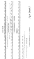
- VH and VL genes of a hybridoma cell line can be copied using the polymerase chain reaction and primers located at the 5' and 3' end of the VH and also of the Vk gene ( Figure 1).
- the amplified VH and Vk DNA are also linked during the PCR reaction, in the order 5' VH-Vk 3' by virtue of a match between the primers at the 3' end of the VH gene, and the 5' end of the Vk gene (Stage 1).
- a second set of 'nested' PCR primers is used (Stage 2).
- VH and Vk genes are linked preferentially within the same cell as follows.
- the cell is first fixed with formaldehyde to stabilise it to the temperature cycling, and treated with Nonidet P40 to permeabilise the cell clones analysed by probing and sequencing.
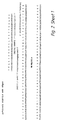
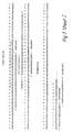
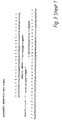
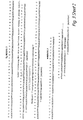
- nucleotide sequences of the mature heavy chain VH domains and the Vk light chain genes of the anti-phenyloxazolone hybridomas NQ2/12.4 and NQ10/12.5, and the encoded protein sequences, are given in Figures 2-5.
- the VH and Vk genes are linked by a nucleotide sequence encoding a polypeptide spacer providing a flexible polypeptide chain to link the two domains.
- the complete nucleotide sequence in Figures 2 and 4 encodes a single chain Fv fragment.
- the spacer is incorporated within the PCR primers MoVhlnk3 and MoVklnk3, which are used with a VH back (NQ10/12.5 or NQ2/12.4) primer and a Vk forward primer (MoJkFOR2) to amplify and link the VH and Vk genes in a form suitable for expression.
- Figures 3 and 5 are similar, and show a shorter linker that contains a unique restriction site to allow the cloning of a longer polypeptide linker by introducing a synthetic oligonucleotide.
- further cycles of PCR are used with a nested VH back (NQ10/12.5BKNES or NQ2/12.4BKNES) primer and a nested Vk forward (MokFORNES) primer.
- the sequence of the hybridoma genes was already known and primers specific to the hybridomas were designed. However in principle more general primers could be used, for example, to clone variable domains in general by PCR (reference 5). Also the second round of PCR amplification in the example gives rise to a single chain Fv truncated at both N- and C-terminus. However, a different set of primers should overcome these problems.
- Vk forward primers might be designed directly to the 3' side of the J-regions, either in the C kappa or C lambda constant domain for RNA templates, or in the region of DNA next to the splice junction for genomic DNA templates.
- Linking primers would be used that encoded a polypeptide sequence (as above), for example to join the VH and Vk as a single chain Fv fragment.
- the Vh back primer would incorporate a restriction site for cloning into expression vectors.
- Vk forward primer(s) based within the Jk region itself (and incorporating restriction site for cloning into expression vectors) might be used in conjunction with the VH back primer.
- NQ2/12.4 or NQ10/12.5 hybridoma cells were grown in tissue culture flasks in Dulbecco's modified Eagle's medium containing 5% foetal bovine serum. Cultures in late logarithmic growth phase were harvested by rinsing adherent cells into the supernatant medium, and centrifuging the medium at 800 rpm for 5 minutes at room temperature. The supernatant was removed and the cells were resuspended in 5ml phosphate buffered saline (PBS, pH 7.2) at room temperature and again centrifuged at 800 rpm for 5 mins (1st wash). Two further washes in 5ml PBS were given under the same conditions.
- PBS phosphate buffered saline
- the cell pellets were then suspended in 10% formal saline (10% formalin [i.e. 4% formaldehyde] and 0.15M NaCl in distilled water), using 1ml per 10-50ml of original culture, and transferred to 2ml Eppendorf Safe-Lock tubes. They were incubated on ice for 1 hour, with frequent agitation on a vortex mixer. The cells were then spun down at 13,000 rpm in a microfuge, and washed 3 times with lml ice-cold PBS, dispersing them by pipetting up and down several times at each wash.
- 10% formal saline % formalin [i.e. 4% formaldehyde] and 0.15M NaCl in distilled water
- the pellets were resuspended in 1ml ice-cold Nonidet P40 in water, and incubated on ice for a further 1 hour with frequent agitation.
- the cells were washed 4 more times in 1ml ice-cold PBS, with vigorous pipetting, and suspended in 0.2-0.5ml cold PBS containing 0.1M glycine.
- a sample was examined microscopically in a haemocytometer, and if clumps were present they were dispersed into single cells by vigorous pipetting or repeated passage through a 26 gauge hypodermic needle.
- the cells were then counted and adjusted to 10 7 per ml in PBS + 0.1M glycine, and aliquoted into 0.5 ml tubes (usually 0.05-0.1 ml per tube) and frozen in dry ice. The frozen aliquots were stored at -70 degrees C.
- Heavy and light chain variable regions were assembled and amplified by a 2-stage Polymerase Chain Reaction.
- VH and Vk genes were linked together, and in the second stage the assembled product was amplified from the cell template.
- Reactions were carried out in Techne HI-TEMP 96 polycarbonate microplates using a Techne PHC-3 programmable heating block, or in 0.5ml Sarstedt tubes using a BioTherm Inc. BioOven.
- Reaction conditions were as follows:- 1ST STAGE: Water 26.5 ul Forward Vk primer 2.5 ul Back Vh primer 2.5 ul Forward Link primer 0.5 ul Back Link primer 0.5 ul dNTPs (5mM) 2.0 ul 10 x PCR buffer 5.0 ul Cell template (in PBS/glycine) 10.0 ul Taq polymerase (5 units/ul) 0.5 ul
- the cell templates were separated from the rest of the reagents and washed. In Techne plates, the supernatant PCR mix and overlying oil were carefully pipetted away, leaving the cells at the bottom of the well. The cells were then suspended in 0.2ml of PBS/0.1M glycine and spun down in a microfuge at 13,000 rpm. After resuspension in the same buffer they were again spun down for a 2nd wash, then resuspended in 10ul PBS/glycine for use as the 2nd stage template.
- Example 2 Use of an emulsion for PCR amplification
- the mixture was incubated for 5 minutes at 64°C.
- the tubes were incubated at 64°C to evaporate the excess ether and 1 ul of the DNA was then removed for a second round of PCR with the nested primers as in Example 1. Analysis of the DNA on a 2% high-gelling temperature agarose gel demonstrated that assembly had occurred.
- Monoclonal antibodies have provided reagents for the identification of marker proteins (and lipids and carbohydrates) expressed within cells (1), on the surface of cells (2) or secreted from cells (3). These reagents have had diverse uses, ranging from the localisation of cells in histological sections (1) to the use of fluorescence activated cell sorting (FACS) of populations of cells (4).
- FACS fluorescence activated cell sorting
- the nucleotide sequence encoding a desired marker we could in principle use the mRNA species as a marker for a cell.
- Such a process could also be used to detect and to clone combinations of mRNAs by linking the PCR amplified cDNAs within the same cell.
- it could help in the cloning of Ig V-gene combinations from immunised or auto-immune sources using recombinant DNA techniques.
- repertoires of antibody heavy and light chains are prepared from lymphocyte populations by PCR (5,6,7), combined at random (8) and expressed as soluble fragments in bacteria for screening with antigen (8), or displayed on the surface of filamentous phage and selected by panning (9,10).
- lentivirus DNA can be amplified in situ using PCR, and the infected cells detected by autoradiography (15).
- the cells were fixed with formalin and permeabilised with the detergent NP40 to allow the access of nucleotides, primers and enzymes.
- one primer was designed to prime at the 3' end of the VH gene, and another at the 5' end of the VL gene, but with complementary tails to allow the self-assembly of the VH and VL genes.
- NQ10/12.5 murine hybridoma cells were grown in Dulbecco's modified Eagle's medium supplemented with 5% fetal bovine serum (FBS), and B1-8 hybridoma cells in RPMI 1640 medium supplemented with 5% FBS. Cells were used or passaged at late log phase, when almost all were viable. NQ10/12.5 secretes a kappa light chain and B1-8 a lambda light chain (16,17). The sequences of the VH and VL genes for both these hybridomas are known (16,17). The human myeloid leukemia cell line K562 was grown in RPMI with 5% FBS.
- FBS fetal bovine serum
- This line carries the Philadelphia chromosome, which is characterised by a bcr-abl fusion gene as a result of a translocation between the abl proto-oncogene on chromosome 9 and the bcr gene on chromosome 22 (19).
- Cells (10 7 to 10 8 ) were sedimented at 50 x g for 5 mins, washed 3 times in 5 ml PBS pH 7.2 at room temperature then suspended in 1 ml ice-cold 10% formaldehyde solution in 0.15M NaCl (formal saline). They were kept on ice for 1 hour, with occasional agitation on a vortex mixer. They were then spun at 13,000 rpm in a microcentrifuge for 2.5 mins and washed 3 times in ice-cold PBS, with vigorous pipetting with a Pasteur pipette to resuspend and disperse any clumps visible by eye.
- the cells were next suspended in 0.5% Nonidet P40 (NP40, B.D.H.) in water. After a further 1 hour incubation on ice with frequent agitation, they were again washed 3 times in ice-cold PBS with vigorous pipetting. A final wash was given in PBS containing 0.1 M glycine, and the cells were resuspended in 0.2 to 0.5 ml of the same buffer, using a 1 ml syringe and 26 gauge needle to disperse the clumps visible using a light microscope. The cells were counted, and for most experiments adjusted to a final concentration of 10 7 per ml. They were frozen in small aliquots in dry ice and stored at -7°C for up to a month.
- NP40 Nonidet P40
- oligonucleotides are listed according to their use as primers or probes. The sequences were based on the sequence of the hybridoma V genes (16,17), and the bcr-abl gene (20), and were synthesised using an Applied Biosystems 394 DNA synthesiser and used without purification.
- a 'first strand mix' was freshly prepared, comprising 5 ul 10 x 1st strand buffer (1.4M KCl, 0.5M Tris-HCl pH8.1 at 42°C, 80mM MgCl 2 ), 5 ul 0.1M dithiothreitol (DTT), 5 ul 5 mM dNTPs, 25 pmol each forward primer, 80 units RNase inhibitor (RNasin, Promega) and water to a total volume of 28 ul.
- the cells were heated to 65°C for 3 mins then cooled on ice.
- the 'first strand mix' was added, followed by 40 units Super RT AMV reverse transcriptase (HT Biotechnology Ltd., Cambridge).
- the cells and reagents were mixed and incubated at 42°C for 1 hour, then the cells were spun down, washed in 200 ul PBS (pH 7.2) containing 0.1 M glycine (PBS/0.1 M glycine) and resuspended in 20 ul of the same buffer for use immediately in PCR.
- PBS pH 7.2
- PBS/0.1 M glycine 0.1 M glycine
- cDNA synthesis was as above except using random primers and oligo (dT) (21).
- viable cells (5 x 10 6 ) were boiled for 5 mins in 100 ul water containing 0.1% diethyl pyrocarbonate and spun for 2 mins at 13,000 rpm in a microcentrifuge.
- a 'first strand mix' was prepared, comprising 10 ul 10 x 1st strand buffer, 5 ul 5 mM dNTPs, 5 ul 100 mM DTT, and 25 pmol each forward primer. It was added to a volume of 62-67 ul supernatant from the boiled cells, and the mixture heated at 65°C for 3 mins and left to cool at room temperature for 15 mins.
- RNasin 160 units
- reverse transcriptase 100 units
- Reactions were set up in 50 ul volumes in 0.5 ml Sarstedt tubes with 10 ul fixed template cells in PBS/0.1 M glycine buffer, 25 pmol back primer, 25 pmol forward primer, 200 uM dNTPs, 5 ul 10x Taq polymerase buffer (Promega) and 2.5 units of Taq polymerase.
- the tubes were subjected to up to 40 cycles of PCR with denaturation at 95°C and extension at 72°C for 1 min each. Annealing was for 1 min at temperatures ranging from 60°C to 72°C.
- a thermal cycling oven ("BioOven", BioTherm Corp.) was used, which did not require the use of a mineral oil overlay. Where cell recovery was not required, cycling was performed on a cycling heat block (Techne PHC-3) with an overlay of 50 ul mineral oil.
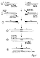
- the cDNAs were amplified by PCR and linked in the same reaction by using primers with complementary tails (22) (Fig. 6).
- Figure 6 illustrates assembly and nested amplification of Ig V-genes from hybridomas for cloning, cDNA synthesis and assembly of VH and VL gene from hybridomas NQ10/12.5 and B1.8. Sequences of primers (small arrows) indicated are as described previously. In each step the template strand is marked with a thick line, and the newly synthesised (first) strand with a thin line; in some cases, two steps are combined in the figure, and the second synthesised strand (using the first as template) is then marked with a dashed line. Sequence tags for assembly are marked as blocks.
- Reactions were set up in 50 ul volumes in tubes as follows: 25 pmol VH back primer, 25 pmol VL forward primer, 10 pmol VH forward primer with linker sequence, 10 pmol VL back primer with linker sequence, 200 uM dNTPs, 5 ul 10 x Taq polymerase buffer, 2.5 units Taq polymerase, and 10 ul fixed cells in PBS/0.1 M glycine buffer. Generally 10 5 (but sometimes up to 5 x 10 5 ) cells per tube were used, and the tubes were given 30 cycles of 95°C for 30 secs, 65°C for 30 secs and 72°C for 30 secs.
- the cells were spun down at 13,000 rpm, washed twice in 200 ul PBS/0.1 M glycine, and resuspended in 10 ul PBS/glycine.
- a second PCR was set up with the washed cells, nested primers (23) using 25 pmol nested VH back primer and 25 pmol nested VL forward primer, 200 uM dNTPs, 5 ul 10 x PCR buffer and 2.5 units Taq polymerase.
- the cells were subjected to 30 more cycles of PCR, and DNA for cloning was isolated from the supernatant.
- PCR primers for each cell line were included in both first and second "nested" PCRs using a 100 ul reaction volume.
- cDNA was synthesised from a 1:1 mix of B1-8 and NQ10/12.5 (NQ10) fixed cells using the primers MOLFOR, MOJH3FOR, B1-8LFOR and B1-8VHLINK3, and the cells washed and resuspended in PBS/0.1 M glycine for PCR assembly.
- the first PCR was carried out using the VL forward primers MOLFOR and B1-8LFOR and the VH back primers NQ10BK and B1-8BK.
- the PCR linker primers MOVHLINK3, MOVKLINK3, B1-8VLLINK3 and B1-8VHLINK3 were also included.
- the 3' ends of the PCR linker primers are complementary to the ends of the DNA template, but incorporate a 5' "tag" to allow assembly of the VH and VL genes (Fig. 6).
- the tags were identical for both NQ10 and B1-8 linker primers, so that all possible combinations of VH and VL could be formed.
- the second PCR used the nested primers NQ10BKNES, MOLFORNES, B1-8BKNES and B1-8FORNES.
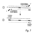
- cDNA in the B1-8 and NQ10 cells was subjected to PCR to amplify VH genes, using the primers indicated in Table 1. They were then washed twice and the genes further amplified with nested PCR primers tagged with sequences complementary to those fluorescence tagged primers (-21ROX, -21FAM, M13ROX used for DNA sequencing) (Table 1). After washing, a third PCR was used with 3pmol of M13 or-21 primers labelled with 6 carboxy-X-rhodamine (ROX) (Applied Biosystems). Conditions for each PCR were 94° C for 40 secs, 58°C for 40 secs, and 72°C for 40 secs.
- ROX carboxy-X-rhodamine
- Figure 7 illustrates nested amplification of assembled Ig V-genes for fluorescent labelling.
- (I) shows second "nested” PCR with tagged primers to introduce primer sites for "universal” fluorescent primers and (2) shows third PCR with fluorescent primers.
- the same fluorescent primer can prime at both the 5' and 3' ends of the tagged sequences.
- the cells were examined and photographed on a MRC 500 scanning laser confocal microscope (BioRad). Between 10 3 and 10 4 cells in 10 ul TE were spread on a glass slide and covered with a coverslip, sealed at the edges with nail varnish. In addition, cytofluorographic analysis was performed on a FACScan flow cytometer (Becton Dickinson). Fluorescence data were displayed as logarithmic overlay histograms using Consort 30 software (Becton Dickinson). Cell debris was gated out and 10 5 cells were analysed per sample.
- DNA from the supernatants of cells after assembly and amplification was purified from 1.5% agarose gels in 0.5 x Tris-borate EDTA (TBE) buffer (pH 8.0) with ethidium bromide (0.5 ug/ml) incorporated into the gels.
- TBE Tris-borate EDTA
- the assembled DNA bands of around 600 bp were eluted with Geneclean II (BIO 101 Inc.), with the addition of Geneclean 'TBE modifier'.
- the purified DNA was ligated into a 'T vector' derived from Bluescript KS+ (Stratagene) (24), and the vector transfected into E. coli CMK603 by electroporation.
- the cells were grown overnight on TYE plates (25) with 100 ug/ml ampicillin, 5-bromo-4-chloroindolyl-B-D-galactoside (26) and isopropyl-B-D-l-thiogalacto-pyranoside (27). White colonies were replica plated in order to prepare colony lifts for hybridisation, or for use as templates for PCR screening.
- the reactions were set up in 20 ul volumes in individual wells of Hi-temp plates (Techne), containing 8 pmol of forward and back primers, 200 uM dNTPs, 2 ul 10 x Taq buffer and 0.4 units Taq polymerase per well.
- each clone was screened with the primers NQ10BKNES and MOLFORNES, B1-8BKNES and B1-8FORNES, NQ10BKNES and B1-8FORNES, and B1-8BKNES and MOLFORNES.
- Each well was inoculated with bacteria from a single replica plated colony by means of a toothpick, and overlaid with 50 ul of mineral oil.
- the plates were subjected to 30 cycles of 95°C for 30 secs, 65°C for 1 min and 72°C for 1 min on a Techne PHC-3 heat block designed for multi-well plates.
- the PCR products were run on 1.5% agarose gels (10 ul per lane).
- bacterial colony lifts were made using Hybond-N nylon membranes (Millipore). The bacteria were lysed with sodium dodecyl sulphate and the DNA denatured and fixed to the membrane by microwaving (29), followed by UV cross-linking. Oligonucleotide probes for heavy (V186.2) and light (B1-8LPRB) chains of the B1-8 hybridoma, and heavy (NQ10PRB) and light (NQ10KPRB) chains of the NQ10/12.5 hybridoma, were labelled with gamma 32 p-ATP using polynucleotide kinase (New England BioLabs) and hybridised to the nylon filters for 16 to 60 hours. The filters were washed with solutions containing tetramethylammonium chloride (21) and exposed on Kodak XAR 5 film.
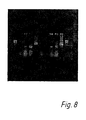
- Permeabilisation was a necessary step (Fig. 8), but formaldehyde concentrations ranging from 4% to 10% were equally effective for fixation. However, fixed and permeabilised cells which had undergone cDNA synthesis provided a much better template for PCR amplification (as shown by the examples in Fig. 8), which shows the gel electrophoresis of PCR products. In this Figure, supernatants from cDNA amplifications were run on a 1.5% agarose gel in TBE buffer.
- lanes 1 and 11 phi X174 HaeIII marker
- lane 2 PCR of B1-8 VH from fixed cells (formalin only);
- lane 3 PCR of B1-8 VH from fixed and permeabilised cells (formalin and NP40);
- lane 4 PCR assembly of B1-8 VH and V lambda from fixed and permeabilised cells;
- lane 5 as lane 4 except using nested primers in second PCR;
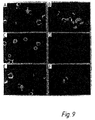
- the amplified DNA within the cells could be visualised directly by confocal microscopy (or conventional fluorescence microscopy) by incorporation of a fluorescent PCR primer in a third PCR step (30).
- B1-8 and NQ10 cells were fixed, permeabilised and mixed in equal ratios. The mixtures were then treated to cDNA synthesis and PCR amplification and assembly (Fig. 6), and the linked product isolated from the supernatant and cloned. The clones were then screened for the four different combinations of heavy and light chains (as described elsewhere). The results of three independent experiments are shown in Table 2 for 1:1 mixtures. In all cases the in-cell PCRs resulted in linkage of NQ10 VH with V Kappa, or B1-8 VH with V lambda, corresponding to the combinations of the hybridomas; no "crossovers" were seen in the 104 clones evaluated. By contrast, PCRs performed with soluble cDNA led in addition to cross-over combinations of NQ10 VH with V lambda and B1-8 VH with V Kappa.
- VH and VL linkage were performed by PCR of cell and soluble cDNA templates from a mix containing 10% B1-8 cells and 90% NQ10/12.5 cells (Table 3).
- 450 clones of each PCR assembly were initially probed with 32 P kinase labelled V186.2 oligonucleotide probe to identify those clones expressing the B1-8 VH chain.
- the positive clones were then screened on agarose gels to detect either V Kappa and V lambda genes using PCR with the primers MOVKLINK3 and MOLFORNES, and B1-8VLLINK3 and B1-8FORNES respectively.
- VH and VL human immunoglobulin variable region genes
- Lymphoprep lynphocyte separation medium Nacomed
- Tubes were centrifuged at 4,000 x g for 20 minutes, after which mononuclear cells (which include lymphocytes) were visible as a band separating the blood plasma (upper layer) and the Lymphoprep (lower layer).
- the cells were removed and washed 3 times in phosphate buffered saline (PBS, pH 7.2), and incubated in 10% formaldehyde in 0.15M NaCl solution on ice for 1 hour. They were again washed in PBS 3 times, followed by incubation on ice in 0.5% Nonidet P40 (BDH) in water. After a further 3 washes in PBS the cells were suspended in PBS containing 0.1M glycine and counted. They were stored frozen at -70°C.
- PBS phosphate buffered saline
- Reactions were set up in 50ul volumes in 0.5 ml Sarstedt tubes, using 10 5 cDNA template cells in 10ul together with a mix containing 25 pmol VH back primer mix (VH3LDR1 + VH3LDR2 + VH3LDR3), 25pmol Ck5'FOR, 10 pmol linker primer mix (JHLINK 1,2 + JH3LINK + JH6LINK + VkLINK + VkLINK1), 200 uM dNTPs, 5ul 10x Taq polymerase buffer (Promega) and 2.5 unit Taq polymerase.
- the tubes were subjected to 30 cycles of PCR with denaturation at 95°C for 30 seconds, annealing for 58 or 65°C for 30 seconds, and extension at 72°C for 30 seconds, in a thermal cycling oven (BioTherm Corp. "BioOven") without an oil overlay. This was referred to as the "1st PCR”.
- the cells were spun down and washed twice in PBS/O.1M glycine, and suspended in 10ul of this buffer for a 2nd PCR together with the following mix:- 200 uM dNTPs, 5ul 10x Taq buffer, 2.5 units Taq polymerase, and either of primer combinations (a) 25 pmol VH3A and 25 pmol of a mix of Jk1FOR + Jk2FOR + Jk3FOR + Jk4FOR + Jk5FOR, or (b) 25 pmol VH3ASfi and 25 pmol of a mix of Jk1FORNot + Jk2FORNot + Jk3FORNot + Jk4FORNot + Jk5FORNot.
- PCR products were analysed by running on 1.5% agarose gels in 0.5 x Tris-borate-EDTA buffer containing 0.5ug/ml ethidium bromide and viewing under ultra-violet light.
- the amplified DNA could be fluorescence labelled within the cell using fluorescent PCR primers, and should allow the analysis of large and diverse populations of cells by fluorescence.
- fluorescent PCR primers we used three PCR amplifications in order to tag the amplified DNA, and then fluorescence lable and tagged DNA with "universal" fluorescent PCR primers.
- fluorescence lable the cDNAs within the cell with a single PCR amplification using a set of fluorescent PCR primers specifically made for the gene of interest.
- PCR templates were derived from a 1:1 mix of NQ10/12.5 and B1-8 cells which were divided into two portions. One portion of the cells were fixed before cDNA synthesis and PCR assembly and nested amplification (Fixed cells), and the other was boiled in water for cDNA synthesis and PCR assembly and nested amplification from soluble mRNA (Random combinatorial). Colonies were screened by PCR for Expts 1, 2 and 3, but extra Expt. 1 colonies were also probed. Screening of linked VH and VL gene combinations from 1:9 mixtures of B1-8: NQ10/12.5 hybridoma cells.
- PCR assembly templates For preparation of PCR assembly templates a 1: 9 mix of B1-8 : NQ10/12.5 cells was divided into two portions. One portion was fixed for cDNA synthesis and PCR (Fixed cells), and the other was boiled in water for cDNA synthesis and PCR from soluble mRNA (Random combinatorial). Clones were probed with 32 P labelled V186.2 probe, and those clones identified with B1.8 VH genes were PCR screened for either V ⁇ or V ⁇ genes.
Landscapes
- Chemical & Material Sciences (AREA)
- Health & Medical Sciences (AREA)
- Life Sciences & Earth Sciences (AREA)
- Genetics & Genomics (AREA)
- Engineering & Computer Science (AREA)
- Organic Chemistry (AREA)
- Bioinformatics & Cheminformatics (AREA)
- Wood Science & Technology (AREA)
- Biomedical Technology (AREA)
- Zoology (AREA)
- Biotechnology (AREA)
- General Engineering & Computer Science (AREA)
- Molecular Biology (AREA)
- Biophysics (AREA)
- Biochemistry (AREA)
- General Health & Medical Sciences (AREA)
- Plant Pathology (AREA)
- Crystallography & Structural Chemistry (AREA)
- Microbiology (AREA)
- Physics & Mathematics (AREA)
- Immunology (AREA)
- Bioinformatics & Computational Biology (AREA)
- Medicinal Chemistry (AREA)
- Proteomics, Peptides & Aminoacids (AREA)
- Measuring Or Testing Involving Enzymes Or Micro-Organisms (AREA)
- Medicines Containing Material From Animals Or Micro-Organisms (AREA)
- Preparation Of Compounds By Using Micro-Organisms (AREA)
- Immobilizing And Processing Of Enzymes And Microorganisms (AREA)
- Micro-Organisms Or Cultivation Processes Thereof (AREA)
Abstract
Description
| 1ST STAGE: | |
| Water | 26.5 ul |
| Forward Vk primer | 2.5 ul |
| Back Vh primer | 2.5 ul |
| Forward Link primer | 0.5 ul |
| Back Link primer | 0.5 ul |
| dNTPs (5mM) | 2.0 ul |
| 10 x PCR buffer | 5.0 ul |
| Cell template (in PBS/glycine) | 10.0 ul |
| Taq polymerase (5 units/ul) | 0.5 ul |
| Techne PHC-3 Block: | 94°C | 30 secs | |
| 65° | 1 min | ||
| 72° | 1 min | 30 cycles | |
| BioOven: | 95°C | 30 secs | |
| 65°C | 30 secs | ||
| 72°C | 30 secs | 30 cycles |
Vh Back NQ2/12.4 BACK


| 2ND STAGE | |
| Water | 27.5 ul |
| Forward nested primer (10pmole/ul) | 2.5 ul |
| Back nested primer | 2.5 ul |
| dNTPs (5 mM) | 2.0 ul |
| 10 x PCR buffer | 5.0 ul |
| | 10.0 ul |
| Taq polymerase (5 units/ul) | 0.5 ul |
Forward nested primer Mok5FORNES
| 5ul | 10 x PCR buffer [10 x PCR = 100mm Tris-HCl pH 8.3/500mM, KCl/25mM MgCl2] |
| 4ul | 2.5 mM deoxynucleotide triphosphates (pH 7.5) |
| 2ul | NQ10/12.5 BACK primer (10pmoles/ul) |
| 2ul | MOJk5FOR2 primer (10pmoles /ul) |
| 2ul | MoVHLNK4 (1pmole/ul) |
| 2ul | MoVkLNK4 (1pmole/ul) |
| 5ul | Taq DNA Polymerase (Cetus 5U/ul) |
| 1ul | plasmid containing NQ10/12.5 VH gene (3 ng pBluescriptIIKS from Stratagene) |
| 1ul | plasmid containing NQ10/12.5 vk gene (3 ng pBluescriptIIKS from Stratagene) |
| H2O (HPLC grade) to 50ul |
MOLFOR (5' CTT ACG TTT CAG CTC CAG CTT GG 3'), MOJH3FOR (5' TAG GAC TCA CCT GCA GAG ACA GTG 3'), B1-8LFOR (5' GCC TAG GAC AGT CAG TTT GGT TC 3'), B1-8VHLINK3 (5' CCA CTG CCG CCA CCA CCG CTA CCA CCA CTG AGG AGA CTG TGA GAG TGG TGC 3').
MOLFOR, MOJH3FOR, B1-8LFOR, B1-8VHLINK3, NQ2BK (5' CAG GTG CAG CTG AAG GAG TCA GG 3'), NQ10BK (5' TGC AGC TGG TGG AGT CTG GGG G 3'), B1-8BK (5' CAG GTC CAA CTG CAG CAG CCT G 3'), BCR1A (5' AGT TAC ACG TTC CTG ATC TC 3'), ABL2C (5' TTA TCT CCA CTG GCC ACA AA 3'), MOVHLINK3 (5' CCA CTG CCG CCA CCA CCG CTA CCA CCA CCA CCT GCA GAG ACA GTG ACC AG 3'), MOVKLINK3 (5' GCG GTG GTG GCG GCA GTG GCG GCG GCG GCT CTC AAA TTG TTC TCA CCC AGT CTC CAG C 3'), B1-8VLLINK3 (5' GCG GTG GTG GCG GCA GTG GCG GCG GCG GCT CTC AGG CTG TTG TGA CTC AGG AAT CTG C 3').
B1-8VLLINK3, B1-8VHLINK3, MOVHLINK3, MOVLLINK3.
NQ10BKNES (5' AGC CTG GAG GGT CCC GGA AAC 3'), B1-8BKNES (5' GAG CTT GTG AAG CCT GGG GCT T 3'), MOLFORNES (5' CCC AGC ACC GAA CGT GAG TGG 3'), B1-8FORNES (5' CCA CCG AAC ACC CAA TGG TTG CT 3'), V186.2 (5' AGA CAA ACC CTC CAG 3').
M13B1-8BKNES (5' CAG GAA ACA GCT ATG ACC GAG CTT GTG AAG CCT GGG GCT 3'), M13B1-8FORNES (5' CAG GAA ACA GCT ATG ACC CCA CCG AAC ACC CAA TGG TTG CT 3'), -21MOLFORNES (5' TGT AAA ACG ACG GCC AGT CCC AGC ACC GAA CGT GAG TGG 3'),-21NQ10BKNES (5' TGT AAA ACG ACG GCC AGT ACG CTG GAG GGT CCC GGA AAC 3'), -21BCR1B (5' TGT AAA ACG ACG GCC AGT TCT GAC TAT GAG CGT GCA GA 3'), -21ABL2D (5' TGT AAA ACG ACG GCC AGT AGT GCA ACG AAA AGG TTG GG 3').
-21ROX or -21FAM (5' TGT AAA ACG ACG GCC CAG 3'), M13ROX (5' CAG GAA ACA GCT ATG AC 3') were obtained commercially from Applied Biosystems.
B1-8LPRB (5' CTG TAC CAT AGA GCA CAG 3'), NQ10PRB (5' GAG TTT CCG GGA CCC TCC AG 3'), NQ10KPRB (5' TTG GAA CCA GTT CAT GTA C 3'), V186.2.
25 pmol VH back primer, 25 pmol VL forward primer, 10 pmol VH forward primer with linker sequence, 10 pmol VL back primer with linker sequence, 200 uM dNTPs, 5 ul 10 x Taq polymerase buffer, 2.5 units Taq polymerase, and 10 ul fixed cells in PBS/0.1 M glycine buffer. Generally 105 (but sometimes up to 5 x 10⁵) cells per tube were used, and the tubes were given 30 cycles of 95°C for 30 secs, 65°C for 30 secs and 72°C for 30 secs. The cells were spun down at 13,000 rpm, washed twice in 200 ul PBS/0.1 M glycine, and resuspended in 10 ul PBS/glycine. To amplify the assembled products, a second PCR was set up with the washed cells, nested primers (23) using 25 pmol nested VH back primer and 25 pmol nested VL forward primer, 200 uM dNTPs, 5 ul 10 x PCR buffer and 2.5 units Taq polymerase. The cells were subjected to 30 more cycles of PCR, and DNA for cloning was isolated from the supernatant.


200 uM dNTPs, 5ul 10x Taq buffer, 2.5 units Taq polymerase, and either of primer combinations (a) 25 pmol VH3A and 25 pmol of a mix of Jk1FOR + Jk2FOR + Jk3FOR + Jk4FOR + Jk5FOR, or (b) 25 pmol VH3ASfi and 25 pmol of a mix of Jk1FORNot + Jk2FORNot + Jk3FORNot + Jk4FORNot + Jk5FORNot. Water was added to bring the cells and reagents to 50ul, and PCR cycling was carried out as for the first stage. When primer combination (a) was used in the 2nd PCR, the cells were recovered and washed, and treated to a 3rd PCR with primer combination (b) to append restriction sites for cloning.
| In cell PCR with fluorescence labelled primers | ||||
| cells | Primers PCR1 | Primers PCR2 | Primers PCR3 | Fluorescent Cells |
| NQ10 (VH gene) | NQ10BK MOJH3FOR | -21NQ10BKNES MOJH3FOR | -21 ROX | + |
| M13 ROX | - | |||
| NQ10 (VH gene) | B1-8BK B1-8VHLINK3 | M13B1-8BKMS V186.2 | -21 ROX | - |
| M13 ROX | - | |||
| B1-8 (VH gene) | NQ10BK MOJH3FOR | -21NQ10BKNES MOJH3FOR | -21 ROX | - |
| M13 ROX | - | |||
| B1-8 (VH gene) | B1-8BK B1-8VHLINK3 | M13B1-8BKNES V186.2 | -21 ROX | - |
| M13 ROX | + | |||
| NQ10 (VH/VK genes) | NQ10BK MOVHLINK3 MOVKLINK3 MOLFOR | -21NQ10BKNES -21MOLFORNES | -21 ROX | + |
| M13 ROX | - | |||
| B1-8 (VH/VL genes) | B1-8VHBK B1-8VHLINK3 B1-8VLLINK3 B1-8BLFOR | M13B1-8BKNES M13B1-8FORNES | -21 ROX | - |
| M13 ROX | + | |||
| K562 (bcr-abl) | ABL2C BCR1A | -21BCRB -21ABL2D | -21 ROX | + |
| M13 ROX | - | |||
| K562 (bcr-abl) | ABL2C BCR1A | -21BCRB -21ABL2D | -21 FAM | + |
| M13 DAM | - |
| Screening of linked VH and VL gene combinations from 1:1 mixtures of B1-8 and NQ10/12.5 hybridoma cells. | |||||
| PCR template | VH/VL combination | Expt. 1 | Expt 1 (probe) | Expt. 2 | |
| Fixed cells | NQ10 VH NQ10 Vκ | 20/47 | 19/32 | 11/36 | 11/21 |
| " | B1-8 VH B1-8 Vλ | 27/47 | 13/32 | 25/36 | 10/21 |
| " | NQ10 VH B1-8 Vλ | 0/47 | 0/32 | 0/36 | 0/21 |
| " | B1-8 VH NQ10 Vκ | 0/47 | 0/32 | 0/36 | 0/21 |
| Random combinatorial | NQ10 VH | 2/49 | 11/103 | 0/44 | 10/24 |
| " | B1-8 VH B1-8 Vλ | 27/49 | 56/103 | 40/44 | 4/24 |
| " | Nq10 VH B1-8 | 10/49 | 14/103 | 2/44 | 4/24 |
| " | B1-8 VH | 10/49 | 22/103 | 2/44 | 6/24 |
| Screening of linked VH and VL gene combinations from 1:9 mixtures of B1-8: NQ10/12.5 hybridoma cells. | |||
| PCR template | Clones with B1-8 VH (probed) | Clones with B1-8 VH and B1.8 Vλ | Clones with B1-8 VH and NQ10/12.5 Vκ |
| Fixed cells | 27/450 | 27/450 | 0/450 |
| Random combinatorial | 34/450 | 3/450 | 31/450 |
Claims (18)
- A method of treating a heterogeneous population of cells to link together two or more copies of non-contiguous DNA sequences from at least some of the cells, comprising the steps of:wherein said linking takes place preferentially in the vicinity of the non-contiguous DNA sequences, such that two or more copies of non-contiguous DNA sequences from the same cell are more likely to be linked with each other than with copies derived from a nucleic acid sequence from a different cell.(i) amplifying the non-contiguous DNA sequences; and(ii) linking together the copies of the non-contiguous DNA sequences,
- A method of treating a population of cells to link together copies of two or more non-contiguous DNA sequences from at least some of the cells, comprising the steps of:(i) treating the cells to stabilise them with respect to temperature and to permeabilise them with respect to reagents;(ii) adding primers for performing the polymerase chain reaction (PCR) in a manner such that the primers diffuse into the interior of the cells;(iii) subjecting the cells to suitable treatment so the non-contiguous DNA sequences of a particular cell are copied within that cell by PCR; and(iv) linking together the copies of the non-contiguous DNA sequences, said linking taking place preferentially in the vicinity of the non-contiguous DNA sequences from which the copies are derived, such that two or more copies of non-contigious DNA sequences from the same cell are more likely to be linked with each other than with copies derived from a nucleic acid sequence from a different cell.
- A method according to claim 1 or 2, wherein the DNA sequence copies are linked by PCR.
- A method according to claim 2 wherein the primers are fluorescently labelled, and said method comprises the additional step of monitoring the fluorescent labels.
- A method according to claim 4, wherein the cells are sorted by fluorescence activated cell sorting (FACS).
- A method according to claims 4 or 5, further comprising the use of two different primers or probes labelled with different fluorescent labels.
- A method according to any one of claims 4, 5 or 6, wherein said monitoring of the fluorescent labels comprises counting labelled cells.
- A method according to any one of claims 2 to 7, wherein the DNA sequence subjected to PCR is derived from cellular mRNA.
- A method according to claim 8, wherein the DNA sequence is cDNA derived by reverse transcription of mRNA.
- A method according to any one of the preceding claims, further comprising a washing step following linking of DNA.
- A method according to any one of the preceding claims, further comprising one or more rounds or further rounds of PCR.
- A method according to claim 11, wherein further rounds of PCR are performed using primers which are complementary to sequences copied in a previous round of PCR.
- A method according to any one of the preceding claims, wherein the DNA sequences comprise sequences encoding immunoglobulin VH and VL domains.
- A method according to any one of the preceding claims, wherein the resulting DNA is cloned by insertion into a suitable vector.
- A method according to claim 14, whereby the clones are sequenced or annealed to nucleic acid probes.
- A method according to claim 14 or 15, further comprising expressing a protein encoded by the DNA of a clone.
- A method according to claim 16, wherein the protein is expressed as part of a fusion protein.
- A method according to claim 17, wherein the fusion protein comprises the coat protein of a filamentous phage or a peptide tag.
Applications Claiming Priority (5)
| Application Number | Priority Date | Filing Date | Title |
|---|---|---|---|
| GB91173526 | 1991-08-10 | ||
| GB919117352A GB9117352D0 (en) | 1991-08-10 | 1991-08-10 | Recombinant dna method |
| GB92124197 | 1992-06-11 | ||
| GB929212419A GB9212419D0 (en) | 1992-06-11 | 1992-06-11 | Recombinant dna method |
| PCT/GB1992/001483 WO1993003151A1 (en) | 1991-08-10 | 1992-08-10 | Treatment of cell populations |
Publications (2)
| Publication Number | Publication Date |
|---|---|
| EP0597960A1 EP0597960A1 (en) | 1994-05-25 |
| EP0597960B1 true EP0597960B1 (en) | 1999-01-20 |
Family
ID=26299394
Family Applications (1)
| Application Number | Title | Priority Date | Filing Date |
|---|---|---|---|
| EP92916742A Expired - Lifetime EP0597960B1 (en) | 1991-08-10 | 1992-08-10 | Treatment of cell populations |
Country Status (11)
| Country | Link |
|---|---|
| US (1) | US5830663A (en) |
| EP (1) | EP0597960B1 (en) |
| JP (1) | JPH06509473A (en) |
| AT (1) | ATE175997T1 (en) |
| AU (1) | AU665365B2 (en) |
| CA (1) | CA2114950A1 (en) |
| DE (1) | DE69228247T2 (en) |
| DK (1) | DK0597960T3 (en) |
| ES (1) | ES2130177T3 (en) |
| GR (1) | GR3029913T3 (en) |
| WO (1) | WO1993003151A1 (en) |
Cited By (2)
| Publication number | Priority date | Publication date | Assignee | Title |
|---|---|---|---|---|
| EP2626433A1 (en) | 2012-02-09 | 2013-08-14 | Max-Planck-Gesellschaft zur Förderung der Wissenschaften e.V. | Method for linking and characterising linked nucleic acids in a composition |
| US8921281B2 (en) | 2009-05-20 | 2014-12-30 | Novimmune S.A. | Synthetic polypeptide libraries and methods for generating naturally diversified polypeptide variants |
Families Citing this family (153)
| Publication number | Priority date | Publication date | Assignee | Title |
|---|---|---|---|---|
| US5830713A (en) * | 1986-11-04 | 1998-11-03 | Protein Polymer Technologies, Inc. | Methods for preparing synthetic repetitive DNA |
| ATE408012T1 (en) | 1991-12-02 | 2008-09-15 | Medical Res Council | PRODUCTION OF AUTOANTIBODIES ON PHAGE SURFACES BASED ON ANTIBODIES SEGMENT LIBRARIES |
| GB9216983D0 (en) * | 1992-08-11 | 1992-09-23 | Unilever Plc | Polypeptide production |
| GB9324807D0 (en) * | 1993-12-03 | 1994-01-19 | Cancer Res Campaign Tech | Tumour antibody |
| DE69534347T2 (en) * | 1994-01-31 | 2006-05-24 | Trustees Of Boston University, Boston | Libraries of polyclonal antibodies |
| GB9401815D0 (en) * | 1994-01-31 | 1994-03-23 | Ray Ronnie A | A new method for combined intracellular reverse transciption and amplification |
| US7622294B2 (en) | 1997-03-14 | 2009-11-24 | Trustees Of Tufts College | Methods for detecting target analytes and enzymatic reactions |
| US20030027126A1 (en) | 1997-03-14 | 2003-02-06 | Walt David R. | Methods for detecting target analytes and enzymatic reactions |
| EP3034626A1 (en) | 1997-04-01 | 2016-06-22 | Illumina Cambridge Limited | Method of nucleic acid sequencing |
| US6143496A (en) * | 1997-04-17 | 2000-11-07 | Cytonix Corporation | Method of sampling, amplifying and quantifying segment of nucleic acid, polymerase chain reaction assembly having nanoliter-sized sample chambers, and method of filling assembly |
| AU736836B2 (en) | 1997-07-07 | 2001-08-02 | Medical Research Council | In vitro sorting method |
| US7364735B1 (en) | 1998-05-08 | 2008-04-29 | Stichting Sanquin Bloedvoorziening | Method for diagnosis and treatment of haemophilia A patients with an inhibitor |
| AR021833A1 (en) | 1998-09-30 | 2002-08-07 | Applied Research Systems | METHODS OF AMPLIFICATION AND SEQUENCING OF NUCLEIC ACID |
| AU2367600A (en) | 1998-12-16 | 2000-07-03 | Chiron Corporation | Human cyclin-dependent kinase ((hpnqalre)) |
| GB9900298D0 (en) * | 1999-01-07 | 1999-02-24 | Medical Res Council | Optical sorting method |
| EP1163359A4 (en) * | 1999-03-24 | 2005-01-26 | Gene Therapy Systems Inc | Method for generating transcriptionally active dna fragments |
| US6770441B2 (en) | 2000-02-10 | 2004-08-03 | Illumina, Inc. | Array compositions and methods of making same |
| WO2001062908A2 (en) * | 2000-02-22 | 2001-08-30 | Ahuva Nissim | Chimeric and tcr phage display libraries, chimeric and tcr reagents and methods of use thereof |
| US6211164B1 (en) * | 2000-03-10 | 2001-04-03 | Abbott Laboratories | Antisense oligonucleotides of the human chk1 gene and uses thereof |
| WO2001079481A2 (en) * | 2000-04-17 | 2001-10-25 | Dyax Corp. | Methods of constructing display libraries of genetic packages for members of a diverse family of peptides |
| US8288322B2 (en) | 2000-04-17 | 2012-10-16 | Dyax Corp. | Methods of constructing libraries comprising displayed and/or expressed members of a diverse family of peptides, polypeptides or proteins and the novel libraries |
| GB0022458D0 (en) * | 2000-09-13 | 2000-11-01 | Medical Res Council | Directed evolution method |
| EP1334347A1 (en) * | 2000-09-15 | 2003-08-13 | California Institute Of Technology | Microfabricated crossflow devices and methods |
| ATE352040T1 (en) * | 2000-11-17 | 2007-02-15 | Univ Rochester | IN-VITRO METHOD FOR PRODUCING AND IDENTIFYING IMMUNOGLOBULIN MOLECULES IN EUKARYOTIC CELLS |
| US20050196755A1 (en) * | 2000-11-17 | 2005-09-08 | Maurice Zauderer | In vitro methods of producing and identifying immunoglobulin molecules in eukaryotic cells |
| AR031640A1 (en) | 2000-12-08 | 2003-09-24 | Applied Research Systems | ISOTHERMAL AMPLIFICATION OF NUCLEIC ACIDS IN A SOLID SUPPORT |
| ES2360479T3 (en) | 2000-12-18 | 2011-06-06 | Dyax Corp. | FOCALIZED LIBRARIES OF GENETIC PACKAGES. |
| AU2002338446A1 (en) * | 2001-01-23 | 2002-11-05 | University Of Rochester Medical Center | Methods of producing or identifying intrabodies in eukaryotic cells |
| WO2002070678A2 (en) | 2001-02-05 | 2002-09-12 | Bayer Aktiengesellschaft | Regulation of human serine/threonine protein kinase |
| GB0127564D0 (en) | 2001-11-16 | 2002-01-09 | Medical Res Council | Emulsion compositions |
| EP1900827A3 (en) | 2002-05-21 | 2008-04-16 | Bayer HealthCare AG | Methods and compositions for the prediction, diagnosis, prognosis, prevention and treatment of malignant neoplasia |
| US20040067532A1 (en) | 2002-08-12 | 2004-04-08 | Genetastix Corporation | High throughput generation and affinity maturation of humanized antibody |
| US20040224325A1 (en) * | 2002-12-20 | 2004-11-11 | Caliper Life Sciences, Inc. | Single molecule amplification and detection of DNA |
| WO2005003375A2 (en) | 2003-01-29 | 2005-01-13 | 454 Corporation | Methods of amplifying and sequencing nucleic acids |
| US20060078893A1 (en) | 2004-10-12 | 2006-04-13 | Medical Research Council | Compartmentalised combinatorial chemistry by microfluidic control |
| GB0307403D0 (en) | 2003-03-31 | 2003-05-07 | Medical Res Council | Selection by compartmentalised screening |
| GB0307428D0 (en) | 2003-03-31 | 2003-05-07 | Medical Res Council | Compartmentalised combinatorial chemistry |
| PL1644493T3 (en) | 2003-06-27 | 2010-11-30 | Axikin Pharmaceuticals Inc | Regulation of a kinase, 'regulated in copd kinase' (rc kinase) |
| EP1641809B2 (en) | 2003-07-05 | 2018-10-03 | The Johns Hopkins University | Method and compositions for detection and enumeration of genetic variations |
| CA2536565A1 (en) * | 2003-09-10 | 2005-05-12 | Althea Technologies, Inc. | Expression profiling using microarrays |
| TWI333977B (en) * | 2003-09-18 | 2010-12-01 | Symphogen As | Method for linking sequences of interest |
| KR101135134B1 (en) * | 2003-09-18 | 2012-04-13 | 심포젠 에이/에스 | Method for linking sequences of interest |
| EP1522594A3 (en) | 2003-10-06 | 2005-06-22 | Bayer HealthCare AG | Methods and kits for investigating cancer |
| EP1691792A4 (en) * | 2003-11-24 | 2008-05-28 | Yeda Res & Dev | Compositions and methods for in vitro sorting of molecular and cellular libraries |
| US20050221339A1 (en) | 2004-03-31 | 2005-10-06 | Medical Research Council Harvard University | Compartmentalised screening by microfluidic control |
| WO2005106012A2 (en) | 2004-04-28 | 2005-11-10 | Bayer Healthcare Ag | Diagnostics and therapeutics for diseases associated with dipeptidyl-peptidase 1 (dpp1) |
| GB0418651D0 (en) * | 2004-08-20 | 2004-09-22 | Medical Res Council | Method |
| US7968287B2 (en) | 2004-10-08 | 2011-06-28 | Medical Research Council Harvard University | In vitro evolution in microfluidic systems |
| EP2236628A3 (en) | 2005-02-01 | 2010-10-13 | AB Advanced Genetic Analysis Corporation | Reagents, methods and libraries for bead-based sequencing |
| JP2008528040A (en) | 2005-02-01 | 2008-07-31 | アジェンコート バイオサイエンス コーポレイション | Reagents, methods and libraries for bead-based sequencing |
| EP1883710A4 (en) * | 2005-05-03 | 2009-07-08 | Althea Technologies Inc | Compositions and methods for the analysis of degraded nucleic acids |
| US20100137163A1 (en) | 2006-01-11 | 2010-06-03 | Link Darren R | Microfluidic Devices and Methods of Use in The Formation and Control of Nanoreactors |
| US20080045880A1 (en) * | 2006-02-11 | 2008-02-21 | Rune Kjeken | Device and method for single-needle in vivo electroporation |
| CA2635437C (en) | 2006-02-11 | 2020-01-28 | Genetronics, Inc. | Device and method for single-needle in vivo electroporation |
| US20080287857A1 (en) * | 2006-02-11 | 2008-11-20 | Rune Kjeken | Device and method for single-needle in vivo electroporation |
| WO2007111937A1 (en) | 2006-03-23 | 2007-10-04 | Applera Corporation | Directed enrichment of genomic dna for high-throughput sequencing |
| JP2009531464A (en) | 2006-03-29 | 2009-09-03 | メリアル リミテッド | Vaccines against streptococci |
| WO2007121489A2 (en) * | 2006-04-19 | 2007-10-25 | Applera Corporation | Reagents, methods, and libraries for gel-free bead-based sequencing |
| EP2481815B1 (en) | 2006-05-11 | 2016-01-27 | Raindance Technologies, Inc. | Microfluidic devices |
| US9562837B2 (en) | 2006-05-11 | 2017-02-07 | Raindance Technologies, Inc. | Systems for handling microfludic droplets |
| US9074242B2 (en) | 2010-02-12 | 2015-07-07 | Raindance Technologies, Inc. | Digital analyte analysis |
| EP2636755A1 (en) * | 2006-05-26 | 2013-09-11 | AltheaDx Incorporated | Biochemical analysis of partitioned cells |
| WO2008021123A1 (en) | 2006-08-07 | 2008-02-21 | President And Fellows Of Harvard College | Fluorocarbon emulsion stabilizing surfactants |
| WO2008093098A2 (en) | 2007-02-02 | 2008-08-07 | Illumina Cambridge Limited | Methods for indexing samples and sequencing multiple nucleotide templates |
| US8772046B2 (en) | 2007-02-06 | 2014-07-08 | Brandeis University | Manipulation of fluids and reactions in microfluidic systems |
| PL2121920T3 (en) | 2007-03-01 | 2012-01-31 | Symphogen As | Method for cloning cognate antibodies |
| WO2008130623A1 (en) | 2007-04-19 | 2008-10-30 | Brandeis University | Manipulation of fluids, fluid components and reactions in microfluidic systems |
| US7883265B2 (en) * | 2007-06-01 | 2011-02-08 | Applied Biosystems, Llc | Devices, systems, and methods for preparing emulsions |
| AU2008282863A1 (en) * | 2007-07-31 | 2009-02-05 | Merck Sharp & Dohme Corp. | IGF-1R specific antibodies useful in the detection and diagnosis of cellular proliferative disorders |
| BRPI0816689B1 (en) | 2007-09-12 | 2021-08-24 | Novartis Ag | VACCINE COMPOSITION, KIT AND METHOD FOR MAKING A VACCINE COMPOSITION FOR THE PREVENTION OR TREATMENT OF INFECTION BY STREPTOCOCCUS PYOGENES |
| US8877688B2 (en) | 2007-09-14 | 2014-11-04 | Adimab, Llc | Rationally designed, synthetic antibody libraries and uses therefor |
| MX344415B (en) | 2007-09-14 | 2016-12-15 | Adimab Inc | Rationally designed, synthetic antibody libraries and uses therefor. |
| JP2010539982A (en) * | 2007-10-01 | 2010-12-24 | アプライド バイオシステムズ, エルエルシー | Chase ligation sequencing |
| CA2709927C (en) | 2007-12-21 | 2017-05-16 | Novartis Ag | Mutant forms of streptolysin o |
| US8211673B2 (en) | 2008-02-04 | 2012-07-03 | Life Technologies Corporation | Composition and method for sequencing nucleic acid |
| US8143007B2 (en) * | 2008-03-13 | 2012-03-27 | National Institute Of Immunology | Nested primer sets for amplifying mouse immunoglobulin variable gene segments |
| US9873957B2 (en) * | 2008-03-13 | 2018-01-23 | Dyax Corp. | Libraries of genetic packages comprising novel HC CDR3 designs |
| WO2009132287A2 (en) * | 2008-04-24 | 2009-10-29 | Dyax Corp. | Libraries of genetic packages comprising novel hc cdr1, cdr2, and cdr3 and novel lc cdr1, cdr2, and cdr3 designs |
| EP2315629B1 (en) | 2008-07-18 | 2021-12-15 | Bio-Rad Laboratories, Inc. | Droplet libraries |
| US12038438B2 (en) | 2008-07-18 | 2024-07-16 | Bio-Rad Laboratories, Inc. | Enzyme quantification |
| WO2010038042A1 (en) | 2008-10-02 | 2010-04-08 | Illumina Cambridge Ltd. | Nucleic acid sample enrichment for sequencing applications |
| WO2010057197A1 (en) | 2008-11-17 | 2010-05-20 | The Regents Of The University Of Michigan | Cancer vaccine compositions and methods of using the same |
| EP3415235A1 (en) | 2009-03-23 | 2018-12-19 | Raindance Technologies Inc. | Manipulation of microfluidic droplets |
| NZ596603A (en) | 2009-05-29 | 2013-07-26 | Morphosys Ag | A collection of antibodies or fragments having properties relevant to developability |
| US20100316673A1 (en) | 2009-06-16 | 2010-12-16 | The Regents Of The University Of Michigan | Nanoemulsion vaccines |
| US20110082054A1 (en) * | 2009-09-14 | 2011-04-07 | Dyax Corp. | Libraries of genetic packages comprising novel hc cdr3 designs |
| US8182994B2 (en) | 2009-09-15 | 2012-05-22 | Illumina Cambridge Limited | Centroid markers for image analysis of high denisty clusters in complex polynucleotide sequencing |
| WO2011042564A1 (en) | 2009-10-09 | 2011-04-14 | Universite De Strasbourg | Labelled silica-based nanomaterial with enhanced properties and uses thereof |
| US10837883B2 (en) | 2009-12-23 | 2020-11-17 | Bio-Rad Laboratories, Inc. | Microfluidic systems and methods for reducing the exchange of molecules between droplets |
| US9366632B2 (en) | 2010-02-12 | 2016-06-14 | Raindance Technologies, Inc. | Digital analyte analysis |
| US9399797B2 (en) | 2010-02-12 | 2016-07-26 | Raindance Technologies, Inc. | Digital analyte analysis |
| US10351905B2 (en) | 2010-02-12 | 2019-07-16 | Bio-Rad Laboratories, Inc. | Digital analyte analysis |
| RS57937B1 (en) | 2010-05-14 | 2019-01-31 | Alltech Inc | Yeast cell wall components and detection thereof |
| KR102318383B1 (en) | 2010-07-16 | 2021-10-27 | 아디맵 엘엘씨 | Abtibody libraries |
| CA2808118C (en) | 2010-07-22 | 2016-04-19 | Kieran Curran | Composite liquid cells |
| EP3447155A1 (en) | 2010-09-30 | 2019-02-27 | Raindance Technologies, Inc. | Sandwich assays in droplets |
| CN103270174B (en) | 2010-10-28 | 2016-08-24 | 生命技术公司 | Chemical reinforcing type Primer composition, method and test kit |
| WO2012066129A1 (en) | 2010-11-19 | 2012-05-24 | Morphosys Ag | A collection and methods for its use |
| ES2615733T3 (en) | 2010-12-16 | 2017-06-08 | Gigagen, Inc. | Methods for parallel mass analysis of nucleic acids in individual cells |
| ES2634098T3 (en) | 2011-01-14 | 2017-09-26 | The Regents Of The University Of California | Therapeutic antibodies against the ROR-1 protein and procedures for their use. |
| EP3412778A1 (en) | 2011-02-11 | 2018-12-12 | Raindance Technologies, Inc. | Methods for forming mixed droplets |
| US9150852B2 (en) | 2011-02-18 | 2015-10-06 | Raindance Technologies, Inc. | Compositions and methods for molecular labeling |
| WO2012119986A1 (en) | 2011-03-09 | 2012-09-13 | Bayer Pharma Aktiengesellschaft | Modulators of slc22a7 |
| DK3415619T3 (en) | 2011-04-28 | 2021-03-08 | Univ Leland Stanford Junior | IDENTIFICATION OF POLYNUCLEOTIDS ASSOCIATED WITH A SAMPLE |
| US9783594B2 (en) | 2011-05-17 | 2017-10-10 | The Rockefeller University | Human immunodeficiency virus neutralizing antibodies and methods of use thereof |
| US8841071B2 (en) | 2011-06-02 | 2014-09-23 | Raindance Technologies, Inc. | Sample multiplexing |
| US9556470B2 (en) | 2011-06-02 | 2017-01-31 | Raindance Technologies, Inc. | Enzyme quantification |
| US8658430B2 (en) | 2011-07-20 | 2014-02-25 | Raindance Technologies, Inc. | Manipulating droplet size |
| WO2013046722A1 (en) | 2011-09-30 | 2013-04-04 | 中外製薬株式会社 | Ion concentration-dependent binding molecule library |
| EP2612916A1 (en) * | 2012-01-09 | 2013-07-10 | Universität Zürich | Cellular high throughput encapsulation for screening or selection |
| US9777269B2 (en) | 2012-01-25 | 2017-10-03 | Gencell Biosystems Ltd. | Biomolecule isolation |
| US9701998B2 (en) | 2012-12-14 | 2017-07-11 | 10X Genomics, Inc. | Methods and systems for processing polynucleotides |
| CN113528634A (en) * | 2012-08-14 | 2021-10-22 | 10X基因组学有限公司 | Microcapsule compositions and methods |
| US9951386B2 (en) | 2014-06-26 | 2018-04-24 | 10X Genomics, Inc. | Methods and systems for processing polynucleotides |
| US10584381B2 (en) | 2012-08-14 | 2020-03-10 | 10X Genomics, Inc. | Methods and systems for processing polynucleotides |
| US10323279B2 (en) | 2012-08-14 | 2019-06-18 | 10X Genomics, Inc. | Methods and systems for processing polynucleotides |
| US10273541B2 (en) | 2012-08-14 | 2019-04-30 | 10X Genomics, Inc. | Methods and systems for processing polynucleotides |
| US10752949B2 (en) | 2012-08-14 | 2020-08-25 | 10X Genomics, Inc. | Methods and systems for processing polynucleotides |
| US11591637B2 (en) | 2012-08-14 | 2023-02-28 | 10X Genomics, Inc. | Compositions and methods for sample processing |
| CA2888042A1 (en) | 2012-11-27 | 2014-06-05 | Gencell Biosystems Ltd. | Handling liquid samples |
| EP3567116A1 (en) | 2012-12-14 | 2019-11-13 | 10X Genomics, Inc. | Methods and systems for processing polynucleotides |
| US10533221B2 (en) | 2012-12-14 | 2020-01-14 | 10X Genomics, Inc. | Methods and systems for processing polynucleotides |
| KR102190198B1 (en) | 2013-02-08 | 2020-12-14 | 10엑스 제노믹스, 인크. | Polynucleotide barcode generation |
| US11901041B2 (en) | 2013-10-04 | 2024-02-13 | Bio-Rad Laboratories, Inc. | Digital analysis of nucleic acid modification |
| US9944977B2 (en) | 2013-12-12 | 2018-04-17 | Raindance Technologies, Inc. | Distinguishing rare variations in a nucleic acid sequence from a sample |
| WO2015103367A1 (en) | 2013-12-31 | 2015-07-09 | Raindance Technologies, Inc. | System and method for detection of rna species |
| US10384187B2 (en) | 2014-02-10 | 2019-08-20 | Gencell Biosystems Ltd | Composite liquid cell (CLC) mediated nucleic acid library preparation device, and methods for using the same |
| WO2015157567A1 (en) | 2014-04-10 | 2015-10-15 | 10X Genomics, Inc. | Fluidic devices, systems, and methods for encapsulating and partitioning reagents, and applications of same |
| CN105085673B (en) * | 2014-05-19 | 2018-09-11 | 厦门大学 | Monoclonal antibody and application thereof for detecting coxsackie virus A 16-type virus full particle |
| EP3161160B1 (en) | 2014-06-26 | 2021-10-13 | 10X Genomics, Inc. | Methods of analyzing nucleic acids from individual cells or cell populations |
| WO2016053883A1 (en) | 2014-10-03 | 2016-04-07 | Life Technologies Corporation | Detection, identification, validation and enrichment of target nucleic acids |
| WO2016057683A2 (en) | 2014-10-07 | 2016-04-14 | Ann And Robert H. Lurie Children's Hospital Of Chicago | Novel anti-nodal antibodies and methods of using same |
| SG11201705615UA (en) | 2015-01-12 | 2017-08-30 | 10X Genomics Inc | Processes and systems for preparing nucleic acid sequencing libraries and libraries prepared using same |
| EP4286516A3 (en) | 2015-02-24 | 2024-03-06 | 10X Genomics, Inc. | Partition processing methods and systems |
| US9422547B1 (en) | 2015-06-09 | 2016-08-23 | Gigagen, Inc. | Recombinant fusion proteins and libraries from immune cell repertoires |
| US10647981B1 (en) | 2015-09-08 | 2020-05-12 | Bio-Rad Laboratories, Inc. | Nucleic acid library generation methods and compositions |
| US11173207B2 (en) | 2016-05-19 | 2021-11-16 | The Regents Of The University Of Michigan | Adjuvant compositions |
| JP7132233B2 (en) | 2016-11-11 | 2022-09-06 | アセンダント ディーエックス, エルエルシー | Compositions and Methods for Diagnosing and Differentiating Systemic Juvenile Idiopathic Arthritis and Kawasaki Disease |
| US10550429B2 (en) | 2016-12-22 | 2020-02-04 | 10X Genomics, Inc. | Methods and systems for processing polynucleotides |
| US10815525B2 (en) | 2016-12-22 | 2020-10-27 | 10X Genomics, Inc. | Methods and systems for processing polynucleotides |
| CN117512066A (en) | 2017-01-30 | 2024-02-06 | 10X基因组学有限公司 | Method and system for droplet-based single cell bar coding |
| US10400235B2 (en) | 2017-05-26 | 2019-09-03 | 10X Genomics, Inc. | Single cell analysis of transposase accessible chromatin |
| CN109526228B (en) | 2017-05-26 | 2022-11-25 | 10X基因组学有限公司 | Single cell analysis of transposase accessible chromatin |
| EP3625361A1 (en) | 2017-11-15 | 2020-03-25 | 10X Genomics, Inc. | Functionalized gel beads |
| US10829815B2 (en) | 2017-11-17 | 2020-11-10 | 10X Genomics, Inc. | Methods and systems for associating physical and genetic properties of biological particles |
| US20200339687A1 (en) * | 2018-01-19 | 2020-10-29 | The Trustees Of The University Of Pennsylvania | Compositions and methods for targeting gamma delta t cells with chimeric antigen receptors |
| WO2019195166A1 (en) | 2018-04-06 | 2019-10-10 | 10X Genomics, Inc. | Systems and methods for quality control in single cell processing |
| US11541105B2 (en) | 2018-06-01 | 2023-01-03 | The Research Foundation For The State University Of New York | Compositions and methods for disrupting biofilm formation and maintenance |
| US11929171B2 (en) | 2018-10-18 | 2024-03-12 | The Board Of Trustees Of The Leland Stanford Junior University | Methods for evaluation and treatment of glycemic dysregulation and atherosclerotic cardiovascular disease and applications thereof |
| EP3941491A4 (en) | 2019-03-21 | 2023-03-29 | Gigamune, Inc. | Engineered cells expressing anti-viral t cell receptors and methods of use thereof |
| CN114107449B (en) * | 2020-08-31 | 2024-04-09 | 中山大学孙逸仙纪念医院 | Single-cell in-situ qPCR method |
| WO2024050450A1 (en) | 2022-08-31 | 2024-03-07 | Gigamune, Inc. | Engineered enveloped vectors and methods of use thereof |
| CN116590283B (en) * | 2023-07-17 | 2023-11-07 | 北京微岩医疗器械有限公司 | Cell binding enhancer and method for removing human free nucleic acid |
-
1992
- 1992-08-10 JP JP5503446A patent/JPH06509473A/en active Pending
- 1992-08-10 US US08/190,199 patent/US5830663A/en not_active Expired - Lifetime
- 1992-08-10 EP EP92916742A patent/EP0597960B1/en not_active Expired - Lifetime
- 1992-08-10 AT AT92916742T patent/ATE175997T1/en not_active IP Right Cessation
- 1992-08-10 DK DK92916742T patent/DK0597960T3/en active
- 1992-08-10 WO PCT/GB1992/001483 patent/WO1993003151A1/en active IP Right Grant
- 1992-08-10 CA CA002114950A patent/CA2114950A1/en not_active Abandoned
- 1992-08-10 DE DE69228247T patent/DE69228247T2/en not_active Expired - Lifetime
- 1992-08-10 ES ES92916742T patent/ES2130177T3/en not_active Expired - Lifetime
- 1992-08-10 AU AU24001/92A patent/AU665365B2/en not_active Expired
-
1999
- 1999-04-07 GR GR990401004T patent/GR3029913T3/en unknown
Cited By (3)
| Publication number | Priority date | Publication date | Assignee | Title |
|---|---|---|---|---|
| US8921281B2 (en) | 2009-05-20 | 2014-12-30 | Novimmune S.A. | Synthetic polypeptide libraries and methods for generating naturally diversified polypeptide variants |
| EP2626433A1 (en) | 2012-02-09 | 2013-08-14 | Max-Planck-Gesellschaft zur Förderung der Wissenschaften e.V. | Method for linking and characterising linked nucleic acids in a composition |
| WO2013117591A1 (en) | 2012-02-09 | 2013-08-15 | Max-Planck Gesellschaft zur Förderung der Wissenschaften e.V. | Method for linking and characterising linked nucleic acids, e.g. antibody encoding nucleic acids, in a composition |
Also Published As
| Publication number | Publication date |
|---|---|
| ES2130177T3 (en) | 1999-07-01 |
| AU665365B2 (en) | 1996-01-04 |
| DK0597960T3 (en) | 1999-09-13 |
| CA2114950A1 (en) | 1993-02-11 |
| DE69228247T2 (en) | 1999-07-08 |
| WO1993003151A1 (en) | 1993-02-18 |
| JPH06509473A (en) | 1994-10-27 |
| US5830663A (en) | 1998-11-03 |
| DE69228247D1 (en) | 1999-03-04 |
| GR3029913T3 (en) | 1999-07-30 |
| ATE175997T1 (en) | 1999-02-15 |
| AU2400192A (en) | 1993-03-02 |
| EP0597960A1 (en) | 1994-05-25 |
Similar Documents
| Publication | Publication Date | Title |
|---|---|---|
| EP0597960B1 (en) | Treatment of cell populations | |
| Embleton et al. | In-cell PCR from mRNA: amplifying and linking the rearranged immunoglobulin heavy and light chain V-genes within single cells | |
| KR102550778B1 (en) | High-throughput polynucleotide library sequencing and transcriptome analysis | |
| CN107002076B (en) | High throughput nucleotide library sequencing | |
| CA2002868C (en) | Single domain ligands, receptors comprising said ligands, methods for their production, and use of said ligands and receptors | |
| JP4651619B2 (en) | Method for concatenating desired sequences | |
| JP5669397B2 (en) | Methods for cloning cognate antibodies | |
| US6255455B1 (en) | Rh(D)-binding proteins and magnetically activated cell sorting method for production thereof | |
| US20140357500A1 (en) | Single cell bar-coding for antibody discovery | |
| JP2003204797A (en) | Oligonucleotide | |
| RU2204602C2 (en) | Polypeptide able to formation of antigen-binding structure with specificity to rhesus-d-antigens, dna sequence, method for preparing polypeptide, method of polypeptide selection, complete anti- rhesus-d-antibody (variants), pharmaceutical composition, diagnostic agent for rhesus-d-typifying | |
| JP2012500634A (en) | Cloning method of antibodies obtained from birds | |
| Duan et al. | Elimination of endogenous aberrant kappa chain transcripts from sp2/0-derived hybridoma cells by specific ribozyme cleavage: utility in genetic therapy of HIV-1 infections | |
| Pope et al. | Construction and use of antibody gene repertoires | |
| Amersdorfer et al. | Phage libraries for generation of anti-botulinum scFv antibodies | |
| WO1992015678A1 (en) | Pcr generated dicistronic dna molecules for producing antibodies | |
| JPH08116978A (en) | Preparation of antibody fab library | |
| RU2790291C2 (en) | High throughput sequencing of polynucleotide libraries and transcriptome analysis | |
| WO1994011507A2 (en) | Production of monoclonal recombinant antibodies without the use of hybridomas by in vitro spleen fragment culture combined with isothermal self-sustained sequence replication of rna | |
| Kato et al. | Enzymatic assembly for scFv library construction | |
| Bradbury | Cloning hybridoma cDNA by RACE | |
| WO1991005856A1 (en) | Chimeric monoclonal antibodies generated by trans-splicing | |
| MXPA06002894A (en) | Method for linking sequences of interest |
Legal Events
| Date | Code | Title | Description |
|---|---|---|---|
| PUAI | Public reference made under article 153(3) epc to a published international application that has entered the european phase |
Free format text: ORIGINAL CODE: 0009012 |
|
| 17P | Request for examination filed |
Effective date: 19940309 |
|
| AK | Designated contracting states |
Kind code of ref document: A1 Designated state(s): AT BE CH DE DK ES FR GB GR IE IT LI LU MC NL SE |
|
| 17Q | First examination report despatched |
Effective date: 19970505 |
|
| GRAG | Despatch of communication of intention to grant |
Free format text: ORIGINAL CODE: EPIDOS AGRA |
|
| GRAG | Despatch of communication of intention to grant |
Free format text: ORIGINAL CODE: EPIDOS AGRA |
|
| GRAG | Despatch of communication of intention to grant |
Free format text: ORIGINAL CODE: EPIDOS AGRA |
|
| GRAH | Despatch of communication of intention to grant a patent |
Free format text: ORIGINAL CODE: EPIDOS IGRA |
|
| GRAH | Despatch of communication of intention to grant a patent |
Free format text: ORIGINAL CODE: EPIDOS IGRA |
|
| GRAH | Despatch of communication of intention to grant a patent |
Free format text: ORIGINAL CODE: EPIDOS IGRA |
|
| GRAA | (expected) grant |
Free format text: ORIGINAL CODE: 0009210 |
|
| AK | Designated contracting states |
Kind code of ref document: B1 Designated state(s): AT BE CH DE DK ES FR GB GR IE IT LI LU MC NL SE |
|
| REF | Corresponds to: |
Ref document number: 175997 Country of ref document: AT Date of ref document: 19990215 Kind code of ref document: T |
|
| REG | Reference to a national code |
Ref country code: CH Ref legal event code: EP |
|
| REG | Reference to a national code |
Ref country code: IE Ref legal event code: FG4D |
|
| REF | Corresponds to: |
Ref document number: 69228247 Country of ref document: DE Date of ref document: 19990304 |
|
| ITF | It: translation for a ep patent filed | ||
| ET | Fr: translation filed | ||
| REG | Reference to a national code |
Ref country code: ES Ref legal event code: FG2A Ref document number: 2130177 Country of ref document: ES Kind code of ref document: T3 |
|
| REG | Reference to a national code |
Ref country code: DK Ref legal event code: T3 |
|
| PLBE | No opposition filed within time limit |
Free format text: ORIGINAL CODE: 0009261 |
|
| STAA | Information on the status of an ep patent application or granted ep patent |
Free format text: STATUS: NO OPPOSITION FILED WITHIN TIME LIMIT |
|
| 26N | No opposition filed | ||
| PGFP | Annual fee paid to national office [announced via postgrant information from national office to epo] |
Ref country code: MC Payment date: 20010426 Year of fee payment: 10 |
|
| PGFP | Annual fee paid to national office [announced via postgrant information from national office to epo] |
Ref country code: GR Payment date: 20010504 Year of fee payment: 10 |
|
| PGFP | Annual fee paid to national office [announced via postgrant information from national office to epo] |
Ref country code: LU Payment date: 20010827 Year of fee payment: 10 |
|
| REG | Reference to a national code |
Ref country code: GB Ref legal event code: IF02 |
|
| PGFP | Annual fee paid to national office [announced via postgrant information from national office to epo] |
Ref country code: ES Payment date: 20020605 Year of fee payment: 11 |
|
| PG25 | Lapsed in a contracting state [announced via postgrant information from national office to epo] |
Ref country code: LU Free format text: LAPSE BECAUSE OF NON-PAYMENT OF DUE FEES Effective date: 20020810 |
|
| PGFP | Annual fee paid to national office [announced via postgrant information from national office to epo] |
Ref country code: FR Payment date: 20020821 Year of fee payment: 11 |
|
| PGFP | Annual fee paid to national office [announced via postgrant information from national office to epo] |
Ref country code: AT Payment date: 20020822 Year of fee payment: 11 Ref country code: IE Payment date: 20020822 Year of fee payment: 11 |
|
| PGFP | Annual fee paid to national office [announced via postgrant information from national office to epo] |
Ref country code: BE Payment date: 20020829 Year of fee payment: 11 |
|
| PG25 | Lapsed in a contracting state [announced via postgrant information from national office to epo] |
Ref country code: MC Free format text: LAPSE BECAUSE OF NON-PAYMENT OF DUE FEES Effective date: 20030301 |
|
| PG25 | Lapsed in a contracting state [announced via postgrant information from national office to epo] |
Ref country code: GR Free format text: LAPSE BECAUSE OF NON-PAYMENT OF DUE FEES Effective date: 20030305 |
|
| PG25 | Lapsed in a contracting state [announced via postgrant information from national office to epo] |
Ref country code: AT Free format text: LAPSE BECAUSE OF NON-PAYMENT OF DUE FEES Effective date: 20030810 |
|
| PG25 | Lapsed in a contracting state [announced via postgrant information from national office to epo] |
Ref country code: IE Free format text: LAPSE BECAUSE OF NON-PAYMENT OF DUE FEES Effective date: 20030811 Ref country code: ES Free format text: LAPSE BECAUSE OF NON-PAYMENT OF DUE FEES Effective date: 20030811 |
|
| PG25 | Lapsed in a contracting state [announced via postgrant information from national office to epo] |
Ref country code: BE Free format text: LAPSE BECAUSE OF NON-PAYMENT OF DUE FEES Effective date: 20030831 |
|
| BERE | Be: lapsed |
Owner name: *MEDICAL RESEARCH COUNCIL Effective date: 20030831 |
|
| PG25 | Lapsed in a contracting state [announced via postgrant information from national office to epo] |
Ref country code: FR Free format text: LAPSE BECAUSE OF NON-PAYMENT OF DUE FEES Effective date: 20040430 |
|
| REG | Reference to a national code |
Ref country code: IE Ref legal event code: MM4A |
|
| REG | Reference to a national code |
Ref country code: FR Ref legal event code: ST |
|
| REG | Reference to a national code |
Ref country code: ES Ref legal event code: FD2A Effective date: 20030811 |
|
| PG25 | Lapsed in a contracting state [announced via postgrant information from national office to epo] |
Ref country code: IT Free format text: LAPSE BECAUSE OF NON-PAYMENT OF DUE FEES Effective date: 20050810 |
|
| REG | Reference to a national code |
Ref country code: GB Ref legal event code: 732E |
|
| REG | Reference to a national code |
Ref country code: CH Ref legal event code: PLI Owner name: CAMBRIDGE ANTIBODY TECHNOLOGY LIMITED Free format text: MEDICAL RESEARCH COUNCIL#20 PARK CRESCENT#LONDON W1N 4AL (GB) -TRANSFER TO- CAMBRIDGE ANTIBODY TECHNOLOGY LIMITED#MILSTEIN BUILDING GRANTA PARK#CAMBRIDGE CB1 6GH (GB) |
|
| NLUE | Nl: licence registered with regard to european patents |
Effective date: 20060420 |
|
| PGFP | Annual fee paid to national office [announced via postgrant information from national office to epo] |
Ref country code: CH Payment date: 20110725 Year of fee payment: 20 Ref country code: DK Payment date: 20110725 Year of fee payment: 20 |
|
| PGFP | Annual fee paid to national office [announced via postgrant information from national office to epo] |
Ref country code: GB Payment date: 20110720 Year of fee payment: 20 Ref country code: SE Payment date: 20110725 Year of fee payment: 20 Ref country code: DE Payment date: 20110725 Year of fee payment: 20 |
|
| PGFP | Annual fee paid to national office [announced via postgrant information from national office to epo] |
Ref country code: NL Payment date: 20110728 Year of fee payment: 20 |
|
| REG | Reference to a national code |
Ref country code: DE Ref legal event code: R071 Ref document number: 69228247 Country of ref document: DE |
|
| REG | Reference to a national code |
Ref country code: DE Ref legal event code: R071 Ref document number: 69228247 Country of ref document: DE |
|
| REG | Reference to a national code |
Ref country code: DK Ref legal event code: EUP |
|
| REG | Reference to a national code |
Ref country code: CH Ref legal event code: PL |
|
| REG | Reference to a national code |
Ref country code: NL Ref legal event code: V4 Effective date: 20120810 |
|
| REG | Reference to a national code |
Ref country code: GB Ref legal event code: PE20 Expiry date: 20120809 |
|
| PG25 | Lapsed in a contracting state [announced via postgrant information from national office to epo] |
Ref country code: GB Free format text: LAPSE BECAUSE OF EXPIRATION OF PROTECTION Effective date: 20120809 Ref country code: DE Free format text: LAPSE BECAUSE OF EXPIRATION OF PROTECTION Effective date: 20120811 |