BRPI1010773B1 - ADAPTER FOR ENDOVASCULAR ELECTROCARDIOGRAPHY CROSS REFERENCE FOR RELATED ORDER - Google Patents
ADAPTER FOR ENDOVASCULAR ELECTROCARDIOGRAPHY CROSS REFERENCE FOR RELATED ORDER Download PDFInfo
- Publication number
- BRPI1010773B1 BRPI1010773B1 BRPI1010773-8A BRPI1010773A BRPI1010773B1 BR PI1010773 B1 BRPI1010773 B1 BR PI1010773B1 BR PI1010773 A BRPI1010773 A BR PI1010773A BR PI1010773 B1 BRPI1010773 B1 BR PI1010773B1
- Authority
- BR
- Brazil
- Prior art keywords
- endovascular
- heart
- location
- ecg
- adapter according
- Prior art date
Links
Images


Classifications
-
- A—HUMAN NECESSITIES
- A61—MEDICAL OR VETERINARY SCIENCE; HYGIENE
- A61B—DIAGNOSIS; SURGERY; IDENTIFICATION
- A61B5/00—Measuring for diagnostic purposes; Identification of persons
- A61B5/06—Devices, other than using radiation, for detecting or locating foreign bodies ; determining position of probes within or on the body of the patient
-
- A—HUMAN NECESSITIES
- A61—MEDICAL OR VETERINARY SCIENCE; HYGIENE
- A61B—DIAGNOSIS; SURGERY; IDENTIFICATION
- A61B5/00—Measuring for diagnostic purposes; Identification of persons
- A61B5/24—Detecting, measuring or recording bioelectric or biomagnetic signals of the body or parts thereof
- A61B5/316—Modalities, i.e. specific diagnostic methods
- A61B5/318—Heart-related electrical modalities, e.g. electrocardiography [ECG]
- A61B5/346—Analysis of electrocardiograms
- A61B5/349—Detecting specific parameters of the electrocardiograph cycle
- A61B5/353—Detecting P-waves
-
- A—HUMAN NECESSITIES
- A61—MEDICAL OR VETERINARY SCIENCE; HYGIENE
- A61B—DIAGNOSIS; SURGERY; IDENTIFICATION
- A61B34/00—Computer-aided surgery; Manipulators or robots specially adapted for use in surgery
- A61B34/20—Surgical navigation systems; Devices for tracking or guiding surgical instruments, e.g. for frameless stereotaxis
-
- A—HUMAN NECESSITIES
- A61—MEDICAL OR VETERINARY SCIENCE; HYGIENE
- A61B—DIAGNOSIS; SURGERY; IDENTIFICATION
- A61B5/00—Measuring for diagnostic purposes; Identification of persons
- A61B5/06—Devices, other than using radiation, for detecting or locating foreign bodies ; determining position of probes within or on the body of the patient
- A61B5/065—Determining position of the probe employing exclusively positioning means located on or in the probe, e.g. using position sensors arranged on the probe
-
- A—HUMAN NECESSITIES
- A61—MEDICAL OR VETERINARY SCIENCE; HYGIENE
- A61B—DIAGNOSIS; SURGERY; IDENTIFICATION
- A61B5/00—Measuring for diagnostic purposes; Identification of persons
- A61B5/24—Detecting, measuring or recording bioelectric or biomagnetic signals of the body or parts thereof
- A61B5/25—Bioelectric electrodes therefor
- A61B5/279—Bioelectric electrodes therefor specially adapted for particular uses
- A61B5/28—Bioelectric electrodes therefor specially adapted for particular uses for electrocardiography [ECG]
- A61B5/283—Invasive
-
- A—HUMAN NECESSITIES
- A61—MEDICAL OR VETERINARY SCIENCE; HYGIENE
- A61B—DIAGNOSIS; SURGERY; IDENTIFICATION
- A61B5/00—Measuring for diagnostic purposes; Identification of persons
- A61B5/24—Detecting, measuring or recording bioelectric or biomagnetic signals of the body or parts thereof
- A61B5/316—Modalities, i.e. specific diagnostic methods
- A61B5/318—Heart-related electrical modalities, e.g. electrocardiography [ECG]
- A61B5/346—Analysis of electrocardiograms
- A61B5/349—Detecting specific parameters of the electrocardiograph cycle
-
- A—HUMAN NECESSITIES
- A61—MEDICAL OR VETERINARY SCIENCE; HYGIENE
- A61B—DIAGNOSIS; SURGERY; IDENTIFICATION
- A61B5/00—Measuring for diagnostic purposes; Identification of persons
- A61B5/68—Arrangements of detecting, measuring or recording means, e.g. sensors, in relation to patient
- A61B5/6846—Arrangements of detecting, measuring or recording means, e.g. sensors, in relation to patient specially adapted to be brought in contact with an internal body part, i.e. invasive
- A61B5/6847—Arrangements of detecting, measuring or recording means, e.g. sensors, in relation to patient specially adapted to be brought in contact with an internal body part, i.e. invasive mounted on an invasive device
- A61B5/6852—Catheters
-
- A—HUMAN NECESSITIES
- A61—MEDICAL OR VETERINARY SCIENCE; HYGIENE
- A61B—DIAGNOSIS; SURGERY; IDENTIFICATION
- A61B5/00—Measuring for diagnostic purposes; Identification of persons
- A61B5/68—Arrangements of detecting, measuring or recording means, e.g. sensors, in relation to patient
- A61B5/6846—Arrangements of detecting, measuring or recording means, e.g. sensors, in relation to patient specially adapted to be brought in contact with an internal body part, i.e. invasive
- A61B5/6867—Arrangements of detecting, measuring or recording means, e.g. sensors, in relation to patient specially adapted to be brought in contact with an internal body part, i.e. invasive specially adapted to be attached or implanted in a specific body part
- A61B5/6876—Blood vessel
-
- A—HUMAN NECESSITIES
- A61—MEDICAL OR VETERINARY SCIENCE; HYGIENE
- A61B—DIAGNOSIS; SURGERY; IDENTIFICATION
- A61B5/00—Measuring for diagnostic purposes; Identification of persons
- A61B5/72—Signal processing specially adapted for physiological signals or for diagnostic purposes
- A61B5/7235—Details of waveform analysis
- A61B5/7246—Details of waveform analysis using correlation, e.g. template matching or determination of similarity
-
- A—HUMAN NECESSITIES
- A61—MEDICAL OR VETERINARY SCIENCE; HYGIENE
- A61B—DIAGNOSIS; SURGERY; IDENTIFICATION
- A61B5/00—Measuring for diagnostic purposes; Identification of persons
- A61B5/74—Details of notification to user or communication with user or patient ; user input means
- A61B5/742—Details of notification to user or communication with user or patient ; user input means using visual displays
- A61B5/743—Displaying an image simultaneously with additional graphical information, e.g. symbols, charts, function plots
-
- A—HUMAN NECESSITIES
- A61—MEDICAL OR VETERINARY SCIENCE; HYGIENE
- A61M—DEVICES FOR INTRODUCING MEDIA INTO, OR ONTO, THE BODY; DEVICES FOR TRANSDUCING BODY MEDIA OR FOR TAKING MEDIA FROM THE BODY; DEVICES FOR PRODUCING OR ENDING SLEEP OR STUPOR
- A61M25/00—Catheters; Hollow probes
- A61M25/0009—Making of catheters or other medical or surgical tubes
- A61M25/0014—Connecting a tube to a hub
-
- A—HUMAN NECESSITIES
- A61—MEDICAL OR VETERINARY SCIENCE; HYGIENE
- A61M—DEVICES FOR INTRODUCING MEDIA INTO, OR ONTO, THE BODY; DEVICES FOR TRANSDUCING BODY MEDIA OR FOR TAKING MEDIA FROM THE BODY; DEVICES FOR PRODUCING OR ENDING SLEEP OR STUPOR
- A61M25/00—Catheters; Hollow probes
- A61M25/01—Introducing, guiding, advancing, emplacing or holding catheters
- A61M25/0105—Steering means as part of the catheter or advancing means; Markers for positioning
-
- A—HUMAN NECESSITIES
- A61—MEDICAL OR VETERINARY SCIENCE; HYGIENE
- A61B—DIAGNOSIS; SURGERY; IDENTIFICATION
- A61B5/00—Measuring for diagnostic purposes; Identification of persons
- A61B5/72—Signal processing specially adapted for physiological signals or for diagnostic purposes
- A61B5/7235—Details of waveform analysis
- A61B5/7253—Details of waveform analysis characterised by using transforms
- A61B5/726—Details of waveform analysis characterised by using transforms using Wavelet transforms
-
- A—HUMAN NECESSITIES
- A61—MEDICAL OR VETERINARY SCIENCE; HYGIENE
- A61M—DEVICES FOR INTRODUCING MEDIA INTO, OR ONTO, THE BODY; DEVICES FOR TRANSDUCING BODY MEDIA OR FOR TAKING MEDIA FROM THE BODY; DEVICES FOR PRODUCING OR ENDING SLEEP OR STUPOR
- A61M39/00—Tubes, tube connectors, tube couplings, valves, access sites or the like, specially adapted for medical use
- A61M39/10—Tube connectors; Tube couplings
- A61M2039/1077—Adapters, e.g. couplings adapting a connector to one or several other connectors
-
- A—HUMAN NECESSITIES
- A61—MEDICAL OR VETERINARY SCIENCE; HYGIENE
- A61M—DEVICES FOR INTRODUCING MEDIA INTO, OR ONTO, THE BODY; DEVICES FOR TRANSDUCING BODY MEDIA OR FOR TAKING MEDIA FROM THE BODY; DEVICES FOR PRODUCING OR ENDING SLEEP OR STUPOR
- A61M39/00—Tubes, tube connectors, tube couplings, valves, access sites or the like, specially adapted for medical use
- A61M39/10—Tube connectors; Tube couplings
- A61M2039/1083—Tube connectors; Tube couplings having a plurality of female connectors, e.g. Luer connectors
-
- A—HUMAN NECESSITIES
- A61—MEDICAL OR VETERINARY SCIENCE; HYGIENE
- A61M—DEVICES FOR INTRODUCING MEDIA INTO, OR ONTO, THE BODY; DEVICES FOR TRANSDUCING BODY MEDIA OR FOR TAKING MEDIA FROM THE BODY; DEVICES FOR PRODUCING OR ENDING SLEEP OR STUPOR
- A61M39/00—Tubes, tube connectors, tube couplings, valves, access sites or the like, specially adapted for medical use
- A61M39/10—Tube connectors; Tube couplings
- A61M2039/1088—Tube connectors; Tube couplings having a plurality of male connectors, e.g. Luer connectors
Abstract
método com base em computador para posicionar um dispositivo endovascular no ou perto do coração usando sinais de eletrocardiograma, interface gráfica de usuário para um sistema de navegação e posicionamento de cateter, adaptador para eletrocardiografia endovascular, e, método para guia e navegação de um dispositivo de acesso endovascular um método com base em computador para posicionar um dispositivo endovascular no ou perto do coração usando sinais de eletrocardiograma (ecg) é fornecido. o método com base em computador inclui receber um sinal de ecg endovascular associado com um dispositivo endovascular, processar o sinal de ecg endovascular, sobre uma pluralidade de períodos de tempos pré-determinados, para calcular uma amplitude de onda p e uma potência espectral para cada período de tempo pré-determinado, determinando uma amplitude de onda p máximo de uma pluralidade de amplitudes de onda p, e uma potência espectral máxima associada de uma pluralidade de potências espectrais, associar a amplitude de onda p máxima e a porta espectral máxima com uma pré-determinada localização no ou perto do coração, calculando uma localização do dispositivo endovascular, para cada período de tempo pré-determinado, com base em uma proporção da amplitude de onda p para a amplitude de onda p máxima e uma proporção da potência espectral para a potência espectral máxima, e exibir a localização do dispositivo endovascular para um usuáriocomputer-based method for positioning an endovascular device at or near the heart using electrocardiogram signals, graphical user interface for a catheter positioning and navigation system, adapter for endovascular electrocardiography, and, method for guiding and navigating a device Endovascular Access A computer-based method for positioning an endovascular device at or near the heart using electrocardiogram (ECG) signals is provided. the computer-based method includes receiving an endovascular ecg signal associated with an endovascular device, processing the endovascular ecg signal, over a plurality of predetermined time periods, to calculate a wave amplitude and a spectral power for each period. of predetermined time, by determining a maximum p-wave amplitude from a plurality of p-wave amplitudes, and an associated maximum spectral power from a plurality of spectral powers, associating the maximum p-wave amplitude and the maximum spectral gate with a pre. - determined location in or near the heart, calculating a location of the endovascular device, for each predetermined time period, based on a ratio of the p-wave amplitude to the maximum p-wave amplitude and a ratio of the spectral power to the maximum spectral power, and display the location of the endovascular device to a user
Description
[0001] Este pedido reivindica prioridade para o Pedido de Patente Provisório dos U.S. de N° Série 61 / 213.474, depositado em 12 de junho de 2009, a divulgação do qual é aqui incorporado para referência em sua totalidade.[0001] This application claims priority to U.S. Provisional Patent Application Serial No. 61 / 213,474, filed June 12, 2009, the disclosure of which is incorporated herein by reference in its entirety.
[0002] A presente invenção se refere a um posicionamento de dispositivo endovascular. Especificamente, a presente invenção se refere a um método para posicionar a ponta de um dispositivo endovascular, tal como um cateter venoso central, dentre e em torno do coração usando sinais de eletrocardiograma (ECG).[0002] The present invention relates to an endovascular device positioning. Specifically, the present invention relates to a method for positioning the tip of an endovascular device, such as a central venous catheter, within and around the heart using electrocardiogram (ECG) signals.
[0003] O sistema de condução elétrica do coração cria sinais elétricos específicos, distribuições de energia elétrica e comportamentos dos mesmos que são indicativos de localizações específicas na cavidade toráxica e / ou de funções ou condições do coração específicas. Quando medidos de modo endovascular, i.e., de dentro dos vasos sanguíneos ou de dentro do coração, determinados parâmetros da atividade elétrica do coração podem ser usados para identificar específicas localizações no sistema cardiovascular e / ou condições funcionais, normais ou anormais. Mais ainda, localmente e precisamente identificando a localização e o tipo de condição, terapia de tais condições pode ser otimizada e o efeito da terapia monitorada em tempo real.[0003] The electrical conduction system of the heart creates specific electrical signals, electrical energy distributions and behaviors thereof that are indicative of specific locations in the chest cavity and/or of specific heart functions or conditions. When measured endovascularly, i.e., from within blood vessels or from within the heart, certain parameters of the electrical activity of the heart can be used to identify specific locations in the cardiovascular system and/or functional conditions, either normal or abnormal. Furthermore, locally and accurately identifying the location and type of condition, therapy of such conditions can be optimized and the effect of therapy monitored in real time.
[0004] Dois tipos de aplicações clínicas são tipicamente abordados. O primeiro é relacionado a orientar dispositivos de guia endovascular através do sistema cardiovascular, enquanto o segundo é relacionada a monitoração remota minimamente agressiva ou não agressiva da atividade elétrica do coração.[0004] Two types of clinical applications are typically addressed. The first is related to guiding endovascular guidance devices through the cardiovascular system, while the second is related to minimally aggressive or non-aggressive remote monitoring of the electrical activity of the heart.
[0005] A confirmação de guia, de posicionamento e de colocação de cateter endovascular são necessárias em um número de aplicações clínicas, tais como, por exemplo:1. Acesso venoso central, por ex., CVC, PICC, portas implantáveis;2. Cateter de hemodiálises;3. Colocação de contatos de marca-passo cardíaco;4. Cateter de monitoração hemodinâmicas, por ex., Swan-Ganz e cateteres de monitoração de pressão central pressão; e5. Guiar fios-guias e cateter no coração.[0005] Confirmation of guidance, placement and placement of endovascular catheters are required in a number of clinical applications, such as, for example:1. Central venous access, eg CVC, PICC, implantable ports;2. Hemodialysis catheter;3. Placement of cardiac pacemaker contacts;4. Hemodynamic monitoring catheters, eg Swan-Ganz and pressure central pressure monitoring catheters; e5. Guide guidewires and catheter into the heart.
[0006] A localização da ponta do cateter é muito importante para a segurança do paciente, a duração e o sucesso do procedimento. Padrão utilizado hoje em dia para avaliar a localização alvo da ponta do cateter é o raio X do peito. Em adição, há correntemente dois tipos de produtos de guia em tempo real disponíveis no mercado, que tentem superar as limitações da avaliação por meio do raio X do peito: com base em eletromagnético e com base em ECG. Em hospitais onde guia em tempo real é usada resulta ter melhora em termos de redução do número de raios X, no tempo de procedimento, e no custo do procedimento. Sob guia em tempo real pela primeira vez a taxa de sucesso tipicamente aumentou de 75% - 80% à 90% - 95%. Em adição, em hospitais onde guia por ECG é usado, por ex., na Itália, Bélgica, Alemanha, avaliação por raio X do peito foi eliminada por mais do que 90% dos pacientes. Sistemas eletromagnéticos são usados principalmente nos Estados Unidos enquanto sistemas baseados em ECG são usados principalmente na Europa. Entre outros fatores que determinam a diferença entre os mercados nos Estados Unidos e Europa em termos de adoção de tecnologia: a) tipo de profissional de tratamento de saúde permitido para efetuar procedimentos: enfermeira têm mais flexibilidade nos Estados Unidos, b) tipo de dispositivos colocados: PICCs são colocados mais e mais frequentemente nos Estado Unidos, c) sensibilidade ao preço: o mercado Europeu para ser mais sensitivo ao preço, e d) os dispositivos de guia corrente são comercializados por fabricantes específicos para trabalhar exclusivamente com seus cateteres: penetração de mercado dos sistemas de guia refletem a penetração do mercado do fabricante do cateter.[0006] The location of the tip of the catheter is very important for patient safety, duration and success of the procedure. The standard used today to assess the target location of the catheter tip is the chest X-ray. In addition, there are currently two types of real-time guidance products available on the market that attempt to overcome the limitations of chest X-ray assessment: electromagnetic-based and ECG-based. In hospitals where real-time guidance is used there is an improvement in terms of reducing the number of X-rays, procedure time, and procedure cost. Under real-time guidance for the first time the success rate typically increased from 75% - 80% to 90% - 95%. In addition, in hospitals where ECG guidance is used, eg in Italy, Belgium, Germany, chest X-ray assessment was eliminated by more than 90% of patients. Electromagnetic systems are mainly used in the United States while ECG-based systems are mainly used in Europe. Among other factors that determine the difference between the US and European markets in terms of technology adoption: a) type of healthcare professional allowed to perform procedures: nurses have more flexibility in the US, b) type of devices placed : PICCs are placed more and more frequently in the United States, c) price sensitivity: the European market to be more price sensitive, and d) current guide devices are marketed by specific manufacturers to work exclusively with their catheters: market penetration guidance systems reflect the market penetration of the catheter manufacturer.
[0007] Também foi encontrado que diferentes opiniões existem considerando a localização da ponta alvo deve estar: por exemplo, terço inferior do SVC ou RA. Por conseguinte tecnologias de guia devem permitir discriminação dessas localizações. O raio X do peito, que é o padrão principal corrente nem sempre permite tal discriminação considerando uma precisão de tipicamente melhor do que 2 cm. Além disso, porque sistema com base em ECG fazem uso de informação fisiológica relacionada à atividade do coração, sua habilidade para orientar a colocação é precisa com à anatomia. Isto não é o caso com sistemas de guia eletromagnéticos que medem a distância entre a ponta do cateter na estrutura vascular e uma referência externa colocada tipicamente no peito do paciente. Por causa deste aspecto, o sistema com base em ECG pode ser usado para documentar o resultado final da colocação do cateter potencialmente substituindo o raio X do peito com o padrão principal.[0007] It was also found that different opinions exist regarding the location of the target tip should be: for example, lower third of the SVC or RA. Therefore guidance technologies must allow discrimination of these locations. Chest X-ray, which is the main current standard, does not always allow for such discrimination considering an accuracy of typically better than 2 cm. Furthermore, because ECG-based systems make use of physiological information related to heart activity, their ability to guide placement is accurate with the anatomy. This is not the case with electromagnetic guidance systems that measure the distance between the tip of the catheter in the vascular structure and an external reference typically placed on the patient's chest. Because of this aspect, the ECG-based system can be used to document the end result of catheter placement potentially replacing the chest X-ray with the main pattern.
[0008] Uma das mais valiosas ferramentas de diagnóstico disponível, o ECG grava a atividade elétrica como formas de onda. Interpretando essas formas de onda, alguém pode identificar perturbações rítmicas, anormalidades de condução, e pode identificar perturbações rítmicas, anormalidades de condução, e desequilíbrio eletrolítico. Um ECG ajuda na diagnose e monitoração de tais condições como pericardites e síndromes coronarianas agudas. A atividade elétrica do coração produz correntes que irradiam através dos tecidos. Porque as correntes elétricas a partir do coração irradiam para o nível da pele em muitas direções, eletrodos são colocados em diferentes localizações no nível da pele para obter uma total imagem da atividade elétrica do coração. Os eletrodos são então conectados a um dispositivo eletrocardiógrafo, ou computador, e gravam informação de diferentes perspectivas, que são chamados sondas e planos. Uma sonda fornece uma visão da atividade elétrica do coração entre dois pontos ou polos. Um plano é uma seção transversal do coração que fornece uma diferente visão da atividade elétrica do coração. Correntemente, a interpretação de uma forma de onda de ECG é com base na identificação de amplitudes de componentes da forma de onda, analisando e então comparando as amplitudes com determinados padrões. Modificações desses componentes de amplitudes são indicativos de determinadas condições, por ex., a elevação do segmento ST ou de determinadas localizações no coração, por ex., uma amplitude da onda P. Nos monitores de ECG práticos hoje em dia são amplamente usados para gravar formas de onda de ECG. Mais e mais frequentemente aplicações são feitas disponíveis para identificação automática dos componentes de amplitude do ECG. Em determinados casos ferramentas estão disponíveis para suportar tomada de decisão e para interpretação automática de componentes de amplitude de ECG com relação às subjacentes condições de coração.[0008] One of the most valuable diagnostic tools available, the ECG records electrical activity as waveforms. By interpreting these waveforms, one can identify rhythm disturbances, conduction abnormalities, and can identify rhythm disturbances, conduction abnormalities, and electrolyte imbalance. An ECG helps in diagnosing and monitoring such conditions as pericarditis and acute coronary syndromes. The electrical activity of the heart produces currents that radiate through the tissues. Because electrical currents from the heart radiate to the skin level in many directions, electrodes are placed at different locations at the skin level to get a full picture of the heart's electrical activity. The electrodes are then connected to an electrocardiograph device, or computer, and record information from different perspectives, which are called probes and planes. A probe provides a view of the heart's electrical activity between two points or poles. A plane is a cross-section of the heart that provides a different view of the heart's electrical activity. Currently, the interpretation of an ECG waveform is based on identifying the amplitudes of waveform components, analyzing and then comparing the amplitudes with certain patterns. Modifications of these amplitude components are indicative of certain conditions, eg ST segment elevation or certain locations in the heart, eg a P wave amplitude. In today's practical ECG monitors are widely used to record ECG waveforms. More and more often applications are made available for automatically identifying the amplitude components of the ECG. In certain cases tools are available to support decision making and for automatic interpretation of ECG amplitude components with respect to underlying heart conditions.
[0009] Monitoração de paciente remota é um campo médico bem estabelecido. Ainda monitoração remota das condições do coração não é tão amplamente aceito quanto seria necessário e possível. Uma das razões é relacionada à maneira relativamente complicada de adquirir sinais relacionados à atividade do coração, em particular sinais de ECG. Um outro importante fator limitante das tecnologias de monitoração remota correntes é o uso de canais de comunicações, como a linha telefônica, que são difíceis de fazer interface com ambas as extremidades, o paciente e o médico.[0009] Remote patient monitoring is a well-established medical field. Yet remote monitoring of heart conditions is not as widely accepted as it would be necessary and possible. One of the reasons is related to the relatively complicated way of acquiring signals related to heart activity, in particular ECG signals. Another important limiting factor for current remote monitoring technologies is the use of communications channels, such as the telephone line, which are difficult to interface with both ends, the patient and the physician.
[0010] Modalidades da presente invenção vantajosamente fornecem métodos com base em computador para posicionar a ponta de um dispositivo endovascular, tal como um cateter venoso central, no e em volta do coração usando sinais de eletrocardiograma (ECG).[0010] Embodiments of the present invention advantageously provide computer-based methods for positioning the tip of an endovascular device, such as a central venous catheter, in and around the heart using electrocardiogram (ECG) signals.
[0011] De acordo com uma modalidade da presente invenção, um método com base em computador para posicionar um dispositivo endovascular no ou perto do coração usando sinais de eletrocardiograma (ECG) é fornecido. Este método com base em computador inclui receber um sinal de ECG endovascular, associado com um dispositivo endovascular, incluindo uma pluralidade de formas de onda, cada forma de onda tendo pelo menos um componente de onda P, processar o sinal de ECG endovascular, sobre uma pluralidade de períodos de tempos pré-determinados, para calcular uma amplitude de onda P e uma potência espectral para cada período de tempo pré- determinado, determinar uma amplitude de onda P máxima a partir de uma pluralidade de amplitude de ondas P, e uma potência espectral máxima associada a partir de uma pluralidade de potências espectrais, associar a amplitude de onda P máxima e a potência espectral máxima com uma pré- determinada localização no ou perto do coração, calcular uma localização do dispositivo endovascular, para cada período de tempo pré-determinado, com base em uma proporção da amplitude de onda P para a amplitude de onda P máxima e uma proporção da potência espectral para a potência espectral máxima, e exibir a localização do dispositivo endovascular para um usuário.[0011] In accordance with an embodiment of the present invention, a computer-based method for positioning an endovascular device at or near the heart using electrocardiogram (ECG) signals is provided. This computer-based method includes receiving an endovascular ECG signal, associated with an endovascular device, including a plurality of waveforms, each waveform having at least one P-wave component, processing the endovascular ECG signal, over a plurality of predetermined time periods, to calculate a P-wave amplitude and a spectral power for each predetermined time period, determine a maximum P-wave amplitude from a plurality of P-wave amplitude, and a power maximum spectral power associated from a plurality of spectral powers, associate the maximum P-wave amplitude and the maximum spectral power with a predetermined location in or near the heart, calculate a location of the endovascular device, for each pre-determined time period. determined, based on a ratio of P-wave amplitude to maximum P-wave amplitude and a ratio of spectral power to spectral power. l maximum, and display the location of the endovascular device to a user.
[0012] De acordo com uma modalidade adicional da presente invenção,a modalidade descrito acima é adaptada para, de forma simultânea, receber um sinal de ECG à nível da pele, associada com uma sonda de ECG à nível da pele, incluindo uma pluralidade de formas de onda, cada forma de onda tendo pelo menos um componente de onda P, processar o sinal de ECG à nível da pele, sobre uma pluralidade de períodos de tempos pré-determinados, para calcular uma amplitude de onda P à nível da pele e uma potência espectral à nível da pele para cada período de tempo pré-determinado, determinar uma amplitude de onda P à nível da pele máxima a partir de uma pluralidade de amplitudes de onda P à nível da pele, e uma associada potência espectral à nível da pele máxima a partir de uma pluralidade de potências espectrais à nível da pele, associar a amplitude de onda P à nível da pele máxima e a potência espectral à nível da pele máxima com a pré-determinado localização no ou perto do coração, e calcular uma localização do dispositivo endovascular, para cada período de tempo pré-determinado, com base em uma proporção da amplitude de onda P endovascular para a amplitude de onda P à nível da pele máxima e uma proporção da potência espectral endovascualr para a potência espectral à nível da pele máxima.[0012] According to a further embodiment of the present invention, the embodiment described above is adapted to simultaneously receive a skin level ECG signal associated with a skin level ECG probe including a plurality of waveforms, each waveform having at least one P wave component, process the ECG signal at the skin level, over a plurality of predetermined time periods, to calculate a P wave amplitude at the skin level and a skin-level spectral power for each predetermined time period, determining a maximum skin-level P-wave amplitude from a plurality of skin-level P-wave amplitudes, and an associated skin-level spectral power. maximum skin from a plurality of spectral powers at the skin level, associate the P-wave amplitude at the maximum skin level and the spectral power at the maximum skin level with the predetermined location in or near the heart, and calculate a it calization of the endovascular device, for each predetermined time period, based on a ratio of the endovascular P-wave amplitude to the P-wave amplitude at the maximum skin level and a ratio of the endovascular spectral power to the spectral power at the level of the maximum skin.
[0013] Assim sendo tem sido descrito, mais propriamente de forma ampla, determinadas modalidades da invenção de modo que a descrição detalhada da mesma aqui pode ser melhor entendia, e de modo que a presente contribuição para a arte possa ser melhor apreciada. É claro que, existem modalidades adicionais da invenção que serão descritas abaixo e que vão formar o assunto das reivindicações anexadas a ela.[0013] Thus it has been described, more properly broadly, certain embodiments of the invention so that the detailed description of the same here can be better understood, and so that the present contribution to the art can be better appreciated. Of course, there are additional embodiments of the invention which will be described below and which will form the subject of the claims appended thereto.
[0014] Neste aspecto, antes de explicar pelo menos uma modalidade da invenção em detalhe, é para ser entendido que a invenção não é limitada em sua aplicação aos detalhes de construção e para os arranjos dos componentes estabelecidos na seguinte descrição ou ilustrados nos desenhos. A invenção é capaz de modalidades em adição àquelas descritas e de ser praticada e realizada em várias maneiras. Além disso, é para ser entendido que a fraseologia e terminologia empregada aqui, assim como o RESUMO, são para o propósito de descrição e não devem ser consideradas como limitantes.[0014] In this regard, before explaining at least one embodiment of the invention in detail, it is to be understood that the invention is not limited in its application to the details of construction and to the arrangements of components set out in the following description or illustrated in the drawings. The invention is capable of modalities in addition to those described and of being practiced and carried out in various ways. Furthermore, it is to be understood that the phraseology and terminology employed here, as well as the ABSTRACT, are for the purpose of description and should not be considered as limiting.
[0015] Como tal, aqueles com habilidade na arte vão apreciar que a concepção em que esta divulgação é baseada pode prontamente ser utilizada como abas para o projeto de outras estruturas, métodos e sistemas para realizar os vários propósitos da presente invenção. É importante, por conseguinte, que as reivindicações sejam consideradas como incluindo tais construções equivalentes na medida em que eles não se afastem do espírito e escopo da presente invenção.[0015] As such, those skilled in the art will appreciate that the design on which this disclosure is based can readily be used as tabs for the design of other structures, methods, and systems to accomplish the various purposes of the present invention. It is important, therefore, that the claims are considered to include such equivalent constructions to the extent that they do not depart from the spirit and scope of the present invention.
[0016] FIG. 1 é um diagrama em bloco que representa um aparelho de acordo com uma modalidade da presente invenção.[0016] FIG. 1 is a block diagram depicting an apparatus in accordance with an embodiment of the present invention.
[0017] FIGS. 2A, 2B e 2C descreve vários dispositivos adaptadores endovasculares.[0017] FIGS. 2A, 2B and 2C describe various endovascular adapter devices.
[0018] FIG. 3 é um diagrama em bloco de um módulo eletrônico para aquisição e processamento de eletrocardiograma endovascular de acordo com uma modalidade da presente invenção.[0018] FIG. 3 is a block diagram of an electronic module for endovascular electrocardiogram acquisition and processing in accordance with an embodiment of the present invention.
[0019] FIGS. 4A, 4B, 4C, e 4D descrevem configurações de eletrodo que fornecem aquisição ótima de eletrocardiograma endovascular de acordo com várias modalidades da presente invenção. FIG. 4A representa um única configuração e sonda, FIG 4B representa uma configuração de sondas com capacidades de orientar dispositivo endovasculars.[0019] FIGS. 4A, 4B, 4C, and 4D describe electrode configurations that provide optimal endovascular electrocardiogram acquisition in accordance with various embodiments of the present invention. FIG. 4A represents a single configuration and probe, FIG 4B represents a configuration of probes with capabilities to guide endovascular devices.
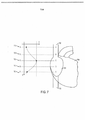
[0020] FIG. 5 ilustra exemplo de amplitudes de sinal de eletrocardiograma em diferentes localizações no sistema venoso central.[0020] FIG. 5 illustrates an example of electrocardiogram signal amplitudes at different locations in the central venous system.
[0021] FIG. 6 ilustra exemplo de espectro de potência de sinal de eletrocardiograma em diferentes localizações no sistema venoso central.[0021] FIG. 6 illustrates an example of an electrocardiogram signal power spectrum at different locations in the central venous system.
[0022] FIG. 7 ilustra exemplo de distribuição de sinal de energia elétrica de eletrocardiograma em diferentes localizações no sistema venoso central.[0022] FIG. 7 illustrates an example of electrocardiogram electrical energy signal distribution at different locations in the central venous system.
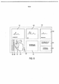
[0023] FIG. 8 representa uma interface gráfica de usuário de acordo com uma modalidade da presente invenção.[0023] FIG. 8 represents a graphical user interface according to an embodiment of the present invention.
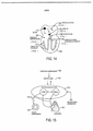
[0024] FIG. 9 representa uma interface gráfica de usuário de acordo com uma outra modalidade da presente invenção.[0024] FIG. 9 represents a graphical user interface according to another embodiment of the present invention.
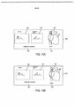
[0025] FIGS. 10A e 10B descrevem um exemplo de impressão para uma informação exibida por uma interface gráfica de usuário, de acordo com uma modalidade da presente invenção.[0025] FIGS. 10A and 10B describe an example printout for information displayed by a graphical user interface, in accordance with an embodiment of the present invention.
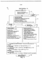
[0026] FIG. 11 é um diagrama em bloco para um método com base em computador para posicionar um dispositivo endovascular no ou perto do coração usando sinais de eletrocardiograma sinais.[0026] FIG. 11 is a block diagram for a computer-based method for positioning an endovascular device at or near the heart using electrocardiogram signals.
[0027] FIG. 12 ilustra um outro algoritmo de suporte para tomada de decisão para um método com base em computador para posicionar um dispositivo endovascular no ou perto do coração usando sinais de eletrocardiograma, de acordo com uma modalidade alternativa.[0027] FIG. 12 illustrates another decision support algorithm for a computer-based method for positioning an endovascular device at or near the heart using electrocardiogram signals, in accordance with an alternative modality.
[0028] FIG. 13 ilustra o sistema de condução cardíaca do coração.[0028] FIG. 13 illustrates the heart's cardiac conduction system.
[0029] FIG. 14 ilustra propagação do sinal elétrico no sistema de condução do coração.[0029] FIG. 14 illustrates electrical signal propagation in the conduction system of the heart.
[0030] FIG. 15 ilustra atividade elétrica no sistema cardiovascular devido ao sistema de controle neuronal.[0030] FIG. 15 illustrates electrical activity in the cardiovascular system due to the neuronal control system.
[0031] A invenção será agora descrita com referência as figuras dos desenhos, no quais numerais linha de referência parecidos se refere à partes parecidas ao longo da descrição.[0031] The invention will now be described with reference to the figures of the drawings, in which like reference line numerals refer to like parts throughout the description.
[0032] Modalidades da presente invenção vantajosamente fornecem um aparelho(es) inventivo, algoritmos de processamento de dados baseados em computador e métodos para obter e usar endovascular ECGs em um número de aplicações clínicas e configurações. Por exemplo, uma vez que o dispositivo pode ser usado para orientar dispositivos endovasculares no e em volta do coração, por ex., orientando dispositivos de acesso ao sistema venoso central na veia cava superior, átrio direito, e ventrículo direito. Tais dispositivos de acesso ao sistema venoso central podem incluir cateteres venosos centrais (CVC), cateteres centrais inserido perifericamente (PICC), portas implantáveis, cateteres de hemodiálises, cateteres em tubo e outros. Outros dispositivos que podem se beneficiar da guia com o aparelho inventivo são sondas temporárias de marca-passo cardíaco colocado através do sistema venoso central. Cateteres e fios-guia usados nos procedimentos do coração esquerdo também podem se beneficiar da presente invenção diminuindo a quantidade de contraste e radiação requerida para orienta esses dispositivos na posição. Em um outro exemplo, o aparelho pode ser usado para monitoração minimamente agressiva e avaliar condições do coração com base em sua atividade elétrica, por ex., avaliando pré-carga em um ciclo do coração ou monitora segmentos de ST e ondas T em insuficiência cardíaca congestiva.[0032] Embodiments of the present invention advantageously provide an inventive apparatus(es), computer-based data processing algorithms, and methods for obtaining and using endovascular ECGs in a number of clinical applications and settings. For example, since the device can be used to guide endovascular devices in and around the heart, eg, guiding devices to access the central venous system in the superior vena cava, right atrium, and right ventricle. Such central venous system access devices may include central venous catheters (CVC), peripherally inserted central catheters (PICC), implantable ports, hemodialysis catheters, tube catheters, and others. Other devices that can benefit from guidance with the inventive apparatus are temporary cardiac pacemaker probes placed through the central venous system. Catheters and guidewires used in left heart procedures can also benefit from the present invention by decreasing the amount of contrast and radiation required to guide these devices into position. In another example, the device can be used for minimally aggressive monitoring and evaluating heart conditions based on its electrical activity, eg, evaluating preload in a heart cycle or monitoring ST segments and T waves in heart failure congestive.
[0033] Em um aspecto da invenção, um aparelho é descrito consistindo de adaptadores estéreis, um módulo eletrônico para aquisição de sinal, um módulo de módulo de computador, software, e dispositivos e conexões periféricos. Em uma modalidade, o módulo eletrônico para aquisição de sinal pode ser dedicado para adquirir e processar sinais elétricos endovasculares gerados pelo corpo (ECG endovascular), em uma outra modalidade o módulo eletrônico pode ser dedicado para adquirir e processar ECGs endovasculares assim como ECGs à nível da pele.[0033] In one aspect of the invention, an apparatus is described consisting of sterile adapters, an electronic module for signal acquisition, a computer module module, software, and peripheral devices and connections. In one modality, the electronic signal acquisition module can be dedicated to acquire and process endovascular electrical signals generated by the body (endovascular ECG), in another modality the electronic module can be dedicated to acquire and process endovascular ECGs as well as ECGs at the level of the skin.
[0034] Em uma modalidade, o módulo eletrônico e o módulo de computador podem ser módulos separados, em uma outra modalidade eles podem ser integrados no mesmo módulo e em um invólucro, e ainda em uma outra modalidade eles pode se comunicar cada um com o outro via uma conexão sem fio, tal como Bluetooth. Em uma modalidade, o aparelho pode conter uma impressora integrada, enquanto em uma outra modalidade a impressora pode ser externa e presa ao aparelho e o aparelho conectado via rede, por ex., sem fio a outros dispositivos. Em ainda uma outra modalidade o aparelho pode ser usado para telemetria e para transmitir os eletrocardiogramas endovasculares para uma localização remota, por ex., via uma linha telefônica, Internet, e / ou telefone sem fio. Qualquer combinação das modalidades mencionadas acima também é possível.[0034] In one modality, the electronic module and the computer module can be separate modules, in another modality they can be integrated in the same module and in a housing, and in yet another modality they can each communicate with the another via a wireless connection, such as Bluetooth. In one mode, the device can contain an integrated printer, while in another mode the printer can be external and attached to the device and the device connected via a network, eg wirelessly, to other devices. In yet another embodiment the device can be used for telemetry and to transmit endovascular electrocardiograms to a remote location, eg via a telephone line, Internet, and/or cordless telephone. Any combination of the modalities mentioned above is also possible.
[0035] Em um outro aspecto da invenção, várias configurações permitem a conexão de dispositivos endovasculares, tal como dispositivo de acessos ao sistema venoso central, ao módulo eletrônico para aquisição de sinal aquisição e processamento. Em uma modalidade, o dispositivo consiste de um fio de conexão com duas extremidades e conectores especiais em cada extremidade. Em uma extremidade, o fio pode estar conectado a um metal ou estilete ou fio-guia de nitinol como comumente disponível no mercado. Na outra extremidade, o fio pode ser conectado com segurança para o módulo eletrônico. Em uma outra modalidade, o dispositivo inclui um fio-guia revestido, por ex., feito de nitinol ou aço inoxidável com as extremidades distância e próxima descobertas e com marcações de centímetros. Em tal uma modalidade, o fio-guia revestido é inserido de modo endovascualr, enquanto o fio de conexão é conectado à extremidade proximal do fio-guia revestido. Em uma outra modalidade, o dispositivo inclui um adaptador de cateter - seringa fornecido com um fio de conexão elétrica. Em uma extremidade, o fio de conexão elétrico está em contato com o fluido, por ex., salino fluindo dentro do adaptador de cateter - seringa. Na outra extremidade o fio de conexão pode ser conectado ao módulo eletrônico.[0035] In another aspect of the invention, various configurations allow the connection of endovascular devices, such as access device to the central venous system, to the electronic module for signal acquisition and processing. In one embodiment, the device consists of a connecting wire with two ends and special connectors at each end. At one end, the wire can be connected to a metal or nitinol stylet or guide wire as commonly available on the market. At the other end, the wire can be securely connected to the electronics module. In another embodiment, the device includes a coated guidewire, eg made of nitinol or stainless steel with the far and near ends uncovered and with centimeter markings. In such an embodiment, the coated guidewire is inserted endovascularly, while the connecting wire is connected to the proximal end of the coated guidewire. In another embodiment, the device includes a catheter-syringe adapter provided with an electrical connection wire. At one end, the electrical connecting wire is in contact with fluid, eg saline, flowing inside the catheter-syringe adapter. At the other end, the connecting wire can be connected to the electronics module.
[0036] Em um outro aspecto da invenção, várias configurações de eletrodo permitem a aquisição ótima de ECGs endovasculares. Em uma modalidade, uma única sonda é usada para fornecer informação sobre a localização da ponta de um dispositivo endovascular dentro da estrutura vascular. Em uma outra modalidade uma configuração modificada de três sondas é usada para fornecer simultânea monitoração em 3 sondas da atividade do coração ao mesmo tempo com fornecimento de informação de localização da ponta. Em uma outra modalidade uma configuração de sonda única modificada mais referência de terra é usada para telemetria e transferir informação a partir da ponta do cateter remotamente.[0036] In another aspect of the invention, various electrode configurations allow for the optimal acquisition of endovascular ECGs. In one embodiment, a single probe is used to provide information about the location of the tip of an endovascular device within the vascular structure. In another embodiment a modified three probe configuration is used to provide simultaneous 3-probe monitoring of heart activity at the same time providing tip location information. In another embodiment a modified single probe plus ground reference configuration is used for telemetry and transferring information from the catheter tip remotely.
[0037] Em um outro aspecto da invenção algoritmos são introduzidos para a análise das formas de onda do ECG e para suportar tomadas de decisão com base nessas formas de onda. Esses algoritmos discriminam entre diferentes localizações na estrutura vascular e avaliar funções do corpo (sistemáticas e em específicas localizações no corpo), em particular funcionalidade do coração. Em várias modalidades, esses algoritmos usam análise de formas de onda no domínio do tempo: morfológica, por exemplo forma; estatística, por exemplo comportamento.[0037] In another aspect of the invention algorithms are introduced for analyzing the ECG waveforms and to support decision making based on these waveforms. These algorithms discriminate between different locations in the vascular structure and assess body functions (systematic and at specific locations in the body), in particular heart functionality. In various embodiments, these algorithms use time-domain waveform analysis: morphological, eg shape; statistics, for example behavior.
[0038] Em outras modalidades, os algoritmos usam análise de formas de onda no domínio da frequência: morfológica, por exemplo forma; estatística, por exemplo comportamento. Em modalidades adicionais, análise de energia de sinal nos domínios do tempo e frequência é também efetuada, morfologicamente e estatisticamente. Difusão, estatística e tomar decisão com base em conhecimento são também contemplados pela presente invenção como ferramentas de suporte à decisão.[0038] In other modalities, the algorithms use waveform analysis in the frequency domain: morphological, for example shape; statistics, for example behavior. In additional embodiments, analysis of signal energy in the time and frequency domains is also performed, morphologically and statistically. Diffusion, statistics and knowledge-based decision making are also contemplated by the present invention as decision support tools.
[0039] Em um outro aspecto da invenção, uma interface de usuário é fornecida que vantajosamente simplifica interpretação de dados e fluxo de funcionamento. Em uma modalidade a interface de usuário inclui gráficos simplificados mostrando a localização na estrutura vascular e no coração da ponta do dispositivo endovascular em uso sem mostrar qualquer das formas de onda do ECG. Em uma outra modalidade, a interface de usuário mostra, em tempo real, a mudança na localização da ponta do dispositivo endovascular em uso.[0039] In another aspect of the invention, a user interface is provided that advantageously simplifies data interpretation and workflow. In one modality the user interface includes simplified graphics showing the location in the vascular structure and in the heart of the tip of the endovascular device in use without showing any of the ECG waveforms. In another modality, the user interface shows, in real time, the change in the location of the tip of the endovascular device in use.
[0040] Em um outro aspecto da invenção, vários métodos inventivos são apresentados que usam o aparelho aqui descrito em aplicações clínicas. Em uma modalidade, um método com base em computador é fornecido que orienta cateteres venosos centrais (CVC, PICCs, hemodiálise, portas implantáveis, e outros) usando estiletes, fios-guia e solução salina para a veia cava superior, veia cava inferior, o átrio direito, e o ventrículo direito. Este método é vantajosamente menos sensitivo para pacientes com arritmias do que a arte anterior, e representa uma alternativa para confirmação via raio X do peito da localização de cateteres venosos centrais na maioria dos casos clínicos. Em uma outra modalidade, um método com base em computador é fornecido que orienta fios-guias revestidos no coração direito e esquerdo. Em uma outra modalidade, um método com base em computador é fornecido que orienta a colocação de sondas de marca-passo cardíaco temporárias através do sistema venoso central. Em uma outra modalidade, um método é fornecido que é minimamente invasivo e monitora pré-carga usando despolarização e ritmos cardíacos. Em uma outra modalidade, um método é fornecido isto é minimamente invasivo e monitora arritmias usando análise de onda P. Em uma outra modalidade, um método é fornecido que é minimamente invasivo e monitora falha do coração usando segmento ST e análise de onda T.[0040] In another aspect of the invention, several inventive methods are presented that use the apparatus described herein in clinical applications. In one modality, a computer-based method is provided that guides central venous catheters (CVC, PICCs, hemodialysis, implantable ports, and others) using stylets, guide wires, and saline solution for the superior vena cava, inferior vena cava, the right atrium, and the right ventricle. This method is advantageously less sensitive for patients with arrhythmias than the prior art, and represents an alternative to chest X-ray confirmation of the location of central venous catheters in most clinical cases. In another embodiment, a computer-based method is provided that guides coated guidewires in the right and left heart. In another embodiment, a computer-based method is provided that guides the placement of temporary cardiac pacemaker probes through the central venous system. In another embodiment, a method is provided that is minimally invasive and monitors preload using depolarization and heart rhythms. In another embodiment, a method is provided that is minimally invasive and monitors arrhythmias using P wave analysis. In another embodiment, a method is provided that is minimally invasive and monitors heart failure using ST segment and T wave analysis.
[0041] FIG. 1 é um diagrama em bloco que representa um aparelho de acordo com uma modalidade da presente invenção.[0041] FIG. 1 is a block diagram depicting an apparatus in accordance with an embodiment of the present invention.
[0042] O aparelho 100 pode ser preso através de um adaptador (120) a uma grande variedade de dispositivos de acesso vasculares personalizados e comercialmente disponíveis (110). Exemplos de tais dispositivos são: cateteres venosos centrais (CVC), cateteres centrais inseridos de modo periférica (PICC), portas implantáveis, cateteres em tubo, cateteres de hemodiálise, cateteres de guia para sondas de marca-passo cardíaco, fios-guias usados para coronária e outras intervenções vasculares, estiletes, agulhas de seringa, e outros. Se o dispositivo de acesso vascular é um estilete, um fio-guia, ou uma agulha de seringa, seu material precisa ser suficientemente eletricamente condutivo, por ex., aço inoxidável ou nitinol. Em tal um caso o adaptador tipo ganho ou tipo jacaré de acordo com a presente invenção deve ser usado para estabelecer um caminho condutivo através de um dos lumens do cateter. Em tal um caso, o adaptador de seringa - cateter de acordo com a presente invenção deve ser usado.
[0043] O módulo eletrônico (130) recebe sinais elétricos provenientes do adaptador e de um ou mais outros eletrodos colocados à nível da pele do paciente (115). Alternativamente, mais do que um adaptador pode ser usado ao mesmo tempo para conectar mais do que um dispositivo endovascular de modo a fornecer diferentes sinais elétricos ao módulo eletrônico. O uso de eletrodos à nível da pele é opcional em determinadas configurações do dispositivo. O módulo eletrônico processa os sinais elétricos e os transmite para um módulo de computador (140) para ainda processamento adicional e outras funções. Em uma modalidade o módulo eletrônico e o módulo de computador podem ser empacotados separadamente, em uma outra modalidade eles podem ser integrados no mesmo pacote. Em uma modalidade a conexão entre o módulo eletrônico e o módulo de computador pode ser com fio, em uma outra modalidade ela pode ser sem fio, por ex., usando Bluetooth.[0043] The electronic module (130) receives electrical signals from the adapter and one or more other electrodes placed at the level of the patient's skin (115). Alternatively, more than one adapter can be used at the same time to connect more than one endovascular device to provide different electrical signals to the electronics module. The use of skin-level electrodes is optional with certain device configurations. The electronic module processes the electrical signals and transmits them to a computer module (140) for further processing and other functions. In one modality the electronic module and the computer module can be packaged separately, in another modality they can be integrated in the same package. In one mode the connection between the electronics module and the computer module can be wired, in another mode it can be wireless, eg using Bluetooth.
[0044] O módulo de computador processa os sinais a partir do módulo eletrônico aplicando algoritmos (170) conforme descrito pela corrente invenção. O módulo de computador também pode ser conectado aos periféricos (160), por ex., uma impressora ou uma impressora de etiqueta e dispositivos de armazenamento e fornece conectividade incluindo conectividade sem fio (150) para outros computadores ou para a Internet. O dispositivo de armazenamento pode ser usado para armazenar um banco de dados de conhecimento e informação considerando a aplicação à mão. A interface de conectividade pode ser usada para atualizar este banco de dados remotamente com o mais recente conhecimento e informação relevante, novas descobertas considerando uma relação entre eletrocardiogramas e condições de coração. O módulo de computador suporta uma interface gráfica de usuário (180) otimizado para o propósito da aplicação clínica à mão.[0044] The computer module processes the signals from the electronic module applying algorithms (170) as described by the current invention. The computer module can also be connected to peripherals (160), eg a printer or label printer and storage devices, and provides connectivity including wireless connectivity (150) to other computers or to the Internet. The storage device can be used to store a database of knowledge and information regarding the application at hand. The connectivity interface can be used to update this database remotely with the latest knowledge and relevant information, new discoveries considering a relationship between electrocardiograms and heart conditions. The computer module supports a graphical user interface (180) optimized for the purpose of hand-held clinical application.
[0045] FIGS. 2A, 2B e 2C descrevem vários dispositivos adaptadores endovasculares.[0045] FIGS. 2A, 2B and 2C describe various endovascular adapter devices.
[0046] FIG. 2A ilustra adaptadores que pode ser feitos de um fio condutivo isolado (255) feito de cobre ou aço inoxidável tendo duas extremidades: uma extremidade conectada a um dispositivo de acesso vascular (255), a outra extremidade conectada ao módulo eletrônico (250). A extremidade que se conecta aos dispositivos de acesso vasculares inclui um conector que podem ter várias configurações. Em uma modalidade, o conector e um conector de clip J (230) com uma mola para propósitos de isolamento quando a ponta J não é estendida. Em uma outra modalidade, o conector é um do tipo jacaré isolado (220). Em uma outra modalidade, o conector é um adaptador de cateter - seringa (210). Uma extremidade do adaptador de cateter - seringa (211) pode se conectar ao luer do cateter. A outra extremidade pode se conectar (215) a uma seringa. Uma inserção de metal (214), por ex., um anel de metal é localizado dentro do corpo do adaptador, fica em contato com a solução salina conforme ele flui da seringa em direção ao lúmen do cateter. A inserção de metal é conectada através da parede do adaptador ta um fio (212) que por sua vez é conectado ao conector (250). Em uma modalidade o conector (250) é assegurado através de isolamento externo (241) e se conecta em uma tomada segura no módulo eletrônico. Em uma outra modalidade o conector (250) tem uma forma de bico otimizada (242) permitindo uma conexão fácil e segura de um conector de cabo de EXG padrão.[0046] FIG. 2A illustrates adapters that may be made of an insulated conductive wire (255) made of copper or stainless steel having two ends: one end connected to a vascular access device (255), the other end connected to the electronics module (250). The end that connects to vascular access devices includes a connector that can have various configurations. In one embodiment, the connector is a J-clip connector (230) with a spring for isolation purposes when the J tip is not extended. In another embodiment, the connector is an insulated alligator type (220). In another embodiment, the connector is a catheter-syringe adapter (210). One end of the catheter-syringe adapter (211) can connect to the luer of the catheter. The other end can connect (215) to a syringe. A metal insert (214), e.g., a metal ring, is located within the adapter body, contacts the saline solution as it flows from the syringe towards the lumen of the catheter. The metal insert is connected through the wall of the adapter to a wire (212) which is in turn connected to the connector (250). In one mode the connector (250) is secured by external insulation (241) and plugs into a secure socket on the electronics module. In another embodiment the connector (250) has an optimized nozzle shape (242) allowing for easy and secure connection of a standard EXG cable connector.
[0047] Figura 2B ilustra um novo fio-guia (260) que permite coleta informação elétrica somente em sua extremidade distal (261). O fio-guia feito de materiais eletricamente condutivos com condutividade bastante boa, por ex., aço inoxidável ou nitinol. O fio-guia é revestido com um revestimento eletricamente isolante como revestimento de parileno conformal sobre seu inteiro comprimento exceto na extremidade distal e na extremidade proximal. O cateter tem marcas de comprimento impressas nele (262). A extremidade distal que é uma ponta a-traumática, ou como uma ponta J ou em qualquer outra modalidade a-traumática não é revestido e ela permite contato elétrico com o sangue. A extremidade proximal não é revestida (263) e permite conectores como aqueles na figura 2 (220 ou 230) serem eletricamente conectados ao fio- guia.[0047] Figure 2B illustrates a new guidewire (260) that allows collection of electrical information only at its distal end (261). Guide wire made of electrically conductive materials with very good conductivity, eg stainless steel or nitinol. The guidewire is coated with an electrically insulating coating such as a conformal parylene coating over its entire length except at the distal end and the proximal end. The catheter has length marks imprinted on it (262). The distal end that is an a-traumatic tip, either like a J-tip or in any other a-traumatic modality is uncoated and it allows electrical contact with the blood. The proximal end is uncoated (263) and allows connectors like those in figure 2 (220 or 230) to be electrically connected to the guidewire.
[0048] Figura 2C ilustra uma outra modalidade de um adaptador de cateter - seringa. Um pedaço de plástico (270) consiste de uma extremidade em forma (271) que pode encaixar em um luer de cateter padrão ou no seu lumen. A forma e o material permitem um bom contato entre a extremidade (271) e a parede interna do luer ou lumen tal que o fluido pode fluir sem fuga e nenhum ar é introduzido no lumen no processo. A outra extremidade do pedaço (272) é um conector tipo luer que pode se encaixar em qualquer seringa padrão. O corpo do adaptador ou câmara interna (273) se adapta ao diâmetro da luer (272) para o tamanho do lúmen de cateter interno (271) e fornece uma conexão de um elemento eletricamente condutivo na câmara interna para um fio conectado para fora da câmara através de uma perfuração na parede da câmara (274). A conexão através da parede da câmara é a prova de água. Quando salina é injetada através do adaptador, a conexão (274) cria um caminho condutivo à prova de água entre a salina em um fio externo. O adaptador (290) é um pedaço de plástico que faz interface dos tamanhos de diâmetros (291) e (292). Em uma modalidade a extremidade (271) do adaptador (270) se encaixa na extremidade do lúmen (291) do adaptador (290) e a outra extremidade (292) do adaptador (290) se encaixa no lumen de um cateter usado para colocar portas implantáveis.[0048] Figure 2C illustrates another modality of a catheter adapter - syringe. A piece of plastic (270) consists of a shaped end (271) that can fit into a standard catheter luer or lumen. The shape and material allow good contact between the end (271) and the inner wall of the luer or lumen such that fluid can flow without leakage and no air is introduced into the lumen in the process. The other end of the piece (272) is a luer-type connector that can fit any standard syringe. The adapter body or inner chamber (273) fits the diameter of the luer (272) to the size of the inner catheter lumen (271) and provides a connection from an electrically conductive element in the inner chamber to a wire connected to the outside of the chamber. through a perforation in the chamber wall (274). The connection through the chamber wall is waterproof. When saline is injected through the adapter, the connection (274) creates a waterproof conductive path between the saline in an outer wire. The adapter (290) is a piece of plastic that interfaces diameter sizes (291) and (292). In one embodiment the end (271) of the adapter (270) fits the lumen end (291) of the adapter (290) and the other end (292) of the adapter (290) fits the lumen of a catheter used to place ports implantable.
[0049] FIG. 3 é um diagrama em bloco de um módulo eletrônico (300) para aquisição e processamento de eletrocardiograma endovascular de acordo com uma modalidade da presente invenção.[0049] FIG. 3 is a block diagram of an electronic module (300) for endovascular electrocardiogram acquisition and processing in accordance with an embodiment of the present invention.
[0050] A interface de conexão do paciente (310) permite conectar sondas elétricas para o paciente (305). Qualquer combinação de eletrodos à nível da pele e / ou conexões elétricas para dispositivos endovasculares usando os adaptadores discutidos acima pode ser usada. Em uma modalidade, o amplificador (320) é um amplificador de quatro estágio com ganho variável, que pode amplificar sinais elétricos que vem através do cabo do paciente, por exemplo, típicos de valores de eletrocardiógrafo. O conversor de analógico para digital (330) converte os sinais em formato digital legível pelo micro-processador (340). Qualquer número e configurações de micro processadores, micro-controladores, processadores de sinais digitais pode ser usado para implementar a função de micro-processamento (340).[0050] The patient connection interface (310) allows connecting electrical probes to the patient (305). Any combination of skin level electrodes and/or electrical connections to endovascular devices using the adapters discussed above can be used. In one embodiment, the amplifier (320) is a four-stage amplifier with variable gain, which can amplify electrical signals coming through the patient cable, eg typical of electrocardiograph values. The analog to digital converter (330) converts the signals into digital format readable by the microprocessor (340). Any number and configurations of microprocessors, microcontrollers, digital signal processors can be used to implement the microprocessing function (340).
[0051] Em uma modalidade, um micro-controlador é responsável para controlar a comunicação serial com um módulo de computador (390) via a interface serial (370) ou via a interface sem fio (380) e um processador de sinal digital (DSP) é responsável para implementar uma ou vários dos algoritmos inventivos aqui descritos. Alternativamente, um único processador pode ser usado para ambos comunicação e processamento.[0051] In one embodiment, a microcontroller is responsible for controlling serial communication with a computer module (390) via the serial interface (370) or via the wireless interface (380) and a digital signal processor (DSP ) is responsible for implementing one or more of the inventive algorithms described herein. Alternatively, a single processor can be used for both communication and processing.
[0052] O micro-processador (340) também recebe comandos a partir do módulo de computador (390) e controla diferentes canais de comunicação serial (370) a partir da interface do paciente (310) de modo a assegurar proteção ao paciente contra choque elétrico. Em uma modalidade o bloco de isolamento (350) pode consistir de um transformador e / ou acopladores, por ex. acopladores ópticos.[0052] The microprocessor (340) also receives commands from the computer module (390) and controls different serial communication channels (370) from the patient interface (310) in order to ensure patient protection against shock electric. In one embodiment the isolation block (350) can consist of a transformer and/or couplers, eg. optical couplers.
[0053] FIGS. 4A, 4B, 4C, e 4D representam configurações de eletrodo que fornecem aquisição ótima de eletrocardiograma endovascular de acordo com várias modalidades da presente invenção.[0053] FIGS. 4A, 4B, 4C, and 4D represent electrode configurations that provide optimal endovascular electrocardiogram acquisition in accordance with various embodiments of the present invention.
[0054] FIG. 4A representa uma configuração de sonda única com um eletrodo de referência (410), por exemplo, preso à nível de pele do paciente sobre o braço direito e com o outro eletrodo preso através de um adaptador a um dispositivo endovascular (415). O eletrodo de referência preso à nível de pele sobre o braço direito é apresentado nesta configuração para propósitos de ilustração somente. Outras localizações do eletrodo de referência são possíveis dependendo do tipo de ECG requerido. O eletrodo de referência sobre o braço direito junto com a ponta do dispositivo endovascular usado com o adaptador pode ser similar à sonda II de um padrão ECG. Neste caso os ECGs obtidos a partir da veia cava superior (401) e veia cava inferior (402) podem ser otimizados. O eletrodo de referência pode ser preso à nível de pele em qualquer outra localização de modo a simular outras sondas do ECG padrão. O eletrodo de referência pode ser também conectado aos adaptadores presos aos outros dispositivos endovasculares de modo a obter mais informação local de dentro do coração do paciente (400).[0054] FIG. 4A depicts a single probe configuration with a reference electrode (410), for example, attached to the patient's skin level on the right arm and with the other electrode attached via an adapter to an endovascular device (415). The reference electrode attached to skin level on the right arm is shown in this configuration for illustration purposes only. Other reference electrode locations are possible depending on the type of ECG required. The reference electrode on the right arm together with the tip of the endovascular device used with the adapter can be similar to probe II of a standard ECG. In this case the ECGs obtained from the superior vena cava (401) and inferior vena cava (402) can be optimized. The reference electrode can be attached to skin level at any other location to simulate other standard ECG probes. The reference electrode can also be connected to adapters attached to other endovascular devices in order to obtain more local information from inside the patient's heart (400).
[0055] FIG 4B representa uma configuração de 3 sondas modificada, com capacidades de monitoração e guia, com 4 eletrodos. Três (3) eletrodos correspondem aos eletrodos de ECG padrão: braço direito (RA, 420), braço esquerdo (LA, 425), e perna esquerda (LL, 430) usados como referência. O quarto eletrodo é preso através de um adaptador ao dispositivo endovascular (C, 435). Nesta configuração, o módulo eletrônico e o algoritmo efetuam duas funções de modo simultâneo: os três eletrodos (RA, LL e LL) padrões efetuam uma função de monitoração do coração, enquanto o eletrodo C (435) permite gravar o ECG na ponta do dispositivo.[0055] FIG 4B represents a modified 3-probe configuration, with monitoring and guidance capabilities, with 4 electrodes. Three (3) electrodes correspond to the standard ECG electrodes: right arm (RA, 420), left arm (LA, 425), and left leg (LL, 430) used as reference. The fourth electrode is attached via an adapter to the endovascular device (C, 435). In this configuration, the electronic module and the algorithm perform two functions simultaneously: the three standard electrodes (RA, LL and LL) perform a heart monitoring function, while the C electrode (435) allows recording the ECG on the device tip .
[0056] FIG 4C representa uma configuração de telemetria com uma unia sonda aterrada, incluindo a configuração ilustrada na Fig. 4A e uma referência de terra (450). Esta configuração pode ser usada para transmitir ECGs remotamente através de uma configuração de sistema de telemetria.[0056] FIG 4C represents a telemetry setup with a grounded probe, including the setup illustrated in Fig. 4A and a ground reference (450). This setup can be used to transmit ECGs remotely via a telemetry system setup.
[0057] FIG 4D representa um uso de monitores de ECG para orientar dispositivos endovasculares. Um monitor de ECG padrão é usado com entradas padrões RA (465), LA (460), e LL (470). LA (460) é conectado a um braço esquerdo e LL (470) para a perna esquerda do paciente. A entrada RA (465) é conectada a um comutador que pode ser usado pelo clínico para comutar a entrada RA (465) entre o eletrodo RA e o eletrodo do cateter (C) 475. Assim sendo ou monitoração ou guia de colocação do cateter pode ser alcançado alternativamente[0057] FIG 4D depicts a use of ECG monitors to guide endovascular devices. A standard ECG monitor is used with standard RA (465), LA (460), and LL (470) inputs. LA (460) is connected to a left arm and LL (470) to the patient's left leg. The RA input (465) is connected to a switch that can be used by the clinician to switch the RA input (465) between the RA electrode and the catheter electrode (C) 475. Therefore either monitoring or catheter placement guide can be reached alternatively
[0058] FIG. 5 ilustra exemplo de amplitudes de sinal de eletrocardiograma em diferentes localizações no sistema venoso central.[0058] FIG. 5 illustrates an example of electrocardiogram signal amplitudes at different locations in the central venous system.
[0059] O coração (504), átrio direito (501), veia cava superior (SVC) (502), e a veia cava inferior (IVC) (503) são ilustrados. A localização A está na SVC superior, a localização B está no terço inferior da SVC, a localização C está na junção cava - átrio, a localização D está no átrio direito, e a localização E está na veia cava inferior superior.[0059] The heart (504), right atrium (501), superior vena cava (SVC) (502), and inferior vena cava (IVC) (503) are illustrated. Location A is in the superior SVC, location B is in the lower third of the SVC, location C is in the vena cava-atrium junction, location D is in the right atrium, and location E is in the superior inferior vena cava.
[0060] Gráfico 510 ilustra uma forma de onda do ECG como uma função do tempo gravado na localização A. A amplitude absoluta das formas de onda é gravada na escala de amplitude (590). No caso de um ECG endovascular, os elementos padrões do eletrocardiograma são ilustrados: a onda P (560), a onda R (570), e a onda T (580). As amplitudes e forma na localização A gravadas com uma configuração de sonda como na figura 4D são similares a um eletrocardiograma gravada à nível da pele com a mesma configuração de eletrodo.[0060]
[0061] Gráfico 520 ilustra um ECG endovascular representado na localização B. A amplitude nesta localização é maior do que aquela na localização A mas as formas gerais da forma de onda são similares as das localizações localização A e B.[0061] Graph 520 illustrates an endovascular ECG represented at location B. The amplitude at this location is greater than at location A but the overall waveform shapes are similar to locations at location A and B.
[0062] Gráfico 530 ilustra um ECG endovascular representado na localização C. Na localização C na junção cava - átrio, a amplitude da forma de onda é ainda maior do que aquela na localização B e a onda P mudou dramaticamente se tornando maior do que a onda R. Esta forma de onda é uma indicação da proximidade do nó seio - átrio.[0062]
[0063] Gráfico 540 ilustra um ECG endovascular representado na localização D. Na localização D no átrio direito, as amplitudes são similares à localização C mas a onda P muda polaridade se tornando bi-polar. Isto é uma indicação que a medição do ECG ocorre além do nó seio - átrio.[0063]
[0064] Gráfico 550 ilustra um ECG endovascular representado na localização E. Na localização E na veia cava inferior, a forma de onda é similar àquela na localização A em termos de amplitude exceto que a onda P tem polaridade reversa. As diferenças nas formas de onda do ECG em diferentes localizações são usadas pelos algoritmos introduzidos aqui para discriminar entre as correspondentes localizações e para avaliar funcionalidade do coração e vasos sanguíneos.[0064]
[0065] FIG. 6 ilustra exemplos de espectro de potência de sinal de eletrocardiograma em diferentes localizações no sistema venoso central, usando uma escala espectral (690).[0065] FIG. 6 illustrates examples of electrocardiogram signal power spectrum at different locations in the central venous system using a spectral scale (690).
[0066] O coração (604), átrio direito (601), veia cava superior (SVC) (602), e a veia cava inferior (IVC) (603) são ilustrados. Gráfico 610 ilustra um espectro de ECG endovascular representado na localização A. Na localização A, o espectro (610) tem a aparência de uma única frequência central ou única banda (660) e com uma energia e potência espectral de distribuição de frequência similar àquela à nível da pele.[0066] The heart (604), right atrium (601), superior vena cava (SVC) (602), and inferior vena cava (IVC) (603) are illustrated.
[0067] Gráfico 620 ilustra espectro do ECG endovascular representado na localização B. Na localização B a frequência distribuição de frequência tem duas bandas principais e uma energia e potência espectral maior do que aquele na localização A.[0067]
[0068] Gráfico 630 ilustra um espectro de ECG endovascular na localização C. Na localização C, há múltiplas (3-4) frequências principais ou componentes espectrais principais distribuídos sobre um intervalo mais amplo de frequências (670). Esta distribuição espectral é indicativa da distribuição de energia em torno do nó seio - átrio. A potência espectral e energia de sinal têm aumentado comparado com localização B.[0068]
[0069] Gráfico 640 ilustra um espectro de ECG endovascular representado na localização D. Na localização D o espectro é mais amplo e de maior banda larga indicativo da atividade elétrica do átrio direito.[0069]
[0070] Gráfico 650 ilustra um espectro de ECG endovascular representado na localização E. O espectro de frequência na localização E é similar àquele na localização A. As diferenças nas formas de onda espectrais em diferentes localizações são usadas pelos algoritmos introduzidos aqui para discriminar entre as correspondentes localizações e para avaliar funcionalidade do coração e vasos sanguíneos.[0070]
[0071] FIG. 7 ilustra exemplo de distribuição de energia elétrica de sinal de eletrocardiograma em diferentes localizações no sistema venoso central. O coração (704), átrio direito (701), veia cava superior (SVC) (702), e veia cava inferior (IVC) (703) são ilustrados. Gráficos (710, 720, 730, 740, 750) representam a distribuição de energia nas diferentes localizações (A, B, C, D e E, respectivamente) e as mudanças no tempo são usadas pelos algoritmos introduzidos aqui para discriminar entre as correspondentes localizações e para avaliar funcionalidade do coração e vasos sanguíneos.[0071] FIG. 7 illustrates an example of electrocardiogram signal electrical energy distribution at different locations in the central venous system. The heart (704), right atrium (701), superior vena cava (SVC) (702), and inferior vena cava (IVC) (703) are illustrated. Graphs (710, 720, 730, 740, 750) represent the distribution of energy at different locations (A, B, C, D and E, respectively) and changes in time are used by the algorithms introduced here to discriminate between the corresponding locations and to assess heart and blood vessel functionality.
[0072] FIG. 8 representa uma interface gráfica de usuário de acordo com uma modalidade da presente invenção.[0072] FIG. 8 represents a graphical user interface according to an embodiment of the present invention.
[0073] Janela (810) apresenta a forma de onda do ECG em tempo real conforme ele é adquirido pelo módulo eletrônico usando a configuração do eletrodo anexado. Janela (820) é uma janela de referência e mostra uma forma de onda congelada usada para comparar com a janela corrente. Em uma modalidade, a forma de onda de referência na janela (820) pode ser obtida através dos eletrodos conectados ao módulo eletrônico em uma localização de referência do cateter e / ou usando uma configuração de referência dos eletrodos à nível da pele. Por exemplo, tal uma forma de onda de referência pode ser o ECG gravado usando um adaptador de acordo com a presente invenção conectado a um dispositivo endovascular colocado na junção cava - átrio. Em uma diferente modalidade, a forma de onda de referência na janela 820 pode ser uma forma de onda típica em uma determinada localização na estrutura vascular ou de uma determinada condição do coração como é gravado em um banco de dados de formas de onda e como é armazenado no meio de armazenamento do sistema de computador. Se a configuração do eletrodo permite simultânea monitoração do coração e gravação dos eletrocardiogramas usando um dispositivo endovascular, a janela (830) mostra uma das formas de ECG padrão para monitoração do coração, enquanto a janela (810) mostra o ECG na ponta dos dispositivos endovasculares quando conectado a um adaptador, tal como aqueles discutidos acima.[0073] Window (810) displays the real-time ECG waveform as it is acquired by the electronics module using the attached electrode configuration. Window (820) is a reference window and shows a frozen waveform used to compare with the current window. In one embodiment, the reference waveform in the window (820) can be obtained via electrodes connected to the electronics module at a reference location on the catheter and/or using a reference configuration of the electrodes at skin level. For example, such a reference waveform can be the ECG recorded using an adapter in accordance with the present invention connected to an endovascular device placed at the cava-atrium junction. In a different modality, the reference waveform in the 820 window can be a typical waveform at a particular location in the vascular structure or a particular condition of the heart as it is recorded in a waveform database and as it is stored in the storage medium of the computer system. If the electrode configuration allows simultaneous monitoring of the heart and recording of electrocardiograms using an endovascular device, the window (830) shows one of the standard ECG forms for monitoring the heart, while the window (810) shows the ECG at the tip of the endovascular devices when connected to an adapter such as those discussed above.
[0074] O ícone (870) é uma representação do coração, e as localizações A à E (875) ilustram diferentes localizações no coração e sistema vascular que podem ser discriminados analisando ECGs endovasculares de acordo com os métodos aqui divulgado. Como uma localização na estrutura vascular é identificado pelos algoritmos, o correspondente local e letra no ícone (875) se tornam realçados ou em algumas outras maneiras é tornado visível para o usuário. As barras (884), (885), e (886) mostram níveis de energia de sinal. A barra "E" (885) apresenta uma quantidade de energia elétrica computada a partir do espectro de frequência do ECG na localização corrente da ponta do dispositivo endovascular. A barra "R" (884) apresenta uma quantidade de energia elétrica computada a partir do espectro de frequência em uma localização de referência. A barra "M" (886) apresenta quantidade de energia elétrica computada a partir do espectro de frequência do ECG usando o sinal de ECG de monitoração a partir dos eletrodos à nível da pele. A janela (840) representa informação de monitoração, por ex., taxa de batida do coração. Informação do paciente (nome, dados de procedimento e outros) são mostrados na janela (850). Janela (860) contém elementos de controle de sistema como botões e informação de estado, por ex., tamanho, velocidade de rolagem, parâmetros de sistema e diagnósticos de sistema.[0074] The icon (870) is a representation of the heart, and locations A to E (875) illustrate different locations in the heart and vascular system that can be discriminated by analyzing endovascular ECGs according to the methods disclosed herein. As a location in the vascular structure is identified by the algorithms, the corresponding location and letter on the icon (875) become highlighted or in some other ways made visible to the user. Bars (884), (885), and (886) show signal energy levels. Bar "E" (885) displays an amount of electrical energy computed from the ECG frequency spectrum at the current location of the tip of the endovascular device. The "R" bar (884) presents an amount of electrical energy computed from the frequency spectrum at a reference location. The "M" bar (886) shows the amount of electrical energy computed from the ECG frequency spectrum using the monitoring ECG signal from the skin level electrodes. Window (840) represents monitoring information, eg heart beat rate. Patient information (name, procedure data and so on) is shown in the window (850). Window (860) contains system control elements such as buttons and status information, eg size, scroll speed, system parameters and system diagnostics.
[0075] FIG. 9 representa uma interface gráfica de usuário de acordo com uma outra modalidade da presente invenção.[0075] FIG. 9 represents a graphical user interface according to another embodiment of the present invention.
[0076] O ícone (920) é uma representação do coração e as localizações A à E (930) ilustram diferentes localizações no coração e sistema vascular que podem ser discriminadas analisando ECGs endovasculares. Como uma localização na estrutura vascular é identificada pelos algoritmos, o correspondente local e letra no ícone (930) se tornam realçados ou em alguma outra maneira é feito visível para o usuário. As barras (940), (950), e (960) mostram níveis de energia de sinal. A barra "E" (940) representa a quantidade de energia elétrica computada a partir do espectro de frequência do ECG na corrente localização da ponta do dispositivo endovascular. A barra "R" (950) mostra uma quantidade de energia elétrica computada a partir do espectro de frequência do ECG em uma localização de referência. A barra "M" (960) mostra a quantidade de energia elétrica computada a partir do espectro de frequência do ECG usando o sinal de ECG de monitoração proveniente dos eletrodos à nível da pele. O botão "Imprimir" (960) permite ao usuário imprimir a informação documentando o caso em uma impressora, por exemplo em uma impressora de etiquete para fixação rápida de prontuário do paciente.[0076] The icon (920) is a representation of the heart and locations A to E (930) illustrate different locations in the heart and vascular system that can be discriminated by analyzing endovascular ECGs. As a location in the vascular structure is identified by the algorithms, the corresponding location and letter on the icon (930) become highlighted or in some other way made visible to the user. Bars (940), (950), and (960) show signal energy levels. The "E" bar (940) represents the amount of electrical energy computed from the ECG frequency spectrum in the current location of the tip of the endovascular device. The "R" bar (950) shows an amount of electrical energy computed from the ECG frequency spectrum at a reference location. The "M" bar (960) shows the amount of electrical energy computed from the ECG frequency spectrum using the monitoring ECG signal from the skin-level electrodes. The "Print" button (960) allows the user to print the information documenting the case on a printer, for example on a label printer for quick attachment of the patient's record.
[0077] FIGS. 10A e 10B descrevem um exemplo de impressão para a informação exibida por uma interface gráfica de usuário, de acordo com uma modalidade da presente invenção.[0077] FIGS. 10A and 10B describe an example printout for information displayed by a graphical user interface, in accordance with an embodiment of the present invention.
[0078] FIG. 10A ilustra uma impressão (1000) para o caso de um procedimento de colocação de ponta do cateter no terço inferior do SVC. O campo 1010 representa o ícone do coração e por meio disso, a letra "B" correspondendo ao terço inferior da veia cava superior (SVC) é realçado (1040). Campo 1030 representa a forma de onda de ECG de referência gravada na ponta do cateter na junção cava - átrio na proximidade do nó seio - átrio. Campo 1020 representa a forma de onda do ECG na ponta do cateter na posição no qual ele foi colocado no final do procedimento. Para FIG. 10A, esta localização está no terço inferior da SVC e a forma de onda do ECG corresponde a esta localização. O nome do paciente (1001) e a data do procedimento (1002) são também impressos.[0078] FIG. 10A illustrates a
[0079] FIG. 10B representa uma impressão similar (1050) exceto que a posição final do procedimento está na junção cava - átrio na localização C (1090) no ícone do coração (1060). O campo "Nó SA" representa a forma de onda do ECG de referência (1080), e o campo "Posição Final " (1070) mostra que o cateter foi colocado com a ponta no nó seio - átrio: a forma de onda do ECG na localização final é similar ou mesmo idêntico com aquela na localização de referência no nó seio - átrio (Nó SA). É conhecido que a proximidade do nó SA indica a localização na junção cava - átrio. Essas localizações são algumas vezes consideradas idênticas por alguns clínicos.[0079] FIG. 10B represents a similar impression (1050) except that the final position of the procedure is at the cava-atrium junction at location C (1090) in the heart icon (1060). The "SA Node" field represents the reference ECG waveform (1080), and the "End Position" field (1070) shows that the catheter was placed with the tip at the sinus-atrium node: the ECG waveform in the final location it is similar or even identical with that in the reference location in the sinus-atrium node (Node SA). It is known that the proximity of the SA node indicates the location at the cava-atrium junction. These locations are sometimes considered to be identical by some clinicians.
[0080] FIG. 11 é um diagrama em bloco para UM método com base em computador (1100) para posicionar um dispositivo endovascular no ou perto do coração usando sinais de eletrocardiograma.[0080] FIG. 11 is a block diagram for a computer-based method (1100) for positioning an endovascular device at or near the heart using electrocardiogram signals.
[0081] Os algoritmos são aplicados ao sinal de entrada (1102) (ECG) adquirido pelo adaptador para os dispositivos endovasculares e, opcionalmente, da mesma forma através de eletrodos à nível da pele. O Bloco de Detecção de Erro (1105) detecta pelo menos três tipos de condições de erro / exceções, tal como, por exemplo, quando um desfibrilador foi aplicado ao paciente, quando um marca-passo cardíaco está disparando pulsos de excitação e / ou quando uma sonda / eletrodo está OFF. Esses erros / exceções podem ser tratados de modo diferente, e o usuário pode ser informado sobre a presença de uma exceção e o caminho de tratamento de exceção (1110).[0081] The algorithms are applied to the input signal (1102) (ECG) acquired by the adapter for the endovascular devices and, optionally, in the same way through electrodes at the skin level. The Error Detection Block (1105) detects at least three types of error conditions / exceptions, such as, for example, when a defibrillator has been applied to the patient, when a cardiac pacemaker is triggering excitation pulses, and/or when a probe / electrode is OFF. These errors / exceptions can be handled differently, and the user can be informed about the presence of an exception and the exception handling path (1110).
[0082] O bloco de Pré-Processamento (1115) pode amplificar o sinal, reduzir ruído, eliminar artefatos, etc. Em uma modalidade, redimensionar o sinal para o intervalo exibido ocorre sob controle do usuário e não é automático, como com a maioria de monitores de ECG correntemente disponíveis. Assim sendo, mudanças na amplitude dos ECGs são facilmente notadas. Um filtro passa alta corrige a linha de referência e reduz tais artefatos como artefatos respiratórios. Supressão de ruído de banda larga pode ser alcançada usando um filtro seletivo, por ex., uma transformada de pequena onda. Interferência eletromagnética com outro equipamento e a grade de energia podem ser suprimidas por um filtro de entalhe (filtro de banda estreita) centrado em 60 Hz ou 50 Hz para acomodar fontes de energia doméstica ou internacional. Ruído de alta frequência pode ser suprimido com um filtro passa baixo, que, em uma modalidade, é implementado com comprimento médio variável, tal como, por exemplo, uma janela de execução correspondendo a um ciclo do coração, uma média do ECG sobre vários ciclos do coração consecutivos. O bloco de Filtragem Adaptativo (1120) otimiza os coeficientes de filtro minimizando um sinal de erro.[0082] The Pre-Processing block (1115) can amplify the signal, reduce noise, eliminate artifacts, etc. In one modality, scaling the signal to the displayed range is under user control and is not automatic, as with most currently available ECG monitors. Therefore, changes in the amplitude of the ECGs are easily noticed. A high pass filter corrects the reference line and reduces such artifacts as respiratory artifacts. Broadband noise suppression can be achieved using a selective filter, eg a small wave transform. Electromagnetic interference with other equipment and the power grid can be suppressed by a notch filter (narrowband filter) centered at 60 Hz or 50 Hz to accommodate domestic or international power sources. High frequency noise can be suppressed with a low pass filter, which, in one modality, is implemented with variable average length, such as, for example, an execution window corresponding to one heart cycle, an ECG average over several cycles of consecutive hearts. The Adaptive Filtering block (1120) optimizes the filter coefficients minimizing an error signal.
[0083] O bloco de Reconhecimento Padrão no Domínio do Tempo (1130) identifica elementos da forma de onda de ECG, sua relação(s) e seu comportamento(s) no tempo. Um importante aspecto do algoritmo de Reconhecimento Padrão no Domínio do tempo no bloco 1130, assim como do bloco de Reconhecimento Padrão no Domínio da Frequência 1140, é histórico dos dados. Os ECGs são analisados em tempo real para determinados elementos, e, para outros elementos, uma área de armazenamento temporário de dados com um apropriado comprimento de área de armazenamento temporário é mantida na memória do eletrônico e / ou módulo de computadores de modo a permitir análise de dados históricos e prognóstico com base nesta análise. Em uma modalidade, a área de armazenamento temporário do histórico de dados é de vários segundos permitindo ao sinal de ECG correspondendo a várias batidas de coração ser salvo na área de memória de armazenamento temporário. Uma técnica de armazenamento temporário duplo permite que a forma de onda em uma área de armazenamento temporário seja processada enquanto a segunda área de armazenamento temporário continua a armazenar sinais. Assim sendo nenhuns dados de sinais são perdidos enquanto a forma de onda em uma área de armazenamento temporário é processada. Após processamento dos dados em uma área de armazenamento temporário ser completado, os resultados são enviados para os Algoritmos de Suporte de Decisão (1150) e as duas áreas de armazenamento temporário comutam os papéis. O comprimento da área de armazenamento temporário acomoda a duração do tempo do processamento de dados de modo a assegurar que nenhuns dados sejam perdidos. Uma técnica de armazenamento temporário duplo similar é também aplicada aos dados submetidos ao bloco de Reconhecimento Padrão no Domínio da Frequência (1140).[0083] The Time Domain Pattern Recognition block (1130) identifies elements of the ECG waveform, their relationship(s) and their behavior(s) in time. An important aspect of the Time Domain Pattern Recognition algorithm in
[0084] No caso de um ECG endovascular, elementos de interesse podem incluir, mas não são limitados à, um ou mais do seguinte:1. As ondas P, Q, R, S, T, e U, seus picos, amplitudes e durações; 2. A duração dos segmentos / intervalos P-R, S-T e T-P;3. A elevação do segmento S-T;4. As variâncias dos intervalos P-P e R-R;5. As variâncias dos intervalos S-T e do R-T, etc.;6. Os valores pico a pico da onda P e do QRS complexo;7. A proporção das amplitudes da onda P e da onda R e a proporção das amplitudes pico a pico de QRS complexo;8. A polaridade da onda P: único positivo, único negativo, ou bipolaridade;9. O derivativo da onda P, QRS complexo, e onda T;10. Média temporal do intervalo R-R em a batida de coração;11. Valor máximo da amplitude da onda P / pico e da amplitude pico a pico da onda P sobre um determinado período de tempo;12. Valor máximo da amplitude da onda R / pico e da amplitude pico a pico da QRX complexo sobre um determinado período de tempo[0084] In the case of an endovascular ECG, elements of interest may include, but are not limited to, one or more of the following:1. The P, Q, R, S, T, and U waves, their peaks, amplitudes and durations; 2. The duration of the segments/intervals P-R, S-T and T-P;3. The elevation of the S-T segment;4. The variances of the P-P and R-R intervals;5. The variances of the S-T and R-T intervals, etc.;6. The peak-to-peak values of the P wave and the complex QRS;7. The proportion of P-wave and R-wave amplitudes and the proportion of peak-to-peak amplitudes of complex QRS;8. The polarity of the P wave: single positive, single negative, or bipolar;9. The derivative of the P wave, complex QRS, and T;10 wave. Temporal average of the R-R interval in the heartbeat;11. Maximum value of the P-wave/peak amplitude and the peak-to-peak P-wave amplitude over a given period of time;12. Maximum value of the amplitude of the R/peak wave and the peak-to-peak amplitude of the complex QRX over a given period of time
[0085] Várias técnicas podem ser usadas para derivar a informação listada acima a partir das formas de onda de ECG, incluindo, mas não limitados à, um ou mais do seguinte:1. "Detecção de pico";2. Computação de primeiros derivativos;3. Execução médias ao longo do sinal em uma batida de coração e ao longo de múltiplas batidas de coração.4. Limitação adaptativa;5. Auto-correlação.[0085] Various techniques can be used to derive the information listed above from the ECG waveforms, including, but not limited to, one or more of the following:1. "Peak detection";2. Computation of first derivatives;3. Running averages over the signal in one heartbeat and over multiple heartbeats.4. Adaptive limitation;5. Auto-correlation.
[0086] A Transformada de Fourier Rápida no bloco (1125) efetua a Transformada de Fourier Rápida em um número de amostras de ECG armazenado em uma área de armazenamento temporário de um comprimento determinado, por ex., 256, 512, 1024, 2048 ou mais amostras de dados. A Transformada de Fourier transforma a forma de onda do domínio do tempo para o domínio da frequência.[0086] The Fast Fourier Transform in block (1125) performs the Fast Fourier Transform on a number of ECG samples stored in a temporary storage area of a specified length, eg 256, 512, 1024, 2048 or more data samples. The Fourier Transform transforms the waveform from the time domain to the frequency domain.
[0087] O bloco de Reconhecimento de Padrão de Domínio da Frequência (1140) ilustra vários aspectos de reconhecimento padrão efetuado nos ECGs em um domínio da frequência, incluindo, mas não limitados à, um ou mais do seguinte:1. Análise de componentes principais, i.e., determinação dos elementos mais significativos de um espectro de frequência (de forma similar para determinar os elementos morfológicos dos eletrocardiogramas, por ex., determinadas ondas e segmentos no domínio do tempo);2. Compressão de dados de modo a reduzir a quantidade de computação com base nos componentes principais;3. Determinação do número e morfologia dos componentes principais, em particular determinação se o espectro foi apenas uma, duas ou múltiplas frequências principais (bandas de frequência);4. Cálculo da potência espectral e da energia de sinal de um espectro de frequência;5. Média de execução ao longo de uma dimensão de frequência sobre um único espectro de modo a reduzir ruído de banda larga;6. Média de execução ao longo de vários espectros de modo filtrar artefatos;7. Determinação de elementos morfológicos adicionais do espectro, por ex. a frequência máxima, a energia contida na frequência máxima, um histograma de frequência, i.e., que frequências contém quanta energia, a frequência do mais alto pico de energia máximo significativo, etc.;8. Cálculo de comportamento e médias ao longo do tempo dos componentes principais e outros parâmetros determinados a partir da distribuição espectral, por ex., determinar o valor máximo da energia de sinal e da potência espectral sobre um determinado período de tempo;9. Determinar / estimar determinadas condições do coração com base na análise espectral. Esta determinação / estimativa é também efetuada em mais detalhes nos blocos de suporte de decisão 1150 e 1250.[0087] The Frequency Domain Pattern Recognition block (1140) illustrates various aspects of pattern recognition performed on ECGs in a frequency domain, including, but not limited to, one or more of the following:1. Principal component analysis, i.e., determining the most significant elements of a frequency spectrum (similarly to determining the morphological elements of electrocardiograms, eg, certain waves and segments in the time domain);2. Data compression in order to reduce the amount of computation based on the main components;3. Determining the number and morphology of the principal components, in particular determining whether the spectrum was only one, two or multiple principal frequencies (frequency bands);4. Calculation of spectral power and signal energy of a frequency spectrum;5. Running averaging over a frequency dimension over a single spectrum in order to reduce broadband noise;6. Run averaging over multiple spectra to filter out artifacts;7. Determination of additional morphological elements of the spectrum, eg. the maximum frequency, the energy contained in the maximum frequency, a frequency histogram, i.e., which frequencies contain how much energy, the frequency of the highest significant maximum energy peak, etc.;8. Calculation of behavior and averages over time of principal components and other parameters determined from the spectral distribution, eg determining the maximum value of signal energy and spectral power over a given period of time;9. Determine / estimate certain heart conditions based on spectral analysis. This determination/estimation is also performed in more detail in decision support blocks 1150 and 1250.
[0088] Vários algoritmos de suporte à decisão usam uma informação fornecida pelos algoritmos de Reconhecimento Padrão no Domínio do tempo e de Reconhecimento Padrão no Domínio da frequência. Em uma modalidade, bloco (1150) suporta colocar um dispositivo endovascular em ambos o terço inferior do SVC ou na junção cava - átrio.[0088] Several decision support algorithms use information provided by the Time Domain Pattern Recognition and Frequency Domain Pattern Recognition algorithms. In one modality, block (1150) supports placing an endovascular device in either the lower third of the SVC or at the cava-atrium junction.
[0089] Em particular, bloco 1150 é com base no conceito de primeiro alcançar a junção cava - átrio durante colocação do cateter. Na junção cava - átrio ou perto do nó seio - átrio, a onda P e outros parâmetros elétricos alcançam um valor máximo. Na junção cava - átrio, a onda P é unipolar. Após alcançar o nó seio - átrio na junção cava - átrio, i.e., o valor máximo da amplitude de pico P e potência espectral, o cateter é puxado para trás vários centímetros até a onda P diminui para metade da amplitude alcançada na junção cava - átrio. Em uma localização onde a onda P diminui para a metade da amplitude como a junção cava - átrio, o cateter é considerado estar no terço inferior da veia cava superior. A amplitude de pico da onda P ou amplitude pico a pico, assim como a potência espectral, é usada para mapear a localização na estrutura vascular para a forma de onda de ECG.[0089] In particular,
[0090] Mais particularmente, após receber um sinal de ECG endovascular associada com um dispositivo endovascular, o sinal é processado, sobre uma pluralidade de períodos de tempos pré-determinados, para calcular uma amplitude de onda P e uma potência espectral para cada período de tempo pré-determinado. Uma amplitude de onda P máxima é então determinada a partir de uma pluralidade de amplitudes de onda P, assim como uma potência espectral máxima associada a partir de uma pluralidade de potências espectrais. A localização na qual esses valores máximos são determinados é associada com uma pré-determinada localização no ou perto do coração, tal como a junção cava-atrial. A localização do dispositivo endovascular é então calculado, para cada período de tempo pré-determinado, com base em uma proporção da amplitude de onda P para a amplitude de onda P máxima e a proporção da potência espectral para a potência espectral máxima, e a localização do dispositivo endovascular é então exibida para o usuário. Adicionalmente, a polaridade da amplitude de onda P e a onda R também pode ser usada para determinar a localização do dispositivo endovascular.[0090] More particularly, upon receiving an endovascular ECG signal associated with an endovascular device, the signal is processed, over a plurality of predetermined time periods, to calculate a P-wave amplitude and a spectral power for each period of pre-determined time. A maximum P-wave amplitude is then determined from a plurality of P-wave amplitudes, as well as an associated maximum spectral power from a plurality of spectral powers. The location at which these maximum values are determined is associated with a predetermined location in or near the heart, such as the vena cava-atrial junction. The location of the endovascular device is then calculated, for each predetermined time period, based on a ratio of the P-wave amplitude to the maximum P-wave amplitude and the ratio of the spectral power to the maximum spectral power, and the location of the endovascular device is then displayed to the user. Additionally, the polarity of the P-wave and R-wave amplitudes can also be used to determine the location of the endovascular device.
[0091] Um único critério ou uma combinação de tal critério pode ser usado para suportar tomada de decisão. Em uma modalidade, Tl, T2, e T3 podem ser limites empiricamente estabelecido que são diferentes para cada paciente, e o algoritmo pode usar um laço adaptativo para ajustar os limites com base nas medições correntes. Em uma outra modalidade, esses limites são pré-determinados.[0091] A single criterion or a combination of such criteria can be used to support decision making. In one modality, T1, T2, and T3 can be empirically established limits that are different for each patient, and the algorithm can use an adaptive loop to adjust the limits based on current measurements. In another modality, these limits are predetermined.
[0092] Em modalidades alternativas, uma proporção entre a amplitude de pico P / P ou a amplitude pico a pico de onda P / amplitude R ou para a amplitude pico a pico QRS complexo também podem ser usados para estabelecer localização relativa para o nó seio - átrio. Em uma modalidade a amplitude de pico P precisa ser aproximadamente metade da amplitude de pico R e onda P precisa ser unipolar para a localização para corresponder ao terço inferior do SVC. Em uma outra modalidade, o pico a pico da onda P precisa ser metade da amplitude pico a pico QRS e a onda P precisa ser unipolar para a localização para corresponder ao terço inferior do SVC.[0092] In alternative modalities, a ratio of the peak P/P amplitude or the peak-to-peak P-wave amplitude/R amplitude or the complex QRS peak-to-peak amplitude can also be used to establish relative location for the sinus node. - atrium. In one modality the P peak amplitude needs to be approximately half of the R peak amplitude and the P wave needs to be unipolar for localization to correspond to the lower third of the SVC. In another modality, the peak-to-peak P wave needs to be half the peak-to-peak QRS amplitude and the P wave needs to be unipolar for localization to correspond to the lower third of the SVC.
[0093] Como discutido acima, os resultados do bloco de algoritmos de suporte de decisão 1150 pode ser apresentado ao usuário, por exemplo, realçando a apropriada localização no ícone do coração correspondendo ao tipo de ECG identificado pelo sistema (1160).[0093] As discussed above, the results of the decision
[0094] O bloco de algoritmo de suporte de decisão 1250, representado na Fig. 12, é com base em comparar a onda P, onda R e potência espectral de onda P nas correntes localizações com os valores desses parâmetros determinados a partir dos eletrocardiogramas à nível da pele em uma equivalente sonda, por ex., sonda II. Limites Tl à T6 são valores empíricos sujeitos à ajustes adaptativos relativos a cada paciente. Cada um do critério ou uma combinação de critério mostrado na Fig. 12 pode ser usado.[0094] The 1250 decision support algorithm block, depicted in Fig. 12, is based on comparing the P wave, R wave and P wave spectral power in the localized currents with the values of these parameters determined from the electrocardiograms to the skin level in an equivalent probe, eg, probe II. Limits Tl to T6 are empirical values subject to adaptive adjustments for each patient. Each of the criteria or a combination of criteria shown in Fig. 12 can be used.
[0095] Outros algoritmos de decisão também podem ser usados, em particular relacionados ao nível de energia elétrica conforme calculados a partir do espectro de ECG. No caso de colocar dispositivos endovasculares, um critério pode ser que, em uma localização correspondendo ao terço inferior do SVC, a energia elétrica média calculada a partir do ECG endovascular é duas vezes tanto alto quanto a energia elétrica média calculada a partir do ECG endovascular à nível da pele nível ou a partir de um ECG à nível da pele em uma correspondente sonda, por ex., sonda II.[0095] Other decision algorithms can also be used, in particular related to the level of electrical energy as calculated from the ECG spectrum. In the case of placing endovascular devices, one criterion may be that, at a location corresponding to the lower third of the SVC, the mean electrical energy calculated from the endovascular ECG is twice as high as the mean electrical energy calculated from the endovascular ECG to skin level or from a skin level ECG on a corresponding probe, eg probe II.
[0096] FIG. 13 ilustra o sistema de condução cardíaco do coração, enquanto a Fig. 14 ilustra propagação de sinal elétrico no sistema de condução do coração.[0096] FIG. 13 illustrates the heart's cardiac conduction system, while Fig. 14 illustrates electrical signal propagation in the heart's conduction system.
[0097] Essas figuras ilustram o mecanismo condutivo do coração, que explicam por que a distribuição de energia elétrica dentro do coração como medido é indicativo de localizações especificas dentro do coração. Consequentemente, sinais elétricos locais, comportamentos e concentrações de energia podem ser medidos e localizações dentro do coração e vasos sanguíneos podem ser determinados mais precisamente; condições do coração locais também podem ser descritos mais precisamente.[0097] These figures illustrate the conductive mechanism of the heart, which explains why the distribution of electrical energy within the heart as measured is indicative of specific locations within the heart. Consequently, local electrical signals, behaviors and energy concentrations can be measured and locations within the heart and blood vessels can be determined more precisely; Local heart conditions can also be described more precisely.
[0098] O sistema de condução do coração começa com o marca-passo cardíaco dominante do coração, o nó seio - átrio (1310). A taxa intrínseca do nó SA é 60 a 100 batidas / minuto. Quando um impulso deixa o nó SA, ele viaja através do átrio ao longo do feixe de Bachmann (1350) e uns caminhos inter- nodais, em seu caminho para o nó atro-ventricular (AV) (1320) e ventrículos. Após o impulso passar através do nó AV, ele viaja para os ventrículos, primeiro para baixo no feixe de His (1330) então ao longo dos ramos de feixe e finalmente para baixo para as fibras de Purkinje (1340). Células de marca-passo cardíaco no tecido de junção e fibras de Purkinje nos ventrículos normalmente permanecem dormente porque eles recebem impulsos provenientes do nó SA. Eles iniciam um impulso somente eles não recebem um a partir do nó SA. A taxa intrínseca da junção de AV j é 40 a 60 batidas / minuto, a taxa intrínseca dos ventrículos é 20 à 40 batidas / minuto. As velocidades de propagação diferentes dos impulsos elétricos são mostrados na figura 14. A partir do nó SA (1410) os impulsos propagam através do músculo átrio (1420) e através do músculo ventricular (1460) em aproximadamente 0,5 ms, através dos ramos de feixe (1440) e (1450) em aproximadamente 2 m/s, através das fibras de Purkinje (1470) em aproximadamente 4 m/s e através do nó de AV (1430) em aproximadamente. 0,05 m/s.[0098] The conduction system of the heart begins with the dominant cardiac pacemaker of the heart, the sinus-atrium node (1310). The intrinsic rate of the SA node is 60 to 100 beats/minute. When an impulse leaves the SA node, it travels through the atrium along Bachmann's bundle (1350) and an internodal pathway, on its way to the atrioventricular (AV) node (1320) and ventricles. After the impulse passes through the AV node, it travels to the ventricles, first down the bundle of His (1330) then along the bundle branches and finally down to the Purkinje fibers (1340). Cardiac pacemaker cells in the junction tissue and Purkinje fibers in the ventricles typically remain dormant because they receive impulses from the SA node. They initiate a boost only they don't get one from the SA node. The intrinsic rate of the AV j junction is 40 to 60 beats/minute, the intrinsic rate of the ventricles is 20 to 40 beats/minute. The different propagation velocities of the electrical impulses are shown in Figure 14. From the SA node (1410) the impulses propagate through the atrium muscle (1420) and through the ventricular muscle (1460) in approximately 0.5 ms, through the branches beam (1440) and (1450) at approximately 2 m/s, through Purkinje fibers (1470) at approximately 4 m/s and through the AV node (1430) at approximately. 0.05 m/sec.
[0099] Os sinais elétricos e a distribuição de energia elétrica são vantajosamente usados para identificar a proximidade do não sino-átrio e atividade elétrica do átrio direito mesmo nos casos de arritmia, i.e., na ausência de uma onda P coerente medida pelo eletrocardiograma à nível da pele padrão. Enquanto em alguns casos de arritmia aleatória sinal elétrico gerado no átrio direito não é coerente o bastante para propagar através do corpo para a pele, a energia elétrica está ainda presente no átrio direito e pode ser detectado pelas medições endovasculares locais como uma onda P não coerente, i.e., como atividade elétrica significativa no segmento P da forma de onda de ECG. Medições de energia são também menos sensitivos para algumas anormalidades locais em condução de impulso: ritmo automático alterado (arritmias), condução retrógrada de impulsos, anormalidades de re-entrada.[0099] The electrical signals and the distribution of electrical energy are advantageously used to identify the proximity of the non-sino-atrium and electrical activity of the right atrium even in cases of arrhythmia, ie, in the absence of a coherent P wave measured by the electrocardiogram at the level of the default skin. While in some cases of random arrhythmia the electrical signal generated in the right atrium is not coherent enough to propagate through the body to the skin, electrical energy is still present in the right atrium and can be detected by local endovascular measurements as a non-coherent P wave , ie, as significant electrical activity in the P segment of the ECG waveform. Energy measurements are also less sensitive to some local abnormalities in impulse conduction: altered automatic rhythm (arrhythmias), retrograde impulse conduction, reentry abnormalities.
[0100] Os sinais elétricos e a distribuição de energia elétrica são também vantajosamente usados para quantificar funcionalidade do coração, por ex., pré-carga que é relacionada à despolarização e extensão do músculo do coração.[0100] Electrical signals and electrical energy distribution are also advantageously used to quantify heart functionality, eg, preload which is related to heart muscle depolarization and extension.
[0101] Os sinais elétricos e a distribuição de energia elétrica são também vantajosamente usados para orientar fios-guias e orientar cateteres através da aorta para o coração esquerdo. Este método é útil em simplificar o acesso para o átrio esquerdo e para as artérias coronárias e em reduzir a quantidade de contraste e radiação necessário para orientar dispositivos endovasculares para aquelas localizações. Em uma diferente aplicação, o aparelho inventivo, o aparelho inventivo também pode ser usado para orientar cateteres, por ex. Swan-Ganz através do ventrículo direito para a artéria pulmonar. Outros dispositivos endovasculares podem ser orientado e ser usado para medir atividade elétrica endovascular em outras localizações do sistema cardiovascular que são identificáveis pelos eletrocardiogramas medidos com o novo aparelho introduzido na presente invenção.[0101] Electrical signals and electrical energy distribution are also advantageously used to guide guide wires and guide catheters through the aorta to the left heart. This method is useful in simplifying access to the left atrium and coronary arteries and in reducing the amount of contrast and radiation needed to guide endovascular devices to those locations. In a different application, the inventive apparatus, the inventive apparatus can also be used to guide catheters, e.g. Swan-Ganz through the right ventricle to the pulmonary artery. Other endovascular devices can be oriented and used to measure endovascular electrical activity at other locations in the cardiovascular system that are identifiable by electrocardiograms measured with the new device introduced in the present invention.
[0102] FIG. 15 ilustra atividade elétrica no sistema cardiovascular devido ao sistema de controle neuronal. Vários caminhos de condução são relacionados ao mecanismo de controle do coração (1530) e atividade dos vasos sanguíneos (1520): receptores (1510), por ex., receptores de pressão transmitem informação relacionada ao estado dos vasos sanguíneos e ao estado do coração para o sistema nervoso através dos centra Medulares (1500). O hipotálamo (1540) e o maior centra (1550) são envolvidos no processamento e reagir à informação recebida a partir dos sensores / receptores. Por sua vez eles enviam impulsos (1560) de volta para vasos sanguíneos e o coração. Medindo atividade elétrica relacionada ao sistema de controle, informação considerando condições do coração pode ser obtida que poderia não ter sido obtida anteriormente.[0102] FIG. 15 illustrates electrical activity in the cardiovascular system due to the neuronal control system. Several conduction pathways are related to the heart's control mechanism (1530) and blood vessel activity (1520): receptors (1510), eg pressure receptors transmit information related to the state of the blood vessels and the state of the heart to the nervous system through the Medullary centers (1500). The hypothalamus (1540) and the greater center (1550) are involved in processing and reacting to information received from sensors/receptors. In turn they send impulses (1560) back to blood vessels and the heart. By measuring electrical activity related to the control system, information regarding heart conditions can be obtained that might not have been obtained previously.
[0103] Os muitos recursos e vantagens da invenção são aparentes a partir da especificação detalhada, e, assim sendo, é pretendido pelas reivindicações anexas para cobrir todos tais recursos e vantagens da invenção que estejam dentro do verdadeiro espírito e escopo da invenção. Adicionalmente, já que numerosas modificações e variações prontamente vão ocorrer para aqueles com qualificação na arte, não é desejado limitar a invenção para a exata construção e operação ilustrada e descrita, e, consequentemente, todas adequadas modificações e equivalentes pode ser re-ordenados para que estejam dentro do escopo da invenção.[0103] The many features and advantages of the invention are apparent from the detailed specification, and, as such, it is intended by the appended claims to cover all such features and advantages of the invention that are within the true spirit and scope of the invention. Additionally, since numerous modifications and variations will readily occur to those skilled in the art, it is not desired to limit the invention to the exact construction and operation illustrated and described, and, consequently, all suitable modifications and equivalents may be re-ordered so that are within the scope of the invention.
Claims (14)
Applications Claiming Priority (3)
| Application Number | Priority Date | Filing Date | Title |
|---|---|---|---|
| US21347409P | 2009-06-12 | 2009-06-12 | |
| US61/213,474 | 2009-06-12 | ||
| PCT/US2010/038555 WO2010144922A1 (en) | 2009-06-12 | 2010-06-14 | Catheter tip positioning method |
Publications (2)
| Publication Number | Publication Date |
|---|---|
| BRPI1010773A2 BRPI1010773A2 (en) | 2017-07-18 |
| BRPI1010773B1 true BRPI1010773B1 (en) | 2021-06-01 |
Family
ID=43307023
Family Applications (1)
| Application Number | Title | Priority Date | Filing Date |
|---|---|---|---|
| BRPI1010773-8A BRPI1010773B1 (en) | 2009-06-12 | 2010-06-14 | ADAPTER FOR ENDOVASCULAR ELECTROCARDIOGRAPHY CROSS REFERENCE FOR RELATED ORDER |
Country Status (9)
| Country | Link |
|---|---|
| US (1) | US9339206B2 (en) |
| EP (2) | EP2440122B1 (en) |
| JP (1) | JP5795576B2 (en) |
| KR (1) | KR101773207B1 (en) |
| CN (1) | CN102802514B (en) |
| BR (1) | BRPI1010773B1 (en) |
| ES (1) | ES2745861T3 (en) |
| RU (2) | RU2691318C2 (en) |
| WO (1) | WO2010144922A1 (en) |
Families Citing this family (75)
| Publication number | Priority date | Publication date | Assignee | Title |
|---|---|---|---|---|
| US8784336B2 (en) | 2005-08-24 | 2014-07-22 | C. R. Bard, Inc. | Stylet apparatuses and methods of manufacture |
| US7794407B2 (en) | 2006-10-23 | 2010-09-14 | Bard Access Systems, Inc. | Method of locating the tip of a central venous catheter |
| US8388546B2 (en) | 2006-10-23 | 2013-03-05 | Bard Access Systems, Inc. | Method of locating the tip of a central venous catheter |
| US8024024B2 (en) * | 2007-06-27 | 2011-09-20 | Stereotaxis, Inc. | Remote control of medical devices using real time location data |
| US8388541B2 (en) | 2007-11-26 | 2013-03-05 | C. R. Bard, Inc. | Integrated system for intravascular placement of a catheter |
| US8849382B2 (en) | 2007-11-26 | 2014-09-30 | C. R. Bard, Inc. | Apparatus and display methods relating to intravascular placement of a catheter |
| US10751509B2 (en) | 2007-11-26 | 2020-08-25 | C. R. Bard, Inc. | Iconic representations for guidance of an indwelling medical device |
| US10449330B2 (en) | 2007-11-26 | 2019-10-22 | C. R. Bard, Inc. | Magnetic element-equipped needle assemblies |
| US10524691B2 (en) | 2007-11-26 | 2020-01-07 | C. R. Bard, Inc. | Needle assembly including an aligned magnetic element |
| US9649048B2 (en) | 2007-11-26 | 2017-05-16 | C. R. Bard, Inc. | Systems and methods for breaching a sterile field for intravascular placement of a catheter |
| US8781555B2 (en) | 2007-11-26 | 2014-07-15 | C. R. Bard, Inc. | System for placement of a catheter including a signal-generating stylet |
| US9521961B2 (en) | 2007-11-26 | 2016-12-20 | C. R. Bard, Inc. | Systems and methods for guiding a medical instrument |
| US8478382B2 (en) | 2008-02-11 | 2013-07-02 | C. R. Bard, Inc. | Systems and methods for positioning a catheter |
| ES2525525T3 (en) | 2008-08-22 | 2014-12-26 | C.R. Bard, Inc. | Catheter assembly that includes ECG and magnetic sensor assemblies |
| US8437833B2 (en) | 2008-10-07 | 2013-05-07 | Bard Access Systems, Inc. | Percutaneous magnetic gastrostomy |
| US9532724B2 (en) | 2009-06-12 | 2017-01-03 | Bard Access Systems, Inc. | Apparatus and method for catheter navigation using endovascular energy mapping |
| KR101773207B1 (en) | 2009-06-12 | 2017-08-31 | 바드 액세스 시스템즈, 인크. | Catheter tip positioning method |
| EP2464407A4 (en) | 2009-08-10 | 2014-04-02 | Bard Access Systems Inc | Devices and methods for endovascular electrography |
| AU2010300677B2 (en) | 2009-09-29 | 2014-09-04 | C.R. Bard, Inc. | Stylets for use with apparatus for intravascular placement of a catheter |
| US11103213B2 (en) | 2009-10-08 | 2021-08-31 | C. R. Bard, Inc. | Spacers for use with an ultrasound probe |
| CN102821679B (en) | 2010-02-02 | 2016-04-27 | C·R·巴德股份有限公司 | For the apparatus and method that catheter navigation and end are located |
| EP2575610B1 (en) | 2010-05-28 | 2022-10-05 | C. R. Bard, Inc. | Insertion guidance system for needles and medical components |
| EP2912999B1 (en) | 2010-05-28 | 2022-06-29 | C. R. Bard, Inc. | Apparatus for use with needle insertion guidance system |
| US8603004B2 (en) * | 2010-07-13 | 2013-12-10 | St. Jude Medical, Atrial Fibrillation Division, Inc. | Methods and systems for filtering respiration noise from localization data |
| WO2012021542A2 (en) | 2010-08-09 | 2012-02-16 | C.R. Bard, Inc. | Support and cover structures for an ultrasound probe head |
| MX338127B (en) | 2010-08-20 | 2016-04-04 | Bard Inc C R | Reconfirmation of ecg-assisted catheter tip placement. |
| ES2922673T3 (en) * | 2010-09-23 | 2022-09-19 | Bard Inc C R | Apparatus and method for catheter navigation using endovascular energy mapping |
| US8801693B2 (en) | 2010-10-29 | 2014-08-12 | C. R. Bard, Inc. | Bioimpedance-assisted placement of a medical device |
| TWI479160B (en) * | 2010-12-20 | 2015-04-01 | Hon Hai Prec Ind Co Ltd | Test apparatus and method |
| EP2729073A4 (en) | 2011-07-06 | 2015-03-11 | Bard Inc C R | Needle length determination and calibration for insertion guidance system |
| USD724745S1 (en) | 2011-08-09 | 2015-03-17 | C. R. Bard, Inc. | Cap for an ultrasound probe |
| USD699359S1 (en) | 2011-08-09 | 2014-02-11 | C. R. Bard, Inc. | Ultrasound probe head |
| EP2564771A1 (en) | 2011-09-05 | 2013-03-06 | ECP Entwicklungsgesellschaft mbH | Medicinal product with a functional element for invasive use in the body of a patient |
| WO2013070775A1 (en) | 2011-11-07 | 2013-05-16 | C.R. Bard, Inc | Ruggedized ultrasound hydrogel insert |
| US9839368B2 (en) * | 2012-01-17 | 2017-12-12 | Zoll Medical Corporation | Systems and methods for filtering ECG artifacts |
| US10820885B2 (en) | 2012-06-15 | 2020-11-03 | C. R. Bard, Inc. | Apparatus and methods for detection of a removable cap on an ultrasound probe |
| US9220432B2 (en) * | 2013-03-02 | 2015-12-29 | C. R. Bard, Inc. | Method and system of utilizing ECG signal for central venous catheter tip positioning |
| US10188831B2 (en) | 2013-03-14 | 2019-01-29 | Angiodynamics, Inc. | Systems and methods for catheter tip placement using ECG |
| US9445746B1 (en) | 2013-03-14 | 2016-09-20 | Angio Dynamics, Inc. | Systems and methods for catheter tip placement using ECG |
| US9440047B1 (en) | 2013-03-14 | 2016-09-13 | Angiodynamics, Inc. | Systems and methods for catheter tip placement using ECG |
| EP2829222B1 (en) | 2013-07-24 | 2020-05-27 | Cook Medical Technologies LLC | Locating device |
| US9990470B2 (en) | 2013-10-30 | 2018-06-05 | St. Jude Medical, Cardiology Division, Inc. | Cardiac mapping system and method for voltage-based evaluation of electrograms |
| CN105979868B (en) * | 2014-02-06 | 2020-03-10 | C·R·巴德股份有限公司 | Systems and methods for guidance and placement of intravascular devices |
| US9854992B2 (en) * | 2014-04-04 | 2018-01-02 | Bard Access Systems, Inc. | Apparatus and method for intravascular catheter navigation using the electrical conduction system of the heart and control electrodes |
| US20150282734A1 (en) | 2014-04-08 | 2015-10-08 | Timothy Schweikert | Medical device placement system and a method for its use |
| CN105233392A (en) * | 2014-07-09 | 2016-01-13 | 山东百多安医疗器械有限公司 | Central venous catheter having function of indicating position thereof according to electrocardiograph signal |
| KR20160007121A (en) * | 2014-07-11 | 2016-01-20 | 삼성전자주식회사 | X-ray apparatus |
| US9801585B2 (en) * | 2014-12-31 | 2017-10-31 | Biosense Webster (Israel) Ltd. | Electrocardiogram noise reduction |
| US10973584B2 (en) | 2015-01-19 | 2021-04-13 | Bard Access Systems, Inc. | Device and method for vascular access |
| US10271795B2 (en) | 2015-06-12 | 2019-04-30 | C. R. Bard, Inc. | Systems and methods for confirmation of prior catheter tip placement |
| US10542961B2 (en) | 2015-06-15 | 2020-01-28 | The Research Foundation For The State University Of New York | System and method for infrasonic cardiac monitoring |
| WO2016210325A1 (en) | 2015-06-26 | 2016-12-29 | C.R. Bard, Inc. | Connector interface for ecg-based catheter positioning system |
| WO2017058578A1 (en) | 2015-09-30 | 2017-04-06 | Heart Test Laboratories, Inc. | Quantitative heart testing |
| US11000207B2 (en) | 2016-01-29 | 2021-05-11 | C. R. Bard, Inc. | Multiple coil system for tracking a medical device |
| US10244956B2 (en) * | 2016-02-12 | 2019-04-02 | Nuvectra Corporation | Stimulation needle apparatus and method |
| CN105832329A (en) * | 2016-05-11 | 2016-08-10 | 苏州康晟通医疗科技有限公司 | Electrocardiogram monitoring system |
| US10376320B2 (en) | 2016-05-11 | 2019-08-13 | Affera, Inc. | Anatomical model generation |
| CN105796093B (en) * | 2016-05-11 | 2019-02-01 | 苏州康晟通医疗科技有限公司 | Central venous catheter external member |
| WO2017197247A2 (en) | 2016-05-12 | 2017-11-16 | Affera, Inc. | Anatomical model controlling |
| IT201600091852A1 (en) | 2016-09-12 | 2018-03-12 | De Lutio Enrico | MEDICAL EQUIPMENT FOR THE INTRODUCTION OF CATHETERS IN THE HUMAN BODY |
| WO2018178351A1 (en) * | 2017-03-30 | 2018-10-04 | Koninklijke Philips N.V. | Functional measurement patient interface module (pim) for distributed wireless intraluminal sensing systems |
| US10686878B2 (en) * | 2017-03-30 | 2020-06-16 | Pacesetter, Inc. | Method and device for managing display of multiple data streams |
| CN111163723A (en) * | 2017-08-16 | 2020-05-15 | 柯惠Lp公司 | Preventative maintenance of robotic surgical systems |
| US20200196885A1 (en) * | 2017-08-30 | 2020-06-25 | Affera, Inc. | Catheter-based identification of cardiac regions |
| KR102053449B1 (en) * | 2017-09-25 | 2019-12-06 | (주) 타우피엔유메디칼 | Capture catheter |
| CN108685605B (en) * | 2017-12-20 | 2019-12-17 | 深圳市科曼医疗设备有限公司 | Catheter tip positioning method and system |
| EP3852622A1 (en) | 2018-10-16 | 2021-07-28 | Bard Access Systems, Inc. | Safety-equipped connection systems and methods thereof for establishing electrical connections |
| CN112295083A (en) | 2019-07-26 | 2021-02-02 | 巴德阿克塞斯系统股份有限公司 | System and method for static catheter tip identification using ECG signals |
| CN110694163A (en) * | 2019-09-20 | 2020-01-17 | 山东百多安医疗器械有限公司 | Intracavitary electrocardiogram-based peripherally inserted central catheter positioning algorithm |
| EP4110175A1 (en) * | 2020-02-28 | 2023-01-04 | Bard Access Systems, Inc. | Optical connection systems and methods thereof |
| EP4127798A1 (en) | 2020-03-30 | 2023-02-08 | Bard Access Systems, Inc. | Optical and electrical diagnostic systems and methods thereof |
| US11899249B2 (en) | 2020-10-13 | 2024-02-13 | Bard Access Systems, Inc. | Disinfecting covers for functional connectors of medical devices and methods thereof |
| WO2022125663A1 (en) * | 2020-12-10 | 2022-06-16 | Abiomed, Inc. | Sysems and methods for determining positioning of intracardiac devives |
| KR20230087002A (en) * | 2021-12-09 | 2023-06-16 | 박을준 | Cathter, method for controlling catheter and catether system |
| WO2024040023A1 (en) * | 2022-08-15 | 2024-02-22 | The United States Of America, As Represented By The Secretary, Department Of Health And Human Services | Systems and methods for electrocardiographic radial depth navigation of intracardiac devices |
Family Cites Families (1316)
| Publication number | Priority date | Publication date | Assignee | Title |
|---|---|---|---|---|
| US576080A (en) * | 1897-01-26 | Window-shade roller | ||
| US3133244A (en) * | 1960-09-15 | 1964-05-12 | Gen Precision Inc | Magnetic field detector and resolver having a two section housing for the detector |
| US3297020A (en) * | 1963-09-30 | 1967-01-10 | Mathiesen Erik | Apparatus for detecting estrus in animals |
| US3625200A (en) * | 1969-08-26 | 1971-12-07 | Us Catheter & Instr Corp | Controlled curvable tip member |
| SE336642B (en) * | 1969-10-28 | 1971-07-12 | Astra Meditec Ab | |
| US4370983A (en) | 1971-01-20 | 1983-02-01 | Lichtenstein Eric Stefan | Computer-control medical care system |
| US3817241A (en) * | 1972-02-16 | 1974-06-18 | Henry And Carol Grausz | Disposable central venous catheter and method of use |
| US3896373A (en) | 1972-11-30 | 1975-07-22 | Stein Paul D | Method and apparatus for determining cross-sectional area of a blood conduit and volumetric flow therethrough |
| US3847157A (en) * | 1973-06-18 | 1974-11-12 | J Caillouette | Medico-surgical tube |
| US3902501A (en) * | 1973-06-21 | 1975-09-02 | Medtronic Inc | Endocardial electrode |
| US3868565A (en) * | 1973-07-30 | 1975-02-25 | Jack Kuipers | Object tracking and orientation determination means, system and process |
| SU555892A1 (en) * | 1974-09-24 | 1977-04-30 | Каунасский Медицинский Институт | Probe for endocardial cardiac stimulation |
| US3995623A (en) * | 1974-12-23 | 1976-12-07 | American Hospital Supply Corporation | Multipurpose flow-directed catheter |
| US4003369A (en) * | 1975-04-22 | 1977-01-18 | Medrad, Inc. | Angiographic guidewire with safety core wire |
| US3986373A (en) | 1975-06-27 | 1976-10-19 | The Maytag Company | Drive system for a laundry apparatus |
| US4175566A (en) * | 1975-08-07 | 1979-11-27 | Millar Instruments, Inc. | Catheter fluid-velocity flow probe |
| US4063561A (en) * | 1975-08-25 | 1977-12-20 | The Signal Companies, Inc. | Direction control device for endotracheal tube |
| US4181120A (en) * | 1976-04-23 | 1980-01-01 | Tokyo Shibaura Electric Co., Ltd. | Vessel for ultrasonic scanner |
| LU77252A1 (en) * | 1976-05-06 | 1977-08-22 | ||
| US4114601A (en) * | 1976-08-09 | 1978-09-19 | Micro Tec Instrumentation, Inc. | Medical and surgical implement detection system |
| US4072146A (en) * | 1976-09-08 | 1978-02-07 | Howes Randolph M | Venous catheter device |
| US4173228A (en) * | 1977-05-16 | 1979-11-06 | Applied Medical Devices | Catheter locating device |
| US4224949A (en) * | 1977-11-17 | 1980-09-30 | Cornell Research Foundation, Inc. | Method and electrical resistance probe for detection of estrus in bovine |
| DE10130427A1 (en) | 2001-06-23 | 2003-03-27 | Reinmar Peppmoeller | Stable, water-swellable and absorbent anionic polymers with a sponge structure and their production and use |
| JPS54112585A (en) * | 1978-02-22 | 1979-09-03 | Tokyo Shibaura Electric Co | Ultrasonic wave probe for ultrasonic wave diagnosis device |
| US4244362A (en) * | 1978-11-29 | 1981-01-13 | Anderson Charles C | Endotracheal tube control device |
| US4327722A (en) * | 1979-08-20 | 1982-05-04 | Groshong Leroy E | Methods and apparatus for intravenous therapy and hyperalimentation |
| US4317078A (en) * | 1979-10-15 | 1982-02-23 | Ohio State University Research Foundation | Remote position and orientation detection employing magnetic flux linkage |
| US4380237A (en) * | 1979-12-03 | 1983-04-19 | Massachusetts General Hospital | Apparatus for making cardiac output conductivity measurements |
| US4365639A (en) * | 1980-02-07 | 1982-12-28 | Applied Cardiac Electrophysiology | Catheter, cardiac pacemaker and method of pacing |
| US4327723A (en) | 1980-05-13 | 1982-05-04 | Arrow International, Inc. | Catheter shield |
| US4431214A (en) * | 1980-09-15 | 1984-02-14 | Federal Paper Board Co., Inc. | Data guide device |
| US4429693A (en) * | 1980-09-16 | 1984-02-07 | Blake L W | Surgical fluid evacuator |
| US4362166A (en) * | 1980-11-04 | 1982-12-07 | Mallinckrodt, Inc. | Disposable medical probe and connector |
| DE3109040A1 (en) * | 1981-03-10 | 1982-09-30 | Siemens AG, 1000 Berlin und 8000 München | ULTRASONIC APPLICATOR |
| US4710708A (en) * | 1981-04-27 | 1987-12-01 | Develco | Method and apparatus employing received independent magnetic field components of a transmitted alternating magnetic field for determining location |
| US4431005A (en) * | 1981-05-07 | 1984-02-14 | Mccormick Laboratories, Inc. | Method of and apparatus for determining very accurately the position of a device inside biological tissue |
| US4445501A (en) * | 1981-05-07 | 1984-05-01 | Mccormick Laboratories, Inc. | Circuits for determining very accurately the position of a device inside biological tissue |
| US4459854A (en) * | 1981-07-24 | 1984-07-17 | National Research Development Corporation | Ultrasonic transducer coupling member |
| US4417886A (en) | 1981-11-05 | 1983-11-29 | Arrow International, Inc. | Catheter introduction set |
| US4407294A (en) * | 1982-01-07 | 1983-10-04 | Technicare Corporation | Ultrasound tissue probe localization system |
| JPS5930213U (en) * | 1982-08-17 | 1984-02-24 | 株式会社東芝 | Puncture type ultrasound probe adapter |
| US4469106A (en) * | 1982-09-02 | 1984-09-04 | Advanced Technology Laboratories, Inc. | Needle guide for use with medical ultrasonic scanning apparatus |
| IL67660A (en) * | 1983-01-11 | 1987-07-31 | Fidelity Medical Ltd | Signal processing apparatus and high resolution electrocardiograph equipment including same |
| DK148405C (en) * | 1983-02-07 | 1986-04-21 | Medical Innovation Co | CONTINUED FOR ULTRA SOUND SCANNER HEADS |
| US4582067A (en) * | 1983-02-14 | 1986-04-15 | Washington Research Foundation | Method for endoscopic blood flow detection by the use of ultrasonic energy |
| US4770185A (en) | 1983-02-14 | 1988-09-13 | The Board Of Regents Of The University Of Washington | Method and apparatus for endoscopic blood flow detection by the use of ultrasonic energy |
| US4681117A (en) * | 1983-02-15 | 1987-07-21 | Brodman Richard F | Intracardiac catheter and a method for detecting myocardial ischemia |
| US4652820A (en) * | 1983-03-23 | 1987-03-24 | North American Philips Corporation | Combined position sensor and magnetic motor or bearing |
| JPS59147508U (en) * | 1983-03-25 | 1984-10-02 | 株式会社東芝 | Ultrasonic probe adapter |
| US4619247A (en) * | 1983-03-31 | 1986-10-28 | Sumitomo Electric Industries, Ltd. | Catheter |
| FR2545349B1 (en) | 1983-05-04 | 1986-09-26 | Duret Francois | PROCESS FOR INPUT OF THE FORM OF HUMAN ORGANS OR PATHOLOGICAL ABNORMALITIES AND DEVICE FOR IMPLEMENTING SAME |
| US4608992A (en) * | 1983-08-18 | 1986-09-02 | Salomon Hakim | External magnetic detection of physiopathological and other parameters |
| US4593687A (en) * | 1983-10-31 | 1986-06-10 | Gray Leo C | Endotracheal catheter |
| US4577634A (en) * | 1983-11-22 | 1986-03-25 | Gessman Lawrence J | Method and apparatus for alleviating paroxysmal atrail tachycardia |
| US4595012A (en) * | 1984-01-13 | 1986-06-17 | American Hospital Supply Corporation | Lumen mounted electrodes for pacing and intra-cardiac ECG sensing |
| US4588394A (en) * | 1984-03-16 | 1986-05-13 | Pudenz-Schulte Medical Research Corp. | Infusion reservoir and pump system |
| US4622644A (en) * | 1984-05-10 | 1986-11-11 | Position Orientation Systems, Ltd. | Magnetic position and orientation measurement system |
| JPS60244161A (en) * | 1984-05-18 | 1985-12-04 | Fuji Photo Optical Co Ltd | Endoscope |
| US4572198A (en) * | 1984-06-18 | 1986-02-25 | Varian Associates, Inc. | Catheter for use with NMR imaging systems |
| US4587975A (en) | 1984-07-02 | 1986-05-13 | Cardiac Pacemakers, Inc. | Dimension sensitive angioplasty catheter |
| US4697595A (en) * | 1984-07-24 | 1987-10-06 | Telectronics N.V. | Ultrasonically marked cardiac catheters |
| YU132884A (en) * | 1984-07-26 | 1987-12-31 | Branko Breyer | Electrode cateter with ultrasonic marking |
| GB8420116D0 (en) | 1984-08-08 | 1984-09-12 | Elchemtec Ltd | Apparatus for monitoring redox reactions |
| US4798588A (en) | 1984-12-03 | 1989-01-17 | Rene Aillon | Central venous pressure catheter and method for using |
| US4601706A (en) * | 1984-12-03 | 1986-07-22 | Rene Aillon | Central venous pressure catheter for preventing air embolism and method of making |
| US4856529A (en) | 1985-05-24 | 1989-08-15 | Cardiometrics, Inc. | Ultrasonic pulmonary artery catheter and method |
| US4733669A (en) * | 1985-05-24 | 1988-03-29 | Cardiometrics, Inc. | Blood flow measurement catheter |
| US4660571A (en) | 1985-07-18 | 1987-04-28 | Cordis Corporation | Percutaneous lead having radially adjustable electrode |
| US4681106A (en) * | 1985-08-12 | 1987-07-21 | Intravascular Surgical Instruments, Inc. | Catheter based surgical methods and apparatus therefor |
| US4790809A (en) * | 1985-08-29 | 1988-12-13 | Medical Engineering Corporation | Ureteral stent |
| US4674518A (en) * | 1985-09-06 | 1987-06-23 | Cardiac Pacemakers, Inc. | Method and apparatus for measuring ventricular volume |
| US4957111A (en) | 1985-09-13 | 1990-09-18 | Pfizer Hospital Products Group, Inc. | Method of using a doppler catheter |
| US4889128A (en) | 1985-09-13 | 1989-12-26 | Pfizer Hospital Products | Doppler catheter |
| US4665925A (en) * | 1985-09-13 | 1987-05-19 | Pfizer Hospital Products Group, Inc. | Doppler catheter |
| US4644960A (en) * | 1985-09-23 | 1987-02-24 | Arrow International, Inc. | Device for making electrical connection to an electrolyte, and system employing same |
| DE3641107A1 (en) | 1985-12-03 | 1987-06-11 | Vladimir Feingold | SUBCUTANEOUS INPUT DEVICE |
| US4737794A (en) | 1985-12-09 | 1988-04-12 | Mcdonnell Douglas Corporation | Method and apparatus for determining remote object orientation and position |
| US4742356A (en) | 1985-12-09 | 1988-05-03 | Mcdonnell Douglas Corporation | Method and apparatus for determining remote object orientation and position |
| US5045071A (en) | 1985-12-17 | 1991-09-03 | Mbo Laboratories, Inc. | Double wall catheter with internal printing and embedded marker |
| US5000185A (en) | 1986-02-28 | 1991-03-19 | Cardiovascular Imaging Systems, Inc. | Method for intravascular two-dimensional ultrasonography and recanalization |
| US4809681A (en) | 1986-03-28 | 1989-03-07 | Aisin Seiki Kabushiki Kaisha | Electrocardiographic measurement method for controlling an intra-aortic balloon pump |
| US4692148A (en) * | 1986-03-28 | 1987-09-08 | Aisin Seiki Kabushiki Kaisha | Intra-aortic balloon pump apparatus and method of using same |
| US5040548A (en) | 1989-06-01 | 1991-08-20 | Yock Paul G | Angioplasty mehtod |
| FR2597351B1 (en) | 1986-04-16 | 1994-03-25 | Celsa Composants Electriques | IMPLANTABLE DRUG DELIVERY CAPSULE AND METHOD AND DEVICE TO FACILITATE ITS USE. |
| US4821731A (en) | 1986-04-25 | 1989-04-18 | Intra-Sonix, Inc. | Acoustic image system and method |
| US5078140A (en) | 1986-05-08 | 1992-01-07 | Kwoh Yik S | Imaging device - aided robotic stereotaxis system |
| US4676249A (en) * | 1986-05-19 | 1987-06-30 | Cordis Corporation | Multi-mode guidewire |
| ES2000247A4 (en) | 1986-05-23 | 1988-02-01 | Sarcem Sa | CATHETER-GUIDE |
| US4771788A (en) | 1986-07-18 | 1988-09-20 | Pfizer Hospital Products Group, Inc. | Doppler tip wire guide |
| JPS6336172A (en) | 1986-07-29 | 1988-02-16 | Toshiba Corp | Ultrasonic coupler |
| US4867169A (en) | 1986-07-29 | 1989-09-19 | Kaoru Machida | Attachment attached to ultrasound probe for clinical application |
| US4741356A (en) | 1986-08-08 | 1988-05-03 | Assured Flow Sales, Inc. | Hydrant variable riser and restraint |
| US4796632A (en) | 1986-08-11 | 1989-01-10 | General Electric Company | Standoff adapter for ultrasound probe |
| US4852580A (en) | 1986-09-17 | 1989-08-01 | Axiom Medical, Inc. | Catheter for measuring bioimpedance |
| US4887606A (en) | 1986-09-18 | 1989-12-19 | Yock Paul G | Apparatus for use in cannulation of blood vessels |
| DE3733439A1 (en) | 1986-10-03 | 1988-04-14 | Toshiba Kawasaki Kk | HEADER FOR DIAGNOSTIC ULTRASONIC PROBE |
| US4849692A (en) | 1986-10-09 | 1989-07-18 | Ascension Technology Corporation | Device for quantitatively measuring the relative position and orientation of two bodies in the presence of metals utilizing direct current magnetic fields |
| US4945305A (en) | 1986-10-09 | 1990-07-31 | Ascension Technology Corporation | Device for quantitatively measuring the relative position and orientation of two bodies in the presence of metals utilizing direct current magnetic fields |
| US4850358A (en) | 1986-11-14 | 1989-07-25 | Millar Instruments, Inc. | Method and assembly for introducing multiple devices into a biological vessel |
| US5046497A (en) | 1986-11-14 | 1991-09-10 | Millar Instruments, Inc. | Structure for coupling a guidewire and a catheter |
| US4700997A (en) * | 1986-11-14 | 1987-10-20 | Minnesota Mining And Manufacturing Company | Electrical connector |
| US4966148A (en) | 1986-11-14 | 1990-10-30 | Millar Instruments, Inc. | Assembly for positioning diagnostic devices in a biological vessel |
| US5231995A (en) * | 1986-11-14 | 1993-08-03 | Desai Jawahar M | Method for catheter mapping and ablation |
| US4836214A (en) | 1986-12-01 | 1989-06-06 | Bomed Medical Manufacturing, Ltd. | Esophageal electrode array for electrical bioimpedance measurement |
| US5050607A (en) | 1987-03-04 | 1991-09-24 | Huntington Medical Research Institutes | High resolution magnetic resonance imaging of body cavities |
| US4793361A (en) | 1987-03-13 | 1988-12-27 | Cardiac Pacemakers, Inc. | Dual channel P-wave detection in surface electrocardiographs |
| JPH0197440A (en) | 1987-03-19 | 1989-04-14 | Toshiba Corp | Ultrasonic probe apparatus |
| US4967753A (en) | 1987-04-10 | 1990-11-06 | Cardiometrics, Inc. | Apparatus, system and method for measuring spatial average velocity and/or volumetric flow of blood in a vessel |
| US5174295A (en) | 1987-04-10 | 1992-12-29 | Cardiometrics, Inc. | Apparatus, system and method for measuring spatial average velocity and/or volumetric flow of blood in a vessel and screw joint for use therewith |
| US4943770A (en) | 1987-04-21 | 1990-07-24 | Mccormick Laboratories, Inc. | Device for accurately detecting the position of a ferromagnetic material inside biological tissue |
| US5025799A (en) | 1987-05-13 | 1991-06-25 | Wilson Bruce C | Steerable memory alloy guide wires |
| US4841977A (en) | 1987-05-26 | 1989-06-27 | Inter Therapy, Inc. | Ultra-thin acoustic transducer and balloon catheter using same in imaging array subassembly |
| US4787396A (en) | 1987-06-18 | 1988-11-29 | Fiberoptic Sensor Technologies, Inc. | Fiberoptic pressure transducer |
| US4989608A (en) | 1987-07-02 | 1991-02-05 | Ratner Adam V | Device construction and method facilitating magnetic resonance imaging of foreign objects in a body |
| US4840622A (en) | 1987-10-06 | 1989-06-20 | Menlo Care, Inc. | Kink resistant catheter |
| US4860757A (en) | 1987-10-28 | 1989-08-29 | Medical Parameters, Incorporated | Guidewire advancement system |
| US4809713A (en) | 1987-10-28 | 1989-03-07 | Joseph Grayzel | Catheter with magnetic fixation |
| US5273042A (en) | 1987-10-28 | 1993-12-28 | Medical Parameters, Inc. | Guidewire advancement method |
| US4911173A (en) | 1987-11-13 | 1990-03-27 | Diasonics, Inc. | Biopsy attachment for ultrasound probe |
| GB8729637D0 (en) * | 1987-12-19 | 1988-02-03 | Smith Ind Plc | Collection bags |
| US4901725A (en) | 1988-01-29 | 1990-02-20 | Telectronics N.V. | Minute volume rate-responsive pacemaker |
| US5251127A (en) | 1988-02-01 | 1993-10-05 | Faro Medical Technologies Inc. | Computer-aided surgery apparatus |
| US4869263A (en) | 1988-02-04 | 1989-09-26 | Cardiometrics, Inc. | Device and method for measuring volumetric blood flow in a vessel |
| CN2031655U (en) * | 1988-02-08 | 1989-02-01 | 山东医科大学 | Multifunction composite type cardiac catheter |
| US4813729A (en) | 1988-02-10 | 1989-03-21 | Speckhart Frank H | Magnetic retrieval tool |
| US5212988A (en) | 1988-02-29 | 1993-05-25 | The Reagents Of The University Of California | Plate-mode ultrasonic structure including a gel |
| US5522878A (en) | 1988-03-25 | 1996-06-04 | Lectec Corporation | Solid multipurpose ultrasonic biomedical couplant gel in sheet form and method |
| US4869718A (en) | 1988-04-04 | 1989-09-26 | Brader Eric W | Transcricothyroid catheterization device |
| US4840182A (en) | 1988-04-04 | 1989-06-20 | Rhode Island Hospital | Conductance catheter |
| US5202985A (en) | 1988-04-14 | 1993-04-13 | Racal-Datacom, Inc. | Apparatus and method for displaying data communication network configuration after searching the network |
| US4856317A (en) | 1988-05-02 | 1989-08-15 | Fiberoptic Sensor Technologies, Inc. | Vacuum calibration system and method for fiberoptic pressure transducer |
| US4873987A (en) | 1988-06-30 | 1989-10-17 | Ljubomir Djordjevich | Noninvasive continuous monitor of arterial blood pressure waveform |
| US4899756A (en) | 1988-07-18 | 1990-02-13 | Sonek Jiri D | Articulated needle guide for ultrasound imaging and method of using same |
| US5239464A (en) | 1988-08-04 | 1993-08-24 | Blair Preston E | Interactive video system providing repeated switching of multiple tracks of actions sequences |
| US5067489A (en) | 1988-08-16 | 1991-11-26 | Flexmedics Corporation | Flexible guide with safety tip |
| WO1990001902A1 (en) | 1988-08-30 | 1990-03-08 | Fujitsu Limited | Acoustic coupler |
| US4905698A (en) | 1988-09-13 | 1990-03-06 | Pharmacia Deltec Inc. | Method and apparatus for catheter location determination |
| US4947852A (en) | 1988-10-05 | 1990-08-14 | Cardiometrics, Inc. | Apparatus and method for continuously measuring volumetric blood flow using multiple transducer and catheter for use therewith |
| US5078148A (en) | 1988-10-05 | 1992-01-07 | Cardiometrics, Inc. | Apparatus and method for continuously measuring volumetric blood flow using multiple transducers and catheter for use therewith |
| JPH0299040A (en) | 1988-10-06 | 1990-04-11 | Toshiba Corp | X-ray diagnostic apparatus |
| US4961433A (en) | 1988-11-02 | 1990-10-09 | Cardiometrics, Inc. | Guide wire assembly with electrical functions and male and female connectors for use therewith |
| US4995396A (en) | 1988-12-08 | 1991-02-26 | Olympus Optical Co., Ltd. | Radioactive ray detecting endoscope |
| US4887615A (en) | 1988-12-28 | 1989-12-19 | Microtek Medical Inc. | Sterile drape for ultrasound probe |
| US4998916A (en) | 1989-01-09 | 1991-03-12 | Hammerslag Julius G | Steerable medical device |
| US4924870A (en) | 1989-01-13 | 1990-05-15 | Fiberoptic Sensor Technologies, Inc. | Fiber optic sensors |
| US5099850A (en) | 1989-01-17 | 1992-03-31 | Olympus Optical Co., Ltd. | Ultrasonic diagnostic apparatus |
| US4977886A (en) | 1989-02-08 | 1990-12-18 | Olympus Optical Co., Ltd. | Position controlling apparatus |
| US4917669A (en) | 1989-02-08 | 1990-04-17 | Safetyject | Catheter inserter |
| US4911174A (en) | 1989-02-13 | 1990-03-27 | Cardiac Pacemakers, Inc. | Method for matching the sense length of an impedance measuring catheter to a ventricular chamber |
| US5004456A (en) | 1989-03-10 | 1991-04-02 | Arrow International Investment Corporation | In-dwelling catheter |
| US4957110A (en) | 1989-03-17 | 1990-09-18 | C. R. Bard, Inc. | Steerable guidewire having electrodes for measuring vessel cross-section and blood flow |
| US5016173A (en) | 1989-04-13 | 1991-05-14 | Vanguard Imaging Ltd. | Apparatus and method for monitoring visually accessible surfaces of the body |
| US5240004A (en) | 1989-04-28 | 1993-08-31 | Thomas Jefferson University | Intravascular, ultrasonic imaging catheters and methods for making same |
| AU642647B2 (en) | 1989-05-24 | 1993-10-28 | Micronix Pty Ltd | Medical instrument location means |
| CN1049287A (en) | 1989-05-24 | 1991-02-20 | 住友电气工业株式会社 | The treatment conduit |
| US5029585A (en) | 1989-07-14 | 1991-07-09 | Baxter International Inc. | Comformable intralumen electrodes |
| US6344053B1 (en) | 1993-12-22 | 2002-02-05 | Medtronic Ave, Inc. | Endovascular support device and method |
| DE69024571T2 (en) * | 1989-09-07 | 1996-06-13 | Canon Kk | Information processing method and device |
| US5570671A (en) | 1989-09-18 | 1996-11-05 | The Research Foundation Of State University Of New York | Method for positioning esophageal catheter for determining pressures associated with the left atrium |
| US5190045A (en) | 1989-09-28 | 1993-03-02 | Frazin Leon J | Method and device for doppler-guided and imaged retrograde catheterization |
| US5220924A (en) | 1989-09-28 | 1993-06-22 | Frazin Leon J | Doppler-guided retrograde catheterization using transducer equipped guide wire |
| EP0419729A1 (en) | 1989-09-29 | 1991-04-03 | Siemens Aktiengesellschaft | Position finding of a catheter by means of non-ionising fields |
| DE69021158T2 (en) | 1989-09-29 | 1995-12-07 | Terumo Corp | Ultrasonic coupler and manufacturing process. |
| US5084022A (en) | 1989-10-04 | 1992-01-28 | Lake Region Manufacturing Company, Inc. | Graduated guidewire |
| US5125410A (en) | 1989-10-13 | 1992-06-30 | Olympus Optical Co., Ltd. | Integrated ultrasonic diagnosis device utilizing intra-blood-vessel probe |
| US5005592A (en) | 1989-10-27 | 1991-04-09 | Becton Dickinson And Company | Method and apparatus for tracking catheters |
| US5105829A (en) | 1989-11-16 | 1992-04-21 | Fabian Carl E | Surgical implement detector utilizing capacitive coupling |
| US5057095A (en) | 1989-11-16 | 1991-10-15 | Fabian Carl E | Surgical implement detector utilizing a resonant marker |
| JP2976379B2 (en) | 1989-11-30 | 1999-11-10 | 株式会社島津製作所 | Ultrasound diagnostic equipment |
| US5272513A (en) | 1991-12-06 | 1993-12-21 | Optical Air Data Systems, L.P. | Laser doppler velocimeter |
| US5058595A (en) | 1990-01-31 | 1991-10-22 | St. Louis University | Judkins-type angiographic catheter with Doppler crystal, and method of use |
| US5114401A (en) | 1990-02-23 | 1992-05-19 | New England Deaconess Hospital Corporation | Method for central venous catheterization |
| US5214615A (en) | 1990-02-26 | 1993-05-25 | Will Bauer | Three-dimensional displacement of a body with computer interface |
| US5078678A (en) | 1990-03-02 | 1992-01-07 | Jefferson Katims | Method and apparatus for locating a catheter adjacent to a pacemaker node of the heart |
| US5121750A (en) * | 1990-03-02 | 1992-06-16 | Katims Jefferson J | Apparatus for locating a catheter adjacent to a pacemaker node of the heart |
| US5078714A (en) | 1990-03-02 | 1992-01-07 | Jefferson Katims | Method and apparatus for placement of a probe in the body and the medical procedure for guiding and locating a catheter or probe in the body |
| US5109862A (en) | 1990-03-19 | 1992-05-05 | Del Mar Avionics | Method and apparatus for spectral analysis of electrocardiographic signals |
| CH681351A5 (en) | 1990-04-12 | 1993-03-15 | Hans Baer Dr | |
| JP2750201B2 (en) | 1990-04-13 | 1998-05-13 | オリンパス光学工業株式会社 | Endoscope insertion state detection device |
| US5360443A (en) | 1990-06-11 | 1994-11-01 | Barone Hector D | Aortic graft for repairing an abdominal aortic aneurysm |
| US5092341A (en) | 1990-06-18 | 1992-03-03 | Del Mar Avionics | Surface ecg frequency analysis system and method based upon spectral turbulence estimation |
| US5100387A (en) | 1990-07-02 | 1992-03-31 | Ng Raymond C | Disposable universal needle guide apparatus (for amniocentesis) |
| US5058583A (en) | 1990-07-13 | 1991-10-22 | Geddes Leslie A | Multiple monopolar system and method of measuring stroke volume of the heart |
| US5158086A (en) | 1990-07-20 | 1992-10-27 | W. L. Gore & Associates, Inc. | Invasive probe system |
| US5160342A (en) | 1990-08-16 | 1992-11-03 | Evi Corp. | Endovascular filter and method for use thereof |
| GB9018660D0 (en) | 1990-08-24 | 1990-10-10 | Imperial College | Probe system |
| US5076278A (en) | 1990-10-15 | 1991-12-31 | Catheter Technology Co. | Annular ultrasonic transducers employing curved surfaces useful in catheter localization |
| US5211636A (en) | 1990-10-31 | 1993-05-18 | Lake Region Manufacturing Co., Inc. | Steerable infusion guide wire |
| DE9015857U1 (en) | 1990-11-21 | 1991-02-07 | B. Braun Melsungen Ag, 3508 Melsungen, De | |
| US5348020A (en) | 1990-12-14 | 1994-09-20 | Hutson William H | Method and system for near real-time analysis and display of electrocardiographic signals |
| US5531664A (en) | 1990-12-26 | 1996-07-02 | Olympus Optical Co., Ltd. | Bending actuator having a coil sheath with a fixed distal end and a free proximal end |
| US5134370A (en) | 1991-01-08 | 1992-07-28 | Northwest Marine Technology Inc. | Apparatus for the detection of magnetic tags |
| JP2953079B2 (en) | 1991-02-14 | 1999-09-27 | 富士写真光機株式会社 | Electronic endoscope device |
| US5350352A (en) | 1991-02-21 | 1994-09-27 | Siemens Aktiengesellschaft | Acoustic pressure pulse generator |
| US5235987A (en) | 1991-02-22 | 1993-08-17 | Dymax Corporation | Needle guide |
| US5161536A (en) | 1991-03-22 | 1992-11-10 | Catheter Technology | Ultrasonic position indicating apparatus and methods |
| US5257636A (en) | 1991-04-02 | 1993-11-02 | Steven J. White | Apparatus for determining position of an endothracheal tube |
| WO1992017240A1 (en) | 1991-04-05 | 1992-10-15 | Medtronic, Inc. | Subcutaneous multi-electrode sensing system |
| US5433729A (en) | 1991-04-12 | 1995-07-18 | Incontrol, Inc. | Atrial defibrillator, lead systems, and method |
| US5144955A (en) | 1991-04-15 | 1992-09-08 | Cordis Corporation | Doppler velocity measuring medical unit |
| US6564087B1 (en) | 1991-04-29 | 2003-05-13 | Massachusetts Institute Of Technology | Fiber optic needle probes for optical coherence tomography imaging |
| US5330496A (en) | 1991-05-06 | 1994-07-19 | Alferness Clifton A | Vascular catheter assembly for tissue penetration and for cardiac stimulation and methods thereof |
| US5233994A (en) | 1991-05-13 | 1993-08-10 | Advanced Technology Laboratories, Inc. | Detection of tissue abnormality through blood perfusion differentiation |
| AU1899292A (en) | 1991-05-24 | 1993-01-08 | Ep Technologies Inc | Combination monophasic action potential/ablation catheter and high-performance filter system |
| US6821287B1 (en) | 1991-05-24 | 2004-11-23 | Advanced Cardiovascular Systems, Inc. | Multi-mode vascular catheter system |
| US5261409A (en) | 1991-05-27 | 1993-11-16 | Sulzer Brothers Limited | Puncturing device for blood vessels |
| US5395366A (en) | 1991-05-30 | 1995-03-07 | The State University Of New York | Sampling capsule and process |
| US5279607A (en) | 1991-05-30 | 1994-01-18 | The State University Of New York | Telemetry capsule and process |
| JP2567099Y2 (en) | 1991-06-07 | 1998-03-30 | 山形日本電気株式会社 | Gas supply device |
| US5279309A (en) | 1991-06-13 | 1994-01-18 | International Business Machines Corporation | Signaling device and method for monitoring positions in a surgical operation |
| US5184601A (en) | 1991-08-05 | 1993-02-09 | Putman John M | Endoscope stabilizer |
| US5174299A (en) | 1991-08-12 | 1992-12-29 | Cardiac Pacemakers, Inc. | Thermocouple-based blood flow sensor |
| US5275053A (en) | 1991-08-21 | 1994-01-04 | Fiberoptic Sensor Technologies, Inc. | Fiber optic pressure sensor systems |
| US5255680A (en) | 1991-09-03 | 1993-10-26 | General Electric Company | Automatic gantry positioning for imaging systems |
| US5251635A (en) | 1991-09-03 | 1993-10-12 | General Electric Company | Stereoscopic X-ray fluoroscopy system using radiofrequency fields |
| JP2735747B2 (en) | 1991-09-03 | 1998-04-02 | ゼネラル・エレクトリック・カンパニイ | Tracking and imaging system |
| US5211165A (en) | 1991-09-03 | 1993-05-18 | General Electric Company | Tracking system to follow the position and orientation of a device with radiofrequency field gradients |
| US5265610A (en) | 1991-09-03 | 1993-11-30 | General Electric Company | Multi-planar X-ray fluoroscopy system using radiofrequency fields |
| US5645065A (en) | 1991-09-04 | 1997-07-08 | Navion Biomedical Corporation | Catheter depth, position and orientation location system |
| US5425367A (en) | 1991-09-04 | 1995-06-20 | Navion Biomedical Corporation | Catheter depth, position and orientation location system |
| US5191891A (en) * | 1991-09-10 | 1993-03-09 | Ralin, Inc. | Portable ECG monitor/recorder |
| US5325860A (en) | 1991-11-08 | 1994-07-05 | Mayo Foundation For Medical Education And Research | Ultrasonic and interventional catheter and method |
| US5713363A (en) | 1991-11-08 | 1998-02-03 | Mayo Foundation For Medical Education And Research | Ultrasound catheter and method for imaging and hemodynamic monitoring |
| US5205830A (en) | 1991-11-12 | 1993-04-27 | Arrow International Investment Corporation | Catheter assembly |
| US5445150A (en) | 1991-11-18 | 1995-08-29 | General Electric Company | Invasive system employing a radiofrequency tracking system |
| US5437277A (en) | 1991-11-18 | 1995-08-01 | General Electric Company | Inductively coupled RF tracking system for use in invasive imaging of a living body |
| US5289373A (en) | 1991-11-29 | 1994-02-22 | General Electric Company | Method and apparatus for real-time tracking of catheter guide wires in fluoroscopic images during interventional radiological procedures |
| US5274551A (en) | 1991-11-29 | 1993-12-28 | General Electric Company | Method and apparatus for real-time navigation assist in interventional radiological procedures |
| US5366443A (en) | 1992-01-07 | 1994-11-22 | Thapliyal And Eggers Partners | Method and apparatus for advancing catheters through occluded body lumens |
| US5280786A (en) | 1992-01-21 | 1994-01-25 | Fiberoptic Sensor Technologies, Inc. | Fiberoptic blood pressure and oxygenation sensor |
| US6187744B1 (en) | 1992-03-11 | 2001-02-13 | Michael W. Rooney | Methods and compositions for regulating the intravascular flow and oxygenating activity of hemoglobin in a human or animal subject |
| DE4207901C3 (en) | 1992-03-12 | 1999-10-07 | Aesculap Ag & Co Kg | Method and device for displaying a work area in a three-dimensional structure |
| US5246007A (en) | 1992-03-13 | 1993-09-21 | Cardiometrics, Inc. | Vascular catheter for measuring flow characteristics and method |
| US5318025A (en) | 1992-04-01 | 1994-06-07 | General Electric Company | Tracking system to monitor the position and orientation of a device using multiplexed magnetic resonance detection |
| US5217026A (en) | 1992-04-06 | 1993-06-08 | Kingston Technologies, Inc. | Guidewires with lubricious surface and method of their production |
| US5540681A (en) | 1992-04-10 | 1996-07-30 | Medtronic Cardiorhythm | Method and system for radiofrequency ablation of tissue |
| US5247171A (en) | 1992-04-17 | 1993-09-21 | Fiberoptic Sensor Technologies, Inc. | Drift correction for fiberoptic pressure sensors |
| US5422478A (en) | 1992-04-17 | 1995-06-06 | Fiberoptic Sensor Technologies, Inc. | Fiberoptic pressure sensor having drift correction means for insitu calibration |
| US5292342A (en) | 1992-05-01 | 1994-03-08 | Medtronic, Inc. | Low cost implantable medical device |
| US5423877A (en) * | 1992-05-04 | 1995-06-13 | David C. Mackey | Method and device for acute pain management by simultaneous spinal cord electrical stimulation and drug infusion |
| AU666554B2 (en) | 1992-05-11 | 1996-02-15 | Harold Jacob | improved biliary catheter |
| US5536248A (en) | 1992-05-11 | 1996-07-16 | Arrow Precision Products, Inc. | Method and apparatus for electrosurgically obtaining access to the biliary tree and placing a stent therein |
| US5246426A (en) | 1992-06-17 | 1993-09-21 | Arrow International Investment Corp. | Catheterization system |
| US5271404A (en) | 1992-06-25 | 1993-12-21 | Cardiometrics, Inc. | Method and apparatus for processing signal data to form an envelope on line |
| US5341807A (en) | 1992-06-30 | 1994-08-30 | American Cardiac Ablation Co., Inc. | Ablation catheter positioning system |
| US5449002A (en) | 1992-07-01 | 1995-09-12 | Goldman; Robert J. | Capacitive biofeedback sensor with resilient polyurethane dielectric for rehabilitation |
| US5307072A (en) | 1992-07-09 | 1994-04-26 | Polhemus Incorporated | Non-concentricity compensation in position and orientation measurement systems |
| US5476090A (en) | 1992-07-15 | 1995-12-19 | Fuji Photo Optical Co., Ltd. | Hard enclosure and sheath for same |
| WO1994002077A2 (en) | 1992-07-15 | 1994-02-03 | Angelase, Inc. | Ablation catheter system |
| US5325873A (en) | 1992-07-23 | 1994-07-05 | Abbott Laboratories | Tube placement verifier system |
| JP3204542B2 (en) | 1992-07-24 | 2001-09-04 | 株式会社東芝 | Magnetic field source measurement device |
| US5257979A (en) | 1992-07-27 | 1993-11-02 | Ravindar Jagpal | Instrument for catheterization |
| US5269759A (en) | 1992-07-28 | 1993-12-14 | Cordis Corporation | Magnetic guidewire coupling for vascular dilatation apparatus |
| CA2141459A1 (en) | 1992-07-31 | 1994-02-17 | Richard Yu | Controlled release implants |
| US5588442A (en) | 1992-08-12 | 1996-12-31 | Scimed Life Systems, Inc. | Shaft movement control apparatus and method |
| US5776080A (en) | 1992-08-12 | 1998-07-07 | Scimed Life Systems, Inc. | Shaft movement control apparatus |
| US6757557B1 (en) | 1992-08-14 | 2004-06-29 | British Telecommunications | Position location system |
| CA2142338C (en) | 1992-08-14 | 1999-11-30 | John Stuart Bladen | Position location system |
| US7189208B1 (en) | 1992-09-23 | 2007-03-13 | Endocardial Solutions, Inc. | Method for measuring heart electrophysiology |
| US7930012B2 (en) | 1992-09-23 | 2011-04-19 | St. Jude Medical, Atrial Fibrillation Division, Inc. | Chamber location method |
| US5333614A (en) | 1992-09-28 | 1994-08-02 | Feiring Andrew J | Measurement of absolute vascular flow |
| US5375596A (en) | 1992-09-29 | 1994-12-27 | Hdc Corporation | Method and apparatus for determining the position of catheters, tubes, placement guidewires and implantable ports within biological tissue |
| US5666473A (en) | 1992-10-08 | 1997-09-09 | Science & Technology Corporation & Unm | Tactile computer aided sculpting device |
| US5287331A (en) | 1992-10-26 | 1994-02-15 | Queen's University | Air coupled ultrasonic transducer |
| US5456718A (en) | 1992-11-17 | 1995-10-10 | Szymaitis; Dennis W. | Apparatus for detecting surgical objects within the human body |
| US5517990A (en) | 1992-11-30 | 1996-05-21 | The Cleveland Clinic Foundation | Stereotaxy wand and tool guide |
| NL9300028A (en) | 1993-01-07 | 1994-08-01 | Academisch Ziekenhuis Utrecht | Method for measuring the electrical impedance in blood vessels and catheterization system using a catheter to carry out that method. |
| US5337678A (en) | 1993-01-07 | 1994-08-16 | Ergonomic Equipment Pty. Ltd. | Adjustable desk frame |
| US5505205A (en) | 1993-01-08 | 1996-04-09 | Hewlett-Packard Company | Interface element for medical ultrasound transducer |
| US5385146A (en) | 1993-01-08 | 1995-01-31 | Goldreyer; Bruce N. | Orthogonal sensing for use in clinical electrophysiology |
| US5311871A (en) | 1993-01-12 | 1994-05-17 | Yock Paul G | Syringe with ultrasound emitting transducer for flow-directed cannulation of arteries and veins |
| ATE177923T1 (en) | 1993-01-18 | 1999-04-15 | Eric Dr Dardel | DEVICE FOR LOCATING AND POINTING BLOOD VESSELS |
| US5651047A (en) | 1993-01-25 | 1997-07-22 | Cardiac Mariners, Incorporated | Maneuverable and locateable catheters |
| US5706809A (en) | 1993-01-29 | 1998-01-13 | Cardima, Inc. | Method and system for using multiple intravascular sensing devices to detect electrical activity |
| US5919170A (en) | 1993-02-01 | 1999-07-06 | Mentor Corporation | Urinary catheter |
| US5423334A (en) | 1993-02-01 | 1995-06-13 | C. R. Bard, Inc. | Implantable medical device characterization system |
| US5453575A (en) | 1993-02-01 | 1995-09-26 | Endosonics Corporation | Apparatus and method for detecting blood flow in intravascular ultrasonic imaging |
| GB9302387D0 (en) | 1993-02-06 | 1993-03-24 | Osprey Metals Ltd | Production of powder |
| US5329927A (en) | 1993-02-25 | 1994-07-19 | Echo Cath, Inc. | Apparatus and method for locating an interventional medical device with a ultrasound color imaging system |
| JP3860227B2 (en) | 1993-03-10 | 2006-12-20 | 株式会社東芝 | Ultrasonic therapy device used under MRI guide |
| US6522905B2 (en) | 1993-03-11 | 2003-02-18 | Jawahar M. Desai | Apparatus and method for cardiac ablation |
| US5433198A (en) | 1993-03-11 | 1995-07-18 | Desai; Jawahar M. | Apparatus and method for cardiac ablation |
| US5394877A (en) | 1993-04-01 | 1995-03-07 | Axon Medical, Inc. | Ultrasound medical diagnostic device having a coupling medium providing self-adherence to a patient |
| US5368048A (en) * | 1993-04-19 | 1994-11-29 | Stoy; George P. | Method of making radio-opaque tipped, sleeved guidewire and product |
| US5411485A (en) | 1993-04-19 | 1995-05-02 | Hyprotek | Catheter access system and method |
| EP0700269B1 (en) | 1993-04-22 | 2002-12-11 | Image Guided Technologies, Inc. | System for locating relative positions of objects |
| CA2161430C (en) | 1993-04-26 | 2001-07-03 | Richard D. Bucholz | System and method for indicating the position of a surgical probe |
| US5357961A (en) | 1993-05-12 | 1994-10-25 | Hdc Corporation | Catheter guidewire and flushing apparatus and method of insertion |
| WO1994027501A1 (en) | 1993-05-24 | 1994-12-08 | Boston Scientific Corporation | Medical acoustic imaging catheter and guidewire |
| US5465724A (en) | 1993-05-28 | 1995-11-14 | Acuson Corporation | Compact rotationally steerable ultrasound transducer |
| DE4409797C2 (en) | 1993-06-02 | 1997-07-03 | Dornier Medizintechnik | Connector |
| DE4319033C1 (en) | 1993-06-08 | 1994-06-30 | Braun Melsungen Ag | Seldinger device with vein catheterisation |
| US5526812A (en) | 1993-06-21 | 1996-06-18 | General Electric Company | Display system for enhancing visualization of body structures during medical procedures |
| US5715817A (en) | 1993-06-29 | 1998-02-10 | C.R. Bard, Inc. | Bidirectional steering catheter |
| US5438873A (en) | 1993-07-01 | 1995-08-08 | Fiberoptic Sensor Technologies, Inc. | Fiberoptic sensor using tapered and bundled fibers |
| US5840031A (en) | 1993-07-01 | 1998-11-24 | Boston Scientific Corporation | Catheters for imaging, sensing electrical potentials and ablating tissue |
| US5738096A (en) | 1993-07-20 | 1998-04-14 | Biosense, Inc. | Cardiac electromechanics |
| US6285898B1 (en) | 1993-07-20 | 2001-09-04 | Biosense, Inc. | Cardiac electromechanics |
| US5391199A (en) | 1993-07-20 | 1995-02-21 | Biosense, Inc. | Apparatus and method for treating cardiac arrhythmias |
| US5427114A (en) | 1993-08-19 | 1995-06-27 | Fiberoptic Sensor Technologies, Inc. | Dual pressure sensing catheter |
| US5398691A (en) | 1993-09-03 | 1995-03-21 | University Of Washington | Method and apparatus for three-dimensional translumenal ultrasonic imaging |
| US5425382A (en) | 1993-09-14 | 1995-06-20 | University Of Washington | Apparatus and method for locating a medical tube in the body of a patient |
| US5902238A (en) | 1993-09-14 | 1999-05-11 | University Of Washington | Medical tube and apparatus for locating the same in the body of a patient |
| US5558091A (en) | 1993-10-06 | 1996-09-24 | Biosense, Inc. | Magnetic determination of position and orientation |
| US5555618A (en) | 1993-10-12 | 1996-09-17 | Arrow International Investment Corp. | Method of making electrode-carrying catheter |
| US5417208A (en) | 1993-10-12 | 1995-05-23 | Arrow International Investment Corp. | Electrode-carrying catheter and method of making same |
| US6059718A (en) | 1993-10-18 | 2000-05-09 | Olympus Optical Co., Ltd. | Endoscope form detecting apparatus in which coil is fixedly mounted by insulating member so that form is not deformed within endoscope |
| US5840024A (en) | 1993-10-18 | 1998-11-24 | Olympus Optical Co., Ltd. | Endoscope form detecting apparatus in which coil is fixedly mounted by insulating member so that form is not deformed within endoscope |
| US5695479A (en) | 1993-11-01 | 1997-12-09 | Jagpal; Ravindar | Instrument, system, kit and method for catheterization procedures |
| US5456256A (en) | 1993-11-04 | 1995-10-10 | Ultra-Scan Corporation | High resolution ultrasonic imaging apparatus and method |
| US5464629A (en) | 1993-11-16 | 1995-11-07 | Georgetown University | Method of forming hydrogel particles having a controlled size using liposomes |
| JPH07136162A (en) | 1993-11-17 | 1995-05-30 | Fujitsu Ltd | Ultrasonic coupler |
| US5429617A (en) | 1993-12-13 | 1995-07-04 | The Spectranetics Corporation | Radiopaque tip marker for alignment of a catheter within a body |
| EP0683926A4 (en) | 1993-12-14 | 1996-05-15 | Plc Medical Systems Inc | Unitary ecg monitor lead and needle electrode system. |
| EP0735842B1 (en) | 1993-12-22 | 1999-03-31 | Sulzer Osypka GmbH | Ultrasonic marked cardiac ablation catheter |
| HRP940025A2 (en) | 1994-01-14 | 1996-06-30 | Branko Breyer | A blood flow velocity measurement system perpendicular to a single probing beam |
| US6099524A (en) | 1994-01-28 | 2000-08-08 | Cardiac Pacemakers, Inc. | Electrophysiological mapping and ablation catheter and method |
| US5413107A (en) | 1994-02-16 | 1995-05-09 | Tetrad Corporation | Ultrasonic probe having articulated structure and rotatable transducer head |
| ES2114626T3 (en) | 1994-03-18 | 1998-06-01 | Schneider Europ Ag | MAGNETIC RESONANCE VISUALIZATION SYSTEM TO LOCATE A MEDICAL INSTRUMENT. |
| US5425370A (en) | 1994-03-23 | 1995-06-20 | Echocath, Inc. | Method and apparatus for locating vibrating devices |
| US5517989A (en) * | 1994-04-01 | 1996-05-21 | Cardiometrics, Inc. | Guidewire assembly |
| US5474065A (en) | 1994-04-04 | 1995-12-12 | Graphic Controls Corporation | Non-invasive fetal probe |
| US5833622A (en) | 1994-04-04 | 1998-11-10 | Graphic Controls Corporation | Non-invasive fetal probe having improved mechanical and electrical properties |
| US5540230A (en) | 1994-04-15 | 1996-07-30 | Echocath, Inc. | Diffracting doppler-transducer |
| US5546949A (en) | 1994-04-26 | 1996-08-20 | Frazin; Leon | Method and apparatus of logicalizing and determining orientation of an insertion end of a probe within a biotic structure |
| NO942222D0 (en) | 1994-06-14 | 1994-06-14 | Vingmed Sound As | Method for determining blood flow velocity / time spectrum |
| US5394876A (en) | 1994-06-30 | 1995-03-07 | Spacelabs Medical, Inc. | Method and apparatus for aiming a doppler flow sensing device |
| US5600330A (en) | 1994-07-12 | 1997-02-04 | Ascension Technology Corporation | Device for measuring position and orientation using non-dipole magnet IC fields |
| US5623582A (en) | 1994-07-14 | 1997-04-22 | Immersion Human Interface Corporation | Computer interface or control input device for laparoscopic surgical instrument and other elongated mechanical objects |
| US5654864A (en) | 1994-07-25 | 1997-08-05 | University Of Virginia Patent Foundation | Control method for magnetic stereotaxis system |
| US5669383A (en) | 1994-07-28 | 1997-09-23 | Sims Deltec, Inc. | Polyimide sheath for a catheter detector and method |
| EP0776176B1 (en) | 1994-08-19 | 1999-12-29 | Biosense, Inc. | Medical diagnosis, treatment and imaging systems |
| US5492538A (en) | 1994-08-25 | 1996-02-20 | Johlin, Jr.; Frederick C. | Method for transferring the exit site of a catheter from the mouth to the nose and instrumentation useful therefor |
| US5701898A (en) * | 1994-09-02 | 1997-12-30 | The United States Of America As Represented By The Department Of Health And Human Services | Method and system for Doppler ultrasound measurement of blood flow |
| AU3371595A (en) | 1994-09-06 | 1996-03-27 | Sims Deltec, Inc. | Method and apparatus for location of a catheter tip |
| US5829444A (en) | 1994-09-15 | 1998-11-03 | Visualization Technology, Inc. | Position tracking and imaging system for use in medical applications |
| EP0951874A3 (en) | 1994-09-15 | 2000-06-14 | Visualization Technology, Inc. | Position tracking and imaging system for use in medical applications using a reference unit secured to a patients head |
| US5740808A (en) | 1996-10-28 | 1998-04-21 | Ep Technologies, Inc | Systems and methods for guilding diagnostic or therapeutic devices in interior tissue regions |
| US5623931A (en) | 1994-10-11 | 1997-04-29 | Siemens Medical Systems, Inc. | Needle guide for use with ultrasound imaging systems |
| US5941251A (en) | 1994-10-11 | 1999-08-24 | Ep Technologies, Inc. | Systems for locating and guiding operative elements within interior body regions |
| US5578873A (en) | 1994-10-12 | 1996-11-26 | Micron Technology, Inc. | Integrated circuitry having a thin film polysilicon layer in ohmic contact with a conductive layer |
| US5453576A (en) | 1994-10-24 | 1995-09-26 | Transonic Systems Inc. | Cardiovascular measurements by sound velocity dilution |
| US6678552B2 (en) | 1994-10-24 | 2004-01-13 | Transscan Medical Ltd. | Tissue characterization based on impedance images and on impedance measurements |
| US5919141A (en) | 1994-11-15 | 1999-07-06 | Life Sensing Instrument Company, Inc. | Vital sign remote monitoring device |
| US5624430A (en) | 1994-11-28 | 1997-04-29 | Eton; Darwin | Magnetic device to assist transcorporeal guidewire placement |
| US5622184A (en) | 1994-11-29 | 1997-04-22 | Applied Medical Resources Corporation | Guidewire and method of manufacture |
| US5630419A (en) | 1994-12-20 | 1997-05-20 | Tetrad Corporation | Sealing connector for multiconductor cables |
| US5762064A (en) | 1995-01-23 | 1998-06-09 | Northrop Grumman Corporation | Medical magnetic positioning system and method for determining the position of a magnetic probe |
| US6690963B2 (en) | 1995-01-24 | 2004-02-10 | Biosense, Inc. | System for determining the location and orientation of an invasive medical instrument |
| US5682890A (en) | 1995-01-26 | 1997-11-04 | Picker International, Inc. | Magnetic resonance stereotactic surgery with exoskeleton tissue stabilization |
| JP3539645B2 (en) | 1995-02-16 | 2004-07-07 | 株式会社日立製作所 | Remote surgery support device |
| US5626554A (en) | 1995-02-21 | 1997-05-06 | Exogen, Inc. | Gel containment structure |
| US6019724A (en) | 1995-02-22 | 2000-02-01 | Gronningsaeter; Aage | Method for ultrasound guidance during clinical procedures |
| US6374670B1 (en) | 1995-03-13 | 2002-04-23 | University Of Washington | Non-invasive gut motility monitor |
| US5795298A (en) | 1995-03-28 | 1998-08-18 | Sonometrics Corporation | System for sharing electrocardiogram electrodes and transducers |
| US6246898B1 (en) | 1995-03-28 | 2001-06-12 | Sonometrics Corporation | Method for carrying out a medical procedure using a three-dimensional tracking and imaging system |
| US5779638A (en) | 1995-03-28 | 1998-07-14 | Sonometrics Corporation | Ultrasound-based 3-D tracking system using a digital signal processor |
| US5515853A (en) | 1995-03-28 | 1996-05-14 | Sonometrics Corporation | Three-dimensional digital ultrasound tracking system |
| US5868673A (en) | 1995-03-28 | 1999-02-09 | Sonometrics Corporation | System for carrying out surgery, biopsy and ablation of a tumor or other physical anomaly |
| US5797849A (en) | 1995-03-28 | 1998-08-25 | Sonometrics Corporation | Method for carrying out a medical procedure using a three-dimensional tracking and imaging system |
| US5817022A (en) | 1995-03-28 | 1998-10-06 | Sonometrics Corporation | System for displaying a 2-D ultrasound image within a 3-D viewing environment |
| GB9506909D0 (en) | 1995-04-04 | 1995-05-24 | Scient Generics Ltd | Spatial magnetic interrogation system |
| US5730129A (en) | 1995-04-03 | 1998-03-24 | General Electric Company | Imaging of interventional devices in a non-stationary subject |
| US5666958A (en) | 1995-04-06 | 1997-09-16 | Rothenberg; Peter M. | Interface module for electrically connecting medical equipment |
| US6100026A (en) | 1995-04-25 | 2000-08-08 | Irori | Matrices with memories and uses thereof |
| US5961923A (en) | 1995-04-25 | 1999-10-05 | Irori | Matrices with memories and uses thereof |
| US6319668B1 (en) | 1995-04-25 | 2001-11-20 | Discovery Partners International | Method for tagging and screening molecules |
| US6340588B1 (en) | 1995-04-25 | 2002-01-22 | Discovery Partners International, Inc. | Matrices with memories |
| US6017496A (en) | 1995-06-07 | 2000-01-25 | Irori | Matrices with memories and uses thereof |
| US6329139B1 (en) | 1995-04-25 | 2001-12-11 | Discovery Partners International | Automated sorting system for matrices with memory |
| US5494038A (en) | 1995-04-25 | 1996-02-27 | Abbott Laboratories | Apparatus for ultrasound testing |
| US6284459B1 (en) | 1995-04-25 | 2001-09-04 | Discovery Partners International | Solid support matrices with memories and combinatorial libraries therefrom |
| US5713858A (en) | 1995-04-28 | 1998-02-03 | Medtronic, Inc. | Permanently implantable guiding catheter |
| US5742291A (en) | 1995-05-09 | 1998-04-21 | Synthonics Incorporated | Method and apparatus for creation of three-dimensional wire frames |
| AU5665996A (en) | 1995-05-16 | 1996-11-29 | Afmc Lo/Jaz | System and method for enhanced visualization of subcutaneous structures |
| US5699801A (en) | 1995-06-01 | 1997-12-23 | The Johns Hopkins University | Method of internal magnetic resonance imaging and spectroscopic analysis and associated apparatus |
| US5691898A (en) | 1995-09-27 | 1997-11-25 | Immersion Human Interface Corp. | Safe and low cost computer peripherals with force feedback for consumer applications |
| US5752513A (en) | 1995-06-07 | 1998-05-19 | Biosense, Inc. | Method and apparatus for determining position of object |
| US5729129A (en) | 1995-06-07 | 1998-03-17 | Biosense, Inc. | Magnetic location system with feedback adjustment of magnetic field generator |
| US6032070A (en) | 1995-06-07 | 2000-02-29 | University Of Arkansas | Method and apparatus for detecting electro-magnetic reflection from biological tissue |
| US5718241A (en) | 1995-06-07 | 1998-02-17 | Biosense, Inc. | Apparatus and method for treating cardiac arrhythmias with no discrete target |
| CA2224589C (en) | 1995-06-12 | 2007-05-08 | Cordis Webster, Inc. | Catheter with an electromagnetic guidance sensor |
| US5592939A (en) | 1995-06-14 | 1997-01-14 | Martinelli; Michael A. | Method and system for navigating a catheter probe |
| US5702433A (en) | 1995-06-27 | 1997-12-30 | Arrow International Investment Corp. | Kink-resistant steerable catheter assembly for microwave ablation |
| EP0845959A4 (en) | 1995-07-16 | 1998-09-30 | Ultra Guide Ltd | Free-hand aiming of a needle guide |
| WO1997003603A1 (en) | 1995-07-21 | 1997-02-06 | Respironics, Inc. | Method and apparatus for diode laser pulse oximetry using multifiber optical cables and disposable fiber optic probes |
| US6023638A (en) | 1995-07-28 | 2000-02-08 | Scimed Life Systems, Inc. | System and method for conducting electrophysiological testing using high-voltage energy pulses to stun tissue |
| US5842986A (en) | 1995-08-16 | 1998-12-01 | Proton Sciences Corp. | Ferromagnetic foreign body screening method and apparatus |
| US5700889A (en) | 1995-08-17 | 1997-12-23 | E. I. Du Pont De Nemours And Company | Process for polymerization of copolymers of tetrafluoroethylene and hexafluoropropylene |
| US5824005A (en) * | 1995-08-22 | 1998-10-20 | Board Of Regents, The University Of Texas System | Maneuverable electrophysiology catheter for percutaneous or intraoperative ablation of cardiac arrhythmias |
| US5638819A (en) | 1995-08-29 | 1997-06-17 | Manwaring; Kim H. | Method and apparatus for guiding an instrument to a target |
| DE19532676C1 (en) | 1995-09-05 | 1997-05-07 | Inst Physikalische Hochtech Ev | Arrangement for determining the position of a marker in a cavity within the organism of a living being |
| US5669388A (en) | 1995-09-06 | 1997-09-23 | Echocath, Inc. | Apparatus and method for automatic placement of transducer |
| US6071300A (en) | 1995-09-15 | 2000-06-06 | Sub-Q Inc. | Apparatus and method for percutaneous sealing of blood vessel punctures |
| US6763261B2 (en) | 1995-09-20 | 2004-07-13 | Board Of Regents, The University Of Texas System | Method and apparatus for detecting vulnerable atherosclerotic plaque |
| US6615071B1 (en) | 1995-09-20 | 2003-09-02 | Board Of Regents, The University Of Texas System | Method and apparatus for detecting vulnerable atherosclerotic plaque |
| JPH0994298A (en) | 1995-09-28 | 1997-04-08 | Terumo Corp | Guide wire |
| USD383968S (en) | 1995-09-29 | 1997-09-23 | Siemens Medical Systems, Inc. | Ultrasound transducer probe holder |
| WO1997012210A1 (en) | 1995-09-29 | 1997-04-03 | Swee Chuan Tjin | Fiber optic catheter for accurate flow measurements |
| USD375450S (en) | 1995-09-29 | 1996-11-12 | Siemens Medical Systems Inc. | Ultrasound transducer probe holder with groove |
| US6375615B1 (en) | 1995-10-13 | 2002-04-23 | Transvascular, Inc. | Tissue penetrating catheters having integral imaging transducers and their methods of use |
| US5716389A (en) | 1995-11-13 | 1998-02-10 | Walinsky; Paul | Cardiac ablation catheter arrangement with movable guidewire |
| US5733323A (en) | 1995-11-13 | 1998-03-31 | Cordis Corporation | Electrically conductive unipolar vascular sheath |
| US5697377A (en) | 1995-11-22 | 1997-12-16 | Medtronic, Inc. | Catheter mapping system and method |
| US5944023A (en) | 1995-12-07 | 1999-08-31 | Sims Deltec, Inc. | Systems and methods for determining the location of an implanted device including a magnet |
| US5598846A (en) | 1995-12-21 | 1997-02-04 | Hewlett-Packard Company | Rotatable ultrasound transducer finger probe |
| NL1001979C1 (en) | 1995-12-22 | 1997-06-24 | Cardiovasculair Research Insti | Device for determining a characteristic point in the heart cycle. |
| US6569103B2 (en) | 1995-12-22 | 2003-05-27 | Arrow International Investment Corp. | Device for determining a characteristic point in the cardiac cycle |
| US5617866A (en) | 1996-01-05 | 1997-04-08 | Acuson Corporation | Modular transducer system |
| US7452358B2 (en) | 1996-01-05 | 2008-11-18 | Thermage, Inc. | RF electrode assembly for handpiece |
| US5727552A (en) | 1996-01-11 | 1998-03-17 | Medtronic, Inc. | Catheter and electrical lead location system |
| DE29601310U1 (en) | 1996-01-26 | 1997-06-05 | Braun Melsungen Ag | Catheter set with ECG lead possibility |
| US5711299A (en) | 1996-01-26 | 1998-01-27 | Manwaring; Kim H. | Surgical guidance method and system for approaching a target within a body |
| US20020045812A1 (en) | 1996-02-01 | 2002-04-18 | Shlomo Ben-Haim | Implantable sensor for determining position coordinates |
| US5795632A (en) | 1996-02-06 | 1998-08-18 | Parker Laboratories | Protective cover set for a medical probe |
| DE69734714T2 (en) | 1996-02-15 | 2006-07-20 | Biosense Webster, Inc., Diamond Bar | WITH OPEN CATHETER |
| JP3935943B2 (en) | 1996-02-15 | 2007-06-27 | バイオセンス・インコーポレイテッド | Catheter calibration system and usage monitoring system |
| ES2243976T3 (en) | 1996-02-15 | 2005-12-01 | Biosense Webster, Inc. | MOBILE TRANSMISSION OR RECEPTION COILS FOR LOCATION SYSTEM. |
| AU709081B2 (en) | 1996-02-15 | 1999-08-19 | Biosense, Inc. | Medical procedures and apparatus using intrabody probes |
| CA2246290C (en) | 1996-02-15 | 2008-12-23 | Biosense, Inc. | Independently positionable transducers for location system |
| US5769843A (en) | 1996-02-20 | 1998-06-23 | Cormedica | Percutaneous endomyocardial revascularization |
| US5991693A (en) | 1996-02-23 | 1999-11-23 | Mindcraft Technologies, Inc. | Wireless I/O apparatus and method of computer-assisted instruction |
| US6211666B1 (en) | 1996-02-27 | 2001-04-03 | Biosense, Inc. | Object location system and method using field actuation sequences having different field strengths |
| US5824031A (en) | 1996-02-28 | 1998-10-20 | Cardio Source | Apparatus and method for deflecting a tip of a lead or catheter |
| JP4237256B2 (en) | 1996-02-29 | 2009-03-11 | シーメンス メディカル ソリューションズ ユーエスエイ インコーポレイテッド | Ultrasonic transducer |
| US5731996A (en) | 1996-03-05 | 1998-03-24 | Hughes Electronics | Dipole moment detector and localizer |
| US5665103A (en) | 1996-03-07 | 1997-09-09 | Scimed Life Systems, Inc. | Stent locating device |
| US5727553A (en) | 1996-03-25 | 1998-03-17 | Saad; Saad A. | Catheter with integral electromagnetic location identification device |
| US6050718A (en) | 1996-03-28 | 2000-04-18 | Immersion Corporation | Method and apparatus for providing high bandwidth force feedback with improved actuator feel |
| US5727550A (en) | 1996-04-09 | 1998-03-17 | Lectec Corporation | Dual purpose ultrasonic biomedical couplant pad and electrode |
| US7678098B2 (en) | 1996-04-10 | 2010-03-16 | Endoscopic Technologies, Inc. | Venous cannula and cardiopulmonary bypass system |
| US5800410A (en) | 1996-04-19 | 1998-09-01 | Becton Dickinson And Company | Catheter with stress distribution fingers |
| US7236816B2 (en) | 1996-04-25 | 2007-06-26 | Johns Hopkins University | Biopsy and sampling needle antennas for magnetic resonance imaging-guided biopsies |
| US5928145A (en) | 1996-04-25 | 1999-07-27 | The Johns Hopkins University | Method of magnetic resonance imaging and spectroscopic analysis and associated apparatus employing a loopless antenna |
| JP4636634B2 (en) | 1996-04-26 | 2011-02-23 | ボストン サイエンティフィック サイムド,インコーポレイテッド | Intravascular stent |
| US5810733A (en) | 1996-05-07 | 1998-09-22 | Acuson Corporation | Encapsulated ultrasound transducer probe assembly |
| AU728802B2 (en) | 1996-05-17 | 2001-01-18 | Biosense, Inc. | Self-aligning catheter |
| JP2000513336A (en) | 1996-05-22 | 2000-10-10 | ディバーシファイド ファーマシューティカルズ,インコーポレーテッド | Compositions, methods and devices for transdermal delivery of drugs |
| DE19622078A1 (en) | 1996-05-31 | 1997-12-04 | Siemens Ag | Active current localising appts. for heart |
| US5742394A (en) | 1996-06-14 | 1998-04-21 | Ascension Technology Corporation | Optical 6D measurement system with two fan shaped beams rotating around one axis |
| US5767669A (en) | 1996-06-14 | 1998-06-16 | Ascension Technology Corporation | Magnetic field position and orientation measurement system with dynamic eddy current rejection |
| US5767960A (en) | 1996-06-14 | 1998-06-16 | Ascension Technology Corporation | Optical 6D measurement system with three fan-shaped beams rotating around one axis |
| NZ333251A (en) | 1996-06-17 | 2000-08-25 | Lucent Medical Systems Inc | Medical tube and associated magnet for insertion and detection within the body of a patient |
| US5775322A (en) | 1996-06-27 | 1998-07-07 | Lucent Medical Systems, Inc. | Tracheal tube and methods related thereto |
| US7819807B2 (en) | 1996-06-28 | 2010-10-26 | Sonosite, Inc. | Balance body ultrasound system |
| US6575908B2 (en) | 1996-06-28 | 2003-06-10 | Sonosite, Inc. | Balance body ultrasound system |
| SE9602574D0 (en) | 1996-06-28 | 1996-06-28 | Siemens Elema Ab | Method and arrangement for locating a measurement and / or treatment catheter in a vessel or organ of a patient |
| US5722412A (en) | 1996-06-28 | 1998-03-03 | Advanced Technology Laboratories, Inc. | Hand held ultrasonic diagnostic instrument |
| US6416475B1 (en) | 1996-06-28 | 2002-07-09 | Sonosite, Inc. | Ultrasonic signal processor for a hand held ultrasonic diagnostic instrument |
| US5817024A (en) | 1996-06-28 | 1998-10-06 | Sonosight, Inc. | Hand held ultrasonic diagnostic instrument with digital beamformer |
| US6569101B2 (en) | 2001-04-19 | 2003-05-27 | Sonosite, Inc. | Medical diagnostic ultrasound instrument with ECG module, authorization mechanism and methods of use |
| US6135961A (en) | 1996-06-28 | 2000-10-24 | Sonosite, Inc. | Ultrasonic signal processor for a hand held ultrasonic diagnostic instrument |
| US6962566B2 (en) | 2001-04-19 | 2005-11-08 | Sonosite, Inc. | Medical diagnostic ultrasound instrument with ECG module, authorization mechanism and methods of use |
| US5893363A (en) | 1996-06-28 | 1999-04-13 | Sonosight, Inc. | Ultrasonic array transducer transceiver for a hand held ultrasonic diagnostic instrument |
| US6496715B1 (en) | 1996-07-11 | 2002-12-17 | Medtronic, Inc. | System and method for non-invasive determination of optimal orientation of an implantable sensing device |
| JPH1043310A (en) | 1996-08-02 | 1998-02-17 | Terumo Corp | Catheter apparatus |
| CA2212275C (en) | 1996-08-05 | 2007-07-03 | Cordis Corporation | Guidewire having a distal tip that can change its shape within a vessel |
| US5713362A (en) | 1996-08-19 | 1998-02-03 | Echocath, Inc. | Higher-order quadrature driven diffraction grating doppler transducers |
| US5842998A (en) | 1996-08-21 | 1998-12-01 | Cleveland Clinic Foundation | Apparatus for determining the conductivity of blood |
| US5827192A (en) | 1996-08-21 | 1998-10-27 | Cleveland Clinic Foundation | Method of determining the conductivity of blood |
| US5844140A (en) | 1996-08-27 | 1998-12-01 | Seale; Joseph B. | Ultrasound beam alignment servo |
| US5744953A (en) | 1996-08-29 | 1998-04-28 | Ascension Technology Corporation | Magnetic motion tracker with transmitter placed on tracked object |
| US5997473A (en) | 1996-09-06 | 1999-12-07 | Olympus Optical Co., Ltd. | Method of locating a coil which consists of determining the space occupied by a source coil generating a magnetic field |
| US5831260A (en) | 1996-09-10 | 1998-11-03 | Ascension Technology Corporation | Hybrid motion tracker |
| SE9603314D0 (en) | 1996-09-12 | 1996-09-12 | Siemens Elema Ab | Method and apparatus for determining the location of a catheter within the body of a patient |
| US5795297A (en) | 1996-09-12 | 1998-08-18 | Atlantis Diagnostics International, L.L.C. | Ultrasonic diagnostic imaging system with personal computer architecture |
| US5971933A (en) | 1996-09-17 | 1999-10-26 | Cleveland Clinic Foundation | Method and apparatus to correct for electric field non-uniformity in conductance catheter volumetry |
| US5830145A (en) | 1996-09-20 | 1998-11-03 | Cardiovascular Imaging Systems, Inc. | Enhanced accuracy of three-dimensional intraluminal ultrasound (ILUS) image reconstruction |
| US6293955B1 (en) | 1996-09-20 | 2001-09-25 | Converge Medical, Inc. | Percutaneous bypass graft and securing system |
| US5758650A (en) | 1996-09-30 | 1998-06-02 | Siemens Medical Systems, Inc. | Universal needle guide for ultrasonic transducers |
| US6136274A (en) | 1996-10-07 | 2000-10-24 | Irori | Matrices with memories in automated drug discovery and units therefor |
| US6165977A (en) | 1996-10-18 | 2000-12-26 | Board Of Trustees Of The Leland Stanford Junior University | Isozyme-specific activators of protein kinase C methods and compositions |
| US5676159A (en) | 1996-11-05 | 1997-10-14 | Janin Group | Ultrasound cover |
| US6406442B1 (en) | 1996-11-07 | 2002-06-18 | Prolifix Medical, Inc. | Guidewire for precision catheter positioning |
| US7302288B1 (en) | 1996-11-25 | 2007-11-27 | Z-Kat, Inc. | Tool position indicator |
| US5967991A (en) | 1996-12-03 | 1999-10-19 | Echocath, Inc. | Drive apparatus for an interventional medical device used in an ultrasonic imaging system |
| US5810008A (en) | 1996-12-03 | 1998-09-22 | Isg Technologies Inc. | Apparatus and method for visualizing ultrasonic images |
| WO1998025513A2 (en) | 1996-12-09 | 1998-06-18 | Swee Chuan Tjin | Apparatus for continuous cardiac output monitoring |
| US6275258B1 (en) | 1996-12-17 | 2001-08-14 | Nicholas Chim | Voice responsive image tracking system |
| US5782767A (en) | 1996-12-31 | 1998-07-21 | Diagnostic Ultrasound Corporation | Coupling pad for use with medical ultrasound devices |
| USD391838S (en) | 1997-01-02 | 1998-03-10 | Siemens Medical Systems, Inc. | Fitted ultrasound transducer probe holder |
| SI0901341T1 (en) | 1997-01-03 | 2005-04-30 | Biosense Webster, Inc. | Bend-responsive catheter |
| US5951598A (en) | 1997-01-14 | 1999-09-14 | Heartstream, Inc. | Electrode system |
| CA2278726C (en) | 1997-01-27 | 2004-08-31 | Immersion Corporation | Method and apparatus for providing high bandwidth, realistic force feedback including an improved actuator |
| IL120228A0 (en) | 1997-02-16 | 1997-06-10 | Technion Res & Dev Foundation | Blood vessel cross-sectional detector and compliance measurement device and method |
| US6019725A (en) | 1997-03-07 | 2000-02-01 | Sonometrics Corporation | Three-dimensional tracking and imaging system |
| US6266563B1 (en) | 1997-03-14 | 2001-07-24 | Uab Research Foundation | Method and apparatus for treating cardiac arrhythmia |
| WO1998040122A1 (en) | 1997-03-14 | 1998-09-17 | University Of Alabama At Birmingham Research Foundation | Method and apparatus for treating cardiac arrhythmia |
| US5836882A (en) | 1997-03-17 | 1998-11-17 | Frazin; Leon J. | Method and apparatus of localizing an insertion end of a probe within a biotic structure |
| US5833605A (en) | 1997-03-28 | 1998-11-10 | Shah; Ajit | Apparatus for vascular mapping and methods of use |
| US5984908A (en) | 1997-04-10 | 1999-11-16 | Chase Medical Inc | Venous return catheter having integral support member |
| JPH10290839A (en) | 1997-04-21 | 1998-11-04 | Terumo Corp | Guide wire |
| US5876328A (en) | 1997-04-23 | 1999-03-02 | Endolap, Inc. | Surgical camera drape assembly and method |
| US5944022A (en) | 1997-04-28 | 1999-08-31 | American Cardiac Ablation Co. Inc. | Catheter positioning system |
| US6263230B1 (en) | 1997-05-08 | 2001-07-17 | Lucent Medical Systems, Inc. | System and method to determine the location and orientation of an indwelling medical device |
| US5879297A (en) | 1997-05-08 | 1999-03-09 | Lucent Medical Systems, Inc. | System and method to determine the location and orientation of an indwelling medical device |
| US6129668A (en) | 1997-05-08 | 2000-10-10 | Lucent Medical Systems, Inc. | System and method to determine the location and orientation of an indwelling medical device |
| US6635027B1 (en) | 1997-05-19 | 2003-10-21 | Micro Therepeutics, Inc. | Method and apparatus for intramural delivery of a substance |
| US6292680B1 (en) | 1997-05-21 | 2001-09-18 | Christopher P. Somogyi | Non-invasive sensing of a physical parameter |
| US5769881A (en) | 1997-05-22 | 1998-06-23 | Sulzer Intermedics Inc. | Endocardial lead with multiple branches |
| EP0880108A1 (en) | 1997-05-23 | 1998-11-25 | Koninklijke Philips Electronics N.V. | Image processing method including a chaining step and medical imaging apparatus including means for carrying out this method |
| US5921598A (en) * | 1997-05-24 | 1999-07-13 | Bredow; Edward F. | Golf ball retriever |
| US6514249B1 (en) | 1997-07-08 | 2003-02-04 | Atrionix, Inc. | Positioning system and method for orienting an ablation element within a pulmonary vein ostium |
| SE9702678D0 (en) | 1997-07-11 | 1997-07-11 | Siemens Elema Ab | Device for mapping electrical activity in the heart |
| US6068599A (en) | 1997-07-14 | 2000-05-30 | Matsushita Electric Industrial Co., Ltd. | Blood vessel puncturing device using ultrasound |
| US5843153A (en) | 1997-07-15 | 1998-12-01 | Sulzer Intermedics Inc. | Steerable endocardial lead using magnetostrictive material and a magnetic field |
| US5800497A (en) | 1997-07-17 | 1998-09-01 | Medtronic, Inc. | Medical electrical lead with temporarily stiff portion |
| WO1999004705A1 (en) | 1997-07-25 | 1999-02-04 | Tsui Ban C H | Devices, systems and methods for determining proper placement of epidural catheters |
| US6115624A (en) | 1997-07-30 | 2000-09-05 | Genesis Technologies, Inc. | Multiparameter fetal monitoring device |
| US6490474B1 (en) | 1997-08-01 | 2002-12-03 | Cardiac Pathways Corporation | System and method for electrode localization using ultrasound |
| GB9717574D0 (en) | 1997-08-19 | 1997-10-22 | Flying Null Ltd | Catheter location |
| WO1999008598A1 (en) | 1997-08-19 | 1999-02-25 | Mendlein John D | Ultrasonic transmission films and devices, particularly for hygienic transducer surfaces |
| US5913830A (en) | 1997-08-20 | 1999-06-22 | Respironics, Inc. | Respiratory inductive plethysmography sensor |
| US6211626B1 (en) | 1997-08-26 | 2001-04-03 | Color Kinetics, Incorporated | Illumination components |
| US6720745B2 (en) | 1997-08-26 | 2004-04-13 | Color Kinetics, Incorporated | Data delivery track |
| US6292901B1 (en) | 1997-08-26 | 2001-09-18 | Color Kinetics Incorporated | Power/data protocol |
| US7038398B1 (en) | 1997-08-26 | 2006-05-02 | Color Kinetics, Incorporated | Kinetic illumination system and methods |
| US6528954B1 (en) | 1997-08-26 | 2003-03-04 | Color Kinetics Incorporated | Smart light bulb |
| US7352339B2 (en) | 1997-08-26 | 2008-04-01 | Philips Solid-State Lighting Solutions | Diffuse illumination systems and methods |
| US6459919B1 (en) | 1997-08-26 | 2002-10-01 | Color Kinetics, Incorporated | Precision illumination methods and systems |
| US6016038A (en) | 1997-08-26 | 2000-01-18 | Color Kinetics, Inc. | Multicolored LED lighting method and apparatus |
| US20020113555A1 (en) | 1997-08-26 | 2002-08-22 | Color Kinetics, Inc. | Lighting entertainment system |
| US6015414A (en) | 1997-08-29 | 2000-01-18 | Stereotaxis, Inc. | Method and apparatus for magnetically controlling motion direction of a mechanically pushed catheter |
| US6128174A (en) | 1997-08-29 | 2000-10-03 | Stereotaxis, Inc. | Method and apparatus for rapidly changing a magnetic field produced by electromagnets |
| US5941904A (en) | 1997-09-12 | 1999-08-24 | Sulzer Intermedics Inc. | Electromagnetic acceleration transducer for implantable medical device |
| US6248072B1 (en) | 1997-09-19 | 2001-06-19 | John M. Murkin | Hand controlled scanning device |
| US5836990A (en) | 1997-09-19 | 1998-11-17 | Medtronic, Inc. | Method and apparatus for determining electrode/tissue contact |
| US6027451A (en) | 1997-09-26 | 2000-02-22 | Ep Technologies, Inc. | Method and apparatus for fixing the anatomical orientation of a displayed ultrasound generated image |
| US6248074B1 (en) | 1997-09-30 | 2001-06-19 | Olympus Optical Co., Ltd. | Ultrasonic diagnosis system in which periphery of magnetic sensor included in distal part of ultrasonic endoscope is made of non-conductive material |
| DE69823214T2 (en) | 1997-10-01 | 2005-03-10 | Boston Scientific Ltd., St. Michael | Kit for determining the length of catheters prior to their introduction |
| US5953683A (en) | 1997-10-09 | 1999-09-14 | Ascension Technology Corporation | Sourceless orientation sensor |
| US6138681A (en) | 1997-10-13 | 2000-10-31 | Light Sciences Limited Partnership | Alignment of external medical device relative to implanted medical device |
| US5941889A (en) | 1997-10-14 | 1999-08-24 | Civco Medical Instruments Inc. | Multiple angle disposable needle guide system |
| US6259941B1 (en) | 1997-10-20 | 2001-07-10 | Irvine Biomedical, Inc. | Intravascular ultrasound locating system |
| JPH11128237A (en) | 1997-10-27 | 1999-05-18 | Toshiba Medical Seizo Kk | Puncture adapter |
| US5935063A (en) | 1997-10-29 | 1999-08-10 | Irvine Biomedical, Inc. | Electrode catheter system and methods thereof |
| US6099481A (en) | 1997-11-03 | 2000-08-08 | Ntc Technology, Inc. | Respiratory profile parameter determination method and apparatus |
| AU1520699A (en) | 1997-11-07 | 1999-05-31 | Johns Hopkins University, The | Methods for treatment of disorders of cardiac contractility |
| US6311082B1 (en) | 1997-11-12 | 2001-10-30 | Stereotaxis, Inc. | Digital magnetic system for magnetic surgery |
| WO1999024097A1 (en) | 1997-11-12 | 1999-05-20 | Stereotaxis, Inc. | Intracranial bolt and method of placing and using an intracranial bolt to position a medical device |
| US6157853A (en) | 1997-11-12 | 2000-12-05 | Stereotaxis, Inc. | Method and apparatus using shaped field of repositionable magnet to guide implant |
| US6212419B1 (en) | 1997-11-12 | 2001-04-03 | Walter M. Blume | Method and apparatus using shaped field of repositionable magnet to guide implant |
| US6014580A (en) | 1997-11-12 | 2000-01-11 | Stereotaxis, Inc. | Device and method for specifying magnetic field for surgical applications |
| US7066924B1 (en) | 1997-11-12 | 2006-06-27 | Stereotaxis, Inc. | Method of and apparatus for navigating medical devices in body lumens by a guide wire with a magnetic tip |
| US6224571B1 (en) | 1997-11-14 | 2001-05-01 | Venetec International, Inc. | Medical line securement device |
| GB2331807B (en) | 1997-11-15 | 2002-05-29 | Roke Manor Research | Catheter tracking system |
| GB2331365B (en) | 1997-11-15 | 2002-03-13 | Roke Manor Research | Catheter tracking system |
| US6233994B1 (en) | 1997-11-24 | 2001-05-22 | Morgan Construction Company | Apparatus for and method of processing billets in a rolling mill |
| IL122336A0 (en) | 1997-11-27 | 1998-04-05 | Ultra Guide Ltd | System and method for guiding the movements of a device to a target particularly for medical applications |
| US20030163142A1 (en) | 1997-11-27 | 2003-08-28 | Yoav Paltieli | System and method for guiding the movements of a device to a target particularly for medical applications |
| US5931788A (en) | 1997-12-05 | 1999-08-03 | Keen; Richard R. | Method and apparatus for imaging internal organs and vascular structures through the gastrointestinal wall |
| US7132804B2 (en) | 1997-12-17 | 2006-11-07 | Color Kinetics Incorporated | Data delivery track |
| US5931863A (en) * | 1997-12-22 | 1999-08-03 | Procath Corporation | Electrophysiology catheter |
| US6073043A (en) | 1997-12-22 | 2000-06-06 | Cormedica Corporation | Measuring position and orientation using magnetic fields |
| US5916209A (en) | 1997-12-24 | 1999-06-29 | Mick; Matthew J. | Coronary catheters for use in a transradial catheterization |
| DE19800416C2 (en) | 1998-01-08 | 2002-09-19 | Storz Karl Gmbh & Co Kg | Device for the treatment of body tissue, in particular soft tissue close to the surface, by means of ultrasound |
| US6052610A (en) | 1998-01-09 | 2000-04-18 | International Business Machines Corporation | Magnetic catheter tracker and method therefor |
| US5865748A (en) | 1998-01-16 | 1999-02-02 | Guidant Corporation | Guided directional coronary atherectomy distal linear encoder |
| WO1999035977A1 (en) | 1998-01-16 | 1999-07-22 | Lumend, Inc. | Catheter apparatus for treating arterial occlusions |
| JP4177903B2 (en) | 1998-01-22 | 2008-11-05 | バイオセンス・ウエブスター・インコーポレーテツド | Measurement in the body |
| WO1999037211A1 (en) | 1998-01-26 | 1999-07-29 | Scimed Life Systems, Inc. | Catheter assembly with distal end inductive coupler and embedded transmission line |
| US6505062B1 (en) | 1998-02-09 | 2003-01-07 | Stereotaxis, Inc. | Method for locating magnetic implant by source field |
| ES2279565T3 (en) | 1998-02-10 | 2007-08-16 | Biosense Webster, Inc. | PROBE ASSEMBLY FOR IMPROVED CATETER CALIBRATION. |
| US5997481A (en) | 1998-02-17 | 1999-12-07 | Ultra Sound Probe Covers, Llc | Probe cover with deformable membrane gel reservoir |
| US6176829B1 (en) | 1998-02-26 | 2001-01-23 | Echocath, Inc. | Multi-beam diffraction grating imager apparatus and method |
| US6471700B1 (en) * | 1998-04-08 | 2002-10-29 | Senorx, Inc. | Apparatus and method for accessing biopsy site |
| US6148228A (en) | 1998-03-05 | 2000-11-14 | Fang; Dan Oun | System and method for detecting and locating heart disease |
| US6006137A (en) | 1998-03-06 | 1999-12-21 | Medtronic, Inc. | Method for single elecrode bi-atrial pacing |
| US6165144A (en) | 1998-03-17 | 2000-12-26 | Exogen, Inc. | Apparatus and method for mounting an ultrasound transducer |
| US5910113A (en) | 1998-03-24 | 1999-06-08 | Pruter; Rick L. | Sheath for ultrasound probe |
| SE9801006D0 (en) | 1998-03-25 | 1998-03-25 | Siemens Elema Ab | Method and arrangement for determining the location of a catheter within an animal body |
| WO1999049407A1 (en) | 1998-03-26 | 1999-09-30 | Boston Scientific Corporation | Interactive systems and methods for controlling the use of diagnostic or therapeutic instruments in interior body regions |
| GB2335744A (en) | 1998-03-27 | 1999-09-29 | Intravascular Res Ltd | Medical ultrasonic imaging |
| AU3491499A (en) | 1998-04-13 | 1999-11-01 | Prolifix Medical, Inc. | Guidewire for precision catheter positioning |
| US6173199B1 (en) | 1998-05-05 | 2001-01-09 | Syncro Medical Innovations, Inc. | Method and apparatus for intubation of a patient |
| US6266555B1 (en) | 1998-05-07 | 2001-07-24 | Medtronic, Inc. | Single complex electrogram display having a sensing threshold for an implantable medical device |
| US5957857A (en) | 1998-05-07 | 1999-09-28 | Cardiac Pacemakers, Inc. | Apparatus and method for automatic sensing threshold determination in cardiac pacemakers |
| US6306105B1 (en) * | 1998-05-14 | 2001-10-23 | Scimed Life Systems, Inc. | High performance coil wire |
| US6107699A (en) | 1998-05-22 | 2000-08-22 | Scimed Life Systems, Inc. | Power supply for use in electrophysiological apparatus employing high-voltage pulses to render tissue temporarily unresponsive |
| US6231518B1 (en) | 1998-05-26 | 2001-05-15 | Comedicus Incorporated | Intrapericardial electrophysiological procedures |
| US6064905A (en) | 1998-06-18 | 2000-05-16 | Cordis Webster, Inc. | Multi-element tip electrode mapping catheter |
| US5910120A (en) | 1998-06-23 | 1999-06-08 | Incontrol, Inc. | Method and system for detecting dislodgment of an implanted right atrial endocardial lead |
| US6039694A (en) | 1998-06-25 | 2000-03-21 | Sonotech, Inc. | Coupling sheath for ultrasound transducers |
| GB9814400D0 (en) | 1998-07-02 | 1998-09-02 | Nokia Telecommunications Oy | Amplifier circuitry |
| US6149595A (en) | 1998-07-02 | 2000-11-21 | Seitz; Walter S. | Noninvasive apparatus and method for the determination of cardiac valve function |
| US6113504A (en) | 1998-07-10 | 2000-09-05 | Oblon, Spivak, Mcclelland, Maier & Neustadt, P.C. | Golf ball locator |
| EP1107692A1 (en) | 1998-07-23 | 2001-06-20 | Cardio Technologies, Inc. | Digital ecg detection system |
| EP2279692A3 (en) | 1998-08-02 | 2011-02-23 | Super Dimension Ltd. | Intrabody navigation system for medical applications |
| US6950689B1 (en) | 1998-08-03 | 2005-09-27 | Boston Scientific Scimed, Inc. | Dynamically alterable three-dimensional graphical model of a body region |
| US6315709B1 (en) | 1998-08-07 | 2001-11-13 | Stereotaxis, Inc. | Magnetic vascular defect treatment system |
| US6522909B1 (en) | 1998-08-07 | 2003-02-18 | Stereotaxis, Inc. | Method and apparatus for magnetically controlling catheters in body lumens and cavities |
| US6132378A (en) | 1998-08-10 | 2000-10-17 | Marino; Sharon | Cover for ultrasound probe |
| US6332874B1 (en) | 1998-08-28 | 2001-12-25 | C.R. Bard, Inc. | Coupling and stabilization system for proximal end of catheter |
| US6385472B1 (en) | 1999-09-10 | 2002-05-07 | Stereotaxis, Inc. | Magnetically navigable telescoping catheter and method of navigating telescoping catheter |
| US6361499B1 (en) | 1998-09-16 | 2002-03-26 | Civco Medical Instruments Inc. | Multiple angle needle guide |
| US6379307B1 (en) | 1998-09-16 | 2002-04-30 | Roy Filly | Adjustable needle guide apparatus and method |
| US6261231B1 (en) | 1998-09-22 | 2001-07-17 | Dupont Pharmaceuticals Company | Hands-free ultrasound probe holder |
| DE29817053U1 (en) | 1998-09-23 | 2000-02-17 | Braun Melsungen Ag | Connection device for intra-atrial ECG lead |
| AU5882599A (en) | 1998-09-24 | 2000-04-10 | Super Dimension Ltd. | System and method for determining the location of a catheter during an intra-body medical procedure |
| IL126333A0 (en) | 1998-09-24 | 1999-05-09 | Super Dimension Ltd | System and method of recording and displaying in context of an image a location of at least one point-of-interest in body during an intra-body medical procedure |
| US6167765B1 (en) | 1998-09-25 | 2001-01-02 | The Regents Of The University Of Michigan | System and method for determining the flow rate of blood in a vessel using doppler frequency signals |
| US6200305B1 (en) | 1998-09-30 | 2001-03-13 | Medtronic Ave, Inc. | Catheter having a variable length shaft segment and method of use |
| AU6279299A (en) | 1998-10-02 | 2000-04-26 | Stereotaxis, Inc. | Magnetically navigable and/or controllable device for removing material from body lumens and cavities |
| US6120445A (en) | 1998-10-02 | 2000-09-19 | Scimed Life Systems, Inc. | Method and apparatus for adaptive cross-sectional area computation of IVUS objects using their statistical signatures |
| US6428551B1 (en) | 1999-03-30 | 2002-08-06 | Stereotaxis, Inc. | Magnetically navigable and/or controllable device for removing material from body lumens and cavities |
| US6203499B1 (en) | 1998-10-05 | 2001-03-20 | Atl Ultrasound Inc. | Multiple angle needle guide |
| US6375639B1 (en) | 1998-10-09 | 2002-04-23 | Renee F. Duplessie | Intravenous stabilizing device |
| FR2799633B1 (en) | 1999-10-14 | 2002-03-22 | Sometec | METHOD AND DEVICE FOR IMPROVING THE PRECISION OF MEASUREMENT OF A SPEED OF A FLUID |
| US6373240B1 (en) | 1998-10-15 | 2002-04-16 | Biosense, Inc. | Metal immune system for tracking spatial coordinates of an object in the presence of a perturbed energy field |
| US6132379A (en) | 1998-11-04 | 2000-10-17 | Patacsil; Estelito G. | Method and apparatus for ultrasound guided intravenous cannulation |
| US6545678B1 (en) | 1998-11-05 | 2003-04-08 | Duke University | Methods, systems, and computer program products for generating tissue surfaces from volumetric data thereof using boundary traces |
| US6277077B1 (en) | 1998-11-16 | 2001-08-21 | Cardiac Pathways Corporation | Catheter including ultrasound transducer with emissions attenuation |
| DE19854905C2 (en) | 1998-11-27 | 2002-08-14 | Siemens Ag | Method for displaying the tip of a medical instrument located in the body of a patient |
| US6522906B1 (en) | 1998-12-08 | 2003-02-18 | Intuitive Surgical, Inc. | Devices and methods for presenting and regulating auxiliary information on an image display of a telesurgical system to assist an operator in performing a surgical procedure |
| US6538634B1 (en) | 1998-12-18 | 2003-03-25 | Kent Ridge Digital Labs | Apparatus for the simulation of image-guided surgery |
| EP1153292B1 (en) | 1998-12-23 | 2011-08-24 | Image Guided Technologies, Inc. | A hybrid 3-d probe tracked by multiple sensors |
| US6534982B1 (en) | 1998-12-23 | 2003-03-18 | Peter D. Jakab | Magnetic resonance scanner with electromagnetic position and orientation tracking device |
| WO2000039753A1 (en) | 1998-12-29 | 2000-07-06 | Koninklijke Philips Electronics N.V. | Image processing method and x-ray apparatus having image processing means for extracting a thread-like structure in a noisy digital image |
| US6139502A (en) | 1998-12-30 | 2000-10-31 | G.E. Vingmed Ultrasound A/S | Ultrasonic transducer probe and handle housing and stand-off pad |
| WO2000040155A1 (en) | 1999-01-01 | 2000-07-13 | Dymax Corporation | Slotted needle guide |
| JP4417459B2 (en) | 1999-01-11 | 2010-02-17 | 株式会社東芝 | X-ray diagnostic equipment |
| US6241673B1 (en) | 1999-01-26 | 2001-06-05 | Acuson Corporation | Diagnostic medical ultrasound system with wireless communication device |
| US6986744B1 (en) | 1999-02-02 | 2006-01-17 | Transonic Systems, Inc. | Method and apparatus for determining blood flow during a vascular corrective procedure |
| US6236883B1 (en) | 1999-02-03 | 2001-05-22 | The Trustees Of Columbia University In The City Of New York | Methods and systems for localizing reentrant circuits from electrogram features |
| US6330467B1 (en) | 1999-02-04 | 2001-12-11 | Stereotaxis, Inc. | Efficient magnet system for magnetically-assisted surgery |
| US6544251B1 (en) | 1999-02-10 | 2003-04-08 | Michael K. Crawford | Peripherally inserted catheter |
| US6193743B1 (en) | 1999-02-18 | 2001-02-27 | Intermedics Inc. | Apparatus for manufacturing an endocardial defibrillation lead with multi-lumen lead body and method |
| US6719724B1 (en) | 1999-02-19 | 2004-04-13 | Alsius Corporation | Central venous line catheter having multiple heat exchange elements and multiple infusion lumens |
| WO2000049942A2 (en) | 1999-02-25 | 2000-08-31 | Minimed, Inc. | Test plug and cable for a glucose monitor |
| US6173715B1 (en) | 1999-03-01 | 2001-01-16 | Lucent Medical Systems, Inc. | Magnetic anatomical marker and method of use |
| US6112115A (en) | 1999-03-09 | 2000-08-29 | Feldman; Marc D. | Method and apparatus for determining cardiac performance in a patient |
| US6494832B1 (en) | 1999-03-09 | 2002-12-17 | Conductance Technologies, Inc. | Multifrequency conductance catheter-based system and method to determine LV function in a patient |
| US6471656B1 (en) | 1999-06-25 | 2002-10-29 | Florence Medical Ltd | Method and system for pressure based measurements of CFR and additional clinical hemodynamic parameters |
| US7174201B2 (en) | 1999-03-11 | 2007-02-06 | Biosense, Inc. | Position sensing system with integral location pad and position display |
| AU4170700A (en) | 1999-03-12 | 2000-09-28 | Echocath, Inc. | Angle-independent continuous wave doppler device |
| US6296604B1 (en) | 1999-03-17 | 2001-10-02 | Stereotaxis, Inc. | Methods of and compositions for treating vascular defects |
| US6148823A (en) | 1999-03-17 | 2000-11-21 | Stereotaxis, Inc. | Method of and system for controlling magnetic elements in the body using a gapped toroid magnet |
| US6375606B1 (en) | 1999-03-17 | 2002-04-23 | Stereotaxis, Inc. | Methods of and apparatus for treating vascular defects |
| US6075442A (en) | 1999-03-19 | 2000-06-13 | Lucent Technoilogies Inc. | Low power child locator system |
| US6470207B1 (en) | 1999-03-23 | 2002-10-22 | Surgical Navigation Technologies, Inc. | Navigational guidance via computer-assisted fluoroscopic imaging |
| US6546787B1 (en) | 1999-03-25 | 2003-04-15 | Regents Of The University Of Minnesota | Means and method for modeling and treating specific tissue structures |
| FR2791249B1 (en) | 1999-03-25 | 2001-06-15 | Edap Technomed | COUPLING MEDIUM FOR POWER ULTRASOUND |
| US6466815B1 (en) | 1999-03-30 | 2002-10-15 | Olympus Optical Co., Ltd. | Navigation apparatus and surgical operation image acquisition/display apparatus using the same |
| US6911026B1 (en) | 1999-07-12 | 2005-06-28 | Stereotaxis, Inc. | Magnetically guided atherectomy |
| US6398736B1 (en) | 1999-03-31 | 2002-06-04 | Mayo Foundation For Medical Education And Research | Parametric imaging ultrasound catheter |
| US6593754B1 (en) | 1999-04-01 | 2003-07-15 | Actuant Corporation | Compact subsurface object locator |
| US6612990B1 (en) | 1999-04-08 | 2003-09-02 | Rick L. Pruter | Method and apparatus for guiding needles |
| USD424693S (en) | 1999-04-08 | 2000-05-09 | Pruter Rick L | Needle guide for attachment to an ultrasound transducer probe |
| US7452331B1 (en) | 1999-04-08 | 2008-11-18 | Rick L Pruter | Vascular adjustable multi-gauge tilt-out method and apparatus for guiding needles |
| US7666204B2 (en) | 1999-04-09 | 2010-02-23 | Evalve, Inc. | Multi-catheter steerable guiding system and methods of use |
| US6902528B1 (en) | 1999-04-14 | 2005-06-07 | Stereotaxis, Inc. | Method and apparatus for magnetically controlling endoscopes in body lumens and cavities |
| JP2002541947A (en) | 1999-04-15 | 2002-12-10 | ウルトラガイド・リミテッド | Apparatus and method for detecting bending of medical invasive device during medical procedure |
| US6031765A (en) | 1999-04-22 | 2000-02-29 | Aplus Flash Technology, Inc. | Reversed split-gate cell array |
| US6139496A (en) | 1999-04-30 | 2000-10-31 | Agilent Technologies, Inc. | Ultrasonic imaging system having isonification and display functions integrated in an easy-to-manipulate probe assembly |
| US6364839B1 (en) | 1999-05-04 | 2002-04-02 | Sonosite, Inc. | Ultrasound diagnostic instrument having software in detachable scanhead |
| US6292678B1 (en) | 1999-05-13 | 2001-09-18 | Stereotaxis, Inc. | Method of magnetically navigating medical devices with magnetic fields and gradients, and medical devices adapted therefor |
| WO2000069490A1 (en) | 1999-05-18 | 2000-11-23 | Sonometrics Corporation | System for incorporating sonomicrometer functions into medical instruments and implantable biomedical devices |
| US6233476B1 (en) | 1999-05-18 | 2001-05-15 | Mediguide Ltd. | Medical positioning system |
| US6417839B1 (en) | 1999-05-20 | 2002-07-09 | Ascension Technology Corporation | System for position and orientation determination of a point in space using scanning laser beams |
| DE19925853A1 (en) | 1999-06-02 | 2000-12-07 | Biotronik Mess & Therapieg | Cardioversion arrangement |
| NL1012223C2 (en) | 1999-06-03 | 2000-12-06 | Martil Instr B V | Cardiac pacemaker as well as pacemaker unit and electric wire therefor. |
| DE60037731T2 (en) | 1999-06-05 | 2009-01-15 | Wilson-Cook Medical Inc. | Markers for a medical endoscopic device |
| US6288704B1 (en) | 1999-06-08 | 2001-09-11 | Vega, Vista, Inc. | Motion detection and tracking system to control navigation and display of object viewers |
| US6478793B1 (en) | 1999-06-11 | 2002-11-12 | Sherwood Services Ag | Ablation treatment of bone metastases |
| US6306097B1 (en) | 1999-06-17 | 2001-10-23 | Acuson Corporation | Ultrasound imaging catheter guiding assembly with catheter working port |
| US6423002B1 (en) | 1999-06-24 | 2002-07-23 | Acuson Corporation | Intra-operative diagnostic ultrasound multiple-array transducer probe and optional surgical tool |
| US7426409B2 (en) | 1999-06-25 | 2008-09-16 | Board Of Regents, The University Of Texas System | Method and apparatus for detecting vulnerable atherosclerotic plaque |
| JP2001061861A (en) | 1999-06-28 | 2001-03-13 | Siemens Ag | System having image photographing means and medical work station |
| US6471655B1 (en) | 1999-06-29 | 2002-10-29 | Vitalwave Corporation | Method and apparatus for the noninvasive determination of arterial blood pressure |
| US6270493B1 (en) | 1999-07-19 | 2001-08-07 | Cryocath Technologies, Inc. | Cryoablation structure |
| US6246231B1 (en) | 1999-07-29 | 2001-06-12 | Ascension Technology Corporation | Magnetic field permeable barrier for magnetic position measurement system |
| US6142987A (en) | 1999-08-03 | 2000-11-07 | Scimed Life Systems, Inc. | Guided filter with support wire and methods of use |
| US7033603B2 (en) | 1999-08-06 | 2006-04-25 | Board Of Regents The University Of Texas | Drug releasing biodegradable fiber for delivery of therapeutics |
| US6427079B1 (en) | 1999-08-09 | 2002-07-30 | Cormedica Corporation | Position and orientation measuring with magnetic fields |
| DE19938558A1 (en) | 1999-08-17 | 2001-02-22 | Axel Muntermann | Catheters with improved electrical properties and treatment methods for improving the electrical properties of catheters |
| US20020173721A1 (en) | 1999-08-20 | 2002-11-21 | Novasonics, Inc. | User interface for handheld imaging devices |
| US6360123B1 (en) | 1999-08-24 | 2002-03-19 | Impulse Dynamics N.V. | Apparatus and method for determining a mechanical property of an organ or body cavity by impedance determination |
| AU3885801A (en) | 1999-09-20 | 2001-04-24 | Stereotaxis, Inc. | Magnetically guided myocardial treatment system |
| US6368285B1 (en) | 1999-09-21 | 2002-04-09 | Biosense, Inc. | Method and apparatus for mapping a chamber of a heart |
| US6385476B1 (en) | 1999-09-21 | 2002-05-07 | Biosense, Inc. | Method and apparatus for intracardially surveying a condition of a chamber of a heart |
| US6535625B1 (en) | 1999-09-24 | 2003-03-18 | Magnetus Llc | Magneto-acoustic imaging |
| US6315727B1 (en) | 1999-09-29 | 2001-11-13 | Cornel Research Foundation, Inc. | Method and apparatus for ultrasound corneal scanning |
| US6975197B2 (en) | 2002-01-23 | 2005-12-13 | Stereotaxis, Inc. | Rotating and pivoting magnet for magnetic navigation |
| US6702804B1 (en) | 1999-10-04 | 2004-03-09 | Stereotaxis, Inc. | Method for safely and efficiently navigating magnetic devices in the body |
| US7019610B2 (en) | 2002-01-23 | 2006-03-28 | Stereotaxis, Inc. | Magnetic navigation system |
| US6102044A (en) | 1999-10-08 | 2000-08-15 | Medical Concepts Development, Inc. | Electrode carrying surgical drape and method |
| US6672308B1 (en) | 1999-10-08 | 2004-01-06 | Jnc Medical, Llc | Endotracheal intubation control assembly |
| US6463121B1 (en) | 1999-10-13 | 2002-10-08 | General Electric Company | Interactive x-ray position and exposure control using image data as reference information |
| US7366562B2 (en) | 2003-10-17 | 2008-04-29 | Medtronic Navigation, Inc. | Method and apparatus for surgical navigation |
| US6379302B1 (en) | 1999-10-28 | 2002-04-30 | Surgical Navigation Technologies Inc. | Navigation information overlay onto ultrasound imagery |
| AU1240801A (en) | 1999-10-28 | 2001-05-08 | Enterprise Medical Technology, Inc. | Coil structures and methods for generating magnetic fields |
| US6493573B1 (en) | 1999-10-28 | 2002-12-10 | Winchester Development Associates | Method and system for navigating a catheter probe in the presence of field-influencing objects |
| US6474341B1 (en) | 1999-10-28 | 2002-11-05 | Surgical Navigation Technologies, Inc. | Surgical communication and power system |
| US6381485B1 (en) | 1999-10-28 | 2002-04-30 | Surgical Navigation Technologies, Inc. | Registration of human anatomy integrated for electromagnetic localization |
| US8239001B2 (en) | 2003-10-17 | 2012-08-07 | Medtronic Navigation, Inc. | Method and apparatus for surgical navigation |
| US6172499B1 (en) | 1999-10-29 | 2001-01-09 | Ascension Technology Corporation | Eddy current error-reduced AC magnetic position measurement system |
| US6325540B1 (en) | 1999-11-29 | 2001-12-04 | General Electric Company | Method and apparatus for remotely configuring and servicing a field replaceable unit in a medical diagnostic system |
| US6574518B1 (en) | 1999-11-29 | 2003-06-03 | General Electric Company | Method and apparatus for communicating operational data for a system unit in a medical diagnostic system |
| GB9928695D0 (en) | 1999-12-03 | 2000-02-02 | Sinvent As | Tool navigator |
| JP4488568B2 (en) | 1999-12-14 | 2010-06-23 | 東芝メディカル製造株式会社 | Puncture adapter |
| US6366804B1 (en) | 1999-12-29 | 2002-04-02 | Ge Medical Systems Information Technologies, Inc. | Method of and apparatus for Identifying a portion of a waveform representing a physiological event |
| US6412980B1 (en) | 1999-12-30 | 2002-07-02 | Ge Medical Systems Global Technology Company, Llc | Method and apparatus for configuring and monitoring a system unit in a medical diagnostic system |
| US6552841B1 (en) | 2000-01-07 | 2003-04-22 | Imperium Advanced Ultrasonic Imaging | Ultrasonic imager |
| WO2001051558A1 (en) | 2000-01-11 | 2001-07-19 | Shiseido Company, Ltd. | Microgels and external preparations containing the same |
| US6354999B1 (en) | 2000-01-14 | 2002-03-12 | Florence Medical Ltd. | System and method for detecting, localizing, and characterizing occlusions and aneurysms in a vessel |
| US8241274B2 (en) | 2000-01-19 | 2012-08-14 | Medtronic, Inc. | Method for guiding a medical device |
| US6556858B1 (en) | 2000-01-19 | 2003-04-29 | Herbert D. Zeman | Diffuse infrared light imaging system |
| US8221402B2 (en) | 2000-01-19 | 2012-07-17 | Medtronic, Inc. | Method for guiding a medical device |
| US6628976B1 (en) | 2000-01-27 | 2003-09-30 | Biosense Webster, Inc. | Catheter having mapping assembly |
| US6711428B2 (en) | 2000-01-27 | 2004-03-23 | Biosense Webster, Inc. | Catheter having mapping assembly |
| US6487916B1 (en) | 2000-02-02 | 2002-12-03 | Bechtel Bxwt Idaho, Llc | Ultrasonic flow metering system |
| US6816266B2 (en) | 2000-02-08 | 2004-11-09 | Deepak Varshneya | Fiber optic interferometric vital sign monitor for use in magnetic resonance imaging, confined care facilities and in-hospital |
| US6514226B1 (en) | 2000-02-10 | 2003-02-04 | Chf Solutions, Inc. | Method and apparatus for treatment of congestive heart failure by improving perfusion of the kidney |
| US6515657B1 (en) | 2000-02-11 | 2003-02-04 | Claudio I. Zanelli | Ultrasonic imager |
| US6401723B1 (en) | 2000-02-16 | 2002-06-11 | Stereotaxis, Inc. | Magnetic medical devices with changeable magnetic moments and method of navigating magnetic medical devices with changeable magnetic moments |
| US7162291B1 (en) | 2000-03-01 | 2007-01-09 | Mirabel Medical Systems Ltd. | Uniform, disposable, interface for multi-element probe |
| US6406422B1 (en) | 2000-03-02 | 2002-06-18 | Levram Medical Devices, Ltd. | Ventricular-assist method and apparatus |
| US6607488B1 (en) | 2000-03-02 | 2003-08-19 | Acuson Corporation | Medical diagnostic ultrasound system and method for scanning plane orientation |
| US6615155B2 (en) | 2000-03-09 | 2003-09-02 | Super Dimension Ltd. | Object tracking using a single sensor or a pair of sensors |
| US8611993B2 (en) * | 2000-03-13 | 2013-12-17 | Arrow International, Inc. | Pre-loaded lockable stimulating catheter for delivery of anaesthetic drugs |
| US6475152B1 (en) | 2000-03-13 | 2002-11-05 | Koninklijke Philips Electronics N.V. | Biopsy needle guide for attachment to an ultrasound transducer |
| US7386341B2 (en) | 2000-03-13 | 2008-06-10 | Arrow International, Inc. | Instrument and method for delivery of anaesthetic drugs |
| US6456874B1 (en) | 2000-03-13 | 2002-09-24 | Arrow International Inc. | Instrument for delivery of anaesthetic drug |
| US6491671B1 (en) | 2000-03-14 | 2002-12-10 | Vanderbilt University | Microcatheter with hemodynamic guide structure |
| US6584343B1 (en) | 2000-03-15 | 2003-06-24 | Resolution Medical, Inc. | Multi-electrode panel system for sensing electrical activity of the heart |
| US6238344B1 (en) | 2000-03-30 | 2001-05-29 | Acuson Corporation | Medical diagnostic ultrasound imaging system with a wirelessly-controlled peripheral |
| DE10015826A1 (en) | 2000-03-30 | 2001-10-11 | Siemens Ag | Image generating system for medical surgery |
| US6733500B2 (en) | 2000-03-31 | 2004-05-11 | Medtronic, Inc. | Method and system for delivering a medical electrical lead within a venous system |
| US6958677B1 (en) | 2000-03-31 | 2005-10-25 | Ge Medical Systems Information Technologies, Inc. | Object location monitoring system |
| WO2001076479A1 (en) | 2000-04-06 | 2001-10-18 | Martil Instruments B.V. | Catheter for measuring the impedance of surrounding blood |
| US6940379B2 (en) | 2000-04-11 | 2005-09-06 | Stereotaxis, Inc. | Magnets with varying magnetization direction and method of making such magnets |
| US6626902B1 (en) | 2000-04-12 | 2003-09-30 | University Of Virginia Patent Foundation | Multi-probe system |
| US7146209B2 (en) | 2000-05-08 | 2006-12-05 | Brainsgate, Ltd. | Stimulation for treating eye pathologies |
| US8133698B2 (en) | 2000-05-15 | 2012-03-13 | Silver James H | Sensors for detecting substances indicative of stroke, ischemia, infection or inflammation |
| US6508802B1 (en) | 2000-05-23 | 2003-01-21 | Cornell Research Foundation, Inc. | Remote sensing gene therapy delivery device and method of administering a therapeutic solution to a heart |
| US6277326B1 (en) | 2000-05-31 | 2001-08-21 | Callaway Golf Company | Process for liquid-phase sintering of a multiple-component material |
| JP2001340334A (en) | 2000-06-01 | 2001-12-11 | Ge Medical Systems Global Technology Co Llc | Piercing needle guiding utensil, ultrasonic probe and ultrasonic imaging device |
| US6689119B1 (en) | 2000-06-02 | 2004-02-10 | Scimed Life Systems, Inc. | Self-aligning medical device |
| US6961608B2 (en) | 2000-06-05 | 2005-11-01 | Kabushiki Kaisha Toshiba | Interventional MR imaging with detection and display of device position |
| US6869390B2 (en) | 2000-06-05 | 2005-03-22 | Mentor Corporation | Automated implantation system for radioisotope seeds |
| US6537192B1 (en) | 2000-06-05 | 2003-03-25 | Mentor Corporation | Automated radioisotope seed loader system for implant needles |
| AU2001275511A1 (en) * | 2000-06-07 | 2001-12-17 | Stereotaxis, Inc. | Guide for medical devices |
| US6423050B1 (en) | 2000-06-16 | 2002-07-23 | Zbylut J. Twardowski | Method and apparatus for locking of central-vein catheters |
| US6964667B2 (en) * | 2000-06-23 | 2005-11-15 | Sdgi Holdings, Inc. | Formed in place fixation system with thermal acceleration |
| US20020019447A1 (en) | 2000-07-03 | 2002-02-14 | Renn Donald Walter | Physical forms of clarified hydrocolloids of undiminished properties and method of producing same |
| US6569160B1 (en) * | 2000-07-07 | 2003-05-27 | Biosense, Inc. | System and method for detecting electrode-tissue contact |
| US6546270B1 (en) | 2000-07-07 | 2003-04-08 | Biosense, Inc. | Multi-electrode catheter, system and method |
| US6511474B1 (en) | 2000-07-12 | 2003-01-28 | Corpak, Inc. | Bolus for non-occluding high flow enteral feeding tube |
| DE10033723C1 (en) | 2000-07-12 | 2002-02-21 | Siemens Ag | Surgical instrument position and orientation visualization device for surgical operation has data representing instrument position and orientation projected onto surface of patient's body |
| DE60042705D1 (en) | 2000-07-13 | 2009-09-17 | Wilson Cook Medical Inc | MARKING SYSTEM FOR MEDICAL INSTRUMENT |
| US6484118B1 (en) | 2000-07-20 | 2002-11-19 | Biosense, Inc. | Electromagnetic position single axis system |
| US6569097B1 (en) | 2000-07-21 | 2003-05-27 | Diagnostics Ultrasound Corporation | System for remote evaluation of ultrasound information obtained by a programmed application-specific data collection device |
| WO2002007794A2 (en) | 2000-07-24 | 2002-01-31 | Stereotaxis, Inc. | Magnetically navigated pacing leads, and methods for delivering medical devices |
| AU2001278318A1 (en) | 2000-07-24 | 2002-02-05 | Jean Nicholson Prudent | Modeling human beings by symbol manipulation |
| DE10037491A1 (en) | 2000-08-01 | 2002-02-14 | Stryker Leibinger Gmbh & Co Kg | Process for three-dimensional visualization of structures inside the body |
| US8036731B2 (en) | 2001-01-22 | 2011-10-11 | Spectrum Dynamics Llc | Ingestible pill for diagnosing a gastrointestinal tract |
| AU8370301A (en) | 2000-08-23 | 2002-03-04 | Micronix Pty Ltd | Catheter locator apparatus and method of use |
| NL1016122C2 (en) | 2000-09-07 | 2002-03-11 | Jozef Reinier Cornelis Jansen | Method and device for determining the segmental volume and the electrical parallel conduction of a heart chamber or blood vessel of a patient, as well as a catheter for use in this method or device. |
| US6524303B1 (en) | 2000-09-08 | 2003-02-25 | Stereotaxis, Inc. | Variable stiffness magnetic catheter |
| DE60136549D1 (en) | 2000-09-14 | 2008-12-24 | Univ R | METHOD FOR MANIPULATING MEDICAL IMAGES |
| US6350160B1 (en) | 2000-09-20 | 2002-02-26 | Robert Feuersanger | Medical connector system and method of use |
| NL1016247C2 (en) | 2000-09-22 | 2002-03-25 | Martil Instr B V | Heart-lung machine provided with an electrical impedance measurement device for signaling microemboli and / or fibrinogen concentration. |
| US6398738B1 (en) | 2000-09-25 | 2002-06-04 | Millar Instruments, Inc. | Method and apparatus for reconstructing a high fidelity pressure waveform with a balloon catheter |
| NL1016320C2 (en) | 2000-10-03 | 2002-04-04 | Jozef Reinier Cornelis Jansen | Device for controlling heart supporting devices. |
| US7106479B2 (en) | 2000-10-10 | 2006-09-12 | Stryker Corporation | Systems and methods for enhancing the viewing of medical images |
| US6537196B1 (en) | 2000-10-24 | 2003-03-25 | Stereotaxis, Inc. | Magnet assembly with variable field directions and methods of magnetically navigating medical objects |
| US20030149368A1 (en) | 2000-10-24 | 2003-08-07 | Hennemann Willard W. | Method and apparatus for locating and detecting vascular plaque via impedence and conductivity measurements, and for cryogenically passivating vascular plaque and inhibiting vascular plaque progression and rupture |
| ATE454845T1 (en) | 2000-10-30 | 2010-01-15 | Gen Hospital Corp | OPTICAL SYSTEMS FOR TISSUE ANALYSIS |
| US6941166B2 (en) | 2000-11-10 | 2005-09-06 | C.R. Bard, Inc. | Software controlled electrophysiology data management |
| US6944495B2 (en) | 2000-11-10 | 2005-09-13 | C.R. Bard, Inc. | Methods for processing electrocardiac signals having superimposed complexes |
| US6662034B2 (en) | 2000-11-15 | 2003-12-09 | Stereotaxis, Inc. | Magnetically guidable electrophysiology catheter |
| EP1208799A1 (en) | 2000-11-16 | 2002-05-29 | Kretztechnik Aktiengesellschaft | Method for determining the insertion direction of a biopsy needle and for controlling its trajectory |
| US6677752B1 (en) | 2000-11-20 | 2004-01-13 | Stereotaxis, Inc. | Close-in shielding system for magnetic medical treatment instruments |
| US7103205B2 (en) | 2000-11-24 | 2006-09-05 | U-Systems, Inc. | Breast cancer screening with ultrasound image overlays |
| US6926673B2 (en) | 2000-11-28 | 2005-08-09 | Roke Manor Research Limited | Optical tracking systems |
| US6517520B2 (en) | 2000-12-21 | 2003-02-11 | Ethicon Endo Surgery, Inc. | Peripherally inserted catheter with flushable guide-tube |
| US6597943B2 (en) | 2000-12-26 | 2003-07-22 | Ge Medical Systems Information Technologies, Inc. | Method of using spectral measures to distinguish among atrialfibrillation, atrial-flutter and other cardiac rhythms |
| US6540679B2 (en) | 2000-12-28 | 2003-04-01 | Guided Therapy Systems, Inc. | Visual imaging system for ultrasonic probe |
| DE10100975C1 (en) * | 2001-01-11 | 2002-07-25 | Horst Pajunk | Clamping adapter for a catheter comprises an electrically conductive contact sleeve which proximally adjoins the clamping element and is provided with an electrical connection |
| US6352363B1 (en) | 2001-01-16 | 2002-03-05 | Stereotaxis, Inc. | Shielded x-ray source, method of shielding an x-ray source, and magnetic surgical system with shielded x-ray source |
| US6602241B2 (en) | 2001-01-17 | 2003-08-05 | Transvascular, Inc. | Methods and apparatus for acute or chronic delivery of substances or apparatus to extravascular treatment sites |
| IL157007A0 (en) | 2001-01-22 | 2004-02-08 | Target Technologies Ltd V | Ingestible device |
| US20020099326A1 (en) | 2001-01-24 | 2002-07-25 | Wilson Jon S. | Multi-lumen catheter with attachable hub |
| US7300430B2 (en) | 2001-01-24 | 2007-11-27 | Arrow International, Inc. | Multi-lumen catheter with attachable hub |
| US6626834B2 (en) | 2001-01-25 | 2003-09-30 | Shane Dunne | Spiral scanner with electronic control |
| US20020103430A1 (en) | 2001-01-29 | 2002-08-01 | Hastings Roger N. | Catheter navigation within an MR imaging device |
| US7630750B2 (en) | 2001-02-05 | 2009-12-08 | The Research Foundation For The State University Of New York | Computer aided treatment planning |
| WO2002062265A2 (en) | 2001-02-06 | 2002-08-15 | Transvacular, Inc. | Methods and apparatus for guided transluminal interventions using vessel wall penetrating catheters and other apparatus |
| DE60103360T2 (en) | 2001-03-01 | 2005-06-02 | Pulsion Medical Systems Ag | Device, computer program and central vein catheter for hemodynamic monitoring |
| US6560473B2 (en) | 2001-03-02 | 2003-05-06 | Steven Dominguez | Disposable ECG chest electrode template with built-in defibrillation electrodes |
| ITSV20010008A1 (en) | 2001-03-05 | 2002-09-05 | Esaote Spa | NEEDLE GUIDE DEVICE IN PARTICULAR FOR ECHOGRAPHIC PROBES AND COMBINATION OF ECHOGRAPHIC PROBE AND SAID NEEDLE GUIDE DEVICE |
| US6679857B1 (en) | 2001-03-06 | 2004-01-20 | Conair Corporation | Massagers having gel coverings |
| WO2002073526A2 (en) | 2001-03-13 | 2002-09-19 | Wide Horizon Holdings Inc. | Cerebral programming |
| JP2002270118A (en) | 2001-03-14 | 2002-09-20 | Hitachi Ltd | Panel grounding electrode and display device |
| US6485426B2 (en) | 2001-03-14 | 2002-11-26 | Sandhu Navparkash | Needle guide for ultrasound transducer |
| US6695786B2 (en) | 2001-03-16 | 2004-02-24 | U-Systems, Inc. | Guide and position monitor for invasive medical instrument |
| US6645148B2 (en) | 2001-03-20 | 2003-11-11 | Vermon | Ultrasonic probe including pointing devices for remotely controlling functions of an associated imaging system |
| US6785571B2 (en) | 2001-03-30 | 2004-08-31 | Neil David Glossop | Device and method for registering a position sensor in an anatomical body |
| US6773412B2 (en) | 2001-04-13 | 2004-08-10 | Chf Solutions, Inc. | User interface for blood treatment device |
| US6969373B2 (en) | 2001-04-13 | 2005-11-29 | Tricardia, Llc | Syringe system |
| JP2003010138A (en) | 2001-04-16 | 2003-01-14 | Nippon Koden Corp | Medical telemeter system |
| US6926674B2 (en) | 2001-04-19 | 2005-08-09 | Radi Medical Systems Ab | Combined pressure-volume sensor and guide wire assembly |
| US6685644B2 (en) | 2001-04-24 | 2004-02-03 | Kabushiki Kaisha Toshiba | Ultrasound diagnostic apparatus |
| US6512958B1 (en) | 2001-04-26 | 2003-01-28 | Medtronic, Inc. | Percutaneous medical probe and flexible guide wire |
| US6610058B2 (en) | 2001-05-02 | 2003-08-26 | Cardiac Pacemakers, Inc. | Dual-profile steerable catheter |
| US6605086B2 (en) | 2001-05-02 | 2003-08-12 | Cardiac Pacemakers, Inc. | Steerable catheter with torque transfer system |
| US6652506B2 (en) | 2001-05-04 | 2003-11-25 | Cardiac Pacemakers, Inc. | Self-locking handle for steering a single or multiple-profile catheter |
| US6648875B2 (en) | 2001-05-04 | 2003-11-18 | Cardiac Pacemakers, Inc. | Means for maintaining tension on a steering tendon in a steerable catheter |
| DE60229630D1 (en) | 2001-05-06 | 2008-12-11 | Stereotaxis Inc | System for advancing a catheter |
| US6511413B2 (en) | 2001-05-16 | 2003-01-28 | Levram Medical Devices, Ltd. | Single cannula ventricular-assist method and apparatus |
| US20040243118A1 (en) | 2001-06-01 | 2004-12-02 | Ayers Gregory M. | Device and method for positioning a catheter tip for creating a cryogenic lesion |
| US6755822B2 (en) * | 2001-06-01 | 2004-06-29 | Cryocor, Inc. | Device and method for the creation of a circumferential cryogenic lesion in a pulmonary vein |
| JP2002368224A (en) | 2001-06-04 | 2002-12-20 | Sony Corp | Functional device and manufacturing method therefor |
| WO2002098272A2 (en) | 2001-06-05 | 2002-12-12 | Barnev Ltd. | Probe anchor |
| US7141812B2 (en) | 2002-06-05 | 2006-11-28 | Mikro Systems, Inc. | Devices, methods, and systems involving castings |
| US20030208142A1 (en) | 2001-06-12 | 2003-11-06 | Boudewijn Alexander C | Vascular guidewire for magnetic resonance and /or fluoroscopy |
| US6473167B1 (en) | 2001-06-14 | 2002-10-29 | Ascension Technology Corporation | Position and orientation determination using stationary fan beam sources and rotating mirrors to sweep fan beams |
| EP1408831A4 (en) | 2001-06-19 | 2007-01-31 | Univ Pennsylvania | Optical guidance system for invasive catheter placement |
| JP4854137B2 (en) | 2001-06-21 | 2012-01-18 | 株式会社東芝 | Medical diagnostic imaging equipment |
| US6666828B2 (en) | 2001-06-29 | 2003-12-23 | Medtronic, Inc. | Catheter system having disposable balloon |
| WO2003002181A2 (en) | 2001-06-29 | 2003-01-09 | A.B. Korkor Medical, Inc. | Catheter introducer having an expandable tip |
| DE10132332A1 (en) | 2001-07-02 | 2003-02-06 | Heiko Fiebig | Isometric exercise machine has two handles with holes through and joined by cable, with cable-clamps with hole through and screw fixtures |
| US6528991B2 (en) | 2001-07-03 | 2003-03-04 | Ascension Technology Corporation | Magnetic position measurement system with field containment means |
| US20030013986A1 (en) | 2001-07-12 | 2003-01-16 | Vahid Saadat | Device for sensing temperature profile of a hollow body organ |
| US6786900B2 (en) | 2001-08-13 | 2004-09-07 | Cryovascular Systems, Inc. | Cryotherapy methods for treating vessel dissections and side branch occlusion |
| JP2003061752A (en) | 2001-08-23 | 2003-03-04 | Katsuhiko Yamagishi | Rotary brush for shower hose |
| US6986739B2 (en) | 2001-08-23 | 2006-01-17 | Sciperio, Inc. | Architecture tool and methods of use |
| JP4443079B2 (en) | 2001-09-13 | 2010-03-31 | 株式会社日立メディコ | Magnetic resonance imaging apparatus and RF receiving coil for magnetic resonance imaging apparatus |
| EP1432345B1 (en) | 2001-09-24 | 2011-11-09 | Given Imaging Ltd. | System for controlling a device in vivo |
| US6684176B2 (en) | 2001-09-25 | 2004-01-27 | Symbol Technologies, Inc. | Three dimensional (3-D) object locator system for items or sites using an intuitive sound beacon: system and method of operation |
| US6733458B1 (en) | 2001-09-25 | 2004-05-11 | Acuson Corporation | Diagnostic medical ultrasound systems and methods using image based freehand needle guidance |
| IL145700A0 (en) | 2001-09-30 | 2002-06-30 | Younis Imad | Electrode system for neural applications |
| US6976962B2 (en) | 2001-10-10 | 2005-12-20 | Bullis James K | Enhanced focusing of propagating waves by compensation for medium attenuation |
| WO2003032837A1 (en) | 2001-10-12 | 2003-04-24 | University Of Florida | Computer controlled guidance of a biopsy needle |
| US6980299B1 (en) | 2001-10-16 | 2005-12-27 | General Hospital Corporation | Systems and methods for imaging a sample |
| GB0124887D0 (en) | 2001-10-17 | 2001-12-05 | Qinetiq Ltd | Metal detection apparatus |
| JP2003126093A (en) | 2001-10-23 | 2003-05-07 | Olympus Optical Co Ltd | Ultrasonic diagnostic apparatus |
| US7308303B2 (en) | 2001-11-01 | 2007-12-11 | Advanced Bionics Corporation | Thrombolysis and chronic anticoagulation therapy |
| EP1450667A2 (en) | 2001-11-02 | 2004-09-01 | Henry M. Jackson Foundation | Cardiac gating method and system |
| US20030088195A1 (en) | 2001-11-02 | 2003-05-08 | Vardi Gil M | Guidewire having measurement indicia |
| US6689067B2 (en) | 2001-11-28 | 2004-02-10 | Siemens Corporate Research, Inc. | Method and apparatus for ultrasound guidance of needle biopsies |
| US6959214B2 (en) | 2001-11-28 | 2005-10-25 | Medtronic, Inc. | Implantable medical device for measuring mechanical heart function |
| US7065403B1 (en) | 2001-12-03 | 2006-06-20 | Pacesetter, Inc. | System and method for measuring lead impedance in an implantable stimulation device employing pulse-train waveforms |
| AU2002353016A1 (en) | 2001-12-03 | 2003-06-17 | Ekos Corporation | Small vessel ultrasound catheter |
| EP1319366A1 (en) | 2001-12-14 | 2003-06-18 | BrainLAB AG | Magnetic navigation for a catheter |
| US7670302B2 (en) | 2001-12-18 | 2010-03-02 | Boston Scientific Scimed, Inc. | Super elastic guidewire with shape retention tip |
| US7729742B2 (en) | 2001-12-21 | 2010-06-01 | Biosense, Inc. | Wireless position sensor |
| KR20030058423A (en) | 2001-12-31 | 2003-07-07 | 주식회사 메디슨 | Method and apparatus for observing biopsy needle and guiding the same toward target object in three-dimensional ultrasound diagnostic system using interventional ultrasound |
| JP4090741B2 (en) | 2002-01-07 | 2008-05-28 | イビケン株式会社 | Shipping management system and shipping management program |
| US7310150B2 (en) | 2002-01-11 | 2007-12-18 | The General Hospital Corporation | Apparatus and method for low coherence ranging |
| US7020512B2 (en) | 2002-01-14 | 2006-03-28 | Stereotaxis, Inc. | Method of localizing medical devices |
| US6999821B2 (en) | 2002-01-18 | 2006-02-14 | Pacesetter, Inc. | Body implantable lead including one or more conductive polymer electrodes and methods for fabricating same |
| WO2003096895A1 (en) | 2002-01-18 | 2003-11-27 | Std Manufacturing, Inc. | Ablation technology for catheter based delivery systems |
| TWI220386B (en) | 2002-01-21 | 2004-08-21 | Matsushita Electric Works Ltd | Ultrasonic transdermal permeation device |
| US7091412B2 (en) | 2002-03-04 | 2006-08-15 | Nanoset, Llc | Magnetically shielded assembly |
| US20040210289A1 (en) | 2002-03-04 | 2004-10-21 | Xingwu Wang | Novel nanomagnetic particles |
| US7161453B2 (en) | 2002-01-23 | 2007-01-09 | Stereotaxis, Inc. | Rotating and pivoting magnet for magnetic navigation |
| WO2003061752A1 (en) | 2002-01-24 | 2003-07-31 | Quinn David G | Catheter and stylet assembly and method of catheter insertion |
| US7355716B2 (en) | 2002-01-24 | 2008-04-08 | The General Hospital Corporation | Apparatus and method for ranging and noise reduction of low coherence interferometry LCI and optical coherence tomography OCT signals by parallel detection of spectral bands |
| US6980852B2 (en) | 2002-01-25 | 2005-12-27 | Subqiview Inc. | Film barrier dressing for intravascular tissue monitoring system |
| DE10203372A1 (en) | 2002-01-29 | 2003-09-04 | Siemens Ag | Medical examination and / or treatment system |
| US6755789B2 (en) | 2002-02-05 | 2004-06-29 | Inceptio Medical Technologies, Llc | Ultrasonic vascular imaging system and method of blood vessel cannulation |
| US6719699B2 (en) | 2002-02-07 | 2004-04-13 | Sonotech, Inc. | Adhesive hydrophilic membranes as couplants in ultrasound imaging applications |
| US7027634B2 (en) | 2002-02-13 | 2006-04-11 | Ascension Technology Corporation | Range adaptable system for determining the angular position and distance of a radiating point source and method of employing |
| US6711431B2 (en) | 2002-02-13 | 2004-03-23 | Kinamed, Inc. | Non-imaging, computer assisted navigation system for hip replacement surgery |
| US6599249B1 (en) | 2002-02-14 | 2003-07-29 | Koninklijke Philips Electronics N.V. | Intraoperative ultrasound probe with an integrated acoustic standoff |
| US6701918B2 (en) | 2002-02-19 | 2004-03-09 | Ibionics Corporation | Magnetically guided device for insertion through a nasal passageway |
| US20030220557A1 (en) | 2002-03-01 | 2003-11-27 | Kevin Cleary | Image guided liver interventions based on magnetic tracking of internal organ motion |
| US6889091B2 (en) | 2002-03-06 | 2005-05-03 | Medtronic, Inc. | Method and apparatus for placing a coronary sinus/cardiac vein pacing lead using a multi-purpose side lumen |
| US6968846B2 (en) | 2002-03-07 | 2005-11-29 | Stereotaxis, Inc. | Method and apparatus for refinably accurate localization of devices and instruments in scattering environments |
| US7846157B2 (en) | 2002-03-15 | 2010-12-07 | C.R. Bard, Inc. | Method and apparatus for control of ablation energy and electrogram acquisition through multiple common electrodes in an electrophysiology catheter |
| US6784660B2 (en) | 2002-03-18 | 2004-08-31 | Ascension Technology Corporation | Magnetic position and orientation measurement system with magnetic field permeable attenuator |
| NL1021183C2 (en) | 2002-03-20 | 2003-09-23 | Martil Instr B V | Catheter with integrated signal processing device. |
| JP4282979B2 (en) | 2002-03-25 | 2009-06-24 | テルモ株式会社 | Guide wire |
| US6774624B2 (en) | 2002-03-27 | 2004-08-10 | Ge Medical Systems Global Technology Company, Llc | Magnetic tracking system |
| ATE357190T1 (en) | 2002-03-27 | 2007-04-15 | Brainlab Ag | MEDICAL NAVIGATION OR PRE-OPERATIVE TREATMENT PLANNING SUPPORTED BY GENERIC PATIENT DATA |
| US7163533B2 (en) | 2002-04-04 | 2007-01-16 | Angiodynamics, Inc. | Vascular treatment device and method |
| US6704590B2 (en) | 2002-04-05 | 2004-03-09 | Cardiac Pacemakers, Inc. | Doppler guiding catheter using sensed blood turbulence levels |
| JP3967950B2 (en) | 2002-04-10 | 2007-08-29 | ジーイー・メディカル・システムズ・グローバル・テクノロジー・カンパニー・エルエルシー | Puncture needle guide, ultrasonic probe, and ultrasonic imaging apparatus |
| US20050256398A1 (en) | 2004-05-12 | 2005-11-17 | Hastings Roger N | Systems and methods for interventional medicine |
| SE519337C2 (en) | 2002-04-26 | 2003-02-18 | Kvaerner Pulping Tech | Diffusion washer for pulp, has air removal channel extending above highest point of each screen assembly |
| JP2005523781A (en) | 2002-05-01 | 2005-08-11 | ベネテック インターナショナル インコーポレイテッド | Medical line fixation device |
| US7008418B2 (en) | 2002-05-09 | 2006-03-07 | Stereotaxis, Inc. | Magnetically assisted pulmonary vein isolation |
| US6908433B1 (en) | 2002-05-10 | 2005-06-21 | Rick L. Pruter | Adhesive method and apparatus for guiding needles |
| US7022082B2 (en) | 2002-05-13 | 2006-04-04 | Sonek Jiri D | Needle guide systems and methods |
| JP4388255B2 (en) | 2002-05-21 | 2009-12-24 | アロカ株式会社 | Ultrasound probe for puncture |
| JP2005527336A (en) | 2002-05-30 | 2005-09-15 | ユニヴァーシティ オブ ワシントン | Solid hydrogel conjugate for ultrasonic imaging and ultrasonic therapy, and acoustic coupling method and hydrogel mass production method thereof |
| US6676605B2 (en) | 2002-06-07 | 2004-01-13 | Diagnostic Ultrasound | Bladder wall thickness measurement system and methods |
| DE10225518B4 (en) | 2002-06-10 | 2004-07-08 | Rayonex Schwingungstechnik Gmbh | Method and device for controlling and determining the position of an instrument or device |
| US6875179B2 (en) | 2002-06-17 | 2005-04-05 | Board Of Trustees Of The University Of Arkansas | Ultrasonic guided catheter deployment system |
| US6856823B2 (en) | 2002-06-18 | 2005-02-15 | Ascension Technology Corporation | Spiral magnetic transmitter for position measurement system |
| AU2002312708A1 (en) | 2002-06-26 | 2004-01-19 | Endosense S.A. | Catheterization method and system |
| US7166122B2 (en) * | 2002-06-27 | 2007-01-23 | Boston Scientific Scimed, Inc. | Anchor assemblies in stretch-resistant vaso-occlusive coils |
| US7248914B2 (en) | 2002-06-28 | 2007-07-24 | Stereotaxis, Inc. | Method of navigating medical devices in the presence of radiopaque material |
| US7096059B2 (en) | 2002-07-03 | 2006-08-22 | Bioanalytical Systems, Inc. | Device and method for electrocardiography on freely moving animals |
| US7189198B2 (en) | 2002-07-03 | 2007-03-13 | Stereotaxis, Inc. | Magnetically guidable carriers and methods for the targeted magnetic delivery of substances in the body |
| JP2005532871A (en) | 2002-07-12 | 2005-11-04 | アイサイエンス サージカル コーポレイション | Ultrasonic interface connection device for tissue imaging |
| US7096057B2 (en) | 2002-08-02 | 2006-08-22 | Barnes Jewish Hospital | Method and apparatus for intracorporeal medical imaging using a self-tuned coil |
| US7604608B2 (en) | 2003-01-14 | 2009-10-20 | Flowcardia, Inc. | Ultrasound catheter and methods for making and using same |
| US6860422B2 (en) | 2002-09-03 | 2005-03-01 | Ricoh Company, Ltd. | Method and apparatus for tracking documents in a workflow |
| GB0220986D0 (en) | 2002-09-10 | 2002-10-23 | Univ Bristol | Ultrasound probe |
| US6962580B2 (en) | 2002-09-17 | 2005-11-08 | Transoma Medical, Inc. | Vascular access port with needle detector |
| US7106043B1 (en) | 2002-09-17 | 2006-09-12 | Bioluminate, Inc. | Low capacitance measurement probe |
| US7128734B1 (en) | 2002-09-20 | 2006-10-31 | Arrow International, Inc. | Apparatus and method for reverse tunneling a multi-lumen catheter in a patient |
| US7107105B2 (en) | 2002-09-24 | 2006-09-12 | Medtronic, Inc. | Deployable medical lead fixation system and method |
| US7620444B2 (en) | 2002-10-05 | 2009-11-17 | General Electric Company | Systems and methods for improving usability of images for medical applications |
| US7534223B2 (en) | 2002-10-08 | 2009-05-19 | Boston Scientific Scimed, Inc. | Catheter with formed guide wire ramp |
| US7252633B2 (en) * | 2002-10-18 | 2007-08-07 | Olympus Corporation | Remote controllable endoscope system |
| JP3821435B2 (en) | 2002-10-18 | 2006-09-13 | 松下電器産業株式会社 | Ultrasonic probe |
| US20040082916A1 (en) | 2002-10-29 | 2004-04-29 | Jenkins Jane A. | Catheter support system |
| US6794667B2 (en) | 2002-10-31 | 2004-09-21 | Ge Medical Systems Global Technology Company, Llc | Source pin loading methods and apparatus for positron emission tomography |
| US6754596B2 (en) | 2002-11-01 | 2004-06-22 | Ascension Technology Corporation | Method of measuring position and orientation with improved signal to noise ratio |
| US7881769B2 (en) | 2002-11-18 | 2011-02-01 | Mediguide Ltd. | Method and system for mounting an MPS sensor on a catheter |
| US7599730B2 (en) | 2002-11-19 | 2009-10-06 | Medtronic Navigation, Inc. | Navigation system for cardiac therapies |
| US7697972B2 (en) | 2002-11-19 | 2010-04-13 | Medtronic Navigation, Inc. | Navigation system for cardiac therapies |
| US20040097803A1 (en) | 2002-11-20 | 2004-05-20 | Dorin Panescu | 3-D catheter localization using permanent magnets with asymmetrical properties about their longitudinal axis |
| WO2004047636A1 (en) | 2002-11-27 | 2004-06-10 | Z-Tech (Canada) Inc. | Apparatus for determining adequacy of electrode-to-skin contact and electrode quality for bioelectrical measurements |
| US7153277B2 (en) | 2002-12-03 | 2006-12-26 | Scimed Life Systems, Inc. | Composite medical device with markers |
| DE60333667D1 (en) | 2002-12-04 | 2010-09-16 | Lake Region Mfg Inc | MARKED GUIDEWIRES |
| US7267650B2 (en) | 2002-12-16 | 2007-09-11 | Cardiac Pacemakers, Inc. | Ultrasound directed guiding catheter system and method |
| US7455660B2 (en) | 2002-12-18 | 2008-11-25 | Medical Components, Inc. | Locking guidewire straightener |
| US7043293B1 (en) | 2002-12-24 | 2006-05-09 | Cardiodynamics International Corporation | Method and apparatus for waveform assessment |
| US7351205B2 (en) | 2003-01-03 | 2008-04-01 | Civco Medical Instruments Co., Inc. | Shallow angle needle guide apparatus and method |
| US20040133130A1 (en) | 2003-01-06 | 2004-07-08 | Ferry Steven J. | Magnetically navigable medical guidewire |
| AU2003285701A1 (en) | 2003-01-07 | 2004-07-29 | Koninklijke Philips Electronics N.V. | Method and arrangement for tracking a medical instrument |
| US6815651B2 (en) | 2003-01-10 | 2004-11-09 | Ascension Technology Corporation | Optical position measurement system employing one or more linear detector arrays |
| US6843771B2 (en) | 2003-01-15 | 2005-01-18 | Salutron, Inc. | Ultrasonic monitor for measuring heart rate and blood flow rate |
| US7947040B2 (en) * | 2003-01-21 | 2011-05-24 | Baylis Medical Company Inc | Method of surgical perforation via the delivery of energy |
| US7112197B2 (en) * | 2003-01-21 | 2006-09-26 | Baylis Medical Company Inc. | Surgical device with pressure monitoring ability |
| US7270662B2 (en) | 2004-01-21 | 2007-09-18 | Naheed Visram | Surgical perforation device with electrocardiogram (ECG) monitoring ability and method of using ECG to position a surgical perforation device |
| US7048733B2 (en) * | 2003-09-19 | 2006-05-23 | Baylis Medical Company Inc. | Surgical perforation device with curve |
| US7204798B2 (en) | 2003-01-24 | 2007-04-17 | Proteus Biomedical, Inc. | Methods and systems for measuring cardiac parameters |
| WO2004069032A2 (en) | 2003-01-29 | 2004-08-19 | Sandhill Scientific, Inc. | Viscous swallow medium and method of use for esophageal function testing |
| US7542791B2 (en) | 2003-01-30 | 2009-06-02 | Medtronic Navigation, Inc. | Method and apparatus for preplanning a surgical procedure |
| US7098907B2 (en) | 2003-01-30 | 2006-08-29 | Frantic Films Corporation | Method for converting explicitly represented geometric surfaces into accurate level sets |
| US7660623B2 (en) | 2003-01-30 | 2010-02-09 | Medtronic Navigation, Inc. | Six degree of freedom alignment display for medical procedures |
| US7591786B2 (en) | 2003-01-31 | 2009-09-22 | Sonosite, Inc. | Dock for connecting peripheral devices to a modular diagnostic ultrasound apparatus |
| WO2008091609A2 (en) | 2007-01-23 | 2008-07-31 | Dtherapeutics, Llc | Devices, systems, and methods for mapping organ profiles |
| US8078274B2 (en) | 2003-02-21 | 2011-12-13 | Dtherapeutics, Llc | Device, system and method for measuring cross-sectional areas in luminal organs |
| NZ579430A (en) | 2003-02-21 | 2011-03-31 | Electro Cat Llc | System and method for measuring cross-sectional areas and pressure gradients in luminal organs |
| US8185194B2 (en) | 2003-02-21 | 2012-05-22 | Dtherapeutics, Llc | Systems and methods for determining phasic cardiac cycle measurements |
| US9603545B2 (en) | 2003-02-21 | 2017-03-28 | 3Dt Holdings, Llc | Devices, systems, and methods for removing targeted lesions from vessels |
| US7182735B2 (en) | 2003-02-26 | 2007-02-27 | Scimed Life Systems, Inc. | Elongated intracorporal medical device |
| US20070055142A1 (en) | 2003-03-14 | 2007-03-08 | Webler William E | Method and apparatus for image guided position tracking during percutaneous procedures |
| US20040186461A1 (en) | 2003-03-17 | 2004-09-23 | Dimatteo Kristian | Catheter with an adjustable cuff |
| US20040185066A1 (en) | 2003-03-17 | 2004-09-23 | Yuh-Jye Uang | Antifreeze gel in a deformable container |
| US7054228B1 (en) | 2003-03-25 | 2006-05-30 | Robert Hickling | Sound source location and quantification using arrays of vector probes |
| US7028387B1 (en) | 2003-03-26 | 2006-04-18 | Advanced Neuromodulation Systems, Inc. | Method of making a miniaturized positional assembly |
| US20040199069A1 (en) | 2003-04-02 | 2004-10-07 | Connelly Patrick R. | Device and method for preventing magnetic resonance imaging induced damage |
| US20050149002A1 (en) | 2003-04-08 | 2005-07-07 | Xingwu Wang | Markers for visualizing interventional medical devices |
| US7651469B2 (en) | 2003-04-25 | 2010-01-26 | Cook Incorporated | Low friction coated marked wire guide for over the wire insertion of a catheter |
| US20040225233A1 (en) | 2003-05-09 | 2004-11-11 | Frankowski Brian J. | Magnetic guidewires |
| ATE411836T1 (en) | 2003-05-19 | 2008-11-15 | Ust Inc | GEOMETRIC SHAPED HYDROGEL COUPLING BODY FOR HIGH-INTENSITY FOCUSED ULTRASOUND TREATMENT |
| WO2004103174A1 (en) | 2003-05-21 | 2004-12-02 | Philips Intellectual Property & Standards Gmbh | Apparatus and method for navigating a catheter |
| US6980843B2 (en) | 2003-05-21 | 2005-12-27 | Stereotaxis, Inc. | Electrophysiology catheter |
| DE602004030110D1 (en) | 2003-05-21 | 2010-12-30 | Philips Intellectual Property | DEVICE FOR NAVIGATING A CATHETER |
| US7909815B2 (en) | 2003-05-23 | 2011-03-22 | Civco Medical Instruments Co., Inc. | Instrument guide for use with needles and catheters |
| US7090639B2 (en) | 2003-05-29 | 2006-08-15 | Biosense, Inc. | Ultrasound catheter calibration system |
| US7850613B2 (en) | 2003-05-30 | 2010-12-14 | Orison Corporation | Apparatus and method for three dimensional ultrasound breast imaging |
| SE525289C2 (en) | 2003-06-02 | 2005-01-25 | Moelnlycke Health Care Ab | Surface product for surgical procedures |
| US7494459B2 (en) | 2003-06-26 | 2009-02-24 | Biophan Technologies, Inc. | Sensor-equipped and algorithm-controlled direct mechanical ventricular assist device |
| ES2398749T3 (en) | 2003-07-11 | 2013-03-21 | C.R. Bard, Inc. | Multicolored overlay system to process and visualize eletrocardiac signals |
| WO2005009509A2 (en) | 2003-07-22 | 2005-02-03 | Georgia Tech Research Corporation | Needle insertion systems and methods |
| CN100473336C (en) | 2003-07-24 | 2009-04-01 | 沙丘医疗设备有限公司 | Method and apparatus for examining a substance, particularly tissue, to characterize type |
| US7321228B2 (en) | 2003-07-31 | 2008-01-22 | Biosense Webster, Inc. | Detection of metal disturbance in a magnetic tracking system |
| US7001341B2 (en) | 2003-08-13 | 2006-02-21 | Scimed Life Systems, Inc. | Marking biopsy sites |
| US20050159676A1 (en) | 2003-08-13 | 2005-07-21 | Taylor James D. | Targeted biopsy delivery system |
| KR100506543B1 (en) | 2003-08-14 | 2005-08-05 | 주식회사 제닉 | Temperature Sensitive State-Changing Hydrogel Composition and Method for their Preparation |
| US8123691B2 (en) | 2003-08-19 | 2012-02-28 | Kabushiki Kaisha Toshiba | Ultrasonic diagnostic apparatus for fixedly displaying a puncture probe during 2D imaging |
| US20050043640A1 (en) | 2003-08-21 | 2005-02-24 | Chang Alexander C. | Remote electrocardiogram for early detection of coronary heart disease |
| US7313430B2 (en) | 2003-08-28 | 2007-12-25 | Medtronic Navigation, Inc. | Method and apparatus for performing stereotactic surgery |
| US8000771B2 (en) | 2003-09-02 | 2011-08-16 | Cardiac Pacemakers, Inc. | Method and apparatus for catheterization by detecting signals indicating proximity to anatomical features |
| US7291146B2 (en) | 2003-09-12 | 2007-11-06 | Minnow Medical, Inc. | Selectable eccentric remodeling and/or ablation of atherosclerotic material |
| US20050075561A1 (en) | 2003-10-01 | 2005-04-07 | Lucent Medical Systems, Inc. | Method and apparatus for indicating an encountered obstacle during insertion of a medical device |
| US20050075696A1 (en) | 2003-10-02 | 2005-04-07 | Medtronic, Inc. | Inductively rechargeable external energy source, charger, system and method for a transcutaneous inductive charger for an implantable medical device |
| WO2005033574A1 (en) | 2003-10-03 | 2005-04-14 | Micronix Pty Ltd | Universal ball joint tensioning mechanism |
| WO2005033524A1 (en) | 2003-10-03 | 2005-04-14 | Micronix Pty Ltd | Universal equipment clamp |
| JP4167162B2 (en) | 2003-10-14 | 2008-10-15 | アロカ株式会社 | Ultrasonic diagnostic equipment |
| US7840253B2 (en) | 2003-10-17 | 2010-11-23 | Medtronic Navigation, Inc. | Method and apparatus for surgical navigation |
| US7951081B2 (en) | 2003-10-20 | 2011-05-31 | Boston Scientific Scimed, Inc. | Transducer/sensor assembly |
| US7280863B2 (en) | 2003-10-20 | 2007-10-09 | Magnetecs, Inc. | System and method for radar-assisted catheter guidance and control |
| US20050085718A1 (en) | 2003-10-21 | 2005-04-21 | Ramin Shahidi | Systems and methods for intraoperative targetting |
| US7029446B2 (en) | 2003-10-30 | 2006-04-18 | Martin Edmund Wendelken | Standoff holder and standoff pad for ultrasound probe |
| US7244234B2 (en) | 2003-11-11 | 2007-07-17 | Soma Development Llc | Ultrasound guided probe device and method of using same |
| US7285096B2 (en) | 2003-11-12 | 2007-10-23 | Esi, Inc. | Ultrasound probe positioning immersion shell |
| US7161686B2 (en) | 2003-11-13 | 2007-01-09 | Ascension Technology Corporation | Sensor for determining the angular position of a radiating point source in two dimensions and method of operation |
| US7106431B2 (en) | 2003-11-13 | 2006-09-12 | Ascension Technology Corporation | Sensor for determining the angular position of a radiating point source in two dimensions |
| US20050208095A1 (en) | 2003-11-20 | 2005-09-22 | Angiotech International Ag | Polymer compositions and methods for their use |
| WO2005051455A2 (en) | 2003-11-21 | 2005-06-09 | Alza Corporation | Ultrasound assisted transdermal vaccine delivery method and system |
| DE10355275B4 (en) | 2003-11-26 | 2009-03-05 | Siemens Ag | catheter device |
| US20050113700A1 (en) | 2003-11-26 | 2005-05-26 | Koji Yanagihara | Ultrasonic probe |
| JP5214883B2 (en) | 2003-11-28 | 2013-06-19 | ザ ジェネラル ホスピタル コーポレイション | Method and apparatus for three-dimensional spectrally encoded imaging |
| US7237313B2 (en) | 2003-12-05 | 2007-07-03 | Boston Scientific Scimed, Inc. | Elongated medical device for intracorporal use |
| EP1691666B1 (en) | 2003-12-12 | 2012-05-30 | University of Washington | Catheterscope 3d guidance and interface system |
| DE10358735B4 (en) | 2003-12-15 | 2011-04-21 | Siemens Ag | Catheter device comprising a catheter, in particular an intravascular catheter |
| JP3873285B2 (en) | 2003-12-24 | 2007-01-24 | 有限会社エスアールジェイ | Endoscope device |
| US20050154308A1 (en) | 2003-12-30 | 2005-07-14 | Liposonix, Inc. | Disposable transducer seal |
| US7026927B2 (en) | 2003-12-31 | 2006-04-11 | Calypso Medical Technologies, Inc. | Receiver used in marker localization sensing system and having dithering in excitation pulses |
| US7104980B1 (en) | 2004-01-16 | 2006-09-12 | Dennis M Laherty | Catheterization assist device and method of use |
| US20050215901A1 (en) | 2004-01-20 | 2005-09-29 | Anderson Thomas L | Interface for use between medical instrumentation and a patient |
| DE602005023833D1 (en) * | 2004-01-20 | 2010-11-11 | Philips Intellectual Property | DEVICE AND METHOD FOR NAVIGATING A CATHETER |
| US8620406B2 (en) | 2004-01-23 | 2013-12-31 | Boston Scientific Scimed, Inc. | Medical devices visible by magnetic resonance imaging |
| US20050165313A1 (en) | 2004-01-26 | 2005-07-28 | Byron Jacquelyn M. | Transducer assembly for ultrasound probes |
| US7341569B2 (en) | 2004-01-30 | 2008-03-11 | Ekos Corporation | Treatment of vascular occlusions using ultrasonic energy and microbubbles |
| CN100399978C (en) | 2004-02-18 | 2008-07-09 | 国立大学法人大阪大学 | Endoscope system |
| US7299086B2 (en) | 2004-03-05 | 2007-11-20 | Cardiac Pacemakers, Inc. | Wireless ECG in implantable devices |
| US7699782B2 (en) | 2004-03-09 | 2010-04-20 | Angelsen Bjoern A J | Extended, ultrasound real time 3D image probe for insertion into the body |
| FR2867396B1 (en) | 2004-03-10 | 2006-12-22 | P2A | PERFORATING PERFORMER WITH STERILE CONNECTION |
| US7578803B2 (en) * | 2004-03-18 | 2009-08-25 | C. R. Bard, Inc. | Multifunction adaptor for an open-ended catheter |
| US20050205081A1 (en) | 2004-03-18 | 2005-09-22 | American Permanent Ware Corporation | Drawer for a heated food cabinet |
| US7699829B2 (en) | 2004-03-25 | 2010-04-20 | Boston Scientific Scimed, Inc. | Catheter with sensor tip and method of use of same |
| US7565208B2 (en) | 2004-03-25 | 2009-07-21 | Boston Scientific Scimed, Inc. | Catheter with sensor tips, tool and device and methods of use of same |
| US8353839B2 (en) | 2004-04-02 | 2013-01-15 | Koninklijke Philips Electronics N.V. | Intracavity probe with continuous shielding of acoustic window |
| US20050256541A1 (en) | 2004-04-30 | 2005-11-17 | Medtronic, Inc. | Catheter with temporary stimulation electrode |
| US7650178B2 (en) | 2004-04-30 | 2010-01-19 | University Of Basel | Magnetic field sensor-based navigation system to track MR image-guided interventional procedures |
| DE102004022628A1 (en) | 2004-05-07 | 2005-12-15 | Sensient Imaging Technologies Gmbh | FRET bioassay |
| US20050288599A1 (en) | 2004-05-17 | 2005-12-29 | C.R. Bard, Inc. | High density atrial fibrillation cycle length (AFCL) detection and mapping system |
| US20080027320A1 (en) | 2004-05-18 | 2008-01-31 | Siemens Medical Solutions Usa, Inc. | Multidimensional transducer systems and methods for intra patient probes |
| WO2005117690A1 (en) | 2004-05-26 | 2005-12-15 | Martil Instruments B.V. | Catheter and portable data managing device |
| EP1750607A2 (en) | 2004-06-02 | 2007-02-14 | Medtronic, Inc. | Loop ablation apparatus and method |
| US7543239B2 (en) | 2004-06-04 | 2009-06-02 | Stereotaxis, Inc. | User interface for remote control of medical devices |
| MXPA06014441A (en) | 2004-06-16 | 2007-06-05 | Greater Glasgow Nhs Board | Ultrasound waveguide. |
| USD520139S1 (en) | 2004-06-18 | 2006-05-02 | Visualsonics Inc. | Nosepiece |
| USD520140S1 (en) | 2004-06-18 | 2006-05-02 | Visualsonics Inc. | Nosepiece |
| USD525363S1 (en) | 2004-06-18 | 2006-07-18 | Visual Sonics | Nosepiece |
| USD518574S1 (en) | 2004-06-18 | 2006-04-04 | Visualsonics Inc. | Nosepiece |
| US20050283216A1 (en) | 2004-06-21 | 2005-12-22 | Pyles Stephen T | Apparatus and method for displacing tissue obstructions |
| US7850610B2 (en) | 2004-06-28 | 2010-12-14 | Medtronic, Inc. | Electrode location mapping system and method |
| AU2005270037B2 (en) | 2004-07-02 | 2012-02-09 | The General Hospital Corporation | Endoscopic imaging probe comprising dual clad fibre |
| JP4109272B2 (en) | 2004-07-09 | 2008-07-02 | 直彦 徳本 | Puncture adapter |
| US7402134B2 (en) | 2004-07-15 | 2008-07-22 | Micardia Corporation | Magnetic devices and methods for reshaping heart anatomy |
| ITMI20041448A1 (en) | 2004-07-20 | 2004-10-20 | Milano Politecnico | APPARATUS FOR THE MERGER AND NAVIGATION OF ECOGRAPHIC AND VOLUMETRIC IMAGES OF A PATIENT USING A COMBINATION OF ACTIVE AND PASSIVE OPTICAL MARKERS FOR THE LOCALIZATION OF ECHOGRAPHIC PROBES AND SURGICAL INSTRUMENTS COMPARED TO THE PATIENT |
| US7373271B1 (en) | 2004-09-20 | 2008-05-13 | Ascension Technology Corporation | System and method for measuring position and orientation using distortion-compensated magnetic fields |
| US7366376B2 (en) | 2004-09-29 | 2008-04-29 | The General Hospital Corporation | System and method for optical coherence imaging |
| US7096870B2 (en) | 2004-09-30 | 2006-08-29 | Lonnie Jay Lamprich | Disposable sterile surgical drape and attached instruments |
| US20060068074A1 (en) | 2004-09-30 | 2006-03-30 | Stefandl Roland E | Shelf stable gelatinous product |
| US7831294B2 (en) | 2004-10-07 | 2010-11-09 | Stereotaxis, Inc. | System and method of surgical imagining with anatomical overlay for navigation of surgical devices |
| US7327872B2 (en) | 2004-10-13 | 2008-02-05 | General Electric Company | Method and system for registering 3D models of anatomical regions with projection images of the same |
| US7190819B2 (en) | 2004-10-29 | 2007-03-13 | Stereotaxis, Inc. | Image-based medical device localization |
| US7382949B2 (en) | 2004-11-02 | 2008-06-03 | The General Hospital Corporation | Fiber-optic rotational device, optical system and method for imaging a sample |
| US7653427B2 (en) | 2004-11-12 | 2010-01-26 | Intra-Medical Imaging LLC | Method and instrument for minimally invasive sentinel lymph node location and biopsy |
| DE102005045071A1 (en) | 2005-09-21 | 2007-04-12 | Siemens Ag | Catheter device with a position sensor system for the treatment of a partial and / or complete vascular occlusion under image monitoring |
| US7798970B2 (en) | 2004-11-17 | 2010-09-21 | Salutron, Inc | Ultrasonic monitor for measuring blood flow and pulse rates |
| US7713210B2 (en) | 2004-11-23 | 2010-05-11 | St. Jude Medical, Atrial Fibrillation Division, Inc. | Method and apparatus for localizing an ultrasound catheter |
| DE102004058008B4 (en) | 2004-12-01 | 2007-08-23 | Siemens Ag | Guidewire for vascular catheter with improved tracking and navigation |
| US20060116576A1 (en) | 2004-12-01 | 2006-06-01 | Scimed Life Systems, Inc. | System and use thereof to provide indication of proximity between catheter and location of interest in 3-D space |
| EP1858396B1 (en) | 2004-12-08 | 2019-02-06 | Boston Scientific Scimed, Inc. | Apparatus for performing needle guided interventions |
| US7869865B2 (en) | 2005-01-07 | 2011-01-11 | Biosense Webster, Inc. | Current-based position sensing |
| US20070032746A1 (en) | 2005-01-10 | 2007-02-08 | Stereotaxis, Inc. | Guide wire with magnetically adjustable bent tip and method for using the same |
| US20070225589A1 (en) | 2005-01-11 | 2007-09-27 | Viswanathan Raju R | Single catheter diagnosis, navigation and treatment of arrhythmias |
| US7976518B2 (en) * | 2005-01-13 | 2011-07-12 | Corpak Medsystems, Inc. | Tubing assembly and signal generator placement control device and method for use with catheter guidance systems |
| WO2006074509A1 (en) | 2005-01-14 | 2006-07-20 | Micronix Pty Ltd | Tubing assembly for use with a catheter position guidance system |
| JP2008526390A (en) | 2005-01-14 | 2008-07-24 | ミクロニックス ピーティーワイ リミテッド | Catheter guide insert assembly for use with a catheter position guidance system |
| EP1838215B1 (en) | 2005-01-18 | 2012-08-01 | Philips Electronics LTD | Electromagnetically tracked k-wire device |
| CN101106944B (en) | 2005-01-26 | 2011-01-05 | 株式会社日立医药 | Pressing member, ultrasonic probe and ultrasonic diagnosing device |
| US20080021336A1 (en) | 2006-04-24 | 2008-01-24 | Dobak John D Iii | Devices and methods for accelerometer-based characterization of cardiac synchrony and dyssynchrony |
| US20060176242A1 (en) | 2005-02-08 | 2006-08-10 | Blue Belt Technologies, Inc. | Augmented reality device and method |
| WO2006091811A2 (en) | 2005-02-24 | 2006-08-31 | Braxton Ernest E | Apparatus and method for non-invasive measurement of intracranial pressure |
| EP1856544A1 (en) | 2005-03-02 | 2007-11-21 | Koninklijke Philips Electronics N.V. | Low power standby mode monitor |
| US10362947B2 (en) | 2005-03-15 | 2019-07-30 | Integra LifeSciences Switzerland Sarl | Pressure sensing devices |
| EP1863449A2 (en) | 2005-03-28 | 2007-12-12 | Dexcel Pharma Technologies Ltd. | Controlled absorption of statins in the intestine |
| EP1890598A1 (en) | 2005-03-31 | 2008-02-27 | Gregersen Enterprises 2005 Aps | Apparatus and method for a global model of hollow internal organs including the determination of cross-sectional areas and volume in internal hollow organs and wall properties |
| US7542800B2 (en) | 2005-04-05 | 2009-06-02 | Cardiac Pacemakers, Inc. | Method and apparatus for synchronizing neural stimulation to cardiac cycles |
| FR2883982B1 (en) | 2005-04-05 | 2009-05-29 | Centre Nat Rech Scient | METHOD AND IMAGING DEVICE USING SHEAR WAVES |
| CN1672649A (en) | 2005-04-16 | 2005-09-28 | 何明利 | Cerebrospinal fluid puncturing drainer |
| EP2727547B1 (en) | 2005-04-21 | 2020-11-18 | Boston Scientific Scimed, Inc. | Devices for energy delivery |
| US20090118612A1 (en) | 2005-05-06 | 2009-05-07 | Sorin Grunwald | Apparatus and Method for Vascular Access |
| US9198600B2 (en) | 2005-05-06 | 2015-12-01 | Vasonova, Inc. | Endovascular access and guidance system utilizing divergent beam ultrasound |
| US20070060992A1 (en) | 2005-06-02 | 2007-03-15 | Carlo Pappone | Methods and devices for mapping the ventricle for pacing lead placement and therapy delivery |
| JP2006338526A (en) | 2005-06-03 | 2006-12-14 | Dentsu Kiko Kk | Pointing device, motion sensor, character recognition device, and position data computing method |
| DE102005028226A1 (en) | 2005-06-17 | 2006-12-28 | Siemens Ag | Device for controlling movement of catheter in patient's body, has control device coupled with joystick that guides magnetic tip of catheter in patient's body, when catheter approaches obstacle in patient's body |
| JP2007000226A (en) | 2005-06-22 | 2007-01-11 | Toshiba Corp | Medical image diagnostic apparatus |
| US20080214931A1 (en) | 2005-06-28 | 2008-09-04 | Timm Dickfeld | Method and System for Guiding a Probe in a Patient for a Medical Procedure |
| EP1906858B1 (en) | 2005-07-01 | 2016-11-16 | Hansen Medical, Inc. | Robotic catheter system |
| US7536218B2 (en) | 2005-07-15 | 2009-05-19 | Biosense Webster, Inc. | Hybrid magnetic-based and impedance-based position sensing |
| DE102005034167B4 (en) | 2005-07-21 | 2012-01-26 | Siemens Ag | Device and method for determining a position of an implant in a body |
| US20080227873A1 (en) | 2005-08-04 | 2008-09-18 | Laneuville Ballester Sandra I | Gelation of Undenatured Proteins with Polysaccharides |
| US20070038113A1 (en) | 2005-08-11 | 2007-02-15 | Kabushiki Kaisha Toshiba | Puncture adaptor, ultrasonic probe for puncture, ultrasonic diagnostic apparatus for puncture, method for detecting angle of puncture needle |
| JP2007068989A (en) | 2005-08-11 | 2007-03-22 | Toshiba Corp | Ultrasonic diagnostic apparatus, ultrasonic probe, and puncture adapter |
| WO2007024702A2 (en) * | 2005-08-19 | 2007-03-01 | Bioquantix Corporation | Active control of epileptic seizures and diagnosis based on critical systems-like behavior |
| US8784336B2 (en) | 2005-08-24 | 2014-07-22 | C. R. Bard, Inc. | Stylet apparatuses and methods of manufacture |
| US8147408B2 (en) | 2005-08-31 | 2012-04-03 | Sonosite, Inc. | Medical device guide locator |
| US8852111B2 (en) | 2005-09-02 | 2014-10-07 | Ultrasound Ventures, Llc | Ultrasound guidance system |
| US20070135803A1 (en) | 2005-09-14 | 2007-06-14 | Amir Belson | Methods and apparatus for performing transluminal and other procedures |
| NL1032272C2 (en) | 2005-09-15 | 2007-05-16 | Martil Instr B V | Method and device for determining the flow in a blood vessel. |
| GB0519391D0 (en) | 2005-09-22 | 2005-11-02 | Aion Diagnostics Ltd | Imaging agents |
| US20090275833A1 (en) | 2005-10-04 | 2009-11-05 | Takashi Ikeda | Ultrasonic probe and ultrasonic diagnostic apparatus employing the same |
| EP1931222B1 (en) | 2005-10-05 | 2012-02-15 | FMC Biopolymer AS | Gelling compositions and methods |
| AU2006302057B2 (en) | 2005-10-11 | 2013-03-21 | Carnegie Mellon University | Sensor guided catheter navigation system |
| US7988633B2 (en) | 2005-10-12 | 2011-08-02 | Volcano Corporation | Apparatus and method for use of RFID catheter intelligence |
| DE102005050344A1 (en) | 2005-10-20 | 2007-05-03 | Siemens Ag | Cryocatheter for medical investigation and treatment equipment for e.g. diagnosis and treatment of heart infarcts, has image capture device that maps region of vessel around balloon arranged near catheter tip |
| US7850623B2 (en) | 2005-10-27 | 2010-12-14 | Boston Scientific Scimed, Inc. | Elongate medical device with continuous reinforcement member |
| US7774055B1 (en) | 2005-11-07 | 2010-08-10 | Pacesetter, Inc. | Left atrial pressure-based criteria for monitoring intrathoracic impedance |
| US7574255B1 (en) | 2005-11-07 | 2009-08-11 | Pacesetter, Inc. | Criteria for monitoring intrathoracic impedance |
| US7867169B2 (en) | 2005-12-02 | 2011-01-11 | Abbott Cardiovascular Systems Inc. | Echogenic needle catheter configured to produce an improved ultrasound image |
| US8303505B2 (en) | 2005-12-02 | 2012-11-06 | Abbott Cardiovascular Systems Inc. | Methods and apparatuses for image guided medical procedures |
| KR20070058785A (en) | 2005-12-05 | 2007-06-11 | 주식회사 메디슨 | Ultrasound system for interventional treatment |
| CN103251451B (en) | 2005-12-06 | 2015-06-10 | 圣朱德医疗有限公司房颤分公司 | Assessment of electrode coupling for tissue ablation |
| DE102005059271B4 (en) | 2005-12-12 | 2019-02-21 | Siemens Healthcare Gmbh | catheter device |
| WO2007069168A2 (en) | 2005-12-15 | 2007-06-21 | Koninklijke Philips Electronics, N.V. | System and method for visualizing heart morphologyduring electrophysiology mapping and treatment |
| JP2007175431A (en) | 2005-12-28 | 2007-07-12 | Olympus Medical Systems Corp | Ultrasonograph |
| US8060214B2 (en) | 2006-01-05 | 2011-11-15 | Cardiac Pacemakers, Inc. | Implantable medical device with inductive coil configurable for mechanical fixation |
| US8204582B2 (en) | 2006-01-12 | 2012-06-19 | Arrow International, Inc. | Adaptive real time ECG triggering and uses thereof |
| AU2007208252A1 (en) | 2006-01-25 | 2007-08-02 | Dtherapeutics | Devices, systems and methods for determining sizes of vessels |
| US7616992B2 (en) | 2006-01-30 | 2009-11-10 | Medtronic, Inc. | Intravascular medical device |
| US7627376B2 (en) | 2006-01-30 | 2009-12-01 | Medtronic, Inc. | Intravascular medical device |
| US7519424B2 (en) | 2006-01-30 | 2009-04-14 | Medtronic, Inc. | Intravascular medical device |
| US9186066B2 (en) | 2006-02-01 | 2015-11-17 | The General Hospital Corporation | Apparatus for applying a plurality of electro-magnetic radiations to a sample |
| WO2007149601A2 (en) | 2006-02-01 | 2007-12-27 | The General Hospital Corporation | Apparatus for controlling at least one of at least two sections of at least one fiber |
| US7637163B2 (en) | 2006-02-02 | 2009-12-29 | The Boeing Company | Thin-film ultrasonic probe |
| US7869854B2 (en) | 2006-02-23 | 2011-01-11 | Magnetecs, Inc. | Apparatus for magnetically deployable catheter with MOSFET sensor and method for mapping and ablation |
| US7729753B2 (en) | 2006-03-14 | 2010-06-01 | Cardionet, Inc. | Automated analysis of a cardiac signal based on dynamical characteristics of the cardiac signal |
| US7792563B2 (en) | 2006-03-16 | 2010-09-07 | Massachusetts Institute Of Technology | Method and apparatus for the guided ablative therapy of fast ventricular arrhythmia |
| US20070225610A1 (en) | 2006-03-27 | 2007-09-27 | Boston Scientific Scimed, Inc. | Capturing electrical signals with a catheter needle |
| CA2647432C (en) | 2006-03-31 | 2016-08-30 | Traxtal Inc. | System, methods, and instrumentation for image guided prostate treatment |
| US20070244413A1 (en) | 2006-04-12 | 2007-10-18 | Medtronic Vascular, Inc. | Medical guidewire tip construction |
| US20070247454A1 (en) | 2006-04-19 | 2007-10-25 | Norbert Rahn | 3D visualization with synchronous X-ray image display |
| US8112292B2 (en) | 2006-04-21 | 2012-02-07 | Medtronic Navigation, Inc. | Method and apparatus for optimizing a therapy |
| US20070255270A1 (en) | 2006-04-27 | 2007-11-01 | Medtronic Vascular, Inc. | Intraluminal guidance system using bioelectric impedance |
| US20070265526A1 (en) | 2006-05-11 | 2007-11-15 | Assaf Govari | Low-profile location pad |
| US20080009720A1 (en) | 2006-05-12 | 2008-01-10 | General Electric Company | Catheter connector |
| US7774051B2 (en) | 2006-05-17 | 2010-08-10 | St. Jude Medical, Atrial Fibrillation Division, Inc. | System and method for mapping electrophysiology information onto complex geometry |
| WO2007136784A2 (en) | 2006-05-17 | 2007-11-29 | Nuvasive, Inc. | Surgical trajectory monitoring system and related methods |
| DE102006023733A1 (en) | 2006-05-19 | 2007-12-06 | Siemens Ag | Instrument, imaging locating system and locating method |
| US8118743B2 (en) | 2006-05-26 | 2012-02-21 | Ultrasound Ventures, Llc | Sterile cover |
| US7727143B2 (en) | 2006-05-31 | 2010-06-01 | Allergan, Inc. | Locator system for implanted access port with RFID tag |
| US7505810B2 (en) | 2006-06-13 | 2009-03-17 | Rhythmia Medical, Inc. | Non-contact cardiac mapping, including preprocessing |
| US7515954B2 (en) | 2006-06-13 | 2009-04-07 | Rhythmia Medical, Inc. | Non-contact cardiac mapping, including moving catheter and multi-beat integration |
| US8932634B2 (en) | 2006-06-15 | 2015-01-13 | Yissum Research Development Company Of The Hebrew University Of Jerusalem Ltd | Hydrocolloid carrier beads with inert filler material |
| US8560047B2 (en) | 2006-06-16 | 2013-10-15 | Board Of Regents Of The University Of Nebraska | Method and apparatus for computer aided surgery |
| US20080008745A1 (en) | 2006-06-21 | 2008-01-10 | University Of Kentucky Research Foundation | Transdermal delivery of naltrexone hydrochloride, naltrexol hydrochloride, and bis(hydroxy-methyl)propionyl-3-0 ester naltrexone using microneedles |
| US9039712B2 (en) | 2006-06-28 | 2015-05-26 | Medtronic Cryocath Lp | Shape modification system for a cooling chamber of a medical device |
| US8892196B2 (en) | 2006-07-06 | 2014-11-18 | Los Angeles Biomedial Research Institute At Harbor-Ucla Medical Center | Device and method for screening congenital heart disease |
| DE102006033229B4 (en) * | 2006-07-18 | 2013-05-08 | Ezono Ag | Ultrasonic probe and method for the optical detection of ultrasonic waves |
| US20090074917A2 (en) | 2006-07-26 | 2009-03-19 | Remington Direct Lp | Low-calorie, no laxation bulking system |
| US20080045908A1 (en) | 2006-08-16 | 2008-02-21 | Boston Scientific Scimed, Inc. | Medical device including a metallic tube fillet welded to a core member |
| US7833564B2 (en) | 2006-08-24 | 2010-11-16 | Boston Scientific Scimed, Inc. | Elongate medical device and method of coating the same |
| US20080051626A1 (en) | 2006-08-28 | 2008-02-28 | Olympus Medical Systems Corp. | Fistulectomy method between first duct and second duct, ultrasonic endoscope, catheter with balloon, magnet retaining device, and magnet set |
| CA2662883A1 (en) | 2006-09-08 | 2008-03-13 | Micronix Pty Ltd | Guide-wire and guiding insert placement assembly for over-the-wire catheter placement and method of use |
| JP5121201B2 (en) | 2006-09-28 | 2013-01-16 | オリンパスメディカルシステムズ株式会社 | Detector position detection system |
| US20080081958A1 (en) | 2006-09-28 | 2008-04-03 | Medtronic, Inc. | Implantable medical device with sensor self-test feature |
| JP4943796B2 (en) | 2006-09-29 | 2012-05-30 | テルモ株式会社 | Medical device |
| US8068920B2 (en) | 2006-10-03 | 2011-11-29 | Vincent A Gaudiani | Transcoronary sinus pacing system, LV summit pacing, early mitral closure pacing, and methods therefor |
| EP2954868A1 (en) | 2006-10-18 | 2015-12-16 | Vessix Vascular, Inc. | Tuned rf energy and electrical tissue characterization for selective treatment of target tissues |
| US8388546B2 (en) | 2006-10-23 | 2013-03-05 | Bard Access Systems, Inc. | Method of locating the tip of a central venous catheter |
| US7794407B2 (en) | 2006-10-23 | 2010-09-14 | Bard Access Systems, Inc. | Method of locating the tip of a central venous catheter |
| US9642986B2 (en) | 2006-11-08 | 2017-05-09 | C. R. Bard, Inc. | Resource information key for an insertable medical device |
| US8155732B2 (en) | 2006-11-10 | 2012-04-10 | Draeger Medical Systems, Inc. | ECG system for use in ECG signal measurement of intra-cardiac ECG using a catheter |
| JP2008136655A (en) | 2006-12-01 | 2008-06-19 | Omron Healthcare Co Ltd | Sphygmometric electrode unit and sphygmometer |
| US20080139944A1 (en) | 2006-12-08 | 2008-06-12 | Weymer Raymond F | Devices for covering ultrasound probes of ultrasound machines |
| US20080146942A1 (en) | 2006-12-13 | 2008-06-19 | Ep Medsystems, Inc. | Catheter Position Tracking Methods Using Fluoroscopy and Rotational Sensors |
| US20080146940A1 (en) | 2006-12-14 | 2008-06-19 | Ep Medsystems, Inc. | External and Internal Ultrasound Imaging System |
| EP1935334B1 (en) | 2006-12-22 | 2015-07-01 | Pulsion Medical Systems AG | Patient monitoring apparatus for determining a parameter representing an intrathoracic volume compartment of a patient |
| US9220439B2 (en) | 2006-12-29 | 2015-12-29 | St. Jude Medical, Atrial Fibrillation Division, Inc. | Navigational reference dislodgement detection method and system |
| JP5154575B2 (en) | 2007-01-03 | 2013-02-27 | タイコ ヘルスケア グループ リミテッド パートナーシップ | Surgical system with magnetic entrance |
| USD585556S1 (en) | 2007-01-10 | 2009-01-27 | Kabushiki Kaisha Toshiba | Probe connector cover for an ultrasonic diagnosis apparatus |
| US8473030B2 (en) | 2007-01-12 | 2013-06-25 | Medtronic Vascular, Inc. | Vessel position and configuration imaging apparatus and methods |
| US7996057B2 (en) | 2007-01-31 | 2011-08-09 | Biosense Webster, Inc. | Ultrasound catheter calibration with enhanced accuracy |
| US20080188830A1 (en) | 2007-02-06 | 2008-08-07 | Arrow International, Inc. | Selectively reinforced medical devices |
| US20080190438A1 (en) | 2007-02-08 | 2008-08-14 | Doron Harlev | Impedance registration and catheter tracking |
| US7665893B2 (en) | 2007-02-16 | 2010-02-23 | Parker Laboratories, Inc. | Protective cover set for a medical probe |
| US8303502B2 (en) | 2007-03-06 | 2012-11-06 | General Electric Company | Method and apparatus for tracking points in an ultrasound image |
| US9468396B2 (en) | 2007-03-19 | 2016-10-18 | University Of Virginia Patent Foundation | Systems and methods for determining location of an access needle in a subject |
| JP5336465B2 (en) | 2007-03-26 | 2013-11-06 | ボストン サイエンティフィック リミテッド | High resolution electrophysiology catheter |
| US20080236598A1 (en) | 2007-03-30 | 2008-10-02 | Fred Gobel | Drape for open tracheal suctioning |
| US8715195B2 (en) | 2007-04-11 | 2014-05-06 | Elcam Medical Agricultural Cooperative | System and method for accurate placement of a catheter tip in a patient |
| US20080255475A1 (en) | 2007-04-16 | 2008-10-16 | C. R. Bard, Inc. | Guidewire-assisted catheter placement system |
| US20080269581A1 (en) | 2007-04-24 | 2008-10-30 | Kim Wood | Method and apparatus for measuring blood volume |
| US20080269611A1 (en) | 2007-04-24 | 2008-10-30 | Gianni Pedrizzetti | Flow characteristic imaging in medical diagnostic ultrasound |
| GB0707906D0 (en) | 2007-04-24 | 2007-05-30 | Apparatus for detecting the position of a catheter | |
| US8463359B2 (en) | 2007-04-25 | 2013-06-11 | Nidus Medical, Llc | Shape-sensing expandable member |
| US20080275765A1 (en) | 2007-05-02 | 2008-11-06 | Edward Kuchar | Configurable gis data system |
| US8706195B2 (en) | 2007-05-08 | 2014-04-22 | Mediguide Ltd. | Method for producing an electrophysiological map of the heart |
| US8480653B2 (en) | 2007-05-23 | 2013-07-09 | Biosense Webster, Inc. | Magnetically guided catheter with concentric needle port |
| US7976469B2 (en) | 2007-06-04 | 2011-07-12 | Medtronic, Inc. | Percutaneous needle guide |
| US8784338B2 (en) | 2007-06-22 | 2014-07-22 | Covidien Lp | Electrical means to normalize ablational energy transmission to a luminal tissue surface of varying size |
| DE102007029229A1 (en) * | 2007-06-22 | 2008-12-24 | Pajunk Gmbh & Co. Kg Besitzverwaltung | Clamping adapter for a catheter |
| US20100185097A1 (en) | 2007-06-22 | 2010-07-22 | Koninklijke Philips Electronics N.V. | Acoustic offset for tranducer |
| WO2009002514A2 (en) | 2007-06-25 | 2008-12-31 | Equallogic, Inc. | Storage area network with target side recognition and routing table upload |
| EP2170162B1 (en) | 2007-06-26 | 2017-08-23 | Vasonova, Inc. | Apparatus for endovascular device guiding and positioning using physiological parameters |
| US8057394B2 (en) | 2007-06-30 | 2011-11-15 | St. Jude Medical, Atrial Fibrillation Division, Inc. | Ultrasound image processing to render three-dimensional images from two-dimensional images |
| WO2009009064A1 (en) | 2007-07-09 | 2009-01-15 | Orison Corporation | Ultrasound coupling material |
| ATE512375T1 (en) | 2007-07-13 | 2011-06-15 | Ezono Ag | OPTOELECTRIC ULTRASONIC SENSOR AND SYSTEM |
| US20090024018A1 (en) | 2007-08-07 | 2009-01-22 | Searete Llc, A Limited Liability Corporation Of The State Of Delaware | Anatomical imaging system |
| US8226562B2 (en) | 2007-08-10 | 2012-07-24 | Ultrasonix Medical Corporation | Hand-held ultrasound system having sterile enclosure |
| JP5127371B2 (en) | 2007-08-31 | 2013-01-23 | キヤノン株式会社 | Ultrasound image diagnostic system and control method thereof |
| US20090101577A1 (en) | 2007-09-28 | 2009-04-23 | Fulkerson Barry N | Methods and Systems for Controlling Ultrafiltration Using Central Venous Pressure Measurements |
| US20090082661A1 (en) | 2007-09-20 | 2009-03-26 | General Electric Company | System and method to automatically assist mobile image acquisition |
| US8485980B2 (en) | 2007-09-28 | 2013-07-16 | Maquet Critical Care Ab | Electrode positioning |
| US8088072B2 (en) | 2007-10-12 | 2012-01-03 | Gynesonics, Inc. | Methods and systems for controlled deployment of needles in tissue |
| JP5244815B2 (en) | 2007-10-31 | 2013-07-24 | オリンパス株式会社 | Chemical solution administration system |
| GB0722406D0 (en) | 2007-11-15 | 2007-12-27 | Smiths Group Plc | Medico-surgical assemblies and methods |
| WO2009067654A1 (en) | 2007-11-21 | 2009-05-28 | Edda Technology, Inc. | Method and system for interactive percutaneous pre-operation surgical planning |
| US10524691B2 (en) | 2007-11-26 | 2020-01-07 | C. R. Bard, Inc. | Needle assembly including an aligned magnetic element |
| US9649048B2 (en) | 2007-11-26 | 2017-05-16 | C. R. Bard, Inc. | Systems and methods for breaching a sterile field for intravascular placement of a catheter |
| US10751509B2 (en) | 2007-11-26 | 2020-08-25 | C. R. Bard, Inc. | Iconic representations for guidance of an indwelling medical device |
| US10449330B2 (en) | 2007-11-26 | 2019-10-22 | C. R. Bard, Inc. | Magnetic element-equipped needle assemblies |
| US8388541B2 (en) | 2007-11-26 | 2013-03-05 | C. R. Bard, Inc. | Integrated system for intravascular placement of a catheter |
| US9521961B2 (en) | 2007-11-26 | 2016-12-20 | C. R. Bard, Inc. | Systems and methods for guiding a medical instrument |
| US8849382B2 (en) | 2007-11-26 | 2014-09-30 | C. R. Bard, Inc. | Apparatus and display methods relating to intravascular placement of a catheter |
| US8781555B2 (en) * | 2007-11-26 | 2014-07-15 | C. R. Bard, Inc. | System for placement of a catheter including a signal-generating stylet |
| EP2067498B1 (en) | 2007-12-03 | 2012-02-01 | BrainLAB AG | Catheter with catheter receptacle lumen |
| US20090171217A1 (en) | 2007-12-27 | 2009-07-02 | Jeong Hwan Kim | Ultrasound system for diagnosing breast cancer |
| US8255035B2 (en) | 2007-12-31 | 2012-08-28 | St. Jude Medical, Atrial Fibrillation Division, Inc. | Coated hypodermic needle |
| CN101475790B (en) | 2008-01-04 | 2012-10-10 | 杨光 | Novel timber adhesive and preparation thereof |
| EP2722000A1 (en) | 2008-02-05 | 2014-04-23 | Peter M. Rothenberg | System and devive for locating the tip of a central venous catheter |
| US8478382B2 (en) | 2008-02-11 | 2013-07-02 | C. R. Bard, Inc. | Systems and methods for positioning a catheter |
| US20090209950A1 (en) | 2008-02-20 | 2009-08-20 | Guided Delivery Systems Inc. | Electrophysiology catheter system |
| US20090221908A1 (en) | 2008-03-01 | 2009-09-03 | Neil David Glossop | System and Method for Alignment of Instrumentation in Image-Guided Intervention |
| US8016814B2 (en) | 2008-03-10 | 2011-09-13 | Medtronic Vascular, Inc. | Guidewires and delivery catheters having fiber optic sensing components and related systems and methods |
| US8538509B2 (en) | 2008-04-02 | 2013-09-17 | Rhythmia Medical, Inc. | Intracardiac tracking system |
| US8287520B2 (en) | 2008-04-10 | 2012-10-16 | Medtronic, Inc. | Automated integrity tests |
| CN104887265B (en) | 2008-04-17 | 2018-04-20 | C·R·巴德股份有限公司 | System and method for carrying out the placement of the conduit in vascular system through aseptic area |
| US8260395B2 (en) * | 2008-04-18 | 2012-09-04 | Medtronic, Inc. | Method and apparatus for mapping a structure |
| WO2009129475A1 (en) | 2008-04-18 | 2009-10-22 | Medtronic, Inc. | Method and apparatus for mapping a structure |
| US8494608B2 (en) * | 2008-04-18 | 2013-07-23 | Medtronic, Inc. | Method and apparatus for mapping a structure |
| US8340751B2 (en) * | 2008-04-18 | 2012-12-25 | Medtronic, Inc. | Method and apparatus for determining tracking a virtual point defined relative to a tracked member |
| EP2285287B1 (en) | 2008-04-22 | 2015-04-01 | eZono AG | Ultrasound imaging system and method for providing assistance in an ultrasound imaging system |
| US8814798B2 (en) | 2008-04-25 | 2014-08-26 | Medtronic, Inc. | Implantable device and method for monitoring venous diameter |
| US20090275828A1 (en) | 2008-05-01 | 2009-11-05 | Magnetecs, Inc. | Method and apparatus for creating a high resolution map of the electrical and mechanical properties of the heart |
| ATE554721T1 (en) | 2008-06-20 | 2012-05-15 | Koninkl Philips Electronics Nv | SYSTEM FOR PERFORMING BIOPSY |
| US20100076305A1 (en) | 2008-06-25 | 2010-03-25 | Deutsches Krebsforschungszentrum Stiftung Des Offentlichen Rechts | Method, system and computer program product for targeting of a target with an elongate instrument |
| US20100004543A1 (en) | 2008-07-03 | 2010-01-07 | Ahlund Patrick | Ultrasound probe cover and method for its manufacture |
| US20100010612A1 (en) | 2008-07-09 | 2010-01-14 | Daniel Gelbart | Lumen diameter and stent apposition sensing |
| US20100016726A1 (en) | 2008-07-18 | 2010-01-21 | Meier Joseph H | Handheld Imaging Device And Method For Manufacture Thereof |
| WO2010011846A1 (en) | 2008-07-23 | 2010-01-28 | St. Jude Medical, Inc. | Catheter radio frequency adapter for wireless communication |
| US20100041984A1 (en) | 2008-08-12 | 2010-02-18 | James Edward Shapland | Impedance sensing device and catheter system |
| US8428220B2 (en) | 2008-08-13 | 2013-04-23 | Koninklijke Philips Electronics N.V. | Dynamical visualization of coronary vessels and myocardial perfusion information |
| ES2525525T3 (en) | 2008-08-22 | 2014-12-26 | C.R. Bard, Inc. | Catheter assembly that includes ECG and magnetic sensor assemblies |
| US20100057157A1 (en) | 2008-08-28 | 2010-03-04 | Assaf Govari | Pacemaker with position sensing |
| WO2010027349A1 (en) | 2008-09-03 | 2010-03-11 | Transdermal Innovations Inc. | Multipurpose hydrogel compositions and products |
| EP2341953B1 (en) | 2008-09-04 | 2018-11-21 | The General Hospital Corporation | Hydrogels for vocal cord and soft tissue augmentation and repair |
| US20100063401A1 (en) | 2008-09-09 | 2010-03-11 | Olympus Medical Systems Corp. | Ultrasound endoscope system and ultrasound observation method |
| US8456182B2 (en) | 2008-09-30 | 2013-06-04 | Biosense Webster, Inc. | Current localization tracker |
| US8437833B2 (en) | 2008-10-07 | 2013-05-07 | Bard Access Systems, Inc. | Percutaneous magnetic gastrostomy |
| US20100114573A1 (en) | 2008-10-30 | 2010-05-06 | Motorola, Inc. | Method and Device for Verifying a User |
| US20110034798A1 (en) | 2008-10-30 | 2011-02-10 | Payner Troy D | Systems and methods for guiding a medical instrument |
| US20100168557A1 (en) | 2008-12-30 | 2010-07-01 | Deno D Curtis | Multi-electrode ablation sensing catheter and system |
| USD603050S1 (en) | 2009-01-06 | 2009-10-27 | Tung Thih Electronic Co., Ltd. | Ultrasound transducer |
| US20100234733A1 (en) | 2009-03-13 | 2010-09-16 | Paul Wahlheim | Sterile Ultrasound Probe Cover and Method of Releasing Coupling Agent from a Sealed Compartment |
| US20100249598A1 (en) | 2009-03-25 | 2010-09-30 | General Electric Company | Ultrasound probe with replaceable head portion |
| US8298149B2 (en) | 2009-03-31 | 2012-10-30 | Boston Scientific Scimed, Inc. | Systems and methods for making and using a motor distally-positioned within a catheter of an intravascular ultrasound imaging system |
| WO2010143196A1 (en) | 2009-04-03 | 2010-12-16 | Cavinkare Pvt Ltd. | Novel synergistic transparent / translucent hydrogel composition; method of preparing it and a sheet / film made thereform |
| US8326419B2 (en) | 2009-04-07 | 2012-12-04 | Pacesetter, Inc. | Therapy optimization via multi-dimensional mapping |
| WO2010127033A1 (en) | 2009-04-28 | 2010-11-04 | Alltranz Inc. | Formulations of cannabidiol and methods of using the same |
| US8608481B2 (en) | 2009-05-13 | 2013-12-17 | Medtronic Navigation, Inc. | Method and apparatus for identifying an instrument location based on measuring a characteristic |
| EP2429496A4 (en) | 2009-05-14 | 2014-03-26 | Anja Mueller | Composition and method of preparation of polysaccharide gel-based artificial, biodegradable skin scaffolds |
| US9895135B2 (en) | 2009-05-20 | 2018-02-20 | Analogic Canada Corporation | Freehand ultrasound imaging systems and methods providing position quality feedback |
| US10039527B2 (en) | 2009-05-20 | 2018-08-07 | Analogic Canada Corporation | Ultrasound systems incorporating spatial position sensors and associated methods |
| KR101773207B1 (en) | 2009-06-12 | 2017-08-31 | 바드 액세스 시스템즈, 인크. | Catheter tip positioning method |
| US9532724B2 (en) | 2009-06-12 | 2017-01-03 | Bard Access Systems, Inc. | Apparatus and method for catheter navigation using endovascular energy mapping |
| US20110015527A1 (en) | 2009-07-15 | 2011-01-20 | Cardinal Health - Neurocare | Flat doppler probe and method of the same |
| EP2464407A4 (en) | 2009-08-10 | 2014-04-02 | Bard Access Systems Inc | Devices and methods for endovascular electrography |
| JP5749265B2 (en) | 2009-08-14 | 2015-07-15 | エシコン・エンド−サージェリィ・インコーポレイテッドEthicon Endo−Surgery,Inc. | Ultrasonic surgical apparatus, silicon waveguide, and method of use thereof |
| PL2473475T3 (en) | 2009-08-31 | 2018-02-28 | Zynerba Pharmaceuticals, Inc. | Use of cannabidiol prodrugs in topical and transdermal administration with microneedles |
| US9642534B2 (en) | 2009-09-11 | 2017-05-09 | University Of Virginia Patent Foundation | Systems and methods for determining location of an access needle in a subject |
| AU2010300677B2 (en) | 2009-09-29 | 2014-09-04 | C.R. Bard, Inc. | Stylets for use with apparatus for intravascular placement of a catheter |
| US8215907B2 (en) | 2009-09-30 | 2012-07-10 | General Electric Company | Method and apparatus for controlling acoustic emissions of a wind turbine |
| US11103213B2 (en) | 2009-10-08 | 2021-08-31 | C. R. Bard, Inc. | Spacers for use with an ultrasound probe |
| US8496592B2 (en) * | 2009-10-09 | 2013-07-30 | Stephen F. Ridley | Clamp for a medical probe device |
| US8761862B2 (en) | 2009-10-09 | 2014-06-24 | Stephen F. Ridley | Ultrasound guided probe device and sterilizable shield for same |
| US20110112396A1 (en) | 2009-11-09 | 2011-05-12 | Magnetecs, Inc. | System and method for targeting catheter electrodes |
| EP2327450A1 (en) | 2009-11-27 | 2011-06-01 | Theraclion SAS | A cover, a treatment device and a method of use of such a device |
| US8439873B1 (en) | 2009-12-17 | 2013-05-14 | Gail Marie Donovan | Catheter with position indicator |
| CA2784407A1 (en) | 2009-12-17 | 2011-07-14 | Cima Labs Inc. | Abuse-resistant formulations |
| CN102821679B (en) | 2010-02-02 | 2016-04-27 | C·R·巴德股份有限公司 | For the apparatus and method that catheter navigation and end are located |
| US8706209B2 (en) | 2010-02-05 | 2014-04-22 | 3Dt Holdings, Llc | Devices, systems, and methods for measuring parallel tissue conductance, luminal cross-sectional areas, fluid velocity, and/or determining plaque vulnerability using temperature |
| USD630757S1 (en) | 2010-03-10 | 2011-01-11 | Kabushiki Kaisha Toshiba | Probe for an ultrasonic diagnosis apparatus |
| USD630756S1 (en) | 2010-03-10 | 2011-01-11 | Kabushiki Kaisha Toshiba | Probe for an ultrasonic diagnosis apparatus |
| US8483802B2 (en) | 2010-03-25 | 2013-07-09 | Medtronic, Inc. | Method and apparatus for guiding an external needle to an implantable device |
| US20110245659A1 (en) | 2010-04-01 | 2011-10-06 | Sonosite, Inc. | Systems and methods to assist with internal positioning of instruments |
| DE102010014869A1 (en) | 2010-04-13 | 2011-10-13 | Lts Lohmann Therapie-Systeme Ag | Hydrogel for natural cosmetic purposes |
| USD684265S1 (en) | 2010-04-20 | 2013-06-11 | Ge Sensing & Inspection Technologies Gmbh | Ultrasonic probe device |
| US9131869B2 (en) | 2010-05-11 | 2015-09-15 | Rhythmia Medical, Inc. | Tracking using field mapping |
| US20130102890A1 (en) | 2010-05-26 | 2013-04-25 | Nabil Dib | System and Method for Visualizing Catheter Placement in a Vasculature |
| EP2575610B1 (en) | 2010-05-28 | 2022-10-05 | C. R. Bard, Inc. | Insertion guidance system for needles and medical components |
| EP2912999B1 (en) | 2010-05-28 | 2022-06-29 | C. R. Bard, Inc. | Apparatus for use with needle insertion guidance system |
| USD629526S1 (en) | 2010-06-04 | 2010-12-21 | Medicis Technologies Corporation | Therapy cap for ultrasonic therapy head |
| USD629527S1 (en) | 2010-06-04 | 2010-12-21 | Medicis Technologies Corporation | Ultrasound therapy cap connection |
| BR112012031421A2 (en) | 2010-06-13 | 2016-11-08 | Angiometrix Corp | method for recovering a nutrient and nutrient recovery system, method for determining information about a vascular body lumen, method for determining information for a vascular body lumen, medical device adapted for determining information about a vascular body lumen, method for providing an elongated medical device for determining information about a vascular body lumen, method for determining an individual's lumen path in a 3d volume, lumen path system, method for determining the axial translation of a medical device within a body lumen vascular, method for obtaining a phase-dependent 3d lumen path for obtaining reference information for diagnostic guidance for in vivo medical processing, method for orienting an endo-lumen instrument in a lumen to a region of interest |
| US8494794B2 (en) | 2010-06-13 | 2013-07-23 | Angiometrix Corporation | Methods and systems for determining vascular bodily lumen information and guiding medical devices |
| US8675939B2 (en) | 2010-07-13 | 2014-03-18 | Stryker Leibinger Gmbh & Co. Kg | Registration of anatomical data sets |
| US8532743B2 (en) | 2010-08-05 | 2013-09-10 | St. Jude Medical, Atrial Fibrillation Division, Inc. | Movable magnet for magnetically guided catheter |
| US8244339B2 (en) | 2010-08-09 | 2012-08-14 | Medtronic, Inc. | Wireless cardiac pulsatility sensing |
| WO2012021542A2 (en) | 2010-08-09 | 2012-02-16 | C.R. Bard, Inc. | Support and cover structures for an ultrasound probe head |
| MX338127B (en) | 2010-08-20 | 2016-04-04 | Bard Inc C R | Reconfirmation of ecg-assisted catheter tip placement. |
| US8634896B2 (en) | 2010-09-20 | 2014-01-21 | Apn Health, Llc | 3D model creation of anatomic structures using single-plane fluoroscopy |
| US8425425B2 (en) * | 2010-09-20 | 2013-04-23 | M. Dexter Hagy | Virtual image formation method for an ultrasound device |
| EP2433564A1 (en) | 2010-09-23 | 2012-03-28 | BIOTRONIK SE & Co. KG | Positioning catheters using impedance measurement |
| ES2922673T3 (en) | 2010-09-23 | 2022-09-19 | Bard Inc C R | Apparatus and method for catheter navigation using endovascular energy mapping |
| US8801693B2 (en) | 2010-10-29 | 2014-08-12 | C. R. Bard, Inc. | Bioimpedance-assisted placement of a medical device |
| US8391956B2 (en) | 2010-11-18 | 2013-03-05 | Robert D. Zellers | Medical device location systems, devices and methods |
| CN103260694A (en) | 2010-12-17 | 2013-08-21 | C·R·巴德股份有限公司 | Catheter introducer including a valve and valve actuator |
| EP3918989A3 (en) | 2010-12-23 | 2022-05-18 | Bard Access Systems, Inc. | Systems and methods for guiding a medical instrument |
| US20120172727A1 (en) | 2010-12-30 | 2012-07-05 | Boston Scientific Scimed, Inc. | Imaging system |
| GB2502236B (en) | 2011-02-14 | 2018-01-17 | Gladshtein Reuven | Indications of cross-section of small branched blood vessels |
| EP2717759A4 (en) | 2011-06-13 | 2014-12-10 | Angiometrix Corp | Multifunctional guidewire assemblies and system for analyzing anatomical and functional parameters |
| US10506948B2 (en) | 2011-07-05 | 2019-12-17 | Cardioinsight Technologies, Inc. | Localization for electrocardiographic mapping |
| EP2729073A4 (en) | 2011-07-06 | 2015-03-11 | Bard Inc C R | Needle length determination and calibration for insertion guidance system |
| US9615759B2 (en) | 2011-07-12 | 2017-04-11 | Bard Access Systems, Inc. | Devices and methods for ECG guided vascular access |
| US9814395B2 (en) | 2011-08-10 | 2017-11-14 | Cardiac Pacemakers, Inc. | Method and apparatus for determination of physiological parameters using cervical impedance |
| CN109276249B (en) | 2011-09-06 | 2021-08-13 | 伊卓诺股份有限公司 | Medical device, method for obtaining position and/or orientation information and magnetization device |
| US10791950B2 (en) | 2011-09-30 | 2020-10-06 | Biosense Webster (Israel) Ltd. | In-vivo calibration of contact force-sensing catheters using auto zero zones |
| US11109835B2 (en) | 2011-12-18 | 2021-09-07 | Metritrack Llc | Three dimensional mapping display system for diagnostic ultrasound machines |
| US8670816B2 (en) | 2012-01-30 | 2014-03-11 | Inneroptic Technology, Inc. | Multiple medical device guidance |
| US9375195B2 (en) | 2012-05-31 | 2016-06-28 | Siemens Medical Solutions Usa, Inc. | System and method for real-time ultrasound guided prostate needle biopsy based on biomechanical model of the prostate from magnetic resonance imaging data |
| US10820885B2 (en) | 2012-06-15 | 2020-11-03 | C. R. Bard, Inc. | Apparatus and methods for detection of a removable cap on an ultrasound probe |
| KR102242881B1 (en) | 2012-09-28 | 2021-04-20 | 씨. 알. 바드, 인크. | Needle assembly including an aligned magnetic element |
| CA2887877A1 (en) | 2012-10-18 | 2014-04-24 | C.R. Bard, Inc. | Magnetic element-equipped needle assemblies |
| CN105072989A (en) | 2013-03-08 | 2015-11-18 | C·R·巴德股份有限公司 | Iconic representations relating to systems for placing a medical device |
| WO2014138918A1 (en) | 2013-03-13 | 2014-09-18 | The University Of British Columbia | Apparatus, system and method for imaging a medical instrument |
| US10383542B2 (en) | 2013-03-14 | 2019-08-20 | St. Jude Medical, Atrial Fibrillation Division, Inc. | Device, system, and method for intracardiac diagnosis or therapy with localization |
| US20140275990A1 (en) | 2013-03-15 | 2014-09-18 | Soma Access Systems, Llc | Ultrasound Guidance System Including Tagged Probe Assembly |
| JP2015008777A (en) | 2013-06-27 | 2015-01-19 | ジーイー・メディカル・システムズ・グローバル・テクノロジー・カンパニー・エルエルシー | Ultrasonic diagnostic apparatus and control program for the same |
| CN105979868B (en) | 2014-02-06 | 2020-03-10 | C·R·巴德股份有限公司 | Systems and methods for guidance and placement of intravascular devices |
-
2010
- 2010-06-14 KR KR1020127000866A patent/KR101773207B1/en active IP Right Grant
- 2010-06-14 US US12/815,331 patent/US9339206B2/en active Active
- 2010-06-14 EP EP10786978.6A patent/EP2440122B1/en active Active
- 2010-06-14 RU RU2015110633A patent/RU2691318C2/en active
- 2010-06-14 JP JP2012515222A patent/JP5795576B2/en active Active
- 2010-06-14 WO PCT/US2010/038555 patent/WO2010144922A1/en active Application Filing
- 2010-06-14 ES ES10786978T patent/ES2745861T3/en active Active
- 2010-06-14 EP EP19170101.0A patent/EP3542713A1/en active Pending
- 2010-06-14 BR BRPI1010773-8A patent/BRPI1010773B1/en active IP Right Grant
- 2010-06-14 RU RU2011150917/14A patent/RU2549998C2/en active
- 2010-06-14 CN CN201080035659.0A patent/CN102802514B/en active Active
Also Published As
| Publication number | Publication date |
|---|---|
| KR101773207B1 (en) | 2017-08-31 |
| US20100317981A1 (en) | 2010-12-16 |
| CN102802514B (en) | 2015-12-02 |
| RU2015110633A3 (en) | 2018-10-25 |
| RU2011150917A (en) | 2013-07-20 |
| BRPI1010773A2 (en) | 2017-07-18 |
| KR20120027527A (en) | 2012-03-21 |
| EP2440122B1 (en) | 2019-08-14 |
| EP2440122A4 (en) | 2014-04-09 |
| ES2745861T3 (en) | 2020-03-03 |
| US9339206B2 (en) | 2016-05-17 |
| EP3542713A1 (en) | 2019-09-25 |
| RU2549998C2 (en) | 2015-05-10 |
| CN102802514A (en) | 2012-11-28 |
| JP2012529929A (en) | 2012-11-29 |
| RU2015110633A (en) | 2015-08-27 |
| EP2440122A1 (en) | 2012-04-18 |
| WO2010144922A1 (en) | 2010-12-16 |
| RU2691318C2 (en) | 2019-06-11 |
| JP5795576B2 (en) | 2015-10-14 |
Similar Documents
| Publication | Publication Date | Title |
|---|---|---|
| ES2745861T3 (en) | Apparatus, computer-aided data-processing algorithm, and computer storage medium for positioning an endovascular device in or near the heart | |
| US11419517B2 (en) | Apparatus and method for catheter navigation using endovascular energy mapping | |
| US10349857B2 (en) | Devices and methods for endovascular electrography | |
| US10912488B2 (en) | Apparatus and method for catheter navigation and tip location | |
| JP6405090B2 (en) | Medical system for tracking the position of a medical device within a patient's vasculature and method for operating the medical system | |
| JP5963834B2 (en) | Vascular access and guidance system | |
| RU2758750C1 (en) | Re-annotation of electroanatomic map | |
| BR112013004083B1 (en) | METHOD OF LOCATION OF A PERMANENT MEDICAL DEVICE WITHIN A PATIENT'S VASCULATURE AND SYSTEM FOR TRACKING A LOCATION OF A MEDICAL DEVICE WITHIN A PATIENT'S VASCULATURE |
Legal Events
| Date | Code | Title | Description |
|---|---|---|---|
| B06F | Objections, documents and/or translations needed after an examination request according [chapter 6.6 patent gazette] | ||
| B06U | Preliminary requirement: requests with searches performed by other patent offices: procedure suspended [chapter 6.21 patent gazette] | ||
| B09A | Decision: intention to grant [chapter 9.1 patent gazette] | ||
| B16A | Patent or certificate of addition of invention granted [chapter 16.1 patent gazette] |
Free format text: PRAZO DE VALIDADE: 20 (VINTE) ANOS CONTADOS A PARTIR DE 14/06/2010, OBSERVADAS AS CONDICOES LEGAIS. PATENTE CONCEDIDA CONFORME ADI 5.529/DF |