WO2022024348A1 - バルーンカテーテル - Google Patents
バルーンカテーテル Download PDFInfo
- Publication number
- WO2022024348A1 WO2022024348A1 PCT/JP2020/029448 JP2020029448W WO2022024348A1 WO 2022024348 A1 WO2022024348 A1 WO 2022024348A1 JP 2020029448 W JP2020029448 W JP 2020029448W WO 2022024348 A1 WO2022024348 A1 WO 2022024348A1
- Authority
- WO
- WIPO (PCT)
- Prior art keywords
- shaft portion
- tip
- balloon catheter
- shaft
- balloon
- Prior art date
- Legal status (The legal status is an assumption and is not a legal conclusion. Google has not performed a legal analysis and makes no representation as to the accuracy of the status listed.)
- Ceased
Links
Images


Classifications
-
- A—HUMAN NECESSITIES
- A61—MEDICAL OR VETERINARY SCIENCE; HYGIENE
- A61M—DEVICES FOR INTRODUCING MEDIA INTO, OR ONTO, THE BODY; DEVICES FOR TRANSDUCING BODY MEDIA OR FOR TAKING MEDIA FROM THE BODY; DEVICES FOR PRODUCING OR ENDING SLEEP OR STUPOR
- A61M25/00—Catheters; Hollow probes
- A61M25/10—Balloon catheters
- A61M25/104—Balloon catheters used for angioplasty
-
- A—HUMAN NECESSITIES
- A61—MEDICAL OR VETERINARY SCIENCE; HYGIENE
- A61M—DEVICES FOR INTRODUCING MEDIA INTO, OR ONTO, THE BODY; DEVICES FOR TRANSDUCING BODY MEDIA OR FOR TAKING MEDIA FROM THE BODY; DEVICES FOR PRODUCING OR ENDING SLEEP OR STUPOR
- A61M25/00—Catheters; Hollow probes
- A61M25/10—Balloon catheters
- A61M25/1027—Making of balloon catheters
- A61M25/1036—Making parts for balloon catheter systems, e.g. shafts or distal ends
-
- A—HUMAN NECESSITIES
- A61—MEDICAL OR VETERINARY SCIENCE; HYGIENE
- A61M—DEVICES FOR INTRODUCING MEDIA INTO, OR ONTO, THE BODY; DEVICES FOR TRANSDUCING BODY MEDIA OR FOR TAKING MEDIA FROM THE BODY; DEVICES FOR PRODUCING OR ENDING SLEEP OR STUPOR
- A61M25/00—Catheters; Hollow probes
- A61M25/0043—Catheters; Hollow probes characterised by structural features
- A61M25/0054—Catheters; Hollow probes characterised by structural features with regions for increasing flexibility
-
- A—HUMAN NECESSITIES
- A61—MEDICAL OR VETERINARY SCIENCE; HYGIENE
- A61M—DEVICES FOR INTRODUCING MEDIA INTO, OR ONTO, THE BODY; DEVICES FOR TRANSDUCING BODY MEDIA OR FOR TAKING MEDIA FROM THE BODY; DEVICES FOR PRODUCING OR ENDING SLEEP OR STUPOR
- A61M25/00—Catheters; Hollow probes
- A61M25/0067—Catheters; Hollow probes characterised by the distal end, e.g. tips
- A61M25/008—Strength or flexibility characteristics of the catheter tip
-
- A—HUMAN NECESSITIES
- A61—MEDICAL OR VETERINARY SCIENCE; HYGIENE
- A61M—DEVICES FOR INTRODUCING MEDIA INTO, OR ONTO, THE BODY; DEVICES FOR TRANSDUCING BODY MEDIA OR FOR TAKING MEDIA FROM THE BODY; DEVICES FOR PRODUCING OR ENDING SLEEP OR STUPOR
- A61M25/00—Catheters; Hollow probes
- A61M25/0043—Catheters; Hollow probes characterised by structural features
- A61M25/0045—Catheters; Hollow probes characterised by structural features multi-layered, e.g. coated
- A61M2025/0046—Coatings for improving slidability
-
- A—HUMAN NECESSITIES
- A61—MEDICAL OR VETERINARY SCIENCE; HYGIENE
- A61M—DEVICES FOR INTRODUCING MEDIA INTO, OR ONTO, THE BODY; DEVICES FOR TRANSDUCING BODY MEDIA OR FOR TAKING MEDIA FROM THE BODY; DEVICES FOR PRODUCING OR ENDING SLEEP OR STUPOR
- A61M25/00—Catheters; Hollow probes
- A61M25/10—Balloon catheters
- A61M2025/1043—Balloon catheters with special features or adapted for special applications
- A61M2025/1052—Balloon catheters with special features or adapted for special applications for temporarily occluding a vessel for isolating a sector
-
- A—HUMAN NECESSITIES
- A61—MEDICAL OR VETERINARY SCIENCE; HYGIENE
- A61M—DEVICES FOR INTRODUCING MEDIA INTO, OR ONTO, THE BODY; DEVICES FOR TRANSDUCING BODY MEDIA OR FOR TAKING MEDIA FROM THE BODY; DEVICES FOR PRODUCING OR ENDING SLEEP OR STUPOR
- A61M25/00—Catheters; Hollow probes
- A61M25/10—Balloon catheters
- A61M2025/1043—Balloon catheters with special features or adapted for special applications
- A61M2025/1075—Balloon catheters with special features or adapted for special applications having a balloon composed of several layers, e.g. by coating or embedding
-
- A—HUMAN NECESSITIES
- A61—MEDICAL OR VETERINARY SCIENCE; HYGIENE
- A61M—DEVICES FOR INTRODUCING MEDIA INTO, OR ONTO, THE BODY; DEVICES FOR TRANSDUCING BODY MEDIA OR FOR TAKING MEDIA FROM THE BODY; DEVICES FOR PRODUCING OR ENDING SLEEP OR STUPOR
- A61M25/00—Catheters; Hollow probes
- A61M25/0067—Catheters; Hollow probes characterised by the distal end, e.g. tips
- A61M25/0068—Static characteristics of the catheter tip, e.g. shape, atraumatic tip, curved tip or tip structure
- A61M25/007—Side holes, e.g. their profiles or arrangements; Provisions to keep side holes unblocked
Definitions
- the techniques disclosed herein relate to balloon catheters.
- a balloon catheter equipped with a balloon that is inserted into a body cavity such as a blood vessel to expand / contract is known.
- the balloon catheter includes a shaft and a balloon that covers a part of the shaft and is joined to the shaft (see, for example, Patent Document 1 below).
- the portion of the shaft to which the balloon is joined hereinafter referred to as "balloon joint portion"
- the portion on the tip side of the balloon hereinafter referred to as “catheter tip portion” are the same as each other. Has flexibility.
- the catheter tip portion has only the same degree of flexibility as the balloon junction portion. For this reason, when the balloon catheter is inserted into, for example, a curved portion in a blood vessel, it is difficult to insert the tip portion of the catheter into the curved portion, and as a result, the tip of the balloon catheter is inserted into the lesion portion in the blood vessel. It cannot be approached (for example, a stenosis or an obstruction). In such a case, for example, a balloon catheter cannot be used to appropriately treat the lesion.
- This specification discloses a technique capable of solving the above-mentioned problems.
- the balloon catheter disclosed in the present specification is a balloon catheter, which has a first shaft portion and a second shaft portion located on the proximal end side of the first shaft portion. And a balloon covering the second shaft portion and joined to the second shaft portion, the flexibility of the first shaft portion is higher than the flexibility of the second shaft portion. Moreover, the axial length of the first shaft portion is 1.5 cm or more.
- the shaft includes a first shaft portion on the distal end side of the second shaft portion to which the balloon is joined. The flexibility of the first shaft portion is lower than the flexibility of the second shaft portion, and the axial length of the first shaft portion is 1.5 cm or more.
- the tip of the balloon catheter (first shaft portion) can be easily inserted close to the lesion as compared with the configuration without the first shaft portion, so that the tip of the balloon catheter can be inserted. And the distance between the lesion and the lesion can be shortened.
- the outer layer of the first shaft portion and the outer layer of the second shaft portion are each made of resin, and the resin material of the outer layer of the first shaft portion is shore hard.
- the configuration may be such that the value is D47 or less. According to this balloon catheter, the tip of the balloon catheter can be more easily inserted closer to the lesion as compared with the configuration in which the shore hardness of the resin material of the outer layer of the first shaft portion is higher than that of D47. It is possible to further shorten the distance between the catheter and the lesion.
- the outer layer of the first shaft portion and the outer layer of the second shaft portion are each made of resin, and the resin material of the outer layer of the first shaft portion is shore hard.
- the configuration may be such that the value is D40 or less. According to this balloon catheter, the tip of the balloon catheter can be more easily inserted closer to the lesion as compared with the configuration in which the shore hardness of the resin material of the outer layer of the first shaft portion is higher than that of D40. It is possible to further shorten the distance between the catheter and the lesion.
- the shaft is formed with a first lumen penetrating from the proximal end side of the second shaft portion to the tip of the first shaft portion, and the first shaft is formed.
- a side hole communicating with the first lumen may be formed on the outer peripheral surface of the portion on the base end side.
- the first shaft portion protrudes toward the distal end side from the balloon. Therefore, for example, when collecting a recovered material (for example, a thrombus fragment, etc.) from a lesion, the recovered material is not collected in the opening of the first lumen formed at the tip of the first shaft portion, and the first is not collected. There is a possibility that it may enter the outer peripheral side of the shaft portion of 1. However, according to this balloon catheter, the collected material that has not been collected can be collected through the side hole.
- a recovered material for example, a thrombus fragment, etc.
- a hydrophilic coating may be formed on at least the outer peripheral surface on the distal end side of the first shaft portion. According to this balloon catheter, the hydrophilic coating improves the slipperiness of the first shaft portion, so that the insertability of the first shaft portion into the body cavity can be improved.
- the hydrophilic coating is formed on the outer peripheral surface of the tip end portion of the first shaft portion, and is on the proximal end side of the first shaft portion of the hydrophilic coating portion.
- the friction coefficient of the outer peripheral surface may be higher than the friction coefficient of the hydrophilic coating. According to this balloon catheter, since the tip portion of the first shaft portion is formed with a hydrophilic coating having a relatively high coefficient of friction, the tip end side of the first shaft portion can be easily inserted into the body cavity. be able to. Further, in the first shaft portion, the friction coefficient of the outer peripheral surface on the proximal end side of the hydrophilic coating is lower than the friction coefficient of the hydrophilic coating.
- the proximal end side having a relatively high coefficient of friction is restricted from moving relative to the inner wall surface in the bent body cavity, for example. This makes it possible to insert the first shaft portion of the balloon catheter into the body cavity and improve the retention of the inserted state.
- the first shaft portion and the second shaft portion are integrally formed, and the shaft has the first shaft from the proximal end side of the second shaft portion.
- a first lumen penetrating to the tip of the portion and a second lumen extending from the proximal end side of the second shaft portion to the inside of the balloon and communicating with the inside of the balloon are formed.
- the outer diameter of the first shaft portion may be smaller than the outer diameter of the second shaft portion.
- the shaft is integrally formed, the second lumen (extended lumen) is not formed in the first shaft portion, and the diameter of the first shaft portion is smaller than that of the second shaft portion.
- Explanatory drawing schematically showing the structure of the balloon catheter 100 in 1st Embodiment Explanatory drawing (cross-sectional view) schematically showing the tip shaft portion 14 of the shaft 10. Explanatory drawing (cross-sectional view) schematically showing the base end shaft portion 16 of the shaft 10. Explanatory drawing showing a state in which a balloon catheter 100 is arranged in a blood vessel Explanatory drawing schematically showing the structure of the balloon catheter 100a in the second embodiment. Explanatory drawing (cross-sectional view) schematically showing the base end shaft portion 16a of the shaft 10a. Explanatory drawing showing a state in which a balloon catheter 100a is arranged in a blood vessel Explanatory drawing which shows the recovery state of thrombus piece C by a side hole 20
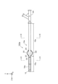
- FIG. 1 is an explanatory diagram schematically showing the configuration of the balloon catheter 100 in the first embodiment.
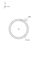
- FIG. 2 is an explanatory diagram schematically showing the tip shaft portion 14 of the shaft 10
- FIG. 3 is an explanatory diagram schematically showing the base end shaft portion 16 of the shaft 10.
- FIG. 2 shows a cross section (XY cross section: cross section cut along a plane including the X axis and the Y axis shown in FIG. 1) of the shaft 10 (tip shaft portion 14) at the position II-II in FIG. Figure) shows the configuration.
- FIG. 3 shows the configuration of the cross section of the shaft 10 (base end shaft portion 16) at the position III-III of FIG.
- the balloon 30 and the connector 40 described later are omitted.
- the Z-axis positive direction side (the side of the tip 12 of the balloon catheter 100) is the distal side (distal side) inserted into the body, and the negative Z-axis side (the tip 12 of the balloon catheter 100).
- the side opposite to the side of the balloon is the proximal side (proximal side) operated by a technician such as a doctor.
- FIG. 1 shows a state in which the balloon catheter 100 is in a linear shape parallel to the Z-axis direction as a whole, but the balloon catheter 100 has sufficient flexibility to be curved. Further, FIG. 1 shows a state in which the balloon 30 described later is expanded.
- the balloon catheter 100 is inserted into a blood vessel, for example, to expand the balloon 30 so as to be in close contact with the blood vessel wall on the front side of a lesion (stenosis or occlusion) in the blood vessel to block blood flow.
- a lesion stenosis or occlusion
- Device for. As shown in FIG. 1, the balloon catheter 100 includes a shaft 10 and a balloon 30.
- the shaft 10 is a tubular (for example, cylindrical) member having an open tip and a proximal end.
- "cylindrical shape (cylindrical shape)” is not limited to a perfect cylindrical shape (cylindrical shape), but is substantially cylindrical (substantially cylindrical shape, for example, slightly conical shape or a part) as a whole. It may be a shape with irregularities, etc.).
- a main lumen S1 and an extended lumen S2 are formed inside the shaft 10.
- a linear device such as a guide wire, a recovery device (stent retriever, suction catheter), or a dilator is inserted through the main lumen S1.
- the fluid may be a gas or a liquid, and examples thereof include gases such as helium gas, CO 2 gas, and O 2 gas, and liquids such as physiological saline and contrast media. That is, the balloon catheter 100 is a so-called two-lumen type catheter including a main lumen S1 and an expanded lumen S2.
- the main lumen S1 is an example of the first lumen in the claims
- the extended lumen S2 is an example of the second lumen in the claims.
- the shaft 10 has a tip shaft portion 14 and a base end shaft portion 16.
- the tip shaft portion 14 is an example of a first shaft portion in the claims
- the proximal shaft portion 16 is an example of a second shaft portion in the claims.
- the tip shaft portion 14 is a portion including the tip of the shaft 10.
- the tip shaft portion 14 has a single lumen structure in which only the main lumen S1 is formed.
- the main lumen S1 penetrates the entire length of the tip shaft portion 14 in the axial direction.
- the main lumen S1 is formed at a substantially central portion of the distal shaft portion 14.
- the thickness D1 of the tip shaft portion 14 is substantially the same over the entire circumference.
- both the outer peripheral shape and the inner peripheral shape of the tip shaft portion 14 are substantially circular when viewed from the axial direction.
- the axial length of the tip shaft portion 14 is, for example, 2 cm.
- a tip tip 12 is provided at the tip of the tip shaft portion 14.
- the tip tip 12 is a cylindrical member having an open tip and a rear end.
- the tip tip 12 may have an outer shape having the same outer diameter over the entire length, or may have a tapered outer diameter in which the outer diameter gradually decreases toward the tip.
- the device inserted in the main lumen S1 is led out from the tip of the tip tip 12.
- the tip tip 12 has a flexibility equal to or higher than that of the base end side of the tip shaft portion 14.
- the base end shaft portion 16 is a portion including the base end of the shaft 10.
- the tip shaft portion 14 and the proximal shaft portion 16 are adjacent to each other in the axial direction.
- the base end shaft portion 16 has a double lumen structure in which an extended lumen S2 is formed in addition to the main lumen S1.
- the main lumen S1 penetrates over the entire length of the proximal shaft portion 16 in the axial direction.
- the extended lumen S2 extends from the proximal end of the proximal shaft portion 16 toward the distal end side, and is open to the outer peripheral surface of the distal end portion of the proximal shaft portion 16. Specifically, as shown in FIG.
- the main lumen S1 is formed at a slightly eccentric position in the proximal shaft portion 16 when viewed from the axial direction, and the extended lumen S2 is the outer periphery of the main lumen S1. It is formed on the side. The diameter of the extended lumen S2 is smaller than the diameter of the main lumen S1.
- the diameter of the main lumen S1 formed on the tip shaft portion 14 and the diameter of the main lumen S1 formed on the proximal shaft portion 16 are substantially the same.
- the fact that A and B are substantially the same means that the error between A and B is 5% or less of A or B. That is, a main lumen S1 having substantially the same diameter is formed through the shaft 10 over the entire length.
- the thickness D2 of the proximal shaft portion 16 on the side opposite to the extended lumen S2 is substantially the same as the thickness D1 of the distal shaft portion 14.
- the thickness D3 of the proximal shaft portion 16 on the side on which the extended lumen S2 is formed is thicker than the thickness D2 on the side opposite to the expanded lumen S2.
- the tip shaft portion 14 has a thinner portion than the proximal shaft portion 16, and the outer diameter of the tip shaft portion 14 is smaller than the outer diameter of the proximal shaft portion 16.
- the flexibility of the tip shaft portion 14 is higher than the flexibility of the base end shaft portion 16.
- the shore hardness of the outer layer resin material of the tip shaft portion 14 is D47 or less.
- the shore hardness of the outer layer resin material of the tip shaft portion 14 may be D40 or less.
- a connector 40 for introducing a device, a fluid, or the like is attached to each lumen S1 and S2 at the base end of the base end shaft portion 16.
- the shaft 10 is integrally formed of the same material throughout.
- the shaft 10 is preferably made of a material that can be heat-sealed and has a certain degree of flexibility.
- the material for forming the shaft 10 include a thermoplastic resin, more specifically polyethylene, polypropylene, polybutene, an ethylene-propylene copolymer, an ethylene-vinyl acetate copolymer, an ionomer, or a mixture of two or more thereof.
- examples thereof include polyolefins such as, polyvinyl chloride resin, polyamide, nylon, polyamide elastomer, polyester, polyester elastomer, thermoplastic polyurethane and the like.
- a hydrophilic coating 18 formed of a hydrophilic resin is formed on the tip portion of the outer peripheral surface of the tip shaft portion 14 of the shaft 10 over the entire circumference of the tip shaft portion 14.
- the hydrophilic coating 18 is provided to reduce the frictional resistance between the surface of the tip shaft portion 14 and the inner wall of the blood vessel to ensure slipperiness when the tip shaft portion 14 is inserted into the blood vessel. ..
- the hydrophilic coating 18 is not formed on the base end side of the tip shaft portion 14, and the friction coefficient of the outer peripheral surface is higher than that of the tip portion.
- the balloon 30 is an expansion part that can be expanded and contracted with the supply and discharge of the fluid.
- the balloon 30 covers the outer circumference of the tip portion of the proximal shaft portion 16.
- the front end portion 32 and the rear end portion 34 of the balloon 30 are joined to the outer peripheral surface of the front end portion of the base end shaft portion 16 by, for example, welding.
- the tip portion 32 of the balloon 30 is joined to the outer peripheral surface of the tip of the base end shaft portion 16, and the joint portion between the tip end portion 32 and the base end shaft portion 16 is the tip end in the axial direction. It is adjacent to the shaft portion 14.
- the expansion lumen S2 described above communicates with an internal space S3 (see FIG. 1) formed between the balloon 30 and the proximal shaft portion 16.
- the length of the balloon 30 in the axial direction is, for example, about 2 cm.
- the balloon 30 is preferably made of a material having a certain degree of flexibility, and more preferably made of a material that is thinner than the shaft 10 and has flexibility.
- the material for forming the balloon 30 include polyolefins such as polyethylene, polypropylene, polybutene, ethylene-propylene copolymer, ethylene-vinyl acetate copolymer, ionomer, or a mixture of two or more thereof, and a soft polyvinyl chloride resin.
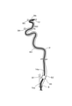
- FIG. 4 is an explanatory diagram showing a state in which the balloon catheter 100 is arranged in a blood vessel.
- the vessels on the far side of the carotid artery (common carotid artery B, external carotid artery B1, internal carotid artery cervical part B2, internal carotid artery pyramidal part B3, ocular artery B4, anterior cerebral artery B5, middle cerebral artery).
- B6 is schematically shown.
- the balloon catheter 100 is a guiding catheter with a balloon, and is used, for example, for thrombus recovery treatment in acute cerebral infarction.
- the balloon catheter 100 is inserted into the blood vessel and pushed from the common carotid artery B into, for example, the internal carotid artery neck B2.
- Petras P which is a flexion blood vessel, is present in the neck B2 of the internal carotid artery.
- the proximal shaft portion 16 of the shaft 10 has relatively low flexibility (high rigidity), and particularly the flexibility of the joint portion between the balloon 30 and the proximal shaft portion 16 is low. Therefore, it is difficult to insert the base end shaft portion 16 into the petras P.
- the tip shaft portion 14 of the shaft 10 has relatively high flexibility, and the axial length of the tip shaft portion 14 is 1.5 cm or more. Therefore, as shown in FIG. 4, the tip shaft portion 14 can be relatively easily inserted into the petras P.
- the balloon 30 By expanding the balloon 30 with the tip shaft portion 14 inserted into the petras P, the blood flow in the carotid artery is temporarily blocked. At this time, the balloon 30 is located, for example, in front of Petras P in the internal carotid artery neck B2.
- a recovery device (not shown) is led out from the tip of the balloon catheter 100 via the main lumen S1 and sent out to the vicinity of the lesion existing beyond the petras P (for example, the internal carotid artery pyramidal portion B3 or the like). Then, the thrombus fragment is captured by using the recovery device, and the recovery device that has captured the thrombus fragment is drawn into the main lumen S1 of the balloon catheter 100. This makes it possible to collect the thrombus fragments in the blood vessels.
- the tip shaft portion 14 having relatively high flexibility is arranged on the tip side of the balloon 30 (see FIGS. 1 and 2).
- the distance between the tip of the balloon catheter 100 and the lesion portion in the blood vessel (hereinafter referred to as “thrombus recovery distance”) is shorter than that in the configuration without the tip shaft portion 14.
- the thrombus recovery distance is shortened, for example, damage to the blood vessel caused by the recovery device can be suppressed by the amount that the distance traveled in the blood vessel in the exposed state of the recovery device is shortened. For example, it is possible to reduce the risk of blood vessel perforation or spasm due to the recovery device scratching the blood vessel.
- the tip shaft portion 14 bends in the petras P, so that the position of the tip shaft portion 14 in the blood vessel is fixed and the support force of the balloon catheter 100 is improved. That is, in general, it is preferable to reduce the flexibility of the shaft in order to improve the support force, but on the other hand, the flexibility is reduced, so that the insertability into the blood vessel (blood vessel followability) is lowered.
- the tip shaft portion 14 having flexibility in the petras P is arranged on the tip side of the balloon 30. Therefore, it is possible to improve the support force of the balloon catheter 100 by bending the tip shaft portion 14 while ensuring the blood vessel followability of the tip shaft portion 14.
- a hydrophilic coating 18 having a relatively low coefficient of friction is formed on the tip portion of the tip shaft portion 14. Therefore, the tip end side of the tip shaft portion 14 can be smoothly inserted into the petras P. Further, the hydrophilic coating 18 is not formed on the base end side of the tip shaft portion 14, and the friction coefficient is relatively high. Therefore, the position of the tip shaft portion 14 in the blood vessel is more firmly fixed by the proximal end side coming into contact with the inner wall surface of the petras P while the tip shaft portion 14 is bent. This makes it possible to further improve the support force of the balloon catheter 100.
- FIG. 5 is an explanatory diagram schematically showing the configuration of the balloon catheter 100a in the second embodiment.
- FIG. 6 is an explanatory diagram schematically showing a base end shaft portion 16a of the shaft 10a.
- FIG. 6 shows the configuration of the cross section of the shaft 10a (base end shaft portion 16a) at the position of VI-VI in FIG. In FIG. 6, the balloon 30 and the connector 40 are omitted.
- the configuration of the cross section of the shaft 10a (tip shaft portion 14a) at the position II-II in FIG. 6 is the same as that in FIG. 2 of the first embodiment.
- the same configurations as those of the balloon catheter 100 of the first embodiment described above will be appropriately described by adding the same reference numerals.
- the shaft 10a included in the balloon catheter 100a is a tubular (for example, cylindrical) member having an open tip and proximal end. As shown in FIGS. 2 and 6, a main lumen S1 and an extended lumen S2a are formed inside the shaft 10a.
- the extended lumen S2a is an example of a second lumen in the claims.
- the shaft 10a has a tip shaft portion 14a and a proximal shaft portion 16a.
- the tip shaft portion 14a is an example of a first shaft portion in the claims
- the proximal shaft portion 16a is an example of a second shaft portion in the claims.
- the tip shaft portion 14a has a single lumen structure in which only the main lumen S1 is formed, similar to the tip shaft portion 14 of the first embodiment (see FIG. 2). However, the axial length of the tip shaft portion 14a is longer than the axial length of the tip shaft portion 14 of the first embodiment, for example, 20 cm. Further, a side hole 20 communicating with the main lumen S1 is formed on the outer peripheral surface of the tip shaft portion 14a on the base end side. In the present embodiment, the pair of side holes 20 are arranged at positions symmetrical with respect to the central axis of the tip shaft portion 14a (see FIG. 8 described later).
- the base end shaft portion 16a has a coaxial structure in which an extended lumen S2a is formed in addition to the main lumen S1.
- the extended lumen S2a extends from the proximal end of the proximal shaft portion 16a toward the distal end side, and is open to the outer peripheral surface of the distal end portion of the proximal end shaft portion 16.
- the main lumen S1 is formed in the central portion of the proximal shaft portion 16a
- the extended lumen S2a surrounds the outer periphery of the main lumen S1. It is formed in a ring shape.
- the diameter of the main lumen S1 formed in the tip shaft portion 14a and the diameter of the main lumen S1 formed in the proximal shaft portion 16a are substantially the same. That is, a main lumen S1 having substantially the same diameter is formed through the shaft 10a over the entire length.
- the thickness D1 of the tip shaft portion 14a is thinner than the thickness D4 of the base end shaft portion 16a, and the outer diameter of the tip end shaft portion 14a is smaller than the outer diameter of the base end shaft portion 16a.
- the flexibility of the tip shaft portion 14a is higher than the flexibility of the proximal shaft portion 16a.
- the shore hardness of the outer layer resin material of the tip shaft portion 14a is D47 or less.
- the shore hardness of the outer layer resin material of the tip shaft portion 14 may be D40 or less.
- a hydrophilic coating 18a formed of a hydrophilic resin is formed on the entire outer peripheral surface of the tip shaft portion 14a of the shaft 10a.
- FIG. 7 is an explanatory diagram showing a state in which the balloon catheter 100a is arranged in the blood vessel.
- the balloon 30 is located near the entrance of the internal carotid artery neck B2, and the tip shaft portion 14a extends beyond Petras P to the vicinity of the middle cerebral artery B6.
- the tip of the balloon catheter 100a can be brought close to the vicinity of the lesion located further away from Petras P.
- FIG. 8 is an explanatory diagram showing a state in which the thrombus piece C is collected by the side hole 20.
- the tip shaft portion 14a protrudes toward the tip side of the balloon 30. Therefore, for example, when collecting the thrombus piece C from the lesion portion, the thrombus piece C is not collected in the opening of the main lumen S1 formed at the tip of the tip shaft portion 14a, and is not collected in the opening of the tip shaft portion 14a on the outer peripheral side. It may get in (see FIGS. 8 (a) and 8 (b)). However, according to the present embodiment, the uncollected thrombus piece C is recovered in the main lumen S1 via the side hole 20 formed in the tip shaft portion 14a.
- the axial length of the tip shaft portions 14, 14a in each of the above embodiments may be any length of 1.5 cm or more.
- the axial length of the tip shaft portion 14 may be 1.5 cm or more and less than 2 cm, or may be longer than 2 cm.
- the axial length of the tip shaft portion 14a may be 1.5 cm or more, less than 20 cm, or longer than 20 cm.
- the tip shaft portions 14, 14a may be configured not to include the tip tip 12.
- the main lumen S1 and the extended lumens S2, S2a do not extend to the proximal end of the proximal shaft portions 16, 16a (shafts 10, 10a), but bend in the middle and open to the outer peripheral surface of the shaft 10. It may be the same configuration.
- the extended lumens S2 and S2a may be configured to extend to the tip shaft portions 14, 14a.
- the number of lumens formed on the tip shaft portions 14, 14a may be two or more, and the number of lumens formed on the proximal shaft portions 16, 16a may be three or more.
- the first shaft portion is more flexible (that is, has lower flexural rigidity) than the second shaft portion.
- the first shaft portion is more flexible (that is, has lower flexural rigidity) than the second shaft portion.
- the shaft may have a configuration in which the first shaft portion is formed of a material having a hardness lower than that of the material for forming the second shaft portion.
- the tip portion 32 of the balloon 30 may be configured to be slightly joined to the proximal end side from the distal end portions of the proximal end shaft portions 16, 16a. That is, the balloon 30 may be configured to be joined to the distal end side of the proximal shaft portions 16, 16a.
- the balloon catheter in the claims may have a configuration in which a first shaft portion having relatively high flexibility is provided on the distal end side of the second shaft portion to which the balloon is joined.
- the balloon catheters 100 and 100a may not be provided with the hydrophilic coatings 18 and 18a.
- the side hole may be formed on the outer peripheral surface of the tip shaft portion 14, or in the second embodiment, the side hole is not formed on the outer peripheral surface of the tip shaft portion 14a. It may be configured.
- the outer diameters of the shafts 10 and 10a may be substantially the same over the entire length.
- the shape of the cross section of the shafts 10 and 10a is not limited to a circle, but may be a polygonal shape or the like.
- the entire shaft 10, 10a, the tip shaft portions 14, 14a, or the base end shaft portions 16, 16a is not limited to a single layer configuration, and may be configured to include a plurality of resin layers (for example, an inner layer resin and an outer layer resin). Further, a reinforcing body (blade), a coil body, or the like may be embedded.
- the tip shaft portions 14, 14a and the proximal end shaft portions 16, 16a are the same for each of the plurality of resin layers and reinforcing bodies. It may be integrally formed of materials.
- each member in the above embodiment is merely an example and can be variously deformed.
- the balloon catheters 100 and 100a inserted into the blood vessel are exemplified as the balloon catheter, but the balloon catheter may be inserted into a body cavity other than the blood vessel (for example, the esophagus).
Landscapes
- Health & Medical Sciences (AREA)
- Life Sciences & Earth Sciences (AREA)
- Heart & Thoracic Surgery (AREA)
- Anesthesiology (AREA)
- Biophysics (AREA)
- Pulmonology (AREA)
- Engineering & Computer Science (AREA)
- Biomedical Technology (AREA)
- Hematology (AREA)
- Animal Behavior & Ethology (AREA)
- General Health & Medical Sciences (AREA)
- Public Health (AREA)
- Veterinary Medicine (AREA)
- Child & Adolescent Psychology (AREA)
- Vascular Medicine (AREA)
- Media Introduction/Drainage Providing Device (AREA)
Priority Applications (4)
| Application Number | Priority Date | Filing Date | Title |
|---|---|---|---|
| JP2022539945A JP7408816B2 (ja) | 2020-07-31 | 2020-07-31 | バルーンカテーテル |
| PCT/JP2020/029448 WO2022024348A1 (ja) | 2020-07-31 | 2020-07-31 | バルーンカテーテル |
| US18/158,630 US20230191091A1 (en) | 2020-07-31 | 2023-01-24 | Balloon catheter |
| JP2023214352A JP2024019635A (ja) | 2020-07-31 | 2023-12-20 | バルーンカテーテル |
Applications Claiming Priority (1)
| Application Number | Priority Date | Filing Date | Title |
|---|---|---|---|
| PCT/JP2020/029448 WO2022024348A1 (ja) | 2020-07-31 | 2020-07-31 | バルーンカテーテル |
Related Child Applications (1)
| Application Number | Title | Priority Date | Filing Date |
|---|---|---|---|
| US18/158,630 Continuation US20230191091A1 (en) | 2020-07-31 | 2023-01-24 | Balloon catheter |
Publications (1)
| Publication Number | Publication Date |
|---|---|
| WO2022024348A1 true WO2022024348A1 (ja) | 2022-02-03 |
Family
ID=80035309
Family Applications (1)
| Application Number | Title | Priority Date | Filing Date |
|---|---|---|---|
| PCT/JP2020/029448 Ceased WO2022024348A1 (ja) | 2020-07-31 | 2020-07-31 | バルーンカテーテル |
Country Status (3)
| Country | Link |
|---|---|
| US (1) | US20230191091A1 (https=) |
| JP (2) | JP7408816B2 (https=) |
| WO (1) | WO2022024348A1 (https=) |
Families Citing this family (1)
| Publication number | Priority date | Publication date | Assignee | Title |
|---|---|---|---|---|
| CN119925787A (zh) * | 2023-10-27 | 2025-05-06 | 深圳市纬康医疗科技有限公司 | 一种球囊鞘管 |
Citations (5)
| Publication number | Priority date | Publication date | Assignee | Title |
|---|---|---|---|---|
| JPH07303701A (ja) * | 1994-05-13 | 1995-11-21 | Takashi Nishimura | 造影剤回収用バルーンカテーテル |
| JP2007501652A (ja) * | 2003-08-08 | 2007-02-01 | ボストン サイエンティフィック リミテッド | 伸縮規制用カテーテルシャフト |
| JP2010167003A (ja) * | 2009-01-21 | 2010-08-05 | Asahi Intecc Co Ltd | バルーンカテーテル |
| JP2014138864A (ja) * | 2007-08-06 | 2014-07-31 | Abbott Cardiovascular Systems Inc | インターロッキング・ネットワークに包含される親水性化合物を有する潤滑コーティングを施された医療装置 |
| JP2020500065A (ja) * | 2016-11-23 | 2020-01-09 | イノベーションズ イン メディスン,エルエルシー | 体管腔の偏向のためのシステムおよび方法 |
Family Cites Families (5)
| Publication number | Priority date | Publication date | Assignee | Title |
|---|---|---|---|---|
| US20060074396A1 (en) * | 2004-09-28 | 2006-04-06 | Medtronic Vascular, Inc. | Stent delivery system |
| JP5732259B2 (ja) * | 2011-01-12 | 2015-06-10 | 株式会社グッドマン | カテーテル |
| WO2012162661A1 (en) * | 2011-05-26 | 2012-11-29 | Abbott Cardiovascular Systems Inc. | Through tip for a catheter |
| JP2020039376A (ja) * | 2017-01-23 | 2020-03-19 | テルモ株式会社 | バルーンカテーテル |
| US11642500B2 (en) * | 2018-02-20 | 2023-05-09 | Crossliner, Inc. | Intravascular delivery system and method for percutaneous coronary intervention |
-
2020
- 2020-07-31 JP JP2022539945A patent/JP7408816B2/ja active Active
- 2020-07-31 WO PCT/JP2020/029448 patent/WO2022024348A1/ja not_active Ceased
-
2023
- 2023-01-24 US US18/158,630 patent/US20230191091A1/en active Pending
- 2023-12-20 JP JP2023214352A patent/JP2024019635A/ja active Pending
Patent Citations (5)
| Publication number | Priority date | Publication date | Assignee | Title |
|---|---|---|---|---|
| JPH07303701A (ja) * | 1994-05-13 | 1995-11-21 | Takashi Nishimura | 造影剤回収用バルーンカテーテル |
| JP2007501652A (ja) * | 2003-08-08 | 2007-02-01 | ボストン サイエンティフィック リミテッド | 伸縮規制用カテーテルシャフト |
| JP2014138864A (ja) * | 2007-08-06 | 2014-07-31 | Abbott Cardiovascular Systems Inc | インターロッキング・ネットワークに包含される親水性化合物を有する潤滑コーティングを施された医療装置 |
| JP2010167003A (ja) * | 2009-01-21 | 2010-08-05 | Asahi Intecc Co Ltd | バルーンカテーテル |
| JP2020500065A (ja) * | 2016-11-23 | 2020-01-09 | イノベーションズ イン メディスン,エルエルシー | 体管腔の偏向のためのシステムおよび方法 |
Also Published As
| Publication number | Publication date |
|---|---|
| JP7408816B2 (ja) | 2024-01-05 |
| JPWO2022024348A1 (https=) | 2022-02-03 |
| US20230191091A1 (en) | 2023-06-22 |
| JP2024019635A (ja) | 2024-02-09 |
Similar Documents
| Publication | Publication Date | Title |
|---|---|---|
| JP5317566B2 (ja) | カテーテル組立体 | |
| JP2004024625A (ja) | カテーテルおよび医療用チューブ | |
| WO2004012604A1 (ja) | 吸引カテーテル | |
| CN101166556A (zh) | 用于导管或套管的可剥离的非创伤性端件和管体 | |
| US20220088354A1 (en) | Balloon catheter | |
| CN113646030B (zh) | 球囊导管 | |
| CN113613702B (zh) | 球囊导管 | |
| CN106102663B (zh) | 支架输送系统和内窥镜系统 | |
| JP5378092B2 (ja) | バルーンカテーテル及びシースの加工方法 | |
| US20240399107A1 (en) | Directional tipped catheters | |
| JP4914281B2 (ja) | カテーテル | |
| JP2020039376A (ja) | バルーンカテーテル | |
| JP2024019635A (ja) | バルーンカテーテル | |
| WO2021085537A1 (ja) | 異物除去用バルーンカテーテル | |
| WO2019180928A1 (ja) | バルーンカテーテル | |
| JP2015226556A (ja) | 医療器具 | |
| US20150088152A1 (en) | Elongated member for medical use | |
| JP7555487B2 (ja) | カテーテル | |
| JP2022020124A (ja) | バルーンカテーテル | |
| JP7148308B2 (ja) | バルーンカテーテル | |
| WO2021186664A1 (ja) | カテーテル | |
| CN215741228U (zh) | 一种远端通路导引导管 | |
| JP2019170654A (ja) | 医療器具 | |
| WO2017202073A1 (zh) | 一种采用非锥状头端的球囊导管 | |
| JP2018064866A (ja) | カテーテル押込み補助具 |
Legal Events
| Date | Code | Title | Description |
|---|---|---|---|
| 121 | Ep: the epo has been informed by wipo that ep was designated in this application |
Ref document number: 20947172 Country of ref document: EP Kind code of ref document: A1 |
|
| ENP | Entry into the national phase |
Ref document number: 2022539945 Country of ref document: JP Kind code of ref document: A |
|
| NENP | Non-entry into the national phase |
Ref country code: DE |
|
| 122 | Ep: pct application non-entry in european phase |
Ref document number: 20947172 Country of ref document: EP Kind code of ref document: A1 |