WO2020129464A1 - 情報処理装置、放射線撮影システムおよび支援方法 - Google Patents
情報処理装置、放射線撮影システムおよび支援方法 Download PDFInfo
- Publication number
- WO2020129464A1 WO2020129464A1 PCT/JP2019/044135 JP2019044135W WO2020129464A1 WO 2020129464 A1 WO2020129464 A1 WO 2020129464A1 JP 2019044135 W JP2019044135 W JP 2019044135W WO 2020129464 A1 WO2020129464 A1 WO 2020129464A1
- Authority
- WO
- WIPO (PCT)
- Prior art keywords
- information
- electronic medical
- unit
- keyword
- medical record
- Prior art date
- Legal status (The legal status is an assumption and is not a legal conclusion. Google has not performed a legal analysis and makes no representation as to the accuracy of the status listed.)
- Ceased
Links
Images









Classifications
-
- A—HUMAN NECESSITIES
- A61—MEDICAL OR VETERINARY SCIENCE; HYGIENE
- A61B—DIAGNOSIS; SURGERY; IDENTIFICATION
- A61B6/00—Apparatus or devices for radiation diagnosis; Apparatus or devices for radiation diagnosis combined with radiation therapy equipment
-
- G—PHYSICS
- G16—INFORMATION AND COMMUNICATION TECHNOLOGY [ICT] SPECIALLY ADAPTED FOR SPECIFIC APPLICATION FIELDS
- G16H—HEALTHCARE INFORMATICS, i.e. INFORMATION AND COMMUNICATION TECHNOLOGY [ICT] SPECIALLY ADAPTED FOR THE HANDLING OR PROCESSING OF MEDICAL OR HEALTHCARE DATA
- G16H30/00—ICT specially adapted for the handling or processing of medical images
Definitions
- the present invention relates to an information processing device, a radiation imaging system, and a support method for supporting the imaging of a medical image of a user.
- the electronic medical chart system can issue various inspection orders such as X-ray imaging from the electronic medical chart by cooperating with the ordering system for various inspections.
- the doctor issues the necessary inspection order using an electronic medical record, taking into account the contents of the interview and the inspection result.
- the ordering system manages the inspection order received from the electronic medical chart.
- the inspection equipment such as the X-ray imaging apparatus determines the patient information, the imaging region, the protocol including the image processing parameters, and the like based on the inspection order received from the ordering system, and executes the inspection.
- Code values are assigned in advance between the electronic medical chart system and the ordering system, and between the ordering system and the inspection equipment, to associate the inspection order issued by the electronic medical chart with the imaging protocol. With this association, it is possible to perform the inspection according to the inspection order even if the inspection is performed over a plurality of systems. Further, a technique for automatically setting parameters and the like used in the determined protocol has also been proposed.
- Patent Document 1 proposes to acquire electronic medical record information such as a surgeon's name, patient data, and a surgical procedure to acquire or correct parameters in the medical endoscope system.
- Patent Document 2 it is proposed that the disease image and the image processing parameter are associated with each other, and the image processing parameter is determined from the degree of similarity between the captured image and the disease image to perform the image processing.
- the imaging technician implement the protocol in consideration of information that does not appear in the above code value.
- the protocol in consideration of information that does not appear in the above code value.
- accurate diagnosis may be difficult without proper imaging, or the patient may be burdened with reimaging. Cases can happen.
- the image processing parameter for the captured image can be corrected, but it is difficult to avoid the positioning failure during capturing.
- Such a problem is not limited to radiographic image capturing, and may occur in medical image capturing with various modalities.
- One aspect of the present invention provides a technique capable of generating support information for supporting medical image capturing.
- support information for supporting the capturing of medical images is generated.
- radiation may include, for example, ⁇ -rays, ⁇ -rays, ⁇ -ray particle beams, cosmic rays, etc., in addition to X-rays.
- FIG. 1 is a diagram showing a configuration example of a radiation imaging system according to the first embodiment.
- a radiographic imaging control device 100 which is an information processing device for controlling radiographic imaging, is connected to a radiation generation device 106 that emits radiation and a radiation sensor 101 that generates a radiation image from the emitted radiation.
- the radiation imaging control apparatus 100 receives a radiation image from the radiation sensor 101.
- the radiation imaging control apparatus 100 is connected to the electronic medical chart system 102 and the ordering system 103 via the in-hospital network 105 so as to receive the electronic medical chart information and the examination order.
- the radiation imaging control apparatus 100 is connected to the image server 104 via the in-hospital network 105 so that the captured radiation image can be transmitted and received.
- the radiation imaging control apparatus 100 and the ordering system 103 communicate with the electronic medical chart system 102 using a protocol based on the HL7 (Health Level 7) standard. Further, the radiation imaging control apparatus 100 communicates with the ordering system 103 and the image server 104 by using a protocol based on the DICOM (Digital Imaging and Communications in Medicine) standard.
- HL7 Health Level 7
- DICOM Digital Imaging and Communications in Medicine
- the inspection order is transmitted from the electronic medical chart system 102 to the ordering system 103.
- the radiographic imaging control apparatus 100 makes an inquiry to the ordering system 103 to acquire the inspection order. Then, the radiographic imaging control apparatus 100 acquires electronic medical chart information from the electronic medical chart system 102.
- the radiographic imaging control apparatus 100 performs image processing on a radiation image acquired using the radiation generation apparatus 106 and the radiation sensor 101, generates an annotation, associates it with patient information, and transmits it to the image server 104.
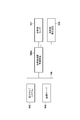
- FIG. 2 is a block diagram showing a functional configuration example of the radiation imaging control apparatus 100 according to the first embodiment.
- the electronic medical chart information receiving unit 201 communicates with the electronic medical chart system 102, and based on the examination order for performing the imaging, the electronic medical chart information in which the medical examination information such as the content of the inquiry and the examination result of the patient is recorded from the electronic medical chart system 102.
- the electronic medical record information receiving unit 201 outputs the electronic medical record information acquired from the electronic medical record system 102 to the support information creating unit 205.
- the inspection order receiving unit 202 communicates with the ordering system 103 and acquires the inspection order.
- the examination order receiving unit 202 outputs the examination order acquired from the ordering system 103 to the support information creating unit 205 and the UI unit 209.
- the image transmission unit 203 communicates with the image server 104 and transmits the radiographic image that has been subjected to image processing by the image processing unit 206.
- the storage unit 204 stores protocol information including an imaging region and image processing parameters associated with the examination order.
- the storage unit 204 stores a specific item (for example, a precaution at the time of shooting) in association with a keyword.
- a specific item for example, a precaution at the time of shooting
- the specific item includes a wording that calls attention and a photograph (confirmation image) that shows the positioning at the time of shooting.
- the keyword and the specific item stored in the storage unit 204 are associated with each other using a supervised machine learning algorithm, and the specific item can be extracted from a combination of frequently used keywords. That is, the storage unit 204 stores a keyword and a specific item associated with each other by supervised machine learning, and the specific item includes a confirmation image obtained by imaging the positioning of the inspection site in radiography.
- the support information creation unit 205 acquires specific items from the storage unit 204 based on the inspection order acquired from the inspection order reception unit 202 and the electronic medical record information received from the electronic medical record information reception unit 201.
- the support information creation unit 205 creates support information for radiation imaging using the acquired specific items and outputs it to the UI unit 209. The process of creating support information will be described later.
- the image processing unit 206 acquires image processing parameters corresponding to the protocol selected by the UI unit 209 from the storage unit 204, performs image processing on the radiographic image received from the communication unit 208, and then the image transmission unit 203 and the UI unit 209. Output to.
- the imaging control unit 207 communicates with the radiation sensor 101 via the communication unit 208 based on an instruction from the UI unit 209, and sets the parameters of the radiation sensor 101.
- the imaging control unit 207 also notifies the UI unit 209 of the state of the radiation sensor 101.
- the communication unit 208 communicates with the radiation sensor 101 to set the parameters received from the imaging control unit 207 and receive the state of the radiation sensor 101. Further, the communication unit 208 outputs the radiographic image received from the radiation sensor to the image processing unit 206. Further, the imaging control unit 207 sets the radiation irradiation parameter of the radiation generation device 106 via the communication unit 208.
- the UI unit 209 is a user interface for a user to operate the radiation imaging control apparatus 100, and displays a screen and receives operation input. For example, the UI unit 209 displays the inspection order received from the inspection order receiving unit 202 so that the imaging engineer who is the user can select the target inspection. In addition, the UI unit 209 displays the state of the radiation sensor 101 received from the imaging control unit 207 and the radiation imaged image processed by the image processing unit 206. In addition, the UI unit 209 instructs the support information creation unit 205 to create support information, and displays the obtained support information on the display unit 210.
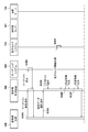
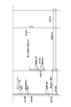
- FIG. 3A and 3B are sequence diagrams showing communication between the systems at the time of radiation imaging, which is started by an operation performed by the imaging engineer 300 as a trigger.
- the support information creation unit 205 of the radiographic imaging control apparatus 100 acquires, through the electronic medical record information receiving unit 201, electronic medical record information in which the medical examination findings of the examination target are recorded.
- An example of this processing is shown in S301 to S305.
- the radiographing engineer 300 operates the radiation imaging control apparatus 100 to perform an operation of searching a work list that is a list of a plurality of examination orders (S301: work list search operation).
- the radiation imaging control apparatus 100 retrieves and acquires the work list from the ordering system 103 (S302).
- the imaging technician 300 operates the radiation imaging control apparatus 100 to perform an operation of selecting an inspection order to be executed from the work list (S303: inspection order selection operation).
- the radiographic imaging control apparatus 100 selects a protocol based on the selected examination order (S304), and acquires electronic chart information of the patient related to the examination order from the electronic chart system 102 (S305).
- the support information creation unit 205 acquires the examination order selected by the user operation from the examination orders provided by the ordering system 103, and obtains the electronic chart information related to the examination order from the electronic chart system 102. get.
- the support information creation unit 205 of the radiation imaging control apparatus 100 creates support information based on the acquired electronic medical record information (S306).
- the support information creation unit 205 uses the acquired keyword by using the storage unit 204 that acquires the keyword from the electronic medical record information and stores the specific item for capturing the medical image and the keyword in association with each other. It is an example of the structure which searches by.
- the support information creation unit 205 is an example of a configuration that creates support information for imaging a medical image based on the searched specific item.
- the keyword for search is also acquired from the inspection order of the inspection target selected in S303.
- An example of the processing of the support information creating unit 205 will be described later with reference to the flowchart of FIG.
- the created support information is displayed on the display unit 210 of the UI unit 209 (S307).
- the imaging engineer 300 positions the patient and adjusts the radiation irradiation parameters of the radiation generator 106 with reference to the displayed support information (S308). Then, the imaging engineer 300 gives an instruction to irradiate the radiation (S309).
- the radiation sensor 101 that has received the radiation emitted from the radiation generation device 106 according to this instruction generates a radiographic image based on the received radiation, and outputs the radiographic image to the radiographic control device 100 (S310).
- the radiographic imaging control device 100 performs image processing on the received radiographic image (S311), and displays the processed radiographic image on the display unit 210 (S312).
- the radiographing engineer 300 confirms the displayed image (S313) and operates the radiation imaging control apparatus 100 to end the inspection (S314: inspection end operation).
- the radiation imaging control apparatus 100 transmits the image-processed radiation image to the image server 104 (S315).
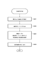
- FIG. 4 is a flowchart showing a process when the support information creation unit 205 of the radiation imaging control apparatus 100 creates support information (S306).
- FIG. 5 is an example of electronic medical record information acquired from the electronic medical record system 102 regarding a patient who performs radiography of the wrist. The flow chart shown in FIG. 4 will be described using the electronic medical record information of FIG.
- the support information creation unit 205 performs a lexical analysis on the received electronic medical record information (S401).
- the electronic medical record information T501 includes “Bone density test performed. Osteoporosis” registered at 10:01:15 on July 31, 2018.
- the electronic medical record information T502 includes “pain when the wrist of the left hand is bent toward the front” registered at 13:50:20 on August 20, 2018.
- the support information creation unit 205 performs the lexical analysis to obtain the words “bone density test”, “execution”, and “osteoporosis” from the electronic medical record information T501 and “left hand” from the electronic medical record information T502. Get the words “wrist" "front” "bend” "pain”.
- the support information creation unit 205 extracts a search keyword from the obtained lexical phrase (S402).
- the inspection result and the inquiry content are necessary as search keywords, but the information that can be acquired from the inspection order and the content that the inspection has been performed are unnecessary, and the support information creation unit 205 extracts only the necessary words and phrases. ..
- the “left hand” and the “wrist” are information acquired from the examination order, and thus are determined to be unnecessary.
- “bone density test” and “execution” are terms that indicate the content of the test and are determined to be unnecessary.
- the support information creation unit 205 extracts “osteoporosis”, “front”, “bend”, and “pain” that are other terms from the electronic medical record information as search keywords. After that, the support information creation unit 205 stores a confirmation image obtained by photographing the specific item and the positioning at the time of photographing, using the part (left hand, wrist) obtained from the examination order and the search keyword obtained from the electronic medical record information. The section 204 is searched (S403). The support information creation unit 205 creates support information using the searched specific item and the positioning confirmation image, and outputs (displays) the support information to the UI unit 209 (S404).
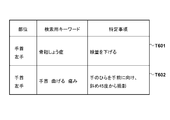
- the specific item obtained when the part is "wrist” and the search keyword is "osteoporosis” is “decrease dose” (T601).
- the specific items obtained when the parts are “left hand” and “wrist” and the search keywords are "front”, “bend” and “pain” are "photographed at an angle of 45 degrees with the palm facing you” (T602). is there.
- the “left hand” as the part information is not used for acquiring the specific item, the information indicating whether the part is the left hand or the right hand may be omitted.
- the specific items are the same for the left hand and the right hand, the information about the “left hand” and the “right hand” is unnecessary.
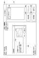
- FIG. 7 is a schematic diagram showing an example of a display screen on the display unit 210 when supporting information is displayed on the radiation imaging control apparatus 100.
- An image display area 701 that displays a radiographic image and patient information, a reception number, and the like, an area 702 that represents the selected protocol, and an area 703 that displays the status of the radiation sensor are arranged on the screen.
- a button 704 for instructing the image quality adjustment of the radiographic image a button 705 for instructing addition of an arbitrary annotation, a button 706 for instructing to suspend inspection, and a button 707 for instructing to stop inspection are arranged.
- a dialog 708 that displays support information related to the protocol is displayed on the screen where these are arranged.
- a specific item 709 (FIG.
- the dialog 708 may be created by the support information creation unit 205, for example. That is, the support information creation unit 205 is an example of a configuration that creates the support image (dialog 708) to be displayed on the display unit that displays the medical image based on the support information generated based on the searched specific item.
- the radiographic imaging control apparatus 100 of the first embodiment by creating and displaying the support information from the electronic medical record information, it is possible to support the doctor's intended examination.
- the support information is generated using the specific items searched based on the electronic medical record information, but the support information may be searched based on the electronic medical record information.
- the storage unit 204 stores the support information for capturing the medical image and the keyword in association with each other, and the support information creation unit 205 searches the storage unit 204 using the keyword acquired from the electronic medical record information.
- the support information creation unit 205 may be configured to create a support image (for example, the dialog 708) to be displayed on the display unit that displays the medical image based on the searched support information.
- the plurality of electronic medical record information items are treated as equivalent to display the support information, but the present invention is not limited to this.
- the electronic medical record information related to the inspection order for which the support information is to be acquired may have a higher priority, and may be configured to be displayed before specific items obtained from other electronic medical record information. Further, it may be configured such that only electronic medical record information for the past several times or within a certain period is used.
- the radiation imaging control apparatus 100 acquires, from a camera, an irradiation field image of an irradiation field.
- the radiographic imaging control apparatus 100 calculates the similarity between the positioning confirmation image retrieved based on the keyword obtained from the electronic medical record information (and examination order) and the irradiation field image, and when the similarity is equal to or less than a predetermined value. Include a warning message in the support information.
- the second embodiment will be described below.
- the configuration of the radiation imaging system according to the second embodiment is similar to that of the first embodiment (FIG. 1).
- FIG. 8 is a block diagram showing a functional configuration example of the radiation imaging control apparatus 100 according to the second embodiment.
- the same components as those in the first embodiment (FIG. 2) are designated by the same reference numerals.
- the support information creation unit 205a has the following functions in addition to the functions of the support information creation unit 205 of the first embodiment (FIG. 2). That is, the support information creation unit 205a compares the irradiation field image received from the camera communication unit 801 at a predetermined time interval with the positioning confirmation image stored in the storage unit 204, and issues a warning message when the similarity is equal to or lower than a certain level. It outputs to the UI unit 209 together with the support information. This processing will be described later.
- the support information creation unit 205a receives a support information output end instruction from the UI unit 209
- the support information creation unit 205a ends the output of the warning message.
- the camera communication unit 801 receives the irradiation field image from the camera 802 based on the instruction from the support information creation unit 205a.
- the camera 802 is installed in the radiation generation device 106 so as to face the radiation sensor 101, captures an irradiation field for radiation inspection at predetermined time intervals, and transmits the captured irradiation field image to the camera communication unit 801.
- FIG. 9 is a flowchart showing a process of creating support information according to the second embodiment.
- the support information creation unit 205a performs lexical analysis on the received electronic medical record information (S901), and extracts a search keyword from the obtained lexical information (S902). After that, the support information creation unit 205a searches the storage unit 204 for a confirmation image of specific items and positioning using the examination order and the search keyword (S903).
- the processing up to this point is the same as the processing (S401 to S403) of the first embodiment (FIG. 4).
- the support information creation unit 205a acquires an irradiation field image from the camera 802 via the camera communication unit 801 (S904). After that, the degree of similarity between the subject shown in the confirmation image and the irradiation field image obtained in S903 is analyzed (S905). If the degree of similarity obtained in S905 is less than or equal to the threshold value (YES in S906), the support information creation unit 205a outputs a warning message to the UI unit 209 in addition to the support information and the irradiation field image (S907).
- the support information creation unit 205a outputs the support information and the irradiation field image to the UI unit 209 (S908). After that, the support information creation unit 205a ends the present process when the support information output end instruction is received from the UI unit 209 (YES in S909). If the instruction to end the support information output has not been received (NO in S909), the process waits for a certain period of time (S910), and then returns to S904.
- FIG. 10 is a schematic diagram showing a screen example when the radiation imaging control apparatus 100 displays support information, an irradiation field image, and a warning message.
- an image display area 1001 that displays a radiographic image and patient information, a reception number, and the like, an area 1002 that represents a selected protocol, and an area 1003 that displays the state of the radiation sensor 101 are arranged.
- a button 1004 for adjusting the image quality of the radiographic image a button 1005 for adding an arbitrary annotation, a button 1006 for suspending the examination, and a button 1007 for stopping the examination are arranged on this screen. These are the same as the configurations (701 to 707 in FIG. 7) described in the first embodiment.
- a dialog 1008 for displaying support information related to the protocol is displayed on the screen.
- a specific item 1009 acquired in the flowchart of FIG. 9 a confirmation image 1010, a warning message 1011, an irradiation field image 1012 acquired from the camera, and a button 1013 for closing the dialog are arranged.
- the radiographing technician can easily confirm the correct positioning during radiography.
- an instruction to end the support information output is output from the UI unit to the support information creation unit 205a.
- the radiographing technician can perform actual positioning and recommended positioning at the time of capturing. You can easily notice the difference with.
- FIG. 11 is a block diagram showing a configuration example of the radiation imaging system according to the third embodiment.
- the ordering system 103 is omitted from the configuration of the radiation imaging system of the first embodiment.
- the radiographic imaging control apparatus 100a searches the electronic medical record information managed by the electronic medical record system 102, and generates a work list (list of inspection orders).
- FIG. 12 is a block diagram showing a functional configuration example of the radiation imaging control apparatus 100a according to the third embodiment.
- the electronic medical record information receiving unit 201a has a function of outputting the retrieved electronic medical record information to the examination order creating unit 1201 in addition to the function of the electronic medical record information receiving unit 201 (FIG. 2) of the first embodiment.
- the inspection order creating unit 1201 creates one or more inspection orders based on the electronic medical record information acquired from the electronic medical record information receiving unit 201a, and creates a work list which is a list of the inspection orders.
- the inspection order creation unit 1201 outputs the created work list to the UI unit 209 for presentation to the imaging engineer. The procedure for creating the inspection order and work list will be described later.
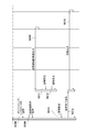
- FIG. 13A and 13B are sequence diagrams showing communication between systems at the time of radiation imaging, which is started by an operation performed by the imaging engineer 300 according to the third embodiment.
- the imaging engineer 300 operates the radiation imaging control apparatus 100 to perform an operation of searching a work list that is a list of a plurality of examination orders (S1301: work list search operation).
- the examination order creation unit 1201 of the radiographic imaging control apparatus 100 acquires electronic medical chart information from the electronic medical chart system 102 (S1302).
- the inspection order creating unit 1201 creates an inspection order based on the acquired electronic medical record information, and creates and presents a work list using the created inspection order (S1303).
- the radiographing engineer 300 operates the radiographic imaging control apparatus 100 to perform an operation of selecting an inspection order to be performed from the presented work list (S1304: inspection order selection operation).
- the radiographic imaging control apparatus 100 selects a protocol based on the selected examination order (S1305), and the support information creation unit 205 acquires electronic chart information of the patient related to the examination order from the electronic chart system 102 (S1306). .. After that, the support information creation unit 205 of the radiation imaging control apparatus 100 creates support information based on the acquired electronic medical record information (S1307) and displays it (S1308).
- the subsequent operation and processing of S1309 to S1316 are the same as those of the first embodiment (S308 to S315 of FIG. 3) or the second embodiment. That is, the imaging technician 300 refers to the displayed support information, positions the patient, adjusts the X-ray irradiation parameters of the radiation generator 106 (S1309), and instructs radiation irradiation (S1310).
- the radiation sensor 101 generates a radiographic image based on the received radiation and outputs the radiographic image to the radiographic control device 100a (S1311).
- the radiographic imaging control apparatus 100 performs image processing on the received radiographic image (S1312) and displays it (S1313).
- the radiographing engineer 300 confirms the displayed image (S1314), operates the radiographic imaging control device 100, and ends the inspection (S1315). In response to this inspection ending operation, the radiation imaging control apparatus 100 transmits the image-processed radiation imaging image to the image server 104 (S1316).
- the inspection order creation unit 1201 of the third embodiment determines the inspection region and the imaging direction based on the keyword acquired from the electronic medical record information and the specific item retrieved from the storage unit 204 by the keyword, and then the inspection order.
- FIG. 14 is a flowchart showing a process in which the inspection order creating unit 1201 creates an inspection order and a work list.
- 15 is an example of electronic medical record information acquired from the electronic medical record system 102. The flowchart shown in FIG. 14 will be described using the electronic medical record information of FIG.
- the examination order creation unit 1201 specifies conditions such as a patient ID and examination date via the electronic chart information receiving unit 201a to search for and obtain electronic chart information from the electronic chart system 102 (S1401). Next, the inspection order creation unit 1201 extracts the electronic medical record information to be the inspection target including the wording such as “radiation inspection performed” from the electronic medical record information acquired in S1401 (S1402).
- the electronic medical record information acquired in S1401 is the electronic medical record information T1501 and the electronic medical record information T1502 as shown in FIG.
- the electronic medical record information T1501 includes a description of “There is a suspicion of a left wrist fracture. August 20, 2018, a radiological examination is performed.” registered at 10:01:15 on August 20, 2018.
- the electronic medical record information T1502 includes a description of “There is pain in the ankle. XX prescription” registered at 13:50:20 on August 20, 2018.
- the electronic medical record information T1501 including “radiation inspection execution” is extracted as the inspection execution target.
- the inspection order creation unit 1201 performs lexical analysis of the electronic medical record information to be inspected (S1403), and extracts a search keyword (S1404). For example, from the electronic medical record information T1501, by performing lexical analysis, the words “left”, “wrist”, “broken”, “suspicious”, “2018”, “August”, “20th”, “radiation examination”, and “execution” are obtained. To be Then, the examination order creation unit 1201 extracts a search keyword from the lexical words obtained in S1403 (S1404). Here, the examination site and findings are required to determine the imaging direction, and the date and examination implementation are unnecessary. Therefore, when only the necessary words and phrases are extracted, the words “left”, “wrist” and “bone fracture” are extracted in this example.
- the inspection order creation unit 1201 searches the storage unit 204 for a specific item using the obtained search keyword (S1405), and determines the inspection site and imaging direction from the electronic medical record information and the specific item (S1406). .. For example, if the specific item is "the result of taking a picture with the fingers closed and the palm underneath", the inspection site was obtained from the lexical analysis of the "wrist", and the imaging direction was obtained from the specific item. It will be "front”. After that, the inspection order creating unit 1201 creates an inspection order from the obtained inspection region and imaging direction (S1407). If a plurality of pieces of electronic medical record information to be inspected are obtained in step S1402, the processing in steps S1403 to S1407 is repeated for each object to be inspected. Finally, the inspection order creation unit 1201 creates a work list by collecting the inspection orders (S1408).
- an inspection order is created from electronic medical record information and specific items without using the ordering system. As a result, it is possible to reduce the trouble of issuing orders by the doctor.
- the shooting direction is not described in the electronic medical record information.
- the specific items are determined from the electronic medical record information. It may be configured to create an inspection order without searching for.
- information that should be noted when capturing a medical image is automatically generated and displayed. Therefore, the user (imaging technologist) can be assisted so that the doctor can perform the intended inspection without adding the caution at the time of imaging. Further, by displaying the support information including the specific items, it is possible to support the imaging of the medical image so that the doctor can perform the intended examination without adding the cautions at the time of the imaging.
- the present invention supplies a program that implements one or more functions of the above-described embodiments to a system or apparatus via a network or a storage medium, and one or more processors in a computer of the system or apparatus read and execute the program. It can also be realized by the processing. It can also be realized by a circuit (for example, ASIC) that realizes one or more functions.
- a circuit for example, ASIC
Landscapes
- Health & Medical Sciences (AREA)
- Life Sciences & Earth Sciences (AREA)
- Medical Informatics (AREA)
- Engineering & Computer Science (AREA)
- General Health & Medical Sciences (AREA)
- Public Health (AREA)
- Radiology & Medical Imaging (AREA)
- Nuclear Medicine, Radiotherapy & Molecular Imaging (AREA)
- Optics & Photonics (AREA)
- Biomedical Technology (AREA)
- Primary Health Care (AREA)
- Biophysics (AREA)
- High Energy & Nuclear Physics (AREA)
- Epidemiology (AREA)
- Pathology (AREA)
- Physics & Mathematics (AREA)
- Heart & Thoracic Surgery (AREA)
- Molecular Biology (AREA)
- Surgery (AREA)
- Animal Behavior & Ethology (AREA)
- Veterinary Medicine (AREA)
- Apparatus For Radiation Diagnosis (AREA)
- Medical Treatment And Welfare Office Work (AREA)
Abstract
情報処理装置は、検査対象の診察所見が記録された電子カルテ情報を取得する第1取得部と、電子カルテ情報からキーワードを取得する第2取得部と、医用画像の撮影のための特定事項とキーワードとを関連付けて記憶している記憶部を、第2取得部により取得されたキーワードを用いて検索する検索部と、検索部によって検索された特定事項に基づいて、医用画像の撮影のための支援情報を作成する作成部と、を備える。
Description
本発明は、ユーザの医用画像の撮影を支援する、情報処理装置、放射線撮影システムおよび支援方法に関するものである。
近年、電子カルテの普及により、医師が患者から聞き取った問診内容や検査結果等が電子カルテに保存されるようになっている。また、電子カルテシステムが各種検査のオーダリングシステムと連携することで、電子カルテからX線撮影等の各種検査オーダを発行することができるようになっている。医師は問診内容や検査結果を勘案し、電子カルテを用いて必要な検査オーダを発行している。オーダリングシステムは、電子カルテから受信した検査オーダを管理する。そして、X線撮影装置等の検査機器は、オーダリングシステムから受信した検査オーダに基づいて、患者情報、撮影部位、画像処理パラメータを含むプロトコルなどを決定し、検査を実行する。
電子カルテシステムとオーダリングシステム間、ならびにオーダリングシステムと検査機器間では、予めコード値を割り振る等して、電子カルテが発行する検査オーダと撮影のプロトコルとを関連付けている。この関連付けにより、複数のシステムをまたがっていても検査オーダ通りの検査を行うことができるようになっている。また、決定されたプロトコルで用いるパラメータなどを自動設定するための技術も提案されている。例えば、特許文献1においては、執刀医師名、患者データ、術式等の電子カルテ情報を取得して医療用内視鏡システムにおけるパラメータの取得あるいは補正をすることが提案されている。また、特許文献2においては、疾患画像と画像処理パラメータを関連付けておき、撮影画像と疾患画像の類似度から画像処理パラメータを決定して画像処理を行うことが提案されている。
診断技術の進歩や検査機器の高度化により、撮影技師は、上記のコード値には現れないような情報を考慮してプロトコルを実施することが要求されている。医師が電子カルテに入力した問診内容によって、プロトコルだけでは伝わらない撮影時の注意事項があると、適切な撮影を行わないと正確な診断が困難になるケースや、再撮影で患者に負担をかけるケースが起こりうる。あるいは、注意事項を医師が別途検査オーダ発行時に付記する必要が出て、医師の違いによる記載の違いや記載漏れによって意図した検査が行われない可能性がある。例えば、斜位撮影の検査オーダでも、医師が意図したことを撮影技師が十分にくみ取れていないと、逆方向で撮影してしまう恐れがある。上述した従来技術では、撮影画像に対する画像処理パラメータを補正することはできるが、撮影時のポジショニング不良などの回避は難しい。このような課題は、放射線画像の撮影に限られるものではなく、各種モダリティによる医用画像の撮影において生じ得る。
本発明の一態様は、医用画像の撮影を支援するための支援情報を生成可能とする技術を提供する。
本発明の一態様による情報処理装置は以下の構成を備える。すなわち、
検査対象の診察所見が記録された電子カルテ情報を取得する第1取得手段と、
前記電子カルテ情報からキーワードを取得する第2取得手段と、
医用画像の撮影のための特定事項とキーワードとを関連付けて記憶している記憶手段を、前記第2取得手段により取得されたキーワードを用いて検索する検索手段と、
前記検索手段によって検索された特定事項に基づいて、医用画像の撮影のための支援情報を作成する第1作成手段と、を備える。
検査対象の診察所見が記録された電子カルテ情報を取得する第1取得手段と、
前記電子カルテ情報からキーワードを取得する第2取得手段と、
医用画像の撮影のための特定事項とキーワードとを関連付けて記憶している記憶手段を、前記第2取得手段により取得されたキーワードを用いて検索する検索手段と、
前記検索手段によって検索された特定事項に基づいて、医用画像の撮影のための支援情報を作成する第1作成手段と、を備える。
本発明によれば、医用画像の撮影を支援するための支援情報が生成される。
本発明のその他の特徴及び利点は、添付図面を参照とした以下の説明により明らかになるであろう。なお、添付図面においては、同じ若しくは同様の構成には、同じ参照番号を付す。
添付図面は明細書に含まれ、その一部を構成し、本発明の実施の形態を示し、その記述と共に本発明の原理を説明するために用いられる。
第1実施形態による放射線撮影システムの構成例を示す図である。
第1実施形態の放射線撮影システムの機能構成例を示すブロック図である。
放射線撮影時の各システム間の通信を表すシーケンス図である。
放射線撮影時の各システム間の通信を表すシーケンス図である。
第1実施形態の支援情報作成部による処理を示すフローチャートである。
電子カルテシステム102から取得した電子カルテ情報の例を示す図である。
特定事項の例を示す図である。
第1実施形態の放射線撮影システムによる表示画面の例を示す図である。
第2実施形態の放射線撮影システムの機能構成例を示すブロック図である。
第2実施形態の支援情報作成部による処理を示すフローチャートである。
第2実施形態の放射線撮影システムによる表示画面の例を示す図である。
第3実施形態の放射線撮影システムの構成例を示す図である。
第3実施家形態の放射線撮影システムの機能構成例を示すブロック図である。
放射線撮影時の各システム間の通信を表すシーケンス図である。
放射線撮影時の各システム間の通信を表すシーケンス図である。
検査オーダ作成部の処理を示すフローチャートである。
電子カルテシステムから取得した電子カルテ情報の例を示す図である。
以下、添付の図面を参照して本発明の実施形態を説明する。なお、以下の実施形態において、放射線という用語は、X線の他、例えば、α線、β線、γ線粒子線、宇宙線などを含み得る。
<第1実施形態>
図1は、第1実施形態による放射線撮影システムの構成例を示す図である。放射線撮影を制御するための情報処理装置である放射線撮影制御装置100は、放射線を照射する放射線発生装置106と、照射された放射線から放射線画像を生成する放射線センサー101とに接続されている。放射線撮影制御装置100は、放射線センサー101から放射線画像を受信する。また、放射線撮影制御装置100は、電子カルテシステム102およびオーダリングシステム103と、電子カルテ情報や検査オーダを受信できるように院内ネットワーク105を介して接続されている。加えて、放射線撮影制御装置100は、画像サーバ104と、撮影した放射線画像を送受信できるように院内ネットワーク105を介して接続されている。
図1は、第1実施形態による放射線撮影システムの構成例を示す図である。放射線撮影を制御するための情報処理装置である放射線撮影制御装置100は、放射線を照射する放射線発生装置106と、照射された放射線から放射線画像を生成する放射線センサー101とに接続されている。放射線撮影制御装置100は、放射線センサー101から放射線画像を受信する。また、放射線撮影制御装置100は、電子カルテシステム102およびオーダリングシステム103と、電子カルテ情報や検査オーダを受信できるように院内ネットワーク105を介して接続されている。加えて、放射線撮影制御装置100は、画像サーバ104と、撮影した放射線画像を送受信できるように院内ネットワーク105を介して接続されている。
放射線撮影制御装置100とオーダリングシステム103は、電子カルテシステム102との間で、HL7(Health Level 7)規格に準拠したプロトコルで通信を行う。また、放射線撮影制御装置100は、オーダリングシステム103および画像サーバ104と、DICOM(Digital Imaging and COmunications in Medicine)規格に準拠したプロトコルで通信を行う。
検査オーダは電子カルテシステム102からオーダリングシステム103に送信される。放射線撮影制御装置100は、オーダリングシステム103に問い合わせを行って検査オーダを取得する。その後、放射線撮影制御装置100は、電子カルテシステム102から電子カルテ情報を取得する。放射線撮影制御装置100は、放射線発生装置106と放射線センサー101を用いて取得した放射線画像に関して画像処理、アノテーションの生成、患者情報との関連付けを行って画像サーバ104に送信する。
図2は、第1実施形態による放射線撮影制御装置100の機能構成例を示すブロック図である。電子カルテ情報受信部201は、電子カルテシステム102と通信し、撮影を実施する検査オーダに基づいて、電子カルテシステム102から当該患者の問診内容や検査結果等の診察所見が記録された電子カルテ情報を取得する。電子カルテ情報受信部201は、電子カルテシステム102から取得した電子カルテ情報を支援情報作成部205に出力する。検査オーダ受信部202は、オーダリングシステム103と通信し、検査オーダを取得する。検査オーダ受信部202は、オーダリングシステム103から取得した検査オーダを支援情報作成部205ならびにUI部209に出力する。画像送信部203は、画像サーバ104と通信し、画像処理部206で画像処理を行った放射線撮影画像を送信する。
記憶部204は、検査オーダに関連付けられた撮影部位および画像処理パラメータなどを含むプロトコル情報を記憶する。また、記憶部204は、特定事項(例えば、撮影時の注意事項)をキーワードと関連付けて記憶する。キーワードには、部位や撮影方向に加えて、電子カルテに入力される語句を利用する。また、本実施形態では、特定事項は、注意喚起を促す文言と撮影時のポジショニングを示す写真(確認画像)を含む。記憶部204に記憶されているキーワードと特定事項は、教師付き機械学習アルゴリズムを用いて関連付けが行われ、よく使われるキーワードの組み合わせから特定事項を抽出できるようになっている。すなわち、記憶部204は、教師付き機械学習によって関連付けられたキーワードと特定事項を記憶しており、特定事項には、放射線撮影における検査部位のポジショニングを撮影した確認画像が含まれている。
支援情報作成部205は、検査オーダ受信部202から取得した検査オーダと、電子カルテ情報受信部201から受信した電子カルテ情報とに基づいて記憶部204から特定事項を取得する。支援情報作成部205は、取得した特定事項を用いて放射線撮影のための支援情報を作成し、UI部209に出力する。支援情報の作成処理については後述する。画像処理部206は、UI部209で選択したプロトコルに対応する画像処理パラメータを記憶部204から取得し、通信部208から受信した放射線撮影画像に画像処理を施し、画像送信部203ならびにUI部209に出力する。
撮影制御部207は、UI部209からの指示に基づいて通信部208を介して放射線センサー101と通信し、放射線センサー101のパラメータ設定を行う。また、撮影制御部207は、UI部209に対して放射線センサー101の状態通知を行う。通信部208は、放射線センサー101と通信し、撮影制御部207から受信したパラメータ設定や、放射線センサー101の状態の受信を行う。また、通信部208は、放射線センサーから受信した放射線撮影画像を画像処理部206に出力する。さらに、撮影制御部207は、通信部208を介して放射線発生装置106の放射線照射パラメータを設定する。
UI部209は、ユーザに放射線撮影制御装置100を操作するためのユーザインターフェースであり、画面表示と操作入力の受付を行う。例えば、UI部209は、検査オーダ受信部202から受信した検査オーダを表示し、ユーザである撮影技師が対象検査を選択できるようにする。また、UI部209は、撮影制御部207から受信した放射線センサー101の状態、画像処理部206が画像処理した放射線撮影画像を表示する。加えて、UI部209は、支援情報作成部205に支援情報の作成を指示し、得られた支援情報を表示部210表示する。
図3Aおよび図3B(以下、図3と記載する)は、撮影技師300が行う操作をトリガーとして開始される、放射線撮影時の各システム間の通信を表すシーケンス図である。まず、放射線撮影制御装置100の支援情報作成部205は、電子カルテ情報受信部201を介して、検査対象の診察所見が記録された電子カルテ情報を取得する。この処理の一例をS301~S305に示す。撮影技師300は、放射線撮影制御装置100を操作して、複数の検査オーダの一覧であるワークリストを検索する操作を行う(S301:ワークリストの検索操作)。放射線撮影制御装置100は、この操作を受けてオーダリングシステム103からワークリストを検索し、取得する(S302)。
次に、撮影技師300は、放射線撮影制御装置100を操作して、ワークリストから実施する検査オーダを選択する操作を行う(S303:検査オーダの選択操作)。放射線撮影制御装置100は、選択された検査オーダに基づいてプロトコルを選択し(S304)、当該検査オーダに関連する患者の電子カルテ情報を電子カルテシステム102から取得する(S305)。このように、支援情報作成部205は、オーダリングシステム103から提供される検査オーダのうちのユーザ操作により選択された検査オーダを取得し、その検査オーダに関連する電子カルテ情報を電子カルテシステム102から取得する。
その後、放射線撮影制御装置100の支援情報作成部205は、取得した電子カルテ情報に基づいて、支援情報を作成する(S306)。このように、支援情報作成部205は、電子カルテ情報からキーワードを取得し、医用画像の撮影のための特定事項とキーワードとを関連付けて記憶している記憶部204を、取得されたキーワードを用いて検索する構成の一例である。また、支援情報作成部205は、検索された特定事項に基づいて、医用画像の撮影のための支援情報を作成する構成の一例である。但し、本実施形態では、医用画像の撮影として放射線撮影を適用した例を示している。なお、本実施形態では、S303で選択された検査対象の検査オーダからも検索のためのキーワードが取得される。このような支援情報作成部205の処理例については、図4のフローチャートにより後述する。作成された支援情報は、UI部209の表示部210に表示される(S307)。
次に、撮影技師300は、表示された支援情報を参考にして、患者のポジショニングや、放射線発生装置106の放射線照射パラメータの調整を行う(S308)。そして、撮影技師300は、放射線の照射を指示する(S309)。この指示に応じて放射線発生装置106から照射された放射線を受信した放射線センサー101は、受信した放射線に基づいて放射線撮影画像を生成し、放射線撮影制御装置100に出力する(S310)。放射線撮影制御装置100は、受信した放射線撮影画像に画像処理を施し(S311)、処理後の放射線画像を表示部210に表示する(S312)。その後、撮影技師300は、表示された画像を確認し(S313)、放射線撮影制御装置100を操作して検査を終了する(S314:検査の終了操作)。撮影技師300による検査の終了操作を受けて、放射線撮影制御装置100は、画像処理した放射線画像を画像サーバ104に送信する(S315)。
図4は、放射線撮影制御装置100の支援情報作成部205が、支援情報を作成する際(S306)の処理を示すフローチャートである。また、図5は手首の放射線撮影を行う患者について、電子カルテシステム102から取得された電子カルテ情報の一例である。図5の電子カルテ情報を用いて、図4に示されるフローチャートを説明する。
まず、支援情報作成部205は、受信した電子カルテ情報について字句解析を行う(S401)。本例では、図5に示されるように、電子カルテ情報T501は、2018年7月31日10時01分15秒に登録された「骨密度検査実施。骨粗しょう症」を含む。また、電子カルテ情報T502は、2018年8月20日13時50分20秒に登録された「左手の手首を手前に曲げると痛み」を含む。この場合、支援情報作成部205は、字句解析を行うことで、電子カルテ情報T501からは「骨密度検査」「実施」「骨粗しょう症」という字句を、電子カルテ情報T502からは「左手」「手首」「手前」「曲げる」「痛み」という字句を得る。
次に、支援情報作成部205は、得られた字句から検索用キーワードを抽出する(S402)。ここでは、検査結果や問診内容は検索用キーワードとして必要だが、検査オーダから取得できる情報や検査を実施したというような内容は不要であり、支援情報作成部205は、必要な字句のみを抽出する。図5の例では、「左手」と「手首」は検査オーダから取得される情報のため、不要と判定される。また、「骨密度検査」と「実施」は、検査の実施内容を示す字句であり不要と判定される。よって、支援情報作成部205は、これら以外の字句である「骨粗しょう症」「手前」「曲げる」「痛み」を、検索用キーワードとして電子カルテ情報から抽出する。その後、支援情報作成部205は、検査オーダから得られた部位(左手、手首)と電子カルテ情報から得られた検索用キーワードを用いて、特定事項と撮影時のポジショニングを撮影した確認画像を記憶部204から検索する(S403)。支援情報作成部205は、検索された特定事項とポジショニングの確認画像を用いて支援情報を作成し、UI部209に出力(表示)する(S404)。
図6は、図5に示した電子カルテ情報から得られた特定事項の例である。部位が「手首」、検索用キーワードが「骨粗しょう症」の場合に得られた特定事項が「線量を下げる」(T601)である。また、部位が「左手」「手首」、検索用キーワードが「手前」「曲げる」「痛み」の場合に得られた特定事項が「手のひらを手前に向け、斜め45度から撮影」(T602)である。なお、部位の情報としての「左手」が特定事項の取得に用いられないのであれば、部位が左手であるか右手であるかの情報は省略されてもよい。例えば、左手と右手で特定事項が同一の場合は、「左手」「右手」の情報は不要となる。
図7は、放射線撮影制御装置100にて支援情報を表示した場合の表示部210における表示画面例を示す模式図である。画面には、放射線撮影画像と患者情報や受付番号等を表示する画像表示領域701、選択しているプロトコルを表す領域702、放射線センサーの状態を表示する領域703が配置されている。また、放射線撮影画像の画質調整を指示するボタン704、任意のアノテーションの追加を指示するボタン705、検査保留を指示するボタン706、検査中止を指示するボタン707が配置される。これらが配置された画面上に、当該プロトコルに関係する支援情報を表示するダイアログ708が表示される。ダイアログ708には、図4のフローチャートで取得した特定事項709(図6)とポジショニングの確認画像710、当該ダイアログを閉じることを指示するボタン711が配置される。なお、ダイアログ708は、例えば、支援情報作成部205により作成されてもよい。すなわち、支援情報作成部205は、検索された特定事項に基づいて生成した支援情報に基づいて、医用画像を表示する表示部に表示させる支援画像(ダイアログ708)を作成する構成の一例である。
以上のように、第1実施形態の放射線撮影制御装置100によれば、電子カルテ情報から支援情報を作成して表示することで、医師の意図した検査が行えるように支援することができる。
なお、上記実施形態では、電子カルテ情報に基づいて検索された特定事項を用いて支援情報を生成するとしたが、電子カルテ情報に基づいて支援情報が検索されるようにしてもよい。この場合、記憶部204は、医用画像の撮影のための支援情報とキーワードとを関連付けて記憶し、支援情報作成部205は、電子カルテ情報から取得したキーワードを用いて記憶部204を検索する。また、支援情報作成部205が、検索された支援情報に基づいて、医用画像を表示する表示部に表示させる支援画像(例えば、ダイアログ708)を作成するように構成されてもよい。
また、第1実施形態では複数の電子カルテ情報を等価に扱って支援情報を表示するように構成したが、これに限られるものではない。例えば、支援情報の取得対象の検査オーダに関連する電子カルテ情報の優先順位を高くし、その他の電子カルテ情報から得られた特定事項よりも先に表示するように構成するようにしてもよい。また、過去数回分または一定期間内の電子カルテ情報のみが用いられるように構成されてもよい。
<第2実施形態>
第2実施形態では、第1実施形態の構成に、可視光による撮影を行うカメラで撮影した画像を用いて、ポジショニング時の位置ずれを警告する構成を加えた例について説明する。より具体的には、第2実施形態の放射線撮影制御装置100は、照射野を撮影した照射野画像をカメラから取得する。放射線撮影制御装置100は、電子カルテ情報(および検査オーダ)から得られたキーワードに基づいて検索されたポジショニングの確認画像と照射野画像との類似度を計算し、類似度が所定値以下の場合に、支援情報に警告メッセージを含める。以下、第2実施形態について説明する。なお、第2実施形態による放射線撮影システムの構成は第1実施形態(図1)と同様である。
第2実施形態では、第1実施形態の構成に、可視光による撮影を行うカメラで撮影した画像を用いて、ポジショニング時の位置ずれを警告する構成を加えた例について説明する。より具体的には、第2実施形態の放射線撮影制御装置100は、照射野を撮影した照射野画像をカメラから取得する。放射線撮影制御装置100は、電子カルテ情報(および検査オーダ)から得られたキーワードに基づいて検索されたポジショニングの確認画像と照射野画像との類似度を計算し、類似度が所定値以下の場合に、支援情報に警告メッセージを含める。以下、第2実施形態について説明する。なお、第2実施形態による放射線撮影システムの構成は第1実施形態(図1)と同様である。
図8は、第2実施形態による放射線撮影制御装置100の機能構成例を示すブロック図である。第1実施形態(図2)と同様の構成には同一の参照番号が付してある。支援情報作成部205aは、第1実施形態(図2)の支援情報作成部205の機能に加えて、以下の機能を有する。すなわち、支援情報作成部205aは、所定の時間間隔でカメラ通信部801から受信した照射野画像と、記憶部204に保存したポジショニング確認画像を比較し、類似度が一定以下の場合に警告メッセージを支援情報とともにUI部209に出力する。この処理については後述する。また、支援情報作成部205aは、UI部209から支援情報出力の終了指示を受信すると、警告メッセージの出力を終了する。カメラ通信部801は、支援情報作成部205aの指示に基づいて、カメラ802から照射野画像を受信する。カメラ802は、放射線センサー101と対向するように放射線発生装置106に設置され、放射線検査を行う照射野を所定の時間間隔で撮影し、撮影した照射野画像をカメラ通信部801に送信する。
図9は、第2実施形態による支援情報の作成処理を示すフローチャートである。支援情報作成部205aは、受信した電子カルテ情報について字句解析を行い(S901)、得られた字句から検索用キーワードを抽出する(S902)。その後、支援情報作成部205aは、検査オーダと検索用キーワードを用いて、記憶部204から特定事項とポジショニングの確認画像を検索する(S903)。ここまでの処理は第1実施形態(図4)の処理(S401~S403)と同様である。
次に、支援情報作成部205aは、カメラ通信部801を介してカメラ802から照射野画像を取得する(S904)。その後、S903で得られた確認画像と照射野画像に写っている被写体の類似度を解析する(S905)。S905で得られた類似度が閾値以下の場合(S906でYES)は、支援情報作成部205aは、支援情報と照射野画像に加えて警告メッセージをUI部209に出力する(S907)。一方、S905で得られた類似度が閾値より大きい場合(S906でNO)は、支援情報作成部205aは、支援情報と照射野画像をUI部209に出力する(S908)。その後、支援情報作成部205aは、UI部209から支援情報出力の終了指示を受信した場合(S909でYES)に、本処理を終了する。支援情報出力の終了指示を受信しなかった場合(S909でNO)、処理は一定時間ウェイトした後(S910)、S904に戻る。
図10は、放射線撮影制御装置100により支援情報、照射野画像ならびに警告メッセージを表示した場合の画面例を示す模式図である。その画面には、放射線撮影画像と患者情報や受付番号等を表示する画像表示領域1001、選択しているプロトコルを表す領域1002、放射線センサー101の状態を表示する領域1003が配置されている。また、この画面には、放射線撮影画像の画質調整を行うボタン1004、任意のアノテーションを追加するボタン1005、検査を保留するボタン1006、検査を中止するボタン1007が配置されている。これらは、第1実施形態で説明した構成(図7の701から707)と同様である。
また、図10に示されるように、画面上には、当該プロトコルに関係する支援情報を表示するダイアログ1008が表示される。ダイアログ1008には、図9のフローチャートで取得した特定事項1009と確認画像1010、警告メッセージ1011、カメラから取得した照射野画像1012ならびにダイアログを閉じるボタン1013が配置される。撮影技師は、警告メッセージ1011、確認画像1010、照射野画像1012の表示により、放射線撮影時の正しいポジショニングを容易に確認することができる。また、閉じるボタン1013の押下により、支援情報出力の終了指示がUI部から支援情報作成部205aに出力される。
以上のように、第2実施形態によれば、カメラで撮影した照射野画像と支援情報を比較して警告メッセージが表示されるので、撮影技師は、撮影時の実際のポジショニングと推奨されるポジショニングとの違いに容易に気づくことができる。
<第3実施形態>
第3実施形態では、オーダリングシステムから検査オーダを取得する構成の代わりに、電子カルテ情報から検査オーダを作成する構成の例について説明する。なお、第1実施形態ならびに第2実施形態と重複する部分については説明を省略する。図11は、第3実施形態による放射線撮影システムの構成例を示すブロック図である。図11に示されるように、第3実施形態では、第1実施形態の放射線撮影システムの構成からオーダリングシステム103が省略されている。放射線撮影制御装置100aは、電子カルテシステム102が管理する電子カルテ情報を検索して、ワークリスト(検査オーダのリスト)を生成する。
第3実施形態では、オーダリングシステムから検査オーダを取得する構成の代わりに、電子カルテ情報から検査オーダを作成する構成の例について説明する。なお、第1実施形態ならびに第2実施形態と重複する部分については説明を省略する。図11は、第3実施形態による放射線撮影システムの構成例を示すブロック図である。図11に示されるように、第3実施形態では、第1実施形態の放射線撮影システムの構成からオーダリングシステム103が省略されている。放射線撮影制御装置100aは、電子カルテシステム102が管理する電子カルテ情報を検索して、ワークリスト(検査オーダのリスト)を生成する。
図12は、第3実施形態による放射線撮影制御装置100aの機能構成例を示すブロック図である。電子カルテ情報受信部201aは、第1実施形態の電子カルテ情報受信部201(図2)の機能に加えて、検索した電子カルテ情報を検査オーダ作成部1201に出力する機能を有する。検査オーダ作成部1201は、電子カルテ情報受信部201aから取得した電子カルテ情報に基づいて1つまたは複数の検査オーダを作成し、検査オーダのリストであるワークリストを作成する。検査オーダ作成部1201は、作成したワークリストを撮影技師に提示するためにUI部209に出力する。検査オーダならびにワークリストの作成手順については後述する。
図13Aおよび図13Bは、第3実施形態による、撮影技師300が行う操作をトリガーとして開始される放射線撮影時のシステム間の通信を表すシーケンス図である。
まず、撮影技師300は、放射線撮影制御装置100を操作して、複数の検査オーダの一覧であるワークリストを検索する操作を行う(S1301:ワークリストの検索操作)。放射線撮影制御装置100の検査オーダ作成部1201は、この操作を受け付けると、電子カルテシステム102から電子カルテ情報を取得する(S1302)。検査オーダ作成部1201は、取得した電子カルテ情報に基づいて検査オーダを作成し、作成された検査オーダを用いてワークリストを作成、提示する(S1303)。撮影技師300は、放射線撮影制御装置100を操作して、提示されたワークリストから実施する検査オーダを選択する操作を行う(S1304:検査オーダの選択操作)。放射線撮影制御装置100は、選択した検査オーダに基づいてプロトコルを選択し(S1305)、支援情報作成部205は当該検査オーダに関連する患者の電子カルテ情報を電子カルテシステム102から取得する(S1306)。その後、放射線撮影制御装置100の支援情報作成部205は、取得した電子カルテ情報に基づいて、支援情報を作成し(S1307)、表示する(S1308)。
以降のS1309~S1316の動作、処理は第1実施形態(図3のS308~S315)あるいは第2実施形態と同様である。すなわち、撮影技師300は、表示された支援情報を参考にして、患者のポジショニングや放射線発生装置106のX線照射パラメータの調整を行い(S1309)、放射線照射を指示する(S1310)。放射線センサー101は、受信した放射線に基づいて放射線撮影画像を生成し、放射線撮影制御装置100aに出力する(S1311)。放射線撮影制御装置100は、受信した放射線撮影画像に画像処理を行い(S1312)、表示する(S1313)。撮影技師300は、表示された画像を確認し(S1314)、放射線撮影制御装置100を操作して検査を終了する(S1315)。この検査終了操作を受けて、放射線撮影制御装置100は、画像処理した放射線撮影画像を画像サーバ104に送信する(S1316)。
第3実施形態の検査オーダ作成部1201は、電子カルテ情報から取得されたキーワードと、該キーワードにより記憶部204から検索された特定事項とに基づいて、検査部位と撮影方向を決定して検査オーダを作成する。図14は、検査オーダ作成部1201が検査オーダならびにワークリストを作成する処理を示すフローチャートである。また、図15は電子カルテシステム102から取得される電子カルテ情報の例である。図15の電子カルテ情報を用いて、図14に示したフローチャートを説明する。
検査オーダ作成部1201は、電子カルテ情報受信部201aを介して患者IDや検査日等の条件を指定して電子カルテシステム102から電子カルテ情報を検索し、取得する(S1401)。次に、検査オーダ作成部1201は、S1401で取得した電子カルテ情報から、「放射線検査実施」等の文言が含まれる検査実施対象となる電子カルテ情報を抽出する(S1402)。
S1401で取得された電子カルテ情報が、図15に示すような、電子カルテ情報T1501と電子カルテ情報T1502であったとする。電子カルテ情報T1501は、2018年8月20日10時01分15秒に登録された「左手首骨折の疑いあり。2018年8月20日 放射線検査実施。」の記述を含む。また、電子カルテ情報T1502は、2018年8月20日13時50分20秒に登録された「足首に痛みあり。○○処方」の記述を含む。この場合、「放射線検査実施」が含まれる電子カルテ情報T1501が検査実施対象として抽出される。
次に、検査オーダ作成部1201は、検査実施対象の電子カルテ情報の字句解析を行い(S1403)、検索用キーワードを抽出する(S1404)。例えば、電子カルテ情報T1501からは、字句解析を行うことで「左」「手首」「骨折」「疑い」「2018年」「8月」「20日」「放射線検査」「実施」という字句が得られる。そして、検査オーダ作成部1201は、S1403で得られた字句から検索用キーワードを抽出する(S1404)。ここでは、撮影方向を決定するために検査部位や所見が必要であり、日付や検査実施は不要である。従って、必要な字句のみが抽出されると、この例では「左」「手首」「骨折」という字句が抽出される。
次に、検査オーダ作成部1201は、得られた検索用キーワードを用いて記憶部204から特定事項を検索し(S1405)、電子カルテ情報と特定事項から検査部位と撮影方向を決定する(S1406)。例えば、特定事項として「指を閉じて、手のひらを下に正面から撮影」という結果)が得られたとすると、検査部位は字句解析で得られた「手首」、撮影方向は特定事項から得られた「正面」となる。その後、検査オーダ作成部1201は、得られた検査部位と撮影方向から検査オーダを作成する(S1407)。S1402において検査実施対象の電子カルテ情報が複数得られた場合は、S1403~S1407の処理を検査実施対象分繰り返す。最後に、検査オーダ作成部1201は、検査オーダをまとめてワークリストを作成する(S1408)。
以上のように、第3実施形態によれば、オーダリングシステムを使わずに、電子カルテ情報と特定事項から検査オーダが作成される。これにより、医師によるオーダ発行の手間を低減することができる。
なお、第3実施形態では撮影方向が電子カルテ情報に記載されていない場合について記載したが、字句解析の結果、撮影方向が記載されていると判断できた場合は、当該電子カルテ情報から特定事項を検索せずに検査オーダを作成するように構成してもよい。
以上説明したように、上記各実施形態によれば、医用画像の撮影時に注意すべき情報(支援情報)が自動的に生成、表示される。そのため、医師が撮影時の注意事項を付記しなくても、医師が意図した検査が行えるように、ユーザ(撮影技師)を支援することができる。また、特定事項を含む支援情報を表示することで、医師が撮影時の注意事項を付記しなくても、医師が意図した検査が行えるように医用画像の撮影を支援することができる。
<他の実施形態>
本発明は、上述の実施形態の1以上の機能を実現するプログラムを、ネットワーク又は記憶媒体を介してシステム又は装置に供給し、そのシステム又は装置のコンピュータにおける1つ以上のプロセッサーがプログラムを読出し実行する処理でも実現可能である。また、1以上の機能を実現する回路(例えば、ASIC)によっても実現可能である。
本発明は、上述の実施形態の1以上の機能を実現するプログラムを、ネットワーク又は記憶媒体を介してシステム又は装置に供給し、そのシステム又は装置のコンピュータにおける1つ以上のプロセッサーがプログラムを読出し実行する処理でも実現可能である。また、1以上の機能を実現する回路(例えば、ASIC)によっても実現可能である。
本発明は上記実施の形態に制限されるものではなく、本発明の精神及び範囲から離脱することなく、様々な変更及び変形が可能である。従って、本発明の範囲を公にするために、以下の請求項を添付する。
本願は、2018年12月21日提出の日本国特許出願特願2018-240139を基礎として優先権を主張するものであり、その記載内容の全てを、ここに援用する。
Claims (18)
- 検査対象の診察所見が記録された電子カルテ情報を取得する第1取得手段と、
前記電子カルテ情報からキーワードを取得する第2取得手段と、
医用画像の撮影のための特定事項とキーワードとを関連付けて記憶している記憶手段を、前記第2取得手段により取得されたキーワードを用いて検索する検索手段と、
前記検索手段によって検索された特定事項に基づいて、医用画像の撮影のための支援情報を作成する第1作成手段と、を備えることを特徴とする情報処理装置。 - 前記第1取得手段は、さらに前記検査対象の検査オーダを取得し、
前記第2取得手段は、さらに前記検査オーダから前記検索手段による検索のためのキーワードを取得することを特徴とする請求項1に記載の情報処理装置。 - 前記第1取得手段は、オーダリングシステムからユーザ操作により選択された検査オーダを取得し、前記検査オーダに関連する電子カルテ情報を電子カルテシステムから取得することを特徴とする請求項1または2に記載の情報処理装置。
- 電子カルテ情報に基づいて検査オーダを作成する第2作成手段をさらに備えることを特徴とする請求項1または2に記載の情報処理装置。
- 前記第2作成手段は、電子カルテシステムから取得した1つまたは複数の検査対象の電子カルテ情報に基づいて1つまたは複数の検査オーダを作成し、
前記第1取得手段は、前記1つまたは複数の検査オーダからユーザ操作により選択された検査オーダを取得することを特徴とする請求項4に記載の情報処理装置。 - 前記第2作成手段は、前記電子カルテ情報から取得されたキーワードと、該キーワードにより前記記憶手段から検索された特定事項とに基づいて、検査部位と撮影方向を決定して検査オーダを作成することを特徴とする請求項4または5に記載の情報処理装置。
- 前記第2取得手段は、前記電子カルテ情報を字句解析してキーワードを取得することを特徴とする請求項1乃至6のいずれか1項に記載の情報処理装置。
- 前記記憶手段は、教師付き機械学習によって関連付けられたキーワードと特定事項を記憶することを特徴とする請求項1乃至7のいずれか1項に記載の情報処理装置。
- 前記特定事項は、放射線撮影におけるポジショニングと放射線照射パラメータの調整とに関する内容を含むことを特徴とする請求項8に記載の情報処理装置。
- 前記特定事項は、さらに放射線撮影における検査部位のポジショニングを撮影した確認画像を含み、前記支援情報は前記確認画像を含むことを特徴とする請求項9に記載の情報処理装置。
- 照射野を撮影した照射野画像をカメラから取得する第3取得手段と、
前記検索手段により検索されたポジショニング確認画像と前記照射野画像との類似度を計算する計算手段と、をさらに備え、
前記第1作成手段は、前記類似度が所定値以下の場合に、前記支援情報に警告メッセージを含めることを特徴とする請求項10に記載の情報処理装置。 - 前記支援情報に、さらに、前記カメラから取得された前記照射野画像を含めることを特徴とする請求項11に記載の情報処理装置。
- 前記第1作成手段により生成された前記支援情報を表示する表示手段をさらに備えることを特徴とする請求項1乃至12のいずれか1項に記載の情報処理装置。
- 検査対象の診察所見が記録された電子カルテ情報を取得する第1取得手段と、
前記電子カルテ情報からキーワードを取得する第2取得手段と、
医用画像の撮影のための支援情報とキーワードとを関連付けて記憶している記憶手段を、前記第2取得手段により取得されたキーワードを用いて検索する検索手段と、
前記検索手段によって検索された支援情報に基づいて、前記医用画像を表示する表示手段に表示させる支援画像を作成する第1作成手段と、を備えることを特徴とする情報処理装置。 - 放射線センサーと、
前記放射線センサーによる放射線撮影を制御する放射線撮影制御装置と、を備える放射線撮影システムであって、
前記放射線撮影制御装置は、
検査対象の診察所見が記録された電子カルテ情報を取得する第1取得手段と、
前記電子カルテ情報からキーワードを取得する第2取得手段と、
医用画像の撮影のための特定事項とキーワードとを関連付けて記憶している記憶手段を、前記第2取得手段により取得されたキーワードを用いて検索する検索手段と、
前記検索手段によって検索された特定事項に基づいて、医用画像の撮影のための支援情報を作成する作成手段と、を備えることを特徴とする放射線撮影システム。 - 医用画像の撮影のための支援方法であって、
検査対象の診察所見が記録された電子カルテ情報を取得する第1取得工程と、
前記電子カルテ情報からキーワードを取得する第2取得工程と、
医用画像の撮影のための特定事項とキーワードとを関連付けて記憶している記憶手段を、前記第2取得工程で取得されたキーワードを用いて検索する検索工程と、
前記検索工程で検索された特定事項に基づいて、医用画像の撮影のための支援情報を作成する作成工程と、を備えることを特徴とする支援方法。 - 医用画像の撮影のための支援方法であって、
検査対象の診察所見が記録された電子カルテ情報を取得する第1取得工程と、
前記電子カルテ情報からキーワードを取得する第2取得工程と、
医用画像の撮影のための支援情報とキーワードとを関連付けて記憶している記憶手段を、前記第2取得工程で取得されたキーワードを用いて検索する検索工程と、
前記検索工程で検索された支援情報に基づいて、前記医用画像を表示する表示手段に表示させる支援画像を作成する第1作成工程と、を備えることを特徴とする支援方法。 - コンピュータを、請求項1乃至14のいずれか1項に記載された情報処理装置の各手段として機能させるためのプログラム。
Applications Claiming Priority (2)
| Application Number | Priority Date | Filing Date | Title |
|---|---|---|---|
| JP2018-240139 | 2018-12-21 | ||
| JP2018240139A JP2020102037A (ja) | 2018-12-21 | 2018-12-21 | 情報処理装置、放射線撮影システムおよび支援方法 |
Publications (1)
| Publication Number | Publication Date |
|---|---|
| WO2020129464A1 true WO2020129464A1 (ja) | 2020-06-25 |
Family
ID=71101238
Family Applications (1)
| Application Number | Title | Priority Date | Filing Date |
|---|---|---|---|
| PCT/JP2019/044135 Ceased WO2020129464A1 (ja) | 2018-12-21 | 2019-11-11 | 情報処理装置、放射線撮影システムおよび支援方法 |
Country Status (2)
| Country | Link |
|---|---|
| JP (1) | JP2020102037A (ja) |
| WO (1) | WO2020129464A1 (ja) |
Families Citing this family (6)
| Publication number | Priority date | Publication date | Assignee | Title |
|---|---|---|---|---|
| JP7382739B2 (ja) * | 2019-05-14 | 2023-11-17 | キヤノンメディカルシステムズ株式会社 | 撮影支援装置 |
| JP7552372B2 (ja) * | 2021-01-18 | 2024-09-18 | コニカミノルタ株式会社 | 撮影支援装置、撮影支援方法及びプログラム |
| WO2022250143A1 (ja) * | 2021-05-28 | 2022-12-01 | 国立研究開発法人理化学研究所 | 疾病リスク評価方法、疾病リスク評価システム、及び健康情報処理装置 |
| JP7333549B2 (ja) * | 2021-05-28 | 2023-08-25 | 国立研究開発法人理化学研究所 | 疾病リスク評価方法、疾病リスク評価システム、及び健康情報処理装置 |
| WO2023100311A1 (ja) * | 2021-12-02 | 2023-06-08 | 日本電気株式会社 | 情報処理システム、情報処理方法、及び非一時的なコンピュータ可読媒体 |
| JP2024067063A (ja) * | 2022-11-04 | 2024-05-17 | キヤノン株式会社 | 情報処理装置、放射線撮影システム、情報処理方法、及びプログラム |
Citations (5)
| Publication number | Priority date | Publication date | Assignee | Title |
|---|---|---|---|---|
| JP2007280134A (ja) * | 2006-04-07 | 2007-10-25 | Topcon Corp | 医療支援システム |
| JP2013182443A (ja) * | 2012-03-02 | 2013-09-12 | Konica Minolta Inc | 電子カルテ装置 |
| JP2015090619A (ja) * | 2013-11-06 | 2015-05-11 | 株式会社東芝 | 医療情報管理装置及び医療情報管理システム |
| JP2015191557A (ja) * | 2014-03-28 | 2015-11-02 | 富士フイルム株式会社 | 撮影者支援装置、撮影者支援方法、並びに撮影者支援プログラム |
| JP2016218799A (ja) * | 2015-05-21 | 2016-12-22 | 株式会社ジェイマックシステム | 複合医療支援システム、複合医療支援方法および複合医療支援プログラム |
-
2018
- 2018-12-21 JP JP2018240139A patent/JP2020102037A/ja active Pending
-
2019
- 2019-11-11 WO PCT/JP2019/044135 patent/WO2020129464A1/ja not_active Ceased
Patent Citations (5)
| Publication number | Priority date | Publication date | Assignee | Title |
|---|---|---|---|---|
| JP2007280134A (ja) * | 2006-04-07 | 2007-10-25 | Topcon Corp | 医療支援システム |
| JP2013182443A (ja) * | 2012-03-02 | 2013-09-12 | Konica Minolta Inc | 電子カルテ装置 |
| JP2015090619A (ja) * | 2013-11-06 | 2015-05-11 | 株式会社東芝 | 医療情報管理装置及び医療情報管理システム |
| JP2015191557A (ja) * | 2014-03-28 | 2015-11-02 | 富士フイルム株式会社 | 撮影者支援装置、撮影者支援方法、並びに撮影者支援プログラム |
| JP2016218799A (ja) * | 2015-05-21 | 2016-12-22 | 株式会社ジェイマックシステム | 複合医療支援システム、複合医療支援方法および複合医療支援プログラム |
Also Published As
| Publication number | Publication date |
|---|---|
| JP2020102037A (ja) | 2020-07-02 |
Similar Documents
| Publication | Publication Date | Title |
|---|---|---|
| WO2020129464A1 (ja) | 情報処理装置、放射線撮影システムおよび支援方法 | |
| JP7164964B2 (ja) | 情報処理装置、放射線撮影装置、放射線撮影システム、情報処理方法及びプログラム | |
| JP2010257210A (ja) | 撮影情報処理装置、撮影情報処理方法及びプログラム | |
| KR20200068992A (ko) | 전자 차트 관리 장치, 전자 차트 관리 방법 및 기록 매체 | |
| JP6732520B2 (ja) | 情報処理装置、情報処理システム、情報処理方法及びプログラム。 | |
| JP7504585B2 (ja) | 画像処理装置、画像処理方法およびプログラム | |
| JP6871687B2 (ja) | 放射線撮影制御装置、放射線撮影制御装置の制御方法及びプログラム | |
| JP5125128B2 (ja) | 医用画像管理システム、データ管理方法 | |
| JP2017192453A (ja) | 情報処理装置、情報処理システム、情報処理方法及びプログラム | |
| JP2017085194A (ja) | 情報処理装置およびその制御方法 | |
| JP2012179105A (ja) | 放射線撮影システム及びその制御方法、並びに、プログラム | |
| JP4992914B2 (ja) | 小規模診断システム及び表示制御方法 | |
| JP7528050B2 (ja) | 医用情報処理装置、医用情報処理方法およびプログラム | |
| JP4802883B2 (ja) | 医用画像システム | |
| JP2003033342A (ja) | 撮影装置、撮影システム、撮影制御方法、記憶媒体、及びプログラム | |
| JP5170287B2 (ja) | 医用画像システム | |
| WO2021187188A1 (ja) | 放射線撮影システム、画像処理方法、及びプログラム | |
| JP2009199161A (ja) | 医用画像管理システム | |
| US20240177837A1 (en) | Medical information processing apparatus, medical information processing method, and non-transitory computer-readable storage medium | |
| JP2014186741A (ja) | 撮影情報処理装置、撮影情報処理方法及びプログラム | |
| JP5019933B2 (ja) | コンテンツ提供装置、及びその作動方法 | |
| JP7749929B2 (ja) | 動態解析装置及びプログラム | |
| EP4210068A1 (en) | Medical information processing system, medical information processing method, and program | |
| JP2009022566A (ja) | 医用画像システム | |
| JP2007259920A (ja) | 小規模診断システム |
Legal Events
| Date | Code | Title | Description |
|---|---|---|---|
| 121 | Ep: the epo has been informed by wipo that ep was designated in this application |
Ref document number: 19900693 Country of ref document: EP Kind code of ref document: A1 |
|
| NENP | Non-entry into the national phase |
Ref country code: DE |
|
| 122 | Ep: pct application non-entry in european phase |
Ref document number: 19900693 Country of ref document: EP Kind code of ref document: A1 |