WO2018025680A1 - 細径内視鏡手術器具 - Google Patents
細径内視鏡手術器具 Download PDFInfo
- Publication number
- WO2018025680A1 WO2018025680A1 PCT/JP2017/026598 JP2017026598W WO2018025680A1 WO 2018025680 A1 WO2018025680 A1 WO 2018025680A1 JP 2017026598 W JP2017026598 W JP 2017026598W WO 2018025680 A1 WO2018025680 A1 WO 2018025680A1
- Authority
- WO
- WIPO (PCT)
- Prior art keywords
- small
- pipe
- diameter
- surgical instrument
- endoscopic surgical
- Prior art date
Links
- 238000003780 insertion Methods 0.000 claims abstract description 22
- 230000037431 insertion Effects 0.000 claims abstract description 22
- 238000012545 processing Methods 0.000 claims description 111
- 230000005611 electricity Effects 0.000 claims description 8
- 239000002504 physiological saline solution Substances 0.000 claims description 6
- 238000009413 insulation Methods 0.000 claims description 5
- 230000000149 penetrating effect Effects 0.000 claims 1
- 238000001356 surgical procedure Methods 0.000 abstract description 25
- 238000011282 treatment Methods 0.000 abstract description 12
- 238000009297 electrocoagulation Methods 0.000 abstract description 11
- 238000000926 separation method Methods 0.000 abstract description 5
- 208000007536 Thrombosis Diseases 0.000 abstract description 4
- 238000000605 extraction Methods 0.000 abstract description 2
- 238000002679 ablation Methods 0.000 abstract 1
- 238000001574 biopsy Methods 0.000 abstract 1
- 210000001519 tissue Anatomy 0.000 description 83
- 210000000115 thoracic cavity Anatomy 0.000 description 13
- 230000008859 change Effects 0.000 description 8
- 230000002889 sympathetic effect Effects 0.000 description 7
- 238000000034 method Methods 0.000 description 6
- 210000005036 nerve Anatomy 0.000 description 6
- 210000003281 pleural cavity Anatomy 0.000 description 5
- 230000000007 visual effect Effects 0.000 description 5
- 238000012546 transfer Methods 0.000 description 4
- 230000008901 benefit Effects 0.000 description 3
- 210000001124 body fluid Anatomy 0.000 description 3
- 239000010839 body fluid Substances 0.000 description 3
- 239000004020 conductor Substances 0.000 description 3
- 238000010586 diagram Methods 0.000 description 3
- 230000007246 mechanism Effects 0.000 description 3
- 230000008569 process Effects 0.000 description 3
- 238000011084 recovery Methods 0.000 description 3
- 230000001954 sterilising effect Effects 0.000 description 3
- 238000004659 sterilization and disinfection Methods 0.000 description 3
- 210000000709 aorta Anatomy 0.000 description 2
- 210000001217 buttock Anatomy 0.000 description 2
- 238000004140 cleaning Methods 0.000 description 2
- 239000011248 coating agent Substances 0.000 description 2
- 238000000576 coating method Methods 0.000 description 2
- 210000003811 finger Anatomy 0.000 description 2
- 239000011810 insulating material Substances 0.000 description 2
- 239000013307 optical fiber Substances 0.000 description 2
- 238000002271 resection Methods 0.000 description 2
- 239000007787 solid Substances 0.000 description 2
- 210000000331 sympathetic ganglia Anatomy 0.000 description 2
- 210000003813 thumb Anatomy 0.000 description 2
- FAPWRFPIFSIZLT-UHFFFAOYSA-M Sodium chloride Chemical compound [Na+].[Cl-] FAPWRFPIFSIZLT-UHFFFAOYSA-M 0.000 description 1
- 238000013459 approach Methods 0.000 description 1
- 210000004204 blood vessel Anatomy 0.000 description 1
- 238000013130 cardiovascular surgery Methods 0.000 description 1
- 210000000038 chest Anatomy 0.000 description 1
- 210000003109 clavicle Anatomy 0.000 description 1
- 238000005345 coagulation Methods 0.000 description 1
- 230000015271 coagulation Effects 0.000 description 1
- 238000011109 contamination Methods 0.000 description 1
- 230000003247 decreasing effect Effects 0.000 description 1
- 238000002674 endoscopic surgery Methods 0.000 description 1
- 210000005224 forefinger Anatomy 0.000 description 1
- 238000005286 illumination Methods 0.000 description 1
- 238000009434 installation Methods 0.000 description 1
- 230000002452 interceptive effect Effects 0.000 description 1
- 210000004072 lung Anatomy 0.000 description 1
- 238000012423 maintenance Methods 0.000 description 1
- 230000007257 malfunction Effects 0.000 description 1
- 238000012986 modification Methods 0.000 description 1
- 230000004048 modification Effects 0.000 description 1
- 238000005498 polishing Methods 0.000 description 1
- 238000003825 pressing Methods 0.000 description 1
- 230000002265 prevention Effects 0.000 description 1
- 239000000779 smoke Substances 0.000 description 1
- 239000011780 sodium chloride Substances 0.000 description 1
- 230000005068 transpiration Effects 0.000 description 1
Images

Classifications
-
- A—HUMAN NECESSITIES
- A61—MEDICAL OR VETERINARY SCIENCE; HYGIENE
- A61B—DIAGNOSIS; SURGERY; IDENTIFICATION
- A61B1/00—Instruments for performing medical examinations of the interior of cavities or tubes of the body by visual or photographical inspection, e.g. endoscopes; Illuminating arrangements therefor
- A61B1/00064—Constructional details of the endoscope body
- A61B1/00071—Insertion part of the endoscope body
- A61B1/0008—Insertion part of the endoscope body characterised by distal tip features
- A61B1/00087—Tools
-
- A—HUMAN NECESSITIES
- A61—MEDICAL OR VETERINARY SCIENCE; HYGIENE
- A61B—DIAGNOSIS; SURGERY; IDENTIFICATION
- A61B17/00—Surgical instruments, devices or methods
- A61B17/34—Trocars; Puncturing needles
- A61B17/3478—Endoscopic needles, e.g. for infusion
-
- A—HUMAN NECESSITIES
- A61—MEDICAL OR VETERINARY SCIENCE; HYGIENE
- A61B—DIAGNOSIS; SURGERY; IDENTIFICATION
- A61B1/00—Instruments for performing medical examinations of the interior of cavities or tubes of the body by visual or photographical inspection, e.g. endoscopes; Illuminating arrangements therefor
- A61B1/00064—Constructional details of the endoscope body
- A61B1/00071—Insertion part of the endoscope body
- A61B1/0008—Insertion part of the endoscope body characterised by distal tip features
-
- A—HUMAN NECESSITIES
- A61—MEDICAL OR VETERINARY SCIENCE; HYGIENE
- A61B—DIAGNOSIS; SURGERY; IDENTIFICATION
- A61B1/00—Instruments for performing medical examinations of the interior of cavities or tubes of the body by visual or photographical inspection, e.g. endoscopes; Illuminating arrangements therefor
- A61B1/00131—Accessories for endoscopes
- A61B1/00135—Oversleeves mounted on the endoscope prior to insertion
-
- A—HUMAN NECESSITIES
- A61—MEDICAL OR VETERINARY SCIENCE; HYGIENE
- A61B—DIAGNOSIS; SURGERY; IDENTIFICATION
- A61B1/00—Instruments for performing medical examinations of the interior of cavities or tubes of the body by visual or photographical inspection, e.g. endoscopes; Illuminating arrangements therefor
- A61B1/00147—Holding or positioning arrangements
- A61B1/00154—Holding or positioning arrangements using guiding arrangements for insertion
-
- A—HUMAN NECESSITIES
- A61—MEDICAL OR VETERINARY SCIENCE; HYGIENE
- A61B—DIAGNOSIS; SURGERY; IDENTIFICATION
- A61B1/00—Instruments for performing medical examinations of the interior of cavities or tubes of the body by visual or photographical inspection, e.g. endoscopes; Illuminating arrangements therefor
- A61B1/005—Flexible endoscopes
- A61B1/01—Guiding arrangements therefore
-
- A—HUMAN NECESSITIES
- A61—MEDICAL OR VETERINARY SCIENCE; HYGIENE
- A61B—DIAGNOSIS; SURGERY; IDENTIFICATION
- A61B1/00—Instruments for performing medical examinations of the interior of cavities or tubes of the body by visual or photographical inspection, e.g. endoscopes; Illuminating arrangements therefor
- A61B1/313—Instruments for performing medical examinations of the interior of cavities or tubes of the body by visual or photographical inspection, e.g. endoscopes; Illuminating arrangements therefor for introducing through surgical openings, e.g. laparoscopes
- A61B1/3135—Instruments for performing medical examinations of the interior of cavities or tubes of the body by visual or photographical inspection, e.g. endoscopes; Illuminating arrangements therefor for introducing through surgical openings, e.g. laparoscopes for examination of the epidural or the spinal space
-
- A—HUMAN NECESSITIES
- A61—MEDICAL OR VETERINARY SCIENCE; HYGIENE
- A61B—DIAGNOSIS; SURGERY; IDENTIFICATION
- A61B10/00—Instruments for taking body samples for diagnostic purposes; Other methods or instruments for diagnosis, e.g. for vaccination diagnosis, sex determination or ovulation-period determination; Throat striking implements
- A61B10/02—Instruments for taking cell samples or for biopsy
- A61B10/04—Endoscopic instruments, e.g. catheter-type instruments
-
- A—HUMAN NECESSITIES
- A61—MEDICAL OR VETERINARY SCIENCE; HYGIENE
- A61B—DIAGNOSIS; SURGERY; IDENTIFICATION
- A61B17/00—Surgical instruments, devices or methods
- A61B17/00234—Surgical instruments, devices or methods for minimally invasive surgery
-
- A—HUMAN NECESSITIES
- A61—MEDICAL OR VETERINARY SCIENCE; HYGIENE
- A61B—DIAGNOSIS; SURGERY; IDENTIFICATION
- A61B17/00—Surgical instruments, devices or methods
- A61B17/32—Surgical cutting instruments
- A61B17/320016—Endoscopic cutting instruments, e.g. arthroscopes, resectoscopes
-
- A—HUMAN NECESSITIES
- A61—MEDICAL OR VETERINARY SCIENCE; HYGIENE
- A61B—DIAGNOSIS; SURGERY; IDENTIFICATION
- A61B17/00—Surgical instruments, devices or methods
- A61B17/34—Trocars; Puncturing needles
- A61B17/3417—Details of tips or shafts, e.g. grooves, expandable, bendable; Multiple coaxial sliding cannulas, e.g. for dilating
- A61B17/3421—Cannulas
-
- A—HUMAN NECESSITIES
- A61—MEDICAL OR VETERINARY SCIENCE; HYGIENE
- A61B—DIAGNOSIS; SURGERY; IDENTIFICATION
- A61B18/00—Surgical instruments, devices or methods for transferring non-mechanical forms of energy to or from the body
- A61B18/04—Surgical instruments, devices or methods for transferring non-mechanical forms of energy to or from the body by heating
- A61B18/12—Surgical instruments, devices or methods for transferring non-mechanical forms of energy to or from the body by heating by passing a current through the tissue to be heated, e.g. high-frequency current
- A61B18/14—Probes or electrodes therefor
- A61B18/1482—Probes or electrodes therefor having a long rigid shaft for accessing the inner body transcutaneously in minimal invasive surgery, e.g. laparoscopy
-
- A—HUMAN NECESSITIES
- A61—MEDICAL OR VETERINARY SCIENCE; HYGIENE
- A61B—DIAGNOSIS; SURGERY; IDENTIFICATION
- A61B18/00—Surgical instruments, devices or methods for transferring non-mechanical forms of energy to or from the body
- A61B18/04—Surgical instruments, devices or methods for transferring non-mechanical forms of energy to or from the body by heating
- A61B18/12—Surgical instruments, devices or methods for transferring non-mechanical forms of energy to or from the body by heating by passing a current through the tissue to be heated, e.g. high-frequency current
- A61B18/14—Probes or electrodes therefor
- A61B18/1492—Probes or electrodes therefor having a flexible, catheter-like structure, e.g. for heart ablation
-
- A—HUMAN NECESSITIES
- A61—MEDICAL OR VETERINARY SCIENCE; HYGIENE
- A61M—DEVICES FOR INTRODUCING MEDIA INTO, OR ONTO, THE BODY; DEVICES FOR TRANSDUCING BODY MEDIA OR FOR TAKING MEDIA FROM THE BODY; DEVICES FOR PRODUCING OR ENDING SLEEP OR STUPOR
- A61M3/00—Medical syringes, e.g. enemata; Irrigators
- A61M3/02—Enemata; Irrigators
- A61M3/0233—Enemata; Irrigators characterised by liquid supply means, e.g. from pressurised reservoirs
- A61M3/0254—Enemata; Irrigators characterised by liquid supply means, e.g. from pressurised reservoirs the liquid being pumped
- A61M3/0262—Enemata; Irrigators characterised by liquid supply means, e.g. from pressurised reservoirs the liquid being pumped manually, e.g. by squeezing a bulb
-
- A—HUMAN NECESSITIES
- A61—MEDICAL OR VETERINARY SCIENCE; HYGIENE
- A61M—DEVICES FOR INTRODUCING MEDIA INTO, OR ONTO, THE BODY; DEVICES FOR TRANSDUCING BODY MEDIA OR FOR TAKING MEDIA FROM THE BODY; DEVICES FOR PRODUCING OR ENDING SLEEP OR STUPOR
- A61M3/00—Medical syringes, e.g. enemata; Irrigators
- A61M3/02—Enemata; Irrigators
- A61M3/0279—Cannula; Nozzles; Tips; their connection means
-
- A—HUMAN NECESSITIES
- A61—MEDICAL OR VETERINARY SCIENCE; HYGIENE
- A61B—DIAGNOSIS; SURGERY; IDENTIFICATION
- A61B17/00—Surgical instruments, devices or methods
- A61B17/00234—Surgical instruments, devices or methods for minimally invasive surgery
- A61B2017/00349—Needle-like instruments having hook or barb-like gripping means, e.g. for grasping suture or tissue
-
- A—HUMAN NECESSITIES
- A61—MEDICAL OR VETERINARY SCIENCE; HYGIENE
- A61B—DIAGNOSIS; SURGERY; IDENTIFICATION
- A61B18/00—Surgical instruments, devices or methods for transferring non-mechanical forms of energy to or from the body
- A61B2018/00053—Mechanical features of the instrument of device
- A61B2018/00059—Material properties
- A61B2018/00071—Electrical conductivity
- A61B2018/00083—Electrical conductivity low, i.e. electrically insulating
-
- A—HUMAN NECESSITIES
- A61—MEDICAL OR VETERINARY SCIENCE; HYGIENE
- A61B—DIAGNOSIS; SURGERY; IDENTIFICATION
- A61B18/00—Surgical instruments, devices or methods for transferring non-mechanical forms of energy to or from the body
- A61B2018/00982—Surgical instruments, devices or methods for transferring non-mechanical forms of energy to or from the body combined with or comprising means for visual or photographic inspections inside the body, e.g. endoscopes
-
- A—HUMAN NECESSITIES
- A61—MEDICAL OR VETERINARY SCIENCE; HYGIENE
- A61B—DIAGNOSIS; SURGERY; IDENTIFICATION
- A61B18/00—Surgical instruments, devices or methods for transferring non-mechanical forms of energy to or from the body
- A61B18/04—Surgical instruments, devices or methods for transferring non-mechanical forms of energy to or from the body by heating
- A61B18/12—Surgical instruments, devices or methods for transferring non-mechanical forms of energy to or from the body by heating by passing a current through the tissue to be heated, e.g. high-frequency current
- A61B18/14—Probes or electrodes therefor
- A61B2018/1405—Electrodes having a specific shape
- A61B2018/1412—Blade
-
- A—HUMAN NECESSITIES
- A61—MEDICAL OR VETERINARY SCIENCE; HYGIENE
- A61B—DIAGNOSIS; SURGERY; IDENTIFICATION
- A61B18/00—Surgical instruments, devices or methods for transferring non-mechanical forms of energy to or from the body
- A61B18/04—Surgical instruments, devices or methods for transferring non-mechanical forms of energy to or from the body by heating
- A61B18/12—Surgical instruments, devices or methods for transferring non-mechanical forms of energy to or from the body by heating by passing a current through the tissue to be heated, e.g. high-frequency current
- A61B18/14—Probes or electrodes therefor
- A61B2018/1405—Electrodes having a specific shape
- A61B2018/1422—Hook
-
- A—HUMAN NECESSITIES
- A61—MEDICAL OR VETERINARY SCIENCE; HYGIENE
- A61B—DIAGNOSIS; SURGERY; IDENTIFICATION
- A61B2218/00—Details of surgical instruments, devices or methods for transferring non-mechanical forms of energy to or from the body
- A61B2218/001—Details of surgical instruments, devices or methods for transferring non-mechanical forms of energy to or from the body having means for irrigation and/or aspiration of substances to and/or from the surgical site
- A61B2218/002—Irrigation
Definitions
- the present invention is a surgical operation for performing excision, exchanging (acting to move a tissue), separation, traction, excision, electrocautery, electrocoagulation, etc. for a small tissue in a body cavity, such as a sympathectomy in a thoracic cavity
- the present invention relates to an endoscopic surgical instrument used for the above.
- endoscopic operation In an operation using an endoscope, so-called endoscopic operation, conventionally, treatment instruments such as a scalpel, forceps, and coagulation electrode are inserted through a skin incision hole different from the endoscope. At least two skin incisions are necessary for the insertion, and depending on the type of treatment, it is necessary to provide three or more skin incisions. However, in order to reduce the burden on the patient and achieve early recovery after surgery, the smaller the number of skin incision holes, the better the smaller the size of the skin incision holes. In the following description, this type of endoscopic surgical instrument is referred to as a “conventional endoscopic surgical instrument”.
- Patent Document 1 discloses that a cylindrical processing instrument with an endoscope is inserted into a body through one skin incision hole, and the endoscopic process is performed.
- An endoscope energizing instrument that can perform treatments such as electrocautery, electrocoagulation, and incision with a cylindrical processing instrument while observing a surgical site with a mirror is shown.
- the cylindrical processing instrument uses its tip as an electrode, and uses the cylinder as an electrical current to the electrode to perform electrocautery and electrocoagulation, and its tip shape is blade-like or needle-like. By forming, processing such as incision can be performed.
- the endoscope energizing instrument has an inner diameter through which the endoscope can pass, and the endoscope is supported so as to be movable in the axial direction of the cylinder, thereby allowing the distal end of the cylindrical processing instrument to
- the relative position of the tip of the endoscope is variable, and when searching for the target part of surgery, the endoscope protrudes from the tip of the cylindrical processing instrument to observe the inside of the body with a wide field of view. It is possible to focus on both the target site and the electrode by pulling it into the cylindrical processing instrument.
- This apparatus is characterized in that a single skin incision hole is sufficient by inserting the endoscope and the processing instrument together, and no additional skin incision hole is required.
- Non-Patent Document 1 shows details of surgery using this instrument and surgical results.
- Patent Document 2 a pulling portion is provided in the endoscope energizing instrument of Patent Document 1 to move or cut a fibrous tissue that hinders surgery or obstructs the visual field. Shows a device suitable for sympathetic blockade.
- the endoscopic electrification device shown in these documents requires only one skin incision hole, and the size of the skin incision hole can be reduced by using a small diameter endoscope, greatly reducing the burden on the patient. And can contribute to early recovery after surgery.
- tissue cannot be grasped or fixed, so that tissue that has been stuck in the incision blade during surgery can be removed from the blade or used for incision.
- the clot stuck to the blade cannot be removed without extracting the device outside the body, or the incised tissue piece can be grasped and taken out to be used for biopathological examination.
- the problem to be solved is a simple configuration with a small number of parts that does not use a joint mechanism or its driving means.
- the endoscopic surgical instrument shown in Patent Document 1 and the like Because it has a fixing function, it can remove tissue stuck in the blade, remove clots attached to the blade, and grab the incised tissue piece to take it out and use it for biopathological examination During insertion, the operation is performed smoothly without damaging surrounding tissue or deforming the tip of the instrument, and it is easy to determine how far the tip of the instrument has reached during insertion. It is to provide an endoscopic surgical instrument.
- the inserted endoscope can obtain a clear image capturing both the target site of the tissue to be operated and a processing means such as an electrode, and a clear image around the distal end of the endoscopic surgical instrument. It is essential.
- the present invention is a small-diameter endoscopic surgical instrument used by inserting a small-diameter endoscope,
- An inner tube that has the same outer diameter as the tube portion of the small-diameter endoscope to be used, is used when the small-diameter endoscopic surgical instrument is inserted into the body, and is replaced with the small-diameter endoscope after the insertion.
- the small-diameter endoscope, or a guide pipe in which the inner tube is inserted and supports them in an axially movable state A processing pipe which is inserted into the guide pipe and supports the guide pipe in a state in which the guide pipe is movable in the axial direction, and includes a processing pipe having processing means such as an electrode, a blade, and a hook at a tip thereof;
- the small-diameter endoscope, the inner tube, the guide pipe, and the processing pipe are each located at the proximal end thereof, that is, outside the patient's body when performing an operation using the small-diameter endoscope.
- Each part has an independent operation part, and by operating using this operation part, the position of each axial direction and the rotation angle of each can be changed as necessary. This is an endoscopic surgical instrument.
- a skin incision hole is made in the armpit and a small-diameter endoscopic surgical instrument is inserted into the thoracic cavity.
- a small-diameter endoscopic surgical instrument is inserted into the thoracic cavity.
- an inner tube having the same outer diameter as the tube portion of the thin endoscope is inserted into the guide pipe, the guide pipe is inserted into the processing pipe, Insertion into the body is performed in a triple tube structure. After confirming that the tip of the instrument has reached a predetermined position, the inner tube is pulled out and a small-diameter endoscope is inserted.
- all devices necessary for endoscopic surgery can be inserted through a single skin incision hole.
- this type of device to be inserted into the body is used for the prevention of contamination and sterilization completeness, and a solid rod-like object is used so that part of the tissue and body fluid do not enter inside, A cylindrical object was not used.
- a triple tube structure in which three tubes are combined at the time of insertion is adopted, and a thin tube is added to a double tube in which two tubes are combined at the time of surgery. As shown in Fig.
- the difference between the outer diameter of the small-diameter endoscope or inner tube and the inner diameter of the guide pipe, and the difference between the outer diameter of the guide pipe and the inner diameter of the processing pipe By reducing the size within a range that can be held in a movable state, it is possible to reduce the possibility that a cut tissue or body fluid enters the gap and a problem occurs in the operation.
- the inner pipe, the guide pipe, and the processing pipe are each in the shape of a simple pipe, and since it is easy to pull out and separate the three members, pre-cleaning and sterilization are easy and complete. Can be done.
- the endoscope to be used is selected so that its tube portion is extremely thin, and the inner tube has the same outer diameter as the tube portion of this endoscope. If the guide pipe and the processing pipe are selected to have a thin thickness within a range where the function can be performed, the outer diameter of the small-diameter endoscopic surgical instrument of the present invention becomes extremely thin as a result. Can be kept small.
- a triple tube structure is adopted in which the inner tube is at the top, the guide pipe, and the processing pipe are gradually shifted in tip order. Since both are pipe-shaped parts that do not protrude sideways, they can be inserted smoothly.
- each of the small-diameter endoscope, the inner tube, the guide pipe, and the processing pipe has an independent operation unit at its proximal end, and a person who performs surgery operates each using the operation unit.
- each axial position and each rotation angle can be changed as necessary.
- “if necessary” means that the rotation angle of the processing pipe needs to be changed.
- the guide pipe when a slit, which will be described later, is provided, rotation is required to change the direction of the field of view, but it is not necessary when the slit is not provided.
- the inner tube it is necessary to change the position in the axial direction, but it is not necessary to change the rotation angle. Considering these conditions, the installation of the operation unit and its contents may be considered.
- target tissue not only the operation of the tissue to be operated (hereinafter referred to as “target tissue”) is electrocauterized and electrocoagulated using the electrodes provided in the processing pipe, but also the processing pipe is provided.
- the target tissue is excised, transferred, separated, and pulled using a blade and hook, and the target tissue is gripped by the hook provided on the processing pipe and the tip of the guide pipe, and the target tissue is transferred and separated.
- the target tissue can be used for a biopathological examination by pulling out and removing the small-diameter endoscopic surgical instrument from the patient's body while keeping the target tissue held.
- the tissue stuck in the hook during the operation or the clot stuck to the hook can be fixed by pressing it against the rib or the like at the tip of the guide pipe.
- the small-diameter endoscopic surgical instrument requires that the small-diameter endoscope, the inner tube, the guide pipe, and the processing pipe constituting the same be changed in the axial relative position.
- the present invention is the small-diameter endoscopic surgical instrument according to claim 1, wherein a tip of the processing pipe has a hook formed by cutting out a part of the processing pipe, and the tip of the hook.
- a tip of the processing pipe has a hook formed by cutting out a part of the processing pipe, and the tip of the hook.
- the hook having a shape obtained by cutting a part of the processing pipe is a hook cut from the curved surface while maintaining the curved surface of the pipe wall constituting the processing pipe. That is, the hook portion is also formed in a curved surface having the same inner diameter and outer diameter as the processing pipe.
- the hook does not hinder the axial movement of the guide pipe inserted into the processing pipe, and the hook portion is the same as the processing pipe when inserting the small diameter endoscopic surgical instrument into the body. However, it does not damage the surrounding tissue or deform the tip of the hook by applying excessive force.
- the hook extends first in a form protruding from the processing pipe, and then bends into a hook shape. Inside the tip of the bent hook, i.e., the proximal end of the processing pipe is in a plane substantially perpendicular to the axial direction of the processing pipe, It becomes almost parallel. If the target tissue is positioned between the inside of the hook of this processing pipe and the tip of the guide pipe, and if both of the operating parts are operated to bring the inside of the hook close to the tip of the guide pipe, the inside of the hook The target tissue can be grasped more easily and more firmly than when the distal end portion of the guide pipe is not substantially parallel. As a result, it is possible to easily and reliably perform the transfer, separation, and pulling of the target tissue, and taking out the target tissue from the patient's body and collecting the tissue.
- the small-diameter endoscope is supported so as to be movable in the axial direction with respect to the guide pipe, and the guide pipe is supported so as to be movable in the axial direction with respect to the processing pipe.
- the small-diameter endoscope is projected from the tip of the guide pipe or processing pipe to observe the body cavity with a wide field of view.
- the small-diameter endoscope is inserted into the guide pipe or processing pipe. It is possible to focus on both the target tissue and the processing means at the tip of the processing pipe, and in any case, a clear image can be obtained.
- the small-diameter endoscope, guide pipe, and processing pipe are configured coaxially, it is possible to always observe the target tissue and processing means such as electrodes at the time of surgery. It is no longer necessary to search for the processing means in the body cavity with the endoscope or to follow the operation with the endoscope so that the processing means fits within the frame of the image. The burden is reduced and the operation can be performed smoothly.
- the distal end portion of the guide pipe narrows the field of view of the endoscope. If one or a plurality of slits are provided near the tip of the guide pipe, an image of the area around the treatment site can be obtained through the slits, which is helpful for the person performing the operation.
- the present invention is a small diameter endoscopic surgical instrument according to claim 1,
- the tip of the processing pipe forms an electrode
- the pipe of the processing pipe itself forms a current-carrying portion that sends electricity to the electrode.
- the portion inserted into the body of the processing pipe is electrically connected to the outer surface except for the electrode.
- the treatment pipe is made of an electrically conductive material, and has an electrode used for electrocautery and electrocoagulation at the tip, and the pipe portion forms an energization section that sends electricity to the electrode.
- the portion inserted into the body of the processing pipe is electrically insulated on the outer surface except for the electrodes.
- the insulation process is performed by coating, applying, baking, or the like of an insulating material.
- Both monopolar and bipolar electrodes can be considered. However, when performing fine surgery on small tissues in the body cavity, such as sympathectomy in the thoracic cavity, the current is concentrated in a relatively narrow field. A monopolar that can be used is suitable.
- the present invention is a small diameter endoscopic surgical instrument according to claim 1,
- a small-diameter endoscopic surgical instrument having a device in which a distal end portion of the inner tube is polished into a rounded shape, and physiological saline or air is fed through the inner tube.
- the inner tube is used in place of a small-diameter endoscope when a small-diameter endoscopic surgical instrument is inserted into the body.
- the inner tube is located at the foremost end of the device, so that the insertion operation can be performed smoothly by polishing the tip of the inner tube into a rounded shape.
- a device for feeding physiological saline or air through the inner tube is provided.
- the inner tube is connected to a syringe through a tube, and physiological saline or air is placed in the syringe.
- the small-diameter endoscopic surgical instrument of the present invention can perform various kinds of operations such as excision, transfer, separation, traction, extraction, electrocautery, and electrocoagulation for a small tissue in a body cavity with one instrument. it can.
- the tissue can be easily grasped and fixed securely and reliably, the tissue stuck in the blade can be removed on the spot, and the clot attached to the blade can be removed on the spot.
- the obtained tissue piece can be grasped and taken out and used for a biopathological examination.
- the number of skin incision holes is only one and the size of the skin incision holes can be reduced. Can contribute to recovery.
- the operation is performed smoothly without damaging the surrounding tissue or deforming the tip of the instrument, and how far the tip of the instrument has reached during insertion. Can be easily determined.
- a clear image that captures both the target site of the tissue to be operated and a processing means such as an electrode and a clear image around the distal end of the endoscopic surgical instrument are obtained. Can do.
- the operation of the device is easy, and it is extremely convenient for those who perform surgery.
- the conventional small diameter endoscopic surgical instrument described in Patent Document 1 uses sympathectomy, sympathectomy, electrocautery, and the like.
- transpiration surgery to destroy the sympathetic nerve trunk was possible, the sympathetic ganglion was usually located between the ribs and the ribs and also in contact with the blood vessels, so the sympathetic ganglion resection surgery was extremely dangerous.
- all of these have become possible by using the small diameter endoscopic surgical instrument of the present invention.
- the surgical results are minimally invasive and do not impair aesthetics, which is a great advantage for patients who wish to do so.
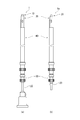
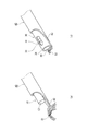
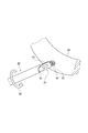
- FIG. 1 is a side view or a cross-sectional view of an endoscope (a), an inner tube (b), a guide pipe (c), and a processing pipe (d) constituting the small-diameter endoscopic surgical instrument of Example 1. It is explanatory drawing of the operation part of a processing pipe. A detailed view (a) of the hook of the processing pipe and an AA arrow view (b) thereof.
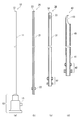
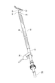
- FIG. 1 shows an overall view (a) of the first embodiment of a small-diameter endoscopic surgical instrument according to the present invention during operation, an overall view (b) of insertion and insertion into the body, and FIG. Side view or sectional view of an endoscope (a), an inner tube (b), a guide pipe (c), and a processing pipe (d) constituting an endoscopic surgical instrument,
- FIG. 3 is an operation of the processing pipe
- FIGS. 4A and 4B are a detailed view (a) of the hook of the processing pipe and an AA arrow view (b).
- the small-diameter endoscopic surgical instrument 1 of this embodiment at the time of surgery is configured by combining an endoscope 10, a guide pipe 30, and a processing pipe 40.
- the inner tube 20 is combined in place of the endoscope 10, and a coaxial triple tube structure is configured.
- a thin fiberscope having an outer diameter of the tube portion 11 of 2 mm is employed as the endoscope 10.
- the endoscope 10 is connected to a monitor system (not shown) used for a standard thoracic endoscopic operation by the connecting portion 13, and the operation proceeds while viewing the surgical site and its periphery by the monitor. be able to.
- An optical fiber for illumination for illuminating the target site of the tissue to be operated and the periphery of the distal end portion 12 of the endoscope is also built in the tube portion 11, and light is sent from the connection terminal 15.
- a person who performs an operation can hold the connecting portion 13 by hand and move the connecting portion 13 to move the distal end portion 12 of the endoscope to an optimum position.
- the guide pipe 30 is an outer diameter 2.35 mm tube having an inner diameter of 2.05 mm, which is slightly larger than the outer diameter of the tube portion of the endoscope 10, and supports the endoscope in a state in which the endoscope can be moved in the axial direction. . Even when the endoscope 10 is pulled into the guide pipe 30 to perform a process such as electrocautery, the endoscope 10 can obtain an image around the processing site. If one or more slits 33 are provided near the tip of the guide pipe so that the white smoke standing on the front surface of the guide pipe can be eliminated more quickly, it is helpful for the person who performs the operation.
- the processing pipe 40 is an outer diameter 2.85 mm pipe having an inner diameter of 2.40 mm which is slightly larger than the outer diameter 2.35 mm of the guide pipe 30, and the guide pipe 30 can be moved in the axial direction and rotated. I support.
- the small-diameter endoscopic surgical instrument 1a is completely inserted into the processing pipe 40 having an outer diameter of 2.85 mm, and there is no place protruding sideways, so that it can be smoothly inserted through a skin incision hole having a size of only 3 mm. it can.
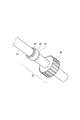
- a hook 43 is provided at the tip of the processing pipe.
- the hook 43 has a shape cut out in a hook shape from the curved surface without deforming the curved surface of the wall of the pipe 41 constituting the processing pipe.
- the hook 43 is also formed in a curved surface having the same inner diameter and outer diameter as the processing pipe.
- the hook 43 does not hinder the axial movement of the guide pipe 30 inserted into the processing pipe 40, and the hook 43 does not protrude sideways beyond the outer diameter of the processing pipe. .
- the sharp hooks and blades provided at the tip of the instrument damage the surrounding tissue when the instrument is inserted into the body, and the tip of the instrument is deformed, making it smooth.
- the hook 43 takes this shape, so that the hook portion is formed along the guide pipe 30 as shown in FIGS. 1 (b) and 4 (b). Since it goes into the body, there is no conflict with surrounding tissues, and the surrounding tissues are not damaged, and the hook 43 is not deformed by excessive force.
- the distal end portion 44 of the hook 43 is bent greatly, and the inner end thereof, that is, the edge on the operation portion side of the processing pipe is formed at the distal end portion of the guide pipe to be installed, that is, the distal end portion formed at right angles to the axial direction. It is formed so as to be substantially parallel to the edge of 34.
- the target tissue can be excised and separated easily and reliably.
- FIG. 4A the position of the distal end portion 12 of the endoscope 10 is manipulated and pulled into the processing pipe 40, and the blade of the distal end portion 44 and the target tissue are clearly identified. Can get a good picture.
- the guide pipe 30 is drawn into the guide pipe 40 so as not to obstruct the field of view.
- the tip 44 of the hook is used as a blade and at the same time as an electrode for electrocautery or electrocoagulation.
- the pipe portion 41 of the processing pipe is made of a conductive material, and forms a current-carrying part that sends electricity to the electrode, and is inserted into the body of the processing pipe so that electricity does not leak outside.
- the pipe portion 41 is electrically insulated 45 on the outer surface except for the electrodes.
- Insulation treatment is performed by coating, applying, baking, etc. of insulating material.
- Both monopolar and bipolar electrodes can be considered. However, when performing fine surgery on small tissues in the body cavity, such as sympathectomy in the thoracic cavity, the current is concentrated in a relatively narrow field.
- a monopolar that can be used is suitable. In that case, another electrode may be disposed on a part of the patient's body, for example, the buttocks, separately from the electrode at the tip 44 of the hook.
- an operation unit 42 is provided at the near end of the processing pipe 40.
- the operation unit 42 is not rotatable in the axial direction by a rotating part 48 fixed to the pipe part 41 of the processing pipe and two retaining rings 47 fixed to the pipe part, and is free to rotate with respect to the pipe part. It consists of the ring-shaped support part 46 attached to. A person who performs the operation pinches the support portion 46 with the thumb and forefinger and attaches the middle finger to the rotating portion 48.
- the axial position of the processing pipe 40 is changed by moving the support portion 46 in the axial direction, and the rotation angle of the processing pipe 40 can be changed by turning the rotating portion 48.
- the guide pipe 30 is also provided with a similar operation section 32.
- the above-described slit 33 is provided in the guide pipe, it is necessary to change the rotation angle in order to change the direction of the field of view, so the same operation unit as the processing pipe 40 is required.
- the slit 33 is not provided in the guide pipe, there is no need to change the rotation angle of the guide pipe 30, so that the rotation portion is not provided, and a support portion fixed to the pipe portion of the guide pipe is simply provided.
- the inner tube 20 is a tube having the same outer diameter as the tube portion 11 of the endoscope 10, and the tip 23 of the inner tube 20 is inserted into the body as shown in FIG. Since it is located at the foremost tip, it should be polished into a rounded shape so that it can be smoothly inserted and the surrounding tissue is not damaged. Further, the inner pipe 10 needs to change its axial position, but does not need to change its rotation angle, so there is no need for a rotating part provided in the operation part 42 of the processing pipe 40, and the pipe part of the inner pipe. It is only necessary to provide the operation unit 22 that is fixed to the size and that is easy to pinch with the thumb and index finger. *
- the inner pipe 20, the guide pipe 30, and the processing pipe 40 described above have a small diameter and a small thickness within a range in which the respective functions can be performed. Since the difference in the outer diameter is extremely small, the outer diameter of the small-sized endoscopic surgical instrument 1 is only 2.85 m, which greatly contributes to reducing the size of the skin incision hole for insertion. In addition, the difference between the inner and outer diameters of adjacent pipes, that is, the gap between the pipes is extremely small, so that the possibility of a malfunction in the operation due to the cut tissue or body fluid entering these gaps. Can do.
- the inner pipe 20, the guide pipe 30, and the processing pipe 40 are each in the shape of a simple pipe, and since it is easy to pull out and separate the three, easy pre-cleaning and sterilization, And it can be done completely.
- FIG. 5 (a) is an explanatory diagram of an operation (movement) for moving the tissue using the hook 43 of the processing pipe.
- the operation it becomes necessary to move the interfering tissue 51 for the purpose of approaching the target site or securing a visual field.
- the support portion 46 of the operation portion of the processing pipe 40 is operated to bring the hook 43 closer to the tissue 51 and the rotation portion 48 is operated. Hook the tissue 51 while changing the direction of the hook.
- the guide pipe 30 is drawn into the processing pipe 40. In this state, the tissue 51 can be transferred, separated, and pulled. Further, since the hook 43 is provided with a blade, the tissue 51 can be excised using the blade.
- FIG. 5B is an explanatory diagram of the operation (b) for grasping the tissue.
- the hook 43 is moved closer to the tissue 52 by operating the support portion 46 of the operation portion of the processing pipe 40 while keeping the target tissue 52 to be grasped by the endoscope 10 in the field of view. Hooking the tissue 52 while changing the direction of the hook by operating the rotating portion 48, then operating the operating portion 32 of the guide pipe 30 to move the guide pipe 30 forward, the tissue 52 caught on the hook 43 is removed. Grab and hold.
- the slit 33 provided in the guide pipe is useful for securing the visual field of the endoscope 10.
- the tissue 52 Since the distal end portion 44 of the hook 43 is bent greatly and the inner edge thereof is formed so as to be substantially parallel to the edge of the distal end portion 34 of the guide pipe 30, the tissue 52 is firmly grasped between them. The tissue 52 can be moved, separated and pulled. Furthermore, the tissue 52 can be used for a biopathological examination by taking out the small diameter endoscopic surgical instrument 1 from the patient's body while keeping the tissue 52 held.
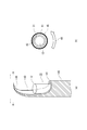
- FIG. 6 is an explanatory view of an operation of fixing and removing a tissue that has been stuck in the tip 44 of the hook of the processing pipe 40 or a clot stuck to the hook 43 by using the guide pipe 30. is there.
- the endoscope 10 is used to search for a nearby solid object, such as the rib 64, and the operation unit 42 is operated to bring the processing pipe hook 43 closer thereto.
- the operation part 32 of the guide pipe 30 is operated, the guide pipe 30 is advanced, and the stuck tissue and the blood clot 53 are pressed against the rib 64 and fixed. In this state, the clot 53 or the like 53 can be easily removed from the hook 43 by operating the rotating portion 48 of the processing pipe 40 to rotate the processing pipe 40 in the direction of the arrow 56 in the figure.
- FIG. 7 is an explanatory diagram of an operation for performing electrocautery or electrocoagulation using the tip 44 of the hook of the processing pipe 40 as an electrode.
- the mouth clip 58 is installed on the pipe portion 41 where the outer surface of the processing pipe 40 is not electrically insulated.
- Another electrode (not shown) is provided on the patient's buttocks. Since the pipe portion 41 of the processing pipe is made of a conductive material, electrocautery or electrocoagulation can be performed on the tissue 57 to be operated using the tip 44 of the hook as an electrode. it can.
- the guide pipe is drawn into the processing pipe 40, and the endoscope 10 can accommodate the tip portion 44 of the electrode and the tissue 57 in the visual field without being obstructed by the guide pipe.
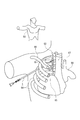
- FIG. 8 is an explanatory view of a method (Example 2) for inserting the small-diameter endoscopic surgical instrument 1a of Example 1 into the body, and FIG. 9 shows the small diameter using the inner tube 20 for the insertion. It is explanatory drawing of the point which knows how far the front-end
- a skin incision hole 61 having a size of 3 mm is provided on the armpit center line of the armpit.
- the small-diameter endoscopic surgical instrument 1a into which the inner tube 20 is inserted instead of the endoscope 10 is inserted.
- the target tissue of the operation is a chest sympathetic nerve trunk 63 that extends vertically along the spine.
- the small diameter endoscopic surgical instrument 1 a approaches the target tissue through the space between the ribs 64.
- the rib 64 there are a lung protrusion 65, a clavicle 66, an upper vena cava 67, an aorta 68, and the like.
- the inner tube 20 of the small diameter endoscopic surgical instrument 1a is connected to a syringe 92 by a flexible tube 91, and physiological saline or air is contained in the syringe.
- physiological saline or air is contained in the syringe.
- the inner tube 20 is extracted from the small-diameter endoscopic surgical instrument 1a, and the endoscope 10 is inserted into the small-diameter endoscopic surgical instrument 1 at the time of the operation to proceed with the surgery. .
- the small-diameter endoscopic surgical instrument of the present invention has a simple configuration with a small number of parts, and not only performs electrocautery, electrocoagulation, excision, transfer, separation, and traction of the target tissue, but also grips the target tissue.
- the target tissue can be transferred, separated, and pulled to be used for biopathological examination, and clots stuck to the hook during surgery can be used on the spot.
- Multifunctionality that can be removed.
- a person who performs an operation can proceed with the operation with an easy operation while viewing a clear image.
- the skin incision hole is small and only one place is sufficient, and the burden on the patient is small. Taking advantage of these advantages, it is expected to be used greatly in various types of surgery in the future.
Landscapes
- Health & Medical Sciences (AREA)
- Life Sciences & Earth Sciences (AREA)
- Surgery (AREA)
- Engineering & Computer Science (AREA)
- Heart & Thoracic Surgery (AREA)
- Animal Behavior & Ethology (AREA)
- General Health & Medical Sciences (AREA)
- Public Health (AREA)
- Veterinary Medicine (AREA)
- Biomedical Technology (AREA)
- Medical Informatics (AREA)
- Nuclear Medicine, Radiotherapy & Molecular Imaging (AREA)
- Molecular Biology (AREA)
- Pathology (AREA)
- Physics & Mathematics (AREA)
- Radiology & Medical Imaging (AREA)
- Optics & Photonics (AREA)
- Biophysics (AREA)
- Hematology (AREA)
- Anesthesiology (AREA)
- Plasma & Fusion (AREA)
- Otolaryngology (AREA)
- Orthopedic Medicine & Surgery (AREA)
- Cardiology (AREA)
- Neurology (AREA)
- Surgical Instruments (AREA)
- Endoscopes (AREA)
- Infusion, Injection, And Reservoir Apparatuses (AREA)
Priority Applications (4)
| Application Number | Priority Date | Filing Date | Title |
|---|---|---|---|
| EP17836786.8A EP3494913B1 (en) | 2016-08-02 | 2017-07-24 | Small-diameter endoscope surgical instrument |
| US16/322,187 US11457798B2 (en) | 2016-08-02 | 2017-07-24 | Small-Diameter endoscope surgical instrument |
| CN201780048717.5A CN109561921B (zh) | 2016-08-02 | 2017-07-24 | 小径内窥镜手术器械 |
| KR1020197003078A KR102253461B1 (ko) | 2016-08-02 | 2017-07-24 | 세경 내시경 수술 기구 |
Applications Claiming Priority (2)
| Application Number | Priority Date | Filing Date | Title |
|---|---|---|---|
| JP2016151724A JP6749545B2 (ja) | 2016-08-02 | 2016-08-02 | 細径内視鏡手術器具 |
| JP2016-151724 | 2016-08-02 |
Publications (1)
| Publication Number | Publication Date |
|---|---|
| WO2018025680A1 true WO2018025680A1 (ja) | 2018-02-08 |
Family
ID=61072829
Family Applications (1)
| Application Number | Title | Priority Date | Filing Date |
|---|---|---|---|
| PCT/JP2017/026598 WO2018025680A1 (ja) | 2016-08-02 | 2017-07-24 | 細径内視鏡手術器具 |
Country Status (6)
Families Citing this family (7)
| Publication number | Priority date | Publication date | Assignee | Title |
|---|---|---|---|---|
| WO2020005218A1 (en) * | 2018-06-27 | 2020-01-02 | Wright Medical Technology, Inc. | Burr with irrigation and imaging |
| CN111265290B (zh) * | 2020-03-25 | 2025-03-14 | 扬州大学附属医院 | 一种面部手术内窥系统 |
| DE102020110845A1 (de) * | 2020-04-21 | 2021-10-21 | Novatech Sa | Medizinische Vorrichtung und Endoskopierverfahren |
| CN114533213B (zh) * | 2020-11-24 | 2024-05-24 | 奥林巴斯株式会社 | 具有超声内窥镜的治疗方法和装置 |
| CN113499133A (zh) * | 2021-07-05 | 2021-10-15 | 上海生知医疗科技有限公司 | 一种双功能的手术器械 |
| CN114668503B (zh) * | 2022-03-29 | 2024-06-04 | 吉林省金博弘智能科技有限责任公司 | 一种诊疗一体式手术机器人 |
| KR102539420B1 (ko) | 2022-08-09 | 2023-06-01 | 조백현 | 수술기구 |
Citations (3)
| Publication number | Priority date | Publication date | Assignee | Title |
|---|---|---|---|---|
| WO2000016707A1 (fr) | 1998-09-18 | 2000-03-30 | Hidehiro Yamamoto | Dispositif d'alimentation en energie pour endoscope |
| JP2007089690A (ja) | 2005-09-27 | 2007-04-12 | Hidehiro Yamamoto | 内視鏡用通電牽引装置 |
| JP2014018299A (ja) * | 2012-07-13 | 2014-02-03 | Olympus Corp | ガイドワイヤ導入装置 |
Family Cites Families (22)
| Publication number | Priority date | Publication date | Assignee | Title |
|---|---|---|---|---|
| DE3313325A1 (de) * | 1983-04-13 | 1984-10-18 | Knut Dr. 7802 Merzhausen Korth | Operationsinstrument |
| US5578053A (en) * | 1993-06-24 | 1996-11-26 | Yoon; Inbae | Safety needle instrument having a triggered safety member |
| US5478329A (en) * | 1994-05-06 | 1995-12-26 | Ternamian; Artin M. | Trocarless rotational entry cannula |
| US5709671A (en) * | 1995-10-16 | 1998-01-20 | Ethicon Endo-Surgery, Inc. | Trocar having an improved tip configuration |
| DE19547246C1 (de) * | 1995-12-18 | 1997-03-20 | Riek Siegfried | Medizinische Nadel |
| AU722939B2 (en) * | 1996-04-10 | 2000-08-17 | Linvatec Corporation | Process for shaping and sharpening a rotatable surgical shaver blade |
| JPH10155807A (ja) * | 1996-11-29 | 1998-06-16 | Olympus Optical Co Ltd | 内視鏡用処置具 |
| DE19730127C2 (de) * | 1997-07-14 | 2001-04-12 | Erbe Elektromedizin | Präparierinstrument |
| US6428539B1 (en) * | 2000-03-09 | 2002-08-06 | Origin Medsystems, Inc. | Apparatus and method for minimally invasive surgery using rotational cutting tool |
| JP4460718B2 (ja) * | 2000-05-12 | 2010-05-12 | オリンパス株式会社 | 内視鏡処置装置 |
| JP3722729B2 (ja) * | 2001-06-04 | 2005-11-30 | オリンパス株式会社 | 内視鏡用処置装置 |
| US7556633B2 (en) * | 2004-03-01 | 2009-07-07 | Terumo Corporation | Method and apparatus for endoscopic dissection of blood vessels |
| DE102005013714A1 (de) * | 2004-04-07 | 2005-12-22 | Carl Zeiss Meditec Ag | Elektrische Sonde für die Mikrochirurgie |
| EP2545871B1 (en) * | 2004-06-29 | 2015-02-11 | Applied Medical Resources Corporation | Insufflating optical surgical instrument |
| EP1834599A4 (en) * | 2004-12-17 | 2009-01-21 | Univ Kyoto | HOOD WITH CUTTING FUNCTION AND ENDOSCOPE |
| DE102005049021B4 (de) | 2005-10-11 | 2008-08-21 | Richard Wolf Gmbh | Endoskop |
| US20070203395A1 (en) * | 2006-02-28 | 2007-08-30 | Takayasu Mikkaichi | Cap installable on distal end portion of endoscope |
| US8728089B2 (en) * | 2008-03-28 | 2014-05-20 | Olympus Medical Systems Corp. | Endoscope treatment instrument |
| US8531064B2 (en) * | 2010-02-11 | 2013-09-10 | Ethicon Endo-Surgery, Inc. | Ultrasonically powered surgical instruments with rotating cutting implement |
| EP2558151A4 (en) * | 2010-04-13 | 2018-01-10 | Sentreheart, Inc. | Methods and devices for pericardial access |
| CN201701297U (zh) * | 2010-04-15 | 2011-01-12 | 欧普康光电(厦门)有限公司 | 一种带数码摄像装置的外科手术内窥镜 |
| JP7166761B2 (ja) * | 2015-06-17 | 2022-11-08 | サフィナ・メディカル・インコーポレイテッド | 単一内視鏡血管採取装置 |
-
2016
- 2016-08-02 JP JP2016151724A patent/JP6749545B2/ja active Active
-
2017
- 2017-07-24 WO PCT/JP2017/026598 patent/WO2018025680A1/ja unknown
- 2017-07-24 US US16/322,187 patent/US11457798B2/en active Active
- 2017-07-24 CN CN201780048717.5A patent/CN109561921B/zh active Active
- 2017-07-24 EP EP17836786.8A patent/EP3494913B1/en active Active
- 2017-07-24 KR KR1020197003078A patent/KR102253461B1/ko active Active
Patent Citations (3)
| Publication number | Priority date | Publication date | Assignee | Title |
|---|---|---|---|---|
| WO2000016707A1 (fr) | 1998-09-18 | 2000-03-30 | Hidehiro Yamamoto | Dispositif d'alimentation en energie pour endoscope |
| JP2007089690A (ja) | 2005-09-27 | 2007-04-12 | Hidehiro Yamamoto | 内視鏡用通電牽引装置 |
| JP2014018299A (ja) * | 2012-07-13 | 2014-02-03 | Olympus Corp | ガイドワイヤ導入装置 |
Non-Patent Citations (4)
| Title |
|---|
| HIDEHIRO YAMAMOTO, MD, THE JOURNAL OF THORACIC AND CARDIOVASCULAR SURGERY, vol. 120, 2000, pages 276 - 279 |
| J THORACIC CARDIOVASC SURG., vol. 120, 2000, pages 276 - 9 |
| See also references of EP3494913A4 |
| YAMAMOTO,HIDEHIRO: "Needlescopic surgery for palmar hyperhidrosis", THE JOURNAL OF THARACIC AND CARDIOVASCULAR SURGERY, vol. 120, no. 2, August 2000 (2000-08-01), pages 276 - 279, XP029481396, DOI: doi:10.1067/mtc.2000.107830 * |
Also Published As
| Publication number | Publication date |
|---|---|
| JP6749545B2 (ja) | 2020-09-02 |
| EP3494913A4 (en) | 2020-03-25 |
| CN109561921B (zh) | 2021-07-09 |
| KR102253461B1 (ko) | 2021-05-17 |
| US11457798B2 (en) | 2022-10-04 |
| KR20190026809A (ko) | 2019-03-13 |
| JP2018019815A (ja) | 2018-02-08 |
| US20190183320A1 (en) | 2019-06-20 |
| EP3494913B1 (en) | 2024-04-24 |
| CN109561921A (zh) | 2019-04-02 |
| EP3494913A1 (en) | 2019-06-12 |
Similar Documents
| Publication | Publication Date | Title |
|---|---|---|
| WO2018025680A1 (ja) | 細径内視鏡手術器具 | |
| CN105411632B (zh) | 使用活检工具获取组织样本的装置、系统和方法 | |
| JP5792416B1 (ja) | 内視鏡処置システム | |
| JP5928862B2 (ja) | 内視鏡用処置具及び切開システム | |
| JP6283091B2 (ja) | 単一の内視鏡用管採取デバイス | |
| US9173697B2 (en) | Surgical cannula for dissipating electric charge | |
| JP2010017574A (ja) | 内視鏡用高周波切開具 | |
| CN103200892A (zh) | 外科用处理器具 | |
| US9808267B2 (en) | Tissue resection device and related methods of use | |
| JP5547736B2 (ja) | 内視鏡とともに使用する円筒体システム | |
| JP6224618B2 (ja) | 組織の切除を容易にするように構成された切除装置および身体から組織を切除するための装置 | |
| JP2016512458A (ja) | 切除装置および関連する使用方法 | |
| JP2011067532A (ja) | 内視鏡用補助具及び内視鏡 | |
| CN117694814A (zh) | 内窥镜设备 | |
| JP6886569B2 (ja) | 細径内視鏡手術器具 | |
| US20140243594A1 (en) | Medical device actuation systems and related methods of use | |
| US10548626B2 (en) | Endoscopic tissue manipulation tool | |
| EP3946097B1 (en) | Colonoscope for removing colorectal polyps | |
| US10244921B2 (en) | Endoscopic system for resection of tissue | |
| WO2015133432A1 (ja) | 内視鏡用処置具 | |
| JP3211674U (ja) | 手術用鋏 | |
| HK40007986B (en) | Small-diameter endoscope surgical instrument | |
| HK40007986A (en) | Small-diameter endoscope surgical instrument |
Legal Events
| Date | Code | Title | Description |
|---|---|---|---|
| 121 | Ep: the epo has been informed by wipo that ep was designated in this application |
Ref document number: 17836786 Country of ref document: EP Kind code of ref document: A1 |
|
| ENP | Entry into the national phase |
Ref document number: 20197003078 Country of ref document: KR Kind code of ref document: A |
|
| NENP | Non-entry into the national phase |
Ref country code: DE |
|
| ENP | Entry into the national phase |
Ref document number: 2017836786 Country of ref document: EP Effective date: 20190304 |