WO2015111560A1 - 内視鏡装置及び内視鏡装置の作動方法 - Google Patents
内視鏡装置及び内視鏡装置の作動方法 Download PDFInfo
- Publication number
- WO2015111560A1 WO2015111560A1 PCT/JP2015/051305 JP2015051305W WO2015111560A1 WO 2015111560 A1 WO2015111560 A1 WO 2015111560A1 JP 2015051305 W JP2015051305 W JP 2015051305W WO 2015111560 A1 WO2015111560 A1 WO 2015111560A1
- Authority
- WO
- WIPO (PCT)
- Prior art keywords
- mist
- value
- evaluation value
- focus
- evaluation
- Prior art date
- Legal status (The legal status is an assumption and is not a legal conclusion. Google has not performed a legal analysis and makes no representation as to the accuracy of the status listed.)
- Ceased
Links
Images


Classifications
-
- A—HUMAN NECESSITIES
- A61—MEDICAL OR VETERINARY SCIENCE; HYGIENE
- A61B—DIAGNOSIS; SURGERY; IDENTIFICATION
- A61B1/00—Instruments for performing medical examinations of the interior of cavities or tubes of the body by visual or photographical inspection, e.g. endoscopes; Illuminating arrangements therefor
- A61B1/00002—Operational features of endoscopes
- A61B1/00004—Operational features of endoscopes characterised by electronic signal processing
- A61B1/00006—Operational features of endoscopes characterised by electronic signal processing of control signals
-
- A—HUMAN NECESSITIES
- A61—MEDICAL OR VETERINARY SCIENCE; HYGIENE
- A61B—DIAGNOSIS; SURGERY; IDENTIFICATION
- A61B1/00—Instruments for performing medical examinations of the interior of cavities or tubes of the body by visual or photographical inspection, e.g. endoscopes; Illuminating arrangements therefor
- A61B1/00002—Operational features of endoscopes
- A61B1/00004—Operational features of endoscopes characterised by electronic signal processing
- A61B1/00009—Operational features of endoscopes characterised by electronic signal processing of image signals during a use of endoscope
- A61B1/000094—Operational features of endoscopes characterised by electronic signal processing of image signals during a use of endoscope extracting biological structures
-
- A—HUMAN NECESSITIES
- A61—MEDICAL OR VETERINARY SCIENCE; HYGIENE
- A61B—DIAGNOSIS; SURGERY; IDENTIFICATION
- A61B1/00—Instruments for performing medical examinations of the interior of cavities or tubes of the body by visual or photographical inspection, e.g. endoscopes; Illuminating arrangements therefor
- A61B1/00002—Operational features of endoscopes
- A61B1/00004—Operational features of endoscopes characterised by electronic signal processing
- A61B1/00009—Operational features of endoscopes characterised by electronic signal processing of image signals during a use of endoscope
- A61B1/000095—Operational features of endoscopes characterised by electronic signal processing of image signals during a use of endoscope for image enhancement
-
- A—HUMAN NECESSITIES
- A61—MEDICAL OR VETERINARY SCIENCE; HYGIENE
- A61B—DIAGNOSIS; SURGERY; IDENTIFICATION
- A61B1/00—Instruments for performing medical examinations of the interior of cavities or tubes of the body by visual or photographical inspection, e.g. endoscopes; Illuminating arrangements therefor
- A61B1/00163—Optical arrangements
- A61B1/00188—Optical arrangements with focusing or zooming features
-
- A—HUMAN NECESSITIES
- A61—MEDICAL OR VETERINARY SCIENCE; HYGIENE
- A61B—DIAGNOSIS; SURGERY; IDENTIFICATION
- A61B1/00—Instruments for performing medical examinations of the interior of cavities or tubes of the body by visual or photographical inspection, e.g. endoscopes; Illuminating arrangements therefor
- A61B1/12—Instruments for performing medical examinations of the interior of cavities or tubes of the body by visual or photographical inspection, e.g. endoscopes; Illuminating arrangements therefor with cooling or rinsing arrangements
- A61B1/127—Instruments for performing medical examinations of the interior of cavities or tubes of the body by visual or photographical inspection, e.g. endoscopes; Illuminating arrangements therefor with cooling or rinsing arrangements with means for preventing fogging
-
- G—PHYSICS
- G02—OPTICS
- G02B—OPTICAL ELEMENTS, SYSTEMS OR APPARATUS
- G02B23/00—Telescopes, e.g. binoculars; Periscopes; Instruments for viewing the inside of hollow bodies; Viewfinders; Optical aiming or sighting devices
- G02B23/24—Instruments or systems for viewing the inside of hollow bodies, e.g. fibrescopes
- G02B23/2407—Optical details
- G02B23/2423—Optical details of the distal end
- G02B23/243—Objectives for endoscopes
-
- G—PHYSICS
- G02—OPTICS
- G02B—OPTICAL ELEMENTS, SYSTEMS OR APPARATUS
- G02B23/00—Telescopes, e.g. binoculars; Periscopes; Instruments for viewing the inside of hollow bodies; Viewfinders; Optical aiming or sighting devices
- G02B23/24—Instruments or systems for viewing the inside of hollow bodies, e.g. fibrescopes
- G02B23/2407—Optical details
- G02B23/2446—Optical details of the image relay
-
- G—PHYSICS
- G02—OPTICS
- G02B—OPTICAL ELEMENTS, SYSTEMS OR APPARATUS
- G02B23/00—Telescopes, e.g. binoculars; Periscopes; Instruments for viewing the inside of hollow bodies; Viewfinders; Optical aiming or sighting devices
- G02B23/24—Instruments or systems for viewing the inside of hollow bodies, e.g. fibrescopes
- G02B23/2407—Optical details
- G02B23/2453—Optical details of the proximal end
-
- G—PHYSICS
- G02—OPTICS
- G02B—OPTICAL ELEMENTS, SYSTEMS OR APPARATUS
- G02B7/00—Mountings, adjusting means, or light-tight connections, for optical elements
- G02B7/02—Mountings, adjusting means, or light-tight connections, for optical elements for lenses
- G02B7/04—Mountings, adjusting means, or light-tight connections, for optical elements for lenses with mechanism for focusing or varying magnification
-
- G—PHYSICS
- G02—OPTICS
- G02B—OPTICAL ELEMENTS, SYSTEMS OR APPARATUS
- G02B7/00—Mountings, adjusting means, or light-tight connections, for optical elements
- G02B7/28—Systems for automatic generation of focusing signals
- G02B7/36—Systems for automatic generation of focusing signals using image sharpness techniques, e.g. image processing techniques for generating autofocus signals
-
- G—PHYSICS
- G03—PHOTOGRAPHY; CINEMATOGRAPHY; ANALOGOUS TECHNIQUES USING WAVES OTHER THAN OPTICAL WAVES; ELECTROGRAPHY; HOLOGRAPHY
- G03B—APPARATUS OR ARRANGEMENTS FOR TAKING PHOTOGRAPHS OR FOR PROJECTING OR VIEWING THEM; APPARATUS OR ARRANGEMENTS EMPLOYING ANALOGOUS TECHNIQUES USING WAVES OTHER THAN OPTICAL WAVES; ACCESSORIES THEREFOR
- G03B13/00—Viewfinders; Focusing aids for cameras; Means for focusing for cameras; Autofocus systems for cameras
- G03B13/32—Means for focusing
- G03B13/34—Power focusing
- G03B13/36—Autofocus systems
-
- G—PHYSICS
- G06—COMPUTING OR CALCULATING; COUNTING
- G06T—IMAGE DATA PROCESSING OR GENERATION, IN GENERAL
- G06T7/00—Image analysis
- G06T7/0002—Inspection of images, e.g. flaw detection
- G06T7/0012—Biomedical image inspection
-
- H—ELECTRICITY
- H04—ELECTRIC COMMUNICATION TECHNIQUE
- H04N—PICTORIAL COMMUNICATION, e.g. TELEVISION
- H04N23/00—Cameras or camera modules comprising electronic image sensors; Control thereof
- H04N23/60—Control of cameras or camera modules
- H04N23/67—Focus control based on electronic image sensor signals
- H04N23/673—Focus control based on electronic image sensor signals based on contrast or high frequency components of image signals, e.g. hill climbing method
-
- H—ELECTRICITY
- H04—ELECTRIC COMMUNICATION TECHNIQUE
- H04N—PICTORIAL COMMUNICATION, e.g. TELEVISION
- H04N23/00—Cameras or camera modules comprising electronic image sensors; Control thereof
- H04N23/70—Circuitry for compensating brightness variation in the scene
- H04N23/74—Circuitry for compensating brightness variation in the scene by influencing the scene brightness using illuminating means
-
- G—PHYSICS
- G02—OPTICS
- G02B—OPTICAL ELEMENTS, SYSTEMS OR APPARATUS
- G02B23/00—Telescopes, e.g. binoculars; Periscopes; Instruments for viewing the inside of hollow bodies; Viewfinders; Optical aiming or sighting devices
- G02B23/24—Instruments or systems for viewing the inside of hollow bodies, e.g. fibrescopes
- G02B23/2407—Optical details
- G02B23/2461—Illumination
- G02B23/2469—Illumination using optical fibres
-
- G—PHYSICS
- G06—COMPUTING OR CALCULATING; COUNTING
- G06T—IMAGE DATA PROCESSING OR GENERATION, IN GENERAL
- G06T2207/00—Indexing scheme for image analysis or image enhancement
- G06T2207/10—Image acquisition modality
- G06T2207/10068—Endoscopic image
-
- H—ELECTRICITY
- H04—ELECTRIC COMMUNICATION TECHNIQUE
- H04N—PICTORIAL COMMUNICATION, e.g. TELEVISION
- H04N23/00—Cameras or camera modules comprising electronic image sensors; Control thereof
- H04N23/50—Constructional details
- H04N23/555—Constructional details for picking-up images in sites, inaccessible due to their dimensions or hazardous conditions, e.g. endoscopes or borescopes
Definitions
- execution of focusing operation in focus control is stopped when occurrence of mist is detected.
- the focusing operation can be stopped when the mist that may cause the focus control to malfunction is generated, and the in-focus state can be maintained even when the mist is generated.
- the focus control is performed by controlling the position of the focus lens of the optical system of the imaging unit, the occurrence of mist is detected, and the occurrence of the mist is detected.
- the present invention relates to a method of operating the endoscope apparatus for stopping the execution of the focusing operation in
- the user fixes the field of view and performs the treatment, but if the focus changes during the treatment, the treatment is disturbed. Therefore, after the focusing is completed once in the fixed field of view, the focusing operation is stopped in order to prevent the occurrence of blurring or the like of the image due to a change in the focus position unintended by the user or a malfunction in AF control. Is desirable.
- mist may be generated in the visual field for the following reasons, and there is a problem that the focus position may be shifted by the mist.
- Patent Document 1 and Patent Document 2 described above as techniques for suppressing unnecessary AF operation, since these techniques do not assume mist, it is necessary to accurately suppress AF operation with respect to generation of mist. Can not. Therefore, it is difficult to stop the focusing operation while the user is performing treatment.
- the endoscope apparatus 10 includes a focus control unit 360 that performs focus control by controlling the position of a focus lens of the optical system of the imaging unit 200, and a mist detection unit 340 that detects the generation of mist. Then, when the mist detection unit 340 detects the generation of mist, the focus control unit 360 stops the execution of the focusing operation in the focus control.
- mist when mist is generated, parameters such as the luminance value, saturation value, and contrast value of the image temporally change.
- the occurrence of mist can be detected by detecting from.
- the generation of the mist may be detected not only from the image but also by combining control information (for example, an energized state) of the treatment tool that generates the mist.
- focus control generally means control of the position of the focus lens including control to keep the focus lens stopped, and an operation of focusing on the object in the focus control is called focusing operation.
- focus control unit 360 stops the movement of the focus lens to fix the focus position.
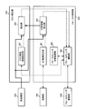
- FIG. 2 shows a configuration example of the endoscope apparatus according to the first embodiment.
- the endoscope apparatus includes a rigid endoscope 100 as an insertion unit into the body, an imaging unit 200 connected to the rigid endoscope 100, a processing unit 300, a display unit 400, an external I / F unit 500, and a light source unit. Including 600.
- the processing unit 300 includes an A / D conversion unit 310, a preprocessing unit 320, an image processing unit 330, a focus control unit 360, a control unit 350, and a mist detection unit 340.
- the mist detection unit 340 detects the generation of mist and the disappearance of the mist based on the image from the pre-processing unit 320.
- the focus control unit 360 controls the focus lens drive unit 230 based on the image from the pre-processing unit 320 to control a focusing operation for focusing on the subject.
- the focus control unit 360 stops or restarts the focusing operation when the generation of the mist or the disappearance of the mist is detected. Details of the mist detection unit 340 and the focus control unit 360 will be described later.
- the control unit 350 is mutually connected to each part of the endoscope apparatus such as the external I / F unit 500, the image processing unit 330, the focus control unit 360, the imaging device 260, the zoom button 210, etc. Control signal input / output.
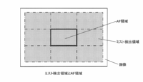
- the mist detection area setting unit 341 sets a mist detection area as shown in FIG. 4 based on information such as the image size output from the control unit 350, for example. Thereafter, the mist detection area setting unit 341 outputs the set mist detection area information to the detection unit 342. For example, the image is divided into 3 ⁇ 3 areas, nine evaluation blocks are set, and a set of these is set as a mist detection area. It goes without saying that the number of evaluation blocks set as the mist detection area can be arbitrarily set here.
- the detection unit 342 detects mist based on the mist detection area information output from the mist detection area setting unit 341 and the images sequentially output from the pre-processing unit 320. Thereafter, the detection unit 342 outputs mist detection information indicating whether or not mist is detected to the focus lens control unit 363. Details of the mist detection method will be described later.
- the AF area setting unit 361 sets an AF area as shown in FIG. 4 based on information such as the image size output from the control unit 350, for example. Thereafter, the AF area setting unit 361 outputs the set AF area information to the AF evaluation value calculation unit 362.
- the same area as the central evaluation block is set as the AF area in order to simplify the description.
- an area having a size completely different from that of the evaluation block may be set as the AF area near the center of the image.
- the AF evaluation value calculation unit 362 calculates an AF evaluation value based on the AF area information output from the AF area setting unit 361 and the images sequentially output from the preprocessing unit 320. For example, Y signals of all the pixels included in the AF area are calculated, arbitrary BPF (band pass filter) processing is performed on the Y signals, and a sum of outputs of the BFP processing is set as an AF evaluation value. Thereafter, the AF evaluation value calculation unit 362 sequentially outputs the calculated AF evaluation values to the focus lens control unit 363.
- the focus lens control unit 363 generates control information of the focus lens based on the mist detection information output from the detection unit 342 and the AF evaluation value output from the AF evaluation value calculation unit 362, and the focus lens drive unit 230 Output.
- the focus lens drive unit 230 drives the focus lens based on the control information output from the focus lens control unit 363.
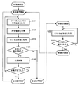
- step S104 When it is determined in step S104 that the mist is generated and the focusing operation is ended, the focus lens control unit 363 starts the standby operation.
- the focus lens control unit 363 acquires mist detection information output from the detection unit 342 (step S201).
- the focus lens control unit 363 detects whether or not the mist has disappeared based on the mist detection information (step S202). If it is determined that the mist has not disappeared, the operations after step S201 are repeated. On the other hand, when it is determined that the mist has disappeared, the standby operation is ended.
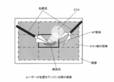
- the change in the image is different from that near the center of the image when mist occurs. That is, while the user is performing treatment, the visual field is fixed so that the lesion site is located near the center of the image, and the lesion site near the center is treated. No treatment is performed, and mist does not occur near the periphery of the image. Therefore, when mist occurs near the center, the change in the image near the periphery is relatively small until the mist reaches the periphery.
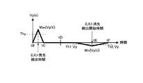
- Vy [n] is calculated from the average luminance of each evaluation block calculated from the current image (time n) and an image acquired in the past by x It is calculated as the sum of the differences with the average luminance of each evaluation block.
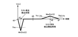
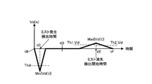
- Vs [n] is the sum of the difference between the average saturation of each evaluation block calculated from the current image (time n) and the average saturation of each evaluation block calculated from an image acquired in the past by x It is calculated as Furthermore, Vd [n] is calculated as the sum of the difference between the D range of each evaluation block calculated from the current image (time n) and the D range of each evaluation block calculated from an image acquired in the past by x It is done.
- the detection unit 342 detects the time when the evaluation value Vy [n] of the luminance value becomes the minimum value Min ⁇ Vy ⁇ under the condition smaller than the first predetermined threshold Th1_Vy, and the evaluation value Vs [n] of the saturation value.
- the AF control can be stopped as soon as possible at that point, and the focus lens can be fixed. This has the advantage that the possibility of a malfunction in AF control can be prevented in advance.
- the time change of the parameter occurs when the mist occurs or disappears, by using the time change, it is possible to determine that the mist occurs at an early stage after the mist actually occurs. Alternatively, it can be determined that the mist has disappeared when the mist has sufficiently disappeared.
- AF control is performed and a state in which the region under treatment is in focus is maintained.
- the AF control is stopped only while the normal AF control can not be performed by the mist, and the state returns to the state where the normal AF control can be performed. AF control can be resumed.
- the in-focus state can be maintained regardless of the presence or absence of the mist, and the convenience for the user is improved.
- the detection unit 342 calculates the Y signal Y ′ [n] and the color signal C ′ [n] by the following equations (10) and (11) using the result of the BPF processing and the light adjustment correction amount.
- Y [n] and C [n] are output signals from the preprocessing unit 320 to the detection unit 342. That is, Y [n] is a Y signal of the image, and C [n] is a Cr signal or a Cb signal of the image.
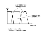
- Bpf (Y [n]) and Bpf (C [n]) are output signals from the AF evaluation value calculator 362 to the detector 342. That is, Bpf ⁇ Y [n] ⁇ is the result of the BPF processing of the Y signal, and is a Y signal of the band AfBd shown in FIG.
- Bpf ⁇ C [n] ⁇ is the result of the BPF processing of the color signal, and is a color signal of the band AfBd shown in FIG.
- LC is a light control correction amount, and the value is larger as the light amount is larger.
- the Y signal Y '[n] and the color signal C' [n] become signals in the low frequency and high frequency band MistBd shown in FIG.
- the detection unit 342 calculates an evaluation value from these signals. As described above, since the evaluation value is averaged in each evaluation block (or all evaluation blocks), the high frequency (thick dotted line in FIG. 15) component in the band MistBd is substantially cut, and the low frequency (thick solid line) Only the ingredients of remain.
- the configuration is not limited to this, and the configuration in which the detection unit 342 performs low pass filter processing is used. For example, similar effects can be obtained.
- the focus lens control unit 363 sets the position of the focus lens at the time when the energization state of the electric knife or the ultrasonic knife is closest to the past and the time closest to time n at which the generation of mist is detected. It is read from the focus lens position recording unit 364. Then, when the occurrence of mist is detected and the focusing operation is stopped, the focus lens is moved to the position of the read focus lens.
- mist generation time when it is set as the mist generation time when the time when the electrification state of the electric knife or the ultrasonic knife becomes ON is set as the mist generation time, the mist disappearance time can not be detected when the mist does not occur, and the time to restart AF control Can not be identified.
- This problem can be solved by performing processing for detecting mist generation as an image as in the first embodiment after the energization state of the electric knife or the ultrasonic knife is turned on.
- the spatial frequency of the mist is a low frequency
- the change in the mist can be efficiently detected by obtaining the mist evaluation value from the image of the low frequency band.
- the band for which the focus evaluation value is obtained is the most susceptible.
- by obtaining the mist evaluation value from a lower band image not overlapping the band for which the focus evaluation value is obtained it is possible to detect the generation and disappearance of mist without being affected by the focus fluctuation.
- the endoscope apparatus of the present embodiment includes a light amount control unit (control unit 350) that controls the light amount of the illumination emitted to the subject imaged by the imaging unit 200. Then, the mist detection unit 340 corrects the pixel value of the captured image based on the light amount of the illumination (the above equations (10), (11)), and detects the generation of mist based on the corrected pixel value. Determine the mist evaluation value for
- the focus lens when the focus control unit 360 stops the execution of the focusing operation in the focus control, the focus lens at a predetermined time before the timing when the mist detection unit 340 detects the generation of the mist. Move the focus lens to the position of.
- the treatment is started after the energization state is started, and the mist is generated at least after the treatment tool is turned on. Therefore, since mist does not occur at the time of turning on the power, by setting the focus lens position at that time, it is possible to stop the AF control in a state in which the area being treated is surely in focus.
- the processing unit 300 and the like of the embodiment described above may include a processor and a memory.
- the processor here may be, for example, a CPU (Central Processing Unit). However, the processor is not limited to a CPU, and various processors such as a graphics processing unit (GPU) or a digital signal processor (DSP) can be used.
- the processor may also be a hardware circuit based on an ASIC.
- the memory stores an instruction readable by a computer, and the instruction is executed by the processor, whereby each unit such as the processing unit 300 according to the present embodiment (for example, the preprocessing unit 320, the image processing unit 330, the mist detection unit 340, the control unit 350, the focus control unit 360, and the like are realized.
- 10 endoscope system 100 rigid scope, 110 lens system, 120 light guide unit, 200 imaging unit, 210 zoom button, 220 focus lens, 230 focus lens driver, 240 zoom lens, 250 zoom lens drive unit, 260 image sensor, 270 objective lens system, 300 processor, 310 A / D converter, 320 pre-processing unit, 330 image processing unit, 334 AF evaluation value calculation unit, 340 mist detection unit, 341 mist detection area setting unit, 342 detection unit, 346 focus lens position recording unit, 350 control unit, 360 focus control unit, 361 AF area setting unit, 362 AF evaluation value calculation unit, 363 focus lens control unit, 364 Focus lens position recording unit, 400 display unit, 500 external I / F unit, 600 light source unit, 610 white light source, 620 light guide cable
Landscapes
- Physics & Mathematics (AREA)
- Health & Medical Sciences (AREA)
- Life Sciences & Earth Sciences (AREA)
- Engineering & Computer Science (AREA)
- Surgery (AREA)
- Optics & Photonics (AREA)
- General Physics & Mathematics (AREA)
- Nuclear Medicine, Radiotherapy & Molecular Imaging (AREA)
- Medical Informatics (AREA)
- General Health & Medical Sciences (AREA)
- Radiology & Medical Imaging (AREA)
- Public Health (AREA)
- Biomedical Technology (AREA)
- Heart & Thoracic Surgery (AREA)
- Molecular Biology (AREA)
- Animal Behavior & Ethology (AREA)
- Pathology (AREA)
- Biophysics (AREA)
- Veterinary Medicine (AREA)
- Signal Processing (AREA)
- Astronomy & Astrophysics (AREA)
- Multimedia (AREA)
- Computer Vision & Pattern Recognition (AREA)
- Quality & Reliability (AREA)
- Theoretical Computer Science (AREA)
- Endoscopes (AREA)
- Instruments For Viewing The Inside Of Hollow Bodies (AREA)
- Automatic Focus Adjustment (AREA)
- Studio Devices (AREA)
Priority Applications (3)
| Application Number | Priority Date | Filing Date | Title |
|---|---|---|---|
| CN201580005024.9A CN105934190B (zh) | 2014-01-22 | 2015-01-20 | 内窥镜装置及内窥镜装置的动作方法 |
| EP15740320.5A EP3097841A4 (en) | 2014-01-22 | 2015-01-20 | Endoscope device and method for operating endoscope device |
| US15/213,702 US10321802B2 (en) | 2014-01-22 | 2016-07-19 | Endoscope apparatus and method for operating endoscope apparatus |
Applications Claiming Priority (2)
| Application Number | Priority Date | Filing Date | Title |
|---|---|---|---|
| JP2014009202A JP6453543B2 (ja) | 2014-01-22 | 2014-01-22 | 内視鏡装置及び内視鏡装置の作動方法 |
| JP2014-009202 | 2014-01-22 |
Related Child Applications (1)
| Application Number | Title | Priority Date | Filing Date |
|---|---|---|---|
| US15/213,702 Continuation US10321802B2 (en) | 2014-01-22 | 2016-07-19 | Endoscope apparatus and method for operating endoscope apparatus |
Publications (1)
| Publication Number | Publication Date |
|---|---|
| WO2015111560A1 true WO2015111560A1 (ja) | 2015-07-30 |
Family
ID=53681364
Family Applications (1)
| Application Number | Title | Priority Date | Filing Date |
|---|---|---|---|
| PCT/JP2015/051305 Ceased WO2015111560A1 (ja) | 2014-01-22 | 2015-01-20 | 内視鏡装置及び内視鏡装置の作動方法 |
Country Status (5)
| Country | Link |
|---|---|
| US (1) | US10321802B2 (enExample) |
| EP (1) | EP3097841A4 (enExample) |
| JP (1) | JP6453543B2 (enExample) |
| CN (1) | CN105934190B (enExample) |
| WO (1) | WO2015111560A1 (enExample) |
Cited By (3)
| Publication number | Priority date | Publication date | Assignee | Title |
|---|---|---|---|---|
| WO2017072860A1 (ja) * | 2015-10-27 | 2017-05-04 | オリンパス株式会社 | 撮像装置、内視鏡装置及び撮像装置の作動方法 |
| WO2021161369A1 (ja) * | 2020-02-10 | 2021-08-19 | オリンパス株式会社 | 内視鏡装置、情報処理方法及びプログラム |
| WO2021250777A1 (ja) * | 2020-06-09 | 2021-12-16 | オリンパス株式会社 | 内視鏡システム及び内視鏡のフォーカス制御方法 |
Families Citing this family (18)
| Publication number | Priority date | Publication date | Assignee | Title |
|---|---|---|---|---|
| JP6188992B1 (ja) * | 2015-09-28 | 2017-08-30 | オリンパス株式会社 | 画像解析装置、画像解析システム、画像解析装置の作動方法 |
| JP6753081B2 (ja) | 2016-03-09 | 2020-09-09 | ソニー株式会社 | 内視鏡手術システム、画像処理方法及び医療用観察システム |
| WO2018116371A1 (ja) * | 2016-12-20 | 2018-06-28 | オリンパス株式会社 | 自動焦点制御装置、内視鏡装置及び自動焦点制御装置の作動方法 |
| JP7095679B2 (ja) * | 2017-03-07 | 2022-07-05 | ソニーグループ株式会社 | 情報処理装置、支援システム及び情報処理方法 |
| JP7159577B2 (ja) | 2018-03-20 | 2022-10-25 | ソニーグループ株式会社 | 内視鏡システム、制御方法、情報処理装置、およびプログラム |
| CN109029917A (zh) * | 2018-05-29 | 2018-12-18 | 力帆实业(集团)股份有限公司 | 一种灯具防起雾性能测试方法 |
| CN111489296A (zh) * | 2019-01-29 | 2020-08-04 | 杭州海康慧影科技有限公司 | 一种内窥镜图像的除雾方法、装置及电子设备 |
| JP2021003530A (ja) * | 2019-06-27 | 2021-01-14 | ソニー株式会社 | 医療用観察システム、制御装置及び制御方法 |
| JP7251425B2 (ja) * | 2019-09-20 | 2023-04-04 | 株式会社デンソーテン | 付着物検出装置および付着物検出方法 |
| AU2020354896B2 (en) * | 2019-09-24 | 2023-07-06 | Boston Scientific Scimed, Inc. | System, device and method for turbidity analysis |
| CN112954142B (zh) * | 2019-11-22 | 2022-06-28 | 宝山钢铁股份有限公司 | 一种智能除尘除雾摄像机防护罩及其智能识别方法 |
| WO2021166014A1 (ja) | 2020-02-17 | 2021-08-26 | オリンパス株式会社 | 内視鏡装置および内視鏡システム |
| JP7044140B2 (ja) * | 2020-08-20 | 2022-03-30 | ソニーグループ株式会社 | 手術支援システム、画像処理方法及び情報処理装置 |
| EP4306063A4 (en) * | 2021-03-10 | 2024-11-13 | Olympus Corporation | SURGICAL SYSTEM, CONTROL DEVICE AND OPERATING METHOD FOR A SURGICAL SYSTEM |
| CN114324256A (zh) * | 2021-12-21 | 2022-04-12 | 福耀玻璃工业集团股份有限公司 | 一种实时起雾测试装置及实时起雾测试方法 |
| JP7719944B2 (ja) * | 2022-03-04 | 2025-08-06 | オリンパス株式会社 | 画像処理装置、処置システムおよび画像処理方法 |
| WO2023170972A1 (ja) * | 2022-03-11 | 2023-09-14 | オリンパス株式会社 | 画像処理装置、処置システム、学習装置および画像処理方法 |
| WO2024166307A1 (ja) * | 2023-02-09 | 2024-08-15 | オリンパスメディカルシステムズ株式会社 | 医療用装置、医療システム、医療用装置の作動方法、および、医療用装置の作動プログラム |
Citations (4)
| Publication number | Priority date | Publication date | Assignee | Title |
|---|---|---|---|---|
| JPH11309156A (ja) * | 1998-04-27 | 1999-11-09 | Olympus Optical Co Ltd | 排煙装置 |
| JP2006208818A (ja) | 2005-01-28 | 2006-08-10 | Sony Corp | フォーカス制御装置、フォーカス制御方法 |
| JP2006280425A (ja) * | 2005-03-31 | 2006-10-19 | Fujinon Corp | 内視鏡装置 |
| JP2010176061A (ja) | 2009-02-02 | 2010-08-12 | Casio Computer Co Ltd | 撮影装置、及びプログラム |
Family Cites Families (11)
| Publication number | Priority date | Publication date | Assignee | Title |
|---|---|---|---|---|
| JPS5953819A (ja) * | 1982-09-21 | 1984-03-28 | Minolta Camera Co Ltd | 防水カメラ |
| US6268885B1 (en) * | 1996-01-31 | 2001-07-31 | Canon Kabushiki Kaisha | Optical apparatus for correcting focus based on temperature and humidity |
| JP3992992B2 (ja) * | 2002-02-19 | 2007-10-17 | 株式会社リコー | 被写体像取得装置 |
| JP4370092B2 (ja) * | 2002-03-27 | 2009-11-25 | オリンパス株式会社 | 光学特性可変光学素子の制御方法及びその制御方法による制御手段を備えた光学装置。 |
| US7176344B2 (en) * | 2002-09-06 | 2007-02-13 | Sca Hygiene Products Ab | Sensoring absorbing article |
| JP4678372B2 (ja) * | 2004-06-29 | 2011-04-27 | 株式会社ニコン | 管理方法及び管理システム、並びにプログラム |
| US20070027362A1 (en) * | 2005-07-27 | 2007-02-01 | Olympus Medical Systems Corp. | Infrared observation system |
| US8725330B2 (en) * | 2010-06-02 | 2014-05-13 | Bryan Marc Failing | Increasing vehicle security |
| US20120050769A1 (en) * | 2010-08-31 | 2012-03-01 | Casio Computer Co., Ltd. | Image processing apparatus, image processing method, and image processing system |
| JP5953049B2 (ja) * | 2012-01-24 | 2016-07-13 | オリンパス株式会社 | 内視鏡システム |
| US8801141B2 (en) * | 2012-04-27 | 2014-08-12 | Canon Kabushiki Kaisha | Recording apparatus, detection method, and storage medium |
-
2014
- 2014-01-22 JP JP2014009202A patent/JP6453543B2/ja active Active
-
2015
- 2015-01-20 WO PCT/JP2015/051305 patent/WO2015111560A1/ja not_active Ceased
- 2015-01-20 CN CN201580005024.9A patent/CN105934190B/zh active Active
- 2015-01-20 EP EP15740320.5A patent/EP3097841A4/en not_active Withdrawn
-
2016
- 2016-07-19 US US15/213,702 patent/US10321802B2/en active Active
Patent Citations (4)
| Publication number | Priority date | Publication date | Assignee | Title |
|---|---|---|---|---|
| JPH11309156A (ja) * | 1998-04-27 | 1999-11-09 | Olympus Optical Co Ltd | 排煙装置 |
| JP2006208818A (ja) | 2005-01-28 | 2006-08-10 | Sony Corp | フォーカス制御装置、フォーカス制御方法 |
| JP2006280425A (ja) * | 2005-03-31 | 2006-10-19 | Fujinon Corp | 内視鏡装置 |
| JP2010176061A (ja) | 2009-02-02 | 2010-08-12 | Casio Computer Co Ltd | 撮影装置、及びプログラム |
Non-Patent Citations (1)
| Title |
|---|
| See also references of EP3097841A4 |
Cited By (6)
| Publication number | Priority date | Publication date | Assignee | Title |
|---|---|---|---|---|
| WO2017072860A1 (ja) * | 2015-10-27 | 2017-05-04 | オリンパス株式会社 | 撮像装置、内視鏡装置及び撮像装置の作動方法 |
| JPWO2017072860A1 (ja) * | 2015-10-27 | 2018-10-11 | オリンパス株式会社 | 撮像装置、内視鏡装置及び撮像装置の作動方法 |
| US10945591B2 (en) | 2015-10-27 | 2021-03-16 | Olympus Corporation | Image capturing device, endoscope apparatus, and method for operating image capturing device |
| WO2021161369A1 (ja) * | 2020-02-10 | 2021-08-19 | オリンパス株式会社 | 内視鏡装置、情報処理方法及びプログラム |
| US12333727B2 (en) | 2020-02-10 | 2025-06-17 | Olympus Corporation | Endoscope apparatus, information processing method, and storage medium |
| WO2021250777A1 (ja) * | 2020-06-09 | 2021-12-16 | オリンパス株式会社 | 内視鏡システム及び内視鏡のフォーカス制御方法 |
Also Published As
| Publication number | Publication date |
|---|---|
| JP6453543B2 (ja) | 2019-01-16 |
| JP2015136470A (ja) | 2015-07-30 |
| EP3097841A4 (en) | 2017-11-15 |
| US10321802B2 (en) | 2019-06-18 |
| US20160324398A1 (en) | 2016-11-10 |
| EP3097841A1 (en) | 2016-11-30 |
| CN105934190B (zh) | 2019-03-12 |
| CN105934190A (zh) | 2016-09-07 |
Similar Documents
| Publication | Publication Date | Title |
|---|---|---|
| WO2015111560A1 (ja) | 内視鏡装置及び内視鏡装置の作動方法 | |
| US10574874B2 (en) | Endoscope apparatus, method for controlling endoscope apparatus, and information storage device | |
| US11962887B2 (en) | Image processing apparatus using information of position gazed at by user, imaging apparatus, and control method | |
| JP5149467B2 (ja) | 内視鏡装置 | |
| US10129454B2 (en) | Imaging device, endoscope apparatus, and method for controlling imaging device | |
| US20140307072A1 (en) | Image processing device, image processing method, and information storage device | |
| US20120147165A1 (en) | Control device, endoscope apparatus, and focus control method | |
| JP6533284B2 (ja) | フォーカス制御装置、撮像装置、内視鏡システム、フォーカス制御装置の制御方法 | |
| US8823866B2 (en) | Image pickup apparatus, method of controlling the same, and storage medium | |
| US10945591B2 (en) | Image capturing device, endoscope apparatus, and method for operating image capturing device | |
| CN115834999B (zh) | 内窥镜处理器、存储介质以及聚焦透镜的控制方法 | |
| WO2018116371A1 (ja) | 自動焦点制御装置、内視鏡装置及び自動焦点制御装置の作動方法 | |
| JP5953049B2 (ja) | 内視鏡システム | |
| JP7600987B2 (ja) | 内視鏡手術システム、画像処理装置、および画像処理方法 | |
| US9509896B2 (en) | Apparatus, imaging method, and focus control apparatus to control autofocus based on contrast signal | |
| US9215365B2 (en) | Imaging apparatus and imaging method | |
| WO2013061939A1 (ja) | 内視鏡装置及びフォーカス制御方法 | |
| JP2016109975A (ja) | 撮像装置及び撮像方法 | |
| WO2014203625A1 (ja) | 内視鏡システム、内視鏡システムの制御方法 |
Legal Events
| Date | Code | Title | Description |
|---|---|---|---|
| 121 | Ep: the epo has been informed by wipo that ep was designated in this application |
Ref document number: 15740320 Country of ref document: EP Kind code of ref document: A1 |
|
| NENP | Non-entry into the national phase |
Ref country code: DE |
|
| REEP | Request for entry into the european phase |
Ref document number: 2015740320 Country of ref document: EP |
|
| WWE | Wipo information: entry into national phase |
Ref document number: 2015740320 Country of ref document: EP |