WO2015019976A1 - Procédé de recherche d'un type d'endommagement hépatique - Google Patents
Procédé de recherche d'un type d'endommagement hépatique Download PDFInfo
- Publication number
- WO2015019976A1 WO2015019976A1 PCT/JP2014/070426 JP2014070426W WO2015019976A1 WO 2015019976 A1 WO2015019976 A1 WO 2015019976A1 JP 2014070426 W JP2014070426 W JP 2014070426W WO 2015019976 A1 WO2015019976 A1 WO 2015019976A1
- Authority
- WO
- WIPO (PCT)
- Prior art keywords
- concentration
- type
- disorder
- liver
- value
- Prior art date
Links
Images

Classifications
-
- G—PHYSICS
- G01—MEASURING; TESTING
- G01N—INVESTIGATING OR ANALYSING MATERIALS BY DETERMINING THEIR CHEMICAL OR PHYSICAL PROPERTIES
- G01N33/00—Investigating or analysing materials by specific methods not covered by groups G01N1/00 - G01N31/00
- G01N33/48—Biological material, e.g. blood, urine; Haemocytometers
- G01N33/483—Physical analysis of biological material
- G01N33/487—Physical analysis of biological material of liquid biological material
-
- G—PHYSICS
- G01—MEASURING; TESTING
- G01N—INVESTIGATING OR ANALYSING MATERIALS BY DETERMINING THEIR CHEMICAL OR PHYSICAL PROPERTIES
- G01N33/00—Investigating or analysing materials by specific methods not covered by groups G01N1/00 - G01N31/00
- G01N33/48—Biological material, e.g. blood, urine; Haemocytometers
- G01N33/50—Chemical analysis of biological material, e.g. blood, urine; Testing involving biospecific ligand binding methods; Immunological testing
- G01N33/53—Immunoassay; Biospecific binding assay; Materials therefor
- G01N33/5308—Immunoassay; Biospecific binding assay; Materials therefor for analytes not provided for elsewhere, e.g. nucleic acids, uric acid, worms, mites
-
- H—ELECTRICITY
- H01—ELECTRIC ELEMENTS
- H01J—ELECTRIC DISCHARGE TUBES OR DISCHARGE LAMPS
- H01J49/00—Particle spectrometers or separator tubes
- H01J49/0027—Methods for using particle spectrometers
- H01J49/0036—Step by step routines describing the handling of the data generated during a measurement
-
- G—PHYSICS
- G01—MEASURING; TESTING
- G01N—INVESTIGATING OR ANALYSING MATERIALS BY DETERMINING THEIR CHEMICAL OR PHYSICAL PROPERTIES
- G01N2400/00—Assays, e.g. immunoassays or enzyme assays, involving carbohydrates
- G01N2400/10—Polysaccharides, i.e. having more than five saccharide radicals attached to each other by glycosidic linkages; Derivatives thereof, e.g. ethers, esters
- G01N2400/38—Heteroglycans, i.e. polysaccharides having more than one sugar residue in the main chain in either alternating or less regular sequence, e.g. gluco- or galactomannans, e.g. Konjac gum, Locust bean gum, Guar gum
- G01N2400/40—Glycosaminoglycans, i.e. GAG or mucopolysaccharides, e.g. chondroitin sulfate, dermatan sulfate, hyaluronic acid, heparin, heparan sulfate, and related sulfated polysaccharides
-
- G—PHYSICS
- G01—MEASURING; TESTING
- G01N—INVESTIGATING OR ANALYSING MATERIALS BY DETERMINING THEIR CHEMICAL OR PHYSICAL PROPERTIES
- G01N2800/00—Detection or diagnosis of diseases
- G01N2800/08—Hepato-biliairy disorders other than hepatitis
- G01N2800/085—Liver diseases, e.g. portal hypertension, fibrosis, cirrhosis, bilirubin
-
- G—PHYSICS
- G01—MEASURING; TESTING
- G01N—INVESTIGATING OR ANALYSING MATERIALS BY DETERMINING THEIR CHEMICAL OR PHYSICAL PROPERTIES
- G01N2800/00—Detection or diagnosis of diseases
- G01N2800/52—Predicting or monitoring the response to treatment, e.g. for selection of therapy based on assay results in personalised medicine; Prognosis
Definitions
- the present invention relates to an inspection method for liver injury, and more particularly to an inspection method for determining the type of liver injury. More specifically, the present invention relates to a test method for measuring the concentration of LCA in a biological sample collected from a subject and discriminating the liver injury type using the measured LCA concentration as an index.
- the liver is a glandular tissue attached to the gastrointestinal tract. It produces and secretes bile, detoxifies, carbohydrate metabolism, protein metabolism, blood clotting factor production, hormone regulation, and various living organisms such as fat, glycogen, protein, and vitamins. It has a number of functions important for living bodies, such as storage of components. Therefore, when these functions are acutely or chronically damaged due to viral infections, drugs or toxic substances, excessive intake of alcohol, etc., the maintenance of liver function homeostasis is disrupted, resulting in serious health problems. Hepatic dysfunction accounts for a high percentage of diseases found in clinical surveys, and it is said that about 30% of Japanese adults have liver dysfunction.
- liver diseases included in liver diseases are classified according to their causes and clinical symptoms. For example, it can be classified according to the cause into viral hepatitis, drug-induced liver injury, alcoholic liver failure, autoimmune liver failure, metabolic disorder liver failure and the like. Moreover, according to the clinical symptom, it can classify
- Hepatocytes also called hepatocytes, are one of the cells that make up the liver and occupy most of the liver. Hepatocytes are exocrine cells that secrete bile, and at the same time, endocrine cells that secrete plasma proteins to the apical membrane side, store glycogen, and regulate blood sugar. Therefore, when hepatocytes are damaged, liver function is significantly reduced. Cholestasis is caused by obstruction of bile excretion from the liver for any reason, resulting in abnormal bile outflow throughout or part of the biliary system in or outside the liver, and bile in the liver and blood. Accumulates and causes various symptoms such as jaundice and hepatitis.
- Biochemical parameters include enzymes that deviate from hepatocytes due to hepatocyte damage, specifically aspartate aminotransferase (hereinafter abbreviated as AST; also referred to as glutamate-oxaloacetate transaminase (GOT)), alanine aminotransferase ( Hereinafter, abbreviated as ALT; also referred to as glutamate-pyruvate transaminase (GPT), ⁇ -glutamyl transpeptidase (hereinafter abbreviated as ⁇ -GTP), alkaline phosphatase (hereinafter abbreviated as ALP), etc. Yes.
- Bile acid is a general term for steroid derivatives widely recognized in mammalian bile and compounds having a colanic acid skeleton, and is a major component of bile that plays an important role in digestion and absorption of fat.
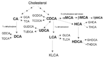
- Bile acids are produced by cholesterol metabolism in hepatocytes of the liver (FIG. 1), and are conjugated with glycine, taurine (amino acid ethylsulfonic acid), etc., and then secreted as bile. Bile acids are roughly classified into primary bile acids and secondary bile acids.
- Primary bile acids are bile acids synthesized in the liver, and include cholic acid (hereinafter sometimes abbreviated as CA), chenodeoxycholic acid (hereinafter sometimes abbreviated as CDCA), and the relationship between CDCA and isomers.
- Ursodeoxycholic acid hereinafter sometimes abbreviated as UDCA
- Secondary bile acids are bile acids that are produced by dehydration, dehydrogenation, hydrogenation, and deconjugation reactions by enterobacteria when primary bile acids are excreted into the intestine via the bile duct. (Hereinafter, it may be abbreviated as DCA) and LCA.
- bile acid fractions each include three types: free, glycine-conjugated, and taurine-conjugated.
- a sulfate-conjugated type or a glucuronic acid-conjugated type exists in many animal species including humans.
- CA and CDCA having hydroxyl groups at the 3, 7, and 12 positions of the colanic acid skeleton are generated as primary bile acids.
- some animals produce bile acids that are specific to that species.
- ⁇ -mulicholic acid (hereinafter sometimes abbreviated as ⁇ MCA) and ⁇ -mulicholic acid (hereinafter sometimes abbreviated as ⁇ MCA), and in pigs, hyocholic acid (hereinafter abbreviated as HCA).
- ⁇ MCA and ⁇ MCA are primary bile acids, and taurine-conjugated forms are also known.
- HCA is a primary bile acid, and hyodeoxycholic acid (hereinafter referred to as HDCA) is generated by 7 ⁇ -dehydration oxidation in the intestine.
- HDCA hyodeoxycholic acid
- the type of liver injury has been determined based on biochemical parameters.
- the type of hepatic disorder is determined using an increase in ALT or AST for the hepatocellular disorder type and an increase in ALP or ⁇ -GTP for the cholestasis type as one index.
- the determination method based on the biochemical parameter, the determination of the type of liver disorder may be wrong. For this reason, attempts have been made to measure the respective concentrations in serum of many fractions constituting bile acids, discriminate the type of liver injury based on the results, and further determine the treatment policy.
- Non-Patent Documents 2 to 6 So far, various relationships have been reported between the cause-specific classification of liver damage and the concentration of bile acid fraction (Non-Patent Documents 2 to 6). It has not been applied.
- Non-Patent Documents 6 to 8 Some reports have been made on the relationship between the classification of clinical symptoms of liver disorders and the concentration of the bile acid fraction (Non-Patent Documents 6 to 8). However, certain opinions are not shown.
- the clinical symptom classification for drug-induced liver disorders has been proposed as an evaluation method based on biochemical parameters rather than bile acid fractions (Non-patent Document 9).
- Non-Patent Document 10 has been proposed (Non-Patent Document 10).
- the present inventors compared hepatic diseases caused by drug administration to rats with typical bile stasis models such as bile duct ligation treatment and 1-naphthyl isothiocyanate (ANIT) administration.
- typical bile stasis models such as bile duct ligation treatment and 1-naphthyl isothiocyanate (ANIT) administration.
- ANIT 1-naphthyl isothiocyanate
- Non-patent Document 11 It has been reported that it is possible to determine the type of disorder of liver disease by measuring the concentration (Non-patent Document 11). However, in the clinic, no clear biomarker for early diagnosis of intrahepatic cholestasis at the level of capillary bile duct has been reported at present, and it is often difficult to determine a treatment policy.
- Palmeira CM Rosetta AP, Mitochondrially-mediated-toxicity of bile acids. Toxicology. 203 (1-3): 1-15 (2004). Danan G, Benichou C. Causalityassessment of adverse reactions to drugs--I. A novel method based on theconclusions of international consensus meetings: application to drug-inducedliver injuries. J Clin Epidemiol: 1 Takikawa, Junichi, DDW-J (2004) Workshop Proposal of diagnostic criteria for drug-induced liver injury Liver (46) (2): 85-90 (2005) Noriko Masubuchi et al., The 24th Annual Meeting of the Japanese Pharmacokinetic Society, page 305, No. 2-P-43, 2009. A. Stiehl, Bile Salt Sulphatesin Cholestasis. European Journal of Clinical Investigation. 4 (1): 59-63 (1974).
- Liver disorders can be categorized according to clinical symptoms as hepatocellular disorder and cholestasis, and these types are important to determine the type of treatment appropriate for each type. No technology has been established to determine this. Therefore, at present, treatment such as medication is performed based on comprehensive judgment based on experiences of doctors while proceeding with treatment. Conventionally, when determining the type of liver injury based on biochemical parameters, an increase in ALT or AST has been used as an index for hepatocellular disorder type, and an increase in ALP or ⁇ -GTP has been used for cholestasis type. This alone can be misleading. Therefore, if there is a marker that can more clearly discriminate the type of liver disorder at the early stage of the onset of liver disease, the treatment policy can be determined early.
- An object of the present invention is to find a biomarker useful for enabling early diagnosis of a liver disorder type and provide a method for discriminating a liver disorder type using the biomarker as an index or a method for assisting in discriminating a liver disorder type It is to be.
- the present inventors have intensively studied to solve the above problems, and measured the concentration of 24 kinds of bile acid fractions, liver fibrosis markers, and oxidative stress markers using clinical serum samples, and obtained measurement data Multivariate analysis found that the concentration of lithocholic acid was associated with liver injury type. Specifically, the concentration of lithocholic acid tended to be high in hepatocellular disorder-type liver disorders and conversely low in cholestatic liver disorders in the hepatic disorder population. Furthermore, in the hepatocyte disorder type liver disorder, the concentration of ursodeoxycholic acid tended to be low, and the concentration of type IV collagen as a liver fibrosis marker tended to be high.
- SSBA serum sulfate-conjugated bile acids
- type IV collagen which is a liver fibrosis marker.
- HA serum sulfate-conjugated bile acids
- ROS reactive oxygen species
- the present invention relates to: 1.
- a test method for discriminating a hepatic disorder type using LCA concentration as an index comprising measuring LCA concentration in a biological sample collected from a subject. 2. Further comprising comparing the measured LCA concentration to a preset LCA concentration cutoff value. Inspection method described in 1. 3. Further comprising determining that the type of liver injury is hepatocellular injury when the measured LCA concentration is greater than or equal to a preset cut-off value of the LCA concentration identifying a liver injury of the liver cell injury type , 1. Inspection method described in 1. 4).
- determining that the type of liver injury is cholestatic when the measured LCA concentration is less than or equal to a preset cut-off value of LCA concentration that identifies a cholestatic liver disorder. 1. Inspection method described in 1. 5. The measured LCA concentration exceeds a preset cut-off value for LCA concentration that identifies a hepatocellular disorder-type liver disorder and less than a preset cut-off value that identifies a cholestatic liver disorder And further comprising determining that the type of liver injury is a mixed type of hepatocellular injury and cholestasis. Inspection method described in 1. 6).
- the measured LCA concentration is equal to or higher than a preset cut-off value of LCA concentration for identifying a hepatocellular disorder-type liver disorder, and the measured UDCA and / or type IV collagen concentration is any one of the following: And further comprising determining that the type of liver injury is hepatocellular injury. Inspection method described in: (1) The UDCA concentration is less than or equal to a preset cutoff value of the UDCA concentration.
- the type IV collagen concentration is equal to or higher than a cut-off value of the type IV collagen concentration for identifying a preset hepatocyte disorder type liver injury, (3) Identifying a hepatocellular disorder-type liver disorder in which the UDCA concentration is equal to or lower than a cut-off value of the UDCA concentration that identifies a preset hepatocyte disorder-type liver disorder, and a type IV collagen concentration is preset It is not less than the cutoff value of the type IV collagen concentration. 7).
- the measured LCA concentration is less than or equal to a preset cut-off value of LCA concentration that identifies a cholestatic liver disorder
- the measured concentrations of DCA, SSBA, type IV collagen, HA, and ROS are Further comprising determining that the type of liver disorder is cholestatic when any one or more of the following: Inspection method described in: (4)
- the DCA concentration is less than or equal to a preset cut-off value of the DCA concentration that identifies a cholestatic liver disorder.
- the SSBA concentration is equal to or higher than a preset cut-off value of the SSBA concentration that identifies a cholestatic liver disorder.
- the type IV collagen concentration is greater than or equal to a preset cutoff value of the type IV collagen concentration that identifies a cholestatic liver disorder.
- HA concentration is equal to or higher than a preset cut-off value of HA concentration for identifying a cholestatic liver disorder set in advance.
- the ROS concentration is equal to or higher than a preset cutoff value of the ROS concentration for identifying a cholestatic liver disorder. 8).
- the measured LCA concentration exceeds a preset cut-off value for LCA concentration that identifies a hepatocellular disorder-type liver disorder and less than a preset cut-off value that identifies a cholestatic liver disorder
- the types of liver disorders are hepatocellular disorder and bile Further comprising determining that it is a mixed type with a stagnation type. Inspection method described in 1. 9. 2.
- the LCA concentration cutoff value set in advance is calculated from a receiver operating characteristic curve (hereinafter referred to as ROC curve) of the LCA concentration. Inspection method. 10. 2.
- the LCA concentration cut-off value for discriminating a preset hepatocellular disorder-type liver disorder is calculated from an LCA concentration ROC curve relating to a predetermined hepatocellular disorder-type liver disorder. 5. 6. Or 8. Inspection method described in 1. 11.
- the LCA concentration cutoff value for identifying a preset cholestatic liver disorder is calculated from an ROC curve of the LCA concentration related to a predetermined cholestatic liver disorder. 5. 6. Or 8. Inspection method described in 1.
- LCA concentration cut-off value for identifying a preset hepatocyte disorder type liver disorder is calculated from the ROC curve of LCA concentration for hepatic disorder of a predetermined hepatocellular disorder type, respectively, 5. Calculated from the ROC curve of the UDCA concentration related to the disorder, and calculated from the ROC curve of the type IV collagen concentration related to the predetermined hepatocyte disorder type liver disease. Inspection method described in 1. 13.
- LCA concentration cut-off value for identifying preset cholestatic liver disorder DCA concentration cut-off value for identifying cholestatic liver disorder, preset cholestatic liver disorder Cut-off value of SSBA concentration for discriminating, preset cut-off value of type IV collagen concentration for identifying cholestatic liver injury, cut-off value of HA concentration for discriminating preset cholestatic liver injury Value, and a ROS concentration cut-off value that identifies a preset cholestatic liver disorder, each calculated from an LCA concentration ROC curve for a predetermined cholestatic liver disorder, a predetermined Calculated from the ROC curve of DCA concentration for cholestatic liver injury, calculated from the ROC curve of SSBA concentration for predetermined cholestatic liver injury Calculated from a ROC curve of type IV collagen concentration related to a predetermined cholestatic liver disorder, calculated from an ROC curve of HA concentration related to a predetermined cholestatic liver disorder, predetermined bile 6.
- the biological sample is a blood sample; To 13. The inspection method as described in any one of these.
- the biological sample is a serum sample; To 14. The inspection method as described in any one of these. 16. 1 above.
- the present invention it is possible to provide a test method for measuring the concentration of LCA in a biological sample collected from a subject and discriminating the type of liver injury using the measured LCA concentration as an index.
- the test method according to the present invention can be used alone or in combination with the measurement of a biochemical parameter that has been performed conventionally, thereby causing liver damage type due to clinical symptoms, specifically hepatocellular disorder type, cholestasis type, And early diagnosis of these and their mixed type and early determination of treatment policy.
- the examination method according to the present invention can be used for examination of liver disease, and is extremely useful as a method for assisting diagnosis and treatment of liver disease.
- FIG. 4 is a schematic diagram for explaining that in hepatocellular disorder-type hepatopathy, the concentration of LCA in the bile acid fraction showed a significantly increasing tendency and the concentration of UDCA showed a decreasing tendency.
- the concentration of CDCA in an isomer relationship with CA or UDCA showed a tendency to decrease although there was no significant difference, while the concentration of DCA showed a tendency to rise although there was no significant difference.
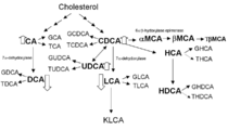
- cholestatic liver disorders it is a schematic diagram explaining that both LCA and DCA concentrations in the bile acid fraction showed a significantly increasing tendency. Although the concentrations of CDCA, UDCA, and CA were not significantly different, all showed a decreasing tendency.
- Table 1 shows the abbreviations of the bile acid fractions used in this specification.
- the name of a bile acid fraction may be described by the abbreviation shown in Table 1.
- the present invention relates to an inspection method for liver damage, and more particularly to an inspection method for determining a liver damage type.
- the test method according to the present invention measures the concentration of LCA in a biological sample collected from a subject, and discriminates the liver injury type using the measured LCA concentration as an index. Discrimination of the liver injury type using the LCA concentration as an index can be performed by comparing the measured LCA concentration with a preset cutoff value of the LCA concentration.
- a test method according to the present invention measuring the LCA concentration in a biological sample collected from a subject, and comparing the measured LCA concentration with a preset cut-off value of the LCA concentration An inspection method for discriminating the liver injury type can be exemplified.
- the concentration of a biomarker different from LCA and related to the type of liver injury may be measured.
- the LCA concentration and the concentration of the biomarker The index can be used to determine the type of liver injury.
- biomarkers different from LCA and related to the type of liver injury include UDCA and type IV collagen related to hepatocellular injury type liver injury.
- biomarkers associated with cholestatic liver damage include DCA, SSBA, type IV collagen, HA, and ROS.
- the inspection method according to the present invention can be carried out alone or in combination with various inspection methods such as measurement of biochemical parameters that have been carried out conventionally.
- type of liver disorder means a type classified according to symptoms of liver disorder. Symptoms of liver damage refer to various complaints and examination findings presented by patients with liver damage.
- examples of “hepatic disorder type” include hepatocellular disorder type and cholestasis type.
- hepatocellular disorder type hepatocytes that are one of the cells constituting the liver and occupy most of the liver are damaged, causing a significant decrease in liver function.
- the cholestatic type is caused by obstruction of bile excretion from the liver for some reason, resulting in abnormal bile outflow throughout the liver or blood in part or all of the intrahepatic or extrahepatic biliary system. Bile accumulates and causes various symptoms such as jaundice and hepatitis.
- hepatocyte disorder type and cholestasis type can be discriminated, and types classified into both hepatocyte disorder type and cholestasis type, that is, mixed type can be discriminated. .
- the “subject” to be subjected to the test method according to the present invention means a person diagnosed as having liver dysfunction, and / or a person suspected of having liver dysfunction due to some test result, viral hepatitis, It includes a person having or / and suspected of having a liver disease classified into drug-induced liver injury, alcoholic liver failure, autoimmune liver failure, metabolic disorder liver disease and the like. Preferably, it is a person diagnosed as having liver dysfunction.
- the “biological sample” is not particularly limited as long as it contains a bile acid fraction, and includes a fibrosis marker and / or an oxidative stress marker in addition to a bile acid fraction. What can be preferably mentioned is mentioned.
- blood isolated from a subject, serum or plasma prepared from the blood, preferably serum can be exemplified.
- the “hepatic disorder therapeutic agent” and “the hepatic disorder treatment method” include, for example, a hepatic disorder therapeutic agent and a hepatic disorder treatment method described in the manual drug-induced hepatic disorder corresponding to serious side effect disease, Examples thereof include a medication method and a treatment method performed in each medical facility.
- a glycyrrhizin preparation having a strong neominophagency preparation having an antiallergic action is intravenously administered, and UDCA having a liver cell membrane protecting action is orally administered.
- IVH central intravenous nutrition
- cholestasis for example, vitamin A or vitamin K is administered to compensate for the lack of fat-soluble vitamins, and then UDCA, prednisolone, phenobarbital, taurine, colestimide, etc. are used as drug treatment.
- LCA is a kind of bile acid. Bile acids are produced by cholesterol metabolism in hepatocytes of the liver, conjugated with glycine, taurine (amino acid ethylsulfonic acid), etc., then secreted as bile and play an important role in digestion and absorption of fat. LCA is a secondary bile acid produced by CDCA, which is one of the primary bile acids synthesized in the liver, excreted via the bile duct into the intestine and undergoing a dehydration oxidation reaction with 7- ⁇ dehydroxylase.
- the LCA concentration was determined to be hepatocellular disorders. Patients showed a high tendency, and conversely, patients with cholestatic liver disorders showed a low tendency. Furthermore, in cholestatic liver disorders, the concentration of DCA in serum tended to be low, and the concentration of SSBA tended to be high. On the other hand, in the hepatocellular disorder type liver disorder, the concentration of UDCA in the serum tended to be low. The taurine conjugates and glycine conjugates of various bile acids also showed the same tendency as the concentration of the free fraction.
- cholestasis suppresses the production of DCA and LCA, which increases the concentration of CA, CDCA, and UDCA, and conversely, in hepatocellular disorder liver disorders, It can be considered that the production of highly lipophilic DCA and LCA is enhanced, and the production of CA, CDCA and UDCA is suppressed.
- the type of liver injury can be determined by comparing each measurement item with a preset cutoff value.
- Cutoff value refers to a value that distinguishes between positive and negative ranges.
- the cut-off value for each measurement item can be set individually depending on the type of liver injury.
- the cutoff value can be set by a method known per se.
- the cutoff value can be set by ROC analysis that is generally used as a method for examining the usefulness of a diagnostic test. In ROC analysis, when the threshold value is changed, the sensitivity (Sensitivity) at each threshold value is plotted on the vertical axis, and FPF (False Positive Fraction, false positive rate: 1-specificity (Specificity)) is plotted on the horizontal axis.
- ROC curve An ROC curve is created.
- a test with no diagnostic ability becomes a straight line on the diagonal line, but as the diagnostic ability improves, the diagonal line curves in the upper left corner. It becomes a curve that passes on the upper side.
- the cut-off value for example, the ROC curve, which is an independent variable with excellent sensitivity and specificity, approaches the upper left corner, so that the point at which the distance from the upper left corner is the minimum is set as the cut-off value. There is a way to do it.
- a method of setting a point farthest from the diagonal line where the area under the ROC curve (area under the curve, AUC) is 0.500 as a cutoff value that is, (sensitivity + specificity-1 ) Can be calculated, and the maximum value of the Yawden index (Youden index) can be set as the cutoff value.
- sensitivity means a true positive rate.
- Specificity means a true negative rate.
- the cut-off value can be set quantitatively from the relationship between each measurement item and the frequency of the type of liver injury.
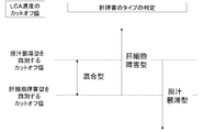
- the discrimination of the type of liver injury by the test method according to the present invention will be described with reference to FIG.
- the LCA concentration is equal to or higher than a preset cut-off value of the LCA concentration for identifying a hepatocyte disorder type liver disorder
- it is determined that the hepatopathy type is hepatocyte disorder type.
- the liver disorder type is determined to be cholestatic.
- the LCA concentration exceeds a preset cut-off value of LCA concentration for identifying hepatopathy of the hepatocellular disorder type, and is less than the cut-off value of LCA concentration for identifying cholestatic liver disorder , It is determined that the type of hepatic disorder is both hepatocellular disorder type and cholestatic type, that is, mixed type. Further, if the LCA concentration exceeds a preset cut-off value of the LCA concentration that identifies a cholestatic liver disorder, the liver disorder type does not include a mixed type liver. Determined to be cytotoxic.
- the hepatic disorder type is a cholestatic type not including a mixed type.
- the concentration of any one or both of UDCA and type IV collagen may be measured, and the measured concentration of UDCA and / or type IV collagen is measured.
- the measured LCA concentration is greater than or equal to a preset cut-off value of LCA concentration that identifies a hepatocellular disorder-type liver injury, and the measured UDCA and / or
- the concentration of type IV collagen is any one of the following, it can be determined that the type of liver injury is hepatocellular injury type: (1) hepatocellular injury type liver injury with a preset UDCA concentration (2) a type IV collagen that discriminates a hepatocyte disorder type liver injury with a preset type IV collagen concentration (3) the UDCA concentration is less than or equal to a preset cut-off value of the UDCA concentration that identifies a hepatocellular disorder-type liver injury, and the type IV collagen concentration is preset
- one or more concentrations of DCA, SSBA, type IV collagen, HA, and ROS may be measured, and each measurement value is set in advance.
- the measured LCA concentration is less than or equal to a preset LCA concentration cutoff value
- the measured concentrations of DCA, SSBA, type IV collagen, HA, and ROS are as follows: When any one or two or more, it can be determined that the type of liver disorder is cholestatic type: (4) DCA concentration is less than or equal to a preset cut-off value of DCA concentration, (5) The SSBA concentration is greater than or equal to a preset SSBA concentration cut-off value. (6)
- the IV collagen concentration is set to a preset IV collagen concentration cutoff. At least, (7) HA concentration is preset HA concentration of less than cut-off value, and (8) ROS concentration is preset ROS concentrations of less than cut-off value.
- the concentration of type IV collagen is measured, each measured value is compared with a preset cutoff value, and the measured LCA concentration Exceeds the LCA concentration cut-off value that identifies a pre-set hepatocellular disorder-type liver disorder, and is less than the LCA concentration cut-off value that identifies a cholestatic liver disorder, and is further measured
- the concentration of type IV collagen is equal to or higher than a preset cutoff value of type IV collagen
- the type of liver injury can be determined to be a mixed type of hepatocellular injury type and cholestatic type.
- cutoff values for each measurement item used in the inspection method according to the present invention are shown in Tables 2-1 and 2-2.
- Table 2-1 shows specific examples of biomarker cutoff values for identifying hepatocellular disorder-type liver disorders.
- Table 2-2 shows specific examples of cutoff values of biomarkers for identifying cholestatic liver disorders. The exemplified cut-off values are for the patients with hepatic disorders to which hepatic disorder treatment drugs are not applied in the examples described later, and serum samples collected from the patients with hepatic disorders are used as biological samples for each biomarker.
- Concentration is measured, and for each measured biomarker concentration, comparison between the hepatic disorder patient population and hepatocellular disorder hepatic disorder patient or cholestatic liver disorder patient is performed using ROC analysis, and the Yoden index is obtained.
- This is a predetermined cutoff value.
- the cut-off value can also be set for each measurement facility or population to be measured. It is also possible to appropriately change the setting by accumulating measurement data of a target biomarker concentration for the same sample as described above and performing analysis using the same analysis method as described above.
- the cutoff value can be set as appropriate.
- the type of liver injury includes the step of determining that hepatocyte damage type,
- the cutoff value is 0.0195 nmol / L.
- the cutoff value is 0.0241 nmol / L.
- Measuring the LCA concentration in a serum sample collected from the subject; Comparing the measured LCA concentration with a preset LCA concentration cutoff value, and the measured LCA concentration identifying a preset hepatocellular disorder-type liver injury Determining that the hepatic disorder type is a mixture of hepatocellular disorder type and cholestatic type when the value is less than a preset cutoff value for identifying hepatic disorder of cholestatic type Including
- the cut-off value of the LCA concentration for discriminating a preset hepatocyte disorder type liver injury is 0.0195 nmol / L
- the LCA concentration cut for discriminating a preset cholestatic liver disorder The off value is 0.0241 nmol / L.
- the type IV collagen concentration is equal to or higher than a cut-off value of the type IV collagen concentration for identifying a preset hepatocyte disorder type liver injury, (3) Identifying a hepatocellular disorder-type liver disorder in which the UDCA concentration is equal to or lower than a cut-off value of the UDCA concentration that identifies a preset hepatocyte disorder-type liver disorder, and a type IV collagen concentration is preset It is above the cutoff value of type IV collagen concentration, Including
- the cut-off value of the LCA concentration for identifying the preset hepatocyte disorder type liver disorder is 0.0195 nmol / L
- the preset cut of the UDCA concentration for identifying the hepatocyte disorder type liver disorder The off value is 0.903 nmol / L
- the cutoff value of the type IV collagen concentration that identifies a preset hepatocyte disorder type liver disorder is 128 ng / ml
- the liver disorder type is determined to be cholestatic Steps to do: (4)
- the DCA concentration is less than or equal to a preset cut-off value of the DCA concentration that identifies a cholestatic liver disorder.
- the SSBA concentration is equal to or higher than a preset cut-off value of the SSBA concentration that identifies a cholestatic liver disorder.
- Type IV collagen concentration is equal to or higher than a cut-off value of type IV collagen concentration for identifying a preset cholestatic liver disorder,
- HA concentration is equal to or higher than a preset cut-off value of HA concentration for identifying a preset cholestatic liver disorder,
- the ROS concentration is equal to or higher than a preset cutoff value of the ROS concentration for identifying a cholestatic liver disorder.
- a preset cut-off value of LCA concentration for identifying a cholestatic liver disorder is 0.0241 nmol / L
- a preset cut-off value of DCA concentration for identifying a cholestatic liver disorder Is 0.175 nmol / L
- the cut-off value of SSBA concentration that identifies a preset cholestatic liver disorder is 21.1 nmol / L
- identifies a preset cholestatic liver disorder The cut-off value of type IV collagen concentration is 288 ng / ml
- the cut-off value of HA concentration identifying a preset cholestatic liver disorder is 47 ng / ml
- the preset cholestatic type The cut-off value of ROS concentration that identifies liver damage is 216 U.
- a test method for discriminating the type of liver injury can be mentioned.
- the type of hepatic disorder includes the step of determining that the hepatocyte disorder type and the cholestatic type are mixed,
- the concentration of the bile acid fraction can be measured using a method conventionally used for the measurement. Examples thereof include a liquid chromatograph / tandem mass spectrometry (LC-MS / MS) method and a gas chromatograph mass spectrometry (GC / MS) method.
- the concentration of bile acid or sulfate-conjugated bile acid can be measured using a commercially available bile acid measurement kit.
- the concentration of sulfate-conjugated bile acid can be measured using Ubastic Auto, a bile acid (USBA) kit (LPS, Inc.). Measurement methods are not limited to these, and any method can be used as long as bile acids and fractions thereof can be measured.
- the concentration of type IV collagen can be measured using a method conventionally used for the measurement.
- the concentration of type IV collagen can be measured using a commercially available reagent kit for measuring type IV collagen.
- examples of such methods include enzyme immunization (EIA) and enzyme immunosolid phase (ELISA) using antibodies against type IV collagen.
- EIA enzyme immunization
- ELISA enzyme immunosolid phase
- an anti-human type IV collagen rabbit polyclonal antibody is added to a biological sample to form a type IV collagen-7S-anti-human type IV collagen rabbit polyclonal antibody complex (complex 1), and then iodine.
- Anti-human type IV collagen 7S rabbit polyclonal which was not able to bind to type IV collagen 7S in the sample added with type IV collagen 7S (labeled antigen) labeled with a radioisotope such as 125 ( 125 I)
- a complex complex 2
- anti-rabbit ⁇ -globulin goat serum goat antibody
- anti-rabbit ⁇ -globulin goat serum goat antibody
- the goat antibody is complex 2
- the concentration of type IV collagen ⁇ 7S can be measured by measuring the radioactivity of the precipitate.
- the measurement of the concentration of HA can be performed using a method conventionally used for the measurement.
- the concentration of HA can be measured using a commercially available reagent kit for measuring HA.
- an enzyme immunosolid phase method (ELISA) using an antibody against HA can be exemplified.
- the measurement of the concentration of ROS can be performed using a method conventionally used for the measurement.
- the concentration of ROS can be measured using a commercially available reagent kit for measuring ROS.
- free radicals ROO. And RO.
- ROOH peroxides
- metal ions such as iron, and N, N-diethyl-1,4-
- DEPPD phenylene-diamine sulfate
- the present invention includes a method of selecting a hepatic disorder therapeutic agent according to the type of liver disorder for a subject whose type of liver disorder has been determined by the test method according to the present invention.
- the present invention includes a treatment method for treating a subject whose liver disorder type has been determined by the examination method according to the present invention according to the liver disorder type.
- Examples of the hepatic disorder treatment agent and hepatic disorder treatment method according to the type of hepatic disorder include, for example, the hepatic disorder therapeutic agent and the hepatic disorder treatment method described in the manual drug-induced hepatic disorder corresponding to serious side effect disease, Examples thereof include a medication method and a treatment method performed in a medical facility.
- the hepatocellular disorder type for example, intravenous injection of a strong neominophagency preparation having an antiallergic action with a glycyrrhizin preparation or oral administration of UDCA is performed.
- IVH central intravenous nutrition
- cholestasis for example, vitamin A or vitamin K is administered to compensate for the lack of fat-soluble vitamins, and then UDCA, prednisolone, phenobarbital, taurine, colestimide, etc. are used as drug treatment.
- liver damage type serum samples obtained from patients with liver damage and healthy volunteers were used to measure the concentration of bile acid fraction, as well as oxidative stress markers and Measurement of liver fiber markers was performed. The collection of serum samples and a series of experiments were conducted with the consent of the patient and healthy volunteers.
- Target sample 150 patients suspected of having liver damage such as viral, autoimmune, alcoholic, hereditary, etc. in about 2 years based on the approval of Daiichi Sankyo Co., Ltd. and the Ethics Committee of Jikei University From 304 serum samples were collected. A serum sample derived from a healthy person was collected from a volunteer and used. Serum samples were stored at ⁇ 80 ° C. until biomarker measurement
- Each bile acid fraction specifically CA, DCA, CDCA, UDCA, LCA, 12_KLCA, ⁇ MCA, ⁇ MCA, HDCA, GCA, GDCA, GCDCA, GUDCA, GLCA, GHCA, GHDCA, TCA, TDCA, TCDCA, Twenty-four bile acid fractions of TUDCA, TLCA, T ⁇ MCA, THCA, and THDCA, and the concentration of sulfate-conjugated bile acid SSBA were measured.
- T.BIL total bilirubin
- D.BIL direct bilirubin
- TBA total bilirubin
- ALB albumin
- PT The concentration of prothrombin time (hereinafter abbreviated as PT) was measured. Furthermore, the concentrations of 8-OHdG and ROS, which are oxidative stress markers, and type IV collagen and HA, which are liver fibrosis markers, were measured.
- electrospray ionization (abbreviated as ESI) method negative was selected, and as the detection method, selected ion recording (abbreviated as SIR) was selected.
- the pretreatment method was a protein removal method.
- the sulfate-conjugated bile acid concentration was measured by BioMajesty JCA-BM2250 (JEOL) using 300 ⁇ L serum and Eubastic Auto, bile acid (USBA) kit (LPS Co., Ltd.).
- 8-OHdG and ROS which are oxidative stress markers
- 8-OHdG was measured using a reagent kit sold by Japan Aging Control Laboratory.
- ROS uses the Fenton reaction catalyzed by metal ions such as iron to generate free radicals (ROO. And RO.) From peroxides (ROOH), and N, N-diethyl-1,4-phenylene-diamine Reaction with sulfate (abbreviated as DEPPD).
- the produced DEPPD ⁇ + has an absorption at a wavelength of 505 nm, and the total amount of substances that produce free radicals was measured by measuring the absorbance.
- Type IV collagen 7S and HA which are liver fibrosis markers was performed as follows.
- Type IV collagen ⁇ 7S reacts with anti-human type IV collagen rabbit polyclonal antibody to form type IV collagen ⁇ 7S-anti-human type IV collagen rabbit polyclonal antibody complex (complex 1).
- iodine 125 (125I) -labeled human type IV collagen-7S labeleled antigen
- anti-human type IV collagen-7S rabbit polyclonal antibody that could not bind to type IV collagen-7S in the sample reacts with the labeled antigen.
- complex 2 When a labeled antigen-anti-human type IV collagen-7S rabbit polyclonal antibody complex (complex 2) is formed and anti-rabbit ⁇ -globulin goat serum (goat antibody) is further added, the goat antibody reacts with complex 2 Then, a labeled antigen-anti-human type IV collagen / 7S rabbit polyclonal antibody-goat antibody complex is formed and precipitated. After removing unreacted labeled antigen, the radioactivity of the precipitate was measured to determine the type IV collagen ⁇ 7S concentration. HA was measured using an HA ELISA kit (manufactured by Cosmo Bio).
- Biomarker candidate factors were bile acid fraction concentration, oxidative stress marker, and liver fibrosis marker.
- Patient background factors include gender, age, BMI, alcohol consumption, hepatic disorder treatment (antiviral agent, liver protectant, bile acid preparation, amino acid preparation / low albumin plasma improving drug, vitamin K preparation), complication (dyslipidemia) Diabetes, obstructive jaundice, gallstones).
- the presence / absence of liver disorder symptoms was determined by two or more people based on the judgment results of three independent evaluators.
- the total analysis unit was a sample, and the correlation between samples in the same case was not considered.
- the significance level for the statistical test was 5% on both sides.
- the 97.5% point of a healthy person's measured value (corresponding to an average + 2SD if normal distribution) is set as a reference value, and a value exceeding the reference value is set. Values that were high and below the reference value were divided into low values.
- Results Table 3 shows the background information of patients who obtained serum samples.
- Table 4 and Table 5 show the results of multivariate analysis by covariate adjustment for biomarkers that define the symptoms of liver disorders that have been type-identified.
- LCA LCA
- the concentration of LCA and type IV collagen tended to be high, and the concentration of UDCA tended to be low (Table 4).
- Type IV collagen is an indicator of liver fibrosis, but a tendency of high concentration was observed in the cholestatic type as in the case of hepatocellular injury type.
- the LCA increased in the former, while the latter in contrast with the decreased. From these results, it was suggested that LCA is a useful marker for discrimination of liver disorder symptoms.
- LCA which is a secondary bile acid
- the UDCA of the isomer of CDCA which is the parent compound of LCA, showed a low value.
- liver fibrosis markers were significantly increased in both hepatocellular disorder type and cholestatic type.
- 8-OHdG showed a significant decrease in hepatocellular injury, but there was no significant change in the value compared to the healthy population, and an example of a patient population that exceeded the cut-off value Due to the small number, it was not possible to clearly determine the relationship with hepatocellular injury type.
- the active oxygen ROS showed a high value in the cholestatic type, no significant change was observed in the hepatocellular disorder type.
- the biochemical parameters conventionally used in the examination of liver diseases have a low correlation with the biomarkers measured this time, and it is difficult to accurately determine liver symptom symptoms only with these biochemical parameters.
- the concentration of LCA is peculiar to each of the hepatocellular disorder type and cholestatic type and shows a significant change, suggesting that it is useful as a marker for distinguishing the type of liver disorder symptom.
- primary bile acids and SSBA are also effective for the determination of hepatocellular disorder type, cholestasis type mixed type, and mixed type thereof as markers that can complement discrimination of liver disorder symptom type by LCA concentration. .
- the present invention provides a test method for discriminating a liver disorder type, and according to the present invention, a liver disorder type due to a clinical symptom, specifically, a hepatocellular disorder type, a cholestasis type, and a mixture thereof. Allows early diagnosis of type and early determination of treatment strategy.
- the present invention is extremely useful in the field of liver disease testing.
Landscapes
- Health & Medical Sciences (AREA)
- Life Sciences & Earth Sciences (AREA)
- Engineering & Computer Science (AREA)
- Biomedical Technology (AREA)
- Chemical & Material Sciences (AREA)
- Immunology (AREA)
- Molecular Biology (AREA)
- Hematology (AREA)
- Urology & Nephrology (AREA)
- Physics & Mathematics (AREA)
- Analytical Chemistry (AREA)
- Pathology (AREA)
- Food Science & Technology (AREA)
- Biochemistry (AREA)
- General Health & Medical Sciences (AREA)
- General Physics & Mathematics (AREA)
- Medicinal Chemistry (AREA)
- Biotechnology (AREA)
- Cell Biology (AREA)
- Microbiology (AREA)
- Biophysics (AREA)
- Tropical Medicine & Parasitology (AREA)
- Investigating Or Analysing Biological Materials (AREA)
- Measuring Or Testing Involving Enzymes Or Micro-Organisms (AREA)
Abstract
Priority Applications (6)
| Application Number | Priority Date | Filing Date | Title |
|---|---|---|---|
| ES14834659T ES2705351T3 (es) | 2013-08-05 | 2014-08-04 | Método para la investigación del tipo de daño hepático |
| KR1020167005833A KR101913016B1 (ko) | 2013-08-05 | 2014-08-04 | 간장해 형태의 검사방법 |
| JP2015530865A JP6353837B2 (ja) | 2013-08-05 | 2014-08-04 | 肝障害のタイプの検査方法 |
| US14/907,828 US10073077B2 (en) | 2013-08-05 | 2014-08-04 | Method for investigation of liver damage type |
| EP14834659.6A EP3032256B1 (fr) | 2013-08-05 | 2014-08-04 | Procédé de recherche du type de dommage hépatique |
| CN201480044033.4A CN105452862B (zh) | 2013-08-05 | 2014-08-04 | 肝损伤类型的检查方法 |
Applications Claiming Priority (2)
| Application Number | Priority Date | Filing Date | Title |
|---|---|---|---|
| JP2013-162429 | 2013-08-05 | ||
| JP2013162429 | 2013-08-05 |
Publications (1)
| Publication Number | Publication Date |
|---|---|
| WO2015019976A1 true WO2015019976A1 (fr) | 2015-02-12 |
Family
ID=52461309
Family Applications (1)
| Application Number | Title | Priority Date | Filing Date |
|---|---|---|---|
| PCT/JP2014/070426 WO2015019976A1 (fr) | 2013-08-05 | 2014-08-04 | Procédé de recherche d'un type d'endommagement hépatique |
Country Status (7)
| Country | Link |
|---|---|
| US (1) | US10073077B2 (fr) |
| EP (1) | EP3032256B1 (fr) |
| JP (1) | JP6353837B2 (fr) |
| KR (1) | KR101913016B1 (fr) |
| CN (1) | CN105452862B (fr) |
| ES (1) | ES2705351T3 (fr) |
| WO (1) | WO2015019976A1 (fr) |
Cited By (2)
| Publication number | Priority date | Publication date | Assignee | Title |
|---|---|---|---|---|
| US20200378991A1 (en) * | 2016-05-29 | 2020-12-03 | Human Metabolomics Institute, Inc. | Liver disease-related biomarkers and methods of use thereof |
| WO2022025069A1 (fr) * | 2020-07-28 | 2022-02-03 | 株式会社シンクメディカル | Méthode d'évaluation de risque de maladie, dispositif d'évaluation de risque de maladie et programme d'évaluation de risque de maladie |
Families Citing this family (4)
| Publication number | Priority date | Publication date | Assignee | Title |
|---|---|---|---|---|
| RU2020110462A (ru) * | 2017-08-14 | 2021-09-16 | Серес Терапеутикс, Инк. | Композиции и способы для лечения холестатического заболевания |
| CN109060977A (zh) * | 2018-07-13 | 2018-12-21 | 深圳市绘云生物科技有限公司 | 用于肝纤维化和肝硬化诊断的生物标志物和试剂盒及使用方法 |
| CN110220987B (zh) * | 2019-06-11 | 2021-09-28 | 上海市内分泌代谢病研究所 | 胆汁酸联合标志物在制备用于预测或诊断糖尿病的检测试剂或检测物的用途 |
| CN113504324B (zh) * | 2021-07-06 | 2023-07-04 | 复旦大学附属儿科医院 | 胆汁淤积症预后生物标记物检测试剂盒 |
Citations (5)
| Publication number | Priority date | Publication date | Assignee | Title |
|---|---|---|---|---|
| JPS56126744A (en) * | 1980-03-10 | 1981-10-05 | Sankyo Co Ltd | Fluorometric method for analysis of bile acid |
| JPH069686A (ja) * | 1991-09-21 | 1994-01-18 | Hoechst Ag | 胆汁酸を濃縮するためのアルキル化ポリエチレンイミン誘導体の使用 |
| JPH11171898A (ja) * | 1997-10-03 | 1999-06-29 | Tosoh Corp | Iv型コラーゲン高分子画分、その製法、その測定法及び肝疾患の判定方法 |
| JP2012002610A (ja) * | 2010-06-16 | 2012-01-05 | National Institute Of Biomedical Innovation | 肝障害検査用バイオマーカー及びそれを用いた肝障害の予測方法 |
| JP2013504310A (ja) * | 2009-09-11 | 2013-02-07 | ザ チャイニーズ ユニバーシティ オブ ホンコン | 肝臓病変を評価する方法 |
Family Cites Families (5)
| Publication number | Priority date | Publication date | Assignee | Title |
|---|---|---|---|---|
| US6060255A (en) | 1997-10-03 | 2000-05-09 | Tosoh Corporation | Type IV collagen high molecular form and production and diagnostic use thereof |
| DE69920048T2 (de) * | 1999-01-12 | 2005-09-22 | Ebara Corp. | Verfahren und biosensor zum nachweis von antigenen |
| US7427490B2 (en) * | 2001-08-20 | 2008-09-23 | Biosite Incorporated | Diagnostic markers of stroke and cerebral injury and methods of use thereof |
| BRPI0924639B1 (pt) * | 2009-06-02 | 2021-12-07 | Biocrates Life Sciences Ag | Novos biomarcadores para avaliação de doenças renais |
| CN104198420B (zh) * | 2014-08-14 | 2016-09-14 | 上海睿康生物科技有限公司 | 一种稳定的总胆汁酸检测试剂盒 |
-
2014
- 2014-08-04 US US14/907,828 patent/US10073077B2/en active Active
- 2014-08-04 JP JP2015530865A patent/JP6353837B2/ja active Active
- 2014-08-04 CN CN201480044033.4A patent/CN105452862B/zh active Active
- 2014-08-04 KR KR1020167005833A patent/KR101913016B1/ko active IP Right Grant
- 2014-08-04 ES ES14834659T patent/ES2705351T3/es active Active
- 2014-08-04 WO PCT/JP2014/070426 patent/WO2015019976A1/fr active Application Filing
- 2014-08-04 EP EP14834659.6A patent/EP3032256B1/fr active Active
Patent Citations (5)
| Publication number | Priority date | Publication date | Assignee | Title |
|---|---|---|---|---|
| JPS56126744A (en) * | 1980-03-10 | 1981-10-05 | Sankyo Co Ltd | Fluorometric method for analysis of bile acid |
| JPH069686A (ja) * | 1991-09-21 | 1994-01-18 | Hoechst Ag | 胆汁酸を濃縮するためのアルキル化ポリエチレンイミン誘導体の使用 |
| JPH11171898A (ja) * | 1997-10-03 | 1999-06-29 | Tosoh Corp | Iv型コラーゲン高分子画分、その製法、その測定法及び肝疾患の判定方法 |
| JP2013504310A (ja) * | 2009-09-11 | 2013-02-07 | ザ チャイニーズ ユニバーシティ オブ ホンコン | 肝臓病変を評価する方法 |
| JP2012002610A (ja) * | 2010-06-16 | 2012-01-05 | National Institute Of Biomedical Innovation | 肝障害検査用バイオマーカー及びそれを用いた肝障害の予測方法 |
Non-Patent Citations (16)
| Title |
|---|
| "Manual for handling disorders due to adverse drug reactions", DRUG-INDUCED LIVER INJURY, April 2008 (2008-04-01), pages 10 - 30 |
| A. STIEHL: "Bile Salt Sulphatesin Cholestasis", EUROPEAN JOURNAL OF CLINICAL INVESTIGATION, vol. 4, no. 1, 1974, pages 59 - 63 |
| BERR F; PRATSCHKE E; FISCHER S; PAUMGARTNER G: "Disorders of bile acid metabolism in cholesterol gallstone disease", J CLIN INVEST., vol. 90, no. 3, 1992, pages 859 - 68 |
| BURKARD I; ECKARDSTEIN A; RENTSCH KM: "Differentiated quantification of human bile acids in serum by high-performance liquid chromatography-tandem mass spectrometry", J CHROMATOGR B ANALYT TECHNOL BIOMED LIFE SCI., vol. 826, no. 1-2, 2005, pages 147 - 59 |
| DANAN G; BENICHOU C: "Causality assessment of adverse reactions to drugs--I. A novel method based on the conclusions of international consensus meetings: application to drug-induced liver injuries", J CLIN EPIDEMIOL., vol. 46, no. 11, 1993, pages 1323 - 30 |
| FISCHER, S; BEUERS, U; SPENGLER, U; ZWIEBEL, FM; KOEBE, H-G: "Hepatic levels of bile acids in end-stage chronic cholestatic liver disease", CLINICA CHIMICA ACTA, vol. 251, no. 2, 1993, pages 173 - 86 |
| HAJIME TAKIGAWA: "Proposal of diagnostic criteria of drug induced hepatic injury", DDW-J2004 WORKSHOP, KANZO, vol. 46, no. 2, 2005, pages 85 - 90 |
| IA BOUCHIER; CR PENNINGTON: "Serum bile acids in hepatobiliary disease", GUT, vol. 19, no. 6, June 1978 (1978-06-01), pages 492 - 6 |
| KIMIHIDE NAKAMURA ET AL.: "Digestive system disease. State of arts (Ver.2). II. Liver and bile and pancreas. Advance on the basic and clinical research of the pathophysiology. Cholestasis and cholestatic factors", JOURNAL OF CLINICAL AND EXPERIMENTAL MEDICINE, 30 June 1999 (1999-06-30), pages 12 - 14, XP008183097 * |
| LUCANGIOLI SE ET AL.: "Lithocholic acid as a biomarker of intrahepatic cholestasis of pregnancy during ursodeoxycholic acid treatment", ANN CLIN BIOCHEM, vol. 46, no. 1, January 2009 (2009-01-01), pages 44 - 49, XP055315033 * |
| NORIKO MASUBUCHI ET AL., 24TH ANNUAL MEETING OF THE JAPANESE SOCIETY FOR THE STUDY OF XENOBIOTICS, 2009, pages 305,2,43 |
| OSTROW JD: "Hepatic transport and bile secretion", 1993, RAVEN, article "Metabolism of bile salts in cholestasis in humans", pages: 673 - 712 |
| PALMEIRA CM; ROLO AP: "Mitochondrially-mediated toxicity of bile acids", TOXICOLOGY, vol. 203, no. 1-3, 2004, pages 1 - 15 |
| See also references of EP3032256A4 * |
| TSUNEO KIMURA: "Tanjusan no Kansaibo Shogaisei ni Kansuru Kenkyu", JAPANESE JOURNAL OF GASTROENTEROLOGY, vol. 77, no. 2, 1980, pages 185 - 194, XP008182762 * |
| WILLIAMS CN: "Bile-acid metabolism and the liver", CLIN BIOCHEM., vol. 9, no. 3, 1976, pages 149 - 52 |
Cited By (4)
| Publication number | Priority date | Publication date | Assignee | Title |
|---|---|---|---|---|
| US20200378991A1 (en) * | 2016-05-29 | 2020-12-03 | Human Metabolomics Institute, Inc. | Liver disease-related biomarkers and methods of use thereof |
| WO2022025069A1 (fr) * | 2020-07-28 | 2022-02-03 | 株式会社シンクメディカル | Méthode d'évaluation de risque de maladie, dispositif d'évaluation de risque de maladie et programme d'évaluation de risque de maladie |
| JPWO2022025069A1 (fr) * | 2020-07-28 | 2022-02-03 | ||
| JP7170368B2 (ja) | 2020-07-28 | 2022-11-14 | 株式会社シンクメディカル | 疾患リスク評価方法、疾患リスク評価装置、及び疾患リスク評価プログラム |
Also Published As
| Publication number | Publication date |
|---|---|
| KR20160040679A (ko) | 2016-04-14 |
| US20160169862A1 (en) | 2016-06-16 |
| CN105452862B (zh) | 2018-10-09 |
| CN105452862A (zh) | 2016-03-30 |
| KR101913016B1 (ko) | 2018-10-29 |
| EP3032256A1 (fr) | 2016-06-15 |
| EP3032256B1 (fr) | 2018-11-21 |
| ES2705351T3 (es) | 2019-03-22 |
| EP3032256A4 (fr) | 2017-03-22 |
| JPWO2015019976A1 (ja) | 2017-03-02 |
| US10073077B2 (en) | 2018-09-11 |
| JP6353837B2 (ja) | 2018-07-04 |
Similar Documents
| Publication | Publication Date | Title |
|---|---|---|
| JP6353837B2 (ja) | 肝障害のタイプの検査方法 | |
| JP2019525198A (ja) | 肝疾患関連バイオマーカーおよびその使用方法 | |
| Yilmaz et al. | Decreased plasma levels of soluble receptor for advanced glycation endproducts (sRAGE) in patients with nonalcoholic fatty liver disease | |
| Natowicz et al. | Abnormal bile acids in the Smith‐Lemli‐Opitz syndrome | |
| EP2756304A1 (fr) | Moyens et méthodes d'évaluation de toxicité du rein | |
| Steinberg et al. | A PEX10 defect in a patient with no detectable defect in peroxisome assembly or metabolism in cultured fibroblasts | |
| US8932870B2 (en) | Method for diagnosing acute alcoholic hepatitis | |
| IL262141A (en) | Method for diagnosing cystic fibrosis | |
| Lee et al. | Metabolic dysfunction associated fatty liver disease (MAFLD) and serum vitamin D concentration in South Korea | |
| Danese et al. | Lack of an association between circulating adiponectin levels and risk of colorectal adenoma | |
| Cisneros et al. | Identification of Potential Visceral Pain Biomarkers in Colon Exudates from Mice with Experimental Colitis: An Exploratory In Vitro Study | |
| US9541543B2 (en) | Method for diagnosing acute alcoholic hepatitis | |
| Liang et al. | The expression of serum M30 and M65 in chronic hepatitis B patients with non-alcoholic fatty liver disease. | |
| US20170181686A1 (en) | Breath test for assessing liver disease | |
| EP3245519B1 (fr) | Procédé de diagnostic d'hépatite alcoolique aiguë | |
| Li | Urinary Bile Acid Indices as Prognostic Biomarkers for the Complications of Liver Diseases | |
| Ki-Chul Sung et al. | Combined Influence of Insulin Resistance, Overweight/Obesity, and Fatty Liver as Risk Factors for Type 2 Diabetes | |
| Haerty | Liver enzymes and function in patients with metabolic syndrome. literature review | |
| Aleksandrova | Clinical significance of pentraxin-3 and C-reactive protein in the differentiation of stages of nonalcoholic fatty liver disease | |
| Alaa et al. | Value of Cytokeratin-18 as a non-invasive diagnostic biomarker of nonalcoholic steatohepatitis (NASH) | |
| Ueno et al. | Clinical Implications of Serum Mac-2 Binding Protein in Patients After Living Donor Liver Transplantation for Biliary Atresia | |
| Alamoudi | Urinary Bile Acid Indices as Diagnostic and Prognosic Biomarkers for Liver Diseases | |
| JP2006343127A (ja) | 代謝性症候群マーカーおよびその利用 | |
| Mustafa et al. | Correlation of Serum Adiponectin with Hepatic Fibrosis in Patients with Non-Alcoholic Fatty Liver Disease | |
| Khalil et al. | Serum Nesfatin-1 in Patients with Metabolic Associated Fatty Liver Disease |
Legal Events
| Date | Code | Title | Description |
|---|---|---|---|
| WWE | Wipo information: entry into national phase |
Ref document number: 201480044033.4 Country of ref document: CN |
|
| 121 | Ep: the epo has been informed by wipo that ep was designated in this application |
Ref document number: 14834659 Country of ref document: EP Kind code of ref document: A1 |
|
| ENP | Entry into the national phase |
Ref document number: 2015530865 Country of ref document: JP Kind code of ref document: A |
|
| WWE | Wipo information: entry into national phase |
Ref document number: 14907828 Country of ref document: US |
|
| NENP | Non-entry into the national phase |
Ref country code: DE |
|
| WWE | Wipo information: entry into national phase |
Ref document number: 2014834659 Country of ref document: EP |
|
| ENP | Entry into the national phase |
Ref document number: 20167005833 Country of ref document: KR Kind code of ref document: A |