WO2014136784A1 - 蛍光受光装置および蛍光受光方法 - Google Patents
蛍光受光装置および蛍光受光方法 Download PDFInfo
- Publication number
- WO2014136784A1 WO2014136784A1 PCT/JP2014/055491 JP2014055491W WO2014136784A1 WO 2014136784 A1 WO2014136784 A1 WO 2014136784A1 JP 2014055491 W JP2014055491 W JP 2014055491W WO 2014136784 A1 WO2014136784 A1 WO 2014136784A1
- Authority
- WO
- WIPO (PCT)
- Prior art keywords
- fluorescence
- light
- hologram
- sample
- phase
- Prior art date
Links
- 238000000034 method Methods 0.000 title claims description 91
- 238000001917 fluorescence detection Methods 0.000 title abstract description 6
- 238000012937 correction Methods 0.000 claims abstract description 89
- 230000005284 excitation Effects 0.000 claims abstract description 69
- 230000035945 sensitivity Effects 0.000 claims abstract description 64
- 238000003860 storage Methods 0.000 claims abstract description 15
- 230000003287 optical effect Effects 0.000 claims description 41
- 230000001419 dependent effect Effects 0.000 claims description 14
- 239000000463 material Substances 0.000 claims description 7
- 238000001514 detection method Methods 0.000 abstract 1
- 230000004075 alteration Effects 0.000 description 21
- 238000004364 calculation method Methods 0.000 description 10
- 238000010586 diagram Methods 0.000 description 10
- 230000008569 process Effects 0.000 description 7
- 230000008859 change Effects 0.000 description 6
- 238000013461 design Methods 0.000 description 6
- 238000009826 distribution Methods 0.000 description 5
- 238000004422 calculation algorithm Methods 0.000 description 4
- 230000000694 effects Effects 0.000 description 4
- 238000012545 processing Methods 0.000 description 4
- 239000004973 liquid crystal related substance Substances 0.000 description 3
- 230000005540 biological transmission Effects 0.000 description 2
- 238000004891 communication Methods 0.000 description 2
- 238000002073 fluorescence micrograph Methods 0.000 description 2
- 238000005259 measurement Methods 0.000 description 2
- 241000278713 Theora Species 0.000 description 1
- 238000010521 absorption reaction Methods 0.000 description 1
- 201000009310 astigmatism Diseases 0.000 description 1
- 238000003705 background correction Methods 0.000 description 1
- 230000008901 benefit Effects 0.000 description 1
- 230000002146 bilateral effect Effects 0.000 description 1
- JJBNVPTYRNJGKC-UHFFFAOYSA-N chromen-2-one;ethanol Chemical compound CCO.C1=CC=C2OC(=O)C=CC2=C1 JJBNVPTYRNJGKC-UHFFFAOYSA-N 0.000 description 1
- 238000011156 evaluation Methods 0.000 description 1
- 230000005281 excited state Effects 0.000 description 1
- 230000002068 genetic effect Effects 0.000 description 1
- 229910052736 halogen Inorganic materials 0.000 description 1
- 150000002367 halogens Chemical class 0.000 description 1
- 238000000265 homogenisation Methods 0.000 description 1
- 238000003384 imaging method Methods 0.000 description 1
- 238000007654 immersion Methods 0.000 description 1
- 230000006872 improvement Effects 0.000 description 1
- 238000011835 investigation Methods 0.000 description 1
- 230000001678 irradiating effect Effects 0.000 description 1
- 239000007788 liquid Substances 0.000 description 1
- 238000005457 optimization Methods 0.000 description 1
- 238000004904 shortening Methods 0.000 description 1
- 229910052710 silicon Inorganic materials 0.000 description 1
- 239000010703 silicon Substances 0.000 description 1
- 238000002922 simulated annealing Methods 0.000 description 1
- 238000004088 simulation Methods 0.000 description 1
- 239000000126 substance Substances 0.000 description 1
- 229910052724 xenon Inorganic materials 0.000 description 1
- FHNFHKCVQCLJFQ-UHFFFAOYSA-N xenon atom Chemical compound [Xe] FHNFHKCVQCLJFQ-UHFFFAOYSA-N 0.000 description 1
Images


Classifications
-
- G—PHYSICS
- G03—PHOTOGRAPHY; CINEMATOGRAPHY; ANALOGOUS TECHNIQUES USING WAVES OTHER THAN OPTICAL WAVES; ELECTROGRAPHY; HOLOGRAPHY
- G03H—HOLOGRAPHIC PROCESSES OR APPARATUS
- G03H1/00—Holographic processes or apparatus using light, infrared or ultraviolet waves for obtaining holograms or for obtaining an image from them; Details peculiar thereto
- G03H1/0005—Adaptation of holography to specific applications
-
- G—PHYSICS
- G01—MEASURING; TESTING
- G01N—INVESTIGATING OR ANALYSING MATERIALS BY DETERMINING THEIR CHEMICAL OR PHYSICAL PROPERTIES
- G01N21/00—Investigating or analysing materials by the use of optical means, i.e. using sub-millimetre waves, infrared, visible or ultraviolet light
- G01N21/62—Systems in which the material investigated is excited whereby it emits light or causes a change in wavelength of the incident light
- G01N21/63—Systems in which the material investigated is excited whereby it emits light or causes a change in wavelength of the incident light optically excited
- G01N21/64—Fluorescence; Phosphorescence
- G01N21/645—Specially adapted constructive features of fluorimeters
- G01N21/6456—Spatial resolved fluorescence measurements; Imaging
- G01N21/6458—Fluorescence microscopy
-
- G—PHYSICS
- G01—MEASURING; TESTING
- G01N—INVESTIGATING OR ANALYSING MATERIALS BY DETERMINING THEIR CHEMICAL OR PHYSICAL PROPERTIES
- G01N21/00—Investigating or analysing materials by the use of optical means, i.e. using sub-millimetre waves, infrared, visible or ultraviolet light
- G01N21/62—Systems in which the material investigated is excited whereby it emits light or causes a change in wavelength of the incident light
- G01N21/63—Systems in which the material investigated is excited whereby it emits light or causes a change in wavelength of the incident light optically excited
- G01N21/64—Fluorescence; Phosphorescence
- G01N21/645—Specially adapted constructive features of fluorimeters
- G01N21/6456—Spatial resolved fluorescence measurements; Imaging
-
- G—PHYSICS
- G02—OPTICS
- G02B—OPTICAL ELEMENTS, SYSTEMS OR APPARATUS
- G02B21/00—Microscopes
- G02B21/0004—Microscopes specially adapted for specific applications
- G02B21/002—Scanning microscopes
- G02B21/0024—Confocal scanning microscopes (CSOMs) or confocal "macroscopes"; Accessories which are not restricted to use with CSOMs, e.g. sample holders
- G02B21/0032—Optical details of illumination, e.g. light-sources, pinholes, beam splitters, slits, fibers
-
- G—PHYSICS
- G02—OPTICS
- G02B—OPTICAL ELEMENTS, SYSTEMS OR APPARATUS
- G02B21/00—Microscopes
- G02B21/0004—Microscopes specially adapted for specific applications
- G02B21/002—Scanning microscopes
- G02B21/0024—Confocal scanning microscopes (CSOMs) or confocal "macroscopes"; Accessories which are not restricted to use with CSOMs, e.g. sample holders
- G02B21/0052—Optical details of the image generation
- G02B21/0076—Optical details of the image generation arrangements using fluorescence or luminescence
-
- G—PHYSICS
- G02—OPTICS
- G02B—OPTICAL ELEMENTS, SYSTEMS OR APPARATUS
- G02B21/00—Microscopes
- G02B21/0004—Microscopes specially adapted for specific applications
- G02B21/002—Scanning microscopes
- G02B21/0024—Confocal scanning microscopes (CSOMs) or confocal "macroscopes"; Accessories which are not restricted to use with CSOMs, e.g. sample holders
- G02B21/008—Details of detection or image processing, including general computer control
-
- G—PHYSICS
- G02—OPTICS
- G02B—OPTICAL ELEMENTS, SYSTEMS OR APPARATUS
- G02B21/00—Microscopes
- G02B21/16—Microscopes adapted for ultraviolet illumination ; Fluorescence microscopes
-
- G—PHYSICS
- G03—PHOTOGRAPHY; CINEMATOGRAPHY; ANALOGOUS TECHNIQUES USING WAVES OTHER THAN OPTICAL WAVES; ELECTROGRAPHY; HOLOGRAPHY
- G03H—HOLOGRAPHIC PROCESSES OR APPARATUS
- G03H1/00—Holographic processes or apparatus using light, infrared or ultraviolet waves for obtaining holograms or for obtaining an image from them; Details peculiar thereto
- G03H1/04—Processes or apparatus for producing holograms
- G03H1/08—Synthesising holograms, i.e. holograms synthesized from objects or objects from holograms
- G03H1/0808—Methods of numerical synthesis, e.g. coherent ray tracing [CRT], diffraction specific
-
- G—PHYSICS
- G03—PHOTOGRAPHY; CINEMATOGRAPHY; ANALOGOUS TECHNIQUES USING WAVES OTHER THAN OPTICAL WAVES; ELECTROGRAPHY; HOLOGRAPHY
- G03H—HOLOGRAPHIC PROCESSES OR APPARATUS
- G03H1/00—Holographic processes or apparatus using light, infrared or ultraviolet waves for obtaining holograms or for obtaining an image from them; Details peculiar thereto
- G03H1/22—Processes or apparatus for obtaining an optical image from holograms
- G03H1/2294—Addressing the hologram to an active spatial light modulator
-
- G—PHYSICS
- G03—PHOTOGRAPHY; CINEMATOGRAPHY; ANALOGOUS TECHNIQUES USING WAVES OTHER THAN OPTICAL WAVES; ELECTROGRAPHY; HOLOGRAPHY
- G03H—HOLOGRAPHIC PROCESSES OR APPARATUS
- G03H1/00—Holographic processes or apparatus using light, infrared or ultraviolet waves for obtaining holograms or for obtaining an image from them; Details peculiar thereto
- G03H1/0005—Adaptation of holography to specific applications
- G03H2001/005—Adaptation of holography to specific applications in microscopy, e.g. digital holographic microscope [DHM]
-
- G—PHYSICS
- G03—PHOTOGRAPHY; CINEMATOGRAPHY; ANALOGOUS TECHNIQUES USING WAVES OTHER THAN OPTICAL WAVES; ELECTROGRAPHY; HOLOGRAPHY
- G03H—HOLOGRAPHIC PROCESSES OR APPARATUS
- G03H1/00—Holographic processes or apparatus using light, infrared or ultraviolet waves for obtaining holograms or for obtaining an image from them; Details peculiar thereto
- G03H1/04—Processes or apparatus for producing holograms
- G03H1/08—Synthesising holograms, i.e. holograms synthesized from objects or objects from holograms
- G03H1/0808—Methods of numerical synthesis, e.g. coherent ray tracing [CRT], diffraction specific
- G03H2001/0825—Numerical processing in hologram space, e.g. combination of the CGH [computer generated hologram] with a numerical optical element
Definitions
- the present invention relates to a fluorescence light receiving device and a fluorescence light receiving method.
- a fluorescence scanning microscope that generates a plurality of spot lights on a sample using an acousto-optic modulation element that is one of spatial light modulators (Spatial Light Modulator, hereinafter referred to as “SLM”) is known (for example, patents).
- SLM Spaal Light Modulator
- the fluorescence scanning microscope can freely change the number, position, or interval of scanning points to be scanned simultaneously without loss of light amount, not only shortening the image acquisition time, It is said that there is an effect that the observation according to the application can be performed flexibly.
- an object of the present invention is to provide a fluorescent light receiving device and a fluorescent light receiving method capable of controlling the intensity of light at multiple points at a condensing position.
- a fluorescence light receiving device is a device that detects fluorescence generated by excitation light modulated by a spatial light modulator, A spatial light modulator that outputs an excitation light source that outputs and modulates at least one of the phase and amplitude of the excitation light by expressing (presenting) the first hologram by receiving the excitation light. And a condensing optical system for collecting the modulated light on the sample, a sample stage on which the sample is placed, and fluorescence generated by collecting the modulated light on the sample.
- the phase and amplitude of excitation light in each of a plurality of two-dimensionally arranged pixels by causing a spatial light modulator to express (present) a fluorescence detector that detects light via a condensing optical system.
- a control unit that modulates at least one of the light sources and condenses the modulated light at the condensing position of the sample by the condensing optical system; and a correction unit that corrects the first hologram.
- the correction unit detects fluorescence.
- the second hologram is generated by correcting the first hologram based on the sensitivity information for each light receiving position unique to the device and the intensity of the fluorescence at the condensing position, and the control unit expresses the second hologram (presentation) Control the spatial light modulator.
- a fluorescence light receiving method is a method for receiving (detecting) fluorescence generated by excitation light modulated by a spatial light modulator, and is output from an excitation light source.
- the modulation obtained by modulating at least one of the phase and the amplitude of the excitation light by inputting the excitation light into the spatial light modulator and causing the spatial light modulator to represent (present) the first hologram.
- the light is output, the modulated light is collected on the sample by the condensing optical system provided at the subsequent stage of the spatial light modulator, and the fluorescence generated when the modulated light is collected on the sample by the fluorescence detector
- the second hologram is generated by correcting the first hologram based on the sensitivity information for each light receiving position unique to the fluorescence detector and the intensity of the fluorescence at the condensing position.
- modulation The by 2 thereby representing a hologram (be presented), and outputs a modulated light obtained by modulating at least one of the phase and amplitude of the excitation light.
- the control unit first corrects the first hologram presented to the spatial light modulator by the correction unit, and newly generates the correction.
- the controlled second hologram is again presented to the spatial light modulator by the control unit. That is, the hologram is corrected by the feedback of the hologram by the correction unit and the control unit.
- the second hologram is obtained by correcting the first hologram based on the fluorescence intensity at the condensing position and the sensitivity information for each light receiving position unique to the fluorescence receiver.
- the generated intensity variations at multiple points are, for example, uniformly controlled in consideration of sensitivity variations in the fluorescence receiver.
- the phase-modulated light irradiated to the sample and the fluorescence irradiated from the sample are controlled uniformly, for example.
- correction by the correction unit is performed based on sensitivity information for each light receiving position unique to the fluorescence receiver, the influence due to the sensitivity variation is recognized while observing a location-dependent sensitivity variation for each light receiving position unique to the fluorescence receiver. Can be reduced.
- the correction by the correction unit may be performed for each of a plurality of light collection positions.
- the correction by the correction unit is performed for each of the plurality of condensing positions. Therefore, the multipoint generated by the spatial light modulator and the hologram Corrections can be applied.
- the excitation light source may be a short pulse laser capable of performing multiphoton excitation.
- the fluorescent light receiving device and the fluorescent light receiving method according to one aspect of the present invention are particularly useful for the intensity variation of a plurality of condensing positions that occurs remarkably when using a short pulse laser capable of performing multiphoton excitation. It is.
- the fluorescence detector may be a multi-anode type photomultiplier tube.
- the fluorescent light receiving apparatus and the fluorescent light receiving method according to one aspect of the present invention are particularly useful for a fluorescent light receiver having a variation in sensitivity depending on a location, such as a multi-anode type photomultiplier tube.
- the sample may be a homogeneous fluorescent light having no place-dependent variation in the ratio of the intensity of the modulated light collected on the sample and the intensity of the fluorescence generated from the sample. It may be made of materials.
- the amount of calculation can be reduced by using a homogeneous fluorescent material without variation depending on location as a sample. It can be carried out simply and preferably.
- correction by the correction unit is performed for each scanning layer at a predetermined interval along the optical axis direction of the objective lens included in the condensing optical system. It may be broken.
- the fluorescent light receiving device and the fluorescent light receiving method according to one aspect of the present invention can be applied to a sample having a certain depth.
- the scanning for each scanning layer is performed by controlling the condensing position in the optical axis direction of the objective lens by the spatial light modulator. Also good.
- the present invention it is possible to provide a fluorescent light receiving device and a fluorescent light receiving method capable of controlling the intensity of light at multiple points at a condensing position.
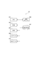
- FIG. 1 is a diagram illustrating an overall configuration of a fluorescence light receiving device 1.
- 2 is a hardware configuration diagram of a control unit 21 and a correction unit 22.
- FIG. It is a figure which illustrates multipoint scanning in this embodiment.
- 3 is a functional configuration diagram of a correction unit 22.
- FIG. It is a figure for demonstrating the sensitivity variation for every light reception position intrinsic
- the correction unit 22 performs feedback correction based on the sensitivity information for each light receiving position unique to the fluorescence receiver 40, while recognizing the location-dependent sensitivity variation for each light receiving position unique to the fluorescence receiver 40, It is a figure for demonstrating that the influence by the said sensitivity variation is reduced and the intensity
- SYMBOLS 1 Fluorescence light-receiving device, 10 ... Excitation light source, 11 ... Spatial filter, 12 ... Collimating lens, 13 ... Mirror, 20 ... Spatial light modulator, 21 ... Control part, 22 ... Correction part, 221 ... 1st hologram input part , 222 ... Photosensitive device specific sensitivity information storage unit, 223 ... second hologram generation unit, 30 ... condensing optical system, 31 ... double telecentric lens system, 32 ... dichroic mirror, 33 ... objective lens, 34 ... lens, 90 ... sample , 91 ... condensing position, 92 ... sample stage, L1 ... excitation light, L2 ... phase modulated light, L3 ... fluorescence.
- FIG. 1 is a diagram showing an overall configuration of the fluorescence light receiving device 1.
- a fluorescence light receiving device 1 shown in this figure is a device that receives fluorescence L3 generated from a sample 90 by modulating excitation light L1 and condensing modulated light L2, and includes an excitation light source 10, a spatial filter. 11, a collimating lens 12, a mirror 13, a spatial light modulator 20, a control unit 21, a correction unit 22, a condensing optical system 30, and a fluorescence receiver (fluorescence detector) 40.
- a fluorescence scanning microscope Apparatus STED microscope apparatus, PALM, STORM, and the like.
- the excitation light source 10 outputs excitation light L ⁇ b> 1 to be irradiated to the condensing position 91 of the sample 90.
- the excitation light source 10 may output laser light as the excitation light L1, and may be a pulse laser light source such as a femtosecond laser light source or an Nd: YAG laser light source.
- the excitation light source 10 may be an LD (Laser Diode), SLD (Super Luminescent Diode), halogen lamp, xenon lamp, or the like.
- the excitation light L1 output from the excitation light source 10 passes through the spatial filter 11, is collimated by the collimating lens 12, is reflected by the mirror 13, and is input to the spatial light modulator 20.
- an expander lens (not shown) may be provided instead of the combination of the spatial filter 11 and the collimating lens 12.
- the excitation light L 1 from the excitation light source 10 is spread by the expander lens, reflected by the mirror 13, and input to the spatial light modulator 20.
- the spatial light modulator 20 is of a phase modulation type, and receives the excitation light L1 and outputs the phase modulation light L2 obtained by phase modulating the excitation light L1. That is, the spatial light modulator 20 receives the excitation light L1 output from the excitation light source 10, and uses the hologram that modulates the phase of the excitation light L1 in each of a plurality of two-dimensionally arranged pixels to phase the excitation light L1. Modulate and output phase-modulated light L2 after the phase modulation.
- the hologram used in the spatial light modulator 20 may be a hologram (Computer Generated Hologram, hereinafter referred to as “CGH”) obtained by numerical calculation.
- CGH Computer Generated Hologram
- the spatial light modulator 20 may be a reflection type or a transmission type.
- any of LCOS (Liquid Crystal on Silicon) type, MEMS (Micro Electro Mechanical Systems) type, and optical address type may be used.
- an LCD (Liquid Crystal Display) type or the like may be used as the transmissive spatial light modulator 20.
- the present invention is not limited to this, and a Segment Mirror type or a Continuous Deformable Mirror type may be used.
- the spatial light modulator 20 is of an amplitude (intensity) modulation type, and inputs the excitation light L1 and outputs the modulated light L2 obtained by amplitude modulating the excitation light L1. May be. That is, the spatial light modulator 20 receives the excitation light L1 output from the excitation light source 10, and amplitudes the excitation light L1 using a hologram that modulates the amplitude of the excitation light L1 in each of a plurality of two-dimensionally arranged pixels. Modulate and output amplitude modulated light L2 after the amplitude modulation.
- the amplitude (intensity) modulation type spatial light modulator 20 may be a reflection type or a transmission type, and is a spatial light modulator such as a MEMS (MicroElectro-Mechanical-Systems) type and a LCD (Liquid-Crystal-Display) type including a DLP of Texas Instruments. Is mentioned.
- the hologram used in the spatial light modulator 20 may be a hologram (Computer Generated Hologram, hereinafter referred to as “CGH”) obtained by numerical calculation.
- CGH Computer Generated Hologram
- FIG. 1 a reflective and phase modulation spatial light modulator is exemplified as the spatial light modulator 20, and hereinafter, a case where a phase modulation spatial light modulator is used will be described.
- the control unit 21 causes the spatial light modulator 20 to express CGH to modulate the excitation light L1 in each of a plurality of two-dimensionally arranged pixels, and the modulated light L2 is used to collect a plurality of samples 90 by the condensing optical system 30.
- the light is condensed at the individual condensing positions 91.
- the control unit 21 presents CGH to the spatial light modulator 20, thereby causing excitation light L1 in each of a plurality of pixels arranged two-dimensionally.
- the phase-modulated light L2 is condensed at a plurality of condensing positions 91 of the sample 90 by the condensing optical system 30.
- the spatial light modulator 20 represents CGH by displaying CGH on the spatial light modulator 20.
- the expression that the spatial light modulator 20 expresses CGH includes that the spatial light modulator 20 presents or displays CGH.
- a drive unit (not shown) may be separately provided, and the phase modulation and light collection may be performed by the control unit 21 operating the drive unit.
- the drive unit sets the phase modulation amount for each of the plurality of two-dimensionally arranged pixels of the spatial light modulator 20 under the control using the CGH of the control unit 21, and the phase modulation amount for each pixel. Is given to the spatial light modulator 20.
- the CGH may be created by either Fourier transform type or Fresnel zone plate type.
- the CGH may be created by the control unit 21 or a separate CGH creation unit (not shown) may be provided.
- the Fourier transform type can create a hologram by an algorithm such as the GS method
- the Fresnel zone plate type can create a hologram by an algorithm such as an ORA (optimal-rotation-angle) method.
- the GS method is described in the following reference 1
- the ORA method is described in the following reference 2.
- FIG. 2 is a hardware configuration diagram of the control unit 21.
- the control unit 21 physically includes a CPU 201, a main storage device such as a ROM 202 and a RAM 203, an input device 204 such as a keyboard and a mouse, an output device 205 such as a display, a spatial light modulator 20, and the like.
- a communication module 206 such as a network card for transmitting / receiving data to / from the computer, an auxiliary storage device 207 such as a hard disk, and the like.
- Each function of the control unit 21 causes the input device 204, the output device 205, and the communication module 206 to operate under the control of the CPU 201 by reading predetermined computer software on hardware such as the CPU 201, the ROM 202, and the RAM 203. This is realized by reading and writing data in the main storage devices 202 and 203 and the auxiliary storage device 207.
- the condensing optical system 30 is provided in the subsequent stage of the spatial light modulator 20, and condenses the phase-modulated light L ⁇ b> 2 phase-modulated for each pixel on the sample 90.
- the condensing optical system 30 includes a bilateral telecentric lens system 31, a dichroic mirror 32, an objective lens 33, and a lens 34.
- the phase-modulated light L2 from the spatial light modulator 20 is transferred to the objective lens 33 by the double telecentric lens system 31 and the dichroic mirror 32, and irradiates the sample 90 placed on the sample stage 92 at multiple points.
- Fluorescence L3 is generated from the stained sample 90 by light irradiation, and a part of the fluorescence L3 enters the fluorescence receiver 40 through the objective lens 33, the dichroic mirror 32, and the lens.
- the sample 90 may be irradiated with light using a scanner such as a galvano scanner, a resonant mirror, or a polygon mirror and a stage moving in the direction of the optical axis or an objective lens. You may scan using the 3-axis stage which moves to an axial direction. Further, the objective lens 33 in FIG. 1 may move for scanning. Further, the lens for irradiating the sample 90 is not limited to the objective lens, but may be a general lens or an F ⁇ lens. With the above configuration, multipoint scanning as exemplified in FIG. 3 can be performed, and an image can be acquired at high speed.
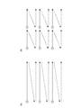
- FIG. 3 is a diagram illustrating multipoint scanning in the present embodiment.
- FIG. 3 is a diagram illustrating multipoint scanning in the present embodiment.
- FIG. 3A illustrates an example in which multiple points are arranged in one dimension
- FIG. 3B illustrates an example in which multiple points are arranged in two dimensions.
- scanning is performed using multiple points. First, scanning is performed in the horizontal direction, and then scanning is performed by moving one line in the vertical direction (raster scanning). Note that not only raster scanning but also various scanning methods using multiple points may be used.
- the fluorescence receiver 40 receives the fluorescence L3 generated when the phase-modulated light L2 is condensed on the sample 90 via the condensing optical system 30.
- the multi-anode type photoelectron It can be constituted by a multiplier tube (PMT).
- the fluorescent light receiver 40 is not limited to this, and a two-dimensional detector such as a photomultiplier tube, a two-dimensional imaging device such as a CCD or a CMOS image sensor, an avalanche photodiode array, or a photodiode array may be used.
- the fluorescence receiver 40 may have a place-dependent sensitivity variation unique to the receiver. Further, a pinhole (not shown) may be disposed in front of the fluorescence light receiving unit 40 to provide a confocal effect.
- the correction unit 22 corrects the CGH already presented to the spatial light modulator 20 by the control unit 21.
- the correction unit 22 has the same hardware configuration as that of the control unit 21 (see FIG. 2).
- the control unit 21 and the correction unit 22 may exist in the same computer system.
- FIG. 4 is a functional configuration diagram of the correction unit 22. As shown in FIG. 4, the correction unit 22 includes a first hologram input unit 221, a receiver specific sensitivity information storage unit 222, and a second hologram generation unit 223.
- the first hologram input unit 221 inputs CGH that has already been presented to the spatial light modulator 20 by the control unit 21 and that has not been corrected.
- the CGH input by the first hologram input unit 221 is the first hologram.
- the first hologram input unit 221 outputs the input first CGH to the second hologram generation unit 223.
- the light receiver specific sensitivity information storage unit 222 acquires and stores in advance sensitivity information for each light receiving position specific to the fluorescent light receiver (hereinafter, “photoreceiver specific sensitivity information”).
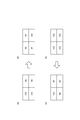
- FIG. 5 is a diagram for explaining the receiver-specific sensitivity information in the two-photon excitation fluorescence microscope.
- the fluorescent light receiver 40 has a location-dependent sensitivity variation unique to the light receiver. That is, if the fluorescence receiver 40 is, for example, a multi-anode type PMT, sensitivity variations exist among a plurality of anodes. When uniform light is incident regardless of the location of the light receiving surface as shown in FIG.
- the intensity information of the fluorescence detected by the fluorescence receiver 40 is as shown in FIG. As shown in (b). That is, even when uniform fluorescence L3 having an intensity of 100 is incident, the fluorescence L3 is detected as an intensity having different intensities of 82, 95, 97, and 100 depending on the location of the fluorescence receiver 40, for example.
- the actually entered fluorescence L3 is not uniform as shown in FIG. 5C. It may be a thing. That is, when the intensity of the fluorescence L3 detected by the fluorescence receiver 40 is uniform, for example, at an intensity of 100, the spatial light modulator 20 actually irradiates non-uniform light of, for example, the intensity of 100, 86, 84, 82. It would have been. In other words, when the intensity of the fluorescence L3 detected by the fluorescence receiver 40 is uniform, this is because the incident fluorescence L3 is uniform when the inherent location-dependent sensitivity variation of the fluorescence receiver 40 is not considered. It tends to be thought of. However, considering the inherent location-dependent sensitivity variation of the fluorescence receiver 40, the incident fluorescence L3 is actually non-uniform.
- the light receiver specific sensitivity information storage unit 222 acquires and stores in advance sensitivity information for each light receiving position specific to the fluorescent light receiver 40 when fluorescent light having a predetermined uniform intensity is incident. Specifically, the light emitted from the excitation light source 10 is spread by an expander lens or the like, and parallel light having a substantially uniform intensity is incident on the fluorescence receiver 40 that is a two-dimensional image sensor. Investigate in advance. By this investigation, when uniform light (fluorescence) is incident, the intensity of each of the multiple points obtained by the fluorescence receiver 40 is recorded as “receiver-specific sensitivity information”. In order to observe the sensitivity variation, a “receiver-specific sensitivity map” that is a bundle of receiver-specific sensitivity information may be created by changing the light intensity a plurality of times.
- the second hologram generation unit 223 performs feedback correction on the first CGH based on the excitation state, the intensity of the fluorescence L3 at the condensing position, and the receiver specific sensitivity information stored in the receiver specific sensitivity information storage unit 222.
- the second CGH is generated.
- the second hologram generation unit 223 outputs the generated second CGH to the control unit 21.
- the CGH generated by the second hologram generation unit 223 and output to the control unit 21 is the second hologram.
- the control unit 21 receives the second CGH and presents it to the spatial light modulator 20.
- the hologram presented to the spatial light modulator 20 by the control unit 21 is feedback-corrected by the correction unit 22, and the corrected hologram is presented to the spatial light modulator 20 by the control unit 21 again. This is repeated until the intensity variation of multiple points irradiated on the sample 90 becomes equal to or less than a predetermined threshold value. Or it repeats until the frequency
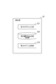
- FIG. 6 is a flowchart showing this procedure.
- the receiver specific sensitivity information storage unit 222 acquires and stores the receiver specific sensitivity information in advance when light of a predetermined uniform intensity is incident (step S1).
- the first CGH is designed under the control of the control unit 21 (step S2).
- the first CGH is presented to the spatial light modulator 20 under the control of the control unit 21, so that the spatial light modulator 20 represents the first CGH.
- the excitation light L1 becomes phase modulated light L2 having a phase distribution corresponding to the first CGH
- the generated phase modulated light L2 is irradiated to the sample 90 at multiple points (step S3).
- the fluorescence receiver 40 captures the fluorescence L3 generated by the irradiation in step S3, and acquires intensity information of the fluorescence (step S4).
- the correction unit 22 calculates the variation in the intensity information of the fluorescence L3 acquired in step S4.
- the peak-to-valley (PV) method or the RMS (Root-Mean-Square-error) method is used to calculate the intensity variation.
- the variation in the intensity of the fluorescence L3 acquired in step S4 is calculated in consideration of the receiver sensitivity information acquired and stored in advance in step S1. For example, if the intensity information of the fluorescence L3 acquired in step S4 is equal to the receiver-specific sensitivity information acquired in step S1, it can be said that the intensity variation is zero.
- whether the Peak-to-valley (PV) method or RMS (Root Mean Square) error is within a certain range. Further, a determination may be made. (Step S5)
- step S6 when the intensity variation calculated in step S5 is equal to or less than a predetermined threshold (step S6: YES), the process is terminated.
- step S6: NO when the intensity variation calculated in step S5 is not equal to or less than the predetermined threshold (step S6: NO), the process flow proceeds to step S7, and the second hologram generation unit 223 controls the fluorescence intensity to control the fluorescence intensity.
- the first CGH is redesigned to generate the second CGH.
- the fluorescence intensity is made uniform as one mode for controlling the fluorescence intensity.
- step S3 steps S3 to S6 are repeated.
- step S6 instead of determining whether or not the intensity variation calculated in step S5 is equal to or less than a predetermined threshold, it is determined whether or not the number of executions of feedback correction is equal to or greater than the predetermined threshold. May be.
- the second hologram generation unit 223 derives a correction coefficient Vk for feeding back to the CGH design from the following equation (1) based on the intensity variation of the fluorescence L3 acquired in step S4.
- m is the position of multiple points
- k is the number of feedbacks
- q is the intensity of the fluorescence L3 acquired in step S4
- n is the number of photons.
- the correction by the correction unit 22 is performed for each of the plurality of condensing positions (step S71).
- the correction coefficient Vk changes depending on the n-photon absorption process.
- the second hologram generation unit 223 redesigns the first CGH using the correction coefficient Vk derived in step S71 to generate the second CGH.
- the target pattern (a pattern indicating the position and intensity of multiple points) is changed using a weight for each iteration.
- the target pattern in 2CGH is represented by the following formula (2). Where l is the number of iterations of the OC method, Is a weight used in the OC method, Tgoal (m) is a target pattern, and s (m) is a coefficient in consideration of receiver-specific sensitivity information (step S72).
- the coefficient s (m) is not considered in the correction coefficient Vk, and the coefficient s (m) is applied during feedback.
- the CGH redesign procedure part 2 is different in that the coefficient s (m) is considered from the beginning in the correction coefficient Vk.
- the second hologram generation unit 223 derives a correction coefficient Vk for feeding back to the CGH design from the following equation (3) based on the intensity variation of the fluorescence L3 acquired in step S4. Similarly to the case of the expression (1), even when the expression (3) is used, the correction by the correction unit 22 is performed for each of the plurality of light collection positions (step S73).
- the second hologram generation unit 223 redesigns the first CGH using the correction coefficient Vk derived in step S73 to generate the second CGH.
- the target pattern (a pattern indicating the position and intensity of multiple points) is changed using a weight for each iteration.
- the target pattern in 2CGH is represented by the following formula (4) (step S74).
- the fourth root of the receiver-specific sensitivity information acquired in step S1 is obtained. This calculation is caused by the fact that the intensity of the phase-modulated light L2 is the square of the amplitude and the intensity of the fluorescence L3 is the square of the intensity of the phase-modulated light L2 in the two-photon excitation fluorescence microscope. The calculated fourth root is shown in the following equation (5) (step S731).
- step S73 the coefficient s (m) in step S72 and step S73 is finally calculated by normalizing the equation (7).
- the calculated coefficient s (m) is shown in the following equation (8) (step S734).
- CGH redesign procedure part 1 and part 2 can be executed.
- the intensity of the phase-modulated light L2 is the square of the amplitude
- the intensity of the fluorescence L3 is the n-th power of the intensity of the phase-modulated light L2.
- the intensity of the phase modulation light L2 is the square of the amplitude
- the intensity of the fluorescence L3 is the first power of the intensity of the phase modulation light L2, that is, the intensity of the fluorescence L3 is the phase modulation light. It is the same as the intensity of L2.
- the intensity of the phase modulated light L2 is the square of the amplitude
- the intensity of the fluorescence L3 is the third power of the intensity of the phase modulated light L2.
- the intensity information of the fluorescence L3 acquired in step S4 by the fluorescence receiver 40 is used instead of the receiver specific sensitivity information (for example, FIG. 5B). It can be calculated by performing the same calculation as in steps S731 to S734 described above.
- both the phase-modulated light L2 irradiated onto the sample 90 and the fluorescence L3 emitted from the sample 90 can be controlled uniformly, for example.
- the excitation light intensity is the same no matter where the light is irradiated, the same intensity is obtained.
- a homogeneous fluorescent material capable of obtaining fluorescence may be used as the sample 90.
- a material made of a homogeneous fluorescent material having no place-dependent variation in the intensity ratio of the phase-modulated light L2 focused on the sample 90 to the fluorescence L3 generated from the sample 90 is used as the sample 90 in this embodiment. May be.
- shading correction by image processing or the like may be further performed in order to uniformize the position-dependent sensitivity variation for each light receiving position unique to the fluorescence receiver 40. Thereby, the frequency
- the design method so far has been described according to the OC method, but the CGH design method is not limited to the OC method. Iterative Fourier transform method including OC method, Simulated annealing, Optimal rotation angle method (ORA method), genetic algorithm and other methods that focus on changes in one pixel, superposition method that adds together in the form of complex amplitude, etc. Can also be adopted as a CGH design method. In any of the methods listed above, the above formula (1) or formula (3) is used at the time of feedback.
- z-stack The correction by the correction unit 22 described above may be performed for each scanning layer at a predetermined interval along the optical axis direction of the objective lens 33 included in the condensing optical system 30.
- z-stack for realizing correction for each scanning layer will be described.
- z-stack refers to obtaining a three-dimensional image by capturing an image by shifting the depth direction of the sample 90.
- z-stack includes (a) a method of presenting a Fresnel lens pattern on the spatial light modulator 20, (b) a method of moving the Z stage of the fluorescence scanning microscope apparatus including the fluorescence light receiving device 1, and (c) the objective lens 33 itself. This is realized by moving the scanning layer at a predetermined interval along the optical axis direction of the objective lens 33 by the method of moving (d) and a combination thereof.
- a depth direction in the optical axis direction of the objective lens 33 is obtained by adding a Fresnel lens to the CGH for generating multiple points to be presented to the spatial light modulator 20, that is, by the method (a).
- An example in which scanning is performed by moving the scanning layer at three depths D1, D2, and D3 will be described.
- step S101 a CGH for generating multiple points is designed.
- a Fresnel lens for observing the depth D1 is added to the CGH designed in step S101 (step S102).
- an aberration correction pattern is further added to the CGH synthesized in step S102 as necessary (step S103).
- Step S104 by presenting the CGH synthesized in step S103 to the spatial light modulator 20, the spatial light modulator 20 expresses the CGH synthesized in step S103, and irradiates the generated sample with the multipoints 90.
- a fluorescent image generated from the sample 90 is picked up by a multi-anode type PMT, a camera or the like (step S105).
- an observation region centered on the center of the condensing point may be provided, and the variation in the total intensity in the observation region may be examined.
- an observation region may be provided so as to cover the entire spot spread by the aberration.
- the refractive index of the medium is n
- the depth from the incident surface of the medium to the focal point of the lens is d
- the aberration generated by the medium is ⁇ s.
- the present invention is not limited to this, and spherical aberration that occurs due to refractive index mismatch between the observation object and immersion liquid, astigmatism that occurs due to the shape of the object, etc. are measured in advance using a simulation or Shack-Hartmann sensor. Then, the aberration in a range in which these are further considered may be set as the “intentionally large aberration” (step S106).
- step S106 If the variation calculated in step S106 is within the allowable range, the process is terminated (step S107).
- step S106 If the variation calculated in step S106 is outside the allowable range, the CGH is redesigned with the CGH designed in step S101 as the initial phase. That is, the CGH designed in step S101 is set as the first CGH, and the second CGH is generated using the above-described CGH redesign procedure No. 1, No. 2 and the calculation method of the coefficient s (m) (step S108).
- Steps S104 to S108 are repeated until the variation calculated in Step S106 is within an allowable range, and an optimum CGH for the depth D1 is designed.
- step S102 and step S103 are excluded. That is, the procedure of step S102 and step S103 is performed only once for each depth (step S109).
- step S101 to step S109 change the depth to D2
- step S101 to step S109 perform the processing from step S101 to step S109. Further, the processing of steps S101 to S109 is performed by changing the depth to D3.
- the case of the above method (a) has been described as a method for performing z-stack. That is, the case where scanning for each scanning layer is performed by controlling the condensing position of the objective lens 33 in the optical axis direction by the spatial light modulator 20 has been described.
- the procedure similar to the above can be applied to the methods (b) and (c).
- the depth is changed by changing the Fresnel lens pattern, but in the case of the method (b), the Z stage is moved, and in the case of the method (c), the objective lens 33 is moved. Change depth by moving.
- the CGH in which these problems are corrected is obtained by performing feedback correction. Designed and re-presented to the spatial light modulator 20.
- the optical system is changed by optically changing the state of the medium in the condensing process. There is a risk that the uniformity of strength will be lost.
- the optimum CGH obtained at the turning angle may no longer be the optimum CGH due to the addition of the Fresnel lens pattern.
- the Fresnel lens pattern and the aberration correction pattern are added to the initially designed CGH, and the Fresnel lens pattern and the aberration correction pattern are not used for feedback correction. .
- the aberration is corrected by the aberration correction pattern, and the feedback correction is performed after the Fresnel lens pattern and the aberration correction pattern are fixed. Therefore, when the Z axis moves due to the addition of a Fresnel lens pattern or the like, the optical state of the medium and the change of the optical system occur, or in the optical system such as a high NA objective lens or a medium existing in the propagation process. Even when an aberration occurs due to a difference in refractive index, the aberration can be corrected and the uniformity of the fluorescence intensity obtained by feedback correction can be maintained.
- the first CGH expressed by the spatial light modulator 20 is first corrected by the correction unit 22, and the second CGH newly generated by the correction is converted to the spatial light modulator. 20 is expressed again. That is, the hologram is fed back by the correction unit 22 and the control unit 21 to correct the hologram.
- the second CGH is obtained by correcting the first CGH based on the excited state, the intensity of the fluorescence L3 at the light collection position, and the receiver sensitivity information, and the second CGH is generated by the spatial light modulator 20 using the second CGH.
- the intensity variation at the point is controlled uniformly, for example, in consideration of the sensitivity variation in the fluorescence receiver 40.
- the phase-modulated light L2 irradiated to the sample 90 and the fluorescence L3 irradiated from the sample 90 are controlled uniformly, for example.
- the correction by the correction unit 22 is performed based on the receiver-specific sensitivity information, it is possible to reduce the influence due to the sensitivity variation while recognizing the location-dependent sensitivity variation for each light receiving position unique to the fluorescence receiver 40. it can.
- FIG. 7 is a diagram for explaining the idea in the present embodiment from another aspect.
- FIG. 7A shows an inherent characteristic of the fluorescence receiver 40 although the incident fluorescence L3 tends to appear to be uniform as described above with reference to FIGS. 5A and 5B. Considering the sensitivity variation depending on the location, it is assumed that the incident fluorescence L3 is actually non-uniform.
- the degree of intensity is indicated by the size of a circle at the light collecting position 91 of the sample 90 and the light receiving position 41 of the fluorescent light receiver 40. That is, the size of the circle is different at the condensing position 91 of the sample 90, which indicates that the light intensity at the condensing position 91 is not uniform.
- the size of the circle is the same at the light receiving position 41 of the fluorescent light receiver 40, which indicates that the light intensity appears to be uniform at the light receiving position 41.
- the intensity of the fluorescence detected by the fluorescence receiver 40 is not uniform, but is incident on the sample 90. It is assumed that the intensity of the phase-modulated light L2 and the fluorescence L3 is uniform. This is because the correction unit 22 of the present embodiment performs feedback correction based on the receiver-specific sensitivity information, and recognizes the location-dependent sensitivity variation for each light receiving position specific to the fluorescence receiver 40, but the influence of the sensitivity variation. This means that the intensities of the phase-modulated light L2 and the fluorescence L3 are made uniform.
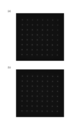
- FIG. 8A shows a fluorescence image before feedback correction is performed.
- the fluorescence intensity in the upper right part is lower than that in the center part, and although it is difficult to visually confirm, there is an intensity variation depending on the location in the vicinity of the center.
- the intensity of fluorescence is shown by brightness.
- FIG. 8B shows a fluorescence image subjected to feedback correction according to the present embodiment. Unlike FIG. 8A, it can be seen that the intensity variation depending on the location is reduced.
- FIG. 8 shows the intensity of the fluorescence detected at each light receiving position in consideration of the receiver specific sensitivity information.
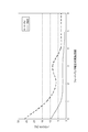
- FIG. 9 is a diagram illustrating a relationship between the number of times feedback correction is performed (horizontal axis) and the variation in fluorescence intensity (vertical axis) according to the present embodiment.
- the peak-to-valley (PV) method or the RMS (Root Mean Square error) method was used as the evaluation function for variation.
- the calculated variations in the PV method and the RMS method can be represented by the following formulas (9) and (10), respectively.
- q (m) is the total fluorescence in the m-th fluorescence observation region
- qmax, qmax, and qdesired are the maximum, minimum, and desired fluorescence intensity, respectively.
- the sample 90 is placed and the fluorescence L3 is detected.
- the merit of detecting the fluorescence by placing the sample 90 is as follows. That is, once correction is performed using a medium such as a rACGFP solution or coumarin ethanol that can obtain a uniform fluorescence intensity as the sample 90, uniform data can be obtained even when cells or the like are used as the sample thereafter.
- the second advantage As a second merit, it is possible to correct the entire optical system because an optical system that is actually measured can be used by using a sample.
- the third merit is that by preparing a homogeneous medium having a refractive index close to the observation condition, it is possible to make uniform with spherical aberration correction and a lens pattern applied.
- the refractive index of the sample 90 and the observation medium are extremely different, the condensed image is blurred, and the SN is lowered, so that there is a possibility that the accuracy of homogenization is lowered. Therefore, the refractive index of the sample 90 may be adjusted to some extent with the refractive index of the observation medium.
- the OC method is an optimization method, and a random phase is often used as the initial phase to prevent falling into a local solution.
- the distribution of the generated multi-point variation differs for each first hologram. Or, even if the same initial phase is used, if the positions and intensities of multiple points in the target pattern change, the distribution of the multi-point variation generated by the first hologram differs.
- the fourth merit is that the variation caused by the hologram can be clearly grasped by using a medium 90 with uniform fluorescence intensity as the sample 90.
- amendment can be applied to each of the many points produced
- the present invention is particularly useful for intensity variations at a plurality of condensing positions that occur when a short pulse laser capable of performing multiphoton excitation is used as the excitation light source 10.
- a short pulse laser capable of performing multiphoton excitation is used as the excitation light source 10.
- a light source having a pulse width of nanoseconds or less and capable of emitting a short pulse laser having a wavelength of 650 nm or more is preferable.
- the present embodiment is particularly useful for a fluorescence receiver 40 having a location-dependent sensitivity variation such as a multi-anode type photomultiplier tube.
- the intensity of both the phase-modulated light L2 to the sample 90 and the fluorescence L3 from the sample 90 is uniformly controlled, for example, a homogeneous fluorescent material having no location-dependent variation is used as the sample 90.
- a homogeneous fluorescent material having no location-dependent variation is used as the sample 90.
- the correction by the correction unit 22 is performed for each scanning layer at a predetermined interval along the optical axis direction of the objective lens 33 included in the condensing optical system 30, so that a certain amount of depth is obtained.
- the present embodiment can be applied to a sample 90 having a thickness.
- the spatial light modulator 20 performs scanning for each scanning layer by controlling the condensing position of the objective lens 33 in the optical axis direction, scanning for each scanning layer is preferably performed. Can do.
- a calibration sample having a refractive index close to that of an actual measurement sample and having a uniform concentration of a solute such as a fluorescent substance may be used as the sample.
- a solute such as a fluorescent substance
- the present invention provides a fluorescent light receiving device and a fluorescent light receiving method capable of controlling the intensity of multi-point fluorescence at a condensing position.
Landscapes
- Physics & Mathematics (AREA)
- General Physics & Mathematics (AREA)
- Chemical & Material Sciences (AREA)
- Analytical Chemistry (AREA)
- Health & Medical Sciences (AREA)
- Optics & Photonics (AREA)
- Biochemistry (AREA)
- Life Sciences & Earth Sciences (AREA)
- Nuclear Medicine, Radiotherapy & Molecular Imaging (AREA)
- General Health & Medical Sciences (AREA)
- Immunology (AREA)
- Pathology (AREA)
- Engineering & Computer Science (AREA)
- Computer Vision & Pattern Recognition (AREA)
- General Engineering & Computer Science (AREA)
- Investigating, Analyzing Materials By Fluorescence Or Luminescence (AREA)
- Microscoopes, Condenser (AREA)
- Holo Graphy (AREA)
Abstract
Description
最初に、本実施形態に係る蛍光受光装置(蛍光検出装置)1の全体構成について説明する。図1は、蛍光受光装置1の全体的な構成を示す図である。この図に示される蛍光受光装置1は、励起光L1が変調され、変調光L2が集光されることにより試料90から発生する蛍光L3を受光する装置であって、励起光源10、スペイシャルフィルタ11、コリメートレンズ12、ミラー13、空間光変調器20、制御部21、補正部22、集光光学系30、および蛍光受光器(蛍光検出器)40を備えて構成さ、例えば、蛍光走査顕微鏡装置やSTED顕微鏡装置、PALM、STORMなどである。
<参考文献1> R. W. Gerchberg and W. O.Saxton, "Apractical algorithm for the determination of phase from imageand diffractionplane pictures", Optik, Vol.35, pp.237-246 (1972).
<参考文献2> Jorgen Bengtsson, "Kinoform designwith anoptimal-rotation-angle method", Applied Optics, Vol.33,No.29,pp.6879-6884 (1994).
続いて、第1ホログラムがフィードバック補正され、第2ホログラムが生成される際の手順について詳細に説明する。図6は、この手順を示すフローチャートである。
ここで、第1CGHを再設計して第2CGHを生成する方法(ステップS7)について説明する。

続いて、第1CGHを再設計して第2CGHを生成する別の方法(ステップS7)について説明する。

以下、ステップS72およびステップS73における2光子励起蛍光顕微鏡における係数s(m)の算出方法について詳細に説明する。以下の説明においては、ステップS1で取得した受光器固有感度情報が例えば図5(b)だったものとする。


以上で説明した補正部22による補正は、集光光学系30に含まれた対物レンズ33の光軸方向に沿った所定の間隔の走査層ごとに行われても良い。以下では、走査層ごとの補正を実現するためのz-stackについて説明する。z-stackとは、試料90の奥行き方向をずらして画像を撮像することで、3次元画像を得ることをいう。z-stackは、(a)空間光変調器20にフレネルレンズパターンを呈示する方法、(b)蛍光受光装置1を含む蛍光走査顕微鏡装置のZステージを移動させる方法、(c)対物レンズ33そのものを移動させる方法、(d)これらの組み合わせ、によって、対物レンズ33の光軸方向に沿った所定の間隔で走査層を移動させることにより実現される。

Claims (8)
- 位相変調された励起光が集光されることにより試料から発生する蛍光を受光する蛍光受光装置であって、
前記励起光を出力する励起光源と、
前記励起光を入力し、前記励起光を位相変調することにより得られた位相変調光を出力する位相変調型の空間光変調器と、
前記空間光変調器の後段に設けられ、前記位相変調光を前記試料に集光する集光光学系と、
前記試料が載置される試料ステージと、
前記位相変調光が前記試料に集光されることにより発生する蛍光を前記集光光学系を介して受光する蛍光受光器と、
前記空間光変調器に第1ホログラムを表示することにより、2次元配列された複数の画素それぞれにおいて前記励起光の位相を変調させ、且つ前記位相変調光を前記集光光学系により前記試料の複数個の集光位置に集光させる制御部と、
前記第1ホログラムを補正する補正部と、
を備え、
前記補正部は、
前記第1ホログラムを入力する第1ホログラム入力部と、
前記蛍光受光器固有の受光位置ごとの感度情報を予め取得し格納する受光器固有感度情報格納部と、
前記集光位置における前記蛍光の強度および前記受光器固有感度情報格納部に格納された前記感度情報に基づき、前記第1ホログラムを補正することにより、第2ホログラムを生成する第2ホログラム生成部と、
を備え、
前記制御部は前記空間光変調器に前記第2ホログラムを表示する、蛍光受光装置。 - 前記補正部による前記補正は、前記複数個の集光位置ごとに行われる、請求項1に記載の蛍光受光装置。
- 前記励起光源は、多光子励起を行うことができる短パルスレーザである、請求項1または2に記載の蛍光受光装置。
- 前記蛍光受光器はマルチアノード型の光電子増倍管である、請求項1~3の何れか1項に記載の蛍光受光装置。
- 前記試料は、前記試料に集光された前記位相変調光対前記試料から発生する前記蛍光の強度の比率において、場所依存のバラツキのない均質な蛍光材料からなる、請求項1~4の何れか1項に記載の蛍光受光装置。
- 前記補正部による前記補正は、前記集光光学系に含まれた対物レンズの光軸方向に沿った所定の間隔の走査層ごとに行われる、請求項1~5の何れか1項に記載の蛍光受光装置。
- 前記走査層ごとの走査は、前記空間光変調器によって、前記対物レンズの前記光軸方向における集光位置が制御されることにより行われる、請求項6に記載の蛍光受光装置。
- 位相変調された励起光が集光されることにより、試料ステージに載置された試料から発生する蛍光を受光する蛍光受光装置における蛍光受光方法であって、
励起光源が、前記励起光を出力するステップと、
位相変調型の空間光変調器が、前記励起光を入力し、前記励起光を位相変調することにより得られた位相変調光を出力するステップと、
前記空間光変調器の後段に設けられた集光光学系が、前記位相変調光を前記試料に集光するステップと、
蛍光受光器が、前記位相変調光が前記試料に集光されることにより発生する蛍光を前記集光光学系を介して受光するステップと、
制御部が、前記空間光変調器に第1ホログラムを表示することにより、2次元配列された複数の画素それぞれにおいて前記励起光の位相を変調させ、且つ前記位相変調光を前記集光光学系により前記試料の複数個の集光位置に集光させるステップと、
補正部が、前記第1ホログラムを補正するステップと、
を備え、
前記補正部が前記第1ホログラムを補正するステップは、
第1ホログラム入力部が、前記第1ホログラムを入力するステップと、
受光器固有感度情報格納部が、前記蛍光受光器固有の受光位置ごとの感度情報を予め取得し格納するステップと、
第2ホログラム生成部が、前記集光位置における前記蛍光の強度および前記受光器固有感度情報格納部に格納された前記感度情報に基づき、前記第1ホログラムを補正することにより、第2ホログラムを生成するステップと、
を備え、
前記制御部が、前記空間光変調器に前記第2ホログラムを表示するステップを更に備える、蛍光受光方法。
Priority Applications (4)
| Application Number | Priority Date | Filing Date | Title |
|---|---|---|---|
| US14/772,570 US9740166B2 (en) | 2013-03-06 | 2014-03-04 | Fluorescence receiving apparatus and fluorescence receiving method |
| DE112014001147.9T DE112014001147T5 (de) | 2013-03-06 | 2014-03-04 | Fluoreszenz-Empfangsvorrichtung und Verfahren zum Empfangen von Fluoreszenz |
| JP2015504332A JP6276749B2 (ja) | 2013-03-06 | 2014-03-04 | 蛍光受光装置および蛍光受光方法 |
| CN201480012124.XA CN105026916B (zh) | 2013-03-06 | 2014-03-04 | 荧光受光装置以及荧光受光方法 |
Applications Claiming Priority (2)
| Application Number | Priority Date | Filing Date | Title |
|---|---|---|---|
| JP2013-044348 | 2013-03-06 | ||
| JP2013044348 | 2013-03-06 |
Publications (1)
| Publication Number | Publication Date |
|---|---|
| WO2014136784A1 true WO2014136784A1 (ja) | 2014-09-12 |
Family
ID=51491299
Family Applications (1)
| Application Number | Title | Priority Date | Filing Date |
|---|---|---|---|
| PCT/JP2014/055491 WO2014136784A1 (ja) | 2013-03-06 | 2014-03-04 | 蛍光受光装置および蛍光受光方法 |
Country Status (5)
| Country | Link |
|---|---|
| US (1) | US9740166B2 (ja) |
| JP (1) | JP6276749B2 (ja) |
| CN (1) | CN105026916B (ja) |
| DE (1) | DE112014001147T5 (ja) |
| WO (1) | WO2014136784A1 (ja) |
Cited By (8)
| Publication number | Priority date | Publication date | Assignee | Title |
|---|---|---|---|---|
| JP2016133636A (ja) * | 2015-01-20 | 2016-07-25 | 浜松ホトニクス株式会社 | 画像取得装置および画像取得方法 |
| JP2016133637A (ja) * | 2015-01-20 | 2016-07-25 | 浜松ホトニクス株式会社 | 画像取得装置および画像取得方法 |
| JP2016176877A (ja) * | 2015-03-23 | 2016-10-06 | 横河電機株式会社 | 光学式センサおよび光学式センサを用いた測定方法 |
| WO2016185974A1 (ja) * | 2015-05-15 | 2016-11-24 | 浜松ホトニクス株式会社 | 変調パターン算出装置、光制御装置、変調パターン算出方法、変調パターン算出プログラムおよび記憶媒体 |
| WO2016185979A1 (ja) * | 2015-05-15 | 2016-11-24 | 浜松ホトニクス株式会社 | 変調パターン算出装置、光制御装置、変調パターン算出方法、変調パターン算出プログラムおよび記憶媒体 |
| JP2018008040A (ja) * | 2016-07-05 | 2018-01-18 | キヤノン株式会社 | 波面制御装置、波面制御方法、情報取得装置、プログラム、および、記憶媒体 |
| CN107941763A (zh) * | 2017-10-27 | 2018-04-20 | 浙江大学 | 一种共轴三维受激辐射损耗超分辨显微成像方法和装置 |
| JP2022524727A (ja) * | 2019-02-22 | 2022-05-10 | プレジデント アンド フェローズ オブ ハーバード カレッジ | 位相空間光変調器を用いた大スケールの一様な光学的焦点アレイ生成 |
Families Citing this family (10)
| Publication number | Priority date | Publication date | Assignee | Title |
|---|---|---|---|---|
| US10595712B2 (en) * | 2013-10-21 | 2020-03-24 | University of Pittsburgh—of the Commonwealth System of Higher Education | Adaptive optical laser beam steering through endoscope for laser surgery in kidney |
| JP6630120B2 (ja) * | 2015-11-06 | 2020-01-15 | 浜松ホトニクス株式会社 | 画像取得装置、画像取得方法、及び空間光変調ユニット |
| FI20165148A (fi) * | 2016-02-25 | 2017-08-26 | Arcdia Int Oy Ltd | Kaksoisfotoniviritteistä fluoresenssia hyödyntävä bioaffiniteettimääritysmenetelmä |
| JP6722883B2 (ja) * | 2016-04-01 | 2020-07-15 | 国立大学法人浜松医科大学 | 画像取得装置および画像取得方法 |
| CN105929526B (zh) * | 2016-06-27 | 2018-08-21 | 广东欧谱曼迪科技有限公司 | 一种体全息还原扫描光束的sted超分辨显微系统及显微方法 |
| CN107301458B (zh) * | 2017-05-31 | 2020-08-11 | 重庆理工大学 | 基于强度传输方程的相位优化方法 |
| CN108132543B (zh) * | 2017-12-23 | 2020-06-12 | 深圳大学 | 超分辨成像系统 |
| DE102019110157B4 (de) | 2019-04-17 | 2021-06-17 | Leica Microsystems Cms Gmbh | Fluoreszenz-Rastermikroskop und Verfahren zur Abbildung einer Probe |
| DE102020113998A1 (de) | 2020-05-26 | 2021-12-02 | Abberior Instruments Gmbh | Verfahren, Computerprogramm und Vorrichtung zum Bestimmen von Positionen von Molekülen in einer Probe |
| DE102020134797B3 (de) | 2020-12-23 | 2022-06-09 | Abberior Instruments Gmbh | Verfahren zum Abbilden einer interessierenden Struktur einer Probe und Mikroskop mit Array-Detektor zu dessen Durchführung |
Citations (5)
| Publication number | Priority date | Publication date | Assignee | Title |
|---|---|---|---|---|
| JPH10186283A (ja) * | 1996-11-01 | 1998-07-14 | Hamamatsu Photonics Kk | 画像形成装置 |
| WO2004017069A1 (ja) * | 2002-08-16 | 2004-02-26 | Kabushiki Kaisha Hayashi Soken | バイオチップ分析装置およびオンライン分析システム |
| JP2005292662A (ja) * | 2004-04-02 | 2005-10-20 | Hamamatsu Photonics Kk | 波面補償装置、波面補償方法、プログラム、及び、記録媒体 |
| JP2009300589A (ja) * | 2008-06-11 | 2009-12-24 | Nikon Corp | 顕微鏡装置 |
| JP2011128572A (ja) * | 2009-12-21 | 2011-06-30 | Olympus Corp | ホログラム像投影方法およびホログラム像投影装置 |
Family Cites Families (5)
| Publication number | Priority date | Publication date | Assignee | Title |
|---|---|---|---|---|
| WO2008061259A2 (en) * | 2006-11-17 | 2008-05-22 | Celloptic, Inc. | System, apparatus and method for extracting three-dimensional information of an object from received electromagnetic radiation |
| EP2492921A1 (en) | 2007-01-26 | 2012-08-29 | New York University | Holographic microscopy of holographically trapped three-dimensional structures |
| DE102007018048A1 (de) | 2007-04-13 | 2008-10-16 | Michael Schwertner | Verfahren und Anordnung zur optischen Abbildung mit Tiefendiskriminierung |
| JP5259154B2 (ja) | 2007-10-24 | 2013-08-07 | オリンパス株式会社 | 走査型レーザ顕微鏡 |
| US20110267663A1 (en) | 2009-12-21 | 2011-11-03 | Olympus Corporation | Holographic image projection method and holographic image projection system |
-
2014
- 2014-03-04 DE DE112014001147.9T patent/DE112014001147T5/de active Granted
- 2014-03-04 JP JP2015504332A patent/JP6276749B2/ja active Active
- 2014-03-04 WO PCT/JP2014/055491 patent/WO2014136784A1/ja active Application Filing
- 2014-03-04 US US14/772,570 patent/US9740166B2/en active Active
- 2014-03-04 CN CN201480012124.XA patent/CN105026916B/zh active Active
Patent Citations (5)
| Publication number | Priority date | Publication date | Assignee | Title |
|---|---|---|---|---|
| JPH10186283A (ja) * | 1996-11-01 | 1998-07-14 | Hamamatsu Photonics Kk | 画像形成装置 |
| WO2004017069A1 (ja) * | 2002-08-16 | 2004-02-26 | Kabushiki Kaisha Hayashi Soken | バイオチップ分析装置およびオンライン分析システム |
| JP2005292662A (ja) * | 2004-04-02 | 2005-10-20 | Hamamatsu Photonics Kk | 波面補償装置、波面補償方法、プログラム、及び、記録媒体 |
| JP2009300589A (ja) * | 2008-06-11 | 2009-12-24 | Nikon Corp | 顕微鏡装置 |
| JP2011128572A (ja) * | 2009-12-21 | 2011-06-30 | Olympus Corp | ホログラム像投影方法およびホログラム像投影装置 |
Non-Patent Citations (2)
| Title |
|---|
| NIKOLENKO ET AL.: "SLM microscopy: scanless two-photon imaging and photostimulation with spatial light modulators", FRONTIERS IN NEURAL CIRCUITS, vol. 2, no. 5, 19 December 2008 (2008-12-19), pages 1 - 14, XP055146495, DOI: doi:10.3389/neuro.04.005.2008 * |
| SHAO Y ET AL.: "Ultrafast, large-field multiphoton microscopy based on anacousto-optic deflector and a spatial light modulator", OPTICS LETTERS, vol. 37, no. 13, 1 July 2012 (2012-07-01), pages 2532 - 2534, XP001576832, DOI: doi:10.1364/OL.37.002532 * |
Cited By (21)
| Publication number | Priority date | Publication date | Assignee | Title |
|---|---|---|---|---|
| CN107209359A (zh) * | 2015-01-20 | 2017-09-26 | 浜松光子学株式会社 | 图像取得装置以及图像取得方法 |
| JP2016133637A (ja) * | 2015-01-20 | 2016-07-25 | 浜松ホトニクス株式会社 | 画像取得装置および画像取得方法 |
| WO2016117415A1 (ja) * | 2015-01-20 | 2016-07-28 | 浜松ホトニクス株式会社 | 画像取得装置および画像取得方法 |
| WO2016117408A1 (ja) * | 2015-01-20 | 2016-07-28 | 浜松ホトニクス株式会社 | 画像取得装置および画像取得方法 |
| JP2016133636A (ja) * | 2015-01-20 | 2016-07-25 | 浜松ホトニクス株式会社 | 画像取得装置および画像取得方法 |
| US10816472B2 (en) | 2015-01-20 | 2020-10-27 | Hamamatsu Photonics K.K. | Image acquisition device and image acquisition method |
| CN107209359B (zh) * | 2015-01-20 | 2020-05-22 | 浜松光子学株式会社 | 图像取得装置以及图像取得方法 |
| US10488640B2 (en) | 2015-01-20 | 2019-11-26 | Hamamatsu Photonics K.K. | Image acquisition device and image acquisition method |
| JP2016176877A (ja) * | 2015-03-23 | 2016-10-06 | 横河電機株式会社 | 光学式センサおよび光学式センサを用いた測定方法 |
| WO2016185974A1 (ja) * | 2015-05-15 | 2016-11-24 | 浜松ホトニクス株式会社 | 変調パターン算出装置、光制御装置、変調パターン算出方法、変調パターン算出プログラムおよび記憶媒体 |
| EP3296804A4 (en) * | 2015-05-15 | 2018-12-26 | Hamamatsu Photonics K.K. | Modulation pattern calculation device, light control device, modulation pattern calculation method, modulation pattern calculation program, and storage medium |
| EP3296803A4 (en) * | 2015-05-15 | 2018-12-26 | Hamamatsu Photonics K.K. | Modulation pattern calculation device, light control device, modulation pattern calculation method, modulation pattern calculation program, and storage medium |
| US10218141B2 (en) | 2015-05-15 | 2019-02-26 | Hamamatsu Photonics K.K. | Modulation pattern calculation device, light control device, modulation pattern calculation method, modulation pattern calculation program, and storage medium |
| JP2016218142A (ja) * | 2015-05-15 | 2016-12-22 | 浜松ホトニクス株式会社 | 変調パターン算出装置、光制御装置、変調パターン算出方法および変調パターン算出プログラム |
| JP2016218141A (ja) * | 2015-05-15 | 2016-12-22 | 浜松ホトニクス株式会社 | 変調パターン算出装置、光制御装置、変調パターン算出方法および変調パターン算出プログラム |
| WO2016185979A1 (ja) * | 2015-05-15 | 2016-11-24 | 浜松ホトニクス株式会社 | 変調パターン算出装置、光制御装置、変調パターン算出方法、変調パターン算出プログラムおよび記憶媒体 |
| US10989935B2 (en) | 2015-05-15 | 2021-04-27 | Hamamatsu Photonics K.K. | Modulation pattern calculation device, light control device, modulation pattern calculation method, modulation pattern calculation program, and storage medium |
| JP2018008040A (ja) * | 2016-07-05 | 2018-01-18 | キヤノン株式会社 | 波面制御装置、波面制御方法、情報取得装置、プログラム、および、記憶媒体 |
| CN107941763A (zh) * | 2017-10-27 | 2018-04-20 | 浙江大学 | 一种共轴三维受激辐射损耗超分辨显微成像方法和装置 |
| JP2022524727A (ja) * | 2019-02-22 | 2022-05-10 | プレジデント アンド フェローズ オブ ハーバード カレッジ | 位相空間光変調器を用いた大スケールの一様な光学的焦点アレイ生成 |
| JP7410959B2 (ja) | 2019-02-22 | 2024-01-10 | プレジデント アンド フェローズ オブ ハーバード カレッジ | 位相空間光変調器を用いた大スケールの一様な光学的焦点アレイ生成 |
Also Published As
| Publication number | Publication date |
|---|---|
| US9740166B2 (en) | 2017-08-22 |
| DE112014001147T5 (de) | 2015-11-19 |
| JP6276749B2 (ja) | 2018-02-07 |
| CN105026916A (zh) | 2015-11-04 |
| JPWO2014136784A1 (ja) | 2017-02-16 |
| US20160018786A1 (en) | 2016-01-21 |
| CN105026916B (zh) | 2017-07-14 |
Similar Documents
| Publication | Publication Date | Title |
|---|---|---|
| JP6276749B2 (ja) | 蛍光受光装置および蛍光受光方法 | |
| Quirin et al. | Instantaneous three-dimensional sensing using spatial light modulator illumination with extended depth of field imaging | |
| JP5802110B2 (ja) | 光変調制御方法、制御プログラム、制御装置、及びレーザ光照射装置 | |
| JP5802109B2 (ja) | 光変調制御方法、制御プログラム、制御装置、及びレーザ光照射装置 | |
| Paudel et al. | Axial range of conjugate adaptive optics in two-photon microscopy | |
| US10488640B2 (en) | Image acquisition device and image acquisition method | |
| JP6635125B2 (ja) | 顕微鏡、観察方法、及び画像処理プログラム | |
| CN109073873B (zh) | 图像取得装置以及图像取得方法 | |
| JP7033123B2 (ja) | 収差補正方法及び光学装置 | |
| US10168283B2 (en) | Observation apparatus and method for sharpening final image | |
| CN107209359B (zh) | 图像取得装置以及图像取得方法 | |
| JP2017058386A (ja) | 顕微鏡装置および観察方法 | |
| US20240019677A1 (en) | Microscope, observation method, and program | |
| US20230069794A1 (en) | Dual-mode restoration microscopy | |
| Matsumoto et al. | Modulation of the pupil function of microscope objective lens for multifocal multi-photon microscopy using a spatial light modulator |
Legal Events
| Date | Code | Title | Description |
|---|---|---|---|
| WWE | Wipo information: entry into national phase |
Ref document number: 201480012124.X Country of ref document: CN |
|
| 121 | Ep: the epo has been informed by wipo that ep was designated in this application |
Ref document number: 14761254 Country of ref document: EP Kind code of ref document: A1 |
|
| ENP | Entry into the national phase |
Ref document number: 2015504332 Country of ref document: JP Kind code of ref document: A |
|
| WWE | Wipo information: entry into national phase |
Ref document number: 14772570 Country of ref document: US |
|
| WWE | Wipo information: entry into national phase |
Ref document number: 1120140011479 Country of ref document: DE Ref document number: 112014001147 Country of ref document: DE |
|
| 122 | Ep: pct application non-entry in european phase |
Ref document number: 14761254 Country of ref document: EP Kind code of ref document: A1 |