WO2014010323A1 - 膜状結合組織体形成用基材及びこれを用いた膜状結合組織体の生産方法 - Google Patents
膜状結合組織体形成用基材及びこれを用いた膜状結合組織体の生産方法 Download PDFInfo
- Publication number
- WO2014010323A1 WO2014010323A1 PCT/JP2013/064999 JP2013064999W WO2014010323A1 WO 2014010323 A1 WO2014010323 A1 WO 2014010323A1 JP 2013064999 W JP2013064999 W JP 2013064999W WO 2014010323 A1 WO2014010323 A1 WO 2014010323A1
- Authority
- WO
- WIPO (PCT)
- Prior art keywords
- connective tissue
- substrate
- forming
- membranous
- membrane
- Prior art date
Links
Images


Classifications
-
- A—HUMAN NECESSITIES
- A61—MEDICAL OR VETERINARY SCIENCE; HYGIENE
- A61M—DEVICES FOR INTRODUCING MEDIA INTO, OR ONTO, THE BODY; DEVICES FOR TRANSDUCING BODY MEDIA OR FOR TAKING MEDIA FROM THE BODY; DEVICES FOR PRODUCING OR ENDING SLEEP OR STUPOR
- A61M27/00—Drainage appliance for wounds or the like, i.e. wound drains, implanted drains
-
- A—HUMAN NECESSITIES
- A61—MEDICAL OR VETERINARY SCIENCE; HYGIENE
- A61F—FILTERS IMPLANTABLE INTO BLOOD VESSELS; PROSTHESES; DEVICES PROVIDING PATENCY TO, OR PREVENTING COLLAPSING OF, TUBULAR STRUCTURES OF THE BODY, e.g. STENTS; ORTHOPAEDIC, NURSING OR CONTRACEPTIVE DEVICES; FOMENTATION; TREATMENT OR PROTECTION OF EYES OR EARS; BANDAGES, DRESSINGS OR ABSORBENT PADS; FIRST-AID KITS
- A61F2/00—Filters implantable into blood vessels; Prostheses, i.e. artificial substitutes or replacements for parts of the body; Appliances for connecting them with the body; Devices providing patency to, or preventing collapsing of, tubular structures of the body, e.g. stents
- A61F2/02—Prostheses implantable into the body
- A61F2/14—Eye parts, e.g. lenses, corneal implants; Implanting instruments specially adapted therefor; Artificial eyes
- A61F2/142—Cornea, e.g. artificial corneae, keratoprostheses or corneal implants for repair of defective corneal tissue
-
- A—HUMAN NECESSITIES
- A61—MEDICAL OR VETERINARY SCIENCE; HYGIENE
- A61F—FILTERS IMPLANTABLE INTO BLOOD VESSELS; PROSTHESES; DEVICES PROVIDING PATENCY TO, OR PREVENTING COLLAPSING OF, TUBULAR STRUCTURES OF THE BODY, e.g. STENTS; ORTHOPAEDIC, NURSING OR CONTRACEPTIVE DEVICES; FOMENTATION; TREATMENT OR PROTECTION OF EYES OR EARS; BANDAGES, DRESSINGS OR ABSORBENT PADS; FIRST-AID KITS
- A61F2/00—Filters implantable into blood vessels; Prostheses, i.e. artificial substitutes or replacements for parts of the body; Appliances for connecting them with the body; Devices providing patency to, or preventing collapsing of, tubular structures of the body, e.g. stents
- A61F2/02—Prostheses implantable into the body
- A61F2/14—Eye parts, e.g. lenses, corneal implants; Implanting instruments specially adapted therefor; Artificial eyes
-
- A—HUMAN NECESSITIES
- A61—MEDICAL OR VETERINARY SCIENCE; HYGIENE
- A61L—METHODS OR APPARATUS FOR STERILISING MATERIALS OR OBJECTS IN GENERAL; DISINFECTION, STERILISATION OR DEODORISATION OF AIR; CHEMICAL ASPECTS OF BANDAGES, DRESSINGS, ABSORBENT PADS OR SURGICAL ARTICLES; MATERIALS FOR BANDAGES, DRESSINGS, ABSORBENT PADS OR SURGICAL ARTICLES
- A61L27/00—Materials for grafts or prostheses or for coating grafts or prostheses
- A61L27/36—Materials for grafts or prostheses or for coating grafts or prostheses containing ingredients of undetermined constitution or reaction products thereof, e.g. transplant tissue, natural bone, extracellular matrix
- A61L27/3641—Materials for grafts or prostheses or for coating grafts or prostheses containing ingredients of undetermined constitution or reaction products thereof, e.g. transplant tissue, natural bone, extracellular matrix characterised by the site of application in the body
- A61L27/3645—Connective tissue
-
- A—HUMAN NECESSITIES
- A61—MEDICAL OR VETERINARY SCIENCE; HYGIENE
- A61L—METHODS OR APPARATUS FOR STERILISING MATERIALS OR OBJECTS IN GENERAL; DISINFECTION, STERILISATION OR DEODORISATION OF AIR; CHEMICAL ASPECTS OF BANDAGES, DRESSINGS, ABSORBENT PADS OR SURGICAL ARTICLES; MATERIALS FOR BANDAGES, DRESSINGS, ABSORBENT PADS OR SURGICAL ARTICLES
- A61L27/00—Materials for grafts or prostheses or for coating grafts or prostheses
- A61L27/36—Materials for grafts or prostheses or for coating grafts or prostheses containing ingredients of undetermined constitution or reaction products thereof, e.g. transplant tissue, natural bone, extracellular matrix
- A61L27/38—Materials for grafts or prostheses or for coating grafts or prostheses containing ingredients of undetermined constitution or reaction products thereof, e.g. transplant tissue, natural bone, extracellular matrix containing added animal cells
- A61L27/3804—Materials for grafts or prostheses or for coating grafts or prostheses containing ingredients of undetermined constitution or reaction products thereof, e.g. transplant tissue, natural bone, extracellular matrix containing added animal cells characterised by specific cells or progenitors thereof, e.g. fibroblasts, connective tissue cells, kidney cells
-
- A—HUMAN NECESSITIES
- A61—MEDICAL OR VETERINARY SCIENCE; HYGIENE
- A61L—METHODS OR APPARATUS FOR STERILISING MATERIALS OR OBJECTS IN GENERAL; DISINFECTION, STERILISATION OR DEODORISATION OF AIR; CHEMICAL ASPECTS OF BANDAGES, DRESSINGS, ABSORBENT PADS OR SURGICAL ARTICLES; MATERIALS FOR BANDAGES, DRESSINGS, ABSORBENT PADS OR SURGICAL ARTICLES
- A61L2430/00—Materials or treatment for tissue regeneration
- A61L2430/16—Materials or treatment for tissue regeneration for reconstruction of eye parts, e.g. intraocular lens, cornea
-
- A—HUMAN NECESSITIES
- A61—MEDICAL OR VETERINARY SCIENCE; HYGIENE
- A61L—METHODS OR APPARATUS FOR STERILISING MATERIALS OR OBJECTS IN GENERAL; DISINFECTION, STERILISATION OR DEODORISATION OF AIR; CHEMICAL ASPECTS OF BANDAGES, DRESSINGS, ABSORBENT PADS OR SURGICAL ARTICLES; MATERIALS FOR BANDAGES, DRESSINGS, ABSORBENT PADS OR SURGICAL ARTICLES
- A61L2430/00—Materials or treatment for tissue regeneration
- A61L2430/20—Materials or treatment for tissue regeneration for reconstruction of the heart, e.g. heart valves
Definitions
- the present invention relates to a substrate for forming a membranous connective tissue for forming a membranous connective tissue such as an artificial cornea and a method for producing a membranous connective tissue using the same.
- the body has a self-defense function, and when a foreign object such as a thorn enters a shallow position in the body, it tries to push it out of the body.
- a foreign object when a foreign object enters a deep position in the body, fibroblasts gradually gather around it to form a connective tissue capsule mainly composed of fibroblasts and collagen to cover the foreign object It is known to isolate foreign substances in the body.
- a technique for forming a living body-derived tissue from living cells using the latter self-defense reaction a plurality of techniques for forming a connective tissue body by embedding a base material as a foreign substance in the living body have been reported (patent) Reference 1 to 3).
- a connective tissue body having a desired shape and size is formed by appropriately setting the shape and size of a base material as a foreign substance to be embedded in a living body. Can do.
- a connective tissue body is formed around a base material embedded in a living body, the shape and size of the base material are appropriately set, and a connective tissue body having a desired shape and size is formed around the base body.
- the connective tissue body cannot be set to a desired thickness and strength, and a connective tissue body having a sufficient thickness and strength cannot always be obtained.
- the substrate for forming a membranous connective tissue is capable of forming a membranous connective tissue on the surface of a substrate by placing it in an environment where a biological tissue material exists.
- a connective tissue forming part that forms a membrane-like connective tissue on the surface of the base material, and a thickening promoting part that promotes the thickening of the connective tissue is provided around the connective tissue forming part. It is.
- the thickening promoting portion is provided around the connective tissue forming portion provided on the base material surface, the thickening of the connective tissue formed on the base material surface can be promoted.
- the thickness of the membranous connective tissue body can be increased and the strength can be increased as compared with the case where only the connective tissue body forming portion is provided.
- the thickness of the membranous connective tissue As a result, it was found that the larger the base material, the thicker the connective tissue formed on the surface of the base material and the higher the strength.
- a connective tissue forming part having a width necessary for forming a membrane connective tissue of a desired size is provided on the surface of the base material, and a thickening promoting part is provided around the connective tissue forming part.
- size as a whole of the base-material surface is enlarged.
- a thick and strong connective tissue can be formed on the surface of the base material as a whole becomes larger, so the surplus formed on the surface of the thickening promoting part around the connective tissue forming part.
- a thick and strong membranous connective tissue can be obtained.
- the “biological tissue material” is a substance necessary for forming a desired biological tissue, such as fibroblasts, smooth muscle cells, endothelial cells, stem cells, ES cells, iPS cells, etc.
- the “biological tissue material” includes materials derived from mammals such as humans, dogs, cows, pigs, goats and sheep, birds, fish and other animals, or artificial materials equivalent thereto.
- “in the environment where biological tissue material is present” means in vivo (for example, limbs, mammals such as humans, dogs, cows, pigs, goats, sheep, birds, fish, and other animals). It refers to an artificial environment containing a biological tissue material outside the living body of an animal), or subcutaneously in the waist, back or abdomen, or embedded in the abdominal cavity.
- the substrate for forming a membranous connective tissue of the present invention can form a membranous connective tissue for any application, but it can be used as a substrate for forming an artificial cornea for forming an artificial cornea as a connective tissue. It is particularly preferable to employ the configuration of the present invention for the base material to be used. That is, the membranous connective tissue formed with the base material of the present invention is used as a planar membranous tissue that covers the surface layer or functions in the form of a membrane, such as pericardium, dura mater, skin, valve membrane, etc. Although the cornea is small in size, it is difficult to form a thick tissue, so the base material of the present invention is used to increase the thickness of the artificial cornea and increase the strength. It is good.
- the present invention also provides a method for producing a membranous connective tissue using the above-mentioned substrate for forming a membranous connective tissue. That is, the method for producing a membranous connective tissue body according to the present invention includes an installation step of placing the base material for forming a membranous connective tissue in an environment where biological tissue material exists, A forming process for forming a connective tissue in the surroundings, a taking-out process for taking out a substrate for forming a membranous connective tissue covered with connective tissue from the environment, and peeling the connective tissue from the substrate for forming a membranous connective tissue And a separating step for removing and removing a surplus portion around the membranous connective tissue from the connective tissue.
- any of autotransplantation, allotransplantation, and xenotransplantation may be used for the transplant recipient, but autotransplantation or allotransplantation is preferable from the viewpoint of avoiding rejection.
- the present invention provides a membranous connective tissue formed by forming a connective tissue having a sufficient thickness using a large base material and removing the surrounding excess portion from the connective tissue as in the above production method. Provide the body. That is, the present invention provides a membranous connective tissue formed on the surface of a base material in an environment where a biological tissue material is present, and a surplus portion around the membranous connective tissue is removed. .
- the thickening promoting portion is provided around the connective tissue forming portion, and the base material is set larger than the membrane-like connective tissue produced using the base material surface.
- the thickness of the connective tissue to be formed can be increased and the strength can be increased.
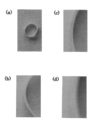
- the perspective view of the base material for membranous connective tissue formation concerning the present invention It is a photograph showing the connective tissue formed on the surface of the cylindrical base material, (a) is the diameter of the cylindrical base material is 10 mm, (b) is the diameter of the cylindrical base material is 20 mm, (c) is the diameter of the cylindrical base material. Shows the case of 30mm
- the photograph which shows the cut surface of the connective tissue formed in the surface of a cylindrical base material (a) is 2 mm in diameter of a cylindrical base material, (b) is 10 mm in diameter of a cylindrical base material, (c) is a cylindrical base material.
- the diameter of 20 mm, (d) shows the case where the diameter of the cylindrical substrate is 30 mm
- Enlarged photos of (a) to (d) in FIG. The figure which shows the relationship between the axial direction length of a cylindrical base material, and the thickness of a connective tissue
- Perspective view of substrate for forming plate-like membranous connective tissue Diagram showing the relationship between the length of one side of a square substrate and the thickness of the connective tissue
- the substrate 1 for forming a membranous connective tissue is, for example, for forming a membranous connective tissue used as an artificial cornea on the surface of a base material by placing it in an environment where a biological tissue material exists.
- a connective tissue formation part 2 for forming a membranous connective tissue body is provided on the surface of the base material as a cornea-forming base material, and the connective tissue body is thickened around the connective tissue formation part 2.
- a thickening promoting portion 3 for promoting is provided.
- the membranous connective tissue-forming substrate 1 has a bulging portion 4 that bulges in a spherical shape at the center of one surface, and a planar shape around the bulging portion 4.
- the peripheral tissue 5 is formed in a disc shape, the connective tissue forming portion 2 is set at the center of the bulging portion 4, and the thickening promoting portion 3 is set at the remaining portion of the bulging portion 4 and the peripheral edge 5.
- the alternate long and two short dashes line is a virtual line indicating the boundary 6 between the connective tissue forming part 2 and the thickening promoting part 3.
- the shape and size of the connective tissue forming part 2 is set in accordance with the shape and size of the artificial cornea to be formed.
- the overall size of the substrate 1 for forming a membranous connective tissue including the thickening promoting portion 3 is set according to the thickness of the artificial cornea to be formed.
- size of the base material 1 for membrane connective tissue formation can be shown using the contact area with the environment where biological tissue material exists, ie, the surface area of the base material 1 for membrane connective tissue formation. .
- the material constituting the substrate 1 for forming a membranous connective tissue is not particularly limited, but the strength (hardness), chemical stability, and the like that can prevent deformation due to implantation in a living body, A resin that has resistance to a load such as sterilization and satisfies the condition that there is no or little eluate that stimulates a living body is preferable, and examples thereof include silicone resins and acrylic resins. Furthermore, since the connective tissue formed on the surface of the membranous connective tissue-forming substrate 1 is made of a highly elastic material, it tends to be thick, so at least the surface of the membranous connective tissue-forming substrate 1 It is more preferable to form an elastic body such as a silicone resin.
- a membrane-like connective tissue-forming substrate 1 is placed in an environment where biological tissue material is present, and a connective tissue is formed around the membrane-like connective tissue-forming substrate 1.
- “Formation step” “Take out step” of taking out the membrane-like connective tissue-forming substrate 1 coated with connective tissue from the environment, and peeling the connective tissue from the membrane-like connective tissue-forming substrate 1
- the substrate 1 for forming a membranous connective tissue is placed in an environment where biological tissue materials are present.
- the environment in which the biological tissue material exists includes in an animal's living body (for example, subcutaneously or intraperitoneally embedded) or in an artificial environment such as a solution in which the biological tissue material floats outside the animal's body.
- biological tissue materials materials derived from other mammals such as humans, dogs, cows, pigs, goats, rabbits, sheep, birds, fish, other animals, or artificial materials can also be used.
- the substrate 1 for forming a membranous connective tissue When the substrate 1 for forming a membranous connective tissue is embedded in an animal, it is performed with a minimum of incision under sufficient anesthesia, and the wound is sutured after implantation.
- part of the base material 1 for membranous connective tissue formation for example, intraperitoneal cavity having a volume for receiving the base material 1 for membranous connective tissue formation, or subcutaneous such as limb, shoulder or back, abdomen, etc. preferable.
- cell culture may be performed according to a known method in a clean environment with various culture conditions.
- a membranous connective tissue is formed around the substrate 1 for forming a membranous connective tissue.
- the connective tissue is composed of an extracellular matrix such as fibroblasts and collagen, and is formed to a sufficient thickness according to the size of the substrate 1 for forming a membranous connective tissue body.
- a take-out process of taking out the membranous connective tissue-forming substrate 1 from the environment where the biological tissue material exists is performed.
- the substrate 1 for forming a membranous connective tissue taken out from an environment in which a biological tissue material exists is entirely covered with a membrane made of connective tissue.
- the connective tissue covering the peripheral portion 5 of the substrate 1 for forming a membranous connective tissue is removed, and the connective tissue film covering the bulging portion 4 is peeled off.
- the connective tissue film has a size and shape corresponding to the size of the bulging portion 4 and the curved surface shape, and is formed to a sufficient thickness according to the size of the substrate 1 for forming a film-like connective tissue. Yes.
- immunogen removal treatment such as decellularization treatment, dehydration treatment, and fixation treatment in order to prevent rejection after transplantation.
- decellularization include ultrasonic treatment, surfactant treatment, and enzyme treatment such as collagenase to elute and wash the extracellular matrix.
- Dehydration methods include methanol, ethanol, isopropyl alcohol, etc. There is a method of washing with a water-soluble organic solvent, and as a method of fixing, there is a method of treating with an aldehyde compound such as glutaraldehyde or formaldehyde.
- the thickening promoting portion 3 is provided around the connective tissue forming portion 2 to enlarge the base material 1 for forming the membrane connective tissue, it is bonded to a sufficient thickness according to the size. An organization can be formed. As a result, an excess portion of the peripheral portion of the connective tissue film formed to a sufficient thickness can be removed to obtain an artificial cornea having a desired size and a curved shape and a sufficient thickness.
- the columnar base material is made of sterilized silicone, and has four types of axial lengths of 30 mm, diameters of 2 mm, 10 mm, 20 mm, and 30 mm.
- FIG. 2 shows external appearance photographs of three types of columnar substrates having diameters of 10 mm, 20 mm, and 30 mm covered with connective tissue.
- the connective tissue formed under the skin was mainly composed of fibroblasts and collagen and encapsulated to cover the base material surface of the cylindrical base material. Further, the columnar substrate covered with the connective tissue membrane could be easily removed from the subcutaneous tissue.
- the connective tissue was peeled off from the substrate surface and separated, and this was cut to examine the thickness of the connective tissue.
- the cylindrical base material and the surrounding connective tissue were not adhered at all, and both could be easily separated without damage.
- FIG. 3 shows a photograph of the appearance of the cut surface of the connective tissue separated from the surface of the four types of cylindrical substrates having diameters of 2 mm, 10 mm, 20 mm, and 30 mm
- FIG. 4 shows an enlarged photograph thereof.
- the connective tissue thickness is 85 ⁇ 27 ⁇ m.
- the connective tissue thickness is 237 ⁇ 55 ⁇ m.
- the connective tissue thickness is 374 ⁇ 128 ⁇ m and the diameter is 30 mm. Then, the thickness of the connective tissue was 813 ⁇ 175 ⁇ m.
- the thickness of the connective tissue was 100 ⁇ m or less.
- the longer the axial length of the cylindrical base material the greater the thickness of the connective tissue. Became thicker.
- the diameter of the connective tissue forming part 2 is set to 7 mm, which is the size of the cornea necessary for transplantation, and the base material composed only of the connective tissue forming part 2 and the remainder of the bulging part 4 and the peripheral part 5
- the film of the connective tissue was formed under the same condition as in ⁇ Relationship Between Diameter of Plate and Thickness of Connective Tissue>, and the thickness was measured.
- all the five types of base materials are made of acrylic, and the thickness of the peripheral portion 5 is 5 mm.
- the thickness of a base material having a diameter of 7 mm consisting only of the connective tissue body forming part 2 is 100 ⁇ m or less, but the connective tissue becomes thicker as the overall diameter of the base material increases to 20 mm and 30 mm. It can be seen that as the diameter of the substrate is increased, a connective tissue having a thick film can be obtained.
- a cornea having a diameter of about 7 mm and a thickness of about 0.5 mm is used, and in the surface corneal transplantation, a cornea having a diameter of about 7 mm and a thickness of 0.2 mm to 0.4 mm is used. Therefore, an artificial cornea having a thickness necessary for transplantation cannot be formed with a base material having a diameter of 7 mm consisting only of the connective tissue formation part 2, but a cornea surface transplantation with a base material having a total diameter of 20 mm.
- An artificial cornea having a thickness capable of transplanting all layers of the cornea can be formed on a base material having a diameter of 30 mm or more.
- a thick connective tissue is obtained as the diameter of the cylindrical substrate is increased, the axial length of the cylindrical substrate is increased, and the diameter of the substrate having the bulging portion is increased.
- a thick connective tissue is formed as the surface area of the base material is the same.
- the membranous connective tissue produced using the substrate 1 for forming a membranous connective tissue may be a planar tissue that covers the surface layer or functions in the form of a membrane, and is not limited to an artificial cornea, but a pericardium It can also be used as a dura mater, skin, valvular membrane and the like.
- a base material for forming a membranous connective tissue not only one having a bulged portion 4 that bulges in a spherical shape at the center, but also a saddle type according to the shape of the membranous connective tissue to be produced
- the base material 7 (see FIG. 7) and the plate-like base material 8 (see FIG. 8) can also be employed.
- the thick base material 7 and the plate-like base material 8 have a sufficiently thick connective structure by increasing the size thereof. Is obtained.
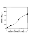
- FIG. 9 shows the relationship between the size of the plate-like substrate and the thickness of the connective tissue.
- the plate-like base material 8 is a square having a thickness of 1 mm, and four types of acrylic base materials having a side length of 10 mm, 30 mm, 50 mm, and 100 mm, under the same conditions as the above cylindrical base material.
- a connective tissue film was formed and its thickness was measured.
- the thickness of the connective tissue formed on the surface of a substrate having a side length of 10 mm is 100 ⁇ m or less.
- the connective tissue thickness is about 400 ⁇ m and the length of one side.
- the thickness of the connective tissue is about 700 ⁇ m, and in the case of a substrate having a side length of 100 mm, the thickness of the connective tissue is about 1000 ⁇ m. became.
Landscapes
- Health & Medical Sciences (AREA)
- Engineering & Computer Science (AREA)
- Biomedical Technology (AREA)
- Life Sciences & Earth Sciences (AREA)
- Transplantation (AREA)
- Veterinary Medicine (AREA)
- Animal Behavior & Ethology (AREA)
- General Health & Medical Sciences (AREA)
- Public Health (AREA)
- Chemical & Material Sciences (AREA)
- Oral & Maxillofacial Surgery (AREA)
- Vascular Medicine (AREA)
- Heart & Thoracic Surgery (AREA)
- Botany (AREA)
- Chemical Kinetics & Catalysis (AREA)
- Epidemiology (AREA)
- Medicinal Chemistry (AREA)
- Dermatology (AREA)
- Cardiology (AREA)
- Ophthalmology & Optometry (AREA)
- Zoology (AREA)
- Cell Biology (AREA)
- Urology & Nephrology (AREA)
- Otolaryngology (AREA)
- Hematology (AREA)
- Anesthesiology (AREA)
- Prostheses (AREA)
- Materials For Medical Uses (AREA)
Abstract
本発明は、十分な厚さ及び強度の膜状の結合組織体を形成することのできる膜状結合組織体形成用基材及びこれを用いた膜状結合組織体の生産方法を提供するものである。具体的には、基材表面に結合組織体形成部2を設ける。結合組織体形成部2の周囲に増厚促進部3を設けて膜状結合組織体形成用基材1を全体として大きくする。膜状結合組織体形成用基材1を生体組織材料の存在する環境に配置して、基材表面に膜状の結合組織体を形成する。膜状結合組織体形成用基材1を大きくする分、膜状結合組織体の厚さが厚くなる。結合組織体のうち、増厚促進部3の表面に形成された余剰部分を除去する。厚くかつ強度の高い膜状結合組織体が得られる。
Description
本発明は、人工角膜などの膜状結合組織体を形成するための膜状結合組織体形成用基材及びこれを用いた膜状結合組織体の生産方法に関するものである。
病気や事故で失われた細胞、組織、器官を、人工素材や細胞により再び蘇らせる再生医療の研究が数多くなされている。
通常、身体には自己防衛機能があり、体内の浅い位置にトゲ等の異物が侵入した場合には、これを体外へ押し出そうとする。また、体内の深い位置に異物が侵入した場合には、その周りに繊維芽細胞が徐々に集まって、主に繊維芽細胞とコラーゲンからなる結合組織体のカプセルを形成して異物を覆うことにより、体内において異物を隔離することが知られている。この後者の自己防衛反応を利用して生細胞から生体由来組織を形成する技術として、生体内に異物としての基材を埋入して結合組織体を形成する技術が複数報告されている(特許文献1~3参照)。
このような手法で結合組織体を形成する場合、生体内に埋入する異物としての基材の形状や大きさを適宜設定することにより、所望の形状や大きさの結合組織体を形成することができる。
ところが、生体内に埋入した基材の周囲に結合組織体を形成する場合、基材の形状や大きさを適宜設定して、その周囲に所望の形状や大きさの結合組織体を形成することはできるものの、結合組織体を所望の厚さ及び強度に設定することはできず、必ずしも十分な厚さや強度の結合組織体を得ることはできない。
本発明は、十分な厚さ及び強度の膜状の結合組織体を形成することのできる膜状結合組織体形成用基材及びこれを用いた膜状結合組織体の生産方法の提供を目的とする。
上記目的を達成するために、本発明に係る膜状結合組織体形成用基材は、生体組織材料の存在する環境に配置して基材表面に膜状の結合組織体を形成可能なものであり、基材表面に、膜状の結合組織体を形成する結合組織体形成部を設け、この結合組織体形成部の周囲に結合組織体の増厚を促進する増厚促進部を設けたものである。
上記構成によれば、基材表面に設けた結合組織体形成部の周囲に増厚促進部を設けるので、基材表面に形成する結合組織の増厚を促進することができ、基材表面に結合組織体形成部のみを設ける場合よりも、膜状結合組織体の厚さを厚くすると共に、強度を高めることができる。
より詳しく説明すると、膜状結合組織体の厚さに影響を与える要因として基材の大きさに着目し、膜状結合組織体の厚さと、この膜状結合組織体を形成する基材の大きさとの関係を調べたところ、基材を大きくするほど、その基材表面に形成される結合組織の厚さが厚くなると共に、強度が高くなるという知見を得ることができた。
そこで、基材表面に、所望の大きさの膜状結合組織体を形成するのに必要な広さの結合組織体形成部を設け、さらに、結合組織体形成部の周囲に増厚促進部を設けることにより、基材表面の全体としての大きさを大きくする。これにより、基材が全体として大きくなる分、その表面に、厚くかつ強度の高い結合組織を形成することができるので、結合組織体形成部の周囲の増厚促進部の表面に形成された余剰部分を除去することにより、厚くかつ強度の高い膜状結合組織体を得ることができる。
ここで、「生体組織材料」とは、所望の生体由来組織を形成するうえで必要な物質のことであり、例えば、線維芽細胞、平滑筋細胞、内皮細胞、幹細胞、ES細胞、iPS細胞等の動物細胞、各種たんぱく質類(コラーゲン、エラスチン)、ヒアルロン酸等の糖類、その他、細胞成長因子、サイトカイン等の生体内に存在する各種の生理活性物質が挙げられる。この「生体組織材料」には、ヒト、イヌ、ウシ、ブタ、ヤギ、ヒツジ等の哺乳類動物、鳥類、魚類、その他の動物に由来するもの、又はこれと同等の人工材料が含まれる。
また、「生体組織材料の存在する環境下」とは、動物(ヒト、イヌ、ウシ、ブタ、ヤギ、ヒツジ等の哺乳類動物、鳥類、魚類、その他の動物)の生体内(例えば、四肢部、腰部、背部又は腹部などの皮下、もしくは腹腔内への埋入)、又は、動物の生体外において、生体組織材料を含有する人工環境のことをいう。
本発明の膜状結合組織体形成用基材は、あらゆる用途の膜状結合組織体を形成することができるが、結合組織体としての人工角膜を形成するための人工角膜形成用基材として使用する基材には、本発明の構成を採用するのが特に好適である。つまり、本発明の基材で形成する膜状結合組織体は、表層を覆うあるいは膜状で機能する平面状の膜状組織として用いるものであり、心膜や硬膜、皮膚、弁膜など、どのようなものであってもよいが、角膜は、その大きさが小さい分、厚い組織を形成しにくいので、本発明の基材を用いて、人工角膜の厚さを厚くすると共に、強度を高めるのがよい。
また、本発明は、上記の膜状結合組織体形成用基材を用いて、膜状結合組織体を生産する方法を提供する。すなわち、本発明に係る膜状結合組織体の生産方法は、膜状結合組織体形成用基材を生体組織材料の存在する環境下におく設置工程と、膜状結合組織体形成用基材の周囲に結合組織を形成する形成工程と、環境下から結合組織で被覆された膜状結合組織体形成用基材を取り出す取り出し工程と、膜状結合組織体形成用基材から結合組織を剥離して取り出す分離工程と、結合組織から膜状結合組織体の周囲の余剰部分を除去する除去工程と、を備える。
この構成によれば、上記の膜状結合組織体形成用基材の構成を採用することによる効果と同じ効果を得ることができる。なお、本発明において、移植対象者に対して、自家移植、同種移植、異種移植のいずれでもよいが、拒絶反応を避ける観点からなるべく自家移植か同種移植が好ましい。また、異種移植の場合には、拒絶反応を避けるため公知の脱細胞化処理などの免疫源除去処理を施すのが好ましい。
また、本発明は、上記の生産方法のごとく、大き目の基材を用いて十分な厚さの結合組織を形成し、この結合組織から周囲の余剰部分を除去することによって形成した膜状結合組織体を提供する。すなわち、本発明は、生体組織材料の存在する環境において基材の表面に形成され、当該膜状結合組織体の周囲の余剰部分が除去されたことを特徴とする膜状結合組織体を提供する。
上記のとおり、本発明によると、結合組織体形成部の周囲に増厚促進部を設けて、基材をこれを用いて生産する膜状結合組織体よりも大き目に設定するので、基材表面に形成する結合組織の厚さを厚くすると共に、強度を高めることができる。これにより、周囲の余剰部分を除去して、厚くかつ強度の高い膜状結合組織体を得ることができ、角膜のように小さな膜状結合組織体を形成する場合であっても、十分な厚さ及び強度を得ることができる。
以下、本発明に係る膜状結合組織体形成用基材及びこれを用いた膜状結合組織体の生産方法を実施するための形態について、図面を用いて説明する。
膜状結合組織体形成用基材1は、例えば、生体組織材料の存在する環境に配置して基材表面に人工角膜として用いる膜状の結合組織体を形成するためのものであり、その人工角膜形成用基材としての基材の表面に、膜状の結合組織体を形成する結合組織体形成部2が設けられると共に、結合組織体形成部2の周囲に、結合組織体の増厚を促進する増厚促進部3が設けられている。
図1に示すように、膜状結合組織体形成用基材1は、一面の中央部に球面状に膨出する膨出部4が形成されると共に、膨出部4の周囲に平面状の周縁部5が形成された円盤状とされ、膨出部4の中央部に結合組織体形成部2が設定され、膨出部4の残部及び周縁部5に増厚促進部3が設定されている。なお、図1において、二点鎖線は結合組織体形成部2と増厚促進部3との境界6を示す仮想線である。
結合組織体形成部2は、形成しようとする人工角膜の形状及び大きさに対応させて、その形状及び大きさが設定される。また、形成しようとする人工角膜の厚さに応じて、増厚促進部3を含む膜状結合組織体形成用基材1の全体の大きさが設定される。ここで、膜状結合組織体形成用基材1の大きさは、生体組織材料の存在する環境との接触面積、すなわち膜状結合組織体形成用基材1の表面積を用いて示すことができる。
膜状結合組織体形成用基材1を構成する素材は、特に限定されるものではないが、生体に埋入することによる変形を防止できる程度の強度(硬度)、化学的安定性、及び、滅菌などの負荷に対する耐性を有し、さらに、生体を刺激する溶出物が全くないか少ないという条件を満たす樹脂が好ましく、シリコーン樹脂やアクリル樹脂を例示できる。さらに、膜状結合組織体形成用基材1を弾性の高い素材で構成すると、その表面に形成される結合組織体が厚くなりやすいことから、膜状結合組織体形成用基材1の少なくとも表面をシリコーン樹脂等の弾性体から構成するのがより好ましい。
次に、上記のような膜状結合組織体形成用基材1を用いて膜状結合組織体を生産する方法について説明する。
この生産方法は、膜状結合組織体形成用基材1を生体組織材料の存在する環境下におく「設置工程」と、膜状結合組織体形成用基材1の周囲に結合組織を形成する「形成工程」と、環境下から結合組織で被覆された膜状結合組織体形成用基材1を取り出す「取り出し工程」と、膜状結合組織体形成用基材1から結合組織を剥離して取り出す「分離工程」と、結合組織から人工角膜としての膜状結合組織体の周囲の余剰部分を除去する「除去工程」とからなる。
<設置工程>
膜状結合組織体形成用基材1を生体組織材料の存在する環境下へ置く。生体組織材料の存在する環境下とは、動物の生体内(例えば、皮下や腹腔内への埋入)、又は、動物の生体外において生体組織材料が浮遊する溶液中等の人工環境内が挙げられる。生体組織材料としては、ヒト、イヌ、ウシ、ブタ、ヤギ、ウサギ、ヒツジなどの他の哺乳類動物由来のものや、鳥類、魚類、その他の動物由来のもの、又は人工材料を用いることもできる。
膜状結合組織体形成用基材1を生体組織材料の存在する環境下へ置く。生体組織材料の存在する環境下とは、動物の生体内(例えば、皮下や腹腔内への埋入)、又は、動物の生体外において生体組織材料が浮遊する溶液中等の人工環境内が挙げられる。生体組織材料としては、ヒト、イヌ、ウシ、ブタ、ヤギ、ウサギ、ヒツジなどの他の哺乳類動物由来のものや、鳥類、魚類、その他の動物由来のもの、又は人工材料を用いることもできる。
膜状結合組織体形成用基材1を動物に埋入する場合には、十分な麻酔下で最小限の切開術で行い、埋入後は傷口を縫合する。膜状結合組織体形成用基材1の埋入部位としては例えば、膜状結合組織体形成用基材1を受け入れる容積を有する腹腔内、あるいは四肢部、肩部又は背部、腹部などの皮下が好ましい。また、膜状結合組織体形成用基材1を生体組織材料の存在する環境下へ置く場合には、種々の培養条件を整えてクリーンな環境下で公知の方法に従って細胞培養を行えばよい。
<形成工程>
設置工程の後、所定時間が経過することにより、膜状結合組織体形成用基材1の周囲に膜状の結合組織が形成される。結合組織は、繊維芽細胞とコラーゲンなどの細胞外マトリックスで構成され、膜状結合組織体形成用基材1の大きさに応じて十分な厚さに形成される。
設置工程の後、所定時間が経過することにより、膜状結合組織体形成用基材1の周囲に膜状の結合組織が形成される。結合組織は、繊維芽細胞とコラーゲンなどの細胞外マトリックスで構成され、膜状結合組織体形成用基材1の大きさに応じて十分な厚さに形成される。
<取り出し工程>
所定時間の形成工程を経て、結合組織が十分に形成された後、膜状結合組織体形成用基材1を生体組織材料の存在する環境下から取り出す取り出し工程を行う。生体組織材料の存在する環境下から取り出された膜状結合組織体形成用基材1は、全体を結合組織による膜で覆われている。
所定時間の形成工程を経て、結合組織が十分に形成された後、膜状結合組織体形成用基材1を生体組織材料の存在する環境下から取り出す取り出し工程を行う。生体組織材料の存在する環境下から取り出された膜状結合組織体形成用基材1は、全体を結合組織による膜で覆われている。
<分離工程>
膜状結合組織体形成用基材1の周縁部5を覆う結合組織を除去して、膨出部4を覆う結合組織の膜を剥がす。この結合組織の膜は、膨出部4の大きさ及び曲面形状に対応する大きさ及び形状で、膜状結合組織体形成用基材1の大きさに応じた十分な厚さに形成されている。
膜状結合組織体形成用基材1の周縁部5を覆う結合組織を除去して、膨出部4を覆う結合組織の膜を剥がす。この結合組織の膜は、膨出部4の大きさ及び曲面形状に対応する大きさ及び形状で、膜状結合組織体形成用基材1の大きさに応じた十分な厚さに形成されている。
<除去工程>
膨出部4から剥がした結合組織の膜の周縁部の余剰部分を除去して、所望の大きさの人工角膜を得る。
膨出部4から剥がした結合組織の膜の周縁部の余剰部分を除去して、所望の大きさの人工角膜を得る。
生産された人工角膜を異種移植する場合には、移植後の拒絶反応を防ぐため、脱細胞処理、脱水処理、固定処理などの免疫源除去処理を施すのが好ましい。脱細胞処理としては、超音波処理や界面活性剤処理、コラゲナーゼなどの酵素処理によって細胞外マトリックスを溶出させて洗浄する等の方法があり、脱水処理の方法としては、メタノール、エタノール、イソプロピルアルコール等の水溶性有機溶媒で洗浄する方法があり、固定処理する方法としては、グルタアルデヒドやホルムアルデヒドなどのアルデヒド化合物で処理する方法がある。
上記構成によれば、結合組織体形成部2の周囲に増厚促進部3を設けて膜状結合組織体形成用基材1を大きくするので、その大きさに応じて十分な厚さに結合組織を形成することができる。これにより、十分な厚さに形成した結合組織の膜の周縁部の余剰部分を除去して、所望の大きさ及び曲面形状で、かつ十分な厚さの人工角膜を得ることができる。
ここで、基材の大きさと、その基材表面に形成される結合組織の厚さとの関係について、「円柱状基材の直径と結合組織の厚さとの関係」、「円柱状基材の軸方向長さと結合組織の厚さとの関係」、及び「膨出部を有する基材の直径と結合組織の厚さとの関係」の順に説明する。
<円柱状基材の直径と結合組織の厚さとの関係>
まず、4種類のシリコーン製の円柱状基材を体重10kgのビーグル犬の背部皮下に埋入し、そのビーグル犬を通常の飼育条件で1ヶ月飼育した後、結合組織で覆われた円柱状基材を摘出した。円柱状基材は、滅菌したシリコーン製で、軸方向長さが全て30mm、直径が2mm、10mm、20mm、30mmの4種類である。
まず、4種類のシリコーン製の円柱状基材を体重10kgのビーグル犬の背部皮下に埋入し、そのビーグル犬を通常の飼育条件で1ヶ月飼育した後、結合組織で覆われた円柱状基材を摘出した。円柱状基材は、滅菌したシリコーン製で、軸方向長さが全て30mm、直径が2mm、10mm、20mm、30mmの4種類である。
図2に、直径が10mm、20mm、30mmの3種類の円柱状基材について、結合組織で覆われた状態の外観写真を示す。皮下で形成された結合組織は主として、線維芽細胞とコラーゲンから構成され、カプセル化して円柱状基材の基材表面を覆っていた。また、結合組織の膜で覆われた円柱状基材は、皮下組織より容易に摘出することができた。
次いで、基材表面から結合組織を剥がして分離し、これを切断して結合組織の厚さを調べた。なお、円柱状基材と周囲の結合組織とは全く接着しておらず、両者は損傷無く容易に分離することができた。
図3に、直径が2mm、10mm、20mm、30mmの4種類の円柱状基材について、その基材表面から分離した結合組織の切断面の外観写真を示し、図4に、その拡大写真を示す。円柱状基材が直径2mmの場合、結合組織の厚さが85±27μm、直径10mmでは、結合組織の厚さが237±55μm、直径20mmでは、結合組織の厚さが374±128μm、直径30mmでは、結合組織の厚さが813±175μmであった。
<円柱状基材の軸方向長さと結合組織の厚さとの関係>
直径が全て10mm、軸方向長さが1mm、10mm、30mm、50mmの4種類の円柱状基材を用いて、上記の<円柱状基材の直径と結合組織の厚さとの関係>と同じ条件で結合組織の膜を形成し、その厚さを測定した。
直径が全て10mm、軸方向長さが1mm、10mm、30mm、50mmの4種類の円柱状基材を用いて、上記の<円柱状基材の直径と結合組織の厚さとの関係>と同じ条件で結合組織の膜を形成し、その厚さを測定した。
図5に示すように、円柱状基材の軸方向長さが1mmでは、結合組織の厚さが100μm以下であったが、円柱状基材の軸方向長さが長くなるほど結合組織の厚さが厚くなった。
<膨出部を有する基材の直径と結合組織の厚さとの関係>
結合組織体形成部2の直径を移植に必要な角膜の大きさである7mmに設定し、この結合組織体形成部2のみからなる基材と、これに膨出部4の残部及び周縁部5を加えた上記膜状結合組織体形成用基材1で直径が10mm、20mm、30mm、50mmの4種類の基材との、合計5種の基材を用いて、上記の<円柱状基材の直径と結合組織の厚さとの関係>と同じ条件で結合組織の膜を形成し、その厚さを測定した。なお、5種類の全ての基材は、アクリル製で、周縁部5の厚さが5mmである。
結合組織体形成部2の直径を移植に必要な角膜の大きさである7mmに設定し、この結合組織体形成部2のみからなる基材と、これに膨出部4の残部及び周縁部5を加えた上記膜状結合組織体形成用基材1で直径が10mm、20mm、30mm、50mmの4種類の基材との、合計5種の基材を用いて、上記の<円柱状基材の直径と結合組織の厚さとの関係>と同じ条件で結合組織の膜を形成し、その厚さを測定した。なお、5種類の全ての基材は、アクリル製で、周縁部5の厚さが5mmである。
図6に示すように、結合組織体形成部2のみからなる直径7mmの基材では、厚さは100μm以下であるが、基材全体の直径が20mm、30mmと大きくなるに従って結合組織が厚くなっており、基材の直径を大きくする程、厚い膜を有する結合組織が得られることがわかる。
角膜移植では直径が約7mmで厚さが約0.5mmの角膜が用いられ、表層角膜移植では直径が約7mmで厚さ0.2mm~0.4mmの角膜が用いられている。したがって、結合組織体形成部2のみからなる直径7mmの基材では、移植に必要な厚さの人工角膜を形成することができないが、基材全体の直径が20mmの基材では、角膜表層移植が可能な厚さの人工角膜を形成することができ、基材全体の直径が30mm以上の基材では、角膜全層移植が可能な厚さの人工角膜を形成することができる。
このように、円柱状基材の直径を大きくするほど、円柱状基材の軸方向長さを長くするほど、さらに、膨出部を有する基材の直径を大きくするほど、厚い結合組織が得られることがわかる。すなわち、基材の形状による違いはあるものの、同じ形状の基材であれば、その表面積を大きくするほど、厚い結合組織が形成されることがわかる。
なお、本発明は、上記の実施の形態に限定されるものではなく、本発明の範囲内において、適宜変更を加えることができる。例えば、膜状結合組織体形成用基材1を用いて生産する膜状結合組織体は、表層を覆うあるいは膜状で機能する平面状の組織であればよく、人工角膜に限らず、心膜、硬膜、皮膚、弁膜等として用いることもできる。
また、膜状結合組織体形成用基材としては、中央部に球面状に膨出する膨出部4を有するものだけでなく、生産すべき膜状結合組織体の形状に応じて、蒲鉾型の基材7(図7参照)や板状の基材8(図8参照)を採用することもできる。なお、上記の円柱状基材、及び膨出部を有する基材だけでなく、蒲鉾型の基材7、板状の基材8についても、その大きさを大きくすることで十分に厚い結合組織が得られる。
例えば、図9に、板状の基材の大きさと結合組織の厚さとの関係を示す。板状の基材8は、厚さが全て1mmの正方形で、一辺の長さが10mm、30mm、50mm、100mmの4種類のアクリル製基材であり、上記の円柱状基材と同じ条件で結合組織の膜を形成し、その厚さを測定した。一辺の長さが10mmの基材の表面に形成される結合組織の厚さは100μm以下であるが、一辺の長さが30mmの基材では、結合組織の厚さが約400μm、一辺の長さが60mmの基材では、結合組織の厚さが約700μm、一辺の長さが100mmの基材では、結合組織の厚さが約1000μmであり、基材が大きくなるほど膜の厚さが厚くなった。
1 膜状結合組織体形成用基材
2 結合組織体形成部
3 増厚促進部
4 膨出部
5 周縁部
6 境界
7 基材(蒲鉾型)
8 基材(板状)
2 結合組織体形成部
3 増厚促進部
4 膨出部
5 周縁部
6 境界
7 基材(蒲鉾型)
8 基材(板状)
Claims (4)
- 生体組織材料の存在する環境に配置して基材表面に膜状の結合組織体を形成可能な基材であって、基材表面に、膜状の結合組織体を形成する結合組織体形成部が設けられ、該結合組織体形成部の周囲に前記結合組織体の増厚を促進する増厚促進部が設けられたことを特徴とする膜状結合組織体形成用基材。
- 前記結合組織体としての人工角膜を形成可能な人工角膜形成用基材として使用されることを特徴とする請求項1に記載の膜状結合組織体形成用基材。
- 請求項1又は2に記載の膜状結合組織体形成用基材を生体組織材料の存在する環境下におく設置工程と、前記膜状結合組織体形成用基材の周囲に結合組織を形成する形成工程と、前記環境下から結合組織で被覆された前記膜状結合組織体形成用基材を取り出す取り出し工程と、前記膜状結合組織体形成用基材から結合組織を剥離して取り出す分離工程と、前記結合組織から膜状結合組織体の周囲の余剰部分を除去する除去工程と、を備えたことを特徴とする膜状結合組織体の生産方法。
- 生体組織材料の存在する環境において基材の表面に形成され、当該膜状結合組織体の周囲の余剰部分が除去されたことを特徴とする膜状結合組織体。
Priority Applications (3)
| Application Number | Priority Date | Filing Date | Title |
|---|---|---|---|
| EP13816703.6A EP2873428A4 (en) | 2012-07-11 | 2013-05-30 | SUBSTRATE FOR MEMBRANE CONJUNCTIVE TISSUE FORMATION AND METHOD FOR PRODUCING MEMBRANE-TYPE CONJUNCTIVE TISSUE USING THE SAME |
| CN201380036025.0A CN104411342A (zh) | 2012-07-11 | 2013-05-30 | 膜状结缔组织体形成用基材和使用该基材的膜状结缔组织体的生产方法 |
| US14/412,266 US20150342724A1 (en) | 2012-07-11 | 2013-05-30 | Substrate for formation of membrane-like connective tissue and production method for membrane-like connective tissue using same |
Applications Claiming Priority (2)
| Application Number | Priority Date | Filing Date | Title |
|---|---|---|---|
| JP2012155334A JP2014014590A (ja) | 2012-07-11 | 2012-07-11 | 膜状結合組織体形成用基材及びこれを用いた膜状結合組織体の生産方法 |
| JP2012-155334 | 2012-07-11 |
Publications (1)
| Publication Number | Publication Date |
|---|---|
| WO2014010323A1 true WO2014010323A1 (ja) | 2014-01-16 |
Family
ID=49915797
Family Applications (1)
| Application Number | Title | Priority Date | Filing Date |
|---|---|---|---|
| PCT/JP2013/064999 WO2014010323A1 (ja) | 2012-07-11 | 2013-05-30 | 膜状結合組織体形成用基材及びこれを用いた膜状結合組織体の生産方法 |
Country Status (5)
| Country | Link |
|---|---|
| US (1) | US20150342724A1 (ja) |
| EP (1) | EP2873428A4 (ja) |
| JP (1) | JP2014014590A (ja) |
| CN (1) | CN104411342A (ja) |
| WO (1) | WO2014010323A1 (ja) |
Families Citing this family (1)
| Publication number | Priority date | Publication date | Assignee | Title |
|---|---|---|---|---|
| KR20130033463A (ko) * | 2005-08-24 | 2013-04-03 | 인터디지탈 테크날러지 코포레이션 | 업링크 용량을 증가시키기 위해 채널 품질 표시자 피드백 주기를 조정하는 방법 및 장치 |
Citations (4)
| Publication number | Priority date | Publication date | Assignee | Title |
|---|---|---|---|---|
| JP2006238987A (ja) * | 2005-03-01 | 2006-09-14 | National Cardiovascular Center | 結合組織シート(バイオシート)の形成基材およびバイオシートの製造方法 |
| JP2007312821A (ja) | 2006-05-23 | 2007-12-06 | National Cardiovascular Center | 結合組織体形成基材およびそれを用いた結合組織体の製造方法 |
| JP2008237896A (ja) | 2007-02-26 | 2008-10-09 | National Cardiovascular Center | 結合組織体形成基材およびそれを用いた結合組織体の製造方法 |
| JP2010094476A (ja) | 2008-10-15 | 2010-04-30 | Shinkan Kogyo Kk | 結合組織体形成用基材およびそれを用いた結合組織体の製造方法 |
Family Cites Families (8)
| Publication number | Priority date | Publication date | Assignee | Title |
|---|---|---|---|---|
| US2714721A (en) * | 1953-01-23 | 1955-08-09 | Jr William Stone | Artificial corneal implants |
| US4983181A (en) * | 1986-10-16 | 1991-01-08 | Cbs Lens, | Collagen hydrogel for promoting epithelial cell growth and artificial lens using the same |
| US4865601A (en) * | 1987-07-07 | 1989-09-12 | Caldwell Delmar R | Intraocular prostheses |
| EP0308077A3 (en) * | 1987-09-14 | 1990-05-30 | Nestle S.A. | Synthetic intracorneal lens |
| US6997888B2 (en) * | 2002-07-08 | 2006-02-14 | Rehrig Glenn A | Libido stimulating device and method of using |
| JP5127120B2 (ja) * | 2005-05-13 | 2013-01-23 | 独立行政法人国立循環器病研究センター | 結合組織体形成基材およびそれを用いた結合組織体の製造方法 |
| JP2010522583A (ja) * | 2007-02-27 | 2010-07-08 | トラスティーズ オブ タフツ カレッジ | 組織工学的に作製された絹製臓器 |
| US8685082B2 (en) * | 2010-11-18 | 2014-04-01 | National Cerebral And Cardiovascular Center | Base material for forming valved lumen shape tissue, method for producing valved lumen shape tissue, and valved artificial blood vessel |
-
2012
- 2012-07-11 JP JP2012155334A patent/JP2014014590A/ja active Pending
-
2013
- 2013-05-30 CN CN201380036025.0A patent/CN104411342A/zh active Pending
- 2013-05-30 WO PCT/JP2013/064999 patent/WO2014010323A1/ja active Application Filing
- 2013-05-30 US US14/412,266 patent/US20150342724A1/en not_active Abandoned
- 2013-05-30 EP EP13816703.6A patent/EP2873428A4/en not_active Withdrawn
Patent Citations (4)
| Publication number | Priority date | Publication date | Assignee | Title |
|---|---|---|---|---|
| JP2006238987A (ja) * | 2005-03-01 | 2006-09-14 | National Cardiovascular Center | 結合組織シート(バイオシート)の形成基材およびバイオシートの製造方法 |
| JP2007312821A (ja) | 2006-05-23 | 2007-12-06 | National Cardiovascular Center | 結合組織体形成基材およびそれを用いた結合組織体の製造方法 |
| JP2008237896A (ja) | 2007-02-26 | 2008-10-09 | National Cardiovascular Center | 結合組織体形成基材およびそれを用いた結合組織体の製造方法 |
| JP2010094476A (ja) | 2008-10-15 | 2010-04-30 | Shinkan Kogyo Kk | 結合組織体形成用基材およびそれを用いた結合組織体の製造方法 |
Non-Patent Citations (1)
| Title |
|---|
| See also references of EP2873428A4 |
Also Published As
| Publication number | Publication date |
|---|---|
| US20150342724A1 (en) | 2015-12-03 |
| EP2873428A4 (en) | 2016-01-20 |
| JP2014014590A (ja) | 2014-01-30 |
| EP2873428A1 (en) | 2015-05-20 |
| CN104411342A (zh) | 2015-03-11 |
Similar Documents
| Publication | Publication Date | Title |
|---|---|---|
| US9956074B2 (en) | Valved stent, base material for forming valved stent, and method for producing valved stent | |
| EP3231392B1 (en) | Substrate for forming artificial valve and artificial valve | |
| WO2016076416A1 (ja) | 結合組織体形成基材及び基材取出具 | |
| JP2012135406A (ja) | 生体由来組織形成基材、これを用いた生体由来組織の生産方法及び生体由来組織 | |
| JP2014030598A (ja) | 膜状結合組織体形成用基材及びこれを用いた膜状結合組織体の生産方法 | |
| US8685082B2 (en) | Base material for forming valved lumen shape tissue, method for producing valved lumen shape tissue, and valved artificial blood vessel | |
| WO2014010323A1 (ja) | 膜状結合組織体形成用基材及びこれを用いた膜状結合組織体の生産方法 | |
| JP6262470B2 (ja) | 結合組織体形成用基材、及び結合組織体の生産方法 | |
| WO2015029707A1 (ja) | 人工気管、人工気管の生産方法、及び人工気管形成用基材 | |
| JP2016136984A (ja) | 管状組織体形成基材 | |
| JP5127120B2 (ja) | 結合組織体形成基材およびそれを用いた結合組織体の製造方法 | |
| JP2017169778A (ja) | 結合組織体形成基材 | |
| WO2014021073A1 (ja) | 人工角膜及び人工角膜の生産方法 | |
| JP6931261B1 (ja) | 組織体形成装置、組織体形成方法、および結合組織体 | |
| JP5658008B2 (ja) | 弁付き人工血管形成用基材、これを用いた弁付き人工血管の生産方法及び弁付き人工血管 | |
| JP6727637B1 (ja) | 組織体形成装置、組織体形成方法、および結合組織体 | |
| JP2017113051A (ja) | 膜状結合組織体形成用基材 | |
| JP2006238987A (ja) | 結合組織シート(バイオシート)の形成基材およびバイオシートの製造方法 | |
| JP2021122747A (ja) | 結合組織体形成用構造体および結合組織体の形成方法 | |
| JP2021122277A (ja) | 結合組織体形成用構造集合体および結合組織体の形成方法 | |
| JP2015039577A (ja) | 人工弁、人工弁形成用基材、及び人工弁の生産方法 | |
| JP5706282B2 (ja) | 弁付き管腔形状組織形成用基材、管腔形状組織形成用基材、膜状組織形成用基材、弁付き管腔形状組織の生産方法、管腔形状組織の生産方法、及び膜状組織の生産方法 | |
| JP2022157586A (ja) | 結合組織体形成用構造体および結合組織体の形成方法 | |
| US20190201583A1 (en) | Method for forming connective tissue body | |
| JP2011041716A (ja) | 生体組織の処理方法 |
Legal Events
| Date | Code | Title | Description |
|---|---|---|---|
| 121 | Ep: the epo has been informed by wipo that ep was designated in this application |
Ref document number: 13816703 Country of ref document: EP Kind code of ref document: A1 |
|
| REEP | Request for entry into the european phase |
Ref document number: 2013816703 Country of ref document: EP |
|
| WWE | Wipo information: entry into national phase |
Ref document number: 2013816703 Country of ref document: EP |
|
| WWE | Wipo information: entry into national phase |
Ref document number: 14412266 Country of ref document: US |
|
| NENP | Non-entry into the national phase |
Ref country code: DE |