WO2012074085A1 - 3剤併用抗がん剤の感受性判定マーカー - Google Patents
3剤併用抗がん剤の感受性判定マーカー Download PDFInfo
- Publication number
- WO2012074085A1 WO2012074085A1 PCT/JP2011/077890 JP2011077890W WO2012074085A1 WO 2012074085 A1 WO2012074085 A1 WO 2012074085A1 JP 2011077890 W JP2011077890 W JP 2011077890W WO 2012074085 A1 WO2012074085 A1 WO 2012074085A1
- Authority
- WO
- WIPO (PCT)
- Prior art keywords
- gene
- salt
- cancer
- expression level
- sensitivity
- Prior art date
Links
Images

Classifications
-
- G—PHYSICS
- G16—INFORMATION AND COMMUNICATION TECHNOLOGY [ICT] SPECIALLY ADAPTED FOR SPECIFIC APPLICATION FIELDS
- G16H—HEALTHCARE INFORMATICS, i.e. INFORMATION AND COMMUNICATION TECHNOLOGY [ICT] SPECIALLY ADAPTED FOR THE HANDLING OR PROCESSING OF MEDICAL OR HEALTHCARE DATA
- G16H20/00—ICT specially adapted for therapies or health-improving plans, e.g. for handling prescriptions, for steering therapy or for monitoring patient compliance
- G16H20/10—ICT specially adapted for therapies or health-improving plans, e.g. for handling prescriptions, for steering therapy or for monitoring patient compliance relating to drugs or medications, e.g. for ensuring correct administration to patients
-
- A—HUMAN NECESSITIES
- A61—MEDICAL OR VETERINARY SCIENCE; HYGIENE
- A61K—PREPARATIONS FOR MEDICAL, DENTAL OR TOILETRY PURPOSES
- A61K45/00—Medicinal preparations containing active ingredients not provided for in groups A61K31/00 - A61K41/00
- A61K45/06—Mixtures of active ingredients without chemical characterisation, e.g. antiphlogistics and cardiaca
-
- A—HUMAN NECESSITIES
- A61—MEDICAL OR VETERINARY SCIENCE; HYGIENE
- A61K—PREPARATIONS FOR MEDICAL, DENTAL OR TOILETRY PURPOSES
- A61K31/00—Medicinal preparations containing organic active ingredients
- A61K31/28—Compounds containing heavy metals
- A61K31/282—Platinum compounds
-
- A—HUMAN NECESSITIES
- A61—MEDICAL OR VETERINARY SCIENCE; HYGIENE
- A61K—PREPARATIONS FOR MEDICAL, DENTAL OR TOILETRY PURPOSES
- A61K31/00—Medicinal preparations containing organic active ingredients
- A61K31/33—Heterocyclic compounds
- A61K31/395—Heterocyclic compounds having nitrogen as a ring hetero atom, e.g. guanethidine or rifamycins
- A61K31/495—Heterocyclic compounds having nitrogen as a ring hetero atom, e.g. guanethidine or rifamycins having six-membered rings with two or more nitrogen atoms as the only ring heteroatoms, e.g. piperazine or tetrazines
- A61K31/505—Pyrimidines; Hydrogenated pyrimidines, e.g. trimethoprim
- A61K31/513—Pyrimidines; Hydrogenated pyrimidines, e.g. trimethoprim having oxo groups directly attached to the heterocyclic ring, e.g. cytosine
-
- A—HUMAN NECESSITIES
- A61—MEDICAL OR VETERINARY SCIENCE; HYGIENE
- A61K—PREPARATIONS FOR MEDICAL, DENTAL OR TOILETRY PURPOSES
- A61K31/00—Medicinal preparations containing organic active ingredients
- A61K31/33—Heterocyclic compounds
- A61K31/395—Heterocyclic compounds having nitrogen as a ring hetero atom, e.g. guanethidine or rifamycins
- A61K31/495—Heterocyclic compounds having nitrogen as a ring hetero atom, e.g. guanethidine or rifamycins having six-membered rings with two or more nitrogen atoms as the only ring heteroatoms, e.g. piperazine or tetrazines
- A61K31/505—Pyrimidines; Hydrogenated pyrimidines, e.g. trimethoprim
- A61K31/519—Pyrimidines; Hydrogenated pyrimidines, e.g. trimethoprim ortho- or peri-condensed with heterocyclic rings
-
- A—HUMAN NECESSITIES
- A61—MEDICAL OR VETERINARY SCIENCE; HYGIENE
- A61K—PREPARATIONS FOR MEDICAL, DENTAL OR TOILETRY PURPOSES
- A61K33/00—Medicinal preparations containing inorganic active ingredients
- A61K33/24—Heavy metals; Compounds thereof
-
- A—HUMAN NECESSITIES
- A61—MEDICAL OR VETERINARY SCIENCE; HYGIENE
- A61K—PREPARATIONS FOR MEDICAL, DENTAL OR TOILETRY PURPOSES
- A61K33/00—Medicinal preparations containing inorganic active ingredients
- A61K33/24—Heavy metals; Compounds thereof
- A61K33/243—Platinum; Compounds thereof
-
- A—HUMAN NECESSITIES
- A61—MEDICAL OR VETERINARY SCIENCE; HYGIENE
- A61P—SPECIFIC THERAPEUTIC ACTIVITY OF CHEMICAL COMPOUNDS OR MEDICINAL PREPARATIONS
- A61P35/00—Antineoplastic agents
-
- A—HUMAN NECESSITIES
- A61—MEDICAL OR VETERINARY SCIENCE; HYGIENE
- A61P—SPECIFIC THERAPEUTIC ACTIVITY OF CHEMICAL COMPOUNDS OR MEDICINAL PREPARATIONS
- A61P43/00—Drugs for specific purposes, not provided for in groups A61P1/00-A61P41/00
-
- C—CHEMISTRY; METALLURGY
- C12—BIOCHEMISTRY; BEER; SPIRITS; WINE; VINEGAR; MICROBIOLOGY; ENZYMOLOGY; MUTATION OR GENETIC ENGINEERING
- C12Q—MEASURING OR TESTING PROCESSES INVOLVING ENZYMES, NUCLEIC ACIDS OR MICROORGANISMS; COMPOSITIONS OR TEST PAPERS THEREFOR; PROCESSES OF PREPARING SUCH COMPOSITIONS; CONDITION-RESPONSIVE CONTROL IN MICROBIOLOGICAL OR ENZYMOLOGICAL PROCESSES
- C12Q1/00—Measuring or testing processes involving enzymes, nucleic acids or microorganisms; Compositions therefor; Processes of preparing such compositions
- C12Q1/68—Measuring or testing processes involving enzymes, nucleic acids or microorganisms; Compositions therefor; Processes of preparing such compositions involving nucleic acids
- C12Q1/6876—Nucleic acid products used in the analysis of nucleic acids, e.g. primers or probes
- C12Q1/6883—Nucleic acid products used in the analysis of nucleic acids, e.g. primers or probes for diseases caused by alterations of genetic material
- C12Q1/6886—Nucleic acid products used in the analysis of nucleic acids, e.g. primers or probes for diseases caused by alterations of genetic material for cancer
-
- G—PHYSICS
- G01—MEASURING; TESTING
- G01N—INVESTIGATING OR ANALYSING MATERIALS BY DETERMINING THEIR CHEMICAL OR PHYSICAL PROPERTIES
- G01N33/00—Investigating or analysing materials by specific methods not covered by groups G01N1/00 - G01N31/00
- G01N33/48—Biological material, e.g. blood, urine; Haemocytometers
- G01N33/50—Chemical analysis of biological material, e.g. blood, urine; Testing involving biospecific ligand binding methods; Immunological testing
- G01N33/53—Immunoassay; Biospecific binding assay; Materials therefor
- G01N33/574—Immunoassay; Biospecific binding assay; Materials therefor for cancer
- G01N33/57407—Specifically defined cancers
- G01N33/57419—Specifically defined cancers of colon
-
- G—PHYSICS
- G16—INFORMATION AND COMMUNICATION TECHNOLOGY [ICT] SPECIALLY ADAPTED FOR SPECIFIC APPLICATION FIELDS
- G16H—HEALTHCARE INFORMATICS, i.e. INFORMATION AND COMMUNICATION TECHNOLOGY [ICT] SPECIALLY ADAPTED FOR THE HANDLING OR PROCESSING OF MEDICAL OR HEALTHCARE DATA
- G16H10/00—ICT specially adapted for the handling or processing of patient-related medical or healthcare data
- G16H10/40—ICT specially adapted for the handling or processing of patient-related medical or healthcare data for data related to laboratory analysis, e.g. patient specimen analysis
-
- C—CHEMISTRY; METALLURGY
- C12—BIOCHEMISTRY; BEER; SPIRITS; WINE; VINEGAR; MICROBIOLOGY; ENZYMOLOGY; MUTATION OR GENETIC ENGINEERING
- C12Q—MEASURING OR TESTING PROCESSES INVOLVING ENZYMES, NUCLEIC ACIDS OR MICROORGANISMS; COMPOSITIONS OR TEST PAPERS THEREFOR; PROCESSES OF PREPARING SUCH COMPOSITIONS; CONDITION-RESPONSIVE CONTROL IN MICROBIOLOGICAL OR ENZYMOLOGICAL PROCESSES
- C12Q2600/00—Oligonucleotides characterized by their use
- C12Q2600/106—Pharmacogenomics, i.e. genetic variability in individual responses to drugs and drug metabolism
-
- C—CHEMISTRY; METALLURGY
- C12—BIOCHEMISTRY; BEER; SPIRITS; WINE; VINEGAR; MICROBIOLOGY; ENZYMOLOGY; MUTATION OR GENETIC ENGINEERING
- C12Q—MEASURING OR TESTING PROCESSES INVOLVING ENZYMES, NUCLEIC ACIDS OR MICROORGANISMS; COMPOSITIONS OR TEST PAPERS THEREFOR; PROCESSES OF PREPARING SUCH COMPOSITIONS; CONDITION-RESPONSIVE CONTROL IN MICROBIOLOGICAL OR ENZYMOLOGICAL PROCESSES
- C12Q2600/00—Oligonucleotides characterized by their use
- C12Q2600/136—Screening for pharmacological compounds
-
- C—CHEMISTRY; METALLURGY
- C12—BIOCHEMISTRY; BEER; SPIRITS; WINE; VINEGAR; MICROBIOLOGY; ENZYMOLOGY; MUTATION OR GENETIC ENGINEERING
- C12Q—MEASURING OR TESTING PROCESSES INVOLVING ENZYMES, NUCLEIC ACIDS OR MICROORGANISMS; COMPOSITIONS OR TEST PAPERS THEREFOR; PROCESSES OF PREPARING SUCH COMPOSITIONS; CONDITION-RESPONSIVE CONTROL IN MICROBIOLOGICAL OR ENZYMOLOGICAL PROCESSES
- C12Q2600/00—Oligonucleotides characterized by their use
- C12Q2600/158—Expression markers
-
- G—PHYSICS
- G01—MEASURING; TESTING
- G01N—INVESTIGATING OR ANALYSING MATERIALS BY DETERMINING THEIR CHEMICAL OR PHYSICAL PROPERTIES
- G01N2800/00—Detection or diagnosis of diseases
- G01N2800/52—Predicting or monitoring the response to treatment, e.g. for selection of therapy based on assay results in personalised medicine; Prognosis
-
- Y—GENERAL TAGGING OF NEW TECHNOLOGICAL DEVELOPMENTS; GENERAL TAGGING OF CROSS-SECTIONAL TECHNOLOGIES SPANNING OVER SEVERAL SECTIONS OF THE IPC; TECHNICAL SUBJECTS COVERED BY FORMER USPC CROSS-REFERENCE ART COLLECTIONS [XRACs] AND DIGESTS
- Y02—TECHNOLOGIES OR APPLICATIONS FOR MITIGATION OR ADAPTATION AGAINST CLIMATE CHANGE
- Y02A—TECHNOLOGIES FOR ADAPTATION TO CLIMATE CHANGE
- Y02A90/00—Technologies having an indirect contribution to adaptation to climate change
- Y02A90/10—Information and communication technologies [ICT] supporting adaptation to climate change, e.g. for weather forecasting or climate simulation
Definitions
- the present invention relates to an anticancer drug sensitivity determination marker used for determining whether or not a cancer of a target patient has therapeutic reactivity to an anticancer drug to be used, and its application.
- anticancer agents such as alkylating agents, platinum preparations, antimetabolites, anticancer antibiotics, and anticancer plant alkaloids. These anticancer drugs may or may not be effective depending on the type of cancer. Furthermore, it is known that even the types of cancer that are recognized as effective may or may not be effective depending on the individual patient. Whether or not an anticancer drug shows an effect on such individual patient's cancer is called anticancer drug sensitivity.
- Oxaliplatin is a platinum complex antineoplastic agent. Like other cisplatin (CDDP) and carboplatin (CBDCA), which are other platinum complex antineoplastic agents, the mechanism of action is thought to be DNA synthesis inhibition and protein synthesis inhibition by crosslinking with DNA bases. L-OHP exhibits an antitumor effect even for colorectal cancer in which CBDCA is ineffective, and exhibits an antitumor spectrum different from that of conventional platinum complex antineoplastic agents. In the US, it was approved as a first-line treatment for metastatic colorectal cancer in combination with fluorouracil (5-FU) / levofolinate (LV) in January 2004.
- fluorouracil 5-FU
- LV levofolinate
- the drug price was listed in combination with the continuous intravenous administration of LV and 5-FU (FOLFOX4 method) for "advanced and recurrent colorectal cancer".
- the survival rate with 5-FU / LV therapy which was performed until the early 1990s, was 10 to 12 months, whereas the survival period with FOLFOX therapy with L-OHP Has almost doubled in 19.5 months.
- “post-operative adjuvant chemotherapy in colon cancer” which was also used in combination with the continuous intravenous administration of 5-FU / LV, was added as an indication of efficacy and effects. It is a drug that can be expected to be beneficial.
- 5-FU is a fluorinated pyrimidine anticancer drug developed in 1957 and is still a basic drug for digestive cancer chemotherapy.
- 5-FU incorporated into cancer cells is mainly composed of inhibition of thymidylate synthase (TS) caused by active metabolite fluorodeoxyuridine-5′-monophosphate (FdUMP), and also has the main mechanism of action. It exhibits a cell-killing effect due to RNA dysfunction caused by 5-fluorouridine triphosphate (FUTP).
- TS thymidylate synthase
- FdUMP active metabolite fluorodeoxyuridine-5′-monophosphate
- Chemotherapy for advanced and metastatic colorectal cancer is centered on key drugs such as irinotecan (CPT-11) and L-OHP, which appeared in the 1990s, and 5-FU, which has been a central drug for colorectal cancer treatment.
- CPT-11 irinotecan
- L-OHP L-OHP
- 5-FU 5-FU
- the clinical outcomes including survival rate have been dramatically improved, but the number of patients who are treated successfully is about half, taking the risk of serious side effects. The effect is not obtained in half of patients who received anticancer drugs.
- the cancer chemotherapy treatment schedule is long-term, and after determining how many cool treatments have been performed while observing the occurrence of side effects, it is determined whether the effect has been obtained or whether the administration should be continued as is. Until then, it took a long time, expensive medical expenses, and side effects have occurred. Therefore, if there is a means for predicting whether an effect can be obtained for each patient before treatment or early in treatment, the burden on the patient and the occurrence of side effects can be reduced, and medical costs can be reduced.
- An object of the present invention is to provide an anticancer drug sensitivity determination marker capable of discriminating treatment responsiveness of individual patients and a new cancer treatment means using the same.
- the present inventors have been involved in sensitivity by comprehensively analyzing the gene expression and sensitivity to treatment using cancer tissues of cancer patients who have been treated with fluorouracil, levofolinate and oxaliplatin in combination.
- Nine genes considered to be identified were identified, and among them, 5 genes were found to be particularly useful. Based on this finding, as a result of further investigation, if the expression level of the gene in a biological sample derived from a cancer patient is measured, it can be determined whether the cancer of the cancer patient is sensitive to an anticancer drug. Further, it was found that the sensitivity of an anticancer agent, specifically, the best tumor reduction effect (ratio) can be calculated by substituting the expression level of the gene into a specific calculation formula.
- the gene expression change is used as an index, screening of an anticancer drug sensitivity-enhancing agent becomes possible, and if the sensitivity-enhancing agent is used in combination with the anticancer agent targeted for sensitivity enhancement, The present inventors have found that the therapeutic effect of a cancer drug is dramatically improved and completed the present invention.
- the present invention relates to oxaliplatin or a salt thereof and fluorouracil or a fluorouracil comprising one or more genes selected from ALAD gene, C20orf43 gene, CABLES1 gene, CDC14B gene, GDA gene, HOXB6 gene, RPL7AP27 gene, TMEM18 gene and UGT2B10 gene.
- the present invention provides a sensitivity determination marker for an anticancer drug containing the salt and levofolinate or the salt thereof.
- the present invention also provides the determination marker described above, wherein the genes are ALAD gene, C20orf43 gene, GDA gene, TMEM18 gene, and UGT2B10 gene.
- the present invention also provides the above-described determination marker for predicting the best tumor reduction effect (ratio).
- the expression level of one or more genes selected from the ALAD gene, C20orf43 gene, CABLES1 gene, CDC14B gene, GDA gene, HOXB6 gene, RPL7AP27 gene, TMEM18 gene, and UGT2B10 gene in a sample is measured. It is intended to provide a method for determining the sensitivity of an anticancer drug comprising oxaliplatin or a salt thereof, fluorouracil or a salt thereof, and levofolinate or a salt thereof.
- the present invention also provides the above determination method, wherein the gene whose expression level is measured is the ALAD gene, C20orf43 gene, GDA gene, TMEM18 gene, and UGT2B10 gene.
- the present invention also provides the determination method described above, wherein the best tumor reduction effect (ratio) is calculated by the following equation (1).
- Best tumor reduction effect (ratio) 0.37664 + 96.360 ⁇ A ⁇ 8.5128 ⁇ B + 42.420 ⁇ C + 26.810 ⁇ D + 747.00 ⁇ E (1)
- A indicates the expression level of the ALAD gene
- B indicates the expression level of the C20orf43 gene
- C indicates the expression level of the GDA gene
- D indicates the expression level of the TMEM18 gene
- E indicates the expression level of the UGT2B10 gene.
- the present invention is for measuring the expression level of one or more genes selected from the ALAD gene, C20orf43 gene, CABLES1 gene, CDC14B gene, GDA gene, HOXB6 gene, RPL7AP27 gene, TMEM18 gene and UGT2B10 gene in a sample.
- the present invention provides a kit for carrying out the above-described determination method including a protocol.
- the present invention relates to an oxali having an expression variation of one or more genes selected from ALAD gene, C20orf43 gene, CABLES1 gene, CDC14B gene, GDA gene, HOXB6 gene, RPL7AP27 gene, TMEM18 gene and UGT2B10 gene in a sample as an index.
- the present invention provides a screening method for an agent for enhancing sensitivity to an anticancer agent containing platin or a salt thereof, fluorouracil or a salt thereof and levofolinate or a salt thereof.
- the present invention provides an agent for enhancing sensitivity to an anticancer agent comprising oxaliplatin or a salt thereof, fluorouracil or a salt thereof, and levofolinate or a salt thereof obtained by the above screening method.
- the present invention provides a composition for cancer treatment comprising the above-described sensitivity enhancer, oxaliplatin or a salt thereof, fluorouracil or a salt thereof, and levofolinate or a salt thereof.
- the sensitivity determination marker of the anticancer agent of the present invention When the sensitivity determination marker of the anticancer agent of the present invention is used, the result that the anticancer agent treatment reactivity of each individual patient can be accurately determined before anticancer agent administration or early after the start of anticancer agent administration As a result, it is possible to select anticancer drugs with higher therapeutic effects, and as a result, it is possible to prevent the progression of cancer and the increase in side effects associated with the continuous administration of anticancer drugs that cannot be expected to have therapeutic effects. We can expect reduction, medical cost reduction. In addition, by using this judgment marker, it is possible to screen for drugs that enhance the sensitivity of anticancer drugs, and the combined use of the targeted anticancer drugs and sensitivity-enhancing drugs dramatically improves cancer treatment effects. To do.
- 2 is a graph showing the prediction formula of the best tumor reduction effect (ratio) by the combined administration of L-OHP / 5-FU / LV using the expression amount of 5 genes of the present invention and the usefulness of the predictability.
- the anti-cancer drug sensitivity determination marker is one or more genes selected from the ALAD gene, C20orf43 gene, CABLES1 gene, CDC14B gene, GDA gene, HOXB6 gene, RPL7AP27 gene, TMEM18 gene, and UGT2B10 gene.
- the gene of the present invention is related to the sensitivity of anticancer agents by correlation analysis of comprehensive gene expression level analysis results using cancer tissues of cancer patients who have been treated with fluorouracil, levofolinate and oxaliplatin.
- the ALAD gene refers to a gene that expresses mRNA of the base sequence represented by GenBank accession number NM_00000031 and its homologue
- the C20orf43 gene refers to a gene that expresses mRNA having the base sequence represented by GenBank accession number NM_016407 and its homologue.
- the CABLES gene refers to a gene that expresses mRNA having the nucleotide sequence represented by GenBank accession number NM_138375 and a homologue thereof.
- the CDC14B gene refers to a gene that expresses mRNA having the nucleotide sequence represented by GenBank accession number NM_033331 and a homologue thereof.
- the GDA gene refers to a gene that expresses mRNA having the nucleotide sequence represented by GenBank accession number NM_004293 and its homologue.
- HOXB6 gene refers to a gene that expresses mRNA having the nucleotide sequence represented by GenBank accession number NM_0188952, and a homolog thereof.
- RPL7AP27 gene refers to the gene shown in Enterz Gene ID152663 and its homologue
- the TMEM18 gene refers to a gene that expresses mRNA having the nucleotide sequence represented by GenBank accession number NM_152834 and a homologue thereof.
- the UGT2B10 gene refers to a gene that expresses mRNA having a base sequence represented by GenBank accession number NM_001075 and a homologue thereof.
- the term “gene” is intended to include not only double-stranded DNA but also each single-stranded DNA such as a sense strand and an antisense strand constituting the DNA, and is not limited to the length.
- the nucleic acid include RNA and DNA.
- Examples of the DNA include cDNA, genomic DNA, synthetic DNA, and examples of the RNA include mRNA, rRNA, and siRNA.
- the polynucleotide includes an oligonucleotide having a plurality of base sequences.
- the method for determining the sensitivity of an anticancer agent in the present invention can be performed by measuring the expression level of one or more genes selected from the 9 genes in a specimen.
- 9 genes 5 genes of ALAD gene, C20orf43 gene, GDA gene, TMEM18 gene, and UGT2B10 gene are particularly useful.
- the expression level of 5 genes in the sample is measured, and the expression level is used to calculate the expression (1).
- the best tumor reduction effect (ratio) can be calculated, and the sensitivity of the target cancer patient to the anticancer agent can be determined.
- the expression levels of the nine genes in the sample may be measured.
- the specimen include biological samples derived from subjects having cancer (cancer patients) such as blood, serum, plasma, urine, tumor tissue / cells, ascites, pleural effusion, cerebrospinal fluid, stool, sputum, and the like.
- tumor tissue is particularly preferred.
- the specimen can be appropriately treated by a known method and used as a tissue extract, a tissue specimen, or the like.
- lip, oral and pharyngeal cancer typified by pharyngeal cancer
- digestive organ cancer typified by esophageal cancer
- stomach cancer colon / rectal cancer
- lung cancer Respiratory and intrathoracic organ cancer, bone and joint cartilage cancer, cutaneous malignant melanoma, squamous cell carcinoma and other skin cancers, mesothelioma and mesothelioma Tissue cancer, breast cancer, uterine cancer, female genital cancer represented by ovarian cancer, male genital cancer represented by prostate cancer, urinary tract cancer represented by bladder cancer, brain tumor Representative eye, brain and central nervous system cancer, thyroid and other endocrine adenocarcinoma, non-Hodgkin lymphoma, lymphoid leukemia, lymphoid tissue, hematopoietic tissue and related tissue cancer, and these as the primary focus
- cancer of metastatic tissue and for colon and rectal cancer (colorect
- the gene expression level is measured by using the probe or primer capable of detecting the gene of the present invention or gene-derived mRNA, and the target gene copy number or expression level is determined by Northern hybridization method, DNA microarray, real-time PCR method It can be carried out by measuring by RT-PCR method or the like.
- a polypeptide encoded by the gene is also exemplified as a measurement target, and is not particularly limited as long as it reflects the expression level of the gene, but it is preferable to use mRNA derived from the gene as a measurement target.
- measurement of the expression level of a gene also includes confirming the presence or absence of gene expression.
- RNA can be extracted from a specimen using a general-purpose method such as a method (AGPC method), a magnetic bead method, or a silica column method.
- a general-purpose method such as a method (AGPC method), a magnetic bead method, or a silica column method.
- RNA can be extracted using a commercially available kit (QIAGEN RNeasy KIt, TRIZOL, etc.).
- Measurement of mRNA includes, for example, (1) determining the amount of amplification product by PCR using a nucleic acid fragment and specimen-derived RNA that can specifically hybridize to the target mRNA, and (2) specifically hybridizing to the target mRNA. Can be obtained by determining the hybridization efficiency between the possible nucleic acid fragment and the sample-derived RNA, or (3) by determining by a quantitative method using other known methods.
- the design of “a nucleic acid fragment capable of specifically hybridizing to the target mRNA” is performed by comparing the base sequence of the target gene with the base sequence of another gene. This can be done by selecting a sequence that is specific for the mRNA.
- the base sequence of the target gene mRNA can be obtained, for example, by referring to a database (GenBank, etc.).
- the base sequence is aligned using software (Clustal X or the like), and a specific sequence can be found by means such as visual observation.
- the length of the nucleic acid fragment is not particularly limited, but a nucleic acid fragment consisting of 5 to 50 bases is preferable, and a nucleic acid fragment consisting of 18 to 25 consecutive bases is more preferable.
- the nucleic acid fragment capable of hybridizing to the mRNA of the target gene can be assumed as appropriate according to known technical common sense as well as the sequence thus designed.
- the base sequence complementary to the base sequence and the target gene Homologous base sequences that can be used in the same manner for mRNA measurement, for example, a) base sequences in which 1 to 10, preferably 1 or several bases are substituted, added or deleted, b A) a nucleotide sequence having 90% or more, preferably 95% or more, more preferably 99% or more identity with the base sequence; c) a DNA comprising a base sequence complementary to the base sequence under stringent conditions.
- Nucleic acid fragments comprising a base sequence that hybridizes can also be used. Further, such a nucleic acid fragment is a nucleic acid fragment to which any number, preferably 100, more preferably 20, even more preferably 10 or less bases are added at both ends or one end, preferably 5 ′ end. May be.
- the nucleic acid fragment thus designed can be artificially synthesized by a DNA synthesizer, for example, according to the base sequence.
- a fragment whose specificity has been confirmed after synthesis is preferable. Specificity can be confirmed here by using, for example, a specific PCR compared to an appropriate control when the target mRNA is used as a template. This can be done by confirming that an amplification product is obtained.
- nucleic acid fragment for example, in the case of an ALAD gene, a nucleic acid fragment consisting of a part of the base sequence of GenBank accession number NM_00000031 or a base sequence complementary thereto, or a base sequence homologous thereto and functional
- nucleic acid fragments having a base sequence homologous to them and functionally equivalent thereto are, for example, the nucleic acid fragments shown in the following (a) to (c), which are used for the measurement of mRNA of the target gene. The thing which can be used is mentioned. The same applies to cases other than the ALAD gene.
- B A nucleic acid fragment comprising a nucleotide sequence having 90% or more, preferably 95% or more, more preferably 99% or more identity with a part of the nucleotide sequence represented by NM — 000031 or a complementary nucleotide sequence thereof.
- C A nucleic acid fragment comprising a base sequence that hybridizes under stringent conditions with a DNA comprising a part of the base sequence represented by NM — 000031 or a base sequence complementary thereto.
- the identity of the base sequence is calculated by using a GENETYX TM homology analysis program.
- “Stringent conditions” means that two DNA fragments hybridize to each other under standard hybridization conditions as described by Sambrook J et al. (Expression of cloned genes in E E. coli (Molecular Cloning: A laboratory manual (1989)) Cold Spring harbor Laboratory Press, New York, USA, 9.47-9.62 and 11.45-11.61).
- the mRNA of the specimen can be measured by performing a PCR method, preferably a real-time RT-PCR method including a step of producing cDNA from mRNA, using the nucleic acid fragment thus prepared and the specimen-derived RNA.
- the RT-PCR method can be performed using a known method such as Two-Step RT-PCR or One-Step RT-PCR, but it is particularly simple and prevents cross-contamination. Is preferably One-Step RT-PCR.
- Such One-Step RT-PCR method can be performed, for example, using a commercially available kit (QIAGEN One-Step RT-PCR kit, etc.).
- RNA polymerase used in the PCR reaction for amplifying DNA is preferably heat resistant at a temperature of 90 ° C. or higher.
- Such a PCR reaction is, for example, a heat denaturation reaction in which double-stranded DNA is made into a single strand at 90 to 98 ° C., an annealing reaction to hybridize a primer to template cDNA at 37 to 72 ° C., and an extension that makes DNA polymerase act.
- the reaction can be carried out by carrying out the reaction under a temperature condition of 50 to 75 ° C. for 1 to several tens of cycles.
- an example of preferable reaction conditions is thermal denaturation 95 ° C./30 seconds, annealing 60 ° C./30 seconds, and extension reaction 72 ° C./40 seconds.
- nucleic acid fragment of the present invention can be used as a probe, and can also be used in combination with other known universal primers, oligonucleotides and the like.
- sample sample containing mRNA as a template for this RT-PCR reaction preferably contains 1 pg to 1 ⁇ g of RNA as the total amount of RNA, more preferably 2 ng to 50 ng.
- the amount of mRNA of the target gene that is, the expression level of the target gene can be determined by appropriately calculating the amount of amplification product amplified by the PCR reaction thus performed and the number of PCR cycles.
- the determination of the amount of PCR amplification product and the number of PCR cycles is not particularly limited and can be performed by any method.
- the number of PCR cycles when a certain amount of DNA is set arbitrarily is specified.
- Such specification is, for example, specifying the number of PCR cycles when the set fluorescence intensity is reached using the “PCR method for measuring the label over time in combination with the PCR method for labeling PCR products”.
- examples of the label include a label with a fluorescent dye, and measurement of the label includes measurement of fluorescence intensity.
- examples of the label with the fluorescent dye include a label with an intercalator fluorescent dye, and examples of the intercalator fluorescent dye include SYBR (R) Green I.
- intercalating fluorescent dye has a property that the fluorescence intensity is enhanced by intercalating the double-stranded nucleic acid, fluorescence having an intensity reflecting the amplified PCR product is emitted.
- labeling with a fluorescent dye can be performed by using a TaqMan probe labeled with a fluorescent dye, Molecular Beacon, or the like.
- TaqMan probe and Molecular Beacon are probes in which a fluorescent dye and a quencher are bound to an oligonucleotide having homology with the internal sequence of the region amplified by PCR, and are used in the PCR reaction.
- the PCR product amplified over time can be observed by measuring the fluorescence intensity at each PCR stage. it can.
- the amount of mRNA of the target gene in the sample can also be obtained, for example, by knowing the hybridization efficiency between the nucleic acid fragment that can specifically hybridize to the target mRNA and the sample-derived RNA.
- nucleic acid fragment that can specifically hybridize to the mRNA of the target gene for example, those designed and produced as described above can be used.
- a nucleic acid fragment is preferably a labeled nucleic acid fragment.
- the label include enzymes, paramagnetic ions, biotin, fluorescent dyes, chromophores, heavy metals, and radioisotopes, and more preferable markers include enzymes.
- the enzymes include horseradish peroxidase or Alkaline phosphatase is mentioned. Such labeling can be performed by a known method.
- the amount of mRNA of the target gene in the specimen can be obtained by a known conversion method.
- the measurement of the degree of hybridization is not particularly limited, and can be performed according to a known method. For example, it can be performed by measuring a label added to a nucleic acid fragment. That is, for example, when a nucleic acid fragment labeled with a fluorescent dye is used, it can be performed by measuring the fluorescence intensity.
- the expression level of the target gene can be measured using a nucleic acid fragment that can specifically hybridize to the target gene or its mRNA base sequence as a probe.
- a nucleic acid fragment that can specifically hybridize to the target gene or its mRNA base sequence as a probe.
- a nucleic acid fragment that can specifically hybridize to the target gene or its mRNA base sequence for example, in the case of the ALAD gene, a part of the base sequence described in the above GenBank accession number NM — 000031 (for example, GAGGAGTCCCCCAGCTATTGAGGCAA) (SEQ ID NO: 1) or a nucleic acid fragment consisting of a complementary base sequence or a base sequence homologous thereto Nucleic acid fragments that are functionally equivalent can also be used as probes. These probes can be immobilized on an arbitrary solid phase and used as a DNA chip, gene chip, cDNA microarray, oligo DNA array, or the like.
- a plurality of appropriate locations are selected from the target gene or its mRNA base sequence so that the target gene or its mRNA base sequence can be specifically detected. Those designed to use a plurality of nucleic acid fragments that can specifically hybridize with each other may be used.
- the solid phase used for immobilizing the probe is not particularly limited as long as it can immobilize the polynucleotide, and examples thereof include a glass plate, nylon membrane, microbead, silicon chip, and capillary. Further, the solid phase may be labeled. There is no restriction
- marker For example, fluorescent dye, a radioisotope etc. can be mentioned.
- the immobilization of the polynucleotide to the solid phase may be a method of placing a pre-synthesized polynucleotide on the solid phase, or a method of synthesizing the target polynucleotide on the solid phase.
- a publicly known method printing of polynucleotide by ink jet method, in situ synthesis, photolithography method
- a publicly known method printing of polynucleotide by ink jet method, in situ synthesis, photolithography method
- Labeled DNA or RNA prepared on the basis of RNA prepared from a DNA chip or the like prepared by the above method and cultured cells, tissues, tissue sections or blood lysates or the like, or directly prepared from these samples
- the amount of expression of the target gene is measured by hybridizing the labeled DNA or RNA and measuring the amount of the formed probe and the double strand of the labeled DNA or RNA as a signal derived from the labeled product of the probe.
- the signal can be detected by a conventional method, for example, by measuring with a radiation detector or a fluorescence detector.
- the expression level of the target gene can also be measured by the microbead method.
- a probe for mRNA derived from each target gene is immobilized on microbeads with different fluorescent labels, and hybridized with mRNA of the target gene prepared from a specimen such as cultured cells, tissues, tissue sections or blood lysates.
- a specimen such as cultured cells, tissues, tissue sections or blood lysates.
- the labeled probe is hybridized to the mRNA derived from the target gene hybridized with the probe fixed to the microbead, and the label of the probe is detected.
- the expression level of a plurality of target genes can also be measured simultaneously.
- the copy number / expression level of the target gene can be measured by using a known method such as Southern hybridization method, Northern hybridization method, FISH method, CGH method, etc. using the above probe.
- a known immunostaining method ELISA method, Western blot method, EIA method, RIA method, IHC method, etc.
- the expression level of a target gene in a biological sample derived from a cancer patient before or during the administration of the anticancer drug is measured, and for example, the CABLES1 gene, the GDA gene, the HOXB6 gene
- the expression level is a value greater than or equal to a predetermined reference value, the cancer is not sensitive to an anticancer agent, and if the expression level is less than a predetermined reference value, the cancer is It can be determined that the cancer drug is sensitive.
- the best tumor reduction effect is calculated by the previous formula (1), and a numerical value equal to or greater than a predetermined reference value If it is, the cancer is sensitive to an anticancer agent, and if the value is less than a predetermined reference value, it can be determined that the cancer is not sensitive to an anticancer agent.
- the predetermined reference value is the condition of the target cancer patient, the type of cancer, oxaliplatin or a salt thereof, fluorouracil or a salt thereof, and an anticancer agent containing levofolinate or a salt thereof.
- the best tumor reduction effect is 0. It is preferably 5 or more, particularly 0.7 or more.
- the sensitivity determination method of the present invention greatly contributes not only to determination of anticancer drug treatment responsiveness, but also to prevention of side effects associated with continuous administration of anticancer drugs that cannot be expected to be effective.
- it can be suitably used for cancer patients before administration of anticancer agents. It can also be used as a determination method for actively selecting patients who can expect a therapeutic effect.
- the expression level of the target gene in biological samples derived from cancer patients undergoing anticancer drug administration is measured, and the expression level of each gene and the numerical value obtained from the previous formula (1) are measured for each treatment cycle.
- the sensitivity of the cancer can be evaluated over time, which is a method for determining whether or not to continue treatment.
- the sensitivity determination method in the present invention is unnecessary. It can also be used as a determination method for avoiding the progression of cancer and the increase of side effects due to the occurrence of side effects or ineffective continuation of treatment.
- the parameters that determine susceptibility include efficacy parameters such as overall survival (days), progression-free survival (days), overall response (days), and stability ( Day), treatment success period (day), blood concentration and elimination half-life of anti-cancer drugs and their metabolites that are side effects parameters, bioavailability, area under the concentration time curve, clearance, distribution volume, etc. It can also be used.
- a cyclophosphamide cyclophosphamide
- ifosfamide ifosfamide, thiotepa, melphalan
- busulfan busulfan
- nimustine ranimustine
- dacarbazine procarbazine (procarbazine) (Carboplatin)
- nedaplatin nedaplatin atine
- methotrexate pemetrexed, tegafur / uracil, doxyfluridine, tegafur / gimeracil / teteracine
- Enocitabine gemcitabine, 6-mercaptopurine, fludarabine, pentostatin, cladribine, hydroxyurey a)
- 1 selected from ALAD gene, C20orf43 gene, CABLES1 gene, CDC14B gene, GDA gene, HOXB6 gene, RPL7AP27 gene, TMEM18 gene and UGT2B10 gene in a sample It is preferable to use a kit including a protocol for measuring the expression level of the above genes.
- the gene expression level measurement protocol include a protocol that describes a method for measuring the expression level of the target gene, a reagent used in the method for measuring the expression level of the target gene, and a hybrid that specifically hybridizes to the mRNA of the target gene. Examples include DNA chips on which nucleic acid fragments capable of soybeans are immobilized.
- the kit may include a reference value for determining the sensitivity of the anticancer agent, and the reference includes a standard expression level of each gene, an expression level determined to be high, Expression levels that are judged to be low, factors that affect the expression levels, and the degree of their effects, etc. are included. These reference values are based on the status of the target cancer patient, the type of specimen / cancer, oxaliplatin or It is possible to set appropriately for each parameter for judging the type and sensitivity of drugs containing other anticancer agents used in combination with the salt and fluorouracil or the salt and levophorinate or an anticancer agent containing the salt. . Using the reference value, it can be determined as described above.
- the expression measurement protocol for the five genes includes, for example, (A1) a protocol describing a method for measuring the expression level of the target gene, and (A2) a method for measuring the expression level of the target gene.
- the reagent used include (A3) a DNA chip on which a nucleic acid fragment capable of specifically hybridizing to the mRNA of the target gene is immobilized.
- the standard includes the standard value of the best tumor reduction effect (ratio), the factors that affect the standard value, and the degree of the effect. These standard values are based on the condition of the cancer patient, It can be set as appropriate depending on the type of cancer, the type of drug containing other anticancer agents used in combination with the anticancer agent containing oxaliplatin or its salt and fluorouracil or its salt and levofolinate or its salt. It is. Using the reference value, it can be determined as described above.
- the kit of the present invention is not limited to these, and refers to a collection of all or part of what is necessary to perform all or part of the method.
- “thing necessary for performing the process” includes, for example, a buffer solution.
- anti-cancer Agents that increase sensitivity to agents can be screened.
- an agent that enhances sensitivity to anticancer agents can be screened using suppression of expression of these genes as an index. That is, a substance that suppresses the expression of these genes in vitro or in vivo enhances anticancer drug sensitivity.
- a substance that enhances the decrease in gene expression before and after administration of an anticancer drug in a cancer-bearing animal is a substance that enhances the sensitivity of the anticancer drug (anticancer drug sensitivity enhancer).
- a substance that enhances the decrease in gene expression in the presence of an anticancer drug is a substance that enhances the sensitivity of the anticancer drug (anticancer drug sensitivity enhancer). It is.
- an agent for enhancing sensitivity to anticancer agents can be screened. That is, a substance that increases this value in vitro or in vivo enhances anticancer drug sensitivity.
- a substance that enhances the increase in the value before and after administration of an anticancer agent in a cancer-bearing animal is a substance that enhances the sensitivity of the anticancer agent (anticancer agent sensitivity-enhancing agent).
- the substance that enhances this increase in the presence of an anticancer drug is a substance that enhances the sensitivity of the anticancer drug (anticancer drug sensitivity enhancer). It is.
- the composition of the present invention can be used either orally or parenterally.
- a solid or liquid nontoxic pharmaceutical composition suitable for administration methods such as oral administration, rectal administration, injection, etc., containing an anticancer agent sensitivity-enhancing agent and an anticancer agent subject to increased sensitivity. It can be mixed with a sex carrier and administered in the form of a conventional pharmaceutical preparation.
- the composition containing the anticancer agent sensitivity-enhancing agent and the anticancer agent that is the subject of sensitivity enhancement may be a single composition containing both of these components, each of which is a separate formulation. It may be a combination of In addition, these components may be different administration routes.
- preparations include solid agents such as tablets, granules, scattered powders, and capsules, solutions such as solutions, suspensions, and emulsions, and freeze-dried agents.
- these preparations can be prepared by conventional means on the preparation.
- the non-toxic pharmaceutical carrier include starch, dextrin, fatty acid glyceride, polyethylene glycol, hydroxyethyl starch, ethylene glycol, polyoxyethylene sorbitan fatty acid ester, amino acid, gelatin, albumin, water, and physiological saline. It is done.
- conventional additives such as stabilizers, wetting agents, emulsifiers, binders, isotonic agents, excipients and the like can be appropriately added as necessary.
- the numerical value of the first term and the coefficient of each gene in formula (1) are determined based on the expression level of each gene by the real-time RT-PCR method. If the quantity and the measured value of gene expression by another method other than the real-time RT-PCR method have a certain correlation, the real-time RT -A coefficient for adjusting the measurement value by the PCR method and another method other than that can be added and used, and the gene expression level by another method other than the real-time RT-PCR method may be substituted into the equation. Even when the real-time RT-PCR method is used, the condition and number of cancer patients, the type of cancer, oxaliplatin or its salt and fluorouracil or its salt and levofolinate or its salt are included. The expression level of each gene is slightly different depending on the type of drug containing other anticancer drugs used in combination with the cancer drug. A coefficient for adjusting the coefficient of the gene is added, and the previous formula (1) may be used.
- the specific selection criteria were (1) cases in which a definitive diagnosis of colorectal cancer was obtained histologically, (2) post-surgical stage IV colorectal cancer postoperative cases, (3) measurable lesions (Response Evaluation) Criteria in Solid Tumors, RECIST), (4) Physiological functions (bone marrow, liver, kidney, heart, etc.) are sufficiently preserved. Meet the standards.
- 54 cases excluding 4 cases (reference cases) in which the above-mentioned test values deviated from the standard, 44 cases were obtained for RNA required for evaluating the gene expression level.
- 37 genes that could be analyzed using DNA microarrays from these cases were searched for genes associated with sensitivity to anticancer drug treatment.
- the DNA microarray uses Whole Human Genome 4 ⁇ 44K (manufactured by Agilent), (1) synthesis of cDNA from total RNA, (2) synthesis of labeled cRNA from cDNA, (3) fragmentation of cRNA, (4) fragment The procedure was as follows: hybridization of activated cRNA and microarray, (5) washing of microarray, (6) scanning of microarray, and (7) gene expression analysis.
- TOTAL RNA was extracted from 54 human colon cancer tissue samples using RNeasy TM Minikit (manufactured by Qiagen) according to the attached protocol and stored at ⁇ 80 ° C.
- the entire volume (700 ⁇ l) was added to the RNeasy mini spin column included in the kit and centrifuged at 13,000 rpm for 30 seconds.
- the RNeasy mini spin column was set in a new 2 ml tube, 500 ⁇ l of Buffer RPE included in the kit was added to the column, and centrifuged at 13,000 rpm for 30 seconds to remove the eluate.
- the mini-spin column was set again in the original 2 ml tube, 500 ⁇ l of Buffer RPE was added to the column, and centrifuged at 13,000 rpm for 1 minute to remove the eluate.
- the mini spin column was transferred to a new 1.5 ml tube, 30 ⁇ l of Nuclease-free water was added directly onto the membrane, allowed to stand at room temperature for 1 minute, and then centrifuged at 13,000 rpm for 30 seconds to elute the cRNA sample. Quantification of cRNA was carried out with NanoDrop (manufactured by Thermo Scientific), the absorbance of the sample solution was measured at 260 nm and 280 nm, the cRNA concentration was quantified, and it was confirmed that the Cy3-CTP dye incorporation rate was 9 pmol / ⁇ g or more. . The cRNA quality check was performed using an Agilent 2100 Bioanalyzer according to the attached protocol, and it was confirmed that the length of the smear peak was 500 bases or more.
- Hybridization of fragmented cRNA and microarray Hybridization was performed according to the protocol using a hybridization oven (manufactured by Agilent) and a hybridization rotor (manufactured by Agilent). That is, the hybridization solution prepared in (3) was applied to a Whole Human Genome 4 ⁇ 44K array surface, covered with a gasket slide (manufactured by Agilent), and fixed in a hybridization chamber for oligo DNA microarray (manufactured by Agilent). did. The fixed array was set in a hybridization rotor and heated in a hybridization oven for 17 hours while rotating at 65 ° C. and 10 rpm.
- Microarray washing After hybridization, the microarray fixed by the hybridization chamber for oligo DNA microarray was taken out and washed. That is, the microarray was transferred to a reservoir filled with Gene Expression Wash Buffer 1 (manufactured by Agilent) supplemented with 0.005% Triton X-102 and washed for 1 minute at room temperature while stirring with a stirrer bar. Subsequently, the microarray was transferred to a constant temperature bath with a stirrer filled with Gene Expression Wash Buffer 2 (manufactured by Agilent) supplemented with 0.005% Triton X-102, and washed at 37 ° C. for 1 minute while stirring with a stirrer bar.
- Gene Expression Wash Buffer 1 manufactured by Agilent
- Triton X-102 Triton X-102
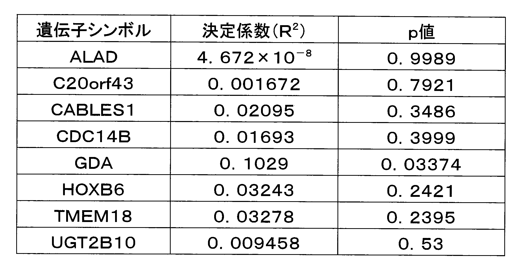
- the expression level of each gene was quantified using a real-time RT-PCR method using TaqMan TM Gene Expression Assays, and regression analysis was performed. The results were as shown in Table 4.
- the RPL7AP27 gene was excluded from analysis because an appropriate primer / probe for RT-PCR could not be prepared.
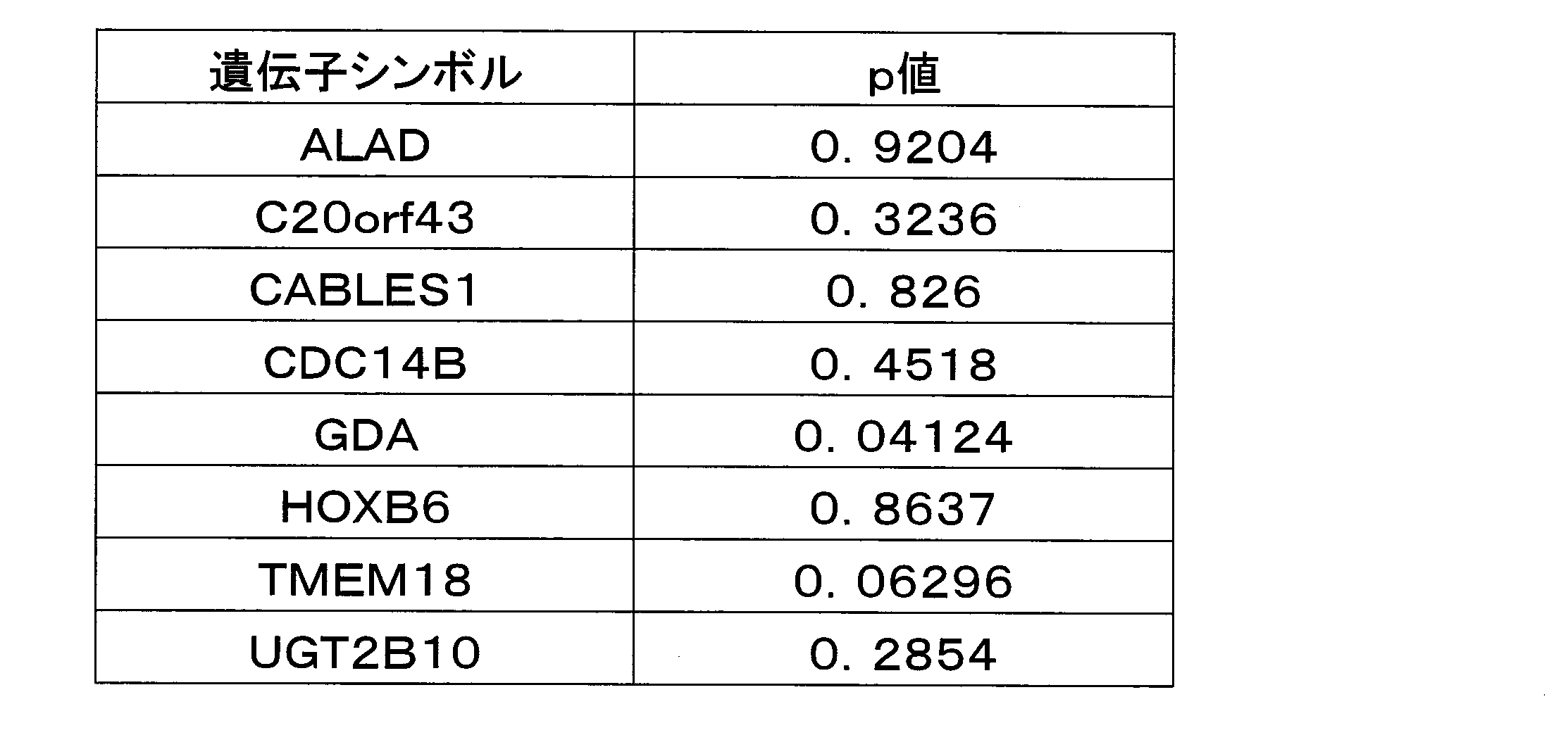
- t-test was performed by stratifying into a group with a high expression level and a group with a low expression level of each gene. The result was as shown in Table 5.
- the RPL7AP27 gene was excluded from analysis because an appropriate primer / probe for RT-PCR could not be prepared.
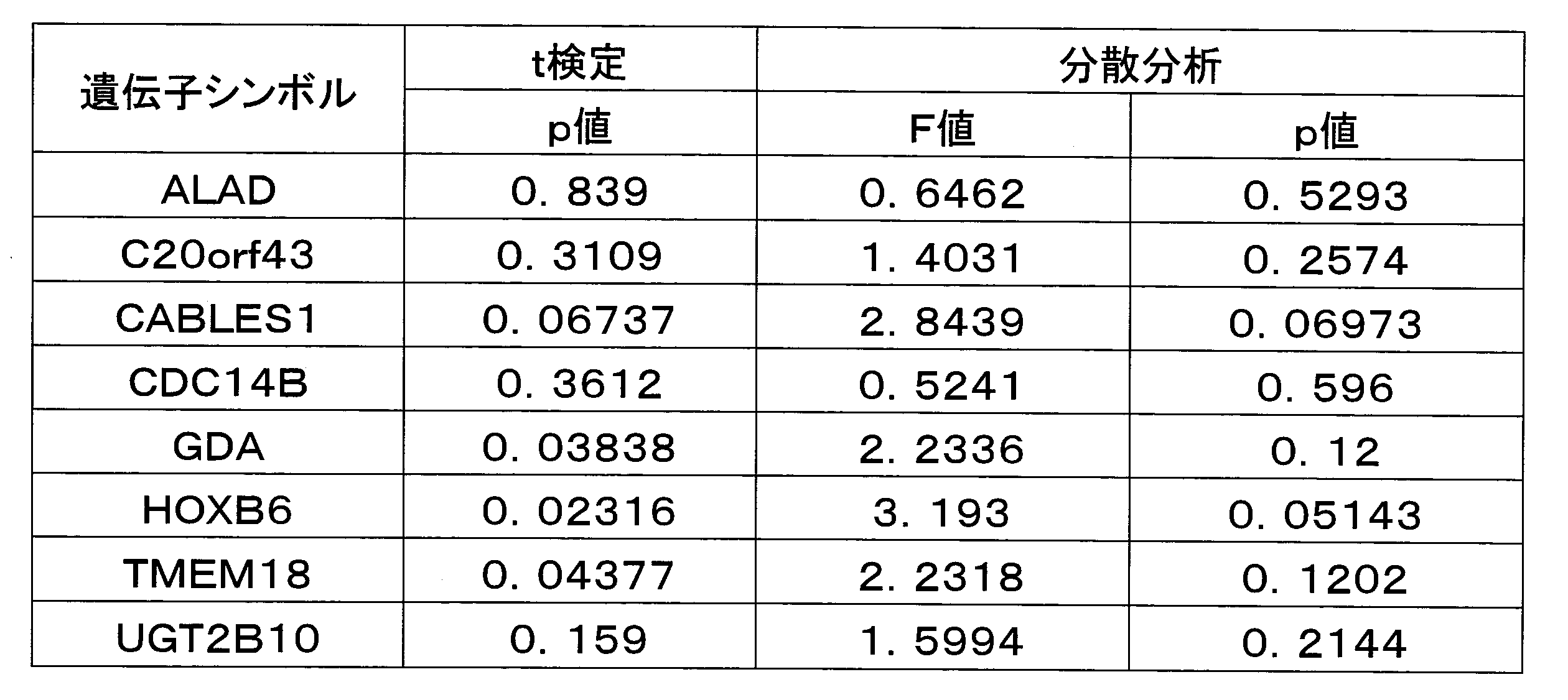
- the best tumor reduction effect was stratified into CR and PR groups and SD and PD groups according to the RECIST criteria, and t-test was conducted, and one-way analysis of variance was conducted stratified into three groups: CR and PR groups, SD group and PD group. .
- the result was as shown in Table 6.
- the expression levels of the CABLES1, GDA, HOXB6, and TMEM18 genes are higher in the SD and PD groups in which no response was observed compared to the CR and PR groups in which the response was observed. Indicated.
- the RPL7AP27 gene was excluded from analysis because an appropriate primer / probe for RT-PCR could not be prepared.
Abstract
Description
また、本発明は、遺伝子が、ALAD遺伝子、C20orf43遺伝子、GDA遺伝子、TMEM18遺伝子及びUGT2B10遺伝子である上記の判定マーカーを提供するものである。
また、本発明は、最良腫瘍縮小効果(比)を予測するものである上記の判定マーカーを提供するものである。
また、本発明は、発現量を測定する遺伝子が、ALAD遺伝子、C20orf43遺伝子、GDA遺伝子、TMEM18遺伝子及びUGT2B10遺伝子である上記の判定方法を提供するものである。
また、本発明は、次式(1)により最良腫瘍縮小効果(比)を算出することを特徴とする上記の判定方法を提供するものである。
(数1)
最良腫瘍縮小効果(比)=0.37664+96.360×A-8.5128×B+42.420×C+26.810×D+747.00×E・・・・・(1)
(式中、AはALAD遺伝子の発現量、BはC20orf43遺伝子の発現量、CはGDA遺伝子の発現量、DはTMEM18遺伝子の発現量、EはUGT2B10遺伝子の発現量を示す。)
C20orf43遺伝子とはGenBankアクセッション番号NM_016407に示される塩基配列のmRNAを発現する遺伝子及びそのホモログをいい、
CABLES1遺伝子とはGenBankアクセッション番号NM_138375に示される塩基配列のmRNAを発現する遺伝子及びそのホモログをいい、
CDC14B遺伝子とはGenBankアクセッション番号NM_033331に示される塩基配列のmRNAを発現する遺伝子及びそのホモログをいい、
GDA遺伝子とはGenBankアクセッション番号NM_004293に示される塩基配列のmRNAを発現する遺伝子及びそのホモログをいい、
HOXB6遺伝子とはGenBankアクセッション番号NM_018952に示される塩基配列のmRNAを発現する遺伝子及びそのホモログをいい、
RPL7AP27遺伝子とはEnterz Gene ID152663に示される遺伝子及びそのホモログをいい、
TMEM18遺伝子とはGenBankアクセッション番号NM_152834に示される塩基配列のmRNAを発現する遺伝子及びそのホモログをいい、
UGT2B10遺伝子とはGenBankアクセッション番号NM_001075に示される塩基配列のmRNAを発現する遺伝子及びそのホモログをいう。
また、遺伝子とは、2本鎖DNAのみならず、それを構成するセンス鎖及びアンチセンス鎖といった各1本鎖DNAを包含する趣旨であり、またその長さに何ら制限されるものではない。また、核酸(ポリヌクレオチド)としては、RNA、DNAを例示でき、DNAはcDNA、ゲノムDNA、合成DNA、RNAはmRNA、rRNA、siRNAが挙げられる。ここでポリヌクレオチドには、複数個の塩基配列からなるオリゴヌクレオチドも含まれる。
(数2)
最良腫瘍縮小効果(比)=0.37664+96.360×A-8.5128×B+42.420×C+26.810×D+747.00×E・・・・・(1)
(式中、AはALAD遺伝子の発現量、BはC20orf43遺伝子の発現量、CはGDA遺伝子の発現量、DはTMEM18遺伝子の発現量、EはUGT2B10遺伝子の発現量を示す。)
また、斯かる核酸断片は、その両端又は片端、好ましくは5'端に任意の数、好ましくは100個、より好ましくは20個、さらに好ましくは10個以下の塩基が付加された核酸断片であってもよい。
(a)NM_000031で示される塩基配列の一部若しくはそれと相補的な塩基配列からなる核酸断片において、1又は数個の塩基が欠失、置換若しくは付加された核酸断片。
(b)NM_000031で示される塩基配列の一部若しくはそれと相補的な塩基配列と90%以上、好ましくは95%以上、より好ましくは99%以上の同一性を有する塩基配列からなる核酸断片。
(c)NM_000031で示される塩基配列の一部若しくはそれと相補的な塩基配列からなるDNAとストリンジェントな条件下でハイブリダイズする塩基配列からなる核酸断片。
また、「ストリンジェントな条件」とは、2つのDNA断片がSambrook Jらによって記載されたような標準的なハイブリダイゼーション条件下で、相互にハイブリダイズすることを意味する(Expression of cloned genes in E.coli(Molecular Cloning:A laboratory manual(1989))Cold Spring harbor Laboratory Press,New York,USA,9.47-9.62及び11.45-11.61)。
1.mFOLFOX6療法によるヒト臨床試験
フルオロウラシル400mg/平方メートル(急速静注)、レボホリナート200mg/平方メートル、フルオロウラシル2,400~3,000mg/平方メートル(持続点滴静注)にオキサリプラチン85mg/平方メートルを併用投与するがん化学療法(mFOLFOX6療法)を実施したがん患者において、がん化学療法の効果と関連する遺伝子を明らかにし、これを検証するため前向きゲノム薬理学的臨床研究を行った。対象は根治切除不能ステージIV大腸癌薬物療法未治療例で、姑息的手術時に腫瘍検体の採取可能な症例とした。具体的な選択基準は、(1)組織学的に大腸がんの確定診断が得られている症例、(2)根治切除不能ステージIV大腸癌術後症例、(3)測定可能病変(Response Evaluation Criteria in Solid Tumors,RECIST)を有する症例、(4)生理機能(骨髄、肝、腎、心など)が十分保持されている症例で、仮登録及び本登録前1週間以内の検査値が以下の基準を満たすこと。白血球数4,000/μL以上12,000/μL以下、好中球数2,000/μL以上、血小板数100,000/μL以上、ヘモグロビン量9.0g/dl以上、血清AST・ALT 施設正常値上限の2倍以下(但し、肝転移症例は3倍以下)、血清総ビリルビン1.5mg/dl以下、血清クレアチニン1.5mg/dl以下、クレアチニン・クリアランス50mL/min以上、BUN 25mg/dl以下、CRP 1mg/dl以下、Performance Status(Eastern Cooperative Oncology Group:ECOG)の分類が0~2の症例、手術以外に前治療のない症例、手術から本登録までに21日以上経過している症例、予想生存期間が3ヶ月以上期待される症例、重篤な併存疾患、活動性の重複がんのない症例、年齢20歳以上75歳未満の症例、遺伝子解析のための組織が手術時に得られている症例、試料提供を含む研究への参加について、患者本人から文書による同意が術前に得られている症例、とし、除外基準を、(1)重篤な合併症を有する症例、(2)感染症を合併している症例、(3)下痢(水様便)のある症例、(4)腸管麻痺、腸閉塞、亜腸閉塞のある症例(本登録前のみ)、(5)間質性肺炎又は肺線維症のある症例、(6)多量の腹水、胸水のある症例、(7)黄疸のある症例、(8)治療を要する程度の虚血性心疾患、不整脈などの心疾患を有する症例(高血圧に伴う左室肥大や軽度の左室負荷、軽度の右脚ブロックなどは登録可)、(9)6ヶ月以内に発症した心筋梗塞の既往を有する症例、(10)肝硬変を合併している症例、(11)繰り返し輸血を要する消化管新鮮出血を認める症例、(12)向精神薬で治療中又は治療を要すると思われる精神障害を有する症例、(13)コントロール困難な糖尿病を合併している症例、(14)その他、重篤な術後合併症を有している症例、(15)他の薬剤に対して重篤な過敏症の既往歴のある症例、(16)妊娠中あるいは授乳中の女性、及び授子を希望する男女、(17)肝炎ウイルス、HIVウイルス、梅毒の陽性例、とした。mFOLFOX6療法は、術後28日以内に、投与を開始し、投与開始日をday 1として、2週間ごと1回投与1週休薬を1コースとして投与を行なった。
TOTAL RNAは54例のヒト大腸癌組織サンプルからRNeasyTM Minikit(Qiagen社製)を用い、添付プロトコールに従って抽出し、-80℃で保存した。
Quick Amp Labeling Kit(Agilent社製)を用いて、そのプロトコールに従いdouble-strand cDNA合成を行った。すなわち、total RNA 500ngを5.3μlにNuclease-free Waterで調整し、該キットに含まれる1.2μl T7 Promoter Primerとポジティブコントロールとして希釈した5μl Spike-Mix(Agilent社製)を混合し、計11.5μlを65℃で10分間加温した後、氷上で5分間急冷した。氷上で該キットに含まれる4μlの5×First-strand Buffer、1μlの10mM dNTP mix、2μlの0.1M DTT、0.5μlのRNase Inhibitor、1μlのMMLV Reverse Transcriptaseを加え、計20μlを40℃で2時間加温した。cDNA合成反応を停止させるため65℃で15分間加温した後、氷上で5分間急冷した。
引き続きQuick Amp Labeling Kit(Agilent社製)のプロトコールに従いin vitro transcription(IVT)反応を行い、cRNAを合成した。すなわち、該キットに含まれる20μlの4×Transcription Buffer、6μlの0.1M DTT、8μlのNTP mix、6.4μlの50% PEG、0.5μlのRNase Inhibitor、0.6μlのInorganic Pyrophosphatase、0.8μlのT7 RNA Polymerase、2.4μlのCyanine 3-CTP、15.3μlのNuclease-free waterをよく混合し、計60μlを(1)で調整した20μl cDNA溶液に添加した後、40℃で2時間加温した。反応終了後、RNeasy Mini Kit(Qiagen社製)を用いて添付プロトコールに従い、合成したcRNAを精製した。すなわち、反応液(80μl)に20μlのNuclease-free Waterを添加し、合計100μlの溶液に該キットに含まれる350μlのBufferRLTを添加してよく混合し、さらに250μlの100%エタノールを添加してよく混合し、全量(700μl)を該キットに含まれるRNeasy ミニスピンカラムに添加して、13,000rpmで30秒間遠心した。RNeasy ミニスピンカラムを新しい2mlチューブにセットし、該キットに含まれる500μlのBuffer RPEをカラムに添加して13,000rpmで30秒間遠心し、溶出液を除去した。再度ミニスピンカラムを元の2mlチューブにセットし、500μlのBuffer RPEをカラムに添加して13,000rpmで1分間遠心し、溶出液を除去した。ミニスピンカラムを新しい1.5mlチューブに移し、30μlのNuclease-free waterをメンブレン上に直接添加し、室温で1分間静置した後、13,000rpmで30秒間遠心し、cRNAサンプルを溶出した。
cRNAの定量はNanoDrop(Thermo scientific社製)で行い、サンプル溶液の260nmおよび280nm吸光度を測定し、cRNA濃度を定量するとともに、Cy3-CTP色素の取り込み率が9pmol/μg以上であることを確認した。cRNAの品質チェックはAgilent 2100 Bioanalyzerを用いて添付プロトコールに従って泳動し、スメアピークの長さが500塩基以上であることを確認した。
Gene Expression Hybridization Kit(Agilent社製)のプロトコールに従い、cRNAを断片化した。すなわち、1.65μgのcRNAが41.8μlになるよう、Nuclease-free waterでメスアップし、該キットに含まれる2.2μlの25×Fragmentation Bufferと11μlの10×Blocking Agentを添加し、60℃で30分間加熱後、氷上で1分間急冷した。断片化されたcRNAを含む55μlの溶液に、該キットに含まれる55μl 2×Hybridization Buffer HI-RPMを加え、計110μlのハイブリダイゼーション溶液を調整した。
ハイブリダイゼーションオーブン(Agilent社製)とハイブリダイゼーションローター(Agilent社製)を用いてプロトコールに従いハイブリダイズを行った。すなわち、(3)で調整したハイブリダイゼーション溶液をWhole Human Genome 4×44Kアレイ面にアプライした後、ガスケットスライド(Agilent社製)でカバーし、オリゴDNAマイクロアレイ用ハイブリダイゼーションチャンバ(Agilent社製)で固定した。固定したアレイをハイブリダイゼーションローターにセットし、ハイブリダイゼーションオーブンにて、65℃、10rpmで旋回させながら17時間加温した。
ハイブリダイゼーション後、オリゴDNAマイクロアレイ用ハイブリダイゼーションチャンバによって固定されていたマイクロアレイを取り出し、洗浄を行った。すなわち、0.005% Triton X-102を添加したGene Expression Wash Buffer 1(Agilent社製)を満たしたリザーバーにマイクロアレイを移し、スターラーバーで撹拌しながら室温で1分間洗浄した。引き続き、0.005% Triton X-102を添加したGene Expression Wash Buffer 2(Agilent社製)で満たしたスターラー付恒温槽にマイクロアレイを移し、スターラーバーで撹拌しながら37℃で1分間洗浄した。
洗浄が終了したマイクロアレイをスライドホルダにセットし、Agilent G2565BA(Agilent社製)スキャナに供し、蛍光パターンを読み取りTIFF imageとして保存した。TIFF imageをAgilent Feature Extraction Ver.9.5ソフトウエアで処理し、アレイ上の各遺伝子スポットのシグナル強度を数値化した。
上記で得られたシグナル強度データをGeneSpring GX(Agilent社製)マイクロアレイ遺伝子発現解析ソフトを用いてNormalizationを行い解析した。すなわち、スポットシグナルから背景シグナルを差し引き、その値が0.01未満は0.01とし、アレイの全スポットシグナルの3/4分位値で割り、底を2とした対数値に変換した値をそれぞれの遺伝子の標準化した相対的発現量とした。
DNAマイクロアレイ解析が可能であった37例の解析結果を用い、最良腫瘍縮小効果(比)を予測しうる遺伝子の探索を行った。37例から得られたDNAマイクロアレイの解析結果から、最良腫瘍縮小効果との関連について、ピアソンの積率相関解析およびスピアマンの順位相関解析を行い、相関係数Rの絶対値が0.5を超え、相関係数ρの検定によりp値が0.2未満、かつ、平均発現量の相対値が0.5を超える遺伝子として17遺伝子を特定した(表1)。また、参考症例4例を加えた41例で同様の解析を実施したところ、相関係数Rの絶対値が0.5を超え、相関係数ρの検定によりp値が0.05未満、かつ、平均発現量の相対値が0.5を超える遺伝子として10遺伝子を特定した(表2)。37例の解析結果から特定された17遺伝子と41例の解析結果から特定された10遺伝子のうち、両者に共通する9遺伝子を特定した(表3)。これら9遺伝子は、条件の異なる解析集団に共通して認められたものであり、より臨床における有用性があると判断し、それぞれの遺伝子と最良腫瘍縮小効果との相関性を評価した。


以上の結果、DNAマイクロアレイを用いて特定した感受性に関連する9遺伝子のうち、RPL7AP27遺伝子を除く8遺伝子について、単独で抗がん剤感受性と統計上有意な相関を示すものが認められた。また、これら8遺伝子について重回帰分析の手法を用い、特定された遺伝子群の定量的発現量を代入することで最良腫瘍縮小効果を予測する式を求め、その予測性を確認した。前述の遺伝子発現量を評価するために要するRNAが得られた症例44例を、予測式の作成に用いる26例(検討群)と、これを検証するための18例(検証群)に分け、リアルタイムRT-PCR法を用いて8遺伝子の発現量を求めた。検討群26例における8遺伝子の発現量を用い、重回帰分析の手法により最良腫瘍縮小効果を予測する式を求めた。その結果、最も良く最良腫瘍縮小効果を予測しうる式として、8遺伝子中5遺伝子(ALAD遺伝子、C20orf43遺伝子、GDA遺伝子、TMEM18遺伝子及びUGT2B10遺伝子)を用いた予測式
最良腫瘍縮小効果(腫瘍径ベースライン比)=0.37664+96.360×A-8.5128×B+42.420×C+26.810×D+747.00×E・・(1)
(式中、AはALAD遺伝子の発現量、BはC20orf43遺伝子の発現量、CはGDA遺伝子の発現量、DはTMEM18遺伝子の発現量、EはUGT2B10遺伝子の発現量を示す。)
Claims (16)
- ALAD遺伝子、C20orf43遺伝子、CABLES1遺伝子、CDC14B遺伝子、GDA遺伝子、HOXB6遺伝子、RPL7AP27遺伝子、TMEM18遺伝子及びUGT2B10遺伝子から選ばれる1以上の遺伝子からなる、オキサリプラチン又はその塩とフルオロウラシル又はその塩とレボホリナート又はその塩を含む抗がん剤の感受性判定マーカー。
- 遺伝子が、ALAD遺伝子、C20orf43遺伝子、GDA遺伝子、TMEM18遺伝子及びUGT2B10遺伝子である請求項1記載の判定マーカー。
- 最良腫瘍縮小効果(比)を予測するものである請求項1又は2記載の判定マーカー。
- 検体中のALAD遺伝子、C20orf43遺伝子、CABLES1遺伝子、CDC14B遺伝子、GDA遺伝子、HOXB6遺伝子、RPL7AP27遺伝子、TMEM18遺伝子及びUGT2B10遺伝子から選ばれる1以上の遺伝子の発現量を測定することを特徴とするオキサリプラチン又はその塩とフルオロウラシル又はその塩とレボホリナート又はその塩を含む抗がん剤の感受性判定方法。
- 発現量を測定する遺伝子が、ALAD遺伝子、C20orf43遺伝子、GDA遺伝子、TMEM18遺伝子及びUGT2B10遺伝子である請求項4記載の判定方法。
- 最良腫瘍縮小効果(比)を予測するものである請求項4又は5記載の判定方法。
- 次式(1)により最良腫瘍縮小効果(比)を算出することを特徴とする請求項6記載の判定方法。
(数1)
最良腫瘍縮小効果(比)=0.37664+96.360×A-8.5128×B+42.420×C+26.810×D+747.00×E・・・・・(1)
(式中、AはALAD遺伝子の発現量、BはC20orf43遺伝子の発現量、CはGDA遺伝子の発現量、DはTMEM18遺伝子の発現量、EはUGT2B10遺伝子の発現量を示す。) - 検体が、がんを有する被験者由来の生体試料である請求項4~7のいずれか1項記載の判定方法。
- 検体が、大腸がんを有する被験者由来の生体試料である請求項4~8のいずれか1項記載の判定方法。
- 遺伝子の発現量が該遺伝子由来のmRNA量である請求項4~9のいずれか1項記載の判定方法。
- 検体中のALAD遺伝子、C20orf43遺伝子、CABLES1遺伝子、CDC14B遺伝子、GDA遺伝子、HOXB6遺伝子、RPL7AP27遺伝子、TMEM18遺伝子及びUGT2B10遺伝子から選ばれる1以上の遺伝子の発現量を測定するためのプロトコールを含むことを特徴とする請求項4~10のいずれか1項記載の判定方法を実施するためのキット。
- 検体が、がんを有する被験者由来の生体試料である請求項11記載のキット。
- 検体が、大腸がんを有する被験者由来の生体試料である請求項11又は12記載のキット。
- 検体中のALAD遺伝子、C20orf43遺伝子、CABLES1遺伝子、CDC14B遺伝子、GDA遺伝子、HOXB6遺伝子、RPL7AP27遺伝子、TMEM18遺伝子及びUGT2B10遺伝子から選ばれる1以上の遺伝子の発現変動を指標とするオキサリプラチン又はその塩とフルオロウラシル又はその塩とレボホリナート又はその塩を含む抗がん剤に対する感受性亢進剤のスクリーニング方法。
- 請求項14記載の方法により得られたオキサリプラチン又はその塩とフルオロウラシル又はその塩とレボホリナート又はその塩を含む抗がん剤に対する感受性亢進剤。
- 請求項15記載の感受性亢進剤とオキサリプラチン又はその塩とフルオロウラシル又はその塩とレボホリナート又はその塩を含む抗がん剤を含有するがん治療用組成物。
Priority Applications (8)
| Application Number | Priority Date | Filing Date | Title |
|---|---|---|---|
| US13/991,318 US8980557B2 (en) | 2010-12-03 | 2011-12-02 | Marker for determination of sensitivity to triplet combination anti-cancer agent |
| KR1020137013890A KR101873079B1 (ko) | 2010-12-03 | 2011-12-02 | 3 제 병용 항암제의 감수성 판정 마커 |
| CN201180058258.1A CN103261413B (zh) | 2010-12-03 | 2011-12-02 | 三剂并用抗癌剂感受性判定标记 |
| AU2011337612A AU2011337612B2 (en) | 2010-12-03 | 2011-12-02 | Marker for determination of sensitivity to triplet combination anti-cancer agent |
| CA2819399A CA2819399C (en) | 2010-12-03 | 2011-12-02 | Marker for determination of sensitivity to triplet combination anti-cancer agent |
| EP11846035.1A EP2647708B1 (en) | 2010-12-03 | 2011-12-02 | Marker for determination of sensitivity to triplet combination anti-cancer agent |
| BR112013013656-1A BR112013013656B1 (pt) | 2010-12-03 | 2011-12-02 | processo para determinação da sensibilidade de um indivíduo a um agente anticâncer, kit para realização do dito processo bem como processo de triagem para um agente de aperfeiçoamento de sensibilidade a um agente anticâncer |
| JP2012546952A JP5980685B2 (ja) | 2010-12-03 | 2011-12-02 | 3剤併用抗がん剤の感受性判定マーカー |
Applications Claiming Priority (2)
| Application Number | Priority Date | Filing Date | Title |
|---|---|---|---|
| JP2010-270634 | 2010-12-03 | ||
| JP2010270634 | 2010-12-03 |
Publications (1)
| Publication Number | Publication Date |
|---|---|
| WO2012074085A1 true WO2012074085A1 (ja) | 2012-06-07 |
Family
ID=46172007
Family Applications (1)
| Application Number | Title | Priority Date | Filing Date |
|---|---|---|---|
| PCT/JP2011/077890 WO2012074085A1 (ja) | 2010-12-03 | 2011-12-02 | 3剤併用抗がん剤の感受性判定マーカー |
Country Status (9)
| Country | Link |
|---|---|
| US (1) | US8980557B2 (ja) |
| EP (1) | EP2647708B1 (ja) |
| JP (1) | JP5980685B2 (ja) |
| KR (1) | KR101873079B1 (ja) |
| CN (1) | CN103261413B (ja) |
| AU (1) | AU2011337612B2 (ja) |
| BR (1) | BR112013013656B1 (ja) |
| CA (1) | CA2819399C (ja) |
| WO (1) | WO2012074085A1 (ja) |
Cited By (6)
| Publication number | Priority date | Publication date | Assignee | Title |
|---|---|---|---|---|
| CN103074436A (zh) * | 2013-01-25 | 2013-05-01 | 海尔施生物医药股份有限公司 | 一种指导5-氟尿嘧啶用药的多重基因检测试剂盒及其检测方法 |
| WO2016167346A1 (ja) * | 2015-04-17 | 2016-10-20 | 学校法人 川崎学園 | アスベスト曝露歴または中皮腫の検査方法 |
| JP2019216746A (ja) * | 2012-12-14 | 2019-12-26 | マインデラ コーポレイション | バイオマーカーの検出および収集のための方法およびデバイス |
| US11315673B2 (en) * | 2018-11-30 | 2022-04-26 | Caris Mpi, Inc. | Next-generation molecular profiling |
| JP7370046B2 (ja) | 2019-10-16 | 2023-10-27 | 医療法人社団キャンサーフリートピア | 抗癌剤の選択方法 |
| US11842805B2 (en) | 2019-12-02 | 2023-12-12 | Caris Mpi, Inc. | Pan-cancer platinum response predictor |
Families Citing this family (3)
| Publication number | Priority date | Publication date | Assignee | Title |
|---|---|---|---|---|
| EP3550306B1 (en) * | 2014-08-26 | 2021-03-03 | Keio University | Anti-cancer agent sensitivity-determining marker |
| CN110476066A (zh) * | 2017-03-31 | 2019-11-19 | 学校法人庆应义塾 | 并用抗癌剂的感受性判定标记 |
| CN112662782B (zh) * | 2020-12-30 | 2023-03-14 | 华南农业大学 | 鸡屠宰性状相关的tmem18基因分子标记及应用 |
Citations (2)
| Publication number | Priority date | Publication date | Assignee | Title |
|---|---|---|---|---|
| JP2009050189A (ja) * | 2007-08-24 | 2009-03-12 | Sumitomo Bakelite Co Ltd | 抗癌剤の有効性予測方法 |
| WO2010103851A1 (ja) * | 2009-03-13 | 2010-09-16 | 学校法人 埼玉医科大学 | イリノテカンの感受性判定方法及びその利用 |
-
2011
- 2011-12-02 EP EP11846035.1A patent/EP2647708B1/en active Active
- 2011-12-02 WO PCT/JP2011/077890 patent/WO2012074085A1/ja active Application Filing
- 2011-12-02 CN CN201180058258.1A patent/CN103261413B/zh active Active
- 2011-12-02 AU AU2011337612A patent/AU2011337612B2/en active Active
- 2011-12-02 KR KR1020137013890A patent/KR101873079B1/ko active IP Right Grant
- 2011-12-02 BR BR112013013656-1A patent/BR112013013656B1/pt active IP Right Grant
- 2011-12-02 JP JP2012546952A patent/JP5980685B2/ja active Active
- 2011-12-02 US US13/991,318 patent/US8980557B2/en active Active
- 2011-12-02 CA CA2819399A patent/CA2819399C/en active Active
Patent Citations (2)
| Publication number | Priority date | Publication date | Assignee | Title |
|---|---|---|---|---|
| JP2009050189A (ja) * | 2007-08-24 | 2009-03-12 | Sumitomo Bakelite Co Ltd | 抗癌剤の有効性予測方法 |
| WO2010103851A1 (ja) * | 2009-03-13 | 2010-09-16 | 学校法人 埼玉医科大学 | イリノテカンの感受性判定方法及びその利用 |
Non-Patent Citations (6)
| Title |
|---|
| "Molecular Cloning: A laboratory manual", 1989, COLD SPRING HARBOR LABORATORY PRESS |
| CHUA, W. ET AL.: "Molecular markers of response and toxicity to FOLFOX chemotherapy in metastatic colorectal cancer.", BR. J. CANCER, vol. 101, no. 6, 2009, pages 998 - 1004, XP055108167 * |
| J. CLIN. ONCOL., vol. 26, 2008, pages 2690 - 2698 |
| LEE, J.J. ET AL.: "An update on treatment advances for the first-line therapy of metastatic colorectal cancer.", CANCER J., vol. 13, no. 5, 2007, pages 276 - 281, XP008108655 * |
| ROSS, J.S. ET AL.: "Biomarker-Based Prediction of Response to Therapy for Colorectal Cancer.", AM. J. CLIN. PATHOL., vol. 134, no. 3, September 2010 (2010-09-01), pages 478 - 490, XP055108170 * |
| See also references of EP2647708A4 |
Cited By (9)
| Publication number | Priority date | Publication date | Assignee | Title |
|---|---|---|---|---|
| JP2019216746A (ja) * | 2012-12-14 | 2019-12-26 | マインデラ コーポレイション | バイオマーカーの検出および収集のための方法およびデバイス |
| CN103074436A (zh) * | 2013-01-25 | 2013-05-01 | 海尔施生物医药股份有限公司 | 一种指导5-氟尿嘧啶用药的多重基因检测试剂盒及其检测方法 |
| CN103074436B (zh) * | 2013-01-25 | 2014-07-16 | 宁波海尔施基因科技有限公司 | 一种指导5-氟尿嘧啶用药的多重基因检测试剂盒及其检测方法 |
| WO2016167346A1 (ja) * | 2015-04-17 | 2016-10-20 | 学校法人 川崎学園 | アスベスト曝露歴または中皮腫の検査方法 |
| JPWO2016167346A1 (ja) * | 2015-04-17 | 2018-02-08 | 学校法人 川崎学園 | アスベスト曝露歴または中皮腫の検査方法 |
| US11315673B2 (en) * | 2018-11-30 | 2022-04-26 | Caris Mpi, Inc. | Next-generation molecular profiling |
| JP7462632B2 (ja) | 2018-11-30 | 2024-04-05 | カリス エムピーアイ インコーポレイテッド | 次世代分子プロファイリング |
| JP7370046B2 (ja) | 2019-10-16 | 2023-10-27 | 医療法人社団キャンサーフリートピア | 抗癌剤の選択方法 |
| US11842805B2 (en) | 2019-12-02 | 2023-12-12 | Caris Mpi, Inc. | Pan-cancer platinum response predictor |
Also Published As
| Publication number | Publication date |
|---|---|
| US20130302327A1 (en) | 2013-11-14 |
| KR20130140046A (ko) | 2013-12-23 |
| EP2647708A4 (en) | 2014-04-23 |
| JP5980685B2 (ja) | 2016-08-31 |
| JPWO2012074085A1 (ja) | 2014-05-19 |
| CA2819399A1 (en) | 2012-06-07 |
| US8980557B2 (en) | 2015-03-17 |
| EP2647708A1 (en) | 2013-10-09 |
| CN103261413B (zh) | 2015-09-16 |
| KR101873079B1 (ko) | 2018-06-29 |
| EP2647708B1 (en) | 2017-04-26 |
| CN103261413A (zh) | 2013-08-21 |
| BR112013013656A2 (pt) | 2016-09-06 |
| BR112013013656B1 (pt) | 2021-01-26 |
| AU2011337612A1 (en) | 2013-06-20 |
| CA2819399C (en) | 2021-03-23 |
| AU2011337612B2 (en) | 2016-06-16 |
Similar Documents
| Publication | Publication Date | Title |
|---|---|---|
| JP5980685B2 (ja) | 3剤併用抗がん剤の感受性判定マーカー | |
| US9066963B2 (en) | Methods of treating breast cancer with anthracycline therapy | |
| JP5774473B2 (ja) | イリノテカンの感受性判定方法及びその利用 | |
| WO2014148557A1 (ja) | Egfr阻害剤感受性予測方法 | |
| EP3259369B1 (en) | Methods and kits for the molecular subtyping of bladder cancer | |
| JP2016535079A (ja) | 核酸生体マーカー及びその使用 | |
| WO2015072555A1 (ja) | Vegf阻害剤長期奏功性予測方法 | |
| Yang et al. | Enhancing detection of circulating tumor cells with activating KRAS oncogene in patients with colorectal cancer by weighted chemiluminescent membrane array method | |
| WO2012076582A1 (en) | Agtr1 as a marker for bevacizumab combination therapies | |
| JP2006520197A (ja) | チミジレートシンターゼ遺伝子座におけるヘテロ接合性の喪失に基づいて化学療法計画を決定する方法 | |
| CN114717312B (zh) | 用于膀胱癌分子亚型分型的方法和试剂盒 | |
| TW201400810A (zh) | 對胃癌患者選擇化學療法之方法 | |
| JP2011147374A (ja) | 外科切除された進行胃癌における術後補助化学療法の有効性予測方法 |
Legal Events
| Date | Code | Title | Description |
|---|---|---|---|
| 121 | Ep: the epo has been informed by wipo that ep was designated in this application |
Ref document number: 11846035 Country of ref document: EP Kind code of ref document: A1 |
|
| ENP | Entry into the national phase |
Ref document number: 2012546952 Country of ref document: JP Kind code of ref document: A |
|
| ENP | Entry into the national phase |
Ref document number: 2819399 Country of ref document: CA |
|
| ENP | Entry into the national phase |
Ref document number: 20137013890 Country of ref document: KR Kind code of ref document: A |
|
| NENP | Non-entry into the national phase |
Ref country code: DE |
|
| REEP | Request for entry into the european phase |
Ref document number: 2011846035 Country of ref document: EP |
|
| WWE | Wipo information: entry into national phase |
Ref document number: 13991318 Country of ref document: US Ref document number: 2011846035 Country of ref document: EP |
|
| ENP | Entry into the national phase |
Ref document number: 2011337612 Country of ref document: AU Date of ref document: 20111202 Kind code of ref document: A |
|
| REG | Reference to national code |
Ref country code: BR Ref legal event code: B01A Ref document number: 112013013656 Country of ref document: BR |
|
| ENP | Entry into the national phase |
Ref document number: 112013013656 Country of ref document: BR Kind code of ref document: A2 Effective date: 20130603 |