US8926681B2 - Vascular remodeling device - Google Patents
Vascular remodeling device Download PDFInfo
- Publication number
- US8926681B2 US8926681B2 US13/016,858 US201113016858A US8926681B2 US 8926681 B2 US8926681 B2 US 8926681B2 US 201113016858 A US201113016858 A US 201113016858A US 8926681 B2 US8926681 B2 US 8926681B2
- Authority
- US
- United States
- Prior art keywords
- filaments
- aneurysm
- catheter
- bifurcation
- junction
- Prior art date
- Legal status (The legal status is an assumption and is not a legal conclusion. Google has not performed a legal analysis and makes no representation as to the accuracy of the status listed.)
- Active, expires
Links
Images


Classifications
-
- A—HUMAN NECESSITIES
- A61—MEDICAL OR VETERINARY SCIENCE; HYGIENE
- A61B—DIAGNOSIS; SURGERY; IDENTIFICATION
- A61B17/00—Surgical instruments, devices or methods, e.g. tourniquets
- A61B17/12—Surgical instruments, devices or methods, e.g. tourniquets for ligaturing or otherwise compressing tubular parts of the body, e.g. blood vessels, umbilical cord
- A61B17/12022—Occluding by internal devices, e.g. balloons or releasable wires
- A61B17/12099—Occluding by internal devices, e.g. balloons or releasable wires characterised by the location of the occluder
- A61B17/12109—Occluding by internal devices, e.g. balloons or releasable wires characterised by the location of the occluder in a blood vessel
- A61B17/12113—Occluding by internal devices, e.g. balloons or releasable wires characterised by the location of the occluder in a blood vessel within an aneurysm
- A61B17/12118—Occluding by internal devices, e.g. balloons or releasable wires characterised by the location of the occluder in a blood vessel within an aneurysm for positioning in conjunction with a stent
-
- A—HUMAN NECESSITIES
- A61—MEDICAL OR VETERINARY SCIENCE; HYGIENE
- A61B—DIAGNOSIS; SURGERY; IDENTIFICATION
- A61B17/00—Surgical instruments, devices or methods, e.g. tourniquets
- A61B17/12—Surgical instruments, devices or methods, e.g. tourniquets for ligaturing or otherwise compressing tubular parts of the body, e.g. blood vessels, umbilical cord
- A61B17/12022—Occluding by internal devices, e.g. balloons or releasable wires
- A61B17/12099—Occluding by internal devices, e.g. balloons or releasable wires characterised by the location of the occluder
- A61B17/12109—Occluding by internal devices, e.g. balloons or releasable wires characterised by the location of the occluder in a blood vessel
-
- A—HUMAN NECESSITIES
- A61—MEDICAL OR VETERINARY SCIENCE; HYGIENE
- A61B—DIAGNOSIS; SURGERY; IDENTIFICATION
- A61B17/00—Surgical instruments, devices or methods, e.g. tourniquets
- A61B17/12—Surgical instruments, devices or methods, e.g. tourniquets for ligaturing or otherwise compressing tubular parts of the body, e.g. blood vessels, umbilical cord
- A61B17/12022—Occluding by internal devices, e.g. balloons or releasable wires
- A61B17/12131—Occluding by internal devices, e.g. balloons or releasable wires characterised by the type of occluding device
- A61B17/1214—Coils or wires
- A61B17/12145—Coils or wires having a pre-set deployed three-dimensional shape
-
- A—HUMAN NECESSITIES
- A61—MEDICAL OR VETERINARY SCIENCE; HYGIENE
- A61B—DIAGNOSIS; SURGERY; IDENTIFICATION
- A61B17/00—Surgical instruments, devices or methods, e.g. tourniquets
- A61B17/12—Surgical instruments, devices or methods, e.g. tourniquets for ligaturing or otherwise compressing tubular parts of the body, e.g. blood vessels, umbilical cord
- A61B17/12022—Occluding by internal devices, e.g. balloons or releasable wires
- A61B17/12131—Occluding by internal devices, e.g. balloons or releasable wires characterised by the type of occluding device
- A61B17/12168—Occluding by internal devices, e.g. balloons or releasable wires characterised by the type of occluding device having a mesh structure
- A61B17/12172—Occluding by internal devices, e.g. balloons or releasable wires characterised by the type of occluding device having a mesh structure having a pre-set deployed three-dimensional shape
-
- A—HUMAN NECESSITIES
- A61—MEDICAL OR VETERINARY SCIENCE; HYGIENE
- A61L—METHODS OR APPARATUS FOR STERILISING MATERIALS OR OBJECTS IN GENERAL; DISINFECTION, STERILISATION OR DEODORISATION OF AIR; CHEMICAL ASPECTS OF BANDAGES, DRESSINGS, ABSORBENT PADS OR SURGICAL ARTICLES; MATERIALS FOR BANDAGES, DRESSINGS, ABSORBENT PADS OR SURGICAL ARTICLES
- A61L31/00—Materials for other surgical articles, e.g. stents, stent-grafts, shunts, surgical drapes, guide wires, materials for adhesion prevention, occluding devices, surgical gloves, tissue fixation devices
- A61L31/04—Macromolecular materials
- A61L31/041—Mixtures of macromolecular compounds
-
- A—HUMAN NECESSITIES
- A61—MEDICAL OR VETERINARY SCIENCE; HYGIENE
- A61L—METHODS OR APPARATUS FOR STERILISING MATERIALS OR OBJECTS IN GENERAL; DISINFECTION, STERILISATION OR DEODORISATION OF AIR; CHEMICAL ASPECTS OF BANDAGES, DRESSINGS, ABSORBENT PADS OR SURGICAL ARTICLES; MATERIALS FOR BANDAGES, DRESSINGS, ABSORBENT PADS OR SURGICAL ARTICLES
- A61L31/00—Materials for other surgical articles, e.g. stents, stent-grafts, shunts, surgical drapes, guide wires, materials for adhesion prevention, occluding devices, surgical gloves, tissue fixation devices
- A61L31/04—Macromolecular materials
- A61L31/06—Macromolecular materials obtained otherwise than by reactions only involving carbon-to-carbon unsaturated bonds
-
- A—HUMAN NECESSITIES
- A61—MEDICAL OR VETERINARY SCIENCE; HYGIENE
- A61L—METHODS OR APPARATUS FOR STERILISING MATERIALS OR OBJECTS IN GENERAL; DISINFECTION, STERILISATION OR DEODORISATION OF AIR; CHEMICAL ASPECTS OF BANDAGES, DRESSINGS, ABSORBENT PADS OR SURGICAL ARTICLES; MATERIALS FOR BANDAGES, DRESSINGS, ABSORBENT PADS OR SURGICAL ARTICLES
- A61L31/00—Materials for other surgical articles, e.g. stents, stent-grafts, shunts, surgical drapes, guide wires, materials for adhesion prevention, occluding devices, surgical gloves, tissue fixation devices
- A61L31/14—Materials characterised by their function or physical properties, e.g. injectable or lubricating compositions, shape-memory materials, surface modified materials
- A61L31/148—Materials at least partially resorbable by the body
-
- C—CHEMISTRY; METALLURGY
- C08—ORGANIC MACROMOLECULAR COMPOUNDS; THEIR PREPARATION OR CHEMICAL WORKING-UP; COMPOSITIONS BASED THEREON
- C08L—COMPOSITIONS OF MACROMOLECULAR COMPOUNDS
- C08L67/00—Compositions of polyesters obtained by reactions forming a carboxylic ester link in the main chain; Compositions of derivatives of such polymers
- C08L67/04—Polyesters derived from hydroxycarboxylic acids, e.g. lactones
-
- A—HUMAN NECESSITIES
- A61—MEDICAL OR VETERINARY SCIENCE; HYGIENE
- A61B—DIAGNOSIS; SURGERY; IDENTIFICATION
- A61B17/00—Surgical instruments, devices or methods, e.g. tourniquets
- A61B2017/00004—(bio)absorbable, (bio)resorbable, resorptive
-
- A—HUMAN NECESSITIES
- A61—MEDICAL OR VETERINARY SCIENCE; HYGIENE
- A61B—DIAGNOSIS; SURGERY; IDENTIFICATION
- A61B17/00—Surgical instruments, devices or methods, e.g. tourniquets
- A61B17/12—Surgical instruments, devices or methods, e.g. tourniquets for ligaturing or otherwise compressing tubular parts of the body, e.g. blood vessels, umbilical cord
- A61B17/12022—Occluding by internal devices, e.g. balloons or releasable wires
- A61B2017/1205—Introduction devices
- A61B2017/12054—Details concerning the detachment of the occluding device from the introduction device
- A61B2017/12063—Details concerning the detachment of the occluding device from the introduction device electrolytically detachable
-
- A—HUMAN NECESSITIES
- A61—MEDICAL OR VETERINARY SCIENCE; HYGIENE
- A61F—FILTERS IMPLANTABLE INTO BLOOD VESSELS; PROSTHESES; DEVICES PROVIDING PATENCY TO, OR PREVENTING COLLAPSING OF, TUBULAR STRUCTURES OF THE BODY, e.g. STENTS; ORTHOPAEDIC, NURSING OR CONTRACEPTIVE DEVICES; FOMENTATION; TREATMENT OR PROTECTION OF EYES OR EARS; BANDAGES, DRESSINGS OR ABSORBENT PADS; FIRST-AID KITS
- A61F2/00—Filters implantable into blood vessels; Prostheses, i.e. artificial substitutes or replacements for parts of the body; Appliances for connecting them with the body; Devices providing patency to, or preventing collapsing of, tubular structures of the body, e.g. stents
- A61F2/02—Prostheses implantable into the body
- A61F2/04—Hollow or tubular parts of organs, e.g. bladders, tracheae, bronchi or bile ducts
- A61F2/06—Blood vessels
- A61F2/07—Stent-grafts
-
- A—HUMAN NECESSITIES
- A61—MEDICAL OR VETERINARY SCIENCE; HYGIENE
- A61L—METHODS OR APPARATUS FOR STERILISING MATERIALS OR OBJECTS IN GENERAL; DISINFECTION, STERILISATION OR DEODORISATION OF AIR; CHEMICAL ASPECTS OF BANDAGES, DRESSINGS, ABSORBENT PADS OR SURGICAL ARTICLES; MATERIALS FOR BANDAGES, DRESSINGS, ABSORBENT PADS OR SURGICAL ARTICLES
- A61L2430/00—Materials or treatment for tissue regeneration
- A61L2430/36—Materials or treatment for tissue regeneration for embolization or occlusion, e.g. vaso-occlusive compositions or devices
Definitions
- the present application generally relates to vascular remodeling devices and to the manner of their positioning in vessels, and, more particularly, to generally spherical remodeling devices and to the matter of their positioning at the junction of neurovascular bifurcations having an aneurysm.
- the aneurysms 10 , 20 may be difficult to treat with embolization coils alone because the coils may be prone to herniating into parent vessels, as illustrated in FIGS. 3A and 3B .
- Herniation of coils may cause arterial occlusion, stroke, and/or death.
- the efferent vessels of the bifurcation may be at substantially different angles, have substantially different sizes, and/or be a different quantity (e.g., three or more).
- a different quantity e.g., three or more
- the aneurysm 20 of the bifurcation may be offset with respect to the junction (e.g., having a neck substantially open to one efferent vessel), tilted with respect to a plane created by the vessels (e.g., into or out of the page), etc.
- the junction e.g., having a neck substantially open to one efferent vessel
- tilted with respect to a plane created by the vessels e.g., into or out of the page
- tubular neck remodeling devices for example NeuroformTM, available from Boston Scientific, and EnterpriseTM, available from Cordis Neurovascular, may be used to keep coils or other materials within the fundus of the aneurysm and out of the vessels.
- Tubular remodeling devices generally consist of a braided wire or cut metallic stent or stents covering the neck of the aneurysm so that materials introduced into the fundus of the aneurysm do not herniate out of the aneurysm.
- tubular remodeling devices 40 are generally useful for side wall aneurysms 10 .
- tubular remodeling devices 42 , 44 are generally less useful for aneurysms 20 at bifurcations, for example because shaping the remodeling devices to preserve blood flow through the afferent and efferent vessels while also inhibiting herniation of coils 28 out of the aneurysm 20 can be difficult.
- a generally spherical vascular remodeling device is provided.
- the device is permanently positionable at a junction of afferent and efferent vessels of a bifurcation (e.g., a neurovascular bifurcation) having an aneurysm having a fundus and a neck.
- Positioning may comprise deployment from a catheter and mechanical or electrolytic release from the catheter. After positioning the device at the junction, the device can lock into place across the arterial ostia and the neck of the aneurysm, substantially conforming to the shape of the junction.
- the device After positioning the device at the junction, the device acts as a scaffolding to inhibit or prevent herniation or prolapse of objects such as embolization coils and thrombi out of the neck of the aneurysm.
- Embolic material may be inserted in the fundus of the aneurysm before or after positioning the device.
- the device permits perfusion of fluid (e.g., blood) to the efferent vessels.
- the device may have a first end, a second end substantially opposite to the first end, and a plurality of polymer filaments extending between and coupled at the first end and the second end. Certain such devices may be football shaped, pumpkin shaped, or twisted.
- Radiopaque markers may be placed at one or both ends of the device and/or at least one of the loops or filaments may comprise a radiopaque material (e.g., platinum).
- a method of treating an aneurysm at a junction of a bifurcation having an afferent vessel and efferent vessels is provided.
- the aneurysm has a neck and a fundus.
- the method comprises advancing a catheter proximate to the junction of the bifurcation.
- the catheter at least partially contains a generally spherical vascular remodeling device in a compressed state.
- the device comprises a plurality of polymer filaments.
- the method further comprises positioning the device at the junction of the bifurcation and withdrawing the catheter and leaving the device at the junction of the bifurcation.
- the device acts as a scaffolding to inhibit herniation of objects out of the neck of the aneurysm after withdrawal of the delivery catheter.
- the device permits perfusion of fluid to the efferent vessels.
- a generally spherical remodeling device comprises a first end, a second end substantially opposite to the first end, and a plurality of polymer filaments extending between the first end and the second end and coupled at the first end and the second end.
- the device is configured to be positioned at a junction of a bifurcation comprising at least one afferent vessel, efferent vessels, and an aneurysm having a neck after withdrawal of a delivery catheter.
- the device is configured to act as a scaffolding to inhibit herniation of objects out of the neck of the aneurysm.
- the device is configured to permit perfusion of fluid to the efferent vessels.
- a remodeling device comprising a plurality of polymer arcuate loops forming a generally spherical shape.
- the device is configured to be positioned at a junction of a bifurcation having an aneurysm after withdrawal of a delivery catheter.
- the device is configured to act as a scaffolding to inhibit matter from herniating out of the aneurysm.
- the device is configured to permit perfusion of blood to efferent vessels of the bifurcation.
- FIG. 1 illustrates an example embodiment of a side wall aneurysm.
- FIG. 2 illustrates an example embodiment of a bifurcation having an aneurysm.
- FIG. 3A illustrates an example embodiment of a side wall aneurysm with herniating embolization coils.
- FIG. 3B illustrates an example embodiment of a bifurcation having an aneurysm with herniating embolization coils.
- FIG. 4A illustrates an example embodiment of a side wall aneurysm treated with embolization coils and a tubular remodeling device.
- FIGS. 4B and 4C illustrates example embodiments of a bifurcation having an aneurysm treated with embolization coils and tubular remodeling devices.
- FIG. 5 illustrates an example embodiment of a generally spherical vascular remodeling device.
- FIGS. 6A-6C illustrate an example embodiment of a method for treating an aneurysm using the device of FIG. 5 .
- FIGS. 7A-7C illustrate another example embodiment of a method for treating an aneurysm using the device of FIG. 5 .
- FIG. 8 illustrates another example embodiment of a generally spherical vascular remodeling device.
- FIGS. 9A-9C illustrate an example embodiment of a method for treating an aneurysm using the device of FIG. 8 .
- FIGS. 10A-10C illustrate another example embodiment of a method for treating an aneurysm using the device of FIG. 8 .
- FIG. 11 illustrates yet another example embodiment of a generally spherical vascular remodeling device.
- FIG. 12 illustrates an example embodiment of treating an aneurysm using the device of FIG. 11 .
- FIG. 13 illustrates still another example embodiment of a generally spherical vascular remodeling device.
- FIG. 14 illustrates an example embodiment of a generally spherical vascular remodeling device at a stage of an example manufacturing process.
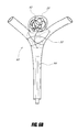
- FIG. 5 illustrates an example embodiment of a generally spherical vascular remodeling device 50 .
- the device 50 may be more compliant than the vasculature in which it is deployed such that it may be somewhat misshapen (e.g., non-spherical, for example as illustrated in FIG. 6B ) after being deployed, and that the phrase “generally spherical” describes the shape of the device 50 when in an expanded (e.g., fully expanded) state. Additionally, the phrase “generally spherical” distinguishes the device 50 , which is generally uniform in each dimension in an expanded state, from tubular stents having a small radial dimension and a large longitudinal dimension in an expanded state.
- an outer periphery of the device has a shape that deviates by between about 10% and about 25% from an outer periphery of a mathematically perfect sphere.
- the device 50 has a length and a width that are within less than about 33% of each other (e.g., having a length of 6 mm and a width of 8 mm, having a length of 6 mm and a width of 8 mm).
- the width is greater than the length may be advantageous due to a difference in porosity at a midpoint and an end proximate to an aneurysm.
- Embodiments in which the length is greater than the width may be advantageous for positioning a portion of the device 50 in a portion of the aneurysm 20 (e.g., to aid in embolization).
- the device 50 comprises a plurality of generally circular loops 52 coupled together. Coupling of the loops 52 may comprise adhering, welding, soldering, interlacing (e.g., some loops 52 being over or under other loops 52 ), intertwining, meshing, combinations thereof, and the like.
- the device 50 comprises a lead or tail 53 , which may be used for releasing and/or retracting the device 50 after deployment, as described herein.
- the device 50 comprises a cut metallic sphere, a single filament, a plurality of non-circular filaments (e.g., arcuate segments), etc.
- each loop 52 forms a plane and the intersections of the planes are substantially parallel (e.g., as illustrated in FIG. 7A ).
- At least some of the loops 52 or filaments comprise a self-expanding and/or a shape-memory material (e.g., comprising Nitinol, CoCr alloy, etc.), thereby causing the device 50 to be self-expanding under certain conditions (e.g., not restrained by a catheter).
- at least one of the loops 52 comprises a different material than others of the loops 52 (e.g., some loops 52 comprising Nitinol and some loops 52 comprising Nitinol and platinum).
- at least one of the loops 52 comprises a radiopaque material (e.g., platinum).
- an even number of loops 52 comprises a radiopaque material (e.g., platinum).
- at least one of the loops 52 comprises a radiopaque material (e.g., platinum) at least partially wrapped (e.g., coiled) around a self-expanding material (e.g., Nitinol).
- at least one of the loops 52 comprises a self-expanding material with a radiopaque core (e.g., Nitinol with a platinum core) or a radiopaque coating (e.g., Nitinol coated with platinum, tantalum, etc.
- the loops 52 have a substantially circular or ovoid cross section (e.g., embodiments, in which the loops 52 comprise separate wires). In some embodiments, the loops 52 have a substantially rectangular or flat cross section (e.g., embodiments, in which the loops 52 comprise uncut portions of a metallic tube). Other shapes of loops 52 and combinations of shapes of loops 52 are also possible.
- the plurality of loops 52 comprises between about six and about twelve loops 52 . In certain embodiments, the plurality of loops 52 comprises at least about six loops 52 , at least about eight loops 52 , or at least about twelve loops 52 . Other numbers of loops 52 are also possible.
- At least some of the loops 52 or filaments comprise a polymer. In some embodiments, at least some of the loops 52 or filaments comprise a polymer that is bioabsorbable. In certain embodiments, at least some of the loops 52 or filaments comprise polyglycolic acid (PGA), polylactic acid (PLA), poly(lactic-co-glycolic acid) (PLGA), poly-epsilon-caprolactone (PCL), naturally-derived bioabsorbable polymers (NDB), or combinations thereof (e.g., a first group of the loops 52 comprising PGA and a second group of the loops 52 comprising PLA, PLGA, PCL, and/or NDB, a first group of the loops 52 comprising PLA and a second group of the loops 52 comprising PGA, PLGA, PCL, and/or NDB, a first group of the loops 52 comprising PLGA and a second group of the loops 52 comprising PLA, PGA, PCL, and/or NDB,
- PGA, PLA, PLGA, PCL, and NDB are all bioabsorbable; however they have different rates of bioabsorption.
- the bioabsorption rates of a single polymer can also vary based on, for example, blood characteristics, blood flow, loop 52 dimensions, etc.
- PLA has the longest bioabsorption rate.
- the bioabsorption rate of PLA is at least about ten months.
- the bioabsorption rate of PLA is at least about one year.
- the bioabsorption rate of PLA is at least about fourteen months.
- the bioabsorption rate of PLA is between about 10 months and about 14 months (e.g., about one year).
- the bioabsorption rate of PGA is between about 1 week and about 3 weeks. In some embodiments, the bioabsorption rate of PGA is between about 2 weeks and about 4 weeks.
- the bioabsorption rates of PLGA, PCL, and NDB are generally between the bioabsorption rates of PLA and PGA, and may depend on parameters such as, for example, molecular weight (e.g., generally the higher the molecular weight, the longer the bioabsorption rate), structure (e.g., depending on the arrangement of repeating units), etc. of the polymer. In some embodiments, PLGA, PCL, and NDB may have a bioabsorption rate between about 4 weeks and about 1 year.
- the bioabsorption rate generally refers to the time lose about 50% of strength.
- the polymer(s) used in the device 50 may be selected based on the amount of time an aneurysm may take to thrombose (e.g., based on fundus size, neck width, etc.). For example, if an aneurysm is expected to take one month to thrombose and the device 50 is expected to persist through thrombosis but not long thereafter, PGA may be selected.
- the polymer(s) used in the device 50 may be selected based on the amount of time an aneurysm may take to obliterate (e.g., based on fundus size, neck width, etc.).
- PLA may be selected.
- Other polymers or combinations of polymers can be selected based on the particular aneurysm to be treated and the desired action and/or persistence of the device 50 with respect to the aneurysm. Other selection criteria are also possible. Different combinations of polymers with different rates of bioabsorption can allow for selection of a desired rate of bioabsorption for the device 50 .
- a device 50 with a combination of PGA and PLA loops 52 may have a rate of bioabsorption in between the rate of bioabsorption of a device 50 comprising only loops 52 comprising PGA and the rate of bioabsorption of a device 50 comprising only loops 52 comprising PLA.
- the device 50 is configured to be positioned at a junction of a bifurcation (e.g., a neurovascular bifurcation) comprising at least one afferent vessel, efferent vessels, and an aneurysm having a fundus and a neck.
- a bifurcation e.g., a neurovascular bifurcation
- the device 50 is suitably dimensioned to fit in a junction of a bifurcation (e.g., having a diameter between about 2 mm and about 12 mm, having a diameter between about 6 mm and about 8 mm, having a diameter less than about 12 mm, having a diameter greater than about 2 mm).
- the device 50 is less rigid than a junction of a bifurcation (e.g., due to the number of loops 52 , the material of the loops 52 , the thickness of the loops 52 , the spacing of the loops 52 , the shape of the loops 52 , combinations thereof, and the like).
- the device 50 is configured to act as a scaffolding to inhibit or prevent herniation or prolapse of objects (e.g., embolization coils, thrombi, etc.) out of a neck of an aneurysm.
- the loops 52 are dense enough at the neck of the aneurysm that objects cannot pass.
- the device 50 is configured to permit perfusion of fluid (e.g., blood) to efferent vessels of a bifurcation.
- fluid e.g., blood
- the device 50 is substantially devoid of a covering, mesh, or other material between the loops 52 , thereby allowing fluid to flow substantially unimpeded.
- the device 50 comprises a plurality of perforations or cells 54 between the loops 52 .
- a percentage of the outer surface of the device 50 covered by the loops 52 is between about 25% and about 40%.
- a percentage of the outer surface of the device 50 covered by the cells 54 is between about 60% and about 75%.
- Other porosities are also possible.
- porosity distally increases between a proximal end of the device 50 and an approximate midpoint and distally decreases between the approximate midpoint and a distal end of the device 50 .
- the device 50 further comprises one or more radiopaque markers (e.g., comprising or at least partially covering a portion of a loop 52 , at a proximal end of the device 50 , at a distal end of the device 50 , etc.).
- one or more radiopaque markers e.g., comprising or at least partially covering a portion of a loop 52 , at a proximal end of the device 50 , at a distal end of the device 50 , etc.
- FIGS. 6A-6C illustrate an example embodiment of a method for treating an aneurysm 20 using the device 50 .
- FIG. 6A illustrates a confluence of afferent and efferent vessels or “junction” at a bifurcation 60 having an aneurysm 20 .
- the vessels are neurovascular or cranial.
- the aneurysm 20 is illustrated with a plurality of embolization coils 62 having been inserted in the fundus 22 of the aneurysm 20 . It will be appreciated that the embolization coils 62 may be a single embolization coil or other embolic material.
- a catheter 64 (e.g., a microcatheter), at least partially containing a constricted or compressed device 50 , is also shown in the afferent vessel.
- the catheter 64 is small enough and flexible enough to be routed through the vasculature and situated proximate to the aneurysm 20 .
- the embolization coils 62 are inserted in the fundus 22 of the aneurysm 20 using the catheter 64 .
- the embolization coils 62 are inserted in the fundus 22 of the aneurysm 20 using a different catheter.
- a guidewire may be used to guide both catheters.
- FIG. 6B illustrates the bifurcation 60 after the device 50 has been deployed from the catheter 64 (e.g., by being pushed out with a plunger, by retracting the catheter 64 while the device 50 remains stationary, etc.).
- the device 50 may expand.
- the device 50 comprises a self-expanding and/or a shape-memory material that automatically expands towards an uncompressed state or does so upon the application of warm fluid (e.g., saline).
- the device 50 may substantially conform to the shape of the junction of the bifurcation 60 (e.g., not substantially including portions extending into the afferent and efferent vessels) and locks into place across the ostia of the afferent and efferent vessels and the neck 24 of the aneurysm 20 .
- the device 50 at least partially covers the neck 24 of the aneurysm 20 as well as the afferent and efferent vessels, but does not need to divert flow.
- the device 50 acts as a scaffolding to inhibit or prevent herniation or prolapse of objects such as the embolization coils 62 and/or thrombi out of the aneurysm 24 .
- the device 50 also allows perfusion of fluid (e.g., blood) from the afferent vessel(s) to the efferent vessel(s).
- FIG. 6C illustrates the bifurcation 60 after the device 50 has been released from the catheter 64 .
- the device 50 is released mechanically (e.g., by a release mechanism).
- the device 50 is released electrolytically (e.g., by applying a small current until a portion of the tail 53 proximal to the device 50 corrodes away, as illustrated by the gap 65 ).
- the catheter 64 is then withdrawn from the bifurcation 60 , thereby leaving or permanently positioning the device 50 at the junction of the bifurcation 60 .
- the term “permanently” does not mean that the device 50 is impossible to remove at a later time.
- the device 50 may be retracted into the catheter 64 after being deployed from the catheter 64 (e.g., by pulling on the tail 53 ).
- the device 50 may then be deployed, for example at a new angle, at a new rotational position, more proximal or distal to an afferent vessel and/or an efferent vessel, etc.
- the resulting shape of the device 50 at the junction of the bifurcation 60 may vary depending on the details of the deployment from the catheter 64 because the device 50 adapts to the shape of the anatomy (e.g., due to the size, shape, number, etc. of the loops 52 ).
- properties of the device 50 e.g., position, tilt, rotation, shape, interaction with the vessels, etc.
- the embolization coils 62 may be inserted in the fundus 22 of the aneurysm 20 after the device 50 has been deployed from the catheter 64 (e.g., using the catheter 64 to insert the embolization coils 62 ). In some embodiments, the embolization coils 62 may be inserted in the fundus 22 of the aneurysm 20 after the device 50 has been released from the catheter 64 (e.g., using the catheter 64 to insert the embolization coils 62 ).
- the loops 52 or filaments comprise a bioabsorbable polymer.
- This bioabsorbability can be advantageous in conjunction with permanent placement of the device 50 .
- the device 50 may no longer be needed to inhibit herniation of material.
- Certain bioabsorbable embodiments of the device 50 may advantageously inhibit herniation during thrombosis of the aneurysm, but bioabsorb when they may no longer be needed to inhibit herniation. This can make permanent placement, or release of the device 50 , a less consequential procedure as a device 50 comprising bioabsorbable filaments 52 will not remain in the vasculature permanently.
- FIGS. 7A-7C illustrate another example embodiment of a method for treating an aneurysm 20 using the device 50 .
- the device 50 was pre-assembled outside of the vasculature prior to positioning.
- the device 50 is introduced piecemeal and is constructed within the patient at the bifurcation 60 .
- FIG. 7A illustrates a first loop 66 and a second loop 68 positioned across the neck 24 of the aneurysm 20 and the ostia of the afferent and efferent vessels.
- the first loop 66 is positioned and the second loop 68 is then positioned inside the first loop 66 .
- a plane defined by the positioned first loop 66 is substantially perpendicular to the plane of the neck 24 of the aneurysm 20 and a plane defined by the positioned second loop 68 is substantially perpendicular to the plane of the neck 24 of the aneurysm 20 .
- the first loop 66 and the second loop 68 are positioned via deployment from a same catheter.
- the first loop 66 is positioned via deployment from a first catheter
- the second loop 68 is positioned via deployment from a second catheter, and so on.
- the device 50 is not released from a catheter, but each loop 52 is released (e.g., mechanically, electrolytically, etc.) from a catheter.
- Embolization coils 62 may be inserted in the fundus 22 of the aneurysm 20 prior to construction of the device 50 , for example as described above with respect to FIG. 6A , or after construction of the device 50 (e.g., as illustrated in FIG. 7C ).
- a partially constructed device 50 may be positioned at the junction of the bifurcation 60 , and then the device 50 may be fully constructed at the junction of the bifurcation 60 .
- a partially constructed device 50 having some missing loops 52 may allow better access to the aneurysm 20 for easier placement of the embolization coils 62 .
- FIG. 8 illustrates another example embodiment of a generally spherical vascular remodeling device 80 .
- the device 80 may be more compliant than the vasculature in which it is deployed such that it may be somewhat misshapen (e.g., non-spherical, for example as illustrated in FIG. 9B ) after being deployed, and that the phrase “generally spherical” describes the shape of the device 80 when in an expanded (e.g., fully expanded) state. Additionally, the phrase “generally spherical” distinguishes the device 80 , which is generally uniform in each dimension in an expanded state, from tubular stents having a small radial dimension and a large longitudinal dimension in an expanded state.
- an outer periphery of the device has a shape that deviates by between about 10% and about 25% from an outer periphery of a mathematically perfect sphere.
- the device 80 has a length and a width that are within less than about 33% of each other (e.g., having a length of 6 mm and a width of 8 mm, having a length of 6 mm and a width of 8 mm).
- the width is greater than the length may be advantageous due to a difference in porosity at a midpoint and an end proximate to an aneurysm.
- Embodiments in which the length is greater than the width may be advantageous for positioning a portion of the device 80 in a portion of the aneurysm 20 (e.g., to aid in embolization).
- the device 80 comprises a first or distal end 81 and a second or proximal end 82 substantially opposite the first end 81 .
- the device 80 further comprises a plurality of filaments 84 extending between the first end 81 and the second end 82 .
- the first end 81 extends outwardly and the second end 82 extends outwardly to form a generally spherical (e.g., oval or oblong) shape similar to a football, a rugby ball, or a watermelon.
- the filaments 84 are coupled at the first end 81 and/or the second end 82 (e.g., by adhering, welding, soldering, combinations thereof, and the like).
- the device 80 comprises a lead or tail 83 , which may be used for releasing and/or retracting the device 80 after deployment, as described herein.
- the device 80 comprises a cut metallic sphere, a single filament, etc.
- the device 80 is configured to be positioned at a junction of a bifurcation (e.g., a neurovascular bifurcation) comprising at least one afferent vessel, efferent vessels, and an aneurysm having a fundus and a neck.
- a bifurcation e.g., a neurovascular bifurcation
- the device 80 is suitably dimensioned to fit in a junction of a bifurcation (e.g., having a diameter between about 2 mm and about 12 mm, having a diameter between about 6 mm and about 8 mm, having a diameter less than about 12 mm, having a diameter greater than about 2 mm).
- the device 80 is less rigid than a junction of a bifurcation (e.g., due to the number of filaments 84 , the material of the filaments 84 , the thickness of the filaments 84 , the spacing of the filaments 84 , the shape of the filaments 84 , combinations thereof, and the like).
- the device 80 is configured to act as a scaffolding to inhibit or prevent herniation or prolapse of objects (e.g., embolization coils, thrombi, etc.) out of a neck of an aneurysm.
- the filaments 84 are dense enough at the neck of the aneurysm that objects cannot pass.
- the device 80 is configured to permit perfusion of fluid (e.g., blood) to efferent vessels of a bifurcation.
- fluid e.g., blood
- the device 80 is substantially devoid of a covering, mesh, or other material between the filaments 84 , thereby allowing fluid to flow substantially unimpeded.
- At least one of the filaments 84 comprises a self-expanding and/or a shape-memory material (e.g., comprising Nitinol, CoCr alloy, etc.), thereby causing the device 80 to be self-expanding under certain conditions (e.g., not restrained by a catheter).
- at least one of the filaments 84 comprises a different material than others of the filaments 84 (e.g., some filaments 84 comprising Nitinol and some filaments 84 comprising Nitinol and platinum).
- at least one of the filaments 84 comprises a radiopaque material (e.g., platinum).
- an even number of filaments 84 comprises a radiopaque material (e.g., platinum).
- at least one of the filaments 84 comprises a radiopaque material (e.g., platinum) at least partially wrapped (e.g., coiled) around a self-expanding material (e.g., Nitinol).
- at least one of the filaments 84 comprises a self-expanding material with a radiopaque core (e.g., Nitinol with a platinum core) or a radiopaque coating (e.g., Nitinol coated with platinum, tantalum, etc.
- the filaments 84 have a substantially circular or ovoid cross section (e.g., embodiments, in which the filaments 84 comprise separate wires). In some embodiments, the filaments 84 have a substantially rectangular or flat cross section (e.g., embodiments, in which the filaments 84 comprise uncut portions of a metallic tube, as described below). Other shapes of filaments 84 and combinations of shapes of filaments 84 are also possible.
- At least some of the filaments 84 comprise a polymer. In some embodiments, at least some of the filaments 84 comprise a polymer that is bioabsorbable. In certain embodiments, at least some of the filaments 84 comprise polyglycolic acid (PGA), polylactic acid (PLA), poly(lactic-co-glycolic acid) (PLGA), poly-epsilon-caprolactone (PCL), naturally-derived bioabsorbable polymers (NDB), or combinations thereof (e.g., a first group of the filaments 84 comprising PGA and a second group of the filaments 84 comprising PLA, PLGA, PCL, and/or NDB, a first group of the filaments 84 comprising PLA and a second group of the filaments 84 comprising PGA, PLGA, PCL, and/or NDB, a first group of the filaments 84 comprising PLGA and a second group of the filaments 84 comprising PLA, PGA, PCL, and/or NDB,
- the bioabsorption rate of PGA is between about 1 week and about 3 weeks. In some embodiments, the bioabsorption rate of PGA is between about 2 weeks and about 4 weeks.
- the bioabsorption rates of PLGA, PCL, and NDB are generally between the bioabsorption rates of PLA and PGA, and may depend on parameters such as, for example, molecular weight (e.g., generally the higher the molecular weight, the longer the bioabsorption rate), structure (e.g., depending on the arrangement of repeating units), etc. of the polymer. In some embodiments, PLGA, PCL, and NDB may have a bioabsorption rate between about 4 weeks and about 1 year.
- the bioabsorption rate generally refers to the time lose about 50% of strength.
- the polymer(s) used in the device 80 may be selected based on the amount of time an aneurysm may take to thrombose (e.g., based on fundus size, neck width, etc.). For example, if an aneurysm is expected to take one month to thrombose and the device 80 is expected to persist through thrombosis but not long thereafter, PGA may be selected.
- the polymer(s) used in the device 80 may be selected based on the amount of time an aneurysm may take to obliterate (e.g., based on fundus size, neck width, etc.).
- PLA may be selected.
- Other polymers or combinations of polymers can be selected based on the particular aneurysm to be treated and the desired action and/or persistence of the device 80 with respect to the aneurysm. Other selection criteria are also possible. Different combinations of polymers with different rates of bioabsorption can allow for selection of a desired rate of bioabsorption for the device 80 .
- a device 80 with a combination of PGA and PLA filaments 84 may have a rate of bioabsorption in between the rate of bioabsorption of a device 80 comprising only filaments 84 comprising PGA and the rate of bioabsorption of a device 80 comprising only filaments 84 comprising PLA.
- the coupling of the filaments 84 may also comprise a bioabsorbable polymer.
- the filaments 84 may be coupled at the proximal end 82 of the device 80 by intertwining the bioabsorbable filaments 84 or the filaments 84 may be coupled at the proximal end 82 of the device 80 using a separate bioabsorbable component.
- the device 80 comprises a plurality of perforations or cells 86 between the filaments 84 .
- a percentage of the outer surface of the device 80 covered by the filaments 84 is between about 25% and about 40%.
- a percentage of the outer surface of the device 80 covered by the cells 86 is between about 60% and about 75%.
- Other porosities are also possible.
- porosity distally increases between the second end 82 and an approximate midpoint (e.g., approximately at the line A-A in FIG. 8 ) and distally decreases between the approximate midpoint and the first end 81 . For example, cross-sections taken along the lines A-A and B-B in FIG.
- the device comprises ten filaments 84 each having a thickness of 0.5 mm
- the porosity at the cross-section A-A would be about 80% with an example circumference of about 25 mm: 100% ⁇ [1 ⁇ ( ⁇ 0.5 mm/filament ⁇ 10 filaments/ ⁇ 25 mm)] ⁇ 80% and the porosity at the cross-section B-B would be about 33% with an example circumference of about 7.5 mm: 100% ⁇ [1 ⁇ ( ⁇ 0.5 mm/filament ⁇ 10 filaments/ ⁇ 7.5 mm)] ⁇ 33%.
- High porosity proximate to a midpoint of the device 80 may provide good fluid flow to efferent vessels.
- Low porosity proximate to the first end 81 of the device 80 may provide good scaffolding properties.
- the device 80 further comprises a radiopaque marker 88 proximate to the first end 81 and/or a radiopaque marker 89 proximate to the second end 82 .
- the radiopaque marker 88 may extend at least partially into the aneurysm 20 when the device 80 is positioned at the junction of a bifurcation.
- the radiopaque markers 88 , 89 may comprise a sleeve positioned or wrapped around the filaments 84 , thereby coupling the filaments 84 . The radiopaque markers 88 , 89 may aid in positioning the device 80 at the junction of a bifurcation.
- FIGS. 9A-9C illustrate an example embodiment of a method for treating an aneurysm 20 using the device 80 .
- FIG. 9A illustrates a confluence of afferent and efferent vessels or “junction” at a bifurcation 60 having an aneurysm 20 .
- the vessels are neurovascular or cranial.
- the aneurysm 20 is illustrated with a plurality of embolization coils 62 having been inserted in the fundus 22 of the aneurysm 20 . It will be appreciated that the embolization coils 62 may be a single embolization coil or other embolic material.
- a catheter 92 (e.g., a microcatheter), at least partially containing a constricted or compressed device 80 , is also shown in the afferent vessel.
- the catheter 92 is small enough and flexible enough to be routed through the vasculature and situated proximate to the aneurysm 20 .
- the embolization coils 62 are inserted in the fundus 22 of the aneurysm 20 using the catheter 92 .
- the embolization coils 62 are inserted in the fundus 22 of the aneurysm 20 using a different catheter.
- a guidewire may be used to guide both catheters.
- FIG. 9B illustrates the bifurcation 60 after the device 80 has been deployed from the catheter 92 (e.g., by being pushed out with a plunger, by retracting the catheter 92 while the device 80 remains stationary, etc.).
- the device 80 may expand.
- the device 80 comprises a self-expanding and/or a shape-memory material that automatically expands towards an uncompressed state or expands towards an uncompressed state upon the application of warm fluid (e.g., saline).
- the device 80 may substantially conform to the shape of the junction of the bifurcation 60 (e.g., not substantially including portions extending into the afferent and efferent vessels) and locks into place across the ostia of the afferent and efferent vessels and the neck 24 of the aneurysm 20 .
- the device 80 at least partially covers the neck 24 of the aneurysm 20 as well as the afferent and efferent vessels, but does not need to divert flow.
- the device 80 acts as a scaffolding to inhibit or prevent herniation or prolapse of objects such as the embolization coils 62 and/or thrombi out of the aneurysm 24 .
- the device 80 also allows perfusion of fluid (e.g., blood) from the afferent vessel(s) to the efferent vessel(s).
- FIG. 9C illustrates the bifurcation 60 after the device 80 has been released from the catheter 92 .
- the device 80 is released mechanically (e.g., by a release mechanism).
- the device 80 is released electrolytically (e.g., by applying a small current until a portion of the tail 83 proximal to the device 80 corrodes away, as illustrated by the gap 95 ).
- the catheter 92 is then withdrawn from the bifurcation 60 , thereby leaving or permanently positioning the device 80 at the junction of the bifurcation 60 .
- the term “permanently” does not mean that the device 80 is impossible to remove at a later time.
- the device 80 may be retracted into the catheter 92 after being deployed from the catheter 92 (e.g., by pulling on the tail 83 ).
- the device 80 may then be deployed, for example at a new angle, at a new rotational position, more proximal or distal to an afferent vessel and/or an efferent vessel, etc.
- the embolization coils 62 may be inserted in the fundus 22 of the aneurysm 20 after the device 80 has been deployed from the catheter 92 , but prior to the device 80 being released from the catheter 92 .
- the embolization coils 62 may be inserted into the fundus 22 of the aneurysm 20 after the device 80 has been deployed from the catheter 92 (e.g., in a coil state), and the device 80 may be retracted and redeployed from the catheter 92 (e.g., in a final state).
- the filaments 84 comprise a bioabsorbable polymer.
- This bioabsorbability can be advantageous in conjunction with permanent placement of the device 80 .
- the device 80 may no longer be needed to inhibit herniation of material.
- Certain bioabsorbable embodiments of the device 80 may advantageously inhibit herniation during thrombosis of the aneurysm, but bioabsorb when they may no longer be needed to inhibit herniation.
- the coupling of the proximal end 82 of the device 80 comprises a bioabsorbable polymer
- the coupling may absorb over time, releasing the filaments 84 .
- the filaments 84 may extend towards and may flatten against the wall of the afferent vessel. This capability may advantageously clear the interior section of the afferent vessel, restoring normal blood flow therein (e.g., in embodiments in which the coupled proximal end may have altered blood flow). In some embodiments, this capability may clear the interior section of the afferent vessel before bioabsorption of the filament 84 is complete.
- the coupling may comprise a material that bioabsorbs faster than the filaments (e.g., the coupling comprising PGA and the filaments comprising PLA). This can make permanent placement, or release of the device 80 , a less consequential procedure as a device 80 comprising bioabsorbable filaments 84 will not remain in the vasculature permanently.
- FIG. 11 illustrates yet another example embodiment of a generally spherical vascular remodeling device 110 .
- the device 110 may be more compliant than the vasculature in which it is deployed such that it may be somewhat misshapen (e.g., non-spherical, for example as illustrated in FIG. 12 ) after being deployed, and that the phrase “generally spherical” describes the shape of the device 110 when in an expanded (e.g., fully expanded) state. Additionally, the phrase “generally spherical” distinguishes the device 110 , which is generally uniform in each dimension in an expanded state, from tubular stents having a small radial dimension and a large longitudinal dimension in an expanded state.
- an outer periphery of the device has a shape that deviates by between about 10% and about 25% from an outer periphery of a mathematically perfect sphere.
- the device 110 has a length and a width that are within less than about 33% of each other (e.g., having a length of 6 mm and a width of 8 mm, having a length of 6 mm and a width of 8 mm).
- the width is greater than the length may be advantageous due to a difference in porosity at a midpoint and an end proximate to an aneurysm.
- Embodiments in which the length is greater than the width may be advantageous for positioning a portion of the device 110 in a portion of the aneurysm 20 (e.g., to aid in embolization).
- the device 110 comprises a first or distal end 111 and a second or proximal end 112 substantially opposite the first end 111 .
- the device 110 further comprises a plurality of filaments 114 extending between the first end 111 and the second end 112 .
- the first end 111 extends inwardly and the second end 112 extends outwardly to form a generally spherical shape similar to a pumpkin, a garlic bulb, or a rutabaga.
- the filaments 114 are coupled at a position proximal to a bend at a distal end of the device 110 (e.g., as illustrated by the dimension din FIG. 11 ).
- the filaments 114 are coupled at the first end 111 and/or the second end 112 (e.g., by adhering, welding, soldering, combinations thereof, and the like).
- the device 110 comprises a lead or tail 113 , which may be used for releasing and/or retracting the device 110 after deployment, as described herein.
- the device 110 comprises a cut metallic sphere, a single filament, etc. It will be appreciated that a device in which the first end extends outwardly and the second end extends inwardly and a device in which the first end extends inwardly and the second end extends inwardly are also possible.
- the device 110 is less rigid than a junction of a bifurcation (e.g., due to the number of filaments 114 , the material of the filaments 114 , the thickness of the filaments 114 , the spacing of the filaments 114 , the shape of the filaments 114 , combinations thereof, and the like).
- the device 110 is configured to act as a scaffolding to inhibit or prevent herniation or prolapse of objects (e.g., embolization coils, thrombi, etc.) out of a neck of an aneurysm.
- the filaments 114 are dense enough at the neck of the aneurysm that objects cannot pass.
- the device 110 is configured to permit perfusion of fluid (e.g., blood) to efferent vessels of a bifurcation.
- fluid e.g., blood
- the device 110 is substantially devoid of a covering, mesh, or other material between the filaments 114 , thereby allowing fluid to flow substantially unimpeded.
- At least one of the filaments 114 comprises a self-expanding and/or a shape-memory material (e.g., comprising Nitinol, CoCr alloy, etc.), thereby causing the device 110 to be self-expanding under certain conditions (e.g., not restrained by a catheter).
- at least one of the filaments 114 comprises a different material than others of the filaments 114 (e.g., some filaments 114 comprising Nitinol and some filaments 114 comprising Nitinol and platinum).
- at least one of the filaments 114 comprises a radiopaque material (e.g., platinum).
- the filaments 114 have a substantially circular or ovoid cross section (e.g., embodiments, in which the filaments 84 comprise separate wires). In some embodiments, the filaments 114 have a substantially rectangular or flat cross section (e.g., embodiments, in which the filaments 84 comprise uncut portions of a metallic tube). Other shapes of filaments 114 and combinations of shapes of filaments 114 are also possible.
- the plurality of filaments 84 comprises between about six and about twelve filaments 114 . In certain embodiments, the plurality of filaments 114 comprises at least about six filaments 114 , at least about eight filaments 114 , or at least about twelve filaments 114 . Other numbers of filaments 114 are also possible.
- At least some of the filaments 114 comprise a polymer. In some embodiments, at least some of the filaments 114 comprise a polymer that is bioabsorbable. In certain embodiments, at least some of the filaments 114 comprise polyglycolic acid (PGA), polylactic acid (PLA), poly(lactic-co-glycolic acid) (PLGA), poly-epsilon-caprolactone (PCL), naturally-derived bioabsorbable polymers (NDB), or combinations thereof (e.g., a first group of the filaments 114 comprising PGA and a second group of the filaments 114 comprising PLA, PLGA, PCL, and/or NDB, a first group of the filaments 114 comprising PLA and a second group of the filaments 114 comprising PGA, PLGA, PCL, and/or NDB, a first group of the filaments 114 comprising PLGA and a second group of the filaments 114 comprising PLA, PGA, PCL, and/or NDB,
- PGA, PLA, PLGA, PCL, and NDB are all bioabsorbable; however they have different rates of bioabsorption.
- the bioabsorption rates of a single polymer can also vary based on, for example, blood characteristics, blood flow, filament 114 dimensions, etc.
- PLA has the longest bioabsorption rate.
- the bioabsorption rate of PLA is at least about ten months.
- the bioabsorption rate of PLA is at least about one year.
- the bioabsorption rate of PLA is at least about fourteen months.
- the bioabsorption rate of PLA is between about 10 months and about 14 months (e.g., about one year).
- the bioabsorption rate of PGA is between about 1 week and about 3 weeks. In some embodiments, the bioabsorption rate of PGA is between about 2 weeks and about 4 weeks.
- the bioabsorption rates of PLGA, PCL, and NDB are generally between the bioabsorption rates of PLA and PGA, and may depend on parameters such as, for example, molecular weight (e.g., generally the higher the molecular weight, the longer the bioabsorption rate), structure (e.g., depending on the arrangement of repeating units), etc. of the polymer. In some embodiments, PLGA, PCL, and NDB may have a bioabsorption rate between about 4 weeks and about 1 year.
- the bioabsorption rate generally refers to the time lose about 50% of strength.
- the polymer(s) used in the device 110 may be selected based on the amount of time an aneurysm may take to thrombose (e.g., based on fundus size, neck width, etc.). For example, if an aneurysm is expected to take one month to thrombose and the device 110 is expected to persist through thrombosis but not long thereafter, PGA may be selected.
- the polymer(s) used in the device 110 may be selected based on the amount of time an aneurysm may take to obliterate (e.g., based on fundus size, neck width, etc.).
- PLA may be selected.
- Other polymers or combinations of polymers can be selected based on the particular aneurysm to be treated and the desired action and/or persistence of the device 110 with respect to the aneurysm. Other selection criteria are also possible. Different combinations of polymers with different rates of bioabsorption can allow for selection of a desired rate of bioabsorption for the device 110 .
- a device 110 with a combination of PGA and PLA filaments 114 may have a rate of bioabsorption in between the rate of bioabsorption of a device 110 comprising only filaments 114 comprising PGA and the rate of bioabsorption of a device 110 comprising only filaments 114 comprising PLA.
- the coupling of the filaments 114 may also comprise a bioabsorbable polymer.
- the filaments 114 may be coupled at the proximal end 112 of the device 110 by intertwining the bioabsorbable filaments 114 or the filaments 114 may be coupled at the proximal end 112 of the device 110 using a separate bioabsorbable component.
- certain embodiments comprising bioabsorbable filaments can be advantageous in conjunction with permanent placement of the device (e.g., the device 110 ).
- the coupling of the proximal end 112 of the device 110 comprises a bioabsorbable polymer
- the coupling may absorb over time, releasing the filaments 114 .
- the filaments 114 may extend towards and may flatten against the wall of the afferent vessel. This capability may advantageously clear the interior section of the afferent vessel, restoring normal blood flow therein (e.g., in embodiments in which the coupled proximal end may have altered blood flow).
- this capability may clear the interior section of the afferent vessel before bioabsorption of the filament 114 is complete.
- the coupling may comprise a material that bioabsorbs faster than the filaments (e.g., the coupling comprising PGA and the filaments comprising PLA).
- the device 110 further comprises a radiopaque marker 118 proximate to the first end 111 and/or a radiopaque marker 119 proximate to the second end 112 .
- the radiopaque marker 118 may extend at least partially into the aneurysm 20 when the device 110 is positioned at the junction of a bifurcation.
- the radiopaque markers 118 , 119 may comprise a sleeve situated or wrapped around the filaments 114 , thereby coupling the filaments 114 . The radiopaque markers 118 , 119 may aid in positioning the device 110 at the junction of a bifurcation.
- the device 110 further comprises a covering (e.g., comprising a porous or non-porous polymer) proximate to the first end 111 .
- the covering improves the scaffolding properties of the device 110 by reducing the porosity at the first end 111 , thereby further inhibiting the herniation or prolapse of embolic material from the aneurysm 20 .
- the covering may be attached to the device 110 by sewing the covering from a pre-formed thin film.
- the covering may be mechanically attached (e.g., wrapped around, looped through, etc.) the filaments 114 .
- the covering may be deposited (e.g., via physical vapor deposition, chemical vapor deposition, etc.) on the filaments 114 .
- Other portions of the device 110 may also comprise a covering.
- FIG. 12 illustrates an example embodiment of treating an aneurysm 20 using the device 110 .
- the junction at the bifurcation 60 , including the treated aneurysm 20 , illustrated in FIG. 12 may be the result of performing a method similar to the method described with respect to FIGS. 9A-9C , the result of performing a method similar to the method described with respect to FIGS. 10A-10C , combinations thereof, and the like.
- the term “bifurcation” described herein is not limited to the particular vasculature illustrated in FIGS. 6A-7C , 9 A- 10 C, and 12 , for example having efferent vessels at substantially different angles, having efferent vessels that are substantially different sizes, and/or having a different quantity of efferent vessels and/or the aneurysm of the bifurcation may be offset with respect to the junction (e.g., having a neck substantially open to one efferent vessel), tilted with respect to a plane created by the vessels (e.g., into or out of the page), etc.
- FIG. 13 illustrates still another example embodiment of a generally spherical vascular remodeling device 130 .
- the device 130 may be more compliant than the vasculature in which it is deployed such that it may be somewhat misshapen (e.g., non-spherical) after being deployed, and that the phrase “generally spherical” describes the shape of the device 130 when in an expanded (e.g., fully expanded) state. Additionally, the phrase “generally spherical” distinguishes the device 130 , which is generally uniform in each dimension in an expanded state, from tubular stents having a small radial dimension and a large longitudinal dimension in an expanded state.
- an outer periphery of the device has a shape that deviates by between about 10% and about 25% from an outer periphery of a mathematically perfect sphere.
- the device 130 has a length and a width that are within less than about 33% of each other (e.g., having a length of 6 mm and a width of 8 mm, having a length of 6 mm and a width of 8 mm).
- the width is greater than the length may be advantageous due to a difference in porosity at a midpoint and an end proximate to an aneurysm.
- Embodiments in which the length is greater than the width may be advantageous for positioning a portion of the device 130 in a portion of the aneurysm 20 (e.g., to aid in embolization).
- the device 130 comprises a first or distal end 131 and a second or proximal end 132 substantially opposite the first end 131 .
- the device 130 further comprises a plurality of filaments 134 extending between the first end 131 and the second end 132 .
- the first end 131 extends outwardly and the second end 132 extends outwardly to form a generally spherical shape similar to a twisted sphere (e.g., after rotating one or both ends 81 , 82 of the device 80 illustrated in FIG. 8 with respect to each other).
- the filaments 134 are coupled at the first end 131 and/or the second end 132 (e.g., by adhering, welding, soldering, combinations thereof, and the like).
- the filaments 134 of the device 130 are longitudinally angled at or adjacent to at least the second end 132 .
- the device 130 comprises a lead or tail 133 , which may be used for releasing and/or retracting the device 130 after deployment, as described herein.
- deployment and/or retraction of the device 130 uses less force than retraction of, for example, the devices 50 , 80 , 110 .
- the device 130 comprises a cut metallic sphere, a single filament, etc.
- the device 130 is configured to be positioned at a junction of a bifurcation (e.g., a neurovascular bifurcation) comprising at least one afferent vessel, efferent vessels, and an aneurysm having a fundus and a neck.
- a bifurcation e.g., a neurovascular bifurcation
- the device 130 is suitably dimensioned to fit in a junction of a bifurcation (e.g., having a diameter between about 2 mm and about 12 mm, having a diameter between about 6 mm and about 8 mm, having a diameter less than about 12 mm, having a diameter greater than about 2 mm).
- the device 130 is less rigid than a junction of a bifurcation (e.g., due to the number of filaments 134 , the material of the filaments 134 , the thickness of the filaments 134 , the spacing of the filaments 134 , the shape of the filaments 134 , combinations thereof, and the like).
- the device 130 is configured to act as a scaffolding to inhibit or prevent herniation or prolapse of objects (e.g., embolization coils, thrombi, etc.) out of a neck of an aneurysm.
- the filaments 134 are dense enough at the neck of the aneurysm that objects cannot pass.
- the device 130 is configured to permit perfusion of fluid (e.g., blood) to efferent vessels of a bifurcation.
- fluid e.g., blood
- the device 130 is substantially devoid of a covering, mesh, or other material between the filaments 134 , thereby allowing fluid to flow substantially unimpeded.
- At least one of the filaments 134 comprises a self-expanding and/or a shape-memory material (e.g., comprising Nitinol, CoCr alloy, etc.), thereby causing the device 130 to be self-expanding under certain conditions (e.g., not restrained by a catheter).
- at least one of the filaments 134 comprises a different material than others of the filaments 134 (e.g., some filaments 134 comprising Nitinol and some filaments 134 comprising Nitinol and platinum).
- at least one of the filaments 134 comprises a radiopaque material (e.g., platinum).
- an even number of filaments 84 comprises a radiopaque material (e.g., platinum).
- at least one of the filaments 84 comprises a radiopaque material (e.g., platinum) at least partially wrapped (e.g., coiled) around a self-expanding material (e.g., Nitinol).
- at least one of the filaments 84 comprises a self-expanding material with a radiopaque core (e.g., Nitinol with a platinum core) or a radiopaque coating (e.g., Nitinol coated with platinum, tantalum, etc.
- the filaments 134 have a substantially circular or ovoid cross section (e.g., embodiments, in which the filaments 84 comprise separate wires). In some embodiments, the filaments 134 have a substantially rectangular or flat cross section (e.g., embodiments, in which the filaments 84 comprise uncut portions of a metallic tube). Other shapes of filaments 134 and combinations of shapes of filaments 134 are also possible.
- the plurality of filaments 84 comprises between about six and about twelve filaments 134 . In certain embodiments, the plurality of filaments 134 comprises at least about six filaments 134 , at least about eight filaments 134 , or at least about twelve filaments 134 . Other numbers of filaments 134 are also possible.
- At least some of the filaments 134 comprise a polymer. In some embodiments, at least some of the filaments 134 comprise a polymer that is bioabsorbable. In certain embodiments, at least some of the filaments 134 comprise polyglycolic acid (PGA), polylactic acid (PLA), poly(lactic-co-glycolic acid) (PLGA), poly-epsilon-caprolactone (PCL), naturally-derived bioabsorbable polymers (NDB), or combinations thereof (e.g., a first group of the filaments 134 comprising PGA and a second group of the filaments 134 comprising PLA, PLGA, PCL, and/or NDB, a first group of the filaments 134 comprising PLA and a second group of the filaments 134 comprising PGA, PLGA, PCL, and/or NDB, a first group of the filaments 134 comprising PLGA and a second group of the filaments 134 comprising PLA, PGA, PCL, and/or NDB,
- PGA, PLA, PLGA, PCL, and NDB are all bioabsorbable; however they have different rates of bioabsorption.
- the bioabsorption rates of a single polymer can also vary based on, for example, blood characteristics, blood flow, filament 134 dimensions, etc.
- PLA has the longest bioabsorption rate.
- the bioabsorption rate of PLA is at least about ten months.
- the bioabsorption rate of PLA is at least about one year.
- the bioabsorption rate of PLA is at least about fourteen months.
- the bioabsorption rate of PLA is between about 10 months and about 14 months (e.g., about one year).
- the bioabsorption rate of PGA is between about 1 week and about 3 weeks. In some embodiments, the bioabsorption rate of PGA is between about 2 weeks and about 4 weeks.
- the bioabsorption rates of PLGA, PCL, and NDB are generally between the bioabsorption rates of PLA and PGA, and may depend on parameters such as, for example, molecular weight (e.g., generally the higher the molecular weight, the longer the bioabsorption rate), structure (e.g., depending on the arrangement of repeating units), etc. of the polymer. In some embodiments, PLGA, PCL, and NDB may have a bioabsorption rate between about 4 weeks and about 1 year.
- the bioabsorption rate generally refers to the time lose about 50% of strength.
- the polymer(s) used in the device 130 may be selected based on the amount of time an aneurysm may take to thrombose (e.g., based on fundus size, neck width, etc.). For example, if an aneurysm is expected to take one month to thrombose and the device 130 is expected to persist through thrombosis but not long thereafter, PGA may be selected.
- the polymer(s) used in the device 130 may be selected based on the amount of time an aneurysm may take to obliterate (e.g., based on fundus size, neck width, etc.).
- PLA may be selected.
- Other polymers or combinations of polymers can be selected based on the particular aneurysm to be treated and the desired action and/or persistence of the device 130 with respect to the aneurysm. Other selection criteria are also possible. Different combinations of polymers with different rates of bioabsorption can allow for selection of a desired rate of bioabsorption for the device 130 .
- a device 130 with a combination of PGA and PLA filaments 134 may have a rate of bioabsorption in between the rate of bioabsorption of a device 130 comprising only filaments 134 comprising PGA and the rate of bioabsorption of a device 130 comprising only filaments 134 comprising PLA.
- the coupling of the filaments 134 may also comprise a bioabsorbable polymer.
- the filaments 134 may be coupled at the proximal end 132 of the device 130 by intertwining the bioabsorbable filaments 134 or the filaments 134 may be coupled at the proximal end 132 of the device 130 using a separate bioabsorbable component.
- certain embodiments comprising bioabsorbable filaments can be advantageous in conjunction with permanent placement of the device (e.g., the device 130 ).
- the coupling of the proximal end 132 of the device 130 comprises a bioabsorbable polymer
- the coupling may absorb over time, releasing the filaments 134 .
- the filaments 134 may extend towards and may flatten against the wall of the afferent vessel. This capability may advantageously clear the interior section of the afferent vessel, restoring normal blood flow therein (e.g., in embodiments in which the coupled proximal end may have altered blood flow).
- this capability may clear the interior section of the afferent vessel before bioabsorption of the filament 134 is complete.
- the coupling may comprise a material that bioabsorbs faster than the filaments (e.g., the coupling comprising PGA and the filaments comprising PLA).
- the device 130 comprises a plurality of perforations or cells 136 between the filaments 134 .
- a percentage of the outer surface of the device 130 covered by the filaments 134 is between about 25% and about 40%.
- a percentage of the outer surface of the device 130 covered by the cells 136 is between about 60% and about 75%.
- Other porosities are also possible. In some embodiments, porosity distally increases between the second end 132 and an approximate midpoint and distally decreases between the approximate midpoint and the first end 131 .
- the device 130 further comprises a radiopaque marker 138 proximate to the first end 131 and/or a radiopaque marker 139 proximate to the second end 132 .
- the radiopaque marker 138 may extend at least partially into the aneurysm 20 when the device 130 is positioned at the junction of a bifurcation.
- the radiopaque markers 138 , 139 may comprise a sleeve situated or wrapped around the filaments 134 , thereby coupling the filaments 134 .
- the radiopaque markers 138 , 139 may aid in positioning the device 130 at the junction of a bifurcation.
- the device 130 further comprises a covering (e.g., comprising a porous or non-porous polymer) proximate to the first end 131 .
- the covering improves the scaffolding properties of the device 130 by reducing the porosity at the first end 131 , thereby further inhibiting the herniation or prolapse of embolic material from the aneurysm 20 .
- the covering may be attached to the device 130 by sewing the covering from a pre-formed thin film.
- the covering may be mechanically attached (e.g., wrapped around, looped through, etc.) the filaments 134 .
- the covering may be deposited (e.g., via physical vapor deposition, chemical vapor deposition, etc.) on the filaments 134 .
- Other portions of the device 130 may also comprise a covering.
- the device 130 may be positioned and retracted as described, for example, by performing a method similar to the method described with respect to FIGS. 9A-9C , by performing a method similar to the method described with respect to FIGS. 10A-10C , combinations thereof, and the like. As described above, the device 130 may be particularly advantageous for embodiments in which retraction and redeployment of the device 130 is likely.
- FIG. 14 illustrates an example embodiment of a generally spherical vascular remodeling device 140 (e.g., having a football shape similar to the device 80 ) at a stage of an example manufacturing process comprising cutting and shaping a metallic tube (e.g., a laser cut hypotube).
- the starting tube has a diameter between about 0.5 mm and about 3 mm or between about 1 mm and about 2 mm (e.g., about 1 mm, about 1.5 mm, about 2 mm, etc.). Other diameters are also possible.
- the device has a first or distal end 141 and a second or proximal end 142 substantially opposite the first end 141 .
- a laser may cut out portions 146 of the tube, leaving a plurality of filaments 144 extending between the first end 141 and the second end 142 .
- the filaments 144 are coupled at the first end 141 and the second end 142 (e.g., due to being integrally formed with the metallic tube and not cut away from each other).
- a lead or tail which may be used for releasing and/or retracting the device 140 after deployment, as described herein, may be attached to the device 140 (e.g., by adhering, soldering, welding, etc.).
- a tail 143 may be integral with the device 140 by being defined by the cut tube.
- the device 140 further comprises a radiopaque marker 148 proximate to the first end 141 and/or a radiopaque marker 149 proximate to the second end 142 .
- the radiopaque marker 148 may extend at least partially into the aneurysm 20 when the device 140 is positioned at the junction of a bifurcation.
- the radiopaque markers 148 , 149 may be integral with the device by being defined by the cut tube. The radiopaque markers 148 , 149 may aid in positioning the device 140 at the junction of a bifurcation.
- Other devices described herein may also be formed using cut a metallic tube that is reshaped after being cut, although it will be appreciated that the pattern of the initial cut may be different, such that details about possible materials, dimensions, porosities, deployment methods, possibly coverings, etc. are not provided.
- Certain devices described herein may be advantageously used to treat aneurysms having a neck ratio (a ratio of fundus width to neck width) greater than about 2 to 1 and/or a neck width greater than about 4 mm.
- embolization coils may be prone to herniating into parent vessels because the size and/or shape of the aneurysm is not conducive to maintaining the coils in their inserted locus.
- embolization coils are inserted in the fundus of the aneurysm after positioning a generally spherical device so that the embolization coils do not have an opportunity to herniate.
- embolization coils are inserted in the fundus of the aneurysm before positioning a generally spherical device.
- Certain devices described herein may advantageously be a single generally spherical device placed at a junction of a bifurcation rather than a plurality of tubular bifurcations. Certain such devices can span a neck of an aneurysm as well as arterial ostia. Positioning such devices may be less complicated, thereby reducing risks associated with, for example, than ensuring that a tubular device is properly anchored in an afferent vessel and in an efferent vessel.
- certain devices described herein may be used as a “rescue device” to push the herniated material back into the aneurysm and to act as a scaffolding to inhibit or prevent further herniation or prolapse of the embolic material.
- deployment of such devices may advantageously avoid traversal of the junction comprising the herniated material by wires or a catheter (e.g., there is no need to traverse wires or a catheter past the junction into an efferent vessel for positioning of the device as is generally needed to position tubular devices such as the devices 42 , 44 illustrated in FIG. 4 B and 4 C), which may cause the herniated material to become tangled and/or dislodged and which may cause rupture of the aneurysm.
Priority Applications (2)
| Application Number | Priority Date | Filing Date | Title |
|---|---|---|---|
| US13/016,858 US8926681B2 (en) | 2010-01-28 | 2011-01-28 | Vascular remodeling device |
| US14/573,244 US20150157331A1 (en) | 2010-01-28 | 2014-12-17 | Vascular remodeling device |
Applications Claiming Priority (2)
| Application Number | Priority Date | Filing Date | Title |
|---|---|---|---|
| US29926610P | 2010-01-28 | 2010-01-28 | |
| US13/016,858 US8926681B2 (en) | 2010-01-28 | 2011-01-28 | Vascular remodeling device |
Related Child Applications (1)
| Application Number | Title | Priority Date | Filing Date |
|---|---|---|---|
| US14/573,244 Continuation US20150157331A1 (en) | 2010-01-28 | 2014-12-17 | Vascular remodeling device |
Publications (2)
| Publication Number | Publication Date |
|---|---|
| US20110184453A1 US20110184453A1 (en) | 2011-07-28 |
| US8926681B2 true US8926681B2 (en) | 2015-01-06 |
Family
ID=44309532
Family Applications (2)
| Application Number | Title | Priority Date | Filing Date |
|---|---|---|---|
| US13/016,858 Active 2033-07-21 US8926681B2 (en) | 2010-01-28 | 2011-01-28 | Vascular remodeling device |
| US14/573,244 Abandoned US20150157331A1 (en) | 2010-01-28 | 2014-12-17 | Vascular remodeling device |
Family Applications After (1)
| Application Number | Title | Priority Date | Filing Date |
|---|---|---|---|
| US14/573,244 Abandoned US20150157331A1 (en) | 2010-01-28 | 2014-12-17 | Vascular remodeling device |
Country Status (4)
| Country | Link |
|---|---|
| US (2) | US8926681B2 (de) |
| EP (1) | EP2528541B1 (de) |
| CN (1) | CN102770091B (de) |
| WO (1) | WO2011094634A1 (de) |
Cited By (8)
| Publication number | Priority date | Publication date | Assignee | Title |
|---|---|---|---|---|
| US9326774B2 (en) | 2012-08-03 | 2016-05-03 | Covidien Lp | Device for implantation of medical devices |
| US9717503B2 (en) | 2015-05-11 | 2017-08-01 | Covidien Lp | Electrolytic detachment for implant delivery systems |
| US9808256B2 (en) | 2014-08-08 | 2017-11-07 | Covidien Lp | Electrolytic detachment elements for implant delivery systems |
| US9814466B2 (en) | 2014-08-08 | 2017-11-14 | Covidien Lp | Electrolytic and mechanical detachment for implant delivery systems |
| US10828039B2 (en) | 2016-06-27 | 2020-11-10 | Covidien Lp | Electrolytic detachment for implantable devices |
| US10828037B2 (en) | 2016-06-27 | 2020-11-10 | Covidien Lp | Electrolytic detachment with fluid electrical connection |
| US11051822B2 (en) | 2016-06-28 | 2021-07-06 | Covidien Lp | Implant detachment with thermal activation |
| US11678886B2 (en) * | 2014-04-14 | 2023-06-20 | Microvention, Inc. | Devices for therapeutic vascular procedures |
Families Citing this family (36)
| Publication number | Priority date | Publication date | Assignee | Title |
|---|---|---|---|---|
| WO2007047851A2 (en) | 2005-10-19 | 2007-04-26 | Pulsar Vascular, Inc. | Methods and systems for endovascularly clipping and repairing lumen and tissue defects |
| JP5610542B2 (ja) | 2008-04-21 | 2014-10-22 | コヴィディエン リミテッド パートナーシップ | ブレードボール塞栓装置および送達システム |
| US10716573B2 (en) | 2008-05-01 | 2020-07-21 | Aneuclose | Janjua aneurysm net with a resilient neck-bridging portion for occluding a cerebral aneurysm |
| US10028747B2 (en) | 2008-05-01 | 2018-07-24 | Aneuclose Llc | Coils with a series of proximally-and-distally-connected loops for occluding a cerebral aneurysm |
| WO2009140437A1 (en) | 2008-05-13 | 2009-11-19 | Nfocus Neuromedical, Inc. | Braid implant delivery systems |
| EP2341843A1 (de) | 2008-07-22 | 2011-07-13 | Micro Therapeutics, Inc. | Vorrichtung für vaskuläre remodellierung |
| ES2879278T3 (es) | 2008-09-05 | 2021-11-22 | Pulsar Vascular Inc | Sistemas para sostener u ocluir una abertura o cavidad fisiológica |
| CN104997544B (zh) | 2009-09-04 | 2018-12-25 | 帕尔萨维斯库勒公司 | 用于封闭解剖开口的系统及方法 |
| CN102791205B (zh) | 2009-11-09 | 2016-02-03 | 恩福克斯神经医学股份有限公司 | 栓塞装置 |
| US9358140B1 (en) | 2009-11-18 | 2016-06-07 | Aneuclose Llc | Stent with outer member to embolize an aneurysm |
| US9468442B2 (en) | 2010-01-28 | 2016-10-18 | Covidien Lp | Vascular remodeling device |
| WO2011094634A1 (en) | 2010-01-28 | 2011-08-04 | Micro Therapeutics, Inc. | Vascular remodeling device |
| US9351859B2 (en) | 2010-12-06 | 2016-05-31 | Covidien Lp | Vascular remodeling device |
| KR101965456B1 (ko) | 2011-01-17 | 2019-04-03 | 메타랙티브 메디컬, 인크. | 블록스텐트 장치 및 사용방법 |
| US11484318B2 (en) | 2011-01-17 | 2022-11-01 | Artio Medical, Inc. | Expandable body device and method of use |
| CN103442653B (zh) | 2011-02-11 | 2016-06-01 | 柯惠有限合伙公司 | 两阶段展开动脉瘤栓塞装置 |
| WO2012134990A1 (en) | 2011-03-25 | 2012-10-04 | Tyco Healthcare Group Lp | Vascular remodeling device |
| CA2837717C (en) | 2011-06-03 | 2019-07-09 | Pulsar Vascular, Inc. | Systems and methods for enclosing an anatomical opening, including shock absorbing aneurysm devices |
| ES2656328T3 (es) | 2011-06-03 | 2018-02-26 | Pulsar Vascular, Inc. | Dispositivos de aneurismo con mecanismos de anclaje adicionales y sistemas asociados |
| US9060886B2 (en) | 2011-09-29 | 2015-06-23 | Covidien Lp | Vascular remodeling device |
| CA2850783C (en) | 2011-10-05 | 2020-01-07 | Pulsar Vascular, Inc. | Devices, systems and methods for enclosing an anatomical opening |
| US9186267B2 (en) | 2012-10-31 | 2015-11-17 | Covidien Lp | Wing bifurcation reconstruction device |
| US9314248B2 (en) | 2012-11-06 | 2016-04-19 | Covidien Lp | Multi-pivot thrombectomy device |
| US9295571B2 (en) | 2013-01-17 | 2016-03-29 | Covidien Lp | Methods and apparatus for luminal stenting |
| US9463105B2 (en) | 2013-03-14 | 2016-10-11 | Covidien Lp | Methods and apparatus for luminal stenting |
| CN108433769B (zh) | 2013-03-15 | 2021-06-08 | 柯惠有限合伙公司 | 闭塞装置 |
| US9907684B2 (en) | 2013-05-08 | 2018-03-06 | Aneuclose Llc | Method of radially-asymmetric stent expansion |
| RU2721288C2 (ru) * | 2014-09-17 | 2020-05-18 | Метэктив Медикал, Инк. | Медицинское устройство для лечения мешотчатых аневризм |
| US10383975B2 (en) * | 2014-10-14 | 2019-08-20 | Antonio Sambusseti | Absorbable device for bone regeneration |
| US10478194B2 (en) | 2015-09-23 | 2019-11-19 | Covidien Lp | Occlusive devices |
| US10893869B2 (en) | 2016-03-24 | 2021-01-19 | Covidien Lp | Thin wall constructions for vascular flow diversion |
| US10966728B2 (en) * | 2016-06-21 | 2021-04-06 | Endostream Medical Ltd. | Medical device for treating vascular malformations |
| US10342686B2 (en) | 2017-08-10 | 2019-07-09 | Covidien Lp | Thin film mesh hybrid for treating vascular defects |
| US10835398B2 (en) | 2017-11-03 | 2020-11-17 | Covidien Lp | Meshes and devices for treating vascular defects |
| US20190262002A1 (en) * | 2018-02-23 | 2019-08-29 | Neurvana Medical Llc | Novel enhanced orb-like intrasacular device |
| US11234707B2 (en) * | 2019-01-22 | 2022-02-01 | Beijing Donglin Fushi Medical Devices Co., Ltd. | Disposable hemostatic clip system |
Citations (311)
| Publication number | Priority date | Publication date | Assignee | Title |
|---|---|---|---|---|
| US4425908A (en) | 1981-10-22 | 1984-01-17 | Beth Israel Hospital | Blood clot filter |
| US4619246A (en) | 1984-05-23 | 1986-10-28 | William Cook, Europe A/S | Collapsible filter basket |
| US4655771A (en) | 1982-04-30 | 1987-04-07 | Shepherd Patents S.A. | Prosthesis comprising an expansible or contractile tubular body |
| WO1988000813A1 (en) | 1986-08-05 | 1988-02-11 | St. Jude Medical, Inc. | Braided polyester vascular prosthesis and method |
| FR2556210B1 (fr) | 1983-12-08 | 1988-04-15 | Barra Jean Aubert | Prothese veineuse et son procede d'obtention |
| US4768507A (en) | 1986-02-24 | 1988-09-06 | Medinnovations, Inc. | Intravascular stent and percutaneous insertion catheter system for the dilation of an arterial stenosis and the prevention of arterial restenosis |
| US4921484A (en) | 1988-07-25 | 1990-05-01 | Cordis Corporation | Mesh balloon catheter device |
| US4998539A (en) | 1987-12-18 | 1991-03-12 | Delsanti Gerard L | Method of using removable endo-arterial devices to repair detachments in the arterial walls |
| US5026377A (en) | 1989-07-13 | 1991-06-25 | American Medical Systems, Inc. | Stent placement instrument and method |
| US5061275A (en) | 1986-04-21 | 1991-10-29 | Medinvent S.A. | Self-expanding prosthesis |
| US5064435A (en) | 1990-06-28 | 1991-11-12 | Schneider (Usa) Inc. | Self-expanding prosthesis having stable axial length |
| US5104404A (en) | 1989-10-02 | 1992-04-14 | Medtronic, Inc. | Articulated stent |
| US5122136A (en) | 1990-03-13 | 1992-06-16 | The Regents Of The University Of California | Endovascular electrolytically detachable guidewire tip for the electroformation of thrombus in arteries, veins, aneurysms, vascular malformations and arteriovenous fistulas |
| US5222971A (en) | 1990-10-09 | 1993-06-29 | Scimed Life Systems, Inc. | Temporary stent and methods for use and manufacture |
| US5250071A (en) | 1992-09-22 | 1993-10-05 | Target Therapeutics, Inc. | Detachable embolic coil assembly using interlocking clasps and method of use |
| US5334210A (en) | 1993-04-09 | 1994-08-02 | Cook Incorporated | Vascular occlusion assembly |
| US5378239A (en) | 1990-04-12 | 1995-01-03 | Schneider (Usa) Inc. | Radially expandable fixation member constructed of recovery metal |
| US5405379A (en) | 1990-07-26 | 1995-04-11 | Lane; Rodney J. | Self expanding vascular endoprosthesis for aneurysms |
| US5425984A (en) | 1991-07-19 | 1995-06-20 | United States Surgical Corporation | Bioabsorbable melt spun fiber based on glycolide-containing copolymer |
| US5484444A (en) | 1992-10-31 | 1996-01-16 | Schneider (Europe) A.G. | Device for the implantation of self-expanding endoprostheses |
| WO1996001591A1 (en) | 1994-07-08 | 1996-01-25 | Microvena Corporation | Method of forming medical devices; intravascular occlusion devices |
| US5499981A (en) | 1993-03-16 | 1996-03-19 | Ep Technologies, Inc. | Flexible interlaced multiple electrode assemblies |
| US5527338A (en) | 1992-09-02 | 1996-06-18 | Board Of Regents, The University Of Texas System | Intravascular device |
| US5545208A (en) | 1990-02-28 | 1996-08-13 | Medtronic, Inc. | Intralumenal drug eluting prosthesis |
| US5549635A (en) | 1994-01-24 | 1996-08-27 | Solar, Rita & Gaterud, Ltd. | Non-deformable self-expanding parallel flow endovascular stent and deployment apparatus therefore |
| US5624461A (en) | 1995-06-06 | 1997-04-29 | Target Therapeutics, Inc. | Three dimensional in-filling vaso-occlusive coils |
| US5634942A (en) | 1994-04-21 | 1997-06-03 | B. Braun Celsa | Assembly comprising a blood filter for temporary or definitive use and a device for implanting it |
| US5645558A (en) | 1995-04-20 | 1997-07-08 | Medical University Of South Carolina | Anatomically shaped vasoocclusive device and method of making the same |
| US5690671A (en) | 1994-12-13 | 1997-11-25 | Micro Interventional Systems, Inc. | Embolic elements and methods and apparatus for their delivery |
| US5725552A (en) | 1994-07-08 | 1998-03-10 | Aga Medical Corporation | Percutaneous catheter directed intravascular occlusion devices |
| US5728906A (en) | 1996-03-11 | 1998-03-17 | Kashima Vinyl Chloride Monomer Co., Ltd. | Method and means for recovering heat in the pyrolysis of 1,2-dichloroethane |
| US5733294A (en) | 1996-02-28 | 1998-03-31 | B. Braun Medical, Inc. | Self expanding cardiovascular occlusion device, method of using and method of making the same |
| US5741333A (en) | 1995-04-12 | 1998-04-21 | Corvita Corporation | Self-expanding stent for a medical device to be introduced into a cavity of a body |
| US5749919A (en) | 1994-01-10 | 1998-05-12 | Microfil Industries S.A. | Resilient prosthesis for widening a channel, particularly a blood vessel, and method for making same |
| US5749891A (en) | 1995-06-06 | 1998-05-12 | Target Therapeutics, Inc. | Multiple layered vaso-occlusive coils |
| US5749920A (en) | 1983-12-09 | 1998-05-12 | Endovascular Technologies, Inc. | Multicapsule intraluminal grafting system and method |
| US5766151A (en) | 1991-07-16 | 1998-06-16 | Heartport, Inc. | Endovascular system for arresting the heart |
| EP0855170A2 (de) | 1997-01-23 | 1998-07-29 | Schneider (Usa) Inc. | Stent-Transplantat mit einer geflochtenen Manschette aus Polymermaterial |
| US5814062A (en) | 1994-12-22 | 1998-09-29 | Target Therapeutics, Inc. | Implant delivery assembly with expandable coupling/decoupling mechanism |
| US5830230A (en) | 1997-03-07 | 1998-11-03 | Micro Therapeutics, Inc. | Method of intracranial vascular embolotherapy using self anchoring coils |
| US5846261A (en) | 1994-07-08 | 1998-12-08 | Aga Medical Corp. | Percutaneous catheter directed occlusion devices |
| WO1999003404A1 (en) | 1997-07-18 | 1999-01-28 | King's Healthcare Nhs Trust | Device for treating aneurysms |
| WO1999005977A1 (en) | 1997-08-04 | 1999-02-11 | Boston Scientific Corporation | Occlusion system for aneurysm repair |
| WO1999008607A1 (en) | 1997-08-05 | 1999-02-25 | Boston Scientific Limited | Detachable aneurysm neck bridge |
| WO1999008743A1 (en) | 1997-08-13 | 1999-02-25 | The Regents Of The University Of California | Improvements in an apparatus and method for the use of detachable coils in vascular aneurysms and body cavities |
| US5879366A (en) | 1996-12-20 | 1999-03-09 | W.L. Gore & Associates, Inc. | Self-expanding defect closure device and method of making and using |
| EP0775470B1 (de) | 1995-11-14 | 1999-03-24 | Schneider (Europe) GmbH | Vorrichtung zur Stentimplantierung |
| US5911731A (en) | 1995-04-20 | 1999-06-15 | Target Therapeutics, Inc. | Anatomically shaped vasoocclusive devices |
| JPH11506686A (ja) | 1995-10-11 | 1999-06-15 | シュナイダー(ユーエスエー)インク | 織込形成された複合プロテーゼ |
| US5925060A (en) | 1998-03-13 | 1999-07-20 | B. Braun Celsa | Covered self-expanding vascular occlusion device |
| US5928260A (en) | 1997-07-10 | 1999-07-27 | Scimed Life Systems, Inc. | Removable occlusion system for aneurysm neck |
| US5928228A (en) | 1993-03-16 | 1999-07-27 | Ep Technologies, Inc. | Flexible high density multiple electrode circuit assemblies employing ribbon cable |
| US5935148A (en) | 1998-06-24 | 1999-08-10 | Target Therapeutics, Inc. | Detachable, varying flexibility, aneurysm neck bridge |
| US5935362A (en) | 1994-03-04 | 1999-08-10 | Mentor Corporation | Method for manufacturing improved self-sealing injection sites and plugs |
| US5941249A (en) | 1996-09-05 | 1999-08-24 | Maynard; Ronald S. | Distributed activator for a two-dimensional shape memory alloy |
| US5944738A (en) | 1998-02-06 | 1999-08-31 | Aga Medical Corporation | Percutaneous catheter directed constricting occlusion device |
| US5951599A (en) | 1997-07-09 | 1999-09-14 | Scimed Life Systems, Inc. | Occlusion system for endovascular treatment of an aneurysm |
| US5980554A (en) | 1997-05-05 | 1999-11-09 | Micro Therapeutics, Inc. | Wire frame partial flow obstruction for aneurysm treatment |
| WO1999062432A1 (en) | 1998-06-04 | 1999-12-09 | New York University | Endovascular thin film devices and methods for treating and preventing stroke |
| US6010517A (en) | 1996-04-10 | 2000-01-04 | Baccaro; Jorge Alberto | Device for occluding abnormal vessel communications |
| US6024756A (en) | 1996-03-22 | 2000-02-15 | Scimed Life Systems, Inc. | Method of reversibly closing a septal defect |
| US6036720A (en) | 1997-12-15 | 2000-03-14 | Target Therapeutics, Inc. | Sheet metal aneurysm neck bridge |
| US6063070A (en) | 1997-08-05 | 2000-05-16 | Target Therapeutics, Inc. | Detachable aneurysm neck bridge (II) |
| US6086577A (en) | 1997-08-13 | 2000-07-11 | Scimed Life Systems, Inc. | Detachable aneurysm neck bridge (III) |
| US6093199A (en) | 1998-08-05 | 2000-07-25 | Endovascular Technologies, Inc. | Intra-luminal device for treatment of body cavities and lumens and method of use |
| US6096034A (en) | 1996-07-26 | 2000-08-01 | Target Therapeutics, Inc. | Aneurysm closure device assembly |
| US6096073A (en) | 1997-02-25 | 2000-08-01 | Scimed Life Systems, Inc. | Method of deploying a stent at a lesion site located at a bifurcation in a parent vessel |
| US6099526A (en) | 1996-01-19 | 2000-08-08 | Ep Technologies, Inc. | Electrode structure including hydrophilic material |
| US6123715A (en) | 1994-07-08 | 2000-09-26 | Amplatz; Curtis | Method of forming medical devices; intravascular occlusion devices |
| US6139564A (en) | 1998-06-16 | 2000-10-31 | Target Therapeutics Inc. | Minimally occlusive flow disruptor stent for bridging aneurysm necks |
| US6152144A (en) | 1998-11-06 | 2000-11-28 | Appriva Medical, Inc. | Method and device for left atrial appendage occlusion |
| US6168615B1 (en) | 1998-05-04 | 2001-01-02 | Micrus Corporation | Method and apparatus for occlusion and reinforcement of aneurysms |
| US6168622B1 (en) | 1996-01-24 | 2001-01-02 | Microvena Corporation | Method and apparatus for occluding aneurysms |
| US6168618B1 (en) | 1998-01-27 | 2001-01-02 | Endotex Interventional Systems, Inc. | Electrolytic stent delivery system and methods of use |
| US6168592B1 (en) | 1996-07-26 | 2001-01-02 | Target Therapeutics, Inc. | Aneurysm closure device assembly |
| US6190402B1 (en) | 1996-06-21 | 2001-02-20 | Musc Foundation For Research Development | Insitu formable and self-forming intravascular flow modifier (IFM) and IFM assembly for deployment of same |
| US6261305B1 (en) | 1998-02-12 | 2001-07-17 | Eclips Systems Inc. | Endovascular prothesis with expandable leaf portion |
| US6306141B1 (en) | 1983-10-14 | 2001-10-23 | Medtronic, Inc. | Medical devices incorporating SIM alloy elements |
| US6309367B1 (en) | 1999-07-23 | 2001-10-30 | Neurovasx, Inc. | Aneurysm shield |
| US6322576B1 (en) | 1997-08-29 | 2001-11-27 | Target Therapeutics, Inc. | Stable coil designs |
| US6325820B1 (en) | 1998-11-16 | 2001-12-04 | Endotex Interventional Systems, Inc. | Coiled-sheet stent-graft with exo-skeleton |
| WO2001093782A1 (en) | 2000-06-08 | 2001-12-13 | Frantzen John J | Radially expandable stent featuring covering tip primarily for bifurcated artery aneurysms |
| US20010051822A1 (en) | 1999-05-17 | 2001-12-13 | Stack Richard S. | Self-expanding stent with enhanced delivery precision and stent delivery system |
| US6331184B1 (en) | 1999-12-10 | 2001-12-18 | Scimed Life Systems, Inc. | Detachable covering for an implantable medical device |
| US6332576B1 (en) | 1996-09-23 | 2001-12-25 | Psc Scanning, Inc. | Dithering assemblies for barcode scanners |
| WO2002000139A1 (en) | 2000-06-23 | 2002-01-03 | Frantzen John J | Radially expandable aneurysm treatment stent |
| US6342068B1 (en) | 1996-04-30 | 2002-01-29 | Schneider (Usa) Inc | Three-dimensional braided stent |
| US20020013599A1 (en) | 1996-07-15 | 2002-01-31 | Limon Timothy A. | Self-expanding stent delivery system |
| US6346117B1 (en) | 2000-03-02 | 2002-02-12 | Prodesco, Inc. | Bag for use in the intravascular treatment of saccular aneurysms |
| US6350270B1 (en) | 2000-01-24 | 2002-02-26 | Scimed Life Systems, Inc. | Aneurysm liner |
| US6361558B1 (en) * | 1998-03-31 | 2002-03-26 | Cordis Neurovascular, Inc. | Stent aneurysm treatment system and method |
| US6375668B1 (en) | 1999-06-02 | 2002-04-23 | Hanson S. Gifford | Devices and methods for treating vascular malformations |
| US6391037B1 (en) | 2000-03-02 | 2002-05-21 | Prodesco, Inc. | Bag for use in the intravascular treatment of saccular aneurysms |
| US20020062091A1 (en) | 1996-05-24 | 2002-05-23 | Stephen C. Jacobsen | Improved hybrid catheter guide wire apparatus and method |
| US6409750B1 (en) | 1999-02-01 | 2002-06-25 | Board Of Regents, The University Of Texas System | Woven bifurcated and trifurcated stents and methods for making the same |
| US6428558B1 (en) | 1999-03-10 | 2002-08-06 | Cordis Corporation | Aneurysm embolization device |
| US6443972B1 (en) | 1997-11-19 | 2002-09-03 | Cordis Europa N.V. | Vascular filter |
| WO2002071977A2 (en) | 2001-03-08 | 2002-09-19 | Atritech, Inc. | Atrial filter implants |
| US6454780B1 (en) | 2001-06-21 | 2002-09-24 | Scimed Life Systems, Inc. | Aneurysm neck obstruction device |
| US20020165572A1 (en) | 2001-05-01 | 2002-11-07 | Vahid Saadat | Embolic balloon |
| US20020169473A1 (en) | 1999-06-02 | 2002-11-14 | Concentric Medical, Inc. | Devices and methods for treating vascular malformations |
| US20030004538A1 (en) | 2001-05-18 | 2003-01-02 | Secrest Dean J. | Retrieval device |
| US6511468B1 (en) | 1997-10-17 | 2003-01-28 | Micro Therapeutics, Inc. | Device and method for controlling injection of liquid embolic composition |
| US20030028209A1 (en) | 2001-07-31 | 2003-02-06 | Clifford Teoh | Expandable body cavity liner device |
| US6530934B1 (en) | 2000-06-06 | 2003-03-11 | Sarcos Lc | Embolic device composed of a linear sequence of miniature beads |
| US6547804B2 (en) | 2000-12-27 | 2003-04-15 | Scimed Life Systems, Inc. | Selectively permeable highly distensible occlusion balloon |
| US6551303B1 (en) | 1999-10-27 | 2003-04-22 | Atritech, Inc. | Barrier device for ostium of left atrial appendage |
| US6569179B2 (en) | 1998-11-10 | 2003-05-27 | Scimed Life Systems, Inc. | Bioactive three loop coil |
| US6585756B1 (en) | 1999-05-14 | 2003-07-01 | Ernst P. Strecker | Implantable lumen prosthesis |
| JP2003520103A (ja) | 2000-01-19 | 2003-07-02 | ボストン・サイエンティフィック・リミテッド | 螺旋配置された固定部材を備えたステントグラフト |
| US6589265B1 (en) | 2000-10-31 | 2003-07-08 | Endovascular Technologies, Inc. | Intrasaccular embolic device |
| US6607551B1 (en) | 1999-05-20 | 2003-08-19 | Scimed Life Systems, Inc. | Stent delivery system with nested stabilizer |
| US6613074B1 (en) | 1999-03-10 | 2003-09-02 | Cordis Corporation | Endovascular aneurysm embolization device |
| US20030171739A1 (en) | 1998-09-04 | 2003-09-11 | Richard Murphy | Detachable aneurysm neck bridge |
| US20030176884A1 (en) | 2002-03-12 | 2003-09-18 | Marwane Berrada | Everted filter device |
| US6626939B1 (en) | 1997-12-18 | 2003-09-30 | Boston Scientific Scimed, Inc. | Stent-graft with bioabsorbable structural support |
| US6632241B1 (en) | 2000-03-22 | 2003-10-14 | Endovascular Technologies, Inc. | Self-expanding, pseudo-braided intravascular device |
| US20030195553A1 (en) | 2002-04-12 | 2003-10-16 | Scimed Life Systems, Inc. | System and method for retaining vaso-occlusive devices within an aneurysm |
| US6635069B1 (en) | 2000-10-18 | 2003-10-21 | Scimed Life Systems, Inc. | Non-overlapping spherical three-dimensional coil |
| US6635068B1 (en) | 1998-02-10 | 2003-10-21 | Artemis Medical, Inc. | Occlusion, anchoring, tensioning and flow direction apparatus and methods for use |
| US6652555B1 (en) | 1999-10-27 | 2003-11-25 | Atritech, Inc. | Barrier device for covering the ostium of left atrial appendage |
| US6652556B1 (en) | 1999-10-27 | 2003-11-25 | Atritech, Inc. | Filter apparatus for ostium of left atrial appendage |
| US6666883B1 (en) | 1996-06-06 | 2003-12-23 | Jacques Seguin | Endoprosthesis for vascular bifurcation |
| US6676696B1 (en) | 1998-02-12 | 2004-01-13 | Thomas R. Marotta | Endovascular prosthesis |
| US20040015224A1 (en) | 2002-07-22 | 2004-01-22 | Armstrong Joseph R. | Endoluminal expansion system |
| US6682505B2 (en) | 1999-03-12 | 2004-01-27 | Arteria Medical Science, Inc. | Catheter for removing emboli from saphenous vein grafts and native coronary arteries |
| US6689486B2 (en) | 2000-02-29 | 2004-02-10 | Ken K. Ho | Bimorphic, compositionally-graded, sputter-deposited, thin film shape memory device |
| JP2004049585A (ja) | 2002-07-22 | 2004-02-19 | Piolax Medical Device:Kk | ステント型治療具 |
| US20040034386A1 (en) | 2002-08-19 | 2004-02-19 | Michael Fulton | Aneurysm stent |
| US6695876B1 (en) | 1999-02-12 | 2004-02-24 | Thomas R. Marotta | Endovascular prosthesis |
| US6698877B2 (en) | 2002-06-28 | 2004-03-02 | Kimberly-Clark Worldwide, Inc. | Offset printing apparatus for applying a substance |
| US6699274B2 (en) | 2001-01-22 | 2004-03-02 | Scimed Life Systems, Inc. | Stent delivery system and method of manufacturing same |
| US20040044391A1 (en) | 2002-08-29 | 2004-03-04 | Stephen Porter | Device for closure of a vascular defect and method of treating the same |
| US6723112B2 (en) | 1998-11-10 | 2004-04-20 | Scimed Life Systems, Inc. | Bioactive three loop coil |
| US6723116B2 (en) | 2002-01-14 | 2004-04-20 | Syde A. Taheri | Exclusion of ascending/descending aorta and/or aortic arch aneurysm |
| US20040098030A1 (en) | 1996-02-02 | 2004-05-20 | Trans Vascular, Inc. | Methods and apparatus for blocking flow through blood vessels |
| US6746890B2 (en) | 2002-07-17 | 2004-06-08 | Tini Alloy Company | Three dimensional thin film devices and methods of fabrication |
| US20040111112A1 (en) | 2002-11-20 | 2004-06-10 | Hoffmann Gerard Von | Method and apparatus for retaining embolic material |
| US20040122468A1 (en) | 2002-11-29 | 2004-06-24 | Mindguard Ltd. | Braided intraluminal device for stroke prevention |
| US20040143286A1 (en) | 2003-01-17 | 2004-07-22 | Johnson Eric G. | Catheter with disruptable guidewire channel |
| US20040162606A1 (en) | 1995-10-11 | 2004-08-19 | Thompson Paul J. | Braided composite prosthesis |
| US20040172056A1 (en) | 2002-07-12 | 2004-09-02 | Guterman Lee R. | Bifurcated aneurysm buttress arrangement |
| US6792979B2 (en) | 1999-02-01 | 2004-09-21 | Board Of Regents, The University Of Texas System | Methods for creating woven devices |
| US20040186562A1 (en) | 2001-07-20 | 2004-09-23 | Cox Brian J. | Aneurysm treatment device and method of use |
| US6797083B2 (en) | 2001-06-11 | 2004-09-28 | Ev3 Inc. | Method of training nitinol wire |
| US20040193206A1 (en) | 1997-07-10 | 2004-09-30 | Brent Gerberding | Methods and devices for the treatment of aneurysms |
| US6802851B2 (en) | 2001-09-20 | 2004-10-12 | Gordia Neurovascular, Inc. | Stent aneurysm embolization method using collapsible member and embolic coils |
| US20040215332A1 (en) | 2000-12-12 | 2004-10-28 | Noureddine Frid | Three-dimensional braided structure stent |
| US6811560B2 (en) | 2001-09-20 | 2004-11-02 | Cordis Neurovascular, Inc. | Stent aneurysm embolization method and device |
| USRE38653E1 (en) | 1991-03-08 | 2004-11-16 | Kabushikikaisha Igaki Iryo Sekkei | Luminal stent, holding structure therefor and device for attaching luminal stent |
| US20040249408A1 (en) | 2001-09-05 | 2004-12-09 | Chase Medical, Lp | Method and device for endoscopic surgical ventricular repair |
| US20040267346A1 (en) | 2003-06-30 | 2004-12-30 | Shelso Susan I. | Stent grip and system for use therewith |
| US20050010281A1 (en) | 2001-07-09 | 2005-01-13 | Ofer Yodfat | Implantable intraluminal device and method of using same in treating aneurysms |
| US20050033409A1 (en) | 2001-07-20 | 2005-02-10 | Burke Thomas H. | Aneurysm treatment device and method of use |
| US6855154B2 (en) | 2000-08-11 | 2005-02-15 | University Of Louisville Research Foundation, Inc. | Endovascular aneurysm treatment device and method |
| US6860893B2 (en) | 1997-08-29 | 2005-03-01 | Boston Scientific Scimed, Inc. | Stable coil designs |
| USRE38711E1 (en) | 1991-03-08 | 2005-03-15 | Kabushikikaisha Igaki Iryo Sekkei | Luminal stent, holding structure therefor and device for attaching luminal stent |
| US20050060017A1 (en) | 2003-09-15 | 2005-03-17 | Fischell Robert E. | Means and method for the treatment of cerebral aneurysms |
| US20050096732A1 (en) | 2000-08-18 | 2005-05-05 | Marotta Thomas R. | Endovascular prosthesis |
| US20050096728A1 (en) | 2003-10-29 | 2005-05-05 | Marc Ramer | Neck covering device for an aneurysm |
| US20050107823A1 (en) | 2003-11-19 | 2005-05-19 | Leone Jim E. | Anchored stent and occlusive device for treatment of aneurysms |
| US20050228434A1 (en) | 2004-03-19 | 2005-10-13 | Aga Medical Corporation | Multi-layer braided structures for occluding vascular defects |
| US20050267568A1 (en) | 2004-05-25 | 2005-12-01 | Chestnut Medical Technologies, Inc. | Flexible vascular occluding device |
| US20050273135A1 (en) | 2004-05-07 | 2005-12-08 | Nmt Medical, Inc. | Catching mechanisms for tubular septal occluder |
| WO2005117718A1 (en) | 2004-06-03 | 2005-12-15 | Noureddine Frid | Luminal endoprosthesis for the occlusion of an aneurysm and method of manufacturing such an endoprosthesis. |
| US6979341B2 (en) | 1994-01-26 | 2005-12-27 | Kyphon Inc. | Expandable preformed structures for deployment in interior body regions |
| US6994717B2 (en) | 1999-03-05 | 2006-02-07 | Board Of Regents, The University Of Texas Systems | Occlusion method and apparatus |
| US6994092B2 (en) | 1999-11-08 | 2006-02-07 | Ev3 Sunnyvale, Inc. | Device for containing embolic material in the LAA having a plurality of tissue retention structures |
| JP2006506201A (ja) | 2002-11-20 | 2006-02-23 | ユーヌ・ブージェムラン | メッシュ状構造を有した医療用インプラントの製造方法ならびにこの製造方法によって得られたインプラント |
| WO2006026744A1 (en) | 2004-08-31 | 2006-03-09 | Cook Incorporated | Device for treating an aneurysm |
| US7011671B2 (en) | 2001-07-18 | 2006-03-14 | Atritech, Inc. | Cardiac implant device tether system and method |
| EP1637176A1 (de) | 2003-05-23 | 2006-03-22 | Kabushikikaisha Igaki Iryo Sekkei | Stent-abgabevorrichtung |
| US20060064151A1 (en) | 2004-09-22 | 2006-03-23 | Guterman Lee R | Cranial aneurysm treatment arrangement |
| WO2006034166A2 (en) | 2004-09-17 | 2006-03-30 | Cordis Neurovascular, Inc. | Thin film metallic devices for plugging aneurysms or vessels |
| US7029487B2 (en) | 1998-07-06 | 2006-04-18 | Microvention, Inc. | Vascular embolization with an expansible implant |
| US20060106421A1 (en) | 2004-11-16 | 2006-05-18 | Clifford Teoh | Expansible neck bridge |
| US20060116714A1 (en) | 2004-11-26 | 2006-06-01 | Ivan Sepetka | Coupling and release devices and methods for their assembly and use |
| US20060116713A1 (en) | 2004-11-26 | 2006-06-01 | Ivan Sepetka | Aneurysm treatment devices and methods |
| US7063679B2 (en) | 2002-09-20 | 2006-06-20 | Flowmedica, Inc. | Intra-aortic renal delivery catheter |
| US7070609B2 (en) | 2001-06-14 | 2006-07-04 | Cordis Neurovascular, Inc. | Aneurysm embolization device and deployment system |
| US7070607B2 (en) | 1998-01-27 | 2006-07-04 | The Regents Of The University Of California | Bioabsorbable polymeric implants and a method of using the same to create occlusions |
| US20060155323A1 (en) | 2005-01-07 | 2006-07-13 | Porter Stephen C | Intra-aneurysm devices |
| US20060167494A1 (en) | 2005-01-21 | 2006-07-27 | Loubert Suddaby | Aneurysm repair method and apparatus |
| US7083632B2 (en) | 2001-11-15 | 2006-08-01 | Cordis Neurovascular, Inc. | Aneurysm embolic device with an occlusive member |
| US20060190070A1 (en) | 2005-02-23 | 2006-08-24 | Dieck Martin S | Rail stent and methods of use |
| US20060190076A1 (en) | 2003-11-17 | 2006-08-24 | Taheri Syde A | Temporary absorbable venous occlusive stent and superficial vein treatment method |
| US20060206200A1 (en) | 2004-05-25 | 2006-09-14 | Chestnut Medical Technologies, Inc. | Flexible vascular occluding device |
| US20060206199A1 (en) | 2005-03-12 | 2006-09-14 | Churchwell Stacey D | Aneurysm treatment devices |
| US20060241690A1 (en) | 2004-03-19 | 2006-10-26 | Aga Medical Corporation | Multi-layer braided structures for occluding vascular defects and for occluding fluid flow through portions of the vasculature of the body |
| US7128736B1 (en) | 1998-09-04 | 2006-10-31 | Boston Scientific Scimed, Inc. | Detachable aneurysm neck closure patch |
| US7128073B1 (en) | 1998-11-06 | 2006-10-31 | Ev3 Endovascular, Inc. | Method and device for left atrial appendage occlusion |
| US20060247680A1 (en) | 2004-04-08 | 2006-11-02 | Aga Medical Corporation | Flanged occlusion devices and methods |
| WO2006119422A2 (en) | 2005-05-02 | 2006-11-09 | Pulsar Vascular, Incorporated | Improved catheters |
| US20060264907A1 (en) | 2005-05-02 | 2006-11-23 | Pulsar Vascular, Inc. | Catheters having stiffening mechanisms |
| US20060264905A1 (en) | 2005-05-02 | 2006-11-23 | Pulsar Vascular, Inc. | Improved Catheters |
| US20060271153A1 (en) | 2005-05-25 | 2006-11-30 | Chestnut Medical Technologies, Inc. | System and method for delivering and deploying an occluding device within a vessel |
| US20060271149A1 (en) | 2005-05-25 | 2006-11-30 | Chestnut Medical Technologies, Inc. | System and method for delivering and deploying an occluding device within a vessel |
| US20060276827A1 (en) | 2005-06-02 | 2006-12-07 | Vladimir Mitelberg | Stretch resistant embolic coil delivery system with mechanical release mechanism |
| US20060293744A1 (en) | 2005-06-23 | 2006-12-28 | John Peckham | ePTFE lamination - resizing ePTFE tubing |
| US20060292206A1 (en) | 2001-11-26 | 2006-12-28 | Kim Steven W | Devices and methods for treatment of vascular aneurysms |
| US20070005125A1 (en) | 2002-04-10 | 2007-01-04 | Boston Scientific Scimed, Inc. | Hybrid stent |
| US20070016243A1 (en) | 2005-06-28 | 2007-01-18 | Venkatesh Ramaiah | Non-occlusive, retrievable dilation system |
| US20070021816A1 (en) | 2005-07-21 | 2007-01-25 | The Research Foundation Of State University Of New York | Stent vascular intervention device and methods for treating aneurysms |
| US7169177B2 (en) | 2003-01-15 | 2007-01-30 | Boston Scientific Scimed, Inc. | Bifurcated stent |
| US7195636B2 (en) | 2001-11-15 | 2007-03-27 | Cordis Neurovascular, Inc. | Aneurysm neck cover for sealing an aneurysm |
| US20070088387A1 (en) | 2005-10-19 | 2007-04-19 | Pulsar Vascular, Inc. | Implantable aneurysm closure systems and methods |
| WO2007047851A2 (en) | 2005-10-19 | 2007-04-26 | Pulsar Vascular, Inc. | Methods and systems for endovascularly clipping and repairing lumen and tissue defects |
| US20070093889A1 (en) | 1999-01-27 | 2007-04-26 | Wu Benjamin M | Non-Fragmenting Low Friction Bioactive Absorbable Coils for Brain Aneurysm Therapy |
| US20070135826A1 (en) | 2005-12-01 | 2007-06-14 | Steve Zaver | Method and apparatus for delivering an implant without bias to a left atrial appendage |
| US20070150045A1 (en) * | 2003-03-25 | 2007-06-28 | Ferrera David A | Methods And Apparatus For Treating Aneurysms And Other Vascular Defects |
| US20070162104A1 (en) | 2000-12-12 | 2007-07-12 | Noureddine Frid | Stent for blood flow improvement |
| US20070173928A1 (en) | 2006-01-23 | 2007-07-26 | Hesham Morsi | Aneurysm sealing device |
| US20070191924A1 (en) | 2004-03-21 | 2007-08-16 | Leon Rudakov | Method for treating aneurysms |
| US20070198075A1 (en) | 2005-12-23 | 2007-08-23 | Levy Elad I | Bifurcated aneurysm treatment arrangement |
| US7261720B2 (en) | 2002-01-11 | 2007-08-28 | Kyphon Inc. | Inflatable device for use in surgical protocol relating to fixation of bone |
| US20070219619A1 (en) | 2005-10-27 | 2007-09-20 | Cardiovasc, Inc. | Partially covered stent devices and methods of use |
| US20070221230A1 (en) | 2006-03-13 | 2007-09-27 | David Thompson | Minimally invasive lung volume reduction device and method |
| US20070225760A1 (en) | 2006-03-24 | 2007-09-27 | Occlutech Gmbh | Occlusion Device and Method for its Manufacture |
| US20070233244A1 (en) | 2006-03-28 | 2007-10-04 | Depuy Spine, Inc. | Artificial Disc Replacement Using Posterior Approach |
| US20070239261A1 (en) | 2006-04-07 | 2007-10-11 | Arani Bose | Aneurysm occlusion system and method |
| EP1621148B1 (de) | 2004-07-30 | 2007-10-31 | Cordis Neurovascular, Inc. | Einführvorrichtung für Emboliespirale mit Bandlösesystem |
| FR2890306B1 (fr) | 2005-09-08 | 2007-11-02 | Beatrix Jean | Dispositif pour le traitement d'une poche vasculaire |
| US20070265656A1 (en) | 2004-03-19 | 2007-11-15 | Aga Medical Corporation | Multi-layer braided structures for occluding vascular defects |
| US20070288083A1 (en) | 2006-05-12 | 2007-12-13 | Hines Richard A | Exclusion Device and System For Delivery |
| US20080009934A1 (en) | 2006-07-07 | 2008-01-10 | Boston Scientific Scimed, Inc. | Endoprosthesis delivery system with stent holder |
| WO2007121405A3 (en) | 2006-04-17 | 2008-01-17 | Micro Theraputics Inc D B A Ev | System and method for mechanically positioning intravascular implants |
| US20080021535A1 (en) | 2003-02-14 | 2008-01-24 | Micrus Corporation, A Delaware Corporation | Intravascular Flow Modifier And Reinforcement Device And Deployment System For Same |
| WO2008022327A2 (en) | 2006-08-17 | 2008-02-21 | Nfocus Neuromedical, Inc. | Isolation devices for the treatment of aneurysms |
| US20080045997A1 (en) | 2005-06-02 | 2008-02-21 | Keith Balgobin | Stretch resistant embolic coil delivery system with mechanical release mechanism |
| US20080058856A1 (en) | 2005-06-28 | 2008-03-06 | Venkatesh Ramaiah | Non-occluding dilation device |
| US20080065145A1 (en) | 2006-09-11 | 2008-03-13 | Carpenter Judith T | Embolic protection device and method of use |
| US20080097495A1 (en) | 2004-09-17 | 2008-04-24 | Feller Lll Frederick R | Thin Film Metallic Device for Plugging Aneurysms or Vessels |
| US20080114436A1 (en) | 2006-08-17 | 2008-05-15 | Dieck Martin S | Aneurysm covering devices and delivery devices |
| US20080114439A1 (en) | 2005-06-28 | 2008-05-15 | Venkatesh Ramaiah | Non-occluding dilation device |
| US20080119886A1 (en) | 2006-11-20 | 2008-05-22 | Stout Medical Group. L.P. | Mechanical tissue device and method |
| US20080140177A1 (en) | 2006-11-13 | 2008-06-12 | Hines Richard A | Over-the-wire exclusion device and system for delivery |
| US20080154286A1 (en) | 2006-12-21 | 2008-06-26 | Ryan Abbott | Systems and Methods for Treating Septal Defects with Capture Devices and Other Devices |
| EP1942972A1 (de) | 2005-10-05 | 2008-07-16 | Balt Extrusion | Sicherheitskatheter zur injektion von flüssigkeiten |
| US20080195139A1 (en) | 2004-09-17 | 2008-08-14 | Donald Jones K | Vascular Occlusion Device With An Embolic Mesh Ribbon |
| US7419503B2 (en) | 1993-12-28 | 2008-09-02 | Endovascular Technologies, Inc. | Stent |
| US20080219533A1 (en) | 2005-03-07 | 2008-09-11 | Koninklijke Philips Electronics, N.V. | Apparatus and Method For Correlating First and Second 3D Images of Tubular Object |
| US20080249562A1 (en) | 2007-04-05 | 2008-10-09 | Nmt Medical, Inc. | Septal closure device with centering mechanism |
| US20080281350A1 (en) | 2006-12-13 | 2008-11-13 | Biomerix Corporation | Aneurysm Occlusion Devices |
| WO2008151204A1 (en) | 2007-06-04 | 2008-12-11 | Sequent Medical Inc. | Methods and devices for treatment of vascular defects |
| WO2008157507A2 (en) | 2007-06-15 | 2008-12-24 | Nfocus Neuromedical, Inc. | Blood flow diverters and aneurysm covering devices |
| US20080319533A1 (en) | 2007-06-22 | 2008-12-25 | Neurovasx, Inc. | Aneurysm occlusion assist device |
| US20090025820A1 (en) | 2007-07-25 | 2009-01-29 | Aga Medical Corporation | Braided occlusion device having repeating expanded volume segments separated by articulation segments |
| US20090069806A1 (en) | 2005-05-11 | 2009-03-12 | Mayo Foundation For Medical And Research | Apparatus and methods for internal surgical procedures |
| US20090082803A1 (en) | 2007-09-26 | 2009-03-26 | Aga Medical Corporation | Braided vascular devices having no end clamps |
| US20090099647A1 (en) | 2007-10-12 | 2009-04-16 | Aga Medical Corporation | Multi-component vascular device |
| DE102008028308A1 (de) | 2007-10-18 | 2009-04-23 | Acandis Gmbh & Co. Kg | Vaskuläres Implantat und Verfahren zum Herstellen eines derartigen Implantats |
| US20090112251A1 (en) | 2007-07-25 | 2009-04-30 | Aga Medical Corporation | Braided occlusion device having repeating expanded volume segments separated by articulation segments |
| US20090118811A1 (en) | 2007-11-05 | 2009-05-07 | Medtronic Vascular, Inc. | Globe Stent |
| EP1872742B1 (de) | 2002-02-28 | 2009-05-20 | Counter Clockwise, Inc. | Auf Führungsdraht geladener Stent zum Einführen durch einen Katheter |
| US20090143851A1 (en) | 2007-11-30 | 2009-06-04 | Cook Incorporated | Method and device for vascular therapy |
| WO2009076515A1 (en) | 2007-12-11 | 2009-06-18 | Cornell University | Method and apparatus for sealing an opening in the side wall of a body lumen |
| CN101472537A (zh) | 2006-05-24 | 2009-07-01 | 切斯纳特医学技术公司 | 柔软的血管闭塞装置 |
| US20090198315A1 (en) | 2006-04-28 | 2009-08-06 | Younes Boudjemline | Vascular Stents, Methods of Use and Methods of Manufacture |
| US7572288B2 (en) | 2001-07-20 | 2009-08-11 | Microvention, Inc. | Aneurysm treatment device and method of use |
| US20090210047A1 (en) | 2008-02-18 | 2009-08-20 | Aga Medical Corporation | Stent graft for reinforcement of vascular abnormalities and associated method |
| US20090216307A1 (en) | 2003-07-30 | 2009-08-27 | Jotec Gmbh | Braided stent to be implanted in a blood vessel |
| US20090228029A1 (en) | 2008-03-05 | 2009-09-10 | Neuro Vasx, Inc. | Aneurysm shield anchoring device |
| US7597704B2 (en) | 2003-04-28 | 2009-10-06 | Atritech, Inc. | Left atrial appendage occlusion device with active expansion |
| US20090259202A1 (en) | 2006-01-26 | 2009-10-15 | Ilh, Llc | Catheters with lubricious linings and methods for making and using them |
| WO2009132045A2 (en) | 2008-04-21 | 2009-10-29 | Nfocus Neuromedical, Inc. | Braid-ball embolic devices and delivery systems |
| WO2009134337A1 (en) | 2008-05-01 | 2009-11-05 | Aneuclose Llc | Aneurysm occlusion device |
| WO2009135166A2 (en) | 2008-05-02 | 2009-11-05 | Sequent Medical Inc. | Filamentary devices for treatment of vascular defects |
| EP1752112B1 (de) | 1997-11-07 | 2009-12-23 | Salviac Limited | Embolieschutzvorrichtung |
| US20090318941A1 (en) | 2006-03-24 | 2009-12-24 | Biomerix Corporation | Self-Expandable Endovascular Device For Aneurysm Occlusion |
| US20100004761A1 (en) | 2008-07-02 | 2010-01-07 | Saudi Arabian Oil Company | Distributed and adaptive smart logic with multi-communication apparatus for reliable safety system shutdown |
| US20100023105A1 (en) | 2008-07-22 | 2010-01-28 | Micro Therapeutics, Inc. | Vascular remodeling device |
| US20100030220A1 (en) | 2008-07-31 | 2010-02-04 | Dfine, Inc. | Bone treatment systems and methods |
| US20100042133A1 (en) | 2004-12-22 | 2010-02-18 | Boston Scientific Scimed Inc. | Vaso-occlusive device having pivotable coupling |
| WO2010028314A1 (en) | 2008-09-05 | 2010-03-11 | Pulsar Vascular, Inc. | Systems and methods for supporting or occluding a physiological opening or cavity |
| US20100069948A1 (en) | 2008-09-12 | 2010-03-18 | Micrus Endovascular Corporation | Self-expandable aneurysm filling device, system and method of placement |
| US7682390B2 (en) | 2001-07-31 | 2010-03-23 | Medtronic, Inc. | Assembly for setting a valve prosthesis in a corporeal duct |
| US7691124B2 (en) | 2006-01-31 | 2010-04-06 | Codman & Shurtleff, Inc. | Delivery of therapeutic devices |
| US7695488B2 (en) | 2002-03-27 | 2010-04-13 | Boston Scientific Scimed, Inc. | Expandable body cavity liner device |
| US7699056B2 (en) | 2004-06-10 | 2010-04-20 | Conceptus, Inc. | Medical devices and methods of making and using such devices |
| US20100131002A1 (en) | 2008-11-24 | 2010-05-27 | Connor Robert A | Stent with a net layer to embolize and aneurysm |
| US7744583B2 (en) | 2003-02-03 | 2010-06-29 | Boston Scientific Scimed | Systems and methods of de-endothelialization |
| US7763011B2 (en) | 2003-12-22 | 2010-07-27 | Boston Scientific Scimed, Inc. | Variable density braid stent |
| US20100268260A1 (en) | 2007-12-11 | 2010-10-21 | Howard Riina | Method and apparatus for restricting flow through an opening in the side wall of a body lumen, and/or for reinforcing a weakness in the side wall of a body lumen, while still maintaining substantially normal flow through the body lumen |
| US20100274276A1 (en) | 2009-04-22 | 2010-10-28 | Ricky Chow | Aneurysm treatment system, device and method |
| US20100312270A1 (en) | 2004-01-22 | 2010-12-09 | Rex Medical, L.P. | Vein filter |
| US20100331948A1 (en) | 2009-06-26 | 2010-12-30 | Cardiomind, Inc. | Implant delivery apparatus and methods with electrolytic release |
| US7906066B2 (en) | 2006-06-30 | 2011-03-15 | Abbott Cardiovascular Systems, Inc. | Method of making a balloon catheter shaft having high strength and flexibility |
| JP4673987B2 (ja) | 2001-02-27 | 2011-04-20 | 株式会社トップ | ステント及びステント部材 |
| WO2011057277A2 (en) | 2009-11-09 | 2011-05-12 | Nfocus Neuromedical, Inc. | Braid ball embolic device features |
| WO2011057002A2 (en) | 2009-11-05 | 2011-05-12 | Sequent Medical Inc. | Multiple layer filamentary devices or treatment of vascular defects |
| US20110144669A1 (en) | 2007-09-11 | 2011-06-16 | Nfocus Neuromedical Inc. | Aneurysm cover device for embolic delivery and retention |
| US7972359B2 (en) | 2005-09-16 | 2011-07-05 | Atritech, Inc. | Intracardiac cage and method of delivering same |
| US20110184453A1 (en) | 2010-01-28 | 2011-07-28 | Micro Therapeutics, Inc. | Vascular remodeling device |
| US20110196415A1 (en) | 2003-08-19 | 2011-08-11 | Riken | Material for aneurysm curing |
| EP2363075A1 (de) | 2010-02-12 | 2011-09-07 | Rex Medical, L.P. | Vorrichtung zur Vorbeugung der Gerinnselmigration aus dem linken Herzohr |
| WO2011130081A1 (en) | 2010-04-14 | 2011-10-20 | Microvention, Inc. | Implant delivery device |
| WO2011153304A1 (en) | 2010-06-02 | 2011-12-08 | Cook Medical Technologies, LLC | Occlusion device |
| US8075585B2 (en) | 2002-08-29 | 2011-12-13 | Stryker Corporation | Device and method for treatment of a vascular defect |
| US20120041470A1 (en) | 2006-04-17 | 2012-02-16 | Tyco Healthcare Group Lp | System and method for mechanically positioning intravascular implants |
| US20120143317A1 (en) | 2010-12-06 | 2012-06-07 | Tyco Healthcare Group Lp | Vascular remodeling device |
| US8202280B2 (en) | 2005-02-04 | 2012-06-19 | Zuli Holdings Ltd. | Device and methods for non-surgical clipping of aneurysms |
| US20120165803A1 (en) | 2008-05-27 | 2012-06-28 | Boston Scientific Scimed, Inc. | Electrical mapping and cryo ablating with a balloon catheter |
| WO2012112749A2 (en) | 2011-02-18 | 2012-08-23 | Sequent Medical, Inc. | Blood flow disruption devices and methods for the treatment of vascular defects |
| US8261648B1 (en) | 2011-10-17 | 2012-09-11 | Sequent Medical Inc. | Braiding mechanism and methods of use |
| US20120245675A1 (en) | 2011-03-25 | 2012-09-27 | Tyco Healthcare Group Lp | Vascular remodeling device |
| US8298257B2 (en) | 2000-06-29 | 2012-10-30 | Concentric Medical, Inc. | Systems, methods and devices for removing obstructions from a blood vessel |
| US20120283768A1 (en) | 2011-05-05 | 2012-11-08 | Sequent Medical Inc. | Method and apparatus for the treatment of large and giant vascular defects |
| US20120330341A1 (en) | 2011-06-22 | 2012-12-27 | Becking Frank P | Folded-Flat Aneurysm Embolization Devices |
| US20130092013A1 (en) | 2011-10-17 | 2013-04-18 | James M. Thompson | Braiding mechanism and methods of use |
| US20130211495A1 (en) | 2009-11-09 | 2013-08-15 | Covidien Lp | Interference-relief type delivery detachment systems |
| US20130268053A1 (en) | 2011-09-29 | 2013-10-10 | Covidien Lp | Vascular remodeling device |
Family Cites Families (4)
| Publication number | Priority date | Publication date | Assignee | Title |
|---|---|---|---|---|
| US6267776B1 (en) * | 1999-05-03 | 2001-07-31 | O'connell Paul T. | Vena cava filter and method for treating pulmonary embolism |
| US7972354B2 (en) * | 2005-01-25 | 2011-07-05 | Tyco Healthcare Group Lp | Method and apparatus for impeding migration of an implanted occlusive structure |
| JP2009519049A (ja) * | 2005-12-02 | 2009-05-14 | シー・アール・バード・インコーポレイテツド | 螺旋形大静脈フィルタ |
| JP5242551B2 (ja) * | 2006-04-06 | 2013-07-24 | レヴァ メディカル、 インコーポレイテッド | 血管性動脈瘤の治療用塞栓プロテーゼ |
-
2011
- 2011-01-28 WO PCT/US2011/023054 patent/WO2011094634A1/en active Application Filing
- 2011-01-28 EP EP11737786.1A patent/EP2528541B1/de not_active Not-in-force
- 2011-01-28 US US13/016,858 patent/US8926681B2/en active Active
- 2011-01-28 CN CN201180007328.0A patent/CN102770091B/zh not_active Expired - Fee Related
-
2014
- 2014-12-17 US US14/573,244 patent/US20150157331A1/en not_active Abandoned
Patent Citations (460)
| Publication number | Priority date | Publication date | Assignee | Title |
|---|---|---|---|---|
| US4425908A (en) | 1981-10-22 | 1984-01-17 | Beth Israel Hospital | Blood clot filter |
| US4655771B1 (en) | 1982-04-30 | 1996-09-10 | Medinvent Ams Sa | Prosthesis comprising an expansible or contractile tubular body |
| US4655771A (en) | 1982-04-30 | 1987-04-07 | Shepherd Patents S.A. | Prosthesis comprising an expansible or contractile tubular body |
| US6306141B1 (en) | 1983-10-14 | 2001-10-23 | Medtronic, Inc. | Medical devices incorporating SIM alloy elements |
| FR2556210B1 (fr) | 1983-12-08 | 1988-04-15 | Barra Jean Aubert | Prothese veineuse et son procede d'obtention |
| US5749920A (en) | 1983-12-09 | 1998-05-12 | Endovascular Technologies, Inc. | Multicapsule intraluminal grafting system and method |
| US4619246A (en) | 1984-05-23 | 1986-10-28 | William Cook, Europe A/S | Collapsible filter basket |
| US4768507A (en) | 1986-02-24 | 1988-09-06 | Medinnovations, Inc. | Intravascular stent and percutaneous insertion catheter system for the dilation of an arterial stenosis and the prevention of arterial restenosis |
| US5061275A (en) | 1986-04-21 | 1991-10-29 | Medinvent S.A. | Self-expanding prosthesis |
| WO1988000813A1 (en) | 1986-08-05 | 1988-02-11 | St. Jude Medical, Inc. | Braided polyester vascular prosthesis and method |
| US4998539A (en) | 1987-12-18 | 1991-03-12 | Delsanti Gerard L | Method of using removable endo-arterial devices to repair detachments in the arterial walls |
| US4921484A (en) | 1988-07-25 | 1990-05-01 | Cordis Corporation | Mesh balloon catheter device |
| US5026377A (en) | 1989-07-13 | 1991-06-25 | American Medical Systems, Inc. | Stent placement instrument and method |
| US5104404A (en) | 1989-10-02 | 1992-04-14 | Medtronic, Inc. | Articulated stent |
| US5545208A (en) | 1990-02-28 | 1996-08-13 | Medtronic, Inc. | Intralumenal drug eluting prosthesis |
| US5122136A (en) | 1990-03-13 | 1992-06-16 | The Regents Of The University Of California | Endovascular electrolytically detachable guidewire tip for the electroformation of thrombus in arteries, veins, aneurysms, vascular malformations and arteriovenous fistulas |
| US5378239A (en) | 1990-04-12 | 1995-01-03 | Schneider (Usa) Inc. | Radially expandable fixation member constructed of recovery metal |
| US5064435A (en) | 1990-06-28 | 1991-11-12 | Schneider (Usa) Inc. | Self-expanding prosthesis having stable axial length |
| US5405379A (en) | 1990-07-26 | 1995-04-11 | Lane; Rodney J. | Self expanding vascular endoprosthesis for aneurysms |
| US5222971A (en) | 1990-10-09 | 1993-06-29 | Scimed Life Systems, Inc. | Temporary stent and methods for use and manufacture |
| USRE38653E1 (en) | 1991-03-08 | 2004-11-16 | Kabushikikaisha Igaki Iryo Sekkei | Luminal stent, holding structure therefor and device for attaching luminal stent |
| USRE38711E1 (en) | 1991-03-08 | 2005-03-15 | Kabushikikaisha Igaki Iryo Sekkei | Luminal stent, holding structure therefor and device for attaching luminal stent |
| US5766151A (en) | 1991-07-16 | 1998-06-16 | Heartport, Inc. | Endovascular system for arresting the heart |
| US5425984A (en) | 1991-07-19 | 1995-06-20 | United States Surgical Corporation | Bioabsorbable melt spun fiber based on glycolide-containing copolymer |
| US5527338A (en) | 1992-09-02 | 1996-06-18 | Board Of Regents, The University Of Texas System | Intravascular device |
| US5250071A (en) | 1992-09-22 | 1993-10-05 | Target Therapeutics, Inc. | Detachable embolic coil assembly using interlocking clasps and method of use |
| US5484444A (en) | 1992-10-31 | 1996-01-16 | Schneider (Europe) A.G. | Device for the implantation of self-expanding endoprostheses |
| US5499981A (en) | 1993-03-16 | 1996-03-19 | Ep Technologies, Inc. | Flexible interlaced multiple electrode assemblies |
| US5928228A (en) | 1993-03-16 | 1999-07-27 | Ep Technologies, Inc. | Flexible high density multiple electrode circuit assemblies employing ribbon cable |
| US5334210A (en) | 1993-04-09 | 1994-08-02 | Cook Incorporated | Vascular occlusion assembly |
| US7419503B2 (en) | 1993-12-28 | 2008-09-02 | Endovascular Technologies, Inc. | Stent |
| US5749919A (en) | 1994-01-10 | 1998-05-12 | Microfil Industries S.A. | Resilient prosthesis for widening a channel, particularly a blood vessel, and method for making same |
| US5549635A (en) | 1994-01-24 | 1996-08-27 | Solar, Rita & Gaterud, Ltd. | Non-deformable self-expanding parallel flow endovascular stent and deployment apparatus therefore |
| US6979341B2 (en) | 1994-01-26 | 2005-12-27 | Kyphon Inc. | Expandable preformed structures for deployment in interior body regions |
| US5935362A (en) | 1994-03-04 | 1999-08-10 | Mentor Corporation | Method for manufacturing improved self-sealing injection sites and plugs |
| US5634942A (en) | 1994-04-21 | 1997-06-03 | B. Braun Celsa | Assembly comprising a blood filter for temporary or definitive use and a device for implanting it |
| US7670356B2 (en) | 1994-07-08 | 2010-03-02 | Ev3 Inc. | Method and device for filtering body fluid |
| US7566338B2 (en) | 1994-07-08 | 2009-07-28 | Ev3 Inc. | Method and device for filtering body fluid |
| US6447531B1 (en) | 1994-07-08 | 2002-09-10 | Aga Medical Corporation | Method of forming medical devices; intravascular occlusion devices |
| US7033375B2 (en) | 1994-07-08 | 2006-04-25 | Ev3 Inc. | Method and device for filtering body fluid |
| WO1996001591A1 (en) | 1994-07-08 | 1996-01-25 | Microvena Corporation | Method of forming medical devices; intravascular occlusion devices |
| US7048752B2 (en) | 1994-07-08 | 2006-05-23 | Ev3 Inc. | Method and device for filtering body fluid |
| US5846261A (en) | 1994-07-08 | 1998-12-08 | Aga Medical Corp. | Percutaneous catheter directed occlusion devices |
| US6989019B2 (en) | 1994-07-08 | 2006-01-24 | Ev3 Inc. | Method and device for filtering body fluid |
| US6579303B2 (en) | 1994-07-08 | 2003-06-17 | Aga Medical Corporation | Method of forming medical device; intravascular occlusion device |
| US6599308B2 (en) | 1994-07-08 | 2003-07-29 | Aga Medical Corporation | Intravascular occlusion devices |
| US6949103B2 (en) | 1994-07-08 | 2005-09-27 | Ev3 Inc. | Method and device for filtering body fluid |
| US6605102B1 (en) | 1994-07-08 | 2003-08-12 | Ev3, Inc. | Intravascular trap and method of trapping particles in bodily fluids |
| US7922732B2 (en) | 1994-07-08 | 2011-04-12 | Tyco Healthcare Group Lp | Method and device for filtering body fluid |
| US7828816B2 (en) | 1994-07-08 | 2010-11-09 | Ev3 Inc. | Method and device for filtering body fluid |
| US6682546B2 (en) | 1994-07-08 | 2004-01-27 | Aga Medical Corporation | Intravascular occlusion devices |
| US7828815B2 (en) | 1994-07-08 | 2010-11-09 | Ev3 Inc. | Method and device for filtering body fluid |
| US7678130B2 (en) | 1994-07-08 | 2010-03-16 | Ev3 Inc. | Method and device for filtering body fluid |
| US7367985B2 (en) | 1994-07-08 | 2008-05-06 | Ev3 Inc. | Method and device for filtering body fluid |
| US5725552A (en) | 1994-07-08 | 1998-03-10 | Aga Medical Corporation | Percutaneous catheter directed intravascular occlusion devices |
| US7367986B2 (en) | 1994-07-08 | 2008-05-06 | Ev3 Inc. | Method and device for filtering body fluid |
| US7670355B2 (en) | 1994-07-08 | 2010-03-02 | Ev3 Inc. | Method and device for filtering body fluid |
| US7371250B2 (en) | 1994-07-08 | 2008-05-13 | Ev3 Inc. | Method and device for filtering body fluid |
| US20080125806A1 (en) | 1994-07-08 | 2008-05-29 | Rudy Mazzocchi | Method and device for filtering body fluid |
| WO1997026939A1 (en) | 1994-07-08 | 1997-07-31 | Microvena Corporation | Method and apparatus for occluding aneurysms |
| US6712835B2 (en) | 1994-07-08 | 2004-03-30 | Ev3 Inc. | Method and device for filtering body fluid |
| US7404820B2 (en) | 1994-07-08 | 2008-07-29 | Ev3 Inc. | Method and device for filtering body fluid |
| US7410492B2 (en) | 1994-07-08 | 2008-08-12 | Ev3 Inc. | Method and device for filtering body fluid |
| US6368339B1 (en) | 1994-07-08 | 2002-04-09 | Aga Medical Corporation | Method of forming medical devices: intra-vascular occlusion devices |
| US6123715A (en) | 1994-07-08 | 2000-09-26 | Amplatz; Curtis | Method of forming medical devices; intravascular occlusion devices |
| US7572273B2 (en) | 1994-07-08 | 2009-08-11 | Ev3 Inc. | Method and device for filtering body fluid |
| US7556635B2 (en) | 1994-07-08 | 2009-07-07 | Ev3 Inc. | Method and device for filtering body fluid |
| US7442200B2 (en) | 1994-07-08 | 2008-10-28 | Ev3 Inc. | Method of forming medical devices: intravascular occlusion devices |
| US5690671A (en) | 1994-12-13 | 1997-11-25 | Micro Interventional Systems, Inc. | Embolic elements and methods and apparatus for their delivery |
| US5814062A (en) | 1994-12-22 | 1998-09-29 | Target Therapeutics, Inc. | Implant delivery assembly with expandable coupling/decoupling mechanism |
| US5741333A (en) | 1995-04-12 | 1998-04-21 | Corvita Corporation | Self-expanding stent for a medical device to be introduced into a cavity of a body |
| US5911731A (en) | 1995-04-20 | 1999-06-15 | Target Therapeutics, Inc. | Anatomically shaped vasoocclusive devices |
| US5645558A (en) | 1995-04-20 | 1997-07-08 | Medical University Of South Carolina | Anatomically shaped vasoocclusive device and method of making the same |
| US6033423A (en) | 1995-06-06 | 2000-03-07 | Target Therapeutics, Inc. | Multiple layered vaso-occlusive coils |
| US5749891A (en) | 1995-06-06 | 1998-05-12 | Target Therapeutics, Inc. | Multiple layered vaso-occlusive coils |
| US5624461A (en) | 1995-06-06 | 1997-04-29 | Target Therapeutics, Inc. | Three dimensional in-filling vaso-occlusive coils |
| US5957948A (en) | 1995-06-06 | 1999-09-28 | Target Therapeutics, Inc. | Three dimensional in-filling vaso-occlusive coils |
| US20040162606A1 (en) | 1995-10-11 | 2004-08-19 | Thompson Paul J. | Braided composite prosthesis |
| JPH11506686A (ja) | 1995-10-11 | 1999-06-15 | シュナイダー(ユーエスエー)インク | 織込形成された複合プロテーゼ |
| US7211109B2 (en) | 1995-10-11 | 2007-05-01 | Schneider (Usa) Inc. | Braided composite prosthesis |
| EP0775470B1 (de) | 1995-11-14 | 1999-03-24 | Schneider (Europe) GmbH | Vorrichtung zur Stentimplantierung |
| US6099526A (en) | 1996-01-19 | 2000-08-08 | Ep Technologies, Inc. | Electrode structure including hydrophilic material |
| US6168622B1 (en) | 1996-01-24 | 2001-01-02 | Microvena Corporation | Method and apparatus for occluding aneurysms |
| US20010000797A1 (en) | 1996-01-24 | 2001-05-03 | Microvena Corporation | Method and apparatus for occluding aneurysms |
| US6506204B2 (en) | 1996-01-24 | 2003-01-14 | Aga Medical Corporation | Method and apparatus for occluding aneurysms |
| US20080045996A1 (en) | 1996-02-02 | 2008-02-21 | Medtronic Vascular, Inc. | Methods and Apparatus for Blocking Flow Through Blood Vessel |
| US7955343B2 (en) | 1996-02-02 | 2011-06-07 | Medtronic Vascular, Inc. | Methods and apparatus for blocking flow through blood vessel |
| US7303571B2 (en) | 1996-02-02 | 2007-12-04 | Medtronic Vascular, Inc. | Methods and apparatus for blocking flow through blood vessels |
| US20040098030A1 (en) | 1996-02-02 | 2004-05-20 | Trans Vascular, Inc. | Methods and apparatus for blocking flow through blood vessels |
| US5733294A (en) | 1996-02-28 | 1998-03-31 | B. Braun Medical, Inc. | Self expanding cardiovascular occlusion device, method of using and method of making the same |
| US5728906A (en) | 1996-03-11 | 1998-03-17 | Kashima Vinyl Chloride Monomer Co., Ltd. | Method and means for recovering heat in the pyrolysis of 1,2-dichloroethane |
| US6024756A (en) | 1996-03-22 | 2000-02-15 | Scimed Life Systems, Inc. | Method of reversibly closing a septal defect |
| US6010517A (en) | 1996-04-10 | 2000-01-04 | Baccaro; Jorge Alberto | Device for occluding abnormal vessel communications |
| US6342068B1 (en) | 1996-04-30 | 2002-01-29 | Schneider (Usa) Inc | Three-dimensional braided stent |
| US20020062091A1 (en) | 1996-05-24 | 2002-05-23 | Stephen C. Jacobsen | Improved hybrid catheter guide wire apparatus and method |
| US6666883B1 (en) | 1996-06-06 | 2003-12-23 | Jacques Seguin | Endoprosthesis for vascular bifurcation |
| US6190402B1 (en) | 1996-06-21 | 2001-02-20 | Musc Foundation For Research Development | Insitu formable and self-forming intravascular flow modifier (IFM) and IFM assembly for deployment of same |
| US20020013599A1 (en) | 1996-07-15 | 2002-01-31 | Limon Timothy A. | Self-expanding stent delivery system |
| US6168592B1 (en) | 1996-07-26 | 2001-01-02 | Target Therapeutics, Inc. | Aneurysm closure device assembly |
| US6096034A (en) | 1996-07-26 | 2000-08-01 | Target Therapeutics, Inc. | Aneurysm closure device assembly |
| US6344041B1 (en) | 1996-07-26 | 2002-02-05 | David Kupiecki | Aneurysm closure device assembly |
| US5941249A (en) | 1996-09-05 | 1999-08-24 | Maynard; Ronald S. | Distributed activator for a two-dimensional shape memory alloy |
| US6332576B1 (en) | 1996-09-23 | 2001-12-25 | Psc Scanning, Inc. | Dithering assemblies for barcode scanners |
| US5879366A (en) | 1996-12-20 | 1999-03-09 | W.L. Gore & Associates, Inc. | Self-expanding defect closure device and method of making and using |
| EP0855170A2 (de) | 1997-01-23 | 1998-07-29 | Schneider (Usa) Inc. | Stent-Transplantat mit einer geflochtenen Manschette aus Polymermaterial |
| US6096073A (en) | 1997-02-25 | 2000-08-01 | Scimed Life Systems, Inc. | Method of deploying a stent at a lesion site located at a bifurcation in a parent vessel |
| US5830230A (en) | 1997-03-07 | 1998-11-03 | Micro Therapeutics, Inc. | Method of intracranial vascular embolotherapy using self anchoring coils |
| US6592605B2 (en) | 1997-05-05 | 2003-07-15 | Board Of Regents, The University Of Texas System | Wire frame partial flow obstruction device for aneurysm treatment |
| US5980554A (en) | 1997-05-05 | 1999-11-09 | Micro Therapeutics, Inc. | Wire frame partial flow obstruction for aneurysm treatment |
| US6183495B1 (en) | 1997-05-05 | 2001-02-06 | Micro Therapeutics, Inc. | Wire frame partial flow obstruction device for aneurysm treatment |
| US5951599A (en) | 1997-07-09 | 1999-09-14 | Scimed Life Systems, Inc. | Occlusion system for endovascular treatment of an aneurysm |
| US5928260A (en) | 1997-07-10 | 1999-07-27 | Scimed Life Systems, Inc. | Removable occlusion system for aneurysm neck |
| US6780196B2 (en) | 1997-07-10 | 2004-08-24 | Scimed Life Systems, Inc. | Removable occlusion system for aneurysm neck |
| US20050021077A1 (en) | 1997-07-10 | 2005-01-27 | Scimed Life Systems, Inc. | Removable occlusion system for aneurysm neck |
| US20020042628A1 (en) | 1997-07-10 | 2002-04-11 | Yem Chin | Removable occlusion system for aneurysm neck |
| US7229461B2 (en) | 1997-07-10 | 2007-06-12 | Boston Scientific Scimed, Inc. | Removable occlusion system for aneurysm neck |
| US7569066B2 (en) | 1997-07-10 | 2009-08-04 | Boston Scientific Scimed, Inc. | Methods and devices for the treatment of aneurysms |
| US6344048B1 (en) | 1997-07-10 | 2002-02-05 | Scimed Life Systems, Inc. | Removable occlusion system for aneurysm neck |
| US20040193206A1 (en) | 1997-07-10 | 2004-09-30 | Brent Gerberding | Methods and devices for the treatment of aneurysms |
| WO1999003404A1 (en) | 1997-07-18 | 1999-01-28 | King's Healthcare Nhs Trust | Device for treating aneurysms |
| US6585748B1 (en) | 1997-07-18 | 2003-07-01 | King's Healthcare Nhs Trust Of King's College | Device for treating aneurysms |
| WO1999005977A1 (en) | 1997-08-04 | 1999-02-11 | Boston Scientific Corporation | Occlusion system for aneurysm repair |
| US6063070A (en) | 1997-08-05 | 2000-05-16 | Target Therapeutics, Inc. | Detachable aneurysm neck bridge (II) |
| US6193708B1 (en) | 1997-08-05 | 2001-02-27 | Scimed Life Systems, Inc. | Detachable aneurysm neck bridge (I) |
| US6383174B1 (en) | 1997-08-05 | 2002-05-07 | Scimed Life Systems, Inc. | Detachable aneurysm neck bridge (II) |
| JP2003524434A (ja) | 1997-08-05 | 2003-08-19 | ボストン サイエンティフィック リミテッド | 着脱可能な動脈瘤頸部ブリッジ |
| WO1999008607A1 (en) | 1997-08-05 | 1999-02-25 | Boston Scientific Limited | Detachable aneurysm neck bridge |
| US6936055B1 (en) | 1997-08-05 | 2005-08-30 | Scime Life Systems, Inc. | Detachable aneurysm neck bridge (III) |
| WO1999008743A1 (en) | 1997-08-13 | 1999-02-25 | The Regents Of The University Of California | Improvements in an apparatus and method for the use of detachable coils in vascular aneurysms and body cavities |
| US6086577A (en) | 1997-08-13 | 2000-07-11 | Scimed Life Systems, Inc. | Detachable aneurysm neck bridge (III) |
| US5916235A (en) | 1997-08-13 | 1999-06-29 | The Regents Of The University Of California | Apparatus and method for the use of detachable coils in vascular aneurysms and body cavities |
| US6860893B2 (en) | 1997-08-29 | 2005-03-01 | Boston Scientific Scimed, Inc. | Stable coil designs |
| US6322576B1 (en) | 1997-08-29 | 2001-11-27 | Target Therapeutics, Inc. | Stable coil designs |
| US6511468B1 (en) | 1997-10-17 | 2003-01-28 | Micro Therapeutics, Inc. | Device and method for controlling injection of liquid embolic composition |
| EP1752112B1 (de) | 1997-11-07 | 2009-12-23 | Salviac Limited | Embolieschutzvorrichtung |
| US6443972B1 (en) | 1997-11-19 | 2002-09-03 | Cordis Europa N.V. | Vascular filter |
| US6036720A (en) | 1997-12-15 | 2000-03-14 | Target Therapeutics, Inc. | Sheet metal aneurysm neck bridge |
| US6626939B1 (en) | 1997-12-18 | 2003-09-30 | Boston Scientific Scimed, Inc. | Stent-graft with bioabsorbable structural support |
| US6168618B1 (en) | 1998-01-27 | 2001-01-02 | Endotex Interventional Systems, Inc. | Electrolytic stent delivery system and methods of use |
| US7070607B2 (en) | 1998-01-27 | 2006-07-04 | The Regents Of The University Of California | Bioabsorbable polymeric implants and a method of using the same to create occlusions |
| US5944738A (en) | 1998-02-06 | 1999-08-31 | Aga Medical Corporation | Percutaneous catheter directed constricting occlusion device |
| US6635068B1 (en) | 1998-02-10 | 2003-10-21 | Artemis Medical, Inc. | Occlusion, anchoring, tensioning and flow direction apparatus and methods for use |
| US7331980B2 (en) | 1998-02-10 | 2008-02-19 | Ethicon Endo-Surgery, Inc. | Occlusion, anchoring, tensioning and flow direction apparatus and methods for use |
| US20030199913A1 (en) | 1998-02-10 | 2003-10-23 | Artemis Medical, Inc. | Occlusion, anchoring, tensioning and flow direction apparatus and methods for use |
| US20020013618A1 (en) | 1998-02-12 | 2002-01-31 | Marotta Thomas R. | Endovascular prosthesis |
| US6261305B1 (en) | 1998-02-12 | 2001-07-17 | Eclips Systems Inc. | Endovascular prothesis with expandable leaf portion |
| US6669717B2 (en) | 1998-02-12 | 2003-12-30 | Thomas R. Marotta | Endovascular prosthesis |
| US6676696B1 (en) | 1998-02-12 | 2004-01-13 | Thomas R. Marotta | Endovascular prosthesis |
| US6221086B1 (en) | 1998-03-13 | 2001-04-24 | B. Braun Medical Sas | Covered self-expanding vascular occlusion device |
| US6589256B2 (en) | 1998-03-13 | 2003-07-08 | B. Braun Medical Sas | Covered self-expanding vascular occlusion device |
| US5925060A (en) | 1998-03-13 | 1999-07-20 | B. Braun Celsa | Covered self-expanding vascular occlusion device |
| US20010012949A1 (en) | 1998-03-13 | 2001-08-09 | Forber Simon J. | Covered self-expanding vascular occlusion device |
| US6361558B1 (en) * | 1998-03-31 | 2002-03-26 | Cordis Neurovascular, Inc. | Stent aneurysm treatment system and method |
| USRE42758E1 (en) | 1998-05-04 | 2011-09-27 | Micrus Corporation | Expandable curvilinear strut arrangement for deployment with a catheter to repair an aneurysm |
| US6168615B1 (en) | 1998-05-04 | 2001-01-02 | Micrus Corporation | Method and apparatus for occlusion and reinforcement of aneurysms |
| US6666882B1 (en) | 1998-06-04 | 2003-12-23 | New York University | Endovascular thin film devices and methods for treating and preventing stroke |
| US6669721B1 (en) | 1998-06-04 | 2003-12-30 | New York University | Endovascular thin film devices and methods for treating and preventing stroke |
| WO1999062432A1 (en) | 1998-06-04 | 1999-12-09 | New York University | Endovascular thin film devices and methods for treating and preventing stroke |
| US6605111B2 (en) | 1998-06-04 | 2003-08-12 | New York University | Endovascular thin film devices and methods for treating and preventing stroke |
| US6139564A (en) | 1998-06-16 | 2000-10-31 | Target Therapeutics Inc. | Minimally occlusive flow disruptor stent for bridging aneurysm necks |
| US6063104A (en) | 1998-06-24 | 2000-05-16 | Target Therapeutics, Inc. | Detachable, varying flexibility, aneurysm neck bridge |
| US5935148A (en) | 1998-06-24 | 1999-08-10 | Target Therapeutics, Inc. | Detachable, varying flexibility, aneurysm neck bridge |
| US7029487B2 (en) | 1998-07-06 | 2006-04-18 | Microvention, Inc. | Vascular embolization with an expansible implant |
| US6093199A (en) | 1998-08-05 | 2000-07-25 | Endovascular Technologies, Inc. | Intra-luminal device for treatment of body cavities and lumens and method of use |
| US20030171739A1 (en) | 1998-09-04 | 2003-09-11 | Richard Murphy | Detachable aneurysm neck bridge |
| US7128736B1 (en) | 1998-09-04 | 2006-10-31 | Boston Scientific Scimed, Inc. | Detachable aneurysm neck closure patch |
| US7410482B2 (en) | 1998-09-04 | 2008-08-12 | Boston Scientific-Scimed, Inc. | Detachable aneurysm neck bridge |
| US7128073B1 (en) | 1998-11-06 | 2006-10-31 | Ev3 Endovascular, Inc. | Method and device for left atrial appendage occlusion |
| US6152144A (en) | 1998-11-06 | 2000-11-28 | Appriva Medical, Inc. | Method and device for left atrial appendage occlusion |
| US6569179B2 (en) | 1998-11-10 | 2003-05-27 | Scimed Life Systems, Inc. | Bioactive three loop coil |
| US6723112B2 (en) | 1998-11-10 | 2004-04-20 | Scimed Life Systems, Inc. | Bioactive three loop coil |
| US6325820B1 (en) | 1998-11-16 | 2001-12-04 | Endotex Interventional Systems, Inc. | Coiled-sheet stent-graft with exo-skeleton |
| US20070093889A1 (en) | 1999-01-27 | 2007-04-26 | Wu Benjamin M | Non-Fragmenting Low Friction Bioactive Absorbable Coils for Brain Aneurysm Therapy |
| US7018401B1 (en) | 1999-02-01 | 2006-03-28 | Board Of Regents, The University Of Texas System | Woven intravascular devices and methods for making the same and apparatus for delivery of the same |
| US6792979B2 (en) | 1999-02-01 | 2004-09-21 | Board Of Regents, The University Of Texas System | Methods for creating woven devices |
| US6409750B1 (en) | 1999-02-01 | 2002-06-25 | Board Of Regents, The University Of Texas System | Woven bifurcated and trifurcated stents and methods for making the same |
| US6695876B1 (en) | 1999-02-12 | 2004-02-24 | Thomas R. Marotta | Endovascular prosthesis |
| US6994717B2 (en) | 1999-03-05 | 2006-02-07 | Board Of Regents, The University Of Texas Systems | Occlusion method and apparatus |
| US6428558B1 (en) | 1999-03-10 | 2002-08-06 | Cordis Corporation | Aneurysm embolization device |
| US6613074B1 (en) | 1999-03-10 | 2003-09-02 | Cordis Corporation | Endovascular aneurysm embolization device |
| US6682505B2 (en) | 1999-03-12 | 2004-01-27 | Arteria Medical Science, Inc. | Catheter for removing emboli from saphenous vein grafts and native coronary arteries |
| US6585756B1 (en) | 1999-05-14 | 2003-07-01 | Ernst P. Strecker | Implantable lumen prosthesis |
| US20010051822A1 (en) | 1999-05-17 | 2001-12-13 | Stack Richard S. | Self-expanding stent with enhanced delivery precision and stent delivery system |
| US6607551B1 (en) | 1999-05-20 | 2003-08-19 | Scimed Life Systems, Inc. | Stent delivery system with nested stabilizer |
| US20040106977A1 (en) | 1999-05-20 | 2004-06-03 | Sullivan Jason R. | Stent delivery system with nested stabilizer and method of loading and using same |
| US6746468B1 (en) | 1999-06-02 | 2004-06-08 | Concentric Medical, Inc. | Devices and methods for treating vascular malformations |
| US20040181253A1 (en) | 1999-06-02 | 2004-09-16 | Ivan Sepetka | Devices and methods for treating vascular malformations |
| US20020169473A1 (en) | 1999-06-02 | 2002-11-14 | Concentric Medical, Inc. | Devices and methods for treating vascular malformations |
| US6375668B1 (en) | 1999-06-02 | 2002-04-23 | Hanson S. Gifford | Devices and methods for treating vascular malformations |
| US6309367B1 (en) | 1999-07-23 | 2001-10-30 | Neurovasx, Inc. | Aneurysm shield |
| US6652555B1 (en) | 1999-10-27 | 2003-11-25 | Atritech, Inc. | Barrier device for covering the ostium of left atrial appendage |
| US8221445B2 (en) | 1999-10-27 | 2012-07-17 | Atritech, Inc. | Barrier device for ostium of left atrial appendage |
| US6652556B1 (en) | 1999-10-27 | 2003-11-25 | Atritech, Inc. | Filter apparatus for ostium of left atrial appendage |
| US6689150B1 (en) | 1999-10-27 | 2004-02-10 | Atritech, Inc. | Filter apparatus for ostium of left atrial appendage |
| US6949113B2 (en) | 1999-10-27 | 2005-09-27 | Atritech, Inc. | Barrier device for ostium of left atrial appendage |
| US7727189B2 (en) | 1999-10-27 | 2010-06-01 | Atritech, Inc. | Filter apparatus for ostium of left atrial appendage |
| US20040122467A1 (en) | 1999-10-27 | 2004-06-24 | Atritech, Inc. | Filter apparatus for ostium of left atrial appendage |
| US6730108B2 (en) | 1999-10-27 | 2004-05-04 | Atritech, Inc. | Barrier device for ostium of left atrial appendage |
| US6551303B1 (en) | 1999-10-27 | 2003-04-22 | Atritech, Inc. | Barrier device for ostium of left atrial appendage |
| US6994092B2 (en) | 1999-11-08 | 2006-02-07 | Ev3 Sunnyvale, Inc. | Device for containing embolic material in the LAA having a plurality of tissue retention structures |
| US6331184B1 (en) | 1999-12-10 | 2001-12-18 | Scimed Life Systems, Inc. | Detachable covering for an implantable medical device |
| JP2003520103A (ja) | 2000-01-19 | 2003-07-02 | ボストン・サイエンティフィック・リミテッド | 螺旋配置された固定部材を備えたステントグラフト |
| US6350270B1 (en) | 2000-01-24 | 2002-02-26 | Scimed Life Systems, Inc. | Aneurysm liner |
| US6689486B2 (en) | 2000-02-29 | 2004-02-10 | Ken K. Ho | Bimorphic, compositionally-graded, sputter-deposited, thin film shape memory device |
| US6391037B1 (en) | 2000-03-02 | 2002-05-21 | Prodesco, Inc. | Bag for use in the intravascular treatment of saccular aneurysms |
| US6346117B1 (en) | 2000-03-02 | 2002-02-12 | Prodesco, Inc. | Bag for use in the intravascular treatment of saccular aneurysms |
| US20100004726A1 (en) | 2000-03-22 | 2010-01-07 | Endovascular Technologies, Inc. | Self-expanding pseudo-braided intravascular device |
| US6632241B1 (en) | 2000-03-22 | 2003-10-14 | Endovascular Technologies, Inc. | Self-expanding, pseudo-braided intravascular device |
| US20080109063A1 (en) | 2000-03-22 | 2008-05-08 | Endosvascular Technologies, Inc. | Self-expanding, pseudo-braided intravascular device |
| US6530934B1 (en) | 2000-06-06 | 2003-03-11 | Sarcos Lc | Embolic device composed of a linear sequence of miniature beads |
| WO2001093782A1 (en) | 2000-06-08 | 2001-12-13 | Frantzen John J | Radially expandable stent featuring covering tip primarily for bifurcated artery aneurysms |
| WO2002000139A1 (en) | 2000-06-23 | 2002-01-03 | Frantzen John J | Radially expandable aneurysm treatment stent |
| US8298257B2 (en) | 2000-06-29 | 2012-10-30 | Concentric Medical, Inc. | Systems, methods and devices for removing obstructions from a blood vessel |
| US6855154B2 (en) | 2000-08-11 | 2005-02-15 | University Of Louisville Research Foundation, Inc. | Endovascular aneurysm treatment device and method |
| US7993364B2 (en) | 2000-08-11 | 2011-08-09 | Noha Llc | Aneurysm flow barrier |
| US20050131443A1 (en) | 2000-08-11 | 2005-06-16 | University Of Louisville Research Foundation | Endovascular aneurysm treatment device and method |
| US20050096732A1 (en) | 2000-08-18 | 2005-05-05 | Marotta Thomas R. | Endovascular prosthesis |
| US6635069B1 (en) | 2000-10-18 | 2003-10-21 | Scimed Life Systems, Inc. | Non-overlapping spherical three-dimensional coil |
| US6953472B2 (en) | 2000-10-31 | 2005-10-11 | Endovascular Technologies, Inc. | Intrasaccular embolic device |
| US20030199919A1 (en) | 2000-10-31 | 2003-10-23 | Palmer Olin J. | Intrasaccular embolic device |
| US6589265B1 (en) | 2000-10-31 | 2003-07-08 | Endovascular Technologies, Inc. | Intrasaccular embolic device |
| US20040215332A1 (en) | 2000-12-12 | 2004-10-28 | Noureddine Frid | Three-dimensional braided structure stent |
| US20070162104A1 (en) | 2000-12-12 | 2007-07-12 | Noureddine Frid | Stent for blood flow improvement |
| US6547804B2 (en) | 2000-12-27 | 2003-04-15 | Scimed Life Systems, Inc. | Selectively permeable highly distensible occlusion balloon |
| US6699274B2 (en) | 2001-01-22 | 2004-03-02 | Scimed Life Systems, Inc. | Stent delivery system and method of manufacturing same |
| JP4673987B2 (ja) | 2001-02-27 | 2011-04-20 | 株式会社トップ | ステント及びステント部材 |
| WO2002071977A2 (en) | 2001-03-08 | 2002-09-19 | Atritech, Inc. | Atrial filter implants |
| US20030057156A1 (en) | 2001-03-08 | 2003-03-27 | Dean Peterson | Atrial filter implants |
| US6855153B2 (en) | 2001-05-01 | 2005-02-15 | Vahid Saadat | Embolic balloon |
| US20020165572A1 (en) | 2001-05-01 | 2002-11-07 | Vahid Saadat | Embolic balloon |
| US20030004538A1 (en) | 2001-05-18 | 2003-01-02 | Secrest Dean J. | Retrieval device |
| US7413622B2 (en) | 2001-06-11 | 2008-08-19 | Ev3 Inc. | Method of training nitinol wire |
| US6797083B2 (en) | 2001-06-11 | 2004-09-28 | Ev3 Inc. | Method of training nitinol wire |
| US7070609B2 (en) | 2001-06-14 | 2006-07-04 | Cordis Neurovascular, Inc. | Aneurysm embolization device and deployment system |
| US6454780B1 (en) | 2001-06-21 | 2002-09-24 | Scimed Life Systems, Inc. | Aneurysm neck obstruction device |
| US20050010281A1 (en) | 2001-07-09 | 2005-01-13 | Ofer Yodfat | Implantable intraluminal device and method of using same in treating aneurysms |
| US20080039933A1 (en) | 2001-07-09 | 2008-02-14 | Surpass Medical Ltd. | Implantable intraluminal device and method of using same in treating aneurysms |
| US7011671B2 (en) | 2001-07-18 | 2006-03-14 | Atritech, Inc. | Cardiac implant device tether system and method |
| US7572288B2 (en) | 2001-07-20 | 2009-08-11 | Microvention, Inc. | Aneurysm treatment device and method of use |
| US20090287297A1 (en) | 2001-07-20 | 2009-11-19 | Cox Brian J | Aneurysm Treatment Device And Method Of Use |
| US20040186562A1 (en) | 2001-07-20 | 2004-09-23 | Cox Brian J. | Aneurysm treatment device and method of use |
| US20050033409A1 (en) | 2001-07-20 | 2005-02-10 | Burke Thomas H. | Aneurysm treatment device and method of use |
| US7682390B2 (en) | 2001-07-31 | 2010-03-23 | Medtronic, Inc. | Assembly for setting a valve prosthesis in a corporeal duct |
| US20040098027A1 (en) | 2001-07-31 | 2004-05-20 | Scimed Life Systems, Inc. | Expandable body cavity liner device |
| US20030028209A1 (en) | 2001-07-31 | 2003-02-06 | Clifford Teoh | Expandable body cavity liner device |
| US20040249408A1 (en) | 2001-09-05 | 2004-12-09 | Chase Medical, Lp | Method and device for endoscopic surgical ventricular repair |
| US7485088B2 (en) | 2001-09-05 | 2009-02-03 | Chase Medical L.P. | Method and device for percutaneous surgical ventricular repair |
| US20050033408A1 (en) | 2001-09-20 | 2005-02-10 | Jones Donald K. | Stent embolization device |
| US7608088B2 (en) | 2001-09-20 | 2009-10-27 | Codman & Shurtleff, Inc. | Stent aneurysm embolization device |
| US6811560B2 (en) | 2001-09-20 | 2004-11-02 | Cordis Neurovascular, Inc. | Stent aneurysm embolization method and device |
| US7306622B2 (en) | 2001-09-20 | 2007-12-11 | Cordis Neurosvascular, Inc. | Stent embolization device |
| US6802851B2 (en) | 2001-09-20 | 2004-10-12 | Gordia Neurovascular, Inc. | Stent aneurysm embolization method using collapsible member and embolic coils |
| US7195636B2 (en) | 2001-11-15 | 2007-03-27 | Cordis Neurovascular, Inc. | Aneurysm neck cover for sealing an aneurysm |
| US7083632B2 (en) | 2001-11-15 | 2006-08-01 | Cordis Neurovascular, Inc. | Aneurysm embolic device with an occlusive member |
| US20060235464A1 (en) | 2001-11-15 | 2006-10-19 | Ernesto Avellanet | Aneurysm embolic device with an occlusive member |
| US20060292206A1 (en) | 2001-11-26 | 2006-12-28 | Kim Steven W | Devices and methods for treatment of vascular aneurysms |
| US7261720B2 (en) | 2002-01-11 | 2007-08-28 | Kyphon Inc. | Inflatable device for use in surgical protocol relating to fixation of bone |
| US6723116B2 (en) | 2002-01-14 | 2004-04-20 | Syde A. Taheri | Exclusion of ascending/descending aorta and/or aortic arch aneurysm |
| EP1872742B1 (de) | 2002-02-28 | 2009-05-20 | Counter Clockwise, Inc. | Auf Führungsdraht geladener Stent zum Einführen durch einen Katheter |
| US20030176884A1 (en) | 2002-03-12 | 2003-09-18 | Marwane Berrada | Everted filter device |
| US7695488B2 (en) | 2002-03-27 | 2010-04-13 | Boston Scientific Scimed, Inc. | Expandable body cavity liner device |
| US20070005125A1 (en) | 2002-04-10 | 2007-01-04 | Boston Scientific Scimed, Inc. | Hybrid stent |
| US20030195553A1 (en) | 2002-04-12 | 2003-10-16 | Scimed Life Systems, Inc. | System and method for retaining vaso-occlusive devices within an aneurysm |
| JP2005522266A (ja) | 2002-04-12 | 2005-07-28 | ボストン サイエンティフィック リミテッド | 動脈瘤内の脈管閉塞装置の固定装置 |
| US20070106311A1 (en) | 2002-04-12 | 2007-05-10 | Boston Scirntific Place | System and method for retaining vaso-occlusive devices within an aneurysm |
| US6698877B2 (en) | 2002-06-28 | 2004-03-02 | Kimberly-Clark Worldwide, Inc. | Offset printing apparatus for applying a substance |
| US20040172056A1 (en) | 2002-07-12 | 2004-09-02 | Guterman Lee R. | Bifurcated aneurysm buttress arrangement |
| US6746890B2 (en) | 2002-07-17 | 2004-06-08 | Tini Alloy Company | Three dimensional thin film devices and methods of fabrication |
| US20040015224A1 (en) | 2002-07-22 | 2004-01-22 | Armstrong Joseph R. | Endoluminal expansion system |
| JP2004049585A (ja) | 2002-07-22 | 2004-02-19 | Piolax Medical Device:Kk | ステント型治療具 |
| US7621928B2 (en) | 2002-08-19 | 2009-11-24 | Jeffery Thramann | Aneurysm stent |
| US20040106945A1 (en) | 2002-08-19 | 2004-06-03 | Thramann Jeffrey J. | Aneurysm stent with growth factor |
| US20040034386A1 (en) | 2002-08-19 | 2004-02-19 | Michael Fulton | Aneurysm stent |
| US20070225794A1 (en) | 2002-08-19 | 2007-09-27 | Thramann Jeffrey J | Aneurysm stent |
| US8075585B2 (en) | 2002-08-29 | 2011-12-13 | Stryker Corporation | Device and method for treatment of a vascular defect |
| US20040044391A1 (en) | 2002-08-29 | 2004-03-04 | Stephen Porter | Device for closure of a vascular defect and method of treating the same |
| US7063679B2 (en) | 2002-09-20 | 2006-06-20 | Flowmedica, Inc. | Intra-aortic renal delivery catheter |
| US20040111112A1 (en) | 2002-11-20 | 2004-06-10 | Hoffmann Gerard Von | Method and apparatus for retaining embolic material |
| JP2006506201A (ja) | 2002-11-20 | 2006-02-23 | ユーヌ・ブージェムラン | メッシュ状構造を有した医療用インプラントの製造方法ならびにこの製造方法によって得られたインプラント |
| US20040122468A1 (en) | 2002-11-29 | 2004-06-24 | Mindguard Ltd. | Braided intraluminal device for stroke prevention |
| US7169177B2 (en) | 2003-01-15 | 2007-01-30 | Boston Scientific Scimed, Inc. | Bifurcated stent |
| US20040143286A1 (en) | 2003-01-17 | 2004-07-22 | Johnson Eric G. | Catheter with disruptable guidewire channel |
| US7744583B2 (en) | 2003-02-03 | 2010-06-29 | Boston Scientific Scimed | Systems and methods of de-endothelialization |
| US20080021535A1 (en) | 2003-02-14 | 2008-01-24 | Micrus Corporation, A Delaware Corporation | Intravascular Flow Modifier And Reinforcement Device And Deployment System For Same |
| US20070150045A1 (en) * | 2003-03-25 | 2007-06-28 | Ferrera David A | Methods And Apparatus For Treating Aneurysms And Other Vascular Defects |
| US7597704B2 (en) | 2003-04-28 | 2009-10-06 | Atritech, Inc. | Left atrial appendage occlusion device with active expansion |
| EP1637176A1 (de) | 2003-05-23 | 2006-03-22 | Kabushikikaisha Igaki Iryo Sekkei | Stent-abgabevorrichtung |
| US20060235501A1 (en) | 2003-05-23 | 2006-10-19 | Keiji Igaki | Stent supplying device |
| US20040267346A1 (en) | 2003-06-30 | 2004-12-30 | Shelso Susan I. | Stent grip and system for use therewith |
| US20090216307A1 (en) | 2003-07-30 | 2009-08-27 | Jotec Gmbh | Braided stent to be implanted in a blood vessel |
| US20110196415A1 (en) | 2003-08-19 | 2011-08-11 | Riken | Material for aneurysm curing |
| US20050060017A1 (en) | 2003-09-15 | 2005-03-17 | Fischell Robert E. | Means and method for the treatment of cerebral aneurysms |
| US7232461B2 (en) | 2003-10-29 | 2007-06-19 | Cordis Neurovascular, Inc. | Neck covering device for an aneurysm |
| US20050096728A1 (en) | 2003-10-29 | 2005-05-05 | Marc Ramer | Neck covering device for an aneurysm |
| US20060190076A1 (en) | 2003-11-17 | 2006-08-24 | Taheri Syde A | Temporary absorbable venous occlusive stent and superficial vein treatment method |
| US20050107823A1 (en) | 2003-11-19 | 2005-05-19 | Leone Jim E. | Anchored stent and occlusive device for treatment of aneurysms |
| US7763011B2 (en) | 2003-12-22 | 2010-07-27 | Boston Scientific Scimed, Inc. | Variable density braid stent |
| US20100312270A1 (en) | 2004-01-22 | 2010-12-09 | Rex Medical, L.P. | Vein filter |
| US20070265656A1 (en) | 2004-03-19 | 2007-11-15 | Aga Medical Corporation | Multi-layer braided structures for occluding vascular defects |
| US20050228434A1 (en) | 2004-03-19 | 2005-10-13 | Aga Medical Corporation | Multi-layer braided structures for occluding vascular defects |
| US20060241690A1 (en) | 2004-03-19 | 2006-10-26 | Aga Medical Corporation | Multi-layer braided structures for occluding vascular defects and for occluding fluid flow through portions of the vasculature of the body |
| US20070191924A1 (en) | 2004-03-21 | 2007-08-16 | Leon Rudakov | Method for treating aneurysms |
| US20060247680A1 (en) | 2004-04-08 | 2006-11-02 | Aga Medical Corporation | Flanged occlusion devices and methods |
| US20050273135A1 (en) | 2004-05-07 | 2005-12-08 | Nmt Medical, Inc. | Catching mechanisms for tubular septal occluder |
| US20060206200A1 (en) | 2004-05-25 | 2006-09-14 | Chestnut Medical Technologies, Inc. | Flexible vascular occluding device |
| US20050267568A1 (en) | 2004-05-25 | 2005-12-01 | Chestnut Medical Technologies, Inc. | Flexible vascular occluding device |
| WO2005117718A1 (en) | 2004-06-03 | 2005-12-15 | Noureddine Frid | Luminal endoprosthesis for the occlusion of an aneurysm and method of manufacturing such an endoprosthesis. |
| US7699056B2 (en) | 2004-06-10 | 2010-04-20 | Conceptus, Inc. | Medical devices and methods of making and using such devices |
| EP1621148B1 (de) | 2004-07-30 | 2007-10-31 | Cordis Neurovascular, Inc. | Einführvorrichtung für Emboliespirale mit Bandlösesystem |
| WO2006026744A1 (en) | 2004-08-31 | 2006-03-09 | Cook Incorporated | Device for treating an aneurysm |
| US20060052816A1 (en) | 2004-08-31 | 2006-03-09 | Cook Incorporated | Device for treating an aneurysm |
| US20080195139A1 (en) | 2004-09-17 | 2008-08-14 | Donald Jones K | Vascular Occlusion Device With An Embolic Mesh Ribbon |
| WO2006034166A2 (en) | 2004-09-17 | 2006-03-30 | Cordis Neurovascular, Inc. | Thin film metallic devices for plugging aneurysms or vessels |
| US20080097495A1 (en) | 2004-09-17 | 2008-04-24 | Feller Lll Frederick R | Thin Film Metallic Device for Plugging Aneurysms or Vessels |
| US20070270902A1 (en) | 2004-09-17 | 2007-11-22 | Slazas Robert R | Thin Film Metallic Devices for Plugging Aneurysms or Vessels |
| WO2006052322A2 (en) | 2004-09-22 | 2006-05-18 | Guterman Lee R | Cranial aneurysm treatment arrangement |
| US20060064151A1 (en) | 2004-09-22 | 2006-03-23 | Guterman Lee R | Cranial aneurysm treatment arrangement |
| US20060106421A1 (en) | 2004-11-16 | 2006-05-18 | Clifford Teoh | Expansible neck bridge |
| US20060116714A1 (en) | 2004-11-26 | 2006-06-01 | Ivan Sepetka | Coupling and release devices and methods for their assembly and use |
| US20060116713A1 (en) | 2004-11-26 | 2006-06-01 | Ivan Sepetka | Aneurysm treatment devices and methods |
| US20100042133A1 (en) | 2004-12-22 | 2010-02-18 | Boston Scientific Scimed Inc. | Vaso-occlusive device having pivotable coupling |
| US20060155323A1 (en) | 2005-01-07 | 2006-07-13 | Porter Stephen C | Intra-aneurysm devices |
| US20060167494A1 (en) | 2005-01-21 | 2006-07-27 | Loubert Suddaby | Aneurysm repair method and apparatus |
| US8202280B2 (en) | 2005-02-04 | 2012-06-19 | Zuli Holdings Ltd. | Device and methods for non-surgical clipping of aneurysms |
| WO2006091891A2 (en) | 2005-02-23 | 2006-08-31 | Dieck Martin S | Rail stent and methods of use |
| US20060190070A1 (en) | 2005-02-23 | 2006-08-24 | Dieck Martin S | Rail stent and methods of use |
| US20080219533A1 (en) | 2005-03-07 | 2008-09-11 | Koninklijke Philips Electronics, N.V. | Apparatus and Method For Correlating First and Second 3D Images of Tubular Object |
| US20060206199A1 (en) | 2005-03-12 | 2006-09-14 | Churchwell Stacey D | Aneurysm treatment devices |
| US20060264905A1 (en) | 2005-05-02 | 2006-11-23 | Pulsar Vascular, Inc. | Improved Catheters |
| US20060264907A1 (en) | 2005-05-02 | 2006-11-23 | Pulsar Vascular, Inc. | Catheters having stiffening mechanisms |
| WO2006119422A2 (en) | 2005-05-02 | 2006-11-09 | Pulsar Vascular, Incorporated | Improved catheters |
| US20090069806A1 (en) | 2005-05-11 | 2009-03-12 | Mayo Foundation For Medical And Research | Apparatus and methods for internal surgical procedures |
| US20060271149A1 (en) | 2005-05-25 | 2006-11-30 | Chestnut Medical Technologies, Inc. | System and method for delivering and deploying an occluding device within a vessel |
| US20060271153A1 (en) | 2005-05-25 | 2006-11-30 | Chestnut Medical Technologies, Inc. | System and method for delivering and deploying an occluding device within a vessel |
| JP2008541832A (ja) | 2005-05-25 | 2008-11-27 | チェストナット メディカル テクノロジーズ インコーポレイテッド | 管路内に閉塞装置を供給して展開するためのシステム及び方法 |
| US20080045997A1 (en) | 2005-06-02 | 2008-02-21 | Keith Balgobin | Stretch resistant embolic coil delivery system with mechanical release mechanism |
| US20060276827A1 (en) | 2005-06-02 | 2006-12-07 | Vladimir Mitelberg | Stretch resistant embolic coil delivery system with mechanical release mechanism |
| US20060293744A1 (en) | 2005-06-23 | 2006-12-28 | John Peckham | ePTFE lamination - resizing ePTFE tubing |
| US20070016243A1 (en) | 2005-06-28 | 2007-01-18 | Venkatesh Ramaiah | Non-occlusive, retrievable dilation system |
| US20080114439A1 (en) | 2005-06-28 | 2008-05-15 | Venkatesh Ramaiah | Non-occluding dilation device |
| US20080058856A1 (en) | 2005-06-28 | 2008-03-06 | Venkatesh Ramaiah | Non-occluding dilation device |
| US20070021816A1 (en) | 2005-07-21 | 2007-01-25 | The Research Foundation Of State University Of New York | Stent vascular intervention device and methods for treating aneurysms |
| FR2890306B1 (fr) | 2005-09-08 | 2007-11-02 | Beatrix Jean | Dispositif pour le traitement d'une poche vasculaire |
| US7972359B2 (en) | 2005-09-16 | 2011-07-05 | Atritech, Inc. | Intracardiac cage and method of delivering same |
| EP1942972A1 (de) | 2005-10-05 | 2008-07-16 | Balt Extrusion | Sicherheitskatheter zur injektion von flüssigkeiten |
| US20070088387A1 (en) | 2005-10-19 | 2007-04-19 | Pulsar Vascular, Inc. | Implantable aneurysm closure systems and methods |
| WO2007047851A2 (en) | 2005-10-19 | 2007-04-26 | Pulsar Vascular, Inc. | Methods and systems for endovascularly clipping and repairing lumen and tissue defects |
| US20070191884A1 (en) | 2005-10-19 | 2007-08-16 | Pulsar Vascular, Inc. | Methods and systems for endovascularly clipping and repairing lumen and tissue defects |
| US20070219619A1 (en) | 2005-10-27 | 2007-09-20 | Cardiovasc, Inc. | Partially covered stent devices and methods of use |
| US20070135826A1 (en) | 2005-12-01 | 2007-06-14 | Steve Zaver | Method and apparatus for delivering an implant without bias to a left atrial appendage |
| US20070198075A1 (en) | 2005-12-23 | 2007-08-23 | Levy Elad I | Bifurcated aneurysm treatment arrangement |
| US20070203567A1 (en) | 2005-12-23 | 2007-08-30 | Levy Elad I | Bifurcation aneurysm treatment stent with distal web |
| WO2007076480A3 (en) | 2005-12-23 | 2007-11-22 | Elad I Levy | Bifurcated aneurysm treatment arrangement |
| US8062379B2 (en) | 2006-01-23 | 2011-11-22 | Hesham Morsi | Aneurysm sealing device |
| US7744652B2 (en) | 2006-01-23 | 2010-06-29 | Hesham Morsi | Aneurysm sealing device |
| US20070173928A1 (en) | 2006-01-23 | 2007-07-26 | Hesham Morsi | Aneurysm sealing device |
| US20090259202A1 (en) | 2006-01-26 | 2009-10-15 | Ilh, Llc | Catheters with lubricious linings and methods for making and using them |
| US7691124B2 (en) | 2006-01-31 | 2010-04-06 | Codman & Shurtleff, Inc. | Delivery of therapeutic devices |
| US20070221230A1 (en) | 2006-03-13 | 2007-09-27 | David Thompson | Minimally invasive lung volume reduction device and method |
| US20090318941A1 (en) | 2006-03-24 | 2009-12-24 | Biomerix Corporation | Self-Expandable Endovascular Device For Aneurysm Occlusion |
| US20070225760A1 (en) | 2006-03-24 | 2007-09-27 | Occlutech Gmbh | Occlusion Device and Method for its Manufacture |
| US20070233244A1 (en) | 2006-03-28 | 2007-10-04 | Depuy Spine, Inc. | Artificial Disc Replacement Using Posterior Approach |
| US20070239261A1 (en) | 2006-04-07 | 2007-10-11 | Arani Bose | Aneurysm occlusion system and method |
| US20120041470A1 (en) | 2006-04-17 | 2012-02-16 | Tyco Healthcare Group Lp | System and method for mechanically positioning intravascular implants |
| WO2007121405A3 (en) | 2006-04-17 | 2008-01-17 | Micro Theraputics Inc D B A Ev | System and method for mechanically positioning intravascular implants |
| US20120065720A1 (en) | 2006-04-17 | 2012-03-15 | Tyco Healthcare Group LP, as successor in interest to Micro Therapeutics,Inc. | System and method for mechanically positioning intravascular implants |
| US20110313447A1 (en) | 2006-04-17 | 2011-12-22 | Tyco Healthcare Group LP, as successor in interest to Micro Therapeutics, Inc. | System and method for mechanically positioning intravascular implants |
| US20100030200A1 (en) | 2006-04-17 | 2010-02-04 | Micro Therapeutics, Inc. | System and method for mechanically positioning intravascular implants |
| US20090198315A1 (en) | 2006-04-28 | 2009-08-06 | Younes Boudjemline | Vascular Stents, Methods of Use and Methods of Manufacture |
| US20070288083A1 (en) | 2006-05-12 | 2007-12-13 | Hines Richard A | Exclusion Device and System For Delivery |
| CN101472537A (zh) | 2006-05-24 | 2009-07-01 | 切斯纳特医学技术公司 | 柔软的血管闭塞装置 |
| US7906066B2 (en) | 2006-06-30 | 2011-03-15 | Abbott Cardiovascular Systems, Inc. | Method of making a balloon catheter shaft having high strength and flexibility |
| US20080009934A1 (en) | 2006-07-07 | 2008-01-10 | Boston Scientific Scimed, Inc. | Endoprosthesis delivery system with stent holder |
| US20080114436A1 (en) | 2006-08-17 | 2008-05-15 | Dieck Martin S | Aneurysm covering devices and delivery devices |
| US20080221600A1 (en) | 2006-08-17 | 2008-09-11 | Dieck Martin S | Isolation devices for the treatment of aneurysms |
| WO2008022327A2 (en) | 2006-08-17 | 2008-02-21 | Nfocus Neuromedical, Inc. | Isolation devices for the treatment of aneurysms |
| US20110245862A1 (en) | 2006-08-17 | 2011-10-06 | Tsunami Innovations Llc | Isolation devices for the treatment of aneurysms |
| US20080114391A1 (en) | 2006-08-17 | 2008-05-15 | Dieck Martin S | Aneurysm covering devices and delivery devices |
| US20080065145A1 (en) | 2006-09-11 | 2008-03-13 | Carpenter Judith T | Embolic protection device and method of use |
| US20080140177A1 (en) | 2006-11-13 | 2008-06-12 | Hines Richard A | Over-the-wire exclusion device and system for delivery |
| US20100152767A1 (en) | 2006-11-20 | 2010-06-17 | Septrx, Inc. | Mechanical Tissue Device and Method |
| US20080119886A1 (en) | 2006-11-20 | 2008-05-22 | Stout Medical Group. L.P. | Mechanical tissue device and method |
| US20080281350A1 (en) | 2006-12-13 | 2008-11-13 | Biomerix Corporation | Aneurysm Occlusion Devices |
| US20080154286A1 (en) | 2006-12-21 | 2008-06-26 | Ryan Abbott | Systems and Methods for Treating Septal Defects with Capture Devices and Other Devices |
| US20080249562A1 (en) | 2007-04-05 | 2008-10-09 | Nmt Medical, Inc. | Septal closure device with centering mechanism |
| US20120165919A1 (en) | 2007-06-04 | 2012-06-28 | Sequent Medical Inc. | Methods and devices for treatment of vascular defects |
| WO2008151204A1 (en) | 2007-06-04 | 2008-12-11 | Sequent Medical Inc. | Methods and devices for treatment of vascular defects |
| US20130274868A1 (en) | 2007-06-04 | 2013-10-17 | Sequent Medical Inc. | Methods and devices for treatment of vascular defects |
| US20130274866A1 (en) | 2007-06-04 | 2013-10-17 | Sequent Medical Inc. | Methods and devices for treatment of vascular defects |
| US20130274862A1 (en) | 2007-06-04 | 2013-10-17 | Sequent Medical Inc. | Methods and devices for treatment of vascular defects |
| US20130274863A1 (en) | 2007-06-04 | 2013-10-17 | Sequent Medical Inc. | Methods and devices for treatment of vascular defects |
| US20110022149A1 (en) | 2007-06-04 | 2011-01-27 | Cox Brian J | Methods and devices for treatment of vascular defects |
| WO2008157507A2 (en) | 2007-06-15 | 2008-12-24 | Nfocus Neuromedical, Inc. | Blood flow diverters and aneurysm covering devices |
| US20080319533A1 (en) | 2007-06-22 | 2008-12-25 | Neurovasx, Inc. | Aneurysm occlusion assist device |
| US20090112251A1 (en) | 2007-07-25 | 2009-04-30 | Aga Medical Corporation | Braided occlusion device having repeating expanded volume segments separated by articulation segments |
| US20090025820A1 (en) | 2007-07-25 | 2009-01-29 | Aga Medical Corporation | Braided occlusion device having repeating expanded volume segments separated by articulation segments |
| US20110144669A1 (en) | 2007-09-11 | 2011-06-16 | Nfocus Neuromedical Inc. | Aneurysm cover device for embolic delivery and retention |
| US20090082803A1 (en) | 2007-09-26 | 2009-03-26 | Aga Medical Corporation | Braided vascular devices having no end clamps |
| US20090099647A1 (en) | 2007-10-12 | 2009-04-16 | Aga Medical Corporation | Multi-component vascular device |
| DE102008028308A1 (de) | 2007-10-18 | 2009-04-23 | Acandis Gmbh & Co. Kg | Vaskuläres Implantat und Verfahren zum Herstellen eines derartigen Implantats |
| US20090118811A1 (en) | 2007-11-05 | 2009-05-07 | Medtronic Vascular, Inc. | Globe Stent |
| US20090143851A1 (en) | 2007-11-30 | 2009-06-04 | Cook Incorporated | Method and device for vascular therapy |
| US20100268260A1 (en) | 2007-12-11 | 2010-10-21 | Howard Riina | Method and apparatus for restricting flow through an opening in the side wall of a body lumen, and/or for reinforcing a weakness in the side wall of a body lumen, while still maintaining substantially normal flow through the body lumen |
| WO2009076515A1 (en) | 2007-12-11 | 2009-06-18 | Cornell University | Method and apparatus for sealing an opening in the side wall of a body lumen |
| US20090264914A1 (en) * | 2007-12-11 | 2009-10-22 | Howard Riina | Method and apparatus for sealing an opening in the side wall of a body lumen, and/or for reinforcing a weakness in the side wall of a body lumen, while maintaining substantially normal flow through the body lumen |
| US20090210047A1 (en) | 2008-02-18 | 2009-08-20 | Aga Medical Corporation | Stent graft for reinforcement of vascular abnormalities and associated method |
| US20090228029A1 (en) | 2008-03-05 | 2009-09-10 | Neuro Vasx, Inc. | Aneurysm shield anchoring device |
| US20120316598A1 (en) | 2008-04-21 | 2012-12-13 | Becking Frank P | Multiple Layer Filamentary Devices for Treatment of Vascular Defects |
| US20110208227A1 (en) | 2008-04-21 | 2011-08-25 | Becking Frank P | Filamentary Devices For Treatment Of Vascular Defects |
| US20130066360A1 (en) | 2008-04-21 | 2013-03-14 | Nfocus Neuromedical, Inc. | Braid-Ball Embolic Devices |
| US20120330347A1 (en) | 2008-04-21 | 2012-12-27 | Becking Frank P | Multiple Layer Filamentary Devices for Treatment of Vascular Defects |
| US20130123830A1 (en) | 2008-04-21 | 2013-05-16 | Nfocus Neuromedical, Inc. | Multiple Layer Filamentary Devices for Treatment of Vascular Defects |
| US20090287291A1 (en) | 2008-04-21 | 2009-11-19 | Becking Frank P | Embolic Device Delivery Systems |
| WO2009132045A2 (en) | 2008-04-21 | 2009-10-29 | Nfocus Neuromedical, Inc. | Braid-ball embolic devices and delivery systems |
| US20130085522A1 (en) | 2008-04-21 | 2013-04-04 | Nfocus Neuromedical, Inc. | Braid-ball embolic devices |
| US8142456B2 (en) | 2008-04-21 | 2012-03-27 | Nfocus Neuromedical, Inc. | Braid-ball embolic devices |
| US20110265943A1 (en) | 2008-04-21 | 2011-11-03 | Rosqueta Arturo S | Methods For Making Braid-Ball Occlusion Devices |
| US20090287294A1 (en) | 2008-04-21 | 2009-11-19 | Rosqueta Arturo S | Braid-Ball Embolic Devices |
| WO2009134337A1 (en) | 2008-05-01 | 2009-11-05 | Aneuclose Llc | Aneurysm occlusion device |
| EP2279023A2 (de) | 2008-05-02 | 2011-02-02 | Sequent Medical, Inc. | Faserartige vorrichtungen zur behandlung von gefässdefekten |
| WO2009135166A2 (en) | 2008-05-02 | 2009-11-05 | Sequent Medical Inc. | Filamentary devices for treatment of vascular defects |
| US20090275974A1 (en) | 2008-05-02 | 2009-11-05 | Philippe Marchand | Filamentary devices for treatment of vascular defects |
| US20130245667A1 (en) | 2008-05-02 | 2013-09-19 | Philippe Marchand | Filamentary devices and treatment of vascular defects |
| US20120165803A1 (en) | 2008-05-27 | 2012-06-28 | Boston Scientific Scimed, Inc. | Electrical mapping and cryo ablating with a balloon catheter |
| US20100004761A1 (en) | 2008-07-02 | 2010-01-07 | Saudi Arabian Oil Company | Distributed and adaptive smart logic with multi-communication apparatus for reliable safety system shutdown |
| US20100023105A1 (en) | 2008-07-22 | 2010-01-28 | Micro Therapeutics, Inc. | Vascular remodeling device |
| US20100030220A1 (en) | 2008-07-31 | 2010-02-04 | Dfine, Inc. | Bone treatment systems and methods |
| WO2010028314A1 (en) | 2008-09-05 | 2010-03-11 | Pulsar Vascular, Inc. | Systems and methods for supporting or occluding a physiological opening or cavity |
| US20100094335A1 (en) | 2008-09-05 | 2010-04-15 | Pulsar Vascular, Inc. | Systems and methods for supporting or occluding a physiological opening or cavity |
| US20100069948A1 (en) | 2008-09-12 | 2010-03-18 | Micrus Endovascular Corporation | Self-expandable aneurysm filling device, system and method of placement |
| WO2010030991A1 (en) | 2008-09-12 | 2010-03-18 | Micrus Endovascular Corporation | Self- expandable aneurysm filling device and system |
| US20100131002A1 (en) | 2008-11-24 | 2010-05-27 | Connor Robert A | Stent with a net layer to embolize and aneurysm |
| US20100274276A1 (en) | 2009-04-22 | 2010-10-28 | Ricky Chow | Aneurysm treatment system, device and method |
| US20100331948A1 (en) | 2009-06-26 | 2010-12-30 | Cardiomind, Inc. | Implant delivery apparatus and methods with electrolytic release |
| WO2011057002A2 (en) | 2009-11-05 | 2011-05-12 | Sequent Medical Inc. | Multiple layer filamentary devices or treatment of vascular defects |
| US20120197283A1 (en) | 2009-11-05 | 2012-08-02 | Sequent Medical Inc. | Multiple layer filamentary devices for treatment of vascular defects |
| US20110152993A1 (en) | 2009-11-05 | 2011-06-23 | Sequent Medical Inc. | Multiple layer filamentary devices or treatment of vascular defects |
| EP2496299A2 (de) | 2009-11-05 | 2012-09-12 | Sequent Medical, Inc. | Mehrschichtige faserartige vorrichtungen zur behandlung von gefässdefekten |
| US20110202085A1 (en) | 2009-11-09 | 2011-08-18 | Siddharth Loganathan | Braid Ball Embolic Device Features |
| US20130211495A1 (en) | 2009-11-09 | 2013-08-15 | Covidien Lp | Interference-relief type delivery detachment systems |
| US20110319926A1 (en) | 2009-11-09 | 2011-12-29 | Becking Frank P | Braid Ball Embolic Device Features |
| WO2011057277A2 (en) | 2009-11-09 | 2011-05-12 | Nfocus Neuromedical, Inc. | Braid ball embolic device features |
| US20110184453A1 (en) | 2010-01-28 | 2011-07-28 | Micro Therapeutics, Inc. | Vascular remodeling device |
| EP2363075A1 (de) | 2010-02-12 | 2011-09-07 | Rex Medical, L.P. | Vorrichtung zur Vorbeugung der Gerinnselmigration aus dem linken Herzohr |
| WO2011130081A1 (en) | 2010-04-14 | 2011-10-20 | Microvention, Inc. | Implant delivery device |
| WO2011153304A1 (en) | 2010-06-02 | 2011-12-08 | Cook Medical Technologies, LLC | Occlusion device |
| US20120143317A1 (en) | 2010-12-06 | 2012-06-07 | Tyco Healthcare Group Lp | Vascular remodeling device |
| US20120143237A1 (en) | 2010-12-06 | 2012-06-07 | Tyco Healthcare Group Lp | Vascular remodeling device |
| WO2012112749A2 (en) | 2011-02-18 | 2012-08-23 | Sequent Medical, Inc. | Blood flow disruption devices and methods for the treatment of vascular defects |
| EP2675402A2 (de) | 2011-02-18 | 2013-12-25 | Sequent Medical, Inc. | Blutflussunterbrechungsvorrichtungen und verfahren zur behandlung von gefässdefekten |
| US20120245674A1 (en) | 2011-03-25 | 2012-09-27 | Tyco Healthcare Group Lp | Vascular remodeling device |
| US20120245675A1 (en) | 2011-03-25 | 2012-09-27 | Tyco Healthcare Group Lp | Vascular remodeling device |
| US20120283768A1 (en) | 2011-05-05 | 2012-11-08 | Sequent Medical Inc. | Method and apparatus for the treatment of large and giant vascular defects |
| US20120330341A1 (en) | 2011-06-22 | 2012-12-27 | Becking Frank P | Folded-Flat Aneurysm Embolization Devices |
| US20130268053A1 (en) | 2011-09-29 | 2013-10-10 | Covidien Lp | Vascular remodeling device |
| US8430012B1 (en) | 2011-10-17 | 2013-04-30 | Sequent Medical Inc. | Braiding mechanism and methods of use |
| US20130233160A1 (en) | 2011-10-17 | 2013-09-12 | Sequent Medical Inc. | Braiding mechanism and methods of use |
| US20130239790A1 (en) | 2011-10-17 | 2013-09-19 | Sequent Medical Inc. | Braiding mechanism and methods of use |
| US20130092013A1 (en) | 2011-10-17 | 2013-04-18 | James M. Thompson | Braiding mechanism and methods of use |
| US8261648B1 (en) | 2011-10-17 | 2012-09-11 | Sequent Medical Inc. | Braiding mechanism and methods of use |
Non-Patent Citations (13)
| Title |
|---|
| Hill, et al., "Initial Results of the AMPLATZER Vascular Plug in the Treatment of Congenital Heart Disease, Business Briefing," US Cardiology 2004. |
| International Preliminary Report on Patentability for International Application No. PCT/US2009/051316, dated Jan. 25, 2011. |
| International Search Report and Written Opinion dated Apr. 12, 2011 for International Application No. PCT/US 11/23054. |
| International Search Report and Written Opinion dated May 5, 2011 in International Application No. PCT/US 11/23058. |
| International Search Report and Written Opinion for International Application No. PCT/US2009/051316, mailed on Nov. 23, 2009, in 17 pages. |
| Notification Concerning Transmittal of International Preliminary Report on Patentability for International Application No. PCT/US2009/051316; dated Feb. 3, 2011. |
| Ronnen, "AMPLATZER Vascular Plug Case Study, Closure of Arteriovenous Fistula Between Deep Femoral Artery and Superficial Femoral Vein," AGA Medical Corporation, May 2007. |
| Thorell, et al., "Y-configured Dual Intracranial Stent-assisted Coil Embolization for the Treatment of Wide-necked Basilar Tip Aneurysms", Neurosurgery, May 2005, vol. 56, Issue 5, pp. 1035-1040. |
| U.S. Appl. No. 13/629,678, filed Sep. 28, 2012. |
| U.S. Appl. No. 13/669,652, filed Nov. 6, 2012. |
| U.S. Appl. No. 13/795,556, filed Mar. 12, 2013. |
| U.S. Appl. No. 13/826,298, filed Mar. 14, 2013. |
| U.S. Appl. No. 13/962,267, filed Aug. 8, 2013. |
Cited By (10)
| Publication number | Priority date | Publication date | Assignee | Title |
|---|---|---|---|---|
| US9326774B2 (en) | 2012-08-03 | 2016-05-03 | Covidien Lp | Device for implantation of medical devices |
| US11678886B2 (en) * | 2014-04-14 | 2023-06-20 | Microvention, Inc. | Devices for therapeutic vascular procedures |
| US9808256B2 (en) | 2014-08-08 | 2017-11-07 | Covidien Lp | Electrolytic detachment elements for implant delivery systems |
| US9814466B2 (en) | 2014-08-08 | 2017-11-14 | Covidien Lp | Electrolytic and mechanical detachment for implant delivery systems |
| US10874401B2 (en) | 2014-08-08 | 2020-12-29 | Covidien Lp | Electrolytic and mechanical detachment for implant delivery systems |
| US11839380B2 (en) | 2014-08-08 | 2023-12-12 | Covidien Lp | Electrolytic and mechanical detachment for implant delivery systems |
| US9717503B2 (en) | 2015-05-11 | 2017-08-01 | Covidien Lp | Electrolytic detachment for implant delivery systems |
| US10828039B2 (en) | 2016-06-27 | 2020-11-10 | Covidien Lp | Electrolytic detachment for implantable devices |
| US10828037B2 (en) | 2016-06-27 | 2020-11-10 | Covidien Lp | Electrolytic detachment with fluid electrical connection |
| US11051822B2 (en) | 2016-06-28 | 2021-07-06 | Covidien Lp | Implant detachment with thermal activation |
Also Published As
| Publication number | Publication date |
|---|---|
| US20150157331A1 (en) | 2015-06-11 |
| EP2528541B1 (de) | 2016-05-18 |
| EP2528541A1 (de) | 2012-12-05 |
| CN102770091A (zh) | 2012-11-07 |
| EP2528541A4 (de) | 2013-07-10 |
| WO2011094634A1 (en) | 2011-08-04 |
| US20110184453A1 (en) | 2011-07-28 |
| CN102770091B (zh) | 2015-07-08 |
Similar Documents
| Publication | Publication Date | Title |
|---|---|---|
| US8926681B2 (en) | Vascular remodeling device | |
| US9179918B2 (en) | Vascular remodeling device | |
| US9468442B2 (en) | Vascular remodeling device | |
| EP3672499B1 (de) | Okklusionsvorrichtung | |
| US9192388B2 (en) | Vascular remodeling device | |
| US20120296362A1 (en) | Vascular remodeling device | |
| US9962164B2 (en) | Wing bifurcation reconstruction device | |
| US9687245B2 (en) | Occlusive devices and methods of use | |
| USRE46662E1 (en) | Vascular occlusion device with an embolic mesh ribbon | |
| EP2648658B1 (de) | Vorrichtung für vaskuläre remodellierung | |
| US9867725B2 (en) | Stent | |
| US20120310319A1 (en) | Stent | |
| US20060200234A1 (en) | Endovascular aneurysm treatment device and delivery system | |
| US20230240686A1 (en) | Occlusive devices with spiral struts for treating vascular defects | |
| US20220370078A1 (en) | Vaso-occlusive devices |
Legal Events
| Date | Code | Title | Description |
|---|---|---|---|
| AS | Assignment |
Owner name: MICRO THERAPEUTICS, INC., CALIFORNIA Free format text: ASSIGNMENT OF ASSIGNORS INTEREST;ASSIGNORS:LEVY, ELAD ISRAEL;SCHUMAN, VICTORIA;SHRIVASTAVA, SANJAY;AND OTHERS;SIGNING DATES FROM 20110221 TO 20110223;REEL/FRAME:025838/0618 |
|
| AS | Assignment |
Owner name: FRITO-LAY NORTH AMERICA, INC., TEXAS Free format text: ASSIGNMENT OF ASSIGNORS INTEREST;ASSIGNORS:FUENTES, XIMENA QUINTERO;JOSEPH, PONNATTU KURIAN;WILLIAMS, ROGERS;SIGNING DATES FROM 20110131 TO 20110208;REEL/FRAME:026196/0401 |
|
| AS | Assignment |
Owner name: COVIDIEN LP, MASSACHUSETTS Free format text: CHANGE OF NAME;ASSIGNOR:TYCO HEALTHCARE GROUP LP;REEL/FRAME:034030/0313 Effective date: 20120928 Owner name: EV3 LLC, MINNESOTA Free format text: MERGER;ASSIGNOR:EV3 INC.;REEL/FRAME:034030/0316 Effective date: 20101222 Owner name: EV3 INC., MINNESOTA Free format text: MERGER;ASSIGNOR:MICRO THERAPEUTICS, INC.;REEL/FRAME:033997/0203 Effective date: 20101220 Owner name: TYCO HEALTHCARE GROUP LP, MASSACHUSETTS Free format text: ASSIGNMENT OF ASSIGNORS INTEREST;ASSIGNOR:EV3 LLC;REEL/FRAME:033997/0281 Effective date: 20101223 |
|
| STCF | Information on status: patent grant |
Free format text: PATENTED CASE |
|
| MAFP | Maintenance fee payment |
Free format text: PAYMENT OF MAINTENANCE FEE, 4TH YEAR, LARGE ENTITY (ORIGINAL EVENT CODE: M1551) Year of fee payment: 4 |
|
| FEPP | Fee payment procedure |
Free format text: 7.5 YR SURCHARGE - LATE PMT W/IN 6 MO, LARGE ENTITY (ORIGINAL EVENT CODE: M1555); ENTITY STATUS OF PATENT OWNER: LARGE ENTITY |
|
| MAFP | Maintenance fee payment |
Free format text: PAYMENT OF MAINTENANCE FEE, 8TH YEAR, LARGE ENTITY (ORIGINAL EVENT CODE: M1552); ENTITY STATUS OF PATENT OWNER: LARGE ENTITY Year of fee payment: 8 |