US8329173B2 - Antibodies inhibiting c-Met dimerization and uses thereof - Google Patents
Antibodies inhibiting c-Met dimerization and uses thereof Download PDFInfo
- Publication number
- US8329173B2 US8329173B2 US12/440,571 US44057108A US8329173B2 US 8329173 B2 US8329173 B2 US 8329173B2 US 44057108 A US44057108 A US 44057108A US 8329173 B2 US8329173 B2 US 8329173B2
- Authority
- US
- United States
- Prior art keywords
- antibody
- cmet
- met
- human
- antibodies
- Prior art date
- Legal status (The legal status is an assumption and is not a legal conclusion. Google has not performed a legal analysis and makes no representation as to the accuracy of the status listed.)
- Active, expires
Links
Images


Classifications
-
- C—CHEMISTRY; METALLURGY
- C07—ORGANIC CHEMISTRY
- C07K—PEPTIDES
- C07K16/00—Immunoglobulins [IGs], e.g. monoclonal or polyclonal antibodies
- C07K16/18—Immunoglobulins [IGs], e.g. monoclonal or polyclonal antibodies against material from animals or humans
- C07K16/28—Immunoglobulins [IGs], e.g. monoclonal or polyclonal antibodies against material from animals or humans against receptors, cell surface antigens or cell surface determinants
- C07K16/2863—Immunoglobulins [IGs], e.g. monoclonal or polyclonal antibodies against material from animals or humans against receptors, cell surface antigens or cell surface determinants against receptors for growth factors, growth regulators
-
- A—HUMAN NECESSITIES
- A61—MEDICAL OR VETERINARY SCIENCE; HYGIENE
- A61K—PREPARATIONS FOR MEDICAL, DENTAL OR TOILETRY PURPOSES
- A61K39/00—Medicinal preparations containing antigens or antibodies
- A61K39/395—Antibodies; Immunoglobulins; Immune serum, e.g. antilymphocytic serum
- A61K39/39533—Antibodies; Immunoglobulins; Immune serum, e.g. antilymphocytic serum against materials from animals
- A61K39/39541—Antibodies; Immunoglobulins; Immune serum, e.g. antilymphocytic serum against materials from animals against normal tissues, cells
-
- A—HUMAN NECESSITIES
- A61—MEDICAL OR VETERINARY SCIENCE; HYGIENE
- A61P—SPECIFIC THERAPEUTIC ACTIVITY OF CHEMICAL COMPOUNDS OR MEDICINAL PREPARATIONS
- A61P1/00—Drugs for disorders of the alimentary tract or the digestive system
- A61P1/04—Drugs for disorders of the alimentary tract or the digestive system for ulcers, gastritis or reflux esophagitis, e.g. antacids, inhibitors of acid secretion, mucosal protectants
-
- A—HUMAN NECESSITIES
- A61—MEDICAL OR VETERINARY SCIENCE; HYGIENE
- A61P—SPECIFIC THERAPEUTIC ACTIVITY OF CHEMICAL COMPOUNDS OR MEDICINAL PREPARATIONS
- A61P11/00—Drugs for disorders of the respiratory system
-
- A—HUMAN NECESSITIES
- A61—MEDICAL OR VETERINARY SCIENCE; HYGIENE
- A61P—SPECIFIC THERAPEUTIC ACTIVITY OF CHEMICAL COMPOUNDS OR MEDICINAL PREPARATIONS
- A61P13/00—Drugs for disorders of the urinary system
- A61P13/08—Drugs for disorders of the urinary system of the prostate
-
- A—HUMAN NECESSITIES
- A61—MEDICAL OR VETERINARY SCIENCE; HYGIENE
- A61P—SPECIFIC THERAPEUTIC ACTIVITY OF CHEMICAL COMPOUNDS OR MEDICINAL PREPARATIONS
- A61P15/00—Drugs for genital or sexual disorders; Contraceptives
-
- A—HUMAN NECESSITIES
- A61—MEDICAL OR VETERINARY SCIENCE; HYGIENE
- A61P—SPECIFIC THERAPEUTIC ACTIVITY OF CHEMICAL COMPOUNDS OR MEDICINAL PREPARATIONS
- A61P19/00—Drugs for skeletal disorders
-
- A—HUMAN NECESSITIES
- A61—MEDICAL OR VETERINARY SCIENCE; HYGIENE
- A61P—SPECIFIC THERAPEUTIC ACTIVITY OF CHEMICAL COMPOUNDS OR MEDICINAL PREPARATIONS
- A61P35/00—Antineoplastic agents
-
- A—HUMAN NECESSITIES
- A61—MEDICAL OR VETERINARY SCIENCE; HYGIENE
- A61P—SPECIFIC THERAPEUTIC ACTIVITY OF CHEMICAL COMPOUNDS OR MEDICINAL PREPARATIONS
- A61P43/00—Drugs for specific purposes, not provided for in groups A61P1/00-A61P41/00
-
- C—CHEMISTRY; METALLURGY
- C07—ORGANIC CHEMISTRY
- C07K—PEPTIDES
- C07K16/00—Immunoglobulins [IGs], e.g. monoclonal or polyclonal antibodies
- C07K16/18—Immunoglobulins [IGs], e.g. monoclonal or polyclonal antibodies against material from animals or humans
- C07K16/28—Immunoglobulins [IGs], e.g. monoclonal or polyclonal antibodies against material from animals or humans against receptors, cell surface antigens or cell surface determinants
- C07K16/30—Immunoglobulins [IGs], e.g. monoclonal or polyclonal antibodies against material from animals or humans against receptors, cell surface antigens or cell surface determinants from tumour cells
-
- C—CHEMISTRY; METALLURGY
- C07—ORGANIC CHEMISTRY
- C07K—PEPTIDES
- C07K16/00—Immunoglobulins [IGs], e.g. monoclonal or polyclonal antibodies
- C07K16/18—Immunoglobulins [IGs], e.g. monoclonal or polyclonal antibodies against material from animals or humans
- C07K16/32—Immunoglobulins [IGs], e.g. monoclonal or polyclonal antibodies against material from animals or humans against translation products of oncogenes
-
- C—CHEMISTRY; METALLURGY
- C07—ORGANIC CHEMISTRY
- C07K—PEPTIDES
- C07K16/00—Immunoglobulins [IGs], e.g. monoclonal or polyclonal antibodies
- C07K16/40—Immunoglobulins [IGs], e.g. monoclonal or polyclonal antibodies against enzymes
-
- G—PHYSICS
- G01—MEASURING; TESTING
- G01N—INVESTIGATING OR ANALYSING MATERIALS BY DETERMINING THEIR CHEMICAL OR PHYSICAL PROPERTIES
- G01N33/00—Investigating or analysing materials by specific methods not covered by groups G01N1/00 - G01N31/00
- G01N33/48—Biological material, e.g. blood, urine; Haemocytometers
- G01N33/50—Chemical analysis of biological material, e.g. blood, urine; Testing involving biospecific ligand binding methods; Immunological testing
- G01N33/53—Immunoassay; Biospecific binding assay; Materials therefor
- G01N33/574—Immunoassay; Biospecific binding assay; Materials therefor for cancer
- G01N33/57484—Immunoassay; Biospecific binding assay; Materials therefor for cancer involving compounds serving as markers for tumor, cancer, neoplasia, e.g. cellular determinants, receptors, heat shock/stress proteins, A-protein, oligosaccharides, metabolites
- G01N33/57492—Immunoassay; Biospecific binding assay; Materials therefor for cancer involving compounds serving as markers for tumor, cancer, neoplasia, e.g. cellular determinants, receptors, heat shock/stress proteins, A-protein, oligosaccharides, metabolites involving compounds localized on the membrane of tumor or cancer cells
-
- A—HUMAN NECESSITIES
- A61—MEDICAL OR VETERINARY SCIENCE; HYGIENE
- A61K—PREPARATIONS FOR MEDICAL, DENTAL OR TOILETRY PURPOSES
- A61K39/00—Medicinal preparations containing antigens or antibodies
- A61K2039/505—Medicinal preparations containing antigens or antibodies comprising antibodies
-
- C—CHEMISTRY; METALLURGY
- C07—ORGANIC CHEMISTRY
- C07K—PEPTIDES
- C07K2317/00—Immunoglobulins specific features
- C07K2317/20—Immunoglobulins specific features characterized by taxonomic origin
- C07K2317/24—Immunoglobulins specific features characterized by taxonomic origin containing regions, domains or residues from different species, e.g. chimeric, humanized or veneered
-
- C—CHEMISTRY; METALLURGY
- C07—ORGANIC CHEMISTRY
- C07K—PEPTIDES
- C07K2317/00—Immunoglobulins specific features
- C07K2317/70—Immunoglobulins specific features characterized by effect upon binding to a cell or to an antigen
- C07K2317/73—Inducing cell death, e.g. apoptosis, necrosis or inhibition of cell proliferation
-
- C—CHEMISTRY; METALLURGY
- C07—ORGANIC CHEMISTRY
- C07K—PEPTIDES
- C07K2317/00—Immunoglobulins specific features
- C07K2317/70—Immunoglobulins specific features characterized by effect upon binding to a cell or to an antigen
- C07K2317/75—Agonist effect on antigen
-
- C—CHEMISTRY; METALLURGY
- C07—ORGANIC CHEMISTRY
- C07K—PEPTIDES
- C07K2317/00—Immunoglobulins specific features
- C07K2317/70—Immunoglobulins specific features characterized by effect upon binding to a cell or to an antigen
- C07K2317/76—Antagonist effect on antigen, e.g. neutralization or inhibition of binding
-
- C—CHEMISTRY; METALLURGY
- C07—ORGANIC CHEMISTRY
- C07K—PEPTIDES
- C07K2317/00—Immunoglobulins specific features
- C07K2317/90—Immunoglobulins specific features characterized by (pharmaco)kinetic aspects or by stability of the immunoglobulin
- C07K2317/92—Affinity (KD), association rate (Ka), dissociation rate (Kd) or EC50 value
Definitions
- the present invention relates to novel antibodies capable of binding specifically to the human c-Met receptor and/or capable of specifically inhibiting the tyrosine kinase activity of said receptor, especially monoclonal antibodies of murine, chimeric and humanized origin, as well as the amino acid and nucleic acid sequences coding for these antibodies. More particularly, antibodies according to the invention are capable of inhibiting the c-Met dimerization.
- the invention likewise comprises the use of these antibodies as a medicament for the prophylactic and/or therapeutic treatment of cancers or any pathology connected with the overexpression of said receptor as well as in processes or kits for diagnosis of illnesses connected with the overexpression of c-Met.
- the invention finally comprises products and/or compositions comprising such antibodies in combination with other antibodies and/or chemical compounds directed against other growth factors involved in tumor progression or metastasis and/or compounds and/or anti-cancer agents or agents conjugated with toxins and their use for the prevention and/or the treatment of certain cancers.
- Receptor tyrosine kinase (RTK) targeted agents such as trastuzumab, cetuximab, bevacizumab, imatinib and gefitinib inhibitors have illustrated the interest of targeting this protein class for treatment of selected cancers.
- RTK Receptor tyrosine kinase
- c-Met is the prototypic member of a sub-family of RTKs which also includes RON and SEA.
- the c-Met RTK family is structurally different from other RTK families and is the only known high-affinity receptor for hepatocyte growth factor (HGF), also called scater factor (SF) [D. P. Bottaro et al., Science 1991, 251: 802-804; L. Naldini et al., Eur. Mol. Biol. Org. J. 1991, 10:2867-2878].
- HGF hepatocyte growth factor
- SF scater factor
- c-Met and HGF are widely expressed in a variety of tissue and their expression is normally restricted to cells of epithelial and mesenchymal origin respectively [M. F.
- Inappropriate c-Met activation can arise by ligand-dependent and independent mechanisms, which include overexpression of c-Met, and/or paracrine or autocrine activation, or through gain in function mutation [J. G. Christensen, Burrows J. and Salgia R., Cancer Latters. 2005, 226:1-26].
- an oligomerization of c-Met receptor in presence or in absence of the ligand, is required to regulate the binding affinity and binding kinetics of the kinase toward ATP and tyrosine-containing peptide substrates [Hays J L, Watowich S J, Biochemistry, 2004 Aug. 17, 43:10570-8].
- Activated c-Met recruits signalling effectors to its multidocking site located in the cytoplasm domain, resulting in the activation of several key signalling pathways, including Ras-MAPK, PI3K, Src and Stat3 [Gao C F, Vande Woude G F, Cell Res. 2005, 15(1):49-51; Furge K A, Zhang Y W, Vande Woude G F, Oncogene. 2000, 19(49):5582-9].
- a unique facet of the c-Met signalling relative to other RTK is its reported interaction with focal adhesion complexes and non kinase binding partners such as ⁇ 6 ⁇ 4 integrins [Trusolino L, Bertotti A, Comoglio P M, Cell. 2001, 107:643-54], CD44v6 [Van der Voort R, Taher T E, Wielenga V J, Spaargaren M, Prevo R, Smit L, David G, Hartmann G, Gherardi E, Pals S T, J Biol Chem.
- Plexin B1 or semaphorins [Giordano S, Corso S, Conrotto P, Artigiani S, Gilestro G, Barberis D, Tamagnone L, Comoglio P M, Nat Cell Biol. 2002, 4(9):720-4; Conrotto P, Valdembri D, Corso S, Serini G, Tamagnone L, Comoglio P M, Bussolino F, Giordano S, Blood. 2005, 105(11):4321-9; Conrotto P, Corso S, Gamberini S, Comoglio P M, Giordano S, Oncogene.
- HGF/SF antagonist NK4 to prevent ligand binding to c-Met [Kuba K, Matsumoto K, Date K, Shimura H, Tanaka M, Nakamura T, Cancer Res., 2000, 60:6737-43], ii) small ATP binding site inhibitors to c-Met that block kinase activity [Christensen J G, Schreck R, Burrows J, Kuruganti P, Chan E, Le P, Chen J, Wang X, Ruslim L, Blake R, Lipson K E, Ramphal J, Do S, Cui J J, Chemington J M, Mendel D B, Cancer Res.
- this unglycosylated molecule is devoided of effector functions and finally, no clear data demonstrate that OA5D5 inhibits dimerization of c-Met.
- a glioblastoma cell line that expresses c-Met but not HGF mRNA and protein and that grows independently of the ligand, the one armed anti-c-Met had no significant effect on G55 tumor growth suggesting that OA5D5 acts primarily by blocking HGF binding and is not able to target tumors activated independently of HGF [Martens T. et al, Clin. Cancer Res., 2006, 12(20):6144-6152].
- One of the innovate aspects of the present invention is to generate mouse monoclonal antibodies without intrinsic agonist activity and inhibiting c-Met dimerization.
- this approach will also impair ligand-independent activations of c-Met due to its overexpression or mutations of the intra cellular domains which remained dependent to oligomerization for signalling.
- Another aspect of the activity of such antibodies could be a steric hindrance for c-Met interaction with its partners that will result in impairment of c-Met functions.
- These antibodies will be humanized and engineered preferentially, but not limited, as human IgG1 to get effector functions such as ADCC and CDC in addition to functions linked to the specific blockade of the c-Met receptor.
- a subject of the present invention is a process for the generation and the selection of antibodies according to the invention.
- the invention concerns a process for the selection of an anti c-Met antibody, or one of its functional fragments or derivatives, capable to inhibit both ligand-dependent and ligand-independent activation of c-Met, said process comprising the following steps:
- step ii) evaluating in vitro the selected antibodies of step i) and selecting antibodies capable to inhibit at least 50%, preferably at least 60%, 70% or 80% of tumoral cell proliferation for at least one tumor type; and then
- step iii) testing the selected antibodies of step ii) and selecting antibodies capable to inhibit the c-Met dimerization.
- the inhibition of the c-Met dimerization is a capital aspect of the invention as such antibodies will present a real interest for a larger population of patients.
- ligand-dependent activated c-Met cancer as it was the case until the present invention, but also ligand-independent activated c-Met cancer could be treated with antibodies generated by the process of the present invention.
- the generation of the antibody can be realized by any method known by the man skilled in the art, such as for example, fusion of a myeloma cell with spleen cells from immunized mice or other species compatible with the selected myeloma cells [Kohler & Milstein, 1975, Nature, 256:495-497].
- the immunized animals could include transgenic mice with human immunoglobulin loci which then directly produce human antibodies.
- Another possible embodiment could consist in using phage display technologies to screen libraries.
- the screening step i) can be realized by any method or process known by the man skilled in the art.
- ELISA BIAcore
- immunohistochemistry FACS analysis and functional screens.
- a preferred process consists in a screen by ELISA on the c-Met recombinant protein and then by FACS analysis on at least a tumoral cell line to be sure that the produced antibodies will be able to also recognize the native receptor on tumor cells. This process will be described more precisely in the following examples.
- step ii) can also be realized classically by known method or process such as, for example, using 3H-thymidine or any other DNA staining agent, MTT, ATP evaluation, etc.
- a preferred tumor cell model in the present invention can consist in the BxPC3 model.
- said step iii) consists in evaluating antibodies by BRET analysis on cells expressing both c-Met-RLuc/c-Met-YFP and selecting antibodies capable to inhibit at least 30%, preferably 35%, 40%, 45%, 50%, 55% and most preferably 60% of the BRET signal.
- the technology BRET is a technology known as being representative of the protein dimerization [Angers et al., PNAS, 2000, 97:3684-89].
- BRET Bioluminescence Resonance Energy Transfer
- Rluc Renilla Luciferase
- GFP Green Fluorescent Protein
- YFP Yellow Fluorescent Protein
- EYFP Enhanced Yellow Fluorescent Protein
- This property is used to generate protein-protein interaction assays. Indeed, in order to study the interaction between two partners, the first one is genetically fused to the Renilla Luciferase and the second one to the yellow mutant of the GFP. Fusion proteins are generally, but not obligatory, expressed in mammalian cells. In presence of its membrane permeable substrate (coelenterazine), Rluc emits blue light. If the GFP mutant is closer than 10 nm from the Rluc, an energy transfer can occur and an additional yellow signal can be detected. The BRET signal is measured as the ratio between the light emitted by the acceptor and the light emitted by the donor. So the BRET signal will increase as the two fusion proteins are brought into proximity or if a conformational change bring Rluc and GFP mutant closer.
- any method known by the man skilled in the art can be used to measure c-Met dimerization. Without limitation, the following technologies can be mentioned: FRET (Fluorescence Resonance Energy Transfer), HTRF (Homogenous Time resolved Fluorescence), FLIM (Fluorescence Lifetime Imaging Microscopy) or SW-FCCS single wavelength fluorescence cross-correlation spectroscopy).
- FRET Fluorescence Resonance Energy Transfer
- HTRF Homogenous Time resolved Fluorescence
- FLIM Fluorescence Lifetime Imaging Microscopy
- SW-FCCS single wavelength fluorescence cross-correlation spectroscopy
- a subject of the invention is an isolated antibody, or one of its functional fragments or derivatives, being obtained by said process.
- Said antibody or one of its said fragments or derivatives is capable of binding specifically to the human c-Met and, if necessary, preferably moreover capable of inhibiting the natural attachment of its ligand HGF and/or capable of specifically inhibiting the tyrosine kinase activity of said c-Met, said antibody being also capable to inhibit c-Met dimerization. More particularly, said antibodies will be capable of inhibiting both ligand-dependent and ligand-independent activation of c-Met.
- the invention does not relate to the antibodies in natural form, that is to say they are not in their natural environment but that they have been able to be isolated or obtained by purification from natural sources, or else obtained by genetic recombination, or by chemical synthesis, and that they can then contain unnatural amino acids as will be described further on.
- an antibody or one of its functional fragments or derivatives, said antibody being characterized in that it comprises at least one complementary determining region CDR chosen from CDRs comprising the amino acid sequence SEQ ID Nos. 1 to 17 and 56 to 61.
- Any antibody, or fragments or derivatives, having at least one CDR whose sequence has at least 80% identity, preferably 85%, 90%, 95% and 98% identity, after optimum alignment with the sequences SEQ ID Nos. 1 to 17 and 56 to 61 must be understood as a equivalent and, as a consequence, as being part of the invention.
- CDR regions or CDR(s) it is intended to indicate the hypervariable regions of the heavy and light chains of the immunoglobulins as defined by IMGT.
- the IMGT unique numbering has been defined to compare the variable domains whatever the antigen receptor, the chain type, or the species [Lefranc M.-P., Immunology Today 18, 509 (1997)/Lefranc M.-P., The Immunologist, 7, 132-136 (1999)/Lefranc, M.-P., Pommié, C., Ruiz, M., Giudicelli, V., Foulquier, E., Truong, L., Thouvenin-Contet, V. and Lefranc, Dev. Comp. Immunol., 27, 55-77 (2003)].
- cysteine 23 (1st-CYS), tryptophan 41 (CONSERVED-TRP), hydrophobic amino acid 89, cysteine 104 (2nd-CYS), phenylalanine or tryptophan 118 (J-PHE or J-TRP).
- the IMGT unique numbering provides a standardized delimitation of the framework regions (FR1-IMGT: positions 1 to 26, FR2-IMGT: 39 to 55, FR3-IMGT: 66 to 104 and FR4-IMGT: 118 to 128) and of the complementarity determining regions: CDR1-IMGT: 27 to 38, CDR2-IMGT: 56 to 65 and CDR3-IMGT: 105 to 117. As gaps represent unoccupied positions, the CDR-IMGT lengths (shown between brackets and separated by dots, e.g. [8.8.13]) become crucial information.
- the IMGT unique numbering is used in 2D graphical representations, designated as IMGT Colliers de Perles [Ruiz, M.
- CDR or CDRs are used here in order to indicate, according to the case, one of these regions or several, or even the whole, of these regions which contain the majority of the amino acid residues responsible for the binding by affinity of the antibody for the antigen or the epitope which it recognizes.
- percentage of identity between two nucleic acid or amino acid sequences in the sense of the present invention, it is intended to indicate a percentage of nucleotides or of identical amino acid residues between the two sequences to be compared, obtained after the best alignment (optimum alignment), this percentage being purely statistical and the differences between the two sequences being distributed randomly and over their entire length.
- the comparisons of sequences between two nucleic acid or amino acid sequences are traditionally carried out by comparing these sequences after having aligned them in an optimum manner, said comparison being able to be carried out by segment or by “comparison window”.
- the optimum alignment of the sequences for the comparison can be carried out, in addition to manually, by means of the local homology algorithm of Smith and Waterman (1981) [Ad. App.
- the percentage of identity between two nucleic acid or amino acid sequences is determined by comparing these two sequences aligned in an optimum manner and in which the nucleic acid or amino acid sequence to be compared can comprise additions or deletions with respect to the reference sequence for an optimum alignment between these two sequences.
- the percentage of identity is calculated by determining the number of identical positions for which the nucleotide or the amino acid residue is identical between the two sequences, by dividing this number of identical positions by the total number of positions in the comparison window and by multiplying the result obtained by 100 in order to obtain the percentage of identity between these two sequences.
- BLAST 2 sequences (Tatusova et al., “Blast 2 sequences—a new tool for comparing protein and nucleotide sequences”, FEMS Microbiol Lett. 174:247-250) available on the site http://www.ncbi.nlm.nih.gov/gorf/b12.html, the parameters used being those given by default (in particular for the parameters “open gap penalty”: 5, and “extension gap penalty”: 2; the matrix chosen being, for example, the matrix “BLOSUM 62” proposed by the program), the percentage of identity between the two sequences to be compared being calculated directly by the program.
- amino acid sequence having at least 80%, preferably 85%, 90%, 95% and 98% identity with a reference amino acid sequence those having, with respect to the reference sequence, certain modifications, in particular a deletion, addition or substitution of at least one amino acid, a truncation or an elongation are preferred.
- substitutions are preferred in which the substituted amino acids are replaced by “equivalent” amino acids.
- the expression “equivalent amino acids” is aimed here at indicating any amino acid capable of being substituted with one of the amino acids of the base structure without, however, essentially modifying the biological activities of the corresponding antibodies and such as will be defined later, especially in the examples.
- These equivalent amino acids can be determined either by relying on their structural homology with the amino acids which they replace, or on results of comparative trials of biological activity between the different antibodies capable of being carried out.
- the invention does not relate to the antibodies in natural form, that is to say they are not in their natural environment but that they have been able to be isolated or obtained by purification from natural sources, or else obtained by genetic recombination, or by chemical synthesis, and that they can then contain unnatural amino acids as will be described further on.
- the antibody will be defined by its heavy chain sequence. More particularly, the antibody of the invention, or one of its functional fragments or derivatives, is characterized in that it comprises a heavy chain comprising at least one CDR chosen from CDRs comprising the amino acid sequences SEQ ID Nos. 1 to 9 and 56 to 58.
- SEQ ID No. 1 GYIFTAYT SEQ ID No. 2: IKPNNGLA SEQ ID No. 3: ARSEITTEFDY SEQ ID No. 4: GYSFTDYT SEQ ID No. 5: INPYNGGT SEQ ID No. 6: AREEITKDFDF SEQ ID No. 7: GYTFIDYN SEQ ID No. 8: INPNNGGT SEQ ID No. 9: ARGRYVGYYYAMDY SEQ ID No. 56: GYTFTSYW SEQ ID No. 57: INPTTGST SEQ ID No. 58: AIGGYGSWFAY
- the CDRs of the heavy chain could be chosen randomly in the previous sequences, i.e. SEQ ID Nos. 1 to 9 and 56 to 58.
- the antibody of the invention comprises a heavy chain comprising at least one CDR chosen from CDR-H1, CDR-H2 and CDR-H3, wherein:
- the antibody of the invention comprises a heavy chain comprising CDR-H1, CDR-H2 and CDR-H3, wherein CDR-H1 comprises the amino acid sequence SEQ ID No. 1, CDR-H2 comprises the amino acid sequence SEQ ID No. 2 and CDR-H3 comprises the amino acid sequence SEQ ID No. 3.
- said antibody, or one of its functional fragments or derivatives, according to this first embodiment comprises a heavy chain of sequence comprising the amino acid sequence SEQ ID No. 18.
- SEQ ID No. 18 EVQLQQSGPELVKPGASVKISCKTSGYIFTAYTMHWVRQSLG ESLDWIGGIKPNNGLANYNQKFKGKATLTVDKSSSTAYMDLRSLTSEDSA VYYCARSEITTEFDYWGQGTALTVSS
- the antibody of the invention comprises a heavy chain comprising CDR-H1, CDR-H2 and CDR-H3, wherein CDR-H1 comprises the amino acid sequence SEQ ID No. 4, CDR-H2 comprises the amino acid sequence SEQ ID No. 5 and CDR-H3 comprises the amino acid sequence SEQ ID No. 6.
- the antibody, or one of its functional fragments or derivatives, according to said second embodiment will preferably comprise a heavy chain of sequence comprising the amino acid sequence SEQ ID No. 19.
- SEQ ID No. 19 EVQLQQSGPELVKPGASMKISCKASGYSFTDYTLNWVKQSH GKTLEWIGLINPYNGGTTYNQKFKGKATLTVDKSSSTAYMELLSLTSEDS AVYYCAREEITKDFDFWGQGTTLTVSS
- the antibody of the invention comprises a heavy chain comprising CDR-H1, CDR-H2 and CDR-H3, wherein CDR-H1 comprises the amino acid sequence SEQ ID No. 7, CDR-H2 comprises the amino acid sequence SEQ ID No. 8 and CDR-H3 comprises the amino acid sequence SEQ ID No. 9.
- the antibody, or one of its functional fragments or derivatives, according to said third embodiment will preferably comprise a heavy chain of sequence comprising the amino acid sequence SEQ ID No. 20.
- SEQ ID No. 20 EVLLQQSGPELVKPGASVKIPCKASGYTFTDYNMDWVKQSH GMSLEWIGDINPNNGGTIFNQKFKGKATLTVDKSSSTAYMELRSLTSEDT AVYYCARGRYVGYYYAMDYWGQGTSVTVSS
- the antibody of the invention comprises a heavy chain comprising CDR-H1, CDR-H2 and CDR-H3, wherein CDR-H1 comprises the amino acid sequence SEQ ID No. 56, CDR-H2 comprises the amino acid sequence SEQ ID No. 57 and CDR-H3 comprises the amino acid sequence SEQ ID No. 58.
- the antibody, or one of its functional fragments or derivatives, according to said fourth embodiment will preferably comprise a heavy chain of sequence comprising the amino acid sequence SEQ ID No. 62.
- SEQ ID No. 62 QVQLQQSGAELAKPGASVKMSCKASGYTFTSYWMNWVKQRPGQGLEWIGY INPTTGSTDYNQKLKDKATLTADKSSNTAYMQLSSLTSEDSAVYYCAIGG YGSWFAYWGQGTLVTVSA
- the antibody will be now define by its light chain sequence. More particularly, according to a second particular aspect of the invention, the antibody, or one of its functional fragments or derivatives, is characterized in that it comprises a light chain comprising at least one CDR chosen from CDRs comprising the amino acid sequence SEQ ID Nos. 10 to 17 and 59 to 61.
- SEQ ID No. 10 ESVDSYANSF SEQ ID No. 11: RAS SEQ ID No. 12: QQSKEDPLT SEQ ID No. 13: ESIDTYGNSF SEQ ID No. 14: QQSNEDPFT SEQ ID No. 15: ENIYSN SEQ ID No. 16: AAT SEQ ID No. 17: QHFWGPPYT SEQ ID No. 59: SSVSSTY SEQ ID No. 60: TTS SEQ ID No. 61: HQWSSYPFT
- the CDRs of the light chain could be chosen randomly in the previous sequences, i.e. SEQ ID Nos. 10 to 17 and 59 to 61.
- the antibody of the invention comprises a light chain comprising at least one CDR chosen from CDR-L1, CDR-L2 and CDR-L3, wherein:
- the antibody of the invention comprises a light chain comprising CDR-L1, CDR-L2 and CDR-L3, wherein CDR-L1 comprises the amino acid sequence SEQ ID No. 10, CDR-L2 comprises the amino acid sequence SEQ ID No. 11 and CDR-L3 comprises the amino acid sequence SEQ ID No. 12.
- said antibody, or one of its functional fragments or derivatives, according to this first embodiment comprises a light chain of sequence comprising the amino acid sequence SEQ ID No. 21.
- SEQ ID No. 21 DIVLTQSPASLAVSLGQRATISCRASESVDSYANSFMHWYQQ KPGQPPKLLIYRASNLESGIPARFSGSGSRTDFTLTINPVEADDVATYYC QQSKEDPLTFGSGTKLEMK
- the antibody of the invention comprises a light chain comprising CDR-L1, CDR-L2 and CDR-L3, wherein CDR-L1 comprises the amino acid sequence SEQ ID No. 13, CDR-L2 comprises the amino acid sequence SEQ ID No. 11 and CDR-L3 comprises the amino acid sequence SEQ ID No. 14.
- the antibody, or one of its functional fragments or derivatives, according to said second embodiment will preferably comprise a light chain of sequence comprising the amino acid sequence SEQ ID No. 22.
- the antibody of the invention comprises a light chain comprising CDR-L1, CDR-L2 and CDR-L3, wherein CDR-L1 comprises the amino acid sequence SEQ ID No. 15, CDR-L2 comprises the amino acid sequence SEQ ID No. 16 and CDR-L3 comprises the amino acid sequence SEQ ID No. 17.
- the antibody, or one of its functional fragments or derivatives, according to said third embodiment will preferably comprise a light chain of sequence comprising the amino acid sequence SEQ ID No. 23.
- SEQ ID No. 23 DIQMTQSPASLSVSVGETVTITCRASENIYSNLAWYQQKQGKSP QLLVYAATNLVDGVPSRFSGSGSGTQYSLKINSLQSEDFGSYYCQHFWGP PYTFGGGTKLEIK
- the antibody of the invention comprises a light chain comprising CDR-L1, CDR-L2 and CDR-L3, wherein CDR-L1 comprises the amino acid sequence SEQ ID No. 59, CDR-L2 comprises the amino acid sequence SEQ ID No. 60 and CDR-L3 comprises the amino acid sequence SEQ ID No. 61.
- the antibody, or one of its functional fragments or derivatives, according to said third embodiment will preferably comprise a light chain of sequence comprising the amino acid sequence SEQ ID No. 63.
- the antibody will be now defined both by its light chain sequence and its heavy chain sequence.
- the antibody of the invention or one of its functional fragments or derivatives, is characterized in that it comprises a heavy chain comprising the amino acid sequence SEQ ID No. 18, 19, 20 or 62 and a light chain comprising the amino acid sequence SEQ ID No. 21, 22, 23 or 63.
- a preferred antibody, or one of its functional fragments or derivatives, according to the invention comprises a heavy chain comprising CDR-H1, CDR-H2 and CDR-H3 comprising respectively the amino acid sequence SEQ ID Nos. 1, 2 and 3; and a light chain comprising CDR-L1, CDR-L2 and CDR-L3 comprising respectively the amino acid sequence SEQ ID Nos. 10, 11 and 12.
- the antibody 224G11 comprises a heavy chain comprising the amino acid sequence SEQ ID No. 18 and a light chain comprising the amino acid sequence SEQ ID No. 21.
- Another preferred antibody, or one of its functional fragments or derivatives, according to the invention, named 227H1 comprises a heavy chain comprising CDR-H1, CDR-H2 and CDR-H3 comprising respectively the amino acid sequence SEQ ID Nos. 4, 5 and 6; and a light chain comprising CDR-L1, CDR-L2 and CDR-L3 comprising respectively the amino acid sequence SEQ ID Nos. 13, 11 and 14.
- the antibody 227H1 comprises a heavy chain comprising the amino acid sequence SEQ ID No. 19 and a light chain comprising the amino acid sequence SEQ ID No. 22.
- Still another preferred antibody, or one of its functional fragments or derivatives, named 223C4 comprises a heavy chain comprising CDR-H1, CDR-H2 and CDR-H3 comprising respectively the amino acid sequence SEQ ID Nos. 7, 8 and 9; and a light chain comprising CDR-L1, CDR-L2 and CDR-L3 comprising respectively the amino acid sequence SEQ ID Nos. 15, 16 and 17.
- the antibody 223C4 comprises a heavy chain comprising the amino acid sequence SEQ ID No. 20 and a light chain comprising the amino acid sequence SEQ ID No. 23.
- Still another preferred antibody, or one of its functional fragments or derivatives, named 11E1 comprises a heavy chain comprising CDR-H1, CDR-H2 and CDR-H3 comprising respectively the amino acid sequence SEQ ID Nos. 56, 57 and 58; and a light chain comprising CDR-L1, CDR-L2 and CDR-L3 comprising respectively the amino acid sequence SEQ ID Nos. 59, 60 and 61.
- the antibody 11E1 comprises a heavy chain comprising the amino acid sequence SEQ ID No. 62 and a light chain comprising the amino acid sequence SEQ ID No. 63.
- the invention relates to murine hybridoma capable of secreting monoclonal antibodies according to the present invention, especially hybridoma of murine origin such as deposited at the Collection Nationale de Cultures de Microorganismes (CNCM, National Collection of Microorganism Cultures) (Institut Pasteur, Paris, France).
- CNCM Collection Nationale de Cultures de Microorganismes
- the monoclonal antibodies according to the invention are characterized in that said antibodies are secreted by the hybridoma deposited at the CNCM on Mar. 14, 2007 under the numbers CNCM I-3724 (corresponding to 11E1), 1-3731 (corresponding to 224G11), I-3732 (corresponding to 227H1) and on Jul. 6, 2007 under the number 1-3786 (corresponding to 223C4).
- These hybridoma consist in murine hybridoma resulting in the cellular fusion of immunized mouse splenocytes with a myeloma cell line (Sp20 Ag14).
- the invention relates to monoclonal antibodies.
- ⁇ Monoclonal Antibody>> or is used in accordance with its ordinary meaning to denote an antibody obtained from a population of substantially homogeneous antibodies, i.e. the individual antibodies comprising the population are identical except for possible naturally occurring mutations that may be present in minor amounts.
- a monoclonal antibody consists in a homogenous antibody resulting from the proliferation of a single clone of cells (e.g., hybridoma cells, eukaryotic host cells transfected with DNA encoding the homogenous antibody, prokaryotic host cells transformed with DNA encoding the homogenous antibody, etc.), and which is generally characterized by heavy chains of a single class and subclass, and light chains of a single type.
- Monoclonal antibodies are highly specific, being directed against a single antigen. Furthermore, in contrast to polyclonal antibodies preparations that typically include different antibodies directed against different determinants, or epitope, each monoclonal antibody is directed against a single determinant on the antigen.
- polypeptides polypeptide sequences, amino acid sequences, peptides and proteins attached to antibody compounds or to their sequence are interchangeable.
- the present invention relates to a chimeric antibody, or one of its functional fragments, according to the invention, characterized in that said antibody moreover comprises the light chain and heavy chain constant regions derived from an antibody of a species heterologous to the mouse, especially man, and in a preferred manner in that the light chain and heavy chain constant regions derived from a human antibody are respectively the kappa and gamma-1, gamma-2 or gamma-4 region.
- IgG1 are preferred to get effector functions, and most preferably ADCC and CDC.
- effector functions include, for example, C1q binding; complement dependent cytotoxicity (CDC); Fc receptor binding; antibody-dependent cell-mediated cytotoxicity (ADCC); phagocytosis; and down regulation of cell surface receptors (e.g. B cell receptor; BCR).
- the antibodies according to the present invention are preferably specific monoclonal antibodies, especially of murine, chimeric or humanized origin, which can be obtained according to the standard methods well known to the person skilled in the art.
- the monoclonal antibodies according to the invention can be obtained, for example, from an animal cell immunized against the c-Met, or one of its fragments containing the epitope specifically recognized by said monoclonal antibodies according to the invention.
- Said c-Met, or one of its said fragments can especially be produced according to the usual working methods, by genetic recombination starting with a nucleic acid sequence contained in the cDNA sequence coding for the c-Met or by peptide synthesis starting from a sequence of amino acids comprised in the peptide sequence of the c-Met.
- the monoclonal antibodies according to the invention can, for example, be purified on an affinity column on which the c-Met or one of its fragments containing the epitope specifically recognized by said monoclonal antibodies according to the invention has previously been immobilized. More particularly, said monoclonal antibodies can be purified by chromatography on protein A and/or G, followed or not followed by ion-exchange chromatography aimed at eliminating the residual protein contaminants as well as the DNA and the LPS, in itself followed or not followed by exclusion chromatography on SepharoseTM gel in order to eliminate the potential aggregates due to the presence of dimers or of other multimers. In an even more preferred manner, the whole of these techniques can be used simultaneously or successively.
- Chimeric or humanized antibodies are likewise included in antibodies according to the present invention.
- chimeric antibody it is intended to indicate an antibody which contains a natural variable (light chain and heavy chain) region derived from an antibody of a given species in combination with the light chain and heavy chain constant regions of an antibody of a species heterologous to said given species (e.g. mouse, horse, rabbit, dog, cow, chicken, etc.).
- a species heterologous to said given species e.g. mouse, horse, rabbit, dog, cow, chicken, etc.
- humanized antibody it is intended to indicate an antibody which contains CDR regions derived from an antibody of nonhuman origin, the other parts of the antibody molecule being derived from one (or from several) human antibodies. Moreover, some of the residues of the segments of the skeleton (called FR) can be modified in order to conserve the affinity of the binding (Jones et al., Nature, 321:522-525, 1986; Verhoeyen et al., Science, 239:1534-1536, 1988; Riechmann et al., Nature, 332:323-327, 1988).
- FR residues of the segments of the skeleton
- humanized antibodies according to the invention or their fragments can be prepared by techniques known to the person skilled in the art (such as, for example, those described in the documents Singer et al., J. Immun. 150:2844-2857, 1992; Mountain et al., Biotechnol. Genet. Eng. Rev., 10:1-142, 1992; or Bebbington et al., Bio/Technology, 10:169-175, 1992).
- “functional fragment” of an antibody according to the invention it is intended to indicate in particular an antibody fragment, such as Fv, scFv (sc for single chain), Fab, F(ab) 2 , Fab′, scFv-Fc fragments or diabodies, or any fragment of which the half-life time would have been increased by chemical modification, such as the addition of poly(alkylene) glycol such as poly(ethylene) glycol (“PEGylation”) (pegylated fragments called Fv-PEG, scFv-PEG, Fab-PEG, F(ab′) 2 —PEG or Fab′-PEG) (“PEG” for Poly(Ethylene) Glycol), or by incorporation in a liposome, said fragments having at least one of the characteristic CDRs of sequence SEQ ID Nos.
- PEGylation poly(ethylene) glycol
- said functional fragments will be constituted or will comprise a partial sequence of the heavy or light variable chain of the antibody from which they are derived, said partial sequence being sufficient to retain the same specificity of binding as the antibody from which it is descended and a sufficient affinity, preferably at least equal to 1/100, in a more preferred manner to at least 1/10, of that of the antibody from which it is descended, with respect to the c-Met.
- Such a functional fragment will contain at the minimum 5 amino acids, preferably 6, 7, 8, 9, 10, 12, 15, 25, 50 and 100 consecutive amino acids of the sequence of the antibody from which it is descended.
- these functional fragments will be fragments of Fv, scFv, Fab, F(ab′) 2 , F(ab′), scFv-Fc type or diabodies, which generally have the same specificity of binding as the antibody from which they are descended.
- these fragments are selected among divalent fragments such as F(ab′) 2 fragments.
- antibody fragments of the invention can be obtained starting from antibodies such as described above by methods such as digestion by enzymes, such as pepsin or papain and/or by cleavage of the disulfide bridges by chemical reduction.
- the antibody fragments comprised in the present invention can be obtained by techniques of genetic recombination likewise well known to the person skilled in the art or else by peptide synthesis by means of, for example, automatic peptide synthesizers such as those supplied by the company Applied Biosystems, etc.
- divalent fragment any antibody fragments comprising two arms and, more particularly, F(ab′) 2 fragments.
- the invention comprises the antibodies, or their functional fragments, according to the present invention, especially chimeric or humanized antibodies, obtained by genetic recombination or by chemical synthesis.
- an antibody By ⁇ derivatives>> of an antibody according to the invention, it is meant a binding protein comprising a protein scaffold and at least on of the CDRs selected from the original antibody in order to maintain the binding capacity.
- Such compounds are well known by the man skilled in the art and will be described in more details in the following specification.
- the antibody, or one of its functional fragments or derivatives, according to the invention is characterized in that sand derivative consists in a binding protein comprising a scaffold on which at least one CDR has been grafted for the conservation of the original antibody paratopic recognizing properties.
- One or several sequences through the 6 CDR sequences described in the invention can be presented on a protein scaffold.
- the protein scaffold reproduces the protein backbone with appropriate folding of the grafted CDR(s), thus allowing it (or them) to maintain their antigen paratopic recognizing properties.
- Such protein scaffold can be, but without limitation, structure selected from the group consisting in fibronectin and preferentially the tenth fibronectin type III domain (FNfn10), lipocalin, anticalin (Skerra A., J. Biotechnol., 2001, 74(4):257-75), the protein Z derivative from the domain B of staphylococcal protein A, thioredoxin A or any protein with repeated domain such as “ankyrin repeat” (Kohl et al., PNAS, 2003, vol. 100, No. 4, 1700-1705), “armadillo repeat”, “leucin-rich repeat” or “tetratricopeptide repeat”.
- scaffold derivative from toxins such as, for example, scorpion, insect, plant or mollusc toxins
- protein inhibitors of neuronal nitric oxide synthase PIN
- an interesting CDR to conserve could be, without limitation, the CDR-L2 as it is conserved in two identified antibodies of the invention, i.e. 227H1 and 224G11.
- such protein scaffold can comprise from 1 to 6 CDR(s) from the original antibody.
- the man skilled in the art would select at least a CDR from the heavy chain, said heavy chain being known to be particularly implicated in the antibody specificity.
- the selection of the CDR(s) of interest will be evident for the man of the art with known method (BES et al., FEBS letters 508, 2001, 67-74).
- the present invention relates to an isolated nucleic acid, characterized in that it is chosen from the following nucleic acids:
- nucleic acid DNA or RNA, coding for an antibody, or one of its functional fragments or derivatives, according to the invention
- nucleic acid comprising a DNA sequence selecting from the group of sequences consisting of:
- nucleic acid comprising a DNA sequence selecting from the group of sequences consisting of:
- nucleic acid of at least 18 nucleotides capable of hybridizing under conditions of height stringency with at least one of the CDRs of sequence SEQ ID Nos. 24 to 40 and 64 to 69.
- nucleic acid nucleic or nucleic acid sequence, polynucleotide, oligonucleotide, polynucleotide sequence, nucleotide sequence, terms which will be employed indifferently in the present invention, it is intended to indicate a precise linkage of nucleotides, which are modified or unmodified, allowing a fragment or a region of a nucleic acid to be defined, containing or not containing unnatural nucleotides, and being able to correspond just as well to a double-stranded DNA, a single-stranded DNA as to the transcription products of said DNAs.
- the present invention does not concern the nucleotide sequences in their natural chromosomal environment, that is to say in the natural state. It concerns sequences which have been isolated and/or purified, that is to say that they have been selected directly or indirectly, for example by copy, their environment having been at least partially modified. It is thus likewise intended to indicate here the isolated nucleic acids obtained by genetic recombination by means, for example, of host cells or obtained by chemical synthesis.
- An hybridization under conditions of high stringency signifies that the temperature conditions and ionic strength conditions are chosen in such a way that they allow the maintenance of the hybridization between two fragments of complementary DNA.
- conditions of high stringency of the hybridization step for the purposes of defining the polynucleotide fragments described above are advantageously the following.
- the DNA-DNA or DNA-RNA hybridization is carried out in two steps: (1) prehybridization at 42° C. for 3 hours in phosphate buffer (20 mM, pH 7.5) containing 5 ⁇ SSC (1 ⁇ SSC corresponds to a 0.15 M NaCl+0.015 M sodium citrate solution), 50% of formamide, 7% of sodium dodecyl sulfate (SDS), 10 ⁇ Denhardt's, 5% of dextran sulfate and 1% of salmon sperm DNA; (2) actual hybridization for 20 hours at a temperature dependent on the size of the probe (i.e.: 42° C., for a probe size >100 nucleotides) followed by 2 washes of 20 minutes at 20° C.
- the invention likewise relates to a vector comprising a nucleic acid according to the present invention.
- the invention aims especially at cloning and/or expression vectors which contain a nucleotide sequence according to the invention.
- the vectors according to the invention preferably contain elements which allow the expression and/or the secretion of the nucleotide sequences in a determined host cell.
- the vector must therefore contain a promoter, signals of initiation and termination of translation, as well as appropriate regions of regulation of transcription. It must be able to be maintained in a stable manner in the host cell and can optionally have particular signals which specify the secretion of the translated protein.
- These different elements are chosen and optimized by the person skilled in the art as a function of the host cell used.
- the nucleotide sequences according to the invention can be inserted into autonomous replication vectors in the chosen host, or be integrative vectors of the chosen host.
- Such vectors are prepared by methods currently used by the person skilled in the art, and the resulting clones can be introduced into an appropriate host by standard methods, such as lipofection, electroporation, thermal shock, or chemical methods.
- the vectors according to the invention are, for example, vectors of plasmidic or viral origin. They are useful for transforming host cells in order to clone or to express the nucleotide sequences according to the invention.
- the invention likewise comprises the host cells transformed by or comprising a vector according to the invention.
- the host cell can be chosen from prokaryotic or eukaryotic systems, for example bacterial cells but likewise yeast cells or animal cells, in particular mammalian cells. It is likewise possible to use insect cells or plant cells.
- the invention likewise concerns animals, except man, which comprise at least one cell transformed according to the invention.
- a subject of the invention is a process for production of an antibody, or one of its functional fragments according to the invention, characterized in that it comprises the following stages:
- the cells transformed according to the invention can be used in processes for preparation of recombinant polypeptides according to the invention.
- the processes for preparation of a polypeptide according to the invention in recombinant form characterized in that they employ a vector and/or a cell transformed by a vector according to the invention, are themselves comprised in the present invention.
- a cell transformed by a vector according to the invention is cultured under conditions which allow the expression of said polypeptide and said recombinant peptide is recovered.
- the host cell can be chosen from prokaryotic or eukaryotic systems.
- a vector according to the invention carrying such a sequence can therefore advantageously be used for the production of recombinant proteins, intended to be secreted. In effect, the purification of these recombinant proteins of interest will be facilitated by the fact that they are present in the supernatant of the cell culture rather than in the interior of the host cells.
- polypeptides according to the invention by chemical synthesis. Such a preparation process is likewise a subject of the invention.
- the person skilled in the art knows the processes of chemical synthesis, for example the techniques employing solid phases [Steward et al., 1984, Solid phase peptide synthesis, Pierce Chem. Company, Rockford, 111, 2nd ed., (1984)] or techniques using partial solid phases, by condensation of fragments or by a classical synthesis in solution.
- the polypeptides obtained by chemical synthesis and being able to contain corresponding unnatural amino acids are likewise comprised in the invention.
- the antibodies, or one of their functional fragments or derivatives, capable of being obtained by a process according to the invention are likewise comprised in the present invention.
- the invention also concerns the antibody of the invention as a medicament.
- the invention likewise concerns a pharmaceutical composition
- a pharmaceutical composition comprising by way of active principle a compound consisting of an antibody, or one of its functional fragments according to the invention, preferably mixed with an excipient and/or a pharmaceutically acceptable vehicle.
- Another complementary embodiment of the invention consists in a composition such as described above which comprises, moreover, as a combination product for simultaneous, separate or sequential use, an anti-tumoral antibody.
- said second anti-tumoral antibody could be chosen through anti-IGF-IR, anti-EGFR, anti-HER2/neu, anti-VEGFR, anti-VEGF, etc., antibodies or any other anti-tumoral antibodies known by the man skilled in the art. It is evident that the use, as second antibody, of functional fragments or derivatives of above mentioned antibodies is part of the invention.
- anti-EGFR antibodies are selected such as for example the antibody C225 (Erbitux).
- “Simultaneous use” is understood as meaning the administration of the two compounds of the composition according to the invention in a single and identical pharmaceutical form.
- composition according to the invention considerably increases the efficacy of the treatment of cancer.
- the therapeutic effect of the anti-c-Met antibodies according to the invention is potentiated in an unexpected manner by the administration of a cytotoxic agent.
- Another major subsequent advantage produced by a composition according to the invention concerns the possibility of using lower efficacious doses of active principle, which allows the risks of appearance of secondary effects to be avoided or to be reduced, in particular the effects of the cytotoxic agent.
- composition according to the invention would allow the expected therapeutic effect to be attained more rapidly.
- composition of the invention can also be characterized in that it comprises, moreover, as a combination product for simultaneous, separate or sequential use, a cytotoxic/cytostatic agent.
- anti-cancer therapeutic agents or “cytotoxic/cytostatic agents”, it is intended a substance which, when administered to a subject, treats or prevents the development of cancer in the subject's body.
- alkylating agents anti-metabolites, anti-tumor antibiotics, mitotic inhibitors, chromatin function inhibitors, anti-angiogenesis agents, anti-estrogens, anti-androgens or immunomodulators.
- Such agents are, for example, cited in the 2001 edition of VIDAL, on the page devoted to the compounds attached to the cancerology and hematology column “Cytotoxics”, these cytotoxic compounds cited with reference to this document are cited here as preferred cytotoxic agents.
- Alkylating agent refers to any substance which can cross-link or alkylate any molecule, preferably nucleic acid (e.g., DNA), within a cell.
- alkylating agents include nitrogen mustard such as mechlorethamine, chlorambucol, melphalen, chlorydrate, pipobromen, prednimustin, disodic-phosphate or estramustine; oxazophorins such as cyclophosphamide, altretamine, trofosfamide, sulfofosfamide or ifosfamide; aziridines or imine-ethylenes such as thiotepa, triethylenamine or altetramine; nitrosourea such as carmustine, streptozocin, fotemustin or lomustine; alkyle-sulfonates such as busulfan, treosulfan or improsulfan; triazenes such as dacarbazine; or platinum
- Anti-metabolites refer to substances that block cell growth and/or metabolism by interfering with certain activities, usually DNA synthesis.
- examples of anti-metabolites include methotrexate, 5-fluoruracil, floxuridine, 5-fluorodeoxyuridine, capecitabine, cytarabine, fludarabine, cytosine arabinoside, 6-mercaptopurine (6-MP), 6-thioguanine (6-TG), chlorodesoxyadenosine, 5-azacytidine, gemcitabine, cladribine, deoxycoformycin and pentostatin.
- Anti-tumor antibiotics refer to compounds which may prevent or inhibit DNA, RNA and/or protein synthesis.
- anti-tumor antibiotics include doxorubicin, daunorubicin, idarubicin, valrubicin, mitoxantrone, dactinomycin, mithramycin, plicamycin, mitomycin C, bleomycin, and procarbazine.
- Mitotic inhibitors prevent normal progression of the cell cycle and mitosis.
- microtubule inhibitors or taxoides such as paclitaxel and docetaxel are capable of inhibiting mitosis.
- Vinca alkaloid such as vinblastine, vincristine, vindesine and vinorelbine are also capable of inhibiting mitosis.
- Chroisomerase inhibitors refer to substances which inhibit the normal function of chromatin modeling proteins such as topoisomerase I or topoisomerase II.
- chromatin function inhibitors include, for topoisomerase I, camptothecine and its derivatives such as topotecan or irinotecan, and, for topoisomerase II, etoposide, etoposide phosphate and teniposide.
- Anti-angiogenesis agent refers to any drug, compound, substance or agent which inhibits growth of blood vessels.
- Exemplary anti-angiogenesis agents include, but are by no means limited to, razoxin, marimastat, batimastat, prinomastat, tanomastat, ilomastat, CGS-27023A, halo fuginon, COL-3, neovastat, BMS-275291, thalidomide, CDC 501, DMXAA, L-651582, squalamine, endostatin, SU5416, SU6668, interferon-alpha, EMD121974, interleukin-12, IM862, angiostatin and vitaxin.
- Anti-estrogen or “anti-estrogenic agent” refer to any substance which reduces, antagonizes or inhibits the action of estrogen.
- anti-estrogen agents are tamoxifen, toremifene, raloxifene, droloxifene, iodoxyfene, anastrozole, letrozole, and exemestane.
- Anti-androgens or “anti-androgen agents” refer to any substance which reduces, antagonizes or inhibits the action of an androgen.
- anti-androgens are flutamide, nilutamide, bicalutamide, sprironolactone, cyproterone acetate, finasteride and cimitidine.
- Immunomodulators are substances which stimulate the immune system.
- immunomodulators include interferon, interleukin such as aldesleukine, OCT-43, denileukin diflitox and interleukin-2, tumoral necrose factors such as tasonermine or others immunomodulators such as lentinan, sizofuran, roquinimex, pidotimod, pegademase, thymopentine, poly I:C or levamisole in conjunction with 5-fluorouracil.
- interleukin such as aldesleukine, OCT-43
- denileukin diflitox and interleukin-2
- tumoral necrose factors such as tasonermine
- immunomodulators such as lentinan, sizofuran, roquinimex, pidotimod, pegademase, thymopentine, poly I:C or levamisole in conjunction with 5-fluorouracil.
- kinase inhibitors such as, for example, gefitinib or erlotinib.
- said composition as a combination product according to the invention is characterized in that said cytotoxic agent is coupled chemically to said antibody for simultaneous use.
- the invention relates, in another aspect, to a composition characterized in that one, at least, of said antibodies, or one of their functional fragments or derivatives, is conjugated with a cell toxin and/or a radioelement.
- said toxin or said radioelement is capable of inhibiting at least one cell activity of cells expressing the c-Met, in a more preferred manner capable of preventing the growth or the proliferation of said cell, especially of totally inactivating said cell.
- said toxin is an enterobacterial toxin, especially Pseudomonas exotoxin A.
- the radioelements (or radioisotopes) preferably conjugated to the antibodies employed for the therapy are radioisotopes which emit gamma rays and preferably iodine 131 , yttrium 90 , gold 199 , palladium 100 , copper 67 , bismuth 217 and antimony 211 .
- the radioisotopes which emit beta and alpha rays can likewise be used for the therapy.
- toxin or radioelement conjugated to at least one antibody, or one of its functional fragments it is intended to indicate any means allowing said toxin or said radioelement to bind to said at least one antibody, especially by covalent coupling between the two compounds, with or without introduction of a linking molecule.
- the agents allowing binding in a chemical (covalent), electrostatic or noncovalent manner of all or part of the components of the conjugate mention may particularly be made of benzoquinone, carbodiimide and more particularly EDC (1-ethyl-3-[3-dimethyl-aminopropyl]-carbodiimide hydrochloride), dimaleimide, dithiobis-nitrobenzoic acid (DTNB), N-succinimidyl S-acetyl thio-acetate (SATA), the bridging agents having one or more phenylazide groups reacting with the ultraviolets (U.V.) and preferably N-[-4-(azidosalicylamino)butyl]-3′-(2′-pyridyldithio)-propionamide (APDP), N-succinimid-yl 3-(2-pyridyldithio)propionate (SPDP), 6-hydrazino-nicotinamide (HYNIC).
- Another form of coupling especially for the radioelements, can consist in the use of a bifunctional ion chelator.
- chelates derived from EDTA (ethylenediaminetetraacetic acid) or from DTPA (diethylenetriaminepentaacetic acid) which have been developed for binding metals, especially radioactive metals, and immunoglobulins.
- DTPA and its derivatives can be substituted by different groups on the carbon chain in order to increase the stability and the rigidity of the ligand-metal complex (Krejcarek et al. (1977); Brechbiel et al. (1991); Gansow (1991); U.S. Pat. No. 4,831,175).
- DTPA diethylenetriaminepentaacetic acid
- its derivatives which have been widely used in medicine and in biology for a long time either in their free form, or in the form of a complex with a metallic ion, have the remarkable characteristic of forming stable chelates with metallic ions and of being coupled with proteins of therapeutic or diagnostic interest such as antibodies for the development of radioimmunoconjugates in cancer therapy (Meases et al., (1984); Gansow et al. (1990)).
- said at least one antibody forming said conjugate according to the invention is chosen from its functional fragments, especially the fragments amputated of their Fc component such as the scFv fragments.
- said cytotoxic/cytostatic agent or said toxin and/or a radioelement is coupled chemically to at least one of the elements of said composition for simultaneous use.
- the present invention comprises the described composition as a medicament.
- the present invention moreover comprises the use of the composition according to the invention for the preparation of a medicament.
- the invention deals with the use of an antibody, or one of its functional fragments or derivatives, and/or of a composition as above described for the preparation of a medicament intended to inhibit the growth and/or the proliferation of tumor cells.
- Another aspect of the invention consists in the use of an antibody, or one of its functional fragments or derivatives and/or of a composition, as described above or the use above mentioned, for the preparation of a medicament intended for the prevention or for the treatment of cancer.
- Is also comprises in the present invention a method intended to inhibit the growth and/or the proliferation of tumor cells in a patient comprising the administration to a patient in need thereof of an antibody, or one of its functional fragments or derivatives according to the invention, an antibody produced by an hybridoma according to the invention or a composition according to the invention.
- the present invention further comprises a method for the prevention or the treatment of cancer in a patient in need thereof, comprising the administration to the patient of an antibody, or one of its functional fragments or derivatives according to the invention, an antibody produced by an hybridoma according to the invention or a composition according to the invention.
- said cancer is a cancer chosen from prostate cancer, osteosarcomas, lung cancer, breast cancer, endometrial cancer, glioblastoma or colon cancer.
- an advantage of the invention is to allow the treatment of HGF dependent and independent Met-activation related cancers.
- the invention in yet another aspect, encompasses a method of in vitro diagnosis of illnesses induced by an overexpression or an underexpression of the c-Met receptor starting from a biological sample in which the abnormal presence of c-Met receptor is suspected, said method being characterized in that it comprises a step wherein said biological sample is contacted with an antibody of the invention, it being possible for said antibody to be, if necessary, labeled.
- said illnesses connected with an abnormal presence of c-Met receptor in said diagnosis method will be cancers.
- Said antibody or one of its functional fragments, can be present in the form of an immunoconjugate or of a labeled antibody so as to obtain a detectable and/or quantifiable signal.
- the antibodies labeled according to the invention or their functional fragments include, for example, antibodies called immunoconjugates which can be conjugated, for example, with enzymes such as peroxidase, alkaline phosphatase, beta-D-galactosidase, glucose oxydase, glucose amylase, carbonic anhydrase, acetylcholinesterase, lysozyme, malate dehydrogenase or glucose 6-phosphate dehydrogenase or by a molecule such as biotin, digoxygenin or 5-bromodeoxyuridine.
- enzymes such as peroxidase, alkaline phosphatase, beta-D-galactosidase, glucose oxydase, glucose amylase, carbonic anhydrase, acetylcholinesterase, lysozyme, malate dehydrogenase or glucose 6-phosphate dehydrogenase or by a molecule such as biotin, digoxygenin or 5-bromode
- Fluorescent labels can be likewise conjugated to the antibodies or to their functional fragments according to the invention and especially include fluorescein and its derivatives, fluorochrome, rhodamine and its derivatives, GFP (GFP for “Green Fluorescent Protein”), dansyl, umbelliferone etc.
- the antibodies of the invention or their functional fragments can be prepared by methods known to the person skilled in the art.
- conjugates containing labels of fluorescein type can be prepared by reaction with an isothiocyanate.
- conjugates can likewise include chemo luminescent labels such as luminol and the dioxetanes, bio-luminescent labels such as luciferase and luciferin, or else radioactive labels such as iodine 123 , iodine 125 , iodine 126 , iodine 133 , bromine 77 , technetium 99m , indium 111 , indium 113m , gallium 67 , gallium 68 , ruthenium 95 , ruthenium 97 , ruthenium 103 , ruthenium 105 , mercury 107 , mercury 203 , rhenium 99m , rhenium 101 , rhenium 105 , scandium 47 , tellurium 121m , tellurium 122m , tellurium 125m , thulium 165 , thulium 167 , thulium 168 , fluorine 18 ,
- the antibodies, or their functional fragments, according to the invention can be employed in a process for the detection and/or the quantification of an overexpression or of an underexpression, preferably an overexpression, of the c-Met receptor in a biological sample, characterized in that it comprises the following steps:
- the antibodies, or their functional fragments, according to the invention can be employed in a process for the detection and/or the quantification of the c-Met receptor in a biological sample, for the monitoring of the efficacy of a prophylactic and/or therapeutic treatment of c-Met-dependent cancer.
- the antibodies, or their functional fragments, according to the invention can be advantageously employed in any situation where the expression of the c-Met-receptor must be observed in a qualitative and/or quantitative manner.
- the biological sample is formed by a biological fluid, such as serum, whole blood, cells, a tissue sample or biopsies of human origin.
- a biological fluid such as serum, whole blood, cells, a tissue sample or biopsies of human origin.
- any procedure or conventional test can be employed in order to carry out such a detection and/or dosage.
- Said test can be a competition or sandwich test, or any test known to the person skilled in the art dependent on the formation of an immune complex of antibody-antigen type.
- the antibody or one of its functional fragments can be immobilized or labeled. This immobilization can be carried out on numerous supports known to the person skilled in the art. These supports can especially include glass, polystyrene, poly-propylene, polyethylene, dextran, nylon, or natural or modified cells. These supports can be either soluble or insoluble.
- a preferred method brings into play immunoenzymatic processes according to the ELISA technique, by immunofluorescence, or radio-immunoassay (RIA) technique or equivalent.
- the present invention likewise comprises the kits or sets necessary for carrying out a method of diagnosis of illnesses induced by an overexpression or an underexpression of the c-Met receptor or for carrying out a process for the detection and/or the quantification of an overexpression or of an underexpression of the c-Met receptor in a biological sample, preferably an overexpression of said receptor, characterized in that said kit or set comprises the following elements:
- a subject of the invention is likewise the use of an antibody or a composition according to the invention for the preparation of a medicament intended for the specific targeting of a biologically active compound to cells expressing or overexpressing the c-Met receptor.
- biologically active compound indicates any compound capable of modulating, especially of inhibiting, cell activity, in particular their growth, their proliferation, transcription or gene translation.
- a subject of the invention is also an in vivo diagnostic reagent comprising an antibody according to the invention, or one of its functional fragments, preferably labeled, especially radio labeled, and its use in medical imaging, in particular for the detection of cancer connected with the expression or the overexpression by a cell of the c-Met receptor.
- the invention likewise relates to a composition as a combination product or to an anti-c-Met/toxin conjugate or radioelement, according to the invention, as a medicament.
- composition as a combination product or said conjugate according to the invention will be mixed with an excipient and/or a pharmaceutically acceptable vehicle.
- pharmaceutically acceptable vehicle is intended to indicate a compound or a combination of compounds entering into a pharmaceutical composition not provoking secondary reactions and which allows, for example, facilitation of the administration of the active compound(s), an increase in its lifespan and/or in its efficacy in the body, an increase in its solubility in solution or else an improvement in its conservation.
- pharmaceutically acceptable vehicles are well known and will be adapted by the person skilled in the art as a function of the nature and of the mode of administration of the active compound(s) chosen.
- these compounds will be administered by the systemic route, in particular by the intravenous route, by the intramuscular, intradermal, intraperitoneal or subcutaneous route, or by the oral route.
- the composition comprising the antibodies according to the invention will be administered several times, in a sequential manner.
- FIG. 1 Examples of FACS profiles of the selected anti-c-Met antibodies
- FIGS. 2 A and 2 B In vitro inhibition of BXPC3 proliferation by antibodies targeting c-Met
- FIG. 3 Inhibition of c-Met dimerization
- FIG. 4 Protein recognition by anti-c-Met antibodies
- FIGS. 5A and 5B “Epitope mapping” of 11E1 and 5D5 by BIAcore analysis
- FIGS. 6A and 6B Effect of MAbs on c-Met phosphorylation
- FIGS. 7A and 7B Displacement of radio-labeled HGF by anti-c-Met antibodies
- FIG. 8 Inhibition of invasion by anti-c-Met antibodies [in this figure, SVF means Fetal Calf Serum (FCS)];
- FIG. 9 Effect of anti c-Met antibodies on wound healing
- FIGS. 10A and 10B Scatter assay
- FIG. 11 Three-dimensional tubulogenesis assay
- FIGS. 12A and 12B Effect of antibodies on spheroid formation
- FIG. 13 In vivo activity of anti-c-Met Mabs in the U87MG xenograft model
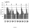
- FIG. 14 HGF expression by a set of tumour cell lines
- FIGS. 15A and 15B Characterization of the NCI-H441 cell line; with FIG. 15A corresponding to quantitative RT-PCR analysis and FIG. 15B corresponding to FACS analysis;
- FIG. 16 In vivo activity of anti-c-Met antibodies on NCI-H441 xenograft model
- FIG. 17A Alignment of 224G11 VL to murine IGKV3-5*01 germline gene
- FIG. 17B Alignment of 224G11 VL to murine IGKJ4*01 germline gene
- FIG. 18A Alignment of 224G11 VL to human IGKV3-11*01 and IGKV4-1*01 germline genes;
- FIG. 18B Alignment of 224G11 VL to human IGKJ4*02 germline gene
- FIG. 19A IGKV3-11*01 based humanized version of 224G11 VL with mentioned mutations;
- FIG. 19B IGKV4-1*01 based humanized version of 224G11 VL with mentioned mutations
- FIG. 20A Alignment of 224G11 VH to murine IGHV1-18*01 germline gene
- FIG. 20B Alignment of 224G11 VH to murine IGHD2-4*01 germline gene
- FIG. 20C Alignment of 224G11 VH to murine IGHJ2*01 germline gene
- FIG. 21A Alignment of 224G11 VH to human IGHV1-2*02 germline gene
- FIG. 21B Alignment of 224G11 VH to human IGHJ4*01 germline gene
- FIG. 22 Humanized 224G11 VH with mentioned mutations
- FIG. 23A Alignment of 227H1 VL to murine IGKV3-5*01 germline gene
- FIG. 23B Alignment of 227H1 VL to murine IGKJ4*01 germline gene
- FIG. 24A Alignment of 227H1 VL to human IGKV3-11*01 and IGKV4-1*01 germline genes;
- FIG. 24B Alignment of 227H1 VL to human IGKJ4*02 germline gene
- FIG. 25A IGKV3-11*01 based humanized version of 227H1 VL with mentioned mutations;
- FIG. 25B IGKV4-1*01 based humanized version of 227H1 VL with mentioned mutations;
- FIG. 26A Alignment of 227H1 VH to murine IGHV1-18*01 germline gene
- FIG. 26B Alignment of 227H1 VH to murine IGHD1-1*02 germline gene
- FIG. 26C Alignment of 227H1 VH to murine IGHJ2*01 germline gene
- FIG. 27A Alignment of 227H1 VH to human IGHV1-2*02 germline gene
- FIG. 27B Alignment of 227H1 VH to human IGHJ4*01 germline gene
- FIG. 28 Humanized 227H1 VH with mentioned mutations
- FIG. 29A Alignment of 223C4 VL to murine IGKV12-46*01 germline gene
- FIG. 29B Alignment of 223C4 VL to murine IGKJ2*01 germline gene
- FIG. 30A Alignment of 223C4 VL to human IGKV1-NL1*01 germline gene
- FIG. 30B Alignment of 223C4 VL to human IGKJ2*01 germline gene
- FIG. 31 Humanized 223C4 VL with mentioned mutations
- FIG. 32A Alignment of 223C4 VH to murine IGHV1-18*01 germline gene
- FIG. 32B Alignment of 223C4 VH to murine IGHD6-3*01 germline gene
- FIG. 32C Alignment of 223C4 VH to murine IGHJ4*01 germline gene
- FIG. 33A Alignment of 223C4 VH to human IGHV1-2*02 germline gene
- FIG. 33B Alignment of 223C4 VH to human IGHD1-26*01 germline gene
- FIG. 33C Alignment of 223C4 VH to human IGHJ6*01 germline gene.
- FIG. 34 Humanized 223C4 VH with mentioned mutations
- FIG. 35 Anti-tumor activity of the murine 224G11 Mab alone or combined with Navelbine® on the established xenograft NCI-H441 tumor model;
- FIG. 36 Evaluation of anti-c-Met Mabs on HUVEC proliferation
- FIG. 37 Evaluation of anti-c-Met Mabs on HUVEC tube formation
- FIG. 38A Alignment of 11E1 VL to murine IGKV4-79*01 germline gene
- FIG. 38B Alignment of 11E1 VL to murine IGKJ4*01 germline gene
- FIG. 39A Alignment of 11E1 VL to human IGKV3D-7*01 germline gene
- FIG. 39B Alignment of 11E1 VL to human IGKJ4*02 germline gene
- FIG. 40 Humanized version of 11E1 VL with mentioned mutations
- FIG. 41A Alignment of 11E1 VH to murine IGHV1-7*01 germline gene
- FIG. 41B Alignment of 11E1 VH to murine IGHD4-1*01 germline gene
- FIG. 41C Alignment of 11E1 VH to murine IGHJ3*01 germline gene
- FIG. 42A Alignment of 11E1 VH to human IGHV1-2*02 and IGHV1-46*01 germline genes;
- FIG. 42B Alignment of 11E1 VH to human IGHJ4*03 germline gene
- FIG. 43 Humanized 11E1 VH with mentioned mutations
- FIGS. 44A and 44B c-Met Phosphorylation assay on A549 cells. Evaluation of 11E1 and 224G11 purified Mabs, in absence or in presence of HGF, either at 30 ⁇ g/ml ( FIG. 44A ) or within a dose range from 0.0015 to 30 ⁇ g/ml in order to determine EC 50 values ( FIG. 44B );
- FIG. 45 In vivo combination of 224G11 Mab with Navelbine® in the NSCLC NCI-H441 xenograft model
- FIG. 46 In vivo combination of 224G11 Mab with Doxorubicin in the NSCLC NCI-H441 xenograft model
- FIG. 47 In vivo combination of 224G11 Mab with Docetaxel in the NSCLC NCI-H441 xenograft model
- FIG. 48 In vivo combination of 224G11 Mab with Temozolomide in the NSCLC NCI-H441 xenograft model
- FIGS. 49A , 49 B, 49 C and 49 D Effect of anti-c-Met Mabs on U87-MG spheroid growth
- FIGS. 50A and 50B In vitro activity of chimeric and humanized forms of 224G11 in the phospho-c-Met assay
- FIG. 51 Settings of Biacore analysis
- FIG. 52 In vivo activity of 224G11 on MDA-MB-231 cells co-implanted with MRC5 cells as human HGF source on Athymic nude mice;
- FIG. 53 ELISA based binding assay to Fc-cMet.
- Anti-Fc-c-Met binding activity was measured in an ELISA-based assay where anti-murine Fc conjugates was used to detect the purified murine monoclonal antibodies 11E1, 224G11 and 227H1.
- Dose-dependent binding activities onto plastic-coated recombinant Fc-cMet was measured at 450 nm;
- FIG. 54 HGF-cMet competition assay.
- ELISA-based assay recombinant Fc-cMET residual binding to plastic coated HGF in the presence of purified murine monoclonal antibodies 11E1, 224G11 and 227H1 was detected with anti-murine Fc conjugate and measured at 450 nm;
- FIG. 55 Amino acid sequences alignment of 227H1-derived recombinant VH domains.
- the 227H1 VH amino acid sequence is aligned with the selected human receiving framework sequence, with only mentioned the amino acids that were found different from the murine 227H1 VH sequence.
- 227H1 HZ1, HZ2 and HZ3 VH sequences correspond to implemented humanized versions of the 227H1 murine VH domain, with remaining murine residues in bold.
- 10 residues (*) were automatically changed for their human counterparts.
- HZ2 the seven residues from the third group (3) have been studied.
- HZ1VH the nine residues from the second group (2) have been mutated into their human counterparts, only the six residues from the first group (1) remain murine;
- FIG. 56 ELISA based binding assay to Fc-cMet of recombinant 227H1 antibodies.
- Anti-Fc-cMet binding activity was measured in an ELISA-based assay where anti-human Fc conjugates was used to detect chimeric and humanized 227H1-derived recombinant antibodies.
- Dose-dependent binding activities onto plastic-coated recombinant Fc-cMet of humanized VH domains-derived 227H1 antibodies was measured at 450 nm and then compare to those of the parental/reference chimeric antibody;
- FIG. 57 ELISA based binding assay to Fc-cMet of recombinant 227H1-derived antibodies.
- Anti-Fc-cMet binding activity was measured in an ELISA-based assay where anti-human Fc conjugates was used to detect chimeric and humanized 227H1-derived recombinant antibodies.
- Dose-dependent binding activity onto plastic-coated recombinant Fc-cMet of humanized HZ4VH-derived 227H1 antibody was measured at 450 nm and then compared to those of the parental/reference chimeric antibody;
- FIG. 58 HGF-cMet competition assay of 227H1 murine and recombinant antibodies.
- ELISA-based assay recombinant Fc-cMet residual binding to plastic coated HGF in the presence of the different forms of the 227H1 antibody was detected with a biotinylated unrelated anti-cMet antibody.
- Purified murine 227H1 monoclonal antibody, chimeric and HZ4VH-derived humanized 227H1-derived recombinant antibodies were tested and compared for their abilities to compete with HGF-cMet binding when measured at 450 nm;
- FIG. 59 227H1-HZ VH humanized variable domain sequence. *, corresponds to amino acids changed de facto to their human counterparts; !, corresponds to amino acids humanized during the HZ3 to HZ1 implementation; ⁇ , corresponds to amino acids humanized in the final 227H1-HZ VH sequence;
- FIG. 60 Amino acid sequences alignment of 11E1-derived recombinant VH domains.
- the 11E1 VH amino acid sequence is aligned with the selected human receiving framework sequence, with only mentioned the amino acids that were found different from the murine 11E1 VH sequence.
- 11E1 HZ VH1, VH2 and VH3 sequences correspond to implemented humanized versions of the 11E1 murine VH domain, with remaining murine residues in bold.
- seven residues (*) were automatically changed for their human counterparts.
- HZ VH2 the seven residues from the third group (3) have been studied.
- HZ VH1 the five residues from the second group (2) have been mutated into their human counterparts, only the five residues from the first group (1) remain murine;
- FIG. 61 ELISA based binding assay to Fc-cMet of recombinant 11E1 antibodies.
- Anti-Fc-cMet binding activity was measured in an ELISA-based assay where anti-human Fc conjugates was used to detect chimeric and humanized 11E1-derived recombinant antibodies.
- Dose-dependent binding activities onto plastic-coated recombinant Fc-cMet of humanized VH domains-derived 11E1 antibodies was measured at 450 nm and then compare to those of the parental/reference chimeric antibody;
- FIG. 62 Amino acid sequences alignment of 11E1-derived recombinant VL domains.
- the 11E1 VL amino acid sequence is aligned with the selected human receiving framework sequence, with only mentioned the amino acids that were found different from the murine 11E1 VL sequence.
- 11E1 HZ VL1, VL2 and VL3 sequences correspond to implemented humanized versions of the 11E1 murine VL domain, with remaining murine residues in bold.
- ten residues (*) were automatically changed for their human counterparts.
- HZ VL2 the eight residues from the third group (3) have been studied.
- the eight residues from the second group (2) have been mutated into their human counterparts, only the four residues from the first group (1) remain murine;
- FIG. 63 ELISA based binding assay to Fc-cMet of recombinant 11E1 antibodies.
- Anti-Fc-cMet binding activity was measured in an ELISA-based assay where anti-human Fc conjugates was used to detect chimeric and humanized 11E1-derived recombinant antibodies.
- Dose-dependent binding activities onto plastic-coated recombinant Fc-cMet of humanized VL domains-derived 11E1 antibodies was measured at 450 nm and then compare to those of the parental/reference chimeric antibody;
- FIG. 64 ELISA based binding assay to Fc-cMet of recombinant 11E1 antibodies.
- Anti-Fc-cMet binding activity was measured in an ELISA-based assay where anti-human Fc conjugates was used to detect chimeric and humanized 11E1-derived recombinant antibodies.
- Dose-dependent binding activities onto plastic-coated recombinant Fc-cMet of single or double humanized domains-derived 11E1 antibodies was measured at 450 nm and then compared to those of the parental/reference chimeric antibody;
- FIG. 65 Amino acid sequences alignment of 224G11 VH domain sequence.
- the 224G11 VH amino acid sequence is aligned with the 227H1 VH sequence (underlined are non homologous residues) and with the selected human receiving framework sequence, with only mentioned the amino acids that were found different from the murine 224G11 VH sequence.
- 224G11 HZ VH0 sequence correspond to “227H1-based/full-IMGT” humanized version of the 224G11 murine VH domain. In this sequence no outside-IMGT-CDRs residues remain murine;
- FIG. 66 ELISA based binding assay to Fc-cMet of recombinant 224G11 antibodies.
- Anti-Fc-cMet binding activity was measured in an ELISA-based assay where anti-human Fc conjugates was used to detect chimeric and HZVH0-derived humanized 224G11-derived recombinant antibodies.
- Dose-dependent binding activity onto plastic-coated recombinant Fc-cMet of the HZVH0 “full-IMGT” humanized VH domain-derived 224G11 antibody was measured at 450 nm and then compared to those of the parental/reference chimeric antibody;
- FIG. 67 HGF-cMet competition assay of 224G11 murine and recombinant antibodies.
- recombinant Fc-cMet residual binding to plastic coated HGF in the presence of the different forms of the 224G11 antibody was detected with a biotinylated unrelated anti-cMet antibody.
- Purified murine 224G11 monoclonal antibody, chimeric and HZVH0-derived humanized 224G11-derived recombinant antibodies were tested and compared for their abilities to compete with HGF-cMet binding when measured at 450 nm;
- FIG. 68 Amino acid sequences alignment of 224G11 VL domain sequences.
- the 224G11 VL amino acid sequence is aligned with the two selected human receiving framework sequences, with only mentioned the amino acids that were found different from the murine 224G11 VL sequence.
- 224G11 HZ VL3 sequence correspond to “shorter-CDR1” humanized version of the 224G11 murine VH domain while HZ VL6 correspond to the “longer-CDR1” version, with the remaining murine residues in bold.
- the remaining murine residues are ranked for further humanization process where * corresponds to amino acids humanized in the basic versions, and 3, 2 and 1 correspond to the residues groups for the design of the implemented humanized versions;
- FIG. 69 ELISA based binding assay to Fc-cMet of recombinant 224G11 antibodies.
- Anti-Fc-cMet binding activity was measured in an ELISA-based assay where anti-human Fc conjugates was used to detect chimeric and humanized 22G11-derived recombinant antibodies.
- Dose-dependent binding activities onto plastic-coated recombinant Fc-cMet of humanized VL3 and VL6 domains-derived 224G11 antibodies was measured at 450 nm and then compare to those of the parental/reference chimeric antibody;
- FIG. 70 ELISA based binding assay to Fc-cMet of recombinant 224G11 antibodies.
- Anti-Fc-cMet binding activity was measured in an ELISA-based assay where anti-human Fc conjugates was used to detect chimeric and humanized 224G11-derived recombinant antibodies.
- Dose-dependent binding activities onto plastic-coated recombinant Fc-cMet of humanized VL domains-derived 224G11 antibodies was measured at 450 nm and then compare to those of the parental/reference chimeric antibody;
- FIG. 71 HGF-cMet competition assay of 224G11 murine and recombinant antibodies.
- ELISA-based assay recombinant Fc-cMet residual binding to plastic coated HGF in the presence of the different forms of the 224G11 antibody was detected with a biotinylated unrelated anti-cMet antibody.
- Purified murine 224G11 monoclonal antibody, chimeric and HZ VL4-derived humanized 224G11-derived recombinant antibodies were tested and compared for their abilities to compete with HGF-cMet binding when measured at 450 nm;
- FIG. 72 Amino acid sequence of VL4 humanized 224G11 VL domain sequence. *, corresponds to amino acids changed de facto to their human counterparts in the basic HZ VL6 version; !, corresponds to amino acids humanized during the HZ VL6 to HZ VL4 implementation; ⁇ , corresponds to amino acids that remain murine in the 224G11-HZ VL4 sequence;
- FIG. 73 ELISA based binding assay to Fc-cMet of recombinant 224G11 antibodies.
- Anti-Fc-cMet binding activity was measured in an ELISA-based assay where anti-human Fc conjugates was used to detect chimeric and humanized 22G11-derived recombinant antibodies.
- Dose-dependent binding activities onto plastic-coated recombinant Fc-cMet of single- or double-humanized domains-derived 224G11 antibodies was measured at 450 nm and then compare to those of the parental/reference chimeric antibody;
- FIG. 74 HGF-cMet competition assay of 224G11 murine and recombinant antibodies.
- recombinant Fc-cMet residual binding to plastic coated HGF in the presence of the different forms of the 224G11 antibody was detected with a biotinylated unrelated anti-cMet antibody.
- Purified murine 224G11 monoclonal antibody, chimeric and fully humanized 224G11-derived recombinant antibodies were tested and compared for their abilities to compete with HGF-cMet binding when measured at 450 nm;
- FIG. 75 ELISA based binding assay to Fc-cMet of recombinant 224G11 antibodies.
- Anti-Fc-cMet binding activity was measured in an ELISA-based assay where anti-human Fc conjugates was used to detect chimeric and humanized 22G11-derived recombinant antibodies.
- Dose-dependent binding activities onto plastic-coated recombinant Fc-cMet of single mutants of the VL4-derived fully humanized 224G11 antibodies was measured at 450 nm and then compare to those of the parental/reference chimeric antibody;
- FIG. 76 ELISA based binding assay to Fc-cMet of recombinant 224G11 antibodies.
- Anti-Fc-cMet binding activity was measured in an ELISA-based assay where anti-human Fc conjugates was used to detect chimeric and humanized 22G11-derived recombinant antibodies.
- Dose-dependent binding activities onto plastic-coated recombinant Fc-cMet of single and multiple mutants of the VL4-derived fully humanized 224G11 antibodies was measured at 450 nm and then compare to those of the parental/reference chimeric antibody; and
- FIG. 77 HGF-cMet competition assay of 224G11 murine and recombinant antibodies.
- recombinant Fc-cMet residual binding to plastic coated HGF in the presence of the different forms of the 224G11 antibody was detected with a biotinylated unrelated anti-cMet antibody.
- Purified murine 224G11 monoclonal antibody, chimeric and single or multiple mutants of the VL4-derived fully humanized 224G11 recombinant antibodies were tested and compared for their abilities to compete with HGF-cMet binding when measured at 450 nm.
- mice 8 weeks old BALB/c mice were immunized either 3 to 5 times subcutaneously with a CHO transfected cell line that express c-Met on its plasma membrane (20 ⁇ 10 6 cells/dose/mouse) or 2 to 3 times with a c-Met extracellular domain fusion protein (10-15 ⁇ g/dose/mouse) (R&D Systems, Catalog #358MT) or fragments of this recombinant protein mixed with complete Freund adjuvant for the first immunization and incomplete Freund adjuvant for the following ones. Mixed protocols in which mice received both CHO-cMet cells and recombinant proteins were also performed. Three days before cell fusion, mice were boosted i.p. or i.v.
- mice were collected and fused to SP2/0-Ag14 myeloma cells (ATCC) and subjected to HAT selection. Four fusions were performed.
- ATCC SP2/0-Ag14 myeloma cells
- Four fusions were performed.
- monoclonal antibodies or their functional fragments especially of murine origin.
- Obtained hybridomas were initially screened by ELISA on the c-Met recombinant protein and then by FACS analysis on A549 NSCLC, BxPC3 pancreatic, and U87-MG glioblastoma cell lines (representative profiles were presented in FIG. 1 ) to be sure that the produced antibodies will be able to also recognize the native receptor on tumor cells. Positive reactors on these 2 tests were amplified, cloned and a set of hybridomas was recovered, purified and screened for its ability to inhibit in vitro cell proliferation in the BxPC3 model.
- BxPC3 cells were plated in 96 well plates in RPMI medium, 2 mM L. Glutamine, without SVF. 24 hours after plating, antibodies to be tested were added at a final concentration ranging from 0.0097 to 40 ⁇ g/ml 60 min before addition of 100 ng/ml of hHGF. After 3 days, cells were pulsed with 0.5 ⁇ Ci of [ 3 H]thymidine for 16 hours. The magnitude of [ 3 H]thymidine incorporated into trichloroacetic acid-insoluble DNA was quantified by liquid scintillation counting. Results were expressed as raw data to really evaluate the intrinsic agonistic effect of each Mab ( FIGS. 2A and 2B ).
- CHO stable cell lines expressing C-Met-Rluc or C-Met-Rluc and C-Met-K1100A-YFP were generated.
- Cells were distributed in white 96 well microplates in DMEM-F12/FBS 5% culture medium one or two days before BRET experiments. Cells were first cultured at 37° C. with CO2 5% in order to allow cell attachment to the plate. Cells were then starved with 200 ⁇ l DMEM/well overnight. Immediately prior to the experiment, DMEM was removed and cells quickly washed with PBS.
- BRET ratio has been defined previously [Angers et al., Proc. Natl. Acad. Sci. USA, 2000, 97:3684-3689] as: [(emission at 530 nm) ⁇ (emission at 485 nm) ⁇ Cf]/(emission at 485 nm), where Cf corresponds to (emission at 530 nm)/(emission at 485 nm) for cells expressing Rluc fusion protein alone in the same experimental conditions.
- BRET ratio corresponds to the ratio 530/485 nm obtained when the two partners were present, corrected by the ratio 530/485 nm obtained under the same experimental conditions, when only the partner fused to R. reniformis luciferase was present in the assay.
- results are expressed in milliBRET units (mBU); mBU corresponds to the BRET ratio multiplied by 1000.
- FIGS. 2A and 2B demonstrates that 11E1, 224G11, 223C4 and 227H1 were without any agonist activity in contrast to 5D5 which induced a dose dependent stimulation of cell proliferation in absence of ligand. A significant inhibition of cell proliferation was observed with the 4 selected antibodies. 5D5 is without effect on HGF-induced cell proliferation in this test.
- 3 ELISA To characterize the pattern of recognition of the 3 selected antibodies, 3 ELISA have been set up with the recombinant c-Met protein, its monomeric fragment (obtained by cleavage of the recombinant c-Met-Fc protein and the recombinant SEMA domain.
- Results presented in FIG. 4 demonstrated that the 4 antibodies recognized both dimeric and monomeric proteins.
- the human dimeric c-Met protein (R&D sytems, cat# 358MT) is coated at the concentration of 0.7 ⁇ g/ml in PBS overnight at 4° C. After saturation of the plates (Costar #3690) with a 0.5% gelatin solution 2 hours at 37° C., hybridoma supernatants are incubated 1 hour at 37° C.
- the anti-mouse HRP-antibody (Jackson ImmunoResearch, catalog #115-035-164) is added to each well at a 1/5000 dilution in ELISA buffer (0.1% gelatin/0.05% Tween 20 in PBS) and the plates incubated for 1 hour at 37° C. After 3 washes in PBS, the activity of the peroxydase is revealed by the addition of 50 ⁇ l of TMB substrate (Uptima). The reaction is left to occur for 5 min at room temperature. The reaction is stopped by the addition of 50 ⁇ l/well of a 1 M H 2 SO 4 solution and read on a plate reader at 450 nm. The same kind of protocol was performed on monomeric c-Met and SEMA domain but in that cases proteins were coated at 5 and 3 ⁇ g/ml respectively.
- 224G11, 227H1 and 223C4 did not bind the SEMA domain.
- 11E1 is able to bind the SEMA.
- BIAcore analysis were performed.
- BIAcore system based on the Surface Plasmon Resonance phenomenon deliver data by monitoring binding events in real-time. It is then useful to group antibodies in a so called “epitope mapping” experiments. A couple of antibodies unable to bind at the same time on the antigen molecule are classified in the same group (identical or neighbouring binding sites). At the opposite when their respective binding sites are sufficiently distant to allow a simultaneous binding of both antibodies these later are classified into two different groups.
- the antigen is commonly used as the ligand (immobilized on the sensorchip) and the antibodies are used without any labelling as analytes (solution phase).
- a 10 ⁇ g/ml solution of c-Met in running buffer was injected during one minute on the flowcell2 (fc2) typically 270 RU of the soluble form of c-Met were captured.
- the flowcell (fc1) was used as a reference to check any non specific binding of the antibodies to the sensorchip matrix.
- the ability of an antibody “A” to block the binding of an antibody “B” is calculated by the ratio BIA/C ⁇ (R2A/B/R1B) ⁇ 100: where R2A/B is the response corresponding to the binding of the MAb “B” when it was injected after Mab “A” and R1B is the response corresponding to the binding of the MAb “B” when it was injected first.
- a BIA/C below 20% means that A is able to block the binding of B so that A and B have neighbouring binding sites.
- the epitope mapping has been performed with 2 Mabs, 11E1 and 5D5.
- Table 3 summarized the calculation ratio obtained with the different sequences of these 2 antibodies. Black values (over 75%) mean that Mab A does not block the binding of Mab B. Bold/italic values (below 20%) mean that the binding sites of both antibody (A and B) are identical or sufficiently close to unable a simultaneous binding.
- a phospho c-Met ELISA assay was set-up. Briefly 500 000 A549 cells were seeded in each well of 6-well plates in F12K medium +10% FCS. 16 hours before HGF addition (100 ng/ml), cells were starved and each antibody to be tested was added at a final concentration of 30 ⁇ g/ml 15 minutes before ligand stimulation. 15 minutes after HGF addition, cold lysis buffer was added, cells were scraped and cell lysates collected and centrifuged at 13 000 rpm for 10 min at 4° C. Supernatants were quantified with a BCA kit (Pierce) and stored at ⁇ 20° C.
- a goat anti-c-Met antibody (R&D ref. AF276) was used as a capture antibody (coating overnight at 4° C.) and after a saturation step (1 h at RT) with a TBS-BSA 5% buffer, 25 ⁇ g of protein from the different cell lysates was added to each well of the 96-well plate. After a 90 minute-incubation time at RT, plates were washed four times and an anti-phospho-c-Met antibody (Rabbit anti-pY1230-1234-1235 c-Met) was added.
- binding of [ 125 I]-HGF (specific activity ⁇ 2,000 Ci/mmol) at 200 ⁇ M to immobilized c-Met was measured in the presence of varying concentrations of the anti-c-Met monoclonal antibodies 11E1, 224G11, 223C4, 227H1 or HGF (R&D Systems) ranging from 0.1 ⁇ M to 1 ⁇ M in PBS pH 7.4.
- the plates were incubated at room temperature for 6 h, then counted on a Packard Top Count Microplate Scintillation Counter. Non specific binding was determined in the presence of 1 ⁇ M of HGF.
- IC 50 values determined for antibodies 224G11, 223C4 and 227H1 were comparable to the IC 50 value determined for non radio labeled HGF, which was comprised between 3 and 5 nM, whereas antibody 11E1 exhibited a higher IC 50 value.
- A549 cells were plated in the upper chamber of BD BioCoatTM MatrigelTM invasion chambers (6.5 mm diameter wells with 8- ⁇ m pre size polycarbonate membranes). A459 cells were starved 24 hours before performing the invasion assay. Then 500 000 A549 cells were plated in chemotaxis buffer (DMEM medium, 0.1% BSA, 12 mM Hepes) in the upper well of each chamber, upon the Matrigel coating either with or without the antibody to be tested (final Mab concentration 10 ⁇ g/ml). After 1 hour incubation of the plates at 37° C.
- DMEM medium 0.1% BSA, 12 mM Hepes
- the lower chambers were filled with either growth medium containing 400 ng/ml of rhHGF or with growth medium alone.
- the chambers were incubated for 48 additional hours at 37° C. with 5% CO2.
- cells that remained on upper surface of the filter were gently removed with a cotton swab, cells that migrated to the lower surface of the filter were lysed, stained with CyQuant GR dye buffer (Invitrogen) and counted using a fluorescence reader Berthold Mithras LB940. All conditions were tested as triplicates.
- HGF induced a significant invasion of tumor cells comparable to the one observed with 10% FCS introduced as a positive control ( FIG. 8 ).
- the murine IgG1 9G4 introduced as an isotype control is without significant effect on basal or HGF-induced invasion when compared to cells plated without IgG. No agonist effect was noticed with 11E1, 224G11, 223C4 and 227H1 when added alone and a significant and comparable inhibition of the HGF-induced invasion was observed with the 3 Mabs.
- HGF stimulates motility.
- NCI-H441 cells were grown to high density and a gap was introduced with a P200 pipette tip. Cells were then stimulate to migrate across the gap with HGF (100 ng/ml) in presence or in absence of 11E1. Wells with 11E1 alone were also evaluated. Each tested condition was evaluated as a sextuplicate and 3 independent experiments were performed. After an overnight incubation, cells were visualized with an Axio Vision Camera (objective ⁇ 4).
- HGF induced a significant migration resulting in a complete closure of the gad within one night ( FIG. 9 ).
- the 9G4 irrelevant IgG1 used as an isotype control is without any effect on cell migration.
- an agonist effect was observed with the 5D5 when added alone but a significant inhibition of cell migration is observed with this antibody in presence of HGF in the portion of the gap remained open.
- the Fab fragment of 5D5 is without any agonist effect when added alone. However no activity of this fragment was observed in presence of HGF.
- the MAb 11E1 had no agonist effect when added alone and behave as a full antagonist in presence of HGF.
- SK-HEP-1 cells were seeded at low density (1.10 4 cells/well) in a 24-well plate in DMEM with 10% FCS and grown for 24 hours before addition, at the same time, of HGF (100 ng/ml) and antibodies to be tested (10 ⁇ g/ml). After 72 hours incubation, colonies were fixed and stained with 0.2% crystal violet in methanol and assessed for scattering visually. Each tested condition was tested as a triplicate and 3 independent experiments were performed.
- FIGS. 10A and 10B Addition of HGF to SK-HEP-1 cells induced a significant cell scattering ( FIGS. 10A and 10B ).
- the 9G4 antibody introduced as an isotype control is without effect neither alone or in presence of HGF.
- the 5D5 antibody displayed a significant agonist effect alone and no inhibitory effect was observed when 5D5 was added with HGF ( FIG. 10A ).
- No agonistic effect was observed neither with 11E1 ( FIG. 10A ) nor with 224G11 ( FIG. 10B ) added alone.
- a very significant inhibitory effect of these antibodies was demonstrated in presence of HGF ( FIGS. 10A and 10B ).
- SK-HEP-1 cells were seeded at 1.10 4 cells/well in a 24-well plate in DMEM with 10% FCS/Matrigel (50/50) and incubated for 30 min before addition, at the same time, of HGF (100 ng/ml) and antibodies to be tested (10 ⁇ g/ml). After 7 days incubation, cells were assessed for tube formation visually. Each tested condition was tested as a triplicate and 3 independent experiments were performed.
- the plates were incubated under standard cell culture conditions at 37° C., 5% CO2 in humidified incubators.
- Purified monoclonal antibodies (10 ⁇ g/ml) were added after 3 and 7 days of spheroid culture.
- HGF 400 ng/ml was added once after 4 days of culture.
- Spheroids were kept in culture for at least 10 days. Then, spheroid growth was monitored by measuring the area of spheroids using automeasure module of axiovision software. Area was expressed in ⁇ m 2 . 8-16 spheroids were evaluated for each condition.
- FIGS. 12A and 12B showed that in presence of 10% FCS no stimulation was observed when HGF was added to the complete medium. As expected the 9G4 isotype control is without effect on spheroid growth. 11E1 and 223C4 reduced significantly spheroid growth both in presence and in absence of HGF. No effect was observed with the 5D5 Fab fragment.
- mice Six to eight weeks old athymic mice were housed in sterilized filter-topped cages, maintained in sterile conditions and manipulated according to French and European guidelines.
- U87-MG a glioblastoma cell line, expressing c-Met and autocrine for the ligand HGF, was selected for in vivo evaluations.
- Mice were injected subcutaneously with 5 ⁇ 10 6 cells. Then, six days after cell implantation, tumors were measurable (approximately 100 mm 3 ), animals were divided into groups of 6 mice with comparable tumor size and treated twice a week with 1 mg/dose of each antibody to be tested. The mice were followed for the observation of xenograft growth rate and body weight changes. Tumor volume was calculated by the formula: ⁇ (Pi)/6 ⁇ length ⁇ width ⁇ height.
- NCI-H441 is derived from papillary lung adenocarcinoma, expresses high levels of c-Met, and demonstrates constitutive phosphorylation of c-Met RTK.
- the primer/probe sets for RPLO were forward, 5′-gaaactctgcattctcgcttcctg-3′ (SEQ ID No. 47); reverse, 5′-aggactcgtttgtacccgttga-3′ (SEQ ID No. 48); and probe, 5′-(FAM)-tgcagattggctacccaactgttgca-(TAMRA)-3′ (SEQ ID No. 49).
- the primer/probe sets for HGF were forward, 5′-aacaatgcctctggttcc-3′ (SEQ ID No. 50); reverse, 5′-cttgtagctgcgtcctttac-3′ (SEQ ID No.
- the primer/probe sets for cMet were forward, 5′-cattaaaggagacctcaccatagctaat-3′ (SEQ ID No. 53); reverse, 5′-cctgatcgagaaaccacaacct-3′ (SEQ ID No. 54); and probe, 5′-(FAM)-catgaagcgaccctctgatgtccca-(TAMRA)-3′ (SEQ ID No. 55).
- the thermocycling protocol consisted of melting at 50° C. for 2 minutes and 95° C.
- NCI-H441 appears as a non autocrine constitutively activated cell line able to grow independently of HGF ligand in which a ligand-independent dimerization of c-met occurred as a consequence of the overexpression of the receptor.
- the evaluation of anti-c-met antibodies on the in vivo activity of this non autocrine cell line could give some insights about their potency to impact on c-met dimerization.
- FIG. 16 demonstrates that 224G11, 11E1 and 227H1 inhibited significantly in vivo growth of NCI-H441 suggesting that in addition to ligand dependent inhibition, these antibodies able to inhibit dimerization are also able to target a ligand-independent inhibition of c-met.
- 224G11, 11E1 and 227H1 are shown to be different from the 5D5 one armed (OA-5D5) anti-c-Met antibody.
- nucleotidic sequence of the 224G11 VL was compared to the murine germline genes sequences part of the IMGT database (http://imgt.cines.fr).
- FIGS. 17A For the V gene and 17 B for the J gene.
- the human germline gene displaying the best identity with the 224G11VL has been searched.
- the nucleotidic sequence of 224G11VL has been aligned with the human germline genes sequences part of the IMGT database. For optimization of the selection, alignments between the proteic sequences were made to search for better homologies.
- the best homology score was first obtained with human the human IGKJ3*01 showing a sequence identity of 80%. But a higher number of consecutive identical nucleotides and a better amino acid fitting has been found in the alignment with human IGKJ4*02 germline gene (sequence identity of 77.14%). Thus the IGKJ4*02 germline gene was selected as receiving human J region for the murine 11E1 VL CDRs.
- FIGS. 18A Alignments are represented in FIGS. 18A for the V region and 18 B for the J region.
- the first corresponds to an initial trial for a human framework with a shorter CDR1 length (IGKV3-11*01), the second with a longer CDR1 length (IGKV4-1*01).
- the following steps in the humanization process consist in linking the selected germline genes sequences IGKV3-11*01 and IGKJ4*02 and also the CDRs of the murine 224G11 VL to the frameworks of these germline genes sequences.
- the bolded residues in the 224G11 VL sequence correspond to the twenty-five amino acids that were found different between 224G11 VL domain and the selected human frameworks (Human FR, i.e. IGKV3-11*01 and IGKJ4*02).
- residues to be eventually tested are not limited but must be considered as preferential mutations.
- all the sixteen others ranked three residues among the twenty-five different amino acids could be reconsidered.
- FIG. 19A represents the implemented IGKV3-11*01 based humanized 224G11 VL with above mentioned mutations clearly identified.
- the number under each proposed mutation corresponds to the rank at which said mutation will be done.
- the following steps in the humanization process consist in linking the selected germline genes sequences IGKV4-1*01 and IGKJ4*02 and also the CDRs of the murine 224G11 VL to the frameworks of these germline genes sequences.
- the bolded residues in the 224G11 VL sequence corresponds to the twenty-two amino acids that were found different between 224G11 VL domain and the selected human frameworks (Human FR, i.e. IGKV4-1*01 and IGKJ4*02).
- residues to be eventually tested are not limited but must be considered as preferential mutations.
- all the fourteen others ranked three residues among the twenty-two different amino acids could be reconsidered.
- FIG. 19B represents the implemented IGKV4-1*01 based humanized 224G11 VL with above mentioned mutations clearly identified.
- the number under each proposed mutation corresponds to the rank at which said mutation will be done.
- nucleotidic sequence of the 224G11 VH was compared to the murine germline genes sequences part of the IMGT database (http://imgt.cines.fr).
- FIGS. 20A For the V gene, 20 B for the D gene and 20 C for the J gene.
- the human germline gene displaying the best identity with the 224G11 VH has been searched.
- the nucleotidic sequence of 224G11 VH has been aligned with the human germline genes sequences part of the IMGT database. For optimization of the selection, alignments between the proteic sequences were made to search for better homologies.
- the D region strictly belongs to the CDR3 region in the VH domain.
- the humanization process is based on a ⁇ CDR-grafting>> approach. Analysis of the closest human D-genes is not useful in this strategy.
- Human IGHV1-2*02 V germline gene and human IGHJ4*01 J germline gene have thus been selected as receiving human sequences for the murine 224G11 VH CDRs.
- FIGS. 21A Alignments are represented in FIGS. 21A for the V region and 21 B for the J region.
- the following steps in the humanization process consist in linking the selected germline genes sequences IGHV1-2*02 and IGHJ4*01 and also the CDRs of the murine 224G11 VH to the frameworks of these germline genes sequences.
- the bolded residues in the 224G11 VH sequence correspond to the thirty amino acids that were found different between 224G11 VH domain and the selected human frameworks (Human FR, i.e. IGHV1-2*02 and IGHJ4*01).
- residues to be eventually tested are not limited but must be considered as preferential mutations.
- all the nineteen others ranked three residues among the thirty different amino acids could be reconsidered.
- FIG. 22 represents the humanized 224G11 VH with above mentioned mutations clearly identified.
- the number under each proposed mutation corresponds to the rank at which said mutation will be done.
- nucleotidic sequence of the 227H1 VL was compared to the murine germline genes sequences part of the IMGT database (http://imgt.cines.fr).
- FIGS. 23A For the V gene and 23 B for the J gene.
- the human germline gene displaying the best identity with the 227H1 VL has been searched.
- the nucleotidic sequence of 227H1 VL has been aligned with the human germline genes sequences part of the IMGT database. For optimization of the selection, alignments between the proteic sequences were made to search for better homologies.
- the best homology score was first obtained with human the human IGKJ3*01 showing a sequence identity of 78.38%. But a higher number of consecutive identical nucleotides and a better amino acid fitting has been found in the alignment with human IGKJ4*02 germline gene (sequence identity of 75.68%). Thus the IGKJ4*02 germline gene was selected as receiving human J region for the murine 227H1 VL CDRs.
- FIGS. 24A Alignments are represented in FIGS. 24A for the V region and 24 B for the J region.
- the first corresponds to an initial trial for a human framework with a shorter CDR1 length (IGKV3-11*01), the second with a longer CDR1 length (IGKV4-1*01).
- the following steps in the humanization process consist in linking the selected germline genes sequences IGKV3-11*01 and IGKJ4*02 and also the CDRs of the murine 227H1 VL to the frameworks of these germline genes sequences.
- the bolded residues in the 227H1 VL sequence corresponds to the twenty-six amino acids that were found different between 227H1 VL domain and the selected human frameworks (Human FR, i.e. IGKV3-11*01 and IGKJ4*02).
- residues to be eventually tested are not limited but must be considered as preferential mutations.
- all the sixteen others ranked three residues among the twenty-five different amino acids could be reconsidered.
- FIG. 25A represents the implemented IGKV3-11*01 based humanized 227H1 VL with above mentioned mutations clearly identified.
- the number under each proposed mutation corresponds to the rank at which said mutation will be done.
- the following steps in the humanization process consist in linking the selected germline genes sequences IGKV4-1*01 and IGKJ4*02 and also the CDRs of the murine 227H1 VL to the frameworks of these germline genes sequences.
- the bolded residues in the 227H1 VL sequence corresponds to the twenty-four amino acids that were found different between 227H1 VL domain and the selected human frameworks (Human FR, i.e. IGKV4-1*01 and IGKJ4*02).
- residues to be eventually tested are not limited but must be considered as preferential mutations.
- all the sixteen others ranked three residues among the twenty-two different amino acids could be reconsidered.
- FIG. 25B represents the implemented IGKV4-1*01 based humanized 227H1 VL with above mentioned mutations clearly identified.
- the number under each proposed mutation corresponds to the rank at which said mutation will be done.
- nucleotidic sequence of the 227H1 VH was compared to the murine germline genes sequences part of the IMGT database (http://imgt.cines.fr).
- FIGS. 26A For the V gene, 26 B for the D gene and 26 C for the J gene.
- the human germline gene displaying the best identity with the 224G11 VH has been searched.
- the nucleotidic sequence of 227H1 VH has been aligned with the human germline genes sequences part of the IMGT database.
- the receiving human IGHV1-2*02 V sequence for the murine 224G11 VH CDRs with a sequence identity of 72.92% was thus identified.
- the D region strictly belongs to the CDR3 region in the VH domain.
- the humanization process is based on a ⁇ CDR-grafting>> approach. Analysis of the closest human D-genes is not useful in this strategy.
- Human IGHV1-2*02 V germline gene and human IGHJ4*01 J germline gene have thus been selected as receiving human sequences for the murine 227H1 VH CDRs.
- FIGS. 27A Alignments are represented in FIGS. 27A for the V region and 27 B for the J region.
- the following steps in the humanization process consist in linking the selected germline genes sequences IGHV1-2*02 and IGHJ4*01 and also the CDRs of the murine 227H1 VH to the frameworks of these germline genes sequences.
- the bolded residues in the 227H1 VH sequence correspond to the thirty-two amino acids that were found different between 227H1 VH domain and the selected human frameworks (Human FR, i.e. IGHV1-2*02 and IGHJ4*01).
- residues 48 K/Q
- 49 T/G
- 53 I/M
- 76 A/V
- 78 L/M
- residues to be eventually tested are not limited but must be considered as preferential mutations.
- all the twenty-one others ranked three residues among the thirty different amino acids could be reconsidered.
- FIG. 28 represents the humanized 227H1 VH with above mentioned mutations clearly identified.
- the number under each proposed mutation corresponds to the rank at which said mutation will be done.
- nucleotidic sequence of the 223C4 VL was compared to the murine germline genes sequences part of the IMGT database (http://imgt.cines.fr).
- FIGS. 29A For the V gene and 29 B for the J gene.
- the human germline gene displaying the best identity with the 223C4 VL has been searched.
- the nucleotidic sequence of 223C4 VL has been aligned with the human germline genes sequences part of the IMGT database.
- the germline genes IGKV1-NL1*01 for the V region and IGKJ2*01 for the J region have thus been selected as receiving human sequences for the murine 223C4 VL CDRs.
- FIGS. 30A Alignments are represented in FIGS. 30A for the V region and 30 B for the J region.
- the following steps in the humanization process consist in linking the selected germline genes sequences IGKV1-NL1*01 and IGKJ2*01 and also the CDRs of the murine 223C4 VL to the frameworks of these germline genes sequences.
- residues involved in the CDR anchors or structure will be tested.
- residues are residue 66 (R/N) and residue 68 (E/V).
- residues exposed to solvent and as such that may involve immunogenicity, will also be tested. These are residues 49 (A/S), 51 (K/Q), 69 (S/D), 86 (D/Q) and 92 (S/N).
- residues involved in structure/folding of variable domain could also be mutated. These residues are residue 46 (P/Q) and residue 96 (P/S).
- residues 9 S/A
- 13 A/V
- 17 D/E
- 18 R/T
- 54 L/V
- 88 T/S
- 90 T/K
- 100 A/G
- 101 T/S
- FIG. 31 represents the humanized 223C4 VL with above mentioned mutations clearly identified.
- the number under each proposed mutation corresponds to the rank at which said mutation will be done.
- nucleotidic sequence of the 223C4 VH was compared to the murine germline genes sequences part of the IMGT database (http://imgt.cines.fr).
- FIGS. 32A For the V gene, 32 B for the D gene and 32 C for the J gene.
- the human germline gene displaying the best identity with the 223C4 VH has been searched.
- the nucleotidic sequence of 223C4 VH has been aligned with the human germline genes sequences part of the IMGT database.
- the germline genes IGHV1-2*02 for the V region and IGHJ6*01 for the J region have thus been selected as receiving human sequences for the murine 223C4 VH CDRs.
- FIGS. 33A Alignments are represented in FIGS. 33A for the V region, 33 B for the D region and 33 C for the J region.
- the following steps in the humanization process consist in linking the selected germline genes sequences IGHV1-2*02 and IGHJ6*01 and also the CDRs of the murine 223C4 VH to the frameworks of these germline genes sequences.
- residues involved in the CDR anchors or structure will be tested.
- residues are residues 40 (H/D), 45 (A/S), 55 (W/D), 66 (N/I) and 67 (Y/F).
- residues exposed to solvent and as such that may involve immunogenicity, will also be tested. These are residues 1 (Q/E), 3 (Q/L), 5 (V/Q), 48 (Q/M) and 80 (R/V).
- residues involved in structure/folding of variable domain could also be mutated. These are residues 9 (A/P), 13 (K/V), 22 (S/P) and 46 (P/H).
- residues 12 V/L
- 21 V/I
- 43 R/K
- 49 G/S
- 53 M/I
- 68 A/N
- 72 Q/K
- 75 R/K
- 76 V/A
- 78 M/L
- 82 T/K
- 84 I/S
- 92 S/R
- 93 R/S
- 95 R/T
- D/E D/E
- FIG. 34 represents the humanized 223C4 VH with above mentioned mutations clearly identified.
- the number under each proposed mutation corresponds to the rank at which said mutation will be done.
- chemotherapeutic approaches depend in part on the cellular response to apoptotic inducers and the balance between pro- and anti-apoptotic pathways within the cell.
- the protective effect of the activated c-Met on cell survival has been documented. It mainly results from an increase expression of the anti-apoptotic Bcl-x1 and Bcl-2 protein as a consequence of PI3-K-mediated signaling which in turn inhibit mitochondrial-dependent apoptosis (caspase 9). Indeed, it is conceivable that the HGF/c-Met system with its marked regulatory effect on apoptotic process can also influence the chemosensitivity of cancer cells.
- NCI-H441 cells from ATCC were routinely cultured in RPMI 1640 medium, 10% FCS and 1% L-Glutamine. Cells were split two days before engraftment so that they were in exponential phase of growth. Ten million NCI-H441 cells were engrafted in PBS to 7 weeks old Swiss nude mice. Three days after implantation, tumors were measured and animals were divided into 4 groups of 6 mice with comparable tumor size. Mice were treated i.p. with a loading dose of 2 mg of 224G11/mouse and then twice a week, for 43 days, with 1 mg of antibody/mouse. The 9G4 MAb was used as an isotype control.
- Navelbine® was given by i.p. injections at a dose of 8 mg/kg on days 5, 12, 19 post-cell injection.
- the two compounds were administered separately. In this experience the 2 compounds were used at their optimal dosage.
- Tumor volume was measured twice a week and calculated by the formula: p/6 ⁇ length ⁇ width ⁇ height.
- FIG. 35 demonstrates that 224G11 is as efficient as Navelbine® when used alone as a single agent therapy. A significant benefit of combining both therapy was observed with complete tumor regressions observed for 3 out of 6 mice at day 63.
- c-met In addition to its direct role in the regulation of a variety of tumor cell functions, activation of c-met has also been implicated in tumor angiogenesis. Endothelial cells expressed c-Met and HGF stimulates endothelial cell growth, invasion and motility (Nakamura Y. et al., Biochem. Biophys. Res., Commun. 1995, 215:483-488; Bussolino F. et al., J. Cell Biol. 1992, 119:629-641). The coordinate regulation of growth, invasion and motility in vascular endothelial cells by HGF/c-Met has been demonstrated to results in the formation of 3D capillary endothelial tubes in vitro (Rosen E. M. et al., Supplementum to Experientia 1991, 59:76-88).
- HUVEC For proliferation experiments, 7500 HUVEC were plated in each well of a 96 well plate previously coated with laminin. Cells were grown 24 hours of EMB-2 assay medium supplemented with 0.5% FBS and heparin. Then, MAbs to be tested (0.15 to 40 ⁇ g/ml) were added for 1 h before addition of 20 ng/ml of HGF. After 24 additional hours, cells were pulsed with 0.5 ⁇ Ci of [ 3 H] Thymidine. The magnitude of [ 3 H] Thymidine incorporated was quantified by liquid scintillation counting. In This experiment the 9G4 MAb is an irrelevant antibody used as an IgG1 isotype control.
- Results expressed as raw data in FIG. 36 demonstrate that, as expected HGF is a potent inducer of HUVEC cell growth. Antibodies evaluated in absence of HGF did not display any agonist proliferative activity on HUVEC whatever the tested dose. In presence of HGF, a dramatic dose dependent inhibition was observed for both 11E1 and 224G11 MAbs.
- HUVEC tube formation 25000 cells incubated 30 min with antibodies to be tested were plated in 48-well plates coated with matrigel. Then HGF 50 ng/ml was added and plates were incubated at 37° C. Medium was then harvested and 5 ⁇ M CMFDA was added for 15 min before microscopic observation.
- Results shown in FIG. 37 demonstrate that, as expected HGF induces a significant tube formation.
- the 9G4 antibody introduced as an IgG1 isotype control was without any effect on HGF-induced tube formation whereas both 11E1 and 224G11 inhibit dramatically tube formation.
- nucleotidic sequence of the 11E1 VL was compared to the murine germline genes sequences part of the IMGT database (http://imgt.cines.fr).
- FIGS. 38A For the V gene and 38 B for the J gene.
- the human germline gene displaying the best identity with the 11E1 VL has been searched.
- the nucleotidic sequence of 11E1 VL has been aligned with the human germline genes sequences part of the IMGT database.
- IGKV3-7*02 and IGKV3D-7*01 with a sequence identity for both germline genes of 69.86% for the V region have been identified.
- IGKV3-7*02 human germline gene is known in the IMGT database as an “ORF” which mean that this sequence has been found in the human genome but may present some recombination problems leading to non functional IGKV3-7*02 derived natural antibodies.
- ORF an “ORF” which mean that this sequence has been found in the human genome but may present some recombination problems leading to non functional IGKV3-7*02 derived natural antibodies.
- the IGKV3D-7*01 germline gene was selected as receiving human V region for the murine 11E1 VL CDRs.
- the best homology score was first obtained with human, the human IGKJ3*01 showing a sequence identity of 78.38%. But a higher number of consecutive identical nucleotides and a better amino acid fitting has been found in the alignment with human IGKJ4*02 germline gene (sequence identity of 75.68%). Thus the IGKJ4*02 germline gene was selected as receiving human J region for the murine 11E1 VL CDRs.
- FIGS. 39A Alignments are represented in FIGS. 39A for the V region and 39 B for the J region.
- the following steps in the humanization process consist in linking the selected germline genes sequences IGKV3D-7*01 and IGKJ4*02 and also the CDRs of the murine 11E1 VL to the frameworks of these germline genes sequences.
- the bolded residues in the 11E1 VL sequence corresponds to the thirty amino acids that were found different between 11E1 VL domain and the selected human frameworks (Human FR, i.e. IGKV3D-7*01 and IGKJ4*02).
- residues to be eventually tested are not limited but must be considered as preferential mutations.
- all the eighteen others ranked three residues among the thirty different amino acids could be reconsidered.
- FIG. 40 represents the implemented humanized 11E1 VL with above mentioned mutations clearly identified.
- the number under each proposed mutation corresponds to the rank at which said mutation will be done.
- nucleotidic sequence of the 11E1 VH was compared to the murine germline genes sequences part of the IMGT database (http://imgt.cines.fr).
- FIGS. 41A For the V gene, 41 B for the D gene and 41 C for the J gene.
- the human germline gene displaying the best identity with the 11E1 VH has been searched.
- the nucleotidic sequence of 11E1 VH has been aligned with the human germline genes sequences part of the IMGT database.
- alignments between the proteic sequences were made to search for better homologies.
- the D region strictly belongs to the CDR3 region in the VH domain.
- the humanization process is based on a ⁇ CDR-grafting>> approach. Analysis of the closest human D-genes is not useful in this strategy.
- human IGHV1-46*01 V germline gene and human IGHJ4*03 J germline gene have thus been selected as receiving human sequences for the murine 11E1 VH CDRs.
- FIGS. 42A Alignments are represented in FIGS. 42A for the V region and 42 B for the J region.
- the following steps in the humanization process consist in linking the selected germline genes sequences IGHV1-46*01 and IGHJ4*03 and also the CDRs of the murine 11E1 VH to the frameworks of these germline genes sequences.
- the bolded residues in the 11E1 VH sequence corresponds to the twenty-six amino acids that were found different between 11E1 VH domain and the selected human frameworks (Human FR, i.e. IGHV1-46*01 and IGHJ4*03).
- residues to be eventually tested are not limited but must be considered as preferential mutations.
- all the sixteen others ranked three residues among the twenty-six different amino acids could be reconsidered.
- FIG. 43 represents the implemented humanized 11E1 VH with above mentioned mutations clearly identified.
- the number under each proposed mutation corresponds to the rank at which said mutation will be done.
- example 3 the effect of anti-c-Met Mabs on phosphorylation was assessed with dosed supernatants from each hybridoma to be evaluated.
- the test has been performed again with purified 11E1 and 224G11 Mabs that have been evaluated either at a final concentration of 30 ⁇ g/ml (200 nM) or at a dose range from 0.0015 to 30 ⁇ g/ml (0.01-200 nM) in order to determine the IC 50 of each antibody.
- the protocol used is the same as the one described in example 3.
- NCI-H441 cells from ATCC were routinely cultured in RPMI 1640 medium, 10% FCS, 1% L-Glutamine. Cells were split two days before engraftment being in exponential phase of growth. Ten million NCI-H441 cells were engrafted to Athymic nude mice. Five days after implantation, tumors were measurable and animals were divided into groups of 6 mice with comparable tumor size. Mice were treated i.p. either with a loading dose of 2 mg of 224G11 Mab/mouse and then twice a week with 1 mg of antibody/mouse until Day 38 or with 3 injections of Navelbine® (D5, D12, D19) at 8 mg/kg. A third group administered with the combine treatment was also included.
- Navelbine® was given by i.p. injections. Tumor volume was measured twice a week and calculated by the formula: ⁇ /6 ⁇ length ⁇ width ⁇ height and animal weights were monitored every day over the period of Navelbine® treatment. Statistical analysis was performed at each measured time using either a t-test or a Mann-Whitney test. In this experiment, the average tumor volume of single modality treated groups is reduced by 72%, 76% and 99.8% for 224G11, Navelbine® and Navelbine®+224G11 respectively at day 41 post first injection.
- the combined therapy improved significantly tumor growth compared to single therapy treatments (p ⁇ 0.041 compared to Navelbine® alone and p ⁇ 0.002 compared to 224G11 alone on day 41), 4 out of 6 mice being without tumors in the combined therapy group. Results are represented in FIG. 45 .
- NCI-H441 cells from ATCC were routinely cultured in RPMI 1640 medium, 10% FCS, 1% L-Glutamine. Cells were split two days before engraftment being in exponential phase of growth. Ten million NCI-H441 cells were engrafted to Athymic nude mice. Five days after implantation, tumors were measurable and animals were divided into groups of 6 mice with comparable tumor size. Mice were treated i.p. either with a loading dose of 2 mg of 224G11 Mab/mouse and then twice a week with 1 mg of antibody/mouse or with 4 injections of Doxorubicin (D5, D12, D19, D26) at 5 mg/kg. A third group administered with the combine treatment was also included.
- Doxorubicin D5, D12, D19, D26
- Doxorubicin was given by i.v. injections. Tumor volume was measured twice a week and calculated by the formula: ⁇ /6 ⁇ length ⁇ width ⁇ height and animal weights were monitored every day over the period of Doxorubicin treatment. Statistical analysis was performed at each measured time using either a t-test or a Mann-Whitney test. Both single therapies and combined treatment displayed significant anti-tumor activity compared to the control group (p ⁇ 0.002 from D11 to D39). Results are represented in FIG. 46 .
- Combined treatment also demonstrates a significant anti-tumour growth activity compared to single modality treatment between D11 and D39 indicating that there is a benefit to combine Doxorubicin to an anti-c-Met treatment.
- NCI-H441 cells from ATCC were routinely cultured in RPMI 1640 medium, 10% FCS, 1% L-Glutamine. Cells were split two days before engraftment being in exponential phase of growth.
- Nine million NCI-H441 cells were engrafted to Athymic nude mice. Five days after implantation, tumors were measurable and animals were divided into groups of 6 mice with comparable tumor size. Mice were treated i.p. either with a loading dose of 2 mg of 224G11 Mab/mouse and then twice a week with 1 mg of antibody/mouse or with 4 injections of Docetaxel (D5, D12, D19, D26) at 7.5 mg/kg. A third group administered with the combine treatment was also included.
- Docetaxel was given by i.p. injections. Tumor volume was measured twice a week and calculated by the formula: ⁇ /6 ⁇ length ⁇ width ⁇ height and animal weights were monitored every day over the period of Docetaxel treatment. Statistical analysis was performed at each measured time using either a t-test or a Mann-Whitney test. Both single therapies and combined treatment displayed significant anti-tumor activity compared to the control group (p ⁇ 0.002 from D11 to D35). Results are represented in FIG. 47 .
- Combined treatment also demonstrated a significant anti-tumour growth activity compared to single modality treatment between D18 and D35 indicating that there is a benefit to combine Docetaxel to an anti-c-Met treatment.
- U87-MG cells from ATCC were routinely cultured in DMEM medium, 10% FCS, 1% L-Glutamine. Cells were split two days before engraftment being in exponential phase of growth. Five million U87-MG cells were engrafted to Athymic nude mice. Nineteen days after implantation, tumors were measurable and animals were divided into groups of 6 mice with comparable tumor size. Mice were treated i.p. either with a loading dose of 2 mg of 224G11 Mab/mouse and then twice a week with 1 mg of antibody/mouse or with 3 injections of Temozolomide (D19, D26, D33) at 5 mg/kg. A third group administered with the combine treatment was also included.
- Temozolomide was given by i.p. injections. Tumor volume was measured twice a week and calculated by the formula: ⁇ /6 ⁇ length ⁇ width ⁇ height and animal weights were monitored every day over the period of Temozolomide treatment. Statistical analysis was performed at each measured time using either a t-test or a Mann-Whitney test. Both single therapies and combined treatment displayed significant anti-tumor activity compared to the control group (p ⁇ 0.002 from D22 to D32 (where control mice were euthanized for ethical reasons)). Results are represented in FIG. 48 .
- Purified monoclonal antibodies (10 ⁇ g/ml) were added after 4 and 10 days of spheroid culture. Spheroids were kept in culture for 17 days. Then, spheroid growth was monitored by measuring the area of spheroids using automeasure module of axiovision software. Area was expressed in ⁇ m 2 . 8-16 spheroids were evaluated for each condition. Spheroid size was measured before addition of antibodies, after 10 days of culture and after 17 days of culture.
- ECD c-Met-Extra-Cellular Domain
- R&D Systems human IgG1 Fc domain
- an anti-Tag histidine capture antibody was coated on CM5 sensorchips.
- the running buffer was HBS-EP, the flow rate was 30 ⁇ l/min and the test was performed at 25° C.
- Soluble c-Met (ECD_M1)2-Fc-(HHHHHH) 2 antigen was captured on the sensorchip (around 270 RU), and the antibodies to be tested were used in solution as analytes.
- the sensorship was regenerated using Glycine, HCl pH 1.5 buffer on both flowcells for half a minute.
- FIG. 51 illustrates the principle of this analysis.
- the resulting kinetic parameters are summarized in the following table 4. They indicate that both 11E1 and 224G11 anti-c-Met antibodies bind the recombinant c-Met-Fc fusion protein with comparable affinities ranging about 40 pM.
- MDA-MB-213 and MRC5 cells from ATCC were both cultured in DMEM medium, 10% FCS, 1% L-Glutamine. Cells were split two days before engraftment being in exponential phase of growth. Five million MDA-MB-231 cells and 500 000 MRC5 cells were co-injected s.c. to Athymic nude mice. Twelve days after implantation, tumors were measurable and animals were divided into groups of 6 mice with comparable tumor size. Mice were treated i.p. either with a loading dose of 2 mg of 224G11 Mab/mouse and then twice a week with 1 mg of antibody/mouse. Tumor volume was measured twice a week and calculated by the formula: ⁇ /6 ⁇ length ⁇ width ⁇ height.
- Results described in FIG. 52 showed a significant difference in median tumors growth of mice treated with 224G11 compared to the one of the control group.
- Humanization of the anti-cMet antibodies were performed independently for each chain and sequentially, regarding to the analysed amino acids in each variable domain.
- the humanization process was evaluated in a first attempt in an ELISA-based binding assay to recombinant Fc-cMet; binding activities the humanized antibodies being compared to the recombinant chimeric antibody.
- anti-cMet humanized antibodies were evaluated for their abilities to displace the Fc-cMet binding onto plastic-coated recombinant HGF; this competition assay allowing the direct comparison of murine, chimeric and humanized versions of the anti-cMet antibodies.
- FIGS. 53 and 54 are exemplified the typical anti-cMet binding activities of 227H1, 11E1 and 224G11 murine monoclonal antibodies.
- FIG. 53 shows anti-cMet direct binding activities of detected purified murine antibodies.
- murine monoclonal anti-cMet antibodies display different but still dose-dependent anti-cMet binding activities.
- FIG. 54 shows the HGF-cMet binding competition activities of purified murine antibodies.
- the competition assay reveals reliable differences between these anti-cMet monoclonal antibodies with a moderate, not full but reliable competitive activity for 11E1 monoclonal antibody whereas murine 224G11 and 227H1 display similar pattern of competitive activities with a 100% of maximum of HGF binding displacement at high antibody concentration.
- the 224G11 monoclonal antibody showing the best IC 50 value.
- anti-cMet variable domains either murine or humanized
- LONZA's pCONplus expression vectors series recombinant IgG 1 / ⁇ -derived antibodies were expressed in CHO cells.
- Expression culture supernatants were concentrated and extensively dialysed against PBS and then dosed for expressed antibodies concentrations and directly used to assess corresponding anti-cMet binding activities. Both direct binding and HGF-competition assays were assessed to better characterize recombinant chimeric or humanized versions.
- human germline gene displaying the best identity with the 227H1 VH murine sequence has been searched.
- human IGHV1-2*02 V germline gene and human IGHJ4*01 J germline gene have thus been selected as receiving human sequences for the murine 227H1 VH CDRs.
- FIG. 55 represents an amino acid alignment of the murine 227H1 VH domain with the selected human framework.
- human FR lane only the amino acid that was found different from the 227H1 murine VH domain is depicted.
- HZ3VH, HZ2VH and HZ1VH lanes correspond to implemented humanized versions of the 227H1 VH domain with above (“changed in” lane) mentioned mutations clearly identified. The number under each proposed mutation corresponds to the rank at which said mutation will be done.
- human germline gene displaying the best identity with the 11E1 VH murine sequence has been searched.
- human IGHV1-46*01 V germline gene and human IGHJ4*03 J germline gene have thus been selected as receiving human sequences for the murine 11E1 VH CDRs.
- FIG. 60 represents an amino acid alignment of the murine 11E1 VH domain with the selected human framework.
- HZ VH3, HZ VH2 and HZ VH1 lanes correspond to implemented humanized versions of the 11E1 VH domain with above (“changed in” lane) mentioned mutations clearly identified.
- the number under each proposed mutation corresponds to the rank at which said mutation will be done.
- human germline gene displaying the best identity with the 11E1 VL murine sequence has been searched.
- human IGKV3D-7*01 V germline gene and human IGKJ4*01 J germline gene have thus been selected as receiving human sequences for the murine 11E1 VL CDRs.
- FIG. 62 represents an amino acid alignment of the murine 11E1 VL domain with the selected human framework.
- human FR lane only the amino acid that was found different from the 11E1 murine VL domain is depicted.
- HZ VL3, HZ VL2 and HZ VL1 lanes correspond to implemented humanized versions of the 11E1 VL domain with above (“changed in” lane) mentioned mutations clearly identified. The number under each proposed mutation corresponds to the rank at which said mutation will be done.
- the theoretical resulting humanized antibody sequence contains only five outside-CDRs residues coming from the parental murine VH domain and four outside-CDRs residues coming from the parental murine VL sequence (see FIG. 60 , lane HZ VH1 and FIG. 62 , lane HZ VL1). It has then be decided to immediately characterize the resulting combined heavy and light chain humanized version of the 11E1 antibody. Results are given in FIG. 64 for the anti-cMet direct binding assay.
- the human germline gene displaying the best identity with the 224G11 VH murine sequence has been searched.
- the same human IGHV1-2*02 V germline gene and human IGHJ4*01 J germline gene have thus been selected as receiving human sequences for the murine 224G11 VH CDRs.
- the ⁇ full-IMGT>> humanized version of the 224G11 murine VH domain has then been constructed and its anti-cMet binding activities were analysed, when expressed in combination with the 224G11 chimeric light chain.
- Results obtained from the anti-cMet direct binding assay are shown in FIG. 66 while FIG. 67 illustrates the HGF binding competition assay.
- the resulting amino acid sequence as depicted in FIG. 65 was selected and a bioinformatic analysis was then performed to evaluate the ⁇ humaness>> level of the so-called 224G11-HZ VH0 domain.
- Example 27 for the humaness analysis of the 224G11 HZ VH0 sequence.
- 227H1 VH domain humanization we confirm the reliability of the IMGT numbering system and homology analysis tools, and also demonstrate the possibility of transferring the humanization strategy between antibodies under the limits of their intrinsec homology.
- the human germline gene displaying the best identity with the 224G11 VL murine sequence has been searched.
- two possible receiving human V regions for the murine 224G11 VL CDRs were identified.
- two humanization strategies have been planed for the 224G11 VL domain. The first corresponds to an initial trial for a human framework with a shorter CDR1 length (IGKV3-11*01), the second with a longer CDR1 length (IGKV4-1*01).
- FIG. 68 represents an amino acid alignment of the murine 224G11 VL domain with the two selected human frameworks.
- Hu-FR FR lanes In the both shorter and longer Hu-FR FR lanes, only the amino acid that was found different from the 224G11 murine VL domain is depicted.
- HZ VL3 and HZ VL6 lanes correspond to basic humanized versions of the 224G11 VL domain with above (“rank” lane) mentioned mutations clearly identified. The number under each proposed mutation corresponds to the rank at which said mutation will be done whenever the basic “shorter” or “longer” CDR1-framework will be selected.
- the resulting sequence contains only four outside-CDRs residues coming from the murine parental sequence. As shown in FIG. 72 , these four ⁇ -labelled residues are L4, M39, H40 and R84.
- the resulting sequence contains only four outside-CDRs residues coming from the murine parental light chain variable domain sequence.
- the VH0/VL4-2 ⁇ mutant correspond to the HZ VH0 224G11 humanized VH domain expressed with the HZ VL4 224G11 humanized VL domain with the double mutation L4M/R84G.
- the VH0/VL4-3 ⁇ mutant correspond to the HZ VH0 224G11 humanized VH domain expressed with the HZ VL4 224G11 humanized VL domain with the triple mutation L4M/M39L/R84G.
- the humaness level of the fully humanized 224G11 antibody should display a strongly reduced immunogenicity due to the minimal conserved murine residues.
Priority Applications (1)
| Application Number | Priority Date | Filing Date | Title |
|---|---|---|---|
| US12/440,571 US8329173B2 (en) | 2007-07-12 | 2008-07-10 | Antibodies inhibiting c-Met dimerization and uses thereof |
Applications Claiming Priority (7)
| Application Number | Priority Date | Filing Date | Title |
|---|---|---|---|
| US92978907P | 2007-07-12 | 2007-07-12 | |
| EP07301231 | 2007-07-12 | ||
| EP07301231.2 | 2007-07-12 | ||
| EP07301231A EP2014681A1 (en) | 2007-07-12 | 2007-07-12 | Novel antibodies inhibiting c-met dimerization, and uses thereof |
| US2063908P | 2008-01-11 | 2008-01-11 | |
| US12/440,571 US8329173B2 (en) | 2007-07-12 | 2008-07-10 | Antibodies inhibiting c-Met dimerization and uses thereof |
| PCT/EP2008/059026 WO2009007427A2 (en) | 2007-07-12 | 2008-07-10 | Novel antibodies inhibiting c-met dimerization, and uses thereof |
Related Parent Applications (1)
| Application Number | Title | Priority Date | Filing Date |
|---|---|---|---|
| PCT/EP2008/059026 A-371-Of-International WO2009007427A2 (en) | 2007-07-12 | 2008-07-10 | Novel antibodies inhibiting c-met dimerization, and uses thereof |
Related Child Applications (4)
| Application Number | Title | Priority Date | Filing Date |
|---|---|---|---|
| US13/673,716 Division US8871910B2 (en) | 2007-07-12 | 2012-11-09 | Antibodies inhibiting c-Met dimerization, and uses thereof |
| US13/673,690 Division US8871909B2 (en) | 2007-07-12 | 2012-11-09 | Antibodies inhibiting c-Met dimerization and uses thereof |
| US13/673,752 Division US9107907B2 (en) | 2007-07-12 | 2012-11-09 | Antibodies inhibiting c-Met dimerization, and uses thereof |
| US13/673,726 Division US8889832B2 (en) | 2007-07-12 | 2012-11-09 | Antibodies inhibiting c-Met dimerization, and uses thereof |
Publications (2)
| Publication Number | Publication Date |
|---|---|
| US20100115639A1 US20100115639A1 (en) | 2010-05-06 |
| US8329173B2 true US8329173B2 (en) | 2012-12-11 |
Family
ID=38871713
Family Applications (6)
| Application Number | Title | Priority Date | Filing Date |
|---|---|---|---|
| US12/440,571 Active 2028-10-20 US8329173B2 (en) | 2007-07-12 | 2008-07-10 | Antibodies inhibiting c-Met dimerization and uses thereof |
| US13/673,752 Active 2028-10-24 US9107907B2 (en) | 2007-07-12 | 2012-11-09 | Antibodies inhibiting c-Met dimerization, and uses thereof |
| US13/673,726 Active 2028-07-29 US8889832B2 (en) | 2007-07-12 | 2012-11-09 | Antibodies inhibiting c-Met dimerization, and uses thereof |
| US13/673,716 Active 2028-08-09 US8871910B2 (en) | 2007-07-12 | 2012-11-09 | Antibodies inhibiting c-Met dimerization, and uses thereof |
| US13/673,690 Active 2028-08-09 US8871909B2 (en) | 2007-07-12 | 2012-11-09 | Antibodies inhibiting c-Met dimerization and uses thereof |
| US14/660,046 Abandoned US20150252114A1 (en) | 2007-07-12 | 2015-03-17 | Novel antibodies inhibiting c-met dimerization and uses thereof |
Family Applications After (5)
| Application Number | Title | Priority Date | Filing Date |
|---|---|---|---|
| US13/673,752 Active 2028-10-24 US9107907B2 (en) | 2007-07-12 | 2012-11-09 | Antibodies inhibiting c-Met dimerization, and uses thereof |
| US13/673,726 Active 2028-07-29 US8889832B2 (en) | 2007-07-12 | 2012-11-09 | Antibodies inhibiting c-Met dimerization, and uses thereof |
| US13/673,716 Active 2028-08-09 US8871910B2 (en) | 2007-07-12 | 2012-11-09 | Antibodies inhibiting c-Met dimerization, and uses thereof |
| US13/673,690 Active 2028-08-09 US8871909B2 (en) | 2007-07-12 | 2012-11-09 | Antibodies inhibiting c-Met dimerization and uses thereof |
| US14/660,046 Abandoned US20150252114A1 (en) | 2007-07-12 | 2015-03-17 | Novel antibodies inhibiting c-met dimerization and uses thereof |
Country Status (36)
| Country | Link |
|---|---|
| US (6) | US8329173B2 (es) |
| EP (6) | EP2014681A1 (es) |
| JP (4) | JP5562844B2 (es) |
| KR (4) | KR101701685B1 (es) |
| CN (4) | CN101981054B (es) |
| AR (4) | AR067517A1 (es) |
| AU (1) | AU2008274171B2 (es) |
| BR (1) | BRPI0815564B8 (es) |
| CA (4) | CA2694418C (es) |
| CL (1) | CL2008002015A1 (es) |
| CO (1) | CO6170364A2 (es) |
| CY (1) | CY1116608T1 (es) |
| DK (1) | DK2188312T3 (es) |
| ES (5) | ES2693542T3 (es) |
| HK (3) | HK1149024A1 (es) |
| HR (1) | HRP20150887T1 (es) |
| HU (1) | HUE026026T2 (es) |
| IL (5) | IL203114A (es) |
| MA (1) | MA31977B1 (es) |
| ME (1) | ME02341B (es) |
| MX (3) | MX353449B (es) |
| MY (1) | MY158756A (es) |
| NZ (4) | NZ599958A (es) |
| PA (1) | PA8789201A1 (es) |
| PH (3) | PH12018502039A1 (es) |
| PL (1) | PL2188312T3 (es) |
| PT (1) | PT2188312E (es) |
| RS (1) | RS54197B1 (es) |
| RU (1) | RU2552161C2 (es) |
| SA (1) | SA08290423B1 (es) |
| SG (1) | SG183015A1 (es) |
| SI (1) | SI2188312T1 (es) |
| TN (1) | TN2010000016A1 (es) |
| TW (4) | TWI602829B (es) |
| WO (1) | WO2009007427A2 (es) |
| ZA (1) | ZA201000966B (es) |
Cited By (11)
| Publication number | Priority date | Publication date | Assignee | Title |
|---|---|---|---|---|
| US20130109844A1 (en) * | 2008-12-02 | 2013-05-02 | Pierre Fabre Medicament | ANTI-cMET ANTIBODY |
| US20140112911A9 (en) * | 2008-12-02 | 2014-04-24 | Liliane Goetsch | Novel anti-cmet antibody |
| WO2015051159A1 (en) * | 2013-10-02 | 2015-04-09 | The Rockefeller University | Amyloid protofibril antibodies and methods of use thereof |
| US20160039935A1 (en) * | 2010-06-01 | 2016-02-11 | Pierre Fabre Medicament | NOVEL ANTI-cMET ANTIBODY |
| US9469691B2 (en) | 2008-12-02 | 2016-10-18 | Pierre Fabre Medicament | Anti-cMET antibody |
| WO2017201204A1 (en) | 2016-05-17 | 2017-11-23 | Abbvie Biotherapeutics Inc. | ANTI-cMet ANTIBODY DRUG CONJUGATES AND METHODS FOR THEIR USE |
| US9902776B2 (en) | 2013-07-29 | 2018-02-27 | Samsung Electronics Co., Ltd. | Anti-EGFR antibody and anti-c-Met/anti-EGFR bispecific antibodies comprising the same |
| US10450376B2 (en) | 2014-09-16 | 2019-10-22 | Symphogen A/S | Anti-MET antibodies and compositions |
| US11142578B2 (en) | 2016-11-16 | 2021-10-12 | Regeneron Pharmaceuticals, Inc. | Anti-MET antibodies, bispecific antigen binding molecules that bind MET, and methods of use thereof |
| US11633497B2 (en) | 2021-04-29 | 2023-04-25 | Abbvie Inc. | Anti-c-Met antibody drug conjugates |
| US11896682B2 (en) | 2019-09-16 | 2024-02-13 | Regeneron Pharmaceuticals, Inc. | Radiolabeled MET binding proteins for immuno-PET imaging and methods of use thereof |
Families Citing this family (86)
| Publication number | Priority date | Publication date | Assignee | Title |
|---|---|---|---|---|
| EP2143441A1 (en) | 2008-07-08 | 2010-01-13 | Pierre Fabre Medicament | Combination of a c-Met antagonist and an aminoheteroaryl compound for the treatment of cancer |
| PA8849001A1 (es) | 2008-11-21 | 2010-06-28 | Lilly Co Eli | Anticuerpos de c-met |
| MX2011010158A (es) | 2009-04-07 | 2011-10-17 | Roche Glycart Ag | Anticuerpos biespecificos anti-erbb-2/anti-c-met. |
| EP2417159A1 (en) | 2009-04-07 | 2012-02-15 | Roche Glycart AG | Bispecific anti-erbb-3/anti-c-met antibodies |
| EP2287197A1 (en) * | 2009-08-21 | 2011-02-23 | Pierre Fabre Medicament | Anti-cMET antibody and its use for the detection and the diagnosis of cancer |
| KR101671378B1 (ko) * | 2009-10-30 | 2016-11-01 | 삼성전자 주식회사 | c-Met에 특이적으로 결합하는 항체 및 그의 용도 |
| KR101748707B1 (ko) | 2009-11-27 | 2017-06-20 | 삼성전자주식회사 | c-Met에 특이적으로 결합하는 항체 및 그를 이용한 암 진단용 키트 |
| AU2011226103C1 (en) * | 2010-03-10 | 2016-04-28 | Genmab A/S | Monoclonal antibodies against c-Met |
| WO2011150454A1 (en) | 2010-06-01 | 2011-12-08 | Monash University | ANTIBODIES DIRECTED TO THE UNPROCESSED RECEPTOR TYROSINE KINASE c-MET |
| EP2402370A1 (en) | 2010-06-29 | 2012-01-04 | Pierre Fabre Médicament | Novel antibody for the diagnosis and/or prognosis of cancer |
| WO2012003338A1 (en) | 2010-07-01 | 2012-01-05 | Takeda Pharmaceutical Company Limited | COMBINATION OF A cMET INHIBITOR AND AN ANTIBODY TO HGF AND/OR cMET |
| US20120089541A1 (en) | 2010-08-31 | 2012-04-12 | Genentech, Inc. | Biomarkers and methods of treatment |
| WO2012030842A2 (en) | 2010-09-03 | 2012-03-08 | Academia Sinica | Anti-c-met antibody and methods of use thereof |
| US11644471B2 (en) | 2010-09-30 | 2023-05-09 | Ablynx N.V. | Techniques for predicting, detecting and reducing aspecific protein interference in assays involving immunoglobulin single variable domains |
| WO2012175400A1 (en) * | 2011-06-23 | 2012-12-27 | Ablynx Nv | Serum albumin binding proteins |
| EP2621953B1 (en) | 2010-09-30 | 2017-04-05 | Ablynx N.V. | Biological materials related to c-met |
| US8637027B2 (en) | 2010-11-03 | 2014-01-28 | Argen-X B.V. | Anti c-Met antibodies |
| EP2643351A1 (en) | 2010-11-24 | 2013-10-02 | Glaxo Group Limited | Multispecific antigen binding proteins targeting hgf |
| KR101444837B1 (ko) * | 2011-06-03 | 2014-09-30 | 한국생명공학연구원 | HGF 활성을 가지는 c-Met에 대한 인간항체 및 이의 용도 |
| US20180009888A9 (en) | 2011-06-23 | 2018-01-11 | Ablynx N.V. | Techniques for predicting, detecting and reducing aspecific protein interference in assays involving immunoglobulin single variable domains |
| US20130004484A1 (en) * | 2011-06-30 | 2013-01-03 | Genentech, Inc. | Anti-c-met antibody formulations |
| MX2014002289A (es) | 2011-08-26 | 2015-03-20 | Merrimack Pharmaceuticals Inc | Anticuerpos fc especificos en tandem. |
| SG11201400724SA (en) | 2011-09-19 | 2014-04-28 | Genentech Inc | Combination treatments comprising c-met antagonists and b-raf antagonists |
| JP2014530201A (ja) | 2011-09-20 | 2014-11-17 | イーライ リリー アンド カンパニー | 抗c−Met抗体 |
| US9346884B2 (en) | 2011-09-30 | 2016-05-24 | Ablynx N.V. | Biological materials related to c-Met |
| EP2747783B1 (en) * | 2011-09-30 | 2017-06-14 | Ablynx N.V. | Biological materials related to c-met |
| KR101865223B1 (ko) * | 2011-10-05 | 2018-06-08 | 삼성전자주식회사 | 항 c-Met 인간화 항체 및 그의 용도 |
| KR20130036993A (ko) | 2011-10-05 | 2013-04-15 | 삼성전자주식회사 | c-Met의 SEMA 도메인 내의 에피토프에 특이적으로 결합하는 항체 |
| KR20130037153A (ko) * | 2011-10-05 | 2013-04-15 | 삼성전자주식회사 | 항 c-Met 항체 및 그의 용도 |
| EP2773660A2 (en) | 2011-11-03 | 2014-09-10 | Argen-X B.V. | Chimeric human-camel antigens and their use |
| RU2014124842A (ru) | 2011-11-21 | 2015-12-27 | Дженентек, Инк. | Очистка анти-с-мет антител |
| US20150147274A1 (en) | 2011-12-02 | 2015-05-28 | Cancer Research Technology Limited | Antibodies against hgf - receptor and uses |
| US8900582B2 (en) | 2011-12-22 | 2014-12-02 | Samsung Electronics Co., Ltd. | Deimmunized anti c-Met humanized antibodies and uses thereof |
| US9376485B2 (en) | 2012-02-29 | 2016-06-28 | The Brigham And Women's Hospital, Inc. | Neutralizing antibody for epstein barr virus-associated disease |
| WO2013152252A1 (en) | 2012-04-06 | 2013-10-10 | OSI Pharmaceuticals, LLC | Combination anti-cancer therapy |
| EP2863946A4 (en) | 2012-06-21 | 2016-04-13 | Sorrento Therapeutics Inc | ANTIGEN-BINDING PROTEINS THAT BIND TO C-MET |
| KR101938698B1 (ko) | 2012-07-23 | 2019-01-16 | 삼성전자주식회사 | Cbl의 항 c-met 항체 적용 대상 환자 선별을 위한 바이오마커로서의 용도 |
| KR101938699B1 (ko) | 2012-07-23 | 2019-01-16 | 삼성전자주식회사 | Lrig1의 항 c―met 항체 적용 대상 환자 선별을 위한 용도 |
| US10413620B2 (en) | 2012-08-17 | 2019-09-17 | The Regents Of The University Of Colorado, A Body Corporate | Light-emitting versions of the monoclonal antibody to C3D (MAB 3D29) for imaging |
| WO2014028865A1 (en) * | 2012-08-17 | 2014-02-20 | The Regents Of The University Of Colorado, A Body Corporate | Compositions and methods for detecting complement activation |
| CN104903726A (zh) * | 2012-09-05 | 2015-09-09 | Jsr株式会社 | 对细胞的上皮性维持起作用的物质的筛选方法 |
| RS53818B1 (en) | 2012-10-12 | 2015-06-30 | Spirogen Sàrl | PIROLOBENZODIAZEPINI I NJIHOVI conjugated |
| EP2727941A1 (en) | 2012-11-05 | 2014-05-07 | MAB Discovery GmbH | Method for the production of multispecific antibodies |
| WO2014067642A1 (en) | 2012-11-05 | 2014-05-08 | Mab Discovery Gmbh | Method for the production of multispecific antibodies |
| WO2014074907A1 (en) * | 2012-11-09 | 2014-05-15 | Indi Molecular, Inc. | C-met-specific capture agents, compositions, and methods of using and making |
| KR101911048B1 (ko) | 2013-01-29 | 2018-10-24 | 삼성전자주식회사 | p53 활성화제 및 c-Met 억제제를 포함하는 병용 투여용 약학 조성물 |
| JP2016509045A (ja) | 2013-02-22 | 2016-03-24 | エフ・ホフマン−ラ・ロシュ・アクチェンゲゼルシャフト | がんを治療し、薬剤耐性を防止する方法 |
| KR20150143458A (ko) | 2013-03-06 | 2015-12-23 | 메리맥 파마슈티컬즈, 인크. | 항-C-MET 탠덤 Fc 이중특이적 항체 |
| CA2905181C (en) | 2013-03-13 | 2020-06-02 | Medimmune Limited | Pyrrolobenzodiazepines and conjugates thereof for providing targeted therapy |
| KR102049990B1 (ko) * | 2013-03-28 | 2019-12-03 | 삼성전자주식회사 | c-Met 항체 및 VEGF 결합 단편이 연결된 융합 단백질 |
| KR102139607B1 (ko) * | 2013-04-01 | 2020-07-30 | 삼성전자주식회사 | 항 c-Met 항체 및 FGFR 저해제를 포함하는 병용 투여용 약학 조성물 |
| WO2015031614A1 (en) * | 2013-08-28 | 2015-03-05 | Abbvie Inc. | Soluble cmet assay |
| WO2015031626A1 (en) | 2013-08-28 | 2015-03-05 | Abbvie Inc. | Soluble cmet assay |
| US9717715B2 (en) | 2013-11-15 | 2017-08-01 | Samsung Electronics Co., Ltd. | Method of combination therapy using an anti-C-Met antibody |
| RU2016141385A (ru) | 2014-03-24 | 2018-04-28 | Дженентек, Инк. | Лечение рака антагонистами с-мет и их корреляция с экспрессией hgf |
| WO2015179835A2 (en) | 2014-05-23 | 2015-11-26 | Genentech, Inc. | Mit biomarkers and methods using the same |
| WO2016037644A1 (en) | 2014-09-10 | 2016-03-17 | Medimmune Limited | Pyrrolobenzodiazepines and conjugates thereof |
| EP3200825A4 (en) | 2014-10-03 | 2018-03-21 | Academia Sinica | Antibodies against pathological forms of tdp-43 and uses thereof |
| WO2016053610A1 (en) * | 2014-10-03 | 2016-04-07 | Academia Sinica | Antibodies against pathological forms of tdp-43 and uses thereof |
| EP3221468B1 (en) * | 2014-11-20 | 2020-03-25 | Stichting Katholieke Universiteit | A novel auto-active and intracellular mutant of met |
| EP3229836B1 (en) | 2014-12-09 | 2019-11-13 | Institut National de la Sante et de la Recherche Medicale (INSERM) | Human monoclonal antibodies against axl |
| PT3233910T (pt) | 2014-12-19 | 2020-03-17 | Ablynx Nv | Dímeros de nanocorpos ligados a cisteína |
| WO2016135041A1 (en) | 2015-02-26 | 2016-09-01 | INSERM (Institut National de la Santé et de la Recherche Médicale) | Fusion proteins and antibodies comprising thereof for promoting apoptosis |
| CN113150147A (zh) | 2015-03-16 | 2021-07-23 | 塞尔德克斯医疗公司 | 抗-met抗体及其使用方法 |
| AU2016239844B2 (en) | 2015-04-02 | 2021-05-13 | Ablynx N.V. | Bispecific CXCR4-CD-4 polypeptides with potent anti-HIV activity |
| TW201709932A (zh) | 2015-06-12 | 2017-03-16 | 西雅圖遺傳學公司 | Cd123抗體及其共軛物 |
| WO2017062496A2 (en) * | 2015-10-05 | 2017-04-13 | University Of Virginia Patent Foundation | Anti-sas1b antibodies, associated methods of use, and compositions and methods for detecting and treating cancer |
| AU2016349152A1 (en) | 2015-11-03 | 2018-06-14 | Merck Patent Gmbh | Bi-specific antibodies for enhanced tumor selectivity and inhibition and uses thereof |
| WO2017173091A1 (en) * | 2016-03-30 | 2017-10-05 | Musc Foundation For Research Development | Methods for treatment and diagnosis of cancer by targeting glycoprotein a repetitions predominant (garp) and for providing effective immunotherapy alone or in combination |
| US11427632B2 (en) * | 2016-07-06 | 2022-08-30 | Celgene Corporation | Antibodies with low immunogenicity and uses thereof |
| WO2018069289A1 (en) | 2016-10-11 | 2018-04-19 | Medimmune Limited | Antibody-drug conjugates with immune-mediated therapy agents |
| TW201825515A (zh) | 2017-01-04 | 2018-07-16 | 美商伊繆諾金公司 | Met抗體以及其免疫結合物及用途 |
| US11525004B2 (en) | 2017-07-20 | 2022-12-13 | H. Lee Moffitt Cancer Center And Research Institute, Inc. | Recombinant CD123-binding antibodies |
| KR102194025B1 (ko) | 2017-12-15 | 2020-12-22 | 경북대학교 산학협력단 | CD44v6에 결합하는 펩타이드 및 이의 용도 |
| WO2019117691A1 (ko) * | 2017-12-15 | 2019-06-20 | 경북대학교 산학협력단 | CD44v6에 결합하는 펩타이드 및 이의 용도 |
| RU2020131383A (ru) * | 2018-03-24 | 2022-04-26 | Ридженерон Фармасьютикалз, Инк. | Генетически модифицированные животные, отличные от человека, для выработки терапевтических антител против комплексов пептид-mhc, способы их получения и варианты применения |
| WO2019224275A1 (en) | 2018-05-23 | 2019-11-28 | Adc Therapeutics Sa | Molecular adjuvant |
| WO2020014306A1 (en) | 2018-07-10 | 2020-01-16 | Immunogen, Inc. | Met antibodies and immunoconjugates and uses thereof |
| CN109541221B (zh) * | 2018-10-24 | 2022-03-22 | 益善生物技术股份有限公司 | 一种c-Met特异性抗体、组合物及试剂盒 |
| WO2022079211A1 (en) | 2020-10-16 | 2022-04-21 | Adc Therapeutics Sa | Glycoconjugates |
| US20240117061A1 (en) * | 2020-12-16 | 2024-04-11 | Wuhan Yzy Biopharma Co., Ltd. | Anti-bcma antibody, preparation method therefor and application thereof |
| CN112679614B (zh) * | 2021-01-15 | 2022-08-16 | 广东安普泽生物医药股份有限公司 | 用于特异性结合rankl靶向治疗药物的抗体及其用途 |
| GB202102396D0 (en) | 2021-02-19 | 2021-04-07 | Adc Therapeutics Sa | Molecular adjuvant |
| CN112946279A (zh) * | 2021-03-17 | 2021-06-11 | 扬州大学 | 一种基于油水界面自组装的三明治sers免疫传感器检测宫颈癌患者血清生物标志物方法 |
| TW202334429A (zh) | 2021-10-01 | 2023-09-01 | 中央研究院 | Sars-cov-2棘蛋白特異性抗體及其用途 |
| WO2023129928A2 (en) * | 2021-12-27 | 2023-07-06 | Academia Sinica | Antibody specific to spike protein of sars-cov-2 and uses thereof |
Citations (22)
| Publication number | Priority date | Publication date | Assignee | Title |
|---|---|---|---|---|
| US4424200A (en) | 1979-05-14 | 1984-01-03 | Nuc Med Inc. | Method for radiolabeling proteins with technetium-99m |
| US4479930A (en) | 1982-07-26 | 1984-10-30 | Trustees Of The University Of Massachusetts | Amines coupled wth dicyclic dianhydrides capable of being radiolabeled product |
| US4816567A (en) | 1983-04-08 | 1989-03-28 | Genentech, Inc. | Recombinant immunoglobin preparations |
| US4831175A (en) | 1986-09-05 | 1989-05-16 | The United States Of America As Represented By The Secretary Of The Department Of Health And Human Services | Backbone polysubstituted chelates for forming a metal chelate-protein conjugate |
| EP0451216A1 (en) | 1988-12-28 | 1991-10-16 | Protein Design Labs Inc | CHIMERAL IMMUNGLOBULINE, SPECIFIC FOR P55 TAC PROTEIN OF THE IL-2 RECEPTOR. |
| EP0566647A1 (en) | 1990-12-19 | 1993-10-27 | Protein Design Labs, Inc. | Improved humanized immunoglobulins |
| WO1996038557A1 (en) | 1995-06-02 | 1996-12-05 | Genentech, Inc. | Hepatocyte growth factor receptor antagonists and uses thereof |
| US5639641A (en) | 1992-09-09 | 1997-06-17 | Immunogen Inc. | Resurfacing of rodent antibodies |
| US5877293A (en) | 1990-07-05 | 1999-03-02 | Celltech Therapeutics Limited | CDR grafted anti-CEA antibodies and their production |
| US5886152A (en) | 1991-12-06 | 1999-03-23 | Sumitomo Pharmaceuticals Company, Limited | Humanized B-B10 |
| US6054297A (en) | 1991-06-14 | 2000-04-25 | Genentech, Inc. | Humanized antibodies and methods for making them |
| US6214344B1 (en) | 1995-06-02 | 2001-04-10 | Genetech, Inc. | Hepatocyte growth factor receptor antagonists and uses thereof |
| GB2404660A (en) | 2003-08-04 | 2005-02-09 | Pfizer Prod Inc | Human monoclonal antibodies which bind c-Met |
| US20050233960A1 (en) * | 2003-12-11 | 2005-10-20 | Genentech, Inc. | Methods and compositions for inhibiting c-met dimerization and activation |
| WO2006015371A2 (en) | 2004-08-05 | 2006-02-09 | Genentech, Inc. | Humanized anti-cmet antagonists |
| WO2006116260A2 (en) | 2005-04-26 | 2006-11-02 | Medimmune, Inc. | Modulation of antibody effector function by hinge domain engineering |
| WO2007011941A2 (en) | 2005-07-18 | 2007-01-25 | Amgen Inc. | Human anti-b7rp1 neutralizing antibodies |
| WO2007016285A2 (en) | 2005-07-28 | 2007-02-08 | Novartis Ag | M-csf specific monoclonal antibody and uses thereof |
| WO2007090807A1 (en) | 2006-02-06 | 2007-08-16 | Metheresis Translational Research S.A. | Anti-met monoclonal antibody, fragments and vectors thereof, for the treatment of tumors and corresponding products |
| WO2007126799A2 (en) | 2006-03-30 | 2007-11-08 | Novartis Ag | Compositions and methods of use for antibodies of c-met |
| WO2010064089A1 (en) | 2008-12-02 | 2010-06-10 | Pierre Fabre Medicament | Novel anti-cmet antibody |
| WO2010069765A1 (en) | 2008-12-02 | 2010-06-24 | Pierre Fabre Medicament | ANTI-cMET ANTIBODY |
Family Cites Families (11)
| Publication number | Priority date | Publication date | Assignee | Title |
|---|---|---|---|---|
| DE2840250C2 (de) | 1978-09-15 | 1983-01-20 | Siemens AG, 1000 Berlin und 8000 München | Schaltungsanordnung für eine leitungsgespeiste Lautfernsprechstation |
| GB8924581D0 (en) | 1989-11-01 | 1989-12-20 | Pa Consulting Services | Bleaching of hair |
| CN100387621C (zh) * | 1997-04-14 | 2008-05-14 | 麦可麦脱股份公司 | 抗人抗原受体的新的生产方法及其用途 |
| US7563874B2 (en) * | 1998-08-31 | 2009-07-21 | The Regents Of The University Of California | Therapeutic monoclonal antibodies that neutralize botulinum neurotoxins |
| US20060057651A1 (en) * | 2000-12-08 | 2006-03-16 | Bowdish Katherine S | Polypeptides and antibodies derived from chronic lymphocytic leukemia cells and uses thereof |
| EP1485130A4 (en) * | 2002-02-21 | 2006-11-22 | Univ Duke | REAGENTS AND TREATMENT PROCEDURES FOR AUTOIMMUNE DISEASES |
| BRPI0407446A (pt) * | 2003-02-13 | 2006-01-31 | Pharmacia Corp | Anticorpos para c-met para o tratamento de cânceres |
| KR101412271B1 (ko) * | 2003-05-09 | 2014-06-25 | 듀크 유니버시티 | Cd20-특이적 항체 및 이를 이용한 방법 |
| US20060094059A1 (en) | 2004-09-22 | 2006-05-04 | Odyssey Thera, Inc. | Methods for identifying new drug leads and new therapeutic uses for known drugs |
| BRPI0607490A2 (pt) * | 2005-02-10 | 2009-09-08 | Baylor Res Inst | anticorpos monoclonais antiinterferon alfa e métodos para uso |
| EP2143441A1 (en) * | 2008-07-08 | 2010-01-13 | Pierre Fabre Medicament | Combination of a c-Met antagonist and an aminoheteroaryl compound for the treatment of cancer |
-
2007
- 2007-07-12 EP EP07301231A patent/EP2014681A1/en not_active Withdrawn
-
2008
- 2008-07-10 PT PT87860508T patent/PT2188312E/pt unknown
- 2008-07-10 WO PCT/EP2008/059026 patent/WO2009007427A2/en active Application Filing
- 2008-07-10 EP EP12183763.7A patent/EP2535357B1/en active Active
- 2008-07-10 CN CN200880024368.4A patent/CN101981054B/zh active Active
- 2008-07-10 EP EP11162720.4A patent/EP2415785B1/en active Active
- 2008-07-10 EP EP08786050.8A patent/EP2188312B1/en active Active
- 2008-07-10 CN CN201310478511.XA patent/CN103509112B/zh active Active
- 2008-07-10 ES ES12183763.7T patent/ES2693542T3/es active Active
- 2008-07-10 EP EP12183761.1A patent/EP2535356B1/en active Active
- 2008-07-10 BR BRPI0815564A patent/BRPI0815564B8/pt active IP Right Grant
- 2008-07-10 NZ NZ599958A patent/NZ599958A/xx unknown
- 2008-07-10 ES ES11162720.4T patent/ES2678495T3/es active Active
- 2008-07-10 RS RS20150560A patent/RS54197B1/en unknown
- 2008-07-10 KR KR1020167002898A patent/KR101701685B1/ko active IP Right Grant
- 2008-07-10 MY MYPI2010000127A patent/MY158756A/en unknown
- 2008-07-10 CN CN201310478556.7A patent/CN103509113B/zh active Active
- 2008-07-10 KR KR1020167002899A patent/KR101701686B1/ko active IP Right Grant
- 2008-07-10 MX MX2014007693A patent/MX353449B/es unknown
- 2008-07-10 CA CA2694418A patent/CA2694418C/en active Active
- 2008-07-10 RU RU2010104633/10A patent/RU2552161C2/ru active
- 2008-07-10 AU AU2008274171A patent/AU2008274171B2/en active Active
- 2008-07-10 MX MX2014008274A patent/MX345391B/es unknown
- 2008-07-10 KR KR1020157015922A patent/KR101719084B1/ko active IP Right Grant
- 2008-07-10 PA PA20088789201A patent/PA8789201A1/es unknown
- 2008-07-10 SI SI200831467T patent/SI2188312T1/sl unknown
- 2008-07-10 NZ NZ583041A patent/NZ583041A/en unknown
- 2008-07-10 CN CN201310073997.9A patent/CN103183739B/zh active Active
- 2008-07-10 ES ES11162718.8T patent/ES2686335T3/es active Active
- 2008-07-10 CA CA2888691A patent/CA2888691C/en active Active
- 2008-07-10 ES ES08786050.8T patent/ES2542059T3/es active Active
- 2008-07-10 CA CA2981821A patent/CA2981821C/en active Active
- 2008-07-10 CL CL2008002015A patent/CL2008002015A1/es unknown
- 2008-07-10 ES ES12183761.1T patent/ES2686693T3/es active Active
- 2008-07-10 NZ NZ599959A patent/NZ599959A/xx unknown
- 2008-07-10 US US12/440,571 patent/US8329173B2/en active Active
- 2008-07-10 PL PL08786050T patent/PL2188312T3/pl unknown
- 2008-07-10 NZ NZ613716A patent/NZ613716A/en unknown
- 2008-07-10 JP JP2010515513A patent/JP5562844B2/ja active Active
- 2008-07-10 CA CA2982484A patent/CA2982484C/en active Active
- 2008-07-10 KR KR1020107003216A patent/KR101625234B1/ko active IP Right Grant
- 2008-07-10 HU HUE08786050A patent/HUE026026T2/en unknown
- 2008-07-10 DK DK08786050.8T patent/DK2188312T3/en active
- 2008-07-10 SG SG2012050837A patent/SG183015A1/en unknown
- 2008-07-10 EP EP11162718.8A patent/EP2415784B1/en active Active
- 2008-07-10 ME MEP-2016-197A patent/ME02341B/me unknown
- 2008-07-11 TW TW104114444A patent/TWI602829B/zh active
- 2008-07-11 TW TW097126356A patent/TWI549965B/zh active
- 2008-07-11 AR ARP080102991A patent/AR067517A1/es active IP Right Grant
- 2008-07-11 TW TW104114445A patent/TWI602830B/zh active
- 2008-07-11 TW TW103121603A patent/TWI592427B/zh active
- 2008-07-12 SA SA08290423A patent/SA08290423B1/ar unknown
-
2010
- 2010-01-03 IL IL203114A patent/IL203114A/en active IP Right Grant
- 2010-01-08 MX MX2013010752A patent/MX341012B/es unknown
- 2010-01-08 TN TNP2010000016A patent/TN2010000016A1/fr unknown
- 2010-02-10 MA MA32608A patent/MA31977B1/fr unknown
- 2010-02-10 ZA ZA2010/00966A patent/ZA201000966B/en unknown
- 2010-02-12 CO CO10016035A patent/CO6170364A2/es active IP Right Grant
-
2011
- 2011-03-28 HK HK11103062.4A patent/HK1149024A1/xx unknown
- 2011-03-28 HK HK14106929.7A patent/HK1193619A1/zh unknown
- 2011-03-28 HK HK14106930.4A patent/HK1193620A1/zh unknown
-
2012
- 2012-11-09 US US13/673,752 patent/US9107907B2/en active Active
- 2012-11-09 US US13/673,726 patent/US8889832B2/en active Active
- 2012-11-09 US US13/673,716 patent/US8871910B2/en active Active
- 2012-11-09 US US13/673,690 patent/US8871909B2/en active Active
- 2012-11-18 IL IL223109A patent/IL223109B/en active IP Right Grant
- 2012-11-18 IL IL223108A patent/IL223108B/en active IP Right Grant
- 2012-11-18 IL IL223106A patent/IL223106B/en active IP Right Grant
- 2012-11-18 IL IL223105A patent/IL223105B/en active IP Right Grant
-
2014
- 2014-01-06 JP JP2014000616A patent/JP5889923B2/ja active Active
- 2014-01-06 JP JP2014000618A patent/JP5889925B2/ja active Active
- 2014-01-06 JP JP2014000617A patent/JP5889924B2/ja active Active
-
2015
- 2015-03-17 US US14/660,046 patent/US20150252114A1/en not_active Abandoned
- 2015-08-04 CY CY20151100680T patent/CY1116608T1/el unknown
- 2015-08-20 HR HRP20150887TT patent/HRP20150887T1/hr unknown
-
2017
- 2017-09-15 AR ARP170102558A patent/AR109659A2/es unknown
- 2017-09-15 AR ARP170102559A patent/AR109660A2/es unknown
- 2017-09-15 AR ARP170102560A patent/AR109535A2/es unknown
-
2018
- 2018-09-24 PH PH12018502039A patent/PH12018502039A1/en unknown
- 2018-09-24 PH PH12018502038A patent/PH12018502038A1/en unknown
- 2018-09-24 PH PH12018502040A patent/PH12018502040A1/en unknown
Patent Citations (30)
| Publication number | Priority date | Publication date | Assignee | Title |
|---|---|---|---|---|
| US4424200A (en) | 1979-05-14 | 1984-01-03 | Nuc Med Inc. | Method for radiolabeling proteins with technetium-99m |
| US4479930A (en) | 1982-07-26 | 1984-10-30 | Trustees Of The University Of Massachusetts | Amines coupled wth dicyclic dianhydrides capable of being radiolabeled product |
| US4816567A (en) | 1983-04-08 | 1989-03-28 | Genentech, Inc. | Recombinant immunoglobin preparations |
| US4831175A (en) | 1986-09-05 | 1989-05-16 | The United States Of America As Represented By The Secretary Of The Department Of Health And Human Services | Backbone polysubstituted chelates for forming a metal chelate-protein conjugate |
| EP0939127A2 (en) | 1988-12-28 | 1999-09-01 | Protein Design Labs, Inc. | Humanized immunoglobulins and their production and use |
| US6180370B1 (en) | 1988-12-28 | 2001-01-30 | Protein Design Labs, Inc. | Humanized immunoglobulins and methods of making the same |
| EP0682040A1 (en) | 1988-12-28 | 1995-11-15 | Protein Design Labs, Inc. | Humanized immunoglobulins and their production and use |
| US5530101A (en) | 1988-12-28 | 1996-06-25 | Protein Design Labs, Inc. | Humanized immunoglobulins |
| US5585089A (en) | 1988-12-28 | 1996-12-17 | Protein Design Labs, Inc. | Humanized immunoglobulins |
| US5693761A (en) | 1988-12-28 | 1997-12-02 | Protein Design Labs, Inc. | Polynucleotides encoding improved humanized immunoglobulins |
| EP0451216A1 (en) | 1988-12-28 | 1991-10-16 | Protein Design Labs Inc | CHIMERAL IMMUNGLOBULINE, SPECIFIC FOR P55 TAC PROTEIN OF THE IL-2 RECEPTOR. |
| US5877293A (en) | 1990-07-05 | 1999-03-02 | Celltech Therapeutics Limited | CDR grafted anti-CEA antibodies and their production |
| EP0566647A1 (en) | 1990-12-19 | 1993-10-27 | Protein Design Labs, Inc. | Improved humanized immunoglobulins |
| US6054297A (en) | 1991-06-14 | 2000-04-25 | Genentech, Inc. | Humanized antibodies and methods for making them |
| US5886152A (en) | 1991-12-06 | 1999-03-23 | Sumitomo Pharmaceuticals Company, Limited | Humanized B-B10 |
| US5639641A (en) | 1992-09-09 | 1997-06-17 | Immunogen Inc. | Resurfacing of rodent antibodies |
| US6207152B1 (en) | 1995-06-02 | 2001-03-27 | Genentech, Inc. | Hepatocyte growth factor receptor antagonists and uses thereof |
| WO1996038557A1 (en) | 1995-06-02 | 1996-12-05 | Genentech, Inc. | Hepatocyte growth factor receptor antagonists and uses thereof |
| US6214344B1 (en) | 1995-06-02 | 2001-04-10 | Genetech, Inc. | Hepatocyte growth factor receptor antagonists and uses thereof |
| GB2404660A (en) | 2003-08-04 | 2005-02-09 | Pfizer Prod Inc | Human monoclonal antibodies which bind c-Met |
| WO2005016382A1 (en) | 2003-08-04 | 2005-02-24 | Pfizer Products Inc. | Antibodies to c-met |
| US20050233960A1 (en) * | 2003-12-11 | 2005-10-20 | Genentech, Inc. | Methods and compositions for inhibiting c-met dimerization and activation |
| WO2006015371A2 (en) | 2004-08-05 | 2006-02-09 | Genentech, Inc. | Humanized anti-cmet antagonists |
| WO2006116260A2 (en) | 2005-04-26 | 2006-11-02 | Medimmune, Inc. | Modulation of antibody effector function by hinge domain engineering |
| WO2007011941A2 (en) | 2005-07-18 | 2007-01-25 | Amgen Inc. | Human anti-b7rp1 neutralizing antibodies |
| WO2007016285A2 (en) | 2005-07-28 | 2007-02-08 | Novartis Ag | M-csf specific monoclonal antibody and uses thereof |
| WO2007090807A1 (en) | 2006-02-06 | 2007-08-16 | Metheresis Translational Research S.A. | Anti-met monoclonal antibody, fragments and vectors thereof, for the treatment of tumors and corresponding products |
| WO2007126799A2 (en) | 2006-03-30 | 2007-11-08 | Novartis Ag | Compositions and methods of use for antibodies of c-met |
| WO2010064089A1 (en) | 2008-12-02 | 2010-06-10 | Pierre Fabre Medicament | Novel anti-cmet antibody |
| WO2010069765A1 (en) | 2008-12-02 | 2010-06-24 | Pierre Fabre Medicament | ANTI-cMET ANTIBODY |
Non-Patent Citations (85)
Cited By (21)
| Publication number | Priority date | Publication date | Assignee | Title |
|---|---|---|---|---|
| US20130109840A1 (en) * | 2008-12-02 | 2013-05-02 | Pierre Fabre Medicament | ANTI-cMET ANTIBODY |
| US20140112911A9 (en) * | 2008-12-02 | 2014-04-24 | Liliane Goetsch | Novel anti-cmet antibody |
| US8747850B2 (en) * | 2008-12-02 | 2014-06-10 | Pierre Fabre Medicament | Anti-cMET antibody |
| US8765128B2 (en) * | 2008-12-02 | 2014-07-01 | Pierre Fabre Medicament | Anti-cMET antibody |
| US20190359717A1 (en) * | 2008-12-02 | 2019-11-28 | Pierre Fabre Medicament | ANTI-cMET ANTIBODY |
| US9469691B2 (en) | 2008-12-02 | 2016-10-18 | Pierre Fabre Medicament | Anti-cMET antibody |
| US20130109844A1 (en) * | 2008-12-02 | 2013-05-02 | Pierre Fabre Medicament | ANTI-cMET ANTIBODY |
| US20160039935A1 (en) * | 2010-06-01 | 2016-02-11 | Pierre Fabre Medicament | NOVEL ANTI-cMET ANTIBODY |
| US9902776B2 (en) | 2013-07-29 | 2018-02-27 | Samsung Electronics Co., Ltd. | Anti-EGFR antibody and anti-c-Met/anti-EGFR bispecific antibodies comprising the same |
| US10035847B2 (en) | 2013-10-02 | 2018-07-31 | The Rockefeller University | Amyloid protofibril antibodies and methods of use thereof |
| WO2015051159A1 (en) * | 2013-10-02 | 2015-04-09 | The Rockefeller University | Amyloid protofibril antibodies and methods of use thereof |
| US10450376B2 (en) | 2014-09-16 | 2019-10-22 | Symphogen A/S | Anti-MET antibodies and compositions |
| WO2017201204A1 (en) | 2016-05-17 | 2017-11-23 | Abbvie Biotherapeutics Inc. | ANTI-cMet ANTIBODY DRUG CONJUGATES AND METHODS FOR THEIR USE |
| US10383948B2 (en) | 2016-05-17 | 2019-08-20 | Abbvie Inc. | Anti-cMet antibody drug conjugates and methods for their use |
| EP3626273A1 (en) | 2016-05-17 | 2020-03-25 | AbbVie Biotherapeutics Inc. | Anti-cmet antibody drug conjugates and methods for their use |
| US10603389B2 (en) | 2016-05-17 | 2020-03-31 | Abbvie Inc. | Anti-cMet antibody drug conjugates and methods for their use |
| EP3804765A1 (en) | 2016-05-17 | 2021-04-14 | AbbVie Biotherapeutics Inc. | Anti-cmet antibody drug conjugates and methods for their use |
| EP4233909A2 (en) | 2016-05-17 | 2023-08-30 | AbbVie Biotherapeutics Inc. | Anti-cmet antibody drug conjugates and methods for their use |
| US11142578B2 (en) | 2016-11-16 | 2021-10-12 | Regeneron Pharmaceuticals, Inc. | Anti-MET antibodies, bispecific antigen binding molecules that bind MET, and methods of use thereof |
| US11896682B2 (en) | 2019-09-16 | 2024-02-13 | Regeneron Pharmaceuticals, Inc. | Radiolabeled MET binding proteins for immuno-PET imaging and methods of use thereof |
| US11633497B2 (en) | 2021-04-29 | 2023-04-25 | Abbvie Inc. | Anti-c-Met antibody drug conjugates |
Also Published As
Similar Documents
| Publication | Publication Date | Title |
|---|---|---|
| US8871909B2 (en) | Antibodies inhibiting c-Met dimerization and uses thereof | |
| AU2016228280B2 (en) | Novel antibodies inhibiting c-met dimerization, and uses thereof | |
| AU2014200630B2 (en) | Novel antibodies inhibiting c-met dimerization, and uses thereof |
Legal Events
| Date | Code | Title | Description |
|---|---|---|---|
| AS | Assignment |
Owner name: PIERRE FABRE MEDICAMENT,FRANCE Free format text: ASSIGNMENT OF ASSIGNORS INTEREST;ASSIGNOR:GOETSCH, LILIANE;REEL/FRAME:023604/0141 Effective date: 20090827 Owner name: PIERRE FABRE MEDICAMENT, FRANCE Free format text: ASSIGNMENT OF ASSIGNORS INTEREST;ASSIGNOR:GOETSCH, LILIANE;REEL/FRAME:023604/0141 Effective date: 20090827 |
|
| STCF | Information on status: patent grant |
Free format text: PATENTED CASE |
|
| FEPP | Fee payment procedure |
Free format text: PAYOR NUMBER ASSIGNED (ORIGINAL EVENT CODE: ASPN); ENTITY STATUS OF PATENT OWNER: LARGE ENTITY |
|
| FPAY | Fee payment |
Year of fee payment: 4 |
|
| MAFP | Maintenance fee payment |
Free format text: PAYMENT OF MAINTENANCE FEE, 8TH YEAR, LARGE ENTITY (ORIGINAL EVENT CODE: M1552); ENTITY STATUS OF PATENT OWNER: LARGE ENTITY Year of fee payment: 8 |