US8309300B2 - Cysteine engineered antibodies and conjugates - Google Patents
Cysteine engineered antibodies and conjugates Download PDFInfo
- Publication number
- US8309300B2 US8309300B2 US12/427,649 US42764909A US8309300B2 US 8309300 B2 US8309300 B2 US 8309300B2 US 42764909 A US42764909 A US 42764909A US 8309300 B2 US8309300 B2 US 8309300B2
- Authority
- US
- United States
- Prior art keywords
- antibody
- frac acc
- antibodies
- binding
- cysteine
- Prior art date
- Legal status (The legal status is an assumption and is not a legal conclusion. Google has not performed a legal analysis and makes no representation as to the accuracy of the status listed.)
- Active, expires
Links
- 0 CCCOC[17*]NC(=O)CSC.[2H]CCOC[17*]N1C(=O)CC(SC)C1=O Chemical compound CCCOC[17*]NC(=O)CSC.[2H]CCOC[17*]N1C(=O)CC(SC)C1=O 0.000 description 10
- BDITVJHHJUTHBB-DNIGJBFUSA-N CC[C@H](C)[C@@H]([C@@H](CC(=O)N1CCC[C@H]1[C@H](OC)[C@@H](C)C(=O)N[C@@H](CC1=CC=CC=C1)C(=O)O)OC)N(C)C(=O)[C@@H](NC(=O)[C@H](C(C)C)N(C)C(=O)OCC1=CC=C(NC(=O)[C@H](CCCNC(N)=O)NC(=O)[C@@H](NC(=O)CCCCCN2C(=O)C=CC2=O)C(C)C)C=C1)C(C)C Chemical compound CC[C@H](C)[C@@H]([C@@H](CC(=O)N1CCC[C@H]1[C@H](OC)[C@@H](C)C(=O)N[C@@H](CC1=CC=CC=C1)C(=O)O)OC)N(C)C(=O)[C@@H](NC(=O)[C@H](C(C)C)N(C)C(=O)OCC1=CC=C(NC(=O)[C@H](CCCNC(N)=O)NC(=O)[C@@H](NC(=O)CCCCCN2C(=O)C=CC2=O)C(C)C)C=C1)C(C)C BDITVJHHJUTHBB-DNIGJBFUSA-N 0.000 description 2
- DSYIBQSUMXINMH-UHFFFAOYSA-N C.C.C.CC(=O)CCN1C(=O)C=CC1=O.O=C(CCCCCOC(=O)C1CCC(CN2C(=O)C=CC2=O)CC1)ON1C(=O)CCC1=O.O=C(CCN1C(=O)C=CC1=O)NCCCCCC(=O)ON1C(=O)CCC1=O.O=C(ON1C(=O)CCC1=O)C1CCC(CN2C(=O)C=CC2=O)CC1 Chemical compound C.C.C.CC(=O)CCN1C(=O)C=CC1=O.O=C(CCCCCOC(=O)C1CCC(CN2C(=O)C=CC2=O)CC1)ON1C(=O)CCC1=O.O=C(CCN1C(=O)C=CC1=O)NCCCCCC(=O)ON1C(=O)CCC1=O.O=C(ON1C(=O)CCC1=O)C1CCC(CN2C(=O)C=CC2=O)CC1 DSYIBQSUMXINMH-UHFFFAOYSA-N 0.000 description 1
- FOVWDMBFQWOTHK-UHFFFAOYSA-N C.C.O=C=O.O=O.[Mg+2] Chemical compound C.C.O=C=O.O=O.[Mg+2] FOVWDMBFQWOTHK-UHFFFAOYSA-N 0.000 description 1
- QSMUTWFUNGBCHF-UHFFFAOYSA-N C.C=C(NC(=O)CCOC)[Y][Y][Y].C=O.CNC(=O)CCN1C(=O)C=CC1=O.CNC(=O)CCN1C(=O)C=CC1=O.O=C(=O)C1=CC=C(N(CCCl)CCCl)C=C1.O=C(CCN1C(=O)C=CC1=O)NC([Y])([Y])[Y].O=CC1=CC=C(N(CCCl)CCCl)C=C1 Chemical compound C.C=C(NC(=O)CCOC)[Y][Y][Y].C=O.CNC(=O)CCN1C(=O)C=CC1=O.CNC(=O)CCN1C(=O)C=CC1=O.O=C(=O)C1=CC=C(N(CCCl)CCCl)C=C1.O=C(CCN1C(=O)C=CC1=O)NC([Y])([Y])[Y].O=CC1=CC=C(N(CCCl)CCCl)C=C1 QSMUTWFUNGBCHF-UHFFFAOYSA-N 0.000 description 1
- VYBGCAVUTQGHEG-UHFFFAOYSA-N C=CS(=O)(=O)C1=CC=C(C(=O)ON2C(=O)CCC2=O)C=C1.CCC(=O)ON1C(=O)CCC1=O.O=C(CBr)NCCC(=O)ON1C(=O)CCC1=O.O=C(CI)NC1=CC=C(C(=O)ON2C(=O)CCC2=O)C=C1.O=C(CI)NCCCCCC(=O)ON1C(=O)CCC1=O Chemical compound C=CS(=O)(=O)C1=CC=C(C(=O)ON2C(=O)CCC2=O)C=C1.CCC(=O)ON1C(=O)CCC1=O.O=C(CBr)NCCC(=O)ON1C(=O)CCC1=O.O=C(CI)NC1=CC=C(C(=O)ON2C(=O)CCC2=O)C=C1.O=C(CI)NCCCCCC(=O)ON1C(=O)CCC1=O VYBGCAVUTQGHEG-UHFFFAOYSA-N 0.000 description 1
- KEOOOSYBQMHGRS-UHFFFAOYSA-N CC(C)(C)OC(=O)NNC1=CC=C(C(=O)ON2C(=O)CCC2=O)C=C1.CC(C)(C)OC(=O)NNCC(=O)ON1C(=O)CCC1=O Chemical compound CC(C)(C)OC(=O)NNC1=CC=C(C(=O)ON2C(=O)CCC2=O)C=C1.CC(C)(C)OC(=O)NNCC(=O)ON1C(=O)CCC1=O KEOOOSYBQMHGRS-UHFFFAOYSA-N 0.000 description 1
- XYLRBQLAGJLPEJ-UHFFFAOYSA-N CC(C)C(=O)CCCCCN1C(=O)CC(C(C)C)C1=O Chemical compound CC(C)C(=O)CCCCCN1C(=O)CC(C(C)C)C1=O XYLRBQLAGJLPEJ-UHFFFAOYSA-N 0.000 description 1
- VURXOEVVPFDZLU-UHFFFAOYSA-N CC(C)C(=O)CCN1C(=O)CC(C(C)C)C1=O Chemical compound CC(C)C(=O)CCN1C(=O)CC(C(C)C)C1=O VURXOEVVPFDZLU-UHFFFAOYSA-N 0.000 description 1
- UIMDZZVXGCKQRC-UHFFFAOYSA-N CC(C)C(=O)COCCOCCN1C(=O)CC(C(C)C)C1=O Chemical compound CC(C)C(=O)COCCOCCN1C(=O)CC(C(C)C)C1=O UIMDZZVXGCKQRC-UHFFFAOYSA-N 0.000 description 1
- PVFBECCHKICAQC-UHFFFAOYSA-N CC(C)NCC(=O)C(C)C.CC(C)OCCNO1CCNC1(C)C Chemical compound CC(C)NCC(=O)C(C)C.CC(C)OCCNO1CCNC1(C)C PVFBECCHKICAQC-UHFFFAOYSA-N 0.000 description 1
- LQFQQURWHUUWIS-UHFFFAOYSA-N CC.CC(C)NC1=CC=C(COC(=O)C(C)C)C=C1 Chemical compound CC.CC(C)NC1=CC=C(COC(=O)C(C)C)C=C1 LQFQQURWHUUWIS-UHFFFAOYSA-N 0.000 description 1
- HFWGJHOZMRYWJF-WDAFUDAASA-N CC.CC(C)[C@H](NC(=O)OCC1C2=C(C=CC=C2)C2=C1C=CC=C2)C(=O)N[C@@H](CCCNC(N)=O)C(=O)NC1=CC=C(COC(=O)OC2=CC=C([N+](=O)[O-])C=C2)C=C1 Chemical compound CC.CC(C)[C@H](NC(=O)OCC1C2=C(C=CC=C2)C2=C1C=CC=C2)C(=O)N[C@@H](CCCNC(N)=O)C(=O)NC1=CC=C(COC(=O)OC2=CC=C([N+](=O)[O-])C=C2)C=C1 HFWGJHOZMRYWJF-WDAFUDAASA-N 0.000 description 1
- PHMDBPPKEVUJEY-WDAFUDAASA-N CC.CNCCCC[C@H](NC(=O)[C@H](CC1=CC=CC=C1)NC(=O)OCC1C2=C(C=CC=C2)C2=C1C=CC=C2)C(=O)NC1=CC=C(CO)C=C1 Chemical compound CC.CNCCCC[C@H](NC(=O)[C@H](CC1=CC=CC=C1)NC(=O)OCC1C2=C(C=CC=C2)C2=C1C=CC=C2)C(=O)NC1=CC=C(CO)C=C1 PHMDBPPKEVUJEY-WDAFUDAASA-N 0.000 description 1
- JQLDNEXPSBDYIW-AVDPWCIQSA-N CC.[2H]C(=O)C(=CC1=CC=C(NCCC)C=C1)C([2H])=O Chemical compound CC.[2H]C(=O)C(=CC1=CC=C(NCCC)C=C1)C([2H])=O JQLDNEXPSBDYIW-AVDPWCIQSA-N 0.000 description 1
- SKOAZMREXWUCRA-GTIFXZTCSA-N CC.[2H]C(=O)OCC1=CC=C(NCCC)C=C1 Chemical compound CC.[2H]C(=O)OCC1=CC=C(NCCC)C=C1 SKOAZMREXWUCRA-GTIFXZTCSA-N 0.000 description 1
- SMIFNEMCWWJFDW-MMDBDLHTSA-N CC[C@H](C)[C@@H]([C@@H](CC(=O)N1CCC[C@H]1[C@H](OC)[C@@H](C)C(=O)C[C@@H](CC1=CC=CC=C1)C(=O)O)OC)N(C)C(=O)[C@@H](CC(=O)[C@H](C(C)C)N(C)C(=O)CCCCCN1C(=O)CC(CSC)C1=O)C(C)C.CC[C@H](C)[C@@H]([C@@H](CC(=O)N1CCC[C@H]1[C@H](OC)[C@@H](C)C(=O)C[C@@H](CC1=CC=CC=C1)C(=O)O)OC)N(C)C(=O)[C@@H](CC(=O)[C@H](C(C)C)N(C)C(=O)OCC1=CC=C(CC(=O)CCCCCN2C(=O)CC(CSC)C2=O)C=C1)C(C)C.CC[C@H](C)[C@@H]([C@@H](CC(=O)N1CCC[C@H]1[C@H](OC)[C@@H](C)C(=O)C[C@H](C)[C@@H](O)C1=CC=CC=C1)OC)N(C)C(=O)[C@@H](CC(=O)[C@H](C(C)C)N(C)C(=O)CCCCCN1C(=O)CC(CSC)C1=O)C(C)C.CC[C@H](C)[C@@H]([C@@H](CC(=O)N1CCC[C@H]1[C@H](OC)[C@@H](C)C(=O)C[C@H](C)[C@@H](O)C1=CC=CC=C1)OC)N(C)C(=O)[C@@H](CC(=O)[C@H](C(C)C)N(C)C(=O)OCC1=CC=C(CC(=O)CCCCCN2C(=O)CC(CSC)C2=O)C=C1)C(C)C.[HH].[HH] Chemical compound CC[C@H](C)[C@@H]([C@@H](CC(=O)N1CCC[C@H]1[C@H](OC)[C@@H](C)C(=O)C[C@@H](CC1=CC=CC=C1)C(=O)O)OC)N(C)C(=O)[C@@H](CC(=O)[C@H](C(C)C)N(C)C(=O)CCCCCN1C(=O)CC(CSC)C1=O)C(C)C.CC[C@H](C)[C@@H]([C@@H](CC(=O)N1CCC[C@H]1[C@H](OC)[C@@H](C)C(=O)C[C@@H](CC1=CC=CC=C1)C(=O)O)OC)N(C)C(=O)[C@@H](CC(=O)[C@H](C(C)C)N(C)C(=O)OCC1=CC=C(CC(=O)CCCCCN2C(=O)CC(CSC)C2=O)C=C1)C(C)C.CC[C@H](C)[C@@H]([C@@H](CC(=O)N1CCC[C@H]1[C@H](OC)[C@@H](C)C(=O)C[C@H](C)[C@@H](O)C1=CC=CC=C1)OC)N(C)C(=O)[C@@H](CC(=O)[C@H](C(C)C)N(C)C(=O)CCCCCN1C(=O)CC(CSC)C1=O)C(C)C.CC[C@H](C)[C@@H]([C@@H](CC(=O)N1CCC[C@H]1[C@H](OC)[C@@H](C)C(=O)C[C@H](C)[C@@H](O)C1=CC=CC=C1)OC)N(C)C(=O)[C@@H](CC(=O)[C@H](C(C)C)N(C)C(=O)OCC1=CC=C(CC(=O)CCCCCN2C(=O)CC(CSC)C2=O)C=C1)C(C)C.[HH].[HH] SMIFNEMCWWJFDW-MMDBDLHTSA-N 0.000 description 1
- HDPGSHRZLYGRNY-XXWQXIMASA-N CC[C@H](C)[C@@H]([C@@H](CC(=O)N1CCC[C@H]1[C@H](OC)[C@@H](C)C(=O)N[C@@H](CC1=CC=CC=C1)C(=O)O)OC)N(C)C(=O)[C@@H](NC(=O)[C@H](C(C)C)N(C)C(C)C)C(C)C.CC[C@H](C)[C@@H]([C@@H](CC(=O)N1CCC[C@H]1[C@H](OC)[C@@H](C)C(=O)N[C@H](C)[C@@H](O)C1=CC=CC=C1)OC)N(C)C(=O)[C@@H](NC(=O)[C@H](C(C)C)N(C)C(C)C)C(C)C Chemical compound CC[C@H](C)[C@@H]([C@@H](CC(=O)N1CCC[C@H]1[C@H](OC)[C@@H](C)C(=O)N[C@@H](CC1=CC=CC=C1)C(=O)O)OC)N(C)C(=O)[C@@H](NC(=O)[C@H](C(C)C)N(C)C(C)C)C(C)C.CC[C@H](C)[C@@H]([C@@H](CC(=O)N1CCC[C@H]1[C@H](OC)[C@@H](C)C(=O)N[C@H](C)[C@@H](O)C1=CC=CC=C1)OC)N(C)C(=O)[C@@H](NC(=O)[C@H](C(C)C)N(C)C(C)C)C(C)C HDPGSHRZLYGRNY-XXWQXIMASA-N 0.000 description 1
- ICEWTYQLHWPKAI-UHFFFAOYSA-N O=C(CCSSC1=CC=CC=N1)NCCCCCC(=O)ON1C(=O)CCC1=O.O=C(CCSSC1=CC=CC=N1)ON1C(=O)CCC1=O Chemical compound O=C(CCSSC1=CC=CC=N1)NCCCCCC(=O)ON1C(=O)CCC1=O.O=C(CCSSC1=CC=CC=N1)ON1C(=O)CCC1=O ICEWTYQLHWPKAI-UHFFFAOYSA-N 0.000 description 1
- WRFPPWPUOIQEBM-UHFFFAOYSA-N O=C1C=CC(=O)N1CCOCCOCCN1C(=O)C=CC1=O.O=C1C=CC(=O)N1CCOCCOCCOCCN1C(=O)C=CC1=O Chemical compound O=C1C=CC(=O)N1CCOCCOCCN1C(=O)C=CC1=O.O=C1C=CC(=O)N1CCOCCOCCOCCN1C(=O)C=CC1=O WRFPPWPUOIQEBM-UHFFFAOYSA-N 0.000 description 1
- YSPYXHPVMKHVDE-UHFFFAOYSA-N O=C1CCC(=O)N1OOCCC1=CC=C(N2C(=O)C=CC2=O)C=C1 Chemical compound O=C1CCC(=O)N1OOCCC1=CC=C(N2C(=O)C=CC2=O)C=C1 YSPYXHPVMKHVDE-UHFFFAOYSA-N 0.000 description 1
- BQBOVBUBHKHJIU-BWPDVVPDSA-N [2H]C(=O)C1=CC=C(CC(=O)CSC)C=C1.[2H]C(=O)C1CCC(CN2C(=O)CC(SC)C2=O)CC1.[2H]C(=O)CN1C(=O)CC(SC)C1=O.[2H]C(=O)CSC.[2H]C(=O)[Y]C(=O)CSC Chemical compound [2H]C(=O)C1=CC=C(CC(=O)CSC)C=C1.[2H]C(=O)C1CCC(CN2C(=O)CC(SC)C2=O)CC1.[2H]C(=O)CN1C(=O)CC(SC)C1=O.[2H]C(=O)CSC.[2H]C(=O)[Y]C(=O)CSC BQBOVBUBHKHJIU-BWPDVVPDSA-N 0.000 description 1
- DZDAGRJFFVDDSC-WJULDGBESA-N [3H]C1CC(=O)N(OOCCN2C(=O)C=CC2=O)C1=O Chemical compound [3H]C1CC(=O)N(OOCCN2C(=O)C=CC2=O)C1=O DZDAGRJFFVDDSC-WJULDGBESA-N 0.000 description 1
- XQTHCJHFBPDSSX-KZINDQQQSA-N [H]N(C(=O)[C@H](C(C)C)N([H])C(=O)CCCCCN1C(=O)CC(C)C1=O)[C@@H](CCCNC(N)=O)C(=O)C[2H].[H]N(C(=O)[C@H](CCCNC(N)=O)N([H])C(=O)[C@H](C(C)C)N([H])C(=O)CCCCCN1C(=O)CC(C)C1=O)C1=CC=C(COC([2H])=O)C=C1.[H]N(CC)[C@H](C(=O)N([H])[C@@H](CCCNC(N)=O)C(=O)C[2H])C(C)C Chemical compound [H]N(C(=O)[C@H](C(C)C)N([H])C(=O)CCCCCN1C(=O)CC(C)C1=O)[C@@H](CCCNC(N)=O)C(=O)C[2H].[H]N(C(=O)[C@H](CCCNC(N)=O)N([H])C(=O)[C@H](C(C)C)N([H])C(=O)CCCCCN1C(=O)CC(C)C1=O)C1=CC=C(COC([2H])=O)C=C1.[H]N(CC)[C@H](C(=O)N([H])[C@@H](CCCNC(N)=O)C(=O)C[2H])C(C)C XQTHCJHFBPDSSX-KZINDQQQSA-N 0.000 description 1
- NTHJGNZNHQWILG-UHFFFAOYSA-N [H]N(CCCCCC(=O)C(C)C)C(=O)CC(C)C Chemical compound [H]N(CCCCCC(=O)C(C)C)C(=O)CC(C)C NTHJGNZNHQWILG-UHFFFAOYSA-N 0.000 description 1
- JWZYSAAZVWXBQE-UHFFFAOYSA-N [H]N(CCOCCOCC(=O)C(C)C)C(=O)CCN1C(=O)CC(C(C)C)C1=O Chemical compound [H]N(CCOCCOCC(=O)C(C)C)C(=O)CCN1C(=O)CC(C(C)C)C1=O JWZYSAAZVWXBQE-UHFFFAOYSA-N 0.000 description 1
- NUCMHACCEIDOKT-QLCGXXCYSA-N [H]N1C(=O)OC2CC1(O)C(OC)/C=C/C=C(\C)CC1=CC(OC)=C(Cl)C(=C1)N(C)C(=O)CC(OC(=O)C(C)N(C)C(=O)CSC(C)C)C1(C)OC1C2C Chemical compound [H]N1C(=O)OC2CC1(O)C(OC)/C=C/C=C(\C)CC1=CC(OC)=C(Cl)C(=C1)N(C)C(=O)CC(OC(=O)C(C)N(C)C(=O)CSC(C)C)C1(C)OC1C2C NUCMHACCEIDOKT-QLCGXXCYSA-N 0.000 description 1
- CRCMHVUKRDNSLX-XUJYJQEKSA-N [H]N1C(=O)O[C@H]2C[C@]1(O)[C@H](OC)/C=C/C=C(\C)CC1=CC(OC)=C(Cl)C(=C1)N(C)C(=O)C[C@H](OC(=O)[C@H](C)N(C)C(=O)CCC(C)(C)SC(C)C)[C@]1(C)O[C@H]1[C@@H]2C.[H]N1C(=O)O[C@H]2C[C@]1(O)[C@H](OC)/C=C/C=C(\C)CC1=CC(OC)=C(Cl)C(=C1)N(C)C(=O)C[C@H](OC(=O)[C@H](C)N(C)C(=O)CCC([H])(C)SC(C)C)[C@]1(C)O[C@H]1[C@@H]2C.[H]N1C(=O)O[C@H]2C[C@]1(O)[C@H](OC)/C=C/C=C(\C)CC1=CC(OC)=C(Cl)C(=C1)N(C)C(=O)C[C@H](OC(=O)[C@H](C)N(C)C(=O)CCSC(C)C)[C@]1(C)O[C@H]1[C@@H]2C Chemical compound [H]N1C(=O)O[C@H]2C[C@]1(O)[C@H](OC)/C=C/C=C(\C)CC1=CC(OC)=C(Cl)C(=C1)N(C)C(=O)C[C@H](OC(=O)[C@H](C)N(C)C(=O)CCC(C)(C)SC(C)C)[C@]1(C)O[C@H]1[C@@H]2C.[H]N1C(=O)O[C@H]2C[C@]1(O)[C@H](OC)/C=C/C=C(\C)CC1=CC(OC)=C(Cl)C(=C1)N(C)C(=O)C[C@H](OC(=O)[C@H](C)N(C)C(=O)CCC([H])(C)SC(C)C)[C@]1(C)O[C@H]1[C@@H]2C.[H]N1C(=O)O[C@H]2C[C@]1(O)[C@H](OC)/C=C/C=C(\C)CC1=CC(OC)=C(Cl)C(=C1)N(C)C(=O)C[C@H](OC(=O)[C@H](C)N(C)C(=O)CCSC(C)C)[C@]1(C)O[C@H]1[C@@H]2C CRCMHVUKRDNSLX-XUJYJQEKSA-N 0.000 description 1
- YBOBNIVAOGQUOC-YDKNMRQQSA-N [H]N1C(=O)O[C@H]2C[C@]1(O)[C@H](OC)/C=C/C=C(\C)CC1=CC(OC)=C(Cl)C(=C1)N(C)C(=O)C[C@H](OC(=O)[C@H](C)N(C)C(=O)CCSC1CC(=O)N(CCOCCOCCN3C(=N)C(SC)CC3=O)C1=O)[C@]1(C)O[C@H]1[C@@H]2C Chemical compound [H]N1C(=O)O[C@H]2C[C@]1(O)[C@H](OC)/C=C/C=C(\C)CC1=CC(OC)=C(Cl)C(=C1)N(C)C(=O)C[C@H](OC(=O)[C@H](C)N(C)C(=O)CCSC1CC(=O)N(CCOCCOCCN3C(=N)C(SC)CC3=O)C1=O)[C@]1(C)O[C@H]1[C@@H]2C YBOBNIVAOGQUOC-YDKNMRQQSA-N 0.000 description 1
- NUCMHACCEIDOKT-MTWYKHQPSA-N [H]N1C(=O)O[C@H]2C[C@]1(O)[C@H](OC)/C=C/C=C(\C)CC1=CC(OC)=C(Cl)C(=C1)N(C)C(=O)C[C@H](OC(=O)[C@H](C)N(C)C(=O)CSC(C)C)[C@]1(C)O[C@H]1[C@@H]2C Chemical compound [H]N1C(=O)O[C@H]2C[C@]1(O)[C@H](OC)/C=C/C=C(\C)CC1=CC(OC)=C(Cl)C(=C1)N(C)C(=O)C[C@H](OC(=O)[C@H](C)N(C)C(=O)CSC(C)C)[C@]1(C)O[C@H]1[C@@H]2C NUCMHACCEIDOKT-MTWYKHQPSA-N 0.000 description 1
- UDPCXVIKHFVPSZ-DRSNIGMVSA-N [H][C@@]12CS[C@H](CCCCC(=O)NCCOCCOCCNC(=O)CCN3C(=O)C=CC3=O)[C@]1([H])NC(=O)N2 Chemical compound [H][C@@]12CS[C@H](CCCCC(=O)NCCOCCOCCNC(=O)CCN3C(=O)C=CC3=O)[C@]1([H])NC(=O)N2 UDPCXVIKHFVPSZ-DRSNIGMVSA-N 0.000 description 1
Images


Classifications
-
- C—CHEMISTRY; METALLURGY
- C07—ORGANIC CHEMISTRY
- C07K—PEPTIDES
- C07K16/00—Immunoglobulins [IG], e.g. monoclonal or polyclonal antibodies
-
- A—HUMAN NECESSITIES
- A61—MEDICAL OR VETERINARY SCIENCE; HYGIENE
- A61K—PREPARATIONS FOR MEDICAL, DENTAL OR TOILETRY PURPOSES
- A61K47/00—Medicinal preparations characterised by the non-active ingredients used, e.g. carriers or inert additives; Targeting or modifying agents chemically bound to the active ingredient
- A61K47/50—Medicinal preparations characterised by the non-active ingredients used, e.g. carriers or inert additives; Targeting or modifying agents chemically bound to the active ingredient the non-active ingredient being chemically bound to the active ingredient, e.g. polymer-drug conjugates
-
- A—HUMAN NECESSITIES
- A61—MEDICAL OR VETERINARY SCIENCE; HYGIENE
- A61K—PREPARATIONS FOR MEDICAL, DENTAL OR TOILETRY PURPOSES
- A61K39/00—Medicinal preparations containing antigens or antibodies
- A61K39/395—Antibodies; Immunoglobulins; Immune serum, e.g. antilymphocytic serum
-
- A—HUMAN NECESSITIES
- A61—MEDICAL OR VETERINARY SCIENCE; HYGIENE
- A61K—PREPARATIONS FOR MEDICAL, DENTAL OR TOILETRY PURPOSES
- A61K45/00—Medicinal preparations containing active ingredients not provided for in groups A61K31/00 - A61K41/00
- A61K45/06—Mixtures of active ingredients without chemical characterisation, e.g. antiphlogistics and cardiaca
-
- A—HUMAN NECESSITIES
- A61—MEDICAL OR VETERINARY SCIENCE; HYGIENE
- A61K—PREPARATIONS FOR MEDICAL, DENTAL OR TOILETRY PURPOSES
- A61K47/00—Medicinal preparations characterised by the non-active ingredients used, e.g. carriers or inert additives; Targeting or modifying agents chemically bound to the active ingredient
- A61K47/50—Medicinal preparations characterised by the non-active ingredients used, e.g. carriers or inert additives; Targeting or modifying agents chemically bound to the active ingredient the non-active ingredient being chemically bound to the active ingredient, e.g. polymer-drug conjugates
- A61K47/51—Medicinal preparations characterised by the non-active ingredients used, e.g. carriers or inert additives; Targeting or modifying agents chemically bound to the active ingredient the non-active ingredient being chemically bound to the active ingredient, e.g. polymer-drug conjugates the non-active ingredient being a modifying agent
- A61K47/68—Medicinal preparations characterised by the non-active ingredients used, e.g. carriers or inert additives; Targeting or modifying agents chemically bound to the active ingredient the non-active ingredient being chemically bound to the active ingredient, e.g. polymer-drug conjugates the non-active ingredient being a modifying agent the modifying agent being an antibody, an immunoglobulin or a fragment thereof, e.g. an Fc-fragment
- A61K47/6801—Drug-antibody or immunoglobulin conjugates defined by the pharmacologically or therapeutically active agent
- A61K47/6803—Drugs conjugated to an antibody or immunoglobulin, e.g. cisplatin-antibody conjugates
- A61K47/68031—Drugs conjugated to an antibody or immunoglobulin, e.g. cisplatin-antibody conjugates the drug being an auristatin
-
- A—HUMAN NECESSITIES
- A61—MEDICAL OR VETERINARY SCIENCE; HYGIENE
- A61K—PREPARATIONS FOR MEDICAL, DENTAL OR TOILETRY PURPOSES
- A61K47/00—Medicinal preparations characterised by the non-active ingredients used, e.g. carriers or inert additives; Targeting or modifying agents chemically bound to the active ingredient
- A61K47/50—Medicinal preparations characterised by the non-active ingredients used, e.g. carriers or inert additives; Targeting or modifying agents chemically bound to the active ingredient the non-active ingredient being chemically bound to the active ingredient, e.g. polymer-drug conjugates
- A61K47/51—Medicinal preparations characterised by the non-active ingredients used, e.g. carriers or inert additives; Targeting or modifying agents chemically bound to the active ingredient the non-active ingredient being chemically bound to the active ingredient, e.g. polymer-drug conjugates the non-active ingredient being a modifying agent
- A61K47/68—Medicinal preparations characterised by the non-active ingredients used, e.g. carriers or inert additives; Targeting or modifying agents chemically bound to the active ingredient the non-active ingredient being chemically bound to the active ingredient, e.g. polymer-drug conjugates the non-active ingredient being a modifying agent the modifying agent being an antibody, an immunoglobulin or a fragment thereof, e.g. an Fc-fragment
- A61K47/6801—Drug-antibody or immunoglobulin conjugates defined by the pharmacologically or therapeutically active agent
- A61K47/6803—Drugs conjugated to an antibody or immunoglobulin, e.g. cisplatin-antibody conjugates
- A61K47/68033—Drugs conjugated to an antibody or immunoglobulin, e.g. cisplatin-antibody conjugates the drug being a maytansine
-
- A—HUMAN NECESSITIES
- A61—MEDICAL OR VETERINARY SCIENCE; HYGIENE
- A61K—PREPARATIONS FOR MEDICAL, DENTAL OR TOILETRY PURPOSES
- A61K47/00—Medicinal preparations characterised by the non-active ingredients used, e.g. carriers or inert additives; Targeting or modifying agents chemically bound to the active ingredient
- A61K47/50—Medicinal preparations characterised by the non-active ingredients used, e.g. carriers or inert additives; Targeting or modifying agents chemically bound to the active ingredient the non-active ingredient being chemically bound to the active ingredient, e.g. polymer-drug conjugates
- A61K47/51—Medicinal preparations characterised by the non-active ingredients used, e.g. carriers or inert additives; Targeting or modifying agents chemically bound to the active ingredient the non-active ingredient being chemically bound to the active ingredient, e.g. polymer-drug conjugates the non-active ingredient being a modifying agent
- A61K47/68—Medicinal preparations characterised by the non-active ingredients used, e.g. carriers or inert additives; Targeting or modifying agents chemically bound to the active ingredient the non-active ingredient being chemically bound to the active ingredient, e.g. polymer-drug conjugates the non-active ingredient being a modifying agent the modifying agent being an antibody, an immunoglobulin or a fragment thereof, e.g. an Fc-fragment
- A61K47/6801—Drug-antibody or immunoglobulin conjugates defined by the pharmacologically or therapeutically active agent
- A61K47/6803—Drugs conjugated to an antibody or immunoglobulin, e.g. cisplatin-antibody conjugates
- A61K47/6811—Drugs conjugated to an antibody or immunoglobulin, e.g. cisplatin-antibody conjugates the drug being a protein or peptide, e.g. transferrin or bleomycin
- A61K47/6817—Toxins
-
- A—HUMAN NECESSITIES
- A61—MEDICAL OR VETERINARY SCIENCE; HYGIENE
- A61K—PREPARATIONS FOR MEDICAL, DENTAL OR TOILETRY PURPOSES
- A61K47/00—Medicinal preparations characterised by the non-active ingredients used, e.g. carriers or inert additives; Targeting or modifying agents chemically bound to the active ingredient
- A61K47/50—Medicinal preparations characterised by the non-active ingredients used, e.g. carriers or inert additives; Targeting or modifying agents chemically bound to the active ingredient the non-active ingredient being chemically bound to the active ingredient, e.g. polymer-drug conjugates
- A61K47/51—Medicinal preparations characterised by the non-active ingredients used, e.g. carriers or inert additives; Targeting or modifying agents chemically bound to the active ingredient the non-active ingredient being chemically bound to the active ingredient, e.g. polymer-drug conjugates the non-active ingredient being a modifying agent
- A61K47/68—Medicinal preparations characterised by the non-active ingredients used, e.g. carriers or inert additives; Targeting or modifying agents chemically bound to the active ingredient the non-active ingredient being chemically bound to the active ingredient, e.g. polymer-drug conjugates the non-active ingredient being a modifying agent the modifying agent being an antibody, an immunoglobulin or a fragment thereof, e.g. an Fc-fragment
- A61K47/6835—Medicinal preparations characterised by the non-active ingredients used, e.g. carriers or inert additives; Targeting or modifying agents chemically bound to the active ingredient the non-active ingredient being chemically bound to the active ingredient, e.g. polymer-drug conjugates the non-active ingredient being a modifying agent the modifying agent being an antibody, an immunoglobulin or a fragment thereof, e.g. an Fc-fragment the modifying agent being an antibody or an immunoglobulin bearing at least one antigen-binding site
- A61K47/6851—Medicinal preparations characterised by the non-active ingredients used, e.g. carriers or inert additives; Targeting or modifying agents chemically bound to the active ingredient the non-active ingredient being chemically bound to the active ingredient, e.g. polymer-drug conjugates the non-active ingredient being a modifying agent the modifying agent being an antibody, an immunoglobulin or a fragment thereof, e.g. an Fc-fragment the modifying agent being an antibody or an immunoglobulin bearing at least one antigen-binding site the antibody targeting a determinant of a tumour cell
-
- A—HUMAN NECESSITIES
- A61—MEDICAL OR VETERINARY SCIENCE; HYGIENE
- A61K—PREPARATIONS FOR MEDICAL, DENTAL OR TOILETRY PURPOSES
- A61K47/00—Medicinal preparations characterised by the non-active ingredients used, e.g. carriers or inert additives; Targeting or modifying agents chemically bound to the active ingredient
- A61K47/50—Medicinal preparations characterised by the non-active ingredients used, e.g. carriers or inert additives; Targeting or modifying agents chemically bound to the active ingredient the non-active ingredient being chemically bound to the active ingredient, e.g. polymer-drug conjugates
- A61K47/51—Medicinal preparations characterised by the non-active ingredients used, e.g. carriers or inert additives; Targeting or modifying agents chemically bound to the active ingredient the non-active ingredient being chemically bound to the active ingredient, e.g. polymer-drug conjugates the non-active ingredient being a modifying agent
- A61K47/68—Medicinal preparations characterised by the non-active ingredients used, e.g. carriers or inert additives; Targeting or modifying agents chemically bound to the active ingredient the non-active ingredient being chemically bound to the active ingredient, e.g. polymer-drug conjugates the non-active ingredient being a modifying agent the modifying agent being an antibody, an immunoglobulin or a fragment thereof, e.g. an Fc-fragment
- A61K47/6889—Conjugates wherein the antibody being the modifying agent and wherein the linker, binder or spacer confers particular properties to the conjugates, e.g. peptidic enzyme-labile linkers or acid-labile linkers, providing for an acid-labile immuno conjugate wherein the drug may be released from its antibody conjugated part in an acidic, e.g. tumoural or environment
-
- A—HUMAN NECESSITIES
- A61—MEDICAL OR VETERINARY SCIENCE; HYGIENE
- A61P—SPECIFIC THERAPEUTIC ACTIVITY OF CHEMICAL COMPOUNDS OR MEDICINAL PREPARATIONS
- A61P35/00—Antineoplastic agents
-
- C—CHEMISTRY; METALLURGY
- C07—ORGANIC CHEMISTRY
- C07K—PEPTIDES
- C07K16/00—Immunoglobulins [IG], e.g. monoclonal or polyclonal antibodies
- C07K16/18—Immunoglobulins [IG], e.g. monoclonal or polyclonal antibodies against material from animals or humans
- C07K16/32—Immunoglobulins [IG], e.g. monoclonal or polyclonal antibodies against material from animals or humans against translation products of oncogenes
-
- A—HUMAN NECESSITIES
- A01—AGRICULTURE; FORESTRY; ANIMAL HUSBANDRY; HUNTING; TRAPPING; FISHING
- A01K—ANIMAL HUSBANDRY; AVICULTURE; APICULTURE; PISCICULTURE; FISHING; REARING OR BREEDING ANIMALS, NOT OTHERWISE PROVIDED FOR; NEW BREEDS OF ANIMALS
- A01K2267/00—Animals characterised by purpose
- A01K2267/03—Animal model, e.g. for test or diseases
- A01K2267/0331—Animal model for proliferative diseases
-
- C—CHEMISTRY; METALLURGY
- C07—ORGANIC CHEMISTRY
- C07K—PEPTIDES
- C07K2317/00—Immunoglobulins specific features
- C07K2317/20—Immunoglobulins specific features characterized by taxonomic origin
- C07K2317/21—Immunoglobulins specific features characterized by taxonomic origin from primates, e.g. man
-
- C—CHEMISTRY; METALLURGY
- C07—ORGANIC CHEMISTRY
- C07K—PEPTIDES
- C07K2317/00—Immunoglobulins specific features
- C07K2317/50—Immunoglobulins specific features characterized by immunoglobulin fragments
- C07K2317/51—Complete heavy chain or Fd fragment, i.e. VH + CH1
-
- C—CHEMISTRY; METALLURGY
- C07—ORGANIC CHEMISTRY
- C07K—PEPTIDES
- C07K2317/00—Immunoglobulins specific features
- C07K2317/50—Immunoglobulins specific features characterized by immunoglobulin fragments
- C07K2317/52—Constant or Fc region; Isotype
-
- C—CHEMISTRY; METALLURGY
- C07—ORGANIC CHEMISTRY
- C07K—PEPTIDES
- C07K2317/00—Immunoglobulins specific features
- C07K2317/50—Immunoglobulins specific features characterized by immunoglobulin fragments
- C07K2317/55—Fab or Fab'
-
- C—CHEMISTRY; METALLURGY
- C07—ORGANIC CHEMISTRY
- C07K—PEPTIDES
- C07K2317/00—Immunoglobulins specific features
- C07K2317/60—Immunoglobulins specific features characterized by non-natural combinations of immunoglobulin fragments
- C07K2317/62—Immunoglobulins specific features characterized by non-natural combinations of immunoglobulin fragments comprising only variable region components
- C07K2317/624—Disulfide-stabilized antibody (dsFv)
Definitions
- the invention relates generally to antibodies engineered with reactive cysteine residues and more specifically to antibodies with therapeutic or diagnostic applications.
- the cysteine engineered antibodies may be conjugated with chemotherapeutic drugs, toxins, affinity ligands such as biotin, and detection labels such as fluorophores.
- the invention also relates to methods of using antibodies and antibody-drug conjugate compounds for in vitro, in situ, and in vivo diagnosis or treatment of mammalian cells, or associated pathological conditions.
- Antibody therapy has been established for the targeted treatment of patients with cancer, immunological and angiogenic disorders.
- researchers have sought to identify transmembrane or otherwise tumor-associated polypeptides that are specifically expressed on the surface of cancer cells as compared to normal, non-cancerous cell(s).
- TAA tumor associated antigens
- ADC antibody-drug conjugates
- cytotoxic or cytostatic agents i.e. drugs to kill or inhibit tumor cells in the treatment of cancer
- Efforts to design and refine ADC have focused on the selectivity of monoclonal antibodies (mAbs) as well as drug-linking and drug-releasing properties (Lambert, J. (2005) Curr. Opinion in Pharmacology 5:543-549. Both polyclonal antibodies and monoclonal antibodies have been reported as useful in these strategies (Rowland et al (1986) Cancer Immunol. Immunother., 21:183-87). Drugs used in these methods include daunomycin, doxorubicin, methotrexate, and vindesine (Rowland et al (1986) supra).
- Toxins used in antibody-toxin conjugates include bacterial toxins such as diphtheria toxin, plant toxins such as ricin, small molecule toxins such as geldanamycin (Mandler et al (2000) J. of the Nat. Cancer Inst. 92(19):1573-1581; Mandler et al (2000) Bioorganic & Med. Chem. Letters 10:1025-1028; Mandler et al (2002) Bioconjugate Chem. 13:786-791), maytansinoids (EP 1391213; Liu et al (1996) Proc. Natl. Acad. Sci. USA 93:8618-8623), and calicheamicin (Lode et al (1998) Cancer Res.
- bacterial toxins such as diphtheria toxin
- plant toxins such as ricin
- small molecule toxins such as geldanamycin
- maytansinoids EP 1391213; Liu et al (1996) Proc. Natl
- the toxins may effect their cytotoxic and cytostatic effects by mechanisms including tubulin binding, DNA binding, or topoisomerase inhibition. Some cytotoxic drugs tend to be inactive or less active when conjugated to large antibodies or protein receptor ligands.
- ZEVALIN® ibritumomab tiuxetan, Biogen/Idec
- ZEVALIN® is composed of a murine IgG1 kappa monoclonal antibody directed against the CD20 antigen found on the surface of normal and malignant B lymphocytes and 111 In or 90 Y radioisotope bound by a thiourea linker-chelator (Wiseman et al (2000) Eur. J. Nucl. Med. 27(7):766-77; Wiseman et al (2002) Blood 99(12):4336-42; Witzig et al (2002) J. Clin. Oncol.
- ZEVALIN® has activity against B-cell non-Hodgkin's Lymphoma (NHL), administration results in severe and prolonged cytopenias in most patients.
- MYLOTARGTM (gemtuzumab ozogamicin, Wyeth Pharmaceuticals), an antibody-drug conjugate composed of a hu CD33 antibody linked to calicheamicin, was approved in 2000 for the treatment of acute myeloid leukemia by injection (Drugs of the Future (2000) 25(7):686; U.S. Pat. Nos.
- Cantuzumab mertansine (Immunogen, Inc.), an antibody-drug conjugate composed of the huC242 antibody linked via the disulfide linker SPP to the maytansinoid drug moiety, DM1 (Xie et al (2004) J. of Pharm. and Exp. Ther. 308(3):1073-1082), is advancing into Phase TI trials for the treatment of cancers that express CanAg, such as colon, pancreatic, gastric, and others.
- MLN-2704 (Millennium Pharm., BZL Biologics, Immunogen Inc.), an antibody-drug conjugate composed of the anti-prostate specific membrane antigen (PSMA) monoclonal antibody linked to the maytansinoid drug moiety, DM1, is under development for the potential treatment of prostate tumors.
- PSMA anti-prostate specific membrane antigen
- auristatin peptides auristatin E (AE) and monomethylauristatin (MMAE), synthetic analogs of dolastatin (WO 02/088172), have been conjugated to: (i) chimeric monoclonal antibodies cBR96 (specific to Lewis Y on carcinomas); (ii) cAC10 which is specific to CD30 on hematological malignancies (Klussman, et al (2004), Bioconjugate Chemistry 15(4):765-773; Doronina et al (2003) Nature Biotechnology 21(7):778-784; Francisco et al (2003) Blood 102(4):1458-1465; US 2004/0018194; (iii) anti-CD20 antibodies such as rituxan (WO 04/032828) for the treatment of CD20-expressing cancers and immune disorders; (iv) anti-EphB2R antibodies 2H9 and anti-IL-8 for treatment of colorectal cancer (Mao et al (2004) Cancer Research 64(3):781-788
- auristatin E variants of auristatin E are disclosed in U.S. Pat. No. 5,767,237 and U.S. Pat. No. 6,124,431. Monomethyl auristatin E conjugated to monoclonal antibodies are disclosed in Senter et al, Proceedings of the American Association for Cancer Research, Volume 45, Abstract Number 623, presented Mar. 28, 2004. Auristatin analogs MMAE and MMAF have been conjugated to various antibodies (WO 2005/081711).
- Analytical and preparative methods are inadequate to separate and characterize the antibody-drug conjugate species molecules within the heterogeneous mixture resulting from a conjugation reaction.
- Antibodies are large, complex and structurally diverse biomolecules, often with many reactive functional groups. Their reactivities with linker reagents and drug-linker intermediates are dependent on factors such as pH, concentration, salt concentration, and co-solvents. Furthermore, the multistep conjugation process may be nonreproducible due to difficulties in controlling the reaction conditions and characterizing reactants and intermediates.
- Cysteine thiols are reactive at neutral pH, unlike most amines which are protonated and less nucleophilic near pH 7. Since free thiol (RSH, sulfhydryl) groups are relatively reactive, proteins with cysteine residues often exist in their oxidized form as disulfide-linked oligomers or have internally bridged disulfide groups. Extracellular proteins generally do not have free thiols (Garman, 1997, Non-Radioactive Labelling: A Practical Approach, Academic Press, London, at page 55). The amount of free thiol in a protein may be estimated by the standard Ellman's assay.
- Immunoglobulin M is an example of a disulfide-linked pentamer
- immunoglobulin G is an example of a protein with internal disulfide bridges bonding the subunits together.
- a reagent such as dithiothreitol (DTT) or selenol (Singh et al (2002) Anal. Biochem. 304:147-156) is required to generate the reactive free thiol. This approach may result in loss of antibody tertiary structure and antigen binding specificity.
- Antibody cysteine thiol groups are generally more reactive, i.e. more nucleophilic, towards electrophilic conjugation reagents than antibody amine or hydroxyl groups.
- Cysteine residues have been introduced into proteins by genetic engineering techniques to form covalent attachments to ligands or to form new intramolecular disulfide bonds (Better et al (1994) J. Biol. Chem. 269(13):9644-9650; Bernhard et al (1994) Bioconjugate Chem. 5:126-132; Greenwood et al (1994) Therapeutic Immunology 1:247-255; Tu et al (1999) Proc. Natl. Acad. Sci. USA 96:4862-4867; Kanno et al (2000) J.
- cysteine thiol groups by the mutation of various amino acid residues of a protein to cysteine amino acids is potentially problematic, particularly in the case of unpaired (free Cys) residues or those which are relatively accessible for reaction or oxidation. In concentrated solutions of the protein, whether in the periplasm of E.
- coli culture supernatants, or partially or completely purified protein
- unpaired Cys residues on the surface of the protein can pair and oxidize to form intermolecular disulfides, and hence protein dimers or multimers.
- Disulfide dimer formation renders the new Cys unreactive for conjugation to a drug, ligand, or other label.
- the protein oxidatively forms an intramolecular disulfide bond between the newly engineered Cys and an existing Cys residue, both Cys groups are unavailable for active site participation and interactions.
- the protein may be rendered inactive or non-specific, by misfolding or loss of tertiary structure (Zhang et al (2002) Anal. Biochem. 311:1-9).
- the compounds of the invention include cysteine engineered antibodies where one or more amino acids of a parent antibody are replaced with a free cysteine amino acid.
- a cysteine engineered antibody comprises one or more free cysteine amino acids having a thiol reactivity value in the range of 0.6 to 1.0.
- a free cysteine amino acid is a cysteine residue which has been engineered into the parent antibody and is not part of a disulfide bridge.
- cysteine engineered antibody is prepared by a process comprising:
- the cysteine engineered antibody may be more reactive than the parent antibody with the thiol-reactive reagent.
- the free cysteine amino acid residues may be located in the heavy or light chains, or in the constant or variable domains.
- Antibody fragments e.g. Fab, may also be engineered with one or more cysteine amino acids replacing amino acids of the antibody fragment, to form cysteine engineered antibody fragments.
- Another aspect of the invention provides a method of preparing (making) a cysteine engineered antibody, comprising:
- cysteine engineered antibody is more reactive than the parent antibody with the thiol-reactive reagent.
- Step (a) of the method of preparing a cysteine engineered antibody may comprise:
- Step (b) of the method of preparing a cysteine engineered antibody may comprise expressing the cysteine engineered antibody on a viral particle selected from a phage or a phagemid particle.
- Step (b) of the method of preparing a cysteine engineered antibody may also comprise:
- Another aspect of the invention is a method of screening cysteine engineered antibodies with highly reactive, unpaired cysteine amino acids for thiol reactivity comprising:
- Step (a) of the method of screening cysteine engineered antibodies may comprise:
- Step (b) of the method of screening cysteine engineered antibodies may comprise expressing the cysteine engineered antibody on a viral particle selected from a phage or a phagemid particle.
- Step (b) of the method of screening cysteine engineered antibodies may also comprise:
- Cysteine engineered antibodies may be useful in the treatment of cancer and include antibodies specific for cell surface and transmembrane receptors, and tumor-associated antigens (TAA). Such antibodies may be used as naked antibodies (unconjugated to a drug or label moiety) or as Formula I antibody-drug conjugates (ADC).
- TAA tumor-associated antigens
- Embodiments of the methods for preparing and screening a cysteine engineered antibody include where the parent antibody is an antibody fragment, such as hu4D5Fabv8.
- the parent antibody may also be a fusion protein comprising an albumin-binding peptide sequence (ABP).
- the parent antibody may also be a humanized antibody selected from huMAb4D5-1, huMAb4D5-2, huMAb4D5-3, huMAb4D5-4, huMAb4D5-5, huMAb4D5-6, huMAb4D5-7 and huMAb4D5-8 (trastuzumab).
- Cysteine engineered antibodies of the invention may be site-specifically and efficiently coupled with a thiol-reactive reagent.
- the thiol-reactive reagent may be a multifunctional linker reagent, a capture label reagent, a fluorophore reagent, or a drug-linker intermediate.
- the cysteine engineered antibody may be labeled with a detectable label, immobilized on a solid phase support and/or conjugated with a drug moiety.
- Another aspect of the invention is an antibody-drug conjugate compound comprising a cysteine engineered antibody (Ab), and a drug moiety (D) wherein the cysteine engineered antibody is attached through one or more free cysteine amino acids by a linker moiety (L) to D; the compound having Formula I: Ab-(L-D) p I where p is 1, 2, 3, or 4; and wherein the cysteine engineered antibody is prepared by a process comprising replacing one or more amino acid residues of a parent antibody by one or more free cysteine amino acids.
- Ab cysteine engineered antibody
- D drug moiety
- L linker moiety
- Drug moieties include, but are not limited to a maytansinoid, an auristatin, a dolastatin, a trichothecene, CC1065, a calicheamicin and other enediyne antibiotics, a taxane, an anthracycline, and stereoisomers, isosteres, analogs or derivatives thereof.
- exemplary drug moieties include DM1, MMAE, and MMAF.
- the antibody-drug conjugate of Formula I may further comprise an albumin-binding peptide (ABP) sequence; the composition having Formula Ia: ABP-Ab-(L-D) p Ia
- compositions comprising a cysteine engineered antibody or a cysteine engineered antibody-drug conjugate and a physiologically or pharmaceutically acceptable carrier or diluent.
- This composition for therapeutic use is sterile and may be lyophilized.
- compositions include combinations of Formula I compounds and one or more chemotherapeutic agents.
- Another aspect of the invention is a method for killing or inhibiting the proliferation of tumor cells or cancer cells comprising treating the cells with an amount of an antibody-drug conjugate of the invention, or a pharmaceutically acceptable salt or solvate thereof, being effective to kill or inhibit the proliferation of the tumor cells or cancer cells.
- aspects of the invention include methods for treating: cancer; an autoimmune disease; or an infectious disease comprising administering to a patient in need thereof an effective amount of the antibody-drug conjugate compound of the invention, or a pharmaceutically acceptable salt or solvate thereof.
- Another aspect of the invention is a method for the treatment of cancer in a mammal, wherein the cancer is characterized by the overexpression of an ErbB receptor.
- the mammal optionally does not respond, or responds poorly, to treatment with an unconjugated anti-ErbB antibody.
- the method comprises administering to the mammal a therapeutically effective amount of an antibody-drug conjugate compound of the invention.
- Another aspect of the invention is a method of inhibiting the growth of tumor cells that overexpress a growth factor receptor selected from the group consisting of HER2 receptor and EGF receptor comprising administering to a patient an antibody-drug conjugate compound which binds specifically to said growth factor receptor and a chemotherapeutic agent wherein said antibody-drug conjugate and said chemotherapeutic agent are each administered in amounts effective to inhibit growth of tumor cells in the patient.
- Another aspect of the invention is a method for the treatment of a human patient susceptible to or diagnosed with a disorder characterized by overexpression of ErbB2 receptor, comprising administering an effective amount of a combination of an antibody-drug conjugate compound and a chemotherapeutic agent.
- Another aspect of the invention is an assay method for detecting cancer cells comprising: exposing cells to an antibody-drug conjugate compound, and determining the extent of binding of the antibody-drug conjugate compound to the cells.
- Another aspect of the invention is an article of manufacture comprising an antibody-drug conjugate compound; a container; and a package insert or label indicating that the compound can be used to treat cancer.
- FIG. 1A shows a three-dimensional representation of the hu4D5Fabv7 antibody fragment derived by X-ray crystal coordinates.
- the structure positions of the exemplary engineered Cys residues of the heavy and light chains are numbered (according to a sequential numbering system).
- FIG. 1B shows a sequential numbering scheme (top row), starting at the N-terminus in comparison with the Kabat numbering scheme (bottom row) for 4D5v7fabH.
- Kabat numbering insertions are noted by a, b, c.
- FIGS. 2A and 2B show binding measurements with detection of absorbance at 450 nm of hu4D5Fabv8 and hu4D5Fabv8 Cys mutant (ThioFab) phage variants: (A) non-biotinylated phage-hu4D5Fabv8 and (B) biotinylated phage-hu4D5Fabv8 (B) by the PHESELECTOR assay for interactions with BSA (open bar), HER2 (striped bar) or streptavidin (solid bar).
- FIG. 4A shows Fractional Surface Accessibility values of residues on wild type hu4D5Fabv8. Light chain sites are on the left side and heavy chain sites are on the right side.
- FIG. 4B shows binding measurements with detection of absorbance at 450 nm of biotinylated hu4D5Fabv8 (left) and hu4D5Fabv8 Cys mutant (ThioFab) variants for interactions with HER2 (day 2), streptavidin (SA) (day 2), HER2 (day 4), and SA (day 4).
- Phage-hu4D5Fabv8 Cys variants were isolated and stored at 4° C. Biotin conjugation was carried out either at day 2 or day 4 followed by PHESELECTOR analyses to monitor their interaction with Her2 and streptavidin as described in Example 2, and probe the stability of reactive thiol groups on engineered ThioFab variants.
- FIG. 5 shows binding measurements with detection of absorbance at 450 nm of biotin-maleimide conjugated-hu4D5Fabv8 (A121C) and non-biotinylated wild type hu4D5Fabv8 for binding to streptavidin and HER2. Each Fab was tested at 2 ng and 20 ng.
- FIG. 6 shows ELISA analysis with detection of absorbance at 450 nm of biotinylated ABP-hu4D5Fabv8 wild type (wt), and ABP-hu4D5Fabv8 cysteine mutants V110C and A121C for binding with rabbit albumin, streptavidin (SA), and HER2.
- FIG. 7 shows ELISA analysis with detection of absorbance at 450 nm of biotinylated ABP-hu4D5Fabv8 cysteine mutants (ThioFab variants): (left to right) single Cys variants ABP-V110C, ABP-A121C, and double Cys variants ABP-V110C-A88C and ABP-V110C-A121C for binding with rabbit albumin, HER2 and streptavidin (SA), and probing with Fab-HRP or SA-HRP.
- ThioFab variants biotinylated ABP-hu4D5Fabv8 cysteine mutants
- FIG. 8 shows binding of biotinylated ThioFab phage and an anti-phage HRP antibody to HER2 (top) and Streptavidin (bottom).
- FIG. 9 shows an exemplary representation of an ABP-ThioFab fusion protein drug conjugate binding to a HER2 receptor antigen.
- ABP albumin binding protein.
- FIG. 10 shows an in vitro, cell proliferation assay of SK-BR-3 cells treated with - ⁇ -trastuzumab; - ⁇ -trastuzumab-SMCC-DM1; and - ⁇ -hu4D5Fabv8 cysteine mutant-(A121C)-BMPEO-DM1.
- FIG. 11 shows an in vitro, cell proliferation assay of SK-BR-3 cells treated with: - ⁇ -trastuzumab; - ⁇ -trastuzumab-SMCC-DM1; and - ⁇ -hu4D5Fabv8 cysteine mutant (V110C)-BMPEO-DM1.
- FIG. 12 shows the mean tumor volume change over time in athymic nude mice with MMTV-HER2Fo5 mammary tumor allografts, dosed on Day 0 with: Vehicle (Buffer); - ⁇ -ABP-hu4D5Fabv8 cysteine mutant (V110C light chain)-DM1; and - ⁇ -ABP-hu4D5Fabv8 cysteine mutant (A121C heavy chain)-DM1.
- FIG. 13A shows a cartoon depiction of biotinylated antibody binding to immobilized HER2 with binding of HRP labeled secondary antibody for absorbance detection.
- FIG. 13B shows binding measurements with detection of absorbance at 450 nm of biotin-maleimide conjugated thio-trastuzumab variants and non-biotinylated wild type trastuzumab in binding to immobilized HER2. From left to right: V110C (single cys), A121C (single cys), V110C/A121C (double cys), and trastuzumab. Each thio IgG variant and trastuzumab was tested at 1, 10, and 100 ng.
- FIG. 14A shows a cartoon depiction of biotinylated antibody binding to immobilized HER2 with binding of biotin to anti-IgG-HRP for absorbance detection.
- FIG. 14B shows binding measurements with detection of absorbance at 450 nm of biotin-maleimide conjugated-thio trastuzumab variants and non-biotinylated wild type trastuzumab in binding to immobilized streptavidin. From left to right: V110C (single cys), A121C (single cys), V110C/A121C (double cys), and trastuzumab. Each thio IgG variant and trastuzumab was tested at 1, 10, and 100 ng.
- FIG. 15 shows the general process to prepare a cysteine engineered antibody (ThioMab) expressed from cell culture for conjugation.
- FIG. 16 shows non-reducing (top) and reducing (bottom) denaturing polyacrylamide gel electrophoresis analysis of 2H9 ThioMab Fc variants (left to right, lanes 1-9): A339C; S337C; S324C; A287C; V284C; V282C; V279C; V273C, and 2H9 wild type after purification on immobilized Protein A.
- the lane on the right is a size marker ladder, indicating the intact proteins are about 150 kDa, heavy chain fragments about 50 kDa, and light chain fragments about 25 kDa.
- FIG. 17A shows non-reducing (left) and reducing (+DTT) (right) denaturing polyacrylamide gel electrophoresis analysis of 2H9 ThioMab variants (left to right, lanes 1-4): L-V15C; S179C; S375C; S400C, after purification on immobilized Protein A.
- FIG. 17B shows non-reducing (left) and reducing (+DTT) (right) denaturing polyacrylamide gel electrophoresis analysis of 2H9 and 3A5 ThioMab variants after purification on immobilized Protein A.
- FIG. 18 shows western blot analysis of biotinylated Thio-IgG variants.
- 2H9 and 3A5 ThioMab variants were analyzed on reduced denaturing polyacrylamide gel electrophoresis, the proteins were transferred to nitrocellulose membrane. The presence of antibody and conjugated biotin were probed with anti-IgG-HRP (top) and streptavidin-HRP (bottom), respectively.
- Lane 1 3A5H-A121C.
- Lane 2 3A5 L-V110C.
- Lane 3 2H9H-A121C.
- FIG. 19 shows ELISA analysis for the binding of biotinylated 2H9 variants to streptavidin by probing with anti-IgG-HRP and measuring the absorbance at 450 nm of (top bar diagram).
- Bottom schematic diagram depicts the experimental design used in the ELISA analysis.
- FIG. 20 shows an in vitro, cell proliferation assay of SK-BR-3 cells treated with: - ⁇ -trastuzumab; - ⁇ -trastuzumab-SMCC-DM1 with a drug loading of 3.4 DM1/Ab; and - ⁇ -thio-trastuzumab (A121C)-BMPEO-DM1 with a drug loading of 1.6 DM1/Ab.
- FIG. 21A shows an in vitro, cell proliferation assay of HT 1080EphB2 cells treated with: parent 2H9 anti-EphB2R; and - ⁇ -thio 2H9 (A121C) BMPEO-DM1.
- FIG. 21B shows an in vitro, cell proliferation assay of BT 474 cells treated with: parent 2H9 anti-EphB2R; and - ⁇ -thio 2H9 (A121C) BMPEO-DM1.
- FIG. 22 shows an in vitro, cell proliferation assay of PC3/neo cells treated with: - ⁇ -3A5 anti MUC16-SMCC-DM1; and - ⁇ -thio 3A5 (A121C) BMPEO-DM1.
- FIG. 23 shows an in vitro, cell proliferation assay of PC3/MUC16 cells treated with: - ⁇ -3A5 anti MUC16-SMCC-DM1; and - ⁇ -thio 3A5 (A121C) BMPEO-DM1.
- FIG. 24 shows an in vitro, cell proliferation assay of OVCAR-3 cells treated with: - ⁇ -3A5 anti MUC16-SMCC-DM1; and - ⁇ -thio 3A5 (A121C) BMPEO-DM1.
- FIG. 25 shows the mean tumor volume change over 21 days in athymic nude mice with MMTV-HER2Fo5 mammary tumor allografts, after a single dose on Day 0 with: Vehicle (Buffer); - ⁇ -trastuzumab-SMCC-DM1 10 mg/kg, with a drug loading of 3.4 DM1/Ab; - ⁇ -thio trastuzumab (A121C)—SMCC-DM1 21 mg/kg, with a drug loading of 1.6 DM1/Ab; and - ⁇ -thio trastuzumab (A121C)—SMCC-DM110 mg/kg, with a drug loading of 1.6 DM1/Ab.
- Vehicle Buffer
- - ⁇ -trastuzumab-SMCC-DM1 10 mg/kg with a drug loading of 3.4 DM1/Ab
- antibody herein is used in the broadest sense and specifically covers monoclonal antibodies, polyclonal antibodies, dimers, multimers, multispecific antibodies (e.g., bispecific antibodies), and antibody fragments, so long as they exhibit the desired biological activity (Miller et al (2003) Jour. of Immunology 170:4854-4861). Antibodies may be murine, human, humanized, chimeric, or derived from other species. An antibody is a protein generated by the immune system that is capable of recognizing and binding to a specific antigen. (Janeway, C., Travers, P., Walport, M., Shlomchik (2001) Immuno Biology, 5th Ed., Garland Publishing, New York).
- a target antigen generally has numerous binding sites, also called epitopes, recognized by CDRs on multiple antibodies. Each antibody that specifically binds to a different epitope has a different structure. Thus, one antigen may have more than one corresponding antibody.
- An antibody includes a full-length immunoglobulin molecule or an immunologically active portion of a full-length immunoglobulin molecule, i.e., a molecule that contains an antigen binding site that immunospecifically binds an antigen of a target of interest or part thereof, such targets including but not limited to, cancer cell or cells that produce autoimmune antibodies associated with an autoimmune disease.
- the immunoglobulin disclosed herein can be of any type (e.g., IgG, IgE, IgM, IgD, and IgA), class (e.g., IgG1, IgG2, IgG3, IgG4, IgA1 and IgA2) or subclass of immunoglobulin molecule.
- the immunoglobulins can be derived from any species. In one aspect, however, the immunoglobulin is of human, murine, or rabbit origin.
- Antibody fragments comprise a portion of a full length antibody, generally the antigen binding or variable region thereof.
- Examples of antibody fragments include Fab, Fab′, F(ab′) 2 , and Fv fragments; diabodies; linear antibodies; minibodies (Olafsen et al (2004) Protein Eng. Design & Sel. 17(4):315-323), fragments produced by a Fab expression library, anti-idiotypic (anti-Id) antibodies, CDR (complementary determining region), and epitope-binding fragments of any of the above which immunospecifically bind to cancer cell antigens, viral antigens or microbial antigens, single-chain antibody molecules; and multispecific antibodies formed from antibody fragments.
- the term “monoclonal antibody” as used herein refers to an antibody obtained from a population of substantially homogeneous antibodies, i.e., the individual antibodies comprising the population are identical except for possible naturally occurring mutations that may be present in minor amounts. Monoclonal antibodies are highly specific, being directed against a single antigenic site. Furthermore, in contrast to polyclonal antibody preparations which include different antibodies directed against different determinants (epitopes), each monoclonal antibody is directed against a single determinant on the antigen. In addition to their specificity, the monoclonal antibodies are advantageous in that they may be synthesized uncontaminated by other antibodies.
- the modifier “monoclonal” indicates the character of the antibody as being obtained from a substantially homogeneous population of antibodies, and is not to be construed as requiring production of the antibody by any particular method.
- the monoclonal antibodies to be used in accordance with the present invention may be made by the hybridoma method first described by Kohler et al (1975) Nature 256:495, or may be made by recombinant DNA methods (see for example: U.S. Pat. No. 4,816,567; U.S. Pat. No. 5,807,715).
- the monoclonal antibodies may also be isolated from phage antibody libraries using the techniques described in Clackson et al (1991) Nature, 352:624-628; Marks et al (1991) J. Mol. Biol., 222:581-597; for example.
- the monoclonal antibodies herein specifically include “chimeric” antibodies in which a portion of the heavy and/or light chain is identical with or homologous to corresponding sequences in antibodies derived from a particular species or belonging to a particular antibody class or subclass, while the remainder of the chain(s) is identical with or homologous to corresponding sequences in antibodies derived from another species or belonging to another antibody class or subclass, as well as fragments of such antibodies, so long as they exhibit the desired biological activity (U.S. Pat. No. 4,816,567; and Morrison et al (1984) Proc. Natl. Acad. Sci. USA, 81:6851-6855).
- Chimeric antibodies of interest herein include “primatized” antibodies comprising variable domain antigen-binding sequences derived from a non-human primate (e.g., Old World Monkey, Ape etc) and human constant region sequences.
- an “intact antibody” herein is one comprising a VL and VH domains, as well as a light chain constant domain (CL) and heavy chain constant domains, CH1, CH2 and CH3.
- the constant domains may be native sequence constant domains (e.g., human native sequence constant domains) or amino acid sequence variant thereof.
- the intact antibody may have one or more “effector functions” which refer to those biological activities attributable to the Fc constant region (a native sequence Fc region or amino acid sequence variant Fc region) of an antibody. Examples of antibody effector functions include C1q binding; complement dependent cytotoxicity; Fc receptor binding; antibody-dependent cell-mediated cytotoxicity (ADCC); phagocytosis; and down regulation of cell surface receptors such as B cell receptor and BCR.
- immunoglobulin antibodies can be assigned to different “classes.” There are five major classes of intact immunoglobulin antibodies: IgA, IgD, IgE, IgG, and IgM, and several of these may be further divided into “subclasses” (isotypes), e.g., IgG1, IgG2, IgG3, IgG4, IgA, and IgA2.
- the heavy-chain constant domains that correspond to the different classes of antibodies are called ⁇ , ⁇ , ⁇ , ⁇ , and ⁇ , respectively.
- the subunit structures and three-dimensional configurations of different classes of immunoglobulins are well known.
- Ig forms include hinge-modifications or hingeless forms (Roux et al (1998) J. Immunol. 161:4083-4090; Lund et al (2000) Eur. J. Biochem. 267:7246-7256; US 2005/0048572; US 2004/0229310).
- ErbB receptor is a receptor protein tyrosine kinase which belongs to the ErbB receptor family whose members are important mediators of cell growth, differentiation and survival.
- the ErbB receptor family includes four distinct members including epidermal growth factor receptor (EGFR, ErbB1, HER1), HER2 (ErbB2 or p185neu), HER3 (ErbB3) and HER4 (ErbB4 or tyro2).
- EGFR epidermal growth factor receptor
- HER2 ErbB2 or p185neu
- HER3 ErbB3
- HER4 ErbB4 or tyro2
- a panel of anti-ErbB2 antibodies has been characterized using the human breast tumor cell line SKBR3 (Hudziak et al (1989) Mol. Cell. Biol. 9(3):1165-1172. Maximum inhibition was obtained with the antibody called 4D5 which inhibited cellular proliferation by 56%.
- the ErbB receptor will generally comprise an extracellular domain, which may bind an ErbB ligand; a lipophilic transmembrane domain; a conserved intracellular tyrosine kinase domain; and a carboxyl-terminal signaling domain harboring several tyrosine residues which can be phosphorylated.
- the ErbB receptor may be a “native sequence” ErbB receptor or an “amino acid sequence variant” thereof.
- the ErbB receptor is native sequence human ErbB receptor.
- a “member of the ErbB receptor family” is EGFR (ErbB1), ErbB2, ErbB3, ErbB4 or any other ErbB receptor currently known or to be identified in the future.
- ErbB1 refers to EGFR as disclosed, for example, in Carpenter et al (1987) Ann. Rev. Biochem., 56:881-914, including naturally occurring mutant forms thereof (e.g., a deletion mutant EGFR as in Humphrey et al (1990) Proc. Nat. Acad. Sci. (USA) 87:4207-4211).
- the term erbB1 refers to the gene encoding the EGFR protein product. Antibodies against HER1 are described, for example, in Murthy et al (1987) Arch. Biochem. Biophys., 252:549-560 and in WO 95/25167.
- ERRP epidermal growth factor receptor
- EGF epidermal growth factor receptor
- ErbB2 and “HER2” are used interchangeably herein and refer to human HER2 protein described, for example, in Semba et al (1985) Proc. Nat. Acad. Sci. (USA) 82:6497-6501 and Yamamoto et al (1986) Nature, 319:230-234 (Genebank accession number X03363).
- the term “erbB2” refers to the gene encoding human ErbB2 and “neu” refers to the gene encoding rat p185neu.
- Preferred ErbB2 is native sequence human ErbB2.
- ErbB3 and “HER3” refer to the receptor polypeptide as disclosed, for example, in U.S. Pat. Nos. 5,183,884 and 5,480,968 as well as Kraus et al (1989) Proc. Nat. Acad. Sci. (USA) 86:9193-9197.
- Antibodies against ErbB3 are known in the art and are described, for example, in U.S. Pat. Nos. 5,183,884, 5,480,968 and in WO 97/35885.
- ErbB4 and HER4 herein refer to the receptor polypeptide as disclosed, for example, in EP Pat Application No 599,274; Plowman et al (1993) Proc. Natl. Acad. Sci. USA 90:1746-1750; and Plowman et al (1993) Nature 366:473-475, including isoforms thereof, e.g., as disclosed in WO 99/19488.
- Antibodies against HER4 are described, for example, in WO 02/18444.
- Antibodies to ErbB receptors are available commercially from a number of sources, including, for example, Santa Cruz Biotechnology, Inc., California, USA.
- amino acid sequence variant refers to polypeptides having amino acid sequences that differ to some extent from a native sequence polypeptide. Ordinarily, amino acid sequence variants will possess at least about 70% sequence identity with at least one receptor binding domain of a native ErbB ligand or with at least one ligand binding domain of a native ErbB receptor, and preferably, they will be at least about 80%, more preferably, at least about 90% homologous by sequence with such receptor or ligand binding domains. The amino acid sequence variants possess substitutions, deletions, and/or insertions at certain positions within the amino acid sequence of the native amino acid sequence. Amino acids are designated by the conventional names, one-letter and three-letter codes.
- Sequence identity is defined as the percentage of residues in the amino acid sequence variant that are identical after aligning the sequences and introducing gaps, if necessary, to achieve the maximum percent sequence identity. Methods and computer programs for the alignment are well known in the art. One such computer program is “Align 2,” authored by Genentech, Inc., which was filed with user documentation in the United States Copyright Office, Washington, D.C. 20559, on Dec. 10, 1991.
- Antibody-dependent cell-mediated cytotoxicity and “ADCC” refer to a cell-mediated reaction in which nonspecific cytotoxic cells that express Fc receptors (FcRs) (e.g., Natural Killer (NK) cells, neutrophils, and macrophages) recognize bound antibody on a target cell and subsequently cause lysis of the target cell.
- FcRs Fc receptors
- FcR expression on hematopoietic cells in summarized is Table 3 on page 464 of Ravetch and Kinet, (1991) “Annu. Rev. Immunol.” 9:457-92.
- ADCC activity of a molecule of interest may be assessed in vitro, such as that described in U.S. Pat. No. 5,500,362 and U.S. Pat. No. 5,821,337.
- Useful effector cells for such assays include peripheral blood mononuclear cells (PBMC) and Natural Killer (NK) cells.
- PBMC peripheral blood mononuclear cells
- NK Natural Killer
- ADCC activity of the molecule of interest may be assessed in vivo, e.g., in a animal model such as that disclosed in Clynes et al (1998) PROC. NAT. ACAD. SCl. (USA) (USA) 95:652-656.
- Human effector cells are leukocytes which express one or more constant region receptors (FcRs) and perform effector functions. Preferably, the cells express at least Fc ⁇ RIII and perform ADCC effector function. Examples of human leukocytes which mediate ADCC include peripheral blood mononuclear cells (PBMC), natural killer (NK) cells, monocytes, cytotoxic T cells and neutrophils; with PBMCs and NK cells being preferred.
- the effector cells may be isolated from a native source thereof, e.g., from blood or PBMCs as described herein.
- Fc receptor or “FcR” are used to describe a receptor that binds to the Fc constant region of an antibody.
- the preferred FcR is a native sequence human FcR.
- a preferred FcR is one which binds an IgG antibody (a gamma receptor) and includes receptors of the Fc ⁇ RI, Fc ⁇ RII, and Fc ⁇ RIII subclasses, including allelic variants and alternatively spliced forms of these receptors.
- Fc ⁇ RII receptors include Fc ⁇ RIIA (an “activating receptor”) and Fc ⁇ RIIB (an “inhibiting receptor”), which have similar amino acid sequences that differ primarily in the cytoplasmic domains thereof.
- Activating receptor Fc ⁇ RIIA contains an immunoreceptor tyrosine-based activation motif (ITAM) in its cytoplasmic domain.
- Inhibiting receptor Fc ⁇ RIIB contains an immunoreceptor tyrosine-based inhibition motif (ITIM) in its cytoplasmic domain.
- ITAM immunoreceptor tyrosine-based activation motif
- ITIM immunoreceptor tyrosine-based inhibition motif
- FcR FcR
- FcRn neonatal receptor
- “Complement dependent cytotoxicity” or “CDC” refers to the ability of a molecule to lyse a target in the presence of complement.
- the complement activation pathway is initiated by the binding of the first component of the complement system (C1q) to a molecule (e.g., an antibody) complexed with a cognate antigen.
- a CDC assay e.g., as described in Gazzano-Santoro et al J. Immunol. Methods, 202:163 (1996), may be performed.
- “Native antibodies” are usually heterotetrameric glycoproteins of about 150,000 daltons, composed of two identical light (L) chains and two identical heavy (H) chains. Each light chain is linked to a heavy chain by one covalent disulfide bond, while the number of disulfide linkages varies among the heavy chains of different immunoglobulin isotypes. Each heavy and light chain also has regularly spaced intrachain disulfide bridges. Each heavy chain has at one end a variable domain (V H ) followed by a number of constant domains. Each light chain has a variable domain at one end (V L ) and a constant domain at its other end. The constant domain of the light chain is aligned with the first constant domain of the heavy chain, and the light-chain variable domain is aligned with the variable domain of the heavy chain. Particular amino acid residues are believed to form an interface between the light chain and heavy chain variable domains.
- variable refers to the fact that certain portions of the variable domains differ extensively in sequence among antibodies and are used in the binding and specificity of each particular antibody for its particular antigen. However, the variability is not evenly distributed throughout the variable domains of antibodies. It is concentrated in three segments called hypervariable regions both in the light chain and the heavy chain variable domains. The more highly conserved portions of variable domains are called the framework regions (FRs).
- the variable domains of native heavy and light chains each comprise four FRs, largely adopting a ⁇ -sheet configuration, connected by three hypervariable regions, which form loops connecting, and in some cases forming part of, the ⁇ -sheet structure.
- the hypervariable regions in each chain are held together in close proximity by the FRs and, with the hypervariable regions from the other chain, contribute to the formation of the antigen-binding site of antibodies (see Kabat et al (1991) Sequences of Proteins of Immunological Interest, 5th Ed. Public Health Service, National Institutes of Health, Bethesda, Md.).
- the constant domains are not involved directly in binding an antibody to an antigen, but exhibit various effector functions, such as participation of the antibody in antibody dependent cellular cytotoxicity (ADCC).
- hypervariable region when used herein refers to the amino acid residues of an antibody which are responsible for antigen-binding.
- the hypervariable region generally comprises amino acid residues from a “complementarity determining region” or “CDR” (e.g., residues 24-34 (L1), 50-56 (L2) and 89-97 (L3) in the light chain variable domain and 31-35 (H1), 50-65 (H2) and 95-102 (H3) in the heavy chain variable domain; Kabat et al supra) and/or those residues from a “hypervariable loop” (e.g., residues 26-32 (L1), 50-52 (L2) and 91-96 (L3) in the light chain variable domain and 26-32 (H1), 53-55 (H2) and 96-101 (H3) in the heavy chain variable domain; Chothia and Lesk (1987) J. Mol. Biol., 196:901-917).
- “Framework Region” or “FR” residues are those variable domain residue
- Papain digestion of antibodies produces two identical antigen-binding fragments, called “Fab” fragments, each with a single antigen-binding site, and a residual “Fc” fragment, whose name reflects its ability to crystallize readily. Pepsin treatment yields an F(ab′)2 fragment that has two antigen-binding sites and is still capable of cross-linking antigen.
- “Fv” is the minimum antibody fragment which contains a complete antigen-recognition and antigen-binding site. This region consists of a dimer of one heavy chain and one light chain variable domain in tight, non-covalent association. It is in this configuration that the three hypervariable regions of each variable domain interact to define an antigen-binding site on the surface of the V H -V L dimer. Collectively, the six hypervariable regions confer antigen-binding specificity to the antibody. However, even a single variable domain (or half of an Fv comprising only three hypervariable regions specific for an antigen) has the ability to recognize and bind antigen, although at a lower affinity than the entire binding site.
- the Fab fragment also contains the constant domain of the light chain and the first constant domain (CH1) of the heavy chain.
- Fab′ fragments differ from Fab fragments by the addition of a few residues at the carboxy terminus of the heavy chain CH1 domain including one or more cysteines from the antibody hinge region.
- Fab′-SH is the designation herein for Fab′ in which the cysteine residue(s) of the constant domains bear at least one free thiol group.
- F(ab′)2 antibody fragments originally were produced as pairs of Fab′ fragments which have hinge cysteines between them. Other chemical couplings of antibody fragments are also known.
- the “light chains” of antibodies from any vertebrate species can be assigned to one of two clearly distinct types, called kappa ( ⁇ ) and lambda ( ⁇ ), based on the amino acid sequences of their constant domains.
- Single-chain Fv or “scFv” antibody fragments comprise the V H and V L domains of antibody, wherein these domains are present in a single polypeptide chain.
- the Fv polypeptide further comprises a polypeptide linker between the V H and V L domains which enables the scFv to form the desired structure for antigen binding.
- scFv see Plückthun in The Pharmacology of Monoclonal Antibodies, vol. 113, Rosenburg and Moore eds., Springer-Verlag, New York, pp. 269-315 (1994).
- Anti-ErbB2 antibody scFv fragments are described in WO 93/16185; U.S. Pat. Nos. 5,571,894; and 5,587,458.
- diabodies refers to small antibody fragments with two antigen-binding sites, which fragments comprise a variable heavy domain (VH) connected to a variable light domain (VL) in the same polypeptide chain (VH ⁇ VL).
- VH variable heavy domain
- VL variable light domain
- VH ⁇ VL variable light domain
- linker that is too short to allow pairing between the two domains on the same chain, the domains are forced to pair with the complementary domains of another chain and create two antigen-binding sites.
- Diabodies are described more fully in, for example, EP 404,097; WO 93/11161; and Hollinger et al (1993) Proc. Natl. Acad. Sci. USA 90:6444-6448.
- “Humanized” forms of non-human (e.g., rodent) antibodies are chimeric antibodies that contain minimal sequence derived from non-human immunoglobulin. Humanization is a method to transfer the murine antigen binding information to a non-immunogenic human antibody acceptor, and has resulted in many therapeutically useful drugs. The method of humanization generally begins by transferring all six murine complementarity determining regions (CDRs) onto a human antibody framework (Jones et al, (1986) Nature 321:522-525). These CDR-grafted antibodies generally do not retain their original affinity for antigen binding, and in fact, affinity is often severely impaired.
- CDRs complementarity determining regions
- humanized antibodies are human immunoglobulins (recipient antibody) in which residues from a hypervariable region of the recipient are replaced by residues from a hypervariable region of a non-human species (donor antibody) such as mouse, rat, rabbit or nonhuman primate having the desired specificity, affinity, and capacity.
- donor antibody such as mouse, rat, rabbit or nonhuman primate having the desired specificity, affinity, and capacity.
- framework region (FR) residues of the human immunoglobulin are replaced by corresponding non-human residues.
- humanized antibodies may comprise residues that are not found in the recipient antibody or in the donor antibody. These modifications are made to further refine antibody performance.
- the humanized antibody will comprise substantially all of at least one, and typically two, variable domains, in which all or substantially all of the hypervariable loops correspond to those of a non-human immunoglobulin and all or substantially all of the FRs are those of a human immunoglobulin sequence.
- the humanized antibody optionally also will comprise at least a portion of an immunoglobulin constant region (Fc), typically that of a human immunoglobulin.
- Fc immunoglobulin constant region
- a “free cysteine amino acid” refers to a cysteine amino acid residue which has been engineered into a parent antibody, has a thiol functional group (—SH), and is not paired as an intramolecular or intermolecular disulfide bridge.
- thiol reactivity value is a quantitative characterization of the reactivity of free cysteine amino acids.
- the thiol reactivity value is the percentage of a free cysteine amino acid in a cysteine engineered antibody which reacts with a thiol-reactive reagent, and converted to a maximum value of 1.
- a free cysteine amino acid on a cysteine engineered antibody which reacts in 100% yield with a thiol-reactive reagent, such as a biotin-maleimide reagent, to form a biotin-labelled antibody has a thiol reactivity value of 1.0.
- Another cysteine amino acid engineered into the same or different parent antibody which reacts in 80% yield with a thiol-reactive reagent has a thiol reactivity value of 0.8.
- Another cysteine amino acid engineered into the same or different parent antibody which fails totally to react with a thiol-reactive reagent has a thiol reactivity value of 0. Determination of the thiol reactivity value of a particular cysteine may be conducted by ELISA assay, mass spectroscopy, liquid chromatography, autoradiography, or other quantitative analytical tests.
- a “parent antibody” is an antibody comprising an amino acid sequence from which one or more amino acid residues are replaced by one or more cysteine residues.
- the parent antibody may comprise a native or wild type sequence.
- the parent antibody may have pre-existing amino acid sequence modifications (such as additions, deletions and/or substitutions) relative to other native, wild type, or modified forms of an antibody.
- a parent antibody may be directed against a target antigen of interest, e.g. a biologically important polypeptide.
- Antibodies directed against nonpolypeptide antigens are also contemplated.
- Exemplary parent antibodies include antibodies having affinity and selectivity for cell surface and transmembrane receptors and tumor-associated antigens (TAA).
- TAA tumor-associated antigens
- exemplary parent antibodies include those selected from, and without limitation, anti-estrogen receptor antibody, anti-progesterone receptor antibody, anti-p53 antibody, anti-HER-2/neu antibody, anti-EGFR antibody, anti-cathepsin D antibody, anti-Bcl-2 antibody, anti-E-cadherin antibody, anti-CA125 antibody, anti-CA15-3 antibody, anti-CA19-9 antibody, anti-c-erbB-2 antibody, anti-P-glycoprotein antibody, anti-CEA antibody, anti-retinoblastoma protein antibody, anti-ras oncoprotein antibody, anti-Lewis X antibody, anti-Ki-67 antibody, anti-PCNA antibody, anti-CD3 antibody, anti-CD4 antibody, anti-CD5 antibody, anti-CD7 antibody, anti-CD8 antibody, anti-CD9/p24 antibody, anti-CD10 antibody, anti-CD11c antibody, anti-CD13 antibody, anti-CD14 antibody, anti-CD15 antibody, anti-CD19 antibody, anti-CD20 antibody, anti-CD
- an “isolated” antibody is one which has been identified and separated and/or recovered from a component of its natural environment. Contaminant components of its natural environment are materials which would interfere with diagnostic or therapeutic uses for the antibody, and may include enzymes, hormones, and other proteinaceous or nonproteinaceous solutes.
- the antibody will be purified (1) to greater than 95% by weight of antibody as determined by the Lowry method, and most preferably more than 99% by weight, (2) to a degree sufficient to obtain at least 15 residues of N-terminal or internal amino acid sequence by use of a spinning cup sequenator, or (3) to homogeneity by SDS-PAGE under reducing or nonreducing conditions using Coomassie blue or, preferably, silver stain.
- Isolated antibody includes the antibody in situ within recombinant cells since at least one component of the antibody's natural environment will not be present. Ordinarily, however, isolated antibody will be prepared by at least one purification step.
- an antibody “which binds” a molecular target or an antigen of interest is one capable of binding that antigen with sufficient affinity such that the antibody is useful in targeting a cell expressing the antigen.
- the antibody is one which binds ErbB2
- it will usually preferentially bind ErbB2 as opposed to other ErbB receptors, and may be one which does not significantly cross-react with other proteins such as EGFR, ErbB3 or ErbB4.
- the extent of binding of the antibody to these non-ErbB2 proteins will be less than 10% as determined by fluorescence activated cell sorting (FACS) analysis or radioimmunoprecipitation (RIA).
- FACS fluorescence activated cell sorting
- RIA radioimmunoprecipitation
- the anti-ErbB2 antibody will not significantly cross-react with the rat neu protein, e.g., as described in Schecter et al. (1984) Nature 312:513 and Drebin et al (1984) Nature 312:545-548.
- CD proteins and their ligands such as, but not limited to: (i) CD3, CD4, CD8, CD19, CD20, CD22, CD34, CD40, CD79 ⁇ (CD79a), and CD79 ⁇ (CD79b); (ii) members of the ErbB receptor family such as the EGF receptor, HER2, HER3 or HER4 receptor; (iii) cell adhesion molecules such as LFA-1, Mac1, p150, 95, VLA-4, ICAM-1, VCAM and ⁇ v/ ⁇ 3 integrin, including either alpha or beta subunits thereof (e.g.
- anti-CD11a, anti-CD18 or anti-CD11b antibodies include growth factors such as VEGF; IgE; blood group antigens; flk2/flt3 receptor; obesity (OB) receptor; mpl receptor; CTLA-4; protein C, BR3, c-met, tissue factor, ⁇ 7 etc; and (v) cell surface and transmembrane tumor-associated antigens (TAA).
- growth factors such as VEGF; IgE; blood group antigens; flk2/flt3 receptor; obesity (OB) receptor; mpl receptor; CTLA-4; protein C, BR3, c-met, tissue factor, ⁇ 7 etc; and TAA).
- the term “monoclonal antibody 4D5” refers to an antibody that has antigen binding residues of, or derived from, the murine 4D5 antibody (ATCC CRL 10463).
- the monoclonal antibody 4D5 may be murine monoclonal antibody 4D5 or a variant thereof, such as a humanized 4D5.
- Exemplary humanized 4D5 antibodies include huMAb4D5-1, huMAb4D5-2, huMAb4D5-3, huMAb4D5-4, huMAb4D5-5, huMAb4D5-6, huMAb4D5-7 and huMAb4D5-8 (trastuzumab, HERCEPTIN®) as in U.S. Pat. No. 5,821,337.
- beneficial or desired clinical results include, but are not limited to, alleviation of symptoms, diminishment of extent of disease, stabilized (i.e., not worsening) state of disease, delay or slowing of disease progression, amelioration or palliation of the disease state, and remission (whether partial or total), whether detectable or undetectable.
- Treatment can also mean prolonging survival as compared to expected survival if not receiving treatment.
- Those in need of treatment include those already with the condition or disorder as well as those prone to have the condition or disorder or those in which the condition or disorder is to be prevented.
- the term “therapeutically effective amount” refers to an amount of a drug effective to treat a disease or disorder in a mammal.
- the therapeutically effective amount of the drug may reduce the number of cancer cells; reduce the tumor size; inhibit (i.e., slow to some extent and preferably stop) cancer cell infiltration into peripheral organs; inhibit (i.e., slow to some extent and preferably stop) tumor metastasis; inhibit, to some extent, tumor growth; and/or relieve to some extent one or more of the symptoms associated with the cancer.
- the drug may prevent growth and/or kill existing cancer cells, it may be cytostatic and/or cytotoxic.
- efficacy can, for example, be measured by assessing the time to disease progression (TTP) and/or determining the response rate (RR).
- bioavailability refers to the systemic availability (i.e., blood/plasma levels) of a given amount of drug administered to a patient. Bioavailability is an absolute term that indicates measurement of both the time (rate) and total amount (extent) of drug that reaches the general circulation from an administered dosage form.
- cancer and “cancerous” refer to or describe the physiological condition in mammals that is typically characterized by unregulated cell growth.
- a “tumor” comprises one or more cancerous cells. Examples of cancer include, but are not limited to, carcinoma, lymphoma, blastoma, sarcoma, and leukemia or lymphoid malignancies.
- squamous cell cancer e.g., epithelial squamous cell cancer
- lung cancer including small-cell lung cancer, non-small cell lung cancer (“NSCLC”), adenocarcinoma of the lung and squamous carcinoma of the lung, cancer of the peritoneum, hepatocellular cancer, gastric or stomach cancer including gastrointestinal cancer, pancreatic cancer, glioblastoma, cervical cancer, ovarian cancer, liver cancer, bladder cancer, hepatoma, breast cancer, colon cancer, rectal cancer, colorectal cancer, endometrial or uterine carcinoma, salivary gland carcinoma, kidney or renal cancer, prostate cancer, vulval cancer, thyroid cancer, hepatic carcinoma, anal carcinoma, penile carcinoma, as well as head and neck cancer.
- NSCLC non-small cell lung cancer
- adenocarcinoma of the lung and squamous carcinoma of the lung cancer of the peritoneum, hepatocellular cancer, gastric or stomach cancer including gastrointestinal cancer
- An “ErbB-expressing cancer” is one comprising cells which have ErbB protein present at their cell surface.
- An “ErbB2-expressing cancer” is one which produces sufficient levels of ErbB2 at the surface of cells thereof, such that an anti-ErbB2 antibody can bind thereto and have a therapeutic effect with respect to the cancer.
- a cancer which “overexpresses” an antigenic receptor is one which has significantly higher levels of the receptor, such as ErbB2, at the cell surface thereof, compared to a noncancerous cell of the same tissue type.
- Such overexpression may be caused by gene amplification or by increased transcription or translation.
- Receptor overexpression may be determined in a diagnostic or prognostic assay by evaluating increased levels of the receptor protein present on the surface of a cell (e.g., via an immunohistochemistry assay; IHC).
- FISH fluorescent in situ hybridization
- PCR polymerase chain reaction
- Overexpression of HER2 at the 3+ level which leads to ligand-independent activation of the tyrosine kinase (Hudziak et al (1987) Proc. Natl. Acad. Sci.
- cytotoxic agent refers to a substance that inhibits or prevents the function of cells and/or causes destruction of cells.
- the term is intended to include radioactive isotopes (e.g., 211 At, 131 I, 125 I, 90 Y, 186 Re, 188 Re, 153 Sm, 212 Bi, 32 P, 60 C, and radioactive isotopes of Lu), chemotherapeutic agents, and toxins such as small molecule toxins or enzymatically active toxins of bacterial, fungal, plant or animal origin, including synthetic analogs and derivatives thereof.
- radioactive isotopes e.g., 211 At, 131 I, 125 I, 90 Y, 186 Re, 188 Re, 153 Sm, 212 Bi, 32 P, 60 C, and radioactive isotopes of Lu
- chemotherapeutic agents e.g., chemotherapeutic agents, and toxins such as small molecule toxins or enzymatically active toxins of bacterial,
- autoimmune disease herein is a disease or disorder arising from and directed against an individual's own tissues or organs or a co-segregate or manifestation thereof or resulting condition therefrom.
- a number of clinical and laboratory markers may exist, including, but not limited to, hypergammaglobulinemia, high levels of autoantibodies, antigen-antibody complex deposits in tissues, benefit from corticosteroid or immunosuppressive treatments, and lymphoid cell aggregates in affected tissues.
- B-cell mediated autoimmune disease Without being limited to any one theory regarding B-cell mediated autoimmune disease, it is believed that B cells demonstrate a pathogenic effect in human autoimmune diseases through a multitude of mechanistic pathways, including autoantibody production, immune complex formation, dendritic and T-cell activation, cytokine synthesis, direct chemokine release, and providing a nidus for ectopic neo-lymphogenesis. Each of these pathways may participate to different degrees in the pathology of autoimmune diseases.
- An autoimmune disease can be an organ-specific disease (i.e., the immune response is specifically directed against an organ system such as the endocrine system, the hematopoietic system, the skin, the cardiopulmonary system, the gastrointestinal and liver systems, the renal system, the thyroid, the ears, the neuromuscular system, the central nervous system, etc.) or a systemic disease which can affect multiple organ systems (for example, systemic lupus erythematosus (SLE), rheumatoid arthritis, polymyositis, etc.).
- organ system such as the endocrine system, the hematopoietic system, the skin, the cardiopulmonary system, the gastrointestinal and liver systems, the renal system, the thyroid, the ears, the neuromuscular system, the central nervous system, etc.
- a systemic disease which can affect multiple organ systems (for example, systemic lupus erythematosus (SLE), rheumatoid arthritis, polymyositis, etc.).
- cytostatic refers to the effect of limiting the function of cells, such as limiting cellular growth or proliferation of cells.
- a “chemotherapeutic agent” is a chemical compound useful in the treatment of cancer.
- chemotherapeutic agents include Erlotinib (TARCEVA®, Genentech/OSI Pharm.), Bortezomib (VELCADEL®, Millenium Pharm.), Fulvestrant (FASLODEX®, Astrazeneca), Sutent (SU11248, Pfizer), Letrozole (FEMARA®, Novartis), Imatinib mesylate (GLEEVEC®, Novartis), PTK787/ZK 222584 (Novartis), Oxaliplatin (Eloxatin®, Sanofi), 5-FU (5-fluorouracil), Leucovorin, Rapamycin (Sirolimus, RAPAMUNE®, Wyeth), Lapatinib (GSK572016, GlaxoSmithKline), Lonafarnib (SCH 66336), Sorafenib (BAY43-9006, Bayer Labs.), and Gefitini
- dynemicin including dynemicin A; bisphosphonates, such as clodronate; an esperamicin; as well as neocarzinostatin chromophore and related chromoprotein enediyne antibiotic chromophores), aclacinomysins, actinomycin, anthramycin, azaserine, bleomycins, cactinomycin, carabicin, caminomycin, carzinophilin, chromomycinis, dactinomycin, daunorubicin, detorubicin, 6-diazo-5-oxo-L-norleucine, ADRIAMYCIN® doxorubicin (including morpholino-doxorubicin, cyanomorpholino-doxorubicin, 2-pyrrolino-doxorubicin and deoxydoxorubicin), epirubicin,
- chemotherapeutic agent include: (i) anti-hormonal agents that act to regulate or inhibit hormone action on tumors such as anti-estrogens and selective estrogen receptor modulators (SERMs), including, for example, tamoxifen (including NOLVADEX® tamoxifen), raloxifene, droloxifene, 4-hydroxytamoxifen, trioxifene, keoxifene, LY 117018, onapristone, and FARESTON.toremifene; (ii) aromatase inhibitors that inhibit the enzyme aromatase, which regulates estrogen production in the adrenal glands, such as, for example, 4(5)-imidazoles, aminoglutethimide, MEGASE® megestrol acetate, AROMASIN® exemestane, formestanie, fadrozole, RIVISOR® vorozole, FEMARA® letrozole, and ARIMIDEX® anastrozole;
- SERMs selective
- EGFR-targeted drug refers to a therapeutic agent that binds to EGFR and, optionally, inhibits EGFR activation.
- agents include antibodies and small molecules that bind to EGFR.
- antibodies which bind to EGFR include MAb 579 (ATCC CRL HB 8506), MAb 455 (ATCC CRL HB8507), MAb 225 (ATCC CRL 8508), MAb 528 (ATCC CRL 8509) (see, U.S. Pat. No.
- the anti-EGFR antibody may be conjugated with a cytotoxic agent, thus generating an immunoconjugate (see, e.g., EP 659,439A2, Merck Patent GmbH).
- a cytotoxic agent such as a cytotoxic agent, thus generating an immunoconjugate.
- small molecules that bind to EGFR include ZD 1839 or Gefitinib (IRESSATM; Astra Zeneca), Erlotinib HCl (CP-358774, TARCEVATM; Genentech/OSI) and AG1478, AG1571 (SU 5271; Sugen).
- Protein kinase inhibitors include tyrosine kinase inhibitors which inhibits to some extent tyrosine kinase activity of a tyrosine kinase such as an ErbB receptor.
- examples of such inhibitors include the EGFR-targeted drugs noted in the preceding paragraph as well as quinazolines such as PD 153035,4-(3-chloroanilino) quinazoline, pyridopyrimidines, pyrimidopyrimidines, pyrrolopyrimidines, such as CGP 59326, CGP 60261 and CGP 62706, and pyrazolopyrimidines, 4-(phenylamino)-7H-pyrrolo[2,3-d]pyrimidines, curcumin (diferuloyl methane, 4,5-bis(4-fluoroanilino)phthalimide), tyrphostines containing nitrothiophene moieties; PD-01
- an “anti-angiogenic agent” refers to a compound which blocks, or interferes with to some degree, the development of blood vessels.
- the anti-angiogenic factor may, for instance, be a small molecule or antibody that binds to a growth factor or growth factor receptor involved in promoting angiogenesis.
- the preferred anti-angiogenic factor herein is an antibody that binds to Vascular Endothelial Growth Factor (VEGF).
- VEGF Vascular Endothelial Growth Factor
- cytokine is a generic term for proteins released by one cell population which act on another cell as intercellular mediators.
- cytokines are lymphokines, monokines, and traditional polypeptide hormones. Included among the cytokines are growth hormone such as human growth hormone, N-methionyl human growth hormone, and bovine growth hormone; parathyroid hormone; thyroxine; insulin; proinsulin; relaxin; prorelaxin; glycoprotein hormones such as follicle stimulating hormone (FSH), thyroid stimulating hormone (TSH), and luteinizing hormone (LH); hepatic growth factor; fibroblast growth factor; prolactin; placental lactogen; tumor necrosis factor- ⁇ and - ⁇ ; mullerian-inhibiting substance; mouse gonadotropin-associated peptide; inhibin; activin; vascular endothelial growth factor; integrin; thrombopoietin (TPO); nerve growth factors such as NGF- ⁇ ; platelet-growth factor;
- prodrug refers to a precursor or derivative form of a pharmaceutically active substance that is less cytotoxic to tumor cells compared to the parent drug and is capable of being enzymatically or hydrolytically activated or converted into the more active parent form. See, e.g., Wilman, “Prodrugs in Cancer Chemotherapy” Biochemical Society Transactions, 14, pp. 375-382, 615th Meeting Harbor (1986) and Stella et al “Prodrugs: A Chemical Approach to Targeted Drug Delivery,” Directed Drug Delivery , Borchardt et al (ed.), pp. 247-267, Humana Press (1985).
- the prodrugs of this invention include, but are not limited to, phosphate-containing prodrugs, thiophosphate-containing prodrugs, sulfate-containing prodrugs, peptide-containing prodrugs, D-amino acid-modified prodrugs, glycosylated prodrugs, ⁇ -lactam-containing prodrugs, optionally substituted phenoxyacetamide-containing prodrugs or optionally substituted phenylacetamide-containing prodrugs, 5-fluorocytosine and other 5-fluorouridine prodrugs which can be converted into the more active cytotoxic free drug.
- cytotoxic drugs that can be derivatized into a prodrug form for use in this invention include, but are not limited to, those chemotherapeutic agents described above.
- a “liposome” is a small vesicle composed of various types of lipids, phospholipids and/or surfactant which is useful for delivery of a drug (such as the anti-ErbB2 antibodies disclosed herein and, optionally, a chemotherapeutic agent) to a mammal.
- a drug such as the anti-ErbB2 antibodies disclosed herein and, optionally, a chemotherapeutic agent
- the components of the liposome are commonly arranged in a bilayer formation, similar to the lipid arrangement of biological membranes.
- package insert is used to refer to instructions customarily included in commercial packages of therapeutic products, that contain information about the indications, usage, dosage, administration, contraindications and/or warnings concerning the use of such therapeutic products.
- “Phage display” is a technique by which variant polypeptides are displayed as fusion proteins to a coat protein on the surface of phage, e.g., filamentous phage, particles.
- phage display One utility of phage display lies in the fact that large libraries of randomized protein variants can be rapidly and efficiently sorted for those sequences that bind to a target molecule with high affinity. Display of peptide and protein libraries on phage has been used for screening millions of polypeptides for ones with specific binding properties. Polyvalent phage display methods have been used for displaying small random peptides and small proteins, typically through fusions to either pIII or pVIII of filamentous phage (Wells and Lowman, (1992) Curr. Opin. Struct.
- phagemid vectors are used, which simplify DNA manipulations. Lowman and Wells, Methods: A companion to Methods in Enzymology, 3:205-0216 (1991).
- Phage display includes techniques for producing antibody-like molecules (Janeway, C., Travers, P., Walport, M., Shlomchik (2001) Immunobiology, 5th Ed., Garland Publishing, New York, p627-628; Lee et al).
- a “phagemid” is a plasmid vector having a bacterial origin of replication, e.g., Co1E1, and a copy of an intergenic region of a bacteriophage.
- the phagemid may be used on any known bacteriophage, including filamentous bacteriophage and lambdoid bacteriophage.
- the plasmid will also generally contain a selectable marker for antibiotic resistance. Segments of DNA cloned into these vectors can be propagated as plasmids. When cells harboring these vectors are provided with all genes necessary for the production of phage particles, the mode of replication of the plasmid changes to rolling circle replication to generate copies of one strand of the plasmid DNA and package phage particles.
- the phagemid may form infectious or non-infectious phage particles.
- This term includes phagemids which contain a phage coat protein gene or fragment thereof linked to a heterologous polypeptide gene as a gene fusion such that the heterologous polypeptide is displayed on the surface of the phage particle.
- Linker means a chemical moiety comprising a covalent bond or a chain of atoms that covalently attaches an antibody to a drug moiety.
- a linker is specified as L.
- Linkers include a divalent radical such as an alkyldiyl, an arylene, a heteroarylene, moieties such as: —(CR 2 ) n O(CR 2 ) n —, repeating units of alkyloxy (e.g. polyethylenoxy, PEG, polymethyleneoxy) and alkylamino (e.g. polyethyleneamino, JeffamineTM); and diacid ester and amides including succinate, succinamide, diglycolate, malonate, and caproamide.
- label means any moiety which can be covalently attached to an antibody and that functions to: (i) provide a detectable signal; (ii) interact with a second label to modify the detectable signal provided by the first or second label, e.g. FRET (fluorescence resonance energy transfer); (iii) stabilize interactions or increase affinity of binding, with antigen or ligand; (iv) affect mobility, e.g. electrophoretic mobility, or cell-permeability, by charge, hydrophobicity, shape, or other physical parameters, or (v) provide a capture moiety, to modulate ligand affinity, antibody/antigen binding, or ionic complexation.
- FRET fluorescence resonance energy transfer
- d and 1 or (+) and ( ⁇ ) are employed to designate the sign of rotation of plane-polarized light by the compound, with ( ⁇ ) or 1 meaning that the compound is levorotatory.
- a compound prefixed with (+) or d is dextrorotatory.
- these stereoisomers are identical except that they are mirror images of one another.
- a specific stereoisomer may also be referred to as an enantiomer, and a mixture of such isomers is often called an enantiomeric mixture.
- a 50:50 mixture of enantiomers is referred to as a racemic mixture or a racemate, which may occur where there has been no stereoselection or stereospecificity in a chemical reaction or process.
- the terms “racemic mixture” and “racemate” refer to an equimolar mixture of two enantiomeric species, devoid of optical activity.
- phrases “pharmaceutically acceptable salt,” as used herein, refers to pharmaceutically acceptable organic or inorganic salts of an ADC.
- Exemplary salts include, but are not limited, to sulfate, citrate, acetate, oxalate, chloride, bromide, iodide, nitrate, bisulfate, phosphate, acid phosphate, isonicotinate, lactate, salicylate, acid citrate, tartrate, oleate, tannate, pantothenate, bitartrate, ascorbate, succinate, maleate, gentisinate, fumarate, gluconate, glucuronate, saccharate, formate, benzoate, glutamate, methanesulfonate, ethanesulfonate, benzenesulfonate, p-toluenesulfonate, and pamoate (i.e., 1,1′-methylene-bis -(2-hydroxy-3-n
- a pharmaceutically acceptable salt may involve the inclusion of another molecule such as an acetate ion, a succinate ion or other counterion.
- the counterion may be any organic or inorganic moiety that stabilizes the charge on the parent compound.
- a pharmaceutically acceptable salt may have more than one charged atom in its structure. Instances where multiple charged atoms are part of the pharmaceutically acceptable salt can have multiple counter ions. Hence, a pharmaceutically acceptable salt can have one or more charged atoms and/or one or more counterion.
- “Pharmaceutically acceptable solvate” refers to an association of one or more solvent molecules and an ADC.
- solvents that form pharmaceutically acceptable solvates include, but are not limited to, water, isopropanol, ethanol, methanol, DMSO, ethyl acetate, acetic acid, and ethanolamine.
- BME beta-mercaptoethanol
- Boc N-(t-butoxycarbonyl)
- cit citrulline (2-amino-5-ureido pentanoic acid)
- dap is dolaproine
- DCC 1,3-dicyclohexylcarbodiimide
- DCM dichloromethane
- DEA diethylamine
- DEAD diethylazodicarboxylate
- DEPC diethylphosphorylcyanidate
- DIAD diisopropylazodicarboxylate
- DIEA N,N-diisopropylethylamine
- dil dolaisoleucine
- DMA is dimethylacetamide
- DMAP 4-dimethylaminopyridine
- DME is ethyleneglycol dimethyl ether (or 1,2-dimethoxyethane)
- DMF N,N-dimethylformamide
- DMSO dimethylsulfoxide
- doe is
- the compounds of the invention include cysteine engineered antibodies where one or more amino acids of a wild-type or parent antibody are replaced with a cysteine amino acid.
- Any form of antibody may be so engineered, i.e. mutated.
- a parent Fab antibody fragment may be engineered to form a cysteine engineered Fab, referred to herein as “ThioFab.”
- a parent monoclonal antibody may be engineered to form a “ThioMab.” It should be noted that a single site mutation yields a single engineered cysteine residue in a ThioFab, while a single site mutation yields two engineered cysteine residues in a ThioMab, due to the dimeric nature of the IgG antibody.
- thiol reactivity value is a relative, numerical term in the range of 0 to 1.0 and can be measured for any cysteine engineered antibody.
- Thiol reactivity values of cysteine engineered antibodies of the invention are in the ranges of 0.6 to 1.0; 0.7 to 1.0; or 0.8 to 1.0.
- the design, selection, and preparation methods of the invention enable cysteine engineered antibodies which are reactive with electrophilic functionality. These methods further enable antibody conjugate compounds such as antibody-drug conjugate (ADC) compounds with drug molecules at designated, designed, selective sites. Reactive cysteine residues on an antibody surface allow specifically conjugating a drug moiety through a thiol reactive group such as maleimide or haloacetyl.
- ADC antibody-drug conjugate
- Reactive cysteine residues on an antibody surface allow specifically conjugating a drug moiety through a thiol reactive group such as maleimide or haloacetyl.
- the nucleophilic reactivity of the thiol functionality of a Cys residue to a maleimide group is about 1000 times higher compared to any other amino acid functionality in a protein, such as amino group of lysine residues or the N-terminal amino group.
- Thiol specific functionality in iodoacetyl and maleimide reagents may react with amine groups, but higher pH (>9.0) and longer reaction times are required (Garman, 1997, Non-Radioactive Labelling: A Practical Approach, Academic Press, London).
- cysteine engineered antibodies of the invention preferably retain the antigen binding capability of their wild type, parent antibody counterparts.
- cysteine engineered antibodies are capable of binding, preferably specifically, to antigens.
- antigens include, for example, tumor-associated antigens (TAA), cell surface receptor proteins and other cell surface molecules, transmembrane proteins, signalling proteins, cell survival regulatory factors, cell proliferation regulatory factors, molecules associated with (for e.g., known or suspected to contribute functionally to) tissue development or differentiation, lymphokines, cytokines, molecules involved in cell cycle regulation, molecules involved in vasculogenesis and molecules associated with (for e.g., known or suspected to contribute functionally to) angiogenesis.
- TAA tumor-associated antigens
- cell surface receptor proteins and other cell surface molecules include, for example, tumor-associated antigens (TAA), cell surface receptor proteins and other cell surface molecules, transmembrane proteins, signalling proteins, cell survival regulatory factors, cell proliferation regulatory factors, molecules associated with (for e.g., known or
- the tumor-associated antigen may be a cluster differentiation factor (i.e., a CD protein).
- An antigen to which a cysteine engineered antibody is capable of binding may be a member of a subset of one of the above-mentioned categories, wherein the other subset(s) of said category comprise other molecules/antigens that have a distinct characteristic (with respect to the antigen of interest).
- the parent antibody may also be a humanized antibody selected from huMAb4D5-1, huMAb4D5-2, huMAb4D5-3, huMAb4D5-4, huMAb4D5-5, huMAb4D5-6, huMAb4D5-7 and huMAb4D5-8 (Trastuzumab, HERCEPTIN®) as described in Table 3 of U.S. Pat. No. 5,821,337, expressly incorporated herein by reference; humanized 520C9 (WO 93/21319) and humanized 2C4 antibodies as described herein.
- Cysteine engineered antibodies of the invention may be site-specifically and efficiently coupled with a thiol-reactive reagent.
- the thiol-reactive reagent may be a multifunctional linker reagent, a capture, i.e. affinity, label reagent (e.g. a biotin-linker reagent), a detection label (e.g. a fluorophore reagent), a solid phase immobilization reagent (e.g. SEPHAROSETM, polystyrene, or glass), or a drug-linker intermediate.
- label reagent e.g. a biotin-linker reagent
- detection label e.g. a fluorophore reagent
- solid phase immobilization reagent e.g. SEPHAROSETM, polystyrene, or glass
- a drug-linker intermediate e.g. SEPHAROSETM, polystyrene, or glass
- NEM N
- reaction of a ThioFab with a biotin-linker reagent provides a biotinylated ThioFab by which the presence and reactivity of the engineered cysteine residue may be detected and measured.
- reaction of a ThioFab with a multifunctional linker reagent provides a ThioFab with a functionalized linker which may be further reacted with a drug moiety reagent or other label.
- Reaction of a ThioFab with a drug-linker intermediate provides a ThioFab drug conjugate.
- the exemplary methods described here may be applied generally to the identification and production of antibodies, and more generally, to other proteins through application of the design and screening steps described herein.
- Such an approach may be applied to the conjugation of other thiol-reactive agents in which the reactive group is, for example, a maleimide, an iodoacetamide, a pyridyl disulfide, or other thiol-reactive conjugation partner (Haugland, 2003, Molecular Probes Handbook of Fluorescent Probes and Research Chemicals, Molecular Probes, Inc.; Brinkley, 1992, Bioconjugate Chem. 3:2; Garman, 1997, Non-Radioactive Labelling: A Practical Approach, Academic Press, London; Means (1990) Bioconjugate Chem. 1:2; Hermanson, G. in Bioconjugate Techniques (1996) Academic Press, San Diego, pp. 40-55, 643-671).
- the reactive group is, for example, a maleimide, an iodoacetamide, a pyridyl disulfide, or other thiol-reactive conjugation partner
- the partner may be a cytotoxic agent (e.g. a toxin such as doxorubicin or pertussis toxin), a fluorophore such as a fluorescent dye like fluorescein or rhodamine, a chelating agent for an imaging or radiotherapeutic metal, a peptidyl or non-peptidyl label or detection tag, or a clearance-modifying agent such as various isomers of polyethylene glycol, a peptide that binds to a third component, or another carbohydrate or lipophilic agent.
- a cytotoxic agent e.g. a toxin such as doxorubicin or pertussis toxin
- a fluorophore such as a fluorescent dye like fluorescein or rhodamine
- a chelating agent for an imaging or radiotherapeutic metal e.g. a chelating agent for an imaging or radiotherapeutic metal
- the sites identified on the exemplary antibody fragment, hu4D5Fabv8, herein are primarily in the constant domain of an antibody which is well conserved across all species of antibodies. These sites should be broadly applicable to other antibodies, without further need of structural design or knowledge of specific antibody structures, and without interference in the antigen binding properties inherent to the variable domains of the antibody.
- Cysteine engineered antibodies which may be useful in the treatment of cancer include, but are not limited to, antibodies against cell surface receptors and tumor-associated antigens (TAA). Such antibodies may be used as naked antibodies (unconjugated to a drug or label moiety) or as Formula I antibody-drug conjugates (ADC).
- Tumor-associated antigens are known in the art, and can prepared for use in generating antibodies using methods and information which are well known in the art.
- TAA tumor-associated antigens
- ADC antibody-drug conjugates
- Tumor-associated antigens are known in the art, and can prepared for use in generating antibodies using methods and information which are well known in the art.
- researchers In attempts to discover effective cellular targets for cancer diagnosis and therapy, researchers have sought to identify transmembrane or otherwise tumor-associated polypeptides that are specifically expressed on the surface of one or more particular type(s) of cancer cell as compared to on one or more normal non-cancerous cell(s).
- tumor-associated polypeptides are more abundantly expressed on the surface of the cancer cells as compared to on the surface of the non-cancerous cells.
- the identification of such tumor-associated cell surface antigen polypeptides has given rise to the ability to specifically target cancer cells for destruction via antibody-based therapies.
- TAA examples include, but are not limited to, TAA (1)-(36) listed below.
- TAA (1)-(36) listed below.
- information relating to these antigens, all of which are known in the art, is listed below and includes names, alternative names, Genbank accession numbers and primary reference(s), following nucleic acid and protein sequence identification conventions of the National Center for Biotechnology Information (NCBI).
- NCBI National Center for Biotechnology Information
- Nucleic acid and protein sequences corresponding to TAA (1)-(36) are available in public databases such as GenBank.
- Tumor-associated antigens targeted by antibodies include all amino acid sequence variants and isoforms possessing at least about 70%, 80%, 85%, 90%, or 95% sequence identity relative to the sequences identified in the cited references, or which exhibit substantially the same biological properties or characteristics as a TAA having a sequence found in the cited references.
- a TAA having a variant sequence generally is able to bind specifically to an antibody that binds specifically to the TAA with the corresponding sequence listed.
- BMPR1B bone morphogenetic protein receptor-type IB, Genbank accession no. NM — 001203
- NP — 001194 bone morphogenetic protein receptor, type IB/pid NP — 001194.1-Cross-references: MIM:603248; NP — 001194.1; AY065994 (2) E16 (LAT1, SLC7A5, Genbank accession no. NM — 003486) Biochem. Biophys. Res. Commun.
- WO2003025138 claim 12; Page 150
- NP — 003477 solute carrier family 7 cationic amino acid transporter, y+ system
- member 5/pid NP — 003477.3 —Homo sapiens Cross-references: MIM:600182; NP — 003477.3; NM — 015923; NM — 003486 — 1 (3)
- STEAP1 ix transmembrane epithelial antigen of prostate, Genbank accession no. NM — 012449) Cancer Res. 61 (15), 5857-5860 (2001), Hubert, R. S., et al (1999) Proc. Natl. Acad. Sci. U.S.A.
- NP — 036581 six transmembrane epithelial antigen of the prostate Cross-references: MIM:604415; NP — 036581.1; NM — 012449 — 1 (4) 0772P (CA125, MUC16, Genbank accession no. AF361486) J. Biol. Chem. 276 (29):27371-27375 (2001)); WO2004045553 (claim 14); WO200292836 (claim 6; FIG.
- TrpM4 (BR22450, FLJ20041, TRPM4, TRPM4B, transient receptor potential cation channel, subfamily M, member 4, Genbank accession no. NM — 017636) Xu, X. Z., et al Proc. Natl. Acad. Sci. U.S.A.
- CD79b (CD79B, CD79p, 1 Gb (immunoglobulin-associated beta), B29, Genbank accession no. NM — 000626 or 11038674) Proc. Natl. Acad. Sci. U.S.A. (2003) 100 (7):4126-4131, Blood (2002) 100 (9):3068-3076, Muller et al (1992) Eur. J. Immunol. 22 (6):1621-1625); WO2004016225 (claim 2, FIG. 140); WO2003087768, US2004101874 (claim 1, page 102); WO2003062401 (claim 9); WO200278524 (Example 2); US2002150573 (claim 5, page 15); U.S. Pat. No.
- GEDA Genbank accession No. AY260763
- AAP14954 lipoma HMGIC fusion-partner-like protein/pid AAP14954.1
- Homo sapiens Species Homo sapiens (human) WO2003054152 (claim 20); WO2003000842 (claim 1); WO2003023013 (Example 3, claim 20); US2003194704 (claim 45); Cross-references: GI:30102449; AAP14954.1; AY260763 — 1
- BAFF-R B cell-activating factor receptor, BLyS receptor 3, BR3, Genbank accession No.
- CD79a (CD79A, CD79 ⁇ , immunoglobulin-associated alpha, a B cell-specific protein that covalently interacts with Ig beta (CD79B) and forms a complex on the surface with Ig M molecules, transduces a signal involved in B-cell differentiation), pI: 4.84, MW: 25028 TM: 2 [P] Gene Chromosome: 19q13.2, Genbank accession No.
- CXCR5 Burkitt's lymphoma receptor 1, a G protein-coupled receptor that is activated by the CXCL13 chemokine, functions in lymphocyte migration and humoral defense, plays a role in HIV-2 infection and perhaps development of AIDS, lymphoma, myeloma, and leukemia); 372 aa, pI: 8.54 MW: 41959 TM: 7 [P] Gene Chromosome: 11q23.3, Genbank accession No.
- HLA-DOB Beta subunit of MHC class II molecule (Ia antigen) that binds peptides and presents them to CD4+ T lymphocytes); 273 aa, pI: 6.56 MW: 30820 TM: 1 [P] Gene Chromosome: 6p21.3, Genbank accession No. NP — 002111.1) Tonnelle et al (1985) EMBO J. 4(11):2839-2847; Jonsson et al (1989) Immunogenetics 29(6):411-413; Beck et al (1992) J. Mol. Biol.
- P2X5 Purinergic receptor P2X ligand-gated ion channel 5, an ion channel gated by extracellular ATP, may be involved in synaptic transmission and neurogenesis, deficiency may contribute to the pathophysiology of idiopathic detrusor instability); 422 aa), pI: 7.63, MW: 47206 TM: 1 [P] Gene Chromosome: 17p 13.3, Genbank accession No.
- LY64 Lymphocyte antigen 64 (RP105), type I membrane protein of the leucine rich repeat (LRR) family, regulates B-cell activation and apoptosis, loss of function is associated with increased disease activity in patients with systemic lupus erythematosis); 661 aa, pI: 6.20, MW: 74147 TM: 1 [P] Gene Chromosome: 5q12, Genbank accession No.
- FcRH1 Fc receptor-like protein 1, a putative receptor for the immunoglobulin Fc domain that contains C2 type Ig-like and ITAM domains, may have a role in B-lymphocyte differentiation
- TENB2 tomoregulin, TPEF, HPP1, TR, putative transmembrane proteoglycan, related to the EGF/heregulin family of growth factors and follistatin
- 374 aa NCBI Accession: AAD55776, AAF91397, AAG49451, NCBI RefSeq: NP — 057276; NCBI Gene: 23671; OMIM: 605734; SwissProt Q9UIK5; Genbank accession No.
- the parent antibody may also be a fusion protein comprising an albumin-binding peptide (ABP) sequence (Dennis et al. (2002) “Albumin Binding As A General Strategy For Improving The Pharmacokinetics Of Proteins” J Biol. Chem. 277:35035-35043; WO 01/45746).
- Antibodies of the invention include fusion proteins with ABP sequences taught by: (i) Dennis et al (2002) J Biol. Chem.
- DNA encoding an amino acid sequence variant of the starting polypeptide is prepared by a variety of methods known in the art. These methods include, but are not limited to, preparation by site-directed (or oligonucleotide-mediated) mutagenesis, PCR mutagenesis, and cassette mutagenesis of an earlier prepared DNA encoding the polypeptide. Variants of recombinant antibodies may be constructed also by restriction fragment manipulation or by overlap extension PCR with synthetic oligonucleotides. Mutagenic primers encode the cysteine codon replacement(s). Standard mutagenesis techniques can be employed to generate DNA encoding such mutant cysteine engineered antibodies.
- Site-directed mutagenesis is one method for preparing substitution variants, i.e. mutant proteins. This technique is well known in the art (see for example, Carter (1985) et al Nucleic Acids Res. 13:4431-4443; Ho et al (1989) Gene (Amst.) 77:51-59; and Kunkel et al (1987) Proc. Natl. Acad. Sci. USA 82:488). Briefly, in carrying out site-directed mutagenesis of DNA, the starting DNA is altered by first hybridizing an oligonucleotide encoding the desired mutation to a single strand of such starting DNA.
- a DNA polymerase is used to synthesize an entire second strand, using the hybridized oligonucleotide as a primer, and using the single strand of the starting DNA as a template.
- the oligonucleotide encoding the desired mutation is incorporated in the resulting double-stranded DNA.
- Site-directed mutagenesis may be carried out within the gene expressing the protein to be mutagenized in an expression plasmid and the resulting plasmid may be sequenced to confirm the introduction of the desired cysteine replacement mutations (Liu et al (1998) J. Biol. Chem. 273:20252-20260).
- Site-directed of protocols and formats including those commercially available, e.g. QuikChangeR Multi Site-Directed Mutagenesis Kit (Stratagene, La Jolla, Calif.).
- PCR mutagenesis is also suitable for making amino acid sequence variants of the starting polypeptide. See Higuchi, (1990) in PCR Protocols, pp. 177-183, Academic Press; Ito et al (1991) Gene 102:67-70; Bernhard et al (1994) Bioconjugate Chem. 5:126-132; and Vallette et al (1989) Nuc. Acids Res. 17:723-733. Briefly, when small amounts of template DNA are used as starting material in a PCR, primers that differ slightly in sequence from the corresponding region in a template DNA can be used to generate relatively large quantities of a specific DNA fragment that differs from the template sequence only at the positions where the primers differ from the template.
- the starting material is the plasmid (or other vector) comprising the starting polypeptide DNA to be mutated.
- the codon(s) in the starting DNA to be mutated are identified.
- the plasmid DNA is cut at these sites to linearize it.
- a double-stranded oligonucleotide encoding the sequence of the DNA between the restriction sites but containing the desired mutation(s) is synthesized using standard procedures, wherein the two strands of the oligonucleotide are synthesized separately and then hybridized together using standard techniques.
- This double-stranded oligonucleotide is referred to as the cassette.
- This cassette is designed to have 5′ and 3′ ends that are compatible with the ends of the linearized plasmid, such that it can be directly ligated to the plasmid.
- This plasmid now contains the mutated DNA sequence. Mutant DNA containing the encoded cysteine replacements can be confirmed by DNA sequencing.
- Single mutations are also generated by oligonucleotide directed mutagenesis using double stranded plasmid DNA as template by PCR based mutagenesis (Sambrook and Russel, (2001) Molecular Cloning: A Laboratory Manual, 3rd edition; Zoller et al (1983) Methods Enzymol. 100:468-500; Zoller, M. J. and Smith, M. (1982) Nucl. Acids Res. 10:6487-6500).
- hu4D5Fabv8 displayed on M13 phage (Gerstner et al (2002) “Sequence Plasticity In The Antigen-Binding Site Of A Therapeutic Anti-HER2 Antibody”, J Mol. Biol. 321:851-62) was used for experiments as a model system. Cysteine mutations were introduced in hu4D5Fabv8-phage, hu4D5Fabv8, and ABP-hu4D5Fabv8 constructs.
- the hu4D5-ThioFab-Phage preps were carried out using the polyethylene glycol (PEG) precipitation method as described earlier (Lowman, Henry B. (1998) Methods in Molecular Biology (Totowa, N.J.) 87 (Combinatorial Peptide Library Protocols) 249-264).
- Oligonucleotides are prepared by the phosphoramidite synthesis method (U.S. Pat. No. 4,415,732; U.S. Pat. No. 4,458,066; Beaucage, S, and Iyer, R. (1992) “Advances in the synthesis of oligonucleotides by the phosphoramidite approach”, Tetrahedron 48:2223-2311).
- the phosphoramidite method entails cyclical addition of nucleotide monomer units with a reactive 3′ phosphoramidite moiety to an oligonucleotide chain growing on a solid-support comprised of controlled-pore glass or highly crosslinked polystyrene, and most commonly in the 3′ to 5′ direction in which the 3′ terminus nucleoside is attached to the solid-support at the beginning of synthesis (U.S. Pat. No. 5,047,524; U.S. Pat. No. 5,262,530).
- the method is usually practiced using automated, commercially available synthesizers (Applied Biosystems, Foster City, Calif.).
- Oligonucleotides can be chemically labelled with non-isotopic moieties for detection, capture, stabilization, or other purposes (Andrus, A. “Chemical methods for 5′ non-isotopic labelling of PCR probes and primers” (1995) in PCR 2: A Practical Approach, Oxford University Press, Oxford, pp. 39-54; Hermanson, G. in Bioconjugate Techniques (1996) Academic Press, San Diego, pp. 40-55, 643-671; Keller, G. and Manak, M. in DNA Probes Second Edition (1993), Stockton Press, New York, pp. 121-23).
- the PHESELECTOR Phage ELISA for Selection of Reactive Thiols assay allows for detection of reactive cysteine groups in antibodies in an ELISA phage format.
- the process of coating the protein (e.g. antibody) of interest on well surfaces, followed incubation with phage particles and then HRP labeled secondary antibody with absorbance detection is detailed in Example 2.
- Mutant proteins displayed on phage may be screened in a rapid, robust, and high-throughput manner. Libraries of cysteine engineered antibodies can be produced and subjected to binding selection using the same approach to identify appropriately reactive sites of free Cys incorporation from random protein-phage libraries of antibodies or other proteins.
- FIG. 8 illustrates the PHESELECTOR Assay by a schematic representation depicting the binding of Fab or ThioFab to HER2 (top) and biotinylated ThioFab to streptavidin (bottom).
- DNA encoding the cysteine engineered antibodies is readily isolated and sequenced using conventional procedures (e.g., by using oligonucleotide probes that are capable of binding specifically to genes encoding the heavy and light chains of murine antibodies).
- the hybridoma cells serve as a source of such DNA.
- the DNA may be placed into expression vectors, which are then transfected into host cells such as E. coli cells, simian COS cells, Chinese Hamster Ovary (CHO) cells, or other mammalian host cells, such as myeloma cells (U.S. Pat. No.
- cysteine engineered antibodies e.g. ThioFabs, with highly reactive unpaired Cys residues
- a bacterial e.g. E. coli
- a mammalian cell culture system e.g. Chinese Hamster Ovary cells (CHO)
- CHO Chinese Hamster Ovary cells
- ThioFabs were expressed upon induction in 34B8, a non-suppressor E. coli strain (Baca et al (1997) Journal Biological Chemistry 272(16):10678-84). See Example 3a.
- the harvested cell pellet was resuspended in PBS (phosphate buffered saline), total cell lysis was performed by passing through a microfluidizer and the ThioFabs were purified by affinity chromatography with protein G SEPHAROSETM (Amersham).
- ThioFabs were conjugated with biotin-PEO-maleimide as described above and the biotinylated-ThioFabs were further purified by Superdex-200TM (Amersham) gel filtration chromatography, which eliminated the free biotin-PEO-maleimide and the oligomeric fraction of ThioFabs.
- LC-ESI-MS Liquid chromatography electrospray ionization mass spectrometric analysis was employed for the accurate molecular weight determination of biotin conjugated Fab (Cole, R. B. Electro Spray Ionization Mass Spectrometry: Fundamentals, Instrumentation And Applications. (1997) Wiley, New York).
- the antibody Fab fragment hu4D5Fabv8 contains about 445 amino acid residues, including 10 Cys residues (five on the light and five on the heavy chain).
- the high-resolution structure of the humanized 4D5 variable fragment (Fv4D5) has been established, see: Eigenbrot et al “X-Ray Structures Of The Antigen-Binding Domains From Three Variants Of Humanized Anti-P185her2 Antibody 4D5 And Comparison With Molecular Modeling” (1993) J Mol. Biol. 229:969-995). All the Cys residues are present in the form of disulfide bonds, therefore these residues do not have any reactive thiol groups to conjugate with drug-maleimide (unless treated with a reducing agent).
- FIG. 1A shows a three-dimensional representation of the hu4D5Fabv8 antibody fragment derived by X-ray crystal coordinates.
- the structure positions of the engineered Cys residues of the heavy and light chains are numbered according to a sequential numbering system. This sequential numbering system is correlated to the Kabat numbering system (Kabat et al., (1991) Sequences of Proteins of Immunological Interest, 5th Ed.
- FIG. 1B shows the sequential numbering scheme (top row), starting at the N-terminus, differs from the Kabat numbering scheme (bottom row) by insertions noted by a, b, c.
- the actual linear amino acid sequence may contain fewer or additional amino acids corresponding to a shortening of, or insertion into, a FR or CDR of the variable domain.
- the cysteine engineered heavy chain variant sites are identified by the sequential numbering and Kabat numbering schemes in the following chart:
- M13 phagemid-Cys mutant Fabs ( FIGS. 3A and 3B ) can be rapidly screened compared to Fab proteins.
- Phagemid-ThioFab binding to antigen and to streptavidin can be tested by coating HER2 and streptavidin, respectively, onto ELISA plates followed by probing with anti-Fab-HRP (Horse radish peroxidase) as described in Example 2 and depicted in FIG. 8 .
- This method allowed simultaneous monitoring of the effect on the antigen binding and the reactivity of the thiol group by the engineered Cys residue/conjugated biotin molecule.
- the method can be applied to screen the reactive thiol groups for any protein displayed on M13 phage.
- Conjugated or unconjugated phagemid-ThioFabs are purified by simple PEG precipitation.
- FIG. 8 is a graphical representation of the PHESELECTOR assay, depicting binding of a biotinylated ThioFab phage and an anti-phage HRP antibody to HER2 (top) and Streptavidin (bottom).
- Biotin conjugated and unconjugated variants were tested for HER2 and streptavidin binding using an ELISA based PHESELECTOR assay ( FIG. 8 , Example 2) with an HRP (horseradish peroxidase)-conjugated anti-phage antibody.
- HRP horseradish peroxidase
- the ThioFabs-phage samples showed varying levels of streptavidin binding activity. From all the tested phage-ThioFabs, the A121C cysteine engineered antibody exhibited maximal thiol reactivity. Even though wild type hu4D5Fabv8-phage was incubated with the same amounts of biotin-maleimide, these phage had little streptavidin binding indicating that preexisting cysteine residues (involved in disulfide bond formation) from the hu4D5Fabv8 and M13 phage coat proteins did not interfere with the site-specific conjugation of biotin-maleimide. These results demonstrate that the phage ELISA assay can be used successfully to screen reactive thiol groups on the Fab surface.
- the PHESELECTOR assay allows screening of reactive thiol groups in antibodies. Identification of the A121C variant by this method is exemplary. The entire Fab molecule may be effectively searched to identify more ThioFab variants with reactive thiol groups. A parameter, fractional surface accessibility, was employed to identify and quantitate the accessibility of solvent to the amino acid residues in a polypeptide.
- the surface accessibility can be expressed as the surface area ( ⁇ 2 ) that can be contacted by a solvent molecule, e.g. water. The occupied space of water is approximated as a 1.4 ⁇ radius sphere.
- AREAIMOL defines the solvent accessible surface of a protein as the locus of the centre of a probe sphere (representing a solvent molecule) as it rolls over the Van der Waals surface of the protein.
- AREAIMOL calculates the solvent accessible surface area by generating surface points on an extended sphere about each atom (at a distance from the atom centre equal to the sum of the atom and probe radii), and eliminating those that lie within equivalent spheres associated with neighboring atoms.
- AREAIMOL finds the solvent accessible area of atoms in a PDB coordinate file, and summarizes the accessible area by residue, by chain and for the whole molecule. Accessible areas (or area differences) for individual atoms can be written to a pseudo-PDB output file.
- AREAIMOL assumes a single radius for each element, and only recognizes a limited number of different elements. Unknown atom types (i.e. those not in AREAIMOL's internal database) will be assigned the default radius of 1.8 ⁇ . The list of recognized atoms is:
- AREAIMOL and SURFACE report absolute accessibilities, i.e. the number of square Angstroms ( ⁇ ).
- Fractional surface accessibility is calculated by reference to a standard state relevant for an amino acid within a polypeptide.
- the reference state is tripeptide Gly-X-Gly, where X is the amino acid of interest, and the reference state should be an ‘extended’ conformation, i.e. like those in beta-strands.
- the extended conformation maximizes the accessibility of X.
- a calculated accessible area is divided by the accessible area in a Gly-X-Gly tripeptide reference state and reports the quotient, which is the fractional accessibility. Percent accessibility is fractional accessibility multiplied by 100.
- Another exemplary algorithm for calculating surface accessibility is based on the SOLV module of the program xsae (Broger, C., F. Hoffman-LaRoche, Basel) which calculates fractional accessibility of an amino acid residue to a water sphere based on the X-ray coordinates of the polypeptide.
- fractional surface accessibility for every amino acid in hu4D5Fabv7 was calculated using the crystal structure information (Eigenbrot et al. (1993) J Mol Biol. 229:969-995).
- the fractional surface accessibility values for the amino acids of the light chain and heavy chain of hu4D5Fabv7 are shown in descending order in Table 1.
- Residues are sorted based on their role in functional and structural interactions of Fab. The residues which are not involved in antigen interactions and distant from the existing disulfide bonds were further selected. The newly engineered Cys residues should be distinct from, and not interfere with, antigen binding nor mispair with cysteines involved in disulfide bond formation.
- Thiol reactivity may be generalized to any antibody where substitution of amino acids with reactive cysteine amino acids may be made within the ranges in the light chain selected from: L-10 to L-20; L-38 to L-48; L-105 to L-115; L-139 to L-149; L-163 to L-173; and within the ranges in the heavy chain selected from: H-35 to H-45; H-83 to H-93; H-114 to H-127; and H-170 to H-184, and in the Fc region within the ranges selected from H-268 to H-291; H-319 to H-344; H-370 to H-380; and H-395 to H-405.
- Thiol reactivity may also be generalized to certain domains of an antibody, such as the light chain constant domain (CL) and heavy chain constant domains, CH1, CH2 and CH3. Cysteine replacements resulting in thiol reactivity values of 0.6 and higher may be made in the heavy chain constant domains ⁇ , ⁇ , ⁇ , ⁇ , and ⁇ of intact antibodies: IgA, IgD, IgE, IgG, and IgM, respectively, including the IgG subclasses: IgG1, IgG2, IgG3, IgG4, IgA, and IgA2.
- FIGS. 3A and 3B Thiol reactivity data is shown in FIGS. 3A and 3B for amino acid residues of 4D5 ThioFab Cys mutants: (3A) non-biotinylated (control) and (3B) biotinylated phage-ThioFabs.
- Reactive thiol groups on antibody/Fab surface were identified by PHESELECTOR assay analyses for the interaction of non-biotinylated phage-hu4D5Fabv8 (3A) and biotinylated phage-hu4D5Fabv8 (3B) with BSA (open box), HER2 (grey box) or streptavidin (solid box). The assay was carried out as described in Example 2.
- Light chain variants are on the left side and heavy chain variants are on the right side.
- the binding of non-biotinylated 4D5 ThioFab Cys mutants is low as expected, but strong binding to HER2 is retained.
- the ratio of binding to streptavidin and to HER2 of the biotinylated 4D5 ThioFab Cys mutants gives the thiol reactivity values in Table 2.
- Background absorbance at 450 nm or small amounts of non-specific protein binding of the biotinylated 4D5 ThioFab Cys mutants to BSA is also evident in FIG. 3B .
- Fractional Surface Accessibility values of the selected amino acid residues that were replaced with a Cys residue are shown in FIG. 4A .
- Amino acids at positions L-15, L-43, L-110, L-144, L-168, H-40, H-88, H-119, H-121, H-122, H-175, and H-179 of an antibody may generally be mutated (replaced) with free cysteine amino acids. Ranges within about 5 amino acid residues on each side of these positions may also be replaced with free cysteine acids, i.e.
- Fab preparation may require 2-3 days, depending on the scale of production. During this time, thiol groups may lose reactivity due to oxidation.
- stability of the thiol reactivity of phage-thioFabs was measured ( FIG. 4B ).
- ThioFab-phage purification on day 1, day 2 and day 4, all the samples were conjugated with biotin-PEO-maleimide and probed with phage ELISA assay (PHESELECTOR) to test HER2 and streptavidin binding.
- PESELECTOR phage ELISA assay
- the cysteine engineered antibodies of the invention may be conjugated with any label moiety which can be covalently attached to the antibody through a reactive cysteine thiol group (Singh et al (2002) Anal. Biochem. 304:147-15; Harlow E. and Lane, D. (1999) Using Antibodies: A Laboratory Manual, Cold Springs Harbor Laboratory Press, Cold Spring Harbor, N.Y.; Lundblad R. L. (1991) Chemical Reagents for Protein Modification, 2nd ed. CRC Press, Boca Raton, Fla.).
- the attached label may function to: (i) provide a detectable signal; (ii) interact with a second label to modify the detectable signal provided by the first or second label, e.g.
- FRET fluorescence resonance energy transfer
- stabilize interactions or increase affinity of binding, with antigen or ligand
- affect mobility e.g. electrophoretic mobility or cell-permeability, by charge, hydrophobicity, shape, or other physical parameters, or
- provide a capture moiety to modulate ligand affinity, antibody/antigen binding, or ionic complexation.
- Labelled cysteine engineered antibodies may be useful in diagnostic assays, e.g., for detecting expression of an antigen of interest in specific cells, tissues, or serum.
- the antibody will typically be labeled with a detectable moiety.
- Numerous labels are available which can be generally grouped into the following categories:
- Radioisotopes radioisotopes (radionuclides), such as 3 H, 11 C 14 C, 18 F, 32 P, 35 S, 64 Cu, 68 Ga, 86 Y, 99 Tc, 111 In, 123 I, 124 I, 125 I, 131 I, 133 Xe, 177 Lu, 211 At, or 213 Bi.
- Radioisotope labelled antibodies are useful in receptor targeted imaging experiments.
- the antibody can be labeled with ligand reagents that bind, chelate or otherwise complex a radioisotope metal where the reagent is reactive with the engineered cysteine thiol of the antibody, using the techniques described in Current Protocols in Immunology , Volumes 1 and 2, Coligen et al, Ed.
- Chelating ligands which may complex a metal ion include DOTA, DOTP, DOTMA, DTPA and TETA (Macrocyclics, Dallas, Tex.). Radionuclides can be targetted via complexation with the antibody-drug conjugates of the invention (Wu et al (2005) Nature Biotechnology 23(9):1137-1146).
- Fluorescent labels such as rare earth chelates (europium chelates), fluorescein types including FITC 1-5 -carboxyfluorescein, 6-carboxy fluorescein; rhodamine types including TAMRA; dansyl; Lissamine; cyanines; phycoerythrins; Texas Red; and analogs thereof.
- the fluorescent labels can be conjugated to antibodies using the techniques disclosed in Current Protocols in Immunology , supra, for example.
- Fluorescent dyes and fluorescent label reagents include those which are commercially available from Invitrogen/Molecular Probes (Eugene, Oreg.) and Pierce Biotechnology, Inc. (Rockford, Ill.).
- the enzyme generally catalyzes a chemical alteration of a chromogenic substrate that can be measured using various techniques. For example, the enzyme may catalyze a color change in a substrate, which can be measured spectrophotometrically. Alternatively, the enzyme may alter the fluorescence or chemiluminescence of the substrate. Techniques for quantifying a change in fluorescence are described above.
- the chemiluminescent substrate becomes electronically excited by a chemical reaction and may then emit light which can be measured (using a chemiluminometer, for example) or donates energy to a fluorescent acceptor.
- enzymatic labels include luciferases (e.g., firefly luciferase and bacterial luciferase; U.S. Pat. No. 4,737,456), luciferin, 2,3-dihydrophthalazinediones, malate dehydrogenase, urease, peroxidase such as horseradish peroxidase (HRP), alkaline phosphatase (AP), ⁇ -galactosidase, glucoamylase, lysozyme, saccharide oxidases (e.g., glucose oxidase, galactose oxidase, and glucose-6-phosphate dehydrogenase), heterocyclic oxidases (such as uricase and xanthine oxidase), lactoperoxidase, microperoxidase, and the like.
- luciferases e.g., firefly luciferase and bacterial luci
- enzyme-substrate combinations include, for example:
- HRP Horseradish peroxidase
- a dye precursor e.g., orthophenylene diamine (OPD) or 3,3′,5,5′-tetramethylbenzidine hydrochloride (TMB)
- OPD orthophenylene diamine
- TMB 3,3′,5,5′-tetramethylbenzidine hydrochloride
- ⁇ -D-galactosidase ( ⁇ -D-Gal) with a chromogenic substrate (e.g., p-nitrophenyl- ⁇ -D-galactosidase) or fluorogenic substrate 4-methylumbelliferyl- ⁇ -D-galactosidase.
- a chromogenic substrate e.g., p-nitrophenyl- ⁇ -D-galactosidase
- fluorogenic substrate 4-methylumbelliferyl- ⁇ -D-galactosidase
- a label may be indirectly conjugated with a cysteine engineered antibody.
- the antibody can be conjugated with biotin and any of the three broad categories of labels mentioned above can be conjugated with avidin or streptavidin, or vice versa. Biotin binds selectively to streptavidin and thus, the label can be conjugated with the antibody in this indirect manner.
- the polypeptide variant is conjugated with a small hapten (e.g., digoxin) and one of the different types of labels mentioned above is conjugated with an anti-hapten polypeptide variant (e.g., anti-digoxin antibody).
- a small hapten e.g., digoxin
- an anti-hapten polypeptide variant e.g., anti-digoxin antibody
- polypeptide variant of the present invention may be employed in any known assay method, such as ELISA, competitive binding assays, direct and indirect sandwich assays, and immunoprecipitation assays (Zola, (1987) Monoclonal Antibodies: A Manual of Techniques , pp. 147-158, CRC Press, Inc.).
- a detection label may be useful for localizing, visualizing, and quantitating a binding or recognition event.
- the labelled antibodies of the invention can detect cell-surface receptors.
- Another use for detectably labelled antibodies is a method of bead-based immunocapture comprising conjugating a bead with a fluorescent labelled antibody and detecting a fluorescence signal upon binding of a ligand. Similar binding detection methodologies utilize the surface plasmon resonance (SPR) effect to measure and detect antibody-antigen interactions.
- SPR surface plasmon resonance
- Detection labels such as fluorescent dyes and chemiluminescent dyes (Briggs et al (1997) “Synthesis of Functionalised Fluorescent Dyes and Their Coupling to Amines and Amino Acids,” J. Chem. Soc., Perkin-Trans. 1:1051-1058) provide a detectable signal and are generally applicable for labelling antibodies, preferably with the following properties: (i) the labelled antibody should produce a very high signal with low background so that small quantities of antibodies can be sensitively detected in both cell-free and cell-based assays; and (ii) the labelled antibody should be photostable so that the fluorescent signal may be observed, monitored and recorded without significant photo bleaching.
- the labels preferably (iii) have good water-solubility to achieve effective conjugate concentration and detection sensitivity and (iv) are non-toxic to living cells so as not to disrupt the normal metabolic processes of the cells or cause premature cell death.
- Direct quantification of cellular fluorescence intensity and enumeration of fluorescently labelled events may be conducted on an system (FMAT® 8100 HTS System, Applied Biosystems, Foster City, Calif.) that automates mix-and-read, non-radioactive assays with live cells or beads (Miraglia, “Homogeneous cell- and bead-based assays for high throughput screening using fluorometric microvolume assay technology”, (1999) J. of Biomolecular Screening 4:193-204).
- FMAT® 8100 HTS System Applied Biosystems, Foster City, Calif.
- labelled antibodies also include cell surface receptor binding assays, immunocapture assays, fluorescence linked immunosorbent assays (FLISA), caspase-cleavage (Zheng, “Caspase-3 controls both cytoplasmic and nuclear events associated with Fas-mediated apoptosis in vivo”, (1998) Proc. Natl. Acad. Sci. USA 95:618-23; U.S. Pat. No. 6,372,907), apoptosis (Vermes, “A novel assay for apoptosis. Flow cytometric detection of phosphatidylserine expression on early apoptotic cells using fluorescein labelled Annexin V” (1995) J. Immunol.
- Fluorometric microvolume assay technology can be used to identify the up or down regulation by a molecule that is targeted to the cell surface (Swartzman, “A homogeneous and multiplexed immunoassay for high-throughput screening using fluorometric microvolume assay technology”, (1999) Anal. Biochem. 271:143-51).
- Labelled cysteine engineered antibodies of the invention are useful as imaging biomarkers and probes by the various methods and techniques of biomedical and molecular imaging such as: (i) MRI (magnetic resonance imaging); (ii) MicroCT (computerized tomography); (iii) SPECT (single photon emission computed tomography); (iv) PET (positron emission tomography) Chen et al (2004) Bioconjugate Chem. 15:41-49; (v) bioluminescence; (vi) fluorescence; and (vii) ultrasound.
- Immunoscintigraphy is an imaging procedure in which antibodies labeled with radioactive substances are administered to an animal or human patient and a picture is taken of sites in the body where the antibody localizes (U.S. Pat.
- Imaging biomarkers may be objectively measured and evaluated as an indicator of normal biological processes, pathogenic processes, or pharmacological responses to a therapeutic intervention.
- Biomarkers may be of several types: Type 0 are natural history markers of a disease and correlate longitudinally with known clinical indices, e.g. MRI assessment of synovial inflammation in rheumatoid arthritis; Type I markers capture the effect of an intervention in accordance with a mechanism-of-action, even though the mechanism may not be associated with clinical outcome; Type II markers function as surrogate endpoints where the change in, or signal from, the biomarker predicts a clinical benefit to “validate” the targeted response, such as measured bone erosion in rheumatoid arthritis by CT.
- Imaging biomarkers thus can provide pharmacodynamic (PD) therapeutic information about: (i) expression of a target protein, (ii) binding of a therapeutic to the target protein, i.e. selectivity, and (iii) clearance and half-life pharmacokinetic data.
- Advantages of in vivo imaging biomarkers relative to lab-based biomarkers include: non-invasive treatment, quantifiable, whole body assessment, repetitive dosing and assessment, i.e. multiple time points, and potentially transferable effects from preclinical (small animal) to clinical (human) results. For some applications, bioimaging supplants or minimizes the number of animal experiments in preclinical studies.
- Radionuclide imaging labels include radionuclides such as 3 H, 11 C, 14 C, 18 F, 32 P, 35 S, 64 Cu, 68 Ga, 86 Y, 99 Tc, 111 In, 123 I, 124 I, 125 I, 131 I, 133 Xe, 177 Lu, 211 At, or 213 Bi
- the radionuclide metal ion can be complexed with a chelating linker such as DOTA.
- Linker reagents such as DOTA-maleimide (4-maleimidobutyramidobenzyl-DOTA) can be prepared by the reaction of aminobenzyl-DOTA with 4-maleimidobutyric acid (Fluka) activated with isopropylchloroformate (Aldrich), following the procedure of Axworthy et al (2000) Proc. Natl. Acad. Sci. USA 97(4):1802-1807). DOTA-maleimide reagents react with the free cysteine amino acids of the cysteine engineered antibodies and provide a metal complexing ligand on the antibody (Lewis et al (1998) Bioconj. Chem. 9:72-86).
- Chelating linker labelling reagents such as DOTA-NHS (1,4,7,10-tetraazacyclododecane-1,4,7,10-tetraacetic acid mono (N-hydroxysuccinimide ester) are commercially available (Macrocyclics, Dallas, Tex.).
- Receptor target imaging with radionuclide labelled antibodies can provide a marker of pathway activation by detection and quantitation of progressive accumulation of antibodies in tumor tissue (Albert et al (1998) Bioorg. Med. Chem. Lett. 8:1207-1210).
- the conjugated radio-metals may remain intracellular following lysosomal degradation.
- FRET fluorescence resonance energy transfer
- Reporter groups are typically fluorescent dyes that are excited by light at a certain wavelength and transfer energy to an acceptor, or quencher, group, with the appropriate Stokes shift for emission at maximal brightness.
- Fluorescent dyes include molecules with extended aromaticity, such as fluorescein and rhodamine, and their derivatives.
- the fluorescent reporter may be partially or significantly quenched by the quencher moiety in an intact peptide. Upon cleavage of the peptide by a peptidase or protease, a detectable increase in fluorescence may be measured (Knight, C. (1995) “Fluorimetric Assays of Proteolytic Enzymes”, Methods in Enzymology, Academic Press, 248:18-34).
- the labelled antibodies of the invention may also be used as an affinity purification agent.
- the labelled antibody is immobilized on a solid phase such a Sephadex resin or filter paper, using methods well known in the art.
- the immobilized antibody is contacted with a sample containing the antigen to be purified, and thereafter the support is washed with a suitable solvent that will remove substantially all the material in the sample except the antigen to be purified, which is bound to the immobilized polypeptide variant. Finally, the support is washed with another suitable solvent, such as glycine buffer, pH 5.0, that will release the antigen from the polypeptide variant.
- Labelling reagents typically bear reactive functionality which may react (i) directly with a cysteine thiol of a cysteine engineered antibody to form the labelled antibody, (ii) with a linker reagent to form a linker-label intermediate, or (iii) with a linker antibody to form the labelled antibody.
- Reactive functionality of labelling reagents include: maleimide, haloacetyl, iodoacetamide succinimidyl ester (e.g.
- An exemplary reactive functional group is N-hydroxysuccinimidyl ester (NHS) of a carboxyl group substituent of a detectable label, e.g. biotin or a fluorescent dye.
- the NHS ester of the label may be preformed, isolated, purified, and/or characterized, or it may be formed in situ and reacted with a nucleophilic group of an antibody.
- the carboxyl form of the label is activated by reacting with some combination of a carbodiimide reagent, e.g. dicyclohexylcarbodiimide, diisopropylcarbodiimide, or a uronium reagent, e.g.
- TSTU (O—(N-Succinimidyl)-N,N,N′,N′-tetramethyluronium tetrafluoroborate
- HBTU (O-benzotriazol-1-yl)-N,N,N′,N′-tetramethyluronium hexafluorophosphate)
- HATU O-(7-azabenzotriazol-1-yl)-N,N,N′,N′-tetramethyluronium hexafluorophosphate
- an activator such as 1-hydroxybenzotriazole (HOBt)
- HOBt 1-hydroxybenzotriazole
- N-hydroxysuccinimide to give the NHS ester of the label.
- the label and the antibody may be coupled by in situ activation of the label and reaction with the antibody to form the label-antibody conjugate in one step.
- Other activating and coupling reagents include TBTU (2-(1H-benzotriazo-1-yl)-1-1,3,3-tetramethyluronium hexafluorophosphate), TFFH(N,N′,N′′,N′′′-tetramethyluronium 2-fluoro-hexafluorophosphate), PyBOP (benzotriazole-1-yl-oxy-tris-pyrrolidino-phosphonium hexafluorophosphate, EEDQ (2-ethoxy-1-ethoxycarbonyl-1,2-dihydro-quinoline), DCC (dicyclohexylcarbodiimide); DIPCDI (diisopropylcarbodiimide), MSNT (1-(mesitylene-2-sulfonyl)-3-nitro-1H-1,2,4-triazo
- ThioFab constructs were cloned into an expression vector under alkaline phosphatase promoter (Chang et al (1987) Gene 55:189-196) and the ThioFab expression was induced by growing E. coli cells in the phosphate-free medium.
- ThioFabs were purified on a Protein G SEPHAROSETM column and analyzed on reducing and non-reducing SDS-PAGE gels.
- ThioFabs L-V15C, L-V110C, H-A88C, and H-A121C were expressed and purified by Protein-G SEPHAROSETM column chromatography (see methods sections for details). Purified proteins were analyzed on SDS-PAGE gel in reducing (with DTT) and non-reducing (without DTT) conditions. Other reducing agents such as BME (beta-mercaptoethanol) can used in the gel to cleave interchain disulfide groups. It is evident from SDS-PAGE gel analysis that the major ( ⁇ 90%) fraction of ThioFab is in the monomeric form, while wild type hu4D5Fabv8 is essentially in the monomeric form (47 kDa).
- ThioFab (A121C) and wild type hu4D5Fabv8 were incubated with 100 fold excess of biotin-maleimide for 3 hours at room temperature and the biotinylated Fabs were loaded onto a Superdex-200TM gel filtration column. This purification step was useful in separating monomeric Fab from oligomeric Fab and also from excess free biotin-maleimide (or free cytotoxic drug).
- FIG. 5 shows validation of the properties of ThioFab variants in the absence of the phage context.
- the proteins without phage fusion, hu4D5Fabv8 and hu4D5Fabv8-A121C (ThioFab-A121C) were expressed and purified using protein-G agarose beads followed by incubation with 100 fold molar excess of biotin-maleimide.
- Streptavidin and HER2 binding of a biotinylated cys engineered ThioFab and a non-biotinylated wild type Fab was compared.
- the extent of biotin conjugation (interaction with streptavidin) and their binding ability to HER2 were monitored by ELISA analyses. Each Fab was tested at 2 ng and 20 ng.
- Biotinylated A121C ThioFab retained comparable HER2 binding to that of wild type hu4D5Fabv8 ( FIG. 5 ).
- Wild type Fab and A121C-ThioFab were purified by gel filtration column chromatography. The two samples were tested for HER2 and streptavidin binding by ELISA using goat anti-Fab-HRP as secondary antibody. Both wild type (open box) and ThioFab (dotted box) have similar binding to HER2 but only ThioFab retained streptavidin binding. Only a background level of interaction with streptavidin was observed with non-biotinylated wild type hu4D5Fabv8 ( FIG. 5 ).
- Plasma-protein binding can be an effective means of improving the pharmacokinetic properties of short lived molecules.
- Albumin is the most abundant protein in plasma.
- Serum albumin binding peptides (ABP) can alter the pharmacodynamics of fused active domain proteins, including alteration of tissue uptake, penetration, and diffusion. These pharmacodynamic parameters can be modulated by specific selection of the appropriate serum albumin binding peptide sequence (US 20040001827).
- a series of albumin binding peptides were identified by phage display screening (Dennis et al. (2002) “Albumin Binding As A General Strategy For Improving The Pharmacokinetics Of Proteins” J Biol. Chem. 277:35035-35043; WO 01/45746).
- Compounds of the invention include ABP sequences taught by: (i) Dennis et al (2002) J Biol. Chem. 277:35035-35043 at Tables III and IV, page 35038; (ii) US 20040001827 at [0076] SEQ ID NOS: 9-22; and (iii) WO 01/45746 at pages 12-13, SEQ ID NOS: z1-z14, and all of which are incorporated herein by reference.
- FIG. 9 shows a graphical albumin binding peptide-Fab fusion (ABP-Fab) linker drug conjugate.
- Exemplary albumin binding peptide sequences include, but are not limited to the amino acid sequences listed in SEQ ID NOS: 1-5:
- CDKTHTGGGSQRLMEDICLPRWGCLWEDDF SEQ ID NO: 1 QRLMEDICLPRWGCLWEDDF SEQ ID NO: 2 QRLIEDICLPRWGCLWEDDF SEQ ID NO: 3 RLIEDICLPRWGCLWEDD SEQ ID NO: 4 DICLPRWGCLW SEQ ID NO: 5
- the albumin binding peptide does not compete with ligands known to bind albumin and has a half life (T1 ⁇ 2) in rabbit of 2.3 hr.
- ABP-ThioFab proteins were purified on BSA-SEPHAROSETM followed by biotin-maleimide conjugation and purification on Superdex-S200 column chromatography as described in previous sections. Purified biotinylated proteins were homogeneous and devoid of any oligomeric forms (Example 4).
- FIG. 6 shows the properties of Albumin Binding Peptide (ABP)-ThioFab variants.
- ELISA analyses were carried out to test the binding ability of ABP-hu4D5Fabv8-wt, ABP-hu4D5Fabv8-V110C and ABP-hu4D5Fabv8-A121C with rabbit albumin, streptavidin and HER2.
- Biotinylated ABP-ThioFabs are capable of binding to albumin and HER2 with similar affinity to that of wild type ABP-hu4D5Fabv8 as confirmed by ELISA ( FIG. 6 ) and BIAcore binding kinetics analysis (Table 3).
- An ELISA plate was coated with albumin, HER2 and SA as described.
- Biotinylated ABP-ThioFabs were capable of binding to streptavidin compared to non biotinylated control ABP-hu4D5Fabv8-wt indicating that ABP-ThioFabs were conjugated with biotin maleimide like ThioFabs in a site specific manner as the same Cys mutants were used for both the variants ( FIG. 6 ).
- an albumin-binding peptide may be linked to the antibody by covalent attachment through a linker moiety.
- FIG. 7 The effects of engineering two reactive Cys residues per Fab was investigated ( FIG. 7 ).
- the presence of a second biotin was tested by probing the binding of biotinylated ABP-ThioFab to SA using streptavidin-HRP ( FIG. 7 ).
- HER2/Fab analysis an ELISA plate was coated with HER2 and probed with anti-Fab HRP.
- SA/Fab analysis an ELISA plate was coated with SA and probed with anti-Fab HRP.
- SA/SA analysis an ELISA plate was coated with SA and probed with SA-HRP.
- FIG. 7 ELISA analyses for the interaction of biotinylated ABP-hu4D5Fabv8 cys variants with HER2, streptavidin (SA).
- HER2/Fab, SA/Fab and SA/SA indicate that their interactions were monitored by anti-Fab-HRP, SA-HRP, respectively.
- SA/Fab monitors the presence of single biotin per Fab and more than one biotin per Fab is monitored by SA/SA analysis. Binding of HER2 with double cys mutants is similar to that of single Cys variants ( FIG. 7 ). However the extent of biotinylation on double Cys mutants was higher compared to single Cys variants due to more than one free thiol group per Fab molecule ( FIG. 7 ).
- Cysteine was introduced into the full-length monoclonal antibody, trastuzumab (HERCEPTIN®, Genentech Inc.) at certain residues.
- the single cys mutants H-A88C, H-A121C and L-V110C of trastuzumab, and double cys mutants V110C-A121C and V110C-A121C of trastuzumab were expressed in CHO (Chinese Hamster Ovary) cells by transient fermentation in media containing 1 mM cysteine.
- the A88C mutant heavy chain sequence (450 aa) is SEQ ID NO:6.
- the A121C mutant heavy chain sequence (450 aa) is SEQ ID NO:7.
- the V110C mutant light chain sequence (214 aa) is SEQ ID NO:8.
- cysteine engineered thio-trastuzumab antibodies comprise one or more of the following variable region heavy chain sequences with a free cysteine amino acid (SEQ ID NOS: 9-16).
- cysteine engineered thio-trastuzumab antibodies comprise one or more of the following variable region light chain sequences with a free cysteine amino acid (SEQ ID NOS: 17-27).
- FIG. 13A shows a cartoon depiction of biotinylated antibody binding to immobilized HER2 and HRP labeled secondary antibody for absorbance detection.
- 13B shows binding measurements to immobilized HER2 with detection of absorbance at 450 nm of (left to right): non-biotinylated wild type trastuzumab (Wt), biotin-maleimide conjugated thio-trastuzumab variants V110C (single cys), A121C (single cys), and V110C-A121C (double cys).
- Wt non-biotinylated wild type trastuzumab
- V110C single cys
- A121C single cys
- V110C-A121C double cys
- FIG. 14A shows a cartoon depiction of a biotinylated antibody binding to immobilized HER2 with binding of biotin to anti-IgG-HRP for absorbance detection.
- FIG. 14B shows binding measurements with detection of absorbance at 450 nm of biotin-maleimide conjugated thio-trastuzumab variants and non-biotinylated wild type trastuzumab in binding to streptavidin. From left to right: V110C (single cys), A121C (single cys), V110C/A121C (double cys), and trastuzumab. Each thio IgG trastuzumab variant and parent trastuzumab was tested at 1, 10, and 100 ng. The measurements show that the HER2 ThioMabs have high thiol reactivity.
- Cysteine was introduced into the full-length 2H9 anti-EphB2R antibody at certain residues.
- the single cys mutant H-A121C of 2H9 was expressed in CHO (Chinese Hamster Ovary) cells by transient fermentation in media containing 1 mM cysteine.
- the A121C 2 H9 mutant heavy chain sequence (450 aa) is SEQ ID NO:28.
- Cysteine engineered thio-2H9 antibodies comprise the following Fc constant region heavy chain sequences with a free cysteine amino acid (SEQ ID NOS: 29-38).
- FIG. 16 shows non-reducing (top) and reducing (bottom) denaturing SDS-PAGE (polyacrylamide gel electrophoresis) analysis of 2H9 ThioMab Fc variants (left to right, lanes 1-9): A339C; S337C; S324C; A287C; V284C; V282C; V279C; and V273C, with 2H9 wild type, after purification on immobilized Protein A.
- the lane on the right is a size marker ladder, indicating the intact proteins are about 150 kDa, heavy chain fragments about 50 kDa, and light chain fragments about 25 kDa.
- FIG. 17A shows non-reducing (left) and reducing (right) denaturing polyacrylamide gel electrophoresis analysis of 2H9 ThioMab variants (left to right, lanes 1-4): L-V15C; S179C; S375C; S400C, after purification on immobilized Protein A.
- FIG. 17B shows non-reducing (left) and reducing (+DTT) (right) denaturing polyacrylamide gel electrophoresis analysis of additional 2H9 and 3A5 ThioMab variants after purification on immobilized Protein A.
- the 2H9 ThioMab variants (in the Fab as well as Fc region) were expressed and purified as described.
- FIGS. 16 , 17 A and 17 B all the proteins are homogenous on SDS-PAGE followed by the reduction and oxidation procedure of Example 11 to prepare reactive ThioMabs for conjugation (Example 12).
- Cysteine was introduced into the full-length 3A5 anti-MUC16 antibody at certain residues.
- the single cys mutant H-A121C of 3A5 was expressed in CHO (Chinese Hamster Ovary) cells by transient fermentation in media containing 1 mM cysteine.
- the A121C 3A5 mutant heavy chain sequence (446 aa) comprises SEQ ID NO:39.
- Cysteine engineered thio-3A5 anti-MUC16 antibodies comprise the following variable region heavy chain sequences with a free cysteine amino acid (SEQ ID NOS: 40-44).
- Cysteine engineered thio-3A5 anti-MUC16 antibodies comprise the following variable region light chain sequences with a free cysteine amino acid (SEQ ID NOS: 45-49).
- the thiol reactivity of full length, IgG cysteine engineered antibodies was measured by biotinylation and streptavidin binding.
- a western blot assay was set up to screen the ThioMab that is specifically conjugated with biotin-maleimide.
- the antibodies are analyzed on reducing SDS-PAGE and the presence of Biotin is specifically probed by incubating with streptavidin-HRP.
- streptavidin-HRP interaction is either observed in heavy chain or light chain depending on which engineered cys variant is being used and no interaction is seen with wild type, indicating that ThioMab variants specifically conjugated the biotin at engineered Cys residue.
- FIG. 18 the streptavidin-HRP interaction is either observed in heavy chain or light chain depending on which engineered cys variant is being used and no interaction is seen with wild type, indicating that ThioMab variants specifically conjugated the biotin at engineered Cys residue.
- the location of cysteine mutation on the cysteine engineered antibodies in lanes 1 and 3 is the heavy chain.
- the location of cysteine mutation on the cysteine engineered antibodies in lanes 2 and 4 is the light chain.
- the cysteine mutation site undergoes conjugation with the biotin-maleimide reagent.
- Cysteine engineering was conducted in the constant domain, i.e. Fc region, of IgG antibodies. A variety of amino acid sites were converted to cysteine sites and the expressed mutants, i.e. cysteine engineered antibodies, were assessed for their thiol reactivity. Biotinylated 2H9 ThioMab Fc variants were assessed for thiol reactivity by HRP quantitation by capture on immobilized streptavidin in an ELISA assay ( FIG. 19 ). An ELISA assay was established to rapidly screen the Cys residues with reactive Thiol groups. As depicted in FIG.
- cysteine engineered antibodies of the invention may be conjugated with any therapeutic agent, i.e. drug moiety, which can be covalently attached to the antibody through a reactive cysteine thiol group.
- an exemplary embodiment of an antibody-drug conjugate (ADC) compound comprises a cysteine engineered antibody (Ab), and a drug moiety (D) wherein the antibody has one or more free cysteine amino acids having a thiol reactivity value in the range of 0.6 to 1.0, and the antibody is attached through the one or more free cysteine amino acids by a linker moiety (L) to D; the composition having Formula I: Ab-(L-D) p I
- ADC of Formula I therefore comprise antibodies which have 1, 2, 3, or 4 engineered cysteine amino acids.
- an antibody-drug conjugate compound comprises a cysteine engineered antibody (Ab), an albumin-binding peptide (ABP) and a drug moiety (D) wherein the antibody is attached to the drug moiety by a linker moiety (L) and the antibody is attached to the albumin-binding peptide by an amide bond or a second linker moiety; the composition having Formula Ia: ABP-Ab-(L-D) p Ia
- the ADC compounds of the invention include those with utility for anticancer activity.
- the compounds include a cysteine-engineered antibody conjugated, i.e. covalently attached by a linker, to a drug moiety, i.e. toxin.
- a drug moiety i.e. toxin.
- the antibody-drug conjugates (ADC) of the invention selectively deliver an effective dose of a cytotoxic agent to tumor tissue whereby greater selectivity, i.e. a lower efficacious dose, may be achieved.
- the bioavailability of the ADC of the invention, or an intracellular metabolite of the ADC is improved in a mammal when compared to a drug compound comprising the drug moiety of the ADC. Also, the bioavailability of the ADC, or an intracellular metabolite of the ADC is improved in a mammal when compared to the analog of the ADC not having the drug moiety.
- the drug moiety (D) of the antibody-drug conjugates (ADC) includes any compound, moiety or group which has a cytotoxic or cytostatic effect.
- Drug moieties include: (i) chemotherapeutic agents, which may function as microtubulin inhibitors, mitosis inhibitors, topoisomerase inhibitors, or DNA intercalators; (ii) protein toxins, which may function enzymatically; and (iii) radioisotopes.
- Exemplary drug moieties include, but are not limited to, a maytansinoid, an auristatin, a dolastatin, a trichothecene, CC1065, a calicheamicin and other enediyne antibiotics, a taxane, an anthracycline, and stereoisomers, isosteres, analogs or derivatives thereof.
- Maytansine compounds suitable for use as maytansinoid drug moieties are well known in the art, and can be isolated from natural sources according to known methods, produced using genetic engineering techniques (see Yu et al (2002) PROC. NAT. ACAD. SCI. (USA) 99:7968-7973), or maytansinol and maytansinol analogues prepared synthetically according to known methods.
- Exemplary maytansinoid drug moieties include those having a modified aromatic ring, such as: C-19-dechloro (U.S. Pat. No. 4,256,746) (prepared by lithium aluminum hydride reduction of ansamytocin P2); C-20-hydroxy (or C-20-demethyl)+/ ⁇ C-19-dechloro (U.S. Pat. Nos. 4,361,650 and 4,307,016) (prepared by demethylation using Streptomyces or Actinomyces or dechlorination using LAH); and C-20-demethoxy, C-20-acyloxy (—OCOR), +/ ⁇ dechloro (U.S. Pat. No. 4,294,757) (prepared by acylation using acyl chlorides). and those having modifications at other positions
- Exemplary maytansinoid drug moieties also include those having modifications such as: C-9-SH (U.S. Pat. No. 4,424,219) (prepared by the reaction of maytansinol with H 2 S or P 2 S 5 ); C-14-alkoxymethyl(demethoxy/CH 20 R)(U.S. Pat. No. 4,331,598); C-14-hydroxymethyl or acyloxymethyl (CH 2 OH or CH 2 OAc) (U.S. Pat. No. 4,450,254) (prepared from Nocardia ); C-15-hydroxy/acyloxy (U.S. Pat. No. 4,364,866) (prepared by the conversion of maytansinol by Streptomyces ); C-15-methoxy (U.S.
- the drug moiety (D) of the antibody-drug conjugates (ADC) of Formula I include maytansinoids having the structure:
- R may independently be H or a C 1 -C 6 alkyl selected from methyl, ethyl, 1-propyl, 2-propyl, 1-butyl, 2-methyl-1-propyl, 2-butyl, 2-methyl-2-propyl, 1-pentyl, 2-pentyl, 3-pentyl, 2-methyl-2-butyl, 3-methyl-2-butyl, 3-methyl-1-butyl, 2-methyl-1-butyl, 1-hexyl, 2-hexyl, 3-hexyl, 2-methyl-2-pentyl, 3-methyl-2-pentyl, 4-methyl-2-pentyl, 3-methyl-3-pentyl, 2-methyl-3-pentyl, 2,3-dimethyl-2-butyl, and 3,3-dimethyl-2-butyl.
- Maytansine compounds inhibit cell proliferation by inhibiting the formation of microtubules during mitosis through inhibition of polymerization of the microtubulin protein, tubulin (Remillard et al (1975) Science 189:1002-1005). Maytansine and maytansinoids are highly cytotoxic but their clinical use in cancer therapy has been greatly limited by their severe systemic side-effects primarily attributed to their poor selectivity for tumors. Clinical trials with maytansine had been discontinued due to serious adverse effects on the central nervous system and gastrointestinal system (Issel et al (1978) Can. Treatment. Rev. 5:199-207).
- Maytansinoid drug moieties are attractive drug moieties in antibody-drug conjugates because they are: (i) relatively accessible to prepare by fermentation or chemical modification, derivatization of fermentation products, (ii) amenable to derivatization with functional groups suitable for conjugation through the non-disulfide linkers to antibodies, (iii) stable in plasma, and (iv) effective against a variety of tumor cell lines (US 2005/0169933; WO 2005/037992; U.S. Pat. No. 5,208,020).
- the maytansinoid drug moiety (D) will have the following stereochemistry:
- Exemplary embodiments of maytansinoid drug moieties include: DM1, (CR 2 ) m ⁇ CH 2 CH 2 ; DM3, (CR 2 ) m ⁇ CH 2 CH 2 CH(CH 3 ); and DM4, (CR 2 ) m ⁇ CH 2 CH 2 C(CH 3 ) 2 , having the structures:
- the linker may be attached to the maytansinoid molecule at various positions, depending on the type of the link.
- an ester linkage may be formed by reaction with a hydroxyl group using conventional coupling techniques. The reaction may occur at the C-3 position having a hydroxyl group, the C-14 position modified with hydroxymethyl, the C-15 position modified with a hydroxyl group, and the C-20 position having a hydroxyl group.
- the linkage is formed at the C-3 position of maytansinol or a maytansinol analogue.
- the drug moiety (D) of the antibody-drug conjugates (ADC) of Formula I also include dolastatins and their peptidic analogs and derivatives, the auristatins (U.S. Pat. Nos. 5,635,483; 5,780,588). Dolastatins and auristatins have been shown to interfere with microtubule dynamics, GTP hydrolysis, and nuclear and cellular division (Woyke et al (2001) Antimicrob. Agents and Chemother. 45(12):3580-3584) and have anticancer (U.S. Pat. No. 5,663,149) and antifungal activity (Pettit et al (1998) Antimicrob. Agents Chemother. 42:2961-2965).
- a dolastatin or auristatin drug moiety may be covalently attached to an antibody through the N (amino) terminus or the C (carboxyl) terminus of the peptidic drug moiety (WO 02/088172; Doronina et al (2003) Nature Biotechnology 21(7):778-784; Francisco et al (2003) Blood 102(4):1458-1465).
- Drug moieties include dolastatins, auristatins (U.S. Pat. No. 5,635,483; U.S. Pat. No. 5,780,588; U.S. Pat. No. 5,767,237; U.S. Pat. No. 6,124,431), and analogs and derivatives thereof.
- Dolastatins and auristatins have been shown to interfere with microtubule dynamics, GTP hydrolysis, and nuclear and cellular division (Woyke et al (2001) Antimicrob. Agents and Chemother. 45(12):3580-3584) and have anticancer (U.S. Pat. No. 5,663,149) and antifungal activity (Pettit et al (1998) Antimicrob.
- the dolastatin or auristatin drug moiety may be attached to the antibody through the N (amino) terminus or the C (carboxyl) terminus of the peptidic drug moiety (WO 02/088172).
- Exemplary auristatin embodiments include the N-terminus linked monomethylauristatin drug moieties DE and DF, disclosed in: WO 2005/081711; Senter et al, Proceedings of the American Association for Cancer Research, Volume 45, Abstract Number 623, presented Mar. 28, 2004, the disclosure of each which are expressly incorporated by reference in their entirety.
- the drug moiety (D) of the antibody-drug conjugates (ADC) of Formula I include the monomethylauristatin drug moieties MMAE and MMAF linked through the N-terminus to the antibody, and having the structures:
- peptide-based drug moieties can be prepared by forming a peptide bond between two or more amino acids and/or peptide fragments.
- Such peptide bonds can be prepared, for example, according to the liquid phase synthesis method (see E. Schröder and K. Lübke, “The Peptides”, volume 1, pp 76-136, 1965, Academic Press) that is well known in the field of peptide chemistry.
- the drug moiety includes calicheamicin, and analogs and derivatives thereof.
- the calicheamicin family of antibiotics are capable of producing double-stranded DNA breaks at sub-picomolar concentrations.
- For the preparation of conjugates of the calicheamicin family see U.S. Pat. No. 5,712,374; U.S. Pat. No. 5,714,586; U.S. Pat. No. 5,739,116; U.S. Pat. No. 5,767,285; U.S. Pat. No. 5,770,701, U.S. Pat. No. 5,770,710; U.S. Pat. No. 5,773,001; U.S. Pat. No. 5,877,296.
- Structural analogues of calicheamicin which may be used include, but are not limited to, ⁇ 1 I , ⁇ 2 I , ⁇ 3 I , N-acetyl- ⁇ 1 I , PSAG and ⁇ 1 I (Hinman et al Cancer Research 53:3336-3342 (1993), Lode et al Cancer Research 58:2925-2928 (1998).
- Protein toxins include: diphtheria A chain, nonbinding active fragments of diphtheria toxin, exotoxin A chain (from Pseudomonas aeruginosa ), ricin A chain (Vitetta et al (1987) Science, 238:1098), abrin A chain, modeccin A chain, alpha-sarcin, Aleurites fordii proteins, dianthin proteins, Phytolaca americana proteins (PAPI, PAPII, and PAP-S), momordica charantia inhibitor, curcin, crotin, sapaonaria officinalis inhibitor, gelonin, mitogellin, restrictocin, phenomycin, enomycin, and the tricothecenes (WO 93/21232).
- Therapeutic radioisotopes include: 32 P, 33 P, 90 Y, 125 I, 131 I, 131 In, 153 Sm, 186 Re, 188 Re, 211 At, 212 Bi, 212 Pb, and radioactive isotopes of Lu.
- radioisotope or other labels may be incorporated in the conjugate in known ways (Fraker et al (1978) Biochem. Biophys. Res. Commun. 80: 49-57; “Monoclonal ntibodies in Immunoscintigraphy” Chatal, CRC Press 1989).
- Carbon-14-labeled 1-isothiocyanatobenzyl-3-methyldiethylene triaminepentaacetic acid is an exemplary chelating agent for conjugation of a radionuclide to the antibody (WO 94/11026).
- a “Linker” (L) is a bifunctional or multifunctional moiety which can be used to link one or more Drug moieties (D) and an antibody unit (Ab) to form antibody-drug conjugates (ADC) of Formula I.
- Antibody-drug conjugates (ADC) can be conveniently prepared using a Linker having reactive functionality for binding to the Drug and to the Antibody.
- a cysteine thiol of a cysteine engineered antibody (Ab) can form a bond with a functional group of a linker reagent, a drug moiety or drug-linker intermediate.
- a Linker has a reactive site which has an electrophilic group that is reactive to a nucleophilic cysteine present on an antibody.
- the cysteine thiol of the antibody is reactive with an electrophilic group on a Linker and forms a covalent bond to a Linker.
- Useful electrophilic groups include, but are not limited to, maleimide and haloacetamide groups.
- Cysteine engineered antibodies react with linker reagents or drug-linker intermediates, with electrophilic functional groups such as maleimide or ⁇ -halo carbonyl, according to the conjugation method at page 766 of Klussman, et al (2004), Bioconjugate Chemistry 15(4):765-773, and according to the protocol of Example 4.
- linker L of an ADC has the formula: A a -W w -Y y - wherein:
- -A- is a Stretcher unit covalently attached to a cysteine thiol of the antibody (Ab);
- a is 0 or 1
- each —W— is independently an Amino Acid unit
- w is independently an integer ranging from 0 to 12;
- Y— is a Spacer unit covalently attached to the drug moiety
- y 0, 1 or 2.
- the Stretcher unit (-A-), when present, is capable of linking an antibody unit to an amino acid unit (—W—).
- an antibody (Ab) has a free cysteine thiol group that can form a bond with an electrophilic functional group of a Stretcher Unit.
- Stretcher units of this embodiment are depicted within the square brackets of Formulas IIIa and IIIb, wherein Ab-, —W—, —Y—, -D, w and y are as defined above, and R 17 is a divalent radical selected from (CH 2 ) r , C 3 -C 8 carbocyclyl, O—(CH 2 ) r , arylene, (CH 2 ) r -arylene, -arylene-(CH 2 ) r —, (CH 2 ) r —(C 3 -C 8 carbocyclyl), (C 3 -C 8 carbocyclyl)-(CH 2 ) r , C 3 -C 8 heterocyclyl, (CH 2 ) r —(C 3 -C 8 heterocyclyl), —(C 3 -C 8 heterocyclyl)-(CH 2 ) r —, —(CH 2 ) r C(O)NR b (CH 2 ) r —
- Arylene includes divalent aromatic hydrocarbon radicals of 6-20 carbon atoms derived by the removal of two hydrogen atoms from a parent aromatic ring system.
- Typical arylene groups include, but are not limited to, radicals derived from benzene, substituted benzene, naphthalene, anthracene, biphenyl, and the like.
- Heterocyclyl groups include a ring system in which one or more ring atoms is a heteroatom, e.g. nitrogen, oxygen, and sulfur.
- the heterocycle radical comprises 1 to 20 carbon atoms and 1 to 3 heteroatoms selected from N, O, P, and S.
- a heterocycle may be a monocycle having 3 to 7 ring members (2 to 6 carbon atoms and 1 to 3 heteroatoms selected from N, O, P, and S) or a bicycle having 7 to 10 ring members (4 to 9 carbon atoms and 1 to 3 heteroatoms selected from N, O, P, and S), for example: a bicyclo [4,5], [5,5], [5,6], or [6,6] system.
- Heterocycles are described in Paquette, Leo A.; “Principles of Modern Heterocyclic Chemistry” (W. A. Benjamin, New York, 1968), particularly Chapters 1, 3, 4, 6, 7, and 9; “The Chemistry of Heterocyclic Compounds, A series of Monographs” (John Wiley & Sons, New York, 1950 to present), in particular Volumes 13, 14, 16, 19, and 28; and J. Am. Chem. Soc . (1960) 82:5566.
- heterocycles include by way of example and not limitation pyridyl, dihydroypyridyl, tetrahydropyridyl (piperidyl), thiazolyl, tetrahydrothiophenyl, sulfur oxidized tetrahydrothiophenyl, pyrimidinyl, furanyl, thienyl, pyrrolyl, pyrazolyl, imidazolyl, tetrazolyl, benzofuranyl, thianaphthalenyl, indolyl, indolenyl, quinolinyl, isoquinolinyl, benzimidazolyl, piperidinyl, 4-piperidonyl, pyrrolidinyl, 2-pyrrolidonyl, pyrrolinyl, tetrahydrofuranyl, bis-tetrahydrofuranyl, tetrahydropyranyl, bis-tetrahydropyranyl, tetra
- Carbocyclyl groups include a saturated or unsaturated ring having 3 to 7 carbon atoms as a monocycle or 7 to 12 carbon atoms as a bicycle.
- Monocyclic carbocycles have 3 to 6 ring atoms, still more typically 5 or 6 ring atoms.
- Bicyclic carbocycles have 7 to 12 ring atoms, e.g. arranged as a bicyclo [4,5], [5,5], [5,6] or [6,6] system, or 9 or 10 ring atoms arranged as a bicyclo [5,6] or [6,6] system.
- Examples of monocyclic carbocycles include cyclopropyl, cyclobutyl, cyclopentyl, 1-cyclopent-1-enyl, 1-cyclopent-2-enyl, 1-cyclopent-3-enyl, cyclohexyl, 1-cyclohex-1-enyl, 1-cyclohex-2-enyl, 1-cyclohex-3-enyl, cycloheptyl, and cyclooctyl.
- An illustrative Stretcher unit is that of Formula IIIa, and is derived from maleimido-caproyl (MC) wherein R 17 is —(CH 2 ) 5 —:
- An illustrative Stretcher unit is that of Formula IIIa, and is derived from maleimido-propanoyl (MP) wherein R 17 is —(CH 2 ) 2 —:
- Stretcher unit is that of Formula IIIa wherein R 17 is —(CH 2 CH 2 O) r —CH 2 — and r is 2:
- Stretcher unit is that of Formula IIIa wherein R 17 is —(CH 2 ) r C(O)NR b (CH 2 CH 2 O) r —CH 2 — where R is H and each r is 2:
- Stretcher unit is that of Formula IIIb wherein R 17 is —(CH 2 ) 5 —:
- the Stretcher unit is linked to the Antibody unit via a disulfide bond between a sulfur atom of the Antibody unit and a sulfur atom of the Stretcher unit.
- a representative Stretcher unit of this embodiment is depicted within the square brackets of Formula IV, wherein R 17 , Ab-, —W—, —Y—, -D, w and y are as defined above. Ab-S—(—R 17 —C(O)—W w —Y y -D) p IV
- the reactive group of the Stretcher contains a thiol-reactive functional group that can form a bond with a free cysteine thiol of an antibody.
- thiol-reaction functional groups include, but are not limited to, maleimide, ⁇ -haloacetyl, activated esters such as succinimide esters, 4-nitrophenyl esters, pentafluorophenyl esters, tetrafluorophenyl esters, anhydrides, acid chlorides, sulfonyl chlorides, isocyanates and isothiocyanates.
- Representative Stretcher units of this embodiment are depicted within the square brackets of Formulas Va and Vb, wherein —R 17 —, Ab-, —W—, —Y—, -D, w and y are as defined above;
- the linker may be a dendritic type linker for covalent attachment of more than one drug moiety through a branching, multifunctional linker moiety to an antibody
- Dendritic linkers can increase the molar ratio of drug to antibody, i.e. loading, which is related to the potency of the ADC.
- a cysteine engineered antibody bears only one reactive cysteine thiol group, a multitude of drug moieties may be attached through a dendritic linker.
- the linker may comprise amino acid residues.
- the Amino Acid unit (—W w —), when present, links the antibody (Ab) to the drug moiety (D) of the cysteine engineered antibody-drug conjugate (ADC) of the invention.
- —W w — is a dipeptide, tripeptide, tetrapeptide, pentapeptide, hexapeptide, heptapeptide, octapeptide, nonapeptide, decapeptide, undecapeptide or dodecapeptide unit.
- Amino acid residues which comprise the Amino Acid unit include those occurring naturally, as well as minor amino acids and non-naturally occurring amino acid analogs, such as citrulline.
- Each —W— unit independently has the formula denoted below in the square brackets, and w is an integer ranging from 0 to 12:
- R 19 is hydrogen, methyl, isopropyl, isobutyl, sec-butyl, benzyl, p-hydroxybenzyl, —CH 2 OH, —CH(OH)CH 3 , —CH 2 CH 2 SCH 3 , —CH 2 CONH 2 , —CH 2 COOH, —CH 2 CH 2 CONH 2 , —CH 2 CH 2 COOH, —(CH 2 ) 3 NHC( ⁇ NH)NH 2 , —(CH 2 ) 3 NH 2 , —(CH 2 ) 3 NHCOCH 3 , —(CH 2 ) 3 NHCHO, —(CH 2 ) 4 NHC( ⁇ NH)NH 2 , —(CH 2 ) 4 NH 2 , —(CH 2 ) 4 NHCOCH 3 , —(CH 2 ) 4 NHCHO, —(CH 2 ) 3 NHCONH 2 , —(CH 2 ) 4 NHCONH 2 , —CH 2
- the Amino Acid unit can be enzymatically cleaved by one or more enzymes, including a tumor-associated protease, to liberate the Drug moiety (-D), which in one embodiment is protonated in vivo upon release to provide a Drug (D).
- one or more enzymes including a tumor-associated protease, to liberate the Drug moiety (-D), which in one embodiment is protonated in vivo upon release to provide a Drug (D).
- Useful —W w — units can be designed and optimized in their selectivity for enzymatic cleavage by a particular enzymes, for example, a tumor-associated protease.
- a —W w — unit is that whose cleavage is catalyzed by cathepsin B, C and D, or a plasmin protease.
- Exemplary —W w — Amino Acid units include a dipeptide, a tripeptide, a tetrapeptide or a pentapeptide.
- Exemplary dipeptides include: valine-citrulline (vc or val-cit), alanine-phenylalanine (af or ala-phe).
- Exemplary tripeptides include: glycine-valine-citrulline (gly-val-cit) and glycine-glycine-glycine (gly-gly-gly).
- R 19 When R 19 is other than hydrogen, the carbon atom to which R 19 is attached is chiral. Each carbon atom to which R 19 is attached is independently in the (S) or (R) configuration, or a racemic mixture. Amino acid units may thus be enantiomerically pure, racemic, or diastereomeric.
- a non self-immolative Spacer unit is one in which part or all of the Spacer unit remains bound to the Drug moiety after cleavage, particularly enzymatic, of an Amino Acid unit from the antibody-drug conjugate or the Drug moiety-linker.
- an ADC containing a glycine-glycine Spacer unit or a glycine Spacer unit undergoes enzymatic cleavage via a tumor-cell associated-protease, a cancer-cell-associated protease or a lymphocyte-associated protease, a glycine-glycine-Drug moiety or a glycine-Drug moiety is cleaved from Ab-A a -W w —.
- an independent hydrolysis reaction takes place within the target cell, cleaving the glycine-Drug moiety bond and liberating the Drug.
- —Y y — is a p-aminobenzylcarbamoyl (PAB) unit (see Schemes 2 and 3) whose phenylene portion is substituted with Q m wherein Q is —C 1 -C 8 alkyl, —O—(C 1 -C 8 alkyl), -halogen, -nitro or -cyano; and m is an integer ranging from 0-4.
- PAB p-aminobenzylcarbamoyl
- Exemplary embodiments of a non self-immolative Spacer unit are: -Gly-Gly-; -Gly-; -Ala-Phe-; -Val-Cit-.
- an ADC containing a self-immolative Spacer unit can release -D.
- —Y— is a PAB group that is linked to —W w — via the amino nitrogen atom of the PAB group, and connected directly to -D via a carbonate, carbamate or ether group, where the ADC has the exemplary structure:
- Q is —C 1 -C 8 alkyl, —O—(C 1 -C 8 alkyl), -halogen, -nitro or -cyano; m is an integer ranging from 0-4; and p ranges from 1 to 4.
- self-immolative spacers include, but are not limited to, aromatic compounds that are electronically similar to the PAB group such as 2-aminoimidazol-5-methanol derivatives (Hay et al. (1999) Bioorg. Med. Chem. Lett. 9:2237) and ortho or para-aminobenzylacetals.
- Spacers can be used that undergo cyclization upon amide bond hydrolysis, such as substituted and unsubstituted 4-aminobutyric acid amides (Rodrigues et al (1995) Chemistry Biology 2:223), appropriately substituted bicyclo[2.2.1] and bicyclo[2.2.2] ring systems (Storm et al (1972) J. Amer. Chem. Soc.
- the Spacer unit is a branched bis(hydroxymethyl)styrene (BHMS), which can be used to incorporate and release multiple drugs, having the structure:
- linker L may be a dendritic type linker for covalent attachment of more than one drug moiety through a branching, multifunctional linker moiety to an antibody (Sun et al (2002) Bioorganic & Medicinal Chemistry Letters 12:2213-2215; Sun et al (2003) Bioorganic & Medicinal Chemistry 11: 1761-1768).
- Dendritic linkers can increase the molar ratio of drug to antibody, i.e. loading, which is related to the potency of the ADC.
- a cysteine engineered antibody bears only one reactive cysteine thiol group, a multitude of drug moieties may be attached through a dendritic linker.
- branched, dendritic linkers with self-immolative 2,6-bis(hydroxymethyl)-p-cresol and 2,4,6-tris(hydroxymethyl)-phenol dendrimer units (WO 2004/01993; Szalai et al (2003) J. Amer. Chem. Soc. 125:15688-15689; Shamis et al (2004) J. Amer. Chem. Soc. 126:1726-1731; Amir et al (2003) Angew. Chem. Int. Ed. 42:4494-4499) may be employed as linkers in the compounds of the invention.
- the D moieties are the same.
- the D moieties are different.
- Spacer units (—Y y —) are represented by Formulas (X)-(XII):
- Q is —C 1 -C 8 alkyl, —O—(C 1 -C 8 alkyl), -halogen, -nitro or -cyano; and m is an integer ranging from 0-4;
- Embodiments of the Formula I antibody-drug conjugate compounds include XIIIa (val-cit), XIIIb (MC-val-cit), XIIIc (MC-val-cit-PAB):
- Y is:
- R is independently H or C 1 -C 6 alkyl; and n is 1 to 12.
- a Linker has a reactive functional group which has a nucleophilic group that is reactive to an electrophilic group present on an antibody.
- Useful electrophilic groups on an antibody include, but are not limited to, aldehyde and ketone carbonyl groups.
- the heteroatom of a nucleophilic group of a Linker can react with an electrophilic group on an antibody and form a covalent bond to an antibody unit.
- Useful nucleophilic groups on a Linker include, but are not limited to, hydrazide, oxime, amino, hydrazine, thiosemicarbazone, hydrazine carboxylate, and arylhydrazide.
- the electrophilic group on an antibody provides a convenient site for attachment to a Linker.
- peptide-type Linkers can be prepared by forming a peptide bond between two or more amino acids and/or peptide fragments.
- Such peptide bonds can be prepared, for example, according to the liquid phase synthesis method (E. Schröder and K. Lübke (1965) “The Peptides”, volume 1, pp 76-136, Academic Press) which is well known in the field of peptide chemistry.
- Linker intermediates may be assembled with any combination or sequence of reactions including Spacer, Stretcher, and Amino Acid units.
- the Spacer, Stretcher, and Amino Acid units may employ reactive functional groups which are electrophilic, nucleophilic, or free radical in nature.
- Reactive functional groups include, but are not limited to:
- X is a leaving group, e.g. O-mesyl, O-tosyl, —Cl, —Br, —I; or maleimide.
- the Linker may be substituted with groups which modulated solubility or reactivity.
- a charged substituent such as sulfonate (—SO 3 ⁇ ) or ammonium, may increase water solubility of the reagent and facilitate the coupling reaction of the linker reagent with the antibody or the drug moiety, or facilitate the coupling reaction of Ab-L (antibody-linker intermediate) with D, or D-L (drug-linker intermediate) with Ab, depending on the synthetic route employed to prepare the ADC.
- the compounds of the invention expressly contemplate, but are not limited to, ADC prepared with linker reagents: BMPEO, BMPS, EMCS, GMBS, HBVS, LC-SMCC, MBS, MPBH, SBAP, SIA, SIAB, SMCC, SMPB, SMPH, sulfo-EMCS, sulfo-GMBS, sulfo-KMUS, sulfo-MBS, sulfo-SIAB, sulfo-SMCC, and sulfo-SMPB, and SVSB (succinimidyl-(4-vinylsulfone)benzoate), and including bis-maleimide reagents: DTME, BMB, BMDB, BMH, BMOE, BM(PEO) 3 , and BM(PEO)4, which are commercially available from Pierce Biotechnology, Inc., Customer Service Department, P.O.
- Bis-maleimide reagents allow the attachment of the thiol group of a cysteine engineered antibody to a thiol-containing drug moiety, label, or linker intermediate, in a sequential or concurrent fashion.
- Useful linker reagents can also be obtained via other commercial sources, such as Molecular Biosciences Inc. (Boulder, Colo.), or synthesized in accordance with procedures described in Toki et al (2002) J. Org. Chem. 67:1866-1872; Walker, M. A. (1995) J. Org. Chem. 60:5352-5355; Frisch et al (1996) Bioconjugate Chem. 7:180-186; U.S. Pat. No. 6,214,345; WO 02/088172; US 2003130189; US2003096743; WO 03/026577; WO 03/043583; and WO 04/032828.
- Stretchers of formula (IIIa) can be introduced into a Linker by reacting the following linker reagents with the N-terminus of an Amino Acid unit:
- n is an integer ranging from 1-10 and T is —H or —SO 3 Na;
- n is an integer ranging from 0-3;
- Stretcher units of can be introduced into a Linker by reacting the following bifunctional reagents with the N-terminus of an Amino Acid unit:
- Stretcher units of formula can also be introduced into a Linker by reacting the following bifunctional reagents with the N-terminus of an Amino Acid unit:
- Stretcher units of formula (Va) can be introduced into a Linker by reacting the following intermediates with the N-terminus of an Amino Acid unit:
- Isothiocyanate Stretchers of the formula shown below may be prepared from isothiocyanatocarboxylic acid chlorides as described in Angew. Chem., (1975) 87(14), 517.
- valine-citrulline (val-cit or vc) dipeptide linker reagent having a maleimide Stretcher and a para-aminobenzylcarbamoyl (PAB) self-immolative Spacer has the structure:
- Q is —C 1 -C 8 alkyl, —O—(C 1 -C 8 alkyl), -halogen, -nitro or -cyano; and m is an integer ranging from 0-4.
- An exemplary phe-lys(Mtr) dipeptide linker reagent having a maleimide Stretcher unit and a p-aminobenzyl self-immolative Spacer unit can be prepared according to Dubowchik, et al. (1997) Tetrahedron Letters, 38:5257-60, and has the structure:
- Mtr is mono-4-methoxytrityl
- Q is —C 1 -C 8 alkyl, —O—(C 1 -C 8 alkyl), -halogen, -nitro or -cyano
- m is an integer ranging from 0-4.
- Exemplary antibody-drug conjugate compounds of the invention include:
- Val valine
- Cit citrulline
- p 1, 2, 3, or 4
- Ab is a cysteine engineered antibody.
- Other exemplary antibody drug conjugates where maytansinoid drug moiety DM1 is linked through a BMPEO linker to a thiol group of trastuzumab have the structure:
- Ab is a cysteine engineered antibody
- n is 0, 1, or 2
- p is 1, 2, 3, or 4.
- the ADC of Formula I may be prepared by several routes, employing organic chemistry reactions, conditions, and reagents known to those skilled in the art, including: (1) reaction of a cysteine group of a cysteine engineered antibody with a linker reagent, to form antibody-linker intermediate Ab-L, via a covalent bond, followed by reaction with an activated drug moiety D; and (2) reaction of a nucleophilic group of a drug moiety with a linker reagent, to form drug-linker intermediate D-L, via a covalent bond, followed by reaction with a cysteine group of a cysteine engineered antibody. Conjugation methods (1) and (2) may be employed with a variety of cysteine engineered antibodies, drug moieties, and linkers to prepare the antibody-drug conjugates of Formula I.
- Antibody cysteine thiol groups are nucleophilic and capable of reacting to form covalent bonds with electrophilic groups on linker reagents and drug-linker intermediates including: (i) active esters such as NHS esters, HOBt esters, haloformates, and acid halides; (ii) alkyl and benzyl halides, such as haloacetamides; (iii) aldehydes, ketones, carboxyl, and maleimide groups; and (iv) disulfides, including pyridyl disulfides, via sulfide exchange.
- active esters such as NHS esters, HOBt esters, haloformates, and acid halides
- alkyl and benzyl halides such as haloacetamides
- aldehydes ketones, carboxyl, and maleimide groups
- disulfides including pyridyl disulfides, via sulfide exchange.
- Nucleophilic groups on a drug moiety include, but are not limited to: amine, thiol, hydroxyl, hydrazide, oxime, hydrazine, thiosemicarbazone, hydrazine carboxylate, and arylhydrazide groups capable of reacting to form covalent bonds with electrophilic groups on linker moieties and linker reagents.
- Maytansine may, for example, be converted to May-SSCH 3 , which can be reduced to the free thiol, May-SH, and reacted with a modified antibody (Chari et al (1992) Cancer Research 52:127-131) to generate a maytansinoid-antibody immunoconjugate with a disulfide linker.
- Antibody-maytansinoid conjugates with disulfide linkers have been reported (WO 04/016801; U.S. Pat. No. 6,884,874; US 2004/039176 A1; WO 03/068144; US 2004/001838 A1; U.S. Pat. Nos. 6,441,163, 5,208,020, 5,416,064; WO 01/024763).
- the disulfide linker SPP is constructed with linker reagent N-succinimidyl 4-(2-pyridylthio) pentanoate.
- cysteine engineered antibodies may be made reactive for conjugation with linker reagents by treatment with a reducing agent such as DTT (Cleland's reagent, dithiothreitol) or TCEP (tris(2-carboxyethyl)phosphine hydrochloride; Getz et al (1999) Anal. Biochem. Vol 273:73-80; Soltec Ventures, Beverly, Mass.).
- a reducing agent such as DTT (Cleland's reagent, dithiothreitol) or TCEP (tris(2-carboxyethyl)phosphine hydrochloride; Getz et al (1999) Anal. Biochem. Vol 273:73-80; Soltec Ventures, Beverly, Mass.).
- TCEP tris(2-carboxyethyl)phosphine hydrochloride
- the reduced ThioMab was diluted and loaded onto HiTrap S column in 10 mM sodium acetate, pH 5, and eluted with PBS containing 0.3M sodium chloride. Disulfide bonds were reestablished between cysteine residues present in the parent Mab with dilute (200 nM) aqueous copper sulfate (CuSO 4 ) at room temperature, overnight.
- Other oxidants, i.e. oxidizing agents, and oxidizing conditions, which are known in the art may be used. Ambient air oxidation is also effective. This mild, partial reoxidation step forms intrachain disulfides efficiently with high fidelity.
- FIG. 15 shows the general process to prepare a cysteine engineered antibody expressed from cell culture for conjugation.
- Cysteine adducts presumably along with various interchain disulfide bonds, are reductively cleaved to give a reduced form of the antibody.
- the interchain disulfide bonds between paired cysteine residues are reformed under partial oxidation conditions, such as exposure to ambient oxygen.
- the newly introduced, engineered, and unpaired cysteine residues remain available for reaction with linker reagents or drug-linker intermediates to form the antibody conjugates of the invention.
- the ThioMabs expressed in mammalian cell lines result in externally conjugated Cys adduct to an engineered Cys through —S—S— bond formation.
- the purified ThioMabs have to be treated with reduction and oxidation procedures as described in Example 11 to produce reactive ThioMabs.
- These ThioMabs are used to conjugate with maleimide containing cytotoxic drugs, fluorophores, and
- the cytotoxic or cytostatic activity of an antibody-drug conjugate is measured by: exposing mammalian cells having receptor proteins, e.g. HER2, to the antibody of the ADC in a cell culture medium; culturing the cells for a period from about 6 hours to about 5 days; and measuring cell viability.
- Cell-based in vitro assays were used to measure viability (proliferation), cytotoxicity, and induction of apoptosis (caspase activation) of the ADC of the invention.
- the in vitro potency of antibody-drug conjugates was measured by a cell proliferation assay ( FIGS. 10 and 11 , Example 9).
- the CellTiter-Glo Luminescent Cell Viability Assay is a commercially available (Promega Corp., Madison, Wis.), homogeneous assay method based on the recombinant expression of Coleoptera luciferase (U.S. Pat. Nos. 5,583,024; 5,674,713 and 5,700,670).
- This cell proliferation assay determines the number of viable cells in culture based on quantitation of the ATP present, an indicator of metabolically active cells (Crouch et al (1993) J. Immunol. Meth. 160:81-88; U.S. Pat. No.
- the CellTiter-Glo Assay was conducted in 96 well format, making it amenable to automated high-throughput screening (HTS) (Cree et al (1995) AntiCancer Drugs 6:398-404).
- the homogeneous assay procedure involves adding the single reagent (CellTiter-Glo Reagent) directly to cells cultured in serum-supplemented medium. Cell washing, removal of medium and multiple pipetting steps are not required.
- the system detects as few as 15 cells/well in a 384-well format in 10 minutes after adding reagent and mixing.
- the cells may be treated continuously with ADC, or they may be treated and separated from ADC. Generally, cells treated briefly, i.e. 3 hours, showed the same potency effects as continuously treated cells.
- the homogeneous “add-mix-measure” format results in cell lysis and generation of a luminescent signal proportional to the amount of ATP present.
- the amount of ATP is directly proportional to the number of cells present in culture.
- the CellTiter-Glo® Assay generates a “glow-type” luminescent signal, produced by the luciferase reaction, which has a half-life generally greater than five hours, depending on cell type and medium used. Viable cells are reflected in relative luminescence units (RLU).
- the substrate, Beetle Luciferin is oxidatively decarboxylated by recombinant firefly luciferase with concomitant conversion of ATP to AMP and generation of photons.
- This cell proliferation assay can be used with various multiwell formats, e.g. 96 or 384 well format. Data can be recorded by luminometer or CCD camera imaging device. The luminescence output is presented as relative light units (RLU), measured over time. Alternatively, photons from luminescence can be counted in a scintillation counter in the presence of a scintillant. The light units can be represented then as CPS—counts per second.
- RLU relative light units
- the anti-proliferative effects of antibody-drug conjugates were measured by the cell proliferation, in vitro cell killing assay above against the SK-BR-3 breast tumor cell line ( FIGS. 10 and 11 ).
- IC 50 values of the ADC were established against SK-BR-3 cells, which are known to overexpress HER2 receptor protein.
- Drug loading for trastuzumab-SMCC-DM1 was 2.8 DM1/Ab and for hu4D5Fabv8 (V110C)-BMPEO-DM1 was 0.9 DM1/Ab.
- FIG. 20 shows the results of an assay of SK-BR-3 cells treated with: parent antibody trastuzumab (HERCEPTIN®, Genentech, Inc.); trastuzumab-SMCC-DM1 with a drug loading of about 3.4 DM1/Ab; and thio-trastuzumab (A121C)-BMPEO-DM1 with a drug loading of about 1.6 DM1/Ab.
- trastuzumab HERCEPTIN®, Genentech, Inc.
- trastuzumab-SMCC-DM1 with a drug loading of about 3.4 DM1/Ab
- thio-trastuzumab (A121C)-BMPEO-DM1 with a drug loading of about 1.6 DM1/Ab.
- FIG. 21A shows the results of an assay of HT 1080EphB2 cells treated with: parent 2H9 anti-EphB2RX; and thio 2H9 (A121C) BMPEO-DM1 conjugate.
- 21B shows the results of an assay of BT 474 cells treated with: parent 2H9 anti-EphB2RX; and thio 2H9 (A121C) BMPEO-DM1 conjugate.
- the 2H9 ThioMab conjugate was more potent than the parent 2H9 antibody conjugate.
- the conjugate Thio-2H9-BMPEO-DM1 showed functional cell killing activity in EphB2 specific cell line (HT1080EphB2) compared to a non EphB2 cell line, BT474 in which only marginal activity is observed.
- FIG. 22 shows the results of an assay of PC3/neo cells treated with: 3A5 anti MUC16-SMCC-DM1; and thio 3A5 (A121C) BMPEO-DM1.
- FIG. 23 shows the results of an assay of PC3/MUC16 cells treated with: 3A5 anti MUC16-SMCC-DM 1; and thio 3A5 (A121C) BMPEO-DM1.
- FIG. 24 shows the results of an assay of OVCAR-3 cells treated with: 3A5 anti MUC16-SMCC-DM1; and thio 3A5 (A121C) BMPEO-DM1.
- Thio-3A5-BMPEO-DM1 did not show any significant cell killing activity in the control PC3/neo cell line, whereas it showed comparable activity to 3A5-SMCC-DM1 in the PC3/MUC16 cell line.
- Thio-3A5-DM1 conjugate also showed activity in the OVCAR-3 that expresses endogenous MUC16 antigen.
- albumin binding peptide-DM1 maytansinoid-antibody-drug conjugates (ADC) of the invention was measured by a high expressing HER2 transgenic explant mouse model ( FIG. 12 , Example 10).
- An allograft was propagated from the Fo5 mmtv transgenic mouse which does not respond to, or responds poorly to, HERCEPTIN® therapy.
- Subjects were treated once with ABP-rhuFab4D5-cys(light chain)-DM1; ABP-rhuFab4D5-cys(heavy chain)-DM1; and placebo PBS buffer control (Vehicle) and monitored over 3 weeks to measure the time to tumor doubling, log cell kill, and tumor shrinkage.
- TDV Sample Dose Ti PR CR (days) Vehicle (PBS buffer) 7/7 0/7 0/7 3 ABP-rhuFab4D5- 25 mg per kg 7/7 1/7 0/7 14 V110C (light chain)- (1012 ⁇ g/m 2 of DM1) DM1 (0.9 DM1/Ab) ABP-rhuFab4D5- 37.5 mg per kg 7/7 4/7 0/7 16 A121C(heavy chain)- (1012 ⁇ g/m 2 of DM1) DM1 (0.6 DM1/Ab)
- TDV is the tumor doubling time, i.e. time in days for the control tumor volume to double.
- mice treated with 25 mg per kg (1012 ug/m 2 of DM1) of ABP-rhuFab4D5-cys(light chain)-DM 1 were all tumor-positive and gave one animal with partial remission after 20 days.
- the seven mice treated with 37.5 mg per kg (1012 ug/m 2 of DM1) of ABP-rhuFab4D5-cys(heavy chain)-DM1 were all tumor-positive and gave four animals with partial remission after 20 days.
- 25 shows the mean tumor volume change over 21 days in athymic nude mice with MMTV-HER2 Fo5 mammary tumor allografts, after a single dose on Day 0 with: Vehicle (Buffer); trastuzumab-SMCC-DM110 mg/kg; thio trastuzumab (A121C)-SMCC-DM121 mg/kg and thio trastuzumab (A121C)-SMCC-DM1 10 mg/kg.
- each conjugate exerts a significant effect of retarding tumor growth relative to placebo (Vehicle).
- the effective amount of DM1 was thus approximately equal between parent trastuzumab-SMCC-DM1 and the higher dose (21 mg Ab) thio-trastuzumab (A121C)-BMPEO-DM1.
- thio-trastuzumab (A121C)-BMPEO-DM1 sample confirmed a DM1 dose-related response.
- Thio-Trastuzumab-DM1 either dosed in equivalent antibody (10 mg/kg) or DM1 drug (21 mg/kg) quantity to that of control trastuzumab-SMCC-DM1 conjugate. As seen from the FIG.
- Thio-BMPEO-DM1 (21 mg/kg) showed slightly better response than that of trastuzumab-SMCC-DM1 group as some of the animals showed complete response with Thiomab-DM 1 whereas there was only partial response with trastuzumab-SMCC-DM 1.
- the antibody-drug conjugates (ADC) of the invention may be administered by any route appropriate to the condition to be treated.
- the ADC will typically be administered parenterally, i.e. infusion, subcutaneous, intramuscular, intravenous, intradermal, intrathecal and epidural.
- compositions of therapeutic antibody-drug conjugates (ADC) of the invention are typically prepared for parenteral administration, i.e. bolus, intravenous, intratumor injection with a pharmaceutically acceptable parenteral vehicle and in a unit dosage injectable form.
- An antibody-drug conjugate (ADC) having the desired degree of purity is optionally mixed with pharmaceutically acceptable diluents, carriers, excipients or stabilizers (Remington's Pharmaceutical Sciences (1980) 16th edition, Osol, A. Ed.), in the form of a lyophilized formulation or an aqueous solution.
- Acceptable diluents, carriers, excipients, and stabilizers are nontoxic to recipients at the dosages and concentrations employed, and include buffers such as phosphate, citrate, and other organic acids; antioxidants including ascorbic acid and methionine; preservatives (such as octadecyldimethylbenzyl ammonium chloride; hexamethonium chloride; benzalkonium chloride, benzethonium chloride; phenol, butyl or benzyl alcohol; alkyl parabens such as methyl or propyl paraben; catechol; resorcinol; cyclohexanol; 3-pentanol; and m-cresol); low molecular weight (less than about 10 residues) polypeptides; proteins, such as serum albumin, gelatin, or immunoglobulins; hydrophilic polymers such as polyvinylpyrrolidone; amino acids such as glycine, glutamine, asparag
- Zn-protein complexes Zn-protein complexes
- non-ionic surfactants such as TWEENTM, PLURONICSTM or polyethylene glycol (PEG).
- TWEENTM TWEENTM
- PLURONICSTM polyethylene glycol
- PEG polyethylene glycol
- the active pharmaceutical ingredients may also be entrapped in microcapsules prepared, for example, by coacervation techniques or by interfacial polymerization, for example, hydroxymethylcellulose or gelatin-microcapsules and poly-(methylmethacylate) microcapsules, respectively, in colloidal drug delivery systems (for example, liposomes, albumin microspheres, microemulsions, nano-particles and nanocapsules) or in macroemulsions.
- colloidal drug delivery systems for example, liposomes, albumin microspheres, microemulsions, nano-particles and nanocapsules
- Sustained-release preparations may be prepared. Suitable examples of sustained-release preparations include semi permeable matrices of solid hydrophobic polymers containing the ADC, which matrices are in the form of shaped articles, e.g. films, or microcapsules. Examples of sustained-release matrices include polyesters, hydrogels (for example, poly(2-hydroxyethyl-methacrylate), or poly(vinyl alcohol)), polylactides (U.S. Pat. No.
- copolymers of L-glutamic acid and gamma-ethyl-L-glutamate non-degradable ethylene-vinyl acetate
- degradable lactic acid-glycolic acid copolymers such as the LUPRON DEPOTTM (injectable microspheres composed of lactic acid-glycolic acid copolymer and leuprolide acetate), and poly-D-( ⁇ )-3-hydroxybutyric acid.
- the formulations to be used for in vivo administration must be sterile, which is readily accomplished by filtration through sterile filtration membranes.
- the formulations include those suitable for the foregoing administration routes.
- the formulations may conveniently be presented in unit dosage form and may be prepared by any of the methods well known in the art of pharmacy. Techniques and formulations generally are found in Remington's Pharmaceutical Sciences (Mack Publishing Co., Easton, Pa.). Such methods include the step of bringing into association the active ingredient with the carrier which constitutes one or more accessory ingredients.
- the formulations are prepared by uniformly and intimately bringing into association the active ingredient with liquid carriers or finely divided solid carriers or both, and then, if necessary, shaping the product.
- Aqueous suspensions of the invention contain the active materials in admixture with excipients suitable for the manufacture of aqueous suspensions.
- excipients include a suspending agent, such as sodium carboxymethylcellulose, croscarmellose, povidone, methylcellulose, hydroxypropyl methylcelluose, sodium alginate, polyvinylpyrrolidone, gum tragacanth and gum acacia, and dispersing or wetting agents such as a naturally occurring phosphatide (e.g., lecithin), a condensation product of an alkylene oxide with a fatty acid (e.g., polyoxyethylene stearate), a condensation product of ethylene oxide with a long chain aliphatic alcohol (e.g., heptadecaethyleneoxycetanol), a condensation product of ethylene oxide with a partial ester derived from a fatty acid and a hexitol anhydride (e.g., polyoxyethylene sorbitan mono
- the aqueous suspension may also contain one or more preservatives such as ethyl or n-propyl p-hydroxy-benzoate, one or more coloring agents, one or more flavoring agents and one or more sweetening agents, such as sucrose or saccharin.
- compositions of ADC may be in the form of a sterile injectable preparation, such as a sterile injectable aqueous or oleaginous suspension.
- a sterile injectable preparation such as a sterile injectable aqueous or oleaginous suspension.
- This suspension may be formulated according to the known art using those suitable dispersing or wetting agents and suspending agents which have been mentioned above.
- the sterile injectable preparation may also be a sterile injectable solution or suspension in a non-toxic parenterally acceptable diluent or solvent, such as a solution in 1,3-butane-diol or prepared as a lyophilized powder.
- the acceptable vehicles and solvents that may be employed are water, Ringer's solution and isotonic sodium chloride solution.
- sterile fixed oils may conventionally be employed as a solvent or suspending medium.
- any bland fixed oil may be employed including synthetic mono- or diglycerides.
- fatty acids such as oleic
- an aqueous solution intended for intravenous infusion may contain from about 3 to 500 ⁇ g of the active ingredient per milliliter of solution in order that infusion of a suitable volume at a rate of about 30 mL/hr can occur.
- Formulations suitable for parenteral administration include aqueous and non-aqueous sterile injection solutions which may contain anti-oxidants, buffers, bacteriostats and solutes which render the formulation isotonic with the blood of the intended recipient; and aqueous and non-aqueous sterile suspensions which may include suspending agents and thickening agents.
- formulations of ADC suitable for oral administration may be prepared as discrete units such as capsules, cachets or tablets each containing a predetermined amount of the ADC.
- the formulations may be packaged in unit-dose or multi-dose containers, for example sealed ampoules and vials, and may be stored in a freeze-dried (lyophilized) condition requiring only the addition of the sterile liquid carrier, for example water, for injection immediately prior to use.
- sterile liquid carrier for example water
- Extemporaneous injection solutions and suspensions are prepared from sterile powders, granules and tablets of the kind previously described.
- Preferred unit dosage formulations are those containing a daily dose or unit daily sub-dose, as herein above recited, or an appropriate fraction thereof, of the active ingredient.
- the invention further provides veterinary compositions comprising at least one active ingredient as above defined together with a veterinary carrier therefore.
- Veterinary carriers are materials useful for the purpose of administering the composition and may be solid, liquid or gaseous materials which are otherwise inert or acceptable in the veterinary art and are compatible with the active ingredient. These veterinary compositions may be administered parenterally, orally or by any other desired route.
- the antibody-drug conjugates (ADC) of the present invention may be used to treat various diseases or disorders, e.g. characterized by the overexpression of a tumor antigen.
- exemplary conditions or hyperproliferative disorders include benign or malignant tumors; leukemia and lymphoid malignancies.
- Others include neuronal, glial, astrocytal, hypothalamic, glandular, macrophagal, epithelial, stromal, blastocoelic, inflammatory, angiogenic and immunologic, including autoimmune, disorders.
- the ADC compounds which are identified in the animal models and cell-based assays can be further tested in tumor-bearing higher primates and human clinical trials.
- Human clinical trials can be designed similar to the clinical trials testing the efficacy of the anti-HER2 monoclonal antibody HERCEPTINO® in patients with HER2 overexpressing metastatic breast cancers that had received extensive prior anti-cancer therapy as reported by Baselga et al. (1996) J. Clin. Oncol. 14:737-744.
- the clinical trial may be designed to evaluate the efficacy of an ADC in combinations with known therapeutic regimens, such as radiation and/or chemotherapy involving known chemotherapeutic and/or cytotoxic agents.
- the disease or disorder to be treated is a hyperproliferative disease such as cancer.
- cancer to be treated herein include, but are not limited to, carcinoma, lymphoma, blastoma, sarcoma, and leukemia or lymphoid malignancies. More particular examples of such cancers include squamous cell cancer (e.g.
- lung cancer including small-cell lung cancer, non-small cell lung cancer, adenocarcinoma of the lung and squamous carcinoma of the lung, cancer of the peritoneum, hepatocellular cancer, gastric or stomach cancer including gastrointestinal cancer, pancreatic cancer, glioblastoma, cervical cancer, ovarian cancer, liver cancer, bladder cancer, hepatoma, breast cancer, colon cancer, rectal cancer, colorectal cancer, endometrial or uterine carcinoma, salivary gland carcinoma, kidney or renal cancer, prostate cancer, vulval cancer, thyroid cancer, hepatic carcinoma, anal carcinoma, penile carcinoma, as well as head and neck cancer.
- lung cancer including small-cell lung cancer, non-small cell lung cancer, adenocarcinoma of the lung and squamous carcinoma of the lung, cancer of the peritoneum, hepatocellular cancer, gastric or stomach cancer including gastrointestinal cancer, pancreatic cancer, glioblastoma, cervical cancer, ovarian cancer, liver cancer,
- the cancer may comprise HER2-expressing cells, such that the ADC of the present invention are able to bind to the cancer cells.
- various diagnostic/prognostic assays are available.
- ErbB2 overexpression may be analyzed by IHC, e.g. using the HERCEPTEST (Dako).
- Parrafin embedded tissue sections from a tumor biopsy may be subjected to the IHC assay and accorded a ErbB2 protein staining intensity criteria as follows: Score 0, no staining is observed or membrane staining is observed in less than 10% of tumor cells; Score 1+, a faint/barely perceptible membrane staining is detected in more than 10% of the tumor cells, the cells are only stained in part of their membrane; Score 2+, a weak to moderate complete membrane staining is observed in more than 10% of the tumor cells; Score 3+, a moderate to strong complete membrane staining is observed in more than 10% of the tumor cells. Those tumors with 0 or 1+ scores for ErbB2 overexpression assessment may be characterized as not overexpressing ErbB2, whereas those tumors with 2+ or 3+ scores may be characterized as overexpressing ErbB2.
- FISH assays such as the INFORMTM (Ventana Co., Ariz.) or PATHVISIONTM (Vysis, Ill.) may be carried out on formalin-fixed, paraffin-embedded tumor tissue to determine the extent (if any) of ErbB2 overexpression in the tumor.
- Autoimmune diseases for which the ADC compounds may be used in treatment include rheumatologic disorders (such as, for example, rheumatoid arthritis, Sjögren's syndrome, scleroderma, lupus such as SLE and lupus nephritis, polymyositis/dermatomyositis, cryoglobulinemia, anti-phospholipid antibody syndrome, and psoriatic arthritis), osteoarthritis, autoimmune gastrointestinal and liver disorders (such as, for example, inflammatory bowel diseases (e.g., ulcerative colitis and Crohn's disease), autoimmune gastritis and pernicious anemia, autoimmune hepatitis, primary biliary cirrhosis, primary sclerosing cholangitis, and celiac disease), vasculitis (such as, for example, ANCA-associated vasculitis, including Churg-Strauss vasculitis, Wegener's granulomatosis, and polyart
- More preferred such diseases include, for example, rheumatoid arthritis, ulcerative colitis, ANCA-associated vasculitis, lupus, multiple sclerosis, Sjögren's syndrome, Graves' disease, IDDM, pernicious anemia, thyroiditis, and glomerulonephritis.
- an ADC for the prevention or treatment of disease, the appropriate dosage of an ADC will depend on the type of disease to be treated, as defined above, the severity and course of the disease, whether the molecule is administered for preventive or therapeutic purposes, previous therapy, the patient's clinical history and response to the antibody, and the discretion of the attending physician.
- the molecule is suitably administered to the patient at one time or over a series of treatments.
- about 1 ⁇ g/kg to 15 mg/kg (e.g. 0.1-20 mg/kg) of molecule is an initial candidate dosage for administration to the patient, whether, for example, by one or more separate administrations, or by continuous infusion.
- a typical daily dosage might range from about 1 ⁇ g/kg to 100 mg/kg or more, depending on the factors mentioned above.
- An exemplary dosage of ADC to be administered to a patient is in the range of about 0.1 to about 10 mg/kg of patient weight.
- An exemplary dosing regimen comprises administering an initial loading dose of about 4 mg/kg, followed by a weekly maintenance dose of about 2 mg/kg of an anti-ErbB2 antibody. Other dosage regimens may be useful. The progress of this therapy is easily monitored by conventional techniques and assays.
- An antibody-drug conjugate (ADC) of the invention may be combined in a pharmaceutical combination formulation, or dosing regimen as combination therapy, with a second compound having anti-cancer properties.
- the second compound of the pharmaceutical combination formulation or dosing regimen preferably has complementary activities to the ADC of the combination such that they do not adversely affect each other.
- the second compound may be a chemotherapeutic agent, cytotoxic agent, cytokine, growth inhibitory agent, anti-hormonal agent, and/or cardioprotectant.
- chemotherapeutic agent such as a tubulin-forming inhibitor, a topoisomerase inhibitor, or a DNA binder.
- the combination therapy may be administered as a simultaneous or sequential regimen.
- the combination may be administered in two or more administrations.
- the combined administration includes coadministration, using separate formulations or a single pharmaceutical formulation, and consecutive administration in either order, wherein preferably there is a time period while both (or all) active agents simultaneously exert their biological activities.
- treatment with an ADC involves the combined administration of an anticancer agent identified herein, and one or more chemotherapeutic agents or growth inhibitory agents, including coadministration of cocktails of different chemotherapeutic agents.
- Chemotherapeutic agents include taxanes (such as paclitaxel and docetaxel) and/or anthracycline antibiotics. Preparation and dosing schedules for such chemotherapeutic agents may be used according to manufacturer's instructions or as determined empirically by the skilled practitioner. Preparation and dosing schedules for such chemotherapy are also described in “Chemotherapy Service”, (1992) Ed., M. C. Perry, Williams & Wilkins, Baltimore, Md.
- the ADC may be combined with an anti-hormonal compound; e.g., an anti-estrogen compound such as tamoxifen; an anti-progesterone such as onapristone (EP 616812); or an anti-androgen such as flutamide, in dosages known for such molecules.
- an anti-hormonal compound e.g., an anti-estrogen compound such as tamoxifen; an anti-progesterone such as onapristone (EP 616812); or an anti-androgen such as flutamide
- an anti-hormonal compound such as tamoxifen
- an anti-progesterone such as onapristone (EP 616812)
- an anti-androgen such as flutamide
- Suitable dosages for any of the above coadministered agents are those presently used and may be lowered due to the combined action (synergy) of the newly identified agent and other chemotherapeutic agents or treatments.
- the combination therapy may provide “synergy” and prove “synergistic”, i.e. the effect achieved when the active ingredients used together is greater than the sum of the effects that results from using the compounds separately.
- a synergistic effect may be attained when the active ingredients are: (1) co-formulated and administered or delivered simultaneously in a combined, unit dosage formulation; (2) delivered by alternation or in parallel as separate formulations; or (3) by some other regimen.
- a synergistic effect may be attained when the compounds are administered or delivered sequentially, e.g. by different injections in separate syringes.
- an effective dosage of each active ingredient is administered sequentially, i.e. serially, whereas in combination therapy, effective dosages of two or more active ingredients are administered together.
- the invention includes novel and unobvious compounds produced by a process comprising contacting a compound of this invention with a mammal for a period of time sufficient to yield a metabolic product thereof.
- Metabolite products typically are identified by preparing a radiolabelled (e.g. 14 C or 3 H) ADC, administering it parenterally in a detectable dose (e.g. greater than about 0.5 mg/kg) to an animal such as rat, mouse, guinea pig, monkey, or to man, allowing sufficient time for metabolism to occur (typically about 30 seconds to 30 hours) and isolating its conversion products from the urine, blood or other biological samples.
- a detectable dose e.g. greater than about 0.5 mg/kg
- cysteine engineered antibodies may be labelled through the cysteine thiol with radionuclides, fluorescent dyes, bioluminescence-triggering substrate moieties, chemiluminescence-triggering substrate moieties, enzymes, and other detection labels for imaging experiments with diagnostic, pharmacodynamic, and therapeutic applications.
- the labelled cysteine engineered antibody i.e. “biomarker” or “probe”
- a living organism e.g. human, rodent, or other small animal, a perfused organ, or tissue sample.
- the distribution of the probe is detected over a time course and represented by an image.
- an article of manufacture or “kit”, containing materials useful for the treatment of the disorders described above.
- the article of manufacture comprises a container and a label or package insert on or associated with the container.
- Suitable containers include, for example, bottles, vials, syringes, blister pack, etc.
- the containers may be formed from a variety of materials such as glass or plastic.
- the container holds an antibody-drug conjugate (ADC) composition which is effective for treating the condition and may have a sterile access port (for example the container may be an intravenous solution bag or a vial having a stopper pierceable by a hypodermic injection needle).
- ADC antibody-drug conjugate
- At least one active agent in the composition is an ADC.
- the label or package insert indicates that the composition is used for treating the condition of choice, such as cancer.
- the article of manufacture may further comprise a second (or third) container comprising a pharmaceutically-acceptable buffer, such as bacteriostatic water for injection (BWFI), phosphate-buffered saline, Ringer's solution and dextrose solution. It may further include other materials desirable from a commercial and user standpoint, including other buffers, diluents, filters, needles, and syringes.
- ThioFab-phage (5 ⁇ 10 12 phage particles) were reacted with 150 fold excess of biotin-PEO-maleimide ((+)-biotinyl-3-maleimidopropionamidyl-3,6-dioxaoctainediamine, Oda et al (2001) Nature Biotechnology 19:379-382, Pierce Biotechnology, Inc.) for 3 hours at room temperature. Excess biotin-PEO-maleimide was removed from biotin-conjugated phage by repeated PEG precipitations (3-4 times).
- biotinylation reagents with electrophilic groups which are reactive with cysteine thiol groups may be used, including Biotin-BMCC, PEO-Iodoacetyl Biotin, Iodoacetyl-LC-Biotin, and Biotin-HPDP (Pierce Biotechnology, Inc.), and N ⁇ -(3-maleimidylpropionyl)biocytin (MPB, Molecular Probes, Eugene, Oreg.).
- Other commercial sources for biotinylation, bifunctional and multifunctional linker reagents include Molecular Probes, Eugene, Oreg., and Sigma, St. Louis, Mo.
- Bovine serum albumin (BSA), erbB2 extracellular domain (HER2) and streptavidin (100 ⁇ l of 2 ⁇ g/ml) were separately coated on Maxisorp 96 well plates. After blocking with 0.5% Tween-20 (in PBS), biotinylated and non-biotinylated hu4D5Fabv8-ThioFab-Phage (2 ⁇ 10 10 phage particles) were incubated for 1 hour at room temperature followed by incubation with horseradish peroxidase (HRP) labeled secondary antibody (anti-M13 phage coat protein, pVIII protein antibody).
- FIG. 8 illustrates the PHESELECTOR Assay by a schematic representation depicting the binding of Fab or ThioFab to HER2 (top) and biotinylated ThioFab to streptavidin (bottom).
- ThioFabs were expressed upon induction in 34B8, a non-suppressor E. coli strain (Baca et al (1997) Journal Biological Chemistry 272(16):10678-84).
- the harvested cell pellet was resuspended in PBS (phosphate buffered saline), total cell lysis was performed by passing through a microfluidizer and the ThioFabs were purified by affinity chromatography with protein G SEPHAROSETM (Amersham).
- ThioFabs L-V15C, L-V110C, H-A88C, and H-A121C were expressed and purified by Protein-G SEPHAROSETM column chromatography. Oligomeric-Fab was present in fractions 26 to 30, and most of the monomeric form was in fractions 31-34. Fractions consisting of the monomeric form were pooled and analyzed by SDS-PAGE along with wild type hu4D5Fabv8 and analyzed on SDS-PAGE gel in reducing (with DTT or BME) and non-reducing (without DTT or BME) conditions. Gel filtration fractions of A121C-ThioFab were analyzed on non-reducing SDS-PAGE.
- ThioFabs were conjugated with biotin-PEO-maleimide as described above and the biotinylated-ThioFabs were further purified by Superdex-200TM (Amersham) gel filtration chromatography, which eliminated the free biotin-PEO-maleimide and the oligomeric fraction of ThioFabs.
- Wild type hu4D5Fabv8 and hu4D5Fabv8 A121C-ThioFab (0.5 mg in quantity) were each and separately incubated with 100 fold molar excess of biotin-PEO-maleimide for 3 hours at room temperature and loaded onto a Superdex-200 gel filtration column to separate free biotin as well as oligomeric Fabs from the monomeric form.
- Enzymatic digest fragments of biotinylated hu4D5Fabv8 (A121C) ThioFab and wild type hu4D5Fabv8 were analyzed by liquid chromatography electrospray ionization mass spectroscopy (LS-ESI-MS) The difference between the 48294.5 primary mass of biotinylated hu4D5Fabv8 (A121C) and the 47737.0 primary mass of wild type hu4D5Fabv8 was 557.5 mass units. This fragment indicates the presence of a single biotin-PEO-maleimide moiety (C 23 H 36 N 5 O 7 S 2 ). Table 4 shows assignment of the fragmentation values which confirms the sequence.
- the drug linker reagent maleimidocaproyl-monomethyl auristatin E (MMAE), i.e. MC-MMAE, dissolved in DMSO, is diluted in acetonitrile and water at known concentration, and added to chilled ABP-hu4D5Fabv8-(V110C) ThioFab in phosphate buffered saline (PBS). After about one hour, an excess of maleimide is added to quench the reaction and cap any unreacted antibody thiol groups.
- MMAE maleimidocaproyl-monomethyl auristatin E
- PBS phosphate buffered saline
- reaction mixture is concentrated by centrifugal ultrafiltration and ABP-hu4D5Fabv8-(V110C)-MC-MMAE is purified and desalted by elution through G25 resin in PBS, filtered through 0.2 ⁇ m filters under sterile conditions, and frozen for storage.
- ABP-hu4D5Fabv8-(V110C)-MC-MMAF is prepared by conjugation of ABP-hu4D5Fabv8-(V110C) ThioFab and MC-MMAF following the procedure of Example 4.
- ABP-hu4D5Fabv8-(A121C)-MC-val-cit-PAB-MMAE is prepared by conjugation of ABP-hu4D5Fabv8-(A121C) and MC-val-cit-PAB-MMAE following the procedure of Example 4.
- ABP-A121C-ThioFab-MC-val-cit-PAB-MMAF by conjugation of ABP-A121C-ThioFab and MC-val-cit-PAB-MMAF
- ABP-hu4D5Fabv8-(A121C)-MC-val-cit-PAB-MMAF is prepared by conjugation of ABP-hu4D5Fabv8-(A121C) and MC-val-cit-PAB-MMAF following the procedure of Example 4.
- BM(PEO)4 bis-maleimido reagent BM(PEO)4 (Pierce Chemical), leaving an unreacted maleimido group on the surface of the antibody. This was accomplished by dissolving BM(PEO)4 in a 50% ethanol/water mixture to a concentration of 10 mM and adding a tenfold molar excess of BM(PEO)4 to a solution containing hu4D5Fabv8-(V110C) ThioFab in phosphate buffered saline at a concentration of approximately 1.6 mg/ml (10 micromolar) and allowing it to react for 1 hour.
- Efficacy of ADC were measured by a cell proliferation assay employing the following protocol (CellTiter Glo Luminiscent Cell Viability Assay, Promega Corp. Technical Bulletin TB288; Mendoza et al (2002) Cancer Res. 62:5485-5488):
- Control wells were prepared containing medium and without cells.
- ADC was added to the experimental wells and incubated for 3-5 days.
- the plate was incubated at room temperature for 10 minutes to stabilize the luminescence signal.
- Certain cells are seeded at 1000-2000/well (PC3 lines) or 2000-3000/well (OVCAR-3) in a 96-well plate, 50 uL/well. After one (PC3) or two (OVCAR-3) days, ADC are added in 50 ⁇ L volumes to final concentration of 9000, 3000, 1000, 333, 111, 37, 12.4, 4.1, or 1.4 ng/mL, with “no ADC” control wells receiving medium alone. Conditions are in duplicate or triplicate After 3 (PC3) or 4-5 (OVCAR-3) days, 100 ⁇ L/well Cell TiterGlo II is added (luciferase-based assay; proliferation measured by ATP levels) and cell counts are determined using a luminometer. Data are plotted as the mean of luminescence for each set of replicates, with standard deviation error bars. The protocol is a modification of the CellTiter Glo Luminiscent Cell Viability Assay (Promega):
- ADC is serially diluted 1:3 in media beginning at working concentration 18 ⁇ g/ml (this results in a final concentration of 9 ⁇ g/ml). 50 ⁇ L of diluted ADC is added to the 50 ⁇ L of cells and media already in the well.
- PC3/neo and PC3/MUC16 grow in 50/50/10% FBS/glutamine/250 ⁇ g/mL G-418 OVCAR-3 grow in RPMI/20% FBS/glutamine
- mice suitable for transgenic experiments can be obtained from standard commercial sources such as Taconic (Germantown, N.Y.). Many strains are suitable, but FVB female mice are preferred because of their higher susceptibility to tumor formation. FVB males were used for mating and vasectomized CD.1 studs were used to stimulate pseudopregnancy. Vasectomized mice can be obtained from any commercial supplier. Founders were bred with either FVB mice or with 129/BL6xFVB p53 heterozygous mice. The mice with heterozygosity at p53 allele were used to potentially increase tumor formation. However, this has proven unnecessary. Therefore, some F1 tumors are of mixed strain. Founder tumors are FVB only. Six founders were obtained with some developing tumors without having litters.
- Animals having tumors were treated with a single or multiple dose by IV injection of ADC. Tumor volume was assessed at various time points after injection.
- transgenic mice were produced using a HER2 cDNA plasmid in which an upstream ATG was deleted in order to prevent initiation of translation at such upstream ATG codons, which would otherwise reduce the frequency of translation initiation from the downstream authentic initiation codon of HER2 (for example, see Child et al (1999) J. Biol. Chem. 274: 24335-24341). Additionally, a chimeric intron was added to the 5′ end, which should also enhance the level of expression as reported earlier (Neuberger and Williams (1988) Nucleic Acids Res. 16:6713; Buchman and Berg (1988) Mol. Cell. Biol. 8:4395; Brinster et al (1988) Proc. Natl.
- the chimeric intron was derived from a Promega vector, Pci-neo mammalian expression vector (bp 890-1022).
- the cDNA 3′-end is flanked by human growth hormone exons 4 and 5, and polyadenylation sequences.
- FVB mice were used because this strain is more susceptible to tumor development.
- the promoter from MMTV-LTR was used to ensure tissue-specific HER2 expression in the mammary gland. Animals were fed the AlN 76A diet in order to increase susceptibility to tumor formation (Rao et al (1997) Breast Cancer Res. and Treatment 45:149-158).
- ThioMabs Full length, cysteine engineered monoclonal antibodies (ThioMabs) expressed in CHO cells were reduced with about a 50 fold excess of TCEP (tris(2-carboxyethyl)phosphine hydrochloride; Getz et al (1999) Anal. Biochem. Vol 273:73-80; Soltec Ventures, Beverly, Mass.) for 3 hrs at 37° C.
- the reduced ThioMab ( FIG. 15 ) was diluted and loaded onto a HiTrap S column in 10 mM sodium acetate, pH 5, and eluted with PBS containing 0.3M sodium chloride.
- the eluted reduced ThioMab was treated with 200 nM aqueous copper sulfate (CuSO 4 ) at room temperature, overnight. Ambient air oxidation was also effective.
- the conjugation mixture was gel filtered, or loaded and eluted through a HiTrap S column to remove excess drug-linker intermediate and other impurities.
Landscapes
- Health & Medical Sciences (AREA)
- Engineering & Computer Science (AREA)
- Bioinformatics & Cheminformatics (AREA)
- Chemical & Material Sciences (AREA)
- Life Sciences & Earth Sciences (AREA)
- Medicinal Chemistry (AREA)
- General Health & Medical Sciences (AREA)
- Immunology (AREA)
- Public Health (AREA)
- Pharmacology & Pharmacy (AREA)
- Animal Behavior & Ethology (AREA)
- Veterinary Medicine (AREA)
- Epidemiology (AREA)
- Organic Chemistry (AREA)
- Molecular Biology (AREA)
- Proteomics, Peptides & Aminoacids (AREA)
- Genetics & Genomics (AREA)
- Biophysics (AREA)
- Biochemistry (AREA)
- Cell Biology (AREA)
- Oncology (AREA)
- Toxicology (AREA)
- Microbiology (AREA)
- Mycology (AREA)
- Nuclear Medicine, Radiotherapy & Molecular Imaging (AREA)
- General Chemical & Material Sciences (AREA)
- Chemical Kinetics & Catalysis (AREA)
- Peptides Or Proteins (AREA)
- Medicines Containing Antibodies Or Antigens For Use As Internal Diagnostic Agents (AREA)
- Medicinal Preparation (AREA)
- Medicines That Contain Protein Lipid Enzymes And Other Medicines (AREA)
- Investigating Or Analysing Biological Materials (AREA)
- Investigating Or Analysing Materials By The Use Of Chemical Reactions (AREA)
- Preparation Of Compounds By Using Micro-Organisms (AREA)
- Micro-Organisms Or Cultivation Processes Thereof (AREA)
- Pharmaceuticals Containing Other Organic And Inorganic Compounds (AREA)
Abstract
Antibodies are engineered by replacing one or more amino acids of a parent antibody with non cross-linked, highly reactive cysteine amino acids. Antibody fragments may also be engineered with one or more cysteine amino acids to form cysteine engineered antibody fragments (ThioFab). Methods of design, preparation, screening, and selection of the cysteine engineered antibodies are provided. Cysteine engineered antibodies (Ab), optionally with an albumin-binding peptide (ABP) sequence, are conjugated with one or more drug moieties (D) through a linker (L) to form cysteine engineered antibody-drug conjugates having Formula I:
Ab-(L-D)p I
where p is 1 to 4. Diagnostic and therapeutic uses for cysteine engineered antibody drug compounds and compositions are disclosed.
Ab-(L-D)p I
where p is 1 to 4. Diagnostic and therapeutic uses for cysteine engineered antibody drug compounds and compositions are disclosed.
Description
This application is a divisional of U.S. Ser. No. 11/233,258 filed on Sep. 22, 2005, now U.S. Pat. No. 7,521,541 issued Apr. 21, 2009, and also claims the benefit of priority under 35 USC §119(e) of U.S. Provisional Application Ser. No. 60/612,468 filed on Sep. 23, 2004 and U.S. Provisional Application Ser. No. 60/696,353 filed on Jun. 30, 2005, each of which are incorporated by reference in their entirety.
The invention relates generally to antibodies engineered with reactive cysteine residues and more specifically to antibodies with therapeutic or diagnostic applications. The cysteine engineered antibodies may be conjugated with chemotherapeutic drugs, toxins, affinity ligands such as biotin, and detection labels such as fluorophores. The invention also relates to methods of using antibodies and antibody-drug conjugate compounds for in vitro, in situ, and in vivo diagnosis or treatment of mammalian cells, or associated pathological conditions.
Antibody therapy has been established for the targeted treatment of patients with cancer, immunological and angiogenic disorders. In attempts to discover effective cellular targets for cancer diagnosis and therapy with antibodies, researchers have sought to identify transmembrane or otherwise tumor-associated polypeptides that are specifically expressed on the surface of cancer cells as compared to normal, non-cancerous cell(s). The identification of such tumor-associated cell surface antigen polypeptides, i.e. tumor associated antigens (TAA), has given rise to the ability to specifically target cancer cells for destruction via antibody-based therapies.
The use of antibody-drug conjugates (ADC), i.e. immunoconjugates, for the local delivery of cytotoxic or cytostatic agents, i.e. drugs to kill or inhibit tumor cells in the treatment of cancer (Lambert, J. (2005) Curr. Opinion in Pharmacology 5:543-549; Wu et al (2005) Nature Biotechnology 23(9):1137-1146; Payne, G. (2003) Cancer Cell 3:207-212; Syrigos and Epenetos (1999) Anticancer Research 19:605-614; Niculescu-Duvaz and Springer (1997) Adv. Drug Del. Rev. 26:151-172; U.S. Pat. No. 4,975,278) theoretically allows targeted delivery of the drug moiety to tumors, and intracellular accumulation therein, where systemic administration of these unconjugated drug agents may result in unacceptable levels of toxicity to normal cells as well as the tumor cells sought to be eliminated (Baldwin et al (1986) Lancet pp. (Mar. 15, 1986):603-05; Thorpe, (1985) “Antibody Carriers Of Cytotoxic Agents In Cancer Therapy: A Review,” in Monoclonal Antibodies '84: Biological And Clinical Applications, A. Pinchera et al (ed.s), pp. 475-506). Maximal efficacy with minimal toxicity is sought thereby. Efforts to design and refine ADC have focused on the selectivity of monoclonal antibodies (mAbs) as well as drug-linking and drug-releasing properties (Lambert, J. (2005) Curr. Opinion in Pharmacology 5:543-549. Both polyclonal antibodies and monoclonal antibodies have been reported as useful in these strategies (Rowland et al (1986) Cancer Immunol. Immunother., 21:183-87). Drugs used in these methods include daunomycin, doxorubicin, methotrexate, and vindesine (Rowland et al (1986) supra). Toxins used in antibody-toxin conjugates include bacterial toxins such as diphtheria toxin, plant toxins such as ricin, small molecule toxins such as geldanamycin (Mandler et al (2000) J. of the Nat. Cancer Inst. 92(19):1573-1581; Mandler et al (2000) Bioorganic & Med. Chem. Letters 10:1025-1028; Mandler et al (2002) Bioconjugate Chem. 13:786-791), maytansinoids (EP 1391213; Liu et al (1996) Proc. Natl. Acad. Sci. USA 93:8618-8623), and calicheamicin (Lode et al (1998) Cancer Res. 58:2928; Hinman et al (1993) Cancer Res. 53:3336-3342). The toxins may effect their cytotoxic and cytostatic effects by mechanisms including tubulin binding, DNA binding, or topoisomerase inhibition. Some cytotoxic drugs tend to be inactive or less active when conjugated to large antibodies or protein receptor ligands.
An antibody-radioisotope conjugate has been approved. ZEVALIN® (ibritumomab tiuxetan, Biogen/Idec) is composed of a murine IgG1 kappa monoclonal antibody directed against the CD20 antigen found on the surface of normal and malignant B lymphocytes and 111In or 90Y radioisotope bound by a thiourea linker-chelator (Wiseman et al (2000) Eur. J. Nucl. Med. 27(7):766-77; Wiseman et al (2002) Blood 99(12):4336-42; Witzig et al (2002) J. Clin. Oncol. 20(10):2453-63; Witzig et al (2002) J. Clin. Oncol. 20(15):3262-69). Although ZEVALIN® has activity against B-cell non-Hodgkin's Lymphoma (NHL), administration results in severe and prolonged cytopenias in most patients. MYLOTARG™ (gemtuzumab ozogamicin, Wyeth Pharmaceuticals), an antibody-drug conjugate composed of a hu CD33 antibody linked to calicheamicin, was approved in 2000 for the treatment of acute myeloid leukemia by injection (Drugs of the Future (2000) 25(7):686; U.S. Pat. Nos. 4,970,198; 5,079,233; 5,585,089; 5,606,040; 5,693,762; 5,739,116; 5,767,285; 5,773,001). Cantuzumab mertansine (Immunogen, Inc.), an antibody-drug conjugate composed of the huC242 antibody linked via the disulfide linker SPP to the maytansinoid drug moiety, DM1 (Xie et al (2004) J. of Pharm. and Exp. Ther. 308(3):1073-1082), is advancing into Phase TI trials for the treatment of cancers that express CanAg, such as colon, pancreatic, gastric, and others. MLN-2704 (Millennium Pharm., BZL Biologics, Immunogen Inc.), an antibody-drug conjugate composed of the anti-prostate specific membrane antigen (PSMA) monoclonal antibody linked to the maytansinoid drug moiety, DM1, is under development for the potential treatment of prostate tumors.
The auristatin peptides, auristatin E (AE) and monomethylauristatin (MMAE), synthetic analogs of dolastatin (WO 02/088172), have been conjugated to: (i) chimeric monoclonal antibodies cBR96 (specific to Lewis Y on carcinomas); (ii) cAC10 which is specific to CD30 on hematological malignancies (Klussman, et al (2004), Bioconjugate Chemistry 15(4):765-773; Doronina et al (2003) Nature Biotechnology 21(7):778-784; Francisco et al (2003) Blood 102(4):1458-1465; US 2004/0018194; (iii) anti-CD20 antibodies such as rituxan (WO 04/032828) for the treatment of CD20-expressing cancers and immune disorders; (iv) anti-EphB2R antibodies 2H9 and anti-IL-8 for treatment of colorectal cancer (Mao et al (2004) Cancer Research 64(3):781-788); (v) E-selectin antibody (Bhaskar et al (2003) Cancer Res. 63:6387-6394); and (vi) other anti-CD30 antibodies (WO 03/043583). Variants of auristatin E are disclosed in U.S. Pat. No. 5,767,237 and U.S. Pat. No. 6,124,431. Monomethyl auristatin E conjugated to monoclonal antibodies are disclosed in Senter et al, Proceedings of the American Association for Cancer Research, Volume 45, Abstract Number 623, presented Mar. 28, 2004. Auristatin analogs MMAE and MMAF have been conjugated to various antibodies (WO 2005/081711).
Conventional means of attaching, i.e. linking through covalent bonds, a drug moiety to an antibody generally leads to a heterogeneous mixture of molecules where the drug moieties are attached at a number of sites on the antibody. For example, cytotoxic drugs have typically been conjugated to antibodies through the often-numerous lysine residues of an antibody, generating a heterogeneous antibody-drug conjugate mixture. Depending on reaction conditions, the heterogeneous mixture typically contains a distribution of antibodies with from 0 to about 8, or more, attached drug moieties. In addition, within each subgroup of conjugates with a particular integer ratio of drug moieties to antibody, is a potentially heterogeneous mixture where the drug moiety is attached at various sites on the antibody. Analytical and preparative methods are inadequate to separate and characterize the antibody-drug conjugate species molecules within the heterogeneous mixture resulting from a conjugation reaction. Antibodies are large, complex and structurally diverse biomolecules, often with many reactive functional groups. Their reactivities with linker reagents and drug-linker intermediates are dependent on factors such as pH, concentration, salt concentration, and co-solvents. Furthermore, the multistep conjugation process may be nonreproducible due to difficulties in controlling the reaction conditions and characterizing reactants and intermediates.
Cysteine thiols are reactive at neutral pH, unlike most amines which are protonated and less nucleophilic near pH 7. Since free thiol (RSH, sulfhydryl) groups are relatively reactive, proteins with cysteine residues often exist in their oxidized form as disulfide-linked oligomers or have internally bridged disulfide groups. Extracellular proteins generally do not have free thiols (Garman, 1997, Non-Radioactive Labelling: A Practical Approach, Academic Press, London, at page 55). The amount of free thiol in a protein may be estimated by the standard Ellman's assay. Immunoglobulin M is an example of a disulfide-linked pentamer, while immunoglobulin G is an example of a protein with internal disulfide bridges bonding the subunits together. In proteins such as this, reduction of the disulfide bonds with a reagent such as dithiothreitol (DTT) or selenol (Singh et al (2002) Anal. Biochem. 304:147-156) is required to generate the reactive free thiol. This approach may result in loss of antibody tertiary structure and antigen binding specificity.
Antibody cysteine thiol groups are generally more reactive, i.e. more nucleophilic, towards electrophilic conjugation reagents than antibody amine or hydroxyl groups. Cysteine residues have been introduced into proteins by genetic engineering techniques to form covalent attachments to ligands or to form new intramolecular disulfide bonds (Better et al (1994) J. Biol. Chem. 269(13):9644-9650; Bernhard et al (1994) Bioconjugate Chem. 5:126-132; Greenwood et al (1994) Therapeutic Immunology 1:247-255; Tu et al (1999) Proc. Natl. Acad. Sci. USA 96:4862-4867; Kanno et al (2000) J. of Biotechnology, 76:207-214; Chmura et al (2001) Proc. Nat. Acad. Sci. USA 98(15):8480-8484; U.S. Pat. No. 6,248,564). However, designing in cysteine thiol groups by the mutation of various amino acid residues of a protein to cysteine amino acids is potentially problematic, particularly in the case of unpaired (free Cys) residues or those which are relatively accessible for reaction or oxidation. In concentrated solutions of the protein, whether in the periplasm of E. coli, culture supernatants, or partially or completely purified protein, unpaired Cys residues on the surface of the protein can pair and oxidize to form intermolecular disulfides, and hence protein dimers or multimers. Disulfide dimer formation renders the new Cys unreactive for conjugation to a drug, ligand, or other label. Furthermore, if the protein oxidatively forms an intramolecular disulfide bond between the newly engineered Cys and an existing Cys residue, both Cys groups are unavailable for active site participation and interactions. Furthermore, the protein may be rendered inactive or non-specific, by misfolding or loss of tertiary structure (Zhang et al (2002) Anal. Biochem. 311:1-9).
The compounds of the invention include cysteine engineered antibodies where one or more amino acids of a parent antibody are replaced with a free cysteine amino acid. A cysteine engineered antibody comprises one or more free cysteine amino acids having a thiol reactivity value in the range of 0.6 to 1.0. A free cysteine amino acid is a cysteine residue which has been engineered into the parent antibody and is not part of a disulfide bridge.
In one aspect, the cysteine engineered antibody is prepared by a process comprising:
-
- (a) replacing one or more amino acid residues of a parent antibody by cysteine; and
- (b) determining the thiol reactivity of the cysteine engineered antibody by reacting the cysteine engineered antibody with a thiol-reactive reagent.
The cysteine engineered antibody may be more reactive than the parent antibody with the thiol-reactive reagent.
The free cysteine amino acid residues may be located in the heavy or light chains, or in the constant or variable domains. Antibody fragments, e.g. Fab, may also be engineered with one or more cysteine amino acids replacing amino acids of the antibody fragment, to form cysteine engineered antibody fragments.
Another aspect of the invention provides a method of preparing (making) a cysteine engineered antibody, comprising:
-
- (a) introducing one or more cysteine amino acids into a parent antibody in order to generate the cysteine engineered antibody; and
- (b) determining the thiol reactivity of the cysteine engineered antibody with a thiol-reactive reagent;
wherein the cysteine engineered antibody is more reactive than the parent antibody with the thiol-reactive reagent.
Step (a) of the method of preparing a cysteine engineered antibody may comprise:
-
- (i) mutagenizing a nucleic acid sequence encoding the cysteine engineered antibody;
- (ii) expressing the cysteine engineered antibody; and
- (iii) isolating and purifying the cysteine engineered antibody.
Step (b) of the method of preparing a cysteine engineered antibody may comprise expressing the cysteine engineered antibody on a viral particle selected from a phage or a phagemid particle.
Step (b) of the method of preparing a cysteine engineered antibody may also comprise:
-
- (i) reacting the cysteine engineered antibody with a thiol-reactive affinity reagent to generate an affinity labelled, cysteine engineered antibody; and
- (ii) measuring the binding of the affinity labelled, cysteine engineered antibody to a capture media.
Another aspect of the invention is a method of screening cysteine engineered antibodies with highly reactive, unpaired cysteine amino acids for thiol reactivity comprising:
-
- (a) introducing one or more cysteine amino acids into a parent antibody in order to generate a cysteine engineered antibody;
- (b) reacting the cysteine engineered antibody with a thiol-reactive affinity reagent to generate an affinity labelled, cysteine engineered antibody; and
- (c) measuring the binding of the affinity labelled, cysteine engineered antibody to a capture media; and
- (d) determining the thiol reactivity of the cysteine engineered antibody with the thiol-reactive reagent.
Step (a) of the method of screening cysteine engineered antibodies may comprise:
-
- (i) mutagenizing a nucleic acid sequence encoding the cysteine engineered antibody;
- (ii) expressing the cysteine engineered antibody; and
- (iii) isolating and purifying the cysteine engineered antibody.
Step (b) of the method of screening cysteine engineered antibodies may comprise expressing the cysteine engineered antibody on a viral particle selected from a phage or a phagemid particle.
Step (b) of the method of screening cysteine engineered antibodies may also comprise:
-
- (i) reacting the cysteine engineered antibody with a thiol-reactive affinity reagent to generate an affinity labelled, cysteine engineered antibody; and
- (ii) measuring the binding of the affinity labelled, cysteine engineered antibody to a capture media.
Cysteine engineered antibodies may be useful in the treatment of cancer and include antibodies specific for cell surface and transmembrane receptors, and tumor-associated antigens (TAA). Such antibodies may be used as naked antibodies (unconjugated to a drug or label moiety) or as Formula I antibody-drug conjugates (ADC).
Embodiments of the methods for preparing and screening a cysteine engineered antibody include where the parent antibody is an antibody fragment, such as hu4D5Fabv8. The parent antibody may also be a fusion protein comprising an albumin-binding peptide sequence (ABP). The parent antibody may also be a humanized antibody selected from huMAb4D5-1, huMAb4D5-2, huMAb4D5-3, huMAb4D5-4, huMAb4D5-5, huMAb4D5-6, huMAb4D5-7 and huMAb4D5-8 (trastuzumab).
Cysteine engineered antibodies of the invention may be site-specifically and efficiently coupled with a thiol-reactive reagent. The thiol-reactive reagent may be a multifunctional linker reagent, a capture label reagent, a fluorophore reagent, or a drug-linker intermediate.
The cysteine engineered antibody may be labeled with a detectable label, immobilized on a solid phase support and/or conjugated with a drug moiety.
Another aspect of the invention is an antibody-drug conjugate compound comprising a cysteine engineered antibody (Ab), and a drug moiety (D) wherein the cysteine engineered antibody is attached through one or more free cysteine amino acids by a linker moiety (L) to D; the compound having Formula I:
Ab-(L-D)p I
where p is 1, 2, 3, or 4; and wherein the cysteine engineered antibody is prepared by a process comprising replacing one or more amino acid residues of a parent antibody by one or more free cysteine amino acids. Drug moieties include, but are not limited to a maytansinoid, an auristatin, a dolastatin, a trichothecene, CC1065, a calicheamicin and other enediyne antibiotics, a taxane, an anthracycline, and stereoisomers, isosteres, analogs or derivatives thereof. Exemplary drug moieties include DM1, MMAE, and MMAF.
The antibody-drug conjugate of Formula I may further comprise an albumin-binding peptide (ABP) sequence; the composition having Formula Ia:
ABP-Ab-(L-D)p Ia
ABP-Ab-(L-D)p Ia
Another aspect of the invention is a composition comprising a cysteine engineered antibody or a cysteine engineered antibody-drug conjugate and a physiologically or pharmaceutically acceptable carrier or diluent. This composition for therapeutic use is sterile and may be lyophilized.
Another aspect of the invention includes diagnostic and therapeutic uses for the compounds and compositions disclosed herein. Pharmaceutical compositions include combinations of Formula I compounds and one or more chemotherapeutic agents.
Another aspect of the invention is a method for killing or inhibiting the proliferation of tumor cells or cancer cells comprising treating the cells with an amount of an antibody-drug conjugate of the invention, or a pharmaceutically acceptable salt or solvate thereof, being effective to kill or inhibit the proliferation of the tumor cells or cancer cells.
Other aspects of the invention include methods for treating: cancer; an autoimmune disease; or an infectious disease comprising administering to a patient in need thereof an effective amount of the antibody-drug conjugate compound of the invention, or a pharmaceutically acceptable salt or solvate thereof.
Another aspect of the invention is a method for the treatment of cancer in a mammal, wherein the cancer is characterized by the overexpression of an ErbB receptor. The mammal optionally does not respond, or responds poorly, to treatment with an unconjugated anti-ErbB antibody. The method comprises administering to the mammal a therapeutically effective amount of an antibody-drug conjugate compound of the invention.
Another aspect of the invention is a method of inhibiting the growth of tumor cells that overexpress a growth factor receptor selected from the group consisting of HER2 receptor and EGF receptor comprising administering to a patient an antibody-drug conjugate compound which binds specifically to said growth factor receptor and a chemotherapeutic agent wherein said antibody-drug conjugate and said chemotherapeutic agent are each administered in amounts effective to inhibit growth of tumor cells in the patient.
Another aspect of the invention is a method for the treatment of a human patient susceptible to or diagnosed with a disorder characterized by overexpression of ErbB2 receptor, comprising administering an effective amount of a combination of an antibody-drug conjugate compound and a chemotherapeutic agent.
Another aspect of the invention is an assay method for detecting cancer cells comprising: exposing cells to an antibody-drug conjugate compound, and determining the extent of binding of the antibody-drug conjugate compound to the cells.
Another aspect of the invention is an article of manufacture comprising an antibody-drug conjugate compound; a container; and a package insert or label indicating that the compound can be used to treat cancer.
Reference will now be made in detail to certain embodiments of the invention, examples of which are illustrated in the accompanying structures and formulas. While the invention will be described in conjunction with the enumerated embodiments, it will be understood that they are not intended to limit the invention to those embodiments. On the contrary, the invention is intended to cover all alternatives, modifications, and equivalents, which may be included within the scope of the present invention as defined by the claims.
One skilled in the art will recognize many methods and materials similar or equivalent to those described herein, which could be used in the practice of the present invention. The present invention is in no way limited to the methods and materials described.
Unless defined otherwise, technical and scientific terms used herein have the same meaning as commonly understood by one of ordinary skill in the art to which this invention belongs, and are consistent with: Singleton et al (1994) Dictionary of Microbiology and Molecular Biology, 2nd Ed., J. Wiley & Sons, New York, N.Y.; and Janeway, C., Travers, P., Walport, M., Shlomchik (2001) Immunobiology, 5th Ed., Garland Publishing, New York.
Unless stated otherwise, the following terms and phrases as used herein are intended to have the following meanings:
When trade names are used herein, applicants intend to independently include the trade name product formulation, the generic drug, and the active pharmaceutical ingredient(s) of the trade name product.
The term “antibody” herein is used in the broadest sense and specifically covers monoclonal antibodies, polyclonal antibodies, dimers, multimers, multispecific antibodies (e.g., bispecific antibodies), and antibody fragments, so long as they exhibit the desired biological activity (Miller et al (2003) Jour. of Immunology 170:4854-4861). Antibodies may be murine, human, humanized, chimeric, or derived from other species. An antibody is a protein generated by the immune system that is capable of recognizing and binding to a specific antigen. (Janeway, C., Travers, P., Walport, M., Shlomchik (2001) Immuno Biology, 5th Ed., Garland Publishing, New York). A target antigen generally has numerous binding sites, also called epitopes, recognized by CDRs on multiple antibodies. Each antibody that specifically binds to a different epitope has a different structure. Thus, one antigen may have more than one corresponding antibody. An antibody includes a full-length immunoglobulin molecule or an immunologically active portion of a full-length immunoglobulin molecule, i.e., a molecule that contains an antigen binding site that immunospecifically binds an antigen of a target of interest or part thereof, such targets including but not limited to, cancer cell or cells that produce autoimmune antibodies associated with an autoimmune disease. The immunoglobulin disclosed herein can be of any type (e.g., IgG, IgE, IgM, IgD, and IgA), class (e.g., IgG1, IgG2, IgG3, IgG4, IgA1 and IgA2) or subclass of immunoglobulin molecule. The immunoglobulins can be derived from any species. In one aspect, however, the immunoglobulin is of human, murine, or rabbit origin.
“Antibody fragments” comprise a portion of a full length antibody, generally the antigen binding or variable region thereof. Examples of antibody fragments include Fab, Fab′, F(ab′)2, and Fv fragments; diabodies; linear antibodies; minibodies (Olafsen et al (2004) Protein Eng. Design & Sel. 17(4):315-323), fragments produced by a Fab expression library, anti-idiotypic (anti-Id) antibodies, CDR (complementary determining region), and epitope-binding fragments of any of the above which immunospecifically bind to cancer cell antigens, viral antigens or microbial antigens, single-chain antibody molecules; and multispecific antibodies formed from antibody fragments.
The term “monoclonal antibody” as used herein refers to an antibody obtained from a population of substantially homogeneous antibodies, i.e., the individual antibodies comprising the population are identical except for possible naturally occurring mutations that may be present in minor amounts. Monoclonal antibodies are highly specific, being directed against a single antigenic site. Furthermore, in contrast to polyclonal antibody preparations which include different antibodies directed against different determinants (epitopes), each monoclonal antibody is directed against a single determinant on the antigen. In addition to their specificity, the monoclonal antibodies are advantageous in that they may be synthesized uncontaminated by other antibodies. The modifier “monoclonal” indicates the character of the antibody as being obtained from a substantially homogeneous population of antibodies, and is not to be construed as requiring production of the antibody by any particular method. For example, the monoclonal antibodies to be used in accordance with the present invention may be made by the hybridoma method first described by Kohler et al (1975) Nature 256:495, or may be made by recombinant DNA methods (see for example: U.S. Pat. No. 4,816,567; U.S. Pat. No. 5,807,715). The monoclonal antibodies may also be isolated from phage antibody libraries using the techniques described in Clackson et al (1991) Nature, 352:624-628; Marks et al (1991) J. Mol. Biol., 222:581-597; for example.
The monoclonal antibodies herein specifically include “chimeric” antibodies in which a portion of the heavy and/or light chain is identical with or homologous to corresponding sequences in antibodies derived from a particular species or belonging to a particular antibody class or subclass, while the remainder of the chain(s) is identical with or homologous to corresponding sequences in antibodies derived from another species or belonging to another antibody class or subclass, as well as fragments of such antibodies, so long as they exhibit the desired biological activity (U.S. Pat. No. 4,816,567; and Morrison et al (1984) Proc. Natl. Acad. Sci. USA, 81:6851-6855). Chimeric antibodies of interest herein include “primatized” antibodies comprising variable domain antigen-binding sequences derived from a non-human primate (e.g., Old World Monkey, Ape etc) and human constant region sequences.
An “intact antibody” herein is one comprising a VL and VH domains, as well as a light chain constant domain (CL) and heavy chain constant domains, CH1, CH2 and CH3. The constant domains may be native sequence constant domains (e.g., human native sequence constant domains) or amino acid sequence variant thereof. The intact antibody may have one or more “effector functions” which refer to those biological activities attributable to the Fc constant region (a native sequence Fc region or amino acid sequence variant Fc region) of an antibody. Examples of antibody effector functions include C1q binding; complement dependent cytotoxicity; Fc receptor binding; antibody-dependent cell-mediated cytotoxicity (ADCC); phagocytosis; and down regulation of cell surface receptors such as B cell receptor and BCR.
Depending on the amino acid sequence of the constant domain of their heavy chains, intact antibodies can be assigned to different “classes.” There are five major classes of intact immunoglobulin antibodies: IgA, IgD, IgE, IgG, and IgM, and several of these may be further divided into “subclasses” (isotypes), e.g., IgG1, IgG2, IgG3, IgG4, IgA, and IgA2. The heavy-chain constant domains that correspond to the different classes of antibodies are called α, δ, ε, γ, and μ, respectively. The subunit structures and three-dimensional configurations of different classes of immunoglobulins are well known. Ig forms include hinge-modifications or hingeless forms (Roux et al (1998) J. Immunol. 161:4083-4090; Lund et al (2000) Eur. J. Biochem. 267:7246-7256; US 2005/0048572; US 2004/0229310).
An “ErbB receptor” is a receptor protein tyrosine kinase which belongs to the ErbB receptor family whose members are important mediators of cell growth, differentiation and survival. The ErbB receptor family includes four distinct members including epidermal growth factor receptor (EGFR, ErbB1, HER1), HER2 (ErbB2 or p185neu), HER3 (ErbB3) and HER4 (ErbB4 or tyro2). A panel of anti-ErbB2 antibodies has been characterized using the human breast tumor cell line SKBR3 (Hudziak et al (1989) Mol. Cell. Biol. 9(3):1165-1172. Maximum inhibition was obtained with the antibody called 4D5 which inhibited cellular proliferation by 56%. Other antibodies in the panel reduced cellular proliferation to a lesser extent in this assay. The antibody 4D5 was further found to sensitize ErbB2-overexpressing breast tumor cell lines to the cytotoxic effects of TNF-a (U.S. Pat. No. 5,677,171). The anti-ErbB2 antibodies discussed in Hudziak et al. are further characterized in Fendly et al (1990) Cancer Research 50:1550-1558; Kotts et al. (1990) In Vitro 26(3):59A; Sarup et al. (1991) Growth Regulation 1:72-82; Shepard et al. J. (1991) Clin. Immunol. 11 (3):117-127; Kumar et al. (1991) Mol. Cell. Biol. 11(2):979-986; Lewis et al. (1993) Cancer Immunol. Immunother. 37:255-263; Pietras et al. (1994) Oncogene 9:1829-1838; Vitetta et al. (1994) Cancer Research 54:5301-5309; Sliwkowski et al. (1994) J. Biol. Chem. 269(20):14661-14665; Scott et al. (1991) J. Biol. Chem. 266:14300-5; D'souza et al. Proc. Natl. Acad. Sci. (1994) 91:7202-7206; Lewis et al. (1996) Cancer Research 56:1457-1465; and Schaefer et al. (1997) Oncogene 15:1385-1394.
The ErbB receptor will generally comprise an extracellular domain, which may bind an ErbB ligand; a lipophilic transmembrane domain; a conserved intracellular tyrosine kinase domain; and a carboxyl-terminal signaling domain harboring several tyrosine residues which can be phosphorylated. The ErbB receptor may be a “native sequence” ErbB receptor or an “amino acid sequence variant” thereof. Preferably, the ErbB receptor is native sequence human ErbB receptor. Accordingly, a “member of the ErbB receptor family” is EGFR (ErbB1), ErbB2, ErbB3, ErbB4 or any other ErbB receptor currently known or to be identified in the future.
The terms “ErbB1”, “epidermal growth factor receptor”, “EGFR” and “HER1” are used interchangeably herein and refer to EGFR as disclosed, for example, in Carpenter et al (1987) Ann. Rev. Biochem., 56:881-914, including naturally occurring mutant forms thereof (e.g., a deletion mutant EGFR as in Humphrey et al (1990) Proc. Nat. Acad. Sci. (USA) 87:4207-4211). The term erbB1 refers to the gene encoding the EGFR protein product. Antibodies against HER1 are described, for example, in Murthy et al (1987) Arch. Biochem. Biophys., 252:549-560 and in WO 95/25167.
The term “ERRP”, “EGF-Receptor Related Protein”, “EGFR Related Protein” and “epidermal growth factor receptor related protein” are used interchangeably herein and refer to ERRP as disclosed, for example in U.S. Pat. No. 6,399,743 and US Publication No. 2003/0096373.
The expressions “ErbB2” and “HER2” are used interchangeably herein and refer to human HER2 protein described, for example, in Semba et al (1985) Proc. Nat. Acad. Sci. (USA) 82:6497-6501 and Yamamoto et al (1986) Nature, 319:230-234 (Genebank accession number X03363). The term “erbB2” refers to the gene encoding human ErbB2 and “neu” refers to the gene encoding rat p185neu. Preferred ErbB2 is native sequence human ErbB2.
“ErbB3” and “HER3” refer to the receptor polypeptide as disclosed, for example, in U.S. Pat. Nos. 5,183,884 and 5,480,968 as well as Kraus et al (1989) Proc. Nat. Acad. Sci. (USA) 86:9193-9197. Antibodies against ErbB3 are known in the art and are described, for example, in U.S. Pat. Nos. 5,183,884, 5,480,968 and in WO 97/35885.
The terms “ErbB4” and “HER4” herein refer to the receptor polypeptide as disclosed, for example, in EP Pat Application No 599,274; Plowman et al (1993) Proc. Natl. Acad. Sci. USA 90:1746-1750; and Plowman et al (1993) Nature 366:473-475, including isoforms thereof, e.g., as disclosed in WO 99/19488. Antibodies against HER4 are described, for example, in WO 02/18444.
Antibodies to ErbB receptors are available commercially from a number of sources, including, for example, Santa Cruz Biotechnology, Inc., California, USA.
The term “amino acid sequence variant” refers to polypeptides having amino acid sequences that differ to some extent from a native sequence polypeptide. Ordinarily, amino acid sequence variants will possess at least about 70% sequence identity with at least one receptor binding domain of a native ErbB ligand or with at least one ligand binding domain of a native ErbB receptor, and preferably, they will be at least about 80%, more preferably, at least about 90% homologous by sequence with such receptor or ligand binding domains. The amino acid sequence variants possess substitutions, deletions, and/or insertions at certain positions within the amino acid sequence of the native amino acid sequence. Amino acids are designated by the conventional names, one-letter and three-letter codes.
“Sequence identity” is defined as the percentage of residues in the amino acid sequence variant that are identical after aligning the sequences and introducing gaps, if necessary, to achieve the maximum percent sequence identity. Methods and computer programs for the alignment are well known in the art. One such computer program is “Align 2,” authored by Genentech, Inc., which was filed with user documentation in the United States Copyright Office, Washington, D.C. 20559, on Dec. 10, 1991.
“Antibody-dependent cell-mediated cytotoxicity” and “ADCC” refer to a cell-mediated reaction in which nonspecific cytotoxic cells that express Fc receptors (FcRs) (e.g., Natural Killer (NK) cells, neutrophils, and macrophages) recognize bound antibody on a target cell and subsequently cause lysis of the target cell. The primary cells for mediating ADCC, NK cells, express FcγRIII only, whereas monocytes express FcγRI, FcγRII and FcγRIII. FcR expression on hematopoietic cells in summarized is Table 3 on page 464 of Ravetch and Kinet, (1991) “Annu. Rev. Immunol.” 9:457-92. To assess ADCC activity of a molecule of interest, an in vitro ADCC assay, such as that described in U.S. Pat. No. 5,500,362 and U.S. Pat. No. 5,821,337 may be performed. Useful effector cells for such assays include peripheral blood mononuclear cells (PBMC) and Natural Killer (NK) cells. Alternatively, or additionally, ADCC activity of the molecule of interest may be assessed in vivo, e.g., in a animal model such as that disclosed in Clynes et al (1998) PROC. NAT. ACAD. SCl. (USA) (USA) 95:652-656.
“Human effector cells” are leukocytes which express one or more constant region receptors (FcRs) and perform effector functions. Preferably, the cells express at least FcγRIII and perform ADCC effector function. Examples of human leukocytes which mediate ADCC include peripheral blood mononuclear cells (PBMC), natural killer (NK) cells, monocytes, cytotoxic T cells and neutrophils; with PBMCs and NK cells being preferred. The effector cells may be isolated from a native source thereof, e.g., from blood or PBMCs as described herein.
The terms “Fc receptor” or “FcR” are used to describe a receptor that binds to the Fc constant region of an antibody. The preferred FcR is a native sequence human FcR. Moreover, a preferred FcR is one which binds an IgG antibody (a gamma receptor) and includes receptors of the FcγRI, FcγRII, and FcγRIII subclasses, including allelic variants and alternatively spliced forms of these receptors. FcγRII receptors include FcγRIIA (an “activating receptor”) and FcγRIIB (an “inhibiting receptor”), which have similar amino acid sequences that differ primarily in the cytoplasmic domains thereof. Activating receptor FcγRIIA contains an immunoreceptor tyrosine-based activation motif (ITAM) in its cytoplasmic domain. Inhibiting receptor FcγRIIB contains an immunoreceptor tyrosine-based inhibition motif (ITIM) in its cytoplasmic domain. (See review M. in Da{tilde over (e)}ron, “Annu. Rev. Immunol.” 15:203-234 (1997)). FcRs are reviewed in Ravetch and Kinet, “Annu. Rev. Immunol”., 9:457-92 (1991); Capel et al (1994) Immunomethods 4:25-34; and de Haas et al (1995) J. Lab. Clin. Med. 126:330-41. Other FcRs, including those to be identified in the future, are encompassed by the term “FcR” herein. The term also includes the neonatal receptor, FcRn, which is responsible for the transfer of maternal IgGs to the fetus (Guyer et al (1976) J. Immunol., 117:587 and Kim et al (1994) J. Immunol. 24:249).
“Complement dependent cytotoxicity” or “CDC” refers to the ability of a molecule to lyse a target in the presence of complement. The complement activation pathway is initiated by the binding of the first component of the complement system (C1q) to a molecule (e.g., an antibody) complexed with a cognate antigen. To assess complement activation, a CDC assay, e.g., as described in Gazzano-Santoro et al J. Immunol. Methods, 202:163 (1996), may be performed.
“Native antibodies” are usually heterotetrameric glycoproteins of about 150,000 daltons, composed of two identical light (L) chains and two identical heavy (H) chains. Each light chain is linked to a heavy chain by one covalent disulfide bond, while the number of disulfide linkages varies among the heavy chains of different immunoglobulin isotypes. Each heavy and light chain also has regularly spaced intrachain disulfide bridges. Each heavy chain has at one end a variable domain (VH) followed by a number of constant domains. Each light chain has a variable domain at one end (VL) and a constant domain at its other end. The constant domain of the light chain is aligned with the first constant domain of the heavy chain, and the light-chain variable domain is aligned with the variable domain of the heavy chain. Particular amino acid residues are believed to form an interface between the light chain and heavy chain variable domains.
The term “variable” refers to the fact that certain portions of the variable domains differ extensively in sequence among antibodies and are used in the binding and specificity of each particular antibody for its particular antigen. However, the variability is not evenly distributed throughout the variable domains of antibodies. It is concentrated in three segments called hypervariable regions both in the light chain and the heavy chain variable domains. The more highly conserved portions of variable domains are called the framework regions (FRs). The variable domains of native heavy and light chains each comprise four FRs, largely adopting a β-sheet configuration, connected by three hypervariable regions, which form loops connecting, and in some cases forming part of, the β-sheet structure. The hypervariable regions in each chain are held together in close proximity by the FRs and, with the hypervariable regions from the other chain, contribute to the formation of the antigen-binding site of antibodies (see Kabat et al (1991) Sequences of Proteins of Immunological Interest, 5th Ed. Public Health Service, National Institutes of Health, Bethesda, Md.). The constant domains are not involved directly in binding an antibody to an antigen, but exhibit various effector functions, such as participation of the antibody in antibody dependent cellular cytotoxicity (ADCC).
The term “hypervariable region” when used herein refers to the amino acid residues of an antibody which are responsible for antigen-binding. The hypervariable region generally comprises amino acid residues from a “complementarity determining region” or “CDR” (e.g., residues 24-34 (L1), 50-56 (L2) and 89-97 (L3) in the light chain variable domain and 31-35 (H1), 50-65 (H2) and 95-102 (H3) in the heavy chain variable domain; Kabat et al supra) and/or those residues from a “hypervariable loop” (e.g., residues 26-32 (L1), 50-52 (L2) and 91-96 (L3) in the light chain variable domain and 26-32 (H1), 53-55 (H2) and 96-101 (H3) in the heavy chain variable domain; Chothia and Lesk (1987) J. Mol. Biol., 196:901-917). “Framework Region” or “FR” residues are those variable domain residues other than the hypervariable region residues as herein defined.
Papain digestion of antibodies produces two identical antigen-binding fragments, called “Fab” fragments, each with a single antigen-binding site, and a residual “Fc” fragment, whose name reflects its ability to crystallize readily. Pepsin treatment yields an F(ab′)2 fragment that has two antigen-binding sites and is still capable of cross-linking antigen.
“Fv” is the minimum antibody fragment which contains a complete antigen-recognition and antigen-binding site. This region consists of a dimer of one heavy chain and one light chain variable domain in tight, non-covalent association. It is in this configuration that the three hypervariable regions of each variable domain interact to define an antigen-binding site on the surface of the VH-VL dimer. Collectively, the six hypervariable regions confer antigen-binding specificity to the antibody. However, even a single variable domain (or half of an Fv comprising only three hypervariable regions specific for an antigen) has the ability to recognize and bind antigen, although at a lower affinity than the entire binding site.
The Fab fragment also contains the constant domain of the light chain and the first constant domain (CH1) of the heavy chain. Fab′ fragments differ from Fab fragments by the addition of a few residues at the carboxy terminus of the heavy chain CH1 domain including one or more cysteines from the antibody hinge region. Fab′-SH is the designation herein for Fab′ in which the cysteine residue(s) of the constant domains bear at least one free thiol group. F(ab′)2 antibody fragments originally were produced as pairs of Fab′ fragments which have hinge cysteines between them. Other chemical couplings of antibody fragments are also known.
The “light chains” of antibodies from any vertebrate species can be assigned to one of two clearly distinct types, called kappa (κ) and lambda (λ), based on the amino acid sequences of their constant domains.
“Single-chain Fv” or “scFv” antibody fragments comprise the VH and VL domains of antibody, wherein these domains are present in a single polypeptide chain. Preferably, the Fv polypeptide further comprises a polypeptide linker between the VH and VL domains which enables the scFv to form the desired structure for antigen binding. For a review of scFv, see Plückthun in The Pharmacology of Monoclonal Antibodies, vol. 113, Rosenburg and Moore eds., Springer-Verlag, New York, pp. 269-315 (1994). Anti-ErbB2 antibody scFv fragments are described in WO 93/16185; U.S. Pat. Nos. 5,571,894; and 5,587,458.
The term “diabodies” refers to small antibody fragments with two antigen-binding sites, which fragments comprise a variable heavy domain (VH) connected to a variable light domain (VL) in the same polypeptide chain (VH−VL). By using a linker that is too short to allow pairing between the two domains on the same chain, the domains are forced to pair with the complementary domains of another chain and create two antigen-binding sites. Diabodies are described more fully in, for example, EP 404,097; WO 93/11161; and Hollinger et al (1993) Proc. Natl. Acad. Sci. USA 90:6444-6448.
“Humanized” forms of non-human (e.g., rodent) antibodies are chimeric antibodies that contain minimal sequence derived from non-human immunoglobulin. Humanization is a method to transfer the murine antigen binding information to a non-immunogenic human antibody acceptor, and has resulted in many therapeutically useful drugs. The method of humanization generally begins by transferring all six murine complementarity determining regions (CDRs) onto a human antibody framework (Jones et al, (1986) Nature 321:522-525). These CDR-grafted antibodies generally do not retain their original affinity for antigen binding, and in fact, affinity is often severely impaired. Besides the CDRs, select non-human antibody framework residues must also be incorporated to maintain proper CDR conformation (Chothia et al (1989) Nature 342:877). The transfer of key mouse framework residues to the human acceptor in order to support the structural conformation of the grafted CDRs has been shown to restore antigen binding and affinity (Riechmann et al (1992) J. Mol. Biol. 224, 487-499; Foote and Winter, (1992) J. Mol. Biol. 224:487-499; Presta et al (1993) J. Immunol. 151, 2623-2632; Werther et al (1996) J. Immunol. Methods 157:4986-4995; and Presta et al (2001) Thromb. Haemost. 85:379-389). For the most part, humanized antibodies are human immunoglobulins (recipient antibody) in which residues from a hypervariable region of the recipient are replaced by residues from a hypervariable region of a non-human species (donor antibody) such as mouse, rat, rabbit or nonhuman primate having the desired specificity, affinity, and capacity. In some instances, framework region (FR) residues of the human immunoglobulin are replaced by corresponding non-human residues. Furthermore, humanized antibodies may comprise residues that are not found in the recipient antibody or in the donor antibody. These modifications are made to further refine antibody performance. In general, the humanized antibody will comprise substantially all of at least one, and typically two, variable domains, in which all or substantially all of the hypervariable loops correspond to those of a non-human immunoglobulin and all or substantially all of the FRs are those of a human immunoglobulin sequence. The humanized antibody optionally also will comprise at least a portion of an immunoglobulin constant region (Fc), typically that of a human immunoglobulin. For further details, see U.S. Pat. No. 6,407,213; Jones et al (1986) Nature, 321:522-525; Riechmann et al (1988) Nature 332:323-329; and Presta, (1992) Curr. Op. Struct. Biol., 2:593-596.
A “free cysteine amino acid” refers to a cysteine amino acid residue which has been engineered into a parent antibody, has a thiol functional group (—SH), and is not paired as an intramolecular or intermolecular disulfide bridge.
The term “thiol reactivity value” is a quantitative characterization of the reactivity of free cysteine amino acids. The thiol reactivity value is the percentage of a free cysteine amino acid in a cysteine engineered antibody which reacts with a thiol-reactive reagent, and converted to a maximum value of 1. For example, a free cysteine amino acid on a cysteine engineered antibody which reacts in 100% yield with a thiol-reactive reagent, such as a biotin-maleimide reagent, to form a biotin-labelled antibody has a thiol reactivity value of 1.0. Another cysteine amino acid engineered into the same or different parent antibody which reacts in 80% yield with a thiol-reactive reagent has a thiol reactivity value of 0.8. Another cysteine amino acid engineered into the same or different parent antibody which fails totally to react with a thiol-reactive reagent has a thiol reactivity value of 0. Determination of the thiol reactivity value of a particular cysteine may be conducted by ELISA assay, mass spectroscopy, liquid chromatography, autoradiography, or other quantitative analytical tests.
A “parent antibody” is an antibody comprising an amino acid sequence from which one or more amino acid residues are replaced by one or more cysteine residues. The parent antibody may comprise a native or wild type sequence. The parent antibody may have pre-existing amino acid sequence modifications (such as additions, deletions and/or substitutions) relative to other native, wild type, or modified forms of an antibody. A parent antibody may be directed against a target antigen of interest, e.g. a biologically important polypeptide. Antibodies directed against nonpolypeptide antigens (such as tumor-associated glycolipid antigens; see U.S. Pat. No. 5,091,178) are also contemplated.
Exemplary parent antibodies include antibodies having affinity and selectivity for cell surface and transmembrane receptors and tumor-associated antigens (TAA).
Other exemplary parent antibodies include those selected from, and without limitation, anti-estrogen receptor antibody, anti-progesterone receptor antibody, anti-p53 antibody, anti-HER-2/neu antibody, anti-EGFR antibody, anti-cathepsin D antibody, anti-Bcl-2 antibody, anti-E-cadherin antibody, anti-CA125 antibody, anti-CA15-3 antibody, anti-CA19-9 antibody, anti-c-erbB-2 antibody, anti-P-glycoprotein antibody, anti-CEA antibody, anti-retinoblastoma protein antibody, anti-ras oncoprotein antibody, anti-Lewis X antibody, anti-Ki-67 antibody, anti-PCNA antibody, anti-CD3 antibody, anti-CD4 antibody, anti-CD5 antibody, anti-CD7 antibody, anti-CD8 antibody, anti-CD9/p24 antibody, anti-CD10 antibody, anti-CD11c antibody, anti-CD13 antibody, anti-CD14 antibody, anti-CD15 antibody, anti-CD19 antibody, anti-CD20 antibody, anti-CD22 antibody, anti-CD23 antibody, anti-CD30 antibody, anti-CD31 antibody, anti-CD33 antibody, anti-CD34 antibody, anti-CD35 antibody, anti-CD38 antibody, anti-CD41 antibody, anti-LCA/CD45 antibody, anti-CD45RO antibody, anti-CD45RA antibody, anti-CD39 antibody, anti-CD100 antibody, anti-CD95/Fas antibody, anti-CD99 antibody, anti-CD106 antibody, anti-ubiquitin antibody, anti-CD71 antibody, anti-c-myc antibody, anti-cytokeratins antibody, anti-vimentins antibody, anti-HPV proteins antibody, anti-kappa light chains antibody, anti-lambda light chains antibody, anti-melanosomes antibody, anti-prostate specific antigen antibody, anti-S-100 antibody, anti-tau antigen antibody, anti-fibrin antibody, anti-keratins antibody and anti-Tn-antigen antibody.
An “isolated” antibody is one which has been identified and separated and/or recovered from a component of its natural environment. Contaminant components of its natural environment are materials which would interfere with diagnostic or therapeutic uses for the antibody, and may include enzymes, hormones, and other proteinaceous or nonproteinaceous solutes. In preferred embodiments, the antibody will be purified (1) to greater than 95% by weight of antibody as determined by the Lowry method, and most preferably more than 99% by weight, (2) to a degree sufficient to obtain at least 15 residues of N-terminal or internal amino acid sequence by use of a spinning cup sequenator, or (3) to homogeneity by SDS-PAGE under reducing or nonreducing conditions using Coomassie blue or, preferably, silver stain. Isolated antibody includes the antibody in situ within recombinant cells since at least one component of the antibody's natural environment will not be present. Ordinarily, however, isolated antibody will be prepared by at least one purification step.
An antibody “which binds” a molecular target or an antigen of interest, e.g., ErbB2 antigen, is one capable of binding that antigen with sufficient affinity such that the antibody is useful in targeting a cell expressing the antigen. Where the antibody is one which binds ErbB2, it will usually preferentially bind ErbB2 as opposed to other ErbB receptors, and may be one which does not significantly cross-react with other proteins such as EGFR, ErbB3 or ErbB4. In such embodiments, the extent of binding of the antibody to these non-ErbB2 proteins (e.g., cell surface binding to endogenous receptor) will be less than 10% as determined by fluorescence activated cell sorting (FACS) analysis or radioimmunoprecipitation (RIA). Sometimes, the anti-ErbB2 antibody will not significantly cross-react with the rat neu protein, e.g., as described in Schecter et al. (1984) Nature 312:513 and Drebin et al (1984) Nature 312:545-548.
Molecular targets for antibodies encompassed by the present invention include CD proteins and their ligands, such as, but not limited to: (i) CD3, CD4, CD8, CD19, CD20, CD22, CD34, CD40, CD79α (CD79a), and CD79β (CD79b); (ii) members of the ErbB receptor family such as the EGF receptor, HER2, HER3 or HER4 receptor; (iii) cell adhesion molecules such as LFA-1, Mac1, p150, 95, VLA-4, ICAM-1, VCAM and αv/β3 integrin, including either alpha or beta subunits thereof (e.g. anti-CD11a, anti-CD18 or anti-CD11b antibodies); (iv) growth factors such as VEGF; IgE; blood group antigens; flk2/flt3 receptor; obesity (OB) receptor; mpl receptor; CTLA-4; protein C, BR3, c-met, tissue factor, β7 etc; and (v) cell surface and transmembrane tumor-associated antigens (TAA).
Unless indicated otherwise, the term “monoclonal antibody 4D5” refers to an antibody that has antigen binding residues of, or derived from, the murine 4D5 antibody (ATCC CRL 10463). For example, the monoclonal antibody 4D5 may be murine monoclonal antibody 4D5 or a variant thereof, such as a humanized 4D5. Exemplary humanized 4D5 antibodies include huMAb4D5-1, huMAb4D5-2, huMAb4D5-3, huMAb4D5-4, huMAb4D5-5, huMAb4D5-6, huMAb4D5-7 and huMAb4D5-8 (trastuzumab, HERCEPTIN®) as in U.S. Pat. No. 5,821,337.
The terms “treat” or “treatment” refer to both therapeutic treatment and prophylactic or preventative measures, wherein the object is to prevent or slow down (lessen) an undesired physiological change or disorder, such as the development or spread of cancer. For purposes of this invention, beneficial or desired clinical results include, but are not limited to, alleviation of symptoms, diminishment of extent of disease, stabilized (i.e., not worsening) state of disease, delay or slowing of disease progression, amelioration or palliation of the disease state, and remission (whether partial or total), whether detectable or undetectable. “Treatment” can also mean prolonging survival as compared to expected survival if not receiving treatment. Those in need of treatment include those already with the condition or disorder as well as those prone to have the condition or disorder or those in which the condition or disorder is to be prevented.
The term “therapeutically effective amount” refers to an amount of a drug effective to treat a disease or disorder in a mammal. In the case of cancer, the therapeutically effective amount of the drug may reduce the number of cancer cells; reduce the tumor size; inhibit (i.e., slow to some extent and preferably stop) cancer cell infiltration into peripheral organs; inhibit (i.e., slow to some extent and preferably stop) tumor metastasis; inhibit, to some extent, tumor growth; and/or relieve to some extent one or more of the symptoms associated with the cancer. To the extent the drug may prevent growth and/or kill existing cancer cells, it may be cytostatic and/or cytotoxic. For cancer therapy, efficacy can, for example, be measured by assessing the time to disease progression (TTP) and/or determining the response rate (RR).
The term “bioavailability” refers to the systemic availability (i.e., blood/plasma levels) of a given amount of drug administered to a patient. Bioavailability is an absolute term that indicates measurement of both the time (rate) and total amount (extent) of drug that reaches the general circulation from an administered dosage form.
The terms “cancer” and “cancerous” refer to or describe the physiological condition in mammals that is typically characterized by unregulated cell growth. A “tumor” comprises one or more cancerous cells. Examples of cancer include, but are not limited to, carcinoma, lymphoma, blastoma, sarcoma, and leukemia or lymphoid malignancies. More particular examples of such cancers include squamous cell cancer (e.g., epithelial squamous cell cancer), lung cancer including small-cell lung cancer, non-small cell lung cancer (“NSCLC”), adenocarcinoma of the lung and squamous carcinoma of the lung, cancer of the peritoneum, hepatocellular cancer, gastric or stomach cancer including gastrointestinal cancer, pancreatic cancer, glioblastoma, cervical cancer, ovarian cancer, liver cancer, bladder cancer, hepatoma, breast cancer, colon cancer, rectal cancer, colorectal cancer, endometrial or uterine carcinoma, salivary gland carcinoma, kidney or renal cancer, prostate cancer, vulval cancer, thyroid cancer, hepatic carcinoma, anal carcinoma, penile carcinoma, as well as head and neck cancer.
An “ErbB-expressing cancer” is one comprising cells which have ErbB protein present at their cell surface. An “ErbB2-expressing cancer” is one which produces sufficient levels of ErbB2 at the surface of cells thereof, such that an anti-ErbB2 antibody can bind thereto and have a therapeutic effect with respect to the cancer.
A cancer which “overexpresses” an antigenic receptor is one which has significantly higher levels of the receptor, such as ErbB2, at the cell surface thereof, compared to a noncancerous cell of the same tissue type. Such overexpression may be caused by gene amplification or by increased transcription or translation. Receptor overexpression may be determined in a diagnostic or prognostic assay by evaluating increased levels of the receptor protein present on the surface of a cell (e.g., via an immunohistochemistry assay; IHC). Alternatively, or additionally, one may measure levels of receptor-encoding nucleic acid in the cell, e.g., via fluorescent in situ hybridization (FISH; see WO 98/45479), southern blotting, or polymerase chain reaction (PCR) techniques, such as real time quantitative PCR (RT-PCR).
The tumors overexpressing ErbB2 (HER2) are rated by immunohistochemical scores corresponding to the number of copies of HER2 molecules expressed per cell, and can been determined biochemically: 0=0-10,000 copies/cell, 1+=at least about 200,000 copies/cell, 2+=at least about 500,000 copies/cell, 3+=about 1−2×10⁶ copies/cell. Overexpression of HER2 at the 3+ level, which leads to ligand-independent activation of the tyrosine kinase (Hudziak et al (1987) Proc. Natl. Acad. Sci. USA, 84:7159-7163), occurs in approximately 30% of breast cancers, and in these patients, relapse-free survival and overall survival are diminished (Slamon et al (1989) Science, 244:707-712; Slamon et al (1987) Science, 235:177-182).
The term “cytotoxic agent” as used herein refers to a substance that inhibits or prevents the function of cells and/or causes destruction of cells. The term is intended to include radioactive isotopes (e.g., 211At, 131I, 125I, 90Y, 186Re, 188Re, 153Sm, 212Bi, 32P, 60C, and radioactive isotopes of Lu), chemotherapeutic agents, and toxins such as small molecule toxins or enzymatically active toxins of bacterial, fungal, plant or animal origin, including synthetic analogs and derivatives thereof.
An “autoimmune disease” herein is a disease or disorder arising from and directed against an individual's own tissues or organs or a co-segregate or manifestation thereof or resulting condition therefrom. In many of these autoimmune and inflammatory disorders, a number of clinical and laboratory markers may exist, including, but not limited to, hypergammaglobulinemia, high levels of autoantibodies, antigen-antibody complex deposits in tissues, benefit from corticosteroid or immunosuppressive treatments, and lymphoid cell aggregates in affected tissues. Without being limited to any one theory regarding B-cell mediated autoimmune disease, it is believed that B cells demonstrate a pathogenic effect in human autoimmune diseases through a multitude of mechanistic pathways, including autoantibody production, immune complex formation, dendritic and T-cell activation, cytokine synthesis, direct chemokine release, and providing a nidus for ectopic neo-lymphogenesis. Each of these pathways may participate to different degrees in the pathology of autoimmune diseases. An autoimmune disease can be an organ-specific disease (i.e., the immune response is specifically directed against an organ system such as the endocrine system, the hematopoietic system, the skin, the cardiopulmonary system, the gastrointestinal and liver systems, the renal system, the thyroid, the ears, the neuromuscular system, the central nervous system, etc.) or a systemic disease which can affect multiple organ systems (for example, systemic lupus erythematosus (SLE), rheumatoid arthritis, polymyositis, etc.).
The term “cytostatic” refers to the effect of limiting the function of cells, such as limiting cellular growth or proliferation of cells.
A “chemotherapeutic agent” is a chemical compound useful in the treatment of cancer. Examples of chemotherapeutic agents include Erlotinib (TARCEVA®, Genentech/OSI Pharm.), Bortezomib (VELCADEL®, Millenium Pharm.), Fulvestrant (FASLODEX®, Astrazeneca), Sutent (SU11248, Pfizer), Letrozole (FEMARA®, Novartis), Imatinib mesylate (GLEEVEC®, Novartis), PTK787/ZK 222584 (Novartis), Oxaliplatin (Eloxatin®, Sanofi), 5-FU (5-fluorouracil), Leucovorin, Rapamycin (Sirolimus, RAPAMUNE®, Wyeth), Lapatinib (GSK572016, GlaxoSmithKline), Lonafarnib (SCH 66336), Sorafenib (BAY43-9006, Bayer Labs.), and Gefitinib (IRESSA®, Astrazeneca), AG1478, AG1571 (SU 5271; Sugen), alkylating agents such as thiotepa and CYTOXAN® cyclosphosphamide; alkyl sulfonates such as busulfan, improsulfan and piposulfan; aziridines such as benzodopa, carboquone, meturedopa, and uredopa; ethylenimines and methylamelamines including altretamine, triethylenemelamine, triethylenephosphoramide, triethylenethiophosphoramide and trimethylomelamine; acetogenins (especially bullatacin and bullatacinone); a camptothecin (including the synthetic analogue topotecan); bryostatin; callystatin; CC-1065 (including its adozelesin, carzelesin and bizelesin synthetic analogues); cryptophycins (particularly cryptophycin 1 and cryptophycin 8); dolastatin; duocarmycin (including the synthetic analogues, KW-2189 and CB1-TM1); eleutherobin; pancratistatin; a sarcodictyin; spongistatin; nitrogen mustards such as chlorambucil, chlornaphazine, cholophosphamide, estramustine, ifosfamide, mechlorethamine, mechlorethamine oxide hydrochloride, melphalan, novembichin, phenesterine, prednimustine, trofosfamide, uracil mustard; nitrosureas such as carmustine, chlorozotocin, fotemustine, lomustine, nimustine, and ranimnustine; antibiotics such as the enediyne antibiotics (e.g., calicheamicin, especially calicheamicin gamma1I and calicheamicin omegaI1 (Angew Chem. Intl. Ed. Engl. (1994) 33:183-186); dynemicin, including dynemicin A; bisphosphonates, such as clodronate; an esperamicin; as well as neocarzinostatin chromophore and related chromoprotein enediyne antibiotic chromophores), aclacinomysins, actinomycin, anthramycin, azaserine, bleomycins, cactinomycin, carabicin, caminomycin, carzinophilin, chromomycinis, dactinomycin, daunorubicin, detorubicin, 6-diazo-5-oxo-L-norleucine, ADRIAMYCIN® doxorubicin (including morpholino-doxorubicin, cyanomorpholino-doxorubicin, 2-pyrrolino-doxorubicin and deoxydoxorubicin), epirubicin, esorubicin, idarubicin, marcellomycin, mitomycins such as mitomycin C, mycophenolic acid, nogalamycin, olivomycins, peplomycin, potfiromycin, puromycin, quelamycin, rodorubicin, streptonigrin, streptozocin, tubercidin, ubenimex, zinostatin, zorubicin; anti-metabolites such as methotrexate and 5-fluorouracil (5-FU); folic acid analogues such as denopterin, methotrexate, pteropterin, trimetrexate; purine analogs such as fludarabine, 6-mercaptopurine, thiamiprine, thioguanine; pyrimidine analogs such as ancitabine, azacitidine, 6-azauridine, carmofur, cytarabine, dideoxyuridine, doxifluridine, enocitabine, floxuridine; androgens such as calusterone, dromostanolone propionate, epitiostanol, mepitiostane, testolactone; anti-adrenals such as aminoglutethimide, mitotane, trilostane; folic acid replenisher such as frolinic acid; aceglatone; aldophosphamide glycoside; aminolevulinic acid; eniluracil; amsacrine; bestrabucil; bisantrene; edatraxate; defofamine; demecolcine; diaziquone; elfomithine; elliptinium acetate; an epothilone; etoglucid; gallium nitrate; hydroxyurea; lentinan; lonidainine; maytansinoids such as maytansine and ansamitocins; mitoguazone; mitoxantrone; mopidanmol; nitraerine; pentostatin; phenamet; pirarubicin; losoxantrone; podophyllinic acid; 2-ethylhydrazide; procarbazine; PSKR polysaccharide complex (JHS Natural Products, Eugene, Oreg.); razoxane; rhizoxin; sizofuran; spirogermanium; tenuazonic acid; triaziquone; 2,2′,2″-trichlorotriethylamine; trichothecenes (especially T-2 toxin, verracurin A, roridin A and anguidine); urethan; vindesine; dacarbazine; mannomustine; mitobronitol; mitolactol; pipobroman; gacytosine; arabinoside (“Ara-C”); cyclophosphamide; thiotepa; taxoids, e.g., TAXOL® paclitaxel (Bristol-Myers Squibb Oncology, Princeton, N.J.), ABRAXANE™ Cremophor-free, albumin-engineered nanoparticle formulation of paclitaxel (American Pharmaceutical Partners, Schaumberg, Ill.), and TAXOTERE® doxetaxel (Rhône-Poulenc Rorer, Antony, France); chloranbucil; GEMZAR® gemcitabine; 6-thioguanine; mercaptopurine; methotrexate; platinum analogs such as cisplatin and carboplatin; vinblastine; platinum; etoposide (VP-16); ifosfamide; mitoxantrone; vincristine; NAVELBINE® vinorelbine; novantrone; teniposide; edatrexate; daunomycin; aminopterin; xeloda; ibandronate; CPT-11; topoisomerase inhibitor RFS 2000; difluoromethylomithine (DMFO); retinoids such as retinoic acid; capecitabine; and pharmaceutically acceptable salts, acids or derivatives of any of the above.
Also included in this definition of “chemotherapeutic agent” are: (i) anti-hormonal agents that act to regulate or inhibit hormone action on tumors such as anti-estrogens and selective estrogen receptor modulators (SERMs), including, for example, tamoxifen (including NOLVADEX® tamoxifen), raloxifene, droloxifene, 4-hydroxytamoxifen, trioxifene, keoxifene, LY 117018, onapristone, and FARESTON.toremifene; (ii) aromatase inhibitors that inhibit the enzyme aromatase, which regulates estrogen production in the adrenal glands, such as, for example, 4(5)-imidazoles, aminoglutethimide, MEGASE® megestrol acetate, AROMASIN® exemestane, formestanie, fadrozole, RIVISOR® vorozole, FEMARA® letrozole, and ARIMIDEX® anastrozole; (iii) anti-androgens such as flutamide, nilutamide, bicalutamide, leuprolide, and goserelin; as well as troxacitabine (a 1,3-dioxolane nucleoside cytosine analog); (iv) aromatase inhibitors; (v) protein kinase inhibitors; (vi) lipid kinase inhibitors; (vii) antisense oligonucleotides, particularly those which inhibit expression of genes in signaling pathways implicated in abherant cell proliferation, such as, for example, PKC-alpha, Ralf and H-Ras; (viii) ribozymes such as a VEGF expression inhibitor (e.g., ANGIOZYME® ribozyme) and a HER2 expression inhibitor; (ix) vaccines such as gene therapy vaccines, for example, ALLOVECTIN® vaccine, LEUVECTIN® vaccine, and VAXID® vaccine; PROLEUKIN® rIL-2; LURTOTECAN® topoisomerase 1 inhibitor; ABARELIX® rmRH; (x) anti-angiogenic agents such as bevacizumab (AVASTIN®, Genentech); and (xi) pharmaceutically acceptable salts, acids or derivatives of any of the above.
As used herein, the term “EGFR-targeted drug” refers to a therapeutic agent that binds to EGFR and, optionally, inhibits EGFR activation. Examples of such agents include antibodies and small molecules that bind to EGFR. Examples of antibodies which bind to EGFR include MAb 579 (ATCC CRL HB 8506), MAb 455 (ATCC CRL HB8507), MAb 225 (ATCC CRL 8508), MAb 528 (ATCC CRL 8509) (see, U.S. Pat. No. 4,943,533, Mendelsohn et al.) and variants thereof, such as chimerized 225 (C225 or Cetuximab; ERBITUX®) and reshaped human 225 (H225) (see, WO 96/40210, Imclone Systems Inc.); antibodies that bind type II mutant EGFR (U.S. Pat. No. 5,212,290); humanized and chimeric antibodies that bind EGFR as described in U.S. Pat. No. 5,891,996; and human antibodies that bind EGFR, such as ABX-EGF (see WO 98/50433, Abgenix). The anti-EGFR antibody may be conjugated with a cytotoxic agent, thus generating an immunoconjugate (see, e.g., EP 659,439A2, Merck Patent GmbH). Examples of small molecules that bind to EGFR include ZD 1839 or Gefitinib (IRESSA™; Astra Zeneca), Erlotinib HCl (CP-358774, TARCEVA™; Genentech/OSI) and AG1478, AG1571 (SU 5271; Sugen).
Protein kinase inhibitors include tyrosine kinase inhibitors which inhibits to some extent tyrosine kinase activity of a tyrosine kinase such as an ErbB receptor. Examples of such inhibitors include the EGFR-targeted drugs noted in the preceding paragraph as well as quinazolines such as PD 153035,4-(3-chloroanilino) quinazoline, pyridopyrimidines, pyrimidopyrimidines, pyrrolopyrimidines, such as CGP 59326, CGP 60261 and CGP 62706, and pyrazolopyrimidines, 4-(phenylamino)-7H-pyrrolo[2,3-d]pyrimidines, curcumin (diferuloyl methane, 4,5-bis(4-fluoroanilino)phthalimide), tyrphostines containing nitrothiophene moieties; PD-0183805 (Warner-Lambert); antisense molecules (e.g., those that bind to ErbB-encoding nucleic acid); quinoxalines (U.S. Pat. No. 5,804,396); tryphostins (U.S. Pat. No. 5,804,396); ZD6474 (Astra Zeneca); PTK-787 (Novartis/Schering AG); pan-ErbB inhibitors such as CI-1033 (Pfizer); Affinitac (ISIS 3521; Isis/Lilly); Imatinib mesylate (Gleevec; Novartis); PKI 166 (Novartis); GW2016 (Glaxo SmithKline); CI-1033 (Pfizer); EKB-569 (Wyeth); Semaxanib (Sugen); ZD6474 (AstraZeneca); PTK-787 (Novartis/Schering AG); INC-1C11 (Imclone); or as described in any of the following patent publications: WO 99/09016 (American Cyanamid); WO 98/43960 (American Cyanamid); WO 97/38983 (Warner Lambert); WO 99/06378 (Warner Lambert); WO 99/06396 (Warner Lambert); WO 96/30347 (Pfizer, Inc); WO 96/33978 (Zeneca); WO 96/3397 (Zeneca); and WO 96/33980 (Zeneca).
An “anti-angiogenic agent” refers to a compound which blocks, or interferes with to some degree, the development of blood vessels. The anti-angiogenic factor may, for instance, be a small molecule or antibody that binds to a growth factor or growth factor receptor involved in promoting angiogenesis. The preferred anti-angiogenic factor herein is an antibody that binds to Vascular Endothelial Growth Factor (VEGF).
The term “cytokine” is a generic term for proteins released by one cell population which act on another cell as intercellular mediators. Examples of such cytokines are lymphokines, monokines, and traditional polypeptide hormones. Included among the cytokines are growth hormone such as human growth hormone, N-methionyl human growth hormone, and bovine growth hormone; parathyroid hormone; thyroxine; insulin; proinsulin; relaxin; prorelaxin; glycoprotein hormones such as follicle stimulating hormone (FSH), thyroid stimulating hormone (TSH), and luteinizing hormone (LH); hepatic growth factor; fibroblast growth factor; prolactin; placental lactogen; tumor necrosis factor-α and -β; mullerian-inhibiting substance; mouse gonadotropin-associated peptide; inhibin; activin; vascular endothelial growth factor; integrin; thrombopoietin (TPO); nerve growth factors such as NGF-β; platelet-growth factor; transforming growth factors (TGFs) such as TGF-α and TGF-β; insulin-like growth factor-I and -II; erythropoietin (EPO); osteoinductive factors; interferons such as interferon-α, -β, and -γ; colony stimulating factors (CSFs) such as macrophage-CSF (M-CSF); granulocyte-macrophage-CSF (GM-CSF); and granulocyte-CSF (G-CSF); interleukins (ILs) such as IL-1, IL-1α, IL-2, IL-3, IL-4, IL-5, IL-6, IL-7, IL-8, IL-9, IL-10, IL-11, IL-12; a tumor necrosis factor such as TNF-α or TNF-β; and other polypeptide factors including LIF and kit ligand (KL). As used herein, the term cytokine includes proteins from natural sources or from recombinant cell culture and biologically active equivalents of the native sequence cytokines.
The term “prodrug” as used in this application refers to a precursor or derivative form of a pharmaceutically active substance that is less cytotoxic to tumor cells compared to the parent drug and is capable of being enzymatically or hydrolytically activated or converted into the more active parent form. See, e.g., Wilman, “Prodrugs in Cancer Chemotherapy” Biochemical Society Transactions, 14, pp. 375-382, 615th Meeting Belfast (1986) and Stella et al “Prodrugs: A Chemical Approach to Targeted Drug Delivery,” Directed Drug Delivery, Borchardt et al (ed.), pp. 247-267, Humana Press (1985). The prodrugs of this invention include, but are not limited to, phosphate-containing prodrugs, thiophosphate-containing prodrugs, sulfate-containing prodrugs, peptide-containing prodrugs, D-amino acid-modified prodrugs, glycosylated prodrugs, β-lactam-containing prodrugs, optionally substituted phenoxyacetamide-containing prodrugs or optionally substituted phenylacetamide-containing prodrugs, 5-fluorocytosine and other 5-fluorouridine prodrugs which can be converted into the more active cytotoxic free drug. Examples of cytotoxic drugs that can be derivatized into a prodrug form for use in this invention include, but are not limited to, those chemotherapeutic agents described above.
A “liposome” is a small vesicle composed of various types of lipids, phospholipids and/or surfactant which is useful for delivery of a drug (such as the anti-ErbB2 antibodies disclosed herein and, optionally, a chemotherapeutic agent) to a mammal. The components of the liposome are commonly arranged in a bilayer formation, similar to the lipid arrangement of biological membranes.
The term “package insert” is used to refer to instructions customarily included in commercial packages of therapeutic products, that contain information about the indications, usage, dosage, administration, contraindications and/or warnings concerning the use of such therapeutic products.
“Phage display” is a technique by which variant polypeptides are displayed as fusion proteins to a coat protein on the surface of phage, e.g., filamentous phage, particles. One utility of phage display lies in the fact that large libraries of randomized protein variants can be rapidly and efficiently sorted for those sequences that bind to a target molecule with high affinity. Display of peptide and protein libraries on phage has been used for screening millions of polypeptides for ones with specific binding properties. Polyvalent phage display methods have been used for displaying small random peptides and small proteins, typically through fusions to either pIII or pVIII of filamentous phage (Wells and Lowman, (1992) Curr. Opin. Struct. Biol., 3:355-362, and references cited therein). In monovalent phage display, a protein or peptide library is fused to a phage coat protein or a portion thereof, and expressed at low levels in the presence of wild type protein. Avidity effects are reduced relative to polyvalent phage so that sorting is on the basis of intrinsic ligand affinity, and phagemid vectors are used, which simplify DNA manipulations. Lowman and Wells, Methods: A companion to Methods in Enzymology, 3:205-0216 (1991). Phage display includes techniques for producing antibody-like molecules (Janeway, C., Travers, P., Walport, M., Shlomchik (2001) Immunobiology, 5th Ed., Garland Publishing, New York, p627-628; Lee et al).
A “phagemid” is a plasmid vector having a bacterial origin of replication, e.g., Co1E1, and a copy of an intergenic region of a bacteriophage. The phagemid may be used on any known bacteriophage, including filamentous bacteriophage and lambdoid bacteriophage. The plasmid will also generally contain a selectable marker for antibiotic resistance. Segments of DNA cloned into these vectors can be propagated as plasmids. When cells harboring these vectors are provided with all genes necessary for the production of phage particles, the mode of replication of the plasmid changes to rolling circle replication to generate copies of one strand of the plasmid DNA and package phage particles. The phagemid may form infectious or non-infectious phage particles. This term includes phagemids which contain a phage coat protein gene or fragment thereof linked to a heterologous polypeptide gene as a gene fusion such that the heterologous polypeptide is displayed on the surface of the phage particle.
“Linker”, “Linker Unit”, or “link” means a chemical moiety comprising a covalent bond or a chain of atoms that covalently attaches an antibody to a drug moiety. In various embodiments, a linker is specified as L. Linkers include a divalent radical such as an alkyldiyl, an arylene, a heteroarylene, moieties such as: —(CR2)nO(CR2)n—, repeating units of alkyloxy (e.g. polyethylenoxy, PEG, polymethyleneoxy) and alkylamino (e.g. polyethyleneamino, Jeffamine™); and diacid ester and amides including succinate, succinamide, diglycolate, malonate, and caproamide.
The term “label” means any moiety which can be covalently attached to an antibody and that functions to: (i) provide a detectable signal; (ii) interact with a second label to modify the detectable signal provided by the first or second label, e.g. FRET (fluorescence resonance energy transfer); (iii) stabilize interactions or increase affinity of binding, with antigen or ligand; (iv) affect mobility, e.g. electrophoretic mobility, or cell-permeability, by charge, hydrophobicity, shape, or other physical parameters, or (v) provide a capture moiety, to modulate ligand affinity, antibody/antigen binding, or ionic complexation.
Stereochemical definitions and conventions used herein generally follow S. P. Parker, Ed., McGraw-Hill Dictionary of Chemical Terms (1984) McGraw-Hill Book Company, New York; and Eliel, E. and Wilen, S., Stereochemistry of Organic Compounds (1994) John Wiley & Sons, Inc., New York. Many organic compounds exist in optically active forms, i.e., they have the ability to rotate the plane of plane-polarized light. In describing an optically active compound, the prefixes D and L, or R and S, are used to denote the absolute configuration of the molecule about its chiral center(s). The prefixes d and 1 or (+) and (−) are employed to designate the sign of rotation of plane-polarized light by the compound, with (−) or 1 meaning that the compound is levorotatory. A compound prefixed with (+) or d is dextrorotatory. For a given chemical structure, these stereoisomers are identical except that they are mirror images of one another. A specific stereoisomer may also be referred to as an enantiomer, and a mixture of such isomers is often called an enantiomeric mixture. A 50:50 mixture of enantiomers is referred to as a racemic mixture or a racemate, which may occur where there has been no stereoselection or stereospecificity in a chemical reaction or process. The terms “racemic mixture” and “racemate” refer to an equimolar mixture of two enantiomeric species, devoid of optical activity.
The phrase “pharmaceutically acceptable salt,” as used herein, refers to pharmaceutically acceptable organic or inorganic salts of an ADC. Exemplary salts include, but are not limited, to sulfate, citrate, acetate, oxalate, chloride, bromide, iodide, nitrate, bisulfate, phosphate, acid phosphate, isonicotinate, lactate, salicylate, acid citrate, tartrate, oleate, tannate, pantothenate, bitartrate, ascorbate, succinate, maleate, gentisinate, fumarate, gluconate, glucuronate, saccharate, formate, benzoate, glutamate, methanesulfonate, ethanesulfonate, benzenesulfonate, p-toluenesulfonate, and pamoate (i.e., 1,1′-methylene-bis -(2-hydroxy-3-naphthoate)) salts. A pharmaceutically acceptable salt may involve the inclusion of another molecule such as an acetate ion, a succinate ion or other counterion. The counterion may be any organic or inorganic moiety that stabilizes the charge on the parent compound. Furthermore, a pharmaceutically acceptable salt may have more than one charged atom in its structure. Instances where multiple charged atoms are part of the pharmaceutically acceptable salt can have multiple counter ions. Hence, a pharmaceutically acceptable salt can have one or more charged atoms and/or one or more counterion.
“Pharmaceutically acceptable solvate” refers to an association of one or more solvent molecules and an ADC. Examples of solvents that form pharmaceutically acceptable solvates include, but are not limited to, water, isopropanol, ethanol, methanol, DMSO, ethyl acetate, acetic acid, and ethanolamine.
The following abbreviations are used herein and have the indicated definitions: BME is beta-mercaptoethanol, Boc is N-(t-butoxycarbonyl), cit is citrulline (2-amino-5-ureido pentanoic acid), dap is dolaproine, DCC is 1,3-dicyclohexylcarbodiimide, DCM is dichloromethane, DEA is diethylamine, DEAD is diethylazodicarboxylate, DEPC is diethylphosphorylcyanidate, DIAD is diisopropylazodicarboxylate, DIEA is N,N-diisopropylethylamine, dil is dolaisoleucine, DMA is dimethylacetamide, DMAP is 4-dimethylaminopyridine, DME is ethyleneglycol dimethyl ether (or 1,2-dimethoxyethane), DMF is N,N-dimethylformamide, DMSO is dimethylsulfoxide, doe is dolaphenine, dov is N,N-dimethylvaline, DTNB is 5,5′-dithiobis(2-nitrobenzoic acid), DTPA is diethylenetriaminepentaacetic acid, DTT is dithiothreitol, EDCI is 1-(3-dimethylaminopropyl)-3-ethylcarbodiimide hydrochloride, EEDQ is 2-ethoxy-1-ethoxycarbonyl-1,2-dihydroquinoline, ES-MS is electrospray mass spectrometry, EtOAc is ethyl acetate, Fmoc is N-(9-fluorenylmethoxycarbonyl), gly is glycine, HATU is O-(7-azabenzotriazol-1-yl)-N,N,N′,N′-tetramethyluronium hexafluorophosphate, HOBt is 1-hydroxybenzotriazole, HPLC is high pressure liquid chromatography, ile is isoleucine, lys is lysine, MeCN(CH3CN) is acetonitrile, MeOH is methanol, Mtr is 4-anisyldiphenylmethyl (or 4-methoxytrityl),nor is (1S,2R)-(+)-norephedrine, PAB is p-aminobenzylcarbamoyl, PBS is phosphate-buffered saline (pH 7), PEG is polyethylene glycol, Ph is phenyl, Pnp is p-nitrophenyl, MC is 6-maleimidocaproyl, phe is L-phenylalanine, PyBrop is bromo tris-pyrrolidino phosphonium hexafluorophosphate, SEC is size-exclusion chromatography, Su is succinimide, TFA is trifluoroacetic acid, TLC is thin layer chromatography, UV is ultraviolet, and val is valine.
Cysteine Engineered Antibodies
The compounds of the invention include cysteine engineered antibodies where one or more amino acids of a wild-type or parent antibody are replaced with a cysteine amino acid. Any form of antibody may be so engineered, i.e. mutated. For example, a parent Fab antibody fragment may be engineered to form a cysteine engineered Fab, referred to herein as “ThioFab.” Similarly, a parent monoclonal antibody may be engineered to form a “ThioMab.” It should be noted that a single site mutation yields a single engineered cysteine residue in a ThioFab, while a single site mutation yields two engineered cysteine residues in a ThioMab, due to the dimeric nature of the IgG antibody. Mutants with replaced (“engineered”) cysteine (Cys) residues are evaluated for the reactivity of the newly introduced, engineered cysteine thiol groups. The thiol reactivity value is a relative, numerical term in the range of 0 to 1.0 and can be measured for any cysteine engineered antibody. Thiol reactivity values of cysteine engineered antibodies of the invention are in the ranges of 0.6 to 1.0; 0.7 to 1.0; or 0.8 to 1.0.
The design, selection, and preparation methods of the invention enable cysteine engineered antibodies which are reactive with electrophilic functionality. These methods further enable antibody conjugate compounds such as antibody-drug conjugate (ADC) compounds with drug molecules at designated, designed, selective sites. Reactive cysteine residues on an antibody surface allow specifically conjugating a drug moiety through a thiol reactive group such as maleimide or haloacetyl. The nucleophilic reactivity of the thiol functionality of a Cys residue to a maleimide group is about 1000 times higher compared to any other amino acid functionality in a protein, such as amino group of lysine residues or the N-terminal amino group. Thiol specific functionality in iodoacetyl and maleimide reagents may react with amine groups, but higher pH (>9.0) and longer reaction times are required (Garman, 1997, Non-Radioactive Labelling: A Practical Approach, Academic Press, London).
Cysteine engineered antibodies of the invention preferably retain the antigen binding capability of their wild type, parent antibody counterparts. Thus, cysteine engineered antibodies are capable of binding, preferably specifically, to antigens. Such antigens include, for example, tumor-associated antigens (TAA), cell surface receptor proteins and other cell surface molecules, transmembrane proteins, signalling proteins, cell survival regulatory factors, cell proliferation regulatory factors, molecules associated with (for e.g., known or suspected to contribute functionally to) tissue development or differentiation, lymphokines, cytokines, molecules involved in cell cycle regulation, molecules involved in vasculogenesis and molecules associated with (for e.g., known or suspected to contribute functionally to) angiogenesis. The tumor-associated antigen may be a cluster differentiation factor (i.e., a CD protein). An antigen to which a cysteine engineered antibody is capable of binding may be a member of a subset of one of the above-mentioned categories, wherein the other subset(s) of said category comprise other molecules/antigens that have a distinct characteristic (with respect to the antigen of interest).
The parent antibody may also be a humanized antibody selected from huMAb4D5-1, huMAb4D5-2, huMAb4D5-3, huMAb4D5-4, huMAb4D5-5, huMAb4D5-6, huMAb4D5-7 and huMAb4D5-8 (Trastuzumab, HERCEPTIN®) as described in Table 3 of U.S. Pat. No. 5,821,337, expressly incorporated herein by reference; humanized 520C9 (WO 93/21319) and humanized 2C4 antibodies as described herein.
Cysteine engineered antibodies of the invention may be site-specifically and efficiently coupled with a thiol-reactive reagent. The thiol-reactive reagent may be a multifunctional linker reagent, a capture, i.e. affinity, label reagent (e.g. a biotin-linker reagent), a detection label (e.g. a fluorophore reagent), a solid phase immobilization reagent (e.g. SEPHAROSE™, polystyrene, or glass), or a drug-linker intermediate. One example of a thiol-reactive reagent is N-ethyl maleimide (NEM). In an exemplary embodiment, reaction of a ThioFab with a biotin-linker reagent provides a biotinylated ThioFab by which the presence and reactivity of the engineered cysteine residue may be detected and measured. Reaction of a ThioFab with a multifunctional linker reagent provides a ThioFab with a functionalized linker which may be further reacted with a drug moiety reagent or other label. Reaction of a ThioFab with a drug-linker intermediate provides a ThioFab drug conjugate.
The exemplary methods described here may be applied generally to the identification and production of antibodies, and more generally, to other proteins through application of the design and screening steps described herein.
Such an approach may be applied to the conjugation of other thiol-reactive agents in which the reactive group is, for example, a maleimide, an iodoacetamide, a pyridyl disulfide, or other thiol-reactive conjugation partner (Haugland, 2003, Molecular Probes Handbook of Fluorescent Probes and Research Chemicals, Molecular Probes, Inc.; Brinkley, 1992, Bioconjugate Chem. 3:2; Garman, 1997, Non-Radioactive Labelling: A Practical Approach, Academic Press, London; Means (1990) Bioconjugate Chem. 1:2; Hermanson, G. in Bioconjugate Techniques (1996) Academic Press, San Diego, pp. 40-55, 643-671). The partner may be a cytotoxic agent (e.g. a toxin such as doxorubicin or pertussis toxin), a fluorophore such as a fluorescent dye like fluorescein or rhodamine, a chelating agent for an imaging or radiotherapeutic metal, a peptidyl or non-peptidyl label or detection tag, or a clearance-modifying agent such as various isomers of polyethylene glycol, a peptide that binds to a third component, or another carbohydrate or lipophilic agent.
The sites identified on the exemplary antibody fragment, hu4D5Fabv8, herein are primarily in the constant domain of an antibody which is well conserved across all species of antibodies. These sites should be broadly applicable to other antibodies, without further need of structural design or knowledge of specific antibody structures, and without interference in the antigen binding properties inherent to the variable domains of the antibody.
Cysteine engineered antibodies which may be useful in the treatment of cancer include, but are not limited to, antibodies against cell surface receptors and tumor-associated antigens (TAA). Such antibodies may be used as naked antibodies (unconjugated to a drug or label moiety) or as Formula I antibody-drug conjugates (ADC). Tumor-associated antigens are known in the art, and can prepared for use in generating antibodies using methods and information which are well known in the art. In attempts to discover effective cellular targets for cancer diagnosis and therapy, researchers have sought to identify transmembrane or otherwise tumor-associated polypeptides that are specifically expressed on the surface of one or more particular type(s) of cancer cell as compared to on one or more normal non-cancerous cell(s). Often, such tumor-associated polypeptides are more abundantly expressed on the surface of the cancer cells as compared to on the surface of the non-cancerous cells. The identification of such tumor-associated cell surface antigen polypeptides has given rise to the ability to specifically target cancer cells for destruction via antibody-based therapies.
Examples of TAA include, but are not limited to, TAA (1)-(36) listed below. For convenience, information relating to these antigens, all of which are known in the art, is listed below and includes names, alternative names, Genbank accession numbers and primary reference(s), following nucleic acid and protein sequence identification conventions of the National Center for Biotechnology Information (NCBI). Nucleic acid and protein sequences corresponding to TAA (1)-(36) are available in public databases such as GenBank. Tumor-associated antigens targeted by antibodies include all amino acid sequence variants and isoforms possessing at least about 70%, 80%, 85%, 90%, or 95% sequence identity relative to the sequences identified in the cited references, or which exhibit substantially the same biological properties or characteristics as a TAA having a sequence found in the cited references. For example, a TAA having a variant sequence generally is able to bind specifically to an antibody that binds specifically to the TAA with the corresponding sequence listed. The sequences and disclosure in the reference specifically recited herein are expressly incorporated by reference.
Tumor-associated Antigens (1)-(36):
(1) BMPR1B (bone morphogenetic protein receptor-type IB, Genbank accession no. NM—001203)
ten Dijke, P., et al Science 264 (5155):101-104 (1994), Oncogene 14 (11):1377-1382 (1997)); WO2004063362 (claim 2); WO2003042661 (claim 12); US2003134790-A1 (Page 38-39); WO2002102235 (claim 13; Page 296); WO2003055443 (Page 91-92); WO200299122 (Example 2; Page 528-530); WO2003029421 (claim 6); WO2003024392 (claim 2; FIG. 112); WO200298358 (claim 1; Page 183); WO200254940 (Page 100-101); WO200259377(Page 349-350); WO200230268 (claim 27; Page 376); WO200148204 (Example; FIG. 4) NP—001194 bone morphogenetic protein receptor, type IB/pid=NP—001194.1-Cross-references: MIM:603248; NP—001194.1; AY065994
(2) E16 (LAT1, SLC7A5, Genbank accession no. NM—003486)
Biochem. Biophys. Res. Commun. 255 (2), 283-288 (1999), Nature 395 (6699):288-291 (1998), Gaugitsch, H. W., et al (1992) J. Biol. Chem. 267 (16):11267-11273); WO2004048938 (Example 2); WO2004032842 (Example IV); WO2003042661 (claim 12); WO2003016475 (claim 1); WO200278524 (Example 2); WO200299074 (claim 19; Page 127-129); WO200286443 (claim 27; Pages 222, 393); WO2003003906 (claim 10; Page 293); WO200264798 (claim 33; Page 93-95); WO200014228 (claim 5; Page 133-136); US2003224454 (FIG. 3); WO2003025138 (claim 12; Page 150); NP—003477 solute carrier family 7 (cationic amino acid transporter, y+ system), member 5/pid=NP—003477.3—Homo sapiens
Cross-references: MIM:600182; NP—003477.3; NM—015923; NM—003486—1
(3) STEAP1 (six transmembrane epithelial antigen of prostate, Genbank accession no. NM—012449)
Cancer Res. 61 (15), 5857-5860 (2001), Hubert, R. S., et al (1999) Proc. Natl. Acad. Sci. U.S.A. 96 (25):14523-14528); WO2004065577 (claim 6); WO2004027049 (FIG. 1L); EP1394274 (Example 11); WO2004016225 (claim 2); WO2003042661 (claim 12); US2003157089 (Example 5); US2003185830 (Example 5); US2003064397 (FIG. 2); WO200289747 (Example 5; Page 618-619); WO2003022995 (Example 9; FIG. 13A, Example 53; Page 173, Example 2; FIG. 2A);
NP—036581 six transmembrane epithelial antigen of the prostate
Cross-references: MIM:604415; NP—036581.1; NM—012449—1
(4) 0772P (CA125, MUC16, Genbank accession no. AF361486)
J. Biol. Chem. 276 (29):27371-27375 (2001)); WO2004045553 (claim 14); WO200292836 (claim 6; FIG. 12); WO200283866 (claim 15; Page 116-121); US2003124140 (Example 16); Cross-references: GI:34501467; AAK74120.3; AF361486 —1
(5) MPF (MPF, MSLN, SMR, megakaryocyte potentiating factor, mesothelin, Genbank accession no. NM—005823) Yamaguchi, N., et al Biol. Chem. 269 (2), 805-808 (1994), Proc. Natl. Acad. Sci. U.S.A. 96 (20):11531-11536 (1999), Proc. Natl. Acad. Sci. U.S.A. 93 (1):136-140 (1996), J. Biol. Chem. 270 (37):21984-21990 (1995)); WO2003101283 (claim 14); (WO2002102235 (claim 13; Page 287-288); WO2002101075 (claim 4; Page 308-309); WO200271928 (Page 320-321); WO9410312 (Page 52-57); Cross-references: MIM:601051; NP—005814.2; NM—005823—1
(6) Napi3b (NAPI-3B, NPTIIb, SLC34A2, solute carrier family 34 (sodium phosphate),member 2, type II sodium-dependent phosphate transporter 3b, Genbank accession no. NM—006424)
J. Biol. Chem. 277 (22):19665-19672 (2002), Genomics 62 (2):281-284 (1999), Feild, J. A., et al (1999) Biochem. Biophys. Res. Commun. 258 (3):578-582); WO2004022778 (claim 2); EP1394274 (Example 11); WO2002102235 (claim 13; Page 326); EP875569 (claim 1; Page 17-19); WO200157188 (claim 20; Page 329); WO2004032842 (Example IV); WO200175177 (claim 24; Page 139-140);
Cross-references: MIM:604217; NP—006415.1; NM—006424—1
(7) Sema 5b (FLJ10372, KIAA1445, Mm.42015, SEMA5B, SEMAG, Semaphorin 5b Hlog, sema domain, seven thrombospondin repeats (type 1 and type 1-like), transmembrane domain (TM) and short cytoplasmic domain, (semaphorin) 5B, Genbank accession no. AB040878)
Nagase T., et al (2000) DNA Res. 7 (2):143-150); WO2004000997 (claim 1); WO2003003984 (claim 1); WO200206339 (claim 1; Page 50); WO200188133 (claim 1; Page 41-43, 48-58); WO2003054152 (claim 20); WO2003101400 (claim 11); Accession: Q9P283; EMBL; AB040878; BAA95969.1. Genew; HGNC:10737;
(8) PSCA hlg (2700050C12Rik, C530008016Rik, RIKEN cDNA 2700050C12, RIKEN cDNA 2700050C12 gene, Genbank accession no. AY358628); Ross et al (2002) Cancer Res. 62:2546-2553; US2003129192 (claim 2); US2004044180 (claim 12); US2004044179 (claim 11); US2003096961 (claim 11); US2003232056 (Example 5); WO2003105758 (claim 12); US2003206918 (Example 5); EP1347046 (claim 1); WO2003025148 (claim 20);
Cross-references: GI:37182378; AAQ88991.1;AY358628 —1
(9) ETBR (Endothelin type B receptor, Genbank accession no. AY275463);
Nakamuta M., et al Biochem. Biophys. Res. Commun. 177, 34-39, 1991; Ogawa Y., et al Biochem. Biophys. Res. Commun. 178, 248-255, 1991; Arai H., et al Jpn. Circ. J. 56, 1303-1307, 1992; Arai H., et al J. Biol. Chem. 268, 3463-3470, 1993; Sakamoto A., Yanagisawa M., et al Biochem. Biophys. Res. Commun. 178, 656-663, 1991; Elshourbagy N. A., et al J. Biol. Chem. 268, 3873-3879, 1993; Haendler B., et al J. Cardiovasc. Pharmacol. 20, s1-S4, 1992; Tsutsumi M., et al Gene 228, 43-49, 1999; Strausberg R. L., et al Proc. Natl. Acad. Sci. U.S.A. 99, 16899-16903, 2002; Bourgeois C., et al J. Clin. Endocrinol. Metab. 82, 3116-3123, 1997; Okamoto Y., et al Biol. Chem. 272, 21589-21596, 1997; Verheij J. B., et al Am. J. Med. Genet. 108, 223-225, 2002; Hofstra R. M. W., et al Eur. J. Hum. Genet. 5, 180-185, 1997; Puffenberger E. G., et al Cell 79, 1257-1266, 1994; Attie T., et al, Hum. Mol. Genet. 4, 2407-2409, 1995; Auricchio A., et al Hum. Mol. Genet. 5:351-354, 1996; Amiel J., et al Hum. Mol. Genet. 5, 355-357, 1996; Hofstra R. M. W., et al Nat. Genet. 12, 445-447, 1996; Svensson P. J., et al Hum. Genet. 103, 145-148, 1998; Fuchs S., et al Mol. Med. 7, 115-124, 2001; Pingault V., et al (2002) Hum. Genet. 111, 198-206; WO2004045516 (claim 1); WO2004048938 (Example 2); WO2004040000 (claim 151); WO2003087768 (claim 1); WO2003016475 (claim 1); WO2003016475 (claim 1); WO200261087 (FIG. 1); WO2003016494 (FIG. 6); WO2003025138 (claim 12; Page 144); WO200198351 (claim 1; Page 124-125); EP522868 (claim 8; FIG. 2); WO200177172 (claim 1; Page 297-299); US2003109676; U.S. Pat. No. 6,518,404 (FIG. 3); U.S. Pat. No. 5,773,223 (Claim 1a; Col 31-34); WO2004001004;
(10) MSG783 (RNF124, hypothetical protein FLJ20315, Genbank accession no. NM—017763);
WO2003104275 (claim 1); WO2004046342 (Example 2); WO2003042661 (claim 12); WO2003083074 (claim 14; Page 61); WO2003018621 (claim 1); WO2003024392 (claim 2; FIG. 93); WO200166689 (Example 6);
Cross-references: LocusID:54894; NP—060233.2; NM—017763—1
(11) STEAP2 (HGNC—8639, IPCA-1, PCANAP1, STAMP1, STEAP2, STMP, prostate cancer associatedgene 1, prostate cancer associated protein 1, six transmembrane epithelial antigen of prostate 2, six transmembrane prostate protein, Genbank accession no. AF455138)
Lab. Invest. 82 (11):1573-1582 (2002)); WO2003087306; US2003064397 (claim 1; FIG. 1); WO200272596 (claim 13; Page 54-55); WO200172962 (claim 1; FIG. 4B); WO2003104270 (claim 11); WO2003104270 (claim 16); US2004005598 (claim 22); WO2003042661 (claim 12); US2003060612 (claim 12; FIG. 10); WO200226822 (claim 23; FIG. 2); WO200216429 (claim 12; FIG. 10);
Cross-references: GI:22655488; AAN04080.1;AF455138 —1
(12) TrpM4 (BR22450, FLJ20041, TRPM4, TRPM4B, transient receptor potential cation channel, subfamily M,member 4, Genbank accession no. NM—017636)
Xu, X. Z., et al Proc. Natl. Acad. Sci. U.S.A. 98 (19):10692-10697 (2001), Cell 109 (3):397-407 (2002), J. Biol. Chem. 278 (33):30813-30820 (2003)); US2003143557 (claim 4); WO200040614 (claim 14; Page 100-103); WO200210382 (claim 1; FIG. 9A); WO2003042661 (claim 12); WO200230268 (claim 27; Page 391); US2003219806 (claim 4); WO200162794 (claim 14; FIG. 1A-D);
Cross-references: MIM:606936; NP—060106.2; NM—017636—1
(13) CRIPTO (CR, CR1, CRGF, CRIPTO, TDGF1, teratocarcinoma-derived growth factor, Genbank accession no. NP—003203 or NM—003212)
Ciccodicola, A., et al EMBO J. 8 (7):1987-1991 (1989), Am. J. Hum. Genet. 49 (3):555-565 (1991)); US2003224411 (claim 1); WO2003083041 (Example 1); WO2003034984 (claim 12); WO200288170 (claim 2; Page 52-53); WO2003024392 (claim 2; FIG. 58); WO200216413 (claim 1; Page 94-95, 105); WO200222808 (claim 2; FIG. 1); U.S. Pat. No. 5,854,399 (Example 2; Col 17-18); U.S. Pat. No. 5,792,616 (FIG. 2);
Cross-references: MIM:187395; NP—003203.1; NM—003212—1
(14) CD21 (CR2 (Complement receptor 2) or C3DR(C3d/Epstein Barr virus receptor) or Hs.73792 Genbank accession no. M26004)
Fujisaku et al (1989) J. Biol. Chem. 264 (4):2118-2125); Weis J. J., et al J. Exp. Med. 167, 1047-1066, 1988; Moore M., et al Proc. Natl. Acad. Sci. U.S.A. 84, 9194-9198, 1987; Barel M., et al Mol. Immunol. 35, 1025-1031, 1998; Weis J. J., et al Proc. Natl. Acad. Sci. U.S.A. 83, 5639-5643, 1986; Sinha S. K., et al (1993) J. Immunol. 150, 5311-5320; WO2004045520 (Example 4); US2004005538 (Example 1); WO2003062401 (claim 9); WO2004045520 (Example 4); WO9102536 (FIG. 9.1-9.9); WO2004020595 (claim 1);
Accession: P20023; Q13866; Q14212; EMBL; M26004; AAA35786.1.
(15) CD79b (CD79B, CD79p, 1 Gb (immunoglobulin-associated beta), B29, Genbank accession no. NM—000626 or 11038674)
Proc. Natl. Acad. Sci. U.S.A. (2003) 100 (7):4126-4131, Blood (2002) 100 (9):3068-3076, Muller et al (1992) Eur. J. Immunol. 22 (6):1621-1625); WO2004016225 (claim 2, FIG. 140); WO2003087768, US2004101874 (claim 1, page 102); WO2003062401 (claim 9); WO200278524 (Example 2); US2002150573 (claim 5, page 15); U.S. Pat. No. 5,644,033; WO2003048202 (claim 1, pages 306 and 309); WO 99/558658, U.S. Pat. No. 6,534,482 (claim 13, FIG. 17A/B); WO200055351 (claim 11, pages 1145-1146);
Cross-references: MIM:147245; NP—000617.1; NM—000626—1
(16) FcRH2 (IFGP4, IRTA4, SPAP1A (SH2 domain containing phosphatase anchor protein 1a), SPAP 1B, SPAPIC, Genbank accession no. NM—030764, AY358130) Genome Res. 13 (10):2265-2270 (2003), Immunogenetics 54 (2):87-95 (2002), Blood 99 (8):2662-2669 (2002), Proc. Natl. Acad. Sci. U.S.A. 98 (17):9772-9777 (2001), Xu, M. J., et al (2001) Biochem. Biophys. Res. Commun. 280 (3):768-775; WO2004016225 (claim 2); WO2003077836; WO200138490 (claim 5; FIG. 18D-1-18D-2); WO2003097803 (claim 12); WO2003089624 (claim 25);
Cross-references: MIM:606509; NP—110391.2; NM—030764—1
(17) HER2 (ErbB2, Genbank accession no. M11730)
Coussens L., et al Science (1985) 230(4730):1132-1139); Yamamoto T., et al Nature 319, 230-234, 1986; Semba K., et al Proc. Natl. Acad. Sci. U.S.A. 82, 6497-6501, 1985; Swiercz J. M., et al J. Cell Biol. 165, 869-880, 2004; Kuhns J. J., et al J. Biol. Chem. 274, 36422-36427, 1999; Cho H.-S., et al Nature 421, 756-760, 2003; Ehsani A., et al (1993)Genomics 15, 426-429; WO2004048938 (Example 2); WO2004027049 (FIG. 11); WO2004009622; WO2003081210; WO2003089904 (claim 9); WO2003016475 (claim 1); US2003118592; WO2003008537 (claim 1); WO2003055439 (claim 29; FIG. 1A-B); WO2003025228 (claim 37; FIG. 5C); WO200222636 (Example 13; Page 95-107); WO200212341 (claim 68; FIG. 7); WO200213847 (Page 71-74); WO200214503 (Page 114-117); WO200153463 (claim 2; Page 41-46); WO200141787 (Page 15); WO200044899 (claim 52; FIG. 7); WO200020579 (claim 3; FIG. 2); U.S. Pat. No. 5,869,445 (claim 3; Col 31-38); WO9630514 (claim 2; Page 56-61); EP1439393 (claim 7); WO2004043361 (claim 7); WO2004022709; WO200100244 (Example 3; FIG. 4);
Accession: P04626; EMBL; M11767; AAA35808.I. EMBL; M11761; AAA35808.1.
(18) NCA (CEACAM6, Genbank accession no. M18728);
Barnett T., et alGenomics 3, 59-66, 1988; Tawaragi Y., et al Biochem. Biophys. Res. Commun. 150, 89-96, 1988; Strausberg R. L., et al Proc. Natl. Acad. Sci. U.S.A. 99:16899-16903, 2002; WO2004063709; EP1439393 (claim 7); WO2004044178 (Example 4); WO2004031238; WO2003042661 (claim 12); WO200278524 (Example 2); WO200286443 (claim 27; Page 427); WO200260317 (claim 2);
Accession: P40199; Q14920; EMBL; M29541; AAA59915.1. EMBL; M18728;
(19) MDP (DPEP1, Genbank accession no. BC017023) Proc. Natl. Acad. Sci. U.S.A. 99 (26):16899-16903 (2002)); WO2003016475 (claim 1); WO200264798 (claim 33; Page 85-87); JP05003790 (FIG. 6-8); WO9946284 (FIG. 9);
Cross-references: MIM:179780; AAH 17023.1;BC017023 —1
(20) IL20Rα (IL20Ra, ZCYTOR7, Genbank accession no. AF184971);
Clark H. F., et al Genome Res. 13, 2265-2270, 2003; Mungall A. J., et al Nature 425, 805-811, 2003; Blumberg H., et al Cell 104, 9-19, 2001; Dumoutier L., et al J. Immunol. 167, 3545-3549, 2001; Parrish-Novak J., et al J. Biol. Chem. 277, 47517-47523, 2002; Pletnev S., et al (2003) Biochemistry 42:12617-12624; Sheikh F., et al (2004) J. Immunol. 172, 2006-2010; EP1394274 (Example 11); US2004005320 (Example 5); WO2003029262 (Page 74-75); WO2003002717 (claim 2; Page 63); WO200222153 (Page 45-47); US2002042366 (Page 20-21); WO200146261 (Page 57-59); WO200146232 (Page 63-65); WO9837193 (claim 1; Page 55-59);
Accession: □9UHF4; Q6UWA9; Q96SH8; EMBL; AF184971; AAF01320.1.
(21) Brevican (BCAN, BEHAB, Genbank accession no. AF229053)
Gary S. C., et al Gene 256, 139-147, 2000; Clark H. F., et al Genome Res. 13, 2265-2270, 2003; Strausberg R. L., et al Proc. Natl. Acad. Sci. U.S.A. 99, 16899-16903, 2002; US2003186372 (claim 11); US2003186373 (claim 11); US2003119131 (claim 1; FIG. 52); US2003119122 (claim 1; FIG. 52); US2003119126 (claim 1); US2003119121 (claim 1; FIG. 52); US2003119129 (claim 1); US2003119130 (claim 1); US2003119128 (claim 1; FIG. 52); US2003119125 (claim 1); WO2003016475 (claim 1); WO200202634 (claim 1);
(22) EphB2R (DRT, ERK, Hek5, EPHT3, Tyro5, Genbank accession no. NM—004442) Chan, J. and Watt, V. M., Oncogene 6 (6), 1057-1061 (1991) Oncogene 10 (5):897-905 (1995), Annu. Rev. Neurosci. 21:309-345 (1998), Int. Rev. Cytol. 196:177-244 (2000)); WO2003042661 (claim 12); WO200053216 (claim 1; Page 41); WO2004065576 (claim 1); WO2004020583 (claim 9); WO2003004529 (Page 128-132); WO200053216 (claim 1; Page 42);
Cross-references: MIM:600997; NP—004433.2; NM—004442—1
(23) ASLG659 (B7h, Genbank accession no. AX092328)
US20040101899 (claim 2); WO2003104399 (claim 11); WO2004000221 (FIG. 3); US2003165504 (claim 1); US2003124140 (Example 2); US2003065143 (FIG. 60); WO2002102235 (claim 13; Page 299); US2003091580 (Example 2); WO200210187 (claim 6; FIG. 10); WO200194641 (claim 12; FIG. 7b); WO200202624 (claim 13; FIG. 1A-1B); US2002034749 (claim 54; Page 45-46); WO200206317 (Example 2; Page 320-321, claim 34; Page 321-322); WO200271928 (Page 468-469); WO200202587 (Example 1; FIG. 1); WO200140269 (Example 3; Pages 190-192); WO200036107 (Example 2; Page 205-207); WO2004053079 (claim 12); WO2003004989 (claim 1); WO200271928 (Page 233-234, 452-453); WO 0116318;
(24) PSCA (Prostate stem cell antigen precursor, Genbank accession no. AJ297436) Reiter R. E., et al Proc. Natl. Acad. Sci. U.S.A. 95, 1735-1740, 1998; Gu Z., et al Oncogene 19, 1288-1296, 2000; Biochem. Biophys. Res. Commun. (2000) 275(3):783-788; WO2004022709; EP1394274 (Example 11); US2004018553 (claim 17); WO2003008537 (claim 1); WO200281646 (claim 1; Page 164); WO2003003906 (claim 10; Page 288); WO200140309 (Example 1; FIG. 17); US2001055751 (Example 1; FIG. 1b); WO200032752 (claim 18; FIG. 1); WO9851805 (claim 17; Page 97); WO9851824 (claim 10; Page 94); WO9840403 (claim 2; FIG. 1B);
Accession: 043653; EMBL; AF043498; AAC39607.1.
(25) GEDA (Genbank accession No. AY260763);
AAP14954 lipoma HMGIC fusion-partner-like protein/pid=AAP14954.1—Homo sapiens Species: Homo sapiens (human)
WO2003054152 (claim 20); WO2003000842 (claim 1); WO2003023013 (Example 3, claim 20); US2003194704 (claim 45);
Cross-references: GI:30102449; AAP14954.1;AY260763 —1
(26) BAFF-R (B cell-activating factor receptor,BLyS receptor 3, BR3, Genbank accession No. AF116456); BAFF receptor/pid=NP—443177.1—Homo sapiens Thompson, J. S., et al Science 293 (5537), 2108-2111 (2001); WO2004058309; WO2004011611; WO2003045422 (Example; Page 32-33); WO2003014294 (claim 35; FIG. 6B); WO2003035846 (claim 70; Page 615-616); WO200294852 (Col 136-137); WO200238766 (claim 3; Page 133); WO200224909 (Example 3; FIG. 3);
Cross-references: MIM:606269; NP—443177.1; NM—052945—1; AF132600
(27) CD22 (B-cell receptor CD22-B isoform, BL-CAM, Lyb-8, Lyb8, SIGLEC-2, FLJ22814, Genbank accession No. AK026467); Wilson et al (1991) J. Exp. Med. 173:137-146; WO2003072036 (claim 1; FIG. 1);
Cross-references: MIM:107266; NP—001762.1; NM—001771—1
(28) CD79a (CD79A, CD79α, immunoglobulin-associated alpha, a B cell-specific protein that covalently interacts with Ig beta (CD79B) and forms a complex on the surface with Ig M molecules, transduces a signal involved in B-cell differentiation), pI: 4.84, MW: 25028 TM: 2 [P] Gene Chromosome: 19q13.2, Genbank accession No. NP—001774.10) WO2003088808, US20030228319; WO2003062401 (claim 9); US2002150573 (claim 4, pages 13-14); WO9958658 (claim 13, FIG. 16); WO9207574 (FIG. 1); U.S. Pat. No. 5,644,033; Ha et al (1992) J. Immunol. 148(5):1526-1531; Mueller et al (1992) Eur. J. Biochem. 22:1621-1625; Hashimoto et al (1994) Immunogenetics 40(4):287-295; Preud'homme et al (1992) Clin. Exp. Immunol. 90(1):141-146; Yu et al (1992) J. Immunol. 148(2) 633-637; Sakaguchi et al (1988) EMBO J. 7(11):3457-3464;
(29) CXCR5 (Burkitt'slymphoma receptor 1, a G protein-coupled receptor that is activated by the CXCL13 chemokine, functions in lymphocyte migration and humoral defense, plays a role in HIV-2 infection and perhaps development of AIDS, lymphoma, myeloma, and leukemia); 372 aa, pI: 8.54 MW: 41959 TM: 7 [P] Gene Chromosome: 11q23.3, Genbank accession No. NP—001707.1) WO2004040000; WO2004015426; US2003105292 (Example 2); U.S. Pat. No. 6,555,339 (Example 2); WO200261087 (FIG. 1); WO200157188 (claim 20, page 269); WO200172830 (pages 12-13); WO200022129 (Example 1, pages 152-153, Example 2, pages 254-256); WO9928468 (claim 1, page 38); U.S. Pat. No. 5,440,021 (Example 2, col 49-52); WO9428931 (pages 56-58); WO9217497 (claim 7, FIG. 5); Dobner et al (1992) Eur. J. Immunol. 22:2795-2799; Barella et al (1995) Biochem. J. 309:773-779;
(30) HLA-DOB (Beta subunit of MHC class II molecule (Ia antigen) that binds peptides and presents them to CD4+ T lymphocytes); 273 aa, pI: 6.56 MW: 30820 TM: 1 [P] Gene Chromosome: 6p21.3, Genbank accession No. NP—002111.1)
Tonnelle et al (1985) EMBO J. 4(11):2839-2847; Jonsson et al (1989) Immunogenetics 29(6):411-413; Beck et al (1992) J. Mol. Biol. 228:433-441; Strausberg et al (2002) Proc. Natl. Acad. Sci. USA 99:16899-16903; Servenius et al (1987) J. Biol. Chem. 262:8759-8766; Beck et al (1996) J. Mol. Biol. 255:1-13; Naruse et al (2002) Tissue Antigens 59:512-519; WO9958658 (claim 13, FIG. 15); U.S. Pat. No. 6,153,408 (Col 35-38); U.S. Pat. No. 5,976,551 (col 168-170); U.S. Pat. No. 6,011,146 (col 145-146); Kasahara et al (1989) Immunogenetics 30(1):66-68; Larhammar et al (1985) J. Biol. Chem. 260(26):14111-14119;
(31) P2X5 (Purinergic receptor P2X ligand-gatedion channel 5, an ion channel gated by extracellular ATP, may be involved in synaptic transmission and neurogenesis, deficiency may contribute to the pathophysiology of idiopathic detrusor instability); 422 aa), pI: 7.63, MW: 47206 TM: 1 [P] Gene Chromosome: 17p 13.3, Genbank accession No. NP—002552.2)
Le et al (1997) FEBS Lett. 418(1-2):195-199; WO2004047749; WO2003072035 (claim 10); Touchman et al (2000) Genome Res. 10:165-173; WO200222660 (claim 20); WO2003093444 (claim 1); WO2003087768 (claim 1); WO2003029277 (page 82);
(32) CD72 (B-cell differentiation antigen CD72, Lyb-2) PROTEIN SEQUENCE Full maeaity . . . tafrfpd (1.359; 359 aa), pI: 8.66, MW: 40225 TM: 1 [P] Gene Chromosome: 9p13.3, Genbank accession No. NP—001773.1) WO2004042346 (claim 65); WO2003026493 (pages 51-52, 57-58); WO200075655 (pages 105-106); Von Hoegen et al (1990) J. Immunol. 144(12):4870-4877; Strausberg et al (2002) Proc. Natl. Acad. Sci. USA 99:16899-16903;
(33) LY64 (Lymphocyte antigen 64 (RP105), type I membrane protein of the leucine rich repeat (LRR) family, regulates B-cell activation and apoptosis, loss of function is associated with increased disease activity in patients with systemic lupus erythematosis); 661 aa, pI: 6.20, MW: 74147 TM: 1 [P] Gene Chromosome: 5q12, Genbank accession No. NP—005573.1)
US2002193567; WO9707198 (claim 11, pages 39-42); Miura et al (1996) Genomics 38(3):299-304; Miura et al (1998) Blood 92:2815-2822; WO2003083047; WO9744452 (claim 8, pages 57-61); WO200012130 (pages 24-26);
(34) FcRH1 (Fc receptor-like protein 1, a putative receptor for the immunoglobulin Fc domain that contains C2 type Ig-like and ITAM domains, may have a role in B-lymphocyte differentiation); 429 aa, pI: 5.28, MW: 46925 TM: 1 [P] Gene Chromosome: 1q21-1q22, Genbank accession No. NP—443170.1)
WO2003077836; WO200138490 (claim 6, FIG. 18E-1-18-E-2); Davis et al (2001) Proc. Natl. Acad. Sci. USA 98(17):9772-9777; WO2003089624 (claim 8); EP1347046 (claim 1); WO2003089624 (claim 7);
(35) IRTA2 (Immunoglobulin superfamily receptor translocation associated 2, a putative immunoreceptor with possible roles in B cell development and lymphomagenesis; deregulation of the gene by translocation occurs in some B cell malignancies); 977 aa, pI: 6.88 MW: 106468 TM: 1 [P] Gene Chromosome: 1q21, Genbank accession No. Human:AF343662, AF343663, AF343664, AF343665, AF369794, AF397453, AK090423, AK090475, AL834187, AY358085; Mouse:AK089756, AY158090, AY506558; NP—112571.1
WO2003024392 (claim 2, FIG. 97); Nakayama et al (2000) Biochem. Biophys. Res. Commun. 277(1):124-127; WO2003077836; WO200138490 (claim 3, FIG. 18B-1-18B-2);
(36) TENB2 (TMEFF2, tomoregulin, TPEF, HPP1, TR, putative transmembrane proteoglycan, related to the EGF/heregulin family of growth factors and follistatin); 374 aa, NCBI Accession: AAD55776, AAF91397, AAG49451, NCBI RefSeq: NP—057276; NCBI Gene: 23671; OMIM: 605734; SwissProt Q9UIK5; Genbank accession No. AF179274; AY358907, CAF85723, CQ782436
WO2004074320 (SEQ ID NO 810); JP2004113151 ( SEQ ID NOS 2, 4, 8); WO2003042661 (SEQ ID NO 580); WO2003009814 (SEQ ID NO 411); EP1295944 (pages 69-70); WO200230268 (page 329); WO200190304 (SEQ ID NO 2706); US2004249130; US2004022727; WO2004063355; US2004197325; US2003232350; US2004005563; US2003124579; Horie et al (2000) Genomics 67:146-152; Uchida et al (1999) Biochem. Biophys. Res. Commun. 266:593-602; Liang et al (2000) Cancer Res. 60:4907-12; Glynne-Jones et al (2001) Int J. Cancer. October 15; 94(2):178-84.
(2) E16 (LAT1, SLC7A5, Genbank accession no. NM—003486)
Biochem. Biophys. Res. Commun. 255 (2), 283-288 (1999), Nature 395 (6699):288-291 (1998), Gaugitsch, H. W., et al (1992) J. Biol. Chem. 267 (16):11267-11273); WO2004048938 (Example 2); WO2004032842 (Example IV); WO2003042661 (claim 12); WO2003016475 (claim 1); WO200278524 (Example 2); WO200299074 (claim 19; Page 127-129); WO200286443 (claim 27; Pages 222, 393); WO2003003906 (
Cross-references: MIM:600182; NP—003477.3; NM—015923; NM—003486—1
(3) STEAP1 (six transmembrane epithelial antigen of prostate, Genbank accession no. NM—012449)
Cancer Res. 61 (15), 5857-5860 (2001), Hubert, R. S., et al (1999) Proc. Natl. Acad. Sci. U.S.A. 96 (25):14523-14528); WO2004065577 (claim 6); WO2004027049 (FIG. 1L); EP1394274 (Example 11); WO2004016225 (claim 2); WO2003042661 (claim 12); US2003157089 (Example 5); US2003185830 (Example 5); US2003064397 (FIG. 2); WO200289747 (Example 5; Page 618-619); WO2003022995 (Example 9; FIG. 13A, Example 53; Page 173, Example 2; FIG. 2A);
NP—036581 six transmembrane epithelial antigen of the prostate
Cross-references: MIM:604415; NP—036581.1; NM—012449—1
(4) 0772P (CA125, MUC16, Genbank accession no. AF361486)
J. Biol. Chem. 276 (29):27371-27375 (2001)); WO2004045553 (claim 14); WO200292836 (
(5) MPF (MPF, MSLN, SMR, megakaryocyte potentiating factor, mesothelin, Genbank accession no. NM—005823) Yamaguchi, N., et al Biol. Chem. 269 (2), 805-808 (1994), Proc. Natl. Acad. Sci. U.S.A. 96 (20):11531-11536 (1999), Proc. Natl. Acad. Sci. U.S.A. 93 (1):136-140 (1996), J. Biol. Chem. 270 (37):21984-21990 (1995)); WO2003101283 (claim 14); (WO2002102235 (claim 13; Page 287-288); WO2002101075 (
(6) Napi3b (NAPI-3B, NPTIIb, SLC34A2, solute carrier family 34 (sodium phosphate),
J. Biol. Chem. 277 (22):19665-19672 (2002), Genomics 62 (2):281-284 (1999), Feild, J. A., et al (1999) Biochem. Biophys. Res. Commun. 258 (3):578-582); WO2004022778 (claim 2); EP1394274 (Example 11); WO2002102235 (claim 13; Page 326); EP875569 (
Cross-references: MIM:604217; NP—006415.1; NM—006424—1
(7) Sema 5b (FLJ10372, KIAA1445, Mm.42015, SEMA5B, SEMAG, Semaphorin 5b Hlog, sema domain, seven thrombospondin repeats (
Nagase T., et al (2000) DNA Res. 7 (2):143-150); WO2004000997 (claim 1); WO2003003984 (claim 1); WO200206339 (
(8) PSCA hlg (2700050C12Rik, C530008016Rik, RIKEN cDNA 2700050C12, RIKEN cDNA 2700050C12 gene, Genbank accession no. AY358628); Ross et al (2002) Cancer Res. 62:2546-2553; US2003129192 (claim 2); US2004044180 (claim 12); US2004044179 (claim 11); US2003096961 (claim 11); US2003232056 (Example 5); WO2003105758 (claim 12); US2003206918 (Example 5); EP1347046 (claim 1); WO2003025148 (claim 20);
Cross-references: GI:37182378; AAQ88991.1;
(9) ETBR (Endothelin type B receptor, Genbank accession no. AY275463);
Nakamuta M., et al Biochem. Biophys. Res. Commun. 177, 34-39, 1991; Ogawa Y., et al Biochem. Biophys. Res. Commun. 178, 248-255, 1991; Arai H., et al Jpn. Circ. J. 56, 1303-1307, 1992; Arai H., et al J. Biol. Chem. 268, 3463-3470, 1993; Sakamoto A., Yanagisawa M., et al Biochem. Biophys. Res. Commun. 178, 656-663, 1991; Elshourbagy N. A., et al J. Biol. Chem. 268, 3873-3879, 1993; Haendler B., et al J. Cardiovasc. Pharmacol. 20, s1-S4, 1992; Tsutsumi M., et al Gene 228, 43-49, 1999; Strausberg R. L., et al Proc. Natl. Acad. Sci. U.S.A. 99, 16899-16903, 2002; Bourgeois C., et al J. Clin. Endocrinol. Metab. 82, 3116-3123, 1997; Okamoto Y., et al Biol. Chem. 272, 21589-21596, 1997; Verheij J. B., et al Am. J. Med. Genet. 108, 223-225, 2002; Hofstra R. M. W., et al Eur. J. Hum. Genet. 5, 180-185, 1997; Puffenberger E. G., et al Cell 79, 1257-1266, 1994; Attie T., et al, Hum. Mol. Genet. 4, 2407-2409, 1995; Auricchio A., et al Hum. Mol. Genet. 5:351-354, 1996; Amiel J., et al Hum. Mol. Genet. 5, 355-357, 1996; Hofstra R. M. W., et al Nat. Genet. 12, 445-447, 1996; Svensson P. J., et al Hum. Genet. 103, 145-148, 1998; Fuchs S., et al Mol. Med. 7, 115-124, 2001; Pingault V., et al (2002) Hum. Genet. 111, 198-206; WO2004045516 (claim 1); WO2004048938 (Example 2); WO2004040000 (claim 151); WO2003087768 (claim 1); WO2003016475 (claim 1); WO2003016475 (claim 1); WO200261087 (FIG. 1); WO2003016494 (FIG. 6); WO2003025138 (
(10) MSG783 (RNF124, hypothetical protein FLJ20315, Genbank accession no. NM—017763);
WO2003104275 (claim 1); WO2004046342 (Example 2); WO2003042661 (claim 12); WO2003083074 (claim 14; Page 61); WO2003018621 (claim 1); WO2003024392 (
Cross-references: LocusID:54894; NP—060233.2; NM—017763—1
(11) STEAP2 (HGNC—8639, IPCA-1, PCANAP1, STAMP1, STEAP2, STMP, prostate cancer associated
Lab. Invest. 82 (11):1573-1582 (2002)); WO2003087306; US2003064397 (
Cross-references: GI:22655488; AAN04080.1;
(12) TrpM4 (BR22450, FLJ20041, TRPM4, TRPM4B, transient receptor potential cation channel, subfamily M,
Xu, X. Z., et al Proc. Natl. Acad. Sci. U.S.A. 98 (19):10692-10697 (2001), Cell 109 (3):397-407 (2002), J. Biol. Chem. 278 (33):30813-30820 (2003)); US2003143557 (claim 4); WO200040614 (claim 14; Page 100-103); WO200210382 (
Cross-references: MIM:606936; NP—060106.2; NM—017636—1
(13) CRIPTO (CR, CR1, CRGF, CRIPTO, TDGF1, teratocarcinoma-derived growth factor, Genbank accession no. NP—003203 or NM—003212)
Ciccodicola, A., et al EMBO J. 8 (7):1987-1991 (1989), Am. J. Hum. Genet. 49 (3):555-565 (1991)); US2003224411 (claim 1); WO2003083041 (Example 1); WO2003034984 (claim 12); WO200288170 (
Cross-references: MIM:187395; NP—003203.1; NM—003212—1
(14) CD21 (CR2 (Complement receptor 2) or C3DR(C3d/Epstein Barr virus receptor) or Hs.73792 Genbank accession no. M26004)
Fujisaku et al (1989) J. Biol. Chem. 264 (4):2118-2125); Weis J. J., et al J. Exp. Med. 167, 1047-1066, 1988; Moore M., et al Proc. Natl. Acad. Sci. U.S.A. 84, 9194-9198, 1987; Barel M., et al Mol. Immunol. 35, 1025-1031, 1998; Weis J. J., et al Proc. Natl. Acad. Sci. U.S.A. 83, 5639-5643, 1986; Sinha S. K., et al (1993) J. Immunol. 150, 5311-5320; WO2004045520 (Example 4); US2004005538 (Example 1); WO2003062401 (claim 9); WO2004045520 (Example 4); WO9102536 (FIG. 9.1-9.9); WO2004020595 (claim 1);
Accession: P20023; Q13866; Q14212; EMBL; M26004; AAA35786.1.
(15) CD79b (CD79B, CD79p, 1 Gb (immunoglobulin-associated beta), B29, Genbank accession no. NM—000626 or 11038674)
Proc. Natl. Acad. Sci. U.S.A. (2003) 100 (7):4126-4131, Blood (2002) 100 (9):3068-3076, Muller et al (1992) Eur. J. Immunol. 22 (6):1621-1625); WO2004016225 (
Cross-references: MIM:147245; NP—000617.1; NM—000626—1
(16) FcRH2 (IFGP4, IRTA4, SPAP1A (SH2 domain containing phosphatase anchor protein 1a), SPAP 1B, SPAPIC, Genbank accession no. NM—030764, AY358130) Genome Res. 13 (10):2265-2270 (2003), Immunogenetics 54 (2):87-95 (2002), Blood 99 (8):2662-2669 (2002), Proc. Natl. Acad. Sci. U.S.A. 98 (17):9772-9777 (2001), Xu, M. J., et al (2001) Biochem. Biophys. Res. Commun. 280 (3):768-775; WO2004016225 (claim 2); WO2003077836; WO200138490 (
Cross-references: MIM:606509; NP—110391.2; NM—030764—1
(17) HER2 (ErbB2, Genbank accession no. M11730)
Coussens L., et al Science (1985) 230(4730):1132-1139); Yamamoto T., et al Nature 319, 230-234, 1986; Semba K., et al Proc. Natl. Acad. Sci. U.S.A. 82, 6497-6501, 1985; Swiercz J. M., et al J. Cell Biol. 165, 869-880, 2004; Kuhns J. J., et al J. Biol. Chem. 274, 36422-36427, 1999; Cho H.-S., et al Nature 421, 756-760, 2003; Ehsani A., et al (1993)
Accession: P04626; EMBL; M11767; AAA35808.I. EMBL; M11761; AAA35808.1.
(18) NCA (CEACAM6, Genbank accession no. M18728);
Barnett T., et al
Accession: P40199; Q14920; EMBL; M29541; AAA59915.1. EMBL; M18728;
(19) MDP (DPEP1, Genbank accession no. BC017023) Proc. Natl. Acad. Sci. U.S.A. 99 (26):16899-16903 (2002)); WO2003016475 (claim 1); WO200264798 (claim 33; Page 85-87); JP05003790 (FIG. 6-8); WO9946284 (FIG. 9);
Cross-references: MIM:179780; AAH 17023.1;
(20) IL20Rα (IL20Ra, ZCYTOR7, Genbank accession no. AF184971);
Clark H. F., et al Genome Res. 13, 2265-2270, 2003; Mungall A. J., et al Nature 425, 805-811, 2003; Blumberg H., et al Cell 104, 9-19, 2001; Dumoutier L., et al J. Immunol. 167, 3545-3549, 2001; Parrish-Novak J., et al J. Biol. Chem. 277, 47517-47523, 2002; Pletnev S., et al (2003) Biochemistry 42:12617-12624; Sheikh F., et al (2004) J. Immunol. 172, 2006-2010; EP1394274 (Example 11); US2004005320 (Example 5); WO2003029262 (Page 74-75); WO2003002717 (
Accession: □9UHF4; Q6UWA9; Q96SH8; EMBL; AF184971; AAF01320.1.
(21) Brevican (BCAN, BEHAB, Genbank accession no. AF229053)
Gary S. C., et al Gene 256, 139-147, 2000; Clark H. F., et al Genome Res. 13, 2265-2270, 2003; Strausberg R. L., et al Proc. Natl. Acad. Sci. U.S.A. 99, 16899-16903, 2002; US2003186372 (claim 11); US2003186373 (claim 11); US2003119131 (
(22) EphB2R (DRT, ERK, Hek5, EPHT3, Tyro5, Genbank accession no. NM—004442) Chan, J. and Watt, V. M., Oncogene 6 (6), 1057-1061 (1991) Oncogene 10 (5):897-905 (1995), Annu. Rev. Neurosci. 21:309-345 (1998), Int. Rev. Cytol. 196:177-244 (2000)); WO2003042661 (claim 12); WO200053216 (
Cross-references: MIM:600997; NP—004433.2; NM—004442—1
(23) ASLG659 (B7h, Genbank accession no. AX092328)
US20040101899 (claim 2); WO2003104399 (claim 11); WO2004000221 (FIG. 3); US2003165504 (claim 1); US2003124140 (Example 2); US2003065143 (FIG. 60); WO2002102235 (claim 13; Page 299); US2003091580 (Example 2); WO200210187 (
(24) PSCA (Prostate stem cell antigen precursor, Genbank accession no. AJ297436) Reiter R. E., et al Proc. Natl. Acad. Sci. U.S.A. 95, 1735-1740, 1998; Gu Z., et al Oncogene 19, 1288-1296, 2000; Biochem. Biophys. Res. Commun. (2000) 275(3):783-788; WO2004022709; EP1394274 (Example 11); US2004018553 (claim 17); WO2003008537 (claim 1); WO200281646 (
Accession: 043653; EMBL; AF043498; AAC39607.1.
(25) GEDA (Genbank accession No. AY260763);
AAP14954 lipoma HMGIC fusion-partner-like protein/pid=AAP14954.1—Homo sapiens Species: Homo sapiens (human)
WO2003054152 (claim 20); WO2003000842 (claim 1); WO2003023013 (Example 3, claim 20); US2003194704 (claim 45);
Cross-references: GI:30102449; AAP14954.1;
(26) BAFF-R (B cell-activating factor receptor,
Cross-references: MIM:606269; NP—443177.1; NM—052945—1; AF132600
(27) CD22 (B-cell receptor CD22-B isoform, BL-CAM, Lyb-8, Lyb8, SIGLEC-2, FLJ22814, Genbank accession No. AK026467); Wilson et al (1991) J. Exp. Med. 173:137-146; WO2003072036 (
Cross-references: MIM:107266; NP—001762.1; NM—001771—1
(28) CD79a (CD79A, CD79α, immunoglobulin-associated alpha, a B cell-specific protein that covalently interacts with Ig beta (CD79B) and forms a complex on the surface with Ig M molecules, transduces a signal involved in B-cell differentiation), pI: 4.84, MW: 25028 TM: 2 [P] Gene Chromosome: 19q13.2, Genbank accession No. NP—001774.10) WO2003088808, US20030228319; WO2003062401 (claim 9); US2002150573 (
(29) CXCR5 (Burkitt's
(30) HLA-DOB (Beta subunit of MHC class II molecule (Ia antigen) that binds peptides and presents them to CD4+ T lymphocytes); 273 aa, pI: 6.56 MW: 30820 TM: 1 [P] Gene Chromosome: 6p21.3, Genbank accession No. NP—002111.1)
Tonnelle et al (1985) EMBO J. 4(11):2839-2847; Jonsson et al (1989) Immunogenetics 29(6):411-413; Beck et al (1992) J. Mol. Biol. 228:433-441; Strausberg et al (2002) Proc. Natl. Acad. Sci. USA 99:16899-16903; Servenius et al (1987) J. Biol. Chem. 262:8759-8766; Beck et al (1996) J. Mol. Biol. 255:1-13; Naruse et al (2002) Tissue Antigens 59:512-519; WO9958658 (claim 13, FIG. 15); U.S. Pat. No. 6,153,408 (Col 35-38); U.S. Pat. No. 5,976,551 (col 168-170); U.S. Pat. No. 6,011,146 (col 145-146); Kasahara et al (1989) Immunogenetics 30(1):66-68; Larhammar et al (1985) J. Biol. Chem. 260(26):14111-14119;
(31) P2X5 (Purinergic receptor P2X ligand-gated
Le et al (1997) FEBS Lett. 418(1-2):195-199; WO2004047749; WO2003072035 (claim 10); Touchman et al (2000) Genome Res. 10:165-173; WO200222660 (claim 20); WO2003093444 (claim 1); WO2003087768 (claim 1); WO2003029277 (page 82);
(32) CD72 (B-cell differentiation antigen CD72, Lyb-2) PROTEIN SEQUENCE Full maeaity . . . tafrfpd (1.359; 359 aa), pI: 8.66, MW: 40225 TM: 1 [P] Gene Chromosome: 9p13.3, Genbank accession No. NP—001773.1) WO2004042346 (claim 65); WO2003026493 (pages 51-52, 57-58); WO200075655 (pages 105-106); Von Hoegen et al (1990) J. Immunol. 144(12):4870-4877; Strausberg et al (2002) Proc. Natl. Acad. Sci. USA 99:16899-16903;
(33) LY64 (Lymphocyte antigen 64 (RP105), type I membrane protein of the leucine rich repeat (LRR) family, regulates B-cell activation and apoptosis, loss of function is associated with increased disease activity in patients with systemic lupus erythematosis); 661 aa, pI: 6.20, MW: 74147 TM: 1 [P] Gene Chromosome: 5q12, Genbank accession No. NP—005573.1)
US2002193567; WO9707198 (claim 11, pages 39-42); Miura et al (1996) Genomics 38(3):299-304; Miura et al (1998) Blood 92:2815-2822; WO2003083047; WO9744452 (
(34) FcRH1 (Fc receptor-
WO2003077836; WO200138490 (
(35) IRTA2 (Immunoglobulin superfamily receptor translocation associated 2, a putative immunoreceptor with possible roles in B cell development and lymphomagenesis; deregulation of the gene by translocation occurs in some B cell malignancies); 977 aa, pI: 6.88 MW: 106468 TM: 1 [P] Gene Chromosome: 1q21, Genbank accession No. Human:AF343662, AF343663, AF343664, AF343665, AF369794, AF397453, AK090423, AK090475, AL834187, AY358085; Mouse:AK089756, AY158090, AY506558; NP—112571.1
WO2003024392 (
(36) TENB2 (TMEFF2, tomoregulin, TPEF, HPP1, TR, putative transmembrane proteoglycan, related to the EGF/heregulin family of growth factors and follistatin); 374 aa, NCBI Accession: AAD55776, AAF91397, AAG49451, NCBI RefSeq: NP—057276; NCBI Gene: 23671; OMIM: 605734; SwissProt Q9UIK5; Genbank accession No. AF179274; AY358907, CAF85723, CQ782436
WO2004074320 (SEQ ID NO 810); JP2004113151 (
The parent antibody may also be a fusion protein comprising an albumin-binding peptide (ABP) sequence (Dennis et al. (2002) “Albumin Binding As A General Strategy For Improving The Pharmacokinetics Of Proteins” J Biol. Chem. 277:35035-35043; WO 01/45746). Antibodies of the invention include fusion proteins with ABP sequences taught by: (i) Dennis et al (2002) J Biol. Chem. 277:35035-35043 at Tables III and IV, page 35038; (ii) US 20040001827 at [0076] SEQ ID NOS: 9-22; and (iii) WO 01/45746 at pages 12-13, SEQ ID NOS: z1-z14, and all of which are incorporated herein by reference.
Mutagenesis
DNA encoding an amino acid sequence variant of the starting polypeptide is prepared by a variety of methods known in the art. These methods include, but are not limited to, preparation by site-directed (or oligonucleotide-mediated) mutagenesis, PCR mutagenesis, and cassette mutagenesis of an earlier prepared DNA encoding the polypeptide. Variants of recombinant antibodies may be constructed also by restriction fragment manipulation or by overlap extension PCR with synthetic oligonucleotides. Mutagenic primers encode the cysteine codon replacement(s). Standard mutagenesis techniques can be employed to generate DNA encoding such mutant cysteine engineered antibodies. General guidance can be found in Sambrook et al Molecular Cloning, A Laboratory Manual, Cold Spring Harbor Laboratory Press, Cold Spring Harbor, N.Y., 1989; and Ausubel et al Current Protocols in Molecular Biology, Greene Publishing and Wiley-Interscience, New York, N.Y., 1993.
Site-directed mutagenesis is one method for preparing substitution variants, i.e. mutant proteins. This technique is well known in the art (see for example, Carter (1985) et al Nucleic Acids Res. 13:4431-4443; Ho et al (1989) Gene (Amst.) 77:51-59; and Kunkel et al (1987) Proc. Natl. Acad. Sci. USA 82:488). Briefly, in carrying out site-directed mutagenesis of DNA, the starting DNA is altered by first hybridizing an oligonucleotide encoding the desired mutation to a single strand of such starting DNA. After hybridization, a DNA polymerase is used to synthesize an entire second strand, using the hybridized oligonucleotide as a primer, and using the single strand of the starting DNA as a template. Thus, the oligonucleotide encoding the desired mutation is incorporated in the resulting double-stranded DNA. Site-directed mutagenesis may be carried out within the gene expressing the protein to be mutagenized in an expression plasmid and the resulting plasmid may be sequenced to confirm the introduction of the desired cysteine replacement mutations (Liu et al (1998) J. Biol. Chem. 273:20252-20260). Site-directed of protocols and formats, including those commercially available, e.g. QuikChangeR Multi Site-Directed Mutagenesis Kit (Stratagene, La Jolla, Calif.).
PCR mutagenesis is also suitable for making amino acid sequence variants of the starting polypeptide. See Higuchi, (1990) in PCR Protocols, pp. 177-183, Academic Press; Ito et al (1991) Gene 102:67-70; Bernhard et al (1994) Bioconjugate Chem. 5:126-132; and Vallette et al (1989) Nuc. Acids Res. 17:723-733. Briefly, when small amounts of template DNA are used as starting material in a PCR, primers that differ slightly in sequence from the corresponding region in a template DNA can be used to generate relatively large quantities of a specific DNA fragment that differs from the template sequence only at the positions where the primers differ from the template.
Another method for preparing variants, cassette mutagenesis, is based on the technique described by Wells et al (1985) Gene 34:315-323. The starting material is the plasmid (or other vector) comprising the starting polypeptide DNA to be mutated. The codon(s) in the starting DNA to be mutated are identified. There must be a unique restriction endonuclease site on each side of the identified mutation site(s). If no such restriction sites exist, they may be generated using the above described oligonucleotide-mediated mutagenesis method to introduce them at appropriate locations in the starting polypeptide DNA. The plasmid DNA is cut at these sites to linearize it. A double-stranded oligonucleotide encoding the sequence of the DNA between the restriction sites but containing the desired mutation(s) is synthesized using standard procedures, wherein the two strands of the oligonucleotide are synthesized separately and then hybridized together using standard techniques. This double-stranded oligonucleotide is referred to as the cassette. This cassette is designed to have 5′ and 3′ ends that are compatible with the ends of the linearized plasmid, such that it can be directly ligated to the plasmid. This plasmid now contains the mutated DNA sequence. Mutant DNA containing the encoded cysteine replacements can be confirmed by DNA sequencing.
Single mutations are also generated by oligonucleotide directed mutagenesis using double stranded plasmid DNA as template by PCR based mutagenesis (Sambrook and Russel, (2001) Molecular Cloning: A Laboratory Manual, 3rd edition; Zoller et al (1983) Methods Enzymol. 100:468-500; Zoller, M. J. and Smith, M. (1982) Nucl. Acids Res. 10:6487-6500).
In the present invention, hu4D5Fabv8 displayed on M13 phage (Gerstner et al (2002) “Sequence Plasticity In The Antigen-Binding Site Of A Therapeutic Anti-HER2 Antibody”, J Mol. Biol. 321:851-62) was used for experiments as a model system. Cysteine mutations were introduced in hu4D5Fabv8-phage, hu4D5Fabv8, and ABP-hu4D5Fabv8 constructs. The hu4D5-ThioFab-Phage preps were carried out using the polyethylene glycol (PEG) precipitation method as described earlier (Lowman, Henry B. (1998) Methods in Molecular Biology (Totowa, N.J.) 87 (Combinatorial Peptide Library Protocols) 249-264).
Oligonucleotides are prepared by the phosphoramidite synthesis method (U.S. Pat. No. 4,415,732; U.S. Pat. No. 4,458,066; Beaucage, S, and Iyer, R. (1992) “Advances in the synthesis of oligonucleotides by the phosphoramidite approach”, Tetrahedron 48:2223-2311). The phosphoramidite method entails cyclical addition of nucleotide monomer units with a reactive 3′ phosphoramidite moiety to an oligonucleotide chain growing on a solid-support comprised of controlled-pore glass or highly crosslinked polystyrene, and most commonly in the 3′ to 5′ direction in which the 3′ terminus nucleoside is attached to the solid-support at the beginning of synthesis (U.S. Pat. No. 5,047,524; U.S. Pat. No. 5,262,530). The method is usually practiced using automated, commercially available synthesizers (Applied Biosystems, Foster City, Calif.). Oligonucleotides can be chemically labelled with non-isotopic moieties for detection, capture, stabilization, or other purposes (Andrus, A. “Chemical methods for 5′ non-isotopic labelling of PCR probes and primers” (1995) in PCR 2: A Practical Approach, Oxford University Press, Oxford, pp. 39-54; Hermanson, G. in Bioconjugate Techniques (1996) Academic Press, San Diego, pp. 40-55, 643-671; Keller, G. and Manak, M. in DNA Probes Second Edition (1993), Stockton Press, New York, pp. 121-23).
PHESELECTOR Assay
The PHESELECTOR (Phage ELISA for Selection of Reactive Thiols) assay allows for detection of reactive cysteine groups in antibodies in an ELISA phage format. The process of coating the protein (e.g. antibody) of interest on well surfaces, followed incubation with phage particles and then HRP labeled secondary antibody with absorbance detection is detailed in Example 2. Mutant proteins displayed on phage may be screened in a rapid, robust, and high-throughput manner. Libraries of cysteine engineered antibodies can be produced and subjected to binding selection using the same approach to identify appropriately reactive sites of free Cys incorporation from random protein-phage libraries of antibodies or other proteins. This technique includes reacting cysteine mutant proteins displayed on phage with an affinity reagent or reporter group which is also thiol-reactive. FIG. 8 illustrates the PHESELECTOR Assay by a schematic representation depicting the binding of Fab or ThioFab to HER2 (top) and biotinylated ThioFab to streptavidin (bottom).
Protein Expression and Purification
DNA encoding the cysteine engineered antibodies is readily isolated and sequenced using conventional procedures (e.g., by using oligonucleotide probes that are capable of binding specifically to genes encoding the heavy and light chains of murine antibodies). The hybridoma cells serve as a source of such DNA. Once isolated, the DNA may be placed into expression vectors, which are then transfected into host cells such as E. coli cells, simian COS cells, Chinese Hamster Ovary (CHO) cells, or other mammalian host cells, such as myeloma cells (U.S. Pat. No. 5,807,715; US 2005/0048572; US 2004/0229310) that do not otherwise produce the antibody protein, to obtain the synthesis of monoclonal antibodies in the recombinant host cells. The yields of hu4D5Fabv8 cysteine engineered antibodies were similar to wild type hu4D5Fabv8. Review articles on recombinant expression in bacteria of DNA encoding the antibody include Skerra et al (1993) Curr. Opinion in Immunol. 5:256-262 and Pl{umlaut over (c)}kthun (1992) Immunol. Revs. 130:151-188.
After design and selection, cysteine engineered antibodies, e.g. ThioFabs, with highly reactive unpaired Cys residues, may be produced by: (i) expression in a bacterial, e.g. E. coli, system or a mammalian cell culture system (WO 01/00245), e.g. Chinese Hamster Ovary cells (CHO); and (ii) purification using common protein purification techniques (Lowman et al (1991) J. Biol. Chem. 266(17):10982-10988).
ThioFabs were expressed upon induction in 34B8, a non-suppressor E. coli strain (Baca et al (1997) Journal Biological Chemistry 272(16):10678-84). See Example 3a. The harvested cell pellet was resuspended in PBS (phosphate buffered saline), total cell lysis was performed by passing through a microfluidizer and the ThioFabs were purified by affinity chromatography with protein G SEPHAROSE™ (Amersham). ThioFabs were conjugated with biotin-PEO-maleimide as described above and the biotinylated-ThioFabs were further purified by Superdex-200™ (Amersham) gel filtration chromatography, which eliminated the free biotin-PEO-maleimide and the oligomeric fraction of ThioFabs.
Mass Spectroscopy Analysis
Liquid chromatography electrospray ionization mass spectrometric (LC-ESI-MS) analysis was employed for the accurate molecular weight determination of biotin conjugated Fab (Cole, R. B. Electro Spray Ionization Mass Spectrometry: Fundamentals, Instrumentation And Applications. (1997) Wiley, New York). The amino acid sequence of biotinylated hu4D5Fabv8 (A121C) peptide was determined by tryptic digestion followed by LC-ESI-Tandem MS analysis (Table 4, Example 3b).
The antibody Fab fragment hu4D5Fabv8 contains about 445 amino acid residues, including 10 Cys residues (five on the light and five on the heavy chain). The high-resolution structure of the humanized 4D5 variable fragment (Fv4D5) has been established, see: Eigenbrot et al “X-Ray Structures Of The Antigen-Binding Domains From Three Variants Of Humanized Anti-P185her2 Antibody 4D5 And Comparison With Molecular Modeling” (1993) J Mol. Biol. 229:969-995). All the Cys residues are present in the form of disulfide bonds, therefore these residues do not have any reactive thiol groups to conjugate with drug-maleimide (unless treated with a reducing agent). Hence, the newly engineered Cys residue, can remain unpaired, and able to react with, i.e. conjugate to, an electrophilic linker reagent or drug-linker intermediate, such as a drug-maleimide. FIG. 1A shows a three-dimensional representation of the hu4D5Fabv8 antibody fragment derived by X-ray crystal coordinates. The structure positions of the engineered Cys residues of the heavy and light chains are numbered according to a sequential numbering system. This sequential numbering system is correlated to the Kabat numbering system (Kabat et al., (1991) Sequences of Proteins of Immunological Interest, 5th Ed. Public Health Service, National Institutes of Health, Bethesda, Md.) for the 4d5v7fabH variant of trastuzumab according to FIG. 1B which shows the sequential numbering scheme (top row), starting at the N-terminus, differs from the Kabat numbering scheme (bottom row) by insertions noted by a, b, c. Using the Kabat numbering system, the actual linear amino acid sequence may contain fewer or additional amino acids corresponding to a shortening of, or insertion into, a FR or CDR of the variable domain. The cysteine engineered heavy chain variant sites are identified by the sequential numbering and Kabat numbering schemes in the following chart:
| 4D5Fab Heavy chain variants | Sequential Numbering | Kabat Numbering |
| A40C | Ala-40 | Ala-40 |
| A88C | Ala-88 | Ala-84 |
| S119C | Ser-119 | Ser-112 |
| S120C | Ser-120 | Ser-113 |
| A121C | Ala-121 | Ala-114 |
| S122C | Ser-122 | Ser-115 |
| A175C | Ala-175 | Ala-168 |
M13 phagemid-Cys mutant Fabs (FIGS. 3A and 3B ) can be rapidly screened compared to Fab proteins. Phagemid-ThioFab binding to antigen and to streptavidin can be tested by coating HER2 and streptavidin, respectively, onto ELISA plates followed by probing with anti-Fab-HRP (Horse radish peroxidase) as described in Example 2 and depicted in FIG. 8 . This method allowed simultaneous monitoring of the effect on the antigen binding and the reactivity of the thiol group by the engineered Cys residue/conjugated biotin molecule. Also, the method can be applied to screen the reactive thiol groups for any protein displayed on M13 phage. Conjugated or unconjugated phagemid-ThioFabs are purified by simple PEG precipitation.
The antigen-binding fragment of humanized 4D5 (hu4D5Fab) is well expressed in E. Coli and has been displayed on bacteriophage (Garrard et al (1993) Gene 128: 103-109). The antibody Fab fragment hu4D5Fabv8 was displayed on M13 phage as a model system in the ELISA based assay to probe thiol reactivity. FIG. 8 is a graphical representation of the PHESELECTOR assay, depicting binding of a biotinylated ThioFab phage and an anti-phage HRP antibody to HER2 (top) and Streptavidin (bottom). Five amino acid residues (L-Ala43, H-Ala40, H-Serl 19, H-Ala121 and H-Ser122) were initially selected from crystal structure information as remote from the antigen binding surface (Eigenbrot et al. (1993) J Mol Biol. 229:969-995). The Protein Database X-ray crystal structure was designated as 1FVC. Cys residues were engineered at these positions by site directed mutagenesis. ThioFab-phage preparations were isolated and reacted with the biotinylation reagent.
Biotin conjugated and unconjugated variants were tested for HER2 and streptavidin binding using an ELISA based PHESELECTOR assay (FIG. 8 , Example 2) with an HRP (horseradish peroxidase)-conjugated anti-phage antibody. The interaction of non-biotinylated phage-hu4D5Fabv8 (FIG. 2A ) and biotinylated phage-hu4D5Fabv8 (FIG. 2B ) with BSA (open box), HER2 (grey box) or streptavidin (solid box) were monitored through anti-M13-horseradish peroxidase (HRP) antibody by developing a standard HRP reaction and measuring absorbance at 450 nm. The absorbance produced by turnover of a colorimetric substrate was measured at 450 nm. The reactivity of ThioFab with HER2 measures antigen binding. The reactivity of ThioFab with streptavidin measures the extent of biotinylation. The reactivity of ThioFab with BSA is a negative control for nonspecific interaction. As seen in FIG. 2A , all the ThioFab-phage variants have similar binding to HER2 compared to that of wild type hu4D5Fabv8-phage. Furthermore, conjugation with biotin did not interfere in the ThioFab binding to HER2 (FIG. 2B ).
Surprisingly and unexpectedly, the ThioFabs-phage samples showed varying levels of streptavidin binding activity. From all the tested phage-ThioFabs, the A121C cysteine engineered antibody exhibited maximal thiol reactivity. Even though wild type hu4D5Fabv8-phage was incubated with the same amounts of biotin-maleimide, these phage had little streptavidin binding indicating that preexisting cysteine residues (involved in disulfide bond formation) from the hu4D5Fabv8 and M13 phage coat proteins did not interfere with the site-specific conjugation of biotin-maleimide. These results demonstrate that the phage ELISA assay can be used successfully to screen reactive thiol groups on the Fab surface.
The PHESELECTOR assay allows screening of reactive thiol groups in antibodies. Identification of the A121C variant by this method is exemplary. The entire Fab molecule may be effectively searched to identify more ThioFab variants with reactive thiol groups. A parameter, fractional surface accessibility, was employed to identify and quantitate the accessibility of solvent to the amino acid residues in a polypeptide. The surface accessibility can be expressed as the surface area (Å2) that can be contacted by a solvent molecule, e.g. water. The occupied space of water is approximated as a 1.4 Å radius sphere. Software is freely available or licensable (Secretary to CCP4, Daresbury Laboratory, Warrington, WA4 4AD, United Kingdom, Fax: (+44) 1925 603825, or by internet: www.ccp4.ac.uk/dist/html/INDEX.html) as the CCP4 Suite of crystallography programs which employ algorithms to calculate the surface accessibility of each amino acid of a protein with known x-ray crystallography derived coordinates (“The CCP4 Suite: Programs for Protein Crystallography” (1994) Acta. Cryst. D50:760-763). Two exemplary software modules that perform surface accessibility calculations are “AREAIMOL” and “SURFACE”, based on the algorithms of B. Lee and F. M. Richards (1971) J. Mol. Biol. 55:379-400. AREAIMOL defines the solvent accessible surface of a protein as the locus of the centre of a probe sphere (representing a solvent molecule) as it rolls over the Van der Waals surface of the protein. AREAIMOL calculates the solvent accessible surface area by generating surface points on an extended sphere about each atom (at a distance from the atom centre equal to the sum of the atom and probe radii), and eliminating those that lie within equivalent spheres associated with neighboring atoms. AREAIMOL finds the solvent accessible area of atoms in a PDB coordinate file, and summarizes the accessible area by residue, by chain and for the whole molecule. Accessible areas (or area differences) for individual atoms can be written to a pseudo-PDB output file. AREAIMOL assumes a single radius for each element, and only recognizes a limited number of different elements. Unknown atom types (i.e. those not in AREAIMOL's internal database) will be assigned the default radius of 1.8 Å. The list of recognized atoms is:
| Atom | Atomic no. | Van der Waals rad. (Å) | ||
| |
6 | 1.80 | ||
| |
7 | 1.65 | ||
| |
8 | 1.60 | ||
| |
12 | 1.60 | ||
| |
16 | 1.85 | ||
| |
15 | 1.90 | ||
| Cl | 17 | 1.80 | ||
| Co | 27 | 1.80 | ||
AREAIMOL and SURFACE report absolute accessibilities, i.e. the number of square Angstroms (Å). Fractional surface accessibility is calculated by reference to a standard state relevant for an amino acid within a polypeptide. The reference state is tripeptide Gly-X-Gly, where X is the amino acid of interest, and the reference state should be an ‘extended’ conformation, i.e. like those in beta-strands. The extended conformation maximizes the accessibility of X. A calculated accessible area is divided by the accessible area in a Gly-X-Gly tripeptide reference state and reports the quotient, which is the fractional accessibility. Percent accessibility is fractional accessibility multiplied by 100.
Another exemplary algorithm for calculating surface accessibility is based on the SOLV module of the program xsae (Broger, C., F. Hoffman-LaRoche, Basel) which calculates fractional accessibility of an amino acid residue to a water sphere based on the X-ray coordinates of the polypeptide.
The fractional surface accessibility for every amino acid in hu4D5Fabv7 was calculated using the crystal structure information (Eigenbrot et al. (1993) J Mol Biol. 229:969-995). The fractional surface accessibility values for the amino acids of the light chain and heavy chain of hu4D5Fabv7 are shown in descending order in Table 1.
| TABLE 1 |
| hu4D5Fabv7-light chain |
| SER | A | 202 | frac acc = | 101.236 | |
| GLY | A | 41 | frac acc = | 90.775 | |
| GLY | A | 157 | frac acc = | 88.186 | |
| ASP | A | 1 | frac acc = | 87.743 | |
| SER | A | 156 | frac acc = | 83.742 | |
| GLY | A | 57 | frac acc = | 81.611 | |
| SER | A | 168 | frac acc = | 79.680 | |
| SER | A | 56 | frac acc = | 79.181 | |
| LYS | A | 169 | frac acc = | 77.591 | |
| SER | A | 60 | frac acc = | 75.291 | |
| THR | A | 109 | frac acc = | 74.603 | |
| CYS | A | 214 | frac acc = | 72.021 | |
| LYS | A | 126 | frac acc = | 71.002 | |
| SER | A | 67 | frac acc = | 66.694 | |
| ARG | A | 18 | frac acc = | 66.126 | |
| ASN | A | 152 | frac acc = | 65.415 | |
| SER | A | 127 | frac acc = | 65.345 | |
| LYS | A | 190 | frac acc = | 65.189 | |
| LYS | A | 145 | frac acc = | 63.342 | |
| GLN | A | 199 | frac acc = | 62.470 | |
| GLU | A | 143 | frac acc = | 61.681 | |
| GLN | A | 3 | frac acc = | 59.976 | |
| LYS | A | 188 | frac acc = | 59.680 | |
| ARG | A | 24 | frac acc = | 59.458 | |
| PHE | A | 53 | frac acc = | 58.705 | |
| SER | A | 9 | frac acc = | 58.446 | |
| GLN | A | 27 | frac acc = | 57.247 | |
| ALA | A | 153 | frac acc = | 56.538 | |
| SER | A | 203 | frac acc = | 55.864 | |
| LYS | A | 42 | frac acc = | 54.730 | |
| GLY | A | 16 | frac acc = | 54.612 | |
| LYS | A | 45 | frac acc = | 54.464 | |
| PRO | A | 204 | frac acc = | 53.172 | |
| GLU | A | 213 | frac acc = | 53.084 | |
| ALA | A | 184 | frac acc = | 52.556 | |
| VAL | A | 15 | frac acc = | 52.460 | |
| SER | A | 7 | frac acc = | 51.936 | |
| LEU | A | 154 | frac acc = | 51.525 | |
| GLN | A | 100 | frac acc = | 51.195 | |
| SER | A | 10 | frac acc = | 49.907 | |
| THR | A | 5 | frac acc = | 48.879 | |
| THR | A | 206 | frac acc = | 48.853 | |
| ASP | A | 28 | frac acc = | 48.758 | |
| GLY | A | 68 | frac acc = | 48.690 | |
| THR | A | 20 | frac acc = | 48.675 | |
| ASP | A | 122 | frac acc = | 47.359 | |
| PRO | A | 80 | frac acc = | 46.984 | |
| SER | A | 52 | frac acc = | 46.917 | |
| SER | A | 26 | frac acc = | 46.712 | |
| TYR | A | 92 | frac acc = | 46.218 | |
| LYS | A | 107 | frac acc = | 45.912 | |
| GLU | A | 161 | frac acc = | 45.100 | |
| VAL | A | 110 | frac acc = | 44.844 | |
| GLU | A | 81 | frac acc = | 44.578 | |
| PRO | A | 59 | frac acc = | 44.290 | |
| ASN | A | 30 | frac acc = | 42.721 | |
| GLN | A | 160 | frac acc = | 42.692 | |
| SER | A | 114 | frac acc = | 42.374 | |
| PRO | A | 40 | frac acc = | 41.928 | |
| ASP | A | 151 | frac acc = | 41.586 | |
| SER | A | 12 | frac acc = | 40.633 | |
| ASN | A | 210 | frac acc = | 40.158 | |
| SER | A | 63 | frac acc = | 39.872 | |
| ARG | A | 66 | frac acc = | 39.669 | |
| PRO | A | 8 | frac acc = | 39.297 | |
| SER | A | 65 | frac acc = | 39.219 | |
| SER | A | 77 | frac acc = | 38.820 | |
| THR | A | 180 | frac acc = | 38.296 | |
| ASP | A | 185 | frac acc = | 38.234 | |
| THR | A | 31 | frac acc = | 38.106 | |
| THR | A | 94 | frac acc = | 37.452 | |
| THR | A | 93 | frac acc = | 37.213 | |
| THR | A | 197 | frac acc = | 36.709 | |
| SER | A | 182 | frac acc = | 36.424 | |
| GLY | A | 128 | frac acc = | 35.779 | |
| LYS | A | 207 | frac acc = | 35.638 | |
| ASP | A | 17 | frac acc = | 35.413 | |
| GLY | A | 200 | frac acc = | 35.274 | |
| GLU | A | 165 | frac acc = | 35.067 | |
| ALA | A | 112 | frac acc = | 34.912 | |
| GLN | A | 79 | frac acc = | 34.601 | |
| VAL | A | 191 | frac acc = | 33.935 | |
| SER | A | 208 | frac acc = | 33.525 | |
| LYS | A | 39 | frac acc = | 33.446 | |
| GLU | A | 123 | frac acc = | 32.486 | |
| THR | A | 69 | frac acc = | 32.276 | |
| SER | A | 76 | frac acc = | 32.108 | |
| HIS | A | 189 | frac acc = | 31.984 | |
| ARG | A | 108 | frac acc = | 31.915 | |
| ASN | A | 158 | frac acc = | 31.447 | |
| VAL | A | 205 | frac acc = | 31.305 | |
| SER | A | 14 | frac acc = | 31.094 | |
| GLN | A | 155 | frac acc = | 30.630 | |
| GLU | A | 187 | frac acc = | 30.328 | |
| ARG | A | 211 | frac acc = | 30.027 | |
| LYS | A | 183 | frac acc = | 29.751 | |
| ASN | A | 138 | frac acc = | 29.306 | |
| ASP | A | 170 | frac acc = | 29.041 | |
| SER | A | 159 | frac acc = | 27.705 | |
| GLN | A | 147 | frac acc = | 27.485 | |
| THR | A | 22 | frac acc = | 27.121 | |
| ALA | A | 43 | frac acc = | 26.801 | |
| ARG | A | 142 | frac acc = | 26.447 | |
| LEU | A | 54 | frac acc = | 25.882 | |
| ASP | A | 167 | frac acc = | 25.785 | |
| THR | A | 129 | frac acc = | 23.880 | |
| ALA | A | 144 | frac acc = | 23.652 | |
| VAL | A | 163 | frac acc = | 22.261 | |
| PRO | A | 95 | frac acc = | 20.607 | |
| ALA | A | 111 | frac acc = | 19.942 | |
| LYS | A | 103 | frac acc = | 18.647 | |
| LEU | A | 181 | frac acc = | 18.312 | |
| THR | A | 72 | frac acc = | 18.226 | |
| GLU | A | 195 | frac acc = | 18.006 | |
| THR | A | 178 | frac acc = | 17.499 | |
| THR | A | 85 | frac acc = | 17.343 | |
| ASP | A | 70 | frac acc = | 17.194 | |
| LEU | A | 11 | frac acc = | 16.568 | |
| PHE | A | 116 | frac acc = | 16.406 | |
| THR | A | 97 | frac acc = | 16.204 | |
| ARG | A | 61 | frac acc = | 16.192 | |
| TYR | A | 49 | frac acc = | 16.076 | |
| SER | A | 50 | frac acc = | 15.746 | |
| LYS | A | 149 | frac acc = | 15.510 | |
| GLU | A | 55 | frac acc = | 14.927 | |
| LEU | A | 201 | frac acc = | 14.012 | |
| GLY | A | 64 | frac acc = | 13.735 | |
| GLY | A | 212 | frac acc = | 13.396 | |
| PHE | A | 98 | frac acc = | 12.852 | |
| THR | A | 74 | frac acc = | 12.169 | |
| SER | A | 171 | frac acc = | 11.536 | |
| PRO | A | 141 | frac acc = | 11.073 | |
| PHE | A | 83 | frac acc = | 10.871 | |
| THR | A | 164 | frac acc = | 10.325 | |
| ALA | A | 32 | frac acc = | 9.971 | |
| HIS | A | 198 | frac acc = | 9.958 | |
| VAL | A | 146 | frac acc = | 9.861 | |
| SER | A | 121 | frac acc = | 9.833 | |
| ALA | A | 13 | frac acc = | 9.615 | |
| GLU | A | 105 | frac acc = | 9.416 | |
| SER | A | 162 | frac acc = | 9.304 | |
| ILE | A | 117 | frac acc = | 8.780 | |
| HIS | A | 91 | frac acc = | 8.557 | |
| ALA | A | 193 | frac acc = | 8.547 | |
| GLN | A | 37 | frac acc = | 8.442 | |
| VAL | A | 58 | frac acc = | 8.281 | |
| PRO | A | 120 | frac acc = | 8.095 | |
| GLN | A | 38 | frac acc = | 6.643 | |
| PRO | A | 113 | frac acc = | 6.594 | |
| GLY | A | 101 | frac acc = | 6.558 | |
| TYR | A | 140 | frac acc = | 5.894 | |
| VAL | A | 115 | frac acc = | 5.712 | |
| TYR | A | 87 | frac acc = | 4.539 | |
| SER | A | 176 | frac acc = | 4.106 | |
| ILE | A | 2 | frac acc = | 4.080 | |
| ASN | A | 137 | frac acc = | 3.906 | |
| TRP | A | 148 | frac acc = | 3.676 | |
| GLY | A | 99 | frac acc = | 3.550 | |
| PRO | A | 44 | frac acc = | 3.543 | |
| LEU | A | 175 | frac acc = | 3.488 | |
| VAL | A | 19 | frac acc = | 3.420 | |
| ILE | A | 106 | frac acc = | 3.337 | |
| PRO | A | 119 | frac acc = | 2.953 | |
| LEU | A | 46 | frac acc = | 2.887 | |
| GLN | A | 6 | frac acc = | 2.860 | |
| TYR | A | 173 | frac acc = | 2.825 | |
| VAL | A | 150 | frac acc = | 2.525 | |
| GLN | A | 166 | frac acc = | 2.525 | |
| THR | A | 172 | frac acc = | 2.436 | |
| LEU | A | 125 | frac acc = | 2.398 | |
| PRO | A | 96 | frac acc = | 2.387 | |
| LEU | A | 47 | frac acc = | 2.180 | |
| ALA | A | 51 | frac acc = | 1.837 | |
| PHE | A | 118 | frac acc = | 1.779 | |
| PHE | A | 62 | frac acc = | 1.581 | |
| ALA | A | 25 | frac acc = | 1.538 | |
| VAL | A | 133 | frac acc = | 1.315 | |
| ASP | A | 82 | frac acc = | 1.141 | |
| LEU | A | 179 | frac acc = | 0.872 | |
| GLN | A | 124 | frac acc = | 0.787 | |
| MET | A | 4 | frac acc = | 0.778 | |
| SER | A | 177 | frac acc = | 0.693 | |
| SER | A | 131 | frac acc = | 0.693 | |
| LEU | A | 135 | frac acc = | 0.654 | |
| PHE | A | 71 | frac acc = | 0.593 | |
| TRP | A | 35 | frac acc = | 0.448 | |
| PHE | A | 209 | frac acc = | 0.395 | |
| TYR | A | 186 | frac acc = | 0.259 | |
| LEU | A | 78 | frac acc = | 0.157 | |
| VAL | A | 196 | frac acc = | 0.000 | |
| VAL | A | 132 | frac acc = | 0.000 | |
| VAL | A | 104 | frac acc = | 0.000 | |
| VAL | A | 33 | frac acc = | 0.000 | |
| VAL | A | 29 | frac acc = | 0.000 | |
| TYR | A | 192 | frac acc = | 0.000 | |
| TYR | A | 86 | frac acc = | 0.000 | |
| TYR | A | 36 | frac acc = | 0.000 | |
| THR | A | 102 | frac acc = | 0.000 | |
| SER | A | 174 | frac acc = | 0.000 | |
| PHE | A | 139 | frac acc = | 0.000 | |
| LEU | A | 136 | frac acc = | 0.000 | |
| LEU | A | 73 | frac acc = | 0.000 | |
| ILE | A | 75 | frac acc = | 0.000 | |
| ILE | A | 48 | frac acc = | 0.000 | |
| ILE | A | 21 | frac acc = | 0.000 | |
| GLN | A | 90 | frac acc = | 0.000 | |
| GLN | A | 89 | frac acc = | 0.000 | |
| CYS | A | 194 | frac acc = | 0.000 | |
| CYS | A | 134 | frac acc = | 0.000 | |
| CYS | A | 88 | frac acc = | 0.000 | |
| CYS | A | 23 | frac acc = | 0.000 | |
| ALA | A | 130 | frac acc = | 0.000 | |
| ALA | A | 84 | frac acc = | 0.000 | |
| ALA | A | 34 | frac acc = | 0.000 |
| hu4D5Fabv7-heavy chain |
| SER | B | 179 | frac acc = | 99.479 | ||
| GLY | B | 42 | frac acc = | 95.850 | ||
| GLU | B | 1 | frac acc = | 87.276 | ||
| GLY | B | 66 | frac acc = | 84.541 | ||
| ASP | B | 102 | frac acc = | 83.794 | ||
| SER | B | 75 | frac acc = | 80.567 | ||
| GLY | B | 140 | frac acc = | 80.344 | ||
| ASN | B | 211 | frac acc = | 79.588 | ||
| GLY | B | 197 | frac acc = | 78.676 | ||
| ASP | B | 62 | frac acc = | 77.716 | ||
| GLY | B | 103 | frac acc = | 77.176 | ||
| SER | B | 163 | frac acc = | 76.664 | ||
| SER | B | 139 | frac acc = | 74.946 | ||
| LYS | B | 213 | frac acc = | 74.442 | ||
| ALA | B | 165 | frac acc = | 74.339 | ||
| THR | B | 167 | frac acc = | 73.934 | ||
| SER | B | 122 | frac acc = | 72.870 | ||
| SER | B | 194 | frac acc = | 71.959 | ||
| PRO | B | 41 | frac acc = | 71.540 | ||
| THR | B | 198 | frac acc = | 68.668 | ||
| SER | B | 222 | frac acc = | 68.128 | ||
| LYS | B | 43 | frac acc = | 67.782 | ||
| GLY | B | 26 | frac acc = | 67.782 | ||
| THR | B | 138 | frac acc = | 65.826 | ||
| ASP | B | 31 | frac acc = | 64.222 | ||
| GLY | B | 15 | frac acc = | 64.172 | ||
| SER | B | 168 | frac acc = | 62.100 | ||
| SER | B | 120 | frac acc = | 61.332 | ||
| LYS | B | 76 | frac acc = | 61.092 | ||
| GLY | B | 141 | frac acc = | 59.419 | ||
| SER | B | 137 | frac acc = | 59.179 | ||
| TYR | B | 57 | frac acc = | 58.916 | ||
| GLU | B | 89 | frac acc = | 58.483 | ||
| SER | B | 180 | frac acc = | 56.289 | ||
| LYS | B | 65 | frac acc = | 55.044 | ||
| ASP | B | 215 | frac acc = | 54.656 | ||
| GLN | B | 13 | frac acc = | 53.719 | ||
| GLN | B | 112 | frac acc = | 53.215 | ||
| TYR | B | 105 | frac acc = | 51.940 | ||
| ALA | B | 88 | frac acc = | 51.602 | ||
| GLY | B | 164 | frac acc = | 50.259 | ||
| PRO | B | 192 | frac acc = | 49.826 | ||
| THR | B | 158 | frac acc = | 49.694 | ||
| THR | B | 142 | frac acc = | 48.896 | ||
| ASN | B | 55 | frac acc = | 48.344 | ||
| LYS | B | 136 | frac acc = | 48.312 | ||
| ARG | B | 19 | frac acc = | 48.082 | ||
| PRO | B | 156 | frac acc = | 47.366 | ||
| PRO | B | 174 | frac acc = | 47.157 | ||
| LYS | B | 217 | frac acc = | 47.102 | ||
| GLN | B | 199 | frac acc = | 46.650 | ||
| SER | B | 17 | frac acc = | 45.980 | ||
| SER | B | 85 | frac acc = | 45.824 | ||
| PRO | B | 14 | frac acc = | 45.729 | ||
| THR | B | 54 | frac acc = | 45.503 | ||
| THR | B | 200 | frac acc = | 45.369 | ||
| LEU | B | 177 | frac acc = | 45.337 | ||
| GLY | B | 8 | frac acc = | 44.898 | ||
| SER | B | 7 | frac acc = | 43.530 | ||
| THR | B | 69 | frac acc = | 43.503 | ||
| PRO | B | 220 | frac acc = | 43.378 | ||
| LYS | B | 208 | frac acc = | 43.138 | ||
| LYS | B | 30 | frac acc = | 42.380 | ||
| ALA | B | 23 | frac acc = | 41.952 | ||
| GLU | B | 46 | frac acc = | 41.430 | ||
| SER | B | 25 | frac acc = | 41.323 | ||
| ARG | B | 87 | frac acc = | 41.282 | ||
| LYS | B | 124 | frac acc = | 40.888 | ||
| ASN | B | 28 | frac acc = | 40.529 | ||
| GLN | B | 3 | frac acc = | 39.824 | ||
| THR | B | 123 | frac acc = | 39.306 | ||
| SER | B | 63 | frac acc = | 38.867 | ||
| GLY | B | 56 | frac acc = | 38.582 | ||
| GLY | B | 169 | frac acc = | 38.469 | ||
| THR | B | 172 | frac acc = | 38.421 | ||
| PRO | B | 209 | frac acc = | 38.309 | ||
| GLY | B | 101 | frac acc = | 38.040 | ||
| TYR | B | 109 | frac acc = | 36.829 | ||
| LYS | B | 221 | frac acc = | 36.520 | ||
| GLY | B | 44 | frac acc = | 35.147 | ||
| GLY | B | 181 | frac acc = | 34.735 | ||
| THR | B | 58 | frac acc = | 34.457 | ||
| GLY | B | 9 | frac acc = | 34.254 | ||
| VAL | B | 5 | frac acc = | 34.198 | ||
| ALA | B | 121 | frac acc = | 33.049 | ||
| SER | B | 127 | frac acc = | 32.390 | ||
| GLY | B | 10 | frac acc = | 32.230 | ||
| SER | B | 71 | frac acc = | 30.659 | ||
| ASP | B | 73 | frac acc = | 30.245 | ||
| LEU | B | 115 | frac acc = | 29.867 | ||
| LEU | B | 11 | frac acc = | 29.825 | ||
| ASN | B | 84 | frac acc = | 29.765 | ||
| SER | B | 210 | frac acc = | 28.656 | ||
| GLU | B | 155 | frac acc = | 28.162 | ||
| SER | B | 160 | frac acc = | 26.526 | ||
| CYS | B | 223 | frac acc = | 26.270 | ||
| GLY | B | 16 | frac acc = | 26.158 | ||
| ILE | B | 202 | frac acc = | 26.068 | ||
| GLN | B | 82 | frac acc = | 25.836 | ||
| SER | B | 193 | frac acc = | 25.550 | ||
| ASN | B | 77 | frac acc = | 25.418 | ||
| ARG | B | 59 | frac acc = | 25.301 | ||
| VAL | B | 93 | frac acc = | 25.254 | ||
| THR | B | 74 | frac acc = | 24.902 | ||
| GLU | B | 219 | frac acc = | 24.778 | ||
| ASN | B | 206 | frac acc = | 24.647 | ||
| VAL | B | 170 | frac acc = | 24.549 | ||
| TYR | B | 52 | frac acc = | 24.298 | ||
| ALA | B | 175 | frac acc = | 23.804 | ||
| LYS | B | 216 | frac acc = | 23.277 | ||
| VAL | B | 214 | frac acc = | 23.150 | ||
| GLY | B | 125 | frac acc = | 22.802 | ||
| ASN | B | 162 | frac acc = | 22.245 | ||
| ALA | B | 72 | frac acc = | 22.166 | ||
| ALA | B | 40 | frac acc = | 21.974 | ||
| LEU | B | 18 | frac acc = | 20.273 | ||
| THR | B | 212 | frac acc = | 20.170 | ||
| LEU | B | 182 | frac acc = | 19.619 | ||
| TYR | B | 33 | frac acc = | 19.398 | ||
| THR | B | 190 | frac acc = | 19.365 | ||
| VAL | B | 176 | frac acc = | 18.941 | ||
| SER | B | 21 | frac acc = | 18.929 | ||
| SER | B | 119 | frac acc = | 18.877 | ||
| THR | B | 91 | frac acc = | 18.237 | ||
| ASP | B | 151 | frac acc = | 17.849 | ||
| THR | B | 114 | frac acc = | 17.601 | ||
| SER | B | 134 | frac acc = | 17.571 | ||
| LEU | B | 196 | frac acc = | 17.090 | ||
| TYR | B | 60 | frac acc = | 16.575 | ||
| TYR | B | 183 | frac acc = | 15.968 | ||
| VAL | B | 2 | frac acc = | 15.901 | ||
| PRO | B | 130 | frac acc = | 15.342 | ||
| LEU | B | 166 | frac acc = | 15.268 | ||
| GLY | B | 100 | frac acc = | 15.003 | ||
| PHE | B | 27 | frac acc = | 14.383 | ||
| ASN | B | 204 | frac acc = | 13.873 | ||
| PHE | B | 104 | frac acc = | 13.836 | ||
| TYR | B | 80 | frac acc = | 13.490 | ||
| VAL | B | 159 | frac acc = | 12.782 | ||
| ARG | B | 67 | frac acc = | 12.362 | ||
| GLN | B | 178 | frac acc = | 12.131 | ||
| HIS | B | 171 | frac acc = | 11.412 | ||
| SER | B | 184 | frac acc = | 11.255 | ||
| ARG | B | 98 | frac acc = | 11.115 | ||
| PRO | B | 53 | frac acc = | 11.071 | ||
| GLN | B | 39 | frac acc = | 11.037 | ||
| SER | B | 195 | frac acc = | 10.909 | ||
| ASP | B | 108 | frac acc = | 10.525 | ||
| LEU | B | 185 | frac acc = | 10.464 | ||
| GLY | B | 113 | frac acc = | 10.406 | ||
| THR | B | 78 | frac acc = | 10.213 | ||
| THR | B | 117 | frac acc = | 9.990 | ||
| LYS | B | 150 | frac acc = | 9.447 | ||
| VAL | B | 157 | frac acc = | 9.323 | ||
| VAL | B | 12 | frac acc = | 9.207 | ||
| TRP | B | 110 | frac acc = | 9.069 | ||
| ALA | B | 143 | frac acc = | 8.903 | ||
| SER | B | 135 | frac acc = | 8.897 | ||
| PHE | B | 129 | frac acc = | 8.895 | ||
| ARG | B | 50 | frac acc = | 8.639 | ||
| ALA | B | 61 | frac acc = | 8.547 | ||
| ALA | B | 132 | frac acc = | 7.882 | ||
| VAL | B | 191 | frac acc = | 7.366 | ||
| PRO | B | 126 | frac acc = | 7.258 | ||
| PHE | B | 153 | frac acc = | 6.918 | ||
| PRO | B | 154 | frac acc = | 6.767 | ||
| PRO | B | 133 | frac acc = | 6.767 | ||
| TRP | B | 99 | frac acc = | 6.502 | ||
| THR | B | 32 | frac acc = | 6.291 | ||
| LEU | B | 45 | frac acc = | 4.649 | ||
| VAL | B | 128 | frac acc = | 4.515 | ||
| ILE | B | 51 | frac acc = | 4.307 | ||
| SER | B | 186 | frac acc = | 4.084 | ||
| PHE | B | 173 | frac acc = | 3.969 | ||
| ARG | B | 38 | frac acc = | 3.734 | ||
| TRP | B | 47 | frac acc = | 3.561 | ||
| VAL | B | 118 | frac acc = | 3.409 | ||
| ALA | B | 24 | frac acc = | 3.376 | ||
| TYR | B | 95 | frac acc = | 3.242 | ||
| GLU | B | 6 | frac acc = | 3.216 | ||
| ALA | B | 144 | frac acc = | 3.167 | ||
| ILE | B | 70 | frac acc = | 1.958 | ||
| GLY | B | 111 | frac acc = | 1.868 | ||
| LEU | B | 4 | frac acc = | 1.808 | ||
| TYR | B | 201 | frac acc = | 1.758 | ||
| LEU | B | 148 | frac acc = | 1.744 | ||
| PHE | B | 68 | frac acc = | 1.708 | ||
| VAL | B | 188 | frac acc = | 1.315 | ||
| CYS | B | 22 | frac acc = | 0.935 | ||
| TRP | B | 161 | frac acc = | 0.876 | ||
| LEU | B | 131 | frac acc = | 0.654 | ||
| VAL | B | 205 | frac acc = | 0.495 | ||
| ALA | B | 92 | frac acc = | 0.356 | ||
| ALA | B | 79 | frac acc = | 0.356 | ||
| VAL | B | 64 | frac acc = | 0.263 | ||
| ILE | B | 29 | frac acc = | 0.227 | ||
| VAL | B | 218 | frac acc = | 0.000 | ||
| VAL | B | 189 | frac acc = | 0.000 | ||
| VAL | B | 149 | frac acc = | 0.000 | ||
| VAL | B | 116 | frac acc = | 0.000 | ||
| VAL | B | 48 | frac acc = | 0.000 | ||
| VAL | B | 37 | frac acc = | 0.000 | ||
| TYR | B | 152 | frac acc = | 0.000 | ||
| TYR | B | 94 | frac acc = | 0.000 | ||
| TRP | B | 36 | frac acc = | 0.000 | ||
| SER | B | 187 | frac acc = | 0.000 | ||
| SER | B | 97 | frac acc = | 0.000 | ||
| MET | B | 107 | frac acc = | 0.000 | ||
| MET | B | 83 | frac acc = | 0.000 | ||
| LEU | B | 145 | frac acc = | 0.000 | ||
| LEU | B | 86 | frac acc = | 0.000 | ||
| LEU | B | 81 | frac acc = | 0.000 | ||
| LEU | B | 20 | frac acc = | 0.000 | ||
| ILE | B | 34 | frac acc = | 0.000 | ||
| HIS | B | 207 | frac acc = | 0.000 | ||
| HIS | B | 35 | frac acc = | 0.000 | ||
| GLY | B | 146 | frac acc = | 0.000 | ||
| CYS | B | 203 | frac acc = | 0.000 | ||
| CYS | B | 147 | frac acc = | 0.000 | ||
| CYS | B | 96 | frac acc = | 0.000 | ||
| ASP | B | 90 | frac acc = | 0.000 | ||
| ALA | B | 106 | frac acc = | 0.000 | ||
| ALA | B | 49 | frac acc = | 0.000 | ||
The following two criteria were applied to identify the residues of hu4D5Fabv8 that can be engineered to replace with Cys residues:
1. Amino acid residues that are completely buried are eliminated, i.e. less than 10% fractional surface accessibility. Table 1 shows there are 134 (light chain) and 151 (heavy chain) residues of hu4D5Fabv8 that are more than 10% accessible (fractional surface accessibility). The top ten most accessible Ser, Ala and Val residues were selected due to their close structural similarity to Cys over other amino acids, introducing only minimal structural constraints in the antibody by newly engineered Cys. Other cysteine replacement sites can also be screened, and may be useful for conjugation.
2. Residues are sorted based on their role in functional and structural interactions of Fab. The residues which are not involved in antigen interactions and distant from the existing disulfide bonds were further selected. The newly engineered Cys residues should be distinct from, and not interfere with, antigen binding nor mispair with cysteines involved in disulfide bond formation.
The following residues of hu4D5Fabv8 possessed the above criteria and were selected to be replaced with Cys: L-V15, L-A43, L-V110, L-A144, L-S168, H-A88, H-A121, H-S122, H-A175 and H-S179 (shown in FIG. 1 ).
Thiol reactivity may be generalized to any antibody where substitution of amino acids with reactive cysteine amino acids may be made within the ranges in the light chain selected from: L-10 to L-20; L-38 to L-48; L-105 to L-115; L-139 to L-149; L-163 to L-173; and within the ranges in the heavy chain selected from: H-35 to H-45; H-83 to H-93; H-114 to H-127; and H-170 to H-184, and in the Fc region within the ranges selected from H-268 to H-291; H-319 to H-344; H-370 to H-380; and H-395 to H-405.
Thiol reactivity may also be generalized to certain domains of an antibody, such as the light chain constant domain (CL) and heavy chain constant domains, CH1, CH2 and CH3. Cysteine replacements resulting in thiol reactivity values of 0.6 and higher may be made in the heavy chain constant domains α, δ, ε, γ, and μ of intact antibodies: IgA, IgD, IgE, IgG, and IgM, respectively, including the IgG subclasses: IgG1, IgG2, IgG3, IgG4, IgA, and IgA2.
It is evident from the crystal structure data that the selected 10 Cys mutants are far away from the antigen-combining site, such as the interface with HER2 in this case. These mutants can be tested experimentally for indirect effects on functional interactions. The thiol reactivities of all the Cys Fab variants were measured and calculated as described in Examples 1 and 2, and presented in Table 2. The residues L-V15C, L-V110C, H-A88C and H-A121C have reactive and stable thiol groups (FIGS. 3A and 3B ). Mutants V15C, V110C, A144C, S168C are light chain Cysvariants. Mutants A88C, A121C, A175C, S179C are heavy chain Cys variants. It was surprising and unexpected that the sites with high fractional surface accessibility did not have the highest thiol reactivity as calculated by the PHESELECTOR assay (Table 2). In other words, fractional surface accessibility (Tables 1, 2) did not correlate with thiol reactivity (Table 2). In fact, the Cys residues engineered at the sites with moderate surface accessibility of 20% to 80% (FIG. 4A , Table 1), or partially exposed sites, like Ala or Val residues, exhibited better thiol reactivity, i.e. >0.6, (FIG. 3B , Table 2) than the Cys introduced at Ser residues, thus necessitating the use of PHESELECTOR assay in the screening of thiol reactive sites since the crystal structure information alone is not sufficient to select these sites (FIGS. 3B and 4A ).
Thiol reactivity data is shown in FIGS. 3A and 3B for amino acid residues of 4D5 ThioFab Cys mutants: (3A) non-biotinylated (control) and (3B) biotinylated phage-ThioFabs. Reactive thiol groups on antibody/Fab surface were identified by PHESELECTOR assay analyses for the interaction of non-biotinylated phage-hu4D5Fabv8 (3A) and biotinylated phage-hu4D5Fabv8 (3B) with BSA (open box), HER2 (grey box) or streptavidin (solid box). The assay was carried out as described in Example 2. Light chain variants are on the left side and heavy chain variants are on the right side. The binding of non-biotinylated 4D5 ThioFab Cys mutants is low as expected, but strong binding to HER2 is retained. The ratio of binding to streptavidin and to HER2 of the biotinylated 4D5 ThioFab Cys mutants gives the thiol reactivity values in Table 2. Background absorbance at 450 nm or small amounts of non-specific protein binding of the biotinylated 4D5 ThioFab Cys mutants to BSA is also evident in FIG. 3B . Fractional Surface Accessibility values of the selected amino acid residues that were replaced with a Cys residue are shown in FIG. 4A . Fractional surface accessibility was calculated from the available hu4D5Fabv7 structure and shown on Table 1 (Eigenbrot et al. (1993) J Mol Biol. 229:969-995). The conformational parameters of the hu4D5Fabv7 and hu4D5Fabv8 structures are highly consistent and allow for determination of any correlation between fractional surface accessibility calculations of hu4D5Fabv7 and thiol reactivity of hu4D5Fabv8 cysteine mutants. The measured thiol reactivity of phage ThioFab Cys residues introduced at partially exposed residues (Ala or Val) have better thiol reactivity compared to the ones introduced at Ser residues (Table 2). It can be seen from the ThioFab Cys mutants of Table 2 that there is little or no correlation between thio reactivity values and fractional surface accessibility.
Amino acids at positions L-15, L-43, L-110, L-144, L-168, H-40, H-88, H-119, H-121, H-122, H-175, and H-179 of an antibody may generally be mutated (replaced) with free cysteine amino acids. Ranges within about 5 amino acid residues on each side of these positions may also be replaced with free cysteine acids, i.e. L-10 to L-20; L-38 to L-48; L-105 to L-115; L-139 to L-149; L-163 to L-173; H-35 to H-45; H-83 to H-93; H-114 to H-127; and H-170 to H-184, as well as the ranges in the Fc region selected from H-268 to H-291; H-319 to H-344; H-370 to H-380; and H-395 to H-405, to yield the cysteine engineered antibodies of the invention.
| TABLE 2 |
| Thiol reactivity of phage-ThioFabs |
| Fractional Surface | ||||
| Phage-ThioFab | Thiol | Accessibility (%) | ||
| construct | Reactivity* | (from Table 1) | ||
| hu4D5Fabv8-wt | 0.125 | — | ||
| L-V15C | 0.934 | 52.46 | ||
| L-A43C | 0.385 | 26.80 | ||
| L-V110C | 0.850 | 44.84 | ||
| L-A144C | 0.373 | 23.65 | ||
| L-S168C | 0.514 | 79.68 | ||
| H-A40C | 0.450 | 21.97 | ||
| H-A88C | 0.914 | 51.60 | ||
| H-S119C | 0.680 | 18.88 | ||
| H-A121C | 0.925 | 33.05 | ||
| H-S122C | 0.720 | 72.87 | ||
| H-A175C | 0.19 | 23.80 | ||
| H-S179C | 0.446 | 99.48 | ||
| L = light chain, | ||||
| H = heavy chain, | ||||
| A = alanine, | ||||
| S = serine, | ||||
| V = valine, | ||||
| C = cysteine | ||||
| *Thiol reactivity is measured as the ratio of OD450 nm for streptavidin binding to OD450 nm for HER2 (antibody) binding (Example 2). Thiol reactivity value of 1 indicates complete biotinylation of the cysteine thiol. | ||||
Two Cys variants from light chain (L-V15C and L-V110C) and two from heavy chain (H-A88C and H-A121C) were selected for further analysis as these variants showed the highest thiol reactivity (Table 2).
Unlike phage purification, Fab preparation may require 2-3 days, depending on the scale of production. During this time, thiol groups may lose reactivity due to oxidation. To probe the stability of thiol groups on hu4D5Fabv8-phage, stability of the thiol reactivity of phage-thioFabs was measured (FIG. 4B ). After ThioFab-phage purification, on day 1, day 2 and day 4, all the samples were conjugated with biotin-PEO-maleimide and probed with phage ELISA assay (PHESELECTOR) to test HER2 and streptavidin binding. L-V15C, L-V110C, H-A88C and H-A121C retain significant amounts of thiol reactivity compared to other ThioFab variants (FIG. 4B ).
Labelled Cysteine Engineered Antibodies
The cysteine engineered antibodies of the invention may be conjugated with any label moiety which can be covalently attached to the antibody through a reactive cysteine thiol group (Singh et al (2002) Anal. Biochem. 304:147-15; Harlow E. and Lane, D. (1999) Using Antibodies: A Laboratory Manual, Cold Springs Harbor Laboratory Press, Cold Spring Harbor, N.Y.; Lundblad R. L. (1991) Chemical Reagents for Protein Modification, 2nd ed. CRC Press, Boca Raton, Fla.). The attached label may function to: (i) provide a detectable signal; (ii) interact with a second label to modify the detectable signal provided by the first or second label, e.g. to give FRET (fluorescence resonance energy transfer); (iii) stabilize interactions or increase affinity of binding, with antigen or ligand; (iv) affect mobility, e.g. electrophoretic mobility or cell-permeability, by charge, hydrophobicity, shape, or other physical parameters, or (v) provide a capture moiety, to modulate ligand affinity, antibody/antigen binding, or ionic complexation.
Labelled cysteine engineered antibodies may be useful in diagnostic assays, e.g., for detecting expression of an antigen of interest in specific cells, tissues, or serum. For diagnostic applications, the antibody will typically be labeled with a detectable moiety. Numerous labels are available which can be generally grouped into the following categories:
(a) Radioisotopes (radionuclides), such as 3H, 11C 14C, 18F, 32P, 35S, 64Cu, 68Ga, 86Y, 99Tc, 111In, 123I, 124I, 125I, 131I, 133Xe, 177Lu, 211At, or 213Bi. Radioisotope labelled antibodies are useful in receptor targeted imaging experiments. The antibody can be labeled with ligand reagents that bind, chelate or otherwise complex a radioisotope metal where the reagent is reactive with the engineered cysteine thiol of the antibody, using the techniques described in Current Protocols in Immunology, Volumes 1 and 2, Coligen et al, Ed. Wiley-Interscience, New York, N.Y., Pubs. (1991). Chelating ligands which may complex a metal ion include DOTA, DOTP, DOTMA, DTPA and TETA (Macrocyclics, Dallas, Tex.). Radionuclides can be targetted via complexation with the antibody-drug conjugates of the invention (Wu et al (2005) Nature Biotechnology 23(9):1137-1146).
Metal-chelate complexes suitable as antibody labels for imaging experiments are disclosed: U.S. Pat. No. 5,342,606; U.S. Pat. No. 5,428,155; U.S. Pat. No. 5,316,757; U.S. Pat. No. 5,480,990; U.S. Pat. No. 5,462,725; U.S. Pat. No. 5,428,139; U.S. Pat. No. 5,385,893; U.S. Pat. No. 5,739,294; U.S. Pat. No. 5,750,660; U.S. Pat. No. 5,834,456; Hnatowich et al (1983) J. Immunol. Methods 65:147-157; Meares et al (1984) Anal. Biochem. 142:68-78; Mirzadeh et al (1990) Bioconjugate Chem. 1:59-65; Meares et al (1990) J. Cancer1990, Suppl. 10:21-26; Izard et al (1992) Bioconjugate Chem. 3:346-350; Nikula et al (1995) Nucl. Med. Biol. 22:387-90; Camera et al (1993) Nucl. Med. Biol. 20:955-62; Kukis et al (1998) J. Nucl. Med. 39:2105-2110; Verel et al (2003) J. Nucl. Med. 44:1663-1670; Camera et al (1994) J. Nucl. Med. 21:640-646; Ruegg et al (1990) Cancer Res. 50:4221-4226; Verel et al (2003) J. Nucl. Med. 44:1663-1670; Lee et al (2001) Cancer Res. 61:4474-4482; Mitchell, et al (2003) J. Nucl. Med. 44:1105-1112; Kobayashi et al (1999) Bioconjugate Chem. 10:103-111; Miederer et al (2004) J. Nucl. Med. 45:129-137; DeNardo et al (1998) Clinical Cancer Research 4:2483-90; Blend et al (2003) Cancer Biotherapy & Radiopharmaceuticals 18:355-363; Nikula et al (1999) J. Nucl. Med. 40:166-76; Kobayashi et al (1998) J. Nucl. Med. 39:829-36; Mardirossian et al (1993) Nucl. Med. Biol. 20:65-74; Roselli et al (1999) Cancer Biotherapy & Radiopharmaceuticals, 14:209-20.
(b) Fluorescent labels such as rare earth chelates (europium chelates), fluorescein types including FITC1-5-carboxyfluorescein, 6-carboxy fluorescein; rhodamine types including TAMRA; dansyl; Lissamine; cyanines; phycoerythrins; Texas Red; and analogs thereof. The fluorescent labels can be conjugated to antibodies using the techniques disclosed in Current Protocols in Immunology, supra, for example. Fluorescent dyes and fluorescent label reagents include those which are commercially available from Invitrogen/Molecular Probes (Eugene, Oreg.) and Pierce Biotechnology, Inc. (Rockford, Ill.).
(c) Various enzyme-substrate labels are available or disclosed (U.S. Pat. No. 4,275,149). The enzyme generally catalyzes a chemical alteration of a chromogenic substrate that can be measured using various techniques. For example, the enzyme may catalyze a color change in a substrate, which can be measured spectrophotometrically. Alternatively, the enzyme may alter the fluorescence or chemiluminescence of the substrate. Techniques for quantifying a change in fluorescence are described above. The chemiluminescent substrate becomes electronically excited by a chemical reaction and may then emit light which can be measured (using a chemiluminometer, for example) or donates energy to a fluorescent acceptor. Examples of enzymatic labels include luciferases (e.g., firefly luciferase and bacterial luciferase; U.S. Pat. No. 4,737,456), luciferin, 2,3-dihydrophthalazinediones, malate dehydrogenase, urease, peroxidase such as horseradish peroxidase (HRP), alkaline phosphatase (AP), β-galactosidase, glucoamylase, lysozyme, saccharide oxidases (e.g., glucose oxidase, galactose oxidase, and glucose-6-phosphate dehydrogenase), heterocyclic oxidases (such as uricase and xanthine oxidase), lactoperoxidase, microperoxidase, and the like. Techniques for conjugating enzymes to antibodies are described in O'Sullivan et al (1981) “Methods for the Preparation of Enzyme-Antibody Conjugates for use in Enzyme Immunoassay”, in Methods in Enzym. (ed J. Langone & H. Van Vunakis), Academic Press, New York, 73:147-166.
Examples of enzyme-substrate combinations include, for example:
(i) Horseradish peroxidase (HRP) with hydrogen peroxidase as a substrate,
wherein the hydrogen peroxidase oxidizes a dye precursor (e.g., orthophenylene diamine (OPD) or 3,3′,5,5′-tetramethylbenzidine hydrochloride (TMB));
(ii) alkaline phosphatase (AP) with para-nitrophenyl phosphate as chromogenic substrate; and
(iii) β-D-galactosidase (β-D-Gal) with a chromogenic substrate (e.g., p-nitrophenyl-β-D-galactosidase) or fluorogenic substrate 4-methylumbelliferyl-β-D-galactosidase.
Numerous other enzyme-substrate combinations are available to those skilled in the art. For a general review, see U.S. Pat. No. 4,275,149 and U.S. Pat. No. 4,318,980.
A label may be indirectly conjugated with a cysteine engineered antibody. For example, the antibody can be conjugated with biotin and any of the three broad categories of labels mentioned above can be conjugated with avidin or streptavidin, or vice versa. Biotin binds selectively to streptavidin and thus, the label can be conjugated with the antibody in this indirect manner. Alternatively, to achieve indirect conjugation of the label with the polypeptide variant, the polypeptide variant is conjugated with a small hapten (e.g., digoxin) and one of the different types of labels mentioned above is conjugated with an anti-hapten polypeptide variant (e.g., anti-digoxin antibody). Thus, indirect conjugation of the label with the polypeptide variant can be achieved (Hermanson, G. (1996) in Bioconjugate Techniques Academic Press, San Diego).
The polypeptide variant of the present invention may be employed in any known assay method, such as ELISA, competitive binding assays, direct and indirect sandwich assays, and immunoprecipitation assays (Zola, (1987) Monoclonal Antibodies: A Manual of Techniques, pp. 147-158, CRC Press, Inc.).
A detection label may be useful for localizing, visualizing, and quantitating a binding or recognition event. The labelled antibodies of the invention can detect cell-surface receptors. Another use for detectably labelled antibodies is a method of bead-based immunocapture comprising conjugating a bead with a fluorescent labelled antibody and detecting a fluorescence signal upon binding of a ligand. Similar binding detection methodologies utilize the surface plasmon resonance (SPR) effect to measure and detect antibody-antigen interactions.
Detection labels such as fluorescent dyes and chemiluminescent dyes (Briggs et al (1997) “Synthesis of Functionalised Fluorescent Dyes and Their Coupling to Amines and Amino Acids,” J. Chem. Soc., Perkin-Trans. 1:1051-1058) provide a detectable signal and are generally applicable for labelling antibodies, preferably with the following properties: (i) the labelled antibody should produce a very high signal with low background so that small quantities of antibodies can be sensitively detected in both cell-free and cell-based assays; and (ii) the labelled antibody should be photostable so that the fluorescent signal may be observed, monitored and recorded without significant photo bleaching. For applications involving cell surface binding of labelled antibody to membranes or cell surfaces, especially live cells, the labels preferably (iii) have good water-solubility to achieve effective conjugate concentration and detection sensitivity and (iv) are non-toxic to living cells so as not to disrupt the normal metabolic processes of the cells or cause premature cell death.
Direct quantification of cellular fluorescence intensity and enumeration of fluorescently labelled events, e.g. cell surface binding of peptide-dye conjugates may be conducted on an system (FMAT® 8100 HTS System, Applied Biosystems, Foster City, Calif.) that automates mix-and-read, non-radioactive assays with live cells or beads (Miraglia, “Homogeneous cell- and bead-based assays for high throughput screening using fluorometric microvolume assay technology”, (1999) J. of Biomolecular Screening 4:193-204). Uses of labelled antibodies also include cell surface receptor binding assays, immunocapture assays, fluorescence linked immunosorbent assays (FLISA), caspase-cleavage (Zheng, “Caspase-3 controls both cytoplasmic and nuclear events associated with Fas-mediated apoptosis in vivo”, (1998) Proc. Natl. Acad. Sci. USA 95:618-23; U.S. Pat. No. 6,372,907), apoptosis (Vermes, “A novel assay for apoptosis. Flow cytometric detection of phosphatidylserine expression on early apoptotic cells using fluorescein labelled Annexin V” (1995) J. Immunol. Methods 184:39-51) and cytotoxicity assays. Fluorometric microvolume assay technology can be used to identify the up or down regulation by a molecule that is targeted to the cell surface (Swartzman, “A homogeneous and multiplexed immunoassay for high-throughput screening using fluorometric microvolume assay technology”, (1999) Anal. Biochem. 271:143-51).
Labelled cysteine engineered antibodies of the invention are useful as imaging biomarkers and probes by the various methods and techniques of biomedical and molecular imaging such as: (i) MRI (magnetic resonance imaging); (ii) MicroCT (computerized tomography); (iii) SPECT (single photon emission computed tomography); (iv) PET (positron emission tomography) Chen et al (2004) Bioconjugate Chem. 15:41-49; (v) bioluminescence; (vi) fluorescence; and (vii) ultrasound. Immunoscintigraphy is an imaging procedure in which antibodies labeled with radioactive substances are administered to an animal or human patient and a picture is taken of sites in the body where the antibody localizes (U.S. Pat. No. 6,528,624). Imaging biomarkers may be objectively measured and evaluated as an indicator of normal biological processes, pathogenic processes, or pharmacological responses to a therapeutic intervention. Biomarkers may be of several types: Type 0 are natural history markers of a disease and correlate longitudinally with known clinical indices, e.g. MRI assessment of synovial inflammation in rheumatoid arthritis; Type I markers capture the effect of an intervention in accordance with a mechanism-of-action, even though the mechanism may not be associated with clinical outcome; Type II markers function as surrogate endpoints where the change in, or signal from, the biomarker predicts a clinical benefit to “validate” the targeted response, such as measured bone erosion in rheumatoid arthritis by CT. Imaging biomarkers thus can provide pharmacodynamic (PD) therapeutic information about: (i) expression of a target protein, (ii) binding of a therapeutic to the target protein, i.e. selectivity, and (iii) clearance and half-life pharmacokinetic data. Advantages of in vivo imaging biomarkers relative to lab-based biomarkers include: non-invasive treatment, quantifiable, whole body assessment, repetitive dosing and assessment, i.e. multiple time points, and potentially transferable effects from preclinical (small animal) to clinical (human) results. For some applications, bioimaging supplants or minimizes the number of animal experiments in preclinical studies.
Radionuclide imaging labels include radionuclides such as 3H, 11C, 14C, 18F, 32P, 35S, 64Cu, 68Ga, 86Y, 99Tc, 111In, 123I, 124I, 125I, 131I, 133Xe, 177Lu, 211At, or 213Bi The radionuclide metal ion can be complexed with a chelating linker such as DOTA. Linker reagents such as DOTA-maleimide (4-maleimidobutyramidobenzyl-DOTA) can be prepared by the reaction of aminobenzyl-DOTA with 4-maleimidobutyric acid (Fluka) activated with isopropylchloroformate (Aldrich), following the procedure of Axworthy et al (2000) Proc. Natl. Acad. Sci. USA 97(4):1802-1807). DOTA-maleimide reagents react with the free cysteine amino acids of the cysteine engineered antibodies and provide a metal complexing ligand on the antibody (Lewis et al (1998) Bioconj. Chem. 9:72-86). Chelating linker labelling reagents such as DOTA-NHS (1,4,7,10-tetraazacyclododecane-1,4,7,10-tetraacetic acid mono (N-hydroxysuccinimide ester) are commercially available (Macrocyclics, Dallas, Tex.). Receptor target imaging with radionuclide labelled antibodies can provide a marker of pathway activation by detection and quantitation of progressive accumulation of antibodies in tumor tissue (Albert et al (1998) Bioorg. Med. Chem. Lett. 8:1207-1210). The conjugated radio-metals may remain intracellular following lysosomal degradation.
Peptide labelling methods are well known. See Haugland, 2003, Molecular Probes Handbook of Fluorescent Probes and Research Chemicals, Molecular Probes, Inc.; Brinkley, 1992, Bioconjugate Chem. 3:2; Garman, (1997) Non-Radioactive Labelling: A Practical Approach, Academic Press, London; Means (1990) Bioconjugate Chem. 1:2; Glazer et al (1975) Chemical Modification of Proteins. Laboratory Techniques in Biochemistry and Molecular Biology (T. S. Work and E. Work, Eds.) American Elsevier Publishing Co., New York; Lundblad, R. L. and Noyes, C. M. (1984) Chemical Reagents for Protein Modification, Vols. I and II, CRC Press, New York; Pfleiderer, G. (1985) “Chemical Modification of Proteins”, Modern Methods in Protein Chemistry, H. Tschesche, Ed., Walter DeGryter, Berlin and New York; and Wong (1991) Chemistry of Protein Conjugation and Cross-linking, CRC Press, Boca Raton, Fla.); De Leon-Rodriguez et al (2004) Chem. Eur. J. 10: 1149-1155; Lewis et al (2001) Bioconjugate Chem. 12:320-324; Li et al (2002) Bioconjugate Chem. 13:110-115; Mier et al (2005) Bioconjugate Chem. 16:240-237.
Peptides and proteins labelled with two moieties, a fluorescent reporter and quencher in sufficient proximity undergo fluorescence resonance energy transfer (FRET). Reporter groups are typically fluorescent dyes that are excited by light at a certain wavelength and transfer energy to an acceptor, or quencher, group, with the appropriate Stokes shift for emission at maximal brightness. Fluorescent dyes include molecules with extended aromaticity, such as fluorescein and rhodamine, and their derivatives. The fluorescent reporter may be partially or significantly quenched by the quencher moiety in an intact peptide. Upon cleavage of the peptide by a peptidase or protease, a detectable increase in fluorescence may be measured (Knight, C. (1995) “Fluorimetric Assays of Proteolytic Enzymes”, Methods in Enzymology, Academic Press, 248:18-34).
The labelled antibodies of the invention may also be used as an affinity purification agent. In this process, the labelled antibody is immobilized on a solid phase such a Sephadex resin or filter paper, using methods well known in the art. The immobilized antibody is contacted with a sample containing the antigen to be purified, and thereafter the support is washed with a suitable solvent that will remove substantially all the material in the sample except the antigen to be purified, which is bound to the immobilized polypeptide variant. Finally, the support is washed with another suitable solvent, such as glycine buffer, pH 5.0, that will release the antigen from the polypeptide variant.
Labelling reagents typically bear reactive functionality which may react (i) directly with a cysteine thiol of a cysteine engineered antibody to form the labelled antibody, (ii) with a linker reagent to form a linker-label intermediate, or (iii) with a linker antibody to form the labelled antibody. Reactive functionality of labelling reagents include: maleimide, haloacetyl, iodoacetamide succinimidyl ester (e.g. NHS, N-hydroxysuccinimide), isothiocyanate, sulfonyl chloride, 2,6-dichlorotriazinyl, pentafluorophenyl ester, and phosphoramidite, although other functional groups can also be used.
An exemplary reactive functional group is N-hydroxysuccinimidyl ester (NHS) of a carboxyl group substituent of a detectable label, e.g. biotin or a fluorescent dye. The NHS ester of the label may be preformed, isolated, purified, and/or characterized, or it may be formed in situ and reacted with a nucleophilic group of an antibody. Typically, the carboxyl form of the label is activated by reacting with some combination of a carbodiimide reagent, e.g. dicyclohexylcarbodiimide, diisopropylcarbodiimide, or a uronium reagent, e.g. TSTU (O—(N-Succinimidyl)-N,N,N′,N′-tetramethyluronium tetrafluoroborate, HBTU (O-benzotriazol-1-yl)-N,N,N′,N′-tetramethyluronium hexafluorophosphate), or HATU (O-(7-azabenzotriazol-1-yl)-N,N,N′,N′-tetramethyluronium hexafluorophosphate), an activator, such as 1-hydroxybenzotriazole (HOBt), and N-hydroxysuccinimide to give the NHS ester of the label. In some cases, the label and the antibody may be coupled by in situ activation of the label and reaction with the antibody to form the label-antibody conjugate in one step. Other activating and coupling reagents include TBTU (2-(1H-benzotriazo-1-yl)-1-1,3,3-tetramethyluronium hexafluorophosphate), TFFH(N,N′,N″,N′″-tetramethyluronium 2-fluoro-hexafluorophosphate), PyBOP (benzotriazole-1-yl-oxy-tris-pyrrolidino-phosphonium hexafluorophosphate, EEDQ (2-ethoxy-1-ethoxycarbonyl-1,2-dihydro-quinoline), DCC (dicyclohexylcarbodiimide); DIPCDI (diisopropylcarbodiimide), MSNT (1-(mesitylene-2-sulfonyl)-3-nitro-1H-1,2,4-triazole, and aryl sulfonyl halides, e.g. triisopropylbenzenesulfonyl chloride.
Conjugation of Biotin-maleimide to Thiofabs
The above-described ThioFab properties were established in the presence of phage because fusion of the Fab to the phage coat protein could potentially alter Cys thiol accessibility or reactivity. Therefore, the ThioFab constructs were cloned into an expression vector under alkaline phosphatase promoter (Chang et al (1987) Gene 55:189-196) and the ThioFab expression was induced by growing E. coli cells in the phosphate-free medium. ThioFabs were purified on a Protein G SEPHAROSE™ column and analyzed on reducing and non-reducing SDS-PAGE gels. These analyses allow assessment of whether ThioFabs retained their reactive thiol group or were rendered inactive by forming intramolecular or intermolecular disulfide bonds. ThioFabs L-V15C, L-V110C, H-A88C, and H-A121C were expressed and purified by Protein-G SEPHAROSE™ column chromatography (see methods sections for details). Purified proteins were analyzed on SDS-PAGE gel in reducing (with DTT) and non-reducing (without DTT) conditions. Other reducing agents such as BME (beta-mercaptoethanol) can used in the gel to cleave interchain disulfide groups. It is evident from SDS-PAGE gel analysis that the major (˜90%) fraction of ThioFab is in the monomeric form, while wild type hu4D5Fabv8 is essentially in the monomeric form (47 kDa).
ThioFab (A121C) and wild type hu4D5Fabv8 were incubated with 100 fold excess of biotin-maleimide for 3 hours at room temperature and the biotinylated Fabs were loaded onto a Superdex-200™ gel filtration column. This purification step was useful in separating monomeric Fab from oligomeric Fab and also from excess free biotin-maleimide (or free cytotoxic drug).
Biotinylated A121C ThioFab retained comparable HER2 binding to that of wild type hu4D5Fabv8 (FIG. 5 ). Wild type Fab and A121C-ThioFab were purified by gel filtration column chromatography. The two samples were tested for HER2 and streptavidin binding by ELISA using goat anti-Fab-HRP as secondary antibody. Both wild type (open box) and ThioFab (dotted box) have similar binding to HER2 but only ThioFab retained streptavidin binding. Only a background level of interaction with streptavidin was observed with non-biotinylated wild type hu4D5Fabv8 (FIG. 5 ). Mass spectral (LC-ESI-MS) analysis of biotinylated-ThioFab (A121C) resulted in a major peak with 48294.5 daltons compared to the wild type hu4D5Fabv8 (47737 daltons). The 537.5 daltons difference between the two molecules exactly corresponds to a single biotin-maleimide conjugated to the ThioFab. Mass spec protein sequencing (LC-ESI-Tandem mass spec analysis) results further confirmed that the conjugated biotin molecule was at the newly engineered Cys residue (Table 4, Example 3).
Site Specific Conjugation of Biotin-maleimide to Albumin Binding Peptide (ABP)-Thiofabs
Plasma-protein binding can be an effective means of improving the pharmacokinetic properties of short lived molecules. Albumin is the most abundant protein in plasma. Serum albumin binding peptides (ABP) can alter the pharmacodynamics of fused active domain proteins, including alteration of tissue uptake, penetration, and diffusion. These pharmacodynamic parameters can be modulated by specific selection of the appropriate serum albumin binding peptide sequence (US 20040001827). A series of albumin binding peptides were identified by phage display screening (Dennis et al. (2002) “Albumin Binding As A General Strategy For Improving The Pharmacokinetics Of Proteins” J Biol. Chem. 277:35035-35043; WO 01/45746). Compounds of the invention include ABP sequences taught by: (i) Dennis et al (2002) J Biol. Chem. 277:35035-35043 at Tables III and IV, page 35038; (ii) US 20040001827 at [0076] SEQ ID NOS: 9-22; and (iii) WO 01/45746 at pages 12-13, SEQ ID NOS: z1-z14, and all of which are incorporated herein by reference.
Albumin Binding (ABP)-Fabs were engineered by fusing an albumin binding peptide to the C-terminus of Fab heavy chain in 1:1 stoichiometric ratio (1 ABP/1 Fab). It was shown that association of these ABP-Fabs with albumin increased their half life by more than 25 fold in rabbits and mice. The above described reactive Cys residues can therefore be introduced in these ABP-Fabs and used for site-specific conjugation with cytotoxic drugs followed by in vivo animal studies. FIG. 9 shows a graphical albumin binding peptide-Fab fusion (ABP-Fab) linker drug conjugate.
Exemplary albumin binding peptide sequences include, but are not limited to the amino acid sequences listed in SEQ ID NOS: 1-5:
| CDKTHTGGGSQRLMEDICLPRWGCLWEDDF | SEQ ID NO: 1 | ||
| QRLMEDICLPRWGCLWEDDF | SEQ ID NO: 2 | ||
| QRLIEDICLPRWGCLWEDDF | SEQ ID NO: 3 | ||
| RLIEDICLPRWGCLWEDD | SEQ ID NO: 4 | ||
| DICLPRWGCLW | SEQ ID NO: 5 |
The albumin binding peptide (ABP) sequences bind albumin from multiple species (mouse, rat, rabbit, bovine, rhesus, baboon, and human) with Kd (rabbit)=0.3 μM. The albumin binding peptide does not compete with ligands known to bind albumin and has a half life (T½) in rabbit of 2.3 hr. ABP-ThioFab proteins were purified on BSA-SEPHAROSE™ followed by biotin-maleimide conjugation and purification on Superdex-S200 column chromatography as described in previous sections. Purified biotinylated proteins were homogeneous and devoid of any oligomeric forms (Example 4).
| TABLE 3 |
| BIAcore kinetic analysis for HER2 and rabbit albumin binding to |
| biotinylated ABP-hu4D5Fabv8 wild type and ThioFabs |
| Antibody | kon (M−1s−1) | koff (s−1) | Kd (nM) | ||
| HER2 binding | |||||
| wild type | 4.57 × 10⁵ | 4.19 × 10⁻⁵ | 0.0917 | ||
| V110C | 4.18 × 10⁵ | 4.05 × 10⁻⁵ | 0.097 | ||
| A121C | 3.91 × 10⁵ | 4.15 × 10⁻⁵ | 0.106 | ||
| Rabbit albumin | |||||
| binding | |||||
| wild type | 1.66 × 10⁵ | 0.0206 | 124 | ||
| V110C | 2.43 × 10⁵ | 0.0331 | 136 | ||
| A121C | 1.70 × 10⁵ | 0.0238 | 140 | ||
| ABP = albumin binding peptide | |||||
Alternatively, an albumin-binding peptide may be linked to the antibody by covalent attachment through a linker moiety.
Engineering of ABP-thiofabs with Two Free Thiol Groups per Fab
The above results indicate that all four (L-V15C, L-V110C, H-A88C and H-A121C) thioFab (cysteine engineered Fab antibodies) variants have reactive thiol groups that can be used for site specific conjugation with a label reagent, linker reagent, or drug-linker intermediate. L-V15C can be expressed and purified but with relatively low yields. However the expression and purification yields of L-V110C, H-A88C and H-A121C variants were similar to that of hu4D5Fabv8. Therefore these mutants can be used for further analysis and recombined to get more than one thiol group per Fab. Towards this objective, one thiol group on the light and one on heavy chain were constructed to obtain two thiol groups per Fab molecule (L-V110C/H-A88C and L-V110C/H-A121C). These two double Cys variants were expressed in an E. coli expression system and purified. The homogeneity of purified biotinylated ABP-ThioFabs was found to be similar to that of single Cys variants.
The effects of engineering two reactive Cys residues per Fab was investigated (FIG. 7 ). The presence of a second biotin was tested by probing the binding of biotinylated ABP-ThioFab to SA using streptavidin-HRP (FIG. 7 ). For HER2/Fab analysis, an ELISA plate was coated with HER2 and probed with anti-Fab HRP. For SA/Fab analysis, an ELISA plate was coated with SA and probed with anti-Fab HRP. For SA/SA analysis, an ELISA plate was coated with SA and probed with SA-HRP. FIG. 7 . ELISA analyses for the interaction of biotinylated ABP-hu4D5Fabv8 cys variants with HER2, streptavidin (SA). HER2/Fab, SA/Fab and SA/SA indicate that their interactions were monitored by anti-Fab-HRP, SA-HRP, respectively. SA/Fab monitors the presence of single biotin per Fab and more than one biotin per Fab is monitored by SA/SA analysis. Binding of HER2 with double cys mutants is similar to that of single Cys variants (FIG. 7 ). However the extent of biotinylation on double Cys mutants was higher compared to single Cys variants due to more than one free thiol group per Fab molecule (FIG. 7 ).
Engineering of Thio IgG Variants of Trastuzumab
Cysteine was introduced into the full-length monoclonal antibody, trastuzumab (HERCEPTIN®, Genentech Inc.) at certain residues. The single cys mutants H-A88C, H-A121C and L-V110C of trastuzumab, and double cys mutants V110C-A121C and V110C-A121C of trastuzumab were expressed in CHO (Chinese Hamster Ovary) cells by transient fermentation in media containing 1 mM cysteine. The A88C mutant heavy chain sequence (450 aa) is SEQ ID NO:6. The A121C mutant heavy chain sequence (450 aa) is SEQ ID NO:7. The V110C mutant light chain sequence (214 aa) is SEQ ID NO:8.
| SEQ ID NO: 6 |
| EVQLVESGGGLVQPGGSLRLSCAASGFNIKDTYIHWVRQAPGKGLEWVAR | |
| IYPTNGYTRYADSVKGRFTISADTSKNTAYLQMNSLRCEDTAVYYCSRWG | |
| GDGFYAMDYWGQGTLVTVSSASTKGPSVFPLAPSSKSTSGGTAALGCLVK | |
| DYFPEPVTVSWNSGALTSGVHTFPAVLQSSGLYSLSSVVTVPSSSLGTQT | |
| YICNVNHKPSNTKVDKKVEPKSCDKTHTCPPCPAPELLGGPSVFLFPPKP | |
| KDTLMISRTPEVTCVVVDVSHEDPEVKFNWYVDGVEVHNAKTKPREEQYN | |
| STYRVVSVLTVLHQDWLNGKEYKCKVSNKALPAPIEKTISKAKGQPREPQ | |
| VYTLPPSREEMTKNQVSLTCLVKGFYPSDIAVEWESNGQPENNYKTTPPV | |
| LDSDGSFFLYSKLTVDKSRWQQGNVFSCSVMHEALHNHYTQKSLSLSPGK | |
| SEQ ID NO: 7 |
| EVQLVESGGGLVQPGGSLRLSCAASGFNIKDTYIHWVRQAPGKGLEWVAR | |
| IYPTNGYTRYADSVKGRFTISADTSKNTAYLQMNSLRAEDTAVYYCSRWG | |
| GDGFYAMDYWGQGTLVTVSSCSTKGPSVFPLAPSSKSTSGGTAALGCLVK | |
| DYFPEPVTVSWNSGALTSGVHTFPAVLQSSGLYSLSSVVTVPSSSLGTQT | |
| YICNVNHKPSNTKVDKKVEPKSCDKTHTCPPCPAPELLGGPSVFLFPPKP | |
| KDTLMISRTPEVTCVVVDVSHEDPEVKFNWYVDGVEVHNAKTKPREEQYN | |
| STYRVVSVLTVLHQDWLNGKEYKCKVSNKALPAPIEKTISKAKGQPREPQ | |
| VYTLPPSREEMTKNQVSLTCLVKGFYPSDIAVEWESNGQPENNYKTTPPV | |
| LDSDGSFFLYSKLTVDKSRWQQGNVFSCSVMHEALHNHYTQKSLSLSPGK | |
| SEQ ID NO: 8 |
| DIQMTQSPSSLSASVGDRVTITCRASQDVNTAVAWYQQKPGKAPKLLIYS | |
| ASFLYSGVPSRFSGSRSGTDFTLTISSLQPEDFATYYCQQHYTTPPTFGQ | |
| GTKVEIKRTCAAPSVFIFPPSDEQLKSGTASVVCLLNNFYPREAKVQWKV | |
| DNALQSGNSQESVTEQDSKDSTYSLSSTLTLSKADYEKHKVYACEVTHQG | |
| LSSPVTKSFNRGEC |
According to one embodiment, the cysteine engineered thio-trastuzumab antibodies comprise one or more of the following variable region heavy chain sequences with a free cysteine amino acid (SEQ ID NOS: 9-16).
| Mutant | Sequence | SEQ ID NO: | ||
| A40C | WVRQCPGKGL | SEQ ID NO: 9 | |||
| A88C | NSLRCEDTAV | SEQ ID NO: 10 | |||
| S119C | LVTVCSASTKGPS | SEQ ID NO: 11 | |||
| S120C | LVTVSCASTKGPS | SEQ ID NO: 12 | |||
| A121C | LVTVSSCSTKGPS | SEQ ID NO: 13 | |||
| S122C | LVTVSSACTKGPS | SEQ ID NO: 14 | |||
| A175C | HTFPCVLQSSGLYS | SEQ ID NO: 15 | |||
| S179C | HTFPAVLQCSGLYS | SEQ ID NO: 16 | |||
According to another embodiment, the cysteine engineered thio-trastuzumab antibodies comprise one or more of the following variable region light chain sequences with a free cysteine amino acid (SEQ ID NOS: 17-27).
| Mutant | Sequence | SEQ ID NO: | ||
| V15C | SLSASCGDRVT | SEQ ID NO: 17 | |||
| A43C | QKPGKCPKLLI | SEQ ID NO: 18 | |||
| V110C | EIKRTCAAPSV | SEQ ID NO: 19 | |||
| S114C | TCAAPCVFIFPP | SEQ ID NO: 20 | |||
| S121C | FIFPPCDEQLK | SEQ ID NO: 21 | |||
| S127C | DEQLKCGTASV | SEQ ID NO: 22 | |||
| A144C | FYPRECKVQWK | SEQ ID NO: 23 | |||
| A153C | WKVDNCLQSGN | SEQ ID NO: 24 | |||
| N158C | ALQSGCSQESV | SEQ ID NO: 25 | |||
| S168C | VTEQDCKDSTY | SEQ ID NO: 26 | |||
| V205C | GLSSPCTKSFN | SEQ ID NO: 27 | |||
The resulting full-length, thio-trastuzumab IgG variants were assayed for thiol reactivity and HER2 binding activity. FIG. 13A shows a cartoon depiction of biotinylated antibody binding to immobilized HER2 and HRP labeled secondary antibody for absorbance detection. FIG. 13B shows binding measurements to immobilized HER2 with detection of absorbance at 450 nm of (left to right): non-biotinylated wild type trastuzumab (Wt), biotin-maleimide conjugated thio-trastuzumab variants V110C (single cys), A121C (single cys), and V110C-A121C (double cys). Each thio IgG variant and trastuzumab was tested at 1, 10, and 100 ng. The measurements show that biotinylated anti-HER2ThioMabs retain HER2 binding activity.
Cysteine was introduced into the full-length 2H9 anti-EphB2R antibody at certain residues. The single cys mutant H-A121C of 2H9 was expressed in CHO (Chinese Hamster Ovary) cells by transient fermentation in media containing 1 mM cysteine. The A121C2H9 mutant heavy chain sequence (450 aa) is SEQ ID NO:28.
| SEQ ID NO: 28 |
| EVQLVESGGGLVQPGGSLRLSCAASGYTFTSYWMHWVRQAPGKGLEWVGF | |
| INPSTGYTDYNQKFKDRFTISADTSKNTAYLQMNSLRAEDTAVYYCTRRP | |
| KIPRHANVFWGQGTLVTVSSCSTKGPSVFPLAPSSKSTSGGTAALGCLVK | |
| DYFPEPVTVSWNSGALTSGVHTFPAVLQSSGLYSLSSVVTVPSSSLGTQT | |
| YICNVNHKPSNTKVDKKVEPKSCDKTHTCPPCPAPELLGGPSVFLFPPKP | |
| KDTLMISRTPEVTCVVVDVSHEDPEVKFNWYVDGVEVHNAKTKPREEQYN | |
| STYRVVSVLTVLHQDWLNGKEYKCKVSNKALPAPIEKTISKAKGQPREPQ | |
| VYTLPPSREEMTKNQVSLTCLVKGFYPSDIAVEWESNGQPENNYKTTPPV | |
| LDSDGSFFLYSKLTVDKSRWQQGNVFSCSVMHEALHNHYTQKSLSLSPGK |
Cysteine engineered thio-2H9 antibodies comprise the following Fc constant region heavy chain sequences with a free cysteine amino acid (SEQ ID NOS: 29-38).
| Mutant | Sequence | SEQ ID NO: |
| V273C | HEDPECKFNWYVDGVEVHNAKTKPR | SEQ ID NO: 29 | |
| V279C | HEDPEVKFNWYCDGVEVHNAKTKPR | SEQ ID NO: 30 | |
| V282C | HEDPEVKFNWYVDGCEVHNAKTKPR | SEQ ID NO: 31 | |
| V284C | HEDPEVKFNWYVDGVECHNAKTKPR | SEQ ID NO: 32 | |
| A287C | HEDPEVKFNWYVDGVEVHNCKTKPR | SEQ ID NO: 33 | |
| S324C | YKCKVCNKALP | SEQ ID NO: 34 | |
| S337C | IEKTICKAKGQPR | SEQ ID NO: 35 | |
| A339C | IEKTISKCKGQPR | SEQ ID NO: 36 | |
| S375C | KOFYPODIAVE | SEQ ID NO: 37 | |
| S400C | PPVLDCDGSFF | SEQ ID NO: 38 | |
Cysteine was introduced into the full-length 3A5 anti-MUC16 antibody at certain residues. The single cys mutant H-A121C of 3A5 was expressed in CHO (Chinese Hamster Ovary) cells by transient fermentation in media containing 1 mM cysteine. The A121C 3A5 mutant heavy chain sequence (446 aa) comprises SEQ ID NO:39.
| SEQ ID NO: 39 |
| DVQLQESGPGLVNPSQSLSLTCTVTGYSITNDYAWNWIRQFPGNKLEWMG | |
| YINYSGYTTYNPSLKSRISITRDTSKNQFFLHLNSVTTEDTATYYCARWD | |
| GGLTYWGQGTLVTVSACSTKGPSVFPLAPSSKSTSGGTAALGCLVKDYFP | |
| EPVTVSWNSGALTSGVHTFPAVLQSSGLYSLSSVVTVPSSSLGTQTYICN | |
| VNHKPSNTKVDKKVEPKSCDKTHTCPPCPAPELLGGPSVFLFPPKPKDTL | |
| MISRTPEVTCVVVDVSHEDPEVKFNWYVDGVEVHNAKTKPREEQYNSTYR | |
| VVSVLTVLHQDWLNGKEYKCKVSNKALPAPIEKTISKAKGQPREPQVYTL | |
| PPSREEMTKNQVSLTCLVKGFYPSDIAVEWESNGQPENNYKTTPPVLDSD | |
| GSFFLYSKLTVDKSRWQQGNVFSCSVMHEALHNHYTQKSLSLSPGK |
Cysteine engineered thio-3A5 anti-MUC16 antibodies comprise the following variable region heavy chain sequences with a free cysteine amino acid (SEQ ID NOS: 40-44).
| Mutant | Sequence | SEQ ID NO: | ||
| F45C | NWIRQCPGNK | SEQ ID NO: 40 | |||
| A90C | LNSCTTEDTAT | SEQ ID NO: 41 | |||
| A121C | GQGTLVTVSACSTKGPSVFPL | SEQ ID NO: 42 | |||
| A175C | HTFPCVLQSSGLYS | SEQ ID NO: 43 | |||
| V176C | HTFPACLQSSGLYS | SEQ ID NO: 44 | |||
Cysteine engineered thio-3A5 anti-MUC16 antibodies comprise the following variable region light chain sequences with a free cysteine amino acid (SEQ ID NOS: 45-49).
| Mutant | Sequence | SEQ ID NO: | ||
| L15C | FLSVSCGGRVT | SEQ ID NO: 45 | |||
| A43C | QKPGNCPRLLI | SEQ ID NO: 46 | |||
| V110C | EIKRTCAAPSV | SEQ ID NO: 47 | |||
| A144C | FYPRECKVQWK | SEQ ID NO: 48 | |||
| S168C | VTEQDCKDSTY | SEQ ID NO: 49 | |||
Thiol Reactivity of ThioMabs
The thiol reactivity of full length, IgG cysteine engineered antibodies (ThioMabs) was measured by biotinylation and streptavidin binding. A western blot assay was set up to screen the ThioMab that is specifically conjugated with biotin-maleimide. In this assay, the antibodies are analyzed on reducing SDS-PAGE and the presence of Biotin is specifically probed by incubating with streptavidin-HRP. As seen from FIG. 18 , the streptavidin-HRP interaction is either observed in heavy chain or light chain depending on which engineered cys variant is being used and no interaction is seen with wild type, indicating that ThioMab variants specifically conjugated the biotin at engineered Cys residue. FIG. 18 shows denaturing gel analysis of reduced, biotinylated Thio-IgG variants after capture on immobilized anti-IgG-HRP (top gel) and streptavidin-HRP (bottom gel). Lane 1: 3A5H-A121C. Lane 2: 3A5 L-V1110C. Lane 3: 2H9H-A121C. Lane 4: 2H9 L-V110C. Lane 5: anti-EphB2R 2H9 parent, wild type. Each mutant (lanes 1-4) was captured by anti-IgG with HRP detection (top) indicating that selectivity and affinity were retained. Capture by immobilized streptavidin with HRP detection (bottom) confirmed the location of biotin on heavy and light chains. The location of cysteine mutation on the cysteine engineered antibodies in lanes 1 and 3 is the heavy chain. The location of cysteine mutation on the cysteine engineered antibodies in lanes 2 and 4 is the light chain. The cysteine mutation site undergoes conjugation with the biotin-maleimide reagent.
Analysis of the ThioMab cysteine engineered antibodies of FIG. 18 and a 2H9 V15C variant by LC/MS gave quantitative indication of thiol reactivity (Table 5).
| TABLE 5 |
| LC/MS quantitation of biotinylation of ThioMabs - Thiol reactivity |
| ThioMab variant | number of biotin per ThioMab | ||
| 2H9 wt | 0.0 | ||
| 2H9 L-V15C | 0.6 | ||
| 2H9 L-V110C | 0.5 | ||
| 2H9 H-A121C | 2.0 | ||
| 3A5 L-V110C | 1.0 | ||
| 3A5 H-A121C | 2.0 | ||
Cysteine engineering was conducted in the constant domain, i.e. Fc region, of IgG antibodies. A variety of amino acid sites were converted to cysteine sites and the expressed mutants, i.e. cysteine engineered antibodies, were assessed for their thiol reactivity. Biotinylated 2H9 ThioMab Fc variants were assessed for thiol reactivity by HRP quantitation by capture on immobilized streptavidin in an ELISA assay (FIG. 19 ). An ELISA assay was established to rapidly screen the Cys residues with reactive Thiol groups. As depicted in FIG. 19 schematic diagram, the streptavidin-biotin interaction is monitored by probing with anti-IgG-HRP followed by measuring absorbance at 450 nm. These results confirmed 2H9-ThioFc variants V282C, A287C, A339C, S375C and S400C had moderate to highest Thiol reactivity. The extent of biotin conjugation of 2H9 ThioMab Fc variants was quantitated by LS/MS analysis as reported in Table 6. The LS/MS analysis confirmed that the A282C, S375C and S400C variants had 100% biotin conjugation and V284C and A339C had 50% conjugation, indicating the presence of a reactive cysteine thiol group. The other ThioFc variants, and the parent, wild type 2H9, had either very little biotinylation or none.
| TABLE 6 |
| LC/MS quantitation of biotinylation of 2H9 Fc ThioMabs |
| 2H9 ThioMab Fc variant | | ||
| V273C |
| 0 | ||
| V279C | 31 | |
| | 100 | |
| | 50 | |
| | 0 | |
| S324C | 71 | |
| | 0 | |
| A339C | 54 | |
| S375C | 100 | |
| S400C | 100 | |
| (wild type 2H9) | 0 | |
Thiol Reactivity of Thio-4D5 Fab Light Chain Variants
Screening of a variety of cysteine engineered light chain variant Fabs of the antiErbB2 antibody 4D5 gave a number of variants with a thiol reactivity value of 0.6 and higher (Table 7), as measured by the PHESELECTOR assay of FIG. 8 . The thiol reactivity values of Table 7 are normalized to the heavy chain 4D5 ThioFab variant (HC-A121C) which is set at 100%, assuming complete biotinylation of HC-A121C variant, and represented as percent values.
| TABLE 7 |
| Thiol reactivity percent values of 4D5 ThioFab light chain variants |
| Thiol reactivity value | |||
| 4D5 ThioFab variant | (%) | ||
| | 100 | ||
| V110C | 95 | ||
| S114C | 78 | ||
| S121C | 75 | ||
| S127C | 75 | ||
| A153C | 82 | ||
| N158C | 77 | ||
| V205C | 78 | ||
| (HC-A121C) | 100 | ||
| (4D5 wild type) | 25 | ||
Antibody-drug Conjugates
The cysteine engineered antibodies of the invention may be conjugated with any therapeutic agent, i.e. drug moiety, which can be covalently attached to the antibody through a reactive cysteine thiol group.
An exemplary embodiment of an antibody-drug conjugate (ADC) compound comprises a cysteine engineered antibody (Ab), and a drug moiety (D) wherein the antibody has one or more free cysteine amino acids having a thiol reactivity value in the range of 0.6 to 1.0, and the antibody is attached through the one or more free cysteine amino acids by a linker moiety (L) to D; the composition having Formula I:
Ab-(L-D)p I
Ab-(L-D)p I
where p is 1, 2, 3, or 4. The number of drug moieties which may be conjugated via a thiol reactive linker moiety to an antibody molecule is limited by the number of cysteine residues which are introduced by the methods described herein. Exemplary ADC of Formula I therefore comprise antibodies which have 1, 2, 3, or 4 engineered cysteine amino acids.
Another exemplary embodiment of an antibody-drug conjugate compound (ADC) comprises a cysteine engineered antibody (Ab), an albumin-binding peptide (ABP) and a drug moiety (D) wherein the antibody is attached to the drug moiety by a linker moiety (L) and the antibody is attached to the albumin-binding peptide by an amide bond or a second linker moiety; the composition having Formula Ia:
ABP-Ab-(L-D)p Ia
ABP-Ab-(L-D)p Ia
where p is 1, 2, 3, or 4.
The ADC compounds of the invention include those with utility for anticancer activity. In particular, the compounds include a cysteine-engineered antibody conjugated, i.e. covalently attached by a linker, to a drug moiety, i.e. toxin. When the drug is not conjugated to an antibody, the drug has a cytotoxic or cytostatic effect. The biological activity of the drug moiety is thus modulated by conjugation to an antibody. The antibody-drug conjugates (ADC) of the invention selectively deliver an effective dose of a cytotoxic agent to tumor tissue whereby greater selectivity, i.e. a lower efficacious dose, may be achieved.
In one embodiment, the bioavailability of the ADC of the invention, or an intracellular metabolite of the ADC, is improved in a mammal when compared to a drug compound comprising the drug moiety of the ADC. Also, the bioavailability of the ADC, or an intracellular metabolite of the ADC is improved in a mammal when compared to the analog of the ADC not having the drug moiety.
Drug Moieties
The drug moiety (D) of the antibody-drug conjugates (ADC) includes any compound, moiety or group which has a cytotoxic or cytostatic effect. Drug moieties include: (i) chemotherapeutic agents, which may function as microtubulin inhibitors, mitosis inhibitors, topoisomerase inhibitors, or DNA intercalators; (ii) protein toxins, which may function enzymatically; and (iii) radioisotopes.
Exemplary drug moieties include, but are not limited to, a maytansinoid, an auristatin, a dolastatin, a trichothecene, CC1065, a calicheamicin and other enediyne antibiotics, a taxane, an anthracycline, and stereoisomers, isosteres, analogs or derivatives thereof.
Maytansine compounds suitable for use as maytansinoid drug moieties are well known in the art, and can be isolated from natural sources according to known methods, produced using genetic engineering techniques (see Yu et al (2002) PROC. NAT. ACAD. SCI. (USA) 99:7968-7973), or maytansinol and maytansinol analogues prepared synthetically according to known methods.
Exemplary maytansinoid drug moieties include those having a modified aromatic ring, such as: C-19-dechloro (U.S. Pat. No. 4,256,746) (prepared by lithium aluminum hydride reduction of ansamytocin P2); C-20-hydroxy (or C-20-demethyl)+/−C-19-dechloro (U.S. Pat. Nos. 4,361,650 and 4,307,016) (prepared by demethylation using Streptomyces or Actinomyces or dechlorination using LAH); and C-20-demethoxy, C-20-acyloxy (—OCOR), +/−dechloro (U.S. Pat. No. 4,294,757) (prepared by acylation using acyl chlorides). and those having modifications at other positions
Exemplary maytansinoid drug moieties also include those having modifications such as: C-9-SH (U.S. Pat. No. 4,424,219) (prepared by the reaction of maytansinol with H2S or P2S5); C-14-alkoxymethyl(demethoxy/CH20R)(U.S. Pat. No. 4,331,598); C-14-hydroxymethyl or acyloxymethyl (CH2OH or CH2OAc) (U.S. Pat. No. 4,450,254) (prepared from Nocardia); C-15-hydroxy/acyloxy (U.S. Pat. No. 4,364,866) (prepared by the conversion of maytansinol by Streptomyces); C-15-methoxy (U.S. Pat. Nos. 4,313,946 and 4,315,929) (isolated from Trewia nudlflora); C-18-N-demethyl (U.S. Pat. Nos. 4,362,663 and 4,322,348) (prepared by the demethylation of maytansinol by Streptomyces); and 4,5-deoxy (U.S. Pat. No. 4,371,533) (prepared by the titanium trichloride/LAH reduction of maytansinol). Many positions on maytansine compounds are known to be useful as the linkage position, depending upon the type of link. For example, for forming an ester linkage, the C-3 position having a hydroxyl group, the C-14 position modified with hydroxymethyl, the C-15 position modified with a hydroxyl group and the C-20 position having a hydroxyl group are all suitable.
The drug moiety (D) of the antibody-drug conjugates (ADC) of Formula I include maytansinoids having the structure:

where the wavy line indicates the covalent attachment of the sulfur atom of D to a linker (L) of an antibody-drug conjugate (ADC). R may independently be H or a C1-C6 alkyl selected from methyl, ethyl, 1-propyl, 2-propyl, 1-butyl, 2-methyl-1-propyl, 2-butyl, 2-methyl-2-propyl, 1-pentyl, 2-pentyl, 3-pentyl, 2-methyl-2-butyl, 3-methyl-2-butyl, 3-methyl-1-butyl, 2-methyl-1-butyl, 1-hexyl, 2-hexyl, 3-hexyl, 2-methyl-2-pentyl, 3-methyl-2-pentyl, 4-methyl-2-pentyl, 3-methyl-3-pentyl, 2-methyl-3-pentyl, 2,3-dimethyl-2-butyl, and 3,3-dimethyl-2-butyl. The alkylene chain attaching the amide group to the sulfur atom may be methanyl, ethanyl, or propyl, i.e. m is 1, 2, or 3.
Maytansine compounds inhibit cell proliferation by inhibiting the formation of microtubules during mitosis through inhibition of polymerization of the microtubulin protein, tubulin (Remillard et al (1975) Science 189:1002-1005). Maytansine and maytansinoids are highly cytotoxic but their clinical use in cancer therapy has been greatly limited by their severe systemic side-effects primarily attributed to their poor selectivity for tumors. Clinical trials with maytansine had been discontinued due to serious adverse effects on the central nervous system and gastrointestinal system (Issel et al (1978) Can. Treatment. Rev. 5:199-207).
Maytansinoid drug moieties are attractive drug moieties in antibody-drug conjugates because they are: (i) relatively accessible to prepare by fermentation or chemical modification, derivatization of fermentation products, (ii) amenable to derivatization with functional groups suitable for conjugation through the non-disulfide linkers to antibodies, (iii) stable in plasma, and (iv) effective against a variety of tumor cell lines (US 2005/0169933; WO 2005/037992; U.S. Pat. No. 5,208,020).
As with other drug moieties, all stereoisomers of the maytansinoid drug moiety are contemplated for the compounds of the invention, i.e. any combination of R and S configurations at the chiral carbons of D. In one embodiment, the maytansinoid drug moiety (D) will have the following stereochemistry:

Exemplary embodiments of maytansinoid drug moieties include: DM1, (CR2)m═CH2CH2; DM3, (CR2)m═CH2CH2CH(CH3); and DM4, (CR2)m═CH2CH2C(CH3)2, having the structures:

The linker may be attached to the maytansinoid molecule at various positions, depending on the type of the link. For example, an ester linkage may be formed by reaction with a hydroxyl group using conventional coupling techniques. The reaction may occur at the C-3 position having a hydroxyl group, the C-14 position modified with hydroxymethyl, the C-15 position modified with a hydroxyl group, and the C-20 position having a hydroxyl group. In a preferred embodiment, the linkage is formed at the C-3 position of maytansinol or a maytansinol analogue.
The drug moiety (D) of the antibody-drug conjugates (ADC) of Formula I also include dolastatins and their peptidic analogs and derivatives, the auristatins (U.S. Pat. Nos. 5,635,483; 5,780,588). Dolastatins and auristatins have been shown to interfere with microtubule dynamics, GTP hydrolysis, and nuclear and cellular division (Woyke et al (2001) Antimicrob. Agents and Chemother. 45(12):3580-3584) and have anticancer (U.S. Pat. No. 5,663,149) and antifungal activity (Pettit et al (1998) Antimicrob. Agents Chemother. 42:2961-2965). Various forms of a dolastatin or auristatin drug moiety may be covalently attached to an antibody through the N (amino) terminus or the C (carboxyl) terminus of the peptidic drug moiety (WO 02/088172; Doronina et al (2003) Nature Biotechnology 21(7):778-784; Francisco et al (2003) Blood 102(4):1458-1465).
Drug moieties include dolastatins, auristatins (U.S. Pat. No. 5,635,483; U.S. Pat. No. 5,780,588; U.S. Pat. No. 5,767,237; U.S. Pat. No. 6,124,431), and analogs and derivatives thereof. Dolastatins and auristatins have been shown to interfere with microtubule dynamics, GTP hydrolysis, and nuclear and cellular division (Woyke et al (2001) Antimicrob. Agents and Chemother. 45(12):3580-3584) and have anticancer (U.S. Pat. No. 5,663,149) and antifungal activity (Pettit et al (1998) Antimicrob. Agents Chemother. 42:2961-2965). The dolastatin or auristatin drug moiety may be attached to the antibody through the N (amino) terminus or the C (carboxyl) terminus of the peptidic drug moiety (WO 02/088172).
Exemplary auristatin embodiments include the N-terminus linked monomethylauristatin drug moieties DE and DF, disclosed in: WO 2005/081711; Senter et al, Proceedings of the American Association for Cancer Research, Volume 45, Abstract Number 623, presented Mar. 28, 2004, the disclosure of each which are expressly incorporated by reference in their entirety.
The drug moiety (D) of the antibody-drug conjugates (ADC) of Formula I include the monomethylauristatin drug moieties MMAE and MMAF linked through the N-terminus to the antibody, and having the structures:

Typically, peptide-based drug moieties can be prepared by forming a peptide bond between two or more amino acids and/or peptide fragments. Such peptide bonds can be prepared, for example, according to the liquid phase synthesis method (see E. Schröder and K. Lübke, “The Peptides”, volume 1, pp 76-136, 1965, Academic Press) that is well known in the field of peptide chemistry.
The drug moiety includes calicheamicin, and analogs and derivatives thereof. The calicheamicin family of antibiotics are capable of producing double-stranded DNA breaks at sub-picomolar concentrations. For the preparation of conjugates of the calicheamicin family, see U.S. Pat. No. 5,712,374; U.S. Pat. No. 5,714,586; U.S. Pat. No. 5,739,116; U.S. Pat. No. 5,767,285; U.S. Pat. No. 5,770,701, U.S. Pat. No. 5,770,710; U.S. Pat. No. 5,773,001; U.S. Pat. No. 5,877,296. Structural analogues of calicheamicin which may be used include, but are not limited to, γ1 I, α2 I, α3 I, N-acetyl-γ1 I, PSAG and θ1 I (Hinman et al Cancer Research 53:3336-3342 (1993), Lode et al Cancer Research 58:2925-2928 (1998).
Protein toxins include: diphtheria A chain, nonbinding active fragments of diphtheria toxin, exotoxin A chain (from Pseudomonas aeruginosa), ricin A chain (Vitetta et al (1987) Science, 238:1098), abrin A chain, modeccin A chain, alpha-sarcin, Aleurites fordii proteins, dianthin proteins, Phytolaca americana proteins (PAPI, PAPII, and PAP-S), momordica charantia inhibitor, curcin, crotin, sapaonaria officinalis inhibitor, gelonin, mitogellin, restrictocin, phenomycin, enomycin, and the tricothecenes (WO 93/21232).
Therapeutic radioisotopes include: 32P, 33P, 90Y, 125I, 131I, 131In, 153Sm, 186Re, 188Re, 211At, 212Bi, 212Pb, and radioactive isotopes of Lu.
The radioisotope or other labels may be incorporated in the conjugate in known ways (Fraker et al (1978) Biochem. Biophys. Res. Commun. 80: 49-57; “Monoclonal ntibodies in Immunoscintigraphy” Chatal, CRC Press 1989). Carbon-14-labeled 1-isothiocyanatobenzyl-3-methyldiethylene triaminepentaacetic acid (MX-DTPA) is an exemplary chelating agent for conjugation of a radionuclide to the antibody (WO 94/11026).
Linkers
A “Linker” (L) is a bifunctional or multifunctional moiety which can be used to link one or more Drug moieties (D) and an antibody unit (Ab) to form antibody-drug conjugates (ADC) of Formula I. Antibody-drug conjugates (ADC) can be conveniently prepared using a Linker having reactive functionality for binding to the Drug and to the Antibody. A cysteine thiol of a cysteine engineered antibody (Ab) can form a bond with a functional group of a linker reagent, a drug moiety or drug-linker intermediate.
In one aspect, a Linker has a reactive site which has an electrophilic group that is reactive to a nucleophilic cysteine present on an antibody. The cysteine thiol of the antibody is reactive with an electrophilic group on a Linker and forms a covalent bond to a Linker. Useful electrophilic groups include, but are not limited to, maleimide and haloacetamide groups.
Cysteine engineered antibodies react with linker reagents or drug-linker intermediates, with electrophilic functional groups such as maleimide or α-halo carbonyl, according to the conjugation method at page 766 of Klussman, et al (2004), Bioconjugate Chemistry 15(4):765-773, and according to the protocol of Example 4.
In one embodiment, linker L of an ADC has the formula:
Aa-Ww-Yy-
wherein:
Aa-Ww-Yy-
wherein:
-A- is a Stretcher unit covalently attached to a cysteine thiol of the antibody (Ab);
a is 0 or 1;
each —W— is independently an Amino Acid unit;
w is independently an integer ranging from 0 to 12;
—Y— is a Spacer unit covalently attached to the drug moiety; and
y is 0, 1 or 2.
Stretcher Unit
The Stretcher unit (-A-), when present, is capable of linking an antibody unit to an amino acid unit (—W—). In this regard an antibody (Ab) has a free cysteine thiol group that can form a bond with an electrophilic functional group of a Stretcher Unit. Representative Stretcher units of this embodiment are depicted within the square brackets of Formulas IIIa and IIIb, wherein Ab-, —W—, —Y—, -D, w and y are as defined above, and R17 is a divalent radical selected from (CH2)r, C3-C8 carbocyclyl, O—(CH2)r, arylene, (CH2)r-arylene, -arylene-(CH2)r—, (CH2)r—(C3-C8 carbocyclyl), (C3-C8 carbocyclyl)-(CH2)r, C3-C8 heterocyclyl, (CH2)r—(C3-C8 heterocyclyl), —(C3-C8 heterocyclyl)-(CH2)r—, —(CH2)rC(O)NRb(CH2)r—, —(CH2CH2O)r—, —(CH2CH2O)r—CH2—, —(CH2)rC(O)NRb(CH2CH2O)r—, —(CH2)rC(O)NRb(CH2CH2O)r—CH2—, —(CH2CH2O)rC(O)NRb(CH2CH2O)r—, —(CH2CH2O)rC(O)NRb(CH2CH2O)r—CH2—, and —(CH2CH2O)rC(O)NRb(CH2)r—; where Rb is H, C1-C6 alkyl, phenyl, or benzyl; and r is independently an integer ranging from 1-10.
Arylene includes divalent aromatic hydrocarbon radicals of 6-20 carbon atoms derived by the removal of two hydrogen atoms from a parent aromatic ring system. Typical arylene groups include, but are not limited to, radicals derived from benzene, substituted benzene, naphthalene, anthracene, biphenyl, and the like.
Heterocyclyl groups include a ring system in which one or more ring atoms is a heteroatom, e.g. nitrogen, oxygen, and sulfur. The heterocycle radical comprises 1 to 20 carbon atoms and 1 to 3 heteroatoms selected from N, O, P, and S. A heterocycle may be a monocycle having 3 to 7 ring members (2 to 6 carbon atoms and 1 to 3 heteroatoms selected from N, O, P, and S) or a bicycle having 7 to 10 ring members (4 to 9 carbon atoms and 1 to 3 heteroatoms selected from N, O, P, and S), for example: a bicyclo [4,5], [5,5], [5,6], or [6,6] system. Heterocycles are described in Paquette, Leo A.; “Principles of Modern Heterocyclic Chemistry” (W. A. Benjamin, New York, 1968), particularly Chapters 1, 3, 4, 6, 7, and 9; “The Chemistry of Heterocyclic Compounds, A series of Monographs” (John Wiley & Sons, New York, 1950 to present), in particular Volumes 13, 14, 16, 19, and 28; and J. Am. Chem. Soc. (1960) 82:5566.
Examples of heterocycles include by way of example and not limitation pyridyl, dihydroypyridyl, tetrahydropyridyl (piperidyl), thiazolyl, tetrahydrothiophenyl, sulfur oxidized tetrahydrothiophenyl, pyrimidinyl, furanyl, thienyl, pyrrolyl, pyrazolyl, imidazolyl, tetrazolyl, benzofuranyl, thianaphthalenyl, indolyl, indolenyl, quinolinyl, isoquinolinyl, benzimidazolyl, piperidinyl, 4-piperidonyl, pyrrolidinyl, 2-pyrrolidonyl, pyrrolinyl, tetrahydrofuranyl, bis-tetrahydrofuranyl, tetrahydropyranyl, bis-tetrahydropyranyl, tetrahydroquinolinyl, tetrahydroisoquinolinyl, decahydroquinolinyl, octahydroisoquinolinyl, azocinyl, triazinyl, 6H-1,2,5-thiadiazinyl, 2H,6H-1,5,2-dithiazinyl, thienyl, thianthrenyl, pyranyl, isobenzofuranyl, chromenyl, xanthenyl, phenoxathinyl, 2H-pyrrolyl, isothiazolyl, isoxazolyl, pyrazinyl, pyridazinyl, indolizinyl, isoindolyl, 3H-indolyl, 1H-indazolyl, purinyl, 4H-quinolizinyl, phthalazinyl, naphthyridinyl, quinoxalinyl, quinazolinyl, cinnolinyl, pteridinyl, 4Ah-carbazolyl, carbazolyl, β-carbolinyl, phenanthridinyl, acridinyl, pyrimidinyl, phenanthrolinyl, phenazinyl, phenothiazinyl, furazanyl, phenoxazinyl, isochromanyl, chromanyl, imidazolidinyl, imidazolinyl, pyrazolidinyl, pyrazolinyl, piperazinyl, indolinyl, isoindolinyl, quinuclidinyl, morpholinyl, oxazolidinyl, benzotriazolyl, benzisoxazolyl, oxindolyl, benzoxazolinyl, and isatinoyl.
Carbocyclyl groups include a saturated or unsaturated ring having 3 to 7 carbon atoms as a monocycle or 7 to 12 carbon atoms as a bicycle. Monocyclic carbocycles have 3 to 6 ring atoms, still more typically 5 or 6 ring atoms. Bicyclic carbocycles have 7 to 12 ring atoms, e.g. arranged as a bicyclo [4,5], [5,5], [5,6] or [6,6] system, or 9 or 10 ring atoms arranged as a bicyclo [5,6] or [6,6] system. Examples of monocyclic carbocycles include cyclopropyl, cyclobutyl, cyclopentyl, 1-cyclopent-1-enyl, 1-cyclopent-2-enyl, 1-cyclopent-3-enyl, cyclohexyl, 1-cyclohex-1-enyl, 1-cyclohex-2-enyl, 1-cyclohex-3-enyl, cycloheptyl, and cyclooctyl.
It is to be understood from all the exemplary embodiments of Formula I ADC such as III-VI, that even where not denoted expressly, from 1 to 4 drug moieties are linked to an antibody (p=1-4), depending on the number of engineered cysteine residues.
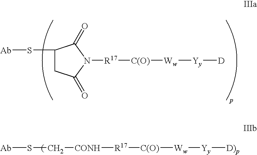
An illustrative Stretcher unit is that of Formula IIIa, and is derived from maleimido-caproyl (MC) wherein R17 is —(CH2)5—:

An illustrative Stretcher unit is that of Formula IIIa, and is derived from maleimido-propanoyl (MP) wherein R17 is —(CH2)2—:

Another illustrative Stretcher unit is that of Formula IIIa wherein R17 is —(CH2CH2O)r—CH2— and r is 2:

Another illustrative Stretcher unit is that of Formula IIIa wherein R17 is —(CH2)rC(O)NRb(CH2CH2O)r—CH2— where R is H and each r is 2:

Another illustrative Stretcher unit is that of Formula IIIb wherein R17 is —(CH2)5—:

In another embodiment, the Stretcher unit is linked to the Antibody unit via a disulfide bond between a sulfur atom of the Antibody unit and a sulfur atom of the Stretcher unit. A representative Stretcher unit of this embodiment is depicted within the square brackets of Formula IV, wherein R17, Ab-, —W—, —Y—, -D, w and y are as defined above.
Ab-S—(—R17—C(O)—Ww—Yy-D)p IV
Ab-S—(—R17—C(O)—Ww—Yy-D)p IV
In yet another embodiment, the reactive group of the Stretcher contains a thiol-reactive functional group that can form a bond with a free cysteine thiol of an antibody. Examples of thiol-reaction functional groups include, but are not limited to, maleimide, α-haloacetyl, activated esters such as succinimide esters, 4-nitrophenyl esters, pentafluorophenyl esters, tetrafluorophenyl esters, anhydrides, acid chlorides, sulfonyl chlorides, isocyanates and isothiocyanates. Representative Stretcher units of this embodiment are depicted within the square brackets of Formulas Va and Vb, wherein —R17—, Ab-, —W—, —Y—, -D, w and y are as defined above;

In another embodiment, the linker may be a dendritic type linker for covalent attachment of more than one drug moiety through a branching, multifunctional linker moiety to an antibody (Sun et al (2002) Bioorganic & Medicinal Chemistry Letters 12:2213-2215; Sun et al (2003) Bioorganic & Medicinal Chemistry 11:1761-1768; King (2002) Tetrahedron Letters 43:1987-1990). Dendritic linkers can increase the molar ratio of drug to antibody, i.e. loading, which is related to the potency of the ADC. Thus, where a cysteine engineered antibody bears only one reactive cysteine thiol group, a multitude of drug moieties may be attached through a dendritic linker.
Amino Acid Unit
The linker may comprise amino acid residues. The Amino Acid unit (—Ww—), when present, links the antibody (Ab) to the drug moiety (D) of the cysteine engineered antibody-drug conjugate (ADC) of the invention.
—Ww— is a dipeptide, tripeptide, tetrapeptide, pentapeptide, hexapeptide, heptapeptide, octapeptide, nonapeptide, decapeptide, undecapeptide or dodecapeptide unit. Amino acid residues which comprise the Amino Acid unit include those occurring naturally, as well as minor amino acids and non-naturally occurring amino acid analogs, such as citrulline. Each —W— unit independently has the formula denoted below in the square brackets, and w is an integer ranging from 0 to 12:

wherein R19 is hydrogen, methyl, isopropyl, isobutyl, sec-butyl, benzyl, p-hydroxybenzyl, —CH2OH, —CH(OH)CH3, —CH2CH2SCH3, —CH2CONH2, —CH2COOH, —CH2CH2CONH2, —CH2CH2COOH, —(CH2)3NHC(═NH)NH2, —(CH2)3NH2, —(CH2)3NHCOCH3, —(CH2)3NHCHO, —(CH2)4NHC(═NH)NH2, —(CH2)4NH2, —(CH2)4NHCOCH3, —(CH2)4NHCHO, —(CH2)3NHCONH2, —(CH2)4NHCONH2, —CH2CH2CH(OH)CH2NH2, 2-pyridylmethyl-, 3-pyridylmethyl-, 4-pyridylmethyl-, phenyl, cyclohexyl,

The Amino Acid unit can be enzymatically cleaved by one or more enzymes, including a tumor-associated protease, to liberate the Drug moiety (-D), which in one embodiment is protonated in vivo upon release to provide a Drug (D).
Useful —Ww— units can be designed and optimized in their selectivity for enzymatic cleavage by a particular enzymes, for example, a tumor-associated protease. In one embodiment, a —Ww— unit is that whose cleavage is catalyzed by cathepsin B, C and D, or a plasmin protease.
Exemplary —Ww— Amino Acid units include a dipeptide, a tripeptide, a tetrapeptide or a pentapeptide. Exemplary dipeptides include: valine-citrulline (vc or val-cit), alanine-phenylalanine (af or ala-phe). Exemplary tripeptides include: glycine-valine-citrulline (gly-val-cit) and glycine-glycine-glycine (gly-gly-gly).
When R19 is other than hydrogen, the carbon atom to which R19 is attached is chiral. Each carbon atom to which R19 is attached is independently in the (S) or (R) configuration, or a racemic mixture. Amino acid units may thus be enantiomerically pure, racemic, or diastereomeric.
Spacer Unit
The Spacer unit (—Y—), when present (y=1 or 2), links an Amino Acid unit (—Ww—) to the drug moiety (D) when an Amino Acid unit is present (w=1-12). Alternately, the Spacer unit links the Stretcher unit to the Drug moiety when the Amino Acid unit is absent. The Spacer unit also links the drug moiety to the antibody unit when both the Amino Acid unit and Stretcher unit are absent (w, y=0). Spacer units are of two general types: self-immolative and non self-immolative. A non self-immolative Spacer unit is one in which part or all of the Spacer unit remains bound to the Drug moiety after cleavage, particularly enzymatic, of an Amino Acid unit from the antibody-drug conjugate or the Drug moiety-linker. When an ADC containing a glycine-glycine Spacer unit or a glycine Spacer unit undergoes enzymatic cleavage via a tumor-cell associated-protease, a cancer-cell-associated protease or a lymphocyte-associated protease, a glycine-glycine-Drug moiety or a glycine-Drug moiety is cleaved from Ab-Aa-Ww—. In one embodiment, an independent hydrolysis reaction takes place within the target cell, cleaving the glycine-Drug moiety bond and liberating the Drug.
In another embodiment, —Yy— is a p-aminobenzylcarbamoyl (PAB) unit (see Schemes 2 and 3) whose phenylene portion is substituted with Qm wherein Q is —C1-C8 alkyl, —O—(C1-C8 alkyl), -halogen, -nitro or -cyano; and m is an integer ranging from 0-4.
Exemplary embodiments of a non self-immolative Spacer unit (—Y—) are: -Gly-Gly-; -Gly-; -Ala-Phe-; -Val-Cit-.
In one embodiment, a Drug moiety-linker or an ADC is provided in which the Spacer unit is absent (y=0), or a pharmaceutically acceptable salt or solvate thereof.
Alternatively, an ADC containing a self-immolative Spacer unit can release -D. In one embodiment, —Y— is a PAB group that is linked to —Ww— via the amino nitrogen atom of the PAB group, and connected directly to -D via a carbonate, carbamate or ether group, where the ADC has the exemplary structure:

wherein Q is —C1-C8 alkyl, —O—(C1-C8 alkyl), -halogen, -nitro or -cyano; m is an integer ranging from 0-4; and p ranges from 1 to 4.
Other examples of self-immolative spacers include, but are not limited to, aromatic compounds that are electronically similar to the PAB group such as 2-aminoimidazol-5-methanol derivatives (Hay et al. (1999) Bioorg. Med. Chem. Lett. 9:2237) and ortho or para-aminobenzylacetals. Spacers can be used that undergo cyclization upon amide bond hydrolysis, such as substituted and unsubstituted 4-aminobutyric acid amides (Rodrigues et al (1995) Chemistry Biology 2:223), appropriately substituted bicyclo[2.2.1] and bicyclo[2.2.2] ring systems (Storm et al (1972) J. Amer. Chem. Soc. 94:5815) and 2-aminophenylpropionic acid amides (Amsberry, et al (1990) J. Org. Chem. 55:5867). Elimination of amine-containing drugs that are substituted at glycine (Kingsbury et al (1984) J. Med. Chem. 27:1447) are also examples of self-immolative spacer useful in ADCs.
In one embodiment, the Spacer unit is a branched bis(hydroxymethyl)styrene (BHMS), which can be used to incorporate and release multiple drugs, having the structure:

comprising a 2-(4-aminobenzylidene)propane-1,3-diol dendrimer unit (WO 2004/043493; de Groot et al (2003) Angew. Chem. Int. Ed. 42:4490-4494), wherein Q is —C1-C8 alkyl, —O—(C1-C8 alkyl), -halogen, -nitro or -cyano; m is an integer ranging from 0-4; n is 0 or 1; and p ranges ranging from 1 to 4.
Dendritic Linkers
In another embodiment, linker L may be a dendritic type linker for covalent attachment of more than one drug moiety through a branching, multifunctional linker moiety to an antibody (Sun et al (2002) Bioorganic & Medicinal Chemistry Letters 12:2213-2215; Sun et al (2003) Bioorganic & Medicinal Chemistry 11: 1761-1768). Dendritic linkers can increase the molar ratio of drug to antibody, i.e. loading, which is related to the potency of the ADC. Thus, where a cysteine engineered antibody bears only one reactive cysteine thiol group, a multitude of drug moieties may be attached through a dendritic linker.
The following exemplary embodiments of dendritic linker reagents allow up to nine nucleophilic drug moiety reagents to be conjugated by reaction with the chloroethyl nitrogen mustard functional groups:

In another embodiment of a Spacer unit, branched, dendritic linkers with self-immolative 2,6-bis(hydroxymethyl)-p-cresol and 2,4,6-tris(hydroxymethyl)-phenol dendrimer units (WO 2004/01993; Szalai et al (2003) J. Amer. Chem. Soc. 125:15688-15689; Shamis et al (2004) J. Amer. Chem. Soc. 126:1726-1731; Amir et al (2003) Angew. Chem. Int. Ed. 42:4494-4499) may be employed as linkers in the compounds of the invention.
In another embodiment, the D moieties are the same.
In yet another embodiment, the D moieties are different.
In one aspect, Spacer units (—Yy—) are represented by Formulas (X)-(XII):

wherein Q is —C1-C8 alkyl, —O—(C1-C8 alkyl), -halogen, -nitro or -cyano; and m is an integer ranging from 0-4;

Embodiments of the Formula I antibody-drug conjugate compounds include XIIIa (val-cit), XIIIb (MC-val-cit), XIIIc (MC-val-cit-PAB):

Other exemplary embodiments of the Formula Ia antibody-drug conjugate compounds include XIVa-e:

where X is:

Y is:
and R is independently H or C1-C6 alkyl; and n is 1 to 12.
In another embodiment, a Linker has a reactive functional group which has a nucleophilic group that is reactive to an electrophilic group present on an antibody. Useful electrophilic groups on an antibody include, but are not limited to, aldehyde and ketone carbonyl groups. The heteroatom of a nucleophilic group of a Linker can react with an electrophilic group on an antibody and form a covalent bond to an antibody unit. Useful nucleophilic groups on a Linker include, but are not limited to, hydrazide, oxime, amino, hydrazine, thiosemicarbazone, hydrazine carboxylate, and arylhydrazide. The electrophilic group on an antibody provides a convenient site for attachment to a Linker.
Typically, peptide-type Linkers can be prepared by forming a peptide bond between two or more amino acids and/or peptide fragments. Such peptide bonds can be prepared, for example, according to the liquid phase synthesis method (E. Schröder and K. Lübke (1965) “The Peptides”, volume 1, pp 76-136, Academic Press) which is well known in the field of peptide chemistry.
Linker intermediates may be assembled with any combination or sequence of reactions including Spacer, Stretcher, and Amino Acid units. The Spacer, Stretcher, and Amino Acid units may employ reactive functional groups which are electrophilic, nucleophilic, or free radical in nature. Reactive functional groups include, but are not limited to:

where X is a leaving group, e.g. O-mesyl, O-tosyl, —Cl, —Br, —I; or maleimide.
In another embodiment, the Linker may be substituted with groups which modulated solubility or reactivity. For example, a charged substituent such as sulfonate (—SO3 −) or ammonium, may increase water solubility of the reagent and facilitate the coupling reaction of the linker reagent with the antibody or the drug moiety, or facilitate the coupling reaction of Ab-L (antibody-linker intermediate) with D, or D-L (drug-linker intermediate) with Ab, depending on the synthetic route employed to prepare the ADC.
The compounds of the invention expressly contemplate, but are not limited to, ADC prepared with linker reagents: BMPEO, BMPS, EMCS, GMBS, HBVS, LC-SMCC, MBS, MPBH, SBAP, SIA, SIAB, SMCC, SMPB, SMPH, sulfo-EMCS, sulfo-GMBS, sulfo-KMUS, sulfo-MBS, sulfo-SIAB, sulfo-SMCC, and sulfo-SMPB, and SVSB (succinimidyl-(4-vinylsulfone)benzoate), and including bis-maleimide reagents: DTME, BMB, BMDB, BMH, BMOE, BM(PEO)3, and BM(PEO)4, which are commercially available from Pierce Biotechnology, Inc., Customer Service Department, P.O. Box 117, Rockford, Ill. 61105 U.S.A, U.S.A 1-800-874-3723, International +815-968-0747. See pages 467-498, 2003-2004 Applications Handbook and Catalog. Bis-maleimide reagents allow the attachment of the thiol group of a cysteine engineered antibody to a thiol-containing drug moiety, label, or linker intermediate, in a sequential or concurrent fashion. Other functional groups besides maleimide, which are reactive with a thiol group of a cysteine engineered antibody, drug moiety, label, or linker intermediate include iodoacetamide, bromoacetamide, vinyl pyridine, disulfide, pyridyl disulfide, isocyanate, and isothiocyanate.

Useful linker reagents can also be obtained via other commercial sources, such as Molecular Biosciences Inc. (Boulder, Colo.), or synthesized in accordance with procedures described in Toki et al (2002) J. Org. Chem. 67:1866-1872; Walker, M. A. (1995) J. Org. Chem. 60:5352-5355; Frisch et al (1996) Bioconjugate Chem. 7:180-186; U.S. Pat. No. 6,214,345; WO 02/088172; US 2003130189; US2003096743; WO 03/026577; WO 03/043583; and WO 04/032828.
Stretchers of formula (IIIa) can be introduced into a Linker by reacting the following linker reagents with the N-terminus of an Amino Acid unit:

where n is an integer ranging from 1-10 and T is —H or —SO3Na;

where n is an integer ranging from 0-3;

Stretcher units of can be introduced into a Linker by reacting the following bifunctional reagents with the N-terminus of an Amino Acid unit:

where X is Br or I. Stretcher units of formula can also be introduced into a Linker by reacting the following bifunctional reagents with the N-terminus of an Amino Acid unit:

Stretcher units of formula (Va) can be introduced into a Linker by reacting the following intermediates with the N-terminus of an Amino Acid unit:

Isothiocyanate Stretchers of the formula shown below may be prepared from isothiocyanatocarboxylic acid chlorides as described in Angew. Chem., (1975) 87(14), 517.
wherein —R17— is as described herein.
An exemplary valine-citrulline (val-cit or vc) dipeptide linker reagent having a maleimide Stretcher and a para-aminobenzylcarbamoyl (PAB) self-immolative Spacer has the structure:

where Q is —C1-C8 alkyl, —O—(C1-C8 alkyl), -halogen, -nitro or -cyano; and m is an integer ranging from 0-4.
An exemplary phe-lys(Mtr) dipeptide linker reagent having a maleimide Stretcher unit and a p-aminobenzyl self-immolative Spacer unit can be prepared according to Dubowchik, et al. (1997) Tetrahedron Letters, 38:5257-60, and has the structure:

where Mtr is mono-4-methoxytrityl, Q is —C1-C8 alkyl, —O—(C1-C8 alkyl), -halogen, -nitro or -cyano; and m is an integer ranging from 0-4.
Exemplary antibody-drug conjugate compounds of the invention include:

where Val is valine; Cit is citrulline; p is 1, 2, 3, or 4; and Ab is a cysteine engineered antibody. Other exemplary antibody drug conjugates where maytansinoid drug moiety DM1 is linked through a BMPEO linker to a thiol group of trastuzumab have the structure:

where Ab is a cysteine engineered antibody; n is 0, 1, or 2; and p is 1, 2, 3, or 4.
Preparation of Antibody-drug Conjugates
The ADC of Formula I may be prepared by several routes, employing organic chemistry reactions, conditions, and reagents known to those skilled in the art, including: (1) reaction of a cysteine group of a cysteine engineered antibody with a linker reagent, to form antibody-linker intermediate Ab-L, via a covalent bond, followed by reaction with an activated drug moiety D; and (2) reaction of a nucleophilic group of a drug moiety with a linker reagent, to form drug-linker intermediate D-L, via a covalent bond, followed by reaction with a cysteine group of a cysteine engineered antibody. Conjugation methods (1) and (2) may be employed with a variety of cysteine engineered antibodies, drug moieties, and linkers to prepare the antibody-drug conjugates of Formula I.
Antibody cysteine thiol groups are nucleophilic and capable of reacting to form covalent bonds with electrophilic groups on linker reagents and drug-linker intermediates including: (i) active esters such as NHS esters, HOBt esters, haloformates, and acid halides; (ii) alkyl and benzyl halides, such as haloacetamides; (iii) aldehydes, ketones, carboxyl, and maleimide groups; and (iv) disulfides, including pyridyl disulfides, via sulfide exchange. Nucleophilic groups on a drug moiety include, but are not limited to: amine, thiol, hydroxyl, hydrazide, oxime, hydrazine, thiosemicarbazone, hydrazine carboxylate, and arylhydrazide groups capable of reacting to form covalent bonds with electrophilic groups on linker moieties and linker reagents.
Maytansine may, for example, be converted to May-SSCH3, which can be reduced to the free thiol, May-SH, and reacted with a modified antibody (Chari et al (1992) Cancer Research 52:127-131) to generate a maytansinoid-antibody immunoconjugate with a disulfide linker. Antibody-maytansinoid conjugates with disulfide linkers have been reported (WO 04/016801; U.S. Pat. No. 6,884,874; US 2004/039176 A1; WO 03/068144; US 2004/001838 A1; U.S. Pat. Nos. 6,441,163, 5,208,020, 5,416,064; WO 01/024763). The disulfide linker SPP is constructed with linker reagent N-succinimidyl 4-(2-pyridylthio) pentanoate.
Under certain conditions, the cysteine engineered antibodies may be made reactive for conjugation with linker reagents by treatment with a reducing agent such as DTT (Cleland's reagent, dithiothreitol) or TCEP (tris(2-carboxyethyl)phosphine hydrochloride; Getz et al (1999) Anal. Biochem. Vol 273:73-80; Soltec Ventures, Beverly, Mass.). Full length, cysteine engineered monoclonal antibodies (ThioMabs) expressed in CHO cells were reduced with about a 50 fold excess of TCEP for 3 hrs at 37° C. to reduce disulfide bonds which may form between the newly introduced cysteine residues and the cysteine present in the culture media. The reduced ThioMab was diluted and loaded onto HiTrap S column in 10 mM sodium acetate, pH 5, and eluted with PBS containing 0.3M sodium chloride. Disulfide bonds were reestablished between cysteine residues present in the parent Mab with dilute (200 nM) aqueous copper sulfate (CuSO4) at room temperature, overnight. Other oxidants, i.e. oxidizing agents, and oxidizing conditions, which are known in the art may be used. Ambient air oxidation is also effective. This mild, partial reoxidation step forms intrachain disulfides efficiently with high fidelity. An approximate 10 fold excess of drug-linker intermediate, e.g. BM(PEO)4-DM1 was added, mixed, and let stand for about an hour at room temperature to effect conjugation and form the ThioMab antibody-drug conjugate. The conjugation mixture was gel filtered and loaded and eluted through a HiTrap S column to remove excess drug-linker intermediate and other impurities.
A variety of ThioFab and ThioMab antibody-drug conjugates were prepared (Examples 4-8). Cysteine mutant hu4D5Fabv8 (V110C) was conjugated with the maytansinoid drug moiety DM1 with a bis-maleimido linker reagent BMPEO to form hu4D5Fabv8 (V110C)-BMPEO-DM1 (Example 8).
In Vitro Cell Proliferation Assays
Generally, the cytotoxic or cytostatic activity of an antibody-drug conjugate (ADC) is measured by: exposing mammalian cells having receptor proteins, e.g. HER2, to the antibody of the ADC in a cell culture medium; culturing the cells for a period from about 6 hours to about 5 days; and measuring cell viability. Cell-based in vitro assays were used to measure viability (proliferation), cytotoxicity, and induction of apoptosis (caspase activation) of the ADC of the invention.
The in vitro potency of antibody-drug conjugates was measured by a cell proliferation assay (FIGS. 10 and 11 , Example 9). The CellTiter-Glo Luminescent Cell Viability Assay is a commercially available (Promega Corp., Madison, Wis.), homogeneous assay method based on the recombinant expression of Coleoptera luciferase (U.S. Pat. Nos. 5,583,024; 5,674,713 and 5,700,670). This cell proliferation assay determines the number of viable cells in culture based on quantitation of the ATP present, an indicator of metabolically active cells (Crouch et al (1993) J. Immunol. Meth. 160:81-88; U.S. Pat. No. 6,602,677). The CellTiter-Glo Assay was conducted in 96 well format, making it amenable to automated high-throughput screening (HTS) (Cree et al (1995) AntiCancer Drugs 6:398-404). The homogeneous assay procedure involves adding the single reagent (CellTiter-Glo Reagent) directly to cells cultured in serum-supplemented medium. Cell washing, removal of medium and multiple pipetting steps are not required. The system detects as few as 15 cells/well in a 384-well format in 10 minutes after adding reagent and mixing. The cells may be treated continuously with ADC, or they may be treated and separated from ADC. Generally, cells treated briefly, i.e. 3 hours, showed the same potency effects as continuously treated cells.
The homogeneous “add-mix-measure” format results in cell lysis and generation of a luminescent signal proportional to the amount of ATP present. The amount of ATP is directly proportional to the number of cells present in culture. The CellTiter-Glo® Assay generates a “glow-type” luminescent signal, produced by the luciferase reaction, which has a half-life generally greater than five hours, depending on cell type and medium used. Viable cells are reflected in relative luminescence units (RLU). The substrate, Beetle Luciferin, is oxidatively decarboxylated by recombinant firefly luciferase with concomitant conversion of ATP to AMP and generation of photons. The extended half-life eliminates the need to use reagent injectors and provides flexibility for continuous or batch mode processing of multiple plates. This cell proliferation assay can be used with various multiwell formats, e.g. 96 or 384 well format. Data can be recorded by luminometer or CCD camera imaging device. The luminescence output is presented as relative light units (RLU), measured over time. Alternatively, photons from luminescence can be counted in a scintillation counter in the presence of a scintillant. The light units can be represented then as CPS—counts per second.

The anti-proliferative effects of antibody-drug conjugates were measured by the cell proliferation, in vitro cell killing assay above against the SK-BR-3 breast tumor cell line (FIGS. 10 and 11 ). IC50 values of the ADC were established against SK-BR-3 cells, which are known to overexpress HER2 receptor protein.
Full-length IgG ThioMab conjugates were tested for in vitro, cell proliferation efficacy and compared with parent antibodies. FIG. 20 shows the results of an assay of SK-BR-3 cells treated with: parent antibody trastuzumab (HERCEPTIN®, Genentech, Inc.); trastuzumab-SMCC-DM1 with a drug loading of about 3.4 DM1/Ab; and thio-trastuzumab (A121C)-BMPEO-DM1 with a drug loading of about 1.6 DM1/Ab. The trastuzumab-SMCC-DM1 conjugate is linked to the antibody via the amino reactive, NHS ester SMCC linker reagent, whereas the thio-trastuzumab (A121C)-BMPEO-DM1 conjugates is linked via the thiol reactive, maleimide BMPEO linker reagent. Both conjugates were potent against SK-BR-3 cells and showed comparable activity, whereas trastuzumab did not exert a cytotoxic effect. FIG. 21A shows the results of an assay of HT 1080EphB2 cells treated with: parent 2H9 anti-EphB2RX; and thio 2H9 (A121C) BMPEO-DM1 conjugate. FIG. 21B shows the results of an assay of BT 474 cells treated with: parent 2H9 anti-EphB2RX; and thio 2H9 (A121C) BMPEO-DM1 conjugate. Against both HT 1080EphB2 and BT 474 cells, the 2H9 ThioMab conjugate was more potent than the parent 2H9 antibody conjugate. The conjugate Thio-2H9-BMPEO-DM1 showed functional cell killing activity in EphB2 specific cell line (HT1080EphB2) compared to a non EphB2 cell line, BT474 in which only marginal activity is observed.
Antibody drug conjugates were compared where the antibody is a parent antibody and where the antibody is a cysteine engineered antibody. FIG. 22 shows the results of an assay of PC3/neo cells treated with: 3A5 anti MUC16-SMCC-DM1; and thio 3A5 (A121C) BMPEO-DM1. FIG. 23 shows the results of an assay of PC3/MUC16 cells treated with: 3A5 anti MUC16-SMCC-DM 1; and thio 3A5 (A121C) BMPEO-DM1. FIG. 24 shows the results of an assay of OVCAR-3 cells treated with: 3A5 anti MUC16-SMCC-DM1; and thio 3A5 (A121C) BMPEO-DM1. Thio-3A5-BMPEO-DM1 did not show any significant cell killing activity in the control PC3/neo cell line, whereas it showed comparable activity to 3A5-SMCC-DM1 in the PC3/MUC16 cell line. Thio-3A5-DM1 conjugate also showed activity in the OVCAR-3 that expresses endogenous MUC16 antigen.
In Vivo Efficacy
The in vivo efficacy of two albumin binding peptide-DM1 (maytansinoid)-antibody-drug conjugates (ADC) of the invention was measured by a high expressing HER2 transgenic explant mouse model (FIG. 12 , Example 10). An allograft was propagated from the Fo5 mmtv transgenic mouse which does not respond to, or responds poorly to, HERCEPTIN® therapy. Subjects were treated once with ABP-rhuFab4D5-cys(light chain)-DM1; ABP-rhuFab4D5-cys(heavy chain)-DM1; and placebo PBS buffer control (Vehicle) and monitored over 3 weeks to measure the time to tumor doubling, log cell kill, and tumor shrinkage.
| TDV | |||||
| Sample | Dose | Ti | PR | CR | (days) |
| Vehicle (PBS buffer) | 7/7 | 0/7 | 0/7 | 3 | |
| ABP-rhuFab4D5- | 25 mg per |
7/7 | 1/7 | 0/7 | 14 |
| V110C (light chain)- | (1012 μg/m2 of DM1) | ||||
| DM1 (0.9 DM1/Ab) | |||||
| ABP-rhuFab4D5- | 37.5 mg per |
7/7 | 4/7 | 0/7 | 16 |
| A121C(heavy chain)- | (1012 μg/m2 of DM1) | ||||
| DM1 (0.6 DM1/Ab) | |||||
The term Ti is the number of animals in the study group with tumor at T=O— total animals in group. The term PR is the number of animals attaining partial remission of tumor—animals with tumor at T=0÷in the group. The term CR is the number of animals attaining complete remission of tumor÷animals with tumor at T=0 in the group. The term TDV is the tumor doubling time, i.e. time in days for the control tumor volume to double.
The seven mice treated with 25 mg per kg (1012 ug/m2 of DM1) of ABP-rhuFab4D5-cys(light chain)-DM 1 were all tumor-positive and gave one animal with partial remission after 20 days. The seven mice treated with 37.5 mg per kg (1012 ug/m2 of DM1) of ABP-rhuFab4D5-cys(heavy chain)-DM1 were all tumor-positive and gave four animals with partial remission after 20 days.
The full length IgG ThioMab antibody variant with the A121C cysteine mutation and conjugated to the BMPEO linker and DM1 drug moiety was tested against the parent trastuzumab-SMCC-DM1 conjugate in MMTV-HER2 Fo5 tumor-bearing mice. Tumor size at day 0 of injection was about 100-200 mm in size. FIG. 25 shows the mean tumor volume change over 21 days in athymic nude mice with MMTV-HER2 Fo5 mammary tumor allografts, after a single dose on Day 0 with: Vehicle (Buffer); trastuzumab-SMCC-DM110 mg/kg; thio trastuzumab (A121C)-SMCC-DM121 mg/kg and thio trastuzumab (A121C)-SMCC-DM1 10 mg/kg.
| Sample | Dose | Ti | PR | CR |
| Vehicle (PBS buffer) | — | 10/10 | 0/10 | 0/10 |
| trastuzumab-SMCC- | 10 mg Ab per |
10/10 | 7/10 | 0/10 |
| DM1 3.4 DM1/Ab | (490 μg/m2 of DM1) | |||
| thio-trastuzumab | 21 mg Ab per |
8/10 | 6/10 | 4/10 |
| (A121C)-BMPEO-DM1 | (496 μg/m2 of DM1) | |||
| 1.6 DM1/Ab | ||||
| thio-trastuzumab | 10 mg Ab per |
10/10 | 0/10 | 0/10 |
| (A121C)-BMPEO-DM1 | (236 μg/m2 of DM1) | |||
| 1.6 DM1/Ab | ||||
It can be seen from FIG. 25 that each conjugate exerts a significant effect of retarding tumor growth relative to placebo (Vehicle). Each of the ten mice in the four groups above received a single injection at day 1. The parent trastuzumab-SMCC-DM1 conjugate was loaded with more than twice (3.4 DM1/Ab) the number of drug moieties than the cysteine engineered thio-trastuzumab (A121C)-BMPEO-DM1 conjugate (1.6 DM1/Ab). The effective amount of DM1 was thus approximately equal between parent trastuzumab-SMCC-DM1 and the higher dose (21 mg Ab) thio-trastuzumab (A121C)-BMPEO-DM1. These two sample showed the most potency. After 14 days post-injection, most of the animals receiving these conjugates were in partial or complete remission. The lower efficacy of the lower dose thio-trastuzumab (A121C)-BMPEO-DM1 sample confirmed a DM1 dose-related response. Thio-Trastuzumab-DM1 either dosed in equivalent antibody (10 mg/kg) or DM1 drug (21 mg/kg) quantity to that of control trastuzumab-SMCC-DM1 conjugate. As seen from the FIG. 25 , Thio-BMPEO-DM1 (21 mg/kg) showed slightly better response than that of trastuzumab-SMCC-DM1 group as some of the animals showed complete response with Thiomab-DM 1 whereas there was only partial response with trastuzumab-SMCC-DM 1.
Administration of Antibody-Drug Conjugates
The antibody-drug conjugates (ADC) of the invention may be administered by any route appropriate to the condition to be treated. The ADC will typically be administered parenterally, i.e. infusion, subcutaneous, intramuscular, intravenous, intradermal, intrathecal and epidural.
Pharmaceutical Formulations
Pharmaceutical formulations of therapeutic antibody-drug conjugates (ADC) of the invention are typically prepared for parenteral administration, i.e. bolus, intravenous, intratumor injection with a pharmaceutically acceptable parenteral vehicle and in a unit dosage injectable form. An antibody-drug conjugate (ADC) having the desired degree of purity is optionally mixed with pharmaceutically acceptable diluents, carriers, excipients or stabilizers (Remington's Pharmaceutical Sciences (1980) 16th edition, Osol, A. Ed.), in the form of a lyophilized formulation or an aqueous solution.
Acceptable diluents, carriers, excipients, and stabilizers are nontoxic to recipients at the dosages and concentrations employed, and include buffers such as phosphate, citrate, and other organic acids; antioxidants including ascorbic acid and methionine; preservatives (such as octadecyldimethylbenzyl ammonium chloride; hexamethonium chloride; benzalkonium chloride, benzethonium chloride; phenol, butyl or benzyl alcohol; alkyl parabens such as methyl or propyl paraben; catechol; resorcinol; cyclohexanol; 3-pentanol; and m-cresol); low molecular weight (less than about 10 residues) polypeptides; proteins, such as serum albumin, gelatin, or immunoglobulins; hydrophilic polymers such as polyvinylpyrrolidone; amino acids such as glycine, glutamine, asparagine, histidine, arginine, or lysine; monosaccharides, disaccharides, and other carbohydrates including glucose, mannose, or dextrins; chelating agents such as EDTA; sugars such as sucrose, mannitol, trehalose or sorbitol; salt-forming counter-ions such as sodium; metal complexes (e.g. Zn-protein complexes); and/or non-ionic surfactants such as TWEEN™, PLURONICS™ or polyethylene glycol (PEG). For example, lyophilized anti-ErbB2 antibody formulations are described in WO 97/04801, expressly incorporated herein by reference.
The active pharmaceutical ingredients may also be entrapped in microcapsules prepared, for example, by coacervation techniques or by interfacial polymerization, for example, hydroxymethylcellulose or gelatin-microcapsules and poly-(methylmethacylate) microcapsules, respectively, in colloidal drug delivery systems (for example, liposomes, albumin microspheres, microemulsions, nano-particles and nanocapsules) or in macroemulsions. Such techniques are disclosed in Remington's Pharmaceutical Sciences 16th edition, Osol, A. Ed. (1980).
Sustained-release preparations may be prepared. Suitable examples of sustained-release preparations include semi permeable matrices of solid hydrophobic polymers containing the ADC, which matrices are in the form of shaped articles, e.g. films, or microcapsules. Examples of sustained-release matrices include polyesters, hydrogels (for example, poly(2-hydroxyethyl-methacrylate), or poly(vinyl alcohol)), polylactides (U.S. Pat. No. 3,773,919), copolymers of L-glutamic acid and gamma-ethyl-L-glutamate, non-degradable ethylene-vinyl acetate, degradable lactic acid-glycolic acid copolymers such as the LUPRON DEPOT™ (injectable microspheres composed of lactic acid-glycolic acid copolymer and leuprolide acetate), and poly-D-(−)-3-hydroxybutyric acid.
The formulations to be used for in vivo administration must be sterile, which is readily accomplished by filtration through sterile filtration membranes.
The formulations include those suitable for the foregoing administration routes. The formulations may conveniently be presented in unit dosage form and may be prepared by any of the methods well known in the art of pharmacy. Techniques and formulations generally are found in Remington's Pharmaceutical Sciences (Mack Publishing Co., Easton, Pa.). Such methods include the step of bringing into association the active ingredient with the carrier which constitutes one or more accessory ingredients. In general the formulations are prepared by uniformly and intimately bringing into association the active ingredient with liquid carriers or finely divided solid carriers or both, and then, if necessary, shaping the product.
Aqueous suspensions of the invention contain the active materials in admixture with excipients suitable for the manufacture of aqueous suspensions. Such excipients include a suspending agent, such as sodium carboxymethylcellulose, croscarmellose, povidone, methylcellulose, hydroxypropyl methylcelluose, sodium alginate, polyvinylpyrrolidone, gum tragacanth and gum acacia, and dispersing or wetting agents such as a naturally occurring phosphatide (e.g., lecithin), a condensation product of an alkylene oxide with a fatty acid (e.g., polyoxyethylene stearate), a condensation product of ethylene oxide with a long chain aliphatic alcohol (e.g., heptadecaethyleneoxycetanol), a condensation product of ethylene oxide with a partial ester derived from a fatty acid and a hexitol anhydride (e.g., polyoxyethylene sorbitan monooleate). The aqueous suspension may also contain one or more preservatives such as ethyl or n-propyl p-hydroxy-benzoate, one or more coloring agents, one or more flavoring agents and one or more sweetening agents, such as sucrose or saccharin.
The pharmaceutical compositions of ADC may be in the form of a sterile injectable preparation, such as a sterile injectable aqueous or oleaginous suspension. This suspension may be formulated according to the known art using those suitable dispersing or wetting agents and suspending agents which have been mentioned above. The sterile injectable preparation may also be a sterile injectable solution or suspension in a non-toxic parenterally acceptable diluent or solvent, such as a solution in 1,3-butane-diol or prepared as a lyophilized powder. Among the acceptable vehicles and solvents that may be employed are water, Ringer's solution and isotonic sodium chloride solution. In addition, sterile fixed oils may conventionally be employed as a solvent or suspending medium. For this purpose any bland fixed oil may be employed including synthetic mono- or diglycerides. In addition, fatty acids such as oleic acid may likewise be used in the preparation of injectables.
The amount of active ingredient that may be combined with the carrier material to produce a single dosage form will vary depending upon the host treated and the particular mode of administration. For example, an aqueous solution intended for intravenous infusion may contain from about 3 to 500 μg of the active ingredient per milliliter of solution in order that infusion of a suitable volume at a rate of about 30 mL/hr can occur.
Formulations suitable for parenteral administration include aqueous and non-aqueous sterile injection solutions which may contain anti-oxidants, buffers, bacteriostats and solutes which render the formulation isotonic with the blood of the intended recipient; and aqueous and non-aqueous sterile suspensions which may include suspending agents and thickening agents.
Although oral administration of protein therapeutics are disfavored due to hydrolysis or denaturation in the gut, formulations of ADC suitable for oral administration may be prepared as discrete units such as capsules, cachets or tablets each containing a predetermined amount of the ADC.
The formulations may be packaged in unit-dose or multi-dose containers, for example sealed ampoules and vials, and may be stored in a freeze-dried (lyophilized) condition requiring only the addition of the sterile liquid carrier, for example water, for injection immediately prior to use. Extemporaneous injection solutions and suspensions are prepared from sterile powders, granules and tablets of the kind previously described. Preferred unit dosage formulations are those containing a daily dose or unit daily sub-dose, as herein above recited, or an appropriate fraction thereof, of the active ingredient.
The invention further provides veterinary compositions comprising at least one active ingredient as above defined together with a veterinary carrier therefore. Veterinary carriers are materials useful for the purpose of administering the composition and may be solid, liquid or gaseous materials which are otherwise inert or acceptable in the veterinary art and are compatible with the active ingredient. These veterinary compositions may be administered parenterally, orally or by any other desired route.
Antibody-drug Conjugate Treatments
It is contemplated that the antibody-drug conjugates (ADC) of the present invention may be used to treat various diseases or disorders, e.g. characterized by the overexpression of a tumor antigen. Exemplary conditions or hyperproliferative disorders include benign or malignant tumors; leukemia and lymphoid malignancies. Others include neuronal, glial, astrocytal, hypothalamic, glandular, macrophagal, epithelial, stromal, blastocoelic, inflammatory, angiogenic and immunologic, including autoimmune, disorders.
The ADC compounds which are identified in the animal models and cell-based assays can be further tested in tumor-bearing higher primates and human clinical trials. Human clinical trials can be designed similar to the clinical trials testing the efficacy of the anti-HER2 monoclonal antibody HERCEPTINO® in patients with HER2 overexpressing metastatic breast cancers that had received extensive prior anti-cancer therapy as reported by Baselga et al. (1996) J. Clin. Oncol. 14:737-744. The clinical trial may be designed to evaluate the efficacy of an ADC in combinations with known therapeutic regimens, such as radiation and/or chemotherapy involving known chemotherapeutic and/or cytotoxic agents.
Generally, the disease or disorder to be treated is a hyperproliferative disease such as cancer. Examples of cancer to be treated herein include, but are not limited to, carcinoma, lymphoma, blastoma, sarcoma, and leukemia or lymphoid malignancies. More particular examples of such cancers include squamous cell cancer (e.g. epithelial squamous cell cancer), lung cancer including small-cell lung cancer, non-small cell lung cancer, adenocarcinoma of the lung and squamous carcinoma of the lung, cancer of the peritoneum, hepatocellular cancer, gastric or stomach cancer including gastrointestinal cancer, pancreatic cancer, glioblastoma, cervical cancer, ovarian cancer, liver cancer, bladder cancer, hepatoma, breast cancer, colon cancer, rectal cancer, colorectal cancer, endometrial or uterine carcinoma, salivary gland carcinoma, kidney or renal cancer, prostate cancer, vulval cancer, thyroid cancer, hepatic carcinoma, anal carcinoma, penile carcinoma, as well as head and neck cancer.
The cancer may comprise HER2-expressing cells, such that the ADC of the present invention are able to bind to the cancer cells. To determine ErbB2 expression in the cancer, various diagnostic/prognostic assays are available. In one embodiment, ErbB2 overexpression may be analyzed by IHC, e.g. using the HERCEPTEST (Dako). Parrafin embedded tissue sections from a tumor biopsy may be subjected to the IHC assay and accorded a ErbB2 protein staining intensity criteria as follows: Score 0, no staining is observed or membrane staining is observed in less than 10% of tumor cells; Score 1+, a faint/barely perceptible membrane staining is detected in more than 10% of the tumor cells, the cells are only stained in part of their membrane; Score 2+, a weak to moderate complete membrane staining is observed in more than 10% of the tumor cells; Score 3+, a moderate to strong complete membrane staining is observed in more than 10% of the tumor cells. Those tumors with 0 or 1+ scores for ErbB2 overexpression assessment may be characterized as not overexpressing ErbB2, whereas those tumors with 2+ or 3+ scores may be characterized as overexpressing ErbB2.
Alternatively, or additionally, FISH assays such as the INFORM™ (Ventana Co., Ariz.) or PATHVISION™ (Vysis, Ill.) may be carried out on formalin-fixed, paraffin-embedded tumor tissue to determine the extent (if any) of ErbB2 overexpression in the tumor.
Autoimmune diseases for which the ADC compounds may be used in treatment include rheumatologic disorders (such as, for example, rheumatoid arthritis, Sjögren's syndrome, scleroderma, lupus such as SLE and lupus nephritis, polymyositis/dermatomyositis, cryoglobulinemia, anti-phospholipid antibody syndrome, and psoriatic arthritis), osteoarthritis, autoimmune gastrointestinal and liver disorders (such as, for example, inflammatory bowel diseases (e.g., ulcerative colitis and Crohn's disease), autoimmune gastritis and pernicious anemia, autoimmune hepatitis, primary biliary cirrhosis, primary sclerosing cholangitis, and celiac disease), vasculitis (such as, for example, ANCA-associated vasculitis, including Churg-Strauss vasculitis, Wegener's granulomatosis, and polyarteriitis), autoimmune neurological disorders (such as, for example, multiple sclerosis, opsoclonus myoclonus syndrome, myasthenia gravis, neuromyelitis optica, Parkinson's disease, Alzheimer's disease, and autoimmune polyneuropathies), renal disorders (such as, for example, glomerulonephritis, Goodpasture's syndrome, and Berger's disease), autoimmune dermatologic disorders (such as, for example, psoriasis, urticaria, hives, pemphigus vulgaris, bullous pemphigoid, and cutaneous lupus erythematosus), hematologic disorders (such as, for example, thrombocytopenic purpura, thrombotic thrombocytopenic purpura, post-transfusion purpura, and autoimmune hemolytic anemia), atherosclerosis, uveitis, autoimmune hearing diseases (such as, for example, inner ear disease and hearing loss), Behcet's disease, Raynaud's syndrome, organ transplant, and autoimmune endocrine disorders (such as, for example, diabetic-related autoimmune diseases such as insulin-dependent diabetes mellitus (IDDM), Addison's disease, and autoimmune thyroid disease (e.g., Graves' disease and thyroiditis)). More preferred such diseases include, for example, rheumatoid arthritis, ulcerative colitis, ANCA-associated vasculitis, lupus, multiple sclerosis, Sjögren's syndrome, Graves' disease, IDDM, pernicious anemia, thyroiditis, and glomerulonephritis.
For the prevention or treatment of disease, the appropriate dosage of an ADC will depend on the type of disease to be treated, as defined above, the severity and course of the disease, whether the molecule is administered for preventive or therapeutic purposes, previous therapy, the patient's clinical history and response to the antibody, and the discretion of the attending physician. The molecule is suitably administered to the patient at one time or over a series of treatments. Depending on the type and severity of the disease, about 1 μg/kg to 15 mg/kg (e.g. 0.1-20 mg/kg) of molecule is an initial candidate dosage for administration to the patient, whether, for example, by one or more separate administrations, or by continuous infusion. A typical daily dosage might range from about 1 μg/kg to 100 mg/kg or more, depending on the factors mentioned above. An exemplary dosage of ADC to be administered to a patient is in the range of about 0.1 to about 10 mg/kg of patient weight.
For repeated administrations over several days or longer, depending on the condition, the treatment is sustained until a desired suppression of disease symptoms occurs. An exemplary dosing regimen comprises administering an initial loading dose of about 4 mg/kg, followed by a weekly maintenance dose of about 2 mg/kg of an anti-ErbB2 antibody. Other dosage regimens may be useful. The progress of this therapy is easily monitored by conventional techniques and assays.
Combination Therapy
An antibody-drug conjugate (ADC) of the invention may be combined in a pharmaceutical combination formulation, or dosing regimen as combination therapy, with a second compound having anti-cancer properties. The second compound of the pharmaceutical combination formulation or dosing regimen preferably has complementary activities to the ADC of the combination such that they do not adversely affect each other.
The second compound may be a chemotherapeutic agent, cytotoxic agent, cytokine, growth inhibitory agent, anti-hormonal agent, and/or cardioprotectant. Such molecules are suitably present in combination in amounts that are effective for the purpose intended. A pharmaceutical composition containing an ADC of the invention may also have a therapeutically effective amount of a chemotherapeutic agent such as a tubulin-forming inhibitor, a topoisomerase inhibitor, or a DNA binder.
Other therapeutic regimens may be combined with the administration of an anticancer agent identified in accordance with this invention. The combination therapy may be administered as a simultaneous or sequential regimen. When administered sequentially, the combination may be administered in two or more administrations. The combined administration includes coadministration, using separate formulations or a single pharmaceutical formulation, and consecutive administration in either order, wherein preferably there is a time period while both (or all) active agents simultaneously exert their biological activities.
In one embodiment, treatment with an ADC involves the combined administration of an anticancer agent identified herein, and one or more chemotherapeutic agents or growth inhibitory agents, including coadministration of cocktails of different chemotherapeutic agents. Chemotherapeutic agents include taxanes (such as paclitaxel and docetaxel) and/or anthracycline antibiotics. Preparation and dosing schedules for such chemotherapeutic agents may be used according to manufacturer's instructions or as determined empirically by the skilled practitioner. Preparation and dosing schedules for such chemotherapy are also described in “Chemotherapy Service”, (1992) Ed., M. C. Perry, Williams & Wilkins, Baltimore, Md.
The ADC may be combined with an anti-hormonal compound; e.g., an anti-estrogen compound such as tamoxifen; an anti-progesterone such as onapristone (EP 616812); or an anti-androgen such as flutamide, in dosages known for such molecules. Where the cancer to be treated is hormone independent cancer, the patient may previously have been subjected to anti-hormonal therapy and, after the cancer becomes hormone independent, the ADC (and optionally other agents as described herein) may be administered to the patient. It may be beneficial to also coadminister a cardioprotectant (to prevent or reduce myocardial dysfunction associated with the therapy) or one or more cytokines to the patient. In addition to the above therapeutic regimes, the patient may be subjected to surgical removal of cancer cells and/or radiation therapy.
Suitable dosages for any of the above coadministered agents are those presently used and may be lowered due to the combined action (synergy) of the newly identified agent and other chemotherapeutic agents or treatments.
The combination therapy may provide “synergy” and prove “synergistic”, i.e. the effect achieved when the active ingredients used together is greater than the sum of the effects that results from using the compounds separately. A synergistic effect may be attained when the active ingredients are: (1) co-formulated and administered or delivered simultaneously in a combined, unit dosage formulation; (2) delivered by alternation or in parallel as separate formulations; or (3) by some other regimen. When delivered in alternation therapy, a synergistic effect may be attained when the compounds are administered or delivered sequentially, e.g. by different injections in separate syringes. In general, during alternation therapy, an effective dosage of each active ingredient is administered sequentially, i.e. serially, whereas in combination therapy, effective dosages of two or more active ingredients are administered together.
Metabolites of the Antibody-Drug Conjugates
Also falling within the scope of this invention are the in vivo metabolic products of the ADC compounds described herein, to the extent such products are novel and unobvious over the prior art. Such products may result for example from the oxidation, reduction, hydrolysis, amidation, esterification, enzymatic cleavage, and the like, of the administered compound. Accordingly, the invention includes novel and unobvious compounds produced by a process comprising contacting a compound of this invention with a mammal for a period of time sufficient to yield a metabolic product thereof.
Metabolite products typically are identified by preparing a radiolabelled (e.g. 14C or 3H) ADC, administering it parenterally in a detectable dose (e.g. greater than about 0.5 mg/kg) to an animal such as rat, mouse, guinea pig, monkey, or to man, allowing sufficient time for metabolism to occur (typically about 30 seconds to 30 hours) and isolating its conversion products from the urine, blood or other biological samples. These products are easily isolated since they are labeled (others are isolated by the use of antibodies capable of binding epitopes surviving in the metabolite). The metabolite structures are determined in conventional fashion, e.g. by MS, LC/MS or NMR analysis. In general, analysis of metabolites is done in the same way as conventional drug metabolism studies well-known to those skilled in the art. The conversion products, so long as they are not otherwise found in vivo, are useful in diagnostic assays for therapeutic dosing of the ADC compounds of the invention.
Labelled Antibody Imaging Methods
In another embodiment of the invention, cysteine engineered antibodies may be labelled through the cysteine thiol with radionuclides, fluorescent dyes, bioluminescence-triggering substrate moieties, chemiluminescence-triggering substrate moieties, enzymes, and other detection labels for imaging experiments with diagnostic, pharmacodynamic, and therapeutic applications. Generally, the labelled cysteine engineered antibody, i.e. “biomarker” or “probe”, is administered by injection, perfusion, or oral ingestion to a living organism, e.g. human, rodent, or other small animal, a perfused organ, or tissue sample. The distribution of the probe is detected over a time course and represented by an image.
Articles of Manufacture
In another embodiment of the invention, an article of manufacture, or “kit”, containing materials useful for the treatment of the disorders described above is provided. The article of manufacture comprises a container and a label or package insert on or associated with the container. Suitable containers include, for example, bottles, vials, syringes, blister pack, etc. The containers may be formed from a variety of materials such as glass or plastic. The container holds an antibody-drug conjugate (ADC) composition which is effective for treating the condition and may have a sterile access port (for example the container may be an intravenous solution bag or a vial having a stopper pierceable by a hypodermic injection needle). At least one active agent in the composition is an ADC. The label or package insert indicates that the composition is used for treating the condition of choice, such as cancer. Alternatively, or additionally, the article of manufacture may further comprise a second (or third) container comprising a pharmaceutically-acceptable buffer, such as bacteriostatic water for injection (BWFI), phosphate-buffered saline, Ringer's solution and dextrose solution. It may further include other materials desirable from a commercial and user standpoint, including other buffers, diluents, filters, needles, and syringes.
ThioFab-phage (5×10¹² phage particles) were reacted with 150 fold excess of biotin-PEO-maleimide ((+)-biotinyl-3-maleimidopropionamidyl-3,6-dioxaoctainediamine, Oda et al (2001) Nature Biotechnology 19:379-382, Pierce Biotechnology, Inc.) for 3 hours at room temperature. Excess biotin-PEO-maleimide was removed from biotin-conjugated phage by repeated PEG precipitations (3-4 times). Other commercially available biotinylation reagents with electrophilic groups which are reactive with cysteine thiol groups may be used, including Biotin-BMCC, PEO-Iodoacetyl Biotin, Iodoacetyl-LC-Biotin, and Biotin-HPDP (Pierce Biotechnology, Inc.), and Nα-(3-maleimidylpropionyl)biocytin (MPB, Molecular Probes, Eugene, Oreg.). Other commercial sources for biotinylation, bifunctional and multifunctional linker reagents include Molecular Probes, Eugene, Oreg., and Sigma, St. Louis, Mo.

Bovine serum albumin (BSA), erbB2 extracellular domain (HER2) and streptavidin (100 μl of 2 μg/ml) were separately coated on Maxisorp 96 well plates. After blocking with 0.5% Tween-20 (in PBS), biotinylated and non-biotinylated hu4D5Fabv8-ThioFab-Phage (2×10¹⁰ phage particles) were incubated for 1 hour at room temperature followed by incubation with horseradish peroxidase (HRP) labeled secondary antibody (anti-M13 phage coat protein, pVIII protein antibody). FIG. 8 illustrates the PHESELECTOR Assay by a schematic representation depicting the binding of Fab or ThioFab to HER2 (top) and biotinylated ThioFab to streptavidin (bottom).
Standard HRP reaction was carried out and the absorbance was measured at 450 nm. Thiol reactivity was measured by calculating the ratio between OD450 for streptavidin/OD450 for HER2. A thiol reactivity value of 1 indicates complete biotinylation of the cysteine thiol. In the case of Fab protein binding measurements, hu4D5Fabv8 (2-20 ng) was used followed by incubation with HRP labeled goat polyclonal anti-Fab antibodies.
ThioFabs were expressed upon induction in 34B8, a non-suppressor E. coli strain (Baca et al (1997) Journal Biological Chemistry 272(16):10678-84). The harvested cell pellet was resuspended in PBS (phosphate buffered saline), total cell lysis was performed by passing through a microfluidizer and the ThioFabs were purified by affinity chromatography with protein G SEPHAROSE™ (Amersham).
ThioFabs L-V15C, L-V110C, H-A88C, and H-A121C were expressed and purified by Protein-G SEPHAROSE™ column chromatography. Oligomeric-Fab was present in fractions 26 to 30, and most of the monomeric form was in fractions 31-34. Fractions consisting of the monomeric form were pooled and analyzed by SDS-PAGE along with wild type hu4D5Fabv8 and analyzed on SDS-PAGE gel in reducing (with DTT or BME) and non-reducing (without DTT or BME) conditions. Gel filtration fractions of A121C-ThioFab were analyzed on non-reducing SDS-PAGE.
ThioFabs were conjugated with biotin-PEO-maleimide as described above and the biotinylated-ThioFabs were further purified by Superdex-200™ (Amersham) gel filtration chromatography, which eliminated the free biotin-PEO-maleimide and the oligomeric fraction of ThioFabs. Wild type hu4D5Fabv8 and hu4D5Fabv8 A121C-ThioFab (0.5 mg in quantity) were each and separately incubated with 100 fold molar excess of biotin-PEO-maleimide for 3 hours at room temperature and loaded onto a Superdex-200 gel filtration column to separate free biotin as well as oligomeric Fabs from the monomeric form.
Enzymatic digest fragments of biotinylated hu4D5Fabv8 (A121C) ThioFab and wild type hu4D5Fabv8 were analyzed by liquid chromatography electrospray ionization mass spectroscopy (LS-ESI-MS) The difference between the 48294.5 primary mass of biotinylated hu4D5Fabv8 (A121C) and the 47737.0 primary mass of wild type hu4D5Fabv8 was 557.5 mass units. This fragment indicates the presence of a single biotin-PEO-maleimide moiety (C23H36N5O7S2). Table 4 shows assignment of the fragmentation values which confirms the sequence.
| TABLE 4 |
| LC-ESI-Mass spec analysis of biotinylated hu4D5Fabv8 |
| ThioFab A121C after tryptic digestion |
| Amino acid | b Fragment | y Fragment | ||
| A (Alanine) | 72 | |||
| M (Methionine) | 203 | 2505 | ||
| D (Aspartic acid) | 318 | 2374 | ||
| Y (Tyrosine) | 481 | 2259 | ||
| W (Tryptophan) | 667 | 2096 | ||
| G (Glycine) | 724 | 1910 | ||
| Q (glutamine) | 852 | 1853 | ||
| G (Glycine) | 909 | 1725 | ||
| T (Threonine) | 1010 | 1668 | ||
| L (Leucine) | 1123 | 1567 | ||
| V (Valine) | 1222 | 1454 | ||
| T (Threonine) | 1323 | 1355 | ||
| V (Valine) | 1422 | 1254 | ||
| S (Serine) | 1509 | 1155 | ||
| S (Serine) | 1596 | 1068 | ||
| C (Cysteine) + biotin | 2242 | 981 | ||
| S (Serine) | 2329 | 335 | ||
| T (Threonine) | 2430 | 248 | ||
| K (Lysine) | 175 | |||
Before and after Superdex-200 gel filtration, SDS-PAGE gel analyses, with and without reduction by DTT or BME, of biotinylated ABP-hu4D5Fabv8-A121C, biotinylated ABP— hu4D5Fabv8-V110C, biotinylated double Cys ABP-hu4D5Fabv8-(V110C-A88C), and biotinylated double Cys ABP-hu4D5Fabv8-(V110C-A121C) were conducted.
Mass spectroscopy analysis (MS/MS) of hu4D5Fabv8-(V110)-BMPEO-DM1 (after Superdex-200 gel filtration purification): Fab+1 51607.5, Fab 50515.5. This data shows 91.2% conjugation. MS/MS analysis of hu4D5Fabv8-(V110C)-BMPEO-DM1 (reduced): LC 23447.2, LC+1 24537.3, HC (Fab) 27072.5. This data shows that all DM1 conjugation is on the light chain of the Fab.
The drug linker reagent, maleimidocaproyl-monomethyl auristatin E (MMAE), i.e. MC-MMAE, dissolved in DMSO, is diluted in acetonitrile and water at known concentration, and added to chilled ABP-hu4D5Fabv8-(V110C) ThioFab in phosphate buffered saline (PBS). After about one hour, an excess of maleimide is added to quench the reaction and cap any unreacted antibody thiol groups. The reaction mixture is concentrated by centrifugal ultrafiltration and ABP-hu4D5Fabv8-(V110C)-MC-MMAE is purified and desalted by elution through G25 resin in PBS, filtered through 0.2 μm filters under sterile conditions, and frozen for storage.
ABP-hu4D5Fabv8-(V110C)-MC-MMAF is prepared by conjugation of ABP-hu4D5Fabv8-(V110C) ThioFab and MC-MMAF following the procedure of Example 4.
ABP-hu4D5Fabv8-(A121C)-MC-val-cit-PAB-MMAE is prepared by conjugation of ABP-hu4D5Fabv8-(A121C) and MC-val-cit-PAB-MMAE following the procedure of Example 4.

The free cysteine on hu4D5Fabv8-(V110C) ThioFab was modified by the bis-maleimido reagent BM(PEO)4 (Pierce Chemical), leaving an unreacted maleimido group on the surface of the antibody. This was accomplished by dissolving BM(PEO)4 in a 50% ethanol/water mixture to a concentration of 10 mM and adding a tenfold molar excess of BM(PEO)4 to a solution containing hu4D5Fabv8-(V110C) ThioFab in phosphate buffered saline at a concentration of approximately 1.6 mg/ml (10 micromolar) and allowing it to react for 1 hour. Excess BM(PEO)4 was removed by gel filtration (HiTrap column, Pharmacia) in 30 mM citrate, pH 6 with 150 mM NaCl buffer. An approximate 10 fold molar excess DM1 dissolved in dimethyl acetamide (DMA) was added to the hu4D5Fabv8-(V110C) ThioFab-BMPEO intermediate. Dimethylformamide (DMF) may also be employed to dissolve the drug moiety reagent. The reaction mixture was allowed to react overnight before gel filtration or dialysis into PBS to remove unreacted drug. Gel filtration on S200 columns in PBS was used to remove high molecular weight aggregates and furnish purified hu4D5Fabv8-(V110C) ThioFab-BMPEO-DM1.
By the same protocol, hu4D5Fabv8 (A121C) ThioFab-BMPEO-DM1 was prepared.
Efficacy of ADC were measured by a cell proliferation assay employing the following protocol (CellTiter Glo Luminiscent Cell Viability Assay, Promega Corp. Technical Bulletin TB288; Mendoza et al (2002) Cancer Res. 62:5485-5488):
1. An aliquot of 100 μl of cell culture containing about 104 cells (SKBR-3, BT474, MCF7 or MDA-MB-468) in medium was deposited in each well of a 96-well, opaque-walled plate.
2. Control wells were prepared containing medium and without cells.
3. ADC was added to the experimental wells and incubated for 3-5 days.
4. The plates were equilibrated to room temperature for approximately 30 minutes.
5. A volume of CellTiter-Glo Reagent equal to the volume of cell culture medium present in each well was added.
6. The contents were mixed for 2 minutes on an orbital shaker to induce cell lysis.
7. The plate was incubated at room temperature for 10 minutes to stabilize the luminescence signal.
8. Luminescence was recorded and reported in graphs as RLU=relative luminescence units.
Certain cells are seeded at 1000-2000/well (PC3 lines) or 2000-3000/well (OVCAR-3) in a 96-well plate, 50 uL/well. After one (PC3) or two (OVCAR-3) days, ADC are added in 50 μL volumes to final concentration of 9000, 3000, 1000, 333, 111, 37, 12.4, 4.1, or 1.4 ng/mL, with “no ADC” control wells receiving medium alone. Conditions are in duplicate or triplicate After 3 (PC3) or 4-5 (OVCAR-3) days, 100 μL/well Cell TiterGlo II is added (luciferase-based assay; proliferation measured by ATP levels) and cell counts are determined using a luminometer. Data are plotted as the mean of luminescence for each set of replicates, with standard deviation error bars. The protocol is a modification of the CellTiter Glo Luminiscent Cell Viability Assay (Promega):
1. Plate 1000 cells/well of PC3/Muc16, PC3/neo (in 50 μL/well) of media. Ovcar3 cells should be plated at 2000 cells/well (in 50 μL) of their media. (recipes below) Allow cells to attach overnight.
2. ADC is serially diluted 1:3 in media beginning at working concentration 18 μg/ml (this results in a final concentration of 9 μg/ml). 50 μL of diluted ADC is added to the 50 μL of cells and media already in the well.
3. Incubate 72-96 hrs (the standard is 72 hours, but watch the 0 ug/mL concentration to stop assay when the cells are 85-95% confluent).
4. Add 100 μL/well of Promega Cell Titer Glo reagent, shake 3 min. and read on luminometer
Media: PC3/neo and PC3/MUC16 grow in 50/50/10% FBS/glutamine/250 μg/mL G-418 OVCAR-3 grow in RPMI/20% FBS/glutamine
Animals suitable for transgenic experiments can be obtained from standard commercial sources such as Taconic (Germantown, N.Y.). Many strains are suitable, but FVB female mice are preferred because of their higher susceptibility to tumor formation. FVB males were used for mating and vasectomized CD.1 studs were used to stimulate pseudopregnancy. Vasectomized mice can be obtained from any commercial supplier. Founders were bred with either FVB mice or with 129/BL6xFVB p53 heterozygous mice. The mice with heterozygosity at p53 allele were used to potentially increase tumor formation. However, this has proven unnecessary. Therefore, some F1 tumors are of mixed strain. Founder tumors are FVB only. Six founders were obtained with some developing tumors without having litters.
Animals having tumors (allograft propagated from Fo5 mmtv transgenic mice) were treated with a single or multiple dose by IV injection of ADC. Tumor volume was assessed at various time points after injection.
Tumors arise readily in transgenic mice that express a mutationally activated form of neu, the rat homolog of HER2, but the HER2 that is overexpressed in human breast cancers is not mutated and tumor formation is much less robust in transgenic mice that overexpress nonmutated HER2 (Webster et al (1994) Semin. Cancer Biol. 5:69-76).
To improve tumor formation with nonmutated HER2, transgenic mice were produced using a HER2 cDNA plasmid in which an upstream ATG was deleted in order to prevent initiation of translation at such upstream ATG codons, which would otherwise reduce the frequency of translation initiation from the downstream authentic initiation codon of HER2 (for example, see Child et al (1999) J. Biol. Chem. 274: 24335-24341). Additionally, a chimeric intron was added to the 5′ end, which should also enhance the level of expression as reported earlier (Neuberger and Williams (1988) Nucleic Acids Res. 16:6713; Buchman and Berg (1988) Mol. Cell. Biol. 8:4395; Brinster et al (1988) Proc. Natl. Acad. Sci. USA 85:836). The chimeric intron was derived from a Promega vector, Pci-neo mammalian expression vector (bp 890-1022). The cDNA 3′-end is flanked by human growth hormone exons 4 and 5, and polyadenylation sequences. Moreover, FVB mice were used because this strain is more susceptible to tumor development. The promoter from MMTV-LTR was used to ensure tissue-specific HER2 expression in the mammary gland. Animals were fed the AlN 76A diet in order to increase susceptibility to tumor formation (Rao et al (1997) Breast Cancer Res. and Treatment 45:149-158).
Full length, cysteine engineered monoclonal antibodies (ThioMabs) expressed in CHO cells were reduced with about a 50 fold excess of TCEP (tris(2-carboxyethyl)phosphine hydrochloride; Getz et al (1999) Anal. Biochem. Vol 273:73-80; Soltec Ventures, Beverly, Mass.) for 3 hrs at 37° C. The reduced ThioMab (FIG. 15 ) was diluted and loaded onto a HiTrap S column in 10 mM sodium acetate, pH 5, and eluted with PBS containing 0.3M sodium chloride. The eluted reduced ThioMab was treated with 200 nM aqueous copper sulfate (CuSO4) at room temperature, overnight. Ambient air oxidation was also effective.
The reoxidized ThioMabs from Example 11, including thio-trastuzumab (A121C), thio-2H9 (A121C), and thio-3A5 (A121C), were combined with a 10 fold excess of drug-linker intermediate, BM(PEO)4-DM1, mixed, and let stand for about an hour at room temperature to effect conjugation and form the ThioMab antibody-drug conjugates, including thio-trastuzumab (A121C)-BMPEO-DM1, thio-2H9 (A121C)-BMPEO-DM1, and thio-3A5 (A121C)-BMPEO-DM1. The conjugation mixture was gel filtered, or loaded and eluted through a HiTrap S column to remove excess drug-linker intermediate and other impurities.
The present invention is not to be limited in scope by the specific embodiments disclosed in the examples which are intended as illustrations of a few aspects of the invention and any embodiments that are functionally equivalent are within the scope of this invention. Indeed, various modifications of the invention in addition to those shown and described herein will become apparent to those skilled in the art and are intended to fall within the scope of the appended claims.
All patents, patent applications, and references cited throughout the specification are expressly incorporated by reference.
Claims (6)
1. A method of screening cysteine engineered antibodies for thiol reactivity comprising:
(a) introducing one or more cysteine amino acids into a parent antibody by site-directed mutagenesis of a gene encoding a cysteine engineered antibody in a double-stranded (ds) plasmid; and expressing the cysteine engineered antibody displayed on a viral particle selected from a phage or a phagemid particle;
(b) reacting the cysteine engineered antibody on the viral particle with a thiol-reactive affinity reagent comprising a biotin moiety to generate an affinity labelled, cysteine engineered antibody on the viral particle; and
(c) measuring the binding of the affinity labelled, cysteine engineered antibody on the viral particle to a capture media comprising streptavidin.
2. The method of screening of claim 1 wherein the thiol-reactive affinity reagent comprises a maleimide moiety.
3. The method of screening of claim 1 wherein the parent antibody is an antibody fragment Fab.
4. The method of claim 1 wherein the cysteine engineered antibody is displayed on phage.
5. The method of screening of claim 1 wherein the parent antibody is a fusion protein comprising an albumin-binding peptide (ABP).
6. The method of screening of claim 5 wherein the ABP comprises a sequence selected from SEQ ID NO: 1, SEQ ID NO: 2, SEQ ID NO: 3, SEQ ID NO: 4, and SEQ ID NO: 5.
Priority Applications (1)
| Application Number | Priority Date | Filing Date | Title |
|---|---|---|---|
| US12/427,649 US8309300B2 (en) | 2004-09-23 | 2009-04-21 | Cysteine engineered antibodies and conjugates |
Applications Claiming Priority (4)
| Application Number | Priority Date | Filing Date | Title |
|---|---|---|---|
| US61246804P | 2004-09-23 | 2004-09-23 | |
| US69635305P | 2005-06-30 | 2005-06-30 | |
| US11/233,258 US7521541B2 (en) | 2004-09-23 | 2005-09-22 | Cysteine engineered antibodies and conjugates |
| US12/427,649 US8309300B2 (en) | 2004-09-23 | 2009-04-21 | Cysteine engineered antibodies and conjugates |
Related Parent Applications (1)
| Application Number | Title | Priority Date | Filing Date |
|---|---|---|---|
| US11/233,258 Division US7521541B2 (en) | 2004-09-23 | 2005-09-22 | Cysteine engineered antibodies and conjugates |
Publications (2)
| Publication Number | Publication Date |
|---|---|
| US20100003766A1 US20100003766A1 (en) | 2010-01-07 |
| US8309300B2 true US8309300B2 (en) | 2012-11-13 |
Family
ID=36090710
Family Applications (6)
| Application Number | Title | Priority Date | Filing Date |
|---|---|---|---|
| US11/233,258 Expired - Lifetime US7521541B2 (en) | 2004-09-23 | 2005-09-22 | Cysteine engineered antibodies and conjugates |
| US12/399,241 Expired - Lifetime US7855275B2 (en) | 2004-09-23 | 2009-03-06 | Cysteine engineered antibodies and conjugates |
| US12/427,649 Active 2027-07-19 US8309300B2 (en) | 2004-09-23 | 2009-04-21 | Cysteine engineered antibodies and conjugates |
| US12/971,197 Abandoned US20110137017A1 (en) | 2004-09-23 | 2010-12-17 | Cysteine engineered antibodies and conjugates |
| US14/152,287 Abandoned US20150017188A1 (en) | 2004-09-23 | 2014-01-10 | Cysteine engineered antibodies and conjugates |
| US15/592,072 Abandoned US20180000962A1 (en) | 2004-09-23 | 2017-05-10 | Cysteine engineered antibodies and conjugates |
Family Applications Before (2)
| Application Number | Title | Priority Date | Filing Date |
|---|---|---|---|
| US11/233,258 Expired - Lifetime US7521541B2 (en) | 2004-09-23 | 2005-09-22 | Cysteine engineered antibodies and conjugates |
| US12/399,241 Expired - Lifetime US7855275B2 (en) | 2004-09-23 | 2009-03-06 | Cysteine engineered antibodies and conjugates |
Family Applications After (3)
| Application Number | Title | Priority Date | Filing Date |
|---|---|---|---|
| US12/971,197 Abandoned US20110137017A1 (en) | 2004-09-23 | 2010-12-17 | Cysteine engineered antibodies and conjugates |
| US14/152,287 Abandoned US20150017188A1 (en) | 2004-09-23 | 2014-01-10 | Cysteine engineered antibodies and conjugates |
| US15/592,072 Abandoned US20180000962A1 (en) | 2004-09-23 | 2017-05-10 | Cysteine engineered antibodies and conjugates |
Country Status (18)
| Country | Link |
|---|---|
| US (6) | US7521541B2 (en) |
| EP (2) | EP1791565B1 (en) |
| JP (2) | JP4948413B2 (en) |
| KR (1) | KR101270829B1 (en) |
| CN (2) | CN104447992A (en) |
| AU (1) | AU2005286607B2 (en) |
| BR (1) | BRPI0516284A (en) |
| CA (1) | CA2580141C (en) |
| DK (1) | DK1791565T3 (en) |
| ES (2) | ES2669510T3 (en) |
| HU (1) | HUE030079T2 (en) |
| IL (1) | IL181584A (en) |
| NZ (2) | NZ553500A (en) |
| PL (1) | PL1791565T3 (en) |
| RU (1) | RU2412947C2 (en) |
| SI (1) | SI1791565T1 (en) |
| TR (1) | TR201808537T4 (en) |
| WO (1) | WO2006034488A2 (en) |
Cited By (71)
| Publication number | Priority date | Publication date | Assignee | Title |
|---|---|---|---|---|
| US20100256338A1 (en) * | 2009-04-02 | 2010-10-07 | Ulrich Brinkmann | Multispecific antibodies comprising full length antibodies and single chain fab fragments |
| US20110206702A1 (en) * | 2010-02-23 | 2011-08-25 | Genentech, Inc. | Compositions and methods for the diagnosis and treatment of tumor |
| WO2014100095A1 (en) | 2012-12-19 | 2014-06-26 | Genentech, Inc. | Methods and compositions for radiohalogen protein labeling |
| WO2014134486A2 (en) | 2013-02-28 | 2014-09-04 | Immunogen, Inc. | Conjugates comprising cell-binding agents and cytotoxic agents |
| WO2014134483A2 (en) | 2013-02-28 | 2014-09-04 | Immunogen, Inc. | Conjugates comprising cell-binding agents and cytotoxic agents |
| US8940298B2 (en) | 2007-09-04 | 2015-01-27 | The Regents Of The University Of California | High affinity anti-prostate stem cell antigen (PSCA) antibodies for cancer targeting and detection |
| US8940871B2 (en) | 2006-03-20 | 2015-01-27 | The Regents Of The University Of California | Engineered anti-prostate stem cell antigen (PSCA) antibodies for cancer targeting |
| US9056910B2 (en) | 2012-05-01 | 2015-06-16 | Genentech, Inc. | Anti-PMEL17 antibodies and immunoconjugates |
| WO2015151081A2 (en) | 2015-07-12 | 2015-10-08 | Suzhou M-Conj Biotech Co., Ltd | Bridge linkers for conjugation of a cell-binding molecule |
| US9175089B2 (en) | 2012-03-30 | 2015-11-03 | Genentech, Inc. | Anti-LGR5 antibodies and immunoconjugates |
| US9260527B2 (en) | 2013-03-15 | 2016-02-16 | Sdix, Llc | Anti-human CXCR4 antibodies and methods of making same |
| US9266967B2 (en) | 2007-12-21 | 2016-02-23 | Hoffmann-La Roche, Inc. | Bivalent, bispecific antibodies |
| US9290578B2 (en) | 2013-10-21 | 2016-03-22 | Genentech, Inc. | Anti-Ly6E antibodies and methods of use |
| US9464141B2 (en) | 2012-08-02 | 2016-10-11 | Genentech, Inc. | Anti-ETBR antibodies and immunoconjugates |
| US9463251B2 (en) | 2012-08-02 | 2016-10-11 | Genentech, Inc. | Anti-ETBR antibodies and immunoconjugates |
| US9676845B2 (en) | 2009-06-16 | 2017-06-13 | Hoffmann-La Roche, Inc. | Bispecific antigen binding proteins |
| US9688758B2 (en) | 2012-02-10 | 2017-06-27 | Genentech, Inc. | Single-chain antibodies and other heteromultimers |
| US9724427B2 (en) | 2012-05-21 | 2017-08-08 | Genentech, Inc. | Anti-Ly6E antibodies and immunoconjugates and methods of use |
| WO2018009916A1 (en) | 2016-07-07 | 2018-01-11 | The Board Of Trustees Of The Leland Stanford Junior University | Antibody adjuvant conjugates |
| US9879095B2 (en) | 2010-08-24 | 2018-01-30 | Hoffman-La Roche Inc. | Bispecific antibodies comprising a disulfide stabilized-Fv fragment |
| WO2018027204A1 (en) | 2016-08-05 | 2018-02-08 | Genentech, Inc. | Multivalent and multiepitopic anitibodies having agonistic activity and methods of use |
| US9890204B2 (en) | 2009-04-07 | 2018-02-13 | Hoffmann-La Roche Inc. | Trivalent, bispecific antibodies |
| US9896506B2 (en) | 2008-01-31 | 2018-02-20 | Genentech, Inc. | Anti-CD79B antibodies and immunoconjugates and methods of use |
| US9982036B2 (en) | 2011-02-28 | 2018-05-29 | Hoffmann-La Roche Inc. | Dual FC antigen binding proteins |
| US9994646B2 (en) | 2009-09-16 | 2018-06-12 | Genentech, Inc. | Coiled coil and/or tether containing protein complexes and uses thereof |
| WO2018112108A1 (en) | 2016-12-13 | 2018-06-21 | Bolt Biotherapeutics, Inc. | Antibody adjuvant conjugates |
| US10017577B2 (en) | 2015-06-15 | 2018-07-10 | Genentech, Inc. | Antibodies and immunoconjugates |
| US10058613B2 (en) | 2015-10-02 | 2018-08-28 | Genentech, Inc. | Pyrrolobenzodiazepine antibody drug conjugates and methods of use |
| US10077318B2 (en) | 2014-09-12 | 2018-09-18 | Genentech, Inc. | Cysteine engineered antibodies and conjugates |
| WO2018191746A1 (en) | 2017-04-14 | 2018-10-18 | Bolt Biotherapeutics, Inc. | Immunoconjugate synthesis method |
| US10106600B2 (en) | 2010-03-26 | 2018-10-23 | Roche Glycart Ag | Bispecific antibodies |
| US10106612B2 (en) | 2012-06-27 | 2018-10-23 | Hoffmann-La Roche Inc. | Method for selection and production of tailor-made highly selective and multi-specific targeting entities containing at least two different binding entities and uses thereof |
| US10124069B2 (en) | 2013-12-16 | 2018-11-13 | Genentech, Inc. | Peptidomimetic compounds and antibody-drug conjugates thereof |
| US10138293B2 (en) | 2007-12-21 | 2018-11-27 | Hoffmann-La Roche, Inc. | Bivalent, bispecific antibodies |
| US10179820B2 (en) | 2014-09-12 | 2019-01-15 | Genentech, Inc. | Anti-HER2 antibodies and immunoconjugates |
| US10232051B2 (en) | 2015-07-15 | 2019-03-19 | Hangzhou Dac Biotech Co., Ltd. | Acetylenedicarboxyl linkers and their uses in specific conjugation of a cell-binding molecule |
| US10246515B2 (en) | 2013-09-17 | 2019-04-02 | Genentech, Inc. | Methods of treating hedgehog-related diseases with an anti-LGR5 antibody |
| US10323099B2 (en) | 2013-10-11 | 2019-06-18 | Hoffmann-La Roche Inc. | Multispecific domain exchanged common variable light chain antibodies |
| US10494432B2 (en) | 2007-07-16 | 2019-12-03 | Genentech, Inc. | Anti-CD79B antibodies and immunoconjugates and methods of use |
| US10533058B2 (en) | 2013-12-16 | 2020-01-14 | Genentech Inc. | Peptidomimetic compounds and antibody-drug conjugates thereof |
| US10604557B2 (en) | 2010-06-08 | 2020-03-31 | Genentech, Inc. | Cysteine engineered antibodies and conjugates |
| US10611825B2 (en) | 2011-02-28 | 2020-04-07 | Hoffmann La-Roche Inc. | Monovalent antigen binding proteins |
| US10633457B2 (en) | 2014-12-03 | 2020-04-28 | Hoffmann-La Roche Inc. | Multispecific antibodies |
| EP3670609A1 (en) | 2018-12-14 | 2020-06-24 | Beckman Coulter, Inc. | Polymer dye modification and applications |
| WO2021000067A1 (en) | 2019-06-29 | 2021-01-07 | 杭州多禧生物科技有限公司 | Cell-binding molecule-tubulysin derivative conjugate and preparation method therefor |
| WO2021009146A1 (en) | 2019-07-15 | 2021-01-21 | F. Hoffmann-La Roche Ag | Antibodies binding to nkg2d |
| US10981987B2 (en) | 2007-07-16 | 2021-04-20 | Genentech, Inc. | Humanized anti-CD79b antibodies and immunoconjugates and methods of use |
| US11000510B2 (en) | 2014-09-23 | 2021-05-11 | Genentech, Inc. | Methods of using anti-CD79b immunoconjugates |
| WO2021097220A1 (en) | 2019-11-15 | 2021-05-20 | Seagen Inc. | Methods of treating her2 positive breast cancer with tucatinib in combination with an anti-her2 antibody-drug conjugate |
| EP3888691A1 (en) | 2016-11-14 | 2021-10-06 | Hangzhou Dac Biotech Co., Ltd. | Conjugation linkers, cell binding molecule-drug conjugates containing the likers, methods of making and uses such conjugates with the linkers |
| US11180571B2 (en) | 2017-04-03 | 2021-11-23 | Hoffmann-La Roche Inc. | Antibodies binding to STEAP-1 |
| US11192957B2 (en) | 2017-12-21 | 2021-12-07 | Hoffmann-La Roche Inc. | Antibodies binding to HLA-A2/WT1 |
| US11208527B2 (en) | 2016-04-15 | 2021-12-28 | Beckman Coulter, Inc. | Photoactive macromolecules and uses thereof |
| US11421022B2 (en) | 2012-06-27 | 2022-08-23 | Hoffmann-La Roche Inc. | Method for making antibody Fc-region conjugates comprising at least one binding entity that specifically binds to a target and uses thereof |
| WO2022246259A1 (en) | 2021-05-21 | 2022-11-24 | Genentech, Inc. | Modified cells for the production of a recombinant product of interest |
| WO2023288182A1 (en) | 2021-07-12 | 2023-01-19 | Genentech, Inc. | Structures for reducing antibody-lipase binding |
| US11596695B2 (en) | 2013-02-08 | 2023-03-07 | Novartis Ag | Specific sites for modifying antibodies to make immunoconjugates |
| US11618790B2 (en) | 2010-12-23 | 2023-04-04 | Hoffmann-La Roche Inc. | Polypeptide-polynucleotide-complex and its use in targeted effector moiety delivery |
| WO2023078273A1 (en) | 2021-11-03 | 2023-05-11 | Hangzhou Dac Biotech Co., Ltd. | Specific conjugation for an antibody-drug conjugate |
| WO2023217933A1 (en) | 2022-05-11 | 2023-11-16 | F. Hoffmann-La Roche Ag | Antibody that binds to vegf-a and il6 and methods of use |
| WO2024077239A1 (en) | 2022-10-07 | 2024-04-11 | Genentech, Inc. | Methods of treating cancer with anti-c-c motif chemokine receptor 8 (ccr8) antibodies |
| US11987632B2 (en) | 2019-12-18 | 2024-05-21 | Hoffmann-La Roche Inc. | Antibodies binding to HLA-A2/MAGE-A4 |
| US12098365B2 (en) | 2020-03-26 | 2024-09-24 | Genentech, Inc. | Modified mammalian cells |
| WO2024231320A1 (en) | 2023-05-08 | 2024-11-14 | F. Hoffmann-La Roche Ag | Targeted interferon alpha fusion proteins and methods of use |
| WO2024263761A1 (en) | 2023-06-22 | 2024-12-26 | Genentech, Inc. | Antibodies and uses thereof |
| WO2025021838A1 (en) | 2023-07-26 | 2025-01-30 | F. Hoffmann-La Roche Ag | Antibodies binding to cd3 |
| WO2025068207A1 (en) | 2023-09-25 | 2025-04-03 | F. Hoffmann-La Roche Ag | ANTIBODY THAT BINDS TO C3bBb |
| US12286619B2 (en) | 2018-03-29 | 2025-04-29 | Genentech, Inc. | Modulating lactogenic activity in mammalian cells |
| WO2025099120A1 (en) | 2023-11-09 | 2025-05-15 | F. Hoffmann-La Roche Ag | Multispecific antibodies with conditional activity |
| WO2025181189A1 (en) | 2024-03-01 | 2025-09-04 | F. Hoffmann-La Roche Ag | Antibodies binding to cd3 |
| WO2025215060A1 (en) | 2024-04-11 | 2025-10-16 | F. Hoffmann-La Roche Ag | Antibodies that specifically bind modified oligonucleotides |
Families Citing this family (1232)
| Publication number | Priority date | Publication date | Assignee | Title |
|---|---|---|---|---|
| US6136311A (en) | 1996-05-06 | 2000-10-24 | Cornell Research Foundation, Inc. | Treatment and diagnosis of cancer |
| US20040141975A1 (en) | 1998-06-01 | 2004-07-22 | Raitano Arthur B. | Nucleic acid and corresponding protein entitled 98P4B6 useful in treatment and detection of cancer |
| US6833438B1 (en) | 1999-06-01 | 2004-12-21 | Agensys, Inc. | Serpentine transmembrane antigens expressed in human cancers and uses thereof |
| US20060052321A1 (en) | 2002-04-05 | 2006-03-09 | Raitano Arthur B | Nucleic acid and corresponding protein entitled 98P4B6 useful in treatment and detection of cancer |
| EP2080802B1 (en) | 1998-06-01 | 2017-03-29 | Agensys, Inc. | Novel serpentine transmembrane antigens expressed in human cancers and uses thereof |
| US20030149531A1 (en) | 2000-12-06 | 2003-08-07 | Hubert Rene S. | Serpentine transmembrane antigens expressed in human cancers and uses thereof |
| HK1049787B (en) | 1999-10-01 | 2014-07-25 | Immunogen, Inc. | Compositions and methods for treating cancer using immunoconjugates and chemotherapeutic agents |
| US7097840B2 (en) * | 2000-03-16 | 2006-08-29 | Genentech, Inc. | Methods of treatment using anti-ErbB antibody-maytansinoid conjugates |
| US20100056762A1 (en) | 2001-05-11 | 2010-03-04 | Old Lloyd J | Specific binding proteins and uses thereof |
| DE60234094D1 (en) | 2001-05-11 | 2009-12-03 | Ludwig Inst For Cancer Res Ltd | SPECIFIC TIE PROTEINS AND ITS USE |
| US7494646B2 (en) | 2001-09-06 | 2009-02-24 | Agensys, Inc. | Antibodies and molecules derived therefrom that bind to STEAP-1 proteins |
| EP2287186B1 (en) | 2001-09-06 | 2014-12-31 | Agensys, Inc. | Nucleic acid and corresponding protein entitled STEAP-1 useful in treatment and detection of cancer |
| US20110045005A1 (en) | 2001-10-19 | 2011-02-24 | Craig Crowley | Compositions and methods for the treatment of tumor of hematopoietic origin |
| US20070207142A1 (en) * | 2002-05-08 | 2007-09-06 | Genentech, Inc. | Compositions and methods for the treatment of tumor of hematopoietic origin |
| US20090068178A1 (en) * | 2002-05-08 | 2009-03-12 | Genentech, Inc. | Compositions and Methods for the Treatment of Tumor of Hematopoietic Origin |
| DK1545613T3 (en) * | 2002-07-31 | 2011-11-14 | Seattle Genetics Inc | Auristatin conjugates and their use in the treatment of cancer, an autoimmune disease or an infectious disease |
| US9701754B1 (en) | 2002-10-23 | 2017-07-11 | City Of Hope | Covalent disulfide-linked diabodies and uses thereof |
| US20110042260A1 (en) * | 2003-04-10 | 2011-02-24 | Craig Crowley | Compositions and methods for the treatment of tumor of hematopoietic origin |
| US8088387B2 (en) | 2003-10-10 | 2012-01-03 | Immunogen Inc. | Method of targeting specific cell populations using cell-binding agent maytansinoid conjugates linked via a non-cleavable linker, said conjugates, and methods of making said conjugates |
| CN107213469A (en) | 2003-11-06 | 2017-09-29 | 西雅图基因公司 | Monomethyl valine compound that can be with ligand coupling |
| EP1718667B1 (en) * | 2004-02-23 | 2013-01-09 | Genentech, Inc. | Heterocyclic self-immolative linkers and conjugates |
| JP4942643B2 (en) | 2004-03-02 | 2012-05-30 | シアトル ジェネティックス, インコーポレイテッド | Partially added antibodies and methods for conjugating them |
| CA2563735C (en) | 2004-04-22 | 2020-07-14 | Agensys, Inc. | Antibodies and molecules derived therefrom that bind to steap-1 proteins |
| US20100111856A1 (en) * | 2004-09-23 | 2010-05-06 | Herman Gill | Zirconium-radiolabeled, cysteine engineered antibody conjugates |
| EP3505191A1 (en) | 2004-11-12 | 2019-07-03 | Seattle Genetics, Inc. | Auristatins having an aminobenzoic acid unit at the n terminus |
| US8367805B2 (en) | 2004-11-12 | 2013-02-05 | Xencor, Inc. | Fc variants with altered binding to FcRn |
| DK1817340T3 (en) | 2004-11-12 | 2012-08-13 | Xencor Inc | FC VARIATIONS WITH CHANGED BINDING TO FCRN |
| US20100104564A1 (en) * | 2005-03-29 | 2010-04-29 | Genevieve Hansen | Altered Antibody Fc Regions and Uses Thereof |
| USRE47223E1 (en) | 2005-06-20 | 2019-02-05 | Genentech, Inc. | Compositions and methods for the diagnosis and treatment of tumor |
| US8871720B2 (en) | 2005-07-07 | 2014-10-28 | Seattle Genetics, Inc. | Monomethylvaline compounds having phenylalanine carboxy modifications at the C-terminus |
| WO2007008603A1 (en) | 2005-07-07 | 2007-01-18 | Seattle Genetics, Inc. | Monomethylvaline compounds having phenylalanine side-chain modifications at the c-terminus |
| GB0514779D0 (en) | 2005-07-19 | 2005-08-24 | Celltech R&D Ltd | Biological products |
| WO2008020827A2 (en) * | 2005-08-01 | 2008-02-21 | Biogen Idec Ma Inc. | Altered polypeptides, immunoconjugates thereof, and methods related thereto |
| US8008453B2 (en) | 2005-08-12 | 2011-08-30 | Amgen Inc. | Modified Fc molecules |
| US7750116B1 (en) | 2006-02-18 | 2010-07-06 | Seattle Genetics, Inc. | Antibody drug conjugate metabolites |
| CL2007001536A1 (en) | 2006-05-30 | 2008-01-25 | Genentech Inc | Anti-cd22 antibodies; polynucleotides that encode them; vector and host cell that includes them; manufacturing method; method of detecting the antibody in a biological sample; immunoconjugate comprising the antibody; composition that comprises it and its use to treat proliferative cell disorders b. |
| GB0619291D0 (en) | 2006-09-29 | 2006-11-08 | Ucb Sa | Altered antibodies |
| AU2013202022B2 (en) * | 2006-10-27 | 2015-06-11 | Genentech, Inc. | Antibodies and immunoconjugates and uses therefor |
| EP2061814B1 (en) * | 2006-10-27 | 2012-06-06 | Genentech, Inc. | Antibodies and immunoconjugates and uses therefor |
| CN101573384B (en) * | 2006-10-27 | 2013-02-20 | 健泰科生物技术公司 | Antibodies and immunoconjugates and uses therefor |
| DK2099823T4 (en) * | 2006-12-01 | 2022-05-09 | Seagen Inc | Target binding agent variants and uses thereof |
| CA2676244C (en) | 2007-01-25 | 2017-01-17 | Kwok-Kin Wong | Use of anti-egfr antibodies in treatment of egfr mutant mediated disease |
| KR20090114443A (en) | 2007-02-09 | 2009-11-03 | 제넨테크, 인크. | Anti-RWF4 antibodies and uses thereof |
| CN101688229B (en) | 2007-03-15 | 2013-06-12 | 路德维格癌症研究所 | Compositions comprising EGFR antibodies and SRC inhibitors and their use in the preparation of medicaments for treating cancer in mammals |
| JP5394246B2 (en) * | 2007-03-30 | 2014-01-22 | ジェネンテック, インコーポレイテッド | Antibodies and immunoconjugates and methods for their use |
| AR066476A1 (en) * | 2007-05-08 | 2009-08-19 | Genentech Inc | ANTI-MUC16 ANTIBODIES DESIGNED WITH CYSTEINE AND ANTIBODIES AND PHARMACOS CONJUGAODS |
| MX354993B (en) | 2007-07-09 | 2018-03-28 | Genentech Inc | Prevention of disulfide bond reduction during recombinant production of polypeptides. |
| AU2008274941A1 (en) | 2007-07-12 | 2009-01-15 | Tragara Pharmaceuticals, Inc. | Methods and compositions for the treatment of cancer, tumors, and tumor-related disorders |
| WO2009018386A1 (en) | 2007-07-31 | 2009-02-05 | Medimmune, Llc | Multispecific epitope binding proteins and uses thereof |
| US9901567B2 (en) | 2007-08-01 | 2018-02-27 | Syntarga B.V. | Substituted CC-1065 analogs and their conjugates |
| EP2201036A1 (en) * | 2007-08-08 | 2010-06-30 | Novozymes Biopharma DK A/S | Transferrin variants and conjugates |
| CA2696360C (en) | 2007-08-14 | 2018-11-20 | Ludwig Institute For Cancer Research | Monoclonal antibody targeting the egfr receptor and uses thereof |
| EP2185188B1 (en) | 2007-08-22 | 2014-08-06 | Medarex, L.L.C. | Site-specific attachment of drugs or other agents to engineered antibodies with c-terminal extensions |
| WO2009097017A2 (en) * | 2007-09-18 | 2009-08-06 | The Jackson Laboratory | Antibodies and fc fusion protein modifications with enhanced persistence or pharmacokinetic stability in vivo and methods of use thereof |
| US7959861B2 (en) * | 2007-09-19 | 2011-06-14 | Stc.Unm | Integrated affinity microcolumns and affinity capillary electrophoresis |
| WO2009042618A1 (en) * | 2007-09-24 | 2009-04-02 | Tragara Pharmaceuticals, Inc. | Therapies for treating cancer using combinations of cox-2 inhibitors and anti-her2(erbb2) antibodies or combinations of cox-2 inhibitors and her2(erbb2) receptor tyrosine kinase inhibitors |
| DK3059246T3 (en) | 2007-09-26 | 2018-10-01 | Chugai Pharmaceutical Co Ltd | Modified constant region of an antibody |
| CA2700410C (en) * | 2007-10-03 | 2020-10-06 | Cornell University | Treatment of proliferative disorders using antibodies to psma |
| DK2211904T3 (en) * | 2007-10-19 | 2016-10-24 | Seattle Genetics Inc | Cd19-binding agents and uses thereof |
| CA2698541C (en) * | 2007-10-19 | 2018-01-09 | Genentech, Inc. | Cysteine engineered anti-tenb2 antibodies and antibody drug conjugates |
| PL2808343T3 (en) | 2007-12-26 | 2019-11-29 | Xencor Inc | Fc variants with altered binding to FcRn |
| BRPI0907046A2 (en) * | 2008-01-18 | 2015-07-28 | Medimmune Llc | Engineered cysteine antibody, isolated nucleic acid, vector, host cell, antibody conjugate, pharmaceutical composition, methods of detecting cancer, autoimmune, inflammatory or infectious disorders in an individual and inhibiting proliferation of a target cell |
| ES2547552T3 (en) * | 2008-02-01 | 2015-10-07 | Genentech, Inc. | Nemorubicin metabolite and similar reagents, antibody-drug conjugates and methods |
| US12492253B1 (en) | 2008-02-25 | 2025-12-09 | Xencor, Inc. | Anti-human C5 antibodies |
| DK2265283T3 (en) | 2008-03-18 | 2014-10-20 | Seattle Genetics Inc | Auristatin drug linker conjugates |
| HUE035184T2 (en) | 2008-03-18 | 2018-05-02 | Genentech Inc | Combinations of anti-HER2 antibody-drug conjugate and docetaxel |
| HUE034763T2 (en) * | 2008-04-30 | 2018-02-28 | Immunogen Inc | Cross-linkers and their uses |
| CN102083461B (en) * | 2008-04-30 | 2014-09-17 | 伊缪诺金公司 | Effective Conjugates and Hydrophilic Linkers |
| AU2015202204A1 (en) * | 2008-04-30 | 2015-05-14 | Immunogen, Inc. | Cross-linkers and their uses |
| PT2310412T (en) | 2008-06-20 | 2018-05-28 | Novartis Ag | IMMUNOGLOBULINS WITH REDUCED AGGREGATION |
| BRPI0915414A2 (en) | 2008-06-20 | 2021-09-08 | Novartis Ag | METHODS FOR IDENTIFYING AN AGGREGATION PRONE REGION AND MACROMOLECULE BINDING REGION IN A PROTEIN, AS WELL AS METHODS FOR PRODUCING A PROTEIN VARIANT AND A PHARMACEUTICAL COMPOSITION. |
| JP5438922B2 (en) * | 2008-06-25 | 2014-03-12 | 日東電工株式会社 | Method for producing nucleic acid |
| DE202008009713U1 (en) * | 2008-06-26 | 2009-08-06 | InBio Prof. Jürgen Büddefeld Dr. Peter Klauth Prof. Manfred Rietz GbR (vertretungsberechtigter Gesellschafter: Dr. Peter Klauth, 41189 Mönchengladbach) | Distance-controlled energy transfer dye complexes |
| MX344076B (en) | 2008-06-30 | 2016-12-05 | Esbatech Alcon Biomed Res Unit | Functionalized polypeptides. |
| US8697074B2 (en) | 2008-07-10 | 2014-04-15 | Esbatech, An Alcon Biomedical Research Unit Llc | Methods and compositions for enhanced delivery of macromolecules |
| JP6067222B2 (en) * | 2008-07-15 | 2017-01-25 | ジェネンテック, インコーポレイテッド | Anthracycline derivative conjugates, methods for their preparation and their use as antitumor compounds |
| US20100069616A1 (en) * | 2008-08-06 | 2010-03-18 | The Regents Of The University Of California | Engineered antibody-nanoparticle conjugates |
| KR101676887B1 (en) * | 2008-11-20 | 2016-11-16 | 제넨테크, 인크. | Therapeutic protein formulations |
| CA2749115C (en) | 2009-01-09 | 2022-06-21 | Seattle Genetics, Inc. | Weekly dosing regimens for anti-cd30 vc-pab-mmae antibody drug-conjugates |
| EP3543256A1 (en) | 2009-01-12 | 2019-09-25 | Cytomx Therapeutics Inc. | Modified antibody compositions, methods of making and using thereof |
| SG172789A1 (en) | 2009-02-11 | 2011-08-29 | Novozymes Biopharma Dk As | Albumin variants and conjugates |
| WO2010096394A2 (en) * | 2009-02-17 | 2010-08-26 | Redwood Biosciences, Inc. | Aldehyde-tagged protein-based drug carriers and methods of use |
| WO2010096486A1 (en) * | 2009-02-17 | 2010-08-26 | Cornell Research Foundation, Inc. | Methods and kits for diagnosis of cancer and prediction of therapeutic value |
| CN102333548B (en) | 2009-02-27 | 2013-01-30 | 健泰科生物技术公司 | Methods and compositions for protein labelling |
| ME02842B (en) | 2009-03-05 | 2018-01-20 | Squibb & Sons Llc | CADM1-SPECIFIC COMPLETE HUMAN ANTIBODIES |
| NZ594665A (en) | 2009-03-20 | 2013-08-30 | Genentech Inc | Bispecific anti-her antibodies |
| AU2010229479B2 (en) | 2009-03-25 | 2013-03-28 | Genentech, Inc. | Novel anti-alpha5beta1 antibodies and uses thereof |
| US20100255487A1 (en) | 2009-03-27 | 2010-10-07 | Life Technologies Corporation | Methods and apparatus for single molecule sequencing using energy transfer detection |
| KR20120057565A (en) | 2009-04-01 | 2012-06-05 | 제넨테크, 인크. | Anti-fcrh5 antibodies and immunoconjugates and methods of use |
| JP6099976B2 (en) | 2009-04-01 | 2017-03-29 | ジェネンテック, インコーポレイテッド | Anti-FcRH5 antibodies and immunoconjugates and methods of use |
| MX2011012794A (en) | 2009-06-03 | 2012-05-08 | Immunogen Inc | Conjugation methods. |
| EP2437785B1 (en) * | 2009-06-04 | 2015-02-25 | Novartis AG | METHODS FOR IDENTIFICATION OF SITES FOR IgG CONJUGATION |
| WO2011005481A1 (en) | 2009-06-22 | 2011-01-13 | Medimmune, Llc | ENGINEERED Fc REGIONS FOR SITE-SPECIFIC CONJUGATION |
| SG176947A1 (en) | 2009-07-03 | 2012-01-30 | Avipep Pty Ltd | Immuno-conjugates and methods for producing them |
| BR112012000953A8 (en) | 2009-07-15 | 2017-12-26 | Aimm Therapeutics Bv | gram-positive bacterial binding antibodies, nucleic acid sequence, their uses and their method of production, isolated or recombinant antibody-producing or isolated cells, compositions, and method of isolating bacteria |
| US8394922B2 (en) | 2009-08-03 | 2013-03-12 | Medarex, Inc. | Antiproliferative compounds, conjugates thereof, methods therefor, and uses thereof |
| US9493578B2 (en) | 2009-09-02 | 2016-11-15 | Xencor, Inc. | Compositions and methods for simultaneous bivalent and monovalent co-engagement of antigens |
| ES2667258T3 (en) | 2009-09-10 | 2018-05-10 | Ucb Biopharma Sprl | Multivalent antibodies |
| KR20120080611A (en) | 2009-10-06 | 2012-07-17 | 이뮤노젠 아이엔씨 | Potent conjugates and hydrophilic linkers |
| US9234037B2 (en) | 2009-10-27 | 2016-01-12 | Ucb Biopharma Sprl | Method to generate antibodies to ion channels |
| GB0922435D0 (en) | 2009-12-22 | 2010-02-03 | Ucb Pharma Sa | Method |
| US8734798B2 (en) * | 2009-10-27 | 2014-05-27 | Ucb Pharma S.A. | Function modifying NAv 1.7 antibodies |
| GB0922434D0 (en) | 2009-12-22 | 2010-02-03 | Ucb Pharma Sa | antibodies and fragments thereof |
| BR112012009450A2 (en) | 2009-10-30 | 2017-05-23 | Novozymes Biopharma Dk As | albumin variants |
| GB0920127D0 (en) * | 2009-11-17 | 2009-12-30 | Ucb Pharma Sa | Antibodies |
| GB0920324D0 (en) | 2009-11-19 | 2010-01-06 | Ucb Pharma Sa | Antibodies |
| EP2506876B1 (en) | 2009-12-02 | 2016-10-12 | Imaginab, Inc. | J591 minibodies and cys-diabodies for targeting human prostate specific membrane antigen (psma) and methods for their use |
| WO2011069074A2 (en) * | 2009-12-04 | 2011-06-09 | Genentech, Inc. | Methods of treating metastatic breast cancer with trastuzumab-mcc-dm1 |
| MX2012006406A (en) * | 2009-12-04 | 2012-07-25 | Genentech Inc | Multispecific antibodies, antibody analogs, compositions, and methods. |
| WO2011084496A1 (en) * | 2009-12-16 | 2011-07-14 | Abbott Biotherapeutics Corp. | Anti-her2 antibodies and their uses |
| WO2011075185A1 (en) | 2009-12-18 | 2011-06-23 | Oligasis | Targeted drug phosphorylcholine polymer conjugates |
| EP2516462B1 (en) * | 2009-12-23 | 2015-05-06 | Avipep Pty Ltd | Immuno-conjugates and methods for producing them |
| WO2011082187A1 (en) | 2009-12-30 | 2011-07-07 | Genentech, Inc. | Methods for modulating a pdgf-aa mediated biological response |
| AU2011213609B2 (en) | 2010-02-08 | 2016-11-03 | Agensys, Inc. | Antibody drug conjugates (ADC) that bind to 161P2F10B proteins |
| MX2012008958A (en) | 2010-02-18 | 2012-08-23 | Genentech Inc | Neuregulin antagonists and use thereof in treating cancer. |
| MX2012010853A (en) | 2010-03-24 | 2013-01-29 | Genentech Inc | Anti-lrp6 antibodies. |
| GB201005064D0 (en) * | 2010-03-25 | 2010-05-12 | Ucb Pharma Sa | Biological products |
| JP5767207B2 (en) * | 2010-03-26 | 2015-08-19 | 協和発酵キリン株式会社 | Novel modified site-introduced antibodies and antibody fragments |
| CA2793753C (en) | 2010-03-26 | 2018-06-05 | Memorial Sloan-Kettering Cancer Center | Antibodies to muc16 and methods of use thereof |
| US8927695B2 (en) | 2010-03-30 | 2015-01-06 | The Board Of Trustees Of The Leland Stanford Junior University | Protein-based assays for screening of the IgE-receptor interaction |
| EP2556087A1 (en) | 2010-04-09 | 2013-02-13 | Novozymes Biopharma DK A/S | Albumin derivatives and variants |
| KR101772354B1 (en) | 2010-04-15 | 2017-08-28 | 시애틀 지네틱스, 인크. | Targeted pyrrolobenzodiazepine conjugates |
| WO2011130598A1 (en) | 2010-04-15 | 2011-10-20 | Spirogen Limited | Pyrrolobenzodiazepines and conjugates thereof |
| ES2623057T3 (en) | 2010-04-15 | 2017-07-10 | Medimmune Limited | Pyrrolobenzodiazepines used to treat proliferative diseases |
| EP3108886B1 (en) | 2010-04-21 | 2020-06-17 | Syntarga B.V. | Conjugates of cc-1065 analogs and bifunctional linkers |
| EP2564203B1 (en) * | 2010-04-27 | 2017-06-07 | Ventana Medical Systems, Inc. | Antibody-nanoparticle conjugates and methods for making and using such conjugates |
| EA201291180A1 (en) | 2010-05-06 | 2013-05-30 | Новартис Аг | COMPOSITIONS AND METHODS OF APPLICATION OF THERAPEUTIC ANTIBODIES AGAINST PROTEIN RELATED TO LOW DENSITY 6 LIPOPROTEIN RECEPTOR (LRP6) |
| PH12012502193A1 (en) | 2010-05-06 | 2021-08-09 | Novartis Ag | Compositions and methods of use for therapeutic low density lipoprotein - related protein 6 (lrp6) multivalent antibodies |
| WO2011146568A1 (en) | 2010-05-19 | 2011-11-24 | Genentech, Inc. | Predicting response to a her inhibitor |
| SG10201710439UA (en) | 2010-05-25 | 2018-01-30 | Genentech Inc | Methods of purifying polypeptides |
| WO2011147834A1 (en) | 2010-05-26 | 2011-12-01 | Roche Glycart Ag | Antibodies against cd19 and uses thereof |
| CA2801064A1 (en) * | 2010-06-01 | 2011-12-08 | Advanced Proteome Therapeutics Inc. | Crosslinking of proteins and other entities via conjugates of alpha-haloacetophenones, benzyl halides, quinones, and their derivatives |
| NZ701208A (en) | 2010-06-03 | 2016-05-27 | Genentech Inc | Immuno-pet imaging of antibodies and immunoconjugates and uses thereof |
| WO2011159980A1 (en) | 2010-06-18 | 2011-12-22 | Genentech, Inc. | Anti-axl antibodies and methods of use |
| WO2011161119A1 (en) | 2010-06-22 | 2011-12-29 | F. Hoffmann-La Roche Ag | Antibodies against insulin-like growth factor i receptor and uses thereof |
| WO2011161189A1 (en) | 2010-06-24 | 2011-12-29 | F. Hoffmann-La Roche Ag | Anti-hepsin antibodies and methods of use |
| JP2013539962A (en) | 2010-07-09 | 2013-10-31 | ジェネンテック, インコーポレイテッド | Anti-neuropilin antibodies and methods of use |
| WO2012010582A1 (en) | 2010-07-21 | 2012-01-26 | Roche Glycart Ag | Anti-cxcr5 antibodies and methods of use |
| EP3029066B1 (en) | 2010-07-29 | 2019-02-20 | Xencor, Inc. | Antibodies with modified isoelectric points |
| JP2013541501A (en) | 2010-08-03 | 2013-11-14 | エフ・ホフマン−ラ・ロシュ・アクチェンゲゼルシャフト | Biomarkers for chronic lymphocytic leukemia (CLL) |
| KR101653030B1 (en) | 2010-08-13 | 2016-08-31 | 로슈 글리카트 아게 | Anti-tenascin-c a2 antibodies and methods of use |
| SG187746A1 (en) | 2010-08-13 | 2013-03-28 | Roche Glycart Ag | Anti-fap antibodies and methods of use |
| TW201215405A (en) | 2010-08-25 | 2012-04-16 | Hoffmann La Roche | Antibodies against IL-18R1 and uses thereof |
| CA2808236A1 (en) | 2010-08-31 | 2012-03-08 | Genentech, Inc. | Biomarkers and methods of treatment |
| TW201302793A (en) | 2010-09-03 | 2013-01-16 | Glaxo Group Ltd | Novel antigen binding protein |
| MX347954B (en) | 2010-09-29 | 2017-05-19 | Agensys Inc | Antibody drug conjugates (adc) that bind to 191p4d12 proteins. |
| WO2012045085A1 (en) | 2010-10-01 | 2012-04-05 | Oxford Biotherapeutics Ltd. | Anti-rori antibodies |
| JP6006640B2 (en) | 2010-10-29 | 2016-10-12 | 株式会社ペルセウスプロテオミクス | Anti-CDH3 antibody with high internalization ability |
| CA2816681A1 (en) | 2010-11-03 | 2012-05-10 | Immunogen, Inc. | Cytotoxic agents comprising new ansamitocin derivatives |
| EP2635674A4 (en) | 2010-11-05 | 2014-11-05 | Transbio Ltd | Markers of endothelial progenitor cells and uses thereof |
| CA2813411C (en) | 2010-11-05 | 2016-08-02 | Rinat Neuroscience Corporation | Engineered polypeptide conjugates and methods for making thereof using transglutaminase |
| WO2012064733A2 (en) * | 2010-11-09 | 2012-05-18 | Medimmune, Llc | Antibody scaffold for homogenous conjugation |
| AR083819A1 (en) | 2010-11-10 | 2013-03-27 | Genentech Inc | AN ANTIBODY THAT JOINS BACE1 (ENZYME 1 OF DISSOLATION OF PROTEIN PRECURSORY OF THE SITE AMILOID), METHODS AND COMPOSITIONS FOR IMMUNOTHERAPY FOR NEURAL DISEASE |
| ES2544608T3 (en) | 2010-11-17 | 2015-09-02 | Genentech, Inc. | Antibody and alaninyl-maitansinol conjugates |
| US20130259806A1 (en) * | 2010-12-09 | 2013-10-03 | David Light | Dimeric molecular complexes with free cysteine residues and conjugates thereof |
| JP6005657B2 (en) | 2010-12-16 | 2016-10-12 | ジェネンテック, インコーポレイテッド | Diagnosis and treatment related to TH2 inhibition |
| WO2012087962A2 (en) | 2010-12-20 | 2012-06-28 | Genentech, Inc. | Anti-mesothelin antibodies and immunoconjugates |
| CA2820953A1 (en) | 2010-12-22 | 2012-06-28 | Genentech, Inc. | Anti-pcsk9 antibodies and methods of use |
| JOP20210044A1 (en) | 2010-12-30 | 2017-06-16 | Takeda Pharmaceuticals Co | Anti-CD38 . antibody |
| US9540438B2 (en) | 2011-01-14 | 2017-01-10 | Redwood Bioscience, Inc. | Aldehyde-tagged immunoglobulin polypeptides and methods of use thereof |
| AU2012212006A1 (en) * | 2011-02-03 | 2013-08-15 | California Institute Of Technology | PSA capture agents, compositions, methods and preparation thereof |
| TR201901259T4 (en) | 2011-02-10 | 2019-02-21 | Deutsches Krebsforschungszentrum Stiftung Des Oeffentlichen Rechts | Hydrophobic modified peptides for liver specific diagnosis. |
| KR20190089048A (en) | 2011-02-15 | 2019-07-29 | 이뮤노젠 아이엔씨 | Methods of preparation of conjugates |
| BR112013024574B1 (en) | 2011-03-29 | 2022-08-09 | Roche Glycart Ag | ANTIBODY AND ANTIBODY USE |
| MX342240B (en) | 2011-04-07 | 2016-09-21 | Genentech Inc | Anti-fgfr4 antibodies and methods of use. |
| CA2835603A1 (en) | 2011-05-09 | 2012-11-15 | Perseus Proteomics Inc. | Antibody capable of specifically recognizing transferrin receptor |
| MX2013013054A (en) | 2011-05-12 | 2014-02-20 | Genentech Inc | Multiple reaction monitoring lc-ms/ms method to detect therapeutic antibodies in animal samples using framework signature peptides. |
| PE20140995A1 (en) | 2011-05-16 | 2014-08-23 | Genentech Inc | FGFR1 AGONISTS AND THEIR METHODS OF USE |
| EP2714738B1 (en) | 2011-05-24 | 2018-10-10 | Zyngenia, Inc. | Multivalent and monovalent multispecific complexes and their uses |
| SG10201605041VA (en) | 2011-06-21 | 2016-08-30 | Immunogen Inc | Novel maytansinoid derivatives with peptide linker and conjugates thereof |
| BR112013030472A2 (en) | 2011-06-30 | 2019-09-24 | Genentech Inc | pharmaceutical formulation, article of manufacture and method |
| WO2013022855A1 (en) | 2011-08-05 | 2013-02-14 | Xencor, Inc. | Antibodies with modified isoelectric points and immunofiltering |
| KR20140057326A (en) | 2011-08-17 | 2014-05-12 | 제넨테크, 인크. | Neuregulin antibodies and uses thereof |
| WO2013026832A1 (en) | 2011-08-23 | 2013-02-28 | Roche Glycart Ag | Anti-mcsp antibodies |
| CN107586340B (en) | 2011-08-23 | 2022-01-21 | 罗切格利卡特公司 | Bispecific antibodies specific for T cell activating antigens and tumor antigens and methods of use |
| AR087601A1 (en) | 2011-08-23 | 2014-04-03 | Roche Glycart Ag | ANTIBODIES WITHOUT FC THAT INCLUDE TWO FAB FRAGMENTS AND METHODS OF USE |
| UY34317A (en) | 2011-09-12 | 2013-02-28 | Genzyme Corp | T cell antireceptor antibody (alpha) / ß |
| BR112014005720A2 (en) | 2011-09-15 | 2017-12-12 | Genentech Inc | method of selecting and / or identifying a usp1 antagonist, uaf1 antagonist and / or an id antagonist that promotes a change in the cellular fate of said method |
| WO2013040188A1 (en) * | 2011-09-15 | 2013-03-21 | Cytodyn, Inc. | Anti-cd11a antibodies and uses thereof |
| US20130078252A1 (en) | 2011-09-19 | 2013-03-28 | Genentech, Inc. | Combination treatments comprising c-met antagonists and b-raf antagonists |
| WO2013041606A1 (en) | 2011-09-20 | 2013-03-28 | Spirogen Sàrl | Pyrrolobenzodiazepines as unsymmetrical dimeric pbd compounds for inclusion in targeted conjugates |
| US9663573B2 (en) | 2011-10-05 | 2017-05-30 | Genentech, Inc. | Methods of treating liver conditions using Notch2 antagonists |
| US10851178B2 (en) | 2011-10-10 | 2020-12-01 | Xencor, Inc. | Heterodimeric human IgG1 polypeptides with isoelectric point modifications |
| US12466897B2 (en) | 2011-10-10 | 2025-11-11 | Xencor, Inc. | Heterodimeric human IgG1 polypeptides with isoelectric point modifications |
| CA2851534C (en) | 2011-10-10 | 2023-02-14 | Xencor, Inc. | A method for purifying antibodies |
| MX358757B (en) | 2011-10-14 | 2018-09-03 | Seattle Genetics Inc | Pyrrolobenzodiazepines and targeted conjugates. |
| BR112014008888A2 (en) | 2011-10-14 | 2017-04-18 | Seattle Genetics Inc | pyrrolobenzodiazepines |
| US9526798B2 (en) | 2011-10-14 | 2016-12-27 | Seattle Genetics, Inc. | Pyrrolobenzodiazepines and targeted conjugates |
| JP6532678B2 (en) | 2011-10-14 | 2019-06-19 | ジェネンテック, インコーポレイテッド | Anti-HtrA1 antibody and method of use |
| US9388187B2 (en) | 2011-10-14 | 2016-07-12 | Medimmune Limited | Pyrrolobenzodiazepines |
| WO2013055987A1 (en) * | 2011-10-14 | 2013-04-18 | Spirogen Sàrl | Pyrrolobenzodiazepines and conjugates thereof |
| MX2014004426A (en) | 2011-10-15 | 2014-07-09 | Genentech Inc | Scd1 antagonists for treating cancer. |
| WO2013059531A1 (en) | 2011-10-20 | 2013-04-25 | Genentech, Inc. | Anti-gcgr antibodies and uses thereof |
| WO2013059740A1 (en) | 2011-10-21 | 2013-04-25 | Foundation Medicine, Inc. | Novel alk and ntrk1 fusion molecules and uses thereof |
| CA2850034A1 (en) | 2011-10-28 | 2013-05-02 | Genentech, Inc. | Therapeutic combinations and methods of treating melanoma |
| MX2014005330A (en) | 2011-11-02 | 2014-09-08 | Genentech Inc | Overload and elute chromatography. |
| EP2776061B1 (en) | 2011-11-07 | 2019-08-14 | MedImmune, LLC | Multispecific and multivalent binding proteins and uses thereof |
| EP2780364A2 (en) | 2011-11-18 | 2014-09-24 | Eleven Biotherapeutics, Inc. | Proteins with improved half-life and other properties |
| TW201326193A (en) | 2011-11-21 | 2013-07-01 | Genentech Inc | Purification of anti-c-met antibodies |
| CN104302669A (en) | 2011-11-23 | 2015-01-21 | 伊格尼卡生物治疗公司 | Anti-CD98 antibodies and methods of use thereof |
| MX2014006739A (en) * | 2011-12-05 | 2015-06-05 | Igenica Biotherapeutics Inc | Antibody-drug conjugates and related compounds, compositions, and methods. |
| US20140335084A1 (en) | 2011-12-06 | 2014-11-13 | Hoffmann-La Roche Inc. | Antibody formulation |
| WO2013096791A1 (en) | 2011-12-23 | 2013-06-27 | Genentech, Inc. | Process for making high concentration protein formulations |
| JP2015502397A (en) * | 2011-12-23 | 2015-01-22 | ファイザー・インク | Engineered antibody constant regions for site-specific conjugation, and methods and uses therefor |
| CA2862422A1 (en) | 2012-01-18 | 2013-07-25 | Genentech, Inc. | Anti-lrp5 antibodies and methods of use |
| JP2015506944A (en) | 2012-01-18 | 2015-03-05 | ジェネンテック, インコーポレイテッド | Methods of using FGF19 modifiers |
| US20150011736A1 (en) | 2012-01-20 | 2015-01-08 | Sea Lane Biotechnologies, Llc | Binding molecule conjugates |
| MX366804B (en) | 2012-02-11 | 2019-07-25 | Genentech Inc | R-spondin translocations and methods using the same. |
| SG11201404667XA (en) | 2012-02-13 | 2014-09-26 | Bristol Myers Squibb Co | Enediyne compounds, conjugates thereof, and uses and methods therefor |
| SI2814587T1 (en) | 2012-02-15 | 2018-08-31 | F. Hoffmann-La Roche Ag | Fc-receptor based affinity chromatography |
| BR112014020697B1 (en) * | 2012-02-24 | 2024-01-09 | Alteogen Inc | MODIFIED ANTIBODY COMPRISING A MOTIF CONTAINING CYSTEINE AT THE TERMINATION OF THE ANTIBODY, MODIFIED ANTIBODY-DRUG CONJUGATE COMPRISING A DRUG LINKED TO THE MODIFIED ANTIBODY AND METHOD OF PRODUCING THE SAME |
| KR102099073B1 (en) | 2012-02-24 | 2020-04-10 | 애브비 스템센트알엑스 엘엘씨 | Anti sez6 antibodies and methods of use |
| EP3093294A1 (en) | 2012-02-24 | 2016-11-16 | Stemcentrx, Inc. | Dll3 modulators and methods of use |
| ES2664328T3 (en) | 2012-03-16 | 2018-04-19 | Albumedix A/S | Albumin variants |
| WO2013148315A1 (en) | 2012-03-27 | 2013-10-03 | Genentech, Inc. | Diagnosis and treatments relating to her3 inhibitors |
| CA2869704A1 (en) | 2012-04-04 | 2013-10-10 | Perseus Proteomics Inc. | Drug conjugate comprising anti-cdh3 (pcadherin) antibody |
| WO2013155346A1 (en) | 2012-04-11 | 2013-10-17 | The Regents Of The University Of California | Diagnostic tools for response to 6-thiopurine therapy |
| WO2013170191A1 (en) | 2012-05-11 | 2013-11-14 | Genentech, Inc. | Methods of using antagonists of nad biosynthesis from nicotinamide |
| HK1208216A1 (en) | 2012-05-15 | 2016-02-26 | Concortis Biosystems, Corp | Drug-conjugates, conjugation methods, and uses thereof |
| US20130309223A1 (en) | 2012-05-18 | 2013-11-21 | Seattle Genetics, Inc. | CD33 Antibodies And Use Of Same To Treat Cancer |
| WO2013177470A1 (en) | 2012-05-23 | 2013-11-28 | Genentech, Inc. | Selection method for therapeutic agents |
| WO2013177481A1 (en) | 2012-05-25 | 2013-11-28 | Immunogen, Inc. | Benzodiazepines and conjugates thereof |
| JP2015530867A (en) | 2012-06-15 | 2015-10-29 | ジェネンテック, インコーポレイテッド | Anti-PCSK9 antibodies, formulations, dosing and methods of use |
| SI2869848T1 (en) | 2012-07-04 | 2017-01-31 | F. Hoffmann-La Roche Ag | Covalently linked antigen-antibody conjugates |
| RU2630296C2 (en) | 2012-07-04 | 2017-09-06 | Ф. Хоффманн-Ля Рош Аг | Antibodies to biotin and application methods |
| CA2872184A1 (en) | 2012-07-04 | 2014-01-09 | F. Hoffmann-La Roche Ag | Anti-theophylline antibodies and methods of use |
| SI2870247T1 (en) | 2012-07-05 | 2019-10-30 | Hoffmann La Roche | Expression and secretion system |
| IN2014DN10652A (en) | 2012-07-09 | 2015-09-11 | Genentech Inc | |
| TW201406785A (en) | 2012-07-09 | 2014-02-16 | Genentech Inc | Anti-CD22 antibodies and immunoconjugates |
| SG11201500142RA (en) | 2012-07-09 | 2015-02-27 | Genentech Inc | Immunoconjugates comprising anti-cd22 antibodies |
| WO2014011521A1 (en) | 2012-07-09 | 2014-01-16 | Genentech, Inc. | Immunoconjugates comprising anti - cd79b antibodies |
| EP2872534B1 (en) | 2012-07-13 | 2018-08-08 | Roche Glycart AG | Bispecific anti-vegf/anti-ang-2 antibodies and their use in the treatment of ocular vascular diseases |
| WO2014014821A1 (en) | 2012-07-19 | 2014-01-23 | Redwood Biosciences, Inc. | Antibody specific for cd22 and methods of use thereof |
| WO2014022817A2 (en) | 2012-08-03 | 2014-02-06 | Novartis Ag | Methods to identify amino acid residues involved in macromolecular binding and uses therefor |
| EP2887965A1 (en) | 2012-08-22 | 2015-07-01 | ImmunoGen, Inc. | Cytotoxic benzodiazepine derivatives |
| TR201900694T4 (en) | 2012-08-23 | 2019-02-21 | Agensys Inc | Antibody drug conjugates (ADC) that bind to 158p1d7 proteins. |
| US20150218220A1 (en) * | 2012-09-12 | 2015-08-06 | Brian Alan MENDELSOHN | Amatoxin derivatives and cell-permeable conjugates thereof as inhibitors of rna polymerase |
| US9790268B2 (en) | 2012-09-12 | 2017-10-17 | Genzyme Corporation | Fc containing polypeptides with altered glycosylation and reduced effector function |
| HUE055443T2 (en) | 2012-09-26 | 2021-11-29 | Immunogen Inc | Improved procedures for acylation of maytansinol |
| WO2014052713A2 (en) | 2012-09-27 | 2014-04-03 | Massachusetts Institute Of Technology | Her2-and vegf-a-binding proteins with enhanced stability |
| BR112015007120A2 (en) | 2012-10-08 | 2017-12-12 | Roche Glycart Ag | bispecific antibody, pharmaceutical composition, use, host cell and method of producing an antibody |
| CA2887129A1 (en) | 2012-10-09 | 2014-04-17 | Igenica, Inc. | Anti-c16orf54 antibodies and methods of use thereof |
| MX364328B (en) | 2012-10-12 | 2019-04-23 | Medimmune Ltd | Pyrrolobenzodiazepine-antibody conjugates. |
| PL2906251T3 (en) | 2012-10-12 | 2018-02-28 | Adc Therapeutics Sa | Pyrrolobenzodiazepine-anti-CD22 antibody conjugates |
| ES2680153T3 (en) | 2012-10-12 | 2018-09-04 | Adc Therapeutics Sa | Anti-PSMA-pyrrolobenzodiazepine antibody conjugates |
| HRP20181646T2 (en) | 2012-10-12 | 2019-08-09 | Adc Therapeutics Sa | PIROLOBENZODIAZEPINE CONJUGATES - ANTI-PSMA ANTIBODIES |
| AU2013328673B2 (en) | 2012-10-12 | 2017-07-13 | Medimmune Limited | Synthesis and intermediates of pyrrolobenzodiazepine derivatives for conjugation |
| KR101645905B1 (en) | 2012-10-12 | 2016-08-04 | 스피로즌 살 | Pyrrolobenzodiazepines and conjugates thereof |
| HRP20190366T1 (en) | 2012-10-12 | 2019-04-19 | Medimmune Limited | Pyrrolobenzodiazepines and conjugates thereof |
| WO2014057117A1 (en) | 2012-10-12 | 2014-04-17 | Adc Therapeutics Sàrl | Pyrrolobenzodiazepine-antibody conjugates |
| WO2014057120A1 (en) | 2012-10-12 | 2014-04-17 | Adc Therapeutics Sàrl | Pyrrolobenzodiazepine-antibody conjugates |
| HK1214831A1 (en) | 2012-11-05 | 2016-08-05 | Foundation Medicine, Inc. | Novel fusion molecules and uses thereof |
| AU2013337277B2 (en) | 2012-11-05 | 2018-03-08 | Foundation Medicine, Inc. | Novel NTRK1 fusion molecules and uses thereof |
| CA2890766A1 (en) | 2012-11-08 | 2014-05-15 | Novozymes Biopharma Dk A/S | Albumin variants |
| US9593165B2 (en) | 2012-11-08 | 2017-03-14 | University Of Miyazaki | Antibody capable of specifically recognizing transferrin receptor |
| EP2917243B1 (en) | 2012-11-08 | 2018-03-14 | F.Hoffmann-La Roche Ag | Her3 antigen binding proteins binding to the beta-hairpin of her3 |
| AU2013345072B2 (en) | 2012-11-13 | 2017-12-07 | Genentech, Inc. | Anti-hemagglutinin antibodies and methods of use |
| RU2015122726A (en) | 2012-11-15 | 2017-01-10 | Дженентек, Инк. | ION POWER-MEDIATED pH-GRADIENT ION EXCHANGE CHROMATOGRAPHY |
| ES2649991T3 (en) * | 2012-11-19 | 2018-01-16 | Baliopharm Ag | Recombinant bispecific antibody that binds to CD20 and CD95 |
| US9474803B2 (en) | 2012-11-27 | 2016-10-25 | Alteogen Inc. | Composition for stabilizing fusion protein in which protein and FC domain are fused |
| US9353150B2 (en) | 2012-12-04 | 2016-05-31 | Massachusetts Institute Of Technology | Substituted pyrazino[1′,2′:1 ,5]pyrrolo[2,3-b]-indole-1,4-diones for cancer treatment |
| AU2013360335B2 (en) | 2012-12-13 | 2017-12-07 | Immunomedics, Inc. | Dosages of immunoconjugates of antibodies and SN-38 for improved efficacy and decreased toxicity |
| CN105246894A (en) | 2012-12-21 | 2016-01-13 | 斯皮罗根有限公司 | Asymmetric pyrrolobenzodiazepine dimers for the treatment of proliferative and autoimmune diseases |
| AR094280A1 (en) | 2012-12-21 | 2015-07-22 | Bioalliance Cv | SELF-IMMOLATING AND CONJUGATED HYDROPHILIC BINDERS OF THE SAME |
| AU2013366493B2 (en) | 2012-12-21 | 2017-08-24 | Medimmune Limited | Pyrrolobenzodiazepines and conjugates thereof |
| WO2014107024A1 (en) * | 2013-01-03 | 2014-07-10 | 셀트리온 | Antibody-linker-drug conjugate, preparation method therefor, and anticancer drug composition containing same |
| US10487155B2 (en) | 2013-01-14 | 2019-11-26 | Xencor, Inc. | Heterodimeric proteins |
| US10131710B2 (en) | 2013-01-14 | 2018-11-20 | Xencor, Inc. | Optimized antibody variable regions |
| CN105051069B (en) | 2013-01-14 | 2019-12-10 | Xencor股份有限公司 | Novel heterodimeric proteins |
| US11053316B2 (en) | 2013-01-14 | 2021-07-06 | Xencor, Inc. | Optimized antibody variable regions |
| US9701759B2 (en) | 2013-01-14 | 2017-07-11 | Xencor, Inc. | Heterodimeric proteins |
| US9605084B2 (en) | 2013-03-15 | 2017-03-28 | Xencor, Inc. | Heterodimeric proteins |
| US10968276B2 (en) | 2013-03-12 | 2021-04-06 | Xencor, Inc. | Optimized anti-CD3 variable regions |
| US9738722B2 (en) | 2013-01-15 | 2017-08-22 | Xencor, Inc. | Rapid clearance of antigen complexes using novel antibodies |
| US10980804B2 (en) | 2013-01-18 | 2021-04-20 | Foundation Medicine, Inc. | Methods of treating cholangiocarcinoma |
| CN103933575B (en) | 2013-01-23 | 2017-09-29 | 上海新理念生物医药科技有限公司 | A kind of three flute profile connexons and its application |
| WO2014116749A1 (en) | 2013-01-23 | 2014-07-31 | Genentech, Inc. | Anti-hcv antibodies and methods of using thereof |
| SI2956173T1 (en) | 2013-02-14 | 2017-06-30 | Bristol-Myers Squibb Company | Tubulysin compounds, methods of making and use |
| KR20150119206A (en) | 2013-02-15 | 2015-10-23 | 가부시키가이샤 페르세우스 프로테오믹스 | Anti-cdh3 humanized antibody, drug conjugate thereof, and utilization of same |
| EP2958592A1 (en) | 2013-02-22 | 2015-12-30 | F. Hoffmann-La Roche AG | Methods of treating cancer and preventing drug resistance |
| RU2019109456A (en) | 2013-02-22 | 2019-04-10 | ЭББВИ СТЕМСЕНТРКС ЭлЭлСи | NEW ANTIBODY CONJUGATES AND THEIR APPLICATIONS |
| KR20150123811A (en) | 2013-02-26 | 2015-11-04 | 로슈 글리카트 아게 | Anti-mcsp antibodies |
| RU2015137610A (en) | 2013-03-06 | 2017-04-10 | Дженентек, Инк. | METHODS FOR TREATMENT AND PREVENTION OF DRUG RESISTANCE OF MALIGNANT TUMORS |
| IL289918B2 (en) | 2013-03-11 | 2025-07-01 | Genzyme Corp | Target site for drug-antibody conjugate through glycoengineering |
| JP6445519B2 (en) | 2013-03-13 | 2018-12-26 | メドイミューン・リミテッドMedImmune Limited | Pyrrolobenzodiazepine and its conjugates |
| EP2968585B1 (en) | 2013-03-13 | 2018-07-18 | Medimmune Limited | Pyrrolobenzodiazepines and conjugates thereof |
| WO2014140862A2 (en) | 2013-03-13 | 2014-09-18 | Spirogen Sarl | Pyrrolobenzodiazepines and conjugates thereof |
| HK1220916A1 (en) | 2013-03-14 | 2017-05-19 | 基因泰克公司 | Methods of treating cancer and preventing cancer drug resistance |
| US9562099B2 (en) | 2013-03-14 | 2017-02-07 | Genentech, Inc. | Anti-B7-H4 antibodies and immunoconjugates |
| US9546203B2 (en) * | 2013-03-14 | 2017-01-17 | Amgen Inc. | Aglycosylated Fc-containing polypeptides with cysteine substitutions |
| EA201591750A1 (en) | 2013-03-14 | 2016-05-31 | Дженентек, Инк. | ANTIBODIES AGAINST B7-H4 AND IMMUNOCONJUGATES |
| BR112015022576A2 (en) | 2013-03-14 | 2017-10-24 | Genentech Inc | pharmaceutical product and its use, kit and method for treating hyperproliferative dysfunction |
| EP3421495A3 (en) | 2013-03-15 | 2019-05-15 | Xencor, Inc. | Modulation of t cells with bispecific antibodies and fc fusions |
| PL3587448T3 (en) | 2013-03-15 | 2021-11-29 | Xencor, Inc. | Heterodimeric proteins |
| US10519242B2 (en) | 2013-03-15 | 2019-12-31 | Xencor, Inc. | Targeting regulatory T cells with heterodimeric proteins |
| KR20250091304A (en) | 2013-03-15 | 2025-06-20 | 제넨테크, 인크. | Biomarkers and methods of treating pd-1 and pd-l1 related conditions |
| US10106624B2 (en) | 2013-03-15 | 2018-10-23 | Xencor, Inc. | Heterodimeric proteins |
| EP3424530A1 (en) | 2013-03-15 | 2019-01-09 | Zyngenia, Inc. | Multivalent and monovalent multispecific complexes and their uses |
| CN105377892A (en) * | 2013-03-15 | 2016-03-02 | 艾伯维生物技术有限公司 | Anti-CD25 antibody and use thereof |
| KR102305226B1 (en) * | 2013-03-15 | 2021-09-29 | 아비에 도이치란트 게엠베하 운트 콤파니 카게 | Anti-egfr antibody drug conjugate formulations |
| EP2970422B1 (en) | 2013-03-15 | 2018-04-18 | F.Hoffmann-La Roche Ag | Il-22 polypeptides and il-22 fc fusion proteins and methods of use |
| US10858417B2 (en) | 2013-03-15 | 2020-12-08 | Xencor, Inc. | Heterodimeric proteins |
| US9550829B2 (en) | 2013-03-15 | 2017-01-24 | Genentech, Inc. | Compositions and methods for diagnosis and treatment of hepatic cancers |
| ES2847383T3 (en) | 2013-03-15 | 2021-08-03 | Atyr Pharma Inc | Fc-histidyl-tRNA synthetase conjugates |
| US9598485B2 (en) | 2013-03-15 | 2017-03-21 | Ac Immune S.A. | Anti-tau antibodies and methods of use |
| US20140328849A1 (en) | 2013-03-15 | 2014-11-06 | Genentech, Inc. | Anti-crth2 antibodies and methods of use |
| CA2904528C (en) * | 2013-03-15 | 2021-01-19 | Abbvie Biotherapeutics Inc. | Fc variants |
| CN105339001A (en) | 2013-03-15 | 2016-02-17 | 基因泰克公司 | Ways to treat cancer and prevent cancer resistance |
| UA118028C2 (en) | 2013-04-03 | 2018-11-12 | Рош Глікарт Аг | Bispecific antibodies specific for fap and dr5, antibodies specific for dr5 and methods of use |
| SG10201810481UA (en) | 2013-04-29 | 2018-12-28 | Hoffmann La Roche | Fcrn-binding abolished anti-igf-1r antibodies and their use in the treatment of vascular eye diseases |
| US20160068613A1 (en) | 2013-04-29 | 2016-03-10 | Hoffmann-La Roche Inc. | Fc-receptor binding modified asymmetric antibodies and methods of use |
| EP3628685A1 (en) | 2013-04-29 | 2020-04-01 | F. Hoffmann-La Roche AG | Human fcrn-binding modified antibodies and methods of use |
| SG11201509566RA (en) | 2013-05-20 | 2015-12-30 | Genentech Inc | Anti-transferrin receptor antibodies and methods of use |
| JP6506262B2 (en) | 2013-05-31 | 2019-04-24 | ジェネンテック, インコーポレイテッド | Anti-cell wall teichoic acid antibody and conjugate |
| US9803002B2 (en) | 2013-05-31 | 2017-10-31 | Genentench, Inc. | Anti-wall teichoic antibodies and conjugates |
| CA2910029A1 (en) | 2013-05-31 | 2014-12-04 | Genentech, Inc. | Anti-wall teichoic antibodies and conjugates |
| WO2014194030A2 (en) | 2013-05-31 | 2014-12-04 | Immunogen, Inc. | Conjugates comprising cell-binding agents and cytotoxic agents |
| WO2014197866A1 (en) | 2013-06-06 | 2014-12-11 | Igenica Biotherapeutics, Inc. | Modified antibodies and related compounds, compositions, and methods of use |
| TWI725931B (en) | 2013-06-24 | 2021-05-01 | 美商建南德克公司 | Anti-fcrh5 antibodies |
| WO2015002486A1 (en) * | 2013-07-03 | 2015-01-08 | 서울대학교산학협력단 | Chicken antibody transformed into cysteine and site-specific conjugation using same |
| HRP20190071T1 (en) | 2013-07-12 | 2019-02-22 | F. Hoffmann - La Roche Ag | ELUCIDATION OF INPUT OPTIMIZATION BY ION CHANGE CHROMATOGRAPHY |
| WO2015015448A2 (en) | 2013-07-31 | 2015-02-05 | Rinat Neuroscience Corp. | Engineered polypeptide conjugates |
| US9925273B2 (en) | 2013-08-01 | 2018-03-27 | Agensys, Inc. | Antibody drug conjugates (ADC) that bind to CD37 proteins |
| US10442836B2 (en) | 2013-08-12 | 2019-10-15 | Genentech, Inc. | 1-(chloromethyl)-2,3-dihydro-1H-benzo[E]indole dimer antibody-drug conjugate compounds, and methods of use and treatment |
| CA2920666A1 (en) | 2013-08-12 | 2015-02-19 | Genentech, Inc. | Compositions and method for treating complement-associated conditions |
| US9475874B2 (en) | 2013-08-26 | 2016-10-25 | MabVax Therapeutics, Inc. | Nucleic acids encoding human antibodies to sialyl-lewisa |
| WO2015031698A1 (en) | 2013-08-28 | 2015-03-05 | Stem Centrx, Inc. | Site-specific antibody conjugation methods and compositions |
| KR20160047567A (en) * | 2013-08-28 | 2016-05-02 | 스템센트알엑스 인코포레이티드 | Engineered anti-dll3 conjugates and methods of use |
| JP2016538318A (en) | 2013-08-28 | 2016-12-08 | ステムセントリックス, インコーポレイテッド | New SEZ6 modulator and method of use |
| CN105848746B (en) | 2013-09-05 | 2019-07-09 | 豪夫迈·罗氏有限公司 | Method for chromatographing recycling |
| WO2015035342A2 (en) | 2013-09-08 | 2015-03-12 | Oligasis Llc | Factor viii zwitterionic polymer conjugates |
| KR102332302B1 (en) | 2013-09-13 | 2021-12-01 | 제넨테크, 인크. | Compositions and methods for detecting and quantifying host cell protein in cell lines and recombinant polypeptide products |
| WO2015044083A1 (en) | 2013-09-27 | 2015-04-02 | F. Hoffmann-La Roche Ag | Thermus thermophilus slyd fkbp domain specific antibodies |
| GB201317981D0 (en) | 2013-10-11 | 2013-11-27 | Spirogen Sarl | Pyrrolobenzodiazepines and conjugates thereof |
| JP2016537965A (en) | 2013-10-11 | 2016-12-08 | ジェネンテック, インコーポレイテッド | NSP4 inhibitors and methods of use |
| US9950078B2 (en) | 2013-10-11 | 2018-04-24 | Medimmune Limited | Pyrrolobenzodiazepine-antibody conjugates |
| US10010624B2 (en) | 2013-10-11 | 2018-07-03 | Medimmune Limited | Pyrrolobenzodiazepine-antibody conjugates |
| GB201317982D0 (en) | 2013-10-11 | 2013-11-27 | Spirogen Sarl | Pyrrolobenzodiazepines and conjugates thereof |
| HRP20210410T1 (en) | 2013-10-11 | 2021-04-30 | Oxford Bio Therapeutics Limited | Conjugated antibodies against ly75 for the treatment of cancer |
| EP3054986B1 (en) | 2013-10-11 | 2019-03-20 | Medimmune Limited | Pyrrolobenzodiazepine-antibody conjugates |
| CN105744954B (en) | 2013-10-18 | 2021-03-05 | 豪夫迈·罗氏有限公司 | anti-RSPO 2 and/or anti-RSPO 3 antibodies and uses thereof |
| BR112016008694A2 (en) | 2013-10-23 | 2017-10-03 | Genentech Inc | METHODS FOR PREDICTING THE REACTION OF PATIENTS WITH ASTHMA, FOR PREDICTING THE ABILITY TO REACT IN PATIENTS WITH ASTHMA, FOR IDENTIFYING PATIENTS WITH ASTHMA, FOR TREATMENT OF PATIENTS WITH ASTHMA AND FOR TREATMENT OF ASTHMA, USE OF A KIT AND KIT |
| PE20160870A1 (en) | 2013-11-06 | 2016-09-09 | Abbvie Stemcentrx Llc | NOVEL ANTI-CLAUDIN ANTIBODIES AND METHODS OF USE |
| EP2873680A1 (en) * | 2013-11-13 | 2015-05-20 | F.Hoffmann-La Roche Ag | Oligopeptide and methods for producing conjugates thereof |
| WO2015075011A1 (en) | 2013-11-21 | 2015-05-28 | F. Hoffmann-La Roche Ag | ANTI-alpha-SYNUCLEIN ANTIBODIES AND METHODS OF USE |
| KR20160097336A (en) | 2013-12-12 | 2016-08-17 | 스템센트알엑스 인코포레이티드 | Novel anti-dpep3 antibodies and methods of use |
| MX2016007576A (en) | 2013-12-13 | 2016-10-03 | Genentech Inc | Anti-cd33 antibodies and immunoconjugates. |
| BR112016013258A2 (en) | 2013-12-16 | 2018-01-16 | Genentech Inc | antibody-drug conjugate, pharmaceutical composition, method for treating cancer and kit |
| CA2928952A1 (en) | 2013-12-16 | 2015-06-25 | Genentech, Inc. | Peptidomimetic compounds and antibody-drug conjugates thereof |
| US8992927B1 (en) | 2014-07-15 | 2015-03-31 | Kymab Limited | Targeting human NAV1.7 variants for treatment of pain |
| US8986694B1 (en) | 2014-07-15 | 2015-03-24 | Kymab Limited | Targeting human nav1.7 variants for treatment of pain |
| KR102225489B1 (en) | 2013-12-17 | 2021-03-10 | 제넨테크, 인크. | Anti-cd3 antibodies and methods of use |
| US9045545B1 (en) | 2014-07-15 | 2015-06-02 | Kymab Limited | Precision medicine by targeting PD-L1 variants for treatment of cancer |
| US9067998B1 (en) | 2014-07-15 | 2015-06-30 | Kymab Limited | Targeting PD-1 variants for treatment of cancer |
| US9914769B2 (en) | 2014-07-15 | 2018-03-13 | Kymab Limited | Precision medicine for cholesterol treatment |
| AU2014368696A1 (en) | 2013-12-20 | 2016-06-02 | F. Hoffmann-La Roche Ag | Humanized anti-Tau(pS422) antibodies and methods of use |
| TWI728373B (en) | 2013-12-23 | 2021-05-21 | 美商建南德克公司 | Antibodies and methods of use |
| MX373017B (en) | 2014-01-03 | 2020-04-28 | Hoffmann La Roche | COVALENTLY LINKED POLYPEPTIDE TOXIN-ANTIBODY CONJUGATES. |
| ES2895752T3 (en) | 2014-01-03 | 2022-02-22 | Hoffmann La Roche | Bispecific anti-hapten/anti-blood brain barrier receptor antibodies, complexes thereof and their use as shuttles across the blood brain barrier |
| MX373856B (en) | 2014-01-03 | 2020-03-25 | Hoffmann La Roche | Covalently bonded helical-antibody conjugates and their uses. |
| CA2932547C (en) | 2014-01-06 | 2023-05-23 | F. Hoffmann-La Roche Ag | Monovalent blood brain barrier shuttle modules |
| EP2891664A1 (en) * | 2014-01-07 | 2015-07-08 | Technische Universität München | Novel methods for the stabilisation of immunoglobulin constant domains |
| CN104861063A (en) * | 2014-01-10 | 2015-08-26 | 博笛生物科技有限公司 | HER2 positive tumor targeting compound and composition thereof |
| JP6342517B2 (en) | 2014-01-10 | 2018-06-13 | シントン・バイオファーマシューティカルズ・ビー.ブイ.Synthon Biopharmaceuticals B.V. | Duocarmycin ADC showing improved in vivo anti-tumor activity |
| AU2015205753A1 (en) * | 2014-01-10 | 2016-07-21 | Birdie Biopharmaceuticals Inc. | Compounds and compositions for treating HER2 positive tumors |
| CN104861064A (en) * | 2014-01-10 | 2015-08-26 | 博笛生物科技有限公司 | EGFR-expressing tumor targeting compound and composition thereof |
| RU2686085C2 (en) | 2014-01-10 | 2019-04-24 | Синтон Байофармасьютикалс Б. В. | Antibody-drug conjugates (adc) with duocarmicin used for treating endometrial cancer |
| CN104861067A (en) * | 2014-01-10 | 2015-08-26 | 博笛生物科技有限公司 | Compound for immunotherapy, and composition |
| WO2015107026A1 (en) | 2014-01-15 | 2015-07-23 | F. Hoffmann-La Roche Ag | Fc-region variants with modified fcrn- and maintained protein a-binding properties |
| WO2015112597A1 (en) * | 2014-01-21 | 2015-07-30 | Belmont Biosciences, Inc. | Variants of igg fc with limited amine groups that retain functional properties |
| KR20160111469A (en) | 2014-01-24 | 2016-09-26 | 제넨테크, 인크. | Methods of using anti-STEAP1 antibodies and immunoconjugates |
| AU2015214058B2 (en) | 2014-02-08 | 2020-07-09 | Genentech, Inc. | Methods of treating Alzheimer's Disease |
| TWI769970B (en) | 2014-02-08 | 2022-07-11 | 美商建南德克公司 | Methods of treating alzheimer's disease |
| JP6713931B2 (en) | 2014-02-11 | 2020-06-24 | シアトル ジェネティクス,インコーポレイティド | Selective reduction of proteins |
| WO2015123325A1 (en) | 2014-02-12 | 2015-08-20 | Genentech, Inc. | Anti-jagged1 antibodies and methods of use |
| CN106029693A (en) | 2014-02-21 | 2016-10-12 | 豪夫迈·罗氏有限公司 | Anti-IL-13/IL-17 bispecific antibodies and uses thereof |
| KR20170008202A (en) | 2014-02-21 | 2017-01-23 | 애브비 스템센트알엑스 엘엘씨 | Anti-dll3 antibodies and drug conjugates for use in melanoma |
| GB201403775D0 (en) | 2014-03-04 | 2014-04-16 | Kymab Ltd | Antibodies, uses & methods |
| CN104672330A (en) * | 2014-03-11 | 2015-06-03 | 南京任诺药业有限公司 | Engineered antibody IgG1-CH-N267C of ADC (antibody-drug conjugate) for building radioactive isotope conjugation label |
| CN104788566A (en) * | 2014-03-11 | 2015-07-22 | 南京任诺药业有限公司 | Modification antibody IgG1-CH-V280C for constructing radioisotope coupling labeled ADC drug |
| EA201691827A1 (en) * | 2014-03-12 | 2017-01-30 | Новартис Аг | SPECIFIC PLOTS FOR MODIFICATION OF ANTIBODIES WITH THE PURPOSE OF OBTAINING IMMUNOCONJUGATES |
| ES2788973T3 (en) | 2014-03-14 | 2020-10-23 | Biomolecular Holdings Llc | Hybrid immunoglobulin containing a non-peptidyl bond |
| BR112016020822A2 (en) | 2014-03-14 | 2017-10-03 | Genentech Inc | METHODS AND COMPOSITIONS FOR SECRETION OF HETEROLOGOUS POLYPEPTIDES |
| US10995148B2 (en) * | 2014-03-19 | 2021-05-04 | Genzyme Corporation | Site-specific glycoengineering of targeting moieties |
| BR112016023417A2 (en) | 2014-03-21 | 2019-04-16 | F. Hoffmann-La Roche Ag | in vitro prediction of in vivo half life |
| BR112016021383A2 (en) | 2014-03-24 | 2017-10-03 | Genentech Inc | METHOD TO IDENTIFY A PATIENT WITH CANCER WHO IS LIKE OR LESS LIKELY TO RESPOND TO TREATMENT WITH A CMET ANTAGONIST, METHOD TO IDENTIFY A PATIENT WITH PREVIOUSLY TREATED CANCER, METHOD TO DETERMINE THE EXPRESSION OF THE HGF BIOMARKER, ANTI-C-MET ANTAGONIST AND ITS USE, DIAGNOSTIC KIT AND ITS PREPARATION METHOD |
| KR102497443B1 (en) | 2014-03-28 | 2023-02-08 | 젠코어 인코포레이티드 | Bispecific antibodies that bind to cd38 and cd3 |
| RU2016142476A (en) | 2014-03-31 | 2018-05-07 | Дженентек, Инк. | COMBINED THERAPY, INCLUDING ANTI-ANGIOGENESIS AGENTS AND AGONISTS BINDING OX40 |
| HRP20192285T1 (en) | 2014-03-31 | 2020-03-06 | F. Hoffmann - La Roche Ag | ANTI-OX40 ANTIBODIES AND METHODS OF ADMINISTRATION |
| US11135182B2 (en) | 2014-04-10 | 2021-10-05 | Af Chemicals, Llc | Affinity medicant conjugates |
| WO2015157578A2 (en) | 2014-04-10 | 2015-10-15 | Af Chemicals, Llc | Affinity medicant conjugates |
| US10285955B2 (en) | 2014-04-10 | 2019-05-14 | Af Chemicals, Llc | Affinity medicant conjugate |
| ES2819863T3 (en) | 2014-04-11 | 2021-04-19 | Medimmune Llc | Bispecific HER2 Antibodies |
| GB201406767D0 (en) | 2014-04-15 | 2014-05-28 | Cancer Rec Tech Ltd | Humanized anti-Tn-MUC1 antibodies anf their conjugates |
| MX388380B (en) | 2014-04-18 | 2025-03-19 | Acceleron Pharma Inc | COMPOSITIONS FOR USE IN INCREASING RED BLOOD CELL LEVELS AND TREATING SICKLE CELL DISEASE. |
| MA39378B2 (en) | 2014-04-25 | 2021-02-26 | Pf Medicament | Igf-1r antibody and its use as a targeting vehicle for the treatment of cancer |
| HUE047952T2 (en) | 2014-04-25 | 2020-05-28 | Pf Medicament | Antibody-drug-conjugate and its use for the treatment of cancer |
| US11602525B2 (en) | 2014-04-25 | 2023-03-14 | Rinat Neuroscience Corp. | Antibody-drug conjugates with high drug loading |
| BR112016024619A2 (en) | 2014-04-25 | 2017-10-10 | Pf Medicament | IgF-1r-Drug Antibody Conjugate and Its Use for Cancer Treatment |
| CR20160561A (en) | 2014-05-01 | 2017-05-03 | Genentech Inc | ANTI-FACTOR D ANTIBODY VARIATIONS AND THEIR USES |
| HUE056023T2 (en) * | 2014-05-22 | 2022-01-28 | Byondis Bv | Site-specific conjugation of linker drugs to antibodies and resulting adcs |
| RU2016150370A (en) | 2014-05-22 | 2018-06-26 | Дженентек, Инк. | Antibodies and immunoconjugates against GPC3 |
| MX2016015163A (en) | 2014-05-23 | 2017-03-03 | Genentech Inc | Mit biomarkers and methods using the same. |
| CA2950602C (en) | 2014-06-04 | 2021-07-20 | MabVax Therapeutics, Inc. | Human monoclonal antibodies to ganglioside gd2 |
| AU2015271100B2 (en) * | 2014-06-06 | 2020-07-30 | Redwood Bioscience, Inc. | Anti-HER2 antibody-maytansine conjugates and methods of use thereof |
| KR20170010785A (en) | 2014-06-11 | 2017-02-01 | 제넨테크, 인크. | Anti-lgr5 antibodies and uses thereof |
| TN2016000553A1 (en) | 2014-06-13 | 2018-04-04 | Acceleron Pharma Inc | Methods and compositions for treating ulcers |
| CN107073121A (en) | 2014-06-13 | 2017-08-18 | 基因泰克公司 | Methods of treating and preventing cancer drug resistance |
| MX2016016490A (en) | 2014-06-20 | 2017-07-28 | Bioalliance Cv | Anti-folate receptor aplha (fra) antibody-drug conjugates and methods of using thereof. |
| EP3164419B1 (en) | 2014-06-26 | 2024-07-24 | F. Hoffmann-La Roche AG | Anti-brdu antibodies and methods of use |
| AR100978A1 (en) | 2014-06-26 | 2016-11-16 | Hoffmann La Roche | ANTI-Tau HUMANIZED ANTIBODY BRAIN LAUNCHERS (pS422) AND USES OF THE SAME |
| US9840553B2 (en) | 2014-06-28 | 2017-12-12 | Kodiak Sciences Inc. | Dual PDGF/VEGF antagonists |
| BR112017000497B1 (en) | 2014-07-11 | 2023-12-26 | Ventana Medical Systems, Inc | ISOLATED ANTIBODY, PROKARYOTIC HOST CELL, IMMUNOCONJUGATE AND METHOD FOR DETECTING THE PRESENCE OR LEVEL OF PD-L1 EXPRESSION |
| BR112017000130A2 (en) | 2014-07-11 | 2018-01-09 | Genentech Inc | method for mitigating toxicity associated with notch pathway inhibition and cancer treatment method |
| US9139648B1 (en) | 2014-07-15 | 2015-09-22 | Kymab Limited | Precision medicine by targeting human NAV1.9 variants for treatment of pain |
| CA2956178A1 (en) | 2014-07-24 | 2016-01-28 | Xencor, Inc. | Rapid clearance of antigen complexes using novel antibodies |
| ES2848857T3 (en) | 2014-07-31 | 2021-08-12 | Us Gov Health & Human Services | Human monoclonal antibodies against EphA4 and their use |
| TWI805109B (en) | 2014-08-28 | 2023-06-11 | 美商奇諾治療有限公司 | Antibodies and chimeric antigen receptors specific for cd19 |
| ES3034398T3 (en) | 2014-08-28 | 2025-08-18 | Bioatla Inc | Conditionally active chimeric antigen receptors for modified t-cells |
| CN112587672A (en) * | 2014-09-01 | 2021-04-02 | 博笛生物科技有限公司 | anti-PD-L1 conjugates for the treatment of tumors |
| US9669102B2 (en) | 2014-09-03 | 2017-06-06 | Immunogen, Inc. | Cytotoxic benzodiazepine derivatives |
| US9381256B2 (en) | 2014-09-03 | 2016-07-05 | Immunogen, Inc. | Cytotoxic benzodiazepine derivatives |
| TW201617368A (en) | 2014-09-05 | 2016-05-16 | 史坦森特瑞斯公司 | Novel anti-MFI2 antibody and method of use |
| WO2016037644A1 (en) | 2014-09-10 | 2016-03-17 | Medimmune Limited | Pyrrolobenzodiazepines and conjugates thereof |
| US10059768B2 (en) | 2014-09-12 | 2018-08-28 | Genentech, Inc. | Anti-B7-H4 antibodies and immunoconjugates |
| SG11201701627PA (en) | 2014-09-12 | 2017-03-30 | Genentech Inc | Anti-cll-1 antibodies and immunoconjugates |
| GB201416112D0 (en) | 2014-09-12 | 2014-10-29 | Medimmune Ltd | Pyrrolobenzodiazepines and conjugates thereof |
| US10149913B2 (en) | 2014-09-12 | 2018-12-11 | Genentech, Inc. | Anthracycline disulfide intermediates, antibody-drug conjugates and methods |
| JP2017533887A (en) | 2014-09-17 | 2017-11-16 | ジェネンテック, インコーポレイテッド | Pyrrolobenzodiazepines and their antibody disulfide conjugates |
| WO2016044396A1 (en) | 2014-09-17 | 2016-03-24 | Genentech, Inc. | Immunoconjugates comprising anti-her2 antibodies and pyrrolobenzodiazepines |
| PT3204425T (en) | 2014-10-09 | 2020-12-18 | Genzyme Corp | Glycoengineered antibody drug conjugates |
| ES2980073T3 (en) * | 2014-10-16 | 2024-09-27 | Univ Melbourne | Novel composition of diagnostic imaging and its uses |
| JP2017536102A (en) | 2014-10-16 | 2017-12-07 | ジェネンテック, インコーポレイテッド | Anti-alpha-synuclein antibodies and methods of use |
| JP6849590B2 (en) | 2014-10-17 | 2021-03-24 | コディアック サイエンシーズ インコーポレイテッドKodiak Sciences Inc. | Butyrylcholinesterase amphoteric ionic polymer conjugate |
| US9821059B2 (en) | 2014-10-17 | 2017-11-21 | Alteogen Inc. | Composition for stabilizing protein and pharmaceutical formulation comprising the same |
| GB201419108D0 (en) | 2014-10-27 | 2014-12-10 | Glythera Ltd | Materials and methods relating to linkers for use in antibody drug conjugates |
| GB201419185D0 (en) * | 2014-10-28 | 2014-12-10 | Adc Biotechnology Ltd | Method of synthesising ADCs using affinity resin |
| KR101969121B1 (en) * | 2014-10-28 | 2019-04-15 | 에스케이텔레콤 주식회사 | Cysteine-engineered antibody |
| JP2017537893A (en) | 2014-10-31 | 2017-12-21 | アッヴィ・バイオセラピューティクス・インコーポレイテッド | Anti-CS1 antibody and antibody drug conjugate |
| CN107109484B (en) | 2014-11-03 | 2021-12-14 | 豪夫迈·罗氏有限公司 | Methods and biomarkers for efficacy prediction and assessment of OX40 agonist therapy |
| AU2015343337A1 (en) | 2014-11-03 | 2017-06-15 | Genentech, Inc. | Assays for detecting T cell immune subsets and methods of use thereof |
| EP3215524B1 (en) | 2014-11-06 | 2021-01-13 | F.Hoffmann-La Roche Ag | Fc-region variants with modified fcrn- and protein a-binding properties |
| SI3215528T1 (en) | 2014-11-06 | 2019-11-29 | Hoffmann La Roche | Fc-region variants with modified fcrn-binding and methods of use |
| WO2016073157A1 (en) | 2014-11-06 | 2016-05-12 | Genentech, Inc. | Anti-ang2 antibodies and methods of use thereof |
| EP3217787B1 (en) | 2014-11-10 | 2019-04-17 | F.Hoffmann-La Roche Ag | Animal model for nephropathy and agents for treating the same |
| US10077287B2 (en) | 2014-11-10 | 2018-09-18 | Bristol-Myers Squibb Company | Tubulysin analogs and methods of making and use |
| CA2960297A1 (en) | 2014-11-10 | 2016-05-19 | Genentech, Inc. | Anti-interleukin-33 antibodies and uses thereof |
| DK3489256T3 (en) | 2014-11-14 | 2021-05-25 | Hoffmann La Roche | Antigen-binding molecules comprising a TNF family ligand trimer |
| EP3221364B1 (en) | 2014-11-19 | 2020-12-16 | Genentech, Inc. | Antibodies against bace1 and use thereof for neural disease immunotherapy |
| WO2016081643A1 (en) | 2014-11-19 | 2016-05-26 | Genentech, Inc. | Anti-transferrin receptor antibodies and methods of use |
| ES2848376T3 (en) | 2014-11-19 | 2021-08-09 | Axon Neuroscience Se | Humanized tau antibodies in Alzheimer's disease |
| JP6993228B2 (en) | 2014-11-19 | 2022-03-03 | ジェネンテック, インコーポレイテッド | Anti-transferrin receptor / anti-BACE1 multispecific antibody and usage |
| RU2753902C2 (en) | 2014-11-20 | 2021-08-24 | Ф.Хоффманн-Ля Рош Аг | Combination therapy based on t-cell-activating bispecific antigen-binding molecules against cd3 and folate receptor 1 (folr1) and antagonists binding to pd-1 axis |
| US10780096B2 (en) | 2014-11-25 | 2020-09-22 | Adc Therapeutics Sa | Pyrrolobenzodiazepine-antibody conjugates |
| CA2967426A1 (en) | 2014-11-26 | 2016-06-02 | Xencor, Inc. | Heterodimeric antibodies that bind cd3 and tumor antigens |
| US10259887B2 (en) | 2014-11-26 | 2019-04-16 | Xencor, Inc. | Heterodimeric antibodies that bind CD3 and tumor antigens |
| BR112017011166A2 (en) | 2014-11-26 | 2018-02-27 | Xencor, Inc. | heterodimeric antibodies that bind to cd3 and cd38 |
| CA2969689A1 (en) * | 2014-12-03 | 2016-06-09 | Genentech, Inc. | Quaternary amine compounds and antibody-drug conjugates thereof |
| MA41119A (en) | 2014-12-03 | 2017-10-10 | Acceleron Pharma Inc | METHODS OF TREATMENT OF MYELODYSPLASIC SYNDROMES AND SIDEROBLASTIC ANEMIA |
| JP6751393B2 (en) | 2014-12-03 | 2020-09-02 | ジェネンテック, インコーポレイテッド | Anti-STAPHYLOCOCCUS AUREUS antibody rifamycin conjugate and use thereof |
| MX2017007055A (en) | 2014-12-03 | 2017-11-08 | Genentech Inc | Anti-staphylococcus aureus antibody rifamycin conjugates and uses thereof. |
| AU2015358325A1 (en) | 2014-12-05 | 2017-05-25 | Genentech, Inc. | Anti-CD79b antibodies and methods of use |
| SG11201704550XA (en) | 2014-12-05 | 2017-07-28 | Memorial Sloan Kettering Cancer Center | Chimeric antigen receptors targeting fc receptor-like 5 and uses thereof |
| WO2016094566A2 (en) | 2014-12-10 | 2016-06-16 | Genentech, Inc. | Blood brain barrier receptor antibodies and methods of use |
| KR101838645B1 (en) | 2014-12-19 | 2018-03-14 | 추가이 세이야쿠 가부시키가이샤 | Anti-c5 antibodies and methods of use |
| TWI656133B (en) | 2014-12-19 | 2019-04-11 | 日商中外製藥股份有限公司 | Anti-myostatin antibody, multi-peptide containing variant Fc region and method of use |
| EP3237449A2 (en) | 2014-12-22 | 2017-11-01 | Xencor, Inc. | Trispecific antibodies |
| ES2747386T3 (en) | 2015-01-14 | 2020-03-10 | Bristol Myers Squibb Co | Heteroarylene-linked benzodiazepine dimers, conjugates thereof and methods of preparation and use |
| AU2016206457B2 (en) | 2015-01-16 | 2021-11-11 | Juno Therapeutics, Inc. | Antibodies and chimeric antigen receptors specific for ROR1 |
| JP2018511557A (en) | 2015-01-22 | 2018-04-26 | 中外製薬株式会社 | Combination and use of two or more anti-C5 antibodies |
| WO2016125495A1 (en) | 2015-02-05 | 2016-08-11 | Chugai Seiyaku Kabushiki Kaisha | Antibodies comprising an ion concentration dependent antigen-binding domain, fc region variants, il-8-binding antibodies, and uses therof |
| WO2016131769A2 (en) * | 2015-02-16 | 2016-08-25 | Lonza Ltd | Antibodies |
| MA41645A (en) * | 2015-03-04 | 2018-01-09 | Abbvie Stemcentrx Llc | SITE-SPECIFIC GENETICALLY ENGINEERED ANTIBODIES AND METHODS OF USE |
| WO2016141387A1 (en) | 2015-03-05 | 2016-09-09 | Xencor, Inc. | Modulation of t cells with bispecific antibodies and fc fusions |
| ES2761726T3 (en) | 2015-03-06 | 2020-05-20 | Hoffmann La Roche | Ultra-purified DsbA and DsbC and their preparation and use procedures |
| SG10201913963RA (en) | 2015-03-09 | 2020-03-30 | Heidelberg Pharma Gmbh | Amatoxin-antibody conjugates |
| ES2884844T3 (en) | 2015-03-09 | 2021-12-13 | Agensys Inc | Antibody Drug Conjugates (ADCs) That Bind to FLT3 Proteins |
| CA2977285A1 (en) | 2015-03-16 | 2016-09-22 | F. Hoffmann-La Roche Ag | Methods of detecting and quantifying il-13 and uses in diagnosing and treating th2-associated diseases |
| EP4190817A1 (en) | 2015-03-17 | 2023-06-07 | Memorial Sloan Kettering Cancer Center | Anti-muc16 antibodies and uses thereof |
| WO2016146833A1 (en) | 2015-03-19 | 2016-09-22 | F. Hoffmann-La Roche Ag | Biomarkers for nad(+)-diphthamide adp ribosyltransferase resistance |
| RU2021124437A (en) | 2015-04-03 | 2021-09-29 | Еурека Терапьютикс, Инк. | CONSTRUCTIONS AIMED AT THE AFP / MHC PEPTIDE COMPLEXES AND THEIR USES |
| MA41919A (en) | 2015-04-06 | 2018-02-13 | Acceleron Pharma Inc | ALK4 HETEROMULTIMERS: ACTRIIB AND THEIR USES |
| HK1250373A1 (en) | 2015-04-06 | 2018-12-14 | Acceleron Pharma Inc. | Alk7:actriib heteromultimers and uses thereof |
| IL283764B2 (en) | 2015-04-10 | 2024-01-01 | Amgen Inc | Interleukin for the expansion of myotonic control T-2 cells |
| JP6957035B2 (en) | 2015-04-10 | 2021-11-02 | トーマス・ジェファーソン・ユニバーシティ | Immunogenic diffusion chamber for transplantation into a subject and its preparation method |
| GB201506402D0 (en) | 2015-04-15 | 2015-05-27 | Berkel Patricius H C Van And Howard Philip W | Site-specific antibody-drug conjugates |
| GB201506411D0 (en) | 2015-04-15 | 2015-05-27 | Bergenbio As | Humanized anti-axl antibodies |
| GB201506389D0 (en) | 2015-04-15 | 2015-05-27 | Berkel Patricius H C Van And Howard Philip W | Site-specific antibody-drug conjugates |
| EP3283523A1 (en) | 2015-04-17 | 2018-02-21 | Elsalys Biotech | Anti-tyro3 antibodies and uses thereof |
| JP6815331B2 (en) * | 2015-04-21 | 2021-01-20 | ジェネンテック, インコーポレイテッド | Compositions and Methods for Prostate Cancer Analysis |
| CN107810197B (en) | 2015-04-24 | 2022-10-25 | 豪夫迈·罗氏有限公司 | Methods of identifying bacteria comprising binding polypeptides |
| KR102462811B1 (en) | 2015-04-27 | 2022-11-04 | 에이비온 주식회사 | Dendron conjugated antibody and use thereof |
| HK1250997A1 (en) | 2015-05-01 | 2019-01-18 | 基因泰克公司 | Masked anti-cd3 antibodies and methods of use |
| HK1248577A1 (en) | 2015-05-11 | 2018-10-19 | F. Hoffmann-La Roche Ag | Compositions and methods of treating lupus nephritis |
| EP3294770B2 (en) | 2015-05-12 | 2024-03-20 | F. Hoffmann-La Roche AG | Therapeutic and diagnostic methods for cancer |
| EP3795679A1 (en) | 2015-05-28 | 2021-03-24 | Genentech, Inc. | Cell-based assay for detecting anti-cd3 homodimers |
| KR20250151554A (en) | 2015-05-29 | 2025-10-21 | 제넨테크, 인크. | Therapeutic and diagnostic methods for cancer |
| CN106267225B (en) | 2015-05-29 | 2020-03-06 | 上海新理念生物医药科技有限公司 | Trimaleimide-type linker and use thereof |
| EP3302563A1 (en) | 2015-05-29 | 2018-04-11 | H. Hoffnabb-La Roche Ag | Humanized anti-ebola virus glycoprotein antibodies and methods of use |
| EP3302552A1 (en) | 2015-06-02 | 2018-04-11 | H. Hoffnabb-La Roche Ag | Compositions and methods for using anti-il-34 antibodies to treat neurological diseases |
| EA033821B1 (en) | 2015-06-04 | 2019-11-29 | Ospedale San Raffaele Srl | Use of inhibitor of igfbp3/tmem219 axis for treating diabetes |
| CN107921094B (en) | 2015-06-04 | 2022-03-01 | 圣拉斐尔医院有限公司 | IGFBP3 and uses thereof |
| UA126272C2 (en) | 2015-06-05 | 2022-09-14 | Дженентек, Інк. | Anti-tau antibodies and methods of use |
| EP3303399A1 (en) | 2015-06-08 | 2018-04-11 | H. Hoffnabb-La Roche Ag | Methods of treating cancer using anti-ox40 antibodies |
| US20170000885A1 (en) | 2015-06-08 | 2017-01-05 | Genentech, Inc. | Methods of treating cancer using anti-ox40 antibodies and pd-1 axis binding antagonists |
| TW201718647A (en) | 2015-06-16 | 2017-06-01 | 建南德克公司 | Anti-CLL-1 antibodies and methods of use |
| EP3916018A1 (en) | 2015-06-16 | 2021-12-01 | Genentech, Inc. | Anti-cd3 antibodies and methods of use |
| ES2957567T3 (en) | 2015-06-16 | 2024-01-22 | Hoffmann La Roche | Humanized and affinity-matured antibodies against FcRH5 and procedures for use |
| ES3043088T3 (en) | 2015-06-17 | 2025-11-24 | Hoffmann La Roche | Anti-her2 antibodies and methods of use |
| CN107949575B (en) | 2015-06-19 | 2022-03-08 | 卫材R&D管理有限公司 | CYS80 conjugated immunoglobulin |
| CN113929779B (en) | 2015-06-24 | 2025-02-25 | 豪夫迈·罗氏有限公司 | Humanized anti-Tau (pS422) antibodies and methods of use |
| EP3313890A1 (en) | 2015-06-24 | 2018-05-02 | H. Hoffnabb-La Roche Ag | Trispecific antibodies specific for her2 and a blood brain barrier receptor and methods of use |
| PE20221007A1 (en) | 2015-06-24 | 2022-06-15 | Hoffmann La Roche | ANTI-TRANSFERRIN RECEPTOR ANTIBODIES WITH ENGINEERED AFFINITY |
| ES2878316T3 (en) | 2015-06-29 | 2021-11-18 | Ventana Med Syst Inc | Materials and Procedures for Histochemical Assays for Human Pro-epiregulin and Ampheregulin |
| WO2017004091A1 (en) | 2015-06-29 | 2017-01-05 | Genentech, Inc. | Type ii anti-cd20 antibody for use in organ transplantation |
| SI3313845T1 (en) | 2015-06-29 | 2021-01-29 | Immunogen, Inc. | Conjugates of cysteine engineered antibodies |
| WO2017024171A1 (en) | 2015-08-04 | 2017-02-09 | Acceleron Pharma Inc. | Methods for treating myeloproliferative disorders |
| WO2017027325A1 (en) | 2015-08-07 | 2017-02-16 | Imaginab, Inc. | Antigen binding constructs to target molecules |
| CN105384825B (en) | 2015-08-11 | 2018-06-01 | 南京传奇生物科技有限公司 | A kind of bispecific chimeric antigen receptor and its application based on single domain antibody |
| MA42613A (en) | 2015-08-13 | 2018-06-20 | Amgen Inc | DEEP FILTRATION LOADED WITH ANTIGEN BINDING PROTEIN |
| CN106467575B (en) * | 2015-08-18 | 2020-07-31 | 四川百利药业有限责任公司 | Cysteine-engineered antibody-toxin conjugates |
| US10906990B2 (en) | 2015-08-19 | 2021-02-02 | Riken | Antibody with non-natural amino acid introduced therein |
| CA2989966C (en) | 2015-08-20 | 2024-04-30 | Albumedix A/S | Albumin variants and conjugates |
| GB201514928D0 (en) | 2015-08-21 | 2015-10-07 | King S College London | PDD compounds |
| EP3932953A1 (en) | 2015-08-28 | 2022-01-05 | F. Hoffmann-La Roche AG | Anti-hypusine antibodies and uses thereof |
| WO2017050729A1 (en) | 2015-09-22 | 2017-03-30 | Spring Bioscience Corporation | Anti-ox40 antibodies and diagnostic uses thereof |
| CN108137681B (en) | 2015-09-23 | 2024-06-18 | 豪夫迈·罗氏有限公司 | Optimized variants of anti-VEGF antibodies |
| RU2757135C2 (en) | 2015-09-24 | 2021-10-11 | АБВИТРО ЭлЭлСи | Hiv antibody compositions and methods for their application |
| KR20250021613A (en) | 2015-09-25 | 2025-02-13 | 제넨테크, 인크. | Anti-tigit antibodies and methods of use |
| AR106188A1 (en) | 2015-10-01 | 2017-12-20 | Hoffmann La Roche | ANTI-CD19 HUMANIZED HUMAN ANTIBODIES AND METHODS OF USE |
| WO2017055542A1 (en) | 2015-10-02 | 2017-04-06 | F. Hoffmann-La Roche Ag | Bispecific anti-human cd20/human transferrin receptor antibodies and methods of use |
| MA43017A (en) | 2015-10-02 | 2018-08-08 | Hoffmann La Roche | BISPECIFIC ANTIBODIES SPECIFIC TO A TNF CO-STIMULATION RECEPTOR |
| IL257858B (en) | 2015-10-02 | 2022-09-01 | Hoffmann La Roche | Antibodies against pd1 and methods of their use |
| SMT202100506T1 (en) | 2015-10-02 | 2021-11-12 | Hoffmann La Roche | Bispecific antibodies specific for pd1 and tim3 |
| AR106189A1 (en) | 2015-10-02 | 2017-12-20 | Hoffmann La Roche | BIESPECTIFIC ANTIBODIES AGAINST HUMAN A-b AND THE HUMAN TRANSFERRINE RECEIVER AND METHODS OF USE |
| AU2016334623A1 (en) | 2015-10-07 | 2018-02-15 | F. Hoffmann-La Roche Ag | Bispecific antibodies with tetravalency for a costimulatory TNF receptor |
| MA43354A (en) | 2015-10-16 | 2018-08-22 | Genentech Inc | CONJUGATE DRUG CONJUGATES WITH CLOUDY DISULPHIDE |
| EP3365025B1 (en) * | 2015-10-20 | 2020-07-15 | Genentech, Inc. | Calicheamicin-antibody-drug conjugates and methods of use |
| JP6968790B2 (en) | 2015-10-26 | 2021-11-17 | ピエール、ファーブル、メディカマン | Composition for the treatment of IGF-1R-expressing cancer |
| US9669106B2 (en) | 2015-10-26 | 2017-06-06 | Pierre Fabre Medicament | Conjugate of monomethyl auristatin F and trastuzumab and its use for the treatment of cancer |
| MX2018005036A (en) | 2015-10-29 | 2018-08-01 | Hoffmann La Roche | Anti-variant fc-region antibodies and methods of use. |
| CN108430515B (en) | 2015-10-29 | 2021-11-12 | 诺华股份有限公司 | Antibody conjugates comprising TOLL-like receptor agonists |
| EP3184547A1 (en) | 2015-10-29 | 2017-06-28 | F. Hoffmann-La Roche AG | Anti-tpbg antibodies and methods of use |
| US10407510B2 (en) | 2015-10-30 | 2019-09-10 | Genentech, Inc. | Anti-factor D antibodies and conjugates |
| CA3003647A1 (en) | 2015-10-30 | 2017-05-04 | Genentech, Inc. | Anti-factor d antibody formulations |
| US10654932B2 (en) | 2015-10-30 | 2020-05-19 | Genentech, Inc. | Anti-factor D antibody variant conjugates and uses thereof |
| NZ741780A (en) | 2015-10-30 | 2019-11-29 | Genentech Inc | Anti-htra1 antibodies and methods of use thereof |
| JP6911026B2 (en) | 2015-10-30 | 2021-07-28 | ジェネンテック, インコーポレイテッド | Method for measuring factor D activity and titer of factor D inhibitor |
| EP3370754A4 (en) | 2015-11-04 | 2019-10-23 | Acceleron Pharma Inc. | METHODS FOR INCREASING ERYTHROCYTE RATES AND TREATING INEFFECTIVE ERYTHROPOISIS |
| CN108602884B (en) | 2015-11-08 | 2024-06-25 | 豪夫迈·罗氏有限公司 | Methods for screening multispecific antibodies |
| ES2908470T3 (en) | 2015-11-09 | 2022-04-29 | Scherer Technologies Llc R P | Anti-CD22-maytansine antibody conjugates and methods of using the same |
| CA3005975A1 (en) | 2015-11-23 | 2017-06-01 | Acceleron Pharma Inc. | Methods for treating eye disorders |
| SG11201803692PA (en) | 2015-11-24 | 2018-06-28 | Synthon Biopharmaceuticals Bv | Anti-5t4 antibodies and antibody-drug conjugates |
| WO2017095808A1 (en) | 2015-11-30 | 2017-06-08 | Abbvie Inc. | ANTI-huLRRC15 ANTIBODY DRUG CONJUGATES AND METHODS FOR THEIR USE |
| US10188660B2 (en) | 2015-11-30 | 2019-01-29 | Abbvie Inc. | Anti-huLRRC15 antibody drug conjugates and methods for their use |
| TWI726942B (en) | 2015-11-30 | 2021-05-11 | 美商輝瑞股份有限公司 | Site specific her2 antibody drug conjugates |
| BR112018011336A2 (en) | 2015-12-04 | 2018-12-04 | Memorial Sloan-Kettering Cancer Center | isolated anti-fc15 antibody or antigen-binding fragment thereof, isolated anti-fc155 scfv antibody, isolated antibody or antigen-binding portion thereof, composition, immunoconjugate, bispecific molecule, isolated nucleic acid, expression vector, host cell , method for detecting fcrl5 in an entire cell or tissue, method for treating cancer in an individual, use of antibody or antigen-binding fragment thereof, cancer treatment kit |
| CN108699136B (en) | 2015-12-07 | 2022-03-18 | Xencor股份有限公司 | Heterodimeric antibodies that bind CD3 and PSMA |
| AR107078A1 (en) | 2015-12-18 | 2018-03-21 | Chugai Pharmaceutical Co Ltd | ANTIMOSTATIN ANTIBODY, POLYPEPTIDES CONTAINING VARIANTS FC REGIONS AS WELL AS METHODS OF USE |
| KR102467124B1 (en) | 2015-12-18 | 2022-11-15 | 추가이 세이야쿠 가부시키가이샤 | Anti-c5 antibodies and methods of use |
| JP2019505575A (en) | 2015-12-21 | 2019-02-28 | ブリストル−マイヤーズ スクイブ カンパニーBristol−Myers Squibb Company | Mutant antibodies for site-specific binding |
| BR112018013407A2 (en) | 2015-12-30 | 2018-12-18 | Kodiak Sciences Inc. | antibodies and conjugates thereof |
| RU2766000C2 (en) | 2016-01-08 | 2022-02-07 | АльтруБио Инк. | Tetravalent antibodies to psgl-1 and their applications |
| US11583593B2 (en) * | 2016-01-14 | 2023-02-21 | Synthis Therapeutics, Inc. | Antibody-ALK5 inhibitor conjugates and their uses |
| CA3011739A1 (en) | 2016-01-20 | 2017-07-27 | Genentech, Inc. | High dose treatments for alzheimer's disease |
| EP3408671B1 (en) | 2016-01-25 | 2023-11-01 | F. Hoffmann-La Roche AG | Methods for assaying t-cell dependent bispecific antibodies |
| GB201601431D0 (en) | 2016-01-26 | 2016-03-09 | Medimmune Ltd | Pyrrolobenzodiazepines |
| CN108884152A (en) | 2016-01-29 | 2018-11-23 | 尔察祯有限公司 | Identify the screening technique of the antibody of combination cell surface epitope |
| GB201602359D0 (en) | 2016-02-10 | 2016-03-23 | Medimmune Ltd | Pyrrolobenzodiazepine Conjugates |
| GB201602356D0 (en) | 2016-02-10 | 2016-03-23 | Medimmune Ltd | Pyrrolobenzodiazepine Conjugates |
| ES2818575T3 (en) | 2016-02-12 | 2021-04-13 | Byondis Bv | Selective antibody reduction with cysteine modification |
| US20190194338A1 (en) | 2016-02-17 | 2019-06-27 | Seattle Genetics, Inc. | Bcma antibodies and use of same to treat cancer and immunological disorders |
| EP3421463A4 (en) * | 2016-02-26 | 2019-10-23 | Jiangsu Hengrui Medicine Co., Ltd. | New toxin and method for preparing intermediate thereof |
| KR102500659B1 (en) | 2016-02-29 | 2023-02-16 | 제넨테크, 인크. | Therapeutic and diagnostic methods for cancer |
| US11098052B2 (en) | 2016-03-02 | 2021-08-24 | Arizona Board Of Regents On Behalf Of Arizona State University | 4-azapodophylotoxins compounds |
| JOP20170053B1 (en) | 2016-03-02 | 2021-08-17 | Eisai Randd Man Co Ltd | Eribulin-based antibody and drug conjugates and methods of their use |
| EP3423457B1 (en) | 2016-03-04 | 2021-02-17 | H. Hoffnabb-La Roche Ag | Process for the preparation of an antibody-rifamycin conjugate |
| CA3016563A1 (en) | 2016-03-21 | 2017-09-28 | Elstar Therapeutics, Inc. | Multispecific and multifunctional molecules and uses thereof |
| EP3433621A1 (en) | 2016-03-25 | 2019-01-30 | H. Hoffnabb-La Roche Ag | Multiplexed total antibody and antibody-conjugated drug quantification assay |
| US11235065B2 (en) | 2016-04-06 | 2022-02-01 | Alteogen, Inc. | Antibody-drug conjugate comprising modified antibody |
| WO2017176007A1 (en) * | 2016-04-06 | 2017-10-12 | (주)알테오젠 | Antibody-drug conjugate comprising modified antibody |
| US20170306027A1 (en) | 2016-04-06 | 2017-10-26 | Acceleron Pharma Inc. | Alk7 antagonists and uses thereof |
| EP3443004A1 (en) | 2016-04-14 | 2019-02-20 | H. Hoffnabb-La Roche Ag | Anti-rspo3 antibodies and methods of use |
| EP3443120A2 (en) | 2016-04-15 | 2019-02-20 | H. Hoffnabb-La Roche Ag | Methods for monitoring and treating cancer |
| KR102606938B1 (en) | 2016-04-15 | 2023-11-29 | 바이오아트라, 인코퍼레이티드 | Anti-axl antibodies and their immunoconjugates and uses thereof |
| CA3019921A1 (en) | 2016-04-15 | 2017-10-19 | Genentech, Inc. | Methods for monitoring and treating cancer |
| WO2017184942A1 (en) | 2016-04-21 | 2017-10-26 | Abbvie Stemcentrx Llc | Novel anti-bmpr1b antibodies and methods of use |
| CN109071634A (en) | 2016-04-26 | 2018-12-21 | R.P.谢勒技术有限责任公司 | Antibody coupling matter and its preparation and application |
| GB201607478D0 (en) | 2016-04-29 | 2016-06-15 | Medimmune Ltd | Pyrrolobenzodiazepine Conjugates |
| CA3019524A1 (en) | 2016-05-02 | 2017-11-09 | F. Hoffmann-La Roche Ag | The contorsbody - a single chain target binder |
| IL321475A (en) | 2016-05-06 | 2025-08-01 | Medimmune Llc | Bispecific binding proteins and uses thereof |
| WO2017197045A1 (en) | 2016-05-11 | 2017-11-16 | Movassaghi Mohammad | Convergent and enantioselective total synthesis of communesin analogs |
| EP3243836A1 (en) | 2016-05-11 | 2017-11-15 | F. Hoffmann-La Roche AG | C-terminally fused tnf family ligand trimer-containing antigen binding molecules |
| WO2017194441A1 (en) | 2016-05-11 | 2017-11-16 | F. Hoffmann-La Roche Ag | Modified anti-tenascin antibodies and methods of use |
| JP7285076B2 (en) | 2016-05-11 | 2023-06-01 | エフ・ホフマン-ラ・ロシュ・アクチェンゲゼルシャフト | Antigen-binding molecule comprising a TNF family ligand trimer and a tenascin-binding portion |
| EP3455261B1 (en) | 2016-05-13 | 2022-08-03 | BioAtla, Inc. | Anti-ror2 antibodies, antibody fragments, their immunoconjugates and uses thereof |
| EP3243832A1 (en) | 2016-05-13 | 2017-11-15 | F. Hoffmann-La Roche AG | Antigen binding molecules comprising a tnf family ligand trimer and pd1 binding moiety |
| CN114177308B (en) | 2016-05-17 | 2024-07-09 | 艾伯维生物制药股份有限公司 | Anti-cMet antibody drug conjugates and methods of use thereof |
| CN118436801A (en) | 2016-05-20 | 2024-08-06 | 豪夫迈·罗氏有限公司 | PROTAC antibody conjugates and methods of use thereof |
| US20170370906A1 (en) | 2016-05-27 | 2017-12-28 | Genentech, Inc. | Bioanalytical analysis of site-specific antibody drug conjugates |
| EP3464280B1 (en) | 2016-06-06 | 2021-10-06 | F. Hoffmann-La Roche AG | Silvestrol antibody-drug conjugates and methods of use |
| CA3026151A1 (en) | 2016-06-14 | 2017-12-21 | Xencor, Inc. | Bispecific checkpoint inhibitor antibodies |
| ES2988363T3 (en) | 2016-06-17 | 2024-11-20 | Hoffmann La Roche | Purification of multispecific antibodies |
| KR102306744B1 (en) | 2016-06-17 | 2021-09-28 | 추가이 세이야쿠 가부시키가이샤 | Anti-myostatin antibodies and methods of use |
| TWI640536B (en) | 2016-06-20 | 2018-11-11 | 克馬伯有限公司 | antibody |
| CN109563160B (en) | 2016-06-24 | 2023-02-28 | 豪夫迈·罗氏有限公司 | Anti-polyubiquitin multispecific antibody |
| MX2018016265A (en) | 2016-06-28 | 2019-07-04 | Xencor Inc | Heterodimeric antibodies that bind somatostatin receptor 2. |
| CN109690315A (en) | 2016-07-08 | 2019-04-26 | 豪夫迈·罗氏有限公司 | Use of Human Epididymal Protein 4 (HE4) for Assessing Responsiveness to MUC16 Positive Cancer Therapy |
| RU2748278C2 (en) | 2016-07-15 | 2021-05-21 | Акселерон Фарма Инк. | Compositions and methods of pulmonary hypertension treatment |
| WO2018014260A1 (en) | 2016-07-20 | 2018-01-25 | Nanjing Legend Biotech Co., Ltd. | Multispecific antigen binding proteins and methods of use thereof |
| EA201990226A1 (en) | 2016-07-27 | 2019-08-30 | Акселерон Фарма Инк. | METHODS AND COMPOSITIONS FOR THE TREATMENT OF MYELOFIBROSIS |
| MA45784A (en) | 2016-07-29 | 2019-06-05 | Juno Therapeutics Inc | ANTI-BODY ANTI-IDIOTYPES DIRECTED AGAINST ANTI-CD19 ANTIBODY |
| CN109689099B (en) | 2016-08-05 | 2023-02-28 | 中外制药株式会社 | Composition for preventing or treating IL-8 related diseases |
| WO2018031662A1 (en) | 2016-08-11 | 2018-02-15 | Genentech, Inc. | Pyrrolobenzodiazepine prodrugs and antibody conjugates thereof |
| SG11201901228QA (en) | 2016-08-15 | 2019-03-28 | Genentech Inc | Chromatography method for quantifying a non-ionic surfactant in a composition comprising the non-ionic surfactant and a polypeptide |
| ES2902179T3 (en) | 2016-08-19 | 2022-03-25 | Bristol Myers Squibb Co | Seco-cyclopropapyrroloindole compounds, antibody-drug conjugates thereof, and methods of making and use |
| WO2018037060A1 (en) | 2016-08-25 | 2018-03-01 | Roche Diagnostics Gmbh | Multifunctionalized silicon nanoparticles, processes for their preparation and uses thereof in electrochemiluminescence based detection methods |
| UY37376A (en) | 2016-08-26 | 2018-03-23 | Amgen Inc | ARNI CONSTRUCTIONS TO INHIBIT EXPRESSION OF ASGR1 AND METHODS FOR USE |
| US10793632B2 (en) | 2016-08-30 | 2020-10-06 | Xencor, Inc. | Bispecific immunomodulatory antibodies that bind costimulatory and checkpoint receptors |
| SG10201607778XA (en) | 2016-09-16 | 2018-04-27 | Chugai Pharmaceutical Co Ltd | Anti-Dengue Virus Antibodies, Polypeptides Containing Variant Fc Regions, And Methods Of Use |
| WO2018050878A1 (en) | 2016-09-19 | 2018-03-22 | F. Hoffmann-La Roche Ag | Complement factor based affinity chromatography |
| CA3031589C (en) | 2016-09-23 | 2025-05-06 | Genentech, Inc. | Uses of il-13 antagonists for treating atopic dermatitis |
| MA46354A (en) | 2016-10-03 | 2019-08-07 | Juno Therapeutics Inc | MOLECULES BINDING SPECIFICALLY TO HPV |
| US11976111B2 (en) | 2016-10-05 | 2024-05-07 | Acceleron Pharma Inc. | ActRIIa and ALK4 polypeptides for treating kidney fibrosis, inflammation and injury |
| JP7050770B2 (en) | 2016-10-05 | 2022-04-08 | エフ・ホフマン-ラ・ロシュ・アクチェンゲゼルシャフト | Method for preparing antibody drug conjugate |
| IL265759B2 (en) | 2016-10-06 | 2025-10-01 | Genentech Inc | Therapeutic and diagnostic methods for cancer |
| CN118846112A (en) * | 2016-10-08 | 2024-10-29 | 四川百利药业有限责任公司 | Cysteine modified antibody-toxin conjugate and preparation method thereof |
| WO2018068201A1 (en) | 2016-10-11 | 2018-04-19 | Nanjing Legend Biotech Co., Ltd. | Single-domain antibodies and variants thereof against ctla-4 |
| GB201617466D0 (en) | 2016-10-14 | 2016-11-30 | Medimmune Ltd | Pyrrolobenzodiazepine conjugates |
| PE20240950A1 (en) | 2016-10-14 | 2024-05-06 | Xencor Inc | HETERODIMERIC FC FUSION PROTEINS IL 15/IL 15R (alpha) |
| CA2999058C (en) | 2016-10-20 | 2024-03-12 | I-Mab | Novel cd47 monoclonal antibodies and uses thereof |
| WO2018075842A1 (en) | 2016-10-20 | 2018-04-26 | Bristol-Myers Squibb Company | Condensed benzodiazepine derivatives and conjugates made therefrom |
| JOP20190097A1 (en) * | 2016-10-27 | 2019-04-28 | Janssen Pharmaceutica Nv | Immunoglobulins and their uses |
| JP2019535250A (en) | 2016-10-29 | 2019-12-12 | ジェネンテック, インコーポレイテッド | Anti-MIC antibody and method of use |
| US11779604B2 (en) | 2016-11-03 | 2023-10-10 | Kymab Limited | Antibodies, combinations comprising antibodies, biomarkers, uses and methods |
| TWI763732B (en) * | 2016-11-07 | 2022-05-11 | 美商西雅圖遺傳學公司 | Distribution of engineered-cysteine caps |
| US11466094B2 (en) | 2016-11-15 | 2022-10-11 | Genentech, Inc. | Dosing for treatment with anti-CD20/anti-CD3 bispecific antibodies |
| TW201829463A (en) | 2016-11-18 | 2018-08-16 | 瑞士商赫孚孟拉羅股份公司 | anti-HLA-G antibody and use thereof |
| JOP20190100A1 (en) | 2016-11-19 | 2019-05-01 | Potenza Therapeutics Inc | Anti-gitr antigen-binding proteins and methods of use thereof |
| WO2018091724A1 (en) | 2016-11-21 | 2018-05-24 | Cureab Gmbh | Anti-gp73 antibodies and immunoconjugates |
| KR102576550B1 (en) | 2016-11-23 | 2023-09-07 | 이뮤노젠 아이엔씨 | Selective Sulfonation of Benzodiazepine Derivatives |
| FI3548515T3 (en) | 2016-12-01 | 2026-03-04 | Regeneron Pharma | Radiolabeled anti-pd-l1 antibodies for immuno-pet imaging |
| MX2019006334A (en) | 2016-12-07 | 2019-08-01 | Genentech Inc | Anti-tau antibodies and methods of use. |
| CA3045294A1 (en) | 2016-12-07 | 2018-06-14 | Genentech, Inc. | Anti-tau antibodies and methods of use |
| CN110366562A (en) | 2016-12-12 | 2019-10-22 | 豪夫迈·罗氏有限公司 | Methods of using anti-PD-L1 antibodies and anti-androgens to treat cancer |
| IL267284B2 (en) | 2016-12-19 | 2023-03-01 | Hoffmann La Roche | Combination therapy with targeted 4-1bb (cd137) agonists |
| KR102692708B1 (en) | 2016-12-20 | 2024-08-07 | 에프. 호프만-라 로슈 아게 | Combination therapy of anti-CD20/anti-CD3 bispecific antibodies and 4-1BB (CD137) agonists |
| JOP20190134A1 (en) | 2016-12-23 | 2019-06-02 | Potenza Therapeutics Inc | Anti-neuropilin antigen-binding proteins and methods of use thereof |
| EP3565843A1 (en) | 2017-01-03 | 2019-11-13 | H. Hoffnabb-La Roche Ag | Bispecific antigen binding molecules comprising anti-4-1bb clone 20h4.9 |
| US11274157B2 (en) | 2017-01-12 | 2022-03-15 | Eureka Therapeutics, Inc. | Constructs targeting histone H3 peptide/MHC complexes and uses thereof |
| UA125198C2 (en) | 2017-02-08 | 2022-01-26 | Ейдісі Терапьютікс Са | PYROLOBENZODIAZEPINE-ANTIBODY CONJUGATES |
| US11266745B2 (en) | 2017-02-08 | 2022-03-08 | Imaginab, Inc. | Extension sequences for diabodies |
| SMT202200068T1 (en) | 2017-02-08 | 2022-05-12 | Adc Therapeutics Sa | Pyrrolobenzodiazepine-antibody conjugates |
| GB201702031D0 (en) | 2017-02-08 | 2017-03-22 | Medlmmune Ltd | Pyrrolobenzodiazepine-antibody conjugates |
| CN110494453B (en) | 2017-02-10 | 2023-05-26 | 豪夫迈·罗氏有限公司 | Anti-tryptase antibodies, compositions thereof and uses thereof |
| US20200291089A1 (en) | 2017-02-16 | 2020-09-17 | Elstar Therapeutics, Inc. | Multifunctional molecules comprising a trimeric ligand and uses thereof |
| KR102697394B1 (en) | 2017-02-28 | 2024-08-20 | 씨젠 인크. | Cysteine-mutated antibodies for conjugation |
| PL3589754T3 (en) | 2017-03-01 | 2023-10-09 | F. Hoffmann-La Roche Ag | Diagnostic and therapeutic methods for cancer |
| GB201703876D0 (en) | 2017-03-10 | 2017-04-26 | Berlin-Chemie Ag | Pharmaceutical combinations |
| AR111249A1 (en) | 2017-03-22 | 2019-06-19 | Genentech Inc | OPTIMIZED ANTIBODY COMPOSITIONS FOR THE TREATMENT OF OCULAR DISORDERS |
| EP3601337A1 (en) | 2017-03-28 | 2020-02-05 | Genentech, Inc. | Methods of treating neurodegenerative diseases |
| WO2018178076A1 (en) | 2017-03-29 | 2018-10-04 | F. Hoffmann-La Roche Ag | Bispecific antigen binding molecule for a costimulatory tnf receptor |
| WO2018178055A1 (en) | 2017-03-29 | 2018-10-04 | F. Hoffmann-La Roche Ag | Bispecific antigen binding molecule for a costimulatory tnf receptor |
| JOP20190203A1 (en) | 2017-03-30 | 2019-09-03 | Potenza Therapeutics Inc | Anti-tigit antigen-binding proteins and methods of use thereof |
| KR20190121816A (en) | 2017-04-03 | 2019-10-28 | 에프. 호프만-라 로슈 아게 | Immunoconjugates of Anti-PD-1 Antibodies with Mutant IL-2 or IL-15 |
| MA49039A (en) | 2017-04-04 | 2020-02-12 | Hoffmann La Roche | NEW BISPECIFIC ANTIGEN BINDING MOLECULES CAPABLE OF BINDING SPECIFICALLY TO CD40 AND FAP |
| PL3606955T3 (en) | 2017-04-05 | 2025-02-24 | F. Hoffmann-La Roche Ag | Bispecific antibodies specifically binding to PD1 and LAG3 |
| TW201836647A (en) | 2017-04-06 | 2018-10-16 | 美商艾伯維有限公司 | Anti-prlr antibody-drug conjugates (adc) and uses thereof |
| AU2018250695A1 (en) | 2017-04-14 | 2019-11-07 | Kodiak Sciences Inc. | Complement factor D antagonist antibodies and conjugates thereof |
| EP3610042A1 (en) | 2017-04-14 | 2020-02-19 | H. Hoffnabb-La Roche Ag | Diagnostic and therapeutic methods for cancer |
| SI3612537T1 (en) | 2017-04-18 | 2022-10-28 | Medimmune Limited | Pyrrolobenzodiazepine conjugates |
| WO2018193102A1 (en) | 2017-04-20 | 2018-10-25 | Adc Therapeutics Sa | Combination therapy with an anti-axl antibody-drug conjugate |
| ES2994451T3 (en) | 2017-04-20 | 2025-01-24 | Atyr Pharma Inc | Compositions for treating lung inflammation |
| WO2018195243A1 (en) | 2017-04-20 | 2018-10-25 | Immunogen, Inc. | Cytotoxic benzodiazepine derivatives and conjugates thereof |
| EP3624820A1 (en) | 2017-04-21 | 2020-03-25 | H. Hoffnabb-La Roche Ag | Use of klk5 antagonists for treatment of a disease |
| MX420947B (en) | 2017-04-26 | 2025-02-10 | Eureka Therapeutics Inc | CONSTRUCTS THAT SPECIFICALLY RECOGNIZE GLYPICAN 3 AND THEIR USES. |
| SG10201913677SA (en) | 2017-04-27 | 2020-03-30 | Tesaro Inc | Antibody agents directed against lymphocyte activation gene-3 (lag-3) and uses thereof |
| WO2018204869A1 (en) | 2017-05-05 | 2018-11-08 | Fusion Pharmaceuticals Inc. | Pharmacokinetic enhancements of bifunctional chelates and uses thereof |
| EP3638320A4 (en) | 2017-05-05 | 2021-01-20 | Centre for Probe Development and Commercialization | ANTI-IGF-1R MONOCLONAL ANTIBODIES AND USES OF THEM |
| US10093741B1 (en) | 2017-05-05 | 2018-10-09 | Fusion Pharmaceuticals Inc. | IGF-1R monoclonal antibodies and uses thereof |
| AU2018278678B2 (en) | 2017-05-09 | 2022-02-03 | Cyano Biotech Gmbh | Method for modifying microcystins and nodularins |
| WO2018206715A2 (en) | 2017-05-09 | 2018-11-15 | Cyano Biotech Gmbh | Modified microcystins and nodularins |
| WO2018209125A1 (en) | 2017-05-10 | 2018-11-15 | Fred Hutchinson Cancer Research Center | Epstein barr virus antibodies, vaccines, and uses of the same |
| WO2018209239A1 (en) | 2017-05-11 | 2018-11-15 | Massachusetts Institute Of Technology | Potent agelastatin derivatives as modulators for cancer invasion and metastasis |
| EP3630046B1 (en) | 2017-05-30 | 2024-12-25 | The Board of Regents of the University of Oklahoma | Anti-doublecortin-like kinase 1 antibodies and methods of use |
| WO2018222901A1 (en) | 2017-05-31 | 2018-12-06 | Elstar Therapeutics, Inc. | Multispecific molecules that bind to myeloproliferative leukemia (mpl) protein and uses thereof |
| CN110891605A (en) * | 2017-06-07 | 2020-03-17 | 希沃尔拜克治疗公司 | Antibody conjugates of immunomodulatory compounds and uses thereof |
| JP7145891B2 (en) | 2017-06-14 | 2022-10-03 | アーデーセー セラピューティクス ソシエテ アノニム | Dosing Regimens for Administering Anti-CD19 ADCs |
| CN110869394A (en) * | 2017-06-16 | 2020-03-06 | 伊莱利利公司 | Engineered antibody compounds and conjugates thereof |
| AU2018291497A1 (en) | 2017-06-30 | 2020-01-16 | Xencor, Inc. | Targeted heterodimeric Fc fusion proteins containing IL-15/IL-15Ra and antigen binding domains |
| CN111492245A (en) | 2017-07-21 | 2020-08-04 | 基因泰克公司 | Methods of treatment and diagnosis of cancer |
| US10457681B2 (en) | 2017-08-16 | 2019-10-29 | Bristol_Myers Squibb Company | Toll-like receptor 7 (TLR7) agonists having a tricyclic moiety, conjugates thereof, and methods and uses therefor |
| US10487084B2 (en) | 2017-08-16 | 2019-11-26 | Bristol-Myers Squibb Company | Toll-like receptor 7 (TLR7) agonists having a heterobiaryl moiety, conjugates thereof, and methods and uses therefor |
| US10508115B2 (en) | 2017-08-16 | 2019-12-17 | Bristol-Myers Squibb Company | Toll-like receptor 7 (TLR7) agonists having heteroatom-linked aromatic moieties, conjugates thereof, and methods and uses therefor |
| EP3668545A2 (en) * | 2017-08-16 | 2020-06-24 | Bristol-Myers Squibb Company | Prodruggable antibodies, prodrugs thereof, and methods of use and making |
| WO2019035938A1 (en) | 2017-08-16 | 2019-02-21 | Elstar Therapeutics, Inc. | Multispecific molecules that bind to bcma and uses thereof |
| US10472361B2 (en) | 2017-08-16 | 2019-11-12 | Bristol-Myers Squibb Company | Toll-like receptor 7 (TLR7) agonists having a benzotriazole moiety, conjugates thereof, and methods and uses therefor |
| US10494370B2 (en) | 2017-08-16 | 2019-12-03 | Bristol-Myers Squibb Company | Toll-like receptor 7 (TLR7) agonists having a pyridine or pyrazine moiety, conjugates thereof, and methods and uses therefor |
| BR112020003003A2 (en) | 2017-08-18 | 2020-08-11 | Medimmune Limited | pyrrolobenzodiazepine conjugates |
| AU2018337815A1 (en) | 2017-09-20 | 2020-03-12 | Ph Pharma Co., Ltd. | Thailanstatin analogs |
| EP3684409A4 (en) | 2017-09-22 | 2021-06-23 | Immunogen, Inc. | LIGHT TRIPLE CHAIN ANTIBODY SEPARATION USING CATION EXCHANGE CHROMATOGRAPHY |
| KR20250029294A (en) | 2017-09-22 | 2025-03-04 | 우시 바이올로직스 아일랜드 리미티드 | Novel bispecific CD3/CD19 polypeptide complexes |
| TW202423960A (en) | 2017-09-29 | 2024-06-16 | 日商中外製藥股份有限公司 | Multispecific antigen-binding molecule having blood coagulation factor VIII (FVIII) cofactor function-substituting activity, and pharmaceutical formulation containing said molecule as active ingredient |
| US11364303B2 (en) | 2017-09-29 | 2022-06-21 | Pfizer Inc. | Cysteine engineered antibody drug conjugates |
| JP7543134B2 (en) | 2017-10-03 | 2024-09-02 | メルク パテント ゲゼルシャフト ミット ベシュレンクテル ハフツング | Cysteine engineered antigen-binding molecules |
| CN111954679A (en) | 2017-10-03 | 2020-11-17 | 朱诺治疗学股份有限公司 | HPV-specific binding molecules |
| AU2018347521A1 (en) | 2017-10-12 | 2020-05-07 | Immunowake Inc. | VEGFR-antibody light chain fusion protein |
| EP3694889A1 (en) | 2017-10-13 | 2020-08-19 | Boehringer Ingelheim International GmbH | Human antibodies to thomsen-nouvelle (tn) antigen |
| US10640508B2 (en) | 2017-10-13 | 2020-05-05 | Massachusetts Institute Of Technology | Diazene directed modular synthesis of compounds with quaternary carbon centers |
| AU2018354189A1 (en) | 2017-10-24 | 2020-04-23 | Crispr Therapeutics Ag | Compositions and methods for the depletion of CD117+ cells |
| KR20200074997A (en) | 2017-11-01 | 2020-06-25 | 주노 쎄러퓨티크스 인코퍼레이티드 | Antibodies and chimeric antigen receptors specific for B-cell maturation antigens |
| CN111246884A (en) | 2017-11-01 | 2020-06-05 | 豪夫迈·罗氏有限公司 | Novel Antigen Binding Molecules Containing Trimers of TNF Family Ligands |
| CN111315781A (en) | 2017-11-01 | 2020-06-19 | 豪夫迈·罗氏有限公司 | Combination therapy with targeted OX40 agonists |
| MY205342A (en) | 2017-11-01 | 2024-10-16 | Hoffmann La Roche | Bispecific 2+1 contorsbodies |
| CA3079313A1 (en) | 2017-11-02 | 2019-05-09 | Oxford Biotherapeutics Ltd | Antibodies and methods of use |
| CN107789631B (en) * | 2017-11-03 | 2021-03-16 | 合肥瀚科迈博生物技术有限公司 | Anti-human ErbB2 double-epitope antibody-drug conjugate and application thereof |
| WO2019090263A1 (en) | 2017-11-06 | 2019-05-09 | Genentech, Inc. | Diagnostic and therapeutic methods for cancer |
| US10981992B2 (en) | 2017-11-08 | 2021-04-20 | Xencor, Inc. | Bispecific immunomodulatory antibodies that bind costimulatory and checkpoint receptors |
| KR20200085828A (en) | 2017-11-08 | 2020-07-15 | 젠코어 인코포레이티드 | Bispecific and monospecific antibodies using novel anti-PD-1 sequences |
| EP3710589A4 (en) | 2017-11-14 | 2021-11-10 | Chugai Seiyaku Kabushiki Kaisha | ANTI-C1S ANTIBODIES AND METHODS OF USE |
| CN111417409B (en) | 2017-11-14 | 2022-07-08 | 麦迪穆有限责任公司 | Pyrrolobenzodiazepine conjugates |
| SG11202004294XA (en) | 2017-11-29 | 2020-06-29 | Magenta Therapeutics Inc | Compositions and methods for the depletion of cd5+ cells |
| WO2019118937A1 (en) | 2017-12-15 | 2019-06-20 | Juno Therapeutics, Inc. | Anti-cct5 binding molecules and methods of use thereof |
| EP3728302A1 (en) | 2017-12-19 | 2020-10-28 | Xencor, Inc. | Engineered il-2 fc fusion proteins |
| EP3502140A1 (en) | 2017-12-21 | 2019-06-26 | F. Hoffmann-La Roche AG | Combination therapy of tumor targeted icos agonists with t-cell bispecific molecules |
| TW201929907A (en) | 2017-12-22 | 2019-08-01 | 美商建南德克公司 | Use of PILRA binding agents for treatment of a Disease |
| AU2018396970B2 (en) | 2017-12-28 | 2025-09-25 | Nanjing Legend Biotech Co., Ltd. | Single-domain antibodies and variants thereof against TIGIT |
| CN111542543B (en) | 2017-12-28 | 2023-12-22 | 南京传奇生物科技有限公司 | Antibodies against PD-L1 and its variants |
| TW202413360A (en) | 2017-12-28 | 2024-04-01 | 美商伊繆諾金公司 | Benzodiazepine derivatives |
| WO2019129677A1 (en) | 2017-12-29 | 2019-07-04 | F. Hoffmann-La Roche Ag | Anti-vegf antibodies and methods of use |
| EP4495597A3 (en) | 2018-01-04 | 2025-04-30 | Iconic Therapeutics LLC | Anti-tissue factor antibodies, antibody-drug conjugates, and related methods |
| SG11202006348VA (en) | 2018-01-05 | 2020-07-29 | Ac Immune Sa | Misfolded tdp-43 binding molecules |
| US12247060B2 (en) | 2018-01-09 | 2025-03-11 | Marengo Therapeutics, Inc. | Calreticulin binding constructs and engineered T cells for the treatment of diseases |
| CN111699200B (en) | 2018-01-15 | 2023-05-26 | 南京传奇生物科技有限公司 | Single domain antibodies against PD-1 and variants thereof |
| EP3743088B1 (en) | 2018-01-26 | 2022-10-12 | F. Hoffmann-La Roche AG | Il-22 fc compositions and methods of use |
| JP7349995B2 (en) | 2018-01-26 | 2023-09-25 | ジェネンテック, インコーポレイテッド | IL-22 Fc fusion protein and method of use |
| CN118772287A (en) | 2018-02-08 | 2024-10-15 | 豪夫迈·罗氏有限公司 | Bispecific antigen binding molecules and methods of use |
| TWI829667B (en) | 2018-02-09 | 2024-01-21 | 瑞士商赫孚孟拉羅股份公司 | Antibodies binding to gprc5d |
| WO2019157342A1 (en) | 2018-02-09 | 2019-08-15 | Acceleron Pharma Inc. | Methods for treating heterotopic ossification |
| EP3749362A1 (en) | 2018-02-09 | 2020-12-16 | F. Hoffmann-La Roche AG | Therapeutic and diagnostic methods for mast cell-mediated inflammatory diseases |
| CN111757894B (en) | 2018-02-14 | 2025-02-25 | Abba疗法股份公司 | Anti-human PD-L2 antibody |
| KR20200123118A (en) | 2018-02-21 | 2020-10-28 | 제넨테크, 인크. | Dosing for treatment with IL-22 Fc fusion protein |
| MX2020008882A (en) | 2018-02-26 | 2021-01-08 | Genentech Inc | Dosing for treatment with anti-tigit and anti-pd-l1 antagonist antibodies. |
| GB201803342D0 (en) | 2018-03-01 | 2018-04-18 | Medimmune Ltd | Methods |
| SG11202008242XA (en) | 2018-03-02 | 2020-09-29 | Kodiak Sciences Inc | Il-6 antibodies and fusion constructs and conjugates thereof |
| EP3533460A1 (en) | 2018-03-02 | 2019-09-04 | Diaccurate | Therapeutic anti-spla2-gib antibodies and the uses thereof |
| JP7328261B2 (en) | 2018-03-06 | 2023-08-16 | インケア バイオテック, エルエルシー | Serine protease inhibitor kazal (SPIK) compositions and methods |
| IL277174B2 (en) | 2018-03-13 | 2025-12-01 | Hoffmann La Roche | Therapeutic combination of 1–4 bb agonists with anti-CD20 antibodies |
| IL277049B2 (en) | 2018-03-13 | 2024-02-01 | Zymeworks Bc Inc | Anti-her2 biparatopic antibody-drug conjugates and methods of use |
| TWI841551B (en) | 2018-03-13 | 2024-05-11 | 瑞士商赫孚孟拉羅股份公司 | Combination therapy with targeted 4-1bb (cd137) agonists |
| WO2019178364A2 (en) | 2018-03-14 | 2019-09-19 | Elstar Therapeutics, Inc. | Multifunctional molecules and uses thereof |
| US20200040103A1 (en) | 2018-03-14 | 2020-02-06 | Genentech, Inc. | Anti-klk5 antibodies and methods of use |
| WO2019178362A1 (en) | 2018-03-14 | 2019-09-19 | Elstar Therapeutics, Inc. | Multifunctional molecules that bind to calreticulin and uses thereof |
| TW202003562A (en) | 2018-03-14 | 2020-01-16 | 中國大陸商北京軒義醫藥科技有限公司 | Anti-claudin 18.2 (CLDN18.2) antibodies |
| IL277375B2 (en) | 2018-03-15 | 2025-08-01 | Chugai Pharmaceutical Co Ltd | Antibodies against dengue virus that cross-react with Zika virus and methods of using them |
| JP7438180B2 (en) | 2018-03-20 | 2024-02-26 | ウーシー バイオロジクス アイルランド リミテッド | Novel anti-LAG-3 antibody polypeptide |
| US12202894B2 (en) | 2018-03-20 | 2025-01-21 | WuXi Biologics Ireland Limited | Anti-TIM-3 antibodies |
| RU2020135052A (en) | 2018-03-28 | 2022-04-29 | Аксон Ньюросайенс Се | METHODS FOR DETECTION AND TREATMENT OF ALZHEIMER'S DISEASE BASED ON ANTIBODIES |
| WO2019191630A1 (en) | 2018-03-29 | 2019-10-03 | Abbvie Inc. | Selective reduction of antibodies |
| AU2019241350B2 (en) | 2018-03-30 | 2025-10-02 | Nanjing Legend Biotech Co., Ltd. | Single-domain antibodies against LAG-3 and uses thereof |
| EP3773911A2 (en) | 2018-04-04 | 2021-02-17 | Xencor, Inc. | Heterodimeric antibodies that bind fibroblast activation protein |
| TW202011029A (en) | 2018-04-04 | 2020-03-16 | 美商建南德克公司 | Methods for detecting and quantifying FGF21 |
| TWI840351B (en) | 2018-04-05 | 2024-05-01 | 美商奇諾治療有限公司 | T cell receptors and engineered cells expressing same |
| GB201806022D0 (en) | 2018-04-12 | 2018-05-30 | Medimmune Ltd | Pyrrolobenzodiazepines and conjugates thereof |
| WO2019197600A1 (en) | 2018-04-13 | 2019-10-17 | F. Hoffmann-La Roche Ag | Her2-targeting antigen binding molecules comprising 4-1bbl |
| AR115052A1 (en) | 2018-04-18 | 2020-11-25 | Hoffmann La Roche | MULTI-SPECIFIC ANTIBODIES AND THE USE OF THEM |
| MX2020010910A (en) | 2018-04-18 | 2021-02-09 | Xencor Inc | Pd-1 targeted heterodimeric fusion proteins containing il-15/il-15ra fc-fusion proteins and pd-1 antigen binding domains and uses thereof. |
| AR114789A1 (en) | 2018-04-18 | 2020-10-14 | Hoffmann La Roche | ANTI-HLA-G ANTIBODIES AND THE USE OF THEM |
| KR20210003814A (en) | 2018-04-18 | 2021-01-12 | 젠코어 인코포레이티드 | TIM-3 targeting heterodimer fusion protein containing IL-15/IL-15Rα Fc-fusion protein and TIM-3 antigen binding domain |
| WO2019209811A1 (en) | 2018-04-24 | 2019-10-31 | Bristol-Myers Squibb Company | Macrocyclic toll-like receptor 7 (tlr7) agonists |
| CN110464842B (en) | 2018-05-11 | 2022-10-14 | 信达生物制药(苏州)有限公司 | Formulations comprising anti-PCSK 9 antibodies and uses thereof |
| KR20250151605A (en) | 2018-06-01 | 2025-10-21 | 노파르티스 아게 | Binding molecules against bcma and uses thereof |
| WO2019227490A1 (en) | 2018-06-01 | 2019-12-05 | Tayu Huaxia Biotech Medical Group Co., Ltd. | Compositions and methods for imaging |
| EP3818085A4 (en) | 2018-06-01 | 2022-03-09 | Tayu Huaxia Biotech Medical Group Co., Ltd. | COMPOSITIONS AND THEIR USES FOR TREATING A DISEASE OR CONDITION |
| WO2019232449A1 (en) | 2018-06-01 | 2019-12-05 | Eisai R&D Management Co., Ltd. | Splicing modulator antibody-drug conjugates and methods of use |
| JP7372237B2 (en) | 2018-06-04 | 2023-10-31 | 中外製薬株式会社 | Antigen-binding molecules with altered half-lives in the cytoplasm |
| TW202519270A (en) | 2018-06-07 | 2025-05-16 | 美商思進公司 | Camptothecin conjugates |
| GB201809746D0 (en) | 2018-06-14 | 2018-08-01 | Berlin Chemie Ag | Pharmaceutical combinations |
| CN111867630B (en) | 2018-06-17 | 2023-10-13 | 上海健信生物医药科技有限公司 | Antibodies, bispecific antibodies, ADCs and CARs targeting CLDN18.2 and their applications |
| EP3806904A4 (en) | 2018-06-18 | 2022-04-27 | Eureka Therapeutics, Inc. | PROSTATE-SPECIFIC MEMBRANE ANTIGEN (PSMA)-TARGETING CONSTRUCTS AND THEIR USES |
| TW202016081A (en) | 2018-06-21 | 2020-05-01 | 英商克拉德夫製藥有限公司 | Small molecule modulators of human sting, conjugates and therapeutic applications |
| US11530274B2 (en) | 2018-07-02 | 2022-12-20 | Amgen Inc. | Anti-STEAP1 antigen-binding protein |
| CA3105448A1 (en) | 2018-07-03 | 2020-01-09 | Elstar Therapeutics, Inc. | Anti-tcr antibody molecules and uses thereof |
| CN112424228B (en) | 2018-07-04 | 2024-08-09 | 豪夫迈·罗氏有限公司 | Novel bispecific agonistic 4-1BB antigen binding molecules |
| WO2020018879A1 (en) | 2018-07-20 | 2020-01-23 | Surface Oncology, Inc. | Anti-cd112r compositions and methods |
| JP7598312B2 (en) | 2018-07-23 | 2024-12-11 | ハイデルベルク ファーマ リサーチ ゲーエムベーハー | Use of Anti-CD137 Antibody Drug Conjugates (ADCs) in Allogeneic Cell Therapy |
| JP2021532116A (en) | 2018-07-23 | 2021-11-25 | マジェンタ セラピューティクス インコーポレイテッドMagenta Therapeutics, Inc. | Use of anti-CD5 antibody drug conjugate (ADC) in allogeneic cell therapy |
| CA3106563A1 (en) | 2018-07-26 | 2020-01-30 | Atyr Pharma, Inc. | Compositions and methods for treating nrp2-associated diseases |
| US11857638B2 (en) | 2018-07-27 | 2024-01-02 | Promega Corporation | Quinone-containing conjugates |
| CN113286824A (en) | 2018-08-03 | 2021-08-20 | 中外制药株式会社 | Antigen binding molecules comprising two antigen binding domains linked to each other |
| US11554120B2 (en) | 2018-08-03 | 2023-01-17 | Bristol-Myers Squibb Company | 1H-pyrazolo[4,3-d]pyrimidine compounds as toll-like receptor 7 (TLR7) agonists and methods and uses therefor |
| KR102697702B1 (en) | 2018-08-10 | 2024-08-22 | 추가이 세이야쿠 가부시키가이샤 | Anti-cd137 antigen-binding molecule and utilization thereof |
| TW202021618A (en) | 2018-08-17 | 2020-06-16 | 美商23與我有限公司 | Anti-il1rap antibodies and methods of use thereof |
| GB201814281D0 (en) | 2018-09-03 | 2018-10-17 | Femtogenix Ltd | Cytotoxic agents |
| KR102353086B1 (en) * | 2018-09-07 | 2022-01-20 | 아주대학교산학협력단 | Novel Method for Preparing Immunotoxin |
| CN112673022B (en) | 2018-09-10 | 2024-07-09 | 南京传奇生物科技有限公司 | Single domain antibodies to CD33 and constructs thereof |
| CN112955747A (en) | 2018-09-19 | 2021-06-11 | 豪夫迈·罗氏有限公司 | Methods for treatment and diagnosis of bladder cancer |
| CN112654641A (en) | 2018-10-01 | 2021-04-13 | 豪夫迈·罗氏有限公司 | Bispecific antigen binding molecules with trivalent binding to CD40 |
| PE20211863A1 (en) | 2018-10-01 | 2021-09-21 | Hoffmann La Roche | BISPECIFIC ANTIGEN BINDING MOLECULES INCLUDING CLONE 212 ANTI-FAP |
| EP3632929A1 (en) | 2018-10-02 | 2020-04-08 | Ospedale San Raffaele S.r.l. | Antibodies and uses thereof |
| MX2021003765A (en) | 2018-10-03 | 2021-07-15 | Xencor Inc | IL-12 FUSION PROTEINS TO HETERODIMER FC. |
| WO2020081493A1 (en) | 2018-10-16 | 2020-04-23 | Molecular Templates, Inc. | Pd-l1 binding proteins |
| EP3867646A1 (en) | 2018-10-18 | 2021-08-25 | F. Hoffmann-La Roche AG | Diagnostic and therapeutic methods for sarcomatoid kidney cancer |
| WO2020079239A1 (en) | 2018-10-19 | 2020-04-23 | Medimmune Limited | Pyrrolobenzodiazepine conjugates |
| WO2020079229A1 (en) | 2018-10-19 | 2020-04-23 | Medimmune Limited | Pyrrolobenzodiazepine conjugates |
| KR102467349B1 (en) | 2018-10-29 | 2022-11-16 | 에프. 호프만-라 로슈 아게 | antibody formulation |
| KR20210084546A (en) | 2018-10-29 | 2021-07-07 | 메르사나 테라퓨틱스, 인코포레이티드 | Cysteine Engineered Antibody-Drug Conjugates with Peptide-Containing Linkers |
| CA3117493A1 (en) | 2018-10-30 | 2020-05-07 | Immunogen, Inc. | Methods of treatment using anti-cd123 immunoconjugates |
| EP4640698A3 (en) | 2018-11-16 | 2026-02-25 | Memorial Sloan Kettering Cancer Center | Antibodies to mucin-16 and methods of use thereof |
| CA3183309A1 (en) | 2018-11-27 | 2020-06-04 | Staidson (Beijing) Biopharmaceuticals Co., Ltd. | Antibodies specifically recognizing granulocyte-macrophage colony stimulating factor receptor alpha and uses thereof |
| KR20210096559A (en) | 2018-11-27 | 2021-08-05 | 이노벤트 바이오로직스 (쑤저우) 컴퍼니, 리미티드 | Anti-IL-23p19 antibodies and uses thereof |
| TWI894130B (en) | 2018-12-03 | 2025-08-21 | 美商艾澤西公司 | Pharmaceutical compositions comprising anti-191p4d12 antibody drug conjugates and methods of use thereof |
| EP3891185A2 (en) | 2018-12-05 | 2021-10-13 | F. Hoffmann-La Roche AG | Diagnostic methods and compositions for cancer immunotherapy |
| AU2018451747A1 (en) | 2018-12-06 | 2021-06-17 | F. Hoffmann-La Roche Ag | Combination therapy of diffuse large B-cell lymphoma comprising an anti-CD79b immunoconjugates, an alkylating agent and an anti-CD20 antibody |
| EP3894427A1 (en) | 2018-12-10 | 2021-10-20 | Genentech, Inc. | Photocrosslinking peptides for site specific conjugation to fc-containing proteins |
| US11160807B1 (en) | 2018-12-11 | 2021-11-02 | Af Chemicals, Llc | Methods, compositions and devices for treating cancer with illudofulvenes |
| BR112021010936A2 (en) | 2018-12-13 | 2021-08-31 | Eisai R&D Management Co., Ltd. | HERBOXIDYENE ANTIBODY-DRUG CONJUGATES AND METHODS OF USE |
| EP3898667A2 (en) | 2018-12-20 | 2021-10-27 | F. Hoffmann-La Roche AG | Modified antibody fcs and methods of use |
| AR117327A1 (en) | 2018-12-20 | 2021-07-28 | 23Andme Inc | ANTI-CD96 ANTIBODIES AND METHODS OF USE OF THEM |
| EP3898673A1 (en) | 2018-12-21 | 2021-10-27 | 23Andme, Inc. | Anti-il-36 antibodies and methods of use thereof |
| TWI829831B (en) | 2018-12-21 | 2024-01-21 | 瑞士商赫孚孟拉羅股份公司 | Antibodies binding to cd3 |
| WO2020127618A1 (en) | 2018-12-21 | 2020-06-25 | F. Hoffmann-La Roche Ag | Tumor-targeted agonistic cd28 antigen binding molecules |
| WO2020132483A1 (en) | 2018-12-21 | 2020-06-25 | Regeneron Pharmaceuticals, Inc. | Rifamycin analogs and antibody-drug conjugates thereof |
| AR117468A1 (en) | 2018-12-21 | 2021-08-11 | Hoffmann La Roche | ANTIBODY JOINING VEGF AND IL-1b AND METHODS OF USE |
| JP2022513495A (en) | 2018-12-21 | 2022-02-08 | エフ・ホフマン-ラ・ロシュ・アクチェンゲゼルシャフト | Tumor targeting superagonist CD28 antigen binding molecule |
| WO2020141145A1 (en) | 2018-12-30 | 2020-07-09 | F. Hoffmann-La Roche Ag | Anti-rabbit cd19 antibodies and methods of use |
| CA3126359A1 (en) | 2019-01-22 | 2020-07-30 | Genentech, Inc. | Immunoglobulin a antibodies and methods of production and use |
| CN113795511B (en) | 2019-01-23 | 2024-07-23 | 大有华夏生物医药集团有限公司 | Anti-PD-L1 double antibody and its use |
| EP3915581A4 (en) | 2019-01-24 | 2023-03-22 | Chugai Seiyaku Kabushiki Kaisha | NOVEL CANCER ANTIGENS AND ANTIBODIES TO SUCH ANTIGENS |
| GB201901197D0 (en) | 2019-01-29 | 2019-03-20 | Femtogenix Ltd | G-A Crosslinking cytotoxic agents |
| CN113365660A (en) | 2019-01-29 | 2021-09-07 | 朱诺治疗学股份有限公司 | Antibodies and chimeric antigen receptors specific for receptor tyrosine kinase-like orphan receptor 1(ROR1) |
| EP3693063A1 (en) | 2019-02-06 | 2020-08-12 | Diaccurate | Methods and compositions for treating cancer |
| US11478553B2 (en) | 2019-02-15 | 2022-10-25 | Wuxi Biologies Ireland Limited | Process for preparing antibody-drug conjugates with improved homogeneity |
| US12109273B2 (en) | 2019-02-15 | 2024-10-08 | Wuxi Xdc Singapore Private Limited | Process for preparing antibody-drug conjugates with improved homogeneity |
| EP3927744A1 (en) | 2019-02-21 | 2021-12-29 | Marengo Therapeutics, Inc. | Multifunctional molecules that bind to t cell related cancer cells and uses thereof |
| EP3927746A1 (en) | 2019-02-21 | 2021-12-29 | Marengo Therapeutics, Inc. | Multifunctional molecules that bind to calreticulin and uses thereof |
| EP3927431A1 (en) | 2019-02-21 | 2021-12-29 | Marengo Therapeutics, Inc. | Anti-tcr antibody molecules and uses thereof |
| GB2597851B (en) | 2019-02-21 | 2024-05-29 | Marengo Therapeutics Inc | Antibody molecules that bind to NKP30 and uses thereof |
| CN114127112A (en) | 2019-02-21 | 2022-03-01 | 马伦戈治疗公司 | Multifunctional molecules that bind to T cells and their use to treat autoimmune disorders |
| WO2020176478A1 (en) | 2019-02-25 | 2020-09-03 | The University Of Chicago | Methods and compositions for treating inflammatory and autoimmune conditions with ecm-affinity peptides linked to anti-inflammatory agents |
| BR112021016923A2 (en) | 2019-02-27 | 2021-11-03 | Genentech Inc | Methods for treating a patient with hematologic cancer, methods for treating a patient with relapsed or refractory mm, methods for treating a patient having a relapsed or refractory lnh, and kits |
| AU2020232605A1 (en) | 2019-03-01 | 2021-10-21 | Xencor, Inc. | Heterodimeric antibodies that bind ENPP3 and CD3 |
| EP3935385A1 (en) | 2019-03-08 | 2022-01-12 | F. Hoffmann-La Roche AG | Methods for detecting and quantifying membrane-associated proteins on extracellular vesicles |
| CN111675762B (en) * | 2019-03-11 | 2023-12-01 | 凯惠科技发展(上海)有限公司 | Cysteine-containing antibody, drug conjugate and application thereof |
| MX2021010996A (en) | 2019-03-14 | 2021-11-04 | Genentech Inc | Treatment of cancer with her2xcd3 bispecific antibodies in combination with anti-her2 mab. |
| MX2021010477A (en) | 2019-03-15 | 2021-10-01 | Medimmune Ltd | Azetidobenzodiazepine dimers and conjugates comprising them for use in the treatment of cancer. |
| TW202102270A (en) | 2019-03-21 | 2021-01-16 | 美商伊繆諾金公司 | Methods of preparing cell-binding agent-drug conjugates |
| CN118221763A (en) | 2019-03-29 | 2024-06-21 | 免疫医疗有限公司 | Compounds and conjugates thereof |
| WO2020206063A1 (en) | 2019-04-03 | 2020-10-08 | Genzyme Corporation | Anti-alpha beta tcr binding polypeptides with reduced fragmentation |
| CN113728008B (en) | 2019-04-10 | 2024-04-09 | 南开大学 | anti-CD 40 antibodies and uses thereof |
| EP3952996A1 (en) | 2019-04-12 | 2022-02-16 | F. Hoffmann-La Roche AG | Bispecific antigen binding molecules comprising lipocalin muteins |
| MX2021012692A (en) | 2019-04-19 | 2021-11-12 | Genentech Inc | Anti-mertk antibodies and their methods of use. |
| SG11202110287QA (en) | 2019-04-24 | 2021-10-28 | Heidelberg Pharma Res Gmbh | Amatoxin antibody-drug conjugates and uses thereof |
| KR20220007136A (en) | 2019-05-14 | 2022-01-18 | 제넨테크, 인크. | Methods of Use of Anti-CD79b Immunoconjugates to Treat Follicular Lymphoma |
| BR112021021689A2 (en) | 2019-05-15 | 2022-03-22 | Chugai Pharmaceutical Co Ltd | Antigen binding molecule, pharmaceutical composition, and method |
| PY2020118A (en) | 2019-05-20 | 2022-10-27 | Servier Lab | MCL-1 INHIBITING ANTIBODY-DRUG CONJUGATES AND THEIR METHODS OF USE |
| US20220315648A1 (en) | 2019-05-23 | 2022-10-06 | Ac Immune Sa | Anti-TDP-43 Binding Molecules and Uses Thereof |
| TWI870412B (en) | 2019-06-05 | 2025-01-21 | 美商建南德克公司 | A method for regeneration of an overload chromatography column |
| WO2020247054A1 (en) | 2019-06-05 | 2020-12-10 | Massachusetts Institute Of Technology | Compounds, conjugates, and compositions of epipolythiodiketopiperazines and polythiodiketopiperazines and uses thereof |
| GB201908128D0 (en) | 2019-06-07 | 2019-07-24 | Adc Therapeutics Sa | Pyrrolobenzodiazepine-antibody conjugates |
| CA3142887A1 (en) | 2019-06-13 | 2020-12-17 | Bolt Biotherapeutics, Inc. | Aminobenzazepine compounds, immunoconjugates, and uses thereof |
| WO2020260329A1 (en) | 2019-06-26 | 2020-12-30 | F. Hoffmann-La Roche Ag | Fusion of an antibody binding cea and 4-1bbl |
| EP3990492A1 (en) | 2019-06-27 | 2022-05-04 | F. Hoffmann-La Roche AG | Novel icos antibodies and tumor-targeted antigen binding molecules comprising them |
| MX2021015501A (en) | 2019-07-03 | 2022-04-20 | Oxford Biotherapeutics Ltd | ANTIBODIES AND METHODS OF USE. |
| EP3996816A1 (en) | 2019-07-08 | 2022-05-18 | Imcare Biotech, LLC | Anti-serine protease inhibitor kazal (spik) antibodies, immunoconjugates, and methods of use |
| WO2021004446A1 (en) | 2019-07-09 | 2021-01-14 | Staidson (Beijing) Biopharmaceuticals Co., Ltd. | Antibodies specifically recognizing pseudomonas pcrv and uses thereof |
| JP7576779B2 (en) | 2019-07-19 | 2024-11-01 | イミューンセンサー セラピューティクス、インコーポレイテッド | Antibody-STING agonist conjugates and their use in immunotherapy - Patents.com |
| KR20220044753A (en) | 2019-07-25 | 2022-04-11 | 큐라데브 파마 프라이버트 리미티드 | Small molecule inhibitor of acetyl coenzyme A synthetase short chain 2 (ACSS2) |
| WO2021018925A1 (en) | 2019-07-31 | 2021-02-04 | F. Hoffmann-La Roche Ag | Antibodies binding to gprc5d |
| SG11202112491WA (en) | 2019-07-31 | 2021-12-30 | Hoffmann La Roche | Antibodies binding to gprc5d |
| JP7181438B2 (en) | 2019-08-06 | 2022-11-30 | アプリノイア セラピューティクス リミテッド | Antibodies that bind to pathological tau species and uses thereof |
| US12516096B2 (en) | 2019-08-08 | 2026-01-06 | Navrogen, Inc. | Composition and use of humoral immune suppressor antagonists for the treatment of humoral immune suppressed diseases |
| US20230211007A1 (en) * | 2019-08-19 | 2023-07-06 | Shenyang Pharmaceutical University | Antibody mutant and application thereof |
| EP3786180A1 (en) | 2019-08-27 | 2021-03-03 | Diaccurate | Antibodies and the uses thereof |
| WO2021046112A1 (en) | 2019-09-03 | 2021-03-11 | Bolt Biotherapeutics, Inc. | Aminoquinoline compounds, immunoconjugates, and uses thereof |
| JP2022547574A (en) | 2019-09-11 | 2022-11-14 | インケア バイオテック, エルエルシー | Epitopes of Anti-Serine Protease Inhibitor KAZAL (SPIK) Antibodies |
| CA3146616A1 (en) | 2019-09-12 | 2021-03-18 | Matthew Dominic CASCINO | Compositions and methods of treating lupus nephritis |
| CN114423791B (en) | 2019-09-18 | 2026-01-09 | 豪夫迈·罗氏有限公司 | Anti-KLK7 antibody, anti-KLK5 antibody, multispecific anti-KLK5/KLK7 antibody and their usage |
| MX2022003266A (en) | 2019-09-20 | 2022-04-11 | Genentech Inc | Dosing for anti-tryptase antibodies. |
| TW202126690A (en) | 2019-09-27 | 2021-07-16 | 美商建南德克公司 | Dosing for treatment with anti-tigit and anti-pd-l1 antagonist antibodies |
| CN114746119A (en) | 2019-09-27 | 2022-07-12 | 詹森生物科技公司 | anti-CEACAM antibodies and uses thereof |
| WO2021067242A1 (en) | 2019-09-30 | 2021-04-08 | Bolt Biotherapeutics, Inc. | Amide-linked, aminobenzazepine immunoconjugates, and uses thereof |
| US12202893B2 (en) | 2019-10-04 | 2025-01-21 | Tae Life Sciences, Llc | Antibody compositions comprising Fc mutations and site-specific conjugation properties for use in treating cancer, immunological disorders, and methods thereof |
| US11912784B2 (en) | 2019-10-10 | 2024-02-27 | Kodiak Sciences Inc. | Methods of treating an eye disorder |
| WO2021076196A1 (en) | 2019-10-18 | 2021-04-22 | Genentech, Inc. | Methods of using anti-cd79b immunoconjugates to treat diffuse large b-cell lymphoma |
| KR102235549B1 (en) * | 2019-10-22 | 2021-04-02 | 주식회사 바이오맥스 | Novel bio-conjugation kit using bifunctional linker and Cys-tagged SpyTag/Catcher for ADC |
| US20240123081A1 (en) | 2019-10-25 | 2024-04-18 | Medimmune, Llc | Branched moiety for use in conjugates |
| TWI857167B (en) | 2019-10-25 | 2024-10-01 | 美商博特生物治療公司 | Thienoazepine immunoconjugates, and uses thereof |
| CN115066613A (en) | 2019-11-06 | 2022-09-16 | 基因泰克公司 | Diagnostic and therapeutic methods for treating hematologic cancers |
| TWI895295B (en) | 2019-11-12 | 2025-09-01 | 美商方得生醫療公司 | Methods of detecting a fusion gene encoding a neoantigen |
| CA3154526A1 (en) | 2019-11-15 | 2021-05-20 | Enthera S.R.L. | Tmem219 antibodies and therapeutic uses thereof |
| EP4061839A1 (en) | 2019-11-21 | 2022-09-28 | Enthera S.R.L. | Igfbp3 antibodies and therapeutic uses thereof |
| US11591295B2 (en) | 2019-11-25 | 2023-02-28 | Af Chemicals Llc | Affinity illudofulvene conjugates |
| CN112898414B (en) | 2019-12-04 | 2024-05-10 | 珠海泰诺麦博制药股份有限公司 | Anti-human cytomegalovirus antibodies and uses thereof |
| CA3159964A1 (en) | 2019-12-04 | 2021-06-10 | Ac Immune Sa | Novel molecules for therapy and diagnosis |
| CN115335399A (en) | 2019-12-06 | 2022-11-11 | 朱诺治疗学股份有限公司 | Anti-idiotype antibodies directed against GPRC5D target binding domains and related compositions and methods |
| IL293430A (en) | 2019-12-06 | 2022-07-01 | Juno Therapeutics Inc | Anti-idiotic antibodies to bcma-targeted binding domains and related compositions and methods |
| PE20221511A1 (en) | 2019-12-13 | 2022-10-04 | Genentech Inc | ANTI-LY6G6D ANTIBODIES AND METHODS OF USE |
| CN110922476A (en) * | 2019-12-16 | 2020-03-27 | 蓝怡科技集团股份有限公司 | Biotin coupled antibody and preparation method and application thereof |
| WO2021127489A1 (en) | 2019-12-18 | 2021-06-24 | Teneobio, Inc. | Heavy chain antibodies binding to cd38 |
| CN114828895A (en) | 2019-12-23 | 2022-07-29 | 卫材R&D管理有限公司 | Method for preparing eribulin-based antibody-drug conjugates |
| EP4082570B1 (en) | 2019-12-27 | 2026-02-25 | Chugai Seiyaku Kabushiki Kaisha | Anti-ctla-4 antibody and use thereof |
| CN113045655A (en) | 2019-12-27 | 2021-06-29 | 高诚生物医药(香港)有限公司 | anti-OX 40 antibodies and uses thereof |
| WO2021136223A1 (en) | 2019-12-31 | 2021-07-08 | Beijing Ql Biopharmaceutical Co., Ltd. | Fusion proteins of glp-1 and gdf15 and conjugates thereof |
| WO2021138407A2 (en) | 2020-01-03 | 2021-07-08 | Marengo Therapeutics, Inc. | Multifunctional molecules that bind to cd33 and uses thereof |
| EP4084823A4 (en) | 2020-01-03 | 2024-05-15 | Marengo Therapeutics, Inc. | Anti-tcr antibody molecules and uses thereof |
| US20230090552A1 (en) | 2020-01-08 | 2023-03-23 | Synthis Therapeutics, Inc. | Alk5 inhibitor conjugates and uses thereof |
| EP4087866A1 (en) | 2020-01-09 | 2022-11-16 | F. Hoffmann-La Roche AG | New 4-1bbl trimer-containing antigen binding molecules |
| CN110818795B (en) | 2020-01-10 | 2020-04-24 | 上海复宏汉霖生物技术股份有限公司 | anti-TIGIT antibodies and methods of use |
| CN115322794B (en) | 2020-01-11 | 2025-09-19 | 北京质肽生物医药科技有限公司 | Conjugate of fusion protein of GLP-1 and FGF21 |
| KR20220130749A (en) | 2020-01-22 | 2022-09-27 | 메디뮨 리미티드 | Compounds and conjugates thereof |
| TW202140076A (en) | 2020-01-22 | 2021-11-01 | 英商梅迪繆思有限公司 | Compounds and conjugates thereof |
| CA3165045A1 (en) * | 2020-01-23 | 2021-07-29 | Adagene Ag | Heterodimeric proteins with fc mutations |
| WO2022050954A1 (en) | 2020-09-04 | 2022-03-10 | Genentech, Inc. | Dosing for treatment with anti-tigit and anti-pd-l1 antagonist antibodies |
| WO2021194481A1 (en) | 2020-03-24 | 2021-09-30 | Genentech, Inc. | Dosing for treatment with anti-tigit and anti-pd-l1 antagonist antibodies |
| TWI895351B (en) | 2020-02-12 | 2025-09-01 | 日商中外製藥股份有限公司 | Anti-CD137 antigen binding molecules for the treatment of cancer |
| EP3868396A1 (en) | 2020-02-20 | 2021-08-25 | Enthera S.R.L. | Inhibitors and uses thereof |
| EP4106819A1 (en) | 2020-02-21 | 2022-12-28 | Silverback Therapeutics, Inc. | Nectin-4 antibody conjugates and uses thereof |
| BR112022016491A2 (en) | 2020-02-28 | 2022-10-11 | Shanghai Henlius Biotech Inc | ANTI-CD137 CONSTRUCTION AND USES THEREOF |
| CN115151573A (en) | 2020-02-28 | 2022-10-04 | 上海复宏汉霖生物技术股份有限公司 | anti-CD 137 constructs, multispecific antibodies, and uses thereof |
| US11879004B2 (en) * | 2020-02-28 | 2024-01-23 | Genzyme Corporation | Modified binding polypeptides for optimized drug conjugation |
| WO2021178896A1 (en) | 2020-03-06 | 2021-09-10 | Go Therapeutics, Inc. | Anti-glyco-cd44 antibodies and their uses |
| KR20220152316A (en) | 2020-03-12 | 2022-11-15 | 이뮨-온크 테라퓨틱스, 인코포레이티드 | Novel anti-LILRB4 antibody and derivative products |
| CA3174680A1 (en) | 2020-03-13 | 2021-09-16 | Genentech, Inc. | Anti-interleukin-33 antibodies and uses thereof |
| PE20230259A1 (en) | 2020-03-19 | 2023-02-07 | Genentech Inc | ANTI-TGF-BETA ANTIBODIES WITH ISOFORM SELECTIVITY AND METHODS OF USE |
| US11365239B2 (en) | 2020-03-20 | 2022-06-21 | Tsb Therapeutics (Beijing) Co., Ltd. | Anti-SARS-COV-2 antibodies and uses thereof |
| US11999787B2 (en) | 2020-03-24 | 2024-06-04 | Genentech, Inc. | Tie2-binding agents and methods of use |
| WO2021198034A1 (en) | 2020-03-30 | 2021-10-07 | F. Hoffmann-La Roche Ag | Antibody that binds to vegf and pdgf-b and methods of use |
| AR121706A1 (en) | 2020-04-01 | 2022-06-29 | Hoffmann La Roche | OX40 AND FAP-TARGETED BSPECIFIC ANTIGEN-BINDING MOLECULES |
| US12523662B2 (en) | 2020-04-02 | 2026-01-13 | Seattle Children's Hospital | Antibodies that specifically bind peptides associated with the primary immunodeficiencies: Wiskott-Aldrich syndrome and x-linked agammaglobulinemia |
| EP4127724A1 (en) | 2020-04-03 | 2023-02-08 | Genentech, Inc. | Therapeutic and diagnostic methods for cancer |
| WO2021207662A1 (en) | 2020-04-10 | 2021-10-14 | Genentech, Inc. | Use of il-22fc for the treatment or prevention of pneumonia, acute respiratory distress syndrome, or cytokine release syndrome |
| CA3180321A1 (en) | 2020-04-24 | 2021-10-28 | Marengo Therapeutics, Inc. | Multifunctional molecules that bind to t cell related cancer cells and uses thereof |
| EP4139359A1 (en) | 2020-04-24 | 2023-03-01 | Genentech, Inc. | Methods of using anti-cd79b immunoconjugates |
| JP2023523450A (en) | 2020-04-28 | 2023-06-05 | ジェネンテック, インコーポレイテッド | Methods and compositions for non-small cell lung cancer immunotherapy |
| CA3176626A1 (en) | 2020-05-08 | 2021-11-11 | David Dornan | Elastase-substrate, peptide linker immunoconjugates, and uses thereof |
| US11919956B2 (en) | 2020-05-14 | 2024-03-05 | Xencor, Inc. | Heterodimeric antibodies that bind prostate specific membrane antigen (PSMA) and CD3 |
| WO2021239666A1 (en) | 2020-05-26 | 2021-12-02 | Diaccurate | Therapeutic methods |
| EP4157881A4 (en) | 2020-05-27 | 2024-10-09 | Staidson (Beijing) Biopharmaceuticals Co., Ltd. | Antibodies specifically recognizing nerve growth factor and uses thereof |
| JP2023528375A (en) | 2020-05-29 | 2023-07-04 | 23アンドミー・インコーポレイテッド | ANTI-CD200R1 ANTIBODY AND METHOD OF USE THEREOF |
| CN116529260A (en) | 2020-06-02 | 2023-08-01 | 当康生物技术有限责任公司 | anti-CD 93 constructs and uses thereof |
| BR112022024629A2 (en) | 2020-06-02 | 2023-02-23 | Dynamicure Biotechnology Llc | ANTI-CD93 CONSTRUCTS AND THEIR USES |
| TWI901692B (en) | 2020-06-05 | 2025-10-21 | 日商衛材R&D企管股份有限公司 | Anti-bcma antibody-drug conjugates and methods of use |
| EP4161644A2 (en) | 2020-06-08 | 2023-04-12 | F. Hoffmann-La Roche AG | Anti-hbv antibodies and methods of use |
| EP4165415A1 (en) | 2020-06-12 | 2023-04-19 | Genentech, Inc. | Methods and compositions for cancer immunotherapy |
| IL298946A (en) | 2020-06-18 | 2023-02-01 | Genentech Inc | Treatment with anti-TIGIT antibodies and PD-1 spindle-binding antagonists |
| AU2021291005A1 (en) | 2020-06-19 | 2023-01-05 | F. Hoffmann-La Roche Ag | Antibodies binding to CD3 and FolR1 |
| TWI852680B (en) | 2020-06-19 | 2024-08-11 | 瑞士商赫孚孟拉羅股份公司 | Antibodies binding to cd3 and cd19 |
| KR20230025665A (en) | 2020-06-19 | 2023-02-22 | 에프. 호프만-라 로슈 아게 | Antibodies that bind to CD3 |
| WO2021255146A1 (en) | 2020-06-19 | 2021-12-23 | F. Hoffmann-La Roche Ag | Antibodies binding to cd3 and cea |
| CN116234824A (en) | 2020-06-22 | 2023-06-06 | 阿尔米雷尔有限公司 | anti-IL-36 antibodies and methods of use thereof |
| JP2023532234A (en) | 2020-06-22 | 2023-07-27 | イノベント バイオロジクス(スーチョウ)カンパニー,リミティド | Anti-CD73 antibody and its use |
| IL297880A (en) | 2020-06-23 | 2023-01-01 | Hoffmann La Roche | Agonistic cd28 antigen-binding molecules targeting her2 |
| US20220041672A1 (en) | 2020-06-24 | 2022-02-10 | Genentech, Inc. | Apoptosis resistant cell lines |
| CN115916830A (en) | 2020-06-25 | 2023-04-04 | 豪夫迈·罗氏有限公司 | Anti-CD3/anti-CD28 bispecific antigen-binding molecules |
| IL299508A (en) | 2020-07-01 | 2023-02-01 | Ars Pharmaceuticals Inc | Name of the invention: antibody conjugates against ASGR1 and their uses |
| CA3188000A1 (en) | 2020-07-13 | 2022-01-20 | Genentech, Inc. | Cell-based methods for predicting polypeptide immunogenicity |
| MX2023000617A (en) | 2020-07-17 | 2023-02-13 | Genentech Inc | Anti-notch2 antibodies and methods of use. |
| GB2597532A (en) | 2020-07-28 | 2022-02-02 | Femtogenix Ltd | Cytotoxic compounds |
| EP4188550A1 (en) | 2020-07-29 | 2023-06-07 | Dynamicure Biotechnology LLC | Anti-cd93 constructs and uses thereof |
| GB202011993D0 (en) | 2020-07-31 | 2020-09-16 | Adc Therapeutics Sa | ANTI-IL 13Ra2 antibodies |
| JP2023536602A (en) | 2020-08-03 | 2023-08-28 | ジェネンテック, インコーポレイテッド | Diagnostic and therapeutic methods for lymphoma |
| US20230324408A1 (en) | 2020-08-05 | 2023-10-12 | Juno Therapeutics, Inc. | Anti-idiotypic antibodies to ror1-targeted binding domains and related compositions and methods |
| CN115836122A (en) | 2020-08-07 | 2023-03-21 | 基因泰克公司 | T cell-based methods for predicting immunogenicity of polypeptides |
| US20230263903A1 (en) | 2020-08-13 | 2023-08-24 | Bolt Biotherapeutics, Inc. | Pyrazoloazepine immunoconjugates, and uses thereof |
| EP4196162A1 (en) | 2020-08-14 | 2023-06-21 | AC Immune SA | Humanized anti-tdp-43 binding molecules and uses thereof |
| JP7773528B2 (en) | 2020-08-19 | 2025-11-19 | ゼンコア インコーポレイテッド | Anti-CD28 composition |
| US11197910B1 (en) * | 2020-08-19 | 2021-12-14 | Vitruviae LLC | Fusion proteins for the diagnosis, prophylaxis and treatment of infectious diseases |
| JP2023540248A (en) | 2020-08-26 | 2023-09-22 | マレンゴ・セラピューティクス,インコーポレーテッド | How to detect TRBC1 or TRBC2 |
| GB2616128A (en) | 2020-08-26 | 2023-08-30 | Marengo Therapeutics Inc | Antibody molecules that bind to NKp30 and uses thereof |
| WO2022046920A2 (en) | 2020-08-26 | 2022-03-03 | Marengo Therapeutics, Inc. | Multifunctional molecules that bind to calreticulin and uses thereof |
| EP4204448A2 (en) | 2020-08-27 | 2023-07-05 | cureab GmbH | Anti-golph2 antibodies for macrophage and dendritic cell differentiation |
| CN116648507A (en) | 2020-08-28 | 2023-08-25 | 基因泰克公司 | CRISPR/Cas9 multiple knockout of host cell proteins |
| CN116323663A (en) | 2020-09-04 | 2023-06-23 | 豪夫迈·罗氏有限公司 | Antibodies that bind VEGF-A and ANG2 and methods of use thereof |
| US11795225B2 (en) | 2020-09-11 | 2023-10-24 | Medimmune Limited | Therapeutic binding molecules |
| JP2023541627A (en) | 2020-09-14 | 2023-10-03 | イシュノス サイエンシズ ソシエテ アノニム | Antibodies that bind to IL1RAP and uses thereof |
| KR20230073196A (en) | 2020-09-21 | 2023-05-25 | 제넨테크, 인크. | Purification of Multispecific Antibodies |
| CA3193569A1 (en) | 2020-09-28 | 2022-03-31 | Xiaofeng Liu | Anti-sclerostin constructs and uses thereof |
| JP2023545684A (en) | 2020-09-30 | 2023-10-31 | ベイジン キューエル バイオファーマシューティカル カンパニー,リミテッド | Polypeptide conjugates and methods of use |
| IL301547A (en) | 2020-10-05 | 2023-05-01 | Genentech Inc | Dosage for treatment with bispecific anti-FCRH5/anti-CD3 antibodies |
| EP4229082A1 (en) | 2020-10-16 | 2023-08-23 | AC Immune SA | Antibodies binding to alpha-synuclein for therapy and diagnosis |
| US20250270302A1 (en) | 2020-10-20 | 2025-08-28 | Kantonsspital St. Gallen | Antibodies or antigen-binding fragments specifically binding to gremlin-1 and uses thereof |
| AR123855A1 (en) | 2020-10-20 | 2023-01-18 | Genentech Inc | PEG-CONJUGATED ANTI-MERTK ANTIBODIES AND METHODS OF USE |
| WO2022084210A1 (en) | 2020-10-20 | 2022-04-28 | F. Hoffmann-La Roche Ag | Combination therapy of pd-1 axis binding antagonists and lrrk2 inhitibors |
| MX2023004715A (en) | 2020-10-22 | 2023-05-10 | Nihon Mediphysics Co Ltd | METHOD TO PRODUCE A RADIOACTIVE ZIRCONIUM COMPLEX. |
| CA3196191A1 (en) | 2020-11-04 | 2022-05-12 | Chi-Chung Li | Dosing for treatment with anti-cd20/anti-cd3 bispecific antibodies and anti-cd79b antibody drug conjugates |
| IL302400A (en) | 2020-11-04 | 2023-06-01 | Genentech Inc | Subcutaneous dosing of anti-cd20/anti-cd3 bispecific antibodies |
| CA3196539A1 (en) | 2020-11-04 | 2022-05-12 | Chi-Chung Li | Dosing for treatment with anti-cd20/anti-cd3 bispecific antibodies |
| WO2022101458A1 (en) | 2020-11-16 | 2022-05-19 | F. Hoffmann-La Roche Ag | Combination therapy with fap-targeted cd40 agonists |
| CN117136076A (en) | 2020-11-24 | 2023-11-28 | 诺华股份有限公司 | MCL-1 inhibitor antibody drug conjugates and methods of use |
| JP2023553861A (en) | 2020-12-02 | 2023-12-26 | 上海復宏漢霖生物技術股▲フン▼有限公司 | Anti-GARP/TGFβ antibody and usage method |
| WO2022125904A1 (en) | 2020-12-11 | 2022-06-16 | Bolt Biotherapeutics, Inc. | Anti-her2 immunoconjugates, and uses thereof |
| EP4259209A1 (en) | 2020-12-11 | 2023-10-18 | Bolt Biotherapeutics, Inc. | Anti-pd-l1 immunoconjugates, and uses thereof |
| US20220195066A1 (en) | 2020-12-11 | 2022-06-23 | Bolt Biotherapeutics, Inc. | Anti-cea immunoconjugates, and uses thereof |
| CA3200051A1 (en) | 2020-12-11 | 2022-06-16 | Shelley Erin ACKERMAN | Anti-cea immunoconjugates, and uses thereof |
| CN116897054A (en) | 2020-12-11 | 2023-10-17 | 博尔特生物治疗药物有限公司 | Anti-HER2 immunoconjugates and their uses |
| EP4263609A1 (en) | 2020-12-17 | 2023-10-25 | F. Hoffmann-La Roche AG | Anti-hla-g antibodies and use thereof |
| AU2021407023B2 (en) | 2020-12-23 | 2025-12-04 | Fortvita Biologics (Singapore) Pte.Ltd. | Anti-b7-h3 antibody and uses thereof |
| WO2022140797A1 (en) | 2020-12-23 | 2022-06-30 | Immunowake Inc. | Immunocytokines and uses thereof |
| MX2023007846A (en) | 2021-01-06 | 2023-07-07 | Hoffmann La Roche | CO-TREATMENT USING A BISPECIFIC ANTIBODY AGAINST PD1-LAG3 AND A BISPECIFIC ANTIBODY TO CD20 T LYMPHOCYTES. |
| AR124681A1 (en) | 2021-01-20 | 2023-04-26 | Abbvie Inc | ANTI-EGFR ANTIBODY-DRUG CONJUGATES |
| CN117120084A (en) | 2021-01-28 | 2023-11-24 | 维肯芬特有限责任公司 | Methods and means for modulating B cell-mediated immune responses |
| CA3206395A1 (en) | 2021-01-28 | 2022-08-04 | Hassan JUMAA-WEINACHT | Method and means for modulating b-cell mediated immune responses |
| WO2022162203A1 (en) | 2021-01-28 | 2022-08-04 | Vaccinvent Gmbh | Method and means for modulating b-cell mediated immune responses |
| EP4288458A1 (en) | 2021-02-03 | 2023-12-13 | Genentech, Inc. | Multispecific binding protein degrader platform and methods of use |
| AU2022222386A1 (en) | 2021-02-16 | 2023-07-20 | Glykos Finland Oy | Linker-payloads and conjugates thereof |
| GB202102396D0 (en) | 2021-02-19 | 2021-04-07 | Adc Therapeutics Sa | Molecular adjuvant |
| EP4046996A1 (en) | 2021-02-19 | 2022-08-24 | Universität Bielefeld | Cryptophycin compounds and conjugates thereof |
| WO2022182415A1 (en) | 2021-02-24 | 2022-09-01 | Massachusetts Institute Of Technology | Himastatin derivatives, and processes of preparation thereof, and uses thereof |
| WO2022187863A1 (en) | 2021-03-05 | 2022-09-09 | Dynamicure Biotechnology Llc | Anti-vista constructs and uses thereof |
| WO2022187591A1 (en) | 2021-03-05 | 2022-09-09 | Go Therapeutics, Inc. | Anti-glyco-cd44 antibodies and their uses |
| CA3212665A1 (en) | 2021-03-09 | 2022-09-15 | Xencor, Inc. | Heterodimeric antibodies that bind cd3 and cldn6 |
| WO2022192898A2 (en) | 2021-03-10 | 2022-09-15 | Immunowake Inc. | Immunomodulatory molecules and uses thereof |
| KR20230154311A (en) | 2021-03-10 | 2023-11-07 | 젠코어 인코포레이티드 | Heterodimeric antibodies binding to CD3 and GPC3 |
| JP2024512377A (en) | 2021-03-12 | 2024-03-19 | ジェネンテック, インコーポレイテッド | Anti-KLK7 antibodies, anti-KLK5 antibodies, multispecific anti-KLK5/KLK7 antibodies, and methods of use |
| IL305283A (en) | 2021-03-15 | 2023-10-01 | Genentech Inc | Compositions and methods of treating lupus nephritis |
| WO2022197877A1 (en) | 2021-03-19 | 2022-09-22 | Genentech, Inc. | Methods and compositions for time delayed bio-orthogonal release of cytotoxic agents |
| TW202300648A (en) | 2021-03-25 | 2023-01-01 | 美商當康生物科技有限公司 | Anti-igfbp7 constructs and uses thereof |
| IL306114A (en) | 2021-03-26 | 2023-11-01 | Bolt Biotherapeutics Inc | 2-amino-4-carboxamide-benzazepine immunoconjugates, and uses thereof |
| JP2024511088A (en) | 2021-03-26 | 2024-03-12 | ボルト バイオセラピューティクス、インコーポレーテッド | 2-amino-4-carboxamide-benzazepine immunoconjugate and uses thereof |
| KR20240004462A (en) | 2021-04-08 | 2024-01-11 | 마렝고 테라퓨틱스, 인크. | Multifunctional molecules that bind to TCR and their uses |
| CA3215049A1 (en) | 2021-04-10 | 2022-10-13 | Baiteng ZHAO | Folr1 binding agents, conjugates thereof and methods of using the same |
| GB202105187D0 (en) | 2021-04-12 | 2021-05-26 | Medimmune Ltd | Pyrrolobenzodiazepine conjugates |
| EP4649959A2 (en) | 2021-04-14 | 2025-11-19 | Kodiak Sciences Inc. | Methods of treating an eye disorder |
| AR125344A1 (en) | 2021-04-15 | 2023-07-05 | Chugai Pharmaceutical Co Ltd | ANTI-C1S ANTIBODY |
| WO2022225880A1 (en) | 2021-04-19 | 2022-10-27 | Genentech, Inc. | Modified mammalian cells |
| WO2022226317A1 (en) | 2021-04-23 | 2022-10-27 | Profoundbio Us Co. | Anti-cd70 antibodies, conjugates thereof and methods of using the same |
| US20240238431A1 (en) | 2021-04-26 | 2024-07-18 | Xuanzhu Biopharmaceutical Co., Ltd. | Bispecific Antibody-Drug Conjugate |
| CA3218170A1 (en) | 2021-05-12 | 2022-11-17 | Jamie Harue HIRATA | Methods of using anti-cd79b immunoconjugates to treat diffuse large b-cell lymphoma |
| JP2024518545A (en) | 2021-05-14 | 2024-05-01 | ジェネンテック, インコーポレイテッド | TREM2 agonists |
| WO2022243261A1 (en) | 2021-05-19 | 2022-11-24 | F. Hoffmann-La Roche Ag | Agonistic cd40 antigen binding molecules targeting cea |
| EP4346882A1 (en) | 2021-05-26 | 2024-04-10 | Oxford BioTherapeutics Ltd | Pharmaceutical combination comprising an anti-cd205 antibody and an immune checkpoint inhibitor |
| KR20220163285A (en) * | 2021-06-01 | 2022-12-09 | 주식회사 에즈큐리스 | Screening Method of compound for IL-33 inhibiting and the Pharmaceutical composition comprising the same |
| AR126009A1 (en) | 2021-06-02 | 2023-08-30 | Hoffmann La Roche | CD28 ANTIGEN-BINDING AGONIST MOLECULES THAT TARGET EPCAM |
| WO2022255440A1 (en) | 2021-06-04 | 2022-12-08 | Chugai Seiyaku Kabushiki Kaisha | Anti-ddr2 antibodies and uses thereof |
| CA3216220A1 (en) | 2021-06-09 | 2022-12-15 | F. Hoffmann-La Roche Ag | Combination of a particular braf inhibitor (paradox breaker) and a pd-1 axis binding antagonist for use in the treatment of cancer |
| US12227574B2 (en) | 2021-06-17 | 2025-02-18 | Amberstone Biosciences, Inc. | Anti-CD3 constructs and uses thereof |
| WO2022271987A1 (en) | 2021-06-23 | 2022-12-29 | TeneoFour, Inc. | Anti-cd38 antibodies and epitopes of same |
| CN117616123B (en) | 2021-06-25 | 2024-11-08 | 中外制药株式会社 | Anti-CTLA-4 antibodies |
| CN117545779B (en) | 2021-06-25 | 2025-04-04 | 中外制药株式会社 | Uses of anti-CTLA-4 antibodies |
| CA3223936A1 (en) | 2021-06-28 | 2023-01-05 | Ronald Christiaan Elgersma | Conjugates comprising phosphoantigens and their use in therapy |
| US20250011448A1 (en) | 2021-07-08 | 2025-01-09 | Staidson Biopharma Inc. | Antibodies specifically recognizing tnfr2 and uses thereof |
| CA3226268A1 (en) | 2021-07-14 | 2023-01-19 | Lycia Therapeutics, Inc. | M6pr cell surface receptor binding compounds and conjugates |
| CA3224853A1 (en) | 2021-07-14 | 2023-01-19 | Gautham GAMPA | Anti-c-c motif chemokine receptor 8 (ccr8) antibodies and methods of use |
| WO2023284714A1 (en) | 2021-07-14 | 2023-01-19 | 舒泰神(北京)生物制药股份有限公司 | Antibody that specifically recognizes cd40 and application thereof |
| JP2024524614A (en) | 2021-07-14 | 2024-07-05 | ライシア セラピューティクス, インコーポレイテッド | ASGPR cell surface receptor binding compounds and conjugates |
| WO2023004386A1 (en) | 2021-07-22 | 2023-01-26 | Genentech, Inc. | Brain targeting compositions and methods of use thereof |
| AU2022315528A1 (en) | 2021-07-22 | 2023-10-19 | F. Hoffmann-La Roche Ag | Heterodimeric fc domain antibodies |
| EP4382538A4 (en) | 2021-08-02 | 2025-10-22 | Innovent Biologics Suzhou Co Ltd | ANTI-CD79BXCD3 BISPECIFIC ANTIBODY AND USE THEREOF |
| WO2023012147A1 (en) | 2021-08-03 | 2023-02-09 | F. Hoffmann-La Roche Ag | Bispecific antibodies and methods of use |
| KR20240107093A (en) | 2021-08-05 | 2024-07-08 | 고 테라퓨틱스, 인크. | Anti-glyco-MUC4 antibodies and uses thereof |
| US20240336697A1 (en) | 2021-08-07 | 2024-10-10 | Genentech, Inc. | Methods of using anti-cd79b immunoconjugates to treat diffuse large b-cell lymphoma |
| EP4384553A1 (en) | 2021-08-13 | 2024-06-19 | Genentech, Inc. | Dosing for anti-tryptase antibodies |
| CN117858905A (en) | 2021-08-19 | 2024-04-09 | 豪夫迈·罗氏有限公司 | Multivalent anti-variant FC region antibodies and methods of use |
| MX2024002295A (en) | 2021-08-27 | 2024-03-07 | Genentech Inc | Methods of treating tau pathologies. |
| JP2024534853A (en) | 2021-08-30 | 2024-09-26 | ジェネンテック, インコーポレイテッド | Anti-polybiquitin multispecific antibody |
| KR20240109604A (en) | 2021-09-03 | 2024-07-11 | 고 테라퓨틱스, 인크. | Anti-glyco-cMET antibodies and uses thereof |
| JP2024534910A (en) | 2021-09-03 | 2024-09-26 | ジーオー セラピューティクス,インコーポレイテッド | Anti-glycolic LAMP1 antibodies and uses thereof |
| CN115975030B (en) * | 2021-09-30 | 2023-09-26 | 杭州邦顺制药有限公司 | Anti-CD39 antibody-drug conjugates and their uses |
| WO2023056403A1 (en) | 2021-09-30 | 2023-04-06 | Genentech, Inc. | Methods for treatment of hematologic cancers using anti-tigit antibodies, anti-cd38 antibodies, and pd-1 axis binding antagonists |
| WO2023058723A1 (en) | 2021-10-08 | 2023-04-13 | 中外製薬株式会社 | Method for preparing prefilled syringe formulation |
| WO2023076599A1 (en) | 2021-10-29 | 2023-05-04 | Bolt Biotherapeutics, Inc. | Tlr agonist immunoconjugates with cysteine-mutant antibodies, and uses thereof |
| CA3236417A1 (en) | 2021-11-05 | 2023-05-11 | American Diagnostics & Therapy, Llc (Adxrx) | Monoclonal antibodies against carcinoembryonic antigens, and their uses |
| EP4429765A1 (en) | 2021-11-10 | 2024-09-18 | Astrazeneca AB | Antibody molecules and conjugates |
| WO2023086807A1 (en) | 2021-11-10 | 2023-05-19 | Genentech, Inc. | Anti-interleukin-33 antibodies and uses thereof |
| EP4180061A1 (en) | 2021-11-10 | 2023-05-17 | Nerviano Medical Sciences S.r.l. | Anthracycline derivative linker reagents, antibody-drug conjugates and methods |
| IL312692A (en) | 2021-11-16 | 2024-07-01 | Genentech Inc | Methods and compositions for treating systemic lupus erythematosus (sle) with mosunetuzumab |
| KR20240100369A (en) | 2021-11-16 | 2024-07-01 | 에이씨 이뮨 에스에이 | Novel Molecules for Treatment and Diagnostics |
| US20250073346A1 (en) | 2021-11-18 | 2025-03-06 | Oxford Bio Therapeutics Ltd | Pharmaceutical combinations |
| JP2024540536A (en) | 2021-11-19 | 2024-10-31 | アーディアジェン コーポレーション | GPC3 binding agents, conjugates thereof and methods of use thereof |
| US20230285580A1 (en) | 2021-11-19 | 2023-09-14 | Adc Therapeutics Sa | Anti-il-13ralpha2 conjugates |
| US20230201366A1 (en) | 2021-11-19 | 2023-06-29 | Adc Therapeutics Sa | Anti-psma conjugates |
| EP4445911A4 (en) | 2021-12-06 | 2025-10-22 | Beijing Solobio Genetechnology Co Ltd | BISPECIFIC ANTIBODY WITH SPECIFIC BINDING TO KLEBSIELLA PNEUMONIAE O2 AND O1 ANTIGENS AND COMPOSITION |
| AR127887A1 (en) | 2021-12-10 | 2024-03-06 | Hoffmann La Roche | ANTIBODIES THAT BIND CD3 AND PLAP |
| EP4448579A4 (en) | 2021-12-17 | 2026-02-18 | Shanghai Henlius Biotech Inc | ANTI-OX40 ANTIBODY AND METHOD OF USE |
| CA3240585A1 (en) | 2021-12-17 | 2023-06-22 | Wenfeng Xu | Anti-ox40 antibodies, multispecific antibodies and methods of use |
| CR20240246A (en) | 2021-12-20 | 2024-07-19 | Hoffmann La Roche | AGONIST ANTI-LTBR ANTIBODIES AND BISPECIFIC ANTIBODIES THAT INCLUDE THEM |
| EP4460330A1 (en) | 2022-01-06 | 2024-11-13 | Virtuoso Binco, Inc. | Conjugates, compositions and methods of use |
| WO2023141445A1 (en) | 2022-01-19 | 2023-07-27 | Genentech, Inc. | Anti-notch2 antibodies and conjugates and methods of use |
| TW202348252A (en) | 2022-02-16 | 2023-12-16 | 英商梅迪繆思有限公司 | Combination therapies for treatment of cancer with therapeutic binding molecules |
| TW202342519A (en) | 2022-02-16 | 2023-11-01 | 瑞士商Ac 免疫有限公司 | Humanized anti-tdp-43 binding molecules and uses thereof |
| EP4230222A1 (en) | 2022-02-17 | 2023-08-23 | Oxsonics Limited | Combination therapy with an anti-axl antibody-pbd conjugate and nanocups |
| EP4479431A1 (en) | 2022-02-18 | 2024-12-25 | Rakuten Medical, Inc. | Anti-programmed death-ligand 1 (pd-l1) antibody molecules, encoding polynucleotides, and methods of use |
| JP2025507736A (en) | 2022-02-28 | 2025-03-21 | アーデーセー セラピューティクス ソシエテ アノニム | Medication Regimen |
| MX2024010956A (en) | 2022-03-09 | 2024-09-17 | Astrazeneca Ab | Binding molecules against frî±. |
| EP4490517A1 (en) | 2022-03-11 | 2025-01-15 | Astrazeneca AB | A scoring method for an anti-fr alpha antibody-drug conjugate therapy |
| EP4499708A1 (en) | 2022-03-25 | 2025-02-05 | Shanghai Henlius Biotech, Inc. | Anti-msln antibodies and methods of use |
| JP2025511000A (en) | 2022-03-28 | 2025-04-15 | エフ・ホフマン-ラ・ロシュ・アクチェンゲゼルシャフト | Interferon gamma variants and antigen-binding molecules containing the same |
| EP4323413A4 (en) | 2022-03-30 | 2025-10-15 | Beijing Ql Biopharmaceutical Co Ltd | LIQUID PHARMACEUTICAL COMPOSITIONS OF POLYPEPTIDE CONJUGATES AND METHODS OF USE THEREOF |
| KR20240169042A (en) | 2022-04-01 | 2024-12-02 | 제넨테크, 인크. | Dosage regimen for treatment with anti-FCRH5/anti-CD3 bispecific antibodies |
| WO2023192622A1 (en) | 2022-04-01 | 2023-10-05 | Genentech, Inc. | Hydroxypropyl methyl cellulose derivatives to stabilize polypeptides |
| US20230355792A1 (en) | 2022-04-07 | 2023-11-09 | Heidelberg Pharma Research Gmbh | Methods of improving the therapeutic index |
| WO2023194565A1 (en) | 2022-04-08 | 2023-10-12 | Ac Immune Sa | Anti-tdp-43 binding molecules |
| AR129062A1 (en) | 2022-04-13 | 2024-07-10 | Genentech Inc | PHARMACEUTICAL COMPOSITIONS OF THERAPEUTIC PROTEINS AND METHODS OF USE |
| WO2023203177A1 (en) | 2022-04-20 | 2023-10-26 | Kantonsspital St. Gallen | Antibodies or antigen-binding fragments pan-specifically binding to gremlin-1 and gremlin-2 and uses thereof |
| CA3257258A1 (en) | 2022-05-03 | 2023-11-09 | Genentech, Inc. | Anti-ly6e antibodies, immunoconjugates, and uses thereof |
| CN119487065A (en) | 2022-05-09 | 2025-02-18 | 舒泰神(北京)生物制药股份有限公司 | Antibodies specifically recognizing GDF15 and their applications |
| AU2022458320A1 (en) | 2022-05-11 | 2024-11-28 | Genentech, Inc. | Dosing for treatment with anti-fcrh5/anti-cd3 bispecific antibodies |
| WO2023222580A1 (en) | 2022-05-16 | 2023-11-23 | Byondis B.V. | Novel masked antibodies |
| EP4527857A1 (en) | 2022-05-17 | 2025-03-26 | Suzhou Transcenta Therapeutics Co., Ltd. | Bifunctional protein, and preparation thereof and use thereof |
| US20250339547A1 (en) | 2022-05-20 | 2025-11-06 | Novartis Ag | Met bcl-xl inhibitor antibody-drug conjugates and methods of use thereof |
| WO2023225320A1 (en) | 2022-05-20 | 2023-11-23 | Novartis Ag | Epha2 bcl-xl inhibitor antibody-drug conjugates and methods of use thereof |
| IL317449A (en) | 2022-06-07 | 2025-02-01 | Genentech Inc | Method for determining the efficacy of a lung cancer treatment comprising an anti-pd-l1 antagonist and an anti-tigit antagonist antibody |
| US20250382354A1 (en) | 2022-06-08 | 2025-12-18 | Institute For Research In Biomedicine (Irb) | Cross-specific antibodies, uses and methods for discovery thereof |
| MA71241A (en) | 2022-06-27 | 2025-04-30 | Astrazeneca Ab | COMBINATIONS INVOLVING EPIDERMAL GROWTH FACTOR RECEPTOR TYROSINE KINASE INHIBITORS FOR THE TREATMENT OF CANCER |
| EP4554978A1 (en) | 2022-07-13 | 2025-05-21 | Genentech, Inc. | Dosing for treatment with anti-fcrh5/anti-cd3 bispecific antibodies |
| WO2024020407A1 (en) | 2022-07-19 | 2024-01-25 | Staidson Biopharma Inc. | Antibodies specifically recognizing b- and t-lymphocyte attenuator (btla) and uses thereof |
| IL318252A (en) | 2022-07-19 | 2025-03-01 | Genentech Inc | Dosing for treatment with anti-fcrh5/anti-cd3 bispecific antibodies |
| CR20250056A (en) | 2022-07-22 | 2025-03-19 | Genentech Inc | ANTI-STEAP1 ANTIGEN-BINDING MOLECULES AND THEIR USES. |
| TW202413646A (en) | 2022-07-22 | 2024-04-01 | 美商萊爾免疫藥物股份有限公司 | Immune cell therapy |
| JP2025526877A (en) | 2022-08-19 | 2025-08-15 | エバイブ バイオテクノロジー (シャンハイ) リミテッド | Preparations containing G-CSF and uses thereof |
| IL319125A (en) | 2022-08-22 | 2025-04-01 | Abdera Therapeutics Inc | Dll3 binding molecules and uses thereof |
| AU2023329484A1 (en) | 2022-08-26 | 2025-02-20 | Juno Therapeutics, Inc. | Antibodies and chimeric antigen receptors specific for delta-like ligand 3 (dll3) |
| WO2024049949A1 (en) | 2022-09-01 | 2024-03-07 | Genentech, Inc. | Therapeutic and diagnostic methods for bladder cancer |
| US20260078185A1 (en) | 2022-09-07 | 2026-03-19 | Dynamicure Biotechnology Llc | Anti-vista constructs and uses thereof |
| TW202421120A (en) | 2022-09-09 | 2024-06-01 | 英商邁瑞科艾克斯醫藥有限公司 | Novel compounds and their use in therapy |
| WO2024067344A1 (en) | 2022-09-27 | 2024-04-04 | 舒泰神(北京)生物制药股份有限公司 | Antibody for specifically recognizing light and use thereof |
| KR20250093362A (en) | 2022-10-20 | 2025-06-24 | 베이징 솔로바이오 진테크놀로지 컴퍼니 리미티드 | Combinations of antibodies and bispecific antibodies that specifically bind to TRAIL or FasL |
| TW202426505A (en) | 2022-10-25 | 2024-07-01 | 美商建南德克公司 | Therapeutic and diagnostic methods for cancer |
| KR20250099224A (en) | 2022-11-01 | 2025-07-01 | 하이델베르크 파마 리서치 게엠베하 | Anti-GUCY2C antibodies and uses thereof |
| AU2023375342A1 (en) | 2022-11-08 | 2025-04-24 | F. Hoffmann-La Roche Ag | Compositions and methods of treating childhood onset idiopathic nephrotic syndrome |
| WO2024102948A1 (en) | 2022-11-11 | 2024-05-16 | Celgene Corporation | Fc receptor-homolog 5 (fcrh5) specific binding molecules and bispecific t-cell engaging antibodies including same and related methods |
| WO2024100170A1 (en) | 2022-11-11 | 2024-05-16 | F. Hoffmann-La Roche Ag | Antibodies binding to hla-a*02/foxp3 |
| WO2024107752A2 (en) | 2022-11-15 | 2024-05-23 | Onestone Therapeutics Llc | Compositions and methods for immunomodulatory bifunctional fusion molecules |
| EP4619045A1 (en) | 2022-11-17 | 2025-09-24 | Sanofi | Ceacam5 antibody-drug conjugates and methods of use thereof |
| EP4619755A1 (en) | 2022-11-18 | 2025-09-24 | Genentech, Inc. | Signal amplification and multiplexing using mass tags for ia-lc-ms/ms based assays |
| CN120265651A (en) | 2022-11-25 | 2025-07-04 | 中外制药株式会社 | Methods for producing proteins |
| WO2024121632A1 (en) | 2022-12-09 | 2024-06-13 | Crispr Therapeutics Ag | Use of anti-cd117 antibody drug conjugate (adc) |
| EP4634227A1 (en) | 2022-12-13 | 2025-10-22 | Seagen Inc. | Site-specific engineered cysteine antibody drug conjugates |
| AU2023393382A1 (en) | 2022-12-14 | 2025-06-19 | Pheon Therapeutics Ltd | Cytotoxic compounds |
| WO2024137619A1 (en) | 2022-12-20 | 2024-06-27 | Bolt Biotherapeutics, Inc. | Anti-claudin, bis-benzimid azole sting agonist immunoconjugates, and uses thereof |
| CN120379696A (en) | 2022-12-22 | 2025-07-25 | 拜奥迪斯私人有限公司 | Novel linker drugs comprising phosphoantigens, novel conjugates and their use in therapy |
| AU2023412611A1 (en) | 2022-12-23 | 2025-07-10 | Chimagen Biosciences, Ltd | Novel anti-gprc5d antibody |
| JP2026502860A (en) | 2022-12-23 | 2026-01-27 | ジェネンテック, インコーポレイテッド | Cereblon degrader conjugates and uses thereof |
| JP2025542322A (en) | 2022-12-23 | 2025-12-25 | チマゲン・バイオサイエンシズ,リミテッド | Multispecific polypeptide conjugates targeting GPRC5D |
| CN115947830B (en) * | 2022-12-27 | 2023-09-12 | 优洛生物(上海)有限公司 | Engineered antibodies, methods of making and uses thereof |
| EP4651903A1 (en) | 2023-01-18 | 2025-11-26 | Lycia Therapeutics, Inc. | M6pr binding compounds and conjugates |
| IL321951A (en) | 2023-01-18 | 2025-09-01 | Genentech Inc | Multispecific antibodies and uses thereof |
| CN120835793A (en) | 2023-01-18 | 2025-10-24 | 利西亚治疗公司 | Lysosome-targeting bifunctional molecules for autoantibody degradation |
| KR20250144397A (en) | 2023-01-18 | 2025-10-10 | 리시아 테라퓨틱스, 인크. | ASGPR binding compounds and conjugates |
| EP4651901A1 (en) | 2023-01-18 | 2025-11-26 | Lycia Therapeutics, Inc. | Cycling lysosomal targeting antibody conjugates |
| JP2026505747A (en) | 2023-01-25 | 2026-02-18 | エフ・ホフマン-ラ・ロシュ・アクチェンゲゼルシャフト | Antibodies that bind to CSF1R and CD3 |
| EP4410314A1 (en) | 2023-02-02 | 2024-08-07 | ADC Therapeutics SA | Combination therapy comprising anti-cd22 antibody-drug conjugate and irak1 inhibitor |
| JP2026506037A (en) | 2023-02-14 | 2026-02-20 | ボルト バイオセラピューティクス、インコーポレーテッド | Azabenzazepine immunoconjugates and uses thereof |
| TW202448517A (en) | 2023-02-16 | 2024-12-16 | 瑞典商阿斯特捷利康公司 | Combination therapies for treatment of cancer with therapeutic binding molecules |
| CN120813607A (en) | 2023-02-28 | 2025-10-17 | 卫材R&D管理有限公司 | Anti-PSMA antibodies, conjugates, and methods of use |
| WO2024186626A1 (en) | 2023-03-03 | 2024-09-12 | Bolt Biotherapeutics, Inc. | Aza-bicyclic sting agonist immunoconjugates, and uses thereof |
| WO2024184494A1 (en) | 2023-03-08 | 2024-09-12 | Ac Immune Sa | Anti-tdp-43 binding molecules and uses thereof |
| EP4676540A1 (en) | 2023-03-10 | 2026-01-14 | Novartis AG | Panras inhibitor antibody-drug conjugates and methods of use thereof |
| EP4428158A1 (en) | 2023-03-10 | 2024-09-11 | Istituto Romagnolo per lo Studio dei Tumori "Dino Amadori" - IRST S.r.l. | Lung cancer targeting human antibodies and therapeutic uses thereof |
| WO2024191785A1 (en) | 2023-03-10 | 2024-09-19 | Genentech, Inc. | Fusions with proteases and uses thereof |
| EP4428159A1 (en) | 2023-03-10 | 2024-09-11 | Istituto Romagnolo per lo Studio dei Tumori "Dino Amadori" - IRST S.r.l. | Melanoma targeting human antibodies and therapeutic uses thereof |
| EP4680635A1 (en) | 2023-03-13 | 2026-01-21 | F. Hoffmann-La Roche AG | Combination therapy employing a pd1-lag3 bispecific antibody and an hla-g t cell bispecific antibody |
| EP4680285A1 (en) | 2023-03-13 | 2026-01-21 | Heidelberg Pharma Research GmbH | Subcutaneously administered antibody-drug conjugates for use in cancer treatment |
| WO2024206788A1 (en) | 2023-03-31 | 2024-10-03 | Genentech, Inc. | Anti-alpha v beta 8 integrin antibodies and methods of use |
| CN121263210A (en) | 2023-04-17 | 2026-01-02 | 沛科生物公司 | Antibodies and antibody-drug conjugates, methods of use, and synthetic processes and intermediates |
| TW202506194A (en) | 2023-04-28 | 2025-02-16 | 英商梅迪繆思有限公司 | B7-h4 therapeutic binding molecules for the treatment of cancer |
| TW202448949A (en) | 2023-05-05 | 2024-12-16 | 美商建南德克公司 | Dosing for treatment with anti-fcrh5/anti-cd3 bispecific antibodies |
| IL324490A (en) | 2023-05-10 | 2026-01-01 | Genentech Inc | Methods and compositions for treating cancer |
| TW202509065A (en) | 2023-05-16 | 2025-03-01 | 瑞士商赫孚孟拉羅股份公司 | Pd-1-regulated il-2 immunoconjugates and uses thereof |
| AR132805A1 (en) | 2023-06-01 | 2025-07-30 | Hoffmann La Roche | Immunostimulatory antigen-binding molecules that specifically bind to BCMA |
| CN121241068A (en) | 2023-06-01 | 2025-12-30 | 豪夫迈·罗氏有限公司 | Bispecific antibodies targeting BCMA and CD28 |
| WO2024254455A1 (en) | 2023-06-08 | 2024-12-12 | Genentech, Inc. | Macrophage signatures for diagnostic and therapeutic methods for lymphoma |
| CN121311247A (en) | 2023-06-09 | 2026-01-09 | 舒泰神(北京)生物制药股份有限公司 | Antibodies that specifically bind to MASP3 and multispecific antibodies that specifically bind to both MASP3 and MASP2 |
| AU2024304180A1 (en) | 2023-06-13 | 2026-01-08 | Adcentrx Therapeutics, Inc. | Methods and compositions related to antibodies and antibody drug conjugates (adcs) that bind nectin-4 proteins |
| CN121712526A (en) | 2023-06-13 | 2026-03-20 | 辛瑟斯治疗股份有限公司 | Anti-CD 5 antibodies and uses thereof |
| CN121358781A (en) | 2023-06-16 | 2026-01-16 | 江苏贝捷泰生物科技有限公司 | Antibodies that specifically recognize factor XIIa and their applications |
| WO2024261013A1 (en) | 2023-06-21 | 2024-12-26 | F. Hoffmann-La Roche Ag | Combination therapy with fap-targeted lymphotoxin beta receptor agonists |
| TW202504929A (en) | 2023-06-22 | 2025-02-01 | 美商建南德克公司 | Methods and compositions for cancer treatment |
| WO2025010424A1 (en) | 2023-07-06 | 2025-01-09 | Vir Biotechnology, Inc. | Antibodies against staphylococcus antigens and methods of using the same |
| WO2025034806A1 (en) | 2023-08-08 | 2025-02-13 | Wisconsin Alumni Research Foundation | Single-domain antibodies and variants thereof against fibroblast activation protein |
| CN121666400A (en) | 2023-08-09 | 2026-03-13 | 豪夫迈·罗氏有限公司 | Monospecific and multispecific anti-TREM2 antibodies, their methods and uses |
| WO2025035040A1 (en) | 2023-08-09 | 2025-02-13 | Lycia Therapeutics, Inc. | Lysosomal targeting bifunctional molecules for degradation of muscle-specific kinase autoantibodies |
| WO2025032069A1 (en) | 2023-08-09 | 2025-02-13 | F. Hoffmann-La Roche Ag | Mono and multispecific anti-trem2 antibodies, methods and uses thereof |
| AU2024322991A1 (en) | 2023-08-09 | 2026-01-08 | F. Hoffmann-La Roche Ag | Anti-a-beta protein antibodies, methods and uses thereof |
| WO2025038492A1 (en) | 2023-08-11 | 2025-02-20 | Abalytics Oncology, Inc. | Anti-ctla-4 antibodies and related binding molecules and methods and uses thereof |
| WO2025045251A2 (en) | 2023-09-03 | 2025-03-06 | Kira Pharmaceuticals (Us) Llc | Multispecific constructs comprising anti-factor d moiety |
| TW202513593A (en) * | 2023-09-08 | 2025-04-01 | 大陸商信達生物製藥(蘇州)有限公司 | Cysteine engineered antibody and immunoconjugate |
| AU2024345608A1 (en) | 2023-09-18 | 2026-03-12 | Genmab A/S | Ultrafiltration / diafiltration purification methods |
| AR133955A1 (en) | 2023-09-26 | 2025-11-19 | Profoundbio Us Co | PTK7 BINDING AGENTS, CONJUGATES THEREOF AND METHODS OF USE THEREOF |
| WO2025072791A2 (en) * | 2023-09-29 | 2025-04-03 | Aktis Oncology, Inc. | Miniproteins, conjugates & uses thereof |
| EP4537907A1 (en) | 2023-10-10 | 2025-04-16 | Enthera S.r.l. | Cd248 inhibitors and uses thereof |
| WO2025083061A1 (en) | 2023-10-17 | 2025-04-24 | Adc Therapeutics Sa | Anti-napi2b conjugates |
| WO2025083067A1 (en) | 2023-10-17 | 2025-04-24 | Adc Therapeutics Sa | Anti-claudin-6 conjugates |
| WO2025083064A1 (en) | 2023-10-17 | 2025-04-24 | Adc Therapeutics Sa | Anti-asct2 conjugates |
| EP4548936A1 (en) | 2023-10-30 | 2025-05-07 | BioNTech SE | Antibody-drug conjugates having a tailor-made drug-to-antibody ratio |
| WO2025106474A1 (en) | 2023-11-14 | 2025-05-22 | Genentech, Inc. | Therapeutic and diagnostic methods for treating cancer with anti-fcrh5/anti-cd3 bispecific antibodies |
| WO2025111402A1 (en) | 2023-11-21 | 2025-05-30 | Board Of Regents Of The University Of Nebraska | Anti-amyloid beta antibodies and related compositions and methods thereof |
| TW202540185A (en) | 2023-11-22 | 2025-10-16 | 法商施維雅藥廠 | Anti-cd74 antibody-drug conjugates and methods of use thereof |
| WO2025111431A1 (en) | 2023-11-22 | 2025-05-30 | Les Laboratoires Servier | Anti-cd7 antibody-drug conjugates and methods of use thereof |
| WO2025109097A2 (en) | 2023-11-24 | 2025-05-30 | Heidelberg Pharma Research Gmbh | Novel nicotinamide phosphoribosyltransferase inhibitors and uses thereof |
| EP4566631A1 (en) | 2023-12-06 | 2025-06-11 | Heidelberg Pharma Research GmbH | New antibody drug conjugate as well as methods of production and uses thereof |
| WO2025120040A1 (en) | 2023-12-06 | 2025-06-12 | Adc Therapeutics Sa | Anti-psma antibody drug conjugates comprising exatecan |
| WO2025125386A1 (en) | 2023-12-14 | 2025-06-19 | F. Hoffmann-La Roche Ag | Antibodies that bind to folr1 and methods of use |
| US20260071002A1 (en) | 2023-12-20 | 2026-03-12 | Hoffmann-La Roche Inc. | Antibodies binding to ceacam5 |
| US20250230251A1 (en) | 2023-12-20 | 2025-07-17 | Bristol-Myers Squibb Company | Antibodies targeting il-18 receptor beta (il-18rb) and related methods |
| WO2025133042A2 (en) | 2023-12-22 | 2025-06-26 | F. Hoffmann-La Roche Ag | Activatable fusion proteins and methods of use |
| WO2025134068A1 (en) | 2023-12-22 | 2025-06-26 | Ac Immune Sa | Antibody drug conjugates targeting proteinopathies, and uses thereof |
| EP4595982A1 (en) | 2024-02-05 | 2025-08-06 | Syndivia | Monoconjugates and their process of preparation |
| EP4596569A1 (en) | 2024-02-05 | 2025-08-06 | Syndivia | Site specific antibody drug conjugates and their process of preparation |
| WO2025184421A1 (en) | 2024-02-28 | 2025-09-04 | Juno Therapeutics, Inc. | Chimeric antigen receptors and antibodies specific for delta-like ligand 3 (dll3) and related methods |
| EP4610267A1 (en) | 2024-02-29 | 2025-09-03 | Ustav Organicke Chemie a Biochemie AV CR, v.v.i. | Cyclic dinucleotides and conjugates thereof with antibodies |
| US20250381289A1 (en) | 2024-02-29 | 2025-12-18 | Genmab A/S | Egfr and c-met bispecific binding agents, conjugates thereof and methods of using the same |
| WO2025184537A1 (en) | 2024-03-01 | 2025-09-04 | The Board Of Trustees Of The Leland Stanford Junior University | Bio-engineered mitochondria for targeted delivery to cells |
| US20250282776A1 (en) | 2024-03-05 | 2025-09-11 | Bristol-Myers Squibb Company | Bicyclic TLR7 Agonists and Uses Thereof |
| WO2025188694A1 (en) | 2024-03-05 | 2025-09-12 | Bristol-Myers Squibb Company | Tricyclic tlr7 agonists and uses thereof |
| GB202403391D0 (en) | 2024-03-08 | 2024-04-24 | Myricx Pharma Ltd | Novel compounds and their use in therapy |
| WO2025199352A2 (en) | 2024-03-20 | 2025-09-25 | Juno Therapeutics, Inc. | Antibodies specific for solute carrier family 34 member 2 (slc34a2) |
| WO2025196639A1 (en) | 2024-03-21 | 2025-09-25 | Seagen Inc. | Cd25 antibodies, antibody-drug conjugates, and uses thereof |
| WO2025199464A1 (en) | 2024-03-22 | 2025-09-25 | Eisai R&D Management Co., Ltd. | Anti-trop2 antibody-drug conjugates and methods of use |
| GB202404476D0 (en) | 2024-03-28 | 2024-05-15 | King S College London | Cancer |
| GB202405758D0 (en) | 2024-04-24 | 2024-06-05 | King S College London | Compounds |
| WO2025226808A1 (en) | 2024-04-24 | 2025-10-30 | Genentech, Inc. | Compositions and methods of treating lupus nephritis |
| WO2025235918A1 (en) | 2024-05-10 | 2025-11-13 | Immunogen, Inc. | Anti-cd123 immunoconjugates for the treatment of blastic plasmacytoid cell neoplasm |
| WO2025235924A2 (en) | 2024-05-10 | 2025-11-13 | Immunogen, Inc. | Anti-cd123 immunoconjugates for the treatment of acute myeloid leukemia |
| WO2025240670A2 (en) | 2024-05-15 | 2025-11-20 | Abalytics Oncology, Inc. | Anti-pd-1 antibodies and related binding molecules and methods and uses thereof |
| GB202407700D0 (en) | 2024-05-30 | 2024-07-17 | Francis Crick Institute Ltd | F-actin binding agents |
| WO2025250969A1 (en) | 2024-05-31 | 2025-12-04 | Vertex Pharmaceuticals Incorporated | Anti-cd74 antibodies, conjugates and uses thereof |
| WO2025264533A1 (en) | 2024-06-17 | 2025-12-26 | Adcentrx Therapeutics Inc. | Methods and compositions related to antibody drug conjugates (adcs) that bind steap-1 proteins |
| WO2026006688A2 (en) | 2024-06-28 | 2026-01-02 | Firefly Bio, Inc. | Degrader antibody conjugates and uses thereof |
| WO2026006689A2 (en) | 2024-06-28 | 2026-01-02 | Firefly Bio, Inc. | Bcl-xl degrader antibody conjugates and uses thereof |
| WO2026011013A1 (en) | 2024-07-02 | 2026-01-08 | Epibiologics, Inc. | Binding agents and uses thereof |
| WO2026013218A1 (en) | 2024-07-10 | 2026-01-15 | Ac Immune Sa | Anti-tdp-43 vectors, binding molecules and uses thereof |
| WO2026015664A1 (en) | 2024-07-10 | 2026-01-15 | Lycia Therapeutics, Inc. | Cycling lysosomal targeting chimeras targeting immunoglobulin e (ige) |
| WO2026030464A1 (en) | 2024-07-30 | 2026-02-05 | Genentech, Inc. | Dosage regimen for reducing cytokine release syndrome (crs) with anti-fcrh5/anti-cd3 bispecific antibodies in multiple myeloma therapy |
| WO2026039642A1 (en) | 2024-08-16 | 2026-02-19 | Ardeagen Corporation | Anti-mesothelin antibody conjugates and methods of use thereof |
| WO2026041568A1 (en) | 2024-08-20 | 2026-02-26 | F. Hoffmann-La Roche Ag | Antibodies binding to cd3 and dotam |
| WO2026044039A2 (en) | 2024-08-21 | 2026-02-26 | Eisai R&D Management Co., Ltd. | Anti-cd73 antibodies, conjugates, and methods of use |
| WO2026050213A1 (en) | 2024-08-27 | 2026-03-05 | Usbolt Biotherapeutics, Inc. | Tlr agonist immunoconjugates and uses thereof |
| WO2026050572A2 (en) | 2024-08-29 | 2026-03-05 | Marengo Therapeutics, Inc. | Multifunctional molecules binding to tcr and uses thereof |
| WO2026055183A1 (en) | 2024-09-03 | 2026-03-12 | Vir Biotechnology, Inc | Antiviral compositions and methods of using the same |
| WO2026055185A1 (en) | 2024-09-04 | 2026-03-12 | Bolt Biotherapeutics, Inc. | Sting agonist immunoconjugates and uses thereof |
Citations (13)
| Publication number | Priority date | Publication date | Assignee | Title |
|---|---|---|---|---|
| WO1989001974A1 (en) | 1987-09-04 | 1989-03-09 | Celltech Limited | Recombinant antibody |
| WO1994006474A1 (en) | 1992-09-16 | 1994-03-31 | Galagen Inc. | ANTIBODY TREATMENT OF $i(HELICOBACTER PYLORI) |
| US6248564B1 (en) | 1997-08-29 | 2001-06-19 | Harvard University | Mutant MHC class I molecules |
| US20030021790A1 (en) * | 1998-01-22 | 2003-01-30 | Genentech, Inc. | Antibody fragment-polymer conjugates and humanized anti-IL-8 monoclonal antibodies |
| WO2003049704A2 (en) | 2001-12-11 | 2003-06-19 | University Of Massachusetts | Antibodies to treat cancer |
| WO2003060080A2 (en) | 2001-12-21 | 2003-07-24 | Idexx Laboratories, Inc. | Canine immunoglobulin variable domains, caninized antibodies, and methods for making and using them |
| US20040005324A1 (en) | 1995-09-18 | 2004-01-08 | Pilkington Glenn R. | Neutralizing monoclonal antibodies to respiratory syncytial virus |
| WO2004050849A2 (en) | 2002-12-02 | 2004-06-17 | The Government Of The United States Of America, As Represented By The Secretary Of The Department Of Health And Human Services | Recombinant immunotoxin and use in treating tumors |
| US6753165B1 (en) | 1999-01-14 | 2004-06-22 | Bolder Biotechnology, Inc. | Methods for making proteins containing free cysteine residues |
| US20040229310A1 (en) | 2003-01-23 | 2004-11-18 | Genentech, Inc. | Methods for producing humanized antibodies and improving yield of antibodies or antigen binding fragments in cell culture |
| US20040235068A1 (en) | 2001-09-05 | 2004-11-25 | Levinson Arthur D. | Methods for the identification of polypeptide antigens associated with disorders involving aberrant cell proliferation and compositions useful for the treatment of such disorders |
| US20050048572A1 (en) | 2002-10-31 | 2005-03-03 | Genentech, Inc. | Methods and compositions for increasing antibody production |
| US7097840B2 (en) | 2000-03-16 | 2006-08-29 | Genentech, Inc. | Methods of treatment using anti-ErbB antibody-maytansinoid conjugates |
Family Cites Families (338)
| Publication number | Priority date | Publication date | Assignee | Title |
|---|---|---|---|---|
| US3773919A (en) | 1969-10-23 | 1973-11-20 | Du Pont | Polylactide-drug mixtures |
| CU22545A1 (en) | 1994-11-18 | 1999-03-31 | Centro Inmunologia Molecular | OBTAINING A CHEMICAL AND HUMANIZED ANTIBODY AGAINST THE RECEPTOR OF THE EPIDERMAL GROWTH FACTOR FOR DIAGNOSTIC AND THERAPEUTIC USE |
| US4307016A (en) | 1978-03-24 | 1981-12-22 | Takeda Chemical Industries, Ltd. | Demethyl maytansinoids |
| US4275149A (en) | 1978-11-24 | 1981-06-23 | Syva Company | Macromolecular environment control in specific receptor assays |
| US4318980A (en) | 1978-04-10 | 1982-03-09 | Miles Laboratories, Inc. | Heterogenous specific binding assay employing a cycling reactant as label |
| US4256746A (en) | 1978-11-14 | 1981-03-17 | Takeda Chemical Industries | Dechloromaytansinoids, their pharmaceutical compositions and method of use |
| JPS55102583A (en) | 1979-01-31 | 1980-08-05 | Takeda Chem Ind Ltd | 20-acyloxy-20-demethylmaytansinoid compound |
| JPS55162791A (en) | 1979-06-05 | 1980-12-18 | Takeda Chem Ind Ltd | Antibiotic c-15003pnd and its preparation |
| JPS5645483A (en) | 1979-09-19 | 1981-04-25 | Takeda Chem Ind Ltd | C-15003phm and its preparation |
| EP0028683A1 (en) | 1979-09-21 | 1981-05-20 | Takeda Chemical Industries, Ltd. | Antibiotic C-15003 PHO and production thereof |
| JPS5645485A (en) | 1979-09-21 | 1981-04-25 | Takeda Chem Ind Ltd | Production of c-15003pnd |
| US4458066A (en) | 1980-02-29 | 1984-07-03 | University Patents, Inc. | Process for preparing polynucleotides |
| WO1982001188A1 (en) | 1980-10-08 | 1982-04-15 | Takeda Chemical Industries Ltd | 4,5-deoxymaytansinoide compounds and process for preparing same |
| US4450254A (en) | 1980-11-03 | 1984-05-22 | Standard Oil Company | Impact improvement of high nitrile resins |
| US4313946A (en) | 1981-01-27 | 1982-02-02 | The United States Of America As Represented By The Secretary Of Agriculture | Chemotherapeutically active maytansinoids from Trewia nudiflora |
| US4315929A (en) | 1981-01-27 | 1982-02-16 | The United States Of America As Represented By The Secretary Of Agriculture | Method of controlling the European corn borer with trewiasine |
| US4415732A (en) | 1981-03-27 | 1983-11-15 | University Patents, Inc. | Phosphoramidite compounds and processes |
| JPS57192389A (en) | 1981-05-20 | 1982-11-26 | Takeda Chem Ind Ltd | Novel maytansinoid |
| US4816567A (en) | 1983-04-08 | 1989-03-28 | Genentech, Inc. | Recombinant immunoglobin preparations |
| US4943533A (en) | 1984-03-01 | 1990-07-24 | The Regents Of The University Of California | Hybrid cell lines that produce monoclonal antibodies to epidermal growth factor receptor |
| US5807715A (en) | 1984-08-27 | 1998-09-15 | The Board Of Trustees Of The Leland Stanford Junior University | Methods and transformed mammalian lymphocyte cells for producing functional antigen-binding protein including chimeric immunoglobulin |
| US5316757A (en) | 1984-10-18 | 1994-05-31 | Board Of Regents, The University Of Texas System | Synthesis of polyazamacrocycles with more than one type of side-chain chelating groups |
| US5342606A (en) | 1984-10-18 | 1994-08-30 | Board Of Regents, The University Of Texas System | Polyazamacrocyclic compounds for complexation of metal ions |
| US4970198A (en) | 1985-10-17 | 1990-11-13 | American Cyanamid Company | Antitumor antibiotics (LL-E33288 complex) |
| US4737456A (en) | 1985-05-09 | 1988-04-12 | Syntex (U.S.A.) Inc. | Reducing interference in ligand-receptor binding assays |
| US5583024A (en) | 1985-12-02 | 1996-12-10 | The Regents Of The University Of California | Recombinant expression of Coleoptera luciferase |
| US5091178A (en) | 1986-02-21 | 1992-02-25 | Oncogen | Tumor therapy with biologically active anti-tumor antibodies |
| IL85035A0 (en) | 1987-01-08 | 1988-06-30 | Int Genetic Eng | Polynucleotide molecule,a chimeric antibody with specificity for human b cell surface antigen,a process for the preparation and methods utilizing the same |
| US5079233A (en) | 1987-01-30 | 1992-01-07 | American Cyanamid Company | N-acyl derivatives of the LL-E33288 antitumor antibiotics, composition and methods for using the same |
| US4975278A (en) | 1988-02-26 | 1990-12-04 | Bristol-Myers Company | Antibody-enzyme conjugates in combination with prodrugs for the delivery of cytotoxic agents to tumor cells |
| US5677425A (en) * | 1987-09-04 | 1997-10-14 | Celltech Therapeutics Limited | Recombinant antibody |
| US5606040A (en) | 1987-10-30 | 1997-02-25 | American Cyanamid Company | Antitumor and antibacterial substituted disulfide derivatives prepared from compounds possessing a methyl-trithio group |
| US5770701A (en) | 1987-10-30 | 1998-06-23 | American Cyanamid Company | Process for preparing targeted forms of methyltrithio antitumor agents |
| WO1989006692A1 (en) | 1988-01-12 | 1989-07-27 | Genentech, Inc. | Method of treating tumor cells by inhibiting growth factor receptor function |
| US5262530A (en) | 1988-12-21 | 1993-11-16 | Applied Biosystems, Inc. | Automated system for polynucleotide synthesis and purification |
| US5047524A (en) | 1988-12-21 | 1991-09-10 | Applied Biosystems, Inc. | Automated system for polynucleotide synthesis and purification |
| US5530101A (en) | 1988-12-28 | 1996-06-25 | Protein Design Labs, Inc. | Humanized immunoglobulins |
| DE3920358A1 (en) | 1989-06-22 | 1991-01-17 | Behringwerke Ag | BISPECIFIC AND OLIGO-SPECIFIC, MONO- AND OLIGOVALENT ANTI-BODY CONSTRUCTS, THEIR PRODUCTION AND USE |
| WO1991002536A1 (en) | 1989-08-23 | 1991-03-07 | Scripps Clinic And Research Foundation | Compositions and methods for detection and treatment of epstein-barr virus infection and immune disorders |
| AU639726B2 (en) | 1989-09-08 | 1993-08-05 | Duke University | Structural alterations of the egf receptor gene in human gliomas |
| US5208020A (en) | 1989-10-25 | 1993-05-04 | Immunogen Inc. | Cytotoxic agents comprising maytansinoids and their therapeutic use |
| US5183884A (en) | 1989-12-01 | 1993-02-02 | United States Of America | Dna segment encoding a gene for a receptor related to the epidermal growth factor receptor |
| JPH053790A (en) | 1990-04-19 | 1993-01-14 | Fujisawa Pharmaceut Co Ltd | Dehydropeptidase-I |
| US5256643A (en) | 1990-05-29 | 1993-10-26 | The Government Of The United States | Human cripto protein |
| WO1992007574A1 (en) | 1990-10-25 | 1992-05-14 | Tanox Biosystems, Inc. | Glycoproteins associated with membrane-bound immunoglobulins as antibody targets on b cells |
| US5571894A (en) | 1991-02-05 | 1996-11-05 | Ciba-Geigy Corporation | Recombinant antibodies specific for a growth factor receptor |
| ES2149772T5 (en) | 1991-03-29 | 2008-02-16 | Genentech, Inc. | HUMAN RECEPTORS PF4A AND ITS USE. |
| US5440021A (en) | 1991-03-29 | 1995-08-08 | Chuntharapai; Anan | Antibodies to human IL-8 type B receptor |
| EP0940468A1 (en) | 1991-06-14 | 1999-09-08 | Genentech, Inc. | Humanized antibody variable domain |
| JP3050424B2 (en) | 1991-07-12 | 2000-06-12 | 塩野義製薬株式会社 | Human endothelin receptor |
| US5264557A (en) | 1991-08-23 | 1993-11-23 | The United States Of America As Represented By The Department Of Health And Human Services | Polypeptide of a human cripto-related gene, CR-3 |
| FI941572L (en) | 1991-10-07 | 1994-05-27 | Oncologix Inc | Combination and method of use of anti-erbB-2 monoclonal antibodies |
| US6011146A (en) | 1991-11-15 | 2000-01-04 | Institut Pasteur | Altered major histocompatibility complex (MHC) determinant and methods of using the determinant |
| US6153408A (en) | 1991-11-15 | 2000-11-28 | Institut Pasteur And Institut National De La Sante Et De La Recherche Medicale | Altered major histocompatibility complex (MHC) determinant and methods of using the determinant |
| JPH07501451A (en) | 1991-11-25 | 1995-02-16 | エンゾン・インコーポレイテッド | Multivalent antigen binding protein |
| US5739294A (en) | 1991-12-10 | 1998-04-14 | The Dow Chemical Company | Bicyclopol yazamacrocyclophosphonic acid complexes for use as contrast agents |
| US5480990A (en) | 1991-12-10 | 1996-01-02 | The Dow Chemical Company | Bicyclopolyazamacrocyclocarboxylic acid complexes for use as contrast agents |
| US5428139A (en) | 1991-12-10 | 1995-06-27 | The Dow Chemical Company | Bicyclopolyazamacrocyclophosphonic acid complexes for use as radiopharmaceuticals |
| DE69333807T2 (en) | 1992-02-06 | 2006-02-02 | Chiron Corp., Emeryville | MARKERS FOR CANCER AND BIOSYNTHETIC BINDEPROTEIN THEREFOR |
| WO1993021319A1 (en) | 1992-04-08 | 1993-10-28 | Cetus Oncology Corporation | HUMANIZED C-erbB-2 SPECIFIC ANTIBODIES |
| ZA932522B (en) | 1992-04-10 | 1993-12-20 | Res Dev Foundation | Immunotoxins directed against c-erbB-2(HER/neu) related surface antigens |
| DE4393463T1 (en) | 1992-07-17 | 1995-05-11 | Komatsu Mfg Co Ltd | Intelligent graphic control panel and highlighting control procedure |
| IL107366A (en) | 1992-10-23 | 2003-03-12 | Chugai Pharmaceutical Co Ltd | Genes coding for megakaryocyte potentiator |
| EP1005870B1 (en) | 1992-11-13 | 2009-01-21 | Biogen Idec Inc. | Therapeutic application of chimeric antibodies to human B lymphocyte restricted differentiation antigen for treatment of B cell lymphoma |
| CA2103323A1 (en) | 1992-11-24 | 1994-05-25 | Gregory D. Plowman | Her4 human receptor tyrosine kinase |
| US5635483A (en) | 1992-12-03 | 1997-06-03 | Arizona Board Of Regents Acting On Behalf Of Arizona State University | Tumor inhibiting tetrapeptide bearing modified phenethyl amides |
| US5644033A (en) | 1992-12-22 | 1997-07-01 | Health Research, Inc. | Monoclonal antibodies that define a unique antigen of human B cell antigen receptor complex and methods of using same for diagnosis and treatment |
| US5780588A (en) | 1993-01-26 | 1998-07-14 | Arizona Board Of Regents | Elucidation and synthesis of selected pentapeptides |
| US5801005A (en) | 1993-03-17 | 1998-09-01 | University Of Washington | Immune reactivity to HER-2/neu protein for diagnosis of malignancies in which the HER-2/neu oncogene is associated |
| US5869445A (en) | 1993-03-17 | 1999-02-09 | University Of Washington | Methods for eliciting or enhancing reactivity to HER-2/neu protein |
| ATE186219T1 (en) | 1993-03-24 | 1999-11-15 | Berlex Biosciences | COMBINATION OF ANTIHORMONAL AND BINDING MOLECULES FOR CANCER TREATMENT |
| US5462725A (en) | 1993-05-06 | 1995-10-31 | The Dow Chemical Company | 2-pyridylmethylenepolyazamacrocyclophosphonic acids, complexes and derivatives thereof, for use as contrast agents |
| US5385893A (en) | 1993-05-06 | 1995-01-31 | The Dow Chemical Company | Tricyclopolyazamacrocyclophosphonic acids, complexes and derivatives thereof, for use as contrast agents |
| US6214345B1 (en) | 1993-05-14 | 2001-04-10 | Bristol-Myers Squibb Co. | Lysosomal enzyme-cleavable antitumor drug conjugates |
| US5773223A (en) | 1993-09-02 | 1998-06-30 | Chiron Corporation | Endothelin B1, (ETB1) receptor polypeptide and its encoding nucleic acid methods, and uses thereof |
| CA2173150C (en) | 1993-10-01 | 2004-11-30 | Kyoichi Sakakibara | Novel peptide derivatives |
| EP0659439B1 (en) | 1993-12-24 | 2001-10-24 | MERCK PATENT GmbH | Immunoconjugates |
| MX9504802A (en) | 1994-03-17 | 1997-05-31 | Merck Patent Gmbh | Anti-egfr single-chain fvs and anti-egfr antibodies. |
| US5773001A (en) | 1994-06-03 | 1998-06-30 | American Cyanamid Company | Conjugates of methyltrithio antitumor agents and intermediates for their synthesis |
| CN1068323C (en) | 1994-07-21 | 2001-07-11 | 阿克佐诺贝尔公司 | Cyclic ketone peroxide formulations |
| US5804396A (en) | 1994-10-12 | 1998-09-08 | Sugen, Inc. | Assay for agents active in proliferative disorders |
| US5750370A (en) | 1995-06-06 | 1998-05-12 | Human Genome Sciences, Inc. | Nucleic acid encoding human endothlein-bombesin receptor and method of producing the receptor |
| US7166470B2 (en) | 1994-10-18 | 2007-01-23 | Symyx Technologies, Inc. | Formation of combinatorial arrays of materials using solution-based methodologies |
| US5663149A (en) | 1994-12-13 | 1997-09-02 | Arizona Board Of Regents Acting On Behalf Of Arizona State University | Human cancer inhibitory pentapeptide heterocyclic and halophenyl amides |
| DE69522717T2 (en) | 1995-03-30 | 2002-02-14 | Pfizer Inc., New York | quinazoline derivatives |
| JPH08336393A (en) | 1995-04-13 | 1996-12-24 | Mitsubishi Chem Corp | Process for producing optically active γ-substituted-β-hydroxybutyric acid ester |
| GB9508565D0 (en) | 1995-04-27 | 1995-06-14 | Zeneca Ltd | Quiazoline derivative |
| GB9508538D0 (en) | 1995-04-27 | 1995-06-14 | Zeneca Ltd | Quinazoline derivatives |
| US5714586A (en) | 1995-06-07 | 1998-02-03 | American Cyanamid Company | Methods for the preparation of monomeric calicheamicin derivative/carrier conjugates |
| US5712374A (en) | 1995-06-07 | 1998-01-27 | American Cyanamid Company | Method for the preparation of substantiallly monomeric calicheamicin derivative/carrier conjugates |
| EP0831880A4 (en) | 1995-06-07 | 2004-12-01 | Imclone Systems Inc | Antibody and antibody fragments for inhibiting the growth of tumors |
| US5707829A (en) | 1995-08-11 | 1998-01-13 | Genetics Institute, Inc. | DNA sequences and secreted proteins encoded thereby |
| PT1516628E (en) | 1995-07-27 | 2013-09-24 | Genentech Inc | Stable isotonic lyophilized protein formulation |
| US20020193567A1 (en) | 1995-08-11 | 2002-12-19 | Genetics Institute, Inc. | Secreted proteins and polynucleotides encoding them |
| US5834456A (en) | 1996-02-23 | 1998-11-10 | The Dow Chemical Company | Polyazamacrocyclofluoromonoalkylphosphonic acids, and their complexes, for use as contrast agents |
| JP3646191B2 (en) | 1996-03-19 | 2005-05-11 | 大塚製薬株式会社 | Human gene |
| IL126303A (en) | 1996-03-27 | 2002-05-23 | Genentech Inc | ErbB3 ANTIBODIES |
| HU228446B1 (en) | 1996-04-12 | 2013-03-28 | Warner Lambert Co | Kinazoline derivatives as irreversible inhibitors of protein-kinase, pharmaceutical compositions containing these compounds and use thereof |
| PL329930A1 (en) | 1996-05-17 | 1999-04-26 | Schering Corp | Antigens of human lymphocytes b and related reagents |
| US5945511A (en) | 1997-02-20 | 1999-08-31 | Zymogenetics, Inc. | Class II cytokine receptor |
| US20030185830A1 (en) | 1997-02-25 | 2003-10-02 | Corixa Corporation | Compositions and methods for the therapy and diagnosis of prostate cancer |
| US7033827B2 (en) | 1997-02-25 | 2006-04-25 | Corixa Corporation | Prostate-specific polynucleotide compositions |
| US6541212B2 (en) | 1997-03-10 | 2003-04-01 | The Regents Of The University Of California | Methods for detecting prostate stem cell antigen protein |
| US6267960B1 (en) | 1997-03-10 | 2001-07-31 | The Regents Of The University Of California | PSCA: prostate stem cell antigen |
| US6261791B1 (en) | 1997-03-10 | 2001-07-17 | The Regents Of The University Of California | Method for diagnosing cancer using specific PSCA antibodies |
| UA73073C2 (en) | 1997-04-03 | 2005-06-15 | Уайт Холдінгз Корпорейшн | Substituted 3-cyan chinolines |
| US5994071A (en) | 1997-04-04 | 1999-11-30 | Albany Medical College | Assessment of prostate cancer |
| US6555339B1 (en) | 1997-04-14 | 2003-04-29 | Arena Pharmaceuticals, Inc. | Non-endogenous, constitutively activated human protein-coupled receptors |
| US6319688B1 (en) | 1997-04-28 | 2001-11-20 | Smithkline Beecham Corporation | Polynucleotide encoding human sodium dependent phosphate transporter (IPT-1) |
| US6235883B1 (en) | 1997-05-05 | 2001-05-22 | Abgenix, Inc. | Human monoclonal antibodies to epidermal growth factor receptor |
| WO1998051805A1 (en) | 1997-05-15 | 1998-11-19 | Abbott Laboratories | Reagents and methods useful for detecting diseases of the prostate |
| WO1998051824A1 (en) | 1997-05-15 | 1998-11-19 | Abbott Laboratories | Reagents and methods useful for detecting disease of the urinary tract |
| ZA986732B (en) | 1997-07-29 | 1999-02-02 | Warner Lambert Co | Irreversible inhibitiors of tyrosine kinases |
| ZA986729B (en) | 1997-07-29 | 1999-02-02 | Warner Lambert Co | Irreversible inhibitors of tyrosine kinases |
| TW436485B (en) | 1997-08-01 | 2001-05-28 | American Cyanamid Co | Substituted quinazoline derivatives |
| US6602677B1 (en) | 1997-09-19 | 2003-08-05 | Promega Corporation | Thermostable luciferases and methods of production |
| AU750453B2 (en) * | 1997-10-03 | 2002-07-18 | Chugai Seiyaku Kabushiki Kaisha | Natural human antibody |
| AU9805398A (en) | 1997-10-15 | 1999-05-03 | Children's Medical Center Corporation | Novel human egf receptors and use thereof |
| US20030060612A1 (en) | 1997-10-28 | 2003-03-27 | Genentech, Inc. | Compositions and methods for the diagnosis and treatment of tumor |
| US20020034749A1 (en) | 1997-11-18 | 2002-03-21 | Billing-Medel Patricia A. | Reagents and methods useful for detecting diseases of the breast |
| US6110695A (en) | 1997-12-02 | 2000-08-29 | The Regents Of The University Of California | Modulating the interaction of the chemokine, B Lymphocyte Hemoattractant, and its Receptor, BLR1 |
| AU762991B2 (en) | 1998-03-13 | 2003-07-10 | Burnham Institute, The | Molecules that home to various selected organs or tissues |
| US6528624B1 (en) | 1998-04-02 | 2003-03-04 | Genentech, Inc. | Polypeptide variants |
| DE19820190A1 (en) | 1998-04-28 | 1999-11-04 | Metagen Gesellschaft Fuer Genomforschung Mbh | Human pancreatic tumor nucleic acid sequences |
| CA2685270C (en) | 1998-05-13 | 2014-07-29 | Pharmexa Inc. | Expression vectors for stimulating an immune response and methods of using the same |
| US20030064397A1 (en) | 1998-05-22 | 2003-04-03 | Incyte Genomics, Inc. | Transmembrane protein differentially expressed in prostate and lung tumors |
| US20020187472A1 (en) | 2001-03-09 | 2002-12-12 | Preeti Lal | Steap-related protein |
| WO2000012130A1 (en) | 1998-08-27 | 2000-03-09 | Smithkline Beecham Corporation | Rp105 agonists and antagonists |
| JP4689781B2 (en) | 1998-09-03 | 2011-05-25 | 独立行政法人科学技術振興機構 | Amino acid transport protein and its gene |
| WO2000020579A1 (en) | 1998-10-02 | 2000-04-13 | Mcmaster University | Spliced form of erbb-2/neu oncogene |
| WO2001057188A2 (en) | 2000-02-03 | 2001-08-09 | Hyseq, Inc. | Novel nucleic acids and polypeptides |
| US6962980B2 (en) | 1999-09-24 | 2005-11-08 | Corixa Corporation | Compositions and methods for the therapy and diagnosis of ovarian cancer |
| US20020119158A1 (en) | 1998-12-17 | 2002-08-29 | Corixa Corporation | Compositions and methods for the therapy and diagnosis of ovarian cancer |
| US20030091580A1 (en) | 2001-06-18 | 2003-05-15 | Mitcham Jennifer L. | Compositions and methods for the therapy and diagnosis of ovarian cancer |
| US6468546B1 (en) | 1998-12-17 | 2002-10-22 | Corixa Corporation | Compositions and methods for therapy and diagnosis of ovarian cancer |
| US6858710B2 (en) | 1998-12-17 | 2005-02-22 | Corixa Corporation | Compositions and methods for the therapy and diagnosis of ovarian cancer |
| US20030009013A1 (en) | 1998-12-30 | 2003-01-09 | Genentech, Inc. | Secreted and transmembrane polypeptides and nucleic acids encoding the same |
| EP2006300A3 (en) | 1998-12-30 | 2009-03-11 | Beth Israel Deaconess Medical Center, Inc. | Characterization of a calcium channel family |
| DE60044665D1 (en) | 1999-01-29 | 2010-08-26 | Corixa Corp | HER2 / NEW FUSION PROTEINS |
| GB9905124D0 (en) | 1999-03-05 | 1999-04-28 | Smithkline Beecham Biolog | Novel compounds |
| AU3395900A (en) | 1999-03-12 | 2000-10-04 | Human Genome Sciences, Inc. | Human lung cancer associated gene sequences and polypeptides |
| US7304126B2 (en) | 1999-05-11 | 2007-12-04 | Genentech, Inc. | Secreted and transmembrane polypeptides and nucleic acids encoding the same |
| US6399743B1 (en) | 1999-05-14 | 2002-06-04 | Dept. Of Veterans Affairs | Isolation and characterization of a rat epidermal growth factor related protein |
| US7049410B2 (en) | 1999-05-14 | 2006-05-23 | Majumdar Adhip P N | Antibodies to a novel EGF-receptor related protein (ERRP) |
| AU4952600A (en) | 1999-06-03 | 2000-12-28 | Takeda Chemical Industries Ltd. | Screening method with the use of cd100 |
| ES2466715T3 (en) | 1999-06-25 | 2014-06-11 | Immunogen, Inc. | Treatment methods using anti-ErbB-maitansinoid antibody conjugates |
| RU2270029C2 (en) | 1999-06-25 | 2006-02-20 | Джинентех, Инк. | HUMANIZED ANTIBODY THAT SHOWS CAPACITY TO BIND ErbB2 AND BLOCK ACTIVATION OF ErbB2 RECEPTOR BY LIGAND (VARIANTS) AND COMPOSITION FOR USING IN CANCER TREATMENT COMPRISING THIS ANTIBODY |
| US7589172B2 (en) | 1999-07-20 | 2009-09-15 | Genentech, Inc. | PRO256 polypeptides |
| US7297770B2 (en) | 1999-08-10 | 2007-11-20 | Genentech, Inc. | PRO6496 polypeptides |
| US7294696B2 (en) | 1999-08-17 | 2007-11-13 | Genentech Inc. | PRO7168 polypeptides |
| CA2380355A1 (en) | 1999-09-01 | 2001-03-08 | Genentech, Inc. | Secreted and transmembrane polypeptides and nucleic acids encoding the same |
| US20030129192A1 (en) | 1999-09-10 | 2003-07-10 | Corixa Corporation | Compositions and methods for the therapy and diagnosis of ovarian cancer |
| US20030206918A1 (en) | 1999-09-10 | 2003-11-06 | Corixa Corporation | Compositions and methods for the therapy and diagnosis of ovarian cancer |
| US20030232056A1 (en) | 1999-09-10 | 2003-12-18 | Corixa Corporation | Compositions and methods for the therapy and diagnosis of ovarian cancer |
| HK1049787B (en) | 1999-10-01 | 2014-07-25 | Immunogen, Inc. | Compositions and methods for treating cancer using immunoconjugates and chemotherapeutic agents |
| JP4028237B2 (en) | 1999-10-29 | 2007-12-26 | ジェネンテック・インコーポレーテッド | Anti-prostate stem cell antigen (PSCA) antibody composition and method of use |
| US6372907B1 (en) | 1999-11-03 | 2002-04-16 | Apptera Corporation | Water-soluble rhodamine dye peptide conjugates |
| ES2591102T3 (en) | 1999-11-29 | 2016-11-24 | The Trustees Of Columbia University In The City Of New York | Isolation of five novel genes encoding new Fc receptor-type melanomas involved in the pathogenesis of lymphoma / melanoma |
| AU1807401A (en) | 1999-11-30 | 2001-06-12 | Corixa Corporation | Compositions and methods for therapy and diagnosis of breast cancer |
| WO2001041787A1 (en) | 1999-12-10 | 2001-06-14 | Epimmune Inc. | INDUCING CELLULAR IMMUNE RESPONSES TO HER2/neu USING PEPTIDE AND NUCLEIC ACID COMPOSITIONS |
| DK1246846T3 (en) | 1999-12-23 | 2008-12-08 | Zymogenetics Inc | Soluble interleukin-20 receptor |
| WO2001046261A1 (en) | 1999-12-23 | 2001-06-28 | Zymogenetics, Inc. | Method for treating inflammation |
| US6610286B2 (en) | 1999-12-23 | 2003-08-26 | Zymogenetics, Inc. | Method for treating inflammation using soluble receptors to interleukin-20 |
| NZ502058A (en) | 1999-12-23 | 2003-11-28 | Ovita Ltd | Isolated mutated nucleic acid molecule for regulation of ovulation rate |
| ES2270893T3 (en) | 1999-12-24 | 2007-04-16 | Genentech, Inc. | PROCEDURE AND COMPOSITIONS TO EXTEND THE AVERAGE LIFE OF BIOACTIVE COMPOUNDS. |
| US20040001827A1 (en) | 2002-06-28 | 2004-01-01 | Dennis Mark S. | Serum albumin binding peptides for tumor targeting |
| US7294695B2 (en) | 2000-01-20 | 2007-11-13 | Genentech, Inc. | PRO10268 polypeptides |
| US20030224379A1 (en) | 2000-01-21 | 2003-12-04 | Tang Y. Tom | Novel nucleic acids and polypeptides |
| AU2001234493A1 (en) | 2000-01-21 | 2001-07-31 | Corixa Corporation | Compounds and methods for prevention and treatment of her-2/neu associated malignancies |
| US20030104562A1 (en) | 2000-02-11 | 2003-06-05 | Genentech, Inc. | Secreted and transmembrane polypeptides and nucleic acids encoding the same |
| AU2001238596A1 (en) | 2000-02-22 | 2001-09-03 | Millennium Pharmaceuticals, Inc. | 18607, a novel human calcium channel |
| US20040002068A1 (en) | 2000-03-01 | 2004-01-01 | Corixa Corporation | Compositions and methods for the detection, diagnosis and therapy of hematological malignancies |
| US20040005561A1 (en) | 2000-03-01 | 2004-01-08 | Corixa Corporation | Compositions and methods for the detection, diagnosis and therapy of hematological malignancies |
| EP1261743A2 (en) | 2000-03-07 | 2002-12-04 | Hyseq, Inc. | Novel nucleic acids and polypeptides |
| JP2004521602A (en) | 2000-03-24 | 2004-07-22 | ファハリ サッチオグリュ | Novel prostate-specific or testis-specific nucleic acid molecules, polypeptides, and diagnostic and therapeutic methods |
| US20030186889A1 (en) | 2000-03-31 | 2003-10-02 | Wolf-Georg Forssmann | Diagnostic and medicament for analysing the cell surface proteome of tumour and inflammatory cells and for treating tumorous and inflammatory diseases, preferably using a specific chemokine receptor analysis and the chemokine receptor-ligand interaction |
| US7279294B2 (en) | 2000-04-03 | 2007-10-09 | The United States Of America As Represented By The Secretary, Dept. Of Health And Human Services, Nih | Tumor markers in ovarian cancer |
| EP1301594A2 (en) | 2000-04-07 | 2003-04-16 | Arena Pharmaceuticals, Inc. | Non-endogenous, consstitutively activated known g protein-coupled receptors |
| WO2001088133A2 (en) | 2000-05-18 | 2001-11-22 | Lexicon Genetics Incorporated | Human semaphorin homologs and polynucleotides encoding the same |
| WO2001090304A2 (en) | 2000-05-19 | 2001-11-29 | Human Genome Sciences, Inc. | Nucleic acids, proteins, and antibodies |
| WO2001094641A2 (en) | 2000-06-09 | 2001-12-13 | Idec Pharmaceuticals Corporation | Gene targets and ligands that bind thereto for treatment and diagnosis of ovarian carcinomas |
| AU2001268471A1 (en) | 2000-06-16 | 2002-01-02 | Incyte Genomics, Inc. | G-protein coupled receptors |
| JP2004528003A (en) | 2000-06-30 | 2004-09-16 | インサイト・ゲノミックス・インコーポレイテッド | Extracellular matrix and cell adhesion molecules |
| CA2413262A1 (en) | 2000-06-30 | 2002-01-10 | Amgen, Inc. | B7-like molecules and uses thereof |
| AU2001271714A1 (en) | 2000-06-30 | 2002-01-14 | Human Genome Sciences, Inc. | B7-like polynucleotides, polypeptides, and antibodies |
| AU2002214531A1 (en) | 2000-07-03 | 2002-01-30 | Curagen Corporation | Proteins and nucleic acids encoding same |
| US20040044179A1 (en) | 2000-07-25 | 2004-03-04 | Genentech, Inc. | Secreted and transmembrane polypeptides and nucleic acids encoding the same |
| US6891030B2 (en) | 2000-07-27 | 2005-05-10 | Mayo Foundation For Medical Education And Research | T-cell immunoregulatory molecule |
| CA2417671A1 (en) | 2000-07-28 | 2002-02-07 | Ulrich Wissenbach | Trp8, trp9 and trp10, novel markers for cancer |
| US7229623B1 (en) | 2000-08-03 | 2007-06-12 | Corixa Corporation | Her-2/neu fusion proteins |
| WO2002013847A2 (en) | 2000-08-14 | 2002-02-21 | Corixa Corporation | Methods for diagnosis and therapy of hematological and virus-associated malignancies |
| WO2002014503A2 (en) | 2000-08-14 | 2002-02-21 | Corixa Corporation | Compositions and methods for the therapy and diagnosis of her-2/neu-associated malignancies |
| AU2001270118A1 (en) | 2000-08-24 | 2002-03-04 | Genentech, Inc. | Compositions and methods for the diagnosis and treatment of tumor |
| GB0020953D0 (en) | 2000-08-24 | 2000-10-11 | Smithkline Beecham Biolog | Vaccine |
| US20020119148A1 (en) | 2000-09-01 | 2002-08-29 | Gerritsen Mary E. | ErbB4 antagonists |
| EP1346040A2 (en) | 2000-09-11 | 2003-09-24 | Nuvelo, Inc. | Novel nucleic acids and polypeptides |
| AR030612A1 (en) | 2000-09-12 | 2003-08-27 | Smithkline Beecham Corp | PROCEDURE AND INTERMEDIATES |
| WO2002022153A2 (en) | 2000-09-15 | 2002-03-21 | Zymogenetics, Inc. | Use of a polypeptide comprising the extracellular domains of il-20rb for the treatment of inflammation |
| US7582293B2 (en) | 2000-09-15 | 2009-09-01 | Genentech, Inc. | Anti-PRO6308 antibodies |
| US6613567B1 (en) | 2000-09-15 | 2003-09-02 | Isis Pharmaceuticals, Inc. | Antisense inhibition of Her-2 expression |
| UA83458C2 (en) | 2000-09-18 | 2008-07-25 | Байоджен Айдек Ма Інк. | The isolated polypeptide baff-r (the receptor of the factor of activation of b-cells of the family tnf) |
| WO2002022808A2 (en) | 2000-09-18 | 2002-03-21 | Biogen, Inc. | Cripto mutant and uses thereof |
| AU2002215345A1 (en) | 2000-10-13 | 2002-04-22 | Eos Biotechnology, Inc. | Methods of diagnosis of prostate cancer, compositions and methods of screening for modulators of prostate cancer |
| CA2428242A1 (en) | 2000-11-07 | 2002-05-16 | Zymogenetics, Inc. | Human tumor necrosis factor receptor |
| US20020150573A1 (en) | 2000-11-10 | 2002-10-17 | The Rockefeller University | Anti-Igalpha-Igbeta antibody for lymphoma therapy |
| US20040018194A1 (en) | 2000-11-28 | 2004-01-29 | Francisco Joseph A. | Recombinant anti-CD30 antibodies and uses thereof |
| WO2002061087A2 (en) | 2000-12-19 | 2002-08-08 | Lifespan Biosciences, Inc. | Antigenic peptides, such as for g protein-coupled receptors (gpcrs), antibodies thereto, and systems for identifying such antigenic peptides |
| US20020159986A1 (en) | 2001-01-12 | 2002-10-31 | John Langenfeld | Bone morphogenetic protein-2 in the treatment and diagnosis of cancer |
| US20030119133A1 (en) | 2001-01-16 | 2003-06-26 | Genentech, Inc. | Secreted and transmembrane polypeptides and nucleic acids encoding the same |
| US20030119125A1 (en) | 2001-01-16 | 2003-06-26 | Genentech, Inc. | Secreted and transmembrane polypeptides and nucleic acids encoding the same |
| US7754208B2 (en) | 2001-01-17 | 2010-07-13 | Trubion Pharmaceuticals, Inc. | Binding domain-immunoglobulin fusion proteins |
| CA2440703A1 (en) | 2001-01-24 | 2002-08-01 | Protein Design Labs, Inc. | Methods of diagnosis of breast cancer, compositions and methods of screening for modulators of breast cancer |
| US20030073144A1 (en) | 2001-01-30 | 2003-04-17 | Corixa Corporation | Compositions and methods for the therapy and diagnosis of pancreatic cancer |
| WO2002064798A1 (en) | 2001-02-12 | 2002-08-22 | Bionomics Limited | Dna sequences differentially expressed in tumour cell lines |
| AU2002258518A1 (en) | 2001-03-14 | 2002-09-24 | Millennium Pharmaceuticals, Inc. | Nucleic acid molecules and proteins for the identification, assessment, prevention, and therapy of ovarian cancer |
| WO2002078524A2 (en) | 2001-03-28 | 2002-10-10 | Zycos Inc. | Translational profiling |
| WO2003008537A2 (en) | 2001-04-06 | 2003-01-30 | Mannkind Corporation | Epitope sequences |
| US6820011B2 (en) | 2001-04-11 | 2004-11-16 | The Regents Of The University Of Colorado | Three-dimensional structure of complement receptor type 2 and uses thereof |
| JP5232350B2 (en) | 2001-04-17 | 2013-07-10 | ザ ボード オブ トラスティーズ オブ ザ ユニバーシティ オブ アーカンソー | Repeat sequence of CA125 gene and its use for diagnostic and therapeutic intervention |
| JP2005527180A (en) | 2001-04-18 | 2005-09-15 | プロテイン デザイン ラブス, インコーポレイテッド | Lung cancer diagnosis method, composition of lung cancer modifier and screening method |
| WO2003083041A2 (en) | 2002-03-22 | 2003-10-09 | Biogen, Inc. | Cripto-specific antibodies |
| HUP0501113A3 (en) | 2001-04-26 | 2007-12-28 | Biogen Idec Inc | Cripto blocking antibodies and uses thereof |
| US6884869B2 (en) | 2001-04-30 | 2005-04-26 | Seattle Genetics, Inc. | Pentapeptide compounds and uses related thereto |
| EP1515982A4 (en) | 2001-05-09 | 2005-10-26 | Corixa Corp | METHODS AND COMPOSITIONS FOR THE THERAPY AND DIAGNOSIS OF PROSTATE CANCER |
| WO2002092836A2 (en) | 2001-05-11 | 2002-11-21 | Sloan-Kettering Institute For Cancer Research | Nucleic acid sequence encoding ovarian antigen, ca125, and uses thereof |
| KR100976743B1 (en) | 2001-05-24 | 2010-08-19 | 지모제넥틱스, 인코포레이티드 | TACI-immunoglobulin fusion protein |
| US6441163B1 (en) | 2001-05-31 | 2002-08-27 | Immunogen, Inc. | Methods for preparation of cytotoxic conjugates of maytansinoids and cell binding agents |
| AU2002314901A1 (en) | 2001-06-04 | 2002-12-16 | Eos Biotechnology, Inc. | Methods of diagnosis and treatment of androgen-dependent prostate cancer, prostate cancer undergoing androgen-withdrawal, and androgen-independent prostate cancer |
| JP2005518185A (en) | 2001-06-04 | 2005-06-23 | キュラジェン コーポレイション | Novel protein and nucleic acid encoding it |
| DE60237917D1 (en) | 2001-06-05 | 2010-11-18 | Exelixis Inc | GFATS AS MODULATORS OF THE P53 PATH AND METHOD OF USE |
| JP2005505257A (en) | 2001-06-05 | 2005-02-24 | エクセリクシス・インコーポレイテッド | IGs as p53 pathway modifiers and methods of use |
| US7235358B2 (en) | 2001-06-08 | 2007-06-26 | Expression Diagnostics, Inc. | Methods and compositions for diagnosing and monitoring transplant rejection |
| WO2002101075A2 (en) | 2001-06-13 | 2002-12-19 | Millennium Pharmaceuticals, Inc. | Novel genes, compositions, kits, and methods for identification, assessment, prevention, and therapy of cervical cancer |
| AU2002347428A1 (en) | 2001-06-18 | 2003-01-02 | Eos Biotechnology Inc. | Methods of diagnosis of ovarian cancer, compositions and methods of screening for modulators of ovarian cancer |
| US7189507B2 (en) | 2001-06-18 | 2007-03-13 | Pdl Biopharma, Inc. | Methods of diagnosis of ovarian cancer, compositions and methods of screening for modulators of ovarian cancer |
| AU2002322280A1 (en) | 2001-06-21 | 2003-01-21 | Millennium Pharmaceuticals, Inc. | Compositions, kits, and methods for identification, assessment, prevention, and therapy of breast cancer |
| US20030108958A1 (en) | 2001-06-28 | 2003-06-12 | Rene De Waal Malefyt | Biological activity of AK155 |
| US20030120040A1 (en) | 2001-06-29 | 2003-06-26 | Genentech, Inc. | Secreted and Transmembrane polypeptides and nucleic acids encoding the same |
| AU2002314433A1 (en) | 2001-07-02 | 2003-01-21 | Licentia Ltd. | Ephrin-tie receptor materials and methods |
| US20040076955A1 (en) | 2001-07-03 | 2004-04-22 | Eos Biotechnology, Inc. | Methods of diagnosis of bladder cancer, compositions and methods of screening for modulators of bladder cancer |
| WO2003003984A2 (en) | 2001-07-05 | 2003-01-16 | Curagen Corporation | Novel proteins and nucleic acids encoding same |
| US7446185B2 (en) | 2001-07-18 | 2008-11-04 | The Regents Of The University Of California | Her2/neu target antigen and use of same to stimulate an immune response |
| WO2003009814A2 (en) | 2001-07-25 | 2003-02-06 | Millennium Pharmaceuticals, Inc. | Novel genes, compositions, kits, and methods for identification, assessment, prevention, and therapy of prostate cancer |
| CN1636067A (en) | 2001-08-03 | 2005-07-06 | 杰南技术公司 | TACIs and BR3 polypeptides and uses thereof |
| AU2002324700A1 (en) | 2001-08-14 | 2003-03-03 | Bayer Ag | Nucleic acid and amino acid sequences involved in pain |
| US20030092013A1 (en) | 2001-08-16 | 2003-05-15 | Vitivity, Inc. | Diagnosis and treatment of vascular disease |
| AU2002313559A1 (en) | 2001-08-23 | 2003-03-10 | Oxford Biomedica (Uk) Limited | Genes |
| AU2002357643A1 (en) | 2001-08-29 | 2003-04-14 | Vanderbilt University | The human mob-5 (il-24) receptors and uses thereof |
| US20030124579A1 (en) | 2001-09-05 | 2003-07-03 | Eos Biotechnology, Inc. | Methods of diagnosis of ovarian cancer, compositions and methods of screening for modulators of ovarian cancer |
| EP2287186B1 (en) | 2001-09-06 | 2014-12-31 | Agensys, Inc. | Nucleic acid and corresponding protein entitled STEAP-1 useful in treatment and detection of cancer |
| WO2003025138A2 (en) | 2001-09-17 | 2003-03-27 | Protein Design Labs, Inc. | Methods of diagnosis of cancer compositions and methods of screening for modulators of cancer |
| EP2143438B1 (en) | 2001-09-18 | 2011-07-13 | Genentech, Inc. | Compositions and methods for the diagnosis and treatment of tumors |
| WO2003025228A1 (en) | 2001-09-18 | 2003-03-27 | Proteologics, Inc. | Methods and compositions for treating hcap associated diseases |
| WO2003025148A2 (en) | 2001-09-19 | 2003-03-27 | Nuvelo, Inc. | Novel nucleic acids and polypeptides |
| US7091186B2 (en) | 2001-09-24 | 2006-08-15 | Seattle Genetics, Inc. | p-Amidobenzylethers in drug delivery agents |
| WO2003026577A2 (en) | 2001-09-24 | 2003-04-03 | Seattle Genetics, Inc. | P-amidobenzylethers in drug delivery agents |
| AU2002327792A1 (en) | 2001-09-28 | 2003-04-07 | Bing Yang | Diagnosis and treatment of diseases caused by mutations in cd72 |
| WO2003029277A2 (en) | 2001-10-03 | 2003-04-10 | Rigel Pharmaceuticals, Inc. | Modulators of lymphocyte activation and migration |
| US20040249144A1 (en) | 2001-10-03 | 2004-12-09 | Zairen Sun | Regulated breast cancer genes |
| CA2461665A1 (en) | 2001-10-19 | 2003-05-01 | Genentech, Inc. | Compositions and methods for the diagnosis and treatment of inflammatory bowel disorders |
| US20050123925A1 (en) | 2002-11-15 | 2005-06-09 | Genentech, Inc. | Compositions and methods for the diagnosis and treatment of tumor |
| US7825089B2 (en) | 2001-10-24 | 2010-11-02 | National Jewish Health | Three-dimensional structures of TALL-1 and its cognate receptors and modified proteins and methods related thereto |
| EP2053135A1 (en) | 2001-10-31 | 2009-04-29 | Alcon, Inc. | Bone morphonic proteins (BMP), BMP receptors and BMP binding proteins and their use in the diagnosis and treatment of glaucoma |
| US20030232350A1 (en) | 2001-11-13 | 2003-12-18 | Eos Biotechnology, Inc. | Methods of diagnosis of cancer, compositions and methods of screening for modulators of cancer |
| WO2003042661A2 (en) | 2001-11-13 | 2003-05-22 | Protein Design Labs, Inc. | Methods of diagnosis of cancer, compositions and methods of screening for modulators of cancer |
| CA2467242A1 (en) | 2001-11-20 | 2003-05-30 | Seattle Genetics, Inc. | Treatment of immunological disorders using anti-cd30 antibodies |
| US7344843B2 (en) | 2001-11-29 | 2008-03-18 | Serono Genetics Institute S.A. | Agonists and antagonists of prolixin for the treatment of metabolic disorders |
| AU2002349784A1 (en) | 2001-12-03 | 2003-06-17 | Asahi Kasei Pharma Corporation | Nf-kappab activating genes |
| CA2469941A1 (en) | 2001-12-10 | 2003-07-03 | Nuvelo, Inc. | Novel nucleic acids and polypeptides |
| US6716821B2 (en) | 2001-12-21 | 2004-04-06 | Immunogen Inc. | Cytotoxic agents bearing a reactive polyethylene glycol moiety, cytotoxic conjugates comprising polyethylene glycol linking groups, and methods of making and using the same |
| US20030134790A1 (en) | 2002-01-11 | 2003-07-17 | University Of Medicine And Dentistry Of New Jersey | Bone Morphogenetic Protein-2 And Bone Morphogenetic Protein-4 In The Treatment And Diagnosis Of Cancer |
| US7452675B2 (en) | 2002-01-25 | 2008-11-18 | The Queen's Medical Center | Methods of screening for TRPM4b modulators |
| WO2003072736A2 (en) | 2002-02-21 | 2003-09-04 | Duke University | Reagents and treatment methods for autoimmune diseases |
| WO2003072035A2 (en) | 2002-02-22 | 2003-09-04 | Genentech, Inc. | Compositions and methods for the treatment of immune related diseases |
| US20030165966A1 (en) | 2002-03-01 | 2003-09-04 | Marcia Belvin | MSRAs as modifiers of the p53 pathway and methods of use |
| EP2258712A3 (en) | 2002-03-15 | 2011-05-04 | Multicell Immunotherapeutics, Inc. | Compositions and Methods to Initiate or Enhance Antibody and Major-histocompatibility Class I or Class II-restricted T Cell Responses by Using Immunomodulatory, Non-coding RNA Motifs |
| WO2004000997A2 (en) | 2002-03-19 | 2003-12-31 | Curagen Corporation | Therapeutic polypeptides, nucleic acids encoding same, and methods of use |
| US7202033B2 (en) | 2002-03-21 | 2007-04-10 | Sunesis Pharmaceuticals, Inc. | Identification of kinase inhibitors |
| US7193069B2 (en) | 2002-03-22 | 2007-03-20 | Research Association For Biotechnology | Full-length cDNA |
| AU2003245239A1 (en) | 2002-03-25 | 2003-11-03 | Uab Research Foundation | FC receptor homolog, reagents, and uses thereof |
| AU2003222103A1 (en) | 2002-03-28 | 2003-10-13 | Idec Pharmaceuticals Corporation | Novel gene targets and ligands that bind thereto for treatment and diagnosis of colon carcinomas |
| US20030194704A1 (en) | 2002-04-03 | 2003-10-16 | Penn Sharron Gaynor | Human genome-derived single exon nucleic acid probes useful for gene expression analysis two |
| AU2003223469A1 (en) | 2002-04-05 | 2003-10-27 | Agensys, Inc. | Nucleic acid and corresponding protein entitled 98p4b6 useful in treatment and detection of cancer |
| WO2003087768A2 (en) | 2002-04-12 | 2003-10-23 | Mitokor | Targets for therapeutic intervention identified in the mitochondrial proteome |
| KR20040101502A (en) | 2002-04-16 | 2004-12-02 | 제넨테크, 인크. | Compositions and Methods for the Diagnosis and Treatment of Tumor |
| AU2003239158A1 (en) | 2002-04-17 | 2003-11-03 | Baylor College Of Medicine | Aib1 as a prognostic marker and predictor of resistance to encocrine therapy |
| AU2003228869A1 (en) | 2002-05-03 | 2003-11-17 | Incyte Corporation | Transporters and ion channels |
| EP1572925A4 (en) | 2002-05-15 | 2007-08-15 | Avalon Pharmaceuticals | CANCER-RELATED GENE AS A TARGET FOR CHEMOTHERAPY |
| US20030224454A1 (en) | 2002-05-30 | 2003-12-04 | Ryseck Rolf Peter | Human solute carrier family 7, member 11 (hSLC7A11) |
| WO2003101283A2 (en) | 2002-06-04 | 2003-12-11 | Incyte Corporation | Diagnostics markers for lung cancer |
| AU2003239969A1 (en) | 2002-06-04 | 2003-12-19 | Avalon Pharmaceuticals, Inc. | Cancer-linked gene as target for chemotherapy |
| WO2003104270A2 (en) | 2002-06-06 | 2003-12-18 | Ingenium Pharmaceuticals Ag | Dudulin 2 genes, expression products, non-human animal model: uses in human hematological disease |
| DE60336227D1 (en) | 2002-06-06 | 2011-04-14 | Oncotherapy Science Inc | Genes and proteins related to human colonic cancer |
| EP1576111A4 (en) | 2002-06-07 | 2006-10-18 | Avalon Pharmaceuticals | Cancer-linked gene as target for chemotherapy |
| WO2003105758A2 (en) | 2002-06-12 | 2003-12-24 | Avalon Pharmaceuticals, Inc. | Cancer-linked gene as target for chemotherapy |
| CA2487809A1 (en) | 2002-06-18 | 2003-12-24 | Archemix Corp. | Aptamer-toxin molecules and methods for using same |
| US20040249130A1 (en) | 2002-06-18 | 2004-12-09 | Martin Stanton | Aptamer-toxin molecules and methods for using same |
| EP1539218A4 (en) | 2002-06-20 | 2007-08-22 | Univ California | COMPOSITIONS AND METHODS FOR MODULATING LYMPHOCYTE ACTIVITY |
| ATE552656T1 (en) | 2002-06-20 | 2012-04-15 | Snaptrack Inc | INTERFERENCE CANCELLATION IN A COMBINED GPS RECEIVER AND COMMUNICATIONS SYSTEM |
| AU2003278161A1 (en) | 2002-06-21 | 2004-01-06 | Johns Hopkins University School Of Medicine | Membrane associated tumor endothelium markers |
| GB0216081D0 (en) * | 2002-07-11 | 2002-08-21 | Imp College Innovations Ltd | Methods of making biological materials and uses thereof |
| WO2004009622A2 (en) | 2002-07-19 | 2004-01-29 | Cellzome Ag | Protein complexes of cellular networks underlying the development of cancer and other diseases |
| EP1551877A4 (en) | 2002-07-25 | 2006-01-18 | Genentech Inc | ANTI-TACI ANTIBODIES AND USES THEREOF |
| WO2004032828A2 (en) | 2002-07-31 | 2004-04-22 | Seattle Genetics, Inc. | Anti-cd20 antibody-drug conjugates for the treatment of cancer and immune disorders |
| WO2004015426A1 (en) | 2002-08-06 | 2004-02-19 | Bayer Healthcare Ag | Diagnostics and therapeutics for diseases associated with human cxc chemokine receptor 5(cxcr5) |
| JP2004121218A (en) | 2002-08-06 | 2004-04-22 | Jenokkusu Soyaku Kenkyusho:Kk | Method for testing bronchial asthma or chronic obstructive pulmonary disease |
| DE60336149D1 (en) | 2002-08-16 | 2011-04-07 | Immunogen Inc | Highly reactive and soluble crosslinkers and their use in the preparation of conjugates for the targeted delivery of small-molecule drugs |
| EP1578371A4 (en) | 2002-08-19 | 2009-05-20 | Genentech Inc | COMPOSITIONS AND METHODS FOR DIAGNOSING AND TREATING TUMORS |
| EP1391213A1 (en) | 2002-08-21 | 2004-02-25 | Boehringer Ingelheim International GmbH | Compositions and methods for treating cancer using maytansinoid CD44 antibody immunoconjugates and chemotherapeutic agents |
| WO2004020583A2 (en) | 2002-08-27 | 2004-03-11 | Bristol-Myers Squibb Company | Polynucleotide predictor set for identifying protein tyrosine kinase modulators |
| WO2004020595A2 (en) | 2002-08-29 | 2004-03-11 | Five Prime Therapeutics, Inc. | Novel human polypeptides encoded by polynucleotides |
| AU2002951346A0 (en) | 2002-09-05 | 2002-09-26 | Garvan Institute Of Medical Research | Diagnosis of ovarian cancer |
| WO2004022709A2 (en) | 2002-09-06 | 2004-03-18 | Mannkind Corporation | Epitope sequences |
| AU2003300776A1 (en) | 2002-09-09 | 2004-05-25 | Omeros Corporation | G protein coupled receptors and uses thereof |
| JP2004113151A (en) | 2002-09-27 | 2004-04-15 | Sankyo Co Ltd | Oncogene and its application |
| CA2500978A1 (en) | 2002-10-03 | 2004-04-15 | Mcgill University | Antibodies and cyclic peptides which bind cea (carcinoembryonic antigen) and their use as cancer therapeutics |
| CA2501131A1 (en) | 2002-10-04 | 2004-04-22 | Van Andel Research Institute | Molecular sub-classification of kidney tumors and the discovery of new diagnostic markers |
| EP2364716A3 (en) | 2002-11-08 | 2012-01-11 | Genentech, Inc. | Compositions and methods for the treatment of natural killer cell related diseases |
| WO2004044178A2 (en) | 2002-11-13 | 2004-05-27 | Genentech, Inc. | Methods and compositions for diagnosing dysplasia |
| WO2004043493A1 (en) | 2002-11-14 | 2004-05-27 | Syntarga B.V. | Prodrugs built as multiple self-elimination-release spacers |
| JP2006516189A (en) | 2002-11-15 | 2006-06-29 | ザ ボード オブ トラスティーズ オブ ザ ユニバーシティ オブ アーカンソー | CA125 gene and its use for diagnosis and therapy |
| CA2505601C (en) | 2002-11-15 | 2014-10-28 | Musc Foundation For Research Development | Complement receptor 2 targeted complement modulators |
| US20080213166A1 (en) | 2002-11-20 | 2008-09-04 | Biogen Idec Inc. | Novel Gene Targets and Ligands that Bind Thereto for Treatment and Diagnosis of Colon Carcinomas |
| WO2004047749A2 (en) | 2002-11-21 | 2004-06-10 | University Of Utah Research Foundation | Purinergic modulation of smell |
| WO2004048938A2 (en) | 2002-11-26 | 2004-06-10 | Protein Design Labs, Inc. | Methods of detecting soft tissue sarcoma, compositions and methods of screening for soft tissue sarcoma modulators |
| EP1581641A4 (en) | 2002-12-06 | 2006-11-15 | Diadexus Inc | COMPOSITIONS, SPLICE VARIANTS AND METHODS RELATING TO OVARY SPECIFIC GENES AND PROTEINS |
| US20040157278A1 (en) | 2002-12-13 | 2004-08-12 | Bayer Corporation | Detection methods using TIMP 1 |
| WO2004058171A2 (en) | 2002-12-20 | 2004-07-15 | Protein Design Labs, Inc. | Antibodies against gpr64 and uses thereof |
| AU2003299819A1 (en) | 2002-12-23 | 2004-07-22 | Human Genome Sciences, Inc. | Neutrokine-alpha conjugate, neutrokine-alpha complex, and uses thereof |
| WO2004063709A2 (en) | 2003-01-08 | 2004-07-29 | Bristol-Myers Squibb Company | Biomarkers and methods for determining sensitivity to epidermal growth factor receptor modulators |
| US20050181375A1 (en) | 2003-01-10 | 2005-08-18 | Natasha Aziz | Novel methods of diagnosis of metastatic cancer, compositions and methods of screening for modulators of metastatic cancer |
| US20050227301A1 (en) | 2003-01-10 | 2005-10-13 | Polgen | Cell cycle progression proteins |
| US20040171823A1 (en) | 2003-01-14 | 2004-09-02 | Nadler Steven G. | Polynucleotides and polypeptides associated with the NF-kappaB pathway |
| WO2004065576A2 (en) | 2003-01-15 | 2004-08-05 | Millennium Pharmaceuticals, Inc. | Methods and compositions for the treatment of urological disorder using differential expressed polypeptides |
| CA2516128A1 (en) | 2003-02-14 | 2004-09-02 | Sagres Discovery, Inc. | Therapeutic targets in cancer |
| US20030224411A1 (en) | 2003-03-13 | 2003-12-04 | Stanton Lawrence W. | Genes that are up- or down-regulated during differentiation of human embryonic stem cells |
| US8088387B2 (en) | 2003-10-10 | 2012-01-03 | Immunogen Inc. | Method of targeting specific cell populations using cell-binding agent maytansinoid conjugates linked via a non-cleavable linker, said conjugates, and methods of making said conjugates |
| CN107213469A (en) | 2003-11-06 | 2017-09-29 | 西雅图基因公司 | Monomethyl valine compound that can be with ligand coupling |
-
2005
- 2005-09-22 KR KR1020077006617A patent/KR101270829B1/en not_active Expired - Lifetime
- 2005-09-22 AU AU2005286607A patent/AU2005286607B2/en not_active Expired
- 2005-09-22 ES ES16158429.7T patent/ES2669510T3/en not_active Expired - Lifetime
- 2005-09-22 RU RU2007115040/10A patent/RU2412947C2/en not_active Application Discontinuation
- 2005-09-22 US US11/233,258 patent/US7521541B2/en not_active Expired - Lifetime
- 2005-09-22 DK DK05811919.9T patent/DK1791565T3/en active
- 2005-09-22 BR BRPI0516284-0A patent/BRPI0516284A/en not_active Application Discontinuation
- 2005-09-22 EP EP05811919.9A patent/EP1791565B1/en not_active Expired - Lifetime
- 2005-09-22 CN CN201410584638.4A patent/CN104447992A/en active Pending
- 2005-09-22 CN CN200580040207.0A patent/CN101065151B/en not_active Expired - Lifetime
- 2005-09-22 TR TR2018/08537T patent/TR201808537T4/en unknown
- 2005-09-22 CA CA2580141A patent/CA2580141C/en not_active Expired - Lifetime
- 2005-09-22 JP JP2007533692A patent/JP4948413B2/en not_active Expired - Lifetime
- 2005-09-22 NZ NZ553500553500A patent/NZ553500A/en not_active IP Right Cessation
- 2005-09-22 EP EP16158429.7A patent/EP3088004B1/en not_active Expired - Lifetime
- 2005-09-22 HU HUE05811919A patent/HUE030079T2/en unknown
- 2005-09-22 SI SI200532080A patent/SI1791565T1/en unknown
- 2005-09-22 NZ NZ580115A patent/NZ580115A/en not_active IP Right Cessation
- 2005-09-22 WO PCT/US2005/034353 patent/WO2006034488A2/en not_active Ceased
- 2005-09-22 PL PL05811919.9T patent/PL1791565T3/en unknown
- 2005-09-22 ES ES05811919.9T patent/ES2579805T3/en not_active Expired - Lifetime
-
2007
- 2007-02-27 IL IL181584A patent/IL181584A/en active IP Right Grant
-
2009
- 2009-03-06 US US12/399,241 patent/US7855275B2/en not_active Expired - Lifetime
- 2009-04-21 US US12/427,649 patent/US8309300B2/en active Active
-
2010
- 2010-12-17 US US12/971,197 patent/US20110137017A1/en not_active Abandoned
-
2011
- 2011-05-27 JP JP2011119769A patent/JP2011162565A/en not_active Withdrawn
-
2014
- 2014-01-10 US US14/152,287 patent/US20150017188A1/en not_active Abandoned
-
2017
- 2017-05-10 US US15/592,072 patent/US20180000962A1/en not_active Abandoned
Patent Citations (14)
| Publication number | Priority date | Publication date | Assignee | Title |
|---|---|---|---|---|
| US5219996A (en) | 1987-09-04 | 1993-06-15 | Celltech Limited | Recombinant antibodies and methods for their production in which surface residues are altered to cysteine residues for attachment of effector or receptor molecules |
| WO1989001974A1 (en) | 1987-09-04 | 1989-03-09 | Celltech Limited | Recombinant antibody |
| WO1994006474A1 (en) | 1992-09-16 | 1994-03-31 | Galagen Inc. | ANTIBODY TREATMENT OF $i(HELICOBACTER PYLORI) |
| US20040005324A1 (en) | 1995-09-18 | 2004-01-08 | Pilkington Glenn R. | Neutralizing monoclonal antibodies to respiratory syncytial virus |
| US6248564B1 (en) | 1997-08-29 | 2001-06-19 | Harvard University | Mutant MHC class I molecules |
| US20030021790A1 (en) * | 1998-01-22 | 2003-01-30 | Genentech, Inc. | Antibody fragment-polymer conjugates and humanized anti-IL-8 monoclonal antibodies |
| US6753165B1 (en) | 1999-01-14 | 2004-06-22 | Bolder Biotechnology, Inc. | Methods for making proteins containing free cysteine residues |
| US7097840B2 (en) | 2000-03-16 | 2006-08-29 | Genentech, Inc. | Methods of treatment using anti-ErbB antibody-maytansinoid conjugates |
| US20040235068A1 (en) | 2001-09-05 | 2004-11-25 | Levinson Arthur D. | Methods for the identification of polypeptide antigens associated with disorders involving aberrant cell proliferation and compositions useful for the treatment of such disorders |
| WO2003049704A2 (en) | 2001-12-11 | 2003-06-19 | University Of Massachusetts | Antibodies to treat cancer |
| WO2003060080A2 (en) | 2001-12-21 | 2003-07-24 | Idexx Laboratories, Inc. | Canine immunoglobulin variable domains, caninized antibodies, and methods for making and using them |
| US20050048572A1 (en) | 2002-10-31 | 2005-03-03 | Genentech, Inc. | Methods and compositions for increasing antibody production |
| WO2004050849A2 (en) | 2002-12-02 | 2004-06-17 | The Government Of The United States Of America, As Represented By The Secretary Of The Department Of Health And Human Services | Recombinant immunotoxin and use in treating tumors |
| US20040229310A1 (en) | 2003-01-23 | 2004-11-18 | Genentech, Inc. | Methods for producing humanized antibodies and improving yield of antibodies or antigen binding fragments in cell culture |
Non-Patent Citations (45)
Cited By (111)
| Publication number | Priority date | Publication date | Assignee | Title |
|---|---|---|---|---|
| US8940871B2 (en) | 2006-03-20 | 2015-01-27 | The Regents Of The University Of California | Engineered anti-prostate stem cell antigen (PSCA) antibodies for cancer targeting |
| US10494432B2 (en) | 2007-07-16 | 2019-12-03 | Genentech, Inc. | Anti-CD79B antibodies and immunoconjugates and methods of use |
| US11866496B2 (en) | 2007-07-16 | 2024-01-09 | Genentech, Inc. | Humanized anti-CD79B antibodies and immunoconjugates and methods of use |
| USRE48558E1 (en) | 2007-07-16 | 2021-05-18 | Genentech, Inc. | Anti-CD79B antibodies and immunoconjugates and methods of use |
| US10981987B2 (en) | 2007-07-16 | 2021-04-20 | Genentech, Inc. | Humanized anti-CD79b antibodies and immunoconjugates and methods of use |
| US9527919B2 (en) | 2007-09-04 | 2016-12-27 | The Regents Of The University Of California | High affinity anti-prostate stem cell antigen (PSCA) antibodies for cancer targeting and detection |
| US8940298B2 (en) | 2007-09-04 | 2015-01-27 | The Regents Of The University Of California | High affinity anti-prostate stem cell antigen (PSCA) antibodies for cancer targeting and detection |
| US10927163B2 (en) | 2007-12-21 | 2021-02-23 | Hoffmann-La Roche, Inc. | Bivalent, bispecific antibodies |
| US10138293B2 (en) | 2007-12-21 | 2018-11-27 | Hoffmann-La Roche, Inc. | Bivalent, bispecific antibodies |
| US9266967B2 (en) | 2007-12-21 | 2016-02-23 | Hoffmann-La Roche, Inc. | Bivalent, bispecific antibodies |
| US9896506B2 (en) | 2008-01-31 | 2018-02-20 | Genentech, Inc. | Anti-CD79B antibodies and immunoconjugates and methods of use |
| US10544218B2 (en) | 2008-01-31 | 2020-01-28 | Genentech, Inc. | Anti-CD79B antibodies and immunoconjugates and methods of use |
| US9382323B2 (en) | 2009-04-02 | 2016-07-05 | Roche Glycart Ag | Multispecific antibodies comprising full length antibodies and single chain fab fragments |
| US20100256338A1 (en) * | 2009-04-02 | 2010-10-07 | Ulrich Brinkmann | Multispecific antibodies comprising full length antibodies and single chain fab fragments |
| US11993642B2 (en) | 2009-04-07 | 2024-05-28 | Hoffmann-La Roche Inc. | Trivalent, bispecific antibodies |
| US9890204B2 (en) | 2009-04-07 | 2018-02-13 | Hoffmann-La Roche Inc. | Trivalent, bispecific antibodies |
| US11673945B2 (en) | 2009-06-16 | 2023-06-13 | Hoffmann-La Roche Inc. | Bispecific antigen binding proteins |
| US10640555B2 (en) | 2009-06-16 | 2020-05-05 | Hoffmann-La Roche Inc. | Bispecific antigen binding proteins |
| US9676845B2 (en) | 2009-06-16 | 2017-06-13 | Hoffmann-La Roche, Inc. | Bispecific antigen binding proteins |
| US9994646B2 (en) | 2009-09-16 | 2018-06-12 | Genentech, Inc. | Coiled coil and/or tether containing protein complexes and uses thereof |
| US20110206702A1 (en) * | 2010-02-23 | 2011-08-25 | Genentech, Inc. | Compositions and methods for the diagnosis and treatment of tumor |
| US9815905B2 (en) | 2010-02-23 | 2017-11-14 | Genentech, Inc. | Compositions and methods for the diagnosis and treatment of tumor |
| US8877897B2 (en) | 2010-02-23 | 2014-11-04 | Genentech, Inc. | Compositions and methods for the diagnosis and treatment of tumor |
| US10106600B2 (en) | 2010-03-26 | 2018-10-23 | Roche Glycart Ag | Bispecific antibodies |
| US10604557B2 (en) | 2010-06-08 | 2020-03-31 | Genentech, Inc. | Cysteine engineered antibodies and conjugates |
| US11873330B2 (en) | 2010-06-08 | 2024-01-16 | Genentech, Inc. | Cysteine engineered antibodies and conjugates |
| US9879095B2 (en) | 2010-08-24 | 2018-01-30 | Hoffman-La Roche Inc. | Bispecific antibodies comprising a disulfide stabilized-Fv fragment |
| US11618790B2 (en) | 2010-12-23 | 2023-04-04 | Hoffmann-La Roche Inc. | Polypeptide-polynucleotide-complex and its use in targeted effector moiety delivery |
| US10611825B2 (en) | 2011-02-28 | 2020-04-07 | Hoffmann La-Roche Inc. | Monovalent antigen binding proteins |
| US9982036B2 (en) | 2011-02-28 | 2018-05-29 | Hoffmann-La Roche Inc. | Dual FC antigen binding proteins |
| US10793621B2 (en) | 2011-02-28 | 2020-10-06 | Hoffmann-La Roche Inc. | Nucleic acid encoding dual Fc antigen binding proteins |
| US9688758B2 (en) | 2012-02-10 | 2017-06-27 | Genentech, Inc. | Single-chain antibodies and other heteromultimers |
| US9175089B2 (en) | 2012-03-30 | 2015-11-03 | Genentech, Inc. | Anti-LGR5 antibodies and immunoconjugates |
| US9056910B2 (en) | 2012-05-01 | 2015-06-16 | Genentech, Inc. | Anti-PMEL17 antibodies and immunoconjugates |
| US10196454B2 (en) | 2012-05-01 | 2019-02-05 | Genentech, Inc. | Anti-PMEL17 antibodies and immunoconjugates |
| US9597411B2 (en) | 2012-05-01 | 2017-03-21 | Genentech, Inc. | Anti-PMEL17 antibodies and immunoconjugates |
| US10653792B2 (en) | 2012-05-21 | 2020-05-19 | Genentech, Inc. | Anti-Ly6E antibodies and immunoconjugates and methods of use |
| US9724427B2 (en) | 2012-05-21 | 2017-08-08 | Genentech, Inc. | Anti-Ly6E antibodies and immunoconjugates and methods of use |
| US10106612B2 (en) | 2012-06-27 | 2018-10-23 | Hoffmann-La Roche Inc. | Method for selection and production of tailor-made highly selective and multi-specific targeting entities containing at least two different binding entities and uses thereof |
| US11407836B2 (en) | 2012-06-27 | 2022-08-09 | Hoffmann-La Roche Inc. | Method for selection and production of tailor-made highly selective and multi-specific targeting entities containing at least two different binding entities and uses thereof |
| US11421022B2 (en) | 2012-06-27 | 2022-08-23 | Hoffmann-La Roche Inc. | Method for making antibody Fc-region conjugates comprising at least one binding entity that specifically binds to a target and uses thereof |
| US9463251B2 (en) | 2012-08-02 | 2016-10-11 | Genentech, Inc. | Anti-ETBR antibodies and immunoconjugates |
| US9464141B2 (en) | 2012-08-02 | 2016-10-11 | Genentech, Inc. | Anti-ETBR antibodies and immunoconjugates |
| WO2014100095A1 (en) | 2012-12-19 | 2014-06-26 | Genentech, Inc. | Methods and compositions for radiohalogen protein labeling |
| US11596695B2 (en) | 2013-02-08 | 2023-03-07 | Novartis Ag | Specific sites for modifying antibodies to make immunoconjugates |
| WO2014134486A2 (en) | 2013-02-28 | 2014-09-04 | Immunogen, Inc. | Conjugates comprising cell-binding agents and cytotoxic agents |
| EP3566750A2 (en) | 2013-02-28 | 2019-11-13 | ImmunoGen, Inc. | Conjugates comprising cell-binding agents and cytotoxic agents |
| WO2014134483A2 (en) | 2013-02-28 | 2014-09-04 | Immunogen, Inc. | Conjugates comprising cell-binding agents and cytotoxic agents |
| US9260527B2 (en) | 2013-03-15 | 2016-02-16 | Sdix, Llc | Anti-human CXCR4 antibodies and methods of making same |
| US10246515B2 (en) | 2013-09-17 | 2019-04-02 | Genentech, Inc. | Methods of treating hedgehog-related diseases with an anti-LGR5 antibody |
| US10323099B2 (en) | 2013-10-11 | 2019-06-18 | Hoffmann-La Roche Inc. | Multispecific domain exchanged common variable light chain antibodies |
| US10889651B2 (en) | 2013-10-21 | 2021-01-12 | Genentech, Inc. | Anti-Ly6E antibodies and methods of use |
| US9290578B2 (en) | 2013-10-21 | 2016-03-22 | Genentech, Inc. | Anti-Ly6E antibodies and methods of use |
| US10066022B2 (en) | 2013-10-21 | 2018-09-04 | Genentech, Inc. | Anti-Ly6E antibodies and methods of use |
| US10632210B2 (en) | 2013-12-16 | 2020-04-28 | Genentech, Inc. | Peptidomimetic compounds and antibody-drug conjugates thereof |
| US11692043B2 (en) | 2013-12-16 | 2023-07-04 | Medimmune Limited | Peptidomimetic compounds and antibody-drug conjugates thereof |
| US10533058B2 (en) | 2013-12-16 | 2020-01-14 | Genentech Inc. | Peptidomimetic compounds and antibody-drug conjugates thereof |
| US10124069B2 (en) | 2013-12-16 | 2018-11-13 | Genentech, Inc. | Peptidomimetic compounds and antibody-drug conjugates thereof |
| US10179820B2 (en) | 2014-09-12 | 2019-01-15 | Genentech, Inc. | Anti-HER2 antibodies and immunoconjugates |
| US10556966B2 (en) | 2014-09-12 | 2020-02-11 | Genentech, Inc. | Anti-HER2 antibodies and immunoconjugates |
| US12492263B2 (en) | 2014-09-12 | 2025-12-09 | Genentech, Inc. | Anti-HER2 antibodies and immunoconjugates |
| US10077318B2 (en) | 2014-09-12 | 2018-09-18 | Genentech, Inc. | Cysteine engineered antibodies and conjugates |
| US11000510B2 (en) | 2014-09-23 | 2021-05-11 | Genentech, Inc. | Methods of using anti-CD79b immunoconjugates |
| US12016842B2 (en) | 2014-09-23 | 2024-06-25 | Genentech, Inc. | Methods of using anti-CD79b immunoconjugates |
| US11999801B2 (en) | 2014-12-03 | 2024-06-04 | Hoffman-La Roche Inc. | Multispecific antibodies |
| US10633457B2 (en) | 2014-12-03 | 2020-04-28 | Hoffmann-La Roche Inc. | Multispecific antibodies |
| US10017577B2 (en) | 2015-06-15 | 2018-07-10 | Genentech, Inc. | Antibodies and immunoconjugates |
| EP4678240A2 (en) | 2015-07-12 | 2026-01-14 | Hangzhou Dac Biotech Co., Ltd. | Bridge linkers for conjugation of cell-binding molecules |
| WO2015151081A2 (en) | 2015-07-12 | 2015-10-08 | Suzhou M-Conj Biotech Co., Ltd | Bridge linkers for conjugation of a cell-binding molecule |
| US10293055B2 (en) | 2015-07-15 | 2019-05-21 | Hangzhou Dac Biotech Co., Ltd. | Acetylenedicarboxyl linkers and their uses in specific conjugation of a cell-binding molecule |
| US10232051B2 (en) | 2015-07-15 | 2019-03-19 | Hangzhou Dac Biotech Co., Ltd. | Acetylenedicarboxyl linkers and their uses in specific conjugation of a cell-binding molecule |
| US10328157B2 (en) | 2015-07-15 | 2019-06-25 | Hangzhou Dac Biotech Co., Ltd. | Acetylenedicarboxyl linkers and their uses in specific conjugation of a cell-binding molecule |
| US10632196B2 (en) | 2015-10-02 | 2020-04-28 | Genentech, Inc. | Pyrrolobenzodiazepine antibody drug conjugates and methods of use |
| US10639373B2 (en) | 2015-10-02 | 2020-05-05 | Genentech, Inc. | Pyrrolobenzodiazepine antibody drug conjugates and methods of use |
| US10058613B2 (en) | 2015-10-02 | 2018-08-28 | Genentech, Inc. | Pyrrolobenzodiazepine antibody drug conjugates and methods of use |
| US11208527B2 (en) | 2016-04-15 | 2021-12-28 | Beckman Coulter, Inc. | Photoactive macromolecules and uses thereof |
| US11834551B2 (en) | 2016-04-15 | 2023-12-05 | Beckman Coulter, Inc. | Photoactive macromolecules and uses thereof |
| US12312437B2 (en) | 2016-04-15 | 2025-05-27 | Beckman Coulter, Inc. | Photoactive macromolecules and uses thereof |
| WO2018009916A1 (en) | 2016-07-07 | 2018-01-11 | The Board Of Trustees Of The Leland Stanford Junior University | Antibody adjuvant conjugates |
| US11046776B2 (en) | 2016-08-05 | 2021-06-29 | Genentech, Inc. | Multivalent and multiepitopic antibodies having agonistic activity and methods of use |
| WO2018027204A1 (en) | 2016-08-05 | 2018-02-08 | Genentech, Inc. | Multivalent and multiepitopic anitibodies having agonistic activity and methods of use |
| EP3888691A1 (en) | 2016-11-14 | 2021-10-06 | Hangzhou Dac Biotech Co., Ltd. | Conjugation linkers, cell binding molecule-drug conjugates containing the likers, methods of making and uses such conjugates with the linkers |
| WO2018112108A1 (en) | 2016-12-13 | 2018-06-21 | Bolt Biotherapeutics, Inc. | Antibody adjuvant conjugates |
| US11180571B2 (en) | 2017-04-03 | 2021-11-23 | Hoffmann-La Roche Inc. | Antibodies binding to STEAP-1 |
| US11685790B2 (en) | 2017-04-03 | 2023-06-27 | Hoffmann-La Roche Inc. | Antibodies binding to STEAP-1 |
| US12129306B2 (en) | 2017-04-03 | 2024-10-29 | Hoffman-La Roche Inc. | Antibodies binding to STEAP-1 |
| EP4218824A2 (en) | 2017-04-14 | 2023-08-02 | Bolt Biotherapeutics, Inc. | Immunoconjugate synthesis method |
| WO2018191746A1 (en) | 2017-04-14 | 2018-10-18 | Bolt Biotherapeutics, Inc. | Immunoconjugate synthesis method |
| US11192957B2 (en) | 2017-12-21 | 2021-12-07 | Hoffmann-La Roche Inc. | Antibodies binding to HLA-A2/WT1 |
| US12098213B2 (en) | 2017-12-21 | 2024-09-24 | Hoffmann-La Roche Inc. | Antibodies binding to HLA-A2/WT1 |
| US12286619B2 (en) | 2018-03-29 | 2025-04-29 | Genentech, Inc. | Modulating lactogenic activity in mammalian cells |
| EP3670609A1 (en) | 2018-12-14 | 2020-06-24 | Beckman Coulter, Inc. | Polymer dye modification and applications |
| US12018117B2 (en) | 2018-12-14 | 2024-06-25 | Beckman Coulter, Inc. | Polymer dye modification and applications |
| US11584825B2 (en) | 2018-12-14 | 2023-02-21 | Beckman Coulter, Inc. | Polymer dye modification and applications |
| WO2021000067A1 (en) | 2019-06-29 | 2021-01-07 | 杭州多禧生物科技有限公司 | Cell-binding molecule-tubulysin derivative conjugate and preparation method therefor |
| WO2021009146A1 (en) | 2019-07-15 | 2021-01-21 | F. Hoffmann-La Roche Ag | Antibodies binding to nkg2d |
| WO2021097220A1 (en) | 2019-11-15 | 2021-05-20 | Seagen Inc. | Methods of treating her2 positive breast cancer with tucatinib in combination with an anti-her2 antibody-drug conjugate |
| US11987632B2 (en) | 2019-12-18 | 2024-05-21 | Hoffmann-La Roche Inc. | Antibodies binding to HLA-A2/MAGE-A4 |
| US12098365B2 (en) | 2020-03-26 | 2024-09-24 | Genentech, Inc. | Modified mammalian cells |
| WO2022246259A1 (en) | 2021-05-21 | 2022-11-24 | Genentech, Inc. | Modified cells for the production of a recombinant product of interest |
| WO2023288182A1 (en) | 2021-07-12 | 2023-01-19 | Genentech, Inc. | Structures for reducing antibody-lipase binding |
| WO2023078273A1 (en) | 2021-11-03 | 2023-05-11 | Hangzhou Dac Biotech Co., Ltd. | Specific conjugation for an antibody-drug conjugate |
| WO2023217933A1 (en) | 2022-05-11 | 2023-11-16 | F. Hoffmann-La Roche Ag | Antibody that binds to vegf-a and il6 and methods of use |
| WO2024077239A1 (en) | 2022-10-07 | 2024-04-11 | Genentech, Inc. | Methods of treating cancer with anti-c-c motif chemokine receptor 8 (ccr8) antibodies |
| WO2024231320A1 (en) | 2023-05-08 | 2024-11-14 | F. Hoffmann-La Roche Ag | Targeted interferon alpha fusion proteins and methods of use |
| WO2024263761A1 (en) | 2023-06-22 | 2024-12-26 | Genentech, Inc. | Antibodies and uses thereof |
| WO2025021838A1 (en) | 2023-07-26 | 2025-01-30 | F. Hoffmann-La Roche Ag | Antibodies binding to cd3 |
| WO2025068207A1 (en) | 2023-09-25 | 2025-04-03 | F. Hoffmann-La Roche Ag | ANTIBODY THAT BINDS TO C3bBb |
| WO2025099120A1 (en) | 2023-11-09 | 2025-05-15 | F. Hoffmann-La Roche Ag | Multispecific antibodies with conditional activity |
| WO2025181189A1 (en) | 2024-03-01 | 2025-09-04 | F. Hoffmann-La Roche Ag | Antibodies binding to cd3 |
| WO2025215060A1 (en) | 2024-04-11 | 2025-10-16 | F. Hoffmann-La Roche Ag | Antibodies that specifically bind modified oligonucleotides |
Also Published As
Similar Documents
| Publication | Publication Date | Title |
|---|---|---|
| US8309300B2 (en) | Cysteine engineered antibodies and conjugates | |
| US20240218050A1 (en) | Cysteine Engineered Antibodies and Conjugates | |
| HK1229700A1 (en) | Cysteine engineered antibodies and conjugates | |
| HK1229700A (en) | Cysteine engineered antibodies and conjugates | |
| HK1229700B (en) | Cysteine engineered antibodies and conjugates | |
| HK1100423B (en) | Cysteine engineered antibodies and conjugates | |
| MX2007003404A (en) | Cysteine engineered antibodies and conjugates |
Legal Events
| Date | Code | Title | Description |
|---|---|---|---|
| STCF | Information on status: patent grant |
Free format text: PATENTED CASE |
|
| FPAY | Fee payment |
Year of fee payment: 4 |
|
| MAFP | Maintenance fee payment |
Free format text: PAYMENT OF MAINTENANCE FEE, 8TH YEAR, LARGE ENTITY (ORIGINAL EVENT CODE: M1552); ENTITY STATUS OF PATENT OWNER: LARGE ENTITY Year of fee payment: 8 |
|
| MAFP | Maintenance fee payment |
Free format text: PAYMENT OF MAINTENANCE FEE, 12TH YEAR, LARGE ENTITY (ORIGINAL EVENT CODE: M1553); ENTITY STATUS OF PATENT OWNER: LARGE ENTITY Year of fee payment: 12 |