EP3294770B2 - Therapeutic and diagnostic methods for cancer - Google Patents
Therapeutic and diagnostic methods for cancer Download PDFInfo
- Publication number
- EP3294770B2 EP3294770B2 EP16724821.0A EP16724821A EP3294770B2 EP 3294770 B2 EP3294770 B2 EP 3294770B2 EP 16724821 A EP16724821 A EP 16724821A EP 3294770 B2 EP3294770 B2 EP 3294770B2
- Authority
- EP
- European Patent Office
- Prior art keywords
- antibody
- tumor
- binding antagonist
- disclosures
- patient
- Prior art date
- Legal status (The legal status is an assumption and is not a legal conclusion. Google has not performed a legal analysis and makes no representation as to the accuracy of the status listed.)
- Active
Links
- 206010028980 Neoplasm Diseases 0.000 title claims description 399
- 238000002560 therapeutic procedure Methods 0.000 title claims description 19
- 201000011510 cancer Diseases 0.000 title description 81
- 230000001225 therapeutic effect Effects 0.000 title description 13
- 238000002405 diagnostic procedure Methods 0.000 title description 7
- 108010074708 B7-H1 Antigen Proteins 0.000 claims description 386
- 102100024216 Programmed cell death 1 ligand 1 Human genes 0.000 claims description 344
- 230000027455 binding Effects 0.000 claims description 327
- 239000005557 antagonist Substances 0.000 claims description 240
- 230000014509 gene expression Effects 0.000 claims description 161
- 238000000034 method Methods 0.000 claims description 154
- 210000002865 immune cell Anatomy 0.000 claims description 88
- 108090000623 proteins and genes Proteins 0.000 claims description 81
- 238000011282 treatment Methods 0.000 claims description 72
- 208000002154 non-small cell lung carcinoma Diseases 0.000 claims description 68
- 208000029729 tumor suppressor gene on chromosome 11 Diseases 0.000 claims description 68
- 210000004881 tumor cell Anatomy 0.000 claims description 64
- 239000003814 drug Substances 0.000 claims description 56
- 102000004169 proteins and genes Human genes 0.000 claims description 51
- 238000003364 immunohistochemistry Methods 0.000 claims description 41
- 239000003795 chemical substances by application Substances 0.000 claims description 34
- 102000008096 B7-H1 Antigen Human genes 0.000 claims description 33
- 230000001965 increasing effect Effects 0.000 claims description 33
- 229940127089 cytotoxic agent Drugs 0.000 claims description 31
- 229940124597 therapeutic agent Drugs 0.000 claims description 27
- 229960003852 atezolizumab Drugs 0.000 claims description 20
- 239000002254 cytotoxic agent Substances 0.000 claims description 15
- 231100000599 cytotoxic agent Toxicity 0.000 claims description 14
- 108010035532 Collagen Proteins 0.000 claims description 10
- 101001117317 Homo sapiens Programmed cell death 1 ligand 1 Proteins 0.000 claims description 9
- 238000001959 radiotherapy Methods 0.000 claims description 8
- 230000004043 responsiveness Effects 0.000 claims description 8
- 239000004037 angiogenesis inhibitor Substances 0.000 claims description 7
- 210000002950 fibroblast Anatomy 0.000 claims description 7
- 206010061289 metastatic neoplasm Diseases 0.000 claims description 7
- 102000008186 Collagen Human genes 0.000 claims description 6
- 229920001436 collagen Polymers 0.000 claims description 6
- 230000001394 metastastic effect Effects 0.000 claims description 6
- 108090000744 Mitogen-Activated Protein Kinase Kinases Proteins 0.000 claims description 5
- 102000004232 Mitogen-Activated Protein Kinase Kinases Human genes 0.000 claims description 5
- 239000003446 ligand Substances 0.000 claims description 5
- 238000001262 western blot Methods 0.000 claims description 5
- 108010044012 STAT1 Transcription Factor Proteins 0.000 claims description 4
- 238000000684 flow cytometry Methods 0.000 claims description 4
- 210000000651 myofibroblast Anatomy 0.000 claims description 4
- 238000010166 immunofluorescence Methods 0.000 claims description 3
- 102000006381 STAT1 Transcription Factor Human genes 0.000 claims 1
- 239000000523 sample Substances 0.000 description 187
- 210000004027 cell Anatomy 0.000 description 114
- 210000001519 tissue Anatomy 0.000 description 81
- 239000000090 biomarker Substances 0.000 description 76
- 239000000427 antigen Substances 0.000 description 73
- 108091007433 antigens Proteins 0.000 description 71
- 102000036639 antigens Human genes 0.000 description 71
- 125000003275 alpha amino acid group Chemical group 0.000 description 58
- 208000037265 diseases, disorders, signs and symptoms Diseases 0.000 description 53
- 108090000765 processed proteins & peptides Proteins 0.000 description 51
- 102000004196 processed proteins & peptides Human genes 0.000 description 45
- 229920001184 polypeptide Polymers 0.000 description 44
- 238000006467 substitution reaction Methods 0.000 description 40
- 230000004044 response Effects 0.000 description 38
- -1 and/or their analogs Substances 0.000 description 34
- 230000006870 function Effects 0.000 description 33
- 150000007523 nucleic acids Chemical class 0.000 description 32
- 201000010099 disease Diseases 0.000 description 31
- 239000012634 fragment Substances 0.000 description 31
- 102000039446 nucleic acids Human genes 0.000 description 31
- 108020004707 nucleic acids Proteins 0.000 description 31
- 102000040430 polynucleotide Human genes 0.000 description 30
- 108091033319 polynucleotide Proteins 0.000 description 30
- 239000002157 polynucleotide Substances 0.000 description 30
- 101100519207 Mus musculus Pdcd1 gene Proteins 0.000 description 29
- 210000001744 T-lymphocyte Anatomy 0.000 description 26
- 239000000203 mixture Substances 0.000 description 26
- 235000018102 proteins Nutrition 0.000 description 25
- 239000002246 antineoplastic agent Substances 0.000 description 24
- NFGXHKASABOEEW-UHFFFAOYSA-N 1-methylethyl 11-methoxy-3,7,11-trimethyl-2,4-dodecadienoate Chemical compound COC(C)(C)CCCC(C)CC=CC(C)=CC(=O)OC(C)C NFGXHKASABOEEW-UHFFFAOYSA-N 0.000 description 23
- 108010021625 Immunoglobulin Fragments Proteins 0.000 description 23
- 102000008394 Immunoglobulin Fragments Human genes 0.000 description 23
- 235000001014 amino acid Nutrition 0.000 description 22
- 208000035475 disorder Diseases 0.000 description 22
- 239000012636 effector Substances 0.000 description 21
- 239000013074 reference sample Substances 0.000 description 21
- 230000004083 survival effect Effects 0.000 description 21
- 239000003112 inhibitor Substances 0.000 description 20
- 238000003556 assay Methods 0.000 description 19
- 108060003951 Immunoglobulin Proteins 0.000 description 18
- 239000013068 control sample Substances 0.000 description 18
- 102000018358 immunoglobulin Human genes 0.000 description 18
- 238000004519 manufacturing process Methods 0.000 description 17
- 230000002829 reductive effect Effects 0.000 description 17
- 102000001301 EGF receptor Human genes 0.000 description 16
- 108060006698 EGF receptor Proteins 0.000 description 16
- 241001529936 Murinae Species 0.000 description 15
- 102100024213 Programmed cell death 1 ligand 2 Human genes 0.000 description 15
- 230000001976 improved effect Effects 0.000 description 15
- 229940002612 prodrug Drugs 0.000 description 15
- 239000000651 prodrug Substances 0.000 description 15
- 108020004414 DNA Proteins 0.000 description 14
- 230000004075 alteration Effects 0.000 description 14
- 230000000694 effects Effects 0.000 description 14
- 230000035772 mutation Effects 0.000 description 14
- 125000003729 nucleotide group Chemical group 0.000 description 14
- 238000004458 analytical method Methods 0.000 description 13
- 238000003752 polymerase chain reaction Methods 0.000 description 13
- 108091032973 (ribonucleotides)n+m Proteins 0.000 description 12
- AOJJSUZBOXZQNB-TZSSRYMLSA-N Doxorubicin Chemical compound O([C@H]1C[C@@](O)(CC=2C(O)=C3C(=O)C=4C=CC=C(C=4C(=O)C3=C(O)C=21)OC)C(=O)CO)[C@H]1C[C@H](N)[C@H](O)[C@H](C)O1 AOJJSUZBOXZQNB-TZSSRYMLSA-N 0.000 description 12
- 108091007491 NSP3 Papain-like protease domains Proteins 0.000 description 12
- 239000000556 agonist Substances 0.000 description 12
- 229940024606 amino acid Drugs 0.000 description 12
- 150000001413 amino acids Chemical class 0.000 description 12
- 238000005516 engineering process Methods 0.000 description 12
- 238000009472 formulation Methods 0.000 description 12
- 125000005647 linker group Chemical group 0.000 description 12
- 230000004048 modification Effects 0.000 description 12
- 238000012986 modification Methods 0.000 description 12
- 230000008685 targeting Effects 0.000 description 12
- 108700030875 Programmed Cell Death 1 Ligand 2 Proteins 0.000 description 11
- 125000000539 amino acid group Chemical group 0.000 description 11
- 150000001875 compounds Chemical class 0.000 description 11
- 229940079593 drug Drugs 0.000 description 11
- 238000001727 in vivo Methods 0.000 description 11
- 230000002401 inhibitory effect Effects 0.000 description 11
- 241001465754 Metazoa Species 0.000 description 10
- 230000010056 antibody-dependent cellular cytotoxicity Effects 0.000 description 10
- 230000004071 biological effect Effects 0.000 description 10
- 230000007423 decrease Effects 0.000 description 10
- 210000004408 hybridoma Anatomy 0.000 description 10
- 239000002773 nucleotide Substances 0.000 description 10
- 230000002285 radioactive effect Effects 0.000 description 10
- 238000010186 staining Methods 0.000 description 10
- 235000000346 sugar Nutrition 0.000 description 10
- 230000003213 activating effect Effects 0.000 description 9
- 210000003719 b-lymphocyte Anatomy 0.000 description 9
- 229940127121 immunoconjugate Drugs 0.000 description 9
- 230000003993 interaction Effects 0.000 description 9
- 238000003127 radioimmunoassay Methods 0.000 description 9
- 230000011664 signaling Effects 0.000 description 9
- 150000003384 small molecules Chemical class 0.000 description 9
- 241000894007 species Species 0.000 description 9
- ZDZOTLJHXYCWBA-VCVYQWHSSA-N N-debenzoyl-N-(tert-butoxycarbonyl)-10-deacetyltaxol Chemical compound O([C@H]1[C@H]2[C@@](C([C@H](O)C3=C(C)[C@@H](OC(=O)[C@H](O)[C@@H](NC(=O)OC(C)(C)C)C=4C=CC=CC=4)C[C@]1(O)C3(C)C)=O)(C)[C@@H](O)C[C@H]1OC[C@]12OC(=O)C)C(=O)C1=CC=CC=C1 ZDZOTLJHXYCWBA-VCVYQWHSSA-N 0.000 description 8
- 229940049595 antibody-drug conjugate Drugs 0.000 description 8
- 210000004369 blood Anatomy 0.000 description 8
- 239000008280 blood Substances 0.000 description 8
- 238000006243 chemical reaction Methods 0.000 description 8
- 230000004927 fusion Effects 0.000 description 8
- 230000012010 growth Effects 0.000 description 8
- 238000003780 insertion Methods 0.000 description 8
- 230000037431 insertion Effects 0.000 description 8
- 239000003550 marker Substances 0.000 description 8
- 108020004999 messenger RNA Proteins 0.000 description 8
- 210000000822 natural killer cell Anatomy 0.000 description 8
- 238000009521 phase II clinical trial Methods 0.000 description 8
- 230000002459 sustained effect Effects 0.000 description 8
- 108700012359 toxins Proteins 0.000 description 8
- 229960005486 vaccine Drugs 0.000 description 8
- XRASPMIURGNCCH-UHFFFAOYSA-N zoledronic acid Chemical compound OP(=O)(O)C(P(O)(O)=O)(O)CN1C=CN=C1 XRASPMIURGNCCH-UHFFFAOYSA-N 0.000 description 8
- 108010074328 Interferon-gamma Proteins 0.000 description 7
- ROHFNLRQFUQHCH-YFKPBYRVSA-N L-leucine Chemical compound CC(C)C[C@H](N)C(O)=O ROHFNLRQFUQHCH-YFKPBYRVSA-N 0.000 description 7
- NKANXQFJJICGDU-QPLCGJKRSA-N Tamoxifen Chemical compound C=1C=CC=CC=1C(/CC)=C(C=1C=CC(OCCN(C)C)=CC=1)/C1=CC=CC=C1 NKANXQFJJICGDU-QPLCGJKRSA-N 0.000 description 7
- 239000000611 antibody drug conjugate Substances 0.000 description 7
- 230000008901 benefit Effects 0.000 description 7
- 150000001720 carbohydrates Chemical class 0.000 description 7
- 239000003153 chemical reaction reagent Substances 0.000 description 7
- 230000004540 complement-dependent cytotoxicity Effects 0.000 description 7
- 235000018417 cysteine Nutrition 0.000 description 7
- 238000001514 detection method Methods 0.000 description 7
- 230000013595 glycosylation Effects 0.000 description 7
- 238000006206 glycosylation reaction Methods 0.000 description 7
- JZZFDCXSFTVOJY-UHFFFAOYSA-N n-[4-(3-chloro-4-fluoroanilino)-7-(3-morpholin-4-ylpropoxy)quinazolin-6-yl]prop-2-enamide;hydron;dichloride Chemical compound Cl.Cl.C1=C(Cl)C(F)=CC=C1NC1=NC=NC2=CC(OCCCN3CCOCC3)=C(NC(=O)C=C)C=C12 JZZFDCXSFTVOJY-UHFFFAOYSA-N 0.000 description 7
- 150000002482 oligosaccharides Chemical class 0.000 description 7
- 229920000642 polymer Polymers 0.000 description 7
- 239000013615 primer Substances 0.000 description 7
- 230000008569 process Effects 0.000 description 7
- 239000000126 substance Substances 0.000 description 7
- 208000024891 symptom Diseases 0.000 description 7
- 239000003053 toxin Substances 0.000 description 7
- 231100000765 toxin Toxicity 0.000 description 7
- 239000013598 vector Substances 0.000 description 7
- 102100036170 C-X-C motif chemokine 9 Human genes 0.000 description 6
- 102100039498 Cytotoxic T-lymphocyte protein 4 Human genes 0.000 description 6
- SHZGCJCMOBCMKK-UHFFFAOYSA-N D-mannomethylose Natural products CC1OC(O)C(O)C(O)C1O SHZGCJCMOBCMKK-UHFFFAOYSA-N 0.000 description 6
- PNNNRSAQSRJVSB-SLPGGIOYSA-N Fucose Natural products C[C@H](O)[C@@H](O)[C@H](O)[C@H](O)C=O PNNNRSAQSRJVSB-SLPGGIOYSA-N 0.000 description 6
- 101000947172 Homo sapiens C-X-C motif chemokine 9 Proteins 0.000 description 6
- 101000946843 Homo sapiens T-cell surface glycoprotein CD8 alpha chain Proteins 0.000 description 6
- 101000851370 Homo sapiens Tumor necrosis factor receptor superfamily member 9 Proteins 0.000 description 6
- 102000008070 Interferon-gamma Human genes 0.000 description 6
- DCXYFEDJOCDNAF-REOHCLBHSA-N L-asparagine Chemical compound OC(=O)[C@@H](N)CC(N)=O DCXYFEDJOCDNAF-REOHCLBHSA-N 0.000 description 6
- SHZGCJCMOBCMKK-DHVFOXMCSA-N L-fucopyranose Chemical compound C[C@@H]1OC(O)[C@@H](O)[C@H](O)[C@@H]1O SHZGCJCMOBCMKK-DHVFOXMCSA-N 0.000 description 6
- ZDXPYRJPNDTMRX-VKHMYHEASA-N L-glutamine Chemical compound OC(=O)[C@@H](N)CCC(N)=O ZDXPYRJPNDTMRX-VKHMYHEASA-N 0.000 description 6
- KDXKERNSBIXSRK-YFKPBYRVSA-N L-lysine Chemical compound NCCCC[C@H](N)C(O)=O KDXKERNSBIXSRK-YFKPBYRVSA-N 0.000 description 6
- FBOZXECLQNJBKD-ZDUSSCGKSA-N L-methotrexate Chemical compound C=1N=C2N=C(N)N=C(N)C2=NC=1CN(C)C1=CC=C(C(=O)N[C@@H](CCC(O)=O)C(O)=O)C=C1 FBOZXECLQNJBKD-ZDUSSCGKSA-N 0.000 description 6
- KDXKERNSBIXSRK-UHFFFAOYSA-N Lysine Natural products NCCCCC(N)C(O)=O KDXKERNSBIXSRK-UHFFFAOYSA-N 0.000 description 6
- 239000002202 Polyethylene glycol Substances 0.000 description 6
- 102100030086 Receptor tyrosine-protein kinase erbB-2 Human genes 0.000 description 6
- IIDJRNMFWXDHID-UHFFFAOYSA-N Risedronic acid Chemical compound OP(=O)(O)C(P(O)(O)=O)(O)CC1=CC=CN=C1 IIDJRNMFWXDHID-UHFFFAOYSA-N 0.000 description 6
- 102100034922 T-cell surface glycoprotein CD8 alpha chain Human genes 0.000 description 6
- 102100036856 Tumor necrosis factor receptor superfamily member 9 Human genes 0.000 description 6
- 238000011319 anticancer therapy Methods 0.000 description 6
- 229950002826 canertinib Drugs 0.000 description 6
- 230000000295 complement effect Effects 0.000 description 6
- 229920001577 copolymer Polymers 0.000 description 6
- CYQFCXCEBYINGO-IAGOWNOFSA-N delta1-THC Chemical compound C1=C(C)CC[C@H]2C(C)(C)OC3=CC(CCCCC)=CC(O)=C3[C@@H]21 CYQFCXCEBYINGO-IAGOWNOFSA-N 0.000 description 6
- 238000003745 diagnosis Methods 0.000 description 6
- 238000010494 dissociation reaction Methods 0.000 description 6
- 230000005593 dissociations Effects 0.000 description 6
- 229960003668 docetaxel Drugs 0.000 description 6
- AAKJLRGGTJKAMG-UHFFFAOYSA-N erlotinib Chemical compound C=12C=C(OCCOC)C(OCCOC)=CC2=NC=NC=1NC1=CC=CC(C#C)=C1 AAKJLRGGTJKAMG-UHFFFAOYSA-N 0.000 description 6
- 229910052731 fluorine Inorganic materials 0.000 description 6
- 238000001802 infusion Methods 0.000 description 6
- 238000002347 injection Methods 0.000 description 6
- 239000007924 injection Substances 0.000 description 6
- BCFGMOOMADDAQU-UHFFFAOYSA-N lapatinib Chemical compound O1C(CNCCS(=O)(=O)C)=CC=C1C1=CC=C(N=CN=C2NC=3C=C(Cl)C(OCC=4C=C(F)C=CC=4)=CC=3)C2=C1 BCFGMOOMADDAQU-UHFFFAOYSA-N 0.000 description 6
- 230000003902 lesion Effects 0.000 description 6
- 229960000485 methotrexate Drugs 0.000 description 6
- 229920001542 oligosaccharide Polymers 0.000 description 6
- 229920001223 polyethylene glycol Polymers 0.000 description 6
- 238000012552 review Methods 0.000 description 6
- 210000002966 serum Anatomy 0.000 description 6
- 239000000758 substrate Substances 0.000 description 6
- RCINICONZNJXQF-MZXODVADSA-N taxol Chemical compound O([C@@H]1[C@@]2(C[C@@H](C(C)=C(C2(C)C)[C@H](C([C@]2(C)[C@@H](O)C[C@H]3OC[C@]3([C@H]21)OC(C)=O)=O)OC(=O)C)OC(=O)[C@H](O)[C@@H](NC(=O)C=1C=CC=CC=1)C=1C=CC=CC=1)O)C(=O)C1=CC=CC=C1 RCINICONZNJXQF-MZXODVADSA-N 0.000 description 6
- 229960004276 zoledronic acid Drugs 0.000 description 6
- XRYJULCDUUATMC-CYBMUJFWSA-N 4-[4-[[(1r)-1-phenylethyl]amino]-7h-pyrrolo[2,3-d]pyrimidin-6-yl]phenol Chemical compound N([C@H](C)C=1C=CC=CC=1)C(C=1C=2)=NC=NC=1NC=2C1=CC=C(O)C=C1 XRYJULCDUUATMC-CYBMUJFWSA-N 0.000 description 5
- STQGQHZAVUOBTE-UHFFFAOYSA-N 7-Cyan-hept-2t-en-4,6-diinsaeure Natural products C1=2C(O)=C3C(=O)C=4C(OC)=CC=CC=4C(=O)C3=C(O)C=2CC(O)(C(C)=O)CC1OC1CC(N)C(O)C(C)O1 STQGQHZAVUOBTE-UHFFFAOYSA-N 0.000 description 5
- 102100038078 CD276 antigen Human genes 0.000 description 5
- 102000004190 Enzymes Human genes 0.000 description 5
- 108090000790 Enzymes Proteins 0.000 description 5
- LYCAIKOWRPUZTN-UHFFFAOYSA-N Ethylene glycol Chemical compound OCCO LYCAIKOWRPUZTN-UHFFFAOYSA-N 0.000 description 5
- DHMQDGOQFOQNFH-UHFFFAOYSA-N Glycine Chemical compound NCC(O)=O DHMQDGOQFOQNFH-UHFFFAOYSA-N 0.000 description 5
- 101001117312 Homo sapiens Programmed cell death 1 ligand 2 Proteins 0.000 description 5
- 101000611936 Homo sapiens Programmed cell death protein 1 Proteins 0.000 description 5
- 102100026120 IgG receptor FcRn large subunit p51 Human genes 0.000 description 5
- XUJNEKJLAYXESH-REOHCLBHSA-N L-Cysteine Chemical compound SC[C@H](N)C(O)=O XUJNEKJLAYXESH-REOHCLBHSA-N 0.000 description 5
- WHUUTDBJXJRKMK-VKHMYHEASA-N L-glutamic acid Chemical compound OC(=O)[C@@H](N)CCC(O)=O WHUUTDBJXJRKMK-VKHMYHEASA-N 0.000 description 5
- 102100030301 MHC class I polypeptide-related sequence A Human genes 0.000 description 5
- 241000124008 Mammalia Species 0.000 description 5
- 206010027476 Metastases Diseases 0.000 description 5
- 108091034117 Oligonucleotide Proteins 0.000 description 5
- 229930012538 Paclitaxel Natural products 0.000 description 5
- 102100040678 Programmed cell death protein 1 Human genes 0.000 description 5
- IEDXPSOJFSVCKU-HOKPPMCLSA-N [4-[[(2S)-5-(carbamoylamino)-2-[[(2S)-2-[6-(2,5-dioxopyrrolidin-1-yl)hexanoylamino]-3-methylbutanoyl]amino]pentanoyl]amino]phenyl]methyl N-[(2S)-1-[[(2S)-1-[[(3R,4S,5S)-1-[(2S)-2-[(1R,2R)-3-[[(1S,2R)-1-hydroxy-1-phenylpropan-2-yl]amino]-1-methoxy-2-methyl-3-oxopropyl]pyrrolidin-1-yl]-3-methoxy-5-methyl-1-oxoheptan-4-yl]-methylamino]-3-methyl-1-oxobutan-2-yl]amino]-3-methyl-1-oxobutan-2-yl]-N-methylcarbamate Chemical compound CC[C@H](C)[C@@H]([C@@H](CC(=O)N1CCC[C@H]1[C@H](OC)[C@@H](C)C(=O)N[C@H](C)[C@@H](O)c1ccccc1)OC)N(C)C(=O)[C@@H](NC(=O)[C@H](C(C)C)N(C)C(=O)OCc1ccc(NC(=O)[C@H](CCCNC(N)=O)NC(=O)[C@@H](NC(=O)CCCCCN2C(=O)CCC2=O)C(C)C)cc1)C(C)C IEDXPSOJFSVCKU-HOKPPMCLSA-N 0.000 description 5
- 239000002253 acid Substances 0.000 description 5
- 239000000562 conjugate Substances 0.000 description 5
- 230000000875 corresponding effect Effects 0.000 description 5
- XUJNEKJLAYXESH-UHFFFAOYSA-N cysteine Natural products SCC(N)C(O)=O XUJNEKJLAYXESH-UHFFFAOYSA-N 0.000 description 5
- 231100000433 cytotoxic Toxicity 0.000 description 5
- 230000001472 cytotoxic effect Effects 0.000 description 5
- STQGQHZAVUOBTE-VGBVRHCVSA-N daunorubicin Chemical compound O([C@H]1C[C@@](O)(CC=2C(O)=C3C(=O)C=4C=CC=C(C=4C(=O)C3=C(O)C=21)OC)C(C)=O)[C@H]1C[C@H](N)[C@H](O)[C@H](C)O1 STQGQHZAVUOBTE-VGBVRHCVSA-N 0.000 description 5
- 230000003247 decreasing effect Effects 0.000 description 5
- 238000012217 deletion Methods 0.000 description 5
- 230000037430 deletion Effects 0.000 description 5
- 229960004679 doxorubicin Drugs 0.000 description 5
- 230000002708 enhancing effect Effects 0.000 description 5
- 230000002255 enzymatic effect Effects 0.000 description 5
- 229940088598 enzyme Drugs 0.000 description 5
- 230000004547 gene signature Effects 0.000 description 5
- YLMAHDNUQAMNNX-UHFFFAOYSA-N imatinib methanesulfonate Chemical compound CS(O)(=O)=O.C1CN(C)CCN1CC1=CC=C(C(=O)NC=2C=C(NC=3N=C(C=CN=3)C=3C=NC=CC=3)C(C)=CC=2)C=C1 YLMAHDNUQAMNNX-UHFFFAOYSA-N 0.000 description 5
- 230000005847 immunogenicity Effects 0.000 description 5
- 238000000338 in vitro Methods 0.000 description 5
- 102000006639 indoleamine 2,3-dioxygenase Human genes 0.000 description 5
- 108020004201 indoleamine 2,3-dioxygenase Proteins 0.000 description 5
- 229960003130 interferon gamma Drugs 0.000 description 5
- RGLRXNKKBLIBQS-XNHQSDQCSA-N leuprolide acetate Chemical compound CC(O)=O.CCNC(=O)[C@@H]1CCCN1C(=O)[C@H](CCCNC(N)=N)NC(=O)[C@H](CC(C)C)NC(=O)[C@@H](CC(C)C)NC(=O)[C@@H](NC(=O)[C@H](CO)NC(=O)[C@H](CC=1C2=CC=CC=C2NC=1)NC(=O)[C@H](CC=1N=CNC=1)NC(=O)[C@H]1NC(=O)CC1)CC1=CC=C(O)C=C1 RGLRXNKKBLIBQS-XNHQSDQCSA-N 0.000 description 5
- 125000002496 methyl group Chemical group [H]C([H])([H])* 0.000 description 5
- 108010093470 monomethyl auristatin E Proteins 0.000 description 5
- 229910052757 nitrogen Inorganic materials 0.000 description 5
- 229960001592 paclitaxel Drugs 0.000 description 5
- WRUUGTRCQOWXEG-UHFFFAOYSA-N pamidronate Chemical compound NCCC(O)(P(O)(O)=O)P(O)(O)=O WRUUGTRCQOWXEG-UHFFFAOYSA-N 0.000 description 5
- 230000036961 partial effect Effects 0.000 description 5
- 230000001575 pathological effect Effects 0.000 description 5
- 238000002823 phage display Methods 0.000 description 5
- 239000008194 pharmaceutical composition Substances 0.000 description 5
- 210000002381 plasma Anatomy 0.000 description 5
- 238000002360 preparation method Methods 0.000 description 5
- 230000009467 reduction Effects 0.000 description 5
- QFJCIRLUMZQUOT-HPLJOQBZSA-N sirolimus Chemical compound C1C[C@@H](O)[C@H](OC)C[C@@H]1C[C@@H](C)[C@H]1OC(=O)[C@@H]2CCCCN2C(=O)C(=O)[C@](O)(O2)[C@H](C)CC[C@H]2C[C@H](OC)/C(C)=C/C=C/C=C/[C@@H](C)C[C@@H](C)C(=O)[C@H](OC)[C@H](O)/C(C)=C/[C@@H](C)C(=O)C1 QFJCIRLUMZQUOT-HPLJOQBZSA-N 0.000 description 5
- 150000008163 sugars Chemical class 0.000 description 5
- WYWHKKSPHMUBEB-UHFFFAOYSA-N tioguanine Chemical compound N1C(N)=NC(=S)C2=C1N=CN2 WYWHKKSPHMUBEB-UHFFFAOYSA-N 0.000 description 5
- OGWKCGZFUXNPDA-XQKSVPLYSA-N vincristine Chemical compound C([N@]1C[C@@H](C[C@]2(C(=O)OC)C=3C(=CC4=C([C@]56[C@H]([C@@]([C@H](OC(C)=O)[C@]7(CC)C=CCN([C@H]67)CC5)(O)C(=O)OC)N4C=O)C=3)OC)C[C@@](C1)(O)CC)CC1=C2NC2=CC=CC=C12 OGWKCGZFUXNPDA-XQKSVPLYSA-N 0.000 description 5
- 229960004528 vincristine Drugs 0.000 description 5
- OGWKCGZFUXNPDA-UHFFFAOYSA-N vincristine Natural products C1C(CC)(O)CC(CC2(C(=O)OC)C=3C(=CC4=C(C56C(C(C(OC(C)=O)C7(CC)C=CCN(C67)CC5)(O)C(=O)OC)N4C=O)C=3)OC)CN1CCC1=C2NC2=CC=CC=C12 OGWKCGZFUXNPDA-UHFFFAOYSA-N 0.000 description 5
- MTCFGRXMJLQNBG-REOHCLBHSA-N (2S)-2-Amino-3-hydroxypropansäure Chemical compound OC[C@H](N)C(O)=O MTCFGRXMJLQNBG-REOHCLBHSA-N 0.000 description 4
- BUROJSBIWGDYCN-GAUTUEMISA-N AP 23573 Chemical compound C1C[C@@H](OP(C)(C)=O)[C@H](OC)C[C@@H]1C[C@@H](C)[C@H]1OC(=O)[C@@H]2CCCCN2C(=O)C(=O)[C@](O)(O2)[C@H](C)CC[C@H]2C[C@H](OC)/C(C)=C/C=C/C=C/[C@@H](C)C[C@@H](C)C(=O)[C@H](OC)[C@H](O)/C(C)=C/[C@@H](C)C(=O)C1 BUROJSBIWGDYCN-GAUTUEMISA-N 0.000 description 4
- OGSPWJRAVKPPFI-UHFFFAOYSA-N Alendronic Acid Chemical compound NCCCC(O)(P(O)(O)=O)P(O)(O)=O OGSPWJRAVKPPFI-UHFFFAOYSA-N 0.000 description 4
- 102100022005 B-lymphocyte antigen CD20 Human genes 0.000 description 4
- 101710185679 CD276 antigen Proteins 0.000 description 4
- 108010021064 CTLA-4 Antigen Proteins 0.000 description 4
- 229940045513 CTLA4 antagonist Drugs 0.000 description 4
- 108010047041 Complementarity Determining Regions Proteins 0.000 description 4
- 238000002965 ELISA Methods 0.000 description 4
- GHASVSINZRGABV-UHFFFAOYSA-N Fluorouracil Chemical compound FC1=CNC(=O)NC1=O GHASVSINZRGABV-UHFFFAOYSA-N 0.000 description 4
- 102100039620 Granulocyte-macrophage colony-stimulating factor Human genes 0.000 description 4
- 101000897405 Homo sapiens B-lymphocyte antigen CD20 Proteins 0.000 description 4
- 101001012157 Homo sapiens Receptor tyrosine-protein kinase erbB-2 Proteins 0.000 description 4
- 101000801234 Homo sapiens Tumor necrosis factor receptor superfamily member 18 Proteins 0.000 description 4
- 108010001336 Horseradish Peroxidase Proteins 0.000 description 4
- 101710177940 IgG receptor FcRn large subunit p51 Proteins 0.000 description 4
- 108010078049 Interferon alpha-2 Proteins 0.000 description 4
- 108020003285 Isocitrate lyase Proteins 0.000 description 4
- LRQKBLKVPFOOQJ-YFKPBYRVSA-N L-norleucine Chemical compound CCCC[C@H]([NH3+])C([O-])=O LRQKBLKVPFOOQJ-YFKPBYRVSA-N 0.000 description 4
- 239000005551 L01XE03 - Erlotinib Substances 0.000 description 4
- 108010000817 Leuprolide Proteins 0.000 description 4
- 102100029193 Low affinity immunoglobulin gamma Fc region receptor III-A Human genes 0.000 description 4
- NWIBSHFKIJFRCO-WUDYKRTCSA-N Mytomycin Chemical compound C1N2C(C(C(C)=C(N)C3=O)=O)=C3[C@@H](COC(N)=O)[C@@]2(OC)[C@@H]2[C@H]1N2 NWIBSHFKIJFRCO-WUDYKRTCSA-N 0.000 description 4
- OVRNDRQMDRJTHS-UHFFFAOYSA-N N-acelyl-D-glucosamine Natural products CC(=O)NC1C(O)OC(CO)C(O)C1O OVRNDRQMDRJTHS-UHFFFAOYSA-N 0.000 description 4
- MBLBDJOUHNCFQT-LXGUWJNJSA-N N-acetylglucosamine Natural products CC(=O)N[C@@H](C=O)[C@@H](O)[C@H](O)[C@H](O)CO MBLBDJOUHNCFQT-LXGUWJNJSA-N 0.000 description 4
- 241000283973 Oryctolagus cuniculus Species 0.000 description 4
- 238000003559 RNA-seq method Methods 0.000 description 4
- CYQFCXCEBYINGO-UHFFFAOYSA-N THC Natural products C1=C(C)CCC2C(C)(C)OC3=CC(CCCCC)=CC(O)=C3C21 CYQFCXCEBYINGO-UHFFFAOYSA-N 0.000 description 4
- 229940123237 Taxane Drugs 0.000 description 4
- CBPNZQVSJQDFBE-FUXHJELOSA-N Temsirolimus Chemical compound C1C[C@@H](OC(=O)C(C)(CO)CO)[C@H](OC)C[C@@H]1C[C@@H](C)[C@H]1OC(=O)[C@@H]2CCCCN2C(=O)C(=O)[C@](O)(O2)[C@H](C)CC[C@H]2C[C@H](OC)/C(C)=C/C=C/C=C/[C@@H](C)C[C@@H](C)C(=O)[C@H](OC)[C@H](O)/C(C)=C/[C@@H](C)C(=O)C1 CBPNZQVSJQDFBE-FUXHJELOSA-N 0.000 description 4
- DKJJVAGXPKPDRL-UHFFFAOYSA-N Tiludronic acid Chemical compound OP(O)(=O)C(P(O)(O)=O)SC1=CC=C(Cl)C=C1 DKJJVAGXPKPDRL-UHFFFAOYSA-N 0.000 description 4
- 108060008682 Tumor Necrosis Factor Proteins 0.000 description 4
- 102000000852 Tumor Necrosis Factor-alpha Human genes 0.000 description 4
- 102100033728 Tumor necrosis factor receptor superfamily member 18 Human genes 0.000 description 4
- 102100022153 Tumor necrosis factor receptor superfamily member 4 Human genes 0.000 description 4
- 239000003242 anti bacterial agent Substances 0.000 description 4
- 229940088710 antibiotic agent Drugs 0.000 description 4
- QZPQTZZNNJUOLS-UHFFFAOYSA-N beta-lapachone Chemical compound C12=CC=CC=C2C(=O)C(=O)C2=C1OC(C)(C)CC2 QZPQTZZNNJUOLS-UHFFFAOYSA-N 0.000 description 4
- 239000012472 biological sample Substances 0.000 description 4
- 230000000903 blocking effect Effects 0.000 description 4
- 235000014633 carbohydrates Nutrition 0.000 description 4
- 230000001413 cellular effect Effects 0.000 description 4
- ACSIXWWBWUQEHA-UHFFFAOYSA-N clodronic acid Chemical compound OP(O)(=O)C(Cl)(Cl)P(O)(O)=O ACSIXWWBWUQEHA-UHFFFAOYSA-N 0.000 description 4
- 230000006378 damage Effects 0.000 description 4
- 230000034994 death Effects 0.000 description 4
- 231100000517 death Toxicity 0.000 description 4
- 239000000539 dimer Substances 0.000 description 4
- 239000003937 drug carrier Substances 0.000 description 4
- 230000009977 dual effect Effects 0.000 description 4
- 238000001943 fluorescence-activated cell sorting Methods 0.000 description 4
- 229960002949 fluorouracil Drugs 0.000 description 4
- XGALLCVXEZPNRQ-UHFFFAOYSA-N gefitinib Chemical compound C=12C=C(OCCCN3CCOCC3)C(OC)=CC2=NC=NC=1NC1=CC=C(F)C(Cl)=C1 XGALLCVXEZPNRQ-UHFFFAOYSA-N 0.000 description 4
- 230000036541 health Effects 0.000 description 4
- 229940088597 hormone Drugs 0.000 description 4
- 239000005556 hormone Substances 0.000 description 4
- 102000048776 human CD274 Human genes 0.000 description 4
- 230000028993 immune response Effects 0.000 description 4
- 229960005386 ipilimumab Drugs 0.000 description 4
- 238000002372 labelling Methods 0.000 description 4
- 229910052751 metal Inorganic materials 0.000 description 4
- 239000002184 metal Substances 0.000 description 4
- 125000001360 methionine group Chemical group N[C@@H](CCSC)C(=O)* 0.000 description 4
- 239000010445 mica Substances 0.000 description 4
- 229910052618 mica group Inorganic materials 0.000 description 4
- 239000003094 microcapsule Substances 0.000 description 4
- 210000001616 monocyte Anatomy 0.000 description 4
- 210000000066 myeloid cell Anatomy 0.000 description 4
- 210000000056 organ Anatomy 0.000 description 4
- 229960001972 panitumumab Drugs 0.000 description 4
- 210000003819 peripheral blood mononuclear cell Anatomy 0.000 description 4
- 239000002953 phosphate buffered saline Substances 0.000 description 4
- 239000000256 polyoxyethylene sorbitan monolaurate Substances 0.000 description 4
- 239000000047 product Substances 0.000 description 4
- 230000035755 proliferation Effects 0.000 description 4
- 238000000159 protein binding assay Methods 0.000 description 4
- ZAHRKKWIAAJSAO-UHFFFAOYSA-N rapamycin Natural products COCC(O)C(=C/C(C)C(=O)CC(OC(=O)C1CCCCN1C(=O)C(=O)C2(O)OC(CC(OC)C(=CC=CC=CC(C)CC(C)C(=O)C)C)CCC2C)C(C)CC3CCC(O)C(C3)OC)C ZAHRKKWIAAJSAO-UHFFFAOYSA-N 0.000 description 4
- 102000005962 receptors Human genes 0.000 description 4
- 108020003175 receptors Proteins 0.000 description 4
- HXCHCVDVKSCDHU-PJKCJEBCSA-N s-[(2r,3s,4s,6s)-6-[[(2r,3s,4s,5r,6r)-5-[(2s,4s,5s)-5-(ethylamino)-4-methoxyoxan-2-yl]oxy-4-hydroxy-6-[[(2s,5z,9r,13e)-9-hydroxy-12-(methoxycarbonylamino)-13-[2-(methyltrisulfanyl)ethylidene]-11-oxo-2-bicyclo[7.3.1]trideca-1(12),5-dien-3,7-diynyl]oxy]-2-m Chemical compound C1[C@H](OC)[C@@H](NCC)CO[C@H]1O[C@H]1[C@H](O[C@@H]2C\3=C(NC(=O)OC)C(=O)C[C@@](C/3=C/CSSSC)(O)C#C\C=C/C#C2)O[C@H](C)[C@@H](NO[C@@H]2O[C@H](C)[C@@H](SC(=O)C=3C(=C(OC)C(O[C@H]4[C@@H]([C@H](OC)[C@@H](O)[C@H](C)O4)O)=C(I)C=3C)OC)[C@@H](O)C2)[C@@H]1O HXCHCVDVKSCDHU-PJKCJEBCSA-N 0.000 description 4
- 150000003839 salts Chemical class 0.000 description 4
- 229960002930 sirolimus Drugs 0.000 description 4
- 239000007787 solid Substances 0.000 description 4
- 208000035458 subtype of a disease Diseases 0.000 description 4
- 238000012546 transfer Methods 0.000 description 4
- 229960001612 trastuzumab emtansine Drugs 0.000 description 4
- 229940121358 tyrosine kinase inhibitor Drugs 0.000 description 4
- 239000005483 tyrosine kinase inhibitor Substances 0.000 description 4
- 229910052720 vanadium Inorganic materials 0.000 description 4
- JXLYSJRDGCGARV-CFWMRBGOSA-N vinblastine Chemical compound C([C@H](C[C@]1(C(=O)OC)C=2C(=CC3=C([C@]45[C@H]([C@@]([C@H](OC(C)=O)[C@]6(CC)C=CCN([C@H]56)CC4)(O)C(=O)OC)N3C)C=2)OC)C[C@@](C2)(O)CC)N2CCC2=C1NC1=CC=CC=C21 JXLYSJRDGCGARV-CFWMRBGOSA-N 0.000 description 4
- 229910052727 yttrium Inorganic materials 0.000 description 4
- QGJZLNKBHJESQX-UHFFFAOYSA-N 3-Epi-Betulin-Saeure Natural products C1CC(O)C(C)(C)C2CCC3(C)C4(C)CCC5(C(O)=O)CCC(C(=C)C)C5C4CCC3C21C QGJZLNKBHJESQX-UHFFFAOYSA-N 0.000 description 3
- CLOUCVRNYSHRCF-UHFFFAOYSA-N 3beta-Hydroxy-20(29)-Lupen-3,27-oic acid Natural products C1CC(O)C(C)(C)C2CCC3(C)C4(C(O)=O)CCC5(C)CCC(C(=C)C)C5C4CCC3C21C CLOUCVRNYSHRCF-UHFFFAOYSA-N 0.000 description 3
- 102000002260 Alkaline Phosphatase Human genes 0.000 description 3
- 108020004774 Alkaline Phosphatase Proteins 0.000 description 3
- WVDDGKGOMKODPV-UHFFFAOYSA-N Benzyl alcohol Chemical compound OCC1=CC=CC=C1 WVDDGKGOMKODPV-UHFFFAOYSA-N 0.000 description 3
- DIZWSDNSTNAYHK-XGWVBXMLSA-N Betulinic acid Natural products CC(=C)[C@@H]1C[C@H]([C@H]2CC[C@]3(C)[C@H](CC[C@@H]4[C@@]5(C)CC[C@H](O)C(C)(C)[C@@H]5CC[C@@]34C)[C@@H]12)C(=O)O DIZWSDNSTNAYHK-XGWVBXMLSA-N 0.000 description 3
- 102100027207 CD27 antigen Human genes 0.000 description 3
- 101150013553 CD40 gene Proteins 0.000 description 3
- PMATZTZNYRCHOR-CGLBZJNRSA-N Cyclosporin A Chemical compound CC[C@@H]1NC(=O)[C@H]([C@H](O)[C@H](C)C\C=C\C)N(C)C(=O)[C@H](C(C)C)N(C)C(=O)[C@H](CC(C)C)N(C)C(=O)[C@H](CC(C)C)N(C)C(=O)[C@@H](C)NC(=O)[C@H](C)NC(=O)[C@H](CC(C)C)N(C)C(=O)[C@H](C(C)C)NC(=O)[C@H](CC(C)C)N(C)C(=O)CN(C)C1=O PMATZTZNYRCHOR-CGLBZJNRSA-N 0.000 description 3
- 108010036949 Cyclosporine Proteins 0.000 description 3
- UHDGCWIWMRVCDJ-CCXZUQQUSA-N Cytarabine Chemical compound O=C1N=C(N)C=CN1[C@H]1[C@@H](O)[C@H](O)[C@@H](CO)O1 UHDGCWIWMRVCDJ-CCXZUQQUSA-N 0.000 description 3
- 102000004127 Cytokines Human genes 0.000 description 3
- 108090000695 Cytokines Proteins 0.000 description 3
- WQZGKKKJIJFFOK-QTVWNMPRSA-N D-mannopyranose Chemical compound OC[C@H]1OC(O)[C@@H](O)[C@@H](O)[C@@H]1O WQZGKKKJIJFFOK-QTVWNMPRSA-N 0.000 description 3
- 206010011968 Decreased immune responsiveness Diseases 0.000 description 3
- 206010061818 Disease progression Diseases 0.000 description 3
- 229940122558 EGFR antagonist Drugs 0.000 description 3
- 102000013128 Endothelin B Receptor Human genes 0.000 description 3
- 108010090557 Endothelin B Receptor Proteins 0.000 description 3
- 108700039887 Essential Genes Proteins 0.000 description 3
- 108010087819 Fc receptors Proteins 0.000 description 3
- 102000009109 Fc receptors Human genes 0.000 description 3
- WHUUTDBJXJRKMK-UHFFFAOYSA-N Glutamic acid Natural products OC(=O)C(N)CCC(O)=O WHUUTDBJXJRKMK-UHFFFAOYSA-N 0.000 description 3
- PEDCQBHIVMGVHV-UHFFFAOYSA-N Glycerine Chemical compound OCC(O)CO PEDCQBHIVMGVHV-UHFFFAOYSA-N 0.000 description 3
- 102100030385 Granzyme B Human genes 0.000 description 3
- 101000914511 Homo sapiens CD27 antigen Proteins 0.000 description 3
- 101001009603 Homo sapiens Granzyme B Proteins 0.000 description 3
- 101000991061 Homo sapiens MHC class I polypeptide-related sequence B Proteins 0.000 description 3
- 101000987581 Homo sapiens Perforin-1 Proteins 0.000 description 3
- 108010054477 Immunoglobulin Fab Fragments Proteins 0.000 description 3
- 102000001706 Immunoglobulin Fab Fragments Human genes 0.000 description 3
- 108010047761 Interferon-alpha Proteins 0.000 description 3
- 102000006992 Interferon-alpha Human genes 0.000 description 3
- 102000014150 Interferons Human genes 0.000 description 3
- 108010050904 Interferons Proteins 0.000 description 3
- 102000000589 Interleukin-1 Human genes 0.000 description 3
- 108010002352 Interleukin-1 Proteins 0.000 description 3
- 102000003816 Interleukin-13 Human genes 0.000 description 3
- 108090000176 Interleukin-13 Proteins 0.000 description 3
- 108010002350 Interleukin-2 Proteins 0.000 description 3
- 102000000588 Interleukin-2 Human genes 0.000 description 3
- QNAYBMKLOCPYGJ-REOHCLBHSA-N L-alanine Chemical compound C[C@H](N)C(O)=O QNAYBMKLOCPYGJ-REOHCLBHSA-N 0.000 description 3
- AYFVYJQAPQTCCC-GBXIJSLDSA-N L-threonine Chemical compound C[C@@H](O)[C@H](N)C(O)=O AYFVYJQAPQTCCC-GBXIJSLDSA-N 0.000 description 3
- 239000005517 L01XE01 - Imatinib Substances 0.000 description 3
- 239000002136 L01XE07 - Lapatinib Substances 0.000 description 3
- 102100030300 MHC class I polypeptide-related sequence B Human genes 0.000 description 3
- 108010061593 Member 14 Tumor Necrosis Factor Receptors Proteins 0.000 description 3
- 241000699666 Mus <mouse, genus> Species 0.000 description 3
- OVRNDRQMDRJTHS-RTRLPJTCSA-N N-acetyl-D-glucosamine Chemical compound CC(=O)N[C@H]1C(O)O[C@H](CO)[C@@H](O)[C@@H]1O OVRNDRQMDRJTHS-RTRLPJTCSA-N 0.000 description 3
- 229910019142 PO4 Inorganic materials 0.000 description 3
- 102100028467 Perforin-1 Human genes 0.000 description 3
- 229920001213 Polysorbate 20 Polymers 0.000 description 3
- DNIAPMSPPWPWGF-UHFFFAOYSA-N Propylene glycol Chemical compound CC(O)CO DNIAPMSPPWPWGF-UHFFFAOYSA-N 0.000 description 3
- 230000018199 S phase Effects 0.000 description 3
- 102100029904 Signal transducer and activator of transcription 1-alpha/beta Human genes 0.000 description 3
- 108010003723 Single-Domain Antibodies Proteins 0.000 description 3
- NAVMQTYZDKMPEU-UHFFFAOYSA-N Targretin Chemical compound CC1=CC(C(CCC2(C)C)(C)C)=C2C=C1C(=C)C1=CC=C(C(O)=O)C=C1 NAVMQTYZDKMPEU-UHFFFAOYSA-N 0.000 description 3
- 102100028785 Tumor necrosis factor receptor superfamily member 14 Human genes 0.000 description 3
- 101710165473 Tumor necrosis factor receptor superfamily member 4 Proteins 0.000 description 3
- 102100040245 Tumor necrosis factor receptor superfamily member 5 Human genes 0.000 description 3
- 108010079206 V-Set Domain-Containing T-Cell Activation Inhibitor 1 Proteins 0.000 description 3
- 102100038929 V-set domain-containing T-cell activation inhibitor 1 Human genes 0.000 description 3
- JXLYSJRDGCGARV-WWYNWVTFSA-N Vinblastine Natural products O=C(O[C@H]1[C@](O)(C(=O)OC)[C@@H]2N(C)c3c(cc(c(OC)c3)[C@]3(C(=O)OC)c4[nH]c5c(c4CCN4C[C@](O)(CC)C[C@H](C3)C4)cccc5)[C@@]32[C@H]2[C@@]1(CC)C=CCN2CC3)C JXLYSJRDGCGARV-WWYNWVTFSA-N 0.000 description 3
- 150000007513 acids Chemical class 0.000 description 3
- 230000004913 activation Effects 0.000 description 3
- 239000004480 active ingredient Substances 0.000 description 3
- 230000009824 affinity maturation Effects 0.000 description 3
- 235000004279 alanine Nutrition 0.000 description 3
- KDGFLJKFZUIJMX-UHFFFAOYSA-N alectinib Chemical compound CCC1=CC=2C(=O)C(C3=CC=C(C=C3N3)C#N)=C3C(C)(C)C=2C=C1N(CC1)CCC1N1CCOCC1 KDGFLJKFZUIJMX-UHFFFAOYSA-N 0.000 description 3
- SHGAZHPCJJPHSC-YCNIQYBTSA-N all-trans-retinoic acid Chemical compound OC(=O)\C=C(/C)\C=C\C=C(/C)\C=C\C1=C(C)CCCC1(C)C SHGAZHPCJJPHSC-YCNIQYBTSA-N 0.000 description 3
- WQZGKKKJIJFFOK-PHYPRBDBSA-N alpha-D-galactose Chemical compound OC[C@H]1O[C@H](O)[C@H](O)[C@@H](O)[C@H]1O WQZGKKKJIJFFOK-PHYPRBDBSA-N 0.000 description 3
- 230000003321 amplification Effects 0.000 description 3
- 238000013459 approach Methods 0.000 description 3
- QGJZLNKBHJESQX-FZFNOLFKSA-N betulinic acid Chemical compound C1C[C@H](O)C(C)(C)[C@@H]2CC[C@@]3(C)[C@]4(C)CC[C@@]5(C(O)=O)CC[C@@H](C(=C)C)[C@@H]5[C@H]4CC[C@@H]3[C@]21C QGJZLNKBHJESQX-FZFNOLFKSA-N 0.000 description 3
- 230000015572 biosynthetic process Effects 0.000 description 3
- 239000000872 buffer Substances 0.000 description 3
- 229930195731 calicheamicin Natural products 0.000 description 3
- 239000002738 chelating agent Substances 0.000 description 3
- 239000003593 chromogenic compound Substances 0.000 description 3
- 230000001684 chronic effect Effects 0.000 description 3
- 229960002271 cobimetinib Drugs 0.000 description 3
- 238000002648 combination therapy Methods 0.000 description 3
- 238000004590 computer program Methods 0.000 description 3
- 125000000151 cysteine group Chemical group N[C@@H](CS)C(=O)* 0.000 description 3
- 210000001151 cytotoxic T lymphocyte Anatomy 0.000 description 3
- 238000002784 cytotoxicity assay Methods 0.000 description 3
- 231100000263 cytotoxicity test Toxicity 0.000 description 3
- 229960000975 daunorubicin Drugs 0.000 description 3
- 230000002950 deficient Effects 0.000 description 3
- PZXJOHSZQAEJFE-UHFFFAOYSA-N dihydrobetulinic acid Natural products C1CC(O)C(C)(C)C2CCC3(C)C4(C)CCC5(C(O)=O)CCC(C(C)C)C5C4CCC3C21C PZXJOHSZQAEJFE-UHFFFAOYSA-N 0.000 description 3
- 230000005750 disease progression Effects 0.000 description 3
- 239000002552 dosage form Substances 0.000 description 3
- 238000012377 drug delivery Methods 0.000 description 3
- 230000004064 dysfunction Effects 0.000 description 3
- 210000003162 effector t lymphocyte Anatomy 0.000 description 3
- 229960001433 erlotinib Drugs 0.000 description 3
- VJJPUSNTGOMMGY-MRVIYFEKSA-N etoposide Chemical compound COC1=C(O)C(OC)=CC([C@@H]2C3=CC=4OCOC=4C=C3[C@@H](O[C@H]3[C@@H]([C@@H](O)[C@@H]4O[C@H](C)OC[C@H]4O3)O)[C@@H]3[C@@H]2C(OC3)=O)=C1 VJJPUSNTGOMMGY-MRVIYFEKSA-N 0.000 description 3
- 230000033581 fucosylation Effects 0.000 description 3
- 229960003297 gemtuzumab ozogamicin Drugs 0.000 description 3
- 229910052739 hydrogen Inorganic materials 0.000 description 3
- 229960003445 idelalisib Drugs 0.000 description 3
- IFSDAJWBUCMOAH-HNNXBMFYSA-N idelalisib Chemical compound C1([C@@H](NC=2C=3N=CNC=3N=CN=2)CC)=NC2=CC=CC(F)=C2C(=O)N1C1=CC=CC=C1 IFSDAJWBUCMOAH-HNNXBMFYSA-N 0.000 description 3
- 229960003685 imatinib mesylate Drugs 0.000 description 3
- 230000001900 immune effect Effects 0.000 description 3
- 230000036039 immunity Effects 0.000 description 3
- 230000002163 immunogen Effects 0.000 description 3
- 230000005764 inhibitory process Effects 0.000 description 3
- 230000002601 intratumoral effect Effects 0.000 description 3
- UWKQSNNFCGGAFS-XIFFEERXSA-N irinotecan Chemical compound C1=C2C(CC)=C3CN(C(C4=C([C@@](C(=O)OC4)(O)CC)C=4)=O)C=4C3=NC2=CC=C1OC(=O)N(CC1)CCC1N1CCCCC1 UWKQSNNFCGGAFS-XIFFEERXSA-N 0.000 description 3
- 229960004338 leuprorelin Drugs 0.000 description 3
- 201000008443 lung non-squamous non-small cell carcinoma Diseases 0.000 description 3
- 210000004379 membrane Anatomy 0.000 description 3
- 239000012528 membrane Substances 0.000 description 3
- GLVAUDGFNGKCSF-UHFFFAOYSA-N mercaptopurine Chemical compound S=C1NC=NC2=C1NC=N2 GLVAUDGFNGKCSF-UHFFFAOYSA-N 0.000 description 3
- 150000002739 metals Chemical class 0.000 description 3
- 230000009401 metastasis Effects 0.000 description 3
- KKZJGLLVHKMTCM-UHFFFAOYSA-N mitoxantrone Chemical compound O=C1C2=C(O)C=CC(O)=C2C(=O)C2=C1C(NCCNCCO)=CC=C2NCCNCCO KKZJGLLVHKMTCM-UHFFFAOYSA-N 0.000 description 3
- MQYXUWHLBZFQQO-UHFFFAOYSA-N nepehinol Natural products C1CC(O)C(C)(C)C2CCC3(C)C4(C)CCC5(C)CCC(C(=C)C)C5C4CCC3C21C MQYXUWHLBZFQQO-UHFFFAOYSA-N 0.000 description 3
- 238000003199 nucleic acid amplification method Methods 0.000 description 3
- 229960003347 obinutuzumab Drugs 0.000 description 3
- 229940046231 pamidronate Drugs 0.000 description 3
- 230000037361 pathway Effects 0.000 description 3
- WVUNYSQLFKLYNI-AATRIKPKSA-N pelitinib Chemical compound C=12C=C(NC(=O)\C=C\CN(C)C)C(OCC)=CC2=NC=C(C#N)C=1NC1=CC=C(F)C(Cl)=C1 WVUNYSQLFKLYNI-AATRIKPKSA-N 0.000 description 3
- SZFPYBIJACMNJV-UHFFFAOYSA-N perifosine Chemical compound CCCCCCCCCCCCCCCCCCOP([O-])(=O)OC1CC[N+](C)(C)CC1 SZFPYBIJACMNJV-UHFFFAOYSA-N 0.000 description 3
- 239000000546 pharmaceutical excipient Substances 0.000 description 3
- NBIIXXVUZAFLBC-UHFFFAOYSA-K phosphate Chemical compound [O-]P([O-])([O-])=O NBIIXXVUZAFLBC-UHFFFAOYSA-K 0.000 description 3
- 239000010452 phosphate Substances 0.000 description 3
- 235000010486 polyoxyethylene sorbitan monolaurate Nutrition 0.000 description 3
- 229920002451 polyvinyl alcohol Polymers 0.000 description 3
- 230000004481 post-translational protein modification Effects 0.000 description 3
- 239000003755 preservative agent Substances 0.000 description 3
- GZUITABIAKMVPG-UHFFFAOYSA-N raloxifene Chemical compound C1=CC(O)=CC=C1C1=C(C(=O)C=2C=CC(OCCN3CCCCC3)=CC=2)C2=CC=C(O)C=C2S1 GZUITABIAKMVPG-UHFFFAOYSA-N 0.000 description 3
- 230000001105 regulatory effect Effects 0.000 description 3
- 229960001302 ridaforolimus Drugs 0.000 description 3
- 229960004641 rituximab Drugs 0.000 description 3
- 108010038379 sargramostim Proteins 0.000 description 3
- 230000002784 sclerotic effect Effects 0.000 description 3
- 238000012216 screening Methods 0.000 description 3
- 230000019491 signal transduction Effects 0.000 description 3
- 206010041823 squamous cell carcinoma Diseases 0.000 description 3
- 239000003381 stabilizer Substances 0.000 description 3
- 230000000638 stimulation Effects 0.000 description 3
- 210000002536 stromal cell Anatomy 0.000 description 3
- JJAHTWIKCUJRDK-UHFFFAOYSA-N succinimidyl 4-(N-maleimidomethyl)cyclohexane-1-carboxylate Chemical compound C1CC(CN2C(C=CC2=O)=O)CCC1C(=O)ON1C(=O)CCC1=O JJAHTWIKCUJRDK-UHFFFAOYSA-N 0.000 description 3
- 238000002198 surface plasmon resonance spectroscopy Methods 0.000 description 3
- 229960001603 tamoxifen Drugs 0.000 description 3
- 229960000235 temsirolimus Drugs 0.000 description 3
- 238000012360 testing method Methods 0.000 description 3
- 229960003989 tocilizumab Drugs 0.000 description 3
- 230000004614 tumor growth Effects 0.000 description 3
- 229950000578 vatalanib Drugs 0.000 description 3
- YCOYDOIWSSHVCK-UHFFFAOYSA-N vatalanib Chemical compound C1=CC(Cl)=CC=C1NC(C1=CC=CC=C11)=NN=C1CC1=CC=NC=C1 YCOYDOIWSSHVCK-UHFFFAOYSA-N 0.000 description 3
- 229960003048 vinblastine Drugs 0.000 description 3
- UGGWPQSBPIFKDZ-KOTLKJBCSA-N vindesine Chemical compound C([C@@H](C[C@]1(C(=O)OC)C=2C(=CC3=C([C@]45[C@H]([C@@]([C@H](O)[C@]6(CC)C=CCN([C@H]56)CC4)(O)C(N)=O)N3C)C=2)OC)C[C@@](C2)(O)CC)N2CCC2=C1N=C1[C]2C=CC=C1 UGGWPQSBPIFKDZ-KOTLKJBCSA-N 0.000 description 3
- 229960004355 vindesine Drugs 0.000 description 3
- YBJHBAHKTGYVGT-ZKWXMUAHSA-N (+)-Biotin Chemical compound N1C(=O)N[C@@H]2[C@H](CCCCC(=O)O)SC[C@@H]21 YBJHBAHKTGYVGT-ZKWXMUAHSA-N 0.000 description 2
- JWDFQMWEFLOOED-UHFFFAOYSA-N (2,5-dioxopyrrolidin-1-yl) 3-(pyridin-2-yldisulfanyl)propanoate Chemical compound O=C1CCC(=O)N1OC(=O)CCSSC1=CC=CC=N1 JWDFQMWEFLOOED-UHFFFAOYSA-N 0.000 description 2
- MFRNYXJJRJQHNW-DEMKXPNLSA-N (2s)-2-[[(2r,3r)-3-methoxy-3-[(2s)-1-[(3r,4s,5s)-3-methoxy-5-methyl-4-[methyl-[(2s)-3-methyl-2-[[(2s)-3-methyl-2-(methylamino)butanoyl]amino]butanoyl]amino]heptanoyl]pyrrolidin-2-yl]-2-methylpropanoyl]amino]-3-phenylpropanoic acid Chemical compound CN[C@@H](C(C)C)C(=O)N[C@@H](C(C)C)C(=O)N(C)[C@@H]([C@@H](C)CC)[C@H](OC)CC(=O)N1CCC[C@H]1[C@H](OC)[C@@H](C)C(=O)N[C@H](C(O)=O)CC1=CC=CC=C1 MFRNYXJJRJQHNW-DEMKXPNLSA-N 0.000 description 2
- FDKXTQMXEQVLRF-ZHACJKMWSA-N (E)-dacarbazine Chemical compound CN(C)\N=N\c1[nH]cnc1C(N)=O FDKXTQMXEQVLRF-ZHACJKMWSA-N 0.000 description 2
- IAKHMKGGTNLKSZ-INIZCTEOSA-N (S)-colchicine Chemical compound C1([C@@H](NC(C)=O)CC2)=CC(=O)C(OC)=CC=C1C1=C2C=C(OC)C(OC)=C1OC IAKHMKGGTNLKSZ-INIZCTEOSA-N 0.000 description 2
- WNXJIVFYUVYPPR-UHFFFAOYSA-N 1,3-dioxolane Chemical compound C1COCO1 WNXJIVFYUVYPPR-UHFFFAOYSA-N 0.000 description 2
- DWZAEMINVBZMHQ-UHFFFAOYSA-N 1-[4-[4-(dimethylamino)piperidine-1-carbonyl]phenyl]-3-[4-(4,6-dimorpholin-4-yl-1,3,5-triazin-2-yl)phenyl]urea Chemical compound C1CC(N(C)C)CCN1C(=O)C(C=C1)=CC=C1NC(=O)NC1=CC=C(C=2N=C(N=C(N=2)N2CCOCC2)N2CCOCC2)C=C1 DWZAEMINVBZMHQ-UHFFFAOYSA-N 0.000 description 2
- HZAXFHJVJLSVMW-UHFFFAOYSA-N 2-Aminoethan-1-ol Chemical compound NCCO HZAXFHJVJLSVMW-UHFFFAOYSA-N 0.000 description 2
- LSBDFXRDZJMBSC-UHFFFAOYSA-N 2-phenylacetamide Chemical class NC(=O)CC1=CC=CC=C1 LSBDFXRDZJMBSC-UHFFFAOYSA-N 0.000 description 2
- NDMPLJNOPCLANR-UHFFFAOYSA-N 3,4-dihydroxy-15-(4-hydroxy-18-methoxycarbonyl-5,18-seco-ibogamin-18-yl)-16-methoxy-1-methyl-6,7-didehydro-aspidospermidine-3-carboxylic acid methyl ester Natural products C1C(CC)(O)CC(CC2(C(=O)OC)C=3C(=CC4=C(C56C(C(C(O)C7(CC)C=CCN(C67)CC5)(O)C(=O)OC)N4C)C=3)OC)CN1CCC1=C2NC2=CC=CC=C12 NDMPLJNOPCLANR-UHFFFAOYSA-N 0.000 description 2
- VPFUWHKTPYPNGT-UHFFFAOYSA-N 3-(3,4-dihydroxyphenyl)-1-(5-hydroxy-2,2-dimethylchromen-6-yl)propan-1-one Chemical compound OC1=C2C=CC(C)(C)OC2=CC=C1C(=O)CCC1=CC=C(O)C(O)=C1 VPFUWHKTPYPNGT-UHFFFAOYSA-N 0.000 description 2
- FPQQSJJWHUJYPU-UHFFFAOYSA-N 3-(dimethylamino)propyliminomethylidene-ethylazanium;chloride Chemical compound Cl.CCN=C=NCCCN(C)C FPQQSJJWHUJYPU-UHFFFAOYSA-N 0.000 description 2
- AOJJSUZBOXZQNB-VTZDEGQISA-N 4'-epidoxorubicin Chemical compound O([C@H]1C[C@@](O)(CC=2C(O)=C3C(=O)C=4C=CC=C(C=4C(=O)C3=C(O)C=21)OC)C(=O)CO)[C@H]1C[C@H](N)[C@@H](O)[C@H](C)O1 AOJJSUZBOXZQNB-VTZDEGQISA-N 0.000 description 2
- FUXVKZWTXQUGMW-FQEVSTJZSA-N 9-Aminocamptothecin Chemical compound C1=CC(N)=C2C=C(CN3C4=CC5=C(C3=O)COC(=O)[C@]5(O)CC)C4=NC2=C1 FUXVKZWTXQUGMW-FQEVSTJZSA-N 0.000 description 2
- 102100034608 Angiopoietin-2 Human genes 0.000 description 2
- 102000004452 Arginase Human genes 0.000 description 2
- 108700024123 Arginases Proteins 0.000 description 2
- BFYIZQONLCFLEV-DAELLWKTSA-N Aromasine Chemical compound O=C1C=C[C@]2(C)[C@H]3CC[C@](C)(C(CC4)=O)[C@@H]4[C@@H]3CC(=C)C2=C1 BFYIZQONLCFLEV-DAELLWKTSA-N 0.000 description 2
- CIWBSHSKHKDKBQ-JLAZNSOCSA-N Ascorbic acid Chemical compound OC[C@H](O)[C@H]1OC(=O)C(O)=C1O CIWBSHSKHKDKBQ-JLAZNSOCSA-N 0.000 description 2
- DCXYFEDJOCDNAF-UHFFFAOYSA-N Asparagine Natural products OC(=O)C(N)CC(N)=O DCXYFEDJOCDNAF-UHFFFAOYSA-N 0.000 description 2
- IJGRMHOSHXDMSA-UHFFFAOYSA-N Atomic nitrogen Chemical compound N#N IJGRMHOSHXDMSA-UHFFFAOYSA-N 0.000 description 2
- 102100029822 B- and T-lymphocyte attenuator Human genes 0.000 description 2
- 102100027205 B-cell antigen receptor complex-associated protein alpha chain Human genes 0.000 description 2
- 102100024222 B-lymphocyte antigen CD19 Human genes 0.000 description 2
- 229940122361 Bisphosphonate Drugs 0.000 description 2
- 108010006654 Bleomycin Proteins 0.000 description 2
- 206010055113 Breast cancer metastatic Diseases 0.000 description 2
- 102100025248 C-X-C motif chemokine 10 Human genes 0.000 description 2
- 102100038077 CD226 antigen Human genes 0.000 description 2
- GAGWJHPBXLXJQN-UORFTKCHSA-N Capecitabine Chemical compound C1=C(F)C(NC(=O)OCCCCC)=NC(=O)N1[C@H]1[C@H](O)[C@H](O)[C@@H](C)O1 GAGWJHPBXLXJQN-UORFTKCHSA-N 0.000 description 2
- 201000009030 Carcinoma Diseases 0.000 description 2
- 108010019670 Chimeric Antigen Receptors Proteins 0.000 description 2
- 102100031518 Collagen alpha-2(VI) chain Human genes 0.000 description 2
- 206010009944 Colon cancer Diseases 0.000 description 2
- XZMCDFZZKTWFGF-UHFFFAOYSA-N Cyanamide Chemical compound NC#N XZMCDFZZKTWFGF-UHFFFAOYSA-N 0.000 description 2
- CMSMOCZEIVJLDB-UHFFFAOYSA-N Cyclophosphamide Chemical compound ClCCN(CCCl)P1(=O)NCCCO1 CMSMOCZEIVJLDB-UHFFFAOYSA-N 0.000 description 2
- SRBFZHDQGSBBOR-IOVATXLUSA-N D-xylopyranose Chemical compound O[C@@H]1COC(O)[C@H](O)[C@H]1O SRBFZHDQGSBBOR-IOVATXLUSA-N 0.000 description 2
- 102000053602 DNA Human genes 0.000 description 2
- 108010092160 Dactinomycin Proteins 0.000 description 2
- WEAHRLBPCANXCN-UHFFFAOYSA-N Daunomycin Natural products CCC1(O)CC(OC2CC(N)C(O)C(C)O2)c3cc4C(=O)c5c(OC)cccc5C(=O)c4c(O)c3C1 WEAHRLBPCANXCN-UHFFFAOYSA-N 0.000 description 2
- XXGMIHXASFDFSM-UHFFFAOYSA-N Delta9-tetrahydrocannabinol Natural products CCCCCc1cc2OC(C)(C)C3CCC(=CC3c2c(O)c1O)C XXGMIHXASFDFSM-UHFFFAOYSA-N 0.000 description 2
- 229920002307 Dextran Polymers 0.000 description 2
- 238000009007 Diagnostic Kit Methods 0.000 description 2
- RTZKZFJDLAIYFH-UHFFFAOYSA-N Diethyl ether Chemical compound CCOCC RTZKZFJDLAIYFH-UHFFFAOYSA-N 0.000 description 2
- BWGNESOTFCXPMA-UHFFFAOYSA-N Dihydrogen disulfide Chemical compound SS BWGNESOTFCXPMA-UHFFFAOYSA-N 0.000 description 2
- CYQFCXCEBYINGO-DLBZAZTESA-N Dronabinol Natural products C1=C(C)CC[C@H]2C(C)(C)OC3=CC(CCCCC)=CC(O)=C3[C@H]21 CYQFCXCEBYINGO-DLBZAZTESA-N 0.000 description 2
- 241000196324 Embryophyta Species 0.000 description 2
- 102100030751 Eomesodermin homolog Human genes 0.000 description 2
- HTIJFSOGRVMCQR-UHFFFAOYSA-N Epirubicin Natural products COc1cccc2C(=O)c3c(O)c4CC(O)(CC(OC5CC(N)C(=O)C(C)O5)c4c(O)c3C(=O)c12)C(=O)CO HTIJFSOGRVMCQR-UHFFFAOYSA-N 0.000 description 2
- 108010008165 Etanercept Proteins 0.000 description 2
- DBVJJBKOTRCVKF-UHFFFAOYSA-N Etidronic acid Chemical compound OP(=O)(O)C(O)(C)P(O)(O)=O DBVJJBKOTRCVKF-UHFFFAOYSA-N 0.000 description 2
- HKVAMNSJSFKALM-GKUWKFKPSA-N Everolimus Chemical compound C1C[C@@H](OCCO)[C@H](OC)C[C@@H]1C[C@@H](C)[C@H]1OC(=O)[C@@H]2CCCCN2C(=O)C(=O)[C@](O)(O2)[C@H](C)CC[C@H]2C[C@H](OC)/C(C)=C/C=C/C=C/[C@@H](C)C[C@@H](C)C(=O)[C@H](OC)[C@H](O)/C(C)=C/[C@@H](C)C(=O)C1 HKVAMNSJSFKALM-GKUWKFKPSA-N 0.000 description 2
- 102100020997 Fractalkine Human genes 0.000 description 2
- 239000004471 Glycine Substances 0.000 description 2
- 102100030386 Granzyme A Human genes 0.000 description 2
- 102100034458 Hepatitis A virus cellular receptor 2 Human genes 0.000 description 2
- 101710083479 Hepatitis A virus cellular receptor 2 homolog Proteins 0.000 description 2
- 101000864344 Homo sapiens B- and T-lymphocyte attenuator Proteins 0.000 description 2
- 101000914489 Homo sapiens B-cell antigen receptor complex-associated protein alpha chain Proteins 0.000 description 2
- 101000980825 Homo sapiens B-lymphocyte antigen CD19 Proteins 0.000 description 2
- 101000858088 Homo sapiens C-X-C motif chemokine 10 Proteins 0.000 description 2
- 101000884298 Homo sapiens CD226 antigen Proteins 0.000 description 2
- 101000941585 Homo sapiens Collagen alpha-2(VI) chain Proteins 0.000 description 2
- 101001064167 Homo sapiens Eomesodermin homolog Proteins 0.000 description 2
- 101000854520 Homo sapiens Fractalkine Proteins 0.000 description 2
- 101001043809 Homo sapiens Interleukin-7 receptor subunit alpha Proteins 0.000 description 2
- 101000945371 Homo sapiens Killer cell immunoglobulin-like receptor 2DL2 Proteins 0.000 description 2
- 101000945333 Homo sapiens Killer cell immunoglobulin-like receptor 2DL3 Proteins 0.000 description 2
- 101000945331 Homo sapiens Killer cell immunoglobulin-like receptor 2DL4 Proteins 0.000 description 2
- 101000945339 Homo sapiens Killer cell immunoglobulin-like receptor 2DS2 Proteins 0.000 description 2
- 101001049181 Homo sapiens Killer cell lectin-like receptor subfamily B member 1 Proteins 0.000 description 2
- 101000971538 Homo sapiens Killer cell lectin-like receptor subfamily F member 1 Proteins 0.000 description 2
- 101000971533 Homo sapiens Killer cell lectin-like receptor subfamily G member 1 Proteins 0.000 description 2
- 101000917858 Homo sapiens Low affinity immunoglobulin gamma Fc region receptor III-A Proteins 0.000 description 2
- 101001109508 Homo sapiens NKG2-A/NKG2-B type II integral membrane protein Proteins 0.000 description 2
- 101001109503 Homo sapiens NKG2-C type II integral membrane protein Proteins 0.000 description 2
- 101001109501 Homo sapiens NKG2-D type II integral membrane protein Proteins 0.000 description 2
- 101001109470 Homo sapiens NKG2-E type II integral membrane protein Proteins 0.000 description 2
- 101000589301 Homo sapiens Natural cytotoxicity triggering receptor 1 Proteins 0.000 description 2
- 101000971513 Homo sapiens Natural killer cells antigen CD94 Proteins 0.000 description 2
- 101000581981 Homo sapiens Neural cell adhesion molecule 1 Proteins 0.000 description 2
- 101000831007 Homo sapiens T-cell immunoreceptor with Ig and ITIM domains Proteins 0.000 description 2
- 101000914514 Homo sapiens T-cell-specific surface glycoprotein CD28 Proteins 0.000 description 2
- 101000666896 Homo sapiens V-type immunoglobulin domain-containing suppressor of T-cell activation Proteins 0.000 description 2
- 108010073807 IgG Receptors Proteins 0.000 description 2
- 102000051628 Interleukin-1 receptor antagonist Human genes 0.000 description 2
- 108700021006 Interleukin-1 receptor antagonist Proteins 0.000 description 2
- 108010065805 Interleukin-12 Proteins 0.000 description 2
- 102000013462 Interleukin-12 Human genes 0.000 description 2
- 102000003810 Interleukin-18 Human genes 0.000 description 2
- 108090000171 Interleukin-18 Proteins 0.000 description 2
- 102000004889 Interleukin-6 Human genes 0.000 description 2
- 108090001005 Interleukin-6 Proteins 0.000 description 2
- 102100021593 Interleukin-7 receptor subunit alpha Human genes 0.000 description 2
- XEEYBQQBJWHFJM-UHFFFAOYSA-N Iron Chemical compound [Fe] XEEYBQQBJWHFJM-UHFFFAOYSA-N 0.000 description 2
- 241000764238 Isis Species 0.000 description 2
- 208000008839 Kidney Neoplasms Diseases 0.000 description 2
- 102100033599 Killer cell immunoglobulin-like receptor 2DL2 Human genes 0.000 description 2
- 102100033634 Killer cell immunoglobulin-like receptor 2DL3 Human genes 0.000 description 2
- 102100033633 Killer cell immunoglobulin-like receptor 2DL4 Human genes 0.000 description 2
- 102100033630 Killer cell immunoglobulin-like receptor 2DS2 Human genes 0.000 description 2
- 102100023678 Killer cell lectin-like receptor subfamily B member 1 Human genes 0.000 description 2
- 102100021458 Killer cell lectin-like receptor subfamily F member 1 Human genes 0.000 description 2
- 102100021457 Killer cell lectin-like receptor subfamily G member 1 Human genes 0.000 description 2
- OUYCCCASQSFEME-QMMMGPOBSA-N L-tyrosine Chemical compound OC(=O)[C@@H](N)CC1=CC=C(O)C=C1 OUYCCCASQSFEME-QMMMGPOBSA-N 0.000 description 2
- 239000002147 L01XE04 - Sunitinib Substances 0.000 description 2
- 102000017578 LAG3 Human genes 0.000 description 2
- JLERVPBPJHKRBJ-UHFFFAOYSA-N LY 117018 Chemical compound C1=CC(O)=CC=C1C1=C(C(=O)C=2C=CC(OCCN3CCCC3)=CC=2)C2=CC=C(O)C=C2S1 JLERVPBPJHKRBJ-UHFFFAOYSA-N 0.000 description 2
- 101150030213 Lag3 gene Proteins 0.000 description 2
- MEPSBMMZQBMKHM-UHFFFAOYSA-N Lomatiol Natural products CC(=C/CC1=C(O)C(=O)c2ccccc2C1=O)CO MEPSBMMZQBMKHM-UHFFFAOYSA-N 0.000 description 2
- 206010058467 Lung neoplasm malignant Diseases 0.000 description 2
- 230000027311 M phase Effects 0.000 description 2
- 102100028198 Macrophage colony-stimulating factor 1 receptor Human genes 0.000 description 2
- 102000029749 Microtubule Human genes 0.000 description 2
- 108091022875 Microtubule Proteins 0.000 description 2
- BTYYWOYVBXILOJ-UHFFFAOYSA-N N-{4-[(3-bromophenyl)amino]quinazolin-6-yl}but-2-ynamide Chemical compound C12=CC(NC(=O)C#CC)=CC=C2N=CN=C1NC1=CC=CC(Br)=C1 BTYYWOYVBXILOJ-UHFFFAOYSA-N 0.000 description 2
- FTFRZXFNZVCRSK-UHFFFAOYSA-N N4-(3-chloro-4-fluorophenyl)-N6-(1-methyl-4-piperidinyl)pyrimido[5,4-d]pyrimidine-4,6-diamine Chemical compound C1CN(C)CCC1NC1=NC=C(N=CN=C2NC=3C=C(Cl)C(F)=CC=3)C2=N1 FTFRZXFNZVCRSK-UHFFFAOYSA-N 0.000 description 2
- 102100022682 NKG2-A/NKG2-B type II integral membrane protein Human genes 0.000 description 2
- 102100022683 NKG2-C type II integral membrane protein Human genes 0.000 description 2
- 102100022680 NKG2-D type II integral membrane protein Human genes 0.000 description 2
- 102100022701 NKG2-E type II integral membrane protein Human genes 0.000 description 2
- 238000005481 NMR spectroscopy Methods 0.000 description 2
- 102100032870 Natural cytotoxicity triggering receptor 1 Human genes 0.000 description 2
- 102100021462 Natural killer cells antigen CD94 Human genes 0.000 description 2
- 102100027347 Neural cell adhesion molecule 1 Human genes 0.000 description 2
- 108091028043 Nucleic acid sequence Proteins 0.000 description 2
- 102000004316 Oxidoreductases Human genes 0.000 description 2
- 108090000854 Oxidoreductases Proteins 0.000 description 2
- 108091007960 PI3Ks Proteins 0.000 description 2
- 208000037581 Persistent Infection Diseases 0.000 description 2
- ISWSIDIOOBJBQZ-UHFFFAOYSA-N Phenol Natural products OC1=CC=CC=C1 ISWSIDIOOBJBQZ-UHFFFAOYSA-N 0.000 description 2
- 102000003993 Phosphatidylinositol 3-kinases Human genes 0.000 description 2
- 108090000430 Phosphatidylinositol 3-kinases Proteins 0.000 description 2
- 239000004372 Polyvinyl alcohol Substances 0.000 description 2
- 101710089372 Programmed cell death protein 1 Proteins 0.000 description 2
- NBBJYMSMWIIQGU-UHFFFAOYSA-N Propionic aldehyde Chemical compound CCC=O NBBJYMSMWIIQGU-UHFFFAOYSA-N 0.000 description 2
- REFJWTPEDVJJIY-UHFFFAOYSA-N Quercetin Chemical compound C=1C(O)=CC(O)=C(C(C=2O)=O)C=1OC=2C1=CC=C(O)C(O)=C1 REFJWTPEDVJJIY-UHFFFAOYSA-N 0.000 description 2
- 102100033479 RAF proto-oncogene serine/threonine-protein kinase Human genes 0.000 description 2
- 101710141955 RAF proto-oncogene serine/threonine-protein kinase Proteins 0.000 description 2
- 239000013614 RNA sample Substances 0.000 description 2
- 238000011529 RT qPCR Methods 0.000 description 2
- 241000700159 Rattus Species 0.000 description 2
- 101710100968 Receptor tyrosine-protein kinase erbB-2 Proteins 0.000 description 2
- 108020004511 Recombinant DNA Proteins 0.000 description 2
- 206010038389 Renal cancer Diseases 0.000 description 2
- 108010039491 Ricin Proteins 0.000 description 2
- CIEYTVIYYGTCCI-UHFFFAOYSA-N SJ000286565 Natural products C1=CC=C2C(=O)C(CC=C(C)C)=C(O)C(=O)C2=C1 CIEYTVIYYGTCCI-UHFFFAOYSA-N 0.000 description 2
- 206010039491 Sarcoma Diseases 0.000 description 2
- MTCFGRXMJLQNBG-UHFFFAOYSA-N Serine Natural products OCC(N)C(O)=O MTCFGRXMJLQNBG-UHFFFAOYSA-N 0.000 description 2
- 206010041067 Small cell lung cancer Diseases 0.000 description 2
- CDBYLPFSWZWCQE-UHFFFAOYSA-L Sodium Carbonate Chemical compound [Na+].[Na+].[O-]C([O-])=O CDBYLPFSWZWCQE-UHFFFAOYSA-L 0.000 description 2
- 208000005718 Stomach Neoplasms Diseases 0.000 description 2
- 108091008874 T cell receptors Proteins 0.000 description 2
- 102000016266 T-Cell Antigen Receptors Human genes 0.000 description 2
- 229940126547 T-cell immunoglobulin mucin-3 Drugs 0.000 description 2
- 102100024834 T-cell immunoreceptor with Ig and ITIM domains Human genes 0.000 description 2
- 102100027213 T-cell-specific surface glycoprotein CD28 Human genes 0.000 description 2
- 241001116498 Taxus baccata Species 0.000 description 2
- FOCVUCIESVLUNU-UHFFFAOYSA-N Thiotepa Chemical compound C1CN1P(N1CC1)(=S)N1CC1 FOCVUCIESVLUNU-UHFFFAOYSA-N 0.000 description 2
- 239000004473 Threonine Substances 0.000 description 2
- 102100038282 V-type immunoglobulin domain-containing suppressor of T-cell activation Human genes 0.000 description 2
- 102000005789 Vascular Endothelial Growth Factors Human genes 0.000 description 2
- 108010019530 Vascular Endothelial Growth Factors Proteins 0.000 description 2
- JLCPHMBAVCMARE-UHFFFAOYSA-N [3-[[3-[[3-[[3-[[3-[[3-[[3-[[3-[[3-[[3-[[3-[[5-(2-amino-6-oxo-1H-purin-9-yl)-3-[[3-[[3-[[3-[[3-[[3-[[5-(2-amino-6-oxo-1H-purin-9-yl)-3-[[5-(2-amino-6-oxo-1H-purin-9-yl)-3-hydroxyoxolan-2-yl]methoxy-hydroxyphosphoryl]oxyoxolan-2-yl]methoxy-hydroxyphosphoryl]oxy-5-(5-methyl-2,4-dioxopyrimidin-1-yl)oxolan-2-yl]methoxy-hydroxyphosphoryl]oxy-5-(6-aminopurin-9-yl)oxolan-2-yl]methoxy-hydroxyphosphoryl]oxy-5-(6-aminopurin-9-yl)oxolan-2-yl]methoxy-hydroxyphosphoryl]oxy-5-(6-aminopurin-9-yl)oxolan-2-yl]methoxy-hydroxyphosphoryl]oxy-5-(6-aminopurin-9-yl)oxolan-2-yl]methoxy-hydroxyphosphoryl]oxyoxolan-2-yl]methoxy-hydroxyphosphoryl]oxy-5-(5-methyl-2,4-dioxopyrimidin-1-yl)oxolan-2-yl]methoxy-hydroxyphosphoryl]oxy-5-(4-amino-2-oxopyrimidin-1-yl)oxolan-2-yl]methoxy-hydroxyphosphoryl]oxy-5-(5-methyl-2,4-dioxopyrimidin-1-yl)oxolan-2-yl]methoxy-hydroxyphosphoryl]oxy-5-(5-methyl-2,4-dioxopyrimidin-1-yl)oxolan-2-yl]methoxy-hydroxyphosphoryl]oxy-5-(6-aminopurin-9-yl)oxolan-2-yl]methoxy-hydroxyphosphoryl]oxy-5-(6-aminopurin-9-yl)oxolan-2-yl]methoxy-hydroxyphosphoryl]oxy-5-(4-amino-2-oxopyrimidin-1-yl)oxolan-2-yl]methoxy-hydroxyphosphoryl]oxy-5-(4-amino-2-oxopyrimidin-1-yl)oxolan-2-yl]methoxy-hydroxyphosphoryl]oxy-5-(4-amino-2-oxopyrimidin-1-yl)oxolan-2-yl]methoxy-hydroxyphosphoryl]oxy-5-(6-aminopurin-9-yl)oxolan-2-yl]methoxy-hydroxyphosphoryl]oxy-5-(4-amino-2-oxopyrimidin-1-yl)oxolan-2-yl]methyl [5-(6-aminopurin-9-yl)-2-(hydroxymethyl)oxolan-3-yl] hydrogen phosphate Polymers Cc1cn(C2CC(OP(O)(=O)OCC3OC(CC3OP(O)(=O)OCC3OC(CC3O)n3cnc4c3nc(N)[nH]c4=O)n3cnc4c3nc(N)[nH]c4=O)C(COP(O)(=O)OC3CC(OC3COP(O)(=O)OC3CC(OC3COP(O)(=O)OC3CC(OC3COP(O)(=O)OC3CC(OC3COP(O)(=O)OC3CC(OC3COP(O)(=O)OC3CC(OC3COP(O)(=O)OC3CC(OC3COP(O)(=O)OC3CC(OC3COP(O)(=O)OC3CC(OC3COP(O)(=O)OC3CC(OC3COP(O)(=O)OC3CC(OC3COP(O)(=O)OC3CC(OC3COP(O)(=O)OC3CC(OC3COP(O)(=O)OC3CC(OC3COP(O)(=O)OC3CC(OC3COP(O)(=O)OC3CC(OC3COP(O)(=O)OC3CC(OC3CO)n3cnc4c(N)ncnc34)n3ccc(N)nc3=O)n3cnc4c(N)ncnc34)n3ccc(N)nc3=O)n3ccc(N)nc3=O)n3ccc(N)nc3=O)n3cnc4c(N)ncnc34)n3cnc4c(N)ncnc34)n3cc(C)c(=O)[nH]c3=O)n3cc(C)c(=O)[nH]c3=O)n3ccc(N)nc3=O)n3cc(C)c(=O)[nH]c3=O)n3cnc4c3nc(N)[nH]c4=O)n3cnc4c(N)ncnc34)n3cnc4c(N)ncnc34)n3cnc4c(N)ncnc34)n3cnc4c(N)ncnc34)O2)c(=O)[nH]c1=O JLCPHMBAVCMARE-UHFFFAOYSA-N 0.000 description 2
- IBXPAFBDJCXCDW-MHFPCNPESA-A [Na+].[Na+].[Na+].[Na+].[Na+].[Na+].[Na+].[Na+].[Na+].[Na+].[Na+].[Na+].[Na+].[Na+].[Na+].[Na+].[Na+].Cc1cn([C@H]2C[C@H](O)[C@@H](COP([S-])(=O)O[C@H]3C[C@@H](O[C@@H]3COP([O-])(=S)O[C@H]3C[C@@H](O[C@@H]3COP([O-])(=S)O[C@H]3C[C@@H](O[C@@H]3COP([O-])(=S)O[C@H]3C[C@@H](O[C@@H]3COP([O-])(=S)O[C@H]3C[C@@H](O[C@@H]3COP([O-])(=S)O[C@H]3C[C@@H](O[C@@H]3COP([O-])(=S)O[C@H]3C[C@@H](O[C@@H]3COP([O-])(=S)O[C@H]3C[C@@H](O[C@@H]3COP([O-])(=S)O[C@H]3C[C@@H](O[C@@H]3COP([O-])(=S)O[C@H]3C[C@@H](O[C@@H]3COP([O-])(=S)O[C@H]3C[C@@H](O[C@@H]3COP([O-])(=S)O[C@H]3C[C@@H](O[C@@H]3COP([O-])(=S)O[C@H]3C[C@@H](O[C@@H]3COP([O-])(=S)O[C@H]3C[C@@H](O[C@@H]3COP([O-])(=S)O[C@H]3C[C@@H](O[C@@H]3COP([O-])(=S)O[C@H]3C[C@@H](O[C@@H]3COP([O-])(=S)O[C@H]3C[C@@H](O[C@@H]3CO)n3cc(C)c(=O)[nH]c3=O)n3ccc(N)nc3=O)n3cc(C)c(=O)[nH]c3=O)n3ccc(N)nc3=O)n3ccc(N)nc3=O)n3ccc(N)nc3=O)n3cnc4c(N)ncnc34)n3cnc4c3nc(N)[nH]c4=O)n3ccc(N)nc3=O)n3cnc4c3nc(N)[nH]c4=O)n3cc(C)c(=O)[nH]c3=O)n3cnc4c3nc(N)[nH]c4=O)n3ccc(N)nc3=O)n3cnc4c3nc(N)[nH]c4=O)n3ccc(N)nc3=O)n3ccc(N)nc3=O)n3cnc4c(N)ncnc34)O2)c(=O)[nH]c1=O Chemical compound [Na+].[Na+].[Na+].[Na+].[Na+].[Na+].[Na+].[Na+].[Na+].[Na+].[Na+].[Na+].[Na+].[Na+].[Na+].[Na+].[Na+].Cc1cn([C@H]2C[C@H](O)[C@@H](COP([S-])(=O)O[C@H]3C[C@@H](O[C@@H]3COP([O-])(=S)O[C@H]3C[C@@H](O[C@@H]3COP([O-])(=S)O[C@H]3C[C@@H](O[C@@H]3COP([O-])(=S)O[C@H]3C[C@@H](O[C@@H]3COP([O-])(=S)O[C@H]3C[C@@H](O[C@@H]3COP([O-])(=S)O[C@H]3C[C@@H](O[C@@H]3COP([O-])(=S)O[C@H]3C[C@@H](O[C@@H]3COP([O-])(=S)O[C@H]3C[C@@H](O[C@@H]3COP([O-])(=S)O[C@H]3C[C@@H](O[C@@H]3COP([O-])(=S)O[C@H]3C[C@@H](O[C@@H]3COP([O-])(=S)O[C@H]3C[C@@H](O[C@@H]3COP([O-])(=S)O[C@H]3C[C@@H](O[C@@H]3COP([O-])(=S)O[C@H]3C[C@@H](O[C@@H]3COP([O-])(=S)O[C@H]3C[C@@H](O[C@@H]3COP([O-])(=S)O[C@H]3C[C@@H](O[C@@H]3COP([O-])(=S)O[C@H]3C[C@@H](O[C@@H]3COP([O-])(=S)O[C@H]3C[C@@H](O[C@@H]3CO)n3cc(C)c(=O)[nH]c3=O)n3ccc(N)nc3=O)n3cc(C)c(=O)[nH]c3=O)n3ccc(N)nc3=O)n3ccc(N)nc3=O)n3ccc(N)nc3=O)n3cnc4c(N)ncnc34)n3cnc4c3nc(N)[nH]c4=O)n3ccc(N)nc3=O)n3cnc4c3nc(N)[nH]c4=O)n3cc(C)c(=O)[nH]c3=O)n3cnc4c3nc(N)[nH]c4=O)n3ccc(N)nc3=O)n3cnc4c3nc(N)[nH]c4=O)n3ccc(N)nc3=O)n3ccc(N)nc3=O)n3cnc4c(N)ncnc34)O2)c(=O)[nH]c1=O IBXPAFBDJCXCDW-MHFPCNPESA-A 0.000 description 2
- DZBUGLKDJFMEHC-UHFFFAOYSA-N acridine Chemical compound C1=CC=CC2=CC3=CC=CC=C3N=C21 DZBUGLKDJFMEHC-UHFFFAOYSA-N 0.000 description 2
- RJURFGZVJUQBHK-UHFFFAOYSA-N actinomycin D Natural products CC1OC(=O)C(C(C)C)N(C)C(=O)CN(C)C(=O)C2CCCN2C(=O)C(C(C)C)NC(=O)C1NC(=O)C1=C(N)C(=O)C(C)=C2OC(C(C)=CC=C3C(=O)NC4C(=O)NC(C(N5CCCC5C(=O)N(C)CC(=O)N(C)C(C(C)C)C(=O)OC4C)=O)C(C)C)=C3N=C21 RJURFGZVJUQBHK-UHFFFAOYSA-N 0.000 description 2
- 229940037127 actonel Drugs 0.000 description 2
- 239000002671 adjuvant Substances 0.000 description 2
- 238000012867 alanine scanning Methods 0.000 description 2
- 108700025316 aldesleukin Proteins 0.000 description 2
- 229960001611 alectinib Drugs 0.000 description 2
- 229960000548 alemtuzumab Drugs 0.000 description 2
- 229940062527 alendronate Drugs 0.000 description 2
- 125000000217 alkyl group Chemical group 0.000 description 2
- 239000002168 alkylating agent Substances 0.000 description 2
- 229960003437 aminoglutethimide Drugs 0.000 description 2
- ROBVIMPUHSLWNV-UHFFFAOYSA-N aminoglutethimide Chemical compound C=1C=C(N)C=CC=1C1(CC)CCC(=O)NC1=O ROBVIMPUHSLWNV-UHFFFAOYSA-N 0.000 description 2
- 210000004381 amniotic fluid Anatomy 0.000 description 2
- YBBLVLTVTVSKRW-UHFFFAOYSA-N anastrozole Chemical compound N#CC(C)(C)C1=CC(C(C)(C#N)C)=CC(CN2N=CN=C2)=C1 YBBLVLTVTVSKRW-UHFFFAOYSA-N 0.000 description 2
- 230000000692 anti-sense effect Effects 0.000 description 2
- 230000000259 anti-tumor effect Effects 0.000 description 2
- 229940041181 antineoplastic drug Drugs 0.000 description 2
- PYMYPHUHKUWMLA-UHFFFAOYSA-N arabinose Natural products OCC(O)C(O)C(O)C=O PYMYPHUHKUWMLA-UHFFFAOYSA-N 0.000 description 2
- 125000003118 aryl group Chemical group 0.000 description 2
- 229960001230 asparagine Drugs 0.000 description 2
- 235000009582 asparagine Nutrition 0.000 description 2
- 125000000613 asparagine group Chemical group N[C@@H](CC(N)=O)C(=O)* 0.000 description 2
- 125000004429 atom Chemical group 0.000 description 2
- 229940120638 avastin Drugs 0.000 description 2
- 230000001580 bacterial effect Effects 0.000 description 2
- 210000003651 basophil Anatomy 0.000 description 2
- SRBFZHDQGSBBOR-UHFFFAOYSA-N beta-D-Pyranose-Lyxose Natural products OC1COC(O)C(O)C1O SRBFZHDQGSBBOR-UHFFFAOYSA-N 0.000 description 2
- 108010005774 beta-Galactosidase Proteins 0.000 description 2
- 229960000397 bevacizumab Drugs 0.000 description 2
- 229960002938 bexarotene Drugs 0.000 description 2
- 230000001588 bifunctional effect Effects 0.000 description 2
- 150000004663 bisphosphonates Chemical class 0.000 description 2
- OYVAGSVQBOHSSS-UAPAGMARSA-O bleomycin A2 Chemical class N([C@H](C(=O)N[C@H](C)[C@@H](O)[C@H](C)C(=O)N[C@@H]([C@H](O)C)C(=O)NCCC=1SC=C(N=1)C=1SC=C(N=1)C(=O)NCCC[S+](C)C)[C@@H](O[C@H]1[C@H]([C@@H](O)[C@H](O)[C@H](CO)O1)O[C@@H]1[C@H]([C@@H](OC(N)=O)[C@H](O)[C@@H](CO)O1)O)C=1N=CNC=1)C(=O)C1=NC([C@H](CC(N)=O)NC[C@H](N)C(N)=O)=NC(N)=C1C OYVAGSVQBOHSSS-UAPAGMARSA-O 0.000 description 2
- 210000000988 bone and bone Anatomy 0.000 description 2
- 229940022399 cancer vaccine Drugs 0.000 description 2
- 238000009566 cancer vaccine Methods 0.000 description 2
- YCIMNLLNPGFGHC-UHFFFAOYSA-N catechol Chemical compound OC1=CC=CC=C1O YCIMNLLNPGFGHC-UHFFFAOYSA-N 0.000 description 2
- 229960000590 celecoxib Drugs 0.000 description 2
- RZEKVGVHFLEQIL-UHFFFAOYSA-N celecoxib Chemical compound C1=CC(C)=CC=C1C1=CC(C(F)(F)F)=NN1C1=CC=C(S(N)(=O)=O)C=C1 RZEKVGVHFLEQIL-UHFFFAOYSA-N 0.000 description 2
- 238000004113 cell culture Methods 0.000 description 2
- 230000004663 cell proliferation Effects 0.000 description 2
- 210000001175 cerebrospinal fluid Anatomy 0.000 description 2
- 229960003115 certolizumab pegol Drugs 0.000 description 2
- 229960005395 cetuximab Drugs 0.000 description 2
- 238000002512 chemotherapy Methods 0.000 description 2
- 210000004978 chinese hamster ovary cell Anatomy 0.000 description 2
- 229960004630 chlorambucil Drugs 0.000 description 2
- JCKYGMPEJWAADB-UHFFFAOYSA-N chlorambucil Chemical compound OC(=O)CCCC1=CC=C(N(CCCl)CCCl)C=C1 JCKYGMPEJWAADB-UHFFFAOYSA-N 0.000 description 2
- 230000002759 chromosomal effect Effects 0.000 description 2
- 229960001265 ciclosporin Drugs 0.000 description 2
- 229960004316 cisplatin Drugs 0.000 description 2
- DQLATGHUWYMOKM-UHFFFAOYSA-L cisplatin Chemical compound N[Pt](N)(Cl)Cl DQLATGHUWYMOKM-UHFFFAOYSA-L 0.000 description 2
- 229960002286 clodronic acid Drugs 0.000 description 2
- HJKBJIYDJLVSAO-UHFFFAOYSA-L clodronic acid disodium salt Chemical compound [Na+].[Na+].OP([O-])(=O)C(Cl)(Cl)P(O)([O-])=O HJKBJIYDJLVSAO-UHFFFAOYSA-L 0.000 description 2
- BSMCAPRUBJMWDF-KRWDZBQOSA-N cobimetinib Chemical compound C1C(O)([C@H]2NCCCC2)CN1C(=O)C1=CC=C(F)C(F)=C1NC1=CC=C(I)C=C1F BSMCAPRUBJMWDF-KRWDZBQOSA-N 0.000 description 2
- 239000002299 complementary DNA Substances 0.000 description 2
- 230000021615 conjugation Effects 0.000 description 2
- 238000007796 conventional method Methods 0.000 description 2
- 229940111134 coxibs Drugs 0.000 description 2
- 238000004132 cross linking Methods 0.000 description 2
- 239000013078 crystal Substances 0.000 description 2
- 210000004748 cultured cell Anatomy 0.000 description 2
- 235000012754 curcumin Nutrition 0.000 description 2
- VFLDPWHFBUODDF-FCXRPNKRSA-N curcumin Chemical compound C1=C(O)C(OC)=CC(\C=C\C(=O)CC(=O)\C=C\C=2C=C(OC)C(O)=CC=2)=C1 VFLDPWHFBUODDF-FCXRPNKRSA-N 0.000 description 2
- 239000003255 cyclooxygenase 2 inhibitor Substances 0.000 description 2
- 150000001945 cysteines Chemical class 0.000 description 2
- 230000016396 cytokine production Effects 0.000 description 2
- OPTASPLRGRRNAP-UHFFFAOYSA-N cytosine Chemical compound NC=1C=CNC(=O)N=1 OPTASPLRGRRNAP-UHFFFAOYSA-N 0.000 description 2
- BFSMGDJOXZAERB-UHFFFAOYSA-N dabrafenib Chemical compound S1C(C(C)(C)C)=NC(C=2C(=C(NS(=O)(=O)C=3C(=CC=CC=3F)F)C=CC=2)F)=C1C1=CC=NC(N)=N1 BFSMGDJOXZAERB-UHFFFAOYSA-N 0.000 description 2
- 229960003901 dacarbazine Drugs 0.000 description 2
- 229950006418 dactolisib Drugs 0.000 description 2
- JOGKUKXHTYWRGZ-UHFFFAOYSA-N dactolisib Chemical compound O=C1N(C)C2=CN=C3C=CC(C=4C=C5C=CC=CC5=NC=4)=CC3=C2N1C1=CC=C(C(C)(C)C#N)C=C1 JOGKUKXHTYWRGZ-UHFFFAOYSA-N 0.000 description 2
- 239000003405 delayed action preparation Substances 0.000 description 2
- 210000004443 dendritic cell Anatomy 0.000 description 2
- 230000001419 dependent effect Effects 0.000 description 2
- 238000001212 derivatisation Methods 0.000 description 2
- 238000011161 development Methods 0.000 description 2
- 229960003957 dexamethasone Drugs 0.000 description 2
- UREBDLICKHMUKA-CXSFZGCWSA-N dexamethasone Chemical compound C1CC2=CC(=O)C=C[C@]2(C)[C@]2(F)[C@@H]1[C@@H]1C[C@@H](C)[C@@](C(=O)CO)(O)[C@@]1(C)C[C@@H]2O UREBDLICKHMUKA-CXSFZGCWSA-N 0.000 description 2
- 239000000032 diagnostic agent Substances 0.000 description 2
- 229940039227 diagnostic agent Drugs 0.000 description 2
- 238000010586 diagram Methods 0.000 description 2
- VFLDPWHFBUODDF-UHFFFAOYSA-N diferuloylmethane Natural products C1=C(O)C(OC)=CC(C=CC(=O)CC(=O)C=CC=2C=C(OC)C(O)=CC=2)=C1 VFLDPWHFBUODDF-UHFFFAOYSA-N 0.000 description 2
- 230000029087 digestion Effects 0.000 description 2
- 230000003292 diminished effect Effects 0.000 description 2
- 230000008034 disappearance Effects 0.000 description 2
- AMRJKAQTDDKMCE-UHFFFAOYSA-N dolastatin Chemical compound CC(C)C(N(C)C)C(=O)NC(C(C)C)C(=O)N(C)C(C(C)C)C(OC)CC(=O)N1CCCC1C(OC)C(C)C(=O)NC(C=1SC=CN=1)CC1=CC=CC=C1 AMRJKAQTDDKMCE-UHFFFAOYSA-N 0.000 description 2
- 229930188854 dolastatin Natural products 0.000 description 2
- 229960004242 dronabinol Drugs 0.000 description 2
- 229940121647 egfr inhibitor Drugs 0.000 description 2
- 210000003979 eosinophil Anatomy 0.000 description 2
- YJGVMLPVUAXIQN-UHFFFAOYSA-N epipodophyllotoxin Natural products COC1=C(OC)C(OC)=CC(C2C3=CC=4OCOC=4C=C3C(O)C3C2C(OC3)=O)=C1 YJGVMLPVUAXIQN-UHFFFAOYSA-N 0.000 description 2
- 229960001904 epirubicin Drugs 0.000 description 2
- 229940009626 etidronate Drugs 0.000 description 2
- 229960005420 etoposide Drugs 0.000 description 2
- 210000003527 eukaryotic cell Anatomy 0.000 description 2
- 229960005167 everolimus Drugs 0.000 description 2
- 238000010195 expression analysis Methods 0.000 description 2
- 239000000284 extract Substances 0.000 description 2
- 239000007850 fluorescent dye Substances 0.000 description 2
- IJJVMEJXYNJXOJ-UHFFFAOYSA-N fluquinconazole Chemical compound C=1C=C(Cl)C=C(Cl)C=1N1C(=O)C2=CC(F)=CC=C2N=C1N1C=NC=N1 IJJVMEJXYNJXOJ-UHFFFAOYSA-N 0.000 description 2
- OVBPIULPVIDEAO-LBPRGKRZSA-N folic acid Chemical compound C=1N=C2NC(N)=NC(=O)C2=NC=1CNC1=CC=C(C(=O)N[C@@H](CCC(O)=O)C(O)=O)C=C1 OVBPIULPVIDEAO-LBPRGKRZSA-N 0.000 description 2
- 229940001490 fosamax Drugs 0.000 description 2
- 230000002538 fungal effect Effects 0.000 description 2
- 108020001507 fusion proteins Proteins 0.000 description 2
- 102000037865 fusion proteins Human genes 0.000 description 2
- 229930182830 galactose Natural products 0.000 description 2
- CHPZKNULDCNCBW-UHFFFAOYSA-N gallium nitrate Chemical compound [Ga+3].[O-][N+]([O-])=O.[O-][N+]([O-])=O.[O-][N+]([O-])=O CHPZKNULDCNCBW-UHFFFAOYSA-N 0.000 description 2
- 206010017758 gastric cancer Diseases 0.000 description 2
- 229960002584 gefitinib Drugs 0.000 description 2
- SDUQYLNIPVEERB-QPPQHZFASA-N gemcitabine Chemical compound O=C1N=C(N)C=CN1[C@H]1C(F)(F)[C@H](O)[C@@H](CO)O1 SDUQYLNIPVEERB-QPPQHZFASA-N 0.000 description 2
- 229940080856 gleevec Drugs 0.000 description 2
- 210000003714 granulocyte Anatomy 0.000 description 2
- 108010037896 heparin-binding hemagglutinin Proteins 0.000 description 2
- 206010073071 hepatocellular carcinoma Diseases 0.000 description 2
- 238000004128 high performance liquid chromatography Methods 0.000 description 2
- 210000003701 histiocyte Anatomy 0.000 description 2
- 229920001519 homopolymer Polymers 0.000 description 2
- 102000048362 human PDCD1 Human genes 0.000 description 2
- JYGXADMDTFJGBT-VWUMJDOOSA-N hydrocortisone Chemical compound O=C1CC[C@]2(C)[C@H]3[C@@H](O)C[C@](C)([C@@](CC4)(O)C(=O)CO)[C@@H]4[C@@H]3CCC2=C1 JYGXADMDTFJGBT-VWUMJDOOSA-N 0.000 description 2
- 125000002887 hydroxy group Chemical group [H]O* 0.000 description 2
- KNOSIOWNDGUGFJ-UHFFFAOYSA-N hydroxysesamone Natural products C1=CC(O)=C2C(=O)C(CC=C(C)C)=C(O)C(=O)C2=C1O KNOSIOWNDGUGFJ-UHFFFAOYSA-N 0.000 description 2
- 229960001101 ifosfamide Drugs 0.000 description 2
- HOMGKSMUEGBAAB-UHFFFAOYSA-N ifosfamide Chemical compound ClCCNP1(=O)OCCCN1CCCl HOMGKSMUEGBAAB-UHFFFAOYSA-N 0.000 description 2
- 230000003053 immunization Effects 0.000 description 2
- 230000016784 immunoglobulin production Effects 0.000 description 2
- 229940072221 immunoglobulins Drugs 0.000 description 2
- 238000011532 immunohistochemical staining Methods 0.000 description 2
- 238000001114 immunoprecipitation Methods 0.000 description 2
- 238000002513 implantation Methods 0.000 description 2
- 230000006872 improvement Effects 0.000 description 2
- 238000007901 in situ hybridization Methods 0.000 description 2
- 238000011534 incubation Methods 0.000 description 2
- 208000015181 infectious disease Diseases 0.000 description 2
- 230000008595 infiltration Effects 0.000 description 2
- 238000001764 infiltration Methods 0.000 description 2
- 108091008042 inhibitory receptors Proteins 0.000 description 2
- 210000005133 interdigitating dendritic cell Anatomy 0.000 description 2
- 229940047124 interferons Drugs 0.000 description 2
- 229940117681 interleukin-12 Drugs 0.000 description 2
- 229940100601 interleukin-6 Drugs 0.000 description 2
- 238000010253 intravenous injection Methods 0.000 description 2
- 201000010982 kidney cancer Diseases 0.000 description 2
- SIUGQQMOYSVTAT-UHFFFAOYSA-N lapachol Natural products CC(=CCC1C(O)C(=O)c2ccccc2C1=O)C SIUGQQMOYSVTAT-UHFFFAOYSA-N 0.000 description 2
- CWPGNVFCJOPXFB-UHFFFAOYSA-N lapachol Chemical compound C1=CC=C2C(=O)C(=O)C(CC=C(C)C)=C(O)C2=C1 CWPGNVFCJOPXFB-UHFFFAOYSA-N 0.000 description 2
- 229960004891 lapatinib Drugs 0.000 description 2
- HPJKCIUCZWXJDR-UHFFFAOYSA-N letrozole Chemical compound C1=CC(C#N)=CC=C1C(N1N=CN=C1)C1=CC=C(C#N)C=C1 HPJKCIUCZWXJDR-UHFFFAOYSA-N 0.000 description 2
- 208000014018 liver neoplasm Diseases 0.000 description 2
- 201000005202 lung cancer Diseases 0.000 description 2
- 208000020816 lung neoplasm Diseases 0.000 description 2
- 229940087857 lupron Drugs 0.000 description 2
- RLSSMJSEOOYNOY-UHFFFAOYSA-N m-cresol Chemical compound CC1=CC=CC(O)=C1 RLSSMJSEOOYNOY-UHFFFAOYSA-N 0.000 description 2
- 210000002540 macrophage Anatomy 0.000 description 2
- 230000003211 malignant effect Effects 0.000 description 2
- 210000004962 mammalian cell Anatomy 0.000 description 2
- 229940099262 marinol Drugs 0.000 description 2
- 239000000463 material Substances 0.000 description 2
- 230000035800 maturation Effects 0.000 description 2
- 229950008001 matuzumab Drugs 0.000 description 2
- HAWPXGHAZFHHAD-UHFFFAOYSA-N mechlorethamine Chemical compound ClCCN(C)CCCl HAWPXGHAZFHHAD-UHFFFAOYSA-N 0.000 description 2
- 229960004961 mechlorethamine Drugs 0.000 description 2
- 230000001404 mediated effect Effects 0.000 description 2
- SGDBTWWWUNNDEQ-LBPRGKRZSA-N melphalan Chemical compound OC(=O)[C@@H](N)CC1=CC=C(N(CCCl)CCCl)C=C1 SGDBTWWWUNNDEQ-LBPRGKRZSA-N 0.000 description 2
- 229960001924 melphalan Drugs 0.000 description 2
- 229960001428 mercaptopurine Drugs 0.000 description 2
- 229960005558 mertansine Drugs 0.000 description 2
- ANZJBCHSOXCCRQ-FKUXLPTCSA-N mertansine Chemical compound CO[C@@H]([C@@]1(O)C[C@H](OC(=O)N1)[C@@H](C)[C@@H]1O[C@@]1(C)[C@@H](OC(=O)[C@H](C)N(C)C(=O)CCS)CC(=O)N1C)\C=C\C=C(C)\CC2=CC(OC)=C(Cl)C1=C2 ANZJBCHSOXCCRQ-FKUXLPTCSA-N 0.000 description 2
- 239000004005 microsphere Substances 0.000 description 2
- 210000004688 microtubule Anatomy 0.000 description 2
- 229960004857 mitomycin Drugs 0.000 description 2
- 229960001156 mitoxantrone Drugs 0.000 description 2
- 239000003607 modifier Substances 0.000 description 2
- 230000000877 morphologic effect Effects 0.000 description 2
- 238000002703 mutagenesis Methods 0.000 description 2
- 231100000350 mutagenesis Toxicity 0.000 description 2
- UPSFMJHZUCSEHU-JYGUBCOQSA-N n-[(2s,3r,4r,5s,6r)-2-[(2r,3s,4r,5r,6s)-5-acetamido-4-hydroxy-2-(hydroxymethyl)-6-(4-methyl-2-oxochromen-7-yl)oxyoxan-3-yl]oxy-4,5-dihydroxy-6-(hydroxymethyl)oxan-3-yl]acetamide Chemical compound CC(=O)N[C@@H]1[C@@H](O)[C@H](O)[C@@H](CO)O[C@H]1O[C@H]1[C@H](O)[C@@H](NC(C)=O)[C@H](OC=2C=C3OC(=O)C=C(C)C3=CC=2)O[C@@H]1CO UPSFMJHZUCSEHU-JYGUBCOQSA-N 0.000 description 2
- 239000002105 nanoparticle Substances 0.000 description 2
- QZGIWPZCWHMVQL-UIYAJPBUSA-N neocarzinostatin chromophore Chemical compound O1[C@H](C)[C@H](O)[C@H](O)[C@@H](NC)[C@H]1O[C@@H]1C/2=C/C#C[C@H]3O[C@@]3([C@@H]3OC(=O)OC3)C#CC\2=C[C@H]1OC(=O)C1=C(O)C=CC2=C(C)C=C(OC)C=C12 QZGIWPZCWHMVQL-UIYAJPBUSA-N 0.000 description 2
- 230000007935 neutral effect Effects 0.000 description 2
- 210000000440 neutrophil Anatomy 0.000 description 2
- 231100000252 nontoxic Toxicity 0.000 description 2
- 230000003000 nontoxic effect Effects 0.000 description 2
- 239000002777 nucleoside Substances 0.000 description 2
- 210000001672 ovary Anatomy 0.000 description 2
- 229960001756 oxaliplatin Drugs 0.000 description 2
- DWAFYCQODLXJNR-BNTLRKBRSA-L oxaliplatin Chemical compound O1C(=O)C(=O)O[Pt]11N[C@@H]2CCCC[C@H]2N1 DWAFYCQODLXJNR-BNTLRKBRSA-L 0.000 description 2
- AQIXEPGDORPWBJ-UHFFFAOYSA-N pentan-3-ol Chemical compound CCC(O)CC AQIXEPGDORPWBJ-UHFFFAOYSA-N 0.000 description 2
- 229940031734 peptide cancer vaccine Drugs 0.000 description 2
- 229950010632 perifosine Drugs 0.000 description 2
- 230000002093 peripheral effect Effects 0.000 description 2
- 102000013415 peroxidase activity proteins Human genes 0.000 description 2
- 108040007629 peroxidase activity proteins Proteins 0.000 description 2
- 229960002087 pertuzumab Drugs 0.000 description 2
- 229910052698 phosphorus Inorganic materials 0.000 description 2
- BASFCYQUMIYNBI-UHFFFAOYSA-N platinum Chemical compound [Pt] BASFCYQUMIYNBI-UHFFFAOYSA-N 0.000 description 2
- 229960001237 podophyllotoxin Drugs 0.000 description 2
- YJGVMLPVUAXIQN-XVVDYKMHSA-N podophyllotoxin Chemical compound COC1=C(OC)C(OC)=CC([C@@H]2C3=CC=4OCOC=4C=C3[C@H](O)[C@@H]3[C@@H]2C(OC3)=O)=C1 YJGVMLPVUAXIQN-XVVDYKMHSA-N 0.000 description 2
- YVCVYCSAAZQOJI-UHFFFAOYSA-N podophyllotoxin Natural products COC1=C(O)C(OC)=CC(C2C3=CC=4OCOC=4C=C3C(O)C3C2C(OC3)=O)=C1 YVCVYCSAAZQOJI-UHFFFAOYSA-N 0.000 description 2
- 108700002563 poly ICLC Proteins 0.000 description 2
- 229940068977 polysorbate 20 Drugs 0.000 description 2
- 229920000036 polyvinylpyrrolidone Polymers 0.000 description 2
- 239000001267 polyvinylpyrrolidone Substances 0.000 description 2
- 235000013855 polyvinylpyrrolidone Nutrition 0.000 description 2
- 230000035935 pregnancy Effects 0.000 description 2
- 210000001236 prokaryotic cell Anatomy 0.000 description 2
- 230000001737 promoting effect Effects 0.000 description 2
- QELSKZZBTMNZEB-UHFFFAOYSA-N propylparaben Chemical compound CCCOC(=O)C1=CC=C(O)C=C1 QELSKZZBTMNZEB-UHFFFAOYSA-N 0.000 description 2
- 125000006239 protecting group Chemical group 0.000 description 2
- 239000003528 protein farnesyltransferase inhibitor Substances 0.000 description 2
- ZCCUUQDIBDJBTK-UHFFFAOYSA-N psoralen Chemical compound C1=C2OC(=O)C=CC2=CC2=C1OC=C2 ZCCUUQDIBDJBTK-UHFFFAOYSA-N 0.000 description 2
- 230000005180 public health Effects 0.000 description 2
- RXWNCPJZOCPEPQ-NVWDDTSBSA-N puromycin Chemical compound C1=CC(OC)=CC=C1C[C@H](N)C(=O)N[C@H]1[C@@H](O)[C@H](N2C3=NC=NC(=C3N=C2)N(C)C)O[C@@H]1CO RXWNCPJZOCPEPQ-NVWDDTSBSA-N 0.000 description 2
- 229960004622 raloxifene Drugs 0.000 description 2
- 230000000306 recurrent effect Effects 0.000 description 2
- 238000011160 research Methods 0.000 description 2
- GHMLBKRAJCXXBS-UHFFFAOYSA-N resorcinol Chemical compound OC1=CC=CC(O)=C1 GHMLBKRAJCXXBS-UHFFFAOYSA-N 0.000 description 2
- 230000000717 retained effect Effects 0.000 description 2
- 229940089617 risedronate Drugs 0.000 description 2
- 229960002530 sargramostim Drugs 0.000 description 2
- 229940095743 selective estrogen receptor modulator Drugs 0.000 description 2
- 239000000333 selective estrogen receptor modulator Substances 0.000 description 2
- 238000013207 serial dilution Methods 0.000 description 2
- 229940112726 skelid Drugs 0.000 description 2
- 208000000587 small cell lung carcinoma Diseases 0.000 description 2
- 239000000243 solution Substances 0.000 description 2
- 238000001228 spectrum Methods 0.000 description 2
- 208000017572 squamous cell neoplasm Diseases 0.000 description 2
- 201000011549 stomach cancer Diseases 0.000 description 2
- PVYJZLYGTZKPJE-UHFFFAOYSA-N streptonigrin Chemical compound C=1C=C2C(=O)C(OC)=C(N)C(=O)C2=NC=1C(C=1N)=NC(C(O)=O)=C(C)C=1C1=CC=C(OC)C(OC)=C1O PVYJZLYGTZKPJE-UHFFFAOYSA-N 0.000 description 2
- 238000010254 subcutaneous injection Methods 0.000 description 2
- 239000007929 subcutaneous injection Substances 0.000 description 2
- 229910052717 sulfur Inorganic materials 0.000 description 2
- 230000001629 suppression Effects 0.000 description 2
- 229940120982 tarceva Drugs 0.000 description 2
- 238000002626 targeted therapy Methods 0.000 description 2
- RCINICONZNJXQF-XAZOAEDWSA-N taxol® Chemical compound O([C@@H]1[C@@]2(CC(C(C)=C(C2(C)C)[C@H](C([C@]2(C)[C@@H](O)C[C@H]3OC[C@]3(C21)OC(C)=O)=O)OC(=O)C)OC(=O)[C@H](O)[C@@H](NC(=O)C=1C=CC=CC=1)C=1C=CC=CC=1)O)C(=O)C1=CC=CC=C1 RCINICONZNJXQF-XAZOAEDWSA-N 0.000 description 2
- 229940063683 taxotere Drugs 0.000 description 2
- NRUKOCRGYNPUPR-QBPJDGROSA-N teniposide Chemical compound COC1=C(O)C(OC)=CC([C@@H]2C3=CC=4OCOC=4C=C3[C@@H](O[C@H]3[C@@H]([C@@H](O)[C@@H]4O[C@@H](OC[C@H]4O3)C=3SC=CC=3)O)[C@@H]3[C@@H]2C(OC3)=O)=C1 NRUKOCRGYNPUPR-QBPJDGROSA-N 0.000 description 2
- 238000011285 therapeutic regimen Methods 0.000 description 2
- 125000003396 thiol group Chemical group [H]S* 0.000 description 2
- 229960001196 thiotepa Drugs 0.000 description 2
- 229940019375 tiludronate Drugs 0.000 description 2
- 229960003087 tioguanine Drugs 0.000 description 2
- PLHJCIYEEKOWNM-HHHXNRCGSA-N tipifarnib Chemical compound CN1C=NC=C1[C@](N)(C=1C=C2C(C=3C=C(Cl)C=CC=3)=CC(=O)N(C)C2=CC=1)C1=CC=C(Cl)C=C1 PLHJCIYEEKOWNM-HHHXNRCGSA-N 0.000 description 2
- UCFGDBYHRUNTLO-QHCPKHFHSA-N topotecan Chemical compound C1=C(O)C(CN(C)C)=C2C=C(CN3C4=CC5=C(C3=O)COC(=O)[C@]5(O)CC)C4=NC2=C1 UCFGDBYHRUNTLO-QHCPKHFHSA-N 0.000 description 2
- XFCLJVABOIYOMF-QPLCGJKRSA-N toremifene Chemical compound C1=CC(OCCN(C)C)=CC=C1C(\C=1C=CC=CC=1)=C(\CCCl)C1=CC=CC=C1 XFCLJVABOIYOMF-QPLCGJKRSA-N 0.000 description 2
- 229960005026 toremifene Drugs 0.000 description 2
- 229960005267 tositumomab Drugs 0.000 description 2
- LIRYPHYGHXZJBZ-UHFFFAOYSA-N trametinib Chemical compound CC(=O)NC1=CC=CC(N2C(N(C3CC3)C(=O)C3=C(NC=4C(=CC(I)=CC=4)F)N(C)C(=O)C(C)=C32)=O)=C1 LIRYPHYGHXZJBZ-UHFFFAOYSA-N 0.000 description 2
- 230000009261 transgenic effect Effects 0.000 description 2
- 229960001727 tretinoin Drugs 0.000 description 2
- 229930013292 trichothecene Natural products 0.000 description 2
- 230000005851 tumor immunogenicity Effects 0.000 description 2
- 229910052721 tungsten Inorganic materials 0.000 description 2
- 229950005972 urelumab Drugs 0.000 description 2
- 229960000241 vandetanib Drugs 0.000 description 2
- GPXBXXGIAQBQNI-UHFFFAOYSA-N vemurafenib Chemical compound CCCS(=O)(=O)NC1=CC=C(F)C(C(=O)C=2C3=CC(=CN=C3NC=2)C=2C=CC(Cl)=CC=2)=C1F GPXBXXGIAQBQNI-UHFFFAOYSA-N 0.000 description 2
- 229920003169 water-soluble polymer Polymers 0.000 description 2
- 229940002005 zometa Drugs 0.000 description 2
- HDTRYLNUVZCQOY-UHFFFAOYSA-N α-D-glucopyranosyl-α-D-glucopyranoside Natural products OC1C(O)C(O)C(CO)OC1OC1C(O)C(O)C(O)C(CO)O1 HDTRYLNUVZCQOY-UHFFFAOYSA-N 0.000 description 1
- BMKDZUISNHGIBY-ZETCQYMHSA-N (+)-dexrazoxane Chemical compound C([C@H](C)N1CC(=O)NC(=O)C1)N1CC(=O)NC(=O)C1 BMKDZUISNHGIBY-ZETCQYMHSA-N 0.000 description 1
- NNJPGOLRFBJNIW-HNNXBMFYSA-N (-)-demecolcine Chemical compound C1=C(OC)C(=O)C=C2[C@@H](NC)CCC3=CC(OC)=C(OC)C(OC)=C3C2=C1 NNJPGOLRFBJNIW-HNNXBMFYSA-N 0.000 description 1
- ZIUSSTSXXLLKKK-KOBPDPAPSA-N (1e,4z,6e)-5-hydroxy-1,7-bis(4-hydroxy-3-methoxyphenyl)hepta-1,4,6-trien-3-one Chemical compound C1=C(O)C(OC)=CC(\C=C\C(\O)=C\C(=O)\C=C\C=2C=C(OC)C(O)=CC=2)=C1 ZIUSSTSXXLLKKK-KOBPDPAPSA-N 0.000 description 1
- JKHVDAUOODACDU-UHFFFAOYSA-N (2,5-dioxopyrrolidin-1-yl) 3-(2,5-dioxopyrrol-1-yl)propanoate Chemical compound O=C1CCC(=O)N1OC(=O)CCN1C(=O)C=CC1=O JKHVDAUOODACDU-UHFFFAOYSA-N 0.000 description 1
- PVGATNRYUYNBHO-UHFFFAOYSA-N (2,5-dioxopyrrolidin-1-yl) 4-(2,5-dioxopyrrol-1-yl)butanoate Chemical compound O=C1CCC(=O)N1OC(=O)CCCN1C(=O)C=CC1=O PVGATNRYUYNBHO-UHFFFAOYSA-N 0.000 description 1
- BQWBEDSJTMWJAE-UHFFFAOYSA-N (2,5-dioxopyrrolidin-1-yl) 4-[(2-iodoacetyl)amino]benzoate Chemical compound C1=CC(NC(=O)CI)=CC=C1C(=O)ON1C(=O)CCC1=O BQWBEDSJTMWJAE-UHFFFAOYSA-N 0.000 description 1
- PMJWDPGOWBRILU-UHFFFAOYSA-N (2,5-dioxopyrrolidin-1-yl) 4-[4-(2,5-dioxopyrrol-1-yl)phenyl]butanoate Chemical compound O=C1CCC(=O)N1OC(=O)CCCC(C=C1)=CC=C1N1C(=O)C=CC1=O PMJWDPGOWBRILU-UHFFFAOYSA-N 0.000 description 1
- VLARLSIGSPVYHX-UHFFFAOYSA-N (2,5-dioxopyrrolidin-1-yl) 6-(2,5-dioxopyrrol-1-yl)hexanoate Chemical compound O=C1CCC(=O)N1OC(=O)CCCCCN1C(=O)C=CC1=O VLARLSIGSPVYHX-UHFFFAOYSA-N 0.000 description 1
- WCMOHMXWOOBVMZ-UHFFFAOYSA-N (2,5-dioxopyrrolidin-1-yl) 6-[3-(2,5-dioxopyrrol-1-yl)propanoylamino]hexanoate Chemical compound O=C1CCC(=O)N1OC(=O)CCCCCNC(=O)CCN1C(=O)C=CC1=O WCMOHMXWOOBVMZ-UHFFFAOYSA-N 0.000 description 1
- IHVODYOQUSEYJJ-UHFFFAOYSA-N (2,5-dioxopyrrolidin-1-yl) 6-[[4-[(2,5-dioxopyrrol-1-yl)methyl]cyclohexanecarbonyl]amino]hexanoate Chemical compound O=C1CCC(=O)N1OC(=O)CCCCCNC(=O)C(CC1)CCC1CN1C(=O)C=CC1=O IHVODYOQUSEYJJ-UHFFFAOYSA-N 0.000 description 1
- ZADWXFSZEAPBJS-SNVBAGLBSA-N (2r)-2-amino-3-(1-methylindol-3-yl)propanoic acid Chemical group C1=CC=C2N(C)C=C(C[C@@H](N)C(O)=O)C2=C1 ZADWXFSZEAPBJS-SNVBAGLBSA-N 0.000 description 1
- WDQLRUYAYXDIFW-RWKIJVEZSA-N (2r,3r,4s,5r,6r)-4-[(2s,3r,4s,5r,6r)-3,5-dihydroxy-4-[(2r,3r,4s,5s,6r)-3,4,5-trihydroxy-6-(hydroxymethyl)oxan-2-yl]oxy-6-[[(2r,3r,4s,5s,6r)-3,4,5-trihydroxy-6-(hydroxymethyl)oxan-2-yl]oxymethyl]oxan-2-yl]oxy-6-(hydroxymethyl)oxane-2,3,5-triol Chemical compound O[C@@H]1[C@@H](CO)O[C@@H](O)[C@H](O)[C@H]1O[C@H]1[C@H](O)[C@@H](O[C@H]2[C@@H]([C@@H](O)[C@H](O)[C@@H](CO)O2)O)[C@H](O)[C@@H](CO[C@H]2[C@@H]([C@@H](O)[C@H](O)[C@@H](CO)O2)O)O1 WDQLRUYAYXDIFW-RWKIJVEZSA-N 0.000 description 1
- RIWLPSIAFBLILR-WVNGMBSFSA-N (2s)-1-[(2s)-2-[[(2s,3s)-2-[[(2s)-2-[[(2s,3r)-2-[[(2r,3s)-2-[[(2s)-2-[[2-[[2-[acetyl(methyl)amino]acetyl]amino]acetyl]amino]-3-methylbutanoyl]amino]-3-methylpentanoyl]amino]-3-hydroxybutanoyl]amino]pentanoyl]amino]-3-methylpentanoyl]amino]-5-(diaminomethy Chemical compound CC(=O)N(C)CC(=O)NCC(=O)N[C@@H](C(C)C)C(=O)N[C@H]([C@@H](C)CC)C(=O)N[C@@H]([C@@H](C)O)C(=O)N[C@@H](CCC)C(=O)N[C@@H]([C@@H](C)CC)C(=O)N[C@@H](CCCN=C(N)N)C(=O)N1CCC[C@H]1C(=O)NCC RIWLPSIAFBLILR-WVNGMBSFSA-N 0.000 description 1
- YOVVNQKCSKSHKT-HNNXBMFYSA-N (2s)-1-[4-[[2-(2-aminopyrimidin-5-yl)-7-methyl-4-morpholin-4-ylthieno[3,2-d]pyrimidin-6-yl]methyl]piperazin-1-yl]-2-hydroxypropan-1-one Chemical compound C1CN(C(=O)[C@@H](O)C)CCN1CC1=C(C)C2=NC(C=3C=NC(N)=NC=3)=NC(N3CCOCC3)=C2S1 YOVVNQKCSKSHKT-HNNXBMFYSA-N 0.000 description 1
- YXTKHLHCVFUPPT-YYFJYKOTSA-N (2s)-2-[[4-[(2-amino-5-formyl-4-oxo-1,6,7,8-tetrahydropteridin-6-yl)methylamino]benzoyl]amino]pentanedioic acid;(1r,2r)-1,2-dimethanidylcyclohexane;5-fluoro-1h-pyrimidine-2,4-dione;oxalic acid;platinum(2+) Chemical compound [Pt+2].OC(=O)C(O)=O.[CH2-][C@@H]1CCCC[C@H]1[CH2-].FC1=CNC(=O)NC1=O.C1NC=2NC(N)=NC(=O)C=2N(C=O)C1CNC1=CC=C(C(=O)N[C@@H](CCC(O)=O)C(O)=O)C=C1 YXTKHLHCVFUPPT-YYFJYKOTSA-N 0.000 description 1
- FLWWDYNPWOSLEO-HQVZTVAUSA-N (2s)-2-[[4-[1-(2-amino-4-oxo-1h-pteridin-6-yl)ethyl-methylamino]benzoyl]amino]pentanedioic acid Chemical compound C=1N=C2NC(N)=NC(=O)C2=NC=1C(C)N(C)C1=CC=C(C(=O)N[C@@H](CCC(O)=O)C(O)=O)C=C1 FLWWDYNPWOSLEO-HQVZTVAUSA-N 0.000 description 1
- XMQUEQJCYRFIQS-YFKPBYRVSA-N (2s)-2-amino-5-ethoxy-5-oxopentanoic acid Chemical compound CCOC(=O)CC[C@H](N)C(O)=O XMQUEQJCYRFIQS-YFKPBYRVSA-N 0.000 description 1
- KYBXNPIASYUWLN-WUCPZUCCSA-N (2s)-5-hydroxypyrrolidine-2-carboxylic acid Chemical compound OC1CC[C@@H](C(O)=O)N1 KYBXNPIASYUWLN-WUCPZUCCSA-N 0.000 description 1
- CGMTUJFWROPELF-YPAAEMCBSA-N (3E,5S)-5-[(2S)-butan-2-yl]-3-(1-hydroxyethylidene)pyrrolidine-2,4-dione Chemical compound CC[C@H](C)[C@@H]1NC(=O)\C(=C(/C)O)C1=O CGMTUJFWROPELF-YPAAEMCBSA-N 0.000 description 1
- TVIRNGFXQVMMGB-OFWIHYRESA-N (3s,6r,10r,13e,16s)-16-[(2r,3r,4s)-4-chloro-3-hydroxy-4-phenylbutan-2-yl]-10-[(3-chloro-4-methoxyphenyl)methyl]-6-methyl-3-(2-methylpropyl)-1,4-dioxa-8,11-diazacyclohexadec-13-ene-2,5,9,12-tetrone Chemical compound C1=C(Cl)C(OC)=CC=C1C[C@@H]1C(=O)NC[C@@H](C)C(=O)O[C@@H](CC(C)C)C(=O)O[C@H]([C@H](C)[C@@H](O)[C@@H](Cl)C=2C=CC=CC=2)C/C=C/C(=O)N1 TVIRNGFXQVMMGB-OFWIHYRESA-N 0.000 description 1
- XRBSKUSTLXISAB-XVVDYKMHSA-N (5r,6r,7r,8r)-8-hydroxy-7-(hydroxymethyl)-5-(3,4,5-trimethoxyphenyl)-5,6,7,8-tetrahydrobenzo[f][1,3]benzodioxole-6-carboxylic acid Chemical compound COC1=C(OC)C(OC)=CC([C@@H]2C3=CC=4OCOC=4C=C3[C@H](O)[C@@H](CO)[C@@H]2C(O)=O)=C1 XRBSKUSTLXISAB-XVVDYKMHSA-N 0.000 description 1
- XRBSKUSTLXISAB-UHFFFAOYSA-N (7R,7'R,8R,8'R)-form-Podophyllic acid Natural products COC1=C(OC)C(OC)=CC(C2C3=CC=4OCOC=4C=C3C(O)C(CO)C2C(O)=O)=C1 XRBSKUSTLXISAB-UHFFFAOYSA-N 0.000 description 1
- AESVUZLWRXEGEX-DKCAWCKPSA-N (7S,9R)-7-[(2S,4R,5R,6R)-4-amino-5-hydroxy-6-methyloxan-2-yl]oxy-6,9,11-trihydroxy-9-(2-hydroxyacetyl)-4-methoxy-8,10-dihydro-7H-tetracene-5,12-dione iron(3+) Chemical compound [Fe+3].COc1cccc2C(=O)c3c(O)c4C[C@@](O)(C[C@H](O[C@@H]5C[C@@H](N)[C@@H](O)[C@@H](C)O5)c4c(O)c3C(=O)c12)C(=O)CO AESVUZLWRXEGEX-DKCAWCKPSA-N 0.000 description 1
- HMLGSIZOMSVISS-ONJSNURVSA-N (7r)-7-[[(2z)-2-(2-amino-1,3-thiazol-4-yl)-2-(2,2-dimethylpropanoyloxymethoxyimino)acetyl]amino]-3-ethenyl-8-oxo-5-thia-1-azabicyclo[4.2.0]oct-2-ene-2-carboxylic acid Chemical compound N([C@@H]1C(N2C(=C(C=C)CSC21)C(O)=O)=O)C(=O)\C(=N/OCOC(=O)C(C)(C)C)C1=CSC(N)=N1 HMLGSIZOMSVISS-ONJSNURVSA-N 0.000 description 1
- JXVAMODRWBNUSF-KZQKBALLSA-N (7s,9r,10r)-7-[(2r,4s,5s,6s)-5-[[(2s,4as,5as,7s,9s,9ar,10ar)-2,9-dimethyl-3-oxo-4,4a,5a,6,7,9,9a,10a-octahydrodipyrano[4,2-a:4',3'-e][1,4]dioxin-7-yl]oxy]-4-(dimethylamino)-6-methyloxan-2-yl]oxy-10-[(2s,4s,5s,6s)-4-(dimethylamino)-5-hydroxy-6-methyloxan-2 Chemical compound O([C@@H]1C2=C(O)C=3C(=O)C4=CC=CC(O)=C4C(=O)C=3C(O)=C2[C@@H](O[C@@H]2O[C@@H](C)[C@@H](O[C@@H]3O[C@@H](C)[C@H]4O[C@@H]5O[C@@H](C)C(=O)C[C@@H]5O[C@H]4C3)[C@H](C2)N(C)C)C[C@]1(O)CC)[C@H]1C[C@H](N(C)C)[C@H](O)[C@H](C)O1 JXVAMODRWBNUSF-KZQKBALLSA-N 0.000 description 1
- INAUWOVKEZHHDM-PEDBPRJASA-N (7s,9s)-6,9,11-trihydroxy-9-(2-hydroxyacetyl)-7-[(2r,4s,5s,6s)-5-hydroxy-6-methyl-4-morpholin-4-yloxan-2-yl]oxy-4-methoxy-8,10-dihydro-7h-tetracene-5,12-dione;hydrochloride Chemical compound Cl.N1([C@H]2C[C@@H](O[C@@H](C)[C@H]2O)O[C@H]2C[C@@](O)(CC=3C(O)=C4C(=O)C=5C=CC=C(C=5C(=O)C4=C(O)C=32)OC)C(=O)CO)CCOCC1 INAUWOVKEZHHDM-PEDBPRJASA-N 0.000 description 1
- RCFNNLSZHVHCEK-IMHLAKCZSA-N (7s,9s)-7-(4-amino-6-methyloxan-2-yl)oxy-6,9,11-trihydroxy-9-(2-hydroxyacetyl)-4-methoxy-8,10-dihydro-7h-tetracene-5,12-dione;hydrochloride Chemical compound [Cl-].O([C@H]1C[C@@](O)(CC=2C(O)=C3C(=O)C=4C=CC=C(C=4C(=O)C3=C(O)C=21)OC)C(=O)CO)C1CC([NH3+])CC(C)O1 RCFNNLSZHVHCEK-IMHLAKCZSA-N 0.000 description 1
- NOPNWHSMQOXAEI-PUCKCBAPSA-N (7s,9s)-7-[(2r,4s,5s,6s)-4-(2,3-dihydropyrrol-1-yl)-5-hydroxy-6-methyloxan-2-yl]oxy-6,9,11-trihydroxy-9-(2-hydroxyacetyl)-4-methoxy-8,10-dihydro-7h-tetracene-5,12-dione Chemical compound N1([C@H]2C[C@@H](O[C@@H](C)[C@H]2O)O[C@H]2C[C@@](O)(CC=3C(O)=C4C(=O)C=5C=CC=C(C=5C(=O)C4=C(O)C=32)OC)C(=O)CO)CCC=C1 NOPNWHSMQOXAEI-PUCKCBAPSA-N 0.000 description 1
- FPVKHBSQESCIEP-UHFFFAOYSA-N (8S)-3-(2-deoxy-beta-D-erythro-pentofuranosyl)-3,6,7,8-tetrahydroimidazo[4,5-d][1,3]diazepin-8-ol Natural products C1C(O)C(CO)OC1N1C(NC=NCC2O)=C2N=C1 FPVKHBSQESCIEP-UHFFFAOYSA-N 0.000 description 1
- IEXUMDBQLIVNHZ-YOUGDJEHSA-N (8s,11r,13r,14s,17s)-11-[4-(dimethylamino)phenyl]-17-hydroxy-17-(3-hydroxypropyl)-13-methyl-1,2,6,7,8,11,12,14,15,16-decahydrocyclopenta[a]phenanthren-3-one Chemical compound C1=CC(N(C)C)=CC=C1[C@@H]1C2=C3CCC(=O)C=C3CC[C@H]2[C@H](CC[C@]2(O)CCCO)[C@@]2(C)C1 IEXUMDBQLIVNHZ-YOUGDJEHSA-N 0.000 description 1
- LKJPYSCBVHEWIU-KRWDZBQOSA-N (R)-bicalutamide Chemical compound C([C@@](O)(C)C(=O)NC=1C=C(C(C#N)=CC=1)C(F)(F)F)S(=O)(=O)C1=CC=C(F)C=C1 LKJPYSCBVHEWIU-KRWDZBQOSA-N 0.000 description 1
- MHFRGQHAERHWKZ-HHHXNRCGSA-N (R)-edelfosine Chemical compound CCCCCCCCCCCCCCCCCCOC[C@@H](OC)COP([O-])(=O)OCC[N+](C)(C)C MHFRGQHAERHWKZ-HHHXNRCGSA-N 0.000 description 1
- WHTVZRBIWZFKQO-AWEZNQCLSA-N (S)-chloroquine Chemical compound ClC1=CC=C2C(N[C@@H](C)CCCN(CC)CC)=CC=NC2=C1 WHTVZRBIWZFKQO-AWEZNQCLSA-N 0.000 description 1
- AGNGYMCLFWQVGX-AGFFZDDWSA-N (e)-1-[(2s)-2-amino-2-carboxyethoxy]-2-diazonioethenolate Chemical compound OC(=O)[C@@H](N)CO\C([O-])=C\[N+]#N AGNGYMCLFWQVGX-AGFFZDDWSA-N 0.000 description 1
- 102000040650 (ribonucleotides)n+m Human genes 0.000 description 1
- FONKWHRXTPJODV-DNQXCXABSA-N 1,3-bis[2-[(8s)-8-(chloromethyl)-4-hydroxy-1-methyl-7,8-dihydro-3h-pyrrolo[3,2-e]indole-6-carbonyl]-1h-indol-5-yl]urea Chemical compound C1([C@H](CCl)CN2C(=O)C=3NC4=CC=C(C=C4C=3)NC(=O)NC=3C=C4C=C(NC4=CC=3)C(=O)N3C4=CC(O)=C5NC=C(C5=C4[C@H](CCl)C3)C)=C2C=C(O)C2=C1C(C)=CN2 FONKWHRXTPJODV-DNQXCXABSA-N 0.000 description 1
- HJTAZXHBEBIQQX-UHFFFAOYSA-N 1,5-bis(chloromethyl)naphthalene Chemical compound C1=CC=C2C(CCl)=CC=CC2=C1CCl HJTAZXHBEBIQQX-UHFFFAOYSA-N 0.000 description 1
- VILFTWLXLYIEMV-UHFFFAOYSA-N 1,5-difluoro-2,4-dinitrobenzene Chemical compound [O-][N+](=O)C1=CC([N+]([O-])=O)=C(F)C=C1F VILFTWLXLYIEMV-UHFFFAOYSA-N 0.000 description 1
- DIYPCWKHSODVAP-UHFFFAOYSA-N 1-[3-(2,5-dioxopyrrol-1-yl)benzoyl]oxy-2,5-dioxopyrrolidine-3-sulfonic acid Chemical compound O=C1C(S(=O)(=O)O)CC(=O)N1OC(=O)C1=CC=CC(N2C(C=CC2=O)=O)=C1 DIYPCWKHSODVAP-UHFFFAOYSA-N 0.000 description 1
- CULQNACJHGHAER-UHFFFAOYSA-N 1-[4-[(2-iodoacetyl)amino]benzoyl]oxy-2,5-dioxopyrrolidine-3-sulfonic acid Chemical compound O=C1C(S(=O)(=O)O)CC(=O)N1OC(=O)C1=CC=C(NC(=O)CI)C=C1 CULQNACJHGHAER-UHFFFAOYSA-N 0.000 description 1
- KKVYYGGCHJGEFJ-UHFFFAOYSA-N 1-n-(4-chlorophenyl)-6-methyl-5-n-[3-(7h-purin-6-yl)pyridin-2-yl]isoquinoline-1,5-diamine Chemical compound N=1C=CC2=C(NC=3C(=CC=CN=3)C=3C=4N=CNC=4N=CN=3)C(C)=CC=C2C=1NC1=CC=C(Cl)C=C1 KKVYYGGCHJGEFJ-UHFFFAOYSA-N 0.000 description 1
- PNDPGZBMCMUPRI-HVTJNCQCSA-N 10043-66-0 Chemical compound [131I][131I] PNDPGZBMCMUPRI-HVTJNCQCSA-N 0.000 description 1
- ABEXEQSGABRUHS-UHFFFAOYSA-N 16-methylheptadecyl 16-methylheptadecanoate Chemical compound CC(C)CCCCCCCCCCCCCCCOC(=O)CCCCCCCCCCCCCCC(C)C ABEXEQSGABRUHS-UHFFFAOYSA-N 0.000 description 1
- APXRHPDHORGIEB-UHFFFAOYSA-N 1H-pyrazolo[4,3-d]pyrimidine Chemical class N1=CN=C2C=NNC2=C1 APXRHPDHORGIEB-UHFFFAOYSA-N 0.000 description 1
- VGONTNSXDCQUGY-RRKCRQDMSA-N 2'-deoxyinosine Chemical compound C1[C@H](O)[C@@H](CO)O[C@H]1N1C(N=CNC2=O)=C2N=C1 VGONTNSXDCQUGY-RRKCRQDMSA-N 0.000 description 1
- YBBNVCVOACOHIG-UHFFFAOYSA-N 2,2-diamino-1,4-bis(4-azidophenyl)-3-butylbutane-1,4-dione Chemical compound C=1C=C(N=[N+]=[N-])C=CC=1C(=O)C(N)(N)C(CCCC)C(=O)C1=CC=C(N=[N+]=[N-])C=C1 YBBNVCVOACOHIG-UHFFFAOYSA-N 0.000 description 1
- BTOTXLJHDSNXMW-POYBYMJQSA-N 2,3-dideoxyuridine Chemical compound O1[C@H](CO)CC[C@@H]1N1C(=O)NC(=O)C=C1 BTOTXLJHDSNXMW-POYBYMJQSA-N 0.000 description 1
- KGLPWQKSKUVKMJ-UHFFFAOYSA-N 2,3-dihydrophthalazine-1,4-dione Chemical class C1=CC=C2C(=O)NNC(=O)C2=C1 KGLPWQKSKUVKMJ-UHFFFAOYSA-N 0.000 description 1
- BOMZMNZEXMAQQW-UHFFFAOYSA-N 2,5,11-trimethyl-6h-pyrido[4,3-b]carbazol-2-ium-9-ol;acetate Chemical compound CC([O-])=O.C[N+]1=CC=C2C(C)=C(NC=3C4=CC(O)=CC=3)C4=C(C)C2=C1 BOMZMNZEXMAQQW-UHFFFAOYSA-N 0.000 description 1
- QXLQZLBNPTZMRK-UHFFFAOYSA-N 2-[(dimethylamino)methyl]-1-(2,4-dimethylphenyl)prop-2-en-1-one Chemical compound CN(C)CC(=C)C(=O)C1=CC=C(C)C=C1C QXLQZLBNPTZMRK-UHFFFAOYSA-N 0.000 description 1
- FZDFGHZZPBUTGP-UHFFFAOYSA-N 2-[[2-[bis(carboxymethyl)amino]-3-(4-isothiocyanatophenyl)propyl]-[2-[bis(carboxymethyl)amino]propyl]amino]acetic acid Chemical compound OC(=O)CN(CC(O)=O)C(C)CN(CC(O)=O)CC(N(CC(O)=O)CC(O)=O)CC1=CC=C(N=C=S)C=C1 FZDFGHZZPBUTGP-UHFFFAOYSA-N 0.000 description 1
- FBUTXZSKZCQABC-UHFFFAOYSA-N 2-amino-1-methyl-7h-purine-6-thione Chemical compound S=C1N(C)C(N)=NC2=C1NC=N2 FBUTXZSKZCQABC-UHFFFAOYSA-N 0.000 description 1
- QCXJFISCRQIYID-IAEPZHFASA-N 2-amino-1-n-[(3s,6s,7r,10s,16s)-3-[(2s)-butan-2-yl]-7,11,14-trimethyl-2,5,9,12,15-pentaoxo-10-propan-2-yl-8-oxa-1,4,11,14-tetrazabicyclo[14.3.0]nonadecan-6-yl]-4,6-dimethyl-3-oxo-9-n-[(3s,6s,7r,10s,16s)-7,11,14-trimethyl-2,5,9,12,15-pentaoxo-3,10-di(propa Chemical compound C[C@H]1OC(=O)[C@H](C(C)C)N(C)C(=O)CN(C)C(=O)[C@@H]2CCCN2C(=O)[C@H](C(C)C)NC(=O)[C@H]1NC(=O)C1=C(N=C2C(C(=O)N[C@@H]3C(=O)N[C@H](C(N4CCC[C@H]4C(=O)N(C)CC(=O)N(C)[C@@H](C(C)C)C(=O)O[C@@H]3C)=O)[C@@H](C)CC)=C(N)C(=O)C(C)=C2O2)C2=C(C)C=C1 QCXJFISCRQIYID-IAEPZHFASA-N 0.000 description 1
- 125000000979 2-amino-2-oxoethyl group Chemical group [H]C([*])([H])C(=O)N([H])[H] 0.000 description 1
- CPJAOFOWDGRJQD-NJVNFBHUSA-N 2-aminoacetic acid;(2s)-2-amino-3-phenylpropanoic acid;(2s)-2,5-diamino-5-oxopentanoic acid Chemical compound NCC(O)=O.OC(=O)[C@@H](N)CCC(N)=O.OC(=O)[C@@H](N)CC1=CC=CC=C1 CPJAOFOWDGRJQD-NJVNFBHUSA-N 0.000 description 1
- VNBAOSVONFJBKP-UHFFFAOYSA-N 2-chloro-n,n-bis(2-chloroethyl)propan-1-amine;hydrochloride Chemical compound Cl.CC(Cl)CN(CCCl)CCCl VNBAOSVONFJBKP-UHFFFAOYSA-N 0.000 description 1
- JIZRGGUCOQKGQD-UHFFFAOYSA-N 2-nitrothiophene Chemical group [O-][N+](=O)C1=CC=CS1 JIZRGGUCOQKGQD-UHFFFAOYSA-N 0.000 description 1
- AOPRXJXHLWYPQR-UHFFFAOYSA-N 2-phenoxyacetamide Chemical class NC(=O)COC1=CC=CC=C1 AOPRXJXHLWYPQR-UHFFFAOYSA-N 0.000 description 1
- YIMDLWDNDGKDTJ-QLKYHASDSA-N 3'-deamino-3'-(3-cyanomorpholin-4-yl)doxorubicin Chemical compound N1([C@H]2C[C@@H](O[C@@H](C)[C@H]2O)O[C@H]2C[C@@](O)(CC=3C(O)=C4C(=O)C=5C=CC=C(C=5C(=O)C4=C(O)C=32)OC)C(=O)CO)CCOCC1C#N YIMDLWDNDGKDTJ-QLKYHASDSA-N 0.000 description 1
- PWMYMKOUNYTVQN-UHFFFAOYSA-N 3-(8,8-diethyl-2-aza-8-germaspiro[4.5]decan-2-yl)-n,n-dimethylpropan-1-amine Chemical compound C1C[Ge](CC)(CC)CCC11CN(CCCN(C)C)CC1 PWMYMKOUNYTVQN-UHFFFAOYSA-N 0.000 description 1
- UZFPOOOQHWICKY-UHFFFAOYSA-N 3-[13-[1-[1-[8,12-bis(2-carboxyethyl)-17-(1-hydroxyethyl)-3,7,13,18-tetramethyl-21,24-dihydroporphyrin-2-yl]ethoxy]ethyl]-18-(2-carboxyethyl)-8-(1-hydroxyethyl)-3,7,12,17-tetramethyl-22,23-dihydroporphyrin-2-yl]propanoic acid Chemical compound N1C(C=C2C(=C(CCC(O)=O)C(C=C3C(=C(C)C(C=C4N5)=N3)CCC(O)=O)=N2)C)=C(C)C(C(C)O)=C1C=C5C(C)=C4C(C)OC(C)C1=C(N2)C=C(N3)C(C)=C(C(O)C)C3=CC(C(C)=C3CCC(O)=O)=NC3=CC(C(CCC(O)=O)=C3C)=NC3=CC2=C1C UZFPOOOQHWICKY-UHFFFAOYSA-N 0.000 description 1
- HVCOBJNICQPDBP-UHFFFAOYSA-N 3-[3-[3,5-dihydroxy-6-methyl-4-(3,4,5-trihydroxy-6-methyloxan-2-yl)oxyoxan-2-yl]oxydecanoyloxy]decanoic acid;hydrate Chemical compound O.OC1C(OC(CC(=O)OC(CCCCCCC)CC(O)=O)CCCCCCC)OC(C)C(O)C1OC1C(O)C(O)C(O)C(C)O1 HVCOBJNICQPDBP-UHFFFAOYSA-N 0.000 description 1
- 125000004180 3-fluorophenyl group Chemical group [H]C1=C([H])C(*)=C([H])C(F)=C1[H] 0.000 description 1
- VXGRJERITKFWPL-UHFFFAOYSA-N 4',5'-Dihydropsoralen Natural products C1=C2OC(=O)C=CC2=CC2=C1OCC2 VXGRJERITKFWPL-UHFFFAOYSA-N 0.000 description 1
- CLPFFLWZZBQMAO-UHFFFAOYSA-N 4-(5,6,7,8-tetrahydroimidazo[1,5-a]pyridin-5-yl)benzonitrile Chemical compound C1=CC(C#N)=CC=C1C1N2C=NC=C2CCC1 CLPFFLWZZBQMAO-UHFFFAOYSA-N 0.000 description 1
- XZKIHKMTEMTJQX-UHFFFAOYSA-N 4-Nitrophenyl Phosphate Chemical compound OP(O)(=O)OC1=CC=C([N+]([O-])=O)C=C1 XZKIHKMTEMTJQX-UHFFFAOYSA-N 0.000 description 1
- QFVHZQCOUORWEI-UHFFFAOYSA-N 4-[(4-anilino-5-sulfonaphthalen-1-yl)diazenyl]-5-hydroxynaphthalene-2,7-disulfonic acid Chemical compound C=12C(O)=CC(S(O)(=O)=O)=CC2=CC(S(O)(=O)=O)=CC=1N=NC(C1=CC=CC(=C11)S(O)(=O)=O)=CC=C1NC1=CC=CC=C1 QFVHZQCOUORWEI-UHFFFAOYSA-N 0.000 description 1
- DODQJNMQWMSYGS-QPLCGJKRSA-N 4-[(z)-1-[4-[2-(dimethylamino)ethoxy]phenyl]-1-phenylbut-1-en-2-yl]phenol Chemical compound C=1C=C(O)C=CC=1C(/CC)=C(C=1C=CC(OCCN(C)C)=CC=1)/C1=CC=CC=C1 DODQJNMQWMSYGS-QPLCGJKRSA-N 0.000 description 1
- ZMRMMAOBSFSXLN-UHFFFAOYSA-N 4-[4-(2,5-dioxopyrrol-1-yl)phenyl]butanehydrazide Chemical compound C1=CC(CCCC(=O)NN)=CC=C1N1C(=O)C=CC1=O ZMRMMAOBSFSXLN-UHFFFAOYSA-N 0.000 description 1
- TVZGACDUOSZQKY-LBPRGKRZSA-N 4-aminofolic acid Chemical compound C1=NC2=NC(N)=NC(N)=C2N=C1CNC1=CC=C(C(=O)N[C@@H](CCC(O)=O)C(O)=O)C=C1 TVZGACDUOSZQKY-LBPRGKRZSA-N 0.000 description 1
- NFBCSWGEYDCCDW-UHFFFAOYSA-N 4-n-(3-methylphenyl)quinazoline-4,6-diamine Chemical compound CC1=CC=CC(NC=2C3=CC(N)=CC=C3N=CN=2)=C1 NFBCSWGEYDCCDW-UHFFFAOYSA-N 0.000 description 1
- OBKXEAXTFZPCHS-UHFFFAOYSA-N 4-phenylbutyric acid Chemical compound OC(=O)CCCC1=CC=CC=C1 OBKXEAXTFZPCHS-UHFFFAOYSA-N 0.000 description 1
- RONQPWQYDRPRGG-UHFFFAOYSA-N 5,6-bis(4-fluoroanilino)isoindole-1,3-dione Chemical compound C1=CC(F)=CC=C1NC(C(=C1)NC=2C=CC(F)=CC=2)=CC2=C1C(=O)NC2=O RONQPWQYDRPRGG-UHFFFAOYSA-N 0.000 description 1
- GYLDXIAOMVERTK-UHFFFAOYSA-N 5-(4-amino-1-propan-2-yl-3-pyrazolo[3,4-d]pyrimidinyl)-1,3-benzoxazol-2-amine Chemical compound C12=C(N)N=CN=C2N(C(C)C)N=C1C1=CC=C(OC(N)=N2)C2=C1 GYLDXIAOMVERTK-UHFFFAOYSA-N 0.000 description 1
- IDPUKCWIGUEADI-UHFFFAOYSA-N 5-[bis(2-chloroethyl)amino]uracil Chemical compound ClCCN(CCCl)C1=CNC(=O)NC1=O IDPUKCWIGUEADI-UHFFFAOYSA-N 0.000 description 1
- NMUSYJAQQFHJEW-KVTDHHQDSA-N 5-azacytidine Chemical compound O=C1N=C(N)N=CN1[C@H]1[C@H](O)[C@H](O)[C@@H](CO)O1 NMUSYJAQQFHJEW-KVTDHHQDSA-N 0.000 description 1
- FHIDNBAQOFJWCA-UAKXSSHOSA-N 5-fluorouridine Chemical compound O[C@@H]1[C@H](O)[C@@H](CO)O[C@H]1N1C(=O)NC(=O)C(F)=C1 FHIDNBAQOFJWCA-UAKXSSHOSA-N 0.000 description 1
- 229940117976 5-hydroxylysine Drugs 0.000 description 1
- MJZJYWCQPMNPRM-UHFFFAOYSA-N 6,6-dimethyl-1-[3-(2,4,5-trichlorophenoxy)propoxy]-1,6-dihydro-1,3,5-triazine-2,4-diamine Chemical compound CC1(C)N=C(N)N=C(N)N1OCCCOC1=CC(Cl)=C(Cl)C=C1Cl MJZJYWCQPMNPRM-UHFFFAOYSA-N 0.000 description 1
- WYXSYVWAUAUWLD-SHUUEZRQSA-N 6-azauridine Chemical compound O[C@@H]1[C@H](O)[C@@H](CO)O[C@H]1N1C(=O)NC(=O)C=N1 WYXSYVWAUAUWLD-SHUUEZRQSA-N 0.000 description 1
- 229960005538 6-diazo-5-oxo-L-norleucine Drugs 0.000 description 1
- YCWQAMGASJSUIP-YFKPBYRVSA-N 6-diazo-5-oxo-L-norleucine Chemical compound OC(=O)[C@@H](N)CCC(=O)C=[N+]=[N-] YCWQAMGASJSUIP-YFKPBYRVSA-N 0.000 description 1
- 102100031126 6-phosphogluconolactonase Human genes 0.000 description 1
- 108010029731 6-phosphogluconolactonase Proteins 0.000 description 1
- VVIAGPKUTFNRDU-UHFFFAOYSA-N 6S-folinic acid Natural products C1NC=2NC(N)=NC(=O)C=2N(C=O)C1CNC1=CC=C(C(=O)NC(CCC(O)=O)C(O)=O)C=C1 VVIAGPKUTFNRDU-UHFFFAOYSA-N 0.000 description 1
- CJIJXIFQYOPWTF-UHFFFAOYSA-N 7-hydroxycoumarin Natural products O1C(=O)C=CC2=CC(O)=CC=C21 CJIJXIFQYOPWTF-UHFFFAOYSA-N 0.000 description 1
- ZGXJTSGNIOSYLO-UHFFFAOYSA-N 88755TAZ87 Chemical compound NCC(=O)CCC(O)=O ZGXJTSGNIOSYLO-UHFFFAOYSA-N 0.000 description 1
- SHGAZHPCJJPHSC-ZVCIMWCZSA-N 9-cis-retinoic acid Chemical compound OC(=O)/C=C(\C)/C=C/C=C(/C)\C=C\C1=C(C)CCCC1(C)C SHGAZHPCJJPHSC-ZVCIMWCZSA-N 0.000 description 1
- HDZZVAMISRMYHH-UHFFFAOYSA-N 9beta-Ribofuranosyl-7-deazaadenin Natural products C1=CC=2C(N)=NC=NC=2N1C1OC(CO)C(O)C1O HDZZVAMISRMYHH-UHFFFAOYSA-N 0.000 description 1
- KVLFRAWTRWDEDF-IRXDYDNUSA-N AZD-8055 Chemical compound C1=C(CO)C(OC)=CC=C1C1=CC=C(C(=NC(=N2)N3[C@H](COCC3)C)N3[C@H](COCC3)C)C2=N1 KVLFRAWTRWDEDF-IRXDYDNUSA-N 0.000 description 1
- 208000035657 Abasia Diseases 0.000 description 1
- 108010066676 Abrin Proteins 0.000 description 1
- WQVFQXXBNHHPLX-ZKWXMUAHSA-N Ala-Ala-His Chemical compound C[C@H](N)C(=O)N[C@@H](C)C(=O)N[C@@H](Cc1cnc[nH]1)C(O)=O WQVFQXXBNHHPLX-ZKWXMUAHSA-N 0.000 description 1
- YYSWCHMLFJLLBJ-ZLUOBGJFSA-N Ala-Ala-Ser Chemical compound C[C@H](N)C(=O)N[C@@H](C)C(=O)N[C@@H](CO)C(O)=O YYSWCHMLFJLLBJ-ZLUOBGJFSA-N 0.000 description 1
- YYAVDNKUWLAFCV-ACZMJKKPSA-N Ala-Ser-Gln Chemical compound [H]N[C@@H](C)C(=O)N[C@@H](CO)C(=O)N[C@@H](CCC(N)=O)C(O)=O YYAVDNKUWLAFCV-ACZMJKKPSA-N 0.000 description 1
- 108010088751 Albumins Proteins 0.000 description 1
- 102000009027 Albumins Human genes 0.000 description 1
- 102100021266 Alpha-(1,6)-fucosyltransferase Human genes 0.000 description 1
- CEIZFXOZIQNICU-UHFFFAOYSA-N Alternaria alternata Crofton-weed toxin Natural products CCC(C)C1NC(=O)C(C(C)=O)=C1O CEIZFXOZIQNICU-UHFFFAOYSA-N 0.000 description 1
- QGZKDVFQNNGYKY-OUBTZVSYSA-N Ammonia-15N Chemical compound [15NH3] QGZKDVFQNNGYKY-OUBTZVSYSA-N 0.000 description 1
- 229930183010 Amphotericin Natural products 0.000 description 1
- QGGFZZLFKABGNL-UHFFFAOYSA-N Amphotericin A Natural products OC1C(N)C(O)C(C)OC1OC1C=CC=CC=CC=CCCC=CC=CC(C)C(O)C(C)C(C)OC(=O)CC(O)CC(O)CCC(O)C(O)CC(O)CC(O)(CC(O)C2C(O)=O)OC2C1 QGGFZZLFKABGNL-UHFFFAOYSA-N 0.000 description 1
- 206010061424 Anal cancer Diseases 0.000 description 1
- 108010048036 Angiopoietin-2 Proteins 0.000 description 1
- 108020000948 Antisense Oligonucleotides Proteins 0.000 description 1
- 101100339431 Arabidopsis thaliana HMGB2 gene Proteins 0.000 description 1
- PTVGLOCPAVYPFG-CIUDSAMLSA-N Arg-Gln-Asp Chemical compound [H]N[C@@H](CCCNC(N)=N)C(=O)N[C@@H](CCC(N)=O)C(=O)N[C@@H](CC(O)=O)C(O)=O PTVGLOCPAVYPFG-CIUDSAMLSA-N 0.000 description 1
- 239000004475 Arginine Substances 0.000 description 1
- 108010078554 Aromatase Proteins 0.000 description 1
- PTNFNTOBUDWHNZ-GUBZILKMSA-N Asn-Arg-Met Chemical compound [H]N[C@@H](CC(N)=O)C(=O)N[C@@H](CCCNC(N)=N)C(=O)N[C@@H](CCSC)C(O)=O PTNFNTOBUDWHNZ-GUBZILKMSA-N 0.000 description 1
- MECFLTFREHAZLH-ACZMJKKPSA-N Asn-Glu-Cys Chemical compound C(CC(=O)O)[C@@H](C(=O)N[C@@H](CS)C(=O)O)NC(=O)[C@H](CC(=O)N)N MECFLTFREHAZLH-ACZMJKKPSA-N 0.000 description 1
- KHCNTVRVAYCPQE-CIUDSAMLSA-N Asn-Lys-Asn Chemical compound [H]N[C@@H](CC(N)=O)C(=O)N[C@@H](CCCCN)C(=O)N[C@@H](CC(N)=O)C(O)=O KHCNTVRVAYCPQE-CIUDSAMLSA-N 0.000 description 1
- 108010024976 Asparaginase Proteins 0.000 description 1
- 102000015790 Asparaginase Human genes 0.000 description 1
- 101000669426 Aspergillus restrictus Ribonuclease mitogillin Proteins 0.000 description 1
- 208000023275 Autoimmune disease Diseases 0.000 description 1
- NOWKCMXCCJGMRR-UHFFFAOYSA-N Aziridine Chemical class C1CN1 NOWKCMXCCJGMRR-UHFFFAOYSA-N 0.000 description 1
- 108010008014 B-Cell Maturation Antigen Proteins 0.000 description 1
- 102000006942 B-Cell Maturation Antigen Human genes 0.000 description 1
- 239000012664 BCL-2-inhibitor Substances 0.000 description 1
- YUXMAKUNSXIEKN-BTJKTKAUSA-N BGT226 Chemical compound OC(=O)\C=C/C(O)=O.C1=NC(OC)=CC=C1C1=CC=C(N=CC2=C3N(C=4C=C(C(N5CCNCC5)=CC=4)C(F)(F)F)C(=O)N2C)C3=C1 YUXMAKUNSXIEKN-BTJKTKAUSA-N 0.000 description 1
- CWHUFRVAEUJCEF-UHFFFAOYSA-N BKM120 Chemical compound C1=NC(N)=CC(C(F)(F)F)=C1C1=CC(N2CCOCC2)=NC(N2CCOCC2)=N1 CWHUFRVAEUJCEF-UHFFFAOYSA-N 0.000 description 1
- 108090000363 Bacterial Luciferases Proteins 0.000 description 1
- 229940123711 Bcl2 inhibitor Drugs 0.000 description 1
- VGGGPCQERPFHOB-MCIONIFRSA-N Bestatin Chemical compound CC(C)C[C@H](C(O)=O)NC(=O)[C@@H](O)[C@H](N)CC1=CC=CC=C1 VGGGPCQERPFHOB-MCIONIFRSA-N 0.000 description 1
- 102100026189 Beta-galactosidase Human genes 0.000 description 1
- 206010005003 Bladder cancer Diseases 0.000 description 1
- ZOXJGFHDIHLPTG-UHFFFAOYSA-N Boron Chemical compound [B] ZOXJGFHDIHLPTG-UHFFFAOYSA-N 0.000 description 1
- 241000283690 Bos taurus Species 0.000 description 1
- 108091003079 Bovine Serum Albumin Proteins 0.000 description 1
- 206010006187 Breast cancer Diseases 0.000 description 1
- 208000026310 Breast neoplasm Diseases 0.000 description 1
- VOVIALXJUBGFJZ-KWVAZRHASA-N Budesonide Chemical compound C1CC2=CC(=O)C=C[C@]2(C)[C@@H]2[C@@H]1[C@@H]1C[C@H]3OC(CCC)O[C@@]3(C(=O)CO)[C@@]1(C)C[C@@H]2O VOVIALXJUBGFJZ-KWVAZRHASA-N 0.000 description 1
- MBABCNBNDNGODA-LTGLSHGVSA-N Bullatacin Natural products O=C1C(C[C@H](O)CCCCCCCCCC[C@@H](O)[C@@H]2O[C@@H]([C@@H]3O[C@H]([C@@H](O)CCCCCCCCCC)CC3)CC2)=C[C@H](C)O1 MBABCNBNDNGODA-LTGLSHGVSA-N 0.000 description 1
- KGGVWMAPBXIMEM-ZRTAFWODSA-N Bullatacinone Chemical compound O1[C@@H]([C@@H](O)CCCCCCCCCC)CC[C@@H]1[C@@H]1O[C@@H]([C@H](O)CCCCCCCCCC[C@H]2OC(=O)[C@H](CC(C)=O)C2)CC1 KGGVWMAPBXIMEM-ZRTAFWODSA-N 0.000 description 1
- KGGVWMAPBXIMEM-JQFCFGFHSA-N Bullatacinone Natural products O=C(C[C@H]1C(=O)O[C@H](CCCCCCCCCC[C@H](O)[C@@H]2O[C@@H]([C@@H]3O[C@@H]([C@@H](O)CCCCCCCCCC)CC3)CC2)C1)C KGGVWMAPBXIMEM-JQFCFGFHSA-N 0.000 description 1
- 108010037003 Buserelin Proteins 0.000 description 1
- PYMDEDHDQYLBRT-DRIHCAFSSA-N Buserelin acetate Chemical compound CC(O)=O.CCNC(=O)[C@@H]1CCCN1C(=O)[C@H](CCCN=C(N)N)NC(=O)[C@H](CC(C)C)NC(=O)[C@@H](COC(C)(C)C)NC(=O)[C@@H](NC(=O)[C@H](CO)NC(=O)[C@H](CC=1C2=CC=CC=C2NC=1)NC(=O)[C@H](CC=1NC=NC=1)NC(=O)[C@H]1NC(=O)CC1)CC1=CC=C(O)C=C1 PYMDEDHDQYLBRT-DRIHCAFSSA-N 0.000 description 1
- COVZYZSDYWQREU-UHFFFAOYSA-N Busulfan Chemical compound CS(=O)(=O)OCCCCOS(C)(=O)=O COVZYZSDYWQREU-UHFFFAOYSA-N 0.000 description 1
- 102100032367 C-C motif chemokine 5 Human genes 0.000 description 1
- 102100025279 C-X-C motif chemokine 11 Human genes 0.000 description 1
- QCMYYKRYFNMIEC-UHFFFAOYSA-N COP(O)=O Chemical class COP(O)=O QCMYYKRYFNMIEC-UHFFFAOYSA-N 0.000 description 1
- KLWPJMFMVPTNCC-UHFFFAOYSA-N Camptothecin Natural products CCC1(O)C(=O)OCC2=C1C=C3C4Nc5ccccc5C=C4CN3C2=O KLWPJMFMVPTNCC-UHFFFAOYSA-N 0.000 description 1
- 239000005461 Canertinib Substances 0.000 description 1
- 241000282472 Canis lupus familiaris Species 0.000 description 1
- 101710158575 Cap-specific mRNA (nucleoside-2'-O-)-methyltransferase Proteins 0.000 description 1
- GAGWJHPBXLXJQN-UHFFFAOYSA-N Capecitabine Natural products C1=C(F)C(NC(=O)OCCCCC)=NC(=O)N1C1C(O)C(O)C(C)O1 GAGWJHPBXLXJQN-UHFFFAOYSA-N 0.000 description 1
- OKTJSMMVPCPJKN-UHFFFAOYSA-N Carbon Chemical compound [C] OKTJSMMVPCPJKN-UHFFFAOYSA-N 0.000 description 1
- OKTJSMMVPCPJKN-OUBTZVSYSA-N Carbon-13 Chemical compound [13C] OKTJSMMVPCPJKN-OUBTZVSYSA-N 0.000 description 1
- OKTJSMMVPCPJKN-NJFSPNSNSA-N Carbon-14 Chemical compound [14C] OKTJSMMVPCPJKN-NJFSPNSNSA-N 0.000 description 1
- SHHKQEUPHAENFK-UHFFFAOYSA-N Carboquone Chemical compound O=C1C(C)=C(N2CC2)C(=O)C(C(COC(N)=O)OC)=C1N1CC1 SHHKQEUPHAENFK-UHFFFAOYSA-N 0.000 description 1
- 229920002134 Carboxymethyl cellulose Polymers 0.000 description 1
- 208000024172 Cardiovascular disease Diseases 0.000 description 1
- AOCCBINRVIKJHY-UHFFFAOYSA-N Carmofur Chemical compound CCCCCCNC(=O)N1C=C(F)C(=O)NC1=O AOCCBINRVIKJHY-UHFFFAOYSA-N 0.000 description 1
- DLGOEMSEDOSKAD-UHFFFAOYSA-N Carmustine Chemical compound ClCCNC(=O)N(N=O)CCCl DLGOEMSEDOSKAD-UHFFFAOYSA-N 0.000 description 1
- 102000000844 Cell Surface Receptors Human genes 0.000 description 1
- 108010001857 Cell Surface Receptors Proteins 0.000 description 1
- 241000282693 Cercopithecidae Species 0.000 description 1
- 206010008342 Cervix carcinoma Diseases 0.000 description 1
- JWBOIMRXGHLCPP-UHFFFAOYSA-N Chloditan Chemical compound C=1C=CC=C(Cl)C=1C(C(Cl)Cl)C1=CC=C(Cl)C=C1 JWBOIMRXGHLCPP-UHFFFAOYSA-N 0.000 description 1
- XCDXSSFOJZZGQC-UHFFFAOYSA-N Chlornaphazine Chemical compound C1=CC=CC2=CC(N(CCCl)CCCl)=CC=C21 XCDXSSFOJZZGQC-UHFFFAOYSA-N 0.000 description 1
- MKQWTWSXVILIKJ-LXGUWJNJSA-N Chlorozotocin Chemical compound OC[C@@H](O)[C@@H](O)[C@H](O)[C@H](C=O)NC(=O)N(N=O)CCCl MKQWTWSXVILIKJ-LXGUWJNJSA-N 0.000 description 1
- KRKNYBCHXYNGOX-UHFFFAOYSA-K Citrate Chemical compound [O-]C(=O)CC(O)(CC([O-])=O)C([O-])=O KRKNYBCHXYNGOX-UHFFFAOYSA-K 0.000 description 1
- PTOAARAWEBMLNO-KVQBGUIXSA-N Cladribine Chemical compound C1=NC=2C(N)=NC(Cl)=NC=2N1[C@H]1C[C@H](O)[C@@H](CO)O1 PTOAARAWEBMLNO-KVQBGUIXSA-N 0.000 description 1
- FBRAWBYQGRLCEK-AVVSTMBFSA-N Clobetasone butyrate Chemical compound C1CC2=CC(=O)C=C[C@]2(C)[C@]2(F)[C@@H]1[C@@H]1C[C@H](C)[C@@](C(=O)CCl)(OC(=O)CCC)[C@@]1(C)CC2=O FBRAWBYQGRLCEK-AVVSTMBFSA-N 0.000 description 1
- 108020004705 Codon Proteins 0.000 description 1
- 208000001333 Colorectal Neoplasms Diseases 0.000 description 1
- 108020004635 Complementary DNA Proteins 0.000 description 1
- 108091035707 Consensus sequence Proteins 0.000 description 1
- ITRJWOMZKQRYTA-RFZYENFJSA-N Cortisone acetate Chemical compound C1CC2=CC(=O)CC[C@]2(C)[C@@H]2[C@@H]1[C@@H]1CC[C@@](C(=O)COC(=O)C)(O)[C@@]1(C)CC2=O ITRJWOMZKQRYTA-RFZYENFJSA-N 0.000 description 1
- 229940046168 CpG oligodeoxynucleotide Drugs 0.000 description 1
- 241000699800 Cricetinae Species 0.000 description 1
- 241000699802 Cricetulus griseus Species 0.000 description 1
- 239000004971 Cross linker Substances 0.000 description 1
- 108700032819 Croton tiglium crotin II Proteins 0.000 description 1
- 229930188224 Cryptophycin Natural products 0.000 description 1
- 229930105110 Cyclosporin A Natural products 0.000 description 1
- 102100037579 D-3-phosphoglycerate dehydrogenase Human genes 0.000 description 1
- FBPFZTCFMRRESA-FSIIMWSLSA-N D-Glucitol Natural products OC[C@H](O)[C@H](O)[C@@H](O)[C@H](O)CO FBPFZTCFMRRESA-FSIIMWSLSA-N 0.000 description 1
- IGXWBGJHJZYPQS-SSDOTTSWSA-N D-Luciferin Chemical compound OC(=O)[C@H]1CSC(C=2SC3=CC=C(O)C=C3N=2)=N1 IGXWBGJHJZYPQS-SSDOTTSWSA-N 0.000 description 1
- FBPFZTCFMRRESA-KVTDHHQDSA-N D-Mannitol Chemical compound OC[C@@H](O)[C@@H](O)[C@H](O)[C@H](O)CO FBPFZTCFMRRESA-KVTDHHQDSA-N 0.000 description 1
- FBPFZTCFMRRESA-JGWLITMVSA-N D-glucitol Chemical compound OC[C@H](O)[C@@H](O)[C@H](O)[C@H](O)CO FBPFZTCFMRRESA-JGWLITMVSA-N 0.000 description 1
- VVNCNSJFMMFHPL-VKHMYHEASA-N D-penicillamine Chemical compound CC(C)(S)[C@@H](N)C(O)=O VVNCNSJFMMFHPL-VKHMYHEASA-N 0.000 description 1
- HMFHBZSHGGEWLO-SOOFDHNKSA-N D-ribofuranose Chemical compound OC[C@H]1OC(O)[C@H](O)[C@@H]1O HMFHBZSHGGEWLO-SOOFDHNKSA-N 0.000 description 1
- 108020003215 DNA Probes Proteins 0.000 description 1
- 239000012624 DNA alkylating agent Substances 0.000 description 1
- 239000003155 DNA primer Substances 0.000 description 1
- 239000003298 DNA probe Substances 0.000 description 1
- 229940124087 DNA topoisomerase II inhibitor Drugs 0.000 description 1
- 102000016928 DNA-directed DNA polymerase Human genes 0.000 description 1
- 108010014303 DNA-directed DNA polymerase Proteins 0.000 description 1
- 108090000626 DNA-directed RNA polymerases Proteins 0.000 description 1
- 102000004163 DNA-directed RNA polymerases Human genes 0.000 description 1
- 108010019673 Darbepoetin alfa Proteins 0.000 description 1
- CYCGRDQQIOGCKX-UHFFFAOYSA-N Dehydro-luciferin Natural products OC(=O)C1=CSC(C=2SC3=CC(O)=CC=C3N=2)=N1 CYCGRDQQIOGCKX-UHFFFAOYSA-N 0.000 description 1
- NNJPGOLRFBJNIW-UHFFFAOYSA-N Demecolcine Natural products C1=C(OC)C(=O)C=C2C(NC)CCC3=CC(OC)=C(OC)C(OC)=C3C2=C1 NNJPGOLRFBJNIW-UHFFFAOYSA-N 0.000 description 1
- 108010002156 Depsipeptides Proteins 0.000 description 1
- 239000004375 Dextrin Substances 0.000 description 1
- 229920001353 Dextrin Polymers 0.000 description 1
- AUGQEEXBDZWUJY-ZLJUKNTDSA-N Diacetoxyscirpenol Chemical compound C([C@]12[C@]3(C)[C@H](OC(C)=O)[C@@H](O)[C@H]1O[C@@H]1C=C(C)CC[C@@]13COC(=O)C)O2 AUGQEEXBDZWUJY-ZLJUKNTDSA-N 0.000 description 1
- AUGQEEXBDZWUJY-UHFFFAOYSA-N Diacetoxyscirpenol Natural products CC(=O)OCC12CCC(C)=CC1OC1C(O)C(OC(C)=O)C2(C)C11CO1 AUGQEEXBDZWUJY-UHFFFAOYSA-N 0.000 description 1
- 102000016607 Diphtheria Toxin Human genes 0.000 description 1
- 108010053187 Diphtheria Toxin Proteins 0.000 description 1
- ZQZFYGIXNQKOAV-OCEACIFDSA-N Droloxifene Chemical compound C=1C=CC=CC=1C(/CC)=C(C=1C=C(O)C=CC=1)\C1=CC=C(OCCN(C)C)C=C1 ZQZFYGIXNQKOAV-OCEACIFDSA-N 0.000 description 1
- 102100031480 Dual specificity mitogen-activated protein kinase kinase 1 Human genes 0.000 description 1
- 102100023266 Dual specificity mitogen-activated protein kinase kinase 2 Human genes 0.000 description 1
- 229930193152 Dynemicin Natural products 0.000 description 1
- KCXVZYZYPLLWCC-UHFFFAOYSA-N EDTA Chemical compound OC(=O)CN(CC(O)=O)CCN(CC(O)=O)CC(O)=O KCXVZYZYPLLWCC-UHFFFAOYSA-N 0.000 description 1
- 206010014733 Endometrial cancer Diseases 0.000 description 1
- 206010014759 Endometrial neoplasm Diseases 0.000 description 1
- AFMYMMXSQGUCBK-UHFFFAOYSA-N Endynamicin A Natural products C1#CC=CC#CC2NC(C=3C(=O)C4=C(O)C=CC(O)=C4C(=O)C=3C(O)=C3)=C3C34OC32C(C)C(C(O)=O)=C(OC)C41 AFMYMMXSQGUCBK-UHFFFAOYSA-N 0.000 description 1
- SAMRUMKYXPVKPA-VFKOLLTISA-N Enocitabine Chemical compound O=C1N=C(NC(=O)CCCCCCCCCCCCCCCCCCCCC)C=CN1[C@H]1[C@@H](O)[C@H](O)[C@@H](CO)O1 SAMRUMKYXPVKPA-VFKOLLTISA-N 0.000 description 1
- 102000009024 Epidermal Growth Factor Human genes 0.000 description 1
- OBMLHUPNRURLOK-XGRAFVIBSA-N Epitiostanol Chemical compound C1[C@@H]2S[C@@H]2C[C@]2(C)[C@H]3CC[C@](C)([C@H](CC4)O)[C@@H]4[C@@H]3CC[C@H]21 OBMLHUPNRURLOK-XGRAFVIBSA-N 0.000 description 1
- 108010074604 Epoetin Alfa Proteins 0.000 description 1
- 241000283086 Equidae Species 0.000 description 1
- 241000588724 Escherichia coli Species 0.000 description 1
- 208000000461 Esophageal Neoplasms Diseases 0.000 description 1
- 229930189413 Esperamicin Natural products 0.000 description 1
- LFQSCWFLJHTTHZ-UHFFFAOYSA-N Ethanol Chemical compound CCO LFQSCWFLJHTTHZ-UHFFFAOYSA-N 0.000 description 1
- JOYRKODLDBILNP-UHFFFAOYSA-N Ethyl urethane Chemical compound CCOC(N)=O JOYRKODLDBILNP-UHFFFAOYSA-N 0.000 description 1
- 229910052693 Europium Inorganic materials 0.000 description 1
- 101710082714 Exotoxin A Proteins 0.000 description 1
- 206010073306 Exposure to radiation Diseases 0.000 description 1
- 241000282326 Felis catus Species 0.000 description 1
- 102000003972 Fibroblast growth factor 7 Human genes 0.000 description 1
- 108090000385 Fibroblast growth factor 7 Proteins 0.000 description 1
- 108010029961 Filgrastim Proteins 0.000 description 1
- 108090000331 Firefly luciferases Proteins 0.000 description 1
- BJGNCJDXODQBOB-UHFFFAOYSA-N Fivefly Luciferin Natural products OC(=O)C1CSC(C=2SC3=CC(O)=CC=C3N=2)=N1 BJGNCJDXODQBOB-UHFFFAOYSA-N 0.000 description 1
- CITFYDYEWQIEPX-UHFFFAOYSA-N Flavanol Natural products O1C2=CC(OCC=C(C)C)=CC(O)=C2C(=O)C(O)C1C1=CC=C(O)C=C1 CITFYDYEWQIEPX-UHFFFAOYSA-N 0.000 description 1
- WJOHZNCJWYWUJD-IUGZLZTKSA-N Fluocinonide Chemical compound C1([C@@H](F)C2)=CC(=O)C=C[C@]1(C)[C@]1(F)[C@@H]2[C@@H]2C[C@H]3OC(C)(C)O[C@@]3(C(=O)COC(=O)C)[C@@]2(C)C[C@@H]1O WJOHZNCJWYWUJD-IUGZLZTKSA-N 0.000 description 1
- WHZRCUIISKRTJL-YTZKRAOUSA-N Fluocortolone caproate Chemical compound C1([C@@H](F)C2)=CC(=O)C=C[C@]1(C)[C@@H]1[C@@H]2[C@@H]2C[C@@H](C)[C@H](C(=O)COC(=O)CCCCC)[C@@]2(C)C[C@@H]1O WHZRCUIISKRTJL-YTZKRAOUSA-N 0.000 description 1
- PXGOKWXKJXAPGV-UHFFFAOYSA-N Fluorine Chemical compound FF PXGOKWXKJXAPGV-UHFFFAOYSA-N 0.000 description 1
- 230000037057 G1 phase arrest Effects 0.000 description 1
- KGPGFQWBCSZGEL-ZDUSSCGKSA-N GSK690693 Chemical compound C=12N(CC)C(C=3C(=NON=3)N)=NC2=C(C#CC(C)(C)O)N=CC=1OC[C@H]1CCCNC1 KGPGFQWBCSZGEL-ZDUSSCGKSA-N 0.000 description 1
- 101710113436 GTPase KRas Proteins 0.000 description 1
- 229910052688 Gadolinium Inorganic materials 0.000 description 1
- 108010015133 Galactose oxidase Proteins 0.000 description 1
- 102100031351 Galectin-9 Human genes 0.000 description 1
- 101710121810 Galectin-9 Proteins 0.000 description 1
- 206010017993 Gastrointestinal neoplasms Diseases 0.000 description 1
- 108010010803 Gelatin Proteins 0.000 description 1
- 108700004714 Gelonium multiflorum GEL Proteins 0.000 description 1
- WQWMZOIPXWSZNE-WDSKDSINSA-N Gln-Asp-Gly Chemical compound [H]N[C@@H](CCC(N)=O)C(=O)N[C@@H](CC(O)=O)C(=O)NCC(O)=O WQWMZOIPXWSZNE-WDSKDSINSA-N 0.000 description 1
- YYOBUPFZLKQUAX-FXQIFTODSA-N Glu-Asn-Glu Chemical compound [H]N[C@@H](CCC(O)=O)C(=O)N[C@@H](CC(N)=O)C(=O)N[C@@H](CCC(O)=O)C(O)=O YYOBUPFZLKQUAX-FXQIFTODSA-N 0.000 description 1
- 108010073178 Glucan 1,4-alpha-Glucosidase Proteins 0.000 description 1
- 102100022624 Glucoamylase Human genes 0.000 description 1
- WQZGKKKJIJFFOK-GASJEMHNSA-N Glucose Natural products OC[C@H]1OC(O)[C@H](O)[C@@H](O)[C@@H]1O WQZGKKKJIJFFOK-GASJEMHNSA-N 0.000 description 1
- 239000004366 Glucose oxidase Substances 0.000 description 1
- 108010015776 Glucose oxidase Proteins 0.000 description 1
- 108010018962 Glucosephosphate Dehydrogenase Proteins 0.000 description 1
- SXRSQZLOMIGNAQ-UHFFFAOYSA-N Glutaraldehyde Chemical compound O=CCCCC=O SXRSQZLOMIGNAQ-UHFFFAOYSA-N 0.000 description 1
- 229930186217 Glycolipid Natural products 0.000 description 1
- 102000003886 Glycoproteins Human genes 0.000 description 1
- 108090000288 Glycoproteins Proteins 0.000 description 1
- 108010069236 Goserelin Proteins 0.000 description 1
- 102100039619 Granulocyte colony-stimulating factor Human genes 0.000 description 1
- 108060005986 Granzyme Proteins 0.000 description 1
- 229940125497 HER2 kinase inhibitor Drugs 0.000 description 1
- 108700010013 HMGB1 Proteins 0.000 description 1
- 101150021904 HMGB1 gene Proteins 0.000 description 1
- 208000002250 Hematologic Neoplasms Diseases 0.000 description 1
- 102100037907 High mobility group protein B1 Human genes 0.000 description 1
- 241000282412 Homo Species 0.000 description 1
- 101000819490 Homo sapiens Alpha-(1,6)-fucosyltransferase Proteins 0.000 description 1
- 101000924533 Homo sapiens Angiopoietin-2 Proteins 0.000 description 1
- 101000797762 Homo sapiens C-C motif chemokine 5 Proteins 0.000 description 1
- 101100222381 Homo sapiens CXCL11 gene Proteins 0.000 description 1
- 101000851181 Homo sapiens Epidermal growth factor receptor Proteins 0.000 description 1
- 101000746373 Homo sapiens Granulocyte-macrophage colony-stimulating factor Proteins 0.000 description 1
- 101001009599 Homo sapiens Granzyme A Proteins 0.000 description 1
- 101001019455 Homo sapiens ICOS ligand Proteins 0.000 description 1
- 101000599852 Homo sapiens Intercellular adhesion molecule 1 Proteins 0.000 description 1
- 101001033249 Homo sapiens Interleukin-1 beta Proteins 0.000 description 1
- 101000945351 Homo sapiens Killer cell immunoglobulin-like receptor 3DL1 Proteins 0.000 description 1
- 101000878605 Homo sapiens Low affinity immunoglobulin epsilon Fc receptor Proteins 0.000 description 1
- 101000991060 Homo sapiens MHC class I polypeptide-related sequence A Proteins 0.000 description 1
- 101000934372 Homo sapiens Macrosialin Proteins 0.000 description 1
- 101000617830 Homo sapiens Sterol O-acyltransferase 1 Proteins 0.000 description 1
- 101000713602 Homo sapiens T-box transcription factor TBX21 Proteins 0.000 description 1
- 101000610605 Homo sapiens Tumor necrosis factor receptor superfamily member 10A Proteins 0.000 description 1
- 101000997835 Homo sapiens Tyrosine-protein kinase JAK1 Proteins 0.000 description 1
- VSNHCAURESNICA-UHFFFAOYSA-N Hydroxyurea Chemical compound NC(=O)NO VSNHCAURESNICA-UHFFFAOYSA-N 0.000 description 1
- 102100034980 ICOS ligand Human genes 0.000 description 1
- MPBVHIBUJCELCL-UHFFFAOYSA-N Ibandronate Chemical compound CCCCCN(C)CCC(O)(P(O)(O)=O)P(O)(O)=O MPBVHIBUJCELCL-UHFFFAOYSA-N 0.000 description 1
- XDXDZDZNSLXDNA-TZNDIEGXSA-N Idarubicin Chemical compound C1[C@H](N)[C@H](O)[C@H](C)O[C@H]1O[C@@H]1C2=C(O)C(C(=O)C3=CC=CC=C3C3=O)=C3C(O)=C2C[C@@](O)(C(C)=O)C1 XDXDZDZNSLXDNA-TZNDIEGXSA-N 0.000 description 1
- XDXDZDZNSLXDNA-UHFFFAOYSA-N Idarubicin Natural products C1C(N)C(O)C(C)OC1OC1C2=C(O)C(C(=O)C3=CC=CC=C3C3=O)=C3C(O)=C2CC(O)(C(C)=O)C1 XDXDZDZNSLXDNA-UHFFFAOYSA-N 0.000 description 1
- IOVUXUSIGXCREV-DKIMLUQUSA-N Ile-Leu-Phe Chemical compound CC[C@H](C)[C@H](N)C(=O)N[C@@H](CC(C)C)C(=O)N[C@H](C(O)=O)CC1=CC=CC=C1 IOVUXUSIGXCREV-DKIMLUQUSA-N 0.000 description 1
- DGAQECJNVWCQMB-PUAWFVPOSA-M Ilexoside XXIX Chemical class C[C@@H]1CC[C@@]2(CC[C@@]3(C(=CC[C@H]4[C@]3(CC[C@@H]5[C@@]4(CC[C@@H](C5(C)C)OS(=O)(=O)[O-])C)C)[C@@H]2[C@]1(C)O)C)C(=O)O[C@H]6[C@@H]([C@H]([C@@H]([C@H](O6)CO)O)O)O.[Na+] DGAQECJNVWCQMB-PUAWFVPOSA-M 0.000 description 1
- 102000006496 Immunoglobulin Heavy Chains Human genes 0.000 description 1
- 108010019476 Immunoglobulin Heavy Chains Proteins 0.000 description 1
- 108010067060 Immunoglobulin Variable Region Proteins 0.000 description 1
- 102000017727 Immunoglobulin Variable Region Human genes 0.000 description 1
- 102100022339 Integrin alpha-L Human genes 0.000 description 1
- 102100037877 Intercellular adhesion molecule 1 Human genes 0.000 description 1
- 238000012695 Interfacial polymerization Methods 0.000 description 1
- 102100037850 Interferon gamma Human genes 0.000 description 1
- 102100039065 Interleukin-1 beta Human genes 0.000 description 1
- 102100030694 Interleukin-11 Human genes 0.000 description 1
- 108090000978 Interleukin-4 Proteins 0.000 description 1
- ZCYVEMRRCGMTRW-AHCXROLUSA-N Iodine-123 Chemical compound [123I] ZCYVEMRRCGMTRW-AHCXROLUSA-N 0.000 description 1
- UETNIIAIRMUTSM-UHFFFAOYSA-N Jacareubin Natural products CC1(C)OC2=CC3Oc4c(O)c(O)ccc4C(=O)C3C(=C2C=C1)O UETNIIAIRMUTSM-UHFFFAOYSA-N 0.000 description 1
- 102100033627 Killer cell immunoglobulin-like receptor 3DL1 Human genes 0.000 description 1
- ODKSFYDXXFIFQN-BYPYZUCNSA-P L-argininium(2+) Chemical compound NC(=[NH2+])NCCC[C@H]([NH3+])C(O)=O ODKSFYDXXFIFQN-BYPYZUCNSA-P 0.000 description 1
- HNDVDQJCIGZPNO-YFKPBYRVSA-N L-histidine Chemical compound OC(=O)[C@@H](N)CC1=CN=CN1 HNDVDQJCIGZPNO-YFKPBYRVSA-N 0.000 description 1
- AGPKZVBTJJNPAG-WHFBIAKZSA-N L-isoleucine Chemical compound CC[C@H](C)[C@H](N)C(O)=O AGPKZVBTJJNPAG-WHFBIAKZSA-N 0.000 description 1
- FFEARJCKVFRZRR-BYPYZUCNSA-N L-methionine Chemical compound CSCC[C@H](N)C(O)=O FFEARJCKVFRZRR-BYPYZUCNSA-N 0.000 description 1
- 239000005411 L01XE02 - Gefitinib Substances 0.000 description 1
- 102100038609 Lactoperoxidase Human genes 0.000 description 1
- 108010023244 Lactoperoxidase Proteins 0.000 description 1
- 229920001491 Lentinan Polymers 0.000 description 1
- ROHFNLRQFUQHCH-UHFFFAOYSA-N Leucine Natural products CC(C)CC(N)C(O)=O ROHFNLRQFUQHCH-UHFFFAOYSA-N 0.000 description 1
- HLFSDGLLUJUHTE-SNVBAGLBSA-N Levamisole Chemical compound C1([C@H]2CN3CCSC3=N2)=CC=CC=C1 HLFSDGLLUJUHTE-SNVBAGLBSA-N 0.000 description 1
- GQYIWUVLTXOXAJ-UHFFFAOYSA-N Lomustine Chemical compound ClCCN(N=O)C(=O)NC1CCCCC1 GQYIWUVLTXOXAJ-UHFFFAOYSA-N 0.000 description 1
- 102100038007 Low affinity immunoglobulin epsilon Fc receptor Human genes 0.000 description 1
- 108060001084 Luciferase Proteins 0.000 description 1
- DDWFXDSYGUXRAY-UHFFFAOYSA-N Luciferin Natural products CCc1c(C)c(CC2NC(=O)C(=C2C=C)C)[nH]c1Cc3[nH]c4C(=C5/NC(CC(=O)O)C(C)C5CC(=O)O)CC(=O)c4c3C DDWFXDSYGUXRAY-UHFFFAOYSA-N 0.000 description 1
- 108010064548 Lymphocyte Function-Associated Antigen-1 Proteins 0.000 description 1
- 206010025323 Lymphomas Diseases 0.000 description 1
- 239000004472 Lysine Substances 0.000 description 1
- 108010068342 MAP Kinase Kinase 1 Proteins 0.000 description 1
- 108010068353 MAP Kinase Kinase 2 Proteins 0.000 description 1
- 102000043136 MAP kinase family Human genes 0.000 description 1
- 108091054455 MAP kinase family Proteins 0.000 description 1
- 229940124640 MK-2206 Drugs 0.000 description 1
- 241000282553 Macaca Species 0.000 description 1
- 239000004907 Macro-emulsion Substances 0.000 description 1
- 101710150918 Macrophage colony-stimulating factor 1 receptor Proteins 0.000 description 1
- 102100025136 Macrosialin Human genes 0.000 description 1
- 108010026217 Malate Dehydrogenase Proteins 0.000 description 1
- PWHULOQIROXLJO-UHFFFAOYSA-N Manganese Chemical compound [Mn] PWHULOQIROXLJO-UHFFFAOYSA-N 0.000 description 1
- 229930195725 Mannitol Natural products 0.000 description 1
- VJRAUFKOOPNFIQ-UHFFFAOYSA-N Marcellomycin Natural products C12=C(O)C=3C(=O)C4=C(O)C=CC(O)=C4C(=O)C=3C=C2C(C(=O)OC)C(CC)(O)CC1OC(OC1C)CC(N(C)C)C1OC(OC1C)CC(O)C1OC1CC(O)C(O)C(C)O1 VJRAUFKOOPNFIQ-UHFFFAOYSA-N 0.000 description 1
- 229930126263 Maytansine Natural products 0.000 description 1
- 208000000172 Medulloblastoma Diseases 0.000 description 1
- 102000018697 Membrane Proteins Human genes 0.000 description 1
- 108010052285 Membrane Proteins Proteins 0.000 description 1
- IVDYZAAPOLNZKG-KWHRADDSSA-N Mepitiostane Chemical compound O([C@@H]1[C@]2(CC[C@@H]3[C@@]4(C)C[C@H]5S[C@H]5C[C@@H]4CC[C@H]3[C@@H]2CC1)C)C1(OC)CCCC1 IVDYZAAPOLNZKG-KWHRADDSSA-N 0.000 description 1
- 208000002030 Merkel cell carcinoma Diseases 0.000 description 1
- XOGTZOOQQBDUSI-UHFFFAOYSA-M Mesna Chemical compound [Na+].[O-]S(=O)(=O)CCS XOGTZOOQQBDUSI-UHFFFAOYSA-M 0.000 description 1
- 206010027406 Mesothelioma Diseases 0.000 description 1
- QXKHYNVANLEOEG-UHFFFAOYSA-N Methoxsalen Chemical compound C1=CC(=O)OC2=C1C=C1C=COC1=C2OC QXKHYNVANLEOEG-UHFFFAOYSA-N 0.000 description 1
- VFKZTMPDYBFSTM-KVTDHHQDSA-N Mitobronitol Chemical compound BrC[C@@H](O)[C@@H](O)[C@H](O)[C@H](O)CBr VFKZTMPDYBFSTM-KVTDHHQDSA-N 0.000 description 1
- 229930192392 Mitomycin Natural products 0.000 description 1
- 244000302512 Momordica charantia Species 0.000 description 1
- 235000009811 Momordica charantia Nutrition 0.000 description 1
- 102000016943 Muramidase Human genes 0.000 description 1
- 108010014251 Muramidase Proteins 0.000 description 1
- 241000699660 Mus musculus Species 0.000 description 1
- 101100381978 Mus musculus Braf gene Proteins 0.000 description 1
- 101100407308 Mus musculus Pdcd1lg2 gene Proteins 0.000 description 1
- 208000031888 Mycoses Diseases 0.000 description 1
- 108010062010 N-Acetylmuramoyl-L-alanine Amidase Proteins 0.000 description 1
- NQTADLQHYWFPDB-UHFFFAOYSA-N N-Hydroxysuccinimide Chemical compound ON1C(=O)CCC1=O NQTADLQHYWFPDB-UHFFFAOYSA-N 0.000 description 1
- OVBPIULPVIDEAO-UHFFFAOYSA-N N-Pteroyl-L-glutaminsaeure Natural products C=1N=C2NC(N)=NC(=O)C2=NC=1CNC1=CC=C(C(=O)NC(CCC(O)=O)C(O)=O)C=C1 OVBPIULPVIDEAO-UHFFFAOYSA-N 0.000 description 1
- WVUNYSQLFKLYNI-UHFFFAOYSA-N N-[4-(3-chloro-4-fluoroanilino)-3-cyano-7-ethoxy-6-quinolinyl]-4-(dimethylamino)-2-butenamide Chemical compound C=12C=C(NC(=O)C=CCN(C)C)C(OCC)=CC2=NC=C(C#N)C=1NC1=CC=C(F)C(Cl)=C1 WVUNYSQLFKLYNI-UHFFFAOYSA-N 0.000 description 1
- OVRNDRQMDRJTHS-FMDGEEDCSA-N N-acetyl-beta-D-glucosamine Chemical compound CC(=O)N[C@H]1[C@H](O)O[C@H](CO)[C@@H](O)[C@@H]1O OVRNDRQMDRJTHS-FMDGEEDCSA-N 0.000 description 1
- 230000004988 N-glycosylation Effects 0.000 description 1
- WTBIAPVQQBCLFP-UHFFFAOYSA-N N.N.N.CC(O)=O.CC(O)=O.CC(O)=O.CC(O)=O.CC(O)=O Chemical compound N.N.N.CC(O)=O.CC(O)=O.CC(O)=O.CC(O)=O.CC(O)=O WTBIAPVQQBCLFP-UHFFFAOYSA-N 0.000 description 1
- 108010072915 NAc-Sar-Gly-Val-(d-allo-Ile)-Thr-Nva-Ile-Arg-ProNEt Proteins 0.000 description 1
- SYNHCENRCUAUNM-UHFFFAOYSA-N Nitrogen mustard N-oxide hydrochloride Chemical compound Cl.ClCC[N+]([O-])(C)CCCl SYNHCENRCUAUNM-UHFFFAOYSA-N 0.000 description 1
- KGTDRFCXGRULNK-UHFFFAOYSA-N Nogalamycin Natural products COC1C(OC)(C)C(OC)C(C)OC1OC1C2=C(O)C(C(=O)C3=C(O)C=C4C5(C)OC(C(C(C5O)N(C)C)O)OC4=C3C3=O)=C3C=C2C(C(=O)OC)C(C)(O)C1 KGTDRFCXGRULNK-UHFFFAOYSA-N 0.000 description 1
- 101710163270 Nuclease Proteins 0.000 description 1
- 230000004989 O-glycosylation Effects 0.000 description 1
- 241000238413 Octopus Species 0.000 description 1
- 206010030155 Oesophageal carcinoma Diseases 0.000 description 1
- 108010038807 Oligopeptides Proteins 0.000 description 1
- 102000015636 Oligopeptides Human genes 0.000 description 1
- 229930187135 Olivomycin Natural products 0.000 description 1
- 108700020796 Oncogene Proteins 0.000 description 1
- 102000043276 Oncogene Human genes 0.000 description 1
- 206010033128 Ovarian cancer Diseases 0.000 description 1
- 206010061535 Ovarian neoplasm Diseases 0.000 description 1
- 239000012823 PI3K/mTOR inhibitor Substances 0.000 description 1
- 239000012648 POLY-ICLC Substances 0.000 description 1
- 241000609499 Palicourea Species 0.000 description 1
- VREZDOWOLGNDPW-ALTGWBOUSA-N Pancratistatin Chemical compound C1=C2[C@H]3[C@@H](O)[C@H](O)[C@@H](O)[C@@H](O)[C@@H]3NC(=O)C2=C(O)C2=C1OCO2 VREZDOWOLGNDPW-ALTGWBOUSA-N 0.000 description 1
- VREZDOWOLGNDPW-MYVCAWNPSA-N Pancratistatin Natural products O=C1N[C@H]2[C@H](O)[C@H](O)[C@H](O)[C@H](O)[C@@H]2c2c1c(O)c1OCOc1c2 VREZDOWOLGNDPW-MYVCAWNPSA-N 0.000 description 1
- 206010061902 Pancreatic neoplasm Diseases 0.000 description 1
- 108090000526 Papain Proteins 0.000 description 1
- 241001494479 Pecora Species 0.000 description 1
- 108010057150 Peplomycin Proteins 0.000 description 1
- 102000057297 Pepsin A Human genes 0.000 description 1
- 108090000284 Pepsin A Proteins 0.000 description 1
- 102000035195 Peptidases Human genes 0.000 description 1
- 108091005804 Peptidases Proteins 0.000 description 1
- WEMYTDDMDBLPMI-DKIMLUQUSA-N Phe-Ile-Lys Chemical compound CC[C@H](C)[C@@H](C(=O)N[C@@H](CCCCN)C(=O)O)NC(=O)[C@H](CC1=CC=CC=C1)N WEMYTDDMDBLPMI-DKIMLUQUSA-N 0.000 description 1
- KIQUCMUULDXTAZ-HJOGWXRNSA-N Phe-Tyr-Tyr Chemical compound N[C@@H](Cc1ccccc1)C(=O)N[C@@H](Cc1ccc(O)cc1)C(=O)N[C@@H](Cc1ccc(O)cc1)C(O)=O KIQUCMUULDXTAZ-HJOGWXRNSA-N 0.000 description 1
- ABLZXFCXXLZCGV-UHFFFAOYSA-N Phosphorous acid Chemical group OP(O)=O ABLZXFCXXLZCGV-UHFFFAOYSA-N 0.000 description 1
- 108091000080 Phosphotransferase Proteins 0.000 description 1
- 108010053210 Phycocyanin Proteins 0.000 description 1
- 101100413173 Phytolacca americana PAP2 gene Proteins 0.000 description 1
- IIXHQGSINFQLRR-UHFFFAOYSA-N Piceatannol Natural products Oc1ccc(C=Cc2c(O)c(O)c3CCCCc3c2O)cc1O IIXHQGSINFQLRR-UHFFFAOYSA-N 0.000 description 1
- KMSKQZKKOZQFFG-HSUXVGOQSA-N Pirarubicin Chemical compound O([C@H]1[C@@H](N)C[C@@H](O[C@H]1C)O[C@H]1C[C@@](O)(CC=2C(O)=C3C(=O)C=4C=CC=C(C=4C(=O)C3=C(O)C=21)OC)C(=O)CO)[C@H]1CCCCO1 KMSKQZKKOZQFFG-HSUXVGOQSA-N 0.000 description 1
- 206010035226 Plasma cell myeloma Diseases 0.000 description 1
- 108010038512 Platelet-Derived Growth Factor Proteins 0.000 description 1
- 102000010780 Platelet-Derived Growth Factor Human genes 0.000 description 1
- 108010051742 Platelet-Derived Growth Factor beta Receptor Proteins 0.000 description 1
- 102100026547 Platelet-derived growth factor receptor beta Human genes 0.000 description 1
- 241000276498 Pollachius virens Species 0.000 description 1
- HFVNWDWLWUCIHC-GUPDPFMOSA-N Prednimustine Chemical compound O=C([C@@]1(O)CC[C@H]2[C@H]3[C@@H]([C@]4(C=CC(=O)C=C4CC3)C)[C@@H](O)C[C@@]21C)COC(=O)CCCC1=CC=C(N(CCCl)CCCl)C=C1 HFVNWDWLWUCIHC-GUPDPFMOSA-N 0.000 description 1
- CWEZAWNPTYBADX-UHFFFAOYSA-N Procyanidin Natural products OC1C(OC2C(O)C(Oc3c2c(O)cc(O)c3C4C(O)C(Oc5cc(O)cc(O)c45)c6ccc(O)c(O)c6)c7ccc(O)c(O)c7)c8c(O)cc(O)cc8OC1c9ccc(O)c(O)c9 CWEZAWNPTYBADX-UHFFFAOYSA-N 0.000 description 1
- 206010036790 Productive cough Diseases 0.000 description 1
- ONIBWKKTOPOVIA-UHFFFAOYSA-N Proline Natural products OC(=O)C1CCCN1 ONIBWKKTOPOVIA-UHFFFAOYSA-N 0.000 description 1
- 206010060862 Prostate cancer Diseases 0.000 description 1
- 208000000236 Prostatic Neoplasms Diseases 0.000 description 1
- 239000004365 Protease Substances 0.000 description 1
- 108010076504 Protein Sorting Signals Proteins 0.000 description 1
- 102100024924 Protein kinase C alpha type Human genes 0.000 description 1
- 101710109947 Protein kinase C alpha type Proteins 0.000 description 1
- 102000004022 Protein-Tyrosine Kinases Human genes 0.000 description 1
- 108090000412 Protein-Tyrosine Kinases Proteins 0.000 description 1
- 241000589517 Pseudomonas aeruginosa Species 0.000 description 1
- ZVOLCUVKHLEPEV-UHFFFAOYSA-N Quercetagetin Natural products C1=C(O)C(O)=CC=C1C1=C(O)C(=O)C2=C(O)C(O)=C(O)C=C2O1 ZVOLCUVKHLEPEV-UHFFFAOYSA-N 0.000 description 1
- 229940127361 Receptor Tyrosine Kinase Inhibitors Drugs 0.000 description 1
- 208000015634 Rectal Neoplasms Diseases 0.000 description 1
- QNVSXXGDAPORNA-UHFFFAOYSA-N Resveratrol Natural products OC1=CC=CC(C=CC=2C=C(O)C(O)=CC=2)=C1 QNVSXXGDAPORNA-UHFFFAOYSA-N 0.000 description 1
- 201000000582 Retinoblastoma Diseases 0.000 description 1
- OWPCHSCAPHNHAV-UHFFFAOYSA-N Rhizoxin Natural products C1C(O)C2(C)OC2C=CC(C)C(OC(=O)C2)CC2CC2OC2C(=O)OC1C(C)C(OC)C(C)=CC=CC(C)=CC1=COC(C)=N1 OWPCHSCAPHNHAV-UHFFFAOYSA-N 0.000 description 1
- HWTZYBCRDDUBJY-UHFFFAOYSA-N Rhynchosin Natural products C1=C(O)C(O)=CC=C1C1=C(O)C(=O)C2=CC(O)=C(O)C=C2O1 HWTZYBCRDDUBJY-UHFFFAOYSA-N 0.000 description 1
- 108091028664 Ribonucleotide Proteins 0.000 description 1
- PYMYPHUHKUWMLA-LMVFSUKVSA-N Ribose Natural products OC[C@@H](O)[C@@H](O)[C@@H](O)C=O PYMYPHUHKUWMLA-LMVFSUKVSA-N 0.000 description 1
- NSFWWJIQIKBZMJ-YKNYLIOZSA-N Roridin A Chemical compound C([C@]12[C@]3(C)[C@H]4C[C@H]1O[C@@H]1C=C(C)CC[C@@]13COC(=O)[C@@H](O)[C@H](C)CCO[C@H](\C=C\C=C/C(=O)O4)[C@H](O)C)O2 NSFWWJIQIKBZMJ-YKNYLIOZSA-N 0.000 description 1
- 206010061934 Salivary gland cancer Diseases 0.000 description 1
- 229940119182 Selectin agonist Drugs 0.000 description 1
- 229940124639 Selective inhibitor Drugs 0.000 description 1
- QMCDMHWAKMUGJE-IHRRRGAJSA-N Ser-Phe-Val Chemical compound [H]N[C@@H](CO)C(=O)N[C@@H](CC1=CC=CC=C1)C(=O)N[C@@H](C(C)C)C(O)=O QMCDMHWAKMUGJE-IHRRRGAJSA-N 0.000 description 1
- DKGRNFUXVTYRAS-UBHSHLNASA-N Ser-Ser-Trp Chemical compound [H]N[C@@H](CO)C(=O)N[C@@H](CO)C(=O)N[C@@H](CC1=CNC2=C1C=CC=C2)C(O)=O DKGRNFUXVTYRAS-UBHSHLNASA-N 0.000 description 1
- 108010071390 Serum Albumin Proteins 0.000 description 1
- 102000007562 Serum Albumin Human genes 0.000 description 1
- BQCADISMDOOEFD-UHFFFAOYSA-N Silver Chemical compound [Ag] BQCADISMDOOEFD-UHFFFAOYSA-N 0.000 description 1
- 108020004682 Single-Stranded DNA Proteins 0.000 description 1
- 229920000519 Sizofiran Polymers 0.000 description 1
- VMHLLURERBWHNL-UHFFFAOYSA-M Sodium acetate Chemical compound [Na+].CC([O-])=O VMHLLURERBWHNL-UHFFFAOYSA-M 0.000 description 1
- 102100021993 Sterol O-acyltransferase 1 Human genes 0.000 description 1
- 108010090804 Streptavidin Proteins 0.000 description 1
- 101000697584 Streptomyces lavendulae Streptothricin acetyltransferase Proteins 0.000 description 1
- 229930006000 Sucrose Natural products 0.000 description 1
- CZMRCDWAGMRECN-UGDNZRGBSA-N Sucrose Chemical compound O[C@H]1[C@H](O)[C@@H](CO)O[C@@]1(CO)O[C@@H]1[C@H](O)[C@@H](O)[C@H](O)[C@@H](CO)O1 CZMRCDWAGMRECN-UGDNZRGBSA-N 0.000 description 1
- 241000282887 Suidae Species 0.000 description 1
- QAOWNCQODCNURD-UHFFFAOYSA-L Sulfate Chemical compound [O-]S([O-])(=O)=O QAOWNCQODCNURD-UHFFFAOYSA-L 0.000 description 1
- 230000017274 T cell anergy Effects 0.000 description 1
- 230000020385 T cell costimulation Effects 0.000 description 1
- BXFOFFBJRFZBQZ-QYWOHJEZSA-N T-2 toxin Chemical compound C([C@@]12[C@]3(C)[C@H](OC(C)=O)[C@@H](O)[C@H]1O[C@H]1[C@]3(COC(C)=O)C[C@@H](C(=C1)C)OC(=O)CC(C)C)O2 BXFOFFBJRFZBQZ-QYWOHJEZSA-N 0.000 description 1
- 102100036840 T-box transcription factor TBX21 Human genes 0.000 description 1
- 108091005735 TGF-beta receptors Proteins 0.000 description 1
- 102000013530 TOR Serine-Threonine Kinases Human genes 0.000 description 1
- 108010065917 TOR Serine-Threonine Kinases Proteins 0.000 description 1
- 229940126624 Tacatuzumab tetraxetan Drugs 0.000 description 1
- WFWLQNSHRPWKFK-UHFFFAOYSA-N Tegafur Chemical compound O=C1NC(=O)C(F)=CN1C1OCCC1 WFWLQNSHRPWKFK-UHFFFAOYSA-N 0.000 description 1
- BPEGJWRSRHCHSN-UHFFFAOYSA-N Temozolomide Chemical compound O=C1N(C)N=NC2=C(C(N)=O)N=CN21 BPEGJWRSRHCHSN-UHFFFAOYSA-N 0.000 description 1
- CGMTUJFWROPELF-UHFFFAOYSA-N Tenuazonic acid Natural products CCC(C)C1NC(=O)C(=C(C)/O)C1=O CGMTUJFWROPELF-UHFFFAOYSA-N 0.000 description 1
- 208000024313 Testicular Neoplasms Diseases 0.000 description 1
- 206010057644 Testis cancer Diseases 0.000 description 1
- RYYWUUFWQRZTIU-UHFFFAOYSA-N Thiophosphoric acid Chemical class OP(O)(S)=O RYYWUUFWQRZTIU-UHFFFAOYSA-N 0.000 description 1
- COYHRQWNJDJCNA-NUJDXYNKSA-N Thr-Thr-Thr Chemical compound C[C@@H](O)[C@H](N)C(=O)N[C@@H]([C@@H](C)O)C(=O)N[C@@H]([C@@H](C)O)C(O)=O COYHRQWNJDJCNA-NUJDXYNKSA-N 0.000 description 1
- AYFVYJQAPQTCCC-UHFFFAOYSA-N Threonine Natural products CC(O)C(N)C(O)=O AYFVYJQAPQTCCC-UHFFFAOYSA-N 0.000 description 1
- 208000024770 Thyroid neoplasm Diseases 0.000 description 1
- 101710183280 Topoisomerase Proteins 0.000 description 1
- 239000000317 Topoisomerase II Inhibitor Substances 0.000 description 1
- IWEQQRMGNVVKQW-OQKDUQJOSA-N Toremifene citrate Chemical compound OC(=O)CC(O)(C(O)=O)CC(O)=O.C1=CC(OCCN(C)C)=CC=C1C(\C=1C=CC=CC=1)=C(\CCCl)C1=CC=CC=C1 IWEQQRMGNVVKQW-OQKDUQJOSA-N 0.000 description 1
- LUKBXSAWLPMMSZ-OWOJBTEDSA-N Trans-resveratrol Chemical compound C1=CC(O)=CC=C1\C=C\C1=CC(O)=CC(O)=C1 LUKBXSAWLPMMSZ-OWOJBTEDSA-N 0.000 description 1
- 108020004566 Transfer RNA Proteins 0.000 description 1
- 102000004887 Transforming Growth Factor beta Human genes 0.000 description 1
- 108090001012 Transforming Growth Factor beta Proteins 0.000 description 1
- 102000016715 Transforming Growth Factor beta Receptors Human genes 0.000 description 1
- 102000004060 Transforming Growth Factor-beta Type II Receptor Human genes 0.000 description 1
- 108010082684 Transforming Growth Factor-beta Type II Receptor Proteins 0.000 description 1
- 102400001320 Transforming growth factor alpha Human genes 0.000 description 1
- 101800004564 Transforming growth factor alpha Proteins 0.000 description 1
- HDTRYLNUVZCQOY-WSWWMNSNSA-N Trehalose Natural products O[C@@H]1[C@@H](O)[C@@H](O)[C@@H](CO)O[C@@H]1O[C@@H]1[C@H](O)[C@@H](O)[C@@H](O)[C@@H](CO)O1 HDTRYLNUVZCQOY-WSWWMNSNSA-N 0.000 description 1
- UMILHIMHKXVDGH-UHFFFAOYSA-N Triethylene glycol diglycidyl ether Chemical compound C1OC1COCCOCCOCCOCC1CO1 UMILHIMHKXVDGH-UHFFFAOYSA-N 0.000 description 1
- 102000004243 Tubulin Human genes 0.000 description 1
- 108090000704 Tubulin Proteins 0.000 description 1
- 102100040113 Tumor necrosis factor receptor superfamily member 10A Human genes 0.000 description 1
- KHPLUFDSWGDRHD-SLFFLAALSA-N Tyr-Tyr-Pro Chemical compound C1C[C@@H](N(C1)C(=O)[C@H](CC2=CC=C(C=C2)O)NC(=O)[C@H](CC3=CC=C(C=C3)O)N)C(=O)O KHPLUFDSWGDRHD-SLFFLAALSA-N 0.000 description 1
- 102100033438 Tyrosine-protein kinase JAK1 Human genes 0.000 description 1
- 108010092464 Urate Oxidase Proteins 0.000 description 1
- 108010046334 Urease Proteins 0.000 description 1
- 208000007097 Urinary Bladder Neoplasms Diseases 0.000 description 1
- 208000006105 Uterine Cervical Neoplasms Diseases 0.000 description 1
- 101150117115 V gene Proteins 0.000 description 1
- 102000009524 Vascular Endothelial Growth Factor A Human genes 0.000 description 1
- 108010073929 Vascular Endothelial Growth Factor A Proteins 0.000 description 1
- 240000001866 Vernicia fordii Species 0.000 description 1
- 208000014070 Vestibular schwannoma Diseases 0.000 description 1
- 241000863480 Vinca Species 0.000 description 1
- 229940122803 Vinca alkaloid Drugs 0.000 description 1
- 206010047741 Vulval cancer Diseases 0.000 description 1
- 102100033220 Xanthine oxidase Human genes 0.000 description 1
- 108010093894 Xanthine oxidase Proteins 0.000 description 1
- PCWZKQSKUXXDDJ-UHFFFAOYSA-N Xanthotoxin Natural products COCc1c2OC(=O)C=Cc2cc3ccoc13 PCWZKQSKUXXDDJ-UHFFFAOYSA-N 0.000 description 1
- 238000012452 Xenomouse strains Methods 0.000 description 1
- SPJCRMJCFSJKDE-ZWBUGVOYSA-N [(3s,8s,9s,10r,13r,14s,17r)-10,13-dimethyl-17-[(2r)-6-methylheptan-2-yl]-2,3,4,7,8,9,11,12,14,15,16,17-dodecahydro-1h-cyclopenta[a]phenanthren-3-yl] 2-[4-[bis(2-chloroethyl)amino]phenyl]acetate Chemical compound O([C@@H]1CC2=CC[C@H]3[C@@H]4CC[C@@H]([C@]4(CC[C@@H]3[C@@]2(C)CC1)C)[C@H](C)CCCC(C)C)C(=O)CC1=CC=C(N(CCCl)CCCl)C=C1 SPJCRMJCFSJKDE-ZWBUGVOYSA-N 0.000 description 1
- IFJUINDAXYAPTO-UUBSBJJBSA-N [(8r,9s,13s,14s,17s)-17-[2-[4-[4-[bis(2-chloroethyl)amino]phenyl]butanoyloxy]acetyl]oxy-13-methyl-6,7,8,9,11,12,14,15,16,17-decahydrocyclopenta[a]phenanthren-3-yl] benzoate Chemical compound C([C@@H]1[C@@H](C2=CC=3)CC[C@]4([C@H]1CC[C@@H]4OC(=O)COC(=O)CCCC=1C=CC(=CC=1)N(CCCl)CCCl)C)CC2=CC=3OC(=O)C1=CC=CC=C1 IFJUINDAXYAPTO-UUBSBJJBSA-N 0.000 description 1
- IHGLINDYFMDHJG-UHFFFAOYSA-N [2-(4-methoxyphenyl)-3,4-dihydronaphthalen-1-yl]-[4-(2-pyrrolidin-1-ylethoxy)phenyl]methanone Chemical compound C1=CC(OC)=CC=C1C(CCC1=CC=CC=C11)=C1C(=O)C(C=C1)=CC=C1OCCN1CCCC1 IHGLINDYFMDHJG-UHFFFAOYSA-N 0.000 description 1
- XZSRRNFBEIOBDA-CFNBKWCHSA-N [2-[(2s,4s)-4-[(2r,4s,5s,6s)-4-amino-5-hydroxy-6-methyloxan-2-yl]oxy-2,5,12-trihydroxy-7-methoxy-6,11-dioxo-3,4-dihydro-1h-tetracen-2-yl]-2-oxoethyl] 2,2-diethoxyacetate Chemical compound O([C@H]1C[C@](CC2=C(O)C=3C(=O)C4=CC=CC(OC)=C4C(=O)C=3C(O)=C21)(O)C(=O)COC(=O)C(OCC)OCC)[C@H]1C[C@H](N)[C@H](O)[C@H](C)O1 XZSRRNFBEIOBDA-CFNBKWCHSA-N 0.000 description 1
- 108010023617 abarelix Proteins 0.000 description 1
- AIWRTTMUVOZGPW-HSPKUQOVSA-N abarelix Chemical compound C([C@@H](C(=O)N[C@H](CC(N)=O)C(=O)N[C@@H](CC(C)C)C(=O)N[C@@H](CCCCNC(C)C)C(=O)N1[C@@H](CCC1)C(=O)N[C@H](C)C(N)=O)N(C)C(=O)[C@H](CO)NC(=O)[C@@H](CC=1C=NC=CC=1)NC(=O)[C@@H](CC=1C=CC(Cl)=CC=1)NC(=O)[C@@H](CC=1C=C2C=CC=CC2=CC=1)NC(C)=O)C1=CC=C(O)C=C1 AIWRTTMUVOZGPW-HSPKUQOVSA-N 0.000 description 1
- 229960002184 abarelix Drugs 0.000 description 1
- 229960003697 abatacept Drugs 0.000 description 1
- ZOZKYEHVNDEUCO-XUTVFYLZSA-N aceglatone Chemical compound O1C(=O)[C@H](OC(C)=O)[C@@H]2OC(=O)[C@@H](OC(=O)C)[C@@H]21 ZOZKYEHVNDEUCO-XUTVFYLZSA-N 0.000 description 1
- 229950002684 aceglatone Drugs 0.000 description 1
- 230000002378 acidificating effect Effects 0.000 description 1
- 208000004064 acoustic neuroma Diseases 0.000 description 1
- 229930183665 actinomycin Natural products 0.000 description 1
- RJURFGZVJUQBHK-IIXSONLDSA-N actinomycin D Chemical compound C[C@H]1OC(=O)[C@H](C(C)C)N(C)C(=O)CN(C)C(=O)[C@@H]2CCCN2C(=O)[C@@H](C(C)C)NC(=O)[C@H]1NC(=O)C1=C(N)C(=O)C(C)=C2OC(C(C)=CC=C3C(=O)N[C@@H]4C(=O)N[C@@H](C(N5CCC[C@H]5C(=O)N(C)CC(=O)N(C)[C@@H](C(C)C)C(=O)O[C@@H]4C)=O)C(C)C)=C3N=C21 RJURFGZVJUQBHK-IIXSONLDSA-N 0.000 description 1
- 239000013543 active substance Substances 0.000 description 1
- 230000001154 acute effect Effects 0.000 description 1
- 125000002015 acyclic group Chemical group 0.000 description 1
- 229960002964 adalimumab Drugs 0.000 description 1
- 208000009956 adenocarcinoma Diseases 0.000 description 1
- 229950004955 adozelesin Drugs 0.000 description 1
- BYRVKDUQDLJUBX-JJCDCTGGSA-N adozelesin Chemical compound C1=CC=C2OC(C(=O)NC=3C=C4C=C(NC4=CC=3)C(=O)N3C[C@H]4C[C@]44C5=C(C(C=C43)=O)NC=C5C)=CC2=C1 BYRVKDUQDLJUBX-JJCDCTGGSA-N 0.000 description 1
- 210000004100 adrenal gland Anatomy 0.000 description 1
- 229940009456 adriamycin Drugs 0.000 description 1
- 239000003463 adsorbent Substances 0.000 description 1
- 230000002411 adverse Effects 0.000 description 1
- JZMHCANOTJFLQJ-IEQBYLOXSA-A affinitac Chemical compound [Na+].[Na+].[Na+].[Na+].[Na+].[Na+].[Na+].[Na+].[Na+].[Na+].[Na+].[Na+].[Na+].[Na+].[Na+].[Na+].[Na+].[Na+].[Na+].O=C1NC(=O)C(C)=CN1[C@@H]1O[C@H](COP([O-])(=S)O[C@@H]2[C@H](O[C@H](C2)N2C(N=C(N)C=C2)=O)COP([O-])(=S)O[C@@H]2[C@H](O[C@H](C2)N2C3=C(C(NC(N)=N3)=O)N=C2)COP([O-])(=S)O[C@@H]2[C@H](O[C@H](C2)N2C(N=C(N)C=C2)=O)COP([O-])(=S)O[C@@H]2[C@H](O[C@H](C2)N2C(NC(=O)C(C)=C2)=O)COP([O-])(=S)O[C@@H]2[C@H](O[C@H](C2)N2C(N=C(N)C=C2)=O)COP([O-])(=S)O[C@@H]2[C@H](O[C@H](C2)N2C(NC(=O)C(C)=C2)=O)COP([O-])(=S)O[C@@H]2[C@H](O[C@H](C2)N2C(NC(=O)C(C)=C2)=O)COP([O-])(=S)O[C@@H]2[C@H](O[C@H](C2)N2C3=C(C(NC(N)=N3)=O)N=C2)CO)[C@@H](OP([S-])(=O)OC[C@@H]2[C@H](C[C@@H](O2)N2C3=C(C(NC(N)=N3)=O)N=C2)OP([O-])(=S)OC[C@@H]2[C@H](C[C@@H](O2)N2C3=C(C(NC(N)=N3)=O)N=C2)OP([O-])(=S)OC[C@@H]2[C@H](C[C@@H](O2)N2C(NC(=O)C(C)=C2)=O)OP([O-])(=S)OC[C@@H]2[C@H](C[C@@H](O2)N2C3=C(C(NC(N)=N3)=O)N=C2)OP([O-])(=S)OC[C@@H]2[C@H](C[C@@H](O2)N2C3=NC=NC(N)=C3N=C2)OP([O-])(=S)OC[C@@H]2[C@H](C[C@@H](O2)N2C3=C(C(NC(N)=N3)=O)N=C2)OP([O-])(=S)OC[C@@H]2[C@H](C[C@@H](O2)N2C(NC(=O)C(C)=C2)=O)OP([O-])(=S)OC[C@@H]2[C@H](C[C@@H](O2)N2C(NC(=O)C(C)=C2)=O)OP([O-])(=S)OC[C@@H]2[C@H](C[C@@H](O2)N2C(NC(=O)C(C)=C2)=O)OP([O-])(=S)OC[C@@H]2[C@H](C[C@@H](O2)N2C(N=C(N)C=C2)=O)OP([O-])(=S)OC[C@@H]2[C@H](C[C@@H](O2)N2C3=NC=NC(N)=C3N=C2)O)C1 JZMHCANOTJFLQJ-IEQBYLOXSA-A 0.000 description 1
- 230000001270 agonistic effect Effects 0.000 description 1
- FJXOGVLKCZQRDN-PHCHRAKRSA-N alclometasone Chemical compound C([C@H]1Cl)C2=CC(=O)C=C[C@]2(C)[C@@H]2[C@@H]1[C@@H]1C[C@@H](C)[C@@](C(=O)CO)(O)[C@@]1(C)C[C@@H]2O FJXOGVLKCZQRDN-PHCHRAKRSA-N 0.000 description 1
- 150000001299 aldehydes Chemical class 0.000 description 1
- 229960005310 aldesleukin Drugs 0.000 description 1
- 229960001445 alitretinoin Drugs 0.000 description 1
- 125000003342 alkenyl group Chemical group 0.000 description 1
- 229940045714 alkyl sulfonate alkylating agent Drugs 0.000 description 1
- 150000008052 alkyl sulfonates Chemical class 0.000 description 1
- 229940100198 alkylating agent Drugs 0.000 description 1
- 229960003459 allopurinol Drugs 0.000 description 1
- OFCNXPDARWKPPY-UHFFFAOYSA-N allopurinol Chemical compound OC1=NC=NC2=C1C=NN2 OFCNXPDARWKPPY-UHFFFAOYSA-N 0.000 description 1
- HDTRYLNUVZCQOY-LIZSDCNHSA-N alpha,alpha-trehalose Chemical compound O[C@@H]1[C@@H](O)[C@H](O)[C@@H](CO)O[C@@H]1O[C@@H]1[C@H](O)[C@@H](O)[C@H](O)[C@@H](CO)O1 HDTRYLNUVZCQOY-LIZSDCNHSA-N 0.000 description 1
- HMFHBZSHGGEWLO-UHFFFAOYSA-N alpha-D-Furanose-Ribose Natural products OCC1OC(O)C(O)C1O HMFHBZSHGGEWLO-UHFFFAOYSA-N 0.000 description 1
- 108010001818 alpha-sarcin Proteins 0.000 description 1
- 229960000473 altretamine Drugs 0.000 description 1
- 229960003099 amcinonide Drugs 0.000 description 1
- ILKJAFIWWBXGDU-MOGDOJJUSA-N amcinonide Chemical compound O([C@@]1([C@H](O2)C[C@@H]3[C@@]1(C[C@H](O)[C@]1(F)[C@@]4(C)C=CC(=O)C=C4CC[C@H]13)C)C(=O)COC(=O)C)C12CCCC1 ILKJAFIWWBXGDU-MOGDOJJUSA-N 0.000 description 1
- 229940059260 amidate Drugs 0.000 description 1
- 229960001097 amifostine Drugs 0.000 description 1
- JKOQGQFVAUAYPM-UHFFFAOYSA-N amifostine Chemical compound NCCCNCCSP(O)(O)=O JKOQGQFVAUAYPM-UHFFFAOYSA-N 0.000 description 1
- 150000001412 amines Chemical group 0.000 description 1
- 229960002749 aminolevulinic acid Drugs 0.000 description 1
- 229960003896 aminopterin Drugs 0.000 description 1
- 229940009444 amphotericin Drugs 0.000 description 1
- APKFDSVGJQXUKY-INPOYWNPSA-N amphotericin B Chemical compound O[C@H]1[C@@H](N)[C@H](O)[C@@H](C)O[C@H]1O[C@H]1/C=C/C=C/C=C/C=C/C=C/C=C/C=C/[C@H](C)[C@@H](O)[C@@H](C)[C@H](C)OC(=O)C[C@H](O)C[C@H](O)CC[C@@H](O)[C@H](O)C[C@H](O)C[C@](O)(C[C@H](O)[C@H]2C(O)=O)O[C@H]2C1 APKFDSVGJQXUKY-INPOYWNPSA-N 0.000 description 1
- 229960001220 amsacrine Drugs 0.000 description 1
- XCPGHVQEEXUHNC-UHFFFAOYSA-N amsacrine Chemical compound COC1=CC(NS(C)(=O)=O)=CC=C1NC1=C(C=CC=C2)C2=NC2=CC=CC=C12 XCPGHVQEEXUHNC-UHFFFAOYSA-N 0.000 description 1
- 229960004238 anakinra Drugs 0.000 description 1
- 201000007538 anal carcinoma Diseases 0.000 description 1
- 229960002932 anastrozole Drugs 0.000 description 1
- BBDAGFIXKZCXAH-CCXZUQQUSA-N ancitabine Chemical compound N=C1C=CN2[C@@H]3O[C@H](CO)[C@@H](O)[C@@H]3OC2=N1 BBDAGFIXKZCXAH-CCXZUQQUSA-N 0.000 description 1
- 229950000242 ancitabine Drugs 0.000 description 1
- 239000003098 androgen Substances 0.000 description 1
- 229940030486 androgens Drugs 0.000 description 1
- 238000010171 animal model Methods 0.000 description 1
- 239000003817 anthracycline antibiotic agent Substances 0.000 description 1
- 229940045799 anthracyclines and related substance Drugs 0.000 description 1
- 230000002280 anti-androgenic effect Effects 0.000 description 1
- 229940046836 anti-estrogen Drugs 0.000 description 1
- 230000001833 anti-estrogenic effect Effects 0.000 description 1
- 230000003110 anti-inflammatory effect Effects 0.000 description 1
- 230000000340 anti-metabolite Effects 0.000 description 1
- 239000000051 antiandrogen Substances 0.000 description 1
- 229940030495 antiandrogen sex hormone and modulator of the genital system Drugs 0.000 description 1
- 230000009833 antibody interaction Effects 0.000 description 1
- 239000003146 anticoagulant agent Substances 0.000 description 1
- 229940127219 anticoagulant drug Drugs 0.000 description 1
- 238000010913 antigen-directed enzyme pro-drug therapy Methods 0.000 description 1
- 230000000890 antigenic effect Effects 0.000 description 1
- 230000007503 antigenic stimulation Effects 0.000 description 1
- 239000013059 antihormonal agent Substances 0.000 description 1
- 229940100197 antimetabolite Drugs 0.000 description 1
- 239000002256 antimetabolite Substances 0.000 description 1
- 229940045687 antimetabolites folic acid analogs Drugs 0.000 description 1
- 229940034982 antineoplastic agent Drugs 0.000 description 1
- 239000003963 antioxidant agent Substances 0.000 description 1
- 235000006708 antioxidants Nutrition 0.000 description 1
- 239000003435 antirheumatic agent Substances 0.000 description 1
- 239000000074 antisense oligonucleotide Substances 0.000 description 1
- 238000012230 antisense oligonucleotides Methods 0.000 description 1
- 229950003145 apolizumab Drugs 0.000 description 1
- 230000001640 apoptogenic effect Effects 0.000 description 1
- NMYKBZSMOUFOJV-FJSWQEPZSA-N aprinocarsen Chemical compound O=C1NC(=O)C(C)=CN1[C@@H]1O[C@H](COP(O)(=S)O[C@@H]2[C@H](O[C@H](C2)N2C3=C(C(NC(N)=N3)=O)N=C2)COP(O)(=S)O[C@@H]2[C@H](O[C@H](C2)N2C3=C(C(NC(N)=N3)=O)N=C2)COP(O)(=S)O[C@@H]2[C@H](O[C@H](C2)N2C(NC(=O)C(C)=C2)=O)COP(O)(=S)O[C@@H]2[C@H](O[C@H](C2)N2C(N=C(N)C=C2)=O)COP(O)(=S)O[C@@H]2[C@H](O[C@H](C2)N2C3=C(C(NC(N)=N3)=O)N=C2)COP(O)(=S)O[C@@H]2[C@H](O[C@H](C2)N2C(N=C(N)C=C2)=O)COP(O)(=S)O[C@@H]2[C@H](O[C@H](C2)N2C(NC(=O)C(C)=C2)=O)COP(O)(=S)O[C@@H]2[C@H](O[C@H](C2)N2C(N=C(N)C=C2)=O)COP(O)(=S)O[C@@H]2[C@H](O[C@H](C2)N2C(NC(=O)C(C)=C2)=O)COP(O)(=S)O[C@@H]2[C@H](O[C@H](C2)N2C(NC(=O)C(C)=C2)=O)COP(O)(=S)O[C@@H]2[C@H](O[C@H](C2)N2C3=C(C(NC(N)=N3)=O)N=C2)CO)[C@@H](OP(O)(=S)OC[C@@H]2[C@H](C[C@@H](O2)N2C3=C(C(NC(N)=N3)=O)N=C2)OP(O)(=S)OC[C@@H]2[C@H](C[C@@H](O2)N2C3=NC=NC(N)=C3N=C2)OP(O)(=S)OC[C@@H]2[C@H](C[C@@H](O2)N2C3=C(C(NC(N)=N3)=O)N=C2)OP(O)(=S)OC[C@@H]2[C@H](C[C@@H](O2)N2C(NC(=O)C(C)=C2)=O)OP(O)(=S)OC[C@@H]2[C@H](C[C@@H](O2)N2C(NC(=O)C(C)=C2)=O)OP(O)(=S)OC[C@@H]2[C@H](C[C@@H](O2)N2C(NC(=O)C(C)=C2)=O)OP(O)(=S)OC[C@@H]2[C@H](C[C@@H](O2)N2C(N=C(N)C=C2)=O)OP(O)(=S)OC[C@@H]2[C@H](C[C@@H](O2)N2C3=NC=NC(N)=C3N=C2)O)C1 NMYKBZSMOUFOJV-FJSWQEPZSA-N 0.000 description 1
- 239000007864 aqueous solution Substances 0.000 description 1
- PYMYPHUHKUWMLA-WDCZJNDASA-N arabinose Chemical compound OC[C@@H](O)[C@@H](O)[C@H](O)C=O PYMYPHUHKUWMLA-WDCZJNDASA-N 0.000 description 1
- 150000008209 arabinosides Chemical class 0.000 description 1
- ODKSFYDXXFIFQN-UHFFFAOYSA-N arginine Natural products OC(=O)C(N)CCCNC(N)=N ODKSFYDXXFIFQN-UHFFFAOYSA-N 0.000 description 1
- 229940078010 arimidex Drugs 0.000 description 1
- 229940087620 aromasin Drugs 0.000 description 1
- 239000003886 aromatase inhibitor Substances 0.000 description 1
- 229940046844 aromatase inhibitors Drugs 0.000 description 1
- GOLCXWYRSKYTSP-UHFFFAOYSA-N arsenic trioxide Inorganic materials O1[As]2O[As]1O2 GOLCXWYRSKYTSP-UHFFFAOYSA-N 0.000 description 1
- 229960002594 arsenic trioxide Drugs 0.000 description 1
- 210000003567 ascitic fluid Anatomy 0.000 description 1
- 235000010323 ascorbic acid Nutrition 0.000 description 1
- 229960005070 ascorbic acid Drugs 0.000 description 1
- 239000011668 ascorbic acid Substances 0.000 description 1
- 229950002882 aselizumab Drugs 0.000 description 1
- 229960003272 asparaginase Drugs 0.000 description 1
- DCXYFEDJOCDNAF-UHFFFAOYSA-M asparaginate Chemical compound [O-]C(=O)C(N)CC(N)=O DCXYFEDJOCDNAF-UHFFFAOYSA-M 0.000 description 1
- 230000002238 attenuated effect Effects 0.000 description 1
- 108010044540 auristatin Proteins 0.000 description 1
- 239000012822 autophagy inhibitor Substances 0.000 description 1
- 229960002756 azacitidine Drugs 0.000 description 1
- VSRXQHXAPYXROS-UHFFFAOYSA-N azanide;cyclobutane-1,1-dicarboxylic acid;platinum(2+) Chemical compound [NH2-].[NH2-].[Pt+2].OC(=O)C1(C(O)=O)CCC1 VSRXQHXAPYXROS-UHFFFAOYSA-N 0.000 description 1
- 229950011321 azaserine Drugs 0.000 description 1
- 229960002170 azathioprine Drugs 0.000 description 1
- LMEKQMALGUDUQG-UHFFFAOYSA-N azathioprine Chemical compound CN1C=NC([N+]([O-])=O)=C1SC1=NC=NC2=C1NC=N2 LMEKQMALGUDUQG-UHFFFAOYSA-N 0.000 description 1
- 150000001541 aziridines Chemical class 0.000 description 1
- 229950001863 bapineuzumab Drugs 0.000 description 1
- 238000003287 bathing Methods 0.000 description 1
- 229960000686 benzalkonium chloride Drugs 0.000 description 1
- 229960001950 benzethonium chloride Drugs 0.000 description 1
- UREZNYTWGJKWBI-UHFFFAOYSA-M benzethonium chloride Chemical compound [Cl-].C1=CC(C(C)(C)CC(C)(C)C)=CC=C1OCCOCC[N+](C)(C)CC1=CC=CC=C1 UREZNYTWGJKWBI-UHFFFAOYSA-M 0.000 description 1
- WPYMKLBDIGXBTP-UHFFFAOYSA-N benzoic acid Chemical compound OC(=O)C1=CC=CC=C1 WPYMKLBDIGXBTP-UHFFFAOYSA-N 0.000 description 1
- 235000019445 benzyl alcohol Nutrition 0.000 description 1
- CADWTSSKOVRVJC-UHFFFAOYSA-N benzyl(dimethyl)azanium;chloride Chemical compound [Cl-].C[NH+](C)CC1=CC=CC=C1 CADWTSSKOVRVJC-UHFFFAOYSA-N 0.000 description 1
- WQZGKKKJIJFFOK-FPRJBGLDSA-N beta-D-galactose Chemical compound OC[C@H]1O[C@@H](O)[C@H](O)[C@@H](O)[C@H]1O WQZGKKKJIJFFOK-FPRJBGLDSA-N 0.000 description 1
- WQZGKKKJIJFFOK-VFUOTHLCSA-N beta-D-glucose Chemical compound OC[C@H]1O[C@@H](O)[C@H](O)[C@@H](O)[C@@H]1O WQZGKKKJIJFFOK-VFUOTHLCSA-N 0.000 description 1
- 102000005936 beta-Galactosidase Human genes 0.000 description 1
- SQVRNKJHWKZAKO-UHFFFAOYSA-N beta-N-Acetyl-D-neuraminic acid Natural products CC(=O)NC1C(O)CC(O)(C(O)=O)OC1C(O)C(O)CO SQVRNKJHWKZAKO-UHFFFAOYSA-N 0.000 description 1
- 229960002537 betamethasone Drugs 0.000 description 1
- UREBDLICKHMUKA-DVTGEIKXSA-N betamethasone Chemical compound C1CC2=CC(=O)C=C[C@]2(C)[C@]2(F)[C@@H]1[C@@H]1C[C@H](C)[C@@](C(=O)CO)(O)[C@@]1(C)C[C@@H]2O UREBDLICKHMUKA-DVTGEIKXSA-N 0.000 description 1
- 229960001102 betamethasone dipropionate Drugs 0.000 description 1
- CIWBQSYVNNPZIQ-XYWKZLDCSA-N betamethasone dipropionate Chemical compound C1CC2=CC(=O)C=C[C@]2(C)[C@]2(F)[C@@H]1[C@@H]1C[C@H](C)[C@@](C(=O)COC(=O)CC)(OC(=O)CC)[C@@]1(C)C[C@@H]2O CIWBQSYVNNPZIQ-XYWKZLDCSA-N 0.000 description 1
- PLCQGRYPOISRTQ-LWCNAHDDSA-L betamethasone sodium phosphate Chemical compound [Na+].[Na+].C1CC2=CC(=O)C=C[C@]2(C)[C@]2(F)[C@@H]1[C@@H]1C[C@H](C)[C@@](C(=O)COP([O-])([O-])=O)(O)[C@@]1(C)C[C@@H]2O PLCQGRYPOISRTQ-LWCNAHDDSA-L 0.000 description 1
- 229960005354 betamethasone sodium phosphate Drugs 0.000 description 1
- 229960004311 betamethasone valerate Drugs 0.000 description 1
- SNHRLVCMMWUAJD-SUYDQAKGSA-N betamethasone valerate Chemical compound C1CC2=CC(=O)C=C[C@]2(C)[C@]2(F)[C@@H]1[C@@H]1C[C@H](C)[C@@](C(=O)CO)(OC(=O)CCCC)[C@@]1(C)C[C@@H]2O SNHRLVCMMWUAJD-SUYDQAKGSA-N 0.000 description 1
- 229960000997 bicalutamide Drugs 0.000 description 1
- 210000003445 biliary tract Anatomy 0.000 description 1
- 238000013357 binding ELISA Methods 0.000 description 1
- 230000000975 bioactive effect Effects 0.000 description 1
- 230000008827 biological function Effects 0.000 description 1
- 230000033228 biological regulation Effects 0.000 description 1
- 238000001574 biopsy Methods 0.000 description 1
- 229940126587 biotherapeutics Drugs 0.000 description 1
- 229960002685 biotin Drugs 0.000 description 1
- 235000020958 biotin Nutrition 0.000 description 1
- 239000011616 biotin Substances 0.000 description 1
- HUTDDBSSHVOYJR-UHFFFAOYSA-H bis[(2-oxo-1,3,2$l^{5},4$l^{2}-dioxaphosphaplumbetan-2-yl)oxy]lead Chemical compound [Pb+2].[Pb+2].[Pb+2].[O-]P([O-])([O-])=O.[O-]P([O-])([O-])=O HUTDDBSSHVOYJR-UHFFFAOYSA-H 0.000 description 1
- 229950008548 bisantrene Drugs 0.000 description 1
- 229960005522 bivatuzumab mertansine Drugs 0.000 description 1
- 229950006844 bizelesin Drugs 0.000 description 1
- 201000000053 blastoma Diseases 0.000 description 1
- 229960001561 bleomycin Drugs 0.000 description 1
- 210000004204 blood vessel Anatomy 0.000 description 1
- 210000001124 body fluid Anatomy 0.000 description 1
- 230000037396 body weight Effects 0.000 description 1
- 229910052796 boron Inorganic materials 0.000 description 1
- GXJABQQUPOEUTA-RDJZCZTQSA-N bortezomib Chemical compound C([C@@H](C(=O)N[C@@H](CC(C)C)B(O)O)NC(=O)C=1N=CC=NC=1)C1=CC=CC=C1 GXJABQQUPOEUTA-RDJZCZTQSA-N 0.000 description 1
- 229940098773 bovine serum albumin Drugs 0.000 description 1
- 201000008274 breast adenocarcinoma Diseases 0.000 description 1
- 229960005520 bryostatin Drugs 0.000 description 1
- MJQUEDHRCUIRLF-TVIXENOKSA-N bryostatin 1 Chemical compound C([C@@H]1CC(/[C@@H]([C@@](C(C)(C)/C=C/2)(O)O1)OC(=O)/C=C/C=C/CCC)=C\C(=O)OC)[C@H]([C@@H](C)O)OC(=O)C[C@H](O)C[C@@H](O1)C[C@H](OC(C)=O)C(C)(C)[C@]1(O)C[C@@H]1C\C(=C\C(=O)OC)C[C@H]\2O1 MJQUEDHRCUIRLF-TVIXENOKSA-N 0.000 description 1
- MUIWQCKLQMOUAT-AKUNNTHJSA-N bryostatin 20 Natural products COC(=O)C=C1C[C@@]2(C)C[C@]3(O)O[C@](C)(C[C@@H](O)CC(=O)O[C@](C)(C[C@@]4(C)O[C@](O)(CC5=CC(=O)O[C@]45C)C(C)(C)C=C[C@@](C)(C1)O2)[C@@H](C)O)C[C@H](OC(=O)C(C)(C)C)C3(C)C MUIWQCKLQMOUAT-AKUNNTHJSA-N 0.000 description 1
- 229960004436 budesonide Drugs 0.000 description 1
- MBABCNBNDNGODA-LUVUIASKSA-N bullatacin Chemical compound O1[C@@H]([C@@H](O)CCCCCCCCCC)CC[C@@H]1[C@@H]1O[C@@H]([C@H](O)CCCCCCCCCC[C@@H](O)CC=2C(O[C@@H](C)C=2)=O)CC1 MBABCNBNDNGODA-LUVUIASKSA-N 0.000 description 1
- 229950003628 buparlisib Drugs 0.000 description 1
- 229960005064 buserelin acetate Drugs 0.000 description 1
- 229960002092 busulfan Drugs 0.000 description 1
- DQXBYHZEEUGOBF-UHFFFAOYSA-N but-3-enoic acid;ethene Chemical compound C=C.OC(=O)CC=C DQXBYHZEEUGOBF-UHFFFAOYSA-N 0.000 description 1
- LRHPLDYGYMQRHN-UHFFFAOYSA-N butyl alcohol Substances CCCCO LRHPLDYGYMQRHN-UHFFFAOYSA-N 0.000 description 1
- 125000000484 butyl group Chemical group [H]C([*])([H])C([H])([H])C([H])([H])C([H])([H])[H] 0.000 description 1
- ZTQSAGDEMFDKMZ-UHFFFAOYSA-N butyric aldehyde Natural products CCCC=O ZTQSAGDEMFDKMZ-UHFFFAOYSA-N 0.000 description 1
- 210000004899 c-terminal region Anatomy 0.000 description 1
- 108700002839 cactinomycin Proteins 0.000 description 1
- 229950009908 cactinomycin Drugs 0.000 description 1
- 229950009823 calusterone Drugs 0.000 description 1
- IVFYLRMMHVYGJH-PVPPCFLZSA-N calusterone Chemical compound C1C[C@]2(C)[C@](O)(C)CC[C@H]2[C@@H]2[C@@H](C)CC3=CC(=O)CC[C@]3(C)[C@H]21 IVFYLRMMHVYGJH-PVPPCFLZSA-N 0.000 description 1
- 229940112129 campath Drugs 0.000 description 1
- 229940088954 camptosar Drugs 0.000 description 1
- 229940127093 camptothecin Drugs 0.000 description 1
- VSJKWCGYPAHWDS-FQEVSTJZSA-N camptothecin Chemical compound C1=CC=C2C=C(CN3C4=CC5=C(C3=O)COC(=O)[C@]5(O)CC)C4=NC2=C1 VSJKWCGYPAHWDS-FQEVSTJZSA-N 0.000 description 1
- 230000005907 cancer growth Effects 0.000 description 1
- OMZCMEYTWSXEPZ-UHFFFAOYSA-N canertinib Chemical compound C1=C(Cl)C(F)=CC=C1NC1=NC=NC2=CC(OCCCN3CCOCC3)=C(NC(=O)C=C)C=C12 OMZCMEYTWSXEPZ-UHFFFAOYSA-N 0.000 description 1
- 229950007296 cantuzumab mertansine Drugs 0.000 description 1
- 229960004117 capecitabine Drugs 0.000 description 1
- 150000004657 carbamic acid derivatives Chemical class 0.000 description 1
- 125000002837 carbocyclic group Chemical group 0.000 description 1
- 125000004432 carbon atom Chemical group C* 0.000 description 1
- 239000002041 carbon nanotube Substances 0.000 description 1
- 229910021393 carbon nanotube Inorganic materials 0.000 description 1
- 229960004562 carboplatin Drugs 0.000 description 1
- 229960002115 carboquone Drugs 0.000 description 1
- 239000001768 carboxy methyl cellulose Substances 0.000 description 1
- 235000010948 carboxy methyl cellulose Nutrition 0.000 description 1
- 239000008112 carboxymethyl-cellulose Substances 0.000 description 1
- 208000002458 carcinoid tumor Diseases 0.000 description 1
- XREUEWVEMYWFFA-CSKJXFQVSA-N carminomycin Chemical compound C1[C@H](N)[C@H](O)[C@H](C)O[C@H]1O[C@@H]1C2=C(O)C(C(=O)C3=C(O)C=CC=C3C3=O)=C3C(O)=C2C[C@@](O)(C(C)=O)C1 XREUEWVEMYWFFA-CSKJXFQVSA-N 0.000 description 1
- 229930188550 carminomycin Natural products 0.000 description 1
- XREUEWVEMYWFFA-UHFFFAOYSA-N carminomycin I Natural products C1C(N)C(O)C(C)OC1OC1C2=C(O)C(C(=O)C3=C(O)C=CC=C3C3=O)=C3C(O)=C2CC(O)(C(C)=O)C1 XREUEWVEMYWFFA-UHFFFAOYSA-N 0.000 description 1
- 229960003261 carmofur Drugs 0.000 description 1
- 229960005243 carmustine Drugs 0.000 description 1
- 239000000969 carrier Substances 0.000 description 1
- 229950001725 carubicin Drugs 0.000 description 1
- BBZDXMBRAFTCAA-AREMUKBSSA-N carzelesin Chemical compound C1=2NC=C(C)C=2C([C@H](CCl)CN2C(=O)C=3NC4=CC=C(C=C4C=3)NC(=O)C3=CC4=CC=C(C=C4O3)N(CC)CC)=C2C=C1OC(=O)NC1=CC=CC=C1 BBZDXMBRAFTCAA-AREMUKBSSA-N 0.000 description 1
- 229950007509 carzelesin Drugs 0.000 description 1
- 108010047060 carzinophilin Proteins 0.000 description 1
- 238000006555 catalytic reaction Methods 0.000 description 1
- 229950006754 cedelizumab Drugs 0.000 description 1
- 230000006369 cell cycle progression Effects 0.000 description 1
- 230000030833 cell death Effects 0.000 description 1
- 230000003915 cell function Effects 0.000 description 1
- 230000010261 cell growth Effects 0.000 description 1
- 230000022534 cell killing Effects 0.000 description 1
- 239000013592 cell lysate Substances 0.000 description 1
- 108091092328 cellular RNA Proteins 0.000 description 1
- 230000007541 cellular toxicity Effects 0.000 description 1
- 201000010881 cervical cancer Diseases 0.000 description 1
- 230000008859 change Effects 0.000 description 1
- XDLYKKIQACFMJG-WKILWMFISA-N chembl1234354 Chemical compound C1=NC(OC)=CC=C1C(C1=O)=CC2=C(C)N=C(N)N=C2N1[C@@H]1CC[C@@H](OCCO)CC1 XDLYKKIQACFMJG-WKILWMFISA-N 0.000 description 1
- JROFGZPOBKIAEW-HAQNSBGRSA-N chembl3120215 Chemical compound N1C=2C(OC)=CC=CC=2C=C1C(=C1C(N)=NC=NN11)N=C1[C@H]1CC[C@H](C(O)=O)CC1 JROFGZPOBKIAEW-HAQNSBGRSA-N 0.000 description 1
- 239000013043 chemical agent Substances 0.000 description 1
- 238000012412 chemical coupling Methods 0.000 description 1
- 230000014564 chemokine production Effects 0.000 description 1
- 239000012829 chemotherapy agent Substances 0.000 description 1
- 229940044683 chemotherapy drug Drugs 0.000 description 1
- 229950008249 chlornaphazine Drugs 0.000 description 1
- 229960003677 chloroquine Drugs 0.000 description 1
- WHTVZRBIWZFKQO-UHFFFAOYSA-N chloroquine Natural products ClC1=CC=C2C(NC(C)CCCN(CC)CC)=CC=NC2=C1 WHTVZRBIWZFKQO-UHFFFAOYSA-N 0.000 description 1
- 229960001480 chlorozotocin Drugs 0.000 description 1
- 210000000349 chromosome Anatomy 0.000 description 1
- 229940090100 cimzia Drugs 0.000 description 1
- 229960002436 cladribine Drugs 0.000 description 1
- 229960004703 clobetasol propionate Drugs 0.000 description 1
- CBGUOGMQLZIXBE-XGQKBEPLSA-N clobetasol propionate Chemical compound C1CC2=CC(=O)C=C[C@]2(C)[C@]2(F)[C@@H]1[C@@H]1C[C@H](C)[C@@](C(=O)CCl)(OC(=O)CC)[C@@]1(C)C[C@@H]2O CBGUOGMQLZIXBE-XGQKBEPLSA-N 0.000 description 1
- 229960005465 clobetasone butyrate Drugs 0.000 description 1
- 229960000928 clofarabine Drugs 0.000 description 1
- WDDPHFBMKLOVOX-AYQXTPAHSA-N clofarabine Chemical compound C1=NC=2C(N)=NC(Cl)=NC=2N1[C@@H]1O[C@H](CO)[C@@H](O)[C@@H]1F WDDPHFBMKLOVOX-AYQXTPAHSA-N 0.000 description 1
- 238000010367 cloning Methods 0.000 description 1
- 230000004186 co-expression Effects 0.000 description 1
- 238000005354 coacervation Methods 0.000 description 1
- RESIMIUSNACMNW-BXRWSSRYSA-N cobimetinib fumarate Chemical compound OC(=O)\C=C\C(O)=O.C1C(O)([C@H]2NCCCC2)CN1C(=O)C1=CC=C(F)C(F)=C1NC1=CC=C(I)C=C1F.C1C(O)([C@H]2NCCCC2)CN1C(=O)C1=CC=C(F)C(F)=C1NC1=CC=C(I)C=C1F RESIMIUSNACMNW-BXRWSSRYSA-N 0.000 description 1
- 229960001338 colchicine Drugs 0.000 description 1
- 208000029742 colonic neoplasm Diseases 0.000 description 1
- 238000011284 combination treatment Methods 0.000 description 1
- 238000012875 competitive assay Methods 0.000 description 1
- 230000024203 complement activation Effects 0.000 description 1
- 239000000470 constituent Substances 0.000 description 1
- 239000000356 contaminant Substances 0.000 description 1
- BMCQMVFGOVHVNG-TUFAYURCSA-N cortisol 17-butyrate Chemical compound C1CC2=CC(=O)CC[C@]2(C)[C@@H]2[C@@H]1[C@@H]1CC[C@@](C(=O)CO)(OC(=O)CCC)[C@@]1(C)C[C@@H]2O BMCQMVFGOVHVNG-TUFAYURCSA-N 0.000 description 1
- FZCHYNWYXKICIO-FZNHGJLXSA-N cortisol 17-valerate Chemical compound C1CC2=CC(=O)CC[C@]2(C)[C@@H]2[C@@H]1[C@@H]1CC[C@@](C(=O)CO)(OC(=O)CCCC)[C@@]1(C)C[C@@H]2O FZCHYNWYXKICIO-FZNHGJLXSA-N 0.000 description 1
- ALEXXDVDDISNDU-JZYPGELDSA-N cortisol 21-acetate Chemical compound C1CC2=CC(=O)CC[C@]2(C)[C@@H]2[C@@H]1[C@@H]1CC[C@@](C(=O)COC(=O)C)(O)[C@@]1(C)C[C@@H]2O ALEXXDVDDISNDU-JZYPGELDSA-N 0.000 description 1
- 229960003290 cortisone acetate Drugs 0.000 description 1
- 230000000139 costimulatory effect Effects 0.000 description 1
- 230000008878 coupling Effects 0.000 description 1
- 239000007822 coupling agent Substances 0.000 description 1
- 238000010168 coupling process Methods 0.000 description 1
- 238000005859 coupling reaction Methods 0.000 description 1
- COFJBSXICYYSKG-OAUVCNBTSA-N cph2u7dndy Chemical compound OS(O)(=O)=O.C([C@H](C[C@]1(C(=O)OC)C=2C(=CC3=C([C@]45[C@H]([C@@]([C@H](O)[C@]6(CC)C=CCN([C@H]56)CC4)(O)C(N)=O)N3C)C=2)OC)C[C@@](C2)(O)CC)N2CCC2=C1NC1=CC=CC=C21 COFJBSXICYYSKG-OAUVCNBTSA-N 0.000 description 1
- 230000005574 cross-species transmission Effects 0.000 description 1
- 108010089438 cryptophycin 1 Proteins 0.000 description 1
- PSNOPSMXOBPNNV-VVCTWANISA-N cryptophycin 1 Chemical compound C1=C(Cl)C(OC)=CC=C1C[C@@H]1C(=O)NC[C@@H](C)C(=O)O[C@@H](CC(C)C)C(=O)O[C@H]([C@H](C)[C@@H]2[C@H](O2)C=2C=CC=CC=2)C/C=C/C(=O)N1 PSNOPSMXOBPNNV-VVCTWANISA-N 0.000 description 1
- 108010090203 cryptophycin 8 Proteins 0.000 description 1
- PSNOPSMXOBPNNV-UHFFFAOYSA-N cryptophycin-327 Natural products C1=C(Cl)C(OC)=CC=C1CC1C(=O)NCC(C)C(=O)OC(CC(C)C)C(=O)OC(C(C)C2C(O2)C=2C=CC=CC=2)CC=CC(=O)N1 PSNOPSMXOBPNNV-UHFFFAOYSA-N 0.000 description 1
- 238000012258 culturing Methods 0.000 description 1
- 229940109262 curcumin Drugs 0.000 description 1
- 239000004148 curcumin Substances 0.000 description 1
- 208000017763 cutaneous neuroendocrine carcinoma Diseases 0.000 description 1
- 125000000392 cycloalkenyl group Chemical group 0.000 description 1
- 125000000753 cycloalkyl group Chemical group 0.000 description 1
- HPXRVTGHNJAIIH-UHFFFAOYSA-N cyclohexanol Chemical compound OC1CCCCC1 HPXRVTGHNJAIIH-UHFFFAOYSA-N 0.000 description 1
- 229960004397 cyclophosphamide Drugs 0.000 description 1
- 229930182912 cyclosporin Natural products 0.000 description 1
- 229960000684 cytarabine Drugs 0.000 description 1
- 238000012303 cytoplasmic staining Methods 0.000 description 1
- 229940104302 cytosine Drugs 0.000 description 1
- 239000000824 cytostatic agent Substances 0.000 description 1
- 230000001085 cytostatic effect Effects 0.000 description 1
- 229960002465 dabrafenib Drugs 0.000 description 1
- 229960002806 daclizumab Drugs 0.000 description 1
- 229960000640 dactinomycin Drugs 0.000 description 1
- 125000001295 dansyl group Chemical group [H]C1=C([H])C(N(C([H])([H])[H])C([H])([H])[H])=C2C([H])=C([H])C([H])=C(C2=C1[H])S(*)(=O)=O 0.000 description 1
- 229960005029 darbepoetin alfa Drugs 0.000 description 1
- 230000002498 deadly effect Effects 0.000 description 1
- 230000002939 deleterious effect Effects 0.000 description 1
- YSMODUONRAFBET-UHFFFAOYSA-N delta-DL-hydroxylysine Natural products NCC(O)CCC(N)C(O)=O YSMODUONRAFBET-UHFFFAOYSA-N 0.000 description 1
- 229960005052 demecolcine Drugs 0.000 description 1
- 229940029030 dendritic cell vaccine Drugs 0.000 description 1
- 239000005547 deoxyribonucleotide Substances 0.000 description 1
- 125000002637 deoxyribonucleotide group Chemical group 0.000 description 1
- 229960003662 desonide Drugs 0.000 description 1
- WBGKWQHBNHJJPZ-LECWWXJVSA-N desonide Chemical compound C1CC2=CC(=O)C=C[C@]2(C)[C@@H]2[C@@H]1[C@@H]1C[C@H]3OC(C)(C)O[C@@]3(C(=O)CO)[C@@]1(C)C[C@@H]2O WBGKWQHBNHJJPZ-LECWWXJVSA-N 0.000 description 1
- VGONTNSXDCQUGY-UHFFFAOYSA-N desoxyinosine Natural products C1C(O)C(CO)OC1N1C(NC=NC2=O)=C2N=C1 VGONTNSXDCQUGY-UHFFFAOYSA-N 0.000 description 1
- 229950003913 detorubicin Drugs 0.000 description 1
- 229960002344 dexamethasone sodium phosphate Drugs 0.000 description 1
- PLCQGRYPOISRTQ-FCJDYXGNSA-L dexamethasone sodium phosphate Chemical compound [Na+].[Na+].C1CC2=CC(=O)C=C[C@]2(C)[C@]2(F)[C@@H]1[C@@H]1C[C@@H](C)[C@@](C(=O)COP([O-])([O-])=O)(O)[C@@]1(C)C[C@@H]2O PLCQGRYPOISRTQ-FCJDYXGNSA-L 0.000 description 1
- 229960000605 dexrazoxane Drugs 0.000 description 1
- 235000019425 dextrin Nutrition 0.000 description 1
- 229930191339 dianthin Natural products 0.000 description 1
- WVYXNIXAMZOZFK-UHFFFAOYSA-N diaziquone Chemical compound O=C1C(NC(=O)OCC)=C(N2CC2)C(=O)C(NC(=O)OCC)=C1N1CC1 WVYXNIXAMZOZFK-UHFFFAOYSA-N 0.000 description 1
- 229950002389 diaziquone Drugs 0.000 description 1
- 125000005442 diisocyanate group Chemical group 0.000 description 1
- ZLFRJHOBQVVTOJ-UHFFFAOYSA-N dimethyl hexanediimidate Chemical compound COC(=N)CCCCC(=N)OC ZLFRJHOBQVVTOJ-UHFFFAOYSA-N 0.000 description 1
- 206010013023 diphtheria Diseases 0.000 description 1
- 150000002016 disaccharides Chemical class 0.000 description 1
- NYDXNILOWQXUOF-UHFFFAOYSA-L disodium;2-[[4-[2-(2-amino-4-oxo-1,7-dihydropyrrolo[2,3-d]pyrimidin-5-yl)ethyl]benzoyl]amino]pentanedioate Chemical compound [Na+].[Na+].C=1NC=2NC(N)=NC(=O)C=2C=1CCC1=CC=C(C(=O)NC(CCC([O-])=O)C([O-])=O)C=C1 NYDXNILOWQXUOF-UHFFFAOYSA-L 0.000 description 1
- ZWIBGKZDAWNIFC-UHFFFAOYSA-N disuccinimidyl suberate Chemical compound O=C1CCC(=O)N1OC(=O)CCCCCCC(=O)ON1C(=O)CCC1=O ZWIBGKZDAWNIFC-UHFFFAOYSA-N 0.000 description 1
- NAGJZTKCGNOGPW-UHFFFAOYSA-N dithiophosphoric acid Chemical class OP(O)(S)=S NAGJZTKCGNOGPW-UHFFFAOYSA-N 0.000 description 1
- VSJKWCGYPAHWDS-UHFFFAOYSA-N dl-camptothecin Natural products C1=CC=C2C=C(CN3C4=CC5=C(C3=O)COC(=O)C5(O)CC)C4=NC2=C1 VSJKWCGYPAHWDS-UHFFFAOYSA-N 0.000 description 1
- 239000003534 dna topoisomerase inhibitor Substances 0.000 description 1
- ZWAOHEXOSAUJHY-ZIYNGMLESA-N doxifluridine Chemical compound O[C@@H]1[C@H](O)[C@@H](C)O[C@H]1N1C(=O)NC(=O)C(F)=C1 ZWAOHEXOSAUJHY-ZIYNGMLESA-N 0.000 description 1
- 229950005454 doxifluridine Drugs 0.000 description 1
- 229950004203 droloxifene Drugs 0.000 description 1
- NOTIQUSPUUHHEH-UXOVVSIBSA-N dromostanolone propionate Chemical compound C([C@@H]1CC2)C(=O)[C@H](C)C[C@]1(C)[C@@H]1[C@@H]2[C@@H]2CC[C@H](OC(=O)CC)[C@@]2(C)CC1 NOTIQUSPUUHHEH-UXOVVSIBSA-N 0.000 description 1
- 229950004683 drostanolone propionate Drugs 0.000 description 1
- 229940000406 drug candidate Drugs 0.000 description 1
- 229960005501 duocarmycin Drugs 0.000 description 1
- VQNATVDKACXKTF-XELLLNAOSA-N duocarmycin Chemical compound COC1=C(OC)C(OC)=C2NC(C(=O)N3C4=CC(=O)C5=C([C@@]64C[C@@H]6C3)C=C(N5)C(=O)OC)=CC2=C1 VQNATVDKACXKTF-XELLLNAOSA-N 0.000 description 1
- 229930184221 duocarmycin Natural products 0.000 description 1
- AFMYMMXSQGUCBK-AKMKHHNQSA-N dynemicin a Chemical compound C1#C\C=C/C#C[C@@H]2NC(C=3C(=O)C4=C(O)C=CC(O)=C4C(=O)C=3C(O)=C3)=C3[C@@]34O[C@]32[C@@H](C)C(C(O)=O)=C(OC)[C@H]41 AFMYMMXSQGUCBK-AKMKHHNQSA-N 0.000 description 1
- 229960002224 eculizumab Drugs 0.000 description 1
- FSIRXIHZBIXHKT-MHTVFEQDSA-N edatrexate Chemical compound C=1N=C2N=C(N)N=C(N)C2=NC=1CC(CC)C1=CC=C(C(=O)N[C@@H](CCC(O)=O)C(O)=O)C=C1 FSIRXIHZBIXHKT-MHTVFEQDSA-N 0.000 description 1
- 229950006700 edatrexate Drugs 0.000 description 1
- 229960000284 efalizumab Drugs 0.000 description 1
- VLCYCQAOQCDTCN-UHFFFAOYSA-N eflornithine Chemical compound NCCCC(N)(C(F)F)C(O)=O VLCYCQAOQCDTCN-UHFFFAOYSA-N 0.000 description 1
- 229960002759 eflornithine Drugs 0.000 description 1
- XOPYFXBZMVTEJF-PDACKIITSA-N eleutherobin Chemical compound C(/[C@H]1[C@H](C(=CC[C@@H]1C(C)C)C)C[C@@H]([C@@]1(C)O[C@@]2(C=C1)OC)OC(=O)\C=C\C=1N=CN(C)C=1)=C2\CO[C@@H]1OC[C@@H](O)[C@@H](O)[C@@H]1OC(C)=O XOPYFXBZMVTEJF-PDACKIITSA-N 0.000 description 1
- XOPYFXBZMVTEJF-UHFFFAOYSA-N eleutherobin Natural products C1=CC2(OC)OC1(C)C(OC(=O)C=CC=1N=CN(C)C=1)CC(C(=CCC1C(C)C)C)C1C=C2COC1OCC(O)C(O)C1OC(C)=O XOPYFXBZMVTEJF-UHFFFAOYSA-N 0.000 description 1
- 229950000549 elliptinium acetate Drugs 0.000 description 1
- 201000008184 embryoma Diseases 0.000 description 1
- 229940073621 enbrel Drugs 0.000 description 1
- 230000002124 endocrine Effects 0.000 description 1
- 201000003914 endometrial carcinoma Diseases 0.000 description 1
- 230000002357 endometrial effect Effects 0.000 description 1
- 210000002889 endothelial cell Anatomy 0.000 description 1
- JOZGNYDSEBIJDH-UHFFFAOYSA-N eniluracil Chemical compound O=C1NC=C(C#C)C(=O)N1 JOZGNYDSEBIJDH-UHFFFAOYSA-N 0.000 description 1
- 229950010213 eniluracil Drugs 0.000 description 1
- 229950011487 enocitabine Drugs 0.000 description 1
- 108010028531 enomycin Proteins 0.000 description 1
- 230000001973 epigenetic effect Effects 0.000 description 1
- 229950002973 epitiostanol Drugs 0.000 description 1
- 229960003388 epoetin alfa Drugs 0.000 description 1
- 229930013356 epothilone Natural products 0.000 description 1
- 150000003883 epothilone derivatives Chemical class 0.000 description 1
- 229950009760 epratuzumab Drugs 0.000 description 1
- 229940082789 erbitux Drugs 0.000 description 1
- 229950004292 erlizumab Drugs 0.000 description 1
- YSMODUONRAFBET-UHNVWZDZSA-N erythro-5-hydroxy-L-lysine Chemical compound NC[C@H](O)CC[C@H](N)C(O)=O YSMODUONRAFBET-UHNVWZDZSA-N 0.000 description 1
- 201000004101 esophageal cancer Diseases 0.000 description 1
- ITSGNOIFAJAQHJ-BMFNZSJVSA-N esorubicin Chemical compound O([C@H]1C[C@@](O)(CC=2C(O)=C3C(=O)C=4C=CC=C(C=4C(=O)C3=C(O)C=21)OC)C(=O)CO)[C@H]1C[C@H](N)C[C@H](C)O1 ITSGNOIFAJAQHJ-BMFNZSJVSA-N 0.000 description 1
- 229950002017 esorubicin Drugs 0.000 description 1
- LJQQFQHBKUKHIS-WJHRIEJJSA-N esperamicin Chemical compound O1CC(NC(C)C)C(OC)CC1OC1C(O)C(NOC2OC(C)C(SC)C(O)C2)C(C)OC1OC1C(\C2=C/CSSSC)=C(NC(=O)OC)C(=O)C(OC3OC(C)C(O)C(OC(=O)C=4C(=CC(OC)=C(OC)C=4)NC(=O)C(=C)OC)C3)C2(O)C#C\C=C/C#C1 LJQQFQHBKUKHIS-WJHRIEJJSA-N 0.000 description 1
- 150000002148 esters Chemical class 0.000 description 1
- 229960001842 estramustine Drugs 0.000 description 1
- FRPJXPJMRWBBIH-RBRWEJTLSA-N estramustine Chemical compound ClCCN(CCCl)C(=O)OC1=CC=C2[C@H]3CC[C@](C)([C@H](CC4)O)[C@@H]4[C@@H]3CCC2=C1 FRPJXPJMRWBBIH-RBRWEJTLSA-N 0.000 description 1
- 229940011871 estrogen Drugs 0.000 description 1
- 239000000262 estrogen Substances 0.000 description 1
- 239000000328 estrogen antagonist Substances 0.000 description 1
- 102000015694 estrogen receptors Human genes 0.000 description 1
- 108010038795 estrogen receptors Proteins 0.000 description 1
- 229960000403 etanercept Drugs 0.000 description 1
- QSRLNKCNOLVZIR-KRWDZBQOSA-N ethyl (2s)-2-[[2-[4-[bis(2-chloroethyl)amino]phenyl]acetyl]amino]-4-methylsulfanylbutanoate Chemical compound CCOC(=O)[C@H](CCSC)NC(=O)CC1=CC=C(N(CCCl)CCCl)C=C1 QSRLNKCNOLVZIR-KRWDZBQOSA-N 0.000 description 1
- 125000001495 ethyl group Chemical group [H]C([H])([H])C([H])([H])* 0.000 description 1
- 239000005038 ethylene vinyl acetate Substances 0.000 description 1
- 229960005237 etoglucid Drugs 0.000 description 1
- NPUKDXXFDDZOKR-LLVKDONJSA-N etomidate Chemical compound CCOC(=O)C1=CN=CN1[C@H](C)C1=CC=CC=C1 NPUKDXXFDDZOKR-LLVKDONJSA-N 0.000 description 1
- 229960004945 etoricoxib Drugs 0.000 description 1
- MNJVRJDLRVPLFE-UHFFFAOYSA-N etoricoxib Chemical compound C1=NC(C)=CC=C1C1=NC=C(Cl)C=C1C1=CC=C(S(C)(=O)=O)C=C1 MNJVRJDLRVPLFE-UHFFFAOYSA-N 0.000 description 1
- OGPBJKLSAFTDLK-UHFFFAOYSA-N europium atom Chemical compound [Eu] OGPBJKLSAFTDLK-UHFFFAOYSA-N 0.000 description 1
- 238000011156 evaluation Methods 0.000 description 1
- 229940085363 evista Drugs 0.000 description 1
- 230000005284 excitation Effects 0.000 description 1
- 229960000255 exemestane Drugs 0.000 description 1
- 239000013604 expression vector Substances 0.000 description 1
- 210000003722 extracellular fluid Anatomy 0.000 description 1
- 239000003889 eye drop Substances 0.000 description 1
- 229950011548 fadrozole Drugs 0.000 description 1
- 229940043168 fareston Drugs 0.000 description 1
- 229950001563 felvizumab Drugs 0.000 description 1
- 229940087476 femara Drugs 0.000 description 1
- 210000003754 fetus Anatomy 0.000 description 1
- 229960004177 filgrastim Drugs 0.000 description 1
- 238000001914 filtration Methods 0.000 description 1
- 239000000834 fixative Substances 0.000 description 1
- 150000002206 flavan-3-ols Chemical class 0.000 description 1
- 235000011987 flavanols Nutrition 0.000 description 1
- ODKNJVUHOIMIIZ-RRKCRQDMSA-N floxuridine Chemical compound C1[C@H](O)[C@@H](CO)O[C@H]1N1C(=O)NC(=O)C(F)=C1 ODKNJVUHOIMIIZ-RRKCRQDMSA-N 0.000 description 1
- 229960000961 floxuridine Drugs 0.000 description 1
- XRECTZIEBJDKEO-UHFFFAOYSA-N flucytosine Chemical compound NC1=NC(=O)NC=C1F XRECTZIEBJDKEO-UHFFFAOYSA-N 0.000 description 1
- 229960004413 flucytosine Drugs 0.000 description 1
- 229960000390 fludarabine Drugs 0.000 description 1
- GIUYCYHIANZCFB-FJFJXFQQSA-N fludarabine phosphate Chemical compound C1=NC=2C(N)=NC(F)=NC=2N1[C@@H]1O[C@H](COP(O)(O)=O)[C@@H](O)[C@@H]1O GIUYCYHIANZCFB-FJFJXFQQSA-N 0.000 description 1
- 239000012530 fluid Substances 0.000 description 1
- 229960001347 fluocinolone acetonide Drugs 0.000 description 1
- FEBLZLNTKCEFIT-VSXGLTOVSA-N fluocinolone acetonide Chemical compound C1([C@@H](F)C2)=CC(=O)C=C[C@]1(C)[C@]1(F)[C@@H]2[C@@H]2C[C@H]3OC(C)(C)O[C@@]3(C(=O)CO)[C@@]2(C)C[C@@H]1O FEBLZLNTKCEFIT-VSXGLTOVSA-N 0.000 description 1
- 229960000785 fluocinonide Drugs 0.000 description 1
- 229960003973 fluocortolone Drugs 0.000 description 1
- GAKMQHDJQHZUTJ-ULHLPKEOSA-N fluocortolone Chemical compound C1([C@@H](F)C2)=CC(=O)C=C[C@]1(C)[C@@H]1[C@@H]2[C@@H]2C[C@@H](C)[C@H](C(=O)CO)[C@@]2(C)C[C@@H]1O GAKMQHDJQHZUTJ-ULHLPKEOSA-N 0.000 description 1
- 229960004437 fluocortolone caproate Drugs 0.000 description 1
- 229960005283 fluocortolone pivalate Drugs 0.000 description 1
- XZBJVIQXJHGUBE-HZMVJJPJSA-N fluocortolone pivalate Chemical compound C1([C@@H](F)C2)=CC(=O)C=C[C@]1(C)[C@@H]1[C@@H]2[C@@H]2C[C@@H](C)[C@H](C(=O)COC(=O)C(C)(C)C)[C@@]2(C)C[C@@H]1O XZBJVIQXJHGUBE-HZMVJJPJSA-N 0.000 description 1
- GNBHRKFJIUUOQI-UHFFFAOYSA-N fluorescein Chemical compound O1C(=O)C2=CC=CC=C2C21C1=CC=C(O)C=C1OC1=CC(O)=CC=C21 GNBHRKFJIUUOQI-UHFFFAOYSA-N 0.000 description 1
- 150000002222 fluorine compounds Chemical class 0.000 description 1
- 229960002650 fluprednidene acetate Drugs 0.000 description 1
- DEFOZIFYUBUHHU-IYQKUMFPSA-N fluprednidene acetate Chemical compound C1CC2=CC(=O)C=C[C@]2(C)[C@]2(F)[C@@H]1[C@@H]1CC(=C)[C@@](C(=O)COC(=O)C)(O)[C@@]1(C)C[C@@H]2O DEFOZIFYUBUHHU-IYQKUMFPSA-N 0.000 description 1
- 229960002074 flutamide Drugs 0.000 description 1
- MKXKFYHWDHIYRV-UHFFFAOYSA-N flutamide Chemical compound CC(C)C(=O)NC1=CC=C([N+]([O-])=O)C(C(F)(F)F)=C1 MKXKFYHWDHIYRV-UHFFFAOYSA-N 0.000 description 1
- 235000019152 folic acid Nutrition 0.000 description 1
- 239000011724 folic acid Substances 0.000 description 1
- 229960000304 folic acid Drugs 0.000 description 1
- 150000002224 folic acids Chemical class 0.000 description 1
- 235000008191 folinic acid Nutrition 0.000 description 1
- 239000011672 folinic acid Substances 0.000 description 1
- VVIAGPKUTFNRDU-ABLWVSNPSA-N folinic acid Chemical compound C1NC=2NC(N)=NC(=O)C=2N(C=O)C1CNC1=CC=C(C(=O)N[C@@H](CCC(O)=O)C(O)=O)C=C1 VVIAGPKUTFNRDU-ABLWVSNPSA-N 0.000 description 1
- 210000001733 follicular fluid Anatomy 0.000 description 1
- 229950004923 fontolizumab Drugs 0.000 description 1
- 229960004783 fotemustine Drugs 0.000 description 1
- YAKWPXVTIGTRJH-UHFFFAOYSA-N fotemustine Chemical compound CCOP(=O)(OCC)C(C)NC(=O)N(CCCl)N=O YAKWPXVTIGTRJH-UHFFFAOYSA-N 0.000 description 1
- 229950004003 fresolimumab Drugs 0.000 description 1
- 230000005714 functional activity Effects 0.000 description 1
- UIWYJDYFSGRHKR-UHFFFAOYSA-N gadolinium atom Chemical compound [Gd] UIWYJDYFSGRHKR-UHFFFAOYSA-N 0.000 description 1
- LNTHITQWFMADLM-UHFFFAOYSA-N gallic acid Chemical compound OC(=O)C1=CC(O)=C(O)C(O)=C1 LNTHITQWFMADLM-UHFFFAOYSA-N 0.000 description 1
- 229940044658 gallium nitrate Drugs 0.000 description 1
- 229950000456 galunisertib Drugs 0.000 description 1
- 229940044627 gamma-interferon Drugs 0.000 description 1
- 230000002496 gastric effect Effects 0.000 description 1
- 208000015419 gastrin-producing neuroendocrine tumor Diseases 0.000 description 1
- 201000000052 gastrinoma Diseases 0.000 description 1
- 229950008209 gedatolisib Drugs 0.000 description 1
- 239000008273 gelatin Substances 0.000 description 1
- 229920000159 gelatin Polymers 0.000 description 1
- 235000019322 gelatine Nutrition 0.000 description 1
- 235000011852 gelatine desserts Nutrition 0.000 description 1
- 229960005277 gemcitabine Drugs 0.000 description 1
- 229940020967 gemzar Drugs 0.000 description 1
- 238000001415 gene therapy Methods 0.000 description 1
- 238000007429 general method Methods 0.000 description 1
- 210000004602 germ cell Anatomy 0.000 description 1
- 208000005017 glioblastoma Diseases 0.000 description 1
- 239000008103 glucose Substances 0.000 description 1
- 229940116332 glucose oxidase Drugs 0.000 description 1
- 235000019420 glucose oxidase Nutrition 0.000 description 1
- 229960002989 glutamic acid Drugs 0.000 description 1
- ZDXPYRJPNDTMRX-UHFFFAOYSA-N glutamine Natural products OC(=O)C(N)CCC(N)=O ZDXPYRJPNDTMRX-UHFFFAOYSA-N 0.000 description 1
- 150000004676 glycans Chemical class 0.000 description 1
- 229930182470 glycoside Natural products 0.000 description 1
- 150000002343 gold Chemical class 0.000 description 1
- PCHJSUWPFVWCPO-UHFFFAOYSA-N gold Chemical compound [Au] PCHJSUWPFVWCPO-UHFFFAOYSA-N 0.000 description 1
- 229960001743 golimumab Drugs 0.000 description 1
- 229960003690 goserelin acetate Drugs 0.000 description 1
- 229940093915 gynecological organic acid Drugs 0.000 description 1
- 201000010536 head and neck cancer Diseases 0.000 description 1
- 208000014829 head and neck neoplasm Diseases 0.000 description 1
- 208000019622 heart disease Diseases 0.000 description 1
- 210000003958 hematopoietic stem cell Anatomy 0.000 description 1
- 238000007490 hematoxylin and eosin (H&E) staining Methods 0.000 description 1
- 230000002440 hepatic effect Effects 0.000 description 1
- 229940022353 herceptin Drugs 0.000 description 1
- 125000000623 heterocyclic group Chemical group 0.000 description 1
- UUVWYPNAQBNQJQ-UHFFFAOYSA-N hexamethylmelamine Chemical compound CN(C)C1=NC(N(C)C)=NC(N(C)C)=N1 UUVWYPNAQBNQJQ-UHFFFAOYSA-N 0.000 description 1
- HNDVDQJCIGZPNO-UHFFFAOYSA-N histidine Natural products OC(=O)C(N)CC1=CN=CN1 HNDVDQJCIGZPNO-UHFFFAOYSA-N 0.000 description 1
- HHXHVIJIIXKSOE-QILQGKCVSA-N histrelin Chemical compound CCNC(=O)[C@@H]1CCCN1C(=O)[C@H](CCCNC(N)=N)NC(=O)[C@H](CC(C)C)NC(=O)[C@H](NC(=O)[C@H](CC=1C=CC(O)=CC=1)NC(=O)[C@H](CO)NC(=O)[C@H](CC=1C2=CC=CC=C2NC=1)NC(=O)[C@H](CC=1N=CNC=1)NC(=O)[C@H]1NC(=O)CC1)CC(N=C1)=CN1CC1=CC=CC=C1 HHXHVIJIIXKSOE-QILQGKCVSA-N 0.000 description 1
- 229960003911 histrelin acetate Drugs 0.000 description 1
- 102000045108 human EGFR Human genes 0.000 description 1
- 102000048119 human PDCD1LG2 Human genes 0.000 description 1
- 210000005260 human cell Anatomy 0.000 description 1
- 229940048921 humira Drugs 0.000 description 1
- 229940088013 hycamtin Drugs 0.000 description 1
- 229960000890 hydrocortisone Drugs 0.000 description 1
- 229960001067 hydrocortisone acetate Drugs 0.000 description 1
- 229960001524 hydrocortisone butyrate Drugs 0.000 description 1
- 239000000017 hydrogel Substances 0.000 description 1
- 239000001257 hydrogen Substances 0.000 description 1
- 125000004435 hydrogen atom Chemical class [H]* 0.000 description 1
- 229920001477 hydrophilic polymer Polymers 0.000 description 1
- 230000002209 hydrophobic effect Effects 0.000 description 1
- 229920001600 hydrophobic polymer Polymers 0.000 description 1
- WGCNASOHLSPBMP-UHFFFAOYSA-N hydroxyacetaldehyde Natural products OCC=O WGCNASOHLSPBMP-UHFFFAOYSA-N 0.000 description 1
- 125000002349 hydroxyamino group Chemical group [H]ON([H])[*] 0.000 description 1
- 229960001330 hydroxycarbamide Drugs 0.000 description 1
- XXSMGPRMXLTPCZ-UHFFFAOYSA-N hydroxychloroquine Chemical compound ClC1=CC=C2C(NC(C)CCCN(CCO)CC)=CC=NC2=C1 XXSMGPRMXLTPCZ-UHFFFAOYSA-N 0.000 description 1
- 229960004171 hydroxychloroquine Drugs 0.000 description 1
- 229920003063 hydroxymethyl cellulose Polymers 0.000 description 1
- 229940031574 hydroxymethyl cellulose Drugs 0.000 description 1
- 229940015872 ibandronate Drugs 0.000 description 1
- 229960000908 idarubicin Drugs 0.000 description 1
- 238000010191 image analysis Methods 0.000 description 1
- 238000005417 image-selected in vivo spectroscopy Methods 0.000 description 1
- 238000003384 imaging method Methods 0.000 description 1
- 150000002463 imidates Chemical class 0.000 description 1
- 230000005965 immune activity Effects 0.000 description 1
- 230000008073 immune recognition Effects 0.000 description 1
- 230000008629 immune suppression Effects 0.000 description 1
- 210000000987 immune system Anatomy 0.000 description 1
- 208000026278 immune system disease Diseases 0.000 description 1
- 238000002649 immunization Methods 0.000 description 1
- 238000013388 immunohistochemistry analysis Methods 0.000 description 1
- 239000002955 immunomodulating agent Substances 0.000 description 1
- 230000004957 immunoregulator effect Effects 0.000 description 1
- 230000001506 immunosuppresive effect Effects 0.000 description 1
- 238000009169 immunotherapy Methods 0.000 description 1
- 230000002637 immunotoxin Effects 0.000 description 1
- 229940051026 immunotoxin Drugs 0.000 description 1
- 239000002596 immunotoxin Substances 0.000 description 1
- 231100000608 immunotoxin Toxicity 0.000 description 1
- 230000001771 impaired effect Effects 0.000 description 1
- DBIGHPPNXATHOF-UHFFFAOYSA-N improsulfan Chemical compound CS(=O)(=O)OCCCNCCCOS(C)(=O)=O DBIGHPPNXATHOF-UHFFFAOYSA-N 0.000 description 1
- 229950008097 improsulfan Drugs 0.000 description 1
- 238000000099 in vitro assay Methods 0.000 description 1
- 238000011065 in-situ storage Methods 0.000 description 1
- 229940055742 indium-111 Drugs 0.000 description 1
- APFVFJFRJDLVQX-AHCXROLUSA-N indium-111 Chemical compound [111In] APFVFJFRJDLVQX-AHCXROLUSA-N 0.000 description 1
- 229960000598 infliximab Drugs 0.000 description 1
- 239000004615 ingredient Substances 0.000 description 1
- 229950004101 inotuzumab ozogamicin Drugs 0.000 description 1
- 238000012739 integrated shape imaging system Methods 0.000 description 1
- 108010021315 integrin beta7 Proteins 0.000 description 1
- 239000000138 intercalating agent Substances 0.000 description 1
- 229940079322 interferon Drugs 0.000 description 1
- 229960003521 interferon alfa-2a Drugs 0.000 description 1
- 229960003507 interferon alfa-2b Drugs 0.000 description 1
- 238000001361 intraarterial administration Methods 0.000 description 1
- 230000003834 intracellular effect Effects 0.000 description 1
- 238000007918 intramuscular administration Methods 0.000 description 1
- 238000007912 intraperitoneal administration Methods 0.000 description 1
- 238000001990 intravenous administration Methods 0.000 description 1
- 229940084651 iressa Drugs 0.000 description 1
- 229960004768 irinotecan Drugs 0.000 description 1
- 229910052742 iron Inorganic materials 0.000 description 1
- OMEUGRCNAZNQLN-UHFFFAOYSA-N isis 5132 Chemical compound O=C1NC(=O)C(C)=CN1C1OC(COP(O)(=S)OC2C(OC(C2)N2C(NC(=O)C(C)=C2)=O)COP(O)(=S)OC2C(OC(C2)N2C3=NC=NC(N)=C3N=C2)COP(O)(=S)OC2C(OC(C2)N2C(N=C(N)C=C2)=O)COP(O)(=S)OC2C(OC(C2)N2C3=C(C(NC(N)=N3)=O)N=C2)COP(O)(=S)OC2C(OC(C2)N2C(NC(=O)C(C)=C2)=O)COP(O)(=S)OC2C(OC(C2)N2C3=NC=NC(N)=C3N=C2)COP(O)(=S)OC2C(OC(C2)N2C(N=C(N)C=C2)=O)COP(O)(=S)OC2C(OC(C2)N2C3=NC=NC(N)=C3N=C2)COP(O)(=S)OC2C(OC(C2)N2C3=C(C(NC(N)=N3)=O)N=C2)COP(S)(=O)OC2C(OC(C2)N2C(NC(=O)C(C)=C2)=O)COP(O)(=S)OC2C(OC(C2)N2C3=C(C(NC(N)=N3)=O)N=C2)COP(O)(=S)OC2C(OC(C2)N2C(NC(=O)C(C)=C2)=O)COP(O)(=S)OC2C(OC(C2)N2C(N=C(N)C=C2)=O)COP(O)(=S)OC2C(OC(C2)N2C(N=C(N)C=C2)=O)COP(O)(=S)OC2C(OC(C2)N2C3=C(C(NC(N)=N3)=O)N=C2)COP(O)(=S)OC2C(OC(C2)N2C(N=C(N)C=C2)=O)COP(O)(=S)OC2C(OC(C2)N2C(N=C(N)C=C2)=O)COP(O)(=S)OC2C(OC(C2)N2C(N=C(N)C=C2)=O)COP(O)(=S)OC2C(OC(C2)N2C(NC(=O)C(C)=C2)=O)CO)C(O)C1 OMEUGRCNAZNQLN-UHFFFAOYSA-N 0.000 description 1
- MWDZOUNAPSSOEL-UHFFFAOYSA-N kaempferol Natural products OC1=C(C(=O)c2cc(O)cc(O)c2O1)c3ccc(O)cc3 MWDZOUNAPSSOEL-UHFFFAOYSA-N 0.000 description 1
- 210000003734 kidney Anatomy 0.000 description 1
- 229940054136 kineret Drugs 0.000 description 1
- 229950000518 labetuzumab Drugs 0.000 description 1
- 229940057428 lactoperoxidase Drugs 0.000 description 1
- 229960001320 lapatinib ditosylate Drugs 0.000 description 1
- 229950005692 larotaxel Drugs 0.000 description 1
- SEFGUGYLLVNFIJ-QDRLFVHASA-N larotaxel dihydrate Chemical compound O.O.O([C@@H]1[C@@]2(C[C@@H](C(C)=C(C2(C)C)[C@H](C([C@@]23[C@H]1[C@@]1(CO[C@@H]1C[C@@H]2C3)OC(C)=O)=O)OC(=O)C)OC(=O)[C@H](O)[C@@H](NC(=O)OC(C)(C)C)C=1C=CC=CC=1)O)C(=O)C1=CC=CC=C1 SEFGUGYLLVNFIJ-QDRLFVHASA-N 0.000 description 1
- 229950002183 lebrikizumab Drugs 0.000 description 1
- GOTYRUGSSMKFNF-UHFFFAOYSA-N lenalidomide Chemical compound C1C=2C(N)=CC=CC=2C(=O)N1C1CCC(=O)NC1=O GOTYRUGSSMKFNF-UHFFFAOYSA-N 0.000 description 1
- 229960004942 lenalidomide Drugs 0.000 description 1
- 229940115286 lentinan Drugs 0.000 description 1
- 229960003881 letrozole Drugs 0.000 description 1
- 229960001691 leucovorin Drugs 0.000 description 1
- 208000032839 leukemia Diseases 0.000 description 1
- 229940087875 leukine Drugs 0.000 description 1
- 229960001614 levamisole Drugs 0.000 description 1
- 229950002950 lintuzumab Drugs 0.000 description 1
- 150000002632 lipids Chemical class 0.000 description 1
- 206010024627 liposarcoma Diseases 0.000 description 1
- 239000002502 liposome Substances 0.000 description 1
- 201000007270 liver cancer Diseases 0.000 description 1
- 238000011068 loading method Methods 0.000 description 1
- 229960002247 lomustine Drugs 0.000 description 1
- DHMTURDWPRKSOA-RUZDIDTESA-N lonafarnib Chemical compound C1CN(C(=O)N)CCC1CC(=O)N1CCC([C@@H]2C3=C(Br)C=C(Cl)C=C3CCC3=CC(Br)=CN=C32)CC1 DHMTURDWPRKSOA-RUZDIDTESA-N 0.000 description 1
- 229950001750 lonafarnib Drugs 0.000 description 1
- 210000004072 lung Anatomy 0.000 description 1
- 201000005249 lung adenocarcinoma Diseases 0.000 description 1
- 108010078259 luprolide acetate gel depot Proteins 0.000 description 1
- RVFGKBWWUQOIOU-NDEPHWFRSA-N lurtotecan Chemical compound O=C([C@]1(O)CC)OCC(C(N2CC3=4)=O)=C1C=C2C3=NC1=CC=2OCCOC=2C=C1C=4CN1CCN(C)CC1 RVFGKBWWUQOIOU-NDEPHWFRSA-N 0.000 description 1
- 229950002654 lurtotecan Drugs 0.000 description 1
- 210000004880 lymph fluid Anatomy 0.000 description 1
- 210000005004 lymphoid follicle Anatomy 0.000 description 1
- 239000006166 lysate Substances 0.000 description 1
- 229960000274 lysozyme Drugs 0.000 description 1
- 239000004325 lysozyme Substances 0.000 description 1
- 235000010335 lysozyme Nutrition 0.000 description 1
- 150000002671 lyxoses Chemical class 0.000 description 1
- 238000007403 mPCR Methods 0.000 description 1
- 238000002595 magnetic resonance imaging Methods 0.000 description 1
- 238000012423 maintenance Methods 0.000 description 1
- 230000036210 malignancy Effects 0.000 description 1
- 208000015486 malignant pancreatic neoplasm Diseases 0.000 description 1
- 229910052748 manganese Inorganic materials 0.000 description 1
- 239000011572 manganese Substances 0.000 description 1
- 239000000594 mannitol Substances 0.000 description 1
- 235000010355 mannitol Nutrition 0.000 description 1
- MQXVYODZCMMZEM-ZYUZMQFOSA-N mannomustine Chemical compound ClCCNC[C@@H](O)[C@@H](O)[C@H](O)[C@H](O)CNCCCl MQXVYODZCMMZEM-ZYUZMQFOSA-N 0.000 description 1
- 229950008612 mannomustine Drugs 0.000 description 1
- 238000004949 mass spectrometry Methods 0.000 description 1
- 230000008774 maternal effect Effects 0.000 description 1
- 238000001840 matrix-assisted laser desorption--ionisation time-of-flight mass spectrometry Methods 0.000 description 1
- WKPWGQKGSOKKOO-RSFHAFMBSA-N maytansine Chemical compound CO[C@@H]([C@@]1(O)C[C@](OC(=O)N1)([C@H]([C@@H]1O[C@@]1(C)[C@@H](OC(=O)[C@H](C)N(C)C(C)=O)CC(=O)N1C)C)[H])\C=C\C=C(C)\CC2=CC(OC)=C(Cl)C1=C2 WKPWGQKGSOKKOO-RSFHAFMBSA-N 0.000 description 1
- 238000005259 measurement Methods 0.000 description 1
- 238000002483 medication Methods 0.000 description 1
- 229960004296 megestrol acetate Drugs 0.000 description 1
- RQZAXGRLVPAYTJ-GQFGMJRRSA-N megestrol acetate Chemical compound C1=C(C)C2=CC(=O)CC[C@]2(C)[C@@H]2[C@@H]1[C@@H]1CC[C@@](C(C)=O)(OC(=O)C)[C@@]1(C)CC2 RQZAXGRLVPAYTJ-GQFGMJRRSA-N 0.000 description 1
- 229940083118 mekinist Drugs 0.000 description 1
- 201000001441 melanoma Diseases 0.000 description 1
- 210000003071 memory t lymphocyte Anatomy 0.000 description 1
- 206010027191 meningioma Diseases 0.000 description 1
- 229960000901 mepacrine Drugs 0.000 description 1
- 229950009246 mepitiostane Drugs 0.000 description 1
- 229960005108 mepolizumab Drugs 0.000 description 1
- 229960004635 mesna Drugs 0.000 description 1
- 229950005555 metelimumab Drugs 0.000 description 1
- 229930182817 methionine Natural products 0.000 description 1
- 229960004469 methoxsalen Drugs 0.000 description 1
- 125000000956 methoxy group Chemical group [H]C([H])([H])O* 0.000 description 1
- VJRAUFKOOPNFIQ-TVEKBUMESA-N methyl (1r,2r,4s)-4-[(2r,4s,5s,6s)-5-[(2s,4s,5s,6s)-5-[(2s,4s,5s,6s)-4,5-dihydroxy-6-methyloxan-2-yl]oxy-4-hydroxy-6-methyloxan-2-yl]oxy-4-(dimethylamino)-6-methyloxan-2-yl]oxy-2-ethyl-2,5,7,10-tetrahydroxy-6,11-dioxo-3,4-dihydro-1h-tetracene-1-carboxylat Chemical compound O([C@H]1[C@@H](O)C[C@@H](O[C@H]1C)O[C@H]1[C@H](C[C@@H](O[C@H]1C)O[C@H]1C[C@]([C@@H](C2=CC=3C(=O)C4=C(O)C=CC(O)=C4C(=O)C=3C(O)=C21)C(=O)OC)(O)CC)N(C)C)[C@H]1C[C@H](O)[C@H](O)[C@H](C)O1 VJRAUFKOOPNFIQ-TVEKBUMESA-N 0.000 description 1
- 239000004292 methyl p-hydroxybenzoate Substances 0.000 description 1
- 235000010270 methyl p-hydroxybenzoate Nutrition 0.000 description 1
- 229960002216 methylparaben Drugs 0.000 description 1
- VQJHOPSWBGJHQS-UHFFFAOYSA-N metoprine, methodichlorophen Chemical compound CC1=NC(N)=NC(N)=C1C1=CC=C(Cl)C(Cl)=C1 VQJHOPSWBGJHQS-UHFFFAOYSA-N 0.000 description 1
- VAOCPAMSLUNLGC-UHFFFAOYSA-N metronidazole Chemical compound CC1=NC=C([N+]([O-])=O)N1CCO VAOCPAMSLUNLGC-UHFFFAOYSA-N 0.000 description 1
- 229960000282 metronidazole Drugs 0.000 description 1
- HPNSFSBZBAHARI-UHFFFAOYSA-N micophenolic acid Natural products OC1=C(CC=C(C)CCC(O)=O)C(OC)=C(C)C2=C1C(=O)OC2 HPNSFSBZBAHARI-UHFFFAOYSA-N 0.000 description 1
- 239000004530 micro-emulsion Substances 0.000 description 1
- 238000002493 microarray Methods 0.000 description 1
- 244000005700 microbiome Species 0.000 description 1
- 108010029942 microperoxidase Proteins 0.000 description 1
- 230000005012 migration Effects 0.000 description 1
- 238000013508 migration Methods 0.000 description 1
- 235000013336 milk Nutrition 0.000 description 1
- 210000004080 milk Anatomy 0.000 description 1
- 239000008267 milk Substances 0.000 description 1
- CFCUWKMKBJTWLW-BKHRDMLASA-N mithramycin Chemical compound O([C@@H]1C[C@@H](O[C@H](C)[C@H]1O)OC=1C=C2C=C3C[C@H]([C@@H](C(=O)C3=C(O)C2=C(O)C=1C)O[C@@H]1O[C@H](C)[C@@H](O)[C@H](O[C@@H]2O[C@H](C)[C@H](O)[C@H](O[C@@H]3O[C@H](C)[C@@H](O)[C@@](C)(O)C3)C2)C1)[C@H](OC)C(=O)[C@@H](O)[C@@H](C)O)[C@H]1C[C@@H](O)[C@H](O)[C@@H](C)O1 CFCUWKMKBJTWLW-BKHRDMLASA-N 0.000 description 1
- 229960005485 mitobronitol Drugs 0.000 description 1
- 229960003539 mitoguazone Drugs 0.000 description 1
- MXWHMTNPTTVWDM-NXOFHUPFSA-N mitoguazone Chemical compound NC(N)=N\N=C(/C)\C=N\N=C(N)N MXWHMTNPTTVWDM-NXOFHUPFSA-N 0.000 description 1
- VFKZTMPDYBFSTM-GUCUJZIJSA-N mitolactol Chemical compound BrC[C@H](O)[C@@H](O)[C@@H](O)[C@H](O)CBr VFKZTMPDYBFSTM-GUCUJZIJSA-N 0.000 description 1
- 229950010913 mitolactol Drugs 0.000 description 1
- 229960000350 mitotane Drugs 0.000 description 1
- 238000002156 mixing Methods 0.000 description 1
- 108010010621 modeccin Proteins 0.000 description 1
- 230000009149 molecular binding Effects 0.000 description 1
- 229960001664 mometasone Drugs 0.000 description 1
- QLIIKPVHVRXHRI-CXSFZGCWSA-N mometasone Chemical compound C1CC2=CC(=O)C=C[C@]2(C)[C@]2(Cl)[C@@H]1[C@@H]1C[C@@H](C)[C@@](C(=O)CCl)(O)[C@@]1(C)C[C@@H]2O QLIIKPVHVRXHRI-CXSFZGCWSA-N 0.000 description 1
- 150000002772 monosaccharides Chemical class 0.000 description 1
- 229960001521 motavizumab Drugs 0.000 description 1
- ZTFBIUXIQYRUNT-MDWZMJQESA-N mubritinib Chemical compound C1=CC(C(F)(F)F)=CC=C1\C=C\C1=NC(COC=2C=CC(CCCCN3N=NC=C3)=CC=2)=CO1 ZTFBIUXIQYRUNT-MDWZMJQESA-N 0.000 description 1
- 210000003097 mucus Anatomy 0.000 description 1
- 229960000951 mycophenolic acid Drugs 0.000 description 1
- HPNSFSBZBAHARI-RUDMXATFSA-N mycophenolic acid Chemical compound OC1=C(C\C=C(/C)CCC(O)=O)C(OC)=C(C)C2=C1C(=O)OC2 HPNSFSBZBAHARI-RUDMXATFSA-N 0.000 description 1
- 201000000050 myeloid neoplasm Diseases 0.000 description 1
- AZBFJBJXUQUQLF-UHFFFAOYSA-N n-(1,5-dimethylpyrrolidin-3-yl)pyrrolidine-1-carboxamide Chemical compound C1N(C)C(C)CC1NC(=O)N1CCCC1 AZBFJBJXUQUQLF-UHFFFAOYSA-N 0.000 description 1
- ZKKVUIPXPPDIRD-UHFFFAOYSA-N n-(3-chlorophenyl)quinazolin-4-amine Chemical compound ClC1=CC=CC(NC=2C3=CC=CC=C3N=CN=2)=C1 ZKKVUIPXPPDIRD-UHFFFAOYSA-N 0.000 description 1
- NJSMWLQOCQIOPE-OCHFTUDZSA-N n-[(e)-[10-[(e)-(4,5-dihydro-1h-imidazol-2-ylhydrazinylidene)methyl]anthracen-9-yl]methylideneamino]-4,5-dihydro-1h-imidazol-2-amine Chemical compound N1CCN=C1N\N=C\C(C1=CC=CC=C11)=C(C=CC=C2)C2=C1\C=N\NC1=NCCN1 NJSMWLQOCQIOPE-OCHFTUDZSA-N 0.000 description 1
- BLCLNMBMMGCOAS-UHFFFAOYSA-N n-[1-[[1-[[1-[[1-[[1-[[1-[[1-[2-[(carbamoylamino)carbamoyl]pyrrolidin-1-yl]-5-(diaminomethylideneamino)-1-oxopentan-2-yl]amino]-4-methyl-1-oxopentan-2-yl]amino]-3-[(2-methylpropan-2-yl)oxy]-1-oxopropan-2-yl]amino]-3-(4-hydroxyphenyl)-1-oxopropan-2-yl]amin Chemical compound C1CCC(C(=O)NNC(N)=O)N1C(=O)C(CCCN=C(N)N)NC(=O)C(CC(C)C)NC(=O)C(COC(C)(C)C)NC(=O)C(NC(=O)C(CO)NC(=O)C(CC=1C2=CC=CC=C2NC=1)NC(=O)C(CC=1NC=NC=1)NC(=O)C1NC(=O)CC1)CC1=CC=C(O)C=C1 BLCLNMBMMGCOAS-UHFFFAOYSA-N 0.000 description 1
- LBWFXVZLPYTWQI-IPOVEDGCSA-N n-[2-(diethylamino)ethyl]-5-[(z)-(5-fluoro-2-oxo-1h-indol-3-ylidene)methyl]-2,4-dimethyl-1h-pyrrole-3-carboxamide;(2s)-2-hydroxybutanedioic acid Chemical compound OC(=O)[C@@H](O)CC(O)=O.CCN(CC)CCNC(=O)C1=C(C)NC(\C=C/2C3=CC(F)=CC=C3NC\2=O)=C1C LBWFXVZLPYTWQI-IPOVEDGCSA-N 0.000 description 1
- 229950006780 n-acetylglucosamine Drugs 0.000 description 1
- YCKACRNXVWJWBX-UHFFFAOYSA-N n-phenyl-7h-pyrrolo[2,3-d]pyrimidin-4-amine Chemical class N=1C=NC=2NC=CC=2C=1NC1=CC=CC=C1 YCKACRNXVWJWBX-UHFFFAOYSA-N 0.000 description 1
- 229960004719 nandrolone Drugs 0.000 description 1
- NPAGDVCDWIYMMC-IZPLOLCNSA-N nandrolone Chemical compound O=C1CC[C@@H]2[C@H]3CC[C@](C)([C@H](CC4)O)[C@@H]4[C@@H]3CCC2=C1 NPAGDVCDWIYMMC-IZPLOLCNSA-N 0.000 description 1
- 239000002088 nanocapsule Substances 0.000 description 1
- 229960005027 natalizumab Drugs 0.000 description 1
- 229930014626 natural product Natural products 0.000 description 1
- 229940086322 navelbine Drugs 0.000 description 1
- 230000007896 negative regulation of T cell activation Effects 0.000 description 1
- 230000027498 negative regulation of mitosis Effects 0.000 description 1
- 229960000801 nelarabine Drugs 0.000 description 1
- IXOXBSCIXZEQEQ-UHTZMRCNSA-N nelarabine Chemical compound C1=NC=2C(OC)=NC(N)=NC=2N1[C@@H]1O[C@H](CO)[C@@H](O)[C@@H]1O IXOXBSCIXZEQEQ-UHTZMRCNSA-N 0.000 description 1
- 108010068617 neonatal Fc receptor Proteins 0.000 description 1
- 230000009826 neoplastic cell growth Effects 0.000 description 1
- 208000007538 neurilemmoma Diseases 0.000 description 1
- 201000011519 neuroendocrine tumor Diseases 0.000 description 1
- 230000003472 neutralizing effect Effects 0.000 description 1
- 229960002653 nilutamide Drugs 0.000 description 1
- XWXYUMMDTVBTOU-UHFFFAOYSA-N nilutamide Chemical compound O=C1C(C)(C)NC(=O)N1C1=CC=C([N+]([O-])=O)C(C(F)(F)F)=C1 XWXYUMMDTVBTOU-UHFFFAOYSA-N 0.000 description 1
- 229950010203 nimotuzumab Drugs 0.000 description 1
- 229960001420 nimustine Drugs 0.000 description 1
- VFEDRRNHLBGPNN-UHFFFAOYSA-N nimustine Chemical compound CC1=NC=C(CNC(=O)N(CCCl)N=O)C(N)=N1 VFEDRRNHLBGPNN-UHFFFAOYSA-N 0.000 description 1
- KGTDRFCXGRULNK-JYOBTZKQSA-N nogalamycin Chemical compound CO[C@@H]1[C@@](OC)(C)[C@@H](OC)[C@H](C)O[C@H]1O[C@@H]1C2=C(O)C(C(=O)C3=C(O)C=C4[C@@]5(C)O[C@H]([C@H]([C@@H]([C@H]5O)N(C)C)O)OC4=C3C3=O)=C3C=C2[C@@H](C(=O)OC)[C@@](C)(O)C1 KGTDRFCXGRULNK-JYOBTZKQSA-N 0.000 description 1
- 229950009266 nogalamycin Drugs 0.000 description 1
- 229940085033 nolvadex Drugs 0.000 description 1
- 239000002736 nonionic surfactant Substances 0.000 description 1
- 238000011330 nucleic acid test Methods 0.000 description 1
- 230000001293 nucleolytic effect Effects 0.000 description 1
- 150000003833 nucleoside derivatives Chemical class 0.000 description 1
- 235000015097 nutrients Nutrition 0.000 description 1
- 229960000435 oblimersen Drugs 0.000 description 1
- 229950005751 ocrelizumab Drugs 0.000 description 1
- CZDBNBLGZNWKMC-MWQNXGTOSA-N olivomycin Chemical class O([C@@H]1C[C@@H](O[C@H](C)[C@@H]1O)OC=1C=C2C=C3C[C@H]([C@@H](C(=O)C3=C(O)C2=C(O)C=1)O[C@H]1O[C@@H](C)[C@H](O)[C@@H](OC2O[C@@H](C)[C@H](O)[C@@H](O)C2)C1)[C@H](OC)C(=O)[C@@H](O)[C@@H](C)O)[C@H]1C[C@H](O)[C@H](OC)[C@H](C)O1 CZDBNBLGZNWKMC-MWQNXGTOSA-N 0.000 description 1
- 229960000470 omalizumab Drugs 0.000 description 1
- CGBJSGAELGCMKE-UHFFFAOYSA-N omipalisib Chemical compound COC1=NC=C(C=2C=C3C(C=4C=NN=CC=4)=CC=NC3=CC=2)C=C1NS(=O)(=O)C1=CC=C(F)C=C1F CGBJSGAELGCMKE-UHFFFAOYSA-N 0.000 description 1
- 229950011093 onapristone Drugs 0.000 description 1
- 229950000846 onartuzumab Drugs 0.000 description 1
- 238000011275 oncology therapy Methods 0.000 description 1
- 229960001840 oprelvekin Drugs 0.000 description 1
- 108010046821 oprelvekin Proteins 0.000 description 1
- 230000003287 optical effect Effects 0.000 description 1
- 229940035567 orencia Drugs 0.000 description 1
- 150000007524 organic acids Chemical class 0.000 description 1
- 235000005985 organic acids Nutrition 0.000 description 1
- 229950001094 ortataxel Drugs 0.000 description 1
- BWKDAMBGCPRVPI-ZQRPHVBESA-N ortataxel Chemical compound O([C@@H]1[C@]23OC(=O)O[C@H]2[C@@H](C(=C([C@@H](OC(C)=O)C(=O)[C@]2(C)[C@@H](O)C[C@H]4OC[C@]4([C@H]21)OC(C)=O)C3(C)C)C)OC(=O)[C@H](O)[C@@H](NC(=O)OC(C)(C)C)CC(C)C)C(=O)C1=CC=CC=C1 BWKDAMBGCPRVPI-ZQRPHVBESA-N 0.000 description 1
- 229940043515 other immunoglobulins in atc Drugs 0.000 description 1
- 230000001590 oxidative effect Effects 0.000 description 1
- LXCFILQKKLGQFO-UHFFFAOYSA-N p-hydroxybenzoic acid methyl ester Natural products COC(=O)C1=CC=C(O)C=C1 LXCFILQKKLGQFO-UHFFFAOYSA-N 0.000 description 1
- 101800000857 p40 protein Proteins 0.000 description 1
- 229960002404 palifermin Drugs 0.000 description 1
- 229960000402 palivizumab Drugs 0.000 description 1
- VREZDOWOLGNDPW-UHFFFAOYSA-N pancratistatine Natural products C1=C2C3C(O)C(O)C(O)C(O)C3NC(=O)C2=C(O)C2=C1OCO2 VREZDOWOLGNDPW-UHFFFAOYSA-N 0.000 description 1
- 201000002528 pancreatic cancer Diseases 0.000 description 1
- 208000008443 pancreatic carcinoma Diseases 0.000 description 1
- 201000002530 pancreatic endocrine carcinoma Diseases 0.000 description 1
- 229940055729 papain Drugs 0.000 description 1
- 235000019834 papain Nutrition 0.000 description 1
- 239000002245 particle Substances 0.000 description 1
- 229950011485 pascolizumab Drugs 0.000 description 1
- 244000052769 pathogen Species 0.000 description 1
- 230000001717 pathogenic effect Effects 0.000 description 1
- 230000007170 pathology Effects 0.000 description 1
- HQQSBEDKMRHYME-UHFFFAOYSA-N pefloxacin mesylate Chemical compound [H+].CS([O-])(=O)=O.C1=C2N(CC)C=C(C(O)=O)C(=O)C2=CC(F)=C1N1CCN(C)CC1 HQQSBEDKMRHYME-UHFFFAOYSA-N 0.000 description 1
- 229960001218 pegademase Drugs 0.000 description 1
- 108010027841 pegademase bovine Proteins 0.000 description 1
- 229960001744 pegaspargase Drugs 0.000 description 1
- 108010001564 pegaspargase Proteins 0.000 description 1
- 229960001373 pegfilgrastim Drugs 0.000 description 1
- 108010044644 pegfilgrastim Proteins 0.000 description 1
- 229960003349 pemetrexed disodium Drugs 0.000 description 1
- 229960001639 penicillamine Drugs 0.000 description 1
- 208000030940 penile carcinoma Diseases 0.000 description 1
- 201000008174 penis carcinoma Diseases 0.000 description 1
- 229960002340 pentostatin Drugs 0.000 description 1
- FPVKHBSQESCIEP-JQCXWYLXSA-N pentostatin Chemical compound C1[C@H](O)[C@@H](CO)O[C@H]1N1C(N=CNC[C@H]2O)=C2N=C1 FPVKHBSQESCIEP-JQCXWYLXSA-N 0.000 description 1
- QIMGFXOHTOXMQP-GFAGFCTOSA-N peplomycin Chemical compound N([C@H](C(=O)N[C@H](C)[C@@H](O)[C@H](C)C(=O)N[C@@H]([C@H](O)C)C(=O)NCCC=1SC=C(N=1)C=1SC=C(N=1)C(=O)NCCCN[C@@H](C)C=1C=CC=CC=1)[C@@H](O[C@H]1[C@H]([C@@H](O)[C@H](O)[C@H](CO)O1)O[C@@H]1[C@H]([C@@H](OC(N)=O)[C@H](O)[C@@H](CO)O1)O)C=1NC=NC=1)C(=O)C1=NC([C@H](CC(N)=O)NC[C@H](N)C(N)=O)=NC(N)=C1C QIMGFXOHTOXMQP-GFAGFCTOSA-N 0.000 description 1
- 229950003180 peplomycin Drugs 0.000 description 1
- 229940111202 pepsin Drugs 0.000 description 1
- 238000010647 peptide synthesis reaction Methods 0.000 description 1
- 229940023041 peptide vaccine Drugs 0.000 description 1
- 230000010412 perfusion Effects 0.000 description 1
- 201000002628 peritoneum cancer Diseases 0.000 description 1
- 229950003203 pexelizumab Drugs 0.000 description 1
- 230000000144 pharmacologic effect Effects 0.000 description 1
- 108010076042 phenomycin Proteins 0.000 description 1
- 125000001997 phenyl group Chemical group [H]C1=C([H])C([H])=C(*)C([H])=C1[H] 0.000 description 1
- 229950009215 phenylbutanoic acid Drugs 0.000 description 1
- 125000002467 phosphate group Chemical group [H]OP(=O)(O[H])O[*] 0.000 description 1
- 150000004713 phosphodiesters Chemical class 0.000 description 1
- 102000020233 phosphotransferase Human genes 0.000 description 1
- 230000000704 physical effect Effects 0.000 description 1
- 230000004962 physiological condition Effects 0.000 description 1
- CDRPUGZCRXZLFL-OWOJBTEDSA-N piceatannol Chemical compound OC1=CC(O)=CC(\C=C\C=2C=C(O)C(O)=CC=2)=C1 CDRPUGZCRXZLFL-OWOJBTEDSA-N 0.000 description 1
- LHNIIDJUOCFXAP-UHFFFAOYSA-N pictrelisib Chemical compound C1CN(S(=O)(=O)C)CCN1CC1=CC2=NC(C=3C=4C=NNC=4C=CC=3)=NC(N3CCOCC3)=C2S1 LHNIIDJUOCFXAP-UHFFFAOYSA-N 0.000 description 1
- 229960000952 pipobroman Drugs 0.000 description 1
- NJBFOOCLYDNZJN-UHFFFAOYSA-N pipobroman Chemical compound BrCCC(=O)N1CCN(C(=O)CCBr)CC1 NJBFOOCLYDNZJN-UHFFFAOYSA-N 0.000 description 1
- NUKCGLDCWQXYOQ-UHFFFAOYSA-N piposulfan Chemical compound CS(=O)(=O)OCCC(=O)N1CCN(C(=O)CCOS(C)(=O)=O)CC1 NUKCGLDCWQXYOQ-UHFFFAOYSA-N 0.000 description 1
- 229950001100 piposulfan Drugs 0.000 description 1
- 229960001221 pirarubicin Drugs 0.000 description 1
- 229960004403 pixantrone Drugs 0.000 description 1
- PEZPMAYDXJQYRV-UHFFFAOYSA-N pixantrone Chemical compound O=C1C2=CN=CC=C2C(=O)C2=C1C(NCCN)=CC=C2NCCN PEZPMAYDXJQYRV-UHFFFAOYSA-N 0.000 description 1
- 230000036470 plasma concentration Effects 0.000 description 1
- 239000013612 plasmid Substances 0.000 description 1
- 229910052697 platinum Inorganic materials 0.000 description 1
- 150000003057 platinum Chemical class 0.000 description 1
- 229960003171 plicamycin Drugs 0.000 description 1
- 229940115270 poly iclc Drugs 0.000 description 1
- 229920000729 poly(L-lysine) polymer Polymers 0.000 description 1
- 229920000191 poly(N-vinyl pyrrolidone) Polymers 0.000 description 1
- 229920001308 poly(aminoacid) Polymers 0.000 description 1
- 229920001200 poly(ethylene-vinyl acetate) Polymers 0.000 description 1
- 229920000747 poly(lactic acid) Polymers 0.000 description 1
- 229920001583 poly(oxyethylated polyols) Polymers 0.000 description 1
- 108010054442 polyalanine Proteins 0.000 description 1
- 229920000728 polyester Polymers 0.000 description 1
- 229920002338 polyhydroxyethylmethacrylate Polymers 0.000 description 1
- 238000006116 polymerization reaction Methods 0.000 description 1
- 150000008442 polyphenolic compounds Chemical class 0.000 description 1
- 235000013824 polyphenols Nutrition 0.000 description 1
- 229920001282 polysaccharide Polymers 0.000 description 1
- 239000005017 polysaccharide Substances 0.000 description 1
- 229960004293 porfimer sodium Drugs 0.000 description 1
- 230000001323 posttranslational effect Effects 0.000 description 1
- 239000002243 precursor Substances 0.000 description 1
- 229960002794 prednicarbate Drugs 0.000 description 1
- FNPXMHRZILFCKX-KAJVQRHHSA-N prednicarbate Chemical compound C1CC2=CC(=O)C=C[C@]2(C)[C@@H]2[C@@H]1[C@@H]1CC[C@@](C(=O)COC(=O)CC)(OC(=O)OCC)[C@@]1(C)C[C@@H]2O FNPXMHRZILFCKX-KAJVQRHHSA-N 0.000 description 1
- 229960004694 prednimustine Drugs 0.000 description 1
- 229960005205 prednisolone Drugs 0.000 description 1
- OIGNJSKKLXVSLS-VWUMJDOOSA-N prednisolone Chemical compound O=C1C=C[C@]2(C)[C@H]3[C@@H](O)C[C@](C)([C@@](CC4)(O)C(=O)CO)[C@@H]4[C@@H]3CCC2=C1 OIGNJSKKLXVSLS-VWUMJDOOSA-N 0.000 description 1
- XOFYZVNMUHMLCC-ZPOLXVRWSA-N prednisone Chemical compound O=C1C=C[C@]2(C)[C@H]3C(=O)C[C@](C)([C@@](CC4)(O)C(=O)CO)[C@@H]4[C@@H]3CCC2=C1 XOFYZVNMUHMLCC-ZPOLXVRWSA-N 0.000 description 1
- 229960004618 prednisone Drugs 0.000 description 1
- 230000002335 preservative effect Effects 0.000 description 1
- 230000002265 prevention Effects 0.000 description 1
- 230000003449 preventive effect Effects 0.000 description 1
- 125000002924 primary amino group Chemical group [H]N([H])* 0.000 description 1
- 230000037452 priming Effects 0.000 description 1
- CPTBDICYNRMXFX-UHFFFAOYSA-N procarbazine Chemical compound CNNCC1=CC=C(C(=O)NC(C)C)C=C1 CPTBDICYNRMXFX-UHFFFAOYSA-N 0.000 description 1
- 229960000624 procarbazine Drugs 0.000 description 1
- 238000012545 processing Methods 0.000 description 1
- 229920002414 procyanidin Polymers 0.000 description 1
- 230000003623 progesteronic effect Effects 0.000 description 1
- 238000004393 prognosis Methods 0.000 description 1
- 208000037821 progressive disease Diseases 0.000 description 1
- 229940087463 proleukin Drugs 0.000 description 1
- 230000001902 propagating effect Effects 0.000 description 1
- 238000011321 prophylaxis Methods 0.000 description 1
- 235000010232 propyl p-hydroxybenzoate Nutrition 0.000 description 1
- 239000004405 propyl p-hydroxybenzoate Substances 0.000 description 1
- 229960003415 propylparaben Drugs 0.000 description 1
- 235000019833 protease Nutrition 0.000 description 1
- 231100000654 protein toxin Toxicity 0.000 description 1
- 230000017854 proteolysis Effects 0.000 description 1
- 230000002797 proteolythic effect Effects 0.000 description 1
- WOLQREOUPKZMEX-UHFFFAOYSA-N pteroyltriglutamic acid Chemical compound C=1N=C2NC(N)=NC(=O)C2=NC=1CNC1=CC=C(C(=O)NC(CCC(=O)NC(CCC(=O)NC(CCC(O)=O)C(O)=O)C(O)=O)C(O)=O)C=C1 WOLQREOUPKZMEX-UHFFFAOYSA-N 0.000 description 1
- 238000000746 purification Methods 0.000 description 1
- 150000003212 purines Chemical class 0.000 description 1
- 229950010131 puromycin Drugs 0.000 description 1
- 150000008518 pyridopyrimidines Chemical class 0.000 description 1
- 150000003230 pyrimidines Chemical class 0.000 description 1
- JOZPEVMCAKXSEY-UHFFFAOYSA-N pyrimido[5,4-d]pyrimidine Chemical class N1=CN=CC2=NC=NC=C21 JOZPEVMCAKXSEY-UHFFFAOYSA-N 0.000 description 1
- 150000004944 pyrrolopyrimidines Chemical class 0.000 description 1
- 238000010791 quenching Methods 0.000 description 1
- 230000000171 quenching effect Effects 0.000 description 1
- 229960001285 quercetin Drugs 0.000 description 1
- 235000005875 quercetin Nutrition 0.000 description 1
- GPKJTRJOBQGKQK-UHFFFAOYSA-N quinacrine Chemical compound C1=C(OC)C=C2C(NC(C)CCCN(CC)CC)=C(C=CC(Cl)=C3)C3=NC2=C1 GPKJTRJOBQGKQK-UHFFFAOYSA-N 0.000 description 1
- 150000003246 quinazolines Chemical class 0.000 description 1
- 150000003252 quinoxalines Chemical class 0.000 description 1
- 230000005855 radiation Effects 0.000 description 1
- 238000007420 radioactive assay Methods 0.000 description 1
- 229920005604 random copolymer Polymers 0.000 description 1
- 229960003876 ranibizumab Drugs 0.000 description 1
- 229940099538 rapamune Drugs 0.000 description 1
- 229910052761 rare earth metal Inorganic materials 0.000 description 1
- 150000002910 rare earth metals Chemical class 0.000 description 1
- 108010014186 ras Proteins Proteins 0.000 description 1
- 229960000424 rasburicase Drugs 0.000 description 1
- 108010084837 rasburicase Proteins 0.000 description 1
- BMKDZUISNHGIBY-UHFFFAOYSA-N razoxane Chemical compound C1C(=O)NC(=O)CN1C(C)CN1CC(=O)NC(=O)C1 BMKDZUISNHGIBY-UHFFFAOYSA-N 0.000 description 1
- 229960000460 razoxane Drugs 0.000 description 1
- 230000009257 reactivity Effects 0.000 description 1
- 230000008707 rearrangement Effects 0.000 description 1
- 238000010188 recombinant method Methods 0.000 description 1
- 206010038038 rectal cancer Diseases 0.000 description 1
- 201000001275 rectum cancer Diseases 0.000 description 1
- 230000022983 regulation of cell cycle Effects 0.000 description 1
- 239000003488 releasing hormone Substances 0.000 description 1
- 229940116176 remicade Drugs 0.000 description 1
- 238000002271 resection Methods 0.000 description 1
- 229960003254 reslizumab Drugs 0.000 description 1
- 235000021283 resveratrol Nutrition 0.000 description 1
- 229940016667 resveratrol Drugs 0.000 description 1
- 229930002330 retinoic acid Natural products 0.000 description 1
- OWPCHSCAPHNHAV-LMONGJCWSA-N rhizoxin Chemical compound C/C([C@H](OC)[C@@H](C)[C@@H]1C[C@H](O)[C@]2(C)O[C@@H]2/C=C/[C@@H](C)[C@]2([H])OC(=O)C[C@@](C2)(C[C@@H]2O[C@H]2C(=O)O1)[H])=C\C=C\C(\C)=C\C1=COC(C)=N1 OWPCHSCAPHNHAV-LMONGJCWSA-N 0.000 description 1
- PYWVYCXTNDRMGF-UHFFFAOYSA-N rhodamine B Chemical compound [Cl-].C=12C=CC(=[N+](CC)CC)C=C2OC2=CC(N(CC)CC)=CC=C2C=1C1=CC=CC=C1C(O)=O PYWVYCXTNDRMGF-UHFFFAOYSA-N 0.000 description 1
- 239000002336 ribonucleotide Substances 0.000 description 1
- 125000002652 ribonucleotide group Chemical group 0.000 description 1
- 108020004418 ribosomal RNA Proteins 0.000 description 1
- 229950004892 rodorubicin Drugs 0.000 description 1
- MBABCNBNDNGODA-WPZDJQSSSA-N rolliniastatin 1 Natural products O1[C@@H]([C@@H](O)CCCCCCCCCC)CC[C@H]1[C@H]1O[C@@H]([C@H](O)CCCCCCCCCC[C@@H](O)CC=2C(O[C@@H](C)C=2)=O)CC1 MBABCNBNDNGODA-WPZDJQSSSA-N 0.000 description 1
- 229950010316 rontalizumab Drugs 0.000 description 1
- IMUQLZLGWJSVMV-UOBFQKKOSA-N roridin A Natural products CC(O)C1OCCC(C)C(O)C(=O)OCC2CC(=CC3OC4CC(OC(=O)C=C/C=C/1)C(C)(C23)C45CO5)C IMUQLZLGWJSVMV-UOBFQKKOSA-N 0.000 description 1
- 229950009092 rovelizumab Drugs 0.000 description 1
- VHXNKPBCCMUMSW-FQEVSTJZSA-N rubitecan Chemical compound C1=CC([N+]([O-])=O)=C2C=C(CN3C4=CC5=C(C3=O)COC(=O)[C@]5(O)CC)C4=NC2=C1 VHXNKPBCCMUMSW-FQEVSTJZSA-N 0.000 description 1
- 229950005374 ruplizumab Drugs 0.000 description 1
- 210000003296 saliva Anatomy 0.000 description 1
- 201000003804 salivary gland carcinoma Diseases 0.000 description 1
- 229950009216 sapanisertib Drugs 0.000 description 1
- 229930182947 sarcodictyin Natural products 0.000 description 1
- 206010039667 schwannoma Diseases 0.000 description 1
- 230000028327 secretion Effects 0.000 description 1
- 150000003341 sedoheptuloses Chemical class 0.000 description 1
- WUWDLXZGHZSWQZ-WQLSENKSSA-N semaxanib Chemical compound N1C(C)=CC(C)=C1\C=C/1C2=CC=CC=C2NC\1=O WUWDLXZGHZSWQZ-WQLSENKSSA-N 0.000 description 1
- 210000000582 semen Anatomy 0.000 description 1
- 230000035945 sensitivity Effects 0.000 description 1
- 238000004904 shortening Methods 0.000 description 1
- SQVRNKJHWKZAKO-OQPLDHBCSA-N sialic acid Chemical compound CC(=O)N[C@@H]1[C@@H](O)C[C@@](O)(C(O)=O)OC1[C@H](O)[C@H](O)CO SQVRNKJHWKZAKO-OQPLDHBCSA-N 0.000 description 1
- 229950008684 sibrotuzumab Drugs 0.000 description 1
- 230000007781 signaling event Effects 0.000 description 1
- 229910052709 silver Inorganic materials 0.000 description 1
- 239000004332 silver Substances 0.000 description 1
- 229940068638 simponi Drugs 0.000 description 1
- 229950003804 siplizumab Drugs 0.000 description 1
- 238000002741 site-directed mutagenesis Methods 0.000 description 1
- 229950001403 sizofiran Drugs 0.000 description 1
- 229910052708 sodium Inorganic materials 0.000 description 1
- 239000011734 sodium Substances 0.000 description 1
- 239000001632 sodium acetate Substances 0.000 description 1
- 235000017281 sodium acetate Nutrition 0.000 description 1
- 229910000029 sodium carbonate Inorganic materials 0.000 description 1
- 238000002415 sodium dodecyl sulfate polyacrylamide gel electrophoresis Methods 0.000 description 1
- MKNJJMHQBYVHRS-UHFFFAOYSA-M sodium;1-[11-(2,5-dioxopyrrol-1-yl)undecanoyloxy]-2,5-dioxopyrrolidine-3-sulfonate Chemical compound [Na+].O=C1C(S(=O)(=O)[O-])CC(=O)N1OC(=O)CCCCCCCCCCN1C(=O)C=CC1=O MKNJJMHQBYVHRS-UHFFFAOYSA-M 0.000 description 1
- ULARYIUTHAWJMU-UHFFFAOYSA-M sodium;1-[4-(2,5-dioxopyrrol-1-yl)butanoyloxy]-2,5-dioxopyrrolidine-3-sulfonate Chemical compound [Na+].O=C1C(S(=O)(=O)[O-])CC(=O)N1OC(=O)CCCN1C(=O)C=CC1=O ULARYIUTHAWJMU-UHFFFAOYSA-M 0.000 description 1
- VUFNRPJNRFOTGK-UHFFFAOYSA-M sodium;1-[4-[(2,5-dioxopyrrol-1-yl)methyl]cyclohexanecarbonyl]oxy-2,5-dioxopyrrolidine-3-sulfonate Chemical compound [Na+].O=C1C(S(=O)(=O)[O-])CC(=O)N1OC(=O)C1CCC(CN2C(C=CC2=O)=O)CC1 VUFNRPJNRFOTGK-UHFFFAOYSA-M 0.000 description 1
- MIDXXTLMKGZDPV-UHFFFAOYSA-M sodium;1-[6-(2,5-dioxopyrrol-1-yl)hexanoyloxy]-2,5-dioxopyrrolidine-3-sulfonate Chemical compound [Na+].O=C1C(S(=O)(=O)[O-])CC(=O)N1OC(=O)CCCCCN1C(=O)C=CC1=O MIDXXTLMKGZDPV-UHFFFAOYSA-M 0.000 description 1
- MIXCUJKCXRNYFM-UHFFFAOYSA-M sodium;diiodomethanesulfonate;n-propyl-n-[2-(2,4,6-trichlorophenoxy)ethyl]imidazole-1-carboxamide Chemical compound [Na+].[O-]S(=O)(=O)C(I)I.C1=CN=CN1C(=O)N(CCC)CCOC1=C(Cl)C=C(Cl)C=C1Cl MIXCUJKCXRNYFM-UHFFFAOYSA-M 0.000 description 1
- 230000000392 somatic effect Effects 0.000 description 1
- 229950006551 sontuzumab Drugs 0.000 description 1
- 239000000600 sorbitol Substances 0.000 description 1
- 230000009870 specific binding Effects 0.000 description 1
- 238000004611 spectroscopical analysis Methods 0.000 description 1
- 238000009987 spinning Methods 0.000 description 1
- 229950006315 spirogermanium Drugs 0.000 description 1
- ICXJVZHDZFXYQC-UHFFFAOYSA-N spongistatin 1 Natural products OC1C(O2)(O)CC(O)C(C)C2CCCC=CC(O2)CC(O)CC2(O2)CC(OC)CC2CC(=O)C(C)C(OC(C)=O)C(C)C(=C)CC(O2)CC(C)(O)CC2(O2)CC(OC(C)=O)CC2CC(=O)OC2C(O)C(CC(=C)CC(O)C=CC(Cl)=C)OC1C2C ICXJVZHDZFXYQC-UHFFFAOYSA-N 0.000 description 1
- 210000003802 sputum Anatomy 0.000 description 1
- 208000024794 sputum Diseases 0.000 description 1
- SFVFIFLLYFPGHH-UHFFFAOYSA-M stearalkonium chloride Chemical compound [Cl-].CCCCCCCCCCCCCCCCCC[N+](C)(C)CC1=CC=CC=C1 SFVFIFLLYFPGHH-UHFFFAOYSA-M 0.000 description 1
- 210000000130 stem cell Anatomy 0.000 description 1
- 238000011146 sterile filtration Methods 0.000 description 1
- 238000003860 storage Methods 0.000 description 1
- 229960001052 streptozocin Drugs 0.000 description 1
- ZSJLQEPLLKMAKR-GKHCUFPYSA-N streptozocin Chemical compound O=NN(C)C(=O)N[C@H]1[C@@H](O)O[C@H](CO)[C@@H](O)[C@@H]1O ZSJLQEPLLKMAKR-GKHCUFPYSA-N 0.000 description 1
- 238000007920 subcutaneous administration Methods 0.000 description 1
- 239000005720 sucrose Substances 0.000 description 1
- NCEXYHBECQHGNR-QZQOTICOSA-N sulfasalazine Chemical compound C1=C(O)C(C(=O)O)=CC(\N=N\C=2C=CC(=CC=2)S(=O)(=O)NC=2N=CC=CC=2)=C1 NCEXYHBECQHGNR-QZQOTICOSA-N 0.000 description 1
- NCEXYHBECQHGNR-UHFFFAOYSA-N sulfasalazine Natural products C1=C(O)C(C(=O)O)=CC(N=NC=2C=CC(=CC=2)S(=O)(=O)NC=2N=CC=CC=2)=C1 NCEXYHBECQHGNR-UHFFFAOYSA-N 0.000 description 1
- 229960001940 sulfasalazine Drugs 0.000 description 1
- 229960001796 sunitinib Drugs 0.000 description 1
- WINHZLLDWRZWRT-ATVHPVEESA-N sunitinib Chemical compound CCN(CC)CCNC(=O)C1=C(C)NC(\C=C/2C3=CC(F)=CC=C3NC\2=O)=C1C WINHZLLDWRZWRT-ATVHPVEESA-N 0.000 description 1
- 239000006228 supernatant Substances 0.000 description 1
- 239000004094 surface-active agent Substances 0.000 description 1
- 208000034223 susceptibility to 2 systemic lupus erythematosus Diseases 0.000 description 1
- 238000013268 sustained release Methods 0.000 description 1
- 239000012730 sustained-release form Substances 0.000 description 1
- 229940034785 sutent Drugs 0.000 description 1
- 210000001179 synovial fluid Anatomy 0.000 description 1
- 210000002437 synoviocyte Anatomy 0.000 description 1
- 238000003786 synthesis reaction Methods 0.000 description 1
- 238000010189 synthetic method Methods 0.000 description 1
- 230000009885 systemic effect Effects 0.000 description 1
- 229950001072 tadocizumab Drugs 0.000 description 1
- 229940081616 tafinlar Drugs 0.000 description 1
- 229950004218 talizumab Drugs 0.000 description 1
- 229940099419 targretin Drugs 0.000 description 1
- DKPFODGZWDEEBT-QFIAKTPHSA-N taxane Chemical class C([C@]1(C)CCC[C@@H](C)[C@H]1C1)C[C@H]2[C@H](C)CC[C@@H]1C2(C)C DKPFODGZWDEEBT-QFIAKTPHSA-N 0.000 description 1
- 210000001138 tear Anatomy 0.000 description 1
- 229950001788 tefibazumab Drugs 0.000 description 1
- 229960001674 tegafur Drugs 0.000 description 1
- WFWLQNSHRPWKFK-ZCFIWIBFSA-N tegafur Chemical compound O=C1NC(=O)C(F)=CN1[C@@H]1OCCC1 WFWLQNSHRPWKFK-ZCFIWIBFSA-N 0.000 description 1
- 229960004964 temozolomide Drugs 0.000 description 1
- QFJCIRLUMZQUOT-UHFFFAOYSA-N temsirolimus Natural products C1CC(O)C(OC)CC1CC(C)C1OC(=O)C2CCCCN2C(=O)C(=O)C(O)(O2)C(C)CCC2CC(OC)C(C)=CC=CC=CC(C)CC(C)C(=O)C(OC)C(O)C(C)=CC(C)C(=O)C1 QFJCIRLUMZQUOT-UHFFFAOYSA-N 0.000 description 1
- 229960001278 teniposide Drugs 0.000 description 1
- MODVSQKJJIBWPZ-VLLPJHQWSA-N tesetaxel Chemical compound O([C@H]1[C@@H]2[C@]3(OC(C)=O)CO[C@@H]3CC[C@@]2(C)[C@H]2[C@@H](C3=C(C)[C@@H](OC(=O)[C@H](O)[C@@H](NC(=O)OC(C)(C)C)C=4C(=CC=CN=4)F)C[C@]1(O)C3(C)C)O[C@H](O2)CN(C)C)C(=O)C1=CC=CC=C1 MODVSQKJJIBWPZ-VLLPJHQWSA-N 0.000 description 1
- 229950009016 tesetaxel Drugs 0.000 description 1
- 201000003120 testicular cancer Diseases 0.000 description 1
- 229960005353 testolactone Drugs 0.000 description 1
- BPEWUONYVDABNZ-DZBHQSCQSA-N testolactone Chemical compound O=C1C=C[C@]2(C)[C@H]3CC[C@](C)(OC(=O)CC4)[C@@H]4[C@@H]3CCC2=C1 BPEWUONYVDABNZ-DZBHQSCQSA-N 0.000 description 1
- MPLHNVLQVRSVEE-UHFFFAOYSA-N texas red Chemical compound [O-]S(=O)(=O)C1=CC(S(Cl)(=O)=O)=CC=C1C(C1=CC=2CCCN3CCCC(C=23)=C1O1)=C2C1=C(CCC1)C3=[N+]1CCCC3=C2 MPLHNVLQVRSVEE-UHFFFAOYSA-N 0.000 description 1
- ZRKFYGHZFMAOKI-QMGMOQQFSA-N tgfbeta Chemical compound C([C@H](NC(=O)[C@H](C(C)C)NC(=O)CNC(=O)[C@H](CCC(O)=O)NC(=O)[C@H](CCCNC(N)=N)NC(=O)[C@H](CC(N)=O)NC(=O)[C@H](CC(C)C)NC(=O)[C@H]([C@@H](C)O)NC(=O)[C@H](CCC(O)=O)NC(=O)[C@H]([C@@H](C)O)NC(=O)[C@H](CC(C)C)NC(=O)CNC(=O)[C@H](C)NC(=O)[C@H](CO)NC(=O)[C@H](CCC(N)=O)NC(=O)[C@@H](NC(=O)[C@H](C)NC(=O)[C@H](C)NC(=O)[C@@H](NC(=O)[C@H](CC(C)C)NC(=O)[C@@H](N)CCSC)C(C)C)[C@@H](C)CC)C(=O)N[C@@H]([C@@H](C)O)C(=O)N[C@@H](C(C)C)C(=O)N[C@@H](CC=1C=CC=CC=1)C(=O)N[C@@H](C)C(=O)N1[C@@H](CCC1)C(=O)N[C@@H]([C@@H](C)O)C(=O)N[C@@H](CC(N)=O)C(=O)N[C@@H](CCC(O)=O)C(=O)N[C@@H](C)C(=O)N[C@@H](CC=1C=CC=CC=1)C(=O)N[C@@H](CCCNC(N)=N)C(=O)N[C@@H](C)C(=O)N[C@@H](CC(C)C)C(=O)N1[C@@H](CCC1)C(=O)N1[C@@H](CCC1)C(=O)N[C@@H](CCCNC(N)=N)C(=O)N[C@@H](CCC(O)=O)C(=O)N[C@@H](CCCNC(N)=N)C(=O)N[C@@H](CO)C(=O)N[C@@H](CCCNC(N)=N)C(=O)N[C@@H](CC(C)C)C(=O)N[C@@H](CC(C)C)C(O)=O)C1=CC=C(O)C=C1 ZRKFYGHZFMAOKI-QMGMOQQFSA-N 0.000 description 1
- 235000014620 theaflavin Nutrition 0.000 description 1
- CNHYKKNIIGEXAY-UHFFFAOYSA-N thiolan-2-imine Chemical compound N=C1CCCS1 CNHYKKNIIGEXAY-UHFFFAOYSA-N 0.000 description 1
- RYYWUUFWQRZTIU-UHFFFAOYSA-K thiophosphate Chemical compound [O-]P([O-])([O-])=S RYYWUUFWQRZTIU-UHFFFAOYSA-K 0.000 description 1
- 125000000341 threoninyl group Chemical group [H]OC([H])(C([H])([H])[H])C([H])(N([H])[H])C(*)=O 0.000 description 1
- 201000002510 thyroid cancer Diseases 0.000 description 1
- YFTWHEBLORWGNI-UHFFFAOYSA-N tiamiprine Chemical compound CN1C=NC([N+]([O-])=O)=C1SC1=NC(N)=NC2=C1NC=N2 YFTWHEBLORWGNI-UHFFFAOYSA-N 0.000 description 1
- 229950011457 tiamiprine Drugs 0.000 description 1
- 229950009158 tipifarnib Drugs 0.000 description 1
- 239000003104 tissue culture media Substances 0.000 description 1
- 238000004448 titration Methods 0.000 description 1
- 229960003114 tixocortol pivalate Drugs 0.000 description 1
- BISFDZNIUZIKJD-XDANTLIUSA-N tixocortol pivalate Chemical compound C1CC2=CC(=O)CC[C@]2(C)[C@@H]2[C@@H]1[C@@H]1CC[C@@](C(=O)CSC(=O)C(C)(C)C)(O)[C@@]1(C)C[C@@H]2O BISFDZNIUZIKJD-XDANTLIUSA-N 0.000 description 1
- RUELTTOHQODFPA-UHFFFAOYSA-N toluene 2,6-diisocyanate Chemical compound CC1=C(N=C=O)C=CC=C1N=C=O RUELTTOHQODFPA-UHFFFAOYSA-N 0.000 description 1
- 229940044693 topoisomerase inhibitor Drugs 0.000 description 1
- 229960000303 topotecan Drugs 0.000 description 1
- 229950001802 toralizumab Drugs 0.000 description 1
- 229940100411 torisel Drugs 0.000 description 1
- 231100000331 toxic Toxicity 0.000 description 1
- 230000002588 toxic effect Effects 0.000 description 1
- 229960004066 trametinib Drugs 0.000 description 1
- 238000013518 transcription Methods 0.000 description 1
- 230000035897 transcription Effects 0.000 description 1
- 230000002103 transcriptional effect Effects 0.000 description 1
- 238000011830 transgenic mouse model Methods 0.000 description 1
- 238000013519 translation Methods 0.000 description 1
- 229960000575 trastuzumab Drugs 0.000 description 1
- 238000011269 treatment regimen Methods 0.000 description 1
- 229950007217 tremelimumab Drugs 0.000 description 1
- 229950001353 tretamine Drugs 0.000 description 1
- IUCJMVBFZDHPDX-UHFFFAOYSA-N tretamine Chemical compound C1CN1C1=NC(N2CC2)=NC(N2CC2)=N1 IUCJMVBFZDHPDX-UHFFFAOYSA-N 0.000 description 1
- 229960005294 triamcinolone Drugs 0.000 description 1
- GFNANZIMVAIWHM-OBYCQNJPSA-N triamcinolone Chemical compound O=C1C=C[C@]2(C)[C@@]3(F)[C@@H](O)C[C@](C)([C@@]([C@H](O)C4)(O)C(=O)CO)[C@@H]4[C@@H]3CCC2=C1 GFNANZIMVAIWHM-OBYCQNJPSA-N 0.000 description 1
- 229960002117 triamcinolone acetonide Drugs 0.000 description 1
- YNDXUCZADRHECN-JNQJZLCISA-N triamcinolone acetonide Chemical compound C1CC2=CC(=O)C=C[C@]2(C)[C@]2(F)[C@@H]1[C@@H]1C[C@H]3OC(C)(C)O[C@@]3(C(=O)CO)[C@@]1(C)C[C@@H]2O YNDXUCZADRHECN-JNQJZLCISA-N 0.000 description 1
- PXSOHRWMIRDKMP-UHFFFAOYSA-N triaziquone Chemical compound O=C1C(N2CC2)=C(N2CC2)C(=O)C=C1N1CC1 PXSOHRWMIRDKMP-UHFFFAOYSA-N 0.000 description 1
- 229960004560 triaziquone Drugs 0.000 description 1
- LZAJKCZTKKKZNT-PMNGPLLRSA-N trichothecene Chemical compound C12([C@@]3(CC[C@H]2OC2C=C(CCC23C)C)C)CO1 LZAJKCZTKKKZNT-PMNGPLLRSA-N 0.000 description 1
- 150000003327 trichothecene derivatives Chemical class 0.000 description 1
- 229960001670 trilostane Drugs 0.000 description 1
- KVJXBPDAXMEYOA-CXANFOAXSA-N trilostane Chemical compound OC1=C(C#N)C[C@]2(C)[C@H]3CC[C@](C)([C@H](CC4)O)[C@@H]4[C@@H]3CC[C@@]32O[C@@H]31 KVJXBPDAXMEYOA-CXANFOAXSA-N 0.000 description 1
- NOYPYLRCIDNJJB-UHFFFAOYSA-N trimetrexate Chemical compound COC1=C(OC)C(OC)=CC(NCC=2C(=C3C(N)=NC(N)=NC3=CC=2)C)=C1 NOYPYLRCIDNJJB-UHFFFAOYSA-N 0.000 description 1
- 229960001099 trimetrexate Drugs 0.000 description 1
- 229950000212 trioxifene Drugs 0.000 description 1
- 229960000875 trofosfamide Drugs 0.000 description 1
- UMKFEPPTGMDVMI-UHFFFAOYSA-N trofosfamide Chemical compound ClCCN(CCCl)P1(=O)OCCCN1CCCl UMKFEPPTGMDVMI-UHFFFAOYSA-N 0.000 description 1
- 229950010147 troxacitabine Drugs 0.000 description 1
- RXRGZNYSEHTMHC-BQBZGAKWSA-N troxacitabine Chemical compound O=C1N=C(N)C=CN1[C@H]1O[C@@H](CO)OC1 RXRGZNYSEHTMHC-BQBZGAKWSA-N 0.000 description 1
- HDZZVAMISRMYHH-LITAXDCLSA-N tubercidin Chemical compound C1=CC=2C(N)=NC=NC=2N1[C@@H]1O[C@@H](CO)[C@H](O)[C@H]1O HDZZVAMISRMYHH-LITAXDCLSA-N 0.000 description 1
- 229950003364 tucotuzumab celmoleukin Drugs 0.000 description 1
- 108700008509 tucotuzumab celmoleukin Proteins 0.000 description 1
- 230000002476 tumorcidal effect Effects 0.000 description 1
- 229940094060 tykerb Drugs 0.000 description 1
- 150000004917 tyrosine kinase inhibitor derivatives Chemical class 0.000 description 1
- 229950009811 ubenimex Drugs 0.000 description 1
- HFTAFOQKODTIJY-UHFFFAOYSA-N umbelliferone Natural products Cc1cc2C=CC(=O)Oc2cc1OCC=CC(C)(C)O HFTAFOQKODTIJY-UHFFFAOYSA-N 0.000 description 1
- ORHBXUUXSCNDEV-UHFFFAOYSA-N umbelliferone Chemical compound C1=CC(=O)OC2=CC(O)=CC=C21 ORHBXUUXSCNDEV-UHFFFAOYSA-N 0.000 description 1
- 241001515965 unidentified phage Species 0.000 description 1
- 238000011144 upstream manufacturing Methods 0.000 description 1
- 229960001055 uracil mustard Drugs 0.000 description 1
- 201000005112 urinary bladder cancer Diseases 0.000 description 1
- 210000002700 urine Anatomy 0.000 description 1
- 229950004362 urtoxazumab Drugs 0.000 description 1
- 229960003824 ustekinumab Drugs 0.000 description 1
- 206010046766 uterine cancer Diseases 0.000 description 1
- 208000012991 uterine carcinoma Diseases 0.000 description 1
- 229960000653 valrubicin Drugs 0.000 description 1
- ZOCKGBMQLCSHFP-KQRAQHLDSA-N valrubicin Chemical compound O([C@H]1C[C@](CC2=C(O)C=3C(=O)C4=CC=CC(OC)=C4C(=O)C=3C(O)=C21)(O)C(=O)COC(=O)CCCC)[C@H]1C[C@H](NC(=O)C(F)(F)F)[C@H](O)[C@H](C)O1 ZOCKGBMQLCSHFP-KQRAQHLDSA-N 0.000 description 1
- LLDWLPRYLVPDTG-UHFFFAOYSA-N vatalanib succinate Chemical compound OC(=O)CCC(O)=O.C1=CC(Cl)=CC=C1NC(C1=CC=CC=C11)=NN=C1CC1=CC=NC=C1 LLDWLPRYLVPDTG-UHFFFAOYSA-N 0.000 description 1
- 229960003862 vemurafenib Drugs 0.000 description 1
- GBABOYUKABKIAF-IELIFDKJSA-N vinorelbine Chemical compound C1N(CC=2C3=CC=CC=C3NC=22)CC(CC)=C[C@H]1C[C@]2(C(=O)OC)C1=CC([C@]23[C@H]([C@@]([C@H](OC(C)=O)[C@]4(CC)C=CCN([C@H]34)CC2)(O)C(=O)OC)N2C)=C2C=C1OC GBABOYUKABKIAF-IELIFDKJSA-N 0.000 description 1
- 229960002066 vinorelbine Drugs 0.000 description 1
- CILBMBUYJCWATM-PYGJLNRPSA-N vinorelbine ditartrate Chemical compound OC(=O)[C@H](O)[C@@H](O)C(O)=O.OC(=O)[C@H](O)[C@@H](O)C(O)=O.C1N(CC=2C3=CC=CC=C3NC=22)CC(CC)=C[C@H]1C[C@]2(C(=O)OC)C1=CC([C@]23[C@H]([C@@]([C@H](OC(C)=O)[C@]4(CC)C=CCN([C@H]34)CC2)(O)C(=O)OC)N2C)=C2C=C1OC CILBMBUYJCWATM-PYGJLNRPSA-N 0.000 description 1
- 230000003612 virological effect Effects 0.000 description 1
- 229950004393 visilizumab Drugs 0.000 description 1
- 238000012800 visualization Methods 0.000 description 1
- 229960001771 vorozole Drugs 0.000 description 1
- XLMPPFTZALNBFS-INIZCTEOSA-N vorozole Chemical compound C1([C@@H](C2=CC=C3N=NN(C3=C2)C)N2N=CN=C2)=CC=C(Cl)C=C1 XLMPPFTZALNBFS-INIZCTEOSA-N 0.000 description 1
- 201000005102 vulva cancer Diseases 0.000 description 1
- XLYOFNOQVPJJNP-UHFFFAOYSA-N water Substances O XLYOFNOQVPJJNP-UHFFFAOYSA-N 0.000 description 1
- XLYOFNOQVPJJNP-OUBTZVSYSA-N water-17o Chemical compound [17OH2] XLYOFNOQVPJJNP-OUBTZVSYSA-N 0.000 description 1
- 230000003442 weekly effect Effects 0.000 description 1
- 229940053867 xeloda Drugs 0.000 description 1
- 150000003742 xyloses Chemical class 0.000 description 1
- 229940055760 yervoy Drugs 0.000 description 1
- 229950008250 zalutumumab Drugs 0.000 description 1
- 229940034727 zelboraf Drugs 0.000 description 1
- 229950009268 zinostatin Drugs 0.000 description 1
- 229960000641 zorubicin Drugs 0.000 description 1
- FBTUMDXHSRTGRV-ALTNURHMSA-N zorubicin Chemical compound O([C@H]1C[C@@](O)(CC=2C(O)=C3C(=O)C=4C=CC=C(C=4C(=O)C3=C(O)C=21)OC)C(\C)=N\NC(=O)C=1C=CC=CC=1)[C@H]1C[C@H](N)[C@H](O)[C@H](C)O1 FBTUMDXHSRTGRV-ALTNURHMSA-N 0.000 description 1
Images

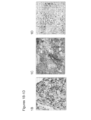
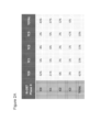
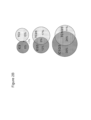

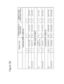
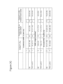

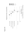
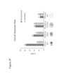


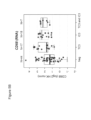
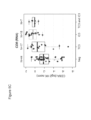


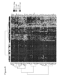
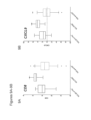
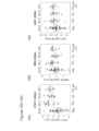


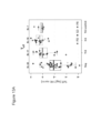

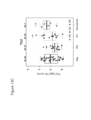

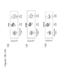
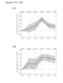


Classifications
-
- C—CHEMISTRY; METALLURGY
- C07—ORGANIC CHEMISTRY
- C07K—PEPTIDES
- C07K16/00—Immunoglobulins [IGs], e.g. monoclonal or polyclonal antibodies
- C07K16/18—Immunoglobulins [IGs], e.g. monoclonal or polyclonal antibodies against material from animals or humans
- C07K16/28—Immunoglobulins [IGs], e.g. monoclonal or polyclonal antibodies against material from animals or humans against receptors, cell surface antigens or cell surface determinants
- C07K16/2803—Immunoglobulins [IGs], e.g. monoclonal or polyclonal antibodies against material from animals or humans against receptors, cell surface antigens or cell surface determinants against the immunoglobulin superfamily
- C07K16/2827—Immunoglobulins [IGs], e.g. monoclonal or polyclonal antibodies against material from animals or humans against receptors, cell surface antigens or cell surface determinants against the immunoglobulin superfamily against B7 molecules, e.g. CD80, CD86
-
- A—HUMAN NECESSITIES
- A61—MEDICAL OR VETERINARY SCIENCE; HYGIENE
- A61P—SPECIFIC THERAPEUTIC ACTIVITY OF CHEMICAL COMPOUNDS OR MEDICINAL PREPARATIONS
- A61P11/00—Drugs for disorders of the respiratory system
-
- A—HUMAN NECESSITIES
- A61—MEDICAL OR VETERINARY SCIENCE; HYGIENE
- A61P—SPECIFIC THERAPEUTIC ACTIVITY OF CHEMICAL COMPOUNDS OR MEDICINAL PREPARATIONS
- A61P35/00—Antineoplastic agents
-
- A—HUMAN NECESSITIES
- A61—MEDICAL OR VETERINARY SCIENCE; HYGIENE
- A61P—SPECIFIC THERAPEUTIC ACTIVITY OF CHEMICAL COMPOUNDS OR MEDICINAL PREPARATIONS
- A61P43/00—Drugs for specific purposes, not provided for in groups A61P1/00-A61P41/00
-
- C—CHEMISTRY; METALLURGY
- C12—BIOCHEMISTRY; BEER; SPIRITS; WINE; VINEGAR; MICROBIOLOGY; ENZYMOLOGY; MUTATION OR GENETIC ENGINEERING
- C12Q—MEASURING OR TESTING PROCESSES INVOLVING ENZYMES, NUCLEIC ACIDS OR MICROORGANISMS; COMPOSITIONS OR TEST PAPERS THEREFOR; PROCESSES OF PREPARING SUCH COMPOSITIONS; CONDITION-RESPONSIVE CONTROL IN MICROBIOLOGICAL OR ENZYMOLOGICAL PROCESSES
- C12Q1/00—Measuring or testing processes involving enzymes, nucleic acids or microorganisms; Compositions therefor; Processes of preparing such compositions
- C12Q1/68—Measuring or testing processes involving enzymes, nucleic acids or microorganisms; Compositions therefor; Processes of preparing such compositions involving nucleic acids
- C12Q1/6876—Nucleic acid products used in the analysis of nucleic acids, e.g. primers or probes
- C12Q1/6883—Nucleic acid products used in the analysis of nucleic acids, e.g. primers or probes for diseases caused by alterations of genetic material
- C12Q1/6886—Nucleic acid products used in the analysis of nucleic acids, e.g. primers or probes for diseases caused by alterations of genetic material for cancer
-
- G—PHYSICS
- G01—MEASURING; TESTING
- G01N—INVESTIGATING OR ANALYSING MATERIALS BY DETERMINING THEIR CHEMICAL OR PHYSICAL PROPERTIES
- G01N33/00—Investigating or analysing materials by specific methods not covered by groups G01N1/00 - G01N31/00
- G01N33/48—Biological material, e.g. blood, urine; Haemocytometers
- G01N33/50—Chemical analysis of biological material, e.g. blood, urine; Testing involving biospecific ligand binding methods; Immunological testing
- G01N33/53—Immunoassay; Biospecific binding assay; Materials therefor
- G01N33/574—Immunoassay; Biospecific binding assay; Materials therefor for cancer
- G01N33/57407—Specifically defined cancers
- G01N33/57423—Specifically defined cancers of lung
-
- A—HUMAN NECESSITIES
- A61—MEDICAL OR VETERINARY SCIENCE; HYGIENE
- A61K—PREPARATIONS FOR MEDICAL, DENTAL OR TOILETRY PURPOSES
- A61K39/00—Medicinal preparations containing antigens or antibodies
- A61K2039/505—Medicinal preparations containing antigens or antibodies comprising antibodies
-
- C—CHEMISTRY; METALLURGY
- C07—ORGANIC CHEMISTRY
- C07K—PEPTIDES
- C07K2317/00—Immunoglobulins specific features
- C07K2317/70—Immunoglobulins specific features characterized by effect upon binding to a cell or to an antigen
- C07K2317/76—Antagonist effect on antigen, e.g. neutralization or inhibition of binding
-
- C—CHEMISTRY; METALLURGY
- C12—BIOCHEMISTRY; BEER; SPIRITS; WINE; VINEGAR; MICROBIOLOGY; ENZYMOLOGY; MUTATION OR GENETIC ENGINEERING
- C12Q—MEASURING OR TESTING PROCESSES INVOLVING ENZYMES, NUCLEIC ACIDS OR MICROORGANISMS; COMPOSITIONS OR TEST PAPERS THEREFOR; PROCESSES OF PREPARING SUCH COMPOSITIONS; CONDITION-RESPONSIVE CONTROL IN MICROBIOLOGICAL OR ENZYMOLOGICAL PROCESSES
- C12Q2600/00—Oligonucleotides characterized by their use
- C12Q2600/106—Pharmacogenomics, i.e. genetic variability in individual responses to drugs and drug metabolism
-
- C—CHEMISTRY; METALLURGY
- C12—BIOCHEMISTRY; BEER; SPIRITS; WINE; VINEGAR; MICROBIOLOGY; ENZYMOLOGY; MUTATION OR GENETIC ENGINEERING
- C12Q—MEASURING OR TESTING PROCESSES INVOLVING ENZYMES, NUCLEIC ACIDS OR MICROORGANISMS; COMPOSITIONS OR TEST PAPERS THEREFOR; PROCESSES OF PREPARING SUCH COMPOSITIONS; CONDITION-RESPONSIVE CONTROL IN MICROBIOLOGICAL OR ENZYMOLOGICAL PROCESSES
- C12Q2600/00—Oligonucleotides characterized by their use
- C12Q2600/158—Expression markers
-
- G—PHYSICS
- G01—MEASURING; TESTING
- G01N—INVESTIGATING OR ANALYSING MATERIALS BY DETERMINING THEIR CHEMICAL OR PHYSICAL PROPERTIES
- G01N2333/00—Assays involving biological materials from specific organisms or of a specific nature
- G01N2333/435—Assays involving biological materials from specific organisms or of a specific nature from animals; from humans
- G01N2333/705—Assays involving receptors, cell surface antigens or cell surface determinants
- G01N2333/70503—Immunoglobulin superfamily, e.g. VCAMs, PECAM, LFA-3
- G01N2333/70532—B7 molecules, e.g. CD80, CD86
-
- G—PHYSICS
- G01—MEASURING; TESTING
- G01N—INVESTIGATING OR ANALYSING MATERIALS BY DETERMINING THEIR CHEMICAL OR PHYSICAL PROPERTIES
- G01N2800/00—Detection or diagnosis of diseases
- G01N2800/52—Predicting or monitoring the response to treatment, e.g. for selection of therapy based on assay results in personalised medicine; Prognosis
Definitions
- Cancer remains one of the most deadly threats to human health. In the U.S., cancer affects nearly 1.3 million new patients each year, and is the second leading cause of death after heart disease, accounting for approximately 1 in 4 deaths.
- lung cancer is the most common form of cancer and the leading cancer killer among American women. It is also predicted that cancer may surpass cardiovascular diseases as the number one cause of death within five years. Solid tumors are responsible for most of those deaths.
- the overall 5-year survival rate for all cancers has improved only by about 10% in the past 20 years. Cancers, or malignant tumors, metastasize and grow rapidly in an uncontrolled manner, making timely detection and treatment extremely difficult.
- Programmed death-ligand 1 is a protein that has been implicated in the suppression of immune system responses during chronic infections, pregnancy, tissue allografts, autoimmune diseases, and cancer.
- PD-L1 regulates the immune response by binding to an inhibitory receptor, known as programmed death 1 (PD-1), which is expressed on the surface of T-cells, B cells, and monocytes.
- PD-L1 negatively regulates T-cell function also through interaction with another receptor, B7-1. Formation of the PD-L1/PD-1 and PD-L1/B7-1 complexes negatively regulates T-cell receptor signaling, resulting in the subsequent downregulation of T-cell activation and suppression of anti-tumor immune activity.
- WO 2014/151006 discloses that expression of PD-L1 is indicative of a higher likelihood of response of cancer patients to anti-PD-L1 axis treatment.
- the present invention provides diagnostic methods and therapeutic compositions for non-small cell lung cancer (NSCLC).
- NSCLC non-small cell lung cancer
- the invention features a PD-L1 binding antagonist for use in treating a patient suffering from a non-small cell lung cancer, wherein a tumor sample obtained from the patient has been determined to have a detectable expression level of PD-L1 in 50% or more of the tumor cells in the tumor sample, wherein the PD-L1 binding antagonist is an antibody, wherein the antibody is atezolizumab, and wherein the expression level of PD-L1 is a protein expression level.
- the tumor sample obtained from the patient has a detectable expression level of PD-L1 in tumour-infiltrating immune cells that comprise less than 10% of the sample.
- the invention features a method for determining whether a patient suffering from a non-small cell lung cancer is likely to respond to treatment comprising a PD-L1 binding antagonist, the method comprising: determining the expression level of PD-L1 in tumor cells in a tumor sample obtained from the patient, wherein a detectable expression level of PD-L1 in 50% or more of the tumor cells in the tumor sample indicates that the patient is likely to respond to treatment comprising a PD-L1 binding antagonist, wherein the PD-L1 binding antagonist is an antibody, wherein the antibody is aztezolizumab, and wherein the expression level of PD-L1 is a protein expression level.
- the invention features a method for predicting responsiveness of a patient suffering from a non-small cell lung cancer to treatment comprising a PD-L1 binding antagonist, the method comprising: determining the expression level of PD-L1 in tumor cells in a tumor sample obtained from the patient, wherein a detectable expression level of PD-L1 in 50% or more of the tumor cells in the tumor sample indicates that the patient is likely to respond to treatment comprising a PD-L1 binding antagonist, wherein the PD-L1 binding antagonist is an antibody, wherein the antibody is aztezolizumab, and wherein the expression level of PD-L1 is a protein expression level.
- the method further comprises determining the expression level of PD-L1 in tumor-infiltrating immune cells in the tumor sample obtained from the patient.
- the tumor sample obtained from the patient has a detectable expression level of PD-L1 in tumor-infiltrating immune cells that comprise less than 10% of the sample.
- the tumor sample obtained from the patient has been determined to have a detectable expression level of PD-L1 in tumor-infiltrating immune cells that comprise 10% or more of the tumor sample.
- the invention features a method for selecting a therapy for a patient suffering from a non-small cell lung cancer, the method comprising: determining the expression level of PD-L1 in tumor cells in a tumor sample obtained from the patient, and selecting a therapy comprising a PD-L1 binding antagonist for the patient based on a detectable expression level of PD-L1 in 50% or more of the tumor cells in the tumor sample, wherein the PD-L1 binding antagonist is an antibody, wherein the antibody is aztezolizumab, and wherein the expression level of PD-L1 is a protein expression level.
- the method further comprises determining the expression level of PD-L1 in tumor-infiltrating immune cells in the tumor sample obtained from the patient.
- the tumor sample obtained from the patient has a detectable expression level of PD-L1 in tumor-infiltrating immune cells that comprise less than 10% of the sample.
- the tumor sample obtained from the patient comprises a population of fibroblasts and/or myofibroblasts. In some embodiments, the tumor sample obtained from the patient comprises a cell-poor and/or collagenized stroma. In some embodiments, the tumor sample has an increased expression level of collagen, STAT1, or MEK relative to a reference tumor sample.
- the PD-L1 binding antagonist inhibits the binding of PD-L1 to one or more of its ligand binding partners.
- the PD-L1 binding antagonist inhibits the binding of PD-L1 to PD-1.
- the PD-L1 binding antagonist inhibits the binding of PD-L1 to B7-1.
- the PD-L1 binding antagonist inhibits the binding of PD-L1 to both PD-1 and B7-1.
- the antibody is MPDL3280A (atezolizumab).
- the antibody comprises a heavy chain comprising HVR-H1 sequence of SEQ ID NO:19, HVR-H2 sequence of SEQ ID NO:20, and HVR-H3 sequence of SEQ ID NO:21; and a light chain comprising HVR-L1 sequence of SEQ ID NO:22, HVR-L2 sequence of SEQ ID NO:23, and HVR-L3 sequence of SEQ ID NO:24.
- the antibody comprises a heavy chain variable region comprising the amino acid sequence of SEQ ID NO:26 and a light chain variable region comprising the amino acid sequence of SEQ ID NO:4.
- the method further comprises administering to the patient an effective amount of a second therapeutic agent.
- the second therapeutic agent is selected from the group consisting of a cytotoxic agent, a growth-inhibitory agent, a radiation therapy agent, an anti-angiogenic agent, and combinations thereof.
- the non-small cell lung cancer is a locally advanced or metastatic non-small cell lung cancer.
- the tumor sample is a formalin-fixed and paraffin-embedded (FFPE) tumor sample, an archival tumor sample, a fresh tumor sample, or a frozen tumor sample.
- the expression level of PD-L1 is a protein expression level.
- the protein expression level of PD-L1 is determined using a method selected from the group consisting of immunohistochemistry (IHC), immunofluorescence, flow cytometry, and Western blot.
- IHC immunohistochemistry
- the protein expression level of PD-L1 is determined using IHC.
- the protein expression level of PD-L1 is detected using an anti-PD-L1 antibody.
- the present invention provides diagnostic methods and therapeutic compositions for non-small cell lung cancer.
- the invention is based, at least in part, on the discovery that determination of expression levels of biomarkers of the invention, for example, PD-L1, in samples obtained from a patient (e.g., in tumor cells and in tumor-infiltrating immune cells in a tumor sample obtained from the patient) is useful in treatment of a patient suffering from cancer, for diagnosing a patient suffering from cancer, for determining whether a patient having a cancer is likely to respond to treatment with an anti-cancer therapy that includes a PD-L1 axis binding antagonist which is the anti-PD-L1 antibody MPDL3280A, for optimizing therapeutic efficacy of an anti-cancer therapy that includes a PD-L1 axis binding antagonist which is the anti-PD-L1 antibody MPDL3280A, and/or for patient selection for an anti-cancer therapy comprising a PD-L1 axis binding antagonist which is the anti-PD-L1 antibody MPDL3280A.
- PD-L1 axis binding antagonist refers to a molecule that inhibits the interaction of a PD-L1 axis binding partner with one or more of its binding partners, so as to remove T-cell dysfunction resulting from signaling on the PD-1 signaling axis, with a result being restored or enhanced T-cell function.
- a PD-L1 axis binding antagonist includes a PD-L1 binding antagonist and a PD-1 binding antagonist as well as molecules that interfere with the interaction between PD-L1 and PD-1 (e.g., a PD-L2-Fc fusion).
- disfunction in the context of immune dysfunction, refers to a state of reduced immune responsiveness to antigenic stimulation.
- the term includes the common elements of both “exhaustion” and/or “anergy” in which antigen recognition may occur, but the ensuing immune response is ineffective to control infection or tumor growth.
- disfunctional also includes refractory or unresponsive to antigen recognition, specifically, impaired capacity to translate antigen recognition into down-stream T-cell effector functions, such as proliferation, cytokine production (e.g., IL-2) and/or target cell killing.
- T-cell anergy refers to the state of unresponsiveness to antigen stimulation resulting from incomplete or insufficient signals delivered through the T-cell receptor (e.g., increase in intracellular Ca 2+ in the absence of Ras activation). T-cell anergy can also result upon stimulation with antigen in the absence of co-stimulation, resulting in the cell becoming refractory to subsequent activation by the antigen even in the context of co-stimulation.
- the unresponsive state can often be overriden by the presence of interleukin-2. Anergic T-cells do not undergo clonal expansion and/or acquire effector functions.
- exhaustion refers to T-cell exhaustion as a state of T-cell dysfunction that arises from sustained TCR signaling that occurs during many chronic infections and cancer. It is distinguished from anergy in that it arises not through incomplete or deficient signaling, but from sustained signaling. It is defined by poor effector function, sustained expression of inhibitory receptors and a transcriptional state distinct from that of functional effector or memory T-cells. Exhaustion prevents optimal control of infection and tumors. Exhaustion can result from both extrinsic negative regulatory pathways (e.g., immunoregulatory cytokines) as well as cell-intrinsic negative regulatory (costimulatory) pathways (PD-1, B7-H3, B7-H4, etc.).
- extrinsic negative regulatory pathways e.g., immunoregulatory cytokines
- costimulatory costimulatory
- Enhancing T-cell function means to induce, cause or stimulate a T-cell to have a sustained or amplified biological function, or renew or reactivate exhausted or inactive T-cells.
- enhancing T-cell function include: increased secretion of ⁇ -interferon from CD8+ T-cells, increased proliferation, increased antigen responsiveness (e.g., viral, pathogen, or tumor clearance) relative to such levels before the intervention.
- the level of enhancement is as least 50%, alternatively 60%, 70%, 80%, 90%, 100%, 120%, 150%, or 200% enhancement. The manner of measuring this enhancement is known to one of ordinary skill in the art.
- Tumor immunity refers to the process in which tumors evade immune recognition and clearance. Thus, as a therapeutic concept, tumor immunity is “treated” when such evasion is attenuated, and the tumors are recognized and attacked by the immune system. Examples of tumor recognition include tumor binding, tumor shrinkage and tumor clearance.
- Immunogenicity refers to the ability of a particular substance to provoke an immune response. Tumors are immunogenic and enhancing tumor immunogenicity aids in the clearance of the tumor cells by the immune response. Examples of enhancing tumor immunogenicity include treatment with a PD-L1 axis binding antagonist.
- a "PD-L1 binding antagonist” is a molecule that decreases, blocks, inhibits, abrogates or interferes with signal transduction resulting from the interaction of PD-L1 with either one or more of its binding partners, such as PD-1 and/or B7-1.
- a PD-L1 binding antagonist is a molecule that inhibits the binding of PD-L1 to its binding partners.
- the PD-L1 binding antagonist inhibits binding of PD-L1 to PD-1 and/or B7-1.
- PD-L1 binding antagonists include anti-PD-L1 antibodies and antigen-binding fragments thereof, immunoadhesins, fusion proteins, oligopeptides, small molecule antagonist, polynucleotide antagonists, and other molecules that decrease, block, inhibit, abrogate or interfere with signal transduction resulting from the interaction of PD-L1 with one or more of its binding partners, such as PD-1 and/or B7-1.
- a PD-L1 binding antagonist reduces the negative signal mediated by or through cell surface proteins expressed on T lymphocytes, and other cells, mediated signaling through PD-L1 or PD-1 so as render a dysfunctional T-cell less dysfunctional.
- the PD-L1 binding antagonist is an anti-PD-L1 antibody and the anti-PD-L1 antibody is MPDL3280A (atezolizumab).
- Programmed Death Ligand 1 and "PD-L1" refer herein to a native sequence PD-L1 polypeptide, polypeptide variants, and fragments of a native sequence polypeptide and polypeptide variants (which are further defined herein).
- the PD-L1 polypeptide described herein may be that which is isolated from a variety of sources, such as from human tissue types or from another source, or prepared by recombinant or synthetic methods.
- a "native sequence PD-L1 polypeptide” comprises a polypeptide having the same amino acid sequence as the corresponding PD-L1 polypeptide derived from nature.
- a "PD-L1 polypeptide variant,” or variations thereof, means a PD-L1 polypeptide, generally an active PD-L1 polypeptide, as defined herein having at least about 80% amino acid sequence identity with any of the native sequence PD-L1 polypeptide sequences as disclosed herein.
- Such PD-L1 polypeptide variants include, for instance, PD-L1 polypeptides wherein one or more amino acid residues are added, or deleted, at the N- or C-terminus of a native amino acid sequence.
- a PD-L1 polypeptide variant will have at least about 80% amino acid sequence identity, alternatively at least about 81%, 82%, 83%, 84%, 85%, 86%, 87%, 88%, 89%, 90%, 91%, 92%, 93%, 94%, 95%, 96%, 97%, 98%, or 99% amino acid sequence identity, to a native sequence PD-L1 polypeptide sequence as disclosed herein.
- PD-L1 variant polypeptides are at least about 10 amino acids in length, alternatively at least about 20, 30, 40, 50, 60, 70, 80, 90, 100, 110, 120, 130, 140, 150, 160, 170, 180, 190, 200, 210, 220, 230, 240, 250, 260, 270, 280, 281, 282, 283, 284, 285, 286, 287, 288, or 289 amino acids in length, or more.
- PD-L1 variant polypeptides will have no more than one conservative amino acid substitution as compared to a native PD-L1 polypeptide sequence, alternatively no more than 2, 3, 4, 5, 6, 7, 8, 9, or 10 conservative amino acid substitutions as compared to the native PD-L1 polypeptide sequence.
- Polynucleotide or “nucleic acid,” as used interchangeably herein, refer to polymers of nucleotides of any length, and include DNA and RNA.
- the nucleotides can be deoxyribonucleotides, ribonucleotides, modified nucleotides or bases, and/or their analogs, or any substrate that can be incorporated into a polymer by DNA or RNA polymerase, or by a synthetic reaction.
- polynucleotides as defined herein include, without limitation, single- and double-stranded DNA, DNA including single- and double-stranded regions, single- and double-stranded RNA, and RNA including single- and double-stranded regions, hybrid molecules comprising DNA and RNA that may be single-stranded or, more typically, double-stranded or include single- and double-stranded regions.
- polynucleotide refers to triple-stranded regions comprising RNA or DNA or both RNA and DNA. The strands in such regions may be from the same molecule or from different molecules.
- the regions may include all of one or more of the molecules, but more typically involve only a region of some of the molecules.
- One of the molecules of a triple-helical region often is an oligonucleotide.
- polynucleotide specifically includes cDNAs.
- a polynucleotide may comprise modified nucleotides, such as methylated nucleotides and their analogs. If present, modification to the nucleotide structure may be imparted before or after assembly of the polymer. The sequence of nucleotides may be interrupted by non-nucleotide components. A polynucleotide may be further modified after synthesis, such as by conjugation with a label.
- modifications include, for example, "caps,” substitution of one or more of the naturally-occurring nucleotides with an analog, internucleotide modifications such as, for example, those with uncharged linkages (e.g., methyl phosphonates, phosphotriesters, phosphoamidates, carbamates, and the like) and with charged linkages (e.g., phosphorothioates, phosphorodithioates, and the like), those containing pendant moieties, such as, for example, proteins (e.g., nucleases, toxins, antibodies, signal peptides, poly-L-lysine, and the like), those with intercalators (e.g., acridine, psoralen, and the like), those containing chelators (e.g., metals, radioactive metals, boron, oxidative metals, and the like), those containing alkylators, those with modified linkages (e.g., alpha anomeric nucleic acids
- any of the hydroxyl groups ordinarily present in the sugars may be replaced, for example, by phosphonate groups, phosphate groups, protected by standard protecting groups, or activated to prepare additional linkages to additional nucleotides, or may be conjugated to solid or semi-solid supports.
- the 5' and 3' terminal OH can be phosphorylated or substituted with amines or organic capping group moieties of from 1 to 20 carbon atoms.
- Other hydroxyls may also be derivatized to standard protecting groups.
- Polynucleotides can also contain analogous forms of ribose or deoxyribose sugars that are generally known in the art, including, for example, 2'-0-methyl-, 2'-O-allyl-, 2'-fluoro-, or 2'-azido-ribose, carbocyclic sugar analogs, ⁇ -anomeric sugars, epimeric sugars such as arabinose, xyloses or lyxoses, pyranose sugars, furanose sugars, sedoheptuloses, acyclic analogs, and abasic nucleoside analogs such as methyl riboside.
- One or more phosphodiester linkages may be replaced by alternative linking groups.
- linking groups include, but are not limited to, disclosures wherein phosphate is replaced by P(O)S ("thioate”), P(S)S ("dithioate”), "(O)NR 2 ("amidate"), P(O)R, P(O)OR', CO or CH 2 ("formacetal"), in which each R or R' is independently H or substituted or unsubstituted alkyl (1-20 C) optionally containing an ether (-O-) linkage, aryl, alkenyl, cycloalkyl, cycloalkenyl or araldyl. Not all linkages in a polynucleotide need be identical. The preceding description applies to all polynucleotides referred to herein, including RNA and DNA.
- Oligonucleotide generally refers to short, single stranded, polynucleotides that are, but not necessarily, less than about 250 nucleotides in length. Oligonucleotides may be synthetic. The terms “oligonucleotide” and “polynucleotide” are not mutually exclusive. The description above for polynucleotides is equally and fully applicable to oligonucleotides.
- primer refers to a single-stranded polynucleotide that is capable of hybridizing to a nucleic acid and allowing polymerization of a complementary nucleic acid, generally by providing a free 3'-OH group.
- small molecule refers to any molecule with a molecular weight of about 2000 daltons or less, preferably of about 500 daltons or less.
- host cell refers to cells into which exogenous nucleic acid has been introduced, including the progeny of such cells.
- Host cells include “transformants” and “transformed cells,” which include the primary transformed cell and progeny derived therefrom without regard to the number of passages. Progeny may not be completely identical in nucleic acid content to a parent cell, but may contain mutations. Mutant progeny that have the same function or biological activity as screened or selected for in the originally transformed cell are included herein.
- vector refers to a nucleic acid molecule capable of propagating another nucleic acid to which it is linked.
- the term includes the vector as a self-replicating nucleic acid structure as well as the vector incorporated into the genome of a host cell into which it has been introduced.
- Certain vectors are capable of directing the expression of nucleic acids to which they are operatively linked. Such vectors are referred to herein as "expression vectors.”
- nucleic acid refers to a nucleic acid molecule that has been separated from a component of its natural environment.
- An isolated nucleic acid includes a nucleic acid molecule contained in cells that ordinarily contain the nucleic acid molecule, but the nucleic acid molecule is present extrachromosomally or at a chromosomal location that is different from its natural chromosomal location.
- antibody herein is used in the broadest sense and encompasses various antibody structures, including but not limited to monoclonal antibodies, polyclonal antibodies, multispecific antibodies (e.g., bispecific antibodies), and antibody fragments so long as they exhibit the desired antigen-binding activity.
- an “isolated” antibody is one which has been identified and separated and/or recovered from a component of its natural environment. Contaminant components of its natural environment are materials which would interfere with research, diagnostic, and/or therapeutic uses for the antibody, and may include enzymes, hormones, and other proteinaceous or nonproteinaceous solutes.
- an antibody is purified (1) to greater than 95% by weight of antibody as determined by, for example, the Lowry method, and in some disclosures, to greater than 99% by weight; (2) to a degree sufficient to obtain at least 15 residues of N-terminal or internal amino acid sequence by use of, for example, a spinning cup sequenator, or (3) to homogeneity by SDS-PAGE under reducing or nonreducing conditions using, for example, Coomassie blue or silver stain.
- An isolated antibody includes the antibody in situ within recombinant cells since at least one component of the antibody's natural environment will not be present. Ordinarily, however, an isolated antibody will be prepared by at least one purification step.
- “Native antibodies” are usually heterotetrameric glycoproteins of about 150,000 daltons, composed of two identical light (L) chains and two identical heavy (H) chains. Each light chain is linked to a heavy chain by one covalent disulfide bond, while the number of disulfide linkages varies among the heavy chains of different immunoglobulin isotypes. Each heavy and light chain also has regularly spaced intrachain disulfide bridges. Each heavy chain has at one end a variable domain (VH) followed by a number of constant domains.
- VH variable domain
- Each light chain has a variable domain at one end (VL) and a constant domain at its other end; the constant domain of the light chain is aligned with the first constant domain of the heavy chain, and the light chain variable domain is aligned with the variable domain of the heavy chain. Particular amino acid residues are believed to form an interface between the light chain and heavy chain variable domains.
- the "light chains” of antibodies (immunoglobulins) from any mammalian species can be assigned to one of two clearly distinct types, called kappa (" ⁇ ") and lambda (“ ⁇ "), based on the amino acid sequences of their constant domains.
- constant domain refers to the portion of an immunoglobulin molecule having a more conserved amino acid sequence relative to the other portion of the immunoglobulin, the variable domain, which contains the antigen binding site.
- the constant domain contains the CH1, CH2 and CH3 domains (collectively, CH) of the heavy chain and the CHL (or CL) domain of the light chain.
- variable region or “variable domain” of an antibody refers to the amino-terminal domains of the heavy or light chain of the antibody.
- the variable domain of the heavy chain may be referred to as "VH.”
- the variable domain of the light chain may be referred to as "VL.” These domains are generally the most variable parts of an antibody and contain the antigen-binding sites.
- variable refers to the fact that certain portions of the variable domains differ extensively in sequence among antibodies and are used in the binding and specificity of each particular antibody for its particular antigen. However, the variability is not evenly distributed throughout the variable domains of antibodies. It is concentrated in three segments called hypervariable regions (HVRs) both in the light chain and the heavy chain variable domains. The more highly conserved portions of variable domains are called the framework regions (FR).
- HVRs hypervariable regions
- FR framework regions
- the variable domains of native heavy and light chains each comprise four FR regions, largely adopting a beta-sheet configuration, connected by three HVRs, which form loops connecting, and in some cases forming part of, the beta-sheet structure.
- the HVRs in each chain are held together in close proximity by the FR regions and, with the HVRs from the other chain, contribute to the formation of the antigen-binding site of antibodies (see Kabat et al., Sequences of Proteins of Immunological Interest, Fifth Edition, National Institute of Health, Bethesda, Md. (1991 )).
- the constant domains are not involved directly in the binding of an antibody to an antigen, but exhibit various effector functions, such as participation of the antibody in antibody-dependent cellular toxicity.
- hypervariable region refers to the regions of an antibody variable domain which are hypervariable in sequence and/or form structurally defined loops.
- antibodies comprise six HVRs; three in the VH (H1, H2, H3), and three in the VL (L1, L2, L3).
- H3 and L3 display the most diversity of the six HVRs, and H3 in particular is believed to play a unique role in conferring fine specificity to antibodies.
- HVR delineations are in use and are encompassed herein.
- the Kabat Complementarity Determining Regions are based on sequence variability and are the most commonly used ( Kabat et al., Sequences of Proteins of Immunological Interest, 5th Ed. Public Health Service, National Institutes of Health, Bethesda, Md. (1991 )). Chothia refers instead to the location of the structural loops ( Chothia and Lesk J. Mol. Biol. 196:901-917 (1987 )).
- the AbM HVRs represent a compromise between the Kabat HVRs and Chothia structural loops, and are used by Oxford Molecular's AbM antibody modeling software.
- the "contact” HVRs are based on an analysis of the available complex crystal structures. The residues from each of these HVRs are noted below. Loop Kabat AbM Chothia Contact L1 L24-L34 L24-L34 L26-L32 L30-L36 L2 L50-L56 L50-L56 L50-L52 L46-L55 L3 L89-L97 L89-L97 L91-L96 L89-L96 H1 H31-H35b H26-H35b H26-H32 H30-H35b (Kabat Numbering) H1 H31-H35 H26-H35 H26-H32 H30-H35 (Chothia Numbering) H2 H50-H65 H50-H58 H53-H55 H47-H58 H3 H95-H102 H95-H102 H96-H101 H93-H101
- HVRs may comprise "extended HVRs" as follows: 24-36 or 24-34 (L1), 46-56 or 50-56 (L2) and 89-97 or 89-96 (L3) in the VL and 26-35 (H1), 50-65 or 49-65 (H2) and 93-102, 94-102, or 95-102 (H3) in the VH.
- the variable domain residues are numbered according to Kabat et al., supra, for each of these definitions.
- Framework or "FR” residues are those variable domain residues other than the HVR residues as herein defined.
- variable domain residue numbering as in Kabat or "amino acid position numbering as in Kabat,” and variations thereof, refers to the numbering system used for heavy chain variable domains or light chain variable domains of the compilation of antibodies in Kabat et al., supra. Using this numbering system, the actual linear amino acid sequence may contain fewer or additional amino acids corresponding to a shortening of, or insertion into, a FR or HVR of the variable domain.
- a heavy chain variable domain may include a single amino acid insert (residue 52a according to Kabat) after residue 52 of H2 and inserted residues (e.g., residues 82a, 82b, and 82c, etc. according to Kabat) after heavy chain FR residue 82.
- the Kabat numbering of residues may be determined for a given antibody by alignment at regions of homology of the sequence of the antibody with a "standard" Kabat numbered sequence.
- the Kabat numbering system is generally used when referring to a residue in the variable domain (approximately residues 1-107 of the light chain and residues 1-113 of the heavy chain) (e.g., Kabat et al., Sequences of Immunological Interest. 5th Ed. Public Health Service, National Institutes of Health, Bethesda, Md. (1991 )).
- the "EU numbering system” or "EU index” is generally used when referring to a residue in an immunoglobulin heavy chain constant region (e.g., the EU index reported in Kabat et al., supra ).
- the "EU index as in Kabat” refers to the residue numbering of the human IgG1 EU antibody.
- full-length antibody “intact antibody,” and “whole antibody” are used herein interchangeably to refer to an antibody in its substantially intact form, not antibody fragments as defined below.
- Antibody fragments comprise a portion of an intact antibody, preferably comprising the antigen-binding region thereof.
- the antibody fragment described herein is an antigen-binding fragment.
- Examples of antibody fragments include Fab, Fab', F(ab') 2 , and Fv fragments; diabodies; linear antibodies; single-chain antibody molecules; and multispecific antibodies formed from antibody fragments.
- Papain digestion of antibodies produces two identical antigen-binding fragments, called “Fab” fragments, each with a single antigen-binding site, and a residual "Fc” fragment, whose name reflects its ability to crystallize readily. Pepsin treatment yields an F(ab') 2 fragment that has two antigen-combining sites and is still capable of cross-linking antigen.
- Fv is the minimum antibody fragment which contains a complete antigen-binding site.
- a two-chain Fv species consists of a dimer of one heavy- and one light-chain variable domain in tight, non-covalent association.
- scFv single-chain Fv
- one heavy- and one light-chain variable domain can be covalently linked by a flexible peptide linker such that the light and heavy chains can associate in a "dimeric" structure analogous to that in a two-chain Fv species. It is in this configuration that the three HVRs of each variable domain interact to define an antigen-binding site on the surface of the VH-VL dimer.
- the six HVRs confer antigen-binding specificity to the antibody.
- the Fab fragment contains the heavy- and light-chain variable domains and also contains the constant domain of the light chain and the first constant domain (CH1) of the heavy chain.
- Fab' fragments differ from Fab fragments by the addition of a few residues at the carboxy terminus of the heavy chain CH1 domain including one or more cysteines from the antibody hinge region.
- Fab'-SH is the designation herein for Fab' in which the cysteine residue(s) of the constant domains bear a free thiol group.
- F(ab') 2 antibody fragments originally were produced as pairs of Fab' fragments which have hinge cysteines between them. Other chemical couplings of antibody fragments are also known.
- Single-chain Fv or “scFv” antibody fragments comprise the VH and VL domains of antibody, wherein these domains are present in a single polypeptide chain.
- the scFv polypeptide further comprises a polypeptide linker between the VH and VL domains which enables the scFv to form the desired structure for antigen binding.
- diabodies refers to antibody fragments with two antigen-binding sites, which fragments comprise a heavy-chain variable domain (VH) connected to a light-chain variable domain (VL) in the same polypeptide chain (VH-VL).
- VH heavy-chain variable domain
- VL light-chain variable domain
- Diabodies may be bivalent or bispecific. Diabodies are described more fully in, for example, EP 404,097 ; WO 1993/01161 ; Hudson et al., Nat. Med. 9:129-134 (2003 ); and Hollinger et al., Proc. Natl. Acad. Sci. USA 90: 6444-6448 (1993 ). Triabodies and tetrabodies are also described in Hudson et al., Nat. Med. 9:129-134 (2003 ).
- the "class" of an antibody refers to the type of constant domain or constant region possessed by its heavy chain.
- the heavy chain constant domains that correspond to the different classes of antibodies are called ⁇ , ⁇ , ⁇ , ⁇ , and ⁇ , respectively.
- a monoclonal antibody refers to an antibody obtained from a population of substantially homogeneous antibodies, e.g., the individual antibodies comprising the population are identical except for possible mutations, e.g., naturally occurring mutations, that may be present in minor amounts. Thus, the modifier “monoclonal” indicates the character of the antibody as not being a mixture of discrete antibodies.
- such a monoclonal antibody typically includes an antibody comprising a polypeptide sequence that binds a target, wherein the target-binding polypeptide sequence was obtained by a process that includes the selection of a single target binding polypeptide sequence from a plurality of polypeptide sequences.
- the selection process can be the selection of a unique clone from a plurality of clones, such as a pool of hybridoma clones, phage clones, or recombinant DNA clones.
- a selected target binding sequence can be further altered, for example, to improve affinity for the target, to humanize the target binding sequence, to improve its production in cell culture, to reduce its immunogenicity in vivo, to create a multispecific antibody, etc., and that an antibody comprising the altered target binding sequence is also a monoclonal antibody of this invention.
- each monoclonal antibody of a monoclonal antibody preparation is directed against a single determinant on an antigen.
- monoclonal antibody preparations are advantageous in that they are typically uncontaminated by other immunoglobulins.
- the modifier "monoclonal” indicates the character of the antibody as being obtained from a substantially homogeneous population of antibodies, and is not to be construed as requiring production of the antibody by any particular method.
- the monoclonal antibodies to be used in accordance with the invention may be made by a variety of techniques, including, for example, the hybridoma method (e.g., Kohler and Milstein, Nature 256:495-97 (1975 ); Hongo et al., Hybridoma 14 (3): 253-260 (1995 ), Harlow et al., Antibodies: A Laboratory Manual (Cold Spring Harbor Laboratory Press, 2nd ed.
- the monoclonal antibodies herein specifically include "chimeric" antibodies in which a portion of the heavy and/or light chain is identical with or homologous to corresponding sequences in antibodies derived from a particular species or belonging to a particular antibody class or subclass, while the remainder of the chain(s) is identical with or homologous to corresponding sequences in antibodies derived from another species or belonging to another antibody class or subclass, as well as fragments of such antibodies, so long as they exhibit the desired biological activity (see, e.g., U.S. Pat. No. 4,816,567 ; and Morrison et al., Proc. Natl. Acad. Sci. USA 81:6851-6855 (1984 )).
- Chimeric antibodies include PRIMATIZED ® antibodies wherein the antigen-binding region of the antibody is derived from an antibody produced by, e.g., immunizing macaque monkeys with the antigen of interest.
- a "human antibody” is one which possesses an amino acid sequence which corresponds to that of an antibody produced by a human or a human cell or derived from a non-human source that utilizes human antibody repertoires or other human antibody-encoding sequences. This definition of a human antibody specifically excludes a humanized antibody comprising non-human antigen-binding residues.
- a “humanized” antibody refers to a chimeric antibody comprising amino acid residues from non-human HVRs and amino acid residues from human framework regions (FRs).
- a humanized antibody will comprise substantially all of at least one, and typically two, variable domains, in which all or substantially all of the HVRs (e.g., CDRs) correspond to those of a non-human antibody, and all or substantially all of the FRs correspond to those of a human antibody.
- a humanized antibody optionally may comprise at least a portion of an antibody constant region derived from a human antibody.
- a "humanized form" of an antibody, e.g., a non-human antibody refers to an antibody that has undergone humanization.
- anti-PD-L1 antibody and "an antibody that binds to PD-L1” refer to an antibody that is capable of binding PD-L1 with sufficient affinity such that the antibody is useful as a diagnostic and/or therapeutic agent in targeting PD-L1.
- the extent of binding of an anti-PD-L1 antibody to an unrelated, non-PD-L1 protein is less than about 10% of the binding of the antibody to PD-L1 as measured, for example, by a radioimmunoassay (RIA).
- RIA radioimmunoassay
- an anti-PD-L1 antibody binds to an epitope of PD-L1 that is conserved among PD-L1 from different species.
- anti-PD-1 antibody and "an antibody that binds to PD-1” refer to an antibody that is capable of binding PD-1 with sufficient affinity such that the antibody is useful as a diagnostic and/or therapeutic agent in targeting PD-1.
- the extent of binding of an anti-PD-1 antibody to an unrelated, non-PD-1 protein is less than about 10% of the binding of the antibody to PD-1 as measured, for example, by a radioimmunoassay (RIA).
- RIA radioimmunoassay
- an anti-PD-1 antibody binds to an epitope of PD-1 that is conserved among PD-1 from different species.
- blocking antibody or an “antagonist” antibody is one which inhibits or reduces biological activity of the antigen it binds.
- Preferred blocking antibodies or antagonist antibodies substantially or completely inhibit the biological activity of the antigen.
- Bind refers to the strength of the sum total of noncovalent interactions between a single binding site of a molecule (e.g., an antibody) and its binding partner (e.g., an antigen).
- binding affinity refers to intrinsic binding affinity which reflects a 1:1 interaction between members of a binding pair (e.g., antibody and antigen).
- the affinity of a molecule X for its partner Y can generally be represented by the dissociation constant (Kd). Affinity can be measured by common methods known in the art, including those described herein. Specific illustrative and exemplary disclosures for measuring binding affinity are described in the following.
- the term “binds”, “specifically binds to” or is “specific for” refers to measurable and reproducible interactions such as binding between a target and an antibody, which is determinative of the presence of the target in the presence of a heterogeneous population of molecules including biological molecules.
- an antibody that binds to or specifically binds to a target is an antibody that binds this target with greater affinity, avidity, more readily, and/or with greater duration than it binds to other targets.
- the extent of binding of an antibody to an unrelated target is less than about 10% of the binding of the antibody to the target as measured, e.g., by a radioimmunoassay (RIA).
- an antibody that specifically binds to a target has a dissociation constant (Kd) of ⁇ 1 ⁇ M, ⁇ 100 nM, ⁇ 10 nM, ⁇ 1 nM, or ⁇ 0.1 nM.
- Kd dissociation constant
- an antibody specifically binds to an epitope on a protein that is conserved among the protein from different species.
- specific binding can include, but does not require exclusive binding.
- an “affinity matured” antibody refers to an antibody with one or more alterations in one or more hypervariable regions (HVRs), compared to a parent antibody which does not possess such alterations, such alterations resulting in an improvement in the affinity of the antibody for antigen.
- HVRs hypervariable regions
- an "antibody that binds to the same epitope” as a reference antibody refers to an antibody that blocks binding of the reference antibody to its antigen in a competition assay by 50% or more, and conversely, the reference antibody blocks binding of the antibody to its antigen in a competition assay by 50% or more.
- An exemplary competition assay is provided herein.
- an “immunoconjugate” is an antibody conjugated to one or more heterologous molecule(s), including but not limited to a cytotoxic agent.
- immunoadhesin designates antibody-like molecules which combine the binding specificity of a heterologous protein (an “adhesin”) with the effector functions of immunoglobulin constant domains.
- the immunoadhesins comprise a fusion of an amino acid sequence with the desired binding specificity which is other than the antigen recognition and binding site of an antibody (i.e., is “heterologous"), and an immunoglobulin constant domain sequence.
- the adhesin part of an immunoadhesin molecule typically is a contiguous amino acid sequence comprising at least the binding site of a receptor or a ligand.
- the immunoglobulin constant domain sequence in the immunoadhesin may be obtained from any immunoglobulin, such as IgG1, IgG2 (including IgG2A and IgG2B), IgG3, or IgG4 subtypes, IgA (including IgA1 and IgA2), IgE, IgD or IgM.
- the Ig fusions preferably include the substitution of a domain of a polypeptide or antibody described herein in the place of at least one variable region within an Ig molecule.
- the immunoglobulin fusion includes the hinge, CH2 and CH3, or the hinge, CH1, CH2 and CH3 regions of an IgG1 molecule.
- useful immunoadhesins as medicaments useful for therapy herein include polypeptides that comprise the extracellular domain (ECD) or PD-1-binding portions of PD-L1 or PD-L2, or the extracellular or PD-L1- or PD-L2-binding portions of PD- 1, fused to a constant domain of an immunoglobulin sequence, such as a PD-L1 ECD-Fc, a PD-L2 ECD-Fc, and a PD-1 ECD-Fc, respectively.
- Immunoadhesin combinations of Ig Fc and ECD of cell surface receptors are sometimes termed soluble receptors.
- a “fusion protein” and a “fusion polypeptide” refer to a polypeptide having two portions covalently linked together, where each of the portions is a polypeptide having a different property.
- the property may be a biological property, such as activity in vitro or in vivo.
- the property may also be simple chemical or physical property, such as binding to a target molecule, catalysis of a reaction, and the like.
- the two portions may be linked directly by a single peptide bond or through a peptide linker but are in reading frame with each other.
- Percent (%) amino acid sequence identity with respect to the polypeptide sequences identified herein is defined as the percentage of amino acid residues in a candidate sequence that are identical with the amino acid residues in the polypeptide being compared, after aligning the sequences and introducing gaps, if necessary, to achieve the maximum percent sequence identity, and not considering any conservative substitutions as part of the sequence identity. Alignment for purposes of determining percent amino acid sequence identity can be achieved in various ways that are within the skill in the art, for instance, using publicly available computer software such as BLAST, BLAST-2, ALIGN or Megalign (DNASTAR) software. Those skilled in the art can determine appropriate parameters for measuring alignment, including any algorithms needed to achieve maximal alignment over the full-length of the sequences being compared.
- % amino acid sequence identity values are generated using the sequence comparison computer program ALIGN-2.
- the ALIGN-2 sequence comparison computer program was authored by Genentech, Inc. and the source code has been filed with user documentation in the U.S. Copyright Office, Washington D.C., 20559, where it is registered under U.S. Copyright Registration No. TXU510087.
- the ALIGN-2 program is publicly available through Genentech, Inc., South San Francisco, California.
- the ALIGN-2 program should be compiled for use on a UNIX operating system, preferably digital UNIX V4.0D. All sequence comparison parameters are set by the ALIGN-2 program and do not vary.
- the % amino acid sequence identity of a given amino acid sequence A to, with, or against a given amino acid sequence B is calculated as follows: 100 times the fraction X / Y where X is the number of amino acid residues scored as identical matches by the sequence alignment program ALIGN-2 in that program's alignment of A and B, and where Y is the total number of amino acid residues in B.
- detection includes any means of detecting, including direct and indirect detection.
- biomarker refers to an indicator, e.g., predictive, diagnostic, and/or prognostic, which can be detected in a sample.
- the biomarker may serve as an indicator of a particular subtype of a disease or disorder (e.g., cancer) characterized by certain, molecular, pathological, histological, and/or clinical features.
- a biomarker is a gene.
- Biomarkers include, but are not limited to, polynucleotides (e.g., DNA and/or RNA), polynucleotide copy number alterations (e.g., DNA copy numbers), polypeptides, polypeptide and polynucleotide modifications (e.g., post-translational modifications), carbohydrates, and/or glycolipid-based molecular markers.
- polynucleotides e.g., DNA and/or RNA
- polynucleotide copy number alterations e.g., DNA copy numbers
- polypeptides e.g., polypeptide and polynucleotide modifications
- carbohydrates e.g., post-translational modifications
- biomarker signature refers to one or a combination of biomarkers whose expression is an indicator, e.g., predictive, diagnostic, and/or prognostic.
- the biomarker signature may serve as an indicator of a particular subtype of a disease or disorder (e.g., cancer) characterized by certain molecular, pathological, histological, and/or clinical features.
- the biomarker signature is a "gene signature.”
- the term “gene signature” is used interchangeably with “gene expression signature” and refers to one or a combination of polynucleotides whose expression is an indicator, e.g., predictive, diagnostic, and/or prognostic.
- the biomarker signature is a "protein signature.”
- the term “protein signature” is used interchangeably with “protein expression signature” and refers to one or a combination of polypeptides whose expression is an indicator, e.g., predictive, diagnostic, and/or prognostic.
- the “amount” or “level” of a biomarker associated with an increased clinical benefit to an individual is a detectable level in a biological sample. These can be measured by methods known to one skilled in the art and also disclosed herein. The expression level or amount of biomarker assessed can be used to determine the response to the treatment.
- level of expression or “expression level” in general are used interchangeably and generally refer to the amount of a biomarker in a biological sample. “Expression” generally refers to the process by which information (e.g., gene-encoded and/or epigenetic information) is converted into the structures present and operating in the cell. Therefore, as used herein, “expression” may refer to transcription into a polynucleotide, translation into a polypeptide, or even polynucleotide and/or polypeptide modifications (e.g., posttranslational modification of a polypeptide).
- Fragments of the transcribed polynucleotide, the translated polypeptide, or polynucleotide and/or polypeptide modifications shall also be regarded as expressed whether they originate from a transcript generated by alternative splicing or a degraded transcript, or from a post-translational processing of the polypeptide, e.g., by proteolysis.
- "Expressed genes” include those that are transcribed into a polynucleotide as mRNA and then translated into a polypeptide, and also those that are transcribed into RNA but not translated into a polypeptide (for example, transfer and ribosomal RNAs).
- Elevated expression refers to an increased expression or increased levels of a biomarker in an individual relative to a control, such as an individual or individuals who are not suffering from the disease or disorder (e.g., cancer) or an internal control (e.g., a housekeeping biomarker).
- a control such as an individual or individuals who are not suffering from the disease or disorder (e.g., cancer) or an internal control (e.g., a housekeeping biomarker).
- Reduced expression refers to a decrease expression or decreased levels of a biomarker in an individual relative to a control, such as an individual or individuals who are not suffering from the disease or disorder (e.g., cancer) or an internal control (e.g., a housekeeping biomarker). In some disclosures, reduced expression is little or no expression.
- housekeeping biomarker refers to a biomarker or group of biomarkers (e.g., polynucleotides and/or polypeptides) which are typically similarly present in all cell types.
- the housekeeping biomarker is a "housekeeping gene.”
- a "housekeeping gene” refers herein to a gene or group of genes which encode proteins whose activities are essential for the maintenance of cell function and which are typically similarly present in all cell types.
- Amplification generally refers to the process of producing multiple copies of a desired sequence.
- Multiple copies mean at least two copies.
- a “copy” does not necessarily mean perfect sequence complementarity or identity to the template sequence.
- copies can include nucleotide analogs such as deoxyinosine, intentional sequence alterations (such as sequence alterations introduced through a primer comprising a sequence that is hybridizable, but not complementary, to the template), and/or sequence errors that occur during amplification.
- multiplex-PCR refers to a single PCR reaction carried out on nucleic acid obtained from a single source (e.g., an individual) using more than one primer set for the purpose of amplifying two or more DNA sequences in a single reaction.
- PCR polymerase chain reaction
- sequence information from the ends of the region of interest or beyond needs to be available, such that oligonucleotide primers can be designed; these primers will be identical or similar in sequence to opposite strands of the template to be amplified.
- the 5' terminal nucleotides of the two primers may coincide with the ends of the amplified material.
- PCR can be used to amplify specific RNA sequences, specific DNA sequences from total genomic DNA, and cDNA transcribed from total cellular RNA, bacteriophage, or plasmid sequences, etc. See generally Mullis et al., Cold Spring Harbor Symp. Quant. Biol. 51:263 (1987 ) and Erlich, ed., PCR Technology, (Stockton Press, NY, 1989 ).
- PCR is considered to be one, but not the only, example of a nucleic acid polymerase reaction method for amplifying a nucleic acid test sample, comprising the use of a known nucleic acid (DNA or RNA) as a primer and utilizes a nucleic acid polymerase to amplify or generate a specific piece of nucleic acid or to amplify or generate a specific piece of nucleic acid which is complementary to a particular nucleic acid.
- DNA or RNA DNA or RNA
- Quantitative real-time polymerase chain reaction refers to a form of PCR wherein the amount of PCR product is measured at each step in a PCR reaction. This technique has been described in various publications including, for example, Cronin et al., Am. J. Pathol. 164(1):35-42 (2004 ) and Ma et al., Cancer Cell 5:607-616 (2004 ).
- microarray refers to an ordered arrangement of hybridizable array elements, preferably polynucleotide probes, on a substrate.
- diagnosis is used herein to refer to the identification or classification of a molecular or pathological state, disease or condition (e.g., cancer).
- diagnosis may refer to identification of a particular type of cancer.
- Diagnosis may also refer to the classification of a particular subtype of cancer, for instance, by histopathological criteria, or by molecular features (e.g., a subtype characterized by expression of one or a combination of biomarkers (e.g., particular genes or proteins encoded by said genes)).
- a method of aiding diagnosis of a disease or condition can comprise measuring certain biomarkers (e.g., PD-L1) in a biological sample from an individual.
- biomarkers e.g., PD-L1
- sample refers to a composition that is obtained or derived from a subject and/or individual of interest that contains a cellular and/or other molecular entity that is to be characterized and/or identified, for example, based on physical, biochemical, chemical, and/or physiological characteristics.
- disease sample and variations thereof refers to any sample obtained from a subject of interest that would be expected or is known to contain the cellular and/or molecular entity that is to be characterized.
- Samples include, but are not limited to, tissue samples, primary or cultured cells or cell lines, cell supernatants, cell lysates, platelets, serum, plasma, vitreous fluid, lymph fluid, synovial fluid, follicular fluid, seminal fluid, amniotic fluid, milk, whole blood, blood-derived cells, urine, cerebro-spinal fluid, saliva, sputum, tears, perspiration, mucus, tumor lysates, and tissue culture medium, tissue extracts such as homogenized tissue, tumor tissue, cellular extracts, and combinations thereof.
- tissue sample or “cell sample” is meant a collection of similar cells obtained from a tissue of a subject or individual.
- the source of the tissue or cell sample may be solid tissue as from a fresh, frozen and/or preserved organ, tissue sample, biopsy, and/or aspirate; blood or any blood constituents such as plasma; bodily fluids such as cerebral spinal fluid, amniotic fluid, peritoneal fluid, or interstitial fluid; cells from any time in gestation or development of the subject.
- the tissue sample may also be primary or cultured cells or cell lines.
- the tissue or cell sample is obtained from a disease tissue/organ.
- a "tumor sample” is a tissue sample obtained from a tumor or other cancerous tissue.
- the tissue sample may contain compounds which are not naturally intermixed with the tissue in nature such as preservatives, anticoagulants, buffers, fixatives, nutrients, antibiotics, or the like.
- Tumor-infiltrating immune cell refers to any immune cell present in a tumor or a sample thereof.
- Tumor-infiltrating immune cells include, but are not limited to, intratumoral immune cells, peritumoral immune cells, other tumor stroma cells (e.g., fibroblasts), or any combination thereof.
- Such tumor-infiltrating immune cells can be, for example, T lymphocytes (such as CD8+ T lymphocytes and/or CD4+ T lymphocytes), B lymphocytes, or other bone marrow-lineage cells, including granulocytes (e.g., neutrophils, eosinophils, and basophils), monocytes, macrophages, dendritic cells (e.g., interdigitating dendritic cells), histiocytes, and natural killer cells.
- T lymphocytes such as CD8+ T lymphocytes and/or CD4+ T lymphocytes
- B lymphocytes or other bone marrow-lineage cells, including granulocytes (e.g., neutrophils, eosinophils, and basophils), monocytes, macrophages, dendritic cells (e.g., interdigitating dendritic cells), histiocytes, and natural killer cells.
- granulocytes e.g., neutrophils,
- tumor cell refers to any tumor cell present in a tumor or a sample thereof. Tumor cells may be distinguished from other cells that may be present in a tumor sample, for example, stromal cells and tumor-infiltrating immune cells, using methods known in the art and/or described herein.
- a “reference sample,” “reference cell,” “reference tissue,” “control sample,” “control cell,” or “control tissue,” as used herein, refers to a sample, cell, tissue, standard, or level that is used for comparison purposes.
- a reference sample, reference cell, reference tissue, control sample, control cell, or control tissue is obtained from a healthy and/or non-diseased part of the body (e.g., tissue or cells) of the same subject or individual.
- the reference sample, reference cell, reference tissue, control sample, control cell, or control tissue may be healthy and/or non-diseased cells or tissue adjacent to the diseased cells or tissue (e.g., cells or tissue adjacent to a tumor).
- a reference sample is obtained from an untreated tissue and/or cell of the body of the same subject or individual.
- a reference sample, reference cell, reference tissue, control sample, control cell, or control tissue is obtained from a healthy and/or non-diseased part of the body (e.g., tissues or cells) of an individual who is not the subject or individual.
- a reference sample, reference cell, reference tissue, control sample, control cell, or control tissue is obtained from an untreated tissue and/or cell of the body of an individual who is not the subject or individual.
- a "section" of a tissue sample is meant a single part or piece of a tissue sample, for example, a thin slice of tissue or cells cut from a tissue sample (e.g., a tumor sample). It is to be understood that multiple sections of tissue samples may be taken and subjected to analysis, provided that it is understood that the same section of tissue sample may be analyzed at both morphological and molecular levels, or analyzed with respect to polypeptides (e.g., by immunohistochemistry) and/or polynucleotides (e.g., by in situ hybridization).
- polypeptides e.g., by immunohistochemistry
- polynucleotides e.g., by in situ hybridization
- correlate or “correlating” is meant comparing, in any way, the performance and/or results of a first analysis or protocol with the performance and/or results of a second analysis or protocol. For example, one may use the results of a first analysis or protocol in carrying out a second protocols and/or one may use the results of a first analysis or protocol to determine whether a second analysis or protocol should be performed. With respect to the disclosure of polypeptide analysis or protocol, one may use the results of the polypeptide expression analysis or protocol to determine whether a specific therapeutic regimen should be performed. With respect to the disclosure of polynucleotide analysis or protocol, one may use the results of the polynucleotide expression analysis or protocol to determine whether a specific therapeutic regimen should be performed.
- “Individual response” or “response” can be assessed using any endpoint indicating a benefit to the individual, including, without limitation, (1) inhibition, to some extent, of disease progression (e.g., cancer progression), including slowing down and complete arrest; (2) a reduction in tumor size; (3) inhibition (i.e., reduction, slowing down or complete stopping) of cancer cell infiltration into adjacent peripheral organs and/or tissues; (4) inhibition (i.e. reduction, slowing down or complete stopping) of metatasis; (5) relief, to some extent, of one or more symptoms associated with the disease or disorder (e.g., cancer); (6) increase or extend in the length of survival, including overall survival and progression free survival; and/or (9) decreased mortality at a given point of time following treatment.
- disease progression e.g., cancer progression
- a reduction in tumor size i.e., reduction, slowing down or complete stopping
- inhibition i.e. reduction, slowing down or complete stopping
- metatasis metatasis
- an "effective response" of a patient or a patient's “responsiveness” to treatment with a medicament and similar wording refers to the clinical or therapeutic benefit imparted to a patient at risk for, or suffering from, a disease or disorder, such as cancer.
- a disease or disorder such as cancer.
- such benefit includes any one or more of: extending survival (including overall survival and progression-free survival); resulting in an objective response (including a complete response or a partial response); or improving signs or symptoms of cancer.
- the biomarker (e.g., PD-L1 expression, for example, as determined using IHC) is used to identify the patient who is predicted to have an increased likelihood of being responsive to treatment with a medicament (e.g., treatment comprising a PD-L1 axis binding antagonist, e.g., an anti-PD-L1 antibody), relative to a patient who does not express the biomarker.
- a medicament e.g., treatment comprising a PD-L1 axis binding antagonist, e.g., an anti-PD-L1 antibody
- the biomarker e.g., PD-L1 expression, for example, as determined using IHC
- a medicament e.g., anti-PD-L1 antibody
- the presence of the biomarker is used to identify a patient who is more likely to respond to treatment with a medicament, relative to a patient that does not have the presence of the biomarker. In another disclosure, the presence of the biomarker is used to determine that a patient will have an increased likelihood of benefit from treatment with a medicament, relative to a patient that does not have the presence of the biomarker.
- An “objective response” refers to a measurable response, including complete response (CR) or partial response (PR).
- objective response rate refers to the sum of complete response (CR) rate and partial response (PR) rate.
- sustained response refers to the sustained effect on reducing tumor growth after cessation of a treatment.
- the tumor size may remain to be the same or smaller as compared to the size at the beginning of the administration phase.
- the sustained response has a duration at least the same as the treatment duration, at least 1.5X, 2.0X, 2.5X, or 3.0X length of the treatment duration, or longer.
- reducing or inhibiting cancer relapse means to reduce or inhibit tumor or cancer relapse or tumor or cancer progression.
- cancer relapse and/or cancer progression include, without limitation, cancer metastasis.
- partial response refers to a decrease in the size of one or more tumors or lesions, or in the extent of cancer in the body, in response to treatment.
- PR refers to at least a 30% decrease in the sum of the longest diameters (SLD) of target lesions, taking as reference the baseline SLD.
- stable disease or “SD” refers to neither sufficient shrinkage of target lesions to qualify for PR, nor sufficient increase to qualify for PD, taking as reference the smallest SLD since the treatment started.
- progressive disease or “PD” refers to at least a 20% increase in the SLD of target lesions, taking as reference the smallest SLD recorded since the treatment started or the presence of one or more new lesions.
- progression-free survival refers to the length of time during and after treatment during which the disease being treated (e.g., cancer) does not get worse. Progression-free survival may include the amount of time patients have experienced a complete response or a partial response, as well as the amount of time patients have experienced stable disease.
- overall survival refers to the percentage of individuals in a group who are likely to be alive after a particular duration of time.
- extending survival is meant increasing overall or progression-free survival in a treated patient relative to an untreated patient (i.e. relative to a patient not treated with the medicament), or relative to a patient who does not express a biomarker at the designated level, and/or relative to a patient treated with an approved anti-tumor agent.
- substantially the same denotes a sufficiently high degree of similarity between two numeric values, such that one of skill in the art would consider the difference between the two values to be of little or no biological and/or statistical significance within the context of the biological characteristic measured by said values (e.g., Kd values or expression levels).
- the difference between said two values is, for example, less than about 50%, less than about 40%, less than about 30%, less than about 20%, and/or less than about 10%, as a function of the reference/comparator value.
- substantially different denotes a sufficiently high degree of difference between two numeric values such that one of skill in the art would consider the difference between the two values to be of statistical significance within the context of the biological characteristic measured by said values (e.g., Kd values or expression levels).
- the difference between said two values is, for example, greater than about 10%, greater than about 20%, greater than about 30%, greater than about 40%, and/or greater than about 50%, as a function of the value for the reference/comparator molecule.
- label when used herein refers to a compound or composition that is conjugated or fused directly or indirectly to a reagent such as a polynucleotide probe or an antibody and facilitates detection of the reagent to which it is conjugated or fused.
- the label may itself be detectable (e.g., radioisotope labels or fluorescent labels) or, in the case of an enzymatic label, may catalyze chemical alteration of a substrate compound or composition which is detectable.
- the term is intended to encompass direct labeling of a probe or antibody by coupling (i.e., physically linking) a detectable substance to the probe or antibody, as well as indirect labeling of the probe or antibody by reactivity with another reagent that is directly labeled. Examples of indirect labeling include detection of a primary antibody using a fluorescently-labeled secondary antibody and end-labeling of a DNA probe with biotin such that it can be detected with fluorescently-labeled streptavidin.
- a “therapeutically effective amount” refers to an amount of a therapeutic agent to treat or prevent a disease or disorder in a mammal.
- the therapeutically effective amount of the therapeutic agent may reduce the number of cancer cells; reduce the primary tumor size; inhibit (i.e., slow to some extent and preferably stop) cancer cell infiltration into peripheral organs; inhibit (i.e., slow to some extent and preferably stop) tumor metastasis; inhibit, to some extent, tumor growth; and/or relieve to some extent one or more of the symptoms associated with the disorder.
- the drug may prevent growth and/or kill existing cancer cells, it may be cytostatic and/or cytotoxic.
- efficacy in vivo can, for example, be measured by assessing the duration of survival, time to disease progression (TTP), response rates (e.g., CR and PR), duration of response, and/or quality of life.
- a “disorder” is any condition that would benefit from treatment including, but not limited to, chronic and acute disorders or diseases including those pathological conditions which predispose the mammal to the disorder in question.
- cancer and “cancerous” refer to or describe the physiological condition in mammals that is typically characterized by unregulated cell growth. Included in this definition are benign and malignant cancers.
- head stage cancer or “early stage tumor” is meant a cancer that is not invasive or metastatic or is classified as a Stage 0, 1, or II cancer.
- cancer examples include, but are not limited to, carcinoma, lymphoma, blastoma (including medulloblastoma and retinoblastoma), sarcoma (including liposarcoma and synovial cell sarcoma), neuroendocrine tumors (including carcinoid tumors, gastrinoma, and islet cell cancer), mesothelioma, schwannoma (including acoustic neuroma), meningioma, adenocarcinoma, melanoma, and leukemia or lymphoid malignancies.
- carcinoma including lymphoma, blastoma (including medulloblastoma and retinoblastoma)
- sarcoma including liposarcoma and synovial cell sarcoma
- neuroendocrine tumors including carcinoid tumors, gastrinoma, and islet cell cancer
- mesothelioma including schwannoma (including a
- squamous cell cancer e.g., epithelial squamous cell cancer
- lung cancer including small-cell lung cancer (SCLC), non-small cell lung cancer (NSCLC), adenocarcinoma of the lung and squamous carcinoma of the lung, cancer of the peritoneum, hepatocellular cancer, gastric or stomach cancer including gastrointestinal cancer, pancreatic cancer, glioblastoma, cervical cancer, ovarian cancer, liver cancer, bladder cancer, hepatoma, breast cancer (including metastatic breast cancer), colon cancer, rectal cancer, colorectal cancer, endometrial or uterine carcinoma, salivary gland carcinoma, kidney or renal cancer, prostate cancer, vulval cancer, thyroid cancer, hepatic carcinoma, anal carcinoma, penile carcinoma, Merkel cell cancer, mycoses fungoids, testicular cancer, esophageal cancer, tumors of the biliary tract, as well as head and neck cancer and hematological malignancies.
- SCLC small-cell lung
- the cancer is triple-negative metastatic breast cancer, including any histologically confirmed triple-negative (ER-, PR-, HER2-) adenocarcinoma of the breast with locally recurrent or metastatic disease (where the locally recurrent disease is not amenable to resection with curative intent).
- the cancer is NSCLC, including squamous NSCLC and non-squamous NSCLC.
- tumor refers to all neoplastic cell growth and proliferation, whether malignant or benign, and all pre-cancerous and cancerous cells and tissues.
- cancer refers to all neoplastic cell growth and proliferation, whether malignant or benign, and all pre-cancerous and cancerous cells and tissues.
- cancer refers to all neoplastic cell growth and proliferation, whether malignant or benign, and all pre-cancerous and cancerous cells and tissues.
- cancer cancer
- cancer cancerous cells and tumor necrosis
- pharmaceutical formulation refers to a preparation which is in such form as to permit the biological activity of an active ingredient contained therein to be effective, and which contains no additional components which are unacceptably toxic to a subject to which the formulation would be administered.
- a “pharmaceutically acceptable carrier” refers to an ingredient in a pharmaceutical formulation, other than an active ingredient, which is nontoxic to a subject.
- a pharmaceutically acceptable carrier includes, but is not limited to, a buffer, excipient, stabilizer, or preservative.
- treatment refers to clinical intervention in an attempt to alter the natural course of the individual being treated, and can be performed either for prophylaxis or during the course of clinical pathology. Desirable effects of treatment include, but are not limited to, preventing occurrence or recurrence of disease, alleviation of symptoms, diminishment of any direct or indirect pathological consequences of the disease, preventing metastasis, decreasing the rate of disease progression, amelioration or palliation of the disease state, and remission or improved prognosis.
- antibodies e.g., anti-PD-L1 antibodies and/or anti-PD-1 antibodies
- anti-cancer therapy refers to a therapy useful in treating cancer.
- anti-cancer therapeutic agents include, but are limited to, cytotoxic agents, chemotherapeutic agents, growth inhibitory agents, agents used in radiation therapy, anti-angiogenesis agents, apoptotic agents, anti-tubulin agents, and other agents to treat cancer, for example, anti-CD20 antibodies, platelet derived growth factor inhibitors (e.g., GLEEVEC TM (imatinib mesylate)), a COX-2 inhibitor (e.g., celecoxib), interferons, cytokines, antagonists (e.g., neutralizing antibodies) that bind to one or more of the following targets PDGFR- ⁇ , BlyS, APRIL, BCMA receptor(s), TRAIL/Apo2, other bioactive and organic chemical agents, and the like. Combinations thereof are also included in the disclosure.
- cytotoxic agent refers to a substance that inhibits or prevents the function of cells and/or causes destruction of cells.
- the term is intended to include radioactive isotopes (e.g., At 211 , I 131 , I 125 , Y 90 Re 186 , Re 188 , Sm 153 , Bi 212 , P 32 , and radioactive isotopes of Lu), chemotherapeutic agents, e.g., methotrexate, adriamicin, vinca alkaloids (vincristine, vinblastine, etoposide), doxorubicin, melphalan, mitomycin C, chlorambucil, daunorubicin or other intercalating agents, enzymes and fragments thereof such as nucleolytic enzymes, antibiotics, and toxins such as small molecule toxins or enzymatically active toxins of bacterial, fungal, plant or animal origin, including fragments and/or variants thereof, and the various radioactive iso
- chemotherapeutic agent is a chemical compound useful in the treatment of cancer.
- examples of chemotherapeutic agents include alkylating agents such as thiotepa and CYTOXAN ® cyclosphosphamide; alkyl sulfonates such as busulfan, improsulfan and piposulfan; aziridines such as benzodopa, carboquone, meturedopa, and uredopa; ethylenimines and methylamelamines including altretamine, triethylenemelamine, trietylenephosphoramide, triethiylenethiophosphoramide and trimethylolomelamine; acetogenins (especially bullatacin and bullatacinone); delta-9-tetrahydrocannabinol (dronabinol, MARINOL ® ); beta-lapachone; lapachol; colchicines; betulinic acid; a camptothecin (including the synthetic analogue
- dynemicin including dynemicin A; an esperamicin; as well as neocarzinostatin chromophore and related chromoprotein enediyne antiobiotic chromophores), aclacinomysins, actinomycin, authramycin, azaserine, bleomycins, cactinomycin, carabicin, carminomycin, carzinophilin, chromomycinis, dactinomycin, daunorubicin, detorubicin, 6-diazo-5-oxo-L-norleucine, ADRIAMYCIN ® doxorubicin (including morpholino-doxorubicin, cyanomorpholino-doxorubicin, 2-pyrrolino-doxorubicin and deoxydoxorubicin), epirubicin,
- “Chemotherapeutic agents” also include “anti-hormonal agents” or “endocrine therapeutics” that act to regulate, reduce, block, or inhibit the effects of hormones that can promote the growth of cancer, and are often in the form of systemic, or whole-body treatment. They may be hormones themselves.
- Examples include anti-estrogens and selective estrogen receptor modulators (SERMs), including, for example, tamoxifen (including NOLVADEX ® tamoxifen), EVISTA ® raloxifene, droloxifene, 4-hydroxytamoxifen, trioxifene, keoxifene, LY117018, onapristone, and FARESTON ® toremifene; anti-progesterones; estrogen receptor down-regulators (ERDs); agents that function to suppress or shut down the ovaries, for example, leutinizing hormone-releasing hormone (LHRH) agonists such as LUPRON ® and ELIGARD ® leuprolide acetate, goserelin acetate, buserelin acetate and tripterelin; other antiandrogens such as flutamide, nilutamide and bicalutamide; and aromatase inhibitors that inhibit the enzyme aromatase, which regulate
- chemotherapeutic agents includes bisphosphonates such as clodronate (for example, BONEFOS ® or OSTAC ® ), DIDROCAL ® etidronate, NE-58095, ZOMETA ® zoledronic acid/zoledronate, FOSAMAX ® alendronate, AREDIA ® pamidronate, SKELID ® tiludronate, or ACTONEL ® risedronate; as well as troxacitabine (a 1,3-dioxolane nucleoside cytosine analog); antisense oligonucleotides, particularly those that inhibit expression of genes in signaling pathways implicated in abherant cell proliferation, such as, for example, PKC-alpha, Raf, H-Ras, and epidermal growth factor receptor (EGFR); vaccines such as THERATOPE ® vaccine and gene therapy vaccines, for example, ALLOVECTIN ® vaccine, LEUVECTIN ® vaccine, and VA
- Chemotherapeutic agents also include antibodies such as alemtuzumab (Campath), bevacizumab (AVASTIN ® , Genentech); cetuximab (ERBITUX ® , Imclone); panitumumab (VECTIBIX ® , Amgen), rituximab (RITUXAN ® , Genentech/Biogen Idec), pertuzumab (OMNITARG ® , 2C4, Genentech), trastuzumab (HERCEPTIN ® , Genentech), tositumomab (Bexxar, Corixia), and the antibody drug conjugate, gemtuzumab ozogamicin (MYLOTARG ® , Wyeth).
- antibodies such as alemtuzumab (Campath), bevacizumab (AVASTIN ® , Genentech); cetuximab (ERBITUX ® , Imclone); panitumumab
- Additional humanized monoclonal antibodies with therapeutic potential as agents in combination with the compounds of the disclosure include: apolizumab, aselizumab, atlizumab, bapineuzumab, bivatuzumab mertansine, cantuzumab mertansine, cedelizumab, certolizumab pegol, cidfusituzumab, cidtuzumab, daclizumab, eculizumab, efalizumab, epratuzumab, erlizumab, felvizumab, fontolizumab, gemtuzumab ozogamicin, inotuzumab ozogamicin, ipilimumab, labetuzumab, lintuzumab, matuzumab, mepolizumab, motavizumab, motovizumab, natalizumab, nimotuzumab, nolovizum
- Chemotherapeutic agents also include "EGFR inhibitors,” which refers to compounds that bind to or otherwise interact directly with EGFR and prevent or reduce its signaling activity, and is alternatively referred to as an "EGFR antagonist.”
- EGFR inhibitors refers to compounds that bind to or otherwise interact directly with EGFR and prevent or reduce its signaling activity
- Examples of such agents include antibodies and small molecules that bind to EGFR.
- antibodies which bind to EGFR include MAb 579 (ATCC CRL HB 8506), MAb 455 (ATCC CRL HB8507), MAb 225 (ATCC CRL 8508), MAb 528 (ATCC CRL 8509) (see, US Patent No. 4,943 , 533, Mendelsohn et al.
- chimerized 225 C225 or Cetuximab; ERBUTIX ®
- H225 reshaped human 225
- IMC-11F8 a fully human, EGFR-targeted antibody (Imclone); antibodies that bind type II mutant EGFR ( US Patent No. 5,212,290 ); humanized and chimeric antibodies that bind EGFR as described in US Patent No.
- EMD 55900 Stragliotto et al. Eur. J. Cancer 32A:636-640 (1996 )
- EMD7200 (matuzumab) a humanized EGFR antibody directed against EGFR that competes with both EGF and TGF-alpha for EGFR binding
- EMD/Merck human EGFR antibody
- HuMax-EGFR HuMax-EGFR
- the anti-EGFR antibody may be conjugated with a cytotoxic agent, thus generating an immunoconjugate (see, e.g., EP659,439A2 , Merck Patent GmbH).
- EGFR antagonists include small molecules such as compounds described in US Patent Nos: 5,616,582 , 5,457,105, 5,475,001 , 5,654,307 , 5,679,683 , 6,084,095 , 6,265,410 , 6,455,534 , 6,521,620 , 6,596,726 , 6,713,484 , 5,770,599 , 6,140,332 , 5,866,572 , 6,399,602 , 6,344,459 , 6,602,863 , 6,391,874 , 6,344,455 , 5,760,041 , 6,002,008 , and 5,747,498 , as well as the following PCT publications: WO98/14451 , WO98/50038 , WO99/09016 , and WO99/24037 .
- EGFR antagonists include OSI-774 (CP-358774, erlotinib, TARCEVA ® Genentech/OSI Pharmaceuticals); PD 183805 (CI 1033, 2-propenamide, N-[4-[(3-chloro-4-fluorophenyl)amino]-7-[3-(4-morpholinyl)propoxy]-6-quinazolinyl]-, dihydrochloride, Pfizer Inc.); ZD1839, gefitinib (IRESSA ® ) 4-(3'-Chloro-4'-fluoroanilino)-7-methoxy-6-(3-morpholinopropoxy)quinazoline, AstraZeneca); ZM 105180 ((6-amino-4-(3-methylphenyl-amino)-quinazoline, Zeneca); BIBX-1382 (N8-(3-chloro-4-fluoro-phenyl)-N2-(1-methyl
- Chemotherapeutic agents also include "tyrosine kinase inhibitors" including the EGFR-targeted drugs noted in the preceding paragraph; small molecule HER2 tyrosine kinase inhibitor such as TAK165 available from Takeda; CP-724,714, an oral selective inhibitor of the ErbB2 receptor tyrosine kinase (Pfizer and OSI); dual-HER inhibitors such as EKB-569 (available from Wyeth) which preferentially binds EGFR but inhibits both HER2 and EGFR-overexpressing cells; lapatinib (GSK572016; available from Glaxo-SmithKline), an oral HER2 and EGFR tyrosine kinase inhibitor; PKI-166 (available from Novartis); pan-HER inhibitors such as canertinib (CI-1033; Pharmacia); Raf-1 inhibitors such as antisense agent ISIS-5132 available from ISIS Pharmaceuticals which inhibit Raf-1 signaling; non-HER targeted
- Chemotherapeutic agents also include dexamethasone, interferons, colchicine, metoprine, cyclosporine, amphotericin, metronidazole, alemtuzumab, alitretinoin, allopurinol, amifostine, arsenic trioxide, asparaginase, BCG live, bevacuzimab, bexarotene, cladribine, clofarabine, darbepoetin alfa, denileukin, dexrazoxane, epoetin alfa, elotinib, filgrastim, histrelin acetate, ibritumomab, interferon alfa-2a, interferon alfa-2b, lenalidomide, levamisole, mesna, methoxsalen, nandrolone, nelarabine, nofetumomab, oprelvekin,
- Chemotherapeutic agents also include hydrocortisone, hydrocortisone acetate, cortisone acetate, tixocortol pivalate, triamcinolone acetonide, triamcinolone alcohol, mometasone, amcinonide, budesonide, desonide, fluocinonide, fluocinolone acetonide, betamethasone, betamethasone sodium phosphate, dexamethasone, dexamethasone sodium phosphate, fluocortolone, hydrocortisone-17-butyrate, hydrocortisone-17-valerate, aclometasone dipropionate, betamethasone valerate, betamethasone dipropionate, prednicarbate, clobetasone-17-butyrate, clobetasol-17-propionate, fluocortolone caproate, fluocortolone pivalate and fluprednidene acetate; immune selective
- prodrug refers to a precursor or derivative form of a pharmaceutically active substance that is less cytotoxic to tumor cells compared to the parent drug and is capable of being enzymatically activated or converted into the more active parent form. See, for example, Wilman, "Prodrugs in Cancer Chemotherapy” Biochemical Society Transactions, 14, pp. 375-382, 615th Meeting Harbor (1986 ) and Stella et al., “Prodrugs: A Chemical Approach to Targeted Drug Delivery,” Directed Drug Delivery, Borchardt et al., (ed.), pp. 247-267, Humana Press (1985 ).
- the prodrugs of this disclosure include, but are not limited to, phosphate-containing prodrugs, thiophosphate-containing prodrugs, sulfate-containing prodrugs, peptide-containing prodrugs, D-amino acid-modified prodrugs, glycosylated prodrugs, ⁇ -tactam-containing prodrugs, optionally substituted phenoxyacetamide-containing prodrugs or optionally substituted phenylacetamide-containing prodrugs, 5-fluorocytosine and other 5-fluorouridine prodrugs which can be converted into the more active cytotoxic free drug.
- cytotoxic drugs that can be derivatized into a prodrug form for use in this disclosure include, but are not limited to, those chemotherapeutic agents described above.
- a “growth inhibitory agent” when used herein refers to a compound or composition which inhibits growth and/or proliferation of a cell (e.g., a cell whose growth is dependent on PD-L1 expression) either in vitro or in vivo .
- the growth inhibitory agent may be one which significantly reduces the percentage of cells in S phase.
- growth inhibitory agents include agents that block cell cycle progression (at a place other than S phase), such as agents that induce G1 arrest and M-phase arrest.
- Classical M-phase blockers include the vincas (vincristine and vinblastine), taxanes, and topoisomerase II inhibitors such as the anthracycline antibiotic doxorubicin ((8S-cis)-10-[(3-amino-2,3,6-trideoxy- ⁇ -L-lyxohexapyranosyl)oxy]-7,8,9,10-tetrahydro-6,8,11-trihydroxy-8-(hydroxyacetyl)-1-methoxy-5,12-naphthacenedione), epirubicin, daunorubicin, etoposide, and bleomycin.
- vincas vincristine and vinblastine
- topoisomerase II inhibitors such as the anthracycline antibiotic doxorubicin ((8S-cis)-10-[(3-amino-2,3,6-trideoxy- ⁇ -L-lyxohexapyranosyl)oxy]-7,8,9,10
- DNA alkylating agents such as tamoxifen, prednisone, dacarbazine, mechlorethamine, cisplatin, methotrexate, 5-fluorouracil, and ara-C.
- DNA alkylating agents such as tamoxifen, prednisone, dacarbazine, mechlorethamine, cisplatin, methotrexate, 5-fluorouracil, and ara-C.
- DNA alkylating agents such as tamoxifen, prednisone, dacarbazine, mechlorethamine, cisplatin, methotrexate, 5-fluorouracil, and ara-C.
- Docetaxel (TAXOTERE ® , Rhone-Poulenc Rorer), derived from the European yew, is a semisynthetic analogue of paclitaxel (TAXOL ® , Bristol-Myers Squibb). Paclitaxel and docetaxel promote the assembly of microtubules from tubulin dimers and stabilize microtubules by preventing depolymerization, which results in the inhibition of mitosis in cells.
- radiation therapy is meant the use of directed gamma rays or beta rays to induce sufficient damage to a cell so as to limit its ability to function normally or to destroy the cell altogether. It will be appreciated that there will be many ways known in the art to determine the dosage and duration of treatment. Typical treatments are given as a one-time administration and typical dosages range from 10 to 200 units (Grays) per day.
- the terms "patient” or “subject” are used interchangeably and refer to any single animal, more preferably a mammal (including such non-human animals as, for example, dogs, cats, horses, rabbits, zoo animals, cows, pigs, sheep, and non-human primates) for which treatment is desired.
- the patient herein is a human.
- administering is meant a method of giving a dosage of a compound (e.g., an antagonist) or a pharmaceutical composition (e.g., a pharmaceutical composition including an antagonist) to a subject (e.g., a patient).
- Administering can be by any suitable means, including parenteral, intrapulmonary, and intranasal, and, if desired for local treatment, intralesional administration.
- Parenteral infusions include, for example, intramuscular, intravenous, intraarterial, intraperitoneal, or subcutaneous administration.
- Dosing can be by any suitable route, e.g., by injections, such as intravenous or subcutaneous injections, depending in part on whether the administration is brief or chronic.
- Various dosing schedules including but not limited to single or multiple administrations over various time-points, bolus administration, and pulse infusion are contemplated herein.
- concurrent administration includes a dosing regimen when the administration of one or more agent(s) continues after discontinuing the administration of one or more other agent(s).
- Reduce or inhibit is meant the ability to cause an overall decrease of 20%, 30%, 40%, 50%, 60%, 70%, 75%, 80%, 85%, 90%, 95%, or greater.
- Reduce or inhibit can refer, for example, to the symptoms of the disorder being treated, the presence or size of metastases, or the size of the primary tumor.
- package insert is used to refer to instructions customarily included in commercial packages of therapeutic products, that contain information about the indications, usage, dosage, administration, combination therapy, contra indications, and/or warnings concerning the use of such therapeutic products.
- a "sterile" formulation is aseptic or free from all living microorganisms and their spores.
- An “article of manufacture” is any manufacture (e.g., a package or container) or kit comprising at least one reagent, e.g., a medicament for treatment of a disease or disorder (e.g., cancer), or a probe for specifically detecting a biomarker (e.g., PD-L1) described herein.
- the manufacture or kit is promoted, distributed, or sold as a unit for performing the methods described herein.
- the phrase "based on” when used herein means that the information about one or more biomarkers is used to inform a treatment decision, information provided on a package insert, or marketing/promotional guidance, etc.
- a cancer e.g., a non-small cell lung cancer
- methods for determining whether a patient suffering from a cancer e.g., a non-small cell lung cancer is likely to respond to treatment comprising a PD-L1 axis binding antagonist.
- methods for predicting responsiveness of a patient suffering from a cancer e.g., a non-small cell lung cancer
- methods for selecting a therapy for a patient suffering from a cancer e.g., a non-small cell lung cancer.
- Any of the preceding methods may be based on the expression level of a biomarker provided herein, for example, PD-L1 expression in a tumor sample, e.g., in tumor-infiltrating immune cells and/or in tumor cells. Any of the methods may further include administering to the patient a PD-L1 axis binding antagonist (for example, as described in Section C, "PD-L1 Axis Binding Antagonists" below) to the patient. Any of the methods may further include administering an effective amount of a second therapeutic agent to the patient.
- the disclosure provides a method for determining whether a patient suffering from a non-small cell lung cancer is likely to respond to treatment comprising a PD-L1 axis binding antagonist, the method comprising: determining the protein expression level of PD-L1 in tumor cells in a tumor sample obtained from the patient, wherein a detectable expression level of PD-L1 in 50% or more, about 55% or more, about 60% or more, about 65% or more, about 70% or more, about 80% or more, about 85% or more, about 90% or more, about 95% or more, or about 99% or more) of the tumor cells in the tumor sample indicates that the patient is likely to respond to treatment comprising a PD-L1 axis binding antagonist, wherein the PD-L1 axis binding antagonist is an antibody, wherein the antibody is atezolizumab.
- a detectable expression level of PD-L1 in 50% or more of the tumor cells in the tumor sample indicates that the patient is likely to respond to treatment comprising a PD-L1 axis binding antagonist, wherein the PD-L1 binding antagonist is an antibody, wherein the antibody is atelozimubab.
- the disclosure also provides a method for predicting responsiveness of a patient suffering from a non-small cell lung cancer to treatment comprising a PD-L1 axis binding antagonist, the method comprising: determining the protein expression level of PD-L1 in tumor cells in a tumor sample obtained from the patient, wherein a detectable expression level of PD-L1 in 50% or more, about 55% or more, about 60% or more, about 65% or more, about 70% or more, about 80% or more, about 85% or more, about 90% or more, about 95% or more, or about 99% or more) of the tumor cells in the tumor sample indicates that the patient is likely to respond to treatment comprising a PD-L1 axis binding antagonist, wherein the PD-L1 axis binding antagonist is an antibody, wherein the antibody is atezolizumab.
- a detectable expression level of PD-L1 in 50% or more of the tumor cells in the tumor sample indicates that the patient is likely to respond to treatment comprising a PD-L1 axis binding antagonist, wherein the PD-L1 binding antagonist is an antibody, wherein the antibody is atezolizumab.
- a detectable protein expression level of PD-L1 in 50% to 99%, about 50% to 95%, about 50% to about 90%, about 50% to about 85%, about 50% to about 80%, about 50% to about 75%, about 50% to about 70%, about 50% to about 65%, about 50% to about 60%, or about 50% to about 55%) of the tumor cells in the tumor sample indicates that the patient is likely to respond to treatment with a PD-L1 binding antagonist, wherein the PD-L1 binding antagonist is an antibody, wherein the antibody is atezolizumab.
- the disclosure further provides a method for selecting a therapy for a patient suffering from a non-small cell lung cancer, the method comprising: determining the expression level of PD-L1 in tumor cells in a tumor sample obtained from the patient, and selecting a therapy comprising a PD-L1 axis binding antagonist for the patient based on a detectable protein expression level of PD-L1 in 50% or more, about 55% or more, about 60% or more, about 65% or more, about 70% or more, about 80% or more, about 85% or more, about 90% or more, about 95% or more, or about 99% or more) of the tumor cells in the tumor sample wherein the PD-L1 axis binding antagonist is an antibody, wherein the antibody is atezolizumab.
- the method includes selecting a therapy comprising a PD-L1 axis binding antagonist for the patient based on a detectable expression level of PD-L1 in 50% or more of the tumor cells in the tumor sample, wherein the PD-L1 axis binding antagonist is an antibody, wherein the antibody is atezolizumab.
- the method may further include determining the expression level of PD-L1 in tumor-infiltrating immune cells in the tumor sample obtained from the patient.
- the tumor sample obtained from the patient has a detectable expression level of PD-L1 in tumor-infiltrating immune cells that comprise less than 10% (e.g., less than 10%, less than 9%, less than 8%, less than 7%, less than 6%, less than 5%, less than 4%, less than 3%, less than 2%, or less than 1%) of the tumor sample.
- the tumor sample obtained from the patient has a detectable expression level of PD-L1 in tumor-infiltrating immune cells that cover less than 10% of tumor area (e.g., less than 10% of tumor area, less than 9% of tumor area, less than 8% of tumor area, less than 7% of tumor area, less than 6% of tumor area, less than 5% of tumor area, less than 4% of tumor area, less than 3% of tumor area, less than 2% of tumor area, or less than 1% of tumor area) in a section of the tumor sample, for example, as determined by immunohistochemistry using an anti-PD-L1 antibody.
- 10% of tumor area e.g., less than 10% of tumor area, less than 9% of tumor area, less than 8% of tumor area, less than 7% of tumor area, less than 6% of tumor area, less than 5% of tumor area, less than 4% of tumor area, less than 3% of tumor area, less than 2% of tumor area, or less than 1% of tumor area
- the tumor sample obtained from the patient may have desmoplasia.
- a tumor sample obtained from the patient may include a population of fibroblasts and/or myofibroblasts.
- the tumor sample obtained from the patient may have a sclerotic reaction.
- the tumor sample obtained from the patient may comprise a cell-poor and/or collagenized stroma.
- the tumor sample may comprise an increased expression level of collagen, STAT1, and/or MEK relative to a reference tumor sample.
- the tumor-infiltrating immune cells may cover about 1% or more (e.g., about 1% or more, about 2% or more, about 3% or more, about 4% or more, about 5% or more, about 6% or more, about 7% or more, about 8% or more, about 10% or more, about 11% or more, about 12% or more, about 13% or more, about 14% or more, about 15% or more, about 20% or more, about 25% or more, about 30% or more, about 35% or more, about 40% or more, about 50% or more, about 45% or more, or about 50% or more) of the tumor area in a section of the tumor sample obtained from the patient.
- about 1% or more e.g., about 1% or more, about 2% or more, about 3% or more, about 4% or more, about 5% or more, about 6% or more, about 7% or more, about 8% or more, about 10% or more, about 11% or more, about 12% or more, about 13% or more, about 14% or more,
- the tumor-infiltrating immune cells may cover about 1% or more of the tumor area in a section of the tumor sample. In some instances, the tumor-infiltrating immune cells may cover about 5% or more of the tumor area in a section of the tumor sample. In other instances, the tumor-infiltrating immune cells may cover about 10% or more of the tumor area in a section of the tumor sample. In some instances, the tumor-infiltrating immune cells may cover about 15% or more of the tumor area in a section of the tumor sample. In yet other instances, the tumor-infiltrating immune cells may cover about 20% or more of the tumor area in a section of the tumor sample.
- the tumor-infiltrating immune cells may cover about 25% or more of the tumor area in a section of the tumor sample. In some instances, the tumor-infiltrating immune cells may cover about 30% or more of the tumor area in a section of the tumor sample. In some instances, the tumor-infiltrating immune cells may cover about 35% or more of the tumor area in a section of the tumor sample. In some instances, the tumor-infiltrating immune cells may cover about 40% or more of the tumor area in a section of the tumor sample. In some instances, the tumor-infiltrating immune cells may cover about 50% or more of the tumor area in a section of the tumor sample.
- the tumor sample obtained from the patient may include an increased number of intra-epithelial and/or stromal immune cells relative to a reference tumor sample.
- the tumor sampel obtained from the patient may include an increased number of CD8+ T-cells relative to a reference tumor sample.
- the tumor sample obtained from the patient has an increased expression level of one or more B-cell-related genes or natural killer (NK) cell-related genes relative to a reference tumor sample.
- the one or more B-cell-related genes is selected from the group consisting of CD19, MS4A1, and CD79A.
- the one or more NK cell-related genes is selected from the group consisting of KLRB1, KLRC1, KLRC2, KLRC3, KLRD1 , KLRF1, KLRG1, KLRK1, NCAM1, PRF1, NCR1, KIR2DL2, KIR2DL3, KIR2DL4, KIR2DS2, KIR30L1, FCGR3A, MICA, and MICB.
- the methods include determining the expression level of one or more additional biomarkers.
- the additional biomarker is an immune-related marker.
- An immune -related marker refers to a marker that is expressed by immune cells, or by other cells (e.g., tumor cells, endothelial cells, fibroblasts, or other stromal cells). If expressed by cells other than immune cells, the marker may be involved in regulation of immune cell biology and function, including, for example, activation, priming, antigen recognition and presentation, cytokine and chemokine production, proliferation, migration, survival, or antibody production.
- the immune-related marker is a T-cell-related marker.
- the T-cell-related marker is selected from the group consisting of CD8A, IFN- ⁇ , EOMES, Granzyme-A, CXCL9, and any combinations thereof.
- the immune-related marker is selected from the group consisting of CX3CL1, CD45RO, IDOI, Galectin 9, MIC-A, MIC-B, CTLA-4, and any combinations thereof.
- the additional biomarker is a NK cell-related gene.
- an NK cell-related gene includes, but is not limited to, KLRB1, KLRC1, KLRC2, KLRC3, KLRD1, KLRF1, KLRG1, KLRK1, NCAM1, PRF1, NCR1, KIR2DL2, KIR2DL3, KIR2DL4, KIR2DS2, KIR3DL1, FCGR3A, MICA, MICB, and any combinations thereof
- the additional biomarker is a myeloid cell-related gene.
- the myeloid cell-related gene includes, but is not limited to, IL1B, IL8, CCL2 , and any combinations thereof.
- the additional biomarker is a B cell-related gene.
- the B cell-related gene includes, but is not limited to, CD19, MS4A1, CD79A, and any combinations thereof.
- the additional biomarker is an effector T-cell (T eff )-related gene.
- T eff -related gene includes, but is not limited to, CD8A, GZMA, GZMB, IFNG, EOMES, PRF1, CXCL9, CXCL10, TBX21, and any combinations thereof.
- the Teff-related gene is IFNG, GZMB, or CXCL9.
- the additional biomarker is a collagen gene.
- the collagen gene is COL6A1 or COL6A2.
- the method may further include administering to the patient a therapeutically effective amount of a PD-L1 axis binding antagonist based on the expression level of PD-L1 in tumor cells or in tumor-infiltrating immune cells in the tumor sample.
- the PD-L1 axis binding antagonist may be any PD-L1 axis binding antagonist known in the art or described herein, for example, in Section C, "PD-L1 Axis Binding Antagonists" below.
- the PD-L1 axis binding antagonist is a PD-L1 binding antagonist.
- the PD-L1 binding antagonist inhibits the binding of PD-L1 to one or more of its ligand binding partners.
- the PD-L1 binding antagonist inhibits the binding of PD-L1 to PD-1.
- the PD-L1 binding antagonist inhibits the binding of PD-L1 to B7-1.
- the PD-L1 binding antagonist inhibits the binding of PD-L1 to both PD-1 and B7-1.
- the PD-L1 binding antagonist is the antibody MPDL3280A (atezolizumab).
- the antibody comprises a heavy chain comprising HVR-H1 sequence of SEQ ID NO:19, HVR-H2 sequence of SEQ ID NO:20, and HVR-H3 sequence of SEQ ID NO:21; and a light chain comprising HVR-L1 sequence of SEQ ID NO:22, HVR-L2 sequence of SEQ ID NO:23, and HVR-L3 sequence of SEQ ID NO:24.
- the antibody comprises a heavy chain variable region comprising the amino acid sequence of SEQ ID NO:26 and a light chain variable region comprising the amino acid sequence of SEQ ID NO:4.
- the method further includes administering to the patient an effective amount of a second therapeutic agent.
- the second therapeutic agent is selected from the group consisting of a cytotoxic agent, a growth-inhibitory agent, a radiation therapy agent, an anti-angiogenic agent, and combinations thereof.
- the non-small cell lung cancer may be a locally advanced or metastatic non-small cell lung cancer. In any of the preceding instances, the non-small cell lung cancer may be squamous NSCLC or non-squamous NSCLC.
- Presence and/or expression levels/amount of a biomarker can be determined qualitatively and/or quantitatively based on any suitable criterion known in the art, including but not limited to DNA, mRNA, cDNA, proteins, protein fragments and/or gene copy number.
- the sample obtained from the patient is selected from the group consisting of tissue, whole blood, plasma, serum, and combinations thereof.
- the sample is a tissue sample.
- the tissue sample is a tumor sample.
- the tumor sample comprises tumor cells, tumor-infiltrating immune cells, stromal cells, or any combinations thereof.
- the tumor sample may be a formalin-fixed and paraffin-embedded (FFPE) tumor sample, an archival tumor sample, a fresh tumor sample, or a frozen tumor sample.
- FFPE formalin-fixed and paraffin-embedded
- the presence and/or expression level/amount of various biomarkers described herein in a sample can be analyzed by a number of methodologies, many of which are known in the art and understood by the skilled artisan, including, but not limited to, immunohistochemistry ("IHC"), Western blot analysis, immunoprecipitation, molecular binding assays, ELISA, ELIFA, fluorescence activated cell sorting (“FACS”), MassARRAY, proteomics, quantitative blood based assays (e.g., Serum ELISA), biochemical enzymatic activity assays, as well as any one of the wide variety of assays that can be performed by protein, array analysis.
- IHC immunohistochemistry
- Western blot analysis Western blot analysis
- immunoprecipitation immunoprecipitation
- molecular binding assays ELISA
- ELIFA fluorescence activated cell sorting
- FACS fluorescence activated cell sorting
- MassARRAY massARRAY
- proteomics quantitative blood based
- the presence and/or expression level/amount of the biomarker PD-L1 is measured by determining protein expression levels of the biomarker.
- the method comprises contacting the biological sample with antibodies that specifically bind to the biomarker (e.g., anti-PD-L1 antibodies) described herein under conditions permissive for binding of the biomarker, and detecting whether a complex is formed between the antibodies and biomarker.
- the biomarker e.g., anti-PD-L1 antibodies
- Such method may be an in vitro or in vivo method.
- an antibody is used to select subjects eligible for therapy with a PD-L1 axis binding antagonist, e.g., a biomarker for selection of individuals. Any method of measuring protein expression levels known in the art or provided herein may be used.
- a protein expression level of the biomarker PD-L1 is determined using a method selected from the group consisting of flow cytometry (e.g., fluorescence-activated cell sorting (FACS TM )), Western blot, enzyme-linked immunosorbent assay (ELISA), immunoprecipitation, immunohistochemistry (IHC), immunofluorescence, radioimmunoassay, dot blotting, immunodetection methods, HPLC, surface plasmon resonance, optical spectroscopy, mass spectrometry, and HPLC.
- the protein expression level of the biomarker e.g., PD-L1 is determined in tumor-infiltrating immune cells.
- the protein expression level of the biomarker PD-L1 is determined in tumor cells.
- the protein expression level of the biomarker (e.g., PD-L1) is determined in tumor-infiltrating immune cells and in tumor cells.
- the presence and/or expression level/amount of a biomarker protein (e.g., PD-L1) in a sample is examined using IHC and staining protocols.
- IHC staining of tissue sections has been shown to be a reliable method of determining or detecting the presence of proteins in a sample.
- the biomarker is PD-L1.
- expression level of biomarker is determined using a method comprising: (a) performing IHC analysis of a sample (such as a tumor sample obtained from a patient) with an antibody; and (b) determining expression level of a biomarker in the sample.
- IHC staining intensity is determined relative to a reference.
- the reference is a reference value.
- the reference is a reference sample (e.g., a control cell line staining sample, a tissue sample from non-cancerous patient, or a PD-L1-negative tumor sample).
- IHC may be performed in combination with additional techniques such as morphological staining and/or in situ hybridization (e.g., FISH).
- additional techniques such as morphological staining and/or in situ hybridization (e.g., FISH).
- FISH in situ hybridization
- Two general methods of IHC are available; direct and indirect assays.
- binding of antibody to the target antigen is determined directly.
- This direct assay uses a labeled reagent, such as a fluorescent tag or an enzyme-labeled primary antibody, which can be visualized without further antibody interaction.
- unconjugated primary antibody binds to the antigen and then a labeled secondary antibody binds to the primary antibody.
- a chromogenic or fluorogenic substrate is added to provide visualization of the antigen.
- Signal amplification occurs because several secondary antibodies may react with different epitopes on the primary antibody.
- the primary and/or secondary antibody used for IHC typically will be labeled with a detectable moiety.
- Numerous labels are available which can be generally grouped into the following categories: (a) radioisotopes, such as 35 S, 14 C, 125 I, 3 H, and 131 I; (b) colloidal gold particles; (c) fluorescent labels including, but are not limited to, rare earth chelates (europium chelates), Texas Red, rhodamine, fluorescein, dansyl, lissamine, umbelliferone, phycocrytherin, phycocyanin, or commercially-available fluorophores such as SPECTRUM ORANGE7 and SPECTRUM GREEN7 and/or derivatives of any one or more of the above; (d) various enzyme-substrate labels are available and U.S.
- Patent No. 4,275,149 provides a review of some of these.
- Examples of enzymatic labels include luciferases (e.g., firefly luciferase and bacterial luciferase; see, e.g., U.S. Patent No.
- luciferin 2,3-dihydrophthalazinediones, malate dehydrogenase, urease, peroxidase such as horseradish peroxidase (HRPO), alkaline phosphatase, ⁇ -galactosidase, glucoamylase, lysozyme, saccharide oxidases (e.g., glucose oxidase, galactose oxidase, and glucose-6-phosphate dehydrogenase), heterocyclic oxidases (such as uricase and xanthine oxidase), lactoperoxidase, microperoxidase, and the like.
- HRPO horseradish peroxidase
- alkaline phosphatase alkaline phosphatase
- ⁇ -galactosidase glucoamylase
- lysozyme saccharide oxidases
- glucose oxidase galactose oxidas
- enzyme-substrate combinations include, for example, horseradish peroxidase (HRPO) with hydrogen peroxidase as a substrate; alkaline phosphatase (AP) with para-Nitrophenyl phosphate as chromogenicsubstrate; and ⁇ -D-galactosidase ( ⁇ -D-Gal) with a chromogenic substrate (e.g., p-nitrophenyl- ⁇ -D-galactosidase) or fluorogenic substrate (e.g., 4-methylumbelliferyl- ⁇ -D-galactosidase).
- HRPO horseradish peroxidase
- AP alkaline phosphatase
- ⁇ -D-galactosidase ⁇ -D-Gal
- a chromogenic substrate e.g., p-nitrophenyl- ⁇ -D-galactosidase
- fluorogenic substrate e.g., 4-methylumbelliferyl- ⁇ -
- Specimens may be prepared, for example, manually, or using an automated staining instrument (e.g., a Ventana BenchMark XT or Benchmark ULTRA instrument; see, e.g., Example 1 below). Specimens thus prepared may be mounted and coverslipped. Slide evaluation is then determined, for example, using a microscope, and staining intensity criteria, routinely used in the art, may be employed. In one disclosure, it is to be understood that when cells and/or tissue from a tumor is examined using IHC, staining is generally determined or assessed in tumor cell(s) and/or tissue (as opposed to stromal or surrounding tissue that may be present in the sample).
- an automated staining instrument e.g., a Ventana BenchMark XT or Benchmark ULTRA instrument; see, e.g., Example 1 below.
- Specimens thus prepared may be mounted and coverslipped. Slide evaluation is then determined, for example, using a microscope, and staining intensity criteria, routinely used in the art
- staining includes determining or assessing in tumor-infiltrating immune cells, including intratumoral or peritumoral immune cells.
- a biomarker e.g., PD-L1
- IHC in >0% of the sample, in at least 1% of the sample, in at least 5% of the sample, in at least 10% of the sample, in at least 15% of the sample, in at least 15% of the sample, in at least 20% of the sample, in at least 25% of the sample, in at least 30% of the sample, in at least 35% of the sample, in at least 40% of the sample, in at least 45% of the sample, in at least 50% of the sample, in at least 55% of the sample, in at least 60% of the sample, in at least 65% of the sample, in at least 70% of the sample, in at least 75% of the sample, in at least 80% of the sample, in at least 85% of the sample, in at least
- PD-L1 is detected by immunohistochemistry using an anti-PD-L1 diagnostic antibody (i.e., primary antibody).
- the PD-L1 diagnostic antibody specifically binds human PD-L1.
- the PD-L1 diagnostic antibody is a non-human antibody.
- the PD-L1 diagnostic antibody is a rat, mouse, or rabbit antibody.
- the PD-L1 diagnostic antibody is a rabbit antibody.
- the PD-L1 diagnostic antibody is a monoclonal antibody.
- the PD-L1 diagnostic antibody is directly labeled. In other disclosures, the PD-L1 diagnostic antibody is indirectly labeled.
- the expression level of PD-L1 is detected in tumor cells, tumor-infiltrating immune cells, or combinations thereof using IHC.
- Tumor-infiltrating immune cells include, but are not limited to, intratumoral immune cells, peritumoral immune cells or any combinations thereof, and other tumor stroma cells (e.g., fibroblasts).
- Such tumor infiltrating immune cells may be T lymphocytes (such as CD8+ T lymphocytes and/or CD4+ T lymphocytes), B lymphocytes, or other bone marrow-lineage cells including granulocytes (neutrophils, eosinophils, basophils), monocytes, macrophages, dendritic cells (e.g., interdigitating dendritic cells), histiocytes, and natural killer cells.
- the staining for PD-L1 is detected as membrane staining, cytoplasmic staining and combinations thereof. In other disclosures, the absence of PD-L1 is detected as absent or no staining in the sample.
- the presence and/or expression levels/amount of a biomarkerin a first sample is increased or elevated as compared to presence/absence and/or expression levels/amount in a second sample.
- the presence/absence and/or expression levels/amount of a biomarker in a first sample is decreased or reduced as compared to presence and/or expression levels/amount in a second sample.
- the second sample is a reference sample, reference cell, reference tissue, control sample, control cell, or control tissue. Additional disclosures for determining the presence/absence and/or expression levels/amount of a gene are described herein.
- a reference sample, reference cell, reference tissue, control sample, control cell, or control tissue is a single sample or combined multiple samples from the same subject or individual that are obtained at one or more different time points than when the test sample is obtained.
- a reference sample, reference cell, reference tissue, control sample, control cell, or control tissue is obtained at an earlier time point from the same subject or individual than when the test sample is obtained.
- Such reference sample, reference cell, reference tissue, control sample, control cell, or control tissue may be useful if the reference sample is obtained during initial diagnosis of cancer and the test sample is later obtained when the cancer becomes metastatic.
- a reference sample, reference cell, reference tissue, control sample, control cell, or control tissue is a combined multiple samples from one or more healthy individuals who are not the patient.
- a reference sample, reference cell, reference tissue, control sample, control cell, or control tissue is a combined multiple samples from one or more individuals with a disease or disorder (e.g., cancer) who are not the subject or individual.
- a reference sample, reference cell, reference tissue, control sample, control cell, or control tissue is pooled RNA samples from normal tissues or pooled plasma or serum samples from one or more individuals who are not the patient.
- a reference sample, reference cell, reference tissue, control sample, control cell, or control tissue is pooled RNA samples from tumor tissues or pooled plasma or serum samples from one or more individuals with a disease or disorder (e.g., cancer) who are not the patient.
- a disease or disorder e.g., cancer
- elevated or increased expression refers to an overall increase of about any of 10%, 20%, 30%, 40%, 50%, 60%, 70%, 80%, 90%, 95%, 96%, 97%, 98%, 99% or greater, in the level of biomarker (e.g., protein or nucleic acid (e.g., gene or mRNA)), detected by standard art-known methods such as those described herein, as compared to a reference sample, reference cell, reference tissue, control sample, control cell, or control tissue.
- biomarker e.g., protein or nucleic acid (e.g., gene or mRNA)
- the elevated expression refers to the increase in expression level/amount of a biomarker in the sample wherein the increase is at least about any of 1.5X, 1.75X, 2X, 3X, 4X, 5X, 6X, 7X, 8X, 9X, 10X, 25X, 50X, 75X, or 100X the expression level/amount of the respective biomarker in a reference sample, reference cell, reference tissue, control sample, control cell, or control tissue.
- elevated expression refers to an overall increase of greater than about 1.5 fold, about 1.75 fold, about 2 fold, about 2.25 fold, about 2.5 fold, about 2.75 fold, about 3.0 fold, or about 3.25 fold as compared to a reference sample, reference cell, reference tissue, control sample, control cell, control tissue, or internal control (e.g., housekeeping gene).
- reduced expression refers to an overall reduction of about any of 10%, 20%, 30%, 40%, 50%, 60%, 70%, 80%, 90%, 95%, 96%, 97%, 98%, 99% or greater, in the level of biomarker (e.g., protein or nucleic acid (e.g., gene or mRNA)), detected by standard art known methods such as those described herein, as compared to a reference sample, reference cell, reference tissue, control sample, control cell, or control tissue.
- biomarker e.g., protein or nucleic acid (e.g., gene or mRNA)
- reduced expression refers to the decrease in expression level/amount of a biomarker in the sample wherein the decrease is at least about any of 0.9X, 0.8X, 0.7X, 0.6X, 0.5X, 0.4X, 0.3X, 0.2X, 0.1X, 0.05X, or 0.01X the expression level/amount of the respective biomarker in a reference sample, reference cell, reference tissue, control sample, control cell, or control tissue.
- the present disclosure provides methods for treating a patient suffering from a cancer (e.g., a non-small cell lung cancer).
- the methods of the disclosure include administering to the patient an anti-cancer therapy that includes a PD-L1 axis binding antagonist, wherein the PD-L1 axis binding antagonist is an antibody, wherein the antibody is atezolizumab.
- the methods involve determining the presence and/or expression level of a biomarker described herein in a sample obtained from a patient and administering an anti-cancer therapy to the patient based on the presence and/or expression level of the biomarker in the sample, for example, using any of the methods described herein (for example, those described in Section A, "Diagnostic Methods," or in the Examples below) or known in the art.
- the biomarker is PD-L1.
- the disclosure provides a method of treating a patient suffering from a non-small cell lung cancer, the method comprising administering to the patient a therapeutically effective amount of a PD-L1 axis binding antagonist, wherein the PD-L1 axis binding antagonist is an antibody, wherein the antibody, is atezolizumab, wherein a tumor sample obtained from the patient has been determined to have a detectable protein expression level of PD-L1 in 50% or more, about 55% or more, about 60% or more, about 65% or more, about 70% or more, about 80% or more, about 85% or more, about 90% or more, about 95% or more, or about 99% or more) of the tumor cells in the tumor sample.
- the tumor sample obtained from the patient has been determined to have a detectable protein expression level of PD-L1 in 50% or more of the tumor cells in the tumor sample.
- the tumor sample obtained from the patient has a detectable expression level of PD-L1 in tumor-infiltrating immune cells that comprise less than 10% (e.g., less than 10%, less than 9%, less than 8%, less than 7%, less than 6%, less than 5%, less than 4%, less than 3%, less than 2%, or less than 1%) of the tumor sample.
- the tumor sample obtained from the patient has a detectable expression level of PD-L1 in tumor-infiltrating immune cells that cover less than 10% of tumor area (e.g., less than 10% of tumor area, less than 9% of tumor area, less than 8% of tumor area, less than 7% of tumor area, less than 6% of tumor area, less than 5% of tumor area, less than 4% of tumor area, less than 3% of tumor area, less than 2% of tumor area, or less than 1% of tumor area) in a section of the tumor sample obtained from the patient, for example, as determined by immunohistochemistry using an anti-PD-L1 antibody.
- the tumor sample obtained from the patient does not have a detectable expression level of PD-L1 in tumor-infiltrating immune cells.
- the disclosure also provides a method of treating a patient suffering from a non-small cell lung cancer, the method comprising administering to the patient a therapeutically effective amount of a PD-L1 axis binding antagonist, wherein a tumor sample obtained from the patient has been determined to have a detectable expression level of PD-L1 in tumor-infiltrating immune cells that comprise 1% or more (e.g., about 1% or more, about 2% or more, about 3% or more, about 4% or more, about 5% or more, about 6% or more, about 7% or more, about 8% or more, about 10% or more, about 11% or more, about 12% or more, about 13% or more, about 14% or more, about 15% or more, about 20% or more, about 25% or more, about 30% or more, about 35% or more, about 40% or more, about 50% or more, about 45% or more, or about 50% or more) of the tumor sample, and a detectable expression level of PD-L1 in less than 50% (e.g., less
- the tumor-infiltrating immune cells may cover about 1% or more (e.g., about 1% or more, about 2% or more, about 3% or more, about 4% or more, about 5% or more, about 6% or more, about 7% or more, about 8% or more, about 10% or more, about 11% or more, about 12% or more, about 13% or more, about 14% or more, about 15% or more, about 20% or more, about 25% or more, about 30% or more, about 35% or more, about 40% or more, about 50% or more, about 45% or more, or about 50% or more) of the tumor area in a section of the tumor sample obtained from the patient.
- about 1% or more e.g., about 1% or more, about 2% or more, about 3% or more, about 4% or more, about 5% or more, about 6% or more, about 7% or more, about 8% or more, about 10% or more, about 11% or more, about 12% or more, about 13% or more, about 14% or more,
- the tumor-infiltrating immune cells may cover about 1% or more of the tumor area in a section of the tumor sample. In some instances, the tumor-infiltrating immune cells may cover about 5% or more of the tumor area in a section of the tumor sample. In other instances, the tumor-infiltrating immune cells may cover about 10% or more of the tumor area in a section of the tumor sample. In some instances, the tumor-infiltrating immune cells may cover about 15% or more of the tumor area in a section of the tumor sample. In yet other instances, the tumor-infiltrating immune cells may cover about 20% or more of the tumor area in a section of the tumor sample.
- the tumor-infiltrating immune cells may cover about 25% or more of the tumor area in a section of the tumor sample. In some instances, the tumor-infiltrating immune cells may cover about 30% or more of the tumor area in a section of the tumor sample. In some instances, the tumor-infiltrating immune cells may cover about 35% or more of the tumor area in a section of the tumor sample. In some instances, the tumor-infiltrating immune cells may cover about 40% or more of the tumor area in a section of the tumor sample. In some instances, the tumor-infiltrating immune cells may cover about 50% or more of the tumor area in a section of the tumor sample.
- the PD-L1 axis binding antagonist is a PD-L1 binding antagonist.
- the PD-L1 binding antagonist inhibits the binding of PD-L1 to one or more of its ligand binding partners.
- the PD-L1 binding antagonist inhibits the binding of PD-L1 to PD-1.
- the PD-L1 binding antagonist inhibits the binding of PD-L1 to B7-1.
- the PD-L1 binding antagonist inhibits the binding of PD-L1 to both PD-1 and B7-1.
- the PD-L1 binding antagonist is an antibody.
- the antibody is MPDL3280A (atezolizumab).
- the antibody comprises a heavy chain comprising HVR-H1 sequence of SEQ ID NO:19, HVR-H2 sequence of SEQ ID NO:20, and HVR-H3 sequence of SEQ ID NO:21; and a light chain comprising HVR-L1 sequence of SEQ ID NO:22, HVR-L2 sequence of SEQ ID NO:23, and HVR-L3 sequence of SEQ ID NO:24.
- the antibody comprises a heavy chain variable region comprising the amino acid sequence of SEQ ID NO:26 and a light chain variable region comprising the amino acid sequence of SEQ ID NO:4.
- the method further includes administering to the patient an effective amount of a second therapeutic agent.
- the second therapeutic agent is selected from the group consisting of a cytotoxic agent, a growth-inhibitory agent, a radiation therapy agent, an anti-angiogenic agent, and combinations thereof.
- the non-small cell lung cancer may be a locally advanced or metastatic non-small cell lung cancer. In any of the preceding instances, the non-small cell lung cancer may be squamous NSCLC or non-squamous NSCLC.
- the disclosure provides for the use of a PD-L1 axis binding antagonist in the manufacture or preparation of a medicament.
- the medicament is for treatment of a cancer.
- the medicament is for use in a method of treating a cancer comprising administering to a patient suffering from a cancer (e.g., NSCLC) an effective amount of the medicament.
- the method further comprises administering to the individual an effective amount of at least one additional therapeutic agent, e.g., as described below.
- compositions utilized in the methods described herein can be administered by any suitable method, including, for example, intravenously, intramuscularly, subcutaneously, intradermally, percutaneously, intraarterially, intraperitoneally, intralesionally, intracranially, intraarticularly, intraprostatically, intrapleurally, intratracheally, intrathecally, intranasally, intravaginally, intrarectally, topically, intratumorally, peritoneally, subconjunctivally, intravesicularly, mucosally, intrapericardially, intraumbilically, intraocularly, intraorbitally, orally, topically, transdermally, intravitreally (e.g., by intravitreal injection), by eye drop, by inhalation, by injection, by implantation, by infusion, by continuous infusion, by localized perfusion bathing target cells directly, by catheter, by lavage, in cremes, or in lipid compositions.
- intravitreally e.g., by intravitreal injection
- compositions utilized in the methods described herein can also be administered systemically or locally.
- the method of administration can vary depending on various factors (e.g., the compound or composition being administered and the severity of the condition, disease, or disorder being treated).
- the PD-L1 axis binding antagonist is administered intravenously, intramuscularly, subcutaneously, topically, orally, transdermally, intraperitoneally, intraorbitally, by implantation, by inhalation, intrathecally, intraventricularly, or intranasally.
- Dosing can be by any suitable route, e.g., by injections, such as intravenous or subcutaneous injections, depending in part on whether the administration is brief or chronic.
- Various dosing schedules including but not limited to single or multiple administrations over various time-points, bolus administration, and pulse infusion are contemplated herein.
- PD-L1 axis binding antagonists e.g., an antibody, binding polypeptide, and/or small molecule
- any additional therapeutic agent may be formulated, dosed, and administered in a fashion consistent with good medical practice.
- Factors for consideration in this context include the particular disorder being treated, the particular mammal being treated, the clinical condition of the individual patient, the cause of the disorder, the site of delivery of the agent, the method of administration, the scheduling of administration, and other factors known to medical practitioners.
- the PD-L1 axis binding antagonist need not be, but is optionally formulated with and/or administered concurrently with one or more agents currently used to prevent or treat the disorder in question.
- the effective amount of such other agents depends on the amount of the PD-L1 axis binding antagonist present in the formulation, the type of disorder or treatment, and other factors discussed above. These are generally used in the same dosages and with administration routes as described herein, or about from 1 to 99% of the dosages described herein, or in any dosage and by any route that is empirically/clinically determined to be appropriate.
- a PD-L1 axis binding antagonist described herein when used alone or in combination with one or more other additional therapeutic agents, will depend on the type of disease to be treated, the severity and course of the disease, whether the PD-L1 axis binding antagonist is administered for preventive or therapeutic purposes, previous therapy, the patient's clinical history and response to the PD-L1 axis binding antagonist, and the discretion of the attending physician.
- the PD-L1 axis binding antagonist is suitably administered to the patient at one time or over a series of treatments.
- One typical daily dosage might range from about 1 ⁇ g/kg to 100 mg/kg or more, depending on the factors mentioned above.
- the treatment would generally be sustained until a desired suppression of disease symptoms occurs.
- Such doses may be administered intermittently, e.g., every week or every three weeks (e.g., such that the patient receives, for example, from about two to about twenty, or e.g., about six doses of the PD-L1 axis binding antagonist).
- An Initial higher loading dose, followed by one or more lower doses may be administered.
- other dosage regimens may be useful. The progress of this therapy is easily monitored by conventional techniques and assays.
- the therapeutically effective amount of a PD-L1 axis binding antagonist antibody administered to human will be in the range of about 0.01 to about 50 mg/kg of patient body weight, whether by one or more administrations.
- the antibody used is about 0.01 mg/kg to about 45 mg/kg, about 0.01 mg/kg to about 40 mg/kg, about 0.01 mg/kg to about 35 mg/kg, about 0.01 mg/kg to about 30 mg/kg, about 0.01 mg/kg to about 25 mg/kg, about 0.01 mg/kg to about 20 mg/kg, about 0.01 mg/kg to about 15 mg/kg, about 0.01 mg/kg to about 10 mg/kg, about 0.01 mg/kg to about 5 mg/kg, or about 0.01 mg/kg to about 1 mg/kg administered daily, weekly, every two weeks, every three weeks, or monthly, for example.
- an anti-PD-L1 antibody described herein is administered to a human at a dose of about 100 mg, about 200 mg, about 300 mg, about 400 mg, about 500 mg, about 600 mg, about 700 mg, about 800 mg, about 900 mg, about 1000 mg, about 1100 mg, about 1200 mg, about 1300 mg, about 1400 mg, about 1500 mg, about 1800 mg, about 1700 mg, or about 1800 mg on day 1 of 21-day cycles (every three weeks, q3w).
- anti-PD-L1 antibody MPDL3280A is administered at 1200 mg intravenously every three weeks (q3w).
- the dose may be administered as a single dose or as multiple doses (e.g., 2 or 3 doses), such as infusions.
- the dose of the antibody administered in a combination treatment may be reduced as compared to a single treatment. The progress of this therapy is easily monitored by conventional techniques.
- the methods further involve administering to the patient an effective amount of a second therapeutic agent.
- the second therapeutic agent is selected from the group consisting of a cytotoxic agent, a chemotherapeutic agent, a growth-inhibitory agent, a radiation therapy agent, an anti-angiogenic agent, and combinations thereof.
- a PD-L1 axis binding antagonist may be administered in conjunction with a chemotherapy or chemotherapeutic agent.
- a PD-L1 axis binding antagonist may be administered in conjunction with a radiation therapy agent.
- a PD-L1 axis binding antagonist may be administered in conjunction with a targeted therapy or targeted therapeutic agent.
- a PD-L1 axis binding antagonist may be administered in conjunction with an immunotherapy or immunotherapeutic agent, for example a monoclonal antibody.
- the second therapeutic agent is an agonist directed against an activating co-stimulatory molecule.
- the second therapeutic agent is an antagonist directed against an inhibitory co-stimulatory molecule.
- combination therapies noted above encompass combined administration (where two or more therapeutic agents are included in the same or separate formulations), and separate administration, in which case, administration of a PD-L1 axis binding antagonist can occur prior to, simultaneously, and/or following, administration of the additional therapeutic agent or agents.
- administration of PD-L1 axis binding antagonist and administration of an additional therapeutic agent occur within about one month, or within about one, two or three weeks, or within about one, two, three, four, five, or six days, of each other.
- enhancing T-cell stimulation by promoting an activating co-stimulatory molecule or by inhibiting a negative co-stlmulatory molecule, may promote tumor cell death thereby treating or delaying progression of cancer.
- a PD-L1 axis binding antagonist may be administered in conjunction with an agonist directed against an activating co-stimulatory molecule.
- an activating co-stimulatory molecule may include CD40, CD226, CD28, OX40, GITR, CD137, CD27, HVEM, or CD127.
- the agonist directed against an activating co-stimulatory molecule is an agonist antibody that binds to CD40, CD226, CD28, OX40, GITR, CD137, CD27, HVEM, or CD127.
- a PD-L1 axis binding antagonist may be administered in conjunction with an antagonist directed against an inhibitory co-stimulatory molecule.
- an inhibitory co-stimulatory molecule may include CTLA-4 (also known as CD152), TIM-3, BTLA, VISTA, LAG-3, B7-H3, B7-H4, IDO, TIGIT, MICA/B, or arginase.
- the antagonist directed against an inhibitory co-stimulatory molecule is an antagonist antibody that binds to CTLA-4, TIM-3, BTLA, VISTA, LAG-3, B7-H3, B7-H4, IDO, TIGIT, MICA/B, or arginase.
- a PD-L1 axis binding antagonist may be administered in conjunction with an antagonist directed against CTLA-4 (also known as CD152), e.g., a blocking antibody.
- a PD-L1 axis binding antagonist may be administered in conjunction with ipilimumab (also known as MDX-010, MDX-101, or YERVOY ® ).
- a PD-L1 axis binding antagonist may be administered in conjunction with tremelimumab (also known as ticilimumab or CP-675,206).
- a PD-L1 axis binding antagonist may be administered in conjunction with an antagonist directed against B7-H3 (also known as CD276), e.g., a blocking antibody.
- a PD-L1 axis binding antagonist may be administered in conjunction with MGA271.
- a PD-L1 axis binding antagonist may be administered in conjunction with an antagonist directed against a TGF-beta, e.g., metelimumab (also known as CAT-192), fresolimumab (also known as GC1008), or LY2157299.
- a PD-L1 axis binding antagonist may be administered in conjunction with a treatment comprising adoptive transfer of a T-cell (e.g., a cytotoxic T-cell or CTL) expressing a chimeric antigen receptor (CAR).
- a PD-L1 axis binding antagonist may be administered in conjunction with a treatment comprising adoptive transfer of a T-cell comprising a dominant-negative TGF beta receptor, e.g., a dominant-negative TGF beta type II receptor.
- a PD-L1 axis binding antagonist may be administered in conjunction with a treatment comprising a HERCREEM protocol (see, e.g., ClinicalTrials.gov Identifier NCT00889954).
- a PD-L1 axis binding antagonist may be administered in conjunction with an agonist directed against CD137 (also known as TNFRSF9, 4-1BB, or ILA), e.g., an activating antibody.
- a PD-L1 axis binding antagonist may be administered in conjunction with urelumab (also known as BMS-663513).
- a PD-L1 axis binding antagonist may be administered in conjunction with an agonist directed against CD40, e.g., an activating antibody.
- a PD-L1 axis binding antagonist may be administered in conjunction with CP-870893.
- a PD-L1 axis binding antagonist may be administered in conjunction with an agonist directed against OX40 (also known as CD134), e.g., an activating antibody.
- a PD-L1 axis binding antagonist may be administered in conjunction with an anti-OX40 antibody (e.g., AgonOX).
- a PD-L1 axis binding antagonist may be administered in conjunction with an agonist directed against CD27, e.g., an activating antibody.
- a PD-L1 axis binding antagonist may be administered in conjunction with CDX-1127.
- a PD-L1 axis binding antagonist may be administered in conjunction with an antagonist directed against indoleamine-2,3-dioxygenase (IDO).
- IDO indoleamine-2,3-dioxygenase
- 1-methyl-D-tryptophan also known as 1-D-MT
- a PD-L1 axis binding antagonist may be administered in conjunction with an antibody-drug conjugate.
- the antibody-drug conjugate comprises mertansine or monomethyl auristatin E (MMAE).
- MMAE monomethyl auristatin E
- a PD-L1 axis binding antagonist may be administered in conjunction with and anti-NaPi2b antibody-MMAE conjugate (also known as DNIB0600A or RG7599).
- a PD-L1 axis binding antagonist may be administered in conjunction with trastuzumab emtansine (also known as T-DM1, ado-trastuzumab emtansine, or KADCYLA ® , Genentech).
- a PD-L1 axis binding antagonist may be administered in conjunction with DMUC5754A.
- a PD-L1 axis binding antagonist may be administered in conjunction with an antibody-drug conjugate targeting the endothelin B receptor (EDNBR), e.g., an antibody directed against EDNBR conjugated with MMAE.
- EDNBR endothelin B receptor
- a PD-L1 axis binding antagonist may be administered in conjunction with an anti-angiogenesis agent.
- a PD-L1 axis binding antagonist may be administered in conjunction with an antibody directed against a VEGF, e.g ., VEGF-A.
- a PD-L1 axis binding antagonist may be administered in conjunction with bevacizumab (also known as AVASTIN ® , Genentech).
- a PD-L1 axis binding antagonist may be administered in conjunction with an antibody directed against angiopoietin 2 (also known as Ang2).
- a PD-L1 axis binding antagonist may be administered in conjunction with MED13617.
- a PD-L1 axis binding antagonist may be administered in conjunction with an antineoplastic agent.
- a PD-L1 axis binding antagonist may be administered in conjunction with an agent targeting CSF-1R (also known as M-CSFR or CD115).
- a PD-L1 axis binding antagonist may be administered in conjunction with anti-CSF-1R (also known as IMC-CS4).
- a PD-L1 axis binding antagonist may be administered in conjunction with an interferon, for example interferon alpha or interferon gamma.
- a PD-L1 axis binding antagonist may be administered in conjunction with Roferon-A (also known as recombinant Interferon alpha-2a).
- a PD-L1 axis binding antagonist may be administered in conjunction with GM-CSF (also known as recombinant human granulocyte macrophage colony stimulating factor, rhu GM-CSF, sargramostim, or LEUKINE ® ).
- a PD-L1 axis binding antagonist may be administered in conjunction with IL-2 (also known as aldesleukin or PROLEUKIN ® ).
- a PD-L1 axis binding antagonist may be administered in conjunction with IL-12.
- a PD-L1 axis binding antagonist may be administered in conjunction with an antibody targeting CD20.
- the antibody targeting CD20 is obinutuzumab (also known as GA101 or GAZYVA ® ) or rituximab.
- a PD-L1 axis binding antagonist may be administered in conjunction with an antibody targeting GITR.
- the antibody targeting GITR is TRX518.
- a PD-L1 axis binding antagonist may be administered in conjunction with a cancer vaccine.
- the cancer vaccine is a peptide cancer vaccine, which in some disclosures is a personalized peptide vaccine.
- the peptide cancer vaccine is a multivalent long peptide, a multi-peptide, a peptide cocktail, a hybrid peptide, or a peptide-pulsed dendritic cell vaccine (see, e.g., Yamada et al., Cancer Sci. 104:14-21, 2013 ).
- a PD-L1 axis binding antagonist may be administered in conjunction with an adjuvant.
- a PD-L1 axis binding antagonist may be administered in conjunction with a treatment comprising a TLR agonist, e.g., Poly-ICLC (also known as HILTONOL ® ), LPS, MPL, or CpG ODN.
- a PD-L1 axis binding antagonist may be administered in conjunction with tumor necrosis factor (TNF) alpha.
- TNF tumor necrosis factor
- a PD-L1 axis binding antagonist may be administered in conjunction with IL-1.
- a PD-L1 axis binding antagonist may be administered in conjunction with HMGB1.
- a PD-L1 axis binding antagonist may be administered in conjunction with an IL-10 antagonist.
- a PD-L1 axis binding antagonist may be administered in conjunction with an IL-4 antagonist. In some disclosures, a PD-L1 axis binding antagonist may be administered in conjunction with an IL-13 antagonist. In some disclosures, a PD-L1 axis binding antagonist may be administered in conjunction with an HVEM antagonist. In some disclosures, a PD-L1 axis binding antagonist may be administered in conjunction with an ICOS agonist, e.g., by administration of ICOS-L, or an agonistic antibody directed against ICOS. In some disclosures, a PD-L1 axis binding antagonist may be administered in conjunction with a treatment targeting CX3CL1.
- a PD-L1 axis binding antagonist may be administered in conjunction with a treatment targeting CXCL9. In some disclosures, a PD-L1 axis binding antagonist may be administered in conjunction with a treatment targeting CXCL10. In some disclosures, a PD-L1 axis binding antagonist may be administered in conjunction with a treatment targeting CCL5. In some disclosures, a PD-L1 axis binding antagonist may be administered in conjunction with an LFA-1 or ICAM1 agonist. In some disclosures, a PD-L1 axis binding antagonist may be administered in conjunction with a Selectin agonist.
- a PD-L1 axis binding antagonist may be administered in conjunction with a targeted therapy. In some disclosures, a PD-L1 axis binding antagonist may be administered in conjunction with an inhibitor of B-Raf. In some disclosures, a PD-L1 axis binding antagonist may be administered in conjunction with vemurafenib (also known as ZELBORAF ® ). In some disclosures, a PD-L1 axis binding antagonist may be administered in conjunction with dabrafenib (also known as TAFINLAR ® ). In some disclosures, a PD-L1 axis binding antagonist may be administered in conjunction with erlotinib (also known as TARCEVA ® ).
- a PD-L1 axis binding antagonist may be administered in conjunction with an inhibitor of a MEK, such as MEK1 (also known as MAP2K1) or MEK2 (also known as MAP2K2).
- a PD-L1 axis binding antagonist may be administered in conjunction with cobimetinib (also known as GDC-0973 or XL-518).
- a PD-L1 axis binding antagonist may be administered in conjunction with trametinib (also known as MEKINIST ® ).
- a PD-L1 axis binding antagonist may be administered in conjunction with an inhibitor of K-Ras.
- a PD-L1 axis binding antagonist may be administered in conjunction with an inhibitor of c-Met.
- a PD-L1 axis binding antagonist may be administered in conjunction with onartuzumab (also known as MetMAb).
- a PD-L1 axis binding antagonist may be administered in conjunction with an inhibitor of Alk.
- a PD-L1 axis binding antagonist may be administered in conjunction with AF802 (also known as CH5424802 or alectinib).
- a PD-L1 axis binding antagonist may be administered in conjunction with an inhibitor of a phosphatidylinositol 3-kinase (PI3K).
- PI3K phosphatidylinositol 3-kinase
- a PD-L1 axis binding antagonist may be administered in conjunction with BKM120.
- a PD-L1 axis binding antagonist may be administered in conjunction with idelalisib (also known as GS-1101 or CAL-101).
- a PD-L1 axis binding antagonist may be administered in conjunction with perifosine (also known as KRX-0401).
- a PD-L1 axis binding antagonist may be administered in conjunction with an inhibitor of an Akt.
- a PD-L1 axis binding antagonist may be administered in conjunction with MK2206.
- a PD-L1 axis binding antagonist may be administered in conjunction with GSK690693. In some disclosures, a PD-L1 axis binding antagonist may be administered in conjunction with GDC-0941. In some disclosures, a PD-L1 axis binding antagonist may be administered in conjunction with an inhibitor of mTOR. In some disclosures, a PD-L1 axis binding antagonist may be administered in conjunction with sirolimus (also known as rapamycin). In some disclosures, a PD-L1 axis binding antagonist may be administered in conjunction with temsirolimus (also known as CCI-779 or Torisel ® ).
- a PD-L1 axis binding antagonist may be administered in conjunction with everolimus (also known as RAD001). In some disclosures, a PD-L1 axis binding antagonist may be administered in conjunction with ridaforolimus (also known as AP-23573, MK-8669, or deforolimus). In some disclosures, a PD-L1 axis binding antagonist may be administered in conjunction with OSI-027. In some disclosures, a PD-L1 axis binding antagonist may be administered in conjunction with AZD8055. In some disclosures, a PD-L1 axis binding antagonist may be administered in conjunction with INK128.
- everolimus also known as RAD001
- a PD-L1 axis binding antagonist may be administered in conjunction with ridaforolimus (also known as AP-23573, MK-8669, or deforolimus). In some disclosures, a PD-L1 axis binding antagonist may be administered in conjunction with OSI-027. In some
- a PD-L1 axis binding antagonist may be administered in conjunction with a dual PI3K/mTOR inhibitor. In some disclosures, a PD-L1 axis binding antagonist may be administered in conjunction with XL765. In some disclosures, a PD-L1 axis binding antagonist may be administered in conjunction with GDC-0980. In some disclosures, a PD-L1 axis binding antagonist may be administered in conjunction with BEZ235 (also known as NVP-BEZ235). In some disclosures, a PD-L1 axis binding antagonist may be administered in conjunction with BGT226. In some disclosures, a PD-L1 axis binding antagonist may be administered in conjunction with GSK2126458.
- a PD-L1 axis binding antagonist may be administered in conjunction with PF-04691502. In some disclosures, a PD-L1 axis binding antagonist may be administered in conjunction with PF-05212384 (also known as PKI-587).
- a cancer e.g., a non-small cell lung cancer
- methods for treating or delaying progression of a cancer comprising administering to the patient a therapeutically effective amount of a PD-L1 axis binding antagonist.
- methods for determining whether a patient suffering from a cancer e.g., a non-small cell lung cancer
- methods for predicting responsiveness of a patient suffering from a cancer (e.g., a non-small cell lung cancer) to treatment comprising a PD-L1 axis binding antagonist are provided herein.
- a therapy for a patient suffering from a cancer e.g., a non-small cell lung cancer. Any of the preceding methods may be based on the expression level of a biomarker provided herein, for example, PD-L1 expression in a tumor sample, e.g., in tumor-infiltrating immune cells and/or in tumor cells.
- the PD-L1 axis binding antagonist is atezolizumab.
- PD-1 programmed death 1
- PDCD1, programmed cell death 1
- CD279 programmed cell death 1
- SEB2 programmed cell death 1
- An exemplary human PD-1 is shown in UniProtKB/Swiss-Prot Accession No. Q15116.
- PD-L1 programmeed death ligand 1
- PDCD1LG1, programmeed cell death 1 ligand 1
- PDCD1LG1, programmed cell death 1 ligand 1
- PDCD1LG1, programmed cell death 1 ligand 1
- B7-H B7-H
- PDL1 An exemplary human PD-L1 is shown in UniProtKB/Swiss-Prot Accession No.Q9NZQ7.1.
- PD-L2 (programmed death ligand 2) is also referred to in the art as “programmed cell death 1 ligand 2," "PDCD1LG2,” “CD273,” “B7-DC,” “Btdc,” and “PDL2.”
- An exemplary human PD-L2 is shown in UniProtKB/Swiss-Prot Accession No. Q9BQ51.
- PD-1, PD-L1, and PD-L2 are human PD-1, PD-L1 and PD-L2.
- the anti-PD-L1 antibody is MPDL3280A (atezolizumab).
- Examples of anti-PD-L1 antibodies and methods for making thereof are described in PCT patent application WO 2010/077634 , WO 2007/005874 , WO 2011/066389 , U.S. Pat. No. 8,217,149 , and US 2013/034559 .
- Anti-PD-L1 antibodies described in WO 2010/077634 A1 and US 8,217,149 may be used in the methods described herein.
- the anti-PD-L1 antibody comprises a heavy chain variable region sequence of SEQ ID NO:3 and/or a light chain variable region sequence of SEQ ID NO:4.
- an isolated anti-PD-L1 antibody comprising a heavy chain variable region and/or a light chain variable region sequence, wherein:
- the anti-PD-L1 antibody comprises a heavy chain variable region comprising an HVR-H1, HVR-H2 and HVR-H3 sequence, wherein: (a) the HVR-H1 sequence is GFTFSX 1 SWIH (SEQ ID NO:5); (b) the HVR-H2 sequence is AWIX 2 PYGGSX 3 YYADSVKG (SEQ ID NO:6); (c) the HVR-H3 sequence is RHWPGGFDY (SEQ ID NO:7); further wherein: X 1 is D or G; X 2 is S or L; X 3 is T or S. In one specific aspect, X 1 is D; X 2 is S and X 3 is T.
- the polypeptide further comprises variable region heavy chain framework sequences juxtaposed between the HVRs according to the formula: (FR-H1)-(HVR-H1)-(FR-H2)-(HVR-H2)-(FR-H3)-(HVR-H3)-(FR-H4).
- the framework sequences are derived from human consensus framework sequences.
- the framework sequences are VH subgroup III consensus framework.
- FR-H1 is EVQLVESGGGLVQPGGSLRLSCAAS (SEQ ID NO:8)
- FR-H2 is WVRQAPGKGLEWV (SEQ ID NO:9)
- FR-H3 is RFTISADTSKNTAYLQMNSLRAEDTAVYYCAR (SEQ ID NO:10)
- FR-H4 is WGQGTLVTVSA (SEQ ID NO:11)
- the heavy chain polypeptide is further combined with a variable region light chain comprising an HVR-L1, HVR-L2 and HVR-L3, wherein: (a) the HVR-L1 sequence is RASOX 4 X 5 X 6 TX 7 X 8 A (SEQ ID NO:12); (b) the HVR-L2 sequence is SASX 9 LX 10 S, (SEQ ID NO:13); (c) the HVR-L3 sequence is QQX 11 X 12 X 13 X 14 PX 15 T (SEQ ID NO:14); wherein: X 4 is D or V; X 5 is V or I; X 6 is S or N; X 7 is A or F; X 8 is V or L; X 9 is F or T; X 10 is Y or A; X 11 is Y, G, F, or S; X 12 is L, Y, F or W; X 13 is Y, N, A, T, G, F or I; X 14 is
- the light chain further comprises variable region light chain framework sequences juxtaposed between the HVRs according to the formula: (FR-L1)-(HVR-L1)-(FR-L2)-(HVR-L2)-(FR-L3)-(HVR-L3)-(FR-L4).
- the framework sequences are derived from human consensus framework sequences.
- the framework sequences are VL kappa I consensus framework.
- FR-L1 is DIQMTQSPSSLSASVGDRVTITC (SEQ ID NO:15)
- FR-L2 is WYQQKPGKAPKLLIY (SEQ ID NO:16)
- FR-L3 is GVPSRFSGSGSGTDFTLTISSLQPEDFATYYC (SEQ ID NO:17)
- FR-L4 is FGQGTKVEIKR (SEQ ID NO:18)
- an isolated anti-PD-L1 antibody or antigen binding fragment comprising a heavy chain and a light chain variable region sequence, wherein: (a) the heavy chain comprises an HVR-H1, HVR-H2 and HVR-H3, wherein further: (i) the HVR-H1 sequence is GFTFSX 1 SWIH; (SEQ ID NO:5) (ii) the HVR-H2 sequence is AWIX 2 PYGGSX 3 YYADSVKG (SEQ ID NO:6) (
- the heavy chain variable region comprises one or more framework sequences juxtaposed between the HVRs as: (FR-H1)-(HVR-H1)-(FR-H2)-(HVR-H2)-(FR-H3)-(HVR-H3)-(FR-H4)
- the light chain variable regions comprises one or more framework sequences juxtaposed between the HVRs as: (FR-L1)-(HVR-L1)-(FR-L2)-(HVR-L2)-(FR-L3)-(HVR-L3)-(FR-L4).
- the framework sequences are derived from human consensus framework sequences.
- the heavy chain framework sequences are derived from a Kabat subgroup I, II, or III sequence. In a still further aspect, the heavy chain framework sequence is a VH subgroup III consensus framework. In a still further aspect, one or more of the heavy chain framework sequences are set forth as SEQ ID NOs:8, 9, 10 and 11. In a still further aspect, the light chain framework sequences are derived from a Kabat kappa I, II, II or IV subgroup sequence. In a still further aspect, the light chain framework sequences are VL kappa I consensus framework. In a still further aspect, one or more of the light chain framework sequences are set forth as SEQ ID NOs:15, 16, 17 and 18.
- the antibody further comprises a human or murine constant region.
- the human constant region is selected from the group consisting of IgG1, IgG2, IgG2, IgG3, IgG4.
- the human constant region is IgG1.
- the murine constant region is selected from the group consisting of IgG1, IgG2A, IgG2B, IgG3.
- the murine constant region if IgG2A.
- the antibody has reduced or minimal effector function.
- the minimal effector function results from an "effector-less Fc mutation" or aglycosylation.
- the effector-less Fc mutation is an N297A or D265A/N297A substitution in the constant region.
- an anti-PD-L1 antibody comprising a heavy chain and a light chain variable region sequence, wherein:
- sequence identity is 86%, 87%, 88%, 89%, 90%, 91%, 92%, 93%, 94%, 95%, 96%, 97%, 98%, 99% or 100%.
- the heavy chain variable region comprises one or more framework sequences juxtaposed between the HVRs as: (FR-H1)-(HVR-H1)-(FR-H2)-(HVR-H2)-(FR-H3)-(HVR-H3)-(FR-H4), and the light chain variable regions comprises one or more framework sequences juxtaposed between the HVRs as: (FR-L1)-(HVR-L1)-(FR-L2)-(HVR-L2)-(FR-L3)-(HVR-L3)-(FR-L4).
- the framework sequences are derived from human consensus framework sequences.
- the heavy chain framework sequences are derived from a Kabat subgroup I, II, or III sequence.
- the heavy chain framework sequence is a VH subgroup III consensus framework.
- one or more of the heavy chain framework sequences are set forth as SEQ ID NOs:8, 9, 10 and 11.
- the light chain framework sequences are derived from a Kabat kappa I, II, II or IV subgroup sequence.
- the light chain framework sequences are VL kappa I consensus framework.
- one or more of the light chain framework sequences are set forth as SEQ ID NOs:15, 16, 17 and 18.
- the antibody further comprises a human or murine constant region.
- the human constant region is selected from the group consisting of IgG1, IgG2, IgG2, IgG3, IgG4.
- the human constant region is IgG1.
- the murine constant region is selected from the group consisting of IgG1, IgG2A, IgG2B, IgG3.
- the murine constant region if IgG2A.
- the antibody has reduced or minimal effector function.
- the minimal effector function results from an "effector-less Fc mutation" or aglycosylation.
- the effector-less Fc mutation is an N297A or D265A/N297A substitution in the constant region.
- an isolated anti-PD-L1 antibody comprising a heavy chain and a light chain variable region sequence, wherein:
- the sequence identity is 86%, 87%, 88%, 89%, 90%, 91%, 92%, 93%, 94%, 95%, 96%, 97%, 98%, 99% or 100%.
- the heavy chain variable region comprises one or more framework sequences juxtaposed between the HVRs as: (FR-H1)-(HVR-H1)-(FR-H2)-(HVR-H2)-(FR-H3)-(HVR-H3)-(FR-H4)
- the light chain variable regions comprises one or more framework sequences juxtaposed between the HVRs as: (FR-L1)-(HVR-L1)-(FR-L2)-(HVR-L2)-(FR-L3)-(HVR-L3)-(FR-L4).
- the framework sequences are derived from human consensus framework sequences.
- the heavy chain framework sequences are derived from a Kabat subgroup I, II, or III sequence.
- the heavy chain framework sequence is a VH subgroup III consensus framework.
- one or more of the heavy chain framework sequences are set forth as SEQ ID NOs:8, 9, 10 and WGQGTLVTVSS (SEQ ID NO:27).
- the light chain framework sequences are derived from a Kabat kappa I, II, II or IV subgroup sequence. In a still further aspect, the light chain framework sequences are VL kappa I consensus framework. In a still further aspect, one or more of the light chain framework sequences are set forth as SEQ ID NOs:15, 16, 17 and 18.
- the antibody further comprises a human or murine constant region.
- the human constant region is selected from the group consisting of IgG1, IgG2, IgG2, IgG3, IgG4.
- the human constant region is IgG1.
- the murine constant region is selected from the group consisting of IgG1, IgG2A, IgG2B, IgG3.
- the murine constant region if IgG2A.
- the antibody has reduced or minimal effector function.
- the minimal effector function results from production in prokaryotic cells.
- the minimal effector function results from an "effector-less Fc mutation" or aglycosylation.
- the effector-less Fc mutation is an N297A or D265A/N297A substitution in the constant region.
- the heavy chain variable region comprises one or more framework sequences juxtaposed between the HVRs as: (FR-H1)-(HVR-H1)-(FR-H2)-(HVR-H2)-(FR-H3)-(HVR-H3)-(FR-H4)
- the light chain variable regions comprises one or more framework sequences juxtaposed between the HVRs as: (FR-L1)-(HVR-L1)-(FR-L2)-(HVR-L2)-(FR-L3)-(HVR-L3)-(FR-L4).
- the framework sequences are derived from human consensus framework sequences.
- the heavy chain framework sequences are derived from a Kabat subgroup I, II, or III sequence. In a still further aspect, the heavy chain framework sequence is a VH subgroup III consensus framework. In a still further aspect, one or more of the heavy chain framework sequences is the following: FR-H1 EVQLVESGGGLVQPGGSLRLSCAASGFTFS (SEQ ID NO:29) FR-H2 WVRQAPGKGLEWVA (SEQ ID NO:30) FR-H3 RFTISADTSKNTAYLQMNSLRAEDTAVYYCAR (SEQ ID NO:10) FR-H4 WGQGTLVTVSS (SEQ ID NO:27)
- the light chain framework sequences are derived from a Kabat kappa I, II, II or IV subgroup sequence. In a still further aspect, the light chain framework sequences are VL kappa I consensus framework. In a still further aspect, one or more of the light chain framework sequences is the following: FR-L1 DIQMTQSPSSLSASVGDRVTITC (SEQ ID NO:15) FR-L2 WYQQKPGKAPKLLIY (SEQ ID NO:16) FR-L3 GVPSRFSGSGSGTDFTLTISSLQPEDFATYYC (SEQ ID NO:17) FR-L4 FGQGTKVEIK (SEQ ID NO:28)
- the antibody further comprises a human or murine constant region.
- the human constant region is selected from the group consisting of IgG1, IgG2, IgG2, IgG3, IgG4.
- the human constant region is IgG1.
- the murine constant region is selected from the group consisting of IgG1, IgG2A, IgG2B, IgG3.
- the murine constant region if IgG2A.
- the antibody has reduced or minimal effector function.
- the minimal effector function results from an "effector-less Fc mutation" or aglycosylation.
- the effector-less Fc mutation is an N297A or D265A/N297A substitution in the constant region.
- an anti-PD-L1 antibody comprising a heavy chain and a light chain variable region sequence, wherein:
- sequence identity is 86%, 87%, 88%, 89%, 90%, 91%, 92%, 93%, 94%, 95%, 96%, 97%, 98%, 99% or 100%.
- the heavy chain variable region comprises one or more framework sequences juxtaposed between the HVRs as: (FR-H1)-(HVR-H1)-(FR-H2)-(HVR-H2)-(FR-H3)-(HVR-H3)-(FR-H4), and the light chain variable regions comprises one or more framework sequences juxtaposed between the HVRs as: (FR-L1)-(HVR-L1)-(FR-L2)-(HVR-L2)-(FR-L3)-(HVR-L3)-(FR-L4).
- the framework sequences are derived from human consensus framework sequences.
- the heavy chain framework sequences are derived from a Kabat subgroup I, II, or III sequence.
- the heavy chain framework sequence is a VH subgroup III consensus framework.
- one or more of the heavy chain framework sequences are set forth as SEQ ID NOs:8, 9, 10 and WGQGTLVTVSSASTK (SEQ ID NO:31).
- the light chain framework sequences are derived from a Kabat kappa I, II, II or IV subgroup sequence. In a still further aspect, the light chain framework sequences are VL kappa I consensus framework. In a still further aspect, one or more of the light chain framework sequences are set forth as SEQ ID NOs:15, 16, 17 and 18. In a still further specific aspect, the antibody further comprises a human or murine constant region. In a still further aspect, the human constant region is selected from the group consisting of IgG1, IgG2, IgG2, IgG3, IgG4. In a still further specific aspect, the human constant region is IgG1.
- the murine constant region is selected from the group consisting of IgG1, IgG2A, IgG2B, IgG3. In a still further aspect, the murine constant region if IgG2A. In a still further specific aspect, the antibody has reduced or minimal effector function. In a still further specific aspect the minimal effector function results from an "effector-less Fc mutation" or aglycosylation. In still a further disclosure, the effector-less Fc mutation is an N297A or D265A/N297A substitution in the constant region.
- an isolated anti-PD-L1 antibody comprising a heavy chain and a light chain variable region sequence, wherein:
- an isolated anti-PD-L1 antibody comprising a heavy chain and a light chain variable region sequence, wherein the light chain variable region sequence has at least 85%, at least 86%, at least 87%, at least 88%, at least 89%, at least 90%, at least 91%, at least 92%, at least 93%, at least 94%, at least 95%, at least 96%, at least 97%, at least 98%, at least 99% or 100% sequence identity to the amino acid sequence of SEQ ID NO:4.
- an isolated anti-PD-L1 antibody comprising a heavy chain and a light chain variable region sequence, wherein the heavy chain variable region sequence has at least 85%, at least 86%, at least 87%, at least 88%, at least 89%, at least 90%, at least 91%, at least 92%, at least 93%, at least 94%, at least 95%, at least 96%, at least 97%, at least 98%, at least 99% or 100% sequence identity to the amino acid sequence of SEQ ID NO:26.
- an isolated anti-PD-L1 antibody comprising a heavy chain and a light chain variable region sequence, wherein the light chain variable region sequence has at least 85%, at least 86%, at least 87%, at least 88%, at least 89%, at least 90%, at least 91%, at least 92%, at least 93%, at least 94%, at least 95%, at least 96%, at least 97%, at least 98%, at least 99%, or 100% sequence identity to the amino acid sequence of SEQ ID NO:4 and the heavy chain variable region sequence has at least 85%, at least 86%, at least 87%, at least 88%, at least 89%, at least 90%, at least 91%, at least 92%, at least 93%, at least 94%, at least 95%, at least 96%, at least 97%, at least 98%, at least 99%, or 100% sequence identity to the amino acid sequence of SEQ ID NO:26.
- an isolated anti-PD-L1 antibody comprising a heavy chain and a light chain sequence, wherein:
- an isolated anti-PD-L1 antibody comprising a heavy chain and a light chain sequence, wherein the light chain sequence has at least 85%, at least 86%, at least 87%, at least 88%, at least 89%, at least 90%, at least 91%, at least 92%, at least 93%, at least 94%, at least 95%, at least 96%, at least 97%, at least 98%, or at least 99% sequence identity to the amino acid sequence of SEQ ID NO:33.
- an isolated anti-PD-L1 antibody comprising a heavy chain and a light chain sequence, wherein the heavy chain sequence has at least 85%, at least 86%, at least 87%, at least 88%, at least 89%, at least 90%, at least 91%, at least 92%, at least 93%, at least 94%, at least 95%, at least 96%, at least 97%, at least 98%, or at least 99% sequence identity to the amino acid sequence of SEQ ID NO:32.
- an isolated anti-PD-L1 antibody comprising a heavy chain and a light chain sequence, wherein the light chain sequence has at least 85%, at least 86%, at least 87%, at least 88%, at least 89%, at least 90%, at least 91%, at least 92%, at least 93%, at least 94%, at least 95%, at least 96%, at least 97%, at least 98%, or at least 99% sequence identity to the amino acid sequence of SEQ ID NO:33 and the heavy chain sequence has at least 85%, at least 86%, at least 87%, at least 88%, at least 89%, at least 90%, at least 91%, at least 92%, at least 93%, at least 94%, at least 95%, at least 96%, at least 97%, at least 98%, or at least 99% sequence identity to the amino acid sequence of SEQ ID NO:32.
- the isolated anti-PD-L1 antibody is aglycosylated.
- Glycosylation of antibodies is typically either N-linked or O-linked.
- N-linked refers to the attachment of the carbohydrate moiety to the side chain of an asparagine residue.
- the tripeptide sequences asparagine-X-serine and asparagine-X-threonine, where X is any amino acid except proline, are the recognition sequences for enzymatic attachment of the carbohydrate moiety to the asparagine side chain.
- X is any amino acid except proline
- O-linked glycosylation refers to the attachment of one of the sugars N-aceylgalactosamine, galactose, or xylose to a hydroxyamino acid, most commonly serine or threonine, although 5-hydroxyproline or 5-hydroxylysine may also be used. Removal of glycosylation sites form an antibody is conveniently accomplished by altering the amino acid sequence such that one of the above-described tripeptide sequences (for N-linked glycosylation sites) is removed. The alteration may be made by substitution of an asparagine, serine or threonine residue within the glycosylation site another amino acid residue (e.g., glycine, alanine or a conservative substitution).
- the isolated anti-PD-L1 antibody can bind to a human PD-L1, for example a human PD-L1 as shown in UniProtKB/Swiss-Prot Accession No.Q9NZQ7.1, or a variant thereof.
- nucleic acid encoding any of the antibodies described herein.
- the nucleic acid further comprises a vector suitable for expression of the nucleic acid encoding any of the previously described anti-PD-L1 antibodies.
- the vector is in a host cell suitable for expression of the nucleic acid.
- the host cell is a eukaryotic cell or a prokaryotic cell.
- the eukaryotic cell is a mammalian cell, such as Chinese hamster ovary (CHO) cell.
- the antibody or antigen binding fragment thereof may be made using methods known in the art, for example, by a process comprising culturing a host cell containing nucleic acid encoding any of the previously described anti-PD-L1 antibodies or antigen-binding fragment in a form suitable for expression, under conditions suitable to produce such antibody or fragment, and recovering the antibody or fragment.
- PD-L1 axis binding antagonist antibodies e.g., anti-PD-L1 antibodies, anti-PD-1 antibodies, and anti-PD-L2 antibodies
- other antibodies described herein e.g., anti-PD-L1 antibodies for detection of PD-L1 expression levels
- PD-L1 axis binding antagonist antibodies e.g., anti-PD-L1 antibodies, anti-PD-1 antibodies, and anti-PD-L2 antibodies
- antibodies described herein e.g., anti-PD-L1 antibodies for detection of PD-L1 expression levels
- an antibody provided herein e.g., an anti-PD-L1 antibody or an anti-PD-1 antibody
- Kd dissociation constant
- ⁇ 1 ⁇ M ⁇ 100 nM, ⁇ 10 nM, ⁇ 1 nM, ⁇ 0.1 nM, ⁇ 0.01 nM, or ⁇ 0.001 nM (e.g., 10 -8 M or less, e.g., from 10 -8 M to 10 -13 M, e.g., from 10 -9 M to 10 -13 M).
- Kd is measured by a radiolabeled antigen binding assay (RIA).
- RIA radiolabeled antigen binding assay
- an RIA is performed with the Fab version of an antibody of interest and its antigen.
- solution binding affinity of Fabs for antigen is measured by equilibrating Fab with a minimal concentration of ( 125 I)-labeled antigen in the presence of a titration series of unlabeled antigen, then capturing bound antigen with an anti-Fab antibody-coated plate (see, e.g., Chen et al., J. Mol. Biol. 293:865-881(1999 )).
- MICROTITER ® multi-well plates (Thermo Scientific) are coated overnight with 5 ⁇ g/ml of a capturing anti-Fab antibody (Cappel Labs) in 50 mM sodium carbonate (pH 9.6), and subsequently blocked with 2% (w/v) bovine serum albumin in PBS for two to five hours at room temperature (approximately 23°C).
- a non-adsorbent plate (Nunc #269620)
- 100 pM or 26 pM [ 125 I]-antigen are mixed with serial dilutions of a Fab of interest (e.g., consistent with assessment of the anti-VEGF antibody, Fab-12, in Presta et al., Cancer Res.
- the Fab of interest is then incubated overnight; however, the incubation may continue for a longer period (e.g., about 65 hours) to ensure that equilibrium is reached. Thereafter, the mixtures are transferred to the capture plate for incubation at room temperature (e.g., for one hour). The solution is then removed and the plate washed eight times with 0.1% polysorbate 20 (TWEEN-20 ® ) in PBS. When the plates have dried, 150 ⁇ l/well of scintillant (MICROSCINT-20 TM ; Packard) is added, and the plates are counted on a TOPCOUNT TM gamma counter (Packard) for ten minutes. Concentrations of each Fab that give less than or equal to 20% of maximal binding are chosen for use in competitive binding assays.
- MICROSCINT-20 TM MICROSCINT-20 TM ; Packard
- Kd is measured using a BIACORE ® surface plasmon resonance assay.
- a BIACORE ® surface plasmon resonance assay For example, an assay using a BIACORE ® -2000 or a BIACORE ® -3000 (BIAcore, Inc., Piscataway, NJ) is performed at 25°C with immobilized antigen CM5 chips at ⁇ 10 response units (RU).
- CM5 carboxymethylated dextran biosensor chips
- EDC N -ethyl- N' -(3-dimethylaminopropyl)-carbodiimide hydrochloride
- NHS N -hydroxysuccinimide
- Antigen is diluted with 10 mM sodium acetate, pH 4.8, to 5 ⁇ g/ml ( ⁇ 0.2 ⁇ M) before injection at a flow rate of 5 ⁇ l/minute to achieve approximately 10 response units (RU) of coupled protein. Following the injection of antigen, 1 M ethanolamine is injected to block unreacted groups. For kinetics measurements, two-fold serial dilutions of Fab (0.78 nM to 500 nM) are injected in PBS with 0.05% polysorbate 20 (TWEEN-20 TM ) surfactant (PBST) at 25°C at a flow rate of approximately 25 ⁇ l/min.
- TWEEN-20 TM polysorbate 20
- PBST surfactant
- association rates (k on ) and dissociation rates (k off ) are calculated using a simple one-to-one Langmuir binding model (BIACORE ® Evaluation Software version 3.2) by simultaneously fitting the association and dissociation sensorgrams.
- the equilibrium dissociation constant (Kd) is calculated as the ratio k off /k on . See, for example, Chen et al., J. Mol. Biol. 293:865-881 (1999 ).
- an antibody e.g., an anti-PD-L1 antibody or an anti-PD-1 antibody
- Antibody fragments include, but are not limited to, Fab, Fab', Fab'-SH, F(ab') 2 , Fv, and scFv fragments, and other fragments described below.
- Fab fragment antigen
- Fab' fragment antigen binding domain
- Fab'-SH fragment antigen binding domain antigen binding domain antigen binding
- Fv ab'-SH
- Fv fragment antigen Vse fragment fragments
- scFv fragments see, e.g., Pluckthün, in The Pharmacology of Monoclonal Antibodies, vol. 113, Rosenburg and Moore eds., (Springer-Verlag, New York), pp.
- Diabodies are antibody fragments with two antigen-binding sites that may be bivalent or bispecific. See, for example, EP 404,097 ; WO 1993/01161 ; Hudson et al. Nat. Med. 9:129-134 (2003 ); and Hollinger et al. Proc. Natl. Acad. Sci. USA 90: 6444-6448 (1993 ). Triabodies and tetrabodies are also described in Hudson et al. Nat. Med. 9:129-134 (2003 ).
- Single-domain antibodies are antibody fragments comprising all or a portion of the heavy chain variable domain or all or a portion of the light chain variable domain of an antibody.
- a single-domain antibody is a human single-domain antibody (Domantis, Inc., Waltham, MA; see, e.g., U.S. Patent No. 6,248,516 B1 ).
- Antibody fragments can be made by various techniques, including but not limited to proteolytic digestion of an intact antibody as well as production by recombinant host cells (e.g., E. coli or phage), as described herein.
- recombinant host cells e.g., E. coli or phage
- an antibody e.g., an anti-PD-L1 antibody or an anti-PD-1 antibody
- a chimeric antibody is a chimeric antibody.
- Certain chimeric antibodies are described, e.g., in U.S. Patent No. 4,816,567 ; and Morrison et al. Proc. Natl. Acad. Sci. USA, 81:6851-6855 (1984 )).
- a chimeric antibody comprises a non-human variable region (e.g., a variable region derived from a mouse, rat, hamster, rabbit, or non-human primate, such as a monkey) and a human constant region.
- a chimeric antibody is a "class switched" antibody in which the class or subclass has been changed from that of the parent antibody. Chimeric antibodies include antigen-binding fragments thereof.
- a chimeric antibody is a humanized antibody.
- a non-human antibody is humanized to reduce immunogenicity to humans, while retaining the specificity and affinity of the parental non-human antibody.
- a humanized antibody comprises one or more variable domains in which HVRs, e.g., CDRs, (or portions thereof) are derived from a non-human antibody, and FRs (or portions thereof) are derived from human antibody sequences.
- HVRs e.g., CDRs, (or portions thereof) are derived from a non-human antibody
- FRs or portions thereof
- a humanized antibody optionally will also comprise at least a portion of a human constant region.
- some FR residues in a humanized antibody are substituted with corresponding residues from a non-human antibody (e.g., the antibody from which the HVR residues are derived), e.g., to restore or improve antibody specificity or affinity.
- a non-human antibody e.g., the antibody from which the HVR residues are derived
- Human framework regions that may be used for humanization include but are not limited to: framework regions selected using the "best-fit" method (see, e.g., Sims et al. J. Immunol. 151:2296 (1993 )); framework regions derived from the consensus sequence of human antibodies of a particular subgroup of light or heavy chain variable regions (see, e.g., Carter et al. Proc. Natl. Acad. Sci. USA, 89:4285 (1992 ); and Presta et al. J. Immunol., 151:2623 (1993 )); human mature (somatically mutated) framework regions or human germline framework regions (see, e.g., Almagro and Fransson, Front. Biosci.
- an antibody e.g., an anti-PD-L1 antibody or an anti-PD-1 antibody
- a human antibody e.g., an anti-PD-L1 antibody or an anti-PD-1 antibody
- Human antibodies can be produced using various techniques known in the art. Human antibodies are described generally in van Dijk and van de Winkel, Curr. Opin. Pharmacol. 5: 368-74 (2001 ) and Lonberg, Curr. Opin. Immunol. 20:450-459 (2008 ).
- Human antibodies may be prepared by administering an immunogen to a transgenic animal that has been modified to produce intact human antibodies or intact antibodies with human variable regions in response to antigenic challenge.
- Such animals typically contain all or a portion of the human immunoglobulin loci, which replace the endogenous immunoglobulin loci, or which are present extrachromosomally or integrated randomly into the animal's chromosomes. In such transgenic mice, the endogenous immunoglobulin loci have generally been inactivated.
- Human antibodies can also be made by hybridoma-based methods. Human myeloma and mouse-human heteromyeloma cell lines for the production of human monoclonal antibodies have been described. (See, e.g., Kozbor J. Immunol., 133: 3001 (1984 ); Brodeur et al., Monoclonal Antibody Production Techniques and Applications, pp. 51-63 (Marcel Dekker, Inc., New York, 1987 ); and Boerner et al., J. Immunol., 147: 86 (1991 ).) Human antibodies generated via human B-cell hybridoma technology are also described in Li et al., Proc. Natl. Acad. Sci.
- Human antibodies may also be generated by isolating Fv clone variable domain sequences selected from human-derived phage display libraries. Such variable domain sequences may then be combined with a desired human constant domain. Techniques for selecting human antibodies from antibody libraries are described below.
- Antibodies of the disclosure may be isolated by screening combinatorial libraries for antibodies with the desired activity or activities. For example, a variety of methods are known in the art for generating phage display libraries and screening such libraries for antibodies possessing the desired binding characteristics. Such methods are reviewed, e.g., in Hoogenboom et al.
- repertoires of VH and VL genes are separately cloned by polymerase chain reaction (PCR) and recombined randomly in phage libraries, which can then be screened for antigen-binding phage as described in Winter et al., Ann. Rev. Immunol., 12: 433-455 (1994 ).
- Phage typically display antibody fragments, either as single-chain Fv (scFv) fragments or as Fab fragments.
- scFv single-chain Fv
- Libraries from immunized sources provide high-affinity antibodies to the immunogen without the requirement of constructing hybridomas.
- naive repertoire can be cloned (e.g., from human) to provide a single source of antibodies to a wide range of non-self and also self antigens without any immunization as described by Griffiths et al., EMBO J, 12: 725-734 (1993 ).
- naive libraries can also be made synthetically by cloning unrearranged V-gene segments from stem cells, and using PCR primers containing random sequence to encode the highly variable CDR3 regions and to accomplish rearrangement in vitro, as described by Hoogenboom and Winter, J. Mol. Biol., 227: 381-388 (1992 ).
- Patent publications describing human antibody phage libraries include, for example: US Patent No.
- Antibodies or antibody fragments isolated from human antibody libraries are considered human antibodies or human antibody fragments herein.
- an antibody e.g., an anti-PD-L1 antibody or an anti-PD-1 antibody
- Multispecific antibodies are monoclonal antibodies that have binding specificities for at least two different sites.
- an antibody provided herein is a multispecific antibody, e.g., a bispecific antibody.
- one of the binding specificities is for PD-L1 and the other is for any other antigen.
- bispecific antibodies may bind to two different epitopes of PD-L1.
- Bispecific antibodies may also be used to localize cytotoxic agents to cells which express PD-L1.
- Bispecific antibodies can be prepared as full length antibodies or antibody fragments.
- Techniques for making multispecific antibodies include, but are not limited to, recombinant co-expression of two immunoglobulin heavy chain-light chain pairs having different specificities (see Milstein and Cuello, Nature 305: 537 (1983 )), WO 93/08829 , and Traunecker et al., EMBO J. 10: 3655 (1991 )), and "knob-in-hole” engineering (see, e.g., U.S. Patent No. 5,731,168 ).
- Multi-specific antibodies may also be made by engineering electrostatic steering effects for making antibody Fc-heterodimeric molecules (see, e.g., WO 2009/089004A1 ); cross-linking two or more antibodies or fragments (see, e.g., US Patent No. 4,676,980 , and Brennan et al., Science 229: 81 (1985 )); using leucine zippers to produce bi-specific antibodies (see, e.g., Kostelny et al., J. Immunol. 148(5): 1547-1553 (1992 )); using "diabody” technology for making bispecific antibody fragments (see, e.g., Hollinger et al., Proc. Natl. Acad.
- Engineered antibodies with three or more functional antigen binding sites, including "Octopus antibodies,” are also included herein (see, e.g., US 2006/0025576A1 ).
- the antibody or fragment herein also includes a "Dual Acting FAb” or “DAF” comprising an antigen binding site that binds to PD-L1 as well as another, different antigen.
- a “Dual Acting FAb” or “DAF” comprising an antigen binding site that binds to PD-L1 as well as another, different antigen.
- amino acid sequence variants of the antibodies are contemplated.
- Amino acid sequence variants of an antibody may be prepared by introducing appropriate modifications into the nucleotide sequence encoding the antibody, or by peptide synthesis. Such modifications include, for example, deletions from, and/or insertions into and/or substitutions of residues within the amino acid sequences of the antibody. Any combination of deletion, insertion, and substitution can be made to arrive at the final construct, provided that the final construct possesses the desired characteristics, for example, antigen-binding.
- antibody variants having one or more amino acid substitutions are provided.
- Sites of interest for substitutional mutagenesis include the HVRs and FRs.
- Conservative substitutions are shown in Table 1 under the heading of "preferred substitutions.” More substantial changes are provided in Table 1 under the heading of "exemplary substitutions,” and as further described below in reference to amino acid side chain classes.
- Amino acid substitutions may be introduced into an antibody of interest and the products screened for a desired activity, for example, retained/improved antigen binding, decreased immunogenicity, or improved ADCC or CDC. Table 1.
- Exemplary and Preferred Amino Acid Substitutions Original Residue Exemplary Substitutions Preferred Substitutions Ala (A) Val; Leu; Ile Val Arg (R) Lys; Gln; Asn Lys Asn (N) Gln; His; Asp, Lys; Arg Gln Asp (D) Glu; Asn Glu Cys (C) Ser; Ala Ser Gln (Q) Asn; Glu Asn Glu (E) Asp; Gln Asp Gly (G) Ala Ala His (H) Asn; Gln; Lys; Arg Arg Ile (I) Leu; Val; Met; Ala; Phe; Norleucine Leu Leu (L) Norleucine; Ile; Val; Met; Ala; Phe Ile Lys (K) Arg; Gln; Asn Arg Met (M) Leu; Phe; Ile Leu Phe (F) Trp; Leu; Val; Ile; Ala; Tyr Tyr Pro (
- Non-conservative substitutions will entail exchanging a member of one of these classes for another class.
- substitutional variant involves substituting one or more hypervariable region residues of a parent antibody (e.g., a humanized or human antibody).
- a parent antibody e.g., a humanized or human antibody
- the resulting variant(s) selected for further study will have modifications (e.g., improvements) in certain biological properties (e.g., increased affinity and/or reduced immunogenicity) relative to the parent antibody and/or will have substantially retained certain biological properties of the parent antibody.
- An exemplary substitutional variant is an affinity matured antibody, which may be conveniently generated, for example, using phage display-based affinity maturation techniques such as those described herein. Briefly, one or more HVR residues are mutated and the variant antibodies displayed on phage and screened for a particular biological activity (e.g., binding affinity).
- Alterations may be made in HVRs, e.g., to improve antibody affinity. Such alterations may be made in HVR "hotspots," i.e., residues encoded by codons that undergo mutation at high frequency during the somatic maturation process (see, e.g., Chowdhury, Methods Mol. Biol. 207:179-196 (2008 )), and/or residues that contact antigen, with the resulting variant VH or VL being tested for binding affinity.
- HVR "hotspots” i.e., residues encoded by codons that undergo mutation at high frequency during the somatic maturation process (see, e.g., Chowdhury, Methods Mol. Biol. 207:179-196 (2008 )
- residues that contact antigen with the resulting variant VH or VL being tested for binding affinity.
- Affinity maturation by constructing and reselecting from secondary libraries has been described, e.g., in Hoogenboom et al
- affinity maturation diversity is introduced into the variable genes chosen for maturation by any of a variety of methods (e.g., error-prone PCR, chain shuffling, or oligonucleotide-directed mutagenesis).
- a secondary library is then created. The library is then screened to identify any antibody variants with the desired affinity.
- Another method to introduce diversity involves HVR-directed approaches, in which several HVR residues (e.g., 4-6 residues at a time) are randomized. HVR residues involved in antigen binding may be specifically identified, e.g., using alanine scanning mutagenesis or modeling. CDR-H3 and CDR-L3 in particular are often targeted.
- substitutions, insertions, or deletions may occur within one or more HVRs so long as such alterations do not substantially reduce the ability of the antibody to bind antigen.
- conservative alterations e.g., conservative substitutions as provided herein
- Such alterations may, for example, be outside of antigen-contacting residues in the HVRs.
- each HVR either is unaltered, or contains no more than one, two or three amino acid substitutions.
- a useful method for identification of residues or regions of an antibody that may be targeted for mutagenesis is called "alanine scanning mutagenesis" as described by Cunningham and Wells (1989) Science, 244:1081-1085 .
- a residue or group of target residues e.g., charged residues such as Arg, Asp, His, Lys, and Glu
- a neutral or negatively charged amino acid e.g., alanine or polyalanine
- Further substitutions may be introduced at the amino acid locations demonstrating functional sensitivity to the initial substitutions.
- a crystal structure of an antigen-antibody complex to identify contact points between the antibody and antigen. Such contact residues and neighboring residues may be targeted or eliminated as candidates for substitution.
- Variants may be screened to determine whether they contain the desired properties.
- Amino acid sequence insertions include amino- and/or carboxyl-terminal fusions ranging in length from one residue to polypeptides containing a hundred or more residues, as well as intrasequence insertions of single or multiple amino acid residues.
- terminal insertions include an antibody with an N-terminal methionyl residue.
- Other insertional variants of the antibody molecule include the fusion to the N- or C-terminus of the antibody to an enzyme (e.g., for ADEPT) or a polypeptide which increases the serum half-life of the antibody.
- antibodies can be altered to increase or decrease the extent to which the antibody is glycosylated. Addition or deletion of glycosylation sites to an antibody of the disclosure may be conveniently accomplished by altering the amino acid sequence such that one or more glycosylation sites is created or removed.
- the carbohydrate attached thereto may be altered.
- Native antibodies produced by mammalian cells typically comprise a branched, biantennary oligosaccharide that is generally attached by an N-linkage to Asn297 of the CH2 domain of the Fc region. See, e.g., Wright et al. TIBTECH 15:26-32 (1997 ).
- the oligosaccharide may include various carbohydrates, e.g., mannose, N-acetyl glucosamine (GlcNAc), galactose, and sialic acid, as well as a fucose attached to a GlcNAc in the "stem" of the biantennary oligosaccharide structure.
- modifications of the oligosaccharide in an antibody may be made in order to create antibody variants with certain improved properties.
- antibody variants having a carbohydrate structure that lacks fucose attached (directly or indirectly) to an Fc region.
- the amount of fucose in such antibody may be from 1% to 80%, from 1% to 65%, from 5% to 65% or from 20% to 40%.
- the amount of fucose is determined by calculating the average amount of fucose within the sugar chain at Asn297, relative to the sum of all glycostructures attached to Asn 297 (e.g. complex, hybrid and high mannose structures) as measured by MALDI-TOF mass spectrometry, as described in WO 2008/077546 , for example.
- Asn297 refers to the asparagine residue located at about position 297 in the Fc region (EU numbering of Fc region residues); however, Asn297 may also be located about ⁇ 3 amino acids upstream or downstream of position 297, i.e., between positions 294 and 300, due to minor sequence variations in antibodies. Such fucosylation variants may have improved ADCC function. See, for example, U.S. Patent Publication Nos. US 2003/0157108 ; US 2004/0093621 .
- Examples of publications related to "defucosylated” or “fucose-deficient” antibody variants include: US 2003/0157108 ; WO 2000/61739 ; WO 2001/29246 ; US 2003/0115614 ; US 2002/0164328 ; US 2004/0093621 ; US 2004/0132140 ; US 2004/0110704 ; US 2004/0110282 ; US 2004/0109865 ; WO 2003/085119 ; WO 2003/084570 ; WO 2005/035586 ; WO 2005/035778 ; WO2005/053742 ; WO2002/031140 ; Okazaki et al. J. Mol. Biol.
- Antibody variants are further provided with bisected oligosaccharides, for example, in which a biantennary oligosaccharide attached to the Fc region of the antibody is bisected by GlcNAc. Such antibody variants may have reduced fucosylation and/or improved ADCC function. Examples of such antibody variants are described, e.g., in WO 2003/011878 ; US Patent No. 6,602,684 ; and US 2005/0123546 . Antibody variants with at least one galactose residue in the oligosaccharide attached to the Fc region are also provided. Such antibody variants may have improved CDC function. Such antibody variants are described, e.g., in WO 1997/30087 ; WO 1998/58964 ; and WO 1999/22764 .
- one or more amino acid modifications may be introduced into the Fc region of an antibody, thereby generating an Fc region variant.
- the Fc region variant may comprise a human Fc region sequence (e.g., a human IgG1, IgG2, IgG3 or IgG4 Fc region) comprising an amino acid modification (e.g., a substitution) at one or more amino acid positions.
- the disclosures contemplate an antibody variant that possesses some but not all effector functions, which make it a desirable candidate for applications in which the half life of the antibody in vivo is important yet certain effector functions (such as complement and ADCC) are unnecessary or deleterious.
- In vitro and/or in vivo cytotoxicity assays can be conducted to confirm the reduction/depletion of CDC and/or ADCC activities.
- Fc receptor (FcR) binding assays can be conducted to ensure that the antibody lacks Fc ⁇ R binding (hence likely lacking ADCC activity), but retains FcRn binding ability.
- NK cells express Fc ⁇ RIII only, whereas monocytes express Fc ⁇ RI, Fc ⁇ RII and Fc ⁇ RIII.
- FcR expression on hematopoietic cells is summarized in Table 3 on page 464 of Ravetch and Kinet, Annu. Rev. Immunol. 9:457-492 (1991 ).
- Non-limiting examples of in vitro assays to assess ADCC activity of a molecule of interest is described in U.S. Patent No. 5,500,362 (see, e.g., Hellstrom, I. et al. Proc. Natl. Acad. Sci. USA 83:7059-7063 (1986 )) and Hellstrom, I et al., Proc.
- non-radioactive assays methods may be employed (see, for example, ACTI TM non-radioactive cytotoxicity assay for flow cytometry (CellTechnology, Inc. Mountain View, CA; and CYTOTOX 96 ® non-radioactive cytotoxicity assay (Promega, Madison, WI).
- Useful effector cells for such assays include peripheral blood mononuclear cells (PBMC) and Natural Killer (NK) cells.
- ADCC activity of the molecule of interest may be assessed in vivo, e.g., in a animal model such as that disclosed in Clynes et al. Proc. Natl. Acad. Sci. USA 95:652-656 (1998 ).
- C1q binding assays may also be carried out to confirm that the antibody is unable to bind C1q and hence lacks CDC activity. See, e.g., C1q and C3c binding ELISA in WO 2006/029879 and WO 2005/100402 .
- a CDC assay may be performed (see, e.g., Gazzano-Santoro et al., J. Immunol.
- FcRn binding and in vivo clearance/half life determinations can also be performed using methods known in the art (see, e.g., Petkova et al. Int'l. Immunol. 18(12):1759-1769 (2006 )).
- Antibodies with reduced effector function include those with substitution of one or more of Fc region residues 238, 265, 269, 270, 297, 327 and 329 ( U.S. Patent Nos. 6,737,056 and 8,219,149 ).
- Such Fc mutants include Fc mutants with substitutions at two or more of amino acid positions 265, 269, 270, 297 and 327, including the so-called "DANA" Fc mutant with substitution of residues 265 and 297 to alanine ( US Patent No. 7,332,581 and 8,219,149 ).
- an antibody variant comprises an Fc region with one or more amino acid substitutions which improve ADCC, e.g., substitutions at positions 298, 333, and/or 334 of the Fc region (EU numbering of residues).
- alterations are made in the Fc region that result in altered (i.e., either improved or diminished) C1q binding and/or Complement Dependent Cytotoxicity (CDC), e.g., as described in US Patent No. 6,194,551 , WO 99/51642 , and Idusogie et al. J. Immunol. 164: 4178-4184 (2000 ).
- CDC Complement Dependent Cytotoxicity
- Antibodies with increased half lives and improved binding to the neonatal Fc receptor (FcRn), which is responsible for the transfer of maternal IgGs to the fetus are described in US2005/0014934A1 (Hinton et al. ). Those antibodies comprise an Fc region with one or more substitutions therein which improve binding of the Fc region to FcRn.
- Such Fc variants include those with substitutions at one or more of Fc region residues: 238, 256, 265, 272, 286, 303, 305, 307, 311, 312, 317, 340, 356, 360, 362, 376, 378, 380, 382, 413, 424 or 434, e.g., substitution of Fc region residue 434 ( US Patent No. 7,371,826 ).
- cysteine engineered antibodies e.g., "thioMAbs”
- one or more residues of an antibody are substituted with cysteine residues.
- the substituted residues occur at accessible sites of the antibody.
- reactive thiol groups are thereby positioned at accessible sites of the antibody and may be used to conjugate the antibody to other moieties, such as drug moieties or linker-drug moieties, to create an immunoconjugate, as described further herein.
- any one or more of the following residues may be substituted with cysteine: V205 (Kabat numbering) of the light chain; A118 (EU numbering) of the heavy chain; and S400 (EU numbering) of the heavy chain Fc region.
- Cysteine engineered antibodies may be generated as described, e.g., in U.S. Patent No. 7,521,541 .
- an antibody provided herein may be further modified to contain additional nonproteinaceous moieties that are known in the art and readily available.
- the moieties suitable for derivatization of the antibody include but are not limited to water soluble polymers.
- water soluble polymers include, but are not limited to, polyethylene glycol (PEG), copolymers of ethylene glycol/propylene glycol, carboxymethylcellulose, dextran, polyvinyl alcohol, polyvinyl pyrrolidone, poly-1, 3-dioxolane, poly-1,3,6-trioxane, ethylene/maleic anhydride copolymer, polyaminoacids (either homopolymers or random copolymers), and dextran or poly(n-vinyl pyrrolidone)polyethylene glycol, propropylene glycol homopolymers, prolypropylene oxide/ethylene oxide co-polymers, polyoxyethylated polyols (e.g., gly
- Polyethylene glycol propionaldehyde may have advantages in manufacturing due to its stability in water.
- the polymer may be of any molecular weight, and may be branched or unbranched.
- the number of polymers attached to the antibody may vary, and if more than one polymer are attached, they can be the same or different molecules. In general, the number and/or type of polymers used for derivatization can be determined based on considerations including, but not limited to, the particular properties or functions of the antibody to be improved, whether the antibody derivative will be used in a therapy under defined conditions, etc.
- conjugates of an antibody and nonproteinaceous moiety that may be selectively heated by exposure to radiation are provided.
- the nonproteinaceous moiety is a carbon nanotube ( Kam et al., Proc. Natl. Acad. Sci. USA 102: 11600-11605 (2005 )).
- the radiation may be of any wavelength, and includes, but is not limited to, wavelengths that do not harm ordinary cells, but which heat the nonproteinaceous moiety to a temperature at which cells proximal to the antibody-nonproteinaceous moiety are killed.
- the disclosure also provides immunoconjugates comprising an antibody herein (e.g., an anti-PD-L1 antibody or an anti-PD-1 antibody) conjugated to one or more cytotoxic agents, such as chemotherapeutic agents or drugs, growth inhibitory agents, toxins (e.g., protein toxins, enzymatically active toxins of bacterial, fungal, plant, or animal origin, or fragments thereof), or radioactive isotopes.
- cytotoxic agents such as chemotherapeutic agents or drugs, growth inhibitory agents, toxins (e.g., protein toxins, enzymatically active toxins of bacterial, fungal, plant, or animal origin, or fragments thereof), or radioactive isotopes.
- an immunoconjugate is an antibody-drug conjugate (ADC) in which an antibody is conjugated to one or more drugs, including but not limited to a maytansinoid (see U.S. Patent Nos. 5,208,020 , 5,416,064 and European Patent EP 0 425 235 B1 ); an auristatin such as monomethylauristatin drug moieties DE and DF (MMAE and MMAF) (see U.S. Patent Nos. 5,635,483 and 5,780,588 , and 7,498,298 ); a dolastatin; a calicheamicin or derivative thereof (see U.S. Patent Nos.
- ADC antibody-drug conjugate
- drugs including but not limited to a maytansinoid (see U.S. Patent Nos. 5,208,020 , 5,416,064 and European Patent EP 0 425 235 B1 ); an auristatin such as monomethylauristatin drug moieties DE and DF (MMA
- an immunoconjugate comprises an antibody as described herein conjugated to an enzymatically active toxin or fragment thereof, including but not limited to diphtheria A chain, nonbinding active fragments of diphtheria toxin, exotoxin A chain (from Pseudomonas aeruginosa), ricin A chain, abrin A chain, modeccin A chain, alpha-sarcin, Aleurites fordii proteins, dianthin proteins, Phytolaca americana proteins (PAPI, PAPII, and PAP-S), momordica charantia inhibitor, curcin, crotin, sapaonaria officinalis inhibitor, gelonin, mitogellin, restrictocin, phenomycin, enomycin, and the tricothecenes.
- an enzymatically active toxin or fragment thereof including but not limited to diphtheria A chain, nonbinding active fragments of diphtheria toxin, exotoxin A chain (
- an immunoconjugate comprises an antibody as described herein conjugated to a radioactive atom to form a radioconjugate.
- a variety of radioactive isotopes are available for the production of radioconjugates. Examples include At 211 , I 131 , I 125 , Y 90 , Re 186 , Re 188 , Sm 153 , Bi 212 , P 32 , Pb 212 and radioactive isotopes of Lu.
- the radioconjugate When used for detection, it may comprise a radioactive atom for scintigraphic studies, for example tc99m or 1123, or a spin label for nuclear magnetic resonance (NMR) imaging (also known as magnetic resonance imaging, mri), such as iodine-123 again, iodine-131, indium-111, fluorine-19, carbon-13, nitrogen-15, oxygen-17, gadolinium, manganese or iron.
- NMR nuclear magnetic resonance
- Conjugates of an antibody and cytotoxic agent may be made using a variety of bifunctional protein coupling agents such as N-succinimidyl-3-(2-pyridyldithio) propionate (SPDP), succinimidyl-4-(N-maleimidomethyl) cyclohexane-1-carboxylate (SMCC), iminothiolane (IT), bifunctional derivatives of imidoesters (such as dimethyl adipimidate HCI), active esters (such as disuccinimidyl suberate), aldehydes (such as glutaraldehyde), bis-azido compounds (such as bis (p-azidobenzoyl) hexanediamine), bis-diazonium derivatives (such as bis-(p-diazoniumbenzoyl)-ethylenediamine), diisocyanates (such as toluene 2,6-diisocyanate), and bis-active fluorine compounds (such as 1,5
- a ricin immunotoxin can be prepared as described in Vitetta et al., Science 238:1098 (1987 ).
- Carbon-14-labeled 1-isothiocyanatobenzyl-3-methyldiethylene triaminepentaacetic acid (MX-DTPA) is an exemplary chelating agent for conjugation of radionucleotide to the antibody. See WO94/11026 .
- the linker may be a "cleavable linker" facilitating release of a cytotoxic drug in the cell.
- an acid-labile linker, peptidase-sensitive linker, photolabile linker, dimethyl linker or disulfide-containing linker Chari et al., Cancer Res. 52:127-131 (1992 ); U.S. Patent No. 5,208,020 ) may be used.
- the immunuoconjugates or ADCs herein expressly contemplate, but are not limited to such conjugates prepared with cross-linker reagents including, but not limited to, BMPS, EMCS, GMBS, HBVS, LC-SMCC, MBS, MPBH, SBAP, SIA, SIAB, SMCC, SMPB, SMPH, sulfo-EMCS, sulfo-GMBS, sulfo-KMUS, sulfo-MBS, sulfo-SIAB, sulfo-SMCC, and sulfo-SMPB, and SVSB (succinimidyl-(4-vinylsulfone)benzoate) which are commercially available (e.g., from Pierce Biotechnology, Inc., Rockford, IL., U.S.A).
- cross-linker reagents including, but not limited to, BMPS, EMCS, GMBS, HBVS, LC
- Therapeutic formulations of the PD-L1 axis binding antagonists used in accordance with the present disclosure are prepared for storage by mixing the antagonist having the desired degree of purity with optional pharmaceutically acceptable carriers, excipients, or stabilizers in the form of lyophilized formulations or aqueous solutions.
- an anti-PD-L1 antibody e.g., MPDL3280A
- optional pharmaceutically acceptable carriers, excipients, or stabilizers in the form of lyophilized formulations or aqueous solutions.
- Acceptable carriers, excipients, or stabilizers are non-toxic to recipients at the dosages and concentrations employed, and include buffers such as phosphate, citrate, and other organic acids; antioxidants including ascorbic acid and methionine; preservatives (such as octadecyldimethylbenzyl ammonium chloride; hexamethonium chloride; benzalkonium chloride, benzethonium chloride; phenol, butyl or benzyl alcohol; alkyl parabens such as methyl or propyl paraben; catechol; resorcinol; cyclohexanol; 3-pentanol; and m-cresol); low molecular weight (less than about 10 residues) polypeptides; proteins, such as serum albumin, gelatin, or immunoglobulins; hydrophilic polymers such as polyvinylpyrrolidone; amino acids such as glycine, glutamine, asparagine, histidine,
- the formulation herein may also contain more than one active compound, preferably those with complementary activities that do not adversely affect each other.
- the type and effective amounts of such medicaments depend, for example, on the amount and type of antagonist present in the formulation, and clinical parameters of the subjects.
- the active ingredients may also be entrapped in microcapsules prepared, for example, by coacervation techniques or by interfacial polymerization, for example, hydroxymethylcellulose or gelatin-microcapsules and poly-(methylmethacylate) microcapsules, respectively, in colloidal drug delivery systems (for example, liposomes, albumin microspheres, microemulsions, nano-particles and nanocapsules) or in macroemulsions.
- colloidal drug delivery systems for example, liposomes, albumin microspheres, microemulsions, nano-particles and nanocapsules
- Sustained-release preparations may be prepared. Suitable examples of sustained-release preparations include semi-permeable matrices of solid hydrophobic polymers containing the antagonist, which matrices are in the form of shaped articles, e.g., films, or microcapsules. Examples of sustained-release matrices include polyesters, hydrogels (for example, poly(2-hydroxyethyl-methacrylate), or poly(vinylalcohol)), polylactides ( U.S. Pat. No.
- copolymers of L-glutamic acid and ⁇ ethyl-L-glutamate copolymers of L-glutamic acid and ⁇ ethyl-L-glutamate, non-degradable ethylene-vinyl acetate, degradable lactic acid-glycolic acid copolymers such as the LUPRON DEPOT TM (injectable microspheres composed of lactic acid-glycolic acid copolymer and leuprolide acetate), and poly-D-(-)-3-hydroxybutyric acid.
- LUPRON DEPOT TM injectable microspheres composed of lactic acid-glycolic acid copolymer and leuprolide acetate
- poly-D-(-)-3-hydroxybutyric acid poly-D-(-)-3-hydroxybutyric acid.
- the formulations to be used for in vivo administration must be sterile. This is readily accomplished by filtration through sterile filtration membranes.
- any of the above articles of manufacture may include an immunoconjugate described herein in place of or in addition to a PD-L1 axis binding antagonist.
- diagnostic kits comprising one or more reagents for determining the presence of a biomarker (e.g., PD-L1 expression levels, for instance, in tumor cells and/or tumor-infiltrating immune cells) in a sample from an individual or patient with a disease or disorder (e.g., cancer, including non-small cell lung cancer).
- a biomarker e.g., PD-L1 expression levels, for instance, in tumor cells and/or tumor-infiltrating immune cells
- a disease or disorder e.g., cancer, including non-small cell lung cancer.
- the presence of the biomarker in the sample indicates a higher likelihood of efficacy when the individual is treated with a PD-L1 axis binding antagonist.
- the absence of the biomarker in the sample indicates a lower likelihood of efficacy when the individual with the disease is treated with the PD-L1 axis binding antagonist.
- the kit may further include instructions to use the kit to select a medicament (e.g., a PD-L1 axis binding antagonist, such as an anti-PD-L1 antibody such as MPDL3280A) for treating the disease or disorder if the individual expresses the biomarker in the sample.
- a medicament e.g., a PD-L1 axis binding antagonist, such as an anti-PD-L1 antibody such as MPDL3280A
- the instructions are to use the kit to select a medicament other than PD-L1 axis binding antagonist if the individual does not express the biomarker in the sample.
- a PD-L1 axis binding antagonist e.g., an anti- PD-L1 antibody
- a package insert indicating that the PD-L1 axis binding antagonist (e.g., anti-PD-L1 antibody) is for treating a patient with a disease or disorder (e.g., cancer) based on expression of a biomarker.
- Treatment methods include any of the treatment methods disclosed herein.
- the disclosure concerns a method for manufacturing an article of manufacture comprising combining in a package a pharmaceutical composition comprising a PD-L1 axis binding antagonist (e.g., an anti-PD-L1 antibody) and a package insert indicating that the pharmaceutical composition is for treating a patient with a disease or disorder based on expression of a biomarker (e.g., PD-L1 expression levels, for instance, in tumor cells and/or tumor-infiltrating immune cells).
- a biomarker e.g., PD-L1 expression levels, for instance, in tumor cells and/or tumor-infiltrating immune cells.
- the article of manufacture may include, for example, a container and a label or package insert on or associated with the container.
- Suitable containers include, for example, bottles, vials, syringes, and the like.
- the container may be formed from a variety of materials such as glass or plastic.
- the container holds or contains a composition comprising the cancer medicament as the active agent and may have a sterile access port (e.g., the container may be an intravenous solution bag or a vial having a stopper pierceable by a hypodermic injection needle).
- the article of manufacture may further include a second container comprising a pharmaceutically-acceptable diluent buffer, such as bacteriostatic water for injection (BWFI), phosphate-buffered saline, Ringer's solution, and/or dextrose solution.
- a pharmaceutically-acceptable diluent buffer such as bacteriostatic water for injection (BWFI), phosphate-buffered saline, Ringer's solution, and/or dextrose solution.
- BWFI bacteriostatic water for injection
- phosphate-buffered saline such as bacteriostatic water for injection (BWFI), phosphate-buffered saline, Ringer's solution, and/or dextrose solution.
- BWFI bacteriostatic water for injection
- phosphate-buffered saline such as bacteriostatic water for injection (BWFI), phosphate-buffered saline, Ringer's solution, and/or dextrose
- the article of manufacture of the present disclosure also includes information, for example in the form of a package insert, indicating that the composition is used for treating cancer based on expression level of the biomarker(s) herein.
- the insert or label may take any form, such as paper or on electronic media such as a magnetically recorded medium (e.g., floppy disk), a CD-ROM, a Universal Serial Bus (USB) flash drive, and the like.
- the label or insert may also include other information concerning the pharmaceutical compositions and dosage forms in the kit or article of manufacture.
- Example 1 Immunohistochemical (IHC) analysis of PD-L1 expression in tumor samples
- Immunohistochemistry Formalin-fixed, paraffin-embedded tissue sections were deparaffinized prior to antigen retrieval, blocking and incubation with primary anti-PD-L1 antibody. Following incubation with secondary antibody and enzymatic color development, sections were counterstained and dehydrated in series of alcohols and xylenes before coverslipping.
- the following protocol was used for IHC.
- the Ventana Benchmark XT or Benchmark Ultra system was used to perform PD-L1 IHC staining using the following reagents and materials:
- Example 2 PD-L1 expression in tumor-infiltrating immune cells and in tumor cells are independent predictors for response to treatment with an anti-PD-L1 antibody in non-small cell lung cancer
- PD-L1 expression in tumor samples prior to treatment was assessed using immunohistochemistry (IHC) as described in Example 1 in several ongoing clinical trials for treatment of cancer patients with MPDL3280A that include cohorts of NSCLC patients, including a phase la clinical trial and two phase II clinical trials.
- Tumor samples were scored for PD-L1 positivity in tumor-infiltrating immune cells and in tumor cells according to both of the criteria for diagnostic assessment shown in Table 2 and Table 3, respectively.
- the ongoing Phase la trial is in patients with advanced solid tumors and hematologic malignancies to evaluate the safety and tolerability of an anti-PD-L1 antibody (MPDL3280A) administered by intravenous infusion every 3 weeks at a dose of 1200 mg.
- MPDL3280A anti-PD-L1 antibody
- phase II-1 Phase II-1
- phase II-2 Phase II-2
- Table 2 Tumor-infiltrating immune cell (IC) IHC diagnostic criteria PD-L1 Diagnostic Assessment IC Score Absence of any discernible PD-L1 staining OR Presence of discernible PD-L1 staining of any intensity in tumor-infiltrating immune cells covering ⁇ 1% of tumor area occupied by tumor cells, associated intratumoral stroma, and contiguous peri-tumoral desmoplastic stroma IC0 Presence of discernible PD-L1 staining of any intensity in tumor-infiltrating immune cells covering ⁇ 1% to ⁇ 5% of tumor area occupied by tumor cells, associated intratumoral stroma, and contiguous peri-tumoral desmoplastic stroma IC1 Presence of discernible PD-L1 staining of any intensity in tumor-infiltrating immune cells covering ⁇ 5% to ⁇ 10% of tumor area occupied by tumor IC2 cells, associated intratumoral stroma, and contiguous peri-tumoral desmoplastic stroma Presence of discern
- PD-L1 was broadly expressed in many human cancers, including NSCLC, bladder cancer, renal cell carcinoma, melanoma, head and neck squamous cell carcinoma (HNSCC), gastric cancer, pancreatic cancer, triple-negative breast cancer, and prostate cancer, as determined by IHC using an anti-PD-L1 diagnostic antibody, as described in Example 1 ( Figure 1A ).
- TC0-3 and IC0-3 were scored as TC0-3 and IC0-3 based on increasing PD-L1 expression in tumor cells and in tumor-infiltrating immune cells, respectively.
- TC3 or IC3, TC2/3 or IC2/3, and TC1/2/3 or IC1/2/3 tumors represented approximately 20%, 40%, and 65% of NSCLC specimens, respectively.
- TC3 tumors and IC3 tumors represented two distinct sub-populations; there was a less than 1% overlap of tumor samples which scored as TC3 and which scored as IC3 using the criteria described in Tables 2 and 3.
- TC3 or IC3 tumor samples represented a significant fraction of NSCLC tumors (see, e.g., Figures 2A-2B and Figure 11 ).
- mRECIST Figure 3B
- RECIST v1.1 Figure 3C
- the study included 3 cohorts of patients: cohort 1 included patients with 1L NSCLC; cohort 2 included patients with ⁇ 2L NSCLC with no brain metastases; and cohort 3 included patients with ⁇ 2L NSCLC with treated brain metastases.
- patients in cohorts 2 and 3 whose tumor samples were scored as TC3 or IC3 showed an increased ORR and 6-month progression-free survival (PFS) compared to the entire patient population following treatment with atezolizumab (MPDL3280A) ( Figures 3B-3C ).
- PFS 6-month progression-free survival
- IC and TC IHC scoring was predictive for improved overall survival, progression-free survival, and overall response rate of NSCLC patients treated with atezolizumab (MPDL3280A) ( Figures 3D-3F ).
- PD-L1 expression in tumor cells and in tumor-infiltrating immune cells was associated with improved overall survival following treatment with MPDL3280A compared to patients whose tumor samples were TC0 and IC0 ( Figure 3D ).
- Patients whose tumor samples were scored as TC3 or IC3 showed increased overall survival following MPDL3280A treatment compared to patients whose tumor samples scored as TC0 and IC0 ( Figure 3D ).
- the lower cut-off of TC2/3 or IC2/3 was also predictive of overall survival ( Figure 3D ).
- PD-L1 expression in tumor cells and in tumor-infiltrating immune cells was associated with improved progression-free survival following treatment with MPDL3280A compared to patients whose tumor samples were TC0 and IC0 ( Figure 3E ).
- Patients whose tumor samples were scored as TC3 or IC3 showed improved progression-free survival following MPDL3280A treatment compared to patients whose tumor samples scored as TC0 and IC0 ( Figure 3E ).
- PD-L1 expression in tumor cells and in tumor-infiltrating immune cells was also associated with an increased ORR in the phase II-2 study ( Figure 3F ).
- Patients whose tumor samples had a TC3 or IC3 PD-L1 IHC score had a 38% ORR following treatment with atezolizumab (MPDL3280A), compared to a 13% ORR following treatment with docetaxel ( Figure 3F ).
- the ORR of patients with IC3 or TC3 tumors treated with MPDL3280A was also higher than patients whose tumor samples were scored as TC0 and IC0 ( Figure 3F ).
- PD-L1 expression in tumor-infiltrating immune cells and PD-L1 expression in tumor cells represent independent predictive biomarkers for response to treatment with PD-L1 axis binding antagonists, including anti-PD-L1 antibodies such as MPDL3280A.
- PD-L1 expression in tumor-infiltrating immune cells and in tumor cells was associated with OS, PFS, and ORR in NSCLC patients treated with MPDL3280A.
- a patient could be selected for treatment with a PD-L1 axis binding antagonist based on PD-L1 expression in tumor-infiltrating immune cells and/or tumor cells.
- patients with a TC3 or an IC3 IHC classification are predicted to respond to treatment with a PD-L1 axis binding antagonist.
- NSCLC tumor samples with a TC3 IHC classification showed distinct histopathological features compared to tumor samples with an IC3 IHC classification ( Figures 6A-6B ).
- Tumor samples were analyzed using hematoxylin and eosin (H&E) staining.
- H&E hematoxylin and eosin
- TC3 tumors exhibited a desmoplastic and sclerotic tumor microenvironment with low intra-epithelial and stromal tumor-infiltrating immune cells
- IC3 tumors demonstrated a high frequency of immune cell infiltrates within the tumor, stroma, and tumor/stroma interface, with minimal to no sclerotic reaction ( Figures 6A-6B ).
- immune cell infiltrates were primarily located in the surrounding stroma.
- TC3 tumors showed an increased expression level of collagen genes ( COL6A1 and COL6A2 ) compared to PD-L1-negative tumors and IC3 tumors ( Figures 7 18A, and 18B ). Tumors that were TC0 and IC0 (approximately 35% of NSCLC samples) showed little to no evidence of immune cell infiltration or activation, consistent with immunologic ignorance.
- NSCLC tumor samples with an IC3 IHC classification had increased mRNA expression levels of CD8 and CXCL9 ( Figures 9A-9B ).
- NSCLC tumor samples with an IC3 IHC classification also had increased expression of B-cell gene signatures ( Figure 12A ) as well as natural killer (NK) cell gene signatures ( Figure 12B ).
- the B-cell gene signatures included the genes CD19 , MS4A1 , and CD79A .
- the NK cell gene signatures included KLRB1 , KLRC1 , KLRC2 , KLRC3 , KLRD1 , KLRF1 , KLRG1 , KLRK1 , NCAM1 , PRF1 , NCR1 , KIR2DL2 , KIR2DL3 , KIR2DL4 , KIR2DS2 , KIR3DL1 , FCGR3A , MICA , and MICB , Therefore, IC3 tumor samples are characterized by an increased expression level of a set of biomarkers that includes CD8 , CXCL9 , B-cell genes, and NK cell genes.
- NSCLC tumor samples with a TC3 IHC classification had increased expression levels of STAT1 and MEK compared to PD-L1-negative tumors and IC3 tumors ( Figures 10A-10B ). JAK1 expression levels were determined to be higher in PD-L1-positive tumors as compared to PD-L1-negative tumors ( Figure 10C ). However, despite active STAT signaling, some tumor cell lines do not upregulate PD-L1 in response to interferon gamma (IFN ⁇ ) ( Figure 10D ). Therefore, TC3 NSCLC tumor samples are characterized by an increased expression level of STAT, MEK, and JAK1.
- IFN ⁇ interferon gamma
- NSCLC tumor samples with a TC3 IHC score had significantly increased expression levels of PD-L1 compared to IC3 tumor samples and TC0 and IC0 tumor samples as assessed by RNA sequencing ( Figure 17A ).
- Expression of PD-L2 was consistent between TC3 and IC3 tumor sample populations as assessed by RNA sequencing ( Figure 17B ).
- RNA sequencing showed that IC3 tumors in particular were characterized by high expression of T eff signature markers ( Figure 13D ). RNA sequencing confirmed that IC3 tumors had increased expression of IFNG, GZMB, and CXCL9 ( Figures 13E-13F ).
- PD-L1 expression in IC is regulated by adaptive interferon gamma (IFNG)-mediated mechanism(s), reflecting pre-existing immunity.
- IFNG adaptive interferon gamma
- the expression level of the MDSC gene signature (which included ITGAM , CD33 , ARG1 , NOS1 , CD68 , CD163 , LAIR1 , and IL34 ) was also determined ( Figure 13B ).
- the expression level of the Th2 gene signature (which included IL13 , IL4 , GATA3 , IL5 , CXCR4 , CCR3 , and CCR4 ) was also determined ( Figure 13C ).
- NSCLC can be classified into distinct molecular and histopathological subsets that define sensitivity to treatment with PD-L1 targeted therapy. While expression of PD-L1 in tumor-infiltrating immune cells and/or immune cells may confer sensitivity to MPDL3280A, the immunologic context and response to treatment may differ.
- Example 4 Surrogate biomarkers for predicting response of NSCLC patients to PD-L1 axis binding antagonists
- Circulating pharmacodynamic and predictive biomarkers for treatment with PD-L1 axis binding antagonists e.g, anti-PD-L1 antibodies such as MPDL3280A were evaluated.
- interferon gamma IFN ⁇ associated markers interleukin-18 and ITAC showed increased expression levels following treatment of NSCLC patients with MPDL3280A ( Figures 14A-14B ). Therefore, these proteins represent pharmacodynamic biomarkers for response to PD-L1 axis binding antagonists.
- the NanoString immune panel comprising 798 custom-annotated immune-related genes was used to assess whether expression levels of biomarkers in peripheral blood mononuclear cells (PBMCs) was associated with treatment with PD-L1 axis binding antagonists (e.g, anti-PD-L1 antibodies such as MPDL3280A).
- PD-L1 axis binding antagonists e.g, anti-PD-L1 antibodies such as MPDL3280A.
- the patient population for this study included NSCLC patients, as well as urothelial bladder cancer (UBC), melanoma, breast cancer, and renal cell carcinoma (RCC) patients being treated with MPDL3280A.
- UBC urothelial bladder cancer
- melanoma melanoma
- breast cancer melanoma
- RRCC renal cell carcinoma
- NK cell Baseline mRNA expression levels of NK cell ( Figure 16A ) and myeloid cell ( Figure 16B ) gene signatures in PBMCs were associated with response of NSCLC patients to treatment with MPDL3280A.
- the NK cell gene signature included the genes KLRB1 , KLRC1 , KLRC2 , KLRC3 , KLRD1 , KLRF1 , KLRG1 , KLRK1 , NCAM1 , PRF1 , NCR1 , KIR2DL2 , KIR2DL3, KIR2DL4, KIR2DS2, KIR3DL1, FCGR3A, MICA, and MICB ,
- the myeloid cell gene signatures included the genes IL18, IL8 , and CCL2 . Increased expression levels of immuno-suppressive myeloid cell gene signatures were associated with progression, while increased expression levels of cytotoxic NK cells were associated with response to MPDL3280A.
- the genes in the NK cell gene signature represent biomarkers predictive of the response of NSCLC patients to treatment with a PD-L1 axis binding antagonist, with an increased expression level indicating an increased likelihood of response to treatment.
- the genes in the myeloid cell gene signature represent biomarkers predictive of the response of NSCLC patients to treatment with a PD-L1 axis binding antagonist, with an increased expression level indicating a decreased likelihood of response to treatment.
Description
- Provided herein are diagnostic methods and therapeutic compositions for non-small cell lung cancer.
- Cancer remains one of the most deadly threats to human health. In the U.S., cancer affects nearly 1.3 million new patients each year, and is the second leading cause of death after heart disease, accounting for approximately 1 in 4 deaths. For example, lung cancer is the most common form of cancer and the leading cancer killer among American women. It is also predicted that cancer may surpass cardiovascular diseases as the number one cause of death within five years. Solid tumors are responsible for most of those deaths. Although there have been significant advances in the medical treatment of certain cancers, the overall 5-year survival rate for all cancers has improved only by about 10% in the past 20 years. Cancers, or malignant tumors, metastasize and grow rapidly in an uncontrolled manner, making timely detection and treatment extremely difficult.
- Programmed death-ligand 1 (PD-L1) is a protein that has been implicated in the suppression of immune system responses during chronic infections, pregnancy, tissue allografts, autoimmune diseases, and cancer. PD-L1 regulates the immune response by binding to an inhibitory receptor, known as programmed death 1 (PD-1), which is expressed on the surface of T-cells, B cells, and monocytes. PD-L1 negatively regulates T-cell function also through interaction with another receptor, B7-1. Formation of the PD-L1/PD-1 and PD-L1/B7-1 complexes negatively regulates T-cell receptor signaling, resulting in the subsequent downregulation of T-cell activation and suppression of anti-tumor immune activity.
-
WO 2014/151006 discloses that expression of PD-L1 is indicative of a higher likelihood of response of cancer patients to anti-PD-L1 axis treatment. - Despite the significant advancement in the treatment of cancer, improved therapies and diagnostic methods are still being sought.
- The present invention provides diagnostic methods and therapeutic compositions for non-small cell lung cancer (NSCLC).
- In one aspect, the invention features a PD-L1 binding antagonist for use in treating a patient suffering from a non-small cell lung cancer, wherein a tumor sample obtained from the patient has been determined to have a detectable expression level of PD-L1 in 50% or more of the tumor cells in the tumor sample, wherein the PD-L1 binding antagonist is an antibody, wherein the antibody is atezolizumab, and wherein the expression level of PD-L1 is a protein expression level. In some embodiments, the tumor sample obtained from the patient has a detectable expression level of PD-L1 in tumour-infiltrating immune cells that comprise less than 10% of the sample.
- In another aspect, the invention features a method for determining whether a patient suffering from a non-small cell lung cancer is likely to respond to treatment comprising a PD-L1 binding antagonist, the method comprising: determining the expression level of PD-L1 in tumor cells in a tumor sample obtained from the patient, wherein a detectable expression level of PD-L1 in 50% or more of the tumor cells in the tumor sample indicates that the patient is likely to respond to treatment comprising a PD-L1 binding antagonist, wherein the PD-L1 binding antagonist is an antibody, wherein the antibody is aztezolizumab, and wherein the expression level of PD-L1 is a protein expression level.
- In another aspect, the invention features a method for predicting responsiveness of a patient suffering from a non-small cell lung cancer to treatment comprising a PD-L1 binding antagonist, the method comprising: determining the expression level of PD-L1 in tumor cells in a tumor sample obtained from the patient, wherein a detectable expression level of PD-L1 in 50% or more of the tumor cells in the tumor sample indicates that the patient is likely to respond to treatment comprising a PD-L1 binding antagonist, wherein the PD-L1 binding antagonist is an antibody, wherein the antibody is aztezolizumab, and wherein the expression level of PD-L1 is a protein expression level.
- In some embodiments, the method further comprises determining the expression level of PD-L1 in tumor-infiltrating immune cells in the tumor sample obtained from the patient. In some embodiments, the tumor sample obtained from the patient has a detectable expression level of PD-L1 in tumor-infiltrating immune cells that comprise less than 10% of the sample.
- In some embodiments of any of the preceding aspects, the tumor sample obtained from the patient has been determined to have a detectable expression level of PD-L1 in tumor-infiltrating immune cells that comprise 10% or more of the tumor sample.
- In another aspect, the invention features a method for selecting a therapy for a patient suffering from a non-small cell lung cancer, the method comprising: determining the expression level of PD-L1 in tumor cells in a tumor sample obtained from the patient, and selecting a therapy comprising a PD-L1 binding antagonist for the patient based on a detectable expression level of PD-L1 in 50% or more of the tumor cells in the tumor sample, wherein the PD-L1 binding antagonist is an antibody, wherein the antibody is aztezolizumab, and wherein the expression level of PD-L1 is a protein expression level. In some embodiments, the method further comprises determining the expression level of PD-L1 in tumor-infiltrating immune cells in the tumor sample obtained from the patient. In some embodiments, the tumor sample obtained from the patient has a detectable expression level of PD-L1 in tumor-infiltrating immune cells that comprise less than 10% of the sample.
- In some embodiments of any of the preceding aspects, the tumor sample obtained from the patient comprises a population of fibroblasts and/or myofibroblasts. In some embodiments, the tumor sample obtained from the patient comprises a cell-poor and/or collagenized stroma. In some embodiments, the tumor sample has an increased expression level of collagen, STAT1, or MEK relative to a reference tumor sample.
- The PD-L1 binding antagonist inhibits the binding of PD-L1 to one or more of its ligand binding partners. The PD-L1 binding antagonist inhibits the binding of PD-L1 to PD-1. The PD-L1 binding antagonist inhibits the binding of PD-L1 to B7-1. The PD-L1 binding antagonist inhibits the binding of PD-L1 to both PD-1 and B7-1. The antibody is MPDL3280A (atezolizumab). The antibody comprises a heavy chain comprising HVR-H1 sequence of SEQ ID NO:19, HVR-H2 sequence of SEQ ID NO:20, and HVR-H3 sequence of SEQ ID NO:21; and a light chain comprising HVR-L1 sequence of SEQ ID NO:22, HVR-L2 sequence of SEQ ID NO:23, and HVR-L3 sequence of SEQ ID NO:24. The antibody comprises a heavy chain variable region comprising the amino acid sequence of SEQ ID NO:26 and a light chain variable region comprising the amino acid sequence of SEQ ID NO:4.
- In some embodiments of any of the preceding methods, the method further comprises administering to the patient an effective amount of a second therapeutic agent. In some embodiments, the second therapeutic agent is selected from the group consisting of a cytotoxic agent, a growth-inhibitory agent, a radiation therapy agent, an anti-angiogenic agent, and combinations thereof. In some embodiments, the non-small cell lung cancer is a locally advanced or metastatic non-small cell lung cancer.
- In some embodiments of any of the preceding aspects, the tumor sample is a formalin-fixed and paraffin-embedded (FFPE) tumor sample, an archival tumor sample, a fresh tumor sample, or a frozen tumor sample. The expression level of PD-L1 is a protein expression level. In some embodiments, the protein expression level of PD-L1 is determined using a method selected from the group consisting of immunohistochemistry (IHC), immunofluorescence, flow cytometry, and Western blot. In some embodiments, the protein expression level of PD-L1 is determined using IHC. In some embodiments, the protein expression level of PD-L1 is detected using an anti-PD-L1 antibody.
-
-
FIGURE 1A is a table showing that PD-L1 is broadly expressed in various human cancers. PD-L1 expression was assessed by immunohistochemistry (IHC) in tumor-infiltrating immune cells ("IC") and in tumor cells ("TC"). IC2/3 indicates an IC IHC score of 2 or 3 (see Table 2). TC2/3 indicates a TC IHC score of 2 or 3 (see Table 3). -
FIGURES 1B-1D are images showing representative non-small cell lung cancer (NSCLC) tumor sample sections analyzed by IHC for PD-L1 expression.Figure 1B shows a sample that is PD-L1-positive in tumor cells,Figure 1C shows a sample that is PD-L1-positive in tumor cells and in tumor-infiltrating immune cells, andFigure 1D shows a sample that is PD-L1 -positive in tumor-infiltrating immune cells. -
FIGURE 2A is a table showing that there was minimal overlap of the IC3 and TC3 IHC diagnostic criteria (see Table 2 and Table 3, respectively) in NSCLC tumor samples as determined in an ongoing phase II clinical trial. N=287. -
FIGURE 2B is a series of Venn diagrams showing the prevalence and overlap of IC and TC IHC diagnostic criteria at different cut-offs for NSCLC tumor samples in an ongoing phase II trial (phase II-2). IC2/3 indicates an IC IHC score of 2 or 3 (see Table 2). TC2/3 indicates a TC IHC score of 2 or 3 (see Table 3). IC1/2/3 indicates an IC IHC score of 1, 2, or 3 (see Table 2). TC1/2/3 indicates a TC IHC score of 1, 2, or 3 (see Table 3). -
FIGURE 3A is a table showing that PD-L1 expression (as assessed by IHC using a diagnostic anti-PD-L1 antibody) in immune-infiltrating tumor cells or in tumor cells independently predicted outcomes to treatment with the anti-PD-L1 antibody MPDL3280A in non-small cell lung cancer in an ongoing phase la clinical trial. The overall response rate (ORR) for patients having the indicated PD-L1 IHC score is shown. "IC3" indicates patients whose tumors were scored as IC3 and may include any TC value (i.e., TC0/1/2/3). "TC3" indicates patients whose tumors were scored as TC3 and may include any IC value (i.e., IC0/1/2/3). "TC3 or IC3" indicates patients whose tumors were scored as either TC3 or IC3. One patient's tumor sample had both a TC3 and IC3 scoring. -
FIGURE 3B is a table showing efficacy results from an ongoing phase II clinical trial (phase II-1) as determined by mRECIST. The confirmed ORR, duration of response (DOR), and 6-month progression-free survival (PFS) for the indicated cohorts are shown. aTwo patients had unknown IC/TC status. bUnless indicated, the median has not yet been reached. -
FIGURE 3C is a table showing efficacy results from an ongoing phase II clinical trial (phase II-1) as determined by RECIST v1.1. The confirmed ORR, duration of response (DOR), and 6-month progression-free survival (PFS) for the indicated cohorts are shown. aTwo patients had unknown IC/TC status. b Medians have not yet been reached. Mets, metastases. -
FIGURE 3D is a table showing that PD-L1 expression (as assessed by IHC using a diagnostic anti-PD-L1 antibody) in immune-infiltrating tumor cells or in tumor cells predicted improved overall survival in NSCLC patients treated with atezolizumab (MDPL3280A) in an ongoing phase II clinical trial (phase II-2). ITT, intention-to-treat. -
FIGURE 3E is a table showing that PD-L1 expression (as assessed by IHC using a diagnostic anti-PD-L1 antibody) in immune-infiltrating tumor cells or in tumor cells predicted improved progression-free survival in NSCLC patients treated with atezolizumab (MPDL3280A). A comparison to NSCLC patients in the trial that were treated with docetaxel is shown. -
FIGURE 3F is a table showing that PD-L1 expression in immune-infiltrating tumor cells or in tumor cells in NSCLC patient tumor samples predicted response (as assessed by ORR) of NSCLC patients to treatment with the anti-PD-L1 antibody MPDL3280A in an ongoing phase II clinical trial (phase II-2). -
FIGURE 4 is a graph showing that the presence of tumor-infiltrating immune cells is necessary but not sufficient to reflect PD-L1 positivity in the IC IHC diagnostic criteria. -
FIGURE 5A is a graph showing that immune infiltrate is present in TC3 patients but is largely PD-L1-negative by IHC. The graph shows the percentage of total immune cell infiltrate per tumor mass. The image to the left is a representative section that was scored TC3, while the image to the right of the graph is a representative section that was scored IC3. -
FIGURES 5B-5C are graphs showing the mRNA expression level of CD68 (Figure 5B ) and CD8 (Figure 5C ) in NSCLC patients enrolled in an ongoing phase la clinical trial and anongoing phase 2 clinical trial. Neg, PD-L1-negative. -
FIGURES 6A-6B are graphs showing that TC3 and IC3 NSCLC tumors had distinct histopathology as assessed by hematoxylin and eosin (H&E) staining.Figure 6A shows histopathological scoring for TC3 tumors, whileFigure 6B shows histopathological scoring for IC3 tumors. TC3 tumors exhibited a desmoplastic tumor microenvironment with low intra-epithelial and stromal tumor-infiltrating immune cells, while IC3 tumors exhibited the presence of intra-epithelial or stromal tumor-infiltrating immune cells. "IC interface activity" indicates immune cell infiltrates at the tumor/stroma interface. "IC intra-epithelial indicates the presence of intraepithelial immune cell infiltrates. "IC TLS" indicates the presence of tertiary lymphoid follicles. "Other stromal IC" indicates the presence of immune cells in the stroma. "Desmoplasia" indicates the presence of a cellular and "activated" fibroblast population (myofibroblasts). "Sclerotic reaction" indicates the presence of a cell-poor/collagenized stroma. "Increased vessels/qualitative" indicates the presence of blood vessels scored qualitatively. The samples were from an ongoing phase II clinical trial. -
FIGURE 7 is a graph showing that TC3 NSCLC tumors had higher expression of collagen (COL6A1) compared to PD-L1-negative ("negative") tumors, IC3 tumors, or TC3 and IC3 tumors. The images below the graph are representative sections of PD-L1 TC3 tumor samples. -
FIGURE 8 is a heatmap showing hierarchical clustering of immune gene set expression across PD-L1 TC/IC subtypes. -
FIGURES 9A-9B are a series of graphs showing increased mRNA expression levels of CD8 (Figure 9A ) and CXCL9 (Figure 9B ) in IC3 NSCLC tumors compared to TC3 tumors and PD-L1-negative (TC0 and IC0) tumors. -
FIGURES 10A-10B are a series of graphs showing increased expression levels of STAT1 and MEK in TC3 tumors compared to PD-L1-negative tumors, IC3 tumors, and TC3 and IC3 tumors. The data are from patients enrolled in an ongoing phase la clinical trial and an ongoing phase II clinical trial. -
FIGURE 10C is a graph showing JAK1 expression levels were higher in PD-L1-positive tumors compared to PD-L1-negative tumors. -
FIGURE 10D is an image of a western blot showing that despite active STAT signaling, some tumor cell lines did not upregulate PD-L1 in response to interferon gamma (IFNy). -
FIGURE 11 is a table showing the prevalence of the indicated PD-L1 IHC diagnostic criteria (see Table 2 and Table 3) in adjuvant, 1L, and 2L+ NSCLC. The Venn diagram below the graph shows that TC3 and IC3 tumors represent distinct sub-populations in NSCLC, with less than 1% overlap. -
FIGURES 12A-12B are graphs showing that IC3 NSCLC tumors were characterized by higher expression of B-cell signatures (Figure 12A ) and natural killer (NK) cell signatures (Figure 12B ). These data are from patients in an ongoing phase la clinical trial and an ongoing phase II clinical trial. -
FIGURES 13A-13C are graphs showing that PD-L1-positive NSCLC tumors showed increased expression of effector T cell (Teff) gene signatures compared to PD-L1-negative tumors. -
FIGURE 13D is a graph showing that IC3 NSCLC tumors were characterized by increased expression of the Teff gene signature as determined by RNA sequencing. In this analysis, the TC3 subgroup excluded IC2/3 tumors, whereas the IC3 subgroup excluded TC2/3 tumors. -
FIGURES 13E-13G are a series of graphs showing that IC3 NSCLC tumors were characterized by increased expression of IFNG (Figure 13E ), GZMB (Figure 13F ), and CXCL9 (Figure 13G ) as determined by RNA sequencing. In this analysis, the TC3 subgroup excluded IC2/3 tumors, whereas the IC3 subgroup excluded TC2/3 tumors. -
FIGURES 14A-14B are graphs showing that treatment of NSCLC patients with the anti-PD-L1 antibody MPDL3280A resulted in an increased plasma level of the IFNy-associated markers interleukin-18 (IL-18) (Figure 14A ) and ITAC (Figure 14B ). The graphs show the log2 fold change relative to C1D1 pre-dose. C indicates cycle, and D indicates day. -
FIGURES 15A-15C are graphs showing a comparison between increased baseline PD-L1 (Figure 15A ), PD-L2 (Figure 15B ), and PD-1 (Figure 15C ) mRNA expression levels (as determined using a NanoString assay) in peripheral blood mononuclear cells (PMBCs) and response of NSCLC patients to treatment with the anti-PD-L1 antibody MPDL3280A. -
FIGURES 16A-16B are graphs showing that baseline mRNA expression levels of NK cell (Figure 16A ) and myeloid cell (Figure 16B ) signature genes in PBMCs were associated with response of NSCLC patients to treatment with the anti-PD-L1 antibody MPDL3280A. Increased expression levels of immunosuppressive myeloid cell gene signatures were associated with progression, while increased expression levels of cytotoxic NK cells were associated with response to MPDL3280A. -
FIGURES 17A-17B are graphs showing expression of PD-L1 (Figure 17A ) and PD-L2 (Figure 17B ) in NSCLC tumor samples in patients with TC0 and IC0 tumors, IC3 tumors, and in TC3 tumors as determined by RNA sequencing. The patients were from the phase II-2 trial and a non-trial cohort (N=162). RPKM, Reads per kilobase per million mapped reads. -
FIGURES 18A-18B are graphs showing that TC3 tumors express high levels of molecular markers of desmoplastic/sclerotic stroma as determined by RNA sequencing.Figure 18A shows increased expression of the collagen gene COL6A1 in TC3 tumors compared to IC3 tumors and TC0 and IC0 tumors.Figure 18B shows increased expression of collagen gene COL6A2 in TC3 tumors compared to IC3 tumors and TC0 and IC0 tumors. In this analysis, the TC3 subgroup excluded IC2/3 tumors, whereas the IC3 subgroup excluded TC2/3 tumors. - The present invention provides diagnostic methods and therapeutic compositions for non-small cell lung cancer. The invention is based, at least in part, on the discovery that determination of expression levels of biomarkers of the invention, for example, PD-L1, in samples obtained from a patient (e.g., in tumor cells and in tumor-infiltrating immune cells in a tumor sample obtained from the patient) is useful in treatment of a patient suffering from cancer, for diagnosing a patient suffering from cancer, for determining whether a patient having a cancer is likely to respond to treatment with an anti-cancer therapy that includes a PD-L1 axis binding antagonist which is the anti-PD-L1 antibody MPDL3280A, for optimizing therapeutic efficacy of an anti-cancer therapy that includes a PD-L1 axis binding antagonist which is the anti-PD-L1 antibody MPDL3280A, and/or for patient selection for an anti-cancer therapy comprising a PD-L1 axis binding antagonist which is the anti-PD-L1 antibody MPDL3280A.
- It is to be understood that aspects and embodiments of the invention described herein include "comprising," "consisting," and "consisting essentially of" aspects and embodiments. As used herein, the singular form "a," "an," and "the" includes plural references unless indicated otherwise.
- The term "about" as used herein refers to the usual error range for the respective value readily known to the skilled person in this technical field. Reference to "about" a value or parameter herein includes (and describes) embodiments that are directed to that value or parameter per se. For example, description referring to "about X" includes description of "X".
- The term "PD-L1 axis binding antagonist" refers to a molecule that inhibits the interaction of a PD-L1 axis binding partner with one or more of its binding partners, so as to remove T-cell dysfunction resulting from signaling on the PD-1 signaling axis, with a result being restored or enhanced T-cell function. As used herein, a PD-L1 axis binding antagonist includes a PD-L1 binding antagonist and a PD-1 binding antagonist as well as molecules that interfere with the interaction between PD-L1 and PD-1 (e.g., a PD-L2-Fc fusion).
- The term "dysfunction," in the context of immune dysfunction, refers to a state of reduced immune responsiveness to antigenic stimulation. The term includes the common elements of both "exhaustion" and/or "anergy" in which antigen recognition may occur, but the ensuing immune response is ineffective to control infection or tumor growth.
- The term "dysfunctional," as used herein, also includes refractory or unresponsive to antigen recognition, specifically, impaired capacity to translate antigen recognition into down-stream T-cell effector functions, such as proliferation, cytokine production (e.g., IL-2) and/or target cell killing.
- The term "anergy" refers to the state of unresponsiveness to antigen stimulation resulting from incomplete or insufficient signals delivered through the T-cell receptor (e.g., increase in intracellular Ca2+ in the absence of Ras activation). T-cell anergy can also result upon stimulation with antigen in the absence of co-stimulation, resulting in the cell becoming refractory to subsequent activation by the antigen even in the context of co-stimulation. The unresponsive state can often be overriden by the presence of interleukin-2. Anergic T-cells do not undergo clonal expansion and/or acquire effector functions.
- The term "exhaustion" refers to T-cell exhaustion as a state of T-cell dysfunction that arises from sustained TCR signaling that occurs during many chronic infections and cancer. It is distinguished from anergy in that it arises not through incomplete or deficient signaling, but from sustained signaling. It is defined by poor effector function, sustained expression of inhibitory receptors and a transcriptional state distinct from that of functional effector or memory T-cells. Exhaustion prevents optimal control of infection and tumors. Exhaustion can result from both extrinsic negative regulatory pathways (e.g., immunoregulatory cytokines) as well as cell-intrinsic negative regulatory (costimulatory) pathways (PD-1, B7-H3, B7-H4, etc.).
- "Enhancing T-cell function" means to induce, cause or stimulate a T-cell to have a sustained or amplified biological function, or renew or reactivate exhausted or inactive T-cells. Examples of enhancing T-cell function include: increased secretion of γ-interferon from CD8+ T-cells, increased proliferation, increased antigen responsiveness (e.g., viral, pathogen, or tumor clearance) relative to such levels before the intervention. In one disclosure, the level of enhancement is as least 50%, alternatively 60%, 70%, 80%, 90%, 100%, 120%, 150%, or 200% enhancement. The manner of measuring this enhancement is known to one of ordinary skill in the art.
- "Tumor immunity" refers to the process in which tumors evade immune recognition and clearance. Thus, as a therapeutic concept, tumor immunity is "treated" when such evasion is attenuated, and the tumors are recognized and attacked by the immune system. Examples of tumor recognition include tumor binding, tumor shrinkage and tumor clearance.
- "Immunogenicity" refers to the ability of a particular substance to provoke an immune response. Tumors are immunogenic and enhancing tumor immunogenicity aids in the clearance of the tumor cells by the immune response. Examples of enhancing tumor immunogenicity include treatment with a PD-L1 axis binding antagonist.
- As used herein, a "PD-L1 binding antagonist" is a molecule that decreases, blocks, inhibits, abrogates or interferes with signal transduction resulting from the interaction of PD-L1 with either one or more of its binding partners, such as PD-1 and/or B7-1. In some disclosures, a PD-L1 binding antagonist is a molecule that inhibits the binding of PD-L1 to its binding partners. In a specific aspect, the PD-L1 binding antagonist inhibits binding of PD-L1 to PD-1 and/or B7-1. In some disclosures, PD-L1 binding antagonists include anti-PD-L1 antibodies and antigen-binding fragments thereof, immunoadhesins, fusion proteins, oligopeptides, small molecule antagonist, polynucleotide antagonists, and other molecules that decrease, block, inhibit, abrogate or interfere with signal transduction resulting from the interaction of PD-L1 with one or more of its binding partners, such as PD-1 and/or B7-1. In one disclosure, a PD-L1 binding antagonist reduces the negative signal mediated by or through cell surface proteins expressed on T lymphocytes, and other cells, mediated signaling through PD-L1 or PD-1 so as render a dysfunctional T-cell less dysfunctional. Herein the PD-L1 binding antagonist is an anti-PD-L1 antibody and the anti-PD-L1 antibody is MPDL3280A (atezolizumab).
- The terms "Programmed
Death Ligand 1" and "PD-L1" refer herein to a native sequence PD-L1 polypeptide, polypeptide variants, and fragments of a native sequence polypeptide and polypeptide variants (which are further defined herein). The PD-L1 polypeptide described herein may be that which is isolated from a variety of sources, such as from human tissue types or from another source, or prepared by recombinant or synthetic methods. - A "native sequence PD-L1 polypeptide" comprises a polypeptide having the same amino acid sequence as the corresponding PD-L1 polypeptide derived from nature.
- A "PD-L1 polypeptide variant," or variations thereof, means a PD-L1 polypeptide, generally an active PD-L1 polypeptide, as defined herein having at least about 80% amino acid sequence identity with any of the native sequence PD-L1 polypeptide sequences as disclosed herein. Such PD-L1 polypeptide variants include, for instance, PD-L1 polypeptides wherein one or more amino acid residues are added, or deleted, at the N- or C-terminus of a native amino acid sequence. Ordinarily, a PD-L1 polypeptide variant will have at least about 80% amino acid sequence identity, alternatively at least about 81%, 82%, 83%, 84%, 85%, 86%, 87%, 88%, 89%, 90%, 91%, 92%, 93%, 94%, 95%, 96%, 97%, 98%, or 99% amino acid sequence identity, to a native sequence PD-L1 polypeptide sequence as disclosed herein. Ordinarily, PD-L1 variant polypeptides are at least about 10 amino acids in length, alternatively at least about 20, 30, 40, 50, 60, 70, 80, 90, 100, 110, 120, 130, 140, 150, 160, 170, 180, 190, 200, 210, 220, 230, 240, 250, 260, 270, 280, 281, 282, 283, 284, 285, 286, 287, 288, or 289 amino acids in length, or more. Optionally, PD-L1 variant polypeptides will have no more than one conservative amino acid substitution as compared to a native PD-L1 polypeptide sequence, alternatively no more than 2, 3, 4, 5, 6, 7, 8, 9, or 10 conservative amino acid substitutions as compared to the native PD-L1 polypeptide sequence.
- "Polynucleotide," or "nucleic acid," as used interchangeably herein, refer to polymers of nucleotides of any length, and include DNA and RNA. The nucleotides can be deoxyribonucleotides, ribonucleotides, modified nucleotides or bases, and/or their analogs, or any substrate that can be incorporated into a polymer by DNA or RNA polymerase, or by a synthetic reaction. Thus, for instance, polynucleotides as defined herein include, without limitation, single- and double-stranded DNA, DNA including single- and double-stranded regions, single- and double-stranded RNA, and RNA including single- and double-stranded regions, hybrid molecules comprising DNA and RNA that may be single-stranded or, more typically, double-stranded or include single- and double-stranded regions. In addition, the term "polynucleotide" as used herein refers to triple-stranded regions comprising RNA or DNA or both RNA and DNA. The strands in such regions may be from the same molecule or from different molecules. The regions may include all of one or more of the molecules, but more typically involve only a region of some of the molecules. One of the molecules of a triple-helical region often is an oligonucleotide. The term "polynucleotide" specifically includes cDNAs.
- A polynucleotide may comprise modified nucleotides, such as methylated nucleotides and their analogs. If present, modification to the nucleotide structure may be imparted before or after assembly of the polymer. The sequence of nucleotides may be interrupted by non-nucleotide components. A polynucleotide may be further modified after synthesis, such as by conjugation with a label. Other types of modifications include, for example, "caps," substitution of one or more of the naturally-occurring nucleotides with an analog, internucleotide modifications such as, for example, those with uncharged linkages (e.g., methyl phosphonates, phosphotriesters, phosphoamidates, carbamates, and the like) and with charged linkages (e.g., phosphorothioates, phosphorodithioates, and the like), those containing pendant moieties, such as, for example, proteins (e.g., nucleases, toxins, antibodies, signal peptides, poly-L-lysine, and the like), those with intercalators (e.g., acridine, psoralen, and the like), those containing chelators (e.g., metals, radioactive metals, boron, oxidative metals, and the like), those containing alkylators, those with modified linkages (e.g., alpha anomeric nucleic acids), as well as unmodified forms of the polynucleotide(s). Further, any of the hydroxyl groups ordinarily present in the sugars may be replaced, for example, by phosphonate groups, phosphate groups, protected by standard protecting groups, or activated to prepare additional linkages to additional nucleotides, or may be conjugated to solid or semi-solid supports. The 5' and 3' terminal OH can be phosphorylated or substituted with amines or organic capping group moieties of from 1 to 20 carbon atoms. Other hydroxyls may also be derivatized to standard protecting groups. Polynucleotides can also contain analogous forms of ribose or deoxyribose sugars that are generally known in the art, including, for example, 2'-0-methyl-, 2'-O-allyl-, 2'-fluoro-, or 2'-azido-ribose, carbocyclic sugar analogs, α-anomeric sugars, epimeric sugars such as arabinose, xyloses or lyxoses, pyranose sugars, furanose sugars, sedoheptuloses, acyclic analogs, and abasic nucleoside analogs such as methyl riboside. One or more phosphodiester linkages may be replaced by alternative linking groups. These alternative linking groups include, but are not limited to, disclosures wherein phosphate is replaced by P(O)S ("thioate"), P(S)S ("dithioate"), "(O)NR2 ("amidate"), P(O)R, P(O)OR', CO or CH2 ("formacetal"), in which each R or R' is independently H or substituted or unsubstituted alkyl (1-20 C) optionally containing an ether (-O-) linkage, aryl, alkenyl, cycloalkyl, cycloalkenyl or araldyl. Not all linkages in a polynucleotide need be identical. The preceding description applies to all polynucleotides referred to herein, including RNA and DNA.
- "Oligonucleotide," as used herein, generally refers to short, single stranded, polynucleotides that are, but not necessarily, less than about 250 nucleotides in length. Oligonucleotides may be synthetic. The terms "oligonucleotide" and "polynucleotide" are not mutually exclusive. The description above for polynucleotides is equally and fully applicable to oligonucleotides.
- The term "primer" refers to a single-stranded polynucleotide that is capable of hybridizing to a nucleic acid and allowing polymerization of a complementary nucleic acid, generally by providing a free 3'-OH group.
- The term "small molecule" refers to any molecule with a molecular weight of about 2000 daltons or less, preferably of about 500 daltons or less.
- The terms "host cell," "host cell line," and "host cell culture" are used interchangeably and refer to cells into which exogenous nucleic acid has been introduced, including the progeny of such cells. Host cells include "transformants" and "transformed cells," which include the primary transformed cell and progeny derived therefrom without regard to the number of passages. Progeny may not be completely identical in nucleic acid content to a parent cell, but may contain mutations. Mutant progeny that have the same function or biological activity as screened or selected for in the originally transformed cell are included herein.
- The term "vector," as used herein, refers to a nucleic acid molecule capable of propagating another nucleic acid to which it is linked. The term includes the vector as a self-replicating nucleic acid structure as well as the vector incorporated into the genome of a host cell into which it has been introduced. Certain vectors are capable of directing the expression of nucleic acids to which they are operatively linked. Such vectors are referred to herein as "expression vectors."
- An "isolated" nucleic acid refers to a nucleic acid molecule that has been separated from a component of its natural environment. An isolated nucleic acid includes a nucleic acid molecule contained in cells that ordinarily contain the nucleic acid molecule, but the nucleic acid molecule is present extrachromosomally or at a chromosomal location that is different from its natural chromosomal location.
- The term "antibody" herein is used in the broadest sense and encompasses various antibody structures, including but not limited to monoclonal antibodies, polyclonal antibodies, multispecific antibodies (e.g., bispecific antibodies), and antibody fragments so long as they exhibit the desired antigen-binding activity.
- An "isolated" antibody is one which has been identified and separated and/or recovered from a component of its natural environment. Contaminant components of its natural environment are materials which would interfere with research, diagnostic, and/or therapeutic uses for the antibody, and may include enzymes, hormones, and other proteinaceous or nonproteinaceous solutes. In some disclosures, an antibody is purified (1) to greater than 95% by weight of antibody as determined by, for example, the Lowry method, and in some disclosures, to greater than 99% by weight; (2) to a degree sufficient to obtain at least 15 residues of N-terminal or internal amino acid sequence by use of, for example, a spinning cup sequenator, or (3) to homogeneity by SDS-PAGE under reducing or nonreducing conditions using, for example, Coomassie blue or silver stain. An isolated antibody includes the antibody in situ within recombinant cells since at least one component of the antibody's natural environment will not be present. Ordinarily, however, an isolated antibody will be prepared by at least one purification step.
- "Native antibodies" are usually heterotetrameric glycoproteins of about 150,000 daltons, composed of two identical light (L) chains and two identical heavy (H) chains. Each light chain is linked to a heavy chain by one covalent disulfide bond, while the number of disulfide linkages varies among the heavy chains of different immunoglobulin isotypes. Each heavy and light chain also has regularly spaced intrachain disulfide bridges. Each heavy chain has at one end a variable domain (VH) followed by a number of constant domains. Each light chain has a variable domain at one end (VL) and a constant domain at its other end; the constant domain of the light chain is aligned with the first constant domain of the heavy chain, and the light chain variable domain is aligned with the variable domain of the heavy chain. Particular amino acid residues are believed to form an interface between the light chain and heavy chain variable domains.
- The "light chains" of antibodies (immunoglobulins) from any mammalian species can be assigned to one of two clearly distinct types, called kappa ("κ") and lambda ("λ"), based on the amino acid sequences of their constant domains.
- The term "constant domain" refers to the portion of an immunoglobulin molecule having a more conserved amino acid sequence relative to the other portion of the immunoglobulin, the variable domain, which contains the antigen binding site. The constant domain contains the CH1, CH2 and CH3 domains (collectively, CH) of the heavy chain and the CHL (or CL) domain of the light chain.
- The "variable region" or "variable domain" of an antibody refers to the amino-terminal domains of the heavy or light chain of the antibody. The variable domain of the heavy chain may be referred to as "VH." The variable domain of the light chain may be referred to as "VL." These domains are generally the most variable parts of an antibody and contain the antigen-binding sites.
- The term "variable" refers to the fact that certain portions of the variable domains differ extensively in sequence among antibodies and are used in the binding and specificity of each particular antibody for its particular antigen. However, the variability is not evenly distributed throughout the variable domains of antibodies. It is concentrated in three segments called hypervariable regions (HVRs) both in the light chain and the heavy chain variable domains. The more highly conserved portions of variable domains are called the framework regions (FR). The variable domains of native heavy and light chains each comprise four FR regions, largely adopting a beta-sheet configuration, connected by three HVRs, which form loops connecting, and in some cases forming part of, the beta-sheet structure. The HVRs in each chain are held together in close proximity by the FR regions and, with the HVRs from the other chain, contribute to the formation of the antigen-binding site of antibodies (see Kabat et al., Sequences of Proteins of Immunological Interest, Fifth Edition, National Institute of Health, Bethesda, Md. (1991)). The constant domains are not involved directly in the binding of an antibody to an antigen, but exhibit various effector functions, such as participation of the antibody in antibody-dependent cellular toxicity.
- The term "hypervariable region," "HVR," or "HV," as used herein, refers to the regions of an antibody variable domain which are hypervariable in sequence and/or form structurally defined loops. Generally, antibodies comprise six HVRs; three in the VH (H1, H2, H3), and three in the VL (L1, L2, L3). In native antibodies, H3 and L3 display the most diversity of the six HVRs, and H3 in particular is believed to play a unique role in conferring fine specificity to antibodies. See, for example, Xu et al., Immunity 13:37-45 (2000); Johnson and Wu, in Methods in Molecular Biology 248:1-25 (Lo, ed., Human Press, Totowa, N.J., 2003). Indeed, naturally occurring camelid antibodies consisting of a heavy chain only are functional and stable in the absence of light chain. See, for example, Hamers-Casterman et al., Nature 363:446-448 (1993); Sheriff et al., Nature Struct. Biol. 3:733-736 (1996).
- A number of HVR delineations are in use and are encompassed herein. The Kabat Complementarity Determining Regions (CDRs) are based on sequence variability and are the most commonly used (Kabat et al., Sequences of Proteins of Immunological Interest, 5th Ed. Public Health Service, National Institutes of Health, Bethesda, Md. (1991)). Chothia refers instead to the location of the structural loops (Chothia and Lesk J. Mol. Biol. 196:901-917 (1987)). The AbM HVRs represent a compromise between the Kabat HVRs and Chothia structural loops, and are used by Oxford Molecular's AbM antibody modeling software. The "contact" HVRs are based on an analysis of the available complex crystal structures. The residues from each of these HVRs are noted below.
Loop Kabat AbM Chothia Contact L1 L24-L34 L24-L34 L26-L32 L30-L36 L2 L50-L56 L50-L56 L50-L52 L46-L55 L3 L89-L97 L89-L97 L91-L96 L89-L96 H1 H31-H35b H26-H35b H26-H32 H30-H35b (Kabat Numbering) H1 H31-H35 H26-H35 H26-H32 H30-H35 (Chothia Numbering) H2 H50-H65 H50-H58 H53-H55 H47-H58 H3 H95-H102 H95-H102 H96-H101 H93-H101 - HVRs may comprise "extended HVRs" as follows: 24-36 or 24-34 (L1), 46-56 or 50-56 (L2) and 89-97 or 89-96 (L3) in the VL and 26-35 (H1), 50-65 or 49-65 (H2) and 93-102, 94-102, or 95-102 (H3) in the VH. The variable domain residues are numbered according to Kabat et al., supra, for each of these definitions.
- "Framework" or "FR" residues are those variable domain residues other than the HVR residues as herein defined.
- The term "variable domain residue numbering as in Kabat" or "amino acid position numbering as in Kabat," and variations thereof, refers to the numbering system used for heavy chain variable domains or light chain variable domains of the compilation of antibodies in Kabat et al., supra. Using this numbering system, the actual linear amino acid sequence may contain fewer or additional amino acids corresponding to a shortening of, or insertion into, a FR or HVR of the variable domain. For example, a heavy chain variable domain may include a single amino acid insert (residue 52a according to Kabat) after residue 52 of H2 and inserted residues (e.g., residues 82a, 82b, and 82c, etc. according to Kabat) after heavy chain FR residue 82. The Kabat numbering of residues may be determined for a given antibody by alignment at regions of homology of the sequence of the antibody with a "standard" Kabat numbered sequence.
- The Kabat numbering system is generally used when referring to a residue in the variable domain (approximately residues 1-107 of the light chain and residues 1-113 of the heavy chain) (e.g., Kabat et al., Sequences of Immunological Interest. 5th Ed. Public Health Service, National Institutes of Health, Bethesda, Md. (1991)). The "EU numbering system" or "EU index" is generally used when referring to a residue in an immunoglobulin heavy chain constant region (e.g., the EU index reported in Kabat et al., supra). The "EU index as in Kabat" refers to the residue numbering of the human IgG1 EU antibody.
- The terms "full-length antibody," "intact antibody," and "whole antibody" are used herein interchangeably to refer to an antibody in its substantially intact form, not antibody fragments as defined below. The terms particularly refer to an antibody with heavy chains that contain an Fc region.
- "Antibody fragments" comprise a portion of an intact antibody, preferably comprising the antigen-binding region thereof. In some disclosures, the antibody fragment described herein is an antigen-binding fragment. Examples of antibody fragments include Fab, Fab', F(ab')2, and Fv fragments; diabodies; linear antibodies; single-chain antibody molecules; and multispecific antibodies formed from antibody fragments.
- Papain digestion of antibodies produces two identical antigen-binding fragments, called "Fab" fragments, each with a single antigen-binding site, and a residual "Fc" fragment, whose name reflects its ability to crystallize readily. Pepsin treatment yields an F(ab')2 fragment that has two antigen-combining sites and is still capable of cross-linking antigen.
- "Fv" is the minimum antibody fragment which contains a complete antigen-binding site. In one disclosure, a two-chain Fv species consists of a dimer of one heavy- and one light-chain variable domain in tight, non-covalent association. In a single-chain Fv (scFv) species, one heavy- and one light-chain variable domain can be covalently linked by a flexible peptide linker such that the light and heavy chains can associate in a "dimeric" structure analogous to that in a two-chain Fv species. It is in this configuration that the three HVRs of each variable domain interact to define an antigen-binding site on the surface of the VH-VL dimer. Collectively, the six HVRs confer antigen-binding specificity to the antibody. However, even a single variable domain (or half of an Fv comprising only three HVRs specific for an antigen) has the ability to recognize and bind antigen, although at a lower affinity than the entire binding site.
- The Fab fragment contains the heavy- and light-chain variable domains and also contains the constant domain of the light chain and the first constant domain (CH1) of the heavy chain. Fab' fragments differ from Fab fragments by the addition of a few residues at the carboxy terminus of the heavy chain CH1 domain including one or more cysteines from the antibody hinge region. Fab'-SH is the designation herein for Fab' in which the cysteine residue(s) of the constant domains bear a free thiol group. F(ab')2 antibody fragments originally were produced as pairs of Fab' fragments which have hinge cysteines between them. Other chemical couplings of antibody fragments are also known.
- "Single-chain Fv" or "scFv" antibody fragments comprise the VH and VL domains of antibody, wherein these domains are present in a single polypeptide chain. Generally, the scFv polypeptide further comprises a polypeptide linker between the VH and VL domains which enables the scFv to form the desired structure for antigen binding. For a review of scFv, see, e.g., Pluckthün, in The Pharmacology of Monoclonal Antibodies, vol. 113, Rosenburg and Moore eds., (Springer-Verlag, New York, 1994), pp. 269-315.
- The term "diabodies" refers to antibody fragments with two antigen-binding sites, which fragments comprise a heavy-chain variable domain (VH) connected to a light-chain variable domain (VL) in the same polypeptide chain (VH-VL). By using a linker that is too short to allow pairing between the two domains on the same chain, the domains are forced to pair with the complementary domains of another chain and create two antigen-binding sites. Diabodies may be bivalent or bispecific. Diabodies are described more fully in, for example,
EP 404,097 WO 1993/01161 - The "class" of an antibody refers to the type of constant domain or constant region possessed by its heavy chain. There are five major classes of antibodies: IgA, IgD, IgE, IgG, and IgM, and several of these may be further divided into subclasses (isotypes), e.g., IgG1, IgG2, IgG3, IgG4, IgA1, and IgA2. The heavy chain constant domains that correspond to the different classes of antibodies are called α, δ, ε, γ, and µ, respectively.
- The term "monoclonal antibody" as used herein refers to an antibody obtained from a population of substantially homogeneous antibodies, e.g., the individual antibodies comprising the population are identical except for possible mutations, e.g., naturally occurring mutations, that may be present in minor amounts. Thus, the modifier "monoclonal" indicates the character of the antibody as not being a mixture of discrete antibodies. In certain disclosures, such a monoclonal antibody typically includes an antibody comprising a polypeptide sequence that binds a target, wherein the target-binding polypeptide sequence was obtained by a process that includes the selection of a single target binding polypeptide sequence from a plurality of polypeptide sequences. For example, the selection process can be the selection of a unique clone from a plurality of clones, such as a pool of hybridoma clones, phage clones, or recombinant DNA clones. It should be understood that a selected target binding sequence can be further altered, for example, to improve affinity for the target, to humanize the target binding sequence, to improve its production in cell culture, to reduce its immunogenicity in vivo, to create a multispecific antibody, etc., and that an antibody comprising the altered target binding sequence is also a monoclonal antibody of this invention. In contrast to polyclonal antibody preparations, which typically include different antibodies directed against different determinants (epitopes), each monoclonal antibody of a monoclonal antibody preparation is directed against a single determinant on an antigen. In addition to their specificity, monoclonal antibody preparations are advantageous in that they are typically uncontaminated by other immunoglobulins.
- The modifier "monoclonal" indicates the character of the antibody as being obtained from a substantially homogeneous population of antibodies, and is not to be construed as requiring production of the antibody by any particular method. For example, the monoclonal antibodies to be used in accordance with the invention may be made by a variety of techniques, including, for example, the hybridoma method (e.g., Kohler and Milstein, Nature 256:495-97 (1975); Hongo et al., Hybridoma 14 (3): 253-260 (1995), Harlow et al., Antibodies: A Laboratory Manual (Cold Spring Harbor Laboratory Press, 2nd ed. 1988); Hammerling et al., in: Monoclonal Antibodies and T-Cell Hybridomas 563-681 (Elsevier, N.Y., 1981)), recombinant DNA methods (see, e.g.,
U.S. Pat. No. 4,816,567 ), phage-display technologies (see, e.g., Clackson et al., Nature, 352: 624-628 (1991); Marks et al., J. Mol. Biol. 222: 581-597 (1992); Sidhu et al., J. Mol. Biol. 338(2): 299-310 (2004); Lee et al., J. Mol. Biol. 340(5): 1073-1093 (2004); Fellouse, Proc. Natl. Acad. Sci. USA 101(34): 12467-12472 (2004); and Lee et al., J. Immunol. Methods 284(1-2): 119-132 (2004), and technologies for producing human or human-like antibodies in animals that have parts or all of the human immunoglobulin loci or genes encoding human immunoglobulin sequences (see, e.g.,WO 1998/24893 WO 1996/34096 WO 1996/33735 WO 1991/10741 U.S. Pat. Nos. 5,545,807 ;5,545,806 ;5,569,825 ;5,625,126 ;5,633,425 ; and5,661,016 ; Marks et al., Bio/Technology 10: 779-783 (1992); Lonberg et al., Nature 368: 856-859 (1994); Morrison, Nature 368: 812-813 (1994); Fishwild et al., Nature Biotechnol. 14: 845-851 (1996); Neuberger, Nature Biotechnol. 14: 826 (1996); and Lonberg et al., Intern. Rev. Immunol. 13: 65-93 (1995). - The monoclonal antibodies herein specifically include "chimeric" antibodies in which a portion of the heavy and/or light chain is identical with or homologous to corresponding sequences in antibodies derived from a particular species or belonging to a particular antibody class or subclass, while the remainder of the chain(s) is identical with or homologous to corresponding sequences in antibodies derived from another species or belonging to another antibody class or subclass, as well as fragments of such antibodies, so long as they exhibit the desired biological activity (see, e.g.,
U.S. Pat. No. 4,816,567 ; and Morrison et al., Proc. Natl. Acad. Sci. USA 81:6851-6855 (1984)). Chimeric antibodies include PRIMATIZED® antibodies wherein the antigen-binding region of the antibody is derived from an antibody produced by, e.g., immunizing macaque monkeys with the antigen of interest. - A "human antibody" is one which possesses an amino acid sequence which corresponds to that of an antibody produced by a human or a human cell or derived from a non-human source that utilizes human antibody repertoires or other human antibody-encoding sequences. This definition of a human antibody specifically excludes a humanized antibody comprising non-human antigen-binding residues.
- A "humanized" antibody refers to a chimeric antibody comprising amino acid residues from non-human HVRs and amino acid residues from human framework regions (FRs). In certain disclosures, a humanized antibody will comprise substantially all of at least one, and typically two, variable domains, in which all or substantially all of the HVRs (e.g., CDRs) correspond to those of a non-human antibody, and all or substantially all of the FRs correspond to those of a human antibody. A humanized antibody optionally may comprise at least a portion of an antibody constant region derived from a human antibody. A "humanized form" of an antibody, e.g., a non-human antibody, refers to an antibody that has undergone humanization.
- The terms "anti-PD-L1 antibody" and "an antibody that binds to PD-L1" refer to an antibody that is capable of binding PD-L1 with sufficient affinity such that the antibody is useful as a diagnostic and/or therapeutic agent in targeting PD-L1. In one disclosure, the extent of binding of an anti-PD-L1 antibody to an unrelated, non-PD-L1 protein is less than about 10% of the binding of the antibody to PD-L1 as measured, for example, by a radioimmunoassay (RIA). In certain disclosures, an anti-PD-L1 antibody binds to an epitope of PD-L1 that is conserved among PD-L1 from different species.
- The terms "anti-PD-1 antibody" and "an antibody that binds to PD-1" refer to an antibody that is capable of binding PD-1 with sufficient affinity such that the antibody is useful as a diagnostic and/or therapeutic agent in targeting PD-1. In one disclosure, the extent of binding of an anti-PD-1 antibody to an unrelated, non-PD-1 protein is less than about 10% of the binding of the antibody to PD-1 as measured, for example, by a radioimmunoassay (RIA). In certain disclosures, an anti-PD-1 antibody binds to an epitope of PD-1 that is conserved among PD-1 from different species.
- A "blocking" antibody or an "antagonist" antibody is one which inhibits or reduces biological activity of the antigen it binds. Preferred blocking antibodies or antagonist antibodies substantially or completely inhibit the biological activity of the antigen.
- "Affinity" refers to the strength of the sum total of noncovalent interactions between a single binding site of a molecule (e.g., an antibody) and its binding partner (e.g., an antigen). Unless indicated otherwise, as used herein, "binding affinity" refers to intrinsic binding affinity which reflects a 1:1 interaction between members of a binding pair (e.g., antibody and antigen). The affinity of a molecule X for its partner Y can generally be represented by the dissociation constant (Kd). Affinity can be measured by common methods known in the art, including those described herein. Specific illustrative and exemplary disclosures for measuring binding affinity are described in the following.
- As used herein, the term "binds", "specifically binds to" or is "specific for" refers to measurable and reproducible interactions such as binding between a target and an antibody, which is determinative of the presence of the target in the presence of a heterogeneous population of molecules including biological molecules. For example, an antibody that binds to or specifically binds to a target (which can be an epitope) is an antibody that binds this target with greater affinity, avidity, more readily, and/or with greater duration than it binds to other targets. In one disclosure, the extent of binding of an antibody to an unrelated target is less than about 10% of the binding of the antibody to the target as measured, e.g., by a radioimmunoassay (RIA). In certain disclosures, an antibody that specifically binds to a target has a dissociation constant (Kd) of ≤ 1µM, ≤ 100 nM, ≤ 10 nM, ≤ 1 nM, or ≤ 0.1 nM. In certain disclosures, an antibody specifically binds to an epitope on a protein that is conserved among the protein from different species. In another disclosure, specific binding can include, but does not require exclusive binding.
- An "affinity matured" antibody refers to an antibody with one or more alterations in one or more hypervariable regions (HVRs), compared to a parent antibody which does not possess such alterations, such alterations resulting in an improvement in the affinity of the antibody for antigen.
- An "antibody that binds to the same epitope" as a reference antibody refers to an antibody that blocks binding of the reference antibody to its antigen in a competition assay by 50% or more, and conversely, the reference antibody blocks binding of the antibody to its antigen in a competition assay by 50% or more. An exemplary competition assay is provided herein.
- An "immunoconjugate" is an antibody conjugated to one or more heterologous molecule(s), including but not limited to a cytotoxic agent.
- As used herein, the term "immunoadhesin" designates antibody-like molecules which combine the binding specificity of a heterologous protein (an "adhesin") with the effector functions of immunoglobulin constant domains. Structurally, the immunoadhesins comprise a fusion of an amino acid sequence with the desired binding specificity which is other than the antigen recognition and binding site of an antibody (i.e., is "heterologous"), and an immunoglobulin constant domain sequence. The adhesin part of an immunoadhesin molecule typically is a contiguous amino acid sequence comprising at least the binding site of a receptor or a ligand. The immunoglobulin constant domain sequence in the immunoadhesin may be obtained from any immunoglobulin, such as IgG1, IgG2 (including IgG2A and IgG2B), IgG3, or IgG4 subtypes, IgA (including IgA1 and IgA2), IgE, IgD or IgM. The Ig fusions preferably include the substitution of a domain of a polypeptide or antibody described herein in the place of at least one variable region within an Ig molecule. In one disclosure, the immunoglobulin fusion includes the hinge, CH2 and CH3, or the hinge, CH1, CH2 and CH3 regions of an IgG1 molecule. For the production of immunoglobulin fusions see also
US Patent No. 5,428,130 . For example, useful immunoadhesins as medicaments useful for therapy herein include polypeptides that comprise the extracellular domain (ECD) or PD-1-binding portions of PD-L1 or PD-L2, or the extracellular or PD-L1- or PD-L2-binding portions of PD- 1, fused to a constant domain of an immunoglobulin sequence, such as a PD-L1 ECD-Fc, a PD-L2 ECD-Fc, and a PD-1 ECD-Fc, respectively. Immunoadhesin combinations of Ig Fc and ECD of cell surface receptors are sometimes termed soluble receptors. - A "fusion protein" and a "fusion polypeptide" refer to a polypeptide having two portions covalently linked together, where each of the portions is a polypeptide having a different property. The property may be a biological property, such as activity in vitro or in vivo. The property may also be simple chemical or physical property, such as binding to a target molecule, catalysis of a reaction, and the like. The two portions may be linked directly by a single peptide bond or through a peptide linker but are in reading frame with each other.
- "Percent (%) amino acid sequence identity" with respect to the polypeptide sequences identified herein is defined as the percentage of amino acid residues in a candidate sequence that are identical with the amino acid residues in the polypeptide being compared, after aligning the sequences and introducing gaps, if necessary, to achieve the maximum percent sequence identity, and not considering any conservative substitutions as part of the sequence identity. Alignment for purposes of determining percent amino acid sequence identity can be achieved in various ways that are within the skill in the art, for instance, using publicly available computer software such as BLAST, BLAST-2, ALIGN or Megalign (DNASTAR) software. Those skilled in the art can determine appropriate parameters for measuring alignment, including any algorithms needed to achieve maximal alignment over the full-length of the sequences being compared. For purposes herein, however, % amino acid sequence identity values are generated using the sequence comparison computer program ALIGN-2. The ALIGN-2 sequence comparison computer program was authored by Genentech, Inc. and the source code has been filed with user documentation in the U.S. Copyright Office, Washington D.C., 20559, where it is registered under U.S. Copyright Registration No. TXU510087. The ALIGN-2 program is publicly available through Genentech, Inc., South San Francisco, California. The ALIGN-2 program should be compiled for use on a UNIX operating system, preferably digital UNIX V4.0D. All sequence comparison parameters are set by the ALIGN-2 program and do not vary.
- In situations where ALIGN-2 is employed for amino acid sequence comparisons, the % amino acid sequence identity of a given amino acid sequence A to, with, or against a given amino acid sequence B (which can alternatively be phrased as a given amino acid sequence A that has or comprises a certain % amino acid sequence identity to, with, or against a given amino acid sequence B) is calculated as follows:

- The term "detection" includes any means of detecting, including direct and indirect detection.
- The term "biomarker" as used herein refers to an indicator, e.g., predictive, diagnostic, and/or prognostic, which can be detected in a sample. The biomarker may serve as an indicator of a particular subtype of a disease or disorder (e.g., cancer) characterized by certain, molecular, pathological, histological, and/or clinical features. In some disclosures, a biomarker is a gene. Biomarkers include, but are not limited to, polynucleotides (e.g., DNA and/or RNA), polynucleotide copy number alterations (e.g., DNA copy numbers), polypeptides, polypeptide and polynucleotide modifications (e.g., post-translational modifications), carbohydrates, and/or glycolipid-based molecular markers.
- The terms "biomarker signature," "signature," "biomarker expression signature," or "expression signature" are used interchangeably herein and refer to one or a combination of biomarkers whose expression is an indicator, e.g., predictive, diagnostic, and/or prognostic. The biomarker signature may serve as an indicator of a particular subtype of a disease or disorder (e.g., cancer) characterized by certain molecular, pathological, histological, and/or clinical features. In some disclosures, the biomarker signature is a "gene signature." The term "gene signature" is used interchangeably with "gene expression signature" and refers to one or a combination of polynucleotides whose expression is an indicator, e.g., predictive, diagnostic, and/or prognostic. In some disclosures, the biomarker signature is a "protein signature." The term "protein signature" is used interchangeably with "protein expression signature" and refers to one or a combination of polypeptides whose expression is an indicator, e.g., predictive, diagnostic, and/or prognostic.
- The "amount" or "level" of a biomarker associated with an increased clinical benefit to an individual is a detectable level in a biological sample. These can be measured by methods known to one skilled in the art and also disclosed herein. The expression level or amount of biomarker assessed can be used to determine the response to the treatment.
- The terms "level of expression" or "expression level" in general are used interchangeably and generally refer to the amount of a biomarker in a biological sample. "Expression" generally refers to the process by which information (e.g., gene-encoded and/or epigenetic information) is converted into the structures present and operating in the cell. Therefore, as used herein, "expression" may refer to transcription into a polynucleotide, translation into a polypeptide, or even polynucleotide and/or polypeptide modifications (e.g., posttranslational modification of a polypeptide). Fragments of the transcribed polynucleotide, the translated polypeptide, or polynucleotide and/or polypeptide modifications (e.g., posttranslational modification of a polypeptide) shall also be regarded as expressed whether they originate from a transcript generated by alternative splicing or a degraded transcript, or from a post-translational processing of the polypeptide, e.g., by proteolysis. "Expressed genes" include those that are transcribed into a polynucleotide as mRNA and then translated into a polypeptide, and also those that are transcribed into RNA but not translated into a polypeptide (for example, transfer and ribosomal RNAs).
- "Elevated expression," "elevated expression levels," or "elevated levels" refers to an increased expression or increased levels of a biomarker in an individual relative to a control, such as an individual or individuals who are not suffering from the disease or disorder (e.g., cancer) or an internal control (e.g., a housekeeping biomarker).
- "Reduced expression," "reduced expression levels," or "reduced levels" refers to a decrease expression or decreased levels of a biomarker in an individual relative to a control, such as an individual or individuals who are not suffering from the disease or disorder (e.g., cancer) or an internal control (e.g., a housekeeping biomarker). In some disclosures, reduced expression is little or no expression.
- The term "housekeeping biomarker" refers to a biomarker or group of biomarkers (e.g., polynucleotides and/or polypeptides) which are typically similarly present in all cell types. In some disclosures, the housekeeping biomarker is a "housekeeping gene." A "housekeeping gene" refers herein to a gene or group of genes which encode proteins whose activities are essential for the maintenance of cell function and which are typically similarly present in all cell types.
- "Amplification," as used herein generally refers to the process of producing multiple copies of a desired sequence. "Multiple copies" mean at least two copies. A "copy" does not necessarily mean perfect sequence complementarity or identity to the template sequence. For example, copies can include nucleotide analogs such as deoxyinosine, intentional sequence alterations (such as sequence alterations introduced through a primer comprising a sequence that is hybridizable, but not complementary, to the template), and/or sequence errors that occur during amplification.
- The term "multiplex-PCR" refers to a single PCR reaction carried out on nucleic acid obtained from a single source (e.g., an individual) using more than one primer set for the purpose of amplifying two or more DNA sequences in a single reaction.
- The technique of "polymerase chain reaction" or "PCR" as used herein generally refers to a procedure wherein minute amounts of a specific piece of nucleic acid, RNA and/or DNA, are amplified as described, for example, in
U.S. Pat. No. 4,683,195 . Generally, sequence information from the ends of the region of interest or beyond needs to be available, such that oligonucleotide primers can be designed; these primers will be identical or similar in sequence to opposite strands of the template to be amplified. The 5' terminal nucleotides of the two primers may coincide with the ends of the amplified material. PCR can be used to amplify specific RNA sequences, specific DNA sequences from total genomic DNA, and cDNA transcribed from total cellular RNA, bacteriophage, or plasmid sequences, etc. See generally Mullis et al., Cold Spring Harbor Symp. Quant. Biol. 51:263 (1987) and Erlich, ed., PCR Technology, (Stockton Press, NY, 1989). As used herein, PCR is considered to be one, but not the only, example of a nucleic acid polymerase reaction method for amplifying a nucleic acid test sample, comprising the use of a known nucleic acid (DNA or RNA) as a primer and utilizes a nucleic acid polymerase to amplify or generate a specific piece of nucleic acid or to amplify or generate a specific piece of nucleic acid which is complementary to a particular nucleic acid. - "Quantitative real-time polymerase chain reaction" or "qRT-PCR" refers to a form of PCR wherein the amount of PCR product is measured at each step in a PCR reaction. This technique has been described in various publications including, for example, Cronin et al., Am. J. Pathol. 164(1):35-42 (2004) and Ma et al., Cancer Cell 5:607-616 (2004).
- The term "microarray" refers to an ordered arrangement of hybridizable array elements, preferably polynucleotide probes, on a substrate.
- The term "diagnosis" is used herein to refer to the identification or classification of a molecular or pathological state, disease or condition (e.g., cancer). For example, "diagnosis" may refer to identification of a particular type of cancer. "Diagnosis" may also refer to the classification of a particular subtype of cancer, for instance, by histopathological criteria, or by molecular features (e.g., a subtype characterized by expression of one or a combination of biomarkers (e.g., particular genes or proteins encoded by said genes)).
- The term "aiding diagnosis" is used herein to refer to methods that assist in making a clinical determination regarding the presence, or nature, of a particular type of symptom or condition of a disease or disorder (e.g., cancer). For example, a method of aiding diagnosis of a disease or condition (e.g., cancer) can comprise measuring certain biomarkers (e.g., PD-L1) in a biological sample from an individual.
- The term "sample," as used herein, refers to a composition that is obtained or derived from a subject and/or individual of interest that contains a cellular and/or other molecular entity that is to be characterized and/or identified, for example, based on physical, biochemical, chemical, and/or physiological characteristics. For example, the phrase "disease sample" and variations thereof refers to any sample obtained from a subject of interest that would be expected or is known to contain the cellular and/or molecular entity that is to be characterized. Samples include, but are not limited to, tissue samples, primary or cultured cells or cell lines, cell supernatants, cell lysates, platelets, serum, plasma, vitreous fluid, lymph fluid, synovial fluid, follicular fluid, seminal fluid, amniotic fluid, milk, whole blood, blood-derived cells, urine, cerebro-spinal fluid, saliva, sputum, tears, perspiration, mucus, tumor lysates, and tissue culture medium, tissue extracts such as homogenized tissue, tumor tissue, cellular extracts, and combinations thereof.
- By "tissue sample" or "cell sample" is meant a collection of similar cells obtained from a tissue of a subject or individual. The source of the tissue or cell sample may be solid tissue as from a fresh, frozen and/or preserved organ, tissue sample, biopsy, and/or aspirate; blood or any blood constituents such as plasma; bodily fluids such as cerebral spinal fluid, amniotic fluid, peritoneal fluid, or interstitial fluid; cells from any time in gestation or development of the subject. The tissue sample may also be primary or cultured cells or cell lines. Optionally, the tissue or cell sample is obtained from a disease tissue/organ. For instance, a "tumor sample" is a tissue sample obtained from a tumor or other cancerous tissue. The tissue sample may contain compounds which are not naturally intermixed with the tissue in nature such as preservatives, anticoagulants, buffers, fixatives, nutrients, antibiotics, or the like.
- A "tumor-infiltrating immune cell," as used herein, refers to any immune cell present in a tumor or a sample thereof. Tumor-infiltrating immune cells include, but are not limited to, intratumoral immune cells, peritumoral immune cells, other tumor stroma cells (e.g., fibroblasts), or any combination thereof. Such tumor-infiltrating immune cells can be, for example, T lymphocytes (such as CD8+ T lymphocytes and/or CD4+ T lymphocytes), B lymphocytes, or other bone marrow-lineage cells, including granulocytes (e.g., neutrophils, eosinophils, and basophils), monocytes, macrophages, dendritic cells (e.g., interdigitating dendritic cells), histiocytes, and natural killer cells.
- A "tumor cell" as used herein, refers to any tumor cell present in a tumor or a sample thereof. Tumor cells may be distinguished from other cells that may be present in a tumor sample, for example, stromal cells and tumor-infiltrating immune cells, using methods known in the art and/or described herein.
- A "reference sample," "reference cell," "reference tissue," "control sample," "control cell," or "control tissue," as used herein, refers to a sample, cell, tissue, standard, or level that is used for comparison purposes. In one disclosure, a reference sample, reference cell, reference tissue, control sample, control cell, or control tissue is obtained from a healthy and/or non-diseased part of the body (e.g., tissue or cells) of the same subject or individual. For example, the reference sample, reference cell, reference tissue, control sample, control cell, or control tissue may be healthy and/or non-diseased cells or tissue adjacent to the diseased cells or tissue (e.g., cells or tissue adjacent to a tumor). In another disclosure, a reference sample is obtained from an untreated tissue and/or cell of the body of the same subject or individual. In yet another disclosure, a reference sample, reference cell, reference tissue, control sample, control cell, or control tissue is obtained from a healthy and/or non-diseased part of the body (e.g., tissues or cells) of an individual who is not the subject or individual. In even another disclosure, a reference sample, reference cell, reference tissue, control sample, control cell, or control tissue is obtained from an untreated tissue and/or cell of the body of an individual who is not the subject or individual.
- For the purposes herein a "section" of a tissue sample is meant a single part or piece of a tissue sample, for example, a thin slice of tissue or cells cut from a tissue sample (e.g., a tumor sample). It is to be understood that multiple sections of tissue samples may be taken and subjected to analysis, provided that it is understood that the same section of tissue sample may be analyzed at both morphological and molecular levels, or analyzed with respect to polypeptides (e.g., by immunohistochemistry) and/or polynucleotides (e.g., by in situ hybridization).
- By "correlate" or "correlating" is meant comparing, in any way, the performance and/or results of a first analysis or protocol with the performance and/or results of a second analysis or protocol. For example, one may use the results of a first analysis or protocol in carrying out a second protocols and/or one may use the results of a first analysis or protocol to determine whether a second analysis or protocol should be performed. With respect to the disclosure of polypeptide analysis or protocol, one may use the results of the polypeptide expression analysis or protocol to determine whether a specific therapeutic regimen should be performed. With respect to the disclosure of polynucleotide analysis or protocol, one may use the results of the polynucleotide expression analysis or protocol to determine whether a specific therapeutic regimen should be performed.
- "Individual response" or "response" can be assessed using any endpoint indicating a benefit to the individual, including, without limitation, (1) inhibition, to some extent, of disease progression (e.g., cancer progression), including slowing down and complete arrest; (2) a reduction in tumor size; (3) inhibition (i.e., reduction, slowing down or complete stopping) of cancer cell infiltration into adjacent peripheral organs and/or tissues; (4) inhibition (i.e. reduction, slowing down or complete stopping) of metatasis; (5) relief, to some extent, of one or more symptoms associated with the disease or disorder (e.g., cancer); (6) increase or extend in the length of survival, including overall survival and progression free survival; and/or (9) decreased mortality at a given point of time following treatment.
- An "effective response" of a patient or a patient's "responsiveness" to treatment with a medicament and similar wording refers to the clinical or therapeutic benefit imparted to a patient at risk for, or suffering from, a disease or disorder, such as cancer. In one disclosure, such benefit includes any one or more of: extending survival (including overall survival and progression-free survival); resulting in an objective response (including a complete response or a partial response); or improving signs or symptoms of cancer. In one disclosure, the biomarker (e.g., PD-L1 expression, for example, as determined using IHC) is used to identify the patient who is predicted to have an increased likelihood of being responsive to treatment with a medicament (e.g., treatment comprising a PD-L1 axis binding antagonist, e.g., an anti-PD-L1 antibody), relative to a patient who does not express the biomarker. In one disclosure, the biomarker (e.g., PD-L1 expression, for example, as determined using IHC) is used to identify the patient who is predicted to have an increase likelihood of being responsive to treatment with a medicament (e.g., anti-PD-L1 antibody), relative to a patient who does not express the biomarker at the same level. In one disclosure, the presence of the biomarker is used to identify a patient who is more likely to respond to treatment with a medicament, relative to a patient that does not have the presence of the biomarker. In another disclosure, the presence of the biomarker is used to determine that a patient will have an increased likelihood of benefit from treatment with a medicament, relative to a patient that does not have the presence of the biomarker.
- An "objective response" refers to a measurable response, including complete response (CR) or partial response (PR). In some disclosures, the "objective response rate (ORR)" refers to the sum of complete response (CR) rate and partial response (PR) rate.
- By "complete response" or "CR" is intended the disappearance of all signs of cancer (e.g., disappearance of all target lesions) in response to treatment. This does not always mean the cancer has been cured.
- "Sustained response" refers to the sustained effect on reducing tumor growth after cessation of a treatment. For example, the tumor size may remain to be the same or smaller as compared to the size at the beginning of the administration phase. In some disclosures, the sustained response has a duration at least the same as the treatment duration, at least 1.5X, 2.0X, 2.5X, or 3.0X length of the treatment duration, or longer.
- As used herein, "reducing or inhibiting cancer relapse" means to reduce or inhibit tumor or cancer relapse or tumor or cancer progression. As disclosed herein, cancer relapse and/or cancer progression include, without limitation, cancer metastasis.
- As used herein, "partial response" or "PR" refers to a decrease in the size of one or more tumors or lesions, or in the extent of cancer in the body, in response to treatment. For example, in some disclosures, PR refers to at least a 30% decrease in the sum of the longest diameters (SLD) of target lesions, taking as reference the baseline SLD.
- As used herein, "stable disease" or "SD" refers to neither sufficient shrinkage of target lesions to qualify for PR, nor sufficient increase to qualify for PD, taking as reference the smallest SLD since the treatment started.
- As used herein, "progressive disease" or "PD" refers to at least a 20% increase in the SLD of target lesions, taking as reference the smallest SLD recorded since the treatment started or the presence of one or more new lesions.
- The term "survival" refers to the patient remaining alive, and includes overall survival as well as progression-free survival
- As used herein, "progression-free survival" (PFS) refers to the length of time during and after treatment during which the disease being treated (e.g., cancer) does not get worse. Progression-free survival may include the amount of time patients have experienced a complete response or a partial response, as well as the amount of time patients have experienced stable disease.
- As used herein, "overall survival" (OS) refers to the percentage of individuals in a group who are likely to be alive after a particular duration of time.
- By "extending survival" is meant increasing overall or progression-free survival in a treated patient relative to an untreated patient (i.e. relative to a patient not treated with the medicament), or relative to a patient who does not express a biomarker at the designated level, and/or relative to a patient treated with an approved anti-tumor agent.
- The term "substantially the same," as used herein, denotes a sufficiently high degree of similarity between two numeric values, such that one of skill in the art would consider the difference between the two values to be of little or no biological and/or statistical significance within the context of the biological characteristic measured by said values (e.g., Kd values or expression levels). The difference between said two values is, for example, less than about 50%, less than about 40%, less than about 30%, less than about 20%, and/or less than about 10%, as a function of the reference/comparator value.
- The phrase "substantially different," as used herein, denotes a sufficiently high degree of difference between two numeric values such that one of skill in the art would consider the difference between the two values to be of statistical significance within the context of the biological characteristic measured by said values (e.g., Kd values or expression levels). The difference between said two values is, for example, greater than about 10%, greater than about 20%, greater than about 30%, greater than about 40%, and/or greater than about 50%, as a function of the value for the reference/comparator molecule.
- The word "label" when used herein refers to a compound or composition that is conjugated or fused directly or indirectly to a reagent such as a polynucleotide probe or an antibody and facilitates detection of the reagent to which it is conjugated or fused. The label may itself be detectable (e.g., radioisotope labels or fluorescent labels) or, in the case of an enzymatic label, may catalyze chemical alteration of a substrate compound or composition which is detectable. The term is intended to encompass direct labeling of a probe or antibody by coupling (i.e., physically linking) a detectable substance to the probe or antibody, as well as indirect labeling of the probe or antibody by reactivity with another reagent that is directly labeled. Examples of indirect labeling include detection of a primary antibody using a fluorescently-labeled secondary antibody and end-labeling of a DNA probe with biotin such that it can be detected with fluorescently-labeled streptavidin.
- A "therapeutically effective amount" refers to an amount of a therapeutic agent to treat or prevent a disease or disorder in a mammal. In the case of cancers, the therapeutically effective amount of the therapeutic agent may reduce the number of cancer cells; reduce the primary tumor size; inhibit (i.e., slow to some extent and preferably stop) cancer cell infiltration into peripheral organs; inhibit (i.e., slow to some extent and preferably stop) tumor metastasis; inhibit, to some extent, tumor growth; and/or relieve to some extent one or more of the symptoms associated with the disorder. To the extent the drug may prevent growth and/or kill existing cancer cells, it may be cytostatic and/or cytotoxic. For cancer therapy, efficacy in vivo can, for example, be measured by assessing the duration of survival, time to disease progression (TTP), response rates (e.g., CR and PR), duration of response, and/or quality of life.
- A "disorder" is any condition that would benefit from treatment including, but not limited to, chronic and acute disorders or diseases including those pathological conditions which predispose the mammal to the disorder in question.
- The terms "cancer" and "cancerous" refer to or describe the physiological condition in mammals that is typically characterized by unregulated cell growth. Included in this definition are benign and malignant cancers. By "early stage cancer" or "early stage tumor" is meant a cancer that is not invasive or metastatic or is classified as a
Stage - The term "tumor," as used herein, refers to all neoplastic cell growth and proliferation, whether malignant or benign, and all pre-cancerous and cancerous cells and tissues. The terms "cancer," "cancerous," and "tumor" are not mutually exclusive as referred to herein.
- The term "pharmaceutical formulation" refers to a preparation which is in such form as to permit the biological activity of an active ingredient contained therein to be effective, and which contains no additional components which are unacceptably toxic to a subject to which the formulation would be administered.
- A "pharmaceutically acceptable carrier" refers to an ingredient in a pharmaceutical formulation, other than an active ingredient, which is nontoxic to a subject. A pharmaceutically acceptable carrier includes, but is not limited to, a buffer, excipient, stabilizer, or preservative.
- As used herein, "treatment" (and grammatical variations thereof such as "treat" or "treating") refers to clinical intervention in an attempt to alter the natural course of the individual being treated, and can be performed either for prophylaxis or during the course of clinical pathology. Desirable effects of treatment include, but are not limited to, preventing occurrence or recurrence of disease, alleviation of symptoms, diminishment of any direct or indirect pathological consequences of the disease, preventing metastasis, decreasing the rate of disease progression, amelioration or palliation of the disease state, and remission or improved prognosis. In some disclosures, antibodies (e.g., anti-PD-L1 antibodies and/or anti-PD-1 antibodies) are used to delay development of a disease or to slow the progression of a disease.
- The term "anti-cancer therapy" refers to a therapy useful in treating cancer. Examples of anti-cancer therapeutic agents include, but are limited to, cytotoxic agents, chemotherapeutic agents, growth inhibitory agents, agents used in radiation therapy, anti-angiogenesis agents, apoptotic agents, anti-tubulin agents, and other agents to treat cancer, for example, anti-CD20 antibodies, platelet derived growth factor inhibitors (e.g., GLEEVEC™ (imatinib mesylate)), a COX-2 inhibitor (e.g., celecoxib), interferons, cytokines, antagonists (e.g., neutralizing antibodies) that bind to one or more of the following targets PDGFR-β, BlyS, APRIL, BCMA receptor(s), TRAIL/Apo2, other bioactive and organic chemical agents, and the like. Combinations thereof are also included in the disclosure.
- The term "cytotoxic agent" as used herein refers to a substance that inhibits or prevents the function of cells and/or causes destruction of cells. The term is intended to include radioactive isotopes (e.g., At211, I131, I125, Y90 Re186, Re188, Sm153, Bi212, P32, and radioactive isotopes of Lu), chemotherapeutic agents, e.g., methotrexate, adriamicin, vinca alkaloids (vincristine, vinblastine, etoposide), doxorubicin, melphalan, mitomycin C, chlorambucil, daunorubicin or other intercalating agents, enzymes and fragments thereof such as nucleolytic enzymes, antibiotics, and toxins such as small molecule toxins or enzymatically active toxins of bacterial, fungal, plant or animal origin, including fragments and/or variants thereof, and the various antitumor or anticancer agents disclosed below. Other cytotoxic agents are described below. A tumoricidal agent causes destruction of tumor cells.
- A "chemotherapeutic agent" is a chemical compound useful in the treatment of cancer. Examples of chemotherapeutic agents include alkylating agents such as thiotepa and CYTOXAN® cyclosphosphamide; alkyl sulfonates such as busulfan, improsulfan and piposulfan; aziridines such as benzodopa, carboquone, meturedopa, and uredopa; ethylenimines and methylamelamines including altretamine, triethylenemelamine, trietylenephosphoramide, triethiylenethiophosphoramide and trimethylolomelamine; acetogenins (especially bullatacin and bullatacinone); delta-9-tetrahydrocannabinol (dronabinol, MARINOL®); beta-lapachone; lapachol; colchicines; betulinic acid; a camptothecin (including the synthetic analogue topotecan (HYCAMTIN®), CPT-11 (irinotecan, CAMPTOSAR®), acetylcamptothecin, scopolectin, and 9-aminocamptothecin); bryostatin; callystatin; CC-1065 (including its adozelesin, carzelesin and bizelesin synthetic analogues); podophyllotoxin; podophyllinic acid; teniposide; cryptophycins (particularly cryptophycin 1 and cryptophycin 8); dolastatin; duocarmycin (including the synthetic analogues, KW-2189 and CB1-TM1); eleutherobin; pancratistatin; a sarcodictyin; spongistatin; nitrogen mustards such as chlorambucil, chlornaphazine, cholophosphamide, estramustine, ifosfamide, mechlorethamine, mechlorethamine oxide hydrochloride, melphalan, novembichin, phenesterine, prednimustine, trofosfamide, uracil mustard; nitrosureas such as carmustine, chlorozotocin, fotemustine, lomustine, nimustine, and ranimnustine; antibiotics such as the enediyne antibiotics (e.g., calicheamicin, especially calicheamicin γ1I and calicheamicin ω1I (see, e.g., Nicolaou et al.,. Angew. Chem Intl. Ed. Engl., 33: 183-186 (1994)); dynemicin, including dynemicin A; an esperamicin; as well as neocarzinostatin chromophore and related chromoprotein enediyne antiobiotic chromophores), aclacinomysins, actinomycin, authramycin, azaserine, bleomycins, cactinomycin, carabicin, carminomycin, carzinophilin, chromomycinis, dactinomycin, daunorubicin, detorubicin, 6-diazo-5-oxo-L-norleucine, ADRIAMYCIN® doxorubicin (including morpholino-doxorubicin, cyanomorpholino-doxorubicin, 2-pyrrolino-doxorubicin and deoxydoxorubicin), epirubicin, esorubicin, idarubicin, marcellomycin, mitomycins such as mitomycin C, mycophenolic acid, nogalamycin, olivomycins, peplomycin, potfiromycin, puromycin, quelamycin, rodorubicin, streptonigrin, streptozocin, tubercidin, ubenimex, zinostatin, zorubicin; anti-metabolites such as methotrexate and 5-fluorouracil (5-FU); folic acid analogues such as denopterin, methotrexate, pteropterin, trimetrexate; purine analogs such as fludarabine, 6-mercaptopurine, thiamiprine, thioguanine; pyrimidine analogs such as ancitabine, azacitidine, 6-azauridine, carmofur, cytarabine, dideoxyuridine, doxifluridine, enocitabine, floxuridine; androgens such as calusterone, dromostanolone propionate, epitiostanol, mepitiostane, testolactone; anti-adrenals such as aminoglutethimide, mitotane, trilostane; folic acid replenisher such as frolinic acid; aceglatone; aldophosphamide glycoside; aminolevulinic acid; eniluracil; amsacrine; bestrabucil; bisantrene; edatraxate; defofamine; demecolcine; diaziquone; elfornithine; elliptinium acetate; an epothilone; etoglucid; gallium nitrate; hydroxyurea; lentinan; Ionidainine; maytansinoids such as maytansine and ansamitocins; mitoguazone; mitoxantrone; mopidanmol; nitraerine; pentostatin; phenamet; pirarubicin; Iosoxantrone; 2-ethylhydrazide; procarbazine; PSK® polysaccharide complex (JHS Natural Products, Eugene, OR); razoxane; rhizoxin; sizofiran; spiro-germanium; tenuazonic acid; triaziquone; 2,2',2"-trichlorotriethylamine; trichothecenes (especially T-2 toxin, verracurin A, roridin A and anguidine); urethan; vindesine (ELDISINE®, FILDESIN®); dacarbazine; mannomustine; mitobronitol; mitolactol; pipobroman; gacytosine; arabinoside ("Ara-C"); thiotepa; taxoids, for example taxanes including TAXOL® paclitaxel (Bristol-Myers Squibb Oncology, Princeton, N.J.), ABRAXANE™ Cremophor-free, albumin-engineered nanoparticle formulation of paclitaxel (American Pharmaceutical Partners, Schaumberg, Illinois), and TAXOTERE® docetaxel (Rhône-Poulenc Rorer, Antony, France); chloranbucil; gemcitabine (GEMZAR®); 6-thioguanine; mercaptopurine; methotrexate; platinum analogs such as cisplatin and carboplatin; vinblastine (VELBAN®); platinum; etoposide (VP-16); ifosfamide; mitoxantrone; vincristine (ONCOVIN®); oxaliplatin; leucovovin; vinorelbine (NAVELBINE®); novantrone; edatrexate; daunomycin; aminopterin; ibandronate; topoisomerase inhibitor RFS 2000; difluorometlhylornithine (DMFO); retinoids such as retinoic acid ; capecitabine (XELODA®); pharmaceutically acceptable salts, acids or derivatives of any of the above; as well as combinations of two or more of the above such as CHOP, an abbreviation for a combined therapy of cyclophosphamide, doxorubicin, vincristine, and prednisolone, and FOLFOX, an abbreviation for a treatment regimen with oxaliplatin (ELOXATIN™) combined with 5-FU and leucovorin. Additional chemotherapeutic agents include the cytotoxic agents useful as antibody drug conjugates, such as maytansinoids (DM1, for example) and the auristatins MMAE and MMAF, for example.
- "Chemotherapeutic agents" also include "anti-hormonal agents" or "endocrine therapeutics" that act to regulate, reduce, block, or inhibit the effects of hormones that can promote the growth of cancer, and are often in the form of systemic, or whole-body treatment. They may be hormones themselves. Examples include anti-estrogens and selective estrogen receptor modulators (SERMs), including, for example, tamoxifen (including NOLVADEX® tamoxifen), EVISTA® raloxifene, droloxifene, 4-hydroxytamoxifen, trioxifene, keoxifene, LY117018, onapristone, and FARESTON® toremifene; anti-progesterones; estrogen receptor down-regulators (ERDs); agents that function to suppress or shut down the ovaries, for example, leutinizing hormone-releasing hormone (LHRH) agonists such as LUPRON® and ELIGARD® leuprolide acetate, goserelin acetate, buserelin acetate and tripterelin; other antiandrogens such as flutamide, nilutamide and bicalutamide; and aromatase inhibitors that inhibit the enzyme aromatase, which regulates estrogen production in the adrenal glands, such as, for example, 4(5)-imidazoles, aminoglutethimide, MEGASE® megestrol acetate, AROMASIN® exemestane, formestanie, fadrozole, RIVISOR® vorozole, FEMARA® letrozole, and ARIMIDEX® anastrozole. In addition, such definition of chemotherapeutic agents includes bisphosphonates such as clodronate (for example, BONEFOS® or OSTAC®), DIDROCAL® etidronate, NE-58095, ZOMETA® zoledronic acid/zoledronate, FOSAMAX® alendronate, AREDIA® pamidronate, SKELID®tiludronate, or ACTONEL® risedronate; as well as troxacitabine (a 1,3-dioxolane nucleoside cytosine analog); antisense oligonucleotides, particularly those that inhibit expression of genes in signaling pathways implicated in abherant cell proliferation, such as, for example, PKC-alpha, Raf, H-Ras, and epidermal growth factor receptor (EGFR); vaccines such as THERATOPE® vaccine and gene therapy vaccines, for example, ALLOVECTIN® vaccine, LEUVECTIN® vaccine, and VAXID® vaccine; LURTOTECAN® topoisomerase 1 inhibitor; ABARELIX® rmRH; lapatinib ditosylate (an ErbB-2 and EGFR dual tyrosine kinase small-molecule inhibitor also known as GW572016); and pharmaceutically acceptable salts, acids or derivatives of any of the above.
- Chemotherapeutic agents also include antibodies such as alemtuzumab (Campath), bevacizumab (AVASTIN®, Genentech); cetuximab (ERBITUX®, Imclone); panitumumab (VECTIBIX®, Amgen), rituximab (RITUXAN®, Genentech/Biogen Idec), pertuzumab (OMNITARG®, 2C4, Genentech), trastuzumab (HERCEPTIN®, Genentech), tositumomab (Bexxar, Corixia), and the antibody drug conjugate, gemtuzumab ozogamicin (MYLOTARG®, Wyeth). Additional humanized monoclonal antibodies with therapeutic potential as agents in combination with the compounds of the disclosure include: apolizumab, aselizumab, atlizumab, bapineuzumab, bivatuzumab mertansine, cantuzumab mertansine, cedelizumab, certolizumab pegol, cidfusituzumab, cidtuzumab, daclizumab, eculizumab, efalizumab, epratuzumab, erlizumab, felvizumab, fontolizumab, gemtuzumab ozogamicin, inotuzumab ozogamicin, ipilimumab, labetuzumab, lintuzumab, matuzumab, mepolizumab, motavizumab, motovizumab, natalizumab, nimotuzumab, nolovizumab, numavizumab, ocrelizumab, omalizumab, palivizumab, pascolizumab, pecfusituzumab, pectuzumab, pexelizumab, ralivizumab, ranibizumab, reslivizumab, reslizumab, resyvizumab, rovelizumab, ruplizumab, sibrotuzumab, siplizumab, sontuzumab, tacatuzumab tetraxetan, tadocizumab, talizumab, tefibazumab, tocilizumab, toralizumab, tucotuzumab celmoleukin, tucusituzumab, umavizumab, urtoxazumab, ustekinumab, visilizumab, and the anti-interleukin-12 (ABT-874/J695, Wyeth Research and Abbott Laboratories) which is a recombinant exclusively human-sequence, full-length IgG1 λ antibody genetically modified to recognize interleukin-12 p40 protein.
- Chemotherapeutic agents also include "EGFR inhibitors," which refers to compounds that bind to or otherwise interact directly with EGFR and prevent or reduce its signaling activity, and is alternatively referred to as an "EGFR antagonist." Examples of such agents include antibodies and small molecules that bind to EGFR. Examples of antibodies which bind to EGFR include MAb 579 (ATCC CRL HB 8506), MAb 455 (ATCC CRL HB8507), MAb 225 (ATCC CRL 8508), MAb 528 (ATCC CRL 8509) (see,
US Patent No. 4,943 ,533, Mendelsohn et al. ) and variants thereof, such as chimerized 225 (C225 or Cetuximab; ERBUTIX®) and reshaped human 225 (H225) (see,WO96/40210 US Patent No. 5,212,290 ); humanized and chimeric antibodies that bind EGFR as described inUS Patent No. 5,891,996 ; and human antibodies that bind EGFR, such as ABX-EGF or Panitumumab (seeWO98/50433 US 6,235,883 ; MDX-447 (Medarex Inc); and mAb 806 or humanized mAb 806 (Johns et al., J. Biol. Chem. 279(29):30375-30384 (2004)). The anti-EGFR antibody may be conjugated with a cytotoxic agent, thus generating an immunoconjugate (see, e.g.,EP659,439A2 US Patent Nos: 5,616,582 ,5,457,105, 5,475,001 ,5,654,307 ,5,679,683 ,6,084,095 ,6,265,410 ,6,455,534 ,6,521,620 ,6,596,726 ,6,713,484 ,5,770,599 ,6,140,332 ,5,866,572 ,6,399,602 ,6,344,459 ,6,602,863 ,6,391,874 ,6,344,455 ,5,760,041 ,6,002,008 , and5,747,498 , as well as the following PCT publications:WO98/14451 WO98/50038 WO99/09016 WO99/24037 - Chemotherapeutic agents also include "tyrosine kinase inhibitors" including the EGFR-targeted drugs noted in the preceding paragraph; small molecule HER2 tyrosine kinase inhibitor such as TAK165 available from Takeda; CP-724,714, an oral selective inhibitor of the ErbB2 receptor tyrosine kinase (Pfizer and OSI); dual-HER inhibitors such as EKB-569 (available from Wyeth) which preferentially binds EGFR but inhibits both HER2 and EGFR-overexpressing cells; lapatinib (GSK572016; available from Glaxo-SmithKline), an oral HER2 and EGFR tyrosine kinase inhibitor; PKI-166 (available from Novartis); pan-HER inhibitors such as canertinib (CI-1033; Pharmacia); Raf-1 inhibitors such as antisense agent ISIS-5132 available from ISIS Pharmaceuticals which inhibit Raf-1 signaling; non-HER targeted TK inhibitors such as imatinib mesylate (GLEEVEC®, available from Glaxo SmithKline); multi-targeted tyrosine kinase inhibitors such as sunitinib (SUTENT®, available from Pfizer); VEGF receptortyrosine kinase inhibitors such as vatalanib (PTK787/ZK222584, available from Novartis/Schering AG); MAPK extracellular regulated kinase I inhibitor CI-1040 (available from Pharmacia); quinazolines, such as PD 153035,4-(3-chloroanilino) quinazoline; pyridopyrimidines; pyrimidopyrimidines; pyrrolopyrimidines, such as CGP 59326, CGP 60261 and CGP 62706; pyrazolopyrimidines, 4-(phenylamino)-7H-pyrrolo[2,3-d] pyrimidines; curcumin (diferuloyl methane, 4,5-bis (4-fluoroanilino)phthalimide); tyrphostines containing nitrothiophene moieties; PD-0183805 (Warner-Lamber); antisense molecules (e.g., those that bind to HER-encoding nucleic acid); quinoxalines (
US Patent No. 5,804,396 ); tryphostins (US Patent No. 5,804,396 ); ZD6474 (Astra Zeneca); PTK-787 (Novartis/Schering AG); pan-HER inhibitors such as CI-1033 (Pfizer); Affinitac (ISIS 3521; Isis/Lilly); imatinib mesylate (GLEEVEC®); PKI 166 (Novartis); GW2016 (Glaxo SmithKline); CI-1033 (Pfizer); EKB-569 (Wyeth); Semaxinib (Pfizer); ZD6474 (AstraZeneca); PTK-787 (Novartis/Schering AG); INC-1C11 (Imclone), rapamycin (sirolimus, RAPAMUNE®); or as described in any of the following patent publications:US Patent No. 5,804,396 ;WO 1999/09016 WO 1998/43960 WO 1997/38983 WO 1999/06378 WO 1999/06396 WO 1996/30347 WO 1996/33978 WO 1996/3397 WO 1996/33980 - Chemotherapeutic agents also include dexamethasone, interferons, colchicine, metoprine, cyclosporine, amphotericin, metronidazole, alemtuzumab, alitretinoin, allopurinol, amifostine, arsenic trioxide, asparaginase, BCG live, bevacuzimab, bexarotene, cladribine, clofarabine, darbepoetin alfa, denileukin, dexrazoxane, epoetin alfa, elotinib, filgrastim, histrelin acetate, ibritumomab, interferon alfa-2a, interferon alfa-2b, lenalidomide, levamisole, mesna, methoxsalen, nandrolone, nelarabine, nofetumomab, oprelvekin, palifermin, pamidronate, pegademase, pegaspargase, pegfilgrastim, pemetrexed disodium, plicamycin, porfimer sodium, quinacrine, rasburicase, sargramostim, temozolomide, VM-26, 6-TG, toremifene, tretinoin, ATRA, valrubicin, zoledronate, and zoledronic acid, and pharmaceutically acceptable salts thereof.
- Chemotherapeutic agents also include hydrocortisone, hydrocortisone acetate, cortisone acetate, tixocortol pivalate, triamcinolone acetonide, triamcinolone alcohol, mometasone, amcinonide, budesonide, desonide, fluocinonide, fluocinolone acetonide, betamethasone, betamethasone sodium phosphate, dexamethasone, dexamethasone sodium phosphate, fluocortolone, hydrocortisone-17-butyrate, hydrocortisone-17-valerate, aclometasone dipropionate, betamethasone valerate, betamethasone dipropionate, prednicarbate, clobetasone-17-butyrate, clobetasol-17-propionate, fluocortolone caproate, fluocortolone pivalate and fluprednidene acetate; immune selective antiinflammatory peptides (ImSAIDs) such as phenylalanine-glutamine-glycine (FEG) and its D-isomeric form (feG) (IMULAN BioTherapeutics, LLC); anti-rheumatic drugs such as azathioprine, ciclosporin (cyclosporine A), D-penicillamine, gold salts, hydroxychloroquine, leflunomideminocycline, sulfasalazine, tumor necrosis factor alpha (TNFα) blockers such as etanercept (ENBREL®), infliximab (REMICADE®), adalimumab (HUMIRA®), certolizumab pegol(CIMZIA®), golimumab (SIMPONI®), Interleukin 1 (IL-1) blockers such as anakinra (KINERET®), T-cell costimulation blockers such as abatacept (ORENCIA®), Interleukin 6 (IL-6) blockers such as tocilizumab (ACTEMERA®); Interleukin 13 (IL-13) blockers such as lebrikizumab; Interferon alpha (IFN) blockers such as rontalizumab; beta 7 integrin blockers such as rhuMAb Beta7; IgE pathway blockers such as Anti-M1 prime; Secreted homotrimeric LTa3 and membrane bound heterotrimer LTa1/p2 blockers such as Anti-lymphotoxin alpha (LTa); miscellaneous investigational agents such as thioplatin, PS-341, phenylbutyrate, ET-18-OCH3, or farnesyl transferase inhibitors (L-739749, L-744832); polyphenols such as quercetin, resveratrol, piceatannol, epigallocatechine gallate, theaflavins, flavanols, procyanidins, betulinic acid and derivatives thereof; autophagy inhibitors such as chloroquine; delta-9-tetrahydrocannabinol (dronabinol, MARINOL®); beta-lapachone; lapachol; colchicines; betulinic acid; acetylcamptothecin, scopolectin, and 9-aminocamptothecin); podophyllotoxin; tegafur (UFTORAL®); bexarotene (TARGRETIN®); bisphosphonates such as clodronate (for example, BONEFOS® or OSTAC®), etidronate (DIDROCAL®), NE-58095, zoledronic acid/zoledronate (ZOMETA®), alendronate (FOSAMAX®), pamidronate (AREDIA®), tiludronate (SKELID®), or risedronate (ACTONEL®); and epidermal growth factor receptor (EGF-R); vaccines such as THERATOPE® vaccine; perifosine, COX-2 inhibitor (e.g., celecoxib or etoricoxib), proteosome inhibitor (e.g., PS341); CCI-779; tipifarnib (R11577); orafenib, ABT510; Bcl-2 inhibitor such as oblimersen sodium (GENASENSE®); pixantrone; farnesyltransferase inhibitors such as lonafarnib (SCH 6636, SARASAR™); and pharmaceutically acceptable salts, acids or derivatives of any of the above; as well as combinations of two or more of the above.
- The term "prodrug" as used herein refers to a precursor or derivative form of a pharmaceutically active substance that is less cytotoxic to tumor cells compared to the parent drug and is capable of being enzymatically activated or converted into the more active parent form. See, for example, Wilman, "Prodrugs in Cancer Chemotherapy" Biochemical Society Transactions, 14, pp. 375-382, 615th Meeting Belfast (1986) and Stella et al., "Prodrugs: A Chemical Approach to Targeted Drug Delivery," Directed Drug Delivery, Borchardt et al., (ed.), pp. 247-267, Humana Press (1985). The prodrugs of this disclosure include, but are not limited to, phosphate-containing prodrugs, thiophosphate-containing prodrugs, sulfate-containing prodrugs, peptide-containing prodrugs, D-amino acid-modified prodrugs, glycosylated prodrugs, β-tactam-containing prodrugs, optionally substituted phenoxyacetamide-containing prodrugs or optionally substituted phenylacetamide-containing prodrugs, 5-fluorocytosine and other 5-fluorouridine prodrugs which can be converted into the more active cytotoxic free drug. Examples of cytotoxic drugs that can be derivatized into a prodrug form for use in this disclosure include, but are not limited to, those chemotherapeutic agents described above.
- A "growth inhibitory agent" when used herein refers to a compound or composition which inhibits growth and/or proliferation of a cell (e.g., a cell whose growth is dependent on PD-L1 expression) either in vitro or in vivo. Thus, the growth inhibitory agent may be one which significantly reduces the percentage of cells in S phase. Examples of growth inhibitory agents include agents that block cell cycle progression (at a place other than S phase), such as agents that induce G1 arrest and M-phase arrest. Classical M-phase blockers include the vincas (vincristine and vinblastine), taxanes, and topoisomerase II inhibitors such as the anthracycline antibiotic doxorubicin ((8S-cis)-10-[(3-amino-2,3,6-trideoxy-α-L-lyxohexapyranosyl)oxy]-7,8,9,10-tetrahydro-6,8,11-trihydroxy-8-(hydroxyacetyl)-1-methoxy-5,12-naphthacenedione), epirubicin, daunorubicin, etoposide, and bleomycin. Those agents that arrest G1 also spill over into S-phase arrest, for example, DNA alkylating agents such as tamoxifen, prednisone, dacarbazine, mechlorethamine, cisplatin, methotrexate, 5-fluorouracil, and ara-C. Further information can be found in "The Molecular Basis of Cancer," Mendelsohn and Israel, eds., . The taxanes (paclitaxel and docetaxel) are anticancer drugs both derived from the yew tree. Docetaxel (TAXOTERE®, Rhone-Poulenc Rorer), derived from the European yew, is a semisynthetic analogue of paclitaxel (TAXOL®, Bristol-Myers Squibb). Paclitaxel and docetaxel promote the assembly of microtubules from tubulin dimers and stabilize microtubules by preventing depolymerization, which results in the inhibition of mitosis in cells.
- By "radiation therapy" is meant the use of directed gamma rays or beta rays to induce sufficient damage to a cell so as to limit its ability to function normally or to destroy the cell altogether. It will be appreciated that there will be many ways known in the art to determine the dosage and duration of treatment. Typical treatments are given as a one-time administration and typical dosages range from 10 to 200 units (Grays) per day.
- As used herein, the terms "patient" or "subject" are used interchangeably and refer to any single animal, more preferably a mammal (including such non-human animals as, for example, dogs, cats, horses, rabbits, zoo animals, cows, pigs, sheep, and non-human primates) for which treatment is desired. In some disclosures, the patient herein is a human.
- As used herein, "administering" is meant a method of giving a dosage of a compound (e.g., an antagonist) or a pharmaceutical composition (e.g., a pharmaceutical composition including an antagonist) to a subject (e.g., a patient). Administering can be by any suitable means, including parenteral, intrapulmonary, and intranasal, and, if desired for local treatment, intralesional administration. Parenteral infusions include, for example, intramuscular, intravenous, intraarterial, intraperitoneal, or subcutaneous administration. Dosing can be by any suitable route, e.g., by injections, such as intravenous or subcutaneous injections, depending in part on whether the administration is brief or chronic. Various dosing schedules including but not limited to single or multiple administrations over various time-points, bolus administration, and pulse infusion are contemplated herein.
- The term "concurrently" is used herein to refer to administration of two or more therapeutic agents, where at least part of the administration overlaps in time. Accordingly, concurrent administration includes a dosing regimen when the administration of one or more agent(s) continues after discontinuing the administration of one or more other agent(s).
- By "reduce or inhibit" is meant the ability to cause an overall decrease of 20%, 30%, 40%, 50%, 60%, 70%, 75%, 80%, 85%, 90%, 95%, or greater. Reduce or inhibit can refer, for example, to the symptoms of the disorder being treated, the presence or size of metastases, or the size of the primary tumor.
- The term "package insert" is used to refer to instructions customarily included in commercial packages of therapeutic products, that contain information about the indications, usage, dosage, administration, combination therapy, contra indications, and/or warnings concerning the use of such therapeutic products.
- A "sterile" formulation is aseptic or free from all living microorganisms and their spores.
- An "article of manufacture" is any manufacture (e.g., a package or container) or kit comprising at least one reagent, e.g., a medicament for treatment of a disease or disorder (e.g., cancer), or a probe for specifically detecting a biomarker (e.g., PD-L1) described herein. In certain disclosures, the manufacture or kit is promoted, distributed, or sold as a unit for performing the methods described herein.
- The phrase "based on" when used herein means that the information about one or more biomarkers is used to inform a treatment decision, information provided on a package insert, or marketing/promotional guidance, etc.
- Provided herein are methods for determining whether a patient suffering from a cancer (e.g., a non-small cell lung cancer) is likely to respond to treatment comprising a PD-L1 axis binding antagonist. Also provided herein are methods for predicting responsiveness of a patient suffering from a cancer (e.g., a non-small cell lung cancer) to treatment comprising a PD-L1 axis binding antagonist. Further provided herein are methods for selecting a therapy for a patient suffering from a cancer (e.g., a non-small cell lung cancer). Any of the preceding methods may be based on the expression level of a biomarker provided herein, for example, PD-L1 expression in a tumor sample, e.g., in tumor-infiltrating immune cells and/or in tumor cells. Any of the methods may further include administering to the patient a PD-L1 axis binding antagonist (for example, as described in Section C, "PD-L1 Axis Binding Antagonists" below) to the patient. Any of the methods may further include administering an effective amount of a second therapeutic agent to the patient.
- The disclosure provides a method for determining whether a patient suffering from a non-small cell lung cancer is likely to respond to treatment comprising a PD-L1 axis binding antagonist, the method comprising: determining the protein expression level of PD-L1 in tumor cells in a tumor sample obtained from the patient, wherein a detectable expression level of PD-L1 in 50% or more, about 55% or more, about 60% or more, about 65% or more, about 70% or more, about 80% or more, about 85% or more, about 90% or more, about 95% or more, or about 99% or more) of the tumor cells in the tumor sample indicates that the patient is likely to respond to treatment comprising a PD-L1 axis binding antagonist, wherein the PD-L1 axis binding antagonist is an antibody, wherein the antibody is atezolizumab. A detectable expression level of PD-L1 in 50% or more of the tumor cells in the tumor sample indicates that the patient is likely to respond to treatment comprising a PD-L1 axis binding antagonist, wherein the PD-L1 binding antagonist is an antibody, wherein the antibody is atelozimubab.
- The disclosure also provides a method for predicting responsiveness of a patient suffering from a non-small cell lung cancer to treatment comprising a PD-L1 axis binding antagonist, the method comprising: determining the protein expression level of PD-L1 in tumor cells in a tumor sample obtained from the patient, wherein a detectable expression level of PD-L1 in 50% or more, about 55% or more, about 60% or more, about 65% or more, about 70% or more, about 80% or more, about 85% or more, about 90% or more, about 95% or more, or about 99% or more) of the tumor cells in the tumor sample indicates that the patient is likely to respond to treatment comprising a PD-L1 axis binding antagonist, wherein the PD-L1 axis binding antagonist is an antibody, wherein the antibody is atezolizumab. A detectable expression level of PD-L1 in 50% or more of the tumor cells in the tumor sample indicates that the patient is likely to respond to treatment comprising a PD-L1 axis binding antagonist, wherein the PD-L1 binding antagonist is an antibody, wherein the antibody is atezolizumab.
- In some disclosures of any of the preceding methods, a detectable protein expression level of PD-L1 in 50% to 99%, about 50% to 95%, about 50% to about 90%, about 50% to about 85%, about 50% to about 80%, about 50% to about 75%, about 50% to about 70%, about 50% to about 65%, about 50% to about 60%, or about 50% to about 55%) of the tumor cells in the tumor sample indicates that the patient is likely to respond to treatment with a PD-L1 binding antagonist, wherein the PD-L1 binding antagonist is an antibody, wherein the antibody is atezolizumab.
- The disclosure further provides a method for selecting a therapy for a patient suffering from a non-small cell lung cancer, the method comprising: determining the expression level of PD-L1 in tumor cells in a tumor sample obtained from the patient, and selecting a therapy comprising a PD-L1 axis binding antagonist for the patient based on a detectable protein expression level of PD-L1 in 50% or more, about 55% or more, about 60% or more, about 65% or more, about 70% or more, about 80% or more, about 85% or more, about 90% or more, about 95% or more, or about 99% or more) of the tumor cells in the tumor sample wherein the PD-L1 axis binding antagonist is an antibody, wherein the antibody is atezolizumab. The method includes selecting a therapy comprising a PD-L1 axis binding antagonist for the patient based on a detectable expression level of PD-L1 in 50% or more of the tumor cells in the tumor sample, wherein the PD-L1 axis binding antagonist is an antibody, wherein the antibody is atezolizumab.
- In any of the preceding methods, the method may further include determining the expression level of PD-L1 in tumor-infiltrating immune cells in the tumor sample obtained from the patient. In some disclosures, the tumor sample obtained from the patient has a detectable expression level of PD-L1 in tumor-infiltrating immune cells that comprise less than 10% (e.g., less than 10%, less than 9%, less than 8%, less than 7%, less than 6%, less than 5%, less than 4%, less than 3%, less than 2%, or less than 1%) of the tumor sample. For example, in some disclosures, the tumor sample obtained from the patient has a detectable expression level of PD-L1 in tumor-infiltrating immune cells that cover less than 10% of tumor area (e.g., less than 10% of tumor area, less than 9% of tumor area, less than 8% of tumor area, less than 7% of tumor area, less than 6% of tumor area, less than 5% of tumor area, less than 4% of tumor area, less than 3% of tumor area, less than 2% of tumor area, or less than 1% of tumor area) in a section of the tumor sample, for example, as determined by immunohistochemistry using an anti-PD-L1 antibody.
- In any of the preceding methods, the tumor sample obtained from the patient may have desmoplasia. For example, in some instances, a tumor sample obtained from the patient may include a population of fibroblasts and/or myofibroblasts. In any of the methods, the tumor sample obtained from the patient may have a sclerotic reaction. In some instances, the tumor sample obtained from the patient may comprise a cell-poor and/or collagenized stroma. In any of the preceding methods, the tumor sample may comprise an increased expression level of collagen, STAT1, and/or MEK relative to a reference tumor sample.
- In any of the preceding methods, the tumor-infiltrating immune cells may cover about 1% or more (e.g., about 1% or more, about 2% or more, about 3% or more, about 4% or more, about 5% or more, about 6% or more, about 7% or more, about 8% or more, about 10% or more, about 11% or more, about 12% or more, about 13% or more, about 14% or more, about 15% or more, about 20% or more, about 25% or more, about 30% or more, about 35% or more, about 40% or more, about 50% or more, about 45% or more, or about 50% or more) of the tumor area in a section of the tumor sample obtained from the patient. For example, in some instances, the tumor-infiltrating immune cells may cover about 1% or more of the tumor area in a section of the tumor sample. In some instances, the tumor-infiltrating immune cells may cover about 5% or more of the tumor area in a section of the tumor sample. In other instances, the tumor-infiltrating immune cells may cover about 10% or more of the tumor area in a section of the tumor sample. In some instances, the tumor-infiltrating immune cells may cover about 15% or more of the tumor area in a section of the tumor sample. In yet other instances, the tumor-infiltrating immune cells may cover about 20% or more of the tumor area in a section of the tumor sample. In further instances, the tumor-infiltrating immune cells may cover about 25% or more of the tumor area in a section of the tumor sample. In some instances, the tumor-infiltrating immune cells may cover about 30% or more of the tumor area in a section of the tumor sample. In some instances, the tumor-infiltrating immune cells may cover about 35% or more of the tumor area in a section of the tumor sample. In some instances, the tumor-infiltrating immune cells may cover about 40% or more of the tumor area in a section of the tumor sample. In some instances, the tumor-infiltrating immune cells may cover about 50% or more of the tumor area in a section of the tumor sample.
- In any of the preceding methods, the tumor sample obtained from the patient may include an increased number of intra-epithelial and/or stromal immune cells relative to a reference tumor sample. In any of the preceding methods, the tumor sampel obtained from the patient may include an increased number of CD8+ T-cells relative to a reference tumor sample. In some instances, the tumor sample obtained from the patient has an increased expression level of one or more B-cell-related genes or natural killer (NK) cell-related genes relative to a reference tumor sample. In some instances, the one or more B-cell-related genes is selected from the group consisting of CD19, MS4A1, and CD79A. In some instances, the one or more NK cell-related genes is selected from the group consisting of KLRB1, KLRC1, KLRC2, KLRC3, KLRD1, KLRF1, KLRG1, KLRK1, NCAM1, PRF1, NCR1, KIR2DL2, KIR2DL3, KIR2DL4, KIR2DS2, KIR30L1, FCGR3A, MICA, and MICB.
- In some disclosures, the methods include determining the expression level of one or more additional biomarkers. In some disclosures, the additional biomarker is an immune-related marker. An immune -related marker refers to a marker that is expressed by immune cells, or by other cells (e.g., tumor cells, endothelial cells, fibroblasts, or other stromal cells). If expressed by cells other than immune cells, the marker may be involved in regulation of immune cell biology and function, including, for example, activation, priming, antigen recognition and presentation, cytokine and chemokine production, proliferation, migration, survival, or antibody production. In some disclosures, the immune-related marker is a T-cell-related marker. In some disclosures, the T-cell-related marker is selected from the group consisting of CD8A, IFN-γ, EOMES, Granzyme-A, CXCL9, and any combinations thereof. In some disclosures, the immune-related marker is selected from the group consisting of CX3CL1, CD45RO, IDOI,
Galectin 9, MIC-A, MIC-B, CTLA-4, and any combinations thereof. In some disclosures, the additional biomarker is a NK cell-related gene. In some disclosures, an NK cell-related gene includes, but is not limited to, KLRB1, KLRC1, KLRC2, KLRC3, KLRD1, KLRF1, KLRG1, KLRK1, NCAM1, PRF1, NCR1, KIR2DL2, KIR2DL3, KIR2DL4, KIR2DS2, KIR3DL1, FCGR3A, MICA, MICB, and any combinations thereof, In some disclosures, the additional biomarker is a myeloid cell-related gene. In some disclosures, the myeloid cell-related gene includes, but is not limited to, IL1B, IL8, CCL2, and any combinations thereof. In some disclosures, the additional biomarker is a B cell-related gene. In some disclosures, the B cell-related gene includes, but is not limited to, CD19, MS4A1, CD79A, and any combinations thereof. In some disclosures, the additional biomarker is an effector T-cell (Teff)-related gene. In some disclosures, a Teff-related gene includes, but is not limited to, CD8A, GZMA, GZMB, IFNG, EOMES, PRF1, CXCL9, CXCL10, TBX21, and any combinations thereof. In some disclosures, the Teff-related gene is IFNG, GZMB, or CXCL9. In some disclosures, the additional biomarker is a collagen gene. In some disclosures, the collagen gene is COL6A1 or COL6A2. - In any of the preceding methods, the method may further include administering to the patient a therapeutically effective amount of a PD-L1 axis binding antagonist based on the expression level of PD-L1 in tumor cells or in tumor-infiltrating immune cells in the tumor sample. The PD-L1 axis binding antagonist may be any PD-L1 axis binding antagonist known in the art or described herein, for example, in Section C, "PD-L1 Axis Binding Antagonists" below.
- The PD-L1 axis binding antagonist is a PD-L1 binding antagonist. The PD-L1 binding antagonist inhibits the binding of PD-L1 to one or more of its ligand binding partners. The PD-L1 binding antagonist inhibits the binding of PD-L1 to PD-1. The PD-L1 binding antagonist inhibits the binding of PD-L1 to B7-1. The PD-L1 binding antagonist inhibits the binding of PD-L1 to both PD-1 and B7-1. The PD-L1 binding antagonist is the antibody MPDL3280A (atezolizumab). The antibody comprises a heavy chain comprising HVR-H1 sequence of SEQ ID NO:19, HVR-H2 sequence of SEQ ID NO:20, and HVR-H3 sequence of SEQ ID NO:21; and a light chain comprising HVR-L1 sequence of SEQ ID NO:22, HVR-L2 sequence of SEQ ID NO:23, and HVR-L3 sequence of SEQ ID NO:24. The antibody comprises a heavy chain variable region comprising the amino acid sequence of SEQ ID NO:26 and a light chain variable region comprising the amino acid sequence of SEQ ID NO:4.
- In some instances, the method further includes administering to the patient an effective amount of a second therapeutic agent. In some instances, the second therapeutic agent is selected from the group consisting of a cytotoxic agent, a growth-inhibitory agent, a radiation therapy agent, an anti-angiogenic agent, and combinations thereof.
- In any of the preceding instances, the non-small cell lung cancer may be a locally advanced or metastatic non-small cell lung cancer. In any of the preceding instances, the non-small cell lung cancer may be squamous NSCLC or non-squamous NSCLC.
- Presence and/or expression levels/amount of a biomarker (e.g., PD-L1) can be determined qualitatively and/or quantitatively based on any suitable criterion known in the art, including but not limited to DNA, mRNA, cDNA, proteins, protein fragments and/or gene copy number.
- In any of the preceding methods, the sample obtained from the patient is selected from the group consisting of tissue, whole blood, plasma, serum, and combinations thereof. In some disclosures, the sample is a tissue sample. In some disclosures, the tissue sample is a tumor sample. In some disclosures, the tumor sample comprises tumor cells, tumor-infiltrating immune cells, stromal cells, or any combinations thereof. In any of the preceding disclosures, the tumor sample may be a formalin-fixed and paraffin-embedded (FFPE) tumor sample, an archival tumor sample, a fresh tumor sample, or a frozen tumor sample.
- The presence and/or expression level/amount of various biomarkers described herein in a sample can be analyzed by a number of methodologies, many of which are known in the art and understood by the skilled artisan, including, but not limited to, immunohistochemistry ("IHC"), Western blot analysis, immunoprecipitation, molecular binding assays, ELISA, ELIFA, fluorescence activated cell sorting ("FACS"), MassARRAY, proteomics, quantitative blood based assays (e.g., Serum ELISA), biochemical enzymatic activity assays, as well as any one of the wide variety of assays that can be performed by protein, array analysis.
- In any of the preceding methods, the presence and/or expression level/amount of the biomarker PD-L1 is measured by determining protein expression levels of the biomarker. In certain disclosures, the method comprises contacting the biological sample with antibodies that specifically bind to the biomarker (e.g., anti-PD-L1 antibodies) described herein under conditions permissive for binding of the biomarker, and detecting whether a complex is formed between the antibodies and biomarker. Such method may be an in vitro or in vivo method. In some instances, an antibody is used to select subjects eligible for therapy with a PD-L1 axis binding antagonist, e.g., a biomarker for selection of individuals. Any method of measuring protein expression levels known in the art or provided herein may be used. For example, in some disclosures, a protein expression level of the biomarker PD-L1 is determined using a method selected from the group consisting of flow cytometry (e.g., fluorescence-activated cell sorting (FACS™)), Western blot, enzyme-linked immunosorbent assay (ELISA), immunoprecipitation, immunohistochemistry (IHC), immunofluorescence, radioimmunoassay, dot blotting, immunodetection methods, HPLC, surface plasmon resonance, optical spectroscopy, mass spectrometry, and HPLC. In some disclosures, the protein expression level of the biomarker (e.g., PD-L1) is determined in tumor-infiltrating immune cells. The protein expression level of the biomarker PD-L1 is determined in tumor cells. In some disclosures, the protein expression level of the biomarker (e.g., PD-L1) is determined in tumor-infiltrating immune cells and in tumor cells.
- In certain disclosures, the presence and/or expression level/amount of a biomarker protein (e.g., PD-L1) in a sample is examined using IHC and staining protocols. IHC staining of tissue sections has been shown to be a reliable method of determining or detecting the presence of proteins in a sample. In some disclosures of any of the methods, assays and/or kits, the biomarker is PD-L1. In one disclosure, expression level of biomarker is determined using a method comprising: (a) performing IHC analysis of a sample (such as a tumor sample obtained from a patient) with an antibody; and (b) determining expression level of a biomarker in the sample. In some disclosures, IHC staining intensity is determined relative to a reference. In some disclosures, the reference is a reference value. In some disclosures, the reference is a reference sample (e.g., a control cell line staining sample, a tissue sample from non-cancerous patient, or a PD-L1-negative tumor sample).
- IHC may be performed in combination with additional techniques such as morphological staining and/or in situ hybridization (e.g., FISH). Two general methods of IHC are available; direct and indirect assays. According to the first assay, binding of antibody to the target antigen is determined directly. This direct assay uses a labeled reagent, such as a fluorescent tag or an enzyme-labeled primary antibody, which can be visualized without further antibody interaction. In a typical indirect assay, unconjugated primary antibody binds to the antigen and then a labeled secondary antibody binds to the primary antibody. Where the secondary antibody is conjugated to an enzymatic label, a chromogenic or fluorogenic substrate is added to provide visualization of the antigen. Signal amplification occurs because several secondary antibodies may react with different epitopes on the primary antibody.
- The primary and/or secondary antibody used for IHC typically will be labeled with a detectable moiety. Numerous labels are available which can be generally grouped into the following categories: (a) radioisotopes, such as 35S, 14C, 125I, 3H, and 131I; (b) colloidal gold particles; (c) fluorescent labels including, but are not limited to, rare earth chelates (europium chelates), Texas Red, rhodamine, fluorescein, dansyl, lissamine, umbelliferone, phycocrytherin, phycocyanin, or commercially-available fluorophores such as SPECTRUM ORANGE7 and SPECTRUM GREEN7 and/or derivatives of any one or more of the above; (d) various enzyme-substrate labels are available and
U.S. Patent No. 4,275,149 provides a review of some of these. Examples of enzymatic labels include luciferases (e.g., firefly luciferase and bacterial luciferase; see, e.g.,U.S. Patent No. 4,737,456 ), luciferin, 2,3-dihydrophthalazinediones, malate dehydrogenase, urease, peroxidase such as horseradish peroxidase (HRPO), alkaline phosphatase, β-galactosidase, glucoamylase, lysozyme, saccharide oxidases (e.g., glucose oxidase, galactose oxidase, and glucose-6-phosphate dehydrogenase), heterocyclic oxidases (such as uricase and xanthine oxidase), lactoperoxidase, microperoxidase, and the like. - Examples of enzyme-substrate combinations include, for example, horseradish peroxidase (HRPO) with hydrogen peroxidase as a substrate; alkaline phosphatase (AP) with para-Nitrophenyl phosphate as chromogenicsubstrate; and β-D-galactosidase (β-D-Gal) with a chromogenic substrate (e.g., p-nitrophenyl-β-D-galactosidase) or fluorogenic substrate (e.g., 4-methylumbelliferyl-β-D-galactosidase). For a general review of these, see, for example,
U.S. Patent Nos. 4,275,149 and4,318,980 . - Specimens may be prepared, for example, manually, or using an automated staining instrument (e.g., a Ventana BenchMark XT or Benchmark ULTRA instrument; see, e.g., Example 1 below). Specimens thus prepared may be mounted and coverslipped. Slide evaluation is then determined, for example, using a microscope, and staining intensity criteria, routinely used in the art, may be employed. In one disclosure, it is to be understood that when cells and/or tissue from a tumor is examined using IHC, staining is generally determined or assessed in tumor cell(s) and/or tissue (as opposed to stromal or surrounding tissue that may be present in the sample). In some disclosures, it is understood that when cells and/or tissue from a tumor is examined using IHC, staining includes determining or assessing in tumor-infiltrating immune cells, including intratumoral or peritumoral immune cells. In some disclosures, the presence of a biomarker (e.g., PD-L1) is detected by IHC in >0% of the sample, in at least 1% of the sample, in at least 5% of the sample, in at least 10% of the sample, in at least 15% of the sample, in at least 15% of the sample, in at least 20% of the sample, in at least 25% of the sample, in at least 30% of the sample, in at least 35% of the sample, in at least 40% of the sample, in at least 45% of the sample, in at least 50% of the sample, in at least 55% of the sample, in at least 60% of the sample, in at least 65% of the sample, in at least 70% of the sample, in at least 75% of the sample, in at least 80% of the sample, in at least 85% of the sample, in at least 90% of the sample, in at least 95% of the sample, or more. Samples may be scored using any of the criteria described herein (see, e.g., Table 2 and 3), for example, by a pathologist or automated image analysis.
- In some disclosures of any of the methods, PD-L1 is detected by immunohistochemistry using an anti-PD-L1 diagnostic antibody (i.e., primary antibody). In some disclosures, the PD-L1 diagnostic antibody specifically binds human PD-L1. In some disclosures, the PD-L1 diagnostic antibody is a non-human antibody. In some disclosures, the PD-L1 diagnostic antibody is a rat, mouse, or rabbit antibody. In some disclosures, the PD-L1 diagnostic antibody is a rabbit antibody. In some disclosures, the PD-L1 diagnostic antibody is a monoclonal antibody. In some disclosures, the PD-L1 diagnostic antibody is directly labeled. In other disclosures, the PD-L1 diagnostic antibody is indirectly labeled.
- In some disclosures of any of the preceding methods, the expression level of PD-L1 is detected in tumor cells, tumor-infiltrating immune cells, or combinations thereof using IHC. Tumor-infiltrating immune cells include, but are not limited to, intratumoral immune cells, peritumoral immune cells or any combinations thereof, and other tumor stroma cells (e.g., fibroblasts). Such tumor infiltrating immune cells may be T lymphocytes (such as CD8+ T lymphocytes and/or CD4+ T lymphocytes), B lymphocytes, or other bone marrow-lineage cells including granulocytes (neutrophils, eosinophils, basophils), monocytes, macrophages, dendritic cells (e.g., interdigitating dendritic cells), histiocytes, and natural killer cells. In some disclosures, the staining for PD-L1 is detected as membrane staining, cytoplasmic staining and combinations thereof. In other disclosures, the absence of PD-L1 is detected as absent or no staining in the sample.
- In certain disclosures, the presence and/or expression levels/amount of a biomarkerin a first sample is increased or elevated as compared to presence/absence and/or expression levels/amount in a second sample. In certain disclosures, the presence/absence and/or expression levels/amount of a biomarker in a first sample is decreased or reduced as compared to presence and/or expression levels/amount in a second sample. In certain disclosures, the second sample is a reference sample, reference cell, reference tissue, control sample, control cell, or control tissue. Additional disclosures for determining the presence/absence and/or expression levels/amount of a gene are described herein.
- In certain disclosures, a reference sample, reference cell, reference tissue, control sample, control cell, or control tissue is a single sample or combined multiple samples from the same subject or individual that are obtained at one or more different time points than when the test sample is obtained. For example, a reference sample, reference cell, reference tissue, control sample, control cell, or control tissue is obtained at an earlier time point from the same subject or individual than when the test sample is obtained. Such reference sample, reference cell, reference tissue, control sample, control cell, or control tissue may be useful if the reference sample is obtained during initial diagnosis of cancer and the test sample is later obtained when the cancer becomes metastatic.
- In certain disclosures, a reference sample, reference cell, reference tissue, control sample, control cell, or control tissue is a combined multiple samples from one or more healthy individuals who are not the patient. In certain disclosures, a reference sample, reference cell, reference tissue, control sample, control cell, or control tissue is a combined multiple samples from one or more individuals with a disease or disorder (e.g., cancer) who are not the subject or individual. In certain disclosures, a reference sample, reference cell, reference tissue, control sample, control cell, or control tissue is pooled RNA samples from normal tissues or pooled plasma or serum samples from one or more individuals who are not the patient. In certain disclosures, a reference sample, reference cell, reference tissue, control sample, control cell, or control tissue is pooled RNA samples from tumor tissues or pooled plasma or serum samples from one or more individuals with a disease or disorder (e.g., cancer) who are not the patient.
- In some disclosures of any of the methods, elevated or increased expression refers to an overall increase of about any of 10%, 20%, 30%, 40%, 50%, 60%, 70%, 80%, 90%, 95%, 96%, 97%, 98%, 99% or greater, in the level of biomarker (e.g., protein or nucleic acid (e.g., gene or mRNA)), detected by standard art-known methods such as those described herein, as compared to a reference sample, reference cell, reference tissue, control sample, control cell, or control tissue. In certain disclosures, the elevated expression refers to the increase in expression level/amount of a biomarker in the sample wherein the increase is at least about any of 1.5X, 1.75X, 2X, 3X, 4X, 5X, 6X, 7X, 8X, 9X, 10X, 25X, 50X, 75X, or 100X the expression level/amount of the respective biomarker in a reference sample, reference cell, reference tissue, control sample, control cell, or control tissue. In some disclosures, elevated expression refers to an overall increase of greater than about 1.5 fold, about 1.75 fold, about 2 fold, about 2.25 fold, about 2.5 fold, about 2.75 fold, about 3.0 fold, or about 3.25 fold as compared to a reference sample, reference cell, reference tissue, control sample, control cell, control tissue, or internal control (e.g., housekeeping gene).
- In some disclosures of any of the methods, reduced expression refers to an overall reduction of about any of 10%, 20%, 30%, 40%, 50%, 60%, 70%, 80%, 90%, 95%, 96%, 97%, 98%, 99% or greater, in the level of biomarker (e.g., protein or nucleic acid (e.g., gene or mRNA)), detected by standard art known methods such as those described herein, as compared to a reference sample, reference cell, reference tissue, control sample, control cell, or control tissue. In certain disclosures, reduced expression refers to the decrease in expression level/amount of a biomarker in the sample wherein the decrease is at least about any of 0.9X, 0.8X, 0.7X, 0.6X, 0.5X, 0.4X, 0.3X, 0.2X, 0.1X, 0.05X, or 0.01X the expression level/amount of the respective biomarker in a reference sample, reference cell, reference tissue, control sample, control cell, or control tissue.
- The present disclosure provides methods for treating a patient suffering from a cancer (e.g., a non-small cell lung cancer). In some instances, the methods of the disclosure include administering to the patient an anti-cancer therapy that includes a PD-L1 axis binding antagonist, wherein the PD-L1 axis binding antagonist is an antibody, wherein the antibody is atezolizumab. In some instances, the methods involve determining the presence and/or expression level of a biomarker described herein in a sample obtained from a patient and administering an anti-cancer therapy to the patient based on the presence and/or expression level of the biomarker in the sample, for example, using any of the methods described herein (for example, those described in Section A, "Diagnostic Methods," or in the Examples below) or known in the art. The biomarker is PD-L1.
- The disclosure provides a method of treating a patient suffering from a non-small cell lung cancer, the method comprising administering to the patient a therapeutically effective amount of a PD-L1 axis binding antagonist, wherein the PD-L1 axis binding antagonist is an antibody, wherein the antibody, is atezolizumab, wherein a tumor sample obtained from the patient has been determined to have a detectable protein expression level of PD-L1 in 50% or more, about 55% or more, about 60% or more, about 65% or more, about 70% or more, about 80% or more, about 85% or more, about 90% or more, about 95% or more, or about 99% or more) of the tumor cells in the tumor sample.
- For example, in some disclosures, the tumor sample obtained from the patient has been determined to have a detectable protein expression level of PD-L1 in 50% or more of the tumor cells in the tumor sample. In some disclosures, the tumor sample obtained from the patient has a detectable expression level of PD-L1 in tumor-infiltrating immune cells that comprise less than 10% (e.g., less than 10%, less than 9%, less than 8%, less than 7%, less than 6%, less than 5%, less than 4%, less than 3%, less than 2%, or less than 1%) of the tumor sample. For example, in some disclosures, the tumor sample obtained from the patient has a detectable expression level of PD-L1 in tumor-infiltrating immune cells that cover less than 10% of tumor area (e.g., less than 10% of tumor area, less than 9% of tumor area, less than 8% of tumor area, less than 7% of tumor area, less than 6% of tumor area, less than 5% of tumor area, less than 4% of tumor area, less than 3% of tumor area, less than 2% of tumor area, or less than 1% of tumor area) in a section of the tumor sample obtained from the patient, for example, as determined by immunohistochemistry using an anti-PD-L1 antibody. In some disclosures, the tumor sample obtained from the patient does not have a detectable expression level of PD-L1 in tumor-infiltrating immune cells.
- The disclosure also provides a method of treating a patient suffering from a non-small cell lung cancer, the method comprising administering to the patient a therapeutically effective amount of a PD-L1 axis binding antagonist, wherein a tumor sample obtained from the patient has been determined to have a detectable expression level of PD-L1 in tumor-infiltrating immune cells that comprise 1% or more (e.g., about 1% or more, about 2% or more, about 3% or more, about 4% or more, about 5% or more, about 6% or more, about 7% or more, about 8% or more, about 10% or more, about 11% or more, about 12% or more, about 13% or more, about 14% or more, about 15% or more, about 20% or more, about 25% or more, about 30% or more, about 35% or more, about 40% or more, about 50% or more, about 45% or more, or about 50% or more) of the tumor sample, and a detectable expression level of PD-L1 in less than 50% (e.g., less than 50%, less than 45%, less than 40%, less than 35%, less than 30%, less than 25%, less than 20%, less than 15%, less than 10%, or less than 5%) of the tumor cells in the tumor sample.
- In any of the preceding methods, the tumor-infiltrating immune cells may cover about 1% or more (e.g., about 1% or more, about 2% or more, about 3% or more, about 4% or more, about 5% or more, about 6% or more, about 7% or more, about 8% or more, about 10% or more, about 11% or more, about 12% or more, about 13% or more, about 14% or more, about 15% or more, about 20% or more, about 25% or more, about 30% or more, about 35% or more, about 40% or more, about 50% or more, about 45% or more, or about 50% or more) of the tumor area in a section of the tumor sample obtained from the patient. For example, in some instances, the tumor-infiltrating immune cells may cover about 1% or more of the tumor area in a section of the tumor sample. In some instances, the tumor-infiltrating immune cells may cover about 5% or more of the tumor area in a section of the tumor sample. In other instances, the tumor-infiltrating immune cells may cover about 10% or more of the tumor area in a section of the tumor sample. In some instances, the tumor-infiltrating immune cells may cover about 15% or more of the tumor area in a section of the tumor sample. In yet other instances, the tumor-infiltrating immune cells may cover about 20% or more of the tumor area in a section of the tumor sample. In further instances, the tumor-infiltrating immune cells may cover about 25% or more of the tumor area in a section of the tumor sample. In some instances, the tumor-infiltrating immune cells may cover about 30% or more of the tumor area in a section of the tumor sample. In some instances, the tumor-infiltrating immune cells may cover about 35% or more of the tumor area in a section of the tumor sample. In some instances, the tumor-infiltrating immune cells may cover about 40% or more of the tumor area in a section of the tumor sample. In some instances, the tumor-infiltrating immune cells may cover about 50% or more of the tumor area in a section of the tumor sample.
- The PD-L1 axis binding antagonist is a PD-L1 binding antagonist. In some instances, the PD-L1 binding antagonist inhibits the binding of PD-L1 to one or more of its ligand binding partners. In other instances, the PD-L1 binding antagonist inhibits the binding of PD-L1 to PD-1. In yet other instances, the PD-L1 binding antagonist inhibits the binding of PD-L1 to B7-1. In some instances, the PD-L1 binding antagonist inhibits the binding of PD-L1 to both PD-1 and B7-1. The PD-L1 binding antagonist is an antibody. The antibody is MPDL3280A (atezolizumab). The antibody comprises a heavy chain comprising HVR-H1 sequence of SEQ ID NO:19, HVR-H2 sequence of SEQ ID NO:20, and HVR-H3 sequence of SEQ ID NO:21; and a light chain comprising HVR-L1 sequence of SEQ ID NO:22, HVR-L2 sequence of SEQ ID NO:23, and HVR-L3 sequence of SEQ ID NO:24. The antibody comprises a heavy chain variable region comprising the amino acid sequence of SEQ ID NO:26 and a light chain variable region comprising the amino acid sequence of SEQ ID NO:4.
- In some instances, the method further includes administering to the patient an effective amount of a second therapeutic agent. In some instances, the second therapeutic agent is selected from the group consisting of a cytotoxic agent, a growth-inhibitory agent, a radiation therapy agent, an anti-angiogenic agent, and combinations thereof.
- In any of the preceding instances, the non-small cell lung cancer may be a locally advanced or metastatic non-small cell lung cancer. In any of the preceding instances, the non-small cell lung cancer may be squamous NSCLC or non-squamous NSCLC.
- In a further aspect, the disclosure provides for the use of a PD-L1 axis binding antagonist in the manufacture or preparation of a medicament. In one disclosure, the medicament is for treatment of a cancer. In a further disclosure, the medicament is for use in a method of treating a cancer comprising administering to a patient suffering from a cancer (e.g., NSCLC) an effective amount of the medicament. In one such disclosure, the method further comprises administering to the individual an effective amount of at least one additional therapeutic agent, e.g., as described below.
- The compositions utilized in the methods described herein (e.g., PD-L1 axis binding antagonists) can be administered by any suitable method, including, for example, intravenously, intramuscularly, subcutaneously, intradermally, percutaneously, intraarterially, intraperitoneally, intralesionally, intracranially, intraarticularly, intraprostatically, intrapleurally, intratracheally, intrathecally, intranasally, intravaginally, intrarectally, topically, intratumorally, peritoneally, subconjunctivally, intravesicularly, mucosally, intrapericardially, intraumbilically, intraocularly, intraorbitally, orally, topically, transdermally, intravitreally (e.g., by intravitreal injection), by eye drop, by inhalation, by injection, by implantation, by infusion, by continuous infusion, by localized perfusion bathing target cells directly, by catheter, by lavage, in cremes, or in lipid compositions. The compositions utilized in the methods described herein can also be administered systemically or locally. The method of administration can vary depending on various factors (e.g., the compound or composition being administered and the severity of the condition, disease, or disorder being treated). In some disclosures, the PD-L1 axis binding antagonist is administered intravenously, intramuscularly, subcutaneously, topically, orally, transdermally, intraperitoneally, intraorbitally, by implantation, by inhalation, intrathecally, intraventricularly, or intranasally. Dosing can be by any suitable route, e.g., by injections, such as intravenous or subcutaneous injections, depending in part on whether the administration is brief or chronic. Various dosing schedules including but not limited to single or multiple administrations over various time-points, bolus administration, and pulse infusion are contemplated herein.
- PD-L1 axis binding antagonists (e.g., an antibody, binding polypeptide, and/or small molecule) described herein (any any additional therapeutic agent) may be formulated, dosed, and administered in a fashion consistent with good medical practice. Factors for consideration in this context include the particular disorder being treated, the particular mammal being treated, the clinical condition of the individual patient, the cause of the disorder, the site of delivery of the agent, the method of administration, the scheduling of administration, and other factors known to medical practitioners. The PD-L1 axis binding antagonist need not be, but is optionally formulated with and/or administered concurrently with one or more agents currently used to prevent or treat the disorder in question. The effective amount of such other agents depends on the amount of the PD-L1 axis binding antagonist present in the formulation, the type of disorder or treatment, and other factors discussed above. These are generally used in the same dosages and with administration routes as described herein, or about from 1 to 99% of the dosages described herein, or in any dosage and by any route that is empirically/clinically determined to be appropriate.
- For the prevention or treatment of a cancer (e.g., a non-small cell lung cancer), the appropriate dosage of a PD-L1 axis binding antagonist described herein (when used alone or in combination with one or more other additional therapeutic agents) will depend on the type of disease to be treated, the severity and course of the disease, whether the PD-L1 axis binding antagonist is administered for preventive or therapeutic purposes, previous therapy, the patient's clinical history and response to the PD-L1 axis binding antagonist, and the discretion of the attending physician. The PD-L1 axis binding antagonist is suitably administered to the patient at one time or over a series of treatments. One typical daily dosage might range from about 1 µg/kg to 100 mg/kg or more, depending on the factors mentioned above. For repeated administrations over several days or longer, depending on the condition, the treatment would generally be sustained until a desired suppression of disease symptoms occurs. Such doses may be administered intermittently, e.g., every week or every three weeks (e.g., such that the patient receives, for example, from about two to about twenty, or e.g., about six doses of the PD-L1 axis binding antagonist). An Initial higher loading dose, followed by one or more lower doses may be administered. However, other dosage regimens may be useful. The progress of this therapy is easily monitored by conventional techniques and assays.
- For example, as a general proposition, the therapeutically effective amount of a PD-L1 axis binding antagonist antibody administered to human will be in the range of about 0.01 to about 50 mg/kg of patient body weight, whether by one or more administrations. In some disclosures, the antibody used is about 0.01 mg/kg to about 45 mg/kg, about 0.01 mg/kg to about 40 mg/kg, about 0.01 mg/kg to about 35 mg/kg, about 0.01 mg/kg to about 30 mg/kg, about 0.01 mg/kg to about 25 mg/kg, about 0.01 mg/kg to about 20 mg/kg, about 0.01 mg/kg to about 15 mg/kg, about 0.01 mg/kg to about 10 mg/kg, about 0.01 mg/kg to about 5 mg/kg, or about 0.01 mg/kg to about 1 mg/kg administered daily, weekly, every two weeks, every three weeks, or monthly, for example. In some disclosures, the antibody is administered at 15 mg/kg. However, other dosage regimens may be useful. In one disclosure, an anti-PD-L1 antibody described herein is administered to a human at a dose of about 100 mg, about 200 mg, about 300 mg, about 400 mg, about 500 mg, about 600 mg, about 700 mg, about 800 mg, about 900 mg, about 1000 mg, about 1100 mg, about 1200 mg, about 1300 mg, about 1400 mg, about 1500 mg, about 1800 mg, about 1700 mg, or about 1800 mg on
day 1 of 21-day cycles (every three weeks, q3w). In some disclosures, anti-PD-L1 antibody MPDL3280A is administered at 1200 mg intravenously every three weeks (q3w). The dose may be administered as a single dose or as multiple doses (e.g., 2 or 3 doses), such as infusions. The dose of the antibody administered in a combination treatment may be reduced as compared to a single treatment. The progress of this therapy is easily monitored by conventional techniques. - In some disclosures, the methods further involve administering to the patient an effective amount of a second therapeutic agent. In some disclosures, the second therapeutic agent is selected from the group consisting of a cytotoxic agent, a chemotherapeutic agent, a growth-inhibitory agent, a radiation therapy agent, an anti-angiogenic agent, and combinations thereof. In some disclosures, a PD-L1 axis binding antagonist may be administered in conjunction with a chemotherapy or chemotherapeutic agent. In some disclosures, a PD-L1 axis binding antagonist may be administered in conjunction with a radiation therapy agent. In some disclosures, a PD-L1 axis binding antagonist may be administered in conjunction with a targeted therapy or targeted therapeutic agent. In some disclosures, a PD-L1 axis binding antagonist may be administered in conjunction with an immunotherapy or immunotherapeutic agent, for example a monoclonal antibody. In some disclosures, the second therapeutic agent is an agonist directed against an activating co-stimulatory molecule. In some disclosures, the second therapeutic agent is an antagonist directed against an inhibitory co-stimulatory molecule.
- Such combination therapies noted above encompass combined administration (where two or more therapeutic agents are included in the same or separate formulations), and separate administration, in which case, administration of a PD-L1 axis binding antagonist can occur prior to, simultaneously, and/or following, administration of the additional therapeutic agent or agents. In one disclosure, administration of PD-L1 axis binding antagonist and administration of an additional therapeutic agent occur within about one month, or within about one, two or three weeks, or within about one, two, three, four, five, or six days, of each other.
- Without wishing to be bound to theory, it is thought that enhancing T-cell stimulation, by promoting an activating co-stimulatory molecule or by inhibiting a negative co-stlmulatory molecule, may promote tumor cell death thereby treating or delaying progression of cancer. In some disclosures, a PD-L1 axis binding antagonist may be administered in conjunction with an agonist directed against an activating co-stimulatory molecule. In some disclosures, an activating co-stimulatory molecule may include CD40, CD226, CD28, OX40, GITR, CD137, CD27, HVEM, or CD127. In some disclosures, the agonist directed against an activating co-stimulatory molecule is an agonist antibody that binds to CD40, CD226, CD28, OX40, GITR, CD137, CD27, HVEM, or CD127. In some disclosures, a PD-L1 axis binding antagonist may be administered in conjunction with an antagonist directed against an inhibitory co-stimulatory molecule. In some disclosures, an inhibitory co-stimulatory molecule may include CTLA-4 (also known as CD152), TIM-3, BTLA, VISTA, LAG-3, B7-H3, B7-H4, IDO, TIGIT, MICA/B, or arginase. In some disclosures, the antagonist directed against an inhibitory co-stimulatory molecule is an antagonist antibody that binds to CTLA-4, TIM-3, BTLA, VISTA, LAG-3, B7-H3, B7-H4, IDO, TIGIT, MICA/B, or arginase.
- In some disclosures, a PD-L1 axis binding antagonist may be administered in conjunction with an antagonist directed against CTLA-4 (also known as CD152), e.g., a blocking antibody. In some disclosures, a PD-L1 axis binding antagonist may be administered in conjunction with ipilimumab (also known as MDX-010, MDX-101, or YERVOY®). In some disclosures, a PD-L1 axis binding antagonist may be administered in conjunction with tremelimumab (also known as ticilimumab or CP-675,206). In some disclosures, a PD-L1 axis binding antagonist may be administered in conjunction with an antagonist directed against B7-H3 (also known as CD276), e.g., a blocking antibody. In some disclosures, a PD-L1 axis binding antagonist may be administered in conjunction with MGA271. In some disclosures, a PD-L1 axis binding antagonist may be administered in conjunction with an antagonist directed against a TGF-beta, e.g., metelimumab (also known as CAT-192), fresolimumab (also known as GC1008), or LY2157299.
- In some disclosures, a PD-L1 axis binding antagonist may be administered in conjunction with a treatment comprising adoptive transfer of a T-cell (e.g., a cytotoxic T-cell or CTL) expressing a chimeric antigen receptor (CAR). In some disclosures, a PD-L1 axis binding antagonist may be administered in conjunction with a treatment comprising adoptive transfer of a T-cell comprising a dominant-negative TGF beta receptor, e.g., a dominant-negative TGF beta type II receptor. In some disclosures, a PD-L1 axis binding antagonist may be administered in conjunction with a treatment comprising a HERCREEM protocol (see, e.g., ClinicalTrials.gov Identifier NCT00889954).
- In some disclosures, a PD-L1 axis binding antagonist may be administered in conjunction with an agonist directed against CD137 (also known as TNFRSF9, 4-1BB, or ILA), e.g., an activating antibody. In some disclosures, a PD-L1 axis binding antagonist may be administered in conjunction with urelumab (also known as BMS-663513). In some disclosures, a PD-L1 axis binding antagonist may be administered in conjunction with an agonist directed against CD40, e.g., an activating antibody. In some disclosures, a PD-L1 axis binding antagonist may be administered in conjunction with CP-870893. In some disclosures, a PD-L1 axis binding antagonist may be administered in conjunction with an agonist directed against OX40 (also known as CD134), e.g., an activating antibody. In some disclosures, a PD-L1 axis binding antagonist may be administered in conjunction with an anti-OX40 antibody (e.g., AgonOX). In some disclosures, a PD-L1 axis binding antagonist may be administered in conjunction with an agonist directed against CD27, e.g., an activating antibody. In some disclosures, a PD-L1 axis binding antagonist may be administered in conjunction with CDX-1127. In some disclosures, a PD-L1 axis binding antagonist may be administered in conjunction with an antagonist directed against indoleamine-2,3-dioxygenase (IDO). In some disclosures, with the IDO antagonist is 1-methyl-D-tryptophan (also known as 1-D-MT).
- In some disclosures, a PD-L1 axis binding antagonist may be administered in conjunction with an antibody-drug conjugate. In some disclosures, the antibody-drug conjugate comprises mertansine or monomethyl auristatin E (MMAE). In some disclosures, a PD-L1 axis binding antagonist may be administered in conjunction with and anti-NaPi2b antibody-MMAE conjugate (also known as DNIB0600A or RG7599). In some disclosures, a PD-L1 axis binding antagonist may be administered in conjunction with trastuzumab emtansine (also known as T-DM1, ado-trastuzumab emtansine, or KADCYLA®, Genentech). In some disclosures, a PD-L1 axis binding antagonist may be administered in conjunction with DMUC5754A. In some disclosures, a PD-L1 axis binding antagonist may be administered in conjunction with an antibody-drug conjugate targeting the endothelin B receptor (EDNBR), e.g., an antibody directed against EDNBR conjugated with MMAE.
- In some disclosures, a PD-L1 axis binding antagonist may be administered in conjunction with an anti-angiogenesis agent. In some disclosures, a PD-L1 axis binding antagonist may be administered in conjunction with an antibody directed against a VEGF, e.g., VEGF-A. In some disclosures, a PD-L1 axis binding antagonist may be administered in conjunction with bevacizumab (also known as AVASTIN®, Genentech). In some disclosures, a PD-L1 axis binding antagonist may be administered in conjunction with an antibody directed against angiopoietin 2 (also known as Ang2). In some disclosures, a PD-L1 axis binding antagonist may be administered in conjunction with MED13617.
- In some disclosures, a PD-L1 axis binding antagonist may be administered in conjunction with an antineoplastic agent. In some disclosures, a PD-L1 axis binding antagonist may be administered in conjunction with an agent targeting CSF-1R (also known as M-CSFR or CD115). In some disclosures, a PD-L1 axis binding antagonist may be administered in conjunction with anti-CSF-1R (also known as IMC-CS4). In some disclosures, a PD-L1 axis binding antagonist may be administered in conjunction with an interferon, for example interferon alpha or interferon gamma. In some disclosures, a PD-L1 axis binding antagonist may be administered in conjunction with Roferon-A (also known as recombinant Interferon alpha-2a). In some disclosures, a PD-L1 axis binding antagonist may be administered in conjunction with GM-CSF (also known as recombinant human granulocyte macrophage colony stimulating factor, rhu GM-CSF, sargramostim, or LEUKINE®). In some disclosures, a PD-L1 axis binding antagonist may be administered in conjunction with IL-2 (also known as aldesleukin or PROLEUKIN®). In some disclosures, a PD-L1 axis binding antagonist may be administered in conjunction with IL-12. In some disclosures, a PD-L1 axis binding antagonist may be administered in conjunction with an antibody targeting CD20. In some disclosures, the antibody targeting CD20 is obinutuzumab (also known as GA101 or GAZYVA®) or rituximab. In some disclosures, a PD-L1 axis binding antagonist may be administered in conjunction with an antibody targeting GITR. In some disclosures, the antibody targeting GITR is TRX518.
- In some disclosures, a PD-L1 axis binding antagonist may be administered in conjunction with a cancer vaccine. In some disclosures, the cancer vaccine is a peptide cancer vaccine, which in some disclosures is a personalized peptide vaccine. In some disclosures the peptide cancer vaccine is a multivalent long peptide, a multi-peptide, a peptide cocktail, a hybrid peptide, or a peptide-pulsed dendritic cell vaccine (see, e.g., Yamada et al., Cancer Sci. 104:14-21, 2013). In some disclosures, a PD-L1 axis binding antagonist may be administered in conjunction with an adjuvant. In some disclosures, a PD-L1 axis binding antagonist may be administered in conjunction with a treatment comprising a TLR agonist, e.g., Poly-ICLC (also known as HILTONOL®), LPS, MPL, or CpG ODN. In some disclosures, a PD-L1 axis binding antagonist may be administered in conjunction with tumor necrosis factor (TNF) alpha. In some disclosures, a PD-L1 axis binding antagonist may be administered in conjunction with IL-1. In some disclosures, a PD-L1 axis binding antagonist may be administered in conjunction with HMGB1. In some disclosures, a PD-L1 axis binding antagonist may be administered in conjunction with an IL-10 antagonist. In some disclosures, a PD-L1 axis binding antagonist may be administered in conjunction with an IL-4 antagonist. In some disclosures, a PD-L1 axis binding antagonist may be administered in conjunction with an IL-13 antagonist. In some disclosures, a PD-L1 axis binding antagonist may be administered in conjunction with an HVEM antagonist. In some disclosures, a PD-L1 axis binding antagonist may be administered in conjunction with an ICOS agonist, e.g., by administration of ICOS-L, or an agonistic antibody directed against ICOS. In some disclosures, a PD-L1 axis binding antagonist may be administered in conjunction with a treatment targeting CX3CL1. In some disclosures, a PD-L1 axis binding antagonist may be administered in conjunction with a treatment targeting CXCL9. In some disclosures, a PD-L1 axis binding antagonist may be administered in conjunction with a treatment targeting CXCL10. In some disclosures, a PD-L1 axis binding antagonist may be administered in conjunction with a treatment targeting CCL5. In some disclosures, a PD-L1 axis binding antagonist may be administered in conjunction with an LFA-1 or ICAM1 agonist. In some disclosures, a PD-L1 axis binding antagonist may be administered in conjunction with a Selectin agonist.
- In some disclosures, a PD-L1 axis binding antagonist may be administered in conjunction with a targeted therapy. In some disclosures, a PD-L1 axis binding antagonist may be administered in conjunction with an inhibitor of B-Raf. In some disclosures, a PD-L1 axis binding antagonist may be administered in conjunction with vemurafenib (also known as ZELBORAF®). In some disclosures, a PD-L1 axis binding antagonist may be administered in conjunction with dabrafenib (also known as TAFINLAR®). In some disclosures, a PD-L1 axis binding antagonist may be administered in conjunction with erlotinib (also known as TARCEVA®). In some disclosures, a PD-L1 axis binding antagonist may be administered in conjunction with an inhibitor of a MEK, such as MEK1 (also known as MAP2K1) or MEK2 (also known as MAP2K2). In some disclosures, a PD-L1 axis binding antagonist may be administered in conjunction with cobimetinib (also known as GDC-0973 or XL-518). In some disclosures, a PD-L1 axis binding antagonist may be administered in conjunction with trametinib (also known as MEKINIST®). In some disclosures, a PD-L1 axis binding antagonist may be administered in conjunction with an inhibitor of K-Ras. In some disclosures, a PD-L1 axis binding antagonist may be administered in conjunction with an inhibitor of c-Met. In some disclosures, a PD-L1 axis binding antagonist may be administered in conjunction with onartuzumab (also known as MetMAb). In some disclosures, a PD-L1 axis binding antagonist may be administered in conjunction with an inhibitor of Alk. In some disclosures, a PD-L1 axis binding antagonist may be administered in conjunction with AF802 (also known as CH5424802 or alectinib). In some disclosures, a PD-L1 axis binding antagonist may be administered in conjunction with an inhibitor of a phosphatidylinositol 3-kinase (PI3K). In some disclosures, a PD-L1 axis binding antagonist may be administered in conjunction with BKM120. In some disclosures, a PD-L1 axis binding antagonist may be administered in conjunction with idelalisib (also known as GS-1101 or CAL-101). In some disclosures, a PD-L1 axis binding antagonist may be administered in conjunction with perifosine (also known as KRX-0401). In some disclosures, a PD-L1 axis binding antagonist may be administered in conjunction with an inhibitor of an Akt. In some disclosures, a PD-L1 axis binding antagonist may be administered in conjunction with MK2206. In some disclosures, a PD-L1 axis binding antagonist may be administered in conjunction with GSK690693. In some disclosures, a PD-L1 axis binding antagonist may be administered in conjunction with GDC-0941. In some disclosures, a PD-L1 axis binding antagonist may be administered in conjunction with an inhibitor of mTOR. In some disclosures, a PD-L1 axis binding antagonist may be administered in conjunction with sirolimus (also known as rapamycin). In some disclosures, a PD-L1 axis binding antagonist may be administered in conjunction with temsirolimus (also known as CCI-779 or Torisel®). In some disclosures, a PD-L1 axis binding antagonist may be administered in conjunction with everolimus (also known as RAD001). In some disclosures, a PD-L1 axis binding antagonist may be administered in conjunction with ridaforolimus (also known as AP-23573, MK-8669, or deforolimus). In some disclosures, a PD-L1 axis binding antagonist may be administered in conjunction with OSI-027. In some disclosures, a PD-L1 axis binding antagonist may be administered in conjunction with AZD8055. In some disclosures, a PD-L1 axis binding antagonist may be administered in conjunction with INK128. In some disclosures, a PD-L1 axis binding antagonist may be administered in conjunction with a dual PI3K/mTOR inhibitor. In some disclosures, a PD-L1 axis binding antagonist may be administered in conjunction with XL765. In some disclosures, a PD-L1 axis binding antagonist may be administered in conjunction with GDC-0980. In some disclosures, a PD-L1 axis binding antagonist may be administered in conjunction with BEZ235 (also known as NVP-BEZ235). In some disclosures, a PD-L1 axis binding antagonist may be administered in conjunction with BGT226. In some disclosures, a PD-L1 axis binding antagonist may be administered in conjunction with GSK2126458. In some disclosures, a PD-L1 axis binding antagonist may be administered in conjunction with PF-04691502. In some disclosures, a PD-L1 axis binding antagonist may be administered in conjunction with PF-05212384 (also known as PKI-587).
- Provided herein are methods for treating or delaying progression of a cancer (e.g., a non-small cell lung cancer) in a patient comprising administering to the patient a therapeutically effective amount of a PD-L1 axis binding antagonist. Provided herein are methods for determining whether a patient suffering from a cancer (e.g., a non-small cell lung cancer) is likely to respond to treatment comprising a PD-L1 axis binding antagonist. Provided herein are methods for predicting responsiveness of a patient suffering from a cancer (e.g., a non-small cell lung cancer) to treatment comprising a PD-L1 axis binding antagonist. Provided herein are methods for selecting a therapy for a patient suffering from a cancer (e.g., a non-small cell lung cancer). Any of the preceding methods may be based on the expression level of a biomarker provided herein, for example, PD-L1 expression in a tumor sample, e.g., in tumor-infiltrating immune cells and/or in tumor cells.
- The PD-L1 axis binding antagonist is atezolizumab. PD-1 (programmed death 1) is also referred to in the art as "
programmed cell death 1," "PDCD1," "CD279," and "SLEB2." An exemplary human PD-1 is shown in UniProtKB/Swiss-Prot Accession No. Q15116. PD-L1 (programmed death ligand 1) is also referred to in the art as "programmed cell death 1ligand 1," "PDCD1LG1," "CD274," "B7-H," and "PDL1." An exemplary human PD-L1 is shown in UniProtKB/Swiss-Prot Accession No.Q9NZQ7.1. PD-L2 (programmed death ligand 2) is also referred to in the art as "programmed cell death 1ligand 2," "PDCD1LG2," "CD273," "B7-DC," "Btdc," and "PDL2." An exemplary human PD-L2 is shown in UniProtKB/Swiss-Prot Accession No. Q9BQ51. In some disclosures, PD-1, PD-L1, and PD-L2 are human PD-1, PD-L1 and PD-L2. - The anti-PD-L1 antibody is MPDL3280A (atezolizumab). Examples of anti-PD-L1 antibodies and methods for making thereof are described in
PCT patent application WO 2010/077634 ,WO 2007/005874 ,WO 2011/066389 ,U.S. Pat. No. 8,217,149 , andUS 2013/034559 . - Anti-PD-L1 antibodies described in
WO 2010/077634 A1 andUS 8,217,149 may be used in the methods described herein. In some disclosures, the anti-PD-L1 antibody comprises a heavy chain variable region sequence of SEQ ID NO:3 and/or a light chain variable region sequence of SEQ ID NO:4. In a still further disclosure, provided is an isolated anti-PD-L1 antibody comprising a heavy chain variable region and/or a light chain variable region sequence, wherein: - (a) the heavy chain sequence has at least 85%, at least 90%, at least 91%, at least 92%, at least 93%, at least 94%, at least 95%, at least 96%, at least 97%, at least 98%, at least 99% or 100% sequence identity to the heavy chain sequence:
and

- (b) the light chain sequence has at least 85%, at least 90%, at least 91%, at least 92%, at least 93%, at least 94%, at least 95%, at least 96%, at least 97%, at least 98%, at least 99% or 100% sequence identity to the light chain sequence:

- In one disclosure, the anti-PD-L1 antibody comprises a heavy chain variable region comprising an HVR-H1, HVR-H2 and HVR-H3 sequence, wherein:
(a) the HVR-H1 sequence is GFTFSX1SWIH (SEQ ID NO:5); (b) the HVR-H2 sequence is AWIX2PYGGSX3YYADSVKG (SEQ ID NO:6); (c) the HVR-H3 sequence is RHWPGGFDY (SEQ ID NO:7); FR-H1 is EVQLVESGGGLVQPGGSLRLSCAAS (SEQ ID NO:8) FR-H2 is WVRQAPGKGLEWV (SEQ ID NO:9) FR-H3 is RFTISADTSKNTAYLQMNSLRAEDTAVYYCAR (SEQ ID NO:10) FR-H4 is WGQGTLVTVSA (SEQ ID NO:11) - In a still further aspect, the heavy chain polypeptide is further combined with a variable region light chain comprising an HVR-L1, HVR-L2 and HVR-L3, wherein:
(a) the HVR-L1 sequence is RASOX4X5X6TX7X8A (SEQ ID NO:12); (b) the HVR-L2 sequence is SASX9LX10S, (SEQ ID NO:13); (c) the HVR-L3 sequence is QQX11X12X13X14PX15T (SEQ ID NO:14); - In a still further aspect, the light chain further comprises variable region light chain framework sequences juxtaposed between the HVRs according to the formula: (FR-L1)-(HVR-L1)-(FR-L2)-(HVR-L2)-(FR-L3)-(HVR-L3)-(FR-L4). In a still further aspect, the framework sequences are derived from human consensus framework sequences. In a still further aspect, the framework sequences are VL kappa I consensus framework. In a still further aspect, at least one of the framework sequence is the following:
FR-L1 is DIQMTQSPSSLSASVGDRVTITC (SEQ ID NO:15) FR-L2 is WYQQKPGKAPKLLIY (SEQ ID NO:16) FR-L3 is GVPSRFSGSGSGTDFTLTISSLQPEDFATYYC (SEQ ID NO:17) FR-L4 is FGQGTKVEIKR (SEQ ID NO:18)
(a) the heavy chain comprises an HVR-H1, HVR-H2 and HVR-H3, wherein further:(i) the HVR-H1 sequence is GFTFSX1SWIH; (SEQ ID NO:5) (ii) the HVR-H2 sequence is AWIX2PYGGSX3YYADSVKG (SEQ ID NO:6) (iii) the HVR-H3 sequence is RHWPGGFDY, and (SEQ ID NO:7)
(b) the light chain comprises an HVR-L1, HVR-L2 and HVR-L3, wherein further:(i) the HVR-L1 sequence is RASQX4X5X6TX7X8A (SEQ ID NO:12) (ii) the HVR-L2 sequence is SASX9LX10S; and (SEQ ID NO:13) (iii) the HVR-L3 sequence is QQX11X12X13X14PX15T; (SEQ ID NO:14) - In a further aspect, the heavy chain variable region comprises one or more framework sequences juxtaposed between the HVRs as: (FR-H1)-(HVR-H1)-(FR-H2)-(HVR-H2)-(FR-H3)-(HVR-H3)-(FR-H4), and the light chain variable regions comprises one or more framework sequences juxtaposed between the HVRs as: (FR-L1)-(HVR-L1)-(FR-L2)-(HVR-L2)-(FR-L3)-(HVR-L3)-(FR-L4). In a still further aspect, the framework sequences are derived from human consensus framework sequences. In a still further aspect, the heavy chain framework sequences are derived from a Kabat subgroup I, II, or III sequence. In a still further aspect, the heavy chain framework sequence is a VH subgroup III consensus framework. In a still further aspect, one or more of the heavy chain framework sequences are set forth as SEQ ID NOs:8, 9, 10 and 11. In a still further aspect, the light chain framework sequences are derived from a Kabat kappa I, II, II or IV subgroup sequence. In a still further aspect, the light chain framework sequences are VL kappa I consensus framework. In a still further aspect, one or more of the light chain framework sequences are set forth as SEQ ID NOs:15, 16, 17 and 18.
- In a still further specific aspect, the antibody further comprises a human or murine constant region. In a still further aspect, the human constant region is selected from the group consisting of IgG1, IgG2, IgG2, IgG3, IgG4. In a still further specific aspect, the human constant region is IgG1. In a still further aspect, the murine constant region is selected from the group consisting of IgG1, IgG2A, IgG2B, IgG3. In a still further aspect, the murine constant region if IgG2A. In a still further specific aspect, the antibody has reduced or minimal effector function. In a still further specific aspect the minimal effector function results from an "effector-less Fc mutation" or aglycosylation. In still a further disclosure, the effector-less Fc mutation is an N297A or D265A/N297A substitution in the constant region.
- In yet another disclosure, provided is an anti-PD-L1 antibody comprising a heavy chain and a light chain variable region sequence, wherein:
- (a) the heavy chain further comprises an HVR-H1, HVR-H2 and an HVR-H3 sequence having at least 85% sequence identity to GFTFSDSWIH (SEQ ID NO:19), AWISPYGGSTYYADSVKG (SEQ ID NO:20) and RHWPGGFDY (SEQ ID NO:21), respectively, or
- (b) the light chain further comprises an HVR-L1, HVR-L2 and an HVR-L3 sequence having at least 85% sequence identity to RASQDVSTAVA (SEQ ID NO:22), SASFLYS (SEQ ID NO:23) and QQYLYHPAT (SEQ ID NO:24), respectively.
- In a specific aspect, the sequence identity is 86%, 87%, 88%, 89%, 90%, 91%, 92%, 93%, 94%, 95%, 96%, 97%, 98%, 99% or 100%.
- In another aspect, the heavy chain variable region comprises one or more framework sequences juxtaposed between the HVRs as: (FR-H1)-(HVR-H1)-(FR-H2)-(HVR-H2)-(FR-H3)-(HVR-H3)-(FR-H4), and the light chain variable regions comprises one or more framework sequences juxtaposed between the HVRs as: (FR-L1)-(HVR-L1)-(FR-L2)-(HVR-L2)-(FR-L3)-(HVR-L3)-(FR-L4). In yet another aspect, the framework sequences are derived from human consensus framework sequences. In a still further aspect, the heavy chain framework sequences are derived from a Kabat subgroup I, II, or III sequence. In a still further aspect, the heavy chain framework sequence is a VH subgroup III consensus framework. In a still further aspect, one or more of the heavy chain framework sequences are set forth as SEQ ID NOs:8, 9, 10 and 11. In a still further aspect, the light chain framework sequences are derived from a Kabat kappa I, II, II or IV subgroup sequence. In a still further aspect, the light chain framework sequences are VL kappa I consensus framework. In a still further aspect, one or more of the light chain framework sequences are set forth as SEQ ID NOs:15, 16, 17 and 18.
- In a still further specific aspect, the antibody further comprises a human or murine constant region. In a still further aspect, the human constant region is selected from the group consisting of IgG1, IgG2, IgG2, IgG3, IgG4. In a still further specific aspect, the human constant region is IgG1. In a still further aspect, the murine constant region is selected from the group consisting of IgG1, IgG2A, IgG2B, IgG3. In a still further aspect, the murine constant region if IgG2A. In a still further specific aspect, the antibody has reduced or minimal effector function. In a still further specific aspect the minimal effector function results from an "effector-less Fc mutation" or aglycosylation. In still a further disclosure, the effector-less Fc mutation is an N297A or D265A/N297A substitution in the constant region.
- In another further disclosure, provided is an isolated anti-PD-L1 antibody comprising a heavy chain and a light chain variable region sequence, wherein:
- (a) the heavy chain sequence has at least 85% sequence identity to the heavy chain sequence:
and/or

- (b) the light chain sequences has at least 85% sequence identity to the light chain sequence:

- In a specific aspect, the sequence identity is 86%, 87%, 88%, 89%, 90%, 91%, 92%, 93%, 94%, 95%, 96%, 97%, 98%, 99% or 100%. In another aspect, the heavy chain variable region comprises one or more framework sequences juxtaposed between the HVRs as: (FR-H1)-(HVR-H1)-(FR-H2)-(HVR-H2)-(FR-H3)-(HVR-H3)-(FR-H4), and the light chain variable regions comprises one or more framework sequences juxtaposed between the HVRs as: (FR-L1)-(HVR-L1)-(FR-L2)-(HVR-L2)-(FR-L3)-(HVR-L3)-(FR-L4). In yet another aspect, the framework sequences are derived from human consensus framework sequences. In a further aspect, the heavy chain framework sequences are derived from a Kabat subgroup I, II, or III sequence. In a still further aspect, the heavy chain framework sequence is a VH subgroup III consensus framework. In a still further aspect, one or more of the heavy chain framework sequences are set forth as SEQ ID NOs:8, 9, 10 and WGQGTLVTVSS (SEQ ID NO:27).
- In a still further aspect, the light chain framework sequences are derived from a Kabat kappa I, II, II or IV subgroup sequence. In a still further aspect, the light chain framework sequences are VL kappa I consensus framework. In a still further aspect, one or more of the light chain framework sequences are set forth as SEQ ID NOs:15, 16, 17 and 18.
- In a still further specific aspect, the antibody further comprises a human or murine constant region. In a still further aspect, the human constant region is selected from the group consisting of IgG1, IgG2, IgG2, IgG3, IgG4. In a still further specific aspect, the human constant region is IgG1. In a still further aspect, the murine constant region is selected from the group consisting of IgG1, IgG2A, IgG2B, IgG3. In a still further aspect, the murine constant region if IgG2A. In a still further specific aspect, the antibody has reduced or minimal effector function. In a still further specific aspect, the minimal effector function results from production in prokaryotic cells. In a still further specific aspect the minimal effector function results from an "effector-less Fc mutation" or aglycosylation. In still a further disclosure, the effector-less Fc mutation is an N297A or D265A/N297A substitution in the constant region.
- In a further aspect, the heavy chain variable region comprises one or more framework sequences juxtaposed between the HVRs as: (FR-H1)-(HVR-H1)-(FR-H2)-(HVR-H2)-(FR-H3)-(HVR-H3)-(FR-H4), and the light chain variable regions comprises one or more framework sequences juxtaposed between the HVRs as: (FR-L1)-(HVR-L1)-(FR-L2)-(HVR-L2)-(FR-L3)-(HVR-L3)-(FR-L4). In a still further aspect, the framework sequences are derived from human consensus framework sequences. In a still further aspect, the heavy chain framework sequences are derived from a Kabat subgroup I, II, or III sequence. In a still further aspect, the heavy chain framework sequence is a VH subgroup III consensus framework. In a still further aspect, one or more of the heavy chain framework sequences is the following:
FR-H1 EVQLVESGGGLVQPGGSLRLSCAASGFTFS (SEQ ID NO:29) FR-H2 WVRQAPGKGLEWVA (SEQ ID NO:30) FR-H3 RFTISADTSKNTAYLQMNSLRAEDTAVYYCAR (SEQ ID NO:10) FR-H4 WGQGTLVTVSS (SEQ ID NO:27) - In a still further aspect, the light chain framework sequences are derived from a Kabat kappa I, II, II or IV subgroup sequence. In a still further aspect, the light chain framework sequences are VL kappa I consensus framework. In a still further aspect, one or more of the light chain framework sequences is the following:
FR-L1 DIQMTQSPSSLSASVGDRVTITC (SEQ ID NO:15) FR-L2 WYQQKPGKAPKLLIY (SEQ ID NO:16) FR-L3 GVPSRFSGSGSGTDFTLTISSLQPEDFATYYC (SEQ ID NO:17) FR-L4 FGQGTKVEIK (SEQ ID NO:28) - In a still further specific aspect, the antibody further comprises a human or murine constant region. In a still further aspect, the human constant region is selected from the group consisting of IgG1, IgG2, IgG2, IgG3, IgG4. In a still further specific aspect, the human constant region is IgG1. In a still further aspect, the murine constant region is selected from the group consisting of IgG1, IgG2A, IgG2B, IgG3. In a still further aspect, the murine constant region if IgG2A. In a still further specific aspect, the antibody has reduced or minimal effector function. In a still further specific aspect the minimal effector function results from an "effector-less Fc mutation" or aglycosylation. In still a further disclosure, the effector-less Fc mutation is an N297A or D265A/N297A substitution in the constant region.
- In yet another disclosure, provided is an anti-PD-L1 antibody comprising a heavy chain and a light chain variable region sequence, wherein:
- (c) the heavy chain further comprises an HVR-H1, HVR-H2 and an HVR-H3 sequence having at least 85% sequence identity to GFTFSDSWIH (SEQ ID NO:19), AWISPYGGSTYYADSVKG (SEQ ID NO:20) and RHWPGGFDY (SEQ ID NO:21), respectively, and/or
- (d) the light chain further comprises an HVR-L1, HVR-L2 and an HVR-L3 sequence having at least 85% sequence identity to RASQDVSTAVA (SEQ ID NO:22), SASFLYS (SEQ ID NO:23) and QQYLYHPAT (SEQ ID NO:24), respectively.
- In a specific aspect, the sequence identity is 86%, 87%, 88%, 89%, 90%, 91%, 92%, 93%, 94%, 95%, 96%, 97%, 98%, 99% or 100%.
- In another aspect, the heavy chain variable region comprises one or more framework sequences juxtaposed between the HVRs as: (FR-H1)-(HVR-H1)-(FR-H2)-(HVR-H2)-(FR-H3)-(HVR-H3)-(FR-H4), and the light chain variable regions comprises one or more framework sequences juxtaposed between the HVRs as: (FR-L1)-(HVR-L1)-(FR-L2)-(HVR-L2)-(FR-L3)-(HVR-L3)-(FR-L4). In yet another aspect, the framework sequences are derived from human consensus framework sequences. In a still further aspect, the heavy chain framework sequences are derived from a Kabat subgroup I, II, or III sequence. In a still further aspect, the heavy chain framework sequence is a VH subgroup III consensus framework. In a still further aspect, one or more of the heavy chain framework sequences are set forth as SEQ ID NOs:8, 9, 10 and WGQGTLVTVSSASTK (SEQ ID NO:31).
- In a still further aspect, the light chain framework sequences are derived from a Kabat kappa I, II, II or IV subgroup sequence. In a still further aspect, the light chain framework sequences are VL kappa I consensus framework. In a still further aspect, one or more of the light chain framework sequences are set forth as SEQ ID NOs:15, 16, 17 and 18. In a still further specific aspect, the antibody further comprises a human or murine constant region. In a still further aspect, the human constant region is selected from the group consisting of IgG1, IgG2, IgG2, IgG3, IgG4. In a still further specific aspect, the human constant region is IgG1. In a still further aspect, the murine constant region is selected from the group consisting of IgG1, IgG2A, IgG2B, IgG3. In a still further aspect, the murine constant region if IgG2A. In a still further specific aspect, the antibody has reduced or minimal effector function. In a still further specific aspect the minimal effector function results from an "effector-less Fc mutation" or aglycosylation. In still a further disclosure, the effector-less Fc mutation is an N297A or D265A/N297A substitution in the constant region.
- In a still further disclosure, provided is an isolated anti-PD-L1 antibody comprising a heavy chain and a light chain variable region sequence, wherein:
- (a) the heavy chain sequence has at least 85% sequence identity to the heavy chain sequence:
or

- (b) the light chain sequences has at least 85% sequence identity to the light chain sequence:

- In some disclosures, provided is an isolated anti-PD-L1 antibody comprising a heavy chain and a light chain variable region sequence, wherein the light chain variable region sequence has at least 85%, at least 86%, at least 87%, at least 88%, at least 89%, at least 90%, at least 91%, at least 92%, at least 93%, at least 94%, at least 95%, at least 96%, at least 97%, at least 98%, at least 99% or 100% sequence identity to the amino acid sequence of SEQ ID NO:4. In some disclosures, provided is an isolated anti-PD-L1 antibody comprising a heavy chain and a light chain variable region sequence, wherein the heavy chain variable region sequence has at least 85%, at least 86%, at least 87%, at least 88%, at least 89%, at least 90%, at least 91%, at least 92%, at least 93%, at least 94%, at least 95%, at least 96%, at least 97%, at least 98%, at least 99% or 100% sequence identity to the amino acid sequence of SEQ ID NO:26. In some disclosures, provided is an isolated anti-PD-L1 antibody comprising a heavy chain and a light chain variable region sequence, wherein the light chain variable region sequence has at least 85%, at least 86%, at least 87%, at least 88%, at least 89%, at least 90%, at least 91%, at least 92%, at least 93%, at least 94%, at least 95%, at least 96%, at least 97%, at least 98%, at least 99%, or 100% sequence identity to the amino acid sequence of SEQ ID NO:4 and the heavy chain variable region sequence has at least 85%, at least 86%, at least 87%, at least 88%, at least 89%, at least 90%, at least 91%, at least 92%, at least 93%, at least 94%, at least 95%, at least 96%, at least 97%, at least 98%, at least 99%, or 100% sequence identity to the amino acid sequence of SEQ ID NO:26. In some disclosures, one, two, three, four or five amino acid residues at the N-terminal of the heavy and/or light chain may be deleted, substituted or modified.
- In a still further disclosure, provided is an isolated anti-PD-L1 antibody comprising a heavy chain and a light chain sequence, wherein:
- (a) the heavy chain sequence has at least 85% sequence identity to the heavy chain sequence:
and/or

- (b) the light chain sequences has at least 85% sequence identity to the light chain sequence:

- In some disclosures, provided is an isolated anti-PD-L1 antibody comprising a heavy chain and a light chain sequence, wherein the light chain sequence has at least 85%, at least 86%, at least 87%, at least 88%, at least 89%, at least 90%, at least 91%, at least 92%, at least 93%, at least 94%, at least 95%, at least 96%, at least 97%, at least 98%, or at least 99% sequence identity to the amino acid sequence of SEQ ID NO:33. In some disclosures, provided is an isolated anti-PD-L1 antibody comprising a heavy chain and a light chain sequence, wherein the heavy chain sequence has at least 85%, at least 86%, at least 87%, at least 88%, at least 89%, at least 90%, at least 91%, at least 92%, at least 93%, at least 94%, at least 95%, at least 96%, at least 97%, at least 98%, or at least 99% sequence identity to the amino acid sequence of SEQ ID NO:32. In some disclosures, provided is an isolated anti-PD-L1 antibody comprising a heavy chain and a light chain sequence, wherein the light chain sequence has at least 85%, at least 86%, at least 87%, at least 88%, at least 89%, at least 90%, at least 91%, at least 92%, at least 93%, at least 94%, at least 95%, at least 96%, at least 97%, at least 98%, or at least 99% sequence identity to the amino acid sequence of SEQ ID NO:33 and the heavy chain sequence has at least 85%, at least 86%, at least 87%, at least 88%, at least 89%, at least 90%, at least 91%, at least 92%, at least 93%, at least 94%, at least 95%, at least 96%, at least 97%, at least 98%, or at least 99% sequence identity to the amino acid sequence of SEQ ID NO:32.
- In some disclosures, the isolated anti-PD-L1 antibody is aglycosylated. Glycosylation of antibodies is typically either N-linked or O-linked. N-linked refers to the attachment of the carbohydrate moiety to the side chain of an asparagine residue. The tripeptide sequences asparagine-X-serine and asparagine-X-threonine, where X is any amino acid except proline, are the recognition sequences for enzymatic attachment of the carbohydrate moiety to the asparagine side chain. Thus, the presence of either of these tripeptide sequences in a polypeptide creates a potential glycosylation site. O-linked glycosylation refers to the attachment of one of the sugars N-aceylgalactosamine, galactose, or xylose to a hydroxyamino acid, most commonly serine or threonine, although 5-hydroxyproline or 5-hydroxylysine may also be used. Removal of glycosylation sites form an antibody is conveniently accomplished by altering the amino acid sequence such that one of the above-described tripeptide sequences (for N-linked glycosylation sites) is removed. The alteration may be made by substitution of an asparagine, serine or threonine residue within the glycosylation site another amino acid residue (e.g., glycine, alanine or a conservative substitution).
- In any of the disclosures herein, the isolated anti-PD-L1 antibody can bind to a human PD-L1, for example a human PD-L1 as shown in UniProtKB/Swiss-Prot Accession No.Q9NZQ7.1, or a variant thereof.
- In a still further disclosure, provided is an isolated nucleic acid encoding any of the antibodies described herein. In some disclosures, the nucleic acid further comprises a vector suitable for expression of the nucleic acid encoding any of the previously described anti-PD-L1 antibodies. In a still further specific aspect, the vector is in a host cell suitable for expression of the nucleic acid. In a still further specific aspect, the host cell is a eukaryotic cell or a prokaryotic cell. In a still further specific aspect, the eukaryotic cell is a mammalian cell, such as Chinese hamster ovary (CHO) cell.
- The antibody or antigen binding fragment thereof, may be made using methods known in the art, for example, by a process comprising culturing a host cell containing nucleic acid encoding any of the previously described anti-PD-L1 antibodies or antigen-binding fragment in a form suitable for expression, under conditions suitable to produce such antibody or fragment, and recovering the antibody or fragment.
- It is expressly contemplated that such PD-L1 axis binding antagonist antibodies (e.g., anti-PD-L1 antibodies, anti-PD-1 antibodies, and anti-PD-L2 antibodies), or other antibodies described herein (e.g., anti-PD-L1 antibodies for detection of PD-L1 expression levels) for use in any of the disclosures enumerated above may have any of the features, singly or in combination, described in Sections 1-7 below.
- In certain disclosures, an antibody provided herein (e.g., an anti-PD-L1 antibody or an anti-PD-1 antibody) has a dissociation constant (Kd) of ≤ 1µM, ≤ 100 nM, ≤ 10 nM, ≤ 1 nM, ≤ 0.1 nM, ≤ 0.01 nM, or ≤ 0.001 nM (e.g., 10-8 M or less, e.g., from 10-8 M to 10-13 M, e.g., from 10-9 M to 10-13 M).
- In one disclosure, Kd is measured by a radiolabeled antigen binding assay (RIA). In one disclosure, an RIA is performed with the Fab version of an antibody of interest and its antigen. For example, solution binding affinity of Fabs for antigen is measured by equilibrating Fab with a minimal concentration of (125I)-labeled antigen in the presence of a titration series of unlabeled antigen, then capturing bound antigen with an anti-Fab antibody-coated plate (see, e.g., Chen et al., J. Mol. Biol. 293:865-881(1999)). To establish conditions for the assay, MICROTITER® multi-well plates (Thermo Scientific) are coated overnight with 5 µg/ml of a capturing anti-Fab antibody (Cappel Labs) in 50 mM sodium carbonate (pH 9.6), and subsequently blocked with 2% (w/v) bovine serum albumin in PBS for two to five hours at room temperature (approximately 23°C). In a non-adsorbent plate (Nunc #269620), 100 pM or 26 pM [125I]-antigen are mixed with serial dilutions of a Fab of interest (e.g., consistent with assessment of the anti-VEGF antibody, Fab-12, in Presta et al., Cancer Res. 57:4593-4599 (1997)). The Fab of interest is then incubated overnight; however, the incubation may continue for a longer period (e.g., about 65 hours) to ensure that equilibrium is reached. Thereafter, the mixtures are transferred to the capture plate for incubation at room temperature (e.g., for one hour). The solution is then removed and the plate washed eight times with 0.1% polysorbate 20 (TWEEN-20®) in PBS. When the plates have dried, 150 µl/well of scintillant (MICROSCINT-20™; Packard) is added, and the plates are counted on a TOPCOUNT™ gamma counter (Packard) for ten minutes. Concentrations of each Fab that give less than or equal to 20% of maximal binding are chosen for use in competitive binding assays.
- According to another disclosure, Kd is measured using a BIACORE® surface plasmon resonance assay. For example, an assay using a BIACORE®-2000 or a BIACORE®-3000 (BIAcore, Inc., Piscataway, NJ) is performed at 25°C with immobilized antigen CM5 chips at ∼10 response units (RU). In one disclosure, carboxymethylated dextran biosensor chips (CM5, BIACORE, Inc.) are activated with N-ethyl-N'-(3-dimethylaminopropyl)-carbodiimide hydrochloride (EDC) and N-hydroxysuccinimide (NHS) according to the supplier's instructions. Antigen is diluted with 10 mM sodium acetate, pH 4.8, to 5 µg/ml (∼0.2 µM) before injection at a flow rate of 5 µl/minute to achieve approximately 10 response units (RU) of coupled protein. Following the injection of antigen, 1 M ethanolamine is injected to block unreacted groups. For kinetics measurements, two-fold serial dilutions of Fab (0.78 nM to 500 nM) are injected in PBS with 0.05% polysorbate 20 (TWEEN-20™) surfactant (PBST) at 25°C at a flow rate of approximately 25 µl/min. Association rates (kon) and dissociation rates (koff) are calculated using a simple one-to-one Langmuir binding model (BIACORE® Evaluation Software version 3.2) by simultaneously fitting the association and dissociation sensorgrams. The equilibrium dissociation constant (Kd) is calculated as the ratio koff/kon. See, for example, Chen et al., J. Mol. Biol. 293:865-881 (1999). If the on-rate exceeds 106 M-1s-1 by the surface plasmon resonance assay above, then the on-rate can be determined by using a fluorescent quenching technique that measures the increase or decrease in fluorescence emission intensity (excitation = 295 nm; emission = 340 nm, 16 nm band-pass) at 25°C of a 20 nM anti-antigen antibody (Fab form) in PBS, pH 7.2, in the presence of increasing concentrations of antigen as measured in a spectrometer, such as a stop-flow equipped spectrophometer (Aviv Instruments) or a 8000-series SLM-AMINCO™ spectrophotometer (ThermoSpectronic) with a stirred cuvette.
- In certain disclosures, an antibody (e.g., an anti-PD-L1 antibody or an anti-PD-1 antibody) provided herein is an antibody fragment. Antibody fragments include, but are not limited to, Fab, Fab', Fab'-SH, F(ab')2, Fv, and scFv fragments, and other fragments described below. For a review of certain antibody fragments, see Hudson et al. Nat. Med. 9:129-134 (2003). For a review of scFv fragments, see, e.g., Pluckthün, in The Pharmacology of Monoclonal Antibodies, vol. 113, Rosenburg and Moore eds., (Springer-Verlag, New York), pp. 269-315 (1994); see also
WO 93/16185 U.S. Patent Nos. 5,571,894 and5,587,458 . For discussion of Fab and F(ab')2 fragments comprising salvage receptor binding epitope residues and having increased in vivo half-life, seeU.S. Patent No. 5,869,046 . - Diabodies are antibody fragments with two antigen-binding sites that may be bivalent or bispecific. See, for example,
EP 404,097 WO 1993/01161 - Single-domain antibodies are antibody fragments comprising all or a portion of the heavy chain variable domain or all or a portion of the light chain variable domain of an antibody. In certain disclosures, a single-domain antibody is a human single-domain antibody (Domantis, Inc., Waltham, MA; see, e.g.,
U.S. Patent No. 6,248,516 B1 ). - Antibody fragments can be made by various techniques, including but not limited to proteolytic digestion of an intact antibody as well as production by recombinant host cells (e.g., E. coli or phage), as described herein.
- In certain disclosures, an antibody (e.g., an anti-PD-L1 antibody or an anti-PD-1 antibody) provided herein is a chimeric antibody. Certain chimeric antibodies are described, e.g., in
U.S. Patent No. 4,816,567 ; and Morrison et al. Proc. Natl. Acad. Sci. USA, 81:6851-6855 (1984)). In one example, a chimeric antibody comprises a non-human variable region (e.g., a variable region derived from a mouse, rat, hamster, rabbit, or non-human primate, such as a monkey) and a human constant region. In a further example, a chimeric antibody is a "class switched" antibody in which the class or subclass has been changed from that of the parent antibody. Chimeric antibodies include antigen-binding fragments thereof. - In certain disclosures, a chimeric antibody is a humanized antibody. Typically, a non-human antibody is humanized to reduce immunogenicity to humans, while retaining the specificity and affinity of the parental non-human antibody. Generally, a humanized antibody comprises one or more variable domains in which HVRs, e.g., CDRs, (or portions thereof) are derived from a non-human antibody, and FRs (or portions thereof) are derived from human antibody sequences. A humanized antibody optionally will also comprise at least a portion of a human constant region. In some disclosures, some FR residues in a humanized antibody are substituted with corresponding residues from a non-human antibody (e.g., the antibody from which the HVR residues are derived), e.g., to restore or improve antibody specificity or affinity.
- Humanized antibodies and methods of making them are reviewed, e.g., in Almagro and Fransson, Front. Biosci. 13:1619-1633 (2008), and are further described, e.g., in Riechmann et al., Nature 332:323-329 (1988); Queen et al., Proc. Natl. Acad. Sci. USA 86:10029-10033 (1989);
US Patent Nos. 5, 821,337 ,7,527,791 ,6,982,321 , and7,087,409 ; Kashmiri et al., Methods 36:25-34 (2005) (describing specificity determining region (SDR) grafting); Padlan, Mol. Immunol. 28:489-498 (1991) (describing "resurfacing"); Dall'Acqua et al., Methods 36:43-60 (2005) (describing "FR shuffling"); and Osbourn et al., Methods 36:61-68 (2005) and Klimka et al., Br. J. Cancer, 83:252-260 (2000) (describing the "guided selection" approach to FR shuffling). - Human framework regions that may be used for humanization include but are not limited to: framework regions selected using the "best-fit" method (see, e.g., Sims et al. J. Immunol. 151:2296 (1993)); framework regions derived from the consensus sequence of human antibodies of a particular subgroup of light or heavy chain variable regions (see, e.g., Carter et al. Proc. Natl. Acad. Sci. USA, 89:4285 (1992); and Presta et al. J. Immunol., 151:2623 (1993)); human mature (somatically mutated) framework regions or human germline framework regions (see, e.g., Almagro and Fransson, Front. Biosci. 13:1619-1633 (2008)); and framework regions derived from screening FR libraries (see, e.g., Baca et al., J. Biol. Chem. 272:10678-10684 (1997) and Rosok et al., J. Biol. Chem. 271:22611-22618 (1996)).
- In certain disclosures, an antibody (e.g., an anti-PD-L1 antibody or an anti-PD-1 antibody) provided herein is a human antibody. Human antibodies can be produced using various techniques known in the art. Human antibodies are described generally in van Dijk and van de Winkel, Curr. Opin. Pharmacol. 5: 368-74 (2001) and Lonberg, Curr. Opin. Immunol. 20:450-459 (2008).
- Human antibodies may be prepared by administering an immunogen to a transgenic animal that has been modified to produce intact human antibodies or intact antibodies with human variable regions in response to antigenic challenge. Such animals typically contain all or a portion of the human immunoglobulin loci, which replace the endogenous immunoglobulin loci, or which are present extrachromosomally or integrated randomly into the animal's chromosomes. In such transgenic mice, the endogenous immunoglobulin loci have generally been inactivated. For review of methods for obtaining human antibodies from transgenic animals, see Lonberg, Nat. Biotech. 23:1117-1125 (2005). See also, e.g.,
U.S. Patent Nos. 6,075,181 and6,150,584 describing XENOMOUSE™ technology;U.S. Patent No. 5,770,429 describing HUMAB® technology;U.S. Patent No. 7,041,870 describing K-M MOUSE® technology, and U.S. Patent Application Publication No.US 2007/0061900 , describing VELOCIMOUSE® technology). Human variable regions from intact antibodies generated by such animals may be further modified, e.g., by combining with a different human constant region. - Human antibodies can also be made by hybridoma-based methods. Human myeloma and mouse-human heteromyeloma cell lines for the production of human monoclonal antibodies have been described. (See, e.g., Kozbor J. Immunol., 133: 3001 (1984); Brodeur et al., Monoclonal Antibody Production Techniques and Applications, pp. 51-63 (Marcel Dekker, Inc., New York, 1987); and Boerner et al., J. Immunol., 147: 86 (1991).) Human antibodies generated via human B-cell hybridoma technology are also described in Li et al., Proc. Natl. Acad. Sci. USA, 103:3557-3562 (2006). Additional methods include those described, for example, in
U.S. Patent No. 7,189,826 (describing production of monoclonal human IgM antibodies from hybridoma cell lines) and Ni, Xiandai Mianyixue, 26(4):265-268 (2006) (describing human-human hybridomas). Human hybridoma technology (Trioma technology) is also described in Vollmers and Brandlein, Histology and Histopathology, 20(3):927-937 (2005) and Vollmers and Brandlein, Methods and Findings in Experimental and Clinical Pharmacology, 27(3):185-91 (2005). - Human antibodies may also be generated by isolating Fv clone variable domain sequences selected from human-derived phage display libraries. Such variable domain sequences may then be combined with a desired human constant domain. Techniques for selecting human antibodies from antibody libraries are described below.
- Antibodies of the disclosure (e.g., anti-PD-L1 antibodies and anti-PD-1 antibodies) may be isolated by screening combinatorial libraries for antibodies with the desired activity or activities. For example, a variety of methods are known in the art for generating phage display libraries and screening such libraries for antibodies possessing the desired binding characteristics. Such methods are reviewed, e.g., in Hoogenboom et al. in Methods in Molecular Biology 178:1-37 (O'Brien et al., ed., Human Press, Totowa, NJ, 2001) and further described, e.g., in the McCafferty et al., Nature 348:552-554; Clackson et al., Nature 352: 624-628 (1991); Marks et al., J. Mol. Biol. 222: 581-597 (1992); Marks and Bradbury, in Methods in Molecular Biology 248:161-175 (Lo, ed., Human Press, Totowa, NJ, 2003); Sidhu et al., J. Mol. Biol. 338(2): 299-310 (2004); Lee et al., J. Mol. Biol. 340(5): 1073-1093 (2004); Fellouse, Proc. Natl. Acad. Sci. USA 101(34): 12467-12472 (2004); and Lee et al., J. Immunol. Methods 284(1-2): 119-132(2004).
- In certain phage display methods, repertoires of VH and VL genes are separately cloned by polymerase chain reaction (PCR) and recombined randomly in phage libraries, which can then be screened for antigen-binding phage as described in Winter et al., Ann. Rev. Immunol., 12: 433-455 (1994). Phage typically display antibody fragments, either as single-chain Fv (scFv) fragments or as Fab fragments. Libraries from immunized sources provide high-affinity antibodies to the immunogen without the requirement of constructing hybridomas. Alternatively, the naive repertoire can be cloned (e.g., from human) to provide a single source of antibodies to a wide range of non-self and also self antigens without any immunization as described by Griffiths et al., EMBO J, 12: 725-734 (1993). Finally, naive libraries can also be made synthetically by cloning unrearranged V-gene segments from stem cells, and using PCR primers containing random sequence to encode the highly variable CDR3 regions and to accomplish rearrangement in vitro, as described by Hoogenboom and Winter, J. Mol. Biol., 227: 381-388 (1992). Patent publications describing human antibody phage libraries include, for example:
US Patent No. 5,750,373 , andUS Patent Publication Nos. 2005/0079574 ,2005/0119455 ,2005/0266000 ,2007/0117126 ,2007/0160598 ,2007/0237764 ,2007/0292936 , and2009/0002360 . - Antibodies or antibody fragments isolated from human antibody libraries are considered human antibodies or human antibody fragments herein.
- In any one of the above aspects, an antibody (e.g., an anti-PD-L1 antibody or an anti-PD-1 antibody) provided herein may be a multispecific antibody, for example, a bispecific antibody. Multispecific antibodies are monoclonal antibodies that have binding specificities for at least two different sites. In certain disclosures, an antibody provided herein is a multispecific antibody, e.g., a bispecific antibody. In certain disclosures, one of the binding specificities is for PD-L1 and the other is for any other antigen. In certain disclosures, bispecific antibodies may bind to two different epitopes of PD-L1. Bispecific antibodies may also be used to localize cytotoxic agents to cells which express PD-L1. Bispecific antibodies can be prepared as full length antibodies or antibody fragments.
- Techniques for making multispecific antibodies include, but are not limited to, recombinant co-expression of two immunoglobulin heavy chain-light chain pairs having different specificities (see Milstein and Cuello, Nature 305: 537 (1983)),
WO 93/08829 U.S. Patent No. 5,731,168 ). Multi-specific antibodies may also be made by engineering electrostatic steering effects for making antibody Fc-heterodimeric molecules (see, e.g.,WO 2009/089004A1 ); cross-linking two or more antibodies or fragments (see, e.g.,US Patent No. 4,676,980 , and Brennan et al., Science 229: 81 (1985)); using leucine zippers to produce bi-specific antibodies (see, e.g., Kostelny et al., J. Immunol. 148(5): 1547-1553 (1992)); using "diabody" technology for making bispecific antibody fragments (see, e.g., Hollinger et al., Proc. Natl. Acad. Sci. USA 90:6444-6448 (1993)); and using single-chain Fv (sFv) dimers (see, e.g., Gruber et al., J. Immunol. 152:5368 (1994)); and preparing trispecific antibodies as described, e.g., in Tutt et al. J. Immunol. 147: 60 (1991). - Engineered antibodies with three or more functional antigen binding sites, including "Octopus antibodies," are also included herein (see, e.g.,
US 2006/0025576A1 ). - The antibody or fragment herein also includes a "Dual Acting FAb" or "DAF" comprising an antigen binding site that binds to PD-L1 as well as another, different antigen.
- In certain disclosures, amino acid sequence variants of the antibodies (e.g., anti-PD-L1 antibodies and anti-PD-1 antibodies) are contemplated. For example, it may be desirable to improve the binding affinity and/or other biological properties of the antibody. Amino acid sequence variants of an antibody may be prepared by introducing appropriate modifications into the nucleotide sequence encoding the antibody, or by peptide synthesis. Such modifications include, for example, deletions from, and/or insertions into and/or substitutions of residues within the amino acid sequences of the antibody. Any combination of deletion, insertion, and substitution can be made to arrive at the final construct, provided that the final construct possesses the desired characteristics, for example, antigen-binding.
- In certain disclosures, antibody variants having one or more amino acid substitutions are provided. Sites of interest for substitutional mutagenesis include the HVRs and FRs. Conservative substitutions are shown in Table 1 under the heading of "preferred substitutions." More substantial changes are provided in Table 1 under the heading of "exemplary substitutions," and as further described below in reference to amino acid side chain classes. Amino acid substitutions may be introduced into an antibody of interest and the products screened for a desired activity, for example, retained/improved antigen binding, decreased immunogenicity, or improved ADCC or CDC.
Table 1. Exemplary and Preferred Amino Acid Substitutions Original Residue Exemplary Substitutions Preferred Substitutions Ala (A) Val; Leu; Ile Val Arg (R) Lys; Gln; Asn Lys Asn (N) Gln; His; Asp, Lys; Arg Gln Asp (D) Glu; Asn Glu Cys (C) Ser; Ala Ser Gln (Q) Asn; Glu Asn Glu (E) Asp; Gln Asp Gly (G) Ala Ala His (H) Asn; Gln; Lys; Arg Arg Ile (I) Leu; Val; Met; Ala; Phe; Norleucine Leu Leu (L) Norleucine; Ile; Val; Met; Ala; Phe Ile Lys (K) Arg; Gln; Asn Arg Met (M) Leu; Phe; Ile Leu Phe (F) Trp; Leu; Val; Ile; Ala; Tyr Tyr Pro (P) Ala Ala Ser (S) Thr Thr Thr (T) Val; Ser Ser Trp (W) Tyr; Phe Tyr Tyr (Y) Trp; Phe; Thr; Ser Phe Val (V) Ile; Leu; Met; Phe; Ala; Norleucine Leu - (1) hydrophobic: Norleucine, Met, Ala, Val, Leu, Ile;
- (2) neutral hydrophilic: Cys, Ser, Thr, Asn, Gln;
- (3) acidic: Asp, Glu;
- (4) basic: His, Lys, Arg;
- (5) residues that influence chain orientation: Gly, Pro;
- (6) aromatic: Trp, Tyr, Phe.
- Non-conservative substitutions will entail exchanging a member of one of these classes for another class.
- One type of substitutional variant involves substituting one or more hypervariable region residues of a parent antibody (e.g., a humanized or human antibody). Generally, the resulting variant(s) selected for further study will have modifications (e.g., improvements) in certain biological properties (e.g., increased affinity and/or reduced immunogenicity) relative to the parent antibody and/or will have substantially retained certain biological properties of the parent antibody. An exemplary substitutional variant is an affinity matured antibody, which may be conveniently generated, for example, using phage display-based affinity maturation techniques such as those described herein. Briefly, one or more HVR residues are mutated and the variant antibodies displayed on phage and screened for a particular biological activity (e.g., binding affinity).
- Alterations (e.g., substitutions) may be made in HVRs, e.g., to improve antibody affinity. Such alterations may be made in HVR "hotspots," i.e., residues encoded by codons that undergo mutation at high frequency during the somatic maturation process (see, e.g., Chowdhury, Methods Mol. Biol. 207:179-196 (2008)), and/or residues that contact antigen, with the resulting variant VH or VL being tested for binding affinity. Affinity maturation by constructing and reselecting from secondary libraries has been described, e.g., in Hoogenboom et al. in Methods in Molecular Biology 178:1-37 (O'Brien et al., ed., Human Press, Totowa, NJ, (2001)). In some disclosures of affinity maturation, diversity is introduced into the variable genes chosen for maturation by any of a variety of methods (e.g., error-prone PCR, chain shuffling, or oligonucleotide-directed mutagenesis). A secondary library is then created. The library is then screened to identify any antibody variants with the desired affinity. Another method to introduce diversity involves HVR-directed approaches, in which several HVR residues (e.g., 4-6 residues at a time) are randomized. HVR residues involved in antigen binding may be specifically identified, e.g., using alanine scanning mutagenesis or modeling. CDR-H3 and CDR-L3 in particular are often targeted.
- In certain disclosures, substitutions, insertions, or deletions may occur within one or more HVRs so long as such alterations do not substantially reduce the ability of the antibody to bind antigen. For example, conservative alterations (e.g., conservative substitutions as provided herein) that do not substantially reduce binding affinity may be made in HVRs. Such alterations may, for example, be outside of antigen-contacting residues in the HVRs. In certain disclosures of the variant VH and VL sequences provided above, each HVR either is unaltered, or contains no more than one, two or three amino acid substitutions.
- A useful method for identification of residues or regions of an antibody that may be targeted for mutagenesis is called "alanine scanning mutagenesis" as described by Cunningham and Wells (1989) Science, 244:1081-1085. In this method, a residue or group of target residues (e.g., charged residues such as Arg, Asp, His, Lys, and Glu) are identified and replaced by a neutral or negatively charged amino acid (e.g., alanine or polyalanine) to determine whether the interaction of the antibody with antigen is affected. Further substitutions may be introduced at the amino acid locations demonstrating functional sensitivity to the initial substitutions. Alternatively, or additionally, a crystal structure of an antigen-antibody complex to identify contact points between the antibody and antigen. Such contact residues and neighboring residues may be targeted or eliminated as candidates for substitution. Variants may be screened to determine whether they contain the desired properties.
- Amino acid sequence insertions include amino- and/or carboxyl-terminal fusions ranging in length from one residue to polypeptides containing a hundred or more residues, as well as intrasequence insertions of single or multiple amino acid residues. Examples of terminal insertions include an antibody with an N-terminal methionyl residue. Other insertional variants of the antibody molecule include the fusion to the N- or C-terminus of the antibody to an enzyme (e.g., for ADEPT) or a polypeptide which increases the serum half-life of the antibody.
- In certain disclosures, antibodies can be altered to increase or decrease the extent to which the antibody is glycosylated. Addition or deletion of glycosylation sites to an antibody of the disclosure may be conveniently accomplished by altering the amino acid sequence such that one or more glycosylation sites is created or removed.
- Where the antibody comprises an Fc region, the carbohydrate attached thereto may be altered. Native antibodies produced by mammalian cells typically comprise a branched, biantennary oligosaccharide that is generally attached by an N-linkage to Asn297 of the CH2 domain of the Fc region. See, e.g., Wright et al. TIBTECH 15:26-32 (1997). The oligosaccharide may include various carbohydrates, e.g., mannose, N-acetyl glucosamine (GlcNAc), galactose, and sialic acid, as well as a fucose attached to a GlcNAc in the "stem" of the biantennary oligosaccharide structure. In some disclosures, modifications of the oligosaccharide in an antibody may be made in order to create antibody variants with certain improved properties.
- In one disclosure, antibody variants are provided having a carbohydrate structure that lacks fucose attached (directly or indirectly) to an Fc region. For example, the amount of fucose in such antibody may be from 1% to 80%, from 1% to 65%, from 5% to 65% or from 20% to 40%. The amount of fucose is determined by calculating the average amount of fucose within the sugar chain at Asn297, relative to the sum of all glycostructures attached to Asn 297 (e.g. complex, hybrid and high mannose structures) as measured by MALDI-TOF mass spectrometry, as described in
WO 2008/077546 , for example. Asn297 refers to the asparagine residue located at about position 297 in the Fc region (EU numbering of Fc region residues); however, Asn297 may also be located about ± 3 amino acids upstream or downstream of position 297, i.e., between positions 294 and 300, due to minor sequence variations in antibodies. Such fucosylation variants may have improved ADCC function. See, for example, U.S. Patent Publication Nos.US 2003/0157108 ;US 2004/0093621 . Examples of publications related to "defucosylated" or "fucose-deficient" antibody variants include:US 2003/0157108 ;WO 2000/61739 WO 2001/29246 US 2003/0115614 ;US 2002/0164328 ;US 2004/0093621 ;US 2004/0132140 ;US 2004/0110704 ;US 2004/0110282 ;US 2004/0109865 ;WO 2003/085119 ;WO 2003/084570 ;WO 2005/035586 ;WO 2005/035778 ;WO2005/053742 ;WO2002/031140 ; Okazaki et al. J. Mol. Biol. 336:1239-1249 (2004); Yamane-Ohnuki et al. Biotech. Bioeng. 87: 614 (2004). Examples of cell lines capable of producing defucosylated antibodies include Lec13 CHO cells deficient in protein fucosylation (Ripka et al. Arch. Biochem. Biophys. 249:533-545 (1986); U.S. Pat. Appl. No.US 2003/0157108 A1 ; andWO 2004/056312 A1 , Adams et al., especially at Example 11), and knockout cell lines, such as alpha-1,6-fucosyltransferase gene, FUT8, knockout CHO cells (see, e.g., Yamane-Ohnuki et al. Biotech. Bioeng. 87: 614 (2004); Kanda, Y. et al., Biotechnol. Bioeng., 94(4):680-688 (2006); andWO2003/085107 ). - Antibody variants are further provided with bisected oligosaccharides, for example, in which a biantennary oligosaccharide attached to the Fc region of the antibody is bisected by GlcNAc. Such antibody variants may have reduced fucosylation and/or improved ADCC function. Examples of such antibody variants are described, e.g., in
WO 2003/011878 ;US Patent No. 6,602,684 ; andUS 2005/0123546 . Antibody variants with at least one galactose residue in the oligosaccharide attached to the Fc region are also provided. Such antibody variants may have improved CDC function. Such antibody variants are described, e.g., inWO 1997/30087 WO 1998/58964 WO 1999/22764 - In certain disclosures, one or more amino acid modifications may be introduced into the Fc region of an antibody, thereby generating an Fc region variant. The Fc region variant may comprise a human Fc region sequence (e.g., a human IgG1, IgG2, IgG3 or IgG4 Fc region) comprising an amino acid modification (e.g., a substitution) at one or more amino acid positions.
- In certain disclosures, the disclosures contemplate an antibody variant that possesses some but not all effector functions, which make it a desirable candidate for applications in which the half life of the antibody in vivo is important yet certain effector functions (such as complement and ADCC) are unnecessary or deleterious. In vitro and/or in vivo cytotoxicity assays can be conducted to confirm the reduction/depletion of CDC and/or ADCC activities. For example, Fc receptor (FcR) binding assays can be conducted to ensure that the antibody lacks FcγR binding (hence likely lacking ADCC activity), but retains FcRn binding ability. The primary cells for mediating ADCC, NK cells, express FcγRIII only, whereas monocytes express FcγRI, FcγRII and FcγRIII. FcR expression on hematopoietic cells is summarized in Table 3 on page 464 of Ravetch and Kinet, Annu. Rev. Immunol. 9:457-492 (1991). Non-limiting examples of in vitro assays to assess ADCC activity of a molecule of interest is described in
U.S. Patent No. 5,500,362 (see, e.g., Hellstrom, I. et al. Proc. Natl. Acad. Sci. USA 83:7059-7063 (1986)) and Hellstrom, I et al., Proc. Natl. Acad. Sci. USA 82:1499-1502 (1985);5,821,337 (see Bruggemann, M. et al., J. Exp. Med. 166:1351-1361 (1987)). Alternatively, non-radioactive assays methods may be employed (see, for example, ACTI™ non-radioactive cytotoxicity assay for flow cytometry (CellTechnology, Inc. Mountain View, CA; and CYTOTOX 96® non-radioactive cytotoxicity assay (Promega, Madison, WI). Useful effector cells for such assays include peripheral blood mononuclear cells (PBMC) and Natural Killer (NK) cells. Alternatively, or additionally, ADCC activity of the molecule of interest may be assessed in vivo, e.g., in a animal model such as that disclosed in Clynes et al. Proc. Natl. Acad. Sci. USA 95:652-656 (1998). C1q binding assays may also be carried out to confirm that the antibody is unable to bind C1q and hence lacks CDC activity. See, e.g., C1q and C3c binding ELISA inWO 2006/029879 andWO 2005/100402 . To assess complement activation, a CDC assay may be performed (see, e.g., Gazzano-Santoro et al., J. Immunol. Methods 202:163 (1996); Cragg et al., Blood. 101:1045-1052 (2003); and Cragg et al., Blood. 103:2738-2743 (2004)). FcRn binding and in vivo clearance/half life determinations can also be performed using methods known in the art (see, e.g., Petkova et al. Int'l. Immunol. 18(12):1759-1769 (2006)). - Antibodies with reduced effector function include those with substitution of one or more of Fc region residues 238, 265, 269, 270, 297, 327 and 329 (
U.S. Patent Nos. 6,737,056 and8,219,149 ). Such Fc mutants include Fc mutants with substitutions at two or more of amino acid positions 265, 269, 270, 297 and 327, including the so-called "DANA" Fc mutant with substitution of residues 265 and 297 to alanine (US Patent No. 7,332,581 and8,219,149 ). - Certain antibody variants with improved or diminished binding to FcRs are described. (See, e.g.,
U.S. Patent No. 6,737,056 ;WO 2004/056312 , and Shields et al., J. Biol. Chem. 9(2): 6591-6604 (2001).) - In certain disclosures, an antibody variant comprises an Fc region with one or more amino acid substitutions which improve ADCC, e.g., substitutions at positions 298, 333, and/or 334 of the Fc region (EU numbering of residues).
- In some disclosures, alterations are made in the Fc region that result in altered (i.e., either improved or diminished) C1q binding and/or Complement Dependent Cytotoxicity (CDC), e.g., as described in
US Patent No. 6,194,551 ,WO 99/51642 - Antibodies with increased half lives and improved binding to the neonatal Fc receptor (FcRn), which is responsible for the transfer of maternal IgGs to the fetus (Guyer et al., J. Immunol. 117:587 (1976) and Kim et al., J. Immunol. 24:249 (1994)), are described in
US2005/0014934A1 (Hinton et al. ). Those antibodies comprise an Fc region with one or more substitutions therein which improve binding of the Fc region to FcRn. Such Fc variants include those with substitutions at one or more of Fc region residues: 238, 256, 265, 272, 286, 303, 305, 307, 311, 312, 317, 340, 356, 360, 362, 376, 378, 380, 382, 413, 424 or 434, e.g., substitution of Fc region residue 434 (US Patent No. 7,371,826 ). - See also Duncan & Winter, Nature 322:738-40 (1988);
U.S. Patent No. 5,648,260 ;U.S. Patent No. 5,624,821 ; andWO 94/29351 - In certain disclosures, it may be desirable to create cysteine engineered antibodies, e.g., "thioMAbs," in which one or more residues of an antibody are substituted with cysteine residues. In some disclosures, the substituted residues occur at accessible sites of the antibody. By substituting those residues with cysteine, reactive thiol groups are thereby positioned at accessible sites of the antibody and may be used to conjugate the antibody to other moieties, such as drug moieties or linker-drug moieties, to create an immunoconjugate, as described further herein. In certain disclosures, any one or more of the following residues may be substituted with cysteine: V205 (Kabat numbering) of the light chain; A118 (EU numbering) of the heavy chain; and S400 (EU numbering) of the heavy chain Fc region. Cysteine engineered antibodies may be generated as described, e.g., in
U.S. Patent No. 7,521,541 . - In certain disclosures, an antibody provided herein may be further modified to contain additional nonproteinaceous moieties that are known in the art and readily available. The moieties suitable for derivatization of the antibody include but are not limited to water soluble polymers. Non-limiting examples of water soluble polymers include, but are not limited to, polyethylene glycol (PEG), copolymers of ethylene glycol/propylene glycol, carboxymethylcellulose, dextran, polyvinyl alcohol, polyvinyl pyrrolidone, poly-1, 3-dioxolane, poly-1,3,6-trioxane, ethylene/maleic anhydride copolymer, polyaminoacids (either homopolymers or random copolymers), and dextran or poly(n-vinyl pyrrolidone)polyethylene glycol, propropylene glycol homopolymers, prolypropylene oxide/ethylene oxide co-polymers, polyoxyethylated polyols (e.g., glycerol), polyvinyl alcohol, and mixtures thereof. Polyethylene glycol propionaldehyde may have advantages in manufacturing due to its stability in water. The polymer may be of any molecular weight, and may be branched or unbranched. The number of polymers attached to the antibody may vary, and if more than one polymer are attached, they can be the same or different molecules. In general, the number and/or type of polymers used for derivatization can be determined based on considerations including, but not limited to, the particular properties or functions of the antibody to be improved, whether the antibody derivative will be used in a therapy under defined conditions, etc.
- In another disclosure, conjugates of an antibody and nonproteinaceous moiety that may be selectively heated by exposure to radiation are provided. In one disclosure, the nonproteinaceous moiety is a carbon nanotube (Kam et al., Proc. Natl. Acad. Sci. USA 102: 11600-11605 (2005)). The radiation may be of any wavelength, and includes, but is not limited to, wavelengths that do not harm ordinary cells, but which heat the nonproteinaceous moiety to a temperature at which cells proximal to the antibody-nonproteinaceous moiety are killed.
- The disclosure also provides immunoconjugates comprising an antibody herein (e.g., an anti-PD-L1 antibody or an anti-PD-1 antibody) conjugated to one or more cytotoxic agents, such as chemotherapeutic agents or drugs, growth inhibitory agents, toxins (e.g., protein toxins, enzymatically active toxins of bacterial, fungal, plant, or animal origin, or fragments thereof), or radioactive isotopes.
- In one disclosure, an immunoconjugate is an antibody-drug conjugate (ADC) in which an antibody is conjugated to one or more drugs, including but not limited to a maytansinoid (see
U.S. Patent Nos. 5,208,020 ,5,416,064 andEuropean Patent EP 0 425 235 B1 ); an auristatin such as monomethylauristatin drug moieties DE and DF (MMAE and MMAF) (seeU.S. Patent Nos. 5,635,483 and5,780,588 , and7,498,298 ); a dolastatin; a calicheamicin or derivative thereof (seeU.S. Patent Nos. 5,712,374, 5,714,586 ,5,739,116 ,5,767,285 ,5,770,701 ,5,770,710 ,5,773,001 , and5,877,296 ; Hinman et al., Cancer Res. 53:3336-3342 (1993); and Lode et al., Cancer Res. 58:2925-2928 (1998)); an anthracycline such as daunomycin or doxorubicin (see Kratz et al., Current Med. Chem. 13:477-523 (2006); Jeffrey et al., Bioorganic & Med. Chem. Letters 16:358-362 (2006); Torgov et al., Bioconj. Chem. 16:717-721 (2005); Nagy et al., Proc. Natl. Acad. Sci. USA 97:829-834 (2000); Dubowchik et al., Bioorg. & Med. Chem. Letters 12:1529-1532 (2002); King et al., J. Med. Chem. 45:4336-4343 (2002); andU.S. Patent No. 6,630,579 ); methotrexate; vindesine; a taxane such as docetaxel, paclitaxel, larotaxel, tesetaxel, and ortataxel; a trichothecene; and CC1065. - In another disclosure, an immunoconjugate comprises an antibody as described herein conjugated to an enzymatically active toxin or fragment thereof, including but not limited to diphtheria A chain, nonbinding active fragments of diphtheria toxin, exotoxin A chain (from Pseudomonas aeruginosa), ricin A chain, abrin A chain, modeccin A chain, alpha-sarcin, Aleurites fordii proteins, dianthin proteins, Phytolaca americana proteins (PAPI, PAPII, and PAP-S), momordica charantia inhibitor, curcin, crotin, sapaonaria officinalis inhibitor, gelonin, mitogellin, restrictocin, phenomycin, enomycin, and the tricothecenes.
- In another disclosure, an immunoconjugate comprises an antibody as described herein conjugated to a radioactive atom to form a radioconjugate. A variety of radioactive isotopes are available for the production of radioconjugates. Examples include At211, I131, I125, Y90, Re186, Re188, Sm153, Bi212, P32, Pb212 and radioactive isotopes of Lu. When the radioconjugate is used for detection, it may comprise a radioactive atom for scintigraphic studies, for example tc99m or 1123, or a spin label for nuclear magnetic resonance (NMR) imaging (also known as magnetic resonance imaging, mri), such as iodine-123 again, iodine-131, indium-111, fluorine-19, carbon-13, nitrogen-15, oxygen-17, gadolinium, manganese or iron. Conjugates of an antibody and cytotoxic agent may be made using a variety of bifunctional protein coupling agents such as N-succinimidyl-3-(2-pyridyldithio) propionate (SPDP), succinimidyl-4-(N-maleimidomethyl) cyclohexane-1-carboxylate (SMCC), iminothiolane (IT), bifunctional derivatives of imidoesters (such as dimethyl adipimidate HCI), active esters (such as disuccinimidyl suberate), aldehydes (such as glutaraldehyde), bis-azido compounds (such as bis (p-azidobenzoyl) hexanediamine), bis-diazonium derivatives (such as bis-(p-diazoniumbenzoyl)-ethylenediamine), diisocyanates (such as
toluene 2,6-diisocyanate), and bis-active fluorine compounds (such as 1,5-difluoro-2,4-dinitrobenzene). For example, a ricin immunotoxin can be prepared as described in Vitetta et al., Science 238:1098 (1987). Carbon-14-labeled 1-isothiocyanatobenzyl-3-methyldiethylene triaminepentaacetic acid (MX-DTPA) is an exemplary chelating agent for conjugation of radionucleotide to the antibody. SeeWO94/11026 U.S. Patent No. 5,208,020 ) may be used. - The immunuoconjugates or ADCs herein expressly contemplate, but are not limited to such conjugates prepared with cross-linker reagents including, but not limited to, BMPS, EMCS, GMBS, HBVS, LC-SMCC, MBS, MPBH, SBAP, SIA, SIAB, SMCC, SMPB, SMPH, sulfo-EMCS, sulfo-GMBS, sulfo-KMUS, sulfo-MBS, sulfo-SIAB, sulfo-SMCC, and sulfo-SMPB, and SVSB (succinimidyl-(4-vinylsulfone)benzoate) which are commercially available (e.g., from Pierce Biotechnology, Inc., Rockford, IL., U.S.A).
- Therapeutic formulations of the PD-L1 axis binding antagonists used in accordance with the present disclosure (e.g., an anti-PD-L1 antibody (e.g., MPDL3280A)) are prepared for storage by mixing the antagonist having the desired degree of purity with optional pharmaceutically acceptable carriers, excipients, or stabilizers in the form of lyophilized formulations or aqueous solutions. For general information concerning formulations, see, e.g., Gilman et al. (eds.) The Pharmacological Bases of Therapeutics, 8th Ed., Pergamon Press, 1990; A. Gennaro (ed.), Remington's Pharmaceutical Sciences, 18th Edition, Mack Publishing Co., Pennsylvania, 1990; Avis et al. (eds.) Pharmaceutical Dosage Forms: Parenteral Medications Dekker, New York, 1993; Lieberman et al. (eds.) Pharmaceutical Dosage Forms: Tablets Dekker, New York, 1990; Lieberman et al. (eds.), Pharmaceutical Dosage Forms: Disperse Systems Dekker, New York, 1990; and Walters (ed.) Dermatological and Transdermal Formulations (Drugs and the Pharmaceutical Sciences), Vol 119, Marcel Dekker, 2002.
- Acceptable carriers, excipients, or stabilizers are non-toxic to recipients at the dosages and concentrations employed, and include buffers such as phosphate, citrate, and other organic acids; antioxidants including ascorbic acid and methionine; preservatives (such as octadecyldimethylbenzyl ammonium chloride; hexamethonium chloride; benzalkonium chloride, benzethonium chloride; phenol, butyl or benzyl alcohol; alkyl parabens such as methyl or propyl paraben; catechol; resorcinol; cyclohexanol; 3-pentanol; and m-cresol); low molecular weight (less than about 10 residues) polypeptides; proteins, such as serum albumin, gelatin, or immunoglobulins; hydrophilic polymers such as polyvinylpyrrolidone; amino acids such as glycine, glutamine, asparagine, histidine, arginine, or lysine; monosaccharides, disaccharides, and other carbohydrates including glucose, mannose, or dextrins; chelating agents such as EDTA; sugars such as sucrose, mannitol, trehalose or sorbitol; salt-forming counter-ions such as sodium; metal complexes (e.g., Zn-protein complexes); and/or non-ionic surfactants such as TWEEN™, PLURONICS™, or polyethylene glycol (PEG).
- The formulation herein may also contain more than one active compound, preferably those with complementary activities that do not adversely affect each other. The type and effective amounts of such medicaments depend, for example, on the amount and type of antagonist present in the formulation, and clinical parameters of the subjects.
- The active ingredients may also be entrapped in microcapsules prepared, for example, by coacervation techniques or by interfacial polymerization, for example, hydroxymethylcellulose or gelatin-microcapsules and poly-(methylmethacylate) microcapsules, respectively, in colloidal drug delivery systems (for example, liposomes, albumin microspheres, microemulsions, nano-particles and nanocapsules) or in macroemulsions. Such techniques are disclosed in Remington's Pharmaceutical Sciences 16th edition, Osol, A. Ed. (1980).
- Sustained-release preparations may be prepared. Suitable examples of sustained-release preparations include semi-permeable matrices of solid hydrophobic polymers containing the antagonist, which matrices are in the form of shaped articles, e.g., films, or microcapsules. Examples of sustained-release matrices include polyesters, hydrogels (for example, poly(2-hydroxyethyl-methacrylate), or poly(vinylalcohol)), polylactides (
U.S. Pat. No. 3,773,919 ), copolymers of L-glutamic acid and γ ethyl-L-glutamate, non-degradable ethylene-vinyl acetate, degradable lactic acid-glycolic acid copolymers such as the LUPRON DEPOT™ (injectable microspheres composed of lactic acid-glycolic acid copolymer and leuprolide acetate), and poly-D-(-)-3-hydroxybutyric acid. - The formulations to be used for in vivo administration must be sterile. This is readily accomplished by filtration through sterile filtration membranes.
- It is to be understood that any of the above articles of manufacture may include an immunoconjugate described herein in place of or in addition to a PD-L1 axis binding antagonist.
- Provided herein are diagnostic kits comprising one or more reagents for determining the presence of a biomarker (e.g., PD-L1 expression levels, for instance, in tumor cells and/or tumor-infiltrating immune cells) in a sample from an individual or patient with a disease or disorder (e.g., cancer, including non-small cell lung cancer). In some instances, the presence of the biomarker in the sample indicates a higher likelihood of efficacy when the individual is treated with a PD-L1 axis binding antagonist. In some instances, the absence of the biomarker in the sample indicates a lower likelihood of efficacy when the individual with the disease is treated with the PD-L1 axis binding antagonist. Optionally, the kit may further include instructions to use the kit to select a medicament (e.g., a PD-L1 axis binding antagonist, such as an anti-PD-L1 antibody such as MPDL3280A) for treating the disease or disorder if the individual expresses the biomarker in the sample. In another instance, the instructions are to use the kit to select a medicament other than PD-L1 axis binding antagonist if the individual does not express the biomarker in the sample.
- Provided herein are also articles of manufacture including, packaged together, a PD-L1 axis binding antagonist (e.g., an anti- PD-L1 antibody) in a pharmaceutically acceptable carrier and a package insert indicating that the PD-L1 axis binding antagonist (e.g., anti-PD-L1 antibody) is for treating a patient with a disease or disorder (e.g., cancer) based on expression of a biomarker. Treatment methods include any of the treatment methods disclosed herein. Further provided are the disclosure concerns a method for manufacturing an article of manufacture comprising combining in a package a pharmaceutical composition comprising a PD-L1 axis binding antagonist (e.g., an anti-PD-L1 antibody) and a package insert indicating that the pharmaceutical composition is for treating a patient with a disease or disorder based on expression of a biomarker (e.g., PD-L1 expression levels, for instance, in tumor cells and/or tumor-infiltrating immune cells).
- The article of manufacture may include, for example, a container and a label or package insert on or associated with the container. Suitable containers include, for example, bottles, vials, syringes, and the like. The container may be formed from a variety of materials such as glass or plastic. The container holds or contains a composition comprising the cancer medicament as the active agent and may have a sterile access port (e.g., the container may be an intravenous solution bag or a vial having a stopper pierceable by a hypodermic injection needle).
- The article of manufacture may further include a second container comprising a pharmaceutically-acceptable diluent buffer, such as bacteriostatic water for injection (BWFI), phosphate-buffered saline, Ringer's solution, and/or dextrose solution. The article of manufacture may further include other materials desirable from a commercial and user standpoint, including other buffers, diluents, filters, needles, and syringes.
- The article of manufacture of the present disclosure also includes information, for example in the form of a package insert, indicating that the composition is used for treating cancer based on expression level of the biomarker(s) herein. The insert or label may take any form, such as paper or on electronic media such as a magnetically recorded medium (e.g., floppy disk), a CD-ROM, a Universal Serial Bus (USB) flash drive, and the like. The label or insert may also include other information concerning the pharmaceutical compositions and dosage forms in the kit or article of manufacture.
- The following examples are provided to illustrate, but not to limit the presently claimed invention.
- Immunohistochemistry (IHC): Formalin-fixed, paraffin-embedded tissue sections were deparaffinized prior to antigen retrieval, blocking and incubation with primary anti-PD-L1 antibody. Following incubation with secondary antibody and enzymatic color development, sections were counterstained and dehydrated in series of alcohols and xylenes before coverslipping.
- The following protocol was used for IHC. The Ventana Benchmark XT or Benchmark Ultra system was used to perform PD-L1 IHC staining using the following reagents and materials:
- Primary antibody: anti- PD-L1 Rabbit Monoclonal Primary Antibody
- Specimen Type: Formalin-fixed paraffin embedded (FFPE) section of tumor samples
- Epitope Recovery Conditions: Cell Conditioning, standard 1 (CC1, Ventana, cat # 950-124)
- Primary Antibody Conditions: 1/100, 6.5 µg/ml for 16 minutes at 36°C
- Diluent: Antibody dilution buffer (Tris-buffered saline containing carrier protein and BRIJ™-35)
- Negative control: Naive Rabbit IgG at 6.5 µg/ml (Cell Signaling) or diluent alone
- Detection: Optiview or ultraView Universal DAB Detection kit (Ventana), and amplification kit (if applicable) were used according to manufacturer's instructions (Ventana).
- Counterstain: Ventana Hematoxylin II (cat # 790-2208)/ with Bluing reagent (Cat # 760-2037) (4 minutes and 4 minutes, respectively)
- The Ventana Benchmark Protocol was as follows:
- 1. paraffin (Selected)
- 2. Deparaffinization (Selected)
- 3. Cell Conditioning (Selected)
- 4. Conditioner #1 (Selected)
- 5. Standard CC1 (Selected)
- 6. Ab Incubation Temperatures (Selected)
- 7. 36C Ab Inc. (Selected)
- 8. Titration (Selected)
- 9. Auto-dispense (Primary Antibody), and Incubate for (16 minutes)
- 10. Countstain (Selected)
- 11. Apply One Drop of (Hematoxylin II) (Countstain), Apply Coverslip, and Incubate for (4 minutes)
- 12. Post Counterstain (Selected)
- 13. Apply One Drop of (BLUING REAGENT) (Post Countstain), Apply Coverslip, and Incubate for (4 minutes)
- 14. Wash slides in soap water to remove oil
- 15. Rinse slides with water
- 16. Dehydrate slides through 95% Ethanol, 100% Ethanol to xylene (Leica autostainer program #9)
- 17. Cover slip.
- The correlation between PD-L1 expression in various cell types present within tumors with response to treatment with PD-L1 axis binding antagonists was evaluated.
- PD-L1 expression in tumor samples prior to treatment was assessed using immunohistochemistry (IHC) as described in Example 1 in several ongoing clinical trials for treatment of cancer patients with MPDL3280A that include cohorts of NSCLC patients, including a phase la clinical trial and two phase II clinical trials. Tumor samples were scored for PD-L1 positivity in tumor-infiltrating immune cells and in tumor cells according to both of the criteria for diagnostic assessment shown in Table 2 and Table 3, respectively. The ongoing Phase la trial is in patients with advanced solid tumors and hematologic malignancies to evaluate the safety and tolerability of an anti-PD-L1 antibody (MPDL3280A) administered by intravenous infusion every 3 weeks at a dose of 1200 mg. The study contains a large cohort of NSCLC patients (n= 88). Both ongoing phase II studies (referred to herein as "phase II-1" and "phase II-2") include locally advanced and metastatic NSCLC patients who have been administered MPDL3280A at the same dosing regimen as the phase Ia clinical trial (1200 mg IV every 3 weeks).
Table 2: Tumor-infiltrating immune cell (IC) IHC diagnostic criteria PD-L1 Diagnostic Assessment IC Score Absence of any discernible PD-L1 staining OR Presence of discernible PD-L1 staining of any intensity in tumor-infiltrating immune cells covering <1% of tumor area occupied by tumor cells, associated intratumoral stroma, and contiguous peri-tumoral desmoplastic stroma IC0 Presence of discernible PD-L1 staining of any intensity in tumor-infiltrating immune cells covering ≥1% to <5% of tumor area occupied by tumor cells, associated intratumoral stroma, and contiguous peri-tumoral desmoplastic stroma IC1 Presence of discernible PD-L1 staining of any intensity in tumor-infiltrating immune cells covering ≥5% to <10% of tumor area occupied by tumor IC2 cells, associated intratumoral stroma, and contiguous peri-tumoral desmoplastic stroma Presence of discernible PD-L1 staining of any intensity in tumor-infiltrating immune cells covering ≥10% of tumor area occupied by tumor cells, associated intratumoral stroma, and contiguous peri-tumoral desmoplastic stroma IC3 Table 3: Tumor cell (TC) IHC diagnostic criteria PD-L1 Diagnostic Assessment TC Score Absence of any discernible PD-L1 staining OR Presence of discernible PD-L1 staining of any intensity in <1% of tumor cells TC0 Presence of discernible PD-L1 staining of any intensity in ≥1% to <5% of tumor cells TC1 Presence of discernible PD-L1 staining of any intensity in ≥5% to <50% of tumor cells TC2 Presence of discernible PD-L1 staining of any intensity in ≥50% of tumor cells TC3 - PD-L1 was broadly expressed in many human cancers, including NSCLC, bladder cancer, renal cell carcinoma, melanoma, head and neck squamous cell carcinoma (HNSCC), gastric cancer, pancreatic cancer, triple-negative breast cancer, and prostate cancer, as determined by IHC using an anti-PD-L1 diagnostic antibody, as described in Example 1 (
Figure 1A ). NSCLC specimens from both pre-treatment tumor trials from across MPDL3280A trials (n=498) and a non-trial cohort (n=706) were evaluated for PD-L1 expression in tumor cells and in tumor-infiltrating immune cells using the anti-PD-L1 IHC assay, as described in Example 1. The specimens were scored as TC0-3 and IC0-3 based on increasing PD-L1 expression in tumor cells and in tumor-infiltrating immune cells, respectively. TC3 or IC3, TC2/3 or IC2/3, and TC1/2/3 or IC1/2/3 tumors represented approximately 20%, 40%, and 65% of NSCLC specimens, respectively. - Distinct subpopulations of NSCLC patients were identified on the basis of PD-L1 expression in tumor-infiltrating immune cells and tumor cells in patient tumor samples (
Figures 1B-1D ;Figures 2A-2B ). TC3 tumors and IC3 tumors represented two distinct sub-populations; there was a less than 1% overlap of tumor samples which scored as TC3 and which scored as IC3 using the criteria described in Tables 2 and 3. TC3 or IC3 tumor samples represented a significant fraction of NSCLC tumors (see, e.g.,Figures 2A-2B andFigure 11 ). - The expression patterns of PD-L1 in tumor-infiltrating immune cells and in tumor cells in tumor samples obtained from NSCLC patients enrolled in the clinical trials described above was correlated with the patients' response to treatment. Across each clinical trial, PD-L1 expression in tumor-infiltrating immune cells and in tumor cells independently predicted outcome to MPDL3280 treatment in NSCLC patients (
Figures 3A-3B ). For example, patients whose tumor samples had an IC3 PD-L1 IHC score had a 44% overall response rate (ORR) in the phase Ia clinical trial, and patients whose tumor samples had a TC3 PD-L1 IHC score had a 50% ORR (Figure 3A ). In contrast, NSCLC patients whose tumor samples were not scored as TC3 or IC3 had a 14% ORR. - Similar results were observed in both phase II clinical trials. In the phase II-1 clinical trial, mRECIST (
Figure 3B ) and RECIST v1.1 (Figure 3C ) were used to determine efficacy results. The study included 3 cohorts of patients:cohort 1 included patients with 1L NSCLC;cohort 2 included patients with ≥2L NSCLC with no brain metastases; andcohort 3 included patients with ≥2L NSCLC with treated brain metastases. In particular, patients incohorts Figures 3B-3C ). - In the phase II-2 clinical trial, IC and TC IHC scoring was predictive for improved overall survival, progression-free survival, and overall response rate of NSCLC patients treated with atezolizumab (MPDL3280A) (
Figures 3D-3F ). PD-L1 expression in tumor cells and in tumor-infiltrating immune cells was associated with improved overall survival following treatment with MPDL3280A compared to patients whose tumor samples were TC0 and IC0 (Figure 3D ). Patients whose tumor samples were scored as TC3 or IC3 showed increased overall survival following MPDL3280A treatment compared to patients whose tumor samples scored as TC0 and IC0 (Figure 3D ). The lower cut-off of TC2/3 or IC2/3 was also predictive of overall survival (Figure 3D ). - PD-L1 expression in tumor cells and in tumor-infiltrating immune cells was associated with improved progression-free survival following treatment with MPDL3280A compared to patients whose tumor samples were TC0 and IC0 (
Figure 3E ). Patients whose tumor samples were scored as TC3 or IC3 showed improved progression-free survival following MPDL3280A treatment compared to patients whose tumor samples scored as TC0 and IC0 (Figure 3E ). - PD-L1 expression in tumor cells and in tumor-infiltrating immune cells was also associated with an increased ORR in the phase II-2 study (
Figure 3F ). Patients whose tumor samples had a TC3 or IC3 PD-L1 IHC score had a 38% ORR following treatment with atezolizumab (MPDL3280A), compared to a 13% ORR following treatment with docetaxel (Figure 3F ). The ORR of patients with IC3 or TC3 tumors treated with MPDL3280A was also higher than patients whose tumor samples were scored as TC0 and IC0 (Figure 3F ). - Therefore, PD-L1 expression in tumor-infiltrating immune cells and PD-L1 expression in tumor cells represent independent predictive biomarkers for response to treatment with PD-L1 axis binding antagonists, including anti-PD-L1 antibodies such as MPDL3280A. PD-L1 expression in tumor-infiltrating immune cells and in tumor cells was associated with OS, PFS, and ORR in NSCLC patients treated with MPDL3280A. For example, based on these results, a patient could be selected for treatment with a PD-L1 axis binding antagonist based on PD-L1 expression in tumor-infiltrating immune cells and/or tumor cells. In particular, patients with a TC3 or an IC3 IHC classification are predicted to respond to treatment with a PD-L1 axis binding antagonist.
- To understand how PD-L1 expression both in tumor-infiltrating immune cells and in tumor cells were predictive of response to treatment with PD-L1 axis binding antagonists such as the anti-PD-L1 antibody MPDL3280A, the IC3 and TC3 sub-populations were studied by histopathological and gene expression analyses.
- The presence of tumor-infiltrating immune cells was necessary but not sufficient to reflect PD-L1 positivity in the IC IHC diagnostic criteria (
Figure 4 ). Immune cell infiltrate was observed in patients whose tumors were classified as TC3 based on PD-L1 IHC, although to a lower extent as compared to tumors that were classified as IC3 based on PD-L1 IHC (Figure 5A ). The immune cell infiltrate present in TC3 patients, although present, was largely PD-L1-negative (Figure 5A ). The TC3 sub-population could further be divided into populations that showed high levels of CD68 mRNA expression levels or low levels of CD68 mRNA expression levels (Figure 5B ). Expression of CD8 mRNA was increased in both TC3 and IC3 tumor samples as compared to PD-L1-negative tumor samples (Figure 5C ). - NSCLC tumor samples with a TC3 IHC classification showed distinct histopathological features compared to tumor samples with an IC3 IHC classification (
Figures 6A-6B ). Tumor samples were analyzed using hematoxylin and eosin (H&E) staining. TC3 tumors exhibited a desmoplastic and sclerotic tumor microenvironment with low intra-epithelial and stromal tumor-infiltrating immune cells, while IC3 tumors demonstrated a high frequency of immune cell infiltrates within the tumor, stroma, and tumor/stroma interface, with minimal to no sclerotic reaction (Figures 6A-6B ). When present, immune cell infiltrates were primarily located in the surrounding stroma. Consistent with the histopathology, TC3 tumors showed an increased expression level of collagen genes (COL6A1 and COL6A2) compared to PD-L1-negative tumors and IC3 tumors (Figures 7 18A, and 18B ). Tumors that were TC0 and IC0 (approximately 35% of NSCLC samples) showed little to no evidence of immune cell infiltration or activation, consistent with immunologic ignorance. - Gene expression analysis using a custom NanoString immune panel comprising 798 custom-annotated immune-related genes was performed to determine the expression patterns of immune signatures in NSCLC tumor samples across the IC and TC subtypes (
Figure 8 ). NSCLC tumor samples with an IC3 IHC classification had increased mRNA expression levels of CD8 and CXCL9 (Figures 9A-9B ). NSCLC tumor samples with an IC3 IHC classification also had increased expression of B-cell gene signatures (Figure 12A ) as well as natural killer (NK) cell gene signatures (Figure 12B ). The B-cell gene signatures included the genes CD19, MS4A1, and CD79A. The NK cell gene signatures included KLRB1, KLRC1, KLRC2, KLRC3, KLRD1, KLRF1, KLRG1, KLRK1, NCAM1, PRF1, NCR1, KIR2DL2, KIR2DL3, KIR2DL4, KIR2DS2, KIR3DL1, FCGR3A, MICA, and MICB, Therefore, IC3 tumor samples are characterized by an increased expression level of a set of biomarkers that includes CD8, CXCL9, B-cell genes, and NK cell genes. - NSCLC tumor samples with a TC3 IHC classification had increased expression levels of STAT1 and MEK compared to PD-L1-negative tumors and IC3 tumors (
Figures 10A-10B ). JAK1 expression levels were determined to be higher in PD-L1-positive tumors as compared to PD-L1-negative tumors (Figure 10C ). However, despite active STAT signaling, some tumor cell lines do not upregulate PD-L1 in response to interferon gamma (IFNγ) (Figure 10D ). Therefore, TC3 NSCLC tumor samples are characterized by an increased expression level of STAT, MEK, and JAK1. - NSCLC tumor samples with a TC3 IHC score had significantly increased expression levels of PD-L1 compared to IC3 tumor samples and TC0 and IC0 tumor samples as assessed by RNA sequencing (
Figure 17A ). Expression of PD-L2 was consistent between TC3 and IC3 tumor sample populations as assessed by RNA sequencing (Figure 17B ). - The NanoString analysis also showed that PD-L1-positive tumor samples (including TC3 and IC3) had elevated expression levels of genes in the Teff gene signature, which included CD8A, GZMA, GZMB, IFNG, EOMES, PRF1, CXCL9, CXCL10, and TBX21, compared to PD-L1-negative tumor samples (
Figure 13A ). RNA sequencing showed that IC3 tumors in particular were characterized by high expression of Teff signature markers (Figure 13D ). RNA sequencing confirmed that IC3 tumors had increased expression of IFNG, GZMB, and CXCL9 (Figures 13E-13F ). Without being bound by theory, these results suggested that PD-L1 expression in IC is regulated by adaptive interferon gamma (IFNG)-mediated mechanism(s), reflecting pre-existing immunity. The expression level of the MDSC gene signature (which included ITGAM, CD33, ARG1, NOS1, CD68, CD163, LAIR1, and IL34) was also determined (Figure 13B ). The expression level of the Th2 gene signature (which included IL13, IL4, GATA3, IL5, CXCR4, CCR3, and CCR4) was also determined (Figure 13C ). - Based on these data, NSCLC can be classified into distinct molecular and histopathological subsets that define sensitivity to treatment with PD-L1 targeted therapy. While expression of PD-L1 in tumor-infiltrating immune cells and/or immune cells may confer sensitivity to MPDL3280A, the immunologic context and response to treatment may differ.
- Circulating pharmacodynamic and predictive biomarkers for treatment with PD-L1 axis binding antagonists (e.g, anti-PD-L1 antibodies such as MPDL3280A) were evaluated.
- The interferon gamma (IFNγ) associated markers interleukin-18 and ITAC showed increased expression levels following treatment of NSCLC patients with MPDL3280A (
Figures 14A-14B ). Therefore, these proteins represent pharmacodynamic biomarkers for response to PD-L1 axis binding antagonists. - The NanoString immune panel comprising 798 custom-annotated immune-related genes was used to assess whether expression levels of biomarkers in peripheral blood mononuclear cells (PBMCs) was associated with treatment with PD-L1 axis binding antagonists (e.g, anti-PD-L1 antibodies such as MPDL3280A). The patient population for this study included NSCLC patients, as well as urothelial bladder cancer (UBC), melanoma, breast cancer, and renal cell carcinoma (RCC) patients being treated with MPDL3280A. The association between baseline PD-L1, and PD-1 mRNA expression in PBMCs with response to MDP3280A treatment in NSCLC patients was evaluated (
Figures 15A-15C ). Baseline mRNA expression levels of NK cell (Figure 16A ) and myeloid cell (Figure 16B ) gene signatures in PBMCs were associated with response of NSCLC patients to treatment with MPDL3280A. The NK cell gene signature included the genes KLRB1, KLRC1, KLRC2, KLRC3, KLRD1, KLRF1, KLRG1, KLRK1, NCAM1, PRF1, NCR1, KIR2DL2, KIR2DL3, KIR2DL4, KIR2DS2, KIR3DL1, FCGR3A, MICA, and MICB, The myeloid cell gene signatures included the genes IL18, IL8, and CCL2. Increased expression levels of immuno-suppressive myeloid cell gene signatures were associated with progression, while increased expression levels of cytotoxic NK cells were associated with response to MPDL3280A. - Therefore, the genes in the NK cell gene signature represent biomarkers predictive of the response of NSCLC patients to treatment with a PD-L1 axis binding antagonist, with an increased expression level indicating an increased likelihood of response to treatment. The genes in the myeloid cell gene signature represent biomarkers predictive of the response of NSCLC patients to treatment with a PD-L1 axis binding antagonist, with an increased expression level indicating a decreased likelihood of response to treatment.
-
- <110> Genentech, Inc. F. Hoffmann-La Roche AG
- <120> THERAPEUTIC AND DIAGNOSTIC METHODS FOR CANCER
- <130> 50474-111WO3
- <150>
US 62/168,700
<151> 2015-05-29 - <150>
US 62/160,561
<151> 2015-05-12 - <160> 33
- <170> PatentIn version 3.5
- <210> 1
<211> 440
<212> PRT
<213> Artificial Sequence - <220>
<223> Synthetic Polypeptide - <400> 1


- <210> 2
<211> 214
<212> PRT
<213> Artificial Sequence - <220>
<223> Synthetic Polypetide - <400> 2

- <210> 3
<211> 118
<212> PRT
<213> Artificial Sequence - <220>
<223> Synthetic Polypeptide - <400> 3
- <210> 4
<211> 108
<212> PRT
<213> Artificial Sequence - <220>
<223> Synthetic Polypeptide - <400> 4

- <210> 5
<211> 10
<212> PRT
<213> Artificial Sequence - <220>
<223> Synthetic Polypeptide - <220>
<221> MOD_RES
<222> (6)..(6)
<223> Xaa is Asp or Gly - <400> 5

- <210> 6
<211> 18
<212> PRT
<213> Artificial Sequence - <220>
<223> Synthetic Polypeptide - <220>
<221> MOD_RES
<222> (4)..(4)
<223> Xaa is Ser or Leu - <220>
<221> MOD_RES
<222> (10)..(10)
<223> Xaa is Thr or Ser - <400> 6
Lys Gly

- <210> 7
<211> 9
<212> PRT
<213> Artificial Sequence - <220>
<223> Synthetic Polypeptide - <400> 7

- <210> 8
<211> 25
<212> PRT
<213> Artificial Sequence - <220>
<223> Synthetic Polypeptide - <400> 8

- <210> 9
<211> 13
<212> PRT
<213> Artificial Sequence - <220>
<223> Synthetic Polypeptide - <400> 9

- <210> 10
<211> 32
<212> PRT
<213> Artificial Sequence - <220>
<223> Synthetic Polypeptide - <400> 10

- <210> 11
<211> 11
<212> PRT
<213> Artificial Sequence - <220>
<223> Synthetic Polypeptide - <400> 11

- <210> 12
<211> 11
<212> PRT
<213> Artificial Sequence - <220>
<223> Synthetic Polypeptide - <220>
<221> MOD_RES
<222> (5)..(5)
<223> Xaa is Asp or Val - <220>
<221> MOD_RES
<222> (6)..(6)
<223> Xaa is Val or Ile - <220>
<221> MOD_RES
<222> (7)..(7)
<223> Xaa is Ser or Asn - <220>
<221> MOD_RES
<222> (9)..(9)
<223> Xaa is Ala or Phe - <220>
<221> MOD_RES
<222> (10)..(10)
<223> Xaa is Val or Leu - <400> 12

- <210> 13
<211> 7
<212> PRT
<213> Artificial Sequence - <220>
<223> Synthetic Polypeptide - <220>
<221> MOD_RES
<222> (4)..(4)
<223> Xaa is Phe or Thr - <220>
<221> MOD_RES
<222> (6)..(6)
<223> Xaa is Tyr or Ala - <400> 13

- <210> 14
<211> 9
<212> PRT
<213> Artificial Sequence - <220>
<223> Synthetic Polypeptide - <220>
<221> MOD_RES
<222> (3)..(3)
<223> Xaa is Tyr, Gly, Phe, or Ser - <220>
<221> MOD_RES
<222> (4)..(4)
<223> Xaa is Leu, Tyr, Phe, or Trp - <220>
<221> MOD_RES
<222> (5)..(5)
<223> Xaa is Tyr, Asn, Ala, Thr, Gly, Phe, or Ile - <220>
<221> MOD_RES
<222> (6)..(6)
<223> Xaa is His, Val, Pro, Thr, or Ile - <220>
<221> MOD_RES
<222> (8)..(8)
<223> Xaa is Ala, Trp, Arg, Pro, or Thr - <400> 14

- <210> 15
<211> 23
<212> PRT
<213> Artificial Sequence - <220>
<223> Synthetic Polypeptide - <400> 15
- <210> 16
<211> 15
<212> PRT
<213> Artificial Sequence - <220>
<223> Synthetic Polypeptide - <400> 16

- <210> 17
<211> 32
<212> PRT
<213> Artificial Sequence - <220>
<223> Synthetic Polypeptide - <400> 17
- <210> 18
<211> 11
<212> PRT
<213> Artificial Sequence - <220>
<223> Synthetic Polypeptide - <400> 18

- <210> 19
<211> 10
<212> PRT
<213> Artificial Sequence - <220>
<223> Synthetic Polypeptide - <400> 19

- <210> 20
<211> 18
<212> PRT
<213> Artificial Sequence - <220>
<223> Synthetic Polypeptide - <400> 20
Lys Gly

- <210> 21
<211> 9
<212> PRT
<213> Artificial Sequence - <220>
<223> Synthetic Polypeptide - <400> 21

- <210> 22
<211> 11
<212> PRT
<213> Artificial Sequence - <220>
<223> Synthetic Polypeptide - <400> 22

- <210> 23
<211> 7
<212> PRT
<213> Artificial Sequence - <220>
<223> Synthetic Polypeptide - <400> 23

- <210> 24
<211> 9
<212> PRT
<213> Artificial Sequence - <220>
<223> Synthetic Polypeptide - <400> 24

- <210> 25
<211> 118
<212> PRT
<213> Artificial Sequence - <220>
<223> Synthetic Polypeptide - <400> 25


- <210> 26
<211> 122
<212> PRT
<213> Artificial Sequence - <220>
<223> Synthetic Polypeptide - <400> 26
<210> 27

- <211> 11
<212> PRT
<213> Artificial Sequence - <220>
<223> Synthetic Polypeptide - <400> 27

- <210> 28
<211> 10
<212> PRT
<213> Artificial Sequence - <220>
<223> Synthetic Polypeptide - <400> 28

- <210> 29
<211> 30
<212> PRT
<213> Artificial Sequence - <220>
<223> Synthetic Polypeptide - <400> 29
- <210> 30
<211> 14
<212> PRT
<213> Artificial Sequence - <220>
<223> Synthetic Polypeptide - <400> 30

- <210> 31
<211> 15
<212> PRT
<213> Artificial Sequence - <220>
<223> Synthetic Polypeptide - <400> 31

- <210> 32
<211> 447
<212> PRT
<213> Artificial Sequence - <220>
<223> Synthetic Polypeptide - <400> 32


- <210> 33
<211> 214
<212> PRT
<213> Artificial Sequence - <220>
<223> Synthetic Polypeptide - <400> 33


Claims (11)
- A PD-L1 binding antagonist for use in treating a patient suffering from a non-small cell lung cancer, wherein a tumor sample obtained from the patient has been determined to have a detectable expression level of PD-L1 in 50% or more of the tumor cells in the tumor sample, wherein the PD-L1 binding antagonist is an antibody, wherein the antibody is atezolizumab, and wherein the expression level of PD-L1 is a protein expression level.
- The PD-L1 binding antagonist for use of claim 1, wherein the tumor sample obtained from the patient has a detectable expression level of PD-L1 in tumor-infiltrating immune cells that comprise less than 10% of the sample.
- The PD-L1 binding antagonist for use of claim 1 or 2, wherein the PD-L1 binding antagonist is formulated for use in combination with a second therapeutic agent, optionally wherein the second therapeutic agent is selected from the group consisting of a cytotoxic agent, a growth-inhibitory agent, a radiation therapy agent, an anti-angiogenic agent, and combinations thereof.
- A method for (i) determining whether a patient suffering from a non-small cell lung cancer is likely to respond to treatment comprising a PD-L1 binding antagonist or (ii) predicting responsiveness of a patient suffering from a non-small cell lung cancer to treatment comprising a PD-L1 binding antagonist, the method comprising:
determining the expression level of PD-L1 in tumor cells in a tumor sample obtained from the patient, wherein a detectable expression level of PD-L1 in 50% or more of the tumor cells in the tumor sample indicates that the patient is likely to respond to treatment comprising a PD-L1 binding antagonist, wherein the PDL1 binding antagonist is an antibody, wherein the antibody is atezolizumab, and wherein the expression level of PD-L1 is a protein expression level. - A method for selecting a therapy for a patient suffering from a non-small cell lung cancer, the method comprising:
determining the expression level of PD-L1 in tumor cells in a tumor sample obtained from the patient, and selecting a therapy comprising a PD-L1 binding antagonist for the patient based on a detectable expression level of PD-L1 in 50% or more of the tumor cells in the tumor sample, wherein the PD-L1 binding antagonist is an antibody, wherein the antibody is atezolizumab, and wherein the expression level of PD-L1 is a protein expression level. - The method of claim 5, wherein the method further comprises determining the expression level of PD-L1 in tumor-infiltrating immune cells in the tumor sample obtained from the patient, optionally wherein the tumor sample obtained from the patient has a detectable expression level of PD-L1 in tumor-infiltrating immune cells that comprise less than 10% of the sample.
- The PD-L1 binding antagonist for use of any one of claims 1-3, or the method of any one of claims 4-6, wherein:(i) the tumor sample obtained from the patient comprises a population of fibroblasts and/or myofibroblasts;(ii) the tumor sample obtained from the patient comprises a cell-poor and/or collagenized stroma; and/or(iii) the tumor sample has an increased expression level of collagen, STAT1, or MEK relative to a reference tumor sample.
- The PD-L1 binding antagonist for use of any one of claims 1-3 and 7, or the method of any one of claims 4-7, wherein the PD-L1 binding antagonist inhibits the binding of PD-L1 to one or more of its ligand binding partners, optionally wherein the PD-L1 binding antagonist inhibits the binding of PD-L1 to PD-1, B7-1, or both PD-1 and B7-1.
- The PD-L1 binding antagonist for use of any one of claims 1-3 and 7-8, or the method of any one of claims 4-8, wherein the non-small cell lung cancer is a locally advanced or metastatic non-small cell lung cancer.
- The PD-L1 binding antagonist for use of any one of claims 1-3 and 7-8, or the method of any one of claims 4-9, wherein the tumor sample is a formalin-fixed and paraffin-embedded (FFPE) tumor sample, an archival tumor sample, a fresh tumor sample, or a frozen tumor sample.
- The PD-L1 binding antagonist for use of any one of claims 1-3 and 7-10, or the method of any one of claims 4-10, wherein:(i) the protein expression level of PD-L1 is determined using a method selected from the group consisting of immunohistochemistry (IHC), immunofluorescence, flow cytometry, and Western blot; optionally wherein the protein expression level of PD-L1 is detected using an anti-PD-L1 antibody.
Priority Applications (4)
| Application Number | Priority Date | Filing Date | Title |
|---|---|---|---|
| EP20191243.3A EP3783029A1 (en) | 2015-05-12 | 2016-05-12 | Therapeutic and diagnostic methods for cancer |
| SI201631013T SI3294770T1 (en) | 2015-05-12 | 2016-05-12 | Therapeutic and diagnostic methods for cancer |
| RS20201469A RS61152B1 (en) | 2015-05-12 | 2016-05-12 | Therapeutic and diagnostic methods for cancer |
| HRP20201900TT HRP20201900T1 (en) | 2015-05-12 | 2020-11-27 | Therapeutic and diagnostic methods for cancer |
Applications Claiming Priority (3)
| Application Number | Priority Date | Filing Date | Title |
|---|---|---|---|
| US201562160561P | 2015-05-12 | 2015-05-12 | |
| US201562168700P | 2015-05-29 | 2015-05-29 | |
| PCT/US2016/032112 WO2016183326A1 (en) | 2015-05-12 | 2016-05-12 | Therapeutic and diagnostic methods for cancer |
Related Child Applications (2)
| Application Number | Title | Priority Date | Filing Date |
|---|---|---|---|
| EP20191243.3A Division-Into EP3783029A1 (en) | 2015-05-12 | 2016-05-12 | Therapeutic and diagnostic methods for cancer |
| EP20191243.3A Division EP3783029A1 (en) | 2015-05-12 | 2016-05-12 | Therapeutic and diagnostic methods for cancer |
Publications (3)
| Publication Number | Publication Date |
|---|---|
| EP3294770A1 EP3294770A1 (en) | 2018-03-21 |
| EP3294770B1 EP3294770B1 (en) | 2020-10-07 |
| EP3294770B2 true EP3294770B2 (en) | 2024-03-20 |
Family
ID=56072467
Family Applications (2)
| Application Number | Title | Priority Date | Filing Date |
|---|---|---|---|
| EP16724821.0A Active EP3294770B2 (en) | 2015-05-12 | 2016-05-12 | Therapeutic and diagnostic methods for cancer |
| EP20191243.3A Pending EP3783029A1 (en) | 2015-05-12 | 2016-05-12 | Therapeutic and diagnostic methods for cancer |
Family Applications After (1)
| Application Number | Title | Priority Date | Filing Date |
|---|---|---|---|
| EP20191243.3A Pending EP3783029A1 (en) | 2015-05-12 | 2016-05-12 | Therapeutic and diagnostic methods for cancer |
Country Status (19)
| Country | Link |
|---|---|
| US (2) | US20180030138A1 (en) |
| EP (2) | EP3294770B2 (en) |
| JP (2) | JP7048319B2 (en) |
| KR (1) | KR20180008449A (en) |
| CN (1) | CN107667119A (en) |
| AU (2) | AU2016262074A1 (en) |
| CA (1) | CA2983282A1 (en) |
| DK (1) | DK3294770T3 (en) |
| ES (1) | ES2835866T3 (en) |
| HK (1) | HK1250376A1 (en) |
| HR (1) | HRP20201900T1 (en) |
| HU (1) | HUE051815T2 (en) |
| IL (2) | IL255312B (en) |
| LT (1) | LT3294770T (en) |
| MX (2) | MX2017014381A (en) |
| PT (1) | PT3294770T (en) |
| RS (1) | RS61152B1 (en) |
| SI (1) | SI3294770T1 (en) |
| WO (1) | WO2016183326A1 (en) |
Families Citing this family (34)
| Publication number | Priority date | Publication date | Assignee | Title |
|---|---|---|---|---|
| US10301273B2 (en) | 2014-08-07 | 2019-05-28 | Mayo Foundation For Medical Education And Research | Compounds and methods for treating cancer |
| IL292449B2 (en) | 2015-03-13 | 2024-02-01 | Cytomx Therapeutics Inc | Nucleic acids encoding anti-pdl1 antibodie and methods of producing same |
| WO2016183326A1 (en) | 2015-05-12 | 2016-11-17 | Genentech, Inc. | Therapeutic and diagnostic methods for cancer |
| US10695400B2 (en) | 2015-08-03 | 2020-06-30 | Enb Therapeutics, Inc. | Compositions and methods for treating cancers associated with ETBR activation |
| KR20180097615A (en) | 2016-01-08 | 2018-08-31 | 에프. 호프만-라 로슈 아게 | Methods for the treatment of CEA-positive cancers using PD-1 axis-binding antagonists and anti-CEA / anti-CD3 bispecific antibodies |
| WO2018097166A1 (en) * | 2016-11-24 | 2018-05-31 | 第一三共株式会社 | Method for predicting sensitivity of cancer to treatment with pd-1 immune checkpoint inhibitor |
| WO2018129331A1 (en) * | 2017-01-07 | 2018-07-12 | Merck Patent Gmbh | Dosing regimens and dosage forms for targeted tgf-b inhibition |
| AU2018211064A1 (en) * | 2017-01-18 | 2019-09-05 | Genentech, Inc. | Idiotypic antibodies against anti-PD-L1 antibodies and uses thereof |
| WO2018151601A1 (en) * | 2017-02-17 | 2018-08-23 | Stichting Vumc | Swarm intelligence-enhanced diagnosis and therapy selection for cancer using tumor- educated platelets |
| US11596629B2 (en) | 2017-02-28 | 2023-03-07 | Mayo Foundation For Medical Education And Research | Compounds and methods for treating cancer |
| MX2019010295A (en) | 2017-03-01 | 2019-11-21 | Genentech Inc | Diagnostic and therapeutic methods for cancer. |
| KR20200016899A (en) | 2017-06-01 | 2020-02-17 | 싸이톰스 테라퓨틱스, 인크. | Activatable anti-PDL1 antibody, and methods of using the same |
| AU2018304458B2 (en) | 2017-07-21 | 2021-12-09 | Foundation Medicine, Inc. | Therapeutic and diagnostic methods for cancer |
| US10269803B2 (en) * | 2017-08-31 | 2019-04-23 | Taiwan Semiconductor Manufacturing Company, Ltd. | Hybrid scheme for improved performance for P-type and N-type FinFETs |
| EP3713571A4 (en) | 2018-01-12 | 2021-08-25 | ENB Therapeutics, Inc. | Deuterated compounds, compositions, and methods for treating cancers associated with etbr activation |
| EP3752528A4 (en) * | 2018-02-17 | 2021-11-03 | Apollomics Inc. | Cancer treatment using combination of neutrophil modulator with modulator of immune checkpoint |
| US11243207B2 (en) | 2018-03-29 | 2022-02-08 | Mayo Foundation For Medical Education And Research | Assessing and treating cancer |
| WO2019207030A1 (en) * | 2018-04-26 | 2019-10-31 | INSERM (Institut National de la Santé et de la Recherche Médicale) | Methods for predicting a response with an immune checkpoint inhibitor in a patient suffering from a lung cancer |
| JP2021524744A (en) * | 2018-05-21 | 2021-09-16 | ナノストリング テクノロジーズ,インコーポレイティド | Molecular gene signature and how to use it |
| KR20210034622A (en) * | 2018-07-18 | 2021-03-30 | 제넨테크, 인크. | Lung cancer treatment method using PD-1 axis binding antagonist, anti-metabolite, and platinum agent |
| KR102084196B1 (en) * | 2018-08-29 | 2020-03-03 | 연세대학교 산학협력단 | Methods for providing information about responses to cancer immunotherapy and kits using the same |
| EP3857230B1 (en) | 2018-09-21 | 2023-06-07 | F. Hoffmann-La Roche AG | Diagnostic methods for triple-negative breast cancer |
| CA3116324A1 (en) | 2018-10-18 | 2020-04-23 | Genentech, Inc. | Diagnostic and therapeutic methods for sarcomatoid kidney cancer |
| WO2020135415A1 (en) * | 2018-12-24 | 2020-07-02 | 正大天晴药业集团股份有限公司 | Use of anti-pd-l1 monoclonal antibody for treatment of cancer |
| CN111973739B (en) * | 2019-05-23 | 2024-02-13 | 正大天晴药业集团股份有限公司 | Use of anti-PD-L1 monoclonal antibodies for the treatment of cancer |
| CN112805566A (en) * | 2019-07-29 | 2021-05-14 | 上海谷森医药有限公司 | Antibody pharmaceutical preparation for inhalation treatment of lung cancer |
| EP3792632A1 (en) * | 2019-09-16 | 2021-03-17 | Vito NV | Immunotherapy markers |
| JP2023511595A (en) | 2020-01-27 | 2023-03-20 | ジェネンテック, インコーポレイテッド | Methods for treating cancer using anti-TIGIT antagonist antibodies |
| CN115698717A (en) * | 2020-04-03 | 2023-02-03 | 基因泰克公司 | Methods of treatment and diagnosis of cancer |
| CA3181820A1 (en) | 2020-06-16 | 2021-12-23 | Genentech, Inc. | Methods and compositions for treating triple-negative breast cancer |
| JP2024516230A (en) | 2021-04-30 | 2024-04-12 | ジェネンテック, インコーポレイテッド | Therapeutic and diagnostic methods and compositions for cancer |
| WO2023019129A1 (en) * | 2021-08-10 | 2023-02-16 | Apexigen, Inc. | Biomarkers for cd40 agonist therapy |
| WO2023220703A1 (en) | 2022-05-12 | 2023-11-16 | Genentech, Inc. | Methods and compositions comprising a shp2 inhibitor and a pd-l1 binding antagonist |
| WO2023240058A2 (en) | 2022-06-07 | 2023-12-14 | Genentech, Inc. | Prognostic and therapeutic methods for cancer |
Family Cites Families (140)
| Publication number | Priority date | Publication date | Assignee | Title |
|---|---|---|---|---|
| US3773919A (en) | 1969-10-23 | 1973-11-20 | Du Pont | Polylactide-drug mixtures |
| CU22545A1 (en) | 1994-11-18 | 1999-03-31 | Centro Inmunologia Molecular | OBTAINING A CHEMICAL AND HUMANIZED ANTIBODY AGAINST THE RECEPTOR OF THE EPIDERMAL GROWTH FACTOR FOR DIAGNOSTIC AND THERAPEUTIC USE |
| US4275149A (en) | 1978-11-24 | 1981-06-23 | Syva Company | Macromolecular environment control in specific receptor assays |
| US4318980A (en) | 1978-04-10 | 1982-03-09 | Miles Laboratories, Inc. | Heterogenous specific binding assay employing a cycling reactant as label |
| US4816567A (en) | 1983-04-08 | 1989-03-28 | Genentech, Inc. | Recombinant immunoglobin preparations |
| US4943533A (en) | 1984-03-01 | 1990-07-24 | The Regents Of The University Of California | Hybrid cell lines that produce monoclonal antibodies to epidermal growth factor receptor |
| US4683195A (en) | 1986-01-30 | 1987-07-28 | Cetus Corporation | Process for amplifying, detecting, and/or-cloning nucleic acid sequences |
| US4737456A (en) | 1985-05-09 | 1988-04-12 | Syntex (U.S.A.) Inc. | Reducing interference in ligand-receptor binding assays |
| US4676980A (en) | 1985-09-23 | 1987-06-30 | The United States Of America As Represented By The Secretary Of The Department Of Health And Human Services | Target specific cross-linked heteroantibodies |
| US6548640B1 (en) | 1986-03-27 | 2003-04-15 | Btg International Limited | Altered antibodies |
| IL85035A0 (en) | 1987-01-08 | 1988-06-30 | Int Genetic Eng | Polynucleotide molecule,a chimeric antibody with specificity for human b cell surface antigen,a process for the preparation and methods utilizing the same |
| DE3883899T3 (en) | 1987-03-18 | 1999-04-22 | Sb2 Inc | CHANGED ANTIBODIES. |
| US5606040A (en) | 1987-10-30 | 1997-02-25 | American Cyanamid Company | Antitumor and antibacterial substituted disulfide derivatives prepared from compounds possessing a methyl-trithio group |
| US5770701A (en) | 1987-10-30 | 1998-06-23 | American Cyanamid Company | Process for preparing targeted forms of methyltrithio antitumor agents |
| GB8823869D0 (en) | 1988-10-12 | 1988-11-16 | Medical Res Council | Production of antibodies |
| AU634186B2 (en) | 1988-11-11 | 1993-02-18 | Medical Research Council | Single domain ligands, receptors comprising said ligands, methods for their production, and use of said ligands and receptors |
| US5225538A (en) | 1989-02-23 | 1993-07-06 | Genentech, Inc. | Lymphocyte homing receptor/immunoglobulin fusion proteins |
| DE3920358A1 (en) | 1989-06-22 | 1991-01-17 | Behringwerke Ag | BISPECIFIC AND OLIGO-SPECIFIC, MONO- AND OLIGOVALENT ANTI-BODY CONSTRUCTS, THEIR PRODUCTION AND USE |
| WO1991003489A1 (en) | 1989-09-08 | 1991-03-21 | The Johns Hopkins University | Structural alterations of the egf receptor gene in human gliomas |
| CA2026147C (en) | 1989-10-25 | 2006-02-07 | Ravi J. Chari | Cytotoxic agents comprising maytansinoids and their therapeutic use |
| US5208020A (en) | 1989-10-25 | 1993-05-04 | Immunogen Inc. | Cytotoxic agents comprising maytansinoids and their therapeutic use |
| CA2050918A1 (en) | 1990-01-12 | 1991-07-13 | Raju Kucherlapati | Generation of xenogeneic antibodies |
| US6075181A (en) | 1990-01-12 | 2000-06-13 | Abgenix, Inc. | Human antibodies derived from immunized xenomice |
| US6150584A (en) | 1990-01-12 | 2000-11-21 | Abgenix, Inc. | Human antibodies derived from immunized xenomice |
| ES2108048T3 (en) | 1990-08-29 | 1997-12-16 | Genpharm Int | PRODUCTION AND USE OF LOWER TRANSGENIC ANIMALS CAPABLE OF PRODUCING HETEROLOGICAL ANTIBODIES. |
| US5633425A (en) | 1990-08-29 | 1997-05-27 | Genpharm International, Inc. | Transgenic non-human animals capable of producing heterologous antibodies |
| US5770429A (en) | 1990-08-29 | 1998-06-23 | Genpharm International, Inc. | Transgenic non-human animals capable of producing heterologous antibodies |
| US5625126A (en) | 1990-08-29 | 1997-04-29 | Genpharm International, Inc. | Transgenic non-human animals for producing heterologous antibodies |
| US5661016A (en) | 1990-08-29 | 1997-08-26 | Genpharm International Inc. | Transgenic non-human animals capable of producing heterologous antibodies of various isotypes |
| US5545806A (en) | 1990-08-29 | 1996-08-13 | Genpharm International, Inc. | Ransgenic non-human animals for producing heterologous antibodies |
| DE69129154T2 (en) | 1990-12-03 | 1998-08-20 | Genentech Inc | METHOD FOR ENRICHING PROTEIN VARIANTS WITH CHANGED BINDING PROPERTIES |
| US5571894A (en) | 1991-02-05 | 1996-11-05 | Ciba-Geigy Corporation | Recombinant antibodies specific for a growth factor receptor |
| EP0590058B1 (en) | 1991-06-14 | 2003-11-26 | Genentech, Inc. | HUMANIZED Heregulin ANTIBODy |
| GB9114948D0 (en) | 1991-07-11 | 1991-08-28 | Pfizer Ltd | Process for preparing sertraline intermediates |
| US5587458A (en) | 1991-10-07 | 1996-12-24 | Aronex Pharmaceuticals, Inc. | Anti-erbB-2 antibodies, combinations thereof, and therapeutic and diagnostic uses thereof |
| WO1993008829A1 (en) | 1991-11-04 | 1993-05-13 | The Regents Of The University Of California | Compositions that mediate killing of hiv-infected cells |
| AU661533B2 (en) | 1992-01-20 | 1995-07-27 | Astrazeneca Ab | Quinazoline derivatives |
| ATE419355T1 (en) | 1992-02-06 | 2009-01-15 | Novartis Vaccines & Diagnostic | MARKER FOR CANCER AND BIOSYNTHETIC BINDING PROTEIN FOR IT |
| DK0669836T3 (en) | 1992-11-13 | 1996-10-14 | Idec Pharma Corp | Therapeutic use of chimeric and radiolabeled antibodies and human B lymphocyte restricted differentiation antigen for the treatment of B cell lymphoma |
| US5635483A (en) | 1992-12-03 | 1997-06-03 | Arizona Board Of Regents Acting On Behalf Of Arizona State University | Tumor inhibiting tetrapeptide bearing modified phenethyl amides |
| US5780588A (en) | 1993-01-26 | 1998-07-14 | Arizona Board Of Regents | Elucidation and synthesis of selected pentapeptides |
| EP0714409A1 (en) | 1993-06-16 | 1996-06-05 | Celltech Therapeutics Limited | Antibodies |
| GB9314893D0 (en) | 1993-07-19 | 1993-09-01 | Zeneca Ltd | Quinazoline derivatives |
| PT659439E (en) | 1993-12-24 | 2002-04-29 | Merck Patent Gmbh | IMUNOCONJUGADOS |
| IL112249A (en) | 1994-01-25 | 2001-11-25 | Warner Lambert Co | Pharmaceutical compositions containing di and tricyclic pyrimidine derivatives for inhibiting tyrosine kinases of the epidermal growth factor receptor family and some new such compounds |
| US5654307A (en) | 1994-01-25 | 1997-08-05 | Warner-Lambert Company | Bicyclic compounds capable of inhibiting tyrosine kinases of the epidermal growth factor receptor family |
| IL112248A0 (en) | 1994-01-25 | 1995-03-30 | Warner Lambert Co | Tricyclic heteroaromatic compounds and pharmaceutical compositions containing them |
| US5773001A (en) | 1994-06-03 | 1998-06-30 | American Cyanamid Company | Conjugates of methyltrithio antitumor agents and intermediates for their synthesis |
| US5804396A (en) | 1994-10-12 | 1998-09-08 | Sugen, Inc. | Assay for agents active in proliferative disorders |
| US5731168A (en) | 1995-03-01 | 1998-03-24 | Genentech, Inc. | Method for making heteromultimeric polypeptides |
| DE69536015D1 (en) | 1995-03-30 | 2009-12-10 | Pfizer Prod Inc | Quinazolinone derivatives |
| US5869046A (en) | 1995-04-14 | 1999-02-09 | Genentech, Inc. | Altered polypeptides with increased half-life |
| GB9508565D0 (en) | 1995-04-27 | 1995-06-14 | Zeneca Ltd | Quiazoline derivative |
| KR100654645B1 (en) | 1995-04-27 | 2007-04-04 | 아브게닉스, 인크. | Human Antibodies from Immunized Genomous |
| GB9508538D0 (en) | 1995-04-27 | 1995-06-14 | Zeneca Ltd | Quinazoline derivatives |
| EP0823941A4 (en) | 1995-04-28 | 2001-09-19 | Abgenix Inc | Human antibodies derived from immunized xenomice |
| US5747498A (en) | 1996-05-28 | 1998-05-05 | Pfizer Inc. | Alkynyl and azido-substituted 4-anilinoquinazolines |
| US5714586A (en) | 1995-06-07 | 1998-02-03 | American Cyanamid Company | Methods for the preparation of monomeric calicheamicin derivative/carrier conjugates |
| EP0831880A4 (en) | 1995-06-07 | 2004-12-01 | Imclone Systems Inc | Antibody and antibody fragments for inhibiting the growth of tumors |
| US5712374A (en) | 1995-06-07 | 1998-01-27 | American Cyanamid Company | Method for the preparation of substantiallly monomeric calicheamicin derivative/carrier conjugates |
| CA2224435C (en) | 1995-07-06 | 2008-08-05 | Novartis Ag | Pyrrolopyrimidines and processes for the preparation thereof |
| US5760041A (en) | 1996-02-05 | 1998-06-02 | American Cyanamid Company | 4-aminoquinazoline EGFR Inhibitors |
| GB9603095D0 (en) | 1996-02-14 | 1996-04-10 | Zeneca Ltd | Quinazoline derivatives |
| GB9603256D0 (en) | 1996-02-16 | 1996-04-17 | Wellcome Found | Antibodies |
| JP3370340B2 (en) | 1996-04-12 | 2003-01-27 | ワーナー―ランバート・コンパニー | Irreversible inhibitors of tyrosine kinase |
| ATE227283T1 (en) | 1996-07-13 | 2002-11-15 | Glaxo Group Ltd | CONDENSED HETEROCYCLIC COMPOUNDS AS PROTEIN KINASE INHIBITORS |
| ID18494A (en) | 1996-10-02 | 1998-04-16 | Novartis Ag | PIRAZOLA DISTRIBUTION IN THE SEQUENCE AND THE PROCESS OF MAKING IT |
| ATE549918T1 (en) | 1996-12-03 | 2012-04-15 | Amgen Fremont Inc | HUMAN ANTIBODIES THAT EXPRESSLY BIND HUMAN TNF ALPHA |
| US6002008A (en) | 1997-04-03 | 1999-12-14 | American Cyanamid Company | Substituted 3-cyano quinolines |
| UA73073C2 (en) | 1997-04-03 | 2005-06-15 | Уайт Холдінгз Корпорейшн | Substituted 3-cyan chinolines |
| US6235883B1 (en) | 1997-05-05 | 2001-05-22 | Abgenix, Inc. | Human monoclonal antibodies to epidermal growth factor receptor |
| PT980244E (en) | 1997-05-06 | 2003-10-31 | Wyeth Corp | UTILIZATION OF QUINAZOLINE COMPOUNDS FOR THE TREATMENT OF THE RENAL POLYCYSTIC DISEASE |
| JP2002506353A (en) | 1997-06-24 | 2002-02-26 | ジェネンテック・インコーポレーテッド | Methods and compositions for galactosylated glycoproteins |
| ZA986729B (en) | 1997-07-29 | 1999-02-02 | Warner Lambert Co | Irreversible inhibitors of tyrosine kinases |
| ZA986732B (en) | 1997-07-29 | 1999-02-02 | Warner Lambert Co | Irreversible inhibitiors of tyrosine kinases |
| TW436485B (en) | 1997-08-01 | 2001-05-28 | American Cyanamid Co | Substituted quinazoline derivatives |
| ATE419009T1 (en) | 1997-10-31 | 2009-01-15 | Genentech Inc | METHODS AND COMPOSITIONS CONSISTING OF GLYCOPROTEIN GLYCOFORMS |
| AU1308799A (en) | 1997-11-06 | 1999-05-31 | American Cyanamid Company | Use of quinazoline derivatives as tyrosine kinase inhibitors for treating colonic polyps |
| US6610833B1 (en) | 1997-11-24 | 2003-08-26 | The Institute For Human Genetics And Biochemistry | Monoclonal human natural antibodies |
| PT1034298E (en) | 1997-12-05 | 2012-02-03 | Scripps Research Inst | Humanization of murine antibody |
| DE69937291T2 (en) | 1998-04-02 | 2008-07-10 | Genentech, Inc., South San Francisco | ANTIBODY VARIANTS AND FRAGMENTS THEREOF |
| US6194551B1 (en) | 1998-04-02 | 2001-02-27 | Genentech, Inc. | Polypeptide variants |
| DE69942021D1 (en) | 1998-04-20 | 2010-04-01 | Glycart Biotechnology Ag | GLYCOSYLATION ENGINEERING OF ANTIBODIES TO IMPROVE ANTIBODY-DEPENDENT CELL-EMITTED CYTOTOXICITY |
| WO2000031048A1 (en) | 1998-11-19 | 2000-06-02 | Warner-Lambert Company | N-[4-(3-chloro-4-fluoro-phenylamino)-7-(3-morpholin-4-yl-propoxy)-quinazolin-6-yl]-acrylamide, an irreversible inhibitor of tyrosine kinases |
| EP2386574A3 (en) | 1999-01-15 | 2012-06-27 | Genentech, Inc. | Polypeptide variants with altered effector function |
| US6737056B1 (en) | 1999-01-15 | 2004-05-18 | Genentech, Inc. | Polypeptide variants with altered effector function |
| DK2270147T4 (en) | 1999-04-09 | 2020-08-31 | Kyowa Kirin Co Ltd | METHOD OF MONITORING THE ACTIVITY OF IMMUNOLOGICAL FUNCTIONAL MOLECULE |
| AU7950400A (en) | 1999-10-19 | 2001-04-30 | Kyowa Hakko Kogyo Co. Ltd. | Process for producing polypeptide |
| AU784983B2 (en) | 1999-12-15 | 2006-08-17 | Genentech Inc. | Shotgun scanning, a combinatorial method for mapping functional protein epitopes |
| CA2395660A1 (en) | 1999-12-29 | 2001-07-12 | Immunogen, Inc. | Cytotoxic agents comprising modified doxorubicins and daunorubicins and their therapeutic use |
| JP2003531588A (en) | 2000-04-11 | 2003-10-28 | ジェネンテック・インコーポレーテッド | Multivalent antibodies and their uses |
| US7064191B2 (en) | 2000-10-06 | 2006-06-20 | Kyowa Hakko Kogyo Co., Ltd. | Process for purifying antibody |
| US6946292B2 (en) | 2000-10-06 | 2005-09-20 | Kyowa Hakko Kogyo Co., Ltd. | Cells producing antibody compositions with increased antibody dependent cytotoxic activity |
| ES2651952T3 (en) | 2000-10-06 | 2018-01-30 | Kyowa Hakko Kirin Co., Ltd. | Cells that produce antibody compositions |
| US6596541B2 (en) | 2000-10-31 | 2003-07-22 | Regeneron Pharmaceuticals, Inc. | Methods of modifying eukaryotic cells |
| JP3523245B1 (en) | 2000-11-30 | 2004-04-26 | メダレックス,インコーポレーテッド | Transgenic chromosome-introduced rodents for the production of human antibodies |
| EP1423510A4 (en) | 2001-08-03 | 2005-06-01 | Glycart Biotechnology Ag | Antibody glycosylation variants having increased antibody-dependent cellular cytotoxicity |
| KR100988949B1 (en) | 2001-10-25 | 2010-10-20 | 제넨테크, 인크. | Glycoprotein compositions |
| US20040093621A1 (en) | 2001-12-25 | 2004-05-13 | Kyowa Hakko Kogyo Co., Ltd | Antibody composition which specifically binds to CD20 |
| EP1500400A4 (en) | 2002-04-09 | 2006-10-11 | Kyowa Hakko Kogyo Kk | Drug containing antibody composition |
| WO2003085107A1 (en) | 2002-04-09 | 2003-10-16 | Kyowa Hakko Kogyo Co., Ltd. | Cells with modified genome |
| AU2003236015A1 (en) | 2002-04-09 | 2003-10-20 | Kyowa Hakko Kirin Co., Ltd. | Process for producing antibody composition |
| EP1500698B1 (en) | 2002-04-09 | 2011-03-30 | Kyowa Hakko Kirin Co., Ltd. | Cell with depression or deletion of the activity of protein participating in gdp-fucose transport |
| EP1498491A4 (en) | 2002-04-09 | 2006-12-13 | Kyowa Hakko Kogyo Kk | METHOD OF ENHANCING ACTIVITY OF ANTIBODY COMPOSITION OF BINDING TO Fc GAMMA RECEPTOR IIIa |
| EP1502603A4 (en) | 2002-04-09 | 2006-12-13 | Kyowa Hakko Kogyo Kk | DRUG CONTAINING ANTIBODY COMPOSITION APPROPRIATE FOR PATIENT SUFFERING FROM Fc gamma RIIIa POLYMORPHISM |
| CA2488441C (en) | 2002-06-03 | 2015-01-27 | Genentech, Inc. | Synthetic antibody phage libraries |
| US7361740B2 (en) | 2002-10-15 | 2008-04-22 | Pdl Biopharma, Inc. | Alteration of FcRn binding affinities or serum half-lives of antibodies by mutagenesis |
| PT1572744E (en) | 2002-12-16 | 2010-09-07 | Genentech Inc | Immunoglobulin variants and uses thereof |
| AU2004205631A1 (en) | 2003-01-16 | 2004-08-05 | Genentech, Inc. | Synthetic antibody phage libraries |
| EP1688439A4 (en) | 2003-10-08 | 2007-12-19 | Kyowa Hakko Kogyo Kk | Fused protein composition |
| EP1705251A4 (en) | 2003-10-09 | 2009-10-28 | Kyowa Hakko Kirin Co Ltd | PROCESS FOR PRODUCING ANTIBODY COMPOSITION BY USING RNA INHIBITING THE FUNCTION OF a1,6-FUCOSYLTRANSFERASE |
| EP1692182B1 (en) | 2003-11-05 | 2010-04-07 | Roche Glycart AG | Cd20 antibodies with increased fc receptor binding affinity and effector function |
| PT2489364E (en) | 2003-11-06 | 2015-04-16 | Seattle Genetics Inc | Monomethylvaline compounds conjugated to antibodies |
| WO2005053742A1 (en) | 2003-12-04 | 2005-06-16 | Kyowa Hakko Kogyo Co., Ltd. | Medicine containing antibody composition |
| RU2386638C2 (en) | 2004-03-31 | 2010-04-20 | Дженентек, Инк. | Humanised anti-tgf-beta-antibody |
| US7785903B2 (en) | 2004-04-09 | 2010-08-31 | Genentech, Inc. | Variable domain library and uses |
| ES2403055T3 (en) | 2004-04-13 | 2013-05-13 | F. Hoffmann-La Roche Ag | Anti-P-selectin antibodies |
| TWI380996B (en) | 2004-09-17 | 2013-01-01 | Hoffmann La Roche | Anti-ox40l antibodies |
| DK1791565T3 (en) | 2004-09-23 | 2016-08-01 | Genentech Inc | Cysteingensplejsede antibodies and conjugates |
| CN105315373B (en) | 2005-05-09 | 2018-11-09 | 小野药品工业株式会社 | The human monoclonal antibodies of programmed death-1 (PD-1) and the method for carrying out treating cancer using anti-PD-1 antibody |
| US8219149B2 (en) | 2005-06-29 | 2012-07-10 | Nokia Corporation | Mobile communication terminal |
| KR101607288B1 (en) | 2005-07-01 | 2016-04-05 | 이. 알. 스퀴부 앤드 선즈, 엘.엘.씨. | Human monoclonal antibodies to programmed death ligand 1(pd-l1) |
| EP1957531B1 (en) | 2005-11-07 | 2016-04-13 | Genentech, Inc. | Binding polypeptides with diversified and consensus vh/vl hypervariable sequences |
| WO2007064919A2 (en) | 2005-12-02 | 2007-06-07 | Genentech, Inc. | Binding polypeptides with restricted diversity sequences |
| EP2016101A2 (en) | 2006-05-09 | 2009-01-21 | Genentech, Inc. | Binding polypeptides with optimized scaffolds |
| US20080226635A1 (en) | 2006-12-22 | 2008-09-18 | Hans Koll | Antibodies against insulin-like growth factor I receptor and uses thereof |
| CN100592373C (en) | 2007-05-25 | 2010-02-24 | 群康科技(深圳)有限公司 | Liquid crystal panel drive device and its drive method |
| CA2709847C (en) | 2008-01-07 | 2018-07-10 | Amgen Inc. | Method for making antibody fc-heterodimeric molecules using electrostatic steering effects |
| MX2010008786A (en) | 2008-02-11 | 2010-12-01 | Curetech Ltd | Monoclonal antibodies for tumor treatment. |
| WO2009114335A2 (en) | 2008-03-12 | 2009-09-17 | Merck & Co., Inc. | Pd-1 binding proteins |
| WO2010027828A2 (en) | 2008-08-25 | 2010-03-11 | Amplimmune, Inc. | Pd-1 antagonists and methods of use thereof |
| SG196798A1 (en) * | 2008-12-09 | 2014-02-13 | Genentech Inc | Anti-pd-l1 antibodies and their use to enhance t-cell function |
| EP2504028A4 (en) | 2009-11-24 | 2014-04-09 | Amplimmune Inc | Simultaneous inhibition of pd-l1/pd-l2 |
| CA2778714C (en) | 2009-11-24 | 2018-02-27 | Medimmune Limited | Targeted binding agents against b7-h1 |
| KR20230070054A (en) | 2013-03-15 | 2023-05-19 | 제넨테크, 인크. | Biomarkers and methods of treating pd-1 and pd-l1 related conditions |
| EP3004877A4 (en) * | 2013-06-06 | 2017-04-19 | Dana-Farber Cancer Institute, Inc. | Compositions and methods for identification, assessment prevention, and treatment of cancer using pd-l1 isoforms |
| WO2015036499A1 (en) | 2013-09-11 | 2015-03-19 | Medimmune Limited | Anti-b7-h1 antibodies for treating tumors |
| KR20240033088A (en) | 2013-09-20 | 2024-03-12 | 브리스톨-마이어스 스큅 컴퍼니 | Combination of anti-lag-3 antibodies and anti-pd-1 antibodies to treat tumors |
| WO2016034718A1 (en) | 2014-09-05 | 2016-03-10 | Medimmune Limited | Methods for identifying patients responsive to anti-pd-l1 antibody therapy using markers (cxcl9, krt8.trim29, and ifngamma) |
| WO2016183326A1 (en) | 2015-05-12 | 2016-11-17 | Genentech, Inc. | Therapeutic and diagnostic methods for cancer |
-
2016
- 2016-05-12 WO PCT/US2016/032112 patent/WO2016183326A1/en active Application Filing
- 2016-05-12 MX MX2017014381A patent/MX2017014381A/en unknown
- 2016-05-12 EP EP16724821.0A patent/EP3294770B2/en active Active
- 2016-05-12 AU AU2016262074A patent/AU2016262074A1/en not_active Abandoned
- 2016-05-12 IL IL255312A patent/IL255312B/en unknown
- 2016-05-12 EP EP20191243.3A patent/EP3783029A1/en active Pending
- 2016-05-12 IL IL295002A patent/IL295002A/en unknown
- 2016-05-12 RS RS20201469A patent/RS61152B1/en unknown
- 2016-05-12 KR KR1020177031945A patent/KR20180008449A/en not_active Application Discontinuation
- 2016-05-12 HU HUE16724821A patent/HUE051815T2/en unknown
- 2016-05-12 CA CA2983282A patent/CA2983282A1/en active Pending
- 2016-05-12 DK DK16724821.0T patent/DK3294770T3/en active
- 2016-05-12 LT LTEP16724821.0T patent/LT3294770T/en unknown
- 2016-05-12 PT PT167248210T patent/PT3294770T/en unknown
- 2016-05-12 ES ES16724821T patent/ES2835866T3/en active Active
- 2016-05-12 SI SI201631013T patent/SI3294770T1/en unknown
- 2016-05-12 JP JP2017558454A patent/JP7048319B2/en active Active
- 2016-05-12 CN CN201680031772.9A patent/CN107667119A/en active Pending
-
2017
- 2017-10-19 US US15/787,988 patent/US20180030138A1/en not_active Abandoned
- 2017-11-09 MX MX2023002251A patent/MX2023002251A/en unknown
-
2018
- 2018-07-31 HK HK18109832.3A patent/HK1250376A1/en unknown
-
2020
- 2020-11-27 HR HRP20201900TT patent/HRP20201900T1/en unknown
-
2021
- 2021-11-26 US US17/535,898 patent/US20220073623A1/en active Pending
-
2022
- 2022-02-01 JP JP2022013871A patent/JP2022078021A/en active Pending
- 2022-05-24 AU AU2022203501A patent/AU2022203501A1/en active Pending
Also Published As
| Publication number | Publication date |
|---|---|
| JP2018520996A (en) | 2018-08-02 |
| LT3294770T (en) | 2020-12-28 |
| CA2983282A1 (en) | 2016-11-17 |
| DK3294770T3 (en) | 2020-12-07 |
| HRP20201900T1 (en) | 2021-01-22 |
| ES2835866T3 (en) | 2021-06-23 |
| AU2022203501A1 (en) | 2022-06-09 |
| KR20180008449A (en) | 2018-01-24 |
| CN107667119A (en) | 2018-02-06 |
| HK1250376A1 (en) | 2018-12-14 |
| US20220073623A1 (en) | 2022-03-10 |
| IL255312B (en) | 2022-08-01 |
| IL255312A0 (en) | 2017-12-31 |
| SI3294770T1 (en) | 2021-01-29 |
| EP3783029A1 (en) | 2021-02-24 |
| JP7048319B2 (en) | 2022-04-05 |
| WO2016183326A1 (en) | 2016-11-17 |
| HUE051815T2 (en) | 2021-03-29 |
| JP2022078021A (en) | 2022-05-24 |
| IL295002A (en) | 2022-09-01 |
| RS61152B1 (en) | 2020-12-31 |
| PT3294770T (en) | 2020-12-04 |
| EP3294770B1 (en) | 2020-10-07 |
| MX2017014381A (en) | 2018-03-02 |
| US20180030138A1 (en) | 2018-02-01 |
| EP3294770A1 (en) | 2018-03-21 |
| AU2016262074A1 (en) | 2017-11-09 |
| MX2023002251A (en) | 2023-03-17 |
Similar Documents
| Publication | Publication Date | Title |
|---|---|---|
| US20220073623A1 (en) | Therapeutic and diagnostic methods for cancer | |
| US11535671B2 (en) | Therapeutic and diagnostic methods for cancer | |
| AU2017339517B2 (en) | Therapeutic and diagnostic methods for cancer | |
| US20230340613A1 (en) | Methods for monitoring and treating cancer | |
| US20220153861A1 (en) | Therapeutic and diagnostic methods for cancer | |
| US20190038734A1 (en) | Methods for monitoring and treating cancer | |
| US20210253710A1 (en) | Diagnostic and therapeutic methods for kidney cancer | |
| US20210208143A1 (en) | Therapeutic and diagnostic methods for bladder cancer | |
| US20240060135A1 (en) | Therapeutic and diagnostic methods for cancer |
Legal Events
| Date | Code | Title | Description |
|---|---|---|---|
| STAA | Information on the status of an ep patent application or granted ep patent |
Free format text: STATUS: THE INTERNATIONAL PUBLICATION HAS BEEN MADE |
|
| PUAI | Public reference made under article 153(3) epc to a published international application that has entered the european phase |
Free format text: ORIGINAL CODE: 0009012 |
|
| STAA | Information on the status of an ep patent application or granted ep patent |
Free format text: STATUS: REQUEST FOR EXAMINATION WAS MADE |
|
| 17P | Request for examination filed |
Effective date: 20171212 |
|
| AK | Designated contracting states |
Kind code of ref document: A1 Designated state(s): AL AT BE BG CH CY CZ DE DK EE ES FI FR GB GR HR HU IE IS IT LI LT LU LV MC MK MT NL NO PL PT RO RS SE SI SK SM TR |
|
| AX | Request for extension of the european patent |
Extension state: BA ME |
|
| DAV | Request for validation of the european patent (deleted) | ||
| DAX | Request for extension of the european patent (deleted) | ||
| STAA | Information on the status of an ep patent application or granted ep patent |
Free format text: STATUS: EXAMINATION IS IN PROGRESS |
|
| 17Q | First examination report despatched |
Effective date: 20181214 |
|
| RIC1 | Information provided on ipc code assigned before grant |
Ipc: C07K 16/28 20060101AFI20200224BHEP Ipc: G01N 33/574 20060101ALI20200224BHEP Ipc: A61K 39/00 20060101ALI20200224BHEP Ipc: C12Q 1/6886 20180101ALI20200224BHEP |
|
| GRAP | Despatch of communication of intention to grant a patent |
Free format text: ORIGINAL CODE: EPIDOSNIGR1 |
|
| STAA | Information on the status of an ep patent application or granted ep patent |
Free format text: STATUS: GRANT OF PATENT IS INTENDED |
|
| RIC1 | Information provided on ipc code assigned before grant |
Ipc: A61P 43/00 20060101ALI20200406BHEP Ipc: C12Q 1/6886 20180101ALI20200406BHEP Ipc: A61K 39/00 20060101ALI20200406BHEP Ipc: A61P 11/00 20060101ALI20200406BHEP Ipc: A61P 35/00 20060101ALI20200406BHEP Ipc: C07K 16/28 20060101AFI20200406BHEP Ipc: G01N 33/574 20060101ALI20200406BHEP |
|
| INTG | Intention to grant announced |
Effective date: 20200429 |
|
| GRAS | Grant fee paid |
Free format text: ORIGINAL CODE: EPIDOSNIGR3 |
|
| GRAA | (expected) grant |
Free format text: ORIGINAL CODE: 0009210 |
|
| STAA | Information on the status of an ep patent application or granted ep patent |
Free format text: STATUS: THE PATENT HAS BEEN GRANTED |
|
| AK | Designated contracting states |
Kind code of ref document: B1 Designated state(s): AL AT BE BG CH CY CZ DE DK EE ES FI FR GB GR HR HU IE IS IT LI LT LU LV MC MK MT NL NO PL PT RO RS SE SI SK SM TR |
|
| REG | Reference to a national code |
Ref country code: GB Ref legal event code: FG4D |
|
| REG | Reference to a national code |
Ref country code: CH Ref legal event code: EP Ref country code: AT Ref legal event code: REF Ref document number: 1321072 Country of ref document: AT Kind code of ref document: T Effective date: 20201015 |
|
| REG | Reference to a national code |
Ref country code: IE Ref legal event code: FG4D |
|
| REG | Reference to a national code |
Ref country code: DE Ref legal event code: R096 Ref document number: 602016045332 Country of ref document: DE |
|
| REG | Reference to a national code |
Ref country code: HR Ref legal event code: TUEP Ref document number: P20201900T Country of ref document: HR |
|
| REG | Reference to a national code |
Ref country code: PT Ref legal event code: SC4A Ref document number: 3294770 Country of ref document: PT Date of ref document: 20201204 Kind code of ref document: T Free format text: AVAILABILITY OF NATIONAL TRANSLATION Effective date: 20201125 |
|
| REG | Reference to a national code |
Ref country code: DK Ref legal event code: T3 Effective date: 20201203 Ref country code: RO Ref legal event code: EPE |
|
| REG | Reference to a national code |
Ref country code: FI Ref legal event code: FGE |
|
| REG | Reference to a national code |
Ref country code: NO Ref legal event code: T2 Effective date: 20201007 |
|
| REG | Reference to a national code |
Ref country code: NL Ref legal event code: FP |
|
| REG | Reference to a national code |
Ref country code: SE Ref legal event code: TRGR |
|
| REG | Reference to a national code |
Ref country code: HR Ref legal event code: T1PR Ref document number: P20201900 Country of ref document: HR |
|
| REG | Reference to a national code |
Ref country code: SK Ref legal event code: T3 Ref document number: E 35999 Country of ref document: SK |
|
| REG | Reference to a national code |
Ref country code: HU Ref legal event code: AG4A Ref document number: E051815 Country of ref document: HU |
|
| REG | Reference to a national code |
Ref country code: HR Ref legal event code: ODRP Ref document number: P20201900 Country of ref document: HR Payment date: 20210504 Year of fee payment: 6 |
|
| PG25 | Lapsed in a contracting state [announced via postgrant information from national office to epo] |
Ref country code: LV Free format text: LAPSE BECAUSE OF FAILURE TO SUBMIT A TRANSLATION OF THE DESCRIPTION OR TO PAY THE FEE WITHIN THE PRESCRIBED TIME-LIMIT Effective date: 20201007 Ref country code: IS Free format text: LAPSE BECAUSE OF FAILURE TO SUBMIT A TRANSLATION OF THE DESCRIPTION OR TO PAY THE FEE WITHIN THE PRESCRIBED TIME-LIMIT Effective date: 20210207 |
|
| REG | Reference to a national code |
Ref country code: ES Ref legal event code: FG2A Ref document number: 2835866 Country of ref document: ES Kind code of ref document: T3 Effective date: 20210623 |
|
| REG | Reference to a national code |
Ref country code: DE Ref legal event code: R026 Ref document number: 602016045332 Country of ref document: DE |
|
| PLBI | Opposition filed |
Free format text: ORIGINAL CODE: 0009260 |
|
| PLAX | Notice of opposition and request to file observation + time limit sent |
Free format text: ORIGINAL CODE: EPIDOSNOBS2 |
|
| PG25 | Lapsed in a contracting state [announced via postgrant information from national office to epo] |
Ref country code: EE Free format text: LAPSE BECAUSE OF FAILURE TO SUBMIT A TRANSLATION OF THE DESCRIPTION OR TO PAY THE FEE WITHIN THE PRESCRIBED TIME-LIMIT Effective date: 20201007 Ref country code: SM Free format text: LAPSE BECAUSE OF FAILURE TO SUBMIT A TRANSLATION OF THE DESCRIPTION OR TO PAY THE FEE WITHIN THE PRESCRIBED TIME-LIMIT Effective date: 20201007 |
|
| REG | Reference to a national code |
Ref country code: FI Ref legal event code: MDE Opponent name: GLAXOSMITHKLINE INTELLECTUAL PROPERTY DEVELOPMENT LIMITED Ref country code: FI Ref legal event code: MDE Opponent name: MERCK PATENT GMBH |
|
| 26 | Opposition filed |
Opponent name: MERCK PATENT GMBH Effective date: 20210707 Opponent name: GLAXOSMITHKLINE INTELLECTUAL PROPERTY DEVELOPMENT LIMITED Effective date: 20210707 |
|
| PG25 | Lapsed in a contracting state [announced via postgrant information from national office to epo] |
Ref country code: AL Free format text: LAPSE BECAUSE OF FAILURE TO SUBMIT A TRANSLATION OF THE DESCRIPTION OR TO PAY THE FEE WITHIN THE PRESCRIBED TIME-LIMIT Effective date: 20201007 |
|
| PLBB | Reply of patent proprietor to notice(s) of opposition received |
Free format text: ORIGINAL CODE: EPIDOSNOBS3 |
|
| PLAB | Opposition data, opponent's data or that of the opponent's representative modified |
Free format text: ORIGINAL CODE: 0009299OPPO |
|
| PG25 | Lapsed in a contracting state [announced via postgrant information from national office to epo] |
Ref country code: LU Free format text: LAPSE BECAUSE OF NON-PAYMENT OF DUE FEES Effective date: 20210512 Ref country code: MC Free format text: LAPSE BECAUSE OF FAILURE TO SUBMIT A TRANSLATION OF THE DESCRIPTION OR TO PAY THE FEE WITHIN THE PRESCRIBED TIME-LIMIT Effective date: 20201007 |
|
| R26 | Opposition filed (corrected) |
Opponent name: GLAXOSMITHKLINE INTELLECTUAL PROPERTY DEVELOPMENT LIMITED Effective date: 20210707 |
|
| REG | Reference to a national code |
Ref country code: HR Ref legal event code: ODRP Ref document number: P20201900 Country of ref document: HR Payment date: 20220428 Year of fee payment: 7 |
|
| REG | Reference to a national code |
Ref country code: AT Ref legal event code: UEP Ref document number: 1321072 Country of ref document: AT Kind code of ref document: T Effective date: 20201007 |
|
| REG | Reference to a national code |
Ref country code: HR Ref legal event code: ODRP Ref document number: P20201900 Country of ref document: HR Payment date: 20230417 Year of fee payment: 8 |
|
| PGFP | Annual fee paid to national office [announced via postgrant information from national office to epo] |
Ref country code: SE Payment date: 20230315 Year of fee payment: 8 Ref country code: RS Payment date: 20230324 Year of fee payment: 8 Ref country code: PL Payment date: 20230322 Year of fee payment: 8 |
|
| P01 | Opt-out of the competence of the unified patent court (upc) registered |
Effective date: 20230327 |
|
| P02 | Opt-out of the competence of the unified patent court (upc) changed |
Effective date: 20230409 |
|
| PG25 | Lapsed in a contracting state [announced via postgrant information from national office to epo] |
Ref country code: CY Free format text: LAPSE BECAUSE OF FAILURE TO SUBMIT A TRANSLATION OF THE DESCRIPTION OR TO PAY THE FEE WITHIN THE PRESCRIBED TIME-LIMIT Effective date: 20201007 |
|
| PGFP | Annual fee paid to national office [announced via postgrant information from national office to epo] |
Ref country code: NL Payment date: 20230417 Year of fee payment: 8 |
|
| PGFP | Annual fee paid to national office [announced via postgrant information from national office to epo] |
Ref country code: RO Payment date: 20230418 Year of fee payment: 8 Ref country code: PT Payment date: 20230426 Year of fee payment: 8 Ref country code: NO Payment date: 20230425 Year of fee payment: 8 Ref country code: LT Payment date: 20230504 Year of fee payment: 8 Ref country code: IT Payment date: 20230515 Year of fee payment: 8 Ref country code: IE Payment date: 20230425 Year of fee payment: 8 Ref country code: FR Payment date: 20230412 Year of fee payment: 8 Ref country code: ES Payment date: 20230607 Year of fee payment: 8 Ref country code: DK Payment date: 20230426 Year of fee payment: 8 Ref country code: DE Payment date: 20230412 Year of fee payment: 8 Ref country code: CZ Payment date: 20230419 Year of fee payment: 8 Ref country code: CH Payment date: 20230602 Year of fee payment: 8 Ref country code: BG Payment date: 20230331 Year of fee payment: 8 |
|
| PGFP | Annual fee paid to national office [announced via postgrant information from national office to epo] |
Ref country code: TR Payment date: 20230511 Year of fee payment: 8 Ref country code: SK Payment date: 20230425 Year of fee payment: 8 Ref country code: SI Payment date: 20230421 Year of fee payment: 8 Ref country code: HU Payment date: 20230421 Year of fee payment: 8 Ref country code: HR Payment date: 20230417 Year of fee payment: 8 Ref country code: GR Payment date: 20230425 Year of fee payment: 8 Ref country code: FI Payment date: 20230426 Year of fee payment: 8 Ref country code: AT Payment date: 20230425 Year of fee payment: 8 |
|
| PGFP | Annual fee paid to national office [announced via postgrant information from national office to epo] |
Ref country code: BE Payment date: 20230413 Year of fee payment: 8 |
|
| PGFP | Annual fee paid to national office [announced via postgrant information from national office to epo] |
Ref country code: GB Payment date: 20230412 Year of fee payment: 8 |
|
| REG | Reference to a national code |
Ref country code: DE Ref legal event code: R082 Ref document number: 602016045332 Country of ref document: DE Representative=s name: KRAUS & LEDERER PARTGMBB, DE |
|
| PUAH | Patent maintained in amended form |
Free format text: ORIGINAL CODE: 0009272 |
|
| STAA | Information on the status of an ep patent application or granted ep patent |
Free format text: STATUS: PATENT MAINTAINED AS AMENDED |
|
| 27A | Patent maintained in amended form |
Effective date: 20240320 |
|
| AK | Designated contracting states |
Kind code of ref document: B2 Designated state(s): AL AT BE BG CH CY CZ DE DK EE ES FI FR GB GR HR HU IE IS IT LI LT LU LV MC MK MT NL NO PL PT RO RS SE SI SK SM TR |
|
| REG | Reference to a national code |
Ref country code: DE Ref legal event code: R102 Ref document number: 602016045332 Country of ref document: DE |