US11243207B2 - Assessing and treating cancer - Google Patents
Assessing and treating cancer Download PDFInfo
- Publication number
- US11243207B2 US11243207B2 US16/368,477 US201916368477A US11243207B2 US 11243207 B2 US11243207 B2 US 11243207B2 US 201916368477 A US201916368477 A US 201916368477A US 11243207 B2 US11243207 B2 US 11243207B2
- Authority
- US
- United States
- Prior art keywords
- cancer
- scd1
- mammal
- ssi
- inhibitor
- Prior art date
- Legal status (The legal status is an assumption and is not a legal conclusion. Google has not performed a legal analysis and makes no representation as to the accuracy of the status listed.)
- Active
Links
- 0 *N([2*])C(=O)[Y]CN1CCC(OC2=C([1*])C=CC=C2)CC1.[1*]C.[2*]N([3*])C(=O)[Y]CN1CCC(OC2=CC=CC=C2)CC1 Chemical compound *N([2*])C(=O)[Y]CN1CCC(OC2=C([1*])C=CC=C2)CC1.[1*]C.[2*]N([3*])C(=O)[Y]CN1CCC(OC2=CC=CC=C2)CC1 0.000 description 26
- NFIUUQAMXPUCEX-UHFFFAOYSA-N CC(C)C1=CC(C(C)C)=NC=C1 Chemical compound CC(C)C1=CC(C(C)C)=NC=C1 NFIUUQAMXPUCEX-UHFFFAOYSA-N 0.000 description 7
- WGKHWTRLHPIUGS-UHFFFAOYSA-N [H]C1=C([H])C([H])=C(OC2([H])CCN(C(=O)N([H])C3=CC(C(=O)N([H])C([H])([H])[H])=CC=N3)CC2)C(Cl)=C1[H] Chemical compound [H]C1=C([H])C([H])=C(OC2([H])CCN(C(=O)N([H])C3=CC(C(=O)N([H])C([H])([H])[H])=CC=N3)CC2)C(Cl)=C1[H] WGKHWTRLHPIUGS-UHFFFAOYSA-N 0.000 description 7
- LNNZANNZMSVFFC-UHFFFAOYSA-N CC(C)[V]CC(=O)C(C)C Chemical compound CC(C)[V]CC(=O)C(C)C LNNZANNZMSVFFC-UHFFFAOYSA-N 0.000 description 6
- ZLEHZIOPMKRTGP-UHFFFAOYSA-N [H]N(C(=O)N1CCN(C(=O)C2=C(C(F)(F)F)C=CC=C2)CC1)C1=CC=C(OC([H])([H])C2=CC=CC=C2)N=C1 Chemical compound [H]N(C(=O)N1CCN(C(=O)C2=C(C(F)(F)F)C=CC=C2)CC1)C1=CC=C(OC([H])([H])C2=CC=CC=C2)N=C1 ZLEHZIOPMKRTGP-UHFFFAOYSA-N 0.000 description 6
- WHFNMYNDHRNENE-UHFFFAOYSA-N [H]C1=C(C)C([H])=C(Cl)C(OC2([H])CCN(C(=O)N([H])C3=CC(C(=O)N([H])C([H])([H])[H])=CC=N3)CC2)=C1[H] Chemical compound [H]C1=C(C)C([H])=C(Cl)C(OC2([H])CCN(C(=O)N([H])C3=CC(C(=O)N([H])C([H])([H])[H])=CC=N3)CC2)=C1[H] WHFNMYNDHRNENE-UHFFFAOYSA-N 0.000 description 1
- IXKMUUXNVXQCJG-UHFFFAOYSA-N [H]N(C(=O)C1=C(OC([H])([H])C(=O)N2CCN(C(=O)C3=C(C(F)(F)F)C=CC=C3)CC2)C=CC=C1)C([H])([H])[H] Chemical compound [H]N(C(=O)C1=C(OC([H])([H])C(=O)N2CCN(C(=O)C3=C(C(F)(F)F)C=CC=C3)CC2)C=CC=C1)C([H])([H])[H] IXKMUUXNVXQCJG-UHFFFAOYSA-N 0.000 description 1
- QCOXWZPTVSWKSU-UHFFFAOYSA-N [H]N(C(=O)N1CCN(C(=O)C2=C(C(F)(F)F)C=CC=C2)CC1)C([H])([H])C1=CC=NC(OC([H])([H])C2=CC=CC=C2)=C1 Chemical compound [H]N(C(=O)N1CCN(C(=O)C2=C(C(F)(F)F)C=CC=C2)CC1)C([H])([H])C1=CC=NC(OC([H])([H])C2=CC=CC=C2)=C1 QCOXWZPTVSWKSU-UHFFFAOYSA-N 0.000 description 1
Images


Classifications
-
- G—PHYSICS
- G01—MEASURING; TESTING
- G01N—INVESTIGATING OR ANALYSING MATERIALS BY DETERMINING THEIR CHEMICAL OR PHYSICAL PROPERTIES
- G01N33/00—Investigating or analysing materials by specific methods not covered by groups G01N1/00 - G01N31/00
- G01N33/48—Biological material, e.g. blood, urine; Haemocytometers
- G01N33/50—Chemical analysis of biological material, e.g. blood, urine; Testing involving biospecific ligand binding methods; Immunological testing
- G01N33/53—Immunoassay; Biospecific binding assay; Materials therefor
- G01N33/574—Immunoassay; Biospecific binding assay; Materials therefor for cancer
- G01N33/57407—Specifically defined cancers
- G01N33/57438—Specifically defined cancers of liver, pancreas or kidney
-
- A—HUMAN NECESSITIES
- A61—MEDICAL OR VETERINARY SCIENCE; HYGIENE
- A61K—PREPARATIONS FOR MEDICAL, DENTAL OR TOILETRY PURPOSES
- A61K31/00—Medicinal preparations containing organic active ingredients
- A61K31/33—Heterocyclic compounds
- A61K31/395—Heterocyclic compounds having nitrogen as a ring hetero atom, e.g. guanethidine or rifamycins
- A61K31/435—Heterocyclic compounds having nitrogen as a ring hetero atom, e.g. guanethidine or rifamycins having six-membered rings with one nitrogen as the only ring hetero atom
- A61K31/44—Non condensed pyridines; Hydrogenated derivatives thereof
- A61K31/4412—Non condensed pyridines; Hydrogenated derivatives thereof having oxo groups directly attached to the heterocyclic ring
-
- A—HUMAN NECESSITIES
- A61—MEDICAL OR VETERINARY SCIENCE; HYGIENE
- A61P—SPECIFIC THERAPEUTIC ACTIVITY OF CHEMICAL COMPOUNDS OR MEDICINAL PREPARATIONS
- A61P35/00—Antineoplastic agents
-
- C—CHEMISTRY; METALLURGY
- C12—BIOCHEMISTRY; BEER; SPIRITS; WINE; VINEGAR; MICROBIOLOGY; ENZYMOLOGY; MUTATION OR GENETIC ENGINEERING
- C12Q—MEASURING OR TESTING PROCESSES INVOLVING ENZYMES, NUCLEIC ACIDS OR MICROORGANISMS; COMPOSITIONS OR TEST PAPERS THEREFOR; PROCESSES OF PREPARING SUCH COMPOSITIONS; CONDITION-RESPONSIVE CONTROL IN MICROBIOLOGICAL OR ENZYMOLOGICAL PROCESSES
- C12Q1/00—Measuring or testing processes involving enzymes, nucleic acids or microorganisms; Compositions therefor; Processes of preparing such compositions
- C12Q1/26—Measuring or testing processes involving enzymes, nucleic acids or microorganisms; Compositions therefor; Processes of preparing such compositions involving oxidoreductase
-
- G—PHYSICS
- G01—MEASURING; TESTING
- G01N—INVESTIGATING OR ANALYSING MATERIALS BY DETERMINING THEIR CHEMICAL OR PHYSICAL PROPERTIES
- G01N33/00—Investigating or analysing materials by specific methods not covered by groups G01N1/00 - G01N31/00
- G01N33/48—Biological material, e.g. blood, urine; Haemocytometers
- G01N33/50—Chemical analysis of biological material, e.g. blood, urine; Testing involving biospecific ligand binding methods; Immunological testing
- G01N33/53—Immunoassay; Biospecific binding assay; Materials therefor
- G01N33/574—Immunoassay; Biospecific binding assay; Materials therefor for cancer
- G01N33/57407—Specifically defined cancers
-
- G—PHYSICS
- G01—MEASURING; TESTING
- G01N—INVESTIGATING OR ANALYSING MATERIALS BY DETERMINING THEIR CHEMICAL OR PHYSICAL PROPERTIES
- G01N33/00—Investigating or analysing materials by specific methods not covered by groups G01N1/00 - G01N31/00
- G01N33/48—Biological material, e.g. blood, urine; Haemocytometers
- G01N33/50—Chemical analysis of biological material, e.g. blood, urine; Testing involving biospecific ligand binding methods; Immunological testing
- G01N33/53—Immunoassay; Biospecific binding assay; Materials therefor
- G01N33/574—Immunoassay; Biospecific binding assay; Materials therefor for cancer
- G01N33/57484—Immunoassay; Biospecific binding assay; Materials therefor for cancer involving compounds serving as markers for tumor, cancer, neoplasia, e.g. cellular determinants, receptors, heat shock/stress proteins, A-protein, oligosaccharides, metabolites
-
- G—PHYSICS
- G01—MEASURING; TESTING
- G01N—INVESTIGATING OR ANALYSING MATERIALS BY DETERMINING THEIR CHEMICAL OR PHYSICAL PROPERTIES
- G01N2333/00—Assays involving biological materials from specific organisms or of a specific nature
- G01N2333/435—Assays involving biological materials from specific organisms or of a specific nature from animals; from humans
- G01N2333/46—Assays involving biological materials from specific organisms or of a specific nature from animals; from humans from vertebrates
- G01N2333/47—Assays involving proteins of known structure or function as defined in the subgroups
- G01N2333/4701—Details
- G01N2333/4703—Regulators; Modulating activity
-
- G—PHYSICS
- G01—MEASURING; TESTING
- G01N—INVESTIGATING OR ANALYSING MATERIALS BY DETERMINING THEIR CHEMICAL OR PHYSICAL PROPERTIES
- G01N2333/00—Assays involving biological materials from specific organisms or of a specific nature
- G01N2333/90—Enzymes; Proenzymes
- G01N2333/902—Oxidoreductases (1.)
- G01N2333/90245—Oxidoreductases (1.) acting on paired donors with incorporation of molecular oxygen (1.14)
- G01N2333/9027—Miscellaneous (1.14.99)
Definitions
- This document relates to methods and materials for identifying and/or treating mammals having cancer (e.g., a SCD1-associated cancer).
- this document provides methods and materials for using one or more stearoyl CoA desaturase 1 (SCD1) polypeptide inhibitors to treat a mammal having cancer (e.g., a SCD1-associated cancer).
- SCD1 stearoyl CoA desaturase 1
- Hepatocellular carcinoma is the sixth most prevalent malignancy worldwide and is a rising cause of cancer related mortality.
- the American Cancer Society predicts that about 40,710 new cases of HCC will be diagnosed in 2017, with about 28,920 people (19,610 men and 9,310 women) expected to die of these cancers (American Cancer Society's Cancer Statistics Center).
- This document provides methods and materials for identifying and/or treating mammals (e.g., humans) having cancer (e.g., a SCD1-associated cancer). For example, this document provides methods and materials for detecting the presence of an elevated level of SCD1 polypeptides within a mammal and identifying the mammal as having a SCD1-associated cancer. As demonstrated herein, SCD1 polypeptides are present in liver cells (e.g., liver cancer cells) of humans, and Wnt regulated polypeptides, survivin and cMyc, were elevated in selective SCD1 inhibitor (SSI) responsive liver cells. In some cases, this document provides methods and materials for assessing a mammal having cancer (e.g., a SCD1-associated cancer).
- SSI selective SCD1 inhibitor
- the presence of an elevated level of one or more Wnt regulated polypeptides can be used to identify a mammal as having a SCD1-associated cancer that is responsive to one or more SCD1 polypeptide inhibitors.
- this document provides methods and materials for treating a mammal having cancer (e.g., a SCD1-associated cancer) and/or identified as having a SCD1-associated cancer that is responsive to one or more SCD1 polypeptide inhibitors by administering one or more SCD1 polypeptide inhibitors.
- a mammal having a SCD1-associated cancer e.g., a SCD1-associated cancer that is responsive to one or more SCD1 polypeptide inhibitors
- one aspect of this document features methods for treating a cancer in a mammal identified as having a cancer exhibiting SCD1 polypeptide expression.
- the methods can include, or consist essentially of, detecting an elevated level of cMyc polypeptides within the biological sample from said mammal, and administering a SCD1 polypeptide inhibitor to the mammal.
- the mammal can be a human.
- the cancer can be liver cancer, renal cell carcinoma, ovarian cancer, breast cancer, prostate cancer, colon cancer, pancreatic cancer, bladder cancer, lung cancer, thyroid cancer, brain cancer, melanoma, or lymphoma.
- the cancer can be liver cancer (e.g., a hepatocellular carcinoma or a cholangiocarcinoma).
- the method also can include detecting an elevated level of survivin polypeptides within the biological sample from the mammal.
- the SCD1 polypeptide inhibitor can be a compound having Formula (II) or Formula (IIa):
- R 1 is halo
- X is —(C ⁇ O)NR 4 —
- Y is
- R 2 , R 3 , and R 4 are each independently H or an unsubstituted C 1-6 alkyl.
- the SCD1 polypeptide inhibitor can be SSI-4, 2- ⁇ [4-(2-Chlorophenoxy)piperidine-1-carbonyl]amino ⁇ -N-methylpyridine-4-carboxamide:
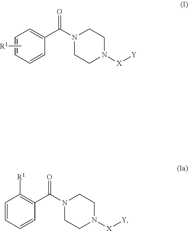
- SCD1 polypeptide inhibitor can be a compound having Formula (I) or Formula (Ia):
- R 1 is an unsubstituted C 1-6 alkyl or C 1-6 haloalkyl; X is
- Y is selected from:
- the SCD1 polypeptide inhibitor can be SSI-2, 2-(benzyloxy)-5- ⁇ [hydroxy( ⁇ 4-[2-(trifluoromethyl)benzoyl]piperazin-1-yl ⁇ )methyl]amino ⁇ -1,2-dihydropyridin-2-ylium-1-ide:
- the method also can include administering an additional therapeutic agent used to treat cancer to the mammal.
- the additional therapeutic agent can be a chemotherapeutic agent.
- the chemotherapeutic agent can be a kinase inhibitor.
- the kinase inhibitor can be sorafenib, regorafenib, bortezomib, erlotinib, gefitinib, imatinib, vemurafenib, or vismodegib.
- the kinase inhibitor can be sorafenib.
- this document features methods for treating a cancer in a mammal identified as having a cancer exhibiting SCD1 polypeptide expression and an elevated level of cMyc polypeptides.
- the methods can include, or consist essentially of, administering a SCD1 polypeptide inhibitor to the mammal.
- the mammal can be a human.
- the cancer can be liver cancer, renal cell carcinoma, ovarian cancer, breast cancer, prostate cancer, colon cancer, pancreatic cancer, bladder cancer, lung cancer, thyroid cancer, brain cancer, melanoma, or lymphoma.
- the cancer can be liver cancer (e.g., a hepatocellular carcinoma or a cholangiocarcinoma).
- the method also can include detecting an elevated level of survivin polypeptides within the biological sample from the mammal.
- the SCD1 polypeptide inhibitor can be a compound having Formula (II) or Formula (IIa):
- R 1 is halo
- X is —(C ⁇ O)NR 4 —
- Y is
- R 2 , R 3 , and R 4 are each independently H or an unsubstituted C 1-6 alkyl.
- the SCD1 polypeptide inhibitor can be SSI-4, 2- ⁇ [4-(2-Chlorophenoxy)piperidine-1-carbonyl]amino ⁇ -N-methylpyridine-4-carboxamide:
- SCD1 polypeptide inhibitor can be a compound having Formula (I) or Formula (Ia):
- R 1 is an unsubstituted C 1-6 alkyl or C 1-6 haloalkyl; X is
- Y is selected from:
- the SCD1 polypeptide inhibitor can be SSI-2, 2-(benzyloxy)-5- ⁇ [hydroxy( ⁇ 4-[2-(trifluoromethyl)benzoyl]piperazin-1-yl ⁇ )methyl]amino ⁇ -1,2-dihydropyridin-2-ylium-1-ide:
- the method also can include administering an additional therapeutic agent used to treat cancer to the mammal.
- the additional therapeutic agent can be a chemotherapeutic agent.
- the chemotherapeutic agent can be a kinase inhibitor.
- the kinase inhibitor can be sorafenib, regorafenib, bortezomib, erlotinib, gefitinib, imatinib, vemurafenib, or vismodegib.
- the kinase inhibitor can be sorafenib.
- this document features methods for treating a cancer in a mammal.
- the methods can include, or consist essentially of, detecting SCD1 polypeptide expression within a biological sample from the mammal, detecting an elevated level of cMyc polypeptides within the biological sample from the mammal, and administering a SCD1 polypeptide inhibitor to the mammal.
- the mammal can be a human.
- the cancer can be liver cancer, renal cell carcinoma, ovarian cancer, breast cancer, prostate cancer, colon cancer, pancreatic cancer, bladder cancer, lung cancer, thyroid cancer, brain cancer, melanoma, or lymphoma.
- the cancer can be liver cancer (e.g., a hepatocellular carcinoma or a cholangiocarcinoma).
- the method also can include detecting an elevated level of survivin polypeptides within the biological sample from the mammal.
- the SCD1 polypeptide inhibitor can be a compound having Formula (II) or Formula (IIa):
- R 1 is halo
- X is —(C ⁇ O)NR 4 —
- Y is
- R 2 , R 3 , and R 4 are each independently H or an unsubstituted C 1-6 alkyl.
- the SCD1 polypeptide inhibitor can be SSI-4, 2- ⁇ [4-(2-Chlorophenoxy)piperidine-1-carbonyl]amino ⁇ -N-methylpyridine-4-carboxamide:
- SCD1 polypeptide inhibitor can be a compound having Formula (I) or Formula (Ia):
- R 1 is an unsubstituted C 1-6 alkyl or C 1-6 haloalkyl; X is
- Y is selected from:
- the SCD1 polypeptide inhibitor can be SSI-2, 2-(benzyloxy)-5- ⁇ [hydroxy( ⁇ 4-[2-(trifluoromethyl)benzoyl]piperazin-1-yl ⁇ )methyl]amino ⁇ -1,2-dihydropyridin-2-ylium-1-ide:
- the method also can include administering an additional therapeutic agent used to treat cancer to the mammal.
- the additional therapeutic agent can be a chemotherapeutic agent.
- the chemotherapeutic agent can be a kinase inhibitor.
- the kinase inhibitor can be sorafenib, regorafenib, bortezomib, erlotinib, gefitinib, imatinib, vemurafenib, or vismodegib.
- the kinase inhibitor can be sorafenib.
- this document features methods for identifying a mammal as having a cancer that is responsive to one or more a SCD1 polypeptide inhibitors.
- the methods can include, or consist essentially of, detecting a level of SCD1 polypeptides within a biological sample from the mammal, detecting a level of cMyc polypeptides within the biological sample from the mammal, and identifying the mammal as having a cancer that is responsive to one or more a SCD1 polypeptide inhibitors when SCD1 polypeptide expression and an elevated level of cMyc polypeptides are detected in the biological sample.
- the mammal can be a human.
- the cancer can be liver cancer, renal cell carcinoma, ovarian cancer, breast cancer, prostate cancer, colon cancer, pancreatic cancer, bladder cancer, lung cancer, thyroid cancer, brain cancer, melanoma, or lymphoma.
- the cancer can be liver cancer (e.g., a hepatocellular carcinoma or a cholangiocarcinoma).
- the method also can include detecting an elevated level of survivin polypeptides within the biological sample from the mammal.
- the SCD1 polypeptide inhibitor can be a compound having Formula (II) or Formula (IIa):
- R 1 is halo
- X is —(C ⁇ O)NR 4 —
- Y is
- R 2 , R 3 , and R 4 are each independently H or an unsubstituted C 1-6 alkyl.
- the SCD1 polypeptide inhibitor can be SSI-4, 2- ⁇ [4-(2-Chlorophenoxy)piperidine-1-carbonyl]amino ⁇ -N-methylpyridine-4-carboxamide:
- SCD1 polypeptide inhibitor can be a compound having Formula (I) or Formula (Ia):
- R 1 is an unsubstituted C 1-6 alkyl or C 1-6 haloalkyl; X is
- Y is selected from:
- the SCD1 polypeptide inhibitor can be SSI-2, 2-(benzyloxy)-5- ⁇ [hydroxy( ⁇ 4-[2-(trifluoromethyl)benzoyl]piperazin-1-yl ⁇ )methyl]amino ⁇ -1,2-dihydropyridin-2-ylium-1-ide:
- the method also can include administering an additional therapeutic agent used to treat cancer to the mammal.
- the additional therapeutic agent can be a chemotherapeutic agent.
- the chemotherapeutic agent can be a kinase inhibitor.
- the kinase inhibitor can be sorafenib, regorafenib, bortezomib, erlotinib, gefitinib, imatinib, vemurafenib, or vismodegib.
- the kinase inhibitor can be sorafenib.
- this document features methods for treating a cancer in a mammal.
- the methods can include, or consist essentially of, identifying the mammal as having a cancer that is responsive to one or more a SCD1 polypeptide inhibitors, where the identifying includes detecting SCD1 polypeptide expression within a biological sample from the mammal and detecting an elevated level of cMyc polypeptides within the biological sample from the mammal, and then administering a SCD1 polypeptide inhibitor to the mammal.
- the mammal can be a human.
- the cancer can be liver cancer, renal cell carcinoma, ovarian cancer, breast cancer, prostate cancer, colon cancer, pancreatic cancer, bladder cancer, lung cancer, thyroid cancer, brain cancer, melanoma, or lymphoma.
- the cancer can be liver cancer (e.g., a hepatocellular carcinoma or a cholangiocarcinoma).
- the method also can include detecting an elevated level of survivin polypeptides within the biological sample from the mammal.
- the SCD1 polypeptide inhibitor can be a compound having Formula (II) or Formula (IIa):
- R 1 is halo
- X is —(C ⁇ O)NR 4 —
- Y is
- R 2 , R 3 , and R 4 are each independently H or an unsubstituted C 1-6 alkyl.
- the SCD1 polypeptide inhibitor can be SSI-4, 2- ⁇ [4-(2-Chlorophenoxy)piperidine-1-carbonyl]amino ⁇ -N-methylpyridine-4-carboxamide:
- SCD1 polypeptide inhibitor can be a compound having Formula (I) or Formula (Ia):
- R 1 is an unsubstituted C 1-6 alkyl or C 1-6 haloalkyl; X is
- Y is selected from:
- the SCD1 polypeptide inhibitor can be SSI-2, 2-(benzyloxy)-5- ⁇ [hydroxy( ⁇ 4-[2-(trifluoromethyl)benzoyl]piperazin-1-yl ⁇ )methyl]amino ⁇ -1,2-dihydropyridin-2-ylium-1-ide:
- the method also can include administering an additional therapeutic agent used to treat cancer to the mammal.
- the additional therapeutic agent can be a chemotherapeutic agent.
- the chemotherapeutic agent can be a kinase inhibitor.
- the kinase inhibitor can be sorafenib, regorafenib, bortezomib, erlotinib, gefitinib, imatinib, vemurafenib, or vismodegib.
- the kinase inhibitor can be sorafenib.
- FIGS. 1A-1B show that SCD1 protein is expressed in hepatocellular carcinoma (HCC) and cholangiocarcinoma (CCA) patient tissues.
- FIG. 1A contains images of SCD1 immunohistochemical (IHC) expression for SCD1 in HCC and CCA.
- FIG. 1B contains a graph of quantitated SCD1 IHC protein expression in patient tumor tissues for HCC and CCA, and showed that HCC had similar SCD1 expression to that of normal liver tissue while CCA levels were somewhat lower on average.
- H score is calculated based upon signal intensity (0-3) using the formula: [(1+% ⁇ 1)+(2+% ⁇ 2)+(3+% ⁇ 3)]. * indicates statistical significance (P>0.05).
- FIGS. 2A-2B contain images of IHC detection of SCD1, cMyc, and survivin proteins expressed in HCC patient derived xenograft (PDTX) tissues ( FIG. 2A ) and CCA PDTX tissues ( FIG. 2B ).
- PDTX patient derived xenograft
- FIGS. 3A-3E shows specificity and oral bioavailability of SCD1 inhibitors.
- FIG. 3A contains structures of SSI-4 and SSI-2, two small molecule SCD1 inhibitors.
- FIG. 3B contains a graph of SCD1 inhibitors evaluated for their inhibition of oleoyl CoA biosynthesis by mouse liver microsomes in vitro.
- SSI-2 or SSI-4 was incubated with microsomes and 13C18-labeled stearoyl CoA, a native substrate of SCD1. Bioconversion of this substrate to 13C18-labeled oleoyl CoA product was quantified by LC/MS for drug treatments relative to vehicle (DMSO) controls.
- DMSO vehicle
- FIG. 3C contains a graph of mean plasma concentration of SSI-4 following administration of a single dose of oral (PO) or intravenous (IV) SSI-4 to fasted male C57BL/6 mice. Plasma levels were measured over 24 hours.
- FIG. 3D contains a table of half-life and bioavailability are key parameters measured in the single dose analyses. Bioavailability (%) was calculated with AUC0-last and nominal dose.
- FIG. 3E contains a full matrix SSI-4 kinome scan. SSI-4 was screened at 10 nM and 100 nM for binding to a total of 468 kinases.
- CDKL2 human atypical kinase
- FIGS. 4A-4B show that SSI-4 has a favorable toxicity profile.
- FIGS. 5A-5F show that SSI-4 demonstrated high affinity and specificity for SCD1.
- FIG. 5A contains a graph of dose responses of 0.01-10 ⁇ M SSI-4 with 5 day exposure, and demonstrates IC50 of 1-5 nM.
- FIG. 5B contains a graph of HCC cell lines demonstrating inhibition of proliferation with 1 ⁇ M SSI-4. To demonstrate SCD1 specificity, cells were pretreated with 500 ⁇ g/ml of oleic acid to rescue the growth inhibitory phenotype.
- FIG. 5A contains a graph of dose responses of 0.01-10 ⁇ M SSI-4 with 5 day exposure, and demonstrates IC50 of 1-5 nM.
- FIG. 5B contains a graph of HCC cell lines demonstrating inhibition of proliferation with 1 ⁇ M SSI-4.
- SCD1 specificity cells were pretreated with 500 ⁇ g/ml of oleic acid to rescue the growth inhibitory phenotype.
- FIG. 5C contains a graph showing SSI4 responsive HCC cells have decreased unsaturated fatty acids (UFA) after 48 hour SSI4 treatment (black) indicating that the function of SCD1 converting saturated fatty acids to unsaturated fatty acids was inhibited.
- FIG. 5D contains images of soft agar assays. Single cells grown in 3D soft agar identified SCD1 sensitive cell lines. Only SCD1 sensitive cell lines grew in soft agar.
- FIG. 5E contains an image of western blot analysis showing that survivin and cMyc were elevated in concert with SCD1 expression in cell lines responsive to SSI-4.
- 5F contains images of western blot analyses showing that survivin and cMyc were elevated in concert with SCD1 expression in cell lines responsive to SSI-4.
- cMyc and survivin are down-regulated with SSI-4 and rescued with oleic acid demonstrating specificity for SCD1.
- SSI-4 induced apoptosis as evidenced by cleaved PARP with specificity demonstrated by rescue with oleic acid.
- Endoplasmic reticulum (ER) stress was induced as shown by upregulation of BiP after SSI-4 treatment. Oleic acid blocks upregulation of BiP demonstrating specificity.
- FIG. 5G contains a graph showing cell proliferation of SSI4 responsive HCC cells infected with either a control nontarget shRNA or c-Myc 1657 shRNA with SSI-4 treatment. Addition of 1 uM SSI-4 to cMyc shRNA cells (red) showed no difference from that of cMyc shRNA alone (gray).
- FIG. 5H contains a graph showing LDHA mRNA levels in HCC cells infected with either nontarget shRNA and c-Myc 1657 shRNA. When c-Myc was silenced, LDHA mRNA decreases (black). These data indicated that cMyc, LDHA, and SCD1 are in the same pathway.
- FIGS. 6A-6C show that SCD1 inhibits in vivo HCC cell proliferation and demonstrates synergy with sorafenib.
- FIG. 6A contains IHC images of HCC PDTX models that express equivalent SCD1 and c-Myc with differences in LDHA expression.
- FIGS. 6B and 6C contains graphs of tumor volume in PAX148 ( FIG. 6B ) tumor tissues and in LIV58 ( FIG. 6C ) tumor tissues after treatment with SSI-4.
- Tumor tissues (5 mm 3 ) with 50% matrigel were implanted in the right flank of 8 week old female athymic nude mice. At ⁇ 100 mm3, daily SSI-4 (20-50 mg/kg oral) was begun. Tumor volume and mouse weight were monitored twice weekly.
- FIG. 6D contains images of IHC detection of Ki-67, cMyc, and survivin proteins expressed in LIV58 tumors treated with control (placebo) or SSI-4. IHC showed that nuclear Ki-67, cMyc, and survivin protein were attenuated in response to SSI-4 in mice. ER stress was induced as evidenced by BIP upregulation. H score is calculated based upon signal intensity (0-3) using the formula: [(1+% ⁇ 1)+(2+% ⁇ 2)+(3+% ⁇ 3)]. * indicates statistical significance (P>0.05).
- FIGS. 7A-7D show that SSI-4 demonstrated single agent antitumor activity and synergy with sorafenib in HCC models.
- FIG. 7A contains a dose response curve identifying SSI-4 responsive HCC cell lines.
- FIG. 7B contains a graph demonstrating synergy with combination SSI-4 and sorafenib using Talalay-Chou method for synergy. CI values ⁇ 1 indicate synergy.
- FIG. 7C contains a graph showing that combination SSI-4 and sorafenib demonstrated prolonged durable response in a HLE HCC mouse model while single agent sorafenib and SSI-4 escape over time on therapy.
- FIG. 7A contains a dose response curve identifying SSI-4 responsive HCC cell lines.
- FIG. 7B contains a graph demonstrating synergy with combination SSI-4 and sorafenib using Talalay-Chou method for synergy. CI values ⁇ 1 indicate syn
- 7D contains a graph demonstrating synergy with combination SSI-4 and regorafenib (an analog of sorafenib) using Talalay-Chou method for synergy.
- CI values ⁇ 1 indicate synergy.
- FIGS. 8A-8C show that SCD1 inhibits cell proliferation in human (CX-004T2 and HUCCT1) and mouse (M1-1a and M6-1b) cholangiocarcinoma (CCA) cell lines.
- FIG. 8A contains a graph of dose responses of 0.1-1 ⁇ M SSI-4 with 5 day exposure in CCA cell lines.
- FIG. 8B contains a graph of CCA cell lines demonstrating inhibition of proliferation with 1 ⁇ M SSI-4.
- SCD1 specificity cells were pretreated with 500 ⁇ g/ml of oleic acid to rescue the growth inhibitory phenotype.
- FIG. 8C contains graph of tumor volume in CX-003 CCA tumor tissues after treatment with SSI-4.
- SCD1 is an enzyme that catalyzes the de novo lipogenesis of ⁇ -9 monounsaturated fatty acids (MUFA) oleic acid (OA) and palmitoleic acid (PA).
- MUFAs are essential for the synthesis of triglycerides, sphingolipids, ceramides, glycolipids, phospholipids, and other lipoproteins which influence membrane fluidity, membrane raft formation and receptor clustering, second messenger signaling, fatty acid oxidation, energy storage, cell division, inflammation, and a number of other biological functions (Guillou et al., 2010 Prog Lipid Res. 49(2):186-199).
- SCD1-associated cancers Aberrant upregulation of SCD1 has been implicated in the development of certain types of cancer; however, not all SCD1-associated cancers are responsive to SCD1 polypeptide inhibitors.
- a cancer associated with expression (e.g., overexpression) of a SCD1 polypeptide can also be referred to as a SCD1-associated cancer (see, e.g., von Roemeling et al. 2015 J. Clin. Endocrinol. Metab. 100:E697-E709).
- the presence of elevated levels of one or more Wnt regulated polypeptides, such as cMyc and survivin, in a SCD1-associated cancer can indicate that the SCD1-associated cancer is likely to be responsive to treatment with one or more SCD1 polypeptide inhibitors (e.g., SSI-4).
- Wnt regulated polypeptides such as cMyc and survivin
- This document provides methods and materials for diagnosing (or identifying as having) mammals (e.g., humans) having cancer (e.g., a SCD1-associated cancer).
- mammals e.g., humans
- cancer e.g., a SCD1-associated cancer
- the methods and materials described herein can be used to diagnose a mammal as having a SCD1-associated cancer.
- a mammal having a cancer can be assessed to determine whether the cancer is a SCD1-associated cancer.
- Any appropriate method can be used to identify a mammal having cancer.
- imaging techniques and biopsy techniques can be used to identify mammals (e.g., humans) having cancer.
- the presence, absence, or level of SCD1 polypeptides can be detected in a sample (e.g., a biological sample) obtained from a mammal to determine if mammal has a SCD1-associated cancer. For example, when the presence of an elevated level of SCD1 polypeptides is detected, the mammal can be identified as having a SCD1-associated cancer.
- a sample e.g., a biological sample
- This document also provides methods and materials for assessing mammals (e.g., humans) having a cancer (e.g., a SCD1-associated cancer).
- a mammal identified as having a SCD1-associated cancer can be assessed to determine whether or not the SCD1-associated cancer will respond to treatment (e.g., treatment with one or more SCD1 polypeptide inhibitors).
- treatment e.g., treatment with one or more SCD1 polypeptide inhibitors
- the presence, absence, or level of one or more Wnt regulated polypeptides can be detected in a sample (e.g., a biological sample) obtained from a mammal to determine the responsiveness of the cancer to a treatment.
- the presence of an elevated level of one or more Wnt regulated polypeptides can indicate that a SCD1-associated cancer is responsive to one or more SCD1 polypeptide inhibitors.
- a SCD1-associated cancer is responsive to one or more SCD1 polypeptide inhibitors.
- the mammal can be identified as having a SCD1-associated cancer that is likely to be responsive to one or more SCD1 polypeptide inhibitors.
- a SCD1-associated cancer that is likely to be responsive to one or more SCD1 polypeptide inhibitors can include elevated levels of one or more (e.g., one, two, three, four, five, or more) Wnt regulated polypeptides.
- the Wnt regulated polypeptides can be any appropriate Wnt regulated polypeptides.
- a Wnt regulated polypeptide can be associated with promoting cell proliferation (e.g., cMyc and cyclin D1).
- a Wnt regulated polypeptide can be associated with cell survival (e.g., survivin). Examples of Wnt regulated polypeptides include, without limitation, cMyc, survivin, and cyclin D1.
- a SCD1-associated cancer that is likely to be responsive to one or more SCD1 polypeptide inhibitors includes elevated levels of cMyc polypeptides. In some cases, a SCD1-associated cancer that is likely to be responsive to one or more SCD1 polypeptide inhibitors includes elevated levels of cMyc polypeptides and elevated levels of survivin polypeptides.
- the term “elevated level” as used herein with respect to a level of one or more Wnt regulated polypeptides refers to any level that is greater than a reference level of one or more Wnt regulated polypeptides, respectively.
- the term “reference level” as used herein with respect to one or more Wnt regulated polypeptides refers to the level of one or more Wnt regulated polypeptides typically observed in a sample (e.g., a control sample) from one or more mammals (e.g., humans) without cancer.
- Control samples can include, without limitation, samples from normal (e.g., healthy) mammals, and cell lines (e.g., non-tumor forming cells lines).
- an elevated level of one or more Wnt regulated polypeptides can be a level that is about least 2 (e.g., at least 3, at least 4, at least 5, at least 6, at least 7, at least 8, at least 9, or at least 10) fold greater relative to a reference level of one or more Wnt regulated polypeptides.
- an elevated level can be a detectable level of one or more Wnt regulated polypeptides. It will be appreciated that levels from comparable samples are used when determining whether or not a particular level is an elevated level.
- a sample can be a tumor sample (e.g., a biopsy).
- a sample can be a biological sample.
- biological samples such as tissue samples (e.g., liver tissue) and/or fluids (e.g., blood, serum, plasma, or urine) can be obtained from a mammal and assessed for the presence of one or more Wnt regulated polypeptides.
- a liver tissue sample can be used as a biological sample to determine whether or not the mammal has an elevated level of one or more Wnt regulated polypeptides.
- any appropriate method can be used to detect the presence, absence, or level of one or more Wnt regulated polypeptides (e.g., cMyc and/or survivin) within a sample (e.g., a biological sample).
- a sample e.g., a biological sample.
- immunohistochemistry (IHC) techniques e.g., mass spectrometry techniques (e.g., proteomics-based mass spectrometry assays or targeted quantification-based mass spectrometry assays), western blotting techniques, and quantitative RT-PCR techniques can be used to determine whether or not a sample contains an elevated level of one or more Wnt regulated polypeptides.
- IHC and/or western blotting techniques can be used to determine whether or not a particular sample contains an elevated level of one or more Wnt regulated polypeptides.
- This document also provides methods and materials for treating mammals (e.g., humans) diagnosed with (or identified as having) a cancer as described herein (e.g., a SCD1-associated cancer) and/or identified as having a SCD1-associated cancer that is likely to be responsive to one or more SCD1 polypeptide inhibitors by administering one or more SCD1 polypeptide inhibitors.
- a cancer as described herein e.g., a SCD1-associated cancer
- the methods and materials described herein can be used to treat a mammal having a cancer exhibiting an elevated level of a SCD1 polypeptides.
- the methods and materials described herein can be used to treat a mammal having a cancer exhibiting an elevated level of a SCD1 polypeptides and an elevated level of cMyc polypeptides.
- a mammal can be administered or instructed to self-administer one or more SCD1 polypeptide inhibitors (e.g., one, two, three, four, five, or more SCD1 inhibitors).
- the methods and materials described herein can include identifying a mammal as having a cancer (e.g., a SCD1-associate cancer).
- the methods and materials described herein can be used to treat a mammal identified (e.g., previously identified) as having cancer (e.g., a SCD1-associate cancer).
- Administration of a SCD1 polypeptide inhibitor to a mammal having an elevated level of SCD1 polypeptides can be effective to treat cancer (e.g., cancer having an elevated level of SCD1 polypeptides and an elevated level of Wnt regulated polypeptides).
- cancer e.g., cancer having an elevated level of SCD1 polypeptides and an elevated level of Wnt regulated polypeptides.
- treating a mammal as described herein can be effective to slow or prevent growth of a cancer.
- administering one or more SCD1 polypeptide inhibitors to a mammal identified as having a SCD1-associated cancer and/or identified as having a SCD1-associated cancer that is likely to be responsive to one or more SCD1 polypeptide inhibitors can be effective to prevent a tumor from increasing in size (e.g., volume).
- administering one or more SCD1 polypeptide inhibitors to a mammal identified as having a SCD1-associated cancer and/or identified as having a SCD1-associated cancer that is likely to be responsive to one or more SCD1 polypeptide inhibitors can be effective prevent a tumor from metastasizing.
- treating a mammal as described herein can be effective to reduce or eliminate the number of cancer cells within the mammal.
- administering one or more SCD1 polypeptide inhibitors to a mammal identified as having a SCD1-associated cancer and/or identified as having a SCD1-associated cancer that is likely to be responsive to one or more SCD1 polypeptide inhibitors can be effective to reduce the size (e.g., volume) of a tumor.
- treating a mammal as described herein can be effective to induce endoplasmic reticulum (ER) stress in cancer cells within the mammal.
- ER endoplasmic reticulum
- treating a mammal as described herein can be effective to induce apoptotic cell death of cancer cells within the mammal.
- any type of mammal can be assessed and/or treated as described herein.
- mammals that can be assessed and/or treated as described herein include, without limitation, primates (e.g., humans and monkeys), dogs, cats, horses, cows, pigs, sheep, rabbits, mice, and rats.
- the mammal can be a human.
- humans identified as having a SCD1-associated cancer and/or identified as having a SCD1-associated cancer that is likely to be responsive to one or more SCD1 polypeptide inhibitors can be treated with one or more SCD1 polypeptide inhibitors as described herein.
- the cancer can be any cancer.
- the cancer can be a SCD1-associated cancer.
- a cancer can be a primary cancer or a metastatic cancer.
- a cancer can be a cancer that has escaped and/or has been non-responsive to chemotherapy (e.g., a chemoresistant cancer).
- cancers examples include, without limitation, liver cancer (e.g., HCC and/or CCA), renal cell carcinoma, ovarian cancer, breast cancer, prostate cancer, colon cancer, pancreatic cancer, bladder cancer, lung cancer, thyroid cancer, brain cancer, melanoma, and lymphoma.
- a cancer treated as described herein can be a HCC.
- a cancer treated as described herein can be a CCA.
- any appropriate SCD1 polypeptide inhibitor can be administered to the mammal.
- a SCD1 polypeptide inhibitor can be any appropriate type of molecule (e.g., nucleic acid molecules designed to induce RNA interference (e.g., a siRNA molecule or a shRNA molecule), antisense molecules, miRNAs, and antibodies (e.g., antibodies (e.g., monoclonal antibodies)) that can reduce or eliminate SCD1 polypeptide expression or SCD1 polypeptide function.
- a SCD1 polypeptide inhibitor can be an inhibitor of SCD1 polypeptide expression or an inhibitor of SCD1 polypeptide activity.
- a SCD1 polypeptide inhibitor can be readily designed based upon the nucleic acid and/or polypeptide sequences of SCD1.
- a SCD1 polypeptide inhibitor can be as described elsewhere (see, e.g., WO 2016/022955).
- a SCD1 polypeptide inhibitor can have Formula (I):
- R 1 is an unsubstituted C 1-6 alkyl or C 1-6 haloalkyl; X is
- Y is selected from:
- a SCD1 polypeptide inhibitor according to Formula (I) can have the structure of Formula (Ia):
- SCD1 polypeptide inhibitors according to Formula (I) and/or Formula (Ia) include, without limitation:
- a SCD1 polypeptide inhibitor can have Formula (II):
- R 1 is halo; X is —(C ⁇ O)NR 4 —; Y is
- R 2 , R 3 , and R 4 are each independently H or an unsubstituted C 1-6 alkyl.
- a SCD1 polypeptide inhibitor according to Formula (II) can have the structure of Formula (IIa):
- SCD1 polypeptide inhibitor according to Formula (II) and/or Formula (IIa) include:
- a SCD1 polypeptide inhibitor can be SSI-4.
- one or more SCD1 polypeptide inhibitors can be administered to a mammal (e.g., human) identified as having cancer (e.g., a SCD1-associate cancer) as described herein and/or identified as having a cancer that is likely to be responsive to one or more SCD1 polypeptide inhibitors as described herein as the sole anti-cancer treatment agent.
- a mammal e.g., human
- cancer e.g., a SCD1-associate cancer
- SSI-4 can be administered to a mammal identified as having a SCD1-associate cancer as described herein and/or identified as having a cancer that is likely to be responsive to one or more SCD1 polypeptide inhibitors as described herein as the sole anti-cancer treatment agent.
- the mammal when treating a mammal (e.g., human) identified as having cancer (e.g., a SCD1-associate cancer) and/or identified as having a cancer that is likely to be responsive to one or more SCD1 polypeptide inhibitors as described herein, the mammal also can be administered or instructed to self-administer one or more additional therapeutic agents (e.g., therapeutic agents used to treat cancer).
- the one or more additional therapeutic agents can include agents approved by the Food and Drug Administration (FDA) for a particular type of cancer.
- FDA Food and Drug Administration
- the one or more additional therapeutic agents can include agents approved by the FDA for liver cancer.
- therapeutic agents that can be used to treat cancer include, without limitation, radiation therapy; surgery; and chemotherapeutic agents (including, but not limited to, alkylating agents (e.g., cyclophosphamide, mechlorethamine, chlorambucil, melphalan, dacarbazine, nitrosoureas, temozolomide), anthracyclines (e.g., daunorubicin, doxorubicin, epirubicin, idarubicin, mitoxantrone, and valrubicin), cytoskeletal disruptors (e.g., paclitaxel, docetaxel, abraxane, and taxotere), histone deacetylase inhibitors (e.g., vorinostat and romidepsin), topoisomerase inhibitors (e.g., irinotecan, topotecan, etoposide, teniposide, and tafluposide), kina
- one or more SCD1 polypeptide inhibitors can be administered in combination with sorafenib to a mammal (e.g., human) identified as having a SCD1-associated cancer and/or identified as having a cancer that is likely to be responsive to one or more SCD1 polypeptide inhibitors to treat the mammal.
- a mammal e.g., human
- the one or more SCD1 polypeptide inhibitors described herein are used in combination with one or more therapeutic agents to treat cancer
- the one or more SCD1 polypeptide inhibitors can be administered at the same time or independently of the administration of one or more therapeutic agents.
- the composition including one or more SCD1 polypeptide inhibitors can be administered before, concurrent with, or after the one or more therapeutic agents are administered.
- one or more SCD1 polypeptide inhibitors can be administered to a mammal (e.g., a mammal identified as having a SCD1-associated cancer and/or identified as having a cancer that is likely to be responsive to one or more SCD1 polypeptide inhibitors) once or multiple times over a period of time ranging from days to weeks.
- a mammal e.g., a mammal identified as having a SCD1-associated cancer and/or identified as having a cancer that is likely to be responsive to one or more SCD1 polypeptide inhibitors
- one or more SCD1 polypeptide inhibitors described herein can be formulated into a pharmaceutically acceptable composition for administration to a mammal.
- a therapeutically effective amount of one or more SCD1 polypeptide inhibitors can be formulated together with one or more pharmaceutically acceptable carriers (additives) and/or diluents.
- a pharmaceutical composition can be formulated for administration in solid or liquid form including, without limitation, sterile solutions, suspension
- Pharmaceutically acceptable carriers, fillers, and vehicles that may be used in a pharmaceutical composition described herein include, without limitation, ion exchangers, alumina, aluminum stearate, lecithin, serum proteins, such as human serum albumin, buffer substances such as phosphates, glycine, sorbic acid, potassium sorbate, partial glyceride mixtures of saturated vegetable fatty acids, water, salts or electrolytes, such as protamine sulfate, disodium hydrogen phosphate, potassium hydrogen phosphate, sodium chloride, zinc salts, colloidal silica, magnesium trisilicate, polyvinyl pyrrolidone, cellulose-based substances, polyethylene glycol, sodium carboxymethylcellulose, polyacrylates, waxes, polyethylene-polyoxypropylene-block polymers, polyethylene glycol and wool fat.
- ion exchangers alumina, aluminum stearate, lecithin
- serum proteins such as human serum albumin
- buffer substances such as phosphates,
- a pharmaceutical composition containing one or more SCD1 polypeptide inhibitors can be designed for oral, parenteral (including subcutaneous, intramuscular, intravenous, and intradermal), or intratumoral administration.
- a pharmaceutical composition can be in the form of a pill, tablet, or capsule.
- Compositions suitable for parenteral administration include aqueous and non-aqueous sterile injection solutions that can contain anti-oxidants, buffers, bacteriostats, and solutes that render the formulation isotonic with the blood of the intended recipient.
- the formulations can be presented in unit-dose or multi-dose containers, for example, sealed ampules and vials, and may be stored in a freeze dried (lyophilized) condition requiring only the addition of the sterile liquid carrier, for example, water for injections, immediately prior to use.
- sterile liquid carrier for example, water for injections, immediately prior to use.
- Extemporaneous injection solutions and suspensions may be prepared from sterile powders, granules, and tablets.
- a pharmaceutically acceptable composition including one or more SCD1 polypeptide inhibitors can be administered locally (e.g., intratumorally) or systemically.
- a composition provided herein can be administered locally by injection into tumors or into biological spaces infiltrated by tumors (e.g. peritoneal cavity and/or pleural space).
- a composition provided herein can be administered systemically, orally, or by injection to a mammal (e.g., a human).
- Effective doses can vary depending on the risk and/or the severity of the cancer, the route of administration, the age and general health condition of the mammal, excipient usage, the possibility of co-usage with other therapeutic treatments such as use of other agents, and the judgment of the treating physician.
- An effective amount of a composition containing one or more SCD1 polypeptide inhibitors can be any amount that reduces the number of cancer cells present within the mammal without producing significant toxicity to the mammal.
- an effective amount of one or more SCD1 polypeptide inhibitors can be from about 10 to about 100 mg per kg body weight (mg/kg) of the mammal being treated.
- an effective amount of one or more SCD1 polypeptide inhibitors can be from about 10 to about 90, from about 10 to about 80, from about 10 to about 70, from about 10 to about 60, from about 10 to about 50, from about 10 to about 40, from about 10 to about 30, from about 20 to about 100, from about 30 to about 100, from about 40 to about 100, from about 50 to about 100, from about 60 to about 100, from about 70 to about 100, from about 80 to about 100, from about 15 to about 90, from about 20 to about 80, from about 25 to about 75, from about 30 to about 60, from about 35 to about 50, or from about 40 to about 50 mg/kg of the mammal being treated.
- about 30 mg/kg of one or more SCD1 polypeptide inhibitors can be administered (e.g., orally administered) to a human identified as having a SCD1-associated cancer as described herein and/or identified as having a cancer that is likely to be responsive to one or more SCD1 polypeptide inhibitors as described herein.
- about 50 mg/kg of one or more SCD1 polypeptide inhibitors can be administered (e.g., orally administered) to a human identified as having a SCD1-associated cancer as described herein and/or identified as having a cancer that is likely to be responsive to one or more SCD1 polypeptide inhibitors as described herein.
- one or more SCD1 polypeptide inhibitors can be administered (e.g., orally administered) to a human identified as having a SCD1-associated cancer as described herein and/or identified as having a cancer that is likely to be responsive to one or more SCD1 polypeptide inhibitors as described herein.
- the amount of one or more SCD1 polypeptide inhibitors can be increased by, for example, two fold. After receiving this higher amount, the mammal can be monitored for both responsiveness to the treatment and toxicity symptoms, and adjustments made accordingly.
- the effective amount can remain constant or can be adjusted as a sliding scale or variable dose depending on the mammal's response to treatment. Various factors can influence the actual effective amount used for a particular application. For example, the frequency of administration, duration of treatment, use of multiple treatment agents, route of administration, and severity of the condition (e.g., cancer) may require an increase or decrease in the actual effective amount administered.
- the frequency of administration of one or more SCD1 polypeptide inhibitors can be any amount that reduces the number of cancer cells present within the mammal without producing significant toxicity to the mammal.
- the frequency of administration of one or more SCD1 polypeptide inhibitors can be from about two to about three times a week to about two to about three times a month.
- a mammal identified as having a SCD1-associated cancer and/or identified as having a cancer that is likely to be responsive to one or more SCD1 polypeptide inhibitors can receive a single administration of one or more SCD1 polypeptide inhibitors described herein.
- the frequency of administration of one or more SCD1 polypeptide inhibitors described herein can remain constant or can be variable during the duration of treatment.
- a course of treatment with a composition containing one or more SCD1 polypeptide inhibitors described herein can include rest periods.
- a composition containing one or more SCD1 polypeptide inhibitors described herein can be administered every other month over a two-year period followed by a six-month rest period, and such a regimen can be repeated multiple times.
- the effective amount various factors can influence the actual frequency of administration used for a particular application. For example, the effective amount, duration of treatment, use of multiple treatment agents, route of administration, and severity of the condition (e.g., cancer) may require an increase or decrease in administration frequency.
- An effective duration for administering a composition containing one or more SCD1 polypeptide inhibitors can be any duration that reduces the number of cancer cells present within the mammal without producing significant toxicity to the mammal.
- the effective duration can vary from several months to several years.
- the effective duration for reducing the number of cancer cells present within the mammal can range in duration from about one or two months to five or more years. Multiple factors can influence the actual effective duration used for a particular treatment. For example, an effective duration can vary with the frequency of administration, effective amount, use of multiple treatment agents, route of administration, and severity of the condition being treated.
- the number of cancer cells present within a mammal can be monitored. Any appropriate method can be used to determine whether or not the number of cancer cells present within a mammal is reduced. For example, imaging techniques can be used to assess the number of cancer cells present within a mammal.
- SCD1 inhibitor SCD1 inhibitor
- SSI-4 Antitumor activity of SCD1 inhibitor, SSI-4, against HCC in cell culture and animal models for HCC was shown.
- SSI-4 also exhibited synergy with sorafenib, an FDA approved drugs for HCC.
- SCD1 inhibitors were developed through a combined computational and synthetic chemistry approach as described elsewhere (see, e.g., WO 2016/022955). Using combined computational and synthetic chemistry approaches, we synthesized four novel specific SCD1 inhibitors with SSI-4 being the lead SCD1 inhibitor.
- Sprague dawley rats (6-8 weeks old M/F) were orally gavaged with SS1-4 at the indicated dose daily, for a duration of seven days. For each dose levels 3 male and 3 females were placed on study. Rats were clinically observed and weighed daily. On day 8 rats were euthanatized and blood was collected by cardiac puncture. Blood chemistry was measured by STAT Veterinary laboratory.
- Pharmacokinetic assessment was performed for SSI-4 administered to fasted male C57BL/6 mice either intravenously (IV) or by oral gavage (PO) in DMSO:PEG400:water (10:70:20) as a clear solution at 5 mg/mL (IV dosing), and 1.3 or 10 mg/mL (respectively for the PO dosing). Serum analysis was performed by LC-MS-MS. PK calculation settings were obtained using the Phoenix WinNonlin 6.3 program, using either the Noncompartmental model 201 (IV bolus input) or Noncompartmental model 200 (extravascular input). Microsomes were isolated from murine liver using Microsome Isolation Kit (BioVision).
- Each assay (100 ⁇ L) contained 50 ⁇ g of microsomes, 1 ⁇ L of DMSO (for no drug controls) or DMSO-solubilized SCD1 inhibitor, 1 mM reduced NADH, 60 ⁇ M coenzyme A, 1 mM ATP, 1 mM DTT, and 5 mM MgCl2 in 100 mM sodium phosphate buffer (pH 7.4). Assays were incubated for five minutes at 25 C and then supplemented with 30 ⁇ M 13C18-stearoyl CoA lithium salt to initiate SCD1-catalyzed conversion to 13C18-oleoyl CoA.
- LC/MS2 was conducted using a Thermo LTQ mass spectrometer interfaced with a Dionex UltiMate 8000 LC system.
- SRM signal areas for isotope-labeled acyl CoAs were measured relative to the heptadecanoyl CoA I.S. using XCalibur software, and percent enzyme inhibition determined by comparing 13C18-oleoyl CoA quantities between treatment and DMSO control assays. Dose-response curves were prepared using GraphPad Prism.
- Tissues were cut into 4 ⁇ 4 ⁇ 4 mm cubes and surgically implanted under anesthesia with 100 ⁇ l Matrigel into 4-6 week old athymic nude female mice. Tumors were measured 1 to 2 times per week using calipers and volumes calculated by 0.536*(L ⁇ W ⁇ H). Once tumors reached ⁇ 100 mm3, either placebo or SSI-4 treatment was administered at 180, or 600 mg/kg in custom AIN76 animal chow by Research Diets, Inc. Based upon food consumption, the amount of SSI4 ingested was 20 mg/kg and 50 mg/kg, respectively. For the combination study, mice were administered either 10 mg/kg SSI4, 30 mg/kg sorafenib or both in Nutra-gel diet.
- FFPE tissues were cut into 5 ⁇ m sections, deparaffinized, hydrated, antigen retrieved for 25 minutes at pH 6.0 and blocked for 5 minutes with Dako diluent. Immunostaining was done for 1 hour with either lamin A+C, SCD1, c-Myc, survivin, LDHA, ki67, and BIP primary antibodies.
- the Envision Dual Labeled Polymer kit (Dako) was used for 30 minutes for anti-species secondary antibodies and stained with DAB chromagen for 5 minutes. Then, lightly counterstained with Gill I hematoxylin for 30 seconds before dehydration and mounting. Images were obtained at 20 ⁇ using an Aperio AT2 Scanscope.
- IHC scoring was done using an algorithm in the Imagescope software based upon signal intensity of weak (1+), moderate (2+) or strong staining (3+) with criteria of at least 20% positivity. H score was calculated based upon signal intensity (0-3) using the formula: [(1+% ⁇ 1)+(2+% ⁇ 2)+(3+% ⁇ 3)].
- Cell lines were maintained in DMEM media supplemented with sodium pyruvate, HEPES, non-essential amino acids, 3% FBS and antibiotics. Cells were plated at 30,000 cells per well in 12-well plate and treated for 4-5 days at the indicated doses with SSI4 and sorafenib diluted in DMSO at 1:1000. Cell number was determined by using a Coulter Particle Counter.
- Soft agar cultures were prepared by diluting 2 ⁇ growth medium 1:1 in 1.5% Seaplaque GTG agarose with 1000 cells/plate in 6-well culture dishes. Colonies were stained with Giemsa after 3 weeks and colonies larger than 150 ⁇ m were counted using Image J software.
- Cells were collected via scraping and lysed in RIPA extraction buffer containing protease inhibitor cocktail and phosphatase inhibitor. Protein concentrations were measured by bicinchoninic acid (BCA) assay and 30 ⁇ g were loaded on 4-12% Bis-Tris/MES gels and then transferred to 0.2 ⁇ m Immobilon-P membranes. The membranes were hybridized overnight at 4° C. with the following antibodies: SCD1, PARP, survivin, c-Myc, BIP and ⁇ -actin. Secondary species-specific horseradish peroxidase-labeled antibodies were from Jackson Immunoresearch. Detection was performed using Supersignal chemiluminescence kit.
- BCA bicinchoninic acid
- Self-inactivating shRNA lentiviruses were generated using MISSION shRNA pLKO.1 constructs that included a nontarget control which was a random scrambled sequence (SHC002) and c-Myc 1657 (clone NM_002467.2-1657s1c1). Lentiviruses were packaged using 293FT cells via transfection of the pLKO.1 constructs along with packaging plasmids using OptiMEM and Lipofectamine 2000.
- the packaging plasmids were originally made by Didier Trono supplied by Addgene, which included: pMDLg/pRRE (gag/pol) (plasmid #12251), pRSV-Rev (plasmid #12253), and pMD2.G (VSVG) (plasmid #12259).
- pMDLg/pRRE gag/pol
- pRSV-Rev plasmid #12253
- pMD2.G VSVG
- Supernatants were collected 72 h post-transfection, passed through a 0.45 ⁇ m PVDF syringe filter and applied to cells for infection along with 5 ⁇ g/ml polybrene. Cells were selected with puromycin.
- the PCR step was done using TaqMan® Fast Universal PCR Master Mix and TaqMan® FAMTM dye-labeled probes for c-Myc, LDHA and POLR2A (Hs00172187_m1). Data was normalized to POLR2A for each sample. Fold change values between nontargets and shRNA samples were calculated using the ⁇ Ct method (Schmittgen and Livak 2008).
- SCD1 protein expression was examined by IHC in HCC and CCA tissues using tissue microarrays comprising of HCC, CCA and normal patient tissues ( FIG. 1A ). Samples were quantitated as H scores and graphed ( FIG. 1B ). SCD1, c-Myc and survivin protein expression was also examined by IHC in HCC and CCA PDTX avatar models. All six models expressed SCD1 and survivin while c-Myc was absent only in the CX-003 CCA PDTX model. ( FIG. 2A-2B ).
- SCD1 inhibitors were synthesized with SSI-4 and SSI-2 as the top contenders as shown ( FIG. 3A ).
- SCD1 inhibitor SSI-4 demonstrated an IC50 of 1.9 nM related to inhibition of oleoyl CoA biosynthesis by mouse liver microsomes ( FIG. 3B ).
- SSI-4 also demonstrated excellent oral bioavailability with a half-life of 3-4 hours following a single oral dose ( FIG. 3C ).
- IC50 concentrations for blocking SCD1 enzyme activity and blood half-life and bioavailability of single dose SSI-4 was determined.
- Half-life and bioavailability information for SSI-4 are shown in FIG. 3D .
- Bioavailability (%) was calculated with area under the curve (AUG 0-last ) and nominal dose.
- CDKL2 1 weak affinity binding kinase
- FIG. 3E Toxicity of SSI-4 was examined. Little to no toxicity was seen in male or female rats with minor weight loss (6%) in males at the extremely high dose of 300 mg/kg ( FIG. 4A ). Blood chemistries were in normal range ( FIG. 4B ).
- HCC cell lines show that some lines are highly responsive with IC50's under 5 nM while others are 10 ⁇ M or higher ( FIG. 5A ). Doubling times of responsive cell lines was less than 47 hours versus the non-responsive cell lines indicating that rapidly dividing cells require MUFAs which are incorporated into cell membranes. Oleic acid rescue demonstrated specificity for SCD1. Normal human hepatocytes (HH) cells were not growth inhibited by 1 ⁇ M SCD1 ( FIG. 5B ) demonstrating tumor specificity. Levels of unsaturated fatty acids decreased upon SSI-4 treatment indicating that the conversion of saturated fatty acids to unsaturated fatty acids was being blocked ( FIG. 5C ).
- FIG. 5D Wnt regulated genes, survivin and cMyc, were upregulated at the protein level in SSI-4 responsive cell lines ( FIG. 5E ).
- SSI-4 down-regulated Wnt regulated genes cMyc, survivin and cyclin D1 while inducing apoptosis (cleaved PARP) and ER stress (BIP) ( FIG. 5F ).
- HCC cells silenced for cMyc with shRNA showed blunted response to SSI-4, thus, mechanistically linking SCD1 and cMyc to cell proliferation ( FIG. 5G ).
- Silencing c-Myc also downregulated LDHA which mechanistically links SCD1 and cMyc to glycolysis ( FIG. 5H ).
Abstract
Description

or pharmaceutically acceptable salt thereof, where R1 is halo, X is —(C═O)NR4—, Y is

and R2, R3, and R4 are each independently H or an unsubstituted C1-6alkyl. In some cases, the SCD1 polypeptide inhibitor can be SSI-4, 2-{[4-(2-Chlorophenoxy)piperidine-1-carbonyl]amino}-N-methylpyridine-4-carboxamide:

or pharmaceutically acceptable salt thereof. The SCD1 polypeptide inhibitor can be a compound having Formula (I) or Formula (Ia):

or a pharmaceutically acceptable salt thereof, where R1 is an unsubstituted C1-6alkyl or C1-6haloalkyl; X is

Y is selected from:

m is 0 or 1; n is 0, 1, or 2; V is NR4 or O; R2, R3, and R4 are each independently H or an unsubstituted C1-6alkyl; and Z is an unsubstituted aryl. In some cases, the SCD1 polypeptide inhibitor can be SSI-2, 2-(benzyloxy)-5-{[hydroxy({4-[2-(trifluoromethyl)benzoyl]piperazin-1-yl})methyl]amino}-1,2-dihydropyridin-2-ylium-1-ide:

or pharmaceutically acceptable salt thereof. The method also can include administering an additional therapeutic agent used to treat cancer to the mammal. The additional therapeutic agent can be a chemotherapeutic agent. The chemotherapeutic agent can be a kinase inhibitor. The kinase inhibitor can be sorafenib, regorafenib, bortezomib, erlotinib, gefitinib, imatinib, vemurafenib, or vismodegib. For example, the kinase inhibitor can be sorafenib.

or pharmaceutically acceptable salt thereof, where R1 is halo, X is —(C═O)NR4—, Y is

and R2, R3, and R4 are each independently H or an unsubstituted C1-6alkyl. In some cases, the SCD1 polypeptide inhibitor can be SSI-4, 2-{[4-(2-Chlorophenoxy)piperidine-1-carbonyl]amino}-N-methylpyridine-4-carboxamide:

or pharmaceutically acceptable salt thereof. The SCD1 polypeptide inhibitor can be a compound having Formula (I) or Formula (Ia):
or a pharmaceutically acceptable salt thereof, where R1 is an unsubstituted C1-6alkyl or C1-6haloalkyl; X is

Y is selected from:

m is 0 or 1; n is 0, 1, or 2; V is NR4 or O; R2, R3, and R4 are each independently H or an unsubstituted C1-6alkyl; and Z is an unsubstituted aryl. In some cases, the SCD1 polypeptide inhibitor can be SSI-2, 2-(benzyloxy)-5-{[hydroxy({4-[2-(trifluoromethyl)benzoyl]piperazin-1-yl})methyl]amino}-1,2-dihydropyridin-2-ylium-1-ide:

or pharmaceutically acceptable salt thereof. The method also can include administering an additional therapeutic agent used to treat cancer to the mammal. The additional therapeutic agent can be a chemotherapeutic agent. The chemotherapeutic agent can be a kinase inhibitor. The kinase inhibitor can be sorafenib, regorafenib, bortezomib, erlotinib, gefitinib, imatinib, vemurafenib, or vismodegib. For example, the kinase inhibitor can be sorafenib.

or a pharmaceutically acceptable salt thereof, where R1 is an unsubstituted C1-6alkyl or C1-6haloalkyl; X is

Y is selected from:

m is 0 or 1; n is 0, 1, or 2; V is NR4 or O; R2, R3, and R4 are each independently H or an unsubstituted C1-6alkyl; and Z is an unsubstituted aryl. In some cases, a SCD1 polypeptide inhibitor according to Formula (I) can have the structure of Formula (Ia):

or a pharmaceutically acceptable salt thereof. Representative examples of SCD1 polypeptide inhibitors according to Formula (I) and/or Formula (Ia) include, without limitation:

SSI-1, N-Methyl-2-(2-oxo-2-{4-[2-(trifluoromethyl)benzoyl]piperazin-1-yl}ethoxy)benzamide;

SSI-2, 2-(benzyloxy)-5-{[hydroxy({4-[2-(trifluoromethyl)benzoyl]piperazin-1-yl})methyl]amino}-1,2-dihydropyridin-2-ylium-1-ide; and

SSI-3, 2-(benzyloxy)-4-({[hydroxy({4-[2-(trifluoromethyl)benzoyl]piperazin-1-yl})methyl]azanidyl}methyl)-1,2-dihydropyridin-2-ylium-1-ide, or a pharmaceutically acceptable salt thereof. In some cases, a SCD1 polypeptide inhibitor can have Formula (II):

or pharmaceutically acceptable salt thereof, where R1 is halo; X is —(C═O)NR4—; Y is

R2, R3, and R4 are each independently H or an unsubstituted C1-6alkyl.

or pharmaceutically acceptable salt thereof. A representative example of a SCD1 polypeptide inhibitor according to Formula (II) and/or Formula (IIa) include:

SSI-4, 2-{[4-(2-Chlorophenoxy)piperidine-1-carbonyl]amino}-N-methylpyridine-4-carboxamide, or pharmaceutically acceptable salt thereof. In some cases, a SCD1 polypeptide inhibitor can be SSI-4.
Claims (14)


Priority Applications (1)
| Application Number | Priority Date | Filing Date | Title |
|---|---|---|---|
| US16/368,477 US11243207B2 (en) | 2018-03-29 | 2019-03-28 | Assessing and treating cancer |
Applications Claiming Priority (2)
| Application Number | Priority Date | Filing Date | Title |
|---|---|---|---|
| US201862650217P | 2018-03-29 | 2018-03-29 | |
| US16/368,477 US11243207B2 (en) | 2018-03-29 | 2019-03-28 | Assessing and treating cancer |
Publications (2)
| Publication Number | Publication Date |
|---|---|
| US20190302121A1 US20190302121A1 (en) | 2019-10-03 |
| US11243207B2 true US11243207B2 (en) | 2022-02-08 |
Family
ID=68055985
Family Applications (1)
| Application Number | Title | Priority Date | Filing Date |
|---|---|---|---|
| US16/368,477 Active US11243207B2 (en) | 2018-03-29 | 2019-03-28 | Assessing and treating cancer |
Country Status (1)
| Country | Link |
|---|---|
| US (1) | US11243207B2 (en) |
Families Citing this family (3)
| Publication number | Priority date | Publication date | Assignee | Title |
|---|---|---|---|---|
| EP3808349B1 (en) | 2014-08-07 | 2022-10-05 | Mayo Foundation for Medical Education and Research | Compounds and methods for treating cancer |
| CA3054572A1 (en) * | 2017-02-28 | 2018-09-07 | Mayo Foundation For Medical Education And Research | Compounds and methods for treating cancer |
| EP3700934A4 (en) | 2017-10-24 | 2021-10-27 | Yumanity Therapeutics, Inc. | Compounds and uses thereof |
Citations (25)
| Publication number | Priority date | Publication date | Assignee | Title |
|---|---|---|---|---|
| US6066730A (en) | 1994-10-28 | 2000-05-23 | Proscript, Inc. | Boronic ester and acid compounds, synthesis and uses |
| WO2003006009A1 (en) | 2001-07-11 | 2003-01-23 | Zen-Bio, Inc. | Compositions for reducing or preventing cellulite in mammalian skin |
| US6699835B2 (en) | 2001-01-25 | 2004-03-02 | The United States Of America As Represented By The Department Of Health And Human Services | Formulation of boronic acid compounds |
| US7014866B2 (en) | 2001-05-03 | 2006-03-21 | Hoffmann-La Roche Inc. | High dose solid unit oral pharmaceutical dosage form of amorphous nelfinavir mesylate and process for making same |
| US20060079502A1 (en) | 1999-11-02 | 2006-04-13 | Steffen Lang | Pharmaceutical compositions |
| US20060094744A1 (en) | 2004-09-29 | 2006-05-04 | Maryanoff Cynthia A | Pharmaceutical dosage forms of stable amorphous rapamycin like compounds |
| WO2008120759A1 (en) | 2007-03-30 | 2008-10-09 | Japan Tobacco Inc. | Urea compound and use thereof |
| WO2009020448A1 (en) | 2007-08-06 | 2009-02-12 | Millennium Pharmaceuticals, Inc. | Proteasome inhibitors |
| WO2009051581A1 (en) | 2007-10-16 | 2009-04-23 | Millennium Pharmaceuticals, Inc. | Proteasome inhibitors |
| WO2009154737A1 (en) | 2008-06-17 | 2009-12-23 | Millennium Pharmaceuticals, Inc. | Boronate ester compounds and pharmaceutical compositions thereof |
| US7687456B2 (en) | 2005-11-09 | 2010-03-30 | Proteolix, Inc. | Compounds for enzyme inhibition |
| WO2010036357A1 (en) | 2008-09-29 | 2010-04-01 | Millennium Pharmaceuticals, Inc. | Derivatives of 1-amino-2-cyclobutylethylboronic acid |
| US7691852B2 (en) | 2006-06-19 | 2010-04-06 | Onyx Therapeutics, Inc. | Compounds for enzyme inhibition |
| WO2011123502A1 (en) | 2010-03-31 | 2011-10-06 | Millennium Pharmaceuticals, Inc | Derivatives of 1-amino-2-cyclopropylethylboronic acid |
| US8088741B2 (en) | 2004-05-10 | 2012-01-03 | Onyx Therapeutics, Inc. | Compounds for enzyme inhibition |
| WO2012151451A1 (en) | 2011-05-03 | 2012-11-08 | Agios Pharmaceuticals, Inc. | Pyruvate kinase activators for use in therapy |
| US20130096181A1 (en) | 2011-10-15 | 2013-04-18 | Genentech, Inc. | Methods of using scd1 antagonists |
| WO2014153150A1 (en) | 2013-03-14 | 2014-09-25 | Lee Delphine J | Methods of predicting anti ctla-4 response and recurrence of cancer |
| US20160000673A1 (en) | 2013-01-03 | 2016-01-07 | Conopco, Inc., D/B/A Unilever | Hair care composition |
| US9233102B2 (en) | 2012-03-07 | 2016-01-12 | Mayo Foundation For Medical Education And Research | Methods and materials for treating cancer |
| WO2016022955A1 (en) | 2014-08-07 | 2016-02-11 | Mayo Foundation For Medical Education And Research | Compounds and methods for treating cancer |
| WO2016141299A1 (en) | 2015-03-04 | 2016-09-09 | Children's Hospital Medical Center | Methods for treating cancer |
| WO2016183326A1 (en) | 2015-05-12 | 2016-11-17 | Genentech, Inc. | Therapeutic and diagnostic methods for cancer |
| US20170015654A1 (en) | 2014-03-12 | 2017-01-19 | Takeda Pharmaceutical Company Limited | Pyridazine compound |
| US20200061055A1 (en) | 2017-02-28 | 2020-02-27 | Mayo Foundation For Medical Education And Research | Compounds and methods for treating cancer |
-
2019
- 2019-03-28 US US16/368,477 patent/US11243207B2/en active Active
Patent Citations (34)
| Publication number | Priority date | Publication date | Assignee | Title |
|---|---|---|---|---|
| US7531526B2 (en) | 1994-10-28 | 2009-05-12 | Millennium Pharmaceuticals, Inc. | Boronic ester and acid compounds, synthesis and uses |
| US6297217B1 (en) | 1994-10-28 | 2001-10-02 | Millennium Pharmaceuticals, Inc. | Boronic ester and acid compounds, synthesis and uses |
| US6548668B2 (en) | 1994-10-28 | 2003-04-15 | Millennium Pharmaceuticals, Inc. | Boronic ester and acid compounds, synthesis and uses |
| US6066730A (en) | 1994-10-28 | 2000-05-23 | Proscript, Inc. | Boronic ester and acid compounds, synthesis and uses |
| US20060079502A1 (en) | 1999-11-02 | 2006-04-13 | Steffen Lang | Pharmaceutical compositions |
| US6699835B2 (en) | 2001-01-25 | 2004-03-02 | The United States Of America As Represented By The Department Of Health And Human Services | Formulation of boronic acid compounds |
| US7109323B2 (en) | 2001-01-25 | 2006-09-19 | The United States Of America As Represented By The Department Of Health And Human Services | Formulation of boronic acid compounds |
| US7014866B2 (en) | 2001-05-03 | 2006-03-21 | Hoffmann-La Roche Inc. | High dose solid unit oral pharmaceutical dosage form of amorphous nelfinavir mesylate and process for making same |
| WO2003006009A1 (en) | 2001-07-11 | 2003-01-23 | Zen-Bio, Inc. | Compositions for reducing or preventing cellulite in mammalian skin |
| US8088741B2 (en) | 2004-05-10 | 2012-01-03 | Onyx Therapeutics, Inc. | Compounds for enzyme inhibition |
| US20060094744A1 (en) | 2004-09-29 | 2006-05-04 | Maryanoff Cynthia A | Pharmaceutical dosage forms of stable amorphous rapamycin like compounds |
| US7687456B2 (en) | 2005-11-09 | 2010-03-30 | Proteolix, Inc. | Compounds for enzyme inhibition |
| US7691852B2 (en) | 2006-06-19 | 2010-04-06 | Onyx Therapeutics, Inc. | Compounds for enzyme inhibition |
| US8431571B2 (en) | 2006-06-19 | 2013-04-30 | Onyx Therapeutics, Inc. | Compounds for enzyme inhibition |
| US8080576B2 (en) | 2006-06-19 | 2011-12-20 | Onyx Therapeutics, Inc. | Compounds for enzyme inhibition |
| US8080545B2 (en) | 2006-06-19 | 2011-12-20 | Onyx Therapeutics, Inc. | Compounds for enzyme inhibition |
| US8357683B2 (en) | 2006-06-19 | 2013-01-22 | Onyx Therapeutics, Inc. | Compounds for enzyme inhibition |
| WO2008120759A1 (en) | 2007-03-30 | 2008-10-09 | Japan Tobacco Inc. | Urea compound and use thereof |
| WO2009020448A1 (en) | 2007-08-06 | 2009-02-12 | Millennium Pharmaceuticals, Inc. | Proteasome inhibitors |
| WO2009051581A1 (en) | 2007-10-16 | 2009-04-23 | Millennium Pharmaceuticals, Inc. | Proteasome inhibitors |
| WO2009154737A1 (en) | 2008-06-17 | 2009-12-23 | Millennium Pharmaceuticals, Inc. | Boronate ester compounds and pharmaceutical compositions thereof |
| WO2010036357A1 (en) | 2008-09-29 | 2010-04-01 | Millennium Pharmaceuticals, Inc. | Derivatives of 1-amino-2-cyclobutylethylboronic acid |
| WO2011123502A1 (en) | 2010-03-31 | 2011-10-06 | Millennium Pharmaceuticals, Inc | Derivatives of 1-amino-2-cyclopropylethylboronic acid |
| WO2012151451A1 (en) | 2011-05-03 | 2012-11-08 | Agios Pharmaceuticals, Inc. | Pyruvate kinase activators for use in therapy |
| US20130096181A1 (en) | 2011-10-15 | 2013-04-18 | Genentech, Inc. | Methods of using scd1 antagonists |
| US9233102B2 (en) | 2012-03-07 | 2016-01-12 | Mayo Foundation For Medical Education And Research | Methods and materials for treating cancer |
| US20160000673A1 (en) | 2013-01-03 | 2016-01-07 | Conopco, Inc., D/B/A Unilever | Hair care composition |
| WO2014153150A1 (en) | 2013-03-14 | 2014-09-25 | Lee Delphine J | Methods of predicting anti ctla-4 response and recurrence of cancer |
| US20170015654A1 (en) | 2014-03-12 | 2017-01-19 | Takeda Pharmaceutical Company Limited | Pyridazine compound |
| WO2016022955A1 (en) | 2014-08-07 | 2016-02-11 | Mayo Foundation For Medical Education And Research | Compounds and methods for treating cancer |
| US20190345123A1 (en) | 2014-08-07 | 2019-11-14 | Mayo Foundation For Medical Education And Research | Compounds and methods for treating cancer |
| WO2016141299A1 (en) | 2015-03-04 | 2016-09-09 | Children's Hospital Medical Center | Methods for treating cancer |
| WO2016183326A1 (en) | 2015-05-12 | 2016-11-17 | Genentech, Inc. | Therapeutic and diagnostic methods for cancer |
| US20200061055A1 (en) | 2017-02-28 | 2020-02-27 | Mayo Foundation For Medical Education And Research | Compounds and methods for treating cancer |
Non-Patent Citations (184)
Also Published As
| Publication number | Publication date |
|---|---|
| US20190302121A1 (en) | 2019-10-03 |
Similar Documents
| Publication | Publication Date | Title |
|---|---|---|
| US11243207B2 (en) | Assessing and treating cancer | |
| EP2790738B1 (en) | Collateral gene inactivation biomarkers and targets for cancer therapy | |
| Missiaen et al. | GCN2 inhibition sensitizes arginine-deprived hepatocellular carcinoma cells to senolytic treatment | |
| US8759097B2 (en) | Inhibition of dynamin related protein 1 to promote cell death | |
| US20120270900A1 (en) | Novel method of treatment | |
| JP2015509489A (en) | Inhibition of the glycine cleavage system for the treatment of cancer | |
| US20140087970A1 (en) | Serine biosynthesis pathway inhibition for treatment of cancer | |
| US20180011102A1 (en) | The protein kinase activity of phosphoglycerate kinase 1 as a target for cancer treatment and diagnosis | |
| Sun et al. | Combined inactivation of CTPS1 and ATR is synthetically lethal to MYC-overexpressing cancer cells | |
| WO2020118251A2 (en) | Hypoxia targeting compositions and combinations thereof with a parp inhibitor and methods of use thereof | |
| US11376256B2 (en) | Method of treating a methionine-dependent cancer | |
| US9539323B2 (en) | Methods and compositions for malic enzyme 2 (ME2) as a target for cancer therapy | |
| US20210072244A1 (en) | Methods and compositions for treating melanoma resistant | |
| Li et al. | Dual targeting of protein translation and nuclear protein export results in enhanced antimyeloma effects | |
| WO2013083813A2 (en) | Modulators of spermine/spermidine n'-acetyltransferase (ssat) and ssat associated proteins for use in prevention or treatment of rheumatoid arthritis | |
| US20230201192A1 (en) | Methods and materials for assessing and treating cancer | |
| Hirai et al. | Effects of Combined Therapeutic Targeting of AXL and ATR on Pleural Mesothelioma Cells | |
| ES2921989T3 (en) | Glutaminase inhibitor therapy | |
| US20230391868A1 (en) | Compositions for and methods of treating cancer | |
| US20230391824A1 (en) | Rationale, design, synthesis and validation of a small molecule anticancer agent | |
| US20230366033A1 (en) | Methods for treating cancers with inhibitors targeting the role of grb2 in dna repair | |
| US20230346831A1 (en) | Methods for treating metastatic cancer using low dose carbon monoxide | |
| Arfin et al. | Metabolic dysregulation in cancer progression | |
| WO2023025912A1 (en) | Use of gcn2 inhibitors in treating cancer | |
| Gabra | Adaptations to Glutamine Deprivation in Cancer and Its Use as a Dietary Supplement in Cancer Therapeutics |
Legal Events
| Date | Code | Title | Description |
|---|---|---|---|
| FEPP | Fee payment procedure |
Free format text: ENTITY STATUS SET TO UNDISCOUNTED (ORIGINAL EVENT CODE: BIG.); ENTITY STATUS OF PATENT OWNER: SMALL ENTITY |
|
| FEPP | Fee payment procedure |
Free format text: ENTITY STATUS SET TO SMALL (ORIGINAL EVENT CODE: SMAL); ENTITY STATUS OF PATENT OWNER: SMALL ENTITY |
|
| STPP | Information on status: patent application and granting procedure in general |
Free format text: DOCKETED NEW CASE - READY FOR EXAMINATION |
|
| STPP | Information on status: patent application and granting procedure in general |
Free format text: NON FINAL ACTION MAILED |
|
| AS | Assignment |
Owner name: MAYO FOUNDATION FOR MEDICAL EDUCATION AND RESEARCH, MINNESOTA Free format text: ASSIGNMENT OF ASSIGNORS INTEREST;ASSIGNORS:COPLAND, JOHN A., III;MARLOW, LAURA ANN;REEL/FRAME:051261/0734 Effective date: 20180521 Owner name: MAYO FOUNDATION FOR MEDICAL EDUCATION AND RESEARCH Free format text: ASSIGNMENT OF ASSIGNORS INTEREST;ASSIGNORS:COPLAND, JOHN A., III;MARLOW, LAURA ANN;REEL/FRAME:051261/0734 Effective date: 20180521 |
|
| STPP | Information on status: patent application and granting procedure in general |
Free format text: RESPONSE TO NON-FINAL OFFICE ACTION ENTERED AND FORWARDED TO EXAMINER |
|
| STPP | Information on status: patent application and granting procedure in general |
Free format text: NON FINAL ACTION MAILED |
|
| STPP | Information on status: patent application and granting procedure in general |
Free format text: RESPONSE TO NON-FINAL OFFICE ACTION ENTERED AND FORWARDED TO EXAMINER |
|
| STPP | Information on status: patent application and granting procedure in general |
Free format text: FINAL REJECTION MAILED |
|
| STPP | Information on status: patent application and granting procedure in general |
Free format text: DOCKETED NEW CASE - READY FOR EXAMINATION |
|
| STPP | Information on status: patent application and granting procedure in general |
Free format text: NON FINAL ACTION MAILED |
|
| STPP | Information on status: patent application and granting procedure in general |
Free format text: RESPONSE TO NON-FINAL OFFICE ACTION ENTERED AND FORWARDED TO EXAMINER |
|
| STPP | Information on status: patent application and granting procedure in general |
Free format text: NOTICE OF ALLOWANCE MAILED -- APPLICATION RECEIVED IN OFFICE OF PUBLICATIONS |
|
| STPP | Information on status: patent application and granting procedure in general |
Free format text: PUBLICATIONS -- ISSUE FEE PAYMENT VERIFIED |
|
| STCF | Information on status: patent grant |
Free format text: PATENTED CASE |